gy Gastroenterology - BPG Management System
175
World Journal of Gastroenterology A Weekly Journal of Gastroenterology and Hepatology Indexed and Abstracted in: Current Contents ® /Clinical Medicine, Science Citation Index Expanded (also known as SciSearch ® ) and Journal Citation Reports/Science Edition, Index Medicus, MEDLINE and PubMed, Chemical Abstracts, EMBASE/Excerpta Medica, Abstracts Journals, Nature Clinical Practice Gastroenterology and Hepatology, CAB Abstracts and Global Health. ISI JCR 2003-2000 IF: 3.318, 2.532, 1.445 and 0.993. Volume 14 Number 18 May 14, 2008 World J Gastroenterol 2008 May 14; 14(18): 2789-2952 Online Submissions wjg.wjgnet.com www.wjgnet.com Printed on Acid-free Paper ISSN 1007-9327 CN 14-1219/R
-
Upload
khangminh22 -
Category
Documents
-
view
0 -
download
0
Transcript of gy Gastroenterology - BPG Management System

World Journal of Gastroenterology
World Journal of Gastroenterology
A Weekly Journal of Gastroenterology and Hepatology
Indexed and Abstracted in:Current Contents®/Clinical Medicine, Science Citation Index Expanded (also known as SciSearch®) and Journal Citation Reports/Science Edition, Index Medicus, MEDLINE and PubMed, Chemical Abstracts, EMBASE/Excerpta Medica, Abstracts Journals, Nature Clinical Practice Gastroenterology and Hepatology, CAB Abstracts and Global Health. ISI JCR 2003-2000 IF: 3.318, 2.532, 1.445 and 0.993.
National Journal Award2005
World Journal of G
astroenterology ww
w.w
jgnet.com Volum
e 14 Num
ber 18 May 14 2008
Published by The WJG Press and Baishideng Room903, Ocean International Center, Building D
No. 62 Dongsihuan Zhonglu, Chaoyang District,Beijing 100025, ChinaTelephone: +86-10-59080039
Fax: +86-10-85381893E-mail: [email protected]://www.wjgnet.com
Volume 14 Number 18May 14, 2008
Volume 14 Number 18May 14, 2008
World J Gastroenterol2008 May 14; 14(18): 2789-2952
Online Submissionswjg.wjgnet.com
www.wjgnet.com Printed on Acid-free Paper
A peer reviewed journal for guiding health professionals and researchers in gastroenterology and hepatology
ISSN 1007-9327 CN 14-1219/R Local Post Offices Code No. 82-261
ISSN 1007-9327CN 14-1219/R
™©Baishideng百世登

Published by The WJG Press and BaishidengRoom 903, Ocean International Center, Building D
No. 62 Dongsihuan Zhonglu, Chaoyang District, Beijing 100025, ChinaFax: +86-10-8538-1893 E-mail: [email protected] http://www.wjgnet.com
HONORARY EDITORS-IN-CHIEFMontgomery Bissell, San FranciscoJames L Boyer, New HavenChao-Long Chen, KaohsiungKe-Ji Chen, BeijingLi-Fang Chou, TaipeiJacques V Dam, StanfordMartin H Floch, New HavenGuadalupe Garcia-Tsao, New HavenZhi-Qiang Huang, BeijingShinn-Jang Hwang, TaipeiIra M Jacobson, New YorkDerek Jewell, OxfordEmmet B Keeffe, Palo AltoMin-Liang Kuo, TaipeiNicholas F LaRusso, RochesterJie-Shou Li, NanjingGeng-Tao Liu, BeijingLein-Ray Mo, TainanBo-Rong Pan, Xi'anFa-Zu Qiu, WuhanEamonn M Quigley, CorkDavid S Rampton, LondonRafi q A Sheikh, SacramentoRudi Schmid, Kentfi eld[1]
Nicholas J Talley, RochesterSun-Lung Tsai, Young-Kang CityGuido NJ Tytgat, AmsterdamHsiu-Po Wang, TaipeiJaw-Ching Wu, TaipeiMeng-Chao Wu, ShanghaiMing-Shiang Wu, TaipeiJia-Yu Xu, ShanghaiTa-Sen Yeh, TaoyuanMing-Lung Yu, Kaohsiung
PRESIDENT AND EDITOR-IN-CHIEFLian-Sheng Ma, Beijing
STRATEGY ASSOCIATE EDITORS-IN-CHIEFPeter Draganov, FloridaRonnie Fass, TucsonHugh J Freeman, Vancouver John P Geibel, New Haven Maria Concepción Gutiérrez-Ruiz, MéxicoKazuhiro Hanazaki, KochiAkio Inui, KagoshimaKalpesh Jani, VadodaraSanaa M Kamal, CairoIoannis E Koutroubakis, HeraklionJose JG Marin, SalamancaJavier S Martin, Punta del EsteNatalia A Osna, OmahaJose Sahel, Marseille Ned Snyder, GalvestonNathan Subramaniam, BrisbaneWei Tang, TokyoAlan BR Thomson, EdmontonPaul Joseph Thuluvath, BaltimoreJames F Trotter, DenverShingo Tsuji, Osaka Harry HX Xia, HanoverYoshio Yamaoka, HoustonJesue K Yamamoto-Furusho, México
ASSOCIATE EDITORS-IN-CHIEFGianfranco D Alpini, TempleBruno Annibale, Roma
www.wjgnet.com I
The World Journal of Gastroenterology Editorial Board consists of 1208 members, representing a team of worldwide experts in gastroenterology and hepatology. They are from 60 countries, including Albania (1), Argentina (4), Australia (39), Austria (10), Belarus (1), Belgium (15), Brazil (2), Bulgaria (1), Canada (28), Chile (1), China (60), Croatia (2), Cuba (1), Czech (2), Denmark (7), Egypt (4), Estonia (1), Finland (4), France (44), Germany (108), Greece (9), Hungary (2), Iceland (1), India (12), Iran (3), Ireland (4), Israel (8), Italy (96), Japan (176), Lebanon (3), Lithuania (1), Macedonia (1), Malaysia (3), Mexico (6), Monaco (1), Morocco (1), The Netherlands (26), New Zealand (1), Nigeria (1), Norway (3), Pakistan (2), Peru (1), Poland (6), Portugal (1), Russia (3), Saudi Arabia (2), Serbia (1), Singapore (4), Slovakia (2), Slovenia (1), South Africa (2), South Korea (14), Spain (38), Sweden (15), Switzerland (13), Turkey (8), United Arab Emirates (1), United Kingdom (83), United States (316) and Uruguay (2).
Roger W Chapman, OxfordChi-Hin Cho, Hong KongAlexander L Gerbes, MunichShou-Dong Lee, TaipeiWalter E Longo, New HavenYou-Yong Lu, BeijingMasao Omata, Tokyo
BIOSTATISTICAL EDITORLiang-Ping Hu, Beijing
MEMBERS OF THE EDITORIAL BOARD
Bashkim Resuli, Tirana
Julio H Carri, Córdoba Carlos J Pirola, Buenos AiresSilvia Sookoian, Buenos AiresAdriana M Torres, Rosario
Leon Anton Adams, NedlandsMinoti V Apte, Liverpool Richard B Banati, Lidcombe Michael R Beard, Adelaide Patrick Bertolino, Sydney
Albania
Argentina
Australia
TM©
World Journal of Gastroenterology
Editorial Board2007-2009
Andrew V Biankin, SydneyFilip Braet, SydneyAndrew D Clouston, SydneyGraham Cooksley, QueenslandDarrell HG Crawford, BrisbaneAdrian G Cummins, Woodville SouthGuy D Eslick, SydneyMichael A Fink, MelbourneRobert JL Fraser, Daw ParkPeter Raymond Gibson, VictoriaJacob George, WestmeadMark D Gorrell, SydneyYik-Hong Ho, TownsvilleGerald J Holtmann, AdelaideMichael Horowitz, AdelaideJohn E Kellow, SydneyRupert Leong, ConcordGeoffrey W McCaughan, SydneyFinlay A Macrae, VictoriaDaniel Markovich, BrisbanePhillip S Oates, PerthJacqui Richmond, VictoriaStephen M Riordan, SydneyIan C Roberts-Thomson, AdelaideDevanshi Seth, CamperdownArthur Shulkes, MelbourneRoss C Smith, SydneyKevin J Spring, BrisbaneHuy A Tran, New South WalesDebbie Trinder, FremantleMartin J Veysey, GosfordDaniel L Worthley, Bedford
Peter Ferenci, ViennaValentin Fuhrmann, ViennaAlfred Gangl, ViennaChristoph Gasche, ViennaKurt Lenz, LinzMarkus Peck-Radosavljevic, ViennaRudolf E Stauber, AuenbruggerplatzHerbert Tilg, InnsbruckMichael Trauner, GrazHarald Vogelsang, ViennaGuenter Weiss, Innsbruck
Belarus
Rudi Beyaert, GentBart Rik De Geest, LeuvenInge I Depoortere, LeuvenOlivier Detry, LiègeBenedicte Y De Winter, AntwerpKarel Geboes, LeuvenThierry Gustot, BrusselsYves J Horsmans, BrusselsGeert G Leroux-Roels, GhentLouis Libbrecht, LeuvenEtienne M Sokal, BrusselsMarc Peeters, De PintelaanGert A Van Assche, LeuvenYvan Vandenplas, BrusselsEddie Wisse, Keerbergen
Heitor Rosa, GoianiaAna Cristina Simões e Silva, Belo Horizonte
Zahariy Krastev, Sofi a
Fernando Alvarez, QuébecDavid Armstrong, OntarioJeffrey P Baker, TorontoOlivier Barbier, QuébecNancy Baxter, TorontoMatthew Bjerknes, TorontoFrank J Burczynski, ManitobaMichael F Byrne, VancouverWang-Xue Chen, OttawaChantal Guillemette, QuébecSamuel S Lee, CalgaryGary A Levy, TorontoAndrew L Mason, AlbertaJohn K Marshall, OntarioDonna-Marie McCafferty, CalgaryThomas I Michalak, St. John'sGerald Y Minuk, ManitobaPaul Moayyedi, HamiltonKostas Pantopoulos, QuébecWilliam G Paterson, KingstonEldon Shaffer, CalgaryMorris Sherman, TorontoMartin Storr, CalgaryElena F Verdu, OntarioJohn L Wallace, CalgaryEric M Yoshida, Vancouver
Silvana Zanlungo, Santiago
Henry LY Chan, HongkongXiao-Ping Chen, WuhanZong-Jie Cui, BeijingDa-Jun Deng, BeijingEr-Dan Dong, BeijingSheung-Tat Fan, Hong KongJin Gu, BeijingXin-Yuan Guan, PokfulamDe-Wu Han, TaiyuanMing-Liang He, Hong KongWayne HC Hu, Hong KongChee-Kin Hui, Hong KongChing-Lung Lai, Hong KongKam Chuen Lai, Hong KongJames YW Lau, Hong KongYuk-Tong Lee, Hong KongSuet-Yi Leung, Hong KongWai-Keung Leung, Hong KongJohn M Luk, PokfulamChung-Mau Lo, Hong KongJing-Yun Ma, BeijingRonnie Tung Ping Poon, Hong KongLun-Xiu Qin, ShanghaiYu-Gang Song, GuangzhouQin Su, BeijingWai-Man Wong, Hong Kong
Hong Xiao, ShanghaiDong-Liang Yang, WuhanWinnie Yeo, Hong KongYuan Yuan, ShenyangMan-Fung Yuen, Hong KongJian-Zhong Zhang, BeijingXin-Xin Zhang, ShanghaiBo-Jian Zheng, Hong Kong Shu Zheng, Hangzhou
Tamara Cacev, ZagrebMarko Duvnjak, Zagreb
Damian C Rodriguez, Havana
Milan Jirsa, PrahaPavel Trunečka, Prague
Peter Bytzer, CopenhagenAsbjørn M Drewes, AalborgHans Gregersen, AalborgJens H Henriksen, HvidovreClaus P Hovendal, OdenseFin S Larsen, CopenhagenSøren Møller, Hvidovre
Abdel-Rahman El-Zayadi, GizaAmr M Helmy, CairoAyman Yosry, Cairo
Riina Salupere, Tartu
Irma E Jarvela, HelsinkiKatri M Kaukinen, TampereMinna Nyström, HelsinkiPentti Sipponen, Espoo
Bettaieb Ali, DijonCorlu Anne, RennesDenis Ardid, Clermont-FerrandCharles P Balabaud, BordeauxSoumeya Bekri, RouenJacques Belghiti, ClichyJacques Bernuau, Clichy CedexPierre Brissot, RennesPatrice P Cacoub, ParisFranck Carbonnel, BesanconLaurent Castera, PessacBruno Clément, RennesBenoit Coffi n, ColombesJacques Cosnes, ParisThomas Decaens, Cedex
Austria
Yury K Marakhouski, Minsk
Belgium
Brazil
Bulgaria
Canada
Chile
China
Croatia
Cuba
Czech
Denmark
Egypt
Estonia
Finland
France
www.wjgnet.comⅡ

www.wjgnet.com Ⅲ
Francoise L Fabiani, AngersGérard Feldmann, ParisJean Fioramonti, ToulouseJean-Noël Freund, StrasbourgJean-Paul Galmiche, NantesCatherine Guettier, VillejuifChantal Housset, ParisJuan L Iovanna, MarseilleRene Lambert, LyonPatrick Marcellin, ParisPhilippe Mathurin, LilleTamara Matysiak–Budnik, ParisFrancis Mégraud, BordeauxRichard Moreau, ClichyThierry Piche, NiceRaoul Poupon, ParisJean Rosenbaum, BordeauxDominique Marie Roulot, BobignyThierry Poynard, ParisJean-Philippe Salier, RouenDidier Samuel, VillejuifJean-Yves Scoazec, LyonKhalid A Tazi, ClichyEmmanuel Tiret, ParisBaumert F Thomas, StrasbourgMarie-Catherine Vozenin-brotons, VillejuifJean-Pierre H Zarski, GrenobleJessica Zucman-Rossi, Paris
Hans-Dieter Allescher, G-PartenkirchenMartin Anlauf, KielRudolf Arnold, MarburgMax G Bachem, UlmThomas F Baumert, FreiburgDaniel C Baumgart, BerlinHubert Blum, FreiburgThomas Bock, TuebingenKatja Breitkopf, MannheimDunja Bruder, BraunschweigMarkus W Büchler, HeidelbergChrista Buechler, RegensburgReinhard Buettner, BonnElke Cario, EssenUta Dahmen, EssenChristoph F Dietrich, Bad MergentheimArno J Dormann, Koeln Rainer J Duchmann, BerlinVolker F Eckardt, WiesbadenPaul Enck, TuebingenFred Fändrich, KielUlrich R Fölsch, KielHelmut Friess, HeidelbergPeter R Galle, MainzNikolaus Gassler, AachenAndreas Geier, AachenMarkus Gerhard, MunichWolfram H Gerlich, GiessenDieter Glebe, GiessenBurkhard Göke, MunichFlorian Graepler, TuebingenAxel M Gressner, AachenVeit Gülberg, MunichRainer Haas, MunichEckhart G Hahn, ErlangenStephan Hellmig, KielMartin Hennenberg, BonnJohannes Herkel, HamburgKlaus R Herrlinger, StuttgartEva Herrmann, Homburg/SaarEberhard Hildt, BerlinJoerg C Hoffmann, BerlinFerdinand Hofstaedter, Regensburg
Werner Hohenberger, ErlangenJörg C Kalff, BonnRalf Jakobs, LudwigshafenJutta Keller, HamburgAndrej Khandoga, MunichSibylle Koletzko, MünchenStefan Kubicka, HannoverJoachim Labenz, SiegenFrank Lammert, BonnThomas Langmann, RegensburgChristian Liedtke, AachenMatthias Löhr, MannheimChristian Maaser, MuensterAhmed Madisch, DresdenPeter Malfertheiner, MagdeburgMichael P Manns, HannoverHelmut Messmann, AugsburgStephan Miehlke, DresdenSabine Mihm, GöttingenSilvio Nadalin, EssenMarkus F Neurath, MainzJohann Ockenga, BerlinFlorian Obermeier, RegensburgGustav Paumgartner, MunichUlrich KS Peitz, MagdeburgMarkus Reiser, BochumEmil C Reisinger, RostockSteffen Rickes, MagdeburgTilman Sauerbruch, BonnDieter Saur, MunichHans Scherubl, BerlinJoerg Schirra, MunichRoland M Schmid, MünchenVolker Schmitz, BonnAndreas G Schreyer, RegensburgTobias Schroeder, EssenHenning Schulze-Bergkamen, MainzHans Seifert, OldenburgNorbert Senninger, MuensterManfred V Singer, MannheimGisela Sparmann, RostockChristian J Steib, MünchenJurgen M Stein, FrankfurtUlrike S Stein, BerlinManfred Stolte, BayreuthChristian P Strassburg, HannoverWolfgang R Stremmel, HeidelbergHarald F Teutsch, UlmRobert Thimme, FreiburgHans L Tillmann, LeipzigTung-Yu Tsui, RegensburgAxel Ulsenheimer, MunichPatrick Veit-Haibach, EssenClaudia Veltkamp, HeidelbergSiegfried Wagner, DeggendorfHenning Walczak, HeidelbergHeiner Wedemeyer, HannoverFritz von Weizsacker, BerlinJens Werner, HeidelbergBertram Wiedenmann, BerlinReiner Wiest, RegensburgStefan Wirth, WuppertalStefan JP Zeuzem, Homburg
Alexandra A Alexopoulou, AthensGeorge N Dalekos, LarissaChristos Dervenis, AthensMelanie Maria Deutsch, AthensTsianos Epameinondas, IoanninaElias A Kouroumalis, HeraklionGeorge Papatheodoridis, AthensSpiros Sgouros, Athens
Peter L Lakatos, BudapestZsuzsa Szondy, Debrecen
Hallgrimur Gudjonsson, Reykjavik
Philip Abraham, MumbaiRakesh Aggarwal, LucknowKunissery A Balasubramanian, VelloreDeepak Kumar Bhasin, ChandigarhSujit K Bhattacharya, KolkataYogesh K Chawla, ChandigarhRadha K Dhiman, ChandigarhSri Prakash Misra, AllahabadRamesh Roop Rai, JaipurNageshwar D Reddy, HyderabadRakesh Kumar Tandon, New Delhi
Seyed-Moayed Alavian, TehranReza Malekzadeh, TehranSeyed A Taghavi, Shiraz
Billy Bourke, DublinRonan A Cahill, CorkAnthony P Moran, Galway
Simon Bar-Meir, HashomerAbraham R Eliakim, HaifaZvi Fireman, HaderaYaron Ilan, JerusalemAvidan U Neumann, Ramat-GanYaron Niv, PardesiaRan Oren, Tel AvivAmi D Sperber, Beer-Sheva
Germany
Greece
Hungary
Iceland
India
Iran
Ireland
Israel
ItalyGiovanni Addolorato, RomaLuigi E Adinolfi , NaplesDomenico Alvaro, Mario Angelico, RomeVito Annese, San Giovanni RotondFilippo Ansaldi, GenoaAdolfo F Attili, RomaGiovanni Barbara, BolognaClaudio Bassi, VeronaGabrio Bassotti, PerugiaPier M Battezzati, MilanStefano Bellentani, CarpiAntomio Benedetti, AnconaMauro Bernardi, BolognaLivia Biancone, RomeLuigi Bonavina, Milano Flavia Bortolotti, PadovaGiuseppe Brisinda, RomeElisabetta Buscarini, CremaGiovanni Cammarota, Roma

www.wjgnet.comIV
Kyoichi Adachi, IzumoYasushi Adachi, SapporoTaiji Akamatsu, MatsumotoSk Md Fazle Akbar, EhimeTakafumi Ando, NagoyaAkira Andoh, OtsuTaku Aoki, TokyoMasahiro Arai, TokyoTetsuo Arakawa, OsakaYasuji Arase, TokyoMasahiro Asaka, SapporoHitoshi Asakura, TokyoTakeshi Azuma, Fukui Yoichi Chida, FukuokaTakahiro Fujimori, TochigiJiro Fujimoto, HyogoKazuma Fujimoto, SagaMitsuhiro Fujishiro, TokyoYoshihide Fujiyama, OtsuHiroyuki Fukui, TochigiHiroyuki Hanai, HamamatsuNaohiko Harada, FukuokaMakoto Hashizume, FukuokaTetsuo Hayakawa, NagoyaToru Hiyama, HigashihiroshimaKazuhide Higuchi, OsakaKeisuke Hino, UbeKeiji Hirata, KitakyushuYuji Iimuro, NishinomiyaKenji Ikeda, TokyoToru Ikegami, Fukuoka Kenichi Ikejima, Bunkyo-kuFumio Imazeki, ChibaYutaka Inagaki, KanagawaYasuhiro Inokuchi, YokohamaHaruhiro Inoue, YokohamaMasayasu Inoue, OsakaHiromi Ishibashi, NagasakiShunji Ishihara, IzumoToru Ishikawa, NiigataKei Ito, SendaiMasayoshi Ito, TokyoHiroaki Itoh, AkitaRyuichi Iwakiri, SagaYoshiaki Iwasaki, OkayamaTerumi Kamisawa, TokyoHiroshi Kaneko, Aichi-GunShuichi Kaneko, KanazawaTakashi Kanematsu, NagasakiMitsuo Katano, FukuokaJunji Kato, SapporoMototsugu Kato, SapporoShinzo Kato, TokyoNorifumi Kawada, OsakaSunao Kawano, OsakaMitsuhiro Kida, KanagawaYoshikazu Kinoshita, IzumoTsuneo Kitamura, ChibaSeigo Kitano, OitaKazuhiko Koike, TokyoNorihiro Kokudo, TokyoSatoshi Kondo, SapporoShoji Kubo, OsakaShigeki Kuriyama, Kagawa[2]
Katsunori Iijima, SendaiMasato Kusunoki, Tsu MieShin Maeda, Tokyo Shigeru Marubashi, SuitaMasatoshi Makuuchi, TokyoOsamu Matsui, KanazawaYasuhiro Matsumura, ChibaYasushi Matsuzaki, TsukubaKiyoshi Migita, Omura
Antonino Cavallari, BolognaGiuseppe Chiarioni, ValeggioMichele Cicala, RomeMassimo Colombo, MilanAmedeo Columbano, CagliariMassimo Conio, SanremoDario Conte, MilanoGino R Corazza, PaviaFrancesco Costa, PisaAntonio Craxi, PalermoSilvio Danese, Milan Roberto de Franchis, MilanoRoberto De Giorgio, BolognaMaria Stella De Mitri, BolognaGiovanni D De Palma, NaplesFabio Farinati, PaduaGiammarco Fava, AnconaFrancesco Feo, SassariFiorucci Stefano, PerugiaAndrea Galli, FirenzeValeria Ghisett, TurinGianluigi Giannelli, BariEdoardo G Giannini, GenoaPaolo Gionchetti, BolognaFabio Grizzi, MilanSalvatore Gruttadauria, PalermoMario Guslandi, MilanoPietro Invernizzi, MilanEzio Laconi, CagliariGiacomo Laffi , FirenzeGiovanni Maconi, MilanLucia Malaguarnera, CataniaEmanuele D Mangoni, NapoliPaolo Manzoni, TorinoGiulio Marchesini, BolognaFabio Marra, FlorenceMarco Marzioni, AnconaGiuseppe Mazzella, BolognaMario U Mondelli, PaviaGiuseppe Montalto, PalermoGiovanni Monteleone, RomeGiovanni Musso, TorinoGerardo Nardone, NapoliValerio Nobili, RomeFabio Pace, MilanoLuisi Pagliaro, PalermoFrancesco Pallone, RomeFabrizio R Parente, MilanMaurizio Parola, TorinoFrancesco Perri, San Giovanni RotondoRaffaele Pezzilli, BolognaAlberto Pilotto, San Giovanni RotondoAlberto Piperno, MonzaMario Pirisi, NovaraAnna C Piscaglia, RomaPaolo Del Poggio, TreviglioGabriele B Porro, MilanoPiero Portincasa, BariCosimo Prantera, RomaBernardino Rampone, SienaOliviero Riggio, RomeClaudio Romano, MessinaMarco Romano, NapoliGerardo Rosati, PotenzaMario Del Tacca, PisaGloria Taliani, RomePier A Testoni, MilanEnrico Roda, BolognaDomenico Sansonno, BariVincenzo Savarino, GenovaVincenzo Stanghellini, BolognaGiovanni Tarantino, NaplesRoberto Testa, GenoaDino Vaira, BolognaAnna Linda Zignego, Florence
Japan
Kenji Miki, TokyoTetsuya Mine, KanagawaHiroto Miwa, Hyogo Masashi Mizokami, NagoyaYoshiaki Mizuguchi, TokyoMotowo Mizuno, HiroshimaMorito Monden, SuitaHisataka S Moriwaki, GifuYasuaki Motomura, IizukaYoshiharu Motoo, KanazawaNaofumi Mukaida, KanazawaKazunari Murakami, OitaKunihiko Murase, TusimaHiroaki Nagano, SuitaMasahito Nagaki, GifuMasaki Nagaya, KawasakiYujl Naito, KyotoAtsushi Nakajima, YokohamaHisato Nakajima, TokyoHiroki Nakamura, Yamaguchi Shotaro Nakamura, FukuokaMikio Nishioka, NiihamaShuji Nomoto, NagoyaSusumu Ohmada, MaebashiHirohide Ohnishi, AkitaMasayuki Ohta, OitaTetsuo Ohta, KanazawaKazuichi Okazaki, OsakaKatsuhisa Omagari, Nagasaki Saburo Onishi, NankokuMorikazu Onji, EhimeSatoshi Osawa, HamamatsuMasanobu Oshima, KanazawaHiromitsu Saisho, Chiba Hidetsugu Saito, TokyoYutaka Saito, TokyoIsao Sakaida, Yamaguchi Michiie Sakamoto, TokyoYasushi Sano, ChibaHiroki Sasaki, TokyoIwao Sasaki, SendaiMotoko Sasaki, KanazawaChifumi Sato, TokyoShuichi Seki, OsakaHiroshi Shimada, YokohamaMitsuo Shimada, TokushimaTomohiko Shimatan, HiroshimaHiroaki Shimizu, ChibaIchiro Shimizu, TokushimaYukihiro Shimizu, KyotoShinji Shimoda, FukuokaTooru Shimosegawa, SendaiTadashi Shimoyama, HirosakiKen Shirabe, Iizuka CityYoshio Shirai, NiigataKatsuya Shiraki, MieYasushi Shiratori, OkayamaMasayuki Sho, NaraYasuhiko Sugawara, TokyoHidekazu Suzuki, TokyoMinoru Tada, TokyoTadatoshi Takayama, TokyoTadashi Takeda, OsakaKoji Takeuchi, KyotoKiichi Tamada, Tochigi Akira Tanaka, KyotoEiji Tanaka, MatsumotoNoriaki Tanaka, Okayama Shinji Tanaka, Hiroshima Hideki Taniguchi, YokohamaKyuichi Tanikawa, KurumeAkira Terano, ShimotsugagunHitoshi Togash, YamagataShinji Togo, YokohamaKazunari Tominaga, OsakaTakuji Torimura, FukuokaMinoru Toyota, Sapporo
www.wjgnet.com Ⅴ
Akihito Tsubota, ChibaTakato Ueno, KurumeNaomi Uemura, TokyoShinichi Wada, TochigiHiroyuki Watanabe, KanazawaToshio Watanabe, OsakaYuji Watanabe, EhimeToshiaki Watanabe, TokyoChun-Yang Wen, NagasakiSatoshi Yamagiwa, NiigataKoji Yamaguchi, FukuokaTakayuki Yamamoto, YokkaichiTakashi Yao, FukuokaMasashi Yoneda, TochigiHiroshi Yoshida, TokyoMasashi Yoshida, TokyoNorimasa Yoshida, KyotoHitoshi Yoshiji, NaraKentaro Yoshika, ToyoakeYasunobu Yoshikai, FukuokaMasahide Yoshikawa, KashiharaKatsutoshi Yoshizato, Higashihiroshima
LebanonBassam N Abboud, BeirutAla I Sharara, BeirutJoseph D Boujaoude, Beirut
LithuaniaLimas Kupcinskas, Kaunas
MacedoniaVladimir C Serafi moski, Skopje
MalaysiaAndrew Seng Boon Chua, IpohKhean-Lee Goh, Kuala LumpurJayaram Menon, Sabah
MexicoDiego Garcia-Compean, MonterreyEduardo R Marin-Lopez, Jesús GarcíaNahum Méndez-Sánchez, MexicoSaúl Villa-Treviño, México
MonacoPatrick Rampal, Monaco
MoroccoAbdellah Essaid, Rabat
The NetherlandsUlrich Beuers, AmsterdamGerd Bouma, AmsterdamLee Bouwman, LeidenJ Bart A Crusius, AmsterdamNKH de Boer, AmsterdamKoert P de Jong, GroningenHenrike Hamer, MaastrichtFrank Hoentjen, HaarlemJanine K Kruit, Groningen
Ernst J Kuipers, RotterdamCBHW Lamers, LeidenTon Lisman, UtrechtYi Liu, AmsterdamJeroen Maljaars, MaastrichtServaas Morré, AmsterdamChris JJ Mulder, AmsterdamMichael Müller, WageningenAmado S Peña, AmsterdamRobert J Porte, GroningenIngrid B Renes, RotterdamAndreas Smout, UtrechtPaul E Sijens, GroningenReinhold W Stockbrugger, MaastrichtLuc JW van der Laan, RotterdamKarel van Erpecum, UtrechtGerard P VanBerge-Henegouwen,Utrecht
New ZealandIan D Wallace, Auckland
NigeriaSamuel B Olaleye, Ibadan
Norway Trond Berg, Oslo Tom H Karlsen, OsloHelge L Waldum, Trondheim
PakistanMuhammad S Khokhar, LahoreSyed MW Jafri, Karachi
PeruHector H Garcia, Lima
PolandTomasz Brzozowski, Cracow Robert Flisiak, BialystokHanna Gregorek, WarsawDariusz M Lebensztejn, BialystokWojciech G Polak, WroclawMarek Hartleb, Katowice
Portugal Miguel C De Moura, Lisbon
Russia Vladimir T Ivashkin, Moscow Leonid Lazebnik, Moscow Vasiliy I Reshetnyak, Moscow
Saudi ArabiaIbrahim A Al Mofl eh, RiyadhAhmed Helmy, Riyadh
SerbiaDusan M Jovanovic, Sremska Kamenica
Singapore Bow Ho, SingaporeKhek-Yu Ho, SingaporeFock Kwong Ming, SingaporeFrancis Seow-Choen, Singapore
SlovakiaSilvia Pastorekova, BratislavaAnton Vavrecka, Bratislava
SloveniaSasa Markovic, Ljubljana
South AfricaRosemar Joyce Burnett, PretoriaMichael C Kew, Parktown
South KoreaByung Ihn Choi, SeoulHo Soon Choi, SeoulMarie Yeo, SuwonSun Pyo Hong, Gyeonggi-doJae J Kim, SeoulJin-Hong Kim, Suwon Myung-Hwan Kim, Seoul Chang Hong Lee, SeoulJong Kyun Lee, SeoulEun-Yi Moon, SeoulJae-Gahb Park, Seoul Dong Wan Seo, Seoul Dong Jin Suh, SeoulByung Chul Yoo, Seoul
SpainJuan G Abraldes, Barcelona Agustin Albillos, MadridRaul J Andrade, MálagaLuis Aparisi, ValenciaFernando Azpiroz, Barcelona Ramon Bataller, Barcelona Josep M Bordas, Barcelona Xavier Calvet, Sabadell Jordi Camps, CatalunyaAndres Cardenas, BarcelonaVicente Carreño, MadridJose Castellote, BarcelonaAntoni Castells, Barcelona Vicente Felipo, ValenciaJuan C Garcia-Pagán, Barcelona Jaime B Genover, BarcelonaJavier P Gisbert, MadridJaime Guardia, Barcelona Isabel Fabregat, BarcelonaMercedes Fernandez, Barcelona Angel Lanas, Zaragoza Juan-Ramón Larrubia, GuadalajaraLaura Lladóa, BarcelonaMaría IT López, JaénJuan R Malagelada, BarcelonaJosé M Mato, DerioJuan F Medina, PamplonaMiguel A Muñoz-Navas, PamplonaJulian Panes, Barcelona Miguel M Perez, ValenciaMiguel Perez-Mateo, Alicante

Josep M Pique, BarcelonaJesús M Prieto, PamplonaSabino Riestra, Pola De SieroLuis Rodrigo, OviedoManuel Romero-Gómez, SevillaJoan Roselló-Catafau, Barcelona
SwedenEinar S Björnsson, GothenburgCurt Einarsson, Huddinge Per M Hellström, StockholmUlf Hindorf, LundElisabeth Hultgren-Hörnquist, Örebro Anders E Lehmann, MölndalHanns-Ulrich Marschall, StockholmLars C Olbe, Molndal Lars A Pahlman, UppsalaMatti Sallberg, StockholmMagnus Simrén, GöteborgXiao-Feng Sun, Linköping Ervin Tóth, MalmöWeimin Ye, StockholmChrister S von Holstein, Lund
SwitzerlandChrish Beglinger, Basel Pierre A Clavien, ZurichJean-Francois Dufour, BernFranco Fortunato, ZürichJean L Frossard, GenevaGerd A Kullak-Ublick, ZurichPierre Michetti, LausanneFrancesco Negro, GenèveBruno Stieger, Zurich Radu Tutuian, ZurichStephan R Vavricka, ZurichGerhard Rogler, ZurichArthur Zimmermann, Berne
TurkeyYusuf Bayraktar, Ankara Figen Gurakan, Ankara Aydin Karabacakoglu, KonyaSerdar Karakose, KonyaHizir Kurtel, IstanbulOsman C Ozdogan, IstanbulÖzlem Yilmaz, IzmirCihan Yurdaydin, Ankara
United Arab EmiratesSherif M Karam, Al-Ain
United KingdomDavid H Adams, BirminghamSimon Afford, Birmingham Navneet K Ahluwalia, StockportAhmed Alzaraa, ManchesterLesley A Anderson, BelfastCharalambos G Antoniades, LondonAnthony TR Axon, Leeds Qasim Aziz, ManchesterNicholas M Barnes, BirminghamJim D Bell, LondonMairi Brittan, LondonAlastair D Burt, NewcastleSimon S Campbell, Manchester
Simon R Carding, LeedsPaul J Ciclitira, LondonEithne Costello, LiverpoolTatjana Crnogorac-Jurcevic, LondonHarry Dalton, TruroAmar P Dhillon, LondonWilliam Dickey, LondonderryJames E East, LondonEmad M El-Omar, AberdeenAhmed M Elsharkawy, Newcastle Upon TyneAnnette Fristscher-Ravens, LondonElizabeth Furrie, DundeeDaniel R Gaya, EdinburghSubrata Ghosh, London William Greenhalf, LiverpoolIndra N Guha, SouthamptonPeter C Hayes, EdinburghGwo-Tzer Ho, EdinburghAnthony R Hobson, SalfordLesley A Houghton, ManchesterStefan G Hübscher, BirminghamRobin Hughes, LondonPali Hungin, StocktonDavid P Hurlstone, Sheffi eldRajiv Jalan, LondonJanusz AZ Jankowski, OxfordBrian T Johnston, BelfastDavid EJ Jones, NewcastleRoger Jones, LondonMichael A Kamm, HarrowPeter Karayiannis, LondonLaurens Kruidenier, HarlowPatricia F Lalor, BirminghamChee Hooi Lim, MidlandsHong-Xiang Liu, Cambridge Yun Ma, LondonKenneth E L McColl, GlasgowStuart AC McDonald, LondonDermot P Mcgovern, OxfordGiorgina Mieli-Vergani, LondonNikolai V Naoumov, London John P Neoptolemos, Liverpool James Neuberger, BirminghamPhilip Noel Newsome, BirminghamMark S Pearce, Newcastle Upon TyneStephen P Pereira, LondonD Mark Pritchard, LiverpoolSakhawat Rahman, LondonStephen E Roberts, SwanseaMarco Senzolo, PadovaSoraya Shirazi-Beechey, LiverpoolRobert Sutton, LiverpoolSimon D Taylor-Robinson, LondonParis P Tekkis, LondonUlrich Thalheimer, LondonDavid G Thompson, SalfordNick P Thompson, NewcastleDavid Tosh, BathFrank I Tovey, London Chris Tselepis, BirminghamDiego Vergani, LondonGeoffrey Warhurst, SalfordAlastair John Watson, LiverpoolPeter J Whorwell, ManchesterRoger Williams, LondonKaren L Wright, BathMin Zhao, Foresterhill
Golo Ahlenstiel, BethesdaBS Anand, HoustonFrank A Anania, AtlantaM Ananthanarayanan, New YorkGavin E Arteel, LouisvilleJasmohan S Bajaj, Milwaukee Subhas Banerjee, Palo AltoPeter A Banks, BostonJamie S Barkin, Miami BeachKim E Barrett, San DiegoMarc D Basson, DetroitAnthony J Bauer, PittsburghWallace F Berman, DurhamTimothy R Billiar, PittsburghEdmund J Bini, New YorkDavid G Binion, MilwaukeeJennifer D Black, Buffalo Herbert L Bonkovsky, CharlotteCarla W Brady, DurhamAndrea D Branch, New YorkRobert S Bresalier, HoustonAlan L Buchman, ChicagoRonald W Busuttil, Los AngelesAlan Cahill, PhiladelphiaJohn M Carethers, San DiegoDavid L Carr-Locke, BostonMaurice A Cerulli, New YorkRavi S Chari, NashvilleJiande Chen, GalvestonXian-Ming Chen, OmahaXin Chen, San FranciscoRamsey Chi-man Cheung, Palo AltoWilliam D Chey, Ann ArborJohn Y Chiang, RootstownParimal Chowdhury, ArkansasRaymond T Chung, BostonJames M Church, ClevelandRam Chuttani, BostonMark G Clemens, CharlotteAna J Coito, Los AngelesVincent Coghlan, BeavertonDavid Cronin II, New HavenJohn Cuppoletti, CincinnatiMark J Czaja, New YorkPeter V Danenberg, Los Angeles Kiron M Das, New Brunswick Conor P Delaney, ClevelandJose L del Pozo, RochesterSharon DeMorrow, TempleDeborah L Diamond, SeattleDouglas A Drossman, Chapel HillKaterina Dvorak, TucsonBijan Eghtesad, ClevelandHala El-Zimaity, HoustonMichelle Embree-Ku, ProvidenceSukru Emre, New HavenDouglas G Farmer, Los AngelesAlessio Fasano, BaltimoreMark A Feitelson, PhiladelphiaAriel E Feldstein, ClevelandAlessandro Fichera, ChicagoRobert L Fine, New YorkMagali Fontaine, StanfordChris E Forsmark, GainesvilleGlenn T Furuta, AuroraChandrashekhar R Gandhi, PittsburghSusan L Gearhart, BaltimoreXupeng Ge, BostonXin Geng, New BrunswickM Eric Gershwin, SuiteJean-Francois Geschwind, BaltimoreIgnacio Gil-Bazo, New YorkShannon S Glaser, TempleAjay Goel, DallasRichard M Green, Chicago
United StatesManal F Abdelmalek, DurhamGary A Abrams, BirminghamMaria T Abreu, New YorkReid B Adams, Virginia
www.wjgnet.comⅥ

Julia B Greer, PittsburghJames H Grendell, New YorkDavid R Gretch, SeattleStefano Guandalini, ChicagoAnna S Gukovskaya, Los Angeles Sanjeev Gupta, BronxDavid J Hackam, PittsburghStephen B Hanauer, ChicagoGavin Harewood, Rochester Margaret M Heitkemper, WashingtonAlan W Hemming, GainesvilleSamuel B Ho, San DiegoPeter R Holt, New YorkColin W Howden, ChicagoHongjin Huang, AlamedaJamal A Ibdah, ColumbiaAtif Iqbal, Omaha Hajime Isomoto, RochesterHartmut Jaeschke, TucsonDennis M Jensen, Los AngelesCheng Ji, Los AngelesLeonard R Johnson, MemphisMichael P Jones, ChicagoPeter J Kahrilas, Chicago Anthony N Kalloo, BaltimoreMarshall M Kaplan, BostonNeil Kaplowitz, Los AngelesSerhan Karvar, Los AngelesRashmi Kaul, TulsaJonathan D Kaunitz, Los AngelesAli Keshavarzian, ChicagoMiran Kim, ProvidenceJoseph B Kirsner, Chicago Leonidas G Koniaris, MiamiBurton I Korelitz, New YorkRobert J Korst, New York Richard A Kozarek, Seattle Alyssa M Krasinskas, PittsburghMichael Kremer, Chapel HillShiu-Ming Kuo, Buffalo Paul Y Kwo, IndianapolisDaryl Tan Yeung Lau, GalvestoStephen J Lanspa, OmahaJoel E Lavine, San DiegoBret Lashner, ClevelandDirk J van Leeuwen, LebanonGlen A Lehman, IndianapolisAlex B Lentsch, CincinnatiAndreas Leodolter, La Jolla Gene LeSage, HoustonJosh Levitsky, ChicagoCynthia Levy, GainesvilleMing Li, New Orleans Zhiping Li, BaltimoreZhe-Xiong Lian, DavisLenard M Lichtenberger, HoustonGary R Lichtenstein, PhiladelphiaOtto Schiueh-Tzang Lin, SeattleMartin Lipkin, New York Chen Liu, GainesvilleEdward V Loftus, RochesteRobin G Lorenz, BirminghamMichael R Lucey, Madison James D Luketich, PittsburghGuangbin Luo, ChevelandHenry T Lynch, OmahaPatrick M Lynch, HoustonJohn S Macdonald, New YorkBruce V MacFadyen, AugustaWillis C Maddrey, DallasAshok Malani, Los AngelesMercedes Susan Mandell, AuroraPeter J Mannon, BethesdaCharles M Mansbach, Tennessee
John F Di Mari, TexasJohn M Mariadason, BronxJorge A Marrero, Ann ArborPaul Martin, New YorkPaulo Ney Aguiar Martins, BostonWendy M Mars, PittsburghLaura E Matarese, PittsburghRichard W McCallum, KansasBeth A McCormick, CharlestownLynne V McFarland, WashingtonKevin McGrath, PittsburghHarihara Mehendale, MonroeAli Mencin, New YorkFanyin Meng, OhioStephan Menne, New YorkDidier Merlin, AtlantaHoward Mertz, NashvilleGeorge W Meyer, SacramentoGeorge Michalopoulos, PittsburghJames M Millis, ChicagoFabrizio Michelassi, New YorkAlbert D Min, New YorkPramod K Mistry, New HavenEmiko Mizoguchi, BostonSmruti R Mohanty, ChicagoSatdarshan S Monga, PittsburghTimothy H Moran, Baltimore Peter L Moses, BurlingtonSteven F Moss, ProvidenceAndrew J Muir, DurhamMilton G Mutchnick, DetroitMasaki Nagaya, BostonVictor Navarro, PhiladelphiaLaura E Nagy, ClevelandHiroshi Nakagawa, PhiladelphiaDouglas B Nelson, Minneapolis Justin H Nguyen, FloridaPatrick G Northup, CharlottesvilleChristopher O'Brien, MiamiRobert D Odze, BostonBrant K Oelschlager, WashingtonCurtis T Okamoto, Los AngelesStephen JD O’Keefe, PittsburghDimitry Oleynikov, OmahaStephen J Pandol, Los AngelesGeorgios Papachristou, PittsburghPankaj J Pasricha, GalvestonZhiheng Pei, New York Michael A Pezzone, PittsburghCS Pitchumoni, New BrunswiucPaul J Pockros, La JollaJay Pravda, GainesvilleMassimo Raimondo, JacksonvilleGS Raju, GalvestonRaymund R Razonable, MinnesotaMurray B Resnick, ProvidenceAdrian Reuben, Charleston Douglas K Rex, IndianapolisVictor E Reyes, GalvestonBasil Rigas, New YorkYehuda Ringel, Chapel HillRichard A Rippe, Chapel HillMaribel Rodriguez-Torres, SanturceMarcos Rojkind, WashingtonPhilip Rosenthal, San FranciscoBarry Rosser, Jacksonville FloridaHemant K Roy, EvanstonSammy Saab, Los AngelesShawn D Safford, NorfolkDushyant V Sahani, BostonBruce E Sands, BostonJames M Scheiman, Ann ArborEugene R Schiff, MiamiNicholas J Shaheen, Chapel Hill
Vanessa M Shami, CharlottesvillePrateek Sharma, Kansas CityHarvey L Sharp, MinneapolisStuart Sherman, Indianapolis Shivendra Shukla, ColumbiaAlphonse E Sirica, VirginiaShanthi V Sitaraman, AtlantaStuart J Spechler, DallasShanthi Srinivasan, AtlantaMichael Steer, BostonPeter D Stevens, New YorkCharmaine A Stewart, RochesterChristian D Stone, Saint LouisGary D Stoner, Columbus R Todd Stravitz, RichmondLiping Su, ChicagoChristina Surawicz, SeattleRobert W Summers, Iowa CityWing-Kin Syn, DurhamGyongyi Szabo, WorcesterYvette Taché, Los AngelesSeng-Lai Tan, SeattleAndrzej S Tarnawski, OrangeK-M Tchou-Wong, New YorkJonathan P Terdiman, San FranciscoNeil D Theise, New YorkChristopher C Thompson, BostonSwan N Thung, New YorkMichael Torbenson, BaltimoreNatalie J Torok, SacramentoRA Travagli, Baton RougeGeorge Triadafi lopoulos, Stanford Chung-Jyi Tsai, LexingtonJanet Elizabeth Tuttle-Newhall, DurhamAndrew Ukleja, FloridaMichael F Vaezi, NashvilleHugo E Vargas, ScottsdaleArnold Wald, WisconsinScott A Waldman, PhiladelphiaJian-Ying Wang, Baltimore Timothy C Wang, New YorkIrving Waxman, ChicagoSteven A Weinman, GalvestonSteven D Wexner, WestonKeith T Wilson, BaltimoreJacqueline L Wolf, BostonJackie Wood, OhioGeorge Y Wu, FarmingtonJian Wu, SacramentoSamuel Wyllie, HoustonWen Xie, PittsburghVijay Yajnik, BostonVincent W Yang, AtlantaFrancis Y Yao, San FranciscoHal F Yee, San FranciscoXiao-Ming Yin, PittsburghMin You, TampaZobair M Younossi, VirginiaLiqing Yu, Winston-SalemDavid Yule, RochesterRuben Zamora, PittsburghMichael E Zenilman, New YorkZhi Zhong, Chapel HillMichael A Zimmerman, ColoradoStephen D Zucker, Cincinnati
UruguayHenry Cohen, Montevideo
[1]Passed away on October 20, 2007[2]Passed away on June 11, 2007
www.wjgnet.com Ⅶ

World Journal ofGastroenterology
Volume 14 Number 18May 14, 2008
Contents
www.wjgnet.com
2789 Occult persistence and lymphotropism of hepatitis C virus infection Pham TNQ, Michalak TI
2794 Recent developments on the role of Clostridium difficile in inflammatory bowel disease
Freeman HJ
2797 Chemotherapy with enteric-coated tegafur/uracil for advanced hepatocellular carcinoma
Ishikawa T
2802 Relationship between expression of gastrin, somatostatin, Fas/FasL and caspases in large intestinal carcinoma
Mao JD, Wu P, Yang YL, Wu J, Huang H
2810 Pseudomonas exotoxin antisense RNA selectively kills hepatitis B virus infected cells
Hafkemeyer P, Brinkmann U, Brinkmann E, Pastan I, Blum HE, Baumert TF
2818 Effect of oophorectomy and exogenous estrogen replacement on liver injury in experimental obstructive jaundice Uçan HB, Kaplan M, Salman B, Yılmaz U, Menteş BB, Aybay C
2825 Decreased postprandial gallbladder emptying in patients with black pigment stones
Sugo T, Hakamada K, Narumi S, Sasaki M
2832 Protective effects of apocynin and allopurinol on ischemia/reperfusion-induced liver injury in mice Liu PG, He SQ, Zhang YH, Wu J
2838 Is a 7-day Helicobater pylori treatment enough for eradication and inactivation of gastric inflammatory activity?
Robles-Jara C, Robles-Medranda C, Moncayo M, Landivar B, Parrales J
2844 Laparo-endoscopic “Rendezvous” to treat cholecysto-choledocolithiasis: Effective, safe and simplifies the endoscopist’s work
La Greca G, Barbagallo F, Di Blasi M, Chisari A, Lombardo R, Bonaccorso R, Latteri S, Di Stefano A, Russello D
2851 N-acetyl-L-cysteine combined with mesalamine in the treatment of ulcerative colitis: Randomized, placebo-controlled pilot study
Guijarro LG, Mate J, Gisbert JP, Perez-Calle JL, Marín-Jimenez I, Arriaza E, Olleros T, Delgado M, Castillejo MS, Prieto-Merino D, Gonzalez Lara V, Peña AS
2858 Etiology and portal vein thrombosis in Budd-Chiari syndrome Uskudar O, Akdogan M, Sasmaz N, Yilmaz S, Tola M, Sahin B
2863 Clinical usefulness of transpapillary removal of common bile duct stones by frequency doubled double pulse Nd:YAG laser
Kim TH, Oh HJ, Choi CS, Yeom DH, Choi SC
EDITORIAL
National Journal Award2005
VIRAL HEPATITIS
COLORECTAL CANCER
REVIEW
BASIC RESEARCH
Weekly Established in October 1995
CLINICAL RESEARCH
RAPID COMMUNICATION
™©
OBSERVER

www.wjgnet.com
2867 Persistent alanine aminotransferase elevation among the general Iranian population: Prevalence and causes Jamali R, Khonsari M, Merat S, Khoshnia M, Jafari E, Bahram Kalhori A, Abolghasemi H, Amini S, Maghsoudlu M, Deyhim MR, Rezvan H, Pourshams A
2872 A simple and rapid method for extracting bacterial DNA from intestinal microflora for ERIC-PCR detection
Yang JL, Wang MS, Cheng AC, Pan KC, Li CF, Deng SX
2877 Hepatitis C virus core proteins derived from different quasispecies of genotype 1b inhibit the growth of Chang liver cells Yan XB, Mei L, Feng X, Wan MR, Chen Z, Pavio N, Brechot C
2882 Control of gallbladder contractions by cholecystokinin through cholecystokinin-A receptors on gallbladder interstitial cells of cajal Xu D, Yu BP, Luo HS, Chen LD
2888 Serum leptin and soluble leptin receptor in non-alcoholic fatty liver disease Huang XD, Fan Y, Zhang H, Wang P, Yuan JP, Li MJ, Zhan XY
2894 Down-regulation of transforming growth factor β1/activin receptor-like kinase 1 pathway gene expression by herbal compound 861 is related to deactivation of LX-2 cells Li L, Zhao XY, Wang BE
2900 Primary study of leptin and human hepatocellular carcinoma in vitro Zhou J, Lei W, Shen L, Luo HS, Shen ZX
2905 Rosiglitazone prevents murine hepatic fibrosis induced by Schistosoma japonicum Chen H, He YW, Liu WQ, Zhang JH
2912 Cytomegalovirus colitis in a patient with Behcet’s disease receiving tumor necrosis factor alpha inhibitory treatment Sari I, Birlik M, Gonen C, Akar S, Gurel D, Onen F, Akkoc N
2915 Agenesis of the dorsal pancreas Pasaoglu L, Vural M, Hatipoglu HG, Tereklioglu G, Koparal S
2917 A rare etiology of post-endoscopic retrograde cholangiopancreatography pneumoperitoneum Assimakopoulos SF, Thomopoulos KC, Giali S, Triantos C, Siagris D, Gogos C
2920 A case of biliary stones and anastomotic biliary stricture after liver transplant treated with the rendez - vous technique and electrokinetic lithotritor Di Pisa M, Traina M, Miraglia R, Maruzzelli L, Volpes R, Piazza S, Luca A, Gridelli B
2924 An autopsy case of granulocyte-colony-stimulating-factor-producing extrahepatic bile duct carcinoma Matsuyama S, Shimonishi T, Yoshimura H, Higaki K, Nasu K, Toyooka M, Aoki S, Watanabe K, Sugihara H
2928 A ruptured large extraluminal ileal gastrointestinal stromal tumor causing hemoperitoneum Hirasaki S, Fujita K, Matsubara M, Kanzaki H, Yamane H, Okuda M, Suzuki S, Shirakawa A, Saeki H
2932 A case of invasive hemolymphangioma of the pancreas Toyoki Y, Hakamada K, Narumi S, Nara M, Kudoh D, Ishido K, Sasaki M
2935 Hepatic angiosarcoma manifested as recurrent hemoperitoneum Lee SW, Song CY, Gi YH, Kang SB, Kim YS, Nam SW, Lee DS, Kim JO
ContentsWorld Journal of Gastroenterology
Volume 14 Number 18 May 14, 2008
CASE REPORT

www.wjgnet.com
ContentsWorld Journal of Gastroenterology
Volume 14 Number 18 May 14, 2008
FLYLEAF
INSIDE FRONT COVER
INSIDE BACK COVER
RESPONSIBLE EDITORSFOR THIS ISSUE
Assistant Editor: Yan Jiang Review Editor: Jing Zhu Electronic Page Editor: Wei Lu
Editor-in-Charge: Jing Zhu Copy Editor: Richard A Rippe, Dr Associate Senior Editor: Jian-Xia ChengLayout Editor: Lian-Sheng Ma
LETTERS TO THE EDITOR
ACKNOWLEDGMENTS
APPENDIX
2939 Chronic ulcerative gastroduodenitis as a first gastrointestinal manifestation of Hermansky-Pudlak syndrome in a 10-year-old child Lee ACW, Poon KH, Lo WH, Wong LG
2942 A case of primary malignant fibrous histiocytoma of the pancreas: CT and MRI findings Yu RS, Wang JW, Chen Y, Ding WH, Xu XF, Chen LR
2946 Desperately seeking hepatitis C virus Moreno-Otero R
2948 Acknowledgments to Reviewers of World Journal of Gastroenterology
2949 Meetings
2950 Instructions to authors
I-Ⅶ Editorial Board
Online Submissions Online Submissions
NAME OF JOURNAL World Journal of Gastroenterology
RESPONSIBLE INSTITUTIONDepartment of Science and Technology of Shanxi Province
SPONSOR Taiyuan Research and Treatment Center for Digestive Diseases, 77 Shuangta Xijie, Taiyuan 030001, Shanxi Province, China
EDITINGEditorial Board of World Journal of Gastroenterolog y, 77 Shuangta Xijie, Taiyuan 030001, Shanxi Province, ChinaTelephone: +86-351-4078656E-mail: [email protected]
PUBLISHINGEditorial Department of World Journal of Gastroenterology, 77 Shuangta Xijie, Taiyuan 030001, Shanxi Province, ChinaTelephone: +86-351-4078656E-mail: [email protected]://www.wjgnet.com
PRINTINGBeijing Kexin Printing House
OVERSEAS DISTRIBUTORBeijing Bureau for Distribution of Newspapers and Journals (Code No. 82-261)China International Book Trading Corporation PO Box 399, Beijing, China (Code No. M4481)
PUBLICATION DATEMay 14, 2008
PRESIDENT AND EDITOR-IN-CHIEFLian-Sheng Ma, Taiyuan
SUBSCRIPTION RMB 50 Yuan for each issue, RMB 2400 Yuan for one year
CSSNISSN 1007-9327CN 14-1219/R
HONORARY EDITORS-IN-CHIEFKe-Ji Chen, BeijingLi-Fang Chou, TaipeiZhi-Qiang Huang, BeijingShinn-Jang Hwang, TaipeiMin-Liang Kuo, TaipeiNicholas F LaRusso, RochesterJie-Shou Li, NanjingGeng-Tao Liu, BeijingLein-Ray Mo, TainanBo-Rong Pan, Xi'anFa-Zu Qiu, WuhanEamonn M Quigley, CorkDavid S Rampton, LondonRudi Schmid, kentfieldNicholas J Talley, RochesterGuido NJ Tytgat, AmsterdamH-P Wang, TaipeiJaw-Ching Wu, TaipeiMeng-Chao Wu, ShanghaiMing-Shiang Wu, TaipeiJia-Yu Xu, ShanghaiTa-Sen Yeh, Taoyuan
ASSOCIATE EDITORS-IN-CHIEFGianfranco D Alpini, TempleBruno Annibale, RomaRoger William Chapman, OxfordChi-Hin Cho, Hong KongAlexander L Gerbes, MunichShou-Dong Lee, TaipeiWalter Edwin Longo, New HavenYou-Yong Lu, BeijingMasao Omata, TokyoHarry HX Xia, Hanover
SCIENCE EDITORSDirector: Jian-Xia Cheng, Beijing
Deputy Director: Jian-Zhong Zhang, Beijing
LANGUAGE EDITORSDirector: Jing-Yun Ma, BeijingDeputy Director: Xian-Lin Wang, Beijing
MEMBERSGianfranco D Alpini, TempleBS Anand, HoustonRichard B Banati, LidcombeGiuseppe Chiarioni, ValeggioJohn Frank Di Mari, TexasShannon S Glaser, Temple Mario Guslandi, MilanoMartin Hennenberg, BonnAtif Iqbal, OmahaManoj Kumar, NepalPatricia F Lalor, BirminghamMing Li, New OrleansMargaret Lutze, ChicagoJing-Yun Ma, BeijingDaniel Markovich, BrisbaneSabine Mihm, GöttingenFrancesco Negro, GenèveBernardino Rampone, SienaRichard A Rippe, Chapel HillStephen E Roberts, Swansea Ross C Smith, SydneySeng-Lai Tan, SeattleXian-Lin Wang, BeijingEddie Wisse, KeerbergenDaniel Lindsay Worthley, Bedford
NEWS EDITORLixin Zhu, Berkeley
COPY EDITORSGianfranco D Alpini, TempleSujit Kumar Bhattacharya, KolkataFilip Braet, SydneyKirsteen N Browning, Baton RougeRadha K Dhiman, ChandigarhJohn Frank Di Mari, TexasShannon S Glaser, Temple
Martin Hennenberg, BonnEberhard Hildt, BerlinPatricia F Lalor, BirminghamMing Li, New OrleansMargaret Lutze, ChicagoMI Torrs, JaénSri Prakash Misra, AllahabadGiovanni Monteleone, RomeGiovanni Musso, TorinoValerio Nobili, RomeOsman Cavit Ozdogan, IstanbulFrancesco Perri, San Giovanni RotondoThierry Piche, NiceBernardino Rampone, SienaRichard A Rippe, Chapel HillRoss C Smith, SydneyDaniel Lindsay Worthley, BedfordGeorge Y Wu, FarmingtonJian Wu, Sacramento
COPYRIGHT© 2008 Published by WJG. All rights reserved; no part of this publication may be reproduced, stored in a retrieval system, or transmitted in any form or by any means, electronic, mechanical, photocopying, recording, or otherwise without the prior permission of WJG. Authors are required to grant WJG an exclusive licence to publish.
SPECIAL STATEMENT All articles published in this journal represent the viewpoints of the authors except where indicated otherwise.
INSTRUCTIONS TO AUTHORSFull instructions are available online at http://www.wjgnet.com/wjg/help/instructions.jsp. If you do not have web access please contact the editorial office.
ONLINE SUBMISSION http://wjg.wjgnet.com

Online Submissions: wjg.wjgnet.com World J Gastroenterol 2008 May 14; 14(18): 2789-2793www.wjgnet.com World Journal of Gastroenterology ISSN [email protected] © 2008 WJG. All rights reserved.
Occult persistence and lymphotropism of hepatitis C virus infection
Tram NQ Pham, Tomasz I Michalak
EDITORIAL
Tram NQ Pham, Tomasz I Michalak, Molecular Virology and Hepatology Research Group, Division of BioMedical Sciences, Faculty of Medicine, Health Sciences Centre, Memorial University, St. John’s NL A1B 3V6, CanadaAuthor contributions: Pham TNQ and Michalak TI contributed equally to this paper.Correspondence to: Tomasz I Michalak, MD, PhD, Molecular Virology and Hepatology Research Group, Faculty of Medicine, Health Sciences Centre, Memorial University, 300 Prince Philip Drive, St. John’s NL A1B 3V6, Canada. [email protected]: +1-709-7777301 Fax: +1-709-7778279 Received: December 3, 2007 Revised: April 5, 2008
AbstractRecent discovery of occult hepatitis C virus (HCV) infection persisting after spontaneous or antiviral therapy-induced resolution of hepatitis C was made possible by the introduction of nucleic acid amplification assays capable of detecting HCV RNA at sensitivities superseding those offered by clinical tests. Although individuals with this seemingly silent HCV infection are usually anti-HCV antibody reactive and have normal liver function tests, occult HCV infection has also been reported in anti-HCV-negative individuals with persistently elevated liver enzymes of unknown etiology. Studies have shown that HCV RNA can persist for years in serum, lymphomononuclear cells and liver in the absence of clinical symptoms, although histological evidence of a mild inflammatory liver injury can be occasionally encountered. Furthermore, while HCV RNA can be detected in circulating lymphoid cells in approximately 30% of cases, a short-term culture under stimulatory conditions augments HCV replication in these cells allowing detection of virus in otherwise HCV-negative cases. HCV infects different immune cell subsets, including CD4+ and CD8+ T lymphocytes, B cells and monocytes. Studies employing clonal sequencing and single-stranded conformational polymorphism analyses have revealed unique HCV variants residing in immune cells, further strengthening the notion of HCV lymphotropism. Overall, the data accumulated suggest that occult HCV infection is a common consequence of resolution of symptomatic hepatitis C and that examination of the cells of the immune system is an effective approach to diagnosis of HCV infection and its long-term persistence. Further work is required to fully realize pathogenic and epidemiological consequences of occult HCV persistence.
© 2008 WJG. All rights reserved.
Key words: Hepatitis C virus; Chronic hepatitis C; Occult viral persistence; HCV lymphotropism; Consequences of occult HCV persistence
Peer reviewer: Thomas Bock, PhD, Professor, Department of Molecular Pathology, Institute of Pathology, University Hospital of Tuebingen, Tuebingen D-72076, Germany
Pham TNQ, Michalak TI. Occult persistence and lymphotropism of hepatitis C virus infection. World J Gastroenterol 2008; 14(18): 2789-2793 Available from: URL: http://www.wjgnet.com/1007-9327/14/2789.asp DOI: http://dx.doi.org/10.3748/wjg.14.2789
INTRODUCTIONHepatitis C virus (HCV) is an important human pathogen which infects over 170 million people world-wide and causes symptomatic chronic hepatitis in up to 85% of those inflicted. In a significant number of patients, chronic hepatitis C (CHC) eventually progresses to fibrosis, cirrhosis and hepatocellular carcinoma (HCC). In fact, it is estimated that cirrhosis can manifest in up to 35% of patients with CHC, of whom approximately 3% would develop HCC[1,2]. End-stage liver disease due to CHC is currently the number one reason for liver transplantation in many parts of the world. The global socioeconomic burden of HCV infection is further magnified by hundreds of thousands of infections identified each year.
HCV is a posit ive single-stranded RNA virus which belongs to the Flaviviridae family and replicates by making the so-called negative strand, which is also referred to as the anti-genomic strand. The virus genome of approximately 9600 base pairs in length contains an internal ribosomal entry site (IRES) located at the 5’-untranslated region (5’-UTR), which drives the translation of the viral RNA transcript. Subsequent processing of the polyprotein precursor gives rise to over ten proteins, including structural proteins which form the viral nucleocapsid and envelope, as well as several non-structural proteins which are essential to replication[3].
The cur rent standard treatment for CHC is a combination of pegylated-interferon alpha and Ribavirin (P-IFNα/RBV), which is administered for 24 wk to patients infected with HCV genotype 2 or 3 and for 48 wk
www.wjgnet.com

to those with genotype 1 or 4[4,5]. At present, patients who are serum HCV RNA non-reactive for at least 6 mo after completion of treatment, as determined by clinical laboratory assays, of which the sensitivities range between 9.6 and 615 IU or 30 and 1000 virus genome copies (also referred to as virus genome equivalents, vge) per mL, are considered to have achieved a sustained virological response (SVR) and would clinically be deemed “cured” of HCV. Thus, by this definition, SVR is attainable in approximately 40%-45% of patients infected with genotype 1 [6,7] and in up to 69% of those carrying genotype 4[5]. HCV genotypes 2 and 3 are generally easiest to treat with up to 80% of patients afflicted with these strains achieving a SVR, as defined above[6,7].
IDENTIFICATION OF OCCULT HCV INFECTIONIn the past four years, the identification of a new entity of HCV infection termed as occult HCV infection was made possible by the introduction of research assays which are capable of detecting HCV RNA at lower quantities (≤ 2 IU or ≤ 10 virus genome copies per mL) than those used in clinical laboratories. One such research assay sequentially involves: (1) a reverse transcription (RT) of high quality intact total RNA extracted from serum or plasma, peripheral blood mononuclear cells (PBMC) or, when feasible, hepatic tissues; (2) a two-round (i.e., direct and nested) amplification of the resulting cDNA by polymerase chain reaction (PCR) using primers spanning different regions of the HCV genome; and (3) validation of the amplified products by nucleic acid hybridization (NAH) to recombinant HCV DNA probe[8]. By employing this assay or those with comparable sensitivities, low levels of HCV RNA can be detected in individuals for many years after clinical and biochemical recovery from hepatitis C[9-12]. In our studies, HCV RNA loads, as determined by the aformentioned method, in individuals who were followed for up to 7 years after SVR, were usually below 100 virus copies per mL of plasma or serum and, in most cases, ranged 100-1000 virus copies per 107 circulating lymphoid cells[9,13]. Comparable levels of HCV genomes were also observed by others[10]. Furthermore, longitudinal analyses of serum or plasma and PBMC samples obtained from the same patients at different time points of SVR duration or after spontaneous recovery from hepatitis C revealed that HCV RNA typically would not fluctuate by more than ten-fold between collections and that screening sequential samples enhanced identification of occult HCV persistence[8,13,14]. In this regard, when serum samples collected 12 mo after the first one were analyzed, the overall HCV RNA positivity was increased by as much as 15% of the cases examined[13].
The discovery of occult HCV infection has, in essence, directly challenged the accepted paradigm that apparent complete resolution of hepatitis C, either spontaneously or therapeutically-induced, would be indicative of eradication of HCV[8]. It should be pointed out that although HCV persistence after resolution of CHC was first made evident from studies using the RT-PCR/NAH or equivalent
research assays, data from more recent studies suggested that this form of clinically unapparent, but molecularly evident HCV infection could also be identified when clinical assays of enhanced sensitivity are employed. On this note, it was reported that using the Roche Cobas-Amplicor assay (sensitivity: 50 virus copies/mL), HCV RNA was detected in freshly isolated blood mononuclear cells of approximately 20% of individuals with clinical SVR[12]. Furthermore, in another study conducted by another group, over 11% of CHC patients who initially failed IFNα monotherapy, but achieved clinical SVR after successful completion of P-IFNα/RBV were also found to carry residual HCV RNA when their sera were tested by the Cobas-Amplicor assay[15].
In addition to the documentation of HCV RNA in serum or plasma, PBMC and hepatic tissue in patients with resolved hepatitis C in whom alanine aminotransferase (ALT) levels were deemed repeatedly normal, HCV genomes were also identified in the same three aforementioned compartments in patients with persistently elevated liver enzymes, who were consistently negative for serological markers typical of a bona fide HCV infection[11,16-18].
LYMPHOTROPISM OF HCVAlthough hepatocytes are considered to be primary targets of HCV, clinical and experimental evidence strongly indicates that the virus also invades and replicates in cells of other organs, particularly the immune system[19-21]. In doing so, HCV may effectively equip itself with one of the most efficient mechanisms to establish long-term, if not life-long, persistence, as it has been documented for other viruses equally capable of inducing protracted infections[22-24]. The notion of HCV lymphotropism is further supported by a greater representation of disorders of the lymphatic system in patients with CHC than in those without, including type Ⅱ mixed cryoglobulinemia[25] and non-Hodgkin’s lymphoma[26].
Recent findings from our studies with different cohorts of patients with either spontaneous or therapy-induced resolution of hepatitis C showed that HCV RNA, on average, is detectable in freshly isolated PBMC in about 30% of cases, at levels ranging 100-1000 virus copies per 107 cells. However, in approximately 10% of such individuals, HCV RNA can reach 104 virus copies per 107 cells or higher, a level which is typically observed in patients with CHC[13]. In individuals where PBMC were apparently negative for HCV RNA, the use of mitogen cocktails supplemented with interleukin-2 (IL-2) and IL-4 to stimulate T and B lymphocytes and monocytes in 72-h cultures, augments HCV replication in the respective cells leading to enhanced detection of the residing virus[9,13,27]. Using this approach, HCV RNA positive strand could be identified in approximately 75% of seemingly HCV-negative cases[9,13,14,27]. Of note, the presence of HCV RNA positive strand in mitogen-treated cells is nearly always accompanied by that of HCV RNA negative (replicative) strand, indicative of authentic viral replication. Moreover, non-structural HCV proteins, such as NS5A, are also
www.wjgnet.com
2790 ISSN 1007-9327 CN 14-1219/R World J Gastroenterol May 14, 2008 Volume 14 Number 18

detectable in circulating immune cells, as our recent study clearly documented[14]. Interestingly, in many cases of occult infection, HCV RNA expression was found to be higher in cells treated with a cocktail of mitogens, which simultaneously stimulated different immune cell types, compared to those treated with single mitogens[13,27]. This implied that within a given individual, different immune cell subsets may be infected with HCV. Indeed, this observation was unequivocally confirmed in our most recent study[14] in which different affinity-purified immune cell types, e.g. CD4+ and CD8+ T lymphocytes, B cells and monocytes, were found to be infected to a varying extent in different individuals. We have also established that HCV infection can be confined to a specific immune cell subtype, as evidenced by the presence of replicating HCV in affinity-purified cell types but not in total PBMC. This finding highlights the need to screen individual immune cell populations, in addition to PBMC, for possible HCV presence before occult infection can be irrefutably excluded.
The use of clonal sequencing and highly sensitive single-stranded conformational analysis (SSCP), which allows for identification of even single nucleotide polymorphisms, has enabled the identification of HCV variants which appeared unique to immune cells. For example, sequence polymorphisms located at the IRES of the 5’-UTR and the hypervariable region (HVR) of the second envelope glycoprotein (E2) were observed in lymphoid cells of individuals with occult persistent HCV infection[10,12-14,28]. The fact that these variants were different from those found in the serum or liver obtained in parallel further strengthened the notion of HCV lymphotropism[10,12-14]. Additional support for the inherent propensity of HCV to infect and propagate in cells of the immune system came from in vitro studies documenting that certain substitutions found in individuals with occult HCV infection[10,12,14] were identical to those that emerged from wild-type HCV passaged through untransformed T cell and lymphoblastoid cell cultures[29,30].
POTENTIAL CLINICAL CONSEQUENCES OF OCCULT HCV INFECTIONAs of now, clinical data pertinent to the clinical significance of occult HCV persistence are still in its infancy. However, it is hypothesized that persistent HCV replication in hepatocytes and lymphoid cells would likely lead to continuous antigenic stimulation of the immune system in immunocompetent patients, which in turn, allows the host to keep this silent infection under relative control. This concept has been supported by demonstration of sustained HCV-specific T cell cytotoxic and proliferative responses in patients years after recovery from hepatitis C[31-33]. Similarly, T cell responses to HCV antigens were also evident in patients with persistently elevated liver enzymes of unknown etiology who were HCV RNA reactive in both lymphoid cells and the liver[34]. On the other hand, such prolonged HCV replication associated with the continuous presentation of HCV antigens by infected B cells and monocytes may contribute to the
immune tolerance of HCV, hence, favouring even further virus persistence. At present, it remains unknown whether and how infection of the immune cells by HCV may alter their functions, although impairment in the allostimulatory capacity of HCV-infected dendritic cells derived from patients with CHC has been reported[35].
In certain scenarios, including immunosuppression, immunomodulatory therapy or co-infection, instead of eliciting desirable T cell responses in the host, persistent replicating HCV could represent a potential source for virus reactivation, as it has been shown in other viruses, including hepatitis B virus[8] and human herpesvirus 6[36]. In this regard, corticosteroid-induced immunosuppression has been shown to affect HCV reactivation years after spontaneous resolution of acute hepatitis C[37]. Along this line, recurrent HCV infection has been reported in liver transplant recipients who were deemed free of HCV RNA at the time of transplantation, as evidenced by negativity of HCV RNA in serum and the explanted liver tissue assessed by Cobas Amplicor assay with a sensitivity of 50 copies/mL[38]. Furthermore, HCV replication was found to be frequent among patients positive for antibodies to HCV (anti-HCV) who received HCV-negative kidney[39]. In this study, HCV became detectable around 30 d with the viremia peaking on d 62 post-transplantation. Similarly, approximately 18% of bone marrow recipients who were HCV seropositive prior to transplantation became reactive to HCV RNA after receiving a transplant from an apparently HCV-negative donor[40].
CONCLUDING REMARKSThe availability of research assays capable of detecting HCV RNA at sensitivities superior to those offered by clinical assays significantly contributed to the recent discovery of occult HCV infection in individuals years after having been clinically deemed to have cleared the virus. Interestingly, when HCV reactivity in both plasma/serum and peripheral lymphomononuclear cells is taken into consideration, nearly all individuals with apparent complete resolution of hepatitis C can be found to carry low levels of HCV RNA. The fact that the HCV RNA replicative strand and viral proteins are detectable in immune cells, as well as that certain HCV variants are unique to immune cells, lends strong support to the existence of an extrahepatic compartment of HCV replication. Overall, the data accumulated in recent years highlight not only the need for development and implementation of more sensitive HCV RNA diagnostic assays but also the importance of screening both serum and peripheral immune cells for HCV RNA[8]. At present, the data pertinent to pathogenic and epidemiological consequences of occult HCV infection remain very sparse. Further research in this area is of significant clinical relevance in which, as works from our and other groups have shown, an involvement of the infected immune system should not be neglected.
REFERENCES1 El-Serag HB , Rudolph KL. Hepatocellular carcinoma:
www.wjgnet.com
Pham TNQ et al . Occult HCV persistence 2791

epidemiology and molecular carcinogenesis. Gastroenterology 2007; 132: 2557-2576
2 Freeman AJ, Dore GJ, Law MG, Thorpe M, Von Overbeck J, Lloyd AR, Marinos G, Kaldor JM. Estimating progression to cirrhosis in chronic hepatitis C virus infection. Hepatology 2001; 34: 809-816
3 Brass V, Moradpour D, Blum HE. Molecular virology of hepatitis C virus (HCV): 2006 update. Int J Med Sci 2006; 3: 29-34
4 Strader DB, Wright T, Thomas DL, Seeff LB. Diagnosis, management, and treatment of hepatitis C. Hepatology 2004; 39: 1147-1171
5 Kamal SM, El Tawil AA, Nakano T, He Q, Rasenack J, Hakam SA, Saleh WA, Ismail A, Aziz AA, Madwar MA. Peginterferon {alpha}-2b and ribavirin therapy in chronic hepatitis C genotype 4: impact of treatment duration and viral kinetics on sustained virological response. Gut 2005; 54: 858-866
6 Fried MW, Shiffman ML, Reddy KR, Smith C, Marinos G, Goncales FL Jr, Haussinger D, Diago M, Carosi G, Dhumeaux D, Craxi A, Lin A, Hoffman J, Yu J. Peginterferon alfa-2a plus ribavirin for chronic hepatitis C virus infection. N Engl J Med 2002; 347: 975-982
7 Manns MP, McHutchison JG, Gordon SC, Rustgi VK, Shiffman M, Reindollar R, Goodman ZD, Koury K, Ling M, Albrecht JK. Peginterferon alfa-2b plus ribavirin compared with interferon alfa-2b plus ribavirin for initial treatment of chronic hepatitis C: a randomised trial. Lancet 2001; 358: 958-965
8 Michalak TI, Pham TNQ, Mulrooney-Cousins PM. Molecular diagnosis of occult hepatitis C and hepatitis B virus infections. Future Virol 2007; 2: 451-465
9 Pham TN, MacParland SA, Mulrooney PM, Cooksley H, Naoumov NV, Michalak TI. Hepatitis C virus persistence after spontaneous or treatment-induced resolution of hepatitis C. J Virol 2004; 78: 5867-5874
10 Radkowski M, Gallegos-Orozco JF, Jablonska J, Colby TV, Walewska-Zielecka B, Kubicka J, Wilkinson J, Adair D, Rakela J, Laskus T. Persistence of hepatitis C virus in patients successfully treated for chronic hepatitis C. Hepatology 2005; 41: 106-114
11 Castillo I, Rodriguez-Inigo E, Lopez-Alcorocho JM, Pardo M, Bartolome J, Carreno V. Hepatitis C virus replicates in the liver of patients who have a sustained response to antiviral treatment. Clin Infect Dis 2006; 43: 1277-1283
12 Di Liberto G, Roque-Afonso AM, Kara R, Ducoulombier D, Fallot G, Samuel D, Feray C. Clinical and therapeutic implications of hepatitis C virus compartmentalization. Gastroenterology 2006; 131: 76-84
13 Pham TN, Mulrooney-Cousins PM, Mercer SE, MacParland SA, Inglot M, Zalewska M, Simon K, Michalak TI. Antagonistic expression of hepatitis C virus and alpha interferon in lymphoid cells during persistent occult infection. J Viral Hepat 2007; 14: 537-548
14 Pham TN, King D, Macparland SA, McGrath JS, Reddy SB, Bursey FR, Michalak TI. Hepatitis C virus replicates in the same immune cell subsets in chronic hepatitis C and occult infection. Gastroenterology 2008; 134: 812-822
15 Ciancio A, Smedile A, Giordanino C, Colletta C, Croce G, Pozzi M, Cariti G, Macor A, Biglino A, Di Napoli A, Tappero GF, Andreoni M, Manca A, Prandi G, Calleri G, Orsi PG, Ciccone G, Rizzetto M, Saracco G. Long-term follow-up of previous hepatitis C virus positive nonresponders to interferon monotherapy successfully retreated with combination therapy: are they really cured? Am J Gastroenterol 2006; 101: 1811-1816
16 Castillo I, Pardo M, Bartolome J, Ortiz-Movilla N, Rodriguez-Inigo E, de Lucas S, Salas C, Jimenez-Heffernan JA, Perez-Mota A, Graus J, Lopez-Alcorocho JM, Carreno V. Occult hepatitis C virus infection in patients in whom the etiology of persistently abnormal results of liver-function tests is unknown. J Infect Dis 2004; 189: 7-14
17 Bartolome J, Lopez-Alcorocho JM, Castillo I, Rodriguez-Inigo E, Quiroga JA, Palacios R, Carreno V. Ultracentrifugation of
serum samples allows detection of hepatitis C virus RNA in patients with occult hepatitis C. J Virol 2007; 81: 7710-7715
18 Carreno V. Occult hepatitis C virus infection: a new form of hepatitis C. World J Gastroenterol 2006; 12: 6922-6925
19 Blackard JT, Kemmer N, Sherman KE. Extrahepatic replication of HCV: insights into clinical manifestations and biological consequences. Hepatology 2006; 44: 15-22
20 Pham TN, Michalak TI. Occult hepatits C virus persistence: identification and characteristics. MLO Med Lab Obs 2006; 38: 20-22
21 Radkowski M, Wilkinson J, Nowicki M, Adair D, Vargas H, Ingui C, Rakela J, Laskus T. Search for hepatitis C virus negative-strand RNA sequences and analysis of viral sequences in the central nervous system: evidence of replication. J Virol 2002; 76: 600-608
22 Oldstone MB. Virus-lymphoid cell interactions. Proc Natl Acad Sci USA 1996; 93: 12756-12758
23 Ciurea A, Klenerman P, Hunziker L, Horvath E, Odermatt B, Ochsenbein AF, Hengartner H, Zinkernagel RM. Persistence of lymphocytic choriomeningitis virus at very low levels in immune mice. Proc Natl Acad Sci USA 1999; 96: 11964-11969
24 Michalak TI. Occult persistence and lymphotropism of hepadnaviral infection: insights from the woodchuck viral hepatitis model. Immunol Rev 2000; 174: 98-111
25 Agnello V, De Rosa FG. Extrahepatic disease manifestations of HCV infection: some current issues. J Hepatol 2004; 40: 341-352
26 Matsuo K, Kusano A, Sugumar A, Nakamura S, Tajima K, Mueller NE. Effect of hepatitis C virus infection on the risk of non-Hodgkin's lymphoma: a meta-analysis of epidemiological studies. Cancer Sci 2004; 95: 745-752
27 Pham TN, Macparland SA, Coffin CS, Lee SS, Bursey FR, Michalak TI. Mitogen-induced upregulation of hepatitis C virus expression in human lymphoid cells. J Gen Virol 2005; 86: 657-666
28 Ducoulombier D , Roque-Afonso AM, Di Liberto G, Penin F, Kara R, Richard Y, Dussaix E, Feray C. Frequent compartmentalization of hepatitis C virus variants in circulating B cells and monocytes. Hepatology 2004; 39: 817-825
29 MacParland SA, Pham TN, Gujar SA, Michalak TI. De novo infection and propagation of wild-type Hepatitis C virus in human T lymphocytes in vitro. J Gen Virol 2006; 87: 3577-3586
30 Nakajima N , Hi j ikata M, Yoshikura H, Shimizu YK. Characterization of long-term cultures of hepatitis C virus. J Virol 1996; 70: 3325-3329
31 Cramp ME, Carucci P, Rossol S, Chokshi S, Maertens G, Williams R, Naoumov NV. Hepatitis C virus (HCV) specific immune responses in anti-HCV positive patients without hepatitis C viraemia. Gut 1999; 44: 424-429
32 Quiroga JA, Llorente S, Castillo I, Rodriguez-Inigo E, Lopez-Alcorocho JM, Pardo M, Carreno V. Virus-specific T cell responses associated with hepatitis C virus (HCV) persistence in the liver after apparent recovery from HCV infection. J Med Virol 2006; 78: 1190-1197
33 Takaki A, Wiese M, Maertens G, Depla E, Seifert U, Liebetrau A, Miller JL, Manns MP, Rehermann B. Cellular immune responses persist and humoral responses decrease two decades after recovery from a single-source outbreak of hepatitis C. Nat Med 2000; 6: 578-582
34 Quiroga JA, Llorente S, Castillo I, Rodriguez-Inigo E, Pardo M, Carreno V. Cellular immune responses associated with occult hepatitis C virus infection of the liver. J Virol 2006; 80: 10972-10979
35 Bain C, Fatmi A, Zoulim F, Zarski JP, Trepo C, Inchauspe G. Impaired allostimulatory functions of dendritic cells in chronic hepatitis C patients. Gastroenterology 2001; 120: 512-524
36 Caserta MT, McDermott MP, Dewhurst S, Schnabel K, Carnahan JA, Gilbert L, Lathan G, Lofthus GK, Hall CB. Human herpesvirus 6 (HHV6) DNA persistence and reactivation in healthy children. J Pediatr 2004; 145: 478-484
37 Lee WM , Polson JE, Carney DS, Sahin B, Gale M Jr .
www.wjgnet.com
2792 ISSN 1007-9327 CN 14-1219/R World J Gastroenterol May 14, 2008 Volume 14 Number 18

Reemergence of hepatitis C virus after 8.5 years in a patient with hypogammaglobulinemia: evidence for an occult viral reservoir. J Infect Dis 2005; 192: 1088-1092
38 Forns X, Garcia-Retortillo M, Serrano T, FeliuSuarez F, de la Mata M, Garcia-Valdecasas JC, Navasa M, Rimola A, Rodes J. Antiviral therapy of patients with decompensated cirrhosis to prevent recurrence of hepatitis C after liver transplantation. J Hepatol 2003; 39: 389-396
39 Melon S, Galarraga MC, Villar M, Laures A, Boga JA, de Ona M, Gomez E. Hepatitis C virus reactivation in anti-hepatitis C virus positive renal transplant recipients. Transplant Proc 2005; 37: 2083-2085
40 Zekri AR, Mohamed WS, Samra MA, Sherif GM, El-Shehaby AM, El-Sayed MH. Risk factors for cytomegalovirus, hepatitis B and C virus reactivation after bone marrow transplantation. Transpl Immunol 2004; 13: 305-311
S- Editor Li DL L- Editor Wang XL E- Editor Lu W
www.wjgnet.com
Pham TNQ et al . Occult HCV persistence 2793

Online Submissions: wjg.wjgnet.com World J Gastroenterol 2008 May 14; 14(18): 2794-2796www.wjgnet.com World Journal of Gastroenterology ISSN [email protected] © 2008 WJG. All rights reserved.
Recent developments on the role of Clostridium difficile in inflammatory bowel disease
Hugh James Freeman
www.wjgnet.com
OBSERVER
H u g h J a m e s F r e e m a n , D e p a r t m e n t o f M e d i c i n e (Gastroenterology), University of British Columbia, Vancouver V6T 1W5, CanadaAuthor contributions: Freeman HJ contributed all to this paper.Correspondence to: Dr. Hugh James Freeman, MD, FRCPC, FACP, Department of Medicine Gastroenterology, University of British Columbia, 2211 Wesbrook Mall, Vancouver V6T 1W5,Canada. [email protected]: +1-604-8227216 Fax: +1-604-8227236Received: March 3, 2008 Revised: March 29, 2008
AbstractClostridium difficile (CD), specifically its toxins, have been implicated as a risk factor for exacerbation of the inflammatory process in up to 5% of patients with ulcerative colitis or Crohn’s disease. Typical evidence of colonic changes with CD infect ion, inc luding pseudomembranous exudate, are often not present; however, a severe clinical course may result, including precipitation of toxic colitis and toxic megacolon. Recently, hypervirulent CD strains have been reported raising concern for a more severe disease process in patients with underlying inflammatory bowel disease. Moreover, small bowel involvement or CD enteritis has been increasingly described, usually in those with a history of a prior colectomy or total proctocolectomy for prior severe and extensive inflammatory bowel disease. Finally, refractory or treatment-resistant pouchitis may occur with CD infection.
© 2008 WJG. All rights reserved.
Key words: Crohn ’s d isease; U lcerat ive co l i t i s ; Antibiotic-associated colitis; Cytotoxin; Enterotoxin; Pseudomembranous colitis; Clostridium difficile colitis
Peer reviewer: Andrew Ukleja, MD, Assistant Professor, Clinical Assistant Professor of Medicine, Director of Nutrition Support Team, Director of Esophageal Motility Laboratory, Cleveland Clinic Florida, Department of Gastroenterology, 2950 Cleveland Clinic Blvd., Weston, FL 33331, United States
Freeman HJ. Recent developments on the role of Clostridium difficile in inflammatory bowel disease. World J Gastroenterol 2008; 14(18): 2794-2796 Available from: URL: http://www.wjg-net.com/1007-9327/14/2794.asp DOI: http://dx.doi.org/10.3748/wjg.14.2794
INTRODUCTIONConsiderable information has emerged on the intriguing relationship between the intestinal luminal microflora and the pathogenesis of inflammatory bowel disease[1]. While not believed to play an etiologic role, one particular organism, Clostridium difficile (CD) has become increasingly recognized as a risk factor for exacerbation of the inflammatory process in ulcerative colitis or Crohn’s colitis[2]. In recent years, there has also been a marked increase in the apparent severity of disease associated with CD per se, especially with a hypervirulent strain (e.g. B1/NAP1/027) that exhibits fluoroquinolone resistance and has been detected in spite of metronidazole treatment. There have also been reports showing increased mortality and more complex CD disease with this hypervirulent strain, initially in Quebec, an eastern province of Canada, and later from other centers in North America and Europe[3-5].
CD TOXINS AND CD DISEASEAfter 1977, evidence rapidly accumulated to show that toxins produced by the microbial agent, CD, rather than the organism, were responsible for significant and sometimes severe inflammatory changes in the colon, particularly pseudomembranous colitis. This usually occurred after antibiotic use that was thought to alter the normal intestinal microflora so that CD could colonize the intestine. Larson et al[6] made the initial observation during attempts to isolate a virus from stool of a 12-year-old female with penicillin-associated pseudomembranous colitis. Diluted fecal ultrafiltrates were toxic to tissue-cultured cells; however, this effect was not due to a viral agent. In addition, toxin concentration decreased with improved clinical status. Others examined clindamycin-induced cecitis in a hamster model and showed that vancomycin was protective, further implicating a bacterial cause[7]. Rifkin et al[8] showed that stool toxin from patients with the disease could be specifically neutralized in tissue culture by antitoxin. Later, toxigenic CD was cultured from fecal material of patients with antibiotic-associated pseudomembranous colitis and CD toxin was also neutralized by antitoxin.
CD causes diarrhea, often watery, rather than bloody, developing within 48 to 72 h after infection. In some, symptoms may be delayed for 2 to 3 mo, usually after an
Hugh James Freeman, MD, Professor, Series Editor

Freeman HJ. Clostridium and IBD 2795
www.wjgnet.com
antimicrobial agent had been administered. In some, only a single antibiotic tablet may lead to severe disease. Over time, the clinical spectrum has become better appreciated with illness severity noted to be broad ranging from an asymptomatic carrier state (without detectable toxin) to severe and life-threatening pseudomembranous colitis with toxic megacolon[2]. In others, persistent symptoms or recurrent bouts of disease develop, in part, likely reflecting the capability of the CD organism to form spores.
CD produces at least two distinct toxins[9]. These have been labeled toxin A and toxin B. Although initially thought to have distinctive actions, both now appear to be cytotoxic and enteropathic. These disrupt the actin cytoskeleton of intestinal epithelial cells by uridine diphosphate-glucose dependent glycosylation of Rho and Ras proteins[10]. Other toxins have been described, but their significance is not clear[11,12]. The most widely used laboratory assays for CD involve toxin A and/or toxin B detection and both are usually detected if diarrhea is present. Atypical toxin variant strains that may cause symptoms have also been described from Asia[13]. So far, there is no widely available clinical detection method for hypervirulent strains. Treatment for hypervirulent CD strains, however, appears to be no different from other CD infections, including oral vancomycin[14]. Recent evidence suggests that PCR (rather than the widely used ELISA assays) may not only permit detection of toxins, but also identify virulent strains, including epidemic strains[14].
CD AND INFLAMMATORY BOWEL DISEASECD toxin was later detected in patients with inflammatory bowel disease, especially with symptomatic relapse[15-23]. In some, no prior antibiotic administration was recorded and symptoms responded to vancomycin. Previously, some “relapses” may have been assumed to be due to “disease activity” of the underlying inflammatory bowel disease. Some thought that drugs used in medical treatment (e.g. sulfasalazine) might alter the intestinal flora and promote CD colonization[20]. Others theorized that altered immune status, possibly related to therapeutic agents, or nutritional status might be important. Pseudomembranous exudates were not always present with underlying colitis[15,17]. Also, CD toxin was detected in ileostomy fluid with symptomatically increased ileostomy output; this resolved with vancomycin. This finding suggested that CD, under special circumstances, might be able to cause small bowel as well as colonic disease[15]. Another report described toxic megacolon with CD toxin in two patients that resolved with metronidazole[23]. In both, underlying inflammatory bowel disease was noted, including Crohn’s colitis and ulcerative colitis. Thus, early recognition of CD in those with known colitis might permit antibiotic treatment and reversal of toxic megacolon.
More recent investigations have confirmed and extended these early reports[24,25]. CD was the most common infecting organism in hospitalized patients with inflammatory bowel disease, recently estimated to occur in up to 5% of patients[25]. Their numbers also appear to be increasing and account for a large proportion of all
patients in hospital with CD infections[24,25]. This may be due to several factors[2]: first, increased awareness of the need to test for CD toxins, particularly soon after hospital admission as many CD infections in colitis patients are community acquired; second, increased detection, due to the sensitivity of modern toxin tests; third, many hospitalized patients (including those with Crohn’s or ulcerative colitis) may have other co-morbidities or reduced resistance to infection; and finally, increased use of proton pump inhibitors, antibiotics and immune modulators may also alter the normal intestinal microbial flora.
Reports have also noted the occurrence of CD enteritis usually with colitis, but very rarely as an independent small intestinal infection in the absence of colitis[26-32]. In the latter, this usually occurs after substantive colon resections, often for underlying severe and extensive colitis[28]. This is not entirely surprising since prior autopsy studies and cultures of jejunal aspirates have suggested that the small bowel per se may be a reservoir for CD[33,34]. Most often, these have been identified in elderly patients and pseudomembranous enterocolitis was found[32]. Most patients had prior gastrointestinal surgery, especially colonic resections, and usually these were patients that had a colectomy or total proctocolectomy for severe ulcerative colitis. Often, the CD infection occurred soon after colectomy, but in some, the colon resection was done even years earlier[32]. In most, a severe, often fatal, clinical course was initially noted[32], although this may now be reduced[35].
The pathogenesis has not been precisely defined. Most had prior use of broad spectrum antibiotics. As CD usually affects the colon, the factors that predispose to small bowel disease are not known. Changes in the small intestinal flora after colectomy may lead to development of a small intestinal environment similar to the colon, susceptible to CD overgrowth following antibiotic usage. Some have shown the colonic-type bacteria grow rapidly in the distal small bowel after ileocolonic resection[36]. Others have reported that the phenotypic histological changes develop in distal ileum so that colonic epithelial characteristics are seen[37]. CD toxin has also been detected in patients that have undergone pelvic pouch reconstruction[38-40]. In these, pouchitis or refractory pouchitis may be present.
CONCLUSIONThe diagnosis of CD-related disease with a positive toxin assay as a cause for new or worsening symptoms in patients with underlying inflammatory bowel disease is significant as it may lead to antibiotic treatment that entirely reverses the exacerbation of clinical symptoms. Usually, disease affects the colon, but, interestingly, in patients with underlying Crohn’s colitis or ulcerative colitis, pseudomembranous changes may not occur. In addition, the ileal mucosa may be at increased risk for inflammatory disease in a specific subset of patients that have undergone a prior colectomy. As a result, CD enteritis may result, possibly because the residual ileum has developed phenotypic features of the colonic luminal environment or the colonic mucosa per se. Similarly, chronic or refractory pouchitis may result from CD toxin after colectomy due

to CD colonization of the proximal small bowel, pouch mucosa or the residual rectal cuff mucosa. In these pouch patients, CD treatment may resolve the pouchitis.
REFERENCES1 Sartor RB. Microbial influences in inflammatory bowel
diseases. Gastroenterology 2008; 134: 577-5942 Tremaine WJ. Inflammatory Bowel Disease and Clostridium
difficile-associated diarrhea: a growing problem. Clin Gastroenterol Hepatol 2007; 5: 310-311
3 Cookson B. Hypervirulent strains of Clostridium difficile. Postgrad Med J 2007; 83: 291-295
4 Pepin J, Valiquette L, Alary ME, Villemure P, Pelletier A, Forget K, Pepin K, Chouinard D. Clostridium difficile-associated diarrhea in a region of Quebec from 1991 to 2003: a changing pattern of disease severity. CMAJ 2004; 171: 466-472
5 Pepin J, Valiquette L, Cossette B. Mortality attributable to nosocomial Clostridium difficile-associated disease during an epidemic caused by a hypervirulent strain in Quebec. CMAJ 2005; 173: 1037-1042
6 Larson HE, Parry JV, Price AB, Davies DR, Dolby J, Tyrrell DA. Undescribed toxin in pseudomembranous colitis. Br Med J 1977; 1: 1246-1248
7 Bartlett JG , Onderdonk AB, Cisneros RL, Kasper DL. Clindamycin-associated colitis due to a toxin-producing species of Clostridium in hamsters. J Infect Dis 1977; 136: 701-705
8 Rifkin GD, Fekety FR, Silva J Jr. Antibiotic-induced colitis implication of a toxin neutralised by Clostridium sordellii antitoxin. Lancet 1977; 2: 1103-1106
9 McFarland LV . Update on the changing epidemiology of Clostridium difficile-associated disease. Nat Clin Pract Gastroenterol Hepatol 2008; 5: 40-48
10 Rupnik M, Dupuy B, Fairweather NF, Gerding DN, Johnson S, Just I, Lyerly DM, Popoff MR, Rood JI, Sonenshein AL, Thelestam M, Wren BW, Wilkins TD, von Eichel-Streiber C. Revised nomenclature of Clostridium difficile toxins and associated genes. J Med Microbiol 2005; 54: 113-117
11 Geric B, Rupnik M, Gerding DN, Grabnar M, Johnson S. Distribution of Clostridium difficile variant toxinotypes and strains with binary toxin genes among clinical isolates in an American hospital. J Med Microbiol 2004; 53: 887-894
12 Goncalves C, Decre D, Barbut F, Burghoffer B, Petit JC. Prevalence and characterization of a binary toxin (actin-specific ADP-ribosyltransferase) from Clostridium difficile. J Clin Microbiol 2004; 42: 1933-1939
13 Rupnik M, Kato N, Grabnar M, Kato H. New types of toxin A-negative, toxin B-positive strains among Clostridium difficile isolates from Asia. J Clin Microbiol 2003; 41: 1118-1125
14 Bartlett JG. Narrative review: the new epidemic of Clostridium difficile-associated enteric disease. Ann Intern Med 2006; 145: 758-764
15 LaMont JT , Trnka YM. Therapeutic implicat ions of Clostridium difficile toxin during relapse of chronic inflammatory bowel disease. Lancet 1980; 1: 381-383
16 Bolton RP , Sherriff RJ, Read AE. Clostridium difficile associated diarrhoea: a role in inflammatory bowel disease? Lancet 1980; 1: 383-384
17 Trnka YM, LaMont JT. Association of Clostridium difficile toxin with symptomatic relapse of chronic inflammatory bowel disease. Gastroenterology 1981; 80: 693-696
18 Meyers S, Mayer L, Bottone E, Desmond E, Janowitz HD. Occurrence of Clostridium difficile toxin during the course of inflammatory bowel disease. Gastroenterology 1981; 80: 687-690
19 Keighley MR, Youngs D, Johnson M, Allan RN, Burdon DW. Clostridium difficile toxin in acute diarrhoea complicating inflammatory bowel disease. Gut 1982; 23: 410-414
20 Pokorney BH, Nichols TW Jr. Pseudomembranous colitis. A
complication of sulfasalazine therapy in a patient with Crohn's colitis. Am J Gastroenterol 1981; 76: 374-376
21 Rolny P, Jarnerot G, Mollby R. Occurrence of Clostridium difficile toxin in inflammatory bowel disease. Scand J Gastroenterol 1983; 18: 61-64
22 Dorman SA, Liggoria E, Winn WC Jr, Beeken WL. Isolation of Clostridium difficile from patients with inactive Crohn's disease. Gastroenterology 1982; 82: 1348-1351
23 Bolton RP, Read AE. Clostridium difficile in toxic megacolon complicating acute inflammatory bowel disease. Br Med J (Clin Res Ed) 1982; 285: 475-476
24 Rodemann JF, Dubberke ER, Reske KA, Seo da H, Stone CD. Incidence of Clostridium difficile infection in inflammatory bowel disease. Clin Gastroenterol Hepatol 2007; 5: 339-344
25 Issa M, Vijayapal A, Graham MB, Beaulieu DB, Otterson MF, Lundeen S, Skaros S, Weber LR, Komorowski RA, Knox JF, Emmons J, Bajaj JS, Binion DG. Impact of Clostridium difficile on inflammatory bowel disease. Clin Gastroenterol Hepatol 2007; 5: 345-351
26 Kuntz DP, Shortsleeve MJ, Kantrowitz PA, Gauvin GP. Clostridium difficile enteritis. A cause of intramural gas. Dig Dis Sci 1993; 38: 1942-1944
27 Tsutaoka B, Hansen J, Johnson D, Holodniy M. Antibiotic-associated pseudomembranous enteritis due to Clostridium difficile. Clin Infect Dis 1994; 18: 982-984
28 Yee HF Jr, Brown RS Jr, Ostroff JW. Fatal Clostridium difficile enteritis after total abdominal colectomy. J Clin Gastroenterol 1996; 22: 45-47
29 Vesoulis Z, Williams G, Matthews B. Pseudomembranous enteritis after proctocolectomy: report of a case. Dis Colon Rectum 2000; 43: 551-554
30 Freiler JF, Durning SJ, Ender PT. Clostridium difficile small bowel enteritis occurring after total colectomy. Clin Infect Dis 2001; 33: 1429-1431; discussion 1432
31 Kim KA, Wry P, Hughes E Jr, Butcher J, Barbot D. Clostridium difficile small-bowel enteritis after total proctocolectomy: a rare but fatal, easily missed diagnosis. Report of a case. Dis Colon Rectum 2007; 50: 920-923
32 Hayetian FD, Read TE, Brozovich M, Garvin RP, Caushaj PF. Ileal perforation secondary to Clostridium difficile enteritis: report of 2 cases. Arch Surg 2006; 141: 97-99
33 Taylor RH, Borriello SP, Taylor AJ. Isolation of Clostridium difficile from the small bowel. Br Med J (Clin Res Ed) 1981; 283: 412
34 Testore GP, Nardi F, Babudieri S, Giuliano M, Di Rosa R, Panichi G. Isolation of Clostridium difficile from human jejunum: identification of a reservoir for disease? J Clin Pathol 1986; 39: 861-862
35 Lundeen SJ, Otterson MF, Binion DG, Carman ET, Peppard WJ. Clostridium difficile enteritis: an early postoperative complication in inflammatory bowel disease patients after colectomy. J Gastrointest Surg 2007; 11: 138-142
36 Apel R, Cohen Z, Andrews CW Jr, McLeod R, Steinhart H, Odze RD. Prospective evaluation of early morphological changes in pelvic ileal pouches. Gastroenterology 1994; 107: 435-443
37 Shepherd NA, Healey CJ, Warren BF, Richman PI, Thomson WH, Wilkinson SP. Distribution of mucosal pathology and an assessment of colonic phenotypic change in the pelvic ileal reservoir. Gut 1993; 34: 101-105
38 Mann SD , Pi t t J , Springal l RG, Thil lainayagam AV. Clostridium difficile infection--an unusual cause of refractory pouchitis: report of a case. Dis Colon Rectum 2003; 46: 267-270
39 Shen B, Goldblum JR, Hull TL, Remzi FH, Bennett AE, Fazio VW. Clostridium difficile-associated pouchitis. Dig Dis Sci 2006; 51: 2361-2364
40 Wood MJ, Hyman N, Hebert JC, Blaszyk H. Catastrophic Clostridium difficile Enteritis in a Pelvic Pouch Patient: Report of a Case. J Gastrointest Surg 2008; 12: 350-352
S- Editor Li DL L- Editor Rippe RA E- Editor Lu W
www.wjgnet.com
2796 ISSN 1007-9327 CN 14-1219/R World J Gastroenterol May 14, 2008 Volume 14 Number 18

Online Submissions: wjg.wjgnet.com World J Gastroenterol 2008 May 14; 14(18): 2797-2801www.wjgnet.com World Journal of Gastroenterology ISSN [email protected] © 2008 WJG. All rights reserved.
Chemotherapy with enteric-coated tegafur/uracil for advanced hepatocellular carcinoma
Toru Ishikawa
REVIEW
Toru Ishikawa, Department of Gastroenterology and Hepatology, Saiseikai Niigata Second Hospital, Teraji 280-7, Niigata 950-1104, JapanCorrespondence to: Toru Ishikawa, MD, Department of Gastroenterology and Hepatology, Saiseikai Niigata Second Hospital, Teraji 280-7, Niigata 950-1104, Japan. [email protected]: +81-25-2336161 Fax: +81-25-2338880Received: December 12, 2007 Revised: February 20, 2008
AbstractHepatocellular carcinoma (HCC) is one of the most common malignancies worldwide, including Japan. Although the development of imaging modalities has made the early diagnosis of HCC possible, surgically resectable cases are relatively uncommon because of hepatic funct ion reserve and/or an advanced stage at presentation. Several modalities, such as transcatheter arterial chemoembolization, percutaneous ethanol injection, microwave coagulation therapy and radiofrequency ablation are reportedly useful in treating patients with non-resectable disease. However, unfortunately, many HCC patients have tumor recurrence. The overall prognosis of patients with HCC is very poor, and treatment of the advanced form is still problematic. In this article, we review the clinical efficacy and toxicity of enteric-coated tegafur/uracil in the treatment of patients with advanced non-resectable HCC.
© 2008 WJG. All rights reserved.
Key words: Advanced hepatocellular carcinoma; Tumor dormancy; Enteric-coated tegafur/uracil; Chemotherapy; Portal vein tumor thrombus; Lung metastasis
Peer reviewer: Susumu Ohwada, Associate Professor, Department of Surgery, Gunma University Graduate School of Medicine, 3-39-15 Shoma-Machi, Maebashi 371-8511, Japan
Ishikawa T. Chemotherapy with enteric-coated tegafur/uracil for advanced hepatocellular carcinoma. World J Gastroenterol 2008; 14(18): 2797-2801 Available from: URL: http://www.wjgnet.com/1007-9327/14/2797.asp DOI: http://dx.doi.org/10.3748/wjg.14.2797
INTRODUCTIONChronic liver disease predisposes patients to hepatocellular
carcinoma (HCC), therefore, a high-risk group can be identified[1]. Progress in diagnostic imaging studies has facilitated the relatively early diagnosis of HCC. In many patients, however, the disease is already advanced at the time of detection. Patients with recurrence after local treatment and those with far-advanced HCC should receive effective systemic chemotherapy. In this review, we outline the mechanism of action and outcomes of enteric-coated tegafur/uracil (UFT-E Taiho Pharmaceutical, Co. Ltd., Tokyo, Japan) used as systemic chemotherapy in patients with HCC.
DEVELOPMENT OF UFTHeidelberger et al[2] synthesized 5-fluorouracil in 1957. Since that time, fluoropyrimidine antimetabolites have been used as broad-spectrum anticancer drugs to treat various types of tumors. However, the serum half-life of 5-fluorouracil is very short (8-12 min)[3,4]. Therefore, many derivatives have been developed to maintain high serum concentrations of 5-fluorouracil, enhance response, and reduce toxicity. In 1968, Hiller et al[5] synthesized tegafur, a prodrug of 5-fluorouracil, which is gradually converted to 5-fluorouracil by cytochrome P450 2A6. Subsequently, UFT was developed to enhance the tumor specificity and effectiveness of 5-fluorouracil. In 1978, Fujii et al[6] conducted a series of experiments on combination therapy with fluoropyrimidines and pyrimidines and reported that the antitumor activity of tegafur was most strongly enhanced by uracil. They also found that combinations of tegafur and uracil in certain ratios enhanced the antitumor activity of tegafur, without increasing toxicity. A combination of tegafur and uracil in a molar ratio of 1:4 was experimentally shown to be optimal.
These findings led to the development of UFT. This preparation produces and maintains high concentrations of 5-fluorouracil and its active metabolites in tumors, and has specific characteristics not obtained with tegafur or 5-fluorouracil alone. In some patients, however, the conventional formulation of UFT capsules is associated with adverse upper gastrointestinal effects such as loss of appetite, nausea and vomiting, and requires dose reduction or treatment withdrawal. To reduce such adverse effects, UFT-E was developed from 1987 to 1988. Subsequently, the therapeutic usefulness of UFT-E has been confirmed in clinical studies[7,8]. In 1990, a new drug application was submitted. UFT-E was approved in 1992 and is now widely used.
www.wjgnet.com

MECHANISM OF ACTION OF UFTThe mechanism of action of UFT is shown in Figures 1 and 2. The anticancer activity of UFT is derived from 5-fluorouracil, to which tegafur is gradually converted. As for the mechanism of action of 5-fluorouracil, 5-fluoro-2'-deoxyuridine 5'-monophosphate (FdUMP), the active metabolite of 5-fluorouracil, competes with 2'-deoxyuridine 5'-monophosphate (dUMP) and inhibits thymidylate synthase, thereby blocking DNA synthesis. 5-fluorouridine 5'-triphosphate (FUTP) is incorporated into RNA, which disrupts RNA function (in vitro)[9,10]. The antitumor activity of 5-fluorouracil depends mainly on inhibition of DNA synthesis. 5-fluorouracil is a time-dependent anticancer drug; therefore, prolonged exposure of cancer cells to even low concentrations of 5-fluorouracil results in antitumor activity. Dihydropyrimidine dehydrogenase (DPD), the main metabolizing enzyme of 5-fluorouracil, is present throughout the body, including the liver, epithelium of the gastrointestinal tract, and peripheral leukocytes, but is most abundant in the liver[11]. After intravenous administration, > 90% of the dose of 5-fluorouracil is metabolized during the first pass through the liver[12]. Tumors contain DPD, which metabolizes 70% of the 5-fluorouracil that enters tumor tissue. Therefore, outcomes of 5-fluorouracil monotherapy in patients with HCC are poor. On the other hand, uracil is a pyrimidine-based analogue, a component of nucleic acids. Uracil alone has no appreciable antitumor activity or toxicity, but has a high affinity for DPD in the liver and tumors, thereby inhibiting DPD and preventing the metabolism of 5-fluorouracil. This mechanism provides the theoretical basis for UFT, a combination of uracil and tegafur that produces prolonged concentrations of 5-fluorouracil in serum, thereby enhancing antitumor activity against HCC and reducing toxicity[13]. Previous studies have reported that γ-hydroxy butyrate (GHB), the metabolite of the protecting group of tegafur, γ-butyrolactone (GBL), and high-dose tegafur inhibit neovascularization[14]. UFT-E is thus expected to be effective against HCC.
REGIMENS OF UFTRecent controlled studies have reported the usefulness of postoperative adjuvant chemotherapy with UFT, given continuously in a high daily dose (400 mg/m2) for 1-2 years.
In a representative study of patients with resected rectal cancer, the 3-year recurrence-free survival rate was 78% in patients who received postoperative adjuvant chemotherapy with UFT (400 mg/m2 per day) for 1 year, and 60% in those who underwent surgery alone. The 3-year survival rate was 91% in the UFT group and 81% in the surgery alone group. The rates of recurrence-free and overall survival were significantly improved by postoperative adjuvant chemotherapy with UFT[15]. In this study, because UFT was given at a high dose of 400 mg/m2 per day (in two divided doses), adequate concentrations of 5-fluorouracil in serum might have been maintained, which inhibited the growth of small amounts of residual tumor cells. Severe myelosuppression and gastrointestinal disorders associated with 5-fluorouracil were rare. This was attributed to the fact bone marrow cells and gastrointestinal mucosal cells were rescued by treatment with UFT, given for five consecutive days followed by two days rest. However, in patients with HCC, long-term treatment with oral anticancer drugs may negatively affect liver function as a side effect because of underlying chronic liver disease. Oral anticancer drugs should thus be administered cautiously. The next section describes the use of a reduced dose of UFT-E, equivalent to two-thirds to less than half of the usual dose, in patients with HCC.
TREATMENT OF HCC WITH UFT-EEffectiveness of UFT-E for stage IVA HCCWe have previously described our own experience with a patient who had stage IVA HCC, in whom all tumors disappeared after monotherapy with UFT-E, with no evidence of recurrence[16]. Subsequently, we assessed the usefulness of monotherapy with UFT-E in 28 patients with stage IVA HCC who could not undergo hepatectomy, interventional radiology, or intra-arterial infusion chemotherapy[17]. In a study that compared patients who received UFT-E with those who did not, the survival rate was significantly higher in the UFT-E group (Figure 3). Moreover, survival was significantly longer in the UFT-E group than in the untreated group among patients who had a complete or partial response, as well as among those with no change or progressive disease (Figure 4)[17].
Figure 1 Mechanism of action of UFT.
Antitumor activity
Disturbed angiogenesis
Disturbed cell proliferation
DegradationInhibition
UFT
O
OO
O O
O
O
O
O
F
N
NH
NH
HN F
NH
HOGHB
COOH
5-FUFdUMPFUTP
DNARNA
Disturbed synthesisFunctional disorderDPD
F-b-Alanine
GBLHN
Tegafur
Uracil
www.wjgnet.com
2798 ISSN 1007-9327 CN 14-1219/R World J Gastroenterol May 14, 2008 Volume 14 Number 18
TegafurCytochrome P450, spontaneous degradation
(High serum concentrations maintained by uracil)
5-FU
Uracil
F-b-Alanine
5-FU
Uracil
F-b-Alanine
TegafurPyrimidine phosphorylase, spontaneous degradation(High serum concentrations maintained by uracil)
Anticancer activity
Phosphorylation
Liver
Tumors
1
2
Figure 2 Biochemical modulation of UFT.
DPD in the liver
DPD in tumors
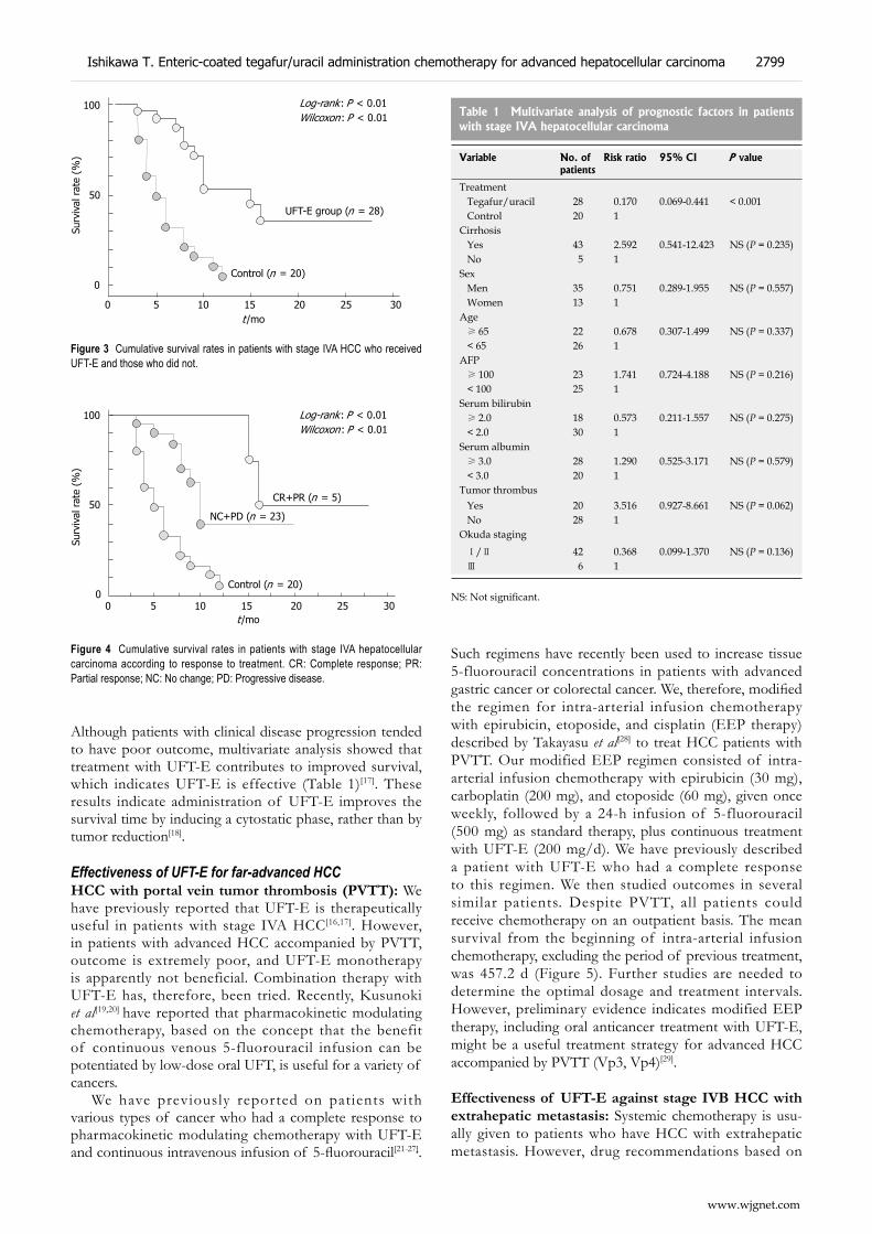
Although patients with clinical disease progression tended to have poor outcome, multivariate analysis showed that treatment with UFT-E contributes to improved survival, which indicates UFT-E is effective (Table 1)[17]. These results indicate administration of UFT-E improves the survival time by inducing a cytostatic phase, rather than by tumor reduction[18].
Effectiveness of UFT-E for far-advanced HCCHCC with portal vein tumor thrombosis (PVTT): We have previously reported that UFT-E is therapeutically useful in patients with stage IVA HCC[16,17]. However, in patients with advanced HCC accompanied by PVTT, outcome is extremely poor, and UFT-E monotherapy is apparently not beneficial. Combination therapy with UFT-E has, therefore, been tried. Recently, Kusunoki et al[19,20] have reported that pharmacokinetic modulating chemotherapy, based on the concept that the benefit of continuous venous 5-fluorouracil infusion can be potentiated by low-dose oral UFT, is useful for a variety of cancers.
We have previously reported on patients with various types of cancer who had a complete response to pharmacokinetic modulating chemotherapy with UFT-E and continuous intravenous infusion of 5-fluorouracil[21-27].
Such regimens have recently been used to increase tissue 5-fluorouracil concentrations in patients with advanced gastric cancer or colorectal cancer. We, therefore, modified the regimen for intra-arterial infusion chemotherapy with epirubicin, etoposide, and cisplatin (EEP therapy) described by Takayasu et al[28] to treat HCC patients with PVTT. Our modified EEP regimen consisted of intra-arterial infusion chemotherapy with epirubicin (30 mg), carboplatin (200 mg), and etoposide (60 mg), given once weekly, followed by a 24-h infusion of 5-fluorouracil (500 mg) as standard therapy, plus continuous treatment with UFT-E (200 mg/d). We have previously described a patient with UFT-E who had a complete response to this regimen. We then studied outcomes in several similar patients. Despite PVTT, all patients could receive chemotherapy on an outpatient basis. The mean survival from the beginning of intra-arterial infusion chemotherapy, excluding the period of previous treatment, was 457.2 d (Figure 5). Further studies are needed to determine the optimal dosage and treatment intervals. However, preliminary evidence indicates modified EEP therapy, including oral anticancer treatment with UFT-E, might be a useful treatment strategy for advanced HCC accompanied by PVTT (Vp3, Vp4)[29].
Effectiveness of UFT-E against stage IVB HCC with extrahepatic metastasis: Systemic chemotherapy is usu-ally given to patients who have HCC with extrahepatic metastasis. However, drug recommendations based on
Figure 3 Cumulative survival rates in patients with stage IVA HCC who received UFT-E and those who did not.
0 5 10 15 20 25 30
100
50
0
UFT-E group (n = 28)
Control (n = 20)
Log-rank : P < 0.01Wilcoxon: P < 0.01
Surv
ival
rat
e (%
)
t /mo
Figure 4 Cumulative survival rates in patients with stage IVA hepatocellular carcinoma according to response to treatment. CR: Complete response; PR: Partial response; NC: No change; PD: Progressive disease.
0 5 10 15 20 25 30
100
50
0Control (n = 20)
NC+PD (n = 23)
CR+PR (n = 5)
Surv
ival
rat
e (%
)
Log-rank : P < 0.01Wilcoxon: P < 0.01
t /mo
Variable No. of patients
Risk ratio 95% CI P value
Treatment Tegafur/uracil 28 0.170 0.069-0.441 < 0.001 Control 20 1Cirrhosis Yes 43 2.592 0.541-12.423 NS (P = 0.235) No 5 1Sex Men 35 0.751 0.289-1.955 NS (P = 0.557) Women 13 1Age ≥ 65 22 0.678 0.307-1.499 NS (P = 0.337) < 65 26 1AFP ≥ 100 23 1.741 0.724-4.188 NS (P = 0.216) < 100 25 1Serum bilirubin ≥ 2.0 18 0.573 0.211-1.557 NS (P = 0.275) < 2.0 30 1Serum albumin ≥ 3.0 28 1.290 0.525-3.171 NS (P = 0.579) < 3.0 20 1Tumor thrombus Yes 20 3.516 0.927-8.661 NS (P = 0.062) No 28 1Okuda staging Ⅰ/Ⅱ 42 0.368 0.099-1.370 NS (P = 0.136) Ⅲ 6 1
Table 1 Multivariate analysis of prognostic factors in patients with stage IVA hepatocellular carcinoma
NS: Not significant.
www.wjgnet.com
Ishikawa T. Enteric-coated tegafur/uracil administration chemotherapy for advanced hepatocellular carcinoma 2799

scientific evidence are currently not available. Although the effectiveness of UFT-E has been confirmed in patients with stage IVA HCC, monotherapy with UFT-E does not improve outcome in patients who have stage IVB disease with distant metastasis, usually an indication for systemic chemotherapy. We have previously reported a case of HCC with multiple lung metastases that had a complete re-sponse to combination therapy with UFT-E plus docetax-el/cisplatin[30]. At present, additional patients with distant metastasis are being studied. Long-term studies of larger numbers of patients are needed to assess the clinical sig-nificance of combination therapy including oral anticancer drugs.
TOXIC EFFECTS OF UFTToxic effects of UFT include severe liver dysfunction and diarrhea. Leukoencephalopathy has been reported as a neurological effect, and pigmentation as a skin symptom. Loss of appetite, nausea and vomiting commonly occur in clinical practice. These adverse signs and symptoms may require a reduction in the dose of UFT or even the cessation of treatment in some patients[31]. In a study that compared the incidence of toxicity between UFT capsules and UFT-E granules, which were developed to reduce upper gastrointestinal symptoms and thereby improve compliance, patients who received UFT-E granules had a lower incidence of adverse upper gastrointestinal symptoms[31]. Grade 1 stomatitis developed in three patients with stage IVA HCC who received monotherapy with UFT-E. However, hepatic function reserve was undisturbed. Monotherapy with UFT-E thus contributed to an improved quality of life. Ikeda et al[32] conducted a controlled study to compare outcome between patients who received UFT-E adjuvant chemotherapy before transcatheter arterial embolization and those who did not. Outcomes did not significantly differ between the groups, but ascites and hepatic encephalopathy developed in the UFT-E group. However, in our studies, no patient had UFT-E-induced liver failure. In patients with tumor occlusion of the portal vein, who received combination chemotherapy, toxic effects such as stomatitis and leukopenia developed, but treatment could be continued after a rest period.
CONCLUSIONThe effectiveness of UFT-E should be evaluated at different stages of HCC. At the same time, different dosages and treatment with UFT-E regimens, including rest periods, should be assessed, rather than administering anticancer drugs continuously. Unlike other types of cancer, HCC is usually associated with cirrhosis. Long-term treatment with anticancer drugs may therefore adversely affect liver function. Further studies are needed to determine optimal regimens, doses, and treatment periods for anticancer drugs. Other clinical questions, such as whether systemic chemotherapy with UFT-E is an effective adjuvant therapy after liver transplantation, remain to be answered.
REFERENCES1 Ishikawa T, Ichida T, Yamagiwa S, Sugahara S, Uehara
K, Okoshi S, Asakura H. High viral loads, serum alanine aminotransferase and gender are predictive factors for the development of hepatocellular carcinoma from viral compensated liver cirrhosis. J Gastroenterol Hepatol 2001; 16: 1274-1281
2 Heidelberger C, Chaudhuri NK, Dannerberg P, Mooren D, Griesbach L, Duschinsky R, Schnitzer RJ, Pleven E, Scheiner J. Fluorinated pyrimidines, a new class of tumour-inhibitory compounds. Nature 1957; 179: 663-666
3 Diasio RB, Harris BE. Clinical pharmacology of 5-fluorouracil. Clin Pharmacokinet 1989; 16: 215-237
4 Baker SD , Khor SP, Adjei AA, Doucette M, Spector T, Donehower RC, Grochow LB, Sartorius SE, Noe DA, Hohneker JA, Rowinsky EK. Pharmacokinetic, oral bioavailability, and safety study of fluorouracil in patients treated with 776C85, an inactivator of dihydropyrimidine dehydrogenase. J Clin Oncol 1996; 14: 3085-3096
5 Hiller SA, Zhuk RA, Lidak MJ, Zidermane AA. Br Patent 1968; 1: 391-395
6 Fujii S, Ikenaka K, Fukushima M, Shirasaka T. Effect of uracil and its derivatives on antitumor activity of 5-fluorouracil and 1-(2-tetrahydrofuryl)-5-fluorouracil. Gann 1978; 69: 763-772
7 Takahashi H, Kamano T. [Clinical results of UFT enteric-coated granule therapy under cooperative study (phase II study). Tokyo Cancer Chemotherapy Cooperative Study Group] Gan To Kagaku Ryoho 1990; 17: 2043-2049
8 Kikuchi K, Wakui A. [Cooperative research of UFT E phase II study. Cooperative Study Group of UFT E in Tohoku Area] Gan To Kagaku Ryoho 1990; 17: 2183-2190
9 Heidelberger C, Kaldor G, Mukherjee KL, Danneberg PB. Studies on fluorinated pyrimidines. XI. In vitro studies on tumor resistance. Cancer Res 1960; 20: 903-909
10 Hartmann KU, Heidelberger C. Studies on fluorinated pyrimidines. XIII. Inhibition of thymidylate synthetase. J Biol Chem 1961; 236: 3006-3013
11 Naguib FN, el Kouni MH, Cha S. Enzymes of uracil catabolism in normal and neoplastic human tissues. Cancer Res 1985; 45: 5405-5412
12 Diasio RB, Harris BE. Clinical pharmacology of 5-fluorouracil. Clin Pharmacokinet 1989; 16: 215-237
13 Yonekura K, Basaki Y, Chikahisa L, Okabe S, Hashimoto A, Miyadera K, Wierzba K, Yamada Y. UFT and its metabolites inhibit the angiogenesis induced by murine renal cell carcinoma, as determined by a dorsal air sac assay in mice. Clin Cancer Res 1999; 5: 2185-2191
14 Basaki Y, Yonekura K, Chikahisa L, Okabe S, Hashimoto A, Miyadera K, Aoyagi K, Yamada Y. [Anti-angiogenic activities of UFT and its metabolites, GHB and GBL, in the dorsal air sac (DAS) model in mice] Gan To Kagaku Ryoho 2000; 27: 93-98
15 Akasu T, Moriya Y, Ohashi Y, Yoshida S, Shirao K, Kodaira S.
100
50
0
Surv
ival
rat
e (%
)
0 100 200 300 400 500 600 700 800t /d
Figure 5 Cumulative survival rates in patients with tumor occlusion of the portal vein who received modified EEP therapy.
www.wjgnet.com
2800 ISSN 1007-9327 CN 14-1219/R World J Gastroenterol May 14, 2008 Volume 14 Number 18

Adjuvant chemotherapy with uracil-tegafur for pathological stage III rectal cancer after mesorectal excision with selective lateral pelvic lymphadenectomy: a multicenter randomized controlled trial. Jpn J Clin Oncol 2006; 36: 237-244
16 Ishikawa T, Ichida T, Ishimoto Y, Yokoyama J, Nomoto M, Ebe Y, Usuda H, Naito M, Asakura H. Complete remission of multiple hepatocellular carcinomas associated with hepatitis C virus-related, decompensated liver cirrhosis by oral administration of enteric-coated tegafur/uracil. Am J Gastroenterol 1999; 94: 1682-1685
17 Ishikawa T, Ichida T, Sugitani S, Tsuboi Y, Genda T, Sugahara S, Uehara K, Inayoshi J, Yokoyama J, Ishimoto Y, Asakura H. Improved survival with oral administration of enteric-coated tegafur/uracil for advanced stage IV-A hepatocellular carcinoma. J Gastroenterol Hepatol 2001; 16: 452-459
18 Takahashi Y, Nishioka K. Survival without tumor shrinkage: re-evaluation of survival gain by cytostatic effect of chemotherapy. J Natl Cancer Inst 1995; 87: 1262-1263
19 Kusunoki M, Yanagi H, Kotera H, Noda M, Yamamura T. Effects of pharmacokinetic modulating chemotherapy using oral UFT and continuous venous 5FU infusion on the prognosis of irradiated rectal carcinomas with p53 overexpression. Int J Oncol 1998; 13: 653-657
20 Kusunoki M , Yanagi H, Noda M, Yamamura T. The usefulness of pharmacokinetic modulating chemotherapy (UFT plus 5FU) in the treatment of unresectable colorectal carcinomas. Oncol Rep 1999; 6: 547-552
21 Ishikawa T, Sato S, Matsuzawa J, Mita Y, Matsui S, Tashiro K, Tashiro S, Matsuki H. [A case of successful management of nonresectable pancreas cancer with liver metastasis by intra-arterial infusion chemotherapy with angiotensin-II and administration of tegafur/uracil] Gan To Kagaku Ryoho 2001; 28: 521-525
22 Ishikawa T, Mita Y, Kobayashi M, Tashiro K, Tashiro S, Matsuki H. [A case of nonresectable scirrhous type gastric cancer treated by hypertensive subselective chemotherapy with pharmacokinetic modulating chemotherapy] Gan To Kagaku Ryoho 2001; 28: 1137-1140
23 Ishikawa T, Nomura K, Baba Y, Hayashi S, Oota H, Yoshida T, Kamimura T. [A case of advanced gastric cancer with liver and intra-abdominal lymph node metastasis treated by hypertensive selective chemotherapy with pharmacokinetic modulating chemotherapy] Gan To Kagaku Ryoho 2003; 30: 1151-1155
24 Ishikawa T, Mizuno K, Togashi T, Watanabe K, Seki K, Ohta H, Yoshida T, Kamimura T. [A case of advanced gastric cancer with bone metastasis and severe DIC responding to
hypertensive subselective chemotherapy with pharmacokinetic modulating chemotherapy] Gan To Kagaku Ryoho 2005; 32: 523-527
25 Ishikawa T, Mizuno K, Togashi T, Watanabe K, Seki K, Ohta H, Yoshida T, Kamimura T. [Modified pharmacokinetic modulating chemotherapy for progressive gastric cancer accompanied by peritoneal dissemination] Gan To Kagaku Ryoho 2005; 32: 469-472
26 Ishikawa T , Mizuno K, Watanabe K, Baba Y, Oota H, Yoshida T, Kamimura T. [A case of successful management of nonresectable pancreas cancer with liver metastasis by intra-arterial infusion chemotherapy with gemcitabine hydrochloride, 5-FU, CDDP and administration of tegafur/uracil] Gan To Kagaku Ryoho 2004; 31: 1555-1558
27 Ishikawa T, Ishikawa N, Oota H, Yoshida T, Honma A, Kamimura T, Takeda K, Ishikawa N, Ozaki T. [A case of common bile duct cancer responding to MMC leucovorin, 5-FU, and UFT combination chemotherapy and radiation] Gan To Kagaku Ryoho 1996; 23: 773-777
28 Takayasu Y. [Hepatic arterial infusion chemotherapy for hepatocellular carcinoma by EEP regimen] Nippon Rinsho 2001; 59 Suppl 6: 624-628
29 Ishikawa T, Imai M, Kamimura H, Tsuchiya A, Togashi T, Watanabe K, Seki K, Ohta H, Yoshida T, Kamimura T. Improved survival for hepatocellular carcinoma with portal vein tumor thrombosis treated by intra-arterial chemotherapy combining etoposide, carboplatin, epirubicin and pharmacokinetic modulating chemotherapy by 5-FU and enteric-coated tegafur/uracil: a pilot study. World J Gastroenterol 2007; 13: 5465-5470
30 Ishikawa T, Ichida T, Yokoyama J, Matsuda Y, Watanabe T, Asakura H. Complete disappearance of pulmonary metastases in a case of hepatocellular carcinoma treated with docetaxel-based systemic chemotherapy. J Gastroenterol Hepatol 2004; 19: 1423-1426
31 Ohyama M, Matsumura M, Katsuta K, Nobori T, Matsuyama H, Fukami K, Kiyota R, Yano H, Shima T, Ogawa K. [A comparative study of UFT enteric-coated granules with UFT capsules on the occurrence of side effects in patients with head and neck cancers--a special attention to the upper gastrointestinal tract disorders] Gan To Kagaku Ryoho 1990; 17: 1211-1216
32 Ikeda K, Saitoh S, Koida I, Tsubota A, Arase Y, Chayama K, Kumada H. A prospective randomized evaluation of a compound of tegafur and uracil as an adjuvant chemotherapy for hepatocellular carcinoma treated with transcatheter arterial chemoembolization. Am J Clin Oncol 1995; 18: 204-210
S- Editor Liu JN L- Editor Kerr C E- Editor Yin DH
www.wjgnet.com
Ishikawa T. Enteric-coated tegafur/uracil administration chemotherapy for advanced hepatocellular carcinoma 2801

Online Submissions: wjg.wjgnet.com World J Gastroenterol 2008 May 14; 14(18): 2802-2809www.wjgnet.com World Journal of Gastroenterology ISSN [email protected] © 2008 WJG. All rights reserved.
Relationship between expression of gastrin, somatostatin, Fas/FasL and caspases in large intestinal carcinoma
Jia-Ding Mao, Pei Wu, Ying-Lin Yang, Jian Wu, He Huang
www.wjgnet.com
COLORECTAL CANCER
Jia-Ding Mao, Pei Wu, Ying-Lin Yang, Jian Wu, He Huang, Department of General Surgery, the First Affiliated Yijishan Hospital of Wannan Medical College, Wuhu 241001, Anhui Province, ChinaAuthor contributions: Mao JD, Wu P, and Yang YL designed and performed the research; Yang YL provided new reagents/analytic tools; Mao JD analyzed data and wrote the paper; and Wu J and Huang H contributed equally to this work.Supported by The Natural Science Foundation of Educational Bureau of Anhui Province, No. 2006kj115c, 2005kj302, and Natural Science Foundation of Anhui Province, No. 03043704Correspondence to: Professor Pei Wu , Department of General Surgery, The First Affiliated Yijishan Hospital of Wannan Medical College, Wuhu 241001, Anhui Province, China. [email protected]: +86-553-5739343Received: November 17, 2007 Revised: February 14, 2008
AbstractAIM: To explore the correlation between the mRNAs and protein expression of gastrin (GAS), somatostatin (SS) and apoptosis index (AI), apoptosis regulation gene Fas/FasL and caspases in large intestinal carcinoma (LIC).
METHODS: Express ion o f GAS and SS mRNAs were detected by nested RT-PCR in 79 cases of LIC. Cell apoptosis was detected by molecular biology in s i tu apoptos i s detec t ing methods (TUNEL) . Immunohistochemical staining for GAS, SS, Fas/FasL, caspase-3 and caspase-8 was performed according to the standard streptavidin-biotin-peroxidase (S-P) method.
RESULTS: There was a significant positive correlation between mRNA and protein expression of GAS and SS (GASr s=0.99, P < 0.01; SSr s = 0.98, P < 0.01). There was significant difference in positive expression rates of GAS, SS mRNAs and protein among different histological differentiation, histological types and Dukes’ stage of LIC. The AI in GAS high and moderate expression groups was significantly lower than that in low expression groups (3.75 ± 2.38 vs 7.82 ± 2.38, P < 0.01; 5.51 ± 2.66 vs 7.82 ± 2.38, P < 0.01), and the AI in SS high and moderate expression groups was significantly higher than that in low expression groups (9.03 ± 1.76 vs 5.35 ± 3.00, P < 0.01; 7.44 ± 2.67 vs 5.35 ± 3.00, P < 0.01). There was a significant negative correlation between the integral ratio of GAS to SS and the AI (r s = -0.41, P < 0.01). The positive expression rate of FasL in GAS high and moderate expression groups was higher than that
in low expression group (90.9% and 81.0% vs 53.2%, P < 0.05). The posit ive expression rates of Fas, caspase-8 and caspase-3 in SS high (90.0%, 90.0% and 100%) and moderate (80.0%, 70.0%, 75.0%) expression groups were higher than that in low expression group (53.1%, 42.9%, 49.0%) (90.0% and 80.0% vs 53.1%, P < 0.05; 90.0% and 70.0% vs 42.9%, P < 0.05; 100.0% and 75.0% vs 49.0%, P < 0.05). There was a significant positive correlation between the integral ratio of GAS to SS and the semiquantitative integral of FasL (r s = 0.32, P < 0.01).
CONCLUSION: GAS and SS play important roles in the regulation and control of cell apoptosis in LIC, and the mechanism may be directly related to the aberrant expression of Fas/FasL. The GAS and SS will be valuable targets of the biological behavior of LIC.
© 2008 WJG. All rights reserved.
Key words: Large intest inal carcinoma; Gastr in; Somatostatin; Apoptosis index; Fas; FasL; Caspase
Peer reviewers: Rene Lambert, Professor, International Agency for Research on Cancer, 150 Cours Albert Thomas, Lyon 69372 cedex 8, France; Takayuki Yamamoto, MD, Inflammatory Bowel Disease Center, Yokkaichi Social Insurance Hospital, 10-8 Hazuyamacho, Yokkaichi 510-0016, Japan
Mao JD, Wu P, Yang YL, Wu J, Huang H. Relationship between expression of gastrin, somatostatin, Fas/FasL and caspases in large intestinal carcinoma. World J Gastroenterol 2008; 14(18): 2802-2809 Available from: URL: http://www.wjgnet.com/1007-9327/14/2802.asp DOI: http://dx.doi.org/10.3748/wjg.14.2802
INTRODUCTIONLarge intestinal cancer (LIC) is one of the most common digestive tract malignant tumors in the world, with a high incidence rate in North America, Western Europe, Australia, New Zealand and France, and is the second leading cause of gastrointestinal cancer-related mortality worldwide[1-4]. In China, it now ranks fourth[5]. Although great progress in understanding the molecular aspects of LIC has been made and several therapeutic agents have been developed, it is still difficult to cure the tumor. It is posing a serious threat to public health and remains a major killer among Chinese patients. The general survival rate
Mao JD et al . Gastrin, somatostatin and caspases in large intestinal carcinoma 2803
www.wjgnet.com
of LIC patients does not exceed 40%. Thereby, it is very important to know what kind of cell factor can influence cell apoptosis, which will enrich the etiology theory of tumor. In addition, while studying the mechanism of loss of control of tumor cell apoptosis from signal transduction pathway, we may find a new way to treat malignant tumors. Previous studies have demonstrated that the occurrence of LIC is directly related to the abnormal expression of gastrointestinal hormones such as gastrin (GAS), somatostatin (SS), etc[6,7]. Some studies found that GAS was able to promote growth and inhibit apoptosis of LIC cells, but the role of SS was opposite[8-10]. However, the detailed molecule mechanism by which GAS and SS mediate cell apoptosis of LIC is not fully known. We used nested RT-PCR method to detect the expression of GAS and SS mRNAs in LIC tissues, and TUNEL method to detect cell apoptosis, and immunohistochemical staining S-P method to detect the protein expression of GAS, SS, Fas/FasL, caspase-3 and caspase-8. The aim of this study was to explore the correlation between the mRNAs and protein expression of GAS and SS, and between the protein expression of GAS, SS and apoptosis index (AI), and apoptosis regulation gene Fas/FasL and caspases in LIC, and to further confirm whether GAS and SS could mediate LIC cell apoptosis mainly via affecting the expression of Fas/FasL.
MATERIALS AND METHODSClinical dataSeventy-nine cases of cancer tissue samples were randomly and retrospectively selected from patients with LIC hospitalized in our hospital from January 2004 to October 2006. All patients were confirmed as having LIC by clinical pathology. Among them, 36 were cases of rectum cancer, and 43 were cases of colorectal carcinoma. Thirty-seven were females and 42 were males. The median age was 50.8 ± 11.2 years, with a range of 21-79 years. There were 27 patients with well differentiated carcinoma, 28 patients with moderately differentiated carcinoma, and 24 patients with poorly differentiated carcinoma (inclusive of undifferentiated carcinoma). Among them, there were 40 cases of ulcerative type, 32 protruded type, 7 infiltrating type, 19 papillary adenocarcinoma, 35 glandular adenocarcinoma, 10 mucoid carcinoma, 6 signet-ring cell carcinoma and 9 undifferentiated carcinoma. The clinical stage was determined according to the Dukes’ stage. Dukes’ stages A and B were found in 36 patients, and stages C and D in 43 patients.
Main reagentsThe polyclonal rabbit antibodies against human SS protein and human GAS, monoclonal rabbit antibodies against human Fas and FasL, caspase-3, caspase-8, and immunohistochemical staining kit and in situ cell apoptosis detection kit Ⅱ were all purchased from Beijing Zhongshan Biological Technology Co, Ltd. Trizol liquid, M-MLV reverse transcriptase, SK312, Taq DNA polymerase, RNAsin, oligo (dt) 15 and dNTPs primers were purchased from Shanghai Sangon Biological Engineering Technology
and Service Co. Ltd and PCR primers were synthesized by the same company.
Total RNA extraction and cDNA synthesisTotal RNA was extracted from frozen tissue specimens (50-100 mg) using TRIzol reagent according to the protocol provided by the manufacturer. Total RNA was primed with an Oligo (dT) 15 oligonucleotide and reverse-transcribed following the manufacturer’s instructions.
PCR amplificationThe primers of GAS, SS and β-actin were synthesized according to the primer design principles, all primers spanned an intron to control against amplification of genomic DNA sequences. Two point five μL first strand cDNA was amplified in 25 μL total volume. The first PCR amplifications were performed in a UNO Ⅱ thermocycler (Perkin-Elmer, USA) under the following conditions. After an initial denaturation for 5 min at 94℃, samples were subjected to 30 cycles, each amplification consisting of denaturation at 94℃ for 30 s, primer annealing at 60℃ for 45 s, extension at 72℃ for 45 s, followed by a final extension of 6 min 72℃. Conditions and parameters of the second PCR amplifications were the same with the first PCR amplifications except for primers. The length of final PCR products was 174 base pair (bp) (GAS) and 231 bp (SS), respectively. At last, 6 μL of reaction product was analyzed by electrophoresis on a 1.5% agarose gel followed by ethidium bromide staining and digital camera photography (Table 1, Figure 1).
Detection of cell apoptosisCell apoptosis was detected using molecular biology in situ apoptosis detecting methods (TUNEL). The sections were fixed and embedded conventionally. Dnase (1 mg/L) was used to digest the positive control specimen for 10 min before put into TUNEL response mixture. Only label liquor was added into the negative control response mixture. The detailed manipulation was conducted according to the introductions for users. Cells with brown or yellow nuclei were assumed as apoptotic cells. The number of apoptotic cells and total cancer cells was counted under light microscope at 400 × magnification in 10 fields of vision and the average values were obtained for the calculation of apoptosis index (AI) according to the formula: AI = (apoptotic cells/total cancer cells) × 100%.
Immunohistochemical stainingSpecimens obtained at surgery were routinely fixed in 10% neutral formalin and embedded in paraffin. Serial 4 μm thick sections were cut. Immunohistochemical staining for Fas, FasL, caspase-3, caspase-8, GAS and SS was performed by the standard streptavidin-biotin-peroxidase (S-P) method. Positive pancreatic tissue and stomach antrum mucous membrane were used as a positive control for SS and GAS. Positive controls for Fas, FasL, caspase-3 and caspase-8 were purchased from Shanghai Sangon Biological Engineering Technology and Service Co. Ltd. PBS 0.01 mol/L as a negative control replaced the primary antibody. The detailed
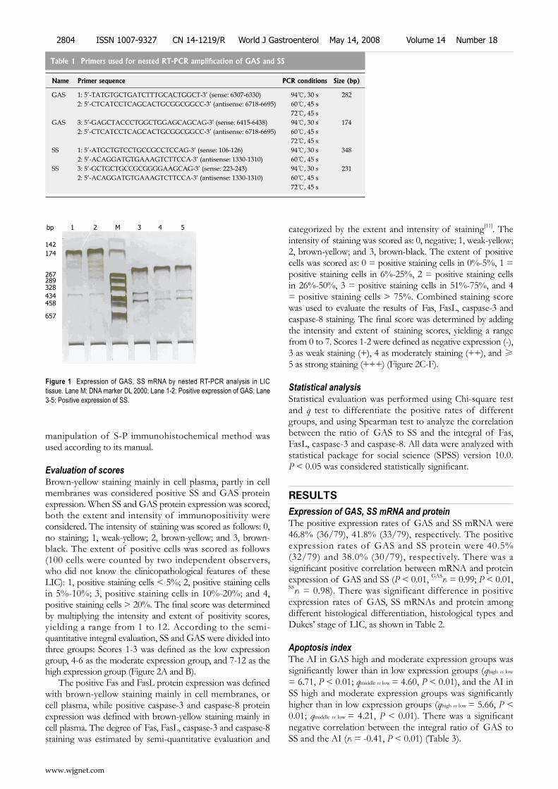
manipulation of S-P immunohistochemical method was used according to its manual.
Evaluation of scoresBrown-yellow staining mainly in cell plasma, partly in cell membranes was considered positive SS and GAS protein expression. When SS and GAS protein expression was scored, both the extent and intensity of immunopositivity were considered. The intensity of staining was scored as follows: 0, no staining; 1, weak-yellow; 2, brown-yellow; and 3, brown-black. The extent of positive cells was scored as follows (100 cells were counted by two independent observers, who did not know the clinicopathological features of these LIC): 1, positive staining cells < 5%; 2, positive staining cells in 5%-10%; 3, positive staining cells in 10%-20%; and 4, positive staining cells > 20%. The final score was determined by multiplying the intensity and extent of positivity scores, yielding a range from 1 to 12. According to the semi-quantitative integral evaluation, SS and GAS were divided into three groups: Scores 1-3 was defined as the low expression group, 4-6 as the moderate expression group, and 7-12 as the high expression group (Figure 2A and B).
The positive Fas and FasL protein expression was defined with brown-yellow staining mainly in cell membranes, or cell plasma, while positive caspase-3 and caspase-8 protein expression was defined with brown-yellow staining mainly in cell plasma. The degree of Fas, FasL, caspase-3 and caspase-8 staining was estimated by semi-quantitative evaluation and
categorized by the extent and intensity of staining[11]. The intensity of staining was scored as: 0, negative; 1, weak-yellow; 2, brown-yellow; and 3, brown-black. The extent of positive cells was scored as: 0 = positive staining cells in 0%-5%, 1 = positive staining cells in 6%-25%, 2 = positive staining cells in 26%-50%, 3 = positive staining cells in 51%-75%, and 4 = positive staining cells > 75%. Combined staining score was used to evaluate the results of Fas, FasL, caspase-3 and caspase-8 staining. The final score was determined by adding the intensity and extent of staining scores, yielding a range from 0 to 7. Scores 1-2 were defined as negative expression (-), 3 as weak staining (+), 4 as moderately staining (++), and ≥ 5 as strong staining (+++) (Figure 2C-F).
Statistical analysisStatistical evaluation was performed using Chi-square test and q test to differentiate the positive rates of different groups, and using Spearman test to analyze the correlation between the ratio of GAS to SS and the integral of Fas, FasL, caspase-3 and caspase-8. All data were analyzed with statistical package for social science (SPSS) version 10.0. P < 0.05 was considered statistically significant.
RESULTSExpression of GAS, SS mRNA and proteinThe positive expression rates of GAS and SS mRNA were 46.8% (36/79), 41.8% (33/79), respectively. The positive expression rates of GAS and SS protein were 40.5% (32/79) and 38.0% (30/79), respectively. There was a significant positive correlation between mRNA and protein expression of GAS and SS (P < 0.01, GASrs = 0.99; P < 0.01, SSrs = 0.98). There was significant difference in positive expression rates of GAS, SS mRNAs and protein among different histological differentiation, histological types and Dukes’ stage of LIC, as shown in Table 2.
Apoptosis indexThe AI in GAS high and moderate expression groups was significantly lower than in low expression groups (qhigh vs low
= 6.71, P < 0.01; qmiddle vs low = 4.60, P < 0.01), and the AI in SS high and moderate expression groups was significantly higher than in low expression groups (qhigh vs low = 5.66, P < 0.01; qmiddle vs low = 4.21, P < 0.01). There was a significant negative correlation between the integral ratio of GAS to SS and the AI (rs = -0.41, P < 0.01) (Table 3).
Figure 1 Expression of GAS, SS mRNA by nested RT-PCR analysis in LIC tissue. Lane M: DNA marker DL 2000; Lane 1-2: Positive expression of GAS; Lane 3-5: Positive expression of SS.
Table 1 Primers used for nested RT-PCR amplification of GAS and SS
Name Primer sequence PCR conditions Size (bp)
GAS 1: 5’-TATGTGCTGATCTTTGCACTGGCT-3’ (sense: 6307-6330) 94℃, 30 s 2822: 5’-CTCATCCTCAGCACTGCGGCGGCC-3’ (antisense: 6718-6695) 60℃, 45 s
72℃, 45 sGAS 3: 5’-GAGCTACCCTGGCTGGAGCAGCAG-3’ (sense: 6415-6438) 94℃, 30 s 174
2: 5’-CTCATCCTCAGCACTGCGGCGGCC-3’ (antisense: 6718-6695) 60℃, 45 s72℃, 45 s
SS 1: 5’-ATGCTGTCCTGCCGCCTCCAG-3’ (sense: 106-126) 94℃, 30 s 3482: 5’-ACAGGATGTGAAAGTCTTCCA-3’ (antisense: 1330-1310) 60℃, 45 s
SS 3: 5’-GCTGCTGCCGCGGGGAAGCAG-3’ (sense: 223-243) 94℃, 30 s 2312: 5’-ACAGGATGTGAAAGTCTTCCA-3’ (antisense: 1330-1310) 60℃, 45 s
72℃, 45 s
1 2 M 3 4 5
142174
267289 328 434 458
657
www.wjgnet.com
2804 ISSN 1007-9327 CN 14-1219/R World J Gastroenterol May 14, 2008 Volume 14 Number 18
bp
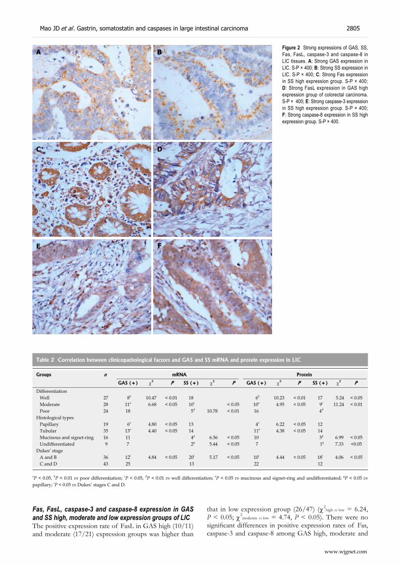
Figure 2 Strong expressions of GAS, SS, Fas, FasL, caspase-3 and caspase-8 in LIC tissues. A: Strong GAS expression in LIC. S-P × 400; B: Strong SS expression in LIC. S-P × 400; C: Strong Fas expression in SS high expression group. S-P × 400; D: Strong FasL expression in GAS high expression group of colorectal carcinoma. S-P × 400; E: Strong caspase-3 expression in SS high expression group. S-P × 400; F: Strong caspase-8 expression in SS high expression group. S-P × 400.
DC
BA
E F
Table 2 Correlation between clinicopathological factors and GAS and SS mRNA and protein expression in LIC
Groups n mRNA Protein
GAS (+) χ2 P SS (+) χ2 P GAS (+) χ2 P SS (+) χ2 P
Differentiation Well 27 8b 10.47 < 0.01 18 6b 10.23 < 0.01 17 5.24 < 0.05 Moderate 28 11a 6.68 < 0.05 10c < 0.05 10a 4.95 < 0.05 9c 11.24 < 0.01 Poor 24 18 5d 10.78 < 0.01 16 4d
Histological types Papillary 19 6e 4.80 < 0.05 13 4e 6.22 < 0.05 12 Tubular 35 13e 4.40 < 0.05 14 11e 4.38 < 0.05 14 Mucinous and signet-ring 16 11 4g 6.56 < 0.05 10 3g 6.99 < 0.05 Undifferentiated 9 7 2g 5.44 < 0.05 7 1g 7.33 <0.05Dukes’ stage A and B 36 12i 4.84 < 0.05 20i 5.17 < 0.05 10i 4.44 < 0.05 18i 4.06 < 0.05 C and D 43 25 13 22 12
aP < 0.05, bP < 0.01 vs poor differentiation; cP < 0.05, dP < 0.01 vs well differentiation; eP < 0.05 vs mucinous and signet-ring and undifferentiated; gP < 0.05 vs papillary; iP < 0.05 vs Dukes’ stages C and D.
Fas, FasL, caspase-3 and caspase-8 expression in GAS and SS high, moderate and low expression groups of LICThe positive expression rate of FasL in GAS high (10/11) and moderate (17/21) expression groups was higher than
that in low expression group (26/47) (χ2high vs low = 6.24,
P < 0.05; χ2moderate vs low = 4.74, P < 0.05). There were no
significant differences in positive expression rates of Fas, caspase-3 and caspase-8 among GAS high, moderate and
www.wjgnet.com
Mao JD et al . Gastrin, somatostatin and caspases in large intestinal carcinoma 2805
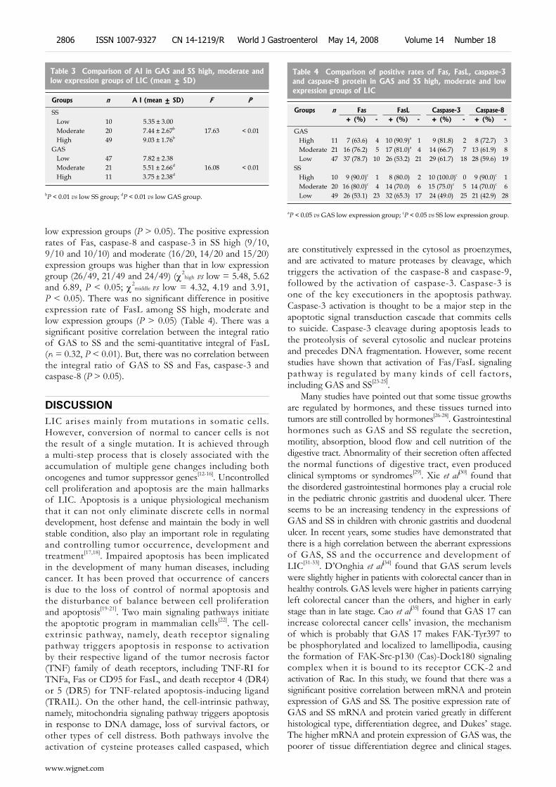
low expression groups (P > 0.05). The positive expression rates of Fas, caspase-8 and caspase-3 in SS high (9/10, 9/10 and 10/10) and moderate (16/20, 14/20 and 15/20) expression groups was higher than that in low expression group (26/49, 21/49 and 24/49) (χ2
high vs low = 5.48, 5.62 and 6.89, P < 0.05; χ2
middle vs low = 4.32, 4.19 and 3.91, P < 0.05). There was no significant difference in positive expression rate of FasL among SS high, moderate and low expression groups (P > 0.05) (Table 4). There was a significant positive correlation between the integral ratio of GAS to SS and the semi-quantitative integral of FasL (rs = 0.32, P < 0.01). But, there was no correlation between the integral ratio of GAS to SS and Fas, caspase-3 and caspase-8 (P > 0.05).
DISCUSSIONLIC arises mainly from mutations in somatic cells. However, conversion of normal to cancer cells is not the result of a single mutation. It is achieved through a multi-step process that is closely associated with the accumulation of multiple gene changes including both oncogenes and tumor suppressor genes[12-16]. Uncontrolled cell proliferation and apoptosis are the main hallmarks of LIC. Apoptosis is a unique physiological mechanism that it can not only eliminate discrete cells in normal development, host defense and maintain the body in well stable condition, also play an important role in regulating and controlling tumor occurrence, development and treatment[17,18]. Impaired apoptosis has been implicated in the development of many human diseases, including cancer. It has been proved that occurrence of cancers is due to the loss of control of normal apoptosis and the disturbance of balance between cell proliferation and apoptosis[19-21]. Two main signaling pathways initiate the apoptotic program in mammalian cells[22]. The cell-extrinsic pathway, namely, death receptor signaling pathway triggers apoptosis in response to activation by their respective ligand of the tumor necrosis factor (TNF) family of death receptors, including TNF-RI for TNFa, Fas or CD95 for FasL, and death receptor 4 (DR4) or 5 (DR5) for TNF-related apoptosis-inducing ligand (TRAIL). On the other hand, the cell-intrinsic pathway, namely, mitochondria signaling pathway triggers apoptosis in response to DNA damage, loss of survival factors, or other types of cell distress. Both pathways involve the activation of cysteine proteases called caspased, which
Table 3 Comparison of AI in GAS and SS high, moderate and low expression groups of LIC (mean ± SD)
Groups n A I (mean ± SD) F P
SS Low 10 5.35 ± 3.00 Moderate 20 7.44 ± 2.67b 17.63 < 0.01 High 49 9.03 ± 1.76b
GAS Low 47 7.82 ± 2.38 Moderate 21 5.51 ± 2.66d 16.08 < 0.01 High 11 3.75 ± 2.38d
bP < 0.01 vs low SS group; dP < 0.01 vs low GAS group.
Table 4 Comparison of positive rates of Fas, FasL, caspase-3 and caspase-8 protein in GAS and SS high, moderate and low expression groups of LIC
Groups n Fas FasL Caspase-3 Caspase-8+ (%) - + (%) - + (%) - + (%) -
GAS High 11 7 (63.6) 4 10 (90.9)a 1 9 (81.8) 2 8 (72.7) 3 Moderate 21 16 (76.2) 5 17 (81.0)a 4 14 (66.7) 7 13 (61.9) 8 Low 47 37 (78.7) 10 26 (53.2) 21 29 (61.7) 18 28 (59.6) 19SS High 10 9 (90.0)c 1 8 (80.0) 2 10 (100.0)c 0 9 (90.0)c 1 Moderate 20 16 (80.0)c 4 14 (70.0) 6 15 (75.0)c 5 14 (70.0)c 6 Low 49 26 (53.1) 23 32 (65.3) 17 24 (49.0) 25 21 (42.9) 28
aP < 0.05 vs GAS low expression group; cP < 0.05 vs SS low expression group.
are constitutively expressed in the cytosol as proenzymes, and are activated to mature proteases by cleavage, which triggers the activation of the caspase-8 and caspase-9, followed by the activation of caspase-3. Caspase-3 is one of the key executioners in the apoptosis pathway. Caspase-3 activation is thought to be a major step in the apoptotic signal transduction cascade that commits cells to suicide. Caspase-3 cleavage during apoptosis leads to the proteolysis of several cytosolic and nuclear proteins and precedes DNA fragmentation. However, some recent studies have shown that activation of Fas/FasL signaling pathway is regulated by many kinds of cell factors, including GAS and SS[23-25].
Many studies have pointed out that some tissue growths are regulated by hormones, and these tissues turned into tumors are still controlled by hormones[26-28]. Gastrointestinal hormones such as GAS and SS regulate the secretion, motility, absorption, blood flow and cell nutrition of the digestive tract. Abnormality of their secretion often affected the normal functions of digestive tract, even produced clinical symptoms or syndromes[29]. Xie et al[30] found that the disordered gastrointestinal hormones play a crucial role in the pediatric chronic gastritis and duodenal ulcer. There seems to be an increasing tendency in the expressions of GAS and SS in children with chronic gastritis and duodenal ulcer. In recent years, some studies have demonstrated that there is a high correlation between the aberrant expressions of GAS, SS and the occurrence and development of LIC[31-33]. D’Onghia et al[34] found that GAS serum levels were slightly higher in patients with colorectal cancer than in healthy controls. GAS levels were higher in patients carrying left colorectal cancer than the others, and higher in early stage than in late stage. Cao et al[35] found that GAS 17 can increase colorectal cancer cells’ invasion, the mechanism of which is probably that GAS 17 makes FAK-Tyr397 to be phosphorylated and localized to lamellipodia, causing the formation of FAK-Src-p130 (Cas)-Dock180 signaling complex when it is bound to its receptor CCK-2 and activation of Rac. In this study, we found that there was a significant positive correlation between mRNA and protein expression of GAS and SS. The positive expression rate of GAS and SS mRNA and protein varied greatly in different histological type, differentiation degree, and Dukes’ stage. The higher mRNA and protein expression of GAS was, the poorer of tissue differentiation degree and clinical stages.
www.wjgnet.com
2806 ISSN 1007-9327 CN 14-1219/R World J Gastroenterol May 14, 2008 Volume 14 Number 18

For example, the expression GAS in poorly differentiated LIC, in particular, in mucinous adenocarcinomas and signet-ring cell carcinoma was prominently higher than the others, with poor prognosis. However, the action of SS was opposite. The results indicate that the expressions of GAS and SS were closely related to the biological behavior of LIC. It is obvious that the GAS and SS will be valuable targets of the biological behavior of LIC.
Tumor escape mechanisms in LIC result from dysregulation of the cell apoptosis signaling pathways, which are mediated by death ligands. Recently, great progress has been made in understanding the regulated signaling pathway mechanisms of GAS and SS. Some studies showed that the abnormal expressions of GAS and SS were closely related to cell apoptosis of LIC. GAS could promote cell proliferation and inhibit cell apoptosis. However, the action of SS was opposite in LIC[36,37]. Our studies showed that the expression of GAS protein in LIC was higher and the AI was lower, and the higher expression of SS protein, the higher AI. Despite abundant evidence that GAS may play an integral role in promoting tumor growth in the stomach and malignancies in the GI tract, the precise mechanisms about gastrin-restrained, somatostatin-induced apoptosis are still largely unknown. To elucidate the mechanisms by which GAS and SS might influence apoptotic signaling pathway, we analyzed them effects on the expression of Fas, FasL, caspase-3 and caspase-8 protein in human LIC tissue.
GAS is a gastrointestinal (GI) peptide that possesses potent trophic effects on most of the normal and neoplastic mucosa of the GI tract. GAS is mainly secreted from GAS secreting cells (G cells) in the antrum mucosa or upper small intestine and large intestine. Medulla oblongata and dorsal nuclei of vagus nerves in central nervous system also secrete GAS[38,39]. Recent reports indicate that chronic hypergastrinaemia increases the risk of colorectal cancer and cancer growth, and that interruption of the effects of GAS could be a potential target in the treatment of colorectal cancer[40]. Recent data have shown that GAS is not only able to induce cell proliferation, but also inhibit LIC cell apoptosis. However, the underlying mechanisms that GAS inhibits cell apoptosis, is still worthy of further elucidation. Wu et al[41] recent studies found that down-regulation of GAS gene expression results in the activation of caspase 9 and 3 in gastrin-dependent human colon cancer cells inducing cell apoptosis. Cui et al[42] found that GAS can induce apoptosis in gastric epithelial cells via GAS/CCK-2 receptor and synergized with FasL stimulation and contribute to the development of gastric carcinogenesis. In this study, we found that the level of GAS protein expression was higher and the positive expression rate of FasL was higher in LIC tissues, and expression of GAS protein was not related to expression of Fas, caspase-3 and caspase-8. The results indicate that the mechanisms of GAS inhibiting LIC cell apoptosis might be via up-regulation of expression of FasL inducing apoptosis with Fas-expressing T cells, making LIC cells escape from immune surveillance and immunocyte attack.
SS is a multifunctional hormone, which is secreted from SS secreting cells (D cells). D cells are distributed mainly in human central and peripheral nervous system, and in the gastrointestinal tract, including the large intestine. SS
inhibits multiple functions, including exocrine and endocrine secretions, inflammation, and angiogenesis, as well as cell proliferation and tumorigenesis, as shown in normal or tumoral cell models[43-46]. SS acts as an inhibitory peptide of various secretory and proliferative responses. The diverse biological effects of SS are mediated through a family of five SS receptors (sst1-sst5) that belong to the family of G-protein-coupled receptors and that regulate diverse signal transduction pathways including adenylate cyclase, phospholipase C-β, phospholipase A2, guanylate cyclase, ionic conductance channels, and tyrosine phosphatase[47,48]. The mechanisms of the inhibition are the combined interaction of SS and its analogs with SST1-5R in tumor tissues, either directly inhibiting division and proliferation of tumor cells or inhibiting the activities of growth factors such as vascular endothelial growth factor (VEGF), insulin-like growth factor (IGF), etc [49-51], thus counteracting tumorigenesis and tumor cell proliferation. The ability of SS and its stable analogues to promote tumor cell apoptosis has been demonstrated in various cell types including mammary, prostatic, gastric, pancreatic, colorectal, and small cell lung cancer cells. However, the underlying mechanisms of SS induced cell apoptosis are still poorly understood. Guillermet et al[52] showed that sst2-dependent activation and cell sensitization to death ligand-induced apoptosis involved activation of the executioner caspases are key factors in both death ligand-or mitochondria-mediated apoptosis. Sst2 affected pathways by up-regulating expression of TRAIL and TNFalpha receptors, DR4 and TNFRI, respectively, and sensitizing the cells to death ligand-induced initiator capase-8 activation; by down-regulating expression of the antiapoptotic mitochondrial Bcl-2 protein; and by enhancing TNFalpha-mediated activation of NF-kappaB, resulting in JNK inhibition and subsequent executioner caspase activation and cell death. Kang et al[53] found that SS was able to induce peritoneal macrophages apoptosis by a Bax- and NO-independent p53 accumulation, and through Fas and caspase-8 activation pathways. Recent reports have demonstrated that SS analogs octreotide was able to induce human somatotroph tumor cell apoptosis by activating SST2 and also induce an increase in caspase-3 levels[54,55]. In this study, we found that the higher the integral of SS was, the higher the positive expression rate of Fas, caspase-3, caspase-8, and expression of SS protein was foreign to expression of FasL. Our results indicated that SS was able to induce over-expression of Fas, caspase-3 and caspase-8 protein, and the mechanisms of SS promoting LIC cell apoptosis can be via up-regulation of expression of Fas or through increasing caspase-3 levels and sensitivity of death receptor signaling pathway.
In this study, we found that the ratio of GAS to SS had an effect on biological characteristics such as malignant type, tissue differentiation and clinical stages of LIC. The increased ratio of GAS to SS is an event of significance in LIC occurrence and development[56,57]. Our results indicated that there was a positive correlation between the ratio of GAS to SS and the semi-quantitative integral of FasL, and negative correlation between the integral ratio of GAS to SS and the AI. Furthermore, the expression of GAS and SS proteins was directly related to the expression of Fas, FasL, caspase-3, 8 in LIC.
www.wjgnet.com
Mao JD et al . Gastrin, somatostatin and caspases in large intestinal carcinoma 2807

In conclusion, the regulation and control of GAS and SS in LIC cell apoptosis may be directly related to the aberrant expression of Fas/FasL. The GAS and SS will be valuable targets of the biological behavior of LIC.
COMMENTSBackgroundAlthough great progress in understanding the molecular aspects of large intestinal cancer (LIC) has been made and several therapeutic agents have been developed, it is still difficult to cure the tumor. The general survival rate of LIC patients does not exceed 40%. Thereby it is highly important to know that what kind of cell factor can influence cell apoptosis, which will enrich the etiology theory of tumor. Despite abundant evidence that gastrin may play an integral role in promoting tumor growth in the large intestine, the precise mechanisms about gastrin-restrained and somatostatin-induced apoptosis are still largely unknown.
Research frontiersRecent some studies have shown that activation of Fas/FasL signaling pathway is regulated by many kinds of cell factors, including gastrin (GAS) and somatostatin (SS).
Innovations and breakthroughsSome studies found that GAS was able to inhibit apoptosis in LIC cell, but the role of SS was opposite. However, the detailed molecule mechanism by which GAS and SS mediate cell apoptosis of LIC is not fully known. In this study, the authors analyzed their effects on the expression of Fas, FasL, caspase-3 and caspase-8 protein. To further confirm whether GAS and SS could mediate LIC cell apoptosis mainly via affecting the expression of Fas/FasL, will help us find a new way to treat malignant tumors.
Applications The GAS and SS will be valuable targets of the biological behavior of LIC.
TerminologyGAS: It is mainly secreted from GAS secreting cells (G cells) in antrum mucosa or upper small intestine and large intestine. Medulla oblongata and dorsal nuclei of vagus nerves in central nervous system also secrete GAS. SS: It is secreted from SS secreting cells (D cells). D cells are distributed mainly in human central and peripheral nervous system, and in the gastrointestinal tract. Fas/FasL: It belongs to the ligand of the tumor necrosis factor (TNF) family of death receptors. Caspase: Mammalian cysteine aspartate proteases (caspase) can be divided into initiator (e.g. caspases 2, 8, 9, 10) and effector (caspases 3, 4, 5, 6, 7, 11, 12, and 13) enzymes.
Peer reviewThis paper demonstrates that GAS and SS play important roles in the regulation and control of cell apoptosis in LIC, and the mechanism may be directly related to the aberrant expression of Fas/FasL, which will enrich the etiology theory of LIC. In addition, it also provides a way to evaluate targets of the biological behavior of LIC.
REFERENCES1 Parkin DM, Bray F, Ferlay J, Pisani P. Global cancer statistics,
2002. CA Cancer J Clin 2005; 55: 74-1082 Peto J. Cancer epidemiology in the last century and the next
decade. Nature 2001; 411: 390-3953. Tsukuma H, Ajiki W. [Descriptive epidemiology of colorectal
cancer--international comparison] Nippon Rinsho 2003; 61 Suppl 7: 25-30
4 Jemal A, Murray T, Ward E, Samuels A, Tiwari RC, Ghafoor A, Feuer EJ, Thun MJ. Cancer statistics, 2005. CA Cancer J Clin 2005; 55: 10-30
5 Zhang MG, Li JT, Yang HY, Zhao HC. Clinical analysis on 1143 cases of large intestine carcinoma. Zhonghua Yixue Zazhi 2003; 83: 2087-2088
6 Saga T, Tamaki N, Itoi K, Yamazaki T, Endo K, Watanabe G, Maruno H, Machinami R, Koizumi K, Ichikawa T, Takami H, Ishibashi M, Kubo A, Kusakabe K, Hirata Y, Murata Y, Miyachi
Y, Tsubuku M, Sakahara H, Katada K, Tonami N, Yamamoto K, Konishi J, Imamura M, Doi R, Shimatsu A, Noguchi S, Hasegawa Y, Ishikawa O, Watanabe Y, Nakajo M. [Phase Ⅲ additional clinical study of 111In-pentetreotide (MP-1727): diagnosis of gastrointestinal hormone producing tumors based on the presence of somatostatin receptors] Kaku Igaku 2003; 40: 185-203
7 Cobb S , Wood T, Ceci J, Varro A, Velasco M, Singh P. Intestinal expression of mutant and wild-type progastrin significantly increases colon carcinogenesis in response to azoxymethane in transgenic mice. Cancer 2004; 100: 1311-1323
8 Beales IL, Ogunwobi O. Glycine-extended gastrin inhibits apoptosis in colon cancer cells via separate activation of Akt and JNK pathways. Mol Cell Endocrinol 2006; 247: 140-149
9 Ogunwobi OO, Beales IL. Glycine-extended gastrin stimulates proliferation and inhibits apoptosis in colon cancer cells via cyclo-oxygenase-independent pathways. Regul Pept 2006; 134: 1-8
10 Mao JD, Wu P, Xia XH, Hu JQ, Huang WB, Xu GQ. Correlation between expression of gastrin, somatostatin and cell apoptosis regulation gene bcl-2/bax in large intestine carcinoma. World J Gastroenterol 2005; 11: 721-725
11 Fromowitz FB, Viola MV, Chao S, Oravez S, Mishriki Y, Finkel G, Grimson R, Lundy J. ras p21 expression in the progression of breast cancer. Hum Pathol 1987; 18: 1268-1275
12 Joyce T, Pintzas A. Microarray analysis to reveal genes involved in colon carcinogenesis. Expert Opin Pharmacother 2007; 8: 895-900
13 John R, El-Rouby NM, Tomasetto C, Rio MC, Karam SM. Expression of TFF3 during multistep colon carcinogenesis. Histol Histopathol 2007; 22: 743-751
14 Zhang B, Pan X, Cobb GP, Anderson TA. microRNAs as oncogenes and tumor suppressors. Dev Biol 2007; 302: 1-12
15 Michor F, Iwasa Y, Lengauer C, Nowak MA. Dynamics of colorectal cancer. Semin Cancer Biol 2005; 15: 484-493
16 Lièvre A, Laurent-Puig P. [Colorectal carcinogenesis: update] Rev Prat 2004; 54: 143-150
17 Kim JS, Lee YC, Nam HT, Li G, Yun EJ, Song KS, Seo KS, Park JH, Ahn JW, Zee O, Park JI, Yoon WH, Lim K, Hwang BD. Apicularen A induces cell death through Fas ligand up-regulation and microtubule disruption by tubulin down-regulation in HM7 human colon cancer cells. Clin Cancer Res 2007; 13: 6509-6517
18 Fernández-Cebrián JM, Nevado Santos M, Vorwald Kuborn P, Pardo de Lama M, Martín-Cavanna J, Pacheco Martínez P, Fernández Escudero B, Ramos Fernández M. Can the clinical outcome in stage II colon carcinomas be predicted by determination of molecular marker expression? Clin Transl Oncol 2007; 9: 663-670
19 Søreide K. [Genetics and molecular classification of colorectal cancer] Tidsskr Nor Laegeforen 2007; 127: 2818-2823
20 He L, Li X, Luo HS, Rong H, Cai J. Possible mechanism for the regulation of glucose on proliferation, inhibition and apoptosis of colon cancer cells induced by sodium butyrate. World J Gastroenterol 2007; 13: 4015-4018
21 Lev-Ari S, Kazanov D, Liberman E, Ben-Yosef R, Arber N. Down-regulation of PGE2 by physiologic levels of celecoxib is not sufficient to induce apoptosis or inhibit cell proliferation in human colon carcinoma cell lines. Dig Dis Sci 2007; 52: 1128-1133
22 Zimmermann KC, Bonzon C, Green DR. The machinery of programmed cell death. Pharmacol Ther 2001; 92: 57-70
23 Cui G, Takaishi S, Ai W, Betz KS, Florholmen J, Koh TJ, Houghton J, Pritchard DM, Wang TC. Gastrin-induced apoptosis contributes to carcinogenesis in the stomach. Lab Invest 2006; 86: 1037-1051
24 Xie XZ, Wang ZM, Zhang HY, Wang L, Gao BH, Li XM, Hu WG. [Expression of gastrin, somatostatin, PCNA and Fas-L in the mucosa of gastric antrum of children with chronic gastritis and duodenal ulcer] Zhonghua Erke Zazhi 2006; 44: 774-777
25 Guillermet J, Saint-Laurent N, Rochaix P, Cuvillier O, Levade T, Schally AV, Pradayrol L, Buscail L, Susini C, Bousquet C. Somatostatin receptor subtype 2 sensitizes human pancreatic cancer cells to death ligand-induced apoptosis. Proc Natl Acad Sci USA 2003; 100: 155-160
26 Schmidt M, Fink D, Lang U, Kimmig R. [Hormone replacement therapy: curse or blessing?] Gynakol Geburtshilfliche Rundsch
www.wjgnet.com
2808 ISSN 1007-9327 CN 14-1219/R World J Gastroenterol May 14, 2008 Volume 14 Number 18
COMMENTS

2006; 46: 16527 Lam PM , Chung TK, Haines C . Where are we wi th
postmenopausal hormone therapy in 2005? Gynecol Endocrinol 2005; 21: 248-256
28 Slattery ML, Sweeney C, Murtaugh M, Ma KN, Wolff RK, Potter JD, Caan BJ, Samowitz W. Associations between ERalpha, ERbeta, and AR genotypes and colon and rectal cancer. Cancer Epidemiol Biomarkers Prev 2005; 14: 2936-2942
29 Cho KH, Lee HS, Ku SK. Changes in gastric endocrine cells in Balb/c mice bearing CT-26 carcinoma cells: an immunohistochemical study. Eur J Histochem 2006; 50: 293-300
30 Xie XZ, Wang ZM, Zhang HY, Wang L, Gao BH, Li XM, Hu WG. [Expression of gastrin, somatostatin, PCNA and Fas-L in the mucosa of gastric antrum of children with chronic gastritis and duodenal ulcer] Zhonghua Erke Zazhi 2006; 44: 774-777
31 Tejeda M, Gaál D, Hullán L, Hegymegi-Barakonyi B, Kéri G. Evaluation of the antitumor efficacy of the somatostatin structural derivative TT-232 on different tumor models. Anticancer Res 2006; 26: 3477-3483
32 Singh P. Role of Annexin-II in GI cancers: interaction with gastrins/progastrins. Cancer Lett 2007; 252: 19-35
33 Schally AV, Szepeshazi K, Nagy A, Comaru-Schally AM, Halmos G. New approaches to therapy of cancers of the stomach, colon and pancreas based on peptide analogs. Cell Mol Life Sci 2004; 61: 1042-1068
34 D'Onghia V, Leoncini R, Carli R, Santoro A, Giglioni S, Sorbellini F, Marzocca G, Bernini A, Campagna S, Marinello E, Vannoni D. Circulating gastrin and ghrelin levels in patients with colorectal cancer: correlation with tumour stage, Helicobacter pylori infection and BMI. Biomed Pharmacother 2007; 61: 137-141
35 Cao J, Yu JP, Zhou L, Song WC, Luo HS, Yu HG. [Molecular mechanism of gastrin increasing colon cancer cells' invasion] Zhonghua Yixue Zazhi 2007; 87: 1704-1708
36 Beales IL, Ogunwobi O. Glycine-extended gastrin inhibits apoptosis in colon cancer cells via separate activation of Akt and JNK pathways. Mol Cell Endocrinol 2006; 247: 140-149
37 Qiu CZ , Wang C, Huang ZX, Zhu SZ, Wu YY, Qiu JL. Relationship between somatostatin receptor subtype expression and clinicopathology, Ki-67, Bcl-2 and p53 in colorectal cancer. World J Gastroenterol 2006; 12: 2011-2015
38 Ali MA, Nyberg F, Chandranath SI, Dhanasekaran S, Tariq S, Petroianu G, Hasan MY, Adeghate EA, Adem A. Distribution of neuroendocrine cells in the small and large intestines of the one-humped camel (Camelus dromedarius). Neuropeptides 2007; 41: 293-299
39 Rindi G, Solcia E. Endocrine hyperplasia and dysplasia in the pathogenesis of gastrointestinal and pancreatic endocrine tumors. Gastroenterol Clin North Am 2007; 36: 851-865, vi
40 Niv Y, Delpre G, Sperber AD, Sandbank J, Zirkin H. Hyperplastic gastric polyposis, hypergastrinaemia and colorectal neoplasia: a description of four cases. Eur J Gastroenterol Hepatol 2003; 15: 1361-1366
41 Wu H, Owlia A, Singh P. Precursor peptide progastrin(1-80) reduces apoptosis of intestinal epithelial cells and upregulates cytochrome c oxidase Vb levels and synthesis of ATP. Am J Physiol Gastrointest Liver Physiol 2003; 285: G1097-G1110
42 Cui G, Takaishi S, Ai W, Betz KS, Florholmen J, Koh TJ, Houghton J, Pritchard DM, Wang TC. Gastrin-induced
apoptosis contributes to carcinogenesis in the stomach. Lab Invest 2006; 86: 1037-1051
43 Williams GT. Endocrine tumours of the gastrointestinal tract-selected topics. Histopathology 2007; 50: 30-41
44 Badway AC, Blake AD. Somatostatin: a hormone for the heart? Curr Vasc Pharmacol 2005; 3: 125-131
45 Ueberberg B, Tourne H, Redman A, Walz MK, Schmid KW, Mann K, Petersenn S. Differential expression of the human somatostatin receptor subtypes sst1 to sst5 in various adrenal tumors and normal adrenal gland. Horm Metab Res 2005; 37: 722-728
46 Pichler R, Pichler J, Mustafa H, Nussbaumer K, Zaunmüller T, Topakian R. Somatostatin-receptor positive brain stem glioma visualized by octreoscan. Neuro Endocrinol Lett 2007; 28: 250-251
47 Guillermet-Guibert J, Lahlou H, Cordelier P, Bousquet C, Pyronnet S, Susini C. Physiology of somatostatin receptors. J Endocrinol Invest 2005; 28: 5-9
48 Arena S, Pattarozzi A, Massa A, Esteve JP, Iuliano R, Fusco A, Susini C, Florio T. An intracellular multi-effector complex mediates somatostatin receptor 1 activation of phospho-tyrosine phosphatase eta. Mol Endocrinol 2007; 21: 229-246
49 Wilkinson-Berka JL, Wraight C, Werther G. The role of growth hormone, insulin-like growth factor and somatostatin in diabetic retinopathy. Curr Med Chem 2006; 13: 3307-3317
50 Sall JW, Klisovic DD, O'Dorisio MS, Katz SE. Somatostatin inhibits IGF-1 mediated induction of VEGF in human retinal pigment epithelial cells. Exp Eye Res 2004; 79: 465-476
51 Zatelli MC, Piccin D, Vignali C, Tagliati F, Ambrosio MR, Bondanelli M, Cimino V, Bianchi A, Schmid HA, Scanarini M, Pontecorvi A, De Marinis L, Maira G, degli Uberti EC. Pasireotide, a multiple somatostatin receptor subtypes ligand, reduces cell viability in non-functioning pituitary adenomas by inhibiting vascular endothelial growth factor secretion. Endocr Relat Cancer 2007; 14: 91-102
52 Guillermet J, Saint-Laurent N, Rochaix P, Cuvillier O, Levade T, Schally AV, Pradayrol L, Buscail L, Susini C, Bousquet C. Somatostatin receptor subtype 2 sensitizes human pancreatic cancer cells to death ligand-induced apoptosis. Proc Natl Acad Sci USA 2003; 100: 155-160
53 Kang BN, Jeong KS, Park SJ, Kim SJ, Kim TH, Kim HJ, Ryu SY. Regulation of apoptosis by somatostatin and substance P in peritoneal macrophages. Regul Pept 2001; 101: 43-49
54 Capello A, Krenning EP, Bernard BF, Breeman WA, van Hagen MP, de Jong M. Increased cell death after therapy with an Arg-Gly-Asp-linked somatostatin analog. J Nucl Med 2004; 45: 1716-1720
55 Ferrante E, Pellegrini C, Bondioni S, Peverelli E, Locatelli M, Gelmini P, Luciani P, Peri A, Mantovani G, Bosari S, Beck-Peccoz P, Spada A, Lania A. Octreotide promotes apoptosis in human somatotroph tumor cells by activating somatostatin receptor type 2. Endocr Relat Cancer 2006; 13: 955-962
56 Wu P, Tu JS, Riu J, Hang H, Hang WB, Yuan P. To study the correlation between expression of gastrin, somatostatin and cell proliferation, apoptosis in colorectal carcinoma. Zhonghua Shiyan Waike Zazhi 2003; 20: 947
57 Wu P , Mao JD, Yan JY, Rui J, Zhao YC, Li XH, Xu GQ. Correlation between the expressions of gastrin, somatostatin and cyclin and cyclin-depend kinase in colorectal cancer. World J Gastroenterol 2005; 11: 7211-7217
S- Editor Zhu WL L- Editor Ma JY E- Editor Liu Y
www.wjgnet.com
Mao JD et al . Gastrin, somatostatin and caspases in large intestinal carcinoma 2809
Online Submissions: wjg.wjgnet.com World J Gastroenterol 2008 May 14; 14(18): 2810-2817www.wjgnet.com World Journal of Gastroenterology ISSN [email protected] © 2008 WJG. All rights reserved.
Pseudomonas exotoxin antisense RNA selectively kills hepatitis B virus infected cells
Peter Hafkemeyer, Ulrich Brinkmann, Elizabeth Brinkmann, Ira Pastan, Hubert E Blum, Thomas F Baumert
www.wjgnet.com
VIRAL HEPATITIS
Peter Hafkemeyer, Hubert E Blum, Department of Medicine Ⅱ, University Hospital Freiburg, Hugstetterstrasse 55, Freiburg D-79106, GermanyUlrich Brinkmann, Elizabeth Brinkmann, Roche Diagnostics, Nonnenwald 2, Penzberg D-82377, GermanyIra Pastan, National Cancer Institute, National Institutes of Health, Bethesda, United StatesThomas F Baumert, Inserm unit 748, Université Louis Pasteur, 3 Rue Koeberle, Strasbourg F-67000, FranceAuthor contributions: Hafkemeyer P designed research/performed experiments/wrote paper; Brinkmann E, Brinkmann U performed experiments; Pastan I, Blum HE designed research; Baumert TF cloned plasmids/performed assays.Correspondence to: Peter Hafkemeyer, MD, Department of Medicine Ⅱ, University Hospital Freiburg, Hugstetterstrasse 55, Freiburg D-79106, Germany. [email protected]: +49-761-2703403 Fax: +49-761-2703610 Received: December 15, 2007 Revised: April 9, 2008
AbstractAIM: To present an approach for selectively killing retrovirus-infected cells that combines the toxicity of Pseudomonas exotoxin (PE) and the presence of reverse transcriptase (RT) in infected cells.
METHODS: PE antisense toxin RNA has palindromic stem loops at its 5' and 3' ends enabling self-primed generation of cDNA in the presence of RT. The RT activity expressed in retrovirus-infected cells converts “antisense-toxin-RNA“ into a lethal toxin gene exclusively in these cells.
RESULTS: Using cotransfection studies with PE-expressing RNAs and β-gal expressing reporter plasmids, we show that, in HepG2 and HepG2.2.15 hepatoma cells as well as in duck hepatitis B virus (DHBV) infected cells, HBV or DHBV-polymerase reverse transcribe a lethal cDNA copy of an antisense toxin RNA, which is composed of sequences complementary to a PE gene and eukaryotic transcription and translation signals.
CONCLUSION: This f inding may have important implications as a novel therapeutic strategy aimed at the elimination of HBV infection.
© 2008 WJG. All rights reserved.
Key words: Gene therapy; Pseudomonas exotoxin; Retrovirus; Reverse transcription
Peer reviewer: Fritz von Weizsäcker, Professor, Department of Medicine Schlosspark-Klinik, Humboldt University, Heubnerweg 2, Berlin D-14059, Germany
Hafkemeyer P, Brinkmann U, Brinkmann E, Pastan I, Blum HE, Baumert TF. Pseudomonas exotoxin antisense RNA selectively kills hepatitis B virus infected cells. World J Gastroenterol 2008; 14(18): 2810-2817 Available from: URL: http://www.wjgnet.com/1007-9327/14/2810.asp DOI: http://dx.doi.org/10.3748/wjg.14.2810
INTRODUCTIONHepatitis B virus (HBV) infection results in a broad spectrum of clinical manifestations, ranging from asymptomatic course to acute self-limited or fulminant hepatitis or chronic hepatitis that may progress to liver cirrhosis and hepatocellular carcinoma (HCC). The individual host immune response as well as viral mutations largely determines the natural course of HBV infection.
Infection with retroviruses causes among others leukemias (HTLV-1) or AIDS (HIV)[1,2]. Because retroviruses encode a reverse transcriptase (RT), one therapeutic strategy is inhibition of RT. RT inhibitors such as lamivudine, adefovir, entecavir and other nucleotide analogues are presently being used to treat HBV infection[3-6].
RT inhibitors are of limited efficacy, however, due to the development of viral resistance[7,8]. An alternative therapeutic approach for retroviral infection is to target infected cells by recombinant toxins or immunotoxins that bind to surface antigens[9-13] and kill the cells by the toxin moiety, as shown for HIV-infected cells[9,10]. One example is the CD4-Pseudomonas exotoxin (PE), composed of a CD4 binding domain fused to a truncated derivative of PE[14,15], which is an extremely potent toxin. Because only few PE molecules are required to kill a cell, the full-length or truncated PE results in the death of eukaryotic cells[16]. The fusion of the HIV-binding domain CD4 to a truncated PE derivative proved extremely effective in HIV infection[17,18].
Here, we describe a strategy for the selective killing of retrovirus-infected cells that combines the extreme toxicity of PE and the presence of RT in infected cells. The RT activity of HBV is recruited to selectively convert PE antisense toxin RNA into a lethal toxin gene exclusively in HBV infected cells. This experimental strategy was first evaluated in uninfected COS and MCF7 cells and

Hafkemeyer P et al . Antisense RNA kills infected cells 2811
www.wjgnet.com
in RT positive MM5MT(murine mammary tumor) and HUT102 (human T-cell leukemia) cells (data not shown). Transfection of sense toxin RNA kills cells independently of RT, whereas antisense toxin RNA requires reverse transcription for toxin expression. In COS monkey kidney or MCF7 human breast carcinoma cells in which RT cannot be detected, antisense toxin RNA was not toxic, because it does not express a sense toxin. However, it reduces cell viability when transfected into RT positive HUT102 or MM5MT cells, infected with a human (HTLV1) and a murine (M-MLV) retrovirus, respectively. Here, we demonstrate that HBV RT converts PE antisense RNA into a cDNA expressing PE that selectively eliminates HBV infected cells. The antisense toxin RNA is complementary to the PE gene, flanked by eukaryotic transcription and translation signals. In addition, antisense toxin RNA has palindromic stem loops at its 5’ and 3’ ends enabling self-primed generation of cDNA in the presence of RT. Antisense toxin RNA does not affect uninfected cells but is lethal for RT containing cells and may therefore represent a novel antiviral strategy.
MATERIALS AND METHODSCell linesHepG2 and HepG2.2.15 cells (American Type Culture Collection, ATCC, Manassas, VA, USA) were maintained as described[19]. HepG2.2.15 is a hepatoblastoma cell line based on Hep G2 cells transfected with a plasmid carry-ing the gene that confers resistance to G418 and four 5’-3’ tandem copies of the HBV genome positioned such that two dimers of the genomic DNA are 3’-3’ with respect to one another[19].
Plasmids for toxin expression and RNA productionThe plasmids for PE expression and production of RNA are derived from pCMVβ (Clontech, Palo Alto, CA, USA)[17] which contain the E. coli LacZ gene under the control of the CMV promoter for expression of β-galactosidase (β-Gal) in eukaryotic cells, and SV40 splice and polyadenylation signals (Figure 1A). We introduced the T7 phage gene 10 promoter, the and 5’- stem loop sequence[20] and the Fd-phage transcription terminator[21] in opposite direction to the CMV expression cassette behind the polyadenylation signal and in front of the CMV promoter of pCMVβ, respectively. This generates a T7-transcription cassette that contains, and is complementary to, the CMV-βGal expression cassette (p92). p92 has the promoter and stem loop sequence of the T7 phage gene 10, obtained by PCR with the primers 5’-GTTCTTTCCAAAGCTTGTTGAATTGAT-3’ and 5’-GGGCGGAGTCGACCCCGCTAGAGGGAAA-3’ inserted between the Sal Ⅰ and HindⅢ restriction sites of pCMVβ and, in addition, the Fd phage transcription terminator inserted in the EcoRⅠ site. The annealed terminator oligos, 5’-AATTACAAAATTAAAGGCTCCTTITGGAGCCTLTITTG-3’ and 5’-AATTCAAAAAAGGCTCCAAAAGGAGCCTTTAATTTTGT-3’ are EcoRⅠ compatible at both ends but reconstitute the site after ligation in the plasmid only at the 3’-end of the terminator.
The T7 transcription cassette generated by these insertions is in opposite orientation to the CMV promoter. Toxin sense or antisense RNA coding plasmids were generated by replacing LacZ of the modified pCMVβ by a truncated but enzymatically fully active PE (PE35KDEL)[22]. We also constructed a PE frameshift mutation (p77) in which the active site of the toxin (Glu553) is inactivated[23]. In p95, p100 and p77, the LacZ gene NotⅠ-fragment of p92 is replaced by fragments containing a translation initiation signal (Kozak) and ATG start codon at the 5’-end of truncated mutated forms of PE obtained by PCR with 5’-ACCCGTCATCGCGGCCGCCACCATGGGCTGGGAACAACTGGA-3’ and 5’-TCGGGCTTTGGCGGCC
pCMVβEcoRⅠ
CMVATG LacZ polyA
SalⅠ HindⅢ
NotⅠ NotⅠ
p100
Fd
CMVATG polyA
SalⅠ HindⅢ
NotⅠ NotⅠEcoRⅠ
Fd
T7
p77
Fd
CMVATG polyA
SalⅠ HindⅢ
NotⅠ NotⅠEcoRⅠ
Fd
T7
p95
Fd
CMVNIXOT polyA
SalⅠ HindⅢ
NotⅠ NotⅠEcoRⅠ
Fd
T7
p92
Fd
CMVATG LacZ polyA
SalⅠ HindⅢ
NotⅠ NotⅠEcoRⅠ
Fd
T7
GTA
TOXIN
TOXIN
A
M 1 2 3 4
524 bp
RTHexamer primerp100-RNA
- - + +- + - ++ + + +
Figure 1 A: p92 has the promoter and stem loop sequence of the T7 phage gene 10 and the Fd phage transcription terminator. In p95, p100 and p77, the LacZ-gene NotⅠ-fragment of p92 is replaced by fragments containing truncated mutated forms of Pseudomonas exotoxin (PE38). p92 represents the control β-Gal expressing plasmid. p95 contains the gene for PE38 in reverse orientation to the CMV promoter. p77 expresses a non-functional frameshift mutant of PE38. p100 expresses the fully active PE38. B: Reverse transcription of toxin antisense RNA in vitro. Ethidium bromide stained nondenaturing 2% agarose gel. PCR fragments indicate the presence of toxin cDNA; templates were p100 RNA incubated with (1) no M-MLV RT, no random hexamer primers, (2) no M-MLV RT, random hexamer primers, (3) M-MLV RT, no random hexamer primers, (4) M-MLV RT, random hexamer primers.
B

GCCGAATTCTTAGAGCTCG-3’; an EcoRⅠ site in the latter primer was later filled in by polymerase, religated and thereby removed. p9 (active toxin)[23] was used as template for PE38KDEL; the introduction of an additional (fill-in) frameshift mutation in the BamHⅠ site located in the enzymatically active domain Ⅲ of the toxin gene provided the template for p77.
Conversion of antisense toxin RNA to a lethal toxin gene by self primed cDNA synthesisThe Fd-terminator stem loop at the 3’ end of the RNAs (SL1, ..ACAAAATTAAAGGCTCCTTTTGGAGCCTTTTTTG-3’) can prime the first strand synthesis, the 3’ end of which forms a stem loop (SL2, previous RNA-5’-stemloop 5’-GGGAGACCACAACGGTTTCCC..) initiating second strand synthesis. cDNA multimers might also be obtained by repeated flip-over polymerizations around the stem loops. The cDNAs would represent functional and lethal toxin expression cassettes. Depending on the orientation of the toxin coding region in the T7/CMV promoter cassette, these plasmids contain either a functional eukaryotic toxin expression cassette (p100) or no toxic activity (p77), or a cassette in which the toxin is encoded by the opposite strand (p95). In this latter plasmid p95, the CMV promoter driven expression generates nonsense RNA in eukaryotic cells. Since the T7 and CMV promoters are in opposite orientation, this situation is inverted for the RNAs from T7-promoter driven in vitro transcription: RNA produced in vitro from p100 and p77 is nonsense RNA (T7-nixoT) while RNA from p95 (T7-Toxin) encodes the toxin.
Construction of plasmid pCH-391mtPlasmid pCH-391wt contains the complete HBV2 (ayw) genome (infectious in chimpanzees) under control of a CMV promoter. The CMV promoter allows transcription of an authentic pregenomic RNA. Plasmid pCH-391mt is derived from pCH-391wt. pCH-391mt contains a G2024C mutation that inactivates the endogenous polymerase because the active polymerase motif YMDD in pCH-391mt is mutated to YMDH[24]. In pCH-391mt a new Nsi Ⅰ restriction site is created. pCH-391mt was cloned by restriction digest of pCH-391wt with XhoⅠ and BsrGⅠ, removing the wild-type XhoⅠ/BsrGⅠ fragment and insertion of a PCR fragment containing the YMDH motif using primers HBV-pol G2024C Rev (5’-CTCAAGATGCTGTACAGACTTGGCCCCCAATACCACATCATGCATATAACTG-3’) and HBV-pol 1400 For (5’-GTCAATCTTCTCGAGGATTGGGGACCCT-3’) and pCH-391wt as template for PCR amplification. The mutation was verified by sequencing as well as NsiⅠ digestion.
Synthesis of antisense toxin RNA by reverse transcription in vitroTemplates for in vitro transcription were linearized with EcoRⅠ to eliminate read-through transcripts. Transcription was performed using a large-scale RNA synthesis kit (RiboMAX, Promega, Mannheim, Germany) without Cap analog. RNA was incubated for 1 h at 37℃ with 10 units RNase free DNaseⅠ (Promega, Mannheim, Germany) to
remove template DNA, extracted with phenol, phenol/chloroform and chloroform, precipitated with ethanol, dissolved in DEPC-treated water and stored at -70℃. For reverse transcription 1 µg p100 RNA (r100) was incubated for 30 min in 20 µL RT buffer (Advantage RT-PCR-Kit, Clontech, Palo Alto, CA, USA) with or without random hexamer primers and M-MLV RT and subsequently treated with 1 µg RNAse A for 1 h at 37℃. One µL of the reaction mixture was used to PCR amplify [25 × (95℃, 1 min; 60℃, 2 min; 72℃, 3 min)] a PE gene fragment with the primers 5’-ATGGTCTCCAGGCGCCCGCCTTCCTC-3’ and 5’-GCTATGTGTTCGTCGGCTACCACGGC-3’. Ethidium bromide stained nondenaturing 2% agarose gel-electrophoresis was performed to identify the PE cDNA PCR fragments.
Transfection of HepG2 and HepG2.2.15 cells with toxin expression plasmidsTransfections were performed using the calcium phosphate transfection kit (GIBCO, Heidelberg, Germany). 1.5 × 10⁵ cells were plated 24 h prior to transfections. Transfections were performed with 7.5 µg β-Gal expressing plasmid p92 and 7.5 µg toxin expressing plasmid. β-Gal activities obtained after cotransfection with the β-Gal expressing plasmid p92 (7.5 µg) and p92 plasmid without insert (7.5 µg) were set to 100%. Activities resulting from coexpression of toxin plasmids (p77, p95, p100) and β-Gal expressing plasmid p92 are expressed as relative levels. RT dependent toxicity was determined by β-Gal activity staining.
In RNA transfection experiments, 7.5 µg β-Gal expression plasmid p92 and 7.5 µg of r95, r100, r77 or r92-RNA obtained by in vitro transcription were transfected. In experiments determining the sensitivity of HepG2 cells to sense toxin and antisense RNA a further plasmid (7.5 µg) expressing either wild-type (pCH-391wt) or mutant HBV polymerase (pCH-391mt) was transfected. β-Gal activity was assessed using the β-Gal activity staining set (Invitrogen, Groningen, Netherlands). To assess the sensitivity of Hep G2 cells to sense and antisense toxin RNA in the presence and absence of the RT inhibitor adefovir, Hep G2 cells were transfected with 7.5 µg β-Gal expressing plasmid p92 and 7.5 g of r95, r100, r77 or r92-RNA. After transfection of plasmid p92 and the respective RNA adefovir was added to a final concentration of 10 µmol/L. In addition, a plasmid expressing wild-type or mutant HBV polymerase was transfected (7.5 µg).
PCR of genomic DNA of HepG2 cells after transfection with in vitro transcribed toxin RNA To demonstrate that HBV polymerase reverse transcribed p100 RNA in HepG2 cells only that were transfected with the HBV polymerase encoding plasmid DNA pCH-391wt, genomic DNA was extracted and purified using the QIA-amp-DNA mini Kit (QIAGEN, Hilden, Germany) according to the manufacturer’s protocol, following transfection with β-Gal expression plasmid p92 and r100 PE expressing RNA. Genomic DNA was stored at -70℃. For reverse transcription 1 µL of the genomic DNA was used to PCR amplify [25 × (94℃, 1 min; 58℃, 2 min;
www.wjgnet.com
2812 ISSN 1007-9327 CN 14-1219/R World J Gastroenterol May 14, 2008 Volume 14 Number 18

72℃, 3 min)] a gene fragment with the primers PE-FOR (5’-GCTATGTGTTCGTCGGC-3’) and 4648 REV (5’- ATTAATGTGAGTTAGCTCACTCAT-3’).
Transfection of DHBV infected primary duck hepatocytes with toxin expression plasmidsDHBV was obtained from the serum of 2-wk to 3-wk-old Pekin ducks congenitally DHBV-infected. One day after hatching, Pekin ducks were infected by intravenous injection of 100 L DHBV DNA positive serum (109 virions/mL). Ten days later the ducks were sacrificed. Hepatocytes were obtained by liver perfusion. After the ducks were anesthesized with pentobarbital sodium, the livers were perfused via the portal vein with 200 mL of 0.5 mmol/L EGTA [ethyleneglycol-bis (β-aminoethyl ether)-N,N,N’,N’-tetraacetic acid] in Swimms 77 medium (GIBCO Heidelberg, Germany) that was buffered with 20 mmol/L HEPES (N-2-hydroxyethylpiperazine-N’- 2-ethanesulfonic acid) followed by 200 mL of 0.5 mg of collagenase type 1 (Sigma, Munich, Germany) per mL, 2.5 mmol/L MgCl2 in Swimms 77 medium. To maintain the perfusion at 37℃, all solutions were kept at 39℃, and the livers were removed and cells dispersed in Williams’ medium (GIBCO Heidelberg, Germany) containing 5% fetal bovine serum (FBS), 20 mmol/L HEPES (pH 7.4), 300 mg of penicillin per liter, 1.5 mg of glucose per liter, 10-5 mol/L hydrocortisone-hemisuccinate (Sigma, Munich, Germany), and nystatin (10 U/mL). Cells were filtered through gauze and centrifuged at 50 × g for 4 min. The cell pellet was washed three times with Williams’ medium supplemented 20 mmol/L HEPES (pH 7.4), 5 mmol/L glutamine, 0.066 mmol/L insulin, 10 mmol/L dexamethasone, 100 µg/mL penicillin, 100 µg/mL streptomycin and 1.5% dimethyl sulfoxide containing 5% FBS. Cells were counted in a hemacytometer, and 60 mm dishes were seeded with 1.5 × 10⁵ cells per dish in Williams’ medium containing the above mentioned supplements. Cultures were incubated at 37℃ and 5% CO2 in a humidified incubator.
Transfections were performed using the calcium phosphate transfection kit (GIBCO, Heidelberg, Germany). DNA and RNA transfections were performed identical to “Transfection of HepG2 and HepG2.2.15 cells with toxin expression plasmids”.
RESULTSTransfection of human HepG2 cells with PE antisense expression constructs and HBV reverse transcriptionTo study the ability of a RT to reverse transcribe an antisense toxin RNA, we first constructed a series of PE expression plasmids allowing primer-independent synthesis of cDNA by RT. RNA was obtained by in vitro transcription from the T7 promoter. The 5’ end of the RNA can form 5-end stem loop of T7 gene 10 promoter transcripts[20]. The 3’ end consists of the Fd-terminator[21] followed by a residual EcoRⅠ sequence (used to linearize the plasmid templates prior to in vitro transcription) that can participate in a 3’ stem loop. Primer independent cDNA synthesis was used to verify that these secondary
structures at the 5’- and 3’- ends of the RNA allow self-primed, i.e., primer-independent synthesis of cDNA by RT. Figure 1B shows the conversion of these RNAs to cDNA when incubated with dNTPs and RT either with or without primers. The self-primed in vitro cDNA synthesis is as effective as random-primed cDNA synthesis under otherwise identical conditions.
Expression of PE or truncated active PE derivatives in animal cells leads to cell death resulting in lack of viable PE expressing transfectants[16]. The analysis of cell viability is based upon transfection of toxin expression plasmids, sense or antisense toxin RNA, and a LacZ reporter plasmid (p92) followed by colorimetric quantitation of the LacZ gene product β-galactosidase (β-Gal). The β-Gal activity correlates with the number of transfectants as well as cell viability, because β-Gal protein is synthesized in viable cells only. Thus, reduced β-Gal activity reflects cell death induced by intracellular PE expression. Our data indicate that plasmids expressing an active toxin (p100) kill cells independent from RT (Figure 2A). By contrast, a plasmid expressing a mutated inactive toxin gene (p77) is not toxic. p95 is not toxic, because the PE cassette has an inverse orientation to the CMV promoter resulting in synthesis of nonsense RNA. We conclude that transfection and expression of a fully active or only partially active sense PE gene kills target cells. By contrast, a PE gene inactivated by a frameshift mutation or inversion of the cassette is not toxic.
Next, we analyzed whether PE sense RNA synthesized in vitro can be transfected and expressed in target cells resulting in cytotoxicity. The T7 in vitro transcription cassette of p95 is in the same orientation as the CMV eukaryotic expression cassette encoding the toxin. Indeed, RNA derived from p95 is cytotoxic even though transfected DNA plasmid itself is not toxic. Cell viability after transfection of r95 toxin RNA is reduced in both HepG2 and HepG2.2.15 cells (Figure 2B compare to p100 DNA in Figure 2A) as well as in DHBV infected cells (Figure 3). These data show that transfected PE RNA is expressed in cells, resulting in cytotoxicity. By comparison transfection of r95 (inverse orientation of PE cassette) is less toxic than transfection of p100.
Reverse transcription of PE antisense RNA by HBV and DHBVTransfection of sense RNA kills HBV expressing target cells as well as normal cells in a non-specific manner. Using PE antisense RNA we analyzed the effect of HBV-mediated reverse transcription on cytotoxicity. PE antisense RNA was synthesized by in vitro transcription of the plasmids p77 and p100. lt contains stem loop sequences at both ends, a toxin gene, poly A signals and CMV promoter sequences, all in inverse orientation to the toxin reading frame and transcription/translation signals (Figure 1A). Antisense RNA should be nontoxic to cells because, similarly to p95 the toxin gene has an inverse orientation. However, if antisense toxin RNA is reverse transcribed in RT positive cells, similar to M-MLV RT in vitro (Figure 1B), an active toxin should be synthesized resulting in cell death of RT positive cells only. Indeed,
www.wjgnet.com
Hafkemeyer P et al . Antisense RNA kills infected cells 2813

transfection of antisense RNA into HepG2.2.15 cells or DHBV infected hepatocytes expressing HBV or DHBV polymerase (HBV or DHBV-RT) results in cell death (Figure 2B, Figure 4) while HepG2 cells expressing no HBV or DHBV polymerase are not affected. In contrast to transfection of sense toxin RNA r95 (or toxin genes, Figure 2A), that kills cells independent from the presence of RT, antisense toxin RNA r100 derived in vitro from p100 is selectively toxic in HBV or DHBV positive cells. We therefore conclude that HBV and DHBV RT reverse transcribes PE antisense RNA, resulting in cytotoxicity.
To further analyze the selective cytotoxicity to HBV infected cells, HepG2 cells were transfected with the HBV polymerase construct pCH-391wt expressing wild-type HBV polymerase or pCH-391mt encoding an inactive HBV polymerase with a mutation in the YMDD-motif (Figure 5A). A similar effect on cell viability was observed after transfection of HepG2 cells with r100 RNA and pCH-391wt or of HepG2.2.15 cells with r100 RNA (Figure 2B). Again, r95 RNA kills HepG2 cells independent from the cotransfection of pCH-391wt or pCH-391mt (Figure 5A). By contrast, markedly reduced cytotoxicity was observed after HepG2 cells were transfected with PE antisense RNA r100 and pCH-391mt. This finding indicates that cell death caused by toxin antisense RNA r100 depends on the presence of active HBV polymerase.
To further support this conclusion, HepG2 cells were transfected with antisense toxin RNA r100 and wild-type HBV polymerase pCH-391wt in the presence of adefovir - a potent inhibitor of HBV polymerase. Inhibition of HBV polymerase indeed results in reduced cytotoxicity (Figure 5B). A similar result was achieved when adefovir was added to DHBV infetced cells after transfection with
antisense toxin RNA r100 (Figure 4). Taken together these findings demonstrate that the toxicity of r100 is dependent on HBV RT.
RT-PCR of genomic DNA from HepG2 cells transfected with HBV polymerase expression plasmid To demonstrate that r100 is reverse transcribed in HBV-infected cells and to visualize the resulting p100 cDNA, genomic DNA was extracted from HepG2 cells transfected with either wild-type or mutant HBV
125
100
75
50
25
0
β-ga
lact
osid
ase
activ
ity (
%)
p92 p77 p95 p100
HepG2HepG2.2.15
125
100
75
50
25
0
β-ga
lact
osid
ase
activ
ity (
%)
HepG2HepG2.2.15
A B
LacZ insert
Inactive toxin
Inverse toxin
Toxin
r92 r77 r95 r100LacZ insert
Inactive toxin
Inverse toxin
Toxin
Figure 2 A: Cytotoxicity of toxin expression plasmids to HepG2 and HepG2.2.15 cells. β-Gal activities obtained after cotransfection with the β-Gal expressing DNA plasmid p92 (7.5 µg) and p92 plasmid without insert (7.5 µg) were set to 100%. Activities resulting from coexpression of toxin plasmids (p77, p95, p100) and β-Gal expressing plasmid p92 are expressed as relative levels. p95, coexpression of p95 with the toxin gene in opposite orientation to the CMV promoter; p100, coexpression of p100 with the active toxin fragment under control of the CMV promoter. Cell viability after cotransfection was determined by β-Gal activity from the β-Gal expressing plasmid. Reduced activity reflects cytotoxicity. B: Cytotoxicity of toxin of sense and antisense RNA. Transfection of PE sense and antisense RNA in HepG2 and HepG2.2.15 cells. RT dependent toxicity of antisense toxin RNA in HepG2 and HepG2.2.15 cells was determined. The β-Gal activities obtained after cotransfection with the β-Gal expressing DNA plasmid p92 (7.5 µg) and in vitro transcribed r92 control RNA (7.5 µg) were set to 100%. Activities resulting from coexpression of toxin RNA (r77, r95, r100) and β-Gal expressing plasmid p92 are expressed as relative levels. r77, antisense RNA of inactivated toxin from p77; r95, sense toxin RNA from p95; r100, antisense RNA of active toxin from p100.
www.wjgnet.com
2814 ISSN 1007-9327 CN 14-1219/R World J Gastroenterol May 14, 2008 Volume 14 Number 18
901 bp
A B C D
p100/pCH391/wt
M p100/pCH391/mt
p100 DNA
Figure 3 RT-PCR of genomic DNA from HepG2 cells transfected with a HBV polymerase expression plasmid. 7.5 µg p77 RNA mixed with 7.5 µg p92 β-Gal expression plasmid were transfected in each experiment as well as plasmids expressing either wild-type or mutant HBV polymerase (7.5 µg each). Genomic DNA was extracted and analyzed by non-denaturing 2% agarose gel electrophoresis. PCR fragments indicate the presence of toxin p77 cDNA. A: Bio-Rad marker 50-2000 base pairs; B: PCR amplified fragment of HepG2 cells cotransfected with pCH391wt and r100; C: PCR amplified fragment of HepG2 cells cotransfected with pCH391mt and r100; D: P77 DNA as control DNA.

polymerase expression plasmid (pCH-391wt, pCH-391mt). RNA was transcribed from linearized p100 DNA. RT-PCR (Figure 3) demonstrates that r100 was reverse transcribed from p100 only in cells cotransfected with pCH-391wt, resulting in the amplification of a 901 bp DNA fragment. In cells cotransfected with pCH-391mt p100 DNA could not be amplified.
DISCUSSIONHBV infection is a major cause of chronic hepatitis, liver cirrhosis and HCC worldwide. HCC develops 30-50 years after HBV infection and is a leading cause of death, especially in sub-Sahara Africa and South East Asia where up to 20% of the population are HBV infected.
The HBV genome replicates via reverse transcription of a RNA intermediate. This property has been used in a model system of HBV infection based on HBV transfected cell lines to study the selective cytotoxicity of a PE antisense toxin RNA.
Despite the availability of a safe and efficient vaccine, chronic HBV infection remains a major health problem. The problems of non-responding individuals and emergence of vaccine escape mutants are largely still unsolved. New treatment options with nucleotide analogues initially designed for HIV infection have become available. Lamivudine efficiently inhibits HBV infection but drug resistant mutations are frequent. Adefovir is an alternative for the treatment of chronic HBV infection. Different from lamivudine, viral resistance to adefovir is used lesser frequent. Viral variants may influence the course of disease and require special attention during antiviral therapy[8]. Antiviral agents such as lamivudine, adefovir, entecavir and others work directly inhibit HBV viral polymerase and thereby suppress viral replication[25]. The viral cccDNA template in the cell nucleus, however, is not affected by antiviral therapy. Therefore, HBV infection persists, because the turnover rate of hepatocytes is rather slow. Therefore, cessation of treatment or the development of breakthrough mutations promptly results in eradication of replication. HBV polymerase is highly error-prone and lacks proofreading activity, as do other RT such as HIV-RT. Thus, viral quasispecies will develop during persistent HBV infection[26]. In this setting, the use of nucleotide chain terminators such as lamivudine, adefovir, entecavir and others carry the risk of selecting drug resistant viral mutants by suppressing the replication of the major wild-type population. These agents allow noncompetitive drug-resistant quasispecies to replicate and become the dominant viral population.
To overcome the problem of drug resistance we explored a novel experimental strategy approach to eliminate HBV infected cells. PE has the advantage as a candidate gene for gene therapy, because only very few molecules are necessary to induce cell death[16]. It is widely used as an experimental therapeutic approach as part of immunotoxins for a variety of malignant diseases. A major requirement for in vivo gene therapy is to target the therapeutic gene expression to tissues of interest by selecting potent therapeutic genes. The mechanism of
action of plant or bacterial toxins is internalization into an endosome, and translocation to the cytosol followed by enzyme induced modification or destruction of components critical for the cellular translation machinery resulting in lethal inhibition of protein synthesis or induction of programmed cell death[27,28]. An example for a retroviral disease is HIV infection where infected cells were selectively killed by an immunotoxin, consisting of a truncated PE joined to the variable region of a broadly neutralizing antibody (3B3) recognizing the viral envelope glycoprotein (env)[17].
In our experiments PE antisense toxin RNA is less toxic to RT positive cells than toxin DNA (compare Figures 4 and 5: ki l l ing of HepG2 cells by p100 DNA versus r100 RNA). Possibly, intracellular reverse transcription of toxin antisense RNA is inefficient and the transfected RNA is degraded rather than expressed. Despite these limitations, our data clearly demonstrate that, in principle, it is possible to reverse transcribe a nontoxic RNA in RT positive cells to a toxic cDNA expressing active PE, resulting in cell death.
The elimination of retrovirus infected cells in vivo by antisense toxin RNA requires effective delivery to target cells. Thus, RNA instability and low transfection efficacy limit its usefulness and make even the analysis of antisense toxin RNA in vitro difficult. It might be possible to deliver such RNA, more effectively using liposomes or recombinant viruses[29,30]. Cells with retrotransposon activity could be affected by this approach when retroposon activity would be able to reverse transcribe the antisense-toxin RNA template.
Ultimately, retroviruses which naturally contain and stabilize RNA could be engineered to carry antisense toxin RNA and thus serve as a Trojan horse for the treatment of HBV infection.
www.wjgnet.com
Hafkemeyer P et al . Antisense RNA kills infected cells 2815
125
100
75
50
25
0
β-ga
lact
osid
ase
activ
ity (
%)
r92 r77 r95 r100LacZ insert
Inactive toxin
Inverse toxin
Toxin
DHBV infectedDHBV infected/Adefovir
Figure 4 Cytotoxicity of toxin sense and antisense RNA to DHBV infected duck hepatocytes in the presence or absence of of 10 mmol/L adefovir. 7.5 µg RNA mixed with 7.5 µg p92 β-Gal expression plasmid were transfected in each experiment and β-Gal activities were quantified as described in legend to Figure 5. In addition, a wild-type HBV polymerase expression plasmid was transfected (7.5 µg). P92, control RNA from p92; p77, antisense RNA of inactivated toxin from p77; p95, sense toxin RNA from p95; p100, antisense RNA of active toxin from p100.

COMMENTSBackgroundHepatitis B virus (HBV) is the prototypic member of the Hepadnaviridae, a DNA-containing virus that replicates by reverse transcription of a pregenomic intermediate. Initiation of reverse transcription is thought to be strictly template specific. Here we present data showing that HBV-reverse transcription of the non-viral template coding for Pseudomonas exotoxin (PE) is possible. The RT-activity expressed in hepadnavirus-infected cells is recruited to convert an “antisense-toxin-RNA“ into a lethal toxin exclusively in HBV-infected cells. We also present this approach for selectively killing retrovirus infected cells that combines the toxicity of Pseudomonas exotoxin (PE) and the presence of RT in infected cells.
Research frontiersCurrent therapies for chronic HBV infection are limited in their effect on viral gene expression and replication. Experimental therapies for HBV-infection are RNA interference (RNAi), ribozyme technology, target-dependent ribozymes, and gene therapeutic approaches using a variety of vectors to deliver therapeutic genes into HBV-infected cells. Pseudomonas exotoxin A kills cells either by direct inhibition of protein synthesis, or by concomitant induction of apoptosis.
Innovations and breakthroughsHere it is shown for the first time that HBV-and DHBV infected cells can be selectively killed by Pseudomonas exotoxin. The explanation for RT dependent toxicity is likely conversion of antisense-toxin-RNA to a toxin expression module by self-primed reverse transcription in the cell. This approach has been previously used by targeting HIV- infected cells by recombinant toxins that bind to HIV-surface antigens and kill infected cells by the toxin moiety. Moreover it is shown that HBV-polymerase can replicate a non-viral template. HBV polymerase can be active in trans outside the HBV nucleocapsid.
ApplicationsThe data demonstrate that, in principle, it is possible to generate a lethal cDNA copy of a per se nontoxic RNA in RT-containing infected cells. This principle is not limited to toxin genes but can probably be applied to other genes, e.g. inhibitors or ribozymes. The elimination of retrovirus infected cells in vivo by antisense-toxin-RNA requires effective delivery to target cells. It might be possible to deliver such RNA more effectively with liposomes or recombinant viruses. Ultimately, retroviruses which naturally contain and stabilize RNA could be loaded with antisense-toxin-RNA to serve as a “Trojan horse“ in the battle against AIDS.
Peer reviewCells with retrotransposon activity could be affected by this approach when retroposon activity would be able to reverse transcribe the antisense-toxin RNA template.
REFERENCES1 Poiesz BJ, Ruscetti FW, Gazdar AF, Bunn PA, Minna JD, Gallo
RC. Detection and isolation of type C retrovirus particles from fresh and cultured lymphocytes of a patient with cutaneous T-cell lymphoma. Proc Natl Acad Sci USA 1980; 77: 7415-7419
2 Gallo RC. Human retroviruses: a decade of discovery and link with human disease. J Infect Dis 1991; 164: 235-243
3 Zoulim F. Hepatitis B virus resistance to entecavir in nucleoside naive patients: Does it exist? Hepatology 2006; 44: 1404-1407
4 Matthews G . The management of HIV and hepatitis B coinfection. Curr Opin Infect Dis 2007; 20: 16-21
5 Pawlotsky JM, Dusheiko G, Hatzakis A, Lau D, Lau G, Liang TJ, Locarnini S, Martin P, Richman DD, Zoulim F. Virologic monitoring of hepatitis B virus therapy in clinical trials and practice: recommendations for a standardized approach. Gastroenterology 2008; 134: 405-415
6 Leemans WF, Ter Borg MJ, de Man RA. Review article: Success and failure of nucleoside and nucleotide analogues in chronic hepatitis B. Aliment Pharmacol Ther 2007; 26 Suppl 2: 171-182
7 Tan J, Degertekin B, Wong SN, Husain M, Oberhelman K, Lok AS. Tenofovir monotherapy is effective in hepatitis B patients with antiviral treatment failure to adefovir in the absence of adefovir-resistant mutations. J Hepatol 2008; 48: 391-398
8 Baumert TF, Thimme R, von Weizsacker F. Pathogenesis of hepatitis B virus infection. World J Gastroenterol 2007; 13: 82-90
9 Chaudhary VK, Mizukami T, Fuerst TR, FitzGerald DJ, Moss B, Pastan I, Berger EA. Selective killing of HIV-infected cells by recombinant human CD4-Pseudomonas exotoxin hybrid protein. Nature 1988; 335: 369-372
10 Ashorn P, Moss B, Weinstein JN, Chaudhary VK, FitzGerald DJ, Pastan I, Berger EA. Elimination of infectious human immunodeficiency virus from human T-cell cultures by synergistic action of CD4-Pseudomonas exotoxin and reverse transcriptase inhibitors. Proc Natl Acad Sci USA 1990; 87: 8889-8893
11 Pincus SH, Cole RL, Hersh EM, Lake D, Masuho Y, Durda PJ,
www.wjgnet.com
2816 ISSN 1007-9327 CN 14-1219/R World J Gastroenterol May 14, 2008 Volume 14 Number 18
125
100
75
50
25
0
β-ga
lact
osid
ase
activ
ity (
%)
A
LacZ insert
r92 r77 r95 r100Inactive toxin
Inverse toxin
Toxin
HepG2/HBV pol mtHepG2/HBV pol wt
125
100
75
50
25
0
β-ga
lact
osid
ase
activ
ity (
%)
B
LacZ insert
r92 r77 r95 r100Inactive toxin
Inverse toxin
Toxin
HepG2/HBV pol mtHepG2/HBV pol wt/Adefovir
Figure 5 A: Cytotoxicity of toxin sense and antisense RNA to HepG2 cells in the presence or absence of transfected HBV polymerase expression plasmid. The β-Gal activities obtained after cotransfection with the β-Gal expressing DNA plasmid p92 (7.5 µg), in vitro transcribed r92 control RNA (7.5 µg) and plasmids expressing either HBV harbouring a wild-type or mutant HBV polymerase (7.5 µg each) were set to 100%. Values resulting from coexpression of toxin RNAs (r77, r95, r100) with p92 and either wild-type HBV polymerase or mutant HBV polymerase expressing plasmids are expressed as relative levels. B: Cytotoxicity of toxin sense and antisense RNA to HepG2 cells in the presence or absence of 10 mmol/L adefovir. 7.5 µg RNA mixed with 7.5 µg p92 β-Gal expression plasmid were transfected in each experiment and β-Gal activities were quantified as described in legend to Figure 3. In addition, a wild-type HBV polymerase expression plasmid was transfected (7.5 µg). p92, control RNA from p92; p77, antisense RNA of inactivated toxin from p77; p95, sense toxin RNA from p95; p100, antisense RNA of active toxin from p100.
COMMENTS
McClure J. In vitro efficacy of anti-HIV immunotoxins targeted by various antibodies to the envelope protein. J Immunol 1991; 146: 4315-4324
12 Bell KD , Ramilo O, Vitetta ES. Combined use of an immunotoxin and cyclosporine to prevent both activated and quiescent peripheral blood T cells from producing type 1 human immunodeficiency virus. Proc Natl Acad Sci USA 1993; 90: 1411-1415
13 Erice A, Balfour HH Jr, Myers DE, Leske VL, Sannerud KJ, Kuebelbeck V, Irvin JD, Uckun FM. Anti-human immunodeficiency virus type 1 activity of an anti-CD4 immunoconjugate containing pokeweed antiviral protein. Antimicrob Agents Chemother 1993; 37: 835-838
14 Hwang J, Fitzgerald DJ, Adhya S, Pastan I. Functional domains of Pseudomonas exotoxin identified by deletion analysis of the gene expressed in E. coli. Cell 1987; 48: 129-136
15 Carroll SF, Collier RJ. Active site of Pseudomonas aeruginosa exotoxin A. Glutamic acid 553 is photolabeled by NAD and shows functional homology with glutamic acid 148 of diphtheria toxin. J Biol Chem 1987; 262: 8707-8711
16 Wels W, Baldrich M, Chakraborty T, Gross R, Goebel W. Expression of bacterial cytotoxin genes in mammalian target cells. Mol Microbiol 1992; 6: 2651-2659
17 McHugh L, Hu S, Lee BK, Santora K, Kennedy PE, Berger EA, Pastan I, Hamer DH. Increased affinity and stability of an anti-HIV-1 envelope immunotoxin by structure-based mutagenesis. J Biol Chem 2002; 277: 34383-34390
18 Kennedy PE, Bera TK, Wang QC, Gallo M, Wagner W, Lewis MG, Berger EA, Pastan I. Anti-HIV-1 immunotoxin 3B3(Fv)-PE38: enhanced potency against clinical isolates in human PBMCs and macrophages, and negligible hepatotoxicity in macaques. J Leukoc Biol 2006; 80: 1175-1182
19 Sells MA, Chen ML, Acs G. Production of hepatitis B virus particles in Hep G2 cells transfected with cloned hepatitis B virus DNA. Proc Natl Acad Sci USA 1987; 84: 1005-1009
20 Dunn JJ, Studier FW. Complete nucleotide sequence of bacteriophage T7 DNA and the locations of T7 genetic elements. J Mol Biol 1983; 166: 477-535
21 Gentz R, Langner A, Chang AC, Cohen SN, Bujard H. Cloning and analysis of strong promoters is made possible by the downstream placement of a RNA termination signal. Proc Natl Acad Sci USA 1981; 78: 4936-4940
22 Theuer CP, Kreitman RJ, FitzGerald DJ, Pastan I. Immunotoxins made with a recombinant form of Pseudomonas exotoxin A that do not require proteolysis for activity. Cancer Res 1993; 53: 340-347
23 Brinkmann U, Pai LH, FitzGerald DJ, Willingham M, Pastan I. B3(Fv)-PE38KDEL, a single-chain immunotoxin that causes complete regression of a human carcinoma in mice. Proc Natl Acad Sci USA 1991; 88: 8616-8620
24 Radziwill G, Tucker W, Schaller H. Mutational analysis of the hepatitis B virus P gene product: domain structure and RNase H activity. J Virol 1990; 64: 613-620
25 Koeck J, Baumert TF, Delaney WE, Blum HE, Weizsaecker F. Inhibitory effect of adefovir and lamivudine on the initiation of hepatitis B virus infection in primary tupaia hepatocytes. Hepatology 2003; 38: 1410-1408
26 Stuyver LJ, Locarnini SA, Lok A, Richman DD, Carman WF, Dienstag JL, Schinazi RF. Nomenclature for antiviral-resistant human hepatitis B virus mutations in the polymerase region. Hepatology 2001; 33: 751-757
27 Kreitman RJ, Bailon P, Chaudhary VK, FitzGerald DJ, Pastan I. Recombinant immunotoxins containing anti-Tac(Fv) and derivatives of Pseudomonas exotoxin produce complete regression in mice of an interleukin-2 receptor-expressing human carcinoma. Blood 1994; 83: 426-434
28 Hafkemeyer P, Brinkmann U, Gottesman MM, Pastan I. Apoptosis induced by Pseudomonas exotoxin: a sensitive and rapid marker for gene delivery in vivo. Hum Gene Ther 1999; 10: 923-934
29 Zhu N, Liggitt D, Liu Y, Debs R. Systemic gene expression after intravenous DNA delivery into adult mice. Science 1993; 261: 209-211
30 Danos O, Mulligan RC. Safe and efficient generation of recombinant retroviruses with amphotropic and ecotropic host ranges. Proc Natl Acad Sci USA 1988; 85: 6460-6464
S- Editor Zhong XY L- Editor Negro F E- Editor Lu W
www.wjgnet.com
Hafkemeyer P et al . Antisense RNA kills infected cells 2817

Online Submissions: wjg.wjgnet.com World J Gastroenterol 2008 May 14; 14(18): 2818-2824www.wjgnet.com World Journal of Gastroenterology ISSN [email protected] © 2008 WJG. All rights reserved.
Effect of oophorectomy and exogenous estrogen replacement on liver injury in experimental obstructive jaundice
Hamdi Bülent Uçan, Mehmet Kaplan, Bülent Salman, Utku Yılmaz, B Bülent Menteş, Cemalettin Aybay
www.wjgnet.com
CLINICAL RESEARCH
Hamdi Bülent Uçan, Department of General Surgery, Karaelmas University Medical School, Zonguldak 67100, TurkeyMehmet Kaplan, Department of General Surgery, Gaziantep State Hospital, Gaziantep 27100, TurkeyBülent Salman, Utku Yılmaz, B Bülent Menteş, Department of General Surgery, Gazi University Medical School, Ankara 06500, TurkeyCemalettin Aybay, Department of Immunology, Gazi University Medical School, Ankara 06500, TurkeyAuthor contributions: Uçan HB, Kaplan M and Salman B designed research; Uçan HB, Kaplan M, Salman B and Yılmaz U performed research; Menteş BB and Aybay C contributed new analytic tools; Uçan HB, Salman B and Yılmaz U Menteş BB analyzed datas; Uçan HB, Salman B and Yılmaz U wrote the paper; Menteş BB and Aybay C supervised and revised the article.Correspondence to: Bülent Salman, MD, Department of General Surgery, Gazi University School of Medicine, Beşevler 06500, Ankara, Turkey. [email protected]: +90-312-2024196 Fax: +90-312-2124647Received: December 5, 2007 Revised: March 26, 2008
AbstractAIM: To investigate the role of estrogen on liver injury in an experimental obstructive jaundice model.
METHODS: Three groups o f female ra ts were constituted; group 1 was oophorectomized and given E2 (n = 14), group 2 was oophorectomized and given placebo (n = 14), and group 3 was sham operated (n = 14). Fourteen days following constitution of bile duct ligation, all groups were compared in terms of serum tests, histopathologic parameters, and tissue levels of IFN-γ and IL-6.
RESULTS: The parameters representing both the injury and/or the reactive response and healing were more pronounced in groups 1 and 2 (χ2 = 17.2, χ2 = 10.20; χ2 = 12.4, P < 0.05). In the sham operated or E2 administered groups significantly lower tissue levels of IFN-γ and higher IL-6 levels were found. In contrast, high IFN-γ and low IL-6 tissue levels were found in the oophorectomized and placebo group (P < 0.001). Kupffer cell alterations were observed to be more pronounced in the groups 1 and 3 (χ2 = 6.13, P < 0.05).
CONCLUSION: Our study indicates that E2 impaired liver functions, accelerated both the liver damage and healing. In the conditions of bile duct obstruction, estrogen significantly changed the cytokine milieu in the liver.
© 2008 WJG . All rights reserved.
Key words: Estrogen; Liver injury; Oophorectomy; Obstructive jaundice
Peer reviewer: Valentin Fuhrmann, MD, Department of Internal Medicine 4, Intensive Care Unit, Medical University Vienna, Waehringer Guertel 18-20, A-1090 Vienna, Austria
Uçan HB, Kaplan M, Salman B, Yılmaz U, Menteş BB, Aybay C. Effect of oophorectomy and exogenous estrogen replacement on liver injury in experimental obstructive jaundice. World J Gastroenterol 2008; 14(18): 2818-2824 Available from: URL: http://www.wjgnet.com/1007-9327/14/2818.asp DOI: http://dx.doi.org/10.3748/wjg.14.2818
INTRODUCTIONIt has long been known that there are differences between the two sexes in the occurrence of some diseases or the response that they give to the same disease entity[1], such as cardiovascular disease[2], sepsis[3], various autoimmune and collagen diseases including autoimmune thyroid disease[4], or liver diseases[5]. These findings suggest that gender may determine the susceptibility to some diseases whereas it may be preventive to others, which is probably attributed to estrogen(s). The recent identification of estrogen receptor (ER) β in many tissues besides the target organs of estrogen, which mainly possess ERα[6,7], has suggested that a larger tissue area, than previously known, is affected by estrogen through its specific receptors[8,9]. The demonstration of ER in the liver[10] has led studies to focus on the influence of steroid hormones, particularly estrogens, on liver disorders and regeneration[11]. In rats, liver injury induced by alcohol[12,13] or carbon tetrachloride[14] is more frequently seen in females. In transplanted liver[15] and hepatectomy models[16,17], estrogen significantly increases regeneration and proliferation, and reverses inhibitory effect of the estrogen receptor antagonist, tamoxifen, on hepatocyte proliferation. These findings demonstrate that estrogens may ag g ravate in jur y or accelerate regenerat ion, depending on the model used.
The influence of estrogen on liver injury has not been studied in obstructive jaundice before. Biliary stones are found in 10% of adult population, and the risk of occurrence increases in women on oral contraceptives and with pregnancy. Each year 3%-5% of patients with

Uçan HB et al . Exogenous estrogen in obstructive jaundice 2819
www.wjgnet.com
symptomatic biliary stones become complicated[18]. In patients with chronic extrahepatic bile duct obstructions, endotoxemia has been demonstrated and prolonged bile duct obstruction is harmful for phagocytic activity of reticuloendothelial system and increases the risk of mortality in the presence of endotoxemia[19,20].
In this experimental study we have searched the effects of estrogens on liver injury and regeneration with the associated cytokine production using the bile duct ligation model. The aim of the study is to determine the differences, in terms of biochemical and histopathological parameters of liver injury in obstructive jaundice, between the groups of rats that represent women who are postmenopausal or oophorectomized with or without estrogen replacement therapy, and premenopausal; and to demonstrate the possible role of cytokines in these differences; and to asses the clinical importance of these data.
MATERIALS AND METHODSFemale Wistar albino rats, 12-16 wk old and 180-200 g, were used. They were kept in temperature-controlled environment, and they had free access to standard rat food and water. The study was approved by Gazi University ethic committee.
The study design and administration of estrogenForty-two female rats were randomly assigned to three groups of 14 each. In order to nullify the effect of ovarian estrogen, the first two groups, group 1 and 2 (n = 14), underwent bilateral oophorectomy, as previously described[22] while group 3 (n = 14) was sham operated to maintain physiological estrogen levels. Only laparotomy and ovary exploration was performed in group 3. Synthetic estrogen 17 β-estradiol (Sigma Chemical Co., Steinheim, Germany) was dissolved in sesame oil (Sigma) and 5 mg/0.1 mL injection doses were prepared. Fourteen days after surgeries, during which effects of ovarian estrogen had disappeared, group 1 was given subcutaneous 5 mg/0.1 mL 17 β-estradiol. Group 2 and group 3 were given 0.1 mL sesame oil subcutaneously as a placebo, meanwhile group 1 received 5 mg estrogen in 0.1 mL doses subcutaneous. Second and third doses were given 1 d and 5 d after the first dose.
The bile ducts were ligated 24 h after administration of the last dose of estrogen (or placebo), as previously described[20]. The animals were anesthesized by an intramuscular injection of ketamine hydrochloride (50 mg/kg of body weight) under semisterile conditions and through a 3 cm long upper midline abdominal incision, the common bile ducts were isolated, double ligated with 6-0 vicryl (Ethicon, Birmingham, UK) and transected between the sutures. Meticulous attention was paid not to damage the blood supply and neighboring organs.
Fourteen days after bile duct ligation, the abdominal incision was reopened following ketamine anesthesia. The animals were killed by cardiac puncture and blood withdrawn for the measurements of estrogen level and biochemical parameters. The liver was excised and the liver was divided into two samples; one sample was kept in 10% formaldehyde solution for histopathological evaluation and
the other was sent to the laboratory immediately as a fresh tissue for cytokine level measurements by ELISA (Table 1).
Measurements of blood biochemical parameters, estrogen and tissue cytokine levelsBlood estrogen levels were measured by chemilumine-scence method in pg/mL with Access Immunoassay System (Beckman Coulter, Inc., CA, USA) analyzer. To show altered liver functions, blood levels of bilirubin in mg/dL, alanine transaminase (ALT), and γ-glutamyl transpeptidase (GGT) in U/L were measured by Synchron CX-7 Clinical System (Beckman).
To assess the effect of estrogen on cytokines, liver IFN (interferon)-γ and IL (interleukin)-6 levels were measured by ELISA in pg/mL, because of their significant role on liver injury and regeneration. For the measurements, 0.5 g of the fresh liver tissue was harvested and prepared for ELISA by homogenization as described previously[22]. Briefly, each specimen was homogenized for 60 s in 10 mL of phosphate buffered solution containing a cocktail of protease inhibitors including 2 mmol/L phenylmethyl-sulphonyl fluoride and 2 μg/mL aprotinin, leupeptin and pepstatin A (Sigma) to inhibit proteolysis of cytokines. The homogenates were collected from the homogenizer (Jencons Scientific Ltd., Bedfordshire, UK), ultra-centrifuged at 10 000 r/min at 4℃ for 45 min, the supernatants aliquoted and stored at -70℃. Cytokines were measured by ELISA with rat IFN-γ and rat IL -6 kits (BioSource Int. Inc., CA, USA) as recommended.
Histopathological evaluationLiver fragments were kept in 10% for maldehyde solution for 6-48 h. Following treatment with various concentrations of alcohol and xylene, the specimens were embedded in paraffin blocks. After the preparation, the sections were stained with Hematoxylin & Eosin and Masson's trichrome stains. Evaluations were made by a pathologist, blinded, using the parameters described previously[20]. Each of the histopathological parameters listed in Table 2 were evaluated and scored for each slide in terms of the degree of change such that zero was given for no change, 1 for slight, 2 for moderate, and three for severe changes. For some of the parameters that could not be so scored, zero or two was assigned for absence or presence of the pathology, respectively (Table 2).
Statistical analysisFor all parameters the means and standard deviations
Table 1 Study design (n = 14)
Day Group 1 Group 2 Group 3
0 Bilateral oophorectomy
Bilateral oophorectomy
Sham operation
15, 16, and 22
5 mg/0.1 mL E2 in sesame oil, SC
0.1 mL sesame oil, SC
0.1 mL sesame oil, SC
23 Bile duct ligation Bile duct ligation Bile duct ligation37 Killed, blood
and tissue samples obtained
Killed, blood and tissue samples obtained
Killed, blood and tissue samples obtained
SC: Subcutane.

were calculated. Kruskall-Wallis variance analysis and Chi-squared tests were used for all nonparametric comparisons.
RESULTSBlood estrogen levels were significantly different in all groups (P < 0.001). The highest values were 580 ± 124.01 in the group 1 in which oophorectomy and E2 replacement had been done, whereas the lowest values were 61.5 ± 15.97 obtained in group 2 given placebo. Blood estrogen levels of group 3 was 208 ± 35.02.
Serum bilirubin, ALT, and GGT levels increased in all groups, as evidence of obstructive jaundice and liver damage. Serum bilirubin levels were not found to be different among the groups (P > 0.05). Blood ALT and GGT levels generally were lower in group 2 given the placebo given group 2 than in the groups 1 and 3 in which blood estrogen levels were normal or high, respectively. The significant differences emerged with its lowest value for ALT in group 2 and its higher values for GGT only between groups 1 and 2 (P < 0.05, Table 3).
Liver specimens of all rats were examined histopatho-logically according to the parameters mentioned in table 2 and groups were then compared using the chi-squared test. Among 17 parameters necrosis, regenerative activity, ductular proliferation, fibroblastic activity, Kupffer cell abnormalities, sinusoidal congestion and portal-central venous phlebitis showed statistical difference among the groups (P < 0.05, Table 4). Progressive rounding, ruffling of cell surface, polarization, appearance of wormlike densities, vacuolization and degranulation were the abnormalities seen in Kupffer cells. The photos of ductal proliferation, fibrosis and sinusoidal congestion of specimens can be seen especially in group 1 and 3 (Figure 1). In terms of portal polymorphonuclear leukocyte and mononuclear leukocyte infiltration, sinusoidal polymorphonuclear leukocyte and mononuclear leukocyte infiltration, portal vascular congestion, portal and sinusoidal vascular thrombosis, arteriolar wall changes, hydropic degeneration,
and hepatocyte glycogen content, there was no significant difference among groups (P > 0.05, data not shown).
When necrosis was considered there appeared a significant difference, which was due to the lower values in group 2 (χ2 = 17.2, P < 0.05, Table 4). Group 2 was also found to be significantly different with its lower regenerative activity (χ2 = 10.22, P < 0.05, Table 4).
Ductular proliferation, a reactive response of liver to bile duct obstruction, was statistically more in groups 1 and 3 than in group 2 (χ2 = 12.43, P < 0.05, Table 4).
In groups 1 and 3 with higher estrogen levels, fibroblastic activity was also significantly more pronounced (χ2 = 31.06, P < 0.001, Table 4). There was a significant difference in terms of Kupffer cell abnormalities between groups 1 and 2 (χ2 = 6.13, P < 0.05, Table 4).
Sinusoidal congestion and portal venous phlebitis were significantly lower in group 2 than in group 3 (χ2 = 8.17, P < 0.05, χ2 = 7.636, P < 0.05, respectively, Table 4).
To summarize, in all groups, bile duct ligation produced histopathological changes in liver and there appeared a significant difference between group 2 with the lowest estrogen level and the others in terms of at least some of the parameters.
The groups were also compared according to the tissue cytokine measurements of IFN-γ and IL-6 and were found to be statistically different. While IFN-γ was significantly higher, IL-6 was lower in group 2 with the lowest estrogen level (P < 0.001, Table 4).
With Pearson’s correlation analysis, IFN-γ and IL-6 levels were tested and found to be inversely related to each other when group 2 was compared with groups 1 and 3 (r = -0.36, P < 0.001, Figure 2).
DISCUSSIONDifferences exist between the two sexes of humans and animals[1]. It is known that in premenopausal period, female gender is a preventive factor against cardiovascular disorders[2,23], peptic ulcer disease[24] and some others[3,25]; on the other hand, the risk of occurrence of autoimmune and collagen tissue disorders is more frequent in women[26]. It has been proposed that this fact may be due to the differences between the two sexes in hormone levels and tissue distribution of hormone receptors.
Because estrogen has influences on many tissues, other than its main target tissues, it has been speculated and shown that estrogen has a larger field of distribution than previously believed[7,27]. After the demonstration of estrogen receptors in liver, studies on the interaction of estrogen and its receptor in liver have accumulated[28,29]. Estrogen is thought to enhance regeneration after hepatectomy,
Table 2 Histopathologic scoring of liver injury
Parameters Score
1 Necrosis 0, 1, 2, 3 2 Regenerative activity 0, 1, 2, 3 3 Portal PMNL infiltration 0, 1, 2, 3 4 Portal MNL infiltration 0, 1, 2, 3 5 Ductular proliferation 0, 1, 2, 3 6 Fibroblastic activity 0, 1, 2, 3 7 Kupffer cell abnormalities 0, 2 8 Sinusoidal PMNL infiltration 0, 2 9 Sinusoidal MNL infiltration 0, 210 Portal vascular congestion 0, 211 Sinusoidal vascular congestion 0, 212 Portal vascular thrombosis 0, 213 Sinusoidal vascular thrombosis 0, 214 Portal and central venous phlebitis 0, 215 Arterial wall changes 0, 1, 2, 316 Hydropic degeneration 0, 1, 2, 317 Decrease in hepatocyte glycogen content 0, 2
PMNL: Polymorphonuclear leukocytes; MNL: Mononuclear leukocytes.
www.wjgnet.com
2820 ISSN 1007-9327 CN 14-1219/R World J Gastroenterol May 14, 2008 Volume 14 Number 18
Table 3 Comparison of liver function tests of the groups
Groups ALT (U/L) GGT (U/L) Bilirubin level (mg/dL)
1 347.85 ± 61.12 65.35 ± 20.98a 6.83 ± 1.32 2 256.57 ± 54.19a 46.50 ± 12.0 5.90 ± 2.12 3 360.50 ± 65.22 58.78 ± 7.47 6.30 ± 1.57
There are significant differences when compared with other groups (aP < 0.05).

due to its potentiative effect on proliferation, and studies have shown that estrogen increases regeneration[17] and mitotic activity of hepatocytes[15], significantly. On the other hand, it potentiates liver injury with alcohol and carbon tetrachloride[4] administration[5,13,14,30]. Estrogen treatment also has an effect in cholangiocyte proliferation[31].
Previous literature comprises data about the influence
of estrogen on various types of liver injury other than obstruc-tive jaundice. In this experimental study, effects of estrogen on liver injury in bile duct ligation model were investigated.
Bile stones, 85% of which is cholesterol type, are frequent in surgical practice. Women are affected twice as much as men, and oral contraceptives and pregnancy increase the risk for biliary stone formation. Each year, 3%-5% of symptomatic biliary stones become complicated[18,32]. In general, benign and malignant bile duct obstructions are mostly seen in the elderly.
In our study, blood estrogen levels of groups were significantly different, as intended (P < 0.001, Table 3). While oophorectomized groups 1 and 2 had estrogen levels higher and lower than normal, respectively, group 3 maintained its physiological level. By this way, groups representing postmenopausal or bilaterally oophorectomized women with estrogen replacement (group 1), without
Table 4 Comparison of groups by categories n (%)
Categories Group 1 Group 2 Group 3 χ2/PNecrosis No change 5 (35.7) 12 (85.7) 7 (50) χ2 = 17.20 Slight 7 (50) 0 (0) 1 (7.1) P < 0.050 Moderate 2 (14.3) 2 (14.3) 6 (42.9)Regenerative activity Slight 6 (42.9) 13 (92.9) 6 (42.9) χ2 = 10.22 Moderate 5 (35.7) 1 (7.1) 6 (42.9) P < 0.050 Severe 3 (21.4) 0 (0) 2 (14.2)Ductular proliferation Slight 0 (0) 6 (42.9) 1 (7.1) χ2 = 12.43 Moderate 5 (35.7) 5 (35.7) 4 (28.6) P < 0.050 Severe 9 (64.3) 3 (21.4) 9 (64.3)Fibroblastic activity Slight 1 (7.1) 9 (64.3) 1 (7.1) χ2 = 31.06 Moderate 9 (64.3) 4 (28.6) 11 (78.6) P < 0.001 Severe 4 (28.6) 1 (7.1) 2 (14.3)Kupffer cell abnormalities Absence 6 (42.9) 12 (85.7) 7 (50) χ2 = 6.13 Presence 8 (57.1) 2 (14.3) 7 (50) P < 0.050Sinusoidal congestion Absence 13 (92.9) 9 (64.3) 14 (100) χ2 = 8.17 Presence 1 (7.1) 5 (35.7) 0 (0) P < 0.050Portal central venous phlebitis Absence 3 (21.4) 6 (42.9) 0 (0) χ2 = 7.64 Presence 11 (78.6) 8 (57.1) 14 (100) P < 0.050IFN-γ (pg/mL) 349.46 ± 125.15 764.61 ± 191.95 450.21 ± 98.25 P < 0.001IL-6 (pg/mL) 839.84 ± 245.05 466.5 ± 106.86 710.02 ± 167.75 P < 0.001
1400
1200
1000
800
600
400
200
00 200 400 600 800 1000 1200
IFN-γ (pg/mL)
IL-6
(pg
/mL)
Figure 2 Correlation between liver IFN-γ and IL-6. r = -0.36, P < 0.001.
Figure 1 A: Ductal proliferation and inflammation (HE, × 20); B: Fibrosis and ductal proliferation (Masson trichrom, × 10); C: Sinusoidal congestion (HE, × 40); D: Ductal proliferation and portal vascular congestion (HE, × 20).
D
C
B
A
www.wjgnet.com
Uçan HB et al . Exogenous estrogen in obstructive jaundice 2821
estrogen replacement (group 2) and premenopausal normal women (group 3) were constituted.
Increased serum bilirubin and liver enzyme levels after 14 d of bile duct obstruction proved that bile duct ligation had been successfully performed. The fact that the group with the lowest estrogen level (group 2) had significantly lower levels of l iver enzymes than groups 1 and 3 suggested that estrogen enhanced liver injury. To test this, previously described parameters of liver injury were used for the comparison of groups (Table 2).
Histopathological changes were less significant in extent in group 2 with the lowest estrogen level than in the other groups, considering the seven parameters yielding statistical difference among groups (Table 4). This finding has shown that estrogen has an influence on liver injury. Reactive changes secondary to bile duct obstruction, like necrosis, ductular proliferation, Kupffer cell abnormalities, sinusoidal congestion, were significantly more pronounced in groups 1 and 3 than in group 2, which meant that both physiological and supraphysiological levels of estrogen potentiated, not only liver injury, but also reactive responses (Table 4). These data correlate well with the results of studies reporting that estrogen accelerates liver injury caused by alcohol and carbon tetrachloride[13,15,17].
The fact that portal central venous phlebitis was more in both group 1 and 3, with statistical significance in the latter only, suggested that the occurrence of this change might be due to the influence of estrogen on coagulation and venous thromboemboli[33,34].
Another striking result of this study was the statistically significant difference in cytokine levels among groups. There has been an inverse correlation between IFN-γ and IL-6 (r < -0.5). In group 2, with the lowest estrogen level, IL-6 was significantly lower (P < 0.001) while IFN-γ was significantly higher (P < 0.001) than in the other groups in which IL-6 was higher and IFN-γ lower (Table 4). This finding showed the possible strong effect of estrogen on liver cytokine pattern. It has been proposed that for regeneration, hepatocyte needs growth factors like hepatocyte growth factor, transforming growth factor-α, and epidermal growth factor; however, to be responsive to these growth factors, it must be sensitized with tumor necrosis factor-α and IL-6[35]. Kupffer cells have important effects on liver regeneration and in one study a decrease in that cell population, and hence in the production and the levels of many cytokines including IL-6, have been shown to slow down regeneration[36]. It has been accepted that IL-6 accelerates regeneration[35,37,38] and the most important synthesizers of IL-6 are Kupffer cells and macrophages infiltrating liver[39]. Besides, it has been suggested that IL-6 increases fibrosis because transforming growth factor-β, that enhances fibrosis as a response to liver injury, seems to decrease in rats deprived of IL-6[40]. In this study, group 2 with low serum estrogen and liver IL-6 levels showed decreased regeneration and fibrotic activity unlike the other groups with higher IL-6 levels, and this supported the data about IL-6 in the literature.
IFN-γ increases fibrosis in liver and in the absence of lymphocytes, responsible for the production of this cytokine, more intense fibrosis results[41,42]. In our study, IFN-γ was found to be high in group 2 with the lowest
estrogen level and in group 2 regeneration and fibroblastic activity were decreased unlike the other groups with low IFN-γ levels. This suggested that IFN-γ had a role opposite to that of IL-6.
It has been reported that, estrogen suppresses[43-45], stimulates[46] or is ineffective[3] on the secretion of IL-6 from various tissues or cells. The mentioned different effects of estrogen on IL-6 secretion probably are due to the distinct features of different tissues. Although it has been shown that, liver[47] and serum[48] IL-6 levels are increased transiently after obstructive jaundice, the effects of estrogen on liver tissue cytokine environment is not well understood. Influence of estrogen on IFN-γ secretion is also contradictory; some reports have suggested suppression[49,50], while others have claimed inhibition[51,52].
In this study, we showed for the first time, even after 14 d of bile duct ligation, secretion of IL-6 was found to be increased while IFN-γ was decreased in a reverse correlation under the influence of estrogen. Increased IL-6 and decreased IFN-γ, together with the demonstration of accelerated regeneration and fibroblastic activity in liver, suggested the role of estrogen on liver injury might be mediated by these cytokines. Furthermore, determination of significantly more Kupffer cell changes in groups 1 and 3 with high level of estrogen, reminded us that changing of liver cytokine levels might be controlled by these cells. However, demonstration of higher degree of necrosis and other injury parameters in the groups with high level of estrogen, suggests that beside augmentation of regeneration, estrogen also increases existing injury.
As a result, our study indicates that in liver injury that is caused by bile duct obstruction, medications that contain estrogens should be avoided until the underlying cause of the liver damage is eliminated but, thereafter, they may be used because of possible effects of estrogen on liver regeneration. The existence and the considerable effects of estrogen receptors in liver may give rise to experiments studying on specific estrogen receptor modulators, at least, in some liver diseases. This study has also suggested that, in liver disease occurring before menopausal period, detrimental effects of physiological levels of estrogen(s) on the liver can perhaps be prevented by using specific estrogen blockers.
COMMENTSBackgroundDifferent responses depend on differences in hormones between the sexes and have been the subject of several studies for several years. The main target hormone, estrogen, plays a critical role in several mechanisms in the body. Among these, estrogen receptors found in liver tissue have a key role for the estrogen effects on liver and related mechanisms. As women are more prone to bile stones and elderly postmenoposal patients have a higher incidence of bile obstruction, a role for estrogen must be considered in these pathologies.
Research frontiersIt was shown that estrogen has a proliferative effect in partial hepatectomy; however, it increases the susceptibility of the liver to injury. On the other hand, bile duct obstruction, bile stasis and related cytokine changes by Kupffer cells have a distinct entity that must be investigated. Histopathological changes including Kupffer cells and cytokine release related to estrogen is still an unclear mechanism.
www.wjgnet.com
2822 ISSN 1007-9327 CN 14-1219/R World J Gastroenterol May 14, 2008 Volume 14 Number 18
COMMENTS
Innovations and breakthroughsAs several studies mentioned the regenerative effect and increased injury due to hepatectomy, our study is performed in bile duct obstruction model. Not only the cholangiocyte proliferation, but also the cytokine levels, liver regeneration and liver function tests were also evaluated. Women are more prone to bile stones and the rate of bile duct obstruction increases with age. Bile duct obstruction related to bile stones is more likely to be seen in postmenopausal women. In this point of view, estrogen status and expected situations must be investigated.
ApplicationsAlthough some points have been mentioned here, there are still unknown mechanisms for the actions of estrogen. Cytokine release, thromboemboli and coagulation are thought to be potential mechanisms. However, no clear definition has been made. We feel that this study will help in further clinical studies and will be an investigative source for other estrogen and bile duct obstruction related studies.
Peer reviewThis is an interesting investigation of effects of estrogens on liver injury and regeneration using the bile duct ligation model. The presentation and readability of the manuscript is good. It indicates that E2 impaired the liver functions, accelerated both the liver damage and healing. In the conditions of bile duct obstruction, estrogen significantly changed the cytokine milieu in the liver.
REFERENCES1 Blair ML. Sex-based differences in physiology: what should
we teach in the medical curriculum? Adv Physiol Educ 2007; 31: 23-25
2 Villablanca AC. Coronary heart disease in women. Gender differences and effects of menopause. Postgrad Med 1996; 100: 191-196, 201-202
3 Schroder J, Kahlke V, Staubach KH, Zabel P, Stuber F. Gender differences in human sepsis. Arch Surg 1998; 133: 1200-1205
4 Lahita RG. The connective tissue diseases and the overall influence of gender. Int J Fertil Menopausal Stud 1996; 41: 156-165
5 Bradley KA, Badrinath S, Bush K, Boyd-Wickizer J, Anawalt B. Medical risks for women who drink alcohol. J Gen Intern Med 1998; 13: 627-639
6 Harris HA. Estrogen receptor-beta: recent lessons from in vivo studies. Mol Endocrinol 2007; 21: 1-13
7 Dechering K, Boersma C, Mosselman S. Estrogen receptors alpha and beta: two receptors of a kind? Curr Med Chem 2000; 7: 561-576
8 Gustafsson JA. Novel aspects of estrogen action. J Soc Gynecol Investig 2000; 7: S8-S9
9 Muramatsu M, Inoue S. Estrogen receptors: how do they control reproductive and nonreproductive functions? Biochem Biophys Res Commun 2000; 270: 1-10
10 Viladiu P, Delgado C, Pensky J, Pearson OH. Estrogen binding protein of rat liver. Endocr Res Commun 1975; 2: 273-280
11 Eagon PK, Porter LE, Francavilla A, DiLeo A, Van Thiel DH. Estrogen and androgen receptors in liver: their role in liver disease and regeneration. Semin Liver Dis 1985; 5: 59-69
12 Muller C. Liver, alcohol and gender. Wien Med Wochenschr 2006; 156: 523-526
13 Yin M, Ikejima K, Wheeler MD, Bradford BU, Seabra V, Forman DT, Sato N, Thurman RG. Estrogen is involved in early alcohol-induced liver injury in a rat enteral feeding model. Hepatology 2000; 31: 117-123
14 Kulscar-Gergely J , Kulcsar A, Gomba S. Estrogens as disposing factors in experimental liver injury. Exp Pathol (Jena) 1978; 16: 283-290
15 Fujii H , Kotani M. Promoting ef fect of estrogen on regeneration of the liver transplanted to an ectopic site in mice. Virchows Arch A Pathol Anat Histopathol 1986; 409: 453-460
16 Fisher B, Gunduz N, Saffer EA, Zheng S. Relation of estrogen and its receptor to rat liver growth and regeneration. Cancer Res 1984; 44: 2410-2415
17 Francavilla A, Polimeno L, DiLeo A, Barone M, Ove P, Coetzee M, Eagon P, Makowka L, Ambrosino G, Mazzaferro V. The
effect of estrogen and tamoxifen on hepatocyte proliferation in vivo and in vitro. Hepatology 1989; 9: 614-620
18 Strasberg SM, Clavien PA. Cholecystolithiasis: lithotherapy for the 1990s. Hepatology 1992; 16: 820-839
19 Holman JM Jr, Rikkers LF. Biliary obstruction and host defense failure. J Surg Res 1982; 32: 208-213
20 Mentes BB , Tatlicioglu E, Akyol G, Uluoglu O, Sultan N, Yilmaz E, Celebi M, Taneri F, Ferahkose Z. Intestinal endotoxins as co-factors of liver injury in obstructive jaundice. HPB Surg 1996; 9: 61-69
21 Hilf R, Battaglini JW, Delmez JA, Cohen N, Rector WD. Some biochemical changes preceding regress ion of 7,12-dimethylbenz(a)anthracene-induced mammary tumors following oophorectomy. Cancer Res 1971; 31: 1195-1200
22 Wu Y, Kudsk KA, DeWitt RC, Tolley EA, Li J. Route and type of nutrition influence IgA-mediating intestinal cytokines. Ann Surg 1999; 229: 662-667; discussion 667-668
23 Contreras I, Parra D. Estrogen replacement therapy and the prevention of coronary heart disease in postmenopausal women. Am J Health Syst Pharm 2000; 57: 1963-1968; quiz 1969-1971
24 Chen TS, Chang FY, Lee SD. Smoking and male gender rather than CagA protein are associated with increased risk for duodenal ulcer in Helicobacter pylori-infected patients in Taiwan. Dig Dis Sci 1999; 44: 2076-2080
25 Salem JE, Kring AM. The role of gender differences in the reduction of etiologic heterogeneity in schizophrenia. Clin Psychol Rev 1998; 18: 795-819
26 Dale E, Davis M, Faustman DL. A role for transcription factor NF-kappaB in autoimmunity: possible interactions of genes, sex, and the immune response. Adv Physiol Educ 2006; 30: 152-158
27 Barkhem T, Nilsson S, Gustafsson JA. Molecular mechanisms, physiological consequences and pharmacological implications of estrogen receptor action. Am J Pharmacogenomics 2004; 4: 19-28
28 Alvaro D, Mancino MG, Onori P, Franchitto A, Alpini G, Francis H, Glaser S, Gaudio E. Estrogens and the pathophysiology of the biliary tree. World J Gastroenterol 2006; 12: 3537-3545
29 Kawai T , Yokoyama Y, Kawai S, Yokoyama S, Oda K, Nagasaka T, Nagino M, Chaudry IH, Nimura Y. Does estrogen contribute to the hepatic regeneration following portal branch ligation in rats? Am J Physiol Gastrointest Liver Physiol 2007; 292: G582-G589
30 Thurman RG. Ⅱ. Alcoholic liver injury involves activation of Kupffer cells by endotoxin. Am J Physiol 1998; 275: G605-G611
31 Alvaro D, Onori P, Metalli VD, Svegliati-Baroni G, Folli F, Franchitto A, Alpini G, Mancino MG, Attili AF, Gaudio E. Intracellular pathways mediating estrogen-induced cholangiocyte proliferation in the rat. Hepatology 2002; 36: 297-304
32 Gillanders W, Strazberg SM. Hepatobiliary disease. 2nd ed. Doherty GM, Meko JB, Olson JA, Peplinski GR, Worrall NK (Eds). The Washington Wilkins, manual of surgery. Lippincott Williams Philadelphia, 1999: 273-296
33 Daly E, Vessey MP, Hawkins MM, Carson JL, Gough P, Marsh S. Risk of venous thromboembolism in users of hormone replacement therapy. Lancet 1996; 348: 977-980
34 Jick H, Derby LE, Myers MW, Vasilakis C, Newton KM. Risk of hospital admission for idiopathic venous thromboembolism among users of postmenopausal oestrogens. Lancet 1996; 348: 981-983
35 Fausto N. Liver regeneration. J Hepatol 2000; 32: 19-3136 Meijer C, Wiezer MJ, Diehl AM, Schouten HJ, Schouten
HJ, Meijer S, van Rooijen N, van Lambalgen AA, Dijkstra CD, van Leeuwen PA. Kupffer cell depletion by CI2MDP-liposomes alters hepatic cytokine expression and delays liver regeneration after partial hepatectomy. Liver 2000; 20: 66-77
37 Kuma S, Inaba M, Ogata H, Inaba K, Okumura T, Saito K, Yamamoto M, Ikehara S. Effect of human recombinant interleukin-6 on the proliferation of mouse hepatocytes in the primary culture. Immunobiology 1990; 180: 235-242
38 Selzner M, Clavien PA. Failure of regeneration of the steatotic rat liver: disruption at two different levels in the regeneration
www.wjgnet.com
Uçan HB et al . Exogenous estrogen in obstructive jaundice 2823

pathway. Hepatology 2000; 31: 35-4239 Oyanagi Y, Takahashi T, Matsui S, Takahashi S, Boku S,
Takahashi K, Furukawa K, Arai F, Asakura H. Enhanced expression of interleukin-6 in chronic hepatitis C. Liver 1999; 19: 464-472
40 Natsume M, Tsuji H, Harada A, Akiyama M, Yano T, Ishikura H, Nakanishi I, Matsushima K, Kaneko S, Mukaida N. Attenuated liver fibrosis and depressed serum albumin levels in carbon tetrachloride-treated IL-6-deficient mice. J Leukoc Biol 1999; 66: 601-608
41 Shi Z, Wakil AE, Rockey DC. Strain-specific differences in mouse hepatic wound healing are mediated by divergent T helper cytokine responses. Proc Natl Acad Sci USA 1997; 94: 10663-10668
42 Xu Q, Cao J, Wu F, Hayakawa Y, Saiki I, Koda A. Role of Th1 and Th2 cytokines in regulating the liver injury induced by delayed-type hypersensitivity to picryl chloride. Liver 1999; 19: 473-480
43 Keck C, Herchenbach D, Pfisterer J, Breckwoldt M. Effects of 17beta-estradiol and progesterone on interleukin-6 production and proliferation of human umbilical vein endothelial cells. Exp Clin Endocrinol Diabetes 1998; 106: 334-339
44 Koka S , Petro TM, Reinhardt RA. Estrogen inhibits interleukin-1beta-induced interleukin-6 production by human osteoblast-like cells. J Interferon Cytokine Res 1998; 18: 479-483
45 Zuckerman SH, Bryan-Poole N, Evans GF, Short L, Glasebrook AL. In vivo modulation of murine serum tumour necrosis factor and interleukin-6 levels during endotoxemia by oestrogen
agonists and antagonists. Immunology 1995; 86: 18-2446 Chen CD, Wu MY, Chen HF, Chen SU, Ho HN, Yang YS.
Relationships of serum pro-inflammatory cytokines and vascular endothelial growth factor with liver dysfunction in severe ovarian hyperstimulation syndrome. Hum Reprod 2000; 15: 66-71
47 Plebani M, Panozzo MP, Basso D, De Paoli M, Biasin R, Infantolino D. Cytokines and the progression of liver damage in experimental bile duct ligation. Clin Exp Pharmacol Physiol 1999; 26: 358-363
48 Kimura F, Miyazaki M, Suwa T, Sugiura T, Shinoda T, Itoh H, Ambiru S, Shimizu H, Nakagawa K. Serum interleukin-6 levels in patients with biliary obstruction. Hepatogastroenterology 1999; 46: 1613-1617
49 Le N, Yousefi S, Vaziri N, Carandang G, Ocariz J, Cesario T. The effect of beta-estradiol, progesterone and testosterone on the production of human leukocyte derived interferons. J Biol Regul Homeost Agents 1988; 2: 199-204
50 Salem ML , Hossain MS, Nomoto K. Mediation of the immunomodulatory effect of beta-estradiol on inflammatory responses by inhibition of recruitment and activation of inflammatory cells and their gene expression of TNF-alpha and IFN-gamma. Int Arch Allergy Immunol 2000; 121: 235-245
51 Correale J, Arias M, Gilmore W. Steroid hormone regulation of cytokine secretion by proteolipid protein-specific CD4+ T cell clones isolated from multiple sclerosis patients and normal control subjects. J Immunol 1998; 161: 3365-3374
52 Fox HS, Bond BL, Parslow TG. Estrogen regulates the IFN-gamma promoter. J Immunol 1991; 146: 4362-4367
S- Editor Li DL L- Editor Rippe RA E- Editor Liu Y
www.wjgnet.com
2824 ISSN 1007-9327 CN 14-1219/R World J Gastroenterol May 14, 2008 Volume 14 Number 18

Online Submissions: wjg.wjgnet.com World J Gastroenterol 2008 May 14; 14(18): 2825-2831www.wjgnet.com World Journal of Gastroenterology ISSN [email protected] © 2008 WJG. All rights reserved.
Decreased postprandial gallbladder emptying in patients with black pigment stones
Takakazu Sugo, Kenichi Hakamada, Shunji Narumi, Mutsuo Sasaki
CLINICAL RESEARCH
Takakazu Sugo, Kenichi Hakamada, Shunji Narumi, Mutsuo Sasaki, Department of Gastroenterological Surgery, Hirosaki University Graduate School of Medicine, 5 Zaifu-cho, Hirosaki 036-8562, JapanAuthor contributions: Sugo T designed the research; Sugo T, Hakamada K and Narumi S collected the data; Sugo T and Hakamada K carried out the statistical analyses and organized the figures and tables; Hakamada K wrote the paper; and Sasaki M supervised the organization process.Correspondence to: Kenichi Hakamada, MD, Department of Gastroenterological Surgery, Hirosaki University Graduate School of Medicine, 5 Zaifu-cho, Hirosaki 036-8562, Japan. [email protected]: +81-172-395079 Fax: +81-172-395080Received: January 19, 2008 Revised: March 14, 2008
AbstractAIM: To analyze gallbladder contractility in patients with black pigment stones (BPSs) and to compare this with patients with cholesterol stones (CSs) and healthy volunteers.
METHODS: The pattern of bile evacuation from the gallbladder was quantified by computer cholescintigraphy in 28 normal subjects, 22 patients with CSs and 14 with BPSs. The parameters of gallbladder contractility included ejection period (EP), ejection fraction (EF) and ejection rate (ER).
RESULTS: A significantly shorter EP was observed in patients with BPSs in comparison to those with CSs (t = 2.4, P < 0.05). EF in BPS patients significantly decreased in comparison to that in CS and normal subjects (t = 6.4, P < 0.0001; t = 2.1, P < 0.05). EF in CS patients also significantly decreased in comparison to that in normal subjects (t = -3.0, P < 0.005). Consequently, ER in patients with BPSs and CSs was significantly smaller than that in normal subjects (t = 3.1, P < 0.005; t = -3.5, P < 0.001). Moreover, in cases where postprandial reflux of a radioisotope into the common hepatic duct from the gallbladder was observed, EF and ER of either CS or BPS patients showed a significant reduction.
CONCLUSION: Bile evacuation from the gallbladder is reduced in patients with BPSs, in comparison to those with CSs and to healthy volunteers. Bile stagnation due to impaired gallbladder kinetics seems to be one of the predisposing factors for the development of BPSs.
© 2008 WJG. All rights reserved.
Key words: Black pigment stone; Cholescintigraphy; Gallbladder emptying; Ejection period; Ejection fraction; Ejection rate
Peer reviewer: Sri P Misra, Professor, Gastroenterology, Moti Lal Nehru Medical College, Allahabad 211001, India
Sugo T, Hakamada K, Narumi S, Sasaki M. Decreased postprandial gallbladder emptying in patients with black pigment stones. World J Gastroenterol 2008; 14(18): 2825-2831 Available from: URL: http://www.wjgnet.com/1007-9327/14/2825.asp DOI: http://dx.doi.org/10.3748/wjg.14.2825
INTRODUCTIONThe prevalence of gallstone disease has been increasing in Japan over the past five decades, with an increased incidence of cholesterol stones (CSs) as a leading cause[1-3]. In addition, the number of patients with black pigment stones (BPSs) has also been increasing, while the incidence of calcium bilirubinate stones is decreasing[1,2] and thus becoming less common in comparison to other Asian countries[2,3].
Much effort has been directed to identify the mecha-nism of gallstone formation, especially in the case of cholesterol and calcium bilirubinate stones. The supersatu-ration of bile, caused by an increase in cholesterol concen-tration or a decrease in bile salt concentration, contributes to form a liquid crystalline phase of cholesterol[4-6]. Calci-um bilirubinate stones (brown pigment stones) develop as the enzyme β-glucuronidase, possibly produced by bacteria such as Escherichia coli, generates unconjugated bilirubin, which precipitates as a calcium salt[5,7]. Therefore, bilirubi-nate calcium is closely related to biliary infection[3,5,7-9].
However, the mechanism by which BPSs develop has not been fully elucidated. BPSs sometimes occur in patients with hemolytic anemia[8,10], liver cirrhosis[5,11,12], and histories of cardiac valve replacement[13] and gastrec-tomy[14-16], but the majority of patients have no specific background[8]. Contrary to brown pigment stones, BPSs are not associated with biliary infection and almost all oc-cur in the gallbladder[5,8,9]. Other reports have suggested that some unique structures of the gallbladder such as the Rokitansky-Aschoff sinuses[17] and the cystic duct[18] might be predisposing factors for the formation of BPSs.
A change in gallbladder contractility is another possible mechanism of gallstone formation. Over the past century, many studies have been conducted to disclose a close
www.wjgnet.com

association with gallbladder dysmotility and gallstone diseases[11,19-29]. Previous studies, however, have yielded conflicting results. The majority of studies have reported impaired gallbladder emptying in patients with gallstones, while others have not. These conflicting results have derived in part from different methodology to assess gallbladder motility[22], and also from the fact that these studies were conducted without taking into account the chemical characteristics of the stones. To date, there have been, according to the PubMed database, very few reports that describe gallbladder motility with special reference to the pathogenesis of BPS formation.
This study analyzed the pattern of postprandial bile evacuation from the gallbladder quantitatively in patients with BPSs, using a non-invasive technique with a radioactive marker 99mTc-PMT, and compared that pattern with that seen in patients with CSs and in healthy volunteers.
MATERIALS AND METHODSPatientsFrom 1991 to 2005, 694 patients underwent cholecystectomy for cholecystolithiasis at Hirosaki University Hospital, Japan. Among these, 22 patients with CSs and 14 with BPSs were included. Patients with positive bile culture obtained at the time of surgery, any sign of cholecystitis, liver cirrhosis or hepatitis, diabetes mellitus, abnormal wall thickening of the gallbladder, and/or a previous surgical history were excluded from the study. The categories of the stones were determined according to the macroscopic characteristics of the specimens and their chemical analyses. Twenty-eight healthy volunteers without any surgical history were also analyzed and included as controls. There was no significant difference in age among the three groups (52.4 ± 13.0 in CS patients, 55.8 ± 14.4 in BPS patients, and 54.8 ± 14.4 in healthy volunteers, P = 0.74). The male-to-female ratios were also comparable (6/16 in CS, 4/10 in PBS, and 15/13 in healthy volunteers, P = 0.11). The nature of the study was explained to each subject before cholescintigraphy, and informed consent was obtained under the instruction of the ethical committee for human studies, and the study was approved by the Hirosaki University School of Medicine.
CholescintigraphyThe method was a modification of that previously described by Krishnamurthy et al[30]. All subjects were obliged to fast from 11:00 pm prior to the day of examination. Each subject was injected with 5 mCi 99m-Tc-N-pyridoxal-5-methyltryptophan (99m-Tc-PMT) via the median cubital vein in the morning. After 60 min bed rest in a semi-reclining position, a scintillation camera with a low energy, high-resolution collimator was placed on the right hypochondrium and positioned to enable the separate visualization of the common hepatic duct, common bile duct, descending portion of the duodenum, and gallbladder. The radioactivity gradually increased and reached a plateau in 60 min. Serial views of the abdomen were obtained at 1-min intervals from this point up to 60 min. The information was simultaneously digitalized
and recorded in the mode on 64 × 64 computer matrices at 1 frame per 15 s. At 10 min, 200 mL Calorie Mate, which was composed of 33.4 g glucose, 6.7 g protein, 4.4 g fat, and 200 kcal (Otuka, Tokyo, Japan), was given to each patient as a test meal, and observation was continued for an additional 50 min. To avoid the effect of respiratory movement on images, each subject was instructed to breathe smoothly throughout the data collection.
Computer data analysisSerial images were created by a data-analyzing computer. Four regions of interest on a composite image were chosen. The first included the entire gallbladder; the second, the common hepatic duct; the third, the descending part of the duodenum; and the fourth, the liver as a background. Time-activity curves were generated for each region, background counts were subtracted, and net count decay was corrected and normalized. In a similar manner, time–radioactivity curves were obtained.
Next, the times when the gallbladder began ejecting (t1) and when the gallbladder stopped ejecting (t2), and their corresponding counts (c1, c2) were set (Figure 1). The ejection period (EP), ejection fraction (EF) and ejection rate (ER) were calculated according to the formulae below: EP = t2 - t1 (min); EF = (c1 - c2)/c1 (%); ER = EF/EP (%/min).
Statistical analysisFor statistical examinations an analysis of variance, Student’s t test, χ2 test and linear regression analysis were all used appropriately, and the SPSS 11.0J software program for Windows was used for the analysis. The continuous variables were reported as the mean ± SD. P < 0.05 was considered to be significantly different.
RESULTSGender did not affect EP, EF and ER in all three groups (Table 1). Age also showed no correlation with EP, EF and
CPS
RI
activ
ity
Test meal
(t1 , c1 )
(t2 , c2 )
EF
EP
EP = t2 - t1 (min)EF = (c1 - c2 )/c1 (%)ER = EF/EP (%/min)
0 10 20 30 40 50 60t /min
Figure 1 Definition of postprandial contractility indices of the gallbladder. Postprandial gallbladder contractility was expressed by EF, which is determined as a percentage decrease in radioactivity, and ER, which is determined by EF/EP. Radioactivity was measured in three regions of interest in the gallbladder and EP was determined as the time of decrease on the time-activity curve. CPS: Counts per second.
www.wjgnet.com
2826 ISSN 1007-9327 CN 14-1219/R World J Gastroenterol May 14, 2008 Volume 14 Number 18

ER (Figure 2). Moreover, the size and the number of the stones correlated with none of the indices in either group with CSs or BPSs (Figures 3 and 4).
Postprandial changes in gallbladder contractility are summarized in Figure 5. The EP values of normal subjects (28.2 ± 9.5 min) and CS patients (30.0 ± 8.4 min) were comparable but the value in BPS patients (23.3 ± 7.8 min) was significantly shorter than that in CS patients (P < 0.05). The EF of CS patients (55.5% ± 24.0%) was significantly smaller than that of normal subjects (72.5% ± 15.5%, P < 0.05), and the value of BPS patients (40.2% ± 15.5%) was significantly reduced in comparison to that of CS patients (P < 0.05), and to that of normal subjects (P < 0.0001). Consequently, the ER in the CS group (1.9% ± 0.8%/min) and in the BPS group (1.9% ± 0.9%/min) significantly decreased in comparison to that of normal subjects (2.8% ± 1.0%/min, P < 0.001, P < 0.005). The ER values were comparable between the CS and BPS groups.
Postprandial bile reflux from the gallbladder to the common hepatic duct was observed in 22/28 (76%) normal subjects, 11/22 (50%) CS patients, and 8/14 (57%) BPS patients. Comparisons between the cases with and without reflux are summarized in Figure 6. In normal subjects, EP and ER were comparable between 22 reflux (28.5 ± 9.5 min, 3.0%/min) and six non-reflux cases (27.0 ± 10.0 min, 2.3% ± 1.0%/min), while EF was significantly larger in the reflux group (76.3% ± 13.6%) than in the non-reflux group (58.4% ± 14.3%, P < 0.01).
In CS patients, EP was significantly longer in 11 reflux cases (33.5 ± 7.4 min) than in 11 non-reflux cases (26.6 ± 8.1 min, P < 0.05). EF and ER were comparable between reflux and non-reflux groups. In BPS patients, EP, EF and ER were comparable, despite reflux state.
In a subgroup analysis limited to patients with postprandial bile reflux from the gallbladder to the common hepatic duct, EF and ER in the BPS CS groups were significantly smaller than those in normal subjects. EP in the BPS group was also significantly shorter than that in the CS group (Figure 6).
EP (
min
)
504540353025201510 5
50
45
40
35
30
25
20
15
45
40
35
30
25
20
15
10
EF (
%)
100
90
80
70
60
50
40
20 30 40 50 60 70 80 20 30 40 50 60 70 80 90 30 35 40 45 50 55 60 65 70 75
100 90 80 70 60 50 40 30 20 10
70
60
50
40
30
20
1020 30 40 50 60 70 80 20 30 40 50 60 70 80 90 30 35 40 45 50 55 60 65 70 75
ER (
%/m
in)
5.0
4.5
4.0
3.5
3.0
2.5
2.0
1.5
4.0
3.5
3.0
2.5
2.0
1.5
1.0
0.5
4.54.03.53.02.52.01.51.00.5
20 30 40 50 60 70 80 20 30 40 50 60 70 80 90 30 35 40 45 50 55 60 65 70 75
Control Cholesterol stone Black pigment stone
Age (yr)
Figure 2 Correlation of age with contractility indices of the gallbladder. There was no correlation between age and EP, EF and ER in all three groups.
r = 0.04P = 0.85
r = 0.07P = 0.76
r = 0.03P = 0.92
r = 0.23P = 0.24
r = 0.02P = 0.92
r = 0.01P = 0.97
r = 0.30P = 0.09
r = 0.10P = 0.67
r = 0.04P = 0.90
Table 1 Effects of gender on postprandial motility indices of the gallbladder
Male Female PNormal subjects (n) 15 13 EP (min) 26.6 ± 10.3 30.1 ± 8.4 0.34 EF (%) 68.7 ± 14.6 77.1 ± 15.9 0.16 ER (%/min) 2.9 ± 1.2 2.7 ± 0.7 0.51CS (n) 6 16 EP (min) 29.1 ± 6.1 30.4 ± 9.2 0.76 EF (%) 54.3 ± 27.1 56.0 ± 23.7 0.89 ER (%/min) 1.9 ± 1.0 1.9 ± 0.8 0.99BPS (n) 4 10 EP (min) 22.4 ± 5.6 23.6 ± 8.7 0.80 EF (%) 39.3 ± 10.9 40.6 ± 17.6 0.89 ER (%/min) 1.9 ± 0.8 1.9 ± 1.0 0.99
www.wjgnet.com
Sugo T et al . Gallbladder emptying in BPS 2827

DISCUSSIONMany studies have reported a high incidence of BPSs in patients with hemolytic jaundice[8,10], heart valve replacement[13], liver cirrhosis[5,11,12] and a previous history of gastrectomy[14-16]. The first two are primarily due to bilirubin overproduction, while the latter are not. However, despite similar biochemical and clinical features, many patients with hemolytic anemia do not develop BPSs. Moreover, Portincasa et al[10] have reported that impaired gallbladder motility is observed in patients with β-thalassemia major, and have suggested that hemolytic hyperbilirubinemia and dysmotility might contribute to the process of pigment stone formation. In patients with liver cirrhosis, Acalovshi et al[11] have reported that gallbladder contractility is impaired and hypomotility is proportional to disease severity. A close relationship between high incidence of gallstones and changes in gallbladder kinetics after gastrectomy has also been described[15]. Taking into consideration that BPSs also occur without these complications[17], impaired gallbladder function may be associated with BPS formation. However, there have so far been very few reports on gallbladder kinetics and the mechanism of stone formation with special reference to BPSs.
In this study, biliary scintigraphy was used to analyze the postprandial dynamics of bile flow in patients with BPSs in comparison to those with CSs and normal subjects, because it was essentially risk-free, with very slight radiation exposure and yielded a precise assessment of the ejection fraction of the gallbladder motility, as previously reported[22,30]. First, the effects of age, gender and the characteristics of the stones on gallbladder
contractility were analyzed, because there have been some previous studies on the effects of age and sex on gallbladder emptying[21,31]. In all groups, age and gender, and size and number of stones did not affect EP, EF and ER. Other studies have also found that size and number of stones have no influence upon postprandial gallbladder emptying[11,27]. Therefore, gallbladder contractility was analyzed without taking these backgrounds into consideration.
It has been reported that EF in normal subjects is 80% in response to postprandial intrinsic loading of cholecystokinin (CCK)[19], and it ranges from 50%-80% in response to extrinsic administration of CCK[24,27]. In patients with gallstones, the contractility is reduced to 52% by intrinsic[19] and to 32%-50% by extrinsic stimulus of CCK[24,27]. These reports, however, included all kinds of gallstones with different etiology, while also neglecting the presence or absence of acute or chronic inflammatory processes, such as gallbladder-wall thickening. In this study, gallbladder contractility was analyzed according to the predominant chemical component of the gallstones. Patients with any sign of cholecystitis, liver cirrhosis or hepatitis, diabetes mellitus, abnormal gallbladder-wall thickening, and/or previous surgical history which might affect contractility were excluded from the study.
In BPS patients, EP was significantly shorter than that in CS patients. Simultaneously, EF was reduced significantly in BPS patients, in comparison to those with CSs and healthy volunteers. These findings suggested that bile clearance from the gallbladder was significantly reduced in patients with BPSs. Krishnamurthy et al[24] have reported that there is no difference in EP between
EP (
min
)EF
(%
)ER
(%
/min
)
5045403530252015
4540353025201510
0 5 10 15 20 25 30 35 40 0 2 4 6 8 10 12 14 16
100 90 80 70 60 50 40 30 20 10
70605040302010
0 5 10 15 20 25 30 35 40
4.03.53.02.52.01.51.00.5
4.54.03.53.02.52.01.51.00.5
0 5 10 15 20 25 30 35 40
0 2 4 6 8 10 12 14 16
0 2 4 6 8 10 12 14 16
Cholesterol stone Black pigment stone
Maximum size of stones (mm)
Figure 3 Correlation of the size of stones with contractility indices of the gallbladder. There was no correlation between stone size and EP, EF and ER in all groups.
r = 0.34P = 0.12
r = 0.14P = 0.62
r = 0.14P = 0.53
r = 0.61P = 0.05
r = 0.43P = 0.05
r = 0.55P = 0.08
EP (
min
)EF
(%
)ER
(%
/min
)
5045403530252015
100 90 80 70 60 50 40 30 20 10
4.03.53.02.52.01.51.00.5
4540353025201510
70605040302010
4.54.03.53.02.52.01.51.00.5
0 20 40 60 80 100 120 140 160 180 0 20 40 60 80 100 120 140 160 180
0 20 40 60 80 100 120 140 160 180 0 20 40 60 80 100 120 140 160 180
0 20 40 60 80 100 120 140 160 180 0 20 40 60 80 100 120 140 160 180
Cholesterol stone Black pigment stone
r = 0.13P = 0.55
r = 0.02P = 0.95
r = 0.55P = 0.06
r = 0.14P = 0.53
r = 0.23P = 0.30
r = 0.52P = 0.07
Number of stones
Figure 4 Correlation between the number of stones with the contractility indices of the gallbladder. There was no correlation between the number of stones and EP, EF and ER in all groups.
www.wjgnet.com
2828 ISSN 1007-9327 CN 14-1219/R World J Gastroenterol May 14, 2008 Volume 14 Number 18
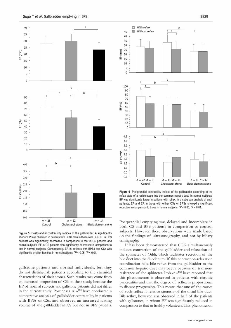
gallstone patients and normal individuals, but they do not distinguish patients according to the chemical characteristics of their stones. Such results may come from an increased proportion of CSs in their study, because the EP of normal subjects and gallstone patients did not differ in the current study. Portincasa et al[28] have conducted a comparative analysis of gallbladder contractility in patients with BPSs or CSs, and observed an increased fasting volume of the gallbladder in CS but not in BPS patients.
Postprandial emptying was delayed and incomplete in both CS and BPS patients in comparison to control subjects. However, these observations were made based on the findings of ultrasonography, and not by biliary scintigraphy.
It has been demonstrated that CCK simultaneously induces contraction of the gallbladder and relaxation of the sphincter of Oddi, which facilitates secretion of the bile duct into the duodenum. If this contraction-relaxation coordination fails, bile reflux from the gallbladder to the common hepatic duct may occur because of transient resistance of the sphincter. Itoh et al[32] have reported that this phenomenon is observed in patients with chronic pancreatitis and that the degree of reflux is proportional to disease progression. This means that one of the causes of such reflux is relative stenosis of the distal bile duct. Bile reflux, however, was observed in half of the patients with gallstones, in whom EF was significantly reduced in comparison to that in healthy volunteers. This phenomenon
EP (
min
)EF
(%
)
40
35
30
25
20
15
10
5
0
90
80
70
60
50
40
30
20
10
0
a
b
b a
ER (
%/m
in)
4.0
3.5
3.0
2.5
2.0
1.5
1.0
0.5
0.0
b
b
n = 28 n = 22 n = 14Control Cholesterol stone Black pigment stone
Figure 5 Postprandial contractility indices of the gallbladder. A significantly shorter EP was observed in patients with BPSs than in those with CSs. EF in BPS patients was significantly decreased in comparison to that in CS patients and normal subjects. EF in CS patients also significantly decreased in comparison to that in normal subjects. Consequently, ER in patients with BPSs and CSs was significantly smaller than that in normal subjects. aP < 0.05, bP < 0.01.
EP (
min
)
45
40
35
30
25
20
15
10
5
0
EF (
%)
100 90 80 70 60 50 40 30 20 10 0
ER (
%/m
in)
4.5
4.0
3.5
3.0
2.5
2.0
1.5
1.0
0.5
0.0
aa
bb
b
ab
n = 22 n = 6 n = 11 n = 11 n = 8 n = 6Control Cholesterol stone Black pigment stone
Figure 6 Postprandial contractility indices of the gallbladder according to the reflux state of a radioisotope into the common hepatic duct. In normal subjects, EF was significantly larger in patients with reflux. In a subgroup analysis of such patients, EF and ER in those with either CSs or BPSs showed a significant reduction in comparison to those in normal subjects. aP < 0.05, bP < 0.01.
With refluxWithout reflux
www.wjgnet.com
Sugo T et al . Gallbladder emptying in BPS 2829
suggests that minute discordance of gallbladder emptying and relaxation of the sphincter of Oddi predisposes to bile reflux and thus causes bile stagnation in the gallbladder.
Chemically, the structure of BPSs is polymerized bilirubin with copper, ion, calcium and other metals, and almost all the stones are formed in the gallbladder[33]. Therefore, an environment suitable for the formation of BPSs comprises conditions that allow chemical polymerization with sufficient time. In fact, EF was reduced in patients with gallstones, especially with BPSs in the present study. Moreover, bile reflux from the gallbladder to the common hepatic duct may cause subclinical transient bile stagnation, thereby contributing to stone formation.
In conclusion, bile evacuation from the gallbladder is reduced in patients with BPSs, in comparison to those with CSs and healthy volunteers. We therefore postulate that impaired gallbladder motor function and discordance of bile evacuation is not the result of the presence of gallstones, but instead it is considered to precede the development of BPSs.
ACKNOWLEDGMENTSThe authors thank Professor R Hada and Emeritus Prof. M Konn, Hirosaki University Graduate School of Medicine, for their many and varied contributions throughout the study.
COMMENTSBackgroundThe incidence of BPSs and CSs is increasing in Japan. However, the mechanism of BPS formation is still not completely understood. The authors hypothesize that reduced motor function of the gallbladder and dysfunction of bile evacuation is associated with BPS formation.
Research frontiersTo date, there have been very few reports that describe gallbladder motility with special reference to the pathogenesis of BPS formation. The authors found impaired gallbladder kinetics in patients with BPSs, in comparison to those with CSs and to healthy volunteers.
Innovations and breakthroughsThe authors strictly selected the patients without any background diseases such as cholecystitis, liver cirrhosis or hepatitis, diabetes mellitus, abnormal gallbladder-wall thickening, and/or a previous surgical history, and then analyzed physiological patterns of bile evacuation from the gallbladder quantitatively by a less-invasive method of computer cholescintigraphy.
ApplicationsThis research is expected to contribute to understanding the pathogenesis of gallstone diseases.
Peer reviewThe authors analyzed gallbladder contractility in patients with BPSs in comparison to those with CSs and healthy volunteers. The study is very interesting.
REFERENCES1 Japan-gallstone-study-group. National survey for gallstone in
Japan. J Jpn Biliary Assoc 1998; 12: 276-293 2 Nakayama F, Miyake H. Changing state of gallstone disease
in Japan. Composition of the stones and treatment of the
condition. Am J Surg 1970; 120: 794-7993 Tazuma S. Gallstone disease: Epidemiology, pathogenesis,
and classification of biliary stones (common bile duct and intrahepatic). Best Pract Res Clin Gastroenterol 2006; 20: 1075-1083
4 Holan KR, Holzbach RT, Hermann RE, Cooperman AM, Claffey WJ. Nucleation time: a key factor in the pathogenesis of cholesterol gallstone disease. Gastroenterology 1979; 77: 611-617
5 Sherlock S, Dooley J. Diseases of the liver and biliary system. Oxford Blackwell Science: London, 2002: 242
6 Small DM. Gallstones. N Engl J Med 1968; 279: 588-5937 Maki T. Pathogenesis of calcium bilirubinate gallstone: role of
E. coli, beta-glucuronidase and coagulation by inorganic ions, polyelectrolytes and agitation. Ann Surg 1966; 164: 90-100
8 Soloway RD, Trotman BW, Maddrey WC, Nakayama F. Pigment gallstone composition in patients with hemolysis or infection/stasis. Dig Dis Sci 1986; 31: 454-460
9 Tabata M, Nakayama F. Bacteria and gallstones. Etiological significance. Dig Dis Sci 1981; 26: 218-224
10 Portincasa P, Moschetta A, Berardino M, Di-Ciaula A, Vacca M, Baldassarre G, Pietrapertosa A, Cammarota R, Tannoia N, Palasciano G. Impaired gallbladder motility and delayed orocecal transit contribute to pigment gallstone and biliary sludge formation in beta-thalassemia major adults. World J Gastroenterol 2004; 10: 2383-2390
11 Acalovschi M, Dumitrascu DL, Nicoara CD. Gallbladder contractility in liver cirrhosis: comparative study in patients with and without gallbladder stones. Dig Dis Sci 2004; 49: 17-24
12 Fornari F, Imberti D, Squillante MM, Squassante L, Civardi G, Buscarini E, Cavanna L, Caturelli E, Buscarini L. Incidence of gallstones in a population of patients with cirrhosis. J Hepatol 1994; 20: 797-801
13 Ai T, Azemoto R, Saisho H. Prevention of gallstones by ursodeoxycholic acid after cardiac surgery. J Gastroenterol 2003; 38: 1071-1076
14 Chijiiwa K, Makino I, Kozaki N, Tanaka M. Differences in gallbladder bile lithogenicity in patients after gastrectomy and colectomy. Eur Surg Res 1996; 28: 1-7
15 Inoue K, Fuchigami A, Higashide S, Sumi S, Kogire M, Suzuki T, Tobe T. Gallbladder sludge and stone formation in relation to contractile function after gastrectomy. A prospective study. Ann Surg 1992; 215: 19-26
16 Kobayashi T, Hisanaga M, Kanehiro H, Yamada Y, Ko S, Nakajima Y. Analysis of risk factors for the development of gallstones after gastrectomy. Br J Surg 2005; 92: 1399-1403
17 Cariati A , Cetta F. Rokitansky-Aschoff sinuses of the gallbladder are associated with black pigment gallstone formation: a scanning electron microscopy study. Ultrastruct Pathol 2003; 27: 265-270
18 Li WG, Luo XY, Johnson AG, Hill NA, Bird N, Chin SB. One-dimensional models of the human biliary system. J Biomech Eng 2007; 129: 164-173
19 Fisher RS , Stelzer F, Rock E, Malmud LS. Abnormal gallbladder emptying in patients with gallstones. Dig Dis Sci 1982; 27: 1019-1024
20 Forgacs IC, Maisey MN, Murphy GM, Dowling RH. Influence of gallstones and ursodeoxycholic acid therapy on gallbladder emptying. Gastroenterology 1984; 87: 299-307
21 Howard PJ, Murphy GM, Dowling RH. Gall bladder emptying patterns in response to a normal meal in healthy subjects and patients with gall stones: ultrasound study. Gut 1991; 32: 1406-1411
22 Jazrawi RP, Pazzi P, Petroni ML, Prandini N, Paul C, Adam JA, Gullini S, Northfield TC. Postprandial gallbladder motor function: refilling and turnover of bile in health and in cholelithiasis. Gastroenterology 1995; 109: 582-591
23 Koruk M, Ozkilic S, Savas MC, Celen Z, Kadayifci A, Ozkilic C. Evaluation of hepatic functions and biliary dynamics in patients with liver cirrhosis by quantitative scintigraphy. Hepatogastroenterology 2003; 50: 1803-1805
24 Krishnamurthy GT, Bobba VR, McConnell D, Turner F, Mesgarzadeh M, Kingston E. Quantitative biliary dynamics: introduction of a new noninvasive scintigraphic technique. J Nucl Med 1983; 24: 217-223
www.wjgnet.com
2830 ISSN 1007-9327 CN 14-1219/R World J Gastroenterol May 14, 2008 Volume 14 Number 18
COMMENTS

25 Maudgal DP, Kupfer RM, Zentler-Munro PL, Northfield TC. Postprandial gall-bladder emptying in patients with gall stones. Br Med J 1980; 280: 141-143
26 Pauletzki J, Cicala M, Holl J, Sauerbruch T, Schafmayer A, Paumgartner G. Correlation between gall bladder fasting volume and postprandial emptying in patients with gall stones and healthy controls. Gut 1993; 34: 1443-1447
27 Pomeranz IS, Shaffer EA. Abnormal gallbladder emptying in a subgroup of patients with gallstones. Gastroenterology 1985; 88: 787-791
28 Portincasa P , Di Ciaula A, Vendemiale G, Palmieri V, Moschetta A, Vanberge-Henegouwen GP, Palasciano G. Gallbladder motility and cholesterol crystallization in bile from patients with pigment and cholesterol gallstones. Eur J Clin Invest 2000; 30: 317-324
29 Spengler U, Sackmann M, Sauerbruch T, Holl J, Paumgartner
G. Gallbladder motility before and after extracorporeal shock-wave lithotripsy. Gastroenterology 1989; 96: 860-863
30 Krishnamurthy GT, Bobba VR, Kingston E. Radionuclide ejection fraction: a technique for quantitative analysis of motor function of the human gallbladder. Gastroenterology 1981; 80: 482-490
31 Khalil T, Walker JP, Wiener I, Fagan CJ, Townsend CM Jr, Greeley GH Jr, Thompson JC. Effect of aging on gallbladder contraction and release of cholecystokinin-33 in humans. Surgery 1985; 98: 423-429
32 Itoh H, Shimono R, Hamamoto K. Evaluation of common bile duct stenosis in chronic pancreatitis using cholescintigraphy. Eur J Nucl Med 1988; 14: 137-140
33 Suzuki N, Nakamura Y, Kobayashi N, Sato T. On metal elements in pure pigment gallstones. Tohoku J Exp Med 1975; 116: 233-240
S- Editor Li DL L- Editor Kerr C E- Editor Lu W
www.wjgnet.com
Sugo T et al . Gallbladder emptying in BPS 2831

Online Submissions: wjg.wjgnet.com World J Gastroenterol 2008 May 14; 14(18): 2832-2837www.wjgnet.com World Journal of Gastroenterology ISSN [email protected] © 2008 WJG. All rights reserved.
Protective effects of apocynin and allopurinol on ischemia/reperfusion-induced liver injury in mice
Ping-Guo Liu, Song-Qing He, Yan-Hong Zhang, Jian Wu
www.wjgnet.com
BASIC RESEARCH
Ping-Guo Liu, Song-Qing He, Yan-Hong Zhang, Jian Wu, Department of Internal Medicine, Transplant Research Program, University of California, Davis Medical Center, Sacramento, CA 95817, United States Ping-Guo Liu, Department of Hepatic-Biliary Surgery, Zhongshan Hospital, Xiamen University, Xiamen 361004, Fujian Province, China Author contributions: Liu PG designed and performed the most of the study; He SQ contributed to an initial idea and helped with MCLA measurement; Zhang YH helped with apoptosis staining; Wu J is the principal investigator, conducted data analysis and manuscript preparation.Supported by The National Institute of Diabetes, Digestive and Kidney Diseases (NIDDK), DK069939 and the Technology Transfer Grant (TTG) by UC Davis Medical Center to Wu JCorrespondence to: Jian Wu, MD, PhD, UC Davis Medical Center, Transplant Research Institute, 4635 2nd Ave. Suite 1001, Sacramento, CA 95817, United States. [email protected]: +1-916-7348044 Fax: +1-916-7348097 Received: March 10, 2008 Revised: March 27, 2008
AbstractAIM: To determine the effects of allopurinol, an inhibitor of xanthine oxidase, and apocynin, an inhibitor of NADPH oxidase, on oxidant stress and liver injury caused by hepatic ischemia/reperfusion (I/R) procedure in mice.
METHODS: Mice were pretreated with a xanthine oxidase inhibitor, allopurinol, or NADPH oxidase (NOX) inhibitor, apocynin before the hepatic I/R procedure. Then treated or untreated mice underwent the hepatic I/R procedure. The effects on hepatic injury and superoxide anions were determined after starting reperfusion.
RESULTS: A standard warm hepatic I/R procedure led to a marked increase in superoxide anion production as indicated by a superoxide anion tracer, MCLA. At the same time, the procedure caused profound acute liver injury, as indicated by elevated serum alanine aminotransferase and tumor necrosis factor-α levels, reduced liver glutathione levels and elevated malondialdehyde contents, as well as a high apoptotic cell count. All these changes were reversed by the use of apocynin or allopurinol prior to the hepatic I/R procedure.
CONCLUSION: Allopurinol and apocynin exerted protective effects on hepatic ischemia/reperfusion injury. The protection is associated with blocking the generation
of superoxide anions during the hepatic I/R procedure by inhibiting xanthine oxidase and NADPH oxidase activity.
© 2008 WJG. All rights reserved.
Key words: Ischemia/reperfusion; Reactive oxygen species; Allopurinol; Apocynin; NADPH oxidase; Xanthine oxidase
Peer reviewer: Paul E Sijens, PhD, Associate Professor, Department of Radiology, UMCG, Groningen 9713GZ, The Netherlands
Liu PG, He SQ, Zhang YH, Wu J. Protective effects of apocynin and allopurinol on ischemia/reperfusion-induced liver injury in mice. World J Gastroenterol 2008; 14(18): 2832-2837 Available from: URL: http://www.wjgnet.com/1007-9327/14/2832.asp DOI: http://dx.doi.org/10.3748/wjg.14.2832
INTRODUCTIONExcessive reaction oxygen species (ROS) cause tissue damage and cell death by binding and altering cellular macromolecules, including DNA, proteins and lipids, and affect their function. One main chemical source which has been shown to contribute significantly to overall pronounced oxidant stress during hepatic ischemia/reperfusion (I/R) procedure is xanthine oxidase (XO), which generates superoxide anions (O2
-) during the conversion of hypoxanthine to xanthine. It is known from many studies that much of the sustained injury during organ transplantation, including the liver, is triggered by ROS via activated XO, because allopurinol, an XO inhibitor, provided some protection against the hepatic I/R-induced injury[1].
NADPH oxidase (NOX), using NADPH as the source of electrons, catalyzes one electron reduction of molecular oxygen to generate O2
-[2], which is a central and initial ROS molecule and may convert to more active and toxic ROS, such as hydrogen peroxide (H2O2), hydroxyl radical (HO-), or peroxide nitrite (ONOO-) in the presence of H+, H2O2, and nitric oxide (NO)[3]. These O2
--derived ROS participating in the inflammatory process, are thought to be key mediators for the activation of Kupffer cells[4] and thus, are crucial in the apoptotic and/or necrotic cell death of the parenchymal cells and sinusoidal endothelial cells (SEC) in the liver[5,6]. The generation of superoxide anions by NADPH oxidase serves as a host defense mechanism

Liu PG et al . Blocking superoxide anion generation 2833
www.wjgnet.com
against invading microorganism infection and the enzyme is present in phagocytic cells, such as monocytes and neutrophils[7]. Although the evidence exists that ROS generated by NOX participate in many cellular responses, and may be involved in many injuring processes, few studies are available that investigate the role of NOX in the contribution to pronounced oxidant stress during the ischemia/reperfusion-induced hepatic injury[7,8]. Apocynin is a naturally occurring methoxy-substituted catechol that effectively inhibits NADPH oxidase through preventing the assembly of its multi-subunits[9]. Thus, we used this inhibitor to explore the role of NADPH oxidase in the generation of superoxide anions during the hepatic I/R procedure and investigate whether apocynin confers any protection against the injury in a mouse model of warm ischemia/reperfusion-induced acute liver injury.
MATERIALS AND METHODSAnimals and treatments ICR mice, from Charles River Laboratory, Wilmington, MA, were fed a pellet diet and water ad libitum and maintained on a 12 h-light/dark cycle. The animal experiment was performed according to a protocol approved by the UC Davis Institutional Animal Care and Use Committee (IACUC). The protocol was prepared in accordance with the National Institutes of Health animal use guidelines. Mice were pretreated with either an XO inhibitor, allopurinol (50 mg/kg, i.p. from Sigma Chemical Co. St. Louis, MO) or a NOX inhibitor, apocynin (3 mg/kg, i.p. from Acros Organics, Geel, Belgium) one day and one hour before the hepatic I/R procedure. A warm hepatic I/R procedure was performed as reported previously by us[10]. In brief, mice were anesthetized with pentobarbital sodium (60 mg/kg, i.p.). Laparotomy was made with a middle incision to expose the lobes of the liver. Following surgical exposure of the portal vein, mice were injected with heparin (100 unit/kg) via tail vein to prevent the formation of blood clot during the ischemia duration. The portal vein and hepatic artery were occluded for 30 min with a microaneurysm clamp to induce hepatic ischemia. Then, the clamp was removed to allow blood to flow through the liver again (reperfusion).
A total of six groups (6 mice in each group) were included in the experiment. Group A: sham-operated group, in which mice received normal saline (N.S.), and underwent a sham operation without I/R procedure; Group B (N.S. control group) in which mice underwent hepatic I/R procedure plus N.S. injection; Group C (allopurinol treatment group), in which animals received prior allopurinol injections and subsequent hepatic I/R procedure; Group D (apocynin treatment group), in which animals received prior apocynin injections and subsequent hepatic I/R procedure. Six and twenty four hours after starting the reperfusion, blood samples were collected from the vena cava before sacrifice. The liver was rapidly excised after drawing blood from the vena cava. Portions of liver tissue were fixed in 10% neutralized formalin for histological evaluation or snap frozen in liquid nitrogen, and maintained at -80℃ until homogenization for the various biochemical assays.
Serum alanine aminotransferase assay Serum levels of alanine aminotransferase (ALT) served as an indicator of liver injury and were analyzed using a commercially available diagnostic kit (Catachem Inc. Bridgeport, Connecticut), and expressed as unit/L.
Measurement of ROS generation in liver homogenates 2-Methyl-6-(P-methoxyphenyl)-3,7-dihydroimidazo(1,2-α)pyrazine-3-one (MCLA) enhanced chemiluminescence was used to determine O2
- generation, as reported by us previously[10]. Approximately, 10 mg of frozen l iver t i ssue was homogenized on ice in 1 mL of homogenization buffer containing 20 mmol/L of N-2-Hydroxyethylpiperazine-N’-2-ethanesulfonic acid (HEPES) and 10 mmol/L ethylene diamine tetraacetic acid (EDTA). The homogenate was subjected to low speed centrifugation (1000 g) for 10 min to remove debris. Luminometer vials containing 2 mL of prewarmed Krebs-HEPES buffer (99 mmol/L NaCl, 4.7 mmol/L KCl, 1.2 mmol/L MgSO4, 1 mmol/L KH2PO4, 1.9 mmol/L CaCl2, 25 mmol/L NaHCO3, 11.1 mmol/L glucose, and 20 mmol/L HEPES, pH 7.4) with 1.0 µmol/L of MCLA were placed in the dark for at least 20 min. After the dark adaptation, background readings were recorded in a luminometer (Lumat LB; Berthold Technologies, BmbH &Co. KG, Germany), and then 20 µL of homogenate supernatant was added to each vial containing MCLA. Chemiluminescent emission in relative light units (RLU) was recorded during a plateau phase of each recording period, and corrected by subtracting the background reading. The calculated value was used to express the integrated values of chemiluminescence (RLU/second/µg protein).
Assays of liver glutathione and malondialdehyde content Levels of the reduced form of glutathione (GSH) and the lipid peroxidation product, malondialdehyde (MDA) in liver tissue were measured spectrophotometrically 6 h and 24 h after starting reperfusion by commercially available kits (OXIS Research, Portland, OR). The levels of GSH and MDA are expressed as nanomoles per milligram of protein.
Histological examination Fixed liver specimens were embedded in paraffin, sectioned in 4 µm thickness, and stained with hematoxylin and eosin (HE) for the evaluation of liver injury. Photomicrographs were taken with a digital camera under a microscope. In addition, frozen sections of the livers were stained with the terminal deoxynucleotidyl transferase (TdT) for detecting of apoptotic cells by an in situ Apoptosis Detection Kit according to the manufacturer’s instructions (Chemicon Inc., Temecula, CA), and reported previously by us[11].
Determination of serum tumor necrosis factor-α levels Serum tumor necrosis factor-α (TNF-α) levels after the hepatic I/R procedure were determined with an enzyme-linked immunosorbent assay (ELISA) kit (R&D Systems, Minneapolis, MN) and expressed as nanograms per milliliter.

Statistical analysis Most data were expressed as mean ± SD of the mean (SEM), and evaluated with an ANOVA analysis and Newman-Keuls tests for multiple comparisons among groups. A P value less than 0.05 was considered statistically significant.
RESULTS Amelioration of I/R-induced acute liver injury As shown in Figure 1E, the hepatic I/R procedure caused a marked increase in serum ALT levels 6 h to 24 h after starting the reperfusion. Prior intraperitoneal injection of either allopurinol or apocynin led to a marked decrease in serum ALT levels compared to animals with I/R plus N.S. injection at both 6-h and 24-h points (P < 0.01). It appeared that either allopurinol or apocynin tended to exert an improved protection to the same extent, and that the differences were not statistically significant at both time points between these two groups. These findings
were consistent with the histological findings as shown in Figure 1A-D. The hepatic I/R procedure led to marked necrosis in the central lobule (Zone Ⅲ) with significant inflammatory infiltration (Figure 1B). A significant reduction in necrosis was found in the livers of animals receiving injections of allopurinol or apocynin (Figure 1C and D) compared to the control group (Figure 1A). For controls, additional animals were injected with allopurinol or apocynin without the hepatic I/R procedure. Serum ALT levels in animals receiving apocynin (65 ± 17 unit/mL, n = 3) or allopurinol (57 ± 14 unit/mL, n = 3) were similar to levels in N.S. controls (46 ± 27 unit/mL, n = 3). These data indicate that neither apocynin nor allopurinol is toxic to the liver.
Enhanced apoptosis in hepatic I/R-induced liver injury Apoptotic cells in liver sections were detected by in situ staining using TUNEL assay. As shown in Figure 2, 6 h after starting reperfusion, profound apoptotic cells with positive TUNEL red fluorescent staining in nuclei were
Figure 1 Attenuation of I/R-induced liver injury by apocynin and allopurinol in mice. A-D: Representative micrographs of liver histology (× 100). A: Normal liver; B: I/R-induced liver injury 6 h after starting reperfusion; C and D: Attenuation of liver injury with apocynin (APO) (C) or Allo (allopurinol) (D); E: Serum ALT levels in mice receiving apocynin or allopurinol and subsequently the hepatic I/R procedure. Serum ALT levels were determined 6 h and 24 h after starting reperfusion (n = 6 each group), and expressed as mean ± SEM. aP < 0.01 vs I/R procedure plus normal saline (N.S.) at 6 h or 24 h.
A B
C D
I/R 6 hI/R 24 h
1000
800
600
400
200
0Sham I/R+NS I/R+Apo I/R+Allo
Seru
m A
LT le
vels
(un
it/m
L)
a
a
a
a
E
www.wjgnet.com
2834 ISSN 1007-9327 CN 14-1219/R World J Gastroenterol May 14, 2008 Volume 14 Number 18

visualized in animals with the hepatic I/R procedure (Figure 2B) compared to sham-operated controls (Figure 2A). Prior treatment of allopurinol or apocynin prevented apoptotic cell death caused by the hepatic I/R procedure (Figure 2C and D).
Enhanced superoxide anion release during the hepatic I/R procedure We employed MCLA, which is proportional to levels of O2
- and singlet oxygen, to investigate whether the hepatic I/R procedure enhanced the chemiluminescent emission from the liver homogenates. We found that light emission from the liver homogenates was elevated 3-fold 6 h after starting the perfusion, compared to animals with sham operation. The enhanced chemiluminescent emission, caused by the I/R procedure, was markedly inhibited by prior administration of allopurinol or apocynin (Figure 3).
This finding provides convincing evidence that NADPH oxidase or xanthine oxidase is the equally important source of superoxide anions, which contributes to pronounced oxidant stress during the hepatic I/R procedure.
Restored GSH and reduced MDA levels by allopurinol or apocynin treatment Liver GSH and MDA levels were determined as indicators of oxidant stress and lipid peroxidation during the hepatic I/R procedure. As shown in Figure 4, both allopurinol or apocynin led to a restoration of decreased GSH levels and a reversion of elevated MDA levels in the liver (P < 0.05-0.01). The GSH levels in the treated groups were close to controls 6 h after starting the reperfusion.
Serum TNF-α levelSerum TNF-α was determined using an ELISA kit 6 and 24 h after starting the reperfusion. It is clear that the hepatic I/R procedure caused an elevation of serum TNF-α levels, and that the prior administration of either allopurinol or apocynin reduced the TNF-α levels when compared to the I/R+N.S. controls (P < 0.05-0.01, Figure 5).
DISCUSSION In the present study, we demonstrated that prior administration of either allopurinol or apocynin prevented the hepatic I/R-induced acute liver injury in mice; the protection was due to a blockage of xanthine oxidase (XO) and NADPH oxidase (NOX) by allopurinol and apocynin, respectively. The findings indicate that both XO and NOX play a critical role in the generation of superoxide anion and superoxide anion-derived ROS during the hepatic I/R procedure. Thus, our findings further confirm the involvement of NOX activation in the hepatic I/R-induced acute injury.
Our previous study showed that pronounced oxidant stress is the major cause of the hepatic I/R injury, and the delivery of either extracellular superoxide dismutase or
Figure 2 Representative micrographs of in situ TUNEL staining of apoptotic cells in liver tissue after the hepatic ischemia/reperfusion procedure. A-D: The TUNEL staining assay was performed as described in the text, and the positive apoptotic cells were stained in red (× 100). A: Control; B: I/R procedure plus N.S. (6 h after starting reperfusion); C: I/R plus apocynin; D: I/R plus allopurinol. E-H: The corresponding liver sections stained with DAPI to illustrate nuclei as a cell number control.
E
CBA D
HGF
1400
1200
1000
800
600
400
200
0
MCL
A ch
emilu
min
esce
nt in
tens
ity
(R
LU/S
ec/ µ
g pr
otei
n)
b
a,d a,d
Sham I/R+NS I/R+Apo I/R+Allo
Figure 3 The hepatic I/R-induced superoxide anion production and the effects of apocynin or allopurinol in mouse liver. The superoxide anion production was determined in mouse liver homogenates with a superoxide anion tracer, MCLA, and the MCLA chemiluminescent emission was recorded in a luminometer and expressed as relative light unit per second per micrograms of protein (mean ± SEM). aP < 0.05, bP < 0.01 vs the sham controls, dP < 0.01 vs the I/R procedure plus N.S. at 6 h time point.
www.wjgnet.com
Liu PG et al . Blocking superoxide anion generation 2835

catalase gene markedly attenuated the hepatic I/R-induced injury in mice[10]. It is known that XO plays a critical role in generating superoxide anions and administering allopurinol, a blocker of XO, reduced the injury. It is not clear whether other pathways of oxidative reactions also contribute to the enhanced oxidant stress based on the fact that allopurinol failed to improve the progression of remote hepatic parenchymal injury caused by hind limb I/R in mice[7]. This drove us to identify other possible pathologic pathways which lead to enhanced oxidant stress in the hepatic I/R process.
Apocynin is a potent inhibitor of NADPH oxidase by reacting with thiol groups required for enzyme assembly. Apocynin is thought to acquire activation by reacting with hydrogen peroxide and extracellular peroxidase. Active NADPH oxidase is comprised of five subunits (p22phox, p40phox, p47phox, p67phox and gp91phox)[12]. In resting cells, three of these subunits (p22phox, p47phox, and p67phox) form a cytosolic complex. Activation of NOX initiates with the phosphorylation of p47phox, which in turn results in the migration of the cytosolic complex to the membrane and subsequent association with membrane-bound p22phox and gp91 to form the active NADPH oxidase[13]. Several homologues of gp91phox-Nox1, Nox3, Nox4 and Nox5 have been identified in non-phagocytic cells[14]. NADPH oxidase has been shown to
have specific subcellular localizations. The mechanisms by which receptors activate NADPH oxidase and regulate ROS production are poorly understood. One may speculate that ROS may be generated during the early and late phases of the hepatic I/R procedure by xanthine oxidase (XO), mitochondrial respiration, or by Kupffer cell-associated and polymorphonuclear neutrophil (PMN)-associated NADPH oxidase[5,15]. Thus, blockage of either XO or NOX with their specific inhibitors decreased the production of superoxide anions, as indicated by less chemiluminescent emission with MCLA, and in turn reduced oxidant stress as reflected by restoration of GSH levels and reversed MDA content in the liver. Consequently, lower levels of serum ALT and TNF-α, improved liver histology, as well as fewer apoptotic cell counts, demonstrating the protective effects of these two inhibitors in the hepatic I/R model system. Our results are consistent with a recent report showing that inhibition of NADPH oxidase prevented alcohol-induced liver injury[16], hemorrhagic shock-induced organ injury[17] and hepatic I/R injury[18], likely by inhibiting ROS formation via NADPH oxidase. However, in the study by Hasegawa et al[18], a less specific NADPH oxidase inhibitor, diphenyleneiodonium chloride (DPI), was used, and superoxide anion levels in the liver were not determined. The cell types which often have high levels of NADPH oxidase include neutrophils, monocytes and Kupffer cells. The former two cell types are recruited during the I/R process and contribute to the damaging process by releasing superoxide anions and proteases[19]. Activation of Kupffer cells also participates in the enhanced oxidant stress, and increases the production of TNF-α and other cytotoxic factors because the depletion of Kupffer cells resulted in reduced hepatic I/R-associated injury in rodent models[20,21]. Nevertheless, further studies are needed to elucidate the role of each cell type that significantly produces superoxide anions via the activation of NADPH oxidase during the hepatic I/R process.
In conclusion, the findings in the present study demonstrate that the pretreatment of both allopurinol and apocynin prevented ischemia/reperfusion-induced acute liver injury and that the protection is associated with the blockage of either xanthine oxidase or NADPH oxidase
450
400
350
300
250
200
150
100
50
0
Live
r G
SH c
onte
nt (
nmol
/mg)
250
200
150
100
50
0
Live
r M
DA
cont
ent
(nm
ol/m
g)
Sham I/R+NS I/R+Apo I/R+Allo
Sham I/R+NS I/R+Apo I/R+Allo
A
B
Figure 4 Hepatic I/R-induced depletion of reduced form of glutathione and enhanced lipid peroxidation, and effects of apocynin and allopurinol. A: Effect of apocynin or allopurinol on liver GSH levels with the hepatic I/R procedure. Liver GSH content was determined spectrophotometrically and expressed as nanomoles per milligram of protein of the tissue; B: Effects of apocynin or allopurinol on liver MDA levels with I/R procedure. Liver MDA content was determined spectrophotometrically and expressed as nanomoles per milligram of tissue protein (mean ± SEM). bP < 0.01 vs Sham controls; cP < 0.05, dP < 0.01 vs the hepatic I/R procedure plus N.S. at 6 h time point (n = 6 in each group).
160
140
120
100
80
60
40
20
0
Seru
m T
NF-
α le
vels
(pg
/mL)
Sham I/R+NS I/R+Apo I/R+Allo
Figure 5 Effects of apocynin or allopurinol on serum TNF-α levels. Serum TNF-α in mice with hepatic I/R-induced liver injury was measured by an enzyme-linked immunosorbent assay kit. bP < 0.01 vs Sham controls (mean ± SEM); cP < 0.05; dP < 0.01 vs I/R procedure plus N.S. at 6 or 24 h time point (n = 6 in each group).
b
d d
b
cc
b
b
c
d
c
d
I/R 6 hI/R 24 h
www.wjgnet.com
2836 ISSN 1007-9327 CN 14-1219/R World J Gastroenterol May 14, 2008 Volume 14 Number 18

activity. The activation of both enzymes contributes to the generation of superoxide anions and related reactive oxygen species during the hepatic ischemia/reperfusion-induced liver injury. This study implies a new pharmacological intervention approach by blocking either xanthine oxidase or NADPH oxidase, or in combination to improve the donor organ quality and survival after the transplantation in recipients.
ACKNOWLEDGMENTS The authors are grateful to Drs. Mark A Zern and Senthil K Venogopal for their invaluable comments and assistance.
COMMENTSBackgroundIschemia/reperfusion-associated donor liver damage is inevitable during the harvest, preservation, transportation and implantation to a recipient. Enhanced oxidant stress has been linked to the donor organ damage; however, the source of reactive oxygen species (ROS) for enhanced oxidant stress has not been well defined. Superoxide anion generated from the reaction of hypoxathine to xanthine catalyzed by xanthine oxidase (OX) has been thought to be one source of ROS, but an intrinsic source of ROS, via the activation of NADPH oxidase (NOX), has not been well studied in a hepatic ischemia/reperfusion model system.
Research frontiersWe employed a sensitive chemiluminescent tracer, MCLA, which reflects tissue levels of superoxide anion, one initial and very active oxygen species, to investigate the importance of superoxide anion via the NADPH oxidase reaction in a model of standard hepatic ischemia/reperfusion procedure in mice.
Innovations and breakthroughsWe found that there was a marked increase in superoxide anion release during the ischemia/reperfusion procedure, and that the use of apocynin, a specific inhibitor of NADPH oxidase, effectively minimized the superoxide anion release, oxidant stress and liver damage caused by a hepatic ischemia/reperfusion procedure in mice.
ApplicationsThe study implies a new pharmacological intervention approach by blocking either xanthine oxidase or NADPH oxidase, or in combination to improve the donor organ quality, function and survival after the transplantation in recipients.
Peer review This study of the beneficial effects of allopurinol and apocynin on the healing of ischemia/reperfusion-induced liver injury is original. It is a well-designed and well-written paper. Allopurinol and apocynin exerted protective effects on hepatic ischemia/reperfusion injury. The protection is associated with blocking the generation of superoxide anions during the hepatic I/R procedure by inhibiting xanthine oxidase and NADPH oxidase activity.
REFERENCES1 Rhoden E, Pereira-Lima L, Lucas M, Mauri M, Rhoden C,
Pereira-Lima JC, Zettler C, Petteffi L, Bello-Klein A. The effects of allopurinol in hepatic ischemia and reperfusion: experimental study in rats. Eur Surg Res 2000; 32: 215-222
2 Yamamori T , Inanami O, Nagahata H, Kuwabara M. Phosphoinositide 3-kinase regulates the phosphorylation of NADPH oxidase component p47(phox) by controlling cPKC/PKCdelta but not Akt. Biochem Biophys Res Commun 2004; 316: 720-730
3 Arteel GE. Oxidants and antioxidants in alcohol-induced liver disease. Gastroenterology 2003; 124: 778-790
4 Czaja MJ. Induction and regulation of hepatocyte apoptosis
by oxidative stress. Antioxid Redox Signal 2002; 4: 759-767 5 Mochida S, Arai M, Ohno A, Masaki N, Ogata I, Fujiwara
K. Oxidative stress in hepatocytes and stimulatory state of Kupffer cells after reperfusion differ between warm and cold ischemia in rats. Liver 1994; 14: 234-240
6 Jaeschke H, Lemasters JJ. Apoptosis versus oncotic necrosis in hepatic ischemia/reperfusion injury. Gastroenterology 2003; 125: 1246-1257
7 Dorman RB, Wunder C, Saba H, Shoemaker JL, MacMillan-Crow LA, Brock RW. NAD(P)H oxidase contributes to the progression of remote hepatic parenchymal injury and endothelial dysfunction, but not microvascular perfusion deficits. Am J Physiol Gastrointest Liver Physiol 2006; 290: G1025-G1032
8 Harada H, Hines IN, Flores S, Gao B, McCord J, Scheerens H, Grisham MB. Role of NADPH oxidase-derived superoxide in reduced size liver ischemia and reperfusion injury. Arch Biochem Biophys 2004; 423: 103-108
9 Stolk J , Hil termann TJ , Di jkman JH, Verhoeven AJ. Characteristics of the inhibition of NADPH oxidase activation in neutrophils by apocynin, a methoxy-substituted catechol. Am J Respir Cell Mol Biol 1994; 11: 95-102
10 He SQ, Zhang YH, Venugopal SK, Dicus CW, Perez RV, Ramsamooj R, Nantz MH, Zern MA, Wu J. Delivery of antioxidative enzyme genes protects against ischemia/reperfusion-induced liver injury in mice. Liver Transpl 2006; 12: 1869-1879
11 Wu J , Liu L, Yen RD, Catana A, Nantz MH, Zern MA. Liposome-mediated extracellular superoxide dismutase gene delivery protects against acute liver injury in mice. Hepatology 2004; 40: 195-204
12 Babior BM. NADPH oxidase: an update. Blood 1999; 93: 1464-1476
13 Cheng G, Cao Z, Xu X, van Meir EG, Lambeth JD. Homologs of gp91phox: cloning and tissue expression of Nox3, Nox4, and Nox5. Gene 2001; 269: 131-140
14 Lentsch AB, Kato A, Yoshidome H, McMasters KM, Edwards MJ. Inflammatory mechanisms and therapeutic strategies for warm hepatic ischemia/reperfusion injury. Hepatology 2000; 32: 169-173
15 Kono H, Rusyn I, Uesugi T, Yamashina S, Connor HD, Dikalova A, Mason RP, Thurman RG. Diphenyleneiodonium sulfate, an NADPH oxidase inhibitor, prevents early alcohol-induced liver injury in the rat. Am J Physiol Gastrointest Liver Physiol 2001; 280: G1005-G1012
16 Abdelrahman M, Mazzon E, Bauer M, Bauer I, Delbosc S, Cristol JP, Patel NS, Cuzzocrea S, Thiemermann C. Inhibitors of NADPH oxidase reduce the organ injury in hemorrhagic shock. Shock 2005; 23: 107-114
17 Yabe Y, Kobayashi N, Nishihashi T, Takahashi R, Nishikawa M, Takakura Y, Hashida M. Prevention of neutrophil-mediated hepatic ischemia/reperfusion injury by superoxide dismutase and catalase derivatives. J Pharmacol Exp Ther 2001; 298: 894-899
18 Hasegawa T, Malle E, Farhood A, Jaeschke H. Generation of hypochlorite-modified proteins by neutrophils during ischemia-reperfusion injury in rat liver: attenuation by ischemic preconditioning. Am J Physiol Gastrointest Liver Physiol 2005; 289: G760-G767
19 Gujral JS, Hinson JA, Farhood A, Jaeschke H. NADPH oxidase-derived oxidant stress is critical for neutrophil cytotoxicity during endotoxemia. Am J Physiol Gastrointest Liver Physiol 2004; 287: G243-G252
20 Vega VL, Mardones L, Maldonado M, Nicovani S, Manriquez V, Roa J, Ward PH. Xanthine oxidase released from reperfused hind limbs mediate kupffer cell activation, neutrophil sequestration, and hepatic oxidative stress in rats subjected to tourniquet shock. Shock 2000; 14: 565-571
21 von Frankenberg M, Golling M, Mehrabi A, Nentwich H, Thies J, Schaeffer F, Jahnke C, Bud O, Gebhard MM, Otto G, Thurman RG, Herfarth C, Klar E. Destruction of Kupffer's cells increases total liver blood flow and decreases ischemia reperfusion injury in pigs. Transplant Proc 1999; 31: 3253-3254
S- Editor Zhong XY L- Editor Rippe RA E- Editor Lu W
www.wjgnet.com
Liu PG et al . Blocking superoxide anion generation 2837
COMMENTS

Online Submissions: wjg.wjgnet.com World J Gastroenterol 2008 May 14; 14(18): 2838-2843www.wjgnet.com World Journal of Gastroenterology ISSN [email protected] © 2008 WJG. All rights reserved.
Is a 7-day Helicobater pylori treatment enough for eradication and inactivation of gastric inflammatory activity?
Carlos Robles-Jara, Carlos Robles-Medranda, Manuel Moncayo, Byron Landivar, Johnny Parrales
www.wjgnet.com
RAPID COMMUNICATION
Carlos Robles-Jara, Carlos Robles-Medranda, Gastroenterology Division, Instituto Ecuatoriano de Enfermedades Digestivas y Pélvicas (IECED), Portoviejo 1301266, EcuadorCarlos Robles-Medranda, Gastroenterology Division, Hôpital Edouard Herriot, Lyon 69003, FranceManuel Moncayo, Gastroenterology Division, Instituto Ecuatoriano del Seguro Social, Portoviejo 1301266, EcuadorByron Landivar, Epidemiology Division, Hospital Oncológico Julio Villacreses Colmont, Sociedad de Lucha contra el Cancer (SOLCA) or Registro Nacional de Tumores, Portoviejo 1301266, EcuadorJohnny Parrales, Laboratorio de Histopatologia (LABOPAT), Portoviejo 1301266, EcuadorAuthor contributions: Robles-Jara C and Robles-Medranda C designed research; Robles-Jara C, Moncayo M and Parrales J performed research; Robles-Medranda C and Landivar B analyzed data; Robles-Medranda C and Robles-Jara C wrote the paper.Correspondence to: Carlos Robles-Jara, MD, Instituto Ecuatoriano de Enfermedades Digestivas y Pélvicas (IECED), Hospital Clinica San Antonio. Av. Paulo Emilio Macias y Tenis club, Portoviejo 1301266, Ecuador. [email protected]: +593-5-2637672 Fax: +593-5-2633265Received: January 5, 2008 Revised: April 8, 2008
AbstractAIM: To compare the efficacy of a 7-d vs 10-d triple therapy regarding H pylori eradication, endoscopic findings and histological gastric inflammatory inactivation in the Ecuadorian population.
METHODS: 136 patients with dyspepsia and H pylori infection were randomized in 2 groups (68 per group): group 1, 7-d therapy; group 2, 10-d therapy. Both groups received the same medication and daily dosage: omeprazole 20 mg bid, clarithromycin 500 mg bid and amoxicillin 1 g bid. Endoscopy was performed for histological assessment and H pylori infection status before and 8 wk after treatment.
RESULTS: H pylori was eradicated in 68% of group 1 vs 83.8% of group 2 for the intention-to-treat analysis (ITT) (P = 0.03; OR = 2.48; 95% CI, 1.1-5.8), and 68% in group 1 vs 88% in group 2 for the per-protocol analysis (PP) (P = 0.008; OR = 3.66; 95% CI, 1.4-10). Endoscopic gastric mucosa normalization was observed in 56.9% in group 1 vs 61.2% in group 2 for ITT, with similar results for the PP, the difference being statistically not significant. The rate of inflammatory inactivation was 69% in group 1 vs 88.7% in group 2 for ITT (P = 0.007; OR = 3.00; 95% CI, 1.2-7.5), and 69% in group 1 vs
96% in group 2 for PP (P = 0.0002; OR = 7.25; 95% CI, 2-26).
CONCLUSION: In this Ecuadorian population, the 10-d therapy was more effective than the 7-d therapy for H pylori eradication as well as for gastric mucosa inflammatory inactivation.
© 2008 WJG. All rights reserved.
Key words: Helicobater pylori treatment; Helicobater pylori infection; Gastric inflammatory inactivation; Triple therapy eradication; Randomized study
Peer reviewer: Colin W Howden, Professor of Medicine, Division of Gastroenterology, Division of Gastroenterology, Northwestern University Feinberg School of Medicine, 676 N. St. Clair Street, Suite 1400, Chicago, IL 60611, United States
Robles-Jara C, Robles-Medranda C, Moncayo M, Landivar B, Parrales J. Is a 7-day Helicobater pylori treatment enough for eradication and inactivation of gastric inflammatory activity? World J Gastroenterol 2008; 14(18): 2838-2843 Available from: URL: http://www.wjgnet.com/1007-9327/14/2838.asp DOI: http://dx.doi.org/10.3748/wjg.14.2838
INTRODUCTIONH pylori bacterial infection is worldwide in humans. It is the main etiologic factor in chronic gastritis and gastroduodenal ulcer disease[1]. It is also closely related to gastric adenocarcinoma and low grade gastric lymphoma of mucosa-associated lymphoid tissue (MALT). Forty percent to 50% of the world population is estimated to be infected, with the highest incidence occurring in older people and in those living in areas with low standards of sanitation[2]. In South American countries, the prevalence of H pylori infection ranges from 70% to 90%[3], being 89.5% in Ecuador[4].
A proton pump inhibitor (PPI) associated with two antibiotics is considered the standard therapy for H pylori eradication[5]. Clinical trials show H pylori eradication rates varying from 45% to 92.3% using PPI associated with amoxicillin and clarithromycin. However, duration of eradication therapy continues to be controversial[6]. In many developed countries, a 7-d therapy is considered enough for bacterial eradication. On the other hand, there is still an argument for increasing the duration of treatment to 10 d or even 14 d[6].

Robles-Jara C et al . H pylori treatment a randomized study 2839
www.wjgnet.com
In Europe, a 7-d triple therapy is still recommended because 14-d therapy had an insignificant advantage in terms of treatment success rate[7]. On the other hand, guidelines from North America recommend 10-d to 14-d therapy, as some studies have reported superior cure rates with prolonged therapy using triple regimens[8]. In Asia, an Indian study reported that prolonged triple therapy with lansoprazole, amoxicillin and tinidazole achieved a significant increase in eradication rates: 47.6% vs 80% vs 91.3% for 1 wk, 2 wk and 3 wk of therapy, respectively[9].
However, in a recent large multicenter randomized trial based on a Korean population, no differences were observed between 7 d vs 14 d of triple therapy with omeprazole associated with amoxicil l in and clarithromycin[10].
Two studies from Turkey using PPI-based triple therapy with amoxicillin and clarithromycin have shown low eradication rates[11,12]. The first study with a 7-d triple therapy showed an eradication rate of 63.6%[11]. The second one using a 14-d therapy showed a 45% eradication rate[12].
In the Mexican population, triple therapy with rabeprazole, amoxicillin and ofloxacin for 14 d achieved significantly superior eradication rates when compared with 7-d therapy (92.3% vs 62.2%)[13].
In South America, it is difficult to identify the best regimen, due to the absence of large multicenter controlled trials. Some recommendations have been published based on consensus reports. The last Brazilian consensus, in 2005, recommended a 7-d triple therapy[14].
The aim of H pylori eradication is to stop the chronic inflammatory activity that leads to histological changes in the gastric mucosa. This inflammatory inactivation has also shown to improve patients’ symptoms[15,16].
The histopathology associated with H pylori infection includes a specific structural disorganization of the epithelial cells and inflammatory infiltration (neutrophils, lymphocytes)[17]. Epithelial nonspecific changes include: presence of microcrypts, reduction of foveolar mucus secretion, atrophy, hyperplasia, metaplasia, atypia, or dysplasia[17].
Many studies have well demonstrated the histological resolution of inf lammatory activity after H pylori eradication using PPI associated with two antibiotics (amoxicillin, clarithromycin or metronidazole) for 7 d, 10 d or 14 d[15,18-20]. Only two studies, however, correlated bacterial eradication, gastric inflammatory inactivation and duration of treatment, comparing 7-d vs 14-d and 10-d vs 14-d therapy[16,21].
This is the first study evaluating the best treatment approach for the Ecuadorian population. Our objective was to compare the efficacy of 7-d vs 10-d triple therapy regarding H pylori eradication rates, the endoscopic findings and the histological gastric inflammatory inactivation.
MATERIALS AND METHODSPatientsThis was a prospective, randomized, open-label study performed at the Ecuadorian Institute of Digestive
Diseases (IECED), in Portoviejo, Ecuador. The study population consisted of patients with dyspepsia who were referred for upper endoscopy. Dyspepsia was defined as pain or discomfort localized in the upper abdomen. Patients were enrolled if they were infected with H pylori.
The exclusion criteria were: (1) Age < 18 years; (2) Use of proton pump inhibitors, antibiotics, H2-receptor antagonists or bismuth compounds in the last 4 wk preceding the inclusion of the patient in the study; (3) Chronic use of non-steroidal anti-inflammatory drugs (NSAIDs); (4) Patients with well-known antibiotic allergy; (5) Pregnant women; (6) Patients with chronic hepatic or renal disease; (7) Patients with Zollinger-Ellison syndrome; (8) Previous gastric surgery; (9) Previous failed H pylori therapy.
Written informed consent was obtained from all participants prior to enrollment.
Endoscopy, histology and assessment of H pyloriDuring endoscopy, 5 biopsies were obtained for histological assessment in accordance with the updated Sydney System (2 from the antrum, 1 from the incisura angularis and 2 from the corpus)[22]. Biopsies were fixed, paraffin embedded and stained with hematoxylin-eosin/Giemsa. A pre-pyloric biopsy was obtained for the rapid urease test. Patients were considered to be infected if histological and rapid urease tests were positive. Eight weeks after treatment, bacterial eradication was checked by using the same endoscopy procedure for histology. At endoscopy, gastritis was defined as the presence of visible alterations of the mucosal appearance, presumably caused by vascular or infiltrative changes (Sydney system definition)[23]. Histological diagnosis was coded in two classes according to the Sydney System classification as follows: (1) non-atrophic chronic gastritis, when inflammatory activity of any grade or antral atrophy grade Ⅰ were present without intestinal metaplasia lesion; and (2) atrophic chronic gastritis, when grade 2 or 3 atrophic mucosa lesions were observed with or without intestinal metaplasia.
The gastric mucosa inflammation activity was also graded in accordance with the updated Sydney System and was considered as the presence of mononuclear cells infiltrate and neutrophilic polymorphonuclear in a background of chronic inflammation[22].
TreatmentPatients were randomized in a 1:1 ratio to receive omeprazole 20 mg bid, clarithromycin 500 mg bid and amoxicillin 1 g bid for 7 d (group 1) or 10 d (group 2). Treatment compliance was assessed by patient report and pill count at the follow-up visit. Good compliance was defined as consumption of more than 90% of the prescribed drugs. After completing treatment, no patients received PPI or antisecretory drugs during the follow-up period.
Statistical analysisStatistical analysis was performed using StatView for Windows (version 5.0). The 95% confidence intervals

were determined with the chi-square test or Fisher’s exact test for small sample sizes, and comparisons were made with Student’s test. P value of < 0.05 were considered as statistically significant. Per protocol and intention to treat approach was performed for statistical analysis in each group.
Sample sizeAs there is no data regarding H pylori eradication rates in the Ecuadorian population, the sample size was calculated a priori, based on available data in the literature. By hypothesizing an 89% eradication rate for the 10-d triple therapy[30] and less than 70% (65%) for 7-d triple therapy[13], the estimated sample size was 54 subjects per group, with a power of 0.85 and a significance level of 0.05. Due to the probability of loss from follow-up, the risk of data loss was estimated at around 25%; therefore the final size was considered 68 patients in each group.
RESULTSBaseline demographicsOne hundred and thirty-six patients (68 patients by group) were enrolled in the study (Figure 1). At endoscopy, the initial diagnoses were: gastritis in 69/136 (50.7%), duodenal ulcer in 26/136 (19.11%), and gastric ulcer in 22/136 (16.17%). Endoscopy was normal in 19/136 (13.9%).
At histology non-atrophic chronic active gastritis was present in 88.2% of patients for group 1 (60/68) and 82.3% (56/68) for group 2. In group 1, eight patients had atrophic chronic active gastritis (11.7%) in which the presence of intestinal metaplasia was identified in 5/8. In group 2, the presence of atrophic chronic active gastritis was observed in 12 patients (17.6%), all of them with intestinal metaplasia. There were no significant differences between the treatment groups in any of the baseline characteristics. Demographic, endoscopic and histological characteristics are summarized in Table 1.
Three patients (4.4%) in group 1 and six patients (8.8%) in group 2 did not return for follow-up and were excluded from the study. Additionally, ten patients (14.7%) in group 2 were not compliant with the treatment: therapy
was stopped at d 8 in 7 patients, at d 3 in 2 patients and at d 4 in 1 patient. In 6 patients (8.8%), the non-compliance was correlated with treatment-adverse events (4 with moderate abdominal pain, 1 with nausea, and 1 with diarrhea).
Per protocol (PP) analysis One hundred and seventeen patients underwent a PP analysis (Table 2): 65 patients in group 1 and 52 patients in group 2. Eight weeks after treatment the endoscopy showed a normal gastric mucosa in 56.9% (37/65) of patients in group 1 and in 63.5% (32/52) in group 2. Ulcer healing was observed in 93.7% (45/48) of patients (both groups). In group 1, gastritis was observed in 25 patients (intestinal metaplasia in 5 patients at histology without presence of gastric mucosa inflammation) and an active ulcer in 3 patients (2 with gastric ulcer and 1 with duodenal ulcer). In group 2, gastritis was present in 20 patients (12 patients with intestinal metaplasia at histology and without gastric mucosa inflammation).
H pylori eradication was obtained in 68% (44/65) of patients for group 1 and 88% (46/52) for group 2 (P = 0.008; OR = 3.66; 95% CI, 1.4-10).
The total average rate of inflammatory inactivation of gastric mucosa was 80% for both groups. In group 1, 45 patients (69%) showed an inactivation of the inflammation compared with 49 patients (96%) in group 2 (P = 0.0002; OR = 7.25; 95% CI, 2-26).
Intention to treat (ITT) analysis One hundred and twenty-seven patients underwent an ITT assessment (Table 3): 65 patients in group 1 and 62 patients in group 2. After treatment, the endoscopy showed a normal gastric mucosa in 56.9% (37/65) of patients for group 1 and 61.2% (37/62) in group 2. Gastritis was present in 25 patients in group 2 (12 patients with intestinal metaplasia at histology and without presence of gastric mucosa inflammation).
H pylori eradication was obtained in 68% (44/65) of patients for group 1 and 83.8% (52/62) for group 2 (P = 0.03; OR = 2.48; 95% CI, 1.1-5.8).
Patients with H pyloriinfectionn = 136
7-d OACn = 68
10-d OACn = 68
n = 65(excluded 3)1
n = 62(excluded 6)1
n = 65(excluded 3)2
n = 52(excluded 16)2
ITT
PP
Figure 1 Randomization protocol. 1Patients were excluded if missing in the follow-up; 2Patients were excluded if ITT excluded population, non-compliance. ITT: Intention to treat analysis; PP: Per protocol analysis; OAC: Omeprazole, amoxicillin and clarithromycin.
Table 1 Baseline demographic, endoscopic and histological characteristics of patients
Group 1 (n = 68) OAC 7-d
Group 2 (n = 68) OAC 10-d
P
Age (yr, mean ± SD) 42 ± 11 44 ± 12 NSSex (M/F) 23/45 27/41 NSEndoscopic findings: Normal 9 10 NS Gastritis (IM) 31 (4) 38 (6) NS Gastric ulcer (IM) 13 (1) 9 (2) NS Duodenal ulcer (IM) 15 (0) 11 (4) NSHistological findings: Nonatrophic chronic 60 56 active gastritis (IM) NS Atrophic chronic 8 (5) 12 (12) active gastritis (IM)
IM: Intestinal metaplasia; M: Male; F: Female; NS: Not significant; OAC: Omeprazole, amoxicillin and clarithromycin.
www.wjgnet.com
2840 ISSN 1007-9327 CN 14-1219/R World J Gastroenterol May 14, 2008 Volume 14 Number 18

The total average rate of gastric mucosa inflammatory inactivation was 79.5% for both groups. In group 1, 45/65 patients (69%) showed an inactivation of the inflammation compared with 54/62 patients (88.7%) in group 2 (P = 0.007; OR = 3.00; 95% CI, 1.2-7.5).
DISCUSSIONAfter the discovery of H pylori many studies were reported for its treatment, including various meta-analyses. Nowadays there is no doubt that triple therapies, that is, PPI & two antibiotics (amoxicillin associated with clarithromycin or metronidazole) are currently the most preferred first-line therapy regimens in clinical practice.
Therapy regimens and their duration could not be standardized because results of clinical data are different between countries. In effect, the results for efficacy of therapy are varied worldwide. This variation of the results could be a consequence of many factors such as bacterial virulence, environmental factors and antibacterial resistance, which are peculiar for each country. In addition, individual factors such as: patient’s age less than 60, presence of duodenal ulcer and treatment duration have been also linked to therapy efficacy[24].
Until now, no data or consensus about the duration of treatment for H pylori in Ecuador was available. We therefore evaluated the twice-daily triple therapy of omeprazole 20 mg bid, amoxicillin 1 g bid and clarithromycin 500 mg bid.
Metronidazole resistance in H pylori strains varies geographically, and influences negatively the effectiveness of therapies containing this antibiotic[25]. In Latin-American countries, more than 62% of the population is resistant to metronidazole treatment[3,26]. This can be explained by the indiscriminate use of these agents for many other diseases (such as parasitosis).
The rates of metronidazole resistance are as high as 81% in Ecuador. In contrast, clarithromycin resistance is still small: < 10%[27]. This is the reason for using clarithromycin in our study.
Two clinical trials from South America reported eradication rates of more than 85% with 7-d triple therapy using PPI, amoxicillin and clarithromycin. Coelho et al[28], in the Brazilian population showed 87% of H pylori eradication with a 7-d treatment (using pantoprazole, amoxicillin and clarithromycin) in 71 patients. In Peru, a randomized study with 72 patients showed 86.1% of H pylori eradication with no differences between 7-d vs 10-d triple therapy[29].
In the Ecuadorian population our study showed a 68% and an 86% eradication rate for the 7-d and 10-d triple therapy, respectively, this difference being statistically significant.
Many clinical trials have demonstrated similar eradication rates using 10-d triple therapy with PPI, amoxicillin and clarithromycin[15,16,30,34]. A large multicenter double-blind trial in the United States, demonstrated an eradication rate of 84%[30]. Fennerty et al, also found comparable results (84% eradication rate) in 284 patients, without difference when compared to 14-d therapy[16]. In the Czech Republic, the rate of H pylori eradication obtained was 87%[15].
Moreover, a recent meta-analysis has shown that prolonging triple therapies from 7 to 10 d or 14 d improves treatment cure rates[31]. Other meta-analysis suggested that 10-d therapy was associated with less failure than 7-d therapy[24]. Our study also indicates that a 10-d regimen was statistically more effective than the 7-d regimen for inactivating the gastric mucosa inflammation.
Acid suppression with PPI in H pylori gastritis only decreases the gastric inflammatory activity, but doesn’t produce its inactivation[32]. Histological changes after H pylori eradication show a rapid improvement of neutrophils infiltration in the first 2 mo, progressive declination of mononuclear cell infiltration during the first 2 years, and no significant changes in the 12 first mo regarding atrophy or intestinal metaplasia[17-20,33]. The presence of an inflammatory activity is seen in fewer cases, even 12 mo after the eradication[32,33].
In our patients, 8 wk after treatment, the endoscopic assessment revealed the persistence of gastric lesions in 43.1% of patients (28/65) in group 1 and 38.5% of patients (20/52) in group 2. However, an inflammatory inactivation was obtained in 69% and 94% of patients in groups 1 and 2 respectively. All cases with intestinal metaplas ia had H py lo r i erad icat ion and gastr ic inflammatory inactivation. In patients with abnormal control endoscopy, persistence of H pylori was observed in 21/28 patients in group 1 and in 6/20 patients in group 2, being associated with the persistence of an inflammatory activity.
Discordance between endoscopic and histological findings after H pylori eradication with 7-d triple therapy (PPI, amoxicil l in, clarithromycin) was previously reported[16,20]. Dajani et al, observed a persistence of lesions at the control endoscopy in 46.4% of patients (7.4% of duodenal ulcer and 39% of gastritis lesions), in spite of 93% eradication rates with a remarkable resolution of the
Table 2 Per protocol analysis, control results at wk 8 in two groups (%)
Table 3 Intention to treat analysis, control results at wk 8 in two groups (%)
Group 1(n = 65)OAC 7-d
Group 2(n = 52)OAC 10-d
P
H pylori eradication 44/65 (68) 46/52 (88) 0.008Normal endoscopy 37/65 (56.9) 32/52 (63.5) NSInflammatory inactivationof gastric mucosa
45/65 (69) 49/52 (96) 0.0002
OAC: Omeprazole, amoxicillin and clarithromycin; NS: Not significant.
Group 1 (n = 65)OAC 7-d
Group 2 (n = 62)OAC 10-d
P
H pylori eradication 44/65 (68) 52/62 (83.8) 0.03Normal endoscopy 37/65 (56.9) 37/62 (61.2) NSInflammatory inactivationof gastric mucosa
45/65 (69) 54/62 (88.7) 0.007
OAC: Omeprazole, amoxicillin and clarithromycin; NS: Not significant.
www.wjgnet.com
Robles-Jara C et al . H pylori treatment a randomized study 2841

inflammatory activity[20]. Another study comparing 10 vs 14 d of treatment shows the persistence of an active ulcer in 28% of patients and gastritis in 9% after an eradication rate of 84%[16].
Prolonging therapy duration has been associated with an increased risk of non-compliance due to side-effects[30,35]. This was also observed in our study, which shows an increased non-compliance rate in the 10-d therapy group (group 2), being correlated in 6 patients with treatment side-effects.
Finally, although our data points to the superior benefits of the 10-d therapy, we should be careful before establishing definitive recommendations for the Ecuadorian population.
On a day-to-day basis, every medical treatment must consider the cost-benefit analysis, mainly in developing countries, where it is difficult for patients to afford their medications in function of its price. The cost of H pylori eradication therapy in South America is very high[3]. Some recommended eradication therapies have 7-d duration mainly because of improved compliance and decreased medical cost[13]. In our study, 32% of patients in the 7-d vs 12% in 10-d therapy did not eradicate H pylori. Probably, the re-treatment of this 32% of patients in group 1 with second or third line therapies would be more expensive and less effective than directly treating them for 10 d with the first line approach. This evaluation however was not the objective of our paper, and should be the subject of a future study.
In conclusion, this randomized open study showed that in Ecuador, a 10-d triple therapy using PPI associated with amoxicillin and clarithromycin was significantly more effective than a 7-d triple therapy. The cost-benefit of this treatment in our population should be evaluated in a future study.
COMMENTSBackgroundThe aim of H pylori eradication is to stop chronic inflammatory activity that leads to histological changes in the gastric mucosa. A proton pump inhibitor (PPI) associated with two antibiotics is considered the standard therapy for H pylori eradication. However, duration of eradication therapy continues to be controversial, due to the variable results from all over the world. There is no data about H pylori treatment in the Ecuadorian population and to our knowledge there are no studies correlating the bacterial eradication, gastric inflammatory inactivation and duration of treatment comparing 7-d vs 10-d.
Research frontiersThis study determined the best valid H pylori eradication treatment in a population with higher rates of infection correlating the bacterial eradication rate, the endoscopic and histological results with the treatment duration.
Innovations and breakthroughsMany studies have well demonstrated the histological resolution of inflammatory activity after H pylori eradication using PPI associated with two antibiotics (amoxi-cillin, clarithromycin or metronidazole) for 7 d, 10 d or 14 d. Only two studies, however, correlated bacterial eradication, gastric inflammatory inactivation and duration of treatment, comparing 7-d vs 14-d and 10-d vs 14-d therapy. This is the first study evaluating the best treatment approach for the Ecuadorian population. We compared the efficacy of 7-d vs 10-d triple therapy regarding H pylori eradication rates, the endoscopic findings and the histological gastric inflammatory inactivation.
ApplicationsIn this Ecuadorian population, 10-d triple therapy using PPI associated with amoxicillin and clarithromycin was significantly more effective than 7-d triple therapy. The cost-benefit of this treatment in our population should be evaluated in a future study.
Peer reviewThis is a reasonably well done clinical trial comparing 7-d and 10-d PPI-based triple therapy for H pylori infection. Ten-day treatment was significantly superior. The trial is well described.
REFERENCES1 Gisbert JP, Gonzalez L, Calvet X. Systematic review and
meta-analysis: proton pump inhibitor vs. ranitidine bismuth citrate plus two antibiotics in Helicobacter pylori eradication. Helicobacter 2005; 10: 157-171
2 Pounder RE, Ng D. The prevalence of Helicobacter pylori infection in different countries. Aliment Pharmacol Ther 1995; 9 Suppl 2: 33-39
3 Castro Lde P, Coelho LG. Helicobacter pylori in South America. Can J Gastroenterol 1998; 12: 509-512
4 Gomez NA, Alvarez LR, Zapatier JA, Vargas PE. [Efficacy of stool antigen and serologic tests in the diagnosis of Helicobacter pylori in Ecuadorian population] Rev Gastroenterol Mex 2005; 70: 146-150
5 Malfertheiner P, Megraud F, O'Morain C, Hungin AP, Jones R, Axon A, Graham DY, Tytgat G. Current concepts in the management of Helicobacter pylori infection--the Maastricht 2-2000 Consensus Report. Aliment Pharmacol Ther 2002; 16: 167-180
6 Bytzer P, O'Morain C. Treatment of Helicobacter pylori. Helicobacter 2005; 10 Suppl 1: 40-46
7 Malfertheiner P, Megraud F, O'Morain C, Bazzoli F, El-Omar E, Graham D, Hunt R, Rokkas T, Vakil N, Kuipers EJ. Current concepts in the management of Helicobacter pylori infection: the Maastricht III Consensus Report. Gut 2007; 56: 772-781
8 Chey WD, Wong BC. American College of Gastroenterology guideline on the management of Helicobacter pylori infection. Am J Gastroenterol 2007; 102: 1808-1825
9 Chaudhary A, Ahuja V, Bal CS, Das B, Pandey RM, Sharma MP. Rank order of success favors longer duration of imidazole-based therapy for Helicobacter pylori in duodenal ulcer disease: a randomized pilot study. Helicobacter 2004; 9: 124-129
10 Kim BG, Lee DH, Ye BD, Lee KH, Kim BW, Kim SG, Kim SW, Kim SK, Kim JJ, Kim HY, Park JJ, Park CY, Baik GH, Lee YC, Lee JH, Lee JH, Chun HJ, Hahm KB, Hong SJ, Lee SW, Jung HC. Comparison of 7-day and 14-day proton pump inhibitor-containing triple therapy for Helicobacter pylori eradication: neither treatment duration provides acceptable eradication rate in Korea. Helicobacter 2007; 12: 31-35
11 Sivri B, Simsek I, Hulagu S, Kadayifci A, Tozun N, Akarsu M, Uraz S, Savas MC, Koruk M, Bozbas A. The efficacy, safety and tolerability of pantoprazole-based one-week triple therapy in H. pylori eradication and duodenal ulcer healing. Curr Med Res Opin 2004; 20: 1301-1307
12 Altintas E , Sezgin O, Ulu O, Aydin O, Camdeviren H. Maastricht II treatment scheme and efficacy of different proton pump inhibitors in eradicating Helicobacter pylori. World J Gastroenterol 2004; 10: 1656-1658
13 Bosques-Padilla FJ, Garza-Gonzalez E, Calderon-Lozano IE, Reed-SanRoman G, de Arino Suarez M, Valdovinos-Diaz MA, Orozco-Gamiz A, Blancas-Valencia JM, Tamayo-de la Cuesta JL. Open, randomized multicenter comparative trial of rabeprazole, ofloxacin and amoxicillin therapy for Helicobacter pylori eradication: 7 vs. 14 day treatment. Helicobacter 2004; 9: 417-421
14 Coelho LG , Zaterka S. [Second Brazil ian Consensus Conference on Helicobacter pylori infection] Arq Gastroenterol 2005; 42: 128-132
15 Kyzekove J, Arlt J, Arltová M. Is there any relationship
www.wjgnet.com
2842 ISSN 1007-9327 CN 14-1219/R World J Gastroenterol May 14, 2008 Volume 14 Number 18
COMMENTS

between functional dyspepsia and chronic gastritis associated with Helicobacter pylori infection? Hepatogastroenterology 2001; 48: 594-602
16 Fennerty MB, Kovacs TO, Krause R, Haber M, Weissfeld A, Siepman N, Rose P. A comparison of 10 and 14 days of lansoprazole triple therapy for eradication of Helicobacter pylori. Arch Intern Med 1998; 158: 1651-1656
17 Warren JR. Gastric pathology associated with Helicobacter pylori. Gastroenterol Clin North Am 2000; 29: 705-751
18 Abdul Aal GM, Dajani AI, Nounou M, Awad S, Abdul Rasheed Z, Gautam S, Ukabam S, Nayal S. Resolution of gastritis induced by Helicobacter pylori 4-5 weeks after successful eradication of infection using a triple therapy regimen of pantoprazole, amoxycillin and clarithromycin for one week. Digestion 1999; 60: 286-297
19 Jakic-Razumovic J, Tentor D, Kusec V, Cuzic S, Brkic T. Histopathological features of gastritis before and after treatment for Helicobacter pylori. Croat Med J 2000; 41: 159-162
20 Dajani AI, Awad S, Ukabam S, Nounou MA, Abdul Rasheed Z, Gautam S, Abdul Aal G, Nayal S. One-week triple regime therapy consisting of pantoprazole, amoxicillin and clarithromycin for cure of Helicobacter pylori-associated upper gastrointestinal diseases. Digestion 1999; 60: 298-304
21 Paoluzi P, Iacopini F, Crispino P, Nardi F, Bella A, Rivera M, Rossi P, Gurnari M, Caracciolo F, Zippi M, Pica R. 2-week triple therapy for Helicobacter pylori infection is better than 1-week in clinical practice: a large prospective single-center randomized study. Helicobacter 2006; 11: 562-568
22 Dixon MF, Genta RM, Yardley JH, Correa P. Classification and grading of gastritis. The updated Sydney System. International Workshop on the Histopathology of Gastritis, Houston 1994. Am J Surg Pathol 1996; 20: 1161-1181
23 Tytgat GN . The Sydney System: endoscopic division. Endoscopic appearances in gastritis/duodenitis. J Gastroenterol Hepatol 1991; 6: 223-234
24 Broutet N, Tchamgoue S, Pereira E, Lamouliatte H, Salamon R, Megraud F. Risk factors for failure of Helicobacter pylori therapy--results of an individual data analysis of 2751 patients. Aliment Pharmacol Ther 2003; 17: 99-109
25 Chey WD. Helicobacter Pylori. Curr Treat Options Gastroenterol 1999; 2: 171-182
26 NA. World Gastroenterology Organisation (WGO). WGO Practice Guideline Helicobacter Pylori in developing countries.
November 2006. Accessed Feb 14, 2007. Available at URL address: http://www.omge.org/globalguidelines/guide15/guideline15.htm
27 Debets-Ossenkopp YJ, Reyes G, Mulder J, aan de Stegge BM, Peters JT, Savelkoul PH, Tanca J, Pena AS, Vandenbroucke-Grauls CM. Characteristics of clinical Helicobacter pylori strains from Ecuador. J Antimicrob Chemother 2003; 51: 141-145
28 Coelho LG, Mattos AA, Francisconi CF, Castro Lde P, Andre SB. [Efficacy of the dosing regimen of pantoprazole 40 mg, amoxicillin 1000 mg and clarithromycin 500 mg, twice daily for 7 days, in the eradication of Helicobacter pylori in patients with peptic ulcer] Arq Gastroenterol 2004; 41: 71-76
29 Rodriguez W, Pareja Cruz A, Yushimito L, Ramírez Ramos A, Gilman RH, Watanabe Yamamoto J, Rodríguez Ulloa C, Mendoza Requena D, Guerra Valencia J, Leey Casella J, Chinga Alayo E, Velapatiño B, Valencia T. Omeprazole, amoxicillin and clarithromycin in the treatment of helicobacter pylori, in 7 and 10-day regimens. Rev Gastroenterol Peru 2003; 23: 177-183
30 Laine L, Suchower L, Frantz J, Connors A, Neil G. Twice-daily, 10-day triple therapy with omeprazole, amoxicillin, and clarithromycin for Helicobacter pylori eradication in duodenal ulcer disease: results of three multicenter, double-blind, United States trials. Am J Gastroenterol 1998; 93: 2106-2112
31 Calvet X, Garcia N, Lopez T, Gisbert JP, Gene E, Roque M. A meta-analysis of short versus long therapy with a proton pump inhibitor, clarithromycin and either metronidazole or amoxicillin for treating Helicobacter pylori infection. Aliment Pharmacol Ther 2000; 14: 603-609
32 Moayyedi P, Wason C, Peacock R, Walan A, Bardhan K, Axon AT, Dixon MF. Changing patterns of Helicobacter pylori gastritis in long-standing acid suppression. Helicobacter 2000; 5: 206-214
33 Salih BA, Abasiyanik MF, Saribasak H, Huten O, Sander E. A follow-up study on the effect of Helicobacter pylori eradication on the severity of gastric histology. Dig Dis Sci 2005; 50: 1517-1522
34 Laine L , Estrada R, Truj i l lo M, Fukanaga K, Neil G. Randomized comparison of differing periods of twice-a-day triple therapy for the eradication of Helicobacter pylori. Aliment Pharmacol Ther 1996; 10: 1029-1033
35 Vakil N , Connor J . Helicobacter pylori eradicat ion: equivalence trials and the optimal duration of therapy. Am J Gastroenterol 2005; 100: 1702-1703
S- Editor Zhong XY L- Editor Negro F E- Editor Lu W
www.wjgnet.com
Robles-Jara C et al . H pylori treatment a randomized study 2843

Online Submissions: wjg.wjgnet.com World J Gastroenterol 2008 May 14; 14(18): 2844-2850www.wjgnet.com World Journal of Gastroenterology ISSN [email protected] © 2008 WJG. All rights reserved.
Laparo-endoscopic “Rendezvous” to treat cholecysto-choledocolithiasis: Effective, safe and simplifies the endoscopist’s work
Gaetano La Greca, Francesco Barbagallo, Michele Di Blasi, Andrea Chisari, Rosario Lombardo, Rosario Bonaccorso, Saverio Latteri, Andrea Di Stefano, Domenico Russello
www.wjgnet.com
RAPID COMMUNICATION
Gaetano La Greca, Francesco Barbagallo, Andrea Chisari, Rosario Lombardo, Rosario Bonaccorso, Saverio Latteri, Andrea Di Stefano, Domenico Russello, Department of Surgical Sciences, Transplantation and Advanced Technologies, University of Catania, Cannizzaro Hospital, Via Messina 829, Catania 95126, ItalyMichele Di Blasi, Endoscopic Unit, Cannizzaro Hospital, Via Messina 829, Catania 95126, ItalyAuthor contributions: La Greca G designed the study, performed the research and wrote the paper; Barbagallo F, chief investigator and guided the research work; Di Blasi M, performed endoscopic procedures; Lombardo R, Bonaccorso R, Di Stefano A, Russello D provided the patient material and other clinical data; Chisari A, Latteri S, tables and drafting.Correspondence to: Gaetano La Greca, MD, PhD, Via Messina 354; Catania 95126, Italy. [email protected]: +39-095-7263586 Fax: +39-095-7122221Received: January 28, 2008 Revised: March 29, 2008
AbstractAIM: To investigate our cl inical experience with combined laparo-endoscopic Rendezvous (RV) for the treatment of patients affected by gallstones and common bile duct (CBD) stones and especially to study the never evaluated opinion of the endoscopist concerning the difficulty of the intraoperative endoscopic procedure during the RV in comparison with standard endoscopic retrograde cholangio-pancreatography (ERCP).
METHODS: Eighty consecutive patients affected by cholecystolithiasis and diagnosed or suspected CBD stones were treated with a standardized “tailored” RV. The relevant technical features, the feasibility, the effectiveness in stone clearance, the safety but also the simple evaluation of difficulty and agreement of the endoscopist were analyzed with a questionnaire.
RESULTS: The feas ib i l i ty was 97.5% and the effectiveness 100% concerning CBD clearance and solution of coexisting problems at the papilla. Minor morbidity was 3.3%, the operating time was prolonged by a mean of 14 min, the mean hospital stay was 3.8 d and only one stone’s recurrence occurred. The endoscopist evaluated the procedure to be simpler than standard ERCP-ES in 81.2% of the cases.
CONCLUSION: S imu l taneous RV ca r r i e s h igh
effectiveness and safety at least comparable to those reported for other options. The endoscopist is very often satisfied with this approach because of the minimization of some steps of the endoscopic procedure and avoidance of relevant iatrogenic risk factors. If the mandatory collaboration between surgeons and endoscopists is guaranteed, this approach can often be preferable for the patient, the surgeon, the endoscopist and the hospital.
© 2008 WJG. All rights reserved.
Key words: Gallstones; Common bile duct; Endoscopic retrograde cholangio-pancreatography; Endoscopic sph inc te ro tomy; Rendezvous; In t ra -opera t i ve cholangiography; Laparoscopic cholecystectomy
Peer reviewer: Yasuji Arase, MD, Department of Gastro-enterology, Toranomon Hospital, 2-2-2Toranomonminato-ku, Tokyo 105-8470, Japan
La Greca G, Barbagallo F, Di Blasi M, Chisari A, Lombardo R, Bonaccorso R, Latteri S, Di Stefano A, Russello D. Laparo-endoscopic “Rendezvous” to treat cholecysto-choledocolithiasis: Effective, safe and simplifies the endoscopist’s work. World J Gastroenterol 2008; 14(18): 2844-2850 Available from: URL: http://www.wjgnet.com/1007-9327/14/2844.asp DOI: http://dx.doi.org/10.3748/wjg.14.2844
INTRODUCTIONCholecystolithiasis is one of the most common diseases in which treatment involves general practitioners, gastroenterologists, endoscopists and general surgeons. The management of patients affected by gallstones complicated by common bile duct (CBD) stones and/or problems at the papilla of Vater is anyway a challenge as there are many available options for treatment, all being effective but with the best practice is still unknown. The development of minimally invasive surgery has been associated with development and diffusion of totally laparoscopic exploration of the CBD which is considered the best approach by the pioneers of minimally invasive surgery. This convention contrasts with the fact that the sequential approach, combining the endoscopic retrograde cholangio-pancreatography (ERCP) with endoscopic

La Greca G et al . RV to treat bile duct stones 2845
www.wjgnet.com
sphincterotomy (ES) prior or after cholecystectomy, was for a long time considered to be the preferable choice and also today many gastroenterologists, endoscopists and surgeons still prefer it in clinical practice. The third option, namely the laparo-endoscopic “Rendezvous” (RV) combines in one procedure laparoscopic cholecystectomy (LC), intra-operative cholangiography (IOC) and endoscopic CBD clearance being an actual alternative to the other two. This laparo-endoscopic approach is for the surgeon technically simpler than total laparoscopy, carries interesting advantages but is more complex to organize and is perhaps also considered to reduce the role of the laparoscopic surgeon. Also for the endoscopist this procedure could carry many advantages. Believing in its usefulness we used this approach in 80 consecutive patients affected by gallstones and CBD stones and here we report the results concerning effectiveness, safety and for the first time in the literature we particularly analysed the agreement, evaluation and opinion of the endoscopist normally used to perform ERCP-ES.
MATERIALS AND METHODSEighty patients affected by cholecystolithiasis and CBD stones or suspected stones at IOC were treated simultaneously during the same operation by patient tai lored combined laparo-endoscopic “RV”. Our definition of the term “RV” consists of the combined laparo-endoscopic simultaneous approach involving the endoscopist and surgeon in the operating room during one single administration of anesthesia to clear CBD stones or solve associated problems related to sludge or other problems at the papilla of Vater. All the records of the treated patients were analyzed concerning main clinical data, history, diagnosis, surgical or endoscopic technical particularities, duration of the procedures, feasibility, failure, conversion to open surgery, effectiveness in stone clearance, post-operative complications, duration of the hospital stay, late complications and recurrence of stones. At the end of every procedure we submitted a questionnaire to the endoscopist to analyze his satisfaction concerning the intraoperative endoscopic procedure. The questionnaire elicited a simple immediate opinion of the endoscopist concerning his evaluation of the endoscopic difficulty of the procedure graded as: (1) simpler; (2) comparable; (3) more difficult, if compared to a standard ERCP-ES.
RESULTSClinical dataFrom 2002 to 2006, 80 consecutive patients were submitted to the laparo-endoscopic RV. The mean patient age was 58 years (9-88 years), 29 patients were male and 51 female. The youngest patient was a 9-year-old female patient with a BMI of 27, with gallstones and recurrence of acute biliary pancreatitis due to common bile duct sludge and sphincter of Oddi dysfunction. The oldest patient was a female patient with increasing jaundice, severe cholangitis, and CBD stones impacted in the papilla and coming out of two failed attempts to treat the jaundice and the
stones with ERCP-ES. The diagnosis of CBD stones was given preoperatively in 47 patients (58.7%), in 33 patients (41.2%) the diagnosis was not available preoperatively. 23 patients (28.7%) had a recent history of mild acute pancreatitis or abnormal increase of amylase or lipase levels associated with biliary pain. In 49 patients (61.2%) there was an abnormal increase of bilirubin levels. In 53 patients (66.2%) suspicion or the diagnosis of CBD stones was obtained by ultrasonography. In 46 patients (57.5%) a cholangio-MRI was also performed preoperatively and in 38 of them (82.6%) this confirmed the diagnosis. In 24 patients (30%) with CBD stones a CT scan was also performed during diagnostic work-up but this confirmed the stones in only 9 patients (37.5%). In fact, in 23 of the 33 patients without preoperative diagnosis (69.6%) there was preoperatively a high clinical suspicion of CBD stones but in 10 (31.1%) the diagnosis could be obtained only during surgery by IOC performed because of the surgeon’s own decision based on the above mentioned criteria. All 80 patients therefore underwent anteretrograde transcystic IOC. The criteria used to perform IOC were the diagnosis of CBD stones or the positiveness of at least one of the following risk factors: history of acute pancreatitis, pre-operative abnormal increase of direct bilirubinemia, ALP or gamma-GT, abnormally dilated CBD at ultrasonography, multiple small stones or sludge in the gallbladder, intraoperative evidence of a large cystic duct or an enlarged CBD. The feasibility of the RV was 97.5%.
Technical dataNone of these 80 patients underwent surgical open or laparoscopic CBD exploration. Only in one patient a transcystic biliary drain (Pedinelli) was left at the end of the procedure because the inexperienced endoscopist inflated too much air in the bowel during the endoscopic procedure preventing the surgeon from having a good view of the cystic duct to clip it safely. Conversion to open surgery occurred in 2 cases (2.5%) because of surgical problems related to adherences due to previous abdominal surgery that rendered the identification of the biliary anatomy difficult. In these two patients the clearance of the CBD was performed by the endoscopist anyway and therefore the procedure remained a RV with open cholecystectomy. Intraoperative cholangiography confirmed CBD stones in 68 patients (85%) and these could be extracted in all cases with stone clearance obtained in 100% of the cases. In 15 patients (18.7%) a delayed contrast medium passage (> 30 min from the injection) was also observed during IOC and resolved by ES. In 12 of the patients (15%) cholangiography did not clearly show stones but ES was performed anyway because of a pre-operative diagnosis of stones, clinical history of recurrent pancreatitis, hyperbilirubinemia or because of CBD dilatation and delay in contrast medium discharge in the duodenum. In 5 of these patients with abnormal delay, the ES could not identify stones in the sudden relevant bile flow after sphincterotomy but in 3, biliary sludge was evident. In 2 of these the ES did not show stones and the images were interpreted as false positives due to air bubbles in the CBD. In only 19 patients (23.7%)

there was the need to pass a guide wire transcystically to help the endoscopist to cannulate the papilla while in the other 61 patients the endoscopist cannulated the papilla without need of surgical help. In the only pediatric case a SOD (sphincter of Oddi dysfunction) was associated with cholecystolithiasis and was successfully managed with pneumatic papilla dilatation. In 5 cases (6.2%) the endoscopist used a mechanical lithotripter to shatter larger stones to facilitate their extraction. In one case with suspected CBD stones at IOC the ES and CBD exploration resulted negative for stones, and this was the only endoscopic procedure considered retrospectively to be an over treatment but unfortunately it was supported by a positive preoperative cholangio-MRI together with multiple risk factors of CBD stones.
ComplicationsOnly one case of intraoperative complications occurred (1.2%) with self limiting bleeding of the papilla after sphincterotomy. In 7 patients (8.7%) there was an increase of amylase levels after the RV but only in 3 (3.7%) it was a real pathologic increase (3X n.v). Of these 7 patients with an increase of amylase levels 5 (71.4%) had had a retrograde transpapillary injection of the contrast medium. This occurred because they were all patients in the initial group (up to the 19th case) in which the endoscopist was used to injecting in retrograde because of the convention and in one later case the retrograde injection was needed because of accidental intra-operative mobilization of the cholangiography catheter out of the cystic duct. The mean duration of the whole RV was 114 min (49-221 min) whereas the mean duration of the intraoperative endoscopic procedures was 14 min (range 6-33 min). The mean postoperative hospital stay of the patients was 3.8 d, thus 1 d longer than our standard simple LC. All theses patients were followed for almost 6 mo and all were symptom free up to the last follow-up. All patients had a bile duct in the normal range at the 6 mo US
control and all without clinical evidence of recurrence of stones or cholangitis. Only in one patient (1.2%) an asymptomatic recurrence of a common bile duct stone was diagnosed 13 mo after RV. This was a patient with a known incomplete ES because of intraoperative bleeding, which led us to suspend the endoscopic procedure. No patient developed jaundice or hyperbilirubinemia (0%) and no patient developed symptoms related to ERCP or acute pancreatitis. Oral feeding started in all patients within 36 h. No patient had to be treated post-operatively for incomplete clearance of the CBD with a post-operative ERCP or ES. We collected the data regarding the endoscopist’s opinion concerning the intraoperative endoscopic procedure.
Endoscopist’s opinionIn 65 cases (81.2%) the endoscopist considered the endoscopic procedure simpler, in 12 cases (15%) indifferent if compared to a normal ERCP-ES and in only 3 cases (3.7%) he considered the procedure more difficult, especially because of the problems in papilla cannulation due to the different positions related to the supine position of the patient on the operating table. The main factors that were considered important to facilitate the endoscopic procedure are summarized in Table 1. When considering all the factors, avoidance of some of the steps of standard endoscopic procedures can lead to an overall reduction of time and main risk factors in iatrogenic damage.
DISCUSSIONIn all other papers concerning the laparo-endoscopic RV mostly published by surgeons[1-8], there never was an analysis of the problems related to the main technical factors of the endoscopic procedure, nor were the compliance and explanations from the point of view of the endoscopist considered. This is in our opinion a relevant lacking in the analysis of this particular procedure,
Table 1 Main differences for the endoscopist between the main steps of endoscopic procedures at the papilla of Vater for CBD exploration comparing RV with standard ERCP-ES
Factors of difference of the endoscopic procedure RV ERCP-ES
(1) Position of the patient Supine LateralRarely more difficult endoscopy Preferred because of habit
(2) Cholangiography Antegrade transcystic RetrogradePositive (time reduction) Negative
(3) Wirsung injection (chemical damage) Absent PossiblePositive (risk reduction) Negative
(4) Ductal hyperpression (physical damage) Absent FrequentPositive (risk reduction) Negative
(5) Guide wire help for papilla cannulation Transcystic RetrogradePositive (time reduction) Negative
(6) Flushing of the CBD Antegrade during basket retrieval (synergic) RetrogradePositive (time reduction) Not synergic
(7) Papilla manipulation causing oedema or Oddi’s spasm Limited or absent FrequentPositive (risk reduction) Negative
(8) Precut of the Papilla Absent PossiblePositive (risk reduction) Negative
(9) Reduction of the steps of endoscopic procedure Possible UncommonPositive (time reduction) Negative
Total of positive factors 8/9 1/9
www.wjgnet.com
2846 ISSN 1007-9327 CN 14-1219/R World J Gastroenterol May 14, 2008 Volume 14 Number 18

for which we tried to make aware to both the surgeon and endoscopist concerning its utility as mandatory for its immediate outcome and for its development. On the other hand, it was impossible to plan a prospective randomized study as every case is different and the endoscopist could never treat the same patient with RV and also ERCP-ES so that the evaluation could be based exclusively on the endoscopist’s subjective opinion. We would like to underline that our definition is somewhat different because we define a laparo-endoscopic RV also when the guide wire is not passed, in contrast to others that consider a RV only when the surgeon passes a guide wire through the cystic duct to help the endoscopist[1-3]. We do not believe that the guide wire has to be passed in every case but, to avoid unnecessary iatrogenic risks, only if needed especially when the endoscopist cannot easily cannulate the papilla. Certainly this combined laparo-endoscopic approach has its main negative factor as the need for synchronized collaboration between surgeon and endoscopist and this is still, not only in our opinion, the main factor that limits its diffusion[4,5]. Cholecystolithiasis is worldwide a very common illness that involves general practitioners, gastroenterologists, and surgeons but also frequently the endoscopist particularly if the presence or the simple suspicion of stones in the CBD becomes the main problem. All the cases where the CBD stones are
easily extractable through the cystic duct by the surgeon during LC should be excluded in this discussion but unfortunately this evaluation is never possible prior to surgery so that a rational plan of all treatment options is mandatory. Especially when transcystic extraction of stones is not possible, as happens in about 30%-40% of the patients with CBD stones, the combined laparo-endoscopic approach should be considered often in our opinion, as the preferable option for many simple technical and clinical reasons. These reasons are summarized in Table 2 that shows our main indications explaining the points of preferability of the RV compared to other options. Some other general considerations have to be met as wel l . F i rs t , the t reatment of CBD stones in cholecystectomized patients today remains the exclusive work of the endoscopist who often performs a salvage procedure for the patient and helps the surgeon as well[9-11]. This endoscopic approach is highly effective[12] with sporadic mortality and minimal early and late morbidity, all anyway lower when compared to surgical interventions. The surgical approach is therefore never proposed today as a first option in cholecystectomized patients and moreover the endoscopic treatment with ERCP-ES is never considered wrong or dangerous for these patients. If cholecystectomy still has to be performed, with the combined approach RV, the surgeon can help the
Table 2 Main indications for the laparo-endoscopic RV with evaluation of the factors that suggest its preferability instead of the other treatment’s options
Main indications for the laparo-endoscopic RV RV preferable vs laparoscopic CBD exploration RV preferable vs sequential ERCP-ES
(1) Common bile duct stones not easily extractable (A) Need of higher surgical skill (a) Risk of synchronizationthrough the cystic duct (B) Longer operation time (b) Risk of unnecessary ERCPPositive factor -> (time reduction) (C) Need of biliary drain (c) Risk of difficult retrograde
cannulation
(2) Multiple small CBD stones and large friable stones A, B, C + a, b, cPositive factor -> (reduction of risk of recurrence ) (D) High risk of residual fragments and recurrence
(3) Any type of CBD stones with delayed passage of the contrast medium during IOC or T-tube-IOC after laparoscopic CBD exploration
A, B, C, D + (E) high risk of undertreatment of chronic papillitis and of maintenance of underlying causes
a, b, c
Positive factor -> (reduction of risk of recurrence)
(4) CBD stones with previous cholangitis A, B, C, D + a, b, c +Positive factor -> (reduction of risk of recurrence) (E) high risk of maintenance of underlying causes
at the papilla(d) Avoidance of contrast medium injection with risk of recurrence of cholangitis
(5)CBD stones after recurrent acute biliary pancreatitis or hyperbilirubinemiaPositive factor -> (iatrogenic risk reduction)
A, B, C, D, E a, b, c, d +(e) risk of recurrence of ERCP related acute pancreatitis
(6) Known or unsuspected Sphincter of Oddi Dysfunction, cholecysto-lithiasis with or without CBD stones
A, B, C, D, E a, b, c, d, e
Positive factor -> (iatrogenic risk reduction)
(7) CBD stones and/or abovementioned problems in patientswith Billroth Ⅱ during open cholecystectomyPositive factor -> (iatrogenic risk reduction)
A, B, C, D, E + (F) Manual drive of the endoscope by the surgeon in the afferent jejunal loop
a, b, c, d, e + (f) more difficult ERCP
(8) CBD stones, SOD, acute pancreatitis in children/CBDstones in patients with normal or thin CBDPositive factor -> (iatrogenic risk reduction)
A, B, C, D, E +(G) difficult laparoscopic CBD exploration and risk of stenosis of the suture
a, b, c, d, e, f +(h) avoidance of sphincterotomy in children
(9) CBD stones and/or SOD after failure of preoperative ERCP-ES or recurrence of acute biliary pancreatitis
A, B, C, D, E a, b, c, d, e, f
Positive factor -> (iatrogenic risk reduction)
(10) Inexperienced surgeon for laparoscopic CBD exploration A, B, C, D, E, G a, b, c, d, e, fPositive factor -> (iatrogenic risk reduction)
www.wjgnet.com
La Greca G et al . RV to treat bile duct stones 2847

endoscopist or otherwise the endoscopist can help the surgeon to clear the CBD, it only depends on whose point of view is considered. The questionable risk of an “avoidable ERCP-ES” curiously appears only if LC has to be performed. According to our results, the endoscopist’s opinion and literature results, ERCP-ES with help seems to be easier for the endoscopist so that radiologic-endoscopic rendezvous was also used[13,14]. On the other hand, CBD clearance during LC with help of the endoscopist also seems easier for the surgeon as all the surgeons using the RV were always satisfied, never reporting results or aspects that lead them to abandon it[1-8,15,16]. These patients are all treated in an in-patient hospital setting so normally both surgeon and endoscopist are available and the other mandatory factors to gain the organization of a RV are functioning clocks and telephones to coordinate them. Sometimes the surgeon is also able to perform an endoscopy and in this case he can himself complete the auto-RV. The RV solves both cholecystolithiasis and CBD stones but it can especially avoid the main negative technical aspects of both laparoscopic CBD exploration and of the standard sequential ERCP-ES. Close to the technical reasons shown in our original analysis there are also several clinical reasons and evidences in the literature to use it. The development of minimally invasive surgery with the diffusion of totally laparoscopic exploration of CBD leads many authors to consider it the best option[17-19]. This concept is somewhat strained as it is certain that the sequential approach was considered for a long time to be the preferable one[20] and even today gastroenterologists, endoscopists and many surgeons still continue to prefer it in the clinical practice[21]. In fact, a recent NIH state of the science statement on ERCP showed that both ERCP and laparoscopic CBD clearance are safe and reliable to clear stones[22,23]. This statement includes the concept that the simultaneous combination of laparoscopy and ERCP-ES should be considered equally safe and reliable. If this statement concerning ERCP is true, the sequential approach is also reliable and effective so that the RV should theoretically be better, especially for the patient, because for the sequential approach there is the well known old problem of optimal timing between ERCP and LC, to also be considered “The bilateral interface…” between the two procedures[24] is eliminated in the simultaneous RV. The RV especially avoids the risks of ineffectiveness of both the pathways of the sequential approach related especially to no synchronization between diagnosis of CBD stones and its treatment. This concept of no synchronization is very important as only during RV the IOC shows the real-time situation of the CBD and moreover other relevant and often unsuspected underlying problems at the papilla. With the sequential approach it could be a renewed passage of stones in the CBD if the ERCP-ES is performed prior to cholecystectomy but moreover ERCP-ES could be unnecessary if performed after LC as stones can often pass spontaneously[25]. These risks are confirmed by a recent analysis of management of suspected CBD stones in children[26] that clearly shows the poor results and pitfalls of the two common sequential pathways of ERCP-ES. If performed prior to LC a total
of 71% of ERCP were unnecessary and 7% failed. If performed after LC 50% of ERCP were unnecessary because stones were no longer found. Therefore, a large number of unnecessary invasive procedures, all potentially related to morbidity and mortality, are performed but often the majority are ineffective especially because they are out of synch with the evolving pathophysiology of gallstone disease[25]. Comparing the RV to totally laparoscopic CBD exploration, it is clear that the RV also solves problems at the papilla of Vater that certainly can not always be solved by laparoscopic CBD exploration and which are the main causes of retained stones and recurrence. Another important problem of the choice of the best option appears in those cases with an unclear IOC with uncertain images of stones or persistence or delayed contrast medium passage in the duodenum. In these unclear situations both transcystic laparoscopic CBD exploration and sequential ERCP-ES are questionable as often related to dense biliary sludge associated with stenosis of the papilla in which ES is certainly the safest solution for the patient. The laparoscopic CBD exploration normally consists of major biliary surgery and needs higher laparoscopic skills and prolongs the duration of the entire operation also increasing the overall r isk for the patient[18,19,27]. The mean prolongation of the time of RV is normally shorter than those reported for total ly laparoscopic CBD exploration[27,28]. This difference becomes more evident for those patients with multiple large stones and sludge and problems at the papilla. The laparoscopic CBD exploration in a recent review carries a conversion rate of 2%-8%, a failure of 3.1%. These unlucky patients have to be treated by the endoscopist in a sequential manner with a risk of ineffectiveness[28]. Retained stones after laparoscopic CBD exploration in very experienced hands are also up to 8% and a biliary drainage after surgical transverse choledochotomy is needed in up to 94% of the patients so that there is a need for repeated controls, prolonged hospitalization and re-admittances[29]. The success rate of laparoscopic duct exploration in a review of 28 papers from 1993 to 2000 was between 81% and 98% (mean of 88.4%) with an incidence of retained stones between 0% and 19% and a conversion rate up to 10%. On the contrary in a large review concerning the patients free of biliary symptoms after ERCP-ES this was up to 90% after 14.2 years[28]. The randomized multicenter EAES trial showed a therapeutic success of 84% for the ERCP-ES and a success rate of 83% for the laparoscopic exploration[10]. The patients matched for simple cholecystectomy reported significantly lower morbidity compared to laparoscopic CBD treatment and biliary complications are up to 16% of the patients, mostly due to the need of biliary drains[30]. The reduction and facilitation of the steps of the endoscopic procedure due to the surgeon’s help of the endoscopist finally brings a relevant reduction of the time of the endoscopic procedure. Moreover the post-procedural hyperamylasemia and acute pancreatitis are strongly reduced or absent after RV if compared to standard ERCP[6,8,16] and this is principally related to avoidance of the risk factors reported in Table 1. These factors are those clearly related to the incidence and mechanism of post-ERCP pancreatitis in
www.wjgnet.com
2848 ISSN 1007-9327 CN 14-1219/R World J Gastroenterol May 14, 2008 Volume 14 Number 18

different analyses[31-34]. It is also remarkable that after laparoscopic CBD exploration, acute pancreatitis can be comparably high to sequential ERCP-ES (7.3% vs 8.8%)[12]. Certainly standard ERCP whenever possible should be limited because, as suggested by experienced endoscopists the only sure way to avoid post-ERCP complications is to avoid ERCP itself[35]. The unique prospective randomized comparison of ERCP-ES with laparo-endoscopic RV by Morino[8] showed that the risk of incomplete duct clearance with ERCP-ES is 20% and in 77.7% of the cases the cause is the inability to cannulate the papilla. This confirms the main advantage of the RV for the endoscopist, namely the surgeon’s help passing the guide wire transcystically. The same study shows that 88% of these ineffective ERCP-ES were brought to an effective intraoperative ERCP-ES during laparoscopic RV, and this happened also in one patient in our series. Simplifying the concept the endoscopist alone was not able to solve the problem but on the contrary, together with the surgeon was effective, confirming the value of the RV.
Our analysis concerning endoscopist’s compliance and opinion concerning the RV procedure shows clearly, the very good results concerning feasibility, effectiveness and safety and shows also that in the majority of the cases he was happy and satisfied to perform the endoscopic procedure intra-operatively. This is very simple to understand because the endoscopist is involved only if absolutely needed reducing in this manner both the risk of endoscopic over-treatment and also the risk of failure. Moreover the endoscopist avoids many steps of the procedure that are to his mind the main risk of iatrogenic damage, he is facilitated to easily understand his position inside the CBD, the surgeon can help him with the guide wire or immediately converting to laparoscopic CBD exploration or to open surgery in case of major problems like impacted stone or blockage of the basket inside the CBD. These positive factors for the endoscopist are also positive for the patient, reducing the risk of repeated or unnecessary procedures, the risk of ineffectiveness altogether, and reducing in our opinion the “cumulated iatrogenic risk” if compared to both other options. For all these reasons our positive clinical experience from the point of view of the surgeon and especially of the endoscopist suggests that this combined laparo-endoscopic simultaneous rendezvous approach, despite some, in our opinion questionable, organizational problems can often result in being the preferable treatment option for the patient with cholecystolithiasis and CBD stones.
COMMENTSBackgroundCholecystolithiasis is one of the most common diseases in which treatment involves general practitioners, gastroenterologists, endoscopists and general surgeons but the management of patients affected by gallstones complicated by common bile duct (CBD) stones and/or problems at the papilla of Vater is anyway a challenge as there are many available options for treatment, all being effective. A third alternative to the two main treatment’s option, sequential or totally laparoscopic, consists of the simultaneous laparoendoscopic “Rendezvous (RV)”. This study analyzed the results concerning 80 consecutive patients treated with a laparoendoscopic rendezvous procedure and especially analyzed the previously unanalyzed endoscopist’s opinion which is also a very important point for this combined approach.
Research frontiersThe gold standard for the treatment of cholecystolithiasis combined with CBD stones is still not available. Large prospective randomized studies could help to clarify the issue but the literature shows that the cooperation between surgeons and endoscopists is usually scarce and that each specialist prefers what he believes is better.
Innovations and breakthroughsUsing the laparoendoscopic rendezvous approach for the treatment of gallstones and CBD stones both feasibility (97.5%) and effectiveness (100%) in stone clearance are very high while morbidity and stone recurrence are low. These results are comparable or even better than those reported for both the other two treatment’s options. Moreover the work of the endoscopists results is simpler and safer than standard ERCP-ES in 4/5 of the cases, because of the reduction of the steps of the endoscopic procedure and the avoidance of relevant iatrogenic risk factors.
Applications This study shows that the simultaneous laparoendoscopic rendezvous approach is often preferable for the patient, the surgeon, the endoscopist and the hospital. This approach reduces hospital stay and the iatrogenic risk compared to the sequential approach and reduces the need of higher surgical skills and biliary drainage compared to the total laparoscopic treatment. The main problems that still limit the diffusion of this procedure are the problems in organizing the mandatory cooperation between endoscopist and surgeon.
TerminologySequential treatments combine ERCP clearance of the CBD stones with previous or successive laparoscopic cholecystectomy. The laparoendoscopic treatment consists of laparoscopic cholecystectomy (LC) combined with endoscopic treatment of stones or underlying problems at the papilla of Vater during a unique anesthesia. The term “RV” means the meeting of a guide wire (the surgeon passes the guide wire anteretrogradely through the cystic duct) with the endoscope inside the duodenum; the presence of the guide wire and the meeting of the two instruments facilitate the cannulation of the Vater’s papilla by the endoscopist himself and so also the clearance of the CBD from the stones. The term RV underlines the concept of “reciprocal implementation” of both surgeon’s and endoscopist’s work. The laparoendoscopic treatment does not automatically include a rendezvous procedure itself and this brings confusion in the evaluation of the results of the published papers.
Peer review The authors described excellent results of the laparoendoscopic rendezvous, without the use of a statistical method.
REFERENCES 1 Basso N, Pizzuto G, Surgo D, Materia A, Silecchia G, Fantini
A, Fiocca F, Trentino P. Laparoscopic cholecystectomy and intraoperative endoscopic sphincterotomy in the treatment of cholecysto-choledocholithiasis. Gastrointest Endosc 1999; 50: 532-535
2 Cemachovic I, Letard JC, Begin GF, Rousseau D, Nivet JM. Intraoperative endoscopic sphincterotomy is a reasonable option for complete single-stage minimally invasive biliary stones treatment: short-term experience with 57 patients. Endoscopy 2000; 32: 956-962
3 Iodice G, Giardiello C, Francica G, Sarrantonio G, Angelone G, Cristiano S, Finelli R, Tramontano G. Single-step treatment of gallbladder and bile duct stones: a combined endoscopic-laparoscopic technique. Gastrointest Endosc 2001; 53: 336-338
4 Wright BE, Freeman ML, Cumming JK, Quickel RR, Mandal AK. Current management of common bile duct stones: is there a role for laparoscopic cholecystectomy and intraoperative endoscopic retrograde cholangiopancreatography as a single-stage procedure? Surgery 2002; 132: 729-735; discussion 735-737
5 Meyer C, Le JV, Rohr S, Duclos B, Reimund JM, Baumann R. Management of common bile duct stones in a single operation combining laparoscopic cholecystectomy and peroperative
www.wjgnet.com
La Greca G et al . RV to treat bile duct stones 2849
COMMENTS

endoscopic sphincterotomy. J Hepatobiliary Pancreat Surg 2002; 9: 196-200
6 Lella F, Bagnolo F, Rebuffat C, Scalambra M, Bonassi U, Colombo E. Use of the laparoscopic-endoscopic approach, the so-called "rendezvous" technique, in cholecystocholedocholithiasis: a valid method in cases with patient-related risk factors for post-ERCP pancreatitis. Surg Endosc 2006; 20: 419-423
7 Enochsson L, Lindberg B, Swahn F, Arnelo U. Intraoperative endoscopic retrograde cholangiopancreatography (ERCP) to remove common bile duct stones during routine laparoscopic cholecystectomy does not prolong hospitalization: a 2-year experience. Surg Endosc 2004; 18: 367-371
8 Morino M, Baracchi F, Miglietta C, Furlan N, Ragona R, Garbarini A. Preoperative endoscopic sphincterotomy versus laparoendoscopic rendezvous in patients with gallbladder and bile duct stones. Ann Surg 2006; 244: 889-893; discussion 893-896
9 Park AE, Mastrangelo MJ Jr. Endoscopic retrograde cholangio-pancreatography in the management of choledocholithiasis. Surg Endosc 2000; 14: 219-226
10 Cuschieri A, Lezoche E, Morino M, Croce E, Lacy A, Toouli J, Faggioni A, Ribeiro VM, Jakimowicz J, Visa J, Hanna GB. E.A.E.S. multicenter prospective randomized trial comparing two-stage vs single-stage management of patients with gallstone disease and ductal calculi. Surg Endosc 1999; 13: 952-957
11 Borie F, Fingerhut A, Millat B. Acute biliary pancreatitis, endoscopy, and laparoscopy. Surg Endosc 2003; 17: 1175-1180
12 Nathanson LK , O'Rourke NA, Martin IJ, Fielding GA, Cowen AE, Roberts RK, Kendall BJ, Kerlin P, Devereux BM. Postoperative ERCP versus laparoscopic choledochotomy for clearance of selected bile duct calculi: a randomized trial. Ann Surg 2005; 242: 188-192
13 Soehendra N. H. Joachim Burhenne Lecture. Common areas of interest between interventional biliary radiology and endoscopy. AJR Am J Roentgenol 1995; 164: 547-551
14 Maetani I , Hoshi H, Ohashi S, Yoshioka H, Sakai Y. Cholangioscopic extraction of intrahepatic stones associated with biliary strictures using a rendezvous technique. Endoscopy 1993; 25: 303-306
15 Cavina E, Franceschi M, Sidoti F, Goletti O, Buccianti P, Chiarugi M. Laparo-endoscopic "rendezvous": a new technique in the choledocholithiasis treatment. Hepatogastroenterology 1998; 45: 1430-1435
16 La Greca G, Barbagallo F, Di Blasi M, Di Stefano M, Castello G, Gagliardo S, Latteri S, Russello D. Rendezvous technique versus endoscopic retrograde cholangiopancreatography to treat bile duct stones reduces endoscopic time and pancreatic damage. J Laparoendosc Adv Surg Tech A 2007; 17: 167-171
17 Lauter DM , Froines EJ . Laparoscopic common duct exploration in the management of choledocholithiasis. Am J Surg 2000; 179: 372-374
18 Lezoche E , Paganini AM. Technical considerat ions and laparoscopic bile duct exploration: transcystic and choledochotomy. Semin Laparosc Surg 2000; 7: 262-278
19 Paganini AM , Guerrier i M, Sarnari J , De Sanct is A, D'Ambrosio G, Lezoche G, Lezoche E. Long-term results after laparoscopic transverse choledochotomy for common bile duct
stones. Surg Endosc 2005; 19: 705-70920 Chan AC, Chung SC, Wyman A, Kwong KH, Ng EK, Lau JY,
Lau WY, Lai CW, Sung JJ, Li AK. Selective use of preoperative endoscopic retrograde cholangiopancreatography in laparoscopic cholecystectomy. Gastrointest Endosc 1996; 43: 212-215
21 Costi R, DiMauro D, Mazzeo A, Boselli AS, Contini S, Violi V, Roncoroni L, Sarli L. Routine laparoscopic cholecystectomy after endoscopic sphincterotomy for choledocholithiasis in octogenarians: is it worth the risk? Surg Endosc 2007; 21: 41-47
22 NIH state-of-the-science statement on endoscopic retrograde cholangiopancreatography (ERCP) for diagnosis and therapy. NIH Consens State Sci Statements 2002; 19: 1-26
23 Martin DJ, Vernon DR, Toouli J. Surgical versus endoscopic treatment of bile duct stones. Cochrane Database Syst Rev 2006; CD003327
24 Esber EJ, Sherman S. The interface of endoscopic retrograde cholangiopancreatography and laparoscopic cholecystectomy. Gastrointest Endosc Clin N Am 1996; 6: 57-80
25 Gigot JF. [Current diagnostic and therapeutic approach to common bile duct calculi: a rapidly evolving field] Ann Chir 1998; 52: 161-165
26 Mah D, Wales P, Njere I, Kortan P, Masiakos P, Kim PC. Management of suspected common bile duct stones in children: role of selective intraoperative cholangiogram and endoscopic retrograde cholangiopancreatography. J Pediatr Surg 2004; 39: 808-812; discussion 808-812
27 Tokumura H, Umezawa A, Cao H, Sakamoto N, Imaoka Y, Ouchi A, Yamamoto K. Laparoscopic management of common bile duct stones: transcystic approach and choledochotomy. J Hepatobiliary Pancreat Surg 2002; 9: 206-212
28 Thompson MH, Tranter SE. All-comers policy for laparoscopic exploration of the common bile duct. Br J Surg 2002; 89: 1608-1612
29 Paganini AM , Guerrier i M, Sarnari J , De Sanct is A, D'Ambrosio G, Lezoche G, Perretta S, Lezoche E. Thirteen years' experience with laparoscopic transcystic common bile duct exploration for stones. Effectiveness and long-term results. Surg Endosc 2007; 21: 34-40
30 Tranter SE, Thompson MH. Comparison of endoscopic sphincterotomy and laparoscopic exploration of the common bile duct. Br J Surg 2002; 89: 1495-1504
31 Testoni PA. Why the incidence of post-ERCP pancreatitis varies considerably? Factors affecting the diagnosis and the incidence of this complication. JOP 2002; 3: 195-201
32 Pezzilli R, Romboli E, Campana D, Corinaldesi R. Mechanisms involved in the onset of post-ERCP pancreatitis. JOP 2002; 3: 162-168
33 Vandervoort J, Soetikno RM, Tham TC, Wong RC, Ferrari AP Jr, Montes H, Roston AD, Slivka A, Lichtenstein DR, Ruymann FW, Van Dam J, Hughes M, Carr-Locke DL. Risk factors for complications after performance of ERCP. Gastrointest Endosc 2002; 56: 652-656
34 Masci E, Mariani A, Curioni S, Testoni PA. Risk factors for pancreatitis following endoscopic retrograde cholangiopancreatography: a meta-analysis. Endoscopy 2003; 35: 830-834
35 Fogel EL. Endoscopic retrograde cholangiopancreatography topics. Endoscopy 2003; 35: 913-919
S- Editor Yang RH L- Editor Alpini GD E- Editor Lu W
www.wjgnet.com
2850 ISSN 1007-9327 CN 14-1219/R World J Gastroenterol May 14, 2008 Volume 14 Number 18

Online Submissions: wjg.wjgnet.com World J Gastroenterol 2008 May 14; 14(18): 2851-2857www.wjgnet.com World Journal of Gastroenterology ISSN [email protected] © 2008 WJG. All rights reserved.
N-acetyl-L-cysteine combined with mesalamine in thetreatment of ulcerative colitis: Randomized,placebo-controlled pilot study
Luis G Guijarro, Jose Mate, Javier P Gisbert, Jose Luis Perez-Calle, Ignacio Marín-Jimenez, Encarna Arriaza, Tomás Olleros, Mario Delgado, Maria S Castillejo, David Prieto-Merino, Venancio Gonzalez Lara,Amado Salvador Peña
RAPID COMMUNICATION
Luis G Guijarro, Mario Delgado, Maria S Castillejo, David Prieto-Merino, Department of Biochemistry and Molecular Biology, CIBERehd, Alcalá University, Alcalá de Henares, Madrid, SpainJose Mate, Javier P Gisbert, Gastroenterology Unit, CIBERehd La Princesa University Hospital, Autonomous University, Madrid, SpainJose Luis Perez-Calle, Ignacio Marín-Jimenez, Venancio Gonzalez Lara, Gastroenterology Unit, Gregorio Marañón University Hospital, Complutense University, Madrid, SpainEncarna Arriaza, Tomás Olleros, Group Farmasierra SL., Ctra N-Ⅱ Km 26.200, San Sebastián de los Reyes, Madrid, SpainAmado Salvador Peña, Laboratory of Immunogenetics, Department of Pathology, VU University Medical Center, Amsterdam, The NetherlandsAuthor contributions: Guijarro LG designed the study, carried out the statistical analysis and wrote the first draft of the manuscript; Mate J compiled the data patients; Gisbert JP helped write and correct the paper; Perez-Calle JL compiled the data patients; Marín-Jimenez I compiled the data patients; Arr iaza E organized pat ient data; Olleros T organized patient data; Delgado M did ELISA assays; Castillejo MS did ELISA assays and GSH measurement; Prieto-Merino D carried out statistical data; Gonzalez Lara V compiled the data patients; Peña AS helped write, corrected and supervised the organization process.Supported by Direccion General de Investigación, No. SAF2004-06289; Contract Art. 83 L.O.U. with Cytochrome, No. UAH 64/2003 and the Instituto de Salud Carlos Ⅲ, No. C03/02 Correspondence to: Luis G Guijarro, PhD, Unidad de Toxicología Molecular Hepática, Department de Bioquímica & Biología Molecular, Universidad de Alcalá, E-28871, Alcalá de Henares, Spain. [email protected]: +34-918854865 Fax: +34-918854585Received: January 31, 2007 Revised: March 23, 2008
AbstractAIM: To evaluate the effectiveness and safety of oral N-acetyl-L-cysteine (NAC) co-administration with mesalamine in ulcerative colitis (UC) patients.
METHODS: Thirty seven patients with mild to moderate UC were randomized to receive a four-wk course of oral mesalamine (2.4 g/d) plus N-acetyl-L-cysteine (0.8 g/d) (group A) or mesalamine plus placebo (group B). Patients were monitored using the Modified Truelove-
Witts Severity Index (MTWSI). The primary endpoint was clinical remission (MTWSI ≤ 2) at 4 wk. Secondary endpoints were clinical response (defined as a reduction from baseline in the MTWSI of ≥ 2 points) and drug safety. The serum TNF-α, interleukin-6, interleukin-8 and MCP-1 were evaluated at baseline and at 4 wk of treatment.
RESULTS: Analysis per-protocol criteria showed clinical remission rates of 63% and 50% after 4 wk treatment with mesalamine plus N-acetyl-L-cysteine (group A) and mesalamine plus placebo (group B) respectively (OR = 1.71; 95% CI: 0.46 to 6.36; P = 0.19; NNT = 7.7). Analysis of variance (ANOVA) of data indicated a significant reduction of MTWSI in group A (P = 0.046) with respect to basal condition without significant changes in the group B (P = 0.735) during treatment. Clinical responses were 66% (group A) vs 44% (group B) after 4 wk of treatment (OR = 2.5; 95% CI: 0.64 to 9.65; P = 0.11; NNT = 4.5). Clinical improvement in group A correlated with a decrease of IL-8 and MCP-1. Rates of adverse events did not differ significantly between both groups.
CONCLUSION: In group A (oral NAC combined with mesalamine) contrarily to group B (mesalamine alone), the clinical improvement correlates with a decrease of chemokines such as MCP-1 and IL-8. NAC addition not produced any side effects.
© 2008 WJG. All rights reserved.
Key words: Ulcerative colitis; Interleukin; Mesalamine; N-acetyl-L-cysteine
Peer reviewers: Jay Pravda, MD, Inflammatory Disease Research Center, Gainesville, Florida, 32614-2181, United States; Navneet K Ahluwalia, MD, FRCP, MHSM, PhD, Stepping Hill Hospital, Poplar Grove, stockport Sk2 7JE, United Kingdom; Limas Kupcinskas, Professor, Dr, Gastroenterology of Kaunas University of Medicine, Mickeviciaus 9, Kaunas LT 44307, Lithuania
Guijarro LG, Mate J, Gisbert JP, Perez-Calle JL, Marín-Jimenez I, Arriaza E, Olleros T, Delgado M, Castillejo MS, Prieto-Merino D, Gonzalez Lara V, Peña AS. N-acetyl-L-cysteine combined with mesalamine in the treatment of ulcerative colitis: Randomized, placebo-controlled pilot study. World J Gastroenterol 2008;
www.wjgnet.com

14(18): 2851-2857 Available from: URL: http://www.wjgnet.com/1007-9327/14/2851.asp DOI: http://dx.doi.org/10.3748/wjg.14.2851
INTRODUCTIONUlcerative colitis (UC) is a chronic idiopathic inflammatory disease of the colon characterized by rectal bleeding, diar rhea, abdominal pain, weight loss and fever [1]. Histological examination of biopsy specimens reveals the presence of infiltrated white blood cells such as neutrophils, monocytes and lymphocytes in the colonic interstitium[2]. In the recruitment and activation of white blood cells, participate cytokines [interleukin (IL)-1β, IL-2, IL-6, IL-12 and tumour necrosis factor-alpha][3], chemokines [IL-8, monocyte chemoattractant protein (MCP)-1 and MCP-3] [4], cell adhesion molecules [5] as well as the inducible isoforms of nitric oxide synthase[6] and cyclooxygenase [7]. Moreover, the infiltration of phagocytes results in the stimulation of the latent plasma membrane associated NADPH oxidase, which release large amounts of superoxide (O2) and H2O2, producing hydroxyl radicals[8].
Recently, a new hypothesis termed “Radical Induction Theory” has been proposed to explain the etiology of UC[9]. The excess of un-neutralized hydrogen peroxide (H2O2), produced within colonic epithelial cells as a result of aberrant cellular metabolism, diffuses through cell membranes to the extracellular space, where it is converted to the highly damaging hydroxyl radical (OH) resulting in oxidative damage to structures comprising the colonic epithelial barrier. Once damaged, the barrier is unable to exclude highly immunogenic fecal bacteria invading the normally sterile submucosa. This antigen exposure provokes an initial immune response of white blood cell infiltration into colonic mucosa. The injurious “reactive oxygen species” would be inactivated by protective enzymes (superoxide dismutase, catalase and glutathione peroxidase) and by non-enzymatic antioxidants (glutathione, ascorbate and α-tocopherol)[10,11]. A common feature of patients with inflammatory bowel disease is a depletion of endogenous oxidant defenses such as ascorbate, β-carotene, α-tocopherol and glutathione[12,13]. This last compound is a naturally occurring tripeptide (γ-Glu-Cys-Gly) found in high concentrations within tissues. In experimental acute colitis, cellular glutathione levels decreased significantly and the administration of the antioxidant N-acetyl-L-cysteine (NAC) restored glutathione level and decreased colonic inflammation[14]. More recently it has been showed that the combination of NAC and mesalamine accelerates mucosal healing in a rodent model of colitis[15]. Taken together, these results suggest an imbalance and inefficient endogenous antioxidant response in the intestinal mucosa of UC patients, which may contribute to the etiology, the pathogenesis and the perpetuation of the inflammatory processes[10]. The aim of the present pilot study was to evaluate the possible effectiveness and safety of NAC associated with mesalamine in mild to moderate UC.
MATERIALS AND METHODSPatient’s selection criteria and study protocolThirty seven patients with mild to moderate UC according to the Modified Truelove and Witts Severity Index (MTWSI)[16] were randomized to receive a 4-wk course of oral mesalamine (2.4 g/d) plus N-acetyl-L-cysteine (NAC) (0.8 g/d) (group A) or mesalamine plus placebo (group B). Exclusion criteria were age over 70 or less 18 years, pregnancy, serious underlying systemic diseases and MTWSI > 10. Treatment with oral or topical steroids, topical mesalamine, immunosuppressors (azathioprine, 6-mercaptopurine, etc.) or antioxidants was discontinued at least three weeks before commencement of the trial. The protocol was approved by the Ethical Investigation Committee of the corresponding Institutions, and written informed consent was obtained from all the patients. Diagnosis of UC was established by standard clinical, radiological, histological, and endoscopic criteria [1]. The patient’s characteristics are described in Table 1. Patients were allocated to one of two treatment groups according to a centrally computer-assisted randomization list and received oral mesalamine at a dose of 2.4 g/d combined with NAC at a dose of 0.8 g/d (group A) or oral mesalamine at a dose of 2.4 g/d and placebo (group B) during four wk. NAC was provided as 200 mg sacks of water-soluble powder identical in taste, bulk and appearance to those of placebo. Mesalamine was provided as an 800 mg tablets coated with 1:2 copolymer of metacrylic acid and methylmethacrylate (Eudragit-S) used for colonic delivery of mesalamine[17].
Measurement of disease activityClinical and biochemical findings were assessed by the gastroenterologist at intervals of two and four weeks respectively. All patients were asked to record stool frequency (number of daily stools) and consistency, nocturnal stools, visible blood in stool, fecal incontinence, abdominal pain, abdominal tenderness, need for antidiarrheals and a patient self-rating evaluation based upon the impact of symptoms on normal life activities. For stool frequency and abdominal pain, a scale from 0 (normal) to 4 (markedly abnormal) was used. For use of antidiarrheal medication, a scale from 0 (no) to 1 (yes) was used. For the other parameters, the scale ranged from 0 (normal) to 3 (markedly abnormal). The Modified Truelove-Witts Severity Index, which has been considered useful in therapeutic trials[16], was calculated from these data. The primary endpoint was clinical remission (MTWSI ≤ 2) at 4 wk. Secondary endpoints were clinical response (defined as a reduction from baseline in the MTWSI of ≥ 2 points) and drug safety.
Assessment of safetyThe hematological and biochemical studies were performed at regular intervals by the analytical laboratory services of the corresponding hospitals: complete blood count, hepatic enzymes, bilirubin, erythrocyte sedimentation rate and C-reactive protein were measured between other biochemical parameters.
www.wjgnet.com
2852 ISSN 1007-9327 CN 14-1219/R World J Gastroenterol May 14, 2008 Volume 14 Number 18

Evaluation of reduced glutathione, TNF-α, IL-6, IL-8 and MCP-1 circulating levelsBlood samples were obtained by venipuncture and placed into tubes containing lithium heparin as anticoagulant. For the measurement of GSH in whole-blood samples, 0.5 mL of blood was treated immediately with 0.25 mL of trichloroacetic acid (12%) on ice. After 5 min tubes were centrifuged at 13 000 g during 10 min at 4℃ and the acidic supernatants were immediately used for GSH measure. GSH determinations were performed as described previously[18] with some modifications. Briefly, the amount of lactoyl-glutathione formed between methylglyoxal (110 mmol/L) and GSH in presence of glyoxalase-I (lactoyl-glutathione lyase) at pH 7.0 buffered with 0.1mmol/L sodium phosphate was measured spectro-photometrically at 240 nm.
The concentration of IL-8, MCP-1, TNF-α and IL-6 present in plasma was determined by using specific sandwich ELISA following manufacturer protocol. Briefly plates (Costar) were coated overnight at 4℃ with specific mouse anti-human monoclonal antibody (Becton Dickinson) in 0.1 mol/L Na2HPO4 (pH 9) (dilution 1:200). After washing with PBS containing 0.5% Tween 20 unspecific sites were blocked with PBS containing 3% BSA. Plasma was added to each well and incubated for 12 h at 4℃. Unbound material was discarded and biotinylated mouse anti-human monoclonal antibody (Becton Dickinson) was incubated during 1 h at room temperature. After washing bound antibodies were detected by incubation with avidin-peroxidase (Sigma) for 30 min in presence of the 2,2 azinobis (3-ethybenzthiazolinesulfonic acid) (ABTS of Sigma) as substrate. Absorbance was measured at 405 nm. A Standard curve was constructed for each cytokine or/and chemokines by using recombinant human molecules (Becton Dickinson) in PBS containing 3% BSA.
Statistical analysisFor quantitative variables, mean and standard deviation were calculated. Statistical analysis was performed with the SAS program. For the comparison of treatment effect on Modified Truelove-Witts Severity Index between both groups of patients, the analysis of covariance (ANCOVA) was used after adjusting for baseline values. To study the evolution of disease score for each group, the analysis of variance (ANOVA) with Bonferroni corrected post hoc
comparisons was used to determine significant treatment effects. P < 0.05 was considered statistically significant.
RESULTSThirty seven patients were included in the trial. All patients had mild to moderate UC according to the criteria of Modified Truelove-Witts Severity Index[16]. Treatment groups were comparable at randomization for baseline demographic characteristics (Table 1). All patients completed the one month study (19 treated with mesalamine plus NAC and 18 treated with mesalamine plus placebo) following the correct protocol.
Treatment effect on Modified Truelove-Witts Severity IndexThe disease score was calculated by “per-protocol” criteria, the data of all randomized patients at 0, 2 and 4 wk of treatment were included. Twelve of 19 patients (63%) in the combination treatment group (A) underwent remission (score ≤ 2) at 4 wk of treatment. At the same period of time nine of 18 patients (50%) in the mesalamine plus placebo group (B) underwent clinical remission (score ≤ 2). However, statistically not-significant differences were observed between both groups in this clinical parameter (OR = 1.71; 95% CI: 0.46 to 6.36; P = 0.19; NNT = 7.7). During 4 wk of treatment, clinical response measured by MTWSI improved in group A compared with the corresponding baseline values (P = 0.046, ANOVA test) (Table 2). Whereas in the group B no statistical improvement were reached (P = 0.735, ANOVA test) at the same period of time. Although a better favorable trend was observed with the combined therapy (group A) as compared to that observed in the mesalamine plus placebo arm (group B), the significance threshold was not reached (ANCOVA test). The disease score decreased in both groups of patients at 4 wk of treatment with respect to the corresponding baseline values, however, the group B was not statistically significant; the MTWSI change was 2.63 ± 0.82 (P = 0.004) and 0.89 ± 0.9 (P = 0.33) (paired student’ t-test) at 4 wk of treatment for A and B groups, respectively.
Effect of treatment on reduced glutathione, TNF-α, IL-6, IL-8 and MCP-1 circulating levelsMeasurement of biochemical parameters was performed
Table 1 Basal and demographic data
Data Group A Mesalamine
+ NAC (n = 19)
Group BMesalamine
+ placebo (n = 18)
Total(n = 37)
Age (mean ± SD) 51.4 ± 14 42.2 ± 13 46.9 ± 14White race, n (%) 11 (57.8) 13 (72.2) 24 (64.8)Smoker, n (%) 2 (10.5) 3 (16.7) 5 (13.5)Male, n (%) 8 (42.1) 5 (27.8) 13 (35.1)Basal modified Truelove-Witts severity index(mean ± SD)
5.95 ± 2.22 4.61 ± 2.09 5.30 ± 2.20
NAC: N-acetyl-L-cysteine.
Table 2 Changes in disease activity assessed using modified Truelove-Witts Severity Index (mean ± SD) in subjects who received the drugs for 2 wk and 4 wk
Week of treatment 0 2 4 ANOVA statistical test(number of patients)
Group A mesalamine plus NAC
5.95 ± 2.22 4.68 ± 3.40 3.32 ± 3.71a P = 0.046(n = 19)
Group B mesalamine plus placebo
4.61 ± 2.09 4.17 ± 3.85 3.72 ± 3.91 P = 0.735(n = 18)
aP < 0.05 represents the significance levels compared with the corresponding basal values using the ANOVA statistical test.
www.wjgnet.com
Guijarro LG et al . N-acetyl-L-cysteine in ulcerative colitis 2853

at the beginning and at 4 wk of treatment (Figure 1). Blood levels of reduced glutathione (GSH) did not change significantly at 4 wk of treatment in group A (NAC plus mesalamine) with respect to control group (placebo plus mesalamine). TNF-α plasma levels remained constant and did not change along the period studied in either group A or B. IL-6 plasma levels decreased slightly, although
not significantly, in group A, without changes in the group B. By contrast, circulating levels of IL-8 decreased significantly (P < 0.05), with respect to baseline values, at 4 wk of treatment in group A, without changes in the group B. The decrease in IL-8 coincided with the induction of remission of the disease in group A. Finally, the profile of MCP-1 circulating levels was similar to that found for IL-8. Thus, at 4 wk of treatment with NAC plus mesalamine (Group A), a significant (P < 0.05) decrease in MCP-1 with respect to the corresponding baseline values was demonstrated, while statistically non-significant changes were observed in group B.
SafetyNo serious adverse effects, clinical or analytical, were observed in either treatment group. The hematological and biochemical results at baseline and 4 wk are summarized in Tables 3 and 4 respectively.
DISCUSSIONThe idea that NAC treatment could be useful in UC emerged from previous studies in animal models of that disease[14]. Very recently it has been showed that antioxidant therapy with NAC plus mesalamine accelerates mucosal healing in rodent model of colitis[15]. Mucosal biopsies taken from active gut inflammation sites from UC patients produced significantly more reactive oxygen species (H2O2 and superoxide) compared to either uninvolved or healthy tissue[10,19]. Glutathione and glutathione peroxidase are the most important system for elimination of H2O2. Consequently, the reduced glutathione levels in the inflamed mucosa from patients with UC could be decreased[12,13]. The oral treatment with NAC (a precursor of glutathione) of UC patients could restore the circulating and local levels of reduced glutathione to respond to the excess of reactive oxygen species. Recently, the effectiveness of antioxidant therapy in pediatric Cohn’s disease has been reported in an open-label pilot study[20].
Monitoring of standard laboratory data of our patients demonstrated no worsening of the values with respect to baseline data during treatment (with or without NAC). Moreover, no significant differences in standard laboratory data were observed between the values of the two treatment groups (Tables 3 and 4). Finally, no significant adverse events related with the NAC treatment occurred in any of the patients.
Treatment with mesalamine was maintained constant throughout the study for all patients, with improvement of the disease measured by MTWSI. However in the group receiving a supplement of NAC (0.8 g/d), a more favorable trend was seen with respect to mesalamine treatment alone. The mesalamine plus NAC group underwent a significant clinical response with respect to basal state measured by MTWSI at 4 wk of treatment. However, in the control group (mesalamine plus placebo) no significant improvement was observed in the course of the disease at that time. These results suggest that NAC could accelerate the healing process in UC. However, the conclusion could be affected as consequence of non-homogeneity of both
750
500
250
0
4 wkBasal
Redu
ced
glut
athi
one
( μm
ol/L
)
4 wkBasal
TNF-
α (
pg/m
L)
2
1
0
4 wkBasal
IL-6
(pg
/mL)
2
1
0
Basal3
2
1
0
4 wk
IL-8
(pg
/mL)
a
4 wkBasal
MCP
-1 (
pg/m
L)
50
25
0
a
Group AMesalamine + NAC
Group BMesalamine + placebo
Figure 1 Reduced glutathione levels in whole blood and tumor necrosis factor alpha (TNF-α), interleukin IL-6, IL-8, and monocyte chemoattractant protein (MCP)-1 levels in plasma of subjects after 4 wk of treatment with mesalamine plus NAC (Group A) or mesalamine plus placebo (Group B). mean ± SD. aP < 0.05 represent the significance level compared with basal values using the Student' t-test.
www.wjgnet.com
2854 ISSN 1007-9327 CN 14-1219/R World J Gastroenterol May 14, 2008 Volume 14 Number 18
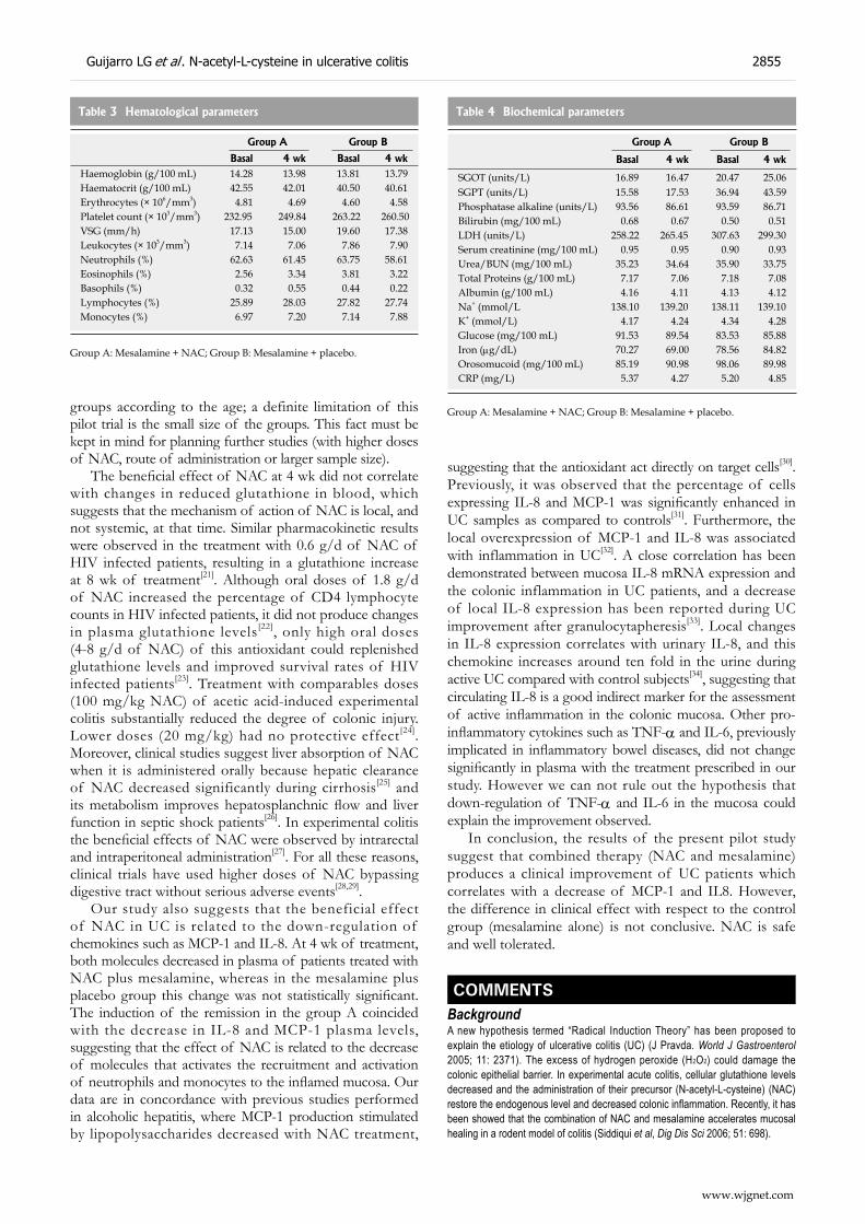
groups according to the age; a definite limitation of this pilot trial is the small size of the groups. This fact must be kept in mind for planning further studies (with higher doses of NAC, route of administration or larger sample size).
The beneficial effect of NAC at 4 wk did not correlate with changes in reduced glutathione in blood, which suggests that the mechanism of action of NAC is local, and not systemic, at that time. Similar pharmacokinetic results were observed in the treatment with 0.6 g/d of NAC of HIV infected patients, resulting in a glutathione increase at 8 wk of treatment[21]. Although oral doses of 1.8 g/d of NAC increased the percentage of CD4 lymphocyte counts in HIV infected patients, it did not produce changes in plasma glutathione levels [22], only high oral doses (4-8 g/d of NAC) of this antioxidant could replenished glutathione levels and improved survival rates of HIV infected patients[23]. Treatment with comparables doses (100 mg/kg NAC) of acetic acid-induced experimental colitis substantially reduced the degree of colonic injury. Lower doses (20 mg/kg) had no protective effect [24]. Moreover, clinical studies suggest liver absorption of NAC when it is administered orally because hepatic clearance of NAC decreased significantly during cirrhosis[25] and its metabolism improves hepatosplanchnic flow and liver function in septic shock patients[26]. In experimental colitis the beneficial effects of NAC were observed by intrarectal and intraperitoneal administration[27]. For all these reasons, clinical trials have used higher doses of NAC bypassing digestive tract without serious adverse events[28,29].
Our study also suggests that the beneficial effect of NAC in UC is related to the down-regulation of chemokines such as MCP-1 and IL-8. At 4 wk of treatment, both molecules decreased in plasma of patients treated with NAC plus mesalamine, whereas in the mesalamine plus placebo group this change was not statistically significant. The induction of the remission in the group A coincided with the decrease in IL-8 and MCP-1 plasma levels, suggesting that the effect of NAC is related to the decrease of molecules that activates the recruitment and activation of neutrophils and monocytes to the inflamed mucosa. Our data are in concordance with previous studies performed in alcoholic hepatitis, where MCP-1 production stimulated by lipopolysaccharides decreased with NAC treatment,
suggesting that the antioxidant act directly on target cells[30]. Previously, it was observed that the percentage of cells expressing IL-8 and MCP-1 was significantly enhanced in UC samples as compared to controls[31]. Furthermore, the local overexpression of MCP-1 and IL-8 was associated with inflammation in UC[32]. A close correlation has been demonstrated between mucosa IL-8 mRNA expression and the colonic inflammation in UC patients, and a decrease of local IL-8 expression has been reported during UC improvement after granulocytapheresis[33]. Local changes in IL-8 expression correlates with urinary IL-8, and this chemokine increases around ten fold in the urine during active UC compared with control subjects[34], suggesting that circulating IL-8 is a good indirect marker for the assessment of active inflammation in the colonic mucosa. Other pro-inflammatory cytokines such as TNF-α and IL-6, previously implicated in inflammatory bowel diseases, did not change significantly in plasma with the treatment prescribed in our study. However we can not rule out the hypothesis that down-regulation of TNF-α and IL-6 in the mucosa could explain the improvement observed.
In conclusion, the results of the present pilot study suggest that combined therapy (NAC and mesalamine) produces a clinical improvement of UC patients which correlates with a decrease of MCP-1 and IL8. However, the difference in clinical effect with respect to the control group (mesalamine alone) is not conclusive. NAC is safe and well tolerated.
COMMENTSBackgroundA new hypothesis termed “Radical Induction Theory” has been proposed to explain the etiology of ulcerative colitis (UC) (J Pravda. World J Gastroenterol 2005; 11: 2371). The excess of hydrogen peroxide (H2O2) could damage the colonic epithelial barrier. In experimental acute colitis, cellular glutathione levels decreased and the administration of their precursor (N-acetyl-L-cysteine) (NAC) restore the endogenous level and decreased colonic inflammation. Recently, it has been showed that the combination of NAC and mesalamine accelerates mucosal healing in a rodent model of colitis (Siddiqui et al, Dig Dis Sci 2006; 51: 698).
Table 3 Hematological parameters
Group A Group B
Basal 4 wk Basal 4 wkHaemoglobin (g/100 mL) 14.28 13.98 13.81 13.79Haematocrit (g/100 mL) 42.55 42.01 40.50 40.61Erythrocytes (× 106/mm3) 4.81 4.69 4.60 4.58Platelet count (× 103/mm3) 232.95 249.84 263.22 260.50VSG (mm/h) 17.13 15.00 19.60 17.38Leukocytes (× 103/mm3) 7.14 7.06 7.86 7.90Neutrophils (%) 62.63 61.45 63.75 58.61Eosinophils (%) 2.56 3.34 3.81 3.22Basophils (%) 0.32 0.55 0.44 0.22Lymphocytes (%) 25.89 28.03 27.82 27.74Monocytes (%) 6.97 7.20 7.14 7.88
Group A: Mesalamine + NAC; Group B: Mesalamine + placebo.
Table 4 Biochemical parameters
Group A Group B
Basal 4 wk Basal 4 wk
SGOT (units/L) 16.89 16.47 20.47 25.06SGPT (units/L) 15.58 17.53 36.94 43.59Phosphatase alkaline (units/L) 93.56 86.61 93.59 86.71Bilirubin (mg/100 mL) 0.68 0.67 0.50 0.51LDH (units/L) 258.22 265.45 307.63 299.30Serum creatinine (mg/100 mL) 0.95 0.95 0.90 0.93Urea/BUN (mg/100 mL) 35.23 34.64 35.90 33.75Total Proteins (g/100 mL) 7.17 7.06 7.18 7.08Albumin (g/100 mL) 4.16 4.11 4.13 4.12Na+ (mmol/L 138.10 139.20 138.11 139.10K+ (mmol/L) 4.17 4.24 4.34 4.28Glucose (mg/100 mL) 91.53 89.54 83.53 85.88Iron (μg/dL) 70.27 69.00 78.56 84.82Orosomucoid (mg/100 mL) 85.19 90.98 98.06 89.98CRP (mg/L) 5.37 4.27 5.20 4.85
Group A: Mesalamine + NAC; Group B: Mesalamine + placebo.
www.wjgnet.com
Guijarro LG et al . N-acetyl-L-cysteine in ulcerative colitis 2855
COMMENTS

Innovations and breakthroughsLocal overexpression of MCP-1 and IL-8 was associated with inflammation in UC and a decrease of colonic IL-8 has been reported during active UC. Our results show that antioxidant therapy with NAC decreases the levels of IL-8 that activates the recruitment and activation of neutrophils to the inflamed mucosa. It is suggested that higher dose or targeted NAC can alleviate colonic inflammation and hopefully become a novel agent for the treatment of UC.
Peer reviewThis is an excellent research paper which highlights the dual role antioxidants play in the pathogenesis of ulcerative colitis. And this is the first paper to cite radical induction theory which logically explains why the addition of NAC to conventional 5-ASA therapy significantly increases the remission rate observed in ulcerative colitis. The findings are potentially important for planning of furthers studies (e.g. with higher dose of N-acetyl-L-cysteine; more homogenous groups of comparison, larger sample size, et cetera).
REFERENCES1 Stange EF, Travis SP, Vermeire S, Beglinger C, Kupcinkas
L, Geboes K, Barakauskiene A, Villanacci V, Von Herbay A, Warren BF, Gasche C, Tilg H, Schreiber SW, Scholmerich J, Reinisch W; European Crohn's and Colitis Organisation.European evidence based consensus on the diagnosis and management of Crohn's disease: definitions and diagnosis. Gut 2006; 55 Suppl 1: i1-i15
2 Zhong YQ, Huang HR, Zhu ZH, Chen QK, Zhan J, Xing LC. Effects of sulfasalazine on biopsy mucosal pathologies and histological grading of patients with active ulcerative colitis. World J Gastroenterol 2005; 11: 4435-4438
3 Dotan I, Rachmilewitz D. Probiotics in inflammatory bowel disease: possible mechanisms of action. Curr Opin Gastroenterol 2005; 21: 426-430
4 Gijsbers K , Geboes K, Van Damme J. Chemokines in gastrointestinal disorders. Curr Drug Targets 2006; 7: 47-64
5 Van Assche G, Rutgeerts P. Physiological basis for novel drug therapies used to treat the inflammatory bowel diseases. I. Immunology and therapeutic potential of antiadhesion molecule therapy in inflammatory bowel disease. Am J Physiol Gastrointest Liver Physiol 2005; 288: G169-G174
6 Palatka K, Serfozo Z, Vereb Z, Hargitay Z, Lontay B, Erdodi F, Banfalvi G, Nemes Z, Udvardy M, Altorjay I.Changes in the expression and distribution of the inducible and endothelial nitric oxide synthase in mucosal biopsy specimens of inflammatory bowel disease. Scand J Gastroenterol 2005; 40: 670-680
7 Singer II, Kawka DW, Schloemann S, Tessner T, Riehl T, Stenson WF. Cyclooxygenase 2 is induced in colonic epithelial cells in inflammatory bowel disease. Gastroenterology 1998; 115: 297-306
8 Szanto I, Rubbia-Brandt L, Kiss P, Steger K, Banfi B, Kovari E, Herrmann F, Hadengue A, Krause KH. Expression of NOX1, a superoxide-generating NADPH oxidase, in colon cancer and inflammatory bowel disease. J Pathol 2005; 207: 164-176
9 Pravda J. Radical induction theory of ulcerative colitis. World J Gastroenterol 2005; 11: 2371-2384
10 Pavlick KP, Laroux FS, Fuseler J, Wolf RE, Gray L, Hoffman J, Grisham MB. Role of reactive metabolites of oxygen and nitrogen in inflammatory bowel disease. Free Radic Biol Med 2002; 33: 311-322
11 Menor C, Fernandez-Moreno MD, Fueyo JA, Escribano O, Olleros T, Arriaza E, Cara C, Lorusso M, Di Paola M, Roman ID, Guijarro LG.Azathioprine acts upon rat hepatocyte mitochondria and stress-activated protein kinases leading to necrosis: protective role of N-acetyl-L-cysteine. J Pharmacol Exp Ther 2004; 311: 668-676
12 Sido B, Hack V, Hochlehnert A, Lipps H, Herfarth C, Droge W. Impairment of intestinal glutathione synthesis in patients with inflammatory bowel disease. Gut 1998; 42: 485-492
13 Buffinton GD, Doe WF. Depleted mucosal antioxidant defences in inflammatory bowel disease. Free Radic Biol Med
1995; 19: 911-91814 Ardite E, Sans M, Panés J, Romero FJ, Piqué JM, Fernández-
Checa JC.Replenishment of glutathione levels improves mucosal function in experimental acute colitis. Lab Invest 2000; 80: 735-744
15 Siddiqui A, Ancha H, Tedesco D, Lightfoot S, Stewart CA, Harty RF. Antioxidant therapy with N-acetylcysteine plus mesalamine accelerates mucosal healing in a rodent model of colitis. Dig Dis Sci 2006; 51: 698-705
16 Lichtiger S, Present DH. Preliminary report: cyclosporin in treatment of severe active ulcerative colitis. Lancet 1990; 336: 16-19
17 Prantera C, Cottone M, Pallone F, Annese V, Franzè A, Cerutti R, Bianchi Porro G. Mesalamine in the treatment of mild to moderate active Crohn's ileitis: results of a randomized, multicenter trial. Gastroenterology 1999; 116: 521-526
18 Martins AM , Cordeiro C, Freire AP. Glyoxalase II in Saccharomyces cerevisiae: in situ kinetics using the 5,5'- dithiobis(2-nitrobenzoic acid) assay. Arch Biochem Biophys 1999; 366: 15-20
19 Kruidenier L, Kuiper I, Lamers CB, Verspaget HW. Intestinal oxidative damage in inflammatory bowel disease: semi-quantification, localization, and association with mucosal antioxidants. J Pathol 2003; 201: 28-36
20 Phylactos AC, Fasoula IN, Arnaud-Battandier F, Walker-Smith JA, Fell JM. Effect of enteral nutrition on antioxidant enzyme systems and inflammation in paediatric Crohn's disease. Acta Paediatr 2001; 90: 883-888
21 Treitinger A, Spada C, Masokawa IY, Verdi JC, Van Der Sander Silveira M, Luis MC, Reis M, Ferreira SI, Abdalla DS. Effect of N-acetyl-L-cysteine on lymphocyte apoptosis, lymphocyte viability, TNF-alpha and IL-8 in HIV-infected patients undergoing anti-retroviral treatment. Braz J Infect Dis 2004; 8: 363-371
22 Look MP, Rockstroh JK, Rao GS, Barton S, Lemoch H, Kaiser R, Kupfer B, Sudhop T, Spengler U, Sauerbruch T. Sodium selenite and N-acetylcysteine in antiretroviral-naive HIV-1-infected patients: a randomized, controlled pilot study. Eur J Clin Invest 1998; 28: 389-397
23 De Rosa SC, Zaretsky MD, Dubs JG, Roederer M, Anderson M, Green A, Mitra D, Watanabe N, Nakamura H, Tjioe I, Deresinski SC, Moore WA, Ela SW, Parks D, Herzenberg LA, Herzenberg LA. N-acetylcysteine replenishes glutathione in HIV infection. Eur J Clin Invest 2000; 30: 915-929
24 Akgun E, Caliskan C, Celik HA, Ozutemiz AO, Tuncyurek M, Aydin HH. Effects of N-acetylcysteine treatment on oxidative stress in acetic acid-induced experimental colitis in rats. J Int Med Res 2005; 33: 196-206
25 Jones AL, Jarvie DR, Simpson D, Hayes PC, Prescott LF. Pharmacokinetics of N-acetylcysteine are altered in patients with chronic liver disease. Aliment Pharmacol Ther 1997; 11: 787-791
26 Rank N, Michel C, Haertel C, Lenhart A, Welte M, Meier-Hellmann A, Spies C. N-acetylcysteine increases liver blood flow and improves liver function in septic shock patients: results of a prospective, randomized, double-blind study. Crit Care Med 2000; 28: 3799-3807
27 Cetinkaya A, Bulbuloglu E, Kurutas EB, Ciralik H, Kantarceken B, Buyukbese MA. Beneficial effects of N-acetylcysteine on acetic acid-induced colitis in rats. Tohoku J Exp Med 2005; 206: 131-139
28 Medved I, Brown MJ, Bjorksten AR, Murphy KT, Petersen AC, Sostaric S, Gong X, McKenna MJ. N-acetylcysteine enhances muscle cysteine and glutathione availability and attenuates fatigue during prolonged exercise in endurance-trained individuals. J Appl Physiol 2004; 97: 1477-1485
29 Soldini D, Zwahlen H, Gabutti L, Marzo A, Marone C. Pharmacokinetics of N-acetylcysteine following repeated intravenous infusion in haemodialysed patients. Eur J Clin Pharmacol 2005; 60: 859-864
30 Devalaraja MN, Mcclain CJ, Barve S, Vaddi K, Hill DB. Increased monocyte MCP-1 production in acute alcoholic hepatitis. Cytokine 1999; 11: 875-881
www.wjgnet.com
2856 ISSN 1007-9327 CN 14-1219/R World J Gastroenterol May 14, 2008 Volume 14 Number 18

31 Banks C , Bateman A, Payne R, Johnson P, Sheron N. Chemokine expression in IBD. Mucosal chemokine expression is unselectively increased in both ulcerative colitis and Crohn's disease. J Pathol 2003; 199: 28-35
32 McCormack G, Moriarty D, O'Donoghue DP, McCormick PA, Sheahan K, Baird AW. Tissue cytokine and chemokine expression in inflammatory bowel disease. Inflamm Res 2001; 50: 491-495
33 Tsukada Y, Nakamura T, Iimura M, Iizuka BE, Hayashi N. Cytokine profile in colonic mucosa of ulcerative colitis correlates with disease activity and response to granulocytapheresis. Am J Gastroenterol 2002; 97: 2820-2828
34 Taha AS, Grant V, Kelly RW. Urinalysis for interleukin-8 in the non-invasive diagnosis of acute and chronic inflammatory diseases. Postgrad Med J 2003; 79: 159-163
S- Editor Sun YL L- Editor Alpini GD E- Editor Liu Y
www.wjgnet.com
Guijarro LG et al . N-acetyl-L-cysteine in ulcerative colitis 2857

Online Submissions: wjg.wjgnet.com World J Gastroenterol 2008 May 14; 14(18): 2858-2862www.wjgnet.com World Journal of Gastroenterology ISSN [email protected] © 2008 WJG. All rights reserved.
Etiology and portal vein thrombosis in Budd-Chiari syndrome
Oguz Uskudar, Meral Akdogan, Nurgul Sasmaz, Sevinc Yilmaz, Muharrem Tola, Burhan Sahin
www.wjgnet.com
RAPID COMMUNICATION
Oguz Uskuda r, Me ra l Akdogan , Nu rgu l Sa smaz , Burhan Sahin, Turkiye Yuksek İhtisas Hospital, clinics of gastroenterology, Ankara 06010, TurkeySevinc Yilmaz, Turkiye Yuksek İhtisas Hospital, hematology unit, Ankara 06010, TurkeyMuharrem Tola, Turkiye Yuksek İhtisas Hospital clinics of radiology, Ankara 06010, TurkeyAuthor contributions: Uskudar O and Akdogan M contributed equally to this work; Uskudar O and Akdogan M designed research; Uskudar O, Akdogan M, Sasmaz N, Yılmaz S, Tola M, Sahin B performed research; Uskudar O and Akdogan M wrote the paper.Correspondence to: Oguz Uskudar, Dr, Yayla Mah. 1. cadde no: 123/3, Kecioren, Ankara 06010, Turkey. [email protected]: +90-312-3788638 Fax: +90-312-3124120Received: January 23, 2008 Revised: April 11, 2008
AbstractAIM: To research the etiology, portal vein thrombosis and other features of Budd-Chiari syndrome (BCS) patients prospectively.
METHODS: A total of 75 patients (40 female, 35 male) who were diagnosed between January 2002 and July 2004 as having BCS were studied prospectively. Findings from on physical examination, ultrasonography, duplex ultrasonography and venography were analyzed. Hemogram and blood chemistry were studied at the time of diagnosis and on each hospital visit. Bone marrow examination and immune phenotyping were performed by a hematologist when necessary. Protein C, S, antithrombin Ⅲ, activated protein C resistance, and anticardiolipin antibodies, antinuclear antibodies, and anti ds-DNA were studied twice. The presence of ascite, esophageal varices, and portal thrombosis were evaluated at admission and on every visit.
RESULTS: At least one etiological factor was determined in 54 (72%) of the patients. The etiology could not be defined in 21 (28%) patients. One etiological factor was found in 39, 2 factors in 14 and 3 factors in 1 patient. The most common cause was the web (16%), the second was Hydatid disease (11%), the third was Behcet’s disease (9%). Portal vein thrombosis was present in 11 patients and at least one etiology was identified in 9 of them (82%).
CONCLUSION: Behcet’s disease and hydatid disease are more prominent etiological factors in Turkey than in other countries. Patients with web have an excellent response to treatment without signs of portal
vein thrombosis while patients having thrombophilic factors more than one are prone to develop portal vein thrombosis with worse clinical outcome.
© 2008 WJG. All rights reserved.
Key words: Budd-Chiari syndrome; Etiology; Web; Behcet’s disease; Portal thrombosis
Peer reviewers: Ned Snyder, Professor, University of Texas Medical Branch, 301 University, University of Texas Medical Branch, Galveston Texas USA 77555-0764, United States; Syed MW Jafri, Professor, Medicine/Gastroenterology, Aga Khan University, POB 3500, Karachi 74800, Pakistan
Uskudar O, Akdogan M, Sasmaz N, Yilmaz S, Tola M, Sahin B. Etiology and portal vein thrombosis in Budd-Chiari syndrome. World J Gastroenterol 2008; 14(18): 2858-2862 Available from: URL: http://www.wjgnet.com/1007-9327/14/2858.asp DOI: http://dx.doi.org/10.3748/wjg.14.2858
INTRODUCTIONBudd-Chiari syndrome (BCS) is a rare heterogeneous group of disorders characterized by hepatic venous outflow obstruction at the level of hepatic venules, the large hepatic veins, the inferior vena cava, or the right atrium[1]. It has been increasingly recognized that a combination of several thrombogenic conditions and triggering factors are necessary for the development of venous thrombosis in general and hepatic vein or cava thrombosis in particular.
The etiology of Budd-Chiari syndrome changes according to geography. In most of the western cases of BCS an etiologic factor has been defined, whereas the causes of BCS in many of patients from India and Japan are not defined[2-6]. While thromboses are more prominent in the west, webs are prominent in the east and in Japan[7-9].
Turkey is a country acting as a bridge in between east and west. To date there is insufficient data about the etiology and features of BCS in Turkey. The relation between the etiologic factor and presence of portal vein thrombosis and yet outcome is not well established to date. In this study we prospectively searched the etiology, characteristics and other features of BCS patients in Turkey.
MATERIALS AND METHODSPatientsTurkiye Yuksek İhtisas Hospital is a tertiary referral center

Uskudar O et al . Etiology of BCS 2859
www.wjgnet.com
in gastroenterology in Turkey. A total of 75 patients (40 female, 35 male) who were diagnosed between January 2002 and July 2004 in Turkiye Yuksek İhtisas Hospital as having BCS were studied prospectively.
The diagnosis of BCS was establ ished us ing ultrasonography, color Doppler ultrasonography (CDU) and venography. A detailed patient history including onset of symptoms, medications, previous disease, pregnancy and use of oral contraceptives for women, oral and genital ulcers, previous surgery, and trauma were especially noted. The onset of symptoms was classified, whether acute, sub acute or chronic. The diagnosis and date of last hospital visit were noted.
Patients were evaluated with a multidisciplinary approach (involving gastroenterology, gastroenterology surgery, hematology, radiology and other clinics when necessary). Findings on physical examination were noted. Ultrasonography was performed by experienced gastroenterologists and CDU and venography were performed by experienced radiologists. The sites of lesions were classified as hepatic vein, inferior vena cava or combined and note was also taken as to whether the obstruction was complete or partial.
Hemogram and blood chemistry were studied at the time of diagnosis and on each visit. All patients were evaluated by a hematologist for myeloproliferative disorders and bone marrow examination and immune phenotyping were performed when necessary. Protein C, S, antithrombin Ⅲ, factor Ⅴ Leiden mutation, and anticardiolipin antibodies were studied twice in suitable conditions. Antinuclear antibody and anti ds-DNA were studied when systemic lupus erythematosus was suspected.
The presence of ascite, esophageal varices, and portal vein thrombosis was evaluated at admission and on every visit. Treatments were classified as conservative management, anticoagulation, angioplasty, TİPS and surgery. Complications that occurred during the follow up were recorded.
Statistical analysisVariables between groups were compared using the Mann-Whitney U test, correlations between parameters were tested by Spearman’s correlation analysis and the “Statistical Package for Social Sciences 10.0” program was used for statistical analysis. P < 0.05 was considered as statistically significant.
RESULTSThere were 40 female and 35 male patients. Mean age at first visit was 33.49 ± 13.88 (14-72) years. Most of the patients were diagnosed during the chronic phase of the disease (53.3%). Other patients were diagnosed during acute (26.7%) and sub acute (16%) periods. The site of obstruction was in hepatic veins in 47%, in IVC+ hepatic vein in 30% and IVC in 23% of the patients (Figure 1). A complete obstruction was observed in 54% of the patients. Venography was available for 80% of the patients. Esophageal varices and ascites were observed in 79% and 84% of the patients at admission, and 54% and 35% at last visit, respectively. The patients were followed-up for 1-30 mo (mean, 18 mo). During the follow-up seven patients died (9%).
At least one etiological factor was determined in 54 (72%) of the patients. The etiology could not be defined in 21 (28%) patients. One etiological factor was found in 39, 2 factors in 14 and 3 factors in 1 patient. The most common cause was the web and this was determined in 12 (16%) patients (Table 1). The second most common cause was Hydatid disease and this was found in 8 (11%) of the patients. Behcet’s disease was the third most common cause (7 patients, 9%). Etiological factors are presented in Table 1.
Laboratory parameters at admission and at the last hospital visit are presented in Table 2.
Significant positive correlations between first AST-ALT and first prothrombin time (P < 0.01), and between last GGT and last urea levels (P < 0.05) were found. A significant negative correlation was found between the first prothrombin time and first albumin levels (P < 0.01).
No differences were observed when patients were compared according to sex or age over or less than 35 years old.
Figure 1 A patient with prominent abdominal wall collateral veins as a result of inferior vena cava thrombosis at the hepatic portion.
Table 1 Etiology
Etiological factors Patients (%) Etiological factors Patients (%)Web 12 (16) Anticardiolipin antibodies 3 (4)Hydatid disease 8 (11) Oral contraceptive use 3 (4)Behçet’ disease 7 (9) Tumor 3 (4)Protein C def. 7 (9) Anti-thrombin 3 def. 2 (3)Myeloproliferativedisorder
6 (8) Lupus erythematosus 1 (1)
Protein S def. 5 (7) Ulcerative colitis 1 (1)Pregnancy 5 (7) Paroxysmal nocturnal
hemoglobinuria 1 (1)
Activated proteinC resistance
3 (4) Acute pancreatitis 1 (1)
Table 2 Laboratory parameters
First Last min max mean min max mean
AST (IU/L) 1300 119 700 7520 1000 82 900 7044ALT (IU/L) 700 90 700 6118 1700 157 200 9477GGT (IU/L) 1600 78 100 13 930 2900 64 200 14044Albumin (g/dL) 2100 5100 3460 2400 4800 3781Bilirubin (mg/dL) 100 1300 286 100 3200 326Urea (mg/dL) 1300 10 600 3478 1100 6100 3078Prothrombin time (s) 1100 4400 1731 1100 3000 1708

Figure 2 A patient with web in the right hepatic vein before angioplasty (A), during angioplasty (B) and after dilatation of the web (C).
C
B
Patients who presented with ascites initially had lower albumin and higher Prothrombin times compared to those who did not. Patients who had ascites at their last visit had significantly lower albumin and AST levels than others.
Portal veins were patent in 85% and thrombosed in 15% of the patients. At least one etiology was identified in 9 of the 11 patients (82%) with portal vein thrombosis and BCS. Five patients had 1, 3 patients had 2 and 1 patient had 3 etiological factors. One of the striking results was that none of the patients with web or Behcet’s disease as a cause of BCS had portal vein thrombosis. Hydatid disease was observed in 4, protein C deficiency in 3, protein S deficiency in 2, mass lesion in 2, and ulcerative colitis, pregnancy and antithrombin 3 deficiencies in 1 patient. Last bilirubin values were significantly higher (16.5/1.7, P = 0.002) and last albumin levels were significantly lower (32/39, P = 0.022) in patients with portal vein thrombosis. Nine patients with portal vein thrombosis were in the chronic, and 2 in a subacute but none in acute phase of BCS.
When compared by means of the extent of the obstruction, patients with complete obstruction had lower initial albumin levels (3.2/3.7, P = 0.007) and higher prothrombin times (19 s/14 s, P = 0.001). No other significant difference was found between other parameters.
Patients were treated conservatively (53%), with balloon angioplasty (24%, Figure 2), oral anticoagulation (17%), TIPS (4%) or surgery (7%). Three patients experienced warfarin related complications (2 intracranial bleeding, one of whom died, 1 intra abdominal bleeding). Three of the patients who underwent surgery died because of liver failure. The balloon angioplasty procedure was performed in 18 patients and failed in 6 of them. When we examined the patients where it was unsuccessful it was found that none of them were webs but, rather, long segment caval thrombosis or hepatic vein thromboses. Two of the 12 successful web dilatation patients later developed thrombosis and one underwent surgery, the other was treated with TİPS procedure.
DISCUSSIONThe prevalence of underlying thrombophilias is markedly increased in patients with BCS. Factors that confer a predisposition to the development of BCS including hereditary and acquired hypercoagulable states and a variety of other causes can be identified in about 75 percent of patients[1]. In our study we identified at least one etiological factor in 54 (72%) patients. The presence of multiple causes in the same patient is increasingly recognized[3]. We identified 2 etiological factors in 14 and 3 factors in 1 patient. These results were similar with reports in the literature.
There were slightly more female patients than males (40 vs 35) but the imbalance was not as marked as in the literature (2/1)[10]. This may be due to the less frequent use of oral contraceptives in Turkey. Mean age at the time of diagnosis was 33.
The site of the lesion varies according to geography. In our patients hepatic veins were involved in 77% and the inferior vena cava was involved in 53% of the patients. While inferior vena cava involvement is more prominent in Nepal, South Africa, China, India and Japan, hepatic vein involvement is more prominent in the western countries[7,11-13]. Our results lay in between these, but were closer to the western countries.
W hen we look to the e t io log y, we see tha t myeloproliferative disorders are found to be the leading cause of BCS in western countries, with a range between 20%-53%[3,4]. In this study we identified myeloproliferative disease in 8% of the patients. We confirmed these disorders by bone marrow aspiration and yet only overt forms of disease were diagnosed. We could not look for JAC2 V617f mutation[14-16]. So we might have missed some of these patients or myeloproliferative disorders may be less frequent in Turkish population.
Webs were the most common etiology and the most easily treated lesions in our patients (16%). This ratio is not as high as in eastern countries but higher than in western ones. It may be another indicator of Turkey as a bridge
A
www.wjgnet.com
2860 ISSN 1007-9327 CN 14-1219/R World J Gastroenterol May 14, 2008 Volume 14 Number 18

between east and west. These patients rapidly responded to balloon dilatation and ascites dramatically disappeared after successful dilatation. None of these patients had portal vein thrombosis. The chances of a patient with BCS may depend on a web etiology. The etiology of BCS is the most important factor for the prognosis.
The second most frequent etiology in our study was Hydatid disease, at 12%, and this is the highest ratio in the literature. Hydatid disease is a common health problem and deserves more interest in Turkey. Half of the patients with BCS and Hydatid disease also had portal vein thrombosis.
Another frequently observed etiology was Behcet’s disease with a ratio of 9%. This ratio is also the highest ratio in the literature. Every patient with BCS must be closely questioned for any history of Behcet’s disease. The difficulty in managing these patients was that they usually had long segment thrombosis and thrombosis of other vascular structures. While Bayraktar has reported portal vein thrombosis in these patients, another interesting observation is that none of our patients with Behcet’s disease and BCS had portal vein thrombosis[17]. This might be due to Bayraktar’s long term follow up period. Other etiological factors were similar with the literature.
Portal vein thrombosis in patients with BCS is a poor prognostic factor[18,19]. In this study we observed that patients with portal vein thrombosis when compared with those not thrombosed, had lower albumin and higher bilirubin level at their last check. Another difference in patients with portal vein thrombosis was the etiology. Since none of the patients with webs or Behçet’s disease had or developed portal vein thrombosis during follow up, this deserves more attention. In our patients one patient had 3 etiological factors and he had also portal vein thrombosis. Since nine patients with portal vein thrombosis were in the chronic, and 2 in a subacute phase of BCS, but none were in an acute phase, we can state that prevention of portal vein thrombosis might be considered in these patients.
Ascites was present in 84% of the patients at admission which means that most of the patients were diagnosed when symptomatic. This ratio lies between the ratio of before and after the 1990 results of Hadengue, indicating that we should diagnose more asymptomatic patients[10]. With an increased knowledge about BCS, patients can be diagnosed earlier and further development of new thrombosis and deterioration of liver functions can be prevented. In patients with poor liver functions surgery is unsuccessful, and this may be due to lack of experience as well as to a very enlarged liver that causes difficulty during surgery.
In conclusion, the etiology of BCS changes according to geographic region. Webs, Behçet’s disease and Hydatid disease are more prominent etiological factors in Turkey which sets it apart from other countries. Complete obstruction of hepatic veins or inferior vena cava and presence of portal vein thrombosis affects the course of the disease. Presence of more than one prethrombotic factor increases the risk of portal vein thrombosis. Patients with web have excellent treatment response and prognosis.COMMENTS
BackgroundThe etiology of Budd-Chiari syndrome (BCS) changes according to geography. There is insufficient data about the etiology and portal vein thrombosis relation BCS. The aim of the present study was to evaluate the etiology, presentation, portal vein thrombosis and other features of BCS patients.
Research frontiersSeveral studies have identified that portal vein thrombosis in patients with BCS is a poor prognostic factor. However there are few prospective studies regarding the etiology and portal vein thrombosis relation. We studied the relationship between the etiology, portal vein thrombosis and find that some etiological factors are not associated with portal vein thrombosis.
Innovations and breakthroughsThe present research studied cases with BCS, but the follow up period was not long enough and some etiological factors such as JAC2 V617f mutation was missing.
Applications The results of this study suggest that patients with etiology other than web or Behçet’s disease are prone to develop portal vein thrombosis and since portal vein thrombosis mostly were in the chronic, some in a subacute but none in acute phase of BCS, early diagnosis and anticoagulation may be important.
Peer reviewThis paper clearly defines that initially the extent of thrombosis and etiology and lately portal vein thrombosis are important factors for BCS. The results are valuable.
REFERENCES1 Menon KV, Shah V, Kamath PS. The Budd-Chiari syndrome.
N Engl J Med 2004; 350: 578-5852 Janssen HL , Meinardi JR, Vleggaar FP, van Uum SH,
Haagsma EB, van Der Meer FJ, van Hattum J, Chamuleau RA, Adang RP, Vandenbroucke JP, van Hoek B, Rosendaal FR. Factor V Leiden mutation, prothrombin gene mutation, and deficiencies in coagulation inhibitors associated with Budd-Chiari syndrome and portal vein thrombosis: results of a case-control study. Blood 2000; 96: 2364-2368
3 Denninger MH, Chait Y, Casadevall N, Hillaire S, Guillin MC, Bezeaud A, Erlinger S, Briere J, Valla D. Cause of portal or hepatic venous thrombosis in adults: the role of multiple concurrent factors. Hepatology 2000; 31: 587-591
4 Hirshberg B, Shouval D, Fibach E, Friedman G, Ben-Yehuda D. Flow cytometric analysis of autonomous growth of erythroid precursors in liquid culture detects occult polycythemia vera in the Budd-Chiari syndrome. J Hepatol 2000; 32: 574-578
5 Deltenre P, Denninger MH, Hillaire S, Guillin MC, Casadevall N, Briere J, Erlinger S, Valla DC. Factor V Leiden related Budd-Chiari syndrome. Gut 2001; 48: 264-268
6 Bismuth E, Hadengue A, Hammel P, Benhamou JP. Hepatic vein thrombosis in Behcet's disease. Hepatology 1990; 11: 969-974
7 Shrestha SM, Okuda K, Uchida T, Maharjan KG, Shrestha S, Joshi BL, Larsson S, Vaidya Y. Endemicity and clinical picture of liver disease due to obstruction of the hepatic portion of the inferior vena cava in Nepal. J Gastroenterol Hepatol 1996; 11: 170-179
8 Mohanty D , Shetty S, Ghosh K, Pawar A, Abraham P. Hereditary thrombophilia as a cause of Budd-Chiari syndrome: a study from Western India. Hepatology 2001; 34: 666-670
9 Okuda H, Yamagata H, Obata H, Iwata H, Sasaki R, Imai F, Okudaira M, Ohbu M, Okuda K. Epidemiological and clinical features of Budd-Chiari syndrome in Japan. J Hepatol 1995; 22: 1-9
10 Hadengue A, Poliquin M, Vilgrain V, Belghiti J, Degott C, Erlinger S, Benhamou JP. The changing scene of hepatic vein thrombosis: recognition of asymptomatic cases.
www.wjgnet.com
Uskudar O et al . Etiology of BCS 2861
COMMENTS
Gastroenterology 1994; 106: 1042-1047 11 Valla DC. Hepatic vein thrombosis (Budd-Chiari syndrome).
Semin Liver Dis 2002; 22: 5-14 12 Okuda K , Kage M, Shrestha SM. Proposal of a new
nomenclature for Budd-Chiari syndrome: hepatic vein thrombosis versus thrombosis of the inferior vena cava at its hepatic portion. Hepatology 1998; 28: 1191-1198
13 Victor S, Jayanthi V, Panchanadam M, Chitra S, Vijayalakshmi CS, Madanagopalan N. Budd Chiari syndrome and pericaval filariasis. Trop Gastroenterol 1994; 15: 161-168
14 Patel RK, Lea NC, Heneghan MA, Westwood NB, Milojkovic D, Thanigaikumar M, Yallop D, Arya R, Pagliuca A, Gaken J, Wendon J, Heaton ND, Mufti GJ. Prevalence of the activating JAK2 tyrosine kinase mutation V617F in the Budd-Chiari syndrome. Gastroenterology 2006; 130: 2031-2038
15 Smalberg JH, Murad SD, Braakman E, Valk PJ, Janssen HL, Leebeek FW. Myeloproliferative disease in the pathogenesis and survival of Budd-Chiari syndrome. Haematologica 2006; 91:
1712-171316 Plume G, Vaya A, Ferrando F, Mira Y, Orbis F. JAK2V617F
mutation as a marker of a latent myeloproliferative disorder in a patient with Budd-Chiari syndrome and factor V Leiden mutation. Thromb Haemost 2007; 98: 681-682
17 Bayraktar Y, Balkanci F, Bayraktar M, Calguneri M. Budd-Chiari syndrome: a common complication of Behcet's disease. Am J Gastroenterol 1997; 92: 858-862
18 Mahmoud AE , Helmy AS, Billingham L, Elias E. Poor prognosis and limited therapeutic options in patients with Budd-Chiari syndrome and portal venous system thrombosis. Eur J Gastroenterol Hepatol 1997; 9: 485-489
19 Murad SD, Valla DC, de Groen PC, Zeitoun G, Haagsma EB, Kuipers EJ, Janssen HL. Pathogenesis and treatment of Budd-Chiari syndrome combined with portal vein thrombosis. Am J Gastroenterol 2006; 101: 83-90
S- Editor Zhong XY L- Editor Alpini GD E- Editor Lu W
www.wjgnet.com
2862 ISSN 1007-9327 CN 14-1219/R World J Gastroenterol May 14, 2008 Volume 14 Number 18

Peer reviewer: Seyed A Taghavi, Associate Professor, Department of Internal Medicine, Nemazee Hospital, No.23, 59th Alley, Ghasrodasht St., Shiraz 71838-95453, Iran
Kim TH, Oh HJ, Choi CS, Yeom DH, Choi SC. Clinical usefulness of transpapillary removal of common bile duct stones by frequency doubled double pulsed Nd:YAG laser. World J Gastroenterol 2008; 14(18): 2863-2866 Available from: URL: http://www.wjgnet.com/1007-9327/14/2863.asp DOI: http://dx.doi.org/10.3748/wjg.14.2863
INTRODUCTIONAbout 90% of all patients with common bile duct stones are currently treated non-surgically using endoscopic sphincterotomy and stone extraction. However, standard endoscopic procedure and mechanical lithotripsy for removal of stones from the bile duct fail in 5% to 10% of patients, because the stones are too large or impacted[1]. In such cases, additional methods such as extracorporeal shockwave lithotripsy (ESWL), laser shockwave lithotripsy (LSWL) or electrohydraulic lithotripsy (EHL) are required[2]. EHL and LSWL need visual control via a mother-baby endoscope system or cholangioscopy for safety because of the potential for accidental damage to the bile duct wall. Moreover, a peroral cholangioscope is expensive, fragile and difficult to use. Laser lithotripsy is expensive due to the cost of the cumbersome endoscopic equipment. Although EHL is inexpensive, it uses high-pulse energy to disintegrate stones[3-5].
The Frequency Doubled Double Pulse Nd:YAG (FREDDY) laser (World of Medicine, Berlin, Germany) is a newly developed economical, short-pulse, double frequency, solid-state laser with wavelengths of 532 nm and 1064 nm that cause less tissue damage. It was specifically designed to fragment urinary and biliary calculi[6]. We used a balloon catheter for FREDDY laser lithotripsy (FREDDY LL) in an effort to improve fluoroscopic targeting and to prevent damage to the bile duct. The purpose of this study was to investigate the safety and the effectiveness of using a FREDDY laser to treat bile duct stones that were not amendable to conventional endoscopic therapy.
MATERIALS AND METHODSPatientsSeventeen patients (7 male and 10 female; mean age 67.8 years, range 55-82 years) with extra-hepatic bile duct
RAPID COMMUNICATION
Clinical usefulness of transpapillary removal of common bile duct stones by frequency doubled double pulse Nd:YAG laser
Tae Hyeon Kim, Hyo Jeong Oh, Chang-Soo Choi, Dong Han Yeom, Suck Chei Choi
Tae Hyeon Kim, Hyo Jeong Oh, Chang-Soo Choi, Dong Han Yeom, Suck Chei Choi, Department of Internal Medicine and Digestive Disease Research Institute, Wonkwang University School of Medicine, Iksan 570180, Korea Author contributions: Kim TH designed research plan and wrote the paper; Oh HJ and Choi CS performed research; Yeom DH and Choi SC analyzed data.Supported by The 2007 research fund of Wonkwang University and Wonkwang Clinical Research InstituteCorrespondence to: Hyo-Jeong Oh, MD, Department of Internal Medicine, Wonkwang University School of Medicine, Sinyong Dong 344-2, Iksan 570-180, Korea. [email protected]: +82-63-8592670 Fax: +82-63-8552025 Received: January 19, 2008 Revised: April 10, 2008
AbstractAIM: To study the efficacy and the safety of laser lithotripsy without direct visual control by using a balloon catheter in patients with bile duct stones that could not be extracted by standard technique.
METHODS: The seventeen patients (7 male and 10 female; mean age 67.8 years) with difficult common bile duct (CBD) stones were not amenable for conventional endoscopic maneuvers such as sphincterotomy and mechanical lithotripsy were included in this study. Laser wavelengths of 532 nm and 1064 nm as a double pulse were applied with pulse energy of 120 mJ. The laser fiber was advanced under fluoroscopic control through the ERCP balloon catheter. Laser lithotripsy was continued until the fragment size seemed to be less than 10 mm. Endoscopic extraction of the stones and fragments was performed with the use of the Dormia basket and balloon catheter.
RESULTS: Bile duct clearance was achieved in 15 of 17 patients (88%). The mean number of treatment sessions was 1.7 ± 0.6. Endoscopic stone removal could not be achieved in 2 patients (7%). Adverse effects were noted in three patients (hemobilia, pancreatitis, and cholangitis).
CONCLUSION: The Frequency Doubled Double Pulse Nd:YAG (FREDDY) laser may be an effective and safe technique in treatment of difficult bile duct stones.
© 2008 WJG. All rights reserved.
Key words: Bile duct stones; Frequency doubled double pulse Nd:YAG laser; Transpapillary removal; Mechanical lithotripsy; Balloon catheter
Online Submissions: wjg.wjgnet.com World J Gastroenterol 2008 May 14; 14(18): 2863-2866www.wjgnet.com World Journal of Gastroenterology ISSN [email protected] © 2008 WJG. All rights reserved.
www.wjgnet.com

stones were included in this study. The inclusion criterion for this study was extrahepatic bile duct stones that were not amenable to standard endoscopic procedure including endoscopic sphincterotomy and mechanical lithotripsy. Stone removal by using conventional methods failed because the stones were not captured in the basket for mechanical lithotripsy or endoscopic sphincterotomy inadequately was done for large periampullary diverticulum with large CBD stones (> 1.5 cm). The main reasons were the following; large stone (n = 7), impacted stone (n = 3), and large stones with periampullary diverticulum (n = 7).
All patients were diagnosed as having bile duct stones by endoscopic retrograde cholangiography and/or MRCP at our hospital. The data regarding stone size, number, and location was based on the endoscopic retrograde cholangiogram (by using the diameter of the distal end of standard duodenosocpy as a reference). The properties of the CBD stones were as follows: 1 stone (n = 5), 2 stones (n = 7) and more than 2 stones (n = 5); diameter of large stone (18.5 ± 3.2 mm) with 10-20 mm (n = 8) and with 21-30 mm (n = 9). Large stones with periampullary diverticulum were found in 11 of 17 patients. The study was approved by our institutional review board. Written informed consent was obtained from each patient for laser therapy.
Methods we used the FREDDY laser (frequency doubled double pulse Nd:YAG laser, Laser 100, World of Medicine). Laser wavelengths of 532 nm and 1064 nm as a double pulse were applied with pulse energy of 120 mJ. The laser fiber with a 250 nm core diameter was inserted in a 6.8 Fr standard extraction balloon catheter (Wilson-Cook Medical Inc., Winston-Salem, N.C.) with an 18-mm-diameter balloon and this was then passed through the papilla via the working channel of a standard duodenoscope (JF-240; Olympus Optical Co., Ltd. Tokyo, Japan).
As the fiber itself is not detectable by fluoroscopy, the radio-opaque tip of the balloon catheter was positioned near the stone, and the balloon was expanded to fix the position of the catheter and the fiber. The tip of the fiber was advanced a few millimeters beyond the radio-opaque tip of the balloon catheter, under fluoroscopic guidance, to ensure that the fiber was positioned on the stone surface. Only one fluoroscopic plane was used to target the stone and to observe fragmentation. The treatment energy level was 120 mJ per pulse at a repetition rate of 8 to 10 Hz. During the laser treatment, we tried to listen to the fragmentation sound of stone by using stethoscope and the fragmentation effect was monitored by fluoroscopy after instillation of contrast media into the bile duct. During laser lithotripsy, the bile duct was continuously irrigated with a mixture of contrast media and saline solution because fluid is required for the generation of shock waves.
After stone disintegration, endoscopic extraction of stones and fragments was done with a dormia basket and balloon catheter. Laser lithotripsy was limited to a maximum duration of 60 min per session. If, at that time, the ductal clearance was incomplete, then a nasobiliary catheter was inserted. Failure of laser lithotripsy was defined as the inability to achieve complete bile duct
clearance after a maximum of three lithotripsy sessions. All patients were followed for at least two days after
the FREDDY LL. Follow-up included simple radiographs and laboratory tests (hemoglobin, white blood cell count, bilirubin, alanine aminotransferase, aspartate aminotransferase, creatinine, serum urea nitrogen, amylase and lipase levels). The occurrence of any complication within one month of the FREDDY LL procedure was assessed by a patient visit or telephone interview.
RESULTSLithotripsyComplete stone fragmentation using FREDDY LL was achieved in 15 of 17 patients without direct visual control (Figure 1). The number of endoscopic retrograde cholangiography sessions per patient that was necessary to totally clear the bile ducts was 1.7 ± 0.6. After FREDDY LL, 3 patients underwent mechanical lithotripsy to achieve complete clearance of the bile duct.
The stone fragmentation failed in two patients, in whom the positioning of the laser fiber on the stone was inadequate due to a huge impacted stone in the tortuous common bile duct and the biliary stricture. One patient was sent to the surgical department to clear the bile duct by laparoscopic surgery. The other patient was treated by percutaneous cholangioscopy with electro-hydraulic lithotripsy.
Complications The side effects and complications of FREDDY LL were mostly mild. Although acute pancreatitis occurred in one patient, it was treated by conservative management, and there was no pseudocyst. Transient hemobilia was observed in two patients. However, the bleeding stopped spontaneously without any change of the hemoglobin level. The hemobilia may have been caused by mucosal damage from insertion of the guiding catheter or the balloon catheter. A patient with acute cholangitis was treated by administration of antibiotics and nasobiliary tube irrigation. This complication may have been caused by transient obstruction of the nasobiliary tube.
DISCUSSIONEndoscopic retrograde cholangiopancreatography (ERCP) and sphincterotomy have been the standard treatments for choledocholithiasis, yet large stones present a major challenge for the endoscopist. Extending a sphincterotomy increases the risk of bleeding and perforation, and mechanical lithotripters are generally expensive, cumbersome to use, and fragile, and they fail to grasp the stones in a significant number of cases. For treating these patients, the complementary techniques such as extracorporeal shock wave, intracorporeal electrohydraulic, or laser induced lithotripsy are required[2].
Electrohydraulic intracorporeal lithotripsy (EHL) represents an effective treatment option for the endoscopic treatment of difficult bile duct stones. However, EHL requires continuous visual control of the fragmentation procedure because of the high pulse energies that are used.
www.wjgnet.com
2864 ISSN 1007-9327 CN 14-1219/R World J Gastroenterol May 14, 2008 Volume 14 Number 18
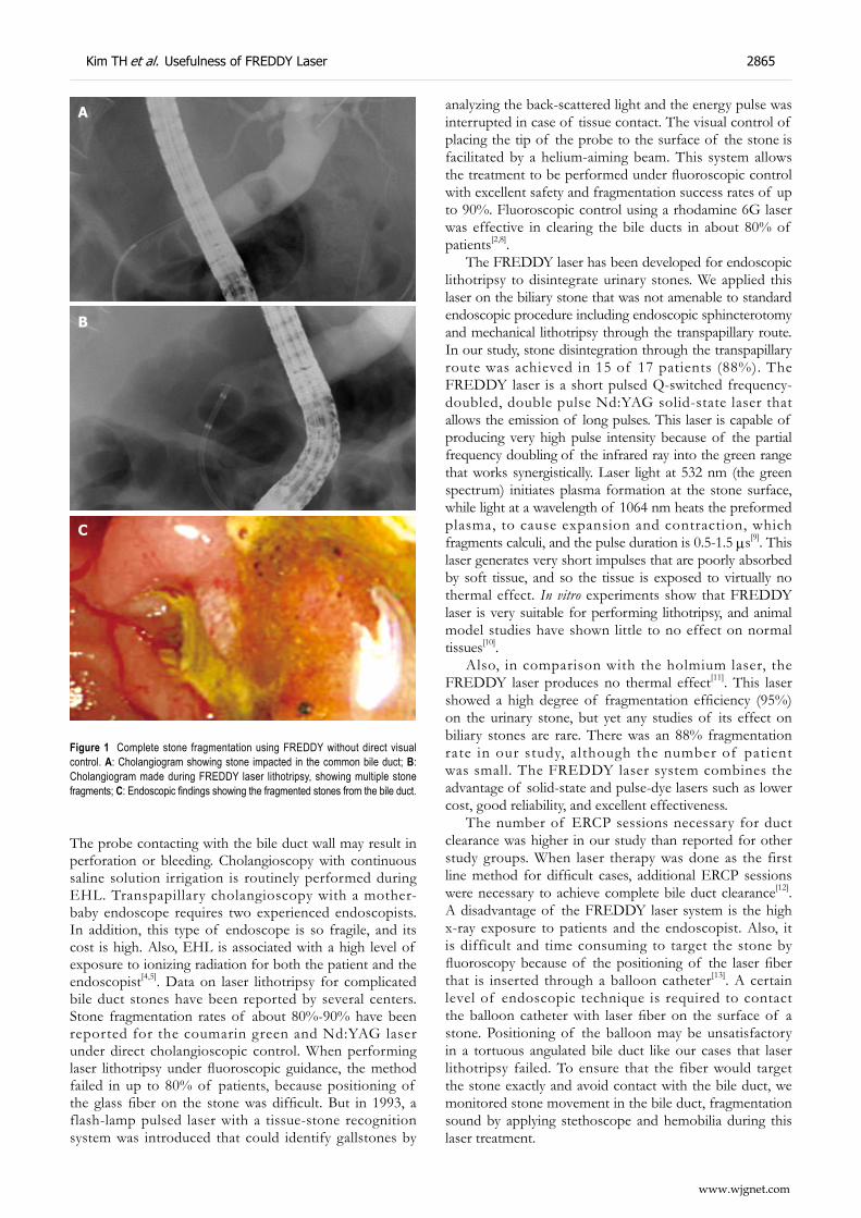
The probe contacting with the bile duct wall may result in perforation or bleeding. Cholangioscopy with continuous saline solution irrigation is routinely performed during EHL. Transpapillary cholangioscopy with a mother-baby endoscope requires two experienced endoscopists. In addition, this type of endoscope is so fragile, and its cost is high. Also, EHL is associated with a high level of exposure to ionizing radiation for both the patient and the endoscopist[4,5]. Data on laser lithotripsy for complicated bile duct stones have been reported by several centers. Stone fragmentation rates of about 80%-90% have been reported for the coumarin green and Nd:YAG laser under direct cholangioscopic control. When performing laser lithotripsy under fluoroscopic guidance, the method failed in up to 80% of patients, because positioning of the glass fiber on the stone was difficult. But in 1993, a flash-lamp pulsed laser with a tissue-stone recognition system was introduced that could identify gallstones by
analyzing the back-scattered light and the energy pulse was interrupted in case of tissue contact. The visual control of placing the tip of the probe to the surface of the stone is facilitated by a helium-aiming beam. This system allows the treatment to be performed under fluoroscopic control with excellent safety and fragmentation success rates of up to 90%. Fluoroscopic control using a rhodamine 6G laser was effective in clearing the bile ducts in about 80% of patients[2,8].
The FREDDY laser has been developed for endoscopic lithotripsy to disintegrate urinary stones. We applied this laser on the biliary stone that was not amenable to standard endoscopic procedure including endoscopic sphincterotomy and mechanical lithotripsy through the transpapillary route. In our study, stone disintegration through the transpapillary route was achieved in 15 of 17 patients (88%). The FREDDY laser is a short pulsed Q-switched frequency-doubled, double pulse Nd:YAG solid-state laser that allows the emission of long pulses. This laser is capable of producing very high pulse intensity because of the partial frequency doubling of the infrared ray into the green range that works synergistically. Laser light at 532 nm (the green spectrum) initiates plasma formation at the stone surface, while light at a wavelength of 1064 nm heats the preformed plasma, to cause expansion and contraction, which fragments calculi, and the pulse duration is 0.5-1.5 μs[9]. This laser generates very short impulses that are poorly absorbed by soft tissue, and so the tissue is exposed to virtually no thermal effect. In vitro experiments show that FREDDY laser is very suitable for performing lithotripsy, and animal model studies have shown little to no effect on normal tissues[10].
Also, in comparison with the holmium laser, the FREDDY laser produces no thermal effect[11]. This laser showed a high degree of fragmentation efficiency (95%) on the urinary stone, but yet any studies of its effect on biliary stones are rare. There was an 88% fragmentation rate in our study, although the number of patient was small. The FREDDY laser system combines the advantage of solid-state and pulse-dye lasers such as lower cost, good reliability, and excellent effectiveness.
The number of ERCP sessions necessary for duct clearance was higher in our study than reported for other study groups. When laser therapy was done as the first line method for difficult cases, additional ERCP sessions were necessary to achieve complete bile duct clearance[12].
A disadvantage of the FREDDY laser system is the high x-ray exposure to patients and the endoscopist. Also, it is difficult and time consuming to target the stone by fluoroscopy because of the positioning of the laser fiber that is inserted through a balloon catheter[13]. A certain level of endoscopic technique is required to contact the balloon catheter with laser fiber on the surface of a stone. Positioning of the balloon may be unsatisfactory in a tortuous angulated bile duct like our cases that laser lithotripsy failed. To ensure that the fiber would target the stone exactly and avoid contact with the bile duct, we monitored stone movement in the bile duct, fragmentation sound by applying stethoscope and hemobilia during this laser treatment.
Figure 1 Complete stone fragmentation using FREDDY without direct visual control. A: Cholangiogram showing stone impacted in the common bile duct; B: Cholangiogram made during FREDDY laser lithotripsy, showing multiple stone fragments; C: Endoscopic findings showing the fragmented stones from the bile duct.
A
B
C
www.wjgnet.com
Kim TH et al. Usefulness of FREDDY Laser 2865

In conclusion, our results suggest that laser lithotripsy using FREDDY without direct visual control was an effective and safe technique for patients with difficult CBD stones that could not be removed by mechanical lithotripsy. In the future, comparative studies with EHL, ESWL, or other laser systems will be required to assess the utility and efficacy of FREDDY laser.
COMMENTSBackgroundEndoscopic papillotomy is successful in more than 90% of the cases of choledocholithiasis. For patients with difficult bile duct stones not responding to mechanical lithotripsy, different methods for stone fragmentation such as laser lithotripsy or electrohydraulic lithotripsy (EHL) with direct visual control have been developed. A major problem in using this device is the requirement of cholangioscopic guidance or the “mother and baby” endoscope.
Research frontiersRecently, a new laser, the Frequency Doubled Double Pulse Nd:YAG (FREDDY), was developed for endoscopic lithotripsy for urinary stones, and has less tissue damage. The efficacy and safety of laser lithotripsy without cholangioscopy by using the balloon catheter were evaluated in patients with bile duct stones that could not be extracted by standard technique.
Innovation and breakthroughsThe FREDDY laser is a newly developed economical, short-pulse, double frequency, solid-state laser that results in less tissue damage. Fluoroscopically guided laser lithotripsy with a balloon catheter through a peroral duodenoscope appears to be effective treatment for bile duct stones that cannot be extracted by using conventional techniques such as mechanical lithotripsy.
ApplicationsFor the fragmentation of the biliary stone, cholangioscopy guidance is expensive due to the cost of the cumbersome endoscopic equipment and the need for two experienced endoscopists operating the “mother and baby” endoscope. When direct visual control is not available or limited, laser lithotripsy with a balloon catheter may be an alternative. To target the laser fiber accurately on the stone in the bile duct, a new and easier method is required.
Peer reviewIt is a well-written and interesting paper. The FREDDY laser may be a useful technique in treatment of difficult bile duct stones.
REFERENCES1 Classen M, Hagenmuller F, Knyrim K, Frimberger E. Giant
bile duct stones--non-surgical treatment. Endoscopy 1988; 20: 21-26
2 Sauerbruch T, Feussner H, Frimberger E, Hasegawa H, Ihse I, Riemann JF, Yasuda H. Treatment of common bile duct stones. A consensus report. Hepatogastroenterology 1994; 41: 513-515
3 Neuhaus H, Hoffmann W, Zillinger C, Classen M. Laser lithotripsy of difficult bile duct stones under direct visual control. Gut 1993; 34: 415-421
4 Binmoeller KF , Bruckner M, Thonke F, Soehendra N. Treatment of difficult bile duct stones using mechanical, electrohydraulic and extracorporeal shock wave lithotripsy. Endoscopy 1993; 25: 201-206
5 Hochberger J, Bayer J, May A, Muhldorfer S, Maiss J, Hahn EG, Ell C. Laser lithotripsy of difficult bile duct stones: results in 60 patients using a rhodamine 6G dye laser with optical stone tissue detection system. Gut 1998; 43: 823-829
6 Delvecchio FC, Auge BK, Brizuela RM, Weizer AZ, Zhong P, Preminger GM. In vitro analysis of stone fragmentation ability of the FREDDY laser. J Endourol 2003; 17: 177-179
7 Neuhaus H, Hoffmann W, Gottlieb K, Classen M. Endoscopic lithotripsy of bile duct stones using a new laser with automatic stone recognition. Gastrointest Endosc 1994; 40: 708-715
8 Ell C, Hochberger J, May A, Fleig WE, Bauer R, Mendez L, Hahn EG. Laser lithotripsy of difficult bile duct stones by means of a rhodamine-6G laser and an integrated automatic stone-tissue detection system. Gastrointest Endosc 1993; 39: 755-762
9 Helfmann J , Muller G. Laser lithotripsy: process and overview. Med Laser Appl 2001; 16: 30-37
10 Zorcher T, Hochberger J, Schrott KM, Kuhn R, Schafhauser W. In vitro study concerning the efficiency of the frequency-doubled double-pulse Neodymium:YAG laser (FREDDY) for lithotripsy of calculi in the urinary tract. Lasers Surg Med 1999; 25: 38-42
11 Delvecchio FC, Preminger GM. Endoscopic management of urologic disease with the holmium laser. Curr Opin Urol 2000; 10: 233-237
12 Jakobs R, Adamek HE, Maier M, Kromer M, Benz C, Martin WR, Riemann JF. Fluoroscopically guided laser lithotripsy versus extracorporeal shock wave lithotripsy for retained bile duct stones: a prospective randomised study. Gut 1997; 40: 678-682
13 Ponchon T, Gagnon P, Valette PJ, Henry L, Chavaillon A, Thieulin F. Pulsed dye laser lithotripsy of bile duct stones. Gastroenterology 1991; 100: 1730-1736
S- Editor Zhong XY L- Editor Alpini GD E- Editor Ma WH
www.wjgnet.com
2866 ISSN 1007-9327 CN 14-1219/R World J Gastroenterol May 14, 2008 Volume 14 Number 18
COMMENTS

Online Submissions: wjg.wjgnet.com World J Gastroenterol 2008 May 14; 14(18): 2867-2871www.wjgnet.com World Journal of Gastroenterology ISSN [email protected] © 2008 WJG. All rights reserved.
Persistent alanine aminotransferase elevation among the general Iranian population: Prevalence and causes
Raika Jamali, Mahmoodreza Khonsari, Shahin Merat, Masoud Khoshnia, Elham Jafari, Alireza Bahram Kalhori, Hassan Abolghasemi, Sedighe Amini, Mahtab Maghsoudlu, Mohammad Reza Deyhim, Houri Rezvan, Akram Pourshams
RAPID COMMUNICATION
Raika Jamali, Masoud Khoshnia, Shahin Merat, Elham Jafari, Akram Pourshams, Digestive Disease Research Center, Shariati Hospital, Medical Sciences/University of Tehran, Tehran 14117, IranMahmoodreza Khonsari, Department of Internal Medicine Hamadan, Hamadan University of Medical Sciences, IranAlireza Bahram Kalhori, Department of Radiology Gonbad, Social Insurance Organization Khatam Hospital, Golestan, IranHassan Abolghasemi, Sedighe Amini, Mahtab Maghsoudlu, Mohammad Reza Deyhim, Houri Rezvan, Iranian Blood Transfusion Research Center, Tehran 14117, IranAuthor contributions: Pourshams A designed the research; Merat S, Jamali R, Khoshnia M, Jafari E and Khonsari M performed the research and wrote the paper; Bahram Kalhori A, Abolghasemi H, Amini S, Maghsoudlu M, Deyhim MR and Rezvan H analyzed the data.Supported by Digestive Disease Research Center, Medical Sciences/University of Tehran and Iranian Blood Transfusion Research Center, Tehran, Iran. No. 1384/4Correspondence to: Dr. Akram Pourshams, Digestive Disease Research Center, Medical Sciences/University of Tehran, Shariati Hospital, North Kargar Avenue, Tehran 14117, Iran. [email protected]: +98-21-2415140 Fax: +98-21-2415400 Received: September 28, 2007 Revised: February 20, 2008
AbstractAIM: To determine the prevalence and causes of persistently elevated alanine aminotransferase (ALT) levels among the general population in northern Iran.
METHODS: A total of 2292 (1376 female, aged 18-75 year), were selected by systematic clustered random sampling from the cities and villages of Gonbad and Kalaleh in Golestan Province and invited to participate in the study. A comprehensive history regarding alcohol drinking and medication was taken. Body mass index (BMI), viral markers and ALT levels were measured. If ALT level was ≥ 40 U/L, it was rechecked twice within 6 mo. Those with ≥ 2 times elevation of ALT were considered as having persistently elevated ALT level. Non-alcoholic fatty liver disease (NAFLD) was diagnosed based on evidence of fatty liver upon sonography and excluding other etiology.
RESULTS: A total of 2049 (1351 female) patients participated in the study, 162 (7.9%) had elevated ALT level at the first measurement. Persistently elevated ALT level was detected in 64 (3.1%) participants, with
51 (79.6%) with no obvious etiology, six (9.3%) with Hepatitis B, four (6.2%) with Hepatitis C virus (HCV) infection and three (4.6%) with alcoholic hepatitis. The prevalence of NAFLD and alcoholic hepatitis was 2.04% (42 patients) and 0.1% (three), respectively. There was correlation between NAFLD and male gender, overweight, diabetes and living in an urban area [odds ratio = 3.03 (95% CI: 1.6-5.72), 4.21 (95% CI: 1.83-9.68), 2.86 (95% CI: 1.05-7.79) and 2.04 (95% CI: 1.00-4.16) respectively].
CONCLUSION: NAFLD is the most common cause of persistently elevated serum ALT level among the general population of Iran.
© 2008 WJG. All rights reserved.
Key words: Alanine aminotransferase; Iran; Non-alcoholic fatty liver disease; Viral hepatitis
Peer reviewers: Edoardo G Giannini, Assistant Professor, Department of Internal Medicine, Gastroenterology Unit, Viale Benedetto XV, No. 6, Genoa, 16132, Italy; Michael Kremer, MD, Skipper Bowles Center for Alcohol Studies, CB# 7178, 3011 Thurston-Bowles Building, University of North Carolina, Chapel Hill, NC 27599, United States
Jamali R, Khonsari M, Merat S, Khoshnia M, Jafari E, Bahram Kalhori A, Abolghasemi H, Amini S, Maghsoudlu M, Deyhim MR, Rezvan H, Pourshams A. Persistent alanine aminotransferase elevation among the general Iranian population: Prevalence and causes. World J Gastroenterol 2008; 14(18): 2867-2871 Available from: URL: http://www.wjgnet.com/1007-9327/14/2867.asp DOI: http://dx.doi.org/10.3748/wjg.14.2867
INTRODUCTIONEnd-stage liver disease is among the top ten common causes of morbidity and mortality in adults older than 15 years, worldwide[1]. The World Health Organization (WHO) estimates that in 2002, cirrhosis caused 382 000 deaths worldwide[1]. According to the 2002 mortality report of the Ministry of Health of Iran, chronic liver diseases accounted for 1% of mortality of inhabitants aged > 15 years across the whole country[2]. Liver cirrhosis was the third and first leading cause of mortality and
www.wjgnet.com
hospitalization, respectively, in an Iranian gastrointestinal and hepatology ward[3]. Recognition of the relative contribution of various etiologies to a disease burden is important for setting public health priorities and prevention guidelines. Iran, a Middle East developing country, has an average prevalence of Hepatitis B virus (HBV) and Hepatitis C virus (HCV) infection, 3%-5% and 0.5% respectively[4-9]. HBV has been reported as the most common cause of chronic liver disease in the last decade in Iran[10,11]. The etiology of chronic liver disease changes by health concerns, national prevention programs, and people’s lifestyle. The prevalence of HBV infection among healthy Iranian blood donors has declined; this may be due to better donor selection programs and an increasing number of repeat donors[12]. Rapidly increasing metabolic syndrome manifestations, including non-alcoholic fatty liver disease (NAFLD), are reported in Iran[13]. Information regarding the causes of chronic liver disease in the general population is rare in Middle-east countries. Alanine aminotransferase (ALT) is a sensitive indicator of liver cell injury and has been used to identify patients with liver disease for almost 50 years[14]. Currently, determination of serum ALT level constitutes the most frequently applied test for the identification of patients suffering from liver disease. The aim of this study was to determine the causes of chronic liver disease in the general population in Iran, using ALT as a surrogate marker of liver disease.
MATERIALS AND METHODSOur study was carried out according to the ethical standards for human experimentation and was approved by the ethics committee of the Digestive Disease Research Center, Tehran University of Medical Sciences. After explaining the aim of the study and the possible need for further blood tests and follow up, written informed consent was obtained.
SubjectsGolestan province is located in North East Iran and has 1 614 376 inhabitants. Gonbad and Kalaleh are located in the North east of Golestan province. Gonbad has a population of 43 960, Kalaleh has a population of 154 349. In 2006, a total of 2292 (1376 females) 18-79 year old inhabitants of the villages and cities of Gonbad and Kalaleh were selected by systematic clustering random sampling, according to the data from family registry in health care centers, and were invited to take part in this study. Among the invited subjects, 698 of 916 males and 1351 of 1376 females participated in the study (participation rate: 76.2% and 98.1% for men and women, respectively). General parameters including age, gender, weight, height, drug history, especially during the past 3 mo, alcohol consumption (the number and type of drinks per day), hypertension and diabetes mellitus were ascertained and recorded by a trained general physician. An alcohol drinker was defined as a subject with an alcohol intake of > 20 g/d. Diabetes mellitus and hypertension were diagnosed according to previous diagnosis of these diseases by a physician or the use of drugs to control them. BMI of 25-30 kg/m2 is defined as overweight and
BMI ≥ 30 kg/m2 is obese[15]. Morning serum was obtained for measuring ALT and detection of Hepatitis B surface antigen (HBsAg) and HCV antibody (HCV Ab). HCV Ab was detected using ELISA. HCV recombinant immunoblot assay Ⅲ was carried out on positive samples of HCV by ELISA. Those with a positive test result were considered positive for anti-HCV and exposed to HCV.
The normal range for the kit, as recommended by the manufacturer, for serum ALT level was 0-40 U/L. ALT levels ≥ 40 U/L were considered elevated. In step 2 of the study, all participants with serum ALT level ≥ 40 U/L in the first blood sampling were invited again and reassessed for alcohol consumption and medication. In step 3 of the study, participants with elevated serum ALT in the second step were considered as having persistently elevated serum ALT level, and were excluded. Those with normal ALT value in the second step were checked again in 3 mo, and if elevated, were considered to have persistently elevated serum ALT. Participants with persistently elevated ALT levels were examined by liver sonography to detect fatty liver. Participants with persistently elevated ALT levels and a negative test result for viral Hepatitis B and C, non-alcohol drinkers, and a negative history of medication intake, along with evidence of fatty infiltration of the liver upon sonography, were presumed to have NAFLD.
Laboratory assessmentsSerum was tested for ALT level using Hitachi autoanalyser 704 (Roche, Switzerland) with Pars Azmoon reagents kit (Tehran, Iran). HBsAg was measured by Enzygnost HBs Ag 5.0 kit (Dade Behring, Germany). HCV Ab was checked by ELISA using the Anti-HCV-EIA-Avicenna kit (Moscow, Russia), and confirmed with recombinant immunoblot assay Ⅲ using INNOLIATM HCV-Score kit (Innogenetics, Ghent, Belgium). All sonography was carried out by an expert sonographer using a 3.5-MHz probe (Logiq 200 PRO, Tokyo, Japan). Fatty infiltration of the liver was graded from 1 to 3, based on the echogenicity of the liver. In grade 1, echogenicity was slightly increased, with normal visualization of the diaphragm and the intrahepatic vessel borders. In grade 2, echogenicity was moderately increased, with slightly impaired visualization of the diaphragm or intrahepatic vessels. In grade 3, echogenicity was markedly increased, with poor or no visualization of the diaphragm, intrahepatic vessels, and posterior portion of the right lobe. Liver size was also assessed. If the liver was > 15 cm in length in the mid-clavicular line in the sagittal view, a diagnosis of hepatomegaly was made[16].
Statistical analysisStatistical analysis was performed using SPSS, version 10.1 (SPSS, Chicago, IL, USA). Continuous variables were analyzed with the t test and categorical variables with χ2. P < 0.05 was considered statistically significant. Logistic regression analysis (multivariate analysis) was performed to identify factors independently associated with NAFLD.
RESULTSA total of 2049 participants (1351 females and 1073
www.wjgnet.com
2868 ISSN 1007-9327 CN 14-1219/R World J Gastroenterol May 14, 2008 Volume 14 Number 18

urban) were included in the study. Relative frequency of the participants according to demographic characteristics, laboratory tests, BMI and sex is shown in Table 1.
A total of 162 participants (7.9%) had elevated serum ALT level at the first measurement. Among the 147 of 162 participants who were invited for further evaluation, 64 were considered to have persistently elevated ALT level. In this group 51 (79.6%) had no obvious etiology, six (9.3%) had Hepatitis B, four (6.2%) had Hepatitis C, and three (4.6%) had alcoholic hepatitis. Drug history was unremarkable. Liver sonography was performed for 51 participants with persistently elevated ALT level with no obvious cause, in which 42 (2.04%) showed fatty liver and were presumed to have NAFLD. Demographic characteristics, laboratory test results, and sonography findings in participants with persistently elevated ALT level and fatty liver are shown in Table 2.
The mean BMI of NAFLD par t ic ipants was 30.47 ± 5.07 kg/m2 compared with 26.04 ± 5.02 kg/m2 of those without NAFLD (P = 0.001). In participants with NAFLD, there were 34 who were overweight and obese, and seven who were normal weight (P = 0.001). In this study, 34/1095 (3.1%) of the overweight and 22/435 (5%) of the obese participants had NAFLD.
The mean BMI in urban participants (26.52 ± 5.32 kg/m2) was greater than that in their rural counterparts (25.69 ± 4.74 kg/m2) (P = 0.035). The prevalence of overweight and obese participants was greater in urban (626) than rural (469) participants (P = 0.001). NAFLD was more common in urban (31) than rural (11) participants, and also in men (24) than in women (18) (P = 0.005 and 0.001, respectively). There was a correlation between NAFLD and male gender, overweight and obese, diabetes and living in an urban area [odds ratio = 3.03 (95% CI: 1.6-5.72), 4.21 (95% CI: 1.83-9.68), 2.86 (95% CI: 1.05-7.79) and 2.04 (95% CI: 1.00-4.16), respectively].
DISCUSSIONThe prevalence of once-only elevated ALT levels, after excluding individuals with viral hepatitis, alcohol and
drug etiology, was 2.1%, which was nearly half of the previously reported prevalence of 5.7% in healthy blood donors in Iran[7]. This may have been due to the greater prevalence of overweight and obese participants and male predominance (70.9% and 74.7%, respectively) in the previous study compared with the present study (54.8% and 34%, respectively). It has already been shown that serum ALT level is associated with male gender and obesity[17-19]. This value is near the prevalence reported in the Third National Health and Nutrition study in the United States (2.8%)[18].
The prevalence of persistently elevated serum ALT levels (3.1%) in our study is comparable with that in previous studies from Western countries and the study of healthy blood donors in Iran[7,20]. The prevalence of HBsAg positivity was 5% in the present study, which is comparable to previous studies in the general population of Hamadan (2.49%), Nahavand (2.3%) and Golestan (9.7%)[4-6]. There are no published data about HCV prevalence in the general population in Iran for comparison. According to previous studies performed in healthy blood donors in Tehran, the prevalence of HBsAg positivity has declined sharply during the past 20 years, which is mainly due to better donor selection and an increasing number of repeat donors[12].
NAFLD is a diagnosis of exclusion when clinical and laboratory examination fail to reveal a cause of liver disease. It is suggested when an imaging study shows fatty liver. Liver sonography is non-invasive, safe, widely available and highly sensitive for the detection of fatty liver. In one study that compared the sensitivity of MRI, CT and liver sonography to detect fatty liver, sonography and CT had sensitivity of 100% and 93%, respectively, in detecting fatty changes that involved > 33% of the liver, with a positive predictive value of 62% and 76%, respectively[21]. We did not perform liver biopsy for diagnosis of NAFLD, and we used criteria similar to those
Characteristic Male (698)
Female (1351)
P-value
Total (2049)
Mean age ± SD (yr) 43.91 ± 15.52 38.88 ± 13.94 0.000 40.59 ± 14.69Mean BMI ± SD (kg/m2) 25.02 ± 4.29 26.70 ± 5.34 0.000 26.13 ± 5.07Median BMI (kg/m2) 24.91 25.94 25.56BMI (kg/m2) < 18.5 40 (5.9) 43 (3.3) 83 (4.2) 18.5-24.9 308 (45.3) 509 (38.8) 817 (41) 25-29.9 240 (35.3) 418 (31.8) 650 (33) 30-34.9 82 (12.1) 248 (18.9) 330 (16.6) 35-39.9 9 (1.3) 73 (5.6) 82 (4.1) ≥ 40 1 (0.1) 22 (1.7) 23 (1.2)Mean ALT ± SD (U/L) 24.01 ± 15.67 19.38 ± 12.56 0.000 20.96 ± 13.87HBsAg positive 38 (5.4) 64 (4.7) 0.340 102 (5)HCV Ab positive 8 (1.1) 12 (0.8) 0.360 20 (1)Diabetes mellitus 28 (4) 59 (4.3) 0.400 87 (4.2)Hypertension 64 (9.1) 155 (11.4) 0.060 219 (10.6)
Table 1 Relative frequency of the participants according to demographic characteristics, laboratory test results, BMI and gender n (%)
Characteristics Male (24) Female (18) Total (42)
Mean age ± SD (yr) 41.04 ± 12.32 44.33 ± 9.12 42.45 ± 11.06Mean BMI ± SD (kg/m2) 29.65 ± 4.07 31.62 ± 7.41 30.47 ± 5.7BMI (kg/m2) < 18.5 0 (0) 0 (0) 0 (0) 18.5-24.9 3 (12.5) 4 (23.5) 7 (17.1) 25-29.9 9 (37.5) 3 (17.6) 12 (29.3) 30-34.9 9 (37.5) 5 (29.4) 14 (34.1) 35-39.9 3 (12.5) 2 (11.8) 5 (12.2) ≥ 40 0 (0) 3 (17.6) 3 (7.3)First mean ALT ± SD (U/L) 62.58 ± 21.51 61.11 ± 24.20 61.95 ± 22.42Second mean ALT ± SD (U/L) 51.42 ± 15.16 45.18 ± 12.28 48.83 ± 14.22Third mean ALT ± SD (U/L) 62.50 ± 20.46 54.00 ± 27.99 56.83 ± 25.10Liver texture at sonography Grade 1 17 (70.8) 11 (61.1) 28 (66.7) Grade 2 7 (29.2) 7 (38.9) 14 (33.3) Grade 3 0 (0) 0 (0) 0 (0)Liver span > 15 cm in sonography
0 (0) 0 (0) 0 (0)
Diabetes mellitus 1 (4.1) 4 (22) 5 (11.9) Hypertension 1 (4.1) 2 (11) 3 (7.1)
Table 2 Demographic characteristics, laboratory test results, and sonography f indings of presumed non-a lcohol ic steatohepatitis subjects n (%)
www.wjgnet.com
Jamali R et al . Persistently elevated serum ALT level 2869

of previous epidemiological studies of NAFLD[18,22]. Large epidemiological studies on the prevalence of NAFLD in the general population are lacking, but prevalence of 2.8%-24% has been reported[18,23,24]. In the present study, the prevalence of NAFLD was 2.04%, which is comparable to that reported previously in healthy blood donors (2.35%) and autopsies (2.1%) in Iran[7,25]. Although the participation rate in men was less than in women, mainly due to work constraints in men, NAFLD was three times more prevalent in men, which accords with previous studies in Asia, including the study of healthy blood donors in Iran[7,26,27]. Previously, it was thought NAFLD is more prevalent in women, but recent data suggest there is no significant difference between the sexes. Women are overrepresented in studies of NAFLD, but it is unclear if gender is an independent risk factor for the disease[22]. The frequency of overweight (54.9%) and obesity (21.9%) in the present study was close to the frequencies reported previously in northern Iran, which showed 62.2% were overweight and 28% were obese[28]. The frequency of NAFLD reaches 80% in obese persons; NASH is reported in 9%-30% of obese adults[29,30]. In the present study, 34/1095 (3.1%) overweight and 22/435 (5%) obese participants had NAFLD. There is a correlation between BMI and steatosis in several studies[22]. We showed that overweight and obesity were associated with a 4.21-fold increased risk in NAFLD participants, and mean BMI was also greater in NAFLD than non-NAFLD participants.
This study showed that living in an urban area is associated with a 2.04-fold increase in NAFLD, and this may be due to daily physical activity, lifestyle and greater prevalence of overweight and obesity in urban participants. NAFLD is associated with type 2 diabetes mellitus and glucose intolerance with or without superimposed obesity. Type 2 diabetes has been described in 20%-75% of NAFLD patients, and may increase the risk of NAFLD more than two-fold compared with that in non-diabetics. In the present study, prevalence of diabetes was 11.9% in NAFLD, and diabetes was associated with a 2.86-fold increase in NAFLD compared with non-diabetics[30]. NAFLD is now recognized as the hepatic manifestation of the metabolic syndrome, which includes hyperlipidemia, hypertension, glucose intolerance and obesity. The risk and severity of NAFLD increase with the number of components of the metabolic syndrome[31]. According to previous studies in the general population of Iran, metabolic syndrome has a prevalence of 33%, which is more common than the rate of 22% reported by the Third National Health and Nutrition Examination Survey for the USA[13,32]. We did not test for uncommon liver diseases such as Wilson’s disease, α1-antitrypsin deficiency, primary biliary cirrhosis and primary sclerosing cholangitis, which can also be responsible for elevated ALT levels; as they are too infrequent to have had a major impact on our results. There are two studies that show that self-reported use of alcohol and opium are reliable and valid in this population[27,33].
NAFLD was the most common cause of persistently elevated serum ALT levels in our study. The importance of NAFLD and ALT elevation is their impact on mortality as a component of the metabolic syndrome. NAFLD is
not a western disease, and is the most frequent cause of persistently elevated serum ALT level in the Asia-Pacific region[26]. Identifying its risk factors and therapeutic strategies to control this public health problem seems reasonable.
ACKNOWLEDGMENTSThe authors extend their gratitude to Dr. Afshin Aslani, and Ms Goharshad Googlani from DDRC for their special help and support of this study and Ms. S Raman, B AmooHossein, M Moghtadaie, R Shamriz and M Mirzaie from IBTO for their technical assistance.
COMMENTSBackgroundEnd-stage liver disease is among the top ten common causes of mortality in adults worldwide. Recognition of the risk factors for chronic liver disease is important for establishing prevention guidelines.
Research frontiersNon-alcoholic fatty liver disease (NAFLD) has been recognized as the main cause of chronic liver injury in both western and developing countries.
Innovations and breakthroughsPrevalence of NAFLD in Iranian populations is comparable to that in western countries. Obesity and other manifestations of the metabolic syndrome, in addition to urban lifestyle, are the main risk factors for NAFLD.
ApplicationsNational planning to increase knowledge regarding NAFLD and control of its risk factors are recommended.
TerminologyNAFLD is the hepatic manifestation of the metabolic syndrome that includes insulin resistance, obesity, hyperlipidemia and hypertension. NAFLD is recognized by fat infiltration of hepatocytes and inflammatory reaction in the liver.
Peer reviewThis is a good and interesting study. It is a very descriptive report about persistently elevated ALT levels in the Iranian population. The authors screened almost 3200 patients in a north eastern part of Iran for elevated ALT.
REFERENCES1 World Health Organization. The World Health Report:
Shaping the future. Geneva: World Health Organization, 2003: 17
2 Naghavi M. Etiology of death in 18 provinces of Iran in year 2001. Tehran: Ministry of Health and Medical Education IR Iran, 2003: 21
3 Ganji A, Safavi M, Nouraie SM, Nasseri-Moghadam S, Merat Sh, Vahedi H, Malekzadeh R. Digestive and Liver Diseases Statistics in Several Referral Centers in Tehran, 2000-2004. Govaresh 2006; 11: 33-38
4 A m i n i S , M a h m o o d i M F , A n d a l i b i S , S o l a t i A A . Seroepidemiology of hepat i t i s B , de l ta and human immunodeficiency virus infections in Hamadan province, Iran: a population based study. J Trop Med Hyg 1993; 96: 277-287
5 Alizadeh AH, Ranjbar M, Ansari S, MirArab A, Alavian SM, Mohammad K, Adibi P, Sadri GH, Keramat F, Ardalan A, Arabi M, Gharekhani S, Ataei A, Amraei GR, Hosseinzadeh M, Hatami S, Zali M. Seroprevalence of hepatitis B in Nahavand, Islamic Republic of Iran. East Mediterr Health J 2006; 12: 528-537
6 Gholamreza R , Shahryar S, Abbasali K, Hamidreza J, Abdolvahab M, Khodaberdi K, Danyal R, Nafiseh A.
www.wjgnet.com
2870 ISSN 1007-9327 CN 14-1219/R World J Gastroenterol May 14, 2008 Volume 14 Number 18
COMMENTS

Seroprevalence of hepatitis B virus and its co-infection with hepatitis D virus and hepatitis C virus in Iranian adult population. Indian J Med Sci 2007; 61: 263-268
7 Pourshams A, Malekzadeh R, Monavvari A, Akbari MR, Mohamadkhani A, Yarahmadi S, Seddighi N, Mohamadnejad M, Sotoudeh M, Madjlessi A. Prevalence and etiology of persistently elevated alanine aminotransferase levels in healthy Iranian blood donors. J Gastroenterol Hepatol 2005; 20: 229-233
8 Ghavanini AA, Sabri MR. Hepatitis B surface antigen and anti-hepatitis C antibodies among blood donors in the Islamic Republic of Iran. East Mediterr Health J 2000; 6: 1114-1116
9 Ansar MM, Kooloobandi A. Prevalence of hepatitis C virus infection in thalassemia and haemodialysis patients in north Iran-Rasht. J Viral Hepat 2002; 9: 390-392
10 Azimi K , Sarafi M, Alavian SM, Alawi M, Mikaeli J , Malekzadeh R. Causes of cirrhosis in cirrhotic patients in Shariati hospital. Govaresh 2002: 19-26
11 Ziad Alizadeh B, Taheri H, Malekzadeh R. Defining the etiologies of chronic Hepatitis in patients visited in several referral centers in Tehran. Govaresh 1998: 13-23
12 Merat S, Malekzadeh R, Rezvan H, Khatibian M. Hepatitis B in Iran. Arch Iranian Med 2000; 3: 192–201
13 Azizi F, Salehi P, Etemadi A, Zahedi-Asl S. Prevalence of metabolic syndrome in an urban population: Tehran Lipid and Glucose Study. Diabetes Res Clin Pract 2003; 61: 29-37
14 Karmen A, Wroblewski F, Ladue JS. Transaminase activity in human blood. J Clin Invest 1955; 34: 126-131
15 US Department of Health and Human Services. Clinical Guidelines on the Identification, Evaluation, and Treatment of Overweight and Obesity in Adults. Washington, DC: US Department of Health and Human Services, 1998: 15
16 Mc Gahan JP , Goldenberg BB. Liver. In: McGahan JP, Goldberg BB, eds. Diagnostic Ultrasound: a Logical Approach. Philadelphia: Lippincott-Raven, 1998: 599-691
17 M o h a m a d n e j a d M , P o u r s h a m s A , M a l e k z a d e h R , Mohamadkhani A, Rajabiani A, Asgari AA, Alimohamadi SM, Razjooyan H, Mamar-Abadi M. Healthy ranges of serum alanine aminotransferase levels in Iranian blood donors. World J Gastroenterol 2003; 9: 2322-2324
18 Ruhl CE, Everhart JE. Determinants of the association of overweight with elevated serum alanine aminotransferase activity in the United States. Gastroenterology 2003; 124: 71-79
19 Clark JM, Brancati FL, Diehl AM. The prevalence and etiology of elevated aminotransferase levels in the United States. Am J Gastroenterol 2003; 98: 960-967
20 Anderson NA, Raafat A, Shwe KH, Barbara J, Contreras M, Fraser ID, Gunson HH, Martlew V, Mijovic V, Goldie DJ. U.K. multicentre study on blood donors for surrogate markers of non-A non-B hepatitis. Part I: Alanine transferase and anti-HBc testing. Transfus Med 1992; 2: 301-310
21 Saadeh S, Younossi ZM, Remer EM, Gramlich T, Ong JP,
Hurley M, Mullen KD, Cooper JN, Sheridan MJ. The utility of radiological imaging in nonalcoholic fatty liver disease. Gastroenterology 2002; 123: 745-750
22 Feldman M, Friedman L S, Brandt L J. Nonalcoholic fatty liver disease. Gastrointestinal and Liver Disease.8th ed. Canada: Saunders, 2006: 1793-1805
23 Neuschwander-Tetri BA , Caldwell SH. Nonalcoholic steatohepatit is : summary of an AASLD Single Topic Conference. Hepatology 2003; 37: 1202-1219
24 Bellentani S, Saccoccio G, Masutti F, Croce LS, Brandi G, Sasso F, Cristanini G, Tiribelli C. Prevalence of and risk factors for hepatic steatosis in Northern Italy. Ann Intern Med 2000; 132: 112-117
25 Sotoudehmanesh R, Sotoudeh M, Ali-Asgari A, Abedi-Ardakani B, Tavangar SM, Khakinejad A, Sadeghi Z, Malekzadeh R. Silent liver diseases in autopsies from forensic medicine of Tehran. Arch Iran Med 2006; 9: 324-328
26 Chitturi S, Farrell GC, George J. Non-alcoholic steatohepatitis in the Asia-Pacific region: future shock? J Gastroenterol Hepatol 2004; 19: 368-374
27 Pourshams A, Saadatian-Elahi M, Nouraie M, Malekshah AF, Rakhshani N, Salahi R, Yoonessi A, Semnani S, Islami F, Sotoudeh M, Fahimi S, Sadjadi AR, Nasrollahzadeh D, Aghcheli K, Kamangar F, Abnet CC, Saidi F, Sewram V, Strickland PT, Dawsey SM, Brennan P, Boffetta P, Malekzadeh R. Golestan cohort study of oesophageal cancer: feasibility and first results. Br J Cancer 2005; 92: 176-181
28 Bahrami H, Sadatsafavi M, Pourshams A, Kamangar F, Nouraei M, Semnani S, Brennan P, Boffetta P, Malekzadeh R. Obesity and hypertension in an Iranian cohort study; Iranian women experience higher rates of obesity and hypertension than American women. BMC Public Health 2006; 6: 158
29 Hsiao TJ, Chen JC, Wang JD. Insulin resistance and ferritin as major determinants of nonalcoholic fatty liver disease in apparently healthy obese patients. Int J Obes Relat Metab Disord 2004; 28: 167-172
30 Dixon JB, Bhathal PS, O'Brien PE. Nonalcoholic fatty liver disease: predictors of nonalcoholic steatohepatitis and liver fibrosis in the severely obese. Gastroenterology 2001; 121: 91-100
31 Marchesini G, Bugianesi E, Forlani G, Cerrelli F, Lenzi M, Manini R, Natale S, Vanni E, Villanova N, Melchionda N, Rizzetto M. Nonalcoholic fatty liver, steatohepatitis, and the metabolic syndrome. Hepatology 2003; 37: 917-923
32 Ford ES, Giles WH, Dietz WH. Prevalence of the metabolic syndrome among US adults: findings from the third National Health and Nutrition Examination Survey. JAMA 2002; 287: 356-359
33 Abnet CC, Saadatian-Elahi M, Pourshams A, Boffetta P, Feizzadeh A, Brennan P, Taylor PR, Kamangar F, Dawsey SM, Malekzadeh R. Reliability and validity of opiate use self-report in a population at high risk for esophageal cancer in Golestan, Iran. Cancer Epidemiol Biomarkers Prev 2004; 13: 1068-1070
S- Editor Zhu WL L- Editor Kerr C E- Editor Yin DH
www.wjgnet.com
Jamali R et al . Persistently elevated serum ALT level 2871

Online Submissions: wjg.wjgnet.com World J Gastroenterol 2008 May 14; 14(18): 2872-2876www.wjgnet.com World Journal of Gastroenterology ISSN [email protected] © 2008 WJG. All rights reserved.
A simple and rapid method for extracting bacterial DNA from intestinal microflora for ERIC-PCR detection
Jin-Long Yang, Ming-Shu Wang, An-Chun Cheng, Kang-Cheng Pan, Chuan-Feng Li, Shu-Xuan Deng
www.wjgnet.com
RAPID COMMUNICATION
Jin-Long Yang, Chuan-Feng Li, Shu-Xuan Deng, Avian Diseases Research Center, College of Veterinary Medicine of Sichuan Agricultural University, Yaan 625014, Sichuan Province, ChinaAn-Chun Cheng, Ming-Shu Wang, Kang-Cheng Pan, Avian Diseases Research Center, College of Veterinary Medicine of Sichuan Agricultural University; Key Laboratory of Animal Diseases and Human Health of Sichuan Province, Yaan 625014, Sichuan Province, ChinaMing-Shu Wang, College of Life Science and Technology of Southwest University for Nationalities, Chengdu 610041, Sichuan Province, ChinaAuthor contributions: Yang JL, Wang MS, Cheng AC and Pan KC contributed equally to this work; Cheng AC, Wang MS designed the research; Yang JL, Pan KC, Li CF and Deng SX performed the research; Li CF and Deng SX analyzed the data; Yang JL, Cheng AC and Wang MS wrote the paper.Supported by The National Science Technology Pillar Program, 2007Z06-017; Program for New Century Outstanding Talents from Universities, NCET-04-0906/NCET-06-0818; Sichuan Provincial Basic Research Program, 04JY029-006-1/04JY021-100/07JY029-017; Program for Key Disciplines Construction of Sichuan Province, SZD0418Correspondence to: Professor An-Chun Cheng, Avian Diseases Research Center, College of Veterinary Medicine of Sichuan Agricultural University, Yaan 625014, Sichuan Province, China. [email protected] Telephone: +86-835-2885774 Fax: +86-835-2885774Received: December 3, 2007 Revised: February 2, 2008
AbstractAIM: To develop a simple and convenient method for extracting genomic DNA from intestinal microflora for en-terobacterial repetitive intergenic consensus (ERIC)-PCR detection.
METHODS: Five methods of extracting bacterial DNA, including Tris-EDTA buffer, chelex-100, ultrapure water, 2% sodium dodecyl sulfate and 10% Triton-100 with and without sonication, were compared with the commercial fecal DNA extraction kit method, which is considered as the gold standard for DNA extraction. The comparison was based on the yield and purity of DNA and the indexes of the structure and property of micro-organisms that were reflected by ERIC-PCR.
RESULTS: The yield and purity of DNA obtained by the chelex method was similar to that obtained with the fecal DNA kit. The ERIC-PCR results obtained for the DNA extracted by the chelex method and those obtained for DNA extracted with the fecal DNA kit were basically the same.
CONCLUSION: The chelex method is recommended for ERIC-PCR experiments in view of its simplicity and cost-effectiveness; and it is suitable for extracting total DNA from intestinal micro-organisms, particularly for handling a large number of samples.
© 2008 WJG. All rights reserved.
Key words: DNA extraction; Intestinal microflora
Peer reviewer: Yvan Vandenplas, Professor, Department of Pediatrics, AZ-VUB, Laarbeeklaan 101, Brussels 1090, Belgium
Yang JL, Wang MS, Cheng AC, Pan KC, Li CF, Deng SX. A simple and rapid method for extracting bacterial DNA from intestinal microflora for ERIC-PCR detection. World J Gastroenterol 2008; 14(18): 2872-2876 Available from: URL: http://www.wjgnet.com/1007-9327/14/2872.asp DOI: http://dx.doi.org/10.3748/wjg.14.2872
INTRODUCTIONThe intestinal tract of animals harbors a large, active, and complex community of microbes[1]. There are at least 400-500 different microbial species, constituting a complex ecosystem[2]. They play significant roles in colonization resistance, i.e., the prevention of colonization of pathogens in the gastrointestinal tract and the growth
of autochthonous opportunistic micro-organisms[3]. Intestinal bacteria that synthesize or metabolize potential carcinogens and produce anti-tumorigenic products may be related to colorectal cancer, which is the second most common cause of cancer death in the USA[4].
Probiotics are defined as living organisms which, on ingestion in certain numbers, exert health benefits beyond the inherent basic nutrition in the host[5,6]. In humans, probiotics are effective in preventing the onset and relapse of pouchitis[7,8], and in mice, they effectively prevent experimental colitis and reduce gut bacterial translocation[9,10].
Commensa l in tes t ina l microf lora have been monitored by cultivation-based techniques and molecular detections[11,12]. Enterobacterial repetitive intergenic consensus (ERIC)-PCR uses oligonucleotides targeting short repetitive sequences that are dispersed throughout various bacterial genomes[11]. Commensal intestinal microflora can be identified at the genus, species and strain levels based on the electrophoretic pattern of amplified products[13-15].

Yang JL et al . Method of extracting bacterial DNA 2873
www.wjgnet.com
The capacity of these assays to monitor bacteria in animal intestines depends on the efficiency of PCR. One of the key factors in obtaining the PCR fingerprinting patterns from the intestinal microflora is the efficiency of the DNA extraction procedure.
Usually, two factors have to be particularly considered during the extraction procedure. The first is to maximize the DNA yield. The second is to ensure that the extracted DNA is amenable to several enzymatic treatments like PCR amplification[16]. In other words, the greatest challenge is the extraction of high-quality PCR-compatible DNA from the intestinal microflora. Several methods have been evaluated for bacterial cell wall lysis and DNA extraction using detergents, proteolytic enzymes, lysozyme, mechanical disruption, temperature changes alone or in various combinations, DNA stool mini- kit, etc. Although the DNA stool mini-kit method is convenient, rapid and highly efficient, it is not widely applied on account of its high cost[17-20].
In this study, 5 methods of extracting bacterial DNA were compared with that using the fecal DNA kit (FDK). The purpose of this study was to establish an economical, simple, and convenient method for extracting genomic DNA from the intestinal microflora for ERIC-PCR detection.
MATERIALS AND METHODSCollection and preparation of fecal samplesFresh fecal samples were collected from 20 10-d-old healthy goslings. No subject had received antibiotics, probiotics, or prebiotics. Gosling care and experimental procedures were performed in compliance with the Institutional Animal Care and Use Guidelines provided by the Centers for Disease Control and Prevention. Two goslings were randomly selected and their fecal samples from rectum were collected separately. The samples were collected in sterile bags, refrige rated, and immediately
taken to the laboratory.
Total DNA extraction Each of the six extraction methods was repeated 3 times. Each sample (20 mg) was first thawed and suspended in 5.0 mL phosphate-buffered saline (PBS; pH 7.2). Centrifugation was carried out at 100 g for 15 min at 4℃ in order to remove the fecal pellets; and the obtained supernatant was centrifuged at 13 000 g at 4 ℃ for 10 min. The pellet was then washed 3 times by suspending it in 1.5 mL acetone. Each preparation was centrifuged at 13 000 g for 10 min at 4℃ in order to remove potential PCR inhibitors in stool[21]. The supernatant was discarded, and the pellet was processed for each procedure as follows. (1) The TE boil extraction method (T method): It is a modification of the bacterial DNA extraction protocol described by Li et al[22]. The pellet was suspended in 200 µL TE buffer [10 mmol/L Tris-HCl (pH 8.0), 1 mmol/L EDTA][23], and the mixture was briefly mixed on a vortex mixer. The suspension was placed in a boiling water bath for 1 min, subjected to 3 freeze-thaw cycles alternating between -70℃ for 3 min and 100℃ for 2 min, and then centrifuged at 10 000 g for 5 min. A 100 µL aliquot of the
supernatant was transferred to a sterile tube and stored at -20℃ until PCR testing. (2) The ultrapure water method (UW method): A 200 µL aliquot of ultrapure water was added to the pellet, and the suspension was treated as described above for the TE buffer. (3) The chelex method (C method): This method is a modification of bacterial DNA extraction protocol described by Emi Suenaga[24]. A 200 µL aliquot of Chelex-100 (5%) and 0.2 mg protease K was added to the pellet and the sample was incubated at 56℃ for 30 min in a water bath. The mixture was then briefly mixed on a vortex mixer and centrifuged at 10 000 g for 5 min. A 100 µL aliquot of the supernatant was transferred to a sterile tube and stored at -20℃ until PCR testing. (4) SDS method: The pellets were treated
as described above for the TE buffer, except that 200 µL of the nonionic detergent mix (2% SDS containing 10% Triton X-100) was substituted for the TE buffer[18]. (5) The SDSS method: A 200 µL aliquot of 2% SDS containing 10% Triton X-100 was added to the pellet, and the mixture was briefly agitated on a vortex mixer. The suspension was sonicated for 15 min and then treated as described above for the TE buffer[18]. (6) The FDK method: The pellets were processed using the Fecal DNA Kit™ (Tianli, China) and the DNA was purified with a spin column according to the manufacturer’s instructions.
Measurement of DNA concentration and purityThe concentration and purity of DNA were determined spectrophotometrically (BIO-RAD Smart Spec 3000; USA); for this purpose, DNA absorbance was measured at 260 nm (µg DNA/g sample; 1 A 260 = 50 µg/mL DNA)
and protein impurities were checked at 280 nm[25]. The concentration and purity of each DNA extraction method was statistically analyzed by Excel 2003 for Windows.
Genomic DNA detection by different extraction methods For each method tested, the presence and quality of the extracted genomic DNA from one of the triplicate samples was analyzed using a 0.5% agarose gel containing ethidium bromide at 4℃. Ten microliters of the DNA extracted by each method was added into the gel and electrophoresed for 30 min at 150 V. Gel images were acquired as tagged image file format (TIFF) files with a Gel Imaging System (Bio-Rad). Unless mentioned otherwise, the following molecular procedures were carried out with the extracted genomic DNA obtained from 1 or 2 of the triplicate samples.
ERIC-PCR and statistical analysis For fingerprinting the bacterial population in fecal samples, the total fecal DNA was used as a template for ERIC-PCR, and the sequence of the ERIC primers were E1 (ERIC1R): 5′-ATGTAAGCTCCTGGGGATTCAC-3′ and E2 (ERIC2): 5′-AAGTAAGTGACTGGGGTGAGCG-3′ which was described by Versalovic et al[11]. The ERIC-PCRs were performed under the conditions described by Di Giovanni et al[26].
We analyzed the PCR products by agarose gel electrophoresis as described by Melanie W Syrmis[27]. We acquired gel images as TIFF files using a Gel Imaging System (Bio-Rad).
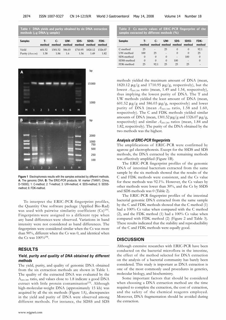
To interpret the ERIC-PCR fingerprint profiles, the Quantity One software package (Applied Bio-Rad) was used with pairwise similarity coefficient (Cs)[28]. Fingerprints were assigned to a different type when any band differences were observed. Variations in band intensity were not considered as band differences. The fingerprints were considered similar when the Cs was more than 90%, different when the Cs was 0, and identical when the Cs was 100%[28].
RESULTSYield, purity and quality of DNA obtained by different methods The yield, purity, and quality of genomic DNA obtained from the six extraction methods are shown in Table 1. The quality of the extracted DNA was evaluated by the A260/280 ratio, and values close to 1.8 indicate a good DNA extract with little protein contamination[25]. Although high-molecular-weight DNA (approximately 15 kb) was acquired by all the six methods (Figure 1A), discrepancies in the yield and purity of DNA were observed among different methods. For instance, the SDSS and SDS
methods yielded the maximum amount of DNA (mean, 1820.12 µg/g and 1710.95 µg/g, respectively), but the lowest A260/280 ratio (mean, 1.49 and 1.54, respectively), thus implying the lowest purity of DNA. The T and UW methods yielded the least amount of DNA (mean, 601.52 µg/g and 586.03 µg/g, respectively) and lower purity of DNA (mean A260/280 ratio, 1.58 and 1.60, respectively). The C and FDK methods yielded similar amounts of DNA (mean, 1301.52 µg/g and 1326.07 µg/g, respectively) and similar A260/280 ratios (mean, 1.84 and 1.82, respectively). The purity of the DNA obtained by the two methods was the highest.
Analysis of ERIC-PCR fingerprintsThe amplifications of ERIC-PCR were confirmed by agarose gel electrophoresis. Except for the SSDS and SDS methods, the DNA extracted by the remaining methods was effectively amplified (Figure 1B).
The ERIC-PCR fingerprint profiles of the genomic DNA of intestinal bacterium extracted from the same sample by the six methods showed that the results of the C and FDK methods were consistent, and the Cs value for these methods was 92.1%. However, the Cs values by other methods were lower than 30%, and the Cs by SSDS and SDS methods was 0 (Table 2).
The ERIC-PCR fingerprint profiles of the intestinal bacterial genomic DNA extracted from the same sample by the C and FDK methods showed that the C method (1) had a 100% Cs value when compared with the C method (2), and the FDK method (1) had a 100% Cs value when compared with FDK method (2) (Figure 2 and Table 3). These results indicated that the stability and reproducibility of the C and FDK methods were equally good.
DISCUSSIONAlthough extensive researches with ERIC-PCR have been conducted on the bacterial microflora in the intestine, the effect of the method selected for DNA extraction on the analysis of a bacterial community has barely been considered. This study is important as DNA extraction is one of the most commonly used procedures in genetics, molecular biology, and biochemistry.
Some important factors that should be considered when choosing a DNA extraction method are the time required to complete the extraction, the cost of extraction, and the safety of the chemical reagents employed. Moreover, DNA fragmentation should be avoided during the extraction.
Table 1 DNA yields and purity obtained by six DNA extraction methods (µg DNA/g sample)
Samples T- C- UW- SDS- SDSS- FDK-method method method method method method
Yield 601.52 1301.52 586.03 1710.95 1820.12 1326.07Purity (A260/280) 1.58 1.84 1.6 1.54 1.49 1.82
15 000
1000
M 1 2 3 4 5 6 M
2000
1000 750
500
250
100
M 1 2 3 4 5 6 M
A
B
Figure 1 Electrophoresis results with the samples extracted by different methods. A: The genomic DNA; B: The ERIC-PCR products. M: marker (TIANYI, China, D-15000); 1: C-method; 2: T-method; 3: UW-method; 4: SDS-method; 5: SDSS-method; 6: FDK-method.
Samples T- C- UW- SDS- SDSS- FDK-method method method method method method
C-method 25 - 25 0 0 92.1UW-method 100 25 - 0 0 25SDS-method 0 0 0 - 100 0SDSS-method 0 0 0 100 - 0FDK-method 25 92.1 25 25 25 -
Table 2 Cs matrix values of ERIC-PCR fingerprint of the samples extracted by different methods (%)
www.wjgnet.com
2874 ISSN 1007-9327 CN 14-1219/R World J Gastroenterol May 14, 2008 Volume 14 Number 18
bp
bp

From the extraction methods already published for various bacteria, we chose and compared six methods for extracting DNA from the intestinal microflora: the T, C, UW, SDSS, SDS, and commercial FDK methods.
The aim of an extraction procedure is to obtain a high quality and high yield of DNA from the samples. The extracted DNA should contain the least amount of proteins, RNA, or any other PCR inhibitors such as bile salt and cholerythrin, which are present in the feces[28,29]. Removing those inhibitors is one of the key factors for a successful PCR. In this study, we used acetone to remove the inhibitors from the stool samples, as reported by Zhong Hua et al[21]. Our ERIC-PCR results was found successful in removal of these inhibitors.
DNA absorbance was measured at 260 nm (A260) to evaluate the quantity of the extracted DNA, and the ratio of the absorbance at 260 nm to that at 280 nm (A260/280) was used to evaluate the DNA quality. This method was employed previously by other researchers to compare different DNA extraction methods[30,31].
In this study, the results of the concentration and the purity for each method were correlated. The SDSS and SDS methods seemed to yield a greater quantity of DNA, but their A260/280 ratio indicated a high protein contamination. On the other hand, the concentrations of the DNA obtained using the C and FDK methods were lower, but the A260/280 ratio showed a high purity of the DNA obtained. When applied to engorged individuals, the C method resulted in 100% successful DNA amplification that was similar to the FDK method. In our study, the C method provided the best results with a Cs close to 92.1% when compared with the FDK method.
Bacterial lysis is the key to obtain bacterial DNA. Although the SDS and SDSS methods can provide the highest DNA yield, the SDS residue inhibits the PCR process. This result is consistent with that reported by Khan[18] and Wade[17]. The excessive SDS above 0.01%
has been shown to inhibit PCR by denaturing the Taq polymerase.
Many reports have described DNA extraction from tissues of other organisms using chelex-100. The results vary by different methods[32,33]. The use of chelex-100 has been recommended for DNA extraction in some papers, but other reports have not regarded it as being optimum because of its lowest efficiency for DNA amplification[32]. However, we found that the modified chelex-100 protocol was the best method to extract DNA from stool. Although
there are methodological differences between our study and others[32,33], the conclusion is the same that this method is simple and efficient for PCR.
Theoretically, column-purified DNA should be the cleanest, containing the least PCR-inhibitory substances. One purpose of our study was to identify a method for rapid DNA extraction that did not compromise PCR sensitivity. We used the FDK extraction method as the gold standard for PCR purification. Our results showed that for PCR, column purification was unnecessary for DNA extracted from the intestinal microflora. Compared to FDK that requires 2 h, DNA extraction with chelex-100 can be completed within less than 1 h, and it does not involve 3-4 transfers of samples to new tubes.
In conclusion, of the 5 extraction methods evaluated, the chelex method is technically simpler, less expensive, and more rapid than the FDK method; it is the best method for extracting genomic DNA from the intestinal microflora. Although the extraction with TE buffer and ultrapure water is extremely easy and inexpensive, the DNA yield is the lowest. The SDS and SDSS methods consistently inhibit PCR, therefore, they cannot be recommended for DNA extraction from the intestinal microflora.
COMMENTSBackgroundThe term probiotic is a relatively new term that implies “for life”. Currently, it is used to refer to intestinal bacteria that have beneficial effects on humans and animals. Probiotics can play an important role in immunological, digestive, and respiratory functions and could have a significant effect in alleviating infectious diseases in children and other high-risk populations. Thus, knowledge about probiotics could contribute to improving human health. It is necessary to understand the bacterial flora in the gastrointestinal tract, particularly at the species level of bacterium. The DNA-based techniques often represent the most appropriate approach in this regard.
Research frontiersTo date, the chelex method has mainly been used for medicolegal investigations. Since it is a rapid, simple, and inexpensive technique for extracting genomic DNA from intestinal microflora, it will be an ideal method to study the microflora in the gastrointestinal tract.
Innovations and breakthroughsIn previous studies on DNA extraction from the intestinal microflora, researchers used the traditional DNA extraction process that involved proteinase K digestion by phenol chloroform extraction or the FDK method. However, the chemical products employed in these experiments were either toxic or expensive. Through the present study, we provide a significantly improved method for the rapid, safe, simple and economical extraction of DNA from the intestinal microflora.
ApplicationsThis study details a new, simple protocol for extracting DNA from the intestinal
2000
1000 750 500
250
100
M 1 2 3 4 M
Figure 2 Electrophoresis for ERIC-PCR fingerprint of the samples extracted by C-method and FDK-method. Lanes: M: marker (TIANYI, China, DL-2000); 1 and 2: C-method; 3 and 4: FDK-method.
Table 3 Cs matrix values of ERIC-PCR fingerprint of the samples extracted by C-method and FDK-methods (%)
Samples C-method (1) C-method (2) FDK-method (1)
FDK-method (2)
C-method (2) 100.0 92.1 92.1FDK-method (1) 92.1 92.1 100.0FDK-method (2) 92.1 92.1 100.0
www.wjgnet.com
Yang JL et al . Method of extracting bacterial DNA 2875
bp
COMMENTS

bacterial microflora and may ultimately provide new insights in the field of probiotics.
Peer reviewIn this study, through the development of new methods for extracting bacterial DNA from the intestinal microflora, the authors found that the chelex method is a simpler, more rapid, and a less expensive method than the FDK method. Moreover, the results presented herein may lead to the development of a novel convenient approach for monitoring the intestinal microflora by molecular detection methods.
REFERENCES1 Steinhoff U. Who controls the crowd? New findings and old
questions about the intestinal microflora. Immunol Lett 2005; 99: 12-16
2 Moore WE, Cato EP, Holdeman LV. Some current concepts in intestinal bacteriology. Am J Clin Nutr 1978; 31: S33-S42
3 Falk PG, Hooper LV, Midtvedt T, Gordon JI. Creating and maintaining the gastrointestinal ecosystem: what we know and need to know from gnotobiology. Microbiol Mol Biol Rev 1998; 62: 1157-1170
4 Nechvatal JM, Ram JL, Basson MD, Namprachan P, Niec SR, Badsha KZ, Matherly LH, Majumdar AP, Kato I. Fecal collection, ambient preservation, and DNA extraction for PCR amplification of bacterial and human markers from human feces. J Microbiol Methods 2008; 72: 124-132
5 Guarner F, Schaafsma GJ. Probiotics. Int J Food Microbiol 1998; 39: 237-238
6 Food and Agriculture Organization of the United Nations/ World Health Organization. Health and nutritional properties of probiotics in food including powder milk with live lactic acid bacteria. World Health Organization online, 2001-10-01, cited 2007-11-08. Available from: URL: http://www.who.int/foodsafety/publications/fs_management/en/probiotics.pdf
7 Mimura T, Rizzello F, Helwig U, Poggioli G, Schreiber S, Talbot IC, Nicholls RJ, Gionchetti P, Campieri M, Kamm MA. Once daily high dose probiotic therapy (VSL#3) for maintaining remission in recurrent or refractory pouchitis. Gut 2004; 53: 108-114
8 Gionchetti P, Rizzello F, Helwig U, Venturi A, Lammers KM, Brigidi P, Vitali B, Poggioli G, Miglioli M, Campieri M. Prophylaxis of pouchitis onset with probiotic therapy: a double-blind, placebo-controlled trial. Gastroenterology 2003; 124: 1202-1209
9 Shen TY, Qin HL, Gao ZG, Fan XB, Hang XM, Jiang YQ. Influences of enteral nutrition combined with probiotics on gut microflora and barrier function of rats with abdominal infection. World J Gastroenterol 2006; 12: 4352-4358
10 Foligne B, Nutten S, Grangette C, Dennin V, Goudercourt D, Poiret S, Dewulf J, Brassart D, Mercenier A, Pot B. Correlation between in vitro and in vivo immunomodulatory properties of lactic acid bacteria. World J Gastroenterol 2007; 13: 236-243
11 Versalovic J, Koeuth T, Lupski JR. Distribution of repetitive DNA sequences in eubacteria and application to fingerprinting of bacterial genomes. Nucleic Acids Res 1991; 19: 6823-6831
12 Finegold SM, Attebery HR, Sutter VL. Effect of diet on human fecal flora: comparison of Japanese and American diets. Am J Clin Nutr 1974; 27: 1456-1469
13 Rodriguez-Barradas MC, Hamill RJ, Houston ED, Georghiou PR, Clarr idge JE, Regnery RL, Koehler JE. Genomic fingerprinting of Bartonella species by repetitive element PCR for distinguishing species and isolates. J Clin Microbiol 1995; 33: 1089-1093
14 Sampaio JL, Viana-Niero C, de Freitas D, Hofling-Lima AL, Leao SC. Enterobacterial repetitive intergenic consensus PCR is a useful tool for typing Mycobacterium chelonae and Mycobacterium abscessus isolates. Diagn Microbiol Infect Dis 2006; 55: 107-118
15 de Bruijn FJ . Use of repetitive (repetitive extragenic palindromic and enterobacterial repetitive intergeneric consensus) sequences and the polymerase chain reaction to fingerprint the genomes of Rhizobium meliloti isolates and other soil bacteria. Appl Environ Microbiol 1992; 58: 2180-2187
16 Spaniolas S , Tsachaki M, Bennett MJ and Tucker GA. Evaluation of DNA extraction methods from green and roasted coffee beans. Food Control 2008; 3: 257-262
17 Aldous WK, Pounder JI, Cloud JL, Woods GL. Comparison of six methods of extracting Mycobacterium tuberculosis DNA from processed sputum for testing by quantitative real-time PCR. J Clin Microbiol 2005; 43: 2471-2473
18 Khan IU, Yadav JS. Development of a single-tube, cell lysis-based, genus-specific PCR method for rapid identification of mycobacteria: optimization of cell lysis, PCR primers and conditions, and restriction pattern analysis. J Clin Microbiol 2004; 42: 453-457
19 Rantakokko-Jalava K, Jalava J. Optimal DNA isolation method for detection of bacteria in clinical specimens by broad-range PCR. J Clin Microbiol 2002; 40: 4211-4217
20 Buck GE, O'Hara LC, Summersgill JT. Rapid, simple method for treating clinical specimens containing Mycobacterium tuberculosis to remove DNA for polymerase chain reaction. J Clin Microbiol 1992; 30: 1331-1334
21 Zhong H, Lai XL, Wei RP, Liu ZL. An improved protocol for DNA extraction from the faeces of giant panda. Acta Zool Sin 2003; 49: 670-674
22 Li M, Gong J, Cottrill M, Yu H, de Lange C, Burton J, Topp E. Evaluation of QIAamp DNA Stool Mini Kit for ecological studies of gut microbiota. J Microbiol Methods 2003; 54: 13-20
23 Wang RF , Cao WW, Cerniglia CE. PCR detection and quantitation of predominant anaerobic bacteria in human and animal fecal samples. Appl Environ Microbiol 1996; 62: 1242-1247
24 Suenaga E, Nakamura H. Evaluation of three methods for effective extraction of DNA from human hair. J Chromatogr B Analyt Technol Biomed Life Sci 2005; 820: 137-141
25 Samuel M , Lu M, Pachuk CJ , Sa t i shchandran C. A spectrophotometric method to quantify linear DNA. Anal Biochem 2003; 313: 301-306
26 Di Giovanni GD, Watrud LS, Seidler RJ, Widmer F. Compari-son of Parental and Transgenic Alfalfa Rhizosphere Bacterial Communities Using Biolog GN Metabolic Fingerprinting and Enterobacterial Repetitive Intergenic Consensus Sequence-PCR (ERIC-PCR). Microb Ecol 1999; 37: 129-139
27 Syrmis MW, O'Carroll MR, Sloots TP, Coulter C, Wainwright CE, Bell SC, Nissen MD. Rapid genotyping of Pseudomonas aeruginosa isolates harboured by adult and paediatric patients with cystic fibrosis using repetitive-element-based PCR assays. J Med Microbiol 2004; 53: 1089-1096
28 Sidransky D, Tokino T, Hamilton SR, Kinzler KW, Levin B, Frost P, Vogelstein B. Identification of ras oncogene mutations in the stool of patients with curable colorectal tumors. Science 1992; 256: 102-105
29 Deuter R, Pietsch S, Hertel S, Muller O. A method for preparation of fecal DNA suitable for PCR. Nucleic Acids Res 1995; 23: 3800-3801
30 Tan H , Wang J , Zhao ZK. Purification and refolding optimization of recombinant bovine enterokinase light chain overexpressed in Escherichia coli. Protein Expr Purif 2007; 56: 40-47
31 Ki JS, Chang KB, Roh HJ, Lee BY, Yoon JY, Jang GY. Direct DNA isolation from solid biological sources without pretreatments with proteinase-K and/or homogenization through automated DNA extraction. J Biosci Bioeng 2007; 103: 242-246
32 Desloire S, Valiente Moro C, Chauve C, Zenner L. Comparison of four methods of extracting DNA from D. gallinae (Acari: Dermanyssidae). Vet Res 2006; 37: 725-732
33 Aranishi F, Okimoto T. A simple and reliable method for DNA extraction from bivalve mantle. J Appl Genet 2006; 47: 251-254
S- Editor Zhong XY L- Editor Ma JY E- Editor Yin DH
www.wjgnet.com
2876 ISSN 1007-9327 CN 14-1219/R World J Gastroenterol May 14, 2008 Volume 14 Number 18

was significantly lower than that of cells transfected with blank plasmid. Among three different quasispecies, T, NT and C191 core expression cells, there was no significant difference in the proportion of S- and G0/G1-phase cells. The percentage of apoptotic cells was highest for T (T > NT > C191), and apoptosis was increased in cells transfected with pEGFP-N1/core as the transfection time increased (72 h > 48 h > 24 h).
CONCLUSION: These results suggest that HCV genotype 1b core protein induces apoptosis, and inhibits cell-cycle progression and proliferation of Chang liver cells. Different quasispecies core proteins of HCV genotype 1b might have some differences in the pathogenesis of HCV persistent infection and hepatocellular carcinoma.
© 2008 WJG. All rights reserved.
Key words: Hepatitis C virus; Core protein; Chang liver cells; Cell cycle; Apoptosis
Peer reviewer: Baumert F Thomas, Professor, Inserm U748, Louis Pasteur University, 3 Rue Koeberle, 67000 Strasbourg, France
Yan XB, Mei L, Feng X, Wan MR, Chen Z, Pavio N, Brechot C. Hepatitis C virus core proteins derived from different quasispecies of genotype 1b inhibit the growth of Chang liver cells. World J Gastroenterol 2008; 14(18): 2877-2881 Available from: URL: http://www.wjgnet.com/1007-9327/14/2877.asp DOI: http://dx.doi.org/10.3748/wjg.14.2877
INTRODUCTIONHepatitis C virus (HCV) infection is prevalent worldwide. HCV infection may lead to the development of chronic hepatitis, cirrhosis and hepatocellular carcinoma (HCC)[1]. HCV is a positive-strand RNA virus that belongs to the family Flaviviridae. The HCV RNA genome is 9.6 kb in length and encodes a precursor polyprotein of 3000 amino acids. The precursor polyprotein is cleaved by host and viral proteases and produces a series of structural and non-structural proteins. In addition, HCV genome has the property of variation, and it usually exists as different quasispecies of the same genotype in HCV-infected patients[2,3]. The function of different quasispecies of HCV is not fully identical[4,5].
RAPID COMMUNICATION
Hepatitis C virus core proteins derived from different quasispecies of genotype 1b inhibit the growth of Chang liver cells
Xue-Bing Yan, Lei Mei, Xia Feng, Mei-Rong Wan, Zhi Chen, Nicole Pavio, Christian Brechot
Xue-Bing Yan, Lei Mei, Department of Infectious Diseases, the First Affiliated Hospital of Xuzhou Medical College, Xuzhou 221002, Jiangsu Province, China Lei Mei, Changshu No. 2 People’s Hospital, 68 Haiyu South Road, Changshu 215500, Jiangsu Province, China Xia Feng, Mei-Rong Wan, Department of Central Labatory, the First Affiliated Hospital of Xuzhou Medical College, Xuzhou 221002, Jiangsu Province, ChinaZhi Chen, Institute of Infectious Diseases, the First Affiliated Hospital, College of Medical Science, Zhejiang University; Key Lab of Infectious Diseases, Ministry of Public Health, Hangzhou, 310003, Zhejiang Province, ChinaNicole Pavio, Christian Brechot, Inserm, U785, Villejuif, F-94804, France; Universite Paris sud, Centre Hepato-Biliaire, Hopital Paul Brousse, Villejuif F-94804, FranceAuthor contributions: Yan XB and Mei L contributed equally to this work; Yan XB, Mei L and Chen Z designed the research; Mei L, Feng X and Wan MR performed the experiments and Pavio N and Brechot C contributed the plasmids.Supported by The Nature Science Foundation of Jiangsu, No. BK2007031; The College Education Nature Science Foundation of Jiangsu, No. 05KJB320137 Correspondence to: Xue-Bing Yan, Department of Infectious Diseases, The First Affiliated Hospital of Xuzhou Medical College, Xuzhou 221002, Jiangsu Province, China. [email protected]: +86-516-85802180 Fax: +86-516-85802215Received: December 22, 2007 Revised: February 4, 2008
AbstractAIM: To investigate the influence of different quasispecies of hepatitis C virus (HCV) genotype 1b core protein on growth of Chang liver cells.
METHODS: Three eukaryotic expression plasmids (pEGFP-N1/core) that contained different quasispecies truncated core proteins of HCV genotype 1b were constructed. These were derived from tumor (T) and non-tumor (NT) tissues of a patient infected with HCV and C191 (HCV-J6). The core protein expression plasmids were transiently transfected into Chang liver cells. At different times, the cell cycle and apoptosis was assayed by flow cytometry, and cell proliferation was assayed by methyl thiazolyl tetrazolium (MTT) assay.
RESULTS: The proportion of S-phase Chang liver cells transfected with pEGFP-N1/core was significantly lower than that of cells transfected with blank plasmid at three different times after transfection (all P < 0.05). The proliferation ratio of cells transfected with pEGFP-N1/core
Online Submissions: wjg.wjgnet.com World J Gastroenterol 2008 May 14; 14(18): 2877-2881www.wjgnet.com World Journal of Gastroenterology ISSN [email protected] © 2008 WJG. All rights reserved.
www.wjgnet.com

HCV core protein is important as a viral structural component in nucleocapsid formation, and it is highly conserved among different HCV genotypes. Several studies have established that HCV core protein can regulate cell signal transduction, such as MAPK-, JAK-STAT-, NF-κB-, AP-1- and SRE-associated pathways[6-8]. Hence, it plays important roles in the pathogenesis of HCV persistent infection, as well as HCC. However, the mechanism of persistent infection and HCC due to HCV core protein infection remains unclear. Although several studies have investigated the influence of the HCV core protein on cell growth and apoptosis, the results of these investigations have been inconsistent and mutually conflicting.
In most studies, HCV core protein has been expressed in single clones of permanently transfected cell lines, while more recently, transiently transfected cells have also been used. These studies have found that core protein affects normal cellular functions, such as proliferation and death, which are involved, directly or indirectly, in HCV hepatocarcinogenesis. Because we have found that different quasispecies core proteins of HCV genotype 1b have some differences in the pathogenesis of HCV persistent infection and HCC. HCV is a hepatotropic virus, it is therefore more appropriate to use a hepatocyte cell line to study the function of HCV core protein.
To further clarify the in vivo role of truncated HCV core protein from different genotype 1b quasispecies, we expressed three different truncated forms of HCV core protein derived from tumor tissue (T), non-tumor tissue (NT) and C191 (HCV-J6), which all belong to HCV genotype 1b, in transiently transfected Chang liver cells, an immortalized non-tumor hepatic cell line. Cell cycle and apoptosis were assayed by flow cytometry, and cell proliferation was assayed by methyl thiazolyl tetrazolium (MTT) assay.
MATERIALS AND METHODSPlasmid constructsThree different truncated HCV core protein eukaryotic expression plasmids, pEGFP-N1/core, were constructed. Truncated core protein nucleotide sequences were amplified from pGEX 4T-1/HCV-core, which contained core sequences from T, NT and C191, respectively. Sequence analysis revealed that T and NT were all HCV genotype 1b. The primers were designed according to the core protein gene sequence of T, NT and C191 (Table 1). PCR reaction system: 50 μL: water 40.75 μL; PCR reaction buffer (10 ×), 5 μL; template (45 ng), 1 μL; primer up 20 pmol/L, 1 μL; primer down 20 pmol/L, 1 μL; dNTP 20 mmol/L, 1 μL, Expand high enzyme 0.25 μL, 94℃ for 2 min, 94℃ for 30 s, 50℃ for 30 s, 72℃ for 30 s, 10 cycles; 94℃ for 30 s, 55℃ for 30 s, 72℃ for 30 s, 20 cycles; and 72℃ for 7 min. PCR products (Figure 1) were purified and cleaved with restriction enzymes NheⅠand EcoRⅠ, and cloned into pEGFP-N1 to yield GFP-core fusion protein plasmids. Cell culture and transfectionChang liver cells were grown in Dulbecco’s modified Eagle’s medium (Gibco, USA) supplemented with 10% fetal bovine serum (FBS; Gibco) at 37℃ in 5% CO2. Cells were incubated for 18 h and then transfected with Lipofectamine
2000 (Invitrogen, USA) following the manufacturer’s instructions. Two micrograms of plasmids were used to achieve transfection. After 6 h, the medium was changed and cells were collected at different times.
Flow cytometry analysisThe cell cycle and apoptosis was assayed by flow cytometry at 24, 48 and 72 h after transfection. Ten × 105 cells were trypsinized, pelleted by centrifugation, and resuspended in PBS, and then cells were fixed with 70% ethanol and stored at 4℃ overnight. After washing twice, cells were incubated in propidium iodine for 20 min at room temperature. The cell suspension was analyzed by flow cytometry using Cell Quest software (Becton Dickinson, USA).
Cell growth analysisCell proliferation was assayed by MTT assay and a growth curve was created. 104 cells were seeded per well on a 96-well plate. Zero, 24, 48, 72 and 96 h after transfection, 10 μL water-soluble tetrazolium reagent (MTT; Sigma, USA) was added to 100 μL culture medium. Cells were then incubated at 37℃ in 5% CO2 for 4 h. A570 was measured. The assay was done in quadruplicate.
Statistical analysisThe values were expressed as mean ± SD. Experimental data were analyzed by SPSS 13.0 software (SPSS Inc, Chicago, IL, USA). Comparison of different groups was analyzed by one-way analysis of variance (ANOVA). P < 0.05 was considered statistically significant.
RESULTSDifferent HCV genotype 1b quasispecies core proteins inhibited Chang liver cell cycle As shown in Tables 2-4, three different quasispecies
Table 1 Primers of different quasispecies core proteins of HCV genotype 1b
Primer up (location) Primer down (location)
T: 1-172 CGCGCTAGCATGAGGCACGAATCC (1-14)
CCGGAATTCGCAACCGGGCAG (505-516)
NT: 1-172 CGCGCTAGCATGAGCACGAATCC (1-14)
CCGGAATTCGGCAACCGGGCAGATTC (505-516)
C191: 1-172 CGCGCTAGCATGAGCACAAATCC (1-14)
CCGGAATTCGGCAACCGGGCAAATTC (505-516)
GCTAGC, NheⅠ site; GAATTC, EcoRⅠ site. Primer designed according to the sequence of different core protein genes.
M T NT C191
500 bp
Figure 1 PCR pro-duct of core protein genes.
www.wjgnet.com
2878 ISSN 1007-9327 CN 14-1219/R World J Gastroenterol May 14, 2008 Volume 14 Number 18

truncated core proteins inhibited Chang liver cell cycle progression by impairing G1- to S-phase transition. The proportion of S- and G0/G1-phase Chang liver cells transfected with pEGFP-N1/core was significantly lower than that of cells transfected with blank plasmid at 24, 48 and 72 h after transfection (P = 0.002, P = 0.001, P = 0.001, respectively), but there were no significant differences among cells expressing the three different quasispecies HCV truncated core proteins.
Different HCV genotype 1b quasispecies core proteins induced Chang liver cell apoptosisAs shown in Figure 2, three different quasispecies truncated core proteins induced apoptosis at different levels. The apoptotic ratio of Chang liver cells transfected with pEGFP-N1/core was significantly higher than that of cells transfected with blank plasmid. The apoptotic percentage of T was the highest, and C191 was the lowest (T > NT > C191). The apoptosis ratio increased in cells transfected with pEGFP-N1/core as transfection time increased (72 h > 48 h > 24 h).
Different HCV genotype 1b quasispecies core proteins inhibited Chang liver cell proliferationWe found that different HCV genotype 1b quasispecies
core proteins inhibited the Chang liver cell cycle by impairing G1- to S-phase transition and induced aptoptosis at different times after transfection. Chang liver cell prolif-eration was further analyzed using MTT assay. As shown in Figure 3, different HCV genotype 1b quasispecies core proteins inhibited Chang liver cell proliferation. Among the three different HCV genotype 1b quasispecies core proteins, that of T inhibited Chang liver cell proliferation more obviously than NT and C191 (T > NT > C191) at 24, 48, 72 and 96 h.
DISCUSSIONHCV core protein is a multifunctional protein that can modulate a number of cellular processes, including transcription, inhibition or stimulation of cell cycle and apoptosis, and suppression of host immunity. In this study, using flow cytometry and MTT assay, we investigated the influence of different quasispecies of truncated HCV genotype 1b core proteins on growth of Chang liver cells, an immortalized non-tumor hepatic cell line. We found that HCV genotype 1b core protein HCV induced apoptosis, and inhibited the cell cycle and cell proliferation. Among
Table 2 Core proteins of different quasispecies of HCV genotype 1b inhibited Chang liver cell cycle by impairing G1 to S transition at 24 h after transfection
G0/G1 phase S phase G2/M phase
T 67.53 ± 5.47a 17.23 ± 4.21a 15.25 ± 1.96NT 65.49 ± 5.98a 18.56 ± 2.49a 15.95 ± 2.59C191 66.72 ± 6.82a 17.89 ± 2.54a 15.38 ± 2.16pEGFP-N1 59.76 ± 5.33 24.33 ± 3.16 15.93 ± 1.42
aP < 0.05 vs pEGFP-N1.
aP < 0.05 vs pEGFP-N1.
Table 3 Core proteins of different quasispecies of HCV genotype 1b inhibited Chang liver cell cycle by impairing G1 to S transition at 48 h after transfection
G0/G1 phase S phase G2/M phase
T 68.23 ± 6.54a 15.30 ± 3.12a 16.46 ± 2.57NT 66.14 ± 5.33a 16.38 ± 2.21a 17.47 ± 3.74C191 66.41 ± 3.02a 15.92 ± 2.93a 16.99 ± 2.43pEGFP-N1 58.72 ± 2.23 25.69 ± 2.41 15.62 ± 1.28
Table 4 Core proteins of different quasispecies of HCV genotype 1b inhibited Chang liver cell cycle by impairing G1 to S transition at 72 h after transfection
G0/G1 phase S phase G2/M phase
T 68.45 ± 4.98a 14.79 ± 3.76a 16.75 ± 3.21NT 67.21 ± 5.47a 16.17 ± 2.55a 16.63 ± 2.95C191 66.77 ± 4.32a 16.38 ± 2.11a 16.86 ± 2.19pEGFP-N1 56.97 ± 3.29 27.48 ± 3.37 15.54 ± 1.93
aP < 0.05 vs pEGFP-N1.
40
35
30
25
20
15
10
5
0
Apop
tosi
s
24 h after transfection
48 h after transfection
72 h after transfection
T NT C191 pEGFP-N1
Figure 2 Core proteins of different quasispecies of HCV genotype 1b induced Chang liver cell apoptosis at 24, 48 and 72 h after transfection.
0.6
0.5
0.4
0.3
0.2
0.1
0.0
A57
0
0 1 2 3 4
t /d
Figure 3 Core proteins from different HCV genotype 1b quasispecies inhibited proliferation of Chang liver cells.
T
NT
C191
pEGFP-N1
www.wjgnet.com
Yan XB et al. HCV core inhibits Chang liver cells 2879

the three different quasispecies core proteins, the rates of inducing apoptosis and inhibiting cell proliferation were different, which suggests that different quasispecies of HCV genotype 1b core proteins might have some differences in their pathogenesis of HCV persistent infection and HCC.
Regulation of cell cycle and apoptosis is essential to maintain normal cell growth. Several studies have reported that HCV core protein can influence cell cycle progression. Two important checkpoints of the cell cycle, G1/S and G2/M, are responsible for accumulation of detrimental mutations that may result in cell transformation. The mechanism of cell cycle regulation is quite complex, and involves a number of proteins, such as p53, p21, PRb, E2F and c-myc[9,10]. Ruggieri et al have reported that stable expression of core protein in unsynchronized HepG2 cells induces perturbation of the cell cycle by increasing the S-phase fraction. c-myc protein stability is increased, which is one of the regulatory molecules of the cell cycle[11]. Honda et al have found that core protein promotes cell cycle progression in stable CHO transformation by upregulation of c-myc[12]. Ohkawa et al have reported that core protein impairs G1 to S transition by using core expression stable CL2 cell, a murine normal liver-derived cell line. E2F-mediated transcription, and PRb, CDK4 and CDK2 activities were suppressed by HCV core protein[13]. Different studies have come to different conclusions. We found that transient expression of HCV genotype 1b truncated core proteins could impair G1/S in the Chang liver cell cycle. Different quasispecies core proteins from T, NT and C191 have no significant difference when it comes to impairing G1/S. The above studies suggest that core protein might be a double-function protein, promoting cell cycle progression and inhibiting cell cycle progression. However, the exact mechanisms of how different quasispecies core proteins affect the G1/S checkpoint remain unclear.
Apoptosis is defined as programmed cell death and it is an orderly process. Apoptosis can be induced by a variety of stimuli, including oxidative stress, heat shock, ionizing radiation, cytokines and virus infection. The apoptotic process appears to be a host defense mechanism against viral infections. HCV might lead to viral persistence by inhibiting hepatocyte apoptosis and inducing monocyte apoptosis[14]. Although diverse effects of the core protein on apoptosis have been reported, the underlying mechanisms are not fully understood[15]. Core protein exhibits both pro- and anti-apoptotic actions. Transient expression of the core protein can inhibit tumor necrosis factor (TNF)-α-mediated apoptosis in MCF7 cells, a cell line derived from breast carcinoma tissue[16]. In contrast, several studies have indicated that core protein can promote apoptosis induced by Fas or TNF-α in stable expression systems in HepG2, HeLa and Jurkat T cell lines[17-19]. In our study, we found that different quasispecies of HCV genotype 1b truncated core proteins could induce Chang liver cell apoptosis, and the ability to induce apoptosis was different (T > NT > C191). Apoptosis was increased depending on prolongation of transfection time (72 h > 48 h > 24 h). Moreover, the cell proliferation induced by HCV core protein was consistent with our finding that HCV core protein induced cell apoptosis. Nevertheless, the present results allow us to
propose an alternative, and in fact contrasting mechanism for HCV core protein. Our observations imply that the liver cell proliferation observed during liver carcinogenesis is associated with the selection of viral genomes whose core products can resist apoptosis.
This apparently paradoxical f inding should be discussed in the light of two important observations. Consistent with previous reports[20-23], in HCV-related HCC, strong arguments exist in favor of the importance of the immune response to HCV proteins in liver cell destruction during acute or chronic HCV infection. Our findings therefore suggest that cell apoptosis may reduce HCV protein expression and the elimination of infected cells, thus favoring viral persistence. A sustained reduction in HCV replication during the later stages of liver cancer development probably depends on other mechanisms, such as a lack in dedifferentiated cancer cells of the tissue-specific cellular factors necessary for HCV polyprotein expression and maturation. This favors the selection of a cell subset that acquires resistance to HCV apoptosis and whose differentiation status maintains down-regulated HCV multiplication.
Cell-cycle progression and apoptosis are balanced processes regulated by a number of cellular factors. Several cell-cycle-regulatory proteins are involved in signal transduction pathways of apoptosis[24-26]. Core protein impaired G1/S transition, which might be advantageous to cells that are entering the apoptotic process. The influence of core protein on cell cycle and apoptosis is different, depending on different cell systems and expression systems used[27-30]. However, in most studies, human hepatoma-derived cell lines Huh-7 or HepG2 were used, but in our study, Chang liver cell and transient transfection were used. As an immortalized non-tumor cell line, the biological function of Chang liver cells is similar to that of normal human hepatocytes, so it is suitable for studying virus-host interactions in vitro.
In summary, we found that HCV genotype 1b core protein could induce apoptosis, and inhibit cell-cycle progression and cell proliferation. Among three different quasispecies core proteins, the capability of inducing apoptosis and inhibiting cell proliferation was different.
COMMENTSBackgroundIt is known that hepatitis C virus (HCV) core protein plays an important role in the pathogenesis of HCV persistent infection and hepatocellular carcinoma (HCC). HCV core protein derived from different quasispecies of the same HCV genotype 1b might have different functions in their pathogenesis.
Research frontiersHCV core protein is involved in regulation of host cell growth, including the cell cycle and apoptosis, but the results are conflicting. Recent studies have shown that the influence of HCV core protein on cell growth might be associated with viral persistence and generation of liver carcinogenesis.
Innovations and breakthroughsWe used Chang liver cells, an immortalized non-tumor hepatic cell line, different from the human hepatoma-derived cell lines in most studies. Moreover, different quasispecies of HCV genotype 1b core proteins are found to have different biological functions.
www.wjgnet.com
2880 ISSN 1007-9327 CN 14-1219/R World J Gastroenterol May 14, 2008 Volume 14 Number 18
COMMENTS
ApplicationsOur findings further show the importance of HCV core proteins on the pathogenesis of HCV persistent infection, as well as HCC. It might provide new ideas for the cure and prevention of HCV infection.
Peer reviewThis work investigated the role of the core protein of HCV from different quasispecies on the pathogenesis of Chang liver cells. The findings are potentially interesting and point towards the ability of HCV core protein to induce apoptosis and inhibit cell-cycle progression in Chang liver cells. The intensity of these observed effects seems to be dependent on the quasispecies of the HCV core protein isolated.
REFERENCES1 Kiyosawa K, Sodeyama T, Tanaka E, Gibo Y, Yoshizawa K,
Nakano Y, Furuta S, Akahane Y, Nishioka K, Purcell RH. Interrelationship of blood transfusion, non-A, non-B hepatitis and hepatocellular carcinoma: analysis by detection of antibody to hepatitis C virus. Hepatology 1990; 12: 671-675
2 Lerat H, Rumin S, Habersetzer F, Berby F, Trabaud MA, Trepo C, Inchauspe G. In vivo tropism of hepatitis C virus genomic sequences in hematopoietic cells: influence of viral load, viral genotype, and cell phenotype. Blood 1998; 91: 3841-3849
3 Ruster B, Zeuzem S, Krump-Konvalinkova V, Berg T, Jonas S, Severin K, Roth WK. Comparative sequence analysis of the core- and NS5-region of hepatitis C virus from tumor and adjacent non-tumor tissue. J Med Virol 2001; 63: 128-134
4 Delhem N, Sabile A, Gajardo R, Podevin P, Abadie A, Blaton MA, Kremsdorf D, Beretta L, Brechot C. Activation of the interferon-inducible protein kinase PKR by hepatocellular carcinoma derived-hepatitis C virus core protein. Oncogene 2001; 20: 5836-5845
5 Pavio N, Battaglia S, Boucreux D, Arnulf B, Sobesky R, Hermine O, Brechot C. Hepatitis C virus core variants isolated from liver tumor but not from adjacent non-tumor tissue interact with Smad3 and inhibit the TGF-beta pathway. Oncogene 2005; 24: 6119-6132
6 Kato N, Yoshida H, Ono-Nita SK, Kato J, Goto T, Otsuka M, Lan K, Matsushima K, Shiratori Y, Omata M. Activation of intracellular signaling by hepatitis B and C viruses: C-viral core is the most potent signal inducer. Hepatology 2000; 32: 405-412
7 Hayashi J, Aoki H, Kajino K, Moriyama M, Arakawa Y, Hino O. Hepatitis C virus core protein activates the MAPK/ERK cascade synergistically with tumor promoter TPA, but not with epidermal growth factor or transforming growth factor alpha. Hepatology 2000; 32: 958-961
8 Heim MH, Moradpour D, Blum HE. Expression of hepatitis C virus proteins inhibits signal transduction through the Jak-STAT pathway. J Virol 1999; 73: 8469-8475
9 Kwun HJ, Jung EY, Ahn JY, Lee MN, Jang KL. p53-dependent transcriptional repression of p21(waf1) by hepatitis C virus NS3. J Gen Virol 2001; 82: 2235-2241
10 Cho J, Baek W, Yang S, Chang J, Sung YC, Suh M. HCV core protein modulates Rb pathway through pRb down-regulation and E2F-1 up-regulation. Biochim Biophys Acta 2001; 1538: 59-66
11 Ruggieri A, Murdolo M, Harada T, Miyamura T, Rapicetta M. Cell cycle perturbation in a human hepatoblastoma cell line constitutively expressing Hepatitis C virus core protein. Arch Virol 2004; 149: 61-74
12 Honda M, Kaneko S, Shimazaki T, Matsushita E, Kobayashi K, Ping LH, Zhang HC, Lemon SM. Hepatitis C virus core protein induces apoptosis and impairs cell-cycle regulation in stably transformed Chinese hamster ovary cells. Hepatology 2000; 31: 1351-1359
13 Ohkawa K, Ishida H, Nakanishi F, Hosui A, Ueda K, Takehara T, Hori M, Hayashi N. Hepatitis C virus core functions as a suppressor of cyclin-dependent kinase-activating kinase and
impairs cell cycle progression. J Biol Chem 2004; 279: 11719-1172614 Kountouras J, Zavos C, Chatzopoulos D. Apoptosis in
hepatitis C. J Viral Hepat 2003; 10: 335-34215 Marusawa H, Hijikata M, Chiba T, Shimotohno K. Hepatitis
C virus core protein inhibits Fas- and tumor necrosis factor alpha-mediated apoptosis via NF-kappaB activation. J Virol 1999; 73: 4713-4720
16 Ray RB, Meyer K, Steele R, Shrivastava A, Aggarwal BB, Ray R. Inhibition of tumor necrosis factor (TNF-alpha)-mediated apoptosis by hepatitis C virus core protein. J Biol Chem 1998; 273: 2256-2259
17 Yamanaka T, Kodama T, Doi T. Subcellular localization of HCV core protein regulates its ability for p53 activation and p21 suppression. Biochem Biophys Res Commun 2002; 294: 528-534
18 Hahn CS, Cho YG, Kang BS, Lester IM, Hahn YS. The HCV core protein acts as a positive regulator of fas-mediated apoptosis in a human lymphoblastoid T cell line. Virology 2000; 276: 127-137
19 Zhu N, Khoshnan A, Schneider R, Matsumoto M, Dennert G, Ware C, Lai MM. Hepatitis C virus core protein binds to the cytoplasmic domain of tumor necrosis factor (TNF) receptor 1 and enhances TNF-induced apoptosis. J Virol 1998; 72: 3691-3697
20 Soguero C, Joo M, Chianese-Bullock KA, Nguyen DT, Tung K, Hahn YS. Hepatitis C virus core protein leads to immune suppression and liver damage in a transgenic murine model. J Virol 2002; 76: 9345-9354
21 Large MK, Kittlesen DJ, Hahn YS. Suppression of host immune response by the core protein of hepatitis C virus: possible implications for hepatitis C virus persistence. J Immunol 1999; 162: 931-938
22 Kimball P , Verbeke S, Shiffman M. HCV core protein augments cyclosporine immunosuppression. Transplant Proc 2005; 37: 652-653
23 Yao ZQ, Nguyen DT, Hiotellis AI, Hahn YS. Hepatitis C virus core protein inhibits human T lymphocyte responses by a complement-dependent regulatory pathway. J Immunol 2001; 167: 5264-5272
24 Shrivastava A, Manna SK, Ray R, Aggarwal BB. Ectopic expression of hepatitis C virus core protein differentially regulates nuclear transcription factors. J Virol 1998; 72: 9722-9728
25 Fukuda K, Tsuchihara K, Hijikata M, Nishiguchi S, Kuroki T, Shimotohno K. Hepatitis C virus core protein enhances the activation of the transcription factor, Elk1, in response to mitogenic stimuli. Hepatology 2001; 33: 159-165
26 Tai DI, Tsai SL, Chen YM, Chuang YL, Peng CY, Sheen IS, Yeh CT, Chang KS, Huang SN, Kuo GC, Liaw YF. Activation of nuclear factor kappaB in hepatitis C virus infection: implications for pathogenesis and hepatocarcinogenesis. Hepatology 2000; 31: 656-664
27 Cho JW, Baek WK, Suh SI, Yang SH, Chang J, Sung YC, Suh MH. Hepatitis C virus core protein promotes cell proliferation through the upregulation of cyclin E expression levels. Liver 2001; 21: 137-142
28 Giannini C, Caini P, Giannelli F, Fontana F, Kremsdorf D, Brechot C, Zignego AL. Hepatitis C virus core protein expression in human B-cell lines does not significantly modify main proliferative and apoptosis pathways. J Gen Virol 2002; 83: 1665-1671
29 Sacco R, Tsutsumi T, Suzuki R, Otsuka M, Aizaki H, Sakamoto S, Matsuda M, Seki N, Matsuura Y, Miyamura T, Suzuki T. Antiapoptotic regulation by hepatitis C virus core protein through up-regulation of inhibitor of caspase-activated DNase. Virology 2003; 317: 24-35
30 Nguyen H, Mudryj M, Guadalupe M, Dandekar S. Hepatitis C virus core protein expression leads to biphasic regulation of the p21 cdk inhibitor and modulation of hepatocyte cell cycle. Virology 2003; 312: 245-253
S- Editor Zhong XY L- Editor Kerr C E- Editor Ma WH
www.wjgnet.com
Yan XB et al. HCV core inhibits Chang liver cells 2881

Xu D, Yu BP, Luo HS, Chen LD. Control of gallbladder contractions by cholecystokinin through cholecystokinin-A receptors on gallbladder interstitial cells of cajal. World J Gastroenterol 2008; 14(18): 2882-2887 Available from: URL: http://www.wjgnet.com/1007-9327/14/2882.asp DOI: http://dx.doi.org/10.3748/wjg.14.2882
INTRODUCTIONCholecystokinin (CCK) is released from mucosal endocrine cells in the proximal small intestine in response to a meal[1], and it is classically known to stimulate the gallbladder contraction. The CCK-induced gallbladder contractions were once believed to be brought about by direct stimulation of gallbladder smooth muscle. The CCK receptors have been identified by using isolated gallbladder muscle membranes and autoradiographical analysis[2-4]. Two CCK receptors, CCK-A (CCK-AR) and CCK-B receptors (CCK-BR) have been identified[5]. It is well established that CCK-AR and CCK-BR regulate a number of physiological functions, e.g. gallbladder contraction[6], pancreatic enzyme release[7], gastric acid secretion[8] and pyloric sphincter closure[9,10]. In 1892, a special kind of cell which is now called “interstitial cells of Cajal” (ICC) in the gastrointestinal tract was first described by the famous Spanish neuro-histologist Ramony Cajal[11]. It has been approved that ICC play an important role in gastrointestinal tissues by generating and propagating electrical slow waves to gastrointestinal muscles, and it mediates signals from the enteric nervous system[12-15]. ICCs were found in the murine[16], Homo sapiens[17] and guinea pig gallbladders[18]. Both the CCK-AR and CCK-BR mediate the contraction of the guinea pig ileum[19-21], whereas the guinea pig gallbladder contraction is mediated solely by CCK-AR[22]. It is considered that CCK-induced gallbladder contractions are controlled through CCK-ARs both on the vagal nerve in stimulating endogenous release of acetylcholine and on the gallbladder to directly stimulate muscle contraction in dogs[23]. The recent availability of antibodies directed against the CCK-AR has made it possible to identify the receptor by immunocytochemistry[24] and immunohistochemistry[25]. It has been reported that some ICCs in the rat pylorus express CCK-ARs[26]. Therefore, we used an immunohistochemical approach to localize the CCK-AR in the gallbladder. Surprisingly, we found that the interstitial cells of Cajal express CCKARs to a high degree. We also measured the effect of atropine,
RAPID COMMUNICATION
Control of gallbladder contractions by cholecystokinin through cholecystokinin-A receptors on gallbladder interstitial cells of cajal
Dan Xu, Bao-Ping Yu, He-Sheng Luo, Ling-Dan Chen
www.wjgnet.com
Dan Xu, Bao-Ping Yu, He-Sheng Luo, Ling-Dan Chen, Department of Gastroenterology, Renmin Hospital of Wuhan University, Wuhan 430060, Hubei Province, China Author contributions: Xu D designed and performed the research; Luo HS contributed new reagents/analytic tools; Xu D and Chen LD analyzed the data; Xu D and Yu BP wrote the paper.Supported by The Natural Science Foundation of Hubei Province, No. 2006AA412cCorrespondence to: Bao-Ping Yu, Department of Gastro-enterology, Renmin Hospital of Wuhan University, Wuhan 430060, Hubei Province, China. [email protected]: +86-27-68759391 Fax: +86-27-68758766Received: February 27, 2008 Revised: March 21, 2008
AbstractAIM: To identify the cholecystokinin (CCK)-A receptors (CCK-AR) on the guniea pig gallbladder interstitial cells of cajal (ICC) and to study CCK-8 induced gallbladder muscle strip contractions through the CCK-AR.
METHODS: The existence of CCK-AR was examined by immunohistofluorescence on sectioned tissue and cultured cells. In vitro contractile response of guinea pig gallbladder muscle strips and the strips with ICC removed were also studied with CCK-8 receptors added.
RESULTS: In t issue sect ions, intensely CCKAR-immunoreactive interstitial cells were found mainly in the muscular layers. In cultured cell sections, distinctive double staining of C-kit and CCK-AR ICCs were found. When we removed the ICC of the gallbladder, CCK-8 induced muscle strip contraction dose response curve significantly shifted to the right.
CONCLUSION: We proved that both the existence of CCK-AR on the guinea pig gallbladder ICC and CCK evoked contraction are mediated through direct action on CCK-AR on the gallbladder ICC.
© 2008 WJG. All rights reserved.
Key words: Gallbladder; Interstitial cells of cajal; Cholecystokinin-A receptor; C-kit; Immunohistofluo-rescence
Peer reviewer: Sharon DeMorrow, Assistant Professor, Division of Research and Education, Scott and White Hospital and The Texas A&M University System, Health Science Center College of Medicine, Temple, Texas 76504, United States
Online Submissions: wjg.wjgnet.com World J Gastroenterol 2008 May 14; 14(18): 2882-2887www.wjgnet.com World Journal of Gastroenterology ISSN [email protected] © 2008 WJG. All rights reserved.

tetrodotoxin and CCK on the gallbladder muscle strips, and the contractions of the muscle strips with ICC removed in the guinea pig. This is the first study in which the CCK-induced gallbladder contraction through CCK-AR on the gallbladder ICC was investigated using the guinea pig as the experimental animal.
MATERIALS AND METHODSAnimalsAdult guinea-pigs (250-300 g) were obtained by mail from Animal Experiment Center of Wuhan University. The animal-testing protocol used in the present investigation was approved by the Institutional Animal Care and the Use Committee of the Wuhan University.
ChemicalsAll the chemicals and bovine serum albumin were obtained from Sigma. The rat monoclonal antibodies against C-kit were purchased from R&D. The Rabbit anti-CCK-AR polyclonal antibody was purchased from Chemicon. M199 were purchased from Sigma. All the secondary antibodies were bought from Beijing Zhongshan Biotechnology CO., LTD.
Experimental proceduresDemonstration of CCK-AR on the gallbladder tissue by immunohistofluorescence: The gallbladder was fixed with 4% paraformaldehyde at 4℃ for 16 h. Following fixation, the tissues were embedded and cut for immunohistochemistry. Next, they were washed and incubated with normal bovine serum albumin for 10 min. They were subsequently incubated with rat anti-kit monoclonal antibody, with rabbit anti-CCK-AR polyclonal antibody added to the anti-kit solution at 4℃ for 24 h. The sections were incubated with the secondary antibody after the first antibody. CCK-AR polyclonal antibody was visualized using the goat anti-rabbit IgG conjugated to Cy3 and the C-kit was visualized using the rabbit anti-rat antibody conjugated to FITC for 2 h at room temperature. A negative control without primary antibody was performed.
Demonstration of CCK-AR on the gallbladder ICCs by immunohistofluorescence: The surrounding tissue of guinea pig’s gallbladder was cleaned off and cut longitudinally. The tissue was pinned to the base of a dish filled with Sylgard elastomer with the mucosal side facing up. The mucosa was removed by sharp dissection, and the muscular layer tissues were cut out and incubated at 37℃ for 23 min in a Ca2+-free physiological saline solution (PSS mmol/L: NaCl 135.0, KCl 5.0, glucose 10.0, Hepes 10.0, and MgCl2 1.2; pH set to 7.4 with NaOH) containing type Ⅱ collagenase (1.2 mg/mL), soybean trypsin inhibitor (2.0 mg/mL) and bovine serum albumin (BSA; 2.0 mg/mL). The pieces of tissue were then rinsed five times with Ca2+- free PSS and gently triturated with a wide-bore glass pipette to yield the isolated cells. Aliquots of the suspension of the cells were then placed in glass coverslips, and they were left at 4℃ for 45-60 min to attach onto the bottom of coverslips for culturing. Cells were washed
Xu D et al. Cholecystokinin stimulate gallbladder through gallbladder ICC 2883
www.wjgnet.com
four times with Krebs-Ringer buffer (KRB mmol/L: NaCl 118.5, KCl 4.5, MgCl2 1.2, NaHCO3 23.8, KH2PO4 1.2, dextrose 11.0 and CaCl2 2.4; pH at 7.4) containing penicillin (200 U/mL), streptomycin (200 mg/mL) and amphotericin B (0.5 mg/mL), and they were placed in M199 medium (Sigma) containing similar concentrations
of antibiotics and antimycotic. The cells were incubated at
37℃ (90% humidity and 95% O2 - 5% CO2) for 1 d. Cells were fixed with 4% paraformaldehyde at 4℃ for 16 h, and then it was washed and incubated with normal bovine serum albumin for 10 min. Tissues were processed with protocol identical to the cells. The cells and tissues were examined under the TCS SP2 MP laser scanning confocal microscope (Leica, Germany). With an excitation beam (488 nm) produced by argon, the emitted fluorescence was captured using Leica Confocal software running on Windows XP work station.
Contractile response of the gallbladder muscle strip to CCK-8: (1) CCK-Induced Contractions on Common Muscle Strips. The adult guinea-pigs sent in were killed by cervical dislocation. The gallbladder was removed and opened, and it was washed several times in Krebs solution to remove bile. Four strips of each gallbladder measuring approximately 0.2 cm × 0.8 cm were mounted in organ baths containing Krebs solution (30 mL) with the following composition (mmol/L): NaCl 120.35, KCl 5.9, MgCl2 2.5, NaH2PO4 1.2, NaHCO3 15.5, CaCl2 2.5,
and glucose 11.5; pH 7.4. The organ bath was maintained at 37℃ and gassed with 95% O2 and 5% CO2. A resting 1 g pre-load was applied to each muscle strip and was allowed to equilibrate for 1 h, and during which time the Krebs solution was changed every 20 min. During the equilibration period, the tension was adjusted to 1 g when required. Mechanical activity was recorded using isometric transducers (World Precision Instruments, Stevenage, Herts, U.K.). Tension was continuously monitored and recorded using a MacLab data acquisition system (AD Instruments Ltd., Hastings, U.K.). A dose-response curve for CCK-8 was constructed by adding CCK-8 to the medium in a cumulative way, and the effect of pretreatment with 10-6 mol/L atropine, 3 × 10⁻⁷ mol/L tetrodotoxin (TTX) on CCK-8 induced gallbladder contraction was also examined. (2) CCK-Induced Contractions on Muscle Strips with ICCs Removed. Methylene blue staining is a nonspecific but effective and time-dependent staining method for ICCs. After the removal of the mucosa, muscle strips were washed with fresh oxygenated Krebs solution (in mmol/L: NaCl 120.35, KCl 5.9, MgCl2 2.5, NaH2PO4 1.2, NaHCO3 15.5, CaCl2 2.5, and glucose 11.5; pH at 7.4) and then incubated
in Krebs solution containing 50 μmol/L methylene blue at 37℃ bubbled with 95% O2 -5% CO2 (v/v) for 40 min in the dark with intermittent visual checking of the staining intensity. After staining, the muscle strips were immediately put under the light (50 mW/cm2) for 5 min. Next, CCK-induced contractions on muscle strips with ICCs removed were done as mentioned above for the common muscle strips. Results were expressed as mean ± SE. (3) Transmisson Electron Microscopy. We used transmission electron microscopy to identify the effect of methylene blue

with light on gallbladder ICC. The muscle trips removed ICC were fixed in 2.5% GA in 0.1 mol/L (PB) for 2 h or overnight. They were then washed in 0.1 mol/L PB (2 × 15 min) and post-fixed with 2% osmium tetroxide in 0.1 mol/L PB (2 h). After another wash with 0.1 mol/L PB (2 × 15 min), they were dehydrated through ascending grades of ethanol (50, 75, 95 and 2 × 100%, 15-20 min each) and embedded in an epoxy resin, Quetol-812. Ultra-thin sections were cut using a Reichert OMU4 ultramicrotome with a diamond-cutting knife. Sections were picked up on 200 mesh copper grids and stained with 2% uranyl acetate in 30% ethanol, and this was followed by Sato's lead staining. Grids with sections were viewed under the OPTON EM10C transmission electron microscope.
RESULTSDemonstration of CCK-AR on the gallbladderTissue sections immunohistof luorescence: In cross sections of the gallbladder, intensely CCK-AR immunoreactive interstitial cells were found mainly in the muscular layers. Labeling in the most intensely stained cells was already optimal at an antibody concentration of 1:8000. At low antibody concentrations, there were very little staining found on smooth muscle cells of the gallbladder muscle, yet, at higher concentrations, the gallbladder muscle showed considerable immunoreactivity, particularly in its inner portions (Figure 1).
Cultured cells immunohistofluorescence: CCK-AR
immunoreactive ICC in the muscle of the gallbladder differed in the complexity and shape of their labeling processes (Figure 2). In one extreme end, the processes showed multiplicity and the results were frequently subdivided into fine filaments and peaks (Figure 2A-D). In another end, ICCs possessed only three smooth processes and two labeled ICC were aggregated into the network (Figure 2E-H).
CCK-8-induced contractions on gallbladder muscle stripsThe contractile responses of muscle strip to CCK-8 was observed at 10-10 mol/L, and they reached the maximum at 10-6 mol/L as shown in Figures 3 and 4. CCK-8 stimulated tonic contraction in a concentration-dependent manner. The dose response curve for CCK-8 was not affected
→
→
→
→
→
→
A B
C D
Figure 1 Overview of CCKAR-immunoreactive ICC seen on cross section of guinea pig gallbladder. A: Low-magnification montage showing C-kit positive cells (arrow) distributed over a band of gallbladder muscle adjacent to the mucosa; B: Low-power montage showing CCKAR-immunoreactive cells distribute mainly in the muscular layers of guinea pig gallbladder; C: Compound micrographs from (A) and (B). Some, but not all C-kit immunoreactive (green) ICC colocalize CCKAR (red), meanwhile, not all CCKAR-immunoreactive cells are ICC; D: Phase-contrast micrographs of the same gallbladders wall at the same magnification.
Figure 2 Colocalization of CCKAR immunoreactivity with markers for ICC, kit in guinea pig gallbladder cultured ICC. CCK-AR immunoreactive ICC in the muscle of the gallbladder is distinctive of their labled processes. ICC possessed several processes (A-D); C-kit immunoreactive (green) ICC colocalize CCKAR (red); C: Compound micrographs from (A) and (B); D: Phase-contrast micrographs of the same gallbladder ICC at the same magnification; Often two or more labled ICC were aggregrated into a network (E-H), making it difficult to discern the individual cells; C-kit immunoreactive (green) ICCs colocalize CCKAR (red); G: Compound micrographs from (E) and (F); H: Phase-contrast micrographs of the same gallbladder ICCs at the same magnification; All these ICCs were big and had a large and angular nucleus; Bar (in D) 30 μm for (A-D) and (in (H)) 30 μm for (E-H).
A B
C D
E F
G H
Kit CCK-AR
Merged
Kit CCK-AR
Merged
www.wjgnet.com
2884 ISSN 1007-9327 CN 14-1219/R World J Gastroenterol May 14, 2008 Volume 14 Number 18

by pretreatment of the muscle strips with atropine (10-6 mol/L) or TTX (3 × 10⁻⁷ mol/L) (Figure 3). The purpose of using atropine and TTX is to eliminate the effect of nervous system on the contractions of the guniea pig gallbladder. When we removed the ICCs from the gallbladder muscle strip, it significantly shifted the dose response curve to the right (Figure 4).
Transmission electron microscopyMethylene blue with light destroyed the structure of the gallbladder ICCs. The ICCs were swollen, and they showed low contrast for the cytoplasm. Cells became smaller and processes diminished. All these changes result in the reduction of contractile function of the gallbladder (Figure 5).
DISCUSSIONIn this study, we demonstrated that: (1) CCK-AR are present on the gallbladder interstitial cells of Cajal; (2) The gallbladder contractions evoked by CCK-8 through the CCK-AR on the gallbladder ICC. This is the first report on on ICC of guniea pig in vitro studies on the mechanism of gallbladder contraction evoked by CCK-8 through CCK-AR.
The study has clearly shown that CCK-AR are present on gallbladder ICCs by in vitro study with immunohisto-chemistry, immunocytochemistry and muscle strip trials. The CCK receptors on the gallbladder have been demonstrated previously by autoradiography in the guniea pig. In this study, we have clearly shown that CCK-AR exists in the gallbladder smooth muscle layer in the guniea pig, and to the best of our knowledge, the CCK-AR has not yet been demonstrated by any other studies. When we initially used high primary antibody concentrations, the entire gallbladder exhibited immunoreactivity. With lower antibody concentration of 1:8000, there were little immunoreactivity associated with the smooth muscle, but intense staining of ICC in the gallbladder sections. CCK may bind to CCK-ARs on both smooth muscle and ICC.
We demonstrate here that a population of guniea pig ga l lb ladder ICC strongly expresses CCK-AR immunoreactivity. Identification of ICC was based on their typical morphological traits such as clusters of mitochondria, well-developed smooth endoplasmic reticulum, dense body, lysosomes, distinctive caveolae and abundant fibers around the lysosomes. In addition, co-localization of C-kit double labeling with CCK-AR showed that only a subpopulation was localized mainly to a contiguous band of circular muscle of the guniea pig.
In the gallbladder, ICCs intimately contact with smooth muscle cells with gap junction, and they are responsible for the generation of smooth muscle rhythmic activity[18]. The finding that gallbladder ICCs strongly express CCK-AR suggests a CCK-induced gallbladder activity. ICCs may mediate some of the effect of CCK on gallbladder pressure. It was previously shown that the gallbladder muscle strip contractions evoked by CCK are mediated by a direct myogenic action[27,28] and inhibited by a specific CCK-AR antagonist in some species[29]. The next question is how CCK action is mediated to the gallbladder through the CCK-AR on the ICC. When we removed the ICCs in the gallbladder, the CCK-8 induced contraction dose response curve shifted significantly to the right. Liu[30]
showed that intense illumination after incubation with methylene blue resulted in specific lesions of dogs’ ICCs, and it was verified by electron microscopy. The ICCs were swollen and had low cytoplasmic contrast. They had barely discernible mitochondria with few caveloae, and the plasma membranes was punctuated so that they were presumably ruptured. All changes in ICCs result in the abolishing of the slow wave. The effects of methylene
Figure 3 Effect of atropine and TTX on CCK-8 induced contraction of gallbladder muscle strips. The addition of atropine (10-6 mol/L) or TTX (3 × 10⁻⁷ mol/L) to the medium had no effect on the control dose-response curve for CCK-8. Each value is the mean ± SE obtained in 5 observations and observed at 10-10, 10-9, 10-8, 10-7, 10-6 mol/L. Series 1: CCK-8+TTX; Series 2: CCK-8+atropine; Series 3: CCK-8 alone.
120
100
80
60
40
20
0
Perc
ent
cont
ract
ion
-10 -9 -8 -7 -6
Concentration of CCK-8 (log M)
Series1
Series2
Series3
Figure 5 The effect of me-thy lene b lue wi th l igh t on gallbladder ICC. After incubation with methylene and light, ICC on gallbladder was swollen and had low cytoplasmic, and the nucleolus became smaller and processes diminished.
Figure 4 Effects of CCK-8 on contraction of gallbladder muscle strips in vitro. Removing the ICC of the gallbladder significantly shifted the control dose-response curve to the right. Each value is the mean ± SE obtained in 5 observations and observed at 10-10, 10-9, 10-8, 10-7, 10-6 mol/L. Series 1: Common gallbladder muscle strip; Series 2: Gallbladder muscle strip removed ICC.
120
100
80
60
40
20
0
Perc
ent
cont
ract
ion
-10 -9 -8 -7 -6
Concentration of CCK-8 (log M)
Series1
Series2
www.wjgnet.com
Xu D et al. Cholecystokinin stimulate gallbladder through gallbladder ICC 2885
blue with light in ICC were specific only to ICCs, but not the associated smooth muscle cells. In our study, the contractile ability of the guinea pig gallbladder muscle strips without the evocation of CCK-8 was sharply weakened. This phenomenon implied that CCK-8 evoked the contraction of the gallbladder through the gallbladder ICC.
Immunohistofluorescence and muscle strip trials proved that CCK acted not only on the CCK-AR on the smooth muscle, but also on the CCK-AR on the gallbladder ICC. This may be the first indication that ICC do not only generate slow-wave pacemaker activity and mediate effect of enteric neurotransmitters, but also mediate the effects of circulating hormones on smooth muscle activity.
In conclusion, we have shown that the CCK-evoked contractions are mediated through direct action on CCK-AR on the gallbladder ICC.
COMMENTSBackgroundIt has been convinced that the interstitial cells of Cajal (ICC) in rat gastric pylorus expresses CCK-ARs to a high degree. The fact that some ICC in the rat pylorus expresses CCK-ARs suggests the possibility that circulating CCK can influence gastrointestinal motility via ICC. It is well known that CCK regulates the guniea pig gallbladder contraction by CCK-AR. The ICC were also found in the murine, human and guinea pig gallbladder. It deserved us to study that if there are CCK-ARs on the guniea pig gallbladder ICC and how CCK regulates gallbladder contraction through CCK-AR on ICC.
Research frontiersICC does not only generate slow-wave pacemaker activity and mediate effects of enteric neurotransmitters, but also mediate effects of circulating hormones such as CCK on smooth muscle activity.
Innovations and breakthroughsIn the present study, it was demonstrated that: (1) CCK-AR is present on the gallbladder interstitial cells of Cajal; (2) The gallbladder contrations evoked by CCK-8 through the CCK-AR on the gallbladder ICC. This is the first report on guniea pig in vitro studies on the mechanism of gallbladder contraction evoked by CCK-8 through CCK-AR on ICC.
ApplicationsIt has been shown that CCK evoked contraction is mediated through direct action on CCK-AR on the gallbladder ICC. This maybe some use of study in gallbladder motility disorders, such as cholelithiasis.
Peer reviewThis manuscript demonstrates CCK-AR receptor immunoreactivity in the gallbladder of the guinea pig. Furthermore, the authors demonstrate that administration of CCK8 to gallbladder muscle strips caused muscle contraction. This study is of important clinical significance and should be of interest to the readers.
REFERENCES1 Lewis LD, Williams JA. Regulation of cholecystokinin
secretion by food, hormones, and neural pathways in the rat. Am J Physiol 1990; 258: G512-G518
2 Schjoldager B, Shaw MJ, Powers SP, Schmalz PF, Szurszewski J, Miller LJ. Bovine gallbladder muscularis: source of a myogenic receptor for cholecystokinin. Am J Physiol 1988; 254: G294-G299
3 von Schrenck T, Moran TH, Heinz-Erian P, Gardner JD, Jensen RT. Cholecystokinin receptors on gallbladder muscle
and pancreatic acinar cells: a comparative study. Am J Physiol 1988; 255: G512-G521
4 Tokunaga Y, Cox KL, Coleman R, Concepcion W, Nakazato P, Esquivel CO. Characterization of cholecystokinin receptors on the human gallbladder. Surgery 1993; 113: 155-162
5 West SD, Mercer DW. Cholecystokinin-induced gastropro-tection: a review of current protective mechanisms. Dig Dis Sci 2004; 49: 361-369
6 Srivastava A, Pandey SN, Dixit M, Choudhuri G, Mittal B. Cholecystokinin receptor A gene polymorphism in gallstone disease and gallbladder cancer. J Gastroenterol Hepatol 2008; 23: 970-975
7 Yamamoto M, Otani M, Jia DM, Fukumitsu K, Yoshikawa H, Akiyama T, Otsuki M. Differential mechanism and site of action of CCK on the pancreatic secretion and growth in rats. Am J Physiol Gastrointest Liver Physiol 2003; 285: G681-G687
8 Chen D, Zhao CM, Hakanson R, Samuelson LC, Rehfeld JF, Friis-Hansen L. Altered control of gastric acid secretion in gastrin-cholecystokinin double mutant mice. Gastroenterology 2004; 126: 476-487
9 Scheurer U , Varga L , Drack E , Burki HR, Hal ter F . Measurement of cholecystokinin octapeptide-induced motility of rat antrum, pylorus, and duodenum in vitro. Am J Physiol 1983; 244: G261-G265
10 Scheurer U, Varga L, Drack E, Burki HR, Halter F. Mechanism of action of cholecystokinin octapeptide on rat antrum, pylorus, and duodenum. Am J Physiol 1983; 244: G266-G272
11 Junquera C, Martinez-Ciriano C, Castiella T, Serrano P, Azanza MJ, Junquera SR. Immunohistochemical and ultrastructural characteristics of interstitial cells of Cajal in the rabbit duodenum. Presence of a single cilium. J Cell Mol Med 2007; 11: 776-787
12 Thuneberg L. Interstitial cells of Cajal: intestinal pacemaker cells? Adv Anat Embryol Cell Biol 1982; 71: 1-130
13 Huizinga JD, Thuneberg L, Kluppel M, Malysz J, Mikkelsen HB, Bernstein A. W/kit gene required for interstitial cells of Cajal and for intestinal pacemaker activity. Nature 1995; 373: 347-349
14 Suzuki N, Prosser CL, Dahms V. Boundary cells between longitudinal and circular layers: essential for electrical slow waves in cat intestine. Am J Physiol 1986; 250: G287-G294
15 Wang XY, Sanders KM, Ward SM. Intimate relationship between interstitial cells of cajal and enteric nerves in the guinea-pig small intestine. Cell Tissue Res 1999; 295: 247-256
16 Sun X, Yu B, Xu L, Dong W, Luo H. Interstitial cells of Cajal in the murine gallbladder. Scand J Gastroenterol 2006; 41: 1218-1226
17 Hinescu ME, Ardeleanu C, Gherghiceanu M, Popescu LM. Interstitial Cajal-like cells in human gallbladder. J Mol Histol 2007; 38: 275-284
18 Lavoie B, Balemba OB, Nelson MT, Ward SM, Mawe GM. Morphological and physiological evidence for interstitial cell of Cajal-like cells in the guinea pig gallbladder. J Physiol 2007; 579: 487-501
19 Lucaites VL, Mendelsohn LG, Mason NR, Cohen ML. CCK-8, CCK-4 and gastrin-induced contractions in guinea pig ileum: evidence for differential release of acetylcholine and substance P by CCK-A and CCK-B receptors. J Pharmacol Exp Ther 1991; 256: 695-703
20 Dal Forno G, Pietra C, Urciuoli M, van Amsterdam FT, Toson G, Gaviraghi G, Trist D. Evidence for two cholecystokinin receptors mediating the contraction of the guinea pig isolated ileum longitudinal muscle myenteric plexus. J Pharmacol Exp Ther 1992; 261: 1056-1063
21 Patel M, Spraggs CF. Functional comparisons of gastrin/cholecystokinin receptors in isolated preparations of gastric mucosa and ileum. Br J Pharmacol 1992; 106: 275-282
22 Bishop LA, Gerskowitch VP, Hull RA, Shankley NP, Black JW. Combined dose-ratio analysis of cholecystokinin receptor antagonists, devazepide, lorglumide and loxiglumide in the guinea-pig gall bladder. Br J Pharmacol 1992; 106: 61-66
23 Sonobe K, Sakai T, Satoh M, Haga N, Itoh Z. Control of gallbladder contractions by cholecystokinin through chole-cystokinin-A receptors in the vagal pathway and gallbladder in the dog. Regul Pept 1995; 60: 33-46
24 Fischer de Toledo C, Roettger BF, Morys-Wortmann C,
www.wjgnet.com
2886 ISSN 1007-9327 CN 14-1219/R World J Gastroenterol May 14, 2008 Volume 14 Number 18
COMMENTS

Schmidt WE, Miller LJ. Cellular handling of unoccupied and agonist-stimulated cholecystokinin receptor determined by immunolocalization. Am J Physiol 1997; 272: G488-G497
25 Sternini C , Wong H, Pham T, De Giorgio R, Miller LJ, Kuntz SM, Reeve JR, Walsh JH, Raybould HE. Expression of cholecystokinin A receptors in neurons innervating the rat stomach and intestine. Gastroenterology 1999; 117: 1136-1146
26 P a t t e r s o n L M , Z h e n g H , W a r d S M , B e r t h o u d H R . Immunohistochemical identification of cholecystokinin A receptors on interstitial cells of Cajal, smooth muscle, and enteric neurons in rat pylorus. Cell Tissue Res 2001; 305: 11-23
27 Amer MS. Studies with cholecystokinin in vitro. 3. Mechanism
of the effect on the isolated rabbit gall bladder strips. J Pharmacol Exp Ther 1972; 183: 527-534
28 Yau WM, Makhlouf GM, Edwards LE, Farrar JT. Mode of action of cholecystokinin and related peptides on gallbladder muscle. Gastroenterology 1973; 65: 451-456
29 Gully D, Frehel D, Marcy C, Spinazze A, Lespy L, Neliat G, Maffrand JP, Le Fur G. Peripheral biological activity of SR 27897: a new potent non-peptide antagonist of CCKA receptors. Eur J Pharmacol 1993; 232: 13-19
30 Liu LW, Thuneberg L, Huizinga JD. Selective lesioning of interstitial cells of Cajal by methylene blue and light leads to loss of slow waves. Am J Physiol 1994; 266: G485-G496
S- Editor Li DL L- Editor Li M E- Editor Ma WH
www.wjgnet.com
Xu D et al. Cholecystokinin stimulate gallbladder through gallbladder ICC 2887

feature of steatosis, and serum leptin seems to increase as hepatocyte steatosis develops. An enhanced release of leptin is accompanied by an decrease in sOB-R con-centration, which suggests higher resistance of periphe-ral tissues towards the action of leptin.
© 2008 WJG. All rights reserved.
Key words: Leptin; Soluble leptin receptor; Non-alcoholic fatty liver disease
Peer reviewer: Philip Abraham, Professor, Consultant Gastroenterologist & Hepatologist, PD Hinduja National Hospital & Medical Research Centre, Veer Savarkar Marg, Mahim, Mumbai 400 016, India
Huang XD, Fan Y, Zhang H, Wang P, Yuan JP, Li MJ, Zhan XY. Serum leptin and soluble leptin receptor in non-alcoholic fatty liver disease. World J Gastroenterol 2008; 14(18): 2888-2893 Available from: URL: http://www.wjgnet.com/1007-9327/14/2888.asp DOI: http://dx.doi.org/10.3748/wjg.14.2888
INTRODUCTIONLeptin - the ob gene product - is a circulating 16-kDa peptide hormone secreted mainly by adipocytes of white fat tissue. It regulates food intake, body fat, insulin action, thermogenesis, induction of angiogenesis, and modulation of the immune system. Leptin synthesis in adipocytes is regulated by several hormones[1]. Leptin action in periphe-ral tissues involves interaction with specific transmembrane receptors. The leptin receptor (Ob-R), which is a member of the class-1 cytokine receptor family, may be an impor-tant determinant of leptin sensitivity. Ob-R was originally demonstrated in hypothalamic neurons, through which leptin regulates food intake and body weight[2]. Alongside several membrane-bound isoforms of Ob-R, with varying cytoplasmic length and with the same extracellular domain, a soluble form of the leptin receptor (sOb-R) can be dem-onstrated. sOb-R represents the main leptin-binding com-pound in plasma, which results in fractions of bound and free leptin in plasma[3]. The balance between the free form, the rapidly bioavailable compartment, and bound leptin regulates leptin bioavailability[4]. However, the precise pa-thophysiological role of sOb-R has not yet been defined.
Non-alcoholic fatty liver disease (NAFLD) is increa-singly recognized as a common and potentially severe con-dition often associated with obesity, type 2 diabetes and
RAPID COMMUNICATION
Serum leptin and soluble leptin receptor in non-alcoholic fatty liver disease
Xiao-Dong Huang, Yan Fan, Hen Zhang, Ping Wang, Jing Ping Yuan, Ming-Jie Li, Xi-Yan Zhan
www.wjgnet.com
Xiao-Dong Huang, Yan Fan, Hen-Zhang, Ping-Wang, Department of Gastroenterology, The Central Hospital of Wuhan, Wuhan 430014, Hubei Province, ChinaJing-Ping Yuan, Department of Pathology, The Central Hospital of Wuhan, Wuhan 430014, Hubei Province, ChinaMing-Jie Li, Department of Hepatobiliary Surgery, The Central Hospital of Wuhan, Wuhan 430014, Hubei Province, China Xi-Yan Zhan, Department of Clinical Laboratory, The Central Hospital of Wuhan, Wuhan 430014, Hubei Province, ChinaAuthor contributions: Huang XD and Fan Y contributed equally to this work; Fan Y, Zhang H, Wang P, Yuan JP, Li MJ and Zhan XY performed the research; Fan Y analyzed the data; and Huang XD wrote the paper.Supported by Clinical Key Project of Health Bureau of WuhanCorrespondence to: Xiao-Dong Huang, Department of Gastroenterology, The Central Hospital of Wuhan, Wuhan 430014, Hubei Province, China. [email protected]: +86-27-82811080 Fax: +86-27-82811446Received: November 30, 2007 Revised: February 19, 2008
AbstractAIM: To determine the role of leptin system in non-al-coholic fatty liver disease (NAFLD) development by deli-neating the changes in serum levels of leptin and soluble leptin receptor (sOB-R).
METHODS: Blood samples were collected from 30 consecutive patients with liver-biopsy-proven NAFLD and 30 patients with cholecystolithiasis (stationary phase) as controls. Serum leptin levels were determined by radio-immunoassay and concentration of sOB-R was measured by ELISA. Body mass index (BMI) was calculated for all subjects, and serum insulin, C-peptide, and lipoprotein levels were also detected.
RESULTS: Mean serum leptin level and BMI in the NAFLD group were significantly higher than in the con-trols (both P < 0.001), but mean sOB-R level was lower in the NAFLD group when compared to the controls. Both men and women in the NAFLD group had higher mean serum leptin levels and lower sOB-R levels than did the men and women in the control group (all P < 0.001). The-re was a significant negative correlation between serum leptin and sOB-R levels (r = -0.725, P < 0.001). Multiva-riate analysis showed that the percentage of hepatocyte steatosis, sex, BMI, and homeostasis model assessment of insulin resistance (HOMA IR) were independently rela-ted to serum leptin levels.
CONCLUSION: Elevated serum leptin seems to be a
Online Submissions: wjg.wjgnet.com World J Gastroenterol 2008 May 14; 14(18): 2888-2893www.wjgnet.com World Journal of Gastroenterology ISSN [email protected] © 2008 WJG. All rights reserved.

hyperlipidemia[5]. However, the pathogenesis of NAFLD remains unclear. Increased synthesis of fatty acids in the liver, increased delivery of free fatty acids to the liver, and decreased β-oxidation of free fatty acids may cause the accumulation of fat in the liver[6]. Therefore, fat in hepato-cytes causes cellular dysfunction and may render the liver more vulnerable to any factor that leads to inflammation. Furthermore, hepatic steatosis, the hallmark of NAFLD[7], may damage liver parenchyma.
The close relationship of leptin with adipose tissue and fat stores of the body suggests its involvement in the etiology and pathogenesis of NAFLD. Chitturi et al[8] have shown that serum leptin levels are significantly higher in subjects with non-alcoholic steatohepatitis (NASH), when compared to controls. Furthermore, they have shown that serum leptin is independently associated with the degree of hepatic steatosis but not hepatic inflammation or fibrosis. In addition to this study, there have been three other published studies that have yielded conflicting re-sults regarding the role of leptin in the pathogenesis of NASH[9-11]. Thus, the role of leptin in NAFLD is not yet clear. Also, the relationship between sOB-R and NAFLD is an unresolved issue. Since concentrations of sOB-R are decreased in obese subjects when compared with lean controls[12], an interaction between sOB-R and NAFLD is suspected. However, currently no data on the association of sOB-R with NAFLD disease are available. Therefore, the objective of this study was to evaluate the association between serum leptin and sOB-R levels and NAFLD.
MATERIALS AND METHODSPatientsA total of 30 consecutive patients with liver-biopsy-proven NAFLD were enrolled in the study from May 2007 to Sep-tember 2007. NAFLD patients were admitted or referred to our department with incidentally found liver enzyme elevations. For the diagnosis of NAFLD and to rule out other possible liver diseases, all patients underwent liver biopsy. Thirty patients with cholecystolithiasis (stationary phase) were recruited as controls. Liver biopsies were ob-tained during an examination that preceded laparoscopic cholecystectomy, using a 16-gauge bard core biopsy needle. Informed consent was obtained from all patients.
All subjects underwent a detailed clinical and laboratory evaluation, including liver function tests, hepatitis markers, and autoantibodies, in addition to upper abdominal ultrasonography. Alcohol consumption was absent in all subjects. Subjects with previous or current history of acute or chronic viral hepatitis; malignant disease; acute infections; pituitary, adrenal, thyroid and pancreatic disease, or evidence for any other endocrine disorder; or prolonged use of corticosteroids or sex hormones were excluded. The controls had normal liver enzymes and no clinical, laboratory or imaging evidence of liver disease.
Clinic and laboratory evaluationOvernight (12 h) fasting samples of serum obtained after centrifugation were stored in aliquots at -70℃ until as-sayed. Clinical and laboratory data were collected on the
Huang XD et al. Leptin and sOB-R in NAFLD 2889
www.wjgnet.com
date the liver biopsy was performed. A complete medical history and physical examination was accomplished in all patients and controls. Body mass index (BMI) was calcula-ted. Laboratory evaluation included liver enzymes, glucose, complete blood count, total cholesterol, triglycerides, viral serology for hepatitis B and C, and autoantibodies. C-pep-tide was measured by a direct, double antibody sequential radioimmunoassay (RIA) (North Bio, Beijing, China). Serum insulin was measured by enzyme immunoassay kits (North Bio). The insulin resistance (IR) index was calcu-lated on the basis of fasting values of plasma glucose and insulin according to the homeostasis model assessment for insulin sensitivity (HOMA) model formula: HOMA IR = fasting insulin (mUI/L) × fasting glucose (mmol/L)/22.5.
Leptin levels were measured by RIA kits (North Bio). The test utilizes 125I-labelled human leptin and human leptin antiserum to determine the level of total (free of and bound to serum proteins) leptin in serum. The test was 100% specific for human leptin and the sensitivity was 0.45 ng/mL. The coefficient of variation (CV) was < 8.3% and < 6.2%, respectively, for intra- and inter-assay variations (n = 5). The total concentration of sOB-R was determined using photometric ELISA kits (BioVendor Laboratory Medicine Inc, Brno, Czech Republic), which utilizes double monoclonal antihuman leptin receptor antibodies labeled with horseradish peroxidase. This test was 100% specific for human leptin receptors and the sensitivity was 0.4 U/mL. The intra-assay (CV < 5.6%) and inter-assay precision was good (CV < 5.5%). Samples were diluted three times according to the manufacturer's recommendation, and results higher than the higher standard were run again at another dilution.
Liver histologyLiver biopsy specimens were at least 15 mm in length, and had an appropriate number of portal tracts to make a confident evaluation of histological features and diagno-sis[13]. Slides were routinely stained with hematoxylin-eosin, Masson’s trichrome and special stains for iron and copper. Liver biopsies were read by a single liver pathologist who was unaware of the patients’ clinical and laboratory data. Degree of steatosis was assessed on a scale of 1-3: 1, mild (5%-33% of hepatocytes affected); 2, moderate (33%-66% of hepatocytes affected); and 3, severe (> 66% of hepato-cytes affected). The grades of inflammation and stages of fibrosis were modified to two categories as follows: mild inflammation (Kleiner grades 0 and 1) and moderate-to-severe inflammation (Kleiner grades 2 and 3), mild fibrosis (stages 0-2), and advanced fibrosis (stages 3 and 4). NASH was defined as steatosis plus lobular inflammation, plus eit-her ballooning of hepatocytes or abnormal fibrosis (stages 1-4).
Statistical analysisHepatic steatosis was assessed as categorical (absent or present) or a continuous variable from 0%-100%. Com-parisons between the NAFLD group and controls were made using Student’s t test for continuous variables and the χ2 or Fisher’s exact probability test for categorical data. All values are presented as mean ± SD. P < 0.05 was

considered to be statistically significant. Multiple linear regression has been used in multivariate analysis of factors associated with leptin levels, including age, sex, steatosis, BMI, sOB-R, C-peptide and IR.
RESULTSComparison of patients and controls Clinical and biochemical characteristics of the patients with NAFLD and controls are compared in Table 1. Se-rum leptin levels were found to be significantly higher in patients with NAFLD as compared with the controls (P < 0.001), and subjects with NAFLD had significantly lower levels of sOB-R, and higher levels of insulin and HOMA than the controls. There was no significant diffe-rence in serum total cholesterol, triglyceride, C-peptide or glucose levels in patients with NAFLD when compared with the controls.
Gender difference in serum leptin and sOB-R levelsAs expected, serum leptin levels were significantly higher in women than in men in the NAFLD group (P = 0.002). In contrast, sOB-R levels were significantly lower in women compared with men in the NAFLD group (P = 0.018). For this reason, comparisons were made between men and women separately. Both men and women in the NAFLD group had higher mean serum leptin levels and lower sOB-R levels than did the men and women in the control group (all P < 0.001, Table 2).
Histological EvaluationUpon histological staging, steatosis was mild in 17 patients, moderate to severe in 13; inflammation was mild in 16, and moderate to severe in 14; and fibrosis was absent in nine, mild in 21, and advanced in one patient. As summarized in Table 3, patients with moderate to severe steatosis had significantly higher levels of leptin than those with mild steatosis. Similarly, patients with moderate to severe steatosis were more insulin resistant as indicated by higher values of HOMA-IR. sOB-R levels were higher in the mild steatosis group than in the moderate to severe steatosis group, but the difference was not significant (P = 0.098).
Correlation between serum leptin and sOB-RThere was a significant negative correlation between serum leptin and sOB-R levels (r = -0.725, P < 0.001; Figure 1).
Relationship of serum leptin and sOB-R levels with BMIThe proportion of obese patients was rather low in the study population. Therefore, to determine the relation-ship between obesity and leptin more clearly, the NAFLD patients were divided into two groups according to mean BMI (26.5) as a cut-off point. Thus, patients with a BMI of 26.5 were defined as overweight. There were 12 patients with a BMI ≥ 26.5, and there were 18 patients with a BMI < 26.5. The serum leptin levels and HOMA IR values were significantly higher in the overweight group than in the normal weight group (P < 0.001 and P = 0.01, respec-tively). In addition, the sOB-R levels were significantly lower in the overweight group than in the normal weight group (P < 0.001,Table 4). Age, sex, BMI, HOMA IR, C-peptide and steatosis were tested in an overall multiple linear regression model, and the percentage of hepatocyte steatosis, sex, and BMI were independently related to se-rum leptin levels (Table 5).
DISCUSSIONThe findings of the present study show that the adipocyte-derived hormone leptin is involved in metabolic abnorma-lities that lead to steatosis in NAFLD patients. Significant-ly high levels of serum leptin in NAFLD patients were found. In contrast to increased leptin level, the concen-tration of sOB-R significantly decreased in patients with NAFLD as compared to controls. As expected, the serum leptin level was significantly higher in women. Such a gen-der difference in serum leptin levels has been reported pre-viously[14]. Leptin is exclusively expressed in adipose tissue and secreted from white adipose cells[15]. The difference is,
Table 1 Clinical and laboratory data of patients with NAFLD and controls
NAFLD Control P
n 30 30Gender (F/M) 15/15 16/14 0.8Age 41.97 ± 8.36 43.90 ± 8.35 0.374BMI (kg/m2) 26.47 ± 3.11 23.97 ± 1.88 < 0.001Total cholesterol (mmol/L) 4.11 ± 0.85 3.97 ± 0.63 0.446Triglyceride (mmol/L) 1.54 ± 0.62 1.30 ± 0.38 0.075C-peptide (nmol/L) 1.73 ± 0.29 1.78 ± 0.34 0.505Glucose (mmol/L) 5.42 ± 0.94 5.03 ± 0.68 0.072Insulin (mU/L) 14.22 ± 2.66 8.64 ± 1.88 < 0.001HOMA IR 3.46 ± 1.04 1.92 ± 0.45 < 0.001Leptin (ng/mL) 10.90 ± 3.39 5.55 ± 1.96 < 0.001sOB-R (ng/mL) 18.00 ± 3.74 23.51 ± 2.62 < 0.001
30.00
25.00
20.00
15.00
10.00
Gender
Men
Women
sOB-
R (
ng/m
L)
6.00 8.00 10.00 12.00 14.00 16.00 18.00 20.00
Leptin (ng/mL)
Figure 1 Correlation between serum leptin and sOB-R.
www.wjgnet.com
2890 ISSN 1007-9327 CN 14-1219/R World J Gastroenterol May 14, 2008 Volume 14 Number 18

therefore, in part, due to the higher percentage body fat in women. In addition, our study showed that serum leptin levels were strongly correlated with BMI or fat mass, con-sistent with a number of previous studies[16-18]. Also, the above results suggest a significant difference between nor-mal and overweight NAFLD patients with regard to serum leptin levels.
The antisteatotic effects of leptin, which can curtail in-trahepatic lipid accumulation (lipotoxicity), have been de-monstrated in rodents. Thus, we considered whether leptin could play a similar role in NAFLD. Steatosis is a feature of clinical states characterized by defective leptin signal-ling. Steatosis is therefore often present in obese individu-als[19] who are generally leptin resistant, and also in patients with congenital generalized lipodystrophy, a syndrome of leptin deficiency[20]. One of the important results of the study was that higher serum leptin levels were detected in patients with moderate to severe steatosis, compared with mild steatosis. The finding was inconsistent with a simple antisteatotic effect of leptin, which assumes that serum leptin values correlate with leptin biological activity in he-patocytes. There are two most likely explanations for the above finding. Firstly, leptin is inextricably related to IR. It has been suggested that leptin may contribute to hepatic steatosis by promoting IR and by altering insulin signaling in hepatocytes, so as to promote increased intracellular
fatty acids[21]. Moreover, at a later stage, leptin may cause hepatic steatosis to turn into steatohepatitis by amplifying selected proinflammatory responses[21]. This explanation is mainly based on the effect of insulin on hepatic fat me-tabolism. In this study, an elevation of serum insulin level and HOMA IR in NAFLD patients, in comparison to the controls, was detected, which supports the latter suggesti-on. Secondly, those patients who develop NAFLD differ from obese individuals who do not develop NASH by ha-ving a state of peripheral (hepatic) leptin resistance (inade-quate leptin signaling or/and decreased levels of sOB-R).
The biological relationship between leptin and its so-luble receptor has previously been well established in ani-mal models, as well as in various other clinical conditions, such as obesity, puberty[22], pregnancy[23] and inflammati-on[24]. However, to the best of our knowledge, the serum level of sOB-R has not previously been described in NAFLD patients. In this study, a strong negative correla-tion was found between serum levels of leptin and sOB-R in patients with NAFLD.
The low sOB-R levels in NAFLD patients may be part of a feedback mechanism aimed at reducing the increase in leptin. High free leptin levels were observed in morbid-ly obese subjects, probably caused by high leptin release by adipocytes due to abundant food intake. Interestingly, during food restriction, circulating leptin levels in lean and obese subjects drop dramatically within 1 d after onset of starvation[25], which indicates that leptin release is strongly reduced. On the other hand, plasma sOB-R might function like the soluble IL-6 receptor for the cytokine IL-6 (also a member of the gp-130 receptor family) and enhance leptin signaling. An increase in the bioavailability of leptin in ob/ob mice by overexpression of sOB-R leads to an improved weight-reducing effect of exogenous leptin[26]. It has been suggested that decreased serum levels of sOB-R result in adequate leptin signaling in NAFLD. Furthermore, the ori-gin of sOB-R in plasma is also unclear at present. It may originate from alternative splicing of the OB-R or from
Table 2 Leptin and sOB-R results according to gender
Male Female
NAFLD Control P NAFLD Control P
n 15 14 15 16Leptin (ng/mL) 9.07 ± 2.22 4.89 ± 1.73 < 0.001 12.73 ± 3.43 6.13 ± 2.01 < 0.001sOB-R (ng/mL) 19.58 ± 4.04 23.92 ± 2.36 < 0.001 16.41 ± 2.70 23.15 ± 2.86 < 0.001
Table 3 Leptin and sOB-R results in relatin to degree of steatosis
Steatosis (n = 30)
Mild Moderate to severe P
n 17 13Gender (F/M) 9/8 6/7 0.7Leptin (ng/mL) 9.42 ± 2.76 12.84 ± 3.23 0.004sOB-R (ng/mL) 18.99 ± 2.95 16.70 ± 4.36 0.098HOMA IR 2.98 ± 0.86 4.20 ± 0.83 0.001
Table 4 The results of BMI, leptin and sOB-R tests in NASH patients with normal weight and overweight
Overweight Normal weight (BMI ≥ 26.5) (BMI < 26.5) P
n 12 18Female/Male 8/4 9/9 0.5Leptin (ng/mL) 13.73 ± 2.96 9.02 ± 2.14 < 0.001sOB-R (ng/mL) 15.16 ± 2.10 19.89 ± 3.40 < 0.001HOMA IR 4.06 ± 1.06 3.14 ± 0.86 0.01
Table 5 Multiple linear regression analysis (R 2 = 0.895) with serum leptin levels as a dependent variable in patients with NAFLD
Variable Coefficient SE t P
Age -0.040 -0.099 -1.371 0.183Sex 2.128 0.319 4.295 < 0.001BMI (kg/m2) 0.529 0.485 5.838 < 0.001C-peptide (nmol/L) -0.866 -0.075 -1.072 0.295HOMA IR 0.744 0.227 2.399 0.025Steatosis (%) 1.329 0.247 2.675 0.014
www.wjgnet.com
Huang XD et al. Leptin and sOB-R in NAFLD 2891

full-length functional OB-Rs released by enzymatic clea-vage. In line with this, sOB-R levels in plasma may reflect the amount of OB-R expressed by tissues. The finding of decreased serum levels of sOB-R in NAFLD suggests an increased resistance of peripheral tissues to the action of leptin, due to decreased expression and secretion of the OB-R. This might be in agreement with the proposed lep-tin resistance in morbidly obese subjects[27-29].
In conclusion, this study shows that increased serum concentrations of leptin are involved in NAFLD. En-hanced release of leptin is accompanied by a decrease in sOB-R concentration, which suggests higher resistance of peripheral tissues towards the action of leptin. However, the precise pathophysiological role of leptin and sOB-R has not yet been defined. We suggest that further studies should focus on local expression of leptin and its receptor in tissues of non-adipose origin, since the expression of leptin has been shown to be non-specific for adipocytes.
ACKNOWLEDGMENTSThe authors thank Dr. Luo for useful comments and expert technical assistance.
COMMENTSBackground The exact pathogenesis in non-alcoholic fatty liver disease (NAFLD) is poorly understood, but some studies have analyzed the role of a variety of soluble peptides and other mediators. The close relationship of leptin with adipose tissue and fat stores of the body suggests its possible involvement in the etiology and pathogenesis of NAFLD.
Research frontiersThere have been several studies on the association between leptin and NAFLD, which have yielded conflicting results regarding the role of leptin in the pathogene-sis of NASH. Since concentrations of the sOB-R are decreased in obese subjects when compared with lean controls, interaction between sOB-R and NAFLD is suspected. However, currently no data on the association of sOB-R with NAFLD disease are available.
Innovations and breakthroughsThe study showed elevated levels of leptin with a corresponding decrease in the concentration of sOB-R in patients with NAFLD. Although the results of laboratory experiments are not readily applicable to the clinical situation, these findings offer further insight into the role of leptin in the pathogenesis of NAFLD.
ApplicationsThe results from this study confirm higher resistance of liver towards the action of leptin. This encourages us to study promising new drugs for treating NAFLD to improve leptin resistance in future clinical trials.
Peer reviewThis study aimed to investigate the role of leptin system in NAFLD development by delineating the changes in serum levels of leptin and sOB-R. The authors found a higher resistance of peripheral tissues towards the action of leptin. This is an inte-resting subject.
REFERENCES1 Stenvinkel P, Lonnqvist F, Schalling M. Molecular studies of
leptin: implications for renal disease. Nephrol Dial Transplant 1999; 14: 1103-1112
2 Tartaglia LA, Dembski M, Weng X, Deng N, Culpepper J,
Devos R, Richards GJ, Campfield LA, Clark FT, Deeds J, Muir C, Sanker S, Moriarty A, Moore KJ, Smutko JS, Mays GG, Wool EA, Monroe CA, Tepper RI. Identification and expression cloning of a leptin receptor, OB-R. Cell 1995; 83: 1263-1271
3 Lammert A, Kiess W, Bottner A, Glasow A, Kratzsch J. Soluble leptin receptor represents the main leptin binding activity in human blood. Biochem Biophys Res Commun 2001; 283: 982-988
4 Lahlou N, Clement K, Carel JC, Vaisse C, Lotton C, Le Bihan Y, Basdevant A, Lebouc Y, Froguel P, Roger M, Guy-Grand B. Soluble leptin receptor in serum of subjects with complete resistance to leptin: relation to fat mass. Diabetes 2000; 49: 1347-1352
5 Angulo P. Nonalcoholic fatty liver disease. N Engl J Med 2002; 346: 1221-1231
6 Sheth SG, Gordon FD, Chopra S. Nonalcoholic steatohepatitis. Ann Intern Med 1997; 126: 137-145
7 Day CP, James OF. Hepatic steatosis: innocent bystander or guilty party? Hepatology 1998; 27: 1463-1466
8 Chitturi S, Farrell G, Frost L, Kriketos A, Lin R, Fung C, Liddle C, Samarasinghe D, George J. Serum leptin in NASH correlates with hepatic steatosis but not fibrosis: a manifestation of lipotoxicity? Hepatology 2002; 36: 403-409
9 Uygun A, Kadayifci A, Yesilova Z, Erdil A, Yaman H, Saka M, Deveci MS, Bagci S, Gulsen M, Karaeren N, Dagalp K. Serum leptin levels in patients with nonalcoholic steatohepatitis. Am J Gastroenterol 2000; 95: 3584-3589
10 Giannini E, Botta F, Cataldi A, Tenconi GL, Ceppa P, Barreca T, Testa R. Leptin levels in nonalcoholic steatohepatitis and chronic hepatitis C. Hepatogastroenterology 1999; 46: 2422-2425
11 Nakao K, Nakata K, Ohtsubo N, Maeda M, Moriuchi T, Ichikawa T, Hamasaki K, Kato Y, Eguchi K, Yukawa K, Ishii N. Association between nonalcoholic fatty liver, markers of obesity, and serum leptin level in young adults. Am J Gastroenterol 2002; 97: 1796-1801
12 van Dielen FM, van ‚t Veer C, Buurman WA, Greve JW. Leptin and soluble leptin receptor levels in obese and weight-losing individuals. J Clin Endocrinol Metab 2002; 87: 1708-1716
13 Kleiner DE, Brunt EM, Van Natta M, Behling C, Contos MJ, Cummings OW, Ferrell LD, Liu YC, Torbenson MS, Unalp-Arida A, Yeh M, McCullough AJ, Sanyal AJ. Design and validation of a histological scoring system for nonalcoholic fatty liver disease. Hepatology 2005; 41: 1313-1321
14 Couillard C, Mauriege P, Prud’homme D, Nadeau A, Tremblay A, Bouchard C, Despres JP. Plasma leptin concentrations: gender differences and associations with metabolic risk factors for cardiovascular disease. Diabetologia 1997; 40: 1178-1184
15 Friedman JM, Halaas JL. Leptin and the regulation of body weight in mammals. Nature 1998; 395: 763-770
16 Ankarberg-Lindgren C, Dahlgren J, Carlsson B, Rosberg S, Carlsson L, Wikland KA, Norjavaara E. Leptin levels show diurnal variation throughout puberty in healthy children, and follow a gender-specific pattern. Eur J Endocrinol 2001; 145: 43-51
17 Ahmed ML, Ong KK, Watts AP, Morrell DJ, Preece MA, Dunger DB. Elevated leptin levels are associated with excess gains in fat mass in girls, but not boys, with type 1 diabetes: longitudinal study during adolescence. J Clin Endocrinol Metab 2001; 86: 1188-1193
18 Horlick MB, Rosenbaum M, Nicolson M, Levine LS, Fedun B, Wang J, Pierson RN Jr, Leibel RL. Effect of puberty on the relationship between circulating leptin and body composition. J Clin Endocrinol Metab 2000; 85: 2509-2518
19 James OF, Day CP. Non-alcoholic steatohepatitis (NASH): a disease of emerging identity and importance. J Hepatol 1998; 29: 495-501
20 Chandalia M, Garg A, Vuitch F, Nizzi F. Postmortem findings in congenital generalized lipodystrophy. J Clin Endocrinol Metab 1995; 80: 3077-3081
21 Kaplan LM. Leptin, obesity, and liver disease. Gastroenterology 1998; 115: 997-1001
22 Quinton ND, Smith RF, Clayton PE, Gill MS, Shalet S, Justice SK, Simon SA, Walters S, Postel-Vinay MC, Blakemore AI,
www.wjgnet.com
2892 ISSN 1007-9327 CN 14-1219/R World J Gastroenterol May 14, 2008 Volume 14 Number 18
COMMENTS

Ross RJ. Leptin binding activity changes with age: the link between leptin and puberty. J Clin Endocrinol Metab 1999; 84: 2336-2341
23 Lewandowski K, Horn R, O‘Callaghan CJ, Dunlop D, Medley GF, O‘Hare P, Brabant G. Free leptin, bound leptin, and soluble leptin receptor in normal and diabetic pregnancies. J Clin Endocrinol Metab 1999; 84: 300-306
24 Voegeling S, Fantuzzi G. Regulation of free and bound leptin and soluble leptin receptors during inflammation in mice. Cytokine 2001; 14: 97-103
25 Kolaczynski JW, Considine RV, Ohannesian J, Marco C, Opentanova I, Nyce MR, Myint M, Caro JF. Responses of leptin to short-term fasting and refeeding in humans: a link with ketogenesis but not ketones themselves. Diabetes 1996; 45: 1511-1515
26 Pelleymounter MA, Cullen MJ, Healy D, Hecht R, Winters D, McCaleb M. Efficacy of exogenous recombinant murine leptin in lean and obese 10- to 12-mo-old female CD-1 mice. Am J Physiol 1998; 275: R950-R959
27 Arch JR, Stock MJ, Trayhurn P. Leptin resistance in obese humans: does it exist and what does it mean? Int J Obes Relat Metab Disord 1998; 22: 1159-1163
28 Zimmet P, Boyko EJ, Collier GR, de Courten M. Etiology of the metabolic syndrome: potential role of insulin resistance, leptin resistance, and other players. Ann N Y Acad Sci 1999; 892: 25-44
29 Martin RL, Perez E, He YJ, Dawson R Jr, Millard WJ. Leptin resistance is associated with hypothalamic leptin receptor mRNA and protein downregulation. Metabolism 2000; 49: 1479-1484
S- Editor Zhong XY L- Editor Kerr C E- Editor Ma WH
www.wjgnet.com
Huang XD et al. Leptin and sOB-R in NAFLD 2893

Online Submissions: wjg.wjgnet.com World J Gastroenterol 2008 May 14; 14(18): 2894-2899www.wjgnet.com World Journal of Gastroenterology ISSN [email protected] © 2008 WJG. All rights reserved.
Down-regulation of transforming growth factor β1/activin receptor-like kinase 1 pathway gene expression by herbal compound 861 is related to deactivation of LX-2 cells
Li Li, Xin-Yan Zhao, Bao-En Wang
www.wjgnet.com
RAPID COMMUNICATION
Li Li, Xin-Yan Zhao, Bao-En Wang, Liver Research Center, Beijing Friendship Hospital, Capital Medical University, Beijing 100050, ChinaAuthor contributions: Wang BE, Li L and Zhao XY designed the research; Li L and Zhao XY performed the research; Li L, Zhao XY and Wang BE analyzed the data; and Li L wrote the paper.Correspondence to: Bao-En Wang, MD, Liver Research Center, Beijing Friendship Hospital, Capital Medical University, Beijing 100050, China. [email protected]: +86-10-63164411 Fax: +86-10-63162233Received: January 30, 2008 Revised: March 15, 2008
AbstractAIM: To investigate the effect of herbal compound 861 (Cpd861) on the transforming growth factor-β1 (TGFβ1)/activin receptor-like kinase 1 (ALK1, type Ⅰ receptor) signaling-pathway-related gene expression in the LX-2 cell line, and the inhibitory mechanism of Cpd861 on the activation of LX-2 cells.
METHODS: LX-2 cel ls were treated with TGFβ1 (5 ng/mL) Cpd861 (0.1 mg/mL), TGFβ1 (5 ng/mL) plus Cpd861 (5 ng/mL) for 24 h to investigate the effect of Cpd861 on the TGFβ1/ALK1 pathway. Real-time PCR was performed to examine the expression of α-SMA (α-smooth muscle actin), ALK1 , Id1 (inhibitor of differentiation 1). Western blotting was carried out to measure the levels of α-SMA and phosphorylated Smad1, and immunocytochemical analysis for the expression of α-SMA.
RESULTS: In LX-2 cells, TGFβ1/ALK1-pathway-related gene expression could be stimulated by TGFβ1, which led to excessive activation of the cells. Cpd861 decreased the activation of LX-2 cells by reducing the expression of α-SMA mRNA and protein expression. This effect was related to inhibition of the above TGFβ1/ALK1-pathway-related expression of genes such as Id1 and ALK1 , and phosphorylation of Smad1 in LX-2 cells, even with TGFβ1 co-treatment for 24 h.
CONCLUSION: Cpd861 can restrain the activation of LX-2 cells by inhibiting the TGFβ1/ALK1/Smad1 pathway.
© 2008 WJG. All rights reserved.
Key words: Herbal compound 861; LX-2 cell; Activin
receptor-like kinase 1; Inhibitor of differentiation 1; Smad1
Peer reviewers: Shannon S Glaser, Dr, Department of Internal Medicine, Scott & White Hospital, 702 SW HK Dodgen Loop, Medical Research Building, Temple 76504, United States; Scott A Waldman, MD, PhD, FCP, Division of Clinical Pharmacology, Departments of Medicine and Biochemistry and Molecular Pharmacology, Jefferson Medical College, Thomas Jefferson University, 1100 Walnut Street, MOB Suite 813, Pennsylvania 19107, Philadelphia, United States
Li L, Zhao XY, Wang BE. Down-regulation of transforming growth factor β1/activin receptor-like kinase 1 pathway gene expression by herbal compound 861 is related to deactivation of LX-2 cells. World J Gastroenterol 2008; 14(18): 2894-2899 Available from: URL: http://www.wjgnet.com/1007-9327/14/2894.asp DOI: http://dx.doi.org/10.3748/wjg.14.2894
INTRODUCTIONHepatic fibrosis is a reversible scarring process that is characterized by increased and altered deposition of extracellular matrix in the liver. During the past 20 years, much research has proved that the activation of hepatic stellate cells (HSCs) triggers fibrogenesis[1-3] and TGFβ1 has a pivotal regulating role in this process[1,4,5]. Currently, TGFβ1/ALK1/Smad1 signaling is found in HSCs and is involved in their activation[6-8].
Herbal compound 861 (Cpd861) is a traditional Chinese medicines that is used to treat liver diseases and has been demonstrated to have anti-fibrotic effects and to reverse cirrhosis, especially in the early stage[9-11]. Cpd861 is an extract of 10 herbs. A randomized, double-blinded, placebo-controlled clinical trial has demonstrated that Cpd861 can significantly delay and reverse the process of clinical hepatic fibrosis in patients with liver fibrosis and early cirrhosis due to HBV infection, as diagnosed with liver biopsy performed before and after treatment[11,12].
Our previous studies have shown that Cpd861 can inhibit the activation of HSCs to exert its anti-fibrotic effect[13]. In this study, we selected the TGFβ1/ALK1/Smad1 signaling pathway in an attempt to elucidate the molecular mechanism of Cpd861 in the deactivation of LX-2 cells. The effect of Cpd861 on the expression of
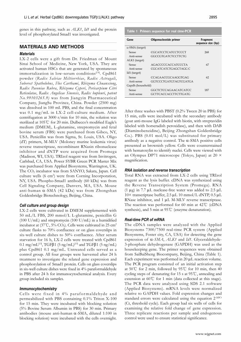
Li L et al . Herbal Cpd861 downregulates TGFβ1/ALK1 pathway 2895
www.wjgnet.com
genes in this pathway, such as ALK1, Id1 and the protein level of phosphorylated Smad1 was investigated.
MATERIALS AND METHODSMaterialsLX-2 cells were a gift from Dr. Friedman of Mount Sinai School of Medicine, New York, USA. They are activated human HSCs that are generated by spontaneous immortalization in low-serum conditions[14]. Cpd861 powder (Radix Salviae Miltior rhiae , Radix Astragali , Suberect Spatholobus, Flos Carthami, Rhizoma Chuanxiong, Radix Paeoniae Rubra, Rhizoma Cyperi, Pericarpium Citri Reticulatae, Radix Angelicae Sinensis, Radix bupleuri, patent No. 99103265.9) was from Jiangyin Pharmaceutical Company, JiangSu Province, China. Powder (2500 mg) was dissolved in 100 mL PBS, and the final concentration was 0.1 mg/mL in LX-2 cell-culture medium. After centrifugation at 3000 r/min for 10 min, the solution was sterilized at 105℃ for 20 min. Dulbecco’s modified Eagle’s medium (DMEM), L-glutamine, streptomycin and fetal bovine serum (FBS) were purchased from Gibco, NY, USA. Penicillin was from Sigma, St. Louis, USA. Oligo (dT) primers, M-MLV (Moloney murine leukemia virus) reverse transcriptase, recombinant RNasin ribonuclease inhibitor and dNTP were acquired from Promega (Madison, WI, USA). TRIzol reagent was from Invitrogen, Carlsbad, CA, USA. Power SYBR Green PCR Master Mix was purchased from Applied Biosystems, Warrington, UK. The CO2 incubator was from SANYO, Sakata, Japan. Cell culture wells (6 cm2) were from Corning Incorporation, NY, USA. Phospho-Smad1 antibody (60 kDa) was from Cell Signaling Company, Danvers, MA, USA. Mouse anti-human α-SMA (42 kDa) was from Zhongshan Goldenbridge Biotechnology, Beijing, China.
Cell culture and group designLX-2 cells were cultivated in DMEM supplemented with 50 mL/L FBS, 200 mmol/L L-glutamine, penicillin G (100 U/mL) and streptomycin (100 U/mL) in a humidified incubator at (37℃, 5% CO2). Cells were cultivated in 25 cm2 culture flasks to 70% confluence or on glass coverslips in six-well culture dishes to 50% confluence. After serum starvation for 16 h, LX-2 cells were treated with Cpd861 0.1 mg/mL[13], TGFβ1 (5 ng/mL)[15] and TGFβ1 (5 ng/mL) plus Cpd861 0.1 mg/mL. Untreated cells served as a control group. All four groups were harvested after 24 h treatment to investigate the related gene expression and phosphorylation of Smad1 protein. Cells on glass coverslips in six-well culture dishes were fixed in 4% paraformaldehyde in PBS after 24 h for immunocytochemical analysis. Every group included six samples.
ImmunocytochemistryCel l s were f i xed in 4% para for ma ldehyde and permeabilized with PBS containing 0.1% Triton X-100 for 15 min. They were incubated with blocking solution (5% Bovine Serum Albumin in PBS) for 30 min. Primary antibodies (mouse anti-human α-SMA, diluted 1:100 in blocking solution) were incubated with the cells overnight.
After three washes with PBST (0.2% Tween 20 in PBS) for 15 min, cells were incubated with the secondary antibody (goat anti-mouse IgG labeled with biotin, with streptavidin labeled with horseradish peroxidase), and then with DAB (Diaminobenzidine), Beijing Zhongshan Goldenbridge Co.). PBS (0.01 mol/L) was substituted for primary antibody as a negative control. The α-SMA positive cells presented as brownish yellow. Cells were counterstained with hematoxylin to identify nuclei. Cells were viewed with an Olympus DP71 microscope (Tokyo, Japan) at 20 × magnification.
RNA isolation and reverse transcriptionTotal RNA was extracted from LX-2 cells using TRIzol reagent as the lysis buffer. cDNA was synthesized using the Reverse Transcription System (Promega). RNA (1 μg) in 7.7 μL nuclease-free water was added to 2.5 μL 10× transcriptase buffer, 2.5 μL 10 mmol/L dNTP, 0.5 μL RNase inhibitor, and 1 μL M-MLV reverse transcriptase. The reaction was performed for 60 min at 42℃ (cDNA synthesis), and 5 min at 95℃ (enzyme denaturation).
Real-time PCR of mRNAThe cDNA samples were analyzed with the Applied Biosystems 7300/7500 real-time PCR system (Applied Biosystems, Foster city, CA, USA) for detecting the gene expression of α-SMA, ALK1 and Id1. Glyceraldehyde-3-phosphate dehydrogenase (GAPDH) was used as the housekeeping gene. The primer sequences were obtained from SaiBaiSheng Biocompany, Beijing, China (Table 1). Each experiment was performed in 20 μL reaction volume. The PCR program consisted of an initial activation step at 50℃ for 2 min, followed by 95℃ for 10 min, then 40 cycling steps of denaturing for 15 s at 95℃, annealing and extension at 60℃ for 1 min (data collected at this stage). The PCR data were analyzed using SDS 2.1 software (Applied Biosystems). mRNA levels were normalized relative to GAPDH values. Fold expression changes and standard errors were calculated using the equation 2-ΔΔCt
(Ct, threshold cycle). Each group had six wells of cells for examining the relative fold change of gene expression. Three replicate reactions per sample and endogenous control were used to ensure statistical significance.
Gene Oligonucleotide primer Fragment sequence size (bp)
α-SMA (target) Sense CGCATCCTCATCCTCCCT 268 Anti-sense GGCCGTGATCTCCTTCTGALK1 (target) Sense AGACCCCCACCATCCCTA 67 Anti-sense CGCATCATCTGAGCTAGG CId1 (target) Sense CCAGAACCGCAAGGTGAG 62 Anti-sense GGTCCCTGATGTAGTCGATGAGapdh (household) Sense GGCTCTCCAGAACATCATCC 187 Anti-sense GCTTCACCACCTTCTTGATG
Table 1 Primers sequence for real time-PCR

Western blot analysisCells were lysed on ice by 80 μL lysis buffer for 30 min. The cell lysate was centrifuged at 10 000 r/min for 10 min and the supernatant was collected for Western blot analysis. Protein concentration was measured using BCA Protein Assay kit (Pierce Company, Rockford, IL, USA) following the manufacturer’s instructions. Protein samples (40 μg/lane) were subjected to 10% SDS-PAGE and then transferred onto a PVDF nitrocellulose membrane by electro-blotting. The membrane was incubated in 25 mL blocking buffer for 1 h at room temperature. After blocking, the membrane was incubated in 10 mL blocking buffer containing 1:1000 dilution of rabbit anti-phospho-Smad1 or anti-SMA at 4℃ overnight. After gentle washing with blocking buffer, the membrane was incubated in
rabbit anti-β-actin polyclonal antibody (1:200 dilution). After vigorous washing, the membrane was incubated with a 1:5000 dilution of horseradish peroxidase-conjugated anti-rabbit IgG antibody. The intensity of the bands was determined by scanning video densitometry. The levels of phospho-Smad1 and SMA were normalized to the level of β-actin protein. Six independent experiments were performed. The intensity of the bands was determined using Gel-Pro Analyzer Version 3.0 (Media Cybernetics, Silver Spring, MD, USA).
Statistical analysisData were expressed as mean ± SD and the statistical significance was assessed by one-way analysis of variance followed by Student-Newman-Keul tests. P < 0.05 was considered to be significant.
RESULTSEffect of TGFβ1 and Cpd861 on α-SMA expressionChanges in gene or protein expression of α-SMA are often used to study the extent of HSC activation[16,17]. In this study, the effect of TGFβ1 and Cpd861 on the activation of the LX-2 cell line was investigated by examining changes in gene and protein expression of α-SMA.
Real-time PCR analysis showed that after exogenous TGFβ1 (5 ng/mL) stimulation for 24 h, α-SMA mRNA expression reached 2.65 fold (P < 0.05) compared with that in control LX-2 cells, whereas Cpd861 inhibited the expression to 0.38-fold (24 h, P < 0.05) (Figure 1A). α-SMA mRNA expression in Cpd861 and TGFβ1 co-treated cells was 1.73-fold compared to that in the controls.
Western blot analysis confirmed the trend of α-SMA gene expression changes (Figure 2A and B). In the TGFβ1 treatment group, α-SMA protein expression
ac
ALK1
mRN
A fo
ld c
hang
e
1.8
1.6
1.4
1.2
1.0
0.8
0.6
0.4
0.2
0.0Groups with different treatment
Control
Cpd861
TGFβ1
TGFβ1 + Cpd861
C
3.5
3.0
2.5
2.0
1.5
1.0
0.5
0.0
Id1 Control
Cpd861
TGFβ1
TGFβ1+Cpd861
a
a
cmRN
A fo
ld c
hang
e
Groups with different treatment
B
Groups with different treatment
a
a
c
α-SMA Control
Cpd861
TGFβ1
TGFβ1+Cpd861
mRN
A fo
ld c
hang
e
4.0
3.5
3.0
2.5
2.0
1.5
1.0
0.5
0.0
A
Figure 1 Effect of different treatment on α-SMA, Id1, ALK1 expression after 24 h. Cpd861 could decrease the level of all these genes compared with untreated LX-2 cells (aP < 0.05 vs control LX-2 cell group). Even with TGFβ1, Cpd861 could also exert its inhibitory roles in these genes expression (cP < 0.05 vs TGFβ1 treated group). Samples were obtained from 6 wells of cells for examining the relative fold change of α-SMA (A), Id1 (B), ALK1 (C) expression .Three replicate reactions for per sample. Error bars, SD.
B
Control Cpd861 TGFβ1 Cpd861+TGFβ1
0.6
0.5
0.4
0.3
0.2
0.1
0.0
Prot
ein
expr
essi
on
0.34
0.19
0.49
0.20a
a
a
A α-SMA 42 kDa
β-actin 43 kDa
Groups Control Cpd861 TGFβ1 Cpd861+TGFβ1
Figure 2 The level of α-SMA protein expression with different treatment after 24 h. Western blot was used as described in Materials and Methods. A: Representative Western blot results of α-SMA. The positions of protein size markers were given; B: Densitometry of Western blot analyzed by Gel-pro software. The levels of α-SMA were normalized to the level of β-actin protein. Six independent experiments were performed. aP < 0.05 vs control LX-2 cell group. Error bars, SD.
www.wjgnet.com
2896 ISSN 1007-9327 CN 14-1219/R World J Gastroenterol May 14, 2008 Volume 14 Number 18

increased significantly compared with that in the controls (photodensity: 0.49 in TGFβ1 group and 0.34 in control respectively), whereas its expression was inhibited by Cpd861 (0.19 vs 0.49), even in the presence of TGFβ1 (0.20 vs 0.34) (Figure 2).
Immunocy tochemica l ana l y s i s showed tha t
α-SMA protein was expressed in the cytoplasm, which was stained brown in low-power fields (Figure 3A). An interesting phenomenon was that after TGFβ1 treatment for 24 h, many various-sized, round spaces were present. Cells around the spaces were fusiform in shape, with dark brown cytoplasm. We presumed that it was a result of contraction of adjacent cells, which acquired the contractive property from excessive activation (Figure 3B). Cells treated with Cpd861, however, had a lighter brown cytoplasm (Figure 3C). In the Cpd861 and TGFβ1 co-treatment group, the cytoplasm presented lighter brown than in the TGFβ1-treated group and no spaces were seen (Figure 3D).
These results confirmed that gene and protein expression and function of α-SMA in LX-2 cells, markers of activation of LX-2 cells, were enhanced by TGFβ1 and inhibited by Cpd861. This is consisted with a previous study, which showed that Cpd861 can inhibit activation of LX-2 cells[13].
Effect of TGFβ1 and Cpd861 on TGFβ1/ALK1 signaling pathway in LX-2 cell lineTGFβ1/ALK1/Smad1 signaling has been found in HSCs[18,19]. In this pathway, ALK1 phosphorylated Smad1 leads to gene expression of Id1[20]. Idl proteins have a helix-loop-helix (HLH) domain but lack ability to bind DNA that acts as an important dominant negative antagonist of the bHLH family of transcription factors[21,22]. Recently, the Id1 gene was identified as a novel target gene that promotes the expression and polymerization of α-SMA, therefore involving the transdifferentiation of HSCs.
Here, we examined the level of phosphorylated Smad1 (the marker of activation of the pathway) using Western blot analysis. The level of phosphorylated Smad1 increased after TGFβ1 treatment and was reduced by Cpd861 or TGFβ1 and Cpd861 co-treatment (photodensity was 0.66 in control, 0.45 in Cpd861-treated cells, 0.84 in TGFβ1-treated cells, and 0.55 in TGFβ1 and Cpd861 co-treated cells, Figure 4).
Stimulation of LX-2 cells with exogenous TGFβ1 strongly induced Id1 gene expression by 2.5-fold, whereas Cpd861 reduced Id1 expression by 0.53-fold (P < 0.05). The fold change of Id1 expression in Cpd861 and TGFβ1 co-treatment was 0.79 compared with that in the controls, but it was significantly lower than that with TGFβ1 treatment alone (Figure 1B).
The ALK1 gene was constitutively expressed in LX-2 cells and TGFβ1 did not further increase its expression, but Cpd861 reduced it significantly (Figure 2C). The change in gene expression of the receptor might be an important inhibitory mechanism of Cpd861 in the pathway.
DISCUSSIONOur data indicated that the TGFβ1/ALK1/Smad1 pathway could be activated by TGFβ1 and abrogated by Cpd861 in LX-2 cells. The TGFβ1/ALK1/Smad1 pathway is one of the TGFβ superfamily signaling branches[23]. Several studies have indicated that activation of the
A
B
C
D
Figure 3 Expression of α-SMA in different treatment groups. Qualitative expression of α-SMA in control LX-2 cells (A), treated with TGFβ1 (B), Cpd861 (C) and Cpd861 together with TGFβ1 (D) for 24 h using immunohistochemical staining. α-SMA presented brown color in cytoplasm (× 200).
www.wjgnet.com
Li L et al . Herbal Cpd861 downregulates TGFβ1/ALK1 pathway 2897

pathway might play an important role in development of organ fibrosis[24-27]. Recent research has demonstrated that the existence of ALK1 and the TGFβ1/ALK1/Smad1 pathway is critical during the transdifferentiation process of primary rat HSCs to myofibroblast (MFB)[20]. The present study demonstrated that activation of the TGFβ1/ALK1/Smad1 pathway may be enhanced by TGFβ1 in LX-2 cells. It was shown that Cpd861 inhibited activation of the TGFβ1/ALK1/Smad1 pathway, and this might have been mediated by reducing ALK1 expression. However, effects of Cpd861 on ALK1 protein expression and on the affinity of TGFβ1 for ALK1 need further investigation.
Cpd861 has been demonstrated to be effective for treatment of patients with hepatic fibrosis[28]. Previous studies have shown that Cpd861 has multiple anti-fibrotic mechanisms, such as stimulating apoptosis of activated HSCs[29], down-regulating tissue inhibitor of metalloproteinase 1 gene expression[30], reducing expression of collagen and fibrosis-related cytokines. This study presented another possible anti-fibrotic mechanism of Cpd861, and also showed that the TGFβ1/ALK1/Smad1 pathway may represent a potential target for antifibrotic therapy. However, which components in the compound play the inhibitory roles needs to be further investigated.
ACKNOWLEDGMENTSWe thank Dr. Friedman and Mount Sinai, School of Medicine, for their help in providing the LX-2 cell line.
COMMENTSBackgroundActivation of hepatic stellate cells (HSCs) triggers fibrogenesis and TGFβ1/ALK1/
Smad1 signaling has been shown to be involved in the transdifferentiation of HSCs. Herbal compound 861 (Cpd861) can inhibit activation of HSCs to exert its anti-fibrotic effect, but the mechanism is not very clear.
Research frontiersThe effect of TGFβ1/ALK1/Smad1 signaling on the activation of HSCs has recently been reported. This study chose the pathway as a means to establishing the molecular mechanism of Cpd861 in the deactivation of LX-2 cells.
Innovations and breakthroughsThis is believed to be the first attempt to describe the molecular mechanism of the traditional herbal medicine Cpd861, which has been demonstrated to have anti-fibrotic effects and to reverse liver disease caused by HBV infection. This is believed to be the first time that TGFβ1/ALK1/Smad1 has been selected as a pathway to study the effects of deactivation of LX-2 HSCs.
ApplicationsThis study presents another possible anti-fibrotic mechanism of Cpd861 and shows that the TGFβ1/ALK1/Smad1 pathway may represent a potential target for the anti-fibrotic therapy.
TerminologyALK1 is activin receptor-like kinase 1, a type Ⅰ receptor, which is typically activated by the bone morphogenetic proteins, and then phosphorylates Smad1, Smad5 and Smad8 for signal transduction. Id1 is inhibitor of differentiation 1, which has an helix-loop-helix (HLH) domain but lacks a DNA-binding domain. It was first shown to act as a dominant negative antagonist of the basic HLH family of transcription factors, which positively regulate differentiation in many cell lineages. It is an important part of signaling pathways involved in development, cell cycle and tumorigenesis.
Peer reviewThis study demonstrates that Cpd861 can down-regulate the TGFβ1/ALK1 pathway, which results in the deactivation of HSCs. The study presents novel findings on the anti-fibrotic mechanism of Cpd861 and its possible application in anti-fibrotic therapy.
REFERENCES1 Gressner AM, Weiskirchen R. Modern pathogenetic concepts
of liver fibrosis suggest stellate cells and TGF-beta as major players and therapeutic targets. J Cell Mol Med 2006; 10: 76-99
2 Friedman SL. Molecular regulation of hepatic fibrosis, an integrated cellular response to tissue injury. J Biol Chem 2000; 275: 2247-2250
3 Iredale JP. Hepatic stellate cell behavior during resolution of liver injury. Semin Liver Dis 2001; 21: 427-436
4 Gressner AM, Weiskirchen R, Breitkopf K, Dooley S. Roles of TGF-beta in hepatic fibrosis. Front Biosci 2002; 7: d793-d807
5 Parsons CJ, Takashima M, Rippe RA. Molecular mechanisms of hepatic fibrogenesis. J Gastroenterol Hepatol 2007; 22 Suppl 1: S79-S84
6 Ouyang XS, Wang X, Ling MT, Wong HL, Tsao SW, Wong YC. Id-1 stimulates serum independent prostate cancer cell proliferation through inactivation of p16(INK4a)/pRB pathway. Carcinogenesis 2002; 23: 721-725
7 Lindert S, Wickert L, Sawitza I, Wiercinska E, Gressner AM, Dooley S, Breitkopf K. Transdifferentiation-dependent expression of alpha-SMA in hepatic stellate cells does not involve TGF-beta pathways leading to coinduction of collagen type I and thrombospondin-2. Matrix Biol 2005; 24: 198-207
8 Shen H, Fan J, Burczynski F, Minuk GY, Cattini P, Gong Y. Increased Smad1 expression and transcriptional activity enhances trans-differentiation of hepatic stellate cells. J Cell Physiol 2007; 212: 764-770
9 Jia JD, Bauer M, Cho JJ, Ruehl M, Milani S, Boigk G, Riecken EO, Schuppan D. Antifibrotic effect of silymarin in rat secondary biliary fibrosis is mediated by downregulation of procollagen alpha1(I) and TIMP-1. J Hepatol 2001; 35: 392-398
10 Kayano K, Sakaida I, Uchida K, Okita K. Inhibitory effects of
Phospho-Smad1 60 kDa
β-actin 43 kDa
Groups Control Cpd861 TGFβ1 Cpd861+TGFβ1
A
Control Cpd861 TGFβ1 Cpd861+TGFβ1
a
a
c
Prot
ein
expr
essi
on
1.2
1.0
0.8
0.6
0.4
0.2
0.0
B
Figure 4 The level of Phosphorylated Smad1 with different treatment after 24 h. Western blot was used as described. A: Representative Western blot results of Phosphorylated Smad1. The positions of protein size markers were given; B: Densitometry of Western-blot analyzed by Gel-pro software. The levels of Phospho-Smad1 were normalized to the level of β-actin protein. Six independent experiments were performed. aP < 0.05 vs untreated LX-2 cell group, cP < 0.05 vs TGFβ1 treated group. Error bars, SD.
www.wjgnet.com
2898 ISSN 1007-9327 CN 14-1219/R World J Gastroenterol May 14, 2008 Volume 14 Number 18
COMMENTS

the herbal medicine Sho-saiko-to (TJ-9) on cell proliferation and procollagen gene expressions in cultured rat hepatic stellate cells. J Hepatol 1998; 29: 642-649
11 Yin SS, Wang BE, Wang TL, Jia JD, Qian LX. [The effect of Cpd 861 on chronic hepatitis B related fibrosis and early cirrhosis: a randomized, double blind, placebo controlled clinical trial] Zhonghua Ganzangbing Zazhi 2004; 12: 467-470
12 Wang BE. Treatment of chronic liver diseases with traditional Chinese medicine. J Gastroenterol Hepatol 2000; 15 Suppl: E67-E70
13 Wang L, Wang J, Wang BE, Xiao PG, Qiao YJ, Tan XH. Effects of herbal compound 861 on human hepatic stellate cell proliferation and activation. World J Gastroenterol 2004; 10: 2831-2835
14 Xu L, Hui AY, Albanis E, Arthur MJ, O'Byrne SM, Blaner WS, Mukherjee P, Friedman SL, Eng FJ. Human hepatic stellate cell lines, LX-1 and LX-2: new tools for analysis of hepatic fibrosis. Gut 2005; 54: 142-151
15 Dooley S, Delvoux B, Lahme B, Mangasser-Stephan K, Gressner AM. Modulation of transforming growth factor beta response and signaling during transdifferentiation of rat hepatic stellate cells to myofibroblasts. Hepatology 2000; 31: 1094-1106
16 Abergel A, Sapin V, Dif N, Chassard C, Darcha C, Marcand-Sauvant J, Gaillard-Martinie B, Rock E, Dechelotte P, Sauvant P. Growth arrest and decrease of alpha-SMA and type I collagen expression by palmitic acid in the rat hepatic stellate cell line PAV-1. Dig Dis Sci 2006; 51: 986-995
17 Pan Q, Li DG, Lu HM, Wang YQ, Zhang WZ, Xu QF. A new immortalized rat cell line, hepatic stellate cell-PQ, exhibiting characteristics of hepatic stellate cell. Hepatobiliary Pancreat Dis Int 2005; 4: 281-284
18 Shen H, Huang GJ, Gong YW. Effect of transforming growth factor beta and bone morphogenetic proteins on rat hepatic stellate cell proliferation and trans-differentiation. World J Gastroenterol 2003; 9: 784-787
19 Shen H , Huang G, Hadi M, Choy P, Zhang M, Minuk GY, Chen Y, Gong Y. Transforming growth factor-beta1 downregulation of Smad1 gene expression in rat hepatic stellate cells. Am J Physiol Gastrointest Liver Physiol 2003; 285: G539-G546
20 Wiercinska E, Wickert L, Denecke B, Said HM, Hamzavi J,
Gressner AM, Thorikay M, ten Dijke P, Mertens PR, Breitkopf K, Dooley S. Id1 is a critical mediator in TGF-beta-induced transdifferentiation of rat hepatic stellate cells. Hepatology 2006; 43: 1032-1041
21 Ruzinova MB, Benezra R. Id proteins in development, cell cycle and cancer. Trends Cell Biol 2003; 13: 410-418
22 Damdinsuren B, Nagano H, Kondo M, Yamamoto H, Hiraoka N, Yamamoto T, Marubashi S, Miyamoto A, Umeshita K, Dono K, Nakamori S, Wakasa K, Sakon M, Monden M. Expression of Id proteins in human hepatocellular carcinoma: relevance to tumor dedifferentiation. Int J Oncol 2005; 26: 319-327
23 Miyazono K , Kusanagi K, Inoue H. Divergence and convergence of TGF-beta/BMP signaling. J Cell Physiol 2001; 187: 265-276
24 Moustakas A, Heldin CH. Non-Smad TGF-beta signals. J Cell Sci 2005; 118: 3573-3584
25 Abe H, Matsubara T, Iehara N, Nagai K, Takahashi T, Arai H, Kita T, Doi T. Type IV collagen is transcriptionally regulated by Smad1 under advanced glycation end product (AGE) stimulation. J Biol Chem 2004; 279: 14201-14206
26 Takahashi T, Abe H, Arai H, Matsubara T, Nagai K, Matsuura M, Iehara N, Yokode M, Nishikawa S, Kita T, Doi T. Activation of STAT3/Smad1 is a key signaling pathway for progression to glomerulosclerosis in experimental glomerulonephritis. J Biol Chem 2005; 280: 7100-7106
27 Matsubara T, Abe H, Arai H, Nagai K, Mima A, Kanamori H, Sumi E, Takahashi T, Matsuura M, Iehara N, Fukatsu A, Kita T, Doi T. Expression of Smad1 is directly associated with mesangial matrix expansion in rat diabetic nephropathy. Lab Invest 2006; 86: 357-368
28 Wang BE, Wang TL, Jia JD, Ma H, Duan ZP, Li XM, Li J, Wang AM, Qian LX. Experiment and clinical study on inhibition and reversion of liver fibrosis with integrated Chinese and Western Medicine. CJIM 1999; 5: 6-11
29 You H, Wang B, Wang T. [Proliferation and apoptosis of hepatic stellate cells and effects of compound 861 on liver fibrosis] Zhonghua Ganzangbing Zazhi 2000; 8: 78-80
30 Yin C , Ma H, Wang A, Ma X, Jia J, Wang B. [Effect of compound 861 on tissue inhibitor of metalloprotenase 1 gene expression of HSC-T6 cells] Zhonghua Ganzangbing Zazhi 2002; 10: 197-199
S- Editor Li DL L- Editor Kerr C E- Editor Yin DH
www.wjgnet.com
Li L et al . Herbal Cpd861 downregulates TGFβ1/ALK1 pathway 2899

Online Submissions: wjg.wjgnet.com World J Gastroenterol 2008 May 14; 14(18): 2900-2904www.wjgnet.com World Journal of Gastroenterology ISSN [email protected] © 2008 WJG. All rights reserved.
Primary study of leptin and human hepatocellular carcinoma in vitro
Jing Zhou, Wei Lei, Lei Shen, He-Sheng Luo, Zhi-Xiang Shen
www.wjgnet.com
RAPID COMMUNICATION
Jing Zhou, Lei Shen, He-Sheng Luo, Zhi-Xiang Shen, Department of Gastroenterology, Renmin Hospital of Wuhan University, Wuhan 430060, Hubei Province, ChinaWei Lei, Department of Urology, Wuhan Children Hospital, Wuhan 430016, Hubei Province, ChinaAuthor contributions: Zhou J, Shen L, Luo HS, and Shen ZX designed research; Zhou J and Lei W performed research; Zhou J and Lei W analyzed data; and Zhou J, Lei W and Shen ZX wrote the paper.Correspondence to: Zhi-Xiang Shen, Department of Gastroenterology, Renmin Hospital of Wuhan University, No. 238 Jiefang Road, Wuchang District, Wuhan 430060, Hubei Province, China. [email protected]: +86-27-62530538 Fax: +86-27-88042292Received: January 9, 2008 Revised: March 21, 2008
AbstractAIM: To investigate the expression level and effects of leptin in human hepatocellular carcinoma cells in vitro and to explore the correlation between them.
METHODS: Human hepatocellular carcinoma cell line HepG2 was cultured in vitro , and (the expression level) mRNA of leptin and leptin receptors in HepG2 were assessed using reverse transcription polymerase chain reaction (RT-PCR). Effects of different concentrations of leptin (50 ng/mL, 100 ng/mL, 200 ng/mL) on HepG2 were detected with colorimetric assay by 3-(4,5-dimethylthiazol-2-yl)-2,5-diphenyltetrazolium bromide (MTT) after incubation periods of 24 h, 48 h, and 72 h. Flow cytometry was performed to assess cell cycle progression of different concentrations of leptin as stated above after each 24 h incubation period.
RESULTS: mRNA of leptin and leptin receptors (including short and long isoforms) were expressed in HepG2. The 72 h incubation of leptin at different concentrations (50 ng/mL, 100 ng/mL, 200 ng/mL) promoted proliferation of HepG2 in a concentration- and time-dependent manner. The experimental group shows significant statistical differences when compared to the controlled group which contained 0 ng/mL of leptin. As the concentration of leptin increases, significant fewer cells were detected in G0-G1 phase and more cells in S and G2-M phases.
CONCLUSION: Lept in and lept in receptor are simultaneously expressed in human hepatocellular carcinoma cell line HepG2. Addition of leptin (0 ng/mL-
200 ng/mL) in 72 h periods indicated there is a concentration- and time-dependent correlation in the stimulation of HepG2 cell proliferation. The effect of proliferation by leptin is due to promotion of DNA synthesis and enhancement of mitotic activity. The relationship between leptin and human hepatocellular carcinoma cells might indicate that adipokine could be associated with the progression of human hepatocellular carcinoma.
© 2008 WJG. All rights reserved.
Key words: Lept in ; Hepatoce l lu la r ca rc inoma; Proliferation
Peer reviewers: Frank J Burczynski, Faculty of Pharmacy, University of Manitoba, 50 Sifton Road, Winnipeg, Manitoba, R3T 2N2, Canada; Yukihiro Shimizu, Dr, Kyoto Katsura Hospital, 17 Yamada-Hirao, Nishikyo, Kyoto 615-8256, Japan; Byung Chul Yoo, Professor, Dr, Division of Gastroenterology, Samsung Medical Center, SungKyunKwan University School of Medicine, Seoul 135-710, Korea
Zhou J, Lei W, Shen L, Luo HS, Shen ZX. Primary study of leptin and human hepatocellular carcinoma in vitro. World J Gastroenterol 2008; 14(18): 2900-2904 Available from: URL: http://www.wjgnet.com/1007-9327/14/2900.asp DOI: http://dx.doi.org/10.3748/wjg.14.2900
INTRODUCTIONLeptin, a protein product encoded by the obese gene (ob gene)[1], is primarily derived from adipocytes which plays an important role in the regulation of food intake and the control of body weight[2]. It takes part in various physical conditions such as lipid metabolism, immune defense, neuroendocrine regulation, pituitary hormone secretion, and pubertal development[3,4]. In rodents and humans, leptin is related with many pathological syndromes including obesity, hyperphagia, hyperinsulinemia, reduced fertility, and cholelithiasis (including gallstone and hepatolithiasis)[5-8]. Studies have shown that individuals who are obese have increased risk for most cancers compared to individuals who are not obese[9]. Since leptin is closely associated with obesity, it may be the bridge conjoining obesity and cancer.
The biological functions of leptin on target cells and tissues are carried out through interaction with its specific receptors (ob-R) which belong to class Ⅰ cytokine receptor

Zhou J et al . Leptin and Hepatocellular Carcinoma 2901
www.wjgnet.com
family[10]. In rodents and humans, two leptin receptor isoforms predominate: the short leptin receptor isoform (ob-Ra) and the long leptin receptor isoform (ob-Rb)[11,12]. They share the same extracellular domain, but they differ in the length of the transmembrane/cytoplasmic coding regions[13]. The physiologic significance of each isoform in relationship to obesity and cancer is still unknown.
Hepatocellular carcinoma (HCC) is one of the most malignant tumors in the world. It causes proximately one hundred and ten thousand deaths annually in China. HCC is extremely difficult to detect in prognosis due to its early metastasis and distant transmission. It has been reported that obesity was related to HCC complicated with cryptogenic cirrhosis and alcoholic liver disease[14,15]. Therefore, we presume that leptin might be associated with HCC. In order to investigate the relationship between leptin and HCC, (the expression level) mRNA of leptin and its receptors in human HCC cell line HepG2 were detected by reverse transcription polymerase chain reaction (RT-PCR). In addition, the effects of leptin on HepG2 in vitro were also discussed in this study.
MATERIALS AND METHODSMaterialsHuman hepatocellular carcinoma cell line HepG2 was obtained from Shanghai Cell Biology Institute of Chinese Academy of Sciences. Human recombinant leptin was the product from Sigma (St. Louis, Missouri, USA). Roswell Park Memorial Institute-1640 (RPMI-1640), fetal bovine serum (FBS) and Trizol reagent were the products of Invitrogen (Carlsbad, California, USA). Trypsin, 3-(4,5-dimethylthiazol-2-yl)-2,5-diphenyltetrazolium bromide (MTT), ethylene diamine tetraacetic acid (EDTA) and dimethyl sulfoxide (DMSO) were purchased from Amresco (Cleveland, Ohio, USA). Revert aid first strand cDNA synthesis kit and TaqDNA polymerase were the products of Fermentas (Burlington, Iowa, USA). All other reagents unless indicated were from Sigma (St. Louis, Missouri, USA).
MethodsCell culture: HepG2 were grown as a monolayer in RPMI-1640 medium supplemented with 10% heat-inactivated FBS, 100 kU/L penicillin, and 0.1 g/L streptomycin. They were cultured in T-75 cm2 culture flasks and maintained at 37℃ in 5% CO2 humidified atmosphere. At the beginning of the experiment, cells in the exponential growth phase were detached from the flask with 0.25% trypsin and 0.02% EDTA solution.
RT-PCR: mRNA of leptin, ob-Ra and ob-Rb in cell line HepG2 were assessed using RT-PCR as described previously[16]. Total RNA was isolated from confluent cells using Trizol reagent according to the manufacture’s instructions. The concentration of RNA was quantitated by absorbance change at 260 nm and 280 nm. Total RNA was suspended in DEPC-treated water and stored at -80℃.
Single-stranded cDNA was synthesized from 1 μg of RNA using revert aid first strand cDNA synthesis kit
with random hexamer primer. The housekeeping gene β-actin was amplified to set a control for RNA loading and also to minimize variations in cDNA synthesis to ensure efficiency. Primer sequences for β-actin, leptin, ob-Ra and ob-Rb have been reported in the previous paper[17]. Following are the primers for: 1) β-actin: sense strand is 5’-ACCCACACTGTGCCCATCTA-3’ and anti-sense strand is 5’-CGGAACCGCTCATTGCC-3’ (encodes a 289-bp fragment); 2) leptin: sense strand is 5’-GTGCGGATTCTTGTGGCTTT-3’ and anti-sense strand is 5’-GGAATGAAGTCCAAACCGGTG-3’ (encodes a 174-bp fragment); 3) ob-Ra: sense strand is 5’-TTGTGCCAGTAATTATTTCCTCTT-3’ and anti-sense strand is 5’-AGTTGGCACATTGGGTTCAT-3’ (encodes a 200-bp fragment); 4) ob-Rb: the sense strand is 5’-TTGTGCCAGTAATTATTTCCTCTT-3’ and antisense strand is 5’-CTGATCAGCGTGGCGTATTT-3’ (encodes a 439-bp fragment).
Amplification of the resulting cDNA sequence was carried out using polymerase chain reaction (PCR). 3 µL cDNA was combined with 25 pmol oligonucleotide primers specific for β-actin, leptin, ob-Ra, ob-Rb and TaqDNA polymerase 1 U, PCR buffer (5 μL), 25 mmol/L MgCl2 (4 μL), 10 mmol/L dNTP mixture (1 μL), and ddH2O (34 μL in a 50 μL solution). PCR was performed in a thermal cycler (Gene Amp PCR system 2400, Perkin-Elmer Corp., Uberlingen, Germany). The condition for the reaction were the following: 1 min at 94℃ (predenaturation), followed by 30 cycles of 1 min (denaturation) at 52℃ (for β-actin), 55℃ (for leptin) or 50℃ (for ob-Ra and ob-Rb); 1 min at 72℃ (annealing), and then 5 min at 72℃ (extension). PCR products were electrophoresed on 2% agarose gels stained with ethidium bromide, and the length were estimated to be 100-bp. Imaging was performed on JS-380 instrument (Peiqing Inc., Shanghai, China).
MTT for cell proliferation: MTT colorimetric assay was used to detect the effects of leptin on proliferation of cell line HepG2. Cells were washed extensively with filtered and sterilized phosphate-buffered saline (PBS) to remove the dead cells. HepG2 cells were suspended at a concentration of 1.0 × 10⁴/mL, and then they were seeded into 96-well microplate at 150 μL/well and incubated to adhere overnight. RPMI-1640 medium containing different concentrations of human recombinant leptin were added in 0 ng/mL, 50 ng/mL, 100 ng/mL and 200 ng/mL to HepG2 cells with each concentration account for 6 wells. MTT assay was performed after incubation periods of 24 h, 48 h, and 72 h. MTT 20 μL (5 mg/mL) stock solution in PBS was added to each well. The microplate was incubated for 4 h and 100 μL DMSO was added (to each well). Optical density (A) value was measured by an ELISA plate reader (Digiscan Lab instruments, Austria) at wavelength of 492 nm. Each variant group was performed in triplicate wells for measurement accuracy.
Flow cytometric analysis: HepG2 cells in the exponential growth phase were detached by 0.25% trypsin and 0.02% EDTA. The cell suspension was then seeded uniformly

into four T-75 cm2 culture flasks and incubated to adhere overnight. RPMI-1640 medium containing different concentrations of human recombinant leptin were added in 0 ng/mL, 50 ng/mL, 100 ng/mL, and 200 ng/mL to HepG2 cells. Flow cytometric analysis was performed after incubated for 24 h. The cells were briefly washed twice with PBS, fixed in ice-cold ethanol (70% vol/vol in ddH2O) and stained with propidium iodide (PI) solution (25 μg/mL PI, 180 U/ml RNase, 0.15% Triton X-100, and 30 mg/mL polyethylene glycol in 4 mmol/L critrate buffer w/pH = 7.8). DNA contents were detected using a FACScan flow cytometer (Becton Dickinson Co., San Jose, CA). The relative percentages of each phase of the cell cycle were analyzed using Cell Quest Software (Becton Dickinson Co., San Jose, CA). For the measurement accuracy, each variant group was tested in triplicate.
Statistical analysisThe data were presented as mean ± SD. ANOVA was used to analysis the results. P < 0.05 were considered to indicate statistically significant differences. Entire data were analyzed with the statistical software SPSS 11.5.
RESULTSLeptin, ob-Ra and ob-Rb mRNA assay by RT-PCRTotal RNA extracted from HepG2 cells were reversely transcribed to check whether mRNA of leptin, ob-Ra and ob-Rb were expressed. RT-PCR detection with β-actin primers revealed a 289-bp fragment in HepG2. The 174-bp cDNA band indicated that the cells expressed leptin mRNA. PCR products for both the short isoform (ob-Ra, 200-bp) and the long isoform (ob-Rb, 439-bp) of leptin receptor were also detectable. No fragment was detected in the negative control that PCR product without the cDNA (Figure 1).
Effects of proliferation of leptin on HepG2MTT assay indicated the cell proliferate conditions of HepG2. As shown in Table 1, leptin significantly stimulated HepG2 cells growth in a concentration- and time-dependent manner. Leptin caused significant growth potency on HepG2 within 72 h. There were significant statistical differences between concentration groups of 0 ng/mL vs 50 ng/mL (P < 0.01), 0 ng/mL vs 100 ng/mL
(P < 0.01), 0 ng/mL vs 200 ng/mL (P < 0.01), 50 ng/mL vs 100 ng/mL (P < 0.05) and 50 ng/mL vs 200 ng/mL (P < 0.01). Meanwhile, significant statistical differences also existed between duration groups of 24 h vs 48 h (P < 0.01) and 24 h vs 72 h (P < 0.01).
Effects of leptin on cell cycle progressions of HepG2The effects of leptin on cell cycle progressions of HepG2 as determined by flow cytometry analysis are shown in Table 2. In the 24 h frame, the results indicate that as the concentration of leptin increases (e.g. 50 ng/mL, 100 ng/mL and 200 ng/mL), the proportion of the HepG2 cells in G0-G1 phase gradually reduces and the number of cells in S and G2-M phases gradually increases. In comparison with control group, the data indicates a significant reduction of cells in G0-G1 phase (P < 0.01) and a significant increase of cells in S phase (P < 0.01) and G2-M phase (P < 0.01) in response to treatment of leptin.
DISSUSIONThe relationship between obesity and cancer has been excessively documented for cancers or adenocarcinoma in endometrium, breast, prostate, renal cells, pancreas, colon, and esophagus[18,19]. Epidemiological observations revealed that obesity was a risk factor of HCC complicated with cryptogenic cirrhosis and alcoholic liver disease[14,15], and in addition, clinical investigations identified that the leptin level of serum had increased significantly in alcoholic and post-hepatitis liver cirrhosis patients with or without HCC as compared to control subjects w/o the complication[20-24]. Still, there were few studies on the involvement of leptin in HCC.
It has been reported that leptin participates in an auto/paracrine manner in the pituitary gland, and it plays regulations roles in the pig[25]. One study reported that the proteins of leptin and ob-R had been detected in 72.22% and 30.56% of HCC, respectively[26]. Present study
600 500 400 300 200
100
M A B C D E
Figure 1 Expression of β-actin, leptin, ob-Ra and ob-Rb in HepG2. M: DNA marker; A: β-actin 289 bp; B: Leptin 174 bp; C: Ob-Ra 200 bp; D: Ob-Rb 439 bp; E: Negative control.
Table 1 Effect of leptin on proliferation of HepG2
Concentration A(ng/mL) 24 h 48 hd 72 hd
0 0.29 ± 0.01 0.34 ± 0.01 0.37 ± 0.01 50b 0.34 ± 0.02 0.42 ± 0.01 0.54 ± 0.02100b 0.41 ± 0.03 0.53 ± 0.05 0.62 ± 0.02200b 0.46 ± 0.04 0.63 ± 0.01 0.73 ± 0.03
bP < 0.01 vs group of 0 ng/mL leptin; dP < 0.01 vs group of incubation 24 h.
Table 2 Effect of leptin on cell cycle of HepG2
Concentration Cell phase (%)(ng/mL) G0-G1 S G2-M
0 61.8 ± 2.5 36.5 ± 1.0 1.6 ± 0.3 50 56.9 ± 2.2 38.5 ± 0.9 4.7 ± 1.4100 50.2 ± 2.6a 40.3 ± 1.2 9.4 ± 2.1a
200 43.6 ± 2.7b 42.0 ± 0.3a 14.4 ± 2.5a
aP < 0.05, bP < 0.01 vs group of 0 ng/mL leptin.
www.wjgnet.com
2902 ISSN 1007-9327 CN 14-1219/R World J Gastroenterol May 14, 2008 Volume 14 Number 18
bp

indicated that the mRNA of leptin, and as well as the short and the long leptin receptor isoforms were all expressed in HepG2. These findings suggested that leptin might act as an auto/paracrine growth factor towards hepatocytes, and together with its receptors, they could play role in HCC initiation and progression.
Serum leptin level fluctuations are also detected in some non-physiological conditions. For example, leptin levels up to 400 ng/mL have been reported in children with chronic renal failure[27]. When obese, but otherwise healthy subjects treated with leptin (1 mg/kg per day), serum leptin levels rose up to 736 ng/mL[28]. However, serum leptin level in normal physiological situation is less than 11.4 ng/mL. Thus, a relatively higher concentration of leptin (200 ng/mL) was used in our study. There are some divaricate results among the studies of leptin and cancer. Leptin not only stimulated proliferation of some human cancer cell lines, including breast cancer cell lines (ZR75-1, MCF-7), esophageal cancer cell lines (KYSE 410) and prostate cancer cell lines (PC-3, DU 145), but also it inhibited the growth of other human cancer cell lines, such as pancreatic cancer cell lines (Mia-Paca, PANC-1)[18]. Data regarding leptin’s effect on HCC cells are rarely reported, and they appear to be contradictory. In one in vitro study, leptin was found to have little effect on the proliferation of liver cancer cell line SMMC-7721[26], while in another study, leptin had shown anti-tumor activity[29]. In our in vitro study, exogenous leptin had effects on the proliferation of HCC cell line HepG2 in a concentration- and time-dependent manner. We might conclude that human cancer cell lines exhibit differential responses to the leptin treatments, depending upon the biological characteristics of the cancer cells and the organ of derivation of the cell lines. Because our study was to interfere HCC cell line with exogenous leptin in vitro, results may differ in concentration threshold and activation initiation time with in vivo HCC cells in tumor tissues with endogenous leptin. Therefore, it will be necessary to continue the research on leptin’s effects on HCC with targeted animal models.
By adopting the techniques of flow cytometry, we were able to significantly expand the analysis of cell cycle progression. With respect to the cell cycle, we were able to accurately determine the relative proportion of cells in each phase of the cycle progression. From our data, it was evident that as the concentration of exogenous leptin increased from 0 to 200 ng/mL, it gradually reduced the relative proportion of the HepG2 cells in G0-G1 phase and gradually increased the number of the cells in S and G2-M phases. Since the S phase of the cell cycle is the synthetic phase of DNA, and G2-M phase represents a later synthetic phase of DNA and the splitting of the cells through mitotic phase, data from our study demonstrate that leptin increase cell proliferation of HepG2 by promoting of DNA synthesis and enhancing mitotic activity.
It has been reported that leptin replacement is a very promising therapeutic approach for managing the complications of lipodystrophy. In addition, leptin may have therapeutic potential in the treatment of epilepsy[30,31]. The primary finding of our study on leptin and HCC cell line indicates that adipokine could be associated with
the progression of human hepatocellular carcinoma. Administration of the leptin antagonists or receptors in the targeted cells may open a new way in HCC prevention and treatment. Further studies are warranted and ongoing.
COMMENTSBackground Leptin is the protein product encoded by obese gene, which plays an important role in the regulation of food intake, and the control of body weight. It is related with many pathological syndrome including obesity, hyperphagia, hyperinsulinemia, reduced fertility, and cholelithiasis. There are considerable researches about the effects of leptin.
Research frontiers Recently, the researches of relationships between leptin and tumors are hotspots. But there are few studies on the involvement of leptin in human hepatocellular carcinoma.
Innovations and breakthroughs In this study, we found mRNA of leptin and leptin receptors (including short and long isoforms) were expressed in human hepatocellular carcinoma cell line HepG2. Leptin (0 ng/mL-200 ng/mL) in 72 h could stimulate proliferation of HepG2 in vitro, and that effect was due to promotion of DNA synthesis and enhancement of mitotic activity.
ApplicationsThe primary study of leptin and HepG2 may indicate that adipokine could be associated with the progression of human hepatocellular carcinoma, and thus may offer new aim for further understanding of the biological function of leptin and new therapeutic targets.
Peer reviewThe manuscript written by Zhou J et al describes a possible role of leptin in hepatocarcinogenesis and promotion of the disease. The data are interesting and potentially important. It’s recommended the additional experiments using another HCC cell lines which could make this study’s conclusions more confidential.
REFERENCES1 Zhang Y , Proenca R, Maffei M, Barone M, Leopold L,
Friedman JM. Positional cloning of the mouse obese gene and its human homologue. Nature 1994; 372: 425-432
2 Sierra-Honigmann MR, Nath AK, Murakami C, Garcia-Cardena G, Papapetropoulos A, Sessa WC, Madge LA, Schechner JS, Schwabb MB, Polverini PJ, Flores-Riveros JR. Biological action of leptin as an angiogenic factor. Science 1998; 281: 1683-1686
3 Janeckova R. The role of leptin in human physiology and pathophysiology. Physiol Res 2001; 50: 443-459
4 Shimizu F, Matsuzaki T, Iwasa T, Tanaka N, Minakuchi M, Kuwahara A, Yasui T, Furumoto H, Irahara M. Transition of leptin receptor expression during pubertal development in female rat pituitary. Endocr J 2008; 55: 191-198
5 Montague CT, Farooqi IS, Whitehead JP, Soos MA, Rau H, Wareham NJ, Sewter CP, Digby JE, Mohammed SN, Hurst JA, Cheetham CH, Earley AR, Barnett AH, Prins JB, O'Rahilly S. Congenital leptin deficiency is associated with severe early-onset obesity in humans. Nature 1997; 387: 903-908
6 Lee GH, Proenca R, Montez JM, Carroll KM, Darvishzadeh JG, Lee JI, Friedman JM. Abnormal splicing of the leptin receptor in diabetic mice. Nature 1996; 379: 632-635
7 Clement K, Vaisse C, Lahlou N, Cabrol S, Pelloux V, Cassuto D, Gourmelen M, Dina C, Chambaz J, Lacorte JM, Basdevant A, Bougneres P, Lebouc Y, Froguel P, Guy-Grand B. A mutation in the human leptin receptor gene causes obesity and pituitary dysfunction. Nature 1998; 392: 398-401
8 Lei ZM, Ye MX, Fu WG, Chen Y, Fang C, Li J. Levels of serum leptin, cholecystokinin, plasma lipid and lipoprotein
www.wjgnet.com
Zhou J et al . Leptin and Hepatocellular Carcinoma 2903
COMMENTS

differ between patients with gallstone or/and those with hepatolithiasis. Hepatobiliary Pancreat Dis Int 2008; 7: 65-69
9 Calle EE, Rodriguez C, Walker-Thurmond K, Thun MJ. Overweight, obesity, and mortality from cancer in a prospectively studied cohort of U.S. adults. N Engl J Med 2003; 348: 1625-1638
10 Tartaglia LA, Dembski M, Weng X, Deng N, Culpepper J, Devos R, Richards GJ, Campfield LA, Clark FT, Deeds J, Muir C, Sanker S, Moriarty A, Moore KJ, Smutko JS, Mays GG, Wool EA, Monroe CA, Tepper RI. Identification and expression cloning of a leptin receptor, OB-R. Cell 1995; 83: 1263-1271
11 Fei H, Okano HJ, Li C, Lee GH, Zhao C, Darnell R, Friedman JM. Anatomic localization of alternatively spliced leptin receptors (Ob-R) in mouse brain and other tissues. Proc Natl Acad Sci USA 1997; 94: 7001-7005
12 Friedman JM. Leptin, leptin receptors, and the control of body weight. Nutr Rev 1998; 56: s38-s46; discussion s54-s75
13 Ahima RS, Flier JS. Leptin. Annu Rev Physiol 2000; 62: 413-43714 Nair S, Mason A, Eason J, Loss G, Perrillo RP. Is obesity
an independent risk factor for hepatocellular carcinoma in cirrhosis? Hepatology 2002; 36: 150-155
15 Roth MJ, Baer DJ, Albert PS, Castonguay TW, Dorgan JF, Dawsey SM, Brown ED, Hartman TJ, Campbell WS, Giffen CA, Judd JT, Taylor PR. Relationship between serum leptin levels and alcohol consumption in a controlled feeding and alcohol ingestion study. J Natl Cancer Inst 2003; 95: 1722-1725
16 Zarkesh-Esfahani H, Pockley G, Metcalfe RA, Bidlingmaier M, Wu Z, Ajami A, Weetman AP, Strasburger CJ, Ross RJ. High-dose leptin activates human leukocytes via receptor expression on monocytes. J Immunol 2001; 167: 4593-4599
17 Yuan SS, Chung YF, Chen HW, Tsai KB, Chang HL, Huang CH, Su JH. Aberrant expression and possible involvement of the leptin receptor in bladder cancer. Urology 2004; 63: 408-413
18 Somasundar P , Yu AK, Vona-Davis L, McFadden DW. Differential effects of leptin on cancer in vitro. J Surg Res 2003; 113: 50-55
19 Somasundar P, Riggs D, Jackson B, Vona-Davis L, McFadden DW. Leptin stimulates esophageal adenocarcinoma growth by nonapoptotic mechanisms. Am J Surg 2003; 186: 575-578
20 Wang YY , Lin SY. Leptin in relation to hepatocellular carcinoma in patients with liver cirrhosis. Horm Res 2003; 60: 185-190
21 Lin SY , Wang YY, Sheu WH. Increased serum leptin concentrations correlate with soluble tumour necrosis factor receptor levels in patients with cirrhosis. Clin Endocrinol (Oxf) 2002; 57: 805-811
22 Testa R, Franceschini R, Giannini E, Cataldi A, Botta F, Fasoli A, Tenerelli P, Rolandi E, Barreca T. Serum leptin levels in patients with viral chronic hepatitis or liver cirrhosis. J Hepatol 2000; 33: 33-37
23 Comlekci A, Akpinar H, Yesil S, Okan I, Ellidokuz E, Okan A, Ersoz G, Tankurt E, Batur Y. Serum leptin levels in patients with liver cirrhosis and chronic viral hepatitis. Scand J Gastroenterol 2003; 38: 779-786
24 Greco AV, Mingrone G, Favuzzi A, Capristo E, Gniuli D, Addolorato G, Brunani A, Cavagnin F, Gasbarrini G. Serum leptin levels in post-hepatitis liver cirrhosis. J Hepatol 2000; 33: 38-42
25 Siawrys G, Kaminski T, Smolinska N, Przala J. Expression of leptin and long form of leptin receptor genes and proteins in pituitary of cyclic and pregnant pigs. J Physiol Pharmacol 2007; 58: 845-857
26 Wang XJ, Yuan SL, Lu Q, Lu YR, Zhang J, Liu Y, Wang WD. Potential involvement of leptin in carcinogenesis of hepatocellular carcinoma. World J Gastroenterol 2004; 10: 2478-2481
27 Daschner M, Tonshoff B, Blum WF, Englaro P, Wingen AM, Schaefer F, Wuhl E, Rascher W, Mehls O. Inappropriate elevation of serum leptin levels in children with chronic renal failure. European Study Group for Nutritional Treatment of Chronic Renal Failure in Childhood. J Am Soc Nephrol 1998; 9: 1074-1079
28 Fujioka K, Patane J, Lubina J, Lau D. CSF leptin levels after exogenous administration of recombinant methionyl human leptin. JAMA 1999; 282: 1517-1518
29 Elinav E, Abd-Elnabi A, Pappo O, Bernstein I, Klein A, Engelhardt D, Rabbani E, Ilan Y. Suppression of hepatocellular carcinoma growth in mice via leptin, is associated with inhibition of tumor cell growth and natural killer cell activation. J Hepatol 2006; 44: 529-536
30 Asterholm IW, Halberg N, Scherer PE. Mouse Models of Lipodystrophy Key reagents for the understanding of the metabolic syndrome. Drug Discov Today Dis Models 2007; 4: 17-24
31 Diano S, Horvath TL. Anticonvulsant effects of leptin in epilepsy. J Clin Invest 2008; 118: 26-28
S- Editor Li DL L- Editor Li M E- Editor Lu W
www.wjgnet.com
2904 ISSN 1007-9327 CN 14-1219/R World J Gastroenterol May 14, 2008 Volume 14 Number 18
Online Submissions: wjg.wjgnet.com World J Gastroenterol 2008 May 14; 14(18): 2905-2911www.wjgnet.com World Journal of Gastroenterology ISSN [email protected] © 2008 WJG. All rights reserved.
Rosiglitazone prevents murine hepatic fibrosis induced by Schistosoma japonicum
Hui Chen, Yong-Wen He, Wen-Qi Liu, Jing-Hui Zhang
RAPID COMMUNICATION
Hui Chen, Yong-Wen He, Department of Infectious Disease, Union Hospital, Tongji Medical College, Huazhong University of Science and Technology, Wuhan 430022, Hubei Province, ChinaWen-Qi Liu, Department of Parasitology, Tongji Medical College, Huazhong University of Science and Technology, Wuhan 430022, Hubei Province, ChinaJing-Hui Zhang, Department of Surgical Laboratory, Union Hospital, Tongji Medical College, Huazhong University of Science and Technology, Wuhan 430022, Hubei Province, ChinaAuthor contributions: Chen H contributed chiefly to this work; Chen H designed and performed the research and wrote the paper; Chen H, Liu WQ and Zhang JH performed the research; He YW gave some good suggestions.Correspondence to: Hui Chen, Department of Infectious Disease, Union Hospital, Tongji Medical college, Huazhong University of Science and Technology, 1277# Jiefang Road, Wuhan 430022, Hubei Province, China. [email protected]: +86-27-85726132 Fax: +86-27-85727851Received: January 25, 2008 Revised: March 8, 2008
AbstractAIM: To evaluate the effect of rosiglitazone in a murine model of liver fibrosis induced by Schistosoma japonicum infection.
METHODS: A total of 50 mice were randomly and averagely divided into groups A, B, C, D and E. The mice in group A served as normal controls, while those in the other four groups were infected with Schistosoma japonicum to induce the model of liver fibrosis. Besides, the mice in groups C, D and E were treated with praziquantel, rosiglitazone and praziquantel plus rosiglitazone, respectively. NF-κB binding activity and expression of PPARγ-mRNA were determined by Western blot assay and real-time quantitative PCR. Radioimmunoassay technique was used to detect the serum content changes of TNF-α and IL-6. Histological specimens were stained with HE. Expression of TGF-β1, a-smooth muscle actin and type Ⅰ and type Ⅲ collagen was detected by immunohistochemistry and multimedia color pathographic analysis system.
RESULTS: Inflammation and fibrosis in the rosiglitazone plus praziquantel treatment group (group E) were lightest among the mice infected with Schistosoma (P < 0.05). To further explore the mechanism of rosiglitazone action, we found that rosiglitazone can significantly increase the expression of PPARγ [E: -18.212 ± (-3.909) vs B: -27.315 ± (-6.348) and C: -25.647 ± (-5.694), P < 0.05],
reduce the NF-κB binding activity (E: 88.89 ± 19.34 vs B: 141.11 ± 15.37, C: 112.89 ± 20.17 and D: 108.89 ± 20.47, P < 0.05), and lower the serum level of TNF-α (E: 1.613 ± 0.420 ng/mL vs B: 2.892 ± 0.587 ng/mL, C: 2.346 ± 0.371 ng/mL and D: 2.160 ± 0.395 ng/mL, P < 0.05) and IL-6 (E: 0.106 ± 0.021 ng/mL vs B: 0.140 ± 0.031 ng/mL and C: 0.137 ± 0.027 ng/mL, P < 0.05) in mice with liver fibrosis. Rosiglitazone can also substantially reduce the hepatic expression of TGF-β1, α-SMA type Ⅰ and type Ⅲ collagen in mice with liver fibrosis.
CONCLUSION: The activation of PPARγ by its ligand can retard liver fibrosis and suggest the use of rosiglitazone for the treatment of liver fibrosis due to Schistosoma japonicum infection.
© 2008 WJG . All rights reserved.
Key words: Peroxisome proliferators-activated receptorγ; Rosiglitazone; Liver fibrosis; Schistosomiasis; Hepatic stellate cell
Peer reviewer: Ana Cristina Simões e Silva, Professor, Pediatrics Department, Federal University of Minas Gerais Institution, Avenida Professor Alfredo Balena, 190, Belo Horizonte 30130-100, Brazil
Chen H, He YW, Liu WQ, Zhang JH. Rosiglitazone prevents murine hepatic fibrosis induced by Schistosoma japonicum. World J Gastroenterol 2008; 14(18): 2905-2911 Available from: URL: http://www.wjgnet.com/1007-9327/14/2905.asp DOI: http://dx.doi.org/10.3748/wjg.14.2905
INTRODUCTIONHepatic schistosomiasis is one of the most prevalent forms of chronic liver diseases in the world, resulting in the morbidity from infection due to its complications of liver fibrosis. However, there are few medicines or means available to control and treat fibrosis in schistosomiasis.
The key pathogenic event in liver fibrosis is the activation of hepatic stellate cells (HSC) and their transformation into myofibroblasts[1,2]. The HSC (formally called the Ito cell) is the primary cell-type in the liver responsible for excessive collagen synthesis during hepatic fibrosis[3,4]. Following liver injury, the HSC undergoes a complex transformation or activation process in which the cell changes from a quiescent, vitamin A-storing cell to an
www.wjgnet.com

activated myofibroblast, resulting in considerable changes such as the appearance of the cytoskeletal protein smooth muscle α-actin (α-SMA), the loss in the cellular vitamin A stores[5,6], and the up-regulation of type Ⅰ and Ⅲ collagen genes. In addition, transforming growth factor-β (TGF-β), as the most potent fibrogenic cytokine described in HSC[7], accompanying its receptors are increased following HSC activation as well. These pathogenic alterations cause excessive depositions of extracellular matrix (ECM) proteins including three large families of protein-glycoproteins, collagens and proteoglycans, and disrupt the balance in ECM integrity to induce hepatic fibrosis[8].
Peroxisome proliferator-activated receptors (PPARs) are a family of ligand-activated nuclear transcription factors, members of the nuclear hormone receptor super-family[9]. There are 3 mammalian subtypes identified as PPAR-α, -β (or -δ), and -γ[10]; however, current studies demonstrate that only PPARγ is mainly expressed in human HSC, which contributes to the process of liver fibrogenesis, and both its translational and transcriptional levels are significantly decreased in in vitro and/or in vivo activated human HSC cells. The decreased PPARγ at the early stage of HSC activation and the amelioration of stimulated PPARγ by its ligands to some deterioration resulting from activated HSCs, suggest that PPARγ may be involved in the maintenance of a quiescent phenotype of HSC[11], although the identification of PPARγ as a novel modulator to liver fibrosis remains controversial.
PPARγ ligands, 15-deoxy-triangle up (1214) prosta-glandin J (2) (15d-PGJ (2) and rosiglitazone, significantly decreased the expression of α-SMA and proliferation in activated human HSCs induced by platelet-derived growth factor[12]. Oral administration of rosiglitazone is found able to diminish extracellular matrix deposition and HSC activation in liver fibrosis of rat models which are created by bile duct ligation[13]. They found PPARγ-specific DNA binding activities in nuclear extracts of HSCs isolated from liver fibrotic rat models are impaired significantly, although they can return, through rosiglitazone administration, to the normal levels as controls. Intriguingly, rosiglitazone induces PPARγ activation to inhibit collagen and fibronectin synthesis in human HSCs initiated by transforming growth factor (TGF)-beta1 in vitro. These findings implicate that the PPARγ activation in HSC retards fibrosis, suggesting the use of PPARγ ligands for the treatment of fibrosis following liver injury.
To determine amelioration of PPARγ and its ligands to liver fibrosis, we studied several different treatments of ligands, rosiglitazone, with/out the additives and praziquantel in liver fibrosis model mice created by infection of schistosome.
MATERIALS AND METHODSAnimal preparation and treatmentFifty 4-5-wk-old Kunming mice, weighing 16-22 g (obtained from the experimental animal center of Tongji Medical College, Huazhong University of Science and Technology) were used. All animals were housed in a temperature and humidity controlled environment, and all
animal experiments were carried out in accordance with the Chinese Council on Animal Care Guide for the Care and Use of Laboratory Animals.
The mice were randomly divided into five groups (10 per group) as follows: group A, B, C, D and E. The mice in group A served as normal controls, while those in the other four groups were infected with 40 Schistosoma japonicum cercariae through skin to create a liver fibrosis model. The mice in groups C, D and E were treated with praziquantel, rosiglitazone, and praziquantel plus rosiglitazone, 4 wk after infection, respectively, Praziquantel (500 mg/kg) was given daily for 2 d by intragastric administration in groups C and E. Rosiglitazone (4 mg/kg) was given daily for 6 wk by intragastric administration in groups D and E. Meanwhile, the mice in the normal control g roup (group A) and the model control group (group B) were given normal saline daily for 6 wk. All mice were sacrificed at the end of the study period. Vein blood was centrifuged at 1500 r/min for 15 min, and the serum was stored at -20℃ for use. Parts of liver tissues were fixed by 40 g/L formaldehyde, and embedded in paraffin. The remaining liver was stored at -70℃.
Histopathological examinationThe sections of the liver were stained with hematoxylin-eosin (HE) staining. Criteria used in histopathological analysis strictly obeyed WHO category and nomenclature for liver fibrosis[14]. Mean degrees of liver fibrosis examined from ten randomized scopes under optical microscopy in each slice were used for statistical analysis in current study. Pathological phases are graded as “-”, “+”, “++” and “+++” respectively: “-”, no fibrosis in liver lobules marked phase “0”; “+”, limit fibrosis in lobules, phase “1”; “++”, typical fibrosis spacing around liver lobules, phase “2”; and “+++”, early cirrhosis, fibrosis enveloping liver lobules and spacing into central vein area, phase “3”.
Serum assaySerum TNF-α and IL-6 were detected using radioim-munoassay kit (Institute of Radioimmunoassay, Science and Technology Empolder Center, General Hospital of PLA) according to the manufacturer’s instructions.
Immunohistochemistry for TGF-β, α-SMA and collagensTGF-β, α-SMA and collagens were detected using the three step streptavidin-biotin immunoperoxidase method. Briefly, tissue sections were de-paraffinized and rehydrated, and then were heated in microwave oven for 10 min to enhance antigen retrieval. For minimizing endogenous peroxidase, activity slides were incubated with 3% H2O2. After blocking with 5% normal goat serum in 0.01% PBS, the primary antibodies (mice raised against α-SMA, rabbit raised against TGF-β, and types Ⅰ and Ⅲ collagens, Bosider, Wuhan, China) were applied and incubated at 37℃ in a moisture chamber for 1 h. Sections were then washed with PBS 3 times. After reacted with biotinylated hircine anti-mouse (or rabbit) IgG and then avidin at 37℃ for 20 min each, sections were washed with PBS 3 times. Then diaminobenzidine solution (1 mg/mL in PBS
www.wjgnet.com
2906 ISSN 1007-9327 CN 14-1219/R World J Gastroenterol May 14, 2008 Volume 14 Number 18
containing 0.03% hydrogen peroxide) was applied as the chromogen. Sections were counterstained with hematoxylin for 15 s before checked under microscope. As a negative control, PBS was used instead of primary antibody. The cytoplasm or membrane of the positive cell was stained brown and yellow. The sections were observed under microscope. For quantification, 10 random fields of intralobular and periportal areas were evaluated under microscope at 40 × magnification. The integral light density was determined by multimedia color pathographic analysis system.
Protein preparation and western blottingOne hundred mg of frozen tissues was homogenized and centrifuged at 5000 r/min at 4℃ for 10 min. The sediment was resuspended in 200 µL of ice-cold extraction buffer A (10 mmol/L HEPES pH 7.9, 1.5 mmol/L MgCl2, 10 mmol/L KCl, 0.5 mmol/L DTT, 0.5 mmol/L phenyl-methylsulfonyl fluoride, 1 µg/mL Aprotinin), and spin down at 5000 r/min at 4℃ for 10 min. The sediment was resuspended in 100 µL of extraction buffer C (25% glycerol, 420 mmol/L NaCl, 20 mmol/L HEPES pH 7.9, 1.5 mmol/L MgCl2, 1% NP40, 0.2 mmol/L EDTA, 0.5 mmol/L DTT, 0.5 mmol/L phenylmethylsulfonyl fluoride, 1 µg/mL each of aprotinin) and kept on ice for 10 min. Finally, it was transferred to a microdosis centrifuge tube and centrifuged at 15 000 r/min at 4℃ for 20 min. The supernatant was stored at -70℃ for use.
Protein concentration was determined using the Coomassie brilliant blue method with G-250 as a standard. Nuclear or cytoplasmic proteins were electrophoresed through 12% SDS-PAGE in Tris-glycine electrophoresis buffer and transferred onto nitrocellulose membrane. The blot was pre-incubated in blocking buffer [Tris buffered saline (TBS) containing Tween and 7% non-fat dried milk powder] at room temperature for 2 h and probed with a primary antibody [rabbit anti-mouse NF-κB polyclonal antibody (Santa Cruz, CA, USA)] at 4℃ overnight. After washed three times with 0.1% Tween-TBS, it was incubated with 1:2000 goat anti-rabbit conjugated with horseradish peroxidase (The Eastman Company, Beijing, China) at room temperature for 2 h. Immunoreactive bands were detected by epiluminescence and quantified in arbitrary units [optical density (A) × band area using Vilber Lourmat image analysis system].
RNA isolation, reverse transcription and quantitative PCRTotal RNA was extracted from the frozen liver tissue with Trizol reagent (GIBCO, USA). Complementary DNA (cDNA) was synthesized from total RNA using the reverse transcriptase Superscript Ⅱ (GiBCO, USA) according to the manufacturer’s instructions.
Relative quantification of target gene expression was performed using glyceraldehyde-3-phosphate dehydrogenase (GAPDH) as an internal control. The threshold cycle and the standard curve method were used for calculating the relative amount of the target mRNA. For PPARγ mRNA detection, the forward and reverse primers were: 5′-TTTCAAGGGTGCCAGTTTCG-3′, and 5′-TCTTTATTCATCAGGGAGGC-3′; for GAPDH, the
primers were: 5′-GATGGTGAAGGTCGGTGTG-3′, and 5′-GAGGTCAATGAAGGGGTCG-3′. Real-time PCR was carried out in 50 µL of PCR reaction mixture which contained 1.5 µL of the extracted cDNA, 25 mmol/L MgCl2, 20 µmol/L each primer, and 1 µL of SYBR Green Ⅰ (Biotium, USA). A “no template” control was added, which consisted of all the reagents listed above for real-time PCR, except the cDNA template was replaced with water. The following thermal profile was used: 10 min at 94℃, followed by 45 cycles of 94℃ for 30 s, 53℃ for 30 s, and 72℃ for 30 s. Dissociation curve was run to prove the purity of the product.
Data analysis: The relative PPARγ mRNA expression levels were calculated by the ΔΔCt (threshold cycle) method in relation to PPARγ expression in the liver tissues. ΔΔCt = ΔCt (specimen) - ΔCt (GAPDH), ΔCt = Ct (negative control) - Ct (specimen). ΔCt is the relative gene expression (Ct-the number of fractional cycle at which the reporter fluorescence was generated by cleavage of the probe passes a fixed threshold above baseline). The relative difference in expression of the gene of interest and of the internal reference gene is represented by ΔCt. Changes of gene expression in relation to the calibrator are represented by 2ΔΔCt [relative quantification (RQ)].
Statistical analysisMeans of triplicates were used for statistical analysis by one-way analysis of variance (ANOVA) with post hoc Tukey test for pairwise group comparisons (SPSS 15, SPSS Inc, Chicago, IL, USA). The level of statistical significance was set at 0.05 (two-sided).
RESULTSHistopathological analysis Compared with normal mice, slice of the liver from fibrosis model (group B) showed typical damage in the liver lobules: the segregation of liver by collagen fibers, necrosis lesions in the granulomas and inflammatory cells extensively in the periphery of granulomas (Figure 1A). Compared with group B, the decreases were not significant in the fibroplasias, hepatocellular necrosis and inflammatory cell infiltration in the liver slice from groups C and D (fibrosis models treated only with praziquantel or rosiglitazone respectively) (Figure 1B). However, the severity of hepatocellular necrosis and fibroplasias was significantly decreased in the liver of mice treated with praziquantel plus rosiglitazone (group E) compared with group B (Figure 1C). Group E mice also showed thin fibroseptal and decreased inflammatory cell infiltrations in the livers, demonstrating a markedly improved or normal architecture of hepatic lobule.
The pathological phase quantities of groups B, C, D and E were 2.31 ± 0.63, 1.49 ± 0.77, 1.38 ± 0.60 and 0.78 ± 0.53, respectively. Fibrosis in the rosiglitazone plus praziquantel treatment group (group E) was lightest among those with Schistosoma infection (P < 0.05).
Immunohistochemical findings TGF-β1-positive cells were concentrated in the portal
www.wjgnet.com
Chen H et al . Rosiglitazone on hepatic fibrosis 2907

venule pericytes with perisinusoidal distribution in the normal mouse liver. Similarly distributed and decreased TGF-β1-positive cells were found in group E mice (treated with praziquantel plus rosiglitazone). However, there was a significantly large number of TGF-β1-positive cells in groups B, C and D, which distributed mainly at the fibro-septa band, the area of necrosis and inflammatory cell infiltration, few hepatic cells and lipid-containing vacuoles.
There were few expressions of α-SMA positive cells in venule pericytes and periphery of granulomas in the mouse livers of normal control and group E. Nevertheless, the positive cells of α-SMA were substantially distributed at venule pericytes, inflammatory cell infiltration, periphery of granulomas and fibrous proliferation in groups B, C and D.
Moreover, few expressions of type Ⅰ & Ⅲ collagen were only found in the central venule pericytes and the linkage region of lobules from the mouse liver of group A (normal control), group D or E. In contrast, there
was a significant larger number of positive collagen cells distributed not only in these areas but also in fibrous proliferation and Disec cavities of mouse livers from groups B (fibrosis model) and C treated with praziquantel alone.
TGF-β1, α-SMA, type Ⅰ & Ⅲ collagen positive cells were quantified with multimedia color pathographic analysis. Quantities of TGF-β1 and type Ⅰ collagen in the mouse livers of praziquantel treatment group (group C) were reduced much more than in group B (P < 0.05). No significant difference was found in the volumes of α-SMA and type Ⅲ collagen in the mouse livers of groups C and group B (P > 0.05). Additionally, volumes of α-SMA and type Ⅲ collagen in the mouse livers of rosiglitazone treatment group (group D) decreased dramatically compared with groups B and C (P < 0.05). TGF-β1 and type Ⅰ collagen in mouse livers of group D decreased more significantly than in group B (P < 0.05). Notably, volumes of TGF-β1, α-SMA, type Ⅰ & Ⅲ collagen in the mouse livers of group E (praziquantel plus rosiglitazone treatment) decreased substantially compared to groups B and C as well (P < 0.05). All data are summarized in Table 1.
Serum assays The serum level of TNF-α was markedly lower in group E (1.613 ± 0.420 ng/mL) than that in groups B (2.892 ± 0.587 ng/mL), C (2.346 ± 0.371 ng/mL) and D (2.160 ± 0.395 ng/mL) (P < 0.05). However, the serum level of IL-6 was markedly higher in groups B (0.140 ± 0.031 ng/mL), and/or C (0.137 ± 0.027 ng/mL) than that in groups D (0.108 ± 0.021 ng/mL) and/or E (0.106 ± 0.021 ng/mL) (P < 0.05) as summarized in Table 2.
NF-κB binding activity Less NF-κB activity was found in groups A, C, D or E than in group B (without any treatment) (P < 0.05). Moreover, the activity of NF-κB decreased much more in group A and E than in either group C or D. The activity of NF-κB in groups C and D (112.89 ± 20.17 and 108.89 ± 20.47) was higher than in groups A and E (78.89 ± 18.12, 88.89 ± 19.34, P < 0.05), but lower than in group B (141.11 ± 15.37, P < 0.05). There was no significant difference between groups C and D (P > 0.05) as seen in Table 3 and Figure 2.
A
B
C
Figure 1 Liver histopathology of mice (HE staining, × 10). A: Represents the model group; B: Represents the group treated with praziquantel; C: Represents the group treated with rosiglitazone plus praziquantel.
11284
51
35
27
kDa Marker A B C D E
Figure 2 NF-κB binding activity in mouse livers with Western blot assay. A: Normal group; B: Rosiglitazone treatment; C: Praziquantel plus rosiglitazone treatment; D: Praziquantel treatment; E: Model group.
www.wjgnet.com
2908 ISSN 1007-9327 CN 14-1219/R World J Gastroenterol May 14, 2008 Volume 14 Number 18
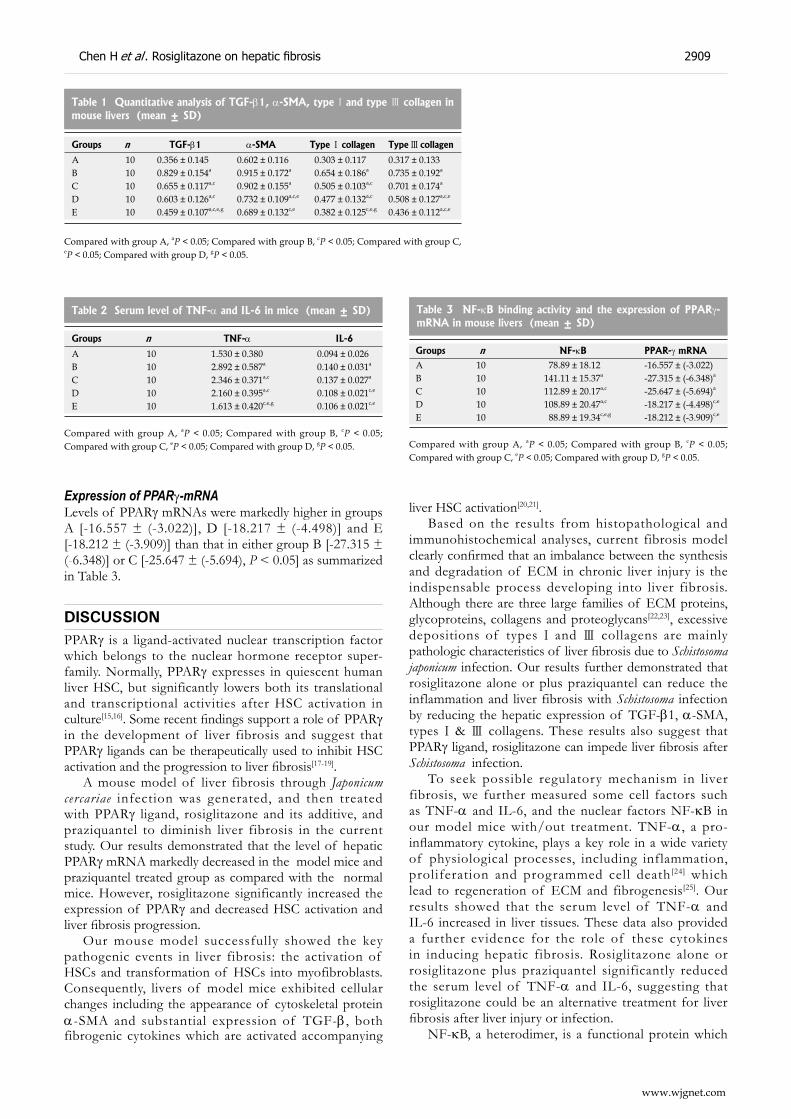
Expression of PPARγ-mRNA Levels of PPARγ mRNAs were markedly higher in groups A [-16.557 ± (-3.022)], D [-18.217 ± (-4.498)] and E [-18.212 ± (-3.909)] than that in either group B [-27.315 ± (-6.348)] or C [-25.647 ± (-5.694), P < 0.05] as summarized in Table 3.
DISCUSSIONPPARγ is a ligand-activated nuclear transcription factor which belongs to the nuclear hormone receptor super-family. Normally, PPARγ expresses in quiescent human liver HSC, but significantly lowers both its translational and transcriptional activities after HSC activation in culture[15,16]. Some recent findings support a role of PPARγ in the development of liver fibrosis and suggest that PPARγ ligands can be therapeutically used to inhibit HSC activation and the progression to liver fibrosis[17-19].
A mouse model of liver fibrosis through Japonicum cercariae infection was generated, and then treated with PPARγ ligand, rosiglitazone and its additive, and praziquantel to diminish liver fibrosis in the current study. Our results demonstrated that the level of hepatic PPARγ mRNA markedly decreased in the model mice and praziquantel treated group as compared with the normal mice. However, rosiglitazone significantly increased the expression of PPARγ and decreased HSC activation and liver fibrosis progression.
Our mouse model successfully showed the key pathogenic events in liver fibrosis: the activation of HSCs and transformation of HSCs into myofibroblasts. Consequently, livers of model mice exhibited cellular changes including the appearance of cytoskeletal protein α-SMA and substantial expression of TGF-β, both fibrogenic cytokines which are activated accompanying
liver HSC activation[20,21].Based on the results from histopathological and
immunohistochemical analyses, current fibrosis model clearly confirmed that an imbalance between the synthesis and degradation of ECM in chronic liver injury is the indispensable process developing into liver fibrosis. Although there are three large families of ECM proteins, glycoproteins, collagens and proteoglycans[22,23], excessive depositions of types Ⅰ and Ⅲ collagens are mainly pathologic characteristics of liver fibrosis due to Schistosoma japonicum infection. Our results further demonstrated that rosiglitazone alone or plus praziquantel can reduce the inflammation and liver fibrosis with Schistosoma infection by reducing the hepatic expression of TGF-β1, α-SMA, types Ⅰ & Ⅲ collagens. These results also suggest that PPARγ ligand, rosiglitazone can impede liver fibrosis after Schistosoma infection.
To seek possible regulatory mechanism in liver fibrosis, we further measured some cell factors such as TNF-α and IL-6, and the nuclear factors NF-κB in our model mice with/out treatment. TNF-α, a pro-inflammatory cytokine, plays a key role in a wide variety of physiological processes, including inflammation, proliferation and programmed cell death[24] which lead to regeneration of ECM and fibrogenesis[25]. Our results showed that the serum level of TNF-α and IL-6 increased in liver tissues. These data also provided a further evidence for the role of these cytokines in inducing hepatic fibrosis. Rosiglitazone alone or rosiglitazone plus praziquantel significantly reduced the serum level of TNF-α and IL-6, suggesting that rosiglitazone could be an alternative treatment for liver fibrosis after liver injury or infection.
NF-κB, a heterodimer, is a functional protein which
Table 1 Quantitative analysis of TGF-β1, α-SMA, type Ⅰ and type Ⅲ collagen in mouse livers (mean ± SD)
Groups n TGF-β1 α-SMA Type Ⅰ collagen Type Ⅲ collagen
A 10 0.356 ± 0.145 0.602 ± 0.116 0.303 ± 0.117 0.317 ± 0.133B 10 0.829 ± 0.154a 0.915 ± 0.172a 0.654 ± 0.186a 0.735 ± 0.192a
C 10 0.655 ± 0.117a,c 0.902 ± 0.155a 0.505 ± 0.103a,c 0.701 ± 0.174a
D 10 0.603 ± 0.126a,c 0.732 ± 0.109a,c,e 0.477 ± 0.132a,c 0.508 ± 0.127a,c,e
E 10 0.459 ± 0.107a,c,e,g 0.689 ± 0.132c,e 0.382 ± 0.125c,e,g 0.436 ± 0.112a,c,e
Compared with group A, aP < 0.05; Compared with group B, cP < 0.05; Compared with group C, eP < 0.05; Compared with group D, gP < 0.05.
Groups n TNF-α IL-6
A 10 1.530 ± 0.380 0.094 ± 0.026B 10 2.892 ± 0.587a 0.140 ± 0.031a
C 10 2.346 ± 0.371a,c 0.137 ± 0.027a
D 10 2.160 ± 0.395a,c 0.108 ± 0.021c,e
E 10 1.613 ± 0.420c,e,g 0.106 ± 0.021c,e
Table 2 Serum level of TNF-α and IL-6 in mice (mean ± SD)
Groups n NF-κB PPAR- γ mRNA
A 10 78.89 ± 18.12 -16.557 ± (-3.022)B 10 141.11 ± 15.37a -27.315 ± (-6.348)a
C 10 112.89 ± 20.17a,c -25.647 ± (-5.694)a
D 10 108.89 ± 20.47a,c -18.217 ± (-4.498)c,e
E 10 88.89 ± 19.34c,e,g -18.212 ± (-3.909)c,e
Table 3 NF-κB binding activity and the expression of PPARγ-mRNA in mouse livers (mean ± SD)
Compared with group A, aP < 0.05; Compared with group B, cP < 0.05; Compared with group C, eP < 0.05; Compared with group D, gP < 0.05.
Compared with group A, aP < 0.05; Compared with group B, cP < 0.05; Compared with group C, eP < 0.05; Compared with group D, gP < 0.05.
www.wjgnet.com
Chen H et al . Rosiglitazone on hepatic fibrosis 2909

is regulated by interaction with a family of regulatory proteins, the inhibitor of nuclear factor κB (IκB) proteins may be stimulated by many factors, such as TNF-α and lipopolysaccharide, via the phosphorylation and degradation of IκB. Activated NF-κB then transports into the cell nucleus and combines with gene promoters to induce the transcription of many cytokines and adhesion molecules[26,27]. Physical stress, oxidative stress, and exposure to certain chemicals can also activate NF-κB, suggesting its critical functions in mediating stress responses as well[28]. Recent studies show that the activation of NF-κB has a great impact on the pathogenesis of liver fibrosis through regulating hepatocyte, HSC and Kupffer cells[29,30]. Some studies demonstrate that NF-κB is also associated with the development of the activated phenotype of HSC and promotes survival of activated HSC[31] by protecting activated HSCs against TNF-induced apoptosis[32]. Notably, NF-κB binding activity increased in our mouse fibrosis model, while rosiglitazone significantly decreased the activity of, suggesting further that rosiglitazone could partially inhibit liver fibrosis by down-regulating NF-κB as well.
As indicated, our study showed a significant reduction of liver fibrosis following the treatment with rosiglitazone, a PPAR-γ ligand, in a murine model of hepatic fibrosis induced by Schistosoma japonicum. The effect of this compound on preventing liver fibrosis may be through down-regulation of liver TGF-β1 and collagens, and reduction of inflammatory mediators (IL-6, TNF-α, NF-κB). It suggested the use of rosiglitazone for the treatment of liver fibrosis.
Interestingly, our results showed that praziquantel alone reduced markedly the serum level of TNF-α, NF-κB binding activity, and volumes of TGF-β1 and type Ⅰ collagen in liver fibrosis due to Schistosoma infection. These data may suggest an antifibrotic effect of praziquantel in the early stage of liver fibrosis. Praziquantel might be able to block liver fibrosis through killing parasite to alleviate liver inflammation. Further studies on mechanisms of rosiglitazone and praziquantel during liver injury or infection may shed lights on developing therapeutic methods in clinical practice.
COMMENTSBackgroundHepatic schistosomiasis is one of the most prevalent forms of chronic liver diseases in the world, resulting in the morbidity due to its complications of liver fibrosis from infection. However, there are currently few medicines or means available to control and treat fibrosis in schistosomiasis. Many studies demonstrate that PPARγ is mainly expressed in human HSC and contributes to the process of liver fibrogenesis, but the identification of PPARγ as a novel modulator to liver fibrosis remains controversial.
Research frontiersCurrent findings further support the role of PPARγ in the development of liver fibrosis and suggest that PPARγ ligands can be therapeutically used to inhibit HSC activation and the progression of liver fibrosis.
Innovations and breakthroughsThis is a first report about PPARγ and liver fibrosis due to Schistosoma infection. This study evaluates the relationship of PPARγ ligand and hepatic fibrosis due to Schistosoma infection.
ApplicationsThis study provides a perspective for a new therapeutic approach to prevent liver fibrosis following Schistosoma japonicum infection.
Peer reviewThe study reported a significant reduction of liver fibrosis following the treatment with rosiglitazone, a PPAR-γ ligand, in a murine model of hepatic fibrosis induced by Schistosoma japonicum. The effect of this compound on preventing liver fibrosis may be through down-regulation of liver TGF-β1 and collagens, and reduction of inflammatory mediators (IL-6, TNF-α, NF-κB). The study is interesting and may herald a new therapeutic approach to prevent liver fibrosis following Schistosoma japonicum infection.
REFERENCES1 Bartley PB, Ramm GA, Jones MK, Ruddell RG, Li Y, McManus
DP. A contributory role for activated hepatic stellate cells in the dynamics of Schistosoma japonicum egg-induced fibrosis. Int J Parasitol 2006; 36: 993-1001
2 Gutierrez-Ruiz MC, Gomez-Quiroz LE. Liver fibrosis: searching for cell model answers. Liver Int 2007; 27: 434-439
3 Parsons CJ, Takashima M, Rippe RA. Molecular mechanisms of hepatic fibrogenesis. J Gastroenterol Hepatol 2007; 22 Suppl 1: S79-S84
4 Galli A, Svegliati-Baroni G, Ceni E, Milani S, Ridolfi F, Salzano R, Tarocchi M, Grappone C, Pellegrini G, Benedetti A, Surrenti C, Casini A. Oxidative stress stimulates proliferation and invasiveness of hepatic stellate cells via a MMP2-mediated mechanism. Hepatology 2005; 41: 1074-1084
5 Zhang XL, Liu JM, Yang CC, Zheng YL, Liu L, Wang ZK, Jiang HQ. Dynamic expression of extracellular signal-regulated kinase in rat liver tissue during hepatic fibrogenesis. World J Gastroenterol 2006; 12: 6376-6381
6 Reeves HL, Friedman SL. Activation of hepatic stellate cells-a key issue in liver fibrosis. Front Biosci 2002; 7: d808-d826
7 Moreira RK. Hepatic stellate cells and liver fibrosis. Arch Pathol Lab Med 2007; 131: 1728-1734
8 Yang C, Zeisberg M, Mosterman B, Sudhakar A, Yerramalla U, Holthaus K, Xu L, Eng F, Afdhal N, Kalluri R. Liver fibrosis: insights into migration of hepatic stellate cells in response to extracellular matrix and growth factors. Gastroenterology 2003; 124: 147-159
9 Shearer BG, Hoekstra WJ. Recent advances in peroxisome proliferator-activated receptor science. Curr Med Chem 2003; 10: 267-280
10 Berkenstam A, Gustafsson JA. Nuclear receptors and their relevance to diseases related to lipid metabolism. Curr Opin Pharmacol 2005; 5: 171-176
11 Marra F, Efsen E, Romanelli RG, Caligiuri A, Pastacaldi S, Batignani G, Bonacchi A, Caporale R, Laffi G, Pinzani M, Gentilini P. Ligands of peroxisome proliferator-activated receptor gamma modulate profibrogenic and proinflammatory actions in hepatic stellate cells. Gastroenterology 2000; 119: 466-478
12 Galli A, Crabb D, Price D, Ceni E, Salzano R, Surrenti C, Casini A. Peroxisome proliferator-activated receptor gamma transcriptional regulation is involved in platelet-derived growth factor-induced proliferation of human hepatic stellate cells. Hepatology 2000; 31: 101-108
13 Galli A, Crabb DW, Ceni E, Salzano R, Mello T, Svegliati-Baroni G, Ridolfi F, Trozzi L, Surrenti C, Casini A. Antidiabetic thiazolidinediones inhibit collagen synthesis and hepatic stellate cell activation in vivo and in vitro. Gastroenterology 2002; 122: 1924-1940
14 Anthony PP, Ishak KG, Nayak NC, Poulsen HE, Scheuer PJ, Sobin LH. The morphology of cirrhosis. Recommendations on definition, nomenclature, and classification by a working group sponsored by the World Health Organization. J Clin Pathol 1978; 31: 395-414
15 Yang L, Chan CC, Kwon OS, Liu S, McGhee J, Stimpson SA, Chen LZ, Harrington WW, Symonds WT, Rockey DC. Regulation of peroxisome proliferator-activated receptor-
www.wjgnet.com
2910 ISSN 1007-9327 CN 14-1219/R World J Gastroenterol May 14, 2008 Volume 14 Number 18
COMMENTS
gamma in liver fibrosis. Am J Physiol Gastrointest Liver Physiol 2006; 291: G902-G911
16 Guo YT, Leng XS, Li T, Peng JR, Song SH, Xiong LF, Qin ZZ. Effect of ligand of peroxisome proliferator-activated receptor gamma on the biological characters of hepatic stellate cells. World J Gastroenterol 2005; 11: 4735-4739
17 Kawaguchi K, Sakaida I, Tsuchiya M, Omori K, Takami T, Okita K. Pioglitazone prevents hepatic steatosis, fibrosis, and enzyme-altered lesions in rat liver cirrhosis induced by a choline-deficient L-amino acid-defined diet. Biochem Biophys Res Commun 2004; 315: 187-195
18 Marra F, DeFranco R, Robino G, Novo E, Efsen E, Pastacaldi S, Zamara E, Vercelli A, Lottini B, Spirli C, Strazzabosco M, Pinzani M, Parola M. Thiazolidinedione treatment inhibits bile duct proliferation and fibrosis in a rat model of chronic cholestasis. World J Gastroenterol 2005; 11: 4931-4938
19 Zhao C, Chen W, Yang L, Chen L, Stimpson SA, Diehl AM. PPARgamma agonists prevent TGFbeta1/Smad3-signaling in human hepatic stellate cells. Biochem Biophys Res Commun 2006; 350: 385-391
20 Jiang W, Yang CQ, Liu WB, Wang YQ, He BM, Wang JY. Blockage of transforming growth factor beta receptors prevents progression of pig serum-induced rat liver fibrosis. World J Gastroenterol 2004; 10: 1634-1638
21 Kershenobich Stalnikowitz D, Weissbrod AB. Liver fibrosis and inflammation. A review. Ann Hepatol 2003; 2: 159-163
22 Tsukada S, Parsons CJ, Rippe RA. Mechanisms of liver fibrosis. Clin Chim Acta 2006; 364: 33-60
23 Benyon RC, Arthur MJ. Extracellular matrix degradation and the role of hepatic stellate cells. Semin Liver Dis 2001; 21: 373-384
24 Theiss AL, Simmons JG, Jobin C, Lund PK. Tumor necrosis factor (TNF) alpha increases collagen accumulation and
proliferation in intestinal myofibroblasts via TNF receptor 2. J Biol Chem 2005; 280: 36099-36109
25 Simeonova PP , Gallucci RM, Hulderman T, Wilson R, Kommineni C, Rao M, Luster MI. The role of tumor necrosis factor-alpha in liver toxicity, inflammation, and fibrosis induced by carbon tetrachloride. Toxicol Appl Pharmacol 2001; 177: 112-120
26 Cogswell PC, Kashatus DF, Keifer JA, Guttridge DC, Reuther JY, Bristow C, Roy S, Nicholson DW, Baldwin AS Jr. NF-kappa B and I kappa B alpha are found in the mitochondria. Evidence for regulation of mitochondrial gene expression by NF-kappa B. J Biol Chem 2003; 278: 2963-2968
27 Lawrence T, Bebien M, Liu GY, Nizet V, Karin M. IKKalpha limits macrophage NF-kappaB activation and contributes to the resolution of inflammation. Nature 2005; 434: 1138-1143
28 Li X, Stark GR. NFkappaB-dependent signaling pathways. Exp Hematol 2002; 30: 285-296
29 Mann DA, Smart DE. Transcriptional regulation of hepatic stellate cell activation. Gut 2002; 50: 891-896
30 Smart DE, Vincent KJ, Arthur MJ, Eickelberg O, Castellazzi M, Mann J, Mann DA. JunD regulates transcription of the tissue inhibitor of metalloproteinases-1 and interleukin-6 genes in activated hepatic stellate cells. J Biol Chem 2001; 276: 24414-24421
31 Muhlbauer M, Weiss TS, Thasler WE, Gelbmann CM, Schnabl B, Scholmerich J, Hellerbrand C. LPS-mediated NFkappaB activation varies between activated human hepatic stellate cells from different donors. Biochem Biophys Res Commun 2004; 325: 191-197
32 Elsharkawy AM, Oakley F, Mann DA. The role and regulation of hepatic stellate cell apoptosis in reversal of liver fibrosis. Apoptosis 2005; 10: 927-939
S- Editor Li DL L- Editor Ma JY E- Editor Yin DH
www.wjgnet.com
Chen H et al . Rosiglitazone on hepatic fibrosis 2911

Online Submissions: wjg.wjgnet.com World J Gastroenterol 2008 May 14; 14(18): 2912-2914www.wjgnet.com World Journal of Gastroenterology ISSN [email protected] © 2008 WJG. All rights reserved.
Cytomegalovirus colitis in a patient with Behcet’s disease receiving tumor necrosis factor alpha inhibitory treatment
Ismail Sari, Merih Birlik, Can Gonen, Servet Akar, Duygu Gurel, Fatos Onen, Nurullah Akkoc
www.wjgnet.com
CASE REPORT
Ismail Sari, Merih Birlik, Servet Akar, Fatos Onen, Nurullah Akkoc, Division of Rheumatology, Department of Internal Medicine, Dokuz Eylul University School of Medicine, Izmir 35340, TurkeyCan Gonen, Division of Gastroenterology, Department of Internal Medicine, Dokuz Eylul University School of Medicine, Izmir 35340, TurkeyDuygu Gurel, Department of Pathology, Dokuz Eylul University School of Medicine, Izmir 35340, TurkeyAuthor contributions: Sari I and Birlik M wrote the paper; Sari I and Akar S drafted the article; Sari I, Gonen C and Gurel D collected and assembled the data; Onen F and Akkoc N critically revised the article for important intellectual content; Akkoc N finally approved the article.Correspondence to: Professor Nurullah Akkoc, MD, Dokuz Eylul Universitesi Tip Fakultesi Ic hastaliklari ABD Immunoloji-Romatoloji BD 35340 Inciralti, Izmir 35340, Turkey. [email protected]: +90-232-4123781 Fax: +90-232-2792739Received: January 28, 2008 Revised: April 9, 2008
AbstractAnti-tumor necrosis factor alpha (TNF-α) inhibitors are effective in the treatment of various inflammatory rheumatic condit ions. Increased risks of serious infections are the major issues concerning the long-term safety of these agents. We present a case of a young male Behcet’s patient whose disease was complicated by cytomegalovirus (CMV) colitis. Colitis started 10 d after the third Infliximab dose and responded to the cessation of TNF blocking treatment and administration of ganciclovir. Tumor necrosis factor alpha and interferon gamma act at several levels in combating viral infections. CMV infections should be kept in mind and included in the differential diagnosis of severe gastrointestinal symptoms in patients receiving anti-TNF agents.
© 2008 WJG. All rights reserved.
Key words: Tumor necrosis factor alpha inhibitors; Adverse effects; Virus diseases
Peer reviewer: Burton I Korelitz, MD, Department of Gastroenterology, Lenox Hill Hospital, 100 East 77th Street, 3 Achelis, New York 10021, United States
Sari I, Birlik M, Gonen C, Akar S, Gurel D, Onen F, Akkoc N. Cytomegalovirus colitis in a patient with Behcet’s disease receiving tumor necrosis factor alpha inhibitory treatment. World J Gastroenterol 2008; 14(18): 2912-2914 Available from: URL:
http://www.wjgnet.com/1007-9327/14/2912.asp DOI: http://dx.doi.org/10.3748/wjg.14.2912
INTRODUCTIONAnti-tumor necrosis factor alpha (TNF-α) inhibitors are relatively new drugs in the rheumatic arena which are effective in the treatment of various inflammatory rheumatic conditions. Increased risk of infections and possible risk of lymphoma with the use of TNF targeting therapies are the major issues concerning the long-term safety of these agents[1]. The association of anti- TNF-α therapy and serious infections has generally been attributed to an increased risk of bacterial (especially microbacteria) and opportunistic fungal microorganisms[2]. In recent years, an increasing number of studies, mainly based on case reports, have reported an increased risk of serious viral infections in patients treated with anti-TNF agents.
In this report, we describe a case of severe, biopsy proven cytomegalovirus (CMV) colitis in a patient with Behcet's disease (BD) whose symptoms started 10 d after the third Infliximab (INF; Remicade®, Schering-Plough) infusion.
CASE REPORTA 25-year-old man presented with diarrhea and weight loss for two months. He was diagnosed as neuro BD 4 years ago according to recurrent oral and genital ulcerations, uveitis, urinary incontinence and seizures. He was treated with various immunosuppressive drugs including monthly cyclophosphamide infusions, interferon and a combination of cyclosporine and azathioprine. Although these medications were used, his eye involvement could not be controlled and visual acuity decreased to no light perception in the left eye and 10/100 in the right eye. Because of this, INF at a dose of 5 mg/kg was started. Remission of ocular symptoms was observed after two infusions. Ten days after the third dose of INF (52 d after the first infusion), the patient developed abdominal discomfort, anorexia, epigastric pain and diarrhea occurring seven to ten times per day with watery stools, but without any associated blood or mucus. He had no history of chronic diarrhea before INF administration. His symptoms continued to worsen and he lost six kilograms during the past two months. He did not use any medications other than INF, colchicine (1.5 mg/d) and

Sari I et al . CMV colitis and anti-TNF 2913
www.wjgnet.com
carbamazepine (600 mg/d). Physical examination showed epigastric tenderness on palpation, with hyperactive bowel sounds. Admission laboratory studies were significant for increased C-reactive protein (CRP = 14.4 mg/L). Stool examination was negative for parasites and Clostridium difficile toxin. Repeated stool cultures were also negative for enteric pathogens. Colonoscopic examination revealed diffuse redness and edematous mucosa and multiple ulcers throughout the colon including rectum and terminal ileum (Figure 1). Histological examination of the biopsy specimen identified cytomegalovirus (CMV) by specific immunohistochemistry (Figure 2). Serological testing revealed a positive CMV immunoglobulin (Ig) G and a negative IgM antibody. CMV pp65 antigenemia as determined by immunofluorescence was also negative. Infliximab was stopped and the patient was given ganciclovir (600 mg/d Ⅳ). On the 14th d of treatment, stool frequency decreased to 4-5 per day and repeated colonoscopy revealed edematous mucosa without ulcers. Immunohistochemical staining for CMV turned negative on histopathological examination of the repeat biopsy specimen. Ganciclovir was continued for an additional 14 d. The patient gained 5 kg and his stool frequency decreased to two per day. In a follow-up period of 30 mo, he did not reveal any recurrence of colitis symptoms. TNF targeting treatments were not considered during this period.
DISCUSSIONWe present a case of a young male Behcet’s patient whose disease was complicated by CMV colitis. Colitis started 10 d after the third INF dose and responded to the cessation of TNF blocking treatment and administration of ganciclovir.
CMV is a member of the herpesviridae family which is prevalent among the general population with an overall seroprevalence of 30%-70% in developed countries[3]. CMV causes a variety of clinical syndromes in immunocompromised patients. Pneumonitis, retinitis and gastrointestinal (GI) CMV disease are the mostly encountered clinical manifestations[3]. GI tract may be affected anywhere from the mouth to the anus. Esophagus
and colon are the frequently involved sites. Ulcerations, erosions, and mucosal hemorrhage are the primary macroscopic findings[4]. The clinical signs and symptoms vary depending upon the involved areas. Patients with colonic CMV disease may present with diarrhea with or without blood, abdominal pain, urgency and tenesmus accompanied with systemic symptoms such as fever, malaise anorexia and weight loss[4]. Diagnosis of CMV infection is based upon the presence of CMV in clinical specimens demonstrated by conventional tissue culture or rapid culture with confirmation by specific monoclonal antibodies, or by detection of the pp65 CMV antigen in peripheral blood leukocytes[5].
Severe CMV infections during TNF targeting treatments have been reported including retinitis[6], hepatitis[7], duodenitis[8], ileitis[9], colitis[10] and disseminated CMV infection[11]. All these cases received concomitant immunosuppressive treatment in addition to INF. Except for our patient and patients who developed retinitis, all other reported CMV infections developed in subjects who had primary GI problems, such as inflammatory bowel disease, common variable immunodeficiency sprue and indeterminate colitis.
BD may affect the intestine. Intestinal involvement commonly accompanies ulcerative lesions in the small and large bowel. The lesions are most commonly found in the terminal ileum and the cecum and less frequently in the colon. Rectal and anal involvement is quite rare[12]. In our patient, clinical symptomatology mimicked intestinal BD. However, diffuse involvement of the colon including the rectum and development of symptoms after the initiation of TNF inhibitory treatment suggested an infectious etiology. Following histopathological examination of the biopsies, which revealed the causative microorganism, colonic CMV disease was diagnosed. Reactivation of the virus was thought since CMV serology was positive for IgG and negative for IgM.
Tumor necrosis factor alpha and interferon gamma act at several levels in combating viral infections. TNF exhibits its antiviral activities against both DNA and RNA viruses by enhancing the induction of antiviral state in uninfected cells and by selectively killing virus-infected cells[13]. CD8+ T cells are the major defense against viruses by direct cytolysis of target cells mediated by perforin release and
BA
Figure 1 Colonoscopic appearance of the mucosa before (A) and during the 14 d of anti-viral treatment (B). The presence of edema and redness is seen in both images. Note the ulcers in the sigmoid colon before the treatment as shown in A.
Figure 2 Colonic endothelial cells showing positive nuclear (A) and cytoplasmic (B) immunostaining of the CMV antigen.
A
B

Fas, or by secreting cytokines such as TNF and interferon-γ and expressing chemokines that attract inflammatory cells to the sites of infection[14]. It has been shown that treatment with monoclonal antibodies directing against TNF-α is associated with a progressive loss of CD4+ and CD8+ T cells[15]. In addition, Infliximab treatment has been reported to be associated with decreased levels of TNF-α and interferon-γ[16]. On the other hand, TNF levels are increased during CMV infection and CD4+ T cells specific for the human CMV result in early gene products[17]. Up-regulation of TNF gene expression by CMV[18] is the major cause for the increased TNF. It has been shown that increased TNF production in response to CMV infection displays anti-CMV activity in vitro[19]. Furthermore, recombinant TNF-α possesses anti-CMV activity[17].
In conclusion, TNF-α in line with interferon-γ acts at several levels in combating viral infections. Blocking the action of TNF-α with novel medications may predispose these patients to serious viral diseases. CMV infections should be kept in mind and included in the differential diagnosis of severe gastrointestinal symptoms in patients receiving Infliximab.
REFERENCES1 Cush JJ. Unusual toxicities with TNF inhibition: heart failure
and drug-induced lupus. Clin Exp Rheumatol 2004; 22: S141-S1472 Ellerin T, Rubin RH, Weinblatt ME. Infections and anti-
tumor necrosis factor alpha therapy. Arthritis Rheum 2003; 48: 3013-3022
3 Gandhi MK, Khanna R. Human cytomegalovirus: clinical aspects, immune regulation, and emerging treatments. Lancet Infect Dis 2004; 4: 725-738
4 Goodgame RW. Gastrointestinal cytomegalovirus disease. Ann Intern Med 1993; 119: 924-935
5 Reusser P. Cytomegalovirus Infection. In: Cohen J, Powderly WG, editors. Cohen & Powderly: Infectious Diseases. 2nd: Mosby, An Imprint of Elsevier, 2004: 1175-1177
6 Haerter G, Manfras BJ, de Jong-Hesse Y, Wilts H, Mertens T, Kern P, Schmitt M. Cytomegalovirus retinitis in a patient treated with anti-tumor necrosis factor alpha antibody therapy
for rheumatoid arthritis. Clin Infect Dis 2004; 39: e88-e947 Mizuta M, Schuster MG. Cytomegalovirus hepatitis associated
with use of anti-tumor necrosis factor-alpha antibody. Clin Infect Dis 2005; 40: 1071-1072
8 Medlicott SA, Coderre S, Horne G, Panaccione R. Multimodal immunosuppressant therapy in steroid-refractory common variable immunodeficiency sprue: a case report complicating cytomegalovirus infection. Int J Surg Pathol 2006; 14: 101-106
9 Kohara MM , Blum RN. Cytomegalovirus i lei t is and hemophagocytic syndrome associated with use of anti-tumor necrosis factor-alpha antibody. Clin Infect Dis 2006; 42: 733-734
10 Papadakis KA, Tung JK, Binder SW, Kam LY, Abreu MT, Targan SR, Vasiliauskas EA. Outcome of cytomegalovirus infections in patients with inflammatory bowel disease. Am J Gastroenterol 2001; 96: 2137-2142
11 Helbling D , Breitbach TH, Krause M. Disseminated cytomegalovirus infection in Crohn's disease following anti-tumour necrosis factor therapy. Eur J Gastroenterol Hepatol 2002; 14: 1393-1395
12 Bayraktar Y, Ozaslan E, Van Thiel DH. Gastrointestinal manifestations of Behcet's disease. J Clin Gastroenterol 2000; 30: 144-154
13 Wong GH, Goeddel DV. Tumour necrosis factors alpha and beta inhibit virus replication and synergize with interferons. Nature 1986; 323: 819-822
14 Wong P, Pamer EG. CD8 T cell responses to infectious pathogens. Annu Rev Immunol 2003; 21: 29-70
15 Baert FJ, D'Haens GR, Peeters M, Hiele MI, Schaible TF, Shealy D, Geboes K, Rutgeerts PJ. Tumor necrosis factor alpha antibody (infliximab) therapy profoundly down-regulates the inflammation in Crohn's ileocolitis. Gastroenterology 1999; 116: 22-28
16 Zou J, Rudwaleit M, Brandt J, Thiel A, Braun J, Sieper J. Down-regulation of the nonspecific and antigen-specific T cell cytokine response in ankylosing spondylitis during treatment with infliximab. Arthritis Rheum 2003; 48: 780-790
17 Davignon JL, Castanie P, Yorke JA, Gautier N, Clement D, Davrinche C. Anti-human cytomegalovirus activity of cytokines produced by CD4+ T-cell clones specifically activated by IE1 peptides in vitro. J Virol 1996; 70: 2162-2169
18 Geist LJ, Monick MM, Stinski MF, Hunninghake GW. The immediate early genes of human cytomegalovirus upregulate tumor necrosis factor-alpha gene expression. J Clin Invest 1994; 93: 474-478
19 Herbein G, O'Brien WA. Tumor necrosis factor (TNF)-alpha and TNF receptors in viral pathogenesis. Proc Soc Exp Biol Med 2000; 223: 241-257
S- Editor Zhong XY L- Editor Wang XL E- Editor Lu W
www.wjgnet.com
2914 ISSN 1007-9327 CN 14-1219/R World J Gastroenterol May 14, 2008 Volume 14 Number 18

Online Submissions: wjg.wjgnet.com World J Gastroenterol 2008 May 14; 14(18): 2915-2916www.wjgnet.com World Journal of Gastroenterology ISSN [email protected] © 2008 WJG. All rights reserved.
Agenesis of the dorsal pancreas
Lale Pasaoglu, Murat Vural, Hatice Gul Hatipoglu, Gokce Tereklioglu, Suha Koparal
CASE REPORT
Lale Pasaoglu, Murat Vural, Hatice Gul Hatipoglu, Gokce Tereklioglu, Suha Koparal, Department of Radiology, Ankara Numune State Hospital, Ankara 06100, Turkey Author contributions: Pasaoglu L, Vural M contributed equally to this case; Hatipoglu HG dealt with MR images; and Tereklioglu G and Koparal S wrote the paper.Correspondence to: Dr. Lale Pasaoglu, Ankara Numune State Hospital, Ankara 06100, Turkey. [email protected]: +90-535-7687688 Fax: +90-312-4338666Received: January 16, 2008 Revised: March 22, 2008
AbstractDevelopmental anomalies of the pancreas have been reported but dorsal pancreatic agenesis is an extremely rare entity. We report an asymptomatic 62-year-old woman with complete agenesis of the dorsal pancreas. Abdominal computed tomography (CT) revealed a normal pancreatic head, but pancreatic body and tail were not visualized. Magnetic resonance imaging (MRI) findings were similar to CT. At magnetic resonance cholangiopancreatography (MRCP), the major pancreatic duct was short and the dorsal pancreatic duct was not visualized. The final diagnosis was dorsal pancreatic agenesis.
© 2008 WJG. All rights reserved.
Key words: Agenesis; Pancreatic anomaly; Computed tomography; Magnetic resonance cholangiopancrea-tography
Peer reviewers: Jia-Yu Xu, Professor, Shanghai Second Medical University, Rui Jin Hospital, 197 Rui Jin Er Road, Shanghai 200025, China; Aydin Karabacakoglu, MD, Assistant Professor, Department of Radiology, Meram Medical Faculty, Selcuk University, Konya 42080, Turkey
Pasaoglu L, Vural M, Hatipoglu HG, Tereklioglu G, Koparal S. Agenesis of the dorsal pancreas. World J Gastroenterol 2008; 14(18): 2915-2916 Available from: URL: http://www.wjgnet.com/1007-9327/14/2915.asp DOI: http://dx.doi.org/10.3748/wjg.14.2915
INTRODUCTIONAgenesis of the dorsal pancreas is an extremely rare anomaly which results from defective pancreas formation. A few case reports have been published in the literature about this anomaly. Agenesis of the dorsal pancreas is mostly asymptomatic but abdominal pain, pancreatitis and
diabetes mellitus may be associated[1]. We present a case of asymptomatic dorsal pancreatic agenesis.
CASE REPORTA 62-year-old female patient was admitted to our hospital with a prediagnosis of gastric lymphoma. The patient has a suspicion of lymphoma with gastroscopy. The biochemical evaluation of the patient revealed mild elevation of fasting plasma glucose (126 mg/dL; reference range: 70-115 mg/dL[1]). Pancreatic amylase and lipase levels in serum were within normal limits. On abdominal computed tomography (CT), perigastric lymph nodes were demonstrated ranging between 10 mm and 20 mm. At this examination pancreas body and tail were not seen. Pancreatic head and neck were normal in size and shape (Figure 1A and B). Upper abdominal Magnetic resonance imaging (MRI) and magnetic resonance cholangiopancreatography (MRCP) were carried out with the suspicion of dorsal pancreatic agenesis. At MRCP, the major pancreatic duct was short and the dorsal pancreatic duct was not visualized (Figure 2A and B).
DISCUSSIONThe pancreas develops by ventral and dorsal endodermal buds. The dorsal bud forms the upper part of the head, body and tail of the pancreas which drains through the Santorini duct. The ventral bud gives rise to the major part of the head and uncinate process which drains through Wirsung duct[1]. Agenesis of the ventral pancreas and complete agenesis of the pancreas are lethal conditions[2]. Dorsal pancreatic agenesis is an extremely rare congenital anomaly. In the literature, about 23 cases of partial agenesis of dorsal pancreas were reported between 1913 and 2007. Pancreas divisum and autodigestion secondary to chronic pancreatitis must be considered in the differential diagnosis of the dorsal pancreatic agenesis. Failure of the ventral and dorsal pancreatic ducts to fuse is called pancreas divisum. In this entity, the ventral duct drains into major papilla, while the dorsal duct drains into separate minor papilla[1]. Atrophy of the body and the tail of the pancreas secondary to chronic pancreatitis and sparing of the pancreatic head is called pseudo-agenesis[3]. This situation may mimic dorsal pancreatic agenesis. In the differential diagnosis of pseudo-agenesis, histories of previous abdominal pain, pancreatitis, CT scanning and the serum amylase level may be helpful. The complete absence of the dorsal duct and demonstration of short ventral duct is important in the diagnosis of the dorsal bud agenesis. Abdominal CT may not evaluate the pancreatic duct in a
www.wjgnet.com

detailed fashion. Therefore ERCP or MRCP is necessary for revealing the major and the accessory duct systems. ERCP is an invasive technique. It might challenge the cannulation of the minor papilla in selected cases. There is also radiation risk to the patient. MRCP can help the diagnosis of the dorsal pancreatic agenesis noninvasively with no radiation risk[4-6].
In our patient, distal pancreas was absent on CT and MRI images. On MRCP, dorsal duct system was not visualized and a short ventral duct was present which supports the diagnosis of dorsal pancreatic agenesis. In summary, this extremely rare congenital anomaly must be kept in mind when the corpus and tail of the pancreas are not seen at routine examinations or as in our case an incidental finding at the examinations for different pathologies.
A
B
Figure 1 Axial CT (A) and thick-slice coronal oblique MIP CT (B) images show the pancreatic head (arrow) and the absence of the corpus and tail of the pancreas.
A
B
Figure 2 Abdominal axial T1-weighted MR image (A) reveals pancreatic head; MRCP demontrates a short major pancreatic duct (arrow) and dorsal duct is not visualized (B).
REFERENCES1 Fukuoka K, Ajiki T, Yamamoto M, Fujiwara H, Onoyama
H, Fujita T, Katayama N, Mizuguchi K, Ikuta H, Kuroda Y, Hanioka K. Complete agenesis of the dorsal pancreas. J Hepatobiliary Pancreat Surg 1999; 6: 94-97
2 Voldsgaard P, Kryger-Baggesen N, Lisse I. Agenesis of pancreas. Acta Paediatr 1994; 83: 791-793
3 Gold RP . Agenesis and pseudo-agenesis of the dorsal pancreas. Abdom Imaging 1993; 18: 141-144
4 Balakrishnan V, Narayanan VA, Siyad I, Radhakrishnan L, Nair P. Agenesis of the dorsal pancreas with chronic calcific pancreatitis. case report, review of the literature and genetic basis. JOP 2006; 7: 651-659
5 Uygur-Bayramicli O, Dabak R, Kilicoglu G, Dolapcioglu C, Oztas D. Dorsal pancreatic agenesis. JOP 2007; 8: 450-452
6 DU J, Xu GQ, Xu P, Jin EY, Liu Q, Li YM. Congenital short pancreas. Chin Med J (Engl) 2007; 120: 259-262
S- Editor: Zhong XY L-Editor: Ma JY E- Editor Yin DH
2916 ISSN 1007-9327 CN 14-1219/R World J Gastroenterol May 14, 2008 Volume 14 Number 18
www.wjgnet.com

Key words: Endoscopic retrograde cholangiopancreat-ography; Pneumoperitoneum; Complications; Pneumobilia; Hepatic metastases
Peer reviewer: Markus Reiser, Professor, Dr, Gastroenterology-Hepatology, Ruhr-University Bochum, Bürkle-de-la-Camp-Platz 1, Bochum 44789, Germany
Assimakopoulos SF, Thomopoulos KC, Giali S, Triantos C, Siagris D, Gogos C. A rare etiology of post-endoscopic retrograde cholangiopancreatography pneumoperitoneum. World J Gastroenterol 2008; 14(18): 2917-2919 Available from: URL: http://www.wjgnet.com/1007-9327/14/2917.asp DOI: http://dx.doi.org/10.3748/wjg.14.2917
INTRODUCTIONEndoscopic retrograde cholangiopancreatography (ERCP) and sphincterotomy are increasingly used in the diagnosis and management of patients with pancreaticobiliary diseases, carrying a lower morbidity and mortality rate than surgery[1]. This invasive procedure has been proven to be a safe and effective method for diagnosis and treatment of biliary and pancreatic disorders, with a very low rate of complications even in the very old (> 80 years old) patients despite the higher prevalence of co-morbidities[2,3].
Major complications of ERCP include pancreatitis, hemorrhage, cholangitis, and duodenal perforation[4,5]. Pneumoperitoneum occurring after ERCP is usually a sinister sign of bowel or ductal perforation[6,7]. We describe for the first time a case of post-ERCP pneumoperitoneum in an 84-year-old woman due to air leakage from rupture of intrahepatic bile ducts and Glisson’s capsule in the area of a large hepatic metastasis.
CASE REPORTAn 84-year-old woman with a past medical history of a carcinoma of the head of the pancreas with hepatic and lung metastases, who underwent an ERCP with stent placement because of obstructive jaundice at the time of diagnosis two months ago, was admitted again to our department because of icterus.
At admission, physical examination was unremarkable except for the presence of skin and conjunctival icterus. Initial laboratory evaluation confirmed the cholestatic
CASE REPORT
A rare etiology of post-endoscopic retrograde cholangiopancreatography pneumoperitoneum
Stelios F Assimakopoulos, Konstantinos C Thomopoulos, Sofia Giali, Christos Triantos, Dimitrios Siagris, Charalambos Gogos
Stelios F Assimakopoulos, Sofia Giali, Dimitrios Siagris, Charalambos Gogos, Department of Internal Medicine, School of Medicine, University of Patras, Patras 26110, GreeceKonstantinos C Thomopoulos, Christos Triantos, Division of Gastroenterology, Department of Internal Medicine, School of Medicine, University of Patras, Patras 26110, GreeceAuthor contributions: Assimakopoulos SF, Giali S and Siagris D were the patient’s doctors in the clinic; Thomopoulos KC and Triantos C performed the ERCP and gastroenterological consultation; Assimakopoulos SF and Gogos C had the idea for case presentation; all authors acquired the data; Assimakopoulos SF wrote the manuscript; Thomopoulos KC and Gogos C revised the manuscript.Correspondence to: Dr. Stelios F Assimakopoulos, Department of Internal Medicine, School of Medicine, University of Patras, Vironos 18, Patras 26224, Greece. [email protected]: +30-2610-346946 Fax: +30-2610-993982Received: January 25, 2008 Revised: March 24, 2008
AbstractMajor compl i ca t ions o f endoscop ic re t rograde cholangiopancreatography (ERCP) include pancreatitis, hemorrhage, cholangitis, and duodenal perforation. The occurrence of free air in the peritoneal cavity post-ERCP is a rare event (< 1%), which is usually the result of duodenal or ductal perforation related to therapeutic ERCP with sphincterotomy. We describe for the first time a different aetiology of pneumoperitoneum, in an 84-year-old woman with pancreatic cancer and a large hepatic metastasis, after ERCP with common bile duct stent deployment. Our patient developed, pneumoperitoneum due to air leakage from rupture of intrahepatic bile ducts and Glisson’s capsule in the area of a peripheral large hepatic metastasis. The potential mechanism underlying this complication might be post-ERCP pneumobilia and increased pressure of intrahepatic bile ducts leading to rupture of intrahepatic bile ducts in the liver metastatic mass owing to neoplastic tissue friability. This case indicates the need for close clinical and radiological observation of patients with hepatic masses (primary or metastatic) subjected to ERCP. In such patients, avoidance of excessive air insufflation during ERCP and/or placement of a nasogastric tube for bowel decompression immediately after ERCP might be a reasonable strategy to prevent such unusual complications.
© 2008 WJG. All rights reserved.
Online Submissions: wjg.wjgnet.com World J Gastroenterol 2008 May 14; 14(18): 2917-2919www.wjgnet.com World Journal of Gastroenterology ISSN [email protected] © 2008 WJG. All rights reserved.
www.wjgnet.com
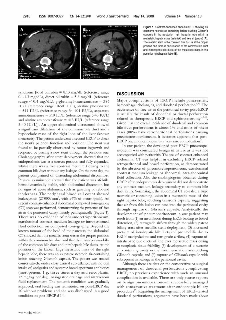
syndrome [total bilirubin = 8.13 mg/dL (reference range 0.1-1.3 mg/dL), direct bilirubin = 5.6 mg/dL (reference range < 0.4 mg/dL), γ-glutamyl-transaminase = 386 IU/L (reference range 10-50 IU/L), alkaline phosphatase = 541 IU/L (reference range 34-104 IU/L), aspartate aminotransferase = 510 IU/L (reference range 5-40 IU/L) and alanine aminotransferase = 413 IU/L (reference range 5-40 IU/L)]. An upper abdominal ultrasound showed a significant dilatation of the common bile duct and a hypoechoic mass of the right lobe of the liver (known metastasis). The patient underwent a second ERCP to check the stent’s patency, function and position. The stent was found to be partially obstructed by tumor ingrowth and reopened by placing a new stent through the previous one. Cholangiography after stent deployment showed that the endoprothesis was at a correct position and fully expanded, whilst there was a free contrast medium flowing to the common bile duct without any leakage. On the next day, the patient complained of distending abdominal discomfort. Physical examination showed that the patient was afebrile, hemodynamically stable, with abdominal distension but no signs of acute abdomen, such as guarding or rebound tenderness. The peripheral blood cell count showed leukocytosis (27 000/mm3, with 94% of neutrophils). An urgent contrast-enhanced abdominal computed tomography (CT) scan was performed, demonstrating the presence of free air in the peritoneal cavity, mainly perihepatically (Figure 1). There was no evidence of pneumoretroperitoneum, extraluminal contrast medium leakage or intraperitoneal fluid collection on computed tomography. Beyond the known tumour of the head of the pancreas, the abdominal CT showed that the metallic stent was at the proper position within the common bile duct and that there was pneumobilia of the common bile duct and intrahepatic bile ducts. At the position of the known large metastatic mass of the right hepatic lobe, there was an extensive necrotic air-containing lesion touching Glisson’s capsule. The patient was treated conservatively, under close clinical surveillance, with no oral intake of, analgesics and systemic broad-spectrum antibiotics (meropenem, 1 g, three times a day and teicoplanin, 12 mg/kg per day), nasogastric drainage and intravenous fluid replacement. The patient’s condition was gradually improved, oral feeding was reinstituted on post-ERCP day 10 without problems and she was discharged in a good condition on post-ERCP d 14.
DISCUSSIONMajor complications of ERCP include pancreatitis, hemorrhage, cholangitis, and duodenal perforation[4,5]. The occurrence of free air in the peritoneal cavity post-ERCP is usually the result of duodenal or ductal perforation related to therapeutic ERCP and sphincterotomy[4,6-8]. Given that the overall incidence of duodenal and common bile duct perforations is about 1% and most of these cases (80%) have retroperitoneal perforations causing pneumoretroperitoneum, it becomes apparent that post-ERCP pneumoperitoneum is a very rare complication[4].
In our patient, the developed post-ERCP pneumope-ritoneum was considered benign in nature as it was not accompanied with peritonitis. The use of contrast-enhanced abdominal CT was helpful in excluding ERCP-related retroperitoneal and bowel perforation, as demonstrated by the absence of pneumoretroperitoneum, extraluminal contrast medium leakage or abnormal intra-abdominal fluid collection. Also the cholangiogram obtained during ERCP after endoprothesis deployment did not demonstrate any contrast medium leakage secondary to common bile duct injury. Surprisingly, the abdominal CT revealed a large necrotic air-containing lesion in a metastatic mass of the right hepatic lobe, touching Glisson’s capsule, suggesting that air from this lesion can pass into the peritoneal cavity through rupture of Glisson’s capsule. Analytically, the development of pneumoperitoneum in our patient may result from (1) air insufflation during ERCP leading to bowel distension, (2) retrograde airflow through the widely patent biliary tract after metallic stent deployment, (3) increased pressure of intrahepatic bile ducts and pneumobilia due to ERCP manipulations and retrograde airflow, (4) rupture of intrahepatic bile ducts of the liver metastatic mass owing to neoplastic tissue friability, (5) development of a necrotic air containing cavity in the liver metastatic mass touching Glisson’s capsule, and (6) rupture of Glisson’s capsule with subsequent air leakage in the peritoneal cavity.
Although there are data on the conservative or surgical management of duodenal perforations complicating ERCP, no previous experience with such an unusual complication is available. There are only scarce reports on benign pneumoperitoneum successfully managed with conservative treatment after endoscopic biliary procedures[9]. Regarding the management of ERCP-related duodenal perforations, arguments have been made about
→
*
A B
www.wjgnet.com
2918 ISSN 1007-9327 CN 14-1219/R World J Gastroenterol May 14, 2008 Volume 14 Number 18
Figure 1 Contrast-enhanced abdominal CT showing an extensive necrotic air-containing lesion touching Glisson’s capsule in the posterior right hepatic lobe within a metastatic hepatic mass (asterisk) and free air (arrow) (A). The metallic stent in the common bile duct is at the proper position and there is pneumobilia of the common bile duct and intrahepatic bile ducts of the metastatic mass in the posterior right hepatic lobe (B).

both surgical[10-12] and non-surgical management[13]. Stapfer and colleagues[14] analyzed fourteen cases of ERCP-related perforations and concluded that patients with no peritonitis, sepsis, significant contrast medium leak during ERCP or follow-up upper gastrointestinal study, and retro- or intraperitoneal fluid collections during computed tomography, can be successfully managed conservatively. Based on these criteria, we decided to treat our patient conservatively under frequent clinical re-evaluation for the early recognition of the potential need for surgical treatment. Our patient’s rare post-ERCP complication was successfully treated conservatively without need for surgical intervention.
In conclusion, this is the first report of post-ERCP pneumoperitoneum caused by rupture of intrahepatic bile ducts and Glisson’s capsule in the area of a large peripheral hepatic metastasis. This case indicates the need for close clinical and radiological observation of patients with hepatic masses (primary or metastatic) subjected to ERCP. In such patients, avoidance of excessive air insufflation during ERCP and/or placement of a nasogastric tube for bowel decompression immediately after ERCP might be a reasonable strategy to prevent such unusual complications.
REFERENCES1 Carr-Locke DL. Therapeutic role of ERCP in the management
of suspected common bile duct stones. Gastrointest Endosc 2002; 56: S170-S174
2 Koklu S, Parlak E, Yuksel O, Sahin B. Endoscopic retrograde cholangiopancreatography in the elderly: a prospective and comparative study. Age Ageing 2005; 34: 572-577
3 Thomopoulos KC , Vagenas K, Assimakopoulos SF, Giannikoulis C, Arvaniti V, Pagoni N, Nikolopoulou VN. Endoscopic retrogade cholangiopancreatography is safe and effective method for diagnosis and treatment of biliary and pancreatic disorders in octogenarians. Acta Gastroenterol Belg
2007; 70: 199-2024 Loperfido S, Angelini G, Benedetti G, Chilovi F, Costan F, De
Berardinis F, De Bernardin M, Ederle A, Fina P, Fratton A. Major early complications from diagnostic and therapeutic ERCP: a prospective multicenter study. Gastrointest Endosc 1998; 48: 1-10
5 Salminen P , Laine S, Gull ichsen R. Severe and fatal complications after ERCP: Analysis of 2555 procedures in a single experienced center. Surg Endosc 2008; 22: 1965-1970
6 Baron TH. Complications of endoscopic biliary sphinctero-tomy: the first cut is the deepest. Clin Gastroenterol Hepatol 2004; 2: 968-970
7 Menuck L, Siemers PT. Pneumoperitoneum: importance of right upper quadrant features. AJR Am J Roentgenol 1976; 127: 753-756
8 Pannu HK , Fishman EK. Complications of endoscopic retrograde cholangiopancreatography: spectrum of abnormalities demonstrated with CT. Radiographics 2001; 21: 1441-1453
9 Hui YT, Lam WM, Lam TW, Cheung WC, Sze SF, Wong CT. Benign pneumoperitoneum developed after endoscopic biliary metallic stent placement with the rendezvous procedure. Gastrointest Endosc 2008; 67: 179-180
10 Sarr MG, Fishman EK, Milligan FD, Siegelman SS, Cameron JL. Pancreatitis or duodenal perforation after peri-Vaterian therapeutic endoscopic procedures: diagnosis, differentiation, and management. Surgery 1986; 100: 461-466
11 Bell RC , Van Stiegmann G, Goff J, Reveille M, Norton L, Pearlman NW. Decision for surgical management of perforation following endoscopic sphincterotomy. Am Surg 1991; 57: 237-240
12 Martin DF, Tweedle DE. Retroperitoneal perforation during ERCP and endoscopic sphincterotomy: causes, clinical features and management. Endoscopy 1990; 22: 174-175
13 Scarlett PY, Falk GL. The management of perforation of the duodenum following endoscopic sphincterotomy: a proposal for selective therapy. Aust N Z J Surg 1994; 64: 843-846
14 Stapfer M, Selby RR, Stain SC, Katkhouda N, Parekh D, Jabbour N, Garry D. Management of duodenal perforation after endoscopic retrograde cholangiopancreatography and sphincterotomy. Ann Surg 2000; 232: 191-198
S- Editor Li DL L- Editor Wang XL E- Editor Ma WH
www.wjgnet.com
Assimakopoulos SF et al. Post-ERCP pneumoperitoneum 2919

Online Submissions: wjg.wjgnet.com World J Gastroenterol 2008 May 14; 14(18): 2920-2923www.wjgnet.com World Journal of Gastroenterology ISSN [email protected] © 2008 WJG. All rights reserved.
A case of biliary stones and anastomotic biliary stricture after liver transplant treated with the rendez - vous technique and electrokinetic lithotritor
Marta Di Pisa, Mario Traina, Roberto Miraglia, Luigi Maruzzelli, Riccardo Volpes, Salvatore Piazza, Angelo Luca, Bruno Gridelli
www.wjgnet.com
CASE REPORT
Marta Di Pisa, Mario Traina, Department of Gastroenterology, IsMeTT, UPMC, Via Tricomi, Palermo 90100, ItalyRoberto Miraglia, Luigi Maruzzelli, Department of Radiology, IsMeTT, UPMC, Via Tricomi 1, Palermo 90100, ItalyRiccardo Volpes, Department of Hepatology, IsMeTT, UPMC, Palermo 90100, ItalySalvatore Piazza, Civico Hospital, Via Tricomi, Palermo 90100, ItalyAngelo Luca, Department of Radiology, IsMeTT, UPMC, Palermo 90100, ItalyBruno Gridelli, Department of Surgery, IsMeTT, UPMC, Palermo 90100, ItalyAuthor contributions: Di Pisa M and Traina M designed the work; Di Pisa M wrote the paper with a contribution from Volpes R and Miraglia R; The rest of the authors contributed equally to this work.Correspondence to: Marta Di Pisa, Dr, IsMeTT, UPMC, Via Tricomi 1, Palermo 90100, Italy. [email protected]: +39-91-2192111 Fax: +39-91-2192400Received: January 17, 2008 Revised: March 19, 2008
AbstractThe paper studies the combined radio logic and endoscopic approach (rendez vous technique) to the treatment of the biliary complications following liver transplant. The “rendez-vous” technique was used with an electrokinetic lithotripter, in the treatment of a biliary anastomotic stricture with multiple biliary stones in a patient who underwent orthotopic liver transplant. In this patient, endoscopic or percutaneous transhepatic management of the biliary complication failed. The combined approach, percutaneous transhepatic and endoscopic treatment (rendez-vous technique) with the use of an electrokinetic lithotritor, was used to solve the biliary stenosis and to remove the stones. Technical success, defined as disappearance of the biliary stenosis and stone removal, was obtained in just one session, which definitively solved the complications. The combined approach of percutaneous transhepatic and endoscopic (rendez-vous technique) treatment, in association with an electrokinetic lithotritor, is a safe and feasible alternative treatment, especially after the failure of endoscopic and/or percutaneous trans-hepatic isolated procedures.
© 2008 WJG. All rights reserved.
Key words: Biliary complications; Liver transplant; Endoscopic retrograde cholangiopancreatography; Percutaneous Transhepatic Cholangiography; Percutaneous lithotripsy
Peer reviewer: Dario Conte, Professor, GI Unit - IRCCS Osp. Maggiore, Chair of Gastroenterology, Via F. Sforza, 35, Milano 20122, Italy
Di Pisa M, Traina M, Miraglia R, Maruzzelli L, Volpes R, Piazza S, Luca A, Gridelli B. A case of biliary stones and anastomotic biliary stricture after liver transplant treated with the rendez - vous technique and electrokinetic lithotritor. World J Gastroenterol 2008; 14(18): 2920-2923 Available from: URL: http://www.wjg-net.com/1007-9327/14/2920.asp DOI: http://dx.doi.org/10.3748/wjg.14.2920
INTRODUCTIONBiliary complications after liver transplant are a very common and impor tant prob lem (8%-50%) [1,2 ]. Untreated biliary complications are associated with high morbidity and mortality rates. Magnetic resonance is an effective technique in the diagnosis of the biliary complicat ions [3], whereas Endoscopic retrograde cholangiopancreatography (ERCP)[4,5], and Percutaneous Transhepatic Cholangiography (PTC) remain the gold standard therapeutic options (with successful results in between 70% and 80% of cases)[6,7]. Common biliary complications after liver transplant are leaks and strictures, which are divided into anastomotic and non-anastomotic groups. Other less frequent complications include stones, cuts, and oddities[8,9].
We report a case of biliary anastomotic stenosis with multiple biliary stones in a patient after orthotopic liver transplantation, treated with a combined percutaneous radiologic and endoscopic approach (rendez - vous technique) and with the use of an electrokinetic lithotritor.
CASE REPORTA 67-year-o ld man underwent or thotop ic l iver transplantation for HCV related cirrhosis in 2000, with a choledocho - choledocho anastomosis. The surgical procedure was complicated by renal failure which

Di Pisa M et al . Biliary complication of liver transplant 2921
www.wjgnet.com
was treated with hemodialysis for 1 mo. No vascular abnormalities were present at the Doppler ultrasound examination. Five years later, after an unsuccessful antiviral treatment for HCV recurrence, medium size esophageal varices were diagnosed. Other co-morbidities present at that time were severe obesity and hypertension.
The patient was recently admitted to our facility because of the onset of severe cholestasis with episodes of cholangitis, and for a general reassessment of the liver disease.
At admission, physical examination showed severe obesity (BMI > 30) and umbilical laparocele (Figure 1). Laboratory data were the following: AST/ALT 130/187 U/L (normal, 5/65 U/L-40/65 U/L), bilirubin tot/dir 26.46/19.69 mg/dL (0-1.5 mg/dL), alkaline phosphates 357 U/L (40 U/L-134 U/L), gamma-GT 1161 mg/L (5 U/L-85 U/L). A Magnetic Resonance Cholangiography was not performed because of the severe obesity; ERCP was therefore planned. Under general anesthesia, the choledochus was cannulated, showing a mild anastomotic stricture with a very tight angle, just above the stricture, multiple stones above the stenosis and at the confluence, and mild intrahepatic biliary dilation (Figure 2). A guide wire was passed through the stenosis, but because of the stones and the kinking, it was impossible to cross, by the guide wire, either Dormia basket, Fogarty or other catheters. Because of ERCP failure, a PTC was planned. In the angiographic room, under sonographic and fluoroscopic control, a peripheral right biliary duct was punctured with a 20 G needle. A cholangiogram showed a mild biliary dilation and multiple filling defects. It was impossible to put a guide wire into the choledochus, and thus a 6.6 F Ring external cathether (Boston Scientific, MA, USA) was placed for bile drainage. One week later, a second cholangiogram was performed and a 6.6 Fr external - internal biliary catheter (Boston Scientific) was successfully placed (Figure 3).
In the following days, because of worsening jaundice (bilirubin tot/dir 34.22/28.78 mg/dL), a combined radiologic and endoscopic technique (rendez vous technique) was performed. In the angiographic suite under general anesthesia, the patient was monitored continuously with electrocardiography, pulse oximeter and automatic recording of blood pressure and pulse. Intravenous
antibiotic prophylaxis was given before the procedure. After visualizing the biliary tree, and under fluoroscopic control, on an Amplatz guide wire, the biliary drainage was changed with a 7 Fr vascular introductory (St. Jude Medical, Minnetonka, USA).
With an electro - kinetic lithotriptor, a ballistic lithotripter which uses high-energy magnetic fields (Electro Medical Supplies, LTD), placed trough the introductory, percutaneous lithotripsy was performed (Figure 4A). A percutaneous balloon dilatation of the anastomotic stricture was then performed using an 8 mm balloon catheter (Bluemax, Boston Scientific, MA, USA) and, at the same time, under endoscopic control, the stones were completely removed with a Fogarty balloon and a Dormia basket (Figure 4B). A final cholangiogram showed no filling defects or stricture (Figure 4C) and, at the end of the procedure, a 12 Fr external - internal biliary drainage was finally placed (Boston Scientific, MA, USA) (Figure 4D).
Two days later, laboratory data showed: AST/ALT 28/29 U/L (normal, 5/65-40/65 U/L), bilirubin tot/dir 6.5/5.3 mg/dL (0-1.5 mg/dL), alkaline phosphates 104 U/L (40-134 U/L), gamma-GT 107 mg/L (5-85 U/L).
Two months after the procedure, the patient has a biliary catheter in place, is asymptomatic, in good general condition, without signs of cholestasis and in follow up for biliary catheter removal.
DISCUSSIONBiliary complications after liver transplant are common and biliary stones represent a small part of these[1,2]. In
Figure 1 A CT scan showing severe obesity and umbilical laparocele.
Figure 2 A first attempt of ERCP showing multiple stones and anastomotic stricture.
Figure 3 PTC with external - internal catheter placement.

A
the majority of cases, ERCP alone is not only the best diagnostic and therapeutic treatment with a successful result in 70%-80% of cases[4,5,10] but is also considered the least invasive procedure, although with some important complications[9]. Western studies showed the efficacy of percutaneous transhepatic choledochoscopy (PTHC) and holmium:yttrium-aluminum-garnet (YAG) laser to remove biliary stones in patients unable or unwilling to undergo endoscopic or surgical removal, but this technique requires much time and prolonged biliary access[11].
In our patient, endoscopic management was attempted first, but we were unable to place a stent in a correct position or to remove the stones, because of the severe biliary stenosis. By a percutaneous transhepatic approach however, we were able to place an external - internal biliary catheter, though not to remove the stones or solve the stenosis.
By a combined technique - radiologic and endoscopic procedure (i.e. the “rendez-vous” technique), associated with the use of a ballistic lithotripter which usually fragments nearly all urinary calculi, we were able to remove the stones and to solve the stenosis.
The rendez-vous technique has been described in the treatment of benign or malignant biliary obstruction, in traumatic bile duct injury repair and in the treatment of large biliary leaks in a liver transplant recipient[12-15]. But, to our knowledge, this is the first report of the use of this technique in treatment of biliary stones and anastomotic stenosis, with the use of a ballistic lithotripter in a patient after liver transplantation.
In conclusion, the radiological/endoscopic rendez-vous technique, associated with an electrokinetic lithotritor, is a feasible alternative for stone removal and biliary stricture treatment, especially after the failure of endoscopic
and/or percutaneous trans-hepatic isolated procedures. Also, this combined approach improve costs, timing and effectiveness in the therapy of biliary complications.
REFERENCES1 Stratta RJ, Wood RP, Langnas AN, Hollins RR, Bruder KJ,
Donovan JP, Burnett DA, Lieberman RP, Lund GB, Pillen TJ. Diagnosis and treatment of biliary tract complications after orthotopic liver transplantation. Surgery 1989; 106: 675-683; discussion 683-684
2 Greif F, Bronsther OL, Van Thiel DH, Casavilla A, Iwatsuki S, Tzakis A, Todo S, Fung JJ, Starzl TE. The incidence, timing, and management of biliary tract complications after orthotopic liver transplantation. Ann Surg 1994; 219: 40-45
3 Boraschi P, Braccini G, Gigoni R, Sartoni G, Neri E, Filipponi F, Mosca F, Bartolozzi C. Detection of biliary complications after orthotopic liver transplantation with MR cholangiography. Magn Reson Imaging 2001; 19: 1097-1105
4 Pfau PR, Kochman ML, Lewis JD, Long WB, Lucey MR, Olthoff K, Shaked A, Ginsberg GG. Endoscopic management of postoperative biliary complications in orthotopic liver transplantation. Gastrointest Endosc 2000; 52: 55-63
5 Thuluvath PJ, Atassi T, Lee J. An endoscopic approach to b i l iary complicat ions fo l lowing orthotopic l iver transplantation. Liver Int 2003; 23: 156-162
6 Ernst O, Sergent G, Mizrahi D, Delemazure O, L'Hermine C. Biliary leaks: treatment by means of percutaneous transhepatic biliary drainage. Radiology 1999; 211: 345-348
7 Cozzi G, Severini A, Civelli E, Milella M, Pulvirenti A, Salvetti M, Romito R, Suman L, Chiaraviglio F, Mazzaferro V. Percutaneous transhepatic biliary drainage in the management of postsurgical biliary leaks in patients with nondilated intrahepatic bile ducts. Cardiovasc Intervent Radiol 2006; 29: 380-388
8 Pascher A, Neuhaus P. Bile duct complications after liver transplantation. Transpl Int 2005; 18: 627-642
9 Khuroo MS, Al Ashgar H, Khuroo NS, Khan MQ, Khalaf HA, Al-Sebayel M, El Din Hassan MG. Biliary disease after liver transplantation: the experience of the King Faisal Specialist
Figure 4 A: With a ballistic lithotripter, placed through the introductory, percutaneous lithotripsy was performed; B : Fragmented stones were removed with a Fogarty bal loon; C : Normal Cholangiography after litotripsy and stenosis dilation; D: An external - internal catheter was placed.
B
DC
www.wjgnet.com
2922 ISSN 1007-9327 CN 14-1219/R World J Gastroenterol May 14, 2008 Volume 14 Number 18

Hospital and Research Center, Riyadh. J Gastroenterol Hepatol 2005; 20: 217-228
10 Loperfido S, Angelini G, Benedetti G, Chilovi F, Costan F, De Berardinis F, De Bernardin M, Ederle A, Fina P, Fratton A. Major early complications from diagnostic and therapeutic ERCP: a prospective multicenter study. Gastrointest Endosc 1998; 48: 1-10
11 Piraka C, Shah RJ, Awadallah NS, Langer DA, Chen YK. Transpapillary cholangioscopy-directed lithotripsy in patients with difficult bile duct stones. Clin Gastroenterol Hepatol 2007; 5: 1333-1338
12 Hazey JW, McCreary M, Guy G, Melvin WS. Efficacy of percutaneous treatment of biliary tract calculi using the
holmium:YAG laser. Surg Endosc 2007; 21: 1180-118313 Wayman J, Mansfield JC, Matthewson K, Richardson DL,
Griffin SM. Combined percutaneous and endoscopic procedures for bile duct obstruction: simultaneous and delayed techniques compared. Hepatogastroenterology 2003; 50: 915-918
14 Aytekin C, Boyvat F, Yimaz U, Harman A, Haberal M. Use of the rendezvous technique in the treatment of biliary anastomotic disruption in a liver transplant recipient. Liver Transpl 2006; 12: 1423-1426
15 Miraglia R , Traina M, Maruzzelli L, Caruso S, Di Pisa M, Gruttadauria S, Luca A, Gridelli B. Usefulness of the "Rendezvous" Technique in Living Related Right Liver Donors with Postoperative Biliary Leakage from Bile Duct Anastomosis. Cardiovasc Intervent Radiol 2008; 31: 999-1002
S- Editor Zhong XY L- Editor Lalor PF E- Editor Lu W
www.wjgnet.com
Di Pisa M et al . Biliary complication of liver transplant 2923

Online Submissions: wjg.wjgnet.com World J Gastroenterol 2008 May 14; 14(18): 2924-2927www.wjgnet.com World Journal of Gastroenterology ISSN [email protected] © 2008 WJG. All rights reserved.
An autopsy case of granulocyte-colony-stimulating-factor-producing extrahepatic bile duct carcinoma
Satoru Matsuyama, Tomonori Shimonishi, Hirofumi Yoshimura, Kensaku Higaki, Kenji Nasu, Mariko Toyooka, Shigehisa Aoki, Keiko Watanabe, Hajime Sugihara
www.wjgnet.com
CASE REPORT
Satoru Matsuyama, Tomonori Shimonishi, Hirofumi Yoshimura, Kensuke Higaki, Kenji Nasu, Department of Surgery, Takagi Hospital, 141-11, Sakemi, Ohkawa-City, Fukuoka 831-0016, JapanMariko Toyooka, Keiko Watanabe, Hajime Sugihara, Department of Pathology, Takagi Hospital, 141-11, Sakemi, Ohkawa-City, Fukuoka 831-0016, JapanShigehisa Aoki, Department of Pathology and Biodefence, Faculty of Medicine, Saga University, 5-1-1, Nabeshima, Saga-City, Saga 849-8501, JapanAuthor contributions: Matsuyama S, Shimonishi T, Yoshimura H, Higaki K and Nasu K treated the patient; Toyooka M, Watanabe K and Sugihara H performed cytological and pathological diagnosis, as well as autopsy, in cooperation with Aoki S.Correspondence to: Dr. Satoru Matsuyama, Department of Surgery, Takagi Hospital, 141-11, Sakemi, Ohkawa-City, Fukuoka 831-0016, Japan. [email protected]: +81-944-870001 Fax: +81-944-87 9310Received: November 6, 2007 Revised: March 16, 2008
AbstractA 79-year-old man was referred to this department due to the presence of extrahepatic bile duct carcinoma with a tumor at the left chest wall. The lesion was suspected to be a metastasis of bile duct carcinoma to the left wall, however, computed tomography (CT) revealed no regional lymph node or liver metastases. In addition, cytological and pathological examinations did not show malignancy. At the time of admission, the white blood cell count was 21 460 cells/μL (neutrophils, 18 240 cells/μL) and this elevated to 106 040 before death. In addition, serum granulocyte colony-stimulating factor (G-CSF) was elevated. At 28 d after admission, the patient died. An autopsy showed a poorly differentiated adenocarcinoma with sarcomatous change, which had slightly invaded into the pancreas around the bile duct, and was found in the distal bile duct with multiple metastases to the chest wall, lung, kidney, adrenal body, liver, mesentery, vertebra and mediastinal and para-aortic lymph nodes, without locoregional lymph node and liver metastasis. The cancer cells showed positive immunohistochemical staining for anti-G-CSF antibody. This is believed to be the first report of an extrahepatic bile duct carcinoma that produces G-CSF. Since G-CSF-producing carcinoma and sarcomatous change of the biliary tract leads to poor prognosis, early diagnosis and treatment are needed. When infection is ruled out, the G-CSF in serum should be examined. In addition, examinations such as bone
scintigraphy and chest CT should also be considered for distant metastasis.
© 2008 WJG. All rights reserved.
Key words: Bile duct carcinoma; Granulocyte colony-stimulating factor; Multiple metastases; Autopsy
Peer reviewer: Tom H Karlsen, MD, Institute of Immunology, Rikshospitalet University Hospital, N-0027 Oslo, Norway
Matsuyama S, Shimonishi T, Yoshimura H, Higaki K, Nasu K, Toyooka M, Aoki S, Watanabe K, Sugihara H. An autopsy case of granulocyte-colony-stimulating-factor-producing extrahepatic bile duct carcinoma. World J Gastroenterol 2008; 14(18): 2924-2927 Available from: URL: http://www.wjgnet.com/1007-9327/14/2924.asp DOI: http://dx.doi.org/10.3748/wjg.14.2924
INTRODUCTIONGranulocyte colony-stimulating factor (G-CSF) produced in some cancers is thought to be an autocrine growth factor[1,2]. Therefore, patients with G-CSF-producing cancer generally have a poor prognosis, and early diagnosis and treatment are required. Biliary cancer, such as cholangiocellular and gallbladder carcinoma are reported to produce G-CSF in some cases[3-10], however, extrahepatic bile duct cancer that produces G-CSF has not yet been reported.
This report describes a case of extrahepatic bile duct cancer that produced G-CSF, in which neither regional lymph node metastasis nor massive progression was observed by radiological examination, but autopsy showed multiple organ metastasis.
CASE REPORTA 79-year-old man who suffered from left lateral chest pain for one month was referred to our department for treatment of a tumor on the left chest wall and extrahepatic bile duct cancer found during an annual medical examination. His body temperature was 36.9℃. From the left axillar to anterior axillary line of the left chest wall, many nodules, 5-10 mm in diameter, were palpated, without tenderness. He had no history of chest trauma. Chest radiography showed massive pleural effusion

Matsuyama S et al . G-CSF-producing bile duct carcinoma 2925
www.wjgnet.com
and a tumor that elevated smoothly from the left chest wall, near which the fifth rib could not be traced (Figure 1). Enhanced chest computed tomography (CT) depicted a mass with necrosis inside the tumor and enhancement on its border, as well as two osteolytic lesions on the seventh thoracic vertebra, swelling of many anterior mediastinal and left axillary lymph nodes, and pleural effusion around extrapleural fluid collection (Figure 2). Enhanced magnetic resonance imaging (MRI) of the chest showed extensive enhancement of the muscle and connective tissue around the ribs, as well as a cavity within the muscle. Bone scintigraphy showed an abnormal opaque uptake in the left third to fifth ribs and vertebrae (Figure 3). Cytodiagnosis of the pleural and extrapleural effusion showed solitary atypical cells, regarded as histiocytes, scattered in numerous inflammatory cells. Those atypical cells had swelling nuclei and a distinct eosinophilic nucleolus; however, it was difficult to diagnose the lesion as an adenocarcinoma from the chromatin pattern. Abdominal CT demonstrated a gradually enhanced tumor in the distal bile duct, without any regional lymph node and liver metastasis (Figure 4), and magnetic resonance cholangiopancreatography (MRCP) revealed stenosis of the distal common bile duct (Figure 5). Laboratory data showed an increase in the following: white blood cell count (WBC) 21 460 cells/μL (neutrophils, 18 240 cells/μL), ALP 536 IU/L, γ-GTP 300 IU/L, LDH 251 IU/L, CRP 4.33 mg/dL, but carcinoembryonic antigen (CEA), carbohydrate antigen 19-9 (CA19-9) and DU-PAN-2, a tumor marker which is especially elevated in
pancreato-biliary cancer patients, were within the normal limit. The neutrophil alkaline phosphatase (NAP) score was elevated to 381 to exclude chronic myeloblastoid leukemia.
Histological examination of the nodule on the chest wall that was partially resected for diagnosis revealed scattered atypical cells among numerous fibroblasts, macrophages and neutrophils in the skeletal muscle and adipose tissue. These atypical cells showed positive immunohistochemical staining for pan-cytokeratin and vimentin, but not for desmin, which excluded leiomyosarcoma (Figure 6). Since such atypical cells had no epithelial connection or basement membrane, they
Figure 1 Chest radiography r e v e a l e d m a s s i v e p l u r a l effusion and a tumor elevating smoothly from the left chest wall (arrowheads), near which the fifth rib could not be traced.
Figure 2 Chest CCT depicted a mass with necrosis inside the tumor (arrows) and enhancement of its border (arrowheads), and extrapleural tumor and pleural effusion.
Figure 3 Bone scintigraphy showed an abnormal opaque uptake in the left third to fifth ribs (arrows).
Figure 4 Abdominal CT revealed a gradually enhanced tumor in the distal bile duct (arrows), without any regional lymph node or liver metastasis.
Figure 5 MRCP revealed stenosis (arrow heads) of the distal common bile duct.

were thought to be reactive mesothelial and not carcinoma cells. Therefore, the pathological diagnosis was considered to be either proliferative myositis or nodular fasciitis-like granuloma.
Eighteen days after admission the patient’s appetite, consciousness and daily activity deteriorated, and WBC was elevated to 86 050 cells/μL (neutrophils, 80 880 cells/μL) without pyrexia. Until then, no bacterium was seen in the sputum, pleural effusion and fluid in the chest wall cavity. Expecting an improvement of his general condition and inflammation of myositis on the left chest wall, we drained pleural effusion fluid and the patient received an injection of dexamethasone. However, his general condition and laboratory data did not improve. WBC increased to 106 040 cells/μL (neutrophils, 91 900 cells/μL) before death. Twenty-seven days after admission, a multiple cerebral infarction was found, which had not been observed 7 d earlier. The patient died the following day and
underwent an autopsy.The autopsy revealed a poorly different iated
adenocarcinoma with sarcomatous change, which had slightly invaded into the pancreas in the distal bile duct (Figure 7A). In addition, no other primary cancer was observed macroscopically in the chest and abdomen. The cancer cells showed positive immunohistochemical s t a i n i n g f o r G - C S F ( F i g u r e 7 B ) . T h e G - C S F concentration in serum samples archived by the hospital was 53, 38 and 134 pg/mL on 21, 23 and 26 d after admission, respectively (normal < 18 pg/mL). This result was obtained 9 d after death. In a specimen of the left chest wall, solitary cancer cells, numerous surrounding inflammatory cells such as neutrophils and macrophages, had invaded into the skeletal muscles, similar to the biopsy tissue. Therefore, the pathological diagnosis of the left chest wall was a metastasis of the bile duct carcinoma with sarcomatous change, and the anterior mediastinal lymph nodes showed the same histological findings. Moreover, under the serosa of the mesentery and liver, some white nodules, measuring 5 mm in diameter, were macroscopically discovered and their histology showed them to be metastases. In addition, histological examination of all the organs in the chest and abdomen demonstrated multiple metastases in the lung, kidney, adrenal gland, mesentery, vertebral bone marrow and mediastinal and para-aortic lymph nodes. In contrast, no metastasis was found in the regional lymph nodes. These findings proved the presence of extensive systemic dissemination of cancer cells.
Figure 6 Histological examination of the resected chest wall showed scattered atypical cells among numerous inflammatory cells (A). Immunohistochemistry for pan-cytokeratin as an epithelial cell marker (B) and vimentin as a mesenchymal cell marker (C) both showed positivity in these atypical cells.
Figure 7 Histological examination revealed a poorly differentiated adenocarcinoma with sarcomatous change in the distal bile duct, which slightly invaded into the pancreas (A), and cancer cells showed positive immunohistochemical staining for G-CSF (B).
A
B
C
A
B
www.wjgnet.com
2926 ISSN 1007-9327 CN 14-1219/R World J Gastroenterol May 14, 2008 Volume 14 Number 18

DISCUSSIONThis is believed to be the first report of an extrahepatic bile duct carcinoma that produces G-CSF. A search of PubMed by the key words CSF and biliary cancer, revealed three and five reports, respectively, of intrahepatic cholangiocarcinoma and gallbladder carcinoma[3–10], but there have so far been no reports of extrahepatic bile duct malignancy. While two of these patients underwent curative surgery and survived without recurrence for 18 mo and 27 mo, respectively[6,8], three patients died within 6 mo after palliative or exploratory surgery[3,5,9]. In one case, the details of the surgery were unknown[4], and the other was a report of archival histological examination[7]. In a case of rapid progression, the patient died 5 d after admission[10]. As with other organs such as the lung, thyroid and kidney, the prognosis of G-CSF-producing carcinoma in the biliary tract is considered to be poor, and early diagnosis and surgical treatment are needed. In the present case, surgery could not have been performed, even if the diagnosis had been done correctly and rapidly. However, an early examination of G-CSF in the serum would have identified the lesions as multiple metastasis of a common bile duct cancer, thereby leading to more appropriate treatment, such as chemotherapy. To avoid wasting time with additional radiological examination and inefficient treatment such as antibiotics for leukocytosis, G-CSF in serum should be examined when bacterial infection is ruled out for leukocytosis by a routine preoperative examination.
In this case, it was not possible to diagnose the correct extent of the carcinoma, in spite of various radiological and pathological data. This was because the accumulation of inflammatory cells induced by G-CSF produced by the cancer cells in the metastatic lesions mimicked inflammation of the chest wall and vertebra in the radiological and pathological examinations. Furthermore, the lack of loco-regional lymph node and liver metastases in the radiological examination led us to consider other lesions in the differential diagnosis, such as mediastinal lymphadenopathy and chest wall nodules, as an inflammatory reaction from the left chest wall lesion. Although endoscopic ultrasonography was not performed in this case, the loco-regional stage of cancer was diagnosed as stage Ⅰa or Ⅰb. This type of dissociation between the loco-regional and whole body extent of carcinoma has occasionally been reported with gastric cancer[11-13], however, no report has been found regarding extrahepatic bile duct carcinoma. Scattered carcinoma cells in many inflammatory cells, thus indicating sarcomatous change, could not be correctly diagnosed due to these changes, such as the loss of basal membrane and intercellular connection between cancer cells. Shimada et al[14] have reported cholangiocarcinomas with two types of sarcomatous change, spindle and pleomorphic type, which have very poor prognosis, especially the latter type. Moreover, Nakajima et al[15] have shown aggressive intrahepatic spreading and widespread metastasis of the sarcomatous cells of an intrahepatic cholangiocarcinoma.
In the present case, dissociation between the findings of the loco-regional and whole body examination might have been due to sarcomatous change and G-CSF, which is thought to be a growth factor. For a case of extrahepatic bile duct carcinoma that produces G-CSF, an examination for distant metastasis, such as bone scintigraphy and chest CT, should be considered.
REFERENCES1 Kyo S, Kanaya T, Takakura M, Inoue M. A case of cervical
cancer with aggressive tumor growth: possible autocrine growth stimulation by G-CSF and Il-6. Gynecol Oncol 2000; 78: 383-387
2 Tachibana M , Miyakawa A, Nakashima J , Murai M, Nakamura K, Kubo A, Hata J. Autocrine growth promotion by multiple hematopoietic growth factors in the established renal cell carcinoma line KU-19-20. Cell Tissue Res 2000; 301: 353-367
3 Takahashi M, Fujiwara M, Kishi K, Sakai C, Sanada M, Moriyama Y, Shibata A. CSF producing gall bladder cancer: case report and characteristics of the CSF produced by tumor cells. Int J Cell Cloning 1985; 3: 294-303
4 Sakamoto K, Egami H, Yoshimura R, Nakamura S, Ikei S, Mori K, Matsumoto M, Akagi M. Colony-stimulating factor producing carcinoma of the gallbladder. Jpn J Clin Oncol 1986; 16: 87-96
5 Takeda T, Ichiyanagi A, Sano K, Yoshida J, Tsutsumi Y, Miyaji T. A case of gallbladder cancer producing granulocyte-colony-stimulating factor. Gastroenterol Jpn 1990; 25: 762-767
6 Omura N, Abe S, Hirai K, Aoki T. A case of granulocyte-colony stimulating factor producing gallbladder cancer. Am J Gastroenterol 1999; 94: 273-275
7 Sasaki M, Tsuneyama K, Ishikawa A, Nakanuma Y. Intrahepatic cholangiocarcinoma in cirrhosis presents granulocyte and granulocyte-macrophage colony-stimulating factor. Hum Pathol 2003; 34: 1337-1344
8 Ikeda T, Ohgaki K, Miura M, Aishima S, Shimizu T, Maehara Y. Granulocyte-colony stimulating factor-producing gallbladder cancer without recurrence more than 2 years after resection: report of a case. Surg Today 2005; 35: 590-593
9 Amano H, Itamoto T, Emoto K, Hino H, Asahara T, Shimamoto F. Granulocyte colony-stimulating factor-producing combined hepatocellular/cholangiocellular carcinoma with sarcomatous change. J Gastroenterol 2005; 40: 1158-1159
10 Sohda T, Shiga H, Nakane H, Watanabe H, Takeshita M, Sakisaka S. Cholangiocellular carcinoma that produced both granulocyte-colony-stimulating factor and parathyroid hormone-related protein. Int J Clin Oncol 2006; 11: 246-249
11 Suzuki S, Kosugi S, Kuwabara S, Ueki K, Oka Y, Nishimaki T, Aizawa K, Suzuki T, Soga J, Hatakeyama K. Tumor recurrence in patients with early gastric cancer: a clinicopathologic evaluation. J Exp Clin Cancer Res 1998; 17: 187-191
12 Shiomi M, Kamisako T, Yutani I, Kudo M, Shigeoka H, Tanaka A, Okuno K, Yasutomi M. Two cases of histopathologically advanced (stage IV) early gastric cancers. Tumori 2001; 87: 191-195
13 Kobayashi M, Araki K, Matsuura K, Kawai S, Moriki T. Early gastric cancer giving rise to bone and brain metastases--a review of the Japanese literature. Hepatogastroenterology 2002; 49: 1751-1754
14 Shimada M, Takenaka K, Rikimaru T, Hamatsu T, Yamashita Y, Kaj iyama K, Taguchi K, Shirabe K, Sugimachi K. Characteristics of sarcomatous cholangiocarcinoma of the liver. Hepatogastroenterology 2000; 47: 956-961
15 Nakajima T, Tajima Y, Sugano I, Nagao K, Kondo Y, Wada K. Intrahepatic cholangiocarcinoma with sarcomatous change. Clinicopathologic and immunohistochemical evaluation of seven cases. Cancer 1993; 72: 1872-1877
S- Editor Li DL L- Editor Kerr C E- Editor Lu W
www.wjgnet.com
Matsuyama S et al . G-CSF-producing bile duct carcinoma 2927
Online Submissions: wjg.wjgnet.com World J Gastroenterol 2008 May 14; 14(18): 2928-2931www.wjgnet.com World Journal of Gastroenterology ISSN [email protected] © 2008 WJG. All rights reserved.
A ruptured large extraluminal ileal gastrointestinal stromal tumor causing hemoperitoneum
Shoji Hirasaki, Kohei Fujita, Minoru Matsubara, Hiromitsu Kanzaki, Hiromichi Yamane, Masato Okuda, Seiyuu Suzuki, Atsuko Shirakawa, Hideyuki Saeki
www.wjgnet.com
CASE REPORT
Shoji Hirasaki, Kohei Fujita, Minoru Matsubara, Hiromitsu Kanzaki, Hiromichi Yamane, Masato Okuda, Seiyuu Suzuki, Department of Internal Medicine, Sumitomo Besshi Hospital, Niihama 7928543, JapanAtsuko Shirakawa, Department of Pathology, Sumitomo Besshi Hospital, Niihama 7928543, JapanHideyuki Saeki, Department of Surgery, Sumitomo Besshi Hospital, Niihama 7928543, JapanAuthor contributions: Hirasaki S, Fujita K, Matsubara M, Kanzaki H, Yamane H, Okuda M and Suzuki S were involved in the care of the patient; Shirakawa A studied the specimen; Saeki H performed the surgery; Hirasaki S wrote the paper.Correspondence to: Shoji Hirasaki, Department of Internal Medicine, Sumitomo Besshi Hospital, 3-1 Ohji-cho, Niihama 7928543, Japan. [email protected] Telephone: +81-897-377111 Fax: +81-897-377121 Received: January 29, 2008 Revised: March 11, 2008
AbstractWe describe an 87-year-old woman with a large ileal gastrointestinal stromal tumor (GIST) causing hemoperitoneum. A CT scan demonstrated a large heterogeneous mass measuring about 13 cm × 11 cm in the pelvis and hemoperitoneum, with a non-uniform enhancement pattern. The mass was diagnosed as a GIST originating from the gastrointestinal tract. She underwent an urgent laparotomy and an ileal GIST with a rupture was found 130 cm from the anal to the Treitz’s ligament. Hemoperitoneum caused by ileal GIST rupture is a rare condition. Bleeding in the large tumor leading to rupture of the capsule might cause hemoperitoneum in the present case.
© 2008 WJG. All rights reserved.
Key words: Intest inal neoplasm; Smal l intest ine; Extraluminal growth; Laparotomy
Peer reviewers: Ton Lisman, PhD, Thrombosis and Haemostasis Laboratory, Department of Haematology G.03.550, University Medical Centre, Heidelberglaan 100, 3584 CX Utrecht, The Netherlands; Vincent W Yang, Professor and Director, 201 Whitehead Research Building, 615 Michael Street, Atlanta, GA 30322, United States; Jayaram Menon, Head, Department of Medicine, Queen Elizabeth Hospital, Department of Medicine, Queen Elizabeth Hospital, Kota Kinabalu, Sabah, Malaysia
Hirasaki S, Fujita K, Matsubara M, Kanzaki H, Yamane H, Okuda M, Suzuki S, Shirakawa A, Saeki H. A ruptured large extraluminal
ileal gastrointestinal stromal tumor causing hemoperitoneum. World J Gastroenterol 2008; 14(18): 2928-2931 Available from: URL: http://www.wjgnet.com/1007-9327/14/2928.asp DOI: http://dx.doi.org/10.3748/wjg.14.2928
INTRODUCTIONGastrointestinal stromal tumor (GIST) is the designation for a major subset of mesenchymal tumors of the gastrointestinal (GI) tract[1-5]. GIST arising in the digestive tract is most commonly located in the stomach and small intestine[6,7]. GIST originating from the small intestine rarely causes hemoperitoneum[8]. Herein, we describe a relatively rare case of extraluminal ileal GIST causing hemoperitoneum.
CASE REPORTAn 87-year-old woman presented with the symptom of a short loss of consciousness. She was in good health with no specific family or past medical history. Her body temperature was 36.7℃, blood pressure was 148/82 mmHg, radial pulse rate was 72 beats/min and regular. She had slight anemia, but no jaundice. Neurological examination revealed no abnormal findings and lymphadenopathy. Abdominal palpation revealed tenderness in the right lower quadrant. Laboratory tests showed a red blood cell count of 315 × 104/μL [normal range (NR), 380-500 × 104/μL], a white blood cell count of 10 500/μL (NR, 4000-9000/μL), a platelet count of 29.8 × 10⁴/μL, and a hemoglobin concentration of 10.1 g/dL (NR, 12-16 g/dL). The levels of hepatic and biliary enzymes, such as aspartate aminotransferase (AST), a lanine aminotransferase (ALT), a lka l ine phosphatase (ALP), leucin aminopeptidase (LAP), and γ-glutamyltranspeptidase (γ-GTP) were normal except for lactate dehydrogenase (LDH) which was 336 IU/L (NR, 106-211 IU/L). A test for C reactive protein revealed a level of 30.3 mg/dL (NR, < 0.5 mg/dL). Renal function tests showed that the blood urea nitrogen level was 38.0 mg/dL (NR, 8-20 mg/dL) and the creatinine level was normal. Serological studies for hepatitis B and C viruses were negative. Urinary protein and sugar were negative. A computed tomography (CT) scan demonstrated a large heterogeneous mass measuring about 13 cm × 11 cm in the pelvis and hemoperitoneum, with a non-

Hirasaki S et al . Ileal GIST causing hemoperitoneum 2929
www.wjgnet.com
uniform enhancement pattern (Figure 1). Based on the imaging examination, this tumor was diagnosed as a GIST originating from the GI tract or omentum, although it should be distinguished from an ovarian tumor or an adenocarcinoma of the small bowel. The patient underwent an urgent laparotomy.
At laparotomy, a 13 cm × 11 cm semipedunculated solid tumor that was 130 cm from the anal to the Treitz’s ligament, showed extraluminal growth (Figure 2A). The tumor was ruptured with no peritoneal metastasis, and partial resection of the ileum was carried out. The resected tumor was brown-red in color, and had bleeding blood clots (Figure 2B). Histological examination of the resected specimen revealed interlaced bundles of large Bizarre spindle-like tumor cells without mitotic figures (Figure 3A). No fission images were evident. Tumor cells were present in the subserosa (Figure 3B). Immunohistological findings
were negative for CD34, α-smooth muscle actin (SMA), desmin and S-100 protein, but positive for CD117. Based on the above findings, this tumor was diagnosed as a malignant GIST. The postoperative course was uneventful. The patient has been followed up for 16 mo with no evidence of recurrence.
DISCUSSIONGIST is the most common mesenchymal tumor of the GI tract and expresses c-kit protein, also known as CD117, which is considered a highly specific marker differentiating GIST from other mesenchymal tumors, such as leiomyomas[9-11]. The majority of GISTs occur in the stomach (60%-70%) and small intestine (20%-30%)[10]. Approximately, 10%-30% of patients with GIST may be asymptomatic[11]. Gastric and small intestinal stromal tumors
Figure 2 Macroscopic finding of the tumor. A: A large tumor (measuring 13 cm × 11 cm) arising from the ileum with extraluminal growth; B: The cut surface showing bleeding blood clots in the tumor.
B
A
Figure 1 CT scan demonstrating a large heterogeneous mass wi th a non-uni form enhancement pattern (white arrow) in the pelvis and hemoperitoneum (black arrows).
Figure 3 Microscopic findings of the tumor. A: Histological examination demonstrating interlaced bundles of large Bizarre spindle cells without mitotic figures (HE, × 100); B: Tumor cells present in the subserosa (black arrow) (HE, × 20).
B
A
←
←
←
←

are usually associated with abdominal pain, GI bleeding or palpable mass[12]. However, GIST in the small intestine rarely causes hemoperitoneum. A MEDLINE search of the literature has revealed only 10 cases of GIST in the small intestine with hemoperitoneum since 2000, including the present case in Japan[8,13] (Table 1). The tumor size was over 5 cm in 9 of the 10 cases. However, Hisaoka et al[13] have reported a case of small intestinal GIST, measuring about 1 cm in size, with intraperitoneal hemorrhage. Thus, a small GIST does not necessarily have a low risk of bleeding. We found that 7 of the 10 patients were in their sixties or over and GIST was predominant in older patients. The male to female ratio was 6:4. The major symptom was abdominal pain. Two patients had consciousness loss, including our case, which might be related to bleeding.
The mechanism underlying hemoperitoneum might be due to bleeding in the tumor leading to hematoma and rupture of the capsule, or transudation of blood components from the tumor. In the present case, bleeding in the tumor leading to rupture of the capsule might have caused hemoperitoneum. Currently, there is no single prognostic factor that can be used alone to predict tumor behavior. The biological behavior of tumors depends on the location (GIST arising from the small bowel is generally associated with a less favorable outcome than that arising in the stomach)[14]. Radiological and surgical factors that have been used to determine malignancy include invasion to adjacent organs, omental or peritoneal seeding, tumor recurrence after surgical resection, and distant metastasis[9,15]. Pathological factors that determine malignancy are tumor size, mitotic activity, pleomorphism of nuclei, degree of cellularity, nucleus/cytoplasm ratio, and mucosal invasion[16]. The present patient was diagnosed with malignant GIST because of the tumor size and rupture. Although peritoneal metastasis was not seen in the present patient, we should pay attention to tumor recurrence because the tumor was ruptured. This patient remains alive without disease 16 mo after surgery. We should carefully follow up with CT or MRI images and this patient is going to undergo imatinib mesilate therapy if tumor recurrence is identified.
Computed tomography (CT) and MR imaging are useful for the diagnosis of GIST and demonstration of the tumor extent[17-19]. Because of the high soft-tissue contrast, MR imaging shows a tendency of GIST toward necrosis and hemorrhage[20]. In particular, hemorrhage observed in large tumors is associated with large necrosis.
Because GIST can rupture and result in hemoperitoneum, as in the present case, the presence of hemorrhage inside and outside the tumor should be detected. It is impossible to differentiate benign from malignant GIST confidently based on imaging f indings a lone. It was recent ly reported that a markedly enhanced GIST on MRI might demonstrate a higher mitosis index even if it is relatively small[20]. To clarify the relationship between MRI and pathological findings, we should accumulate and analyze many cases of GIST. Because of hemoperitoneum, our patient underwent urgent laparotomy but not MRI. In addition, previous reports of contrast-enhanced CT indicate that tumor density and heterogenetity after contrast enhancement may reflect the poor prognosis and tumor response to chemotherapy, respectively[21,22]. Further studies on the correlation between imaging diagnosis and pathological findings of GIST are certainly required.
In conclusion, we reported the case of a woman with a large ileal GIST causing hemoperitoneum. It is necessary to know that a large GIST may cause hemoperitoneum, although GIST in the small intestine rarely causes hemo-peritoneum.
REFERENCES1 Miettinen M, Lasota J. Gastrointestinal stromal tumors--
definition, clinical, histological, immunohistochemical, and molecular genetic features and differential diagnosis. Virchows Arch 2001; 438: 1-12
2 Fletcher CD, Berman JJ, Corless C, Gorstein F, Lasota J, Longley BJ, Miettinen M, O'Leary TJ, Remotti H, Rubin BP, Shmookler B, Sobin LH, Weiss SW. Diagnosis of gastrointestinal stromal tumors: A consensus approach. Hum Pathol 2002; 33: 459-465
3 Joensuu H, Fletcher C, Dimitrijevic S, Silberman S, Roberts P, Demetri G. Management of malignant gastrointestinal stromal tumours. Lancet Oncol 2002; 3: 655-664
4 Suzuki K, Kaneko G, Kubota K, Horigome N, Hikita H, Senga O, Miyakawa M, Shimojo H, Uehara T, Itoh N. Malignant tumor, of the gastrointestinal stromal tumor type, in the greater omentum. J Gastroenterol 2003; 38: 985-988
5 Miettinen M, Lasota J. Gastrointestinal stromal tumors (GISTs): definition, occurrence, pathology, differential diagnosis and molecular genetics. Pol J Pathol 2003; 54: 3-24
6 DeMatteo RP, Lewis JJ, Leung D, Mudan SS, Woodruff JM, Brennan MF. Two hundred gastrointestinal stromal tumors: recurrence patterns and prognostic factors for survival. Ann Surg 2000; 231: 51-58
7 Grassi N, Cipolla C, Torcivia A, Mandala S, Graceffa G, Bottino A, Latteri F. Gastrointestinal stromal tumour of the rectum: Report of a case and review of literature. World J
Table 1 Summary of 10 cases of small bowel GIST causing hemoperitoneum in Japan (since 2000)
AuthorRef Age Sex Year Tumor location Tumor size (cm) Symptoms
1 Ri 45 Male 2000 Ileum 5 Abdominal pain 2 Yanaginuma 58 Male 2001 Ileum 13 × 10 × 8 Abdominal pain 3 Sugawara[8] 37 Male 2001 Ileum 4 × 4 × 3 Abdominal pain 4 Okita 79 Male 2003 Ileum 18.5 × 15.7 × 6.5 Abdominal pain 5 Hirose 71 Female 2003 Ileum 7 Vomiting 6 Kinoshita 70 Male 2005 Jejunum 11 × 7 Abdominal pain 7 Saito 62 Male 2006 Ileum 5.0 × 4.5 × 3 Abdominal pain 8 Goto 71 Female 2006 Ileum 9 × 4 × 5 Consciousness loss, vomiting 9 Hisaoka[13] 73 Female 2007 Ileum 1 Abdominal pain10 Present case 87 Female 2007 Ileum 13 × 11 Consciousness loss, abdominal pain
www.wjgnet.com
2930 ISSN 1007-9327 CN 14-1219/R World J Gastroenterol May 14, 2008 Volume 14 Number 18

Gastroenterol 2008; 14: 1302-13048 Sugawara G, Yamaguchi A, Isogai M, Kaneoka Y, Suzuki M.
A case of gastrointestinal stromal tumor of the ileum with intraabdominal hemorrhage (in Japanese). J Jpn Soc Clin Surg 2003; 64: 3092-3096
9 Miettinen M, Sarlomo-Rikala M, Lasota J. Gastrointestinal stromal tumors: recent advances in understanding of their biology. Hum Pathol 1999; 30: 1213-1220
10 Fletcher CD, Berman JJ, Corless C, Gorstein F, Lasota J, Longley BJ, Miettinen M, O'Leary TJ, Remotti H, Rubin BP, Shmookler B, Sobin LH, Weiss SW. Diagnosis of gastrointestinal stromal tumors: A consensus approach. Hum Pathol 2002; 33: 459-465
11 Miettinen M, Sobin LH, Sarlomo-Rikala M. Immunohisto-chemical spectrum of GISTs at different sites and their differential diagnosis with a reference to CD117 (KIT). Mod Pathol 2000; 13: 1134-1142
12 Mehta RM, Sudheer VO, John AK, Nandakumar RR, Dhar PS, Sudhindran S, Balakrishnan V. Spontaneous rupture of giant gastric stromal tumor into gastric lumen. World J Surg Oncol 2005; 3: 11
13 Hisaoka S, Wakata T, Hirose C, Kakehisa M, kajikawa A, Yoneda A, Hirokawa M. A case of small intestinal GIST with intraperitoneal hemorrhage (in Japanese). Rinsho Hosyasen 2007; 52: 820-824
14 Emory TS, Sobin LH, Lukes L, Lee DH, O'Leary TJ. Prognosis of gastrointestinal smooth-muscle (stromal) tumors: dependence on anatomic site. Am J Surg Pathol 1999; 23: 82-87
15 Miettinen M, Sarlomo-Rikala M, Lasota J. Gastrointestinal
stromal tumours. Ann Chir Gynaecol 1998; 87: 278-28116 Franquemont DW. Differentiation and risk assessment of
gastrointestinal stromal tumors. Am J Clin Pathol 1995; 103: 41-47
17 Levy AD, Remotti HE, Thompson WM, Sobin LH, Miettinen M. Anorectal gastrointestinal stromal tumors: CT and MR imaging features with clinical and pathologic correlation. AJR Am J Roentgenol 2003; 180: 1607-1612
18 Kim HC, Lee JM, Kim SH, Kim KW, Lee M, Kim YJ, Han JK, Choi BI. Primary gastrointestinal stromal tumors in the omentum and mesentery: CT findings and pathologic correlations. AJR Am J Roentgenol 2004; 182: 1463-1467
19 Lau S, Tam KF, Kam CK, Lui CY, Siu CW, Lam HS, Mak KL. Imaging of gastrointestinal stromal tumour (GIST). Clin Radiol 2004; 59: 487-498
20 Amano M, Okuda T, Amano Y, Tajiri T, Kumazaki T. Magnetic resonance imaging of gastrointestinal stromal tumor in the abdomen and pelvis. Clin Imaging 2006; 30: 127-131
21 Tateishi U, Hasegawa T, Satake M, Moriyama N. Gastrointestinal stromal tumor. Correlation of computed tomography findings with tumor grade and mortality. J Comput Assist Tomogr 2003; 27: 792-798
22 Choi H, Charnsangavej C, de Castro Faria S, Tamm EP, Benjamin RS, Johnson MM, Macapinlac HA, Podoloff DA. CT evaluation of the response of gastrointestinal stromal tumors after imatinib mesylate treatment: a quantitative analysis correlated with FDG PET findings. AJR Am J Roentgenol 2004; 183: 1619-1628
S- Editor Zhong XY L- Editor Wang XL E- Editor Liu Y
www.wjgnet.com
Hirasaki S et al . Ileal GIST causing hemoperitoneum 2931
Online Submissions: wjg.wjgnet.com World J Gastroenterol 2008 May 14; 14(18): 2932-2934www.wjgnet.com World Journal of Gastroenterology ISSN [email protected] © 2008 WJG. All rights reserved.
A case of invasive hemolymphangioma of the pancreas
Yoshikazu Toyoki, Kenichi Hakamada, Shunji Narumi, Masaki Nara, Daisuke Kudoh, Keinosuke Ishido, Mutsuo Sasaki
www.wjgnet.com
CASE REPORT
Yoshikazu Toyoki, Kenichi Hakamada, Shunji Narumi, Masaki Nara, Daisuke Kudoh, Keinosuke Ishido, Mutsuo Sasaki, Department of Gastroenterological Surgery, Hirosaki University School of Medicine, 5 Zaifu-cho, Hirosaki, Aomori 0368652, JapanAuthor contributions: Toyoki Y, Hakamada K, Narumi S, Nara M, Kudoh D, Ishido K and Sasaki M contribured in this case, images and wrote the paper.Correspondence to: Yoshikazu Toyoki, Dr, Department of Gastroenterological Surgery, Hirosaki University School of Medicine, 5 Zaifu-cho, Hirosaki, Aomori 0368652, Japan. [email protected]: +81-172-395079 Fax: +81-172-395080Received: December 15, 2007 Revised: March 19, 2008
AbstractHemolymphangioma of the pancreas is a very rare benign tumor. There were only five reports of this disease until March 2008. Herein, we report a case of hemolymphangioma of the pancreas with gastrointestinal bleeding due to duodenal invasion. A 53-year-old man had been admitted a referral hospital because of severe anemia due to gastrointestinal bleeding in December 2005. He was then transferred to our institute with a diagnosis of a tumor of the head of the pancreas with duodenal invasion in January 2006. No abnormalities were revealed except for anemia in laboratory data including CEA and CA19-9. Gastrointestinal endoscopy revealed bleeding at the duodenum. Computed tomography also demonstrated a heterogenous mass at the pancreatic head and suspected invasion to the duodenal wall. Ultrasonography showed a huge mass at the pancreatic head with a mixture of high and low echoic areas. Pylorous-preserving pancreatoduodenectomy was performed. The pancreatic tumor was soft and had invaded to the duodenum. The pathological diagnosis was a hemolymphangioma of the pancreas invaded to the duodenum. His postoperative course was uneventful and he was discharged on the 26th d after surgery. Hemolymphangioma of the pancreas is a very rare benign tumor. In a literature review until March 2008, we found five case reports. Major symptoms are abdominal pain and distension due to the enlarged tumor. However, we experienced a case of hemolymphangioma of the pancreas with gastrointestinal bleeding due to invasion to the duodenum. This disease is a very rare entity, but should be considered when patients have gastrointestinal bleeding.
© 2008 WJG. All rights reserved.
Key words: Hemolymphangioma; Duodenal invasion; Gastrointestinal bleeding
Peer reviewers: Massimo Conio, Professor, Department of Gastroenterology, Sanremo Hospital, Via Borea, 56, Sanremo 18038, Italy; Gisela Sparmann, MD, Division of Gastroenterology, Department of Internal Medicine, University of Rostock, Ernst-Heydemann-Str. 6, Rostock D-18057, Germany
Toyoki Y, Hakamada K, Narumi S, Nara M, Kudoh D, Ishido K, Sasaki M. A case of invasive hemolymphangioma of the pancreas. World J Gastroenterol 2008; 14(18): 2932-2934 Available from: URL: http://www.wjgnet.com/1007-9327/14/2932.asp DOI: http://dx.doi.org/10.3748/wjg.14.2932
INTRODUCTIONHemolymphangioma of the pancreas is an uncommon disease and basically benign tumor. There were only five reports of this tumor of the pancreas until March 2008[1-5]. Vascular tumors of the pancreas are cystic tumors accounting for 0.1% of all pancreatic tumors[6]. The most frequent of these pancreatic tumors is lymphangioma.
Major symptoms in this hemolymphangioma are abdominal pain and distension associated with the enlarged tumor. This tumor is commonly a benign disease and has no invasion ability. Herein, we report a case of hemolymphangioma of the pancreas with gastrointestinal bleeding due to invasion to the duodenum.
CASE REPORTA 53-year-old man was admitted to a referral hospital because of severe anemia due to gastrointestinal bleeding in December 2005. He was then transferred to our institute with a diagnosis of a tumor of the head of the pancreas with duodenal invasion in January 2006. No abnormalities were revealed except for anemia in laboratory data including tumor markers such as CEA and CA19-9. Gastrointestinal endoscopy revealed bleeding at the duodenum from a flat elevated lesion (Figure 1A). Computed tomography also demonstrated a heterogenous mass at the pancreatic head and suspected invasion to the duodenal wall (Figure 1B). Ultrasonography showed a huge mass at the pancreatic head with a mixture of high and low echoic areas. Endoscopic retrograde cholangio-pancratography showed no abnormal findings in the bile duct and pancreatic duct. Pylorous-preserving pancreatoduodenectomy was

Toyoki Y et al . Invasive hemolymphangioma 2933
www.wjgnet.com
performed. The pancreatic head tumor was soft and had invaded to the duodenum. Final pathological diagnosis of this tumor was a hemolymphangioma of the pancreas with invasion to the duodenum (Figure 2). His postoperative course was uneventful and he was discharged 26 d after surgery. He is currently enjoying normal life without signs of recurrence.
DISCUSSIONHemolymphangioma of the pancreas is an uncommon and benign tumor. In a literature review until March 2008 (PubMed), there were only five reports of this tumor occurring in the pancreas[1-5]. The first report by a French group, Couinaud et al reported a giant hemolymphangioma
Figure 1 A: Gastrointestinal endoscopy revealed bleeding at the duodenum; B: Computed tomography demonstrating a heterogenous mass (arrow) at the pancreatic head and suspected invas ion to the duodenal wall (arrowhead).
Figure 2 Histology consists of a benign soft tissue mass with lymphatic and blood vessels. Arrows show invasion to the duodenal wall.
A B

of the pancreas in 1966[5]. Banchini et al considered this tumor a congenital malformation of the vascular system[2]. However, there are no reports that describe invasive features of this tumor.
Most frequent symptoms of this disease in these 5 case reports were abdominal pain and distension associated with enlarged tumor. Other symptoms such as vomiting and nausea are caused by occupied tumor[5]. This tumor is commonly a benign disease and has no invasion ability. In this case, the chief complaint was severe anemia caused by duodenal bleeding because of hemolymphangioma of the pancreas which invaded to the duodenum. This symptom is extremely rare. In generally, this disease is benign, but it is possible that this tumor invaded other organs like our case.
Despite its low frequency, this disease should be considered when gastrointestinal bleeding is seen. Surgery including local resection is a definitive modality. All cases in the literature had good postoperative courses as did our case. The risk of recurrence or metastasis seems very low, but careful follow-up is necessary. Herein, we reported a
case of hemolymphangioma of the pancreas head with gastrointestinal bleeding due to duodenal invasion.
REfERENCES1 Balderramo DC, Di Tada C, de Ditter AB, Mondino JC.
Hemolymphangioma of the pancreas: case report and review of the literature. Pancreas 2003; 27: 197-199
2 Banchini E , Bonati L, Villani LG. [A case of hemolym-phangioma of the pancreas] Minerva Chir 1987; 42: 807-813
3 M o n t e t e P , M a r m u s e J P , C l a u d e R , C h a r l e u x H . [Hemolymphangioma of the pancreas] J Chir (Paris) 1985; 122: 659-663
4 Couinaud C, Jouan, Prot, Chalut, Favre, Schneiter. [A rare tumor of the head of the pancreas. (Hemolymphangioma weighing 1,500 kg.)] Presse Med 1967; 75: 1955-1956
5 Couinaud, Jouan , Prot, Chalut, Schneiter. [Hemolym-phangioma of the head of the pancreas] Mem Acad Chir (Paris) 1966; 92: 152-155
6 Le Borgne J, de Calan L, Partensky C. Cystadenomas and cystadenocarcinomas of the pancreas: a multiinstitutional retrospective study of 398 cases. French Surgical Association. Ann Surg 1999; 230: 152-161
S- Editor Zhong XY L- Editor Lalor PF E- Editor Lu W
www.wjgnet.com
2934 ISSN 1007-9327 CN 14-1219/R World J Gastroenterol May 14, 2008 Volume 14 Number 18

is usually difficult because the symptoms and signs are unspecific, and tumors are difficult to differentiate from other hepatic tumors radiologically[4]. Moreover, although tissue is required for diagnosis, the rate of diagnosis by needle biopsy is not high[5]. Angiosarcoma progresses rapidly and has a poor prognosis[6]. Treatment guidelines have not been issued as yet, however, we think it important to diagnose angiosarcoma as early as possible. Herein, we report a case of primary hepatic angiosarcoma, which was manifested as recurrent hemoperitoneum.
CASE REPORT A 73-year-old woman presented with epigastric pain and right shoulder pain. She had suffered from hypertension for several years, but no other past medical histories. On physical examination, the patient appeared with acutely ill and had right upper quadrant pain and tenderness. A complete blood count and blood chemistry were: hemoglobin 11.3 g/dL, hematocrit 34.2%, white blood count 18 400/mm3 (segmented neutrophil 86.6%), platelet 284 000/mm3, total protein/albumin 5.7/3.8 g/dL, total bilirubin 0.7 mg/dL, direct bilirubin 0.1 mg/dL, AST 24 IU/L, ALT 36 U/L, ALP 49 IU/L, G-GTP 59 IU/L, PT 13.2 s, and amylase 60. Tumor markers such as alpha feto-protein, CEA, CA19-9 and CA125 were all within normal limit.
Computed tomography (CT) of the abdomen showed multiple hypervascular masses in the liver and a large amount of hematoma in lesser sac. We thought that a mass in the caudate lobe had ruptured into the abdominal cavity and had caused hemoperitoneum (Figure 1A and B). Transarterial embolization was performed. Her condition then stabilized and she was discharged. However, one month later she revisited the emergency room with identical symptoms. Abdominal CT revealed rupture of two hypervascular mass in the right hepatic lobe. After the 2nd transarterial embolization, she became destabilized. A sono-guided needle biopsy was performed but we failed to confirm a specific diagnosis. Five days after discharge, she visited the emergency room again with the same symptoms. Abdominal CT showed interval increased size of low attenuated lesions with peripheral nodular enhancement in the left lateral and right lobe. One of them shows septum-like and peripheral enhancement within the mass (Figure 1C and D). Transarterial embolization was performed repeatedly (Figure 2) but her vital signs were unstable and bleeding of the hepatic mass continued. On the 8th d after the 3rd embolization, we decided to resect
CASE REPORT
Hepatic angiosarcoma manifested as recurrent hemoperitoneum
Seung-Woo Lee, Chun-Young Song, Young-Hwa Gi, Sang-Beom Kang, Yon-Soo Kim, Soon-Woo Nam,Dong-Soo Lee, Jong-Ok Kim
Seung-Woo Lee, Chun-Young Song, Young-Hwa Gi, Sang-Beom Kang, Yon-Soo Kim, Soon-Woo Nam, Dong-Soo Lee, Division of Gasroenterology and Hepatology, Departments of Internal Medicine, The Catholic University of Korea, College of Medicine, Seoul 301-723, KoreaJong-Ok Kim, Department of pathology, The Catholic University of Korea, College of Medicine, Seoul 301-723, KoreaAuthor contributions: Lee SW mainly wrote the manuscript; Lee DS revised the manuscript; Song CY, Gi YH, Kang SB, Kim YS and Nam SW were involved in the care of the patient; Kim JO is the pathologist who studied the specimen.Correspondence to: Dr. Dong-Soo Lee, Division of Gastro-enterology and Hepatology, Department of Internal Medicine, Dae Jeon St. Mary’s Hospital, The Catholic University of Korea, 520-2 Dae Heung Dong, Joong Gu, Dae Jeon, Seoul 301-723, Korea. [email protected] Telephone: +82-42-2209825 Fax: +82-42-2526807Received: November 27, 2007 Revised: February 29, 2008
AbstractAngiosarcoma is a rare tumor that account for less than 1% of all sarcomas. Although hepatic angiosarcoma usually presents with unspecific symptoms, it rapidly progresses and has a high mortality. We report a rare case of primary hepatic angiosarcoma manifested as recurrent hemoperitoneum.
© 2008 WJG. All rights reserved.
Key words: Angiosarcoma; Hemoperitoneum; Primary hepatic tumor
Peer reviewer: Luis Rodrigo, Professor, Gastroenterology Service, Hospital Universitario Central de Asturias, c/Celestino Villamil, s.n., Oviedo 33.006, Spain
Lee SW, Song CY, Gi YH, Kang SB, Kim YS, Nam SW, Lee DS, Kim JO. Hepatic angiosarcoma manifested as recurrent hemoperitoneum. World J Gastroenterol 2008; 14(18): 2935-2938 Available from: URL: http://www.wjgnet.com/1007-9327/14/2935.asp DOI: http://dx.doi.org/10.3748/wjg.14.2935
INTRODUCTIONAngiosarcoma is a rare tumor that account for less than 1% of all sarcomas[1], and only 2% of all primary tumors of liver[2]. Although it is rarely observed, it is the third most common primary malignant liver tumor[3]. Clinical diagnosis
Online Submissions: wjg.wjgnet.com World J Gastroenterol 2008 May 14; 14(18): 2935-2938www.wjgnet.com World Journal of Gastroenterology ISSN [email protected] © 2008 WJG. All rights reserved.
www.wjgnet.com

the left hepatic lobe, which showed active bleeding on abdominal CT scan. In operation, there was much blood in the abdominal cavity. The rupture of hepatic mass at segment 2 and 3 was noted. So we decided to resect the left lateral segment which contained the bleeding mass. However, the intra-abdominal bleeding continued and she finally died of disseminated intravascular coagulation. The postoperative surgical specimen showed a great amount of atypical cells forming solid sheets filled with red blood cells (RBCs), which is suggestive of hepatic angiosarcoma. Immunohistochemical staining for CD31 and CD34 showed diffuse strong positive expression (Figure 3).
DISCUSSION Angiosarcoma (AS) can originate from the endothelia of lymphatics or blood vessels and is a rarely encountered type of soft tissue sarcoma[7]. Angiosarcoma has a predilection for cutaneous sites in the head and neck region of elderly male patients[8]. Moreover, despite its
rarity, hepatic angiosarcoma is the third most common primary malignant tumor of the liver[3]. Environmental toxins are suggested to be the etiology of AS, for example, the industrial intermediates thorotrast and vinyl chloride and agricultural insecticides are associated with AS[9,10], as do the anabolic steroids and synthetic estrogens[11,12]. However, the majority of hepatic angiosarcomas are not related to the above agents.
Angiosarcoma usually presents with nonspecific symptoms, such as abdominal pain, weight loss and general weakness[2]. Its physical findings are ascites, hepatomegaly and jaundice. In this case, the main symptoms were abdominal and right shoulder pain.
Primary hepatic angiosarcoma is difficult to differentiate from other vascular tumors of the liver using radiographic techniques. On ultrasound, single or multiple masses are demonstrated with different echotextures due to different levels of necrosis and hemorrhage[13]. On nonenhanced helical CT, tumors present as multiple hypodense masses or as a large solitary hypodense mass. In enhanced view,
A B
C D
Figure 1 Initial abdominal computed tomography images. A and B: A large amount of hematoma in lesser sac and a lowly attenuated nodular lesion with peripheral globular enhancement in the left lobe. Abdominal CT images after 1 mo; C and D: increased size of lowly attenuated lesions in the right lobe and the left lateral lobe. One showing septum-like appearance and peripheral enhancement within the mass (black arrow).
→
Figure 2 Transarterial embolization images. A: A large tumor stain in the lateral segment of left lobe (arrows); B: Post-embolization angiogram showing no remained tumor stain.
A B
→
→
www.wjgnet.com
2936 ISSN 1007-9327 CN 14-1219/R World J Gastroenterol May 14, 2008 Volume 14 Number 18

some of the masses are hyperattenuated while others remain hypoattenuated[14]. Within hyperattenuated mass areas, an area of hypoattenuation may be seen, which reflect areas of necrosis and/or old hemorrhage. The delayed scans of some lesions show progressive enhancement over minutes due to the vascular nature of the tumor[15]. In this case, multiple heterogeneous and lowly attenuated lesions with septum like structures were observed on CT scans. Arterioportal shunting is not seen in hemangiomas, and if it is present, it favors a diagnosis of angiosarcoma[16]. The magnetic resonance imaging findings of angiosarcomas and hemangiomas are also similar. Liver biopsies in angiosarcoma patients are accompanied by risks of morbidity and mortality, because the vascular nature of the tumor and its tendency to hemorrhage make the percutaneous biopsy dangerous[2]. Microscopic features include sinusoidal and solid patterns. The sinusoidal pattern is characterized by the proliferation of single or multilayered tumor cell along sinusoids which show sinusoidal dilation, atrophy of liver cell cords. The solid pattern is composed of spindle and polyhedral cells, which form a solid tumor nest without significant vascular spaces[17]. The immunohistochemical staining for CD31 and factor Ⅷ related antigen can aid diagnosis[18].
The treatment of hepatic angiosarcoma has not been defined because of its rarity and associated mortality. The median survival is 6 mo if not treated and only 3% of patients live longer than 2 years[2]. Hepatic resection is indicated when the tumor is localized to one lobe and the remainder of the liver is relatively normal[19]. However, patients meeting these criteria are extremely rare. Timaran et al[20] reported a case of hepatic angiosarcoma with a long postoperative survival after complete surgical resection of the tumor.
Radiation therapy to the liver has been used to palliate liver metastases[21], but soft tissue sarcoma is relatively radioresistant and the dosage required to treat sarcoma is higher than the tolerable dose[22].
Chemotherapy plays a role in the treatment of soft tissue sarcomas. The drugs that show some activity are: alkylating agents, vincristine, actinomycin, adriamycin, and DTIC (Dimethyl triazeno imidazole carboxamide)[23]. Gershon et al[2] reported that adriamycin produced two definite responses, as documented by angiography. Dannaher et al[24] reported that chemotherapy produced an objective improvement in three of four patients and stabilized the disease in one patient. Transarterial chemoembolization is useful for preventing acute arterial bleeding from the liver. Catheter directed embolization allows planning and well structured surgery without the need of emergency liver surgery, which carries high risks of morbidity and lethality[25]. In this case, the patient underwent hepatic resection after transarterial chemoembolization due to acute arterial bleeding in the liver. Liver transplantation has been described in a patient with an unresectable primary hepatic tumor. However, no patient has been reported to survive for longer than 28 mo because of the high recurrence rate of hepatic angiosarcoma[26].
When a ruptured mass causes the spillage of tumor cells into the peritoneal cavity and diffuse sarcomatosis in the peritoneum, the angiosarcomatous lesions subsequently formed induce diffuse and severe hemorrhage, thus appearing to be a leading cause of death[25].
The prognosis of patients with hepatic angiosarcoma is dismal[6]. Although hepatic angiosarcoma is usually found to be unresectable at the time of diagnosis, early diagnosis and complete resection of the tumor may reduce the mortality associated with this disease. In conclusion,
A B
C D
Figure 3 Pathologic findings. A and B : Atypica l ce l ls forming solid sheets filled with RBCs (HE stain, × 200, × 400, respectively); C and D: Immunohistochemical staining for CD31 and CD34 showing d i f fuse s t rong positive expression (× 200, × 400, respectively).
www.wjgnet.com
Lee SW et al. Hepatic angiosarcoma 2937

we advise that the possibility of angiosarcoma should be considered when a bleeding hepatic mass is encountered.
REFERENCES1 Bardwil JM, Mocega EE, Butler JJ, Russin DJ. Angiosarcomas
of the head and neck region. Am J Surg 1968; 116: 548-5532 Locker GY, Doroshow JH, Zwelling LA, Chabner BA. The
clinical features of hepatic angiosarcoma: a report of four cases and a review of the English literature. Medicine (Baltimore) 1979; 58: 48-64
3 Wanebo HJ, Falkson C, Order SE. Cancer of the hepatic biliary system. In: DeVita VT Jr, Hellman S. Rosenberg SA, editors. Cancer: Principles and Practice of Oncology, 3rd ed. Philadelphia: JB Lippincott, 1989: 836-868
4 Molina E, Hernandez A. Clinical manifestations of primary hepatic angiosarcoma. Dig Dis Sci 2003; 48: 677-682
5 Kew MC, Dos Santos HA, Sherlock S. Diagnosis of primary cancer of the liver. Br Med J 1971; 4: 408-411
6 Falk H, Herbert J, Crowley S, Ishak KG, Thomas LB, Popper H, Caldwell GG. Epidemiology of hepatic angiosarcoma in the United States: 1964-1974. Environ Health Perspect 1981; 41: 107-113
7 Naka N, Ohsawa M, Tomita Y, Kanno H, Uchida A, Aozasa K. Angiosarcoma in Japan. A review of 99 cases. Cancer 1995; 75: 989-996
8 Pawlik TM, Paulino AF, McGinn CJ, Baker LH, Cohen DS, Morris JS, Rees R, Sondak VK. Cutaneous angiosarcoma of the scalp: a multidisciplinary approach. Cancer 2003; 98: 1716-1726
9 Mazanet R, Antman KH. Sarcomas of soft tissue and bone. Cancer 1991; 68: 463-473
10 Popper H , Thomas LB, Telles NC, Falk H, Selikoff IJ . Development of hepatic angiosarcoma in man induced by vinyl chloride, thorotrast, and arsenic. Comparison with cases of unknown etiology. Am J Pathol 1978; 92: 349-369
11 F a l k H , T h o m a s L B , P o p p e r H , I s h a k K G . H e p a t i c angiosarcoma associated with androgenic-anabolic steroids. Lancet 1979; 2: 1120-1123
12 Hoch-Ligeti C. Angiosarcoma of the liver associated with diethylstilbestrol. JAMA 1978; 240: 1510-1511
13 Buetow PC, Buck JL, Ros PR, Goodman ZD. Malignant
vascular tumors of the liver: radiologic-pathologic correlation. Radiographics 1994; 14: 153-166; quiz 167-168
14 Peterson MS, Baron RL, Rankin SC. Hepatic angiosarcoma: findings on multiphasic contrast-enhanced helical CT do not mimic hepatic hemangioma. AJR Am J Roentgenol 2000; 175: 165-170
15 White PG, Adams H, Smith PM. The computed tomographic appearances of angiosarcoma of the liver. Clin Radiol 1993; 48: 321-325
16 Ohmoto K , Hirokawa M, Takesue M, Yamamoto S . Hepatic angiosarcoma with early central enhancement and arterioportal shunt on dynamic CT. Hepatogastroenterology 2000; 47: 1717-1718
17 Kojiro M, Nakashima T, Ito Y, Ikezaki H, Mori T, Kido C. Thorium dioxide-related angiosarcoma of the liver. Pathomorphologic study of 29 autopsy cases. Arch Pathol Lab Med 1985; 109: 853-857
18 Saleh HA, Tao LC. Hepatic angiosarcoma: aspiration biopsy cytology and immunocytochemical contribution. Diagn Cytopathol 1998; 18: 208-211
19 Adson MA, Beart RW Jr. Elective hepatic resections. Surg Clin North Am 1977; 57: 339-360
20 Timaran CH, Grandas OH, Bell JL. Hepatic angiosarcoma: long-term survival after complete surgical removal. Am Surg 2000; 66: 1153-1157
21 Turek-Maischeider M, Kazem I. Palliative irradiation for liver metastases. JAMA 1975; 232: 625-628
22 McNeer GP, Cantin J, Chu F, Nickson JJ. Effectiveness of radiation therapy in the management of sarcoma of the soft somatic tissues. Cancer 1968; 22: 391-397
23 Talley RW. Chemotherapy of soft tissue sarcomas. Proc Natl Cancer Conference 1973; 7: 889
24 Dannaher CL, Tamburro CH, Yam LT. Chemotherapy of vinyl chloride-associated hepatic angiosarcoma. Cancer 1981; 47: 466-469
25 Leowardi C, Hormann Y, Hinz U, Wente MN, Hallscheidt P, Flechtenmacher C, Buchler MW, Friess H, Schwarzbach MH. Ruptured angiosarcoma of the liver treated by emergency catheter-directed embolization. World J Gastroenterol 2006; 12: 804-808
26 O'Grady JG. Treatment options for other hepatic malignancies. Liver Transpl 2000; 6: S23-S29
S- Editor Liu JN L- Editor Ma JY E- Editor Ma WH
www.wjgnet.com
2938 ISSN 1007-9327 CN 14-1219/R World J Gastroenterol May 14, 2008 Volume 14 Number 18

Online Submissions: wjg.wjgnet.com World J Gastroenterol 2008 May 14; 14(18): 2939-2941www.wjgnet.com World Journal of Gastroenterology ISSN [email protected] © 2008 WJG. All rights reserved.
Chronic ulcerative gastroduodenitis as a first gastrointestinal manifestation of Hermansky-Pudlak syndrome in a 10-year-old child
Anselm Chi-Wai Lee, Kin-Hung Poon, Wing-Hong Lo, Lap-Gate Wong
CASE REPORT
Anselm Chi-Wai Lee, Kin-Hung Poon, Department of Pediatrics & Adolescent Medicine, Tuen Mun Hospital, New Territories, Hong Kong 999077, ChinaWing-Hong Lo, Lap-Gate Wong, Department of Clinical Pathology, Tuen Mun Hospital, New Territories, Hong Kong 999077, ChinaAuthor contributions: Lee ACW conceptualized the report and wrote the paper; Poon KH, Lo WH and Wong LG prepared the illustrations and wrote the paper.Correspondence to: Dr. Anselm Chi-Wai Lee, Children’s Hematology & Cancer Centre, East Shore Medical Centre #05-01, 319 Joo Chiat Place, 427989, Singapore. [email protected]: +65-63408610 Fax: +65-63440117Received: January 4, 2008 Revised: March 8, 2008
AbstractA 10-year-old Chinese boy who had a history of congenital thrombocytopathy presented with severe iron deficiency anemia secondary to chronic gastric inf lammat ion and duodenal u lcerat ions. Subt le oculocutaneous albinism led to the finding of diminished dense bodies in the platelets under electron microscopy, hence the diagnosis of Hermansky-Pudlak syndrome (HPS). Biopsies from the stomach and duodenum revealed a lymphocytic infiltration in the submucosa, but H pylori infection was absent. The gastroduodenitis responded to the treatment with omeprazole while iron deficiency anemia was corrected by oral iron therapy. HPS is a rare cause of congenital bleeding disorder with multisystemic manifestations. Upper gastrointestinal involvement is rare and should be distinguished from a mere manifestation of the bleeding diathesis.
© 2008 WJG. All rights reserved.
Key words: Albinism; Duodenal ulceration; Hermansky-Pudlak syndrome; Inf lammatory bowel disease; Thrombocytopathy
Peer reviewer: Limas Kupcinskas, Gastroenterology of Kaunas University of Medicine, Mickeviciaus 9, Kaunas LT 44307, Lithuania
Lee ACW, Poon KH, Lo WH, Wong LG. Chronic ulcerative gastroduodenitis as a first gastrointestinal manifestation of Hermansky-Pudlak syndrome in a 10-year-old child. World J Gastroenterol 2008; 14(18): 2939-2941 Available from: URL:
http://www.wjgnet.com/1007-9327/14/2939.asp DOI: http://dx.doi.org/10.3748/wjg.14.2939
INTRODUCTIONHermansky-Pudlak syndrome (HPS) is a rare form of congenital platelet disorder, affecting 1 in 500 000-1 000 000 populations of different ethnicity[1]. A high prevalence is seen in northern Puerto Rico where 1 per 1800 people is affected. It is inherited as an autosomal recessive disorder of intracytoplasmic organelle-specific protein biosynthesis and trafficking[2]. Eight subtypes with known genetic mutations have been described[1,3-7]. The disease is characterized by oculocutaneous albinism and bleeding diathesis due to platelet storage pool deficiency. Colitis is an important bowel complication in patients with HPS[8]. Involvement of the upper gastrointestinal tract has not been reported and can be potentially confused with thrombocytopathic bleeding. Our successful diagnosis and management under such circumstances is reported below.
CASE REPORTThe patient first presented at 5 years of age with a longstanding history of easy bruising and recurrent epistaxis. Both parents were of Chinese origin and unrelated. There was no history of bleeding into muscles, joints or the gastrointestinal tract. He had occasional mild eczema but no recurrent infections. Initial physical examination was remarkable only for multiple bruises of varying ages on his legs. The first laboratory investigations revealed mild thrombocytopenia (platelet counts, 118 and 84 × 109/L on two occasions) with normal platelet volume and morphology under light microscopy. A microcytic, hypochromic anemia (Hb, 10.4 g/dL) due to β-thalassemia trait was also found. The coagulation screen was normal.
Four months later, he had a scalp laceration after a minor fall at home. The wound was sutured but he was re-admitted a few hours later because of progressive wound swelling. An excessive scalp hematoma was found but intracranial bleeding was excluded on computer tomography (CT) scan. The total blood counts were remarkable with Hb 9.3 g/dL and platelets 91 × 109/L. A prolonged bleeding time of 13 min was then documented. Platelet function studies showed a diminished response
www.wjgnet.com

to collagen and no response to arachidonic acid while response to adenosine diphosphate (ADP), adrenaline, ristocetin and calcium ionophore was normal. The findings were suggestive, but not typical, of storage pool deficiency.
The child has had only mild bleeding manifestations since then with cutaneous bruises and epistaxis. None of them required any hemostatic control other than local pressure. His skin color was paler compared with other children but the color of his hair and iris was not noticeably different. His mother recalled that he had once developed severe sunburn with skin blistering after having fun on the beach and he would never tan under the sun.
He presented at 10 years of age again because of a 3-mo history of recurrent severe epigastric pain, nausea and vomiting. There was no diarrhea, overt gastrointestinal bleeding or joint pain. He had lost 2.2 kg over the course of the illness. The blood counts showed Hb 5.9 g/dL and platelet 237 × 109/L. Serum biochemistries including hepatic transaminases and amylase were normal except
for diminished ferritin level (30 pmol/L, normal 67-896). Tests for C-reactive protein, erythrocyte sedimentation rate (ESR), anti-nuclear antibody, serum IgE, C3 and C4 were all normal. Bacterial, viral and parasitic studies of the stool were negative, while occult blood was found. C13-urea breath test was negative. An ophthalmological consultation revealed diminished pigmentations in the iris and retina. Electron microscopy showed that dense bodies were diminished in some platelets and were absent in other platelets (Figure 1). The diagnosis of HPS was therefore made.
On esophagogastroduodenoscopy, scattered areas of inflammatory changes were noted in the esophagus and stomach. Mucosal ulcerations were noted in the first and second parts of the duodenum (Figure 2A-D). Biopsies taken from the stomach and the duodenum showed a mixed chronic infiltration with lymphocytes, plasma cells and some eosinophils in the lamina propria consistent with inactive chronic gastritis and non-specific duodenitis, respectively (Figure 3). H pylori-like organism and intracellular ceroid-lipofuscin inclusions were not found.
The child was treated with omeprazole 20 mg (0.8 mg/kg) daily and iron (elemental iron 6 mg/kg per day) orally. He also received counseling on protection from solar damage with respect to his visual and dermatological vulnerabilities. Reassessment 5 wk later showed complete remission of epigastric pain, near total correction of anemia, complete ulcer healing and marked improvement in the gastric and duodenal inflammation on endoscopic and histologic examination. Omeprazole was given for 4 mo until his premorbid body weight and hemoglobin levels were restored. A normal fasting serum level of gastrin (16 pmol/L, normal < 55) obtained after cessation of omeprazole excluded hypergastrinemia.
However, two weeks after stopping omeprazole, the c l in ica l symptoms and endoscopic s igns of gastroduodenitis recurred. Although a full gastrointestinal workup was declined, the child responded again to omeprazole treatment, which would be given for long term acid suppression.
DISCUSSIONAlthough the combination of oculocutaneous albinism
Figure 1 Electron photomicrograph of the patient’s plaletets, showing a diminished number (normal, 4-8) of dense bodies (DB) in one, and absence of dense bodies in the adjacent platelets.
DB
A B
C D
Figure 2 Pictures obtained at esophagogastroduodenoscopy. A: Esophagus appeared normal except for mild nodularis near gastroesophageal junction; B: Stomach was covered with a coffee-ground substance and a patch of mucosal irregularity at proximal lesser curve was seen; C: A solitary ulcer of 2 cm in diameter with a rolled margin was found at anterior wall of the first part of duodenum; D: Multiple small, flat and oval aphthoid ulcers were found in the second part of duodenum.
Figure 3 Histologic section of duodenal mucosa showing intact villous architecture with a moderate inflammatory infiltrate of lymphocytes, plasma cells and eosinophils in lamina propria.
www.wjgnet.com
2940 ISSN 1007-9327 CN 14-1219/R World J Gastroenterol May 14, 2008 Volume 14 Number 18

and platelet function disorder has only been described in HPS[9,10], the definitive diagnosis was delayed in this patient. This was in part due to the subtleness of the hypopigmentation and the fact that the condition has not been previously reported in Hong Kong. Nevertheless, the emergence of an unusual complication during the course of the child’s illness prompted to a re-evaluation, thus establishing the diagnosis of HPS. It is interesting that our case experienced marked upper gastrointestinal inflammation in association with his rare inherited disorder.
In addition to bleeding tendency, visual and cutaneous problems, patients with HPS may be complicated by diseases of the lungs, the gastrointestinal tract, the heart, and the kidneys[1,11]. Chronic neutropenia with recurrent infections have been reported in a few children with type 2 HPS[12]. Progressive pulmonary fibrosis, appearing during the fourth decade of life, is the most important cause of early mortality among patients with HPS[1,13]. It primarily affects individuals with types 1 and 4 HPS.
Gastrointestinal complications are an important manifestation in patients with HPS. Granulomatous colitis is the most frequently reported feature and may affect 10%-20% of patients with HPS[1,14]. The mean age at presentation is 15 years and about 15% of patients may present with severe inflammatory lesions. Granulomatous colitis may resemble Crohn’s disease both clinically and histologically. The clinical features include signs and symptoms of inflammatory bowel disease like abdominal pain, bloody diarrhea, symptoms of bowel constriction, and perianal fistula[15]. Microscopically, the bowel shows areas of hyperemia and denuded epithelium with non-necrotizing granulomas. Ceroid deposits may be identified in the intestinal lesions but they may be absent even with severe disease[8]. Management of granulomatous colitis in HPS is also similar to Crohn’s disease and consists of immunomodulatory therapy. Total colectomy has been required in a few patients with severe illness[1,8,14].
Extra-colonic involvement of the gastrointestinal tract in HPS is unusual[8] and the clinical features and specific management have not been characterized. In the present case, the patient’s clinical manifestations were similar to peptic ulceration except that H pylori infection was absent and rapid relapse followed cessation of therapy with proton-pump inhibitor. Treatment with a proton-pump inhibitor was found to be satisfactory, but the optimal duration of treatment was not known.
In summary, HPS is an unusual cause but an important differential diagnosis of upper gastrointestinal inflammatory disease in children. Endoscopic evaluation
is a safe and helpful adjunct in the diagnosis, but the optimal long-term management and outcomes of the gastrointestinal disease are unknown.
REFERENCES1 Gahl WA. Gene reviews: Hermansky-Pudlak syndrome. Last
update on November 27, 2007. Available from: URL: http://www.geneclinics.org/profiles/hps/details.html
2 Di Pietro SM , Dell 'Angelica EC. The cel l biology of Hermansky-Pudlak syndrome: recent advances. Traffic 2005; 6: 525-533
3 Hermos CR , Huizing M, Kaiser-Kupfer MI, Gahl WA. Hermansky-Pudlak syndrome type 1: gene organization, novel mutations, and clinical-molecular review of non-Puerto Rican cases. Hum Mutat 2002; 20: 482
4 Anderson PD, Huizing M, Claassen DA, White J, Gahl WA. Hermansky-Pudlak syndrome type 4 (HPS-4): clinical and molecular characteristics. Hum Genet 2003; 113: 10-17
5 Huizing M, Hess R, Dorward H, Claassen DA, Helip-Wooley A, Kleta R, Kaiser-Kupfer MI, White JG, Gahl WA. Cellular, molecular and clinical characterization of patients with Hermansky-Pudlak syndrome type 5. Traffic 2004; 5: 711-722
6 Li W, Zhang Q, Oiso N, Novak EK, Gautam R, O'Brien EP, Tinsley CL, Blake DJ, Spritz RA, Copeland NG, Jenkins NA, Amato D, Roe BA, Starcevic M, Dell'Angelica EC, Elliott RW, Mishra V, Kingsmore SF, Paylor RE, Swank RT. Hermansky-Pudlak syndrome type 7 (HPS-7) results from mutant dysbindin, a member of the biogenesis of lysosome-related organelles complex 1 (BLOC-1). Nat Genet 2003; 35: 84-89
7 Morgan NV, Pasha S, Johnson CA, Ainsworth JR, Eady RA, Dawood B, McKeown C, Trembath RC, Wilde J, Watson SP, Maher ER. A germline mutation in BLOC1S3/reduced pigmentation causes a novel variant of Hermansky-Pudlak syndrome (HPS8). Am J Hum Genet 2006; 78: 160-166
8 de Leusse A, Dupuy E, Huizing M, Danel C, Meyer G, Jian R, Marteau P. Ileal Crohn's disease in a woman with Hermansky-Pudlak syndrome. Gastroenterol Clin Biol 2006; 30: 621-624
9 Hermansky F , Pudlak P . Alb inism assoc ia ted wi th hemorrhagic diathesis and unusual pigmented reticular cells in the bone marrow: report of two cases with histochemical studies. Blood 1959; 14: 162-169
10 Scheinfeld NS. Syndromic albinism: a review of genetics and phenotypes. Dermatol Online J 2003; 9: 5
11 Wei ML. Hermansky-Pudlak syndrome: a disease of protein trafficking and organelle function. Pigment Cell Res 2006; 19: 19-42
12 Huizing M, Gahl WA. Disorders of vesicles of lysosomal lineage: the Hermansky-Pudlak syndromes. Curr Mol Med 2002; 2: 451-467
13 Sandberg-Gertzen H, Eid R, Jarnerot G. Hermansky-Pudlak syndrome with colitis and pulmonary fibrosis. Scand J Gastroenterol 1999; 34: 1055-1056
14 Schinella RA, Greco MA, Cobert BL, Denmark LW, Cox RP. Hermansky-Pudlak syndrome with granulomatous colitis. Ann Intern Med 1980; 92: 20-23
15 Goswami GK, Sadler MA, Siegel S. Small-bowel stricture in a woman with oculocutaneous albinism (Hermansky-Pudlak syndrome). AJR Am J Roentgenol 2000; 174: 1163-1164
S- Editor Li DL L- Editor Ma JY E- Editor Yin DH
www.wjgnet.com
Lee ACW et al . Inflammatory bowel disease in HPS 2941

Online Submissions: wjg.wjgnet.com World J Gastroenterol 2008 May 14; 14(18): 2942-2945www.wjgnet.com World Journal of Gastroenterology ISSN [email protected] © 2008 WJG. All rights reserved.
A case of primary malignant fibrous histiocytoma of the pancreas: CT and MRI findings
Ri-Sheng Yu, Jia-Wei Wang, Ying Chen, Wen-Hong Ding, Xiu-Fang Xu, Li-Rong Chen
www.wjgnet.com
CASE REPORT
Ri-Sheng Yu, Jia-Wei Wang, Ying Chen, Wen-Hong Ding, Xiu-Fang Xu, Department of Radiology, Second Affiliated Hospital, School of Medicine, Zhejiang University, Hangzhou 310009, Zhejiang Province, ChinaLi-Rong Chen, Department of Pathology, Second Affiliated Hospital, School of Medicine, Zhejiang University, Hangzhou 310009, Zhejiang Province, ChinaAuthor contributions: Yu RS designed the research; Yu RS and Wang JW wrote the paper; Ding WH and Chen Y revised the paper; Chen Y, Ding WH and Xu XF performed the research; and Chen LR analyzed the pathological figures.Correspondence to: Wen-Hong Ding, Department of Radiology, the Second Affiliated Hospital, School of Medicine, Zhejiang University, Hangzhou 310009, Zhejiang Province, China. [email protected] Telephone: 86-571-87783860 Fax: 86-571-87784556Received: January 3, 2008 Revised: February 28, 2008
AbstractPrimary malignant fibrous histiocytoma (MFH) of the pancreas is rare and a distinct clinical entity. We report a case of recurrence of pancreatic MFH with computed tomography (CT) and magnetic resonance imaging (MRI) findings. A 67-year-old man presented with a history of decreased body weight over the past 6 mo. Abdominal CT revealed a large, multilocular cystic mass in the head of the pancreas with obvious atrophy in the body and tail of the pancreas. After 6 mo postoperatively, MRI demonstrated a recurrent large mass in the primary area of the head of the pancreas. The lesion was heterogeneous, hypointense to the liver on T1-weighted imaging, and heterogeneously hyperintense to the liver with a hypointense area in the central part of the tumor on fat-saturated T2-weighted imaging. Contrast-enhanced T1-weighted imaging demonstrated a large multilocular cystic mass with a cystic wall, fibrous septa and enhancement of solid components. To the best of our knowledge, this is the first report on recurrence of primary MFH of the pancreas, and the first with MRI findings.
© 2008 WJG. All rights reserved.
Key words: Pancreatic neoplasms; Malignant fibrous hist iocytoma; Computed tomography; Magnet ic resonance imaging; Tumor recurrence
Peer reviewer: Francesco Perri , MD, Department of Gastroenterology, CSS Hospital, IRCCS, Via Cappuccini, San Giovanni Rotondo 71013, Italy
Yu RS, Wang JW, Chen Y, Ding WH, Xu XF, Chen LR. A case of primary malignant fibrous histiocytoma of the pancreas: CT and MRI findings. World J Gastroenterol 2008; 14(18): 2942-2945 Available from: URL: http://www.wjgnet.com/1007-9327/14/2942.asp DOI: http://dx.doi.org/10.3748/wjg.14.2942
INTRODUCTIONMalignant fibrous histiocytoma (MFH) is the most common type of soft tissue sarcoma of middle and late adulthood[1-4], but it originates only infrequently in the pancreas[5-8]. Since the first MFH of the pancreas was described in 1976 by Margules et al[9], only 20 cases have been documented in the world literature, including the present case[10-13]. Some of these cases have exhibited the characteristic radiological findings[10-12], and no case with recurrence has been reported. Herein, we present a patient with primary MFH of the pancreas, which recurred 6 mo after surgery, and its computed tomography (CT) and magnetic resonance imaging (MRI) appearance. To the best of our knowledge, this is the first report on recurrence of primary MFH of the pancreas and the first with MRI findings.
CASE REPORTA 67-year-old man was referred to the hospital because of abnormal ultrasonographic findings in the pancreas, found by chance at medical checkup. He was asymptomatic apart from a 3-kg decrease in body weight over the past 6 mo. The patient had no history of alcohol consumption or smoking. Physical examination on admission was unremarkable except for a 9 cm × 10 cm hard mass located in the middle abdomen. No lymph nodes were detected at the body surface. Hepatitis B and C serology was negative, and blood cell counts, liver function tests and serum amylase were normal. Serum levels of carcinoembryonic antigen (CEA) and carbohydrate antigen 19-9 (CA19-9) were not elevated.
Abdominal ultrasound showed a huge soft-tissue mass with cystic and solid components in the head of the pancreas. Abdominal CT revealed a large (8.2 cm × 9.5 cm), non-homogeneous, dense mass in the head of the pancreas, with obvious atrophy in the body and tail of the pancreas on pre-contrast CT (Figure 1A). The lesion showed a well-defined, multilocular cystic
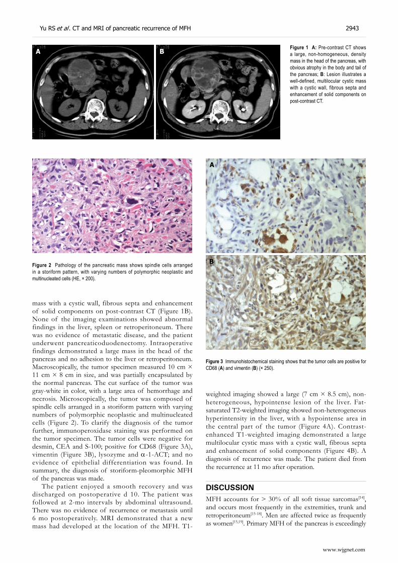
Yu RS et al . CT and MRI of pancreatic recurrence of MFH 2943
www.wjgnet.com
mass with a cystic wall, fibrous septa and enhancement of solid components on post-contrast CT (Figure 1B). None of the imaging examinations showed abnormal findings in the liver, spleen or retroperitoneum. There was no evidence of metastatic disease, and the patient underwent pancreaticoduodenectomy. Intraoperative findings demonstrated a large mass in the head of the pancreas and no adhesion to the liver or retroperitoneum. Macroscopically, the tumor specimen measured 10 cm × 11 cm × 8 cm in size, and was partially encapsulated by the normal pancreas. The cut surface of the tumor was gray-white in color, with a large area of hemorrhage and necrosis. Microscopically, the tumor was composed of spindle cells arranged in a storiform pattern with varying numbers of polymorphic neoplastic and multinucleated cells (Figure 2). To clarify the diagnosis of the tumor further, immunoperoxidase staining was performed on the tumor specimen. The tumor cells were negative for desmin, CEA and S-100; positive for CD68 (Figure 3A), vimentin (Figure 3B), lysozyme and α-1-ACT; and no evidence of epithelial differentiation was found. In summary, the diagnosis of storiform-pleomorphic MFH of the pancreas was made.
The patient enjoyed a smooth recovery and was discharged on postoperative d 10. The patient was followed at 2-mo intervals by abdominal ultrasound. There was no evidence of recurrence or metastasis until 6 mo postoperatively. MRI demonstrated that a new mass had developed at the location of the MFH. T1-
weighted imaging showed a large (7 cm × 8.5 cm), non-heterogeneous, hypointense lesion of the liver. Fat-saturated T2-weighted imaging showed non-heterogeneous hyperintensity in the liver, with a hypointense area in the central part of the tumor (Figure 4A). Contrast-enhanced T1-weighted imaging demonstrated a large multilocular cystic mass with a cystic wall, fibrous septa and enhancement of solid components (Figure 4B). A diagnosis of recurrence was made. The patient died from the recurrence at 11 mo after operation.
DISCUSSIONMFH accounts for > 30% of all soft tissue sarcomas[14], and occurs most frequently in the extremities, trunk and retroperitoneum[15-18]. Men are affected twice as frequently as women[15,19]. Primary MFH of the pancreas is exceedingly
Figure 1 A: Pre-contrast CT shows a large, non-homogeneous, density mass in the head of the pancreas, with obvious atrophy in the body and tail of the pancreas; B: Lesion illustrates a well-defined, multilocular cystic mass with a cystic wall, fibrous septa and enhancement of solid components on post-contrast CT.
A B
Figure 2 Pathology of the pancreatic mass shows spindle cells arranged in a storiform pattern, with varying numbers of polymorphic neoplastic and multinucleated cells (HE, × 200).
A
B
Figure 3 Immunohistochemical staining shows that the tumor cells are positive for CD68 (A) and vimentin (B) (× 250).

rare[5-8]. Akatsu et al have reviewed 16 cases and reported that the tumor has a male predominance with a median age at onset of 55 years. It is usually a large mass (mean tumor diameter is 13 cm, with a range of 4-35 cm)[11]. Of the documented cases, only three survived for > 48 mo[10-13], which suggests a poor prognosis for long-term survival. Only one case had lymph node metastases[12]. Our case was the only one with recurrence.
MFH consists of histiocyte-like and fibroblast-like cells arranged in a storiform pattern, with other pleomorphic cells and multinucleated giant cells[20-23]. There are five subtypes of MFH according to histology[10,24]: storiform-pleomorphic, myxoid, giant cell, inflammatory, and angiomatoid. The storiform-pleomorphic pattern is the most common variant[25]. Three histological subtypes have been observed among the 20 cases previously reported in the literature: 13 (65%) storiform-pleomorphic, four (20%) giant cell, and three (15%) myxoid[2-5]. Our case belonged to the storiform-pleomorphic type.
Only a few radiological findings of primary MFH of the pancreas have been described[10-12]. The CT findings of the tumor include the following: a large, heterogeneous, low-attenuation density or multinodular mass, with intratumoral calcification on pre-contrast CT; non-homogeneously enhancing pancreatic mass with a large amount of necrosis[11,12]; and single enhancing peripheral pseudo-capsular mass on post-contrast CT[10]. To the best of our knowledge, we describe the first case of primary MFH of the pancreas, with CT showing a large multilocular cystic mass with a cystic wall, fibrous septa and enhancement of solid components. It is not clear whether or not the pathological features of pancreatic MFH correlate with the CT findings. Liu et al have described a predominant cystic MFH of the pancreas of the myxoid variety[10], but other reports and our case suggest that not all cystic degeneration of the tumor belongs to the myxoid pattern[11,12]. Ko et al have considered that abdominal MFH, especially the storiform-pleomorphic subtype, exhibits metaplastic calcifications on CT, and they showed a heterogeneously enhanced mass with a large necrotic area in the pancreatic body and tail, and eccentrically located ring calcification[26]. Akatsu et al did not, however, support this sign and concluded that calcification is not always helpful in differentiating pancreatic MFH from other pancreatic tumors[11]. Therefore, we think that more cases need to be studied to prove the relationship between the CT findings
and the subtypes of primary MFH.Mahajan and colleagues have retrospectively reviewed
the MRI changes in 39 patients with MFH, however, none of these originated from the pancreas[27]. This study concluded that MRI sensitivity and specificity for detecting a neoplasm were 96% and 83%, respectively, but the signal changes were non-specific for MFH. When compared to CT in 14 patients, MRI better defined the extent of the MFH, its relationship to surrounding tissues and vessels, and best differentiated residual or recurrent disease from postoperative changes when examined at least 3 mo after surgery. There was no significant difference in signal intensity of 12 preoperative and 13 recurrent neoplasms[27].
Tateishi et al have described the manner in which that primary MFH has a high signal intensity on T2-weighted MR images and non-homogeneous isosignal intensity compared with the surrounding muscle[28]. Miller et al have reported that a correlation between MRI appearance and histopathology may not be established, but concluded that MRI exhibits general features suggestive of malignant soft tissue neoplasm, namely, internal low signal septation, and heterogeneous high signal intensity on T2-weighted images[29]. Kitajima et al[30] have considered that MRI is helpful in the differential diagnosis between MFH of the renal capsule and other renal tumors, and that a hypointense area identified on T2-weighted images is an important characteristic of renal MFH. In our case, MRI showed a large multilocular cystic mass, which was similar to that on preoperative CT, and it could reveal the exact location and extent of the tumor recurrence. An obviously hypointense area on T2-weighted images that reflected the fibrous component was also seen in our pancreatic MFH.
In conclusion, primary MFH of the pancreas is rare and a distinct clinical entity. It may recur and the prognosis is poor. Although there are no unique findings of pancreatic MFH, a large, massive liquescent necrotic mass or multilocular cystic lesion, with calcification on CT or a hypointense area within the mass on T2-weighted imaging in an older male patient, should lead to consideration of a differential diagnosis of pancreatic MFH.
REFERENCES1 Gutierrez JC , Perez EA, Franceschi D, Moffat FL Jr ,
Livingstone AS, Koniaris LG. Outcomes for soft-tissue sarcoma in 8249 cases from a large state cancer registry. J Surg Res 2007; 141: 105-114
Figure 4 A: MR fat-saturated T2-weighted image shows a large, non-heterogeneous, hyperintense lesion in the liver, with a hypointense area in the central part of the tumor; B: Contrast-enhanced T1-weighted image shows a large multilocular cystic mass with a cystic wall, fibrous septa and enhancement of solid components.
A B
www.wjgnet.com
2944 ISSN 1007-9327 CN 14-1219/R World J Gastroenterol May 14, 2008 Volume 14 Number 18

2 Fu DL, Yang F, Maskay A, Long J, Jin C, Yu XJ, Xu J, Zhou ZW, Ni QX. Primary intestinal malignant fibrous histiocytoma: two case reports. World J Gastroenterol 2007; 13: 1299-1302
3 Randall RL, Albritton KH, Ferney BJ, Layfield L. Malignant fibrous histiocytoma of soft tissue: an abandoned diagnosis. Am J Orthop 2004; 33: 602-608
4 Issakov J, Kollender Y, Soyfer V, Bickels J, Flusser G, Meller I, Merimsky O. A single-team experience of limb sparing approach in adults with high-grade malignant fibrous histiocytoma. Oncol Rep 2005; 14: 1071-1076
5 Basso L, Pisanelli MC, Bovino A, Vietri F, D'Ermo G, De Toma G. Malignant fibrous histiocytoma of the mesentery: report of two cases and review of the literature. G Chir 2005; 26: 43-46
6 Mizukami H, Yajima N, Wada R, Matsumoto K, Kojima M, Kloppel G, Yagihashi S. Pancreatic malignant fibrous histiocytoma, inflammatory myofibroblastic tumor, and inf lammatory pseudotumor re lated to autoimmune pancreatitis: characterization and differential diagnosis. Virchows Arch 2006; 448: 552-560
7 Allen KB, Skandalakis LJ, Brown BC, Gray SW, Skandalakis JE. Malignant fibrous histiocytoma of the pancreas. Am Surg 1990; 56: 364-368
8 Balén EM, De Villa VH, Cienfuegos JA, Contreras F, Pardo F, Gonzalez J, Benito C. [Malignant fibrous histiocytoma of the pancreas] Rev Esp Enferm Dig 1993; 83: 475-480
9 Margules RM, Allen RE, Dunphy JE. Pancreatic tumor of mesenchymal origin presenting as obstructive jaundice. Am J Surg 1976; 131: 357-359
10 Liu DM, Jeffrey RB Jr, Mindelzun RE. Malignant fibrous histiocytoma presenting as cystic pancreatic mass. Abdom Imaging 1999; 24: 299-300
11 Akatsu T, Tsugita M, Ro S, Kameyama K, Kitajima M. Primary malignant fibrous histiocytoma of the pancreas: a case with K-ras mutation and a review of the literature. Dig Dis Sci 2005; 50: 2214-2217
12 Bastian D, Ramaswamy A, Barth PJ, Gerdes B, Ernst M, Bartsch D. Malignant fibrous histiocytoma of the pancreas: a case report with genetic analysis. Cancer 1999; 85: 2352-2358
13 Song HB, Guo LN, Liu TH. Pancreatic multilocular myxoid cystoadenoma cancerization incorporation with MFH of giant cell pattern: case report. Chin J Pathol 2001; 30: 311-312
14 Sternheim A, Jin X, Shmookler B, Jelinek J, Malawer MM. 'Telangiectatic' transformation in soft tissue sarcomas. a clinicopathology analysis of an aggressive feature of high-grade sarcomas. Ann Surg Oncol 2008; 15: 345-354
15 Chen HC, Chen CJ, Jeng CM, Yang CM. Malignant fibrous histiocytoma presenting as hemoperitoneum mimicking hepatocellular carcinoma rupture. World J Gastroenterol 2007; 13: 6441-6443
16 Weiss SW, Enzinger FM. Malignant fibrous histiocytoma: an analysis of 200 cases. Cancer 1978; 41: 2250-2266
17 Nurdjanah S, Bayupurnama P, Maduseno S, Ratnasari N. Abdominal Malignant Fibrous Histiocytoma Infiltrating Stomach with Chilaiditi's Sign Manifestation (A Rare Case Report). Kobe J Med Sci 2007; 53: 119-124
18 Peiper M, Zurakowski D, Knoefel WT, Izbicki JR. Malignant fibrous histiocytoma of the extremities and trunk: an institutional review. Surgery 2004; 135: 59-66
19 Belal A, Kandil A, Allam A, Khafaga Y, El-Husseiny G, El-Enbaby A, Memon M, Younge D, Moreau P, Gray A, Schultz H. Malignant fibrous histiocytoma: a retrospective study of 109 cases. Am J Clin Oncol 2002; 25: 16-22
20 Liu P, Li J, Shen L. [A clinico-pathologic study of primary malignant fibrous histiocytoma of bone] Zhonghua Zhongliu Zazhi 1996; 18: 146-149
21 Tos AP. Classification of pleomorphic sarcomas: where are we now? Histopathology 2006; 48: 51-62
22 Murphey MD. World Health Organization classification of bone and soft tissue tumors: modifications and implications for radiologists. Semin Musculoskelet Radiol 2007; 11: 201-214
23 Scapolan M , Perin T, Wassermann B, Canzonieri V, Colombatti A, Italia F, Spessotto P. Expression profiles in malignant fibrous histiocytomas: clues for differentiating 'spindle cell' and 'pleomorphic' subtypes. Eur J Cancer 2008; 44: 298-309
24 Munk PL, Sallomi DF, Janzen DL, Lee MJ, Connell DG, O'Connell JX, Logan PM, Masri B. Malignant fibrous histiocytoma of soft tissue imaging with emphasis on MRI. J Comput Assist Tomogr 1998; 22: 819-826
25 Meister P. Malignant fibrous histiocytoma. History, histology, histogenesis. Pathol Res Pract 1988; 183: 1-7
26 Ko SF, Wan YL, Lee TY, Ng SH, Lin JW, Chen WJ. CT features of calcifications in abdominal malignant fibrous histiocytoma. Clin Imaging 1998; 22: 408-413
27 Mahajan H , Kim EE, Wallace S, Abello R, Benjamin R, Evans HL. Magnetic resonance imaging of malignant fibrous histiocytoma. Magn Reson Imaging 1989; 7: 283-288
28 Tateishi U , Kusumoto M, Hasegawa T, Yokoyama R, Moriyama N. Primary malignant fibrous histiocytoma of the chest wall: CT and MR appearance. J Comput Assist Tomogr 2002; 26: 558-563
29 Miller TT, Hermann G, Abdelwahab IF, Klein MJ, Kenan S, Lewis MM. MRI of malignant fibrous histiocytoma of soft tissue: analysis of 13 cases with pathologic correlation. Skeletal Radiol 1994; 23: 271-275
30 Kitajima K , Kaji Y, Morita M, Okuda Y, Sugimura K. Malignant fibrous histiocytoma arising from the renal capsule. Magn Reson Med Sci 2003; 2: 199-202
S- Editor Zhong XY L-Editor Kerr C E- Editor Yin DH
www.wjgnet.com
Yu RS et al . CT and MRI of pancreatic recurrence of MFH 2945

Online Submissions: wjg.wjgnet.com World J Gastroenterol 2008 May 14; 14(18): 2946-2947www.wjgnet.com World Journal of Gastroenterology ISSN [email protected] © 2008 WJG. All rights reserved.
Desperately seeking hepatitis C virus
Ricardo Moreno-Otero
www.wjgnet.com
LETTERS TO THE EDITOR
Ricardo Moreno-Otero, Hepato-Gastroenterology Service and Ciberehd, Hospital Universitario de La Princesa, Autonomous University of Madrid, Madrid 28006, SpainAuthor contributions: Moreno-Otero R wrote the paper.Correspondence to: Ricardo Moreno-Otero, MD, Chief, Hepato-Gastroenterology Service and Ciberehd, Hospital Universitario de La Princesa, Autonomous University of Madrid, Madrid 28006, Spain. [email protected]: +34-91-3093911 Fax: +34-91-4022299Received: January 30, 2008 Revised: March 8, 2008
AbstractSpanish investigators described recently the so-called occult hepatitis C virus (HCV) infection, emphasizing the detection of genomic and antigenomic HCV RNA strands in liver and peripheral blood mononuclear cells. Therefore, the persistence of viral replication in occult HCV infection should be considered as a putative source of infection among family members and patients undergoing invasive procedures, transfusion or transplantation. Additionally, the most worrisome finding is that an occult HCV infection may persist in patients with sustained virological response.
© 2008 WJG. All rights reserved.
Key words: Occult hepatitis C; Occult hepatitis C virus infection; Hepatitis C virus RNA; Peginterferon; Ribavirin
Peer reviewers: Giovanni Tarantino, MD, Professor, Department of Clinical and Experimental Medicine, Federico Ⅱ University Medical School, VIA S. PANSINI, 5, Naples 80131, Italy; Peter L Lakatos, MD, PhD, Assistant Professor, 1st Department of Medicine, Semmelweis University, Koranyi S 2A, Budapest H1083, Hungary; Hisato Nakajima, MD, Department of Gastroenterology and Hepatology, The Jikei University School of Medicine, 3-25-8, Nishi-Shinbashi, Minato-ku, Tokyo 105-8461, Japan
Moreno-Otero R. Desperately seeking hepatitis C virus. World J Gastroenterol 2008; 14(18): 2946-2947 Available from: URL: http://www.wjgnet.com/1007-9327/14/2946.asp DOI: http://dx.doi.org/10.3748/wjg.14.2946
TO THE EDITORResearch into hepatitis C virus infection has progressed substantially over the past 15 years. Treatment with peginterferon plus ribavirin yields a sustained virological
response (SVR) in approximately 45% of patients with genotype 1 and in 80% of those with genotypes 2 or 3. SVR is associated with HCV eradication and liver damage reduction. Enormous scientific and economic efforts have dramatically changed the medical focus on HCV, and the prior condition of shipwrecked hepatologists has evolved into skilful navigators trained to cure chronic hepatitis C (CHC), adapting treatment to each patient instead of adapting the patient to an established therapy[1,2].
Spanish investigators described recently the so-called occult hepatitis C virus (HCV) infection[3] summarizing its successive studies in a comprehensive review[4] that emphasizes the detection of genomic and antigenomic HCV RNA strands in l iver and peripheral blood mononuclear cells (PBMCs). Therefore, the persistence of viral replication in occult HCV infection should be considered as a putative source of infection among family members and patients undergoing invasive procedures, transfusion or transplantation. Additionally, the most worrisome finding is that an occult HVC infection may persist in SVR patients, with HCV RNA presence in liver and PBMCs[4,5]. The hope of treated patients, who consider themselves having got rid of the virus, and of physicians, struggling for control and cure CHC, is most probably devastated.
These authors also found that HCV RNA is detected in the sera of patients with occult HCV infection after ultracentrifugation[6]. What is the significance of this new entity? Answers are a challenge for the scientific community in order to dissect the epidemiological aspects and pathological consequences of occult HCV infection, not to mention the design of therapeutic strategies. The social and medical costs can be astronomic, including work incapacity, absenteeism and intangible costs able to ruin the private life of some patients. Concerns and doubtful questions enunciated by frightened persons and medical doctors have been raised after an extensive report on occult hepatitis C was published, alluding the aforementioned study[6], on a newspaper from Madrid (LA RAZÓN, July 10-2007, page 31). Whether all the information has any alarming relevance remains to be seen.
REFERENCES1 Hoofnagle JH, Seeff LB. Peginterferon and ribavirin for
chronic hepatitis C. N Engl J Med 2006; 355: 2444-24512 Moreno-Otero R. Therapeutic modalities in hepatitis C:
challenges and development. J Viral Hepat 2005; 12: 10-19

Moreno-Otero R. Occult hepatitis C 2947
www.wjgnet.com
3 Castillo I, Pardo M, Bartolomé J, Ortiz-Movilla N, Rodríguez-Iñigo E, de Lucas S, Salas C, Jiménez-Heffernan JA, Pérez-Mota A, Graus J, López-Alcorocho JM, Carreño V. Occult hepatitis C virus infection in patients in whom the etiology of persistently abnormal results of liver-function tests is unknown. J Infect Dis 2004; 189: 7-14
4 Carreño V. Occult hepatitis C virus infection: a new form of hepatitis C. World J Gastroenterol 2006; 12: 6922-6925
5 Castillo I, Rodríguez-Iñigo E, López-Alcorocho JM, Pardo M, Bartolomé J, Carreño V. Hepatitis C virus replicates in the liver of patients who have a sustained response to antiviral treatment. Clin Infect Dis 2006; 43: 1277-1283
6 Bartolomé J, López-Alcorocho JM, Castillo I, Rodríguez-Iñigo E, Quiroga JA, Palacios R, Carreño V. Ultracentrifugation of serum samples allows detection of hepatitis C virus RNA in patients with occult hepatitis C. J Virol 2007; 81: 7710-7715
S- Editor Zhong XY L- Editor Ma JY E- Editor Yin DH

ACKNOWLEDGMENTS
Acknowledgments to Reviewers of World Journal of Gastroenterology
Many reviewers have contributed their expertise and time to the peer review, a critical process to ensure the quality of World Journal of Gastroenterology. The editors and authors of the articles submitted to the journal are grateful to the following reviewers for evaluating the articles (including those published in this issue and those rejected for this issue) during the last editing time period.
Sk Md Fazle Akbar, Dr, Assistant Professor, Third Department of Internal Medicine, Ehime University School of Medicine, Shigenobu-Cho, Ehime 791-0295, Japan
Claudio Bassi, MD, Professor, Department of Surgery and Gastroenterology, Hospital GB Rossi, University of Verona, Piazza LA Scuro 37134 Verona, Italy
Reinhard Buettner, Professor, Institute of Pathology, University Hospital Bonn, Sigmund-Freud-Str. 25, D-53127 Bonn, Germany
David L Carr-Locke, MD, Director of Endoscopy, Brigham and Women’s Hospital, Endoscopy Center, Brigham and Women’s Hospital, 75 Francis St, Boston MA 02115, United States
Massimo Colombo, Professor, 1st Division of Gastroenterology, Fondazione IRCCS Maggiore Hospital, Policlinico, Mangiagalli e Regina Elena, University of Milan, Via F. Sforza 35, 20122 Milan, Italy
David Cronin II, MD, PhD, FACS, Associate Professor, Department of Surgery, Yale University School of Medicine, 330 Cedar Street, FMB 116, P. O. Box 208062, New Haven, Connecticut 06520-8062, United States
Arno J Dormann, PD, MED, Habil, Medizinische Klinik, Krankenhaus Holweide, Kliniken der Stadt Köln gGmbH, Neufelder St. 32, 51067 Köln, Germany
James E East, BSc, MBChB, MRCP, St. Mary’s Hospital, Endoscopy Unit, Clarence Wing, 3rd Floor, Praed Street, London, W2 1NY, United Kingdom
Francesco Feo, Professor, Dipartimento di Scienze Biomediche, Sezione di Patologia Sperimentale e Oncologia, Università di Sassari, Via P, Manzella 4, 07100 Sassari, Italy
Mitsuhiro Fujishiro, Dr, Department of Gastroenterology, Faculty of Medicine, University of Tokyo, 7-3-1 Hongo, Bunkyo-ku, Tokyo, Japan
Toru Ishikawa, MD, Department of Gastroenterology, Saiseikai Niigata Second Hospital, Teraji 280-7, Niigata, Niigata 950-1104, Japan
Syed MW Jafri, Professor, Medicine/Gastroenterology, Aga Khan University, POB 3500, Karachi 74800, Pakistan
Tom H Karlsen, MD, Institute of Immunology, Rikshospitalet University Hospital, N-0027 Oslo, Norway
Ioannis E Koutroubakis, MD, PhD, Assistant Professor of Medicine,University Hospital Heraklion, Department of Gastroenterology, PO Box 1352, 71110 Heraklion, Crete, Greece
Shiu-Ming Kuo, MD, University at Buffalo, 15 Farber Hall, 3435 Main Street, Buffalo 14214, United States
Ezio Laconi, MD, PhD, Professor of General Pathology, Department of Sciences and Biomedical Technologies, Unit of Experimental Pathology, University of Cagliari, Via Porcell, 4 - IV Piano, 09125 - Cagliari, Italy
James YW Lau, Department of Surgery, Prince of Wales Hospital, the Chinese University of Hong Kong, Hong Kong, China
Laura Lladóa, PhD, Department of Surgery, Liver Transplant Unit, Hospital Universitari de Bellvitge, IDIBELL, 08907 Barcelona, Spain
Mercedes Susan Mandell, MD, PhD, Department of Anesthesiology, University of Colorado Health Sciences Ctr., 12401 E. 17th Ave, B113 Aurora, CO 80045, United States
Robert D Odze, MD, FRCPc, Chief, Gastrointestinal Pathology Service, Associate Professor of Pathology , Brigham and Women's Hospital , Harvard Medical School, Boston MA, United States
Natalia A Osna, Liver Study Unit, Research Service (151), VA Medical Center, 4101 Woolworth Avenue, Omaha NE 68105, United States
Kostas Pantopoulos, Associate Professor, Department of Medicine, McGill University, Lady Davis Institute for Medical Research, 3755 Cote Ste-Catherine Road, Montreal, Quebec, H3T 1E2, Canada
George Papatheodoridis, MD, Assistant Professor in Medicine & Gastroenterology, 2nd Department of Internal Medicine, Athens University Medical School, Hippokration General Hospital of Athens, 114 Vas. Sophias Ave., 115 27 Athens, Greece
Gustav Paumgartner, Professor, University of Munich, Klinikum Grosshadern, Marchioninistr. 15, Munich, D-81377, Germany
Jose L del Pozo, MD, PhD, Infectious Diseases Division, Mayo Clinic College of Medicine, 200 1st St SW, Rochester, MN 55905, United States
Paul E Sijens, PhD, Associate Professor,Radiology, UMCG, Hanzeplein 1, 9713GZ Groningen, The Netherlands
Akihito Tsubota, Assistant Professor, Institute of Clinical Medicine and Research , Jikei University School of Medicine, 163-1 Kashiwa-shita, Kashiwa, Chiba 277-8567, Japan
Marie-Catherine Vozenin-brotons, PhD, UPRES EA 27-10, IRSN/IGR, 39 rue C. Desmoulins, Villejuif Cedex 94305, France
Eddie Wisse, Professor, Irisweg 16, Keerbergen 3140, Belgium
Harry HX Xia, PhD, MD, Novartis Pharmaceuticals Corporation, One Health Plaza, East Hanover, NJ 07936-1080, United States
Yoshio Yamaoka, MD, PhD, Associate Professor, Department of Medicine/Gastroenterology, Baylor College of Medicine and VA Medical Center (111D), 2002 Holcombe Blvd, Houston, Texas 77030, United States
Ta-Sen Yeh, MD, PhD, Department of Surgery, Chang Gung Memorial Hospital, 5 Fu-Hsing Street, Taoyuan, Taiwan, China
Yuan Yuan, Professor, Cancer Institute of China Medical University, 155 North Nanjing Street, Heping District, Shenyang 110001, Liaoning Province, China
Online Submissions: wjg.wjgnet.com World J Gastroenterol 2008 May 14; 14(18): 2948www.wjgnet.com World Journal of Gastroenterology ISSN [email protected] © 2008 WJG. All rights reserved.

Meetings
Events Calendar 2008-2009FALK SYMPOSIA 2008January 24-25, Frankfurt, GermanyFalk Workshop: Perspectives in Liver Transplantation
International Gastroenterological Congresses 2008February 14-16, Paris, FranceEASL-AASLD-APASL-ALEH-IASL Conference Hepatitis B and C virus resistance to antiviral therapieswww.easl.ch/hepatitis-conference
February 14-17, Berlin, Germany8th International Conference on New Trends in Immunosuppression and Immunotherapywww.kenes.com/immuno
February 28, Lyon, France3rd Congress of ECCO - the European Crohn’s and Colitis OrganisationInflammatory Bowel Diseases 2008www.ecco-ibd.eu
March 10-13, Birmingham, UKBritish Society of Gastroenterology Annual MeetingE-mail: [email protected]
March 14-15, HangZhou, ChinaFalk Symposium 163: Chronic Inflammation of Liver and Gut
March 23-26, Seoul, KoreaAsian Pacific Association for the Study of the Liver18th Conference of APASL: New Horizons in Hepatologywww.apaslseoul2008.org
March 29-April 1, Shanghai, ChinaShanghai - Hong Kong International Liver Congresswww.livercongress.org
April 05-09, Monte-Carlo (Grimaldi Forum), Monaco OESO 9th World Congress, The Gastro-esophageal Reflux Disease: from Reflux to Mucosal Inflammation - Management of Adeno- carcinomasEmail: [email protected]
April 18-22, Buenos Aires, Argentina9th World Congress of the International Hepato-Pancreato Biliary AssociationAssociation for the Study of the Liverwww.ca-ihpba.com.ar
April 23-27, Milan, Italy43rd Annual Meeting of the European
Online Submissions: wjg.wjgnet.com World J Gastroenterol 2008 May 14; 14(18): 2949www.wjgnet.com World Journal of Gastroenterology ISSN [email protected] © 2008 WJG. All rights reserved.
Association for the Study of the Liverwww.easl.ch
May 2-3, Budapest, HungaryFalk Symposium 164: Intestinal Disorders
May 18-21, San Diego, California, USADigestive Disease Week 2008June 4-7, Helsinki, Finland
The 39th Nordic Meeting of Gastroenterologywww.congrex.com/ngc2008June 6-8, Prague, Czech Republic3rd Annual European Meeting: Perspectives in Inflammatory Bowel DiseasesEmail: [email protected]
June 13-14, Amsterdam, NetherlandsFalk Symposium 165: XX International Bile Acid Meeting. Bile Acid Biology and Therapeutic Actions
June 25-28, Barcelona, Spain 10th World Congress on Gastrointestinal CancerImedex and ESMOEmail: [email protected]
June 25-28, Lodz, PolandJoint Meeting of the European Pancreatic Club (EPC) and the International Association of Pancreatology (IAP)E-mail: [email protected] www.pancreatology.org
June 26-28, Bratislava, Slovakia5th Central European Gastroenterology Meetingwww.ceurgem2008.cz September 10-13, Budapest, Hungary
11th World Congress of the International Society for Diseases of the Esophagus Email: [email protected]
September 13-16, New Delhi, IndiaAsia Pacific Digestive WeekE-mail: [email protected]
III FALK GASTRO-CONFERENCESeptember 17, Mainz, GermanyFalk Workshop: Strategies of Cancer Prevention in Gastroenterology
September 18-19, Mainz, GermanyFalk Symposium 166:GI Endoscopy - Standards & Innovations
September 18-20, Prague, Czech RepublicPrague Hepatology Meeting 2008www.czech-hepatology.cz/phm2008
September 20-21, Mainz, GermanyFalk Symposium 167:Liver Under Constant Attack - From
Fat to VirusesSeptember 24-27, Nantes, FranceThird Annual MeetingEuropean Society of Coloproctologywww.escp.eu.com
October 8-11, Istanbul, Turkey18th World Congress of the International Association of Surgeons, Gastroenterologists and OncologistsE-mail: [email protected]
October 18-22, Vienna, Austria16th United European Gastroenterology Weekwww.negf.org www.acv.at
October 22-25, Brisbane, Australia Anstralian Gastroenterology Week 2008Email: [email protected]
October 31-November 4, Moscone West Convention Center, San Francisco, CA 59th AASLD Annual Meeting and Postgraduate CourseThe Liver MeetingInformation: www.aasld.org
November 6-9, Lucerne, SwitzerlandNeurogastroenterology & MotilityJoint International Meeting 2008Email: [email protected]
November 12, Santiago de Chile, ChileFalk Workshop: Digestive Diseases: State of the Art and Daily Practice
December 7-9, Seoul, Korea6th International Meeting Hepatocellular Carcinoma: Eastern and Western ExperiencesE-mail: [email protected]
INFORMATION FOR ALLFALK FOUNDATION e.V.Email: [email protected] www.falkfoundation.de
Advanced Courses - European Institute of Telesurgery EITS - 2008Strasbourg, FranceJanuary 18-19, March 28-29, June 6-7, October 3-4 N.O.T.E.SApril 3-5, November 27-29Laparoscopic Digestive SurgeryJune 27-28, November 7-8 Laparoscopic Colorectal SurgeryJuly 3-5Interventional GI Endoscopy TechniquesContact address for all courses: [email protected]
International Gastroenterological
Congresses 2009March 23-26, Glasgow, ScotlandMeeting of the British Society of Gastroenterology (BSG)E-mail: [email protected]
May 17-20, Denver, Colorado, USADigestive Disease Week 2009
November 21-25, London, UKGastro 2009 UEGW/World Congress of Gastroenterologywww.gastro2009.org
Global Collaboration forGastroenterologyFor the first time in the history of gastroenterology, an international conference will take place which joins together the forces of four pre-eminent organisations: Gastro 2009, UEGW/WCOG London. The United European Gastroenterology Federation (UEGF) and the World Gastroenterology Organisation (WGO), together with the World Organisation of Digestive Endoscopy (OMED) and the British Society of Gastroenterology (BSG), are jointly organising a landmark meeting in London from November 21-25, 2009. This collaboration will ensure the perfect balance of basic science and clinical practice, will cover all disciplines in gastroenterology (endoscopy, digestive oncology, n u t r i t i o n , d i g e s t i v e s u r g e r y , hepatology, gastroenterology) and ensure a truly global context; all presented in the exciting setting of the city of London. Attendance is expected to reach record heights as participants are provided with a compact “all-in-one” programme merging the best of several GI meetings. Faculty and participants from all corners of the earth will merge to provide a truly global environment conducive to the exchange of ideas and the forming of friendships and collaborations.
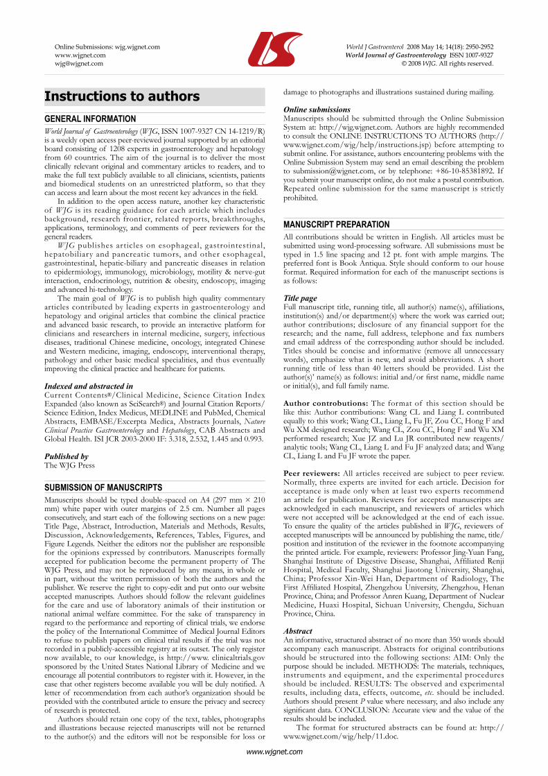
www.wjgnet.com
Instructions to authorsGENERAL INFORMATIONWorld Journal of Gastroenterology (WJG, ISSN 1007-9327 CN 14-1219/R) is a weekly open access peer-reviewed journal supported by an editorial board consisting of 1208 experts in gastroenterology and hepatology from 60 countries. The aim of the journal is to deliver the most clinically relevant original and commentary articles to readers, and to make the full text publicly available to all clinicians, scientists, patients and biomedical students on an unrestricted platform, so that they can access and learn about the most recent key advances in the field.
In addition to the open access nature, another key characteristic of WJG is its reading guidance for each article which includes background, research frontier, related reports, breakthroughs, applications, terminology, and comments of peer reviewers for the general readers.
WJG publishes articles on esophageal, gastrointestinal, hepatobiliary and pancreatic tumors, and other esophageal, gastrointestinal, hepatic-biliary and pancreatic diseases in relation to epidermiology, immunology, microbiology, motility & nerve-gut interaction, endocrinology, nutrition & obesity, endoscopy, imaging and advanced hi-technology.
The main goal of WJG is to publish high quality commentary articles contributed by leading experts in gastroenterology and hepatology and original articles that combine the clinical practice and advanced basic research, to provide an interactive platform for clinicians and researchers in internal medicine, surgery, infectious diseases, traditional Chinese medicine, oncology, integrated Chinese and Western medicine, imaging, endoscopy, interventional therapy, pathology and other basic medical specialities, and thus eventually improving the clinical practice and healthcare for patients.
Indexed and abstracted inCurrent Contents®/Clinical Medicine, Science Citation Index Expanded (also known as SciSearch®) and Journal Citation Reports/Science Edition, Index Medicus, MEDLINE and PubMed, Chemical Abstracts, EMBASE/Excerpta Medica, Abstracts Journals, Nature Clinical Practice Gastroenterology and Hepatology, CAB Abstracts and Global Health. ISI JCR 2003-2000 IF: 3.318, 2.532, 1.445 and 0.993.
Published byThe WJG Press
SUBMISSION OF MANUSCRIPTSManuscripts should be typed double-spaced on A4 (297 mm × 210 mm) white paper with outer margins of 2.5 cm. Number all pages consecutively, and start each of the following sections on a new page: Title Page, Abstract, Introduction, Materials and Methods, Results, Discussion, Acknowledgements, References, Tables, Figures, and Figure Legends. Neither the editors nor the publisher are responsible for the opinions expressed by contributors. Manuscripts formally accepted for publication become the permanent property of The WJG Press, and may not be reproduced by any means, in whole or in part, without the written permission of both the authors and the publisher. We reserve the right to copy-edit and put onto our website accepted manuscripts. Authors should follow the relevant guidelines for the care and use of laboratory animals of their institution or national animal welfare committee. For the sake of transparency in regard to the performance and reporting of clinical trials, we endorse the policy of the International Committee of Medical Journal Editors to refuse to publish papers on clinical trial results if the trial was not recorded in a publicly-accessible registry at its outset. The only register now available, to our knowledge, is http://www. clinicaltrials.gov sponsored by the United States National Library of Medicine and we encourage all potential contributors to register with it. However, in the case that other registers become available you will be duly notified. A letter of recommendation from each author’s organization should be provided with the contributed article to ensure the privacy and secrecy of research is protected.
Authors should retain one copy of the text, tables, photographs and illustrations because rejected manuscripts will not be returned to the author(s) and the editors will not be responsible for loss or
damage to photographs and illustrations sustained during mailing.
Online submissionsManuscripts should be submitted through the Online Submission System at: http://wjg.wjgnet.com. Authors are highly recommended to consult the ONLINE INSTRUCTIONS TO AUTHORS (http://www.wjgnet.com/wjg/help/instructions.jsp) before attempting to submit online. For assistance, authors encountering problems with the Online Submission System may send an email describing the problem to [email protected], or by telephone: +86-10-85381892. If you submit your manuscript online, do not make a postal contribution. Repeated online submission for the same manuscript is strictly prohibited.
MANUSCRIPT PREPARATIONAll contributions should be written in English. All articles must be submitted using word-processing software. All submissions must be typed in 1.5 line spacing and 12 pt. font with ample margins. The preferred font is Book Antiqua. Style should conform to our house format. Required information for each of the manuscript sections is as follows:
Title pageFull manuscript title, running title, all author(s) name(s), affiliations, institution(s) and/or department(s) where the work was carried out; author contributions; disclosure of any financial support for the research; and the name, full address, telephone and fax numbers and email address of the corresponding author should be included. Titles should be concise and informative (remove all unnecessary words), emphasize what is new, and avoid abbreviations. A short running title of less than 40 letters should be provided. List the author(s)’ name(s) as follows: initial and/or first name, middle name or initial(s), and full family name.
Author controbutions: The format of this section should be like this: Author contributions: Wang CL and Liang L contributed equally to this work; Wang CL, Liang L, Fu JF, Zou CC, Hong F and Wu XM designed research; Wang CL, Zou CC, Hong F and Wu XM performed research; Xue JZ and Lu JR contributed new reagents/analytic tools; Wang CL, Liang L and Fu JF analyzed data; and Wang CL, Liang L and Fu JF wrote the paper.
Peer reviewers: All articles received are subject to peer review. Normally, three experts are invited for each article. Decision for acceptance is made only when at least two experts recommend an article for publication. Reviewers for accepted manuscripts are acknowledged in each manuscript, and reviewers of articles which were not accepted will be acknowledged at the end of each issue. To ensure the quality of the articles published in WJG, reviewers of accepted manuscripts will be announced by publishing the name, title/position and institution of the reviewer in the footnote accompanying the printed article. For example, reviewers: Professor Jing-Yuan Fang, Shanghai Institute of Digestive Disease, Shanghai, Affiliated Renji Hospital, Medical Faculty, Shanghai Jiaotong University, Shanghai, China; Professor Xin-Wei Han, Department of Radiology, The First Affiliated Hospital, Zhengzhou University, Zhengzhou, Henan Province, China; and Professor Anren Kuang, Department of Nuclear Medicine, Huaxi Hospital, Sichuan University, Chengdu, Sichuan Province, China.
AbstractAn informative, structured abstract of no more than 350 words should accompany each manuscript. Abstracts for original contributions should be structured into the following sections: AIM: Only the purpose should be included. METHODS: The materials, techniques, instruments and equipment, and the experimental procedures should be included. RESULTS: The observed and experimental results, including data, effects, outcome, etc. should be included. Authors should present P value where necessary, and also include any significant data. CONCLUSION: Accurate view and the value of the results should be included.
The format for structured abstracts can be found at: http://www.wjgnet.com/wjg/help/11.doc.
Online Submissions: wjg.wjgnet.com World J Gastroenterol 2008 May 14; 14(18): 2950-2952www.wjgnet.com World Journal of Gastroenterology ISSN [email protected] © 2008 WJG. All rights reserved.

Key wordsPlease list 5-10 key words, selected mainly from Index Medicus, which reflect the content of the study.
TextFor articles of these sections, original articles, rapid communication and case reports, the main text should be structured into the following sections: INTRODUCTION, MATERIALS AND METHODS, RESULTS and DISCUSSION, and should include appropriate Figures and Tables. Data should be presented in the body text or in Figures and Tables, but not in both. The main text format of these sections, editorial, topic highlight, case report, letters to the editors, should be found at: http://www.wjgnet.com/wjg/help/instructions.jsp.
IllustrationsFigures should be numbered as 1, 2, 3, etc., and mentioned clearly in the main text. Provide a brief title for each figure on a separate page. Detailed legends should not be provided under the figures. This part should be added into the text where the figures are applicable. Figures should be either Photoshop or Illustrator files (in tiff, eps, jpeg formats) at high-resolution. Examples can be found at: http://www.wjgnet.com/1007-9327/13/4520.pdf; http://www.wjgnet.com/1007-9327/13/4554.pdf; http://www.wjgnet .com/1007-9327/13/4891.pdf ; ht tp ://www.wjgnet.com/1007-9327/13/4986.pdf; http://www.wjgnet.com/1007-9327/13/4498.pdf. Keeping all elements compiled is necessary in line-art image. Scale bars should be used rather than magnification factors, with the length of the bar defined in the legend rather than on the bar itself. File names should identify the figure and panel. Avoid layering type directly over shaded or textured areas. Please use uniform legends for the same subjects. For example: Figure 1 Pathological changes in atrophic gastritis after treatment. A: ...; B: ...; C: ...; D: ...; E: ...; F: ...; G: …etc. It is our principle to publish high resolution-figures for the printed and E-versions.
TablesThree-line tables should be numbered 1, 2, 3, etc., and mentioned clearly in the main text. Provide a brief title for each table. Detailed legends should not be included under tables, but rather added into the text where applicable. The information should complement but not duplicate the text. Use one horizontal line under the title, a second under column heads, and a third below the Table, above any footnotes. Vertical and italic lines should be omitted.
Notes in tables and illustrationsData that are not statistically significant should not be noted. aP < 0.05, bP < 0.01 should be noted (P > 0.05 should not be noted). If there are other series of P values, cP < 0.05 and dP < 0.01 are used. A third series of P values can be expressed as eP < 0.05 and fP < 0.01. Other notes in tables or under illustrations should be expressed as 1F, 2F, 3F; or sometimes as other symbols with a superscript (Arabic numerals) in the upper left corner. In a multi-curve illustration, each curve should be labeled with ●, ○, ■, □, ▲, △, etc., in a certain sequence.
AcknowledgmentsBrief acknowledgments of persons who have made genuine contributions to the manuscripts and who endorse the data and conclusions should be included. Authors are responsible for obtaining written permission to use any copyrighted text and/or illustrations.
REFERENCESCoding systemThe author should number the references in Arabic numerals according to the citation order in the text. Put reference numbers in square brackets in superscript at the end of citation content or after the cited author’s name. For citation content which is part of the narration, the coding number and square brackets should be typeset normally. For example, “Crohn’s disease (CD) is associated with increased intestinal permeability[1,2]”. If references are cited directly in the text, they should be put together within the text, for example, “From references[19,22-24], we know that...”
When the authors write the references, please ensure that the order in text is the same as in the references section, and also ensure the spelling accuracy of the first author’s name. Do not list the same citation twice.
PMID requirementPMID roots in the abstract serial number indexed by PubMed (http://www.ncbi.nlm.nih.gov/entrez/query.fcgi?db=PubMed). The author should supply the PMID for journal citation. For those references that have not been indexed by PubMed, a printed copy of the first page of the full reference should be submitted.
The accuracy of the information for journal citations is very important. Using the reference testing system, the authors and editor should check the authors name, title, journal title, publication date, volume number, start page, and end page. We will interlink all references with PubMed in an ASP file so that the readers can immediately access the abstract of the citations online.
DOI requirementA CrossRef DOI® (Digital Object Identifier) name is a unique string created to identify a piece of scholarly content in the online environment. The author should supply the DOIs for journal citation(doi:10.3748/wjg.13.6458). This link (http://www.crossref.org/SimpleTextQuery/) allows you to retrieve Digital Object Identifiers (DOIs) for journal articles, books, and chapters by simply cutting and pasting the reference list into the box. You may use the form with any reference style, although the tool works most reliably if references are formatted in a standard style such as shown in this example: Assimakopoulos SF, Scopa CD, Vagianos CE. Pathophysiology of increased intestinal permeability in obstructive jaundice. World J Gastroenterol 2007; 13(48): 6458-6464
The accuracy of the information of journal citations is very important. We will interlink all references with DOI in ASP file so that readers can access the abstracts of cited articles online immediately.
Style for journal referencesAuthors: the name of the first author should be typed in bold-faced letters. The family name of all authors should be typed with the initial letter capitalized, followed by their abbreviated first and middle initials. (For example, Lian-Sheng Ma is abbreviated as Ma LS, Bo-Rong Pan as Pan BR). The title of the cited article and italicized journal title (journal title should be in its abbreviated form as shown in PubMed), publication date, volume number (in black), start page, and end page [PMID: 11819634 DOI: 10.3748/wjg.13.5396].
Style for book referencesAuthors: the name of the first author should be typed in bold-faced letters. The surname of all authors should be typed with the initial letter capitalized, followed by their abbreviated middle and first initials. (For example, Lian-Sheng Ma is abbreviated as Ma LS, Bo-Rong Pan as Pan BR) Book title. Publication number. Publication place: Publication press, Year: start page and end page.
FormatJournals English journal article (list all authors and include the PMID where applicable)1 Jung EM, Clevert DA, Schreyer AG, Schmitt S, Rennert J,
Kubale R, Feuerbach S, Jung F. Evaluation of quantitative contrast harmonic imaging to assess malignancy of liver tumors: A prospective controlled two-center study. World J Gastroenterol 2007; 13: 6356-6364 [PMID: 18081224 DOI: 10.3748/wjg.13.6356]
Chinese journal article (list all authors and include the PMID where applicable)2 Lin GZ, Wang XZ, Wang P, Lin J, Yang FD. Immunologic effect
of Jianpi Yishen decoction in treatment of Pixu-diarrhoea. Shijie Huaren Xiaohua Zazhi 1999; 7: 285-287
In press3 Tian D, Araki H, Stahl E, Bergelson J, Kreitman M. Signature
of balancing selection in Arabidopsis. Proc Natl Acad Sci USA 2006; In press
Organization as author4 Diabetes Prevention Program Research Group. Hypertension,
insulin, and proinsulin in participants with impaired glucose tolerance. Hypertension 2002; 40: 679-686 [PMID: 12411462]
Both personal authors and an organization as author 5 Vallancien G, Emberton M, Harving N, van Moorselaar RJ; Alf-
One Study Group. Sexual dysfunction in 1, 274 European men suffering from lower urinary tract symptoms. J Urol 2003; 169: 2257-2261 [PMID: 12771764]
No author given6 21st century heart solution may have a sting in the tail. BMJ
Instructions to authors 2951
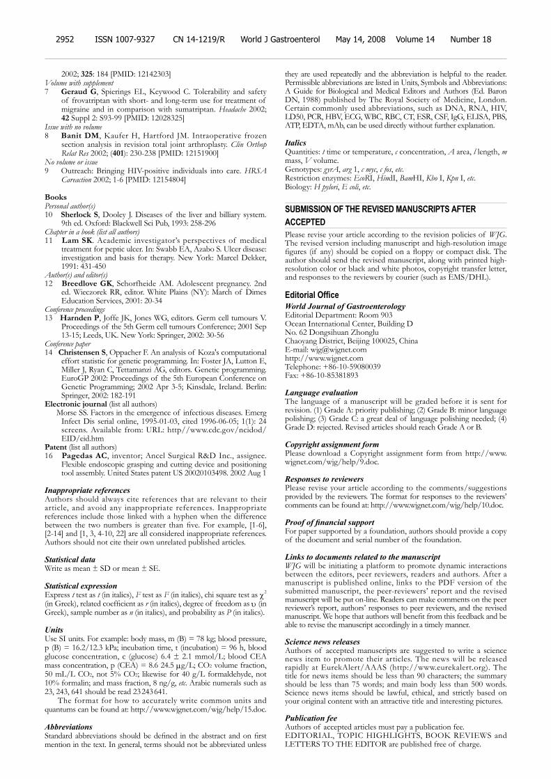
2002; 325: 184 [PMID: 12142303]Volume with supplement7 Geraud G, Spierings EL, Keywood C. Tolerability and safety
of frovatriptan with short- and long-term use for treatment of migraine and in comparison with sumatriptan. Headache 2002; 42 Suppl 2: S93-99 [PMID: 12028325]
Issue with no volume8 Banit DM, Kaufer H, Hartford JM. Intraoperative frozen
section analysis in revision total joint arthroplasty. Clin Orthop Relat Res 2002; (401): 230-238 [PMID: 12151900]
No volume or issue9 Outreach: Bringing HIV-positive individuals into care. HRSA
Careaction 2002; 1-6 [PMID: 12154804]
Books Personal author(s)10 Sherlock S, Dooley J. Diseases of the liver and billiary system.
9th ed. Oxford: Blackwell Sci Pub, 1993: 258-296Chapter in a book (list all authors)11 Lam SK. Academic investigator’s perspectives of medical
treatment for peptic ulcer. In: Swabb EA, Azabo S. Ulcer disease: investigation and basis for therapy. New York: Marcel Dekker, 1991: 431-450
Author(s) and editor(s)12 Breedlove GK, Schorfheide AM. Adolescent pregnancy. 2nd
ed. Wieczorek RR, editor. White Plains (NY): March of Dimes Education Services, 2001: 20-34
Conference proceedings13 Harnden P, Joffe JK, Jones WG, editors. Germ cell tumours V.
Proceedings of the 5th Germ cell tumours Conference; 2001 Sep 13-15; Leeds, UK. New York: Springer, 2002: 30-56
Conference paper14 Christensen S, Oppacher F. An analysis of Koza's computational
effort statistic for genetic programming. In: Foster JA, Lutton E, Miller J, Ryan C, Tettamanzi AG, editors. Genetic programming. EuroGP 2002: Proceedings of the 5th European Conference on Genetic Programming; 2002 Apr 3-5; Kinsdale, Ireland. Berlin: Springer, 2002: 182-191
Electronic journal (list all authors) Morse SS. Factors in the emergence of infectious diseases. Emerg
Infect Dis serial online, 1995-01-03, cited 1996-06-05; 1(1): 24 screens. Available from: URL: http//www.cdc.gov/ncidod/EID/eid.htm
Patent (list all authors)16 Pagedas AC, inventor; Ancel Surgical R&D Inc., assignee.
Flexible endoscopic grasping and cutting device and positioning tool assembly. United States patent US 20020103498. 2002 Aug 1
Inappropriate referencesAuthors should always cite references that are relevant to their article, and avoid any inappropriate references. Inappropriate references include those linked with a hyphen when the difference between the two numbers is greater than five. For example, [1-6], [2-14] and [1, 3, 4-10, 22] are all considered inappropriate references. Authors should not cite their own unrelated published articles.
Statistical dataWrite as mean ± SD or mean ± SE.
Statistical expressionExpress t test as t (in italics), F test as F (in italics), chi square test as χ2 (in Greek), related coefficient as r (in italics), degree of freedom as υ (in Greek), sample number as n (in italics), and probability as P (in italics).
UnitsUse SI units. For example: body mass, m (B) = 78 kg; blood pressure, p (B) = 16.2/12.3 kPa; incubation time, t (incubation) = 96 h, blood glucose concentration, c (glucose) 6.4 ± 2.1 mmol/L; blood CEA mass concentration, p (CEA) = 8.6 24.5 mg/L; CO2 volume fraction, 50 mL/L CO2, not 5% CO2; likewise for 40 g/L formaldehyde, not 10% formalin; and mass fraction, 8 ng/g, etc. Arabic numerals such as 23, 243, 641 should be read 23 243 641.
The format for how to accurately write common units and quantums can be found at: http://www.wjgnet.com/wjg/help/15.doc.
AbbreviationsStandard abbreviations should be defined in the abstract and on first mention in the text. In general, terms should not be abbreviated unless
they are used repeatedly and the abbreviation is helpful to the reader. Permissible abbreviations are listed in Units, Symbols and Abbreviations: A Guide for Biological and Medical Editors and Authors (Ed. Baron DN, 1988) published by The Royal Society of Medicine, London. Certain commonly used abbreviations, such as DNA, RNA, HIV, LD50, PCR, HBV, ECG, WBC, RBC, CT, ESR, CSF, IgG, ELISA, PBS, ATP, EDTA, mAb, can be used directly without further explanation.
ItalicsQuantities: t time or temperature, c concentration, A area, l length, m mass, V volume.Genotypes: gyrA, arg 1, c myc, c fos, etc.Restriction enzymes: EcoRI, HindI, BamHI, Kbo I, Kpn I, etc.Biology: H pylori, E coli, etc.
SUBMISSION OF THE REVISED MANUSCRIPTS AFTER ACCEPTEDPlease revise your article according to the revision policies of WJG. The revised version including manuscript and high-resolution image figures (if any) should be copied on a floppy or compact disk. The author should send the revised manuscript, along with printed high-resolution color or black and white photos, copyright transfer letter, and responses to the reviewers by courier (such as EMS/DHL).
Editorial Office World Journal of GastroenterologyEditorial Department: Room 903 Ocean International Center, Building D No. 62 Dongsihuan Zhonglu Chaoyang District, Beijing 100025, ChinaE-mail: [email protected]://www.wjgnet.comTelephone: +86-10-59080039Fax: +86-10-85381893
Language evaluation The language of a manuscript will be graded before it is sent for revision. (1) Grade A: priority publishing; (2) Grade B: minor language polishing; (3) Grade C: a great deal of language polishing needed; (4) Grade D: rejected. Revised articles should reach Grade A or B.
Copyright assignment formPlease download a Copyright assignment form from http://www.wjgnet.com/wjg/help/9.doc.
Responses to reviewersPlease revise your article according to the comments/suggestions provided by the reviewers. The format for responses to the reviewers’ comments can be found at: http://www.wjgnet.com/wjg/help/10.doc.
Proof of financial supportFor paper supported by a foundation, authors should provide a copy of the document and serial number of the foundation.
Links to documents related to the manuscript WJG will be initiating a platform to promote dynamic interactions between the editors, peer reviewers, readers and authors. After a manuscript is published online, links to the PDF version of the submitted manuscript, the peer-reviewers’ report and the revised manuscript will be put on-line. Readers can make comments on the peer reviewer’s report, authors’ responses to peer reviewers, and the revised manuscript. We hope that authors will benefit from this feedback and be able to revise the manuscript accordingly in a timely manner.
Science news releasesAuthors of accepted manuscripts are suggested to write a science news item to promote their articles. The news will be released rapidly at EurekAlert/AAAS (http://www.eurekalert.org). The title for news items should be less than 90 characters; the summary should be less than 75 words; and main body less than 500 words. Science news items should be lawful, ethical, and strictly based on your original content with an attractive title and interesting pictures.
Publication feeAuthors of accepted articles must pay a publication fee.EDITORIAL, TOPIC HIGHLIGHTS, BOOK REVIEWS and LETTERS TO THE EDITOR are published free of charge.
2952 ISSN 1007-9327 CN 14-1219/R World J Gastroenterol May 14, 2008 Volume 14 Number 18