Gastroenterology - BPG Management System
139
World Journal of Gastroenterology ISSN 1007-9327 CN 14-1219/R A Weekly Journal of Gastroenterology and Hepatology Indexed and Abstracted in: Current Contents ® /Clinical Medicine, Science Citation Index Expanded (also known as SciSearch ® ) and Journal Citation Reports/Science Edition, Index Medicus, MEDLINE and PubMed, Chemical Abstracts, EMBASE/Excerpta Medica, Abstracts Journals, PubMed Central, Digital Object Identifier, CAB Abstracts and Global Health. ISI, Thomson Reuters, 2008 Impact Factor: 2.081 (32/55 Gastroenterology and Hepatology). Volume 15 Number 26 July 14, 2009 World J Gastroenterol 2009 July 14; 15(26): 3201-3328 Online Submissions wjg.wjgnet.com www.wjgnet.com Printed on Acid-free Paper
-
Upload
khangminh22 -
Category
Documents
-
view
0 -
download
0
Transcript of Gastroenterology - BPG Management System

World Journal of
Gastroenterology
ISSN 1007-9327CN 14-1219/R
A Weekly Journal of Gastroenterology and Hepatology
Indexed and Abstracted in:Current Contents®/Clinical Medicine, Science Citation Index Expanded (also known as SciSearch®) and Journal Citation Reports/Science Edition, Index Medicus, MEDLINE and PubMed, Chemical Abstracts, EMBASE/Excerpta Medica, Abstracts Journals, PubMed Central, Digital Object Identifier, CAB Abstracts and Global Health. ISI, Thomson Reuters, 2008 Impact Factor: 2.081(32/55 Gastroenterology and Hepatology).
Volume 15 Number 26July 14, 2009
World J Gastroenterol2009 July 14; 15(26): 3201-3328
Online Submissionswjg.wjgnet.com
www.wjgnet.com Printed on Acid-free Paper

World Journal of Gastroenterology
Editorial Board2007-2009
™©Baishideng百世登
Editorial Office: World Journal of GastroenterologyRoom 903, Building D, Ocean International Center
No. 62 Dongsihuan Zhonglu, Chaoyang District, Beijing 100025, ChinaE-mail: [email protected] http://www.wjgnet.com Telephone: 0086-10-5908-0039 Fax: 0086-10-8538-1893
HONORARY EDITORS-IN-CHIEFMontgomery Bissell, San FranciscoJames L Boyer, New HavenChao-Long Chen, KaohsiungKe-Ji Chen, BeijingLi-Fang Chou, TaipeiJacques V Dam, StanfordMartin H Floch, New HavenGuadalupe Garcia-Tsao, New HavenZhi-Qiang Huang, BeijingShinn-Jang Hwang, TaipeiIra M Jacobson, New YorkDerek Jewell, OxfordEmmet B Keeffe, Palo AltoMin-Liang Kuo, TaipeiNicholas F LaRusso, RochesterJie-Shou Li, NanjingGeng-Tao Liu, BeijingLein-Ray Mo, TainanBo-Rong Pan, Xi'anFa-Zu Qiu, Wuhan[3]
Eamonn M Quigley, CorkDavid S Rampton, LondonRafiq A Sheikh, SacramentoRudi Schmid, Kentfield[1]
Nicholas J Talley, RochesterSun-Lung Tsai, Young-Kang CityGuido NJ Tytgat, AmsterdamHsiu-Po Wang, TaipeiJaw-Ching Wu, TaipeiMeng-Chao Wu, ShanghaiMing-Shiang Wu, TaipeiJia-Yu Xu, ShanghaiTa-Sen Yeh, TaoyuanMing-Lung Yu, Kaohsiung
PRESIDENT AND EDITOR-IN-CHIEFLian-Sheng Ma, Beijing
STRATEGY ASSOCIATE EDITORS-IN-CHIEFPeter Draganov, FloridaRonnie Fass, TucsonHugh J Freeman, Vancouver John P Geibel, New Haven Maria Concepción Gutiérrez-Ruiz, MéxicoKazuhiro Hanazaki, KochiAkio Inui, KagoshimaKalpesh Jani, VadodaraSanaa M Kamal, CairoIoannis E Koutroubakis, HeraklionJose JG Marin, SalamancaJavier S Martin, Punta del EsteNatalia A Osna, OmahaJose Sahel, Marseille Ned Snyder, GalvestonNathan Subramaniam, BrisbaneWei Tang, TokyoAlan BR Thomson, EdmontonPaul Joseph Thuluvath, BaltimoreJames F Trotter, DenverShingo Tsuji, Osaka Harry HX Xia, HanoverYoshio Yamaoka, HoustonJesue K Yamamoto-Furusho, México
ASSOCIATE EDITORS-IN-CHIEFGianfranco D Alpini, TempleBruno Annibale, Roma
www.wjgnet.com I
The World Journal of Gastroenterology Editorial Board consists of 1179 members, representing a team of worldwide experts in gastroenterology and hepatology. They are from 60 countries, including Albania (1), Argentina (4), Australia (38), Austria (11), Belarus (1), Belgium (15), Brazil (2), Bulgaria (1), Canada (25), Chile (1), China (59), Croatia (2), Cuba (1), Czech (2), Denmark (7), Egypt (4), Estonia (1), Finland (4), France (42), Germany (106), Greece (9), Hungary (2), Iceland (1), India (12), Iran (4), Ireland (4), Israel (8), Italy (94), Japan (168), Lebanon (3), Lithuania (1), Macedonia (1), Malaysia (3), Mexico (6), Monaco (1), Morocco (1), The Netherlands (27), New Zealand (1), Nigeria (1), Norway (3), Pakistan (2), Peru (1), Poland (6), Portugal (1), Russia (3), Saudi Arabia (2), Serbia (1), Singapore (4), Slovakia (2), Slovenia (1), South Africa (2), South Korea (14), Spain (36), Sweden (15), Switzerland (13), Turkey (8), United Arab Emirates (1), United Kingdom (80), United States (308), and Uruguay (2).
Roger W Chapman, OxfordChi-Hin Cho, Hong KongAlexander L Gerbes, MunichShou-Dong Lee, TaipeiWalter E Longo, New HavenYou-Yong Lu, BeijingMasao Omata, Tokyo
BIOSTATISTICAL EDITORLiang-Ping Hu, Beijing
MEMBERS OF THE EDITORIAL BOARD
Bashkim Resuli, Tirana
Julio H Carri, Córdoba Carlos J Pirola, Buenos AiresSilvia Sookoian, Buenos AiresAdriana M Torres, Rosario
Leon Anton Adams, NedlandsMinoti V Apte, Liverpool Richard B Banati, Lidcombe Michael R Beard, Adelaide Patrick Bertolino, Sydney
Albania
Argentina
Australia

Andrew V Biankin, SydneyFilip Braet, SydneyAndrew D Clouston, SydneyGraham Cooksley, QueenslandDarrell HG Crawford, BrisbaneAdrian G Cummins, Woodville SouthGuy D Eslick, SydneyMichael A Fink, MelbourneRobert JL Fraser, Daw ParkPeter Raymond Gibson, VictoriaJacob George, WestmeadMark D Gorrell, SydneyYik-Hong Ho, TownsvilleGerald J Holtmann, AdelaideMichael Horowitz, AdelaideJohn E Kellow, SydneyRupert Leong, ConcordGeoffrey W McCaughan, SydneyFinlay A Macrae, VictoriaDaniel Markovich, BrisbanePhillip S Oates, PerthJacqui Richmond, VictoriaStephen M Riordan, SydneyIan C Roberts-Thomson, AdelaideDevanshi Seth, CamperdownArthur Shulkes, MelbourneRoss C Smith, SydneyKevin J Spring, BrisbaneHuy A Tran, New South WalesDebbie Trinder, FremantleMartin J Veysey, GosfordDaniel L Worthley, Bedford
Belarus
Rudi Beyaert, GentBart Rik De Geest, LeuvenInge I Depoortere, LeuvenOlivier Detry, LiègeBenedicte Y De Winter, AntwerpKarel Geboes, LeuvenThierry Gustot, BrusselsYves J Horsmans, BrusselsGeert G Leroux-Roels, GhentLouis Libbrecht, LeuvenEtienne M Sokal, BrusselsMarc Peeters, De PintelaanGert A Van Assche, LeuvenYvan Vandenplas, BrusselsEddie Wisse, Keerbergen
Heitor Rosa, GoianiaAna Cristina Simões e Silva, Belo Horizonte
Zahariy Krastev, Sofia
Fernando Alvarez, QuébecDavid Armstrong, OntarioJeffrey P Baker, TorontoOlivier Barbier, QuébecNancy Baxter, TorontoFrank J Burczynski, ManitobaMichael F Byrne, VancouverWang-Xue Chen, OttawaSamuel S Lee, CalgaryGary A Levy, TorontoAndrew L Mason, AlbertaJohn K Marshall, OntarioDonna-Marie McCafferty, CalgaryThomas I Michalak, St. John'sGerald Y Minuk, ManitobaPaul Moayyedi, HamiltonKostas Pantopoulos, QuébecWilliam G Paterson, KingstonEldon Shaffer, CalgaryMartin Storr, CalgaryElena F Verdu, OntarioWaliul Khan, OntarioJohn L Wallace, CalgaryEric M Yoshida, Vancouver
Hong Xiao, ShanghaiDong-Liang Yang, WuhanWinnie Yeo, Hong KongYuan Yuan, ShenyangMan-Fung Yuen, Hong KongJian-Zhong Zhang, BeijingXin-Xin Zhang, ShanghaiBo-Jian Zheng, Hong Kong Shu Zheng, Hangzhou
Tamara Cacev, ZagrebMarko Duvnjak, Zagreb
Damian C Rodriguez, Havana
Milan Jirsa, PrahaPavel Trunečka, Prague
Peter Bytzer, CopenhagenAsbjørn M Drewes, AalborgHans Gregersen, AalborgJens H Henriksen, HvidovreClaus P Hovendal, OdenseFin S Larsen, CopenhagenSøren Møller, Hvidovre
Abdel-Rahman El-Zayadi, GizaAmr M Helmy, CairoAyman Yosry, Cairo
Riina Salupere, Tartu
Irma E Jarvela, HelsinkiKatri M Kaukinen, TampereMinna Nyström, HelsinkiPentti Sipponen, Espoo
Bettaieb Ali, DijonAnne Corlu, RennesDenis Ardid, Clermont-FerrandCharles P Balabaud, BordeauxSoumeya Bekri, RouenJacques Belghiti, ClichyJacques Bernuau, Clichy CedexPierre Brissot, RennesPatrice P Cacoub, ParisFranck Carbonnel, BesanconLaurent Castera, PessacBruno Clément, RennesBenoit Coffin, ColombesThomas Decaens, CedexFrancoise L Fabiani, Angers
Peter Ferenci, ViennaValentin Fuhrmann, ViennaAlfred Gangl, ViennaChristoph Gasche, ViennaKurt Lenz, LinzMarkus Peck-Radosavljevic, ViennaRudolf E Stauber, AuenbruggerplatzHerbert Tilg, InnsbruckMichael Trauner, GrazHarald Vogelsang, ViennaGuenter Weiss, Innsbruck
Austria
Yury K Marakhouski, Minsk
Belgium
Brazil
Bulgaria
Canada
Silvana Zanlungo, SantiagoChile
Henry LY Chan, Hong KongXiao-Ping Chen, WuhanZong-Jie Cui, BeijingDa-Jun Deng, BeijingEr-Dan Dong, BeijingSheung-Tat Fan, Hong KongJin Gu, BeijingXin-Yuan Guan, PokfulamDe-Wu Han, TaiyuanMing-Liang He, Hong KongWayne HC Hu, Hong KongChee-Kin Hui, Hong KongChing-Lung Lai, Hong KongKam Chuen Lai, Hong KongJames YW Lau, Hong KongYuk-Tong Lee, Hong KongSuet-Yi Leung, Hong KongWai-Keung Leung, Hong KongJohn M Luk, PokfulamChung-Mau Lo, Hong KongJing-Yun Ma, BeijingRonnie Tung Ping Poon, Hong KongLun-Xiu Qin, ShanghaiYu-Gang Song, GuangzhouQin Su, BeijingWai-Man Wong, Hong Kong
China
Croatia
Cuba
Czech
Denmark
Egypt
Estonia
Finland
France
www.wjgnet.comⅡ
www.wjgnet.com Ⅲ
Gérard Feldmann, ParisJean Fioramonti, ToulouseJean-Noël Freund, StrasbourgCatherine Guettier, VillejuifChantal Housset, ParisJuan L Iovanna, MarseilleRene Lambert, LyonPatrick Marcellin, ParisPhilippe Mathurin, LilleTamara Matysiak-Budnik, ParisFrancis Mégraud, BordeauxRichard Moreau, ClichyThierry Piche, NiceRaoul Poupon, ParisJean Rosenbaum, BordeauxDominique Marie Roulot, BobignyThierry Poynard, ParisJean-Philippe Salier, RouenDidier Samuel, VillejuifJean-Yves Scoazec, LyonAlain L Servin, Châtenay-MalabryKhalid A Tazi, ClichyEmmanuel Tiret, ParisBaumert F Thomas, StrasbourgJean-Pierre H Zarski, GrenobleJessica Zucman-Rossi, Paris
Jörg C Kalff, BonnRalf Jakobs, LudwigshafenJutta Keller, HamburgAndrej Khandoga, MunichSibylle Koletzko, MünchenStefan Kubicka, HannoverJoachim Labenz, SiegenFrank Lammert, BonnThomas Langmann, RegensburgChristian Liedtke, AachenMatthias Löhr, MannheimChristian Maaser, MuensterAhmed Madisch, DresdenPeter Malfertheiner, MagdeburgMichael P Manns, HannoverHelmut Messmann, AugsburgStephan Miehlke, DresdenSabine Mihm, GöttingenSilvio Nadalin, EssenMarkus F Neurath, MainzJohann Ockenga, BerlinFlorian Obermeier, RegensburgGustav Paumgartner, MunichUlrich KS Peitz, MagdeburgMarkus Reiser, BochumEmil C Reisinger, RostockSteffen Rickes, MagdeburgTilman Sauerbruch, BonnDieter Saur, MunichHans Scherubl, BerlinJoerg Schirra, MunichRoland M Schmid, MünchenVolker Schmitz, BonnAndreas G Schreyer, RegensburgTobias Schroeder, EssenHenning Schulze-Bergkamen, MainzHans Seifert, OldenburgNorbert Senninger, MuensterManfred V Singer, MannheimGisela Sparmann, RostockChristian J Steib, MünchenJurgen M Stein, FrankfurtUlrike S Stein, BerlinManfred Stolte, BayreuthChristian P Strassburg, HannoverWolfgang R Stremmel, HeidelbergHarald F Teutsch, UlmRobert Thimme, FreiburgHans L Tillmann, LeipzigTung-Yu Tsui, RegensburgAxel Ulsenheimer, MunichPatrick Veit-Haibach, EssenClaudia Veltkamp, HeidelbergSiegfried Wagner, DeggendorfHenning Walczak, HeidelbergHeiner Wedemeyer, HannoverFritz von Weizsacker, BerlinJens Werner, HeidelbergBertram Wiedenmann, BerlinReiner Wiest, RegensburgStefan Wirth, WuppertalStefan JP Zeuzem, Homburg
Alexandra A Alexopoulou, AthensGeorge N Dalekos, LarissaChristos Dervenis, AthensMelanie Maria Deutsch, AthensTsianos Epameinondas, IoanninaElias A Kouroumalis, HeraklionGeorge Papatheodoridis, AthensSpiros Sgouros, Athens
Peter L Lakatos, BudapestZsuzsa Szondy, Debrecen
Hallgrimur Gudjonsson, Reykjavik
Philip Abraham, MumbaiRakesh Aggarwal, LucknowKunissery A Balasubramanian, VelloreDeepak Kumar Bhasin, ChandigarhSujit K Bhattacharya, KolkataYogesh K Chawla, ChandigarhRadha K Dhiman, ChandigarhSri Prakash Misra, AllahabadRamesh Roop Rai, JaipurNageshwar D Reddy, HyderabadRakesh Kumar Tandon, New Delhi
Mohammad Abdollahi, TehranSeyed-Moayed Alavian, TehranReza Malekzadeh, TehranSeyed A Taghavi, Shiraz
Billy Bourke, DublinRonan A Cahill, CorkAnthony P Moran, Galway
Simon Bar-Meir, HashomerAbraham R Eliakim, HaifaZvi Fireman, HaderaYaron Ilan, JerusalemAvidan U Neumann, Ramat-GanYaron Niv, PardesiaRan Oren, Tel AvivAmi D Sperber, Beer-Sheva
Hans-Dieter Allescher, G-PartenkirchenMartin Anlauf, KielRudolf Arnold, MarburgMax G Bachem, UlmThomas F Baumert, FreiburgDaniel C Baumgart, BerlinHubert Blum, FreiburgThomas Bock, TuebingenKatja Breitkopf, MannheimDunja Bruder, BraunschweigMarkus W Büchler, HeidelbergChrista Buechler, RegensburgReinhard Buettner, BonnElke Cario, EssenUta Dahmen, EssenChristoph F Dietrich, Bad MergentheimArno J Dormann, Koeln Rainer J Duchmann, BerlinVolker F Eckardt, WiesbadenFred Fändrich, KielUlrich R Fölsch, KielHelmut Friess, HeidelbergPeter R Galle, MainzNikolaus Gassler, AachenAndreas Geier, AachenMarkus Gerhard, MunichWolfram H Gerlich, GiessenDieter Glebe, GiessenBurkhard Göke, MunichFlorian Graepler, TuebingenAxel M Gressner, AachenVeit Gülberg, MunichRainer Haas, MunichEckhart G Hahn, ErlangenStephan Hellmig, KielMartin Hennenberg, BonnJohannes Herkel, HamburgKlaus R Herrlinger, StuttgartEva Herrmann, Homburg/SaarEberhard Hildt, BerlinJoerg C Hoffmann, BerlinFerdinand Hofstaedter, RegensburgWerner Hohenberger, Erlangen
Germany
Greece
Hungary
Iceland
India
Iran
Ireland
Israel
ItalyGiovanni Addolorato, RomaLuigi E Adinolfi, NaplesDomenico Alvaro, Vito Annese, San Giovanni RotondFilippo Ansaldi, GenoaAdolfo F Attili, RomaGiovanni Barbara, BolognaClaudio Bassi, VeronaGabrio Bassotti, PerugiaPier M Battezzati, MilanStefano Bellentani, CarpiAntomio Benedetti, AnconaMauro Bernardi, BolognaLivia Biancone, RomeLuigi Bonavina, Milano Flavia Bortolotti, PadovaGiuseppe Brisinda, RomeElisabetta Buscarini, CremaGiovanni Cammarota, Roma

www.wjgnet.comIV
Kyoichi Adachi, IzumoYasushi Adachi, SapporoTaiji Akamatsu, MatsumotoSk Md Fazle Akbar, EhimeTakafumi Ando, NagoyaAkira Andoh, OtsuTaku Aoki, TokyoMasahiro Arai, TokyoTetsuo Arakawa, OsakaYasuji Arase, TokyoHitoshi Asakura, TokyoTakeshi Azuma, Fukui Takahiro Fujimori, TochigiJiro Fujimoto, HyogoKazuma Fujimoto, SagaMitsuhiro Fujishiro, TokyoYoshihide Fujiyama, OtsuHiroyuki Fukui, TochigiHiroyuki Hanai, HamamatsuNaohiko Harada, FukuokaMakoto Hashizume, FukuokaTetsuo Hayakawa, NagoyaToru Hiyama, HigashihiroshimaKazuhide Higuchi, OsakaKeisuke Hino, UbeKeiji Hirata, KitakyushuYuji Iimuro, NishinomiyaKenji Ikeda, TokyoToru Ikegami, Fukuoka Kenichi Ikejima, Bunkyo-kuFumio Imazeki, ChibaYutaka Inagaki, KanagawaYasuhiro Inokuchi, YokohamaHaruhiro Inoue, YokohamaMasayasu Inoue, OsakaHiromi Ishibashi, NagasakiShunji Ishihara, IzumoToru Ishikawa, NiigataKei Ito, SendaiMasayoshi Ito, TokyoHiroaki Itoh, AkitaRyuichi Iwakiri, SagaYoshiaki Iwasaki, OkayamaTerumi Kamisawa, TokyoHiroshi Kaneko, Aichi-GunShuichi Kaneko, KanazawaTakashi Kanematsu, NagasakiMitsuo Katano, FukuokaMototsugu Kato, SapporoShinzo Kato, TokyoNorifumi Kawada, OsakaSunao Kawano, OsakaMitsuhiro Kida, KanagawaYoshikazu Kinoshita, IzumoTsuneo Kitamura, ChibaSeigo Kitano, OitaKazuhiko Koike, TokyoNorihiro Kokudo, TokyoShoji Kubo, OsakaMasatoshi Kudo, OsakaShigeki Kuriyama, Kagawa[2]
Katsunori Iijima, SendaiShin Maeda, Tokyo Shigeru Marubashi, SuitaMasatoshi Makuuchi, TokyoOsamu Matsui, KanazawaYasuhiro Matsumura, KashiwaYasushi Matsuzaki, TsukubaKiyoshi Migita, OmuraKenji Miki, Tokyo
Antonino Cavallari, BolognaGiuseppe Chiarioni, ValeggioMichele Cicala, RomeMassimo Colombo, MilanAmedeo Columbano, CagliariMassimo Conio, SanremoDario Conte, MilanoGino R Corazza, PaviaFrancesco Costa, PisaAntonio Craxi, PalermoSilvio Danese, Milan Roberto de Franchis, MilanoRoberto De Giorgio, BolognaMaria Stella De Mitri, BolognaGiovanni D De Palma, NaplesFabio Farinati, PaduaGiammarco Fava, AnconaFrancesco Feo, SassariFiorucci Stefano, PerugiaAndrea Galli, FirenzeValeria Ghisett, TurinGianluigi Giannelli, BariEdoardo G Giannini, GenoaPaolo Gionchetti, BolognaFabio Grizzi, MilanSalvatore Gruttadauria, PalermoMario Guslandi, MilanoPietro Invernizzi, MilanEzio Laconi, CagliariGiacomo Laffi, FirenzeGiovanni Maconi, MilanLucia Malaguarnera, CataniaEmanuele D Mangoni, NapoliPaolo Manzoni, TorinoGiulio Marchesini, BolognaFabio Marra, FlorenceMarco Marzioni, AnconaRoberto Mazzanti, FlorenceGiuseppe Mazzella, BolognaGiuseppe Montalto, PalermoGiovanni Monteleone, RomeGiovanni Musso, TorinoGerardo Nardone, NapoliValerio Nobili, RomeFabio Pace, MilanoLuisi Pagliaro, PalermoFrancesco Pallone, RomeFabrizio R Parente, MilanMaurizio Parola, TorinoFrancesco Perri, San Giovanni RotondoRaffaele Pezzilli, BolognaAlberto Pilotto, San Giovanni RotondoAlberto Piperno, MonzaMario Pirisi, NovaraAnna C Piscaglia, RomaPaolo Del Poggio, TreviglioGabriele B Porro, MilanoPiero Portincasa, BariCosimo Prantera, RomaBernardino Rampone, SienaOliviero Riggio, RomeClaudio Romano, MessinaMarco Romano, NapoliGerardo Rosati, PotenzaMario Del Tacca, PisaGloria Taliani, RomePier A Testoni, MilanEnrico Roda, BolognaDomenico Sansonno, BariVincenzo Savarino, GenovaVincenzo Stanghellini, BolognaGiovanni Tarantino, NaplesRoberto Testa, GenoaDino Vaira, Bologna
Japan
Tetsuya Mine, KanagawaHiroto Miwa, Hyogo Masashi Mizokami, NagoyaYoshiaki Mizuguchi, TokyoMotowo Mizuno, HiroshimaMorito Monden, SuitaHisataka Moriwaki, GifuYasuaki Motomura, IizukaYoshiharu Motoo, KanazawaNaofumi Mukaida, KanazawaKazunari Murakami, OitaKunihiko Murase, TusimaHiroaki Nagano, SuitaMasahito Nagaki, GifuYujl Naito, KyotoAtsushi Nakajima, YokohamaHisato Nakajima, TokyoHiroki Nakamura, Yamaguchi Shotaro Nakamura, FukuokaMikio Nishioka, NiihamaShuji Nomoto, NagoyaSusumu Ohmada, MaebashiHirohide Ohnishi, AkitaMasayuki Ohta, OitaTetsuo Ohta, KanazawaKazuichi Okazaki, OsakaKatsuhisa Omagari, Nagasaki Saburo Onishi, NankokuMorikazu Onji, EhimeSatoshi Osawa, HamamatsuMasanobu Oshima, KanazawaHiromitsu Saisho, Chiba Hidetsugu Saito, TokyoYutaka Saito, TokyoMichiie Sakamoto, TokyoYasushi Sano, ChibaHiroki Sasaki, TokyoIwao Sasaki, SendaiMotoko Sasaki, KanazawaChifumi Sato, TokyoShuichi Seki, OsakaHiroshi Shimada, YokohamaMitsuo Shimada, TokushimaTomohiko Shimatan, HiroshimaHiroaki Shimizu, ChibaIchiro Shimizu, TokushimaYukihiro Shimizu, KyotoShinji Shimoda, FukuokaTooru Shimosegawa, SendaiTadashi Shimoyama, HirosakiKen Shirabe, Iizuka CityYoshio Shirai, NiigataKatsuya Shiraki, MieYasushi Shiratori, OkayamaMasayuki Sho, NaraYasuhiko Sugawara, TokyoHidekazu Suzuki, TokyoMinoru Tada, TokyoTadatoshi Takayama, TokyoTadashi Takeda, OsakaKiichi Tamada, Tochigi Akira Tanaka, KyotoEiji Tanaka, MatsumotoNoriaki Tanaka, Okayama Shinji Tanaka, Hiroshima Hideki Taniguchi, YokohamaKyuichi Tanikawa, KurumeAkira Terano, ShimotsugagunHitoshi Togash, YamagataShinji Togo, YokohamaKazunari Tominaga, OsakaTakuji Torimura, FukuokaMinoru Toyota, Sapporo

www.wjgnet.com Ⅴ
Akihito Tsubota, ChibaTakato Ueno, KurumeShinichi Wada, TochigiHiroyuki Watanabe, KanazawaToshio Watanabe, OsakaYuji Watanabe, EhimeToshiaki Watanabe, TokyoChun-Yang Wen, NagasakiSatoshi Yamagiwa, NiigataKoji Yamaguchi, FukuokaTakayuki Yamamoto, YokkaichiTakashi Yao, FukuokaMasashi Yoneda, TochigiHiroshi Yoshida, TokyoMasashi Yoshida, TokyoNorimasa Yoshida, KyotoHitoshi Yoshiji, NaraKentaro Yoshika, ToyoakeMasahide Yoshikawa, KashiharaKatsutoshi Yoshizato, Higashihiroshima
LebanonBassam N Abboud, BeirutAla I Sharara, BeirutJoseph D Boujaoude, Beirut
LithuaniaLimas Kupcinskas, Kaunas
MacedoniaVladimir C Serafimoski, Skopje
MalaysiaAndrew Seng Boon Chua, IpohKhean-Lee Goh, Kuala LumpurJayaram Menon, Sabah
MexicoDiego Garcia-Compean, MonterreyEduardo R Marin-Lopez, Jesús GarcíaNahum Méndez-Sánchez, MexicoSaúl Villa-Treviño, México
MonacoPatrick Rampal, Monaco
MoroccoAbdellah Essaid, Rabat
The NetherlandsUlrich Beuers, AmsterdamGerd Bouma, AmsterdamLee Bouwman, LeidenJ Bart A Crusius, AmsterdamNKH de Boer, AmsterdamKoert P de Jong, GroningenHenrike Hamer, MaastrichtFrank Hoentjen, HaarlemJanine K Kruit, Groningen
Ernst J Kuipers, RotterdamCBHW Lamers, LeidenTon Lisman, UtrechtYi Liu, AmsterdamJeroen Maljaars, MaastrichtServaas Morré, AmsterdamChris JJ Mulder, AmsterdamMichael Müller, WageningenAmado S Peña, AmsterdamRobert J Porte, GroningenIngrid B Renes, RotterdamAndreas Smout, UtrechtPaul E Sijens, GroningenReinhold W Stockbrugger, MaastrichtLuc JW van der Laan, RotterdamKarel van Erpecum, UtrechtGerard P VanBerge-Henegouwen,Utrecht
New ZealandIan D Wallace, Auckland
NigeriaSamuel B Olaleye, Ibadan
Norway Trond Berg, Oslo Tom H Karlsen, OsloHelge L Waldum, Trondheim
PakistanMuhammad S Khokhar, LahoreSyed MW Jafri, Karachi
PeruHector H Garcia, Lima
PolandTomasz Brzozowski, Cracow Robert Flisiak, BialystokHanna Gregorek, WarsawDariusz M Lebensztejn, BialystokWojciech G Polak, WroclawMarek Hartleb, Katowice
Portugal Miguel C De Moura, Lisbon
Russia Vladimir T Ivashkin, Moscow Leonid Lazebnik, Moscow Vasiliy I Reshetnyak, Moscow
Saudi ArabiaIbrahim A Al Mofleh, RiyadhAhmed Helmy, Riyadh
SerbiaDusan M Jovanovic, Sremska Kamenica
Singapore Bow Ho, SingaporeKhek-Yu Ho, SingaporeFock Kwong Ming, SingaporeFrancis Seow-Choen, Singapore
SlovakiaSilvia Pastorekova, BratislavaAnton Vavrecka, Bratislava
SloveniaSasa Markovic, Ljubljana
South AfricaRosemary Joyce Burnett, PretoriaMichael C Kew, Parktown
South KoreaByung Ihn Choi, SeoulHo Soon Choi, SeoulMarie Yeo, SuwonSun Pyo Hong, Gyeonggi-doJae J Kim, SeoulJin-Hong Kim, Suwon Myung-Hwan Kim, Seoul Chang Hong Lee, SeoulJeong Min Lee, SeoulJong Kyun Lee, SeoulEun-Yi Moon, SeoulJae-Gahb Park, Seoul Dong Wan Seo, Seoul Byung Chul Yoo, Seoul
SpainJuan G Abraldes, Barcelona Agustin Albillos, MadridRaul J Andrade, MálagaLuis Aparisi, ValenciaFernando Azpiroz, Barcelona Ramon Bataller, Barcelona Josep M Bordas, Barcelona Jordi Camps, CatalunyaAndres Cardenas, BarcelonaVicente Carreño, MadridJose Castellote, BarcelonaAntoni Castells, Barcelona Vicente Felipo, ValenciaJuan C Garcia-Pagán, Barcelona Jaime B Genover, BarcelonaJavier P Gisbert, MadridJaime Guardia, Barcelona Isabel Fabregat, BarcelonaMercedes Fernandez, Barcelona Angel Lanas, Zaragoza Juan-Ramón Larrubia, GuadalajaraLaura Lladóa, BarcelonaMaría IT López, JaénJosé M Mato, DerioJuan F Medina, PamplonaMiguel A Muñoz-Navas, PamplonaJulian Panes, Barcelona Miguel M Perez, ValenciaMiguel Perez-Mateo, Alicante

Josep M Pique, BarcelonaJesús M Prieto, PamplonaSabino Riestra, Pola De SieroLuis Rodrigo, OviedoManuel Romero-Gómez, SevillaJoan Roselló-Catafau, Barcelona
SwedenEinar S Björnsson, GothenburgCurt Einarsson, Huddinge Per M Hellström, StockholmUlf Hindorf, LundElisabeth Hultgren-Hörnquist, Örebro Anders E Lehmann, MölndalHanns-Ulrich Marschall, StockholmLars C Olbe, Molndal Lars A Pahlman, UppsalaMatti Sallberg, StockholmMagnus Simrén, GöteborgXiao-Feng Sun, Linköping Ervin Tóth, MalmöWeimin Ye, StockholmChrister S von Holstein, Lund
SwitzerlandChrish Beglinger, Basel Pierre A Clavien, ZurichJean-Francois Dufour, BernFranco Fortunato, ZurichJean L Frossard, GenevaGerd A Kullak-Ublick, ZurichPierre Michetti, LausanneFrancesco Negro, GenèveBruno Stieger, Zurich Radu Tutuian, ZurichStephan R Vavricka, ZurichGerhard Rogler, ZurichArthur Zimmermann, Berne
TurkeyYusuf Bayraktar, Ankara Figen Gurakan, Ankara Aydin Karabacakoglu, KonyaSerdar Karakose, KonyaHizir Kurtel, IstanbulOsman C Ozdogan, IstanbulÖzlem Yilmaz, IzmirCihan Yurdaydin, Ankara
United Arab EmiratesSherif M Karam, Al-Ain
United KingdomDavid H Adams, BirminghamSimon Afford, Birmingham Navneet K Ahluwalia, StockportAhmed Alzaraa, ManchesterLesley A Anderson, BelfastCharalambos G Antoniades, LondonAnthony TR Axon, Leeds Qasim Aziz, ManchesterNicholas M Barnes, BirminghamJim D Bell, LondonMairi Brittan, LondonAlastair D Burt, Newcastle
Simon S Campbell, ManchesterSimon R Carding, LeedsPaul J Ciclitira, LondonEithne Costello, LiverpoolTatjana Crnogorac-Jurcevic, LondonHarry Dalton, TruroAmar P Dhillon, LondonWilliam Dickey, LondonderryJames E East, LondonEmad M El-Omar, AberdeenAhmed M Elsharkawy, Newcastle Upon TyneAnnette Fristscher-Ravens, LondonElizabeth Furrie, DundeeDaniel R Gaya, EdinburghSubrata Ghosh, London William Greenhalf, LiverpoolIndra N Guha, SouthamptonGwo-Tzer Ho, EdinburghAnthony R Hobson, SalfordLesley A Houghton, ManchesterStefan G Hübscher, BirminghamRobin Hughes, LondonPali Hungin, StocktonDavid P Hurlstone, SheffieldRajiv Jalan, LondonJanusz AZ Jankowski, OxfordBrian T Johnston, BelfastDavid EJ Jones, NewcastleRoger Jones, LondonMichael A Kamm, HarrowPeter Karayiannis, LondonLaurens Kruidenier, HarlowPatricia F Lalor, BirminghamChee Hooi Lim, MidlandsHong-Xiang Liu, Cambridge Yun Ma, LondonKenneth E L McColl, GlasgowStuart AC McDonald, LondonDermot P Mcgovern, OxfordGiorgina Mieli-Vergani, LondonNikolai V Naoumov, London John P Neoptolemos, Liverpool James Neuberger, BirminghamPhilip Noel Newsome, BirminghamMark S Pearce, Newcastle Upon TyneD Mark Pritchard, LiverpoolSakhawat Rahman, LondonStephen E Roberts, SwanseaMarco Senzolo, PadovaSoraya Shirazi-Beechey, LiverpoolRobert Sutton, LiverpoolSimon D Taylor-Robinson, LondonParis P Tekkis, LondonUlrich Thalheimer, LondonDavid G Thompson, SalfordNick P Thompson, NewcastleFrank I Tovey, London Chris Tselepis, BirminghamDiego Vergani, LondonGeoffrey Warhurst, SalfordAlastair John Watson, LiverpoolPeter J Whorwell, ManchesterRoger Williams, LondonKaren L Wright, BathMin Zhao, Foresterhill
Golo Ahlenstiel, BethesdaBS Anand, HoustonM Ananthanarayanan, New YorkGavin E Arteel, LouisvilleJasmohan S Bajaj, Milwaukee Shashi Bala, WorcesterSubhas Banerjee, Palo AltoPeter A Banks, BostonJamie S Barkin, Miami BeachKim E Barrett, San DiegoMarc D Basson, DetroitAnthony J Bauer, PittsburghWallace F Berman, DurhamTimothy R Billiar, PittsburghEdmund J Bini, New YorkDavid G Binion, MilwaukeeJennifer D Black, Buffalo Herbert L Bonkovsky, CharlotteCarla W Brady, DurhamAndrea D Branch, New YorkRobert S Bresalier, HoustonAlan L Buchman, ChicagoRonald W Busuttil, Los AngelesAlan Cahill, PhiladelphiaJohn M Carethers, San DiegoDavid L Carr-Locke, BostonMaurice A Cerulli, New YorkRavi S Chari, NashvilleAnping Chen, St. LouisJiande Chen, GalvestonXian-Ming Chen, OmahaXin Chen, San FranciscoRamsey Chi-man Cheung, Palo AltoWilliam D Chey, Ann ArborJohn Y Chiang, RootstownParimal Chowdhury, ArkansasRaymond T Chung, BostonJames M Church, ClevelandRam Chuttani, BostonMark G Clemens, CharlotteAna J Coito, Los AngelesVincent Coghlan, BeavertonDavid Cronin II, New HavenJohn Cuppoletti, CincinnatiMark J Czaja, New YorkPeter V Danenberg, Los Angeles Kiron M Das, New Brunswick Conor P Delaney, ClevelandJose L del Pozo, RochesterSharon DeMorrow, TempleDeborah L Diamond, SeattleDouglas A Drossman, Chapel HillKaterina Dvorak, TucsonBijan Eghtesad, ClevelandHala El-Zimaity, HoustonMichelle Embree-Ku, ProvidenceSukru Emre, New HavenDouglas G Farmer, Los AngelesAlessio Fasano, BaltimoreAriel E Feldstein, ClevelandAlessandro Fichera, ChicagoRobert L Fine, New YorkChris E Forsmark, GainesvilleGlenn T Furuta, AuroraChandrashekhar R Gandhi, PittsburghSusan L Gearhart, BaltimoreXupeng Ge, BostonXin Geng, New BrunswickM Eric Gershwin, SuiteJean-Francois Geschwind, BaltimoreIgnacio Gil-Bazo, New YorkShannon S Glaser, TempleAjay Goel, Dallas
United StatesManal F Abdelmalek, DurhamGary A Abrams, BirminghamMaria T Abreu, New YorkReid B Adams, Virginia
www.wjgnet.comⅥ

Richard M Green, ChicagoJulia B Greer, PittsburghJames H Grendell, New YorkDavid R Gretch, SeattleStefano Guandalini, ChicagoAnna S Gukovskaya, Los Angeles Sanjeev Gupta, BronxDavid J Hackam, PittsburghStephen B Hanauer, ChicagoGavin Harewood, Rochester Margaret M Heitkemper, WashingtonAlan W Hemming, GainesvilleSamuel B Ho, San DiegoPeter R Holt, New YorkColin W Howden, ChicagoHongjin Huang, AlamedaJamal A Ibdah, ColumbiaAtif Iqbal, Omaha Hajime Isomoto, RochesterHartmut Jaeschke, TucsonCheng Ji, Los AngelesLeonard R Johnson, MemphisPeter J Kahrilas, Chicago Anthony N Kalloo, BaltimoreMarshall M Kaplan, BostonNeil Kaplowitz, Los AngelesSerhan Karvar, Los AngelesRashmi Kaul, TulsaJonathan D Kaunitz, Los AngelesAli Keshavarzian, ChicagoMiran Kim, ProvidenceJoseph B Kirsner, Chicago Leonidas G Koniaris, MiamiBurton I Korelitz, New YorkRobert J Korst, New York Richard A Kozarek, Seattle Alyssa M Krasinskas, PittsburghMichael Kremer, Chapel HillShiu-Ming Kuo, Buffalo Paul Y Kwo, IndianapolisDaryl Tan Yeung Lau, GalvestoStephen J Lanspa, OmahaJoel E Lavine, San DiegoBret Lashner, ClevelandDirk J van Leeuwen, LebanonGlen A Lehman, IndianapolisAlex B Lentsch, CincinnatiAndreas Leodolter, La Jolla Gene LeSage, HoustonJosh Levitsky, ChicagoCynthia Levy, GainesvilleMing Li, New Orleans Zhiping Li, BaltimoreZhe-Xiong Lian, DavisLenard M Lichtenberger, HoustonGary R Lichtenstein, PhiladelphiaOtto Schiueh-Tzang Lin, SeattleMartin Lipkin, New York Chen Liu, GainesvilleEdward V Loftus, RochesteRobin G Lorenz, BirminghamMichael R Lucey, Madison James D Luketich, PittsburghGuangbin Luo, ChevelandHenry T Lynch, OmahaPatrick M Lynch, HoustonJohn S Macdonald, New YorkBruce V MacFadyen, AugustaWillis C Maddrey, DallasAshok Malani, Los AngelesMercedes Susan Mandell, AuroraPeter J Mannon, BethesdaCharles M Mansbach, Tennessee
John F Di Mari, TexasJohn M Mariadason, BronxJorge A Marrero, Ann ArborPaul Martin, New YorkPaulo Ney Aguiar Martins, BostonWendy M Mars, PittsburghLaura E Matarese, PittsburghRichard W McCallum, KansasBeth A McCormick, CharlestownLynne V McFarland, WashingtonKevin McGrath, PittsburghHarihara Mehendale, MonroeAli Mencin, New YorkFanyin Meng, OhioStephan Menne, New YorkDidier Merlin, AtlantaHoward Mertz, NashvilleGeorge W Meyer, SacramentoGeorge Michalopoulos, PittsburghJames M Millis, ChicagoAlbert D Min, New YorkPramod K Mistry, New HavenEmiko Mizoguchi, BostonSmruti R Mohanty, ChicagoSatdarshan S Monga, PittsburghTimothy H Moran, Baltimore Peter L Moses, BurlingtonSteven F Moss, ProvidenceAndrew J Muir, DurhamMilton G Mutchnick, DetroitMasaki Nagaya, BostonVictor Navarro, PhiladelphiaLaura E Nagy, ClevelandHiroshi Nakagawa, PhiladelphiaDouglas B Nelson, Minneapolis Justin H Nguyen, FloridaChristopher O'Brien, MiamiRobert D Odze, BostonBrant K Oelschlager, WashingtonCurtis T Okamoto, Los AngelesStephen JD O’Keefe, PittsburghDimitry Oleynikov, OmahaStephen J Pandol, Los AngelesGeorgios Papachristou, PittsburghPankaj J Pasricha, GalvestonZhiheng Pei, New York CS Pitchumoni, New BrunswiucPaul J Pockros, La JollaJay Pravda, GainesvilleMassimo Raimondo, JacksonvilleGS Raju, GalvestonRaymund R Razonable, MinnesotaMurray B Resnick, ProvidenceAdrian Reuben, Charleston Douglas K Rex, IndianapolisVictor E Reyes, GalvestonBasil Rigas, New YorkYehuda Ringel, Chapel HillRichard A Rippe, Chapel HillMaribel Rodriguez-Torres, SanturceMarcos Rojkind, WashingtonPhilip Rosenthal, San FranciscoBarry Rosser, Jacksonville FloridaHemant K Roy, EvanstonSammy Saab, Los AngelesShawn D Safford, NorfolkDushyant V Sahani, BostonJames M Scheiman, Ann ArborEugene R Schiff, MiamiNicholas J Shaheen, Chapel HillVanessa M Shami, CharlottesvillePrateek Sharma, Kansas CityHarvey L Sharp, Minneapolis
Stuart Sherman, Indianapolis Shivendra Shukla, ColumbiaAlphonse E Sirica, VirginiaShanthi V Sitaraman, AtlantaBronislaw L Slomiany, NewarkStuart J Spechler, DallasSubbaramiah Sridhar, AugustaShanthi Srinivasan, AtlantaPeter D Stevens, New YorkCharmaine A Stewart, RochesterChristian D Stone, Saint LouisGary D Stoner, Columbus R Todd Stravitz, RichmondLiping Su, ChicagoChristina Surawicz, SeattleRobert W Summers, Iowa CityWing-Kin Syn, DurhamGyongyi Szabo, WorcesterYvette Taché, Los AngelesToku Takahashi, MilwaukeeAndrzej S Tarnawski, OrangeK-M Tchou-Wong, New YorkJonathan P Terdiman, San FranciscoChristopher C Thompson, BostonSwan N Thung, New YorkMichael Torbenson, BaltimoreNatalie J Torok, SacramentoRA Travagli, Baton RougeGeorge Triadafilopoulos, Stanford Chung-Jyi Tsai, LexingtonJanet Elizabeth Tuttle-Newhall, DurhamAndrew Ukleja, FloridaMichael F Vaezi, NashvilleHugo E Vargas, PhoenixArnold Wald, WisconsinScott A Waldman, PhiladelphiaJian-Ying Wang, Baltimore Junru Wang, Little RockTimothy C Wang, New YorkIrving Waxman, ChicagoSteven A Weinman, GalvestonSteven D Wexner, WestonKeith T Wilson, BaltimoreJacqueline L Wolf, BostonJackie Wood, OhioGeorge Y Wu, FarmingtonJian Wu, SacramentoSamuel Wyllie, HoustonWen Xie, PittsburghVijay Yajnik, BostonVincent W Yang, AtlantaFrancis Y Yao, San FranciscoHal F Yee, San FranciscoXiao-Ming Yin, PittsburghMin You, TampaZobair M Younossi, VirginiaLiqing Yu, Winston-SalemDavid Yule, RochesterRuben Zamora, PittsburghMichael E Zenilman, New YorkZhi Zhong, Chapel HillMichael A Zimmerman, ColoradoStephen D Zucker, Cincinnati
UruguayHenry Cohen, Montevideo
[1]Passed away on October 20, 2007[2]Passed away on June 11, 2007[3]Passed away on June 14, 2008
www.wjgnet.com Ⅶ

3201 What every gastroenterologist needs to know about common anorectal
disorders Schubert MC, Sridhar S, Schade RR, Wexner SD
3210 Current management strategy of hepatocellular carcinomaRampone B, Schiavone B, Martino A, Viviano C, Confuorto G
3217 Characteristics of common solid liver lesions and recommendations for
diagnostic workup Assy N, Nasser G, Djibre A, Beniashvili Z, Elias S, Zidan J
3228 Perfusion computed tomography in colorectal cancer: Protocols, clinical
applications and emerging trends Wu GY, Ghimire P
3232 Cystic echinococcosis of the liver and lung treated by radiofrequency thermal
ablation: An ex-vivo pilot experimental study in animal modelsLamonaca V, Virga A, Minervini MI, Di Stefano R, Provenzani A, Tagliareni P, Fleres G, Luca A, Vizzini G, Palazzo U, Gridelli B
3240 Prophylaxis with carnosol attenuates liver injury induced by intestinal
ischemia/reperfusionYao JH, Zhang XS, Zheng SS, Li YH, Wang LM, Wang ZZ, Chu L, Hu XW, Liu KX, Tian XF
3246 Insulin-like growth factor binding protein-7 induces activation and
transdifferentiation of hepatic stellate cells in vitro Liu LX, Huang S, Zhang QQ, Liu Y, Zhang DM, Guo XH, Han DW
3254 Thermal hypersensitivity in a subset of irritable bowel syndrome patients Zhou Q, Fillingim RB, Riley JL, Verne GN
3261 Specific probiotics alleviate allergic rhinitis during the birch pollen season Ouwehand AC, Nermes M, Collado MC, Rautonen N, Salminen S, Isolauri E
3269 Color Doppler sonography and angioscintigraphy in hepatic Hodgkin’s
lymphoma Stojković MV, Artiko VM, Radoman IB, Knežević SJ, Lukić SM, Kerkez MD, Lekić NS, Antić AA, Žuvela MM, Ranković VI, Petrović MN, Šobić DP, Obradović VB
3276 Impact of fecal incontinence on quality of life Bartlett L, Nowak M, Ho YH
Contents
Weekly Established in October 1995
World Journal ofGastroenterology
Volume 15 Number 26July 14, 2009
www.wjgnet.com
™©Baishideng百世登
EDITORIAL
REVIEW
ORIGINAL ARTICLES
BRIEF ARTICLES

ContentsWorld Journal of Gastroenterology
Volume 15 Number 26 July 14, 2009
3283 Is ERCP really necessary in case of suspected spontaneous passage of bile
duct stones?
Sakai Y, Tsuyuguchi T, Ishihara T, Yukisawa S, Ohara T, Tsuboi M, Ooka Y, Kato K,
Katsuura K, Kimura M, Takahashi M, Nemoto K, Miyazaki M, Yokosuka O
3288 Psychometrics of the chronic liver disease questionnaire for Southern Chinese
patients with chronic hepatitis B virus infection
Lam ETP, Lam CLK, Lai CL, Yuen MF, Fong DYT
3298 Liver and spleen volume variations in patients with hepatic fibrosis
Liu P, Li P, He W, Zhao LQ
3303 Adult intussusception: A retrospective review of 41 cases
Wang N, Cui XY, Liu Y, Long J, Xu YH, Guo RX, Guo KJ
3309 Unexplainable development of a hydatid cyst
Di Cataldo A, Latino R, Cocuzza A, Li Destri G
3312 Excision of a large abdominal wall lipoma improved bowel passage in a
Proteus syndrome patient
Nakayama Y, Kusuda S, Nagata N, Yamaguchi K
3315 Granular cell tumor of the cecum with extensive hyalinization and calcification:
A case report
Hong R, Lim SC
3319 Large cavernous hemangioma in the cecum treated by laparoscopic ileocecal
resection
Huh JW, Cho SH, Lee JH, Kim HR
3322 Different modalities of arterial reconstruction in hepatic retransplantation
using right partial graft
Gruttadauria S, di Francesco F, Spada M, Milazzo M, Gridelli B
3324 Acknowledgments to reviewers of World Journal of Gastroenterology
3325 Meetings
3326 Instructions to authors
I-VII Editorial Board
Online Submissions
Online Submissions
www.wjgnet.com
CASE REPORT
FLYLEAF
INSIDE FRONT COVER
INSIDE BACK COVER
ACKNOWLEDGMENTS
APPENDIX
LETTERS TO THE EDITOR

EDITORS FOR THIS ISSUE
Responsible Assistant Editor: Xiao-Fang Liu Responsible Science Editor: Lai-Fu LiResponsible Electronic Editor: De-Hong Yin Proofing Editorial Office Director: Jian-Xia ChengProofing Editor-in-Chief: Lian-Sheng Ma
NAME OF JOURNAL World Journal of Gastroenterology
RESPONSIBLE INSTITUTIONDepartment of Science and Technology of Shanxi Province
SPONSOR Taiyuan Research and Treatment Center for Digestive Diseases, 77 Shuangta Xijie, Taiyuan 030001, Shanxi Province, China
EDITINGEditorial Board of World Journal of Gastroenterology, Room 903, Building D, Ocean International Center, No.62 Dongsihuan Zhonglu, Chaoyang District, Beijing 100025, ChinaTelephone: +86-10-59080039Fax: +86-10-85381893E-mail: [email protected]://www.wjgnet.com
PUBLISHINGThe WJG Press and Beijing Baishideng BioMed Scientific Co., Ltd.. Room 903, Building D, Ocean International Center, No.62 Dongsihuan Zhonglu, Chaoyang District, Beijing 100025, ChinaTelephone: +86-10-59080039Fax: +86-10-85381893E-mail: [email protected]://www.wjgnet.com
PRINTINGBeijing Kexin Printing House
OVERSEAS DISTRIBUTORBeijing Bureau for Distribution of Newspapers and Journals (Code No. 82-261)China International Book Trading Corporation PO Box 399, Beijing, China (Code No. M4481)
PUBLICATION DATEJuly 14, 2009
EDITOR-IN-CHIEFLian-Sheng Ma, Beijing
SUBSCRIPTION RMB 50 Yuan for each issue, RMB 2400 Yuan for one year
CSSNISSN 1007-9327CN 14-1219/R
HONORARY EDITORS-IN-CHIEFMontgomery Bissell, San FranciscoJames L Boyer, New HavenChao-Long Chen, KaohsiungKe-Ji Chen, BeijingLi-Fang Chou, TaipeiJacques V Dam, StanfordMartin H Floch, New HavenGuadalupe Garcia-Tsao, New HavenZhi-Qiang Huang, BeijingShinn-Jang Hwang, TaipeiIra M Jacobson, New YorkDerek Jewell, OxfordEmmet B Keeffe, Palo AltoMin-Liang Kuo, TaipeiNicholas F LaRusso, RochesterJie-Shou Li, NanjingGeng-Tao Liu, BeijingLein-Ray Mo, TainanBo-Rong Pan, Xi'anFa-Zu Qiu, WuhanEamonn M Quigley, CorkDavid S Rampton, LondonRafiq A Sheikh, SacramentoRudi Schmid, Kentfield[1]
Nicholas J Talley, RochesterSun-Lung Tsai, Young-Kang CityGuido NJ Tytgat, AmsterdamHsiu-Po Wang, TaipeiJaw-Ching Wu, TaipeiMeng-Chao Wu, ShanghaiMing-Shiang Wu, TaipeiJia-Yu Xu, ShanghaiTa-Sen Yeh, TaoyuanMing-Lung Yu, Kaohsiung
STRATEGY ASSOCIATE EDITORS-IN-CHIEFPeter Draganov, FloridaRonnie Fass, TucsonHugh J Freeman, Vancouver John P Geibel, New Haven Maria C Gutiérrez-Ruiz, México
Kazuhiro Hanazaki, KochiAkio Inui, KagoshimaKalpesh Jani, VadodaraSanaa M Kamal, CairoIoannis E Koutroubakis, HeraklionJose JG Marin, SalamancaJavier S Martin, Punta del EsteNatalia A Osna, OmahaJose Sahel, Marseille Ned Snyder, GalvestonNathan Subramaniam, BrisbaneWei Tang, TokyoAlan BR Thomson, EdmontonPaul Joseph Thuluvath, BaltimoreJames F Trotter, DenverShingo Tsuji, Osaka Harry HX Xia, HanoverYoshio Yamaoka, HoustonJesue K Yamamoto-Furusho, México
ASSOCIATE EDITORS-IN-CHIEFGianfranco D Alpini, TempleBruno Annibale, RomaRoger William Chapman, OxfordChi-Hin Cho, Hong KongAlexander L Gerbes, MunichShou-Dong Lee, TaipeiWalter Edwin Longo, New HavenYou-Yong Lu, BeijingMasao Omata, Tokyo
EDITORIAL OFFICEDirector: Jian-Xia Cheng, BeijingDeputy Director: Jian-Zhong Zhang, Beijing
LANGUAGE EDITORSDirector: Jing-Yun Ma, BeijingDeputy Director: Xian-Lin Wang, Beijing
MEMBERSGianfranco D Alpini, TempleBS Anand, HoustonManoj Kumar, NepalPatricia F Lalor, BirminghamMing Li, New OrleansMargaret Lutze, ChicagoSabine Mihm, GöttingenFrancesco Negro, GenèveBernardino Rampone, SienaRichard A Rippe, Chapel HillStephen E Roberts, Swansea
COPY EDITORSGianfranco D Alpini, TempleSujit Kumar Bhattacharya, KolkataFilip Braet, SydneyKirsteen N Browning, Baton RougeRadha K Dhiman, ChandigarhJohn Frank Di Mari, TexasShannon S Glaser, TempleEberhard Hildt, BerlinPatricia F Lalor, BirminghamMing Li, New OrleansMargaret Lutze, ChicagoMI Torrs, JaénSri Prakash Misra, AllahabadGiovanni Monteleone, RomeGiovanni Musso, TorinoValerio Nobili, RomeOsman Cavit Ozdogan, IstanbulFrancesco Perri, San Giovanni RotondoThierry Piche, NiceBernardino Rampone, SienaRichard A Rippe, Chapel HillRoss C Smith, SydneyDaniel Lindsay Worthley, BedfordGeorge Y Wu, FarmingtonJian Wu, Sacramento
COPYRIGHT© 2009 Published by The WJG Press and Baishideng. All rights reserved; no part of this publication may be reproduced, stored in a retrieval system, or transmitted in any form or by any means, electronic, mechanical, photocopying, recording, or otherwise without the prior permission of WJG. Authors are required to grant WJG an exclusive licence to publish.
SPECIAL STATEMENT All articles published in this journal represent the viewpoints of the authors except where indicated otherwise.
INSTRUCTIONS TO AUTHORSFull instructions are available online at http://www.wjgnet.com/wjg/help/instructions.jsp. If you do not have web access please contact the editorial office.
ONLINE SUBMISSION http://wjg.wjgnet.com
www.wjgnet.com
ContentsWorld Journal of Gastroenterology
Volume 15 Number 26 July 14, 2009
INTRODUCTION World Journal of Gastroenterology is an international, open-access, peer-reviewed, and multi-disciplinary weekly journal that serves gastroenterologists and hepatologists. The biggest advantage of the open access model is that it provides free, full-text articles in PDF and other formats for experts and the public without registration, which eliminates the obstacle that traditional journals possess and usually delays the speed of the propagation and communication of scientific research results. The open access model has been proven to be a true approach that may achieve the ultimate goal of the journals, i.e. the maximization of the values of the readers, the authors and the society.
Maximization of the value of the readers can be comprehended in two ways. First, the journal publishes articles that can be directly read or downloaded free of charge at any time, which attracts more readers. Second, the readers can apply the knowledge in clinical practice without delay after reading and understanding the information in their fields. In addition, the readers are encouraged to propose new ideas based on those of the authors, or to provide viewpoints that are different from those of the authors. Such discussions or debates among different schools of thought will definitely boost advancements and developments in the fields. Maximization of the value of the authors refers to the fact that these journals provide a platform that promotes the speed of propagation and communication to a maximum extent. This is also what the authors really need. Maximization of the value of the society refers to the maximal extent of the social influences and impacts produced by the high quality original articles published in the journal. This is also the main purpose of many journals around the world.

www.wjgnet.com
EDITORIAL
What every gastroenterologist needs to know about common anorectal disorders
Moonkyung Cho Schubert, Subbaramiah Sridhar, Robert R Schade, Steven D Wexner
Online Submissions: wjg.wjgnet.com World J Gastroenterol 2009 July 14; 15(26): [email protected] World Journal of Gastroenterology ISSN 1007-9327doi:10.3748/wjg.15.3201 © 2009 The WJG Press and Baishideng. All rights reserved.
Moonkyung Cho Schubert, Subbaramiah Sridhar, Robert R Schade, Department of Gastroenterology and Hepatology, Medical College of Georgia, 1120 15th Street, Augusta, GA 30912, United StatesSteven D Wexner, Department of Colorectal Surgery, Cleveland Clinic Florida, 2950 Cleveland Clinic Boulevard, Weston, FL 33331, United StatesAuthor contr ibut ions: Schubert MC and Wexner SD contributed equally to this work; Schubert MC, Schade RR, and Sridhar S designed the paper; Wexner SD edited the manuscript and contributed figures; Schubert MC wrote the paper and contributed tables.Correspondence to: Moonkyung Cho Schubert, MD, PhD, Gastroenterology and Hepatology Medicine, Medical College of Georgia, 1120 15 St. BBR2536, Augusta, GA 30912, United States. [email protected]: +1-706-7212238 Fax: +1-706-7210331Received: February 24, 2009 Revised: June 9, 2009Accepted: June 16, 2009Published online: July 14, 2009
AbstractAnorectal complaints are very common and are caused by a variety of mostly benign anorectal disorders. Many anorectal conditions may be successfully treated by primary care physicians in the outpatient setting, but patients tend not to seek medical attention due to embarrassment or fear of cancer. As a result, patients frequently present with advanced disease after experiencing significant decreases in quality of life. A number of patients with anorectal complaints are referred to gastroenterologists. However, gastroenterologists’ knowledge and experience in approaching these conditions may not be sufficient. This article can serve as a guide to gastroenterologists to recognize, evaluate, and manage medically or non-surgically common benign anorectal disorders, and to identify when surgical referrals are most prudent. A review of the current literature is performed to evaluate comprehensive cl inical pearls and management guidelines for each topic. Topics reviewed include hemorrhoids, anal fissures, anorectal fistulas and abscesses, and pruritus ani.
© 2009 The WJG Press and Baishideng. All rights reserved.
Key words: Anal fissures; Anorectal disease; Anorectal fistulas and abscesses; Hemorrhoids; Pruritus ani
Peer reviewers: Dr. Jörg C Hoffmann, MA, Priv, Doz, MD, Chief of the Medizinischen Klinik I mit Schwerpunkt Gastroenterologie, Diabetologie, Rheumatologie und Onkologie St. Marien-und St. Annastiftskrankenhaus Salzburger Straße 15, D67067 Ludwigshafen, Germany; Nick P Thompson, MD, Department of Medicine, Freeman Hospital, Newcastle Upon Tyne NE7 7DN, United Kingdom
Schubert MC, Sridhar S, Schade RR, Wexner SD. What every gastroenterologist needs to know about common anorectal disorders. World J Gastroenterol 2009; 15(26): 3201-3209 Available from: URL: http://www.wjgnet.com/1007-9327/15/3201.asp DOI: http://dx.doi.org/10.3748/wjg.15.3201
INTRODUCTIONAnorectal complaints are very common and are most ly caused by benign anorecta l d i sorders. Although many anorectal conditions are successfully treated by gastroenterologists in the outpatient setting, knowledgeable and skilled colorectal surgical interventions may be required. This article can serve as a guide to gastroenterologists in recognizing, evaluating, and managing common benign anorectal disorders, as well as identifying when surgical referrals are most prudent.
HEMORRHOIDSThe estimated prevalence rate of symptomatic hemorrhoids in the United States is 4.4% of the adult population; more than one million individuals annually are affected by hemorrhoidal conditions[1,2]. Hemorrhoids are cushions of nonpathologic vascular tissue in the anal canal, which microscopically are sinusoids because they do not have any muscle as do veins[3]. Hemorrhoidal tissue is thought to contribute to anal continence because 15%-20% of resting anal pressure derives from these cushions. Hemorrhoids may protect the sphincter during defecation, and could operate as plugs to permit the anus to completely close while at rest. Three main cushions are found in the left lateral, the right anterolateral, and the right posterolateral portions of the anal canal. The symptoms of hemorrhoidal disease are caused by pathologic and dilated changes in hemorrhoidal tissue.

www.wjgnet.com
EtiologyProposed etiologic factors include vascular congestion and mucosal prolapse[4]. Vascular congestion could derive from prolonged straining or increased intra-abdominal pressure due to ascites, obesity, or pregnancy. Mucosal prolapse may develop secondary to derangement of the internal sphincter or through aging causing the anatomic structures supporting the muscularis submucosa to weaken, leading to prolapse of the hemorrhoidal tissue[5]. Multiple studies have shown elevated anal resting pressure in patients with hemorrhoids[1,6]. Whether the elevated resting pressure is caused by or is due to enlarged hemorrhoids is unknown, but resting tone does become normal after a hemorrhoidectomy.
SymptomsPatients often self-refer with symptoms of itching, pain, or bleeding per the rectum. To the general population, anything problematic around the anus is suspected to be hemorrhoids. Internal hemorrhoids may prolapse or bleed, but rarely become painful unless they develop thrombosis or necrosis. Thus, anal pain usually suggests other pathology and mandates closer investigation. As many as 20% of patients with hemorrhoids have concomitant anal fissure(s). Usually, painless bright red bleeding that stains the water in the toilet occurs from internal hemorrhoids. This bleeding is arterial, from presinusoidal arterioles, and is mostly associated with bowel movements where the stool is itself brown. If rectal bleeding is not typical for hemorrhoidal bleeding as described, a prompt and thorough medical evaluation is warranted. Thrombosed external hemorrhoids may cause significant pain because the anoderm is richly innervated which is exactly why external hemorrhoids should not be ligated or excised without adequate local anesthetics. Skin tags are often confused with symptomatic hemorrhoids. A skin tag is redundant fibrotic skin at the anal verge, often persisting as the residual of a thrombosed external hemorrhoid. It is important to note that there is no increased risk of cancer in hemorrhoids.
ClassificationHemorrhoidal conditions are classified according to their location. External hemorrhoids are situated distal to the dentate line and are covered by anoderm that is sensitive to touch, temperature, and stretch because of innervation by somatic nerves. The dentate line is the junction of ectoderm and endoderm, and therefore represents an important mark between two distinct origins of venous and lymphatic drainage, nerve supply, and epithelial lining. Internal hemorrhoids are covered by columnar or transitional epithelium, are located proximal to the dentate line, and are graded based on the degree of the prolapse[7,8]. First-degree hemorrhoids may bleed and may bulge into the anal canal and may prolapse beyond the dentate line on straining. Second-degree hemorrhoids prolapse through the anus but spontaneously reduce. Third-degree hemorrhoids
prolapse through the anal canal and require manual reduction. Fourth-degree hemorrhoids prolapse, but are irreducible, and thus are at risk for strangulation. However, most hemorrhoids are a combined type of internal and external hemorrhoids. Prognosis and treatment are mostly based on the classification.
DiagnosisPatients who complain of hemorrhoids need a careful evaluation to exclude other conditions. Either the prone or the left lateral decubitus position can be used to evaluate the anal area, although the lateral position is easier for pregnant patients and those patients with severe chronic obstructive pulmonary disease. Digital, anoscopic, and sigmoidoscopic examination are important initial evaluations. A thorough examination of the anorectal area is required. Inspection is performed by gentle retraction of the buttocks. The color or condition of the skin should be examined for findings such as swelling, induration, fissure, draining sinuses, or mass. The sacrococcygeal region and the perianal skin should be examined. Palpation with a lubricated gloved index finger begins at the anal orifice, and then proceeds circumferentially around the anal canal through the lower rectum to identify any abscesses, tumors, or sphincter defects. Also, evaluating resting and/or squeezing anal pressures by asking the patient to squeeze will provide more information on the anal sphincter and the puborectalis muscle. Internal hemorrhoids are generally difficult to palpate unless thrombosed or very large. An anoscopy is done to visualize internal hemorrhoids, which bulge into the lumen of the anoscope when the patient strains. A full examination of the colon with a barium enema or colonoscopy is considered if there are no compatible findings of hemorrhoidal disease, especially in patients older than 40 years.
TreatmentBecause hemorrhoids are a normal part of anorectal anatomy, treatment is only indicated if they become symptomatic. However, in the general management of hemorrhoids, colorectal surgeons agree that all painful thrombosed hemorrhoids (Figure 1) should be excised. Some patients present at a time after thrombosis when symptoms have actually begun to subside. Excision is not mandatory in these cases, especially in the absence of erosion or significant tenderness to touch. Initial medical management is recommended for all but the most advanced cases. As a conservative treatment, the almost-universal recommendations are to add dietary fiber[9], avoid straining during defecation, and to utilize sitz baths two to four times a day[10]. Although the available evidence to support the benefits of high fiber intake to manage and prevent hemorrhoids is limited, the use of high fiber is commonly recommended in clinical practice. Since fiber consumption can induce problems with abdominal bloating and pain, patients should start at a low dose of the fiber supplement and slowly increase the amount until reaching at least 20-30 g/d. Patients should be educated to
3202 ISSN 1007-9327 CN 14-1219/R World J Gastroenterol July 14, 2009 Volume 15 Number 26

www.wjgnet.com
drink plenty of water with fiber. Over-the-counter topical agents and suppositories have been used as the empirical treatment and can reduce some hemorrhoidal symptoms, but data supporting their effectiveness are lacking. Hemorrhoids that fail to respond to medical management may be treated with rubber band ligation, sclerosis, and thermotherapy by using infrared beam, electric current, CO2 laser, or ultrasonic energy. These local techniques induce scarring and fixation of the hemorrhoids to the underlying tissues. These procedures are usually performed in the office setting, do not require anesthesia, and are mostly applied for second degree and some third-degree prolapsed hemorrhoids. Infrared photocoagulation works well on small bleeding hemorrhoids, but is less effective on large or bulky hemorrhoids. Infrared radiation, generated by a tungsten-halogen lamp, creates heat and thus leads to inflammation and later scarring of the tissue. Most authors report only infrequent complications to infrared treatments. Rubber band ligation has been demonstrated to be the most effective method to treat symptomatic internal hemorrhoids that have failed to respond to conservative management[11-14]. Controversy exists as to how many hemorrhoidal bundles can be effectively and safely banded at one time[15]. Complications associated with this procedure are not frequent (< 2%) and include vasovagal response, anal pain, bleeding from early dislodgment, and pelvic sepsis[16,17]. Operative hemorrhoidectomy is reserved for the large third- and fourth-degree hemorrhoids, mixed hemorrhoids with a prominent external component, and incarcerated internal hemorrhoids requiring urgent intervention. The hemorrhoidal tissue is excised, and the mucosal and skin defect may be left open as an open hemorrhoidectomy, may be partially closed, or may be closed with a running suture. Several randomized trials have compared different types of hemorrhoidectomies with a variety of open[18-20] and closed techniques with inconsistent results[20,21]. Similarly, a variety of techniques have been introduced to reduce postoperative pain. The stapled hemorrhoidopexy, also called Procedure for Prolapse and Hemorrhoids (PPH, Figure 2), is a technique that reduces the prolapse of hemorrhoidal tissue by using an intraluminal circular stapling device to remove a ring of redundant mucosa and submucosa from the upper anal canal, thereby reducing the prolapsing hemorrhoidal tissue back into the anal
canal and fixing it into position. The hemorrhoidal inflow that transverses the excised segment is also interrupted, thus decreasing vascularity. Compared to conventional hemorrhoidectomy, PPH affects few nerve endings, which results in less post-operative pain. The ultrasonic scalpel hemorrhoidectomy and the bipolar sealing and cutting device have also been reported to produce less postoperative pain than conventional excisional hemorrhoidectomy[18,19,22,23]. However, long-term efficacy needs to be determined[21,24].
ANAL FISSURESAn anal fissure is a cut or split in the epithelial lining of the anal canal distal to the dentate line. A chronic anal fissure is usually categorized when the fissure fails to heal within 6-8 wk. Chronic fissures develop ulceration and heaped-up edges with exposure of the internal anal sphincter fiber at the base of the ulcer. There is often an associated external skin tag and/or an internal hypertrophied anal papilla. The vast majority of anal fissures occur in the posterior midline, while 10% to 15% occur in the anterior midline[25] and less than 1% of fissures occur in lateral positions.
Differential diagnosisIf an anal fissure develops in atypical locations, one must consider other diseases. Crohn’s disease is the most common cause of anal fissures associated with atypical locations, although other inflammatory bowel diseases, syphilis, tuberculosis, leukemia, cancer, and human immunodeficiency virus (HIV) are also known causes[26,27].
SymptomsAnal fissures are the most common causes of severe anorectal pain. Characteristic symptoms include tearing pain with defecation and hematochezia that is usually present as blood on the toilet paper. Patients may also complain of a sensation of intensely painful anal spasms lasting for several hours after a bowel movement.
DiagnosisAnal fissures can be diagnosed through history and physical examination.
Figure 1 Thrombosed hemorrhoids. Figure 2 Procedure for prolapsing hemorrhoids (PPH).
Schubert MC et al . What gastroenterologists should know about common anorectal disorders 3203

www.wjgnet.com
Gentle spreading of the buttocks to expose the perianal area may facilitate the examination. The fissure is easily seen in the anal canal (Figure 3). Some patients may experience extreme physical discomfort during examination and may require anesthesia. Digital or anoscopic examination may be poorly tolerated on the initial visit.
Pathophysiology Although the etiology of this condition is uncertain, the main hypothesis is that the posterior midline area may have decreased blood flow due to the configuration of the vessels of the anus[28]. Also, spasm of the internal anal sphincter may cause further reduction in blood flow to the posterior anal canal. Trauma from such factors as hard stools can aggravate the condition, and then eventually cause fissures. Once a tear occurs, it begins a cycle of pain, with increased contraction of the internal anal sphincter, thereby increasing pressure in the anal canal, which results in ischemia[29]. This cycle contributes to the development of a poorly healing wound that becomes a chronic fissure. Patients with chronic anal fissures also appear to have increased resting pressure in the anus per anal manometry[29,30].
Treatment Medical therapy leads to healing in the vast majority of patients with acute anal fissures, and almost half of the patients with chronic fissures[31]. Therapy focuses on breaking the cycle of pain, spasm, and ischemia thought responsible for the development of the fissure. Initial conservative measures have consisted of three components: relaxation of the internal sphincter; institution and maintenance of atraumatic passage of stool; and pain relief. These goals can be accomplished with bulk agents and stool softeners, and warm sitz baths following bowel movements to relax the sphincter[32,33]. Warm sitz baths (not to exceed 120°F) may ease the acute pain in the anal area. The patient should undergo two to three sitz baths a day, especially after bowel movements, for about 10-15 min each session in a warm bath. After a sitz bath, the anal area should be carefully dried with a towel or a hair dryer with cool air. A significant reduction in anal pressure after soaking in warm water has been reported[34]. Based on the theory
that anal fissures are caused by ischemia through a spasmodic internal sphincter, pharmacological agents including glyceryl trinitrate (GTN)[35-37], diltiazem[35,38-40], and botulinum toxin may be useful. These agents have been employed to reduce the resting anal canal pressure and to improve blood flow, and as an alternative to surgical sphincterotomy for chronic fissures. GTN ointment applied two to four times per day to the anus has been the most extensively studied, resulting in various healing rates and the identification of a major side effect in dose-related headaches[36]. A randomized, controlled trial has shown that 0.2% nitroglycerin ointment applied twice daily healed chronic ulcers (68% compared with 8% in the placebo group) with evidence of a reduction in resting anal pressure and an increase in anoderm blood flow after eight weeks of treatment[37]. Diltiazem ointment (2%) appears to have similar efficacy to GTN, but may cause fewer side effects (headaches and gastrointestinal side effects) than GTN. Diltiazem maybe associated with the development of pruritus. Both diltiazem and GTN would be first-line therapies, while botulinum toxin is used as rescue treatment[41-44]. Botulinum toxin causes temporary muscle paralysis by preventing acetylcholine release from presynaptic nerve terminals, thereby decreasing the pressure in the internal sphincter[41,45]. The healing rate of anal fissures ranges from 60%-70% after a single injection of 15 or 20 U of the toxin[45,46]. One prospective randomized study showed that botulinum toxin had a better healing rate compared to 0.2% GTN ointment[47]. It may be beneficial to repeat the injection of botulinum toxin for patients with recurrent fissures[48,49]. There is no conclusion on the optimal dose of the toxin, the number of injections, or the precise site of injection. Despite relatively good efficacy, medical therapy has some limitations with poor compliance, unpleasant side effects, and recurrence of the fissure[35,43,50,51]. Surgical treatment is generally reserved for fissures that have failed medical therapy. A recent meta-analysis of four randomized, controlled trials revealed superior fissure healing rates with lateral internal sphincterotomy compared with topical nitroglycerin[52]. Lateral internal sphincterotomy is the procedure of choice for the majority of surgeons[53]. The aim of this procedure is to decrease spasms of the internal sphincter by dividing a portion of the
Figure 3 Anal fissure. Figure 4 Anal fistula.
3204 ISSN 1007-9327 CN 14-1219/R World J Gastroenterol July 14, 2009 Volume 15 Number 26

www.wjgnet.com
muscle. Depending on the preference of the surgeon, approximately 30% of the internal sphincter fibers are divided laterally by using either open or closed techniques. Although healing is achieved in more than 95% of patients and most patients experience immediate pain relief with overall satisfaction, most surgeons are reluctant to use this treatment as first-line therapy for chronic anal fissures due to possible postoperative incontinence. Persistent minor incontinence, which generally does not have a major impact on quality of life, has been reported as occurring in 1.2% to 35% in various studies[25,52,54-57]. However, several studies have also demonstrated no significant difference in minor fecal incontinence between sphincterotomy and topical nitroglycerin treatments[52,58,59]. A prospective randomized controlled trial with long-term follow-up and a large number of patients will be required to support lateral internal sphincterotomy as a first-line therapy.
ANAL FISTULAS AND ANAL ABSCESSES“Fistula” means pipe or tube in Latin. An anal fistula is an abnormal connection between two epithelial-lined spaces of the anus and rectum, creating the appearance of a pipe or tube. Anal abscesses and fistulas are the acute and chronic manifestations of the same perirectal pathogenic process. The majority of these conditions originate from infected anal glands. Anal fistulas are classified by Parks and colleagues[60], as intersphincteric, trans-sphincteric, suprasphincteric, and extrasphincteric fistula-in-ano. An in-depth understanding of anorectal anatomy is essential to successfully treat an anorectal abscess or fistula.
SymptomsThe patient may complain of drainage, bleeding, pain with defecation or sexual activity, swelling, or diarrhea. Fistulas may be related to other diseases such as Crohn’s disease, proctitis, or anorectal cancer.
Diagnosis Physical examination may reveal the external opening as a protrusion or an induration, which drains pus. The importance of accurately characterizing the fistula tract (Figure 4) prior to therapy can not be overemphasized. The risk of incomplete healing, a recurrent fistula, or even inadvertent sphincter injury is increased if fistula anatomy is incorrectly delineated or an occult abscess is missed. Several imaging modalities are available to evaluate perianal fistulas and abscesses. Although correlation varies in the literature between 45%-95%, according to Goodsall’s rule, an imaginary transverse line should be drawn across the anus. External lesions seen anterior to this line run directly from the anal canal. If the external opening is detected posterior to this line, the fistula is more complex and tracks laterally around the anus prior to a midline posterior opening. Bidigital examination, with the index finger in the anal canal and the thumb exterior to it, may enable identification of the fistulous track as a cord-like lesion under the skin.
Endoscopy may detect the internal opening. Other methods include; passing a probe; injection of a dye such as hydrogen peroxide (H2O2), milk, or methylene blue; fistulography; anal ultrasound with H2O2 injection; and magnetic resonance imaging (MRI). Several studies have concluded that MRI and anorectal endosonography (EUS) are accurate means of delineating anatomy in relation to a fistula[61]. EUS is easily performed and less expensive than MRI, but it is not appropriate for the patient with severe anal pain or an anatomical stricture. Adopting endoanal coils and phased array imaging has contributed to the evolution of using MRI to evaluate anorectal disease[61-64]. The exact choice of which modality to use depends on local expertise, cost, and the available equipment.
ManagementThe principal management is surgery. Anal abscesses should always be drained in a timely manner. Delayed or inadequate treatment may occasionally cause extensive or life-threatening suppuration with massive tissue necrosis and septicemia.
Thus, an early referral to a specialist is recommended. The goals of surgical therapy are to remove the fistula tract while preserving fecal continence. The surgical approach depends upon the type of fistula. Simple intersphincteric fistulas can be treated by fistulotomy (opening of the fistula tract). High transsphincteric and supraphincteric fistulas are more safely treated by initial placement of a seton[65]. The seton is a foreign material placed through the fistula tract which is tightened at regular intervals until it eventually cuts through the sphincter. There are different types and seton techniques used in anorectal surgery[66-70]. The slow division of the anal sphincter with the simultaneous fibrosis allows the fistula tract to be more superficial and to re-establish continuity of the anorectal ring while preventing wide separation of the anal sphincter[66,71]. The literature has reported a 2%-8% recurrence rate following treatment with cutting setons. The rate of fecal incontinence following this procedure has been reported to be about 60%, but it is mostly minor incontinence to flatus; major incontinence to solid stool is 2%-3%. An anorectal advancement flap has been advocated for complicated and multiple fistulas including for patients with inflammatory disease and high transsphincteric or suprasphincteric fistulas[72,73]. Anal sphincter injury is the main morbidity after surgical management of anal fistulas. Fibrin glue has been used for the eradication of fistulas to reduce complications following surgical procedures[74-76]. However, fibrin glue should only be used as second or third line treatment because of the conflicting success rate of closing the fistulas (range = 30%-85%) and a high recurrence rate of up to 59%[77]. Its advantages as an acceptable alternative to conventional surgery are low morbidity, simplicity, and repeatability, especially in treating complex fistulas[78,79]. A relatively new treatment using a biodegradable “collagen plug” has been adopted to treat complex, high, and recurrent anorectal fistulas. Although the fistula
Schubert MC et al . What gastroenterologists should know about common anorectal disorders 3205

www.wjgnet.com
closure rate was inconsistent, the collagen plug might be a promising alternative for complex and high anorectal fistulas because it is minimally invasive and can be used repeatedly without damaging the anal sphincter[80,81]. However, large randomized controlled trials with long-term follow up are required to assess the value of this treatment.
Anal fistulas complicated by Crohn’s disease are challenging because surgical treatment is associated with poor and delayed wound healing and the high risk of incontinence. Therefore, anal fistulas in this group should be initially managed with medical therapy that includes sitz baths, metronidazole, or IV infliximab, a tumor necrosis factor-a inhibitor[82].
PRURITUS ANIPruritus ani is a symptom complex, not a disease. It is a common but socially embarrassing condition. The word “pruritus” originated from the Latin word prurire, which means “to itch,” and refers to an unpleasant cutaneous sensation. Thus, patients develop a nearly uncontrollable desire to scratch[83]. Excessive rubbing or scratching of the skin results in maceration, superinfection, and a decrease in thickness of the fatty skin layer, which
exacerbates the problem and leads to hypertrophy of the epidermis and lichenification. Pruritus ani affects 1%-5% of the adult population[83], not uncommonly occurs in adolescents and the elderly, and is more prevalent in men than women[84].
EtiologyPruritus ani is classified when it has an identifiable etiology that may include numerous anorectal diseases, other systemic diseases, transient internal sphincter relaxation, an exaggerated rectoanal inhibitory reflex, poor hygiene, overaggressive cleansing, and local irritants (Table 1)[85-90]. However, in more than one half of patients with pruritus ani the cause is categorized as idiopathic.
DiagnosisA variety of possible etiologic factors challenge the physician to approach the correct diagnosis and institute the appropriate management. A detailed history and close physical examination can often help in the identification of pruritus. Inspection, palpation, and anoscopic examination should be performed on the initial visit. Scrapings to exclude fungal and yeast infection may be helpful. Perianal skin biopsy may be useful in
Anorectal disease Anal fistula, fissure, skin tags, hemorrhoids, rectal prolapse, anal papillomas, rectal and anal carcinoma, fecal incontinence, hidradenitis
Systemic disease Diabetes mellitus, chronic renal failure, iron deficiency, thyrotoxicosis, myxedema, Hodgkin’s lymphoma, jaundice, leuke-mia, aplastic anemia
Dermatologic conditions Psoriasis, erythrasma, seborrheic dermatitis, atropic dematitis, intertrigoInfections Virus, bacteria, fungi, parasitesGynecologic conditionsNeoplasms
Vaginitis, endocervicitisExtramammary Paget’s disease, squamous cell carcinoma, cloacogenic carcinoma, Bowen’s disease
Hygiene Poor cleansing or overaggressive cleansing with rubbing or excessive soap useDiet Coffee, chocolate, citrus, spicy foods, tea, beer, sodas containing caffeine, fat substitutes, milk and milk productsLocal irritants Obesity, excessive hair, tight-fitting clothing, anal creases, perfumed or dyed toilet paper, anal creamsDiarrheal states Ulcerative colitis, crohn’s disease, irritable bowel syndromeRadiation Postirradiation changesPsychogenic Anxiety, neuroses, psychosesDrugs: Idiopathic Quinidine, colchicine, antibiotics, ointments that may contain alcohol
Table 1 Etiologies of pruritus ani
Do not scratch or rub the anal areaWash the anal area with only water. Do not use soap or salt when you wash the anal area. Dry the area well after cleaning, by patting the skin with a soft towel or using a hair dryer with cool airMake sure to clean the anal area after each bowel movement as instructed. Avoid the use of toilet paper that may be abrasiveWhen you shower or bathe, use unscented soapApply a thin cotton pledget directly in the anal crease in the morning and at bedtime, and change the pledget as needed if it becomes moistWear loose underwearSoak the anal area in a warm sitz bath for 10 to 15 min two to three times a day. Do not add soaps, salt, oil, or skin softners to the water, and dry the anal area as aboveMaintain a soft, large and nonirritating stool by having bulking agents such as psyllium or methylcellulose in 8-12 oz of water. Start at a low dose of the fiber supplement and slowly increase the amount of fiber until reaching at least 20-30 g/d Eat a high fiber diet that includes 8-10 glasses of water a dayAvoid foods that include colas, spicy foods, citrus foods, coffee, beer, nuts, dairy products, tomatoesYou may apply 0.5% or 1% hydrocortisone ointment to the itch area, but only if directed by your doctor, and antihistamine tablets may be helpful for nighttime symptomsDon’t be despondent when recurrence occurs because it is common. Reconsult your doctor so that appropriate management can be applied
Table 2 Instructions for patients
3206 ISSN 1007-9327 CN 14-1219/R World J Gastroenterol July 14, 2009 Volume 15 Number 26

www.wjgnet.com
suspicious skin lesions or severe cases. Some publications recommend sigmoidoscopy or colonoscopy to evaluate inflammatory bowel disease and colorectal neoplasms[88].
TreatmentManagement is nonsurgical, thus should be aimed at the underlying cause; there is very rarely any reason for surgical referral. Appropriate follow-up care is necessary for therapeutic success. The goal to achieve success with patients who have no identifiable etiology is to maintain the perianal skin clean, dry, and slightly acidic. However, aggressive cleaning of the perianal area with alkaline soaps leads to chronic pruritus[88]. The vast majority of patients with pruritus ani can be treated by conservative management[89,91]. Instructions for patients, well described by Hicks and colleagues (Table 2)[88], are removal of irritants, improving perianal hygiene[92], avoiding scratching, wearing loose cotton underwear, adjusting the diet by adding a bulking agent to induce soft and non-irritating stools, and discontinuing offending agents, such as coffee, tea, cola, beer, chocolate, and tomatoes[88]. Hydrocortisone ointment 0.5 to 1.0% can provide symptomatic relief of idiopathic pruritus ani, but should not be used for prolonged periods of time because of dermal atrophy. Skin barriers such as topical zinc oxide, can also provide some relief. Nighttime sedating antihistamines or tricyclic antidepressants may be helpful. Refractory patients with intractable symptoms should be referred to a dermatologist[91]. Various therapies have been used with inconsistent results or insufficient data to judge efficacy, such as injection of methylene blue, surgical debridement, radiation therapy, ultraviolet phototherapy, cryotherapy, and intralesional corticosteroids[93-96].
CONCLUSIONAlthough most anorectal conditions are benign, knowledgeable and skilled physician intervention is often required. Understanding the pathophysiology of anorectal disease guides treatment selection. Initiating early appropriate treatments should lead to prompt symptomatic resolution in a cost-effective manner. A subgroup of patients who persistently present with symptoms despite applicable conservative or non-surgical management should be referred to a colorectal surgeon.
ACKNOWLEDGMENTSWe thank Ms. Heather Dean from Cleveland Clinic Florida for her contribution to the editing and compilation of this publication. All the figures were provided by Badma Bashankaev (MD, Colorectal Department, Cleveland Clinic Florida), and reprints were not permitted without permission.
REFERENCES1 Johanson JF, Sonnenberg A. The prevalence of hemorrhoids
and chronic constipation. An epidemiologic study.
Gastroenterology 1990; 98: 380-3862 Bleday R, Pena JP, Rothenberger DA, Goldberg SM, Buls
JG. Symptomatic hemorrhoids: current incidence and complications of operative therapy. Dis Colon Rectum 1992; 35: 477-481
3 Thomson WH. The nature of haemorrhoids. Br J Surg 1975; 62: 542-552
4 Milsom JW. Hemorrhoidal disease. In: Beck DE, Wexner SD, editors. Fundamentals of Anorectal Surgery. New York: McGraw-Hill Inc., 1992; 192-214
5 Haas PA , Fox TA Jr, Haas GP. The pathogenesis of hemorrhoids. Dis Colon Rectum 1984; 27: 442-450
6 Hancock BD . Internal sphincter and the nature of haemorrhoids. Gut 1977; 18: 651-655
7 Sardinha TC, Corman ML. Hemorrhoids. Surg Clin North Am 2002; 82: 1153-1167, vi
8 C o r m a n M L . C o l o n a n d r e c t a l S u r g e r y . 2 n d e d . Philadelphia: JB Lippincott, 1989: 49-105
9 Moesgaard F, Nielsen ML, Hansen JB, Knudsen JT. High-fiber diet reduces bleeding and pain in patients with hemorrhoids: a double-blind trial of Vi-Siblin. Dis Colon Rectum 1982; 25: 454-456
10 Dodi G, Bogoni F, Infantino A, Pianon P, Mortellaro LM, Lise M. Hot or cold in anal pain? A study of the changes in internal anal sphincter pressure profiles. Dis Colon Rectum 1986; 29: 248-251
11 MacRae HM, McLeod RS. Comparison of hemorrhoidal treatment modalities. A meta-analysis. Dis Colon Rectum 1995; 38: 687-694
12 Templeton JL , Spence RA, Kennedy TL, Parks TG, Mackenzie G, Hanna WA. Comparison of infrared coagulation and rubber band ligation for first and second degree haemorrhoids: a randomised prospective clinical trial. Br Med J (Clin Res Ed) 1983; 286: 1387-1389
13 Walker AJ , Leicester RJ , Nicholls RJ, Mann CV. A prospective study of infrared coagulation, injection and rubber band ligation in the treatment of haemorrhoids. Int J Colorectal Dis 1990; 5: 113-116
14 Johanson JF, Rimm A. Optimal nonsurgical treatment of hemorrhoids: a comparative analysis of infrared coagula t ion , rubber band l igat ion , and in jec t ion sclerotherapy. Am J Gastroenterol 1992; 87: 1600-1606
15 Lau WY, Chow HP, Poon GP, Wong SH. Rubber band ligation of three primary hemorrhoids in a single session. A safe and effective procedure. Dis Colon Rectum 1982; 25: 336-339
16 O'Hara VS . Fa ta l c los t r id ia l in fec t ion fo l lowing hemorrhoidal banding. Dis Colon Rectum 1980; 23: 570-571
17 Russell TR , Donohue JH. Hemorrhoidal banding. A warning. Dis Colon Rectum 1985; 28: 291-293
18 Kwok SY, Chung CC, Tsui KK, Li MK. A double-blind, randomized trial comparing Ligasure and Harmonic Scalpel hemorrhoidectomy. Dis Colon Rectum 2005; 48: 344-348
19 Chung CC, Ha JP, Tai YP, Tsang WW, Li MK. Double-blind, randomized trial comparing Harmonic Scalpel hemorrhoidectomy, bipolar scissors hemorrhoidectomy, and scissors excision: ligation technique. Dis Colon Rectum 2002; 45: 789-794
20 Armstrong DN, Ambroze WL, Schertzer ME, Orangio GR. Harmonic Scalpel vs. electrocautery hemorrhoidectomy: a prospective evaluation. Dis Colon Rectum 2001; 44: 558-564
21 Khan S, Pawlak SE, Eggenberger JC, Lee CS, Szilagy EJ, Wu JS, Margolin M D DA. Surgical treatment of hemorrhoids: prospective, randomized trial comparing closed excisional hemorrhoidectomy and the Harmonic Scalpel technique of excisional hemorrhoidectomy. Dis Colon Rectum 2001; 44: 845-849
22 Rowsell M, Bello M, Hemingway DM. Circumferential mucosectomy (stapled haemorrhoidectomy) versus conventional haemorrhoidectomy: randomised controlled trial. Lancet 2000; 355: 779-7814
23 Ho YH, Cheong WK, Tsang C, Ho J, Eu KW, Tang CL,
Schubert MC et al . What gastroenterologists should know about common anorectal disorders 3207

www.wjgnet.com
Seow-Choen F. Stapled hemorrhoidectomy--cost and effectiveness. Randomized, controlled trial including incontinence scoring, anorectal manometry, and endoanal ultrasound assessments at up to three months. Dis Colon Rectum 2000; 43: 1666-1675
24 Cheetham MJ, Mortensen NJ, Nystrom PO, Kamm MA, Phillips RK. Persistent pain and faecal urgency after stapled haemorrhoidectomy. Lancet 2000; 356: 730-733
25 Hananel N , Gordon PH. Re-examination of clinical manifestations and response to therapy of fissure-in-ano. Dis Colon Rectum 1997; 40: 229-233
26 Fleshner P. Anal fissure in Crohn's disease. Semin Colon Rectal Surg 1997; 8: 36-39
27 Saler ME, Gottesman L. Anal and rectal ulcer. In: Allen-Mersh TG, Gottesman L, editors. Anorectal Disease in AIDS. London: Edward Arnold, 1991: 103-129
28 Smith LE. Anal fissures. Neth J Med 1990; 37 Suppl 1: S33-S36
29 Gibbons CP, Read NW. Anal hypertonia in fissures: cause or effect? Br J Surg 1986; 73: 443-445
30 Lin JK. Anal manometric studies in hemorrhoids and anal fissures. Dis Colon Rectum 1989; 32: 839-842
31 Jensen SL. Treatment of first episodes of acute anal fissure: prospective randomised study of lignocaine ointment versus hydrocortisone ointment or warm sitz baths plus bran. Br Med J (Clin Res Ed) 1986; 292: 1167-1169
32 Tejirian T, Abbas MA. Sitz bath: where is the evidence? Scientific basis of a common practice. Dis Colon Rectum 2005; 48: 2336-2340
33 Jiang JK, Chiu JH, Lin JK. Local thermal stimulation relaxes hypertonic anal sphincter: evidence of somatoanal reflex. Dis Colon Rectum 1999; 42: 1152-1159
34 Dodi G, Bogoni F, Infantino A, Pianon P, Mortellaro LM, Lise M. Hot or cold in anal pain? A study of the changes in internal anal sphincter pressure profiles. Dis Colon Rectum 1986; 29: 248-251
35 Carapeti EA, Kamm MA, McDonald PJ, Chadwick SJ, Melville D, Phillips RK. Randomised controlled trial shows that glyceryl trinitrate heals anal fissures, higher doses are not more effective, and there is a high recurrence rate. Gut 1999; 44: 727-730
36 Loder PB, Kamm MA, Nicholls RJ, Phillips RK. 'Reversible chemical sphincterotomy' by local application of glyceryl trinitrate. Br J Surg 1994; 81: 1386-1389
37 Lund JN, Scholefield JH. A randomised, prospective, double-blind, placebo-controlled trial of glyceryl trinitrate ointment in treatment of anal fissure. Lancet 1997; 349: 11-14
38 Shrivastava UK, Jain BK, Kumar P, Saifee Y. A comparison of the effects of diltiazem and glyceryl trinitrate ointment in the treatment of chronic anal fissure: a randomized clinical trial. Surg Today 2007; 37: 482-485
39 Knight JS, Birks M, Farouk R. Topical diltiazem ointment in the treatment of chronic anal fissure. Br J Surg 2001; 88: 553-556
40 K o c h e r H M , S t e w a r d M , L e a t h e r A J , C u l l e n P T . Randomized clinical trial assessing the side-effects of glyceryl trinitrate and diltiazem hydrochloride in the treatment of chronic anal fissure. Br J Surg 2002; 89: 413-417
41 Jost WH, Schimrigk K. Botulinum toxin in therapy of anal fissure. Lancet 1995; 345: 188-189
42 Bhardwaj R, Parker MC. Modern perspectives in the treatment of chronic anal fissures. Ann R Coll Surg Engl 2007; 89: 472-478
43 Gorfine SR. Topical nitroglycerin therapy for anal fissures and ulcers. N Engl J Med 1995; 333: 1156-1157
44 Lindsey I , Jones OM, Cunningham C, George BD, Mortensen NJ. Botulinum toxin as second-line therapy for chronic anal fissure failing 0.2 percent glyceryl trinitrate. Dis Colon Rectum 2003; 46: 361-366
45 Maria G, Cassetta E, Gui D, Brisinda G, Bentivoglio AR, Albanese A. A comparison of botulinum toxin and saline for the treatment of chronic anal fissure. N Engl J Med 1998;
338: 217-22046 Gui D, Cassetta E, Anastasio G, Bentivoglio AR, Maria G,
Albanese A. Botulinum toxin for chronic anal fissure. Lancet 1994; 344: 1127-1128
47 Brisinda G, Cadeddu F, Brandara F, Marniga G, Maria G. Randomized clinical trial comparing botulinum toxin injections with 0.2 per cent nitroglycerin ointment for chronic anal fissure. Br J Surg 2007; 94: 162-167
48 Maria G, Brisinda G, Bentivoglio AR, Cassetta E, Gui D, Albanese A. Influence of botulinum toxin site of injections on healing rate in patients with chronic anal fissure. Am J Surg 2000; 179: 46-50
49 Jost WH. One hundred cases of anal fissure treated with botulin toxin: early and long-term results. Dis Colon Rectum 1997; 40: 1029-1032
50 Altomare DF, Rinaldi M, Milito G, Arcanà F, Spinelli F, Nardelli N, Scardigno D, Pulvirenti-D'Urso A, Bottini C, Pescatori M, Lovreglio R. Glyceryl trinitrate for chronic anal fissure--healing or headache? Results of a multicenter, randomized, placebo-controled, double-blind trial. Dis Colon Rectum 2000; 43: 174-179; discussion 179-181
51 Dorfman G, Levitt M, Platell C. Treatment of chronic anal fissure with topical glyceryl trinitrate. Dis Colon Rectum 1999; 42: 1007-1010
52 Nelson R. A systematic review of medical therapy for anal fissure. Dis Colon Rectum 2004; 47: 422-431
53 Romano G, Rotondano G, Santangelo M, Esercizio L. A critical appraisal of pathogenesis and morbidity of surgical treatment of chronic anal fissure. J Am Coll Surg 1994; 178: 600-604
54 Khubchandani IT , Reed JF . Sequelae o f in terna l sphincterotomy for chronic fissure in ano. Br J Surg 1989; 76: 431-434
55 Walker WA, Rothenberger DA, Goldberg SM. Morbidity of internal sphincterotomy for anal fissure and stenosis. Dis Colon Rectum 1985; 28: 832-835
56 Hananel N, Gordon PH. Lateral internal sphincterotomy for fissure-in-ano--revisited. Dis Colon Rectum 1997; 40: 597-602
57 Nyam DC, Pemberton JH. Long-term results of lateral internal sphincterotomy for chronic anal fissure with particular reference to incidence of fecal incontinence. Dis Colon Rectum 1999; 42: 1306-1310
58 Richard CS, Gregoire R, Plewes EA, Silverman R, Burul C, Buie D, Reznick R, Ross T, Burnstein M, O'Connor BI, Mukraj D, McLeod RS. Internal sphincterotomy is superior to topical nitroglycerin in the treatment of chronic anal fissure: results of a randomized, controlled trial by the Canadian Colorectal Surgical Trials Group. Dis Colon Rectum 2000; 43: 1048-1057; discussion 1057-1058
59 Brown CJ, Dubreuil D, Santoro L, Liu M, O'Connor BI, McLeod RS. Lateral internal sphincterotomy is superior to topical nitroglycerin for healing chronic anal fissure and does not compromise long-term fecal continence: six-year follow-up of a multicenter, randomized, controlled trial. Dis Colon Rectum 2007; 50: 442-448
60 Parks AG, Gordon PH, Hardcastle JD. A classification of fistula-in-ano. Br J Surg 1976; 63: 1-12
61 Berman L, Israel GM, McCarthy SM, Weinreb JC, Longo WE. Utility of magnetic resonance imaging in anorectal disease. World J Gastroenterol 2007; 13: 3153-3158
62 Halligan S, Stoker J. Imaging of fistula in ano. Radiology 2006; 239: 18-33
63 Buchanan GN, Halligan S, Bartram CI, Williams AB, Tarroni D, Cohen CR. Clinical examination, endosonography, and MR imaging in preoperative assessment of fistula in ano: comparison with outcome-based reference standard. Radiology 2004; 233: 674-681
64 Beets-Tan RG, Beets GL, van der Hoop AG, Kessels AG, Vliegen RF, Baeten CG, van Engelshoven JM. Preoperative MR imaging of anal fistulas: Does it really help the surgeon? Radiology 2001; 218: 75-84
65 García-Aguilar J , Belmonte C, Wong DW, Goldberg
3208 ISSN 1007-9327 CN 14-1219/R World J Gastroenterol July 14, 2009 Volume 15 Number 26
www.wjgnet.com
SM, Madoff RD. Cutting seton versus two-stage seton fistulotomy in the surgical management of high anal fistula. Br J Surg 1998; 85: 243-245
66 McCourtney JS , F inlay IG. Setons in the surgical management of fistula in ano. Br J Surg 1995; 82: 448-452
67 Vatansev C, Alabaz O, Tekin A, Aksoy F, Yilmaz H, Kücükkartallar T, Akcam T, Pamukcu A. A new seton type for the treatment of anal fistula. Dig Dis Sci 2007; 52: 1920-1923
68 Williams JG, MacLeod CA, Rothenberger DA, Goldberg SM. Seton treatment of high anal fistulae. Br J Surg 1991; 78: 1159-1161
69 Isbister WH, Al Sanea N. The cutting seton: an experience at King Faisal Specialist Hospital. Dis Colon Rectum 2001; 44: 722-727
70 White RA , Eisenstat TE, Rubin RJ, Salvati EP. Seton management of complex anorectal fistulas in patients with Crohn's disease. Dis Colon Rectum 1990; 33: 587-589
71 Arderne J. Classic articles in colonic and rectal surgery. Treatises of fistula in ano, haemorrhoids, and clysters. John Arderne, 1307-1380(?). Dis Colon Rectum 1983; 26: 134-138
72 Fazio VW. Complex anal fistulae. Gastroenterol Clin North Am 1987; 16: 93-114
73 Gordon PH. Management of anorectal abscess and fistulous disease. In: Kodner IJ, Fry RD, Roe JP, editors. Colon, Rectal and Anal Surgery. St. Louis: CV Mosby, 1985: 91-107
74 Loungnarath R, Dietz DW, Mutch MG, Birnbaum EH, Kodner IJ, Fleshman JW. Fibrin glue treatment of complex anal fistulas has low success rate. Dis Colon Rectum 2004; 47: 432-436
75 Hwang TL, Chen MF. Randomized trial of fibrin tissue glue for low output enterocutaneous fistula. Br J Surg 1996; 83: 112
76 Cellier C, Landi B, Faye A, Wind P, Frileux P, Cugnenc PH, Barbier JP. Upper gastrointestinal tract fistulae: endoscopic obliteration with fibrin sealant. Gastrointest Endosc 1996; 44: 731-733
77 Zmora O, Mizrahi N, Rotholtz N, Pikarsky AJ, Weiss EG, Nogueras JJ, Wexner SD. Fibrin glue sealing in the treatment of perineal fistulas. Dis Colon Rectum 2003; 46: 584-589
78 Witte ME, Klaase JM, Gerritsen JJ, Kummer EW. Fibrin glue treatment for simple and complex anal fistulas. Hepatogastroenterology 2007; 54: 1071-1073
79 Barillari P, Basso L, Larcinese A, Gozzo P, Indinnimeo M. Cyanoacrylate glue in the treatment of ano-rectal fistulas. Int J Colorectal Dis 2006; 21: 791-794
80 Champagne BJ, O'Connor LM, Ferguson M, Orangio GR, Schertzer ME, Armstrong DN. Efficacy of anal fistula plug in closure of cryptoglandular fistulas: long-term follow-up. Dis Colon Rectum 2006; 49: 1817-1821
81 van Koperen PJ, D'Hoore A, Wolthuis AM, Bemelman WA, Slors JF. Anal fistula plug for closure of difficult anorectal fistula: a prospective study. Dis Colon Rectum 2007; 50: 2168-2172
82 Present DH, Rutgeerts P, Targan S, Hanauer SB, Mayer L, van Hogezand RA, Podolsky DK, Sands BE, Braakman T, DeWoody KL, Schaible TF, van Deventer SJ. Infliximab for the treatment of fistulas in patients with Crohn's disease. N Engl J Med 1999; 340: 1398-1405
83 Sullivan ES, Garnjobst WM. Symposium on colon and anorectal surgery. Pruritus ani: a practical approach. Surg Clin North Am 1978; 58: 505-512
84 Daniel GL, Longo WE, Vernava AM 3rd. Pruritus ani. Causes and concerns. Dis Colon Rectum 1994; 37: 670-674
85 Allan A , Ambrose NS, Si lverman S, Keighley MR. Physiological study of pruritus ani. Br J Surg 1987; 74: 576-579
86 Farouk R, Duthie GS, Pryde A, Bartolo DC. Abnormal transient internal sphincter relaxation in idiopathic pruritus ani: physiological evidence from ambulatory monitoring. Br J Surg 1994; 81: 603-606
87 Bowyer A, McColl I. A study of 200 patients with pruritus ani. Proc R Soc Med 1970; 63 Suppl: 96-98
88 Hicks TC , Stamos MJ. Pruritus Ani: diagnosis and Treatment. In: Beck DE, Wexner SD, editors. Fundamentals of Anorectal Surgery. 2nd edition. Philadelphia: WB Saunders, 1998: 198-208
89 Hanno R, Murphy P. Pruritus ani. Classification and management. Dermatol Clin 1987; 5: 811-816
90 Zuccati G, Lotti T, Mastrolorenzo A, Rapaccini A, Tiradritti L. Pruritus ani. Dermatol Ther 2005; 18: 355-362
91 Dasan S, Neill SM, Donaldson DR, Scott HJ. Treatment of persistent pruritus ani in a combined colorectal and dermatological clinic. Br J Surg 1999; 86: 1337-1340
92 Oztaş MO, Oztaş P, Onder M. Idiopathic perianal pruritus: washing compared with topical corticosteroids. Postgrad Med J 2004; 80: 295-297
93 Farouk R, Lee PW. Intradermal methylene blue injection for the treatment of intractable idiopathic pruritus ani. Br J Surg 1997; 84: 670
94 Detrano SJ. Cryotherapy for chronic nonspecific pruritus ani. J Dermatol Surg Oncol 1984; 10: 483-484
95 Tunuguntla A , Sullivan MJ. A new concept for the treatment of intractable pruritus ani. South Med J 2004; 97: 710
96 Mentes BB, Akin M, Leventoglu S, Gultekin FA, Oguz M. Intradermal methylene blue injection for the treatment of intractable idiopathic pruritus ani: results of 30 cases. Tech Coloproctol 2004; 8: 11-14
S- Editor Tian L L- Editor Webster JR E- Editor Yin DH
Schubert MC et al . What gastroenterologists should know about common anorectal disorders 3209

www.wjgnet.com
EDITORIAL
Current management strategy of hepatocellular carcinoma
Bernardino Rampone, Beniamino Schiavone, Antonio Martino, Carmine Viviano, Giuseppe Confuorto
Bernardino Rampone, Beniamino Schiavone, Antonio Martino, Carmine Viviano, Giuseppe Confuorto, Department of Surgery, Pineta Grande Hospital, Castel Volturno, Caserta 81100, ItalyAuthor contributions: Rampone B wrote and edited the paper; the others authors participated in the preparation of the manuscript; all authors read and approved the final manuscript.Supported by A grant from Pineta Grande Hospital, Castel Volturno, Caserta, ItalyCorrespondence to: Dr. Bernardino Rampone, Department of Surgery, Pineta Grande Hospital, Castel Volturno, Caserta 81100, Italy. [email protected]: +39-823-854266 Fax: +39-823-854266Received: April 24, 2009 Revised: June 9, 2009Accepted: June 16, 2009Published online: July 14, 2009
AbstractHepatocellular carcinoma (HCC) sti l l remains a considerable challenge for surgeons. Surgery, including liver transplantation, is the most important therapeutic approach for patients with this disease. HCC is frequently diagnosed at advanced stages and has a poor prognosis with a high mortality rate even when surgical resection has been considered potentially curative. This brief report summarizes the current status of the management of this malignancy and includes a short description of new pharmacological approaches in HCC treatment.
© 2009 The WJG Press and Baishideng. All rights reserved.
Key words: Hepatocellular carcinoma; Locoregional procedures; Liver transplantation; Surgical resection; Systemic treatment
Peer reviewers: Toru Ishikawa, MD, Department of Gastro-enterology, Saiseikai Niigata Second Hospital, Teraji 280-7, Niigata, Niigata 950-1104, Japan; Richard A Kozarek, MD, Department of Gastroenterology, Virginia Mason Medical Center, 1100 Ninth Avenue, PO Box 900, Seattle 98111-0900, United States
Rampone B, Schiavone B, Martino A, Viviano C, Confuorto G. Current management strategy of hepatocellular carcinoma. World J Gastroenterol 2009; 15(26): 3210-3216 Available from: URL: http://www.wjgnet.com/1007-9327/15/3210.asp DOI: http://dx.doi.org/10.3748/wjg.15.3210
INTRODUCTIONHepatocellular carcinoma (HCC) is the third leading cause of cancer-related death and accounts for as many as 500 000 deaths annually being a considerable challenge for surgeons[1]. The incidence of HCC varies by geo-graphic location from a relatively rare tumour, like those found in North America and Europe, to a very common and highly malignant tumour as occurs in sub-Saharan Africa and Southeast Asia. HCC has been found to ac-count for 80% of all primary liver cancers, being the fifth most common cancer worldwide[1,2]. Most patients with HCC also suffer from coexisting cirrhosis, which is the major clinical risk factor for hepatic cancer and is correlated to hepatitis B virus or hepatitis C virus (HCV) infection[3]. However, cirrhosis from non-viral causes such as alcoholism, hemochromatosis and primary bil-iary cirrhosis are also associated with an elevated risk of HCC. Furthermore, concomitant risk factors such as HCV infection in addition to alcoholism, tobacco use, diabetes or obesity increase the relative risk of HCC de-velopment, as numerous studies in humans and animal models have shown[4-10]. The incidence of HCC varies by geographic area worldwide. Research has shown that Southeast Asia and sub-Saharan Africa have an incidence rate of HCC that ranges from 150 to 500 per 100 000 population, primarily because of the endemic nature of hepatitis B and C in those regions[11-13].
HCV accounts for almost 90% of all cases of HCC in Japan, and in China, hepatitis B infection is diagnosed in about 80% of patients with HCC[12-14]. In Europe and North America, however, despite a significantly lower in-cidence rate of 3 to 4 per 100 000 population, a distinct increase in cases of HCC has been reported as a result of intravenous drug use, unsafe sexual practices, and other causes[15-17]. Because of a lack of effective HCV vaccination, underlying HCV infection is largely respon-sible for that increase. As a result of the interval between the onset of infection and the development of liver cir-rhosis, the incidence of HCV-related HCC will continue to increase over the next few years[18]. In contrast to Asian populations, the percentage of Western patients with HCC but without underlying cirrhosis is consider-able, and the development of HCC in cirrhotic indi-viduals in the West is associated with a wider spectrum of underlying diseases. In the West, the percentage of virally engendered cirrhosis is lower than that in Asian regions, but alcohol-toxic or cryptogenic hepatic dam-
Online Submissions: wjg.wjgnet.com World J Gastroenterol 2009 July 14; 15(26): [email protected] World Journal of Gastroenterology ISSN 1007-9327doi:10.3748/wjg.15.3210 © 2009 The WJG Press and Baishideng. All rights reserved.

Rampone B et al . Hepatocellular carcinoma 3211
www.wjgnet.com
age is observed more frequently in Western countries[14]. Thus, the etiologic pattern of HCC in Western regions of low risk for that disease differs appreciably from that in Southeast Asia and sub-Saharan Africa.
Surgery, including liver transplantation (LT), still remains the most important therapeutic approach for patients with HCC. Over the past few decades, consider-able progress has been made in the diagnosis and surgical treatment of HCC. In fact, tumors are now often identi-fied at an early stage[19,20], surgery is safer with an accept-able overall mortality rate (in cirrhotic patients < 5%), being characterized by a good long-term survival[21,22] and results of LT have steadily improved because of careful patient selection[23].
However, HCC is still associated with a high rate of mortality and prognosis of this tumour is poor even when treatment has been considered potentially cura-tive[24,25].
This brief report summarizes the current status of the management of this disease and includes a short description of a new pharmacological approach in HCC treatment.
STAGING Various classification systems are available for HCC. The Barcelona Clinic Liver Cancer (BCLC) classification has emerged during recent years as the standard classifica-tion that is used for trial design and clinical management of patients with HCC[26,27]. This classification has been approved by EASL and the AASLD[24,28] and has subse-quently been corroborated in clinical studies[29,30]. The BCLC staging system was constructed on the basis of the results obtained in the setting of several cohort stud-ies and randomized controlled trials by the Barcelona group.
The main prognostic factors of this staging system are related to tumour status (defined by the number and size of nodules, the presence or absence of vascular invasion, and the presence or absence of extrahepatic spread), liver function (defined by the Child-Pugh score system, serum bilirubin and albumin levels, and portal hypertension), and general health status [defined by the Eastern Cooperative Oncology Group (ECOG), classification and presence of symptoms]. On the other hand, aetiology is not an independent prognostic factor. The BCLC classification links stage stratification with a recommended treatment strategy and defines standard of care for each tumour stage (Figure 1).
Patients with very early HCC (stage 0) are optimal candidates for resection. Patients with early HCC (stage A) should be considered for radical therapy [resection, LT, or local ablation via percutaneous ethanol injection (PEI) or radiofrequency (RF) ablation]. Patients with interme-diate HCC (stage B) have been found to benefit from transarterial chemoembolization (TACE). Patients with advanced HCC, defined as the presence of macroscopic vascular invasion, extrahepatic spread, or cancer-related symptoms (ECOG performance status 1 or 2) (stage C), have recently been found to benefit from Sorafenib
treatment[31,32]. Patients with end-stage disease (stage D) will receive symptomatic treatment.
Treatment strategy will transition from one stage to another on treatment failure or contraindications for the procedures.
HCC TREATMENT OPTIONSLiver resectionPre-treatment imaging studies such as computed tom-ography and magnetic resonance imaging, either with or without angiography, can be used to match patients with their most appropriate treatment. Positron emis-sion tomography is also useful in the identification of extrahepatic metastases which considerably influence clinical decision-making. These types of studies aid in detecting intrahepatic and extrahepatic disease as well as vascular invasion. Knowledge about the relation of the tumour to regional anatomic structures such as large vessels is crucial because it provides valuable informa-tion about resectability. Furthermore, volumetric studies can be used to define the residual parenchyma exactly. The determination of hepatic reserve is also significant when resection is considered. The healthy liver has a great capacity for regeneration and adjusts to the meta-bolic requirements of the host after liver resection due to hypertrophy of the residual liver. Partial hepatectomy usually ensures a safety margin of at least one centimeter and is associated with an operative mortality rate of less than 5%[33,34]. For patients with inadequate or borderline remnant parenchyma, hypertrophy of the prospective liver remnant can be induced by preoperative portal vein embolization[35]. However, the use of portal vein embolization to induce compensatory hypertrophy of healthy liver before major resection is controversial. Un-controlled tumour progression as a result of the prolif-eration of malignant cells stimulated by this method and the risk of variceal bleeding resulting from acute portal hypertension should be carefully considered and for that reason this procedure raises some concerns[36]. In certain circumstances, an unfavourable location of the tumour and involvement of the confluence of the three hepatic veins and either the caval vein or the retrohepatic caval vein can render resection by conventional techniques im-possible. In these rare cases, special techniques such as in situ or ante situm resection can be used[37].
The overall long-term results after resection are fa-vourable. However, only 20% to 30% of patients with HCC are eligible for resection because of advanced or multifocal disease or inadequate functional hepatic re-serve[38]. In patients with solitary lesions of less than 5 cm, no vascular invasion, and a negative surgical margin of at least 1 cm, the 5-year survival rate after resection has been reported to be greater than 70%[39]. In a series con-sisting of 68 patients with HCC and non-cirrhotic liver, an overall 5-year survival rate of 40% was achieved even when extensive resection had been performed[40]. Similar results were observed in a large series in which patients with HCC and non-cirrhotic liver demonstrated a sur-vival rate of 58% after 3 years and 42% after 5 years[41].

www.wjgnet.com
Despite earlier detection, safer surgical procedures and more aggressive treatment of HCC, recurrence, as a result of multicentric carcinogenesis or intrahepatic me-tastases from the primary tumour may occur. In selected patients, repeat resection provides good long-term ben-efits and is an option for those with solitary peripheral tumors that can be treated with segmental or atypical resection. In patients with adequate functional reserve capacity and no extrahepatic tumour growth, the 5-year survival rate after repeat resection has been reported to be as high as 86%[42].
LTLT is one of the best treatment approaches for HCC in patients who fulfil the selection criteria (solitary tu-mors of less than 5 cm and up to three tumour nodules, each of which is smaller than 3 cm)[23]. First, it removes the tumour with the widest margin together with any intrahepatic metastasis. Second, it cures the underlying cirrhosis that is responsible for both hepatic decompen-sation and metachronous tumour after partial hepate-ctomy. Finally, it allows the histologic examination of the entire liver explant for the most accurate pathologic staging.
Study results have generally shown a significantly higher probability of survival in patients with inciden-tally discovered tumors, no vascular invasion, a negative nodal status, a tumour size of less than 5 cm, and a tu-mour of lower histologic grade[43-48]. The 5-year reported survival rate is around 26% after resection and 69% after transplantation in cirrhotic patients with HCC[43]. The decisive prognostic factor for patients with HCC is vas-cular invasion, which no system of medical imaging can accurately demonstrate at this time[49]. Therefore, the pre-operative prognosis is still based on the number and size of tumour nodes demonstrated, due to the evidence that vascular invasion has been found to be correlated to tu-mour size and number[44]. Because of the present lack of organs available, an accurate estimation of the patient’s prognosis is important and not every patient with HCC
and cirrhosis can be treated with LT. Thus the need to obtain the optimal benefit from the limited number of organs available has prompted adherence to strict selec-tion criteria, so that only those patients with early HCC and the highest likelihood of survival after transplanta-tion are listed to undergo that procedure (Figure 1).
Excellent 5-year post-transplant patient survival of at least 70% has been reported from many centres[50,51]. In another study, excellent results were achieved in pa-tients with solitary lesions of a maximum diameter of 6.5 cm, patients with a maximum of three lesions (the larg-est of which was no larger than 4.5 cm), and those who had no more than 8 cm in diameter in all tumour nodes. The 1-year and 5-year survival rates of these patients were as high as 90% and 72%, respectively[52].
Even if the selection criteria for LT to treat HCC could be expanded, the current shortage of liver grafts and the lack of data defining the new limits for LT in patients with HCC makes the attempt to expand the listing criteria a very controversial issue. As a result of expanded listing criteria, the inclusion of patients with more advanced cancer may result in a higher dropout rate that in turn leads to poor survival rates in an intent-to-treat analysis[53,54].
In their recent publication, Mazzaferro and col-leagues[55] developed a new prognostic model that could be used to expand the Milan criteria and help identify patients whose odds of survival could be improved by having a liver transplant. Using the “up-to-seven” crite-ria, where seven is the maximum number obtained by adding the size of the largest tumour (in centimeter) to the total number of tumors, the report indicates that the 5-year survival in patients without microvascular inva-sion was 71.2%, similar to patients who met the Milan criteria. The researchers conclude: more patients with HCC could be candidates for transplantation if the cur-rent dual (yes/no) approach to candidacy, based on the strict Milan criteria, was replaced with a more precise estimation of survival contouring individual tumour characteristics and use of the up-to-seven criteria.
Stage 0PST 0, Child-Pugh A
Stage A-CPST 0-2, Child-Pugh A-B
Stage DPST > 2, Child-Pugh C
Early stage (A)Single or 3 nodules < 3 cm, PS 0
Intermediate stage (B)Multinodular, PST 0
Advanced stage (C)Portal invasion, N1, M1, PST 1-2
Very early stage (0)Single < 2 cm
Carcinoma in situ
End stage (D)
HCC
Single
Portal pressure/bilirubin
Normal
Resection
Increased Associated diseases
3 nodules ≤ 3 cm
No Yes
Liver transplantation(CLT/LDLT)
PEI/RF TACE Sorafenib
Curative treatments (30%)5-yr survival: 40%-70%
Randomized controlled trials (50%)Median survival 11-20 mo
Symptomatic treatment (20%)Survival < 3 mo
Figure 1 Barcelona Clinic Liver Cancer staging classification and treatment schedule[27].
3212 ISSN 1007-9327 CN 14-1219/R World J Gastroenterol July 14, 2009 Volume 15 Number 26
www.wjgnet.com
Therefore, the ultimate therapeutic choice should always result from the analysis of each individual case and should be based on the experience and judgment of the transplant team and not just on the statistical results derived from the literature.
The wait for a graft to become available still presents the greatest challenge. Methods of neoadjuvant therapy (PEI, RF, TACE) may be used to provide tumour control in patients on a waiting list for LT.
Patients can reach a prognostically unfavourable stage because of tumour progression with subsequent deterioration of their clinical profile while waiting and may no longer fulfil the criteria for LT[52]. As a result, these patients should be removed from the waiting list, indeed about 50% of HCC patients who were initially candidates for LT will become ineligible for transplanta-tion if the median waiting period exceeds 1 year[53,56].
Strategies to increase the donor pool and diminish the tumour progression rate have become a priority in many centres. In the United States, the United Network for Organ Sharing proposed a new system to allocate pa-tients on the list according to the Model for End-Stage Liver Disease score[57]. This change gives priority to pa-tients to minimize drop-out rates and has improved the access to timely liver transplant for patients with HCC[58].
Recent advances in adul t l iv ing-donor l iver transplantation (LDLT) may produce a drastic change in the role of transplantation surgery for HCC. This option enables patients to avoid the long waiting time before transplantation and consequently to reduce the dropout rate. The keynote of living donation in patients with HCC and cirrhosis is supported by these factors: (1) Better graft function, because each graft is obtained from a healthy person; (2) Better clinical condition of the patient at the time of transplantation; (3) Optimal organ harvest, conservation, and reduced cold ischemia time; (4) A significantly reduced waiting time.
Whether LDLT is indicated in patients with HCC that exceeds the Milan criteria remains controversial[59,60]. A recent survey of transplant surgeons from North America, Europe, and Asia revealed that 41% of the re-spondents favoured LDLT for use in patients with HCC that exceeds the Milan criteria[61]. Patients who no longer fulfil the criteria because of tumour size or the number of tumour nodes may still retain the option of LDLT. Because a potential 5-year survival rate of around 50% in patients whose LT is justified by extended criteria has been described[53], transplantation would offer a better chance of survival than would all other therapeutic op-tions.
However, evidence-based universal guidelines for this important issue have not been established. Primary graft failure and the risk to the donor present ethical concerns that cannot be disregarded. Donor deaths and other complications, such as insufficiency of the donor’s remaining liver (which required subsequent transplanta-tion), have been reported[62]. Furthermore, some data also suggest the need to have a better evaluation of the donor, including liver histology[63]. The complication rate for LT in North America and Europe ranges between
9.2% and 40% for the donor, and the mortality risk in donors is still 0.3% to 0.61%[64,65]. In contrast, data in Japan showed a complication rate of 12% with no perioperative deaths in living liver donors[66]. Meticulous surgical techniques and perioperative management, lean body mass in individual Japanese donors, and genetic factors were given as possible explanations for the zero transplant-related mortality rate in Japan. In general, however, morbidity after living liver donation strongly correlates with the expertise of the staff of the trans-plant centre. Therefore, a combination of surgical exper-tise and thorough, individualized medical and psycho-logical evaluations is vital to ensure the lowest morbidity rate and best outcome, not only in the recipient, but also in the donor.
Locoregional procedures and chemotherapyPercutaneous ablation: For patients who are not can-didates for liver resection or transplantation, percutane-ous ablation offers the best treatment option. However, to our knowledge, there are no randomized controlled clinical trials that have compared the results of this treat-ment option with those of surgical therapy for HCC, and none of the ablation techniques have been shown to offer a definitive survival advantage. The principle of ablation is based on the destruction of tumour cells by the application of chemical substances, such as ethanol, or by using RF or laser to modify the temperature in the tumour via the delivery of heat. Of all those techniques, PEI has been the most investigated[67]. In individuals who do not fit the optimal surgical profile, PEI is as ef-fective as surgery and is associated with a 5-year survival rate as high as 72% if the accurate selection of patients is performed[68,69]. The low rate of procedure-related complications and the low cost of PEI are additional advantages. The main drawback of this technique is the need for repeated injections in separate sessions and the inability to achieve complete necrosis in larger tumors. In that regard, RF ablation has been shown to be more effective in achieving complete necrosis in tumors larger than 2 cm and to require fewer treatment sessions[70]. RF ablation involves the delivery of energy created by RF waves to tumors to induce thermal damage and coagulat-ive necrosis. Study results have shown that RF ablation is superior to PEI in terms of causing complete tumour necrosis (90% vs 80%) and in the number of required treatments (1.2 vs 4.8)[71]. However, RF ablation causes more complications such as pleural effusion, bleeding, and tumour seeding than does PEI[71,72]. In addition, the effectiveness of RFA decreases as the tumour size ex-ceeds 3 cm.
Chemoembolization: This approach can be used be-fore liver resection to improve resectability, as a bridge to LT while awaiting organ availability, or as a palliative treatment, and it may offer patients with preserved liver function and no evidence of ascites a survival advan-tage[73]. Chemoembolization is based on the principle of arterial obstruction (obstruction of the hepatic artery during angiography via the use of agents such as an ab-
Rampone B et al . Hepatocellular carcinoma 3213

www.wjgnet.com
sorbable gelatin sponge, alcohol, etc, to induce ischemic tumour necrosis). This technique is effective because the growth of HCC depends primarily on the hepatic artery blood supply, but the healthy hepatic parenchyma has a dual blood supply (85% is supplied by the portal vein, and the remainder is supplied by the hepatic artery). The injection of a chemotherapeutic agent [usually cisplatin, doxorubicin hydrochloride (Adriamycin), or mitomycin C] before arterial obstruction (transcatheter arterial em-bolization) results in TACE, a method by which region-ally elevated levels of these agents in the liver can be achieved while concomitant systemic toxicity is avoided. When compared with controls, patients treated with TACE exhibited a decrease in tumour size of 16% to 61% and a 1-year survival advantage as high as 82%[74-77].
Systemic treatmentA number of systemic chemotherapies have been evalu-ated in many clinical trials. Unfortunately, no single agent or combination of agents given systemically leads to re-producible response rates that show beneficial effect of systemic chemotherapy on survival rates[78]. Tamoxifen, octreotide, interferon, and interleukin-2 have not been shown to be effective in treating HCC in randomized controlled clinical trials[79,80]. The increasing knowledge in the molecular structure of HCC as well as the introduc-tion of molecular targeted therapies in oncology have created an encouraging trend in the management of this disease[81]. These targeted molecular therapies are aimed at growth factors and their receptors, intracellular signal transduction and cell cycle control. Recent positive re-sults from a preliminary study of the receptor tyrosine kinase inhibitor, sorafenib, have been reported in the treatment of HCC[31,32]. This oral multikinase inhibitor action of several kinases (VEGF, PDGF, c-kit receptor, Raf) involved in both tumour cell proliferation (tumour growth) and angiogenesis (tumour blood supply).
CONCLUSIONNovel treatment options based on an improved under-standing of the molecular pathogenesis of HCC have been proposed. Nonetheless a substantial improvement in the outcomes of intermediate and advanced stage HCC is expected with the advent of these targeted ther-apies, used in combination with surgical or locoregional therapies.
LT still remains an important treatment approach for HCC and is a well-documented and proven treatment modality for this disease. However, early unsatisfactory results have emphasized that only a highly selected patient population would benefit from transplantation. Pretransplantation therapies and the development of novel agents to prevent progression of HCC in patients awaiting LT are needed. Promotion of organ donation is necessary, the role of new systemic therapy is now becoming a new and interesting option in the treatment of patients with HCC, but LDLT as the dominant strategy will continue to escalate.
REFERENCES1 Parkin DM, Bray F, Ferlay J, Pisani P. Estimating the world
cancer burden: Globocan 2000. Int J Cancer 2001; 94: 153-1562 Befeler AS, Di Bisceglie AM. Hepatocellular carcinoma:
diagnosis and treatment. Gastroenterology 2002; 122 : 1609-1619
3 Liu JH, Chen PW, Asch SM, Busuttil RW, Ko CY. Surgery for hepatocellular carcinoma: does it improve survival? Ann Surg Oncol 2004; 11: 298-303
4 Fong TL, Kanel GC, Conrad A, Valinluck B, Charboneau F, Adkins RH. Clinical significance of concomitant hepatitis C infection in patients with alcoholic liver disease. Hepatology 1994; 19: 554-557
5 Ming L, Thorgeirsson SS, Gail MH, Lu P, Harris CC, Wang N, Shao Y, Wu Z, Liu G, Wang X, Sun Z. Dominant role of hepatitis B virus and cofactor role of aflatoxin in hepatocarcinogenesis in Qidong, China. Hepatology 2002; 36: 1214-1220
6 Hassan MM , Hwang LY, Hatten CJ, Swaim M, Li D, Abbruzzese JL, Beasley P, Patt YZ. Risk factors for hepatocellular carcinoma: synergism of alcohol with viral hepatitis and diabetes mellitus. Hepatology 2002; 36: 1206-1213
7 Ohata K, Hamasaki K, Toriyama K, Matsumoto K, Saeki A, Yanagi K, Abiru S, Nakagawa Y, Shigeno M, Miyazoe S, Ichikawa T, Ishikawa H, Nakao K, Eguchi K. Hepatic steatosis is a risk factor for hepatocellular carcinoma in patients with chronic hepatitis C virus infection. Cancer 2003; 97: 3036-3043
8 El-Serag HB, Richardson PA, Everhart JE. The role of diabetes in hepatocellular carcinoma: a case-control study among United States Veterans. Am J Gastroenterol 2001; 96: 2462-2467
9 Davila JA, Morgan RO, Shaib Y, McGlynn KA, El-Serag HB. Diabetes increases the risk of hepatocellular carcinoma in the United States: a population based case control study. Gut 2005; 54: 533-539
10 Chokshi MM, Marrero JA. Hepatocellular carcinoma. Curr Opin Gastroenterol 2001; 17: 276-280
11 Kew MC. The development of hepatocellular cancer in humans. Cancer Surv 1986; 5: 719-739
12 Di Bisceglie AM. Hepatitis C and hepatocellular carcinoma. Semin Liver Dis 1995; 15: 64-69
13 El-Serag HB. Hepatocellular carcinoma and hepatitis C in the United States. Hepatology 2002; 36: S74-S83
14 Beasley RP. Hepatitis B virus. The major etiology of hepatocellular carcinoma. Cancer 1988; 61: 1942-1956
15 Wingo PA, Tong T, Bolden S. Cancer statistics, 1995. CA Cancer J Clin 1995; 45: 8-30
16 El-Serag HB, Mason AC. Rising incidence of hepatocellular carcinoma in the United States. N Engl J Med 1999; 340: 745-750
17 Allen J, Venook A. Hepatocellular carcinoma: epidemic and treatment. Curr Oncol Rep 2004; 6: 177-183
18 Tanaka Y, Hanada K, Mizokami M, Yeo AE, Shih JW, Gojobori T, Alter HJ. Inaugural Article: A comparison of the molecular clock of hepatitis C virus in the United States and Japan predicts that hepatocellular carcinoma incidence in the United States will increase over the next two decades. Proc Natl Acad Sci USA 2002; 99: 15584-15589
19 Bolondi L, Sofia S, Siringo S, Gaiani S, Casali A, Zironi G, Piscaglia F, Gramantieri L, Zanetti M, Sherman M. Surveillance programme of cirrhotic patients for early diagnosis and treatment of hepatocellular carcinoma: a cost effectiveness analysis. Gut 2001; 48: 251-259
20 Llovet JM, Burroughs A, Bruix J. Hepatocellular carcinoma. Lancet 2003; 362: 1907-1917
21 Makuuchi M, Sano K. The surgical approach to HCC: our progress and results in Japan. Liver Transpl 2004; 10: S46-S52
22 Shimozawa N, Hanazaki K. Longterm prognosis after
3214 ISSN 1007-9327 CN 14-1219/R World J Gastroenterol July 14, 2009 Volume 15 Number 26

www.wjgnet.com
hepatic resection for small hepatocellular carcinoma. J Am Coll Surg 2004; 198: 356-365
23 Mazzaferro V, Regalia E, Doci R, Andreola S, Pulvirenti A, Bozzetti F, Montalto F, Ammatuna M, Morabito A, Gennari L. Liver transplantation for the treatment of small hepatocellular carcinomas in patients with cirrhosis. N Engl J Med 1996; 334: 693-699
24 Bruix J , Sherman M. Management of hepatocellular carcinoma. Hepatology 2005; 42: 1208-1236
25 Tang ZY. Hepatocellular carcinoma--cause, treatment and metastasis. World J Gastroenterol 2001; 7: 445-454
26 Tang ZY. Hepatocellular carcinoma. J Gastroenterol Hepatol 2000; 15 Suppl: G1-G7
27 Llovet JM, Di Bisceglie AM, Bruix J, Kramer BS, Lencioni R, Zhu AX, Sherman M, Schwartz M, Lotze M, Talwalkar J, Gores GJ. Design and endpoints of clinical trials in hepatocellular carcinoma. J Natl Cancer Inst 2008; 100: 698-711
28 Bruix J, Sherman M, Llovet JM, Beaugrand M, Lencioni R, Burroughs AK, Christensen E, Pagliaro L, Colombo M, Rodés J. Clinical management of hepatocellular carcinoma. Conclusions of the Barcelona-2000 EASL conference. European Association for the Study of the Liver. J Hepatol 2001; 35: 421-430
29 Marrero JA, Fontana RJ, Barrat A, Askari F, Conjeevaram HS, Su GL, Lok AS. Prognosis of hepatocellular carcinoma: comparison of 7 staging systems in an American cohort. Hepatology 2005; 41: 707-716
30 Cillo U, Vitale A, Grigoletto F, Farinati F, Brolese A, Zanus G, Neri D, Boccagni P, Srsen N, D'Amico F, Ciarleglio FA, Bridda A, D'Amico DF. Prospective validation of the Barcelona Clinic Liver Cancer staging system. J Hepatol 2006; 44: 723-731
31 Llovet JM, Ricci S, Mazzaferro V, Hilgard P, Gane E, Blanc JF, de Oliveira AC, Santoro A, Raoul JL, Forner A, Schwartz M, Porta C, Zeuzem S, Bolondi L, Greten TF, Galle PR, Seitz JF, Borbath I, Häussinger D, Giannaris T, Shan M, Moscovici M, Voliotis D, Bruix J. Sorafenib in advanced hepatocellular carcinoma. N Engl J Med 2008; 359: 378-390
32 Llovet JM , Bruix J. Molecular targeted therapies in hepatocellular carcinoma. Hepatology 2008; 48: 1312-1327
33 Buell JF, Rosen S, Yoshida A, Labow D, Limsrichamrern S, Cronin DC, Bruce DS, Wen M, Michelassi F, Millis JM, Posner MC. Hepatic resection: effective treatment for primary and secondary tumors. Surgery 2000; 128: 686-693
34 De Carlis L, Giacomoni A, Pirotta V, Lauterio A, Slim AO, Sammartino C, Cardillo M, Forti D. Surgical treatment of hepatocellular cancer in the era of hepatic transplantation. J Am Coll Surg 2003; 196: 887-897
35 V a u t h e y J N , C h a o u i A , D o K A , B i l i m o r i a M M , Fenstermacher MJ, Charnsangavej C, Hicks M, Alsfasser G, Lauwers G, Hawkins IF, Caridi J . Standardized measurement of the future liver remnant prior to extended liver resection: methodology and clinical associations. Surgery 2000; 127: 512-519
36 Farges O , Belghiti J, Kianmanesh R, Regimbeau JM, Santoro R, Vilgrain V, Denys A, Sauvanet A. Portal vein embolization before right hepatectomy: prospective clinical trial. Ann Surg 2003; 237: 208-217
37 Oldhafer KJ, Lang H, Malagó M, Testa G, Broelsch CE. [Ex situ resection and resection of the in situ perfused liver: are there still indications?] Chirurg 2001; 72: 131-137
38 Tsuzuki T, Sugioka A, Ueda M, Iida S, Kanai T, Yoshii H, Nakayasu K. Hepatic resection for hepatocellular carcinoma. Surgery 1990; 107: 511-520
39 Yamanaka N, Okamoto E, Toyosaka A, Mitunobu M, Fujihara S, Kato T, Fujimoto J, Oriyama T, Furukawa K, Kawamura E. Prognostic factors after hepatectomy for hepatocellular carcinomas. A univariate and multivariate analysis. Cancer 1990; 65: 1104-1110
40 Bismuth H, Chiche L, Castaing D. Surgical treatment of hepatocellular carcinomas in noncirrhotic liver: experience with 68 liver resections. World J Surg 1995; 19: 35-41
41 Fong Y, Sun RL, Jarnagin W, Blumgart LH. An analysis of 412 cases of hepatocellular carcinoma at a Western center. Ann Surg 1999; 229: 790-799; discussion 799-800
42 Marín-Hargreaves G, Azoulay D, Bismuth H. Hepatocellular carcinoma: surgical indications and results. Crit Rev Oncol Hematol 2003; 47: 13-27
43 Ringe B, Weimann A, Tusch G, Pichlmayr R. Resection versus transplantation for malignancy of liver and bile duct. In: Wanebo HJ, ed. Surgery for gastrointestinal cancer. Philadelphia: Lippincott-Raven, 1997: 513-524
44 Pawlik TM, Delman KA, Vauthey JN, Nagorney DM, Ng IO, Ikai I, Yamaoka Y, Belghiti J, Lauwers GY, Poon RT, Abdalla EK. Tumor size predicts vascular invasion and histologic grade: Implications for selection of surgical treatment for hepatocellular carcinoma. Liver Transpl 2005; 11: 1086-1092
45 Bismuth H, Chiche L, Adam R, Castaing D, Diamond T, Dennison A. Liver resection versus transplantation for hepatocellular carcinoma in cirrhotic patients. Ann Surg 1993; 218: 145-151
46 Achkar JP, Araya V, Baron RL, Marsh JW, Dvorchik I, Rakela J. Undetected hepatocellular carcinoma: clinical features and outcome after liver transplantation. Liver Transpl Surg 1998; 4: 477-482
47 Ojogho ON, So SK, Keeffe EB, Berquist W, Concepcion W, Garcia-Kennedy R, Imperial J, Esquivel CO. Orthotopic liver transplantation for hepatocellular carcinoma. Factors affecting long-term patient survival. Arch Surg 1996; 131: 935-939; discussion 939-941
48 Molmenti EP, Klintmalm GB. Liver transplantation in association with hepatocellular carcinoma: an update of the International Tumor Registry. Liver Transpl 2002; 8: 736-748
49 Plessier A, Codes L, Consigny Y, Sommacale D, Dondero F, Cortes A, Degos F, Brillet PY, Vilgrain V, Paradis V, Belghiti J, Durand F. Underestimation of the influence of satellite nodules as a risk factor for post-transplantation recurrence in patients with small hepatocellular carcinoma. Liver Transpl 2004; 10: S86-S90
50 Jonas S, Bechstein WO, Steinmüller T, Herrmann M, Radke C, Berg T, Settmacher U, Neuhaus P. Vascular invasion and histopathologic grading determine outcome after liver transplantation for hepatocellular carcinoma in cirrhosis. Hepatology 2001; 33: 1080-1086
51 Bruix J, Fuster J, Llovet JM. Liver transplantation for hepatocellular carcinoma: Foucault pendulum versus evidence-based decision. Liver Transpl 2003; 9: 700-702
52 Yao FY, Ferrell L, Bass NM, Watson JJ, Bacchetti P, Venook A, Ascher NL, Roberts JP. Liver transplantation for hepatocellular carcinoma: expansion of the tumor size limits does not adversely impact survival. Hepatology 2001; 33: 1394-1403
53 Yao FY, Bass NM, Nikolai B, Davern TJ, Kerlan R, Wu V, Ascher NL, Roberts JP. Liver transplantation for hepatocellular carcinoma: analysis of survival according to the intention-to-treat principle and dropout from the waiting list. Liver Transpl 2002; 8: 873-883
54 Roayaie S, Haim MB, Emre S, Fishbein TM, Sheiner PA, Miller CM, Schwartz ME. Comparison of surgical outcomes for hepatocellular carcinoma in patients with hepatitis B versus hepatitis C: a western experience. Ann Surg Oncol 2000; 7: 764-770
55 Mazzaferro V, Llovet JM, Miceli R, Bhoori S, Schiavo M, Mariani L, Camerini T, Roayaie S, Schwartz ME, Grazi GL, Adam R, Neuhaus P, Salizzoni M, Bruix J, Forner A, De Carlis L, Cillo U, Burroughs AK, Troisi R, Rossi M, Gerunda GE, Lerut J, Belghiti J, Boin I, Gugenheim J, Rochling F, Van Hoek B, Majno P. Predicting survival after liver transplantation in patients with hepatocellular carcinoma beyond the Milan criteria: a retrospective, exploratory analysis. Lancet Oncol 2009; 10: 35-43
56 Sarasin FP, Giostra E, Mentha G, Hadengue A. Partial hepatectomy or orthotopic liver transplantation for the treatment of resectable hepatocellular carcinoma? A cost-
Rampone B et al . Hepatocellular carcinoma 3215

www.wjgnet.com
effectiveness perspective. Hepatology 1998; 28: 436-44257 Saab S, Wang V, Ibrahim AB, Durazo F, Han S, Farmer DG,
Yersiz H, Morrisey M, Goldstein LI, Ghobrial RM, Busuttil RW. MELD score predicts 1-year patient survival post-orthotopic liver transplantation. Liver Transpl 2003; 9: 473-476
58 Yao FY, Bass NM, Ascher NL, Roberts JP. Liver transplantation for hepatocellular carcinoma: lessons from the first year under the Model of End-Stage Liver Disease (MELD) organ allocation policy. Liver Transpl 2004; 10: 621-630
59 Helton WS, Di Bisceglie A, Chari R, Schwartz M, Bruix J. Treatment strategies for hepatocellular carcinoma in cirrhosis. J Gastrointest Surg 2003; 7: 401-411
60 Bruix J, Llovet JM. Prognostic prediction and treatment strategy in hepatocellular carcinoma. Hepatology 2002; 35: 519-524
61 Van Kleek EJ, Schwartz JM, Rayhill SC, Rosen HR, Cotler SJ. Liver transplantation for hepatocellular carcinoma: a survey of practices. J Clin Gastroenterol 2006; 40: 643-647
62 Pomfret EA. Early and late complications in the right-lobe adult living donor. Liver Transpl 2003; 9: S45-S49
63 Cuomo O, Perrella A, Pisaniello D, Marino G, Di Costanzo G. Evidence of liver histological alterations in apparently healthy individuals evaluated for living donor liver transplantation. Transplant Proc 2008; 40: 1823-1826
64 Broering DC, Wilms C, Bok P, Fischer L, Mueller L, Hillert C, Lenk C, Kim JS, Sterneck M, Schulz KH, Krupski G, Nierhaus A, Ameis D, Burdelski M, Rogiers X. Evolution of donor morbidity in living related liver transplantation: a single-center analysis of 165 cases. Ann Surg 2004; 240: 1013-1024; discussions 1024-1026
65 Trotter JF, Wachs M, Everson GT, Kam I. Adult-to-adult transplantation of the right hepatic lobe from a living donor. N Engl J Med 2002; 346: 1074-1082
66 Umeshita K, Fujiwara K, Kiyosawa K, Makuuchi M, Satomi S, Sugimachi K, Tanaka K, Monden M. Operative morbidity of living liver donors in Japan. Lancet 2003; 362: 687-690
67 Livraghi T , Giorgio A, Marin G, Salmi A, de Sio I , Bolondi L, Pompili M, Brunello F, Lazzaroni S, Torzilli G. Hepatocellular carcinoma and cirrhosis in 746 patients: long-term results of percutaneous ethanol injection. Radiology 1995; 197: 101-108
68 Ryu M, Shimamura Y, Kinoshita T, Konishi M, Kawano N, Iwasaki M, Furuse J, Yoshino M, Moriyama N, Sugita M. Therapeutic results of resection, transcatheter arterial embolization and percutaneous transhepatic ethanol injection in 3225 patients with hepatocellular carcinoma: a retrospective multicenter study. Jpn J Clin Oncol 1997; 27: 251-257
69 Lau H, Fan ST, Ng IO, Wong J. Long term prognosis after hepatectomy for hepatocellular carcinoma: a survival analysis
of 204 consecutive patients. Cancer 1998; 83: 2302-231170 Lin SM, Lin CJ, Lin CC, Hsu CW, Chen YC. Radiofrequency
ablation improves prognosis compared with ethanol injection for hepatocellular carcinoma < or =4 cm. Gastroenterology 2004; 127: 1714-1723
71 Livraghi T, Goldberg SN, Lazzaroni S, Meloni F, Solbiati L, Gazelle GS. Small hepatocellular carcinoma: treatment with radio-frequency ablation versus ethanol injection. Radiology 1999; 210: 655-661
72 Livraghi T, Solbiati L, Meloni MF, Gazelle GS, Halpern EF, Goldberg SN. Treatment of focal liver tumors with percutaneous radio-frequency ablation: complications encountered in a multicenter study. Radiology 2003; 226: 441-451
73 Llovet JM, Real MI, Montaña X, Planas R, Coll S, Aponte J, Ayuso C, Sala M, Muchart J, Solà R, Rodés J, Bruix J. Arterial embolisation or chemoembolisation versus symptomatic treatment in patients with unresectable hepatocellular carcinoma: a randomised controlled trial. Lancet 2002; 359: 1734-1739
74 Llovet JM, Bruix J. Systematic review of randomized trials for unresectable hepatocellular carcinoma: Chemoembolization improves survival. Hepatology 2003; 37: 429-442
75 Poon RT, Fan ST, Tsang FH, Wong J. Locoregional therapies for hepatocellular carcinoma: a critical review from the surgeon's perspective. Ann Surg 2002; 235: 466-486
76 Lo CM, Ngan H, Tso WK, Liu CL, Lam CM, Poon RT, Fan ST, Wong J. Randomized controlled trial of transarterial lipiodol chemoembolization for unresectable hepatocellular carcinoma. Hepatology 2002; 35: 1164-1171
77 Ferrari FS, Stella A, Pasquinucci P, Vigni F, Civeli L, Pieraccini M, Magnolfi F. Treatment of small hepatocellular carcinoma: a comparison of techniques and long-term results. Eur J Gastroenterol Hepatol 2006; 18: 659-672
78 Schwartz JD, Schwartz M, Mandeli J, Sung M. Neoadjuvant and adjuvant therapy for resectable hepatocellular carcinoma: review of the randomised clinical trials. Lancet Oncol 2002; 3: 593-603
79 Chow PK, Tai BC, Tan CK, Machin D, Win KM, Johnson PJ, Soo KC. High-dose tamoxifen in the treatment of inoperable hepatocellular carcinoma: A multicenter randomized controlled trial. Hepatology 2002; 36: 1221-1226
80 Yuen MF, Poon RT, Lai CL, Fan ST, Lo CM, Wong KW, Wong WM, Wong BC. A randomized placebo-controlled study of long-acting octreotide for the treatment of advanced hepatocellular carcinoma. Hepatology 2002; 36: 687-691
81 Villanueva A, Newell P, Chiang DY, Friedman SL, Llovet JM. Genomics and signaling pathways in hepatocellular carcinoma. Semin Liver Dis 2007; 27: 55-76
S- Editor Cheng JX L- Editor Webster JR E- Editor Zheng XM
3216 ISSN 1007-9327 CN 14-1219/R World J Gastroenterol July 14, 2009 Volume 15 Number 26

www.wjgnet.com
REVIEW
Characteristics of common solid liver lesions and recommendations for diagnostic workup
Nimer Assy, Gattas Nasser, Agness Djibre, Zaza Beniashvili, Saad Elias, Jamal Zidan
Online Submissions: wjg.wjgnet.com World J Gastroenterol 2009 July 14; 15(26): [email protected] World Journal of Gastroenterology ISSN 1007-9327doi:10.3748/wjg.15.3217 © 2009 The WJG Press and Baishideng. All rights reserved.
Nimer Assy, Liver Unit, Ziv Medical Center, Zefat 13100, Israel, Faculty of Medicine, Technion Institute of Technology, Haifa 32000, IsraelGattas Nasser, Liver Unit, Ziv Medical Center, Zefat 13100, IsraelAgness Djibre, Department of Medicine, Western Galilee Hospital, Nahariya 22100, IsraelZaza Beniashvili, Liver Unit, Ziv Medical Center, Zefat 13100, IsraelSaad Elias, Department of Medicine, Ziv Medical Center, Zefat 13100, IsraelJamal Zidan, Institute of Oncology, Ziv Medical Center, Zefat 13100, Israel; Faculty of Medicine, Technion Institute of Technology, Haifa 32000, IsraelAuthor contributions: The manuscript was written by Assy N, Nasser G, Djibre A, Beniashvili Z; Elias S collected data and designed the manuscript; Zidan J reviewed the manuscript prior to submission.Correspondence to: Nimer Assy, MD, Liver Unit, Ziv Medical Center, Zefat 13100, Israel. [email protected]: +972-4-6828441 Fax: +972-4-6828442Received: May 4, 2009 Revised: June 8, 2009Accepted: June 15, 2009Published online: July 14, 2009
AbstractDue to the widespread clinical use of imaging modalities such as ultrasonography, computed tomography and magnetic resonance imaging (MRI), previously unsuspected liver masses are increasingly being found in asymptomatic patients. This review discusses the various characteristics of the most common solid liver lesions and recommends a practical approach for diagnostic workup. Likely diagnoses include hepatocellular carcinoma (the most likely; a solid liver lesion in a cirrhotic liver) and hemangioma (generally presenting as a mass in a non-cirrhotic liver). Focal nodular hyperplasia and hepatic adenoma should be ruled out in young women. In 70% of cases, MRI with gadolinium differentiates between these lesions. Fine needle core biopsy or aspiration, or both, might be required in doubtful cases. If uncertainty persists as to the nature of the lesion, surgical resection is recommended. If the patient is known to have a primary malignancy and the lesion was found at tumor staging or follow up, histology is required only when the nature of the liver lesion is doubtful.
© 2009 The WJG Press and Baishideng. All rights reserved.
Key words: Liver mass; Hepatic nodule; Tumor; Lesion; Cirrhosis; Hepatocellular carcinoma; Magnetic resonance imaging; Ultrasonography; Computed tomography; Fine needle aspiration; Biopsy
Peer reviewers: Raffaele Pezzilli, MD, Department of Internal Medicine and Gastroenterology, Sant’Orsola-Malpighi Hospital, Via Massarenti, 9, Bologna 40138, Italy; Paul E Sijens, PhD, Associate Professor, Radiology, UMCG, Hanzeplein 1, 9713GZ Groningen, The Netherlands
Assy N, Nasser G, Djibre A, Beniashvili Z, Elias S, Zidan J . Character is t ics of common sol id l iver lesions and recommendations for diagnostic workup. World J Gastroenterol 2009; 15(26): 3217-3227 Available from: URL: http://www.wjgnet.com/1007-9327/15/3217.asp DOI: http://dx.doi.org/10.3748/wjg.15.3217
INTRODUCTIONLiver masses are increasingly being identified due to the widespread use of imaging modalities such as ultrasonography (US), computed tomography (CT), and magnetic resonance imaging. The majority of these lesions are detected incidentally in asymptomatic patients. An accurate history and physical examination are essential to the diagnosis and treatment of solid liver masses. For example, the use of oral contraceptives or anabolic steroids might be related to hepatic adenoma (HA)[1]; alcohol use and occupational exposure are associated with angiosarcoma[2] and primary sclerosing cholangitis, liver fluke, Caroli’s disease, and choledochal cysts are associated with cholangiocarcinoma[3]. Physical examination should look for liver tenderness, stigmata of chronic liver disease, or general deterioration signs (fever, weight loss). High alkaline phosphatase, high lactate dehydrogenase (LDH), low albumin, high prothrombin time, and iron overload are non-specific but might suggest an underlying chronic hepatitis, cirrhosis or an infiltrative process[4]. A history of hepatitis B, C or liver cirrhosis might point to hepatocellular carcinoma (HCC). A previous neoplasm or history of chemotherapy increases the suspicion of metastatic liver disease.
CURRENT KNOWLEDGEIn the majority of patients, a proper diagnosis can be

www.wjgnet.com
made based on the characteristics on imaging modalities (Lesions size < 1.0 cm are usually benign). For diagnostic purposes, liver masses should be divided into those occurring with and without cirrhosis. A liver mass in a cirrhotic liver should be viewed as HCC until proven otherwise. Multiple liver masses in a cirrhotic liver indicate diffuse HCC, high-grade dysplastic nodules or, on rare occasions, hepatic lymphoma[5]. Benign liver lesions are found in more than 20% of the general population[6], including haemangioma (4%), focal nodular hyperplasia (FNH, 0.4%) and hepatic adenomas (0.004%). Multiple liver lesions in a normal liver usually indicate liver metastasis (most commonly from adenocarcinoma of the colon, stomach, lung or prostate), but could be cysts or hemangiomas[7]. Liver metastasis is a rare finding in a cirrhotic liver[8] (Table 1). Multiple liver lesions of a benign nature such as hemangiomas or focal nodular hyperplasia are not uncommon in a normal liver.
Calc if icat ions have no diagnost ic ut i l i ty but might suggest fibro-lamellar carcinoma or colorectal adenocarcinoma, whereas hemorrhage within the lesion suggests adenoma[9]. The initial strategy in cirrhosis should be the measurement of α-fetoprotein (AFP) followed by ultrasound, contrast CT or magnetic resonance imaging (MRI)[10,11]. Fine needle core biopsy (FNCB) might be required, but biopsy of potentially operable lesions should be avoided. This review discusses the various characteristics of the most common solid liver lesions and recommends a practical approach for diagnostic workup.
DIAGNOSTIC VALUE OF TUMOR MARKERSAFP, PIVKA 2 (< 0.1 U/mL), desgamma-carboxy prothrombin and CA 19-9 (< 37 U/mL) are tumor markers for HCC. AFP is the first choice when diagnosing HCC and 10 ng/mL is the cut-off level. The formula (Ca 19-9 + CEA × 40) provides an index accuracy of 86%
in diagnosis of cholangiocarcinoma[12]. AFP values > 400 ng/mL are indicative of HCC. 30% of patients with HCC < 2 cm have normal AFP, 20% of HCCs produce no AFP, and levels from 20-250 are frequently seen in regenerating nodules or viral cirrhosis. An increase in AFP over time is virtually diagnostic of HCC[13]. Globally, the serum level of at least one of the tumor markers was elevated in 88% of patients with proven malignancy. Elevation was marked in 57%. No tumor marker alterations were detected in patients with benign lesions. Early lesions may have elevated tumor markers in fewer than 30% of cases[12].
IMAGING TECHNIQUES (TABLE 2)A single imaging modality might suffice in cases that show interval development or progression, such as metastasis. Hemangiomas are often diagnosed by a single dynamic contrast enhanced imaging modality (MDCT). When fur ther imaging techniques are necessary, CT angiography, MRI, and contrast enhanced-CT are performed to plan a surgical strategy. If this is not available on site, the patient and physician should decide whether the patient should undergo a biopsy or be referred for additional imaging. The key features of imaging techniques in the diagnosis of liver mass are shown in Table 3. Imaging tools for tumor assessment include: (1) Angiogram, RBC scintigraphy, contrast-enhanced CT, Porto-angiography CT, color Doppler ultrasound, contrast ultrasound, and gadopentetate dimeglumine-enhanced MRI; (2) MD-DPDP enhanced MRI imaging, Gd-Bopta-enhanced MRI imaging, EOB DTPA (to assess hepatocyte function and biliary excretion); (3) Plain film, US, CT scan (to assess tumor calcifications); and (4) US, contrast US, enhanced CT, MRI (utilized to assess capsule formation). US and CT are indicated for diagnosis of biliary obstruction or gallbladder diseases and for differentiation of cysts from solid liver lesions. Intraoperative ultrasound detects small liver lesions (< 5 mm). Endoscopic US assesses the left
Table 1 Clinical differential diagnosis of the most common liver masses
Cirrhotic liver Common lesions Non-cirrhotic liver Common lesions
Malignant mass Hepatocellular carcinoma a,d Metastasis a,bCholangiocarcinoma Well differentiated HCCHigh grade dysplastic nodule Fibro lamellar HCC a,b,c,gLymphoma CholangiocarcinomaMetastasis (exceptional) Hemangio-Endothelioma g
Lymphoma MelanomaNeuroendocrine tumor aSarcoma (angiosarcoma,leiomyosarcoma) g
Benign mass Low grade dysplasia d Hemangioma bFocal fatty liver Focal nodular hyperplasia (FNH) a,bHemangioma Hepatic adenoma (HA) a,bHepatic adenoma g Nodular regenerative hyperplasia b,f
Partial nodular transformation e,fFocal fatty infiltration c,eBile duct adenoma
a: Hyper vascular liver tumor; b: Tumors that are extremely rare in cirrhosis but relatively frequent in healthy normal liver; c: Tumors frequent in the left lobe; d: Mainly in cirrhosis; e: Equally found in cirrhotic and non cirrhotic; f: Clinically mimics cirrhosis; g: Extremely rare tumors.
3218 ISSN 1007-9327 CN 14-1219/R World J Gastroenterol July 14, 2009 Volume 15 Number 26

www.wjgnet.com
liver lobe and the gastrohepatic ligament lymph nodes, and can help perform FNA. Doppler US evaluates blood
vessels patency and portal hypertension[14].The gold standard for detection and location of
focal lesions is MRI or triple phase dynamic spiral CT[15]. Conventionally, a triple phase CT scan includes unenhanced, arterial and venous phases. The fourth phase is a delayed venous scan quadruple phase MDCT[16]. This is required only for small lesions thought to be HCC or cysts and hemangiomas. CT portography is one of the most sensitive imaging modalities for secondary lesions, but it is an examination that is performed in highly selected cases only, in few institutions and not for all types of liver lesions[16,17]. FDG PET CT scan is not very useful for HCC and therefore is not the best imaging modality to distinguish benign from malignant lesions[18]. A nuclear scan with Tc-99m-sulfur colloid shows increased uptake in FNH. MIBG and octreotide scintigraphy detect neuroendocrine tumors[19]. Hepatic Tc-99m red blood cell scan diagnoses hemangiomas > 2.5 cm (most university centers do not use this method and prefer contrast enhanced US, CT, and MRI)[20]. Ultrasound contrast agents and MRI using iron or gadolinium contrast better detect smaller lesions, satellite lesions or distant metastasis[21-23].
Table 2 Accuracy and key features of imaging techniques in the diagnosis of most common liver masses
US-US doppler, contrast ultrasound
Triphasic CT MRI PET SCAN CT-angiography
Hemangioma (1-10 cm)
++ +++ ++++ +++Hyperechoic
Doppler: low flow, low index, absence of spectral broadening
Peripheral puddles, fill in from periphery, enhancement on
delayed scan
Peripheral enhancement centripetal progression
Hyperintense on T2, hypo intense on T1
SS > 95%, SP 95%
No uptake Cotton wool pooling of contrast, normal vessels without AV
shunt, persistent enhancement
Focal fatty liver
+ ++ +++ Normal findingHyper echoic, no mass effect, no
vessel displacementSharp interface
Low density (< 40 u)No uptake
FNH (< 3 cm) + ++ ++++ +++Homogenous iso, hypo, or hyper echoic, central hyper echoic area
Central arterial signal Doppler: high flow, spectral
broadening
Homogeneous enhance strongly with hepatic arterial phase
Isodense with liver; Central low density scar
Hyper vascular +Gd Isodense T1
Hyper intense scar T2SS > 95%; SP > 95%
No uptake Hyper vascular 70% centrifugal supply
Adenoma (5-10 cm)
+ ++ ++ ++HeterogeneousHyper echoic
If haemorrhage: anechoic centerIn doppler: variable flow, spectral
broadening
Homogenous > Heterogeneous, Peripheral feeders filling in
from periphery
Capsule, Hyper intense in T1 (intra lesional fat)
No uptake uptake if
degenera-tion to HCC
Hyper vascular Large peripheral
VesselCentral scar if haemorrhage
HCC + +++ +++ + ++++Hypo or hyper echoic
Doppler: hyper vascular Doppler: index and flow high,
spectral broadening
Hyper vascular, often irregular borders
Heterogeneous > Homogeneous abnormal internal vessel
Hallmark is venous washout SS 52%-54%
Hyper vascularPoor different: Hypo intense
T-1, Hyper intense T2Well different: Hyper
intense T-1, Iso intense T-2 SS 53%-78%
Increased uptake, but many HCCs
show no uptake at PET
Hyper vascularAv shunting Angiogenesis
Cholangio-carcinoma
Bile duct dilatation if major ducts are involved. Intra-hepatic CCC:
no bile dilatation
Hypo dense lesion. Delayed enhancement
Hypo intense T1Hyper intense T2MRCP is useful
Uptake ++
SS 93%
Hypervascular
Metastasis +1 +++ +++ +++++ ++++ SS 40%-70% hypo to hyper echoic; doppler; low index
and flow; presence of spectral broadening
SS 49%-74 % complete ring enhancement
SS 68%-90 %Low intensity T-1High intensity T-2
SS 90%-100% SS 88%-95%hyper vascular
1Intraoperative ultrasound, contrast ultrasound and EUS are highly sensitive to detect liver mass; +: Degree of accuracy; SS: Sensitivity; SP: Specificity; MRI: Magnetic resonance imaging; CT: Computed tomography; HCC: Hepatocellular carcinoma.
Table 3 Immunohistochemical staining in the evaluation of hepatic tumors
Tumor Recommended immunostaining
HCC Polyclonal CEACytokeratin 8/18 pair (+/+ staining)Cytokeratin 7/20 pair(-/- staining)Hep Par 1, AFP
Cholangiocarcinoma Cytokeratin 7/19 pair (+/+ staining)Cytokeratin 7/20 pair (+/- staining)B-HCG, CEA, Mucin-1
Epithelioid hemangioendothelioma
CD34CD31Factor Ⅷ
Angiomyolipoma HMB-45, smooth muscle actinMetastatic carcinoma Neuroendocrine Chromagin, synaptophysin, neural enolase Pancreas Cytokeratin 7/20 pair (+/+ staining) Colorectal Cytokeratin 7/20 pair( -/+ staining) Breast Cytokeratin 7/20 pair (+/- staining) Lung Cytokeratin 7/20 pair (+/- staining)
Assy N et al . Diagnosis of solid liver mass 3219

www.wjgnet.com
Radiographic characteristics favoring HCC include the presence of a capsule bulging beyond the normal hepatic contour or a lesion with different densities. Contrast injection produces an immediate enhancement of most hepatomas.
FINE NEEDLE ASPIRATION AND CORE BIOPSY (FNAB)FNAB is safe, accurate and cost effective. Its specificity approaches 100% and its sensitivity is 67%-100%[24,16]. FNAB under CT or US in an appropriate location is the method of choice. FNAB is superior to FNCB; however, the methods are complementary, i.e. FNAB and FNCB have an accuracies of 78% separately and 88% when considered in combination[25,26]. However, many pathologists state that core biopsies are much preferred over needle biopsies for diagnosis of hepatic masses, because well differentiated HCCs cannot be separated from normal liver. Complications (mostly hemorrhages) are rare, with 0.5% minor complications and 0.05% major complications[27-30]. Another concern is the possible seeding of a tumor. Blind FNAB is diagnostic in > 50% of cases[31], which increases to 65% when performing a second pass. An additional 5%-10% of tumors will be recognized if a cell block is obtained. Cohn’s cytological criteria help to distinguish HCC from non-neoplastic lesions (81% of HCC show at least two criteria)[32], e.g. increased nuclear/cytoplasmic ratio, trabecular pattern, and atypical naked hepatocyte nuclei. Some key features of immunohistochemical staining in the evaluation of hepatic tumors are shown in Table 3.
GENERAL APPROACH TO LIVER MASSES IN CIRRHOTIC LIVERS (TABLE 1, FIGURE 1)Mass > 2 cm Enhancement in the arterial phase and washout in the portal venous phase is essential for the diagnosis of a liver lesion > 2 cm in a cirrhotic liver. More than 80% of masses > 2 cm in a cirrhotic liver are HCC[33,34]. An elevated AFP confirms the diagnosis. If AFP is normal, further imaging will be diagnostic (triphasic CT, MRI)[13]. If there is still doubt, FNCB might be indicated (Figure 1).
Mass < 2 cmSeventy-five percent of masses < 2 cm in a cirrhotic liver are HCCs[35]. AFP levels and imaging might secure the diagnosis. If still in doubt, repeated imaging that detects enlargement of the lesion, or FNAB/FNCB might be indicated. Due to the risk of tumor seeding, biopsy should be avoided if surgical resection is possible[36]. A small nodule can be preneoplastic or benign. The American Association for the Study of Liver Diseases (AASLD) distinguishes lesions < 1 cm from those > 1 cm but < 2 cm. They suggest performing two imaging techniques from among US, CT, and MRI with Ⅳ contrast injection. If two techniques display typical
imaging criteria, it is possible to diagnose HCC. Larger nodules should be diagnosed and small lesions should be surveyed every three months[37]. Caturelli showed that 69% of new nodules in a cirrhotic liver are malignant. Moreover, liver cell dysplasia is found in 60% of cirrhotic livers containing HCC and in only 10% of non-cirrhotic livers[38].
AFP is increased without a liver mass In this case, repeated dynamic CT or MRI every three months is the rule[39]. An elevated AFP does not necessarily diagnose HCC, especially in patients with HCV who commonly have modest elevation of AFP without HCC. A marked AFP is helpful, but modest elevations would certainly not be an indication for OLT in the absence of a liver mass.
HCC: HCC is a common malignancy with an incidence of 1%-6% among cirrhotic patients[13,40-42]. Risk factors include cirrhosis, alcohol, HBV, HCV, metabolic liver diseases, environmental carcinogens, hormonal treatments and smoking[43,44]. Ninety to ninety five percent of HCCs arise in cirrhotic livers. Autopsy studies indicate that 20%-40% of patients with cirrhosis have HCC. Tumor size and severity of liver disease influence the survival rate. Patients with tumors < 5 cm have a survival of 80% at one year and 20% at three years. New abdominal pain, recent hepatomegaly, hemoperitoneum, persistent fever or weight loss in a cirrhotic patient should raise suspicion of HCC. Laboratory results that characterize HCC include a sudden increase in alkaline phosphatases, an increased ratio AST/ALT, an erythrocytosis, persistent leukocytosis,
HCC/or other malignancy
Not diagnostic (10%) Benign lesion (15%
DN, RN)
Resection liver transplantation
US/CT guided FNCB after excluding cyst or hemangioma by contrast enhanced CT/MRI(seeding 0.003%, accuracy: 90%)
If nodule > 2 cm ± AFP levels > 400: need one imaging technique to diagnose HCC If nodules < 2 cm: two conditions: (1) Nodule > 1 cm: follow-up every 3 mo(2) Nodule between 1-2 cm: two coincident imaging techniques showing typical findings. If imaging appearance is atypical, proceed for FNCBIf no nodule: Triphasic CT or enhanced MRI over time
Repeat FNCB or second imaging study
Negative for HCCDifferentials: DN, RN, focal fatty liver
Positive for HCC Assess for surgery/OLT: size, location, number of lesions, CP-score, vascular invasion, metastasis
Increased (> 10 mg/mL)
Normal (< 10 mg/mL)
AFP
Figure 1 Algorithm for the investigation of a liver mass in a cirrhotic liver. Some hepatologists consider biopsy to be unnecessary for a mass in a cirrhotic liver even if the α-fetoprotein (AFP) < 10; FNCB: Fine needle core biopsy; MRI: Magnetic resonance imaging.
3220 ISSN 1007-9327 CN 14-1219/R World J Gastroenterol July 14, 2009 Volume 15 Number 26

www.wjgnet.com
recurrent hypoglycemia, hypercholesterolemia and hypercalcemia. The last four findings are paraneoplastic manifestations[45] together with ectopic hormonal syndrome, hypertrophic osteoarthropathy and porphyria cutana tarda[46]. Complications of HCC include obstructive jaundice[47], and rupture of HCC (60%-90% mortality).
Screening for HCC includes US + AFP levels every six months. The AASLD guidelines recommend US only. AFP is of little additional value. Lesions > 2 cm need just one imaging technique showing typical findings (arterial hypervascularization) or one imaging technique and AFP levels > 400 in order to make a non-invasive diagnosis of HCC[13]. Lesions < 2 cm are divided into larger and smaller than 1 cm. Nodules > 1 cm but < 2 cm (1-2 cm) need diagnostic workup with two coincident or serial imaging techniques, rather than just proceeding with a biopsy. Nodules < 1 cm need screening follow up every three months. Nine to thirty seven percent of HCC are resectable at diagnosis[48]. Contraindications to resection include decompensated cirrhosis, extra-hepatic metastases, involvement of hepatic nodes or inferior vena cava (IVC), or bilobar extension[45]. The histological variants in cirrhotic livers include trabecular (65%), mixed (15%), compact (12%), pseudo glandular (5%) fibro lamellar (1.5%), and scirrhous 0.5%[49].
Regenerative nodules: Dysplatic nodules often occur within regenerative cirrhotic nodules. They can show low- or high-grade dysplasia. A progression from regenerative nodule with low-grade dysplasia to high-grade dysplasia, in well differentiated and poorly differentiated HCC, is possible[50-51]. MRI best differentiates this iso-or hypo- intense lesion from hyper intense HCC. In difficult cases, histology is required after liver resection or liver transplant. If HCC cannot be confirmed, the investigation must be repeated later. Over time, high-grade dysplastic nodules can become malignant, suggesting preventive ablation by ethanol[7].
GENERAL APPROACH TO LIVER MASSES IN NON-CIRRHOTIC LIVERS (TABLE 1, FIGURE 2)Liver masses present with fever, pain, abdominal discomfort, or accidentally without overt symptomology. Benign masses include hemangioma, FNH, nodular regenerative hyperplasia (NRH), and HA. The most frequent malignancy is gastrointestinal, urogenital, lung or breast carcinoma metastasis. Liver primary malignancy includes fibrolamellar carcinoma, cholangiocarcinoma, hepatic lymphoma and angiosarcoma[52,53].
Elevated AFP diagnoses HCC[54], otherwise, further imaging is required. US or CT classifies cysts, metastases and hemangioma. MRI identifies focal fatty liver, FNH, HA and hemangioma[55]. When the diagnosis remains uncertain, FNAB or follow up imaging is considered. Resection is indicated for large (> 5 cm) or growing adenomas.
Benign lesionsHemangiomas: Hemangiomas are found in 20% of the general population, more commonly in women[56,57]. The majority are asymptomatic. Giant hemangiomas (> 4 cm) are symptomatic in 40% of cases. Symptoms are rare and can include abdominal pain, early satiety, anorexia, and nausea[58]. Tc-99m labelled RBC Spect is the best and least expensive modality (specificity 100%) for lesions > 2.5 cm and MRI for lesions < 2 cm[59,60]. University centers rarely require RBC Spect for diagnosis of hemangiomas due to the use of cross sectional imaging. Histology of liver hemangiomas is blood-filled vascular sinusoids separated by connective tissue septa (peliosis lacks endothelial layer and fibrous trabeculae)[61]. The risk of rupture is minimal and does not justify resection. Other complications include thrombosis, sclerosis, and calcification. Liver hemangiomas can grow during pregnancy or oral contraception. Kasabach-Merritt syndrome (consumption coagulopathy) and Bornman-Terblanche-Blumgart syndrome (fever and abdominal pain) constitute uncommon complications[61].
FNH: Focal nodular hyperplasia is found more often in women 20-50 years of age (80%). The pathogenesis is unknown, but includes vascular injury. The lesion is usually < 3 cm, asymptomatic and discovered accidentally. The main difficulty for the physician is differentiating FNH from adenoma or fibrolamellar carcinoma by imaging techniques. However, fibrolamellar carcinomas enhance heterogeneously, wash out, and have central calcifications and enlarged lymph nodes. These features are very rarely
Liver mass in healthy young adult liver
Evidence of liver disease (history, viral serology, neoplastic markers); AFP elevated?
No
YesExclude HCC & CCC: Enhanced CT, MRI or FNABDifferential: Hemangioma, FNH, adenoma, fibrolamellar CA, hemangioendothelioma
MASS < 2 cm MASS > 2 cm
Enhanced MRI or triphasic CT
1RBC scintigraphy(if available)
Negative for hemangiomaenhanced MRI
Positive for hemangioma: stop investigation
Adenoma vs HCC: difficult Hemangioma/FNH/fatty liver: stop investigation-observation servation
Not diagnostic (10%) Tru-cut needle liver biopsy (FNCB)2nd attempt: 10%, laparoscopy or resection if histological diagnosis is unclear
Figure 2 Algorithm for the management of a liver mass in a non-cirrhotic liver. 1Most centers do not use RBC scintigraphy to diagnose hemangioma due to their use of cross sectional imaging such as contrast enhanced ultrasonography (US)/CT/MRI.
Assy N et al . Diagnosis of solid liver mass 3221

www.wjgnet.com
found in FNH. Lack of symptoms, normal liver enzymes and no use of oral contraceptives argue in favour of FNH. Recent literature suggests that MRI has 70% sensitivity and 98% specificity for FNH (homogeneous hypervascular lesion with central scar)[62,63]. If radiology is unequivocal, most hepatologists advocate a “wait and see” attitude. Otherwise, image guided percutaneous biopsies are performed, one in the tumoral tissue and the other in the normal liver tissue. The histology resembles inactive cirrhosis with proliferating hepatocytes around a normal prominent central artery with a central fibrous scar[62]. The natural history of FNH is variable (stable, regressive or progressive over time), but resection is not necessary because it does not progress to malignancy. Complications are rare and include hepatic vein thrombosis or Kasabach-Merritt syndrome[63,64].
HA: Adenoma occurs in women with oral contraception use > 5 years[65] or in diabetic patients. Multiple adenomas are associated with glycogen storage disease type Ⅰ and type Ⅲ[66]. Adenomatosis (> 10 adenomas) is observed with anabolic or androgenic steroids consumption[67]. Abdominal discomfort is common[68]. The lesion is hypo- to hyper- echoic on US and hypo- to hyper- dense on CT. MRI is not specific[69,70]. The lesions are often < 8 cm but can be > 15 cm microscopically; they appear as monotonous sheets of normal or small hepatocytes with no bile duct, portal tract or central vein. Five percent of hepatic adenomas transform to HCC[71]. β-catenin immunostaining might be useful for diagnosis. Spontaneous rupture and hemoperitoneum occur in 10% of cases, especially during menstruation, pregnancy or post partum. Most hepatologists advocate resection and discontinuation of oral contraception[72,73].
NRH and partial nodular transformation (PNT): NRH (large regenerative nodule) is associated with Behcet’s disease, rheumatic diseases, myeloproliferative disorders, chronic venous congestion, metastatic neuroendocrine tumors, Budd-Chiari syndrome, and various drugs (steroids, contraceptives, antineoplastics, anticonvulsives, and immunosuppressives)[74]. Some lesions present with portal hypertension and cholestasis. A diagnostic open liver biopsy is rarely required[75]. NRH shows diffuse hyperplastic nodules with thickened liver cell plates without fibrosis. Reticulin changes are characteristic. Portal vein thrombosis could lead to NRH by parenchymal atrophy and compensatory hyperplasia. Portal vein thrombosis has also been invoked in the pathogenesis of PNT[76].
Focal fatty infiltration of the liver: In 10% of patients with fatty liver, fat accumulates focally or shows focal sparing, usually in the anteromedial segment of the left lobe. These patients usually have diabetes, hyperlipidemia, obesity, drink alcohol or take steroids[77]. On US, fat is hyper echoic. On CT, it has low attenuation. Focal fatty liver does not displace intrahepatic vessels. The gold standard imaging technique is MRI with increased signal on T1 sequence[78]. Fat suppression techniques are also very promising.
Other rare benign tumorsHepatobiliary cyst adenoma: This lesion is rare, occurs predominantly in middle-aged women and causes abdominal pain. It differs from benign cyst by having thick septated wall. Up to 25% become malignant (cystadenocarcinoma), therefore surgical excision is indicated[79].
Bile duct adenoma (cholangioma): Bile duct adenomas are solitary sub capsular nodules measuring 1-20 mm described in patients with α-1-antitrypsin deficiency. The bile ducts are smaller and do not contain bile.
Biliary papillomatosis: Only 50 cases have been described worldwide[80]. It is a tumor of middle age, leading to biliary obstruction by tumor shedding, mucus and lithiasis, cholangitis and hemobilia. Diagnosis is made by endoscopic retrograde cholangiopancreatography (ERCP), which shows typical mucinous discharge from a dilated ampulla, multiple filling defects and stenosis. Hepatectomy can effect a cure.
Mesenchymal and neural tumors: This category includes lipomas, myolipomas, angiomyolipomas, schwannomas, neurofibromas and chondromas.
Inflammatory pseudotumor: This is a rare, benign inflammatory condition of middle-aged men. Patients might present fever, weight loss, leukocytosis and elevated ESR. If the diagnosis can be made on biopsy, there should be no need for resection; rather the primary source should be treated[81].
Pseudo-lesions: A pseudo-lesion is a non-diseased area of different signal intensity, attributable to focal alteration in hemodynamics or parenchymal metabolism[82]. Pseudo-lesions seen in cirrhotic and non-cirrhotic livers include arterio-portal shunts, regenerating nodules, confluent fibrosis, and abnormal blood inflow.
Malignant lesionsLiver metastasis: The liver is the most common site of metastasis from the gastrointestinal tract, pancreas, breast, and lung[41]. Multiple defects in the liver imaging suggest a metastatic process. Only 20% of l iver metastases present as solitary lesions. Generally, both hepatic lobes are involved. On CT-scan, colorectal metastases appear as low attenuation lesions, often with irregular margins and necrotic centres[14]. During the early vascular phase of dynamic CT, metastasis appears with increased enhancement. The sensitivity of CT (85%) can be augmented by CT arterial portography[83]. Intraoperative US has excellent sensitivity and specificity for colorectal adenocarcinoma metastasis[84]. The most promising imaging modality is PET CT with FDG that accumulates in cells with hypermetabolism. Colon, lung and breast cancers can be staged with PET CT with sensitivity of 92%-100% and specificity of 85%-100%[85]. The hypervascularity of neuroendocrine tumors is often evident on dynamic CT[86]. Somatostatin receptor
3222 ISSN 1007-9327 CN 14-1219/R World J Gastroenterol July 14, 2009 Volume 15 Number 26

www.wjgnet.com
scintigraphy can localize 90% of neuroendocrine tumors (gastrinoma)[87].
In metastatic colorectal carcinoma, the prognosis is improved following surgical resection. Contraindications to resection include: N > 4 liver metastases, extrahepatic spread and involvement of hepatic lymph nodes. Metastatic liver tumors that might calcify include colon, leiomyosarcoma, osteogenic sarcoma, rhabdomyosarcoma, chondrosarcoma, ovarian cystadenocarcinoma, melanoma, pleural mesothelioma, neuroblastoma, and testis tumors. Calcified metastases from stomach, pancreas, lung and breast to the liver are extremely rare. Guided FNA will help identify the primary lesion[88].
HCC (see previous section on HCC): Almost all patients with HCV related HCC have cirrhosis, whereas patients with HCC related to HBV are less likely to have cirrhosis. The absence of cirrhosis makes this tumor more amenable to surgical resection[34].
Fibrolamellar carcinoma: The fibrolamellar variant is a distinctive subtype of HCC but is not associated with classic risk factors for HCC. It occurs at a mean age of 26 years, presenting as a large, solitary painful mass usually located in the left lobe. The AFP level is normal[89]. The term “fibrolamellar” characterizes the microscopic appearance of the lesion: thin layers of fibrosis separate the neoplastic hepatocytes[90]. A fibrous central scar may be seen on imaging studies[91]. 50% of lesions are resectable at the time of diagnosis[90].
Intrahepatic cholangiocarcinoma: Cholangio-carcinoma accounts for 20% of primary liver tumors and arises as adenocarcinoma or papillary or mucinous carcinomas[92]. Risk factors include cirrhosis, primary sclerosing cholangitis (PSC, 10%), bile duct adenoma, choledochal cysts, biliary papillomatosis, Caroli’s disease, and liver fluke[93-95]. Jaundice is the most common clinical presentation[96], and rapidly increasing bilirubin associated with weight loss predicts cholangiocarcinoma. Tumor markers CEA, CA-19-9 or AFP might be elevated. CA 19-9 level > 100 has 89% sensitivity and 86% specificity[97]. There are three anatomic subtypes: Peripheral intrahepatic 15%, perihilar central (Klatskin tumor) 60%, and distal common bile duct 25%. Peripheral cholangiocarcinoma resembles HCC without cirrhosis. The central hilar and distal types are associated to sclerosing cholangitis, inflammatory bowel disease, or other chronic biliary disease. US and CT show marked intrahepatic duct dilatation[98]. An abrupt change in the calibre of the bile duct suggests malignancy[99]. Digital image analysis and fluorescent in situ hybridization are more sensitive than routine standard brush cytology in the diagnosis of cholangiocarcinoma. ERCP, percutaneous transhepatic cholangiography (PTC) and magnetic resonance cholangiopancreatography (MRCP) assess the resectability of the tumor.
PET CT stages these tumors with a sensitivity of 93%. The suggested screening includes US, CEA and CA 19-9 every six months, ERCP and brush cytology if there
is biliary stenosis. Combined HCC-cholangiocarcinoma shows features of both hepatocellular and biliary epithelial differentiation[100].
Epithelioid hemangioendothelioma: This low-grade malignancy affects individuals between 20-80 years of age. It is associated with oral contraception and exposure to polyvinyl[101]. Epithelioid hemangioendothelioma presents with abdominal pain, hepatomegaly, low fever and normal liver enzymes. Endothelial cells stain for CD34, CD31 and factor Ⅷ. The prognosis is good with surgical resection or liver transplantation[102,103].
Cystadenocarcinoma: Usually in the right lobe, cystadenocarcinoma is multicystic and contains bile-stained material. It presents as abdominal pain with weight loss[104] and prognosis is good[105].
Lymphomas and leukemia: Liver involvement is common in Hodgkin’s disease including lymphoma infiltration (diffuse small nodules or large masses), drugs, viral hepatitis, and sepsis. Cholestasis is uncommon and vanishing bile duct syndrome has been described[106]. The differential diagnosis includes reactive infiltrate and T-cell lymphomas.
Primary hepatic lymphoma is rare and can present as solitary or multiple masses, as a diffuse hepatic involvement with hepatomegaly, or as hepatic failure with elevated LDH[107]. Peripheral gamma delta T cell lymphomas with massive hepatic sinusoidal infiltration and splenic involvement have also been described[108]. The liver might be diffusely or locally infiltrated by multiple myelomas or leukemia (chronic lymphoid leukemia, hairy cell leukemia)[109-113].
Neuroendocrine tumors: Neuroendocrine tumors originating in the gastrointestinal tract frequently metastasize to the liver[114]. Liver metastases can be resected[115]. Traditional chemotherapy is not effective. α-interferon has been associated with tumor response. The use of somatostatin analogs in the carcinoid syndrome improves symptoms. Liver transplant remains an option[116].
Angiosarcoma: Angiosarcoma is associated with exposure to vinyl chloride[117]. Most patients are not amenable to surgery. Usually both hepatic lobes are involved and rapid tumor growth and tendency to metastasize contribute to its dismal prognosis. Chemotherapy and radiotherapy have no role, and ligation of the hepatic artery might permit palliation.
Undifferentiated sarcoma of the liver: This rare tumor mainly affects children. The clinical features are fever and a liver mass with recurrent hypoglycemia. The median survival is two months. Imaging shows a solid and cystic lesion with multiple loculi[118].
Other mesenchymal cell malignancies: Rhabdomyo-sarcoma is the most common tumor of the biliary tree in young children. The tumor can mimic a choledocal
Assy N et al . Diagnosis of solid liver mass 3223
www.wjgnet.com
cyst[119].Fibrosarcoma presents as a hepatic mass with
recurrent hypoglycemia that will resolve after resection. Serum IGF-2 is elevated[120] and Leiomyosarcoma presents with general deterioration and right upper quadrant pain. Angiograms or CT-angiography show a hypervascular tumor. Liver transplantation is possible[121].
LIVER BIOPSY VERSUS LIVER MASS RESECTION Before hepatic resection, lesions should be measured, counted and localized to the Couinaud segments. Their relationship to major anatomical structures (portal vein, hepatic artery, inferior vena cava, and hepatic vein) should be detailed[122-125]. If malignancy is obvious, biopsy should be avoided because of possible dissemination[30,44,126]. Liver histology by true cut needle biopsy is much more profitable than fine needle aspiration and cytological examination but has several disadvantages. If the tumor is small (< 3 cm), a second attempt should be made in 20% of cases[127], bleeding is mild in 1% and severe in 0.1%. In 10% of cases, a firm diagnosis is not established and resection should be performed.
The Child Pugh score helps select which patients should undergo hepatic resection[128]. Survival depends on the regenerative potential and the presence of cirrhosis[129]. Traditionally, cirrhosis is a contraindication to hepatic resection because of the high mortality rate (20%). A dilemma arises when patients with cirrhosis require a hepatic resection. The problem is that 10%-20% of patients with cirrhosis have primary hepatic malignancy. Moreover, 80%-90% of patients with HCC and 10%-20% of patients with cholangiocarcinoma have cirrhosis. The operative mortality of extensive hepatic resection in patients without cirrhosis is 10%[130].
Treatment modalities include radiofrequency ablation (RFA), percutaneous ethanol injection, cryoablation, hepatic arterial chemoembolisation (TACE), and laparoscopic liver resection. Patients with compensated cirrhosis might benefit from liver resection, RFA or TACE, but patients with decompensated cirrhosis would probably experience no survival benefit[131]. In highly selected patients with incidental, central or multifocal tumors, hepatic transplantation might be more beneficial.
CONCLUSIONIn the diagnostic strategy of liver masses, two scenarios are examined: (1) incidentally discovered solid lesions or masses in a cirrhotic patient. The most likely diagnosis is HCC, followed by high and low-grade dysplastic nodule. Lesions > 2 cm are diagnosed by imaging techniques, lesions of 1-2 cm require histology if imaging modalities are atypical, and lesions < 1 cm require US follow-up every three months; (2) incidentally discovered solid lesions or masses in a non cirrhotic patient. The most prevalent lesion is hemangioma. FNH and adenoma should be ruled out in young women with contraceptive treatment. If the lesion is found at
staging or follow up of a known primary malignancy, histology is required when the lesion is doubtful. The most common liver metastases are from adenocarcinoma of colon, stomach, lung, prostate, or breast.
REFERENCES1 Giannitrapani L , Soresi M, La Spada E, Cervello M,
D'Alessandro N, Montalto G. Sex hormones and risk of liver tumor. Ann N Y Acad Sci 2006; 1089: 228-236
2 Dogliotti E. Molecular mechanisms of carcinogenesis by vinyl chloride. Ann Ist Super Sanita 2006; 42: 163-169
3 Shaib Y, El-Serag HB. The epidemiology of cholangio-carcinoma. Semin Liver Dis 2004; 24: 115-125
4 Borentain P , Gérolami R, Dodero F, Chrestian MA, Quillichini F, Ardissone J, Perrimond H, Chamlian A, Gérolami A. [High serum alkaline phosphatase level revealing a liver adenoma] Gastroenterol Clin Biol 2006; 30: 304-306
5 Coakley FV, Schwartz LH. Imaging of hepatocellular carcinoma: a practical approach. Semin Oncol 2001; 28: 460-473
6 Karhunen PJ, Penttilä A, Liesto K, Männikkö A, Möttönen MM. Occurrence of benign hepatocellular tumors in alcoholic men. Acta Pathol Microbiol Immunol Scand A 1986; 94: 141-147
7 Hussain SM, Semelka RC. Liver masses. Magn Reson Imaging Clin N Am 2005; 13: 255-275
8 Seitz G. [Why are metastases in cirrhotic livers so rare?] Ultraschall Med 1989; 10: 123-126
9 Hussain SM , Terkivatan T, Zondervan PE, Lanjouw E, de Rave S, Ijzermans JN, de Man RA. Focal nodular hyperplasia: findings at state-of-the-art MR imaging, US, CT, and pathologic analysis. Radiographics 2004; 24: 3-17; discussion 18-19
10 Okuda K. Early recognition of hepatocellular carcinoma. Hepatology 1986; 6: 729-738
11 Sherman M. Alphafetoprotein: an obituary. J Hepatol 2001; 34: 603-605
12 R a m a g e J K , D o n a g h y A , F a r r a n t J M , I o r n s R , Williams R. Serum tumor markers for the diagnosis of cholangiocarcinoma in primary sclerosing cholangitis. Gastroenterology 1995; 108: 865-869
13 Caturelli E, Solmi L, Anti M, Fusilli S, Roselli P, Andriulli A, Fornari F, Del Vecchio Blanco C, de Sio I. Ultrasound guided fine needle biopsy of early hepatocellular carcinoma complicating liver cirrhosis: a multicentre study. Gut 2004; 53: 1356-1362
14 Tsao JI, DeSanctis J, Rossi RL, Oberfield RA. Hepatic malignancies. Surg Clin North Am 2000; 80: 603-632
15 Balci NC, Befeler AS, Leiva P, Pilgram TK, Havlioglu N. Imaging of liver disease: comparison between quadruple-phase multidetector computed tomography and magnetic resonance imaging. J Gastroenterol Hepatol 2008; 23 : 1520-1527
16 Murakami T, Oi H, Hori M, Kim T, Takahashi S, Tomoda K, Narumi Y, Nakamura H. Helical CT during arterial portography and hepatic arteriography for detecting hypervascular hepatocellular carcinoma. AJR Am J Roentgenol 1997; 169: 131-135
17 Hori M, Murakami T, Kim T, Takahashi S, Oi H, Tomoda K, Narumi Y, Nakamura H. Sensitivity of double-phase helical CT during arterial portography for detection of hypervascular hepatocellular carcinoma. J Comput Assist Tomogr 1998; 22: 861-867
18 van Kouwen MC , Oyen WJ, Nagengast FM, Jansen JB, Drenth JP. FDG-PET scanning in the diagnosis of gastrointestinal cancers. Scand J Gastroenterol Suppl 2004; 85-92
19 Kinnard MF, Alavi A, Rubin RA, Lichtenstein GR. Nuclear imaging of solid hepatic masses. Semin Roentgenol 1995; 30: 375-395
3224 ISSN 1007-9327 CN 14-1219/R World J Gastroenterol July 14, 2009 Volume 15 Number 26

www.wjgnet.com
20 Martínez-Lázaro R , Domínguez P, Pascau J, Bittini A, Lafuente J , Desco M. Usefulness of Tc-99m RBC SPECT/MRI fusion imaging in small suspected hepatic hemangiomas. Clin Nucl Med 2004; 29: 844-845
21 Kanematsu M, Hoshi H, Yamada T, Murakami T, Kim T, Kato M, Yokoyama R, Nakamura H. Small hepatic nodules in cirrhosis: ultrasonographic, CT, and MR imaging findings. Abdom Imaging 1999; 24: 47-55
22 Gandhi SN, Brown MA, Wong JG, Aguirre DA, Sirlin CB. MR contrast agents for liver imaging: what, when, how. Radiographics 2006; 26: 1621-1636
23 Solbiati L, Tonolini M, Cova L, Goldberg SN. The role of contrast-enhanced ultrasound in the detection of focal liver leasions. Eur Radiol 2001; 11 Suppl 3: E15-E26
24 Torzilli G, Makuuchi M, Ferrero A, Takayama T, Hui AM, Abe H, Inoue K, Nakahara K. Accuracy of the preoperative determination of tumor markers in the differentiation of liver mass lesions in surgical patients. Hepatogastroenterology 2002; 49: 740-745
25 França AV, Valério HM, Trevisan M, Escanhoela C, Sevá-Pereira T, Zucoloto S, Martinelli A, Soares EC. Fine needle aspiration biopsy for improving the diagnostic accuracy of cut needle biopsy of focal liver lesions. Acta Cytol 2003; 47: 332-336
26 Borzio M, Borzio F, Macchi R, Croce AM, Bruno S, Ferrari A, Servida E. The evaluation of fine-needle procedures for the diagnosis of focal liver lesions in cirrhosis. J Hepatol 1994; 20: 117-121
27 Buscarini L, Fornari F, Bolondi L, Colombo P, Livraghi T, Magnolfi F, Rapaccini GL, Salmi A. Ultrasound-guided fine-needle biopsy of focal liver lesions: techniques, diagnostic accuracy and complications. A retrospective study on 2091 biopsies. J Hepatol 1990; 11: 344-348
28 Chawla YK, Ramesh GN, Kaur U, Bambery P, Dilawari JB. Percutaneous liver biopsy: a safe outpatient procedure. J Gastroenterol Hepatol 1990; 5: 94-95
29 Fornari F, Civardi G, Cavanna L, Di Stasi M, Rossi S, Sbolli G, Buscarini L. Complications of ultrasonically guided fine-needle abdominal biopsy. Results of a multicenter Italian study and review of the literature. The Cooperative Italian Study Group. Scand J Gastroenterol 1989; 24: 949-955
30 Smith EH. Complications of percutaneous abdominal fine-needle biopsy. Review. Radiology 1991; 178: 253-258
31 Edoute Y, Malberger E, Tibon-Fishe O, Assy N. Non-imaging-guided fine-needle aspiration of liver lesions: a retrospective study of 279 patients. World J Gastroenterol 1999; 5: 98-102
32 Cohen MB, Haber MM, Holly EA, Ahn DK, Bottles K, Stoloff AC. Cytologic criteria to distinguish hepatocellular carcinoma from nonneoplastic liver. Am J Clin Pathol 1991; 95: 125-130
33 Durand F, Regimbeau JM, Belghiti J, Sauvanet A, Vilgrain V, Terris B, Moutardier V, Farges O, Valla D. Assessment of the benefits and risks of percutaneous biopsy before surgical resection of hepatocellular carcinoma. J Hepatol 2001; 35: 254-258
34 Ryder SD. Guidelines for the diagnosis and treatment of hepatocellular carcinoma (HCC) in adults. Gut 2003; 52 Suppl 3: iii1-iii8
35 Caturelli E , Biasini E, Bartolucci F, Facciorusso D, Decembrino F, Attino V, Bisceglia M. Diagnosis of hepatocellular carcinoma complicating liver cirrhosis: utility of repeat ultrasound-guided biopsy after unsuccessful first sampling. Cardiovasc Intervent Radiol 2002; 25: 295-299
36 Torzilli G, Minagawa M, Takayama T, Inoue K, Hui AM, Kubota K, Ohtomo K, Makuuchi M. Accurate preoperative evaluation of liver mass lesions without fine-needle biopsy. Hepatology 1999; 30: 889-893
37 Bruix J, Sherman M, Llovet JM, Beaugrand M, Lencioni R, Burroughs AK, Christensen E, Pagliaro L, Colombo M, Rodés J. Clinical management of hepatocellular carcinoma. Conclusions of the Barcelona-2000 EASL conference.
European Association for the Study of the Liver. J Hepatol 2001; 35: 421-430
38 Rapaccini GL, Pompili M, Caturelli E, Anti M, Aliotta A, Cedrone A, Amadei E, Grattagliano A, Squillante MM, Rabitti C. Focal ultrasound lesions in liver cirrhosis diagnosed as regenerating nodules by fine-needle biopsy. Follow-up of 12 cases. Dig Dis Sci 1990; 35: 422-427
39 Cedrone A, Covino M, Caturelli E, Pompili M, Lorenzelli G, Villani MR, Valle D, Sperandeo M, Rapaccini GL, Gasbarrini G. Utility of alpha-fetoprotein (AFP) in the screening of patients with virus-related chronic liver disease: does different viral etiology influence AFP levels in HCC? A study in 350 western patients. Hepatogastroenterology 2000; 47: 1654-1658
40 Sherman M. Screening for hepatocellular carcinoma. Hepatol Res 2007; 37 Suppl 2: S152-S165
41 El-Serag HB, Mason AC. Rising incidence of hepatocellular carcinoma in the United States. N Engl J Med 1999; 340: 745-750
42 Barbara L, Benzi G, Gaiani S, Fusconi F, Zironi G, Siringo S, Rigamonti A, Barbara C, Grigioni W, Mazziotti A. Natural history of small untreated hepatocellular carcinoma in cirrhosis: a multivariate analysis of prognostic factors of tumor growth rate and patient survival. Hepatology 1992; 16: 132-137
43 Simonetti RG, Cammà C, Fiorello F, Politi F, D'Amico G, Pagliaro L. Hepatocellular carcinoma. A worldwide problem and the major risk factors. Dig Dis Sci 1991; 36: 962-972
44 Souto E, Gores GJ. When should a liver mass suspected of being a hepatocellular carcinoma be biopsied? Liver Transpl 2000; 6: 73-75
45 Mor E, Kaspa RT, Sheiner P, Schwartz M. Treatment of hepatocellular carcinoma associated with cirrhosis in the era of liver transplantation. Ann Intern Med 1998; 129: 643-653
46 Reddy KR, Schiff ER. Approach to a liver mass. Semin Liver Dis 1993; 13: 423-435
47 Bolondi L, Sofia S, Siringo S, Gaiani S, Casali A, Zironi G, Piscaglia F, Gramantieri L, Zanetti M, Sherman M. Surveillance programme of cirrhotic patients for early diagnosis and treatment of hepatocellular carcinoma: a cost effectiveness analysis. Gut 2001; 48: 251-259
48 Peng YC, Chan CS, Chen GH. The effectiveness of serum alpha-fetoprotein level in anti-HCV positive patients for screening hepatocellular carcinoma. Hepatogastroenterology 1999; 46: 3208-3211
49 Nakashima T, Kojiro M. Pathologic characteristics of hepatocellular carcinoma. Semin Liver Dis 1986; 6: 259-266
50 Terminology of nodular hepatocellular lesions. International Working Party. Hepatology 1995; 22: 983-993
51 Hussain SM, Zondervan PE, IJzermans JN, Schalm SW, de Man RA, Krestin GP. Benign versus malignant hepatic nodules: MR imaging findings with pathologic correlation. Radiographics 2002; 22: 1023-1036; discussion 1037-1039
52 Craig JR. Fibrolamellar carcinoma: Clinical and pathologic features. In: Okuda K, Tabor E, eds. Liver Cancer. New York: Churchill Livingstone, 1997: 255-262
53 Melato M, Laurino L, Mucli E, Valente M, Okuda K. Relationship between cirrhosis, liver cancer, and hepatic metastases. An autopsy study. Cancer 1989; 64: 455-459
54 Johnson PJ. Role of alpha-fetoprotein in the diagnosis and management of hepatocellular carcinoma. J Gastroenterol Hepatol 1999; 14 Suppl: S32-S36
55 Wilson SR. Imaging of benign hepatic lesions. In: Freeny PC, ed. Radiology of the liver, biliary tree and pancreas. Reston: American Roentgen Ray Society, 1996: 17-25
56 Takagi H . Diagnosis and management of cavernous hemangioma of the liver. Semin Surg Oncol 1985; 1: 12-22
57 Cherqui D, Rahmouni A, Charlotte F, Boulahdour H, Métreau JM, Meignan M, Fagniez PL, Zafrani ES, Mathieu D, Dhumeaux D. Management of focal nodular hyperplasia and hepatocellular adenoma in young women: a series
Assy N et al . Diagnosis of solid liver mass 3225

www.wjgnet.com
of 41 patients with clinical, radiological, and pathological correlations. Hepatology 1995; 22: 1674-1681
58 Trastek VF, van Heerden JA, Sheedy PF 2nd, Adson MA. Cavernous hemangiomas of the liver: resect or observe? Am J Surg 1983; 145: 49-53
59 Stark DD, Felder RC, Wittenberg J, Saini S, Butch RJ, White ME, Edelman RR, Mueller PR, Simeone JF, Cohen AM. Magnetic resonance imaging of cavernous hemangioma of the liver: tissue-specific characterization. AJR Am J Roentgenol 1985; 145: 213-222
60 Sigal R, Lanir A, Atlan H, Naschitz JE, Simon JS, Enat R, Front D, Israel O, Chisin R, Krausz Y. Nuclear magnetic resonance imaging of liver hemangiomas. J Nucl Med 1985; 26: 1117-1122
61 Sewell JH, Weiss K. Spontaneous rupture of hemangioma of the liver. A review of the literature and presentation of illustrative case. Arch Surg 1961; 83: 729-733
62 Yamamoto M, Ariizumi S, Yoshitoshi K, Saito A, Nakano M, Takasaki K. Hepatocellular carcinoma with a central scar and a scalloped tumor margin resembling focal nodular hyperplasia in macroscopic appearance. J Surg Oncol 2006; 94: 587-591
63 Lizardi-Cervera J, Cuéllar-Gamboa L, Motola-Kuba D. Focal nodular hyperplasia and hepatic adenoma: a review. Ann Hepatol 2006; 5: 206-211
64 Rogers JV, Mack LA, Freeny PC, Johnson ML, Sones PJ. Hepatic focal nodular hyperplasia: angiography, CT, sonography, and scintigraphy. AJR Am J Roentgenol 1981; 137: 983-990
65 Rabe T, Feldmann K, Grunwald K, Runnebaum B. Liver tumours in women on oral contraceptives. Lancet 1994; 344: 1568-1569
66 Labrune P, Trioche P, Duvaltier I, Chevalier P, Odièvre M. Hepatocellular adenomas in glycogen storage disease type I and III: a series of 43 patients and review of the literature. J Pediatr Gastroenterol Nutr 1997; 24: 276-279
67 Grazioli L, Federle MP, Brancatelli G, Ichikawa T, Olivetti L, Blachar A. Hepatic adenomas: imaging and pathologic findings. Radiographics 2001; 21: 877-892; discussion 892-894
68 Erdogan D, Busch OR, van Delden OM, Ten Kate FJ, Gouma DJ, van Gulik TM. Management of spontaneous haemorrhage and rupture of hepatocellular adenomas. A single centre experience. Liver Int 2006; 26: 433-438
69 Mathieu D, Bruneton JN, Drouillard J, Pointreau CC, Vasile N. Hepatic adenomas and focal nodular hyperplasia: dynamic CT study. Radiology 1986; 160: 53-58
70 Kume N, Suga K, Nishigauchi K, Shimizu K, Matsunaga N. Characterization of hepatic adenoma with atypical appearance on CT and MRI by radionuclide imaging. Clin Nucl Med 1997; 22: 825-831
71 Colli A, Fraquelli M, Massironi S, Colucci A, Paggi S, Conte D. Elective surgery for benign liver tumours. Cochrane Database Syst Rev 2007; CD005164
72 Kerlin P , Davis GL, McGill DB, Weiland LH, Adson MA, Sheedy PF 2nd. Hepatic adenoma and focal nodular hyperplasia: clinical, pathologic, and radiologic features. Gastroenterology 1983; 84: 994-1002
73 D e m a r c o M P , S h e n P , B r a d l e y R F , L e v i n e E A . Intraperitoneal hemorrhage in a patient with hepatic focal nodular hyperplasia. Am Surg 2006; 72: 555-559
74 Al-Mukhaizeem KA, Rosenberg A, Sherker AH. Nodular regenerative hyperplasia of the liver: an under-recognized cause of portal hypertension in hematological disorders. Am J Hematol 2004; 75: 225-230
75 Trenschel GM , Schubert A, Dries V, Benz-Bohm G. Nodular regenerative hyperplasia of the liver: case report of a 13-year-old girl and review of the literature. Pediatr Radiol 2000; 30: 64-68
76 Tanaka M, Wanless IR. Pathology of the liver in Budd-Chiar i syndrome: portal vein thrombosis and the histogenesis of veno-centric cirrhosis, veno-portal cirrhosis, and large regenerative nodules. Hepatology 1998; 27: 488-496
77 Perlemuter G, Bigorgne A, Cassard-Doulcier AM, Naveau S. Nonalcoholic fatty liver disease: from pathogenesis to patient care. Nat Clin Pract Endocrinol Metab 2007; 3: 458-469
78 Karcaaltincaba M, Akhan O. Imaging of hepatic steatosis and fatty sparing. Eur J Radiol 2007; 61: 33-43
79 Wheeler DA , Edmondson HA. Cystadenoma with mesenchymal stroma (CMS) in the liver and bile ducts. A clinicopathologic study of 17 cases, 4 with malignant change. Cancer 1985; 56: 1434-1445
80 L a m C M , Y u e n S T , Y u e n W K , F a n S T . B i l i a r y papillomatosis. Br J Surg 1996; 83: 1715-1716
81 C o f f i n C M , W a t t e r s o n J , P r i e s t J R , D e h n e r L P . Extrapulmonary inflammatory myofibroblastic tumor (inflammatory pseudotumor). A clinicopathologic and immunohistochemical study of 84 cases. Am J Surg Pathol 1995; 19: 859-872
82 Tamura S, Kihara Y, Yuki Y, Sugimura H, Shimizu T, Adjei ON, Watanabe K. Pseudo lesions on CTAP secondary to arterio-portal shunts. Clin Imaging 1997; 21: 359-365
83 Schwartz L , Brody L, Brown K, Covey A, Tuorto S, Mazumdar M, Riedel E, Jarnagin W, Getrajdman G, Fong Y. Prospective, blinded comparison of helical CT and CT arterial portography in the assessment of hepatic metastasis from colorectal carcinoma. World J Surg 2006; 30: 1892-1899; discussion 1900-1901
84 Leen E , Ceccotti P, Moug SJ, Glen P, MacQuarrie J, Angerson WJ, Albrecht T, Hohmann J, Oldenburg A, Ritz JP, Horgan PG. Potential value of contrast-enhanced intraoperative ultrasonography during partial hepatectomy for metastases: an essential investigation before resection? Ann Surg 2006; 243: 236-240
85 Nagaoka S, Itano S, Ishibashi M, Torimura T, Baba K, Akiyoshi J, Kurogi J, Matsugaki S, Inoue K, Tajiri N, Takada A, Ando E, Kuromatsu R, Kaida H, Kurogi M, Koga H, Kumashiro R, Hayabuchi N, Kojiro M, Sata M. Value of fusing PET plus CT images in hepatocellular carcinoma and combined hepatocellular and cholangiocarcinoma patients with extrahepatic metastases: preliminary findings. Liver Int 2006; 26: 781-788
86 Schillaci O. Somatostatin receptor imaging in patients with neuroendocrine tumors: not only SPECT? J Nucl Med 2007; 48: 498-500
87 Seemann MD, Meisetschlaeger G, Gaa J, Rummeny EJ. Assessment of the extent of metastases of gastrointestinal carcinoid tumors using whole-body PET, CT, MRI, PET/CT and PET/MRI. Eur J Med Res 2006; 11: 58-65
88 Wee A . Fine needle aspiration biopsy of the l iver: Algorithmic approach and current issues in the diagnosis of hepatocellular carcinoma. Cytojournal 2005; 2: 7
89 Soreide O, Czerniak A, Bradpiece H, Bloom S, Blumgart L. Characteristics of fibrolamellar hepatocellular carcinoma. A study of nine cases and a review of the literature. Am J Surg 1986; 151: 518-523
90 C r a i g J R , P e t e r s R L , E d m o n d s o n H A , O m a t a M . Fibrolamellar carcinoma of the liver: a tumor of adolescents and young adults with distinctive clinico-pathologic features. Cancer 1980; 46: 372-379
91 Brandt DJ , Johnson CD, Stephens DH, Weiland LH. Imaging of fibrolamellar hepatocellular carcinoma. AJR Am J Roentgenol 1988; 151: 295-299
92 Landis SH , Murray T, Bolden S, Wingo PA. Cancer statistics, 1998. CA Cancer J Clin 1998; 48: 6-29
93 Callea F, Sergi C, Fabbretti G, Brisigotti M, Cozzutto C, Medicina D. Precancerous lesions of the biliary tree. J Surg Oncol Suppl 1993; 3: 131-133
94 Watanapa P . Cholangiocarcinoma in patients with opisthorchiasis. Br J Surg 1996; 83: 1062-1064
95 Shin HR, Lee CU, Park HJ, Seol SY, Chung JM, Choi HC, Ahn YO, Shigemastu T. Hepatitis B and C virus, Clonorchis sinensis for the risk of liver cancer: a case-control study in Pusan, Korea. Int J Epidemiol 1996; 25: 933-940
96 Suzuki M , Takahashi T, Ouchi K, Matsuno S. The
3226 ISSN 1007-9327 CN 14-1219/R World J Gastroenterol July 14, 2009 Volume 15 Number 26

www.wjgnet.com
development and extension of hepatohilar bile duct carcinoma. A three-dimensional tumor mapping in the intrahepatic biliary tree visualized with the aid of a graphics computer system. Cancer 1989; 64: 658-666
97 Nichols JC, Gores GJ, LaRusso NF, Wiesner RH, Nagorney DM, Ritts RE Jr. Diagnostic role of serum CA 19-9 for cholangiocarcinoma in patients with primary sclerosing cholangitis. Mayo Clin Proc 1993; 68: 874-879
98 Nesbit GM , Johnson CD, James EM, MacCarty RL, Nagorney DM, Bender CE. Cholangiocarcinoma: diagnosis and evaluation of resectability by CT and sonography as procedures complementary to cholangiography. AJR Am J Roentgenol 1988; 151: 933-938
99 Tillich M, Mischinger HJ, Preisegger KH, Rabl H, Szolar DH. Multiphasic helical CT in diagnosis and staging of hilar cholangiocarcinoma. AJR Am J Roentgenol 1998; 171: 651-658
100 Dunk AA, Spiliadis H, Sherlock S, Fowler MJ, Monjardino JP, Scheuer PJ, Thomas HC. Hepatocellular carcinoma: clinical, aetiological and pathological features in British patients. Int J Cancer 1988; 41: 17-23
101 Dean PJ, Haggitt RC, O'Hara CJ. Malignant epithelioid hemangioendothelioma of the liver in young women. Relationship to oral contraceptive use. Am J Surg Pathol 1985; 9: 695-704
102 Ashraf S, Ashraf HM, Mamoon N, Luqman M. Epithelioid hemangioendothelioma of the liver. J Coll Physicians Surg Pak 2007; 17: 280-282
103 Bufton S, Haydon G, Neil D. Liver transplantation for hepatic epithelioid hemangioendothelioma: a case series. Prog Transplant 2007; 17: 70-72
104 Teoh AY, Ng SS, Lee KF, Lai PB. Biliary cystadenoma and other complicated cystic lesions of the liver: diagnostic and therapeutic challenges. World J Surg 2006; 30: 1560-1566
105 Hai S , Hirohashi K, Uenishi T, Yamamoto T, Shuto T, Tanaka H, Kubo S, Tanaka S, Kinoshita H. Surgical management of cystic hepatic neoplasms. J Gastroenterol 2003; 38: 759-764
106 Schmitt A , Gilden DJ, Saint S, Moseley RH. Clinical problem-solving. Empirically incorrect. N Engl J Med 2006; 354: 509-514
107 Zornoza J, Ginaldi S. Computed tomography in hepatic lymphoma. Radiology 1981; 138: 405-410
108 Belhadj K, Reyes F, Farcet JP, Tilly H, Bastard C, Angonin R, Deconinck E, Charlotte F, Leblond V, Labouyrie E, Lederlin P, Emile JF, Delmas-Marsalet B, Arnulf B, Zafrani ES, Gaulard P. Hepatosplenic gammadelta T-cell lymphoma is a rare clinicopathologic entity with poor outcome: report on a series of 21 patients. Blood 2003; 102: 4261-4269
109 Kuroda N , Mizobuchi M, Shimamura Y, Daibata M, Miyoshi I, Ohara M, Hirouchi T, Mizuno K, Lee GH. Bridging necrosis and reticulin bridging fibrosis induced by intrahepatic involvement of acute biphenotypic leukemia. APMIS 2006; 114: 908-911
110 Dellon ES, Morris SR, Tang W, Dunphy CH, Russo MW. Acute liver failure due to natural killer-like T-cell leukemia/lymphoma: a case report and review of the literature. World J Gastroenterol 2006; 12: 4089-4092
111 Powell N, Rusli F, Hubscher SG, Karanth M, Mutimer D. Adult T-cell leukemia presenting with acute liver failure. Leuk Res 2006; 30: 1315-1317
112 Ando K, Miyazawa K, Kuriyama Y, Kimura Y, Mukai K, Ohyashiki K. Hemophagocytic syndrome associated with CD8 positive T-cell chronic lymphocytic leukemia. Leuk Lymphoma 2004; 45: 193-198
113 Kraut EH . Cl inical manifestat ions and infect ious
complications of hairy-cell leukaemia. Best Pract Res Clin Haematol 2003; 16: 33-40
114 Shah NA , Urusova IA, D'Agnolo A, Colquhoun SD, Rosenbloom BE, Vener SL, Geller SA, Younes M, Lechago J, Heaney AP. Primary hepatic carcinoid tumor presenting as Cushing's syndrome. J Endocrinol Invest 2007; 30: 327-333
115 Garrot C, Stuart K. Liver-directed therapies for metastatic neuroendocrine tumors. Hematol Oncol Clin North Am 2007; 21: 545-560; ix-x
116 Kulke M. Advances in the treatment of neuroendocrine tumors. Curr Treat Options Oncol 2005; 6: 397-409
117 Locker GY, Doroshow JH, Zwelling LA, Chabner BA. The clinical features of hepatic angiosarcoma: a report of four cases and a review of the English literature. Medicine (Baltimore) 1979; 58: 48-64
118 Lao XM, Chen DY, Zhang YQ, Xiang J, Guo RP, Lin XJ, Li JQ. Primary carcinosarcoma of the liver: clinicopathologic features of 5 cases and a review of the literature. Am J Surg Pathol 2007; 31: 817-826
119 Zampieri N, Camoglio F, Corroppolo M, Cecchetto M, Ornis S, Ottolenghi A. Botryoid rhabdomyosarcoma of the biliary tract in children: a unique case report. Eur J Cancer Care (Engl) 2006; 15: 463-466
120 Chan G, Horton PJ, Thyssen S, Lamarche M, Nahal A, Hill DJ, Marliss EB, Metrakos P. Malignant transformation of a solitary fibrous tumor of the liver and intractable hypoglycemia. J Hepatobiliary Pancreat Surg 2007; 14: 595-599
121 Saint-Paul MC, Gugenheim J, Hofman P, Arpurt JP, Fabiani P, Michiels JF, Fujita N, Goubeaux B, Loubière R, Delmont J. [Leiomyosarcoma of the liver: a case treated by transplantation] Gastroenterol Clin Biol 1993; 17: 218-222
122 Heiken JP . Liver. In: Lee JKT, Sagel SS, Stanley RJ, Heiken JP, eds. Computed Body Tomography with MRI Correlation. Philadelphia: Lippincott-Raven, 1998: 701-779
123 Harned RK 2nd, Chezmar JL, Nelson RC. Imaging of patients with potentially resectable hepatic neoplasms. AJR Am J Roentgenol 1992; 159: 1191-1194
124 Small WC, Mehard WB, Langmo LS, Dagher AP, Fishman EK, Heiken JP, Bernardino ME. Preoperative determination of the resectability of hepatic tumors: efficacy of CT during arterial portography. AJR Am J Roentgenol 1993; 161: 319-322
125 Heyneman LE, Nelson RC. Modality for imaging liver tumors. In: Clavien PA, ed. Malignant Liver Tumors: Current and Emerging Therapies. Malden: Blackwell Science, 1999: 46-62
126 Takamori R, Wong LL, Dang C, Wong L. Needle-tract implantation from hepatocellular cancer: is needle biopsy of the liver always necessary? Liver Transpl 2000; 6: 67-72
127 Francque SM, De Pauw FF, Van den Steen GH, Van Marck EA, Pelckmans PA, Michielsen PP. Biopsy of focal liver lesions: guidelines, comparison of techniques and cost-analysis. Acta Gastroenterol Belg 2003; 66: 160-165
128 Assy N, Pruzansky Y, Gaitini D, Shen Orr Z, Hochberg Z, Baruch Y. Growth hormone-stimulated IGF-1 generation in cirrhosis reflects hepatocellular dysfunction. J Hepatol 2008; 49: 34-42
129 Assy N, Hochberg Z, Amit T, Shen-Orr Z, Enat R, Baruch Y. Growth hormone-stimulated insulin-like growth factor (IGF) I and IGF-binding protein-3 in liver cirrhosis. J Hepatol 1997; 27: 796-802
130 Bouwman DL, Walt AJ. Current status of resection for hepatic neoplasms. Semin Liver Dis 1983; 3: 193-202
131 Frezza EE. Therapeutic management algorithm in cirrhotic and noncirrhotic patients in primary or secondary liver masses. Dig Dis Sci 2004; 49: 866-871
S- Editor Tian L L- Editor Stewart GJ E- Editor Lin YP
Assy N et al . Diagnosis of solid liver mass 3227

www.wjgnet.com
REVIEW
Perfusion computed tomography in colorectal cancer: Protocols, clinical applications and emerging trends
Guang-Yao Wu, Prasanna Ghimire
Guang-Yao Wu, Prasanna Ghimire, Department of Magnetic Resonance Imaging, Zhongnan Hospital, Wuhan University, Wuhan 430071, Hubei Province, ChinaAuthor contributions: Wu GY and Ghimire P contributed equally to this paper.Correspondence to: Guang-Yao Wu, MD, PhD, Department of Magnetic Resonance Imaging, Zhongnan Hospital, Wuhan University, 169 East Lake Road, Wuhan 430071, Hubei Province, China. [email protected]: +86-27-67813187 Fax: +86-27-67813188Received: May 4, 2009 Revised: May 19, 2009 Accepted: May 26, 2009Published online: July 14, 2009
AbstractPerfusion computed tomography (CT) has emerged as a novel functional imaging technique with gradually increasing importance in the management of colorectal cancer (CRC). By providing the functional tumor microvasculature, it also helps the assessment of therapeutic response of anti-angiogenic drugs as it may reflect tumor angiogenesis. Perfusion CT has been applied in clinical practice to delineate inflammatory or neoplastic lymph nodes irrespective of their size, identify micro-metastases and to predict metastases in advance of their development. It is of increasing significance for preoperative adjuvant therapies and avoidance of unnecessary interventions. Despite controversies regarding the techniques employed, its validity and reproducibility, it can be advantageous in the management of CRCs in which the prognosis is dependent on preoperative staging. With recent advances in the perfusion CT techniques, and incorporation to other modalities like positron emission tomography, perfusion CT will be a novel tool in the overall management of CRCs. This article aims at reviewing the existing clinical applications and recent advances of perfusion CT with a reference to future development in the management of CRCs.
© 2009 The WJG Press and Baishideng. All rights reserved.
Key words: Colorectal cancers; Perfusion computed tomography; Angiogenesis; Neovascularization; Micrometastases
Peer reviewer: Yik-Hong Ho, Professor, Department of Surgery, School of Medicine, James Cook University, Townsville 4811, Australia
Wu GY, Ghimire P. Perfusion computed tomography in colorectal cancer: Protocols, clinical applications and emerging trends. World J Gastroenterol 2009; 15(26): 3228-3231 Available from: URL: http://www.wjgnet.com/1007-9327/15/3228.asp DOI: http://dx.doi.org/10.3748/wjg.15.3228
INTRODUCTIONColorectal cancer (CRC) is the second most common cause of cancer mortality in the developed countries with increasing prevalence in developing countries due to the changing life style and eating habits[1]. More than 940 000 new cases of CRC and nearly 500 000 related deaths are reported each year worldwide.
CRCs are almost primary adenocarcinomas. Neovascularization is considered as an initial triggering event in colorectal tumor growth and dissemination[2]. The main mechanisms inciting neovascularization are angiogenesis, the growth of new blood vessels by budding or sprouting from the existing vasculature and vasculogenesis which is the de novo vascular formation from precursor endothelial cells known as angioblasts. Neovascularization arises early in the adenomacarcinoma sequence via upregulation of various angiogenic factors including vascular endothelial growth factor[3]. Thus, the in vivo imaging techniques that assess the microvascularity play an immense role in the management of patients with CRC.
Functional imaging techniques including perfusion computed tomography (CT) have emerged as new dimensional tools in the evaluation of blood supply and kinetics in the oncological as well as non oncological aspect of medical f ields. A definite radiological preoperative staging is mandatory for adequate management because the treatment protocols need to be standardized based on tumor invasion depth, status of the regional lymph nodes, and distant metastases. Although perfusion CT has been used for a while in the arena of neurological science, it has gained momentum recently in cancer management. In the field of oncology, perfusion CT is emerging as a novel technique applied in the diagnosis, staging, grading, prognostic evaluation based on tumor vascularity, and therapeutic response monitoring. Although many other promising imaging techniques allow for tissue perfusion measurement, CT is particularly ideal for various reasons such as its
Online Submissions: wjg.wjgnet.com World J Gastroenterol 2009 July 14; 15(26): [email protected] World Journal of Gastroenterology ISSN 1007-9327doi:10.3748/wjg.15.3228 © 2009 The WJG Press and Baishideng. All rights reserved.

Wu GY et al . Perfusion CT applications in colorectal cancer 3229
www.wjgnet.com
widespread availability, affordability, prevalence of better experience, routine availability of commercial software and readily incorporation of perfusion software for vascular measurements into existing CT protocols[4,5].
PERFUSION CT PRINCIPLES AND PROTOCOLS IN CRCSThe basic principle governing perfusion CT is the temporal changes in the tissue enhancement following intravenous administration of iodinated contrast media[6,7]. Compartmental and deconvolution analyses are the two commonly used analytical methods to quantify vascular physiology from the data acquired from the dynamic CT. In the compartmental modeling technique, analysis can be done by one compartment method which assumes the intravascular and extravascular compartment as a single compartment and allows measurement of tissue perfusion based on Fick’s principle during the first pass of contrast, and by double compartment method which assumes the intravascular spaces as two compartments and estimates capillary permeability and blood volume (BV) by Patlak analysis which denotes the passage of contrast into the extravascular space from the intravascular space[6]. The deconvolution model on the other hand is based on determining the impulse reside function for the targeted tissue which applies to the arterial and tissue concentrationtime curves.
A general perfusion CT technique typically requires a baseline unenhanced image acquisition, followed by a series of images acquired over a time period after an intravenous bolus injection of iodinated contrast media[8,9]. The unenhanced CT acquisition primarily provides a wide coverage to the targeted organ, thus serving as a localizer to select the appropriate tissue area to be covered in the following dynamic CT acquisition range. A large coverage area of about 816 cm in the dynamic acquisition scan providing increased sample volume has become possible with the recent advances in the scanner technology, although a 2cm or 4cm coverage area is still in practice in majority of the institutions. For perfusion measurement, the first pass study includes the images acquired during the initial phase for a total of approximately 4060 s. By the compartment method, the acquisition of images is done every 35 s whereas by deconvolution method, images are acquired every 1 s[8,9]. For permeability measurement, secondphase images are acquired, ranging 210 min after the first pass study. By compartmental method, the images are obtained every 1020 s whereas by deconvolution method every 10 s for 2 min after the first pass study[9,10].
Special considerations have to be taken while performing the perfusion studies in CRC. Patients are usually kept on 46 h of fasting before water soluble contrast material is given for ingestion to assist in opacifying the small intestine. Image misregistration and errors in estimation of perfusion values due to the respiratory as well as abdominal motions during image acquisition must be avoided. Although respiratory
gating may not be so practicable, it is worthwhile instructing patients for minimal respiratory efforts. Abdominal gating is useful for proper assessment of the tumor physiology. Use of abdominal straps to curtail anterior abdominal wall excursion has been routinely performed. To overcome the bowel peristalsis during perfusion examination of colorectal region, spasmolytic drugs such as hyoscine butyl bromide or glucagon are recommended[1114]. Besides, the use of water or saline to distend the lumen of colorectal region is advantageous for the optimal delineation of the tumor and accurate assessment of the bowel wall thickness[15]. Various fallacies do exist while selecting the region of interest, thus mandating careful judgment to avoid attenuation artifacts from various prosthesis as well as misregistration in areas prone to motion. Besides, the proper selection of arterial input is necessary to avoid errors in perfusion value measurement[7].
APPLICATIONS OF PERFUSION CT IN CRCPerfusion CT has been applied increasingly in the diagnosis, differentiation, staging, grading and prognosis of CRC and for identification of occult malignancies and local or regional metastases and relapse, and prediction of tumor activity and monitor of treatment response to antiangiogenic drugs and chemoradiotherapy as well.
Henderson et al[16] noted that perfusion CT enables characterization of the tumor status based on extrapolating the physiological vascular parameters, thus allowing in vivo quantification of the microvasculature of the tumor. It has been considered that the gross morphological changes in tumor pathology occur at a later time period than the associated functional vascular profile alteration, precluding accurate and early quantification. Perfusion CT has therefore of a substantial value in assessing the tumor physiology, which is superior to other earlier performed methods[1719].
Validation and correlation with microvessel density (MVD) in CRCPerfusion CT exploits the enhancement characteristics of the tissue following contrast which heralds the vascular physiologic changes. Thus, perfusion CT which reflects the tumor physiology can be regarded as an indirect imaging biomarker for the in vivo evaluation of angiogenesis. MVD on the other hand represents the number of tumor blood vessels, varied in different tumor types, which may not reflect the angiogenic activity or angiogenic dependence of the tumor.
Dynamic contrast enhanced CT has been regarded as an appropriate noninvasive technique for assessment of angiogenesis, and positive correlations between CT parameters and immunohistologic measures, such as MVD, have also been noted[20]. Although Li et al[21] in their study revealed that there was no significant correlation in perfusion parameters between CRC and MVD, later observations by Goh et al[22,23] suggested

www.wjgnet.com
that BV and permeability surface area product (PS) measurement reflects angiogenesis and that perfusion CT is an appropriate technique for assessing tumor vasculature. CT perfusion imaging may be considered more valid for assessing tumorigenesis in CRC compared with the histological MVD[21].
Value in diagnosis of CRCWhite et al[24] reported that MVD progressively increases from normal colonic mucosa through adenomas to CRC. However, MVD was not related to the Dukes’ stage and appeared to decrease slightly with increasing Dukes’ stage or tumor differentiation[21]. Bossi et al[25] reported that angiogenesis is an initial prerequisite in colorectal tumorigenesis. Goh et al[22] observed that CRC has high blood flow (BF), BV, PS and low mean transit time (MTT) values as compared with diverticular disease, which helps differentiate cancer from colonic thickness due to diverticulitis. Li et al[21] found that superficial invasive carcinomas are capable of eliciting neovascularization comparable to those with distant metastases. Rectal cancer demonstrated higher BF and shorter MTT than normal rectum. In their initial study, Bellomi et al[26] also found that BF, BV and PS are significantly higher in rectal cancer than in the normal rectal wall. Sahani et al[15] observed a significant difference in BF and MTT between rectal tumor and normal rectum. However, there was no significant difference in the BV and PS. Perfusion CT has not been found to be correlated with the tumor stages, depth of invasion, and lymph node metastasis in various studies in CRC. However, Li et al[21] observed that perfusion measurement had a negative correlation with increasing Duke’s stage.
Value in prognosis of CRCBesides a diagnostic value in CRC, perfusion CT has also been employed to assess various treatment modalities and predict the tumor response to those therapies[15,20]. Controversies exist while taking MVD as prognostic indicator because of incoherent results and various practical limitations. Bossi et al[25] indicated that MVD did not provide significant prognostic information in CRC. Pietra et al[27] observed that MVD in CRC without lymph nodes involvement has no correlation with conventional prognostic factors and provides no significant prognostic information. Sahani et al[15] observed that poor prognosis was more likely in tumors with high BF and low MTT possibly due to the increased angiogenic activity of the tumor cells. Bellomi et al[26], however, found that rectal cancers with high baseline BF and BV showed good response to chemoradiotherapy.
Value in metastases and treatment monitoring in CRCLeggett et al[28] observed that increased arterial perfusion appeared to be an indicator of liver metastases in patients with CRC. It was also noted that progressive disease may be indicated by a reduction in the portal perfusion.
Irrespective of the presence of focal liver lesions, there are alterations in the microvasculature and perfusion values
in CRC, it is not ideal to employ the same enhancement protocols in patients with liver metastases[27].
Perfusion CT has played an increasing role in monitoring therapeutic response to antiangiogenic drugs and chemoradiation as it may reflect tumor angiogenesis and provide insight of the functional tumor microvasculature[15,20,26]. However, there have been few studies to predict subsequent relapse in CRC. Goh et al[29] prospectively studied the vascular measurements in patients with rectal cancer to determine if it can predict subsequent metastatic relapse. It was observed that there was a significant difference in the vascularity of the tumors in patients who will subsequently develop metastases despite of curative surgery. Vascular parameter such as MTT was significantly high whereas tumor BF and permeability surface area product were low in patients who developed metastases. It was also noted that PS measurements were significantly higher in patients who already had evidence of metastatic disease compared with patients who subsequently developed metastatic disease[29].
Perfusion CT definitely has an implied role in the monitoring and prediction of treatment response to chemotherapy and radiation therapy in rectal cancers[26]. Sahani et al [15] observed that after completion of chemoradiotherapy, rectal cancer showed a consistent decrease of BF and an increase of MTT. Bellomi et al[26] observed similar results with decrease in BF, BV and PS after chemoradiation in rectal cancer. In a clinical trial of patients with rectal cancer treated with antiangiogenic therapy (bevacizumab), Willett et al[20] observed that CT perfusion helped monitor antiangiogenic changes within 2 wk of treatment and its characteristics are correlated with tumor MVD, which presented with decrease in MVD and other surrogate markers of angiogenesis.
CONCLUSIONPerfusion CT is strengthening its role as a preferred functional imaging technique in the management of CRC. Despite various limitations, perfusion CT has substantially influenced the imaging and treatment aspect in the management of CRC patients, particularly as a biomarker for monitoring the response of various treatment modalities, which may have further clinical usefulness in the staging of the disease and prognostic value as well. With the advancement in perfusion software and the development of new generation of scanners having a wider cine acquisition, it has allowed single or even multiple organ perfusions with the advantage of decreased radiation exposure and motion artifacts. Moreover, the integration of positron emission tomography (PET)CT systems with FDGPET data represents an exciting new innovative technology that has a wide range of clinical application as a single examination while minimizing the image misregistration. Development of new contrast agents which retain longer in intravascular compartment may also overcome some of the complexities of physiological modeling
3230 ISSN 1007-9327 CN 14-1219/R World J Gastroenterol July 14, 2009 Volume 15 Number 26

www.wjgnet.com
required for conventional contrast agents that exhibit twocompartment pharmacokinetics. Perfusion CT which was primarily introduced as a research tool has now emerged as a definitive functional technique in the management of CRC.
REFERENCES1 Li M, Gu J. Changing patterns of colorectal cancer in China
over a period of 20 years. World J Gastroenterol 2005; 11: 4685-4688
2 Kerbel RS. Tumor angiogenesis. N Engl J Med 2008; 358: 2039-2049
3 Rak J, Mitsuhashi Y, Bayko L, Filmus J, Shirasawa S, Sasazuki T, Kerbel RS. Mutant ras oncogenes upregulate VEGF/VPF expression: implications for induction and inhibition of tumor angiogenesis. Cancer Res 1995; 55: 4575-4580
4 Miles KA. Perfusion imaging with computed tomography: brain and beyond. Eur Radiol 2006; 16 Suppl 7: M37-M43
5 Goh V, Bartram C, Halligan S. Effect of intravenous contrast agent volume on colorectal cancer vascular parameters as measured by perfusion computed tomography. Clin Radiol 2009; 64: 368-372
6 Miles KA. Tumour angiogenesis and its relation to contrast enhancement on computed tomography: a review. Eur J Radiol 1999; 30: 198-205
7 Miles KA, Griffiths MR. Perfusion CT: a worthwhile enhancement? Br J Radiol 2003; 76: 220-231
8 Miles KA. Functional computed tomography in oncology. Eur J Cancer 2002; 38: 2079-2084
9 Miles KA. Perfusion CT for the assessment of tumour vascularity: which protocol? Br J Radiol 2003; 76 Spec No 1: S36-S42
10 Miles KA, Charnsangavej C, Lee FT, Fishman EK, Horton K, Lee TY. Application of CT in the investigation of angiogenesis in oncology. Acad Radiol 2000; 7: 840-850
11 Goh V, Halligan S, Gartner L, Bassett P, Bartram CI. Quantitative colorectal cancer perfusion measurement by multidetector-row CT: does greater tumour coverage improve measurement reproducibility? Br J Radiol 2006; 79: 578-583
12 Goh V, Halligan S, Gharpuray A, Wellsted D, Sundin J, Bartram CI. Quantitative assessment of colorectal cancer tumor vascular parameters by using perfusion CT: influence of tumor region of interest. Radiology 2008; 247: 726-732
13 Goh V, Halligan S, Hugill JA, Bassett P, Bartram CI. Quantitative assessment of colorectal cancer perfusion using MDCT: inter- and intraobserver agreement. AJR Am J Roentgenol 2005; 185: 225-231
14 Goh V, Halligan S, Hugill JA, Gartner L, Bartram CI. Quantitative colorectal cancer perfusion measurement using dynamic contrast-enhanced multidetector-row computed tomography: effect of acquisition time and implications for protocols. J Comput Assist Tomogr 2005; 29: 59-63
15 Sahani DV, Kalva SP, Hamberg LM, Hahn PF, Willett CG, Saini S, Mueller PR, Lee TY. Assessing tumor perfusion and treatment response in rectal cancer with multisection CT: initial observations. Radiology 2005; 234: 785-792
16 Henderson E, Milosevic MF, Haider MA, Yeung IW. Functional CT imaging of prostate cancer. Phys Med Biol 2003; 48: 3085-3100
17 Cuenod CA, Fournier L, Balvay D, Guinebretière JM. Tumor angiogenesis: pathophysiology and implications for contrast-enhanced MRI and CT assessment. Abdom Imaging 2006; 31: 188-193
18 Miles KA. Functional CT imaging in oncology. Eur Radiol 2003; 13 Suppl 5: M134- M138
19 Li WW . Tumor angiogenesis: molecular pathology, therapeutic targeting, and imaging. Acad Radiol 2000; 7: 800-811
20 Willett CG, Boucher Y, di Tomaso E, Duda DG, Munn LL, Tong RT, Chung DC, Sahani DV, Kalva SP, Kozin SV, Mino M, Cohen KS, Scadden DT, Hartford AC, Fischman AJ, Clark JW, Ryan DP, Zhu AX, Blaszkowsky LS, Chen HX, Shellito PC, Lauwers GY, Jain RK. Direct evidence that the VEGF-specific antibody bevacizumab has antivascular effects in human rectal cancer. Nat Med 2004; 10: 145-147
21 Li ZP, Meng QF, Sun CH, Xu DS, Fan M, Yang XF, Chen DY. Tumor angiogenesis and dynamic CT in colorectal carcinoma: radiologic-pathologic correlation. World J Gastroenterol 2005; 11: 1287-1291
22 Goh V, Halligan S, Taylor SA, Burling D, Bassett P, Bartram CI. Differentiation between diverticulitis and colorectal cancer: quantitative CT perfusion measurements versus morphologic criteria--initial experience. Radiology 2007; 242: 456-462
23 Goh V, Halligan S, Daley F, Wellsted DM, Guenther T, Bartram CI. Colorectal tumor vascularity: quantitative assessment with multidetector CT--do tumor perfusion measurements reflect angiogenesis? Radiology 2008; 249: 510-517
24 White JD, Hewett PW, Kosuge D, McCulloch T, Enholm BC, Carmichael J, Murray JC. Vascular endothelial growth factor-D expression is an independent prognostic marker for survival in colorectal carcinoma. Cancer Res 2002; 62: 1669-1675
25 Bossi P, Viale G, Lee AK, Alfano R, Coggi G, Bosari S. Angiogenesis in colorectal tumors: microvessel quantitation in adenomas and carcinomas with clinicopathological correlations. Cancer Res 1995; 55: 5049-5053
26 Bellomi M, Petralia G, Sonzogni A, Zampino MG, Rocca A. CT perfusion for the monitoring of neoadjuvant chemotherapy and radiation therapy in rectal carcinoma: initial experience. Radiology 2007; 244: 486-493
27 Pietra N, Sarli L, Caruana P, Cabras A, Costi R, Gobbi S, Bordi C, Peracchia A. Is tumour angiogenesis a prognostic factor in patients with colorectal cancer and no involved nodes? Eur J Surg 2000; 166: 552-556
28 Leggett DA, Kelley BB, Bunce IH, Miles KA. Colorectal cancer: diagnostic potential of CT measurements of hepatic perfusion and implications for contrast enhancement protocols. Radiology 1997; 205: 716-720
29 Goh V, Halligan S, Wellsted DM, Bartram CI. Can perfusion CT assessment of primary colorectal adenocarcinoma blood flow at staging predict for subsequent metastatic disease? A pilot study. Eur Radiol 2009; 19: 79-89
S- Editor Li LF L- Editor Ma JY E- Editor Zheng XM
Wu GY et al . Perfusion CT applications in colorectal cancer 3231

www.wjgnet.com
ORIGINAL ARTICLES
Cystic echinococcosis of the liver and lung treated by radiofrequency thermal ablation: An ex-vivo pilot experimental study in animal models
Vincenzo Lamonaca, Antonino Virga, Marta Ida Minervini, Roberta Di Stefano, Alessio Provenzani, Pietro Tagliareni, Giovanna Fleres, Angelo Luca, Giovanni Vizzini, Ugo Palazzo, Bruno Gridelli
Vincenzo Lamonaca, Marta Ida Minervini, Roberta Di Stefano, Alessio Provenzani, Pietro Tagliareni, Giovanna Fleres, Angelo Luca, Giovanni Vizzini, Ugo Palazzo, Bruno Gridelli, ISMETT (Mediterranean Institute for Transplantation and Advanced Therapies), University of Pittsburgh Medical Center Italy, Via E. Tricomi 1 - 90127 Palermo, ItalyAntonino Virga, Health Care Department of Region of Sicily, Veterinary Service, Piazza O. Ziino 24 - 90141 Palermo, ItalyAuthor contributions: Lamonaca V designed, performed the study and wrote the paper; Virga A provided organ samples; Minervini MI performed the pathology and histology examination of the samples; Di Stefano R helped with data collection and storage; Provenzani A did the statistical analysis and edited the figures; Tagliareni P assisted during the RTA procedure and pathology sampling; Fleres G processed the pathology samples; Luca A provided radiology equipment; Vizzini G helped organize the study; Palazzo U coordinated different services in sample management; Gridelli B reviewed the paper.Supported by ISMETTCorrespondence to: Vincenzo Lamonaca, MD, Clinical Assistant Professor of Medicine, ISMETT, University of Pittsburgh Medical Center Italy, Via E. Tricomi 1 - 90127 Palermo, Italy. [email protected]: +39-91-2192111 Fax: +39-91-2192400Received: March 28, 2009 Revised: May 27, 2009Accepted: June 3, 2009Published online: July 14, 2009
AbstractAIM: To evaluate radiofrequency thermal ablation (RTA) for treatment of cystic echinococcosis in animal models (explanted organs).
METHODS: Infected livers and lungs from slaugh-tered animals, 10 bovine and two ovine, were collect-ed. Cysts were photographed, and their volume, cyst content, germinal layer adhesion status, wall calcifica-tion and presence of daughter or adjacent cysts were evaluated by ultrasound. Some cysts were treated with RTA at 150 W, 80℃, 7 min. Temperature was moni-tored inside and outside the cyst. A second needle was placed inside the cyst for pressure stabilization. After treatment, all cysts were sectioned and examined by
histology. Cysts were defined as alive if a preserved germinal layer at histology was evident, and as suc-cessfully treated if the germinal layer was necrotic.
RESULTS: The subjects of the study were 17 cysts (nine hepatic and eight pulmonary), who were treated with RTA. Pathology showed 100% success rate in both hepatic (9/9) and lung cysts (8/8); immediate volume reduction of at least 65%; layer of host tissue necrosis outside the cyst, with average extension of 0.64 cm for liver and 1.57 cm for lung; and endocyst attached to the pericystium both in hepatic and lung cysts with small and focal de novo endocyst detach-ment in just 3/9 hepatic cysts.
CONCLUSION: RTA appears to be very effective in killing hydatid cysts of explanted liver and lung. Bile duct and bronchial wall necrosis, persistence of endocyst attached to pericystium, should help avoid or greatly decrease in vivo post-treatment fistula oc-currence and consequent overlapping complications that are common after surgery or percutaneous aspi-ration, injection and reaspiration. In vivo studies are required to confirm and validate this new therapeutic approach.
© 2009 The WJG Press and Baishideng. All rights reserved.
Key words: Cystic echinococcosis; Hydatid cyst; Radio-frequency thermal ablation; Hepatic hydatidosis; Pul-monary hydatidosis
Peer reviewer: Patrick Veit-Haibach, MD, Department of Diagnostic and Interventional Radiology and Neuroradiology University Hospital Essen, Hufelandstrasse 55 45121 Essen, Germany
Lamonaca V, Virga A, Minervini MI, Di Stefano R, Provenzani A, Tagliareni P, Fleres G, Luca A, Vizzini G, Palazzo U, Gridelli B. Cystic echinococcosis of the liver and lung treated by radiofrequency thermal ablation: An ex-vivo pilot experimental study in animal models. World J Gastroenterol 2009; 15(26): 3232-3239 Available from: URL: http://www.wjgnet.com/1007-9327/15/3232.asp DOI: http://dx.doi.org/10.3748/wjg.15.3232
Online Submissions: wjg.wjgnet.com World J Gastroenterol 2009 July 14; 15(26): [email protected] World Journal of Gastroenterology ISSN 1007-9327doi:10.3748/wjg.15.3232 © 2009 The WJG Press and Baishideng. All rights reserved.

Lamonaca V et al . Radiofrequency ablation of hydatid cyst 3233
www.wjgnet.com
INTRODUCTIONCystic echinococcosis is a helminthic zoonosis that can occasionally affect human beings. The organs most frequently involved are the liver and the lungs[1,2]. Viability and pathogenicity of the cyst are conditioned by the integrity of its germinal membrane. For many years elective treatment has been surgery (organ resection, cysto-pericystectomy)[3,4]. Recently percutaneous aspiration, injection and reaspiration (PAIR), using hypertonic saline or ethanol, has become a valid alternative option, principally for the liver[5,6]. This is now widely advocated in the treatment of uncomplicated fluid-filled liver cysts because of lower recurrence and complication rates[3,7,8]. There are, however, few reports in the literature on PAIR treatment of pulmonary echinococcosis[9,10]. Chemotherapy with albendazole can be used in combination with interventional procedures, but is seldom effective alone[3,11-13]. Both surgery and PAIR can result in post-procedure complications, namely, biliary or bronchial fistulae promoted by endocyst detachment[14-16]; chemical cholangitis or pneumonia due to passing of hypertonic saline or ethanol into the biliary or bronchial tree through a pre-existing communication[9,10,17,18]; and infection or abscess on the cyst cavity[3,7,10]. Chemical cholangitis is a frightful complication since it invariably requires liver transplantation[17]. Radiofrequency thermal ablation (RTA) is currently used for treatment of neoplasms, primarily hepatocellular carcinoma[19-21].
To our knowledge, there is no controlled study in the literature on possible treatment of hydatid cyst with RTA. The aim of our study was to evaluate RTA for treatment of cystic echinococcosis of liver and lung in animal models (explanted organs). The primary end point was to evaluate efficacy in killing the parasite. The secondary end-points were to evaluate germinal layer adhesion status after the procedure and the effect of radiofrequency (RF) on host organ tissue outside the cyst.
MATERIALS AND METHODSMaterialCystic echinococcosis is an endemic zoonosis in Sicily[22,23]. To perform our study we used infected livers and lungs from slaughtered animals, 10 bovine and two ovine. Samples were provided by the Veterinary Service of the Region of Sicily within 12 h of their collection. By using slaughtered animals with endemic zoonosis, our study had minimal economic costs and raised no ethical concerns.
ProcedureEach cyst was photographed and evaluated by ultrasound (US). Cyst volume was calculated with the ellipsoid volume formula (4/3 π abc; abc = cyst hemidiameteres). Cyst content, germinal layer adhesion status, presence of wall calcification and presence of daughter or adjacent cysts were also evaluated by US.
The ex-vivo approach, large size and superficial place
of the cysts made US evaluation easy in the lung as well as the liver.
Each cyst was placed on a RITA® dispersive electrode and, under US guidance, a 9-tip expandable needle (RITA® StarBurst TM XL, RITA Medical Systems) was inserted into the cyst, expanded and connected to a RF generator (RITA Medical System 1500 X). Temperature inside the cyst was monitored by RITA needle tip temperature transducers. Under US guidance a thermometer (bimetal thermometer 0 + 120℃, probe length: 110 mm - Φ 4 mm, dial Φ 45 mm) was also placed outside the cyst, up to 1 cm from its wall, for external temperature monitoring. Both internal and external temperature was monitored throughout the procedure and for the following 20 min.
In addition to the RITA needle, a second needle, which we named “sentinel needle,” was also placed inside the cyst in order to stabilize internal cyst pressure during the procedure (Figure 1). In fact, during pre-study sessions of the treatment that we preliminarily performed in order to optimize the technical aspects of the procedure, we observed that while reaching target temperature, endocytic pressure quickly increases and invariably causes cyst rupture unless pressure stabilization is obtained. The increase in endocytic pressure is likely due to gas formation (water vapor, CO2) at the interface of the fluid/needle because of the high local temperature.
The RTA parameters we used were: 150 watts, 80℃, 7 min. Treated cysts were then sectioned and a pathology examination was performed. Histology on the samples evaluated the germinal layer, the presence of scolices, the pericystium, parenchymal cells, blood vessels, biliary ducts (liver) and the bronchial walls (lung).
All the data were stored in an Excel database and used for statistics. In addition to the treated cysts, there were also some untreated parasitic cysts randomly examined with US and histology, using the same criteria as that used for treated cysts, as an addendum to the study.
Viability definitionAlive cyst: histological evidence of preserved germinal layer; killed cyst: necrotic endocyst at histology.
Statistical analysisBy definition, pilot studies are conducted on a small
RITA needleExternal thermometer
Sentinel needle
Cyst fluidPericystium Germinal layer
Host tissues
℃
Figure 1 Schematic description of radiofrequency thermal ablation (RTA) on echinococcal cyst.

www.wjgnet.com
number of cases, which makes reliable statistical correlations difficult to establish. Nevertheless, we performed a statistical analysis of our data in an attempt to identify significant trends. All values were expressed as a mean, and the Mann-Whitney U-test was used for comparison among groups. All P values were two-sided, and P < 0.05 was considered statistically significant. The Pearson test was used to investigate the linear relationship between two groups of data.
RESULTSA total of 28 cysts, 16 hepatic and 12 pulmonary, were evaluated. Seventeen, treated with RTA, were the object of our study (Table 1). In addition, 11 untreated cysts, seven hepatic and four pulmonary, were randomly evaluated. All of them were unilocular with anechoic content, no wall calcification, germinal membrane adherence were observed at US, and all were alive at pathology.
Treated cystsSeventeen cysts were treated with RTA: nine hepatic, aver-age volume 40.5 mL (range, 8.3-61.6 mL), and eight pul-monary, average volume 158.7 mL (range, 4.2-471 mL). Baseline US showed the endocyst was attached to the pericystium in 100% (9/9) of liver and 75% (6/8) of lung cysts, and focally detached in 25% (2/8) of lung cysts. Av-erage time to reach the target temperature inside the cyst was 6.3 min for the liver (range, 3-14 min) and 9.5 min for the lung (range, 3-19 min). During RF administration the internal temperature remained stable at target temperature
in all treated cysts. After RTA, 100% of the cysts had volume reduction.
For the liver, the cyst volume average decreased from 40.5 mL before to 14.9 mL after RTA (P = 0.004). For the lung, the cyst mean volume decreased from 158.7 to 57.1 mL (P = 0.13). Hence, immediate reduction of mean volume for liver and lung cysts was 65% and 69%, respectively (Figure 2).
Compared with baseline, the endocyst remained attached to the pericystium after RTA in hepatic and lung cysts. We observed just a small and focal (i.e. few millimeters) de novo endocyst detachment in three hepatic cysts. Histology showed a 100% success rate in both hepatic (9/9) and lung cysts (8/8). A coagulative necrosis of the endocyst and the pericystium was observed, as well as a concentric area of host organ tissue necrosis, variable in size, with an average extension of 0.64 cm (range, 0-2 cm) for liver, and 1.57 cm (range, 0.8-2.5 cm) for lung (Figures 3-6). Possible correlation between cyst volume and size of pericyst necrosis was evaluated. For the liver, an inverse correlation of 68% was observed between pre-RTA volume and pericyst necrosis extension (P = 0.044). For lung cysts this correlation was 30% (P = 0.471). It is worth noting that within the necrotic area, parenchymal cells, blood vessels, bile ducts (liver) and bronchial walls (lung) were all necrotic in all treated cysts. Organ samples taken from areas far from the treated cysts showed normal histology in all cases.
Two cysts, hepatic and pulmonary, respectively,
70
60
50
40
30
20
10
0
Liver cysts
Volu
me
(mL)
1 2 3 4 5 6 7 8 9
Pre-RTAPost-RTA
500
450
400
350
300
250
200
150
100
50
0
Lung cysts
Volu
me
(mL)
1 2 3 4 5 6 7 8
Pre-RTAPost-RTA
B
A
Figure 2 Cyst volume before and after RTA in liver (A) and lung (B). All treated cysts showed a strong and immediate reduction in volume.
Table 1 Baseline characteristics of liver and lung cysts and results of treatment with RTA
Liver (n = 9) Lung (n = 8)Baseline Post-RTA Baseline Post-RTA
Cyst volume (mL)1
Average 40.5 14.9 158.7 57.1 Range 8.3-61.6 2.0-28.3 4.2-471 1.3-287.8Fluid echo pattern2
Anechoic 9 0 6 0 Anechoic + ground 0 0 2 0 Disomogeneous 0 9 0 8Proligera status1
Adhered 9 6 6 6 Focally detached 0 3 2 2 Detached 0 0 0 0Wall calcification (any)1 0 0 0 0Daughter/adjacent cysts1 1 1 1 1 Internal 0 0 0 0 External 1 1 1 1Killing rate (%)3 - 100 - 100Pericyst necrosis (cm)3
Mean (range) - 0.64 (0-2) - 1.57 (0.8-2.5)External temperature (℃)4
Average of peaks 42.9 52.6 Range of peaks 23-69 39-64 Gradient T (range)5 11.9-58.3 16.9-38.7
1Ultrasound and pathology assessment; 2Ultrasound assessment; 3Pathology assessment; 4During the procedure; 5Gradient T = Tmax internal - Tmax external. RTA: Radiofrequency thermal ablation
3234 ISSN 1007-9327 CN 14-1219/R World J Gastroenterol July 14, 2009 Volume 15 Number 26

www.wjgnet.com
MGerminal layer
DC
BA
FE
Figure 3 Hydatid cyst of the liver treated with RTA. The upper panel shows the surface of the liver before (A) and after (B) RTA. The procedure caused cyst retraction. The middle panel shows pre-procedure ultrasound (C) and post-RTA section (D) of the cyst. A transverse US scan shows the RITA needle expanded inside the cyst and a smaller adjacent cyst (1) situated within the necrotic area surrounding the treated cyst. Histology showed the smaller cyst was necrotic and killed despite no direct treatment. In the lower panel, histology (HE stain, × 4) of an untreated cyst (E) is compared with that of the treated one (F). In the former, all structures are well recognizable, while in the latter the cyst layers are totally necrotic. M: Germinal membrane; P: Pericystium; L: Liver parenchyma. Arrow tips indicate necrotic portal tracts. 1: Adjacent cyst.
(1) (1)
P L
Pericystium
Liver
DC
BA
FE
Figure 4 Hydatid cyst of the lung. Untreated (left) and treated (right) cysts are compared. External surface (upper panel), findings after sectioning (middle panel), and 4 × HE stained histology (lower panel) are shown. Untreated cyst shows normal surface, flaccidity after sectioning, thin and translucid endocyst and well preserved cyst layers at histology. Treated cyst looks dehydrated, rigid, has a thick and papyraceous-like endocyst and is totally necrotic at histology. T: RITA-needle through; M: Germinal membrane; P: Pericystium; L: Lung parenchyma.
LM
P
L
M
P
C 21 T
C 21Post-RITA
Lamonaca V et al . Radiofrequency ablation of hydatid cyst 3235

www.wjgnet.com
adjacent (< 1 cm) to the ones treated were killed despite no direct treatment (Figures 3 and 5). Another cyst, an external daughter lung cyst not directly treated, was partially alive at histology, though the procedure on the mother cyst was non-optimal because the cyst ruptured early.
Temperature curves detected inside and outside the cysts during the entire procedure had similar courses, but with clearly lower values in the host tissue near the cysts (data not shown). The highest temperature gradient (Tmax internal - Tmax external) ranged between 11.9℃ and 58.3℃ (mean, 38.0℃) in liver cysts and between 16.9℃
DC
BA
FE
Figure 5 Echinococcus cyst of the lung before (A) and after (B-D) RTA. External surface of the cyst (upper panel), findings after sectioning (middle panel), and h is to logy ( lower pane l ) . After treatment (B) the cyst looks shrunken, rigid and dehydrated. Post-RTA sect ion of the cyst (C) shows the endocyst is thick, papyraceous-like and attached to pericystium. Histology (HE stain, × 4) shows complete necrosis of the cyst wall (E). An adjacent cyst (indicated by arrow A in slides A, B, D) was necrotic and killed at histology (F) despite no direct treatment. RN: RITA needle; SN: Sentinel needle.
AA
RN
SN
23
23 BIS
C 23 Post-RITA
(lung)
A
DC
BA
10
10Post-RITA
ET
RN
SN
Figure 6 Echinococcus cyst of the liver treated with RTA. The upper panel shows the surface of the liver before (A) and after (B) RTA. Procedure caused cyst retraction. RITA needle, sentinel needle and external thermometer, still in place, can be seen (B). The left lower panel shows post-RTA section (C) of the cyst. Histology of a paramedian section (right lower panel, HE stain, × 2) shows cyst walls and surrounding liver closest to the cyst are totally necrotic and with granular amorphous aspect (D). RN: RITA needle; SN: Sentinel needle; ET: External thermometer.
3236 ISSN 1007-9327 CN 14-1219/R World J Gastroenterol July 14, 2009 Volume 15 Number 26

www.wjgnet.com
and 38.7℃ (mean, 27.7℃) in the lung. The external temperature peak was never above 70℃, and in 88% of the samples was below 55℃.
DISCUSSIONOur primary goal was to investigate whether RTA can kill Echinococcus granulosus cysts in explanted livers and lungs. We observed that RF (1) warmed the hydatid cyst fluid and caused endocyst necrosis in both hepatic and lung cysts in 100% of cases; (2) maintained endocyst attachment to the pericystium; (3) caused a layer of host tissue damage outside the cyst; (4) gave lower temperature values outside the cyst than inside it; and (5) resulted in a post-procedure volume reduction average of more than 65%.
From a review of the literature we found only two case reports on using RTA for treatment of cystic lesions in humans. No controlled study on RTA treatment of hydatid cysts is reported. Rhim et al[24] successfully treated a simple cyst of the liver, though this differs from the hydatid cyst from biological and pathological points of view. Brunetti et al[25] reported treatment with RTA on a single patient with cystic echinococcosis of the liver. However, here, the assessment of treatment efficacy was based solely on disappearance of scolices upon post-procedure microscopic examination of cyst fluid. This is not sufficient for assessing the definitive killing of the cyst if long term clinical follow-up is lacking, though the ideal procedure is microscopic verification of permanent damage to the germinal membrane, as mentioned by Brunetti et al[25] themselves in their comments on Khuroo’s paper[26]. In this regard, we would like to underscore that in our study a microscopic examination with methylene blue stain on cyst fluid collected through the sentinel needle just before, during, and soon after the RF administration was initially performed on a limited number of cysts, looking for scolices and their viability. This was discontinued in the study because of its total lack of sensitivity (no scolex was seen in the centrifuged fluid, even though they were present later at histology). Finally, Brunetti et al[25] in their case report say nothing of pressure stabilization to avoid cyst rupture.
Actually, in our pre-study sessions of treatment, all the cysts we treated invariably “exploded” a few minutes after the start of the procedure, independently of their baseline volume. That no longer happened after we started placing a “sentinel needle” inside the cyst. We also observed that ruptured cysts, despite reaching the target internal temperature faster, were still alive and often contained well preserved scolices on pathology examination. This suggests that RTA kills the parasite by using its own fluid, which assures homogeneous and sustained values of high temperature inside the whole cyst. If there is a rupture of the cyst and its fluid is released, RF causes only focal damage, probably where the needle tips are in contact with the cyst wall.
It has been demonstrated that during PAIR on cystic echinococcosis, the germinal membrane usually detaches entirely and collapses inside the cyst[3,9,14-16]. This always
occurs, by definition, during peri-cystectomy. Endocyst detachment can cause the appearance of biliary or bronchial fistulae inside the cyst cavity. This is more likely if a pre-existing virtual communication between the parasitic cyst and the biliary or bronchial tree is present[9,10,27,28]. The biliary fistula is one of the most frequent complications after surgery or percutaneous treatment of liver hydatid cysts[15,29-31], though at lower rates after PAIR[3,32]. It often evolves into infected collection and abscess, thus increasing hospital length of stay, economic costs and patient discomfort[32]. The heat-related coagulative necrosis we obtained with RTA can explain both the killing effect on the parasite and also why the endocyst remained attached to the pericystium. Based on that, it seems reasonable to suppose a low (if not lack of) incidence of fistula occurrence after RTA in vivo. Furthermore, the layer of host tissue necrosis we observed outside the treated cysts, whether thinner or thicker, could further increase the killing effect (by stopping in vivo blood vessel supply) and decrease the incidence of post-procedure fistula occurrence (by causing bile duct or bronchial wall necrosis). This, of course, must still be proven by controlled in vivo studies. Nevertheless, an indication of this possibility emerged in a recently published case report by Thanos et al[33], who successfully treated a post-surgical biliocystic communication and cystocutaneous fistula with RF.
Based on our results we expect that in vivo blood flow would have no significant effect on the intra-cystic killing power of RTA. In fact, data on treatment of hepatocellular carcinoma and other hepatic tumors show how RTA can cause in vivo necrosis of vascularized nodules up to 5 cm in size[19,20], and up to 8.5 cm in size if associated with occlusion of tumor blood supply[21]. This should occur even more on a hydatid cyst that has no internal blood flow (and therefore no internal heat sink effect), that is full of fluid and that has a relatively thick wall that creates a physical barrier from the outside, as also demonstrated by the gradient we calculated between the temperature inside the cysts and outside. In any case, only in vivo controlled studies can definitively answer this question.
Several studies report a volume reduction of the cyst of 65% or more after PAIR or pericystectomy, and such rates are described usually 6-19 mo after the treatment[16,31,34,35]. In our study, compared with baseline, we observed this level of volume reduction immediately after treatment. This is probably due to necrosis and dehydration of the cyst wall and the surrounding host tissues (Figures 5 and 6). The volume decrease after RTA and correlation of cyst volume with extension of pericyst necrosis were significant for the liver but not for the lung. This is probably due to different tissue characteristics (liver is solid, while lung has some air inside); to the small number of cases analyzed; and, lastly, to the fact that the lung cysts provided were larger and mainly exophytic compared with the hepatic cysts. An optimal statistical investigation of such correlations would require larger numbers.
With respect to our goals, we believe the results of
Lamonaca V et al . Radiofrequency ablation of hydatid cyst 3237

www.wjgnet.com
our pilot study are very interesting, particularly the 100% success rate we observed despite different baseline cyst volumes and types of organs treated.
Potential advantages expected with in vivo RTA on cystic echinococcosis as follows: (1) While PAIR some-times requires more sections to assure definitive killing of the cyst, RTA can kill a hydatid cyst in a single sec-tion, since the entire cyst wall is made necrotic by a heat-related mechanism; (2) Given that RTA avoids the use of chemical media there is no risk of chemical cholan-gitis related to its accidental passing into the biliary tree. Hence, pre-existence of cysto-biliary communication is no longer a contraindication to percutaneous treatment; (3) Compared with PAIR or surgery, persistence of en-docyst attachment to pericystium in a high percentage of cases should help avoid or greatly decrease post-RTA fistula occurrence, and consequent overlapping compli-cations.
The results, and the experimental hypothesis raised by our study, must be confirmed by in vivo study.
ACKNOWLEDGMENTSThe authors thank Warren Blumberg for his help in editing this paper.
COMMENTSBackgroundTreatment of cystic echinococcosis is surgery or percutaneous aspiration, injection and reaspiration (PAIR) using hypertonic saline or ethanol, and it is aimed to cause permanent damage to the endocyst. Surgery and PAIR can result in biliary or bronchial fistulae, prompted by endocyst detachment; chemical cholangitis or pneumonia, due to passing of hypertonic saline or ethanol into the biliary or bronchial tree; and infection or abscess on residual cyst cavity. Radiofrequency thermal ablation (RTA) is currently used for treatment of neoplasms, primarily hepatocellular carcinoma.Research frontiersFeatures of an ideal therapy for hydatid cysts should include the following: (1) certainty of killing the parasite; (2) avoidance of post-procedure fistulae and consequent overlapping complications; (3) mini-invasive approach. The authors experimented with the use of RTA trusting in its heat-related killing power as well as on the possibility that, after heat-related coagulative necrosis of cyst wall, the parasitic endocyst could have remained attached to the pericystium. This approach is unlike surgery or PAIR, which invariably cause endocyst detachment.Innovations and breakthroughsThe research showed that radiofrequency is able to warm and irreversibly dam-age hydatid cysts in a single section, since the entire cyst wall is made necrotic by a heat-related mechanism. Avoiding the use of chemical media, there is no risk of chemical cholangitis. Unlike PAIR, the existence of cysto-biliary com-munication would no longer be a contraindication to percutaneous treatment of the hydatid cyst. The persistence of endocyst attachment to pericystium should help avoid or greatly decrease post-RTA fistula occurrence and consequent overlapping complications. ApplicationsThis pilot study suggests that a new therapeutic approach in treatment of hydatid cyst is possible and, indeed, very effective. The ex vivo model is expected to be predictive of what would be the in vivo effect of RTA, since cystic lesions have no internal blood vessels. In vivo blood flow would not have a significant impact, particularly in view of the fact that HCCs of several cm can be currently ablated with RTA despite being vascularized (heat sink effect), and that RTA power is increased by occlusion of tumor blood supply. The study also seems to suggest that RTA could potentially be used to attempt treatment of other types of cystic lesions (symptomatic treatment in polycystic disease,
cystic neoplasms, pancreatic pseudocysts, etc).TerminologyCystic echinococcosis is a helminthic zoonosis that can affect human beings, most frequently involving the liver or the lungs, where it causes expansive cystic lesions. Endocyst or prolifera membrane is the thin, delicate, and translucid inner membrane that produces the cyst fluid and generates new larval elements able to expand the infestation.Peer reviewThis is an ex vivo pilot study on RFA in cystic echinococcosis in a relatively small animal sample size. The authors report 100% success rate in treating cysts and several innovative technical aspects. Results and literature data reported in the paper discussion suggest that this technique is very promising.
REFERENCES1 Sayek I, Tirnaksiz MB, Dogan R. Cystic hydatid disease:
current trends in diagnosis and management. Surg Today 2004; 34: 987-996
2 Chin J. Control of communicable disease manual. 17th ed. Washington: American Public Health Association, 2000: 176-179
3 Smego RA Jr, Sebanego P. Treatment options for hepatic cystic echinococcosis. Int J Infect Dis 2005; 9: 69-76
4 Ulkü R, Yilmaz HG, Onat S, Ozçelik C. Surgical treatment of pulmonary hydatid cysts: report of 139 cases. Int Surg 2006; 91: 77-81
5 Guidelines for treatment of cystic and alveolar echinococcosis in humans. WHO Informal Working Group on Echinococcosis. Bull World Health Organ 1996; 74: 231-242
6 Nasseri Moghaddam S, Abrishami A, Malekzadeh R. Percutaneous needle aspiration, injection, and reaspiration with or without benzimidazole coverage for uncomplicated hepatic hydatid cysts. Cochrane Database Syst Rev 2006; CD003623
7 Dervenis C, Delis S, Avgerinos C, Madariaga J, Milicevic M. Changing concepts in the management of liver hydatid disease. J Gastrointest Surg 2005; 9: 869-877
8 Fìlice C, Brunetti E, Bruno R, Crippa FG. Percutaneous drainage of echinococcal cysts (PAIR--puncture, aspiration, injection, reaspiration): results of a worldwide survey for assessment of its safety and efficacy. WHO-Informal Working Group on Echinococcosis-Pair Network. Gut 2000; 47: 156-157
9 Mawhorter S , Temeck B, Chang R, Pass H, Nash T. Nonsurgical therapy for pulmonary hydatid cyst disease. Chest 1997; 112: 1432-1436
10 Akhan O, Ozmen MN, Dinçer A, Göçmen A, Kalyoncu F. Percutaneous treatment of pulmonary hydatid cysts. Cardiovasc Intervent Radiol 1994; 17: 271-275
11 Horton RJ. Albendazole in treatment of human cystic echinococcosis: 12 years of experience. Acta Trop 1997; 64: 79-93
12 Franchi C, Di Vico B, Teggi A. Long-term evaluation of patients with hydatidosis treated with benzimidazole carbamates. Clin Infect Dis 1999; 29: 304-309
13 Brehm K, Kern P, Hubert K, Frosch M. Echinococcosis from every angle. Parasitol Today 1999; 15: 351-352
14 Khuroo MS , Zargar SA, Mahajan R. Echinococcus granulosus cysts in the liver: management with percutaneous drainage. Radiology 1991; 180: 141-145
15 Ustünsöz B, Akhan O, Kamiloğlu MA, Somuncu I, Uğurel MS, Cetiner S. Percutaneous treatment of hydatid cysts of the liver: long-term results. AJR Am J Roentgenol 1999; 172: 91-96
16 Khuroo MS, Wani NA, Javid G, Khan BA, Yattoo GN, Shah AH, Jeelani SG. Percutaneous drainage compared with surgery for hepatic hydatid cysts. N Engl J Med 1997; 337: 881-887
17 Castellano G, Moreno-Sanchez D, Gutierrez J, Moreno-Gonzalez E, Colina F, Solis-Herruzo JA. Caustic sclerosing cholangitis. Report of four cases and a cumulative review of
COMMENTS
3238 ISSN 1007-9327 CN 14-1219/R World J Gastroenterol July 14, 2009 Volume 15 Number 26

www.wjgnet.com
the literature. Hepatogastroenterology 1994; 41: 458-47018 Tsimoyiannis EC, Grantzis E, Moutesidou K, Lekkas
ET. Secondary sclerosing cholangitis: after injection of formaldehyde into hydatid cysts in the liver. Eur J Surg 1995; 161: 299-300
19 Delis S, Bramis I, Triantopoulou C, Madariaga J, Dervenis C. The imprint of radiofrequency in the management of hepatocellular carcinoma. HPB (Oxford) 2006; 8: 255-263
20 Geyik S, Akhan O, Abbasoğlu O, Akinci D, Ozkan OS, Hamaloğlu E, Ozmen M. Radiofrequency ablation of unresectable hepatic tumors. Diagn Interv Radiol 2006; 12: 195-200
21 Rossi S, Garbagnati F, Lencioni R, Allgaier HP, Marchianò A, Fornari F, Quaretti P, Tolla GD, Ambrosi C, Mazzaferro V, Blum HE, Bartolozzi C. Percutaneous radio-frequency thermal ablation of nonresectable hepatocellular carcinoma after occlusion of tumor blood supply. Radiology 2000; 217: 119-126
22 Garippa G. Updates on cystic echinococcosis (CE) in Italy. Parassitologia 2006; 48: 57-59
23 Giannetto S, Poglayen G, Brianti E, Sorgi C, Gaglio G, Canu S, Virga A. An epidemiological updating on cystic echinococcosis in cattle and sheep in Sicily, Italy. Parassitologia 2004; 46: 423-424
24 Rhim H, Kim YS, Heo JN, Koh BH, Cho OK, Kim Y, Seo HS. Radiofrequency thermal ablation of hepatic cyst. J Vasc Interv Radiol 2004; 15: 95-96
25 Brunetti E, Filice C. Radiofrequency thermal ablation of echinococcal liver cysts. Lancet 2001; 358: 1464
26 Schipper HG, Kager PA. Percutaneous drainage of hydatid cysts. N Engl J Med 1998; 338: 391; author reply 392-393
27 Filice C, Brunetti E. Use of PAIR in human cystic echinococcosis.
Acta Trop 1997; 64: 95-10728 al Karawi MA, el-Shiekh Mohamed AR, Yasawy MI.
Advances in diagnosis and management of hydatid disease. Hepatogastroenterology 1990; 37: 327-331
29 Akhan O, Ozmen MN, Dinçer A, Sayek I, Göçmen A. Liver hydatid disease: long-term results of percutaneous treatment. Radiology 1996; 198: 259-264
30 Men S , Hekimoğlu B, Yücesoy C, Arda IS, Baran I. Percutaneous treatment of hepatic hydatid cysts: an alternative to surgery. AJR Am J Roentgenol 1999; 172: 83-89
31 Schipper HG, Laméris JS, van Delden OM, Rauws EA, Kager PA. Percutaneous evacuation (PEVAC) of multivesicular echinococcal cysts with or without cystobiliary fistulas which contain non-drainable material: first results of a modified PAIR method. Gut 2002; 50: 718-723
32 Yagci G, Ustunsoz B, Kaymakcioglu N, Bozlar U, Gorgulu S, Simsek A, Akdeniz A, Cetiner S, Tufan T. Results of surgical, laparoscopic, and percutaneous treatment for hydatid disease of the liver: 10 years experience with 355 patients. World J Surg 2005; 29: 1670-1679
33 Thanos L, Mylona S, Brontzakis P, Ptohis N, Karaliotas K. A complicated postsurgical echinococcal cyst treated with radiofrequency ablation. Cardiovasc Intervent Radiol 2008; 31: 215-218
34 Bozkurt B, Soran A, Karabeyoğlu M, Unal B, Coşkun F, Cengiz O. Follow-up problems and changes in obliteration of the residual cystic cavity after treatment for hepatic hydatidosis. J Hepatobiliary Pancreat Surg 2003; 10: 441-445
35 Smego RA Jr, Bhatti S, Khaliq AA, Beg MA. Percutaneous aspiration-injection-reaspiration drainage plus albendazole or mebendazole for hepatic cystic echinococcosis: a meta-analysis. Clin Infect Dis 2003; 37: 1073-1083
S- Editor Tian L L- Editor O’Neill M E- Editor Zheng XM
Lamonaca V et al . Radiofrequency ablation of hydatid cyst 3239

www.wjgnet.com
ORIGINAL ARTICLES
Prophylaxis with carnosol attenuates liver injury induced by intestinal ischemia/reperfusion
Ji-Hong Yao, Xue-Song Zhang, Shu-Sen Zheng, Ying-Hua Li, Li-Ming Wang, Zhen-Zhen Wang, Liang Chu, Xiao-Wei Hu, Ke-Xin Liu, Xiao-Feng Tian
Ji-Hong Yao, Zhen-Zhen Wang, Liang Chu, Xiao-Wei Hu, Ke-Xin Liu, Department of Pharmacology, Dalian Medical University, Dalian 116044, Liaoning Province, ChinaXue-Song Zhang, Ying-Hua Li, Li-Ming Wang, Xiao-Feng Tian, Department of General Surgery, Second Affiliated Hospital of Dalian Medical University, Dalian 116023, Liaoning Province, ChinaShu-Sen Zheng, Department of Hepatobiliary and Pancreatic Surgery, First Affiliated Hospital, Zhejiang University School of Medicine, Hangzhou 310003, Zhejiang Province, ChinaAuthor contributions: Yao JH, Zheng SS, Wang LM, Liu KX and Tian XF designed the research; Zhang XS and Wang ZZ performed the research; Zhang XS, Li YH, Chu L and Hu XW analyzed the data; Yao JH and Tian XF wrote the paper.Supported by The grants from the Dalian Scientific Research Foundation, No. 2004B3SF143, No. 2007J21JH006 and National Natural Science Foundation, No. 30872449Correspondence to: Dr. Xiao-Feng Tian, Department of General Surgery, Second Affiliated Hospital of Dalian Medical University, Dalian 116023, Liaoning Province, China. [email protected]: +86-411-84690722 Fax: +86-411-84672130Received: April 14, 2009 Revised: May 25, 2009Accepted: June 1, 2009Published online: July 14, 2009
AbstractAIM: To investigate the possible protective effects of carnosol on liver injury induced by intestinal ischemia reperfusion (I/R).
METHODS: Rats were divided randomly into three experimental groups: sham, intestinal I/R and carnosol treatment (n = 18 each). The intestinal I/R model was established by clamping the superior mesenteric artery for 1 h. In the carnosol treatment group, surgery was performed as in the intestinal I/R group, with intraperitoneal administration of 3 mg/kg carnosol 1 h before the operation. At 2, 4 and 6 h after reperfusion, rats were killed and blood, intestine and liver tissue samples were obtained. Intestine and liver histology was investigated. Serum levels of aspartate aminotransferase (AST), alanine aminotransferase (ALT) and interleukin (IL)-6 were measured. Liver tissue superoxide dismutase (SOD) and myeloperoxidase (MPO) activity were assayed. The liver intercellular adhesion molecule-1 (ICAM-1)
and nuclear factor κB (NF-κB) were determined by immunohistochemical analysis and western blot analysis.
RESULTS: Intestinal I/R induced intestine and liver injury, characterized by histological changes, as well as a significant increase in serum AST and ALT levels. The activity of SOD in the liver tissue decreased after I/R, which was enhanced by carnosol pretreatment. In addition, compared with the control group, carnosol markedly reduced liver tissue MPO activity and serum IL-6 level, which was in parallel with the decreased level of liver ICAM-1 and NF-κB expression.
CONCLUSION: Our results indicate that carnosol pretreatment attenuates liver injury induced by intestinal I/R, attributable to the antioxidant effect and inhibition of the NF-κB pathway.
© 2009 The WJG Press and Baishideng. All rights reserved.
Key words: Carnosol; Liver injury; Ischemia reperfusion; Nuclear factor κB
Peer reviewer: Yuji Naito, Professor, Kyoto Prefectural University of Medicine, Kamigyo-ku, Kyoto 602-8566, Japan
Yao JH, Zhang XS, Zheng SS, Li YH, Wang LM, Wang ZZ, Chu L, Hu XW, Liu KX, Tian XF. Prophylaxis with carnosol attenuates l iver injury induced by intestinal ischemia/reperfusion. World J Gastroenterol 2009; 15(26): 3240-3245 Available from: URL: http://www.wjgnet.com/1007-9327/15/3240.asp DOI: http://dx.doi.org/10.3748/wjg.15.3240
INTRODUCTIONIntestinal ischemia reperfusion (I/R) is one of the most common types of cell injury, which is caused by many factors, such as acute blood loss, shock, ileus and multiple trauma. Intestinal I/R not only leads to intestinal damage itself, but also causes severe destruction of remote organs[1-3]. The liver is the first distant organ involved after intestinal I/R[4,5]. Although the detailed mechanism of liver injury induced by intestinal I/R remains to be elucidated, a variety
Online Submissions: wjg.wjgnet.com World J Gastroenterol 2009 July 14; 15(26): [email protected] World Journal of Gastroenterology ISSN 1007-9327doi:10.3748/wjg.15.3240 © 2009 The WJG Press and Baishideng. All rights reserved.
Yao JH et al . Carnosol attenuates liver injury 3241
www.wjgnet.com
of inflammatory mediators including intracellular adhesion molecule-1 (ICAM-1) and cytokines, as well as infiltration of neutrophils have been implicated in this process[6-8].
Nuclear factor κB (NF-κB) is one of the most important transcriptional factors, which consists of p50 and p65 subunits. In quiescent cells, it exists as a latent cytoplasmic complex bound to an inhibitor protein, I-κB. In response to an activation signal, I-κB is phosphorylated and degraded through the proteosomal pathway. The free NF-κB complex is then able to translocate into the nucleus and induce expression of various target genes that are critical for cell survival, inflammation and immunity[9,10]. NF-κB is also activated and plays a major role during the local and systemic inflammatory response following intestinal I/R[11]. Thus, modulating the NF-κB pathway is a new concept and therapeutic option to attenuate intestinal I/R-induced local and remote organ injury[11-13].
Carnosol, a major component of rosemary, is a phenolic diterpene that has potent antioxidant and anti-inflammatory activities[14-16]. Carnosol has been found to suppress NO production and inducible nitric oxide synthase gene expression by inhibiting NF-κB activation[17].
In this study, we investigated the effects of carnosol on liver injury following intestinal I/R and explored the mechanism of its protective action.
MATERIALS AND METHODSAnimals Male Wistar rats (Animal Center of Dalian Medical University, Dalian, China) weighing 190-220 g were used in this study, which was approved by the Institutional Ethics Committee. All rats were fed with standard laboratory chow and water, and housed in accordance with institutional animal care guidelines.
Experimental designRats were assigned randomly into three experimental groups: control, intestinal I/R and carnosol pretreatment groups (n = 18 in each group). The rats in the control group underwent surgical preparation including isolation of the superior mesenteric artery (SMA) without occlusion; the intestinal I/R group was subjected to 1 h intestinal ischemia and various times of reperfusion after the SMA was isolated and occluded[18]. In the carnosol pretreatment group, surgery was performed as in the intestinal I/R group with intraperitoneal administration of 3 mg/kg carnosol (Cayman Chemical Company, Ann Arbor, MI, USA) 1 h before the operation. Carnosol was dissolved in 3% DMSO before administration. The dose of carnosol administered was determined according to a previous study[19], with modification from our preliminary experiments. The rats in the control and intestinal I/R group were treated with an equal volume of 3% DMSO when needed. At each of the indicated time points (2, 4 and 6 h after reperfusion), six rats were
killed randomly from each group, and blood, intestine and liver tissue samples were obtained for further analysis.
Intestine and liver morphological assessment The isolated intestine and liver tissues were harvested and fixed in 10% formalin. After being embedded in paraffin, the tissues were stained with hematoxylin and eosin for light microscopy.
Measurement of serum aspartate aminotransferase (AST), alanine aminotransferase (ALT) and interleukin (IL)-6 levelsThe serum levels of AST and ALT were measured with an OLYMPUS AU1000 automatic analyzer (AusBio Laboratories Co., Ltd. Beijing, China). The serum levels of IL-6 were determined using a radioimmunoassay kit (Radioimmunity Institute of PLA General Hospital, Beijing, China) following the manufacturer’s instructions.
Liver superoxide dismutase (SOD) and myeloperoxidase (MPO) activity assayThe liver tissues were harvested and homogenized immediately on ice in five volumes of normal saline. The homogenates were centrifuged at 1200 r/min for 10 min. The SOD and MPO activity in the supernatant was determined using an assay kit (Nanjing Jincheng Corp. , China) , according to the manufacturer ’s recommendations.
Liver NF-κB immunohistochemical analysisFor mal in-f ixed, paraff in-embedded l iver t issue sections of 4-μm thickness were stained with SP immunohistochemistry technique for NF-κB. After being dewaxed or washed in PBS, tissue sections were cultured in 3% hydrogen peroxide to eliminate intrinsic peroxidase, and quenched in normal goat serum for 30 min. The sections were then incubated overnight at 4℃ with polyclonal rabbit anti-rat NF-κB antibody (NeoMarkers Corp, Jingmei Biotech, Shenzhen, China), followed by addition of anti-rabbit immunoglobulin and streptavidin conjugated to horseradish peroxidase (HRP). Finally, slides were stained with 3,3'-diaminobenzidine (DAB), and hematoxylin was used for counter staining. Brown sta ining in cytoplasm and nucleus were considered as positive.
Liver ICAM-1 and NF-κB western blotting Cellular plasma and nuclear protein were extracted from frozen live tissue with a protein extraction kit (Pierce, Rockford, IL, USA). The protein was separated by 10% SDS-PAGE and then electroblotted onto nitrocellulose membranes (Millipore, Bedford, MA, USA) for 30 min. The membranes were then incubated overnight at 4℃ with rabbit polyclonal antibody ICAM-1 (Boster Corp., Ltd. Wuhan, China) and NF-κB against rat. The membranes were incubated for 1 h at 37℃ with an anti-rabbit IgG conjugated with HRP. The signals were visualized by a DAB assay kit (Fuzhou Maixin Biological

www.wjgnet.com
Technology Co., Ltd, Fuzhou, China) and analyzed with a gel imaging system (Kodak System EDAS120, Japan).
Statistical analysis All data were expressed as mean ± SD. The F test was used to evaluate statistical significance using SPSS 10.0 statistical software (Chicago, IL, USA). P < 0.05 was considered statistically significant.
RESULTSEffect of carnosol on intestinal I/R-induced liver injuryIntestinal I/R induced apparent intestine and liver injury at all the time points (2, 4 and 6 h) after reperfusion, manifested as histological changes in the intestine and liver with edema, hemorrhage and neutrophil infiltration, as well as a significant increase in serum AST and ALT level (P < 0.01, P < 0.05) when compared with the control group. Compared with the I/R group, the intestine and liver pathological damage was improved in the carnosol pretreatment group (Figure 1). In addition, there was a significant difference in liver function between the intestinal I/R and carnosol pretreatment groups (P < 0.05, Figure 2), which indicates that carnosol significantly attenuated the intestinal I/R-induced liver injury.
Effect of carnosol on liver SOD activityCompared with the control group, liver tissue SOD level in the I/R group reduced significantly (P < 0.01). SOD level was elevated markedly in carnosol pretreatment (P < 0.05, Figure 2).
Effect of carnosol on liver neutrophil infiltrationLiver neutrophil infiltration was determined by MPO
activity. Compared with the control group, liver tissue MPO activity increased significantly after intestinal I/R (P < 0.01). Administration of carnosol reduced MPO activity in liver tissue significantly (P < 0.05, Figure 2).
Effect of carnosol on serum IL-6 levelCompared with the control group, there was a marked increase in serum IL-6 level in the intestinal I/R group at 2 and 4 h of reperfusion (P < 0.01), however carnosol pretreatment decreased serum level of IL-6 significantly at 2 h of reperfusion when compared with the I/R group (P < 0.01, Figure 2). This indicated that carnosol was sufficiently effective to suppress the increase of IL-6 level at early times after reperfusion.
Effect of carnosol on liver NF-κB p65 expressionThe immunohistochemical study showed that there was little staining of NF-κB p65 in the control group. In comparison, the strong positive expression of NF-κB p65 as brown staining was observed in the nucleus and cytoplasm in the intestinal I/R group. While the positive staining of NF-κB p65 expression was weakened markedly in the carnosol pretreatment group (Figure 3), western blotting showed weak staining for liver NF-κB p65 in the control group. Compared with the control group, IOD level of NF-κB p65 increased markedly in the intestinal I/R group (P < 0.05). After pretreatment with carnosol, the IOD level of NF-κB p65 decreased significantly (P < 0.05, Figure 4A).
Effect of carnosol on liver ICAM-1 expressionWestern blotting showed weak positive staining for ICAM-1 in the liver in the control group. However, a significant ICAM-1 protein signal was observed in the
Figure 1 Changes in histology of intestine and liver tissues (× 200) in the control (A), intestinal I/R (B) (1 h ischemia and 4 h reperfusion) and carnosol pretreatment (C) groups. A: Normal structure of intestine and liver; B: Edema, hemorrhage and neutrophil infiltration were observed in intestinal mucosa and liver tissue; C: Relatively normal histology of intestine and liver with less inflammatory cell infiltration.
3242 ISSN 1007-9327 CN 14-1219/R World J Gastroenterol July 14, 2009 Volume 15 Number 26
A B C
Intestine
Liver

www.wjgnet.com
intestinal I/R group. Compared with the control group, IOD level of ICAM-1 increased markedly (P < 0.05). This decreased markedly after pretreatment with carnosol (P < 0.05, Figure 4B).
DISCUSSIONIntestinal I/R is not only necessary to the intestine itself, but involves severe distant tissue dysfunction. Liver is particularly vulnerable to the negative consequences of intestinal I/R because its vasculature is coupled in series with that of the intestine[20]. Although the precise mechanisms of intestinal I/R-induced liver injury have not been elucidated fully, previous research has shown that liver injury associated with intestinal I/R appear to be dependent on leukocyte adhesion and activation. Intestine- and/or liver-derived mediators, such as oxygen
radical species, IL-6 and tumor necrosis factor-α, have been suggested as participants in the I/R-induced, leukocyte-mediated liver responses[5,7,21].
Recent studies have implied that the NF-κB pathway is involved in this process. A multitude of signaling factors, including oxidants, inflammatory cytokines, immune stimuli and viruses, can activate the transcriptional factor NF-κB[11,12,21]. In the early stage of intestinal I/R, the gut barrier function is damaged progressively, and bacteria, endogenous endotoxin, bacteriotoxin and reactive oxygen species invade the circulation and induce expression of NF-κB[10]. Since NF-κB activation requires nuclear translocation of the Rel/p65 subunit of NF-κB, nuclear NF-κB p65 level was examined to assess the activation of NF-κB. In the present study, intestinal I/R-induced liver injury manifested as pathological liver injury, significantly
Figure 2 Activity of serum AST (U/L), serum ALT (U/L), liver tissue SOD (U/mg), liver tissue MPO (U/g) and serum IL-6 (ng/L) in different groups. After 1h intestinal ischemia and 2, 4 and 6 h reperfusion, the serum was assayed for AST, ALT and IL-6 activity. The liver tissue was assayed for SOD and MPO level. Data are expressed as mean ± SD. aP < 0.05, bP < 0.01 vs control group; cP < 0.05, dP < 0.01 vs I/R group.
25
20
15
10
5
0
Seru
m A
ST (
U/L
)
Control I/R2 treat I/R4 treat I/R6 treat
b bb
c c
14
12
10
8
6
4
2
0
Seru
m A
LT (
U/L
)
Control I/R2 treat I/R4 treat I/R6 treat
b
a
ac
d
600
500
400
300
200
100
0
Live
r SO
D (
U/m
g)
Control I/R2 treat I/R4 treat I/R6 treat
b bb
c cc
6
5
4
3
2
1
0
Live
r M
PO (
U/g
)
Control I/R2 treat I/R4 treat I/R6 treat
bb
ac
c
200
180
160
140
120
100
80
60
40
20
0
Seru
m I
L-6
(ng/
L)
Control I/R2 treat I/R4 treat I/R6 treat
b
b
d
Figure 3 Immunohistochemical staining of liver NF-κB p65 (× 400) in the control (A), I/R (B) (1 h ischemia and 4 h reperfusion) and carnosol pretreatment (C) groups (carnosol 3 mg/kg intraperitoneal administration).
CBA
Yao JH et al . Carnosol attenuates liver injury 3243

www.wjgnet.com
increased serum ALT and AST levels, and alterations in the biochemical indicators of oxidative stress in the liver. These changes were parallel to the increased level of serum IL-6 and liver NF-κB p65 expression, which suggests that activation of the NF-κB pathway plays an important part in the pathogenesis of intestinal I/R-induced liver injury. The above observation is also consistent with our previous study[12,13].
Extracts of Rosmarinus officinalis L. have been used widely as a preservative in the food industry because of the antioxidant activity of their constituents such as carnosol and carnosic acid[22-24]. Carnosol and carnosic acid are good scavengers of peroxyl radicals and are able to block the formation of the hydroxyl radical generated in non-lipid systems[25]. They have been shown to inhibit non-enzymatic-induced lipid peroxidation in liver microsomes when incubated in the presence of FeCl3[25] or Fe(NO3)3
[23]. Carnosol has also been shown to suppress the formation of proinflammatory leukotrienes in rat leukocytes[26]. In addition, inhibition of activation of transcription factor NF-κB has been proposed as one of the important underlying mechanisms of action of carnosol[16,17]. Therefore, it is reasonable to postulate that carnosol may exert protective effects in intestinal I/R-induced liver injury.
As expected, the present s tudy showed that pretreatment with carnosol considerably attenuated intestinal I/R-induced liver injury, including reduced liver morphological injury and oxidative stress, as well as decreased serum ALT and AST activity. In addition, carnosol pretreatment resulted in significant down-regulation of the proinflammatory factor IL-6, which in turn, limited activation of circulation leukocytes in the microcirculation of the liver and other tissues. This led to reduced inflammation-mediated tissue injury. Moreover, endothelial adhesion molecules expressed on the surface of endothelial cells, such as ICAM-1, which play an important role in mediating firm adhesion and emigration of activated leukocytes in postcapillary
venules, were attenuated after carnosol treatment. The down-regulation of IL-6 and ICAM-1 was consistent with the decreased expression of NF-κB, which indicated that the protective effect of carnosol on intestinal I/R-induced liver injury may be related partly to the inhibition of NF-κB activation.
In conclusion, we showed pretreatment with the natural antioxidant carnosol attenuated liver injury induced by intestinal I/R, attributable to the antioxidant effect and inhibition of the NF-κB pathway.
COMMENTSBackgroundCarnosol is a phenolic diterpene that has potent antioxidant and anti-inflammatory activities. There have been no reports about its effect on liver injury induced by intestinal ischemia/reperfusion (I/R). The present study investigated the preconditioning effects of carnosol on liver injury induced by intestinal I/R and confirmed the hypothesis that the protective effect of carnosol is mediated via inhibition of nuclear factor κB (NF-κB) activity.Research frontiersRecently, it has been found that NF-κB plays a major role during the local and systemic inflammatory response following I/R. This study attempted to confirm that modulating the NF-κB pathway is a novel concept and therapeutic strategy for attenuating liver injury caused by intestinal I/R. Innovations and breakthroughsThe study showed pretreatment with the natural antioxidant carnosol attenuated liver injury induced by intestinal I/R, attributable to the antioxidant effect and inhibition of the NF-κB pathway.ApplicationsThis study indicated that carnosol pretreatment protects liver injury induced by intestinal I/R. The protective effects may be associated with inhibition of NF-κB activation. This may represent a novel and attractive approach to prevent intestinal I/R injury.TerminologyNF-κB proteins are a family of transcriptional factors that control the expressions of many genes in immune and acute phase inflammatory responses, cell adhesion, differentiation, oxidative stress responses and apoptosis. Recently, it has been found that NF-κB plays an important role during intestinal I/R.Peer reviewThe authors demonstrated that carnosol attenuated liver injury induced by intestinal I/R in rats. The study is very interesting and the results are sound.
Figure 4 IOD level of western blotting analysis of liver NF-κB p65 (A) and ICAM-1 (B) in the control, intestinal I/R (1 h ischemia and 4 h reperfusion) and carnosol pretreatment groups (carnosol 3 mg/kg intraperitoneal administration). Data are expressed as mean ± SD. aP < 0.05 vs control group; cP < 0.05 vs I/R group.
COMMENTS
3244 ISSN 1007-9327 CN 14-1219/R World J Gastroenterol July 14, 2009 Volume 15 Number 26
180
160
140
120
100
80
60
40
20
0Control group I/R4 group Pretreatment
c
a
IOD
leve
l of
NF-κB
C I/R4 Treat
A 400
350
300
250
200
150
100
50
0Control group I/R4 group Pretreatment
c
a
IOD
leve
l of
liver
ICA
M1
C I/R4 Treat
B

www.wjgnet.com
REFERENCES1 Pierro A, Eaton S. Intestinal ischemia reperfusion injury and
multisystem organ failure. Semin Pediatr Surg 2004; 13: 11-172 Hassoun HT, Kone BC, Mercer DW, Moody FG, Weisbrodt
NW, Moore FA. Post-injury multiple organ failure: the role of the gut. Shock 2001; 15: 1-10
3 Moore-Olufemi SD, Kozar RA, Moore FA, Sato N, Hassoun HT, Cox CS Jr, Kone BC. Ischemic preconditioning protects against gut dysfunction and mucosal injury after ischemia/reperfusion injury. Shock 2005; 23: 258-263
4 Horie Y, Ishii H. Liver dysfunction elicited by gut ischemia-reperfusion. Pathophysiology 2001; 8: 11-20
5 Leister I, Mbachu EM, Post S, Samel ST, Stojanovic T, Gutt CN, Becker H, Markus PM. Vasoactive intestinal polypeptide and gastrin-releasing peptide attenuate hepatic microvasculatory disturbances following intestinal ischemia and reperfusion. Digestion 2002; 66: 186-192
6 Yao YM, Sheng ZY, Yu Y, Tian HM, Wang YP, Lu LR, Xu SH. The potential etiologic role of tumor necrosis factor in mediating multiple organ dysfunction in rats following intestinal ischemia-reperfusion injury. Resuscitation 1995; 29: 157-168
7 Kaplan N, Yagmurdur H, Kilinc K, Baltaci B, Tezel S. The protective effects of intravenous anesthetics and verapamil in gut ischemia/reperfusion-induced liver injury. Anesth Analg 2007; 105: 1371-1378, table of contents
8 Horie Y, Wolf R, Miyasaka M, Anderson DC, Granger DN. Leukocyte adhesion and hepatic microvascular responses to intestinal ischemia/reperfusion in rats. Gastroenterology 1996; 111: 666-673
9 Lee JI, Burckart GJ. Nuclear factor kappa B: important transcription factor and therapeutic target. J Clin Pharmacol 1998; 38: 981-993
10 Karin M . The beginning of the end: IkappaB kinase (IKK) and NF-kappaB activation. J Biol Chem 1999; 274: 27339-27342
11 Souza DG, Vieira AT, Pinho V, Sousa LP, Andrade AA, Bonjardim CA, McMillan M, Kahn M, Teixeira MM. NF-kappaB plays a major role during the systemic and local acute inflammatory response following intestinal reperfusion injury. Br J Pharmacol 2005; 145: 246-254
12 Tian XF, Yao JH, Li YH, Gao HF, Wang ZZ, Yang CM, Zheng SS. Protective effect of pyrrolidine dithiocarbamate on liver injury induced by intestinal ischemia-reperfusion in rats. Hepatobiliary Pancreat Dis Int 2006; 5: 90-95
13 Yao JH, Li YH, Wang ZZ, Zhang XS, Wang YZ, Yuan JC, Zhou Q, Liu KX, Tian XF. Proteasome inhibitor lactacystin ablates liver injury induced by intestinal ischaemia-reperfusion. Clin Exp Pharmacol Physiol 2007; 34: 1102-1108
14 Poeckel D, Greiner C, Verhoff M, Rau O, Tausch L, Hörnig C, Steinhilber D, Schubert-Zsilavecz M, Werz O. Carnosic acid
and carnosol potently inhibit human 5-lipoxygenase and suppress pro-inflammatory responses of stimulated human polymorphonuclear leukocytes. Biochem Pharmacol 2008; 76: 91-97
15 Wijeratne SS, Cuppett SL. Potential of rosemary (Rosemarinus officinalis L.) diterpenes in preventing lipid hydroperoxide-mediated oxidative stress in Caco-2 cells. J Agric Food Chem 2007; 55: 1193-1199
16 Huang SC, Ho CT, Lin-Shiau SY, Lin JK. Carnosol inhibits the invasion of B16/F10 mouse melanoma cells by suppressing metalloproteinase-9 through down-regulating nuclear factor-kappa B and c-Jun. Biochem Pharmacol 2005; 69: 221-232
17 Lo AH, Liang YC, Lin-Shiau SY, Ho CT, Lin JK. Carnosol, an antioxidant in rosemary, suppresses inducible nitric oxide synthase through down-regulating nuclear factor-kappaB in mouse macrophages. Carcinogenesis 2002; 23: 983-991
18 Megison SM, Horton JW, Chao H, Walker PB. A new model for intestinal ischemia in the rat. J Surg Res 1990; 49: 168-173
19 Sotelo-Félix JI, Martinez-Fong D, Muriel De la Torre P. Protective effect of carnosol on CCl(4)-induced acute liver damage in rats. Eur J Gastroenterol Hepatol 2002; 14: 1001-1006
20 Turnage RH, Bagnasco J, Berger J, Guice KS, Oldham KT, Hinshaw DB. Hepatocellular oxidant stress following intestinal ischemia-reperfusion injury. J Surg Res 1991; 51: 467-471
21 Horie Y, Wolf R, Anderson DC, Granger DN. Hepatic leukostasis and hypoxic stress in adhesion molecule-deficient mice after gut ischemia/reperfusion. J Clin Invest 1997; 99: 781-788
22 Gloire G, Dejardin E, Piette J. Extending the nuclear roles of IkappaB kinase subunits. Biochem Pharmacol 2006; 72: 1081-1089
23 Haraguchi H, Saito T, Okamura N, Yagi A. Inhibition of lipid peroxidation and superoxide generation by diterpenoids from Rosmarinus officinalis. Planta Med 1995; 61: 333-336
24 Frankel EN, Huang SW, Prior E, Aeschbach R. Evaluation of antioxidant activity of Rosemary extracts, carnosol and carnosic acid in bulk vegetable oils and fish oil and their emulsions. J Sci Food Agric 1996; 72: 201-208
25 Aruoma OI , Hall iwell B, Aeschbach R, Löligers J . Antioxidant and pro-oxidant properties of active rosemary constituents: carnosol and carnosic acid. Xenobiotica 1992; 22: 257-268
26 Laughton MJ, Evans PJ, Moroney MA, Hoult JR, Halliwell B. Inhibition of mammalian 5-lipoxygenase and cyclo-oxygenase by flavonoids and phenolic dietary additives. Relationship to antioxidant activity and to iron ion-reducing ability. Biochem Pharmacol 1991; 42: 1673-1681
S- Editor Li LF L- Editor Kerr C E- Editor Zheng XM
Yao JH et al . Carnosol attenuates liver injury 3245

Li-Xin Liu, Qian-Qian Zhang, Experimental Center of Science and Research of The First Teaching Hospital of Shanxi Medical University, Institute of Liver Disease of Shanxi Medical University and Key Laboratory of Cell Physiology, Provincial Department of the Ministry of Education, Taiyuan 030001, Shanxi Province, ChinaShuai Huang, Experimental Center of Science and Research of The First Teaching Hospital of Shanxi Medical University, Taiyuan 030001, Shanxi Province, ChinaYi Liu, De-Wu Han, Institute of Liver Disease of Shanxi Medical University, Taiyuan 030001, Shanxi Province, ChinaDong-Mei Zhang, The First Teaching Hospital of Shanxi Medical University, Taiyuan 030001, Shanxi Province, ChinaXiao-Hong Guo, Experimental Center of Science and Research of The First Teaching Hospital of Shanxi Medical University, and Key Laboratory of Cell Physiology, Provincial Department of the Ministry of Education, Taiyuan 030001, Shanxi Province, ChinaAuthor contributions: Liu LX and Huang S contributed equally to this work; Liu LX designed this study; Huang S, Zhang QQ, Liu Y, Zhang DM, Guo XH performed the experiments; Han DW review the paper.Supported by National Natural Science Foundation of China No. 30740031, No. 30871146 and the New Century Excellent Talent of the Ministry of Education of China, No. NCET-06-0264Correspondence to: Li-Xin Liu, Institute of Liver Disease of Shanxi Medical University, Mailbox 206, 56 Xinjian South Road, Taiyuan 030001, Shanxi Province, China. [email protected]: +86-351-8263169 Fax: +86-351-8263169 Received: February 24, 2009 Revised: May 23, 2009Accepted: May 30, 2009Published online: July 14, 2009
AbstractAIM: To investigate the role of insulin-like growth factor binding protein-7 (IGFBP-7) in the activation and transdifferentiation of hepatic stellate cells (HSC) in vitro .
METHODS: Rat HSC-T6 cells were cultured in separate dishes and treated with various concentration of transforming growth factor (TGF)-β1, IGFBP-7 or anti-IGFBP-7 antibody for 24 h. The supernatant or a cytoplasm suspension was obtained from cultured HSC, followed by transfer of cells to form cell-coated dishes. Immunocytochemistry and Western blotting were used to analyze the expression of IGFBP-7 induced by TGF-β1
and the level of fibronectin, collagen Ⅰ and α-smooth muscle actin (SMA). The pro-apoptotic effect of anti-IGFBP-7 antibody was determined by flow cytometry.
RESULTS: Immunocytochemistry and Western blotting revealed that the expression of IGFBP-7 in TGF-β1 treated HSC was significantly up-regulated compared to that in the control group. In addition, fibronectin, co l lagen Ⅰ and α -SMA a lso showed enhanced expression in accordance with the transdifferentiation process in a dose-dependent manner to some extent. Moreover, flow cytometry suggested that anti-IGFBP-7 antibody induced apoptosis of activated HSC, which is responsible for the development of liver fibrosis, and may represent a novel pathway and target for therapeutic intervention.
CONCLUSION: IGFBP-7 showed increased expression in activated HSC and played an important role in the activation and transdifferentiation process of HSC. Anti-IGFBP-7 antibody may ameliorate liver fibrogenesis.
© 2009 The WJG Press and Baishideng. All rights reserved.
Key words: Insulin-like growth factor-binding protein-7; Smooth muscle actin; Fibronectins; Collagen type Ⅰ; Hepatic stellate cells
Peer reviewer: Filip Braet, Associate Professor, Australian Key Centre for Microscopy and Microanalysis, Madsen Building (F09), The University of Sydney, Sydney NSW 2006, Australia
Liu LX, Huang S, Zhang QQ, Liu Y, Zhang DM, Guo XH, Han DW. Insulin-like growth factor binding protein-7 induces activation and transdifferentiation of hepatic stellate cells in vitro. World J Gastroenterol 2009; 15(26): 3246-3253 Available from: URL: http://www.wjgnet.com/1007-9327/15/3246.asp DOI: http://dx.doi.org/10.3748/wjg.15.3246
INTRODUCTIONHepatic fibrosis and cirrhosis are characterized by excessive fibrosis attributable to hepatic stellate cell (HSC) proliferation and activation[1], resulting in excessive production of extracellular matrix (ECM), including fibrin-forming collagens Ⅰ and Ⅲ, proteoglycans, fibronectins
Online Submissions: wjg.wjgnet.com World J Gastroenterol 2009 July 14; 15(26): [email protected] World Journal of Gastroenterology ISSN 1007-9327doi:10.3748/wjg.15.3246 © 2009 The WJG Press and Baishideng. All rights reserved.
Insulin-like growth factor binding protein-7 induces activation and transdifferentiation of hepatic stellate cells in vitro
Li-Xin Liu, Shuai Huang, Qian-Qian Zhang, Yi Liu, Dong-Mei Zhang, Xiao-Hong Guo, De-Wu Han
ORIGINAL ARTICLES
www.wjgnet.com

and hyaluronic acid[2]. The occurrence of hepatic fibrosis is also a common response to most chronic liver injuries such as viral hepatitis, parasitic infection, metabolic or autoimmune diseases, congenital abnormalities, and alcohol abuse[1,3]. However, the pathogenesis of hepatic fibrosis is still unknown[4]. In recent studies, HSC were the primary source of excess ECM assembly in liver fibrosis[5]. During the development of liver fibrosis, HSC undergo a process of activation, resulting in the expression of ECM, reduced retinoid storage capability and transdifferentiation to a myofibroblast-like phenotype which is characterized by expression of α-smooth muscle actin (α-SMA)[6]. Fibrotic changes result in structural changes and lead to reconstruction of hepatic lobules, dysfunction of liver bioactivity, and even death. At present, there is no effective treatment to halt the progression of liver fibrosis and cirrhosis. The only current life-prolonging intervention is liver transplantation. Thus, HSC play an important role in the pathogenesis of liver fibrosis.
HSC activation process is very complicated and is the result of a complex interplay between different hepatic cell types such as inflammatory cells and secreted cytokines, chemokines and growth factors[7]. The cellular transformation that develops gradually in vivo can be mimicked in vitro by short term culture of HSC on plastic, providing a model to study the intra- and extra-cellular determinants that regulate the transformation/activation process. Excessive cellular proliferation and an abundant ECM protein production that is not counteracted by increased ECM protein degradation have been shown to be the specific hallmark of HSC activation[8]. Another major feature of the activation process is the responsiveness of HSC to cytokines, resulting in the up-regulation of the platelet-derived-growth factor (PDGF) and members of the transforming growth factor (TGF)-β family. PDGF appears to a major mitogen for stellate cells, and members of the TGF-β family are the primary fibrogenic cytokines[9,10]. Boers et al[11] have detected a significant change in expression of insulin-like growth factor binding protein (IGFBP)-7 as a novel marker for hepatic fibrosis.
IGFs play an important role in the regulation of metabolism, development and growth of HSC[12]. The capacity of IGFs to exert their biological effects via interactions with specific cell surface receptors is modulated by the presence of a family of structurally related IGF-binding proteins. So far, 6 distinct IGFBPs have been identified and differ in molecular mass, binding properties for IGFs and posttranslational modifications as well as tissue and development regulated expression[13]. In addition, low affinity binders termed IGFBP-rPs have been found. IGFBP-7, the first protein proven to be functionally related to IGFBPs, called IGBP-rP1, is one of these low affinity binders to IGF-I and is expressed relatively more highly upon HSC activation, especially in the end stage[11].
The aim of the present study was to investigate whether IGFBP-7, at different concentrations, may induce the activation and perpetuation of HSC. Our initial experiments demonstrated that expression of IGFBP-7 was up-regulated in patients with liver fibrosis
and cirrhosis. We examined roles of IGFBP-7 in different stages of HSC by exposure of HSC to exogenous cytokines. The involvement of IGFBP-7 was evaluated in activated HSC and increased ECM was detected. IGFBP-7 also induced the differentiation of HSC from a quiescent to an activated phase, whereas the specific antibody of IGFBP-7 can induce apoptosis of activated HSC. The data suggest that IGFBP-7 is sufficient to initiate the activation of HSC in conjunction with or prior to observation, which strongly implicates IGFBP-7 in the pathogenesis of hepatic fibrosis. In addition, overexpression of IGFBP-7 provides a novel cellular model to study the pathogenesis of human hepatic fibrosis.
MATERIALS AND METHODSHSC-T6 lines were a generous gift from Scott L Friedman of the Mount Sinai School of Medicine (NY, USA). Briefly, samples were washed and transferred into 25 cm2
culture dishes (Orange Scientific, Belgium) for culture under standard conditions in a normoxic atmosphere of 16% O2, 5% CO2, and 79% N2 (by volume) in RPMT1640 medium (Gibco, USA) containing 10% fetal calf serum (FCS), 100 U/mL penicillin, 100 g/mL streptomycin and 3.57 g/L HEPES at 37℃. HSC were kept in secondary culture (partially “activated” HSC) and were passaged once every 2 to 3 d. Briefly, for cells in secondary culture, 105 cells/well were seeded into 12-well plates supplemented with RPMT1640 (Gibco) without FCS, or 10 cells/well were transferred to 96-well plates. After allowing HSC to attach overnight, cultures were incubated with TGF-β1 (Peprotech, UK) or IGFBP-7 (R&D, US), or anti-IGFBP-7 antibody (R&D, US), in different concentrations or in combination for 24 h before each experimental manipulation.
ImmunocytochemistryAfter 24 h in cell culture to allow cells to attach, cell-coated dishes were obtained, fixed with 1% paraformaldehyde for 10 min, and washed by PBS. Endogenous peroxidases and biotins were then quenched using an endogenous peroxidase blocking kit and biotin blocking kit, respectively. The sections were blocked with 3% FCS (Roche, US) and incubated with one of the following antibodies: polyclonal anti-α-SMA/monoclonal anti-collagen Ⅰ (Abcan, UK), and polyclonal anti-fibronectin (Santa Cruz, US). Sections were washed and incubated with biotinylated secondary antibody (Santa Cruz, US and Jackson, US). Bound secondary antibody was detected using the UltraSensitive™
SP kit (DAKO, US) according to the manufacturer’s instructions. For modeling negative controls, the primary antibodies were substituted with PBS. The reaction products were visualized by diaminobenzidine tetrahydro-chloride (DAB) (DAKO, US). Stained sections were viewed under a Nikon Eclipse 800 microscope and IOD (integrated optical density) or IA (integrated absorption) of the posi-tive brown particles determined semiquantitatively by examining 5 fields randomly at × 200 magnification in each slice.
Liu LX et al . IGFBP-7 and hepatofibrosis 3247
www.wjgnet.com

Western blottingCulture supernatant and cellular lysates were obtained from a cultured HSC-T6 line according to the standard protocol. In brief, 2 × 10⁵/mL active cells were cultured in 12-well plates in the absence or presence of TGF-β1 at different concentrations. Cultures were harvested after 12 h of incubation, supernatants were extensively dialyzed, and the remaining cells were trypsinized, then centrifuged, lysed, and finally cellular lysates were attained. Thirty microliter of cellular lysates were separated on 15% sodium dodecyl polyacrylamide gels (SDS-PAGE) under reducing conditions and then transferred onto PVDF membranes (SABC, US). After blocking with TBST (TBS and 0.1% Tween) for 3 h, the membranes were incubated with anti-IGFBP-7 antibody as the primary antibody overnight then washed with PBS. Secondary antibodies were conjugated with horseradish peroxidase for 3 h. The signals were visualized by DAB. The relative signal intensities of the bands were quantitated using Molecular Analyst software, and the results were normalized to levels of β-actins (Santa Cruz, US) in each sample. In selected experiments, the secretion of collagen Ⅰ to the media was determined by ELISA (R&D, US) following incubation of confluent cultures of HSC-T6 with TGF-β1 (Peprotech, UK) or anti-IGFBP-7 antibody in different concentrations and combinations for up to 24 h, in accordance with the manufacturer’s instructions.
Flow cytometryHSC-T6 l ines were mainta ined under s tandard condit ions as mentioned before, and then were transferred to 12-well dishes. After 24 h in culture, serum-free media was added containing anti-IGFBP-7 antibody in concentrations of 0.25 mg/L, 0.50 mg/L and 1.00 mg/L. Following another 14 h in culture, supernatants were discarded, and 500 g of cells were centrifuged. The remaining cells were washed with PBS, followed by centrifugation again for 5 min at 500 g at 4℃. Then, cell pellets were resuspended in ice-cold binding buffer to 1 × 10⁶ cells/mL, with tubes on ice. Both 5 μL of annexin-V and 2.5 μL of propidium iodide (PI) (Beckman Coulter, US) were added to 100 μL of cell suspensions. Tubes were kept on ice for 10 min in the dark, and then another 100 μL of binding buffer was added to the preparations and mixed gently. Quantitative analysis of the preparations was performed after
30 min by flow cytometry. Meanwhile, following the 14 h culture, 20 μL MTT and 100 μL PBS were added to each well at 37℃ in 5% CO2 atmosphere (by volume) for 4 h. Supernatants were discarded, and 150 μL DMSO was added and mixed gently. After incubation with chromogenic substance for 10 min, absorbance (A) at 490 nm was measured in samples to measure the value of A under different concentrations of anti-IGFBP-7 antibody.
Statistical analysisData are presented as the mean ± SD, and all calculations were made using SPSS11.5. Statistical comparisons were performed using an ANOVA correlation analysis. Paired comparisons were done using the SNK-q test. Significance was set at a value of P < 0.05.
RESULTSIn vitro expression of IGFBP-7 in HSCDifferences in absorbance between TGF-β1-treated cells and controls are shown in Table 1. Expression of IGFBP-7 was up-regulated by TGF-β1 of 2, 4, 8 and 16 μL/L, as detected in the cytoplasm of HSC by Western blotting analysis of cellular lysates (Figure 1A). Immunohistochemistry also demonstrated an increased expression of IGFP-7 in TGF-β1-treated HSC on cell-coated dishes. As shown in Figure 1B, in the TGF-β1-treated groups, the amount of positive brown staining was abundant and the value of IOD of positively stained particles increased significantly (Table 1), as compared with the control group in which only a few cells were positively stained. The expression in cells treated with 4 μL/L of TGF-β1 was stronger than most other groups. Perhaps this effect is the reason that the autocrine expression of TGF-β1 is strengthened, and exogenous TGF-β1 expression decreases the secretion of IGFBP-7.
www.wjgnet.com
Table 1 Immunocytochemistry staining and Western blotting analysis of IGFBPrP1 (mean ± SD)
Immunohistochemistry Western blotting
n (The value of n (The value of IOD × 105) A × 105)
Control 6 3.70 ± 0.32 6 0.03 ± 0.01TGFβ1 2 ng/mL 6 4.86 ± 0.27a 6 1.05 ± 0.03a
TGFβ1 4 ng/mL 6 6.45 ± 0.32a 6 1.16 ± 0.02a
TGFβ1 8 ng/mL 6 4.91 ± 0.30a 6 1.01 ± 0.02a
TGFβ1 16 ng/mL 6 4.31 ± 0.26a 6 1.06 ± 0.02a
IOD: Integrated optical density; A: Absorbance; IGFBP: Insulin-like growth factor binding proteins; aP < 0.05 vs control group.
Table 2 ELISA determination of collagen Ⅰ (mean ± SD)
n Collagen Ⅰ (ng/mL)Control 6 36.61 ± 1.28TGFβ1 2 ng/mL 6 41.93 ± 2.95c
TGFβ1 4 ng/mL 6 49.44 ± 3.21c
Anti-IGFBPrP1 antibody 0.1 μg/mL 6 35.46 ± 5.80a
Anti-IGFBPrP1 antibody 1 μg/mL 6 36.38 ± 2.59a
Anti-IGFBPrP1 antibody 2 μg/mL 6 37.26 ± 2.12a
TGFβ1 2 ng/mL + Anti-IGFBPrP1 6 38.24 ± 2.76c
antibody 0.1 μg/mLTGFβ1 2 ng/mL + Anti-IGFBPrP1 6 33.95 ± 3.27c
antibody 1 μg/mLTGFβ1 2 ng/mL + Anti-IGFBPrP1 6 34.59 ± 3.97c
antibody 2 μg/mL TGFβ1 4 ng/mL + Anti-IGFBPrP1 6 40.24 ± 3.46e
antibody 0.1 μg/mL TGFβ1 4 ng/mL + Anti-IGFBPrP1 6 32.61 ± 4.86e
antibody 1 μg/mL
Relative expression of collagen Ⅰ was measured by ELISA; after HSC incubated for 24 h, supernatant was obtained and analyzed. Statistical analysis was done by the SNK-q test. aP < 0.05, compared with the control group; cP < 0.05, compared with the TGFβ1 2 ng/mL; eP < 0.05, compared with TGF-β1 4 ng/mL.
3248 ISSN 1007-9327 CN 14-1219/R World J Gastroenterol July 14, 2009 Volume 15 Number 26

Quantitation of collagen Ⅰ deposition in HSCTo analyze the in vitro effects of IGFBP-7 on HSC secre-tion of collagen Ⅰ , supernatant was collected and the amount of collagen Ⅰ was analyzed by ELISA following a standard protocol according to the manufacturer’s instructions. As shown in Table 2, the secretion of collagen Ⅰ was prominent in the TGF-β1-treated groups, compared with the control group and the anti-IGFBP-7-treated groups. However in the wells treated with 2 or 4 ng/mL TGF-β1 and 0.1, 1 or 2 μg/mL anti-IGFBP-7 antibody, the amount of collagen Ⅰ was significantly decreased, as compared with the groups treated with TGF-β1 alone. We further performed immunohistochemistry to determine the expression of collagen Ⅰ in HSC. As shown in Figure 2A, positive brown collagen Ⅰ staining in IGFBP-7-treated HSC obviously increased compared with the controls in a dose-dependent manner to some extent. Western blotting analysis was also used to characterize the amount of collagen Ⅰ protein in the cytoplasm of HSC. As shown
in Figure 2B, the protein was prominently synthesized in accordance with HSC activation. The differences in absorbance between IGFBP-7-treated groups at different concentrations and the control group in sequence were 0.7663 ± 0.0412, 0.7439 ± 0.0720, 0.7039 ± 0.0889, and 0.6402 ± 0.0475, respectively (P < 0.05).
Comparison of fibronectin levels and fibroblast phenotypeThe deposit ion of fibronectin was examined by immunohistochemistry. As shown in Figure 3A (a-d), fibronectin was detected in the cytoplasm of HSC. In the control group, only a few brown particles were detected although the amount of staining was higher in the IGFBP-7-treated groups, also in a dose-dependent manner. To characterize the HSC phenotype, α-SMA expression was examined. As shown in Figure 3A (e-h), α-SMA-positive HSC were more abundant in IGFBP-7-treated cells than in the control group, and the increase was in a dose-dependent manner. The comparison and analysis of IOD of fibronectin and α-SMA was performed and presented in Figure 3B. A significant difference was seen between each IGFBP-7-treated group at different concentrations and the control group. Thus,
www.wjgnet.com
M 1 2 3 4 5
β-actin
IGFBP-7
a b
c d
e
Figure 1 In vitro expression of IGFBP-7 in HSC. A: Detection of IGFBP-7 by Western blotting. Cultured HSC were incubated with TGF-β1; lysates were harvested after 24 h, and analyzed by Western blotting for IGFBP-7. β-actin was managed as a loading control. Each experiment was replicated 6 times. M: Marker; 1: Control group; 2, 3, 4, 5 represent 2 μg/L, 4 μg/L, 8 μg/L and 16 μg/L TGF-β1, respectively; B: Expression of IGFBP-7 in activated HSC. Expression of IGFBP-7 was examined on HSC-coated dishes after treatment with TGF-β1 at different concentrations for 24 h (a-e indicated control, 2 μg/L, 4 μg/L, 8 μg/L and 16 μg/L TGF-β1, respectively). IGFBP-7 was detected by immunocytochemistry, and the level of the expression of IGFBP-7 was enhanced in a dose-dependent manner to some extent compared with the control group. Original magnification: × 200.
A
B
a b
c d
A
M 1 2 3 4 B
CollagenⅠ
β-actin
Figure 2 Quantitation of collagen Ⅰ deposition in HSC. A: Immunocytochemical detection of collagen Ⅰ. Expression of collagen Ⅰ was examined on HSC-coated dishes after treatment with IGFBP-7 at different concentrations for 24 h (a-d indicated control, 10 μg/L, 20 μg/L, and 30 μg/L IGFBP-7, respectively). Collagen Ⅰ was examined by immunocytochemistry and the level of the expression of collagen Ⅰ was enhanced in a dose-dependent manner in sequence compared with the control group. Original magnification: × 200; B: Detection of collagen Ⅰ by Western blotting. Cultured HSC were incubated with IGFBP-7 for 24 h, and then lysates of HSC were harvested and analyzed by Western blotting for collagen Ⅰ. β-actin was used as a loading control. Each experiment was replicated 6 times. M: Marker; 1: Control group; 2, 3, 4 represent 10 μg/L, 20 μg/L, 30 μg/L.
Liu LX et al . IGFBP-7 and hepatofibrosis 3249
95 kDa
43 kDa
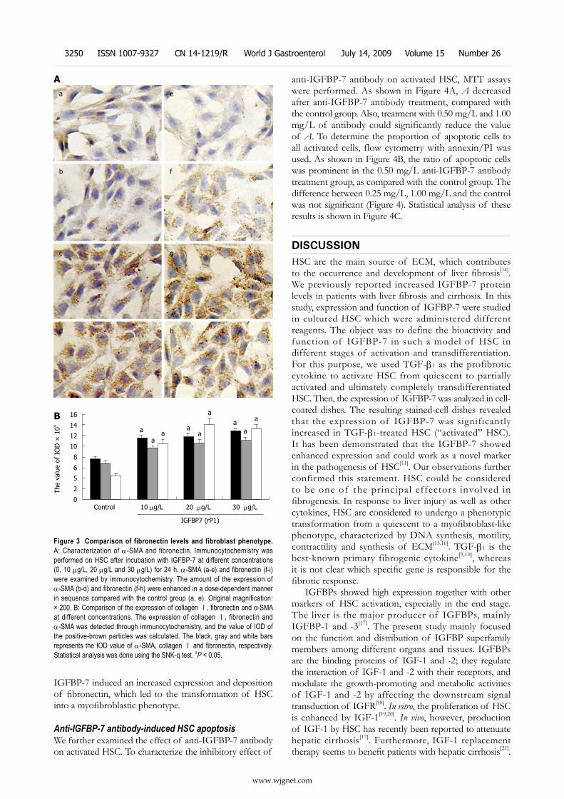
IGFBP-7 induced an increased expression and deposition of fibronectin, which led to the transformation of HSC into a myofibroblastic phenotype.
Anti-IGFBP-7 antibody-induced HSC apoptosis We further examined the effect of anti-IGFBP-7 antibody on activated HSC. To characterize the inhibitory effect of
anti-IGFBP-7 antibody on activated HSC, MTT assays were performed. As shown in Figure 4A, A decreased after anti-IGFBP-7 antibody treatment, compared with the control group. Also, treatment with 0.50 mg/L and 1.00 mg/L of antibody could significantly reduce the value of A. To determine the proportion of apoptotic cells to all activated cells, flow cytometry with annexin/PI was used. As shown in Figure 4B, the ratio of apoptotic cells was prominent in the 0.50 mg/L anti-IGFBP-7 antibody treatment group, as compared with the control group. The difference between 0.25 mg/L, 1.00 mg/L and the control was not significant (Figure 4). Statistical analysis of these results is shown in Figure 4C.
DISCUSSIONHSC are the main source of ECM, which contributes to the occurrence and development of liver fibrosis[14]. We previously reported increased IGFBP-7 protein levels in patients with liver fibrosis and cirrhosis. In this study, expression and function of IGFBP-7 were studied in cultured HSC which were administered different reagents. The object was to define the bioactivity and function of IGFBP-7 in such a model of HSC in different stages of activation and transdifferentiation. For this purpose, we used TGF-β1 as the profibrotic cytokine to activate HSC from quiescent to partially activated and ultimately completely transdifferentiated HSC. Then, the expression of IGFBP-7 was analyzed in cell-coated dishes. The resulting stained-cell dishes revealed that the expression of IGFBP-7 was significantly increased in TGF-β1-treated HSC (“activated” HSC). It has been demonstrated that the IGFBP-7 showed enhanced expression and could work as a novel marker in the pathogenesis of HSC[11]. Our observations further confirmed this statement. HSC could be considered to be one of the principal effectors involved in fibrogenesis. In response to liver injury as well as other cytokines, HSC are considered to undergo a phenotypic transformation from a quiescent to a myofibroblast-like phenotype, characterized by DNA synthesis, motility, contractility and synthesis of ECM[15,16]. TGF-β1 is the best-known primary fibrogenic cytokine[9,10], whereas it is not clear which specific gene is responsible for the fibrotic response.
IGFBPs showed high expression together with other markers of HSC activation, especially in the end stage. The liver is the major producer of IGFBPs, mainly IGFBP-1 and -3[17]. The present study mainly focused on the function and distribution of IGFBP superfamily members among different organs and tissues. IGFBPs are the binding proteins of IGF-1 and -2; they regulate the interaction of IGF-1 and -2 with their receptors, and modulate the growth-promoting and metabolic activities of IGF-1 and -2 by affecting the downstream signal transduction of IGFR[18]. In vitro, the proliferation of HSC is enhanced by IGF-1[19,20]. In vivo, however, production of IGF-1 by HSC has recently been reported to attenuate hepatic cirrhosis[17]. Furthermore, IGF-1 replacement therapy seems to benefit patients with hepatic cirrhosis[21].
www.wjgnet.com
Figure 3 Comparison of fibronectin levels and fibroblast phenotype. A: Characterization of α-SMA and fibronectin. Immunocytochemistry was performed on HSC after incubation with IGFBP-7 at different concentrations (0, 10 μg/L, 20 μg/L and 30 μg/L) for 24 h. α-SMA (a-e) and fibronectin (f-i) were examined by immunocytochemistry. The amount of the expression of α-SMA (b-d) and fibronectin (f-h) were enhanced in a dose-dependent manner in sequence compared with the control group (a, e). Original magnification: × 200. B: Comparison of the expression of collagen Ⅰ, fibronectin and α-SMA at different concentrations. The expression of collagen Ⅰ, fibronectin and α-SMA was detected through immunocytochemistry, and the value of IOD of the positive-brown particles was calculated. The black, gray and white bars represents the IOD value of α-SMA, collagen Ⅰ and fibronectin, respectively. Statistical analysis was done using the SNK-q test. aP < 0.05.
a e
b f
A
c g
d h
16
14
12
10
8
6
5
2
0
a
aa
aa
aa
a
a
The
valu
e of
IO
D ×
104
B
Control 10 μg/L 20 μg/L 30 μg/L
IGFBP7 (rP1)
3250 ISSN 1007-9327 CN 14-1219/R World J Gastroenterol July 14, 2009 Volume 15 Number 26

Because of the binding activity of IGFBPs, the induction of these binding proteins may affect hepatic cirrhosis. An in vitro study revealed that IGFBPs could display intrinsic bioactivities that are dependent on or independent of binding of IGF-1 and -2, in the process of hepatocyte growth, differentiation and activity[22]. So far, 6 distinct IGFBPs have been identified, which differ in molecular mass and binding affinities for IGF-1 and -2[13]. Moreover, low affinity IGF binders termed IGFBP-rPs have been found. IGFBP-7, also called IGFBP-rP1, is one of these low affinity binders of IGF-1. In the transcription of activated human HSC, expression of 5 IGFBP family members was detected. The most prominent was the extremely high expression of IGFBP-5, which is known to be the most abundantly expressed protein in activated HSC. In addition, IGFBP-7 is obviously detected in these cells, whereas a smaller induction is seen for IGFBP-4,
-6 and -3 [6]. The growth-promoting and metabolic activities of IGF-1 and -2 are modulated by the family of IGFBPs[23]. In certain pathological conditions, IGFBP-1 through -6 all may inhibit the growth of cells. IGFBP-1 and -3 may destroy glucose equilibrium in humans; IGFBP-1, -5, and -6 may also disturb this capability[24], whereas IGFBP-7 (rP1) could modulate proliferation, cohesion of cells, the regeneration of blood vessel and biosynthesis of prostacyclin[25]. IGFBP-8 (rP2) is considered to be a key cytokine in the fibrogenesis of tissues and organs[13]. Since modulation of IGF activity is very complex and varied, their differences may be important factors in determining the local autocrine and paracrine activities of IGFs. The striking up-regulation of IGFBP-7 predicted that it is a good marker of HSC activation and is also a promising target for therapeutic intervention in hepatic fibrosis.
www.wjgnet.com
Figure 4 Anti-IGFBP-7 antibody induces HSC apoptosis. A: HSC viability assay. The activated HSCs had anti-IGFBP-7 antibody of different concentrations added, and the absorbance (A) of each treated group was analyzed by the MTT cell viability assay to investigate the bioactivity of cultured cells. Statistical analysis was done using the SNK-q test. aP < 0.05; B: Percentage of apoptosis in all treated cells. Activated HSCs were divided into 4 groups, and then each group was treated with anti-IGFBP-7 antibody at different concentrations of 0 mg/L, 0.25 mg/L, 0.50 mg/L and 1.0 mg/L, and analyzed after 24 h culture, using flow cytometric assay. Statistical analysis was done using the SNK-q test. aP < 0.05; C: Flow cytometry assay. Cultured cells were treated with anti-IGFBP-7 body at different concentrations of 0 mg/L, 0.25 mg/L, 0.50 mg/L and 1.0 mg/L. After 24 h incubation, flow cytometry was used to analyze the pro-apoptotic effect of anti-IGFBP-7 antibody.
0.55
0.50
0.45
0.40
0.35
0.30
a
The
valu
e of
IO
D
A
Control 0.25 mg/L 0.50 mg/L 1.00 mg/L
a
25
21
17
13
9
5
Perc
enta
ge o
f ap
opto
sis
B
Control 0.25 mg/L 0.50 mg/L 1.00 mg/L
a
C 104
103
102
101
100
PI
100 101 102 103 104
Annexin V FITC
a 104
103
102
101
100
PI
100 101 102 103 104
Annexin V FITC
b
104
103
102
101
100
PI
100 101 102 103 104
Annexin V FITC
c 104
103
102
101
100
PI
100 101 102 103 104
Annexin V FITC
d
RU
2.63%
6.21%
RL
RU
2.51%
6.35%
RL
RU
4.34%
22.90%
RL
RU
3.53%
3.69%
RL
Liu LX et al . IGFBP-7 and hepatofibrosis 3251

Increased collagen and fibronectin deposition were detected in IGFBP-7-treated HSC, demonstrating that IGFBP-7 can activate and induce ECM production and fibrosis in vitro. Moreover, α-SMA-expressing cells increased among TGF-β1-treated cells, suggesting that they acquired a myofibroblastic phenotype[26]. Taken together, our findings indicate that superexpression of IGFBP-7 induces the deposition of ECM through the transformation of HSC into myofibroblastic cells which then contribute to the resulting fibrosis. We have shown that myofibroblastic changes are induced in vitro by IGFBP-7. In patients with liver fibrosis and cirrhosis, HSC are mainly responsible for the excessive deposition of ECM, many of which may have the phenotypic characteristics of myofibroblasts. A myofibroblast is strictly defined by electromicroscopic findings as a cell that is vimentin- and/or α-SMA-positive, with prominent rough endoplasmic reticulum, a modestly developed periphery, with focal densities, and producing collagen, granules, gap, and actin-filament-based junctions[27]. However, most of the present studies are restricted to an immunohistochemical determination without electron microscopy, such as spindle-shaped cells expressing α-SMA[28]. Thus, α-SMA-expressing cells in our model undergo a myofibroblastic change, and this in vitro phenomenon can also be triggered by exogenous IGFBP-7 applied to human stellate cells. In humans, HSC are mainly located at the sinusoid within the subendothelial space of Disse in close contact with the hepatocytes. They comprise roughly one-third of the nonparenchymal cell population or about 5%-8% of total liver cells[29]. During the development of liver fibrosis, HSC undergo a process of activation, resulting in a reduced retinoid storage capability and transformation to a myofibroblastic phenotype that is characterized by expression of α-SMA. In fact, the myofibroblast-like phenotype is not limited to liver, but also is a prominent feature of fibrosis in other tissues including pancreas, kidney, lung and skin[30].
Apoptosis of activated HSC is significantly detected when anti-IGFBP-7 antibody is administered to the medium. Flow cytometric analysis is a novel, specific and exact way to detect and measure apoptosis, based on the new parameters appearing on the surface of cells such as phospholipids that become exposed at the cell surface and form one of the specific signals for recognition and removal of apoptotic cells by macrophages. The anti-IGFBP-7 antibody acts as a proapoptotic factor, having great impact on the survival and proliferation of HSC. We predict that the anti-IGFBP-7 antibody also performs this function in vivo, which will be investigated in subsequent studies.
In summary, we have shown that IGFBP-7 plays a pivotal role in the pathogenesis of HSC activation and has profibrotic activities in vitro. IGFBP-7 induces the production of ECM via activation of HSC and the development of a myofibroblastic phenotype in HSC. The over-expression of IGFBP-7 in vitro also provides a new model to study the pathogenesis of HSC activation. The inhibitory effect of anti-IGFBP-7 antibody on
activated HSC provides a promising intervention for liver fibrosis.
ACKNOWLEDGMENTSThe authors thank the other technicians in the laboratory of the key medical laboratory of the First Teaching Hospital, including Yan Guo and Hai-Yan Zhang. The excellent technical assistance of the Department of Pathophysiology is gratefully acknowledged, especially the teacher, Yuan-Chang Zhao.
COMMENTSBackgroundHepatic stellate cells (HSC) are the main source of extracellular matrix (ECM), and thus play an important role in the occurrence and development of liver fibrosis. Insulin-like growth factor binding protein (IGFBP)-7 is a novel protein, which may be relevant to the activation of HSC. Research frontiersIGFBP-7 expression has been detected significantly during fibrogenesis, but how this protein is related to liver fibrosis is largely unknown. In this study, the authors demonstrated that IGFBP-7 could activate HSC in accordance with the transdifferentiation process in a dose-dependent manner to some extent.Innovations and breakthroughsRecent studies have highlighted the importance of IGF-Ⅰ and IGFBP-3 in the liver tissues and the roles they play in fibrogenesis. This is the first study to report that expression of IGFBP-7 is also enhanced in this process and IGFBP-7 could activate HSC in vitro. Furthermore, this protein may be the main mediator of HSC bioactivity.Applications By understanding how IGFBP-7 is relevant to liver fibrosis, this study may represent a new strategy for therapeutic intervention in the treatment of patients with liver fibrosis.TerminologyHSC are specific cells located in the hepatic lobules, and are the primary source of excess ECM assembly during liver fibrosis. The activation of HSC is regulated by known and unknown cytokines. IGFBP-7 is one member of the IGFBP superfamily, which is thought to work as a potential activator of HSC.Peer reviewThe paper by Liu et al is an interesting one, not only because of its findings, but also because of the quality of the presented data.
REFERENCES1 Friedman SL. Liver fibrosis -- from bench to bedside. J
Hepatol 2003; 38 Suppl 1: S38-S532 Gressner AM. Liver fibrosis: perspectives in pathobio-
chemical research and clinical outlook. Eur J Clin Chem Clin Biochem 1991; 29: 293-311
3 Friedman SL. Molecular regulation of hepatic fibrosis, an integrated cellular response to tissue injury. J Biol Chem 2000; 275: 2247-2250
4 Wynn TA. Common and unique mechanisms regulate fibrosis in various fibroproliferative diseases. J Clin Invest 2007; 117: 524-529
5 Parsons CJ, Takashima M, Rippe RA. Molecular mecha-nisms of hepatic fibrogenesis. J Gastroenterol Hepatol 2007; 22 Suppl 1: S79-S84
6 Yumei F, Zhou Y, Zheng S, Chen A. The antifibrogenic effect of (-)-epigallocatechin gallate results from the induction of de novo synthesis of glutathione in passaged rat hepatic stellate cells. Lab Invest 2006; 86: 697-709
7 Gressner AM. Cytokines and cellular crosstalk involved in the activation of fat-storing cells. J Hepatol 1995; 22: 28-36
8 Bataller R, Brenner DA. Liver fibrosis. J Clin Invest 2005; 115: 209-218
www.wjgnet.com
COMMENTS
3252 ISSN 1007-9327 CN 14-1219/R World J Gastroenterol July 14, 2009 Volume 15 Number 26
9 Casini A, Pinzani M, Milani S, Grappone C, Galli G, Jezequel AM, Schuppan D, Rotella CM, Surrenti C. Regulation of extracellular matrix synthesis by transforming growth factor beta 1 in human fat-storing cells. Gastroenterology 1993; 105: 245-253
10 Tiggelman AM, Boers W, Linthorst C, Sala M, Chamuleau RA. Collagen synthesis by human liver (myo)fibroblasts in culture: evidence for a regulatory role of IL-1 beta, IL-4, TGF beta and IFN gamma. J Hepatol 1995; 23: 307-317
11 Boers W, Aarrass S, Linthorst C, Pinzani M, Elferink RO, Bosma P. Transcriptional profiling reveals novel markers of liver fibrogenesis: gremlin and insulin-like growth factor-binding proteins. J Biol Chem 2006; 281: 16289-16295
12 Novosyadlyy R, Dargel R, Scharf JG. Expression of insulin-like growth factor-I and insulin-like growth factor binding proteins during thioacetamide-induced liver cirrhosis in rats. Growth Horm IGF Res 2005; 15: 313-323
13 Hwa V, Oh Y, Rosenfeld RG. The insulin-like growth factor-binding protein (IGFBP) superfamily. Endocr Rev 1999; 20: 761-787
14 Anan A, Baskin-Bey ES, Bronk SF, Werneburg NW, Shah VH, Gores GJ. Proteasome inhibition induces hepatic stellate cell apoptosis. Hepatology 2006; 43: 335-344
15 Ramadori G, Veit T, Schwogler S, Dienes HP, Knittel T, Rieder H, Meyer zum Buschenfelde KH. Expression of the gene of the alpha-smooth muscle-actin isoform in rat liver and in rat fat-storing (ITO) cells. Virchows Arch B Cell Pathol Incl Mol Pathol 1990; 59: 349-357
16 Rockey DC, Boyles JK, Gabbiani G, Friedman SL. Rat hepatic lipocytes express smooth muscle actin upon activation in vivo and in culture. J Submicrosc Cytol Pathol 1992; 24: 193-203
17 Conchillo M, de Knegt RJ, Payeras M, Quiroga J, Sangro B, Herrero JI, Castilla-Cortazar I, Frystyk J, Flyvbjerg A, Yoshizawa C, Jansen PL, Scharschmidt B, Prieto J. Insulin-like growth factor I (IGF-I) replacement therapy increases albumin concentration in liver cirrhosis: results of a pilot randomized controlled clinical trial. J Hepatol 2005; 43: 630-636
18 Wolf E, Schneider MR, Zhou R, Fisch TM, Herbach N, Dahlhoff M, Wanke R, Hoeflich A. Functional consequences of IGFBP excess-lessons from transgenic mice. Pediatr Nephrol 2005; 20: 269-278
19 Skrtic S, Wallenius K, Gressner AM, Jansson JO. Insulin-like growth factor signaling pathways in rat hepatic stellate
cells: importance for deoxyribonucleic acid synthesis and hepatocyte growth factor production. Endocrinology 1999; 140: 5729-5735
20 Svegliati-Baroni G , Ridolfi F, Di Sario A, Casini A, Marucci L, Gaggiotti G, Orlandoni P, Macarri G, Perego L, Benedetti A, Folli F. Insulin and insulin-like growth factor-1 stimulate proliferation and type I collagen accumulation by human hepatic stellate cells: differential effects on signal transduction pathways. Hepatology 1999; 29: 1743-1751
21 Sanz S, Pucilowska JB, Liu S, Rodriguez-Ortigosa CM, Lund PK, Brenner DA, Fuller CR, Simmons JG, Pardo A, Martinez-Chantar ML, Fagin JA, Prieto J. Expression of insulin-like growth factor I by activated hepatic stellate cells reduces fibrogenesis and enhances regeneration after liver injury. Gut 2005; 54: 134-141
22 Clemmons DR. Insulin-like growth factor binding proteins and their role in controlling IGF actions. Cytokine Growth Factor Rev 1997; 8: 45-62
23 Clemmons DR. Use of mutagenesis to probe IGF-binding protein structure/function relationships. Endocr Rev 2001; 22: 800-817
24 Kelley KM, Schmidt KE, Berg L, Sak K, Galima MM, Gillespie C, Balogh L, Hawayek A, Reyes JA, Jamison M. Comparative endocrinology of the insulin-like growth factor-binding protein. J Endocrinol 2002; 175: 3-18
25 Rajaram S, Baylink DJ, Mohan S. Insulin-like growth factor-binding proteins in serum and other biological fluids: regulation and functions. Endocr Rev 1997; 18: 801-831
26 Yasuoka H, Zhou Z, Pilewski JM, Oury TD, Choi AM, Feghali-Bostwick CA. Insulin-like growth factor-binding protein-5 induces pulmonary fibrosis and triggers mononuclear cellular infiltration. Am J Pathol 2006; 169: 1633-1642
27 Eyden B. The myofibroblast: an assessment of controversial issues and a definition useful in diagnosis and research. Ultrastruct Pathol 2001; 25: 39-50
28 Willis BC, Liebler JM, Luby-Phelps K, Nicholson AG, Crandall ED, du Bois RM, Borok Z. Induction of epithelial-mesenchymal transition in alveolar epithelial cells by transforming growth factor-beta1: potential role in idiopathic pulmonary fibrosis. Am J Pathol 2005; 166: 1321-1332
29 Hautekeete ML, Geerts A. The hepatic stellate (Ito) cell: its role in human liver disease. Virchows Arch 1997; 430: 195-207
30 Serini G, Gabbiani G. Mechanisms of myofibroblast activity and phenotypic modulation. Exp Cell Res 1999; 250: 273-283
S- Editor Tian L L- Editor Cant MR E- Editor Ma WH
www.wjgnet.com
Liu LX et al . IGFBP-7 and hepatofibrosis 3253

www.wjgnet.com
BRIEF ARTICLES
Thermal hypersensitivity in a subset of irritable bowel syndrome patients
QiQi Zhou, Roger B Fillingim, Joseph L Riley III, G Nicholas Verne
Online Submissions: wjg.wjgnet.com World J Gastroenterol 2009 July 14; 15(26): [email protected] World Journal of Gastroenterology ISSN 1007-9327doi:10.3748/wjg.15.3254 © 2009 The WJG Press and Baishideng. All rights reserved.
QiQi Zhou, G Nicholas Verne, Department of Medicine, Ohio State University, Columbus, Ohio 43210, United StatesRoger B Fillingim, Joseph L Riley III, College of Dentistry, University of Florida, Gainesville, Florida 32610, United StatesRoger B Fillingim, North Florida/South Georgia Veteran Health System, Gainesville, Florida 32610, United StatesG Nicholas Verne, Research Service, Cincinnati VAMC, Cincinnati, Ohio 45220, United StatesAuthor contributions: Zhou Q and Verne GN performed all of the experiments and wrote the majority of the manuscript; Fillingim RB and Riley JL helped to edit the manuscript and provided support in the analysis of the data.Supported by A NIH RO1-NS053090 award (PI: GN Verne) and a VA Merit Review Award (PI: GN Verne) from the Medical Research Service at the Department of Veterans AffairsCorrespondence to: G Nicholas Verne, MD, Division of Gastroenterology, Hepatology, and Nutrition, Ohio State University, 288A Office Tower, 395 West 12th Avenue, Columbus, OH 43210, United States. [email protected]: +1-614-3667095 Fax: +1-614-2476924Received: January 16, 2009 Revised: May 11, 2009Accepted: May 18, 2009Published online: July 14, 2009
AbstractAIM: To characterize thermal hypersensitivity in patients with constipation- and diarrhea-predominant irritable bowel syndrome (IBS).
METHODS: Thermal pain sensitivity was tested among patients with diarrhea-predominant IBS (D-IBS) and constipation-predominant IBS (C-IBS) compared to healthy subjects. A total of 42 patients (29 female and 13 male; mean age 27.0 ± 6.4 years) with D-IBS; 24 patients (16 female and eight male; mean age 32.5 ± 8.8 years) with C-IBS; and 52 control subjects (34 female and 18 male; mean age 27.3 ± 8.0 years) participated in the study. Thermal stimuli were delivered using a Medoc Thermal Sensory Analyzer with a 3 cm × 3 cm surface area. Heat pain threshold (HPTh) and heat pain tolerance (HPTo) were assessed on the left ventral forearm and left calf using an ascending method of limits. The Functional Bowel Disease Severity Index (FBDSI) was also obtained for all subjects.
RESULTS: Controls were less sensitive than C-IBS and D-IBS (both at P < 0.001) with no differences between C-IBS and D-IBS for HPTh and HPTo.
Thermal hyperalgesia was present in both groups of IBS patients relative to controls, with IBS patients reporting significantly lower pain threshold and pain tolerance at both test sites. Cluster analysis revealed the presence of subgroups of IBS patients based on thermal hyperalgesia. One cluster (17% of the sample) showed a profile of heat pain sensitivity very similar to that of healthy controls; a second cluster (47% of the sample) showed moderate heat pain sensitivity; and a third cluster (36% of the sample) showed a very high degree of thermal hyperalgesia.
CONCLUSION: A subset of IBS patients had thermal hypersensitivity compared to controls, who reported significantly lower HPTh and HPTo. All IBS patients had a higher score on the FBDSI than controls. Interestingly, the subset of IBS patients with high thermal sensitivity (36%) had the highest FBDSI score compared to the other two groups of IBS patients.
© 2009 The WJG Press and Baishideng. All rights reserved.
Key words: Irritable bowel syndrome; Visceral pain; thermal hypersensitivity; Heat pain threshold; Heat pain tolerance
Peer reviewer: Mario Guslandi, Professor, Department of Gastroenterology, S: Raffaele University Hospital, via Olgettina 60, Milano 20132, Italy
Zhou Q, Fil l ingim RB, Riley JL, Verne GN. Thermal hypersensitivity in a subset of irritable bowel syndrome patients. World J Gastroenterol 2009; 15(26): 3254-3260 Available from: URL: http://www.wjgnet.com/1007-9327/15/3254.asp DOI: http://dx.doi.org/10.3748/wjg.15.3254
INTRODUCTIONIrritable bowel syndrome (IBS) is one of the most common gastrointestinal disorders encountered by gastroenterologists. Patients classically present with chronic abdominal pain associated with an alteration in bowel habits. Even though the pathophysiology of the IBS is unclear, visceral hypersensitivity is a common clinical marker of the disorder[1,2]. Visceral hypersensi t iv i ty may account for symptoms of

Zhou Q et al . Thermal hypersensitivity in irritable bowel syndrome 3255
www.wjgnet.com
abdominal pain, urgency, and bloating experienced by many patients with this disorder.
Although visceral hypersensitivity is considered a hallmark feature of IBS, conflicting evidence exists regarding somatic hypersensitivity in this patient population. Somatic pain conditions, such as fibromyalgia and migraine headaches show significant comorbidity with IBS, which suggests that somatic hypersensitivity characterizes at least a subpopulation of IBS patients[2,3]. Several investigators have found no evidence for heightened somatic pain sensitivity in IBS patients. For example, two studies reported that IBS patients showed lower sensitivity to painful electrocutaneous stimuli compared to healthy controls[4,5]. Also, others have reported similar cold pressor pain tolerance in IBS patients and controls[6,7]. In contrast, our recent studies using hot water immersion have shown widespread somatic hyperalgesia associated with IBS[2,8,9], and others using the cold pressor test have demonstrated somatic hypersensitivity in IBS patients compared with healthy controls[3,10].
These conflicting findings may result from differing somatic pain testing procedures. Alternatively, patient sampling may be a contributing factor, given that there may be subgroups of IBS patients who differ in their somatic sensitivity. For example, somatic hypersensitivity may be present only in a subset of patients based on IBS subtype [i.e. diarrhea-predominant IBS (D-IBS) vs constipation-predominant IBS (C-IBS)], symptom severity and/or psychological profile.
Previous studies have explored the correlates of visceral hypersensitivity among patients with IBS[11-16]. For example, depression is correlated with rectal pain thresholds only in patients who alternate between constipation and diarrhea. Others have reported no association of psychological factors with rectal sensitivity; however, hypersensitivity to rectal distention is associated with increased IBS symptom severity assessed by daily diary[11]. Similarly, other investigators have found that rectal pain sensitivity is correlated positively with clinical symptoms among IBS patients[12-14], while others have reported no such association[15,16]. However, the association of somatic hypersensitivity with clinical symptoms in IBS has not been evaluated.
To further evaluate somatic hyperalgesia among patients with IBS, we evaluated thermal pain sensitivity among patients with D-IBS and C-IBS compared with healthy subjects. The aims of the present study were: (a) to compare the spatial distribution and magnitude of thermal hyperalgesia between D-IBS and C-IBS patients and controls; and (b) to compare the spatial distribution and magnitude of thermal hyperalgesia among IBS patients as a function of symptom severity.
MATERIALS AND METHODSParticipantsA total of 42 patients (29 female and 13 male; mean age 27.0 ± 6.4 years) with D-IBS; 24 patients (16 female and eight male; mean age 32.5 ± 8.8 years) with C-IBS; and 52
control subjects (34 females and 18 male; mean age 27.3 ± 8.0 years) participated in the study. The demographics of the participating subjects are presented in Table 1. IBS subjects and healthy controls were recruited via advertisements posted at the University of Florida and the Ohio State University. The study was approved by the University of Florida, the North Florida/South Georgia Veterans Health System, and the Ohio State University Institutional Review Boards. All subjects signed informed consent prior to the start of the study.
None of the control subjects had any evidence of acute or chronic somatic/abdominal pain or IBS based on a questionnaire and complete physical examination by an experienced gastroenterologist. Also, controls were free of any systemic medical disease or psychological conditions that could affect sensory responses. All IBS subjects had symptoms for at least 5 years. The diagnosis of IBS was made by the same gastroenterologist who examined patients based on the ROME Ⅲ criteria and exclusion of organic disease[17]. All subjects with IBS were examined for fibromyalgia (FM) using the 1990 American College of Rheumatology criteria for FM[18]. None of the patients were diagnosed as having FM. None of the IBS or control subjects were taking analgesics, serotonin uptake inhibitors, serotonin antagonists, or tricyclic antidepressants for a period of at least 3 wk prior to the study.
All subjects underwent experimental psychophysical testing during a single session. All sessions were conducted between 9 AM and 6 PM to control for circadian rhythm effects. Subjects were instructed to refrain from the use of any analgesic medication for 48 h and from caffeine for 4 h before their sessions. Prior to each session, participants received a reminder concerning the restrictions on analgesic medication and caffeine use.
Female subjects participated during the follicular phase of their cycles (i.e. 4-9 d post onset of menses). This cycle phase was chosen because it is characterized generally by the least sensitivity to pain and by minimal menstrual cycle related symptoms[19]. The menstrual cycle has been reported to alter pain perception in women, and IBS symptoms have also been reported to fluctuate across the menstrual cycle[20]. In addition, the follicular phase was chosen because some female
Table 1 Demographic variables for IBS patients and controls
D-IBS (n = 42)
C-IBS (n = 24)
Controls (n = 52)
Age (mean ± SD) 27.0 ± 6.4 32.5 ± 8.8 27.3 ± 8.0Sex (female), n (%) 29 (69) 16 (67) 34 (64)FBDSI 62.8 (23.2) 68.4 (25.3) 2.1 (4.9) Self-reported race/ethnicity n (%)White 33 (79) 15 (63) 43 (83)Black 3 (7) 6 (25) 3 (6)Hispanic 3 (7) 1 (4) 3 (6)Asian 3 (7) 2 (8) 3 (6)
D-IBS: Diarrhea-predominant irritable bowel syndrome; C-IBS: Constipation-predominant irritable bowel syndrome. Columns may not sum to 100%, because values were rounded to the nearest 1%. Scores on the Functional Bowel Disorder Severity Index (FBDSI) that share a superscript are different at P < 0.01.

www.wjgnet.com
subjects would have been using oral contraceptives (OCs), and the follicular phase is the cycle phase during which women who use OCs and normal cycling women are the most similar in their responses to experimental pain[21].
Psychophysical measuresMultiple psychological factors have been related to pain responses in a number of studies, and these factors may mediate partially group differences in pain sensitivity. Therefore, all participants completed psychological questionnaires that assessed coping style, anxiety, depression, and hyper-vigilance prior to the first experimental session. In addition, a measure of current affective state was administered prior to each experimental session. These measures were used as control variables (as further described in the data analysis section) to determine whether group differences in pain sensitivity remain significant after controlling for the influence of psychological variables.
The Coping Strategies Questionnaire (CSQ) consists of 44 items that are related to how individuals cope with pain[22]. It yields seven subscales based on the pain coping strategies: diverting attention, catastrophizing, praying and hoping, ignoring pain sensations, reinterpreting pain sensations, increasing behavioral activity, and coping self-statements. The CSQ also provides measures of subjects’ perceived ability to control and decrease pain. It has been used widely with various pain populations, and has been modified for use with healthy pain-free subjects, by having individuals respond to the instrument based on how they cope typically with day-to-day aches and pains[23]. Responses on the CSQ have previously been related to experimental pain responses[24], as well as clinical pain, including IBS[25-27]. We have used this scale in previous psychophysical and clinical research[28-30].
The Kohn reactivity scale consists of 24 items that assess an individual’s level of reactivity or central nervous system arousability. It has been used recently as a measure of the construct of hypervigilance[31]. This measure has been shown to correlate negatively with pain tolerance[31,32] and has been reported to have adequate internal consistency, ranging from an a value of 0.73 to 0.83[33].
The State-Trait Anxiety Inventory (STAXI) consists of 20 items that assess dispositional (i.e. trait) anxiety[34]. This is a well-validated and widely used instrument for assessing general anxiety.
The Beck Depression Inventory (BDI) is a widely used, 21-item, self-report measure that assesses common cognitive, affective and vegetative symptoms of depress ion. Research that has evaluated the psychometric properties of the BDI has suggested that it shows excellent reliability and validity as an index of depression[35]. Since chronic pain patients often endorse somatic symptoms assessed by the BDI, which may artificially inflate their scores[36], the BDI has been separated into a 13-item cognitive affective subscale and an eight-item somatic-performance subscale[37].
The Profile of Mood States-bipolar Form (POMS-BI) consists of 72 mood-related items, and subjects
indicate the extent to which each item describes their current mood[38]. This questionnaire assesses positive and negative affective dimensions. The POMS-BI has been well validated with other mood measures and is sensitive to subtle differences in affective state. We have used this measure in previous psychophysical studies[28]. This mood measure was administered to determine the current affective state at the beginning of each sensory testing session.
Functional Bowel Disorder Severity Index (FBDSI) comprises three variables: current pain (by visual analog scale), diagnosis of chronic abdominal pain, and number of physician visits in the past 6 mo[39]. The FBDSI is sufficiently sensitive to distinguish among the different groups, from healthy controls, through non-IBS patients, to patients with IBS only and,finally, IBS patients with concomitant FM. Severity is rated as none (0 points, controls), mild (1-36 points, non-IBS patients), moderate (37-110 points, IBS patients), and severe (> 110 points, IBS and FM). All IBS patients that participated in the study were characterized with this index.
Thermal proceduresThermal stimuli were delivered using a computer-controlled Medoc Thermal Sensory Analyzer (TSA-2001; Ramat Yishai, Israel). This is peltier-element-based stimulator with a 3 cm × 3 cm surface area. Temperature levels were monitored by a contactor-contained thermistor, and returned to a preset baseline of 32℃ by active cooling at a rate of 10℃/s. Heat pain threshold (HPTh) and heat pain tolerance (HPTo) were assessed on the left ventral forearm and left calf using an ascending method of limits. The cutoff temperature (to avoid tissue damage) for all trials was 52℃. Subjects were assigned randomly within each group (controls, C-IBS, D-IBS) to receive thermal nociceptive stimulation to the left ventral forearm versus the left calf in a random order that was counterbalanced across all groups.
From a baseline of 32℃, probe temperature increased at a rate of 0.5℃/s until the subject responded by pressing a button on a handheld device. This slow rate of rise preferentially activates C-fibers and diminishes artifacts associated with reaction time.
HPThFor HPTh, subjects were informed via digitally recorded instructions to press the button when the sensation first became painful. Four trials of HPTh were performed at each site (ventral forearm and calf). The average of the four trials at each site was computed for HPTh. The position of the thermode was altered slightly between trials in order to avoid either sensitization or habituation of cutaneous receptors. In addition, interstimulus intervals of at least 30 s were maintained between successive stimuli. Four trials of HPTh were presented followed by a 15-min rest period, and then four trials of HPTo were performed.
HPToFor HPTo, subjects were instructed to press the button
3256 ISSN 1007-9327 CN 14-1219/R World J Gastroenterol July 14, 2009 Volume 15 Number 26

www.wjgnet.com
when the sensation was no longer tolerable. Four trials of HPTo were performed at each site (ventral forearm and calf). The average of the four trials at each site was computed for HPTo. The position of the thermode was altered slightly between trials in order to avoid either sensitization or habituation of cutaneous receptors. In addition, interstimulus intervals of at least 30 s were maintained between successive stimuli.
Statistical analysisThe primary analyses involved one between- and two within-subject variables, and consequently, the repeated measures command within the General Linear model module of SPSS was used. In the first primary analysis, differences between controls and D-IBS and C-IBS patients for HPTh and HPTo at the forearm and calf were tested. In the second primary analysis, associations between IBS symptom severity and HPTh and HPTo at the forearm and calf were tested. A series of one-way ANOVAs were used to test for group differences in psychological inventories. As a result of the number of statistical tests performed, a Bonferroni correction was used to maintain family-wise type 1 error rate at P < 0.05.
RESULTSSamplesA total of 118 participants were studied, which included 42 patients (29 female and 13 males; mean age 27.0 ± 6.4 years) with D-IBS; 24 patients (16 female and eight male; mean age 32.5 ± 8.8 years) with C-IBS; and 52 control subjects (34 female and 18 males; mean age 27.3 ± 8.0 years) (Table 1). There was no difference in age or sex between the groups (controls, D-IBS and C-IBS). FBDSI score was higher in the D-IBS (62.8 ± 23.2) and C-IBS (68.4 ± 25.3) patients than the controls (2.1 ± 4.9), but did not differ between the patient groups.
HPTh and HPToHPTh and HPTo (mean ± SD) at both sites for C-IBS, D-IBS, and controls are presented in Table 2. We found significant effects for site, pain measure, and group (all at P < 0.001). Pair-wise comparisons supported the hypothesis that controls were less sensitive than C-IBS and D-IBS patients (both at P < 0.001), with no differences between C-IBS and D-IBS. Two-way interactions for pain measure × group (P = 0.002) and site × pain measure (P < 0.001) were also significant. Pair-wise comparisons revealed that controls had higher HPTh and HPTo than C-IBS and D-IBS (both at P < 0.001) on the forearm and the calf. There was no difference in HPTh or HPTo between C-IBS and D-IBS patients.
A group effect emerged for Kohn scores (P = 004), with C-IBS (78.8 ± 9.9) having higher scores than D-IBS (70.8 ± 10.8) and controls (70.1 ± 11.1) (collapsed across groups, 72.1 ± 11.2). There were no differences in the BDI (3.4 ± 3.7), STAXI (28.0 ± 4.8), POMS positive (53.2 ± 15.7), POMS negative (29.8 ± 16.3) or any of the subscales of the CSQ (distracting attention, 10.0 ± 6.1; cognitive
self-statements, 13.3 ± 5.3; ignoring pain sensations, 11.0 ± 6.1; praying and hoping, 5.4 ± 4.9; reinterpreting pain sensations, 4.9 ± 4.8; catastrophizing, 5.9 ± 6.1).
Differences as a function of IBS symptomsWe found significant effects for site, pain measure, and symptom severity (all at P < 0.001). The site by symptom severity interaction was not significant; however, the 3-way interaction (site × pain × symptoms) was significant (P < 0.01). To interpret this interaction involving an internal level variable (IBS symptom severity) and two nominal variables, we examined scatter plots for HPTh and HPTo at the leg and arm by FBDSI scores. The results suggest a naturally occurring gap between scores of 72 and 82 that was associated with pain sensitivity; therefore, two groups were formed based on FBDSI scores, with a cutoff at 80. Demographic variables for the two symptom severity groups are presented in Table 3. HPTh and HPTo at both sites for the two IBS symptom severity groups are presented in Table 2 and Figure 1. The IBS symptom subgroups did not differ for Kohn, BDI, STAXI, POMS, or CSQ scores.
DISCUSSIONOverall, our findings indicate thermal hyperalgesia for IBS-C and IBC-D patients relative to controls, with IBS patients reporting significantly lower pain threshold and pain tolerance at both test sites. These findings add further
Table 2 HPTh and HPTo for the arm and leg
Groups (n ) Threshold (HPTh) Tolerance (HPTo)
Forearm Calf Forearm CalfControls (n = 52) 43.2 ± 1.8 45.1 ± 1.7 48.0 ± 1.8 48.1 ± 1.4Diarrhea (n = 42) 39.1 ± 3.3 41.1 ± 4.0 44.7 ± 3.8 44.9 ± 2.1Constipation (n = 24) 39.6 ± 3.3 41.3 ± 3.5 45.0 ± 3.7 46.0 ± 3.1IBS symptom groupsModerate symptoms (n = 42)
41.6 ± 1.7 43.6 ± 1.9 47.2 ± 1.6 47.0 ± 1.3
Severe symptoms (n = 24)
35.6 ± 1.5 36.9 ± 1.6 40.2 ± 2.1 42.1 ± 1.6
Values represent temperature mean ± SD.
Table 3 Demographic variable for IBS pain subgroups
Moderate sensitivity (n = 42)
High sensitivity (n = 24)
Age (SD) 28.7 (7.0) 29.1 (7.3)Sex (female) n (%) 30 (71) 15 (63)FBDSI 49.3 (9.1) 96.4 (8.7)
Range 38-72 Range 82-110Diarrhea subtype n (%) 25 (60) 17 (41)Constipation subtype n (%) 17 (40) 7 (29)Self-reported race/ethnicity n (%)White 30 (71) 18 (37)Black 9 (21) 0 (0)Hispanic 1 (2) 3 (75)Asian 2 (5) 3 (60)
Columns may not sum to 100% because values were rounded to the nearest 1%.
Zhou Q et al . Thermal hypersensitivity in irritable bowel syndrome 3257

www.wjgnet.com
support to the notion that IBS patients show somatic hyperalgesia. A unique finding of our study is that we detected a strong relationship between heat pain measures and FBDSI scores. IBS patients with high FBDSI scores had the highest thermal pain sensitivity compared to IBS patients with low to moderate FBDSI scores.
In contrast to our current findings, several previous studies have indicated lack of somatic hyperalgesia among IBS patients relative to controls[6,7], with some studies actually showing higher pain threshold among IBS patients relative to controls[4,5]. One possible explanation for this is differences in the painful stimulus, as previous studies have used electrical stimuli, mechanical pressure, and cold immersion, but none has used contact heat. Also, Chang and colleagues[40] have reported that female IBS patients showed significantly higher pressure pain threshold than female controls in response to a randomly administered series of fixed stimuli, but no group differences emerged for threshold assessed using ascending stimuli. Randomly administered stimuli are thought to reduce response bias, which is likely driven by psychological factors such as hypervigilance. Since we used ascending stimuli in the present study, it is possible that response bias contributed to our results. However, this seems unlikely given the lack of correlations between psychological factors, including anxiety and hypervigilance, and heat pain responses. Differences in the nature of the patient population may present another explanation for the differences between our findings and those of previous studies. Most prior investigations[4-7] recruited IBS patients from clinical settings, typically in tertiary care centers, whereas our IBS sample was recruited from the community, using print and postal advertisements. This community-based recruitment approach yielded a psychologically healthy IBS population, which was similar to the controls for most psychological measures. Moreover, most previous studies have included a female-only population, while we included women and men in both the IBS and control samples. Thus, differences in the experimental pain stimulus and patient population may have contributed to the differing pattern of results.
Consistent with the present findings, other investigators have reported somatic hyperalgesia in IBS patients, using cold pain[3,10], and we have shown similar results
with heat immersion[2,8]. However, to the best of our knowledge, this is the first report of an association between somatic pain sensitivity and clinical symptoms among patients with IBS. Wilder-Smith and colleagues[10] have reported a strong association between somatic and visceral hypersensitivity. However, there is mixed evidence regarding the association between visceral hypersensitivity and clinical symptoms in IBS[11,13,15,16,41]. In our study, the association of somatic hyperalgesia with FBDSI score suggests that central mechanisms contribute to the severity of patients’ clinical symptoms. This has potentially important treatment implications, as one might speculate that patients exhibiting somatic hypersensitivity may benefit from treatments that alter central processes, rather than those that are restricted to peripheral targets.
Several limitations of this study should be mentioned. First, somatic pain testing was limited to HPTh and HPTo and did not include other stimuli. Thus, it is not possible to determine whether these findings are specific to heat pain. Second, we did not assess sensitivity to visceral stimuli, and it would be interesting to know whether heat pain sensitivity is associated with visceral sensitivity. However, given our relatively large sample size, visceral testing was not feasible. Finally, based on the current design, we were unable to identify the mechanisms that underlie heat hyperalgesia and its association with clinical symptoms. Despite these limitations, the relatively large sample size, the use of heat pain measures designed to activate C-fibers, and the strong association between heat pain sensitivity and IBS symptom severity represent unique and valuable features of our study.
CONCLUSIONOur study indicates somatic hypersensitivity for both groups of IBS patients relative to controls, with IBS patients reporting significantly lower thermal pain threshold and tolerance. Moreover, somatic pain sensitivity was associated with IBS symptoms, such that patients with high FBDSI scores showed significantly greater pain sensitivity compared to those with low to moderate FBDSI scores. Further studies are warranted to evaluate somatic hypersensitivity as a predictor of clinical symptoms in IBS.
COMMENTSBackgroundIrritable bowel syndrome (IBS) is one of the most common gastrointestinal disorders encountered by gastroenterologists. Patients present classically with chronic abdominal pain associated with an alteration in bowel habits. Even though the pathophysiology of IBS is unclear, visceral hypersensitivity is a common clinical marker of the disorder. Visceral hypersensitivity may account for symptoms of abdominal pain, urgency, and bloating experienced by many patients with this disorder.Research frontiersAlthough visceral hypersensitivity is considered a hallmark feature of IBS, conflicting evidence exists regarding somatic hypersensitivity in this patient population. Somatic pain conditions, such as fibromyalgia and migraine headaches show significant comorbidity with IBS, which suggests that somatic hypersensitivity characterizes at least a subpopulation of IBS patients. Several
48
46
44
42
40
38
36
34HPTh arm HPTh leg HPTo arm HPTo leg
Moderate symptoms
Severe symptoms
Figure 1 HPTh and HPTo for IBS pain subgroups.
Tem
pera
ture
(℃
)
COMMENTS
3258 ISSN 1007-9327 CN 14-1219/R World J Gastroenterol July 14, 2009 Volume 15 Number 26

www.wjgnet.com
recent studies using hot water immersion have shown widespread somatic hyperalgesia associated with IBS, and others using the cold pressor test have demonstrated somatic hypersensitivity in IBS patients compared with healthy controls.Innovations and breakthroughsOverall, the findings indicate thermal hyperalgesia for constipation- and diarrhea-dependent IBS relative to controls, with IBS patients reporting significantly lower pain threshold and tolerance at both test sites. These findings add further support to the notion that IBS patients show somatic hyperalgesia. A unique finding of this study is that the authors detected a strong relationship between heat pain measures and Functional Bowel Disease Severity Index (FBDSI) scores. IBS patients with high FBDSI scores had the highest thermal pain sensitivity compared to those IBS patients with low to moderate FBDSI scores.ApplicationsThis study suggests that a subset of IBS patients has evidence of somatic hypersensitivity that may relate to extra-intestinal symptoms.Peer reviewThis is an elegant study that evaluated systematically thermal hypersensitivity in a large number of IBS patients. The results are novel and interesting and may lead to new therapy in a subset of IBS patients with somatic hypersensitivity.
REFERENCES1 Naliboff BD, Munakata J, Fullerton S, Gracely RH, Kodner
A, Harraf F, Mayer EA. Evidence for two distinct perceptual alterations in irritable bowel syndrome. Gut 1997; 41: 505-512
2 Verne GN, Robinson ME, Price DD. Hypersensitivity to visceral and cutaneous pain in the irritable bowel syndrome. Pain 2001; 93: 7-14
3 Bouin M, Meunier P, Riberdy-Poitras M, Poitras P. Pain hypersensitivity in patients with functional gastrointestinal disorders: a gastrointestinal-specific defect or a general systemic condition? Dig Dis Sci 2001; 46: 2542-2548
4 Accarino AM , Azpiroz F, Malagelada JR. Selective dysfunction of mechanosensitive intestinal afferents in irritable bowel syndrome. Gastroenterology 1995; 108: 636-643
5 Cook IJ, van Eeden A, Collins SM. Patients with irritable bowel syndrome have greater pain tolerance than normal subjects. Gastroenterology 1987; 93: 727-733
6 Whitehead WE, Holtkotter B, Enck P, Hoelzl R, Holmes KD, Anthony J, Shabsin HS, Schuster MM. Tolerance for rectosigmoid distention in irritable bowel syndrome. Gastroenterology 1990; 98: 1187-1192
7 Zighelboim J , Talley NJ, Phillips SF, Harmsen WS, Zinsmeister AR. Visceral perception in irritable bowel syndrome. Rectal and gastric responses to distension and serotonin type 3 antagonism. Dig Dis Sci 1995; 40: 819-827
8 Verne GN, Himes NC, Robinson ME, Gopinath KS, Briggs RW, Crosson B, Price DD. Central representation of visceral and cutaneous hypersensitivity in the irritable bowel syndrome. Pain 2003; 103: 99-110
9 Verne GN , Price DD. Irritable bowel syndrome as a common precipitant of central sensitization. Curr Rheumatol Rep 2002; 4: 322-328
10 Wilder-Smith CH, Robert-Yap J. Abnormal endogenous pain modulation and somatic and visceral hypersensitivity in female patients with irritable bowel syndrome. World J Gastroenterol 2007; 13: 3699-3704
11 van der Veek PP, Van Rood YR, Masclee AA. Symptom severity but not psychopathology predicts visceral hypersensitivity in irritable bowel syndrome. Clin Gastroenterol Hepatol 2008; 6: 321-328
12 Kanazawa M, Palsson OS, Thiwan SI, Turner MJ, van Tilburg MA, Gangarosa LM, Chitkara DK, Fukudo S, Drossman DA, Whitehead WE. Contributions of pain sensitivity and colonic motility to IBS symptom severity and predominant bowel habits. Am J Gastroenterol 2008; 103: 2550-2561
13 Posserud I, Syrous A, Lindström L, Tack J, Abrahamsson
H, Simrén M. Altered rectal perception in irritable bowel syndrome is associated with symptom severity. Gastroenterology 2007; 133: 1113-1123
14 Awad RA, Camacho S, Martín J, Ríos N. Rectal sensation, pelvic floor function and symptom severity in Hispanic population with irritable bowel syndrome with constipation. Colorectal Dis 2006; 8: 488-493
15 Castilloux J, Noble A, Faure C. Is visceral hypersensitivity correlated with symptom severity in children with functional gastrointestinal disorders? J Pediatr Gastroenterol Nutr 2008; 46: 272-278
16 Sabate JM, Veyrac M, Mion F, Siproudhis L, Ducrotte P, Zerbib F, Grimaud JC, Dapoigny M, Dyard F, Coffin B. Relationship between rectal sensitivity, symptoms intensity and quality of life in patients with irritable bowel syndrome. Aliment Pharmacol Ther 2008; 28: 484-490
17 Longstreth GF, Thompson WG, Chey WD, Houghton LA, Mearin F, Spiller RC. Functional bowel disorders. Gastroenterology 2006; 130: 1480-1491
18 Geel SE. The fibromyalgia syndrome: musculoskeletal pathophysiology. Semin Arthritis Rheum 1994; 23: 347-353
19 Riley JL 3rd, Robinson ME, Wise EA, Price DD. A meta-analytic review of pain perception across the menstrual cycle. Pain 1999; 81: 225-235
20 Heitkemper MM, Jarrett M. Pattern of gastrointestinal and somatic symptoms across the menstrual cycle. Gastroenterology 1992; 102: 505-513
21 Fillingim RB, Ness TJ. Sex-related hormonal influences on pain and analgesic responses. Neurosci Biobehav Rev 2000; 24: 485-501
22 Rosenstiel AK, Keefe FJ. The use of coping strategies in chronic low back pain patients: relationship to patient characteristics and current adjustment. Pain 1983; 17: 33-44
23 Lefebvre JC, Lester N, Keefe FJ. Pain in young adults. II: The use and perceived effectiveness of pain-coping strategies. Clin J Pain 1995; 11: 36-44
24 Geisser ME, Robinson ME, Pickren WE. Differences in cognitive coping strategies among pain sensitive and pain tolerant individuals on the cold pressor test. Beh Ther 1992; 23: 31-41
25 Keefe FJ, Caldwell DS, Queen KT, Gil KM, Martinez S, Crisson JE, Ogden W, Nunley J. Pain coping strategies in osteoarthritis patients. J Consult Clin Psychol 1987; 55: 208-212
26 Keefe FJ, Dolan E. Pain behavior and pain coping strategies in low back pain and myofascial pain dysfunction syndrome patients. Pain 1986; 24: 49-56
27 Scarinci IC, McDonald-Haile J, Bradley LA, Richter JE. Altered pain perception and psychosocial features among women with gastrointestinal disorders and history of abuse: a preliminary model. Am J Med 1994; 97: 108-118
28 Fillingim RB, Keefe FJ, Light KC, Booker DK, Maixner W. The influence of gender and psychological factors on pain perception. J Gender Cult Health 1996; 1: 21-36
29 Fillingim RB, Maixner W, Kincaid S, Sigurdsson A, Harris MB. Pain sensitivity in patients with temporomandibular disorders: relationship to clinical and psychosocial factors. Clin J Pain 1996; 12: 260-269
30 Riley JL 3rd , Robinson ME, Geisser ME. Empirical subgroups of the Coping Strategies Questionnaire-Revised: a multisample study. Clin J Pain 1999; 15: 111-116
31 McDermid AJ, Rollman GB, McCain GA. Generalized hypervigilance in fibromyalgia: evidence of perceptual amplification. Pain 1996; 66: 133-144
32 Dubreuil DL, Kohn PM. Reactivity and response to pain. Pers Indiv Diff 1986; 7: 907-909
33 Kohn PM. Sensation seeking, augmenting-reducing, and strength of the nervous system. In: Spence JT, Izard CE, eds. Motivation, Emotion, and Personality. Amsterdam: Elsevier, 1985: 167-173
34 Spielberger CD, Gorusch RL, Lushene R, Vagg PR, Jacobs GA. Manual for the State-Trait Anxiety Inventory (Form Y1). Palo Alto, CA: Consulting Psychologists Press, 1983
Zhou Q et al . Thermal hypersensitivity in irritable bowel syndrome 3259
www.wjgnet.com
35 Beck AT, Steer RA, Garbin MG. Psychometric properties of the Beck Depression Inventory: twenty-five years of evaluation. Clin Psychol Rev 1988; 8: 77-100
36 Novy DM, Nelson DV, Berry LA, Averill PM. What does the Beck Depression Inventory measure in chronic pain?: a reappraisal. Pain 1995; 61: 261-270
37 Beck AT, Steer RA. Beck depression inventory manual. San Antonio, TX: Psychological Corporation, 1987
38 Lorr M, McNair DM. Profile of Mood States: Bipolar Form (POMS-BI). San Diego, CA: Educational and Industrial Testing Service, 1988
39 Sperber AD, Carmel S, Atzmon Y, Weisberg I, Shalit
Y, Neumann L, Fich A, Friger M, Buskila D. Use of the Functional Bowel Disorder Severity Index (FBDSI) in a study of patients with the irritable bowel syndrome and fibromyalgia. Am J Gastroenterol 2000; 95: 995-998
40 Chang L, Mayer EA, Johnson T, FitzGerald LZ, Naliboff B. Differences in somatic perception in female patients with irritable bowel syndrome with and without fibromyalgia. Pain 2000; 84: 297-307
41 Kuiken SD, Lindeboom R, Tytgat GN, Boeckxstaens GE. Relationship between symptoms and hypersensitivity to rectal distension in patients with irritable bowel syndrome. Aliment Pharmacol Ther 2005; 22: 157-164
S- Editor Li LF L- Editor Kerr C E- Editor Lin YP
3260 ISSN 1007-9327 CN 14-1219/R World J Gastroenterol July 14, 2009 Volume 15 Number 26

www.wjgnet.com
BRIEF ARTICLES
Specific probiotics alleviate allergic rhinitis during the birch pollen season
Arthur C Ouwehand, Merja Nermes, Maria Carmen Collado, Nina Rautonen, Seppo Salminen, Erika Isolauri
Online Submissions: wjg.wjgnet.com World J Gastroenterol 2009 July 14; 15(26): [email protected] World Journal of Gastroenterology ISSN 1007-9327doi:10.3748/wjg.15.3261 © 2009 The WJG Press and Baishideng. All rights reserved.
Arthur C Ouwehand, Nina Rautonen, Health & Nutrition, Danisco Finland, 02460 Kantvik, FinlandMerja Nermes, Erika Isolauri, Department of Pediatrics, Turku University Hospital, 20520 Turku, FinlandMaria Carmen Collado, Seppo Salminen, Functional Foods Forum, Turku University, 20014 Turku, FinlandAuthor contributions: Ouwehand AC, Rautonen N, Isolauri E and Salminen S designed the study; Nermes M and Isolauri E were responsible for recruiting the patients and performing the clinical part of the study; Collado MC, Salminen S, Ouwehand AC and Rautonen N were responsible for the laboratory analyses; All authors contributed to writing of the manuscript.Supported by Danisco (partial)Correspondence to: Arthur C Ouwehand, PhD, Health & Nutrition, Danisco Finland, Sokeritehtaantie 20, 02460 Kantvik, Finland. [email protected]: +358-40-5956353 Fax: +358-9-2982203Received: March 11, 2009 Revised: June 5, 2009 Accepted: June 12, 2009Published online: July 14, 2009
AbstractAIM: To investigate whether birch pollen allergy symptoms are linked with gut microbiota changes and whether probiotics have an effect on these.
METHODS: Forty seven children with confirmed birch pollen allergy were randomized to receive either a probiotic combination of Lactobacillus acidophilus (L . ac idophi lus ) NCFMTM (ATCC 700396) and Bifidobacterium lactis (B. lactis ) Bl-04 (ATCC SD5219) or placebo in a double-blind manner for 4 mo, starting prior to onset of the birch pollen season. Symptoms were recorded in a diary. Blood samples were taken for analysis of cytokines and eosinophils. Fecal samples were analysed for microbiota components, calprotectin and IgA. Nasal swabs were taken for analysis of eosinophils.
RESULTS: The pollen season induced a reduction in Bifidobacterium , Clostridium and Bacteroides which could not be prevented by the probiotic intervention. During the intervention, significantly higher numbers of B. lactis 11.2 × 10⁷ ± 4.2 × 10⁷ vs 0.1 × 10⁷ ± 0.1 × 10⁷ bacteria/g feces (P < 0.0001) and L. acidophilus NCFMTM 3.5 × 10⁶ ± 1.3 × 10⁶ vs 0.2 × 10⁶ ± 0.1 × 10⁶ bacteria/g feces (P < 0.0001) were observed in the probiotic group compared to the placebo group.
During May, there was a tendency for fewer subjects, (76.2% vs 95.2%, P = 0.078) to report runny nose, while during June, fewer subjects, 11.1% vs 33.3%, reported nasal blocking in the probiotics group (P = 0.101). Concomitantly, fewer subjects in the probiotic group had infiltration of eosinophils in the nasal mucosa compared to the placebo group, 57.1% vs 95% (P = 0.013). Eye symptoms tended to be slightly more frequent in the probiotic group, 12.5 d [interquartile range (IQR) 6-18] vs 7.5 d (IQR 0-11.5) (P = 0.066) during May. Fecal IgA was increased in the placebo group during the pollen season; this increase was prevented by the probiotics (P = 0.028).
CONCLUSION: Birch pollen allergy was shown to be associated with changes in fecal microbiota composition. The specific combination of probiotics used was shown to prevent the pollen-induced infiltration of eosinophils into the nasal mucosa, and indicated a trend for reduced nasal symptoms.
© 2009 The WJG Press and Baishideng. All rights reserved.
Key words: Probiotics; Pollen allergy; Intestines; Microbiology; Immunity; Eosinophilia; Lactobacillus acidophilus ; Bifidobacterium lactis
Peer reviewer: Tamar Ringel-Kulka, MD, MPH, Assistant Professor, Maternal and Child Health, UNC Gillings School of Global Public Health, The University of North Carolina at Chapel Hill, CB# 7445, 404A Rosenau Hall, 421 Pittsboro Street, Chapel Hill, NC 27599-7445, United States
Ouwehand AC, Nermes M, Collado MC, Rautonen N, Salminen S, Isolauri E. Specific probiotics alleviate allergic rhinitis during the birch pollen season. World J Gastroenterol 2009; 15(26): 3261-3268 Available from: URL: http://www.wjgnet.com/1007-9327/15/3261.asp DOI: http://dx.doi.org/10.3748/wjg.15.3261
INTRODUCTIONAllergic rhinitis and asthma are common chronic conditions, with a recently reported annual symptom prevalence of 15% for both diseases in young teenagers in Western Europe[1]. Allergic rhinitis is often seasonal and caused by exposure to environmental allergens. It has been shown that, in allergic rhinitis, nasal allergen

www.wjgnet.com
provocation induces an inflammatory response not only in nasal but also in bronchial mucosa[2], and vice versa[3], emphasizing the importance of the airways as a single anatomic-functional unit. Furthermore, chronic allergic rhinitis is known to be an independent risk factor of asthma[4]. Allergic rhinitis is frequently accompanied by allergic conjunctivitis, characterized by decreased conjunctival epithelial integrity and repair mechanisms even outside the pollen season[5].
Probiotics are live microorganisms which, when administered in adequate amounts confer a health benefit on the host[6]. Specific probiotic strains have been shown to be effective in the prevention[7-9] and treatment[10] of atopic eczema, alleviating allergic inflammation both locally and systemically. The evidence of probiotic efficacy against allergic rhinitis and immunological sensitization predisposing to asthma is insufficient and contradictory at present. The previously studied probiotic strains or combinations of these may not have targeted airway allergies, or the populations studied may not have been responsive to immune modulation[9].
We hypothesized that allergic rhinitis can be alleviated by the use of specific probiotics selected to both modify the intestinal microbiota and to confer immune effects, which can be demonstrated in the intestinal tract prior to an allergic reaction. We assumed that the nasal mucosa is the most vulnerable structure of the airways, allowing the greatest inhalational allergen penetration. Thus we chose to study the effects of selected probiotic bacteria in allergic rhinitis in children during the birch pollen season. The specific strains used in this study were selected based on their anti-inflammatory properties and an expected effect in promoting a Th1 type of immune response[11,12]. The study population was chosen to cover subjects with confirmed birch pollen allergy. By selecting children, we hypothesized that there would be more opportunity to modify the immune responses.
MATERIALS AND METHODSPatientsForty-seven children with clinically and immunologically documented and physician-verified birch pollen allergy were enrolled in the study. All had previously had symptoms of allergic rhinitis confined to the birch pollen season, and the specific test result to birch pollen had been positive, either with skin prick testing (positive result if the mean wheal diameter for birch pollen allergen was greater than 3 mm) or demonstration of specific IgE antibody in serum (according to the manufacturer’s reference values, the level for positivity was a specific IgE more than 0.35 kU/L). The symptoms of the patients during the previous pollen season included sneezing and runny or blocked nose, and the nasal symptoms were often associated with allergic eye symptoms, such as itching and redness of the conjunctiva. Patients with diagnosed asthma, habitual use of probiotics and/or prebiotics and recent use of antibiotics were excluded from the study.
The study was approved by the Ethics Committee
of the Hospital District of South-Western Finland and informed, written consent was received from the participants and their parents. The study was registered at www.clinicaltrials.gov under the identifier NCT00746226.
Study materialThe study material consisted of capsules containing 5 ×10⁹ CFU of a combination of 25% Lactobacillus acidophilus (L. acidophilus) NCFMTM (ATCC 700396) and 75% Bifidobacterium lactis (B. lactis) Bl-04 (ATCC SD5219), Danisco Cultures, Madison, USA. The strains were selected based on their anti-inflammatory properties and Th1-type immune stimulating effects and their ratio was chosen to optimize these functions. The viability of the study material was determined at monthly intervals during the study by Danisco Cultures, Madison. This laboratory was not involved in the study, and no significant reduction in viability was observed (results not shown). The placebo consisted of identical capsules containing microcrystalline cellulose. The study material was randomized at the site of production by a person not involved in the study. The volunteers, researchers and biostatisticians remained blinded for the duration of the study and the analyses. The code was broken after completion of the statistical analyses.
Study designEligible subjects were assigned to receive one of the individually coded test products. The subjects or their parents were instructed to consume one capsule daily or to suspend its contents in a suitable liquid.
The patients attended the study unit 3 times during spring 2006 (Figure 1). The first visit was in March before the birch pollen season, and consumption of the study material started after this visit. The second visit was at the end of April-beginning of May during the birch pollen season and the third visit was in June at the end of the season, when consumption of the study product was finished.
Clinical examination of the patients was carried out at each study visit. At each visit, nasal smears were taken from both nostrils by gently rubbing the middle or inferior turbinate with a cotton covered stick, followed by fixing and staining with eosin and methylene blue. Eosinophil counts were determined semiquantitatively by microscope as follows: -, no eosinophils in any of the fields of view; +, a few scattered eosinophils at least in some fields of view; ++, eosinophils easily found in every field of view; and +++, every field of view abundant in eosinophils. Venous blood samples were drawn at each study visit. At the first and third visit, the blood eosinophil count was determined by automated blood cell counter (Advia 120, Bayer Health Care, Germany), and allergen-specific IgE antibodies in serum were measured by an immunofluorometric assay (ImmunoCAPTM, Phadia AB, Uppsala, Sweden). All samples were initially screened using a mix of inhalation allergens (Phadiatop ImmunoCAP). The samples which were positive at screening were subsequently tested for
3262 ISSN 1007-9327 CN 14-1219/R World J Gastroenterol July 14, 2009 Volume 15 Number 26

www.wjgnet.com
birch pollen-specific IgE.The concentrations of the cytokines interleukin
(IL)-4, IL-5, IL-6, IL-10 and tumor necrosis factor (TNF)-a were measured by multiplex flow cytometric assay using a commercial Human Th1/Th2 Cytokine Kit (BD Immunocytometry Systems and BD Biosciences Pharmingen). The concentrations of transforming growth factor (TGF)-b2 were measured at the first and second visit and soluble CD-14 (sCD-14) at the first and third visit using commercial sandwich ELISA specific for these molecules (R&D Systems Europe Ltd., Abingdon, UK).
The presence of blocked or runny nose, respiratory difficulty, coughing, eye symptoms or eczema was recorded by the parents on the diary cards throughout the study. The number of days with each symptom during each of the study months was reported.
Analysis of gut microbiota by fluorescent in situ hybridization (FISH)The major groups of fecal bacteria were analyzed using the FISH method as previously described[13]. In brief, fecal samples were suspended in PBS and homog en i z ed . Bac t e r i a we r e f i xed w i th p a r a f o r m a l d e hyd e a n d hy b r i d i z e d w i t h C y 3 indocarbocyanin-labeled oligonucleotide probe. Probes included Bac303 (5'-CCAATGTGGGGGACCTT-3') for the Bacteroides-Prevotella group, Bif164 (5'-CAT CCGGCATTACCACCC-3') for bifidobacteria, His150 [5'-TTATGCGGTATTAATCT (C/T) CCTTT-3'] for clostridia of the C. histolyticum group and MUC-1437 (5'-CCTTGCGGTTGGCTTCAGAT-3') for Akkermansia muciniphila-like bacteria[14], total cells were enumerated using an EUB338 (5'-GCTGCCTCCCGTAGGAGT-3')-fluorescein (FITC)-labelled probe[15,16].
Flow-cytometric analyses were performed using a BDTM
LSR Ⅱ flow cytometer (Becton, Dickinson and Co., USA) equipped with 4 lasers (355, 405, 488 and 635 nm). We used the 488 nm laser at 15 mW. This standard instrument is equipped with 2 light scatter detectors which measure forward (FSC) and side scatter (SSC) and 4 fluorescence detectors detecting appropriately filtered light at green (FL1, 525 nm), yellow (FL2, 575 nm), orange (FL3, 620 nm), and red (FL4, 675 nm) wavelengths. To avoid cell coincidence, the flow rate was kept at the lowest setting (data rate 200-300 events per second). At least 30 000 events were recorded for each sample and all experiments were conducted in duplicate. Data were stored as list-mode files and analyzed off-line using the BD FACSDiva™ software version 4.1.1 (Becton, Dickinson and Co., USA).
Immediate ly pr ior to ana lys i s, F low-Count fluorospheres (Beckman Coulter, USA) were added to each sample. Absolute bacterial cell counts were determined following the manufacturer’s instructions, using the ratio of positive bacteria to fluorospheres counted using the following formula: cells/mL = [(cells counted)/(fluorospheres counted)] × fluorospheres/mL. To avoid loss of the signal intensity of hybridized cells, they were kept in the dark on ice at 4℃ until the flow
cytometry assay. Results were expressed as the numbers of cells hybridizing with the specific group-Cy3 probe and total bacteria EUB 338-FITC probe.
Analysis of microbiota by quantitative real-time PCR (qRT-PCR)DNA extractions from pure cultures of the different microorganisms and fecal samples were extracted using the QIAamp DNA stool Mini kit (Qiagen, Hilden, Germany) following the manufacturer’s instructions. qPCRs were conducted as previously described[10,11]. For characterization of the fecal microbiota PCR primers were designed targeting B. lactis according to Gueimonde and co-workers[17]. L. acidophilus NCFMTM was quantified using primers as described by Ouwehand and co-workers [18]. The o l igonucleot ides were purchased from the Thermo Electron Corporation (Thermo Biosciences, Ulm, Germany). Briefly, PCR amplification and detection were performed with an ABI PRISM 7300-PCR sequence detection system (Applied Biosystems, UK). Each reaction mixture of 25 mL was composed of SYBR® Green PCR Master Mix (Applied Biosystems, UK.), 1 mL of each of the specific primers at a concentration of 0.25 mmol/L, and 1 mL of template DNA. The fluorescent products were detected at the last step of each cycle. A melting curve analysis was made after amplif ication to distinguish the targeted PCR product from the non-targeted PCR product.
Microbial activityMicrobial activity was determined by measurement of lactic acid, short chain fatty acids: acetic, butyric, lactic and propionic acids, and branched chain fatty acids: valeric, isobutyric, 2-methylbutyric and isovaleric acid. These were determined by gas chromatography as described previously by Holben et al[19]. The results were expressed in mmol/kg.
Determination of intestinal immune responsesChanges in the immunological status of the intestine were monitored by measuring the concentrations of IgA and calprotectin from the soluble fraction of feces. For IgA measurements the frozen samples were thawed and extracted with BSA as described previously and stored at -20℃ before analysis[20]. Concentrations of IgA were then determined with the ELISA according to the manufacturer’s instructions (E80-102, Bethyl Laboratories, Inc., Montgomery, TX, USA). The concentrations of calprotectin were determined with ELISA following the manufacturer’s instructions (Calpro AS, Oslo, Norway). The results were expressed as mg per g fresh weight.
Statistical analysisDifferences between the treatment groups were analysed using the Mann-Whitney U test. Differences within the treatment groups between the different time points were analyzed using the Wilcoxon signed rank test. Correlations between the level of fecal colonization of B. lactis or L. acidophilus NCFMTM and immune or
Ouwehand AC et al . Probiotics and birch pollen allergy 3263

www.wjgnet.com
microbiological variables in the probiotic group during the second visit were determined with the Spearman rank correlation test. To determine differences between the groups and over time, the data was analyzed with a linear mixed effects model, having effects for time, group, and their joint effect. Individual baseline differences were taken into account by including the first time point as a covariate in the model. P-values below 0.05 were considered significant.
RESULTSClinical characteristicsBaseline clinical characteristics of the 2 groups are presented in Table 1. Twenty patients in the probiotic and 21 in the placebo group completed the study. Three patients, 2 in the probiotic group and 1 in the placebo group, were excluded during the study because of newly developed asthma. Three patients, 2 in the probiotic group and 1 in the placebo group, decided to drop out of the study. The mean duration of breast-feeding was 7.5 mo (range 0-18 mo), there being 2 patients whom were not breast-fed.
Impact of the birch pollen season on allergyThe impact of the onset of the birch pollen season on allergy symptoms in the two study groups is depicted in Figure 1. The peak period of birch pollen exposure, April and May, resulted in specific symptoms such as nasal obstruction and runny nose. All the recorded
symptoms increased during April and May (the height of the pollen season in Turku, Finland), and decreased again in June (P < 0.001).
At the start of the study, there was no difference in the observed symptoms between the 2 study groups. Runny nose tended (P = 0.078) to be reported in May by fewer subjects in the probiotic group (76.2%) than in the placebo group (95.2%). During June, fewer subjects reported nasal blocking in the probiotics group (11.1%) than in the placebo group (33.3%). This did not, however, reach statistical significance (P = 0.101). Concomitantly with the differences in nasal symptoms, the number of subjects with infiltration of eosinophils in the nasal mucosa increased in the placebo group during the April/May visit but an increase was not observed in the probiotic group (P = 0.013), where the fraction of subjects with nasal eosinophil infiltration remained unchanged (Table 2).
During May, the subjects in the probiotic group reported more days with eye symptoms, 12.5 d (interquartile range (IQR) 6-18 d), than subjects in the placebo group, 7.5 d (IQR 0-11.5 d), P = 0.066. In June, this difference was no longer detectable.
Impact of intervention on immunological parametersIn addition to the increase in symptoms over the season, concentrations of birch pollen-specific IgE also increased in both groups from March to June, P = 0.0015 and P = 0.0002 for placebo and probiotic respectively (Table 3). Likewise, blood eosinophil numbers increased in both placebo (P = 0.03) and probiotic groups (P = 0.002), from March to June (Table 3). Concentrations of IL-6 decreased in both groups from March to April/May, P = 0.0004 and P = 0.0002 in placebo and probiotic groups, respectively (Table 3). Concentrations of IL-10 were reduced in the placebo group from March to April/May, P = 0.0495. For both groups, concentrations of TNF-a decreased from March to April/May, P = 0.0007 and P = 0.0005 in placebo and probiotic groups, respectively. Concentrations of TNF-a were higher in the placebo group at the start of the study, P = 0.0062, however, at the April/May visit no difference was observed, P = 0.17. Concentrations of interferon-g and IL-2 were below the detection limit for both groups at all 3 time points tested. No differences in the concentrations of TGF-b2, IL-4, IL-5 and CD14 were detected (Table 3).
Concentrations of fecal IgA and calprotectin exhibited great individual variance (P < 0.0001 and P = 0.015, respectively). Because of this significant variation, the data were analyzed taking the baseline
Month Placebo Probiotic
March 14 (63.6) 17 (70.8)April/May 19 (95.0) 12 (57.1)1
June 12 (60.0) 12 (60.0)
Table 2 Number of subjects with nasal eosinophilia n (%)
1P = 0.013 compared to placebo group.
Alder pollen Birch pollen
March April May June
90
80
70
60
50
40
30
20
10
0
Subj
ects
with
sym
ptom
s (%
)
Respiratory difficultyNasal obstructionEye symptomsCoughingRunny noseSkin symptoms
Figure 1 Percentage of subjects with specific symptoms of birch pollen allergy among allergic children. The gray bar represents the alder pollen season and the black bar the birch pollen season in Turku (Finland) during the study period (> 100 pollen/m3).
3264 ISSN 1007-9327 CN 14-1219/R World J Gastroenterol July 14, 2009 Volume 15 Number 26
Probiotic Placebo P -value
N 24 23Drop-outs 2 1 P > 0.99Age (yr) 9.0 (4.2-11.9) 8.9 (6.1-12.9) P = 0.845Gender (M/F) 11 (13) 8 (15) P = 0.24Length (cm) 135.2 (107.3-156.5) 133.6 (113.8-152.5) P = 0.50Weight (kg) 31.0 (11.9-47.4) 32.4 (20.2-45.3) P = 0.80
Table 1 The clinical characteristics of the study subjects
Data are expressed as mean (range).

www.wjgnet.com
level into account. Neither the probiotic treatment, nor pollen season had any significant effects on the fecal calprotectin concentrations. However, fecal IgA concentrations were increased in the placebo group during April/May (P = 0.028), which was prevented by the probiotic intervention. In the latter group, the concentration remained stable during the entire study period, Table 4.
Impact of probiotics on the gut microbiotaAdministration of B. lactis Bl-04 and L. acidophilus NCFMTM led to a significant increase in the fecal numbers of B. lactis (P = 0.0032) and L. acidophilus NCFMTM (P = 0.0036), from March to April/May and numbers remained high until the end of the intervention in June, indicating good compliance by the subjects (Table 5). The differences were also significant compared to the placebo group during April/May; B. lactis (P < 0.0001) and L. acidophilus NCFMTM (P = 0.0002), and June; B. lactis (P < 0.0001) and L. acidophilus NCFMTM (P <
0.0001), Table 5.A general decrease in numbers of the major bacterial
groups Eubacterium, Bifidobacterium, Bacteroides and Clostridium accompanied the birch pollen season in both study groups (Table 5). This may not be explained by the increase in fecal numbers of B. lactis and L. acidophilus as the decrease in fecal numbers was manifested in both probiotic and placebo groups. In the placebo group, concentrations of 2-methylbutyric acid were reduced from April/May to June, P = 0.0501. However, no differences in concentrations of other measured fecal organic acids were observed between the 2 groups for any of the time points (Table 6).
DISCUSSIONProbiotics have been used to reduce the risk of atopic eczema, frequently the first allergic symptom to manifest itself. Previous intervention studies showing risk reduction for eczema have failed to reduce the risk of
March April/May June
Placebo Probiotic Placebo Probiotic Placebo Probiotic
Specific IgE birch pollen (kU/L) 26.0 (18.0-45.8) 25.5 (15.5-54.0) - - 150.00 (36.0-150.0)1 150.00 (150.0-150.0)1
Blood eosinophils (109/L) 0.30 (0.24-0.44) 0.18 (0.12-0.34) - - 0.40 (0.25-0.72)1 0.42 (0.28-069)1
IL-4 (pg/mL) 2.20 (1.5-2.8) 1.70 (0.0-2.2) 1.70 (0.8-2.2) 1.7 (1.3-2.1) - -IL-5 (pg/mL) 19.6 (0.0-69.7) 9.10 (0.0-24.2) 14.70 (0.0-28.3) 18.4 (0.0-60.2) - -IL-6 (pg/mL) 16.6 (5.7-38.3) 4.70 (3.0-11.4)3 4.20 (2.3-7.3)1 3.0 (2.0-3.9)1 - -IL-10 (pg/mL) 1.60 (1.4-1.9) 1.50 (0.0-2.1) 1.40 (0.0-1.8)2 0.0 (0.0-1.5) - -TNF-a (pg/mL) 7.70 (3.9-13.5) 3.50 (2.5-5.2)3 2.90 (2.3-4.8)1 2.5 (2.1-3.2)1 - -TGF-b2 (pg/mL) 242 (0.0-342) 247 (0.0-369) 213.6 (0.0-314.3) 267.9 (0.0-445.9) - -CD-14 (mg/mL) 1.20 (1.1-1.3) 1.20 (1.1-1.3) - - 1.20 (1.1-1.3) 1.10 (1.1-1.3)
Table 3 Serum immune markers; before, during and after the birch pollen season, median
1Significantly different from previous time point; P < 0.01. 2Significantly different from previous time point; P < 0.05. 3Significantly different between groups; P < 0.05. -: No sample collected. Data are expressed as median (interquartile range).
Visit 1 (March) Visit 2 (April/May) Visit 3 (June)
Placebo Probiotic Placebo Probiotic Placebo Probiotic
Fecal IgA (mg/g) 157.0 (105.0-278.8) 157.0 (114.0-371.0) 233.0 (136.0-288.8)1 155.0 (44.5-336.5)1 203.0 (99.0-388.0)1 135.0 (63.4-325.3)1
Fecal calprotectin (mg/g) 21.6 (9.5-58.6) 14.3 (10.7-30.1) 21.1 (12.6-46.7) 16.2 (13.1-31.8) 22.1 (10.9-53.2) 15.5 (12.0-31.5)
Table 4 Fecal immune markers; before, during and after the birch pollen season
1Significantly different between groups; P < 0.05. Data are expressed as median (interquartile range).
March April/May June
Placebo Probiotic Placebo Probiotic Placebo Probiotic
Eubacterium4 2.7 × 10⁹ (0.3 × 10⁹) 2.5 × 10⁹ (0.4 × 10⁹) 1.8 × 10⁹ (1.0 × 10⁹) 1.6 × 10⁹ (0.2 × 10⁹)2 2.00 × 10⁹ (0.2 × 10⁹) 1.9 × 10⁹ (0.3 × 10⁹)Bifidobacterium4 5.0 × 10⁸ (5.0 × 10⁸) 3.5 × 10⁸ (3.3 × 10⁸) 2.1 × 10⁸ (0.4 × 10⁸)2 1.6 × 10⁸ (0.3 × 10⁸)1 2.50 × 10⁸ (0.4 × 10⁸) 2.0 × 10⁸ (0.2 × 10⁸)Bacteroides4 4.9 × 10⁸ (3.5 × 10⁸) 5.4 × 10⁸ (3.2 × 10⁸) 2.3 × 10⁸ (0.4 × 10⁸)2 1.7 × 10⁸ (0.3 × 10⁸)1 1.30 × 10⁸ (0.2 × 10⁸) 1.5 × 10⁸ (0.4 × 10⁸)Clostridium4 1.4 × 10⁸ (1.0 × 10⁸) 1.5 × 10⁸ (0.3 × 10⁸) 0.7 × 10⁸ (0.2 × 10⁸)2 0.8 × 10⁸ (0.2 × 10⁸)1 1.30 × 10⁸ (0.3 × 10⁸)2 1.6 × 10⁸ (0.2 × 10⁸)1
Akkermansia4 1.3 × 10⁸ (0.2 × 10⁸) 1.3 × 10⁸ (0.2 × 10⁸) 1.2 × 10⁸ (0.2 × 10⁸) 1.1 × 10⁸ (0.1 × 10⁸) 0.90 × 10⁸ (0.2 × 10⁸) 1.1 × 10⁸ (0.2 × 10⁸)B. lactis5 2.6 × 10⁷ (1.7 × 10⁷) 1.5 × 10⁷ (1.3 × 10⁷) 0.1 × 10⁷ (0.1 × 10⁷) 11.2 × 10⁷ (4.2 × 10⁷)1,3 0.01 × 10⁷ (0.05 × 10⁷) 2.6 × 10⁷ (1.0 × 10⁷)1,3
L. acidophilus NCFM5 1.0 × 10⁶ (0.8 × 10⁶) 0.7 × 10⁶ (0.5 × 10⁶) 0.2 × 10⁶ (0.1 × 10⁶)1 3.5 × 10⁶ (1.3 × 10⁶)3 0.02 × 10⁶ (0.1 × 10⁶) 4.2 × 10⁶ (2.4 × 10⁶)3
Table 5 Alteration of the fecal microbiota composition during the birch pollen season
1Significantly different from previous time point; P < 0.01. 2Significantly different from previous time point; P < 0.05. 3Significantly different from placebo; P < 0.01. 4Determined by fluorescent in situ hybridization (FISH). 5Determined by quantitative real-time PCR. Data are expressed as mean (SE), microbes/g feces.
Ouwehand AC et al . Probiotics and birch pollen allergy 3265

www.wjgnet.com
respiratory allergies. This may be due to strain and host specific characteristics. However, a recent study with a combination of Lactobacillus rhamnosus (L. rhamnosus) GG and B. lactis Bb-12, achieved for the first time, a reduction in risk of both eczema and later sensitization, but only in a high-risk population[21]. Nevertheless, we need to acknowledge that a more profound understanding of the complex nature of atopy and atopic disease is needed, as it is likely that there are distinct etiological factors and pathogenetic mechanisms underlying the heterogeneous manifestations of the disorder. Thus, one mode of prevention or treatment may not suffice to target the plethora of allergic disease.
In the present study, we observed in the probiotic group a reduction of nasal eosinophil infiltration, an objective marker of allergic rhinitis. The degree of eosinophil infiltration of the respiratory mucosa is known to directly correlate with the intensity of the disease[22]. Indeed, trends for less runny nose during the peak of the pollen season and reduced nasal blocking towards the end of the season were observed in the current study. Earlier trials with probiotics and pollen allergy have shown that L. rhamnosus GG was not effective in relieving birch pollen allergy symptoms[23]. Likewise, Lactobacillus casei Shirota, was not found to be effective in reducing symptoms of Japanese cedar pollen allergy[24], although the strain did reduce serum concentrations of IL-5, IL-6, interferon-g and specific IgE in subjects with allergic rhinitis[25]. A recent study showed, however, that B. longum BB536 was able to relieve eye symptoms in subjects suffering from Japanese cedar pollen allergy[26,27], likewise L. rhamnosus GG and L. gasseri TMC0356 reduced nasal symptoms of Japanese cedar pollen allergy[28].
Interestingly, our results indicate that gut microbiota are involved in regulating the inflammatory processes also in airway allergies. The fecal levels of bifidobacteria, clostridia and Bacteroides were reduced at the peak of the birch pollen season. As the change occurred in both treatment groups, it is not likely to be a consequence of the intervention, but may rather relate to the birch pollen season or possibly, but unlikely, to the concomitant antihistamine medication. Changes in fecal microbiota composition, a decrease in Bacteroides fragilis levels, in response to cedar pollen challenge have previously been
reported[29]. The underlying reasons for the temporary reduction of the numbers of the major phyla of the microbiota can only be speculated. Despite the change in the composition of the fecal microbiota, only a small change in 2-methylbutyric acid concentrations was observed, and no other changes in the determined intestinal microbial metabolites were detected.
The probiotic strains used in the current study were selected on the basis that they had either anti-inflammatory properties or could be expected to have a Th1 response-promoting effect[11,12]. Their ratio was chosen to optimize these 2 effects. The probiotic intake was reflected in temporary colonization of the gut by the study probiotics and in their enhanced numbers in fecal samples from the study children. This indicates that the study subjects had good compliance to the treatment and the study procedure. Furthermore, fecal L. acidophilus NCFMTM numbers correlated positively with fecal acetic acid concentrations (R = 0.612, P = 0.0116), fecal propionic acid concentrations (R = 0.449, P = 0.0642) and fecal butyric acid concentrations (R = 0.519, P = 0.0323). These observations may suggest that the presence of, in particular, L. acidophilus NCFMTM increases microbial fermentation in the colon. However, the consumption of the probiotic strains did not prevent changes in the colonization pattern when compared to the placebo group.
The study population was chosen to cover subjects with confirmed birch pollen allergy. The subjects selected suffered from significant symptoms of birch pollen allergy; this may have exceeded the potential effect of the intervention. Furthermore, all subjects were allowed to use oral antihistamines prophylactically. Also this has undoubtedly reduced the possibility of observing a treatment effect. Notwithstanding these limitations, positive influences could be observed on established clinical markers of pollen allergy, particularly in mucosal responses, where the increases in nasal eosinophil infiltration and increases in fecal IgA were prevented. Inhibition of increased Th2 type reactivity in the gut during the birch pollen season in the probiotic group may indicate that the shift to Th2 responses which has been associated with the development of allergy can be relieved by supplementation with this particular probiotic combination.
March April/May June
Placebo Probiotic Placebo Probiotic Placebo Probiotic
Acetic acid 64.6 (6.0) 74.50 (7.2) 59.20 (5.6) 65.50 (5.8) 66.90 (5.1) 58.20 (4.5)Propionic acid 15.5 (1.3) 17.10 (1.6) 15.20 (1.2) 16.10 (2.1) 16.20 (1.9) 14.60 (1.5)Isobutyric acid 2.30 (0.3) 2.20 (0.2) 2.00 (0.2) 1.90 (0.1) 1.80 (0.2) 1.90 (0.2)Butyric acid 11.9 (2.0) 13.40 (2.5) 12.50 (1.2) 11.70 (1.7) 14.00 (1.8) 11.50 (1.7)2-methylbutyric acid 1.60 (0.2) 1.30 (0.2) 1.60 (0.2) 1.20 (0.1) 1.10 (0.1)1 1.20 (0.2)Isovaleric acid 1.80 (0.2) 1.70 (0.1) 1.60 (0.2) 1.50 (0.1) 1.50 (0.2) 1.50 (0.2)Lactic acid 0.14 (0.14) 0.83 (0.56) 0.00 (0.00) 0.14 (0.14) 0.36 (0.25) 0.50 (0.38)Valeric acid 2.70 (0.3) 2.60 (0.3) 2.70 (0.2) 2.50 (0.2) 2.60 (0.3) 2.50 (0.3)Capronic acid 0.06 (0.06) 0.31 (0.20) 0.17 (0.12) 0.27 (0.16) 0.03 (0.03) 0.22 (0.09)
Table 6 Concentrations of fecal organic acids (mmol/kg)
1Significantly different from previous time point; P = 0.05. Data are expressed as mean ± SE.
3266 ISSN 1007-9327 CN 14-1219/R World J Gastroenterol July 14, 2009 Volume 15 Number 26

www.wjgnet.com
In conclusion, our study showed that consumption of a combination of L. acidophilus NCFMTM and B. lactis Bl-04 could positively influence markers of respiratory allergy, especially in the mucosae, and also resulted in a tendency for a reduction in reported nasal symptoms.
ACKNOWLEDGMENTSWe thank Satu Leinonen, Jaana Oksanen, Brita Mäki, Kirsi Stenström and Markku Saarinen for their technical assistance. We also thank Tuija Poussa and Dr. Janne Nikkilä for their help with the statistical analyses. Collado MC is the recipient of Excellence Postdoctoral grant from Conselleria Empressa Universidad y Ciencia de la Generalitat Valenciana, Spain (BPOSTDOC 06/16).
COMMENTSBackgroundPollen allergies are increasing in affluent societies. This is thought to be related to a reduced microbial exposure. Probiotics could provide a safe form of microbial exposure possibly through modulation of the intestinal microbiota.Research frontiersStudies have shown that selected probiotics may modulate the immune response and contribute to the relief or primary prevention of atopic dermatitis. Few studies have, however, investigated the potential of probiotics on pollen allergies. The present study indicates a potential for the specific combination of Lactobacillus acidophilus (L. acidophilus) NCFM and Bifidobacterium lactis (B. lactis) Bl-04 in the alleviation of birch pollen allergy in children.Innovations and breakthroughsThe present randomized, placebo-controlled, double-blind study showed that the intestinal microbiota changes during the birch pollen season, and the combination of L. acidophilus NCFM and B. lactis Bl-04 was not able to counteract this change. Despite the prophylactic use of antihistamines, the probiotic combination did reduce an objective marker of pollen allergy (nasal eosinophilia) and indicated a trend for a reduction in subjective markers, nasal blocking and runny nose.ApplicationsProbiotics may provide an alternative or complementary treatment for pollen allergies. A future study could investigate whether this would lead to a reduced use of antihistamines.TerminologyProbiotics are live microorganisms which, when administered in adequate amounts confer a health benefit on the host. Specific strains of probiotics have been observed to modulate the immune system and the composition and activity of the intestinal microbiota.Peer reviewThe study addresses an interesting and relevant issue on which data is scarce, and the findings may contribute to our understanding of the role of probiotics in allergic rhinitis.
REFERENCES1 Asher MI, Montefort S, Björkstén B, Lai CK, Strachan
DP, Weiland SK, Williams H. Worldwide time trends in the prevalence of symptoms of asthma, al lergic rhinoconjunctivitis, and eczema in childhood: ISAAC Phases One and Three repeat multicountry cross-sectional surveys. Lancet 2006; 368: 733-743
2 Braunstahl GJ , Overbeek SE, Kleinjan A, Prins JB, Hoogsteden HC, Fokkens WJ. Nasal allergen provocation induces adhesion molecule expression and t issue eosinophilia in upper and lower airways. J Allergy Clin Immunol 2001; 107: 469-476
3 Braunstahl GJ, Overbeek SE, Fokkens WJ, Kleinjan A, McEuen AR, Walls AF, Hoogsteden HC, Prins JB. Segmental
bronchoprovocation in allergic rhinitis patients affects mast cell and basophil numbers in nasal and bronchial mucosa. Am J Respir Crit Care Med 2001; 164: 858-865
4 Settipane RJ, Hagy GW, Settipane GA. Long-term risk factors for developing asthma and allergic rhinitis: a 23-year follow-up study of college students. Allergy Proc 1994; 15: 21-25
5 Hughes JL, Lackie PM, Wilson SJ, Church MK, McGill JI. Reduced structural proteins in the conjunctival epithelium in allergic eye disease. Allergy 2006; 61: 1268-1274
6 FAO/WHO. Guidelines for the evaluation of probiotics in food. Available from: URL: http://www.who.int/foodsafety/publications/fs_management/probiotics2/en/
7 Kalliomäki M, Salminen S, Poussa T, Isolauri E. Probiotics during the first 7 years of life: a cumulative risk reduction of eczema in a randomized, placebo-controlled trial. J Allergy Clin Immunol 2007; 119: 1019-1021
8 Kalliomäki M, Salminen S, Poussa T, Arvilommi H, Isolauri E. Probiotics and prevention of atopic disease: 4-year follow-up of a randomised placebo-controlled trial. Lancet 2003; 361: 1869-1871
9 Kalliomäki M, Salminen S, Arvilommi H, Kero P, Koskinen P, Isolauri E. Probiotics in primary prevention of atopic disease: a randomised placebo-controlled trial. Lancet 2001; 357: 1076-1079
10 Isolauri E, Arvola T, Sütas Y, Moilanen E, Salminen S. Probiotics in the management of atopic eczema. Clin Exp Allergy 2000; 30: 1604-1610
11 Daniel C, Poiret S, Goudercourt D, Dennin V, Leyer G, Pot B. Selecting lactic acid bacteria for their safety and functionality by use of a mouse colitis model. Appl Environ Microbiol 2006; 72: 5799-5805
12 Foligne B, Nutten S, Grangette C, Dennin V, Goudercourt D, Poiret S, Dewulf J, Brassart D, Mercenier A, Pot B. Correlation between in vitro and in vivo immunomodulatory properties of lactic acid bacteria. World J Gastroenterol 2007; 13: 236-243
13 Kalliomäki M, Kirjavainen P, Eerola E, Kero P, Salminen S, Isolauri E. Distinct patterns of neonatal gut microflora in infants in whom atopy was and was not developing. J Allergy Clin Immunol 2001; 107: 129-134
14 Collado MC, Derrien M, Isolauri E, de Vos WM, Salminen S. Intestinal integrity and Akkermansia muciniphila, a mucin-degrading member of the intestinal microbiota present in infants, adults, and the elderly. Appl Environ Microbiol 2007; 73: 7767-7770
15 Harmsen HJ, Wildeboer-Veloo AC, Raangs GC, Wagendorp AA, Klijn N, Bindels JG, Welling GW. Analysis of intestinal flora development in breast-fed and formula-fed infants by using molecular identification and detection methods. J Pediatr Gastroenterol Nutr 2000; 30: 61-67
16 Harmsen HJ , Gibson GR, Elf fer ich P, Raangs GC, Wildeboer-Veloo AC, Argaiz A, Roberfroid MB, Welling GW. Comparison of viable cell counts and fluorescence in situ hybridization using specific rRNA-based probes for the quantification of human fecal bacteria. FEMS Microbiol Lett 2000; 183: 125-129
17 Gueimonde M, Tölkkö S, Korpimäki T, Salminen S. New real-time quantitative PCR procedure for quantification of bifidobacteria in human fecal samples. Appl Environ Microbiol 2004; 70: 4165-4169
18 Ouwehand AC , Tiihonen K, Saarinen M, Putaala H, Rautonen N. Influence of a combination of Lactobacillus acidophilus NCFM and lactitol on healthy elderly: intestinal and immune parameters. Br J Nutr 2009; 101: 367-375
19 Holben WE, Williams P, Gilbert MA, Saarinen M, Särkilahti LK, Apajalahti JH. Phylogenetic analysis of intestinal microflora indicates a novel Mycoplasma phylotype in farmed and wild salmon. Microb Ecol 2002; 44: 175-185
20 Peuranen S, Tiihonen K, Apajalahti J, Kettunen A, Saarinen M, Rautonen N. Combination of polydextrose and lactitol affects microbial ecosystem and immune responses in rat gastrointestinal tract. Br J Nutr 2004; 91: 905-914
COMMENTS
Ouwehand AC et al . Probiotics and birch pollen allergy 3267

www.wjgnet.com
21 Huurre A, Laitinen K, Rautava S, Korkeamäki M, Isolauri E. Impact of maternal atopy and probiotic supplementation during pregnancy on infant sensitization: a double-blind placebo-controlled study. Clin Exp Allergy 2008; 38: 1342-1348
22 Kramer MF, Jordan TR, Klemens C, Hilgert E, Hempel JM, Pfrogner E, Rasp G. Factors contributing to nasal allergic late phase eosinophilia. Am J Otolaryngol 2006; 27: 190-199
23 Helin T, Haahtela S, Haahtela T. No effect of oral treatment with an intestinal bacterial strain, Lactobacillus rhamnosus (ATCC 53103), on birch-pollen allergy: a placebo-controlled double-blind study. Allergy 2002; 57: 243-246
24 Tamura M, Shikina T, Morihana T, Hayama M, Kajimoto O, Sakamoto A, Kajimoto Y, Watanabe O, Nonaka C, Shida K, Nanno M. Effects of probiotics on allergic rhinitis induced by Japanese cedar pollen: randomized double-blind, placebo-controlled clinical trial. Int Arch Allergy Immunol 2007; 143: 75-82
25 Ivory K, Chambers SJ, Pin C, Prieto E, Arqués JL, Nicoletti C. Oral delivery of Lactobacillus casei Shirota modifies allergen-induced immune responses in allergic rhinitis. Clin Exp Allergy 2008; 38: 1282-1289
26 Ishida Y, Nakamura F, Kanzato H, Sawada D, Yamamoto N,
Kagata H, Oh-Ida M, Takeuchi H, Fujiwara S. Effect of milk fermented with Lactobacillus acidophilus strain L-92 on symptoms of Japanese cedar pollen allergy: a randomized placebo-controlled trial. Biosci Biotechnol Biochem 2005; 69: 1652-1660
27 Xiao JZ, Kondo S, Yanagisawa N, Miyaji K, Enomoto K, Sakoda T, Iwatsuki K, Enomoto T. Clinical efficacy of probiotic Bifidobacterium longum for the treatment of symptoms of Japanese cedar pollen allergy in subjects evaluated in an environmental exposure unit. Allergol Int 2007; 56: 67-75
28 Kawase M, He F, Kubota A, Hiramatsu M, Saito H, Ishii T, Yasueda H, Akiyama K. Effect of fermented milk prepared with two probiotic strains on Japanese cedar pollinosis in a double-blind placebo-controlled clinical study. Int J Food Microbiol 2009; 128: 429-434
29 Odamaki T, Xiao JZ, Iwabuchi N, Sakamoto M, Takahashi N, Kondo S, Iwatsuki K, Kokubo S, Togashi H, Enomoto T, Benno Y. Fluctuation of fecal microbiota in individuals with Japanese cedar pollinosis during the pollen season and influence of probiotic intake. J Investig Allergol Clin Immunol 2007; 17: 92-100
S- Editor Li LF L- Editor Cant MR E- Editor Yin DH
3268 ISSN 1007-9327 CN 14-1219/R World J Gastroenterol July 14, 2009 Volume 15 Number 26

www.wjgnet.com
BRIEF ARTICLES
Color Doppler sonography and angioscintigraphy in hepatic Hodgkin’s lymphoma
Mirjana V Stojković, Vera M Artiko, Irena B Radoman, Slavko J Knežević, Snezana M Lukić, Mirko D Kerkez, Nebojsa S Lekić, Andrija A Antić, Marinko M Žuvela, Vitomir I Ranković, Milorad N Petrović, Dragana P Šobić, Vladimir B Obradović
Online Submissions: wjg.wjgnet.com World J Gastroenterol 2009 July 14; 15(26): [email protected] World Journal of Gastroenterology ISSN 1007-9327doi:10.3748/wjg.15.3269 © 2009 The WJG Press and Baishideng. All rights reserved.
Mirjana V Stojković, Snezana M Lukić, Mirko D Kerkez, Nebojsa S Lekić, Andrija A Antić, Marinko M Žuvela, Vitomir I Ranković, Milorad N Petrović, Institute for Digestive Diseases Clinical Center of Serbia, Visegradska 26, 11000 Belgrade, SerbiaVera M Artiko, Dragana P Šobić, Vladimir B Obradović, Institute for Nuclear Medicine, Clinical Center of Serbia, Visegradska 26, 11000 Belgrade, SerbiaIrena B Radoman, Gastroenterology Department, Clinical Center Podgorica, Ljubljanska 1, 81000 Podgorica, MontenegroSlavko J Knežević, Gastroenterology Department, Clinical Center Kragujevac, Zmaj Jovina 30, 34000 Kragujevac, SerbiaAuthor contributions: Stojković MV, Artiko VM, Kerkez MD, Petrović MN, Šobić DP, and Obradović VB contributed equally to this work; Stojković MV, Artiko VM, Kerkez MD, Petrović MN, Šobić DP, and Obradović VB designed research; Stojković MV, Artiko VM, Radoman IB, Knežević SJ, Lukić SM, Petrović MN, Kerkez MD, Lekić NS, Antić AA, Žuvela MM, Ranković VI helped in the appropriate choice of the patients and performed research; Stojković MV, Artiko VM, Šobić DP and Obradović VB analyzed data; Stojković MV wrote the paper.Supported by The grant of the Republic of Science of Serbia (M 145033, Chief investigatior: Professor Vladimir Obradovic)Correspondence to: Vera M Artiko, Institute for Nuclear Medicine, Clinical Center of Serbia, Visegradska 26, 11000 Belgrade, Serbia. [email protected]: +381-11-3540891 Fax: +381-11-3540891Received: February 3, 2009 Revised: May 23, 2009Accepted: May 30, 2009Published online: July 14, 2009
AbstractAIM: To estimate the characteristics of Color Doppler findings and the results of hepatic radionuclide angiography (HRA) in secondary Hodgkin’s hepatic lymphoma.
METHODS: The research included patients with a diagnosis of Hodgkin’s lymphoma with metastatic focal lesions in the liver and controls. Morphologic characteristics of focal liver lesions and hemodynamic parameters were examined by pulsed and Color Doppler in the portal, hepatic and splenic veins were examined. Hepatic perfusion index (HPI) estimated by HRA was calculated.
RESULTS: In the majority of patients, hepatomegaly
was observed. Lesions were mostly hypoechoic and mixed, solitary or multiple. Some of the patients presented with dilated splenic veins and hepatofugal blood flow. A pulse wave was registered in the centre and at the margins of lymphoma. The average velocity of the pulse wave was higher at the margins (P > 0.05). A continuous venous wave was found only at the margins of lymphoma. There was no linear correlation between lymphoma size and velocity of pulse and continuous wave (r = 390, P < 0.01). HPI was significantly lower in patients with lymphomas than in controls (P < 0.05), pointing out increased arterial perfusion in comparison to portal perfusion.
CONCLUSION: Color Doppler ultrasonography is a sensitive method for the detection of neovascularization in Hodgkin’s hepatic lymphoma and estimation of its intensity. Hepatic radionuclide angiography can additionally help in the assesment of vascularisation of liver lesions.
© 2009 The WJG Press and Baishideng. All rights reserved.
Key words: Color Doppler ultrasonography; Hodgkin’s disease; Lymphoma; Liver; Radionuclide angiography; Velocity; Hepatic artery; Portal vein; Blood flow; Splenomegaly
Peer reviewers: Dr. Paolo Del Poggio, Hepatology Unit, Department of Internal Medicine, Treviglio Hospital, Piazza Ospedale 1, Treviglio Bg 24047, Italy; Vladimir C Serafimoski, Professor, Clinic of Gastroenterohepatology, Medical Faculty, Skopje, Fyrom, Vodnjanska 17, Skopje 1000, Macedonia
Stojković MV, Artiko VM, Radoman IB, Knežević SJ, Lukić SM, Kerkez MD, Lekić NS, Antić AA, Žuvela MM, Ranković VI, Petrović MN, Šobić DP, Obradović VB. Color Doppler sonography and angioscintigraphy in hepatic Hodgkin’s lymphoma. World J Gastroenterol 2009; 15(26): 3269-3275 Available from: URL: http://www.wjgnet.com/1007-9327/15/3269.asp DOI: http://dx.doi.org/10.3748/wjg.15.3269
INTRODUCTIONThe first choice technique for the investigation of

www.wjgnet.com
liver lesions, including Hodgkin’s hepatic lymphoma, is ultrasound[l]. The diagnosis of this type of lymphoma can be made by non-specific sonographic signs, such as hepatomegaly, irregularity of liver margins, number and appearance of the lesions, echogenity, enlarged retroperitoneal lymph nodes[2], presence of pleural effusion or ascites, etc. Biopsy of focal lesions of the liver controlled by ultrasound in patients with malignant lymphomas is a reliable technique which can replace lymphography and other imaging procedures such as CT and nuclear magnetic resonance. Thrombosis of the portal and splenic vein has been described as a real rare complication of secondary hepatic lymphoma. The use of Color Doppler (CD) in the investigation of vascularisation of focal lesions in the liver brings about new possibilities in differential diagnostics of focal hepatic lesions[2].
In the pre-evaluation and diagnosis of patients with liver tumors, after ultrasonography and Doppler-US and contrast enhanced ultrasonography (CEUS), significant nuclear medicine methods are used: radiocolloids, blood pool, hepatobiliary scintigraphy, angioscintigraphy with radiolabeled microspheres and “first pass” radionuclide angiography[3-6]. These methods precede selective angiography and other invasive methods. CT and nuclear magnetic resonance, although used mainly for the morphological examination of the liver, can be also performed in hepatic blood flow studies[7,8]. Recently, positron emission tomography has also been included, in combination with other imaging modalities. Nuclear medicine methods are basically founded on the analysis of hepatic radionuclide angiography (HRA), obtained after the “first pass” of the radioactive bolus and registered activity over the liver and other abdominal organs after intravenous injection of 99mTc. As well as methods based on the assessment of total liver blood flow and assessment of shunts the technique of radionuclide angiography and determination of the hepatic perfusion index (HPI) proposed by Sarper[9] is a noninvasive method providing additional information on portal blood flow.
The aim of our examination within this study was the evaluation of characteristics of Color Doppler findings and the results of HRA in secondary Hodgkin’s hepatic lymphoma. Another aim was the estimation of reliability of CD ultrasound (US) in respect of neovascularization and level of intensity of vascularisation of the focal liver change.
MATERIALS AND METHODSThe study included 25 patients with a diagnosis of Hodgkin’s lymphoma who had secondary focal lesions in the liver and 30 controls. Controls were patients with different complaints, examined in the Clinical Center of Serbia, who underwent, among other investigations, nuc lea r med ic ine examina t ions ( rad ionuc l ide ventriculography or another study which required iv injection of 99mTc pertechnetate) and abdominal and cardiology US. All the results, including laboratory analysis were physiological, and showed that they did not
have any disease or disorders. All controls were informed that their results would be included in the study.
Focal hepatic lesions had histopathological findings that correlated with our ultrasonographic studies. The ATL Ultramark9 system, and a convex probe with pulsed Doppler and CD of 2.5 MHz (WF50 Hz) were used for ultrasonographic examination. Hemodynamic parameters examined by pulsed Doppler and CD in the portal, hepatic and splenic vein were: diameter, flow, respiratory dependence of flow and collaterals. Examination of vascularisation of the focal hepatic lesion involved detection and measurement of blood flow in the centre of the focal lesion, as well as at its margin and periphery. Counting and recording for each specific focal change was performed by detection of the CD signal at a certain depth of scanning per cm2. The CD signal was created by reflection of an ultrasonic beam from the small blood vessels. Examination was repeated several times for every focal lesion, since the number of CD signals varied while breathing or changing the angle of the ultrasonic beam. An average of 5 planes of scanning were used for every hepatic change. Flow at the margins and in the center of every focal lesion was measured by combination of Color and pulsed Doppler. The results were statistically analyzed using Wilcoxon’s, Kruskal-Wallis and c2 tests.
HRA was performed in patients with Hodgkin’s hepatic lymphoma and controls after bolus injection of 740 MBq 99mTc-pertechnetate, (1 min, 1f/s), using a ROTA scintillation camera and MicroDelta computer. The arterial-hepatic and portal-venous phase of HRA were separated at the moment when maximal activity over the left kidney region of interest (ROI) was reg-istered. The HPI was calculated according to Sarper’s method[9]. Thus, the HPI reflects the value of the rela-tive portal contribution to the liver blood flow. As well as the mean values (mean) and standard deviation (SD), statistical analyses included t tests, U tests, one and two way analysis of variance and a multiple range test.
The final diagnoses were based on the clinical findings, results of the functional and laboratory analysis, US and Doppler US, angiography, biopsy with histopathology and other clinical examinations. Consent was obtained from each patient, and the study protocol conformed to the established ethical guidelines. Written consent was obtained from all patients, according to the regulations of the School of Medicine Ethical Committee.
RESULTSHepatomegaly existed in 23 patients (91%). Hyperechoic appearance of node was present in 11% of cases, hypoechoic in 55% and heterogeneous mixed echoic structure in 34%. Solitary tumor nodes were found in the liver in 19 patients (48%) and multiple nodes existed in 16 patients (52%). The smallest tumor node was 1.5 cm, and the biggest was 14 cm. There was a diffuse infiltration of the liver in 2 patients. Solitary tumor knots were most frequently localized in the right lobe (36%),
3270 ISSN 1007-9327 CN 14-1219/R World J Gastroenterol July 14, 2009 Volume 15 Number 26

www.wjgnet.com
between right and left lobe (53%) and in the left lobe of liver (11%). The position at the right liver lobe was frequently followed by irregular margins of the liver and pleural effusion at the right side. Ascites was present in 4 patients (16%). Retroperitoneal lymph nodes were found in 16 patients (64%). Splenomegaly was detected in 16 (64%). The spleen consisted of heterogeneous echoes with bigger or smaller knots in 10 patients (40%).
Dilated splenic veins were present in 40% of cases and hepatofugal blood flow in 32%. Hepatofugal flow in the splenic vein was observed in 16% of the patients, while the others had normal hepatopetal flow. A spontaneous splenorenal shunt and collateral blood flow was detected in 12% of patients with mass splenomegaly. A hepatofugal direction of flow existed in the portal vein in 50% of patients. Hemodynamic parameters in patients with lymphomas and controls are presented in Table 1. Thus, the diameter of the portal vein was significantly (P < 0.05) bigger than standard values and ranged from 1.1-2.4 cm. However, the average flow velocity in the portal vein was not significantly different (P > 0.05) from that in the controls. The volume of blood flow through the portal vein was 600-1200 mL/min which was not significantly different from normal values (P > 0.05). Flow velocity in the hepatic artery was 15-55 cm/s, which was significantly different from the values in the control group (P < 0.05).
In 3 patients without nodules in the liver visible on US examination, slight perfusion disorders were detected by Doppler-US. The volume of portal blood flow was slightly increased (1000-1200 mL/min), without any impact on the statistical calculation.
Two types of spectral curved lines were found: pulse-arterial wave and continuous venous wave, and velocities are presented in Table 2. A pulse wave was registered in the centre and at the margins of lymphoma and its velocity ranged from 80-140 cm/s. The average velocity of the pulse wave at the margins was a little higher (P > 0.05) than in the center of the lesion. A continuous venous wave was found only at the margins of the lymphoma and its maximum velocity was 35 cm/s. There was no linear correlation between lymphoma size
and velocity of pulse and continuous waves (r = 390, P < 0.01).
The CD signal could not be found within the parts of the focal lesion where there was no flow in small blood vessels or where the flow could not be detected by the CD system. The CD signal in hypoechoic lesions varied from 0.5-0.9 of blood vessels/cm2. Maximum values of the number of CD signals were noticed in lesions 3-5 cm in size and they ranged from 1-3 blood vessels/cm2 of area in hypoechoic zones. In the case of hyperechoic lesions the result was 0.5-0.6 vessels/cm2. Comparing number of CD signals in vivo to number of blood vessels in histological tissue, we could not find any relevant differences between these two methods (P > 0.05) (Figure 1).
Results of HRA in patients with secondary Hodgkin’s hepatic lymphoma (Table 3), showed that HPI was significantly lower in patients with lymphomas than in controls (P < 0.05). Thus, arterial hepatic perfusion was increased in patients with secondary hepatic lymphomas, while the venous component of hepatic perfusion was decreased.
DISCUSSIONMost of the patients with Hodgkin’s hepatic lymphoma had hepatomegaly, mainly with hypoechoic or heterogenous mixed echoic structures, while in a small number of patients it was hyperechoic. The lesions were solitary or multiple, of various sizes. Solitary tumor knots were most frequently localized between the lobes or in the right lobe and the rarest lesions were in the left lobe. Some patients had irregular liver margins with pleural effusion at the right side, while some had ascites. Most of them had enlarged retroperitoneal lymph nodes.
Our results regarding the morphology of lesions, are in accordance with the literature data[l,2], where the non-specific sonographic sign in the majority of secondary hepatic lymphomas is hepatomegaly with irregular, cogged like liver margins. According to our results, secondary hepatic lymphomas appear in solitary or multinodular shapes or as diffuse hepatic infiltration. A rarer echoic structure is the shape of the target lesion, or
Wave Tumor center Tumor margin
Mean SD Med Mean SD Med
Pulse (cm/s) 80 23 84 84 25 88Continuous wave (cm/s) 25 9 24
Table 2 Average velocities of the pulse and continuous waves in the center and at the margins of lymphoma
Hemodynamic parameter Statistical parameters
n Mean SD CV MedControls 30 0.66 0.07 8 0.66Lymphoma patients 25 0.27 0.22 73 0.35
Table 3 The values of hemodynamic parameters in the control group and in the patients with lymphomas
Stojković MV et al . Color Doppler and scintigraphy in hepatic Hodgkin’s lymphoma 3271
Hemodynamic parameters Statistical parameters
Unit n Mean SD CV Med
Patients with secondary Hodgkin’s hepatic lymphoma Diameter portal vein cm 25 1.7 0.4 26 1.7 Velocity portal vein cm/s 25 18.0 22.0 18 13.0 Volume of blood flow portal vein
mL/min 25 859.0 574.0 69 643.0
Velocity hepatic artery cm/s 25 40.0 16.0 38 40.0Control group Diameter portal vein cm 30 1.0 0.1 10 1.0 Velocity portal vein cm/s 30 17.0 2.0 13 17.0 Volume of blood flow portal vein
mL/min 30 781.0 164.0 23 763.0
Velocity hepatic artery cm/s 30 38.0 5.0 14 36.0
Table 1 The values of hemodynamic parameters (Doppler-US)
US: Ultrasound; CV: Coefficient of variation; Med: Median.

www.wjgnet.com
mixed echoic structure[2]. The appearance of secondary hepatic lymphoma is associated with enlarged spleen, or with retroperitoneal lymphadenopathy or pleural effusion in a great number of cases[1]. Ascites is also very often a finding in patients with secondary hepatic lymphoma[1].
Splenomegaly was detected in half of the patients. Splenic lesions were heterogeneous with bigger or smaller knots in nearly half of the patients. Because of the higher vascular pressure, dilated splenic veins were often present with hepatofugal blood flow in a third of the patients. In some patients, blood flow was in 2 directions. A spontaneous splenorenal shunt and collateral blood flow could be detected in patients with mass splenomegaly. In patients with liver Hodgkin’s lymphomas, changes in the diameter of the splenic vein were due to changes in resistance in the portal vascular system, hilar adenopathy and splenomegaly.
Changes in venous vessels of the portal system in patients with liver Hodgkin’s lymphomas included dilated portal veins (P < 0.05) and changes in flow direction (hepatofugal in 50%) (P < 0.05). However, no significant changes in volume of blood flow or blood flow velocity through the portal vein (P > 0.05) were observed. However, significant differences in blood flow velocity through the hepatic artery were seen (P < 0.05). In 3 patients without nodules in the liver visible on US examination, slight perfusion disorders were detected by Doppler-US. The volume of portal blood flow was slightly increased because of the increased inflow through the portal vein due to splenomegaly. This fact could enable early further investigation and detection of lymphomas in the liver, before the obvious changes in liver tissue occur. These values were not statistically significant.
A pulse wave was registered in the center and a little higher (P > 0.05) at the margins of lymphoma. A continuous venous wave was found at the margins of lymphoma. There was no linear correlation between lymphoma size and velocity of pulse and continuous waves (r = 390, P < 0.01). Our results show intense neovascularization of the focal lesions in the center, as well as on the periphery, which differentiate those tumors from other secondary deposits. The highest pulse wave velocities correspond to arteriovenous shunts.
The CD signal could not be found within the parts
of focal lesions with no flow in small blood vessels or where flow could not be detected by the current CD system. The CD signal in hypoechoic lesions varied from 0.5-0.9 of blood vessels/cm2. Estimation of CD signals in vivo in these tumors corresponds to the number of blood vessels in histological tissue. HPI values were significantly lower in patients with lymphomas than in controls (P < 0.05). Thus, arterial hepatic perfusion was increased in patients with secondary hepatic lymphomas, while the venous component of hepatic perfusion was decreased which is usual for malignant lesions.
Various literature results prove the value in Doppler-US in the estimation of characteristics of liver tumors that could help in the early diagnosis, differential diagnosis and appropriate choice of therapy. Although there is much literature data about Doppler-US characteristics and possibilities of differential diagnosis of malignant and benign liver tumors, not many of them deal with hepatic metastases of Hodgkin’s lymphoma. According to Tchelepi et al[10], detecting and characterizing focal liver lesions is one of the most difficult challenges in imaging emphasizing the main strengths being in its ability to definitively characterize common benign lesions (cysts and hemangiomas), safety, low cost, and its ability to guide biopsy. Disadvantages include its inability to image the entire liver in many patients and its inferiority to CT as a means of detecting extrahepatic malignant disease. Sonography is less sensitive than CT or MRI in detecting focal lesions. However, US contrast agents can improve liver lesion detection and characterization. Tscelepi indicated that intraoperative US is the most sensitive imaging modality in detecting focal liver lesions. However, some authors obtained better results with CEUS. Thus, Ernst et al[11] with a new US contrast agent showed that metastatic liver lesions had previously undetected blood flow in the rim of the tumor; hepatocellular carcinoma displayed enhanced signal intensity in the vessels of the rim and in the center of the tumor, while adenoma and focal nodular hyperplasia showed signal enhancement in the central area of the tumor. No signal enhancement was observed in hemangiomas, a focal fatty lesion, or in a carcinoid metastatic lesion. Thus, enhanced CD flow study may aid in the detection of flow signals or sonographic differentiation of hepatic tumors. Also, some authors, after the study of patients with malignant
A BFigure 1 Color dop-pler ultrasonogra-phy. A: Intense-central neovascularization in the focal liver lesion; B: Intense peripheral neovascularization in the focal liver lesion.
3272 ISSN 1007-9327 CN 14-1219/R World J Gastroenterol July 14, 2009 Volume 15 Number 26

www.wjgnet.com
diseases (hepatocellular carcinoma, cholangiocellular carcinoma, metastasis and lymphoma) and those with benign lesions, with color stimulated acoustic emission in the late phase of Levovist enhancement showed a high specificity and sensitivity for differentiation between benign and malignant focal liver lesions[12].
Results of HPI show that arterial hepatic perfusion in Hodgkin’s lymphoma is dominant and that venous-portal inflow is reduced in comparison with standard values. Although there are no precise data from the literature concerning metastatic lymphoma in the liver, the results of other authors regarding the flow in malignant lesions in general are in accordance with our findings. Thus, according to Petrović et al[13] HPI is a sensitive indicator of the presence of malignant liver tumors, but is within normal range in patients with hepatic hemangioma. The results of Dragoteanu et al[14]
showed that malignant tumors (primitive or metastases) increase the arterial supply of the liver and decrease the portal flow. However, benign tumors do not change the portal/arterial liver blood flow ratio. However, recently, according to Robinson[15] the sensitivity of US, CT and magnetic resonance techniques for detecting liver metastases assessed in comparison with surgical inspection, intraoperative US and pathological examination, are of uncertain accuracy in detecting very small lesions. With current imaging technology, even with optimum imaging, it is possible to detect only about one-half of metastatic nodules < 1 cm in patients undergoing liver resection and pathological correlation. Robinson emphasized that micrometastases produce alterations in blood flow that may be recognized by radionuclide or Doppler perfusion methods in the very early phase.
Currently, use of CT and MRI to detect hepatic lymphoma as well as its perfusion has been employed. Generally speaking, CT reveals Hodgkin’s lymphoma as homogeneously hypodense clearly delineated nodules. After contrast application the lesions appear hypodense, although a weak enhancement may be detected. However, in the case of the infiltrative form, a diffusely decreased attenuation may be seen, without the possibility of distinguishing it from fatty infiltration. MRI reveals focal hepatic lymphomas are homogeneously hypointense in comparison to the normal liver parenchyma on unenhanced T1-weighted images. However, dynamic imaging after gadolinium contrast application reveals hypointensity on arterial phase images followed by homogeneous delayed enhancement on portal venous phase imaging. However, it is still difficult to differentiate lymphoma, metastases and hepatocellular carcinoma.
Thus, according to Liu et al[16], a plain CT scan identified hypodense lesions which did not display marked enhancement on arterial phase and portal venous phase scans. On delayed phase scan, the border of the lesions became clear, and slight enhancement was observed in the periphery and some partitions of the lesions. According to Vinnicombe et al[17], CT accurately depicts nodal enlargement above and below the
diaphragm, has variable sensitivity for intra-abdominal visceral involvement and is generally outstanding in depicting the extent of disease, especially extranodal extension. Despite the advances in CT technology, there are still areas where CT performs less well, even when using an intravenous contrast medium. The results of Earl et al[18] show that although CT scan could occasionally demonstrate disease in nodes, its value was limited by its inability to detect involvement of nodes which were not significantly enlarged. However, CT scan would appear to be the investigation of choice in patients with suspected abdominal relapse because of the more frequent presence of disease in sites not seen on lymphography. Gossmann et al[19] emphasized that fast MRI has considerably reduced imaging time, and it is now considered to be as diagnostic as CT for staging Hodgkin’s disease. The excellent soft-tissue contrast and the lack of exposure to ionizing radiation are the main advantages of MRI imaging, especially with the application of newly developed lymphotropic contrast agents. In addition, Hoane et al[20] concluded that MRI and CT may be equivalent imaging modalities in the detection of nodal involvement, and that MRI has an advantage in its ability to diagnose marrow involvement. According to MRI findings in primary lymphoma of the liver, on T1-weighted imaging the tumor was isointense in one and homogeneously hypointense to the liver parenchyma in the other. On T2-weighted imaging both tumors were homogeneously hyperintense. In one case the margin of the tumor was poorly defined, and portal branches were identified within the tumor, an unusual finding in other liver neoplasms. Additionally, according to Hori et al[21] it is possible to investigate the morphological, hemodynamical and functional nature of focal hepatic lesions and correctly establish a diagnosis of liver tumors based on those findings using dynamic CT and MRI with extracellular contrast material or MRI with liver-specific contrast material.
Some recent papers employ positron emission tomography for the assessment of tumor perfusion. Growth of malignant tumors is dependent on sufficient blood supply, an enhanced microvessel density is seen as part of these reactions and this is associated with increased perfusion as measured by PET[22]. Some authors used PET and CT in the investigation of liver tumor blood flow and metabolism[23].
In comparison to other metastatic liver tumors, Hodgkin’s lymphoma has increased vascularization which we have seen only in primary hepatocellular carcinoma, which can help in differentiating it from other metastatic tumors. The significant impact on assessment of microvascularization can be obtained using contrast enhanced US. The absence of splenomegaly and retroperitoneal lymph nodes can further help in differentiation of hepatocellular carcinoma from lymphoma. In that case, further investigation is needed. Firstly, laboratory analysis can be employed (tumor marker levels, the absence of hepatotropic viruses B and C), as well as estimation of the age group, and anamnestic data (alcohol abuse and former cirrhosis).
Stojković MV et al . Color Doppler and scintigraphy in hepatic Hodgkin’s lymphoma 3273

www.wjgnet.com
For an exact diagnosis, radionuclide methods, as well as MDCT and MRI, can be also used.
Liver metastases of different tumors have different flow patterns. According to our results, a special characteristic of Hodgkin’s disease is the demonstration of continuous venous flow at the periphery and pulsed arterial flow both at the margins and at the center of the lesion. Increased vascularization has also been obtained in metastases of carcinomas of the kidneys and suprarenal gland. In the center of the lesion, pulse waves did not register at all, while those registered at the good vascularized tumor margin had slightly lower velocities (68-78 cm/s) in comparison to Hodgkin’s lymphoma. In metastases of colorectal adenocarcinomas, there was no pulse wave registered in the center, while it was registered at the margin in only 50% of patients, with obviously decreased velocities in comparison to Hodgkin’s lymphoma (27 cm/s). A continuous wave registered on the edges in all the patients, which showed decreased velocities (19 cm/s). Metastases of pancreatic and gallbladder adenocarcinomas showed pulse wave velocity on the tumor margin was slightly decreased in comparison to Hodgkin’s lymphoma (65-75 cm/s), while continuous wave velocity in the tumor margins registered in all patients had a similar velocity (28-40 cm/s) to that in Hodgkin’s lymphoma[24]. Another type of lymphoma (secondary non-Hodgkin’s) shows a similar echo type, but different vascular characteristics. Arteriovenous shunts in this type of lymphoma are localized on the edges of the tumor, and thus, the pulse waves registered in this area with CD showed lower values (50-60 cm/s). No continuous wave on the periphery was registered in any patient with non-Hodgkin’s lymphoma, and when registered, velocities were also lower (10-26 cm/s)[24].
However, in order to perform prompt and adequate therapy, diagnosis and monitoring of malignant liver disease should be improved. According to our results, as well as the above mentioned data of other authors, contemporary diagnostic imaging methods mainly provide morphological data about the tissue while perfusion imaging of the liver can improve this limitation. According to some authors[7] liver flow scintigraphy and flow quantification at Doppler US have focused on characterization of global abnormalities. CT and MRI imaging can provide regional and global parameters. However, some shortcomings of these methods need to be overcome (by reduction of radiation doses associated with CT perfusion imaging, improvement of spatial and temporal resolution at MR imaging, accurate quantification of tissue contrast material at MR imaging, and validation of parameters obtained from fitting enhancement curves to biokinetic models, applicable to all perfusion methods).
In conclusion, CD US is a useful method for detection of vascularization in Hodkin’s hepatic lymphoma and estimation of its intensity. Thus, it could help in the early diagnosis and differential diagnosis of malignant liver lesions, including hepatic Hodgkin’s lymphoma. In comparison to other metastatic liver tumors, Hodgkin’s lymphoma has
increased vascularization which we have seen only in primary hepatocellular carcinoma, which can help in differentiating it from other metastatic tumors. A significant impact on assessment of microvascularization can be obtained using contrast enhanced US. The presence of splenomegaly and retroperitoneal lymph nodes can further help in differential diagnosis. However, in most cases, further investigation (such as laboratory analysis, estimation of the age group and anamnestic data) is needed. For a more reliable diagnosis, radionuclide methods, MDCT and MRI can be also used.
COMMENTSBackgroundThe aim of the paper was to estimate the characteristics of Color Doppler findings and the results of hepatic radionuclide angiography (HRA) in secondary Hodgkin’s hepatic lymphoma, in order to try to distinguish these entities from other hepatic tumors without using more advanced diagnostic methods.Research frontiersPossibility of distinguishing liver involvement by Hodgkin’s hepatic lymphoma by estimation of the morphology of the liver by US, as well as estimation of typical flow patterns (pulse and continuous flow) at the center and margins of the lymphoma. Innovations and breakthroughs“It seems that a peculiar characteristic of Hodgkin’s disease is the demonstration of continuous venous flow at the periphery and pulsed arterial flow both at the margins and at the center of the lesions. This is interesting and merits further investigation (reviewer’s opinion)”. ApplicationsBy carefully examining the liver, one can, using easily available methods, differentiate benign from malignant lesions, as well as suspect Hodgkin’s hepatic lymphoma.Peer reviewThis paper evaluated of characteristics of Color Doppler finding and the results of radionuclide angiography in secondary Hodgkin’s hepatic lymphoma. This is interesting and merits further investigation.
REFERENCES1 Bhargava SK. Ultrasound differential diagnosis. New Delhi:
Jaypee Brothers Medical Publishers (P) Ltd., 2007: 235-2382 Brkljačić B. Dopler kvrnih žila. Zagreb: Medicinska
naklada, 2000; 256-3023 Dragoteanu M, Cotul SO, Tamas S, Piglesan C. Nuclear
medicine dynamic investigations of diffuse chronic liver diseases and portal hypertension. Rom J Gastroenterol 2004; 13: 351-357
4 Dragoteanu M, Balea IA, Dina LA, Piglesan CD, Grigorescu I, Tamas S, Cotul SO. Staging of portal hypertension and portosystemic shunts using dynamic nuclear medicine investigations. World J Gastroenterol 2008; 14: 3841-3848
5 Artiko VM, Sobić-Saranović DP, Pavlović SV, Perisić-Savić MS, Stojković MV, Radoman IB, Knezević SJ, Vlajković MZ, Obradović VB. Estimation of the relative liver perfusion using two methods of radionuclide angiography in the patients with hemodynamic disorders in the portal system. Acta Chir Iugosl 2008; 55: 11-16
6 Artiko V, Obradović V, Petrović M, Perisić M, Stojković M, Sobić-Saranović D, Mikić A, Vlajković M, Milovanović J, Vuksanović L. Hepatic radionuclide angiography and Doppler ultrasonography in the detection and assessment of vascular disturbances in the portal system. Hepatogastroenterology 2007; 54: 892-897
7 Pandharipande PV, Krinsky GA, Rusinek H, Lee VS. Perfusion imaging of the liver: current challenges and future
COMMENTS
3274 ISSN 1007-9327 CN 14-1219/R World J Gastroenterol July 14, 2009 Volume 15 Number 26

www.wjgnet.com
goals. Radiology 2005; 234: 661-6738 Annet L, Materne R, Danse E, Jamart J, Horsmans Y,
Van Beers BE. Hepatic flow parameters measured with MR imaging and Doppler US: correlations with degree of cirrhosis and portal hypertension. Radiology 2003; 229: 409-414
9 Sarper R, Fajman WA, Rypins EB, Henderson JM, Tarcan YA, Galambos JT, Warren WD. A noninvasive method for measuring portal venous/total hepatic blood flow by hepatosplenic radionuclide angiography. Radiology 1981; 141: 179-184
10 Tchelepi H, Ralls PW. Ultrasound of focal liver masses. Ultrasound Q 2004; 20: 155-169
11 Ernst H, Hahn EG, Balzer T, Schlief R, Heyder N. Color doppler ultrasound of liver lesions: signal enhancement after intravenous injection of the ultrasound contrast agent Levovist. J Clin Ultrasound 1996; 24: 31-35
12 von Herbay A, Vogt C, Häussinger D. Differentiation between benign and malignant hepatic lesions: utility of color stimulated acoustic emission with the microbubble contrast agent Levovist. J Ultrasound Med 2004; 23: 207-215
13 Petrović N, Artiko V, Obradović V, Kostić K. [Study of blood flow in liver hemangiomas using radionuclide angiography] Acta Chir Iugosl 2001; 48: 25-29
14 Dragoteanu M, Cotul SO, Pîgleşan C, Tamaş S. Liver angioscintigraphy: clinical applications. Rom J Gastroenterol 2004; 13: 55-63
15 Robinson PJ. Imaging liver metastases: current limitations and future prospects. Br J Radiol 2000; 73: 234-241
16 Liu FY, Chen D, Shang JB, Wu XM, Zhang XL. [Clinical and imaging diagnosis of primary hepatic lymphoma] Di Yi Jun
Yi Da Xue Xue Bao 2005; 25: 1290-129217 Vinnicombe SJ, Reznek RH. Computerised tomography
in the staging of Hodgkin's disease and non-Hodgkin's lymphoma. Eur J Nucl Med Mol Imaging 2003; 30 Suppl 1: S42-S55
18 Earl HM, Sutcliffe SB, Fry IK, Tucker AK, Young J, Husband J, Wrigley PF, Malpas JS. Computerised tomographic (CT) abdominal scanning in Hodgkin's disease. Clin Radiol 1980; 31: 149-153
19 Gossmann A, Eich HT, Engert A, Josting A, Müller RP, Diehl V, Lackner KJ. CT and MR imaging in Hodgkin's disease--present and future. Eur J Haematol Suppl 2005; 83-89
20 Hoane BR, Shields AF, Porter BA, Borrow JW. Comparison of initial lymphoma staging using computed tomography (CT) and magnetic resonance (MR) imaging. Am J Hematol 1994; 47: 100-105
21 Hori M, Murakami T, Kim T, Tomoda K, Nakamura H. CT Scan and MRI in the Differentiation of Liver Tumors. Dig Dis 2004; 22: 39-55
22 S c h m i d t K , H o f f e n d J , A l t m a n n A , S t r a u s s L G , Dimitrakopoulou-Strauss A, Engelhardt B, Koczan D, Peter J, Dengler TJ, Mier W, Eisenhut M, Haberkorn U, Kinscherf R. Angiostatin overexpression in Morris hepatoma results in decreased tumor growth but increased perfusion and vascularization. J Nucl Med 2006; 47: 543-551
23 Ganeshan B, Miles KA, Young RC, Chatwin CR. In search of biologic correlates for liver texture on portal-phase CT. Acad Radiol 2007; 14: 1058-1068
24 Monas L. 23rd International Congress of Internal Medicine; 1996 Oct 10-15; Manilla, Phillipines. Bologna: Monduzzi Editore, International Proceedings Division, 1996: 173-179
S- Editor Tian L L- Editor O’Neill M E- Editor Yin DH
Stojković MV et al . Color Doppler and scintigraphy in hepatic Hodgkin’s lymphoma 3275

www.wjgnet.com
BRIEF ARTICLES
Impact of fecal incontinence on quality of life
Lynne Bartlett, Madeleine Nowak, Yik-Hong Ho
Lynne Bartlett, Madeleine Nowak, School of Public Health, Tropical Medicine and Rehabilitation Sciences, within North Queensland Centre for Cancer Research, James Cook University, Townsville, Queensland 4811, AustraliaYik-Hong Ho, School of Medicine and Dentistry, within the North Queensland Centre for Cancer Research, James Cook University, Townsville, Queensland 4811, AustraliaAuthor contributions: Bartlett L and Ho YH designed the research; Bartlett L performed the research and analyzed the data; Bartlett L, Nowak M and Ho YH wrote the paper.Supported by A James Cook University Program Grant (2003) and A Cancer Council Queensland scholarshipCorrespondence to: Lynne Bartlett, Fecal Incontinence Research Group, School of Public Health, Tropical Medicine & Rehabilitation Sciences, James Cook University, Townsville, Queensland 4811, Australia. [email protected]: +61-747-961721 Fax: +61-747-961767Received: February 27, 2009 Revised: June 11, 2009Accepted: June 18, 2009Published online: July 14, 2009
AbstractAIM: To explore the impact of fecal incontinence (FI) on quality of life (QOL) of patients attending urogynecology and colorectal clinics (CCs).
METHODS: Cross-sectional study of 154 patients (27 male) with FI, who attended the clinics at a regional hospital in North Queensland, Australia in 2003 and 2004, and completed the Fecal Incontinence Quality of Life Scale (FIQL: 1 = very affected; 4 = not affected).
RESULTS: More than 22% of patients had their QOL affected severely by FI. Patients reported that they had not previously been asked about FI by a medical practitioner nor did they voluntarily disclose its presence. The median FIQL scores for all participants were: lifestyle = 3.24; coping = 2.23; depression = 2.42; and embarrassment = 2.33. Increasing frequency of soiling had a negative effect on all four FIQL scales (P < 0.001) as did the quantity of soiling (P < 0.01). Female CC patients had poorer FIQL scores than urogynecology clinic patients for lifestyle (P = 0.015), coping (P = 0.004) and embarrassment (P = 0.009), but not depression (P = 0.062), despite having experienced FI for a shorter period.
CONCLUSION: Failure to seek treatment for FI degrades the quality of patients’ lives over time. FI assessment tools should incorporate the quantity of fecal loss.
© 2009 The WJG Press and Baishideng. All rights reserved.
Key words: Quality of life; Fecal incontinence; Rural health; Colorectal cancer; Urogynecology
Peer reviewer: Dr. Giuseppe Chiarioni, Gastroenterological Rehabilitation Division of the University of Verona, Valeggio sul Mincio Hospital, Azienda Ospedale di Valeggio s/M, Valeggio s/M 37067, Italy
Bartlett L, Nowak M, Ho YH. Impact of fecal incontinence on quality of life. World J Gastroenterol 2009; 15(26): 3276-3282 Available from: URL: http://www.wjgnet.com/1007-9327/15/3276.asp DOI: http://dx.doi.org/10.3748/wjg.15.3276
INTRODUCTIONFecal incontinence (FI) is the involuntary discharge of liquid or solid stools. FI severity has been described as a combination of the frequency and type of stools, the severity of urgency, and frequency of pad usage[1,2]. This problem affects both men and women, irrespective of social, employment or financial status[3,4]. The prevalence of FI increases with age[5,6] and Australian studies have reported some of the highest rates in the world[5,7,8].
People with mild fecal loss such as staining are unwilling to admit to themselves that they have an FI problem[9]. As the problem worsens and patients inevitably accept that they have FI, they are reluctant to disclose the problem to others[9-15], with only 5%-27% seeking help from their doctors[15]. Lack of disclosure may be to the result of embarrassment, the erroneous belief that FI is a normal part of aging, or the perception that no treatment is available. Doctors may fail to comprehend patient hints about diarrhea and FI[15] or may be reluctant to ask about fecal leakage, perhaps because of their own embarrassment or the perception that FI is a trivial concern[16].
FI can range from causing mild embarrassment to becoming an insidious burden on the person’s quality of life (QOL)[17-21]. Tools used to measure the impact of FI on QOL have been under development for 20 years and include lifestyle components in summary scales, generic measures, disease-specific measures, utility measures, and more recently, direct questioning of objective measures[1]. The Fecal Incontinence Quality Of Life (FIQL) questionnaire, a disease-specific tool, was designed to
Online Submissions: wjg.wjgnet.com World J Gastroenterol 2009 July 14; 15(26): [email protected] World Journal of Gastroenterology ISSN 1007-9327doi:10.3748/wjg.15.3276 © 2009 The WJG Press and Baishideng. All rights reserved.
Bartlett L et al . Impact of FI on QOL 3277
www.wjgnet.com
evaluate the impact of FI on four aspects of patients’ QOL: lifestyle; coping behavior; depression or self perception; and level of embarrassment. Each aspect is described as a score measured on a scale between 1 and 4, where 1 is very affected and 4 is not affected[22]. Validity and reliability of the FIQL have been established and it has been recommended as a useful tool to assess FI[23].
This study was designed to explore the impact of FI on the QOL of patients attending urogynecology and colorectal surgical outpatient clinics at a publicly funded regional hospital with a large rural catchment[24].
MATERIALS AND METHODSParticipantsParticipants were consecutive patients attending the colorectal clinic (CC) and urogynecology clinic (UC) at The Townsville Hospital, in North Queensland Australia, between January and June 2003 and August and November 2004. Ethical approval was obtained from the ethics committees of Townsville Hospital and James Cook University.
Study procedureThe study procedure has been described previously[5,9]. Briefly, a cross-sectional survey was conducted among patients at the CC and UC. On arrival, all patients attending these clinics were invited to participate in the study. Exclusion criteria included children (under 18 years), pregnancy, terminal illness, mental illness, or gastrointestinal stoma. Eligible subjects completed a patient consent form. Participants were then given a self-administered questionnaire that contained questions about patient demographics, alcohol consumption, preexisting medical conditions, and prior surgical history; all known risk factors for FI. Patients who answered “yes” to the question “do you ever accidentally soil your clothes or underclothes with feces?” proceeded to questions relating to the frequency, severity and management of FI, and the 29 question FIQL survey tool[22]. The physicians of participating patients also surveyed them using the Cleveland Clinic Florida Fecal Incontinence Score[25], which was compared with the self-administered survey tool results to investigate disclosure problems[9]. Here, we report information about the QOL questions from the self-administered questionnaire.
Statistical analysisMean and SD were used to describe age. Medians and interquartile ranges were used as measures of central tendency and estimates for dispersion for duration of FI and FIQL scales. FIQL scales were calculated in accordance with the developers recommendations[26]. For bivariate testing of categorical variables, exact versions of χ2 tests were conducted for nominal items, while exact trend tests were used for ordinal variables. Multiple linear regressions were used to investigate relationships between FIQL scales and clinics, basic severity (type × frequency) and duration of FI among
female participants, and FIQL scales and components of severity[2], with and without quantity of fecal loss, in all participants. Potential components were initially considered separately and were then combined. As FIQL scales were not distributed normally, they were transformed by taking the square root. As no major differences between transformed and untransformed regression results were found, the untransformed regression data have been presented. Results of linear regression analyses are presented by regression coefficients (r) with 95% CIs and/or P values. A significance level of 0.05 was adopted a priori.
RESULTSThe recruitment methodology and tools used were the same in 2003 and 2004. There were no differences in age (P = 0.603), sex (P = 0.149) or prevalence of FI (P = 0.076) in participants between the two recruitment periods, thus the data were combined for analysis.
Response rateA total of 769 patients (451 in 2003 and 318 in 2004) were invited to participate. Of these, 423 women attended the UC and 146 men and 200 women attended the CC, with 93.4% (n = 718) of the eligible patients completing the self-administered FI questionnaire. As 43 patients completed the survey more than once, the initial responses were used and duplicate data were removed from the combined database, which left a total of 675 unique entries.
PrevalenceOf the 675 patients in the study 154 (22.8%; 95% CI: 19.6%-26.0%) reported having accidentally soiled their clothes or underclothes with feces and answered the FIQL questions. There were 27 men from the CC, and 127 women, 52 from the CC and 75 from the UC, with FI. The mean age (SD) of the participants with FI was 56.2 (14.3) years. There was no age difference between sexes (P = 0.281) or clinics (P = 0.82), or the women attending the two clinics (P = 0.87).
EtiologyPatients reported the following etiological risk factors for FI. There were 27 participants who reported having bowel disease (25 from CC, nine male), with 14 of them (all from CC, two male) having been diagnosed with colorectal cancer. Twenty seven women (eight from CC) had undergone vaginal repair surgery, whilst 51 women (16 from CC) reported difficult vaginal births using forceps, vacuum extraction or long second-stage labor. Twenty-one participants (11 from CC, two male) reported rectal prolapse; 23 (20 from CC, nine male) had undergone surgery for hemorrhoids, anal fissures or fistulas; 101 (39 from CC, 10 male) had urinary incontinence; 11 (six from CC, four male) had anal injuries; 18 (10 from CC, four male) had diabetes; six (four from CC, two male) reported spinal cord disease; three (one from CC, one male) had neurological disease; 39 (21

www.wjgnet.com
from CC, seven male) reported chronic constipation; and 17 women (five from CC) had psychiatric problems that required medication. Some patients had multiple factors.
DurationPatients attending the UC documented having FI for a significantly longer period (median: 24 mo, IQR: 12-60), than those attending the CC [12 (6-24), P = 0.001). Similarly, women in the UC clinic had FI for longer than those in the CC clinic [UC = 24 (12-60); CC = 12 (6-24), P = 0.002].
FrequencyMore than half (55.8%) of the participants admitted to soiling at least once per month: 17% (25) of patients reported soiling daily, 24% (35) weekly, 15% (22) monthly, and 44% (65) less than once a month. There was no difference in the frequency of incontinent episodes between sexes (P = 0.678). However, women who attended the CC reported more frequent leakage, with 27.5% soiling daily compared with 11.0% of those in the UC (P = 0.037), possibly as a result of their bowel condition.
TypeIn the CC, significantly more men (79%) disclosed liquid bowel leakage than women (46%), whilst fewer men reported solid (8%) and combined solid/liquid (13%) leakage than women (solid = 18%, combined = 36%; P = 0.008). There was no significant difference between women attending the CC or UC with regard to type of leakage (UC: liquid bowel leakage, 46%; solid bowel leakage, 28%; combined solid/liquid bowel leakage, 25%, P > 0.05). More women attending the CC reported passive leakage (80%) than those attending the UC (62.5%, P = 0.037) or men (48%, P = 0.026). There were no differences between sexes or clinics for fecal urgency or quantity of fecal soiling.
QOL impactThe median (IQR) results of the FIQL scales (range: 1-4; 4 = not affected) for all participants were: lifestyle = 3.24 (2.22-3.80); coping = 2.23 (1.60-3.00); depression = 2.42 (1.95-3.33); and embarrassment = 2.33 (1.67-3.58) (Table 1). Participants who attended the CC had significantly poorer scores than those who attended the UC for lifestyle (P = 0.005), coping (P = 0.003) and embarrassment (P = 0.024) but not depression (P = 0.056).
There were no significant differences in any of the FIQL scales between sexes when compared in total or within the CC (Figure 1). Women who attended the CC had lower scores on all scales compared with those who attended the UC (lifestyle: P = 0.015; coping: P = 0.004; depression: P = 0.062; embarrassment: P = 0.009; Figure 1). The four multiple linear regression analyses [FIQL = clinic + duration + basic FI severity (type × frequency) + error] used to investigate the relationships between QOL of women by clinic, FI type/frequency and duration determined that the poorer QOL results in the CC with regard to clinic and type/frequency
remained significant (all P < 0.05), whilst duration of soiling remained significant for the lifestyle, depression and embarrassment scales (all P < 0.05) but not for the coping scale (P = 0.103, Table 2).
Increasing frequency and quantity of soiling had a significant negative effect across all four QOL scales (frequency, P < 0.001; quantity, P < 0.01). QOL was poorer in participants with greater urgency and passive soiling, whilst those participants who documented both solid and liquid bowel leakage had poorer QOL than those with either alone. Not all scales reached significance (Table 3). When comparing QOL between female participants who attended the CC and UC, the scores for frequency, quantity, type and fecal urgency were significantly poorer across all FIQL scales for female participants in the CC (P < 0.05). Women who reported passive FI in the CC had poorer QOL scores than those in the UC for lifestyle (P = 0.040), coping (P = 0.020), depression (P = 0.181), and embarrassment (P = 0.019).
The model (FIQL = frequency + type + urgency + pad use + error) used to investigate the relationships between the four FIQL scales and the components of severity showed that the significant components that affected QOL were frequency and fecal urgency with regard to lifestyle and coping (all P < 0.05); and frequency and type with regard to depression and embarrassment (all P < 0.05). As a result of the low rate of pad usage (36% wore pads), passive leakage (68% reported) was included in the model, and the quantity of fecal loss was also investigated. This amended model (Table 4) showed: daily, solid, and large losses of stool to be significant factors for lifestyle (P < 0.05); monthly and daily leakage, urgency, pad wearing and large quantities of fecal loss to be significant factors for coping (P < 0.05); frequent and major leakage of both solid and liquid stool were significant factors for depression (P < 0.05); whilst frequent, solid and liquid, and passive stool loss significantly affected embarrassment (P < 0.05).
DISCUSSIONThe major findings of this study were that more than 22% of patients who attended the UC and CC in 2003 and 2004 for matters other than FI had their QOL severely affected by FI, with the QOL of participants who
Table 1 FIQL of patients attending CCs and UCs in North Queensland
Scale3 n 1 Missing Mean (range)2 SD Median (IQR)
Scale 1: lifestyle 119 35 2.99 (1.0-4.0) 0.899 3.24 (2.22-3.80)Scale 2: coping 130 24 2.36 (1.0-4.0) 0.884 2.23 (1.60-3.00)Scale 3: depression
128 26 2.57 (1.0-4.0) 0.806 2.42 (1.95-3.33)
Scale 4: embarrassment
124 30 2.53 (1.0-4.0) 0.990 2.33 (1.67-3.58)
FIQL, Rockwood et al[22]; 1Patients who consented to participate in the 2003 and 2004 studies and answered yes to ever soiling with feces, n = 154; 2QOL scale of 1 represents very low functional status and 4 is not affected by FI; 3Scales calculated as per letter to the editor. Rockwood[26].
3278 ISSN 1007-9327 CN 14-1219/R World J Gastroenterol July 14, 2009 Volume 15 Number 26
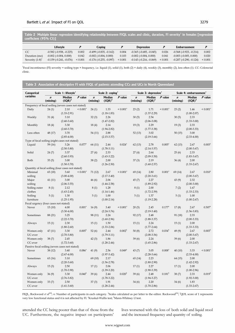
www.wjgnet.com
attended the CC being poorer than that of those from the UC. Furthermore, the negative impact on participants’
lives worsened with the loss of both solid and liquid stool and the increased frequency and quantity of soiling.
Table 2 Multiple linear regression identifying relationship between FIQL scales and clinic, duration, FI severity1 in females [regression coefficient (95% CI)]
Lifestyle P Coping P Depression P Embarrassment P
CC -0.582 (-0.938, -0.225) 0.002 -0.499 (-0.835, -0.162) 0.004 -0.365 (-0.685, -0.045) 0.026 -0.568 (-0.923, -0.214) 0.002Duration (mo) -0.002 (-0.004, 0.000) 0.042 -0.002 (-0.004, 0.000) 0.103 -0.002 (-0.004, 0.000) 0.041 -0.003 (-0.005, 0.000) 0.020Severity (1-8)1 -0.159 (-0.241, -0.076) < 0.001 -0.176 (-0.255, -0.097) < 0.001 -0.143 (-0.216, -0.069) < 0.001 -0.207 (-0.290, -0.124) < 0.001
1Fecal incontinence (FI) severity = soiling type × frequency, i.e. liquid (1), solid (1), both (2) × daily (4), weekly (3), monthly (2), less often (1). CC: Colorectal clinic.
Table 3 Association of descriptive FI with FIQL of patients attending CCs and UCs in North Queensland
Categorical variables
Scale 1: lifestyle1 Scale 2: coping1 Scale 3: depression1 Scale 4: embarrassment1
n (missing)
Median (IQR)2
P value n (missing)
Median (IQR)2
P value n (missing)
Median (IQR)2
P value n (missing)
Median (IQR)2
P value
Frequency of fecal soiling (seven cases not stated) Daily 24 (1) 2.10
(1.33-2.91)< 0.001a 24 (1) 1.33
(1.18-1.83)< 0.001a 23 (2) 1.71
(1.37-2.29)< 0.001a 23 (2) 1.46
(1.00-2.07)< 0.001a
Weekly 31 (4) 3.10 (2.40-3.60)
32 (3) 2.26 (1.47-2.82)
30 (5) 2.54 (2.06-3.08)
30 (5) 2.33 (1.33-3.00)
Monthly 18 (4) 3.30 (2.60-3.70)
18 (4) 2.14 (1.94-2.82)
19 (3) 2.29 (1.77-3.38)
19 (3) 2.33 (2.00-3.33)
Less often 48 (17) 3.70 (2.85-4.00)
54 (11) 2.88 (2.15-3.57)
52 (13) 3.02 (2.19-3.66)
50 (15) 3.00 (2.33-4.00)
Type of fecal soiling (eight cases not stated) Liquid 59 (16) 3.26
(2.50-3.80) 0.077a 64 (11) 2.44
(1.78-3.11) 0.024a 62 (13) 2.78
(2.14-3.57) 0.005a 62 (13) 2.67
(2.00-3.67) 0.003a
Solid 24 (7) 3.37 (2.60-3.93)
27 (4) 2.33 (1.63-3.22)
27 (4) 2.64 (2.09-3.50)
25 (6) 2.33 (1.83-3.67)
Both 35 (5) 3.00 (1.50-3.70)
38 (2) 2.00 (1.26-2.50)
37 (3) 2.19 (1.60-2.57)
36 (4) 2.00 (1.33-2.67)
Quantity of fecal soiling (four cases not stated) Minimal soiling
65 (18) 3.60 (3.00-4.00)
< 0.001a 71 (12) 2.67 (2.17-3.40)
< 0.001a 69 (14) 2.80 (2.20-3.61)
0.001a 69 (14) 2.67 (2.00-3.67)
0.010a
Major soiling
41 (11) 3.00 (2.06-3.55)
46 (6) 2.06 (1.44-2.58)
45 (7) 2.27 (1.89-2.92)
43 (9) 2.32 (2.00-3.00)
Soiling outer clothes
8 (1) 2.12 (1.63-2.45)
8 (1) 1.28 (1.14-1.46)
8 (1) 2.00 (1.72-2.59)
7 (2) 1.67 (1.33-2.33)
Soiling furniture
5 (1) 1.30 (1.25-1.95)
5 (1) 1.00 (1.00-2.14)
5 (1) 1.37 (1.19-2.28)
5 (1) 1.00 (1.00-2.67)
Fecal urgency (four cases not stated) Never 15 (10) 4.00
(3.30-4.00) 0.001a 16 (9) 3.40
(2.24-3.76)< 0.001a 20 (5) 2.45
(2.19-3.40) 0.177a 17 (8) 2.67
(1.56-3.83) 0.507a
Sometimes 88 (21) 3.25 (2.22-3.70)
98 (11) 2.24 (1.60-2.96)
92 (17) 2.48 (1.88-3.37)
91 (18) 2.33 (2.00-3.33)
Always 15 (1) 2.30 (1.80-2.60)
15 (1) 1.50 (1.33-2.06)
15 (1) 2.24 (1.77-2.64)
15 (1) 2.00 (1.33-3.33)
Women only UC-ever
47 (11) 3.30 (2.70-3.80)
0.005b 52 (6) 2.44 (1.79-3.11)
0.002b 50 (8) 2.72 (2.08-3.56)
0.034b 49 (9) 2.67 (2.00-3.67)
0.003b
Women only CC-ever
38 (7) 2.45 (1.72-3.60)
42 (3) 1.86 (1.28-2.44)
39 (6) 2.24 (1.65-2.86)
39 (6) 2.00 (1.33-2.67)
Passive fecal soiling (seven cases not stated) Never 38 (12) 3.40
(2.67-4.00) 0.086a 41 (9) 2.56
(1.97-3.42) 0.049a 43 (7) 3.05
(2.28-3.66) 0.008a 40 (10) 3.33
(2.33-4.00)< 0.001a
Sometimes 63 (16) 3.10 (2.20-3.60)
69 (10) 2.17 (1.56-2.78)
65 (14) 2.23 (1.80-2.90)
64 (15) 2.00 (1.42-2.92)
Always 15 (3) 2.50 (1.70-3.90)
17 (1) 2.06 (1.39-3.20)
17 (1) 2.27 (1.90-3.39)
17 (1) 2.00 (1.00-2.96)
Women only UC-ever
36 (9) 3.30 (2.76-3.82)
0.040b 39 (6) 2.44 (1.78-3.22)
0.020b 39 (6) 2.48 (1.94-3.27)
0.181b 38 (7) 2.33 (1.92-3.00)
0.019b
Women only CC-ever
33 (7) 2.50 (1.61-3.60)
37 (3) 1.91 (1.28-2.44)
34 (6) 2.20 (1.70-2.86)
34 (6) 1.83 (1.33-2.67)
FIQL, Rockwood et al[22]; n: Number of participants in each category. 1Scales calculated as per letter to the editor. Rockwood[26]; 2QOL score of 1 represents very low functional status and 4 is not affected by FI. aKruskal-Wallis test; bMann-Whitney U test.
Bartlett L et al . Impact of FI on QOL 3279

www.wjgnet.com
In this study of the impact on QOL of FI in rural and regional north Queensland, our overall FIQL results for lifestyle (3.24), coping (2.23), depression (2.42) and embarrassment (2.33) were within the range of comparable clinic-based studies in other countries[21,27-31], and closely reflected the scores found in a similar study
conducted at a Pelvic Floor Center in Minnesota, USA[21] and baseline scores from two Victorian (Australia) clinical studies that investigated injectable material for FI[32,33].
The QOL of patients who attended the CC was more severely affected than that of participants from the UC, even though they had reported FI for a shorter duration. There were no significant differences between those who had a diagnosis of bowel disease and those who did not. Thus the poorer QOL in CC participants may be a direct result of recent colorectal surgery, in which the sudden and unexpected onslaught of FI was more devastating than for the UC participants who may have learnt to cope with their progressive FI over an extended time period. These CC FIQL results are comparable with previously published results from a pouch, non-pouch study[29], but the participants in our study were less able to cope, and were more depressed and embarrassed, although this did not affect their lifestyle to the same degree.
The UC patients’ QOL scores were lower than those in a similar study in Texas, USA[30] but higher than in the study in Minnesota, USA[21], for lifestyle, coping and embarrassment, but not depression. This difference may have been caused by a longer duration with FI in the older cohorts in our study and the Minnesota study, (mean age 56 years), compared with the younger cohort in the Texas study (mean age 39 years). This suggests that older women with FI had poorer QOL than younger affected women, which implies that delaying treatment for this condition results in poorer QOL over time.
4.00
3.00
2.00
1.00CC & UCWomen
CCMen
CCWomen
UCWomen
CCAll
Depression scale
P = 0.648P = 0.630
P = 0.062 P = 0.056
P = 0.491
P = 0.520 P = 0.004
P = 0.003 CC & UCWomen
CCMen
CCWomen
UCWomen
CCAll
Embarrassment scale
4.00
3.00
2.00
1.00CC & UCWomen
CCMen
CCWomen
UCWomen
CCAll
Lifestyle scale
P = 0.228 P = 0.870
P = 0.015 P = 0.005
P = 0.491
P = 0.520 P = 0.004
P = 0.003CC & UCWomen
CCMen
CCWomen
UCWomen
CCAll
Coping scale
Figure 1 Association of FIQL scales with sex and CC and UC. FIQL, Rockwood et al[22]; Box and whisker demonstrates median, IQR, minimum and maximum. P values calculated by unpaired Wilcoxin test (Mann-Whitney); QOL score of 1 represents very low functional status and 4 is not affected by FI; FIQL scales calculated as per letter to the editor, Rockwood[26].
Table 4 Multiple linear regression identifying relationship between FIQL and components of FI severity
Components of FI severity
FIQL scales
Lifestyle Coping Depression Embarrassment
Frequency Monthly 0.061 0.002 0.008 0.006 Weekly 0.241 0.846 0.531 0.595 Daily < 0.001 0.012 0.023 0.021Type Solid 0.010 0.192 0.515 0.633 Both solid/liquid 0.254 0.085 0.007 0.014Urgency Sometimes 0.961 0.325 0.565 0.888 Always 0.106 0.023 0.554 0.636Wears pads 0.209 0.022 0.488 0.107Passive leakage Sometimes 0.659 0.169 0.103 0.787 Always 0.626 0.485 0.252 0.007Quantity of leakage Major (Requires immediate underwear change)
0.001 < 0.001 0.004 0.096
Soiling of outer clothes
0.023 0.217 0.969 0.405
Soiling of furniture/bedding
0.578 0.781 0.381 0.662
3280 ISSN 1007-9327 CN 14-1219/R World J Gastroenterol July 14, 2009 Volume 15 Number 26

www.wjgnet.com
This study investigated the relationship between FI severity and the FIQL scales individually and collectively. A negative impact was found on patients’ lives, which increased with frequency of soiling. This was evident on all scales of the FIQL, but there was little difference in each of the FIQL scales between weekly and monthly incontinent episodes. This lack of difference may be because an incontinent episode that occurs infrequently is unexpected, and hence, would be similarly distressing.
The data collected for pad wearing was dichotomous, which may explain why only significant results for the coping scale were obtained. If, in addition, the number of pads worn per day had been assessed, the embarrassment scale may also have reached significance.
The type of soiling affected participants’ QOL differently. Patients with both solid and liquid soiling reported a poorer QOL than those with either solid or liquid only incontinent episodes. This is consistent with the Texas study in which a liquid component of anal incontinence was reported to have a greater impact upon QOL of participants than either flatal or non-liquid incontinence[30].
The association between quantity of fecal loss and FIQL scales was found to be highly significant. Patients with the most soiling, i.e. those who soiled furniture, had the lowest possible FIQL score for coping and embarrassment, and performed only marginally better on the lifestyle and depression scales. There are few reports of the relationship between quantity of fecal loss and QOL, however, a Japanese study investigating the QOL of patients following total proctocolectomy and ileal J-pouch-anal anastomosis determined that greater soiling resulted in higher levels of frustration, which is an anxiety measure calculated using a Japanese translation of Cattell’s anxiety scale[34]. When quantity was included in the FIQL/severity regression model, it was found to be significant (P < 0.05) for the lifestyle, coping and depressions scales, but did not reach significance for the embarrassment scale (P = 0.065). Given this relationship between the FIQL scales and quantity of fecal soiling, it is suggested that the definition of FI severity should include quantity of fecal loss as well as frequency, type, urgency and pad wearing.
In conclusion, to the best of our knowledge, this is the first study to measure the effect of FI on QOL of people in rural and regional Australia. More than 22% of the patients attending the UC and CC in North Queensland, for matters other than FI, had their QOL severely affected by this condition. Patients reported that they had not been asked about FI by their general practitioners or hospital physicians, nor did they voluntarily disclose its presence[9]. Women who have obstetric-injury-related FI suffer in silence, and their QOL deteriorates as they age. The definition of FI severity should include quantity of fecal loss[35]. Further community-based research in Australia is warranted in regard to the impact of FI on QOL of people who suffer with this complaint.
ACKNOWLEDGMENTSWe thank Dr. Rane AJ for facilitating access to the patients in his UCs.
COMMENTSBackgroundAs many as one in seven adults suffer with fecal incontinence (FI), which can have a devastating effect on the lifestyle of people with frequent, ad hoc or large amounts of fecal seepage. These people often fail to seek treatment due to embarrassment, believing the problem is uniquely theirs, or because they are unaware of the existence of available treatments.Research frontiersPatients referred to urogynecology and colorectal surgical clinics, for other reasons, have a high incidence of FI. They do not seek assistance whilst attending these clinics from physicians who can recommend or refer for treatment. In this study, the authors demonstrated the impact that FI had on these patients’ quality of life (QOL).Innovations and breakthroughsRecent reports have highlighted the impact conservative treatments, such as biofeedback, and more invasive treatments, such as injectable bulking agents, sacral nerve stimulation, artificial bowel sphincter and dynamic graciloplasty, have had on the QOL of patients with FI. This study reports that despite these improvements many people continue to suffer unaware of such advances.ApplicationsBy understanding more than one in five patients attending colorectal and urogynecological clinics have their QOL severely affected by FI, attending physicians can enable access to treatment by directly asking patients about this problem.TerminologyFI is the involuntary discharge of solid or liquid stools.Peer reviewFI severity, including the quantity of stool loss, can have a severe negative impact on QOL. This is an interesting and relevant study that deals with a demanding subject.
REFERENCES1 Baxter NN, Rothenberger DA, Lowry AC. Measuring fecal
incontinence. Dis Colon Rectum 2003; 46: 1591-16052 Bharucha AE, Locke GR 3rd, Seide BM, Zinsmeister AR. A
new questionnaire for constipation and faecal incontinence. Aliment Pharmacol Ther 2004; 20: 355-364
3 Miner PB Jr. Economic and personal impact of fecal and urinary incontinence. Gastroenterology 2004; 126: S8-S13
4 Deutekom M, Dobben AC, Dijkgraaf MG, Terra MP, Stoker J, Bossuyt PM. Costs of outpatients with fecal incontinence. Scand J Gastroenterol 2005; 40: 552-558
5 Ho YH, Muller R, Veitch C, Rane A, Durrheim D. Faecal incontinence: an unrecognised epidemic in rural North Queensland? Results of a hospital-based outpatient study. Aust J Rural Health 2005; 13: 28-34
6 Whitehead WE, Borrud L, Goode PS, Meikle S, Mueller ER, Tuteja A, Weidner A, Weinstein M, Ye W. Fecal Incontinence in US Adults: Epidemiology and Risk Factors. Gastroenterology 2009; Epub ahead of print
7 Lam TCF, Kennedy ML, Chen FC, Lubowski DZ, Talley NJ. Prevalence of faecal incontinence: obstetric and constipation-related risk factors; a population-based study. Colorectal Dis 1999; 1: 197-203
8 Kalantar JS, Howell S, Talley NJ. Prevalence of faecal incontinence and associated risk factors; an underdiagnosed problem in the Australian community? Med J Aust 2002; 176: 54-57
9 Bartlett L, Nowak M, Ho YH. Reasons for non-disclosure of faecal incontinence: a comparison between two survey methods. Tech Coloproctol 2007; 11: 251-257
COMMENTS
Bartlett L et al . Impact of FI on QOL 3281

www.wjgnet.com
10 Leigh RJ, Turnberg LA. Faecal incontinence: the unvoiced symptom. Lancet 1982; 1: 1349-1351
11 Drossman DA , Sandler RS, Broom CM, McKee DC. Urgency and fecal soiling in people with bowel dysfunction. Dig Dis Sci 1986; 31: 1221-1225
12 Enck P, Bielefeldt K, Rathmann W, Purrmann J, Tschöpe D, Erckenbrecht JF. Epidemiology of faecal incontinence in selected patient groups. Int J Colorectal Dis 1991; 6: 143-146
13 Johanson JF, Lafferty J. Epidemiology of fecal incontinence: the silent affliction. Am J Gastroenterol 1996; 91: 33-36
14 Edwards NI, Jones D. The prevalence of faecal incontinence in older people living at home. Age Ageing 2001; 30: 503-507
15 Whitehead WE. Diagnosing and managing fecal incontinence: if you don't ask, they won't tell. Gastroenterology 2005; 129: 6
16 Stevens TK, Soffer EE, Palmer RM. Fecal incontinence in elderly patients: common, treatable, yet often undiagnosed. Cleve Clin J Med 2003; 70: 441-448
17 Rothbarth J, Bemelman WA, Meijerink WJ, Stiggelbout AM, Zwinderman AH, Buyze-Westerweel ME, Delemarre JB. What is the impact of fecal incontinence on quality of life? Dis Colon Rectum 2001; 44: 67-71
18 Colquhoun P, Kaiser R Jr, Efron J, Weiss EG, Nogueras JJ, Vernava AM 3rd, Wexner SD. Is the quality of life better in patients with colostomy than patients with fecal incontience? World J Surg 2006; 30: 1925-1928
19 Deutekom M, Terra MP, Dobben AC, Dijkgraaf MG, Baeten CG, Stoker J, Bossuyt PM. Impact of faecal incontinence severity on health domains. Colorectal Dis 2005; 7: 263-269
20 Damon H, Dumas P, Mion F. Impact of anal incontinence and chronic constipation on quality of life. Gastroenterol Clin Biol 2004; 28: 16-20
21 Bordeianou L, Rockwood T, Baxter N, Lowry A, Mellgren A, Parker S. Does incontinence severity correlate with quality of life? Prospective analysis of 502 consecutive patients. Colorectal Dis 2008; 10: 273-279
22 Rockwood TH , Church JM, Fleshman JW, Kane RL, Mavrantonis C, Thorson AG, Wexner SD, Bliss D, Lowry AC. Fecal Incontinence Quality of Life Scale: quality of life instrument for patients with fecal incontinence. Dis Colon Rectum 2000; 43: 9-16; discussion 16-17
23 Avery KN, Bosch JL, Gotoh M, Naughton M, Jackson S, Radley SC, Valiquette L, Batista J, Donovan JL. Questionnaires to assess urinary and anal incontinence: review and recommendations. J Urol 2007; 177: 39-49
24 Queensland Health. The Townsville Hospital. Available
from: http://www.health.qld.gov.au/townsville/Facilities/tville_hosp.asp
25 Jorge JM, Wexner SD. Etiology and management of fecal incontinence. Dis Colon Rectum 1993; 36: 77-97
26 Rockwood T. The author replies. Dis Colon Rectum 2008; 51: 1434
27 Colquhoun PH , Efron J, Wexner SD. Attainment of continence with J-pouch and artificial bowel sphincter for concomitant imperforate anus and familial adenomatous polyposis: report of a case. Dis Colon Rectum 2004; 47: 538-541
28 Rullier E, Zerbib F, Marrel A, Amouretti M, Lehur PA. Validation of the French version of the Fecal Incontinence Quality-of-Life (FIQL) scale. Gastroenterol Clin Biol 2004; 28: 562-568
29 Park JG, Lee MR, Lim SB, Hong CW, Yoon SN, Kang SB, Heo SC, Jeong SY, Park KJ. Colonic J-pouch anal anastomosis after ultralow anterior resection with upper sphincter excision for low-lying rectal cancer. World J Gastroenterol 2005; 11: 2570-2573
30 Boreham MK, Richter HE, Kenton KS, Nager CW, Gregory WT, Aronson MP, Vogt VY, McIntire DD, Schaffer JI. Anal incontinence in women presenting for gynecologic care: prevalence, risk factors, and impact upon quality of life. Am J Obstet Gynecol 2005; 192: 1637-1642
31 Pla-Martí V, Moro-Valdezate D, Alos-Company R, Solana-Bueno A, Roig-Vila JV. The effect of surgery on quality of life in patients with faecal incontinence of obstetric origin. Colorectal Dis 2007; 9: 90-95
32 Tjandra JJ, Lim JF, Hiscock R, Rajendra P. Injectable silicone biomaterial for fecal incontinence caused by internal anal sphincter dysfunction is effective. Dis Colon Rectum 2004; 47: 2138-2146
33 Tjandra JJ , Chan MK, Yeh HC. Injectable s i l icone biomaterial (PTQ) is more effective than carbon-coated beads (Durasphere) in treating passive faecal incontinence--a randomized trial. Colorectal Dis 2009; 11: 382-389
34 Fujita S, Kusunoki M, Shoji Y, Owada T, Utsunomiya J. Quality of life after total proctocolectomy and ileal J-pouch-anal anastomosis. Dis Colon Rectum 1992; 35: 1030-1039
35 Landefeld CS, Bowers BJ, Feld AD, Hartmann KE, Hoffman E, Ingber MJ, King JT Jr, McDougal WS, Nelson H, Orav EJ, Pignone M, Richardson LH, Rohrbaugh RM, Siebens HC, Trock BJ. National Institutes of Health state-of-the-science conference statement: prevention of fecal and urinary incontinence in adults. Ann Intern Med 2008; 148: 449-458
S- Editor Tian L L- Editor Kerr C E- Editor Zheng XM
3282 ISSN 1007-9327 CN 14-1219/R World J Gastroenterol July 14, 2009 Volume 15 Number 26

www.wjgnet.com
BRIEF ARTICLES
Is ERCP really necessary in case of suspected spontaneous passage of bile duct stones?
Yuji Sakai, Toshio Tsuyuguchi, Takeshi Ishihara, Seigo Yukisawa, Tadashi Ohara, Masaru Tsuboi, Yoshihiko Ooka, Kazuki Kato, Kiyotake Katsuura, Michio Kimura, Makoto Takahashi, Kazuhisa Nemoto, Masaru Miyazaki, Osamu Yokosuka
Online Submissions: wjg.wjgnet.com World J Gastroenterol 2009 July 14; 15(26): [email protected] World Journal of Gastroenterology ISSN 1007-9327doi:10.3748/wjg.15.3283 © 2009 The WJG Press and Baishideng. All rights reserved.
Yuji Sakai, Toshio Tsuyuguchi, Takeshi Ishihara, Seigo Yukisawa, Osamu Yokosuka, Department of Medicine and Clinical Oncology, Graduate School of Medicine, Chiba University, Chiba 260-8670, JapanYuji Sakai, Tadashi Ohara, Masaru Tsuboi, Yoshihiko Ooka, Kazuki Kato, Kiyotake Katsuura, Michio Kimura, Department of Internal Medicine, Funabashi Central Hospital, Funabashi City, Chiba 273-8556, JapanMakoto Takahashi, Department of Surgery, Funabashi Central Hospital, Funabashi City, Chiba 273-8556, JapanKazuhisa Nemoto, Department of Radiology, Funabashi Central Hospital, Funabashi City, Chiba 273-8556, JapanMasaru Miyazaki, Department of General Surgery, Graduate School of Medicine, Chiba University, Chiba 260-8670, JapanAuthor contributions: Sakai Y, Tsuyuguchi T, Ishihara T, and Yukisawa S were responsible for study design, data analysis, and manuscript preparation; Sakai Y wrote the paper, performed endoscopic treatment; Ohara T, Tsuboi M, Ooka Y, Kato K, Katsuura K, Kimura M and Nemoto K were responsible for data collection; Takahashi M performed the surgical procedure; Miyazaki M and Yokosuka O reviewed this manuscript.Correspondence to: Yuji Sakai, MD, Department of Medicine and Clinical Oncology (K1), Graduate School of Medicine, Chiba University, Inohana 1-8-1, Chuou-ku, Chiba 260-8670, Japan. [email protected]: +81-43-2262083 Fax: +81-43-2262088Received: March 29, 2009 Revised: June 3, 2009Accepted: June 10, 2009Published online: July 14, 2009
AbstractAIM: To investigate the usefulness of magnetic resonance cholangiopancreatography (MRCP) and the need for endoscopic retrograde cholangiopancreatography (ERCP) in cases of suspected spontaneous passage of stones into the common bile duct.
METHODS: Thirty-six patients with gallbladder stones were clinically suspected of spontaneous passage of stones into the common bile duct because they presented with clinical symptoms such as abdominal pain and fever, and showed signs of inflammatory reaction and marked rise of hepatobiliary enzymes. These symptoms resolved and they showed normalized values of blood biochemical parameters after conservative treatment without evidence of stones in the common bile duct on MRCP. All these patients were
subjected to ERCP within 3 d of MRCP to check for the presence of stones.
RESULTS: No stones were detected by ERCP in any patient, confirming the results of MRCP.
CONCLUSION: When clinical symptoms improve, blood biochemical parameters have normalized, and MRCP shows there are no stones in the common bile duct, it can be considered the stone has spontaneously passed and thus ERCP is not necessary.
© 2009 The WJG Press and Baishideng. All rights reserved.
Key words: Magnetic resonance cholangiopan-creatography; Endoscopic retrograde cholangiopan-creatography; Spontaneous passage of bile duct stones; Bile duct stones; Pancreatitis
Peer reviewer: Peter L Moses, MD, FACG, AGAF, Professor, University of Vermont College of Medicine Section of Gastroenterology & Hepatology, 111 Colchester Avenue, Smith 237B, MCHV, Burlington, VT 05401, United States
Sakai Y, Tsuyuguchi T, Ishihara T, Yukisawa S, Ohara T, Tsuboi M, Ooka Y, Kato K, Katsuura K, Kimura M, Takahashi M, Nemoto K, Miyazaki M, Yokosuka O. Is ERCP really necessary in case of suspected spontaneous passage of bile duct stones? World J Gastroenterol 2009; 15(26): 3283-3287 Available from: URL: http://www.wjgnet.com/1007-9327/15/3283.asp DOI: http://dx.doi.org/10.3748/wjg.15.3283
INTRODUCTIONMagnetic resonance cholangiopancreatography (MRCP) is a relatively new imaging technique developed for the diagnosis of patients with pancreaticobiliary disease. The signal of structures filled with static or slowly moving fluid, such as the bile and pancreatic ducts, is greatly increased on T2-weighted images, resulting in an increased duct-to-background contrast. Recent studies have shown that MRCP is as useful as endoscopic retrograde cholangiopancreatography (ERCP) regarding the diagnosis of extrahepatic bile duct and pancreatic duct abnormalities such as common bile duct stones[1-4],

www.wjgnet.com
malignant obstruction of the bile and pancreatic ducts[1,2,5], congenital anomalies[1,6,7], and chronic pancreatitis[8-10]. In some institutions, MRCP is becoming the first-choice technique for imaging the biliary system, with ERCP being reserved for therapeutic indications. In recent studies, MRCP showed extremely high performance in visualizing bile duct stones with both a sensitivity and a specificity of 90% or higher in most cases[11-17]. In the false negative cases, all stones were as small as 3-5 mm and were present near the duodenal papilla. Thus, MRCP is very useful for the diagnosis of bile duct stones and the determination of a therapeutic strategy. Since ERCP involves the risk of complications such as acute pancreatitis[18-21], it should be performed only when absolutely necessary. In some patients clinically suspected of choledocholithiasis, ERCP shows a cleavage at the duodenal papilla, but does not detect the stones in the common bile duct. These findings suggested that stones in the common bile duct had probably passed spontaneously. In fact, it was reported that three-fourths of common bile duct stones passed spontaneously before cholecystectomy[22], indicating that many patients with bile duct stones may not need ERCP. There are reports on the clinical course of patients suspected of spontaneous passage of bile duct stones after MRCP alone[23,24]. However, since ERCP was not performed, it was not clear whether the stones were actually present. Presence of bile duct stones could cause acute cholangitis, which, if aggravated, may be fatal. Consequently, we investigated the usefulness of MRCP and the need for diagnostic ERCP in patients suspected of spontaneous passage of stones.
MATERIALS AND METHODSFrom April 2007 to January 2009, 36 patients suspected of spontaneous passage of bile duct stones were prospectively evaluated. There were 26 men and 10 women and the mean age of the patients was 62.1 years (range: 33-82 years). The mean observation period was 6.7 mo (1-9 mo). After blood sampling the patients were subjected to ultrasound examination (Power Vision 6000; TOSHIBA Tochigi Japan) to assess the presence of stones in the gallbladder and common bile duct. The patients had gallbladder stones and were clinically suspected of spontaneous passage because they presented with clinical symptoms that included abdominal pain and fever, and showed signs of inflammatory reaction as well as a marked rise of hepatobiliary enzymes. These symptoms resolved and patients showed normalized values of blood biochemical parameters after conservative treatment, without evidence of stones in the common bile duct on MRCP. Patients who had originally had intrahepatic cholestasis were considered as “improved” if the level of hepatobiliary enzymes had normalized. The US, MRCP and ERCP images of typical cases are shown in Figures 1-3, respectively.
In this study, the need for urgent drainage was assessed first. Accordingly, blood was collected from
the patients on arrival at the hospital for the assessment of acute cholangitis. Patients with moderate to severe acute cholangitis[25] that did not respond to the initial medical treatment, were excluded from this study and were subjected to emergency ERCP with biliary drainage. When a patient presented symptoms of acute cholangitis during follow-up, ERCP was conducted provided the patient had given his/her informed consent prior to the examination. Patients with symptoms of acute pancreatitis were excluded from this study. Even
Figure 1 A 56-year-old man. Several gallbladder stones measuring about 5-10 mm were detected by abdominal ultrasonography.
Figure 2 No translucency indicative of stones in the bile duct was shown by magnetic resonance cholangiopancreatography.
A B
Figure 3 No translucency indicative of stones in the bile duct was shown by endoscopic retrograde cholangiopancreatography (A). After withdrawal of the fiber, the posture was changed and the bile duct was imaged again. However, no translucency was observed (B).
3284 ISSN 1007-9327 CN 14-1219/R World J Gastroenterol July 14, 2009 Volume 15 Number 26

www.wjgnet.com
if a marked increase in hepatobiliary enzymes was detected before arrival at the hospital, we waited for an improvement in clinical symptoms and blood test values if cholangitis was mild, before performing MRCP followed by ERCP.
MRCP was performed using a 1.5T GE CV/I scanner and a phased-array torso coil (General Electric Medical Systems, Milwaukee, WI). The radiographic conditions were as follows: thin-section MRCP images were obtained in the coronal plane using half Fourier single-shot fast spin echo (SSFSE) imaging at a slice thickness of 4.0 mm, 34FOV, 256 × 256 matrix, and a TE of 180. Thick-slab SSFSE MRCP images were obtained in the plane using a matrix of 256X256 and a maximum TE. Imaging in the coronal and axial planes was done using a fast 3D spoiled gradient echo sequence with a TR of 3.9 TE minutes, 2.4 mm slice thickness (interpolated to 1.2 mm), and a 256 × 192 matrix. Images were reviewed on an Advantage Windows workstation (General Electric Medical Systems) that allowed 3D reconstruction. MRCP images were read after evaluating clinical symptoms and hematological findings to examine the common bile duct for the presence of stones. First, a radiologist with more than 20 years of experience examined the MRCP images and if there were no bile duct stones, the same MRCP images were subsequently examined by a gastroenterologist with more than 15 years of experience. Only after the absence of stones was confirmed by these 2 specialists, was the case defined as spontaneous passage of stones. MRCP was conducted after the clinical symptoms and blood test findings had sufficiently improved. ERCP was conducted within 4 d of MRCP to check for the presence of stones. ERCP images were read only by the gastroenterologist who had examined the MRCP images. As a measure against complications, the duration of ERCP was strictly limited to within 15 min. This prospective study was conducted after obtaining informed consent from the patients. Furthermore, the protocol of this study was discussed and approved by the Institutional Review Board of Funabashi Central Hospital.
RESULTSERCP was successfully conducted in all 36 patients after MRCP confirmed the absence of stones in each of them. The ERCP procedure was completed within 15 min in all these cases. Mild acute pancreatitis,
assessed according to Cotton’s criteria[21], occurred as a complication after ERCP in 1 (2.8%) of the 36 patients. Cannulation was difficult in this patient and pancreatitis was assumed to be caused by pancreatography, which was performed 3 times during ERCP. However, remission was attained after conservative treatment. No other complications occurred. Both the radiologist and the gastroenterologist who read MRCP images ruled out the presence of bile duct stones in the 36 patients (K = 1.0) (Figures 2 and 3).
MRCP showed the bile duct had a mean diameter of 6.6 mm (range, 4.8-8.3 mm). Biochemical average (and range) data were total bilirubin 2.6 (1.9-6.2) mg/dL (Figure 4A), alkaline phosphatase (ALP) 603.6 (299-1123) IU/L (Figure 4B), and alanine transferase (ALT) 325.8 (112-654) IU/L (Figure 4C) at the time of common bile duct obstruction while the normal range values were total bilirubin 0.3-1.0 g/dL; ALP 80-220 IU/L and ALT 5-40 IU/L. Subsequently, 6 patients underwent cholecystectomy. During the observation period, none of the 36 patients experienced a second spontaneous passage of stones. At the time of registration for this study, the clinical symptoms of 6 patients suspected of spontaneous passage of stones were already showing a tendency towards remission. Yet these patients were not registered for the study because they had moderate cholangitis. Accordingly, urgent ERCP was conducted without MRCP. The result indicated no bile duct stone in any of the 6 cases.
DISCUSSIONMRCP can detect bile duct stones with a high rate of accuracy and sensitivity. In the present study, ERCP detected no stones in any of the patients suspected of spontaneous passage of stones and confirmed by the MRCP conducted after the clinical symptoms had improved and blood test values had normalized or returned to baseline levels. Nevertheless, the possible presence of small stones undetectable by ERCP cannot be denied. Intraductal ultrasonography (IDUS), which can be used following ERCP, is a valuable tool for the imaging of tiny stones. IDUS is likely to detect very small bile duct stones more accurately than ERCP alone[26]. Bile duct stones have, however, been reported to pass spontaneously in most cases[22]. Although the aggressive use of IDUS may increase the detection of
Total-bilirubin (mg/dL) normal range (0.3-1.0) n = 36
Before attack of stones
(0.9-1.0)
average 0.98
Attack of stones
(1.9-6.2)
average 2.6
After attack of stones
(0.9-1.0)
average 0.98
7.0
6.0
5.0
4.0
3.0
2.0
1.0
0.0
mg/
dL
A Alkaline phosphatase (IU/L) normal range (80-220) n = 36
Before attack of stones
(98-200)
average 137.3
Attack of stones
(299-1123)
average 603.6
After attack of stones
(99-187)
average 134.9
1200
1000
800
600
400
200
0
IU/L
B Alanine transferase (IU/L) normal range (5-40) n = 36
Before attack of stones
(10-48)
average 26.4
Attack of stones
(112-654)
average 325.8
After attack of stones
(15-45)
average 27.6
700600500400300200100
0
IU/L
C
Figure 4 Changes in serum. A: Total-bilirubin; B: Alkaline phosphatase; C: Alanine transferase.
Sakai Y et al . Diagnostic value of MRCP 3285

www.wjgnet.com
bile duct stones, prolonged ERCP practice may result in accidental events. Thus, this procedure was not included in the study design.
The presence or absence of small stones is a matter of controversy. However, it is basically reasonable to assume a spontaneous passage of stones if MRCP has detected no stones in the patients whose clinical symptoms improved and whose blood test values normalized or returned to the baseline levels. Today, most reports conclude that MRCP is comparable to ERCP in the diagnosis of biliary stones. Institutions with high-magnetic-field MRI make it a rule not to perform diagnostic ERCP in patients who have no stones identified by MRCP. The possibility that the present study would be useful in considering indications for use of ERCP was suggested. The results of this study indicate the feasibility of diagnosis of spontaneous passage of stones in the common bile duct based on clinical symptoms, blood biochemistry, US, and MRCP without exposure to X-ray, thus reducing such exposure for medical personnel responsible for conducting ERCP[27], and avoiding the potential complications of ERCP.
US is a simple examination tool for imaging bile duct stones but is less satisfactory for the detection of stones[26]. However, it provides information on the gallbladder and important information on the diameter of the bile duct considering the presence of bile duct stones and thus it is a very useful examination. Given the higher performance of MRCP, diagnosis employing US or MRCP rather than diagnostic imaging will become more common when bile duct stones are suspected. However, the patients with spontaneous passage of stones included in this study presented with mild cholangitis. In patients with moderate or severe cholangitis, drainage of the biliary tract that allows the detection of stones more rapidly than MRCP, may prove very useful[25]. When clinical symptoms improve, blood biochemical parameters have normalized, and MRCP shows there are no stones in the common bile duct, it can be considered that the stone has spontaneously passed and thus ERCP would not be necessary.
COMMENTSBackgroundMagnetic resonance cholangiopancreatography (MRCP) is a relatively new imaging technique developed for the diagnosis of patients with pancreaticobiliary disease. Recent studies have shown that MRCP is as useful as endoscopic retrograde cholangiopancreatography (ERCP) for the diagnosis of extrahepatic bile duct and pancreatic duct abnormalities such as common bile duct stones.Innovations and breakthroughsWhen clinical symptoms improve, blood biochemical parameters have normalized, and MRCP shows there are no stones in the common bile duct, it can be considered the stone has spontaneously passed and thus ERCP would not be necessary.Peer reviewThis is a prospective, non-controlled study from a single center. The observation is made that ERCP is not helpful when a passed stone is suspected and MRCP is negative.
REFERENCES1 Fulcher AS, Turner MA, Capps GW, Zfass AM, Baker
KM. Half-Fourier RARE MR cholangiopancreatography: experience in 300 subjects. Radiology 1998; 207: 21-32
2 Guibaud L, Bret PM, Reinhold C, Atri M, Barkun AN. Bile duct obstruction and choledocholithiasis: diagnosis with MR cholangiography. Radiology 1995; 197: 109-115
3 Chan YL, Chan AC, Lam WW, Lee DW, Chung SS, Sung JJ, Cheung HS, Li AK, Metreweli C. Choledocholithiasis: comparison of MR cholangiography and endoscopic retrograde cholangiography. Radiology 1996; 200: 85-89
4 Regan F, Fradin J, Khazan R, Bohlman M, Magnuson T. Choledocholithiasis: evaluation with MR cholangiography. AJR Am J Roentgenol 1996; 167: 1441-1445
5 Morimoto K, Shimoi M, Shirakawa T, Aoki Y, Choi S, Miyata Y, Hara K. Biliary obstruction: evaluation with three-dimensional MR cholangiography. Radiology 1992; 183: 578-580
6 Bret PM, Reinhold C, Taourel P, Guibaud L, Atri M, Barkun AN. Pancreas divisum: evaluation with MR cholangiopancreatography. Radiology 1996; 199: 99-103
7 Taourel P, Bret PM, Reinhold C, Barkun AN, Atri M. Anatomic variants of the biliary tree: diagnosis with MR cholangiopancreatography. Radiology 1996; 199: 521-527
8 Soto JA, Barish MA, Yucel EK, Clarke P, Siegenberg D , C h u t t a n i R , F e r r u c c i J T . P a n c r e a t i c d u c t : M R cholangiopancreatography with a three-dimensional fast spin-echo technique. Radiology 1995; 196: 459-464
9 Hirohashi S, Hirohashi R, Uchida H, Akira M, Itoh T, Haku E, Ohishi H. Pancreatitis: evaluation with MR cholangiopancreatography in children. Radiology 1997; 203: 411-415
10 Matos C , Metens T, Devière J, Nicaise N, Braudé P, Van Yperen G, Cremer M, Struyven J. Pancreatic duct: morphologic and functional evaluation with dynamic MR pancreatography after secretin stimulation. Radiology 1997; 203: 435-441
11 Varghese JC , Liddel l RP, Farrel l MA, Murray FE, Osborne H, Lee MJ. The diagnostic accuracy of magnetic resonance cholangiopancreatography and ultrasound compared with direct cholangiography in the detection of choledocholithiasis. Clin Radiol 1999; 54: 604-614
12 Stiris MG, Tennøe B, Aadland E, Lunde OC. MR cholangiopancreaticography and endoscopic retrograde cholangiopancreaticography in patients with suspected common bile duct stones. Acta Radiol 2000; 41: 269-272
13 Kim TK, Kim BS, Kim JH, Ha HK, Kim PN, Kim AY, Lee MG. Diagnosis of intrahepatic stones: superiority of MR cholangiopancreatography over endoscopic retrograde cholangiopancreatography. AJR Am J Roentgenol 2002; 179: 429-434
14 Kats J , Kraai M, Dijkstra AJ, Koster K, Ter Borg F, Hazenberg HJ, Eeftinck Schattenkerk M, des Plantes BG, Eddes EH. Magnetic resonance cholangiopancreaticography as a diagnostic tool for common bile duct stones: a comparison with ERCP and clinical follow-up. Dig Surg 2003; 20: 32-37
15 Lomas DJ, Bearcroft PW, Gimson AE. MR cholangiopancreatography: prospective comparison of a breath-hold 2D projection technique with diagnostic ERCP. Eur Radiol 1999; 9: 1411-1417
16 Taylor AC, Little AF, Hennessy OF, Banting SW, Smith PJ, Desmond PV. Prospective assessment of magnetic resonance cholangiopancreatography for noninvasive imaging of the biliary tree. Gastrointest Endosc 2002; 55: 17-22
17 Becker CD, Grossholz M, Becker M, Mentha G, de Peyer R, Terrier F. Choledocholithiasis and bile duct stenosis: diagnostic accuracy of MR cholangiopancreatography. Radiology 1997; 205: 523-530
18 Masci E, Toti G, Mariani A, Curioni S, Lomazzi A, Dinelli M, Minoli G, Crosta C, Comin U, Fertitta A, Prada A,
COMMENTS
3286 ISSN 1007-9327 CN 14-1219/R World J Gastroenterol July 14, 2009 Volume 15 Number 26

www.wjgnet.com
Passoni GR, Testoni PA. Complications of diagnostic and therapeutic ERCP: a prospective multicenter study. Am J Gastroenterol 2001; 96: 417-423
19 Boender J, Nix GA, de Ridder MA, van Blankenstein M, Schütte HE, Dees J, Wilson JH. Endoscopic papillotomy for common bile duct stones: factors influencing the complication rate. Endoscopy 1994; 26: 209-216
20 Freeman ML, Nelson DB, Sherman S, Haber GB, Herman ME, Dorsher PJ, Moore JP, Fennerty MB, Ryan ME, Shaw MJ, Lande JD, Pheley AM. Complications of endoscopic biliary sphincterotomy. N Engl J Med 1996; 335: 909-918
21 Cotton PB , Lehman G, Vennes J, Geenen JE, Russell RC, Meyers WC, Liguory C, Nickl N. Endoscopic sphincterotomy complications and their management: an attempt at consensus. Gastrointest Endosc 1991; 37: 383-393
22 Tranter SE, Thompson MH. Spontaneous passage of bile duct stones: frequency of occurrence and relation to clinical presentation. Ann R Coll Surg Engl 2003; 85: 174-177
23 Sakai Y , Tsuyuguchi T , Yukisawa S , Tsuchiya S , Sugiyama H, Miyakawa K, Ohara T, Ebara M, Miyazaki M, Yokosuka O. Diagnostic value of magnetic resonance cholangiopancreatography for clinically suspicious
spontaneous passage of bile duct stones. J Gastroenterol Hepatol 2008; 23: 736-740
24 Sakai Y, Tsuyuguchi T, Tsuchiya S, Sugiyama H, Miyakawa K, Ebara M, Saisho H, Yokosuka O. Diagnostic value of MRCP and indications for ERCP. Hepatogastroenterology 2007; 54: 2212-2215
25 Miura F, Takada T, Kawarada Y, Nimura Y, Wada K, Hirota M, Nagino M, Tsuyuguchi T, Mayumi T, Yoshida M, Strasberg SM, Pitt HA, Belghiti J, de Santibanes E, Gadacz TR, Gouma DJ, Fan ST, Chen MF, Padbury RT, Bornman PC, Kim SW, Liau KH, Belli G, Dervenis C. Flowcharts for the diagnosis and treatment of acute cholangitis and cholecystitis: Tokyo Guidelines. J Hepatobiliary Pancreat Surg 2007; 14: 27-34
26 Moon JH, Cho YD, Cha SW, Cheon YK, Ahn HC, Kim YS, Kim YS, Lee JS, Lee MS, Lee HK, Shim CS, Kim BS. The detection of bile duct stones in suspected biliary pancreatitis: comparison of MRCP, ERCP, and intraductal US. Am J Gastroenterol 2005; 100: 1051-1057
27 Larkin CJ, Workman A, Wright RE, Tham TC. Radiation doses to patients during ERCP. Gastrointest Endosc 2001; 53: 161-164
S- Editor Li LF L- Editor O’Neill M E- Editor Yin DH
Sakai Y et al . Diagnostic value of MRCP 3287

Elegance Ting Pui Lam, Cindy Lo Kuen Lam, Ching Lung Lai, Man Fung Yuen, Department of Medicine, The University of Hong Kong, Hong Kong, ChinaDaniel Yee Tak Fong, Department of Nursing Studies, The University of Hong Kong, Hong Kong, ChinaAuthor contributions: All authors participated in the design of the study; Lam ETP, Lai CL and Yuen MF collected the data; Lam ETP, Lam CLK and Fong DYT were involved in data analysis and interpretation; Lam ETP and Lam CLK drafted the manuscript; All authors read and approved the final manuscript.Supported by Small Project Grant from the Committee of Research and Conference Grant, CRCG project, No. 10207293, the University of Hong Kong and the Health and Health Service Research Fund, HHSRF project, No. 05060741, Food and Health Bureau, Government of Hong Kong Special Administration Region, China Correspondence to: Elegance Ting Pui Lam, Family Medicine Unit, Department of Medicine, The University of Hong Kong, 3/F, Ap Lei Chau Clinic, 161 Main Street, Ap Lei Chau, Hong Kong, China. [email protected]: +852-25185656 Fax: +852-28147475Received: February 3, 2009 Revised: May 25, 2009Accepted: June 1, 2009Published online: July 14, 2009
AbstractAIM: To test the psychometric properties of a Chinese [(Hong Kong) HK] translation of the chronic liver disease questionnaire (CLDQ).
METHODS: A Chinese (HK) translation of the CLDQ was developed by iterative translation and cognitive debriefing. It was then administered to 72 uncomplicated and 78 complicated chronic hepatitis B (CHB) patients in Hong Kong together with a structured questionnaire on service utilization, and the Chinese (HK) SF36 Health Survey Version 2 (SF36v2).
RESULTS: Scaling success was ≥ 80% for all but three items. A new factor assessing sleep was found and items of two (Fatigue and Systemic Symptoms) subscales tended to load on the same factor. Internal consistency and testretest reliabilities ranged from 0.580.90 for different subscales. Construct validity was confirmed by the expected correlations between the SF36v2 Health Survey and CLDQ scores. Mean scores of CLDQ were significantly lower in complicated compared with uncomplicated CHB, supporting sensitivity in detecting differences between groups.
CONCLUSION: The Chinese (HK) CLDQ is valid, reliable and sensitive for patients with CHB. Some modifications to the scaling structure might further improve its psychometric properties.
© 2009 The WJG Press and Baishideng. All rights reserved.
Key words: Chronic liver disease; Healthrelated quality of life; Hepatitis B; Southern Chinese; Validity
Peer reviewer: Edmund J Bini, Professor, VA New York Harbor Healthcare System, Division of Gastroenterology (111D), 423 East 23rd Street, New York 10010, United States
Lam ETP, Lam CLK, Lai CL, Yuen MF, Fong DYT. Psycho-metrics of the chronic liver disease questionnaire for Southern Chinese patients with chronic hepatitis B virus infection. World J Gastroenterol 2009; 15(26): 3288-3297 Available from: URL: http://www.wjgnet.com/1007-9327/15/3288.asp DOI: http://dx.doi.org/10.3748/wjg.15.3288
INTRODUCTIONChronic hepatitis B (CHB) virus infection remains a major global health problem. It is estimated that 350 million people worldwide are chronically infected, of whom one third (120 million) are Chinese[1]. The prevalence is higher in southern China (> 10%) than Northern China (6%-10%)[2]. Up to 25% of patients may die from CHB complications, such as cirrhosis-related complications or hepatocellular carcinoma (HCC), posing a threat to both mental and physical health, leading to impairment of health-related quality of life (HRQOL).
HRQOL has become an important outcome mea-sure in clinical and health policy settings in the last two decades. Disease-specific measures are often needed to complement generic measures to give a more comprehen-sive evaluation of the HRQOL of patients with specific diseases. Several HRQOL measures have been developed specifically for chronic liver disease (CLD), such as the Chronic Liver Disease Questionnaire (CLDQ)[3], the Hep-atitis Quality of Life (HQLQ)[4], the Liver Disease Quality of Life[5] and the Liver Disease Symptom Index (LDSI)[6]. The CLDQ developed by Younossi et al[3] was the first and is the most widely used. The other liver disease-specific HRQOL measures are not commonly used because they are either too long, or the validity data are limited[4-8].
Online Submissions: wjg.wjgnet.com World J Gastroenterol 2009 July 14; 15(26): [email protected] World Journal of Gastroenterology ISSN 1007-9327doi:10.3748/wjg.15.3288 © 2009 The WJG Press and Baishideng. All rights reserved.
Psychometrics of the chronic liver disease questionnaire for Southern Chinese patients with chronic hepatitis B virus infection
Elegance Ting Pui Lam, Cindy Lo Kuen Lam, Ching Lung Lai, Man Fung Yuen, Daniel Yee Tak Fong
BRIEF ARTICLES
www.wjgnet.com

The CLDQ consists of 29 items which are grouped into 6 subscales: abdominal symptoms (AS), fatigue (FA), systemic symptoms (SS), activity (AC), emotional function (EF) and worry (WO). It is applicable to all types of liver diseases including CHB. It has been shown to have ade-quate internal reliability, validity and sensitivity. Test-retest reliability was more variable with intra-class correlation (ICC) ranging from 0.23 to 0.72 for different subscales[3]. Previous studies showed that the CLDQ is more respon-sive than a generic measure to detect a change in patients with CLD[3,9]. It has been translated and validated in dif-ferent languages[9-13], supporting its potential for cross-cultural adaptation. However, most of the psychometric data of the CLDQ have been derived from patients with hepatitis C virus (HCV) infection and Western popula-tions. There are few data on its applicability for Southern Chinese CHB patients despite the fact that China has the world’s largest population suffering from CLD.
Recently, the CLDQ has been translated into Manda-rin Chinese but this Chinese (Mainland) version may not be applicable to Southern Chinese who speak Cantonese, a dialect that has significant differences in the usage of words and terms from Mandarin. In addition, information on the validity, reliability and other psychometric proper-ties of the Chinese (Mainland) CLDQ version is limited. The aim of this study was to test the psychometric prop-erties of a Chinese [Hong Kong (HK)] translation of the CLDQ for Southern Chinese CHB patients. This would enable the evaluation of the impact of CHB infection and assess the effect of anti-viral drug treatments on HRQOL in the world’s largest population of CHB patients.
The objectives of this study were: (1) To develop a Chinese (HK) CLDQ that is semantically equivalent to the original; (2) To test the scaling assumptions and factor structure of the Chinese (HK) CLDQ; (3) To assess the psychometric properties in terms of reliability, construct validity, and sensitivity of the Chinese (HK) CLDQ; (4) To determine whether any modification of the CLDQ can improve its psychometric properties for Southern Chinese CHB patients.
MATERIALS AND METHODSEthicsThis research project was approved by the Institutional Review Board of the University of Hong Kong/Hospital Authority Hong Kong West Cluster (IRB reference No., UW 06-089 T/1114 and trial registration No., HKC-TR-151).
Development of the Chinese (HK) CLDQThe Chinese (HK) translation of the CLDQ was developed by iterative translations, expert panel review and cognitive debriefing, as recommended guidelines by experts[14,15]. The original CLDQ was translated into Chinese by two independent professional translators. Reconciliation of the forward translations into a single forward translation was carried out by a bilingual expert in HRQOL measures (Lam CLK) and the translators. The reconciled Chinese translation was back-translated into English by another professional translator. The back
translation was reviewed by the original author and the bilingual expert to identify any non-equivalence in the Chinese translation, which was then revised. The first draft of the Chinese (HK) CLDQ was evaluated by cognitive debriefing interviews with six Southern Chinese patients with CHB infection and further revision was made to ensure item clarity and equivalence to become the final Chinese (HK) CLDQ (used in this study on psychometrics properties).
SubjectsPatients with complicated CHB were recruited from out-patient hepatitis clinics of a regional hospital and patients with uncomplicated CHB were randomly selected from the computerized registers of three public primary care clinics serving over 100 000 people in one of five regions in Hong Kong. Patients aged 18 years or older who were hepatitis B surface antigen-positive for more than six months were included in the study. Patients were excluded if they could not communicate in Cantonese; had cognitive impairment shown by the patient’s inability to understand the study to give consent; were co-infected with HIV, HCV or hepa-titis D virus; had undergone liver transplantation or had end-stage non-hepatitis B-related illnesses; were currently taking excessive alcohol (> 30 U/wk) or illegal drugs; or refused to give consent. Each patient completed the Chinese (HK) CLDQ, the Chinese (HK) SF-36v2 Health Survey and a structured questionnaire on morbidity and socio-demographics, administered by a trained interviewer. Each patient was asked if he/she had ever been diagnosed by a registered practitioner for more than four weeks to have hypertension, diabetes mellitus, heart disease, stroke, chronic lung disease, arthritis, psychological illness (i.e. de-pression, anxiety, neurasthenia or psychosis) or any other chronic diseases. Chronic co-morbidity was measured by the total number of diseases (summation of positive re-sponses to the questions) and the presence of a specific diagnosis. Clinical data related to the CHB infection in-cluding Child’s staging for patients with cirrhosis and the biomarkers of liver disease (alanine aminotransferase, as-partate aminotransferase, α-fetoprotein and total bilirubin) in each patient were retrieved from medical records. Socio-demographic data including age, gender, education, marital status, occupation, household income and family history of liver disease were also collected.
The Chinese (HK) CLDQ was re-administered to the 46 subjects with uncomplicated CHB, whose condition was expected to be stable, by telephone two weeks from the first administration, in order to assess the test-retest reliabil-ity of the Chinese (HK) CLDQ. Sixty one percent of the repeat interviews were carried out by the same interviewer.
InstrumentsThe Chinese (HK) CLDQ consists of 29 i tems measuring six subscales as described above. Each item is rated on a 7-point (1 = all of the time to 7 = none of the time) Likert scale. Scores for each of the six domains are calculated by the mean of the item scores within the subscale. A summary score is calculated by the mean of all subscale scores. The scores range from 1 to 7 with a higher score indicating better HRQOL.
Lam ETP et al . Chronic liver disease questionnaire in CHB 3289
www.wjgnet.com

The Chinese (HK) SF-36v2 Health Survey is a generic HRQOL measure that has been translated, validated and normed on the general Chinese population in Hong Kong[16,17]. It measures eight domains of HRQOL on physical functioning (PF), role-physical (RP), bodily pain (BP), general health (GH), vitality (VT), social functioning (SF), role-motional (RE) and mental health (MH). Sum-mations of item scores of the same domain give the domain scores, which are transformed into a range from 0 to 100. A higher score indicates better HRQOL. The eight domain scores are summarized to form the physical component (PCS) and mental component (MCS) summary scores.
Statistical analysisAll data analysis was carried out in SPSS for Windows 15.0. Statistical significant levels were set at P values less than 0.05.
Scaling assumptionsThe CLDQ item and subscale scores were calculated and tested against the following scaling assumptions: (1) Items should be substantially linearly correlated to the hypoth-esized subscale score with a coefficient of 0.4 or above by Spearman rank correlation test, to show the item is a significant indicator for the subscale concept. (2) An item should have a stronger correlation with its hypothesized subscale than other subscales indicating scaling success[18]. This is a test of item discriminant validity. The difference between correlations is statistically significant if it is greater than two standard errors (1 divided by the square root of sample size).
Construct validityFactor analysis: Exploratory factor analysis using prin-cipal components with varimax rotation was performed to evaluate the factor structure of the Chinese (HK) CLDQ. The criterion for factor extraction was an eigen-value greater than one. The highest factor loading was identified for each item. The scree plot was also used to determine the number of factors.
Convergent validity: Construct validity was also tested by convergent validity determined by Spearman correla-tions between corresponding CLDQ and SF-36v2 Health Survey domain scores. It was hypothesized that moderate (r = 0.4 to 0.7) to strong (r > 0.7) correlations should exist between CLDQ FA and SF-36v2 VT; between CLDQ SS and SF-36v2 BP; between CLDQ AC and SF-36v2 PF, RP and RE; and between CLDQ EF and SF-36v2 MH scores.
SensitivityThe mean CLDQ scores were compared between two CHB patient groups, and the difference was tested by independent t to evaluate its sensitivity in detecting a dif-ference between patients with complicated and uncompli-cated infections. The sensitivity of the CLDQ was also as-sessed by the effect size (difference between group mean scores/overall standard deviation). According to Cohen[19], effect sizes of 0.3, 0.5 and 0.8 were considered small, me-dium and large differences, respectively. An effect size of less than 0.3 was considered not significant.
ReliabilityDifferent methods were used to assess reliability, including internal consistency and test-retest reliability. Internal consistency was measured by Cronbach’s α, which is a measure of the extent to which items in a questionnaire are homogeneous (correlated) in supporting the same concept[20]. Test-retest reliability refers to the stability of an instrument over time[21], which was measured by the intra-class correlation (ICC) between the two-week test-retest results. Reliability coefficients ≥ 0.7 and 0.9 are usually expected for group comparisons and individual comparisons, respectively[21].
RESULTSTranslational equivalence of the Chinese (HK) CLDQAll items except item 11 (level of energy) were found to be understood by 6 patients. Three out of six patients did not understand item 11. Five patients (83%) interpreted the meaning of all except four items (11, 13, 19 and 28) correctly. Four out of six patients misinterpreted the meaning of item 11 with three interpreting it as decreased physical strength. Two patients (33.3%) had difficulty in differentiating the meaning of “sleepy” and “drowsy”; and did not seem to have interpreted the words “mood swings” (item 19). Two out of six patients did not include the meaning of “worried about never feeling better” (item 28) in their interpretation. The Chinese (HK) translation was revised based on the results of cognitive debriefing and the revised questionnaire was then field tested on 23 CHB patients before this study. The final Chinese (HK) CLDQ was formed and its back-translation is shown in the appendix.
SubjectsOne hundred and eighty four CHB patients were identi-fied; 6 patients were excluded (3 had hepatitis B infection less than 6 mo, 2 had communication problems and 1 had co-infection with HCV) and 28 patients refused to partici-pate in this study. One hundred and fifty Chinese adults consisting of 72 uncomplicated (normal liver function defined as liver enzymes persistently within the normal range and without any history of cirrhosis or HCC) and 78 complicated (cirrhosis or HCC) completed the study, giving a response rate of 84.3% (150/178). Table 1 shows their characteristics, overall and by disease severity groups. There were 8 patients in the complicated CHB group who had HCC without any cirrhosis and had normal liver function. There were no statistical differences in demo-graphics between the uncomplicated and complicated CHB groups, except age and sex (P < 0.001). Complicated CHB patients were older and there were more men than those in the uncomplicated group which was expected because CHB complications were more common in men than in women and the median age for the development of complications was 57.2 years[22,23].
Score distributionTable 2 shows the distribution of the Chinese (HK) CLDQ and SF-36v2 scores. There was practically no
www.wjgnet.com
3290 ISSN 10079327 CN 141219/R World J Gastroenterol July 14, 2009 Volume 15 Number 26

floor effect but there were significant ceiling effects in the Chinese (HK) CLDQ AS, AC and WO subscales, more so in the uncomplicated than the complicated group. Significant ceiling effects were also found in most SF-36v2 Health Survey scales. Sub-group analysis showed that the mean Chinese (HK) CLDQ scores were significantly lower in the complicated group than the uncomplicated group in all subscale and overall scores.
Figure 1 compares the distribution of the Chinese (HK) CLDQ scores with those from other countries. The distribution pattern of the Chinese (HK) CLDQ subscale scores was very similar to those of other countries[3,9,11,13], ex-cept Italy, supporting cross-cultural conceptual equivalence.
Scaling propertiesTable 3 shows the mean item scores and standard deviation of the 29 CLDQ items grouped under their hypothesized subscales. All correlations between items and their hypothesized subscales score exceeded the standard of 0.4.
All but six items had a higher correlation with its hy-pothesized subscale than other subscales, i.e. 100% scaling success. Four items of the SS subscale and two items of the AC subscale correlated more highly with some other subscales than their own. Scaling success was the lowest in item 3 “bodily pain”, which correlated more highly with four other subscales than with the SS subscale, with the highest found for EF, but the differences were not statisti-cally significant. Items 6 “shortness of breath”, 23 “dry mouth”, 27 “itching”, 7 “not able to eat as much as you
would like” and 9 “trouble in lifting or carrying heavy ob-jects” correlated higher with one to three other subscales than its own, but the differences in the correlations were not statistically significant.
The overall scaling success rate on discriminant validity was 100% for four scales (AS, FA, EF and WO), but it was 73% for the AC subscale and 64% for the SS subscale.
Factor analysisTable 4 illustrates the rotated factor loadings between the
www.wjgnet.com
Table 1 Socio-demographic and clinical characteristics of subjects n (%)
Uncomplicated CHB (n = 72) Complicated CHB (n = 78) Overall (n = 150)
Age, mean years ± SD1 50.2 ± 12.0 55.9 ± 9.5 53.2 ± 11.1Sex1
Male 42 (58.3) 65 (83.3) 107 (71.3)Female 30 (41.7) 13 (16.7) 43 (28.7)
Education attainmentNo schooling 2 (2.8) 6 (7.7) 8 (5.3)Primary 19 (26.4) 14 (17.9) 33 (22.0)Secondary 35 (48.6) 46 (59.0) 81 (54.0)Tertiary 16 (22.2) 12 (15.4) 28 (18.7)
Marital statusNow married, living with spouse 59 (81.9) 67 (85.9) 126 (84.0)Never married 6 (8.3) 5 (6.4) 11 (7.3)Widowed 1 (1.4) 1 (1.3) 2 (1.3)Divorced/separated 6 (8.3) 5 (6.4) 11 (7.3)
OccupationManagers, administrators & professional 19 (26.4) 17 (21.8) 36 (24.0)Clerk, service and shop sales workers 16 (22.2) 19 (24.4) 35 (23.3)Craft, machine operators & elementary 27 (37.5) 39 (50.0) 66 (44.0)Others 10 (13.9) 3 (3.8) 13 (8.7)
Health statusCHB 72 (100) 72 (48.0)Cirrhosis 30 (38.5) 30 (20.0)HCC 48 (61.5) 48 (32.0)
Child-Pugh ClassificationNo cirrhosis/normal LF 72 (100) 8 (10.3) 80 (53.3)Child A 47 (60.3) 47 (31.3)Child B 8 (10.3) 8 (5.3)Child C 15 (19.2) 15 (10.0)
CHB: Chronic hepatitis B; HCC: Hepatocellular carcinoma; LF: Liver function. Data are no. (%) of patients, unless otherwise indicated. 1Significant difference between uncomplicated and complicated CHB groups by independent sample t or Fisher’s exact test, P < 0.05. Socio-demographic variables are recoded as binary categorical variables for performing Fisher’s exact test: male vs female; no schooling vs any formal education; living with spouse vs other marital status; managers, administrators and professional vs other occupations.
Figure 1 The Chinese (HK) CLDQ scores by countries. AS: Abdominal Symptoms; FA: Fatigue; SS: Systemic Symptoms; AC: Activity; EF: Emotional Function; WO: Worry.
7.00
6.00
5.00
4.00
3.00
2.00
1.00
Mea
n sc
ale
scor
es
AS FA SS AC EF WO
Scales
USA
Germany
Spain
Italy
Thailand
HK
Lam ETP et al . Chronic liver disease questionnaire in CHB 3291

29 items and 6 factors with eigenvalue > 1. The six factors explained 70.1% of total variance. The factor loadings of the items were not entirely consistent with the scaling hy-pothesis. Items of the FA and SS subscales, except bodily pain (item 3), decreased strength (item 8) and decreased energy (item 11), seemed to load on the same factor (factor 3). Two FA subscale items (8 “decreased energy” and 11 “decreased strength”) loaded more strongly on AC than its hypothesized factor. A new factor (factor 6) was found with the highest loading from two items assessing sleep (items 16 and 20). The items of EF, WO, AS and AC sub-scales loaded nicely on their hypothesized factors.
Construct validityTable 5 shows the correlations between the scores of the CLDQ and SF-36v2 Health Survey. As hypothesized, moderate to strong correlations were found between CLDQ FA and SF-36v2 VT scores; and between CLDQ SS and the SF-36v2 BP scores. The CLDQ AC score correlated significantly with all SF-36v2 Health Survey domain scores and the strongest was found with the SF-36v2 RP and SF scores. The CLDQ EF score correlated strongly not only with the SF-36v2 MH score but moderately with the SF-36v2 VT, RE, RP and GH scores.
SensitivityAs shown in Table 2, the CLDQ overall and subscale mean scores were all significantly higher in the uncom-plicated than the complicated CHB group. The effect sizes of the group differences in the CLDQ scores all exceeded 0.4 (range 0.4-0.6). Only three of the eight SF-36v2 domain scores (PF, RP and SF) and the PCS
score detected a significant difference between the uncomplicated and the complicated groups. However, the greatest effect size difference between the two groups was found in the SF-36v2 RP score.
ReliabilityAcross all subscales, the Cronbach’s α coefficients of the internal consistency reliability were higher than the recommended value of 0.7 (Table 5). ICC coefficients measuring the two-week test-retest reliability exceeded 0.7 in all but the AS (0.58) and AC (0.66) subscales. The reliability coefficients were comparable to those of the SF-36v2 Health Survey.
Table 5 also shows that the correlations (range 0.50-0.87) between the CLDQ subscales were smaller than the subscale internal reliability coefficients for all subscales, showing that each subscale measures a distinct concept. The overall CLDQ scores correlated strongly with all CLDQ subscale scores.
DISCUSSIONThe mean scores of the CLDQ found in our population were generally higher than those found in other countries. This might be the result of a sampling difference, in that over half of our subjects had uncomplicated CHB infection and most of the other studies included patients with more serious diseases and patients with HCV who tend to have more impairment in HRQOL than patients with CHB infection[24]. The other reason for a difference in the absolute HRQOL scores between different populations is a difference in the sociocultural norms. A comparison
www.wjgnet.com
Table 2 Distribution of the Chinese (HK) CLDQ and SF-36v2 Health Survey scores1
Uncomplicated CHB2 (n = 72) Complicated CHB2 (n = 78) Overall (n = 150)
Mean (SD) % floor3 % ceiling4 Mean (SD) % floor % ceiling Mean (SD) % floor % ceiling ES5
CLDQAS 6.3 (1.0) 0.0 47.2 5.6 (1.5) 0.0 28.2 5.9 (1.3) 0.0 37.3 0.5a
FA 5.2 (1.1) 0.0 6.9 4.6 (1.5) 0.0 5.1 4.9 (1.4) 0.0 6.0 0.4a
SS 5.8 (1.0) 0.0 19.4 5.2 (1.2) 0.0 6.4 5.5 (1.2) 0.0 12.7 0.5a
AC 6.2 (1.1) 0.0 48.6 5.4 (1.6) 0.0 28.2 5.8 (1.4) 0.0 38.0 0.6a
EF 5.5 (1.0) 0.0 8.3 5.0 (1.4) 0.0 5.1 5.2 (1.3) 0.0 6.7 0.4a
WO 5.9 (1.3) 1.4 37.5 5.0 (1.7) 0.0 17.9 5.4 (1.6) 0.7 27.3 0.6a
Overall 5.8 (0.8) 0.0 0.0 5.1 (1.2) 0.0 1.3 5.4 (1.1) 0.0 0.7 0.6a
SF-36v2 Health survey (HK norm)PF (90.6) 89.0 (14.7) 0.0 34.7 80.4b (15.4) 0.0 10.3 84.5 (15.6) 0.0 22.0 0.5a
RP (90.2) 83.6b (21.0) 0.0 45.8 63.9b (29.4) 1.3 24.4 73.4 (27.4) 0.7 34.7 0.7a
BP (82.6) 72.6b (23.0) 0.0 31.9 70.1b (28.6) 1.3 38.5 71.3 (26.0) 0.7 35.3 0.1GH (53.2) 53.8 (21.7) 0.0 0.0 46.9b (25.6) 0.0 2.6 50.2 (24.0) 0.0 1.3 0.3VT (60.2) 63.9 (19.8) 1.4 1.4 58.0 (26.5) 5.1 5.1 60.8 (23.6) 3.3 3.3 0.2SF (92.4) 86.1b (19.3) 0.0 48.6 71.6b (29.0) 1.3 35.9 78.6 (25.8) 0.7 42.0 0.6a
RE (88.5) 80.4b (19.8) 0.0 36.1 75.0b (26.9) 0.0 33.3 77.6 (23.8) 0.0 34.7 0.2MH (72.0) 73.6 (18.4) 0.0 5.6 70.4 (21.9) 0.0 11.5 72.0 (20.3) 0.0 8.7 0.2PCS (50) 46.4b (9.9) 0.0 0.0 39.5b (12.2) 0.0 0.0 42.8 (11.6) 0.0 0.0 0.6a
MCS (50) 50.1 (10.4) 0.0 0.0 47.8 (13.9) 0.0 0.0 48.9 (12.3) 0.0 0.0 0.2
AS: Abdominal Symptoms; FA: Fatigue; SS: Systemic Symptoms; AC: Activity; EF: Emotional Function; WO: Worry; PF: Physical Functioning; RP: Role Physical; BP: Bodily Pain; GH: General Health; VT: Vitality; SF: Social Functioning; RE: Role Emotional; MH: Mental Health; PCS: Physical Component Summary; MCS: Mental Component Summary; ES: Effect Size. 1The Chinese (HK) CLDQ score range 1-7 and SF-36v2 score range 0-100; higher scores indicate better health-related quality of life; 2Uncomplicated CHB are patients who had no cirrhosis or hepatocellular carcinoma (HCC) and normal liver function; Complicated CHB refers to those with cirrhosis or HCC; 3% floor: Percentage of CHB patients at the lowest possible score; 4% ceiling: Percentage of CHB patients at the highest possible score; 5Effect size was calculated as the difference between uncomplicated and complicated mean score, divided by the overall SD. aSignificant difference between uncomplicated and complicated CHB patients by independent sample t (P < 0.05); bSignificant difference between CHB groups and HK norm by independent sample t (P < 0.05).
3292 ISSN 10079327 CN 141219/R World J Gastroenterol July 14, 2009 Volume 15 Number 26
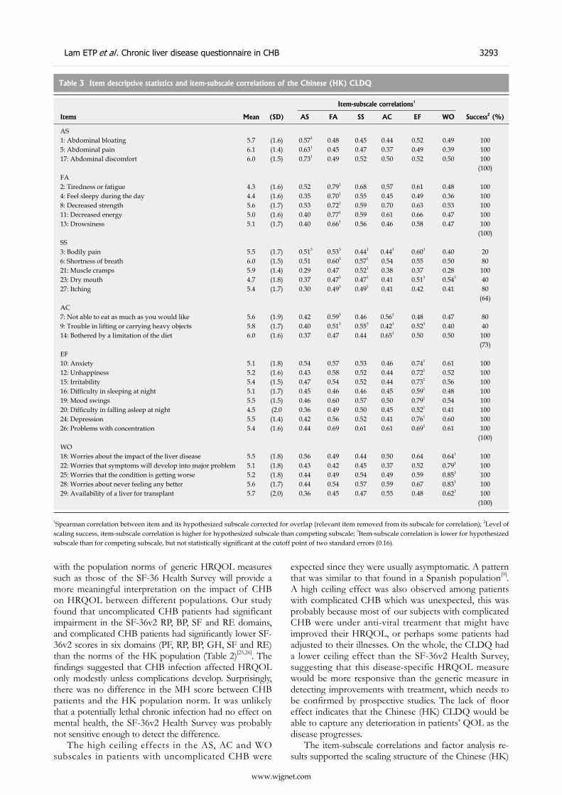
with the population norms of generic HRQOL measures such as those of the SF-36 Health Survey will provide a more meaningful interpretation on the impact of CHB on HRQOL between different populations. Our study found that uncomplicated CHB patients had significant impairment in the SF-36v2 RP, BP, SF and RE domains, and complicated CHB patients had significantly lower SF-36v2 scores in six domains (PF, RP, BP, GH, SF and RE) than the norms of the HK population (Table 2)[25,26]. The findings suggested that CHB infection affected HRQOL only modestly unless complications develop. Surprisingly, there was no difference in the MH score between CHB patients and the HK population norm. It was unlikely that a potentially lethal chronic infection had no effect on mental health, the SF-36v2 Health Survey was probably not sensitive enough to detect the difference.
The high ceiling effects in the AS, AC and WO subscales in patients with uncomplicated CHB were
expected since they were usually asymptomatic. A pattern that was similar to that found in a Spanish population[9]. A high ceiling effect was also observed among patients with complicated CHB which was unexpected, this was probably because most of our subjects with complicated CHB were under anti-viral treatment that might have improved their HRQOL, or perhaps some patients had adjusted to their illnesses. On the whole, the CLDQ had a lower ceiling effect than the SF-36v2 Health Survey, suggesting that this disease-specific HRQOL measure would be more responsive than the generic measure in detecting improvements with treatment, which needs to be confirmed by prospective studies. The lack of floor effect indicates that the Chinese (HK) CLDQ would be able to capture any deterioration in patients’ QOL as the disease progresses.
The item-subscale correlations and factor analysis re-sults supported the scaling structure of the Chinese (HK)
www.wjgnet.com
Table 3 Item descriptive statistics and item-subscale correlations of the Chinese (HK) CLDQ
Item-subscale correlations1
Items Mean (SD) AS FA SS AC EF WO Success2 (%)
AS1: Abdominal bloating 5.7 (1.6) 0.571 0.48 0.45 0.44 0.52 0.49 1005: Abdominal pain 6.1 (1.4) 0.631 0.45 0.47 0.37 0.49 0.39 10017: Abdominal discomfort 6.0 (1.5) 0.731 0.49 0.52 0.50 0.52 0.50 100
(100)FA2: Tiredness or fatigue 4.3 (1.6) 0.52 0.791 0.68 0.57 0.61 0.48 1004: Feel sleepy during the day 4.4 (1.6) 0.35 0.701 0.55 0.45 0.49 0.36 1008: Decreased strength 5.6 (1.7) 0.53 0.721 0.59 0.70 0.63 0.53 10011: Decreased energy 5.0 (1.6) 0.40 0.771 0.59 0.61 0.66 0.47 10013: Drowsiness 5.1 (1.7) 0.40 0.661 0.56 0.46 0.58 0.47 100
(100)SS3: Bodily pain 5.5 (1.7) 0.513 0.533 0.441 0.443 0.603 0.40 206: Shortness of breath 6.0 (1.5) 0.51 0.603 0.571 0.54 0.55 0.50 8021: Muscle cramps 5.9 (1.4) 0.29 0.47 0.521 0.38 0.37 0.28 10023: Dry mouth 4.7 (1.8) 0.37 0.473 0.471 0.41 0.513 0.543 4027: Itching 5.4 (1.7) 0.30 0.493 0.491 0.41 0.42 0.41 80
(64)AC7: Not able to eat as much as you would like 5.6 (1.9) 0.42 0.593 0.46 0.561 0.48 0.47 809: Trouble in lifting or carrying heavy objects 5.8 (1.7) 0.40 0.513 0.553 0.421 0.523 0.40 4014: Bothered by a limitation of the diet 6.0 (1.6) 0.37 0.47 0.44 0.651 0.50 0.50 100
(73)EF10: Anxiety 5.1 (1.8) 0.54 0.57 0.53 0.46 0.741 0.61 10012: Unhappiness 5.2 (1.6) 0.43 0.58 0.52 0.44 0.721 0.52 10015: Irritability 5.4 (1.5) 0.47 0.54 0.52 0.44 0.731 0.56 10016: Difficulty in sleeping at night 5.1 (1.7) 0.45 0.46 0.46 0.45 0.591 0.48 10019: Mood swings 5.5 (1.5) 0.46 0.60 0.57 0.50 0.791 0.54 10020: Difficulty in falling asleep at night 4.5 (2.0 0.36 0.49 0.50 0.45 0.521 0.41 10024: Depression 5.5 (1.4) 0.42 0.56 0.52 0.41 0.761 0.60 10026: Problems with concentration 5.4 (1.6) 0.44 0.69 0.61 0.61 0.691 0.61 100
(100)WO18: Worries about the impact of the liver disease 5.5 (1.8) 0.56 0.49 0.44 0.50 0.64 0.641 10022: Worries that symptoms will develop into major problem 5.1 (1.8) 0.43 0.42 0.45 0.37 0.52 0.791 10025: Worries that the condition is getting worse 5.2 (1.8) 0.44 0.49 0.54 0.49 0.59 0.851 10028: Worries about never feeling any better 5.6 (1.7) 0.44 0.54 0.57 0.59 0.67 0.831 10029: Availability of a liver for transplant 5.7 (2.0) 0.36 0.45 0.47 0.55 0.48 0.621 100
(100)
1Spearman correlation between item and its hypothesized subscale corrected for overlap (relevant item removed from its subscale for correlation); 2Level of scaling success, item-subscale correlation is higher for hypothesized subscale than competing subscale; 3Item-subscale correlation is lower for hypothesized subscale than for competing subscale, but not statistically significant at the cutoff point of two standard errors (0.16).
Lam ETP et al . Chronic liver disease questionnaire in CHB 3293

CLDQ in general. However, the scaling success rates of items 3 (bodily pain), 23 (dry mouth) and 9 (trouble in
lifting or carrying heavy objects) seemed too low to be acceptable, raising the question whether they should be
www.wjgnet.com
Table 4 Factor loadings of the Chinese (HK) CLDQ
Factor 1 Factor 2 Factor 3 Factor 4 Factor 5 Factor 6Item EF WO SS + FA AS AC SL
Abdominal symptom (AS)1: Abdominal bloating 0.18 0.27 0.16 0.70 0.14 0.155: Abdominal pain 0.20 0.06 0.13 0.81 0.08 0.1417: Abdominal discomfort 0.17 0.23 0.17 0.82 0.20 0.08Fatigue (FA)2: Tiredness or fatigue 0.33 0.03 0.51 0.31 0.45 0.224: Feel sleepy during the day 0.28 -0.05 0.47 0.14 0.44 0.148: Decreased strength 0.37 0.14 0.37 0.24 0.60 0.2411: Decreased energy 0.56 0.06 0.37 0.11 0.51 0.1213: Drowsiness 0.49 0.09 0.47 0.15 0.36 -0.17Systemic symptoms (SS)3: Bodily pain 0.49 0.00 0.18 0.39 0.14 0.226: Shortness of breath 0.22 0.22 0.59 0.39 0.18 0.0321: Muscle cramps 0.09 0.08 0.76 0.17 0.10 0.1423: Dry mouth 0.24 0.42 0.48 0.16 0.08 -0.0227: Itching 0.10 0.27 0.65 -0.01 0.02 0.28Activity (AC)7: Not able to eat as much as you would like 0.05 0.29 0.04 0.24 0.81 0.129: Trouble in lifting or carrying heavy objects 0.41 0.18 0.37 -0.05 0.35 0.1714: Bothered by a limitation of the diet 0.11 0.43 0.09 0.10 0.67 0.12Emotional function (EF)10: Anxiety 0.67 0.32 0.15 0.39 0.14 0.0412: Unhappiness 0.77 0.25 0.15 0.20 0.13 0.0115: Irritability 0.78 0.27 0.15 0.17 0.06 0.1316: Difficulty in sleeping at night 0.27 0.17 0.17 0.18 0.14 0.7919: Mood swings 0.78 0.24 0.10 0.19 0.20 0.2120: Difficulty in falling asleep at night 0.16 0.14 0.25 0.22 0.25 0.7124: Depression 0.8 0.31 0.15 0.08 0.03 0.2226: Problems with concentration 0.52 0.28 0.29 0.18 0.37 0.27Worry (WO)18: Worries about the impact of the liver disease 0.40 0.47 0.03 0.50 0.25 0.0722: Worries that symptoms will develop into major problem 0.29 0.80 0.11 0.16 0.04 0.0525: Worries that the condition is getting worse 0.29 0.84 0.13 0.17 0.17 0.0928: Worries about never feeling any better 0.27 0.77 0.13 0.18 0.26 0.2429: Availability of a liver for transplant 0.16 0.64 0.26 0.14 0.30 0.10
SL: Sleep.
Table 5 Reliability and correlations of the Chinese (HK) CLDQ and SF-36v2 health survey scores
AS FA SS AC EF WO Cronbach’s α ICC1
CLDQAS 0.84 0.58FA 0.54 0.88 0.82SS 0.54 0.72 0.74 0.86AC 0.5.0 0.67 0.60 0.72 0.66EF 0.58 0.72 0.68 0.61 0.90 0.86WO 0.52 0.57 0.60 0.58 0.69 0.90 0.89Overall 0.72 0.87 0.84 0.80 0.86 0.81 0.90 0.85
SF-36v2 Health SurveyPF 0.47 0.56 0.65 0.54 0.47 0.46 0.81 0.93RP 0.56 0.72 0.70 0.67 0.60 0.62 0.91 0.90BP 0.44 0.44 0.62 0.42 0.48 0.44 0.89 0.77GH 0.46 0.66 0.56 0.57 0.60 0.62 0.82 0.89VT 0.51 0.79 0.63 0.58 0.67 0.54 0.86 0.85SF 0.44 0.56 0.52 0.60 0.58 0.52 0.88 0.54RE 0.53 0.51 0.52 0.46 0.66 0.50 0.89 0.74MH 0.41 0.58 0.49 0.49 0.78 0.61 0.84 0.89
1All analyses were performed with the total sample of 150 patients, except for the Intra-class Correlation Coefficient (ICC), which was based on the results from 46 uncomplicated CHB patients.
3294 ISSN 10079327 CN 141219/R World J Gastroenterol July 14, 2009 Volume 15 Number 26
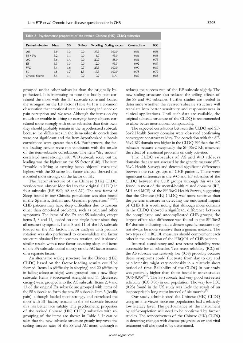
grouped under other subscales than the originally hy-pothesized. It is interesting to note that bodily pain cor-related the most with the EF subscale score and loaded the strongest on the EF factor (Table 4). It is a common observation that emotional state has a strong influence on pain perception and vice versa. Although the items on dry mouth or trouble in lifting or carrying heavy objects cor-related more strongly with other subscales than their own, they should probably remain in the hypothesized subscale because the differences in the item-subscale correlations were not significant and the item-hypothesized subscale correlations were greater than 0.4. Furthermore, the fac-tor loading results were not consistent with the results of the item-subscale correlations. The item “dry mouth” correlated most strongly with WO subscale score but the loading was the highest on the SS factor (0.48). The item “trouble in lifting or carrying heavy objects” correlated highest with the SS score but factor analysis showed that it loaded most strongly on the factor of EF.
The factor structure of the Chinese (HK) CLDQ version was almost identical to the original CLDQ in four subscales (EF, WO, AS and AC). The new factor of Sleep found in our Chinese population was also found in the Spanish, Italian and German population[9,10,27]. CHB patients may have sleep difficulties due to reasons other than emotional problems, such as pain and other symptoms. The items of the FA and SS subscales, except items 3, 8 and 11, loaded on one single factor since they all measure symptoms. Items 8 and 11 of the FA subscale loaded on the AC factor. Factor analysis with promax rotation was also performed to cross-validate the factor structure obtained by the varimax rotation, and it showed similar results with a new factor assessing sleep and items of the FA subscale loaded mostly on the AC factor instead of a separate factor.
An alternative scaling structure for the Chinese (HK) CLDQ based on the factor loading results could be formed. Items 16 (difficulty in sleeping) and 20 (difficulty in falling asleep at night) were grouped into a new Sleep subscale. Items 8 (decreased strength) and 11 (decreased energy) were grouped into the AC subscale. Items 2, 4 and 13 of the original FA subscale are grouped with items of the SS subscale to form the new SS subscale. Item 3 (bodily pain), although loaded most strongly and correlated the most with EF factor, remains in the SS subscale because this has better face validity. The psychometric properties of the revised Chinese (HK) CLDQ subscales with re-grouping of the items are shown in Table 6. It can be seen that the new subscale structure greatly improves the scaling success rates of the SS and AC items, although it
reduces the success rate of the EF subscale slightly. The new scaling structure also reduced the ceiling effects of the SS and AC subscales. Further studies are needed to determine whether the revised subscale structure will translate into better sensitivity and responsiveness in clinical applications. Until such data are available, the original subscale structure of the CLDQ is recommended to allow better international comparability.
The expected correlations between the CLDQ and SF-36v2 Health Survey domains were observed confirming convergent construct validity. The correlation with the SF-36v2 RE domain was higher in the CLDQ EF than the AC subscale because conceptually the SF-36v2 RE measures the effect of emotional problems on daily activities.
The CLDQ subscales of AS and WO address domains that are not assessed by the generic measure (SF-36v2 Health Survey) and detected significant differences between the two groups of CHB patients. There were significant differences in the WO and EF subscales of the CLDQ between the CHB groups although this was not found in most of the mental-health related domains (RE, MH and MCS) of the SF-36v2 Health Survey, suggesting that the Chinese (HK) CLDQ was more sensitive than the generic measure in detecting the emotional impact of CHB. It is worth noting that although more domains in the CLDQ showed a significant difference between the complicated and uncomplicated CHB groups, the largest effect size difference was found in the SF-36v2 RP domain indicating that a disease-specific measure may not always be more sensitive than a generic measure. The two types of HRQOL measures should complement each other in the evaluation of the HRQOL of CHB patients.
Internal consistency and test-retest reliability were acceptable for all subscales. Test-retest reliability (ICC) of the AS subscale was relatively low (0.58) probably because these symptoms could fluctuate from day to day and pain intensity might vary noticeably in a relatively short period of time. Reliability of the CLDQ in our study was generally higher than those found in other studies (0.46-0.95)[9,12]. The SS subscale had very good test-retest reliability (ICC 0.86) in our population. The very low ICC (0.23) found in the US study was likely the result of an inappropriately long retest interval of six months[3].
Our study administered the Chinese (HK) CLDQ using an interviewer since our populations had a relatively low literacy level. The performance of the instrument by self-completion will need to be confirmed by further studies. The responsiveness of the Chinese (HK) CLDQ in detecting changes with disease progression or anti-viral treatment will also need to be determined.
www.wjgnet.com
Table 6 Psychometric properties of the revised Chinese (HK) CLDQ subscales
Revised subscales Mean SD % floor % ceiling Scaling success Cronbach's α ICC
AS 5.9 1.3 0.0 37.3 100.0 0.84 0.58SS + FA 5.2 1.1 0.0 4.7 95.0 0.84 0.88AC 5.6 1.4 0.0 20.7 88.0 0.84 0.75EF 5.3 1.3 0.0 12.0 93.3 0.92 0.87WO 5.4 1.6 0.7 27.3 100.0 0.90 0.89SL 4.8 1.7 1.3 17.3 100.0 0.78 0.79Overall Scores 5.4 1.1 0.0 0.7 NA 0.89 0.85
Lam ETP et al . Chronic liver disease questionnaire in CHB 3295

The Chinese (HK) CLDQ was validated in content and construct. It had satisfactory psychometric properties in terms of factor structure, scaling assumption, construct validity, reliability and sensitivity in Southern Chinese patients with CHB infection. It was more sensitive than the SF-36v2 Health Survey in detecting the impact of CHB on mental-health and symptom related HRQOL. The Chinese (HK) CLDQ should be applicable to all Cantonese-speaking Chinese in HK and other parts of Southern China. It is also likely to be applicable to the majority of Chinese populations in Australia, North America, and Europe who are mostly emigrants from HK. There was good equivalence in the score distribution pattern across several cultures indicating that it can be used as a cross-cultural HRQOL measure in multiethnic populations or global studies. Some modifications of the scaling structure of the CLDQ may improve its psychometric properties for CHB patients, which need to be explored by further clinical studies.
Appendix: Back-translation of Chinese (HK) CLDQ
Original Wording Backward Translation
This questionnaire is designed to find out how you have been feeling during the last 2 wk.
The purpose of this questionnaire is to understand how you felt in the past 2 wk.
You will be asked about you symptoms related to your liver disease, how you have been affected in doing activities, and how your mood has been.
The questions are about the symptoms resulting from your liver illness and how these symptoms affect your participation in activities, and your emotions.
Please complete all of questions and select only one response for each question.
Please answer all questions. You can only choose one answer for each question.
1 How much of the time during the last 2 wk have you been troubled by a feeling of abdominal bloating?
1 In the past 2 wk, how much time you have been bothered by your bloating problem?
All of the time All the timeMost of the time Most of the timeA good bit of the time Quite OftenSome of the time SometimesA little of the time A Short TimeHardly any of the time Hardly AnyNone of the time Never2 How much of the time have you been tired or fatigued during the last 2 wk?
2 In the past 2 wk, how much time did you feel tired or exhausted?
3 How much of the time during the last 2 wk have you experienced bodily pain?
3 In the past 2 wk, how much time did your body ache?
4 How often during the last 2 wk have you felt sleepy during the day?
4 In the past 2 wk, how often did you feel sleepy during the daytime?
5 How much of the time during the last 2 wk have you experienced abdominal pain?
5 In the past 2 wk, how much time did you have abdominal pain?
6 How much of the time during the last 2 wk has shortness of breath been a problem for you in your daily activities?
6 In the past 2 wk, how much time were your daily activities affected by your shortness of breath?
7 How much of the time during the last 2 wk have you not been able to eat as much as you would like?
7 In the past 2 wk, how much time were you unable to eat as much as you want?
8 How much of the time in the last 2 wk have you been bothered by having decreased strength?
8 In the past 2 wk, how much time have you been bothered by the decline in your physical energy?
9 How often during the last 2 wk have you had trouble lifting or carrying heavy objects?
9 In the past 2 wk, how often did you find it difficult when you were lifting or carrying heavy objects?
10 How often during the last 2 wk have you felt anxious?
10 In the past 2 wk, how often did you feel anxious?
11 How often during the last 2 wk have you felt a decreased level of energy?
11 In the past 2 wk, how often did you find your energy level decreasing?
12 How much of the time during the last 2 wk have you felt unhappy?
12 In the past 2 wk, how much time did you feel unhappy?
13 How often during the last 2 wk have you felt drowsy?
13 In the past 2 wk, how often did you feel sleepy?
14 How much of the time during the last 2 wk have you been bothered by a limitation of your diet?
14 In the past 2 wk, how much time have you been bothered by your restricted diet?
15 How often during the last 2 wk have you been irritable?
15 In the past 2 wk, how often did you become irritable?
16 How much of the time during the last 2 wk have you had difficulty sleeping at night?
16 In the past 2 wk, how much time did you find it difficult to sleep at night?
17 How much of the time during the last 2 wk have you been troubled by a feeling of abdominal discomfort?
17 In the past 2 wk, how much time have you been bothered by your abdominal discomfort?
18 How much of the time during the last 2 wk have you been worried about the impact your liver disease has on your family?
18 In the past 2 wk, how much time did you worry that your liver illness will affect your family?
19 How much of the time during the last 2 wk have you had mood swings?
19 In the past 2 wk, how much time did your emotions fluctuate?
20 How much of the time during the last 2 wk have you been unable to fall asleep at night?
20 In the past 2 wk, how much time were you unable to sleep until sunrise?
21 How often during the last 2 wk have you had muscle cramps?
21 In the past 2 wk, how often did your muscle cramp?
22 How much of the time during the last 2 wk have you been worried that your symptoms will develop into major problems?
22 In the past 2 wk, how much time did you worry that your symptoms will become a serious problem?
23 How much of the time during the last 2 wk have you had a dry mouth?
23 In the past 2 wk, how much time did you have dry mouth?
24 How much of the time during the last 2 wk have you felt depressed?
24 In the past 2 wk, how much time did you feel depressed?
25 How much of the time during the last 2 wk have you been worried about your condition getting worse?
25 In the past 2 wk, how much time did you worry that your condition will deteriorate?
26 How much of the time during the last 2 wk have you had problems concentration?
26 In the past 2 wk, how much time did you find it hard to concentrate?
27 How much of the time have you been troubled by itching during the last 2 wk?
27 In the past 2 wk, how much time have you been bothered by itchiness?
28 How much of the time during the last 2 wk have you been worried about never feeling any better?
28 In the past 2 wk, how much time did you worry that your health condition will not improve?
29 How much of the time during the last 2 wk have you been concerned about the availability of a liver if you need a liver transplant?
29 In the past 2 wk, how much time have you worried that you could not get a liver if you have to undergo a liver transplant?
ACKNOWLEDGMENTSWe thank Ms Fong Nga Po and Ms Cara Chan Yuen Yee for their assistance in data collection and entry, and the staff of the Division of Hepatobiliary/Pancreatic Surgery & Liver Transplantation and Gastroenterology & Hepatology Specialist Outpatient Clinics, Queen Mary Hospital for their help in patient recruitment.
COMMENTSBackgroundChronic hepatitis B (CHB) virus infection remains a global problem and a public health threat. CHB patients may suffer or die from liver-related complications, posing a threat to both mental and physical health, leading to impairment of quality of life.Research frontiersHealth-related quality of life (HRQOL) outcomes should supplement traditional clinical outcomes in the evaluation of the impact and the effectiveness of treatment for patients with CHB infection.
www.wjgnet.com
COMMENTS
3296 ISSN 10079327 CN 141219/R World J Gastroenterol July 14, 2009 Volume 15 Number 26

Innovations and breakthroughsThe Chronic Liver Disease Questionnaire (CLDQ) had been applied mainly to patients with hepatitis C virus infection in Western countries. This study was the first to show that a Chinese (Hong Kong) translation of the CLDQ was valid, reliable and sensitive for Southern Chinese patients with CHB infection. The CLDQ can be applied to millions of Southern Chinese CHB patients to evaluate their HRQOL. Some modifications might further improve its validity, reliability and sensitivity.ApplicationsThe Chinese (Hong Kong) CLDQ can be used to evaluate the impact of CHB virus infection and assess the effectiveness of anti-viral drug treatments in Cantonese-speaking Southern Chinese. The CLDQ can be used as a cross-cultural HRQOL measure in international studies that include Southern Chinese.TerminologyCHB virus infection refers to those who are hepatitis B surface antigen-positive for more than six months. Validity is defined as the extent to which a test measures what it is intended to measure. Reliability refers to the consistency or stability of the measurement process across time, patients or observers. Peer reviewThe authors validated and tested the psychometric properties of a Southern Chinese translation of the CLDQ and determined that their questionnaire was valid, reliable, and sensitive for southern Chinese patients with hepatitis B virus infection. The study was well done and used appropriate methodology to validate and test the questionnaire.
REFERENCES1 Liu J, Fan D. Hepatitis B in China. Lancet 2007; 369: 1582-15832 Chen CJ, Wang LY, Yu MW. Epidemiology of hepatitis
B virus infection in the Asia-Pacific region. J Gastroenterol Hepatol 2000; 15 Suppl: E3-E6
3 Younossi ZM, Guyatt G, Kiwi M, Boparai N, King D. Development of a disease specific questionnaire to measure health related quality of life in patients with chronic liver disease. Gut 1999; 45: 295-300
4 Bayliss MS, Gandek B, Bungay KM, Sugano D, Hsu MA, Ware JE Jr. A questionnaire to assess the generic and disease-specific health outcomes of patients with chronic hepatitis C. Qual Life Res 1998; 7: 39-55
5 Gralnek IM, Hays RD, Kilbourne A, Rosen HR, Keeffe EB, Artinian L, Kim S, Lazarovici D, Jensen DM, Busuttil RW, Martin P. Development and evaluation of the Liver Disease Quality of Life instrument in persons with advanced, chronic liver disease--the LDQOL 1.0. Am J Gastroenterol 2000; 95: 3552-3565
6 van der Plas SM, Hansen BE, de Boer JB, Stijnen T, Passchier J, de Man RA, Schalm SW. The Liver Disease Symptom Index 2.0; validation of a disease-specific questionnaire. Qual Life Res 2004; 13: 1469-1481
7 Spiegel BM, Bolus R, Han S, Tong M, Esrailian E, Talley J, Tran T, Smith J, Karsan HA, Durazo F, Bacon B, Martin P, Younossi Z, Hwa-Ong S, Kanwal F. Development and validation of a disease-targeted quality of life instrument in chronic hepatitis B: the hepatitis B quality of life instrument, version 1.0. Hepatology 2007; 46: 113-121
8 Lee EH, Cheong JY, Cho SW, Hahm KB, Kim HY, Park JJ, Lee DH, Kim SK, Choi SR, Lee ST, Moon SM. Development and psychometric evaluation of a chronic liver disease-specific quality of life questionnaire. J Gastroenterol Hepatol 2008; 23: 231-238
9 Ferrer M, Cordoba J, Garin O, Olive G, Flavia M, Vargas V, Esteban R, Alonso J. Validity of the Spanish version of the Chronic Liver Disease Questionnaire (CLDQ) as a standard outcome for quality of life assessment. Liver Transpl 2006;
12: 95-10410 Rucci P, Taliani G, Cirrincione L, Alberti A, Bartolozzi D,
Caporaso N, Colombo M, Coppola R, Chiaramonte M, Craxi A, De Sio I, Floreani AR, Gaeta GB, Persico M, Secchi G, Versace I, Mele A. Validity and reliability of the Italian version of the Chronic Liver Disease Questionnaire (CLDQ-I) for the assessment of health-related quality of life. Dig Liver Dis 2005; 37: 850-860
11 Sobhonslidsuk A, Silpakit C, Kongsakon R, Satitpornkul P, Sripetch C. Chronic liver disease questionnaire: translation and validation in Thais. World J Gastroenterol 2004; 10: 1954-1957
12 Wu CH, Deng QW, Ji XS, Yan LM. Preliminary Use of the CLDQ in Chronic Hepatitis B Patients. Zhongguo Linchuang Xinlixue Zhazhi 2003; 11: 60-62
13 Hauser W, Schnur M, Steder-Neukamm U, Muthny FA, Grandt D. Validation of the German version of the Chronic Liver Disease Questionnaire. Eur J Gastroenterol Hepatol 2004; 16: 599-606
14 Wild D, Grove A, Martin M, Eremenco S, McElroy S, Verjee-Lorenz A, Erikson P. Principles of Good Practice for the Translation and Cultural Adaptation Process for Patient-Reported Outcomes (PRO) Measures: report of the ISPOR Task Force for Translation and Cultural Adaptation. Value Health 2005; 8: 94-104
15 Beaton DE , Bombardier C, Guillemin F, Ferraz MB. Guidelines for the process of cross-cultural adaptation of self-report measures. Spine 2000; 25: 3186-3191
16 Lam CL, Gandek B, Ren XS, Chan MS. Tests of scaling assumptions and construct validity of the Chinese (HK) version of the SF-36 Health Survey. J Clin Epidemiol 1998; 51: 1139-1147
17 Lam ETP, Lam CLK, Lo YYC, Grandek B. Psychometrics and population norm of the Chinese (HK) SF-36 Health Survey_Version 2. HK Pract 2008; 30: 185-198
18 Campbell DT, Fiske DW. Convergent and discriminant validation by the multitrait-multimethod matrix. Psychol Bull 1959; 56: 81-105
19 Cohen J. Statistical power analysis for the behavioral sciences. 2nd ed. Hillsdale, N.J.: L. Erlbaum Associates, 1988: 1-74
20 Terwee CB, Bot SD, de Boer MR, van der Windt DA, Knol DL, Dekker J, Bouter LM, de Vet HC. Quality criteria were proposed for measurement properties of health status questionnaires. J Clin Epidemiol 2007; 60: 34-42
21 The Netherlands Cancer Institute, Amsterdam. Assessing health status and quality-of-life instruments: attributes and review criteria. Qual Life Res 2002; 11: 193-205
22 Public Health Report No. 3. Viral hepatitis & liver cancer and unintentional injuries in children. Hong Kong: Dept. of Health, 1998: 6-16
23 Yuen MF, Yuan HJ, Wong DK, Yuen JC, Wong WM, Chan AO, Wong BC, Lai KC, Lai CL. Prognostic determinants for chronic hepatitis B in Asians: therapeutic implications. Gut 2005; 54: 1610-1614
24 Foster GR, Goldin RD, Thomas HC. Chronic hepatitis C virus infection causes a significant reduction in quality of life in the absence of cirrhosis. Hepatology 1998; 27: 209-212
25 Gandek B, Lam CLK. Evaluating the SF-36v2 in Hong Kong. Qual Life Res 2005; 14: 2098
26 Lam CLK, Lauder IJ, Lam TP, Gandek B. Population based norming of the Chinese (HK) version of the SF-36 health survey. HK Pract 1999; 21: 460-470
27 Schulz KH, Kroencke S, Ewers H, Schulz H, Younossi ZM. The factorial structure of the Chronic Liver Disease Questionnaire (CLDQ). Qual Life Res 2008; 17: 575-584
S- Editor Li LF L- Editor Webster JR E- Editor Ma WH
www.wjgnet.com
Lam ETP et al . Chronic liver disease questionnaire in CHB 3297

www.wjgnet.com
BRIEF ARTICLES
Liver and spleen volume variations in patients with hepatic fibrosis
Peng Liu, Peng Li, Wen He, Li-Qin Zhao
Online Submissions: wjg.wjgnet.com World J Gastroenterol 2009 July 14; 15(26): [email protected] World Journal of Gastroenterology ISSN 1007-9327doi:10.3748/wjg.15.3298 © 2009 The WJG Press and Baishideng. All rights reserved.
Peng Liu, Peng Li, Wen He, Li-Qin Zhao, Department of Radiology, Beijing Friendship Hospital, Capital Medical University, Beijing 100050, ChinaAuthor contributions: He W and Liu P designed the research; Liu P, Zhao LQ and Li P collected the data; Liu P and He W performed the post-processing of images and analyzed the data; Liu P wrote the manuscript; He W revised the paper.Supported by Science and Technology Program of Beijing Education Committee, No. KM200810025002Correspondence to: Wen He, MD, Department of Radiology, Beijing Friendship Hospital, Capital Medical University, 95 Yongan Road, Xuanwu District, Beijing 100050, China. [email protected]: +86-10-63138470 Fax: + 86-10-63037216Received: March 4, 2009 Revised: April 18, 2009Accepted: April 25, 2009Published online: July 14, 2009
AbstractAIM: To study the liver and spleen volume variations in hepatic fibrosis patients at different histopathological stages.
METHODS: Multidetector computed tomography (MDCT) scan was performed in 85 hepatic fibrosis patients. Liver volume (LV) and spleen volume (SV) were measured. Fifteen healthy individuals served as a control group (S0). The patients were divided into stage 1 (S1) group (n = 34), stage 2 (S2) group (n = 25), stage 3 (S3) group (n = 16), and stage 4 (S4) group (n = 10) according to their histopathological stage of liver fibrosis.
RESULTS: The LV and standard LV(SLV)had a tendency to increase with the severity of fibrosis, but no statistical difference was observed in the 5 groups (LV: F = 0.245, P = 0.912; SLV: F = 1.902, P = 0.116). The SV was gradually increased with the severity of fibrosis, and a statistically significant difference in SV was observed among the 5 groups (P < 0.01). The LV/SV ratio and SLV/SV ratio were gradually decreased with the aggravation of hepatic fibrosis, and statistically significant differences in both LV/SV and SLV/SV were found among the 5 groups (P < 0.01).
CONCLUSION: The absence of obvious LV reduction in patients with chronic liver disease may be a
morphological index of patients without liver cirrhosis. The SV is related to the severity of fibrosis, and the spleen of patients with advanced fibrosis is enlarged evidently. The LV/SV ratio and SLV/SV ratio are of a significant clinical value in the diagnosis of advanced liver fibrosis.
© 2009 The WJG Press and Baishideng. All rights reserved.
Key words: Hepatic fibrosis; Volume measurement; Liver volume; Standard liver volume; Spleen volume; Liver volume/spleen volume ratio; Standard liver volume/spleen volume ratio
Peer reviewer: Heitor Rosa, Professor, Department of Gastroenterology and Hepatology, Federal University School of Medicine, Rua 126 n.21, Goiania - GO 74093-080, Brazil
Liu P, Li P, He W, Zhao LQ. Liver and spleen volume variations in patients with hepatic fibrosis. World J Gastroenterol 2009; 15(26): 3298-3302 Available from: URL: http://www.wjgnet.com/1007-9327/15/3298.asp DOI: http://dx.doi.org/10.3748/wjg.15.3298
INTRODUCTIONHepatic fibrosis is an important histopathological stage of chronic liver disease, and can progress to hepatocirrhosis. Studies have shown that hepatic fibrosis can be reversed[1,2]. Therefore, hepatic fibrosis can be halted if it is early diagnosed and treated with intervention therapy. Liver biopsy is the golden diagnosis standard for hepatic fibrosis and cirrhosis, but it cannot be used widely and repeatedly due to its invasive nature. This study was to observe the liver volume (LV) and spleen volume (SV) variations in patients with hepatic fibrosis by 64-row multidetector computed tomography (MDCT).
MATERIALS AND METHODSPatientsEighty-five patients (66 males and 19 females, mean age 41 years) with liver fibrosis in our hospital between November 2007 and September 2008 were included in this study. All of them had chronic hepatitis B. The patients were divided into stage 1 (S1) group (n = 34),

Liu P et al . Liver and spleen volume in hepatic fibrosis patients 3299
www.wjgnet.com
stage 2 (S2) group (n = 25), stage 3 (S3) group (n = 16), and stage 4 (S4) group (n = 10) according to their histopathological stage of fibrosis. The patients had no liver and spleen tumor. Fifteen patients (10 males and 5 females, mean age 35.5 years) with no history of liver and spleen disease served as a control group (S0). They underwent abdominal computed tomography (CT) scan for liver disease. Routine abdominal CT scanning showed no liver, bile and spleen disease. Liver test was normal and antigen test was negative for hepatitis B and C.
Diagnostic methodAll patients with chronic hepatitis B were diagnosed according to the results of pathological examination of liver biopsy. The patients were divided into four groups according to the diagnostic criteria for virus hepatitis as previously described[3].
Imaging parametersAbdominal CT scan was performed with a 64-row MDCT scanner (GE LightSpeed VCT). Protocols included a non-enhancement scan and an enhancement scan of the upper abdomen from the dome of diaphragm to the entire liver and spleen (5 mm thickness, 80-120 mAs, and 120 kVp). All patients received an injection of 100 mL iopromide (300 mg I/mL; Ultravist, Schering) through a peripheral vein (generally an
antecubital vein) at a flow rate of 4.0 mL/s. The arterial phases were tracked with a scanning monitor and a time delay of 25 s after arterial phases was applied in all patients for the venous phases. Bolus-injection technique was used to administer contrast material with a power injector. All patients underwent CT scan at a supine position during a single breath.
Measurement of LV and SVAfter scanning, 5 mm portal vein phase reconstruction images obtained from the raw data were divided (0.625 mm) and transferred to an interactive workstation (AW4.2 or AW4.3) to measure LV and SV. The profile of liver was outlined manually to exclude the inferior vena cava with an interval of several sections. Gallbladder, main liver blood vessels and fat were excluded by regulating the threshold value. The enclosed liver parenchymal area was then calculated automatically[4,5] and expressed as the LV (Figures 1-4).
Body weight (BW) and body height (BH) recorded at the time of CT examination were used to calculate the body surface area (BSA) following the equation: BSA (m2) = [0.0071 × BH (cm) + 0.0133 × BW (kg) - 0.1971]. LV is significantly related with BW, BH, and BSA as described elsewhere[6,7]. Therefore, standard liver volume (SLV) was measured following the equation: SLV (cm3/m2) = LV (cm3)/BSA (m2).
The measurement of SV and LV was similar. Since
Figure 4 Volume rendering image of liver after reconstruction.Figure 3 Measurement of liver volume.
Figure 2 Regulation of threshold value for exclusion of gallbladder, main liver blood vessels and fat.
Figure 1 Profile of the liver with an interval of several sections for the separation of liver from other organs and their tissues around.

www.wjgnet.com
SV does not correlate with BW, BH and body mass index (BMI)[8-10], it is not necessary to make the SV standard.
Then, the LV/SV ratio and SLV/SV ratio were calculated.
Statistical analysisResults were expressed as mean ± SD. One way analysis of variance(ANOVA) was used for multiple mean comparisons. Bonferroni correction was used for comparison of two samples. If heterogeneity of variance was found, a log transformation was used to normalize the data distribution, and ANOVA was run with an equal variance. P < 0.05 was considered statistically significant. Statistical analysis was performed using SPSS (version 14.0, SPSS).
RESULTSThe total LV and SLV were 1133.39 ± 131.84 cm3 and 637.98 ± 65.93 cm3 in the control group. The LV (F = 0.245) and SLV (F = 1.902) in patients with hepatic fibrosis tended to increase with the severity of fibrosis, but no statistically significant difference was observed among the 5 groups (Table 1).
A log transformation was used to normalize the data distribution and ANOVA was run with an equal variance due to the heterogeneity of variance in SV. The SV was larger in patients with liver fibrosis than in control group and increased gradually with the severity of fibrosis (Table 1). There was a significant difference among the 5 groups (F = 12.383, P = 0.000). Statistically significant differences were observed between S3, S4 and other groups(S0, S1 and S2, P < 0.01).
The LV/SV ratio and SLV/SV ratio decreased gradually with the severity of fibrosis (Table 1). The differences in LV/SV (F = 9.702, P = 0.000) and SLV/SV (F = 9.784, P = 0.000) were significant among the 5 groups. Statistically significant differences in LV/SV were observed between S3 and S0, S1, and between S4 and S0, S1, S2 (P < 0.01). There were statistically significant differences in SLV/SV between S3 and S0, S4 and S0, S1 (P < 0.01), and between S2 and S0, S3 and S1, S4 and S2 (P < 0.05).
DISCUSSIONMeasurement of LV and SV is a mature technique[4,5,10-12].
The method, which combines profile outline and threshold value regulation, is convenient with a good veracity and repeatability[5]. Liver and spleen vary in size among different individuals[13,14]. LV is obviously related with BW, BH, and BSA in normal persons[15-17]. BSA is calculated according to BW and BH, and therefore can equalize the difference in BW and BH. It is more reasonable to choose SLV (per BSA volume) to analyze LV change in hepatic fibrosis patients at different histopathological stages. Because SV does not correlate with BW, BH, and BMI[8-10], there is no need to make the spleen volume standard.
Reports are available on LV changes in patients with hepatic fibrosis[18,19]. Chen et al[18] reported that the left, right and caudate lobe volumes tend to increase with the aggravation of inflammatory activity and the severity of fibrosis, with significant changes in vertical diameter and volume index of the left lobe but no obvious change in the right and caudate lobe volume. Tarao et al[19] reported that the LV is significantly larger in grade 2 alcoholic liver fibrosis patients than in grade 1 alcoholic liver fibrosis patients and healthy controls, and the LV is larger in grade 3 alcoholic liver fibrosis patients than in grade 2 alcoholic liver fibrosis patients, suggesting that LV in patients with alcoholic liver fibrosis increases gradually with the severity of fibrosis. The results of this study show that the LV and SLV in patients with liver fibrosis tended to increase with the severity of fibrosis from S1 to S3, but decrease in S4,indicating that LV tends to increase gradually with the severity of chronic liver diseases or fibrosis. Since the number of hepatic cells accounts for 70%-80% of the liver parenchyma, and the reserved liver function is closely related to the total functional hepatic cells, LV measurement can partly show changes in hepatic cells and stages of liver fibrosis or cirrhosis, and thus can be used as an indicator of liver function. It has been shown that LV significantly correlates with Child-Pugh classification of cirrhosis[5,20,21]. In this study, LV and SLV changes were observed in patients with fibrosis (P > 0.05), which may be attributed to the limited number of cases. On the other hand, it may indicate that hepatic fibrosis has not yet led to obvious liver volume reduction and the decrease of hepatic cellular components.
It has been reported that changes in SV are related to the severity of fibrosis[19,22,23]. Tarao et al[19] showed that SV is significantly larger in grade 3 hepatic fibrosis
Group Patients (n )
Liver volume (cm3)
Standard liver volume (cm3)
Spleen volume (cm3)
Liver/spleen volume ratio
Standard liver/spleen volume ratio
Control 15 1133.39 ± 131.84 637.98 ± 65.93 190.94 ± 70.37b,d 6.70 ± 2.56b,d 3.81 ± 1.62b,d,e
Stage 1 34 1117.33 ± 190.37 575.96 ± 73.82 213.20 ± 77.30b,d 5.84 ± 2.05b,d 3.05 ± 1.14a,d
Stage 2 25 1158.30 ± 229.10 592.49 ± 65.31 253.53 ± 113.43b,d 5.13 ± 1.55d 2.69 ± 0.96c
Stage 3 16 1168.23 ± 202.82 607.35 ± 69.81 358.67 ± 154.63 3.83 ± 1.77 2.00 ± 0.87Stage 4 10 1126.40 ± 271.38 590.11 ± 114.70 479.65 ± 181.56 2.67 ± 1.12 1.39 ± 0.53F value 0.245 1.902 12.383 9.702 9.784P value 0.912 0.116 0.000 0.000 0.000
Table 1 Comparison of related volumes in patients with liver fibrosis and healthy controls (mean ± SD)
aP < 0.05, bP < 0.01 vs S3; cP < 0.05, dP < 0.01 vs S4; eP < 0.05 vs S2.
3300 ISSN 1007-9327 CN 14-1219/R World J Gastroenterol July 14, 2009 Volume 15 Number 26

www.wjgnet.com
patients than in grade 1 and 2 hepatic fibrosis patients and healthy controls, indicating that SV in patients with hepatic fibrosis at an early stage is inconspicuously changed, but conspicuously at the advanced stage. Hoefs et al[22] reported that different percentages of SV above the upper normal limits can be noted in patients with histologically proven liver disease: no fibrosis (10%), or mild- moderate fibrosis (36.7%), early liver cirrhosis (52%), and advanced liver disease (75%), indicating that SV is closely related with the severity of liver fibrosis and cirrhosis. Ding et al[23] reported that SV is closely correlated with the histopathological stage of hepatic fibrosis and cirrhosis. In their study, the SV increased gradually in patients with hepatic fibrosis compared with normal subjects, and the differences between S3,S4 and the others were significant (P < 0.01).
In our study, the SV was larger in patients with liver fibrosis and increased gradually with the severity of fibrosis, and statistically significant differences were seen between S3, S4 and other groups (P < 0.01), suggesting that SV in patients with advanced fibrosis increases obviously compared with normal persons and patients with early fibrosis.
The underlying mechanism may be that extensive fibrosis with formation of fibrous septum occurs at the advanced stages of fibrosis, thus leading to portal congestion and hypertension, obstruction of spleen vein and splenomegaly.
It has been shown that the SV/LV ratio and SLV/SV ratio are significantly different between normal individuals and liver cirrhosis patients. The SV/LV ratio is considered a better index for the diagnosis of liver cirrhosis due to its minor coefficient of variation[11]. It was reported that the SV/LV ratio is of prognostic importance in patients with primary biliary cirrhosis[24]. The SV/LV ratio and SLV/SV ratio, calculated in this study, were 6.70 ± 2.56 and 3.81 ± 1.62, respectively in the control group, and decreased gradually with the aggravation of fibrosis in the other groups. The differences in LV/SV between S3 and S0, S1, and between S4 and S0, S1, S2 were statistically significant (P < 0.01). The differences in SLV/SV between S3 and S0, between S4 and S0, S1 and between S3 and S1, S4, S2 were statistically significant (P < 0.01), indicating that the SV/LV ratio and SLV/SV ratio are smaller in patients with advanced hepatic fibrosis than in patients with early hepatic fibrosis and normal persons, which may contribute to the diagnosis of advanced fibrosis.
Liver biopsy is the golden standard for the differential diagnosis of liver fibrosis from cirrhosis. However, some CT image findings may be useful in pathologic staging of liver fibrosis and cirrhosis. Studies have shown that LV is smaller in cirrhosis patients than in normal individuals[5,11,20,21]. Since this study showed that there was no statistical difference in LV between the fibrosis and control groups, no obvious LV reduction in patients with chronic liver disease may be a morphological and predictive index for the absence of liver cirrhosis. However, its accuracy needs to be further evaluated.
The SV range is wide in normal cases[12], and can be easily influenced by factors, such as venous congestion and disorders of the hematology system. Therefore, using SV as a dependable index for the severity of liver fibrosis needs to be further studied. However, SV can be used as a reference index based on the fact that changes in SV are related to the severity of liver fibrosis. The significance of changes in LV/SV ratio and SLV /SV ratio is similar to that of SV, because variations in LV/SV ratio and SLV/SV ratio mainly depend on the increased SV.
When the LV and SV are measured, some other parameters, such as main portal and spleen vein diameter, and collateral circulation of portal vein, can be detected and evaluated, all of which are the indirect signs of liver disease and portal hypertension. However, changes in LV are a direct sign of the severity of chronic liver disease.
COMMENTSBackgroundHepatic fibrosis is an important histopathological stage of chronic liver disease, and can progress to liver cirrhosis. Its diagnosis and staging remain difficult.Research frontiersLiver biopsy is considered the golden diagnosis standard for hepatic fibrosis and cirrhosis, but it is an invasive procedure. Changes in liver volume (LV) and spleen volume (SV) are the morphological index of chronic liver disease.Innovations and breakthroughsEighty-five cases of liver fibrosis underwent 64-row multidetector computed tomography (MDCT) scanning, and their LV and SV were measured. The results showed that the LV was not obviously reduced in liver fibrosis and cirrhosis patients, indicating that which may be a morphological and predictive index for the absence of liver cirrhosis.ApplicationsMeasurement of LV and SV with a 64-row MDCT is a noninvasive method to display the morphological changes in liver fibrosis and can contribute to the clinical diagnosis and treatment of chronic liver disease.Peer reviewThe measurement of LV and SV with MDCT is a noninvasive method to estimate the severity of chronic liver disease, and is of potential usefulness in differentiating liver fibrosis from cirrhosis and in staging of liver fibrosis. Further study is needed to evaluate its accuracy.
REFERENCES1 Bonis PA, Friedman SL, Kaplan MM. Is liver fibrosis
reversible? N Engl J Med 2001; 344: 452-4542 Bataller R, Brenner DA. Liver fibrosis. J Clin Invest 2005;
115: 209-2183 Chinese Society of Infectious Diseases and Parasitology
and Chinese Society of Hepatology, Chinese Medical Association. The program of prevention and treatment for viral hepatitis. Zhonghua Chuanranbing Zazhi 2001; 19: 56-62
4 Geraghty EM, Boone JM, McGahan JP, Jain K. Normal organ volume assessment from abdominal CT. Abdom Imaging 2004; 29: 482-490
5 Sun CJ, He W. Investigation to liver volume change of hepatic cirrhosis by multi-slice spiral CT. Zhongguo Yixue Yingxiang Jishu 2007; 23: 566-569
6 Li YM, Lv F, Bai ZL, Ji H, Lei TJ, Yang QX. The investigation significance of volume formula of normal people in our country. Shanxi Yixue Zazhi 2002; 31: 634-636
7 Zhou XP, Lu T, Wei YG, Chen XZ. Liver volume variation in patients with virus-induced cirrhosis: findings on MDCT. AJR Am J Roentgenol 2007; 189: W153-W159
8 Prassopoulos P, Daskalogiannaki M, Raissaki M, Hatjidakis
COMMENTS
Liu P et al . Liver and spleen volume in hepatic fibrosis patients 3301

www.wjgnet.com
A, Gourtsoyiannis N. Determination of normal splenic volume on computed tomography in relation to age, gender and body habitus. Eur Radiol 1997; 7: 246-248
9 Kaneko J, Sugawara Y, Matsui Y, Ohkubo T, Makuuchi M. Normal splenic volume in adults by computed tomography. Hepatogastroenterology 2002; 49: 1726-1727
10 Wang HB, Liu C. Measurement of normal spleen volume in vivo by multislice spiral CT. Zhongguo Linchuang Jiepouxue Zazhi 2004; 22: 481-484
11 Tu R, Zhang YD, Wu BZ, Huang HW, Liu H, Zheng MQ. Evaluation study of CT volumetry in quantitative diagnosis of liver cirrhosis. Linchuang Gandanbing Zazhi 2004; 20: 28-29
12 Nawaratne S, Fabiny R, Brien JE, Zalcberg J, Cosolo W, Whan A, Morgan DJ. Accuracy of volume measurement using helical CT. J Comput Assist Tomogr 1997; 21: 481-486
13 Abdalla EK, Denys A, Chevalier P, Nemr RA, Vauthey JN. Total and segmental liver volume variations: implications for liver surgery. Surgery 2004; 135: 404-410
14 Geraghty EM, Boone JM. Determination of height, weight, body mass index, and body surface area with a single abdominal CT image. Radiology 2003; 228: 857-863
15 Urata K, Hashikura Y, Ikegami T, Terada M, Kawasaki S. Standard liver volume in adults. Transplant Proc 2000; 32: 2093-2094
16 Vauthey JN , Abdalla EK, Doherty DA, Gertsch P, Fenstermacher MJ, Loyer EM, Lerut J, Materne R, Wang X, Encarnacion A, Herron D, Mathey C, Ferrari G, Charnsangavej C, Do KA, Denys A. Body surface area and body weight predict total liver volume in Western adults. Liver Transpl 2002; 8: 233-240
17 Hashimoto T, Sugawara Y, Tamura S, Hasegawa K, Kishi Y, Kokudo N, Makuuchi M. Estimation of standard liver volume in Japanese living liver donors. J Gastroenterol Hepatol 2006; 21: 1710-1713
18 Chen KM, Zhong L, Chai WM, Ding XL, Wang PJ, Tian JM, Zeng MS, Zhou KR, Li HM, Xiao XS. Morphological changes of liver in patients with hepatic fibrosis by CT-MR assessment. Ganzang 2000; 5: 209-210
19 Tarao K, Hoshino H, Motohashi I, Iimori K, Tamai S, Ito Y, Takagi S, Oikawa Y, Unayama S, Fujiwara T. Changes in liver and spleen volume in alcoholic liver fibrosis of man. Hepatology 1989; 9: 589-593
20 Li YM, Lv F, Ji H, Bai ZL, Lei TJ. A study on the correlation between hepatic volume and liver functional reserve. Zhonghua Putong Waike Zazhi 2003; 18: 79-81
21 Lu T, Zhou XP, Wei YG, Chen XZ. The correlation between hepatic lobe volume as evaluation by compute tomography and liver function reserve in post-hepatitis cirrhosis patients. Zhonghua Putong Waike Zazhi 2007; 22: 42-45
22 Hoefs JC, Wang FW, Lilien DL, Walker B, Kanel G. A novel, simple method of functional spleen volume calculation by liver-spleen scan. J Nucl Med 1999; 40: 1745-1755
23 Ding K, Huang ZK, Long LL, Lin SC, Li CL, Sun LW. Correlation of CT measurement of spleen size with histopathological stage of chronic hepatic fibrosis and cirrhosis. Shijie Huaren Xiaohua Zazhi 2007; 15: 3535-3539
24 Murata Y , Abe M, Hiasa Y, Azemoto N, Kumagi T, Furukawa S, Matsuura B, Michitaka K, Horiike N, Onji M. Liver/spleen volume ratio as a predictor of prognosis in primary biliary cirrhosis. J Gastroenterol 2008; 43: 632-636
S- Editor Cheng JX L- Editor Wang XL E- Editor Yin DH
3302 ISSN 1007-9327 CN 14-1219/R World J Gastroenterol July 14, 2009 Volume 15 Number 26

www.wjgnet.com
BRIEF ARTICLES
Adult intussusception: A retrospective review of 41 cases
Ning Wang, Xing-Yu Cui, Yu Liu, Jin Long, Yuan-Hong Xu, Ren-Xuan Guo, Ke-Jian Guo
Online Submissions: wjg.wjgnet.com World J Gastroenterol 2009 July 14; 15(26): [email protected] World Journal of Gastroenterology ISSN 1007-9327doi:10.3748/wjg.15.3303 © 2009 The WJG Press and Baishideng. All rights reserved.
Ning Wang, Xing-Yu Cui, Yu Liu, Jin Long, Yuan-Hong Xu, Ren-Xuan Guo, Ke-Jian Guo, General Surgery Department, the First Affiliated Hospital, China Medical University, Shenyang 110001, Liaoning Province, ChinaXing-Yu Cui, Radiology Department, the First Affiliated Hospital, China Medical University, Shenyang 110001, Liaoning Province, ChinaAuthor contributions: Wang N and Cui XY contributed equally to this work; Wang N and Cui XY designed the research; Wang N, Liu Y, Long J collected the data; Wang N, Cui XY, Xu YH, Guo RX and Guo KJ analyzed the data; Wang N wrote the manuscript.Correspondence to: Ning Wang, Associate Professor, PhD, General Surgery Department, the First Affiliated Hospital, China Medical University. No. 155, Nanjing Street, Heping District, Shenyang 110001, Liaoning Province, China. [email protected]: +86-24-83283330 Fax: +86-24-83282997Received: March 3, 2009 Revised: May 2, 2009Accepted: May 9, 2009Published online: July 14, 2009
AbstractAIM: To optimize the preoperative diagnosis and surgical management of adult intussusception (AI).
METHODS: A retrospective review of the clinical features, diagnosis, management and pathology 41 adult patients with postoperative diagnoses of intussusception was conducted.
RESULTS: Forty-one patients with 44 intussusceptions were operated on, 24.4% had acute symptoms, 24.4% had subacute symptoms, and 51.2% had chronic symptoms. 70.7% of the patients presented with intestinal obstruction. There were 20 enteric, 15 ileocolic, eight colocolonic and one sigmoidorectal intussuscept ions. 65.9% of intussuscept ions were diagnosed preoperatively using a computed tomography (CT) scan (90.5% accurate) and ultrasonography (60.0% accurate, rising to 91.7% for patients who had a palpable abdominal mass). Coloscopy located the occupying lesions of the lead point of ileocolic, colocolonic and sigmoidorectal intussusceptions. Four intussusceptions in three patients were simply reduced. Twenty-one patients underwent resect ion after pr imary reduction. There was no mortality and anastomosis leakage perioperatively. Except for one patient with multiple small bowel adenomas, which recurred 5 mo after surgery, no patients were recurrent within 6 mo.
Pathologically, 54.5% of the intussusceptions had a tumor, of which 27.3% were malignant. 9.1% comprised nontumorous polyps. Four intussusceptions had a gastrojejunostomy with intestinal intubation, and four intussusceptions had no organic lesion.
CONCLUSION: CT is the most effective and accurate diagnostic technique. Colonoscopy can detect most lead point lesions of non-enteric intussusceptions. Intestinal intubation should be avoided.
© 2009 The WJG Press and Baishideng. All rights reserved.
Key words: Adult intussusception; Diagnosis; Management; Computed tomography scan; Intestinal intubation
Peer reviewer: Luis Bujanda, Professor, Donostia Hospital, Avda. Sancho El Sabio 21-3ºC, San Sebastián 20010, Spain
Wang N, Cui XY, Liu Y, Long J, Xu YH, Guo RX, Guo KJ. Adult intussusception: A retrospective review of 41 cases. World J Gastroenterol 2009; 15(26): 3303-3308 Available from: URL: http://www.wjgnet.com/1007-9327/15/3303.asp DOI: http://dx.doi.org/10.3748/wjg.15.3303
INTRODUCTIONIntussusception is defined as the telescoping of a segment of the gastrointestinal tract into an adjacent one. Intussusception is uncommon in adults compared with the pediatric population. It is estimated that only 5% of all intussusceptions occur in adults and approximately 5% of bowel obstructions in adults are the result of intussusception[1]. Adult intussusception (AI) often presents with nonspecific symptoms. Preoperative diagnosis remains difficult and the extent of resection, and whether the intussusception, should be reduced remains controversial[1]. The present study reviews our experience of AI, and discusses the optimal preoperative diagnosis and surgical management techniques.
MATERIALS AND METHODSThe medical records of 41 adult patients (18 years of age and older) with a postoperative diagnosis of intussusception at the First Affiliated Hospital, China Medical University, from January 2001 to August 2008, were collected. The clinical features, diagnosis,

www.wjgnet.com
management and pathology of the 41 patients were reviewed.
An intussusception that involved only the jejunum or ileum was considered an enteric intussusception. An intussusception that involved the ileum and the colon was designated an ileocolic intussusception. An intussusception that involved only the colon was considered a colocolonic intussusception and one that involved the sigmoid colon and rectum was considered a sigmoidorectal intussusception[1]. A proximal segment of the bowel telescoped into the lumen of the adjacent distal segment was defined as antegrade intussusception. A distal segment of the bowel telescoped into the lumen of the adjacent proximal segment was defined as retrograde intussusception[2].
Acute symptoms were defined as < 4 d, subacute symptoms were defined as 4-14 d, and chronic symptoms were defined as > 14 d[3].
Intussusception was preoperatively diagnosed by abdominal ultrasonography with the target and doughnut signs on transverse view and the pseudokidney sign in the longitudinal view[1]. Intussusception was preoperatively diagnosed by multi-slice spiral computed tomography (CT) scans with the characteristic target or sausage sign, edematous bowel wall and mesentery in the lumen[4,5].
RESULTSOf all the 41 patients, there were 18 males with an average age of 41.3 (15-71) and 23 females with an average age of 47.0 (18-87). The male:female ratio was 1:1.3. Three (7.3%) patients had two intussusceptions. In all, 44 intussusceptions were diagnosed, of which 20 were enteric intussusceptions (45.5%), 15 were ileocolic intussusceptions (34.1%), eight were colocolonic intussusceptions (18.2%) and one was a sigmoidorectal intussusception (2.3%). Forty-three intussusceptions were anteg rade (97 .7%) and only one enter ic intussusception was retrograde (2.3%) (Table 1).
Of the 41 patients, 95.1% (39/41) had abdominal pain, 26.8% (11/41) had bloody stool, and 34.1% (14/41) had a palpable abdominal mass. This classic pediatric presentation triad was only seen in 9.8% (4/41). 70.7% (29/41) presented with intestinal obstructions of various extents. The duration of the symptoms varied from six hours to three years; 24.4% (10/41) with acute symptoms, 24.4% (10/41) with subacute symptoms, and 51.2% (21/41) with chronic symptoms.
Of the 41 patients, 65.9% (27/41) were preoperatively diagnosed with intussusception. Thirty patients had ultrasonography, of which 18 were diagnosed with intussusception (60.0% accuracy). However, the preoperative diagnostic accuracy of the patients who had palpable abdominal masses was 91.7% (11/12). Twenty-one patients had helical CT scans, of which 19 were diagnosed with intussusception (90.5% accuracy) (see Table 1, Figures 1-4). None of the patients who had experienced gastrojejunostomy and underwent an upper gastrointestinal water-soluble contrast study, were
diagnosed intussusception. One patient who had a small intestinal lipoma and underwent capsule endoscopy was diagnosed with regional mucosa puffiness. Eight patients underwent a colonoscopy. The etiologies were found in most of them by coloscopy (Table 2).
Four intussusceptions in three patients, including two patients who had undergone gastrojejunostomy with an intestinal tube, and one patient with a retrograde idiopathic enteric intussusception, were simply reduced. One patient with a mobile cecum underwent appendectomy and cecum immobilization after primary reduction. Eighteen patients underwent segmental resection of the small bowel, 17 underwent a right hemicolectomy, one underwent a left hemicolectomy, and one patient with a sigmoidorectal intussusception underwent a segmental sigmoidectomy. One patient with multiple small and large intestinal adenomas underwent segmental resection of the small and large bowel. Of the 41 patients, 21 underwent resection after primary reduction (Table 1).
Of the 20 enteric intussusceptions, four of them (20%) underwent a simple reduction, nine (45%) had a
10 cm
Figure 1 A 44-year-old man with two enteric intussusceptions due to mul-tiple adenoma cancerations. The intussusceptions appear as round target-shaped masses with a hypodense area of fat density close to its centre, the mesenteric fat. The beam is perpendicular to the axis of the intussusceptions.
10 cm
Figure 2 A 64-year-old man with an ileocolic intussusception due to a ileum B cell malignant lymphoma. A sausage-shaped mass with high density soft tissue above represents the edematous bowel wall of the intussuscipiens and the intussusceptum, with fat density below, representing mesenteric fat. The higher linear density within the mesenteric fat (arrow) is mesenteric blood vessels. This appearance is caused by the axis of the intussusception being parallel with the computed tomography (CT) beam.
3304 ISSN 1007-9327 CN 14-1219/R World J Gastroenterol July 14, 2009 Volume 15 Number 26
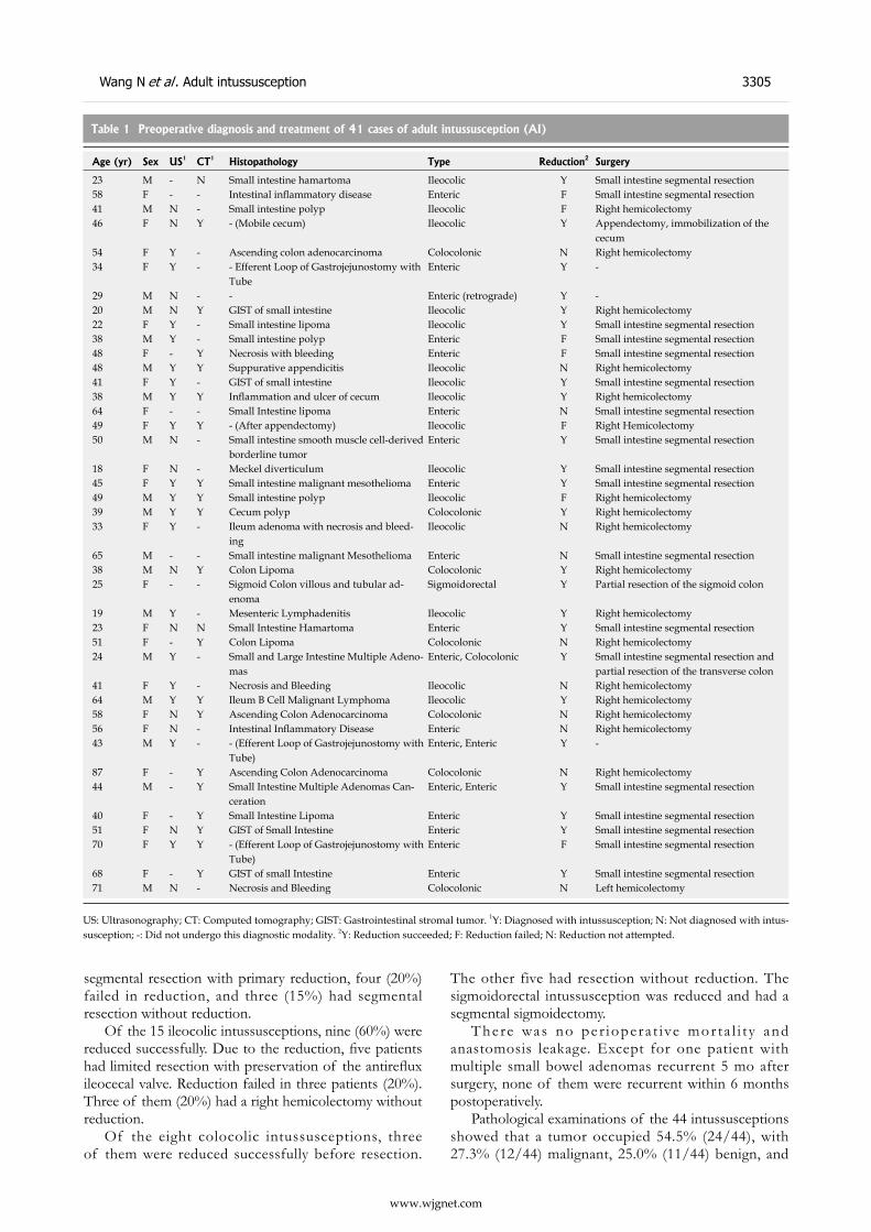
www.wjgnet.com
segmental resection with primary reduction, four (20%) failed in reduction, and three (15%) had segmental resection without reduction.
Of the 15 ileocolic intussusceptions, nine (60%) were reduced successfully. Due to the reduction, five patients had limited resection with preservation of the antireflux ileocecal valve. Reduction failed in three patients (20%). Three of them (20%) had a right hemicolectomy without reduction.
Of the eight colocolic intussusceptions, three of them were reduced successfully before resection.
The other five had resection without reduction. The sigmoidorectal intussusception was reduced and had a segmental sigmoidectomy.
There was no per iopera t ive mor ta l i t y and anastomosis leakage. Except for one patient with multiple small bowel adenomas recurrent 5 mo after surgery, none of them were recurrent within 6 months postoperatively.
Pathological examinations of the 44 intussusceptions showed that a tumor occupied 54.5% (24/44), with 27.3% (12/44) malignant, 25.0% (11/44) benign, and
Age (yr) Sex US1 CT1 Histopathology Type Reduction2 Surgery
23 M - N Small intestine hamartoma Ileocolic Y Small intestine segmental resection58 F - - Intestinal inflammatory disease Enteric F Small intestine segmental resection41 M N - Small intestine polyp Ileocolic F Right hemicolectomy46 F N Y - (Mobile cecum) Ileocolic Y Appendectomy, immobilization of the
cecum54 F Y - Ascending colon adenocarcinoma Colocolonic N Right hemicolectomy34 F Y - - Efferent Loop of Gastrojejunostomy with
TubeEnteric Y -
29 M N - - Enteric (retrograde) Y -20 M N Y GIST of small intestine Ileocolic Y Right hemicolectomy22 F Y - Small intestine lipoma Ileocolic Y Small intestine segmental resection38 M Y - Small intestine polyp Enteric F Small intestine segmental resection48 F - Y Necrosis with bleeding Enteric F Small intestine segmental resection48 M Y Y Suppurative appendicitis Ileocolic N Right hemicolectomy41 F Y - GIST of small intestine Ileocolic Y Small intestine segmental resection38 M Y Y Inflammation and ulcer of cecum Ileocolic Y Right hemicolectomy64 F - - Small Intestine lipoma Enteric N Small intestine segmental resection49 F Y Y - (After appendectomy) Ileocolic F Right Hemicolectomy50 M N - Small intestine smooth muscle cell-derived
borderline tumorEnteric Y Small intestine segmental resection
18 F N - Meckel diverticulum Ileocolic Y Small intestine segmental resection45 F Y Y Small intestine malignant mesothelioma Enteric Y Small intestine segmental resection49 M Y Y Small intestine polyp Ileocolic F Right hemicolectomy39 M Y Y Cecum polyp Colocolonic Y Right hemicolectomy33 F Y - Ileum adenoma with necrosis and bleed-
ingIleocolic N Right hemicolectomy
65 M - - Small intestine malignant Mesothelioma Enteric N Small intestine segmental resection38 M N Y Colon Lipoma Colocolonic Y Right hemicolectomy25 F - - Sigmoid Colon villous and tubular ad-
enomaSigmoidorectal Y Partial resection of the sigmoid colon
19 M Y - Mesenteric Lymphadenitis Ileocolic Y Right hemicolectomy23 F N N Small Intestine Hamartoma Enteric Y Small intestine segmental resection51 F - Y Colon Lipoma Colocolonic N Right hemicolectomy24 M Y - Small and Large Intestine Multiple Adeno-
masEnteric, Colocolonic Y Small intestine segmental resection and
partial resection of the transverse colon41 F Y - Necrosis and Bleeding Ileocolic N Right hemicolectomy64 M Y Y Ileum B Cell Malignant Lymphoma Ileocolic Y Right hemicolectomy58 F N Y Ascending Colon Adenocarcinoma Colocolonic N Right hemicolectomy56 F N - Intestinal Inflammatory Disease Enteric N Right hemicolectomy43 M Y - - (Efferent Loop of Gastrojejunostomy with
Tube)Enteric, Enteric Y -
87 F - Y Ascending Colon Adenocarcinoma Colocolonic N Right hemicolectomy44 M - Y Small Intestine Multiple Adenomas Can-
cerationEnteric, Enteric Y Small intestine segmental resection
40 F - Y Small Intestine Lipoma Enteric Y Small intestine segmental resection51 F N Y GIST of Small Intestine Enteric Y Small intestine segmental resection70 F Y Y - (Efferent Loop of Gastrojejunostomy with
Tube)Enteric F Small intestine segmental resection
68 F - Y GIST of small Intestine Enteric Y Small intestine segmental resection71 M N - Necrosis and Bleeding Colocolonic N Left hemicolectomy
Table 1 Preoperative diagnosis and treatment of 41 cases of adult intussusception (AI)
US: Ultrasonography; CT: Computed tomography; GIST: Gastrointestinal stromal tumor. 1Y: Diagnosed with intussusception; N: Not diagnosed with intus-susception; -: Did not undergo this diagnostic modality. 2Y: Reduction succeeded; F: Reduction failed; N: Reduction not attempted.
Wang N et al . Adult intussusception 3305

www.wjgnet.com
2.3% (1/44) borderline. 9.1% (4/44) were nontumorous polyps. 4.5% (2/44) were due to intestinal inflammatory disease. Meckel’s diverticulum and mobile cecum accounted for 4.5% (2/44). Apart from three cases (four intussusceptions), who had already undergone a gastrojejunostomy, no organic lesion was found in four intussusceptions by pathology or exploration, one of which one was the only retrograde intussusception (see Tables 1 and 3).
DISCUSSIONIntussusception is the leading cause of intestinal obstruction in children and ranks second only to
appendicitis as the most common cause of acute abdominal emergency in children. AI is distinct from pediatric intussusception in that it is rare, accounting for only 5% of all cases of intestinal obstructions, and about 90% of it is secondary. The exact mechanism is still unknown. However, it is believed that any lesion in the bowel wall or irritant within the lumen that alters normal peristaltic activity is able to initiate an invagination. Ingested food and subsequent peristaltic activity of the bowel produces an area of constriction above the stimulus and relaxation below, thus telescoping the lead point through the distal bowel lumen[1,3,4].
Similar to the results of Zubadi et al[1], enteric type intussusception is the most common type in our series. However, in the report of Goh et al[4] of 60 cases of AI, ileocolic (25%) and ileocecal-colic (13.3%) types were the most common. Their enteric type occupied 26.7%. Similar to our results, their colocolic and sigmoidorectal
Table 2 Coloscopy results for intussusceptions
Intussusception type Etiology Coloscopy diagnosis
Diagnosis of intussuscption Etiological diagnosis
Enteric Intestinal inflammatory disease No Intestinal inflammatory diseaseIleocolic - (After appendectomy) Yes -Ileocolic Ileum B cell malignant lymphoma Yes Occupying lesionColocolonic Colon lipoma No Occupying lesionColocolonic Colon lipoma Yes Occupying lesionColocolonic Ascending colon adenocarcinoma No AdenocarcinomaColocolonic Ascending colon adenocarcinoma No AdenocarcinomaSigmoidorectal Sigmoid colon villous and tubular adenoma Yes Villous and tubular adenoma
Enteric Ileocolic Colocolonic Sigmoidorectal Percentage
Tumor Malignant 6 3 3 0 27.3 (12/44)Borderline 1 0 0 0 2.3 (1/44)Benign 4 3 3 1 25.0 (11/44)
Nontumorous polyp 1 2 1 0 9.1 (4/44)Intestinal inflammatory disease 2 3 0 0 11.4 (5/44)Anatomy abnormality 0 2 0 0 4.5 (2/44)Iatrogenic 4 1 0 0 11.4 (5/44)Idiopathic 2 1 1 0 9.1 (4/44)Percentage 45.5 (20/44) 34.1 (15/44) 18.2 (8/44) 2.3 (1/44) 100.0 (44/44)
Table 3 Etiologies of the 44 AIs
Figure 4 An 87-year-old woman with a colocolonic intussusception due to ascending colon carcinoma.
Figure 3 Efferent loop intussusception with a tube. A 70-year-old woman underwent Billroth Ⅱ gastrectomy and efferent loop intubation for enteral nutri-tion. One month postoperatively, CT at the level of the lower abdomen shows a round, target-shaped mass in the left abdomen. The mass consists of a hy-perdense tube (arrow), a “half-moon” shaped hypodense area medial to it, the intussuscepted mesenteric fat and a soft tissue rim representing the opposing walls of the intussuscipiens and the intussusceptum.
10 cm
3306 ISSN 1007-9327 CN 14-1219/R World J Gastroenterol July 14, 2009 Volume 15 Number 26

www.wjgnet.com
types were the least common types.Most patients present with subacute (24.4%) or
chronic (51.2%) symptoms; therefore the characteristic pediatric presentation triad of abdominal pain, palpable abdominal mass and bloody stool was only seen in 9.8% of cases. It is one of the reasons why preoperative diagnosis is difficult.
Ultrasound is apt to be masked by gas-fil led loops of bowel, and most AIs present with intestinal obstruction[6]. The preoperative diagnosis accuracy (60.0%) of ultrasonography is not satisfying. However, the preoperative diagnosis accuracy of the 12 patients who had palpable abdominal mass was 91.7%, indicating that in cases of palpable abdominal mass, the diagnostic accuracy of ultrasonography would increase significantly.
Recently, with the signs of target or sausage, mesenteric fat and vessels, abdominal CT scan has been reported to be the most useful imaging technique, with a diagnostic accuracy is 58%-100%[3-5,7,8]. Recent studies have demonstrated the superiority of CT in revealing the site, level, and cause of intestinal obstructions and in demonstrating threatening signs of bowel nonviability[9,10]. As was shown in our study, the majority of AIs presented with partial or complete intestinal obstruction. Moreover, 90.5% (20/22) of AIs were diagnosed by CT in our series. One case of AI that was not diagnosed by CT, however, was correctly diagnosed as having an intestinal occupying lesion. In contrast to ultrasound, CT is not affected by the presence of gas in the bowel and will clearly demonstrate the intussusception, whether in the small bowel or in the colon. Additional valuable information, such as metastasis or lymphadenopathy, is readily obtained by CT and may point to an underlying pathology[5]. Therefore, we suggest that all patients presenting with an intestinal obstruction should have an abdominal CT scan as a regular diagnostic test.
Wang et al[3] considered that most lesions lead distally, can easily be seen by coloscopy, and intraoperative coloscopy might help to distinguish benign from malignant lesions before reduction of intussusceptions. Using the results of a coloscopy, limited surgical management by appendectomy, polypectomy or diverticulectomy might be performed in specific situations, resulting in an uncompromised bowel after reduction[3]. In our study, all lesions of the lead points of ileocolic, colocolic or sigmoidorectal intussusceptions were found in the seven patients who had a colonoscopy. Moreover, all the adenoma and adenocarcinomas were diagnosed pathologically through colonoscopy. However, some lesions, such as lymphoma and lipoma, which were not in the mucosa, were not diagnosed pathologically. Therefore, we think that the pathological diagnosis of most of the lead points of ileocolic, colocolic and sigmoidorectal intussusceptions, which locate in the mucosa, could be made by coloscopy. Perhaps some lesions, such as appendicitis and polyps, could be diagnosed by coloscopy to avoid undue surgery.
It is reported that 8%-20% of AIs are idiopathic and are more likely to occur in the small intestine[6]. In
our series, there were four [9.1% (4/44)] patients whose etiologies were not found by surgical exploration and/or pathology. One of them occurred in the colon (see Tables 1 and 3). Three of them had intestinal necrosis with bleeding. Segments of intestine had to be resected. The only retrograde intussusception found in our patients was cured by simple reduction.
Most AIs have underlying pathological lesions; therefore, most authors agree that laparotomy is mandatory. However, whether or not the intussusception shou ld be r educed be fore r e sec t ion r ema ins controversial. The theoretical objections to reductions are intraluminal seeding and venous dissemination of malignant cells, possible perforation during manipulation and increased risk of anastomotic complications in the face of edematous and inflamed bowel[1].
Although only 30% (6/20) of the etiologies of our enteric intussusceptions were malignant, borderline leiomyoma is potentially malignant. Therefore, reduction before resection would be more prudent. We suggest that if the underlying etiology and/or the lead point is suspected to be malignant, or if resected area required without reduction is not massive, an en bloc resection of the intussusception should be considered.
There were only three cases (37.5%) of colocolic intussusceptions caused by a malignant tumor-adenocarcinoma in our series, but most authors report malignant pathology presents in 69%-100%[3,4]. Only one case of ileocolic intussusception had a malignant organic lesion (terminal ileum B cell malignant lymphoma) in our series. Wang et al[3] reported 5/12 patients had malignant lesions in this type of intussusception. They think intraoperative colonoscopy might help to distinguish benign from malignant lesions before reduction. This technique can identify benign lesions of the ileum and be used to perform limited resection with preservation of the antireflux ileocecal valve. Moreover, appendectomy, polypectomy or diverticulectomy might be performed in specific situations to produce an uncompromised bowel after reduction, when definitively diagnosed by coloscopy[3]. Our coloscopies found all (7/7) of the lead point lesions and diagnosed all of the adenoma and adenocarcinomas of non-enteric intussusceptions. In our study, the lesions of appendicitis, benign tumors and polyps might have been diagnosed by coloscopy; organic lesion might have been excluded on the patient who had undergone appendectomy before exploration for intussusception. If coloscopy had been undertaken, unnecessary surgery could have been avoided. Therefore, we consider that in ileocolic, colocolic and sigmoidorectal intussusceptions, coloscopy is necessary, either preoperatively or intraoperatively.
The sigmoidorectal intussusception caused by villous and tubular adenoma of the sigmoid colon in our study was successfully reduced before partial resection of the sigmoid colon and the patient was spared a Miles operation. Zubaidi et al[1] considered that even if an intussusception of this type is secondary to a carcinoma, to avoid an abdominoperineal resection and a permanent colostomy, reduction should be performed first.
Wang N et al . Adult intussusception 3307

www.wjgnet.com
There were three patients in our study who underwent gastroje junostomy and nasoje junal intubation for enteral nutrition and had efferent loop intussusceptions. All of them were misdiagnosed as gastroplegia initially. The tube had been intubated together with a nasogastric tube into the stomach. During the operation, before completion of the gastrojejunostomy, a candy ball enveloped with a pertured surgery glove finger end, which was tied on the tip of the tube, was put into the efferent loop, then the candy ball was drawn from the outside of the efferent loop more than 40 cm distally to the anastomosis. Gayer et al[5] and Erkan et al[6] also consider intestinal intubation in the etiology of intussusception. However, they did not illuminate the mechanism of it. It is well known that the mucosal and seromuscular layers of the intestine connect loosely with each other. We think that when the candy ball on the tip of the tube is drawn distally, because of friction, the proximal mucosa will be pushed, folded and prolapsed distally. If the muscularis of mucosa is injured, then the distally pushed mucosa will not be able to recover by itself. As a result, the mucosa could prolapse distally and protrude into the intestinal lumen like a ring shaped tumor, and then an intussusception could occur.
In conclusion, AI is an infrequent problem. Most AIs present with subacute and chronic symptoms have intestinal obstructions to various extents. CT is the most effective and accurate diagnostic technique. In the case of a palpable abdominal mass, ultrasonography is also helpful for diagnosis. Enteric intussusception should be reduced when the underlying etiology is suspected to be benign or the resection is massive without reduction. Most lead point lesions of the ileocolic, colocolic or sigmoidorectal intussusceptions can be found by coloscopy. For these types of intussusceptions, coloscopy might provide information allowing the avoidance of unnecessary surgery. Intestinal intubation is the main cause of iatrogenic intussusception and should be avoided.
COMMENTSBackgroundIntussusception is uncommon in adults. It is estimated that only 5% of all intussusceptions occur in adults and approximately 5% of bowel obstructions in adults are the result of intussusception. Adult intussusception (AI) often presents with nonspecific symptoms. Preoperative diagnosis remains difficult. The extent of resection and whether the intussusception should be reduced remains controversial.Research frontiersRecent researches are focused on accurate preoperative diagnosis and proper treatment of it.Innovations and breakthroughsAccording to their data, the authors found computed tomography (CT) is the most effective and accurate diagnostic technique. In case of a palpable abdominal mass, ultrasonography is also helpful for diagnosis. Enteric intussusception should be reduced when the underlying etiology is suspected to be benign or the resection is massive without reduction. Most lead point lesions of the ileocolic, colocolic or sigmoidorectal intussusceptions can be found
by coloscopy. For these types of intussusception, coloscopy might provide information that allows avoidance of unnecessary surgery. Intestinal intubation is the main cause of iatrogenic intussusception and should be avoided. They also propose a hypothesis for the occurrence of this kind of intussusception.ApplicationsThe authors suggest that all patients presenting with an intestinal obstruction or the patients who are suspected intussusception, have an abdominal CT scan as a regular diagnostic test. In cases with a palpable abdominal mass, ultrasonography should be applied. If non-enteric intussusception is suspected, coloscopy might provide information allowing the avoidance of unnecessary surgery. Enteric intussusception should be reduced when the underlying etiology is suspected to be benign or the resection is massive without reduction. Intestinal intubation should be omitted unless very necessary or carried out very gently or with lubrication.TerminologyIntussusception: defined as the telescoping of a segment of the gastrointestinal tract into an adjacent one; enteric intussusception: an intussusception involved only jejunum or ileum is considered an enteric intussusception; ileocolic intussusception: an intussusception that involves the ileum and the colon is designated an ileocolic intussusception; colocolonic intussusception: an intussusception involving only the colon is considered a colocolonic intussusception; sigmoidorectal intussusception: involving the sigmoid colon and rectum; non-enteric intussusception: including ileocolic, colocolonic and sigmoidorectal intussusceptions. Acute symptoms: defined as < 4 d; Subacute symptoms: defined as 4-14 d. Chronic symptoms: defined as > 14 d. Antegrade intussusception: the proximal segment of the bowel telescoped into the lumen of the adjacent distal segment is defined as antegrade intussusception. Retrograde intussusception: the distal segment of the bowel telescoped into the lumen of the adjacent proximal segment is defined as retrograde intussusception.Peer reviewThe authors reviewed 41 cases of adult intussuception. The article is well written and worthy of publication.
REFERENCES1 Zubaidi A, Al-Saif F, Silverman R. Adult intussusception: a
retrospective review. Dis Colon Rectum 2006; 49: 1546-15512 Chand M , Bradford L, Nash GF. Intussusception in
colorectal cancer. Clin Colorectal Cancer 2008; 7: 204-2053 Wang LT , Wu CC, Yu JC, Hsiao CW, Hsu CC, Jao
SW. Clinical entity and treatment strategies for adult intussusceptions: 20 years' experience. Dis Colon Rectum 2007; 50: 1941-1949
4 Goh BK, Quah HM, Chow PK, Tan KY, Tay KH, Eu KW, Ooi LL, Wong WK. Predictive factors of malignancy in adults with intussusception. World J Surg 2006; 30: 1300-1304
5 Gayer G, Zissin R, Apter S, Papa M, Hertz M. Pictorial review: adult intussusception--a CT diagnosis. Br J Radiol 2002; 75: 185-190
6 Erkan N, Haciyanli M, Yildirim M, Sayhan H, Vardar E, Polat AF. Intussusception in adults: an unusual and challenging condition for surgeons. Int J Colorectal Dis 2005; 20: 452-456
7 Barussaud M, Regenet N, Briennon X, de Kerviler B, Pessaux P, Kohneh-Sharhi N, Lehur PA, Hamy A, Leborgne J, le Neel JC, Mirallie E. Clinical spectrum and surgical approach of adult intussusceptions: a multicentric study. Int J Colorectal Dis 2006; 21: 834-839
8 Tan KY, Tan SM, Tan AG, Chen CY, Chng HC, Hoe MN. Adult intussusception: experience in Singapore. ANZ J Surg 2003; 73: 1044-1047
9 Boudiaf M, Soyer P, Terem C, Pelage JP, Maissiat E, Rymer R. Ct evaluation of small bowel obstruction. Radiographics 2001; 21: 613-624
10 Beattie GC, Peters RT, Guy S, Mendelson RM. Computed tomography in the assessment of suspected large bowel obstruction. ANZ J Surg 2007; 77: 160-165
S- Editor Tian L L- Editor Stewart GJ E- Editor Yin DH
COMMENTS
3308 ISSN 1007-9327 CN 14-1219/R World J Gastroenterol July 14, 2009 Volume 15 Number 26

www.wjgnet.com
CASE REPORT
Unexplainable development of a hydatid cyst
Antonio Di Cataldo, Rosalia Latino, Aldo Cocuzza, Giovanni Li Destri
Online Submissions: wjg.wjgnet.com World J Gastroenterol 2009 July 14; 15(26): [email protected] World Journal of Gastroenterology ISSN 1007-9327doi:10.3748/wjg.15.3309 © 2009 The WJG Press and Baishideng. All rights reserved.
Antonio Di Cataldo, Rosalia Latino, Aldo Cocuzza, Giovanni Li Destri, Department of Digestive and Colorectal Surgery, University of Catania, Via S.Sofia 84, 95100 Catania, ItalyAuthor contributions: Di Cataldo A, Latino R, Cocuzza A and Li Destri G contributed equally to this work; Di Cataldo A and Li Destri G designed the research; Latino R and Cocuzza A performed the research; Li Destri G and Latino R analyzed the data; Di Cataldo A and Cocuzza A wrote the paper.Correspondence to: Antonio Di Cataldo, MD, Professor, FACS, Department of Digestive and Colorectal Surgery, University of Catania, Viale Odorico da Pordenone 5, 95128 Catania, Italy. [email protected]: +39-95-501106 Fax: +39-95-3782231Received: April 19, 2009 Revised: June 9, 2009Accepted: June 16, 2009Published online: July 14, 2009
AbstractEchinococcosis is a cyclozoonosis characterized by cystic lesions usually situated inside or outside the liver. We discuss the case of a 77-year-old woman with a recurrent hydatidosis with a cyst arising from the liver, growing through the lateral right abdomen wall, and reaching the subcutaneous tissue of the lumbar region. In the literature, rare subcutaneous or muscular localizations of hydatid cysts are described, however, there is no mention of a cyst growing over the abdominal wall muscles, shaped like an hourglass, partially in the liver and partially in the subcutaneous tissue, as in our case. We have not found any pathogenetic explanation for this growth pattern which is not typical of the biological behaviour of a hydatid cyst.
© 2009 The WJG Press and Baishideng. All rights reserved.
Key words: Echinococcosis; Growth; Hepatic; Hydatid cyst; Pathogenesis
Peer reviewers: Dr. Bijan Eghtesad, Associate Professor, Department of General Surgery, Cleveland Clinic Foundation, 9500 Euclid Avenue, Cleveland OH 44195, United States; Salvatore Gruttadauria, MD, Assistant Professor, Abdominal Transplant Surgery, ISMETT, Via E. Tricomi, 190127 Palermo, Italy
Di Cataldo A, Latino R, Cocuzza A, Li Destri G. Unexplainable development of a hydatid cyst. World J Gastroenterol 2009; 15(26): 3309-3311 Available from: URL: http://www.wjgnet.com/1007-9327/15/3309.asp DOI: http://dx.doi.org/10.3748/wjg.15.3309
INTRODUCTIONHydatid disease is a cyclozoonosis caused by Echinococcus granulosus or Echinococcus multilocularis, and is characterized by cystic lesions usually situated in the liver. These lesions can be asymptomatic for a long time, or cause an ache, however, there are no specific local or general symptoms and signs are related to size, location or complications of the cyst. The case of recurrent hydatidosis in a 77-year-old woman is discussed where the cyst, probably arising from the liver, grew through the lateral right abdomen wall and reached the subcutaneous tissue of the lumbar region.
CASE REPORTA 77-year-old woman was evaluated due to of the growth of an aching mass in the right lumbar region. Three years previously surgery was performed for hydatidosis of the mesentery. Objective examination revealed a painful swelling mass in the right lumbar region about 7-8 cm in diameter and mobile with respect to the upper and lower layers, which had distinct margins and a smooth surface (Figure 1). On palpating the abdomen the patient had pain in the right upper abdominal quadrant. CT of the abdomen showed a voluminous low-density mass (74 cm × 58 mm) in the low-external side of the Ⅵ liver segment. The mass was pedunculated, growing over the right posterior abdominal wall muscles and reaching the subcutaneous tissues (Figure 2).
Surgical treatment confirmed that the voluminous cyst was connected to the Ⅵ liver segment by a peduncle. Growing sideways and then behind, this mass reached the muscular layer arriving at the subcutaneous tissue of the right lumbar region. The mass was removed from the perihepatic side of the peduncle, 2-3 cm inside the liver, and from the abdominal external oblique muscle and the latissimus dorsi up to the subcutaneous tissue of the lumbar region (Figure 3). The post-operative period was problem-free, and the patient was discharged after 1 wk on anti-helminthic therapy. Histology confirmed the suspicion of hydatid cyst.
DISCUSSIONHydatid disease is caused by Echinococcus granulosus, an endemic infection in several mediterranean countries, or by Echinococcus multilocularis. Hydatid cysts can

www.wjgnet.com
be observed in all parts of the body, but are more frequent in the liver (50%-70%), lung (20%), and spleen (5%-8%)[1], while rare localizations are the kidneys, adrenal glands[2], spleen, soft tissues and brain[3].
Echinococcosis may be asymptomatic, detected only when imaging scans of the liver are carried out for different reasons, but it may cause pain or lead to complications. Cysts can rupture into the peritoneum, resulting in anaphylaxis or peritonitis[4], or into the biliary tract, causing cholangitis or cholestasis, and into the bronchi, causing bronchial fistula, or the cysts may become infected and form liver abscesses. Compression of the bile duct, portal vein, and hepatic veins can cause cholestasis, portal hypertension, and Budd-Chiari syndrome, respectively[5].
The diagnosis of hydatid cysts relies on epidemiologic data (geographical location and profession) clinical manifestations, serologic tests and radiologic imaging.
The treatment of choice is surgery. Chemotherapy (albendazole and mebendazole) is currently indicated in patients who cannot undergo major operations or for the prevention of secondary echinococcosis after surgery[6], but is contraindicated for large, superficially situated, or infected cysts, which are prone to rupture[7,8].
Our case shows some particular features that deserve closer examination. Usually the hydatid cyst grows in the liver in every direction, both inside the hepatic parenchyma and outside towards the peritoneal cavity. In our case, the cyst was connected to the liver by a peduncle 3-4 cm inside the liver. The surgeon who operated on our patient three years previously informed us of two hydatid cysts (diameter 9 cm and 12 cm) located in the mesocolon, which were connected by a peduncle, making it easy to remove them without spreading the disease. The hypothesis that the cyst we treated could have been a secondary echinococcosis due to spillage
Figure 1 Mass in the right lumbar region.
Figure 3 Pedunculated mass arising from the sixth liver segment and growing over the posterior abdominal wall muscles and reaching the subcutaneous tissue.
Figure 2 CT. The mass and the peduncle which connected it to the liver, as shown by the scheme with the surgical specimen.
3310 ISSN 1007-9327 CN 14-1219/R World J Gastroenterol July 14, 2009 Volume 15 Number 26

www.wjgnet.com
of cystic fluid and seeding during the first operation[5] has been denied. In the literature, rare subcutaneous or muscular localizations of hydatid cysts have been described[9], however, a cyst growing over the abdominal wall muscles, shaped like an hourglass, partially in the liver and partially in the subcutaneous tissue, as in our case has never been described. This growth pattern is not typical of the biological behaviour of a “benign tumor” such as a hydatid cyst, and seems to be more similar to the behaviour of an infiltrating malignant neoplasm. The only plausible explanation is that it could be related to seeding during the first surgery through drainage, however, the only drainage after the first surgery was located in the right lower abdominal quadrant, far away from the lumbar localization of the cyst we treated.
In the literature, Parmar et al [10] reported a case showing some similarities to our patient: a hepatic hydatid cyst which had ruptured into the subcutaneous tissue of the anterior abdominal wall . While the superficial and subcutaneous localization of the mass was similar, its connection with the right lobe of the liver was different. In Parmar’s case the right lobe of the liver was almost entirely occupied by the cyst, in our case the cyst was connected only by a peduncle. Parmar did not suggest any pathogenetic explanation, but just describes the kind of rupture that can involve a hydatid cyst.
In conclusion, we have not found any evidence in the literature for a pathogenetic explanation for the growth of the hydatid cyst over the abdominal wall muscles, shaped like an hourglass, partially in the liver and partially in the subcutaneous tissue. A similar growth pattern may occur in cysts found in the liver cupola where they can damage the diaphragm through inflammatory processes. The cysts then grow towards the pleural opening to the bronchial tubes, resulting in an uncommon and severe complication,
a hydatid bronchobiliary fistula. In this situation the pathogenetic mechanism is clearly caused by the infective process resulting in adhesions in the diaphragm. In our case no infective process was present, the cyst was connected to the liver by a peduncle which was easily removed from the transverse colon and from the muscles.
REFERENCES1 Iuliano L, Gurgo A, Polettini E, Gualdi G, De Marzio P.
Musculoskeletal and adipose tissue hydatidosis based on the iatrogenic spreading of cystic fluid during surgery: report of a case. Surg Today 2000; 30: 947-949
2 Di Cataldo A, Trombatore G, Greco R, Lanteri R, Li Destri G, Licata A. Hydatid disease in a very unusual location: the adrenal gland. A case report. Chir Ital 2003; 55: 275-278
3 Cöl C, Cöl M, Lafçi H. Unusual localizations of hydatid disease. Acta Med Austriaca 2003; 30: 61-64
4 Di Cataldo A , Lanteri R, Caniglia S, Santangelo M, Occhipinti R, Li Destri G. A rare complication of the hepatic hydatid cyst: intraperitoneal perforation without anaphylaxis. Int Surg 2005; 90: 42-44
5 Gourgiotis S, Stratopoulos C, Moustafellos P, Dimopoulos N, Papaxoinis G, Vougas V, Hadjiyannakis E. Surgical techniques and treatment for hepatic hydatid cysts. Surg Today 2007; 37: 389-395
6 Filippou D, Tselepis D, Filippou G, Papadopoulos V. Advances in liver echinococcosis: diagnosis and treatment. Clin Gastroenterol Hepatol 2007; 5: 152-159
7 Bekhti A, Schaaps JP, Capron M, Dessaint JP, Santoro F, Capron A. Treatment of hepatic hydatid disease with mebedazole: preliminary results in four cases. Br Med J 1977; 2: 1047-1051
8 Schantz PM. Effective medical treatment for hydatid disease? JAMA 1985; 253: 2095-2097
9 Latino R, Costa S, Barbagallo E, Virzì A, Vagnoni G. [Primary localization of a hydatid cyst in the major dorsal muscle: report of a case] Ann Ital Chir 1999; 70: 123-126; discussion 126-127
10 Parmar H, Nagarajan G, Supe A. Subcutaneous rupture of hepatic hydatid cyst. Scand J Infect Dis 2001; 33: 870-872
S- Editor Tian L L- Editor Webster JR E- Editor Lin YP
Di Cataldo A et al . Strange growth of a hydatid cyst 3311

www.wjgnet.com
CASE REPORT
Excision of a large abdominal wall lipoma improved bowel passage in a Proteus syndrome patient
Yoshifumi Nakayama, Shinichi Kusuda, Naoki Nagata, Koji Yamaguchi
Online Submissions: wjg.wjgnet.com World J Gastroenterol 2009 July 14; 15(26): [email protected] World Journal of Gastroenterology ISSN 1007-9327doi:10.3748/wjg.15.3312 © 2009 The WJG Press and Baishideng. All rights reserved.
Yoshifumi Nakayama, Koji Yamaguchi, Department of Surgery 1, School of Medicine, University of Occupational and Environmental Health, Kita-Kyushu 807-8555, JapanShinichi Kusuda, Department of Surgery, Moji Medical Center, Kita-Kyushu 801-8502, JapanNaoki Nagata, Department of Surgery, Kitakyushu General Hospital, Kita-Kyushu 800-0295, JapanAuthor contributions: Nakayama Y, Kusuda S, Nagata N and Yamaguchi K contributed equally to this work; Nakayama Y and Nagata N designed the research; Nakayama Y and Kusuda S performed and analyzed the data; Nakayama Y and Yamaguchi K wrote the paper.Correspondence to: Yoshifumi Nakayama, MD, PhD, Department of Surgery 1, University of Occupational Environmental Health, 1-1 Iseigaoka, Yahata-nishi-ku, Kita-kyushu 807-8555, Japan. [email protected]: +81-93-6917441 Fax: +81-93-6032361Received: March 25, 2009 Revised: June 3, 2009Accepted: June 10, 2009Published online: July 14, 2009
AbstractProteus syndrome is an extremely rare congenital disorder that produces multifocal overgrowth of tissue. This report presents a surgical case of a large lipoma in the abdominal wall of a patient with Proteus syndrome. She was diagnosed with Proteus syndrome based on certain diagnostic criteria. The neoplasm increased in size gradually, producing hemihypertrophy of her left lower extremity and trunk, and spread to her retroperitoneum and her left abdominal wall. She experienced gradually progressive constipation, nausea, vomiting, and abdominal pain. Computed tomography (CT) of the abdomen demonstrated a large mass in the subcutaneous adipose tissue of the left lower abdominal wall which measured 12 cm × 8 cm x 6 cm in diameter and encased the left colon. This mass in the abdominal wall was excised. The weight of the excised mass was 1550 g. The histopathological diagnosis of this mass was lipoma. After surgery, the encasement of the left colon was improved, and the patient was able to move her bowels twice per day. The excision of the large lipoma in the abdominal wall contributed to the improved bowel passage in this patient with Proteus syndrome.
© 2009 The WJG Press and Baishideng. All rights reserved.
Key words: Proteus syndrome; Lipoma; Bowel obstruction; Abdominal wall; Bowel passage
Peer reviewer: Werner Hohenberger, Professor, Chirurgische Klinik und Poliklinik, Krankenhausstrasse 12, Erlangen D-91054, Germany
Nakayama Y, Kusuda S, Nagata N, Yamaguchi K. Excision of a large abdominal wall lipoma improved bowel passage in a Proteus syndrome patient. World J Gastroenterol 2009; 15(26): 3312-3314 Available from: URL: http://www.wjgnet.com/1007-9327/15/3312.asp DOI: http://dx.doi.org/10.3748/wjg.15.3312
INTRODUCTIONProteus syndrome is an extremely rare disorder characterized by postnatal overgrowth of multiple tissues derived from different cell lineage[1-3]. The patient with Proteus syndrome described in this paper experienced constipation, nausea, vomiting, and abdominal pain due to the encasement and pressure of the intestinal tract by a large fatty mass which had developed in the left abdominal wall. The excision of this mass improved the symptoms.
CASE REPORTA 4-year-old female underwent a resection of a soft mass in her left inguinal area. This mass was pathologically diagnosed to be a lipoma. She was sent to the Department of Orthopedic Surgery because the lesion recurred and spread to the surrounding area. The recurrent tumors in her left inguinal area, left thigh and knee increased in size, and additional excisions were performed when the patient was 8- and 11-years-old. The pathological diagnosis was an infiltrating lipoma. She was diagnosed to have Proteus syndrome based on the diagnostic criteria reported[2-4]. The neoplasm increased in size gradually, and produced hemihypertrophy of the left lower extremity and trunk, and spread to the left abdominal wall and the retroperitoneum. Since the increasing abdominal mass encased and pressed the digestive organs, she gradually developed constipation. Bowel movements occurred about once a week at the time of presentation. A bowel obstruction caused symptoms including constipation, nausea, vomiting and abdominal pain. She had two healthy sisters and the rest of her family were also healthy. She had iron-deficient anemia with hypermenorrhea and she had a

Nakayama Y et al . A large abdominal wall lipoma in a Proteus syndrome patient 3313
www.wjgnet.com
transfusion history. On physical examination, she had a temperature of 36.4℃, and blood pressure of 120/70 mmHg. A large mass with unclear margins was found in the left lower quadrant of her abdominal wall (Figure 1). She showed hemihypertrophy and deformity of her left lower extremity. There were large and bizarre skin tumors on her left lower extremity.
Laboratory data showed a white blood cell count of 4800 cells/mm3, hemoglobin 7.7 g/dL, hematocrit 25%, platelets 321 000 cells/mm3, normal electrolytes and blood urea nitrogen, and the liver function was normal.
A plain abdominal X-ray examination indicated scoliosis and deformity of the pelvic bone. Colon gas in the left colon shifted to the right upper side (Figure 2). Computed tomog raphy (CT) of the abdomen demonstrated a large mass in the subcutaneous adipose tissue at the left lower abdominal wall which was 12 cm x 8 cm x 6 cm in diameter and encased the peritoneal organs to the right side (Figure 3). An upper gastrointestinal series revealed a mass encasing the stomach toward the upper side and the small intestine toward the right upper side (data not shown).
The mass in the abdominal wall was excised in 1997. The weight of the excised mass was 1550 g. A histopathological examination revealed a lobular arrangement of mature adipocytes (Figure 4), and these cells diffusely invaded into the skeletal muscle.
These findings indicated the diagnosis of a non-malignant lipoma.
The postoperative course was uneventful, the encasement of the left colon was improved, and she left the hospital on the 15th postoperative day. At present, she continues to receive medical treatment on an outpatient basis. Postoperatively, bowel movement occurred twice a day. A postoperative plain X-ray examination indicated that the shift of the left colon gas was improved (Figure 5). Postoperative CT also revealed reduced encasement of the peritoneal organs (Figure 6).
DISCUSSIONWiedermann et al[2] proposed diagnostic criteria for Proteus syndrome: (1) gigantism of the hands and/or feet, (2) pigmented nevus, (3) hemihypertrophy, (4) subcutaneous tumors, (5) skull anomalies, (6) accelerated growth and (7) visceral abnormalities. Recently, the main diagnostic criteria for Proteus syndrome are that it is sporadic, mosaic and has a progressive course[3]. The current case presented with lipomatosis, severe hemihypertrophy of the left trunk and left lower extremity, scoliosis, endometriosis and huge bizarre-shaped body tumors. Consequently, this patient was diagnosed to have Proteus syndrome based on the above-mentioned diagnostic criteria[5] and was the 6th
Figure 1 Clinical findings of the abdomen. There was a child-head sized mass at the left lower abdomen (arrows).
Figure 2 A preoperative plane abdominal X-ray examination indicated that colon gas in the left colon (arrows) was shifted to the upper right side.
RL
Figure 3 Preoperative computed tomography (CT) of the abdomen. A large mass in the subcutaneous adipose tissue in the left lower abdominal wall was identified (arrows) and this encased the peritoneal organs to the right side.
Figure 4 Histopathological findings of the excised mass. Lobular arrange-ment of mature adipocytes diffusely infiltrating into the skeletal muscle was observed.

www.wjgnet.com
Japanese case described in the English literature[5-9].This syndrome is caused by the germline loss of
functional mutations in PTEN[10] and is an overgrowth syndrome[11]. PTEN acts as a negative regulator of PI3-kinase signaling by catalyzing the dephosphorylation of PIP3[12,13]. This case was diagnosed based on the previous criteria for Proteus syndrome[2-4], and the germline mutation in PTEN was not examined.
Lipomatosis of the colon complicating Proteus syndrome was reported previously[14]. A 12 year-old boy with Proteus syndrome presented with gross abdominal distension and severe intractable constipation. Magnetic resonance imaging showed adipose tissue widely spread throughout the abdomen and pelvis. A laparotomy revealed a huge infiltrating lipomatosis encasing the left colon, including the rectum. On the other hand, the symptoms of bowel obstruction in the current case may have been caused by the encasement and pressure of the intestinal tract by a child-head sized lipoma that developed in the left abdominal wall. This lipoma was excised from the abdominal wall and removal of this huge lipoma produced the improvement in the patient’s symptoms.
Proteus syndrome is a complex disorder associated with varied, disproportionate, asymmetric overgrowth of many body parts and unregulated adipose tissue. The overgrowth seen in Proteus syndrome is progressive and difficult to manage. Patients with Proteus syndrome require repeated treatment for the progressive overgrowth of tissue over a long period. Aggressive treatment may cause severe functional and cosmetic consequences, so surgical intervention is often delayed until it is absolutely necessary[15]. In the current case, other sites of the lipomatosis, such as the left extremity and back, were excised when it affected the patient’s quality of life. This report presented an extremely rare case of bowel obstruction due to a large lipoma in the abdominal wall. The excision of this large mass improved the bowel passage in a patient with Proteus syndrome.
REFERENCES1 Cohen MM Jr , Hayden PW. A newly recognized
hamartomatous syndrome. Birth Defects Orig Artic Ser 1979; 15: 291-296
2 Wiedemann HR , Burgio GR, Aldenhoff P, Kunze J, Kaufmann HJ, Schirg E. The proteus syndrome. Partial gigantism of the hands and/or feet, nevi, hemihypertrophy, subcutaneous tumors, macrocephaly or other skull anomalies and possible accelerated growth and visceral affections. Eur J Pediatr 1983; 140: 5-12
3 Biesecker LG, Happle R, Mulliken JB, Weksberg R, Graham JM Jr, Viljoen DL, Cohen MM Jr. Proteus syndrome: diagnostic criteria, differential diagnosis, and patient evaluation. Am J Med Genet 1999; 84: 389-395
4 Hotamisligil GS. Proteus syndrome and hamartoses with overgrowth. Dysmorphol Clin Genet 1990; 4: 87
5 Yasuda H, Yamamoto O, Hirokawa H, Asahi M, Kashimura M, Sakai A. Proteus syndrome. Dermatology 2001; 203: 180-184
6 Hagari Y , Aso M, Shimao S, Okano T, Kurimasa A, Takeshita K. Proteus syndrome: report of the first Japanese case with special reference to differentiation from Klippel-Trenaunay-Weber syndrome. J Dermatol 1992; 19: 477-480
7 Katayama M, Imafuku S, Imayama S, Nakayama J, Hori Y. Two cases of Proteus syndrome characterized by verrucous skin lesions on the soles (abstract in English). Nishinihon J Dermatol 1992; 54: 1067
8 Miura H, Uchida Y, Ihara K, Sugioka Y. Macrodactyly in Proteus syndrome. J Hand Surg Br 1993; 18: 308-309
9 Sayama K, Hato N, Matsuda O, Shiraishi S, Miki Y. Proteus syndrome. Dermatology 1994; 189: 392-395
10 Zhou X, Hampel H, Thiele H, Gorlin RJ, Hennekam RC, Parisi M, Winter RM, Eng C. Association of germline mutation in the PTEN tumour suppressor gene and Proteus and Proteus-like syndromes. Lancet 2001; 358: 210-211
11 Barker KT , Houlston RS. Overgrowth syndromes: is dysfunctional PI3-kinase signalling a unifying mechanism? Eur J Hum Genet 2003; 11: 665-670
12 Maehama T, Dixon JE. The tumor suppressor, PTEN/MMAC1, dephosphorylates the lipid second messenger, phosphatidylinositol 3,4,5-trisphosphate. J Biol Chem 1998; 273: 13375-13378
13 Wu X, Senechal K, Neshat MS, Whang YE, Sawyers CL. The PTEN/MMAC1 tumor suppressor phosphatase functions as a negative regulator of the phosphoinositide 3-kinase/Akt pathway. Proc Natl Acad Sci USA 1998; 95: 15587-15591
14 Mackay G, Spitz L, McHugh K. Lipomatosis of the colon complicating Proteus syndrome. Arch Dis Child 2002; 86: 265
15 Biesecker L. The challenges of Proteus syndrome: diagnosis and management. Eur J Hum Genet 2006; 14: 1151-1157
S- Editor Li LF L- Editor O’Neill M E- Editor Yin DH
Figure 5 A postoperative plane abdominal X-ray examination indicated that the shift of the left colon gas (arrows) was improved as compared to the preoperative state.
RL
Figure 6 Postoperative CT of the abdomen. The encasement of the perito-neal organs was improved as compared to the preoperative state (arrows).
3314 ISSN 1007-9327 CN 14-1219/R World J Gastroenterol July 14, 2009 Volume 15 Number 26

www.wjgnet.com
CASE REPORT
Granular cell tumor of the cecum with extensive hyalinization and calcification: A case report
Ran Hong, Sung-Chul Lim
Online Submissions: wjg.wjgnet.com World J Gastroenterol 2009 July 14; 15(26): [email protected] World Journal of Gastroenterology ISSN 1007-9327doi:10.3748/wjg.15.3315 © 2009 The WJG Press and Baishideng. All rights reserved.
Ran Hong, Sung-Chul Lim, Department of Pathology, Chosun University School of Medicine, Gwangju 501-140, South KoreaSung-Chul Lim, Research Center for Resistant Cells, Chosun University School of Medicine, Gwangju 501-140, South KoreaAuthor contributions: Lim SC designed research; Hong R and Lim SC performed research and analyzed data; Hong R wrote the paper.Supported by Research funds from Chosun University, 2009Correspondence to: Sung-Chul Lim, MD, PhD, Department of Pathology Chosun University Hospital, 588, Seosuk-dong, Dong-gu, Gwangju 501-140, South Korea. [email protected]: +82-62-2306343 Fax: +82-62-2265860 Received: May 12, 2009 Revised: June 11, 2009Accepted: June 18, 2009 Published online: July 14, 2009
AbstractA granular cell tumor (GCT) is a benign neoplasm of unclear histogenesis that is generally believed to be of nerve sheath origin. GCT is not common and most often affects the tongue, skin and soft tissue, although it may occur anywhere in the body. We experienced a case of GCT that arose in the cecum of a 55-year-old man. The GCT was removed by laparoscopic resection. In addition to the tumor, endoscopic examination revealed the presence of a 5-mm-polyp in the descending colon and multiple tiny polyps in the sigmoid colon and rectum. Histological examination demonstrated a cecal tumor 1.5 cm × 1.0 cm × 0.7 cm with a hard consistency; in cut sections, mixed cells with yellowish and whitish portions were seen. The tumor was located between the mucosa and subserosa, and was composed of plump histiocyte-like tumor cells with abundant granular eosinophilic cytoplasm, which were immunoreactive for S-100 protein, vimentin, neuron-specific enolase, inhibin-a and calretinin. The tumor showed extensive hyal inization and focal dystrophic calcification. Immunohistochemical profiles did not confirm any particular cell type for the histogenetic origin of the GCT, including a nerve sheath origin. Extensive hyalinization and calcification showing involution of tumor cells suggest benign clinical behavior of GCT.
© 2009 The WJG Press and Baishideng. All rights reserved.
Key words: Granular cell tumor; Cecum; Histogenesis; Calcification
Peer reviewer: Qin Su, Professor, Department of Pathology, Cancer Hospital and Cancer Institute, Chinese Academy of Medical Sciences and Peking Medical College, PO Box 2258, Beijing 100021, China
Hong R, Lim SC. Granular cell tumor of the cecum with extensive hyalinization and calcification: A case report. World J Gastroenterol 2009; 15(26): 3315-3318 Available from: URL: http://www.wjgnet.com/1007-9327/15/3315.asp DOI: http://dx.doi.org/10.3748/wjg.15.3315
INTRODUCTIONGranular cell tumor (GCT) is a benign tumor with unknown histogenesis that is characterized by large, granular eosinophilic cells[1]. The tumor was first described by Abrikossoff[2] in 1926 as a muscle tumor; yet, a close association with nerves and immunohistochemical characteristics have identified GCT as a neural lesion[1]. A wide variety of cell types have been proposed as the cells of origin, including histiocytes, fibroblasts, myoblasts, neural sheath cells, neuroendocrine cells and undifferentiated mesenchymal cells[3-5]. Tumor cells have been shown recently to have positive expression of a number of new markers of neural differentiation, as well as several non-neural markers[6]. Vered et al[7] have proposed the possibility of GCT as a reactive lesion rather than as a true neoplasm. In the present case, extensive hyalinization and calcification within the GCT support this proposal. GCT is not common in the gastrointestinal tract, where the most common site for the tumor is the esophagus, followed by the duodenum, anus and stomach[8,9]. A few cases of GCT have been reported in the cecum[10,11]. In this report, we present a case of GCT of the cecum with extensive hyalinization and focal dystrophic calcification, accompanied by multiple colonic polyps, including tubular adenoma. The immunoreactivity of granular cells based on the use of a broad panel of antibodies did not confirm any particular cell type for the histogenesis of GCT.
CASE REPORTA 56-year-old man was admitted to the Gastroenterology Department of Chosun University Hospital with a 2-mo history of abdominal pain and diarrhea. Colonoscopy

www.wjgnet.com
revealed a protruding mass approximately 2 cm in diameter, with central umbilication in the cecum (Figure 1), as well as a 5-mm polyp and tiny sized multiple polyps in the descending colon and rectum, respectively.
A polyp in the descending colon was removed by snare polypectomy, and the polyp was diagnosed histologically as tubular adenoma. The cecal mass was resected by laparoscopic myotomy. Grossly, the mass was 1.5 cm × 1.0 cm × 0.7 cm, yellowish-white in color, and hardly palpable. Microscopically, the well-circumscribed mass was located mainly between the submucosa and subserosa, with partial involvement of the mucosa (Figure 2A). The mass was composed of round to polygonal cells with abundant granular eosinophilic cytoplasm. Characteristically, much of the tumor showed extensive hyalinization and focal dystrophic calcification (Figure 2B and C). There was no tumor necrosis, spindling and vesicular nuclei with large nucleoli, increased mitotic activity, a high nuclear to cytoplasmic ratio, and pleomorphism. The tumor was seen as a small, well-circumscribed mass, but with no infiltrative growth pattern; findings that favor a benign rather than a malignant lesion. Immunohistochemical analysis demonstrated that the tumor cells were reactive for S-100 protein (Figure 2D), neuron-specific enolase (NSE), vimentin, calretinin and inhibin (Figure 3). The tumor cells were non-reactive for smooth muscle actin (SMA), c-kit (CD117) and CD34. Based on the morphological and immunohistochemical findings, the resected tumor was diagnosed as a benign GCT that occurred in the cecum.
DISCUSSIONGCT is diagnosed only rarely, based on macroscopic and endoscopic examinations, as a result of its small size and shape that resemble a diminutive polyp[12]. Recently, endoscopic ultrasonography (EUS) has been used more frequently to determine the depth of tumor invasion in the gastrointestinal wall, and, it is useful for evaluating gastrointestinal tract submucosal tumors[13]. However, EUS cannot sufficiently distinguish a benign submucosal tumor from other tumors such as malignant neoplasia[14].
The final diagnosis of GCT is dependent on the pathological findings. The histological markers for GCT are plump histiocyte-like, bland-looking neoplastic cells
with abundant granular eosinophilic cytoplasm, which contains acidophilic, periodic acid-Schiff-positive, diastase-resistant granules. The cells contain small, uniform nuclei where mitotic figures are absent and neural markers, including S-100 protein or NSE, are expressed uniformly[15,16].
The histogenesis of GCT has remained enigmatic in spite of a vast number of immunohistochemical and ultrastructural studies[7]. Neural origin or differentiation, in particular of the Schwann cell type, is currently in favor. However, recent findings have cast doubt on the neural origin of these tumors[7]. Vered et al[7] have suggested that immunoreactivity of the granular cells to a broad panel of antibodies including S-100 protein, CD68, vimentin, calretinin, NKI/C3, protein gene product 9.5, nerve growth factor receptor and inhibin-a that characterize different tissue do not confirm any particular cell type for the histogenesis of GCT. In the present case, tumor cells were reactive for S-100 protein, NSE, calretinin, vimentin, and inhibin-a, which agrees with a previous study[7].
In most cases of colonic GCT, the tumor was less than 2 cm and well separated from the muscularis propria[17]. Since this tumor is considered as benign, and no patients with recurrence or metastasis have been documented, it is usually accepted that endoscopic tumor excision may be the best treatment for GCT in the gastrointestinal tract[14]. However, Nakachi et al[14] have suggested that GCT in the gastrointestinal tract is usually small and asymptomatic, and the tumor tends to be found incidentally during endoscopy performed for other reasons. Observation of these GCTs with the use of endoscopy and EUS is indicated unless the patient is symptomatic, or the tumor is larger than 2 cm or demonstrates atypical EUS or histological features. In the present case, there was extensive hyalinization and focal dystrophic calcification of tumor cells, which indicated a long-standing tumor with no atypical changes, which supports the above description. Vered et al[7] have advocated that GCT can be regarded as a lesion that reflects local metabolic or reactive changes rather than a true neoplasm. In the large series of GCTs evaluated by Vered et al[7], lesions displayed three main architectural patterns, including small and well-circumscribed nodules, larger and poorly circumscribed lesions, and an impressive infiltrative pattern with remote satellite nodules. The pattern of small and well-circumscribed nodules may represent benign mesenchymal tumors that have undergone extensive or complete granular cell change, and the remaining patterns may be compatible with a diffuse process of metabolically induced cytoplasmic granular changes in mesenchymal cells. Most lesions with an infiltrative pattern and positive margin almost never recur, whereas for a definite tumor, recurrent lesions are expected. It has been suggested that the lesions may be metabolic or reactive in nature and not neoplastic. The present case displayed extensive hyalinization and calcification in between granular cells, which suggests a long duration; findings that favor GCT with reactive changes, or a true neoplasm.
Figure 1 Endoscopic examination revealed an approximately 2-cm-sized protruding mass in the ce-cum.
3316 ISSN 1007-9327 CN 14-1219/R World J Gastroenterol July 14, 2009 Volume 15 Number 26

www.wjgnet.com
Several GCTs with adjacent benign or malignant tumors have been reported previously. In 2006, Eriksen et al[18] reported the first case of a synchronic adenoma
A B
C D
Figure 3 Tumor cells of the granular cell tumor were immunoreactive for inhibin-a (A), calretinin (B), vimentin (C) and NSE (D).
and GCT, and Sarsik et al[19] also observed a tubular adenoma in the vicinity of a GCT. Caltabiano et al[20] reported a GCT covered by squamous cell carcinoma
Hong R et al . Cecal granular cell tumor 3317
Figure 2 Microscopically and immunohistochemically, the appearance of the tumor was compatible with that of granular cell tumor. A: The tumor was located mainly between the submucosa and the subserosa. Much of the tumor showed extensive hyalinization (star) and dystrophic calcification. B and C: A high mag-nification view (square of A) showed dystrophic calcification, hyalinization and some viable tumor cell nests in the peripheral portion of the tumor. The tumor cells were composed of round to polygonal cells with abundant granular eosinophilic cytoplasm. D: Immunohistochemically, tumor cells were reactive for S-100 protein.
A B
C D

www.wjgnet.com
of the tongue. The present case also had adjacent multiple polyps, including tubular adenoma. Based on these findings, it is suggested that GCT shows reactive granular cell changes in the process of spontaneous regression of a preceding tumor. The coincidence of the adjacent tumor can be regarded as favoring a non-neoplastic or reactive process. However, to date, there is no evidence of any association or disposing factors between GCT in the colon and colonic adenoma or other malignancy[18].
In summary, we experienced a 1.5 cm × 1.0 cm × 0.7 cm cecal GCT that showed extensive hyalinization and focal dystrophic calcification with synchronous tubular adenoma in the descending colon. Immunohistochemical profiles did not confirm any particular cell type for the histogenesis of GCT. Endoscopists and pathologists should consider the possibility of this tumor in the gastrointestinal tract. When a patient is asymptomatic, and unless the tumor is larger than 2 cm or shows atypical features, observation of this tumor with the use of endoscopy and EUS is indicated.
REFERENCES1 Weiss SW, Goldblum JR. Enzinger and Weiss’s soft tissue
tumors. St Louis: Mosby Inc., 2008: 878-8882 Abrikossoff A . Über myome, ausgehened von der
quergestreiften willkürlichen muskulatur. Virchows Arch (Pathol Anat) 1926; 260: 215-223
3 Stewart CM, Watson RE, Eversole LR, Fischlschweiger W, Leider AS. Oral granular cell tumors: a clinicopathologic and immunocytochemical study. Oral Surg Oral Med Oral Pathol 1988; 65: 427-435
4 Kaiserling E, Ruck P, Xiao JC. Congenital epulis and granular cell tumor: a histologic and immunohistochemical study. Oral Surg Oral Med Oral Pathol Oral Radiol Endod 1995; 80: 687-697
5 Williams HK, Williams DM. Oral granular cell tumours: a histological and immunocytochemical study. J Oral Pathol Med 1997; 26: 164-169
6 Murakata LA, Ishak KG. Expression of inhibin-alpha by granular cell tumors of the gallbladder and extrahepatic bile ducts. Am J Surg Pathol 2001; 25: 1200-1203
7 Vered M, Carpenter WM, Buchner A. Granular cell tumor
of the oral cavity: updated immunohistochemical profile. J Oral Pathol Med 2009; 38: 150-159
8 Melo CR, Melo IS, Schmitt FC, Fagundes R, Amendola D. Multicentric granular cell tumor of the colon: report of a patient with 52 tumors. Am J Gastroenterol 1993; 88: 1785-1787
9 Yamaguchi K, Maeda S, Kitamura K. Granular cell tumor of the stomach coincident with two early gastric carcinomas. Am J Gastroenterol 1989; 84: 656-659
10 Ghazi A. Combined granular cell tumor of the stomach and cecum. Mt Sinai J Med 1979; 46: 195-198
11 Hamajima E, Suzuki T, Yoshioka N, Ogawa Y, Tanikawa M, Nakamura S, Yoshikawa T, Yokoi T, Yoshikane H. [A case of granular cell tumor of cecum] Nippon Shokakibyo Gakkai Zasshi 1997; 94: 27-32
12 Lack EE , Worsham GF, Callihan MD, Crawford BE, Klappenbach S, Rowden G, Chun B. Granular cell tumor: a clinicopathologic study of 110 patients. J Surg Oncol 1980; 13: 301-316
13 Orlowska J , Pachlewski J, Gugulski A, Butruk E. A conservative approach to granular cell tumors of the esophagus: four case reports and literature review. Am J Gastroenterol 1993; 88: 311-315
14 Nakachi A, Miyazato H, Oshiro T, Shimoji H, Shiraishi M, Muto Y. Granular cell tumor of the rectum: a case report and review of the literature. J Gastroenterol 2000; 35: 631-634
15 Endo S, Hirasaki S, Doi T, Endo H, Nishina T, Moriwaki T, Nakauchi M, Masumoto T, Tanimizu M, Hyodo I. Granular cell tumor occurring in the sigmoid colon treated by endoscopic mucosal resection using a transparent cap (EMR-C). J Gastroenterol 2003; 38: 385-389
16 Lisato L, Bianchini E, Reale D. [Granular cell tumor of the rectum: description of a case with unusual histological features] Pathologica 1995; 87: 175-178
17 Sohn DK, Choi HS, Chang YS, Huh JM, Kim DH, Kim DY, Kim YH, Chang HJ, Jung KH, Jeong SY. Granular cell tumor of colon: report of a case and review of literature. World J Gastroenterol 2004; 10: 2452-2454
18 Eriksen JR, Ibsen PH, Gyrtrup HJ. [Granular cell tumor of the colon--Abrikossoff's tumor] Ugeskr Laeger 2006; 168: 2080-2081
19 Sarsik B, Doğanavşargil B, Ozkök EE, Aydin A, Tunçyürek M. Granular cell tumor of colon. Turk J Gastroenterol 2008; 19: 73-74
20 Caltabiano R, Cappellani A, Di Vita M, Lanzafame S. The unique simultaneous occurrence of a squamous cell carcinoma and a granular cell tumor of the tongue at the same site: a histological and immunohistochemical study. J Craniofac Surg 2008; 19: 1691-1694
S- Editor Tian L L- Editor Kerr C E- Editor Yin DH
3318 ISSN 1007-9327 CN 14-1219/R World J Gastroenterol July 14, 2009 Volume 15 Number 26

Jung Wook Huh, Sang Hyuk Cho, Hyeong Rok Kim, Department of Surgery, Chonnam National University Hwasun Hospital and Medical School, Gwangju 501-757, South KoreaJae Hyuk Lee, Department of Pathology, Chonnam National University Hwasun Hospital and Medical School, Gwangju 501-757, South KoreaAuthor contributions: Huh JW wrote the paper; Kim HR performed the operation; All authors contributed to the intellectual content and approved the final version.Correspondence to: Hyeong Rok Kim, MD, PhD, Department of Surgery, Chonnam National University Hwasun Hospital and Medical School, Gwangju 501-757, South Korea. [email protected]: +82-61-3797643 Fax: +82-61-3797661Received: April 2, 2009 Revised: June 4, 2009Accepted: June 11, 2009Published online: July 14, 2009
AbstractA cavernous hemangioma of the cecum is a rare vascular malformation but is clinically important because of the possibility of massive bleeding. We report a case of a large cavernous hemangioma with pericolic infiltration in the cecum which was removed successfully using minimally invasive surgery.
© 2009 The WJG Press and Baishideng. All rights reserved.
Key words: Cavernous hemangioma; Cecum; Laparoscopic surgery
Peer reviewers: Zvi Fireman, MD, Associate Professor of Medicine, Head, Gastroenterology Department, Hillel Yaffe Medical Center, POB 169, 38100, Hadera, Israel; Rafiq A Sheikh, MBBS, MD, MRCP, FACP, FACG, Department of Gastroenterology, Kaiser Permanente Medical Center, 6600 Bruceville Road, Sacramento, CA 95823, United States
Huh JW, Cho SH, Lee JH, Kim HR. Large cavernous hemangioma in the cecum treated by laparoscopic ileocecal resection. World J Gastroenterol 2009; 15(26): 3319-3321 Available from: URL: http://www.wjgnet.com/10079327/15/3319.asp DOI: http://dx.doi.org/10.3748/wjg.15.3319
INTRODUCTIONHemangiomas of the large intestine are relatively
uncommon benign tumors which can occur anywhere in the gastrointestinal tract[1]. These are clinically important vascular malformations because of the possibility of massive bleeding. Although a variety of histological and clinical types of hemangioma exist, the cavernous and capillary subtypes are most commonly encountered. Recent advances in colonoscopic techniques have led to successful endoscopic resection in selected cases[2-4], but most large lesions have been treated surgically[5,6]. We report the case of a large cavernous hemangioma in the cecum with pericolic infiltration which was removed successfully using minimally invasive surgery.
CASE REPORTA 37-year-old woman was referred to her local hospital because of intermittent rectal bleeding. Colonoscopy revealed a blue polypoid lesion with a nodular surface in the cecum (Figure 1). A hemangioma was suspected. She was transferred to our hospital for further evaluation and surgical treatment.
The physical examination revealed no specific signs. Abdominopelvic computed tomography (CT) showed a lesion in the cecum protruding extraluminally with heterogenous enhancement (Figure 2). The operative findings suggested a tumor involving the cecum and extending into the pericolic fat. Laparoscopic-assisted ileocecal resection and side-to-end ileo-ascending colon anastomosis were carried out. Macroscopically, the surgical specimen was a 5.8 cm × 4.2 cm cavernous hemangioma involving the entire wall of the colon and extending into the pericolic fat (Figure 3). Microscopically, the tumor consisted of large, dilated, blood-filled vessels lined by flattened endothelium. The vascular walls were thickened focally by adventitial fibrosis (Figure 4). Postoperatively, the patient has recovered well with no recurrence of symptoms.
DISCUSSIONHemangiomas are rare lesions of the colon, but vascular malformations of the gastrointestinal tract have been reported since 1839[7]. According to the literature, these lesions originate from embryonic sequestrations of mesodermal tissue[8]. Histologically, hemangiomas are distinct from telangiectasias and angiodysplasias, and most colonic hemangiomas are the capillary or
Online Submissions: wjg.wjgnet.com World J Gastroenterol 2009 July 14; 15(26): [email protected] World Journal of Gastroenterology ISSN 1007-9327doi:10.3748/wjg.15.3319 © 2009 The WJG Press and Baishideng. All rights reserved.
Large cavernous hemangioma in the cecum treated by laparoscopic ileocecal resection
Jung Wook Huh, Sang Hyuk Cho, Jae Hyuk Lee, Hyeong Rok Kim
CASE REPORT
www.wjgnet.com

100 mm
cavernous subtype. Capillary hemangiomas consist of a proliferation of small capillaries composed of thin-walled spaces lined by endothelial cells, while cavernous hemangiomas consist of large spaces lined by single or multiple layers of endothelial cells[9]. The capillary subtype is usually solitary and causes no symptoms, while the cavernous subtype presents with bleeding (60%-90%), anemia (43%), obstruction (17%), and, rarely, with platelet sequestration, although approximately 10% of patients remain asymptomatic[10]. The endoscopic findings of hemangiomas of the colon vary. Grossly, hemangiomas appear as soft, compressible bluish or deep red submucosal lesions, with dilated, engorged veins in the rectal wall[5]. However, they can be difficult to diagnose in some cases, especially if the hemangioma has an unusual color or is covered. A histological diagnosis before treatment may be difficult because of the risk of uncontrollable bleeding following a biopsy[1,4]. Hence, these lesions should not be biopsied. Abdominal CT can provide useful information about the size of the lesion and its extension to adjacent organs. Visceral angiography may also be useful in excluding coexisting hemangiomas at other sites in the gastrointestinal tract[5].
Complete surgical resection is the best treatment for large or diffuse lesions[5], although endoscopic resection has sometimes been recommended for hemangiomas that are pedunculated, polypoid, small, and limited
to the submucosal layer according to endoscopic ultrasonography[4]. Polypoid hemangiomas sometimes involve the entire wall of the colon, extending through the mesocolon and mesentery, as in our case[5]. In summary, we performed laparoscopic resection of a large cavernous hemangioma in the cecum. Although these lesions are rare, a better understanding of these lesions should help to obtain a definite diagnosis and devise an appropriate treatment plan.
REFERENCES1 Pontecorvo C, Lombardi S, Mottola L, Donisi M, DiTuoro
A. Hemangiomas of the large bowel. Report of a case. Dis Colon Rectum 1983; 26: 818-820
www.wjgnet.com
Figure 1 Colonoscopy of the cecum showing a bluish nodular lesion. Figure 2 Abdominopelvic computed tomography showing a lesion in the cecum protruding extraluminally with heterogenous enhancement.
Figure 3 Macroscopically, the surgical specimen was a cavernous hemangioma.
A
Figure 4 HE staining of the hemangioma. A: Histological section showing large, dilated, blood-filled vessels lined by flattened endothelium (HE, × 1); B: The vascular walls were thickened focally by adventitial fibrosis (HE, × 100).
B
3320 ISSN 10079327 CN 141219/R World J Gastroenterol July 14, 2009 Volume 15 Number 26
2 van Deursen CT, Buijs J, Nap M. An uncommon polyp in the colon: a pedunculated cavernous hemangioma. Endoscopy 2008; 40 Suppl 2: E127
3 Kimura S, Tanaka S, Kusunoki H, Kitadai Y, Sumii M, Tazuma S, Yoshihara M, Haruma K, Chayama K. Cavernous hemangioma in the ascending colon treated by endoscopic mucosal resection. J Gastroenterol Hepatol 2007; 22: 280-281
4 Hasegawa K, Lee WY, Noguchi T, Yaguchi T, Sasaki H, Nagasako K. Colonoscopic removal of hemangiomas. Dis Colon Rectum 1981; 24: 85-89
5 Sylla P, Deutsch G, Luo J, Recavarren C, Kim S, Heimann TM, Steinhagen RM. Cavernous, arteriovenous, and mixed hemangioma-lymphangioma of the rectosigmoid: rare causes of rectal bleeding--case series and review of the literature. Int J Colorectal Dis 2008; 23: 653-658
6 Marinis A, Kairi E, Theodosopoulos T, Kondi-Pafiti A, Smyrniotis V. Right colon and liver hemangiomatosis: a case report and a review of the literature. World J Gastroenterol 2006; 12: 6405-6407
7 Head HD, Baker JQ, Muir RW. Hemangioma of the colon. Am J Surg 1973; 126: 691-694
8 Lyon DT, Mantia AG. Large-bowel hemangiomas. Dis Colon Rectum 1984; 27: 404-414
9 Levy AD, Abbott RM, Rohrmann CA Jr, Frazier AA, Kende A. Gastrointestinal hemangiomas: imaging findings with pathologic correlation in pediatric and adult patients. AJR Am J Roentgenol 2001; 177: 1073-1081
10 Hsu RM, Horton KM, Fishman EK. Diffuse cavernous hemangiomatosis of the colon: findings on three-dimensional CT colonography. AJR Am J Roentgenol 2002; 179: 1042-1044
S- Editor Li LF L- Editor Cant MR E- Editor Ma WH
www.wjgnet.com
Huh JW et al . Cavernous hemangioma in cecum 3321

www.wjgnet.com
LETTERS TO THE EDITOR
Different modalities of arterial reconstruction in hepatic retransplantation using right partial graft
Salvatore Gruttadauria, Fabrizio di Francesco, Marco Spada, Mariapina Milazzo, Bruno Gridelli
Online Submissions: wjg.wjgnet.com World J Gastroenterol 2009 July 14; 15(26): [email protected] World Journal of Gastroenterology ISSN 1007-9327doi:10.3748/wjg.15.3322 © 2009 The WJG Press and Baishideng. All rights reserved.
Salvatore Gruttadauria, Fabrizio di Francesco, Marco Spada, Mariapina Milazzo, Bruno Gridelli, Mediterranean Institute for Transplant and Advanced Specialized Therapies, University of Pittsburgh Medical Center in Italy, 90127 Palermo, ItalyAuthor contributions: Gruttadauria S and Gridelli B designed the study; Gruttadauria S and di Francesco F wrote the paper; Spada M analyzed the data; Milazzo M collected the radiologic data.Correspondence to: Salvatore Gruttadauria, MD, Associate Professor of Surgery, Department of Surgery, University of Pittsburgh, Coordinator Abdominal Adult Transplant ISMETT (Mediterranean Institute for Transplant and Advanced Specialized Therapies, University of Pittsburgh Medical Center in Italy, 90127 Palermo, Italy), ISMETT-UPMC Italy, Via E. Tricomi N. 1, 90127 Palermo, Italy. [email protected]: +39-91-2192111 Fax: +39-91-2192400Received: April 22, 2009 Revised: June 12, 2009Accepted: June 19, 2009Published online: July 14, 2009
AbstractThough split-liver and living-related transplantation are routinely performed, they are done almost exclusively for primary liver transplantation because of potential surgical difficulties. These difficulties are generally related to arterial revascularization, particularly if there is hepatic artery thrombosis. According to UNOS data, of the hepatic retransplantations performed between 1996 and 2007, only 8.7% were done using right or extended right grafts from deceased donors, and 14.3% using right grafts from live donors. Here we report our experience with 5 hepatic retransplantations in which right partial grafts resulting from conventional in situ splits, and one right lobe resulting from an adult-to-adult living-related transplant, were successfully used with different modalities of graft arterialization.
© 2009 The WJG Press and Baishideng. All rights reserved.
Key words: Hepatic artery; Liver transplantation; Split liver; Living related transplantation; Hepatic retransplantation
Peer reviewers: Mitsuo Shimada, Professor, Department of Digestive and Pediatric Surgery, Tokushima University, Kuramoto 3-18-15, Tokushima 770-8503, Japan; Kazuhiro Hanazaki, MD, Professor and Chairman, Department of
Surgery, Kochi Medical School, Kochi University, Kohasu, Okohcho, Nankoku, Kochi 783-8505, Japan
Gruttadauria S, di Francesco F, Spada M, Milazzo M, Gridelli B. Different modalities of arterial reconstruction in hepatic retransplantation using right partial graft. World J Gastroenterol 2009; 15(26): 3322-3323 Available from: URL: http://www.wjgnet.com/1007-9327/15/3322.asp DOI: http://dx.doi.org/10.3748/wjg.15.3322
TO THE EDITORHepatic retransplantation (HR) is a major challenge due to inferior results when compared to primary transplantation, ethical questions related to the use of a limited resource, major economic costs, and more demanding surgical techniques[1,2]. Arterial revascularization of a graft can be challenging, especially if hepatic artery thrombosis or vascular abnormalities are present[3,4].
The chronic shortage of cadaveric livers has led surgeons to alternative approaches, namely split-liver[5] and living-related transplantation[6], which are now routinely performed, but almost exclusively for primary liver transplantation.
According to UNOS data, of the HRs performed between 1996 and 2007, only 8.7% were done using right or extended right grafts from deceased donors, and 14.3% using right grafts from live donors[7].
In our institute, as well as in most European centers, both the right lobe from a living donor and the right extended graft from the deceased donor are procured with only the right branch of the hepatic artery for arterial supply. When a right partial graft is transplanted, the need to use the right branch of the hepatic artery instead of the entire hepatic artery, or the celiac trunk, or the aortic patch, can discourage surgeons from considering the possibility of an interposition graft.
Here we report our experience with 5 HRs in which right partial grafts resulting from conventional in situ extended right graft split livers (ERSL), and one right lobe resulting from an adult-to-adult living-related transplant (LRLT) were used.
For arterial reconstruction, we used, in the first case, an end-to-end anastomosis between the right branch of the ERSL and the recipient proper hepatic

Gruttadauria S et al . Arterial reconstruction in hepatic retransplantation 3323
www.wjgnet.com
artery in a patient who needed retransplantation after LRLT (Figure 1A). In the second case, we performed an end-to-end anastomosis between the right branch of the ERSL and the recipient splenic artery in a patient transplanted because of hepatic artery thrombosis after LRLT (Figure 1B). In the third case, a living-related transplant involving a patient who had hepatic artery thrombosis after the standard primary transplant, the arterial reconstruction was obtained using a cadaveric donor iliac interposition graft between the infra renal aorta and the right branch of the right lobe (Figure 1C). In the fourth case, an ERSL was used to re-transplant a patient having undergone a standard primary liver transplant. In this case, arterial revascularization was obtained using a cadaveric donor iliac interposition graft between the infra renal aorta and the right branch of
the ERSL (Figure 1D). In the last case, after the failure of an LRLT, an ERSL was used, with the need again for a cadaveric donor iliac interposition graft between the infra renal aorta and the right branch of the ERSL (Figure 1E). All the patients here reported received a mean follow-up of 28 mo (3-42) and are alive and free of biliary complications, or required relaparotomy due to graft loss.
The HR rate in our center is 8.6% out of a total of 580 liver transplants performed since 1999. Recipients are considered candidates for HR if they fit our listing criteria for primary transplantation. During HR, from a technical stand point, we always perform the standard technique with the use of veno-venous bypass. Portal vein and hepatic artery reconstruction are realized according to the vascular anatomy of each single patient. Vessel mismatch is not problematic when a wider iliac graft, which does not require spatulating or modifying in any way the larger artery, is used.
In regions like ours, with a rate of 9.3 cadaveric donor pmp, the chance of using right partial graft gave us the possibility to re-transplant patients that otherwise could not be cured. Although the surgical technique of arterial reconstruction here described is not a novelty, it should be emphasized that right partial grafting for HR achieves good results and allows optimization of a limited resource.
ACKNOWLEDGMENTSThe authors thank Warren Blumberg for his help in editing this letter.
REFERENCES1 Pfitzmann R, Benscheidt B, Langrehr JM, Schumacher G,
Neuhaus R, Neuhaus P. Trends and experiences in liver retransplantation over 15 years. Liver Transpl 2007; 13: 248-257
2 Chen GH, Fu BS, Cai CJ, Lu MQ, Yang Y, Yi SH, Xu C, Li H, Wang GS, Zhang T. A single-center experience of retransplantation for liver transplant recipients with a failing graft. Transplant Proc 2008; 40: 1485-1487
3 Nikeghbalian S , Kazemi K, Davari HR, Salahi H, Bahador A, Jalaeian H, Khosravi MB, Ghaffari S, Lahsaee M, Alizadeh M, Rasekhi AR, Nejatollahi SM, Malek-Hosseini SA. Early hepatic artery thrombosis after liver transplantation: diagnosis and treatment. Transplant Proc 2007; 39: 1195-1196
4 Wang K, Hu S, Jiang X, Zhu M, Jin B. Liver transplantation for patient with variant hepatic artery arising from right renal artery: a case report. Transplant Proc 2007; 39: 1716-1717
5 Colledan M, Andorno E, Valente U, Gridelli B. A new splitting technique for liver grafts. Lancet 1999; 353: 1763
6 Gruttadauria S, Marsh JW, Cintorino D, Biondo D, Luca A, Arcadipane A, Vizzini G, Volpes R, Marcos A, Gridelli B. Adult to adult living-related liver transplant: report on an initial experience in Italy. Dig Liver Dis 2007; 39: 342-350
7 OPTN data. Available from: URL: http://www.unos.org/data/about/viewDataReports.asp
S- Editor Tian L L- Editor Wang XL E- Editor Yin DH
Figure 1 Arterial Reconstruction. A: End-to-end anastomosis between the right branch of the extended right graft split livers (ERSL) and the recipient proper hepatic artery in a patient who needed retransplantation after living-related transplant (LRLT); B: End-to-end anastomosis between the right branch of the ERSL and recipient splenic artery in a patient transplanted because of hepatic artery thrombosis after LRLT; C: Cadaveric donor iliac interposition graft between the infra renal aorta and the right branch of the LRLT in a patient who had hepatic artery thrombosis after the standard primary transplant; D: Arterial revascularization using a cadaveric donor iliac interposition graft between the infra renal aorta and the right branch of the ERSL; E: Cadaveric donor iliac in-terposition graft between the infra renal aorta and the right branch of the ERSL.
A B
C D
E
ACKNOWLEDGMENTS
Acknowledgments to reviewers of World Journal of Gastroenterology
Many reviewers have contributed their expertise and time to the peer review, a critical process to ensure the quality of World Journal of Gastroenterology. The editors and authors of the articles submitted to the journal are grateful to the following reviewers for evaluating the articles (including those published in this issue and those rejected for this issue) during the last editing time period.
Shashi Bala, PhD, Post doctoral AssociateDepartment of Medicine, LRB 270L, 364 Plantation street, UMass Medical School, Worcester, MA 01605, United States
Jamie S Barkin, MD, Professor of Medicine, ChiefSinai Medical Center Division of Gastroenterology, Mt. Sinai Medical Center, University of Miami, School of Medicine, 4300 Alton Road, Miami Beach, FL 33140, United States
Gabrio Bassotti, MDDepartment of Clinical and Experimental Medicine, University of Perugia, Via Enrico dal Pozzo, Padiglione W, Perugia 06100, Italy
Giuseppe Brisinda, MDDepartment of Surgery, Catholic School of Medicine “Agostino Gemelli”, Largo Agostino Gemelli 8, 00168 Rome, Italy
Elke Cario, MDDivision of Gastroenterology and Hepatology, University Hospital of Essen, Institutsgruppe I, Virchowstr. 171, Essen D-45147, Germany
George N Dalekos, MD, PhD, Associate Professor MedicineDepartment of Medicine, Academic Liver Unit & Research Lab of Internal Medicine, Medical School, University of Thessaly, University Hospital of Larissa, PO Box 1425, 41110, Larissa, Greece
Dr. Olivier DetryDepartment of Abdominal Surgery and Transplantation, University of Liège, CHU Sart Tilman B35, B-4000 Liège, Belgium
Dr. Peter DraganovDivision Gastroenterology, Hepatology and Nutrition, University of Florida, Gainesville, 1600 SW Archer Road PO Box 100214, Florida 32610, United States
Robert JL Fraser, Associate ProfessorInvestigations and Procedures Unit, Repatriation General Hospital, Daw Park, Australia
Juan Carlos Garcia-Pagán, MDLiver Unit Hospital Clinic, Villaroel 170, Barcelona 08036, Spain
Kazuhiro Hanazaki, MD, Professor and ChairmanDepartment of Surgery, Kochi Medical School, Kochi University, Kohasu, Okohcho, Nankoku, Kochi 783-8505, Japan
Marek Hartleb, ProfessorDepartment of Gastroenterology, Silesian Medical School, ul. Medyków 14, Katowice 40-752, Poland
Toru Ishikawa, MDDepartment of Gastroenterology, Saiseikai Niigata Second Hospital, Teraji 280-7, Niigata, Niigata 950-1104, Japan
Dr. Yoshiaki IwasakiDepartment of Gastroenterology and Hepatology, Okayama University Graduate School of Medicine, Dentistry, and Pharmaceutical Sciences, 2-5-1, Shikata-cho, Okayama 700-8558, Japan
Dr. Milan JirsaLaboratory of Experimental Medicine, building Z1,Institute for Clinical and Experimental Medicine, Videnska 1958/9, Praha 414000, Czech
Tom H Karlsen, MDInstitute of Immunology, Rikshospitalet University Hospital, N-0027 Oslo, Norway
Shiu-Ming Kuo, MDUniversity at Buffalo, 15 Farber Hall, 3435 Main Street, Buffalo 14214, United States
Dr. Limas Kupcinskas, ProfessorGastroenterology of Kaunas University of Medicine, Mickeviciaus 9, Kaunas LT 44307, Lithuania
Nahum Méndez-Sánchez, MD, PhDDepartments of Biomedical Research, Gastroenterology & Liver Unit, Medica Sur Clinic & Foundation, Puente de Piedra 150, Col. Toriello Guerra, Tlalpan 14050, México, City, México
Yuji Naito, ProfessorKyoto Prefectural University of Medicine, Kamigyo-ku, Kyoto 602-8566, Japan
Atsushi Nakajima, ProfessorDivision of Gastroenterology, Yokohama City University Graduate School of Medicine, 3-9 Fuku-ura, Kanazawa-ku, Yokohama 236-0004, Japan
Osman C Ozdogan, Associate ProfessorDepartment of Gastroenterology, Liver Unit, Marmara University School of Medicine, Istanbul 34662, Turkey
Raffaele Pezzilli, MDDepartment of Internal Medicine and Gastroenterology, Sant’Orsola-Malpighi Hospital, Via Massarenti, 9, Bologna 40138, Italy
Victor E Reyes, PhD, ProfessorD e p a r t m e n t s o f Pe d i a t r i c s a n d M i c r o b i o l o g y & Immunology,Director, GI Immunology Core, Texas Gulf Coast Digestive Diseases Center, Technical Director, Child Health Research Center,University of Texas Medical Branch, 301 University Blvd., Children’s Hospital,Galveston, TX 77555-0366, United States
Online Submissions: wjg.wjgnet.com World J Gastroenterol 2009 July 14; 15(26): [email protected] World Journal of Gastroenterology ISSN 1007-9327www.wjgnet.com © 2009 The WJG Press and Baishideng. All rights reserved.

Meetings
Events Calendar 2009
January 12-15, 2009Hyatt Regency San Francisco, San Francisco, CAMouse Models of Cancer
January 21-24, 2009 Westin San Diego Hotel, San Diego, CAAdvances in Prostate Cancer Research
February 3-6, 2009Carefree Resort and Villas, Carefree, AZ (Greater Phoenix Area)Second AACR ConferenceThe Sc ience of Cancer Heal th Disparities in Racial/Ethnic Minorities and the Medically Underserved
February 7-10, 2009Hyatt Regency Boston, Boston, MATranslation of the Cancer Genome
February 8-11, 2009Westin New Orleans Canal Place, New Orleans, LAChemistry in Cancer Research: A Vital Partnership in Cancer Drug Discovery and Development
February 13-16, 2009H o n g K o n g C o n v e n t i o n a n d Exhibition Centre, Hong Kong, China19th Conference of the APASLhttp://www.apasl2009hongkong.org/en/home.aspx
February 27-28, 2009Orlando, FloridaAGAI/AASLD/ASGE/ACG Training Directors' Workshop
February 27-Mar 1, 2009Vienna, AustriaEASL/AASLD Monothemat ic : Nuclear Receptors and Liver Diseasewww.easl.ch/vienna2009
March 13-14, 2009Phoenix, ArizonaAGAI/AASLD Academic Skills Workshop
March 20-24, 2009Marriott Wardman Park HotelWashington, DC13th International Symposium on Viral Hepatitis and Liver Disease
Global Collaboration for GastroenterologyFor the first time in the history of gastroenterology, an international conference will take place which joins together the forces of four pre-eminent organisations: Gastro 2009, UEGW/WCOG London. The United European Gastroenterology Federation (UEGF) and the World Gastroenterology Organisation (WGO), together with the World O r g a n i s a t i o n o f D i g e s t i v e E n d o s c o p y ( O M E D ) a n d t h e British Society of Gastroenterology (BSG), are jointly organising a landmark meet ing in London from November 21-25, 2009. This collaboration will ensure the perfect balance of basic science and clinical practice, will cover all disciplines in gastroenterology (endoscopy, digest ive oncology, nutr i t ion, digestive surgery, hepatology, gastroenterology) and ensure a truly global context; all presented in the exciting setting of the city of London. Attendance is expected to reach record heights as participants are provided with a compact “all-in-one” programme merging the best of several GI meetings. Faculty and participants from all corners of the earth will merge to provide a truly global environment conducive to the exchange of ideas and the forming of friendships and collaborations.
March 23-26, 2009Glasgow, Scotland British Society of Gastroenterology (BSG) Annual Meeting Email: [email protected]
April 8-9, 2009Silver Spring, Maryland2009 Hepatotoxicity Special Interest Group Meeting
April 18-22, 2009Colorado Convent ion Center , Denver, COAACR 100th Annual Meeting 2009
April 22-26, 2009Copenhagen, Denmarkthe 44th Annual Meeting of the European Association for the Study of the Liver (EASL)http://www.easl.ch/
May 17-20, 2009Denver, Colorado, USADigestive Disease Week 2009
May 29-June 2, 2009Orange County Convention CenterOrlando, Florida45th ASCO Annual Meetingwww.asco.org/annualmeeting
May 30, 2009Chicago, IllinoisEndpoints Workshop: NASH
May 30-June 4, 2009McCormick Place, Chicago, ILDDW 2009http://www.ddw.org
June 17-19, 2009North Bethesda, MDAccelerating Anticancer Agent Development
June 20-26, 2009Flims, SwitzerlandMethods in Clinical Cancer Research (Europe)
June 24-27 2009Barcelona, SpainESMO Conference: 11th World Congress on Gastrointestinal Cancerwww.worldgicancer.com
June 25-28, 2009Beijing International Convention Center (BICC), Beijing, ChinaWorld Conference on Interventional Oncologyhttp://www.chinamed.com.cn/wcio2009/
July 5-12, 2009Snowmass, CO, United StatesPathobiology of Cancer: The Edward A. Smuckler Memorial Workshop
July 17-24, 2009Aspen, CO, United StatesMolecular Biology in Cl inical Oncology
August 1-7, 2009Vail Marriott Mountain Resort, Vail, CO, United StatesMethods in Clinical Cancer Research
August 14-16, 2009Bell Harbor Conference Center, Seattle, Washington, United StatesPractical Solutions for Successful Managementh t t p : / / w w w . a s g e . o r g / i n d e x .aspx?id=5040
September 23-26, 2009Beijing International Convention Center (BICC), Beijing, China1 9 t h W o r l d C o n g r e s s o f t h e I n t e r n a t i o n a l A s s o c i a t i o n o f Surgeons, Gastroenterologists and Oncologists(IASGO)http://iasgo2009.org/en/index.shtml
September 27-30, 2009Taipei, ChinaAsian Pacific Digestive Weekh t t p : / / w w w . a p d w c o n g r e s s .org/2009/index.shtml
October 7-11, 2009Boston Park Plaza Hotel and Towers, Boston, MA, United StatesFrontiers in Basic Cancer Research
October 13-16, 2009Hyatt Regency Mission Bay Spa and Marina, San Diego, CA, United StatesAdvances in Breast Cancer Research: Genetics, Biology, and Clinical Applications
October 20-24, 2009Versailles, FranceFifth International Conference on Tumor Microenvironment : P r o g r e s s i o n , T h e r a p y , a n d Prevention
October 30-November 3, 2009Boston, MA, United StatesThe Liver Meeting
November 15-19, 2009John B. Hynes Veterans Memorial Convention Center, Boston, MA, United StatesAACR-NCI-EORTC Molecular Targets and Cancer Therapeutics
November 21-25, 2009London, UKGastro 2009 UEGW/World Congress of Gastroenterologywww.gastro2009.org
Online Submissions: wjg.wjgnet.com World J Gastroenterol 2009 July 14; 15(26): [email protected] World Journal of Gastroenterology ISSN 1007-9327www.wjgnet.com © 2009 The WJG Press and Baishideng. All rights reserved.

Online Submissions: wjg.wjgnet.com World J Gastroenterol 2009 July 14; 15(26): [email protected] World Journal of Gastroenterology ISSN 1007-9327www.wjgnet.com © 2009 The WJG Press and Baishideng. All rights reserved.
Instructions to authors
GENERAL INFORMATIONWorld Journal of Gastroenterology (World J Gastroenterol ISSN 1007-9327 CN 14-1219/R) is a weekly open-access (OA) peer-reviewed journal supported by an editorial board consisting of 1179 experts in gastroenterology and hepatology from 60 countries.
The biggest advantage of the OA model is that it provides free, full-text articles in PDF and other formats for experts and the public without registration, which eliminates the obstacle that traditional journals possess and usually delays the speed of the propagation and communication of scientific research results. The open access model has been proven to be a true approach that may achieve the ultimate goal of the journals, i.e. the maximization of the value to the readers, authors and society.
Maximization of the value of the readers can be comprehended in two ways. First, the journal publishes articles that can be directly read or downloaded free of charge at any time, which attracts more readers. Second, the readers can apply the knowledge in clinical practice without delay after reading and understanding the information in their fields. In addition, the readers are encouraged to propose new ideas based on those of the authors, or to provide viewpoints that are different from those of the authors. Such discussions or debates among different schools of thought will definitely boost advancements and developments in the fields. Maximization of the value of the authors refers to the fact that these journals provide a platform that promotes the speed of propagation and communication to a maximum extent. This is also what the authors really need. Maximization of the value of the society refers to the maximal extent of the social influences and impacts produced by the high quality original articles published in the journal. This is also the main purpose of many journals around the world.
The major task of WJG is to rapidly report the most recent results in basic and clinical research on gastroenterology, hepatology, endoscopy and gastrointestinal surgery fields, specifically including autoimmune, cholestatic and biliary disease, esophageal, gastric and duodenal disorders, cirrhosis and its complications, celiac disease, dyspepsia, gastroesophageal reflux disease, esophageal and stomach cancers, carcinoma of the colon and rectum, gastrointestinal bleeding, gastrointestinal infection, intestinal inflammation, intestinal microflora and immunity, irritable bowel syndrome; liver biology/pathobiology, liver failure, growth and cancer; liver failure/cirrhosis/portal hypertension, liver fibrosis; Helicobacter pylori, hepatitis B and C virus, hepatology elsewhere; pancreatic disorders, pancreas and biliary tract disease, pancreatic cancer; transplantation, genetics, epidemiology, microbiology and inflammatory disorders, molecular and cell biology, nutrition; geriatric gastroenterology, pediatric gastroenterology, steatohepatitis and metabolic liver disease; diagnosis and screening, endoscopy, imaging and advanced technology.
The columns in the issues of WJG will be adjusted in 2009, which will include: (1) Editorial: To introduce and comment on the substantial advance and its importance in the fast-developing areas; (2) Frontier: To review the most representative achievements and comment on the current research status in the important fields, and propose directions for the future research; (3) Topic Highlight: This column consists of three formats, including (A) 10 invited review articles on a hot topic, (B) a commentary on common issues of this hot topic, and (C) a commentary on the 10 individual articles; (4) Observation: To update the development of old and new questions, highlight unsolved problems, and provide strategies on how to solve the questions; (5) Guidelines for Basic Research: To provide Guidelines for basic research; (6) Guidelines for Clinical Practice: To provide guidelines for clinical diagnosis and treatment; (7) Review: To systemically review the most representative progress and unsolved problems in the major scientific disciplines, comment on the current research status, and make suggestions on the future work; (8) Original Articles: To originally report the innovative and valuable findings in gastroenterology and hepatology; (9) Brief Articles: To briefly report the novel and innovative findings in gastroenterology and hepatology; (10) Case Report: To report a rare or typical case; (11) Letters to the Editor: To discuss and make reply to the contributions published in WJG, or to introduce and comment on a controversial issue of general interest; (12) Book Reviews: To introduce and comment on quality monographs of gastroenterology and hepatology; (13) Guidelines: To introduce Consensuses and Guidelines reached by international and national academic authorities worldwide on basic research and clinical practice in gastroenterology and hepatology.
Indexed and abstracted inCurrent Contents®/Clinical Medicine, Science Citation Index Expanded (also known as SciSearch®) and Journal Citation Reports/Science Edition, Index Medicus, MEDLINE and PubMed, Chemical Abstracts, EMBASE/Excerpta Medica, Abstracts Journals, PubMed Central, Digital Object Identifier, CAB Abstracts and Global Health. ISI, Thomson Reuters, 2008 Impact Factor: 2.081(32/55 Gastroenterology and Hepatology).
Published byThe WJG Press and Baishideng
SUBMISSION OF MANUSCRIPTSManuscripts should be typed in 1.5 line spacing and 12 pt. Book Antiqua with ample margins. Number all pages consecutively, and start each of the following sections on a new page: Title Page, Abstract, Introduction, Materials and Methods, Results, Discussion, Acknowledgments, References, Tables, Figures, and Figure Legends. Neither the editors nor the publisher are responsible for the opinions expressed by contributors. Manuscripts formally accepted for publication become the permanent property of The WJG Press and Baishideng, and may not be reproduced by any means, in whole or in part, without the written permission of both the authors and the publisher. We reserve the right to copy-edit and put onto our website accepted manuscripts. Authors should follow the relevant guidelines for the care and use of laboratory animals of their institution or national animal welfare committee. For the sake of transparency in regard to the performance and reporting of clinical trials, we endorse the policy of the International Committee of Medical Journal Editors to refuse to publish papers on clinical trial results if the trial was not recorded in a publicly-accessible registry at its outset. The only register now available, to our knowledge, is http://www. clinicaltrials.gov sponsored by the United States National Library of Medicine, and we encourage all potential contributors to register with it. However, in the event that other registers become available, you will be duly notified. A letter of recommendation from each author’s organization should be provided with the contributed article to ensure the privacy and secrecy of research is protected.
Authors should retain one copy of the text, tables, photographs and illustrations because rejected manuscripts will not be returned to the corresponding author(s) and the editors will not be responsible for loss or damage to photographs and illustrations sustained during mailing.
Online submissionsManuscripts should be submitted through the Online Submission System at: http://wjg.wjgnet.com/wjg. Authors are highly recommended to consult the ONLINE INSTRUCTIONS TO AUTHORS (http://www.wjgnet.com/wjg/help/instructions.jsp) before attempting to submit online. For assistance, authors encountering problems with the Online Submission System may send an email describing the problem to [email protected], or by telephone: +86-10-85381892. If you submit your manuscript online, do not make a postal contribution. Repeated online submission for the same manuscript is strictly prohibited.
MANUSCRIPT PREPARATIONAll contributions should be written in English. All articles must be submitted using word-processing software. All submissions must be typed in 1.5 line spacing and 12 pt. Book Antiqua with ample margins. Style should conform to our house format. Required information for each of the manuscript sections is as follows:
Title pageTitle: Title should be less than 12 words.
Running title: A short running title of less than 6 words should be provided.
Authorship: Authorship credit should be in accordance with the standard proposed by International Committee of Medical Journal Editors, based on (1) substantial contributions to conception and design, acquisition of data, or analysis and interpretation of data; (2) drafting the article or revising it critically for important intellectual content; and (3) final approval of the version to be published. Authors should meet conditions 1, 2, and 3.
Institution: Author names should be given first, then the complete name of institution, city, province and postcode. For example, Xu-Chen Zhang, Li-Xin Mei, Department of Pathology, Chengde Medical College, Chengde 067000, Hebei Province, China. One author may be represented from two institutions, for example, George Sgourakis, Department of General, Visceral, and Transplantation Surgery, Essen 45122, Germany; George Sgourakis, 2nd Surgical Department, Korgialenio-Benakio Red Cross Hospital, Athens 15451, Greece
Author contributions: The format of this section should be: Author contributions: Wang CL and Liang L contributed equally to this work; Wang CL, Liang L, Fu JF, Zou CC, Hong F and Wu XM designed the research; Wang CL, Zou CC, Hong F and Wu XM performed the research; Xue JZ and Lu JR contributed new reagents/analytic tools; Wang CL, Liang L and Fu JF analyzed the data; and Wang CL, Liang L and Fu JF wrote the paper.
Supportive foundations: The complete name and number of supportive
Instructions to authors 3327
foundations should be provided, e.g., Supported by National Natural Science Foundation of China, No. 30224801
Correspondence to: Only one corresponding address should be provided. Author names should be given first, then author title, affiliation, the complete name of institution, city, postcode, province, country, and email. All the letters in the email should be in lower case. A space interval should be inserted between country name and email address. For example, Montgomery Bissell, MD, Professor of Medicine, Chief, Liver Center, Gastroenterology Division, University of California, Box 0538, San Francisco, CA 94143, United States. [email protected]
Telephone and fax: Telephone and fax should consist of +, country number, district number and telephone or fax number, e.g., Telephone: +86-10-59080039, Fax: +86-10-85381893
Peer reviewers: All articles received are subject to peer review. Normally, three experts are invited for each article. Decision for acceptance is made only when at least two experts recommend an article for publication. Reviewers for accepted manuscripts are acknowledged in each manuscript, and reviewers of articles which were not accepted will be acknowledged at the end of each issue. To ensure the quality of the articles published in WJG, reviewers of accepted manuscripts will be announced by publishing the name, title/position and institution of the reviewer in the footnote accompanying the printed article. For example, reviewers: Professor Jing-Yuan Fang, Shanghai Institute of Digestive Disease, Shanghai, Affiliated Renji Hospital, Medical Faculty, Shanghai Jiaotong University, Shanghai, China; Professor Xin-Wei Han, Department of Radiology, The First Affiliated Hospital, Zhengzhou University, Zhengzhou, Henan Province, China; and Professor Anren Kuang, Department of Nuclear Medicine, Huaxi Hospital, Sichuan University, Chengdu, Sichuan Province, China.
AbstractThere are unstructured abstracts (no more than 256 words) and structured abstracts (no more than 480). The specific requirements for structured abstracts are as follows:
An informative, structured abstracts of no more than 480 words should accompany each manuscript. Abstracts for original contributions should be structured into the following sections. AIM (no more than 20 words): Only the purpose should be included. Please write the aim as the form of “To investigate/study/…; MATERIALS AND METHODS (no more than 140 words); RESULTS (no more than 294 words): You should present P values where appropriate and must provide relevant data to illustrate how they were obtained, e.g. 6.92 ± 3.86 vs 3.61 ± 1.67, P < 0.001; CONCLUSION (no more than 26 words). Available from:http://www.wjgnet.com/wjg/help/8.doc
Key wordsPlease list 5-10 key words, selected mainly from Index Medicus, which reflect the content of the study.
TextFor articles of these sections, original articles, rapid communication and case reports, the main text should be structured into the following sections: INTRODUCTION, MATERIALS AND METHODS, RESULTS and DISCUSSION, and should include appropriate Figures and Tables. Data should be presented in the main text or in Figures and Tables, but not in both. The main text format of these sections, editorial, topic highlight, case report, letters to the editors, can be found at: http://www.wjgnet.com/wjg/help/instructions.jsp.
IllustrationsFigures should be numbered as 1, 2, 3, etc., and mentioned clearly in the main text. Provide a brief title for each figure on a separate page. Detailed legends should not be provided under the figures. This part should be added into the text where the figures are applicable. Figures should be either Photoshop or Illustrator files (in tiff, eps, jpeg formats) at high-resolution. Examples can be found at : http ://www.wjgnet .com/1007-9327/13/4520.pdf; http://www.wjgnet.com/1007-9327/13/4554.pdf; http://www.w jgne t . com/1007 -9327/13/4891 .pd f ; h t tp : //www.wjgnet .com/1007-9327/13/4986.pdf ; h t tp ://www.wjgnet .com/1007-9327/13/4498.pdf. Keeping all elements compiled is necessary in line-art image. Scale bars should be used rather than magnification factors, with the length of the bar defined in the legend rather than on the bar itself. File names should identify the figure and panel. Avoid layering type directly over shaded or textured areas. Please use uniform legends for the same subjects. For example: Figure 1 Pathological changes in atrophic gastritis after treatment. A: ...; B: ...; C: ...; D: ...; E: ...; F: ...; G: …etc. It is our principle to publish high resolution-figures for the printed and E-versions.
TablesThree-line tables should be numbered 1, 2, 3, etc., and mentioned clearly in the main text. Provide a brief title for each table. Detailed legends should not be included under tables, but rather added into the text where applicable. The information should complement, but not duplicate the text. Use one horizontal line under the title, a second under column heads, and a third below the Table, above any footnotes. Vertical and italic lines should be omitted.
Notes in tables and illustrationsData that are not statistically significant should not be noted. aP < 0.05, bP < 0.01 should be noted (P > 0.05 should not be noted). If there are other series of P values, cP < 0.05 and dP < 0.01 are used. A third series of P values can be expressed as eP < 0.05 and fP < 0.01. Other notes in tables or under illustrations should be expressed as 1F, 2F, 3F; or sometimes as other symbols with a superscript (Arabic numerals) in the upper left corner. In a multi-curve illustration, each curve should be labeled with ●, ○, ■, □, ▲, △, etc., in a certain sequence.
AcknowledgmentsBrief acknowledgments of persons who have made genuine contributions to the manuscript and who endorse the data and conclusions should be included. Authors are responsible for obtaining written permission to use any copyrighted text and/or illustrations.
REFERENCESCoding systemThe author should number the references in Arabic numerals according to the citation order in the text. Put reference numbers in square brackets in superscript at the end of citation content or after the cited author’s name. For citation content which is part of the narration, the coding number and square brackets should be typeset normally. For example, “Crohn’s disease (CD) is associated with increased intestinal permeability[1,2]”. If references are cited directly in the text, they should be put together within the text, for example, “From references[19,22-24], we know that...”
When the authors write the references, please ensure that the order in text is the same as in the references section, and also ensure the spelling accuracy of the first author’s name. Do not list the same citation twice.
PMID and DOIPleased provide PubMed citation numbers to the reference list, e.g. PMID and DOI, which can be found at http://www.ncbi.nlm.nih.gov/sites/entrez?db=pubmed and http://www.crossref.org/SimpleTextQuery/, respectively. The numbers will be used in E-version of this journal.
Style for journal referencesAuthors: the name of the first author should be typed in bold-faced letters. The family name of all authors should be typed with the initial letter capitalized, followed by their abbreviated first and middle initials. (For example, Lian-Sheng Ma is abbreviated as Ma LS, Bo-Rong Pan as Pan BR). The title of the cited article and italicized journal title (journal title should be in its abbreviated form as shown in PubMed), publication date, volume number (in black), start page, and end page [PMID: 11819634 DOI: 10.3748/wjg.13.5396].
Style for book referencesAuthors: the name of the first author should be typed in bold-faced letters. The surname of all authors should be typed with the initial letter capitalized, followed by their abbreviated middle and first initials. (For example, Lian-Sheng Ma is abbreviated as Ma LS, Bo-Rong Pan as Pan BR) Book title. Publication number. Publication place: Publication press, Year: start page and end page.
FormatJournals English journal article (list all authors and include the PMID where applicable)1 Jung EM, Clevert DA, Schreyer AG, Schmitt S, Rennert J, Kubale
R, Feuerbach S, Jung F. Evaluation of quantitative contrast harmonic imaging to assess malignancy of liver tumors: A prospective controlled two-center study. World J Gastroenterol 2007; 13: 6356-6364 [PMID: 18081224 DOI: 10.3748/wjg.13.6356]
Chinese journal article (list all authors and include the PMID where applicable)2 Lin GZ, Wang XZ, Wang P, Lin J, Yang FD. Immunologic effect
of Jianpi Yishen decoction in treatment of Pixu-diarrhoea. Shijie Huaren Xiaohua Zazhi 1999; 7: 285-287
In press3 Tian D, Araki H, Stahl E, Bergelson J, Kreitman M. Signature of
balancing selection in Arabidopsis. Proc Natl Acad Sci USA 2006; In press
Organization as author4 Diabetes Prevention Program Research Group. Hypertension,
3328 ISSN 1007-9327 CN 14-1219/R World J Gastroenterol July 14, 2009 Volume 15 Number 26
insulin, and proinsulin in participants with impaired glucose tolerance. Hypertension 2002; 40: 679-686 [PMID: 12411462 PMCID:2516377 DOI:10.1161/01.HYP.0000035706.28494.09]
Both personal authors and an organization as author 5 Vallancien G, Emberton M, Harving N, van Moorselaar RJ; Alf-One
Study Group. Sexual dysfunction in 1274 European men suffering from lower urinary tract symptoms. J Urol 2003; 169: 2257-2261 [PMID: 12771764 DOI:10.1097/01.ju.0000067940.76090.73]
No author given6 21st century heart solution may have a sting in the tail. BMJ 2002;
325: 184 [PMID: 12142303 DOI:10.1136/bmj.325.7357.184]Volume with supplement7 Geraud G, Spierings EL, Keywood C. Tolerability and safety
of frovatriptan with short- and long-term use for treatment of migraine and in comparison with sumatriptan. Headache 2002; 42 Suppl 2: S93-99 [PMID: 12028325 DOI:10.1046/j.1526-4610.42.s2.7.x]
Issue with no volume8 Banit DM, Kaufer H, Hartford JM. Intraoperative frozen section
analysis in revision total joint arthroplasty. Clin Orthop Relat Res 2002; (401): 230-238 [PMID: 12151900 DOI:10.1097/00003086-200208000-00026]
No volume or issue9 Outreach: Bringing HIV-positive individuals into care. HRSA
Careaction 2002; 1-6 [PMID: 12154804]
BooksPersonal author(s)10 Sherlock S, Dooley J. Diseases of the liver and billiary system. 9th
ed. Oxford: Blackwell Sci Pub, 1993: 258-296Chapter in a book (list all authors)11 Lam SK. Academic investigator’s perspectives of medical treatment
for peptic ulcer. In: Swabb EA, Azabo S. Ulcer disease: investigation and basis for therapy. New York: Marcel Dekker, 1991: 431-450
Author(s) and editor(s)12 Breedlove GK, Schorfheide AM. Adolescent pregnancy. 2nd
ed. Wieczorek RR, editor. White Plains (NY): March of Dimes Education Services, 2001: 20-34
Conference proceedings13 Harnden P, Joffe JK, Jones WG, editors. Germ cell tumours V.
Proceedings of the 5th Germ cell tumours Conference; 2001 Sep 13-15; Leeds, UK. New York: Springer, 2002: 30-56
Conference paper14 Christensen S, Oppacher F. An analysis of Koza's computational
effort statistic for genetic programming. In: Foster JA, Lutton E, Miller J, Ryan C, Tettamanzi AG, editors. Genetic programming. EuroGP 2002: Proceedings of the 5th European Conference on Genetic Programming; 2002 Apr 3-5; Kinsdale, Ireland. Berlin: Springer, 2002: 182-191
Electronic journal (list all authors)15 Morse SS. Factors in the emergence of infectious diseases. Emerg
Infect Dis serial online, 1995-01-03, cited 1996-06-05; 1(1): 24 screens. Available from: URL: http//www.cdc.gov/ncidod/EID/eid.htm
Patent (list all authors)16 Pagedas AC, inventor; Ancel Surgical R&D Inc., assignee. Flexible
endoscopic grasping and cutting device and positioning tool assembly. United States patent US 20020103498. 2002 Aug 1
Statistical dataWrite as mean ± SD or mean ± SE.
Statistical expressionExpress t test as t (in italics), F test as F (in italics), chi square test as χ2 (in Greek), related coefficient as r (in italics), degree of freedom as υ (in Greek), sample number as n (in italics), and probability as P (in italics).
UnitsUse SI units. For example: body mass, m (B) = 78 kg; blood pressure, p (B) = 16.2/12.3 kPa; incubation time, t (incubation) = 96 h, blood glucose concentration, c (glucose) 6.4 ± 2.1 mmol/L; blood CEA mass concentration, p (CEA) = 8.6 24.5 mg/L; CO2 volume fraction, 50 mL/L CO2, not 5% CO2; likewise for 40 g/L formaldehyde, not 10% formalin; and mass fraction, 8 ng/g, etc. Arabic numerals such as 23, 243, 641 should be read 23 243 641.
The format for how to accurately write common units and quantums can be found at: http://www.wjgnet.com/wjg/help/14.doc.
AbbreviationsStandard abbreviations should be defined in the abstract and on first
mention in the text. In general, terms should not be abbreviated unless they are used repeatedly and the abbreviation is helpful to the reader. Permissible abbreviations are listed in Units, Symbols and Abbreviations: A Guide for Biological and Medical Editors and Authors (Ed. Baron DN, 1988) published by The Royal Society of Medicine, London. Certain commonly used abbreviations, such as DNA, RNA, HIV, LD50, PCR, HBV, ECG, WBC, RBC, CT, ESR, CSF, IgG, ELISA, PBS, ATP, EDTA, mAb, can be used directly without further explanation.
ItalicsQuantities: t time or temperature, c concentration, A area, l length, m mass, V volume.Genotypes: gyrA, arg 1, c myc, c fos, etc.Restriction enzymes: EcoRI, HindI, BamHI, Kbo I, Kpn I, etc.Biology: H pylori, E coli, etc.
SUBMISSION OF THE REVISED MANUSCRIPTS AFTER ACCEPTEDPlease revise your article according to the revision policies of WJG. The revised version including manuscript and high-resolution image figures (if any) should be copied on a floppy or compact disk. The author should send the revised manuscript, along with printed high-resolution color or black and white photos, copyright transfer letter, and responses to the reviewers by courier (such as EMS/DHL).
Editorial Office World Journal of GastroenterologyEditorial Department: Room 903, Building D, Ocean International Center, No.62 Dongsihuan Zhonglu, Chaoyang District, Beijing 100025, ChinaTelephone: +86-10-59080039Fax: +86-10-85381893E-mail: [email protected]://www.wjgnet.com
Language evaluation The language of a manuscript will be graded before it is sent for revision. (1) Grade A: priority publishing; (2) Grade B: minor language polishing; (3) Grade C: a great deal of language polishing needed; (4) Grade D: rejected. Revised articles should reach Grade A or B.
Copyright assignment formPlease download a Copyright assignment form from http://www.wjgnet.com/wjg/help/10.doc.
Responses to reviewersPlease revise your article according to the comments/suggestions provided by the reviewers. The format for responses to the reviewers’ comments can be found at: http://www.wjgnet.com/wjg/help/9.doc.
Proof of financial supportFor paper supported by a foundation, authors should provide a copy of the document and serial number of the foundation.
Links to documents related to the manuscript WJG will be initiating a platform to promote dynamic interactions between the editors, peer reviewers, readers and authors. After a manuscript is published online, links to the PDF version of the submitted manuscript, the peer-reviewers’ report and the revised manuscript will be put on-line. Readers can make comments on the peer reviewer’s report, authors’ responses to peer reviewers, and the revised manuscript. We hope that authors will benefit from this feedback and be able to revise the manuscript accordingly in a timely manner.
Science news releasesAuthors of accepted manuscripts are suggested to write a science news item to promote their articles. The news will be released rapidly at EurekAlert/AAAS (http://www.eurekalert.org). The title for news items should be less than 90 characters; the summary should be less than 75 words; and main body less than 500 words. Science news items should be lawful, ethical, and strictly based on your original content with an attractive title and interesting pictures.
Publication feeAuthors of accepted articles must pay a publication fee.EDITORIAL, TOPIC HIGHLIGHTS, BOOK REVIEWS and LETTERS TO THE EDITOR are published free of charge.