
World Journal of Psychiatry - BPG Management System
143
World Journal of Psychiatry ISSN 2220-3206 (online) World J Psychiatr 2021 July 19; 11(7): 265-402 Published by Baishideng Publishing Group Inc
-
Upload
khangminh22 -
Category
Documents
-
view
0 -
download
0
Transcript of World Journal of Psychiatry - BPG Management System

World Journal ofPsychiatry
ISSN 2220-3206 (online)
World J Psychiatr 2021 July 19; 11(7): 265-402
Published by Baishideng Publishing Group Inc

WJP https://www.wjgnet.com I July 19, 2021 Volume 11 Issue 7
World Journal of
PsychiatryW J PContents Monthly Volume 11 Number 7 July 19, 2021
EDITORIAL
Cognitive screening for adult psychiatric outpatients: Comparison of the Cognivue® to the Montreal
Cognitive Assessment
265
Rose AF, Gilbertson AF, Cottrell C, Tampi RR
OPINION REVIEW
Primary care and mental health: Where do we go from here?271
Moise N, Wainberg M, Shah RN
REVIEW
Novel approaches in schizophrenia-from risk factors and hypotheses to novel drug targets277
Ľupták M, Michaličková D, Fišar Z, Kitzlerová E, Hroudová J
Glutamate and depression: Reflecting a deepening knowledge of the gut and brain effects of a ubiquitous molecule
297
Onaolapo AY, Onaolapo OJ
MINIREVIEWS
Selective serotonin reuptake inhibitors and risk reduction for cardiovascular disease in patients with schizophrenia: A controversial but promising approach
316
Bellon A, Nguyen K
Risk factors for antenatal depression: A review325
Míguez MC, Vázquez MB
Psychological and mental health impacts of COVID-19 pandemic on healthcare workers in China: A review
337
Cai CZ, Lin YL, Hu ZJ, Wong LP
Impact of SARS-CoV-2 on neuropsychiatric disorders347
Robinson-Agramonte MA, Gonçalves CA, Noris-García E, Préndes Rivero N, Brigida AL, Schultz S, Siniscalco D, García García RJ
History of the dopamine hypothesis of antipsychotic action355
Seeman MV

WJP https://www.wjgnet.com II July 19, 2021 Volume 11 Issue 7
World Journal of PsychiatryContents
Monthly Volume 11 Number 7 July 19, 2021
ORIGINAL ARTICLE
Clinical and Translational Research
Role of perceived family support in psychological distress for pregnant women during the COVID-19 pandemic
365
Wang YN, Yuan ZJ, Leng WC, Xia LY, Wang RX, Li ZZ, Zhou YJ, Zhang XY
Prospective Study
Classification of subtypes of patients with eating disorders by correspondence analysis375
Martín J, Anton-Ladislao A, Padierna Á, Berjano B, Quintana JM
SYSTEMATIC REVIEWS
Mental health of parents of children with autism spectrum disorder during COVID-19 pandemic: A systematic review
388
Yılmaz B, Azak M, Şahin N

WJP https://www.wjgnet.com III July 19, 2021 Volume 11 Issue 7
World Journal of PsychiatryContents
Monthly Volume 11 Number 7 July 19, 2021
ABOUT COVER
Peer Reviewer of World Journal of Psychiatry, Ravi N Shah, MD, MBA, Assistant Professor, Chief, Department of Psychiatry, Columbia University Irving Medical Center, New York, NY 10032, United States. [email protected]
AIMS AND SCOPE
The primary aim of World Journal of Psychiatry (WJP, World J Psychiatr) is to provide scholars and readers from various fields of psychiatry with a platform to publish high-quality basic and clinical research articles and communicate their research findings online. WJP mainly publishes articles reporting research results and findings obtained in the field of psychiatry and covering a wide range of topics including adolescent psychiatry, biological psychiatry, child psychiatry, community psychiatry, ethnopsychology, psychoanalysis, psychosomatic medicine, etc.
INDEXING/ABSTRACTING
The WJP is now abstracted and indexed in Science Citation Index Expanded (SCIE, also known as SciSearch®), Current Contents/Clinical Medicine, Journal Citation Reports/Science Edition, PubMed, and PubMed Central. The 2021 edition of Journal Citation Reports® cites the 2020 impact factor (IF) for WJP as 4.571; IF without journal self cites: 4.429; 5-year IF: 7.697; Journal Citation Indicator: 0.73; Ranking: 46 among 156 journals in psychiatry; and Quartile category: Q2.
RESPONSIBLE EDITORS FOR THIS ISSUE
Production Editor: Lin-YuTong Wang; Production Department Director: Yu-Jie Ma; Editorial Office Director: Jia-Ping Yan.
NAME OF JOURNAL INSTRUCTIONS TO AUTHORS
World Journal of Psychiatry https://www.wjgnet.com/bpg/gerinfo/204
ISSN GUIDELINES FOR ETHICS DOCUMENTS
ISSN 2220-3206 (online) https://www.wjgnet.com/bpg/GerInfo/287
LAUNCH DATE GUIDELINES FOR NON-NATIVE SPEAKERS OF ENGLISH
December 31, 2011 https://www.wjgnet.com/bpg/gerinfo/240
FREQUENCY PUBLICATION ETHICS
Monthly https://www.wjgnet.com/bpg/GerInfo/288
EDITORS-IN-CHIEF PUBLICATION MISCONDUCT
Rajesh R Tampi https://www.wjgnet.com/bpg/gerinfo/208
EDITORIAL BOARD MEMBERS ARTICLE PROCESSING CHARGE
https://www.wjgnet.com/2220-3206/editorialboard.htm https://www.wjgnet.com/bpg/gerinfo/242
PUBLICATION DATE STEPS FOR SUBMITTING MANUSCRIPTS
July 19, 2021 https://www.wjgnet.com/bpg/GerInfo/239
COPYRIGHT ONLINE SUBMISSION
© 2021 Baishideng Publishing Group Inc https://www.f6publishing.com
© 2021 Baishideng Publishing Group Inc. All rights reserved. 7041 Koll Center Parkway, Suite 160, Pleasanton, CA 94566, USA
E-mail: [email protected] https://www.wjgnet.com

WJP https://www.wjgnet.com 265 July 19, 2021 Volume 11 Issue 7
World Journal of
PsychiatryW J PSubmit a Manuscript: https://www.f6publishing.com World J Psychiatr 2021 July 19; 11(7): 265-270
DOI: 10.5498/wjp.v11.i7.265 ISSN 2220-3206 (online)
EDITORIAL
Cognitive screening for adult psychiatric outpatients: Comparison of the Cognivue® to the Montreal Cognitive Assessment
Amanda F Rose, Alan F Gilbertson, Constance Cottrell, Rajesh R Tampi
ORCID number: Amanda F Rose 0000-0003-3387-7362; Alan F Gilbertson 0000-0001-7276-9546; Constance Cottrell 0000-0001-8820-9746; Rajesh R Tampi 0000-0001-6754-6567.
Author contributions: Rose AF designed the overall project concept and study design, analyzed data, and outlined the manuscript; Gilbertson AF contributed to the content of the research study, interpretation of data, and manuscript; Cottrell C contributed to study design, review of literature, and revising manuscript; Tampi RR contributed to study design and revising manuscript.
Conflict-of-interest statement: There is no conflict of interest.
Open-Access: This article is an open-access article that was selected by an in-house editor and fully peer-reviewed by external reviewers. It is distributed in accordance with the Creative Commons Attribution NonCommercial (CC BY-NC 4.0) license, which permits others to distribute, remix, adapt, build upon this work non-commercially, and license their derivative works on different terms, provided the original work is properly cited and the use is non-commercial. See: http://creativecommons.org/License
Amanda F Rose, Alan F Gilbertson, Rajesh R Tampi, Department of Psychiatry and Behavioral Sciences, Cleveland Clinic Akron General, Akron, OH 44307, United States
Constance Cottrell, Office of Nursing Research and Innovation, Cleveland Clinic, Cleveland, OH 44106, United States
Corresponding author: Amanda F Rose, PhD, Doctor, Department of Psychiatry and Behavioral Sciences, Cleveland Clinic Akron General, 1 Akron General Blvd., Akron, OH 44307, United States. [email protected]
AbstractIn this editorial we comment on the article by Cahn-Hidalgo D published in a recent issue of the World Journal of Psychiatry 2020; 10(1); 1-11. We focus on the importance of utilizing psychometrically valid cognitive screening tools when assessing for cognitive decline in older adults in a psychiatric outpatient setting. We compared the use of Cognivue® to use of the montreal cognitive assessment (MoCA) as a cognitive screening tool. A total of 58 patients aged 55 and over participated in this comparison study. Patients completed cognitive screening on Cognivue®, a new Food and Drug Administration-cleared computer screening device, and the MoCA. The results of patient performance using these two instruments were analyzed. Sixteen (28%) patients screened negative for cognitive impairment on both assessments. Forty-two (72%) patients screened positive on one or both of the assessments. There was 43% agreement between Cognivue® and the MoCA in identifying patients with cognitive impairment, and individual subtests were weakly correlated. The MoCA was determined to be the preferred instrument due to its high sensitivity and specificity (100% and 87%, respectively) when screening for cognitive impairment. We propose that the use of Cognivue® cognitive screening tool be closely reviewed until more research proves that the test meets the standards for reliability and validity. It is important for clinicians to remember that screeners should not be used to diagnosis patients with neurocog-nitive disorders; instead, they should be used to determine whether further evaluation is warranted. Additionally, misdiagnosing of neurocognitive disorders can pose unnecessary psychological and emotional harm to patients and their families and also lead to incorrect treatment and undue healthcare costs.
Key Words: Dementia; Cognitive screening test; Cognitive impairment; Psychological assessment; Neurocognitive disorder; Geriatric psychiatry; Cognitive decline

Rose AF et al. Cognitive screening for adult psychiatric outpatients
WJP https://www.wjgnet.com 266 July 19, 2021 Volume 11 Issue 7
s/by-nc/4.0/
Manuscript source: Invited manuscript
Specialty type: Psychiatry
Country/Territory of origin: United States
Peer-review report’s scientific quality classificationGrade A (Excellent): 0 Grade B (Very good): B Grade C (Good): 0 Grade D (Fair): 0 Grade E (Poor): 0
Received: December 16, 2020 Peer-review started: December 16, 2020 First decision: April 6, 2021 Revised: May 10, 2021 Accepted: June 28, 2021 Article in press: June 28, 2021 Published online: July 19, 2021
P-Reviewer: Chakrabarti S S-Editor: Zhang L L-Editor: A P-Editor: Li JH
©The Author(s) 2021. Published by Baishideng Publishing Group Inc. All rights reserved.
Core Tip: Practicing clinicians should utilize validated measures when screening for cognitive impairment among older adults. Based on their findings they should make recommendations for further evaluation and not use cognitive screening tools as diagnostic tools.
Citation: Rose AF, Gilbertson AF, Cottrell C, Tampi RR. Cognitive screening for adult psychiatric outpatients: Comparison of the Cognivue® to the Montreal Cognitive Assessment. World J Psychiatr 2021; 11(7): 265-270URL: https://www.wjgnet.com/2220-3206/full/v11/i7/265.htmDOI: https://dx.doi.org/10.5498/wjp.v11.i7.265
INTRODUCTIONCognitive decline is the leading cause of functional impairment among older adults[1]. As the population of older adults in the United States continues to rise, recognition and prevention of neurocognitive disorders becomes increasingly important. Screening for cognitive deficits facilitates early identification of these disorders, which, in turn, helps providers determine when to refer patients to neurology, psychology, or geriatric specialists for more extensive evaluation. Further, recognition of cognitive impairments allows clinicians to more effectively monitor safety and adherence to treatment, determine when to include family in treatment/decision making, and make accommodations during visits (such as providing materials and instructions the patient can understand and remember). Early and accurate diagnosis enables clinicians to educate patients and their families on symptoms and prognoses and to advise on treatment and support options.
There are different types of neurocognitive disorders (i.e., mild and major) that vary in symptom presentation, degree of impairment, prognosis, and treatment. Screening for and differentiating among these conditions can be a challenge. When testing for cognitive impairments, an ideal screening tool should sample the various cognitive domains that are most often compromised. These domains include executive functioning, visuospatial skills, language, processing speed, attention, memory, abstraction, and psychomotor skills[2]. In addition to using the cognitive screening tool, direct observation of the patient and collection of collateral information from a close family member, friend, or caregiver can provide important details regarding symptoms and level of functioning. This information will assist clinicians in making informed decisions on how to proceed with further evaluation and treatment. Clinicians should not rely solely on cognitive screening tools to diagnose patients with neurocognitive disorders, as gathering additional information is imperative in confirming a diagnosis and providing the most appropriate treatment for patients. Incorrectly diagnosing any type of neurocognitive disorder can lead to misman-agement of symptoms, improper use of medications, anxiety and distress for patients and their families, and unnecessary health care costs.
CRITICAL EVALUATION OF SCREENING INSTRUMENTSIt is important to critically evaluate screening tools to ensure they are psychometrically valid. Currently, there are a number of readily available screening instruments from which to choose[3]. Among the more widely researched and utilized screeners are the montreal cognitive assessment (MoCA), saint louis missouri mental status (SLUMS), and mini-mental state examination (MMSE). The MoCA detects symptoms of dementia with 100% sensitivity and 87% specificity[4]. It has been shown to evaluate many cognitive domains that are impacted in the various types of neurocognitive disorders. The pen-and-paper tool is administered by a clinician and takes about 10 min to complete. Scores range from 0-30 (+1 for 12 or fewer years of education); a score of 26 or higher indicates “normal” cognitive functioning, while a score of 25 or lower indicates “impaired” functioning.

Rose AF et al. Cognitive screening for adult psychiatric outpatients
WJP https://www.wjgnet.com 267 July 19, 2021 Volume 11 Issue 7
Cognivue® is a recently introduced screening tool that is administered using a standalone computer and onscreen instructions. The instrument has been “cleared” by the Food and Drug Administration (FDA), signifying that the administration does not perceive it to pose any danger to patients when used as directed[5]. Notably, clearance of a “de novo medical device” implies there is no comparable instrument and, thus, imposes few, if any, requirements for comparative analyses. Cognivue® provides scores ranging from 0-100, with a score of 75 or higher signifying “normal” cognitive functioning, a score of 51-74 signifying low-moderate cognitive impairment, and a score of 50 or lower signifying severe cognitive impairment. There are a few research studies on this device which have been company-funded and focused on comparing Cognivue® results with that of the SLUMS and several other neuropsychological assessment tools. This research by Cahn-Hidalgo et al[6] was published in a recent issue of the World Journal of Psychiatry [2020; 10(1); 1-11].
In the Cahn-Hidalgo et al[6] article they noted correlations between various neuropsychological tests and the “components” of the Cognivue®; however, it is unclear which subtests of the Cognivue® fell under each of the five “components”. It did label the components as verbal processing, manual dexterity and speed, visual acuity, visuospatial and executive function, and speed and sequencing, which doesn’t align with the domains on the clinician report generated by the Cognivue® (i.e., Visuospatial, Executive Function/Attention, Naming/Language, Memory, Delayed Recall, and Abstraction). The results from Table of their article highlight strong correl-ations (0.529 to 0.902) between verbal processing and the SLUMS naming task and Rey Auditory Verbal Learning Test; manual dexterity and speed with Groove Peg Board Task; visuospatial and executive function with Trails B and Judgment of Line Orientation; and speed and sequencing with Trails A (Cahn-Hidalgo et al[6], 2020). Correlations with other Cognivue® components and neuropsychological tests administered had low to moderate correlations (0.003 to 0.408) also outlined in Table. There was no good indication in this research that the Cognivue® tapped into the domains of attention, immediate memory, delayed recall, or abstraction, which are important areas to consider when screening for neurocognitive disorders. For example Cognivue® presentation of stimuli is all visual, which is a limitation. After initial exposure a few seconds pass before the participant is given a multiple choice paradigm to recognize and respond. This brief delay can be categorized as a short-term memory process, but not a long-term memory one. Additionally when considering models of memory, recognition of stimuli in a multiple choice format is easier than free recall of information or encoding the stimuli to long-term memory[2]. Since recognition can be intact in some individuals with neurocognitive disorders, such as with vascular dementia or mild cognitive impairment, presentation of information in this way could lead to false-negatives. Additionally it is unclear how the Cognivue® subtests measure executive functioning skills even though Cahn-Hidalgo et al[6] research suggests correlations with Trails B, an executive function test.
It is also important to highlight that correlations between subtests do not necessarily mean that they are valid or even that they measure the intended variables. For example, the naming task on the SLUMS had a strong correlation (0.529) with the “language” measures on Cognivue®[6]. The SLUMS naming task requires the subject to verbally generate as many animals as they can in one minute and is intended to screen for aphasia and other language/speech disturbances. In comparison, the language section on Cognivue® does not have a verbal component. The tasks consist of single letters or simple three-letter words being visually displayed on a screen and then presents the subject with a visual multiple-choice paradigm, which requires them to select what was previously presented from items that were not. While this task involves some elements of language, it does not assess the same area of the brain as a naming task that requires verbal fluency and word finding skills, which are commonly observed deficits in neurocognitive disorders like Alzheimer’s disease[2]. The cognitive domains measured by the Cognivue® are not well defined or researched in comparison to other screeners and neuropsychological measures.
Importantly, there are potential conflicts of interest with the aforementioned article. The research was funded by the makers of Cognivue® and the authors were employees or consultants for the company. Therefore it is important that studies with larger sample sizes are completed by unaffiliated researchers for validation of the Cognivue®. Additionally the company did not use trained psychologists or clinicians to administer the neuropsychological assessments in their research, calling the validity of the results into question. These authors concluded that the Cognivue® is either the equivalent or superior to the SLUMS when screening for cognitive impairment and “superior” for test-retest reliability[6,7]. They do admit more comparison studies are warranted; however they go on to infer that the Cognivue® will be equivalent in terms

Rose AF et al. Cognitive screening for adult psychiatric outpatients
WJP https://www.wjgnet.com 268 July 19, 2021 Volume 11 Issue 7
Table 1 Demographics and montreal cognitive assessment and Cognivue® Scores
Positive MoCA score, n = 12 Positive Cognivue® score, n = 12
Gender, n (%); Male; Female 5, (41.7); 7 (58.3) 0, (0.0); 12 (100.0)
Age, yr 63.1 (5.0) 68.0 (7.2)
Length of education, yr 15.5 (2.2) 15.4 (2.4)
MoCA score 24.3 (0.8) 27.1 (1.3)
Cognivue® score 81.3 (4.7) 62.7 (11.6)
Presented as mean (SD), unless otherwise indicated. MoCA: Montreal cognitive assessment.
of its sensitivity, specificity, and psychometric validity to commonly used screeners like the MoCA and MMSE[6]. Without more research, with larger sample sizes, it is not appropriate to suggest the Cognivue® is more useful or accurate than other screening instruments. Furthermore the researchers claim the Cognivue® reduces “costs” associated with screening for cognitive impairments; however the cost saving advantage of this device vs other tools has not been established.
Unfortunately there are limited validation studies of the Cognivue®, especially ones that are not associated with or funded by the company. There has been research examining the use of the Cognivue® with a small sample of MS patients, which was coauthored by the founder and chief executive officer of Cerebral Assessment Systems and inventor of Cognivue®. This study compared Cognivue® total scores to the paced auditory serial addition test (PASAT) (which assesses auditory information processing speed, attention, and flexibility) and symbol digit modalities test (SDMT) (which assesses visual processing speed and attention)[8]. The PASAT and SDMT are commonly used cognitive screeners and research tools when working with Multiple Sclerosis (MS) patients[9]. Smith et al[8] found strong correlation between the Cognivue® Total Score and SDMT (0.79) and the PASAT (0.61)[8]. In 2020 Bomprezzi expanded this research and found moderate correlations (0.67) between the Cognivue® Total Score and SDMT results in a small sample of MS patients[10]. The finding of these studies suggests the Total Cognivue® score correlates with tests that are measu-ring elements of attention and processing speed.
Digital and computer based screeners and tests show promise for detecting cognitive impairments[11]. In addition to the Cognivue® there has been development of different computerized cognitive screeners. For example the historical Clock Drawing Test has been transformed into a digital version. The five minute Digital Clock Drawing Test is registered as a FDA Class II medical device for cognitive screening[12]. The tablet uses a digitizing pen that captures and analyzes the drawing. One Harvard research study concluded the DCT clock showed “excellent discrim-ination” between individuals with cognitive impairment and controls[12]. Unfortu-nately much of the technology and test adaptations for these devices are new, with few studies, small sample sizes, and lack of evidence, making it risky to suggest that computerized testing should be used clinically for the detection, diagnosis, and monitoring of neurocognitive disorders without complete and validated research[11].
We compared the Cognivue® to the MoCA to assess its ability to screen for cognitive deficits among older adults in a mental health outpatient clinic. Both instruments were administered to 58 adult clinic outpatients aged 55-89 years by trained personnel. The results showed 28% agreement between tests for patients who did not screen positive for cognitive impairment according to their scores. In contrast, 42 (72%) patients screened positive on one or both measures. Of all patients who screened positive, the tests showed only 43% agreement in terms of identifying patients who may benefit from further assessment. Both Cognivue® and the MoCA independently identified 12 different patients as being positive for cognitive impairment. Demographics as well as MoCA and Cognivue® scores are described in Table 1. As can be seen here, there may be particular risks for false positive results among older women using Cognivue® and among patients who score close to the cutoff (24 or 25) using the MoCA.
Given the lack of agreement between these measures, we then determined whether correlations exist between domains of the MoCA and Cognivue® and whether Cognivue® measures the same or similar domains as the well-established MoCA. The results, presented in Table 2, suggest there are a few low to moderate correlations between subtests of the two instruments, in terms of ability to assess visuospatial abilities, naming ability, and attention. The results also indicated that most subtests,

Rose AF et al. Cognitive screening for adult psychiatric outpatients
WJP https://www.wjgnet.com 269 July 19, 2021 Volume 11 Issue 7
Table 2 Correlations between Subtests of the montreal cognitive assessment and Cognivue®
MoCA subtests Cognivue® subtests Correlation score P value
Executive function/visuospatial Cognivue visual salience 0.24815 0.0604
Executive function/visuospatial Cognivue shape discrimination 0.26059 0.0482
Executive function/visuospatial Cognivue motion discrimination 0.30570 0.0196
Executive function/visuospatial Cognivue word memory 0.19058 0.1519
Executive function/visuospatial Cognivue shape memory 0.35760 0.00591
Attention Cognivue visual salience 0.43944 0.00061
Attention Cognivue share discrimination 0.19740 0.1375
Attention Cognivue motion discrimination 0.34035 0.0089
Attention Cognivue word memory 0.25763 0.0509
Attention Cognivue shape memory 0.42319 0.00091
MoCA naming Cognivue letter discrimination 0.44421 0.00051
MoCA naming Cognivue word discrimination 0.35821 0.00581
MoCA language Cognivue letter discrimination 0.28987 0.0273
MoCA language Cognivue word discrimination 0.09739 0.4670
MoCA delayed memory Cognivue word memory 0.30907 0.0182
MoCA delayed memory Cognivue shape memory 0.21664 0.1024
MoCA delayed memory Cognivue letter memory 0.27064 0.0399
MoCA delayed memory Cognivue motion memory 0.29831 0.0229
MoCA delayed memory Cognivue word discrimination 0.19972 0.1328
MoCA delayed memory Cognivue shape discrimination 0.30308 0.0207
MoCA abstraction Cognivue shape discrimination -0.03896 0.7715
MoCA abstraction Cognivue motion discrimination 0.00276 0.9836
Bolded numbers represent significant correlations between the subtests.1Notes it was significant at the P < 0.005 level.MoCA: Montreal cognitive assessment.
which purportedly measure the same domains, do not demonstrate commensurate correlations.
CONCLUSIONThe findings of this limited study raise questions regarding the utility of Cognivue® for its intended purposes. We compared instruments and based on our findings and previous research which determined that the MoCA is the preferable screening tool. While both instruments seemed comparable with regard to their acceptability to patients, the MoCA does require more time from trained personnel to administer. In addition, the use of MoCA is now restricted to trained users as there were significant variations observed in the quality of the tests that were administered and the potential liability that this issue causes to its users[13]. The training to administer and score the MoCA has been deemed necessary starting September 1, 2019. The users will have 1 year to complete their training and will continue during that time to access the test without any restriction. After September 1, 2020, the access to the test has been restricted to certified users. We believe the requirement of additional time is offset by the extensive body of research supporting its psychometric properties and the significant risks to patients when screeners result in misdiagnosis.
Our findings call into question claims pertaining to the domains that Cognivue® measures, which are crucial for correctly identifying potential neurocognitive deficits. Most Cognivue® subtests appear to place cognitive demands in the domains of visual

Rose AF et al. Cognitive screening for adult psychiatric outpatients
WJP https://www.wjgnet.com 270 July 19, 2021 Volume 11 Issue 7
ability, motor control, attention, processing speed, visual discrimination, and short-term memory/recognition. These areas are important; however, if patients have deficits in one or more of these domains, it may impact their performance on some, if not all, subtests of the Cognivue® given the way tasks are presented. Therefore, results may be skewed, potentially creating false positive outcomes. Additionally the subtests do not appear to assess long-term memory, executive functioning, language, or abstraction. Clearly defining the subtests of the Cognivue® is crucial in determining its efficacy as a screening tool. More research by unaffiliated researchers, on large samples of participants, is needed to determine what specifically the Cognivue® subtests are measuring and what modifications can be made to improve its screening capabilities.
Practicing clinicians should be aware of the importance of identifying cognitive impairments among older adults. Screening tools may play an important role in the identification of cognitive impairments and should not be seen as an inconvenience, but as an essential part of optimizing patient care. A cognitive screening tool should not be chosen because it is new and easily administered, but because it is the most efficacious way to accomplish the task. We recommend that clinicians primarily use the MoCA for this purpose. Further, we propose that use of Cognivue® be evaluated carefully, until its subtests are modified or more research proves that the test meets standards for reliability and validity. Inadequate evaluation and misdiagnosis of neurocognitive disorders can be distressing for patients and their families and lead to inappropriate treatment and unnecessary healthcare costs. It is important to remember that a cognitive screening tool should not be used in isolation to establish a diagnosis of neurocognitive disorder, rather, it should be used to assist clinicians in determining when further evaluation is indicated.
REFERENCESAmerican Psychological Association. Guidelines for the evaluation of dementia and age-related cognitive change. Am Psychol 2012; 67: 1-9 [PMID: 21842971 DOI: 10.1037/a0024643]
1
Lee GP. The Little Black Book of Neuropsychology: A Syndrome-Based Approach. In: Mike RS, James GS. Archives of Clinical Neuropsychology 2011, Springer, New York [DOI: 10.1093/arclin/acr047]
2
Ranjit E, Sapra A, Bhandari P, Albers CE, Ajmeri MS. Cognitive Assessment of Geriatric Patients in Primary Care Settings. Cureus 2020; 12: e10443 [PMID: 33072453 DOI: 10.7759/cureus.10443]
3
Segal-Gidan F. Cognitive screening tools. Clin Rev 2013; 23: 12-184 United State Food and Drug Administration. Device Approvals, Denials and Clearances [cited 20 April 2021]. Available from: https://www.fda.gov/medical-devices/products-and-medical-procedures/device-approvals-denials-and-clearances
5
Cahn-Hidalgo D, Estes PW, Benabou R. Validity, reliability, and psychometric properties of a computerized, cognitive assessment test (Cognivue®). World J Psychiatry 2020; 10: 1-11 [PMID: 31956523 DOI: 10.5498/wjp.v10.i1.1]
6
Andrefsky J, Cahn-Hidalgo D, Benabou R. Superior test-retest reliability of a computerized cognitive assessment vs SLUMS during an 18-month longitudinal study. Am J Geriatr Psychiatry 2020; 28: S106-S107 [DOI: 10.1016/j.jagp.2020.01.133]
7
Smith AD 3rd, Duffy C, Goodman AD. Novel computer-based testing shows multi-domain cognitive dysfunction in patients with multiple sclerosis. Mult Scler J Exp Transl Clin 2018; 4: 2055217318767458 [PMID: 29900003 DOI: 10.1177/2055217318767458]
8
Benedict RH, DeLuca J, Phillips G, LaRocca N, Hudson LD, Rudick R; Multiple Sclerosis Outcome Assessments Consortium. Validity of the Symbol Digit Modalities Test as a cognition performance outcome measure for multiple sclerosis. Mult Scler 2017; 23: 721-733 [PMID: 28206827 DOI: 10.1177/1352458517690821]
9
Bomprezzi R. Cognitive impairment in patients with multiple sclerosis as assessed by objective computerized testing [cited 20 April 2021]. Scholars Literature 2020. Available from: https://www.scholarsliterature.com/journals/neurological-sciences-and-neurosurgery/fulltext/cognitive-impairment-in-patients-with-multiple-sclerosis-as-assessed-by-objective-computerized-testing
10
Aslam RW, Bates V, Dundar Y, Hounsome J, Richardson M, Krishan A, Dickson R, Boland A, Fisher J, Robinson L, Sikdar S. A systematic review of the diagnostic accuracy of automated tests for cognitive impairment. Int J Geriatr Psychiatry 2018; 33: 561-575 [PMID: 29356098 DOI: 10.1002/gps.4852]
11
Brooks M. Five-minute digital clock test may speed early alzheimer's diagnosis [cited 20 April 2021]. Available from: https://www.medscape.com/viewarticle/949410
12
Nasreddine ZS. MoCA Test Mandatory Training and Certification: What Is the Purpose? J Am Geriatr Soc 2020; 68: 444-445 [PMID: 31792923 DOI: 10.1111/jgs.16267]
13

WJP https://www.wjgnet.com 271 July 19, 2021 Volume 11 Issue 7
World Journal of
PsychiatryW J PSubmit a Manuscript: https://www.f6publishing.com World J Psychiatr 2021 July 19; 11(7): 271-276
DOI: 10.5498/wjp.v11.i7.271 ISSN 2220-3206 (online)
OPINION REVIEW
Primary care and mental health: Where do we go from here?
Nathalie Moise, Milton Wainberg, Ravi Navin Shah
ORCID number: Nathalie Moise 0000-0002-5660-5573; Milton Wainberg 0000-0002-9390-4652; Ravi Navin Shah 0000-0001-5611-1269.
Author contributions: Moise N and Shah RN contributed study concept and design; Moise N, Shah RN and Wainberg M contributed acquisition of data, analysis and interpretation of data, drafting of the manuscript, critical revision of manuscript for important intellectual content and statistical analysis; Moise N obtained funding and contributed study supervision.
Supported by National Institute of Mental Health Agency for Healthcare Research and Quality (AHRQ), No. R01HS025198 (to Moise N).
Conflict-of-interest statement: Shah RN is a clinical advisor with < 1% equity interest in the following mental health startups which were not involved in the conduct of this study: Two Chairs, Groupwell, Mantra Health, and Tempest. The other authors declare that they have no competing interests.
Open-Access: This article is an open-access article that was selected by an in-house editor and fully peer-reviewed by external reviewers. It is distributed in accordance with the Creative Commons Attribution NonCommercial (CC BY-NC 4.0)
Nathalie Moise, Center for Behavioral Cardiovascular Health, Columbia University Medical Center, New York, NY 10032, United States
Milton Wainberg, Department of Psychiatry, New York State Psychiatric Institute, New York, NY 10032, United States
Milton Wainberg, Department of Psychiatry, Columbia University College of Physicians and Surgeons, New York, NY 10032, United States
Ravi Navin Shah, Department of Psychiatry, Columbia University Irving Medical Center, New York, NY 10019, United States
Corresponding author: Ravi Navin Shah, MD, Assistant Professor, Department of Psychiatry, Columbia University Irving Medical Center, 5 Columbus Circle, 7th Floor, New York, NY 10019, United States. [email protected]
AbstractPrimary care has been dubbed the “de facto” mental health system of the United States since the 1970s. Since then, various forms of mental health delivery models for primary care have proven effective in improving patient outcomes and satisfaction and reducing costs. Despite increases in collaborative care implemen-tation and reimbursement, prevalence rates of major depression in the United States remain unchanged while anxiety and suicide rates continue to climb. Meanwhile, primary care task forces in countries like the United Kingdom and Canada are recommending against depression screening in primary care altogether, citing lack of trials demonstrating improved outcomes in screened vs unscreened patients when the same treatment is available, high false-positive results, and small treatment effects. In this perspective, a primary care physician and two psychiatrists address the question of why we are not making headway in treating common mental health conditions in primary care. In addition, we propose systemic changes to improve the dissemination of mental health treat-ment in primary care.
Key Words: Mental health; Collaborative care; Primary care; Depression; Integrated care; Anxiety
©The Author(s) 2021. Published by Baishideng Publishing Group Inc. All rights reserved.

Moise N et al. Primary care and mental health recs
WJP https://www.wjgnet.com 272 July 19, 2021 Volume 11 Issue 7
license, which permits others to distribute, remix, adapt, build upon this work non-commercially, and license their derivative works on different terms, provided the original work is properly cited and the use is non-commercial. See: http://creativecommons.org/Licenses/by-nc/4.0/
Manuscript source: Invited manuscript
Specialty type: Psychiatry
Country/Territory of origin: United States
Peer-review report’s scientific quality classificationGrade A (Excellent): 0 Grade B (Very good): B, B Grade C (Good): 0 Grade D (Fair): 0 Grade E (Poor): 0
Received: March 1, 2021 Peer-review started: March 1, 2021 First decision: April 20, 2021 Revised: April 30, 2021 Accepted: June 15, 2021 Article in press: June 15, 2021 Published online: July 19, 2021
P-Reviewer: Gazdag G, Tung TH S-Editor: Gao CC L-Editor: A P-Editor: Li JH
Core Tip: Primary care has been dubbed the “de facto” mental health system of the United States since the 1970s. Two psychiatrists and an internist at a major academic medical center review difficulties with implementation of collaborative care in academic primary care settings along with novel recommendations to improve dissem-ination of this evidence based practice.
Citation: Moise N, Wainberg M, Shah RN. Primary care and mental health: Where do we go from here? World J Psychiatr 2021; 11(7): 271-276URL: https://www.wjgnet.com/2220-3206/full/v11/i7/271.htmDOI: https://dx.doi.org/10.5498/wjp.v11.i7.271
INTRODUCTIONMuch of the evidence for integrating mental health treatment into primary care settings comes from collaborative care (CC) interventions for depression. While the effect of CC on clinical outcomes like glycemic control has been inconsistent[1], CC has proven effective in improving depressive symptoms, satisfaction, quality of life and costs[2,3], especially among racial and ethnic minorities[4]. Policymakers and resear-chers designed CC to optimize the care of psychiatric patients, the majority of whom receive mental health treatment in primary care. The last decade saw remarkable improvements in CC implementation and reimbursement[5]. However, the recent coronavirus disease 2019 (COVID-19) pandemic contributed to physical morbidity and mortality but also social isolation, loneliness, economic insecurity, and alarming rates of acute stress, anxiety, and depression among patients[6] and providers alike[7]. Depression for example increased 3-fold in the United States[8], and up to 7-fold according to recent meta-analysis of multiple countries[9]. Few if any recent articles have addressed how best to overcome barriers to CC implementation in the post-COVID era, however. In this perspective, we address emerging barriers and challenges to treating common mental health conditions. In addition, we propose systemic changes to improve the dissemination of mental health treatment in the telemedicine era.
CHALLENGES PRIMARY CARE PHYSICIANS FACE IN TREATING MENTAL HEALTHPolicymakers, providers and researchers developed CC models, in part, to address gaps in the access to quality mental health treatment in primary care and to offload busy primary care providers (PCPs). The lynchpin of these models are care managers, typically nurses or licensed social workers, who provide monitoring (using standardized screening tools) and problem-solving therapy under the supervision of a psychiatrist who assists with case review and complex cases. It remains unknown whether the programs can handle or even effectively treat the new deluge of patients with mental health issues. In fact, studies from multiple countries conducted in 2020 (53 studies; n = 158000) report high point prevalence estimates of stress (29%-31%), depression (25%-47%), anxiety (32%-47%), sleep disturbances (34%-36%), and posttraumatic stress disorder (16%-18%)[9-17].
At the systems level, even prior to COVID-19, settings with CC programs reported insufficient resources (e.g., care manager fulltime equivalents) to address the volume and complexity of common mental health disorders seen in real-world primary care settings[18]. This remains an issue despite inroads in payment models and an expanded non-physician workforce. There are a variety factors contributing factors to the insufficient number of care managers to meet patient demand, including low reimbursement rates, limited time due to competing demands (i.e., coordination vs therapy), low job satisfaction, and suboptimal relationships with PCPs, particularly in large primary care settings with numerous PCPs per care manager[18]. Relatedly, CC outcomes also hinge on having a strong, integrated primary health care system[5], which has also historically been difficult to widely implement[19]. Furthermore, the rapid uptake of telemedicine during the COVID-19 pandemic affected clinical roles,

Moise N et al. Primary care and mental health recs
WJP https://www.wjgnet.com 273 July 19, 2021 Volume 11 Issue 7
particularly for medical assistants who traditionally administered depression screening but lack pre-visit telemedicine workflows. Meanwhile, communication infrastructures among staff, patients, and providers have become fragmented. Due to the economic effects of COVID-19, many medical settings now have a greater percentage of uninsured, Medicare and Medicaid patients and higher costs on a case-mix adjusted basis. Few studies, however, examine the unique barriers to CC implementation in settings that operate in fee-for-service models that devalue mental health care[18].
Provider engagement is also crucial to CC implementation[18,20], but PCPs increasingly face shortened, now remote, visits, administrative/teaching/telemedicine onboarding tasks, high turnover (i.e., of trainees) as well competing quality improvement priorities (e.g., diabetes targets, domestic violence screening), all resulting in fatigue and burnout[18]. Many providers in academic settings are not always physically present in clinics (e.g., have half day sessions) and lack formal mental health/CC training in residency, producing physicians ill-equipped to successfully manage their patients’ mental health conditions and provide population health-based ‘shared-care’ with a psychiatrist[21]. Increasing rates of provider psycho-logical distress may also make it difficult to detect and address mental health issues in patients[7]. Finally, direct communication between PCPs and psychiatrists remains rare in these models despite the fact that physician-to-physician engagement often fosters a medical learning environment that enhances the psychiatric treatment skills of PCPs. This may explain why even successfully implemented CC programs see remission in less than half of patients[22].
Meanwhile, patient level barriers include stigma, fear of side-effects, low treatment availability and preferences for focusing on physical concerns resulting in patient nonadherence[23], which is compounded by chronic, resistant, psychosomatic symptoms often seen in primary care settings. It’s unclear whether the mental but also long-term physical sequelae of COVID-19 can be effectively managed by the short-term treatment provided by CC.
ROLE DISCORDANCE: CHALLENGES FOR PSYCHIATRISTSIntegrated care models require psychiatrists to step back from direct patient care and collaborate with a care manager who provides therapy and communicates with the PCP for medication management. Although in an idealized CC setting, psychiatrist time would be focused on educating the team and supervising the care manager, often the psychiatrist’s limited time quickly becomes filled with direct patient consults. This is the result of several factors. Psychiatry residency, like all the other medical residency training programs, offers little if any training in supervising other clinicians (e.g., care managers) or liaising with PCPs during psychiatry residency. Concrete data does not exist to dictate whether a patient would be better suited for independent care by the PCP as opposed to direct or indirect (via care manager) psychiatric consultation, resulting in a patchwork of unnecessary psychiatric consultations or patients remaining in primary care who need referral to more specialized treatment. In addition, like in most other specialty residency training programs, many physicians enter psychiatry specifically to spend time delivering individual care to patients, creating a tendency to veer towards direct vs indirect consultation. Combined with the general psychiatrist workforce shortage, these factors make locating psychiatrists for these roles challenging. Finally, while the advent of telepsychiatry comes with improvements in access and convenience for patients and providers, corresponding decreases in direct face-to-face interaction with PCPs and care managers can create unique challenges, such as reduction in non-verbal cues and informal interactions that are often necessary for clarifying clinical and process details and building team-based trust and rapport[24].
LEVERAGING ADVANCES IN MENTAL HEALTH AWARENESS AND TREATMENT TO ADDRESS PRIMARY CARE NEED IN THE POST-COVID ERAIn the post-COVID era, telehealth both for primary care and mental health is increasingly the norm and will at least partially remain in place, offering a rare opportunity to address the above barriers and expand and improve the delivery of CC

Moise N et al. Primary care and mental health recs
WJP https://www.wjgnet.com 274 July 19, 2021 Volume 11 Issue 7
for mental disorders in primary care. Prior research suggests that off-site telemedicine-based CC may yield better outcomes than local practice-based CC albeit through better fidelity[25], but widespread implementation will require innovative, multi-discip-linary solutions and adaptations. In Table 1, we recommend several interventions to improve mental healthcare in the primary care setting, starting with requiring dedicated time during outpatient internal medicine residency rotations to learn psychopharmacological and CC principles but also self-care strategies for reducing provider burnout. The Advancing Integrated Mental Health Solutions Center is a valuable resource for CC training. In addition, groups like the Association of American Medical Colleges have begun to create online curricula and modules for residents, and topics include cognitive behavioral therapy for insomnia and trauma informed care. Second, telemedicine era primary care settings may benefit from leveraging technology to make psychoeducation, cognitive behavior therapy (CBT) apps, and symptom self-monitoring, all proven effective in prevention and/or managing mild symptoms, part of routine care[26] (perhaps as part of new pre-visit telehealth roles of medical assistants or patient portals). This may be particularly important given the deluge of patients with mental health concerns in the post-COVID-19 era[6]. The American Psychiatric Association developed toolkits of telepsychiatry and CBT apps, which will be important resources.
Regardless, medication and therapy remain first-line in moderate-severe cases[26]. Patient-preference driven or precise, individualized algorithms (e.g., machine learning) for targeting screening and treatment according to patient depression phenotypes or risk[27] is now possible with integrated electronic health records and may further help address resource limitations, patient engagement and treatment efficiency. In CC settings, improved designations for referrals to care management vs direct psychiatry, ideally both remotely delivered, will also be essential and improve efficiency and engagement. Care will need to be taken to avoid technology-driven disparities among the socioeconomically disadvantaged populations often seen in community and academic medical centers (e.g., addressing concerns with stigma and confidentiality; offering phone vs video visits). True inroads in mental health treatment in primary care will require flexibility and acknowledging that not every setting is suitable for CC and may instead benefit from improving psychiatry-PCP communication, particularly in non-integrated medical settings where collaboration remains siloed[28]. Advances in telemedicine and technology have the potential to improve communication and make “colocation” even more possible, particularly in settings where a higher density of PCPs and psychiatrists practice.
DISSEMINATING SKILLS FOR PSYCHIATRISTS WORKING IN PRIMARY CAREWhile the Accreditation Council for Graduate Medical Education-required experience in consultation-liaison psychiatry provides some inpatient training in collaboration, the outpatient environment is meaningfully different. Trainees need practice and supervision to know the limits of what can and cannot be done with a patient they have not directly interviewed, and how to teach colleagues clinical pearls in a digestible and helpful manner. These skills can and should be part and parcel to psychiatric training. In the interim, the American Psychiatric Association has developed trainings for psychiatrists already in practice to learn the skills needed to successfully operate in a CC setting. Systems should compensate psychiatrists not only for direct patient time but also indirect consultations and teaching primary care colleagues the nuances antidepressant titration strategies. These are the tools that will help scale an expertise-driven treatment of depression and anxiety much faster than having these patients wait to see a psychiatrist. Relatedly, financial models now compensate for telepsychiatry and tele-CC models but should also align with the long-term need for indirect e-consultations as well as with new roles of PCPs and psychi-atrists within integrated care settings particularly in the post-COVID-19 financial milieu.
CONCLUSIONIn conclusion, long-standing barriers to addressing mental health in primary care settings are underscored in today’s environment. COVID-19 propelled the use of

Moise N et al. Primary care and mental health recs
WJP https://www.wjgnet.com 275 July 19, 2021 Volume 11 Issue 7
Table 1 Recommendations to improve treatment of common mental health conditions in primary care settings
Recommendations
(1) ACGME requirements should be amended to require dedicated time for primary care physicians to learn self-care/burnout prevention as well as basic problem-solving therapy and psychopharmacological care on outpatient psychiatry rotations or through internal medicine resident-run mental health clinics and for psychiatrists to learn how to supervise other clinicians, including but not limited to: social workers, psychologists, and primary care doctors who function as the primary prescribers; (2) Health systems should streamline communications systems (pagers, cellphones, telehealth) to create access to e-consultations for primary care doctors needing psychiatric expertise; (3) Financial models should align with the long-term need for indirect consultations as well as with new roles of primary care providers and psychiatrists within integrated care settings particularly in the post-COVID-19 financial milieu; (4) Integrated care models should leverage technology to fill administrative functions (such as tracking patient health questionnaire (PHQ-9 forms), develop guidelines for determining when and how to use smartphone treatment applications and self-care resources in primary care settings, and rapidly expand telemedicine to address workforce gaps particularly in socioeconomically disadvantaged groups who face technology-driven disparities; (5) Primary care practices must partner with psychiatry specialty services to create a robust process for referring appropriate patients to specialty mental health care; and (6) Real world effectiveness research should be conducted to elucidate the effectiveness of precisely and efficiently targeting screening and treatment recommendations according to patient phenotype, risk and preference
ACGME: Accreditation Council for Graduate Medical Education; COVID-19: Coronavirus disease 2019.
telehealth and telepsychiatry, offering multiple opportunities for improving the uptake of CC. Future success in these settings will require that primary care and mental health providers apply lessons learned during this period and consider innovations in training, technology, workforce, and treatment selection.
REFERENCESvan der Feltz-Cornelis CM, Nuyen J, Stoop C, Chan J, Jacobson AM, Katon W, Snoek F, Sartorius N. Effect of interventions for major depressive disorder and significant depressive symptoms in patients with diabetes mellitus: a systematic review and meta-analysis. Gen Hosp Psychiatry 2010; 32: 380-395 [PMID: 20633742 DOI: 10.1016/j.genhosppsych.2010.03.011]
1
Gilbody S, Bower P, Fletcher J, Richards D, Sutton AJ. Collaborative care for depression: a cumulative meta-analysis and review of longer-term outcomes. Arch Intern Med 2006; 166: 2314-2321 [PMID: 17130383 DOI: 10.1001/archinte.166.21.2314]
2
Thota AB, Sipe TA, Byard GJ, Zometa CS, Hahn RA, McKnight-Eily LR, Chapman DP, Abraido-Lanza AF, Pearson JL, Anderson CW, Gelenberg AJ, Hennessy KD, Duffy FF, Vernon-Smiley ME, Nease DE Jr, Williams SP; Community Preventive Services Task Force. Collaborative care to improve the management of depressive disorders: a community guide systematic review and meta-analysis. Am J Prev Med 2012; 42: 525-538 [PMID: 22516495 DOI: 10.1016/j.amepre.2012.01.019]
3
Areán PA, Ayalon L, Hunkeler E, Lin EH, Tang L, Harpole L, Hendrie H, Williams JW Jr, Unützer J; IMPACT Investigators. Improving depression care for older, minority patients in primary care. Med Care 2005; 43: 381-390 [PMID: 15778641 DOI: 10.1097/01.mlr.0000156852.09920.b1]
4
Sederer LI, Derman M, Carruthers J, Wall M. The New York State Collaborative Care Initiative: 2012-2014. Psychiatr Q 2016; 87: 1-23 [PMID: 26040961 DOI: 10.1007/s11126-015-9375-1]
5
Clay JM, Parker MO. Alcohol use and misuse during the COVID-19 pandemic: a potential public health crisis? Lancet Public Health 2020; 5: e259 [PMID: 32277874 DOI: 10.1016/S2468-2667(20)30088-8]
6
Shechter A, Diaz F, Moise N, Anstey DE, Ye S, Agarwal S, Birk JL, Brodie D, Cannone DE, Chang B, Claassen J, Cornelius T, Derby L, Dong M, Givens RC, Hochman B, Homma S, Kronish IM, Lee SAJ, Manzano W, Mayer LES, McMurry CL, Moitra V, Pham P, Rabbani L, Rivera RR, Schwartz A, Schwartz JE, Shapiro PA, Shaw K, Sullivan AM, Vose C, Wasson L, Edmondson D, Abdalla M. Psychological distress, coping behaviors, and preferences for support among New York healthcare workers during the COVID-19 pandemic. Gen Hosp Psychiatry 2020; 66: 1-8 [PMID: 32590254 DOI: 10.1016/j.genhosppsych.2020.06.007]
7
Ettman CK, Abdalla SM, Cohen GH, Sampson L, Vivier PM, Galea S. Prevalence of Depression Symptoms in US Adults Before and During the COVID-19 Pandemic. JAMA Netw Open 2020; 3: e2019686 [PMID: 32876685 DOI: 10.1001/jamanetworkopen.2020.19686]
8
Bueno-Notivol J, Gracia-García P, Olaya B, Lasheras I, López-Antón R, Santabárbara J. Prevalence of depression during the COVID-19 outbreak: A meta-analysis of community-based studies. Int J Clin Health Psychol 2021; 21: 100196 [PMID: 32904715 DOI: 10.1016/j.ijchp.2020.07.007]
9
Yao H, Chen JH, Xu YF. Patients with mental health disorders in the COVID-19 epidemic. Lancet Psychiatry 2020; 7: e21 [PMID: 32199510 DOI: 10.1016/S2215-0366(20)30090-0]
10
Wang C, Pan R, Wan X, Tan Y, Xu L, Ho CS, Ho RC. Immediate Psychological Responses and Associated Factors during the Initial Stage of the 2019 Coronavirus Disease (COVID-19) Epidemic among the General Population in China. Int J Environ Res Public Health 2020; 17 [PMID: 32155789 DOI: 10.3390/ijerph17051729]
11
Shevlin M, McBride O, Murphy J, Miller JG, Hartman TK, Levita L, Mason L, Martinez AP, McKay R, Stocks TVA, Bennett KM, Hyland P, Karatzias T, Bentall RP. Anxiety, depression, traumatic
12

Moise N et al. Primary care and mental health recs
WJP https://www.wjgnet.com 276 July 19, 2021 Volume 11 Issue 7
stress and COVID-19-related anxiety in the UK general population during the COVID-19 pandemic. BJPsych Open 2020; 6: e125 [PMID: 33070797 DOI: 10.1192/bjo.2020.109]Salari N, Khazaie H, Hosseinian-Far A, Khaledi-Paveh B, Kazeminia M, Mohammadi M, Shohaimi S, Daneshkhah A, Eskandari S. The prevalence of stress, anxiety and depression within front-line healthcare workers caring for COVID-19 patients: a systematic review and meta-regression. Hum Resour Health 2020; 18: 100 [PMID: 33334335 DOI: 10.1186/s12960-020-00544-1]
13
Salari N, Hosseinian-Far A, Jalali R, Vaisi-Raygani A, Rasoulpoor S, Mohammadi M, Khaledi-Paveh B. Prevalence of stress, anxiety, depression among the general population during the COVID-19 pandemic: a systematic review and meta-analysis. Global Health 2020; 16: 57 [PMID: 32631403 DOI: 10.1186/s12992-020-00589-w]
14
Ran L, Wang W, Ai M, Kong Y, Chen J, Kuang L. Psychological resilience, depression, anxiety, and somatization symptoms in response to COVID-19: A study of the general population in China at the peak of its epidemic. Soc Sci Med 2020; 262: 113261 [PMID: 32758794 DOI: 10.1016/j.socscimed.2020.113261]
15
Gao J, Zheng P, Jia Y, Chen H, Mao Y, Chen S, Wang Y, Fu H, Dai J. Mental health problems and social media exposure during COVID-19 outbreak. PLoS One 2020; 15: e0231924 [PMID: 32298385 DOI: 10.1371/journal.pone.0231924]
16
Deng J, Zhou F, Hou W, Silver Z, Wong CY, Chang O, Huang E, Zuo QK. The prevalence of depression, anxiety, and sleep disturbances in COVID-19 patients: a meta-analysis. Ann N Y Acad Sci 2021; 1486: 90-111 [PMID: 33009668 DOI: 10.1111/nyas.14506]
17
Moise N, Shah RN, Essock S, Jones A, Carruthers J, Handley MA, Peccoralo L, Sederer L. Sustainability of collaborative care management for depression in primary care settings with academic affiliations across New York State. Implement Sci 2018; 13: 128 [PMID: 30314522 DOI: 10.1186/s13012-018-0818-6]
18
Fleishon HB, Itri JN, Boland GW, Duszak R Jr. Academic Medical Centers and Community Hospitals Integration: Trends and Strategies. J Am Coll Radiol 2017; 14: 45-51 [PMID: 27815052 DOI: 10.1016/j.jacr.2016.07.006]
19
Overbeck G, Davidsen AS, Kousgaard MB. Enablers and barriers to implementing collaborative care for anxiety and depression: a systematic qualitative review. Implement Sci 2016; 11: 165 [PMID: 28031028 DOI: 10.1186/s13012-016-0519-y]
20
Kates N. Sharing mental health care. Training psychiatry residents to work with primary care physicians. Psychosomatics 2000; 41: 53-57 [PMID: 10665268 DOI: 10.1016/S0033-3182(00)71173-X]
21
Solberg LI, Crain AL, Jaeckels N, Ohnsorg KA, Margolis KL, Beck A, Whitebird RR, Rossom RC, Crabtree BF, Van de Ven AH. The DIAMOND initiative: implementing collaborative care for depression in 75 primary care clinics. Implement Sci 2013; 8: 135 [PMID: 24238225 DOI: 10.1186/1748-5908-8-135]
22
Dong M, Salamanca LF, Medina V, Firpo-Greenwood JY, Carter EJ, Malhotra S, Ortiz Y, Moise N. Patient-level barriers and facilitators to sustaining collaborative care programs for underserved minorities: A qualitative study. Gen Hosp Psychiatry 2020; 67: 169-170 [PMID: 32843204 DOI: 10.1016/j.genhosppsych.2020.06.016]
23
Calderone J, Lopez A, Schwenk S, Yager J, Shore JH. Telepsychiatry and integrated primary care: setting expectations and creating an effective process for success. Mhealth 2020; 6: 29 [PMID: 32632367 DOI: 10.21037/mhealth.2020.02.01]
24
Fortney JC, Pyne JM, Mouden SB, Mittal D, Hudson TJ, Schroeder GW, Williams DK, Bynum CA, Mattox R, Rost KM. Practice-based vs telemedicine-based collaborative care for depression in rural federally qualified health centers: a pragmatic randomized comparative effectiveness trial. Am J Psychiatry 2013; 170: 414-425 [PMID: 23429924 DOI: 10.1176/appi.ajp.2012.12050696]
25
Ramanuj P, Ferenchick EK, Pincus HA. Depression in primary care: part 2-management. BMJ 2019; 365: l835 [PMID: 30962249 DOI: 10.1136/bmj.l835]
26
Chekroud AM, Zotti RJ, Shehzad Z, Gueorguieva R, Johnson MK, Trivedi MH, Cannon TD, Krystal JH, Corlett PR. Cross-trial prediction of treatment outcome in depression: a machine learning approach. Lancet Psychiatry 2016; 3: 243-250 [PMID: 26803397 DOI: 10.1016/S2215-0366(15)00471-X]
27
Chapman E, Chung H, Pincus HA. Using a Continuum-Based Framework for Behavioral Health Integration Into Primary Care in New York State. Psychiatr Serv 2017; 68: 756-758 [PMID: 28712354 DOI: 10.1176/appi.ps.201700085]
28

WJP https://www.wjgnet.com 277 July 19, 2021 Volume 11 Issue 7
World Journal of
PsychiatryW J PSubmit a Manuscript: https://www.f6publishing.com World J Psychiatr 2021 July 19; 11(7): 277-296
DOI: 10.5498/wjp.v11.i7.277 ISSN 2220-3206 (online)
REVIEW
Novel approaches in schizophrenia-from risk factors and hypotheses to novel drug targets
Matej Ľupták, Danica Michaličková, Zdeněk Fišar, Eva Kitzlerová, Jana Hroudová
ORCID number: Matej Ľupták 0000-0001-8002-2477; Danica Michaličková 0000-0002-9690-9846; Zdeněk Fišar 0000-0002-3389-0287; Eva Kitzlerová 0000-0002-2989-0030; Jana Hroudová 0000-0001-5448-1717.
Author contributions: Ľupták M wrote a part of MS regarding genetics, stress and triggers of schizophrenia; Fišar Z wrote current hypotheses of schizophrenia; Michaličková D wrote immunologic processes in schizophrenia; Hroudová J wrote novel strategies for treatment of schizophrenia and completed the MS; all authors approved the final version.
Supported by the Projects Progres of Charles University, No. Q25/LF1 and No. Q27/LF1; the Grant Agency of Charles University, Czech Republic, No. 34119; and the Project Ministry of Health, Czech Republic for Conceptual Development of Research Organization, No. 64165.
Conflict-of-interest statement: Authors declare that there is no conflict of interest regarding the publication of this article.
Open-Access: This article is an open-access article that was selected by an in-house editor and fully peer-reviewed by external
Matej Ľupták, Danica Michaličková, Jana Hroudová, Institute of Pharmacology, First Faculty of Medicine, Charles University and General University Hospital in Prague, Prague 12800, Czech Republic
Zdeněk Fišar, Eva Kitzlerová, Jana Hroudová, Department of Psychiatry, First Faculty of Medicine, Charles University and General University Hospital in Prague, Prague 12000, Czech Republic
Corresponding author: Jana Hroudová, MD, PharmD, PhD, Associate Professor, Department of Psychiatry, First Faculty of Medicine, Charles University and General University Hospital in Prague, Albertov 4, Prague 12000, Czech Republic. [email protected]
AbstractSchizophrenia is a severe psychiatric disorder characterized by emotional, behavioral and cognitive disturbances, and the treatment of schizophrenia is often complicated by noncompliance and pharmacoresistance. The search for the pathophysiological mechanisms underlying schizophrenia has resulted in the proposal of several hypotheses to explain the impacts of environmental, genetic, neurodevelopmental, immune and inflammatory factors on disease onset and progression. This review discusses the newest insights into the pathophysiology of and risk factors for schizophrenia and notes novel approaches in antipsychotic treatment and potential diagnostic and theranostic biomarkers. The current hypotheses focusing on neuromediators (dopamine, glutamate, and serotonin), neuroinflammation, the cannabinoid hypothesis, the gut-brain axis model, and oxidative stress are summarized. Key genetic features, including small nucleotide polymorphisms, copy number variations, microdeletions, mutations and epigenetic changes, are highlighted. Current pharmacotherapy of schizophrenia relies mostly on dopaminergic and serotonergic antagonists/partial agonists, but new findings in the pathophysiology of schizophrenia have allowed the expansion of novel approaches in pharmacotherapy and the establishment of more reliable biomarkers. Substances with promising results in preclinical and clinical studies include lumateperone, pimavanserin, xanomeline, roluperidone, agonists of trace amine-associated receptor 1, inhibitors of glycine transporters, AMPA allosteric modulators, mGLUR2-3 agonists, D-amino acid oxidase inhibitors and cannabidiol. The use of anti-inflammatory agents as an add-on therapy is mentioned.
Key Words: Schizophrenia; Immune system; Inflammation; Genetics; Novel antipsy-

Ľupták M et al. Novel approaches in schizophrenia
WJP https://www.wjgnet.com 278 July 19, 2021 Volume 11 Issue 7
reviewers. It is distributed in accordance with the Creative Commons Attribution NonCommercial (CC BY-NC 4.0) license, which permits others to distribute, remix, adapt, build upon this work non-commercially, and license their derivative works on different terms, provided the original work is properly cited and the use is non-commercial. See: http://creativecommons.org/Licenses/by-nc/4.0/
Manuscript source: Invited manuscript
Specialty type: Psychiatry
Country/Territory of origin: Czech Republic
Peer-review report’s scientific quality classificationGrade A (Excellent): 0 Grade B (Very good): B Grade C (Good): 0 Grade D (Fair): 0 Grade E (Poor): 0
Received: February 27, 2021 Peer-review started: February 27, 2021 First decision: March 30, 2021 Revised: April 6, 2021 Accepted: June 18, 2021 Article in press: June 18, 2021 Published online: July 19, 2021
P-Reviewer: Zhang Y S-Editor: Zhang H L-Editor: A P-Editor: Xing YX
chotics; Add-on therapy
©The Author(s) 2021. Published by Baishideng Publishing Group Inc. All rights reserved.
Core Tip: This review discusses the newest insights in the pathophysiology and risk factors for schizophrenia and points out the novel approaches of antipsychotic treatment, potential diagnostic and theranostic biomarkers. The hypotheses focusing on neuromediators (dopamine, glutamate, serotonin), neuroinflammation, cannabinoid hypothesis, gut brain axis model, and other currently discussed hypotheses are summarized. Key genetic features and new findings in the pathophysiology of schizo-phrenia support the expansion of novel approaches in pharmacotherapy and development of non-dopaminergic antipsychotics.
Citation: Ľupták M, Michaličková D, Fišar Z, Kitzlerová E, Hroudová J. Novel approaches in schizophrenia-from risk factors and hypotheses to novel drug targets. World J Psychiatr 2021; 11(7): 277-296URL: https://www.wjgnet.com/2220-3206/full/v11/i7/277.htmDOI: https://dx.doi.org/10.5498/wjp.v11.i7.277
INTRODUCTIONSchizophrenia is a serious mental disorder with a lifelong prevalence of approximately 1% and a peak age of onset of 23-34 years in women and the early twenties in men. It is a very complex syndrome that involves widespread brain multi-dysconnectivity. It is characterized by cognitive, behavioral and emotional dysfunctions. To fulfil the diagnostic criteria for schizophrenia, patients must exhibit two or more negative, disorganized or positive symptoms that persist for a minimum of six months, and at least one symptom must be disorganized speech or a positive symptom[1]. Positive symptoms include hallucinations and delusions; negative symptoms are characterized by deficits in normal behavior, including asociality, alogia, anhedonia, blunted affect, and avolition[2]. There is a wide range of treatment possibilities; however, the effect-iveness and/or adverse effects of antipsychotics with different pharmacological profiles vary. Successful treatment of schizophrenia is complicated by noncompliance and pharmacoresistance. The prevalence of pharmacoresistant schizophrenia is estimated to range from 12.9% to 48%[3]. It has been estimated that approximately 20% of patients with schizophrenia receive combination treatment and/or anti-psychotic polypharmacy[4]. Augmentation strategies used in clinical practice include the addition of another antipsychotic, concurrent administration of benzo-diazepines or mood stabilizers, repetitive transcranial magnetic stimulation or electroconvulsive therapy.
The pathophysiological mechanism of the onset and progression of schizophrenia, the diagnostic neuropathology, and sensitive and specific biomarkers have not yet been identified. Several different hypotheses have been proposed to explain the neuropathology of schizophrenia that focus on environmental, genetic, neurodevelop-mental, and neurochemical effects. Research and development in imaging methods and in preclinical studies have led to the improvement of these theories. Positron emission tomography (PET) and single photon emission computer tomography enable in vivo quantification of dopaminergic functions in the brain and dopamine synthesis, release, and availability in postsynaptic dopaminergic neurons and transporters.
The targeting of existing and new drugs is based primarily on the dopamine and glutamate hypotheses of schizophrenia. All current antipsychotics modulate the function of the dopamine D2 receptor. A nonlinear relationship between D2 receptor occupancy, clinical response, and adverse effects of current antipsychotics was found. A small response to antipsychotic treatment appears at 50% dopamine receptor occupancy; as receptor occupancy increases, the response increases as well as the risk of extrapyramidal adverse effects[5]. These findings were proven in a double-blind study in patients with first episode schizophrenia; 65% occupancy of D2 receptors was the borderline between responders and nonresponders[6]. Recently, research has focused on the prodromal phase of schizophrenia. Dopamine synthesis increases
Ľupták M et al. Novel approaches in schizophrenia
WJP https://www.wjgnet.com 279 July 19, 2021 Volume 11 Issue 7
during the acute phase of the disease. Stress and other risk factors affect the dopamine systems, leading to their dysregulation and consequently to the development of psychotic disorder[7].
Excitatory glutamate neurotransmission occurs though ionotropic and metabotropic glutamate receptors. The glutamate hypothesis of schizophrenia is based on the dysfunction of the N-methyl-D-aspartate (NMDA) receptor. Currently, the effects of ketamine on brain function in healthy volunteers are being examined; studies are focused on glutamate concentrations in the brains of patients with prodromal symptoms during the first episode and other episodes of schizophrenia. Dysfunction of both NMDA receptors and presynaptic synthesis of dopamine has been implicated in the clinical symptoms of schizophrenia. Relationships between presynaptic dopamine dysfunction and positive symptoms and between glutamate dysfunction and negative and cognitive symptoms are expected[7].
To improve the diagnosis of schizophrenia, predict the therapeutic response to antipsychotics, develop new drugs, and personalize treatment, it is necessary to identify new specific and sensitive biomarkers of the disease[8]. Blood-based biomarkers are regarded as a feasible option because the dysregulation of gene expression, epigenetic patterns, protein quantities, and metabolic and inflammatory molecules in peripheral blood have been shown to have distinct patterns in patients with schizophrenia[8]. The aim of this review is to provide the newest insights into the pathophysiology and risk factors of schizophrenia and novel approaches to antipsychotic treatment.
GENETICS AND SCHIZOPHRENIASchizophrenia is closely linked to genetic factors, including small nucleotide polymorphisms (SNPs), copy number variations and changes in gene expression. Combinations of different pathogenic mechanisms, including aberrant DNA methylation, altered histone code, dysregulated long noncoding RNA (lncRNA)-dependent tethering of epigenetic complexes to DNA, aberrant polyadenylation of pre-mRNAs, and mis-splicing, have been reported to play a role in schizophrenia development[9]. The hereditary burden of schizophrenia is estimated to be approx-imately 80%. Genome-wide association studies (GWAS) have identified more than 100 loci, many of which contain multiple genes that are significantly associated with schizophrenia. The assessment of polygenic scores allows us to determine the risk of schizophrenia based on the number of risk alleles weighted by the odds ratio of each allele.
DNA methylation, an epigenetic process that produces 5-methylcytosine, is mediated by DNA methyltransferases and has a key role in several processes, such as imprinting, inactivation of the X-chromosome, silencing of transposons or regulation of genomic stability and chromatin structure. Schizophrenia is linked to patho-physiological DNA methylation of several genes, including those encoding reelin, catechol-O-methyltransferase (COMT), monoamine oxidase A, serotonin receptor 2A, the transcription factor SOX-10, and others. Unfortunately, no schizophrenia-specific “methylation panel” has been proposed, and it has not yet been clarified whether these changes represent causes or consequences of schizophrenia development[9].
Approximately 70%-80% of the genome is transcribed into noncoding transcripts, and the majority of schizophrenia-associated risk variants have been found in noncoding regions. LncRNAs can interact with DNA, RNA, and proteins, influencing transcription and posttranscriptional processes such as splicing, polyadenylation and/or regulation of transcript stability. MicroRNAs (miRNAs) are small noncoding RNAs that regulate more than 50% of protein-coding genes by acting as promoter or enhancer elements; miRNAs might participate in histone, DNA, or chromatin methylation and modification. Both lncRNAs and miRNAs can be affected by different genetic variants, especially SNPs, which could increase the risk of schizophrenia onset[9,10].
Microdeletions in chromosomal region 22q11.2 are one of the well-established genetic risk factors for schizophrenia and increase the risk of schizophrenia development to 30%-40%[11,12]. COMT is a major dopamine catabolic enzyme, and its gene is located in this microdeletion region. In addition, a functional COMT polymorphism [valine/methionine (VAL/MET) substitution at codon 108] causes differences in its catabolic activity, dopamine baselines and stress-induced cortical dopamine release[13]. The MET version of the allele is not as stable as the VAL version, causing decreased COMT activity and an increase in dopamine levels,

Ľupták M et al. Novel approaches in schizophrenia
WJP https://www.wjgnet.com 280 July 19, 2021 Volume 11 Issue 7
especially in the prefrontal cortex[14].The major histocompatibility complex (MHC) locus located on chromosome 6,
which contains genes encoding proteins essential for adaptive immunity, has one of the strongest links to schizophrenia. Specifically, there was increased expression of complement component 4A (C4A). Sex differences in the C4 gene could explain the higher male susceptibility to schizophrenia. Schizophrenia patients with higher C4 Levels were characterized as low responders or nonresponders to antipsychotic medication. The expression of the genes encoding CSMD1 and CSMD2, which are important regulators of C4, has been found to be decreased in schizophrenia and connected with reduced cognition and executive function[15,16]. Other immune receptors, including toll-like receptors (TLRs), which take part in microbe-derived molecular signaling, early brain development, synaptic plasticity, and neurogenesis, have been identified as schizophrenia susceptibility genes by GWAS. Both TLR2 and TLR4 were altered in the blood and brain tissue of schizophrenic patients[15].
The genes encoding for neuregulin 1 and neuregulin 3 are candidate schizophrenia genes and produce several possible proteins that influence neuronal differentiation and migration. The role of neuregulin 1 in schizophrenia is not well known, but increased neuregulin 1 signaling led to NMDA receptor hypofunction (in accordance with the glutamate hypofunction hypothesis of schizophrenia). There is no evidence of hyperexpression of neuregulin 1 itself; however, the possibility of mutations causing the production of proteins with enhanced function is still present[14]. Neuregulin 3 is a ligand for receptor tyrosine-protein kinase erbB-4 (ErbB4), and different genetic variants of the neuregulin 3 gene, especially the rs10748842 allele, are connected with higher schizophrenia risk and cognitive impairment[17]. Mutant mice with ErbB4 deletion from fast-spiking interneurons exhibited increased cortical excitability and oscillatory activity and desynchronized neurons in the cortical region, probably caused by the disruption of the proper function of inhibitory GABA circuits in interneurons. These functional changes manifested in increased locomotion, impaired social and emotional behavior and cognitive dysfunction, which are common symptoms of schizophrenia[18,19].
The gene encoding dystrobrevin-binding protein 1 (also referred to as dysbindin or DTNBP1) has been identified as a gene associated with schizophrenia; however, no specific protein coding mutations increasing the risk of schizophrenia have been identified. Decreased dysbindin expression has been found in the brains of schizo-phrenia patients, and dysbindin risk haplotypes have been associated with increased negative symptomatology in schizophrenia[14].
The gene most closely linked to schizophrenia is probably the gene encoding the protein disrupted in schizophrenia 1 (DISC1), which has been associated with schizo-phrenia mainly due to a mutation causing a translocation between exons 8 and 9. The molecular mechanism of this mutation is not known, but the shortened mutant DISC1 protein is incapable of dimerization, and it may interact with other proteins. DISC1 expression is especially high during neurodevelopment in the late fetal and early postnatal phases, during which it participates in hippocampal development; however, DISC1 expression continues into adulthood. In schizophrenia pathophysiology, not only DISC1 itself but also its binding and interaction partners, such as microtubule-associated protein 1A, glycogen synthase kinase 3β, phosphodiesterase 4 and fascicu-lation and elongation protein zeta-1, might play a crucial role[14,20-22].
The synaptosomal-associated protein SNAP25 is involved in synaptic vesicle docking and fusion during neurotransmitter release. The promoter variant rs6039769 with the C risk allele caused an increase in SNAP25 expression, probably causing a larger amygdala and greater functional connectivity between the amygdala and ventromedial prefrontal cortex in male schizophrenic patients. This modulation in the plasticity of the prefrontal cortex-limbic connection caused higher schizophrenia risk[23].
The gene encoding transcription factor 4 (TCF4) is another GWAS-confirmed gene associated with schizophrenia. It encodes class I basic helix-loop-helix transcription factors and plays a role in neurodevelopment. Altered expression of TCF4 in the forebrain of a transgenic mouse caused altered cognition and long-term depression increased the density of immature spines[24]. Many other genes have been associated with schizophrenia diagnosis and have been reported in the literature[25-27]; description of all schizophrenia-linked genes is beyond the scope of this review.

Ľupták M et al. Novel approaches in schizophrenia
WJP https://www.wjgnet.com 281 July 19, 2021 Volume 11 Issue 7
TRIGGERS AND RISK FACTORSEnvironmental model of schizophreniaThe onset and severity of schizophrenia are always modulated by an interplay between genetic and environmental risk factors[28]. Many epidemiological studies have investigated putative environmental risk factors for schizophrenia and peripheral biomarkers of the disease[29,30]. According to an umbrella review of meta-analyses on risk factors and peripheral biomarkers for schizophrenia[31], history of obstetric complications, exposure to stressful events in adulthood or to childhood adversity, cannabis use, and serum folate level showed robust evidence of association with schizophrenia.
The prenatal and perinatal periods are characterized by great neural vulnerability to environmental insults. A recent systematic review and meta-analysis of 152 studies revealed numerous prenatal and perinatal risk factors, calculated with odds ratios (ORs), that were statistically linked to schizophrenia onset[32]. The biggest risk factors for schizophrenia onset are any familial psychopathology, especially maternal psychosis (OR: 7.61). Maternal infections (herpes simplex 2, OR: 1.35; unspecified infections, OR: 1.27), a suboptimal number of antenatal care visits (OR: 1.83), or maternal stress (OR: 2.4) can lead to a higher prevalence of obstetric events (OR: 1.52), which are the longest-studied and best replicated environmental risk factors for schizophrenia. Significantly relevant obstetric events include maternal hypertension (OR: 1.4), hypoxia (OR: 1.63), premature rupture of membranes (OR: 2.29) and polyhy-dramniosis (OR: 3.05). There is experimental and clinical evidence showing significant risks of prenatal infection and inflammation for the later development of schizo-phrenia. According to the viral model of schizophrenia, prenatal viral and bacterial infections and inflammation play an important role in the development of schizo-phrenia[33].
Nutritional deficits or famine in pregnancy (OR: 1.4) or more than two pregnancies (OR: 1.3) can be associated with reduced allocation or lower socioeconomic status. Another risk factor is congenital malformations (OR: 2.35)[32]. The most relevant postnatal environmental risk factors are childhood trauma (OR: 2.87), urban living (OR: 2.19), migration (2.10) and cannabis use (OR: 5.17), and these stress factors lead to the sensitization of the subcortical dopamine system[11].
Many genes relevant to schizophrenia, especially immune genes, can be altered by air pollution. Children with greater exposure to traffic-related air pollution had increased levels of proinflammatory cytokines. It is not yet clear whether air pollution itself causes brain changes or inflammatory changes caused by air pollution contribute to the pathology of schizophrenia[15].
A study of the roles of both genetic and environmental influences on the development of schizophrenia is necessary to explain the fact that in approximately 40%-55% of cases, monozygotic twins do not share a diagnosis of schizophrenia[34]. How genetic and environmental factors interact and the related neurobiological mechanisms that induce schizophrenia are not yet known.
Stress and schizophreniaThe vulnerability-stress model of schizophrenia proposes that when stress exceeds the vulnerability threshold, an individual is likely to develop a psychotic episode[35]. Stressful life events or psychological stress, especially in key periods of neurodevel-opment, increase the risk of schizophrenia. These events include physical or mental abuse, lower socioeconomic status, urban environment, and neglect. The molecular mechanisms connecting these stressful situations with schizophrenia remain unclear. It was proven that patients with schizophrenia have altered cortisol function, and its release is linked to the inflammatory response rather than the anti-inflammatory response. Observation of HPA axis activation and cortisol release as a result of stress events in individuals with schizophrenia has produced inconsistent results; however, HPA axis dysfunction has been observed[15].
Neurons are extremely sensitive to redox imbalance during neurodevelopment and differentiation, mostly because of their high lipid content and metabolic rate. Increased reactive oxygen species (ROS) production and/or lowered antioxidant system capacity are considered risk factors for schizophrenia development. Increased protein and lipid oxidation and lowered levels of vitamin C and E, catalase, glutathione peroxidase and superoxide dismutase have been detected in schizophrenia patients. A study revealed that participants with low vitamin D3 Levels in the first year of life were at two times higher risk of schizophrenia. Glutamate-cysteine ligase is the rate-limiting biosynthetic enzyme of glutathione. One allelic variant of the GCLC gene is linked to the decreased

Ľupták M et al. Novel approaches in schizophrenia
WJP https://www.wjgnet.com 282 July 19, 2021 Volume 11 Issue 7
activity of glutamate-cysteine ligase and schizophrenia. NMDA receptors are regulated by the redox state, and glutathione deficiency induces NMDA receptor hypofunction, which leads to cortical oxidative stress and glutathione decrease[36,37].
Neurodevelopmental modelThe neurodevelopmental model postulates that an increased risk of schizophrenia development is the result of abnormal brain neurodevelopment caused by genetic and environmental factors years before the onset of the disease[38]. The hypothesis is based on clinical, epidemiological, brain imaging, and genetic studies[39,40]. Schizo-phrenia is supposed to be a developmental disorder of the brain, and changes in brain neuroplasticity are involved. The disconnection hypothesis[41] presumes the involvement of abnormal synaptic connections in the pathophysiology of schizo-phrenia. Impaired synaptic plasticity and synaptic efficacy, mainly in areas of the brain responsible for learning, memory, and emotion, participate in schizophrenia pathophysiology. Modulation of ascending neurotransmitter systems and consol-idation of synaptic connections during learning are implicated in schizophrenia neuropsychology, especially in impaired adaptive behavior and disintegrative aspects[42].
The unitary hypothesis of schizophrenia includes different types of pathophysio-logical models[43]; according to the hypothesis, early brain insults can lead to dysplasia of selective neural circuits, which is responsible for premorbid cognitive and psychosocial dysfunction in patients with schizophrenia. The onset of psychosis in adolescence may be associated with the excessive elimination of synapses with subsequent dopaminergic over activity. Decreased glutamatergic neurotransmission can predispose the brain to these processes. After the onset of the disease, these neurochemical changes can lead to further neurodegenerative processes. Brain plasticity includes both synaptic and nonsynaptic plasticity. The dysplastic model of schizophrenia suggests that impaired neuroplasticity during brain development may underlie cognitive and deficit symptoms and may lead to reorganization in other neuronal circuits, which may lead to affective and psychotic symptoms[44].
The multiple hit theory of schizophrenia[45] presumes that schizophrenia can be conceptualized as a process involving multiple vulnerability factors across numerous neurodevelopmental windows in which some hits are applied prenatally, in childhood, in adolescence, and in adulthood. Thus, the development of schizophrenia is driven by the interactions between genetic vulnerability and environmental influences (including prenatal vitamin D, nutrition, childhood trauma, viral infections, IQ, smoking, cannabis use, and social defeat), which are cumulative and interact with each other. The neurodevelopmental phase involves changes in synaptogenesis, synaptic enhancement, and myelination, leading to excessive elimination of synapses and loss of neuroplasticity.
An extension of the neurodevelopmental model[46] proposes that the abnormal formation and maturation of connectomes (an extensive network of interconnected neurons) is central to the etiology of the disease. That is, abnormal anatomical architecture and functional organization of the connectome may be a final common pathway leading to the manifestation of schizophrenia symptoms. To further refine the developmental hypothesis of schizophrenia, progress in our understanding of brain connectivity during development and dysconnectivity resulting from genetic and environmental factors is necessary.
Oxidative stress and apoptosis Disconnection of the prefrontal cortex in schizophrenic patients is associated with abnormalities in white matter, oligodendrocytes, and myelin. Myelin is produced by mature oligodendrocytes, and oligodendrocyte precursor cells are extremely sensitive to oxidative stress. A redox-induced prefrontal oligodendrocyte precursor cell-dysfunctioning hypothesis of cognitive symptomatology in schizophrenia has been proposed[47]. According to this hypothesis, the combination of environmental factors and genetic predisposition causes oxidative stress due to the excessive generation of ROS and reactive nitrogen species in oligodendrocyte precursor cells. Oxidative stress can lead to the downregulation of myelin-related genes in oligodendrocytes, decreased expression of myelin basic protein, and a reduced number of oligodendrocytes in the rat brain. During adolescence, a high concentration of ROS impairs the proliferation and differentiation of oligodendrocytes and their precursors. This leads to their dysfunction and hypomyelination and consequently to the disruption of connectivity in the prefrontal cortex. The resulting cognitive symptoms coincide with the onset of schizophrenia.

Ľupták M et al. Novel approaches in schizophrenia
WJP https://www.wjgnet.com 283 July 19, 2021 Volume 11 Issue 7
Additionally, oxidative stress induces dysregulation of the immune system and favors a proinflammatory response. Inflammation and disruption of immunity are other factors contributing to the pathogenesis of schizophrenia, as described in the following sections.
Mitochondria play a major role in cellular bioenergetics, oxidative stress, and apoptosis. According to the mitochondrial hypothesis of schizophrenia, mitochondrial dysfunction leads to distorted neuronal activity and plasticity, causing imbalanced brain circuitry and finally abnormal behavior[48]. Massive loss of white matter oligodendrocytes is a hallmark of schizophrenia. Therefore, it has been hypothesized that mitophagy is increased in oligodendrocytes in schizophrenia, which contributes to disease-related white matter neuropathology.
The intrinsic pathway of apoptosis is activated by intracellular signals generated during cellular stress and is triggered by the release of proapoptotic factors from mitochondria. Thus, consistent with the mitochondrial hypothesis, the apoptotic hypothesis postulates that apoptosis contributes to the pathophysiology of schizo-phrenia. The data indicate a dysregulation of apoptosis in several cortical areas in schizophrenia. The potential involvement of nonlethal localized apoptosis in the early stages of the disease is presumed[49].
NEUROCHEMICAL HYPOTHESESDopamine hypothesesAccording to the classic (receptor) dopamine hypothesis of schizophrenia, psychotic symptoms are related to dopaminergic hyperactivity in the brain. Hyperactivity of dopaminergic systems during schizophrenia is the result of increased sensitivity and density of dopamine 2 (D2) receptors. This increased activity can be localized in specific brain regions[50,51]. The dopamine hypothesis does not assume that dopamine hyperactivity fully explains schizophrenia. Over activation of D2 receptors appears to be only one effect of the overall dysregulation of chemical synapses in this disease.
The modified dopamine hypothesis assumes that schizophrenia is characterized by abnormally low prefrontal dopamine activity (causing negative symptoms) that leads to excessive dopamine activity in mesolimbic dopamine neurons (causing positive symptoms). Thus, this hypothesis presumes the co-occurrence of high and low dopamine activity in different neuronal circuits, which could explain the concurrent presence of positive and negative symptoms[52].
The unifying dopamine hypothesis of schizophrenia, called "the final common pathway", proposes that multiple environmental, genetic, and other risk factors (such as stress, drugs, or frontotemporal dysfunction) interact and result in striatal dopamine dysregulation, which alters signal transmission and leads to psychosis[53]. This hypothesis combines dopamine dysfunction with other risk factors, including pregnancy and obstetric complications, stress and trauma, drug abuse, genetic predis-position and environment–gene interactions, with both increased presynaptic striatal dopaminergic function and other brain functions that underlie negative and cognitive symptoms.
A model has been presented of how genes and environmental factors may sensitize the dopamine system so that it is vulnerable to acute stress, leading to progressive dysregulation and the onset of psychosis[13]. The main steps of this model are as follows: genetic risk factors lead to impaired glutamatergic regulation, followed by increased striatal dopamine release, aberrant salience, and psychotic symptoms. Acute psychosocial stress can activate increased striatal dopamine release both directly and indirectly via blunted cortical dopamine release and impaired glutamatergic regulation. The dopaminergic system interacts also with muscarinic cholinergic system and closely related muscarinic hypothesis of schizophrenia.
Glutamate hypothesesThe glutamate hypothesis assumes that schizophrenia is caused by developmental abnormalities in glutamate synapse formation at specific sites, particularly at GABA interneurons in the cerebral cortex. These abnormalities may lead to subsequent excessive glutamate signaling to the ventral tegmental area (VTA), and excessive activation of this pathway may result in an excess of dopamine in the ventral striatum via the mesolimbic pathway[54]. The role of dysregulation of glutamatergic neurotrans-mission in the pathophysiology of schizophrenia is supported by evidence from genetics, pharmacological, postmortem, and brain imaging studies[55]. The conver-

Ľupták M et al. Novel approaches in schizophrenia
WJP https://www.wjgnet.com 284 July 19, 2021 Volume 11 Issue 7
gence of GABA impairment and glutamate neurotransmission in the dorsolateral prefrontal cortex could explain the impairment of certain cognitive functions in schizo-phrenia[56].
The NMDA receptor hypofunction hypothesis[57] assumes that genetic and other risk factors induce epigenetic alterations leading to NMDA receptor hypofunction in schizophrenia. NMDA receptor hypofunction induces a cascade of downstream disturbances in neuronal activity, calcium entry, and epigenetic machinery, leading to abnormal synaptic development and dopaminergic and GABAergic dysfunction. These changes in neurotransmission result in the cognitive and social deficits found in schizophrenia. According to this hypothesis, changes in the dopamine system are secondary to NMDA receptor hypofunction.
Antagonists of NMDA receptors (e.g., phencyclidine) have been shown to cause symptoms similar to the positive and negative symptoms and cognitive defects in schizophrenia[58]. According to increasing evidence, deficits in NMDA transmission are linked to cognitive defects and negative symptomatology[59].
Serotonin hypothesisThere are 3 interconnected pathways hypothetically associated with hallucinations and delusions: (1) Dopamine hyperactivity at D2 dopamine receptors in the mesolimbic pathway, which extends from the VTA to the ventral striatum; (2) NMDA receptor hypoactivity on GABAergic interneurons in the prefrontal cortex; and (3) Serotonin (5-HT) hyperactivity of 5-HT2A receptors on glutamate neurons in the cerebral cortex. All 3 pathways can lead to hyperactivity of the mesolimbic dopamine pathway[54].
According to the serotonin hypothesis[60], the basic cause of schizophrenia is stress-induced serotonergic hyperfunction in the cerebral cortex, especially in the anterior cingulate cortex and the dorsolateral frontal lobe. The serotonin hypothesis assumes hyperfunction of 5-HT2A receptors on glutamate neurons in the cerebral cortex. This overactivation of 5-HT2A receptors may be due to an excess of serotonin, upregulation of 5-HT2A receptors, or the effects of 5-HT2A receptor agonists. Subsequent release of glutamate in the VTA may activate the mesolimbic pathway, resulting in excess dopamine in the ventral striatum[54].
Cannabinoid hypothesisAccording to the cannabinoid hypothesis[61-63], changes in the endocannabinoid system may contribute to the pathogenesis of schizophrenia. This hypothesis proposes that increased activation of the endocannabinoid system through CB1 receptors on GABAergic interneurons in the ventral tegmental area, basolateral amygdala, and medial prefrontal cortex may lead to a hyperdopaminergic and hypoglutamatergic status, which may cause schizophrenia. The hypothesis was supported by evidence that cannabis use in adolescence is an independent risk factor for schizophrenia development (OR: 3.90)[31] and by the confirmation of interactions between the cannabinoid and dopamine systems that may be related to the processes associated with drug addiction or schizophrenia[64].
BLOOD BRAIN BARRIER The pathophysiology of many central nervous system (CNS) disorders, including schizophrenia, includes altered function of the blood brain barrier (BBB), as shown by evidence from neuroimaging studies, research of both cerebrospinal fluid (CSF) and blood-based biomarkers, and postmortem studies[65]. It remains to be elucidated whether BBB dysfunction is the cause or consequence of schizophrenia pathology[65]. P-glycoprotein is highly expressed in capillary endothelial cells. P-glycoprotein limits the accumulation of psychotropic drugs in the brain and is responsible for the efflux of drugs from the CNS by using the energy from ATP hydrolysis to return the compound to the bloodstream.
According to increasing evidence, malfunction of the BBB and microvascular abnormalities contribute to the pathophysiology of schizophrenia[66]. In a postmortem study, the cellular expression of ABCB1 [the gene encoding P-glycoprotein 1 (P-gp); multidrug resistance protein 1] was examined in patients with schizophrenia. A reduced density of P-gp-expressing neurons was found in the medial habenula of patients with schizophrenia compared to that of controls[66]. Furthermore, polymor-phisms of ABCB1 have been associated with changes in drug disposition and pharma-cotherapy response[67].

Ľupták M et al. Novel approaches in schizophrenia
WJP https://www.wjgnet.com 285 July 19, 2021 Volume 11 Issue 7
P-gp is not the only efflux protein; multiple drug resistance (MRP) and breast cancer resistance protein (BCRP) also facilitate the efflux of ATP-dependent substrates. In addition to P-gp, BCRP (ABCG2) and the multidrug resistance proteins MRP1 (ABCC1) and MRP2 (ABCC2) are ATP-dependent efflux transporters present in the BBB[68].
Claudin-5 is a component of tight junctions and is specifically expressed in endothelial cells in the CNS. Polymorphism of claudin-5 has been associated with schizophrenia risk[65], and serum claudin-5 Levels were decreased in patients with schizophrenia[69]. The expression of claudin-5 in the hippocampus was reduced in patients with schizophrenia; the levels of claudin-5 correlated with the duration and age of onset of the disease[70]. The BBB impedes the transfer of many drugs, including antipsychotics, as well as some inflammatory molecules, such as cytokines, which play an important role in the pathophysiology of schizophrenia (see below).
Other explanations of pharmacoresistance in schizophrenia involve abnormal structure of the BBB, downregulation of genes encoding ion transport proteins, impaired immune system, dysfunctional glutamatergic transmission, etc.
NEUROINFLAMMATIONBased on the observation that schizophrenia is often associated with chronic neuroin-flammation in the CNS[71], the vulnerability-stress model has been expanded into the vulnerability-stress-inflammation model[72], which suggests that the symptoms of schizophrenia are associated with specific changes in dopaminergic, serotonergic, noradrenergic, and glutamatergic neurotransmission following neuroinflammation and microglial activation. The hypothesis is based on the following findings: (1) Stress can increase proinflammatory cytokines and may even contribute to a chronic proinflammatory condition; (2) The typical changes in neurotransmission observed in schizophrenia have also been found in low-level neuroinflammation; (3) Risk factors for schizophrenia include genes whose expression promotes inflammation, environ-mental stressors, alterations of the immune system, severe infections, and autoimmune disorders; and (4) Antipsychotics also provide anti-inflammatory and immunomodu-latory effects.
The vulnerability-stress-inflammation model of schizophrenia suggests that genetic vulnerability and infection during pregnancy may induce a proinflammatory response in the mother, causing deleterious effects on the neurodevelopment of the fetus and increasing the risk of developing schizophrenia. The development of the glutamate system may be disrupted. Re-exposure to stress at a later age may be followed by increased cytokine release, astrocyte activation or loss, dopaminergic hyperactivity, and NMDA antagonism, leading to the positive, negative, and cognitive symptoms of schizophrenia. Immune conditioning and immune sensitization can elicit a repeated response to stress leading to the symptoms of the disease.
Immunologic processes in schizophreniaCurrently, the immune system, immunological processes and inflammation are believed to have a significant role in the neurobiology of schizophrenia[73]. Evidence of immune etiology in schizophrenia comes from GWAS, where a significant association between schizophrenia and the expression of MHC, located on chromo-some 6, was observed[16,74].
The relationship between neurotransmitters and mediators of the inflammatory response can be reciprocal; an immunoregulatory function of dopamine has been described. Increased expression of dopamine D3 receptors and increased synthesis of interferon gamma (IFNγ) in lymphocytes were observed in nonmedicated patients suffering from schizophrenia[75]. An important finding from PET studies of inflam-mation with elevation of proinflammatory cytokines produced by microglia was an elevated microglial activity in subjects with subclinical symptoms and patients with schizophrenia[64].
Numerous studies have found immune dysregulation in patients with schizo-phrenia compared to healthy controls, and several meta-analyses have concluded that patients with schizophrenia exhibit signs of low-grade peripheral inflammation characterized by upregulated proinflammatory cytokines and acute phase proteins[76-78]. A recent meta-analysis of postmortem brain studies evaluating histological alterations of cellular composition and those assessing molecular parameters strengthened the immunologic hypothesis of schizophrenia[79]. The authors found significant increases in the density of microglia (especially in the temporal cortex) and
Ľupták M et al. Novel approaches in schizophrenia
WJP https://www.wjgnet.com 286 July 19, 2021 Volume 11 Issue 7
the overall expression of pro-inflammatory genes but no difference in the expression of anti-inflammatory genes in patients with schizophrenia compared to those in controls. However, it is important to note that these immunological alterations have been found only in a subgroup of patients with schizophrenia: approximately 40% of studied patients have exhibited some level of inflammation[80,81]. As schizophrenia is seen as a syndrome consisting of several disease phenotypes with different underlying pathologies, it is crucial to define robust immune biomarkers that would help in the identification of patient groups that might benefit from anti-inflammatory therapy[76,82]. Cytokines represent a broad category of signaling molecules produced by a wide range of cells, including immune cells such as B and T lymphocytes, macrophages, and mastocytes, as well as endothelial cells and fibroblasts. A meta-analysis of 18 studies found alterations in both proinflammatory and anti-inflammatory cytokines, and these disturbances were stage dependent[83]. In patients with first-episode psychosis, elevated levels of proinflammatory cytokines were found, whereas the level of interleukin (IL)-4 was significantly reduced. In acutely ill patients, increased pro-inflammatory cytokines were observed, and lower levels of IL-4 and IL-10 Levels were found than in controls. In chronically ill patients, augmented levels of IL-1β, sIL-2R, IL-6, and tumor necrosis factor alpha (TNF-α) and reduced IFNγ levels were observed compared to those in controls. Details are summarized in Table 1.
Moreover, a study evaluating the gene expression of cytokines in peripheral blood mononuclear cells reported increased mRNA levels of IL-6, IL-8 and TNF-α and decreased anti-inflammatory IL-2 mRNA[81]. Alterations in these cytokines were also found in CFS[84], and a meta-analysis of 16 studies found significantly higher CSF levels of IL-1β, IL-6 and IL-8 in patients with schizophrenia compared to healthy controls. Interleukins (e.g., IL-1β and IL-6) play important roles in neurotransmitter systems in schizophrenia. A relationship exists between increased concentrations of IL-6 in childhood and a higher risk of subclinical psychotic symptoms in young adulthood. Increased concentrations of IL-6 and other proinflammatory cytokines, such as TNF-α, IL-1β, and IFNγ, are normalized in episodes of remission after antipsychotic treatment. Some studies have suggested an association among increased serum concentrations of cytokines, including IL-6, severity of the disease, and duration and antipsychotic therapy[74].
Alterations in the immune system influence the neurotransmission of dopamine, 5-HT, norepinephrine, and glutamate. The immune system can activate indoleamine 2,3-dioxygenase, an enzyme involved in tryptophan/kynurenine metabolism[73]. Kynurenic acid acts as a naturally occurring NMDA antagonist in the human brain. Increased levels of kynurenic acid were found in the CSF of patients with schizo-phrenia[85,86]; however, no changes in kynurenic acid levels were observed in the peripheral blood of patients with schizophrenia[73]. Proinflammatory cytokines increase the concentration of kynurenic acid. Approximately 10% of nonmedicated patients in acute episodes of schizophrenia produce NMDA receptor antibodies, and this finding supports the hypothesis of NMDA receptor antagonism in schizophrenia[72,73].
C-reactive protein (CRP) appears to be the most promising theranostic marker for inflammation, and patients with increased CRP might benefit from anti-inflammatory therapy[76,87]. CRP is synthesized in the liver in response to IL-1β, IL-6 and TNF-α and is released from macrophages and adipocytes[76]. CRP has been reported to correlate both with positive and negative symptoms of schizophrenia[88] and with cognitive dysfunction[89]. Recently, “ultraresistance” to treatment in schizophrenia (defined as current clozapine treatment and a mean positive and negative syndrome scale (PANSS) score ≥ 70) was found to be associated with abnormal CRP levels (> 3 g/L), providing further justification for treating ultra-resistant patients with anti-inflammatory agents[90]. Additionally, other acute phase proteins, including haptoglobin, alpha-1 antitrypsin, and alpha-2-macroglobulin, were also found to be elevated in a subgroup of individuals with schizophrenia and other psychoses[91,92].
Additional biomolecules and metabolites essential for inflammation and endothelial cell function (e.g., creatine kinase m/B, angiotensin-converting enzyme, matrix metalloproteinase, thyroid-stimulating hormone, thyroxine-binding globulin, intercellular adhesion molecule 1, cortisol, α-2-macroglobulin, and thrombopoietin) have been found in lower concentrations in drug-naïve patients than in controls[93]. The authors commented that most of these proteins are involved. Additionally, upregulation of leucocyte adhesion molecules was described in psychotic disorders, increased levels of soluble L-selectin were detected in the serum of drug-naïve patients with schizophrenia, and the serum levels of L-selectin and P-selectin in patients with schizophrenia did not differ from those in healthy controls[94]. In another study, P-selectin plasma levels were found to be increased in patients with acute psychosis[95].

Ľupták M et al. Novel approaches in schizophrenia
WJP https://www.wjgnet.com 287 July 19, 2021 Volume 11 Issue 7
Table 1 Possible diagnostic/theranostic immunologic biomarkers in schizophrenia
Parameter Serum/plasma/peripheral blood CSF
Pro-inflammatory cytokines ↑ IL-6, IFN-γ, IL-1RA, IL-1β, IL-6, IL-8, IL-12, sIL-2R, TGF-β, and TNF-α ↑ IL-1β, IL-6 and IL-8
Anti-inflammatory cytokines ↓ IL-10 and IL-4
Acute phase proteins ↑ CRP, haptoglobin, α-1 antitrypsin, and α-2 macroglobulin
Antibodies ↑ Anti-cardiolipin IgG and anti-NMDA receptor titers
Immune cells ↑ CD4+, CD3+ and CD56+
Other biomolecules/metabolites ↓ Creatine kinase m/B, MMP3, ACE, cortisol, TBG, α-2 macroblobulin, thrombopoietin, TSH, and ICAM-1, P-selectin
ACE: Angiotensin-converting enzyme; CRP: C-reactive protein; CSF: Cerebro-spinal fluid; ICAM: Intercellular adhesion molecule; IFNγ: Interferon gamma; IL: Interleukin; MMP3: Matrix metalloproteinase 3; NMDA: N-methyl-D-aspartate; TBG: Thyroxine-binding globulin; TGF-β: Transforming growth factor-beta; TNF-α: Tumor necrosis factor-alpha; TSH: Thyroid-stimulating hormone.
Certain studies have also found changes in the number of immune cells in patients with schizophrenia compared to healthy controls. A meta-analysis of 16 studies evaluating blood lymphocyte counts found a significant increase in the percentages of CD4+ (T-helper lymphocytes) and CD56+ (natural killer cells) lymphocytes in acutely relapsed inpatients and a significant increase in the absolute numbers of total lymphocytes and CD3+ (T-lymphocytes) and CD4+ cells[96]. However, a significant decrease in the percentage of CD3+ cells was found in drug-naïve patients in the first episode of schizophrenia. Additionally, some autoimmune responses were also found in schizophrenic patients, but their clinical relevance remains elusive. Regarding the pathogenesis of schizophrenia, different autoantibodies, as well as antibodies against diet antigens, e.g., gliadin and casein, were investigated in different parts of the brain, serum, and CSF. A systematic quantitative review of 81 studies found significantly increased anti-cardiolipin IgG and anti-NMDA receptor autoantibody titers in patients in the first episode of schizophrenia[97]. The authors also reported increased titers of anti-cardiolipin IgG and IgM and nerve growth factor in patients with schizophrenia compared with controls.
As mentioned, there is a strong link between oxidative stress and the immune system; therefore, by counteracting oxidative stress, antioxidants reduce inflammation and the overactive immune response. Glutathione is an antioxidant that is essential in the myelinization and maturation of white matter and can be supplemented as the amino acid precursor N-acetyl cysteine. N-acetyl cysteine possesses antioxidant properties and mild anti-inflammatory effects and regulates synaptic NMDA receptors. NAC supplementation for 6 mo ameliorated positive symptoms and improved neurocognition (processing speed) in patients with schizophrenia with high peripheral oxidative stress[98]. Omega-3-type polyunsaturated fatty acids have also exerted antioxidative capacity and anti-inflammatory effects[99].
Non-steroidal anti-inflammatory drugs in pharmacotherapyAnti-inflammatory agents have shown some benefits as adds-ons to antipsychotic treatment in schizophrenia, as reported by two recent meta-analyses[100,101]. A meta-analysis of 62 double-blind randomized clinical trials studying aspirin, celecoxib, omega-3 fatty acids, estrogens, pregnenolone, minocycline, N-acetyl cysteine, and erythropoietin in 2914 patients found an overall significant effect of a decrease in the PANSS score[100]. Additionally, cognitive improvement was significantly associated with minocycline and pregnenolone therapy. Another meta-analysis of 70 randomized clinical trials including 4104 subjects investigated either primarily non-steroidal anti-inflammatory drugs (NSAIDs), minocycline and monoclonal antibodies, or drugs with potential anti-inflammatory properties (N-acetyl cysteine, melatonin, neurosteroids, estrogens, fatty acids, statins, and glitazones) as adjunctive therapies to antipsychotics. The analysis also found a decrease in the PANSS score[101]. Small but significant effect sizes were observed on both negative and positive symptoms, general psycho-pathology and working memory. Interestingly, primarily anti-inflammatory drugs were not found to be superior to potential anti-inflammatory drugs. However, the authors highlighted that the reported effects might be overestimated due to the many small study samples included in the analysis.

Ľupták M et al. Novel approaches in schizophrenia
WJP https://www.wjgnet.com 288 July 19, 2021 Volume 11 Issue 7
In conclusion, changes in the frequencies of immune cells, the levels and expression of cytokines, and the levels of acute phase proteins in the blood and CSF were observed in patients with schizophrenia compared to healthy controls, and CRP seems to be a promising theranostic biomarker for schizophrenia. Moreover, larger studies with longer treatment durations and the inclusion of only schizophrenic patients with proven inflammation (CRP levels > 3 g/L) are warranted to elucidate the efficacy of anti-inflammatory treatment in schizophrenia.
The challenges of drug development include the development of novel molecules affecting the immune system and of immunotherapy using autoantibodies as well as the stratification of patients with schizophrenia according to their immune phenotypes to enable the selection of effective pharmacotherapeutic agents[74].
MICROBIOME-BRAIN AXIS MODELThe microbiome-gut-brain axis model postulates that bidirectional communication between the central and enteric nervous systems ensures the connection of the brain with peripheral intestinal functions[102,103]. It is presumed that the gut microbiome can program brain function during early development; in other words, active signals from the microbiome play a critical role in brain development. It is thought that the microbiome may affect brain development through epigenetic mechanisms[104]. Signal pathways from the gut microbiome to the brain include: (1) The direct activation of the vagus nerve; (2) The production or induction of various metabolites that may cross the BBB to regulate neurological functions; and (3) An immune system whose cytokines affect neurophysiology[105].
Probiotics modulate the immune responses of the host and could be beneficial for schizophrenia patients[106]. The immunomodulatory effects of probiotic supple-mentation were examined after 14 wk in chronic patients with schizophrenia, and Lactobacillus and Bifidobacterium were administered as adjuvant treatments. Increased levels of brain-derived neurotrophic factor, chemokine ligand 5 (RANTES), monocyte chemotactic protein and macrophage inflammatory protein-1 beta were found[106]. Gut-brain communication suggests the direct secretion of some neuroactive substances, and some intestinal bacteria can produce mediators, e.g., GABA, acetylcholine.
INNOVATIVE DRUG APPROACHES AND TARGETSAll antipsychotics for schizophrenia treatment are based on dopamine agonism/ antagonism, and no nondopaminergic antipsychotics have yet been developed or approved (Figure 1). Lumateperone is characterized as a partial D2 receptor agonist/antagonist and a 5-HT2A antagonist, and it is a serotonin reuptake inhibitor that indirectly modulates glutamatergic transmission (effect on NMDA receptor subtype 2B, NR2B)[107,108]. It has no metabolic or extrapyramidal adverse effects and positively affects cognition[109]. Another study found that lumateperone was well tolerated with minimal extrapyramidal and cardiometabolic adverse effects and maintained or reduced the symptoms of schizophrenia after a switch from a previous antipsychotic[110].
A promising therapeutic drug strategy for schizophrenia is the use of antipsychotics without D2 receptor binding[111]. Agonists of trace amine-associated receptor 1 (TAAR1) seem to selectively affect dopamine and may represent a new class of psychotropic drugs[112,113]. A novel compound, SEP-363856, was described as a TAAR1 and 5-TH1A agonist and was tested in a pilot trial with 120 patients with an acute exacerbation of schizophrenia. The reduction in the PANSS score of the treatment group was significant compared with that of the placebo group; longer and larger trials are necessary to prove the efficacy and safety of this TAAR1 agonist[113]. Other TAAR1 agonists, e.g., RO5263397, have been developed and are being tested for use in schizophrenia. RO5263397 was found to be safe, but a great variety in metabolism and plasma levels was found that depended on ethnicity and genotype[112,114].
Novel drugs indirectly targeting glutamate neurotransmission are among the strategies being pursued for antipsychotic development. Inhibitors of glycine transporters (e.g., sarcosine and bitopertin), coadministration of NMDA agonists, allosteric modulators of AMPA receptors (ampakines), and allosteric modulators of mGluR5 are being tested; novel drugs affecting the glutamate ionotropic and meta-
Ľupták M et al. Novel approaches in schizophrenia
WJP https://www.wjgnet.com 289 July 19, 2021 Volume 11 Issue 7
Figure 1 Illustration of current hypotheses and novel approaches in treatment of schizophrenia. DAAO: D-Amino acid oxidase; TAAR1: Trace amine-associated receptor 1; NSAIDs: Non-steroidal anti-inflammatory drugs.
botropic receptors mGluR2-3 have been developed[115].Agonists of mGluR2-3 have anxiolytic properties and reverse the effects of stress;
agonists of the mGluR3 receptor antagonize the effect of phencyclidine and amphetamine. Novel drugs directly affecting receptors were found to be ineffective and/or to have adverse effects. Pomaglumetad methionil is an mGluR2-3 agonist[58]. It was tested as an adjunctive therapy for patients with negative symptoms of schizo-phrenia, and the study did not find a difference between the pomaglumetad group and the placebo group[116]. Post hoc analyses suggested the efficacy of pomaglu-metad in patients suffering from schizophrenia for less than or equal to 3 years or in patients previously treated with antipsychotics predominantly acting as D2 antagonists[117,118]. Thus, the potential of novel mGluR2-3 agonists is suggested for treating psychosis and aggression/agitation associated with neurodegenerative diseases (Parkinson’s disease, Alzheimer’s disease, dementia with Lewy bodies, etc.)[117].
D-Amino acid oxidase (DAAO) inhibitors were found to modulate NMDA transmission. Sodium benzoate, a DAAO inhibitor, has been tested as an add-on therapy and improved symptoms in clozapine-resistant patients[119]. In another study, sodium benzoate was administered to patients with schizophrenia as an add-on therapy compared to placebo. The adjunctive therapy was well tolerated, and chronic schizophrenia patients showed improved function, especially neurocognition[120].
Cannabidiol seems to be a promising candidate for the treatment of schizophrenia. Increased function of the endocannabinoid system was observed in schizophrenic patients; cannabidiol was found to decrease mesolimbic dopaminergic activity. There is evidence that chronic and acute administration of cannabidiol led to improvement of schizophrenia symptomatology[121].
Based on the serotonin hypothesis, pimavanserin was developed and characterized as an inverse agonist at 5-HT2A receptors, and it has binding affinity for sigma-1 receptors. This antipsychotic has been FDA-approved for the treatment of psychosis associated with Parkinson’s disease. Further randomized controlled trials are needed to consider pimavanserin as a drug for schizophrenia treatment[122].
Antagonism at sigma-2 receptors was described for roluperidone, which was developed as a novel antipsychotic affecting 5-HT2A receptors and sigma-2 receptors[58]. Roluperidone has been found to be effective in the treatment of negative symptoms[123].
Xanomeline is a muscarinic M1/M4 agonist and M5 antagonist that was originally developed for Alzheimer´s disease. Currently, it is being tested in combination with trospium, which reduces peripheral adverse effects (nausea and vomiting), as a new antipsychotic with a novel mechanism of action.
Phosphodiesterase inhibitors have been investigated as drugs to enhance cognition in schizophrenia[124]. Phosphodiesterase 10A inhibitors likely modulate D1 (directly) and D2 (indirectly) striatal pathways and regulate glutamate receptors[112]. TAK-063, a

Ľupták M et al. Novel approaches in schizophrenia
WJP https://www.wjgnet.com 290 July 19, 2021 Volume 11 Issue 7
phosphodiesterase 10A inhibitor, was tested, but the clinical trial did not meet the primary endpoint, and extrapyramidal syndromes occurred more often in the TAK-063 group than in the placebo group[125].
CONCLUSIONGenetic predisposition and neurodevelopmental and environmental risk factors for schizophrenia were summarized. Nevertheless, the understanding of schizophrenia pathophysiology is limited, and current pharmacotherapy is complicated by adverse effects, pharmacoresistance, and low compliance of patients. Novel targets and approaches of antipsychotic treatment are being developed with the aim of covering the wide range of schizophrenia symptoms, especially negative symptomatology, cognitive impairment, and residual and treatment-resistant symptoms. Novel drug targets based on current schizophrenia hypotheses include molecules indirectly targeting glutamate neurotransmission, DAAO inhibitors, 5-HT2A receptor inverse agonists, sigma-2 receptor antagonists, phosphodiesterase inhibitors, etc. In pharma-coresistant patients, possible comorbidities may be related to inflammation or a disrupted microbiome-gut-brain axis, and augmentation of antipsychotic treatment via NSAID or probiotic administration can be considered. Further research on schizo-phrenia pathophysiology, genetic predisposition (based on GWAS), regulatory mechanisms in impaired mediator transmission and other factors is needed to improve the clinical outcomes of pharmacotherapy.
REFERENCESOrtiz-Orendain J, Castiello-de Obeso S, Colunga-Lozano LE, Hu Y, Maayan N, Adams CE. Antipsychotic combinations for schizophrenia. Cochrane Database Syst Rev 2017; 6: CD009005 [PMID: 28658515 DOI: 10.1002/14651858.CD009005.pub2]
1
Kirkpatrick B, Fenton WS, Carpenter WT Jr, Marder SR. The NIMH-MATRICS consensus statement on negative symptoms. Schizophr Bull 2006; 32: 214-219 [PMID: 16481659 DOI: 10.1093/schbul/sbj053]
2
Bebawy M, Chetty M. Differential pharmacological regulation of drug efflux and pharmacoresistant schizophrenia. Bioessays 2008; 30: 183-188 [PMID: 18200566 DOI: 10.1002/bies.20706]
3
Gallego JA, Bonetti J, Zhang J, Kane JM, Correll CU. Prevalence and correlates of antipsychotic polypharmacy: a systematic review and meta-regression of global and regional trends from the 1970s to 2009. Schizophr Res 2012; 138: 18-28 [PMID: 22534420 DOI: 10.1016/j.schres.2012.03.018]
4
Nordström AL, Farde L, Wiesel FA, Forslund K, Pauli S, Halldin C, Uppfeldt G. Central D2-dopamine receptor occupancy in relation to antipsychotic drug effects: a double-blind PET study of schizophrenic patients. Biol Psychiatry 1993; 33: 227-235 [PMID: 8097114 DOI: 10.1016/0006-3223(93)90288-o]
5
Kapur S, Zipursky R, Jones C, Remington G, Houle S. Relationship between dopamine D(2) occupancy, clinical response, and side effects: a double-blind PET study of first-episode schizophrenia. Am J Psychiatry 2000; 157: 514-520 [PMID: 10739409 DOI: 10.1176/appi.ajp.157.4.514]
6
Howes O, McCutcheon R, Stone J. Glutamate and dopamine in schizophrenia: an update for the 21st century. J Psychopharmacol 2015; 29: 97-115 [PMID: 25586400 DOI: 10.1177/0269881114563634]
7
Lai CY, Scarr E, Udawela M, Everall I, Chen WJ, Dean B. Biomarkers in schizophrenia: A focus on blood based diagnostics and theranostics. World J Psychiatry 2016; 6: 102-117 [PMID: 27014601 DOI: 10.5498/wjp.v6.i1.102]
8
Blokhin IO, Khorkova O, Saveanu RV, Wahlestedt C. Molecular mechanisms of psychiatric diseases. Neurobiol Dis 2020; 146: 105136 [PMID: 33080337 DOI: 10.1016/j.nbd.2020.105136]
9
Brum CB, Paixão-Côrtes VR, Carvalho AM, Martins-Silva T, Carpena MX, Ulguim KF, Luquez KYS, Salatino-Oliveira A, Tovo-Rodrigues L. Genetic variants in miRNAs differentially expressed during brain development and their relevance to psychiatric disorders susceptibility. World J Biol Psychiatry 2020; 1-12 [PMID: 33040684 DOI: 10.1080/15622975.2020.1834618]
10
McCutcheon RA, Reis Marques T, Howes OD. Schizophrenia-An Overview. JAMA Psychiatry 2020; 77: 201-210 [PMID: 31664453 DOI: 10.1001/jamapsychiatry.2019.3360]
11
Owen MJ, Sawa A, Mortensen PB. Schizophrenia. Lancet 2016; 388: 86-97 [PMID: 26777917 DOI: 10.1016/S0140-6736(15)01121-6]
12
Howes OD, McCutcheon R, Owen MJ, Murray RM. The Role of Genes, Stress, and Dopamine in the Development of Schizophrenia. Biol Psychiatry 2017; 81: 9-20 [PMID: 27720198 DOI: 10.1016/j.biopsych.2016.07.014]
13
Ross CA, Margolis RL, Reading SA, Pletnikov M, Coyle JT. Neurobiology of schizophrenia. Neuron 2006; 52: 139-153 [PMID: 17015232 DOI: 10.1016/j.neuron.2006.09.015]
14

Ľupták M et al. Novel approaches in schizophrenia
WJP https://www.wjgnet.com 291 July 19, 2021 Volume 11 Issue 7
Comer AL, Carrier M, Tremblay MÈ, Cruz-Martín A. The Inflamed Brain in Schizophrenia: The Convergence of Genetic and Environmental Risk Factors That Lead to Uncontrolled Neuroinflammation. Front Cell Neurosci 2020; 14: 274 [PMID: 33061891 DOI: 10.3389/fncel.2020.00274]
15
Shi J, Levinson DF, Duan J, Sanders AR, Zheng Y, Pe'er I, Dudbridge F, Holmans PA, Whittemore AS, Mowry BJ, Olincy A, Amin F, Cloninger CR, Silverman JM, Buccola NG, Byerley WF, Black DW, Crowe RR, Oksenberg JR, Mirel DB, Kendler KS, Freedman R, Gejman PV. Common variants on chromosome 6p22.1 are associated with schizophrenia. Nature 2009; 460: 753-757 [PMID: 19571809 DOI: 10.1038/nature08192]
16
Tost H, Callicott JH, Rasetti R, Vakkalanka R, Mattay VS, Weinberger DR, Law AJ. Effects of neuregulin 3 genotype on human prefrontal cortex physiology. J Neurosci 2014; 34: 1051-1056 [PMID: 24431462 DOI: 10.1523/JNEUROSCI.3496-13.2014]
17
Del Pino I, García-Frigola C, Dehorter N, Brotons-Mas JR, Alvarez-Salvado E, Martínez de Lagrán M, Ciceri G, Gabaldón MV, Moratal D, Dierssen M, Canals S, Marín O, Rico B. Erbb4 deletion from fast-spiking interneurons causes schizophrenia-like phenotypes. Neuron 2013; 79: 1152-1168 [PMID: 24050403 DOI: 10.1016/j.neuron.2013.07.010]
18
Fazzari P, Paternain AV, Valiente M, Pla R, Luján R, Lloyd K, Lerma J, Marín O, Rico B. Control of cortical GABA circuitry development by Nrg1 and ErbB4 signalling. Nature 2010; 464: 1376-1380 [PMID: 20393464 DOI: 10.1038/nature08928]
19
Callicott JH, Feighery EL, Mattay VS, White MG, Chen Q, Baranger DA, Berman KF, Lu B, Song H, Ming GL, Weinberger DR. DISC1 and SLC12A2 interaction affects human hippocampal function and connectivity. J Clin Invest 2013; 123: 2961-2964 [PMID: 23921125 DOI: 10.1172/JCI67510]
20
Wexler EM, Geschwind DH. DISC1: a schizophrenia gene with multiple personalities. Neuron 2011; 72: 501-503 [PMID: 22099453 DOI: 10.1016/j.neuron.2011.10.023]
21
Kang E, Burdick KE, Kim JY, Duan X, Guo JU, Sailor KA, Jung DE, Ganesan S, Choi S, Pradhan D, Lu B, Avramopoulos D, Christian K, Malhotra AK, Song H, Ming GL. Interaction between FEZ1 and DISC1 in regulation of neuronal development and risk for schizophrenia. Neuron 2011; 72: 559-571 [PMID: 22099459 DOI: 10.1016/j.neuron.2011.09.032]
22
Houenou J, Boisgontier J, Henrion A, d'Albis MA, Dumaine A, Linke J, Wessa M, Daban C, Hamdani N, Delavest M, Llorca PM, Lançon C, Schürhoff F, Szöke A, Le Corvoisier P, Barau C, Poupon C, Etain B, Leboyer M, Jamain S. A Multilevel Functional Study of a SNAP25 At-Risk Variant for Bipolar Disorder and Schizophrenia. J Neurosci 2017; 37: 10389-10397 [PMID: 28972123 DOI: 10.1523/JNEUROSCI.1040-17.2017]
23
Badowska DM, Brzózka MM, Kannaiyan N, Thomas C, Dibaj P, Chowdhury A, Steffens H, Turck CW, Falkai P, Schmitt A, Papiol S, Scheuss V, Willig KI, Martins-de-Souza D, Rhee JS, Malzahn D, Rossner MJ. Modulation of cognition and neuronal plasticity in gain- and loss-of-function mouse models of the schizophrenia risk gene Tcf4. Transl Psychiatry 2020; 10: 343 [PMID: 33037178 DOI: 10.1038/s41398-020-01026-7]
24
Greenwood TA, Lazzeroni LC, Maihofer AX, Swerdlow NR, Calkins ME, Freedman R, Green MF, Light GA, Nievergelt CM, Nuechterlein KH, Radant AD, Siever LJ, Silverman JM, Stone WS, Sugar CA, Tsuang DW, Tsuang MT, Turetsky BI, Gur RC, Gur RE, Braff DL. Genome-wide Association of Endophenotypes for Schizophrenia From the Consortium on the Genetics of Schizophrenia (COGS) Study. JAMA Psychiatry 2019; 76: 1274-1284 [PMID: 31596458 DOI: 10.1001/jamapsychiatry.2019.2850]
25
Cross-Disorder Group of the Psychiatric Genomics Consortium. Identification of risk loci with shared effects on five major psychiatric disorders: a genome-wide analysis. Lancet 2013; 381: 1371-1379 [PMID: 23453885 DOI: 10.1016/S0140-6736(12)62129-1]
26
Fromer M, Roussos P, Sieberts SK, Johnson JS, Kavanagh DH, Perumal TM, Ruderfer DM, Oh EC, Topol A, Shah HR, Klei LL, Kramer R, Pinto D, Gümüş ZH, Cicek AE, Dang KK, Browne A, Lu C, Xie L, Readhead B, Stahl EA, Xiao J, Parvizi M, Hamamsy T, Fullard JF, Wang YC, Mahajan MC, Derry JM, Dudley JT, Hemby SE, Logsdon BA, Talbot K, Raj T, Bennett DA, De Jager PL, Zhu J, Zhang B, Sullivan PF, Chess A, Purcell SM, Shinobu LA, Mangravite LM, Toyoshiba H, Gur RE, Hahn CG, Lewis DA, Haroutunian V, Peters MA, Lipska BK, Buxbaum JD, Schadt EE, Hirai K, Roeder K, Brennand KJ, Katsanis N, Domenici E, Devlin B, Sklar P. Gene expression elucidates functional impact of polygenic risk for schizophrenia. Nat Neurosci 2016; 19: 1442-1453 [PMID: 27668389 DOI: 10.1038/nn.4399]
27
Dietz AG, Goldman SA, Nedergaard M. Glial cells in schizophrenia: a unified hypothesis. Lancet Psychiatry 2020; 7: 272-281 [PMID: 31704113 DOI: 10.1016/S2215-0366(19)30302-5]
28
Tandon R, Keshavan MS, Nasrallah HA. Schizophrenia, "just the facts" what we know in 2008. 2. Epidemiology and etiology. Schizophr Res 2008; 102: 1-18 [PMID: 18514488 DOI: 10.1016/j.schres.2008.04.011]
29
Fatemi SH, Folsom TD, Rooney RJ, Mori S, Kornfield TE, Reutiman TJ, Kneeland RE, Liesch SB, Hua K, Hsu J, Patel DH. The viral theory of schizophrenia revisited: abnormal placental gene expression and structural changes with lack of evidence for H1N1 viral presence in placentae of infected mice or brains of exposed offspring. Neuropharmacology 2012; 62: 1290-1298 [PMID: 21277874 DOI: 10.1016/j.neuropharm.2011.01.011]
30
Belbasis L, Köhler CA, Stefanis N, Stubbs B, van Os J, Vieta E, Seeman MV, Arango C, Carvalho AF, Evangelou E. Risk factors and peripheral biomarkers for schizophrenia spectrum disorders: an umbrella review of meta-analyses. Acta Psychiatr Scand 2018; 137: 88-97 [PMID: 29288491 DOI:
31

Ľupták M et al. Novel approaches in schizophrenia
WJP https://www.wjgnet.com 292 July 19, 2021 Volume 11 Issue 7
10.1111/acps.12847]Davies C, Segre G, Estradé A, Radua J, De Micheli A, Provenzani U, Oliver D, Salazar de Pablo G, Ramella-Cravaro V, Besozzi M, Dazzan P, Miele M, Caputo G, Spallarossa C, Crossland G, Ilyas A, Spada G, Politi P, Murray RM, McGuire P, Fusar-Poli P. Prenatal and perinatal risk and protective factors for psychosis: a systematic review and meta-analysis. Lancet Psychiatry 2020; 7: 399-410 [PMID: 32220288 DOI: 10.1016/S2215-0366(20)30057-2]
32
Kneeland RE, Fatemi SH. Viral infection, inflammation and schizophrenia. Prog Neuropsychopharmacol Biol Psychiatry 2013; 42: 35-48 [PMID: 22349576 DOI: 10.1016/j.pnpbp.2012.02.001]
33
Brown AS. The environment and susceptibility to schizophrenia. Prog Neurobiol 2011; 93: 23-58 [PMID: 20955757 DOI: 10.1016/j.pneurobio.2010.09.003]
34
Zubin J, Spring B. Vulnerability--a new view of schizophrenia. J Abnorm Psychol 1977; 86: 103-126 [PMID: 858828 DOI: 10.1037//0021-843x.86.2.103]
35
Hardingham GE, Do KQ. Linking early-life NMDAR hypofunction and oxidative stress in schizophrenia pathogenesis. Nat Rev Neurosci 2016; 17: 125-134 [PMID: 26763624 DOI: 10.1038/nrn.2015.19]
36
Madireddy S, Madireddy S. Regulation of Reactive Oxygen Species-Mediated Damage in the Pathogenesis of Schizophrenia. Brain Sci 2020; 10 [PMID: 33081261 DOI: 10.3390/brainsci10100742]
37
Rapoport JL, Giedd JN, Gogtay N. Neurodevelopmental model of schizophrenia: update 2012. Mol Psychiatry 2012; 17: 1228-1238 [PMID: 22488257 DOI: 10.1038/mp.2012.23]
38
McGrath JJ, Féron FP, Burne TH, Mackay-Sim A, Eyles DW. The neurodevelopmental hypothesis of schizophrenia: a review of recent developments. Ann Med 2003; 35: 86-93 [PMID: 12795338 DOI: 10.1080/07853890310010005]
39
Piper M, Beneyto M, Burne TH, Eyles DW, Lewis DA, McGrath JJ. The neurodevelopmental hypothesis of schizophrenia: convergent clues from epidemiology and neuropathology. Psychiatr Clin North Am 2012; 35: 571-584 [PMID: 22929867 DOI: 10.1016/j.psc.2012.06.002]
40
Friston KJ. Schizophrenia and the disconnection hypothesis. Acta Psychiatr Scand Suppl 1999; 395: 68-79 [PMID: 10225335 DOI: 10.1111/j.1600-0447.1999.tb05985.x]
41
Friston KJ. The disconnection hypothesis. Schizophr Res 1998; 30: 115-125 [PMID: 9549774 DOI: 10.1016/s0920-9964(97)00140-0]
42
Keshavan MS. Development, disease and degeneration in schizophrenia: a unitary pathophysiological model. J Psychiatr Res 1999; 33: 513-521 [PMID: 10628528 DOI: 10.1016/s0022-3956(99)00033-3]
43
Keshavan MS, Mehta UM, Padmanabhan JL, Shah JL. Dysplasticity, metaplasticity, and schizophrenia: Implications for risk, illness, and novel interventions. Dev Psychopathol 2015; 27: 615-635 [PMID: 25997775 DOI: 10.1017/S095457941500019X]
44
Davis J, Eyre H, Jacka FN, Dodd S, Dean O, McEwen S, Debnath M, McGrath J, Maes M, Amminger P, McGorry PD, Pantelis C, Berk M. A review of vulnerability and risks for schizophrenia: Beyond the two hit hypothesis. Neurosci Biobehav Rev 2016; 65: 185-194 [PMID: 27073049 DOI: 10.1016/j.neubiorev.2016.03.017]
45
Collin G, Keshavan MS. Connectome development and a novel extension to the neurodevelopmental model of schizophrenia. Dialogues Clin Neurosci 2018; 20: 101-111 [PMID: 30250387 DOI: 10.31887/DCNS.2018.20.2/gcollin]
46
Maas DA, Vallès A, Martens GJM. Oxidative stress, prefrontal cortex hypomyelination and cognitive symptoms in schizophrenia. Transl Psychiatry 2017; 7: e1171 [PMID: 28934193 DOI: 10.1038/tp.2017.138]
47
Ben-Shachar D. The bimodal mechanism of interaction between dopamine and mitochondria as reflected in Parkinson's disease and in schizophrenia. J Neural Transm (Vienna) 2020; 127: 159-168 [PMID: 31848775 DOI: 10.1007/s00702-019-02120-x]
48
Jarskog LF. Apoptosis in schizophrenia: pathophysiologic and therapeutic considerations. Curr Opin Psychiatry 2006; 19: 307-312 [PMID: 16612218 DOI: 10.1097/01.yco.0000218603.25346.8f]
49
Meltzer HY, Stahl SM. The dopamine hypothesis of schizophrenia: a review. Schizophr Bull 1976; 2: 19-76 [PMID: 779020 DOI: 10.1093/schbul/2.1.19]
50
Carlsson A. The current status of the dopamine hypothesis of schizophrenia. Neuropsychopharmacology 1988; 1: 179-186 [PMID: 3075131 DOI: 10.1016/0893-133x(88)90012-7]
51
Davis KL, Kahn RS, Ko G, Davidson M. Dopamine in schizophrenia: a review and reconceptualization. Am J Psychiatry 1991; 148: 1474-1486 [PMID: 1681750 DOI: 10.1176/ajp.148.11.1474]
52
Howes OD, Kapur S. The dopamine hypothesis of schizophrenia: version III--the final common pathway. Schizophr Bull 2009; 35: 549-562 [PMID: 19325164 DOI: 10.1093/schbul/sbp006]
53
Stahl SM. Beyond the dopamine hypothesis of schizophrenia to three neural networks of psychosis: dopamine, serotonin, and glutamate. CNS Spectr 2018; 23: 187-191 [PMID: 29954475 DOI: 10.1017/S1092852918001013]
54
Uno Y, Coyle JT. Glutamate hypothesis in schizophrenia. Psychiatry Clin Neurosci 2019; 73: 204-215 [PMID: 30666759 DOI: 10.1111/pcn.12823]
55
Lewis DA, Moghaddam B. Cognitive dysfunction in schizophrenia: convergence of gamma-aminobutyric acid and glutamate alterations. Arch Neurol 2006; 63: 1372-1376 [PMID: 17030651
56
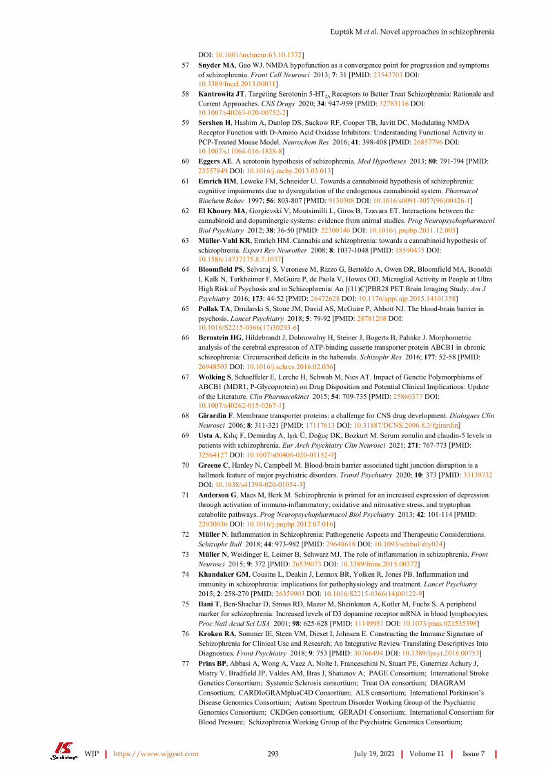
Ľupták M et al. Novel approaches in schizophrenia
WJP https://www.wjgnet.com 293 July 19, 2021 Volume 11 Issue 7
DOI: 10.1001/archneur.63.10.1372]Snyder MA, Gao WJ. NMDA hypofunction as a convergence point for progression and symptoms of schizophrenia. Front Cell Neurosci 2013; 7: 31 [PMID: 23543703 DOI: 10.3389/fncel.2013.00031]
57
Kantrowitz JT. Targeting Serotonin 5-HT2A Receptors to Better Treat Schizophrenia: Rationale and Current Approaches. CNS Drugs 2020; 34: 947-959 [PMID: 32783116 DOI: 10.1007/s40263-020-00752-2]
58
Sershen H, Hashim A, Dunlop DS, Suckow RF, Cooper TB, Javitt DC. Modulating NMDA Receptor Function with D-Amino Acid Oxidase Inhibitors: Understanding Functional Activity in PCP-Treated Mouse Model. Neurochem Res 2016; 41: 398-408 [PMID: 26857796 DOI: 10.1007/s11064-016-1838-8]
59
Eggers AE. A serotonin hypothesis of schizophrenia. Med Hypotheses 2013; 80: 791-794 [PMID: 23557849 DOI: 10.1016/j.mehy.2013.03.013]
60
Emrich HM, Leweke FM, Schneider U. Towards a cannabinoid hypothesis of schizophrenia: cognitive impairments due to dysregulation of the endogenous cannabinoid system. Pharmacol Biochem Behav 1997; 56: 803-807 [PMID: 9130308 DOI: 10.1016/s0091-3057(96)00426-1]
61
El Khoury MA, Gorgievski V, Moutsimilli L, Giros B, Tzavara ET. Interactions between the cannabinoid and dopaminergic systems: evidence from animal studies. Prog Neuropsychopharmacol Biol Psychiatry 2012; 38: 36-50 [PMID: 22300746 DOI: 10.1016/j.pnpbp.2011.12.005]
62
Müller-Vahl KR, Emrich HM. Cannabis and schizophrenia: towards a cannabinoid hypothesis of schizophrenia. Expert Rev Neurother 2008; 8: 1037-1048 [PMID: 18590475 DOI: 10.1586/14737175.8.7.1037]
63
Bloomfield PS, Selvaraj S, Veronese M, Rizzo G, Bertoldo A, Owen DR, Bloomfield MA, Bonoldi I, Kalk N, Turkheimer F, McGuire P, de Paola V, Howes OD. Microglial Activity in People at Ultra High Risk of Psychosis and in Schizophrenia: An [(11)C]PBR28 PET Brain Imaging Study. Am J Psychiatry 2016; 173: 44-52 [PMID: 26472628 DOI: 10.1176/appi.ajp.2015.14101358]
64
Pollak TA, Drndarski S, Stone JM, David AS, McGuire P, Abbott NJ. The blood-brain barrier in psychosis. Lancet Psychiatry 2018; 5: 79-92 [PMID: 28781208 DOI: 10.1016/S2215-0366(17)30293-6]
65
Bernstein HG, Hildebrandt J, Dobrowolny H, Steiner J, Bogerts B, Pahnke J. Morphometric analysis of the cerebral expression of ATP-binding cassette transporter protein ABCB1 in chronic schizophrenia: Circumscribed deficits in the habenula. Schizophr Res 2016; 177: 52-58 [PMID: 26948503 DOI: 10.1016/j.schres.2016.02.036]
66
Wolking S, Schaeffeler E, Lerche H, Schwab M, Nies AT. Impact of Genetic Polymorphisms of ABCB1 (MDR1, P-Glycoprotein) on Drug Disposition and Potential Clinical Implications: Update of the Literature. Clin Pharmacokinet 2015; 54: 709-735 [PMID: 25860377 DOI: 10.1007/s40262-015-0267-1]
67
Girardin F. Membrane transporter proteins: a challenge for CNS drug development. Dialogues Clin Neurosci 2006; 8: 311-321 [PMID: 17117613 DOI: 10.31887/DCNS.2006.8.3/fgirardin]
68
Usta A, Kılıç F, Demirdaş A, Işık Ü, Doğuç DK, Bozkurt M. Serum zonulin and claudin-5 levels in patients with schizophrenia. Eur Arch Psychiatry Clin Neurosci 2021; 271: 767-773 [PMID: 32564127 DOI: 10.1007/s00406-020-01152-9]
69
Greene C, Hanley N, Campbell M. Blood-brain barrier associated tight junction disruption is a hallmark feature of major psychiatric disorders. Transl Psychiatry 2020; 10: 373 [PMID: 33139732 DOI: 10.1038/s41398-020-01054-3]
70
Anderson G, Maes M, Berk M. Schizophrenia is primed for an increased expression of depression through activation of immuno-inflammatory, oxidative and nitrosative stress, and tryptophan catabolite pathways. Prog Neuropsychopharmacol Biol Psychiatry 2013; 42: 101-114 [PMID: 22930036 DOI: 10.1016/j.pnpbp.2012.07.016]
71
Müller N. Inflammation in Schizophrenia: Pathogenetic Aspects and Therapeutic Considerations. Schizophr Bull 2018; 44: 973-982 [PMID: 29648618 DOI: 10.1093/schbul/sby024]
72
Müller N, Weidinger E, Leitner B, Schwarz MJ. The role of inflammation in schizophrenia. Front Neurosci 2015; 9: 372 [PMID: 26539073 DOI: 10.3389/fnins.2015.00372]
73
Khandaker GM, Cousins L, Deakin J, Lennox BR, Yolken R, Jones PB. Inflammation and immunity in schizophrenia: implications for pathophysiology and treatment. Lancet Psychiatry 2015; 2: 258-270 [PMID: 26359903 DOI: 10.1016/S2215-0366(14)00122-9]
74
Ilani T, Ben-Shachar D, Strous RD, Mazor M, Sheinkman A, Kotler M, Fuchs S. A peripheral marker for schizophrenia: Increased levels of D3 dopamine receptor mRNA in blood lymphocytes. Proc Natl Acad Sci USA 2001; 98: 625-628 [PMID: 11149951 DOI: 10.1073/pnas.021535398]
75
Kroken RA, Sommer IE, Steen VM, Dieset I, Johnsen E. Constructing the Immune Signature of Schizophrenia for Clinical Use and Research; An Integrative Review Translating Descriptives Into Diagnostics. Front Psychiatry 2018; 9: 753 [PMID: 30766494 DOI: 10.3389/fpsyt.2018.00753]
76
Prins BP, Abbasi A, Wong A, Vaez A, Nolte I, Franceschini N, Stuart PE, Guterriez Achury J, Mistry V, Bradfield JP, Valdes AM, Bras J, Shatunov A; PAGE Consortium; International Stroke Genetics Consortium; Systemic Sclerosis consortium; Treat OA consortium; DIAGRAM Consortium; CARDIoGRAMplusC4D Consortium; ALS consortium; International Parkinson’s Disease Genomics Consortium; Autism Spectrum Disorder Working Group of the Psychiatric Genomics Consortium; CKDGen consortium; GERAD1 Consortium; International Consortium for Blood Pressure; Schizophrenia Working Group of the Psychiatric Genomics Consortium;
77

Ľupták M et al. Novel approaches in schizophrenia
WJP https://www.wjgnet.com 294 July 19, 2021 Volume 11 Issue 7
Inflammation Working Group of the CHARGE Consortium, Lu C, Han B, Raychaudhuri S, Bevan S, Mayes MD, Tsoi LC, Evangelou E, Nair RP, Grant SF, Polychronakos C, Radstake TR, van Heel DA, Dunstan ML, Wood NW, Al-Chalabi A, Dehghan A, Hakonarson H, Markus HS, Elder JT, Knight J, Arking DE, Spector TD, Koeleman BP, van Duijn CM, Martin J, Morris AP, Weersma RK, Wijmenga C, Munroe PB, Perry JR, Pouget JG, Jamshidi Y, Snieder H, Alizadeh BZ. Investigating the Causal Relationship of C-Reactive Protein with 32 Complex Somatic and Psychiatric Outcomes: A Large-Scale Cross-Consortium Mendelian Randomization Study. PLoS Med 2016; 13: e1001976 [PMID: 27327646 DOI: 10.1371/journal.pmed.1001976]Rodrigues-Amorim D, Rivera-Baltanás T, Spuch C, Caruncho HJ, González-Fernandez Á, Olivares JM, Agís-Balboa RC. Cytokines dysregulation in schizophrenia: A systematic review of psychoneuroimmune relationship. Schizophr Res 2018; 197: 19-33 [PMID: 29239785 DOI: 10.1016/j.schres.2017.11.023]
78
van Kesteren CF, Gremmels H, de Witte LD, Hol EM, Van Gool AR, Falkai PG, Kahn RS, Sommer IE. Immune involvement in the pathogenesis of schizophrenia: a meta-analysis on postmortem brain studies. Transl Psychiatry 2017; 7: e1075 [PMID: 28350400 DOI: 10.1038/tp.2017.4]
79
Osimo EF, Cardinal RN, Jones PB, Khandaker GM. Prevalence and correlates of low-grade systemic inflammation in adult psychiatric inpatients: An electronic health record-based study. Psychoneuroendocrinology 2018; 91: 226-234 [PMID: 29544672 DOI: 10.1016/j.psyneuen.2018.02.031]
80
Boerrigter D, Weickert TW, Lenroot R, O'Donnell M, Galletly C, Liu D, Burgess M, Cadiz R, Jacomb I, Catts VS, Fillman SG, Weickert CS. Using blood cytokine measures to define high inflammatory biotype of schizophrenia and schizoaffective disorder. J Neuroinflammation 2017; 14: 188 [PMID: 28923068 DOI: 10.1186/s12974-017-0962-y]
81
Fond G, Lançon C, Korchia T, Auquier P, Boyer L. The Role of Inflammation in the Treatment of Schizophrenia. Front Psychiatry 2020; 11: 160 [PMID: 32256401 DOI: 10.3389/fpsyt.2020.00160]
82
Goldsmith DR, Rapaport MH, Miller BJ. A meta-analysis of blood cytokine network alterations in psychiatric patients: comparisons between schizophrenia, bipolar disorder and depression. Mol Psychiatry 2016; 21: 1696-1709 [PMID: 26903267 DOI: 10.1038/mp.2016.3]
83
Wang AK, Miller BJ. Meta-analysis of Cerebrospinal Fluid Cytokine and Tryptophan Catabolite Alterations in Psychiatric Patients: Comparisons Between Schizophrenia, Bipolar Disorder, and Depression. Schizophr Bull 2018; 44: 75-83 [PMID: 28338954 DOI: 10.1093/schbul/sbx035]
84
Erhardt S, Blennow K, Nordin C, Skogh E, Lindström LH, Engberg G. Kynurenic acid levels are elevated in the cerebrospinal fluid of patients with schizophrenia. Neurosci Lett 2001; 313: 96-98 [PMID: 11684348 DOI: 10.1016/s0304-3940(01)02242-x]
85
Linderholm KR, Skogh E, Olsson SK, Dahl ML, Holtze M, Engberg G, Samuelsson M, Erhardt S. Increased levels of kynurenine and kynurenic acid in the CSF of patients with schizophrenia. Schizophr Bull 2012; 38: 426-432 [PMID: 20729465 DOI: 10.1093/schbul/sbq086]
86
Wang Z, Li P, Chi D, Wu T, Mei Z, Cui G. Association between C-reactive protein and risk of schizophrenia: An updated meta-analysis. Oncotarget 2017; 8: 75445-75454 [PMID: 29088880 DOI: 10.18632/oncotarget.17995]
87
Liemburg EJ, Nolte IM; PHAMOUS investigators, Klein HC, Knegtering H. Relation of inflammatory markers with symptoms of psychotic disorders: a large cohort study. Prog Neuropsychopharmacol Biol Psychiatry 2018; 86: 89-94 [PMID: 29778547 DOI: 10.1016/j.pnpbp.2018.04.006]
88
Johnsen E, Fathian F, Kroken RA, Steen VM, Jørgensen HA, Gjestad R, Løberg EM. The serum level of C-reactive protein (CRP) is associated with cognitive performance in acute phase psychosis. BMC Psychiatry 2016; 16: 60 [PMID: 26973142 DOI: 10.1186/s12888-016-0769-x]
89
Fond G, Godin O, Boyer L, Berna F, Andrianarisoa M, Coulon N, Brunel L, Bulzacka E, Aouizerate B, Capdevielle D, Chereau I, D'Amato T, Dubertret C, Dubreucq J, Faget C, Leignier S, Lançon C, Mallet J, Misdrahi D, Passerieux C, Rey R, Schandrin A, Urbach M, Vidailhet P, Llorca PM, Schürhoff F, Leboyer M; FACE-SZ (FondaMental Academic Centers of Expertise for Schizophrenia) Group. Chronic low-grade peripheral inflammation is associated with ultra resistant schizophrenia. Results from the FACE-SZ cohort. Eur Arch Psychiatry Clin Neurosci 2019; 269: 985-992 [PMID: 29808267 DOI: 10.1007/s00406-018-0908-0]
90
Yang Y, Wan C, Li H, Zhu H, La Y, Xi Z, Chen Y, Jiang L, Feng G, He L. Altered levels of acute phase proteins in the plasma of patients with schizophrenia. Anal Chem 2006; 78: 3571-3576 [PMID: 16737209 DOI: 10.1021/ac051916x]
91
Yee JY, Nurjono M, Ng WY, Teo SR, Lee TS, Lee J. Peripheral blood gene expression of acute phase proteins in people with first episode psychosis. Brain Behav Immun 2017; 65: 337-341 [PMID: 28627459 DOI: 10.1016/j.bbi.2017.06.006]
92
Herberth M, Rahmoune H, Schwarz E, Koethe D, Harris LW, Kranaster L, Witt SH, Spain M, Barnes A, Schmolz M, Leweke MF, Guest PC, Bahn S. Identification of a molecular profile associated with immune status in first-onset schizophrenia patients. Clin Schizophr Relat Psychoses 2014; 7: 207-215 [PMID: 23395839 DOI: 10.3371/CSRP.HERA.020113]
93
Iwata Y, Suzuki K, Nakamura K, Matsuzaki H, Sekine Y, Tsuchiya KJ, Sugihara G, Kawai M, Minabe Y, Takei N, Mori N. Increased levels of serum soluble L-selectin in unmedicated patients with schizophrenia. Schizophr Res 2007; 89: 154-160 [PMID: 17049815 DOI: 10.1016/j.schres.2006.08.026]
94

Ľupták M et al. Novel approaches in schizophrenia
WJP https://www.wjgnet.com 295 July 19, 2021 Volume 11 Issue 7
Masopust J, Malý R, Andrýs C, Vališ M, Bažant J, Hosák L. Markers of thrombogenesis are activated in unmedicated patients with acute psychosis: a matched case control study. BMC Psychiatry 2011; 11: 2 [PMID: 21199572 DOI: 10.1186/1471-244X-11-2]
95
Miller BJ, Gassama B, Sebastian D, Buckley P, Mellor A. Meta-analysis of lymphocytes in schizophrenia: clinical status and antipsychotic effects. Biol Psychiatry 2013; 73: 993-999 [PMID: 23062357 DOI: 10.1016/j.biopsych.2012.09.007]
96
Ezeoke A, Mellor A, Buckley P, Miller B. A systematic, quantitative review of blood autoantibodies in schizophrenia. Schizophr Res 2013; 150: 245-251 [PMID: 23953827 DOI: 10.1016/j.schres.2013.07.029]
97
Conus P, Seidman LJ, Fournier M, Xin L, Cleusix M, Baumann PS, Ferrari C, Cousins A, Alameda L, Gholam-Rezaee M, Golay P, Jenni R, Woo TW, Keshavan MS, Eap CB, Wojcik J, Cuenod M, Buclin T, Gruetter R, Do KQ. N-acetylcysteine in a Double-Blind Randomized Placebo-Controlled Trial: Toward Biomarker-Guided Treatment in Early Psychosis. Schizophr Bull 2018; 44: 317-327 [PMID: 29462456 DOI: 10.1093/schbul/sbx093]
98
Sommer IE, Bearden CE, van Dellen E, Breetvelt EJ, Duijff SN, Maijer K, van Amelsvoort T, de Haan L, Gur RE, Arango C, Díaz-Caneja CM, Vinkers CH, Vorstman JA. Early interventions in risk groups for schizophrenia: what are we waiting for? NPJ Schizophr 2016; 2: 16003 [PMID: 27336054 DOI: 10.1038/npjschz.2016.3]
99
Cho M, Lee TY, Kwak YB, Yoon YB, Kim M, Kwon JS. Adjunctive use of anti-inflammatory drugs for schizophrenia: A meta-analytic investigation of randomized controlled trials. Aust N Z J Psychiatry 2019; 53: 742-759 [PMID: 30864461 DOI: 10.1177/0004867419835028]
100
Jeppesen R, Christensen RHB, Pedersen EMJ, Nordentoft M, Hjorthøj C, Köhler-Forsberg O, Benros ME. Efficacy and safety of anti-inflammatory agents in treatment of psychotic disorders - A comprehensive systematic review and meta-analysis. Brain Behav Immun 2020; 90: 364-380 [PMID: 32890697 DOI: 10.1016/j.bbi.2020.08.028]
101
Carabotti M, Scirocco A, Maselli MA, Severi C. The gut-brain axis: interactions between enteric microbiota, central and enteric nervous systems. Ann Gastroenterol 2015; 28: 203-209 [PMID: 25830558]
102
Kuwahara A, Matsuda K, Kuwahara Y, Asano S, Inui T, Marunaka Y. Microbiota-gut-brain axis: enteroendocrine cells and the enteric nervous system form an interface between the microbiota and the central nervous system. Biomed Res 2020; 41: 199-216 [PMID: 33071256 DOI: 10.2220/biomedres.41.199]
103
Alam R, Abdolmaleky HM, Zhou JR. Microbiome, inflammation, epigenetic alterations, and mental diseases. Am J Med Genet B Neuropsychiatr Genet 2017; 174: 651-660 [PMID: 28691768 DOI: 10.1002/ajmg.b.32567]
104
Sampson TR, Mazmanian SK. Control of brain development, function, and behavior by the microbiome. Cell Host Microbe 2015; 17: 565-576 [PMID: 25974299 DOI: 10.1016/j.chom.2015.04.011]
105
Tomasik J, Yolken RH, Bahn S, Dickerson FB. Immunomodulatory Effects of Probiotic Supplementation in Schizophrenia Patients: A Randomized, Placebo-Controlled Trial. Biomark Insights 2015; 10: 47-54 [PMID: 26052224 DOI: 10.4137/BMI.S22007]
106
Greenwood J, Acharya RB, Marcellus V, Rey JA. Lumateperone: A Novel Antipsychotic for Schizophrenia. Ann Pharmacother 2021; 55: 98-104 [PMID: 32590907 DOI: 10.1177/1060028020936597]
107
Davis RE, Correll CU. ITI-007 in the treatment of schizophrenia: from novel pharmacology to clinical outcomes. Expert Rev Neurother 2016; 16: 601-614 [PMID: 27042868 DOI: 10.1080/14737175.2016.1174577]
108
Edinoff A, Wu N, deBoisblanc C, Feltner CO, Norder M, Tzoneva V, Kaye AM, Cornett EM, Kaye AD, Viswanath O, Urits I. Lumateperone for the Treatment of Schizophrenia. Psychopharmacol Bull 2020; 50: 32-59 [PMID: 33012872]
109
Correll CU, Vanover KE, Davis RE, Chen R, Satlin A, Mates S. Safety and tolerability of lumateperone 42 mg: An open-label antipsychotic switch study in outpatients with stable schizophrenia. Schizophr Res 2021; 228: 198-205 [PMID: 33453691 DOI: 10.1016/j.schres.2020.12.006]
110
Goff DC. Promising Evidence of Antipsychotic Efficacy without Dopamine D2-Receptor Binding. N Engl J Med 2020; 382: 1555-1556 [PMID: 32294351 DOI: 10.1056/NEJMe2001508]
111
Krogmann A, Peters L, von Hardenberg L, Bödeker K, Nöhles VB, Correll CU. Keeping up with the therapeutic advances in schizophrenia: a review of novel and emerging pharmacological entities. CNS Spectr 2019; 24: 38-69 [PMID: 31482779 DOI: 10.1017/S109285291900124X]
112
Koblan KS, Kent J, Hopkins SC, Krystal JH, Cheng H, Goldman R, Loebel A. A Non-D2-Receptor-Binding Drug for the Treatment of Schizophrenia. N Engl J Med 2020; 382: 1497-1506 [PMID: 32294346 DOI: 10.1056/NEJMoa1911772]
113
Fowler S, Kletzl H, Finel M, Manevski N, Schmid P, Tuerck D, Norcross RD, Hoener MC, Spleiss O, Iglesias VA. A UGT2B10 splicing polymorphism common in african populations may greatly increase drug exposure. J Pharmacol Exp Ther 2015; 352: 358-367 [PMID: 25503386 DOI: 10.1124/jpet.114.220194]
114
Gibert-Rahola J, Villena-Rodriguez A. Glutamatergic drugs for schizophrenia treatment. Actas Esp Psiquiatr 2014; 42: 234-241 [PMID: 25179095]
115
Stauffer VL, Millen BA, Andersen S, Kinon BJ, Lagrandeur L, Lindenmayer JP, Gomez JC. 116

Ľupták M et al. Novel approaches in schizophrenia
WJP https://www.wjgnet.com 296 July 19, 2021 Volume 11 Issue 7
Pomaglumetad methionil: no significant difference as an adjunctive treatment for patients with prominent negative symptoms of schizophrenia compared to placebo. Schizophr Res 2013; 150: 434-441 [PMID: 24035403 DOI: 10.1016/j.schres.2013.08.020]Marek GJ. When is a Proof-of-Concept (POC) not a POC? Curr Pharm Des 2015; 21: 3788-3796 [PMID: 26044978 DOI: 10.2174/1381612821666150605105632]
117
Adams DH, Kinon BJ, Baygani S, Millen BA, Velona I, Kollack-Walker S, Walling DP. A long-term, phase 2, multicenter, randomized, open-label, comparative safety study of pomaglumetad methionil (LY2140023 monohydrate) versus atypical antipsychotic standard of care in patients with schizophrenia. BMC Psychiatry 2013; 13: 143 [PMID: 23694720 DOI: 10.1186/1471-244X-13-143]
118
Lin CH, Lin CH, Chang YC, Huang YJ, Chen PW, Yang HT, Lane HY. Sodium Benzoate, a D-Amino Acid Oxidase Inhibitor, Added to Clozapine for the Treatment of Schizophrenia: A Randomized, Double-Blind, Placebo-Controlled Trial. Biol Psychiatry 2018; 84: 422-432 [PMID: 29397899 DOI: 10.1016/j.biopsych.2017.12.006]
119
Lane HY, Lin CH, Green MF, Hellemann G, Huang CC, Chen PW, Tun R, Chang YC, Tsai GE. Add-on treatment of benzoate for schizophrenia: a randomized, double-blind, placebo-controlled trial of D-amino acid oxidase inhibitor. JAMA Psychiatry 2013; 70: 1267-1275 [PMID: 24089054 DOI: 10.1001/jamapsychiatry.2013.2159]
120
Larsen C, Shahinas J. Dosage, Efficacy and Safety of Cannabidiol Administration in Adults: A Systematic Review of Human Trials. J Clin Med Res 2020; 12: 129-141 [PMID: 32231748 DOI: 10.14740/jocmr4090]
121
Srinivasan S, Tampi RR, Balaram K, Kapoor A. Pimavanserin for the treatment of psychosis in Alzheimer's disease: A literature review. World J Psychiatry 2020; 10: 162-174 [PMID: 32844093 DOI: 10.5498/wjp.v10.i7.162]
122
Strauss GP, Zamani Esfahlani F, Sayama H, Kirkpatrick B, Opler MG, Saoud JB, Davidson M, Luthringer R. Network Analysis Indicates That Avolition Is the Most Central Domain for the Successful Treatment of Negative Symptoms: Evidence From the Roluperidone Randomized Clinical Trial. Schizophr Bull 2020; 46: 964-970 [PMID: 31989151 DOI: 10.1093/schbul/sbz141]
123
Al-Nema MY, Gaurav A. Phosphodiesterase as a Target for Cognition Enhancement in Schizophrenia. Curr Top Med Chem 2020; 20: 2404-2421 [PMID: 32533817 DOI: 10.2174/1568026620666200613202641]
124
Macek TA, McCue M, Dong X, Hanson E, Goldsmith P, Affinito J, Mahableshwarkar AR. A phase 2, randomized, placebo-controlled study of the efficacy and safety of TAK-063 in subjects with an acute exacerbation of schizophrenia. Schizophr Res 2019; 204: 289-294 [PMID: 30190165 DOI: 10.1016/j.schres.2018.08.028]
125

WJP https://www.wjgnet.com 297 July 19, 2021 Volume 11 Issue 7
World Journal of
PsychiatryW J PSubmit a Manuscript: https://www.f6publishing.com World J Psychiatr 2021 July 19; 11(7): 297-315
DOI: 10.5498/wjp.v11.i7.297 ISSN 2220-3206 (online)
REVIEW
Glutamate and depression: Reflecting a deepening knowledge of the gut and brain effects of a ubiquitous molecule
Adejoke Yetunde Onaolapo, Olakunle James Onaolapo
ORCID number: Adejoke Yetunde Onaolapo 0000-0001-7126-7050; Olakunle James Onaolapo 0000-0003-2142-6046.
Author contributions: Both authors contributed equally to the writing of this manuscript.
Conflict-of-interest statement: Authors have nothing to declare.
Open-Access: This article is an open-access article that was selected by an in-house editor and fully peer-reviewed by external reviewers. It is distributed in accordance with the Creative Commons Attribution NonCommercial (CC BY-NC 4.0) license, which permits others to distribute, remix, adapt, build upon this work non-commercially, and license their derivative works on different terms, provided the original work is properly cited and the use is non-commercial. See: http://creativecommons.org/Licenses/by-nc/4.0/
Manuscript source: Invited manuscript
Specialty type: Psychiatry
Country/Territory of origin: Nigeria
Peer-review report’s scientific quality classificationGrade A (Excellent): 0
Adejoke Yetunde Onaolapo, Behavioural Neuroscience Unit, Neurobiology Subdivision, Department of Anatomy, Ladoke Akintola University of Technology, Oyo State 234, Nigeria
Olakunle James Onaolapo, Behavioural Neuroscience Unit, Neuropharmacology Subdivision, Department of Pharmacology, Ladoke Akintola University of Technology, Oyo State 234, Nigeria
Corresponding author: Olakunle James Onaolapo, MBBS, MSc, PhD, Senior Lecturer, Behavioural Neuroscience Unit, Neuropharmacology Subdivision, Department of Pharmaco-logy, Ladoke Akintola University of Technology, P.M.B 4000, Oyo State 234, Nigeria. [email protected]
AbstractThe versatility of glutamate as the brain’s foremost excitatory neurotransmitter and modulator of neurotransmission and function is considered common knowledge. Years of research have continued to uncover glutamate’s effects and roles in several neurological and neuropsychiatric disorders, including depression. It had been considered that a deeper understanding of the roles of glutamate in depression might open a new door to understanding the patholo-gical basis of the disorder, improve the approach to patient management, and lead to the development of newer drugs that may benefit more patients. This review examines our current understanding of the roles of endogenous and exogenous sources of glutamate and the glutamatergic system in the aetiology, progression and management of depression. It also examines the relationships that link the gut-brain axis, glutamate and depression; as it emphasizes how the gut-brain axis could impact depression pathogenesis and management via changes in glutamate homeostasis. Finally, we consider what the likely future of glutamate-based therapies and glutamate-based therapeutic manipulations in depression are, and if with them, we are now on the final chapter of understanding the neuroche-mical milieu of depressive disorders.
Key Words: Brain; Gut microbiome; Mental health; Mood disorders; Neurotransmitters
©The Author(s) 2021. Published by Baishideng Publishing Group Inc. All rights reserved.

Onaolapo AY et al. Glutamate and glutamate-based therapies in depression
WJP https://www.wjgnet.com 298 July 19, 2021 Volume 11 Issue 7
Grade B (Very good): B Grade C (Good): C, C Grade D (Fair): D Grade E (Poor): 0
Received: March 7, 2021 Peer-review started: March 7, 2021 First decision: March 30, 2021 Revised: April 13, 2021 Accepted: May 10, 2021 Article in press: May 10, 2021 Published online: July 19, 2021
P-Reviewer: Pasquini M, Pavón L, Qu SQ S-Editor: Gao CC L-Editor: Webster JR P-Editor: Li JH
Core Tip: The versatility of glutamate as the brain’s foremost excitatory neurotrans-mitter, and modulator of intermediary metabolism in the gastrointestinal tract is considered common knowledge. Years of research suggest glutamate has a role to play in depression. Also, there is increasing evidence of a possible relationship between glutamate and the pathophysiology and/or treatment of depression. The complexity of depression suggests dysregulation of glutamate in sites such as the gastrointestinal tract and brain. The communication link involving dietary glutamate, the gut, endogenous glutamate, and the brain is a multidirectional pathway; the understanding of which is necessary to fully account for glutamate’s role in depression.
Citation: Onaolapo AY, Onaolapo OJ. Glutamate and depression: Reflecting a deepening knowledge of the gut and brain effects of a ubiquitous molecule. World J Psychiatr 2021; 11(7): 297-315URL: https://www.wjgnet.com/2220-3206/full/v11/i7/297.htmDOI: https://dx.doi.org/10.5498/wjp.v11.i7.297
INTRODUCTIONClinical depression or major depressive disorder (MDD) is a chronic, debilitating, and disabling mental health disorder that affects over 300 million people (across all age groups) globally[1-4]. It contributes significantly to the global burden of disease and escalating incidence of suicides amongst teenagers and young adults worldwide. Globally, the prevalence of depression continues to increase[4,5]; with factors such as worsening poverty, increasing unemployment, adverse life events and genetics[4] being recognized as important risk factors for its development.
The discovery of monoamine oxidase inhibitors and tricyclic antidepressants opened opportunities for the treatment of depression and provided insights into the role of neurotransmitters such as dopamine, serotonin and norepinephrine in the pathophysiology of depression[6,7]. However, the shortcomings of the currently-approved pharmacotherapies such as the lag time between the effect of drugs on monoamine availability and their therapeutic effect, inadequate response, and the increasing incidence of treatment-resistant depression[7-9] means that there is still a critical need to better understand the pathophysiology of depression; and develop more-effective and efficient therapeutic interventions for depressive disorders.
The monoamine hypothesis supports the notion that the pathology in depression is primarily depletion in the levels of brain monoamine neurotransmitters including serotonin, norepinephrine, and dopamine[10-12]. In the almost seven decades since its formulation, it has largely explained the symptoms and response to currently available antidepressant therapy. However, inconsistencies in the hypothesis have resulted in further research to better understand depression pathophysiology and management. In the last three decades, there has been compelling clinical[13,14] and preclinical[15,16] evidence demonstrating the involvement of the glutamatergic system in the pathophysiology of depression.
Since the first mention of the glutamate hypothesis of depression in the 1990s[15], our understanding of the versatility of glutamate as the brain’s foremost excitatory neurotransmitter, and modulator of neurotransmission and function has increased considerably. Years of research have continued to uncover glutamate’s effects and roles in several neurological and neuropsychiatric disorders, including depression. More recently, the antidepressant actions of ketamine, an N-methyl-D-aspartate (NMDA) glutamate receptor antagonist especially in treatment-resistant depression[17,18] was reported; suggesting a deeper understanding of the roles of glutamate in depression could open new doors to understanding the pathological basis of the disorder, improve the approach to patient management, and lead to the development of newer drugs that may benefit more patients. Also, the roles of the gut-brain axis in glutamate signalling have been investigated, with reports that these could also impact the pathophysiology and treatment options in depression[19,20]. This review examines current understanding of the roles of endogenous and exogenous sources of glutamate and the glutamatergic system in the aetiology, progression and management of depression. It also examines the relationships that link the gut-brain axis, depression and glutamate (Figure 1); as it emphasizes how the gut-brain axis could impact

Onaolapo AY et al. Glutamate and glutamate-based therapies in depression
WJP https://www.wjgnet.com 299 July 19, 2021 Volume 11 Issue 7
Figure 1 Possible relationships that link dietary glutamate, the gut-brain axis, endogenous glutamate and depression.
depression pathogenesis and management via changes in glutamate homeostasis. Finally, we consider what the likely future of glutamate-based therapies and glutamate-based therapeutic manipulations in depression are, and if with them, we are now on the final chapter of understanding the neurochemical milieu of depressive disorder.
GLUTAMATE AND DEPRESSIONGlutamate is an amino acid and the major excitatory neurotransmitter in the brain[21]. In the last few decades, there has been increasing insights into the roles played by the glutamatergic system (Figure 1) in the pathophysiology and treatment of mood disorders generally, and depressive disorders specifically[12,22,23]. Although glutamate is ubiquitous in the brain, excessive release of glutamate has been associated with excitotoxicity-induced brain injury[24]. The possible involvement of the glutama-tergic system in mood disorders is supported by preclinical evidence of the antide-pressant effects of NMDA antagonists[15,25]. Also, the results of early studies showing alterations in the levels of glutamate (peripherally and centrally) in persons with mood disorders confirmed this involvement[13,26]. Alterations in excitatory and/or inhibitory neurotransmitters resulting in the alteration of functional connectivity patterns within large brain networks have also been reported[27]. More recently, there is overwhelming evidence of the anxiolytic and antidepressant response to subanaes-thetic-doses of ketamine in clinical[27-30] and preclinical studies[31,32].
The ability of diet to cause depression and depression-like phenotypes has been reported[33,34]. Research has continued to show that the consumption of diets rich in fat, deficient in magnesium, or high in monosodium glutamate can cause depression-like behaviours such as decreased social interaction, anhedonia and behavioural despair in rodents[35-37]. While there have been suggestions that these effects are linked to the ability of dietary factors to alter the composition of the gut microbiota[36,37], emerging evidence of the interactions between the gut microbiota and brain neurotransmitters such as dopamine, serotonin, gamma amino-butyric acid and glutamate[9,38] are opening new vistas into possible novel treatment modalities for depression.
Dietary glutamate and depressionGlutamate is an α-amino acid that is useful in the biosynthesis of proteins and important in intermediary metabolism, through its ability to link carbohydrate and amino acid metabolism via the tricarboxylic acid cycle[39,40]. Glutamate (in addition to being synthesized in the body in humans) is also derived from dietary sources such as cheese, meat, and several food-seasonings including monosodium glutamate[21,41].
Onaolapo AY et al. Glutamate and glutamate-based therapies in depression
WJP https://www.wjgnet.com 300 July 19, 2021 Volume 11 Issue 7
While endogenous brain glutamate has been linked to the pathophysiology of psychiatric conditions such as schizophrenia and mood disorders, the possible role of dietary glutamate (Table 1) in the development of neuropsychiatric conditions is still being evaluated[42]. There have been suggestions that the consumption of diets containing high concentrations of monosodium glutamate could increase body levels of glutamic acid, resulting in hyperglutamatergic neurotransmission, which could possibly contribute to the development of depression[43]. Also, a few studies have reported that factors such as chronic stress that reduce brain levels of glutamate and glutamine causing hypoactive glutamatergic signaling in the mouse prefrontal cortex are also associated with the development of depression[44], suggesting that regarding brain glutamatergic transmission a delicate balance always needs to be maintained.
In the last few years, reports from preclinical studies have associated the use of monosodium glutamate with the development of behavioural phenotypes such as anxiety and depression; and the ability to influence brain endogenous glutamate and glutamatergic neurotransmission[35,42,44-46]. There have also been reports from studies that monosodium glutamate could directly influence the concentrations of brain neurotransmitters such as serotonin and glutamate[21,35,45,47], although there have also been reports to the contrary[48].
The relationship between dietary monosodium glutamate and depression has been examined by a few studies[35,43,47]. The results of a clinical analysis that examined the relationship between the consumption of a diet high in glutamic acid and the development of depressive symptoms in a group of persons with schizophrenia revealed that in non-obese patients the consumption of high dietary glutamic acid was associated with an increase in depressive symptoms, although this was linked to the susceptibility of persons with one psychiatric condition to develop other co-morbidities[42]. A preclinical study by Quines et al[35] that examined the effects of monosodium glutamate administered parenterally in the neonatal period with exposure to behavioural paradigms on postnatal days 60-64 reported the presence of anxiety and behavioural despair. However, studies from our laboratory revealed that while orally administered monosodium glutamate was associated with the development of anxiety behaviour, especially in male mice[21,45,49], an antide-pressant effect was observed in the behavioural-despair paradigms irrespective of sex[47]. The results of these studies from our laboratory suggest that the antidepressant response observed when monosodium glutamate was administered by gavage (compared to the response following parenteral administration) could have been influenced by the gut microbiota or the gut-brain axis, or by the ability of monosodium glutamate (at these doses) to minimally increase brain levels of glutamate which could have antidepressant benefits as previously reported[44].
Endogenous brain glutamate and depressionGlutamate plays an important role in the modulation of synaptic plasticity and transmission. It is also the precursor of the inhibitory neurotransmitter gamma amino-butyric acid (GABA). Studies have shown that dysregulation of glutamatergic transmission or alterations in brain concentrations of glutamate is associated with derangement of brain function, development of excitotoxic brain injury, and cell death[24,47]. There have also been reports showing that alterations in glutamatergic neurotransmission contribute significantly to the development of peripheral and central nervous system disorders[50]. In the last few years, the possible relationships that exist between glutamate/glutamatergic system and the development of neuropsy-chiatric disorders such as depression have continued to be examined[22,24,51].
Several studies have linked dysregulation of glutamate neurotransmission with the development and progression of neurodevelopmental, neurodegenerative and psychiatric disorders such as autism, epilepsy and schizophrenia[52,53]. There is also emerging evidence (Table 2) linking the pathogenesis of depression to alterations in glutamate and glutamate signalling[12,52]. Levine et al[54], using a proton magnetic resonance spectroscopy (MRS) technique examined the relationship between cerebrospinal fluid (CSF) metabolites, such as glutamate and glutamine on depressive symptoms in hospitalized persons with severe unmedicated depression, and reported that compared to control subjects, glutamine level in the CSF of depressed patients was elevated. Also, using high performance liquid chromatography with fluorometric detection, Mitani et al[55] examined the relationship between plasma levels of glutamate on severity of depression and concluded that plasma levels of glutamate as well as alanine and L-serine were reflective of the severity of depression. The impact of brain glutamate levels on depression and depression phenotypes have been studied extensively[56-60]. Auer et al[56] and Hasler et al[57] using MRS reported region-specific changes in the levels of brain glutamate in patients with depression. The result
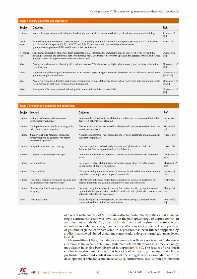
Onaolapo AY et al. Glutamate and glutamate-based therapies in depression
WJP https://www.wjgnet.com 301 July 19, 2021 Volume 11 Issue 7
Table 1 Dietary glutamate and depression
Subject Outcome Ref.
Human In non-obese participants, diets high in levels of glutamic acid were associated with greater depression symptomatology Kumar et al[43]
Adult mice
While chronic immobilization stress decreased sodium-coupled neutral amino acid transporter (SNAT)-1 and 2 in neurons and glutamate transporter (GLT)1, SNAT3, and SNAT5 in astrocytes in the medial prefrontal cortex, glutamine—supplemented diet ameliorated these decrements
Baek et al[44]
Neonatal rats
Subcutaneous injection of monosodium glutamate (MSG) increased the immobility time in the forced swim test and the freezing reaction in the contextual fear conditioning. MSG also increased serotonin uptake in the cerebral cortices and caused deregulation of the hypothalamic-pituitary-adrenal axis
Quines et al[35]
Mice Anxiolytic and memory-enhancing effects at low doses of MSG; however, at higher doses, anxiety and memory retardation were observed
Onaolapo et al[45]
Mice Higher doses of dietary glutamate resulted in an increase in plasma glutamate and glutamine but no difference in total brain glutamate or glutamine levels
Onaolapo et al[21,45,46]
Mice Anxiolytic response in females, and anxiogenic response in males following dietary MSG. A decrease in behavioural despair was observed in both sexes (females more than males)
Onaolapo et al[46,47]
Mice Anxiogenic effect was observed following subchronic oral administration of MSG Onaolapo et al[49]
Table 2 Endogenous glutamate and depression
Subject Method Outcome Ref.
Human Using a proton magnetic resonance spectroscopy technique
Compared to control subjects, glutamine levels in the cerebrospinal fluid of the depressed patients were elevated
Levine et al[54]
Human High performance liquid chromatography with fluorometric detection
Plasma levels of glutamate as well as alanine and L-serine were reflective of the severity of depression
Mitani et al[55]
Human Single voxel (1)H-Magnetic resonance spectroscopy in 19 patients with major depressive episodes
A significant decrease was observed in the levels of glutamate and glutamine in the anterior cingulate
Auer et al[56]
Human Magnetic resonance spectroscopy Depressed patients had reduced glutamine and glutamate levels in the dorsomedial/dorsal anterolateral prefrontal cortex
Hasler et al[57]
Human Magnetic resonance spectroscopy Compared with controls, depressed patients showed an increase in glutamine levels
Godlewska et al[59]
Human Meta-analysis Decreased levels of glutamatergic metabolites were observed in the medial frontal cortex of depressed subjects
Moriguchi et al[60]
Human Meta-analysis Glutamate and glutamine concentrations were found to be lower in the anterior cingulate cortex in patients compared to controls
Luykx et al[58]
Human Functional magnetic resonance imaging and magnetic resonance spectroscopy
Patients with anhedonic major depression showed decreased glutamine but normal glutamate and gamma-aminobutyric acid concentrations
Walter et al[61]
Human Resting state functional magnetic resonance imaging
Decreased amplitude of low frequency fluctuation level in right putamen and right middle temporal cortex correlated positively with glutamate concentration in female patients with depression
Zhang et al[66]
Mice Preclinical study Blockade of glutamate transporter-1 in the central amygdala and prefrontal cortex induced both anhedonia and anxiety
John et al[62,63]
of a recent meta-analysis of MRS studies also supported the hypothesis that glutama-tergic neurotransmission was involved in the pathophysiology of depression[60]. In another meta-analysis, Luykx et al[58] also reported region and state specific alterations in glutamate and glutamine concentrations in depression. The importance of glutamatergic neurotransmission in depression has been further supported by studies that showed altered glutamine concentrations despite normal glutamate levels[59,61].
Abnormalities of the glutamatergic system such as those associated with glutamate clearance at the synaptic cleft and glutamate-related alterations in astrocytic energy modulation have also been observed in depression[61,62]. The results of preclinical studies have also demonstrated that blockade of astrocytic glutamate uptake in the prefrontal cortex and central nucleus of the amygdala was associated with the development of anhedonia and anxiety[62,63]. Furthermore, results from post-mortem

Onaolapo AY et al. Glutamate and glutamate-based therapies in depression
WJP https://www.wjgnet.com 302 July 19, 2021 Volume 11 Issue 7
and magnetic resonance imaging studies have also revealed the presence of altered expression of glutamate-related genes, elevated levels of glutamate, reduced glutamine/glutamate ratio, and/or reduced levels of glutathione (a reservoir of neuronal glutamate) in brain regions such as the medial prefrontal cortex (which have been linked to depression symptomatology) in persons with depression[64-67].
Finally, the rapid antidepressant effects of drugs such as tianeptine and NMDA receptor antagonist ketamine further validate the importance of glutamate and the glutamatergic transmission in depression[22,68,69]. While reports provide some evidence for the involvement of endogenous glutamate in the pathogenesis and treatment of depression[22,50,69,70], the complexity of the disorder would suggest that the dysregulation of glutamate needs to occur in multiple sites (Figure 1) such as the gastrointestinal tract and brain, as can be seen if there is a communication link involving exogenous glutamate, the gut, endogenous glutamate, and then the brain in a multidirectional pathway which we would call the “GLUTAMATE-GUT-GLUTAMATE-BRAIN AXIS”.
The gut-brain axis and glutamateThe impact of the gut (microbiota and gut peptides) in times past was not considered significant in brain development and functioning. Previously, it was believed that the commensal bacteria and their genes which constitute the gut microbiome enjoy a symbiotic relationship with man. In this relationship, they reside in a nutritionally enriched and protected habitat of the human gastrointestinal tract, while they in turn protect humans against colonization of the gut by pathogenic bacteria and provide the body with a rich source of indigestible nutrients. In the light of new evidence, it is now clear that they also play a key role in the brain, either in health or disease.
The gut-brain axis or microbiome-gut-brain axis describes the bidirectional, at times multidimensional system of communication that links the gastrointestinal tract with the central nervous system. It ensures that not only does the central nervous system modulate gastrointestinal function; the gut can also regulate or modulate brain signalling and impact brain structure and function[19]. There is now ample evidence supporting the view that the microbiome-gut-brain axis can influence the development or progression of central nervous system disorders. Some of these include observations of psychiatric co-morbidities occurring in several enteric neuropathies such as chronic inflammatory intestinal disorders[38,71,72]. The presence of altered gut microbial flora and concentration in neurodevelopmental disorders such as autism[73,74] as well as the results of microbial challenge using pathogenic bacteria or pharmacological manipulations with pre or probiotics, are also pointers to the possible roles played by the gut microbiota in the development of brain disorders[72,75]. Further reports have also shown that the gut microbiota influences brain function via its ability to modulate endocrine, immunologic and neurocrine signalling pathways; and brain neurotrans-mitters[38,76].
Also, several possible mechanisms through which microbiome-gut-brain communication could impact the genesis or progression of central nervous system disorders have been proposed. There have been suggestions that communications occur through the activation of neurotransmitters such as serotonin, dopamine, GABA and noradrenaline in the enteric nervous system; these neurotransmitters are secreted by gut microbiota and are akin to the neurotransmitters in the central nervous system[76]. The possible role of gut peptides and metabolites as mediators of the gut-brain crosstalk have also been suggested[77].
Gut-brain axis and depressionStudies have continued to demonstrate the deleterious effects that alterations in gut microbiome composition and microbiome-related metabolites could exert on the development of obesity, autoimmune disorders, inflammatory bowel disease, irritable bowel syndrome, and neuropsychiatric disorders[77-80]. Also, scientific information showing how gut microbes and gut stimuli (such as intragastric infusion of glucose or fatty acid) can directly influence emotional and cognitive functions[81,82] are pointers to the possible involvement of the gut-brain axis in psychiatric disorders. Several preclinical studies have also shown that the gut microbiota modulates brain behaviours such as behavioural despair, anhedonia and anxiety-like behaviours that have construct validity with clinical depression and anxiety[83-86]. Arentsen et al[84] and Kamimura et al[86] showed that compared to specific pathogen-free (SPF) mice, germ-free (GF) mice showed impaired social interactions choosing to spend more time with an object than conventionally raised mice. Huo et al[85] also observed that the exposure of GF mice and SPF mice to chronic restraint stress paradigm was associated with an increase in open field exploration time in GF compared to the SPF mice; with
Onaolapo AY et al. Glutamate and glutamate-based therapies in depression
WJP https://www.wjgnet.com 303 July 19, 2021 Volume 11 Issue 7
SPF mice exhibiting more anxiety-like behaviours compared to GF mice. Chronic prebiotic administration has also been shown to exhibit anxiolytic and antidepressant behaviours in mice. The ability of the prebiotic to modulate behaviour correlated positively with its effects on hippocampal and hypothalamic gene expression and its ability to ensure a balance in the concentrations of short-chain fatty acids[87].
Although the mechanisms by which the gut microbiota influences mood and mood-related behaviours are still being studied; there have been suggestions that because the gut and brain share peptide and receptor similarities the gut microbiome is able to modulate brain function through the activity of gut peptides[77]. Also, studies evaluating the impact of gut microbiome modulation (using prebiotics and probiotics) on brain function have demonstrated that chronic treatment with prebiotic had both antidepressant and anxiolytic effects, which could be linked to reduction in stress-induced corticosterone and proinflammatory cytokine release, and modification of the expression of specific genes in the hippocampus[86,87]. Also, probiotics such as GABA-producing Lactococcus lactis strain have been shown to possess the capacity to modulate behaviours. In one study, in which Lactococcus lactis was grown in both glutamate and non-glutamate supplemented media, a significant increase in GABA production was observed in the glutamate supplemented medium[87], reinforcing the importance of glutamate in the modulation of mood and mood disorders.
There is now ample evidence suggesting that microbes play the role of signalling components in the gut-brain axis. This emerging concept of a microbiota-gut-brain axis suggests that our ability to modulate the gut microbiota may be a potential tool towards the development of novel therapies for complex brain disorders such as psychiatric disorders[19,71,72] (Table 3).
Is there a dietary glutamate-gut-endogenous glutamate-brain axis and how can it impact depression pathogenesis?Glutamate is a multifunctional amino acid that is involved in intermediary metabolism in the gastrointestinal tract and is also crucial for the normal functioning and development of the brain. In the gut, it is derived from exogenous sources including dietary proteins and from free glutamate present in food additives; also, a fraction of the free glutamate in the lumen is from bacterial synthesis[88]. In the central and enteric nervous systems, respectively, glutamate is the major excitatory neurotrans-mitter[50,89].
Glutamate and the glutamatergic pathways are also crucial to microbiota-gut-brain communication. There is increasing evidence that glutamate is both a neurotransmitter and neuromodulator of several functions[77,89]. Glutamate receptors and their transduction molecules have been demonstrated on the epithelial cells of the gut, splanchnic, vagal, and/or pelvic afferents[89-91]. The stimulation of gut glutamate receptors by dietary or luminal glutamate has been associated with the activation of vagal afferents which directly or indirectly influence brain areas such as the cerebral cortex, limbic system, hypothalamus and basal ganglia[90,92]. Also, the activation of glutamate receptors present on splanchnic, vagal, and/or pelvic afferents, allows the communication of sensory inputs to regions of the brain involved in the gut-brain axis, while, also influencing efferent pathways that convey excitatory or inhibitory inputs to the gastrointestinal tract[89,91].
While in health, the enteric ganglia and the brain are impermeable to dietary or luminal glutamate; there have been reports suggesting that glutamate permeability in these systems increases in diseased states such as enteric neuropathies, or conditions that alter the integrity of the blood-brain-barrier. The gut microbiome also influences brain glutamate, with the results of metabolomic studies revealing that alterations in the composition of the gut saprophytic microflora also affected brain concentrations of glutamate[93,94]. Although there is enough scientific evidence in support of the impact of central glutamate and the glutamatergic system in depression, there is however a dearth of information regarding the possible ways luminal glutamate (either from dietary sources or microbial activities) may influence depression pathophysiology.
However, from the foregoing, it is evident that while exogenous glutamate (from dietary sources or secreted by gut microbiota) can influence brain function through activation of the gut glutamatergic pathways, endogenous glutamate can be influenced by gut microbial composition or metabolites to impact brain function. This shows that glutamate may be more important in the bidirectional communication system of the microbiome-gut-brain axis than previously considered.

Onaolapo AY et al. Glutamate and glutamate-based therapies in depression
WJP https://www.wjgnet.com 304 July 19, 2021 Volume 11 Issue 7
Table 3 Gut brain axis and depression
Subject Outcome Ref.
Germ-free mice Germ-free (GF) mice showed impaired social interactions, anxiety and derangement of brain-derived neurotrophic factor levels
Crumeyrolle-Arias et al[83], Huo et al[85], and Kamimura et al[86]
GF and SPF mice Exposure of GF mice and specific pathogen-free (SPF) mice to chronic restraint stress paradigm revealed an increase in open field exploration time in GF compared to SPF mice. Also, SPF mice exhibited more anxiety-like behavior than GF mice under the same external stress
Arentsen et al[84]
C57BL/6J male mice Chronic administration of prebiotic (fructo-oligosaccharides and galacto-oligosaccharides) have been associated with antidepressant and anxiolytic effects
Kamimura et al[86]
Glutamate and non-glutamate supplemented media
Gamma amino-butyric acid (GABA)-producing Lactococcus lactis strain increased GABA production in the glutamate supplemented medium
Burokas et al[87]
GLUTAMATE AND DEPRESSION MANAGEMENTIn the preceding sections, the possible crucial roles that glutamate and glutamatergic neurotransmission play in the pathogenesis of mood disorders were discussed. The results of studies demonstrating the impact of glutamate dyshomeostasis and an imbalance between glutamatergic neurotransmission and synaptic plasticity on mood disorders have been pivotal in the search for novel pharmacotherapeutic strategies[51]. Also, evidence from studies demonstrating the possible antidepressant-like effects of agents acting at the glutamate receptors including NMDA receptors, α-amino-3-hydroxy-5-methyl-4-isoxazole propionic acid (AMPA) and metabotropic glutamate (mGlu) receptors are all pointers to the possible roles of glutamate and glutamate receptors in depression management.
GLUTAMATE-BASED THERAPIES IN DEPRESSIONFor more than six decades, drugs that modulate biogenic amines, increasing their availability at the synaptic cleft by selectively blocking the uptake of serotonin and/or norepinephrine have been used in the management of depression. While the safety profile of the newer biogenic amine drugs has improved considerably (compared to the older monoamine oxidase inhibitors and tricyclic antidepressants), they do not address the major drawbacks that have been associated with this mechanism[95-97].
Delayed therapeutic onset, low remission rates, and increased treatment refract-oriness are among the major limitations or drawbacks of the standard pharmacological agents for depression treatment[95,96]. Again, up to a third of patients are diagnosed with the treatment-resistant phenotype, adding significantly to the global burden of depression[98]. These limitations are evidence of therapeutics that were still based on an incomplete understanding of disease pathogenesis.
Evidence of ketamine’s quick and relatively sustained antidepressant, anti-suicidal, and anti-anhedonic effects in treatment-resistant depression has been documented; and it represents a turning point in our understanding of the possibly crucial role that the glutamatergic system plays in depression. Such findings also prompted further research into developing novel glutamate-based therapeutic targets with better antide-pressant effects and without dissociative side-effects, meaning an improvement over ketamine. The results of animal studies evaluating immobility in the forced swim test and tail suspension tests highlighted that both competitive and non-competitive NMDA receptor antagonists had antidepressant-like effects[15]. Decades later, these findings eventually culminated in the approval of esketamine (Spravato®) by the US Food and Drug Administration[99] and the European Medicines Agency[100]; esketamine was selectively approved for use (in addition to a known antidepressant) in adults with treatment-resistant MDD. Apart from this, clinical investigations also continue to affirm that a single intravenous bolus administration of ketamine can evoke a rapid (within 2 h) and lasting (up to 7 d) antidepressant action[101-103].
NMDA antagonistsKetamine: In humans, ketamine’s ability to alleviate depressive symptoms (Table 4) was first highlighted by a small, randomized, double-blind study demonstrating that a single subanaesthetic (0.5 mg/kg) dose of ketamine administered intravenously

Onaolapo AY et al. Glutamate and glutamate-based therapies in depression
WJP https://www.wjgnet.com 305 July 19, 2021 Volume 11 Issue 7
Table 4 Glutamate-based therapies in depression
Study Receptor type Outcome Ref.
Randomized, double-blind study
NMDAR antagonist
A single subanaesthetic (0.5 mg/kg) dose of ketamine administered intravenously improved depressive symptoms within 72 h in seven persons with treatment resistant major depressive disorder (MDD)
Berman et al[68]
Double-blind, placebo-controlled, crossover study
NMDAR antagonist
A single ketamine infusion (0.5 mg/kg over 40 min) had a rapid, robust and mildly sustained antidepressant effect (1 wk) in treatment resistant MDD
Zarate et al[104]
Open label study NMDAR antagonist
Rapid anti-depressant effects of a single ketamine infusion in persons with treatment-resistant bipolar depression
DiazGranados et al[105]
Preclinical NMDAR antagonist
Memantine exhibited a dose-dependent antidepressant-like response in the tail-suspension test, with the response observed at a dose of 15 mg/kg persisting with sub-chronic administration
Kitanaka et al[112]
Double-blind placebo controlled
NMDAR antagonist
Memantine administered at doses of between 5-20 mg/d, showed no significant effects on depression phenotypes
Parsons et al[110], Kos and Popik[111], and Muhonen et al[114]
Preclinical NMDAR antagonist
The antidepressant effects of amantadine have been observed in situations where it is administered in combination with standard antidepressants such as fluoxetine and imipramine
Czarnecka et al[115] and Maj and Rogóz[116]
Preclinical NMDA (NR2B) receptor blockers
Ro 25-6981 exhibited behavioural antidepressant-like effects in the forced swim test
Mathews et al[118] and Refsgaard et al[119]
Preclinical NR2B-selective NMDA antagonist
CP-101,606 that was well-tolerated and devoid of psychotropic side effects was also used in a clinical trial involving subjects with traumatic brain injury
Refsgaard et al[119]
Randomized, placebo-controlled, double-blind study
NR2B-selective NMDA antagonist
CP-101,606 demonstrated efficacy in treatment-refractory MDD subjects Merchant et al[120]
Cross-over pilot study NR2B-selective NMDA antagonist
Oral formulation of MK-0657 in persons with treatment-resistant MDD showed a significant antidepressant effect compared with placebo while no improvement in symptoms was noted using the primary efficacy measure
Preskorn et al[121]
Preclinical AMPA-antagonist
LY392098 and LY451616 exhibited antidepressant effects in a number of animal models of depression; including the inescapable stressors, learned-helplessness models, and exposure to chronic mild stress models
Li et al[122] and Lauterborn et al[123]
Preclinical mGLu LY341495, MSG0039, and MPEP exhibited significant antidepressant effects in rodent models of behavioural despair
Jaso et al[7] and Chaki et al[130]
NMDA: N-methyl-D-aspartate; NMDAR: N-methyl-D-aspartate receptor; AMPA: α-Amino-3-hydroxy-5-methyl-4-isoxazole propionic acid; mGLu: Metabotropic glutamate.
improved depressive symptoms within 72 h in seven persons with treatment resistant MDD[68]. A larger, double-blind, placebo-controlled, crossover study also found that a single ketamine infusion (0.5 mg/kg over 40 min) had a rapid, robust and mildly sustained antidepressant effect (1 wk) in treatment-resistant MDD[104]. Adverse effects that included confusion, euphoria, dizziness, perceptual disturbances, blood pressure elevation and increased libido were self-limiting[104].
Since the first clinical study that demonstrated the antidepressant effects of ketamine, several other studies have continued to examine its efficacy across other depression phenotypes[104,105]. Diazgranados et al[105] demonstrated the rapid anti-depressant effects of a single ketamine infusion in persons with treatment-resistant bipolar depression, which was replicated in a double-blind, randomized, crossover, placebo-controlled study[104]. The impact of ketamine on reducing suicides has also been examined. Ketamine was reported to have a rapid and significant anti-suicidal effect in persons with MDD[101]. While the discovery of ketamine’s antidepressant effect was met with enthusiasm, its associated sedative and psychotomimetic effects remain limitations to its use. Therefore, efforts continue to be directed towards blunting these effects. Studies have tried to research the effects of augmenting ketamine with other drugs that are probably better-tolerated; these drugs such as riluzole and lamotrigine have been examined for their ability to either improve ketamine antidepressant effects and/or reduce its psychotomimetic effects[106-108]. However, a number of these studies reported that these add-ons showed no significant ability to improve the course of the antidepressant response (compared to ketamine alone) or reduce the side effects of ketamine[107,108]. Anand et al[106] on the other

Onaolapo AY et al. Glutamate and glutamate-based therapies in depression
WJP https://www.wjgnet.com 306 July 19, 2021 Volume 11 Issue 7
hand reported lamotrigine’s ability to decrease the perceptual abnormalities induced by ketamine.
Memantine and amantadine: The dampening of the enthusiasm that arose from the rapid and robust antidepressant effects of ketamine due its psychotomimetic side-effects prompted research into the possible antidepressant activities of other non-competitive NMDA receptor antagonists such as memantine, which has been reported to be devoid of these effects, at least at therapeutic doses[109-111]. The anti-depressant effects of memantine, a low-affinity, non-competitive, open-channel NMDA receptor antagonist which is approved for use in the management of Alzheimer’s disease has been extensively studied [104,112]. The results of a preclinical study by Kos and Popik[111] revealed a dose-dependent antidepressant-like response in the tail-suspension test, with the response observed at a dose of 15 mg/kg persisting with sub-chronic administration. However, the results of clinical studies have shown mixed results[110,111]. In one double-blind, placebo-controlled trial in which memantine was administered at doses of between 5-20 mg/d, no significant effects were observed[113]. This result was also supported by another double-blind placebo-controlled study that examined the effect of memantine administered at 10 mg/kg on late-onset depression[113]. However, the result of a large double-blind randomized Finnish study reported a significant antidepressant effect with memantine in persons with co-morbid alcohol dependence[114].
Amantadine is another NMDA receptor antagonist with possible influence on the serotonergic, dopaminergic and monamine-oxidase systems. The antidepressant effects of amantadine have been observed in situations where it is administered in combination with standard antidepressants such as fluoxetine and imipramine[115,116]. However, clinical trials are limited, mostly using amantadine as an augmentation agent (up to 300 mg/daily) in treatment resistant MDD, where it had shown some modest effects[117].
Subtype-selective NMDA (NR2B) receptor blockers: Investigations into subtype-selective blockers of the NMDA receptor (Table 4) have also been undertaken to by-pass the psychotomimetic effects of ketamine. Along this line, agents such as Ro 25-6981 that can block NR2B receptors have been studied. In preclinical experiments, the NR2B antagonist Ro 25-6981 exhibited behavioural antidepressant-like effects in the forced swim test[118]. The NR2B-selective NMDA antagonist (CP-101,606) that was well-tolerated and devoid of psychotropic side effects was also used in a clinical trial involving subjects with traumatic brain injury[119].
Other selective NMDA NR2B antagonists such as MK-0657 have also been examined. Again, in a randomized, placebo-controlled, double-blind study, the anti-depressant efficacy of CP-101,606 was demonstrated in treatment-refractory MDD subjects[120], while the result of a crossover pilot study that evaluated the potential antidepressant efficacy and tolerability of an oral formulation of MK-0657 in persons with treatment-resistant MDD observed a significant antidepressant effect compared with placebo using recognized secondary efficacy scales; with no improvement noted when symptoms were assessed using the primary efficacy measure[121].
Other glutamate receptorsThe AMPA glutamate receptors are main contributors in excitatory neurotransmission, as they mediate the fast, rapidly desensitizing excitation of many synapses. The potential beneficial role of AMPA receptor modulators (Table 4) in the treatment of mood disorders has been highlighted by studies that have shown that AMPA receptor potentiators such as LY392098 and LY451616 possess antidepressant effects in a number of animal models of depression; including the inescapable stressors, learned-helplessness models, and exposure to chronic mild stress models[122]. Also, they do not seem to affect the extracellular concentration of monoamines[122]; yet they enhance the neurotrophic actions of BDNF mRNA and protein in primary neuronal cultures[122,123]. However, of the AMPA receptor positive allosteric modulators being investigated for MDD treatment, ORG-26576 was amongst the most-promising until it failed Phase II trial. Currently, while the potential roles of AMPA receptor modulators in experimental models of depression are still being researched, the world is still waiting for drugs whose actions are primarily linked to this. Stimulation of the AMPA receptor has also been associated with mediating the antidepressant-like effects of ketamine and group II mGlu receptor antagonist MGS0039[124], with suggestions that increased transmission via glutamatergic AMPA receptors possibly provide a common mechanism of antidepressant response[51].

Onaolapo AY et al. Glutamate and glutamate-based therapies in depression
WJP https://www.wjgnet.com 307 July 19, 2021 Volume 11 Issue 7
Kainate receptors are now recognized as important mediators of the pre- and postsynaptic actions of glutamate and GABA, through mechanisms that are still being evaluated[125]. The results of some studies have associated genetic variations in certain kainate receptor subtypes with the therapeutic outcome of antidepressant medications like citalopram and venaflaxine[125-127]. While it is generally accepted that kainate receptors have modulatory effects on synaptic transmission, the paucity of selective kainate receptor subtype agonists or antagonists has hampered research into the possible mechanisms through which kainite receptors modulate brain function and/or impact the pathogenesis and treatment of depressive disorders[125].
mGlu receptors have been shown to regulate glutamate’s neuronal transmission through the ability to alter neurotransmitter release or modulate the post-synaptic responses to glutamate release. There is enough evidence from studies to support the notion that regulation of glutamatergic neurotransmission through mGlu receptors is associated with the development of mood, leading to suggestions that they could serve as novel targets in depression management[128,129]. Modulation of the mGlu receptor has also been reported to increase neurogenesis and neurotransmitter release that has now been associated with therapeutic response in humans[128]. Several mGlu2, mGlu3 and mGlu5 receptors’ (Table 4) negative allosteric modulators (LY341495, MSG0039, MPEP) have been reported to have significant antidepressant effects in rodent models of behavioural despair[7,130]. Also type III mGluRs (4-8) are mostly expressed presynaptically, modulating glutamate release and response; but while a number of preclinical studies have identified possible type III mGluR novel drug targets such as the mGluR7[131,132], there is a dearth of clinical studies evaluating the possible therapeutic benefits of these type III mGluRs in depression. There have however been reports that a positive allosteric modulator of mGluR7 (AMN082) has antidepressant-like properties in rodent models of behavioural despair[7], that can be linked to its ability to modulate glutamate transmission in the hippocampus[133].
CAN THE RELATIONSHIP BETWEEN GLUTAMATE AND THE GUT-BRAIN AXIS BE OF THERAPEUTIC BENEFIT IN DEPRESSION?There is ample evidence that dietary and endogenous glutamate (directly or indirectly) influences glutamatergic neurotransmission in the gastrointestinal tract and the brain, respectively. The gut microbiome has been shown to be involved in the synthesis and release of neuroactive molecules, including those involved in the pathogenesis of depression. Alterations in glutamate concentration and glutamatergic neurotrans-mission have also been linked to the pathophysiology of depressive disorders. Scientific evidence has shown that diet influences the gut microbiota composition and density, and that both diet and gut microbiota influence emotional behaviour and neurological processes[134,135]. By direct evidence and inference, we now know there is a complex interplay involving diet, the gut microbiome and depression. Hence, specific dietary patterns that can help prevent or mitigate mood disorders, possibly via shifts in gut microbiota equilibrium can be identified and offered as components of clinical management.
Also, the use of dietary intervention may prove to be attractive and cost-effective as an alternative or adjuvant therapy in the clinical management of depression. However, despite the tantalizing prospect, the directionality and mechanism of the relationship involving diet, the gut microbiome, and depression are still subjects of research. That being said, diets rich in vegetables, fruits, cereals, nuts, seeds, pulses and moderate amounts of dairy, eggs, fish and unsaturated fats have been associated with a lower incidence of depression[136-139], a view not supported by some studies[140,141]. Research has shown that different microbiota profiles may be associated with positive or negative mental health, emphasizing the behavioural impact of the gut microbiome. Also, if we know that behaviour is determined by the brain’s neurotransmitter milieu, then the link involving diet, the gut microbiome, neurotransmitters and depression will be easier to appreciate.
Humans with depression have been shown to harbor variations in gut microbiota which tend towards a general pattern of increases in potentially harmful and inflam-mation promoting bacteria such as Proteobacteria, and a decrease in commensal bacteria, which are normally more abundant[77,141-144]. However, the lack of a specific depression-associated gut microbiota profile is a major challenge[9]. Despite this, the presence of bacteria that produce neuroactive molecules strengthens the link between gut microbiota and behavioural disorders such as depression. In vitro and in vivo studies have demonstrated the ability of intestinally cultured strains of Lactoba-

Onaolapo AY et al. Glutamate and glutamate-based therapies in depression
WJP https://www.wjgnet.com 308 July 19, 2021 Volume 11 Issue 7
cillus brevis and Bifidobacterium dentium to efficiently produce GABA from monosodium glutamate enriched medium or monosodium glutamate supplemented food, respectively[145,146]; leading to suggestions that this could represent a promising therapeutic approach for depression management. Also, Lactobacillus rhamnosus (JB-1) has been shown to reduce stress-induced corticosterone levels in mice and ameliorate depression-like behaviour in the forced swim test; while also increasing brain glutamate and glutamatergic activity[9]. Other specific relationships involving glutamate are still being investigated, and their direct implications for therapy are being considered.
CONCLUSIONThe limitations of the amine theory redirected the research focus to what was supposedly missing in the scientific understanding of the aetiology, course and management of depression. The discovery of the antidepressant effect of ketamine brought attention to the impact that manipulation of the glutamatergic system can have on the management of depression. Also, the role of diet and the gut microbiome in glutamate homeostasis is being continuously examined. As it becomes evident that gut microbes are involved in the synthesis and secretion of molecules that directly impact the brain, including glutamate, we now know that their manipulation through diet can become a cornerstone for the prevention and management of behavioural disorders. However, while glutamate-based therapies for depression are still in their infancy (and much more is dietary manipulation of glutamate balance via the gut microbes), it appears that understanding the diet, gut microbiome, gut-brain axis and glutamate link may be the next frontier in advancing our understanding of depression.
REFERENCESBirnbaum HG, Kessler RC, Kelley D, Ben-Hamadi R, Joish VN, Greenberg PE. Employer burden of mild, moderate, and severe major depressive disorder: mental health services utilization and costs, and work performance. Depress Anxiety 2010; 27: 78-89 [PMID: 19569060 DOI: 10.1002/da.20580]
1
Greenberg PE, Fournier AA, Sisitsky T, Pike CT, Kessler RC. The economic burden of adults with major depressive disorder in the United States (2005 and 2010). J Clin Psychiatry 2015; 76: 155-162 [PMID: 25742202 DOI: 10.4088/JCP.14m09298]
2
GBD 2017 Disease and Injury Incidence and Prevalence Collaborators. Global, regional, and national incidence, prevalence, and years lived with disability for 354 diseases and injuries for 195 countries and territories, 1990-2017: a systematic analysis for the Global Burden of Disease Study 2017. Lancet 2018; 392: 1789-1858 [PMID: 30496104 DOI: 10.1016/S0140-6736(18)32279-7]
3
World Health Organization. Depression and Other Common Mental Disorders: Global Health Estimates. [cited 1 March 2021]. In: World Health Organization [Internet]. Available from: https://www.who.int/publications/i/item/depression-global-health-estimates
4
World Health Organization. Depression. [cited 1 March 2021]. In: World Health Organization [Internet]. Available from: https://www.who.int/news-room/fact-sheets/detail/depression
5
Niciu MJ, Kelmendi B, Sanacora G. Overview of glutamatergic neurotransmission in the nervous system. Pharmacol Biochem Behav 2012; 100: 656-664 [PMID: 21889952 DOI: 10.1016/j.pbb.2011.08.008]
6
Jaso BA, Niciu MJ, Iadarola ND, Lally N, Richards EM, Park M, Ballard ED, Nugent AC, Machado-Vieira R, Zarate CA. Therapeutic Modulation of Glutamate Receptors in Major Depressive Disorder. Curr Neuropharmacol 2017; 15: 57-70 [PMID: 26997505 DOI: 10.2174/1570159x14666160321123221]
7
Park M, Niciu MJ, Zarate CA Jr. Novel Glutamatergic Treatments for Severe Mood Disorders. Curr Behav Neurosci Rep 2015; 2: 198-208 [PMID: 26824031 DOI: 10.1007/s40473-015-0050-5]
8
Du Y, Gao XR, Peng L, Ge JF. Crosstalk between the microbiota-gut-brain axis and depression. Heliyon 2020; 6: e04097 [PMID: 32529075 DOI: 10.1016/j.heliyon.2020.e04097]
9
Heninger GR, Delgado PL, Charney DS. The revised monoamine theory of depression: a modulatory role for monoamines, based on new findings from monoamine depletion experiments in humans. Pharmacopsychiatry 1996; 29: 2-11 [PMID: 8852528 DOI: 10.1055/s-2007-979535]
10
Hirschfeld RM. History and evolution of the monoamine hypothesis of depression. J Clin Psychiatry 2000; 61 Suppl 6: 4-6 [PMID: 10775017]
11
Sanacora G, Treccani G, Popoli M. Towards a glutamate hypothesis of depression: an emerging frontier of neuropsychopharmacology for mood disorders. Neuropharmacology 2012; 62: 63-77 [PMID: 21827775 DOI: 10.1016/j.neuropharm.2011.07.036]
12
Hashimoto K, Sawa A, Iyo M. Increased levels of glutamate in brains from patients with mood disorders. Biol Psychiatry 2007; 62: 1310-1316 [PMID: 17574216 DOI:
13

Onaolapo AY et al. Glutamate and glutamate-based therapies in depression
WJP https://www.wjgnet.com 309 July 19, 2021 Volume 11 Issue 7
10.1016/j.biopsych.2007.03.017]Küçükibrahimoğlu E, Saygin MZ, Calişkan M, Kaplan OK, Unsal C, Gören MZ. The change in plasma GABA, glutamine and glutamate levels in fluoxetine- or S-citalopram-treated female patients with major depression. Eur J Clin Pharmacol 2009; 65: 571-577 [PMID: 19373461 DOI: 10.1007/s00228-009-0650-7]
14
Trullas R, Skolnick P. Functional antagonists at the NMDA receptor complex exhibit antidepressant actions. Eur J Pharmacol 1990; 185: 1-10 [PMID: 2171955 DOI: 10.1016/0014-2999(90)90204-j]
15
Koike H, Chaki S. Requirement of AMPA receptor stimulation for the sustained antidepressant activity of ketamine and LY341495 during the forced swim test in rats. Behav Brain Res 2014; 271: 111-115 [PMID: 24909673 DOI: 10.1016/j.bbr.2014.05.065]
16
Dhir A. Investigational drugs for treating major depressive disorder. Expert Opin Investig Drugs 2017; 26: 9-24 [PMID: 27960559 DOI: 10.1080/13543784.2017.1267727]
17
Pérez-Esparza R. Ketamine for Treatment-Resistant Depression: a New Advocate. Rev Invest Clin 2018; 70: 65-67 [PMID: 29718013 DOI: 10.24875/RIC.18002501]
18
Mazzoli R, Pessione E. The Neuro-endocrinological Role of Microbial Glutamate and GABA Signaling. Front Microbiol 2016; 7: 1934 [PMID: 27965654 DOI: 10.3389/fmicb.2016.01934]
19
Liang S, Wu X, Hu X, Wang T, Jin F. Recognizing Depression from the Microbiota⁻Gut⁻Brain Axis. Int J Mol Sci 2018; 19 [PMID: 29843470 DOI: 10.3390/ijms19061592]
20
Onaolapo OJ, Onaolapo AY, Akanmu MA, Gbola O. Evidence of alterations in brain structure and antioxidant status following 'low-dose' monosodium glutamate ingestion. Pathophysiology 2016; 23: 147-156 [PMID: 27312658 DOI: 10.1016/j.pathophys.2016.05.001]
21
Lapidus KA, Soleimani L, Murrough JW. Novel glutamatergic drugs for the treatment of mood disorders. Neuropsychiatr Dis Treat 2013; 9: 1101-1112 [PMID: 23976856 DOI: 10.2147/NDT.S36689]
22
Jun C, Choi Y, Lim SM, Bae S, Hong YS, Kim JE, Lyoo IK. Disturbance of the glutamatergic system in mood disorders. Exp Neurobiol 2014; 23: 28-35 [PMID: 24737937 DOI: 10.5607/en.2014.23.1.28]
23
Meldrum BS. Glutamate as a neurotransmitter in the brain: review of physiology and pathology. J Nutr 2000; 130: 1007S-1015S [PMID: 10736372 DOI: 10.1093/jn/130.4.1007S]
24
Skolnick P, Legutko B, Li X, Bymaster FP. Current perspectives on the development of non-biogenic amine-based antidepressants. Pharmacol Res 2001; 43: 411-423 [PMID: 11394932 DOI: 10.1006/phrs.2000.0806]
25
Machado-Vieira R, Salvadore G, Ibrahim LA, Diaz-Granados N, Zarate CA Jr. Targeting glutamatergic signaling for the development of novel therapeutics for mood disorders. Curr Pharm Des 2009; 15: 1595-1611 [PMID: 19442176 DOI: 10.2174/138161209788168010]
26
Lener MS, Niciu MJ, Ballard ED, Park M, Park LT, Nugent AC, Zarate CA Jr. Glutamate and Gamma-Aminobutyric Acid Systems in the Pathophysiology of Major Depression and Antidepressant Response to Ketamine. Biol Psychiatry 2017; 81: 886-897 [PMID: 27449797 DOI: 10.1016/j.biopsych.2016.05.005]
27
Harraz MM, Tyagi R, Cortés P, Snyder SH. Antidepressant action of ketamine via mTOR is mediated by inhibition of nitrergic Rheb degradation. Mol Psychiatry 2016; 21: 313-319 [PMID: 26782056 DOI: 10.1038/mp.2015.211]
28
Fond G, Loundou A, Rabu C, Macgregor A, Lançon C, Brittner M, Micoulaud-Franchi JA, Richieri R, Courtet P, Abbar M, Roger M, Leboyer M, Boyer L. Ketamine administration in depressive disorders: a systematic review and meta-analysis. Psychopharmacology (Berl) 2014; 231: 3663-3676 [PMID: 25038867 DOI: 10.1007/s00213-014-3664-5]
29
Newport DJ, Carpenter LL, McDonald WM, Potash JB, Tohen M, Nemeroff CB; APA Council of Research Task Force on Novel Biomarkers and Treatments. Ketamine and Other NMDA Antagonists: Early Clinical Trials and Possible Mechanisms in Depression. Am J Psychiatry 2015; 172: 950-966 [PMID: 26423481 DOI: 10.1176/appi.ajp.2015.15040465]
30
Onaolapo OJ, Ademakinwa OQ, Olalekan TO, Onaolapo AY. Ketamine-induced behavioural and brain oxidative changes in mice: an assessment of possible beneficial effects of zinc as mono- or adjunct therapy. Psychopharmacology (Berl) 2017; 234: 2707-2725 [PMID: 28612134 DOI: 10.1007/s00213-017-4666-x]
31
Onaolapo AY, Ayeni OJ, Ogundeji MO, Ajao A, Onaolapo OJ, Owolabi AR. Subchronic ketamine alters behaviour, metabolic indices and brain morphology in adolescent rats: Involvement of oxidative stress, glutamate toxicity and caspase-3-mediated apoptosis. J Chem Neuroanat 2019; 96: 22-33 [PMID: 30529750 DOI: 10.1016/j.jchemneu.2018.12.002]
32
Rao TS, Asha MR, Ramesh BN, Rao KS. Understanding nutrition, depression and mental illnesses. Indian J Psychiatry 2008; 50: 77-82 [PMID: 19742217 DOI: 10.4103/0019-5545.42391]
33
Sharma S, Fulton S. Diet-induced obesity promotes depressive-like behaviour that is associated with neural adaptations in brain reward circuitry. Int J Obes (Lond) 2013; 37: 382-389 [PMID: 22508336 DOI: 10.1038/ijo.2012.48]
34
Quines CB, Rosa SG, Da Rocha JT, Gai BM, Bortolatto CF, Duarte MM, Nogueira CW. Monosodium glutamate, a food additive, induces depressive-like and anxiogenic-like behaviors in young rats. Life Sci 2014; 107: 27-31 [PMID: 24802127 DOI: 10.1016/j.lfs.2014.04.032]
35
Winther G, Pyndt Jørgensen BM, Elfving B, Nielsen DS, Kihl P, Lund S, Sørensen DB, Wegener G. Dietary magnesium deficiency alters gut microbiota and leads to depressive-like behaviour. Acta Neuropsychiatr 2015; 27: 168-176 [PMID: 25690713 DOI: 10.1017/neu.2015.7]
36

Onaolapo AY et al. Glutamate and glutamate-based therapies in depression
WJP https://www.wjgnet.com 310 July 19, 2021 Volume 11 Issue 7
Hassan AM, Mancano G, Kashofer K, Fröhlich EE, Matak A, Mayerhofer R, Reichmann F, Olivares M, Neyrinck AM, Delzenne NM, Claus SP, Holzer P. High-fat diet induces depression-like behaviour in mice associated with changes in microbiome, neuropeptide Y, and brain metabolome. Nutr Neurosci 2019; 22: 877-893 [PMID: 29697017 DOI: 10.1080/1028415X.2018.1465713]
37
Onaolapo OJ, Onaolapo AY, Olowe AO. The neurobehavioral implications of the brain and microbiota interaction. Front Biosci (Landmark Ed) 2020; 25: 363-397 [PMID: 31585893]
38
Schousboe A, Scafidi S, Bak LK, Waagepetersen HS, McKenna MC. Glutamate metabolism in the brain focusing on astrocytes. Adv Neurobiol 2014; 11: 13-30 [PMID: 25236722 DOI: 10.1007/978-3-319-08894-5_2]
39
Onaolapo AY, Onaolapo OJ. Peripheral and central glutamate dyshomeostasis in neurodegenerative disorders. Curr Neuropharmacol 2020 [PMID: 33059576 DOI: 10.2174/1570159X18666201015161919]
40
Onaolapo AY, Odetunde I, Akintola AS, Ogundeji MO, Ajao A, Obelawo AY, Onaolapo OJ. Dietary composition modulates impact of food-added monosodium glutamate on behaviour, metabolic status and cerebral cortical morphology in mice. Biomed Pharmacother 2019; 109: 417-428 [PMID: 30399577 DOI: 10.1016/j.biopha.2018.10.172]
41
Kraal AZ, Arvanitis NR, Jaeger AP, Ellingrod VL. Could Dietary Glutamate Play a Role in Psychiatric Distress? Neuropsychobiology 2020; 79: 13-19 [PMID: 30699435 DOI: 10.1159/000496294]
42
Kumar P, Kraal AZ, Prawdzik AM, Ringold AE, Ellingrod V. Dietary Glutamic Acid, Obesity, and Depressive Symptoms in Patients With Schizophrenia. Front Psychiatry 2020; 11: 620097 [PMID: 33551881 DOI: 10.3389/fpsyt.2020.620097]
43
Baek JH, Vignesh A, Son H, Lee DH, Roh GS, Kang SS, Cho GJ, Choi WS, Kim HJ. Glutamine Supplementation Ameliorates Chronic Stress-induced Reductions in Glutamate and Glutamine Transporters in the Mouse Prefrontal Cortex. Exp Neurobiol 2019; 28: 270-278 [PMID: 31138994 DOI: 10.5607/en.2019.28.2.270]
44
Onaolapo OJ, Onaolapo AY, Akanmu MA, Olayiwola G. Changes in Spontaneous Working-memory, Memory-recall and Approach-avoidance following “Low Dose” Monosodium Glutamate in Mice. AIMS Neuroscience 2016; 3: 317-337 [DOI: 10.3934/Neuroscience.2016.3.317]
45
Onaolapo AY, Onaolapo OJ. Dietary glutamate and the brain: In the footprints of a Jekyll and Hyde molecule. Neurotoxicology 2020; 80: 93-104 [PMID: 32687843 DOI: 10.1016/j.neuro.2020.07.001]
46
Onaolapo AY, Olawore OI, Yusuf FO, Adeyemo AM, Adewole IO, Onaolapo OJ. Oral Monosodium Glutamate Differentially Affects Open-Field Behaviours, Behavioural Despair and Place Preference in Male and Female Mice. Curr Psychopharmacolo 2019; 8: 1-16 [DOI: 10.2174/2211556008666181213160527]
47
Fernstrom JD. Monosodium Glutamate in the Diet Does Not Raise Brain Glutamate Concentrations or Disrupt Brain Functions. Ann Nutr Metab 2018; 73 Suppl 5: 43-52 [PMID: 30508818 DOI: 10.1159/000494782]
48
Onaolapo OJ, Onaolapo AY, Mosaku TJ, Onigbinde OA, Oyedele RA. Elevated plus maze and Y-maze behavioral effects of subchronic, oral low dose monosodium glutamate in Swiss albino mice IOSR JPBS 2012; 3: 21-27
49
Miladinovic T, Nashed MG, Singh G. Overview of Glutamatergic Dysregulation in Central Pathologies. Biomolecules 2015; 5: 3112-3141 [PMID: 26569330 DOI: 10.3390/biom5043112]
50
Sanacora G, Zarate CA, Krystal JH, Manji HK. Targeting the glutamatergic system to develop novel, improved therapeutics for mood disorders. Nat Rev Drug Discov 2008; 7: 426-437 [PMID: 18425072 DOI: 10.1038/nrd2462]
51
Javitt DC. Glutamate as a therapeutic target in psychiatric disorders. Mol Psychiatry 2004; 9: 984-997, 979 [PMID: 15278097 DOI: 10.1038/sj.mp.4001551]
52
Santoro MR, Bray SM, Warren ST. Molecular mechanisms of fragile X syndrome: a twenty-year perspective. Annu Rev Pathol 2012; 7: 219-245 [PMID: 22017584 DOI: 10.1146/annurev-pathol-011811-132457]
53
Levine J, Panchalingam K, Rapoport A, Gershon S, McClure RJ, Pettegrew JW. Increased cerebrospinal fluid glutamine levels in depressed patients. Biol Psychiatry 2000; 47: 586-593 [PMID: 10745050 DOI: 10.1016/s0006-3223(99)00284-x]
54
Mitani H, Shirayama Y, Yamada T, Maeda K, Ashby CR Jr, Kawahara R. Correlation between plasma levels of glutamate, alanine and serine with severity of depression. Prog Neuropsychopharmacol Biol Psychiatry 2006; 30: 1155-1158 [PMID: 16707201 DOI: 10.1016/j.pnpbp.2006.03.036]
55
Auer DP, Pütz B, Kraft E, Lipinski B, Schill J, Holsboer F. Reduced glutamate in the anterior cingulate cortex in depression: an in vivo proton magnetic resonance spectroscopy study. Biol Psychiatry 2000; 47: 305-313 [PMID: 10686265 DOI: 10.1016/s0006-3223(99)00159-6]
56
Hasler G, van der Veen JW, Tumonis T, Meyers N, Shen J, Drevets WC. Reduced prefrontal glutamate/glutamine and gamma-aminobutyric acid levels in major depression determined using proton magnetic resonance spectroscopy. Arch Gen Psychiatry 2007; 64: 193-200 [PMID: 17283286 DOI: 10.1001/archpsyc.64.2.193]
57
Luykx JJ, Laban KG, van den Heuvel MP, Boks MP, Mandl RC, Kahn RS, Bakker SC. Region and state specific glutamate downregulation in major depressive disorder: a meta-analysis of (1)H-MRS findings. Neurosci Biobehav Rev 2012; 36: 198-205 [PMID: 21672551 DOI: 10.1016/j.neubiorev.2011.05.014]
58

Onaolapo AY et al. Glutamate and glutamate-based therapies in depression
WJP https://www.wjgnet.com 311 July 19, 2021 Volume 11 Issue 7
Godlewska BR, Masaki C, Sharpley AL, Cowen PJ, Emir UE. Brain glutamate in medication-free depressed patients: a proton MRS study at 7 Tesla. Psychol Med 2018; 48: 1731-1737 [PMID: 29224573 DOI: 10.1017/S0033291717003373]
59
Moriguchi S, Takamiya A, Noda Y, Horita N, Wada M, Tsugawa S, Plitman E, Sano Y, Tarumi R, ElSalhy M, Katayama N, Ogyu K, Miyazaki T, Kishimoto T, Graff-Guerrero A, Meyer JH, Blumberger DM, Daskalakis ZJ, Mimura M, Nakajima S. Glutamatergic neurometabolite levels in major depressive disorder: a systematic review and meta-analysis of proton magnetic resonance spectroscopy studies. Mol Psychiatry 2019; 24: 952-964 [PMID: 30315224 DOI: 10.1038/s41380-018-0252-9]
60
Walter M, Henning A, Grimm S, Schulte RF, Beck J, Dydak U, Schnepf B, Boeker H, Boesiger P, Northoff G. The relationship between aberrant neuronal activation in the pregenual anterior cingulate, altered glutamatergic metabolism, and anhedonia in major depression. Arch Gen Psychiatry 2009; 66: 478-486 [PMID: 19414707 DOI: 10.1001/archgenpsychiatry.2009.39]
61
John CS, Smith KL, Van't Veer A, Gompf HS, Carlezon WA Jr, Cohen BM, Öngür D, Bechtholt-Gompf AJ. Blockade of astrocytic glutamate uptake in the prefrontal cortex induces anhedonia. Neuropsychopharmacology 2012; 37: 2467-2475 [PMID: 22739467 DOI: 10.1038/npp.2012.105]
62
John CS, Sypek EI, Carlezon WA, Cohen BM, Öngür D, Bechtholt AJ. Blockade of the GLT-1 Transporter in the Central Nucleus of the Amygdala Induces both Anxiety and Depressive-Like Symptoms. Neuropsychopharmacology 2015; 40: 1700-1708 [PMID: 25586634 DOI: 10.1038/npp.2015.16]
63
Yüksel C, Öngür D. Magnetic resonance spectroscopy studies of glutamate-related abnormalities in mood disorders. Biol Psychiatry 2010; 68: 785-794 [PMID: 20728076 DOI: 10.1016/j.biopsych.2010.06.016]
64
Gawryluk JW, Wang JF, Andreazza AC, Shao L, Young LT. Decreased levels of glutathione, the major brain antioxidant, in post-mortem prefrontal cortex from patients with psychiatric disorders. Int J Neuropsychopharmacol 2011; 14: 123-130 [PMID: 20633320 DOI: 10.1017/S1461145710000805]
65
Zhang X, Tang Y, Maletic-Savatic M, Sheng J, Zhang X, Zhu Y, Zhang T, Wang J, Tong S, Li Y. Altered neuronal spontaneous activity correlates with glutamate concentration in medial prefrontal cortex of major depressed females: An fMRI-MRS study. J Affect Disord 2016; 201: 153-161 [PMID: 27235818 DOI: 10.1016/j.jad.2016.05.014]
66
Rosa CE, Soares JC, Figueiredo FP, Cavalli RC, Barbieri MA, Schaufelberger MS, Salmon CEG, Del-Ben CM, Santos AC. Glutamatergic and neural dysfunction in postpartum depression using magnetic resonance spectroscopy. Psychiatry Res Neuroimaging 2017; 265: 18-25 [PMID: 28494346 DOI: 10.1016/j.pscychresns.2017.04.008]
67
Berman RM, Cappiello A, Anand A, Oren DA, Heninger GR, Charney DS, Krystal JH. Antidepressant effects of ketamine in depressed patients. Biol Psychiatry 2000; 47: 351-354 [PMID: 10686270 DOI: 10.1016/s0006-3223(99)00230-9]
68
McEwen BS, Chattarji S, Diamond DM, Jay TM, Reagan LP, Svenningsson P, Fuchs E. The neurobiological properties of tianeptine (Stablon): from monoamine hypothesis to glutamatergic modulation. Mol Psychiatry 2010; 15: 237-249 [PMID: 19704408 DOI: 10.1038/mp.2009.80]
69
Iovino L, Tremblay ME, Civiero L. Glutamate-induced excitotoxicity in Parkinson's disease: The role of glial cells. J Pharmacol Sci 2020; 144: 151-164 [PMID: 32807662 DOI: 10.1016/j.jphs.2020.07.011]
70
Bercik P, Collins SM, Verdu EF. Microbes and the gut-brain axis. Neurogastroenterol Motil 2012; 24: 405-413 [PMID: 22404222 DOI: 10.1111/j.1365-2982.2012.01906.x]
71
Cryan JF, Dinan TG. Mind-altering microorganisms: the impact of the gut microbiota on brain and behaviour. Nat Rev Neurosci 2012; 13: 701-712 [PMID: 22968153 DOI: 10.1038/nrn3346]
72
Adams JB, Johansen LJ, Powell LD, Quig D, Rubin RA. Gastrointestinal flora and gastrointestinal status in children with autism--comparisons to typical children and correlation with autism severity. BMC Gastroenterol 2011; 11: 22 [PMID: 21410934 DOI: 10.1186/1471-230X-11-22]
73
Tomova A, Husarova V, Lakatosova S, Bakos J, Vlkova B, Babinska K, Ostatnikova D. Gastrointestinal microbiota in children with autism in Slovakia. Physiol Behav 2015; 138: 179-187 [PMID: 25446201 DOI: 10.1016/j.physbeh.2014.10.033]
74
Berer K, Mues M, Koutrolos M, Rasbi ZA, Boziki M, Johner C, Wekerle H, Krishnamoorthy G. Commensal microbiota and myelin autoantigen cooperate to trigger autoimmune demyelination. Nature 2011; 479: 538-541 [PMID: 22031325 DOI: 10.1038/nature10554]
75
Baj A, Moro E, Bistoletti M, Orlandi V, Crema F, Giaroni C. Glutamatergic Signaling Along The Microbiota-Gut-Brain Axis. Int J Mol Sci 2019; 20 [PMID: 30934533 DOI: 10.3390/ijms20061482]
76
Lach G, Schellekens H, Dinan TG, Cryan JF. Anxiety, Depression, and the Microbiome: A Role for Gut Peptides. Neurotherapeutics 2018; 15: 36-59 [PMID: 29134359 DOI: 10.1007/s13311-017-0585-0]
77
De Palma G, Collins SM, Bercik P. The microbiota-gut-brain axis in functional gastrointestinal disorders. Gut Microbes 2014; 5: 419-429 [PMID: 24921926 DOI: 10.4161/gmic.29417]
78
Cenit MC, Sanz Y, Codoñer-Franch P. Influence of gut microbiota on neuropsychiatric disorders. World J Gastroenterol 2017; 23: 5486-5498 [PMID: 28852308 DOI: 10.3748/wjg.v23.i30.5486]
79
Dinan TG, Cryan JF. Melancholic microbes: a link between gut microbiota and depression? Neurogastroenterol Motil 2013; 25: 713-719 [PMID: 23910373 DOI: 10.1111/nmo.12198]
80
Cani PD, Knauf C. How gut microbes talk to organs: The role of endocrine and nervous routes. Mol 81

Onaolapo AY et al. Glutamate and glutamate-based therapies in depression
WJP https://www.wjgnet.com 312 July 19, 2021 Volume 11 Issue 7
Metab 2016; 5: 743-752 [PMID: 27617197 DOI: 10.1016/j.molmet.2016.05.011]Van Oudenhove L, McKie S, Lassman D, Uddin B, Paine P, Coen S, Gregory L, Tack J, Aziz Q. Fatty acid-induced gut-brain signaling attenuates neural and behavioral effects of sad emotion in humans. J Clin Invest 2011; 121: 3094-3099 [PMID: 21785220 DOI: 10.1172/JCI46380]
82
Crumeyrolle-Arias M, Jaglin M, Bruneau A, Vancassel S, Cardona A, Daugé V, Naudon L, Rabot S. Absence of the gut microbiota enhances anxiety-like behavior and neuroendocrine response to acute stress in rats. Psychoneuroendocrinology 2014; 42: 207-217 [PMID: 24636517 DOI: 10.1016/j.psyneuen.2014.01.014]
83
Arentsen T, Raith H, Qian Y, Forssberg H, Diaz Heijtz R. Host microbiota modulates development of social preference in mice. Microb Ecol Health Dis 2015; 26: 29719 [PMID: 26679775 DOI: 10.3402/mehd.v26.29719]
84
Huo R, Zeng B, Zeng L, Cheng K, Li B, Luo Y, Wang H, Zhou C, Fang L, Li W, Niu R, Wei H, Xie P. Microbiota Modulate Anxiety-Like Behavior and Endocrine Abnormalities in Hypothalamic-Pituitary-Adrenal Axis. Front Cell Infect Microbiol 2017; 7: 489 [PMID: 29250490 DOI: 10.3389/fcimb.2017.00489]
85
Kamimura I, Kaneko R, Morita H, Mogi K, Kikusui T. Microbial colonization history modulates anxiety-like and complex social behavior in mice. Neurosci Res 2020 [PMID: 32017965 DOI: 10.1016/j.neures.2020.01.001]
86
Burokas A, Arboleya S, Moloney RD, Peterson VL, Murphy K, Clarke G, Stanton C, Dinan TG, Cryan JF. Targeting the Microbiota-Gut-Brain Axis: Prebiotics Have Anxiolytic and Antidepressant-like Effects and Reverse the Impact of Chronic Stress in Mice. Biol Psychiatry 2017; 82: 472-487 [PMID: 28242013 DOI: 10.1016/j.biopsych.2016.12.031]
87
Mazzoli R, Pessione E, Dufour M, Laroute V, Giuffrida MG, Giunta C, Cocaign-Bousquet M, Loubière P. Glutamate-induced metabolic changes in Lactococcus lactis NCDO 2118 during GABA production: combined transcriptomic and proteomic analysis. Amino Acids 2010; 39: 727-737 [PMID: 20174841 DOI: 10.1007/s00726-010-0507-5]
88
Filpa V, Moro E, Protasoni M, Crema F, Frigo G, Giaroni C. Role of glutamatergic neurotransmission in the enteric nervous system and brain-gut axis in health and disease. Neuropharmacology 2016; 111: 14-33 [PMID: 27561972 DOI: 10.1016/j.neuropharm.2016.08.024]
89
Kondoh T, Mallick HN, Torii K. Activation of the gut-brain axis by dietary glutamate and physiologic significance in energy homeostasis. Am J Clin Nutr 2009; 90: 832S-837S [PMID: 19587084 DOI: 10.3945/ajcn.2009.27462V]
90
Furness JB, Callaghan BP, Rivera LR, Cho HJ. The enteric nervous system and gastrointestinal innervation: integrated local and central control. Adv Exp Med Biol 2014; 817: 39-71 [PMID: 24997029 DOI: 10.1007/978-1-4939-0897-4_3]
91
Kondoh T, Torii K. Brain activation by umami substances via gustatory and visceral signaling pathways, and physiological significance. Biol Pharm Bull 2008; 31: 1827-1832 [PMID: 18827336 DOI: 10.1248/bpb.31.1827]
92
Matsumoto M, Kibe R, Ooga T, Aiba Y, Sawaki E, Koga Y, Benno Y. Cerebral low-molecular metabolites influenced by intestinal microbiota: a pilot study. Front Syst Neurosci 2013; 7: 9 [PMID: 23630473 DOI: 10.3389/fnsys.2013.00009]
93
Kawase T, Nagasawa M, Ikeda H, Yasuo S, Koga Y, Furuse M. Gut microbiota of mice putatively modifies amino acid metabolism in the host brain. Br J Nutr 2017; 117: 775-783 [PMID: 28393748 DOI: 10.1017/S0007114517000678]
94
Rush AJ, Trivedi MH, Wisniewski SR, Nierenberg AA, Stewart JW, Warden D, Niederehe G, Thase ME, Lavori PW, Lebowitz BD, McGrath PJ, Rosenbaum JF, Sackeim HA, Kupfer DJ, Luther J, Fava M. Acute and longer-term outcomes in depressed outpatients requiring one or several treatment steps: a STAR*D report. Am J Psychiatry 2006; 163: 1905-1917 [PMID: 17074942 DOI: 10.1176/ajp.2006.163.11.1905]
95
McIntyre RS, Filteau MJ, Martin L, Patry S, Carvalho A, Cha DS, Barakat M, Miguelez M. Treatment-resistant depression: definitions, review of the evidence, and algorithmic approach. J Affect Disord 2014; 156: 1-7 [PMID: 24314926 DOI: 10.1016/j.jad.2013.10.043]
96
Malhi GS, Byrow Y. Is treatment-resistant depression a useful concept? Evid Based Ment Health 2016; 19: 1-3 [PMID: 26767390 DOI: 10.1136/eb-2015-102299]
97
Skolnick P, Popik P, Trullas R. Glutamate-based antidepressants: 20 years on. Trends Pharmacol Sci 2009; 30: 563-569 [PMID: 19837463 DOI: 10.1016/j.tips.2009.09.002]
98
Bahr R, Lopez A, Rey JA. Intranasal Esketamine (SpravatoTM) for Use in Treatment-Resistant Depression In Conjunction With an Oral Antidepressant. P T 2019; 44: 340-375 [PMID: 31160868]
99
European Medicines Agency. Esketamine nasal spray Summary of Product Characteristics. [cited 1 March 2021]. In: European Medicines Agency [Internet]. Available from: https://www.ema.europa.eu/en/medicines
100
Diazgranados N, Ibrahim L, Brutsche NE, Newberg A, Kronstein P, Khalife S, Kammerer WA, Quezado Z, Luckenbaugh DA, Salvadore G, Machado-Vieira R, Manji HK, Zarate CA Jr. A randomized add-on trial of an N-methyl-D-aspartate antagonist in treatment-resistant bipolar depression. Arch Gen Psychiatry 2010; 67: 793-802 [PMID: 20679587 DOI: 10.1001/archgenpsychiatry.2010.90]
101
Singh JB, Fedgchin M, Daly EJ, De Boer P, Cooper K, Lim P, Pinter C, Murrough JW, Sanacora G, Shelton RC, Kurian B, Winokur A, Fava M, Manji H, Drevets WC, Van Nueten L. A Double-Blind, Randomized, Placebo-Controlled, Dose-Frequency Study of Intravenous Ketamine in Patients With
102

Onaolapo AY et al. Glutamate and glutamate-based therapies in depression
WJP https://www.wjgnet.com 313 July 19, 2021 Volume 11 Issue 7
Treatment-Resistant Depression. Am J Psychiatry 2016; 173: 816-826 [PMID: 27056608 DOI: 10.1176/appi.ajp.2016.16010037]Adell A. Brain NMDA Receptors in Schizophrenia and Depression. Biomolecules 2020; 10 [PMID: 32585886 DOI: 10.3390/biom10060947]
103
Zarate CA Jr, Singh JB, Carlson PJ, Brutsche NE, Ameli R, Luckenbaugh DA, Charney DS, Manji HK. A randomized trial of an N-methyl-D-aspartate antagonist in treatment-resistant major depression. Arch Gen Psychiatry 2006; 63: 856-864 [PMID: 16894061 DOI: 10.1001/archpsyc.63.8.856]
104
DiazGranados N, Ibrahim LA, Brutsche NE, Ameli R, Henter ID, Luckenbaugh DA, Machado-Vieira R, Zarate CA Jr. Rapid resolution of suicidal ideation after a single infusion of an N-methyl-D-aspartate antagonist in patients with treatment-resistant major depressive disorder. J Clin Psychiatry 2010; 71: 1605-1611 [PMID: 20673547 DOI: 10.4088/JCP.09m05327blu]
105
Anand A, Charney DS, Oren DA, Berman RM, Hu XS, Cappiello A, Krystal JH. Attenuation of the neuropsychiatric effects of ketamine with lamotrigine: support for hyperglutamatergic effects of N-methyl-D-aspartate receptor antagonists. Arch Gen Psychiatry 2000; 57: 270-276 [PMID: 10711913 DOI: 10.1001/archpsyc.57.3.270]
106
Mathew SJ, Murrough JW, aan het Rot M, Collins KA, Reich DL, Charney DS. Riluzole for relapse prevention following intravenous ketamine in treatment-resistant depression: a pilot randomized, placebo-controlled continuation trial. Int J Neuropsychopharmacol 2010; 13: 71-82 [PMID: 19288975 DOI: 10.1017/S1461145709000169]
107
Ibrahim L, Diazgranados N, Franco-Chaves J, Brutsche N, Henter ID, Kronstein P, Moaddel R, Wainer I, Luckenbaugh DA, Manji HK, Zarate CA Jr. Course of improvement in depressive symptoms to a single intravenous infusion of ketamine vs add-on riluzole: results from a 4-week, double-blind, placebo-controlled study. Neuropsychopharmacology 2012; 37: 1526-1533 [PMID: 22298121 DOI: 10.1038/npp.2011.338]
108
Hesselink MB, De Boer AG, Breimer DD, Danysz W. Dopamine release in the prefrontal cortex in response to memantine following sub-chronic NMDA receptor blockade with memantine: a microdialysis study in rats. J Neural Transm (Vienna) 1999; 106: 803-818 [PMID: 10599863 DOI: 10.1007/s007020050201]
109
Parsons CG, Danysz W, Quack G. Memantine is a clinically well tolerated N-methyl-D-aspartate (NMDA) receptor antagonist--a review of preclinical data. Neuropharmacology 1999; 38: 735-767 [PMID: 10465680 DOI: 10.1016/s0028-3908(99)00019-2]
110
Kos T, Popik P. A comparison of the predictive therapeutic and undesired side-effects of the NMDA receptor antagonist, memantine, in mice. Behav Pharmacol 2005; 16: 155-161 [PMID: 15864070 DOI: 10.1097/00008877-200505000-00004]
111
Kitanaka N, Kitanaka J, Hall FS, Kubota Y, Mimura Y, Ogura S, Okada Y, Uhl GR, Takemura M. Psychotomimetic-like behavioral effects of memantine in the mouse. Biomed Pharmacother 2018; 100: 116-123 [PMID: 29427922 DOI: 10.1016/j.biopha.2018.01.160]
112
Lenze EJ, Skidmore ER, Begley AE, Newcomer JW, Butters MA, Whyte EM. Memantine for late-life depression and apathy after a disabling medical event: a 12-week, double-blind placebo-controlled pilot study. Int J Geriatr Psychiatry 2012; 27: 974-980 [PMID: 22173933 DOI: 10.1002/gps.2813]
113
Muhonen LH, Lönnqvist J, Juva K, Alho H. Double-blind, randomized comparison of memantine and escitalopram for the treatment of major depressive disorder comorbid with alcohol dependence. J Clin Psychiatry 2008; 69: 392-399 [PMID: 18348597 DOI: 10.4088/jcp.v69n0308]
114
Czarnecka K, Chuchmacz J, Wójtowicz P, Szymański P. Memantine in neurological disorders - schizophrenia and depression. J Mol Med (Berl) 2021; 99: 327-334 [PMID: 33447926 DOI: 10.1007/s00109-020-01982-z]
115
Maj J, Rogóz Z. Synergistic effect of amantadine and imipramine in the forced swimming test. Pol J Pharmacol 2000; 52: 111-114 [PMID: 10949113]
116
Rogóz Z, Kubera M, Rogóz K, Basta-Kaim A, Budziszewska B. Effect of co-administration of fluoxetine and amantadine on immunoendocrine parameters in rats subjected to a forced swimming test. Pharmacol Rep 2009; 61: 1050-1060 [PMID: 20081240 DOI: 10.1016/s1734-1140(09)70167-7]
117
Mathews DC, Henter ID, Zarate CA. Targeting the glutamatergic system to treat major depressive disorder: rationale and progress to date. Drugs 2012; 72: 1313-1333 [PMID: 22731961 DOI: 10.2165/11633130-000000000-00000]
118
Refsgaard LK, Pickering DS, Andreasen JT. Investigation of antidepressant-like and anxiolytic-like actions and cognitive and motor side effects of four N-methyl-D-aspartate receptor antagonists in mice. Behav Pharmacol 2017; 28: 37-47 [PMID: 27740963 DOI: 10.1097/FBP.0000000000000266]
119
Merchant RE, Bullock MR, Carmack CA, Shah AK, Wilner KD, Ko G, Williams SA. A double-blind, placebo-controlled study of the safety, tolerability and pharmacokinetics of CP-101,606 in patients with a mild or moderate traumatic brain injury. Ann N Y Acad Sci 1999; 890: 42-50 [PMID: 10668412 DOI: 10.1111/j.1749-6632.1999.tb07979.x]
120
Preskorn SH, Baker B, Kolluri S, Menniti FS, Krams M, Landen JW. An innovative design to establish proof of concept of the antidepressant effects of the NR2B subunit selective N-methyl-D-aspartate antagonist, CP-101,606, in patients with treatment-refractory major depressive disorder. J Clin Psychopharmacol 2008; 28: 631-637 [PMID: 19011431 DOI: 10.1097/JCP.0b013e31818a6cea]
121
Li X, Tizzano JP, Griffey K, Clay M, Lindstrom T, Skolnick P. Antidepressant-like actions of an 122

Onaolapo AY et al. Glutamate and glutamate-based therapies in depression
WJP https://www.wjgnet.com 314 July 19, 2021 Volume 11 Issue 7
AMPA receptor potentiator (LY392098). Neuropharmacology 2001; 40: 1028-1033 [PMID: 11406194 DOI: 10.1016/s0028-3908(00)00194-5]Lauterborn JC, Lynch G, Vanderklish P, Arai A, Gall CM. Positive modulation of AMPA receptors increases neurotrophin expression by hippocampal and cortical neurons. J Neurosci 2000; 20: 8-21 [PMID: 10627576 DOI: 10.1523/JNEUROSCI.20-01-00008.2000]
123
Lauterborn JC, Truong GS, Baudry M, Bi X, Lynch G, Gall CM. Chronic elevation of brain-derived neurotrophic factor by ampakines. J Pharmacol Exp Ther 2003; 307: 297-305 [PMID: 12893840 DOI: 10.1124/jpet.103.053694]
124
Karasawa J, Shimazaki T, Kawashima N, Chaki S. AMPA receptor stimulation mediates the antidepressant-like effect of a group II metabotropic glutamate receptor antagonist. Brain Res 2005; 1042: 92-98 [PMID: 15823257 DOI: 10.1016/j.brainres.2005.02.032]
125
Lerma J, Marques JM. Kainate receptors in health and disease. Neuron 2013; 80: 292-311 [PMID: 24139035 DOI: 10.1016/j.neuron.2013.09.045]
126
Laje G, Perlis RH, Rush AJ, McMahon FJ. Pharmacogenetics studies in STAR*D: strengths, limitations, and results. Psychiatr Serv 2009; 60: 1446-1457 [PMID: 19880459 DOI: 10.1176/appi.ps.60.11.1446]
127
Sun Q, Yuan F, Yuan R, Ren D, Zhu Y, Bi Y, Hu J, Guo Z, Xu F, Niu W, Ma G, Wu X, Yang F, Wang L, Li X, Yu T, He L, He G. GRIK4 and GRM7 gene may be potential indicator of venlafaxine treatment reponses in Chinese of Han ethnicity. Medicine (Baltimore) 2019; 98: e15456 [PMID: 31083176 DOI: 10.1097/MD.0000000000015456]
128
Witkin JM, Marek GJ, Johnson BG, Schoepp DD. Metabotropic glutamate receptors in the control of mood disorders. CNS Neurol Disord Drug Targets 2007; 6: 87-100 [PMID: 17430147 DOI: 10.2174/187152707780363302]
129
Chaki S, Ago Y, Palucha-Paniewiera A, Matrisciano F, Pilc A. mGlu2/3 and mGlu5 receptors: potential targets for novel antidepressants. Neuropharmacology 2013; 66: 40-52 [PMID: 22640631 DOI: 10.1016/j.neuropharm.2012.05.022]
130
Chaki S. [The potential of group II metabotropic glutamate receptor antagonists as a novel antidepressant]. Nihon Shinkei Seishin Yakurigaku Zasshi 2012; 32: 219-226 [PMID: 23012890]
131
Cryan JF, Kelly PH, Neijt HC, Sansig G, Flor PJ, van Der Putten H. Antidepressant and anxiolytic-like effects in mice lacking the group III metabotropic glutamate receptor mGluR7. Eur J Neurosci 2003; 17: 2409-2417 [PMID: 12814372 DOI: 10.1046/j.1460-9568.2003.02667.x]
132
O'Mahony CM, Bravo JA, Dinan TG, Cryan JF. Comparison of hippocampal metabotropic glutamate receptor 7 (mGlu7) mRNA levels in two animal models of depression. Neurosci Lett 2010; 482: 137-141 [PMID: 20638442 DOI: 10.1016/j.neulet.2010.07.018]
133
Bradley SR, Uslaner JM, Flick RB, Lee A, Groover KM, Hutson PH. The mGluR7 allosteric agonist AMN082 produces antidepressant-like effects by modulating glutamatergic signaling. Pharmacol Biochem Behav 2012; 101: 35-40 [PMID: 22138407 DOI: 10.1016/j.pbb.2011.11.006]
134
Oriach CS, Robertson RC, Stanton C, Cryan JF, Dinan TG. Food for thought: the role of nutrition in the microbiota-gut–brain axis. Clin Nutr Exp 2016; 6: 25-38
135
Bear TLK, Dalziel JE, Coad J, Roy NC, Butts CA, Gopal PK. The Role of the Gut Microbiota in Dietary Interventions for Depression and Anxiety. Adv Nutr 2020; 11: 890-907 [PMID: 32149335 DOI: 10.1093/advances/nmaa016]
136
Nanri A, Kimura Y, Matsushita Y, Ohta M, Sato M, Mishima N, Sasaki S, Mizoue T. Dietary patterns and depressive symptoms among Japanese men and women. Eur J Clin Nutr 2010; 64: 832-839 [PMID: 20485303 DOI: 10.1038/ejcn.2010.86]
137
Jacka FN, Mykletun A, Berk M, Bjelland I, Tell GS. The association between habitual diet quality and the common mental disorders in community-dwelling adults: the Hordaland Health study. Psychosom Med 2011; 73: 483-490 [PMID: 21715296 DOI: 10.1097/PSY.0b013e318222831a]
138
Ford PA, Jaceldo-Siegl K, Lee JW, Youngberg W, Tonstad S. Intake of Mediterranean foods associated with positive affect and low negative affect. J Psychosom Res 2013; 74: 142-148 [PMID: 23332529 DOI: 10.1016/j.jpsychores.2012.11.002]
139
Suzuki T, Miyaki K, Tsutsumi A, Hashimoto H, Kawakami N, Takahashi M, Shimazu A, Inoue A, Kurioka S, Kakehashi M, Sasaki Y, Shimbo T; J-HOPE study group (the Japanese study of Health, Occupation, and Psychosocial factors related Equity). Japanese dietary pattern consistently relates to low depressive symptoms and it is modified by job strain and worksite supports. J Affect Disord 2013; 150: 490-498 [PMID: 23759276 DOI: 10.1016/j.jad.2013.04.044]
140
Sugawara N, Yasui-Furukori N, Tsuchimine S, Kaneda A, Tsuruga K, Iwane K, Okubo N, Takahashi I, Kaneko S. No association between dietary patterns and depressive symptoms among a community-dwelling population in Japan. Ann Gen Psychiatry 2012; 11: 24 [PMID: 23006931 DOI: 10.1186/1744-859X-11-24]
141
Chocano-Bedoya PO, O'Reilly EJ, Lucas M, Mirzaei F, Okereke OI, Fung TT, Hu FB, Ascherio A. Prospective study on long-term dietary patterns and incident depression in middle-aged and older women. Am J Clin Nutr 2013; 98: 813-820 [PMID: 23885043 DOI: 10.3945/ajcn.112.052761]
142
Jiang H, Ling Z, Zhang Y, Mao H, Ma Z, Yin Y, Wang W, Tang W, Tan Z, Shi J, Li L, Ruan B. Altered fecal microbiota composition in patients with major depressive disorder. Brain Behav Immun 2015; 48: 186-194 [PMID: 25882912 DOI: 10.1016/j.bbi.2015.03.016]
143
Kelly JR, Borre Y, O' Brien C, Patterson E, El Aidy S, Deane J, Kennedy PJ, Beers S, Scott K, Moloney G, Hoban AE, Scott L, Fitzgerald P, Ross P, Stanton C, Clarke G, Cryan JF, Dinan TG.
144

Onaolapo AY et al. Glutamate and glutamate-based therapies in depression
WJP https://www.wjgnet.com 315 July 19, 2021 Volume 11 Issue 7
Transferring the blues: Depression-associated gut microbiota induces neurobehavioural changes in the rat. J Psychiatr Res 2016; 82: 109-118 [PMID: 27491067 DOI: 10.1016/j.jpsychires.2016.07.019]Barrett E, Ross RP, O'Toole PW, Fitzgerald GF, Stanton C. γ-Aminobutyric acid production by culturable bacteria from the human intestine. J Appl Microbiol 2012; 113: 411-417 [PMID: 22612585 DOI: 10.1111/j.1365-2672.2012.05344.x]
145
Lim HS, Cha IT, Roh SW, Shin HH, Seo MJ. Enhanced Production of Gamma-Aminobutyric Acid by Optimizing Culture Conditions of Lactobacillus brevis HYE1 Isolated from Kimchi, a Korean Fermented Food. J Microbiol Biotechnol 2017; 27: 450-459 [PMID: 27880963 DOI: 10.4014/jmb.1610.10008]
146

WJP https://www.wjgnet.com 316 July 19, 2021 Volume 11 Issue 7
World Journal of
PsychiatryW J PSubmit a Manuscript: https://www.f6publishing.com World J Psychiatr 2021 July 19; 11(7): 316-324
DOI: 10.5498/wjp.v11.i7.316 ISSN 2220-3206 (online)
MINIREVIEWS
Selective serotonin reuptake inhibitors and risk reduction for cardiovascular disease in patients with schizophrenia: A controversial but promising approach
Alfredo Bellon, Kieuhanh Nguyen
ORCID number: Alfredo Bellon 0000-0003-2669-084X; Kieuhanh Nguyen 0000-0002-7200-4073.
Author contributions: Nguyen K wrote the first draft and collected most of the information included in this manuscript; Bellon A envisioned and designed this manuscript, then retrieved further essential information and wrote the final draft.
Conflict-of-interest statement: The authors report no conflict of interest related to this manuscript.
Open-Access: This article is an open-access article that was selected by an in-house editor and fully peer-reviewed by external reviewers. It is distributed in accordance with the Creative Commons Attribution NonCommercial (CC BY-NC 4.0) license, which permits others to distribute, remix, adapt, build upon this work non-commercially, and license their derivative works on different terms, provided the original work is properly cited and the use is non-commercial. See: http://creativecommons.org/Licenses/by-nc/4.0/
Manuscript source: Invited manuscript
Alfredo Bellon, Department of Psychiatry and Behavioral Health, Penn State Hershey Medical Center, Hershey, PA 17033, United States
Kieuhanh Nguyen, Department of Penn State College of Medicine, Penn State Hershey Medical Center, Hershey, PA 17033, United States
Corresponding author: Alfredo Bellon, MD, PhD, Assistant Professor, Department of Psychiatry and Behavioral Health, Penn State Hershey Medical Center, 500 University Drive, Hershey, PA 17033, United States. [email protected]
AbstractPatients with schizophrenia (SCZ) are at high risk of cardiovascular disease (CVD) due to an inherited predisposition, a sedentary life style and the use of antipsychotic medications. Several approaches have been taken to minimize this risk but results continue to be unsatisfactory. A potential alternative is prescribing selective serotonin reuptake inhibitors (SSRIs). SSRIs decrease platelet aggregation and reduce the risk of coronary heart disease in patients with depression. We therefore aim to investigate whether there is evidence that supports the use of SSRIs to reduce the risk for CVD in SCZ. A review of the literature revealed five published reports relating to the impact of SSRIs on CV risk in SCZ. Three trials assessed the influence on metabolic parameters of fluvoxamine when combined with clozapine. Two of those studies found improvements with fluvoxamine. Of the other two reports, one indicates SSRIs as a group caused minimal but statist-ically significant increments in total cholesterol, low-density lipoprotein and triglyceride. The second report suggests that when SSRIs are combined with antipsychotics, the metabolic impact depends on the antipsychotic prescribed. While there are promising results, no conclusions can be made currently on whether SSRIs increase or decrease CV risk in SCZ. Further studies are needed to resolve this matter.
Key Words: Antidepressants; Metabolic syndrome; Cholesterol; Psychotic disorders; Antipsychotics; Body weight
©The Author(s) 2021. Published by Baishideng Publishing Group Inc. All rights reserved.

Bellon A et al. SSRIs, CVD and SCZ
WJP https://www.wjgnet.com 317 July 19, 2021 Volume 11 Issue 7
Specialty type: Psychiatry
Country/Territory of origin: United States
Peer-review report’s scientific quality classificationGrade A (Excellent): 0 Grade B (Very good): B Grade C (Good): 0 Grade D (Fair): 0 Grade E (Poor): 0
Received: December 17, 2020 Peer-review started: December 17, 2020 First decision: March 30, 2021 Revised: May 16, 2021 Accepted: June 28, 2021 Article in press: June 28, 2021 Published online: July 19, 2021
P-Reviewer: Dimopoulos N S-Editor: Zhang L L-Editor: A P-Editor: Li JH
Core Tip: We searched MEDLINE and Google Scholar to find articles related to the cardiovascular effects of selective serotonin reuptake inhibitors (SSRIs) in patients with schizophrenia (SCZ) who are taking antipsychotics. We found evidence showing that fluvoxamine reduces metabolic factors in patients taking clozapine, but we also found that SSRIs as a group cause significant yet small increments in metabolic factors. There is also evidence that the effect of SSRIs on metabolic factors depends on which antipsychotics the patient is concurrently taking. Further research in this area is needed before any firm conclusion can be reached on whether SSRIs are beneficial or harmful for cardiovascular risk in SCZ.
Citation: Bellon A, Nguyen K. Selective serotonin reuptake inhibitors and risk reduction for cardiovascular disease in patients with schizophrenia: A controversial but promising approach. World J Psychiatr 2021; 11(7): 316-324URL: https://www.wjgnet.com/2220-3206/full/v11/i7/316.htmDOI: https://dx.doi.org/10.5498/wjp.v11.i7.316
INTRODUCTIONLife expectancy of patients with schizophrenia (SCZ) is significantly lower than for the rest of the population[1]. While multiple factors are at play, a common culprit is cardiovascular disease (CVD)[2-5]. The risk for CV illness is compounded by several characteristics surrounding SCZ, such as an inherited predisposition to develop metabolic abnormalities[6] and the fact that patients often experience apathy and anhedonia, symptoms that lead to a sedentary lifestyle. In addition, high rates of smoking and diets rich in calories and fat are also common among patients with SCZ[7]. But perhaps even more significant is the impact of antipsychotics, particularly second-generation antipsychotics. These medications induce weight gain, hyperlip-idemia and diabetes[8] all risk factors for CVD.
Mitigation of CV risk in patients with SCZ already includes a polydimensional approach that considers promoting changes in life style such as increased exercise and improved diet, switching or reducing the dose of antipsychotic medications[8], as well as the potential use of statins[9] and metformin[10]. But according to a recent meta-analysis, all these efforts continue to fall short of a desirable outcome[1]. In the general population, patients at risk for CVD are often placed on aspirin or an anticoagulant like clopidogrel, but these medications are associated with increased risk of bleeding[11] and thus guidelines for their use are becoming more restrictive[11]. A potential alternative is the use of selective serotonin reuptake inhibitors (SSRIs). SSRIs decrease platelet aggregation and appear to have a lower risk of bleeding than other antico-agulants[12,13]. Moreover, there is evidence indicating that SSRIs can reduce the risk of coronary disease[14] and lower the severity of ischemic strokes[15]. Therefore, our hypothesis is that SSRIs could be a safe alternative to reduce the risk of CVD in patients with SCZ taking antipsychotics.
In order to challenge our hypothesis, we first present evidence indicating patients with SCZ are at an increased risk of CVD. Second, we review emerging data on the impact of SSRIs for CV risk and third, we describe and discuss currently available published literature on the role SSRIs have on CVD in patients with SCZ.
A review of the literature was conducted via MEDLINE and Google Scholar using search terms such as SCZ, SSRIs, CV risk, metabolic abnormalities, metabolic syndrome and morbidity. The search was limited to studies published in English. For each of the scientific manuscripts identified relating to the impact of SSRIs on metabolic or CV risk in patients with SCZ, its references were thoroughly inspected for secondary publications.
Schizophrenia and Cardiovascular riskPatients with SCZ appear to have an inherited predisposition to develop risk factors for CVD. For instance, drug-naïve patients have greater than three times as much intra-abdominal fat as age-and body mass index (BMI)-matched individuals[16]. They also have impaired fasting glucose tolerance and are more insulin resistant than healthy subjects[6]. In addition, apathy and anhedonia are symptoms commonly experienced by patients with SCZ. These symptoms lead to limited physical activity

Bellon A et al. SSRIs, CVD and SCZ
WJP https://www.wjgnet.com 318 July 19, 2021 Volume 11 Issue 7
which in combination with high intake of fat and sugar often seen in SCZ, ultimately result in the development of metabolic syndrome[7]. Metabolic syndrome understood as dyslipidemias, insulin resistance and elevated blood glucose, frequently results in diabetes mellitus type 2 and CVD[17]. Not surprisingly, the prevalence of both diabetes and obesity is two to four times higher in patients with SCZ than in the general population[18]. Another contributing factor for the high prevalence of diabetes and the increased risk for CVD is antipsychotic intake.
All antipsychotics, including older typical neuroleptics, can elicit metabolic abnormalities[8,19]. The rate at which these side effects occur however, differs among medications. In the typical antipsychotic class, lower potency antipsychotics such as chlorpromazine and thioridazine induce greater weight gain compared to higher potency antipsychotics such as fluphenazine and haloperidol[20]. Likewise, chlorpro-mazine and thioridazine are more strongly associated with diabetes compared to other typical antipsychotics[21]. But comparisons between typical vs atypical antipsychotics, also known as second generation antipsychotics (SGA), have clearly shown that SGA are more commonly associated with metabolic side effects[22]. Among SGA, clozapine and olanzapine appear to have the strongest association with weight gain and diabetes[23,24]. Quetiapine is not far behind in its ability to elicit metabolic dysregulations. According to the clinical antipsychotic trials of intervention effectiveness study[25], olanzapine and quetiapine are associated with increases in total cholesterol and trigly-ceride (TG) levels. When ranked for its potential to cause weight gain and other metabolic abnormalities, clozapine and olanzapine are at the top of the list followed by quetiapine and risperidone, while aripiprazole and ziprasidone are found at the bottom of the ranking[23,26,27]. For those medications with higher risk, data indicates there is a dose-dependent relationship between dose and metabolic complications[28]. For aripiprazole and ziprasidone no such relationship has been found[28]. Newer antipsychotics such as lurasidone, cariprazine and paliperidone are poorly studied to date.
SSRIs and Cardiovascular diseaseSerotonin is needed for platelets to elicit platelet aggregation and vasoconstriction[29]. Platelets rely on reuptake of serotonin as they lack the capacity to synthetize this amine[29]. By inhibiting serotonin reuptake in platelets, SSRIs alter hemostasis[12,13] and therefore, are associated with increased risk for bleeding[30]. This potentially serious side effect however, is most commonly observed in individuals with medical conditions that already carry an increased risk of bleeding[13] or those taking other anticoagulant medications[30]. Overall, SSRIs are safe medications that rarely cause any serious side effects[31].
In the context of CVD, disrupting platelet aggregation could become an advantage. Not surprisingly, several studies have tried to establish whether SSRIs can minimize the risk of CV events. The majority of these publications indicate that SSRIs are cardioprotective in patients with depression (For a review refer to Andrade et al[9] and a recent meta-analysis by Guo et al[32]) but inconsistencies remain[9,33]. Variations in the cardioprotective effects of SSRIs could be related to differences in its mechanism of action. While all SSRIs diminish vasoconstriction and platelet aggregation by lowering serotonin release in platelets, other signaling cascades are also at play. For instance, sertraline impairs platelet aggregation by inhibiting CD9, GPIb, GPIIb/IIIa surface receptors while its inactive metabolite, N-desmethylsertraline, targets P-selectin and platelet endothelial cell adhesion molecule-1[12]. Sertraline also diminishes E-selectin and β-thromboglobulin concentrations[34]. In contrast, citalopram, fluvoxamine and fluoxetine inhibit tumor necrosis factor (TNF)-α-induced expression of vascular cell adhesion molecule and intracellular adhesion molecule in human aorta endothelial cells and TNF-α-stimulated adhesiveness to monocytes, resulting in less inflammation and more cardioprotective effects in patients with heart disease[35].
Recent clinical data also indicates there are differences in the potential CV benefits offered by SSRIs. Escitalopram appears to be the most advantageous for CV safety in older individuals at risk of coronary heart disease, whereas fluoxetine provided little benefit if at all[32]. In this same study, sertraline, citalopram and paroxetine delivered better caridoprotection than fluoxetine but less than escitalopram[32].
In addition to its effects on platelet aggregation and vasoconstriction, SSRIs could also impact metabolic markers. Diagnosis appears to be an important factor determining the role of SSRIs on metabolic markers. For instance, several studies have shown that SSRIs increase cholesterol levels in patients with panic disorder[36-38] with paroxetine being the main offender[37,38]. For women with generalized anxiety disorder (GAD) the impact varies according to the SSRI taken. Paroxetine increased BMI, waist circumference, fasting glucose, total cholesterol, low-density lipoprotein

Bellon A et al. SSRIs, CVD and SCZ
WJP https://www.wjgnet.com 319 July 19, 2021 Volume 11 Issue 7
(LDL), and TG after 16 wk, while citalopram and escitalopram only resulted in higher TG levels[39]. This study involving women with GAD also found that sertraline elevated total cholesterol, in contrast, fluoxetine lowered total cholesterol, weight and TG[39]. Similarly, adding fluoxetine to olanzapine for patients with bipolar depression did not affect cholesterol levels or body weight (BW) when compared to treatment with olanzapine alone[40].
SSRIs and Cardiovascular risk in SchizophreniaThe first study to assess the metabolic impact of SSRIs in SCZ is a randomized, prospective trail published in the year 2000 (Table 1)[41]. The authors tested whether clozapine alone or in combination with fluvoxamine differentially impacted BW, BMI or leptin levels among other parameters during a 6-wk follow-up period. They found no changes in weight or BMI between groups. Leptin levels however, were higher in patients receiving the combined therapy. Levels of clozapine, norclozapine or the ratio norclozapine-clozapine were similar between cohorts. Eleven patients received the dual therapy while 12 patients were prescribed only the antipsychotic. Fluvoxamine was prescribed at either 50 or 75 mg/d while clozapine was given at doses of 100 to 150 mg/d in the combined group and around 300 mg/d for patients receiving clozapine alone.
The second study on SSRIs, CV risk and SCZ is a prospective, randomized, open-label study that also compared clozapine monotherapy vs clozapine with fluvoxamine (Table 1)[42]. The medications were prescribed at slightly different doses than on the previous trail. The monotherapy group received up to 600 mg/d of clozapine, whereas the combined group could only take up to 250 mg/d together with fluvoxamine 50 mg/d. The rationale was that fluvoxamine increases the serum clozapine level 2.3 times[43]. Sixty-eight patients were recruited, thirty-four for each group. The authors assessed BW weekly during the 12-wk follow-up period. Fasting glucose, cholesterol and TG were measured at baseline and then at the end of the study. Their results showed that clozapine significantly increased weight, BMI, blood sugar and TG when baseline numbers were compared to values obtained after 12 wk. Comparisons between groups, revealed that individuals receiving clozapine alone had higher levels of blood sugar and TG by the end of the follow-up period. The authors also found that levels of norclozapine correlated with elevated blood sugar and TG while levels of clozapine did not. It is important to note that this study was conducted entirely with inpatients and therefore, their food intake was restricted to a hospital diet.
Lu et al[44] followed their open-label study on the metabolic effects of fluvoxamine in patients taking clozapine with a double-blind, randomized, clinically controlled trial (Table 1). Eighty-five patients were recruited and followed for 12 wk, with 43 receiving clozapine monotherapy at a target dose of 300 mg/d and 42 given fluvoxamine at 50 mg/d and clozapine at 100 mg/d. The authors found that the clozapine-fluvoxamine combination limited increments in BW and waist circumference and reduced levels of insulin resistance, blood glucose, cholesterol and TG when compared with clozapine monotherapy. The Positive and Negative Symptoms Scores also improved on the dual therapy cohort. Liquid chromatography revealed no differences in blood levels of clozapine but, levels of norclozapine and clozapine N-oxide were higher on the monotherapy group. The norclozapine-clozapine ratio was higher in the combination group.
Through a naturalistic, cross-sectional study, Fjukstad et al[45] aimed to determine the effects of SSRIs on total cholesterol, LDL, high-density lipoprotein (HDL), TG, glucose, BMI, waist circumference and blood pressure. Their database included 868 patients with SCZ of whom 169 were taking SSRIs and 433 individuals with bipolar disorder of whom 111 were taking SSRIs (Table 1). Linear regression analyses, indicated that SSRIs caused minimal but statistically significant increments in total cholesterol, LDL and TG. The authors also found that patients taking SSRIs had a slightly higher risk for developing metabolic syndrome. Blood glucose, BMI, waist circumference and blood pressure were not affected by the use of SSRIs. Unfortu-nately, the authors did not parcel patients by diagnosis. Potential differences among the different SSRIs included in the analysis namely, escitalopram, citalopram, sertraline, fluoxetine and paroxetine were not investigated.
This research group published a second study mining the same cohort of patients (Table 1)[46]. Their new objective was to determine whether adding SSRIs to antipsychotic medications would increase metabolic risk factors[46]. Three antipsy-chotics were included in their analysis, olanzapine, quetiapine and risperidone. SSRIs added to olanzapine or quetiapine led to small but statistically significant elevations in total cholesterol and LDL. Blood glucose increased when olanzapine was given together with SSRIs, in contrast, combining risperidone with SSRIs led to lower blood

Bellon A et al. SSRIs, CVD and SCZ
WJP https://www.wjgnet.com 320 July 19, 2021 Volume 11 Issue 7
Table 1 Selective serotonin reuptake inhibitors and cardiovascular risk in patients with schizophrenia
Ref. SSRI studied Number of patients Sex Average
age Duration Metabolic parameters
Hinze-Selch et al[41], 2000
Fluvoxamine1 23 M: 11 F: 12
32 ± 151 6 wk BW, BMI, Leptin
Lu et al[42], 2004 Fluvoxamine1 68 M: 20 F: 48
32.9 ± 8.51 12 wk BW, BMI, Glucose, Total Cholesterol, TG
Fjukstad et al[45], 2016
Escitalopram, citalopram, sertraline, fluoxetine and paroxetine
8682 M: 697 F: 604
31.7 ± 10.6 Cross sectional study
Total Cholesterol, LDL-C, HDL-C, TG, WC, SBP, DBP, BMI, Glucose
Lu et al[44], 2018 Fluvoxamine1 85 M: 61 F: 24
43.6 ± 8.1 12 wk SBP, DBP, BW, WC, Insulin, FPG, Uric Acid, Total Cholesterol, TG, HDL-C, LDL-C, HOMA-IR
Fjukstad et al[46], 20183
Escitalopram, citalopram, sertraline, fluoxetine and paroxetine
8682 M: 697 F: 604
31.7 ± 10.6 Cross sectional study
Total Cholesterol, LDL-C, HDL-C, TG, WC, SBP, DBP, BMI, Glucose
1In combination with clozapine.2the authors also included 433 individuals with bipolar disorder.3The authors studied SSRIs combined with olanzapine, quetiapine and risperidone.SSRI: Selective serotonin reuptake inhibitors; BW: Body weight; BMI: Body mass index; LDL-C: Low-density lipoprotein cholesterol; HDL-C: High-density lipoprotein cholesterol; TG: Triglyceride; SBP: Systolic Blood Pressure; DBP: Diastolic blood pressure; FPG: Fasting plasma glucose; HOMA-IR: Homeostasis model assessment of insulin resistance.
glucose. The authors reported that none of the other parameters studied were affected by coadministration of risperidone and SSRIs. Dual therapy with SSRIs and either olanzapine or quetiapine also did not alter HDL, TG, BMI or blood pressure. What the authors did not mention but appears to be evident from their figures, is that risperidone alone led to statistically higher levels of LDL but when prescribed in conjunction with SSRIs, LDL did not increase. Likewise, quetiapine monotherapy caused a modest but statistically significant elevation in TG, while in combination with SSRIs TG did not change. Quetiapine without SSRIs significantly increased BMI but with these antidepressants, BMI was unaffected. The authors emphasized that their results have to be pondered with caution as their methodology did not allow excluding the potential impact of diet and even more importantly, they did not have access to non-psychotropic medications being taken by their patients such as statins or insulin.
CONCLUSIONSeveral factors place individuals with SCZ at risk of CVD including an inherited predisposition to metabolic anomalies[6,16], a sedentary life style prompted at least in part by core symptoms of this psychotic disorder and the use of antipsychotic medications which elicit metabolic syndrome[8]. So far, attempts to reduce all these risk factors have rendered unsatisfactory results[1]. Therefore, the search for new approaches continues.
Because of their capacity to decrease platelet aggregation and vasoconstriction[12,13], SSRIs have been investigated as a potential alternative. Specially, considering that SSRIs have delivered promising cardioprotective results when prescribed for other mental illnesses such as major depressive disorder[9,14]. In addition to its effects on hemostasis, SSRIs can also influence CV risk by altering metabolic markers such as total cholesterol, LDL, BMI, blood glucose and others. But in contrast with its effects on platelet aggregation and vasoconstriction which are directly linked to SSRIs ability to block serotonin[12,13], how these medications elicit changes in metabolic parameters is yet undetermined. What the evidence currently indicates is that SSRIs impact on CV risks varies according to diagnosis and the specific SSRI prescribed. For instance, the risk for CVD is likely to increase if patients with panic disorder take paroxetine[36-38]. Gender also has to be considered. If women with GAD receive paroxetine, their likelihood of developing CVD also augments[39]. Conversely, fluoxetine has a cardioprotective effect on women with GAD[39]. Age also appears to be a factor. Older individuals at risk of coronary heart disease obtain no benefit from

Bellon A et al. SSRIs, CVD and SCZ
WJP https://www.wjgnet.com 321 July 19, 2021 Volume 11 Issue 7
receiving fluoxetine, whereas, escitalopram can be advantageous[32].Not surprisingly, how SSRIs affect CV risk in patients with SCZ also depends on
which specific one is being prescribed. Two trials developed by the same research team have shown that fluvoxamine diminishes at least some of the metabolic side effects elicited by clozapine[42,44]. These two studies took important steps to limit potential confounding factors such as excluding individuals taking medications known to affect metabolic parameters and at least one of those trails controlled their cohort’s food intake. There is also one publication that encountered different results. An independent team that also assessed the effects of fluvoxamine coadministered with clozapine did not find any metabolic benefits[41]. It is possible that the short duration of this study of only 6 wk could have prevented the authors from finding any significant differences. The two studies that found fluvoxamine to be effective, lasted for 12 wk. All three studies measured blood levels of clozapine and its metabolites and two of them found norclozapine levels to be associated with metabolic abnormalities. The three trials recruited a relatively small number of patients (Table 1).
The first cross-sectional study that Fjukstad et al[45] published found that SSRIs increased total cholesterol, LDL and TG in patients with SCZ and bipolar disorder. Nonetheless, there are several confounding factors that have to be pondered when assessing these results. Their study design did not allow discrimination of metabolic parameters between patients with SCZ and bipolar disorder as they were considered a single cohort. Likewise, escitalopram, citalopram, sertraline, fluoxetine and paroxetine were all clustered together in the analysis and consequently its potential individual impact could not be determined. Diet was not controlled for either this or their second study discussed below. Also applicable for both studies is that the authors did not have access to other medications their patients may have been taken such as statins or insulin which could ultimately impact their results.
Fjukstad et al[46] second cross-sectional study presents intriguing results consistent with previous publications suggesting that SSRIs influence the metabolic impact of antipsychotics in patients with SCZ. The authors found that when olanzapine is combined with an SSRI, several metabolic parameters worsened. Similarly, the combination of quetiapine and SSRIs leads to increases in total cholesterol and LDL. But this dual therapy prevents increments in TG and BMI caused by quetiapine alone. SSRIs appear to be beneficial when taken with risperidone. This combination lowers blood glucose and prevents rises in LDL elicited by risperidone monotherapy. Unfortunately, whether each of the SSRIs studied affects metabolic markers differently, was not determined, as all SSRIs included in the analysis were clustered as one group (Table 1).
SSRIs could improve CV risk by another mechanism of action. One of the factors that place patients with SCZ at increased CV risk is a sedentary life style. Negative symptom of SCZ can significantly contribute to lower levels of psychical activity. Thus, successfully treating negative symptoms would lead to benefits in CV health. Antidepressants have been successfully used in treating negative symptoms of SCZ, though not always[47].
The information currently available does not allow us to draw any firm conclusions. However, it suggests that for patients with SCZ, adding fluvoxamine to clozapine brings metabolic benefits[42,44], though clinicians have to be cautious with this combination as fluvoxamine can drastically increase clozapine levels[43]. Similarly, dual therapy with risperidone and SSRIs also appears to improve some metabolic parameters[46] but whether a specific SSRI is more advantageous than others is yet to be established. What appears to be clear is that SSRIs impact CV risk by affecting metabolic markers and that each SSRI has its own unique metabolic advantages and disadvantages depending on gender, age, diagnosis and the presence or absence of antipsychotics. Therefore, when metabolic parameters are being studied, SSRIs should be considered a confounder. Also evident is that no conclusions can be made currently on whether SSRIs increase or decrease CV risk in patients with SCZ. Further studies are needed to resolve this matter.
ACKNOWLEDGEMENTSThe authors would like to thank the Ling and Esther Tan Early Career Professorship endowment given to AB. We are also grateful for the thorough editorial comments from Professor Andrew Francis.

Bellon A et al. SSRIs, CVD and SCZ
WJP https://www.wjgnet.com 322 July 19, 2021 Volume 11 Issue 7
REFERENCESHjorthøj C, Stürup AE, McGrath JJ, Nordentoft M. Years of potential life lost and life expectancy in schizophrenia: a systematic review and meta-analysis. Lancet Psychiatry 2017; 4: 295-301 [PMID: 28237639 DOI: 10.1016/S2215-0366(17)30078-0]
1
Goff DC, Cather C, Evins AE, Henderson DC, Freudenreich O, Copeland PM, Bierer M, Duckworth K, Sacks FM. Medical morbidity and mortality in schizophrenia: guidelines for psychiatrists. J Clin Psychiatry 2005; 66: 183-94; quiz 147, 273 [PMID: 15705003 DOI: 10.4088/jcp.v66n0205]
2
Lahti M, Tiihonen J, Wildgust H, Beary M, Hodgson R, Kajantie E, Osmond C, Räikkönen K, Eriksson J. Cardiovascular morbidity, mortality and pharmacotherapy in patients with schizophrenia. Psychol Med 2012; 42: 2275-2285 [PMID: 22405504 DOI: 10.1017/S0033291712000396]
3
Laursen TM, Wahlbeck K, Hällgren J, Westman J, Ösby U, Alinaghizadeh H, Gissler M, Nordentoft M. Life expectancy and death by diseases of the circulatory system in patients with bipolar disorder or schizophrenia in the Nordic countries. PLoS One 2013; 8: e67133 [PMID: 23826212 DOI: 10.1371/journal.pone.0067133]
4
Ifteni P, Correll CU, Burtea V, Kane JM, Manu P. Sudden unexpected death in schizophrenia: autopsy findings in psychiatric inpatients. Schizophr Res 2014; 155: 72-76 [PMID: 24704220 DOI: 10.1016/j.schres.2014.03.011]
5
Ryan MC, Collins P, Thakore JH. Impaired fasting glucose tolerance in first-episode, drug-naive patients with schizophrenia. Am J Psychiatry 2003; 160: 284-289 [PMID: 12562574 DOI: 10.1176/appi.ajp.160.2.284]
6
Scheen AJ, De Hert MA. Abnormal glucose metabolism in patients treated with antipsychotics. Diabetes Metab 2007; 33: 169-175 [PMID: 17412628 DOI: 10.1016/j.diabet.2007.01.003]
7
Yogaratnam J, Biswas N, Vadivel R, Jacob R. Metabolic complications of schizophrenia and antipsychotic medications--an updated review. East Asian Arch Psychiatry 2013; 23: 21-28 [PMID: 23535629]
8
Andrade C, Kumar CB, Surya S. Cardiovascular mechanisms of SSRI drugs and their benefits and risks in ischemic heart disease and heart failure. Int Clin Psychopharmacol 2013; 28: 145-155 [PMID: 23325305 DOI: 10.1097/YIC.0b013e32835d735d]
9
Hasnain M, Fredrickson SK, Vieweg WV, Pandurangi AK. Metabolic syndrome associated with schizophrenia and atypical antipsychotics. Curr Diab Rep 2010; 10: 209-216 [PMID: 20425584 DOI: 10.1007/s11892-010-0112-8]
10
Zheng SL, Roddick AJ. Association of Aspirin Use for Primary Prevention With Cardiovascular Events and Bleeding Events: A Systematic Review and Meta-analysis. JAMA 2019; 321: 277-287 [PMID: 30667501 DOI: 10.1001/jama.2018.20578]
11
Serebruany VL, Gurbel PA, O'Connor CM. Platelet inhibition by sertraline and N-desmethylsertraline: a possible missing link between depression, coronary events, and mortality benefits of selective serotonin reuptake inhibitors. Pharmacol Res 2001; 43: 453-462 [PMID: 11394937 DOI: 10.1006/phrs.2001.0817]
12
Halperin D, Reber G. Influence of antidepressants on hemostasis. Dialogues Clin Neurosci 2007; 9: 47-59 [PMID: 17506225]
13
Serebruany VL, Glassman AH, Malinin AI, Atar D, Sane DC, Oshrine BR, Ferguson JJ, O'Connor CM. Selective serotonin reuptake inhibitors yield additional antiplatelet protection in patients with congestive heart failure treated with antecedent aspirin. Eur J Heart Fail 2003; 5: 517-521 [PMID: 12921813 DOI: 10.1016/s1388-9842(03)00005-9]
14
Savadi Oskouie D, Sharifipour E, Sadeghi Bazargani H, Hashemilar M, Nikanfar M, Ghazanfari Amlashi S, Abbaszade Z, Sadeghihokmabadi E, Rikhtegar R, Golzari SEJ. Efficacy of Citalopram on Acute Ischemic Stroke Outcome: A Randomized Clinical Trial. Neurorehabil Neural Repair 2017; 31: 638-647 [PMID: 28454498 DOI: 10.1177/1545968317704902]
15
Thakore JH, Mann JN, Vlahos I, Martin A, Reznek R. Increased visceral fat distribution in drug-naive and drug-free patients with schizophrenia. Int J Obes Relat Metab Disord 2002; 26: 137-141 [PMID: 11791159 DOI: 10.1038/sj.ijo.0801840]
16
Grundy SM, Brewer HB Jr, Cleeman JI, Smith SC Jr, Lenfant C; National Heart, Lung, and Blood Institute; American Heart Association. Definition of metabolic syndrome: report of the National Heart, Lung, and Blood Institute/American Heart Association conference on scientific issues related to definition. Arterioscler Thromb Vasc Biol 2004; 24: e13-e18 [PMID: 14766739 DOI: 10.1161/01.ATV.0000111245.75752.C6]
17
Dixon L, Weiden P, Delahanty J, Goldberg R, Postrado L, Lucksted A, Lehman A. Prevalence and correlates of diabetes in national schizophrenia samples. Schizophr Bull 2000; 26: 903-912 [PMID: 11087022 DOI: 10.1093/oxfordjournals.schbul.a033504]
18
Cohn T, Prud'homme D, Streiner D, Kameh H, Remington G. Characterizing coronary heart disease risk in chronic schizophrenia: high prevalence of the metabolic syndrome. Can J Psychiatry 2004; 49: 753-760 [PMID: 15633853 DOI: 10.1177/070674370404901106]
19
Llorente MD, Urrutia V. Diabetes, psychiatric disorders, and the metabolic effects of antipsychotic medications. Clin Diabetes 2006; 24: 18-24 [DOI: 10.2337/diaclin.24.1.18]
20
Baldessarini RJ, Lipinski JF. Toxicity and side effects of antipsychotic, antimanic, and antidepressant medications. Psychiatr Ann 1976; 6: 52-68 [DOI: 10.3928/0048-5713-19761001-07]
21
Smith M, Hopkins D, Peveler RC, Holt RI, Woodward M, Ismail K. First- v. second-generation antipsychotics and risk for diabetes in schizophrenia: systematic review and meta-analysis. Br J
22

Bellon A et al. SSRIs, CVD and SCZ
WJP https://www.wjgnet.com 323 July 19, 2021 Volume 11 Issue 7
Psychiatry 2008; 192: 406-411 [PMID: 18515889 DOI: 10.1192/bjp.bp.107.037184]McIntyre RS, Mancini DA, Basile VS. Mechanisms of antipsychotic-induced weight gain. J Clin Psychiatry 2001; 62 Suppl 23: 23-29 [PMID: 11603882]
23
Sernyak MJ, Leslie DL, Alarcon RD, Losonczy MF, Rosenheck R. Association of diabetes mellitus with use of atypical neuroleptics in the treatment of schizophrenia. Am J Psychiatry 2002; 159: 561-566 [PMID: 11925293 DOI: 10.1176/appi.ajp.159.4.561]
24
Lieberman JA, Stroup TS, McEvoy JP, Swartz MS, Rosenheck RA, Perkins DO, Keefe RS, Davis SM, Davis CE, Lebowitz BD, Severe J, Hsiao JK; Clinical Antipsychotic Trials of Intervention Effectiveness (CATIE) Investigators. Effectiveness of antipsychotic drugs in patients with chronic schizophrenia. N Engl J Med 2005; 353: 1209-1223 [PMID: 16172203 DOI: 10.1056/NEJMoa051688]
25
Allison DB, Mentore JL, Heo M, Chandler LP, Cappelleri JC, Infante MC, Weiden PJ. Antipsychotic-induced weight gain: a comprehensive research synthesis. Am J Psychiatry 1999; 156: 1686-1696 [PMID: 10553730 DOI: 10.1176/ajp.156.11.1686]
26
De Hert M, Dekker JM, Wood D, Kahl KG, Holt RI, Möller HJ. Cardiovascular disease and diabetes in people with severe mental illness position statement from the European Psychiatric Association (EPA), supported by the European Association for the Study of Diabetes (EASD) and the European Society of Cardiology (ESC). Eur Psychiatry 2009; 24: 412-424 [PMID: 19682863 DOI: 10.1016/j.eurpsy.2009.01.005]
27
Simon V, van Winkel R, De Hert M. Are weight gain and metabolic side effects of atypical antipsychotics dose dependent? J Clin Psychiatry 2009; 70: 1041-1050 [PMID: 19653979 DOI: 10.4088/jcp.08r04392]
28
Li N, Wallén NH, Ladjevardi M, Hjemdahl P. Effects of serotonin on platelet activation in whole blood. Blood Coagul Fibrinolysis 1997; 8: 517-523 [PMID: 9491270 DOI: 10.1097/00001721-199711000-00006]
29
Andrade C, Sharma E. Serotonin Reuptake Inhibitors and Risk of Abnormal Bleeding. Psychiatr Clin North Am 2016; 39: 413-426 [PMID: 27514297 DOI: 10.1016/j.psc.2016.04.010]
30
MacGillivray S, Arroll B, Hatcher S, Ogston S, Reid I, Sullivan F, Williams B, Crombie I. Efficacy and tolerability of selective serotonin reuptake inhibitors compared with tricyclic antidepressants in depression treated in primary care: systematic review and meta-analysis. BMJ 2003; 326: 1014 [PMID: 12742924 DOI: 10.1136/bmj.326.7397.1014]
31
Guo S, Chen L, Cheng S, Xu H. Comparative cardiovascular safety of selective serotonin reuptake inhibitors (SSRIs) among Chinese senile depression patients: A network meta-analysis of randomized controlled trials. Medicine (Baltimore) 2019; 98: e15786 [PMID: 31145302 DOI: 10.1097/MD.0000000000015786]
32
Von Ruden AE, Adson DE, Kotlyar M. Effect of selective serotonin reuptake inhibitors on cardiovascular morbidity and mortality. J Cardiovasc Pharmacol Ther 2008; 13: 32-40 [PMID: 18287588 DOI: 10.1177/1074248407308467]
33
Serebruany VL, Glassman AH, Malinin AI, Nemeroff CB, Musselman DL, van Zyl LT, Finkel MS, Krishnan KR, Gaffney M, Harrison W, Califf RM, O'Connor CM; Sertraline AntiDepressant Heart Attack Randomized Trial Study Group. Platelet/endothelial biomarkers in depressed patients treated with the selective serotonin reuptake inhibitor sertraline after acute coronary events: the Sertraline AntiDepressant Heart Attack Randomized Trial (SADHART) Platelet Substudy. Circulation 2003; 108: 939-944 [PMID: 12912814 DOI: 10.1161/01.CIR.0000085163.21752.0A]
34
Lekakis J, Ikonomidis I, Papoutsi Z, Moutsatsou P, Nikolaou M, Parissis J, Kremastinos DT. Selective serotonin re-uptake inhibitors decrease the cytokine-induced endothelial adhesion molecule expression, the endothelial adhesiveness to monocytes and the circulating levels of vascular adhesion molecules. Int J Cardiol 2010; 139: 150-158 [PMID: 19004511 DOI: 10.1016/j.ijcard.2008.10.010]
35
Bailey DL, Le Mellédo JM. Effects of selective serotonin reuptake inhibitors on cholesterol levels in patients with panic disorder. J Clin Psychopharmacol 2003; 23: 317-319 [PMID: 12826997 DOI: 10.1097/00004714-200306000-00016]
36
Kim EJ, Yu BH. Increased cholesterol levels after paroxetine treatment in patients with panic disorder. J Clin Psychopharmacol 2005; 25: 597-599 [PMID: 16282846 DOI: 10.1097/01.jcp.0000186868.67418.f5]
37
Herrán A, Ramírez ML, Carrera M, García-Unzueta MT, Sierra-Biddle D, Rodríguez-Cabo B, Ayestarán A, Hoyuela F, Vázquez-Barquero JL. Panic disorder, treatment with selective serotonin reuptake inhibitors, and cholesterol levels. J Clin Psychopharmacol 2006; 26: 538-540 [PMID: 16974205 DOI: 10.1097/01.jcp.0000237941.56107.b7]
38
Beyazyüz M, Albayrak Y, Eğilmez OB, Albayrak N, Beyazyüz E. Relationship between SSRIs and Metabolic Syndrome Abnormalities in Patients with Generalized Anxiety Disorder: A Prospective Study. Psychiatry Investig 2013; 10: 148-154 [PMID: 23798963 DOI: 10.4306/pi.2013.10.2.148]
39
Tamayo JM, Sutton VK, Mattei MA, Diaz B, Jamal HH, Vieta E, Zarate CA Jr, Fumero I, Tohen M. Effectiveness and safety of the combination of fluoxetine and olanzapine in outpatients with bipolar depression: an open-label, randomized, flexible-dose study in Puerto Rico. J Clin Psychopharmacol 2009; 29: 358-361 [PMID: 19593175 DOI: 10.1097/JCP.0b013e3181ad223f]
40
Hinze-Selch D, Deuschle M, Weber B, Heuser I, Pollmächer T. Effect of coadministration of clozapine and fluvoxamine vs clozapine monotherapy on blood cell counts, plasma levels of cytokines and body weight. Psychopharmacology (Berl) 2000; 149: 163-169 [PMID: 10805611 DOI: 10.1007/s002139900351]
41

Bellon A et al. SSRIs, CVD and SCZ
WJP https://www.wjgnet.com 324 July 19, 2021 Volume 11 Issue 7
Lu ML, Lane HY, Lin SK, Chen KP, Chang WH. Adjunctive fluvoxamine inhibits clozapine-related weight gain and metabolic disturbances. J Clin Psychiatry 2004; 65: 766-771 [PMID: 15291653 DOI: 10.4088/jcp.v65n0607]
42
Lu ML, Lane HY, Chen KP, Jann MW, Su MH, Chang WH. Fluvoxamine reduces the clozapine dosage needed in refractory schizophrenic patients. J Clin Psychiatry 2000; 61: 594-599 [PMID: 10982203 DOI: 10.4088/jcp.v61n0809]
43
Lu ML, Chen TT, Kuo PH, Hsu CC, Chen CH. Effects of adjunctive fluvoxamine on metabolic parameters and psychopathology in clozapine-treated patients with schizophrenia: A 12-week, randomized, double-blind, placebo-controlled study. Schizophr Res 2018; 193: 126-133 [PMID: 28688742 DOI: 10.1016/j.schres.2017.06.030]
44
Fjukstad KK, Engum A, Lydersen S, Dieset I, Steen NE, Andreassen OA, Spigset O. Metabolic Abnormalities Related to Treatment With Selective Serotonin Reuptake Inhibitors in Patients With Schizophrenia or Bipolar Disorder. J Clin Psychopharmacol 2016; 36: 615-620 [PMID: 27749681 DOI: 10.1097/JCP.0000000000000582]
45
Fjukstad KK, Engum A, Lydersen S, Dieset I, Steen NE, Andreassen OA, Spigset O. Metabolic risk factors in schizophrenia and bipolar disorder: The effect of comedication with selective serotonin reuptake inhibitors and antipsychotics. Eur Psychiatry 2018; 48: 71-78 [PMID: 29331603 DOI: 10.1016/j.eurpsy.2017.04.001]
46
Remington G, Foussias G, Fervaha G, Agid O, Takeuchi H, Lee J, Hahn M. Treating Negative Symptoms in Schizophrenia: an Update. Curr Treat Options Psychiatry 2016; 3: 133-150 [PMID: 27376016 DOI: 10.1007/s40501-016-0075-8]
47

WJP https://www.wjgnet.com 325 July 19, 2021 Volume 11 Issue 7
World Journal of
PsychiatryW J PSubmit a Manuscript: https://www.f6publishing.com World J Psychiatr 2021 July 19; 11(7): 325-336
DOI: 10.5498/wjp.v11.i7.325 ISSN 2220-3206 (online)
MINIREVIEWS
Risk factors for antenatal depression: A review
M Carmen Míguez, M Belén Vázquez
ORCID number: M Carmen Míguez 0000-0001-8749-088X; M Belén Vázquez 0000-0002-9385-7868.
Author contributions: Míguez MC and Vázquez MB developed the framework of the paper; Vázquez MB carried out the literature searches and wrote the first draft; all authors worked on subsequent drafts, confirmed the last version before submission, and approved the final manuscript.
Conflict-of-interest statement: The authors declare that they have no conflict of interest.
Open-Access: This article is an open-access article that was selected by an in-house editor and fully peer-reviewed by external reviewers. It is distributed in accordance with the Creative Commons Attribution NonCommercial (CC BY-NC 4.0) license, which permits others to distribute, remix, adapt, build upon this work non-commercially, and license their derivative works on different terms, provided the original work is properly cited and the use is non-commercial. See: http://creativecommons.org/Licenses/by-nc/4.0/
Manuscript source: Invited manuscript
Specialty type: Psychiatry
Country/Territory of origin: Spain
M Carmen Míguez, M Belén Vázquez, Department of Clinical Psychology and Psychobiology, Faculty of Psychology, University of Santiago de Compostela, Santiago de Compostela 15782, Spain
Corresponding author: M Carmen Míguez, PhD, Professor, Department of Clinical Psychology and Psychobiology, Faculty of Psychology, University of Santiago de Compostela, Campus Vida, Santiago de Compostela 15782, Spain. [email protected]
AbstractDepression is the most prevalent mental disorder in pregnancy, and yet it is less studied than postpartum depression despite the consequences it may have on both the pregnant woman and her offspring. Therefore, it would be important to know which risk factors may favour the appearance of antenatal depression in order to carry out appropriate prevention interventions. The aim of the present review was to identify the main risk factors of antenatal depression. We searched in databases PubMed and PsycINFO for articles published about the factors associated with antenatal depression from January 2010 through December 2020. The literature review identified three main groups of antenatal depression risk factors: sociodemographic, obstetric, and psychological. First, among the sociodemographic variables, the low level of studies and the economic income clearly stood out from the rest. Then, not having planned the pregnancy was the main obstetric variable, and finally, the main psychological risk factors were having a history of psychological disorders and/or depression as well as presenting anxiety, stress, and/or low social support during pregnancy. This review shows that the antenatal depression is affected by multiple factors. Most can be identified at the beginning of the pregnancy, and some are risk factors potentially modifiable through appropriate interventions, such as psychological factors. For this reason, it is important to carry out a good screening for de-pression during pregnancy and consequently, be able to prevent its appearance or treat it if necessary.
Key Words: Depression; Antenatal; Antenatal depression; Pregnancy; Risk factors; Review
©The Author(s) 2021. Published by Baishideng Publishing Group Inc. All rights reserved.
Core Tip: Depression is the most prevalent mental disorder in pregnancy and is caused by multiple factors. This review article shows that sociodemographic, obstetric, and

Míguez MC et al. Risk factors for antenatal depression
WJP https://www.wjgnet.com 326 July 19, 2021 Volume 11 Issue 7
Peer-review report’s scientific quality classificationGrade A (Excellent): 0 Grade B (Very good): B Grade C (Good): 0 Grade D (Fair): 0 Grade E (Poor): 0
Received: February 5, 2021 Peer-review started: February 5, 2021 First decision: March 8, 2021 Revised: March 20, 2021 Accepted: May 20, 2021 Article in press: May 20, 2021 Published online: July 19, 2021
P-Reviewer: Wang MZ S-Editor: Fan JR L-Editor: Filipodia P-Editor: Xing YX
psychological factors are associated with the presence of antenatal depression. Most of them can be identified in early pregnancy. Therefore, a complete medical history along with the routine use of screening instruments to detect the risk profile of these women would allow the prevention and early detection of antenatal depression.
Citation: Míguez MC, Vázquez MB. Risk factors for antenatal depression: A review. World J Psychiatr 2021; 11(7): 325-336URL: https://www.wjgnet.com/2220-3206/full/v11/i7/325.htmDOI: https://dx.doi.org/10.5498/wjp.v11.i7.325
INTRODUCTIONTraditionally, it was thought that pregnancy protected women against the onset or relapse of a depressive disorder. However, it has been shown that pregnancy does not protect them, and together with postpartum are two periods of great vulnerability for women[1]. Some studies have found that antenatal depression is as frequent, or even more frequent than postpartum depression[2-4]. Still, it is often underdiagnosed and, as a consequence, undertreated[5]. Furthermore, antenatal depression constitutes a major public health problem[6] due to the reasons discussed below.
Despite the fact that many women remain undiagnosed[7], the prevalence of depression during pregnancy is high[8,9] and varies in the different trimesters across studies[8,10,11].Therefore, diagnosing antenatal depression can be difficult if women are only screened once throughout pregnancy.
Depression during pregnancy has received much less attention than postpartum depression, although in the last decade the study of antenatal depression is becoming more and more prominent. This may be due to either an increase in diagnosed cases of depression in pregnant women or to the acquisition of greater awareness of the consequences of antenatal depression[12] among professionals.
During pregnancy, depression has a negative impact on both the course of pregnancy and on the foetal and neonatal outcome[13-15], with growing research in the field of “foetal programming,” triggering interest in the study of depression during pregnancy. According to this theory, the perinatal period is a critical stage where mental health protection efforts should be focused, and prevention models be developed[16]. It is now known that the psychological state of a mother during pregnancy has an important impact on the subsequent development and health of her child[15]. In this regard, the association between maternal stress, depression, or anxiety in pregnancy and an adverse neurodevelopmental outcome of the child is evident[17]. In addition, studies focusing on the perinatal period found that depression during pregnancy is the most important risk factor for postpartum depression[18,19].
Reviews identifying risk factors associated with prenatal depression are scarce and focus on a few risk factors, analyze them separately[20-22], or in a particular country/culture[23]. In light of the foregoing, it would be important to identify the risk factors associated with the presence of antenatal depression from the beginning of pregnancy, as this would allow us to offer more efficient help in accordance with the needs of future mothers, improving their self-perception of well-being. This would also prevent this depressive state from extending to the postpartum period, with the consequences that this would entail for both mothers and their children.
Therefore, the objective of this review is to describe and group the main risk factors found in the studies published in the last 10 years that simultaneously may be associated with the presence of antenatal depression.
LITERATURE REVIEWA literature search was performed in PsycINFO and PubMed databases, using the search string: “antenatal depression” OR “depression during pregnancy” AND “risk factors” OR “variables associated.” The literature search was restricted to studies written in the English language and published from January 2010 to December 2020. Reference lists from retrieved articles were also examined.

Míguez MC et al. Risk factors for antenatal depression
WJP https://www.wjgnet.com 327 July 19, 2021 Volume 11 Issue 7
INCLUSION AND EXCLUSION CRITERIAThe variables/risk factors associated with antenatal depression were the epidemi-ological parameters of interest. Studies were included as long as they made use of population-based surveys representative of communities, regions, or countries under study. Non-representative samples (e.g., inpatient groups, minority populations, victims of gender-based violence, immigrants, with presence of concomitant medical conditions, at risk of social exclusion) were excluded as they would probably provide biased estimates of risk factors associated with antenatal depression in the general population. Finally, studies using screening instruments (e.g., Edinburgh Postnatal Depression Scale, Patient Health Questionnaire, Beck Depression Inventory) and structured interviews (e.g., Structured Clinical Interview for Diagnostic and Statistical Manual of Mental Disorders) for diagnosis were included.
RESULTS AND DISCUSSIONDetails of results are presented in Table 1. Out of the 466 titles identified on risk factors of antenatal depression, 29 studies met inclusion criteria. The analysis of the literature showed that the factors associated with antenatal depression can be classified into three domains of risk factors: sociodemographic, obstetric, and psychological risk factors. Although some variables are related to lifestyle, such as weight and substance use, they are also mentioned in several studies. In the studies, when information was available, the level of significance of the association (P value) and the odds ratio of the predictor variables are shown with the objective of showing the strength of the association between variables.
The variability of risk factors associated with antenatal depression found across studies can be attributed to several reasons. First, different instruments are used for assessing antenatal depression. More specifically, 27 studies assessed antenatal depression with self-reported questionnaires, with the Edinburgh Postnatal Depression Scale being the most commonly used (n = 16). However, with different cut-off points across countries, even in studies carried out in the same country[24-26]. Only 2 studies used clinical interviews for diagnosis. In this regard, women may overestimate or underestimate their responses to a self-report questionnaire based on their beliefs, perceptions, culture, and stigmatisation of mental health in their communities[27]. It is also important to consider cultural differences, as they may explain part of the variability of some of the risk factors [for example, the type of public and/or private health care, prenatal health care and the ease of access, the professionals who monitor the pregnancy (e.g., gynaecologists, midwives, nurses), the quality of care available, religious customs, attitudes towards pregnancy and motherhood, gender roles, and/or the role of women in making decisions about her pregnancy][27].
Of the 29 studies included in this review, Arab countries contributed with the largest number of articles (n = 8), followed by Europe (n = 6), Asia (n = 5), Brazil (n = 4), Africa (n = 3), Australia (n = 3), and India (n = 1). The samples of these studies were obtained from women who attended their follow-up pregnancy check-ups at their health centres of reference.
Particularly, there are 15 cross-sectional/cohort studies and, hence, evaluated women at any time regardless of their gestational age; 10 studies included women in their third trimester of pregnancy, and 4 studies evaluated women in their first trimester. No studies were found that analyzed risk factors associated with antenatal depression by trimester. The absence of studies that separately identify the risk factors associated with antenatal depression by trimester may offer a biased information on these variables. This is because each trimester of pregnancy has specific characteristics because both the physical changes, the medical tests, and the concerns that pregnant women face vary throughout the gestation. Therefore, variables that in a given trimester could be significantly related to antenatal depression (e.g., being in the first trimester and having had previous abortions) are perhaps not if women are evaluated in another trimester.
Sociodemographic variablesDifferent sociodemographic variables have been evaluated in the scientific literature (e.g., age, marital and employment status, educational level, etc.). In this section, we will present those most studied in relation to the presence of depression during pregnancy.

Míguez MC et al. Risk factors for antenatal depression
WJP https://www.wjgnet.com 328 July 19, 2021 Volume 11 Issue 7
Table 1 Risk factors for antenatal depression
Ref. Sociodemographic variables Obstetric variables Psychological variables
Benute et al[41], 2010, Brazil Low Socioeconomic S. (0.04) U. pregnancy (0.04)
Being a housewife1 2.28 U. pregnancy1 1.62Kheirabadi and Maracy[12], 2010, Iran
Age < 25-yr-old1 1.53 Multiparity1 2.35
Previous depression1 1.83
Banti et al[10], 2011, Italy Low Socioeconomic S.1 2.48 Multiparity1 1.95 Anxiety (pregnancy)1 4.10
Low Socioeconomic S.1 1.40
Low Educational L.1 1.28
Unemployed husband1 1.73
Husain et al[38], 2011, Pakistan
Nuclear family1 1.18
Complications in a previous delivery1 0.98
Marital dissatisfaction (0.002) U. pregnancy1 0.08 Anxiety1 0.13
Concern about the economic situation1 0.08
Lack of knowledge about parenting1 0.16
Stress1 0.41
Mohammad et al[46], 2011, Jordan
Lack of maternal ability1 0.27 Low social support (0.001)
Foreign nationality1 3.34
Unemployment1 2.17
Giardinelli et al[47], 2012, Italy
Marital dissatisfaction1 4.20
U. pregnancy1 3.83 Previous psychiatric disorders1 3.11
Goecke et al[37], 2012, Germany
Low Educational L. (0.001) Miscarriages (0.016)
Low Socioeconomic S.1 1.75
No partner1 1.93
Melo et al[25], 2012, Brazil
Non-White race1 1.48
Multiparity1 1.32
U. pregnancy (0.019)
Previous obstetric complications (< 0.001)
Obstetric complications (< 0.001)
Miscarriages (< 0.001)
Ajinkya et al[6], 2013, India
Multiparity (< 0.01)
Low Socioeconomic S. (< 0.05)
Low Educational L. (< 0.05)
Unemployment (< 0.01)
Bödecs et al[23], 2013, Hungary
Age < 20-yr-old (< 0.05)
Fadzil et al[56], 2013, Malaysia Previous cesarean section (0.042) Anxiety (pregnancy) (0.006)
U. pregnancy1 1.41Yanikkerem et al[39], 2013, Turkey
Low Educational L.1 1.49
Physical symptoms during pregnancy1 0.68
Low social support1 2.42
Age > 30-yr-old1 1.16 U. pregnancy1 1.55Weobong et al[34], 2014, Australia
No partner1 1.34 Miscarriages1 1.30
Low Socioeconomic S.1 1.03 Previous history of gender violence1 1.9Brittain et al[42], 2015, South Africa
No partner1 1.7
U. pregnancy1 2.0
Stressful life events1 1.9
Perceived stress (pregnancy)1 1.34Waldie et al[15], 2015, Australia Non-European race1 1.90-2.35 U. pregnancy1 1.30
Anxiety before and during pregnancy1 3.08
Al-Azri et al[48], 2016, Oman Marital dissatisfaction1 13.83 U. pregnancy1 1.37 Family depression (0.019)
Multiparity (0.02) Previous depression1 11.32Castro e Couto et al[26], 2016,

Míguez MC et al. Risk factors for antenatal depression
WJP https://www.wjgnet.com 329 July 19, 2021 Volume 11 Issue 7
Brazil Low Educational L. (0.022) Gender violence1 2.66
Maternal age 15-20-yr-old (0.012) U. pregnancy (0.014) Gender violence1 3.90
Obstetric complications (0.034)
No partner (0.010) Previous cesarean (0.032)
Increased number of family members (0.029)
Thompson and Ajayi[33], 2016, Nigeria
Drinking alcohol (pregnancy)1 3.98
Low Socioeconomic S. U. pregnancy Previous depression
Passive smoking Poor sleep quality
Weng et al[11], 2016, China
No partner
Low Educational L.1 5.47
Age < 35-yr-old1 1.36
Coll et al[24], 2017, Brazil
No partner1 1.36
Multiparity1 2.56 Previous depression1 2.93
Current psychiatric disorders (< 0.05)
Stressful family events1 2.41
Redinger et al[50], 2018, South Africa
Marital dissatisfaction (< 0.05) Nulliparity (< 0.05)
Anxiety (< 0.05)
Yu et al[72], 2017, China Poor sleep quality 1.54
U. pregnancy1 2.78González-Mesa et al[52], 2018, Spain
Unemployment1 1.34
Miscarriages1 1.67
Turkey Unemployment1 1.34 Multiparity1 0.81
Low Socioeconomic S.1 0.3-0.6 Low partner support1 8.5Ogbo et al[43], 2018, Australia
Living in a multiethnic population1 1.8
Gender violence1 6.0
Pampaka et al[44], 2018, Kuwait Low Socioeconomic S.1 1.69 Previous depression1 4.35
Poor sleep quality1 1.88Al-Hejji et al[73], 2019, Saudi Arabia
Husband smoker1 1.43 Multiparity1 1.87
Post-miscarriage psychological complications1 1.28
Low Socioeconomic S.1 0.31 Reproduction techniques1 5.63
Unemployment1 2.24 Lack of knowledge about prenatal health1 1.43
Marital dissatisfaction1 4.46
Living with extended family1 2.52
Chen et al[74], 2019, China
Live in a rural area1 1.71
Younger age1 0.93
Low Educational L.1 1.29
Unemployment1 1.075
Hu et al[75], 2019, China
Marital dissatisfaction1 4.77
Lack of self-esteemMarcos-Nájera et al[49], 2020, Spain
Marital dissatisfaction
Low social support
1Odds ratio of predictor variables; (P value). Low Educational L.: Low Educational Level; Low Socioeconomic S.: Low Socioeconomic Status; U. pregnancy: Unplanned pregnancy.
Although the age of a woman is a variable considered in all the studies, it is not always analyzed in relation to the presence of depression in pregnancy. In this regard, the results provided by the studies are inconsistent. Several studies have indicated that younger maternal age increases the likelihood of depression during pregnancy[28-32]. Thus, age younger than 25[12], younger than 20[23], or between 15-20-years-old[33]

Míguez MC et al. Risk factors for antenatal depression
WJP https://www.wjgnet.com 330 July 19, 2021 Volume 11 Issue 7
have been associated with increased risk of antenatal depression. This may be because younger women tend to have a more unfavorable and unstable economic position. Likewise, younger age may be associated with lower educational attainment and income level, lower paying jobs, or unemployment[33]. On the contrary, other studies have found that older maternal age has been associated with higher risk of antenatal depression. Specifically, it was found that age older than 35[24] and older than 30-years-old[34] were associated with increased risk of depression. Accordingly, rather than age per se, the explanation behind these inconsistencies may be more related to cultural issues, such as the fact that in some societies being a young mother is the norm and expected, while in others the opposite is true. Also, the personal baggage that older women bring to motherhood with regard to potential difficulties in conceiving along with anxiety about obstetric and pregnancy complications associated with advanced maternal age[35,36] could be a possible explanation for their higher prevalence of depression.
Regarding educational level, there seems to be a consensus among researchers as when an association has been found, it has always been between lower educational level and antenatal depression[24,33,37-39]. It should be noted, though, that a low level of education is often related to other socioeconomic disadvantages, such as low income[24]. Thus, it can also be explained by the fact that these women present low self-esteem and self-efficacy[40,41], as they may feel inferior both socially and because of their inability to access better paid jobs.
Another factor that has been studied in relation to depression during pregnancy is socioeconomic status. In this case, different studies also agree that being in an unfavorable socioeconomic situation is associated with the presence of antenatal depression[11,34,42-44]. It has been proposed that during the perinatal period women with low socioeconomic status may fear being unable to care for their children[45]. Similarly, low socioeconomic status is often followed by increased stress related to economic hardship, which in turn is a risk factor for antenatal depression[46].
One factor related to socioeconomic status is the employment status of both the woman and her partner. In this regard of not having a paid job, the woman[12,23,47] and/or the partner[38], has been associated with the presence of antenatal depression. In the same vein, being a homemaker has also been associated with antenatal depression[12]. A possible explanation could be the fact that not working and/or being a homemaker implies having a smaller social support network and a certain isolation[47]. Likewise, being a homemaker may be an indicator of low educational level and lower economic resources[12]. It is worth noting that studies such as that of Husain et al[38] in Iran did not find such a relationship, but they did find a connection with the fact that it is the partner who is unemployed. Cultural issues related to the role of women in the world of work may be the background of this association. Another aspect that could explain this association could be women’s frustrations at the fact that pregnancy did not allow them equal access to the labor market.
Another factor associated with depression in pregnancy is related to marital status. Being single, not cohabiting with a partner, not having a stable partner or with a certain level of commitment[11,25,33,34,42], and/or cohabiting as a domestic partner[23] has been associated with increased risk of antenatal depression. The absence of a partner may mean less social support[15,24] or lead to a worse economic situation, and it is sometimes associated with unintended pregnancy[33].
On the other hand, maintaining an unsatisfactory relationship with a partner has been associated with increased risk of depression during pregnancy[46-50]. One possible explanation for this relationship is based on the fact that physiological and psychological changes that occur during pregnancy often influence women’s moods, and they seek support from their partner. Consequently, the lack of such support may increase the likelihood of prenatal depression. Indeed, difficult or strained couple relationships marked by disharmony increase rates of prenatal depression[21,51]. Similarly, partners of women who have a depressive episode report greater marital distress. Likewise, these couples tend to resort to less constructive tactics to resolve their conflicts[51]. In the same way, being a victim of gender-based violence has also been associated with increased risk of antenatal depression[26,33,43].
The context of belonging of the pregnant woman as well as the social environment in which she lives and socializes are also factors that may be related to the risk of suffering depression. Thus, not being of the same race as the country in which one lives and/or being a foreigner[15,25,47] along with social isolation[23] have been described as variables associated with depression in pregnancy. The explanation behind all these situations is the lack of social support.

Míguez MC et al. Risk factors for antenatal depression
WJP https://www.wjgnet.com 331 July 19, 2021 Volume 11 Issue 7
Obstetric variablesIt is increasingly common for researchers to consider obstetric and/or pregnancy-related factors when looking for variables associated with antenatal depression. Pregnancy planning is one of the most studied variables, having been found by several studies that the situation of unplanned pregnancy is a risk factor for antenatal depression[11,34,46-48,52]. An unplanned or unwanted pregnancy carries an enormous emotional burden. Moreover, these women may not be financially, psycho-logically, or socially prepared to cope with the demands of pregnancy[53]. That is, they may have difficulty reconciling maternal needs and other responsibilities at home or at work. Another explanation may be the fact that these women tend to have more unstable psychosocial environments and feel a lack of security and attachment with their partner[54]. In addition, couples with unplanned pregnancies tend to have more marital conflicts[55], which in turn increases the risk of antenatal depression.
Regarding parity, there are contradictory results. While one study has found nulliparity was associated with increased risk of antenatal depression[50], most studies have found that being multiparous has been associated with increased risk of antenatal depression[6,10,12,24-26,52]. One possible explanation is that the caregiving and parenting-related stress experienced by these women (e.g., expectations of coping with the new child) may make them more vulnerable to depression[24].
Variables related to the woman’s previous and current obstetric history have also been studied in relation to antenatal depression. Thus, the existence of previous abortions[6,34,37,52], complications in previous delivery and/or pregnancy as well as those that may exist in the course of the current gestation[6,33,38], and a history of caesarean section[33,56] have been associated with increased risk of depression during pregnancy. These events are very stressful during pregnancy[21]. As a matter of fact, women who have had a previous surgical delivery are more likely to experience feelings of loss, personal failure, and low self-esteem[33].
Additionally, the presence of physical symptoms, such as nausea, vomiting, and fatigue, have been associated with increased risk of antenatal depression because they can have negative effects on women’s daily lives[39]. With respect to caesarean section, this sort of delivery is highly influenced by cultural issues too. In some countries, having a caesarean birth would be an adverse event, while in others the adverse event is having a normal delivery. In fact, in recent decades, caesarean deliveries have become normalized in many countries, up to the point that organiz-ations such as the World Health Organization have warned of the risks that these entail. It is worth highlighting the aspect of “perception” here because in many cases what the woman considers a complication is not so from the clinical point of view. For instance, some women perceive that having low back pain or nausea are complic-ations, while for a health professional both entities would be considered physiological or within normality. Therefore, what it is relevant at a psychological level is not what is reflected in the medical history or what the professional thinks, but the perception that the woman has of her own process.
Psychological variablesThe psychological variables that have been most closely related to antenatal depression will be discussed below. Notable among these variables are a history of depression, anxiety, stress, and low perception of social support.
History of depression: Family and personal history of psychological disorder, specifically, personal history of depression[10-12,24,26,44], or psychological disorders in general[47] as well as family history of depression[48] have been associated with an increased risk of antenatal depression. Specifically, it was found that women with a previous history of depression had a tenfold increased risk of antenatal depression[26]. Patton et al[57] followed a sample of women for more than 30 years and found that 85.0% of those who had depressive symptoms during pregnancy also had mental health problems during adolescence or adulthood. This association may be due to the existence of a vulnerability to depression that may be intensified by lifestyle changes (sleep and eating patterns) as well as physical changes (symptoms and limitations) that take place in pregnancy[24]. Another possible reason that could explain this fact is the interruption of depression pharmacological treatment by the mother at the beginning of pregnancy for fear of possible teratogenic effects for the foetus[5].
Anxiety and stress: Regarding anxiety, its presence before and/or during pregnancy has been found to be associated with antenatal depression[10,15,46,50,56]. This may be due to the frequent comorbidity of both disorders[56]. In addition, both anxiety and antenatal depression share risk factors[58]. Ross et al[59] concluded that more than

Míguez MC et al. Risk factors for antenatal depression
WJP https://www.wjgnet.com 332 July 19, 2021 Volume 11 Issue 7
50.0% of pregnant women with depression had also been diagnosed with anxiety. Therefore, depressive and anxiety disorders during pregnancy are probably not independent clinical entities[60]. It is worth noting that the Edinburgh Postnatal Depression Scale, designed specifically to detect depressive symptomatology, includes three items that assess anxiety, so it is not surprising that women who score high on this scale score high on depression and on anxiety.
Likewise, different studies have found that high levels of perceived stress during pregnancy and adverse life events play an important role in the onset of antenatal depression[15,42,46,50,61-63]. This may be because the time of pregnancy is especially considered as stressful for many women because of the changes this period causes in their lives[64]. They may experience fear for the baby’s health and well-being, fear of impending childbirth, hospitals, postpartum, and maternal role coping[65]. In fact, maternal stress, also called pregnancy-specific distress or pregnancy-related stress[66], is considered a negative emotional state different from depression and anxiety[67].
Social support: Regarding social support, research agrees that the absence or low perception of social support[39,46,49] and specifically the lack of partner support[43,52] increases the risk of antenatal depression. Social support has been found to be crucial for psychological well-being during pregnancy[39]. In this regard, family contexts are important, although they differ substantially across cultures[52]. On the one hand, nuclear family settings are common in Western countries, whereas extended family structures are more frequent elsewhere such as Asia and/or Arab countries[18]. Some authors[38,68] found that women living in a nuclear family environment were at higher risk of developing antenatal depression vs those in the context of a multigenerational household. This may be due to women feeling isolated and less socially supported than they would in a larger family setting. In contrast, living in an extended family has been identified as a protective factor for antenatal depression[69]. However, González-Mesa et al[52] found the opposite scenery, as women living in larger families, with a larger number of children and more relatives living in the same household, were at higher risk for antenatal depression. Despite beliefs about the strength of traditional family relationships, 40.0% of these women claimed to have had insufficient family support. Perhaps in the case of social support it is not so much the number of people you live with, but rather the support they give you.
In this respect, the possible explanations provided in the studies, for the connection between lower perception of social support and depression, are more based on the protective effect of social support on depression than on low social support as a risk factor for depression. Moreover, social support during times of stress can be a protective factor against the onset of depression, as it moderates the stress of pregnancy and childbirth and increases maternal self-efficacy[70], that is, the belief in one’s own capacity as a mother. Therefore, continuous social support can facilitate the process of adaptation to motherhood[71]. However, it should be noted that the important thing is that the woman perceives that she is indeed receiving this support.
LimitationsThis review has met some limitations that ought to be mentioned. First, the review is confined to studies published in English, and thus generalizability of the findings is limited. Second, we did not conduct a meta-analysis of the findings, which may have added additional information about the differential impact of each risk factor. Nevertheless, most of the risk factors described in this review have been inde-pendently replicated by a number of studies. This review has excluded research prior to 2010 and based on high-risk populations (e.g., studies conducted in women with pre-existing diseases, residents of war zones, poverty conditions, victims of gender-based violence). Therefore, the generalizability of the findings to these populations may be limited.
CONCLUSIONIn view of the results found in this review, there seems to be evidence that sociodemo-graphic, obstetric, and psychological factors can influence the mental health of pregnant women. Many of these factors cannot be modified, such as age and obstetric and psychological history, but others, such as anxiety, stress, or lack of social support, can be influenced through appropriate interventions. Therefore, it is important to identify these risk factors from the first pregnancy follow-up visits in order to be able to carry out preventive and/or therapeutic interventions if necessary. For this purpose,

Míguez MC et al. Risk factors for antenatal depression
WJP https://www.wjgnet.com 333 July 19, 2021 Volume 11 Issue 7
we consider it necessary to implement routine and protocolized screening tools to identify women at risk of depression in regular pregnancy check-ups. Likewise, better use could be made of health resources already available as is the case of maternal education classes. The mere fact of attending, of being in contact with other women in the same situation and with the same needs, and of having a professional of reference who listens empathetically and resolves doubts can minimize the impact of the possible fears and worries that most women have during pregnancy. Both strategies would prevent and/or reduce the psychological distress of pregnant women, limit its continuation in the postpartum period, and, therefore, reduce the costs of care for the depression. By intervening at the right time, future problems can be prevented.
REFERENCESSmith MV, Shao L, Howell H, Lin H, Yonkers KA. Perinatal depression and birth outcomes in a Healthy Start project. Matern Child Health J 2011; 15: 401-409 [PMID: 20300813 DOI: 10.1007/s10995-010-0595-6]
1
Bowen A, Muhajarine N. Antenatal depression. Can Nurse 2006; 102: 26-30 [PMID: 17168096]2 Edwards B, Galletly C, Semmler-Booth T, Dekker G. Does antenatal screening for psychosocial risk factors predict postnatal depression? Aust N Z J Psychiatry 2008; 42: 51-55 [PMID: 18058444 DOI: 10.1080/00048670701739629]
3
Evans J, Heron J, Francomb H, Oke S, Golding J. Cohort study of depressed mood during pregnancy and after childbirth. BMJ 2001; 323: 257-260 [PMID: 11485953 DOI: 10.1136/bmj.323.7307.257]
4
Vesga-López O, Blanco C, Keyes K, Olfson M, Grant BF, Hasin DS. Psychiatric disorders in pregnant and postpartum women in the United States. Arch Gen Psychiatry 2008; 65: 805-815 [PMID: 18606953 DOI: 10.1001/archpsyc.65.7.805]
5
Ajinkya S, Jadhav PR, Srivastava NN. Depression during pregnancy: Prevalence and obstetric risk factors among pregnant women attending a tertiary care hospital in Navi Mumbai. Ind Psychiatry J 2013; 22: 37-40 [PMID: 24459372 DOI: 10.4103/0972-6748.123615]
6
Milgrom J, Gemmill AW, Bilszta JL, Hayes B, Barnett B, Brooks J, Ericksen J, Ellwood D, Buist A. Antenatal risk factors for postnatal depression: a large prospective study. J Affect Disord 2008; 108: 147-157 [PMID: 18067974 DOI: 10.1016/j.jad.2007.10.014]
7
Vázquez MB, Míguez MC. Validation of the Edinburgh postnatal depression scale as a screening tool for depression in Spanish pregnant women. J Affect Disord 2019; 246: 515-521 [PMID: 30599376 DOI: 10.1016/j.jad.2018.12.075]
8
Pereira PK, Lovisi GM, Pilowsky DL, Lima LA, Legay LF. Depression during pregnancy: prevalence and risk factors among women attending a public health clinic in Rio de Janeiro, Brazil. Cad Saude Publica 2009; 25: 2725-2736 [PMID: 20191163 DOI: 10.1590/s0102-311x2009001200019]
9
Banti S, Mauri M, Oppo A, Borri C, Rambelli C, Ramacciotti D, Montagnani MS, Camilleri V, Cortopassi S, Rucci P, Cassano GB. From the third month of pregnancy to 1 year postpartum. Prevalence, incidence, recurrence, and new onset of depression. Results from the perinatal depression-research & screening unit study. Compr Psychiatry 2011; 52: 343-351 [PMID: 21683171 DOI: 10.1016/j.comppsych.2010.08.003]
10
Weng SC, Huang JP, Huang YL, Lee TS, Chen YH. Effects of tobacco exposure on perinatal suicidal ideation, depression, and anxiety. BMC Public Health 2016; 16: 623 [PMID: 27448804 DOI: 10.1186/s12889-016-3254-z]
11
Kheirabadi GR, Maracy MR. Perinatal depression in a cohort study on Iranian women. J Res Med Sci 2010; 15: 41-49 [PMID: 21526057]
12
Alder J, Fink N, Urech C, Hösli I, Bitzer J. Identification of antenatal depression in obstetric care. Arch Gynecol Obstet 2011; 284: 1403-1409 [PMID: 21424404 DOI: 10.1007/s00404-011-1872-3]
13
Bansil P, Kuklina EV, Meikle SF, Posner SF, Kourtis AP, Ellington SR, Jamieson DJ. Maternal and fetal outcomes among women with depression. J Womens Health (Larchmt) 2010; 19: 329-334 [PMID: 20095906 DOI: 10.1089/jwh.2009.1387]
14
Waldie KE, Peterson ER, D'Souza S, Underwood L, Pryor JE, Carr PA, Grant C, Morton SM. Depression symptoms during pregnancy: Evidence from Growing Up in New Zealand. J Affect Disord 2015; 186: 66-73 [PMID: 26231443 DOI: 10.1016/j.jad.2015.06.009]
15
Lewis AJ, Galbally M, Gannon T, Symeonides C. Early life programming as a target for prevention of child and adolescent mental disorders. BMC Med 2014; 12: 33 [PMID: 24559477 DOI: 10.1186/1741-7015-12-33]
16
O'Donnell K, O'Connor TG, Glover V. Prenatal stress and neurodevelopment of the child: focus on the HPA axis and role of the placenta. Dev Neurosci 2009; 31: 285-292 [PMID: 19546565 DOI: 10.1159/000216539]
17
Cankorur VS, Abas M, Berksun O, Stewart R. Social support and the incidence and persistence of depression between antenatal and postnatal examinations in Turkey: a cohort study. BMJ Open 2015; 5: e006456 [PMID: 25833665 DOI: 10.1136/bmjopen-2014-006456]
18
Míguez MC, Fernández V, Pereira B. Depresión postparto y factores asociados en mujeres con embarazos de riesgo [Postpartum depression and associated risk factors among women with risk
19

Míguez MC et al. Risk factors for antenatal depression
WJP https://www.wjgnet.com 334 July 19, 2021 Volume 11 Issue 7
pregnancies]. Behav Psychol Psicol Conduct 2017; 25: 47-64Biaggi A, Conroy S, Pawlby S, Pariante CM. Identifying the women at risk of antenatal anxiety and depression: A systematic review. J Affect Disord 2016; 191: 62-77 [PMID: 26650969 DOI: 10.1016/j.jad.2015.11.014]
20
Lancaster CA, Gold KJ, Flynn HA, Yoo H, Marcus SM, Davis MM. Risk factors for depressive symptoms during pregnancy: a systematic review. Am J Obstet Gynecol 2010; 202: 5-14 [PMID: 20096252 DOI: 10.1016/j.ajog.2009.09.007]
21
Getinet W, Amare T, Boru B, Shumet S, Worku W, Azale T. Prevalence and risk factors for antenatal depression in Ethiopia: Systematic review. Depress Res Treat 2018; 2018: 3649269 [PMID: 30112199 DOI: 10.1155/2018/3649269]
22
Bödecs T, Szilágyi E, Cholnoky P, Sándor J, Gonda X, Rihmer Z, Horváth B. Prevalence and psychosocial background of anxiety and depression emerging during the first trimester of pregnancy: data from a Hungarian population-based sample. Psychiatr Danub 2013; 25: 352-358 [PMID: 24247046]
23
Coll CVN, da Silveira MF, Bassani DG, Netsi E, Wehrmeister FC, Barros FC, Stein A. Antenatal depressive symptoms among pregnant women: Evidence from a Southern Brazilian population-based cohort study. J Affect Disord 2017; 209: 140-146 [PMID: 27914247 DOI: 10.1016/j.jad.2016.11.031]
24
Melo EF Jr, Cecatti JG, Pacagnella RC, Leite DF, Vulcani DE, Makuch MY. The prevalence of perinatal depression and its associated factors in two different settings in Brazil. J Affect Disord 2012; 136: 1204-1208 [PMID: 22169251 DOI: 10.1016/j.jad.2011.11.023]
25
Castro e Couto T, Cardoso MN, Brancaglion MY, Faria GC, Garcia FD, Nicolato R, de Miranda DM, Corrêa H. Antenatal depression: Prevalence and risk factor patterns across the gestational period. J Affect Disord 2016; 192: 70-75 [PMID: 26707350 DOI: 10.1016/j.jad.2015.12.017]
26
Halbreich U, Karkun S. Cross-cultural and social diversity of prevalence of postpartum depression and depressive symptoms. J Affect Disord 2006; 91: 97-111 [PMID: 16466664 DOI: 10.1016/j.jad.2005.12.051]
27
Glazier RH, Elgar FJ, Goel V, Holzapfel S. Stress, social support, and emotional distress in a community sample of pregnant women. J Psychosom Obstet Gynaecol 2004; 25: 247-255 [PMID: 15715023 DOI: 10.1080/01674820400024406]
28
Kazi A, Fatmi Z, Hatcher J, Kadir MM, Niaz U, Wasserman GA. Social environment and depression among pregnant women in urban areas of Pakistan: importance of social relations. Soc Sci Med 2006; 63: 1466-1476 [PMID: 16797813 DOI: 10.1016/j.socscimed.2006.05.019]
29
Lee AM, Lam SK, Sze Mun Lau SM, Chong CS, Chui HW, Fong DY. Prevalence, course, and risk factors for antenatal anxiety and depression. Obstet Gynecol 2007; 110: 1102-1112 [PMID: 17978126 DOI: 10.1097/01.AOG.0000287065.59491.70]
30
Marchesi C, Bertoni S, Maggini C. Major and minor depression in pregnancy. Obstet Gynecol 2009; 113: 1292-1298 [PMID: 19461425 DOI: 10.1097/AOG.0b013e3181a45e90]
31
Rich-Edwards JW, Kleinman K, Abrams A, Harlow BL, McLaughlin TJ, Joffe H, Gillman MW. Sociodemographic predictors of antenatal and postpartum depressive symptoms among women in a medical group practice. J Epidemiol Community Health 2006; 60: 221-227 [PMID: 16476752 DOI: 10.1136/jech.2005.039370]
32
Thompson O, Ajayi I. Prevalence of antenatal depression and associated risk factors among pregnant women attending antenatal clinics in Abeokuta North Local Government Area, Nigeria. Depress Res Treat 2016; 2016: 4518979 [PMID: 27635258 DOI: 10.1155/2016/4518979]
33
Weobong B, Soremekun S, Ten Asbroek AH, Amenga-Etego S, Danso S, Owusu-Agyei S, Prince M, Kirkwood BR. Prevalence and determinants of antenatal depression among pregnant women in a predominantly rural population in Ghana: the DON population-based study. J Affect Disord 2014; 165: 1-7 [PMID: 24882170 DOI: 10.1016/j.jad.2014.04.009]
34
Cleary-Goldman J, Malone FD, Vidaver J, Ball RH, Nyberg DA, Comstock CH, Saade GR, Eddleman KA, Klugman S, Dugoff L, Timor-Tritsch IE, Craigo SD, Carr SR, Wolfe HM, Bianchi DW, D'Alton M; FASTER Consortium. Impact of maternal age on obstetric outcome. Obstet Gynecol 2005; 105: 983-990 [PMID: 15863534 DOI: 10.1097/01.AOG.0000158118.75532.51]
35
Treacy A, Robson M, O'Herlihy C. Dystocia increases with advancing maternal age. Am J Obstet Gynecol 2006; 195: 760-763 [PMID: 16949410 DOI: 10.1016/j.ajog.2006.05.052]
36
Goecke TW, Voigt F, Faschingbauer F, Spangler G, Beckmann MW, Beetz A. The association of prenatal attachment and perinatal factors with pre- and postpartum depression in first-time mothers. Arch Gynecol Obstet 2012; 286: 309-316 [PMID: 22437190 DOI: 10.1007/s00404-012-2286-6]
37
Husain N, Parveen A, Husain M, Saeed Q, Jafri F, Rahman R, Tomenson B, Chaudhry IB. Prevalence and psychosocial correlates of perinatal depression: a cohort study from urban Pakistan. Arch Womens Ment Health 2011; 14: 395-403 [PMID: 21898171 DOI: 10.1007/s00737-011-0233-3]
38
Yanikkerem E, Ay S, Mutlu S, Goker A. Antenatal depression: prevalence and risk factors in a hospital based Turkish sample. J Pak Med Assoc 2013; 63: 472-477 [PMID: 23905444]
39
Francis L, Weiss BD, Senf JH, Heist K, Hargraves R. Does literacy education improve symptoms of depression and self-efficacy in individuals with low literacy and depressive symptoms? J Am Board Fam Med 2007; 20: 23-27 [PMID: 17204731 DOI: 10.3122/jabfm.2007.01.060058]
40
Benute GR, Nomura RM, Reis JS, Fraguas Junior R, Lucia MC, Zugaib M. Depression during pregnancy in women with a medical disorder: risk factors and perinatal outcomes. Clinics (Sao Paulo) 2010; 65: 1127-1131 [PMID: 21243285 DOI: 10.1590/s1807-59322010001100013]
41
Brittain K, Myer L, Koen N, Koopowitz S, Donald KA, Barnett W, Zar HJ, Stein DJ. Risk factors for 42

Míguez MC et al. Risk factors for antenatal depression
WJP https://www.wjgnet.com 335 July 19, 2021 Volume 11 Issue 7
antenatal depression and associations with infant birth outcomes: Results from a South African Birth Cohort Study. Paediatr Perinat Epidemiol 2015; 29: 505-514 [PMID: 26236987 DOI: 10.1111/ppe.12216]Ogbo FA, Eastwood J, Hendry A, Jalaludin B, Agho KE, Barnett B, Page A. Determinants of antenatal depression and postnatal depression in Australia. BMC Psychiatry 2018; 18: 49 [PMID: 29463221 DOI: 10.1186/s12888-018-1598-x]
43
Pampaka D, Papatheodorou SI, AlSeaidan M, Al Wotayan R, Wright RJ, Buring JE, Dockery DW, Christophi CA. Depressive symptoms and comorbid problems in pregnancy - results from a population based study. J Psychosom Res 2018; 112: 53-58 [PMID: 30097136 DOI: 10.1016/j.jpsychores.2018.06.011]
44
Segre LS, O'Hara MW, Arndt S, Stuart S. The prevalence of postpartum depression: the relative significance of three social status indices. Soc Psychiatry Psychiatr Epidemiol 2007; 42: 316-321 [PMID: 17370048 DOI: 10.1007/s00127-007-0168-1]
45
Mohammad KI, Gamble J, Creedy DK. Prevalence and factors associated with the development of antenatal and postnatal depression among Jordanian women. Midwifery 2011; 27: e238-e245 [PMID: 21130548 DOI: 10.1016/j.midw.2010.10.008]
46
Giardinelli L, Innocenti A, Benni L, Stefanini MC, Lino G, Lunardi C, Svelto V, Afshar S, Bovani R, Castellini G, Faravelli C. Depression and anxiety in perinatal period: prevalence and risk factors in an Italian sample. Arch Womens Ment Health 2012; 15: 21-30 [PMID: 22205237 DOI: 10.1007/s00737-011-0249-8]
47
Al-Azri M, Al-Lawati I, Al-Kamyani R, Al-Kiyumi M, Al-Rawahi A, Davidson R, Al-Maniri A. Prevalence and risk factors of antenatal depression among Omani women in a Primary Care Setting: Cross-sectional study. Sultan Qaboos Univ Med J 2016; 16: e35-e41 [PMID: 26909211 DOI: 10.18295/squmj.2016.16.01.007]
48
Marcos-Nájera R, Rodríguez-Muñoz MF, Soto Balbuena C, Olivares Crespo ME, Izquierdo Méndez N, Le HN, Escudero Gomis A. The prevalence and risk factors for antenatal depression among pregnant immigrant and native women in Spain. J Transcult Nurs 2020; 31: 564-575 [PMID: 31779531 DOI: 10.1177/1043659619891234]
49
Redinger S, Norris SA, Pearson RM, Richter L, Rochat T. First trimester antenatal depression and anxiety: prevalence and associated factors in an urban population in Soweto, South Africa. J Dev Orig Health Dis 2018; 9: 30-40 [PMID: 28877770 DOI: 10.1017/S204017441700071X]
50
Coyne JC, Thompson R, Palmer SC. Marital quality, coping with conflict, marital complaints, and affection in couples with a depressed wife. J Fam Psychol 2002; 16: 26-37 [PMID: 11915407 DOI: 10.1037//0893-3200.16.1.26]
51
González-Mesa E, Kabukcuoglu K, Körükcü O, Blasco M, Ibrahim N, Kavas T. Cultural factors influencing antenatal depression: A cross-sectional study in a cohort of Turkish and Spanish women at the beginning of the pregnancy. J Affect Disord 2018; 238: 256-260 [PMID: 29890453 DOI: 10.1016/j.jad.2018.06.003]
52
Sedgh G, Bankole A, Oye-Adeniran B, Adewole IF, Singh S, Hussain R. Unwanted pregnancy and associated factors among Nigerian women. Int Fam Plan Perspect 2006; 32: 175-184 [PMID: 17237014 DOI: 10.1363/3217506]
53
Bouchard G. Adult couples facing a planned or an unplanned pregnancy. J Fam Issues 2005; 26: 619-637
54
Bouchard G, Boudreau J, Hébert R. Transition to parenthood and conjugal life: comparisons between planned and unplanned pregnancies. J Fam Issues 2006; 27: 1512-1531 [DOI: 10.1177/0192513X06290855]
55
Fadzil A, Balakrishnan K, Razali R, Sidi H, Malapan T, Japaraj RP, Midin M, Nik Jaafar NR, Das S, Manaf MR. Risk factors for depression and anxiety among pregnant women in Hospital Tuanku Bainun, Ipoh, Malaysia. Asia Pac Psychiatry 2013; 5 Suppl 1: 7-13 [PMID: 23857831 DOI: 10.1111/appy.12036]
56
Patton GC, Romaniuk H, Spry E, Coffey C, Olsson C, Doyle LW, Oats J, Hearps S, Carlin JB, Brown S. Prediction of perinatal depression from adolescence and before conception (VIHCS): 20-year prospective cohort study. Lancet 2015; 386: 875-883 [PMID: 26072108 DOI: 10.1016/S0140-6736(14)62248-0]
57
Heron J, O'Connor TG, Evans J, Golding J, Glover V; ALSPAC Study Team. The course of anxiety and depression through pregnancy and the postpartum in a community sample. J Affect Disord 2004; 80: 65-73 [PMID: 15094259 DOI: 10.1016/j.jad.2003.08.004]
58
Ross LE, Gilbert Evans SE, Sellers EM, Romach MK. Measurement issues in postpartum depression part 1: anxiety as a feature of postpartum depression. Arch Womens Ment Health 2003; 6: 51-57 [PMID: 12715264 DOI: 10.1007/s00737-002-0155-1]
59
Tendais I, Costa R, Conde A, Figueiredo B. Screening for depression and anxiety disorders from pregnancy to postpartum with the EPDS and STAI. Span J Psychol 2014; 17: E7 [PMID: 25012783 DOI: 10.1017/sjp.2014.7]
60
Abujilban SK, Abuidhail J, Al-Modallal H, Hamaideh S, Mosemli O. Predictors of antenatal depression among Jordanian pregnant women in their third trimester. Health Care Women Int 2014; 35: 200-215 [PMID: 24020729 DOI: 10.1080/07399332.2013.817411]
61
Melville JL, Gavin A, Guo Y, Fan MY, Katon WJ. Depressive disorders during pregnancy: prevalence and risk factors in a large urban sample. Obstet Gynecol 2010; 116: 1064-1070 [PMID: 20966690 DOI: 10.1097/AOG.0b013e3181f60b0a]
62

Míguez MC et al. Risk factors for antenatal depression
WJP https://www.wjgnet.com 336 July 19, 2021 Volume 11 Issue 7
Woods SM, Melville JL, Guo Y, Fan MY, Gavin A. Psychosocial stress during pregnancy. Am J Obstet Gynecol 2010; 202: 61.e1-61. e7 [PMID: 19766975 DOI: 10.1016/j.ajog.2009.07.041]
63
Van den Bergh BR, Mulder EJ, Mennes M, Glover V. Antenatal maternal anxiety and stress and the neurobehavioural development of the fetus and child: links and possible mechanisms. A review. Neurosci Biobehav Rev 2005; 29: 237-258 [PMID: 15811496 DOI: 10.1016/j.neubiorev.2004.10.007]
64
Dunkel Schetter C. Psychological science on pregnancy: stress processes, biopsychosocial models, and emerging research issues. Annu Rev Psychol 2011; 62: 531-558 [PMID: 21126184 DOI: 10.1146/annurev.psych.031809.130727]
65
Alderdice F, Lynn F, Lobel M. A review and psychometric evaluation of pregnancy-specific stress measures. J Psychosom Obstet Gynaecol 2012; 33: 62-77 [PMID: 22554138 DOI: 10.3109/0167482X.2012.673040]
66
Rallis S, Skouteris H, McCabe M, Milgrom J. A prospective examination of depression, anxiety and stress throughout pregnancy. Women Birth 2014; 27: e36-e42 [PMID: 25240846 DOI: 10.1016/j.wombi.2014.08.002]
67
Fisher J, Cabral de Mello M, Patel V, Rahman A, Tran T, Holton S, Holmes W. Prevalence and determinants of common perinatal mental disorders in women in low- and lower-middle-income countries: a systematic review. Bull World Health Organ 2012; 90: 139G-149G [PMID: 22423165 DOI: 10.2471/BLT.11.091850]
68
Rahman A, Iqbal Z, Harrington R. Life events, social support and depression in childbirth: perspectives from a rural community in the developing world. Psychol Med 2003; 33: 1161-1167 [PMID: 14580070 DOI: 10.1017/s0033291703008286]
69
Haslam DM, Pakenham KI, Smith A. Social support and postpartum depressive symptomatology: The mediating role of maternal self-efficacy. Infant Ment Health J 2006; 27: 276-291 [PMID: 28640472 DOI: 10.1002/imhj.20092]
70
Poçan AG, Aki OE, Parlakgümüs AH, Gereklioglu C, Dolgun AB. The incidence of and risk factors for postpartum depression at an urban maternity clinic in Turkey. Int J Psychiatry Med 2013; 46: 179-194 [PMID: 24552041 DOI: 10.2190/PM.46.2.e]
71
Yu Y, Li M, Pu L, Wang S, Wu J, Ruan L, Jiang S, Wang Z, Jiang W. Sleep was associated with depression and anxiety status during pregnancy: a prospective longitudinal study. Arch Womens Ment Health 2017; 20: 695-701 [PMID: 28685391 DOI: 10.1007/s00737-017-0754-5]
72
Al-Hejji Z, Al-Khudhair M, Al-Musaileem M, Al-Eithan M. Prevalence and associated risk factors of antenatal depression among women attending antenatal clinics in primary health care centers in the Ministry of Health in Al-Ahsa City, Saudi Arabia. J Family Med Prim Care 2019; 8: 3900-3907 [PMID: 31879633 DOI: 10.4103/jfmpc.jfmpc_724_19]
73
Chen J, Cross WM, Plummer V, Lam L, Sun M, Qin C, Tang S. The risk factors of antenatal depression: A cross-sectional survey. J Clin Nurs 2019; 28: 3599-3609 [PMID: 31165522 DOI: 10.1111/jocn.14955]
74
Hu Y, Wang Y, Wen S, Guo X, Xu L, Chen B, Chen P, Xu X. Association between social and family support and antenatal depression: a hospital-based study in Chengdu, China. BMC Pregnancy Childbirth 2019; 19: 420 [PMID: 31744468 DOI: 10.1186/s12884-019-2510-5]
75
WJP https://www.wjgnet.com 337 July 19, 2021 Volume 11 Issue 7
World Journal of
PsychiatryW J PSubmit a Manuscript: https://www.f6publishing.com World J Psychiatr 2021 July 19; 11(7): 337-346
DOI: 10.5498/wjp.v11.i7.337 ISSN 2220-3206 (online)
MINIREVIEWS
Psychological and mental health impacts of COVID-19 pandemic on healthcare workers in China: A review
Carla Zi Cai, Yu-Lan Lin, Zhi-Jian Hu, Li Ping Wong
ORCID number: Carla Zi Cai 0000-0002-7280-3027; Yu-Lan Lin 0000-0002-4208-9408; Zhi-Jian Hu 0000-0002-6889-0789; Li Ping Wong 0000-0002-0107-0532.
Author contributions: Cai CZ drafted the manuscript; Lin YL, Hu ZJ, and Wong LP reviewed and finalized the manuscript; all authors have read and approved the final manuscript.
Supported by the Pilot Project of the Fujian Provincial Department of Science and Technology, No. 2020Y0005.
Conflict-of-interest statement: The authors declare no conflict of interest for this article.
Open-Access: This article is an open-access article that was selected by an in-house editor and fully peer-reviewed by external reviewers. It is distributed in accordance with the Creative Commons Attribution NonCommercial (CC BY-NC 4.0) license, which permits others to distribute, remix, adapt, build upon this work non-commercially, and license their derivative works on different terms, provided the original work is properly cited and the use is non-commercial. See: http://creativecommons.org/Licenses/by-nc/4.0/
Carla Zi Cai, Yu-Lan Lin, Zhi-Jian Hu, Department of Epidemiology and Heath Statistics, School of Public Health, Fujian Medical University, Fuzhou 350122, Fujian Province, China
Li Ping Wong, Department of Social and Preventive Medicine, Faculty of Medicine, University of Malaya, Kuala Lumpur 50603, Malaysia
Corresponding author: Li Ping Wong, MSc, PhD, Professor, Department of Social and Preventive Medicine, Faculty of Medicine, University of Malaya, Jalan Universiti, Kuala Lumpur 50603, Malaysia. [email protected]
AbstractThe coronavirus disease-19 (COVID-19) pandemic has put healthcare workers in an unprecedented situation, increasing their psychological and mental health distress. Much research has focused on the issues surrounding anxiety, depression, and stress among healthcare workers. The consequences of mental health problems on healthcare workers’ physical health, health-compromising behaviours, suicide ideation, family relationships, and job satisfaction during the COVID-19 pandemic are not well studied. Enhanced psychological stress has known effects on an individual’s physical health. In healthcare workers with pre-existing comorbidities, psychological stressors may exacerbate their current health problems. Healthcare professionals are known to have a high risk of substance use, hence they may be at risk of development of substance use addiction or vulnerable to addiction relapse. Frontline COVID-19 healthcare workers are being pushed above and beyond their limits, possibly resulting in suicidal tendencies. Furthermore, the burden of high workload and burnout may also have serious manifestations in relationships with family and an intention to quit their jobs. Future studies should explore the above-mentioned deleterious consequences to provide insight into the development of mental healthcare strategies to combat the psychological impact of COVID-19 on healthcare workers during the COVID-19 emergency. It is imperative to employ strategies to care for and policies to protect the psychological well-being of healthcare workers.
Key Words: Psychological; Mental health; COVID-19; Healthcare workers; China
©The Author(s) 2021. Published by Baishideng Publishing Group Inc. All rights reserved.

Cai CZ et al. Psychological and mental health impacts of COVID-19
WJP https://www.wjgnet.com 338 July 19, 2021 Volume 11 Issue 7
Manuscript source: Invited manuscript
Specialty type: Behavioral sciences
Country/Territory of origin: Malaysia
Peer-review report’s scientific quality classificationGrade A (Excellent): 0 Grade B (Very good): B Grade C (Good): C Grade D (Fair): 0 Grade E (Poor): 0
Received: February 8, 2021 Peer-review started: February 8, 2021 First decision: May 5, 2021 Revised: May 13, 2021 Accepted: June 23, 2021 Article in press: June 23, 2021 Published online: July 19, 2021
P-Reviewer: Li BF S-Editor: Zhang H L-Editor: Wang TQ P-Editor: Ma YJ
Core Tip: Much has been investigated surrounding the issue of anxiety, depression, and stress during the coronavirus disease-19 (COVID-19) pandemic among the healthcare workers in China. Nonetheless, the consequences of psychological and mental distress on healthcare workers’ physical health, general well-being, family relationships, job satisfaction, and anticipated turnover are not well studied. We herein discuss the multi-faceted consequences of psychological and mental health on healthcare workers in China during the COVID-19 pandemic. This review also highlights the important areas overlooked in research and mental health policies.
Citation: Cai CZ, Lin YL, Hu ZJ, Wong LP. Psychological and mental health impacts of COVID-19 pandemic on healthcare workers in China: A review. World J Psychiatr 2021; 11(7): 337-346URL: https://www.wjgnet.com/2220-3206/full/v11/i7/337.htmDOI: https://dx.doi.org/10.5498/wjp.v11.i7.337
INTRODUCTIONCoronavirus disease-19 (COVID-19) infection was first reported in Wuhan, China in December 2019, and spread rapidly throughout China. Just 3 mo later, the World Health Organization (WHO) declared the spread of the COVID-19 as a pandemic. In a short one year, the COVID-19 pandemic has become a major global health crisis. While the pandemic is still a crisis in many countries worldwide, China has managed to control the pandemic rapidly and effectively in just over 3 mo after its onset[1]. As of the end of December 2020, China had confirmed 96324 cases of COVID-19 and 4777 deaths, while there have been over 79.2 million cases and over 1.7 million deaths globally since the start of the pandemic[2].
The COVID-19 pandemic is not only a threat to human life. Beyond the direct impacts of the virus, the mental health of the entire population is profoundly impacted. More importantly, the psychological and mental health of healthcare workers has been greatly challenged during this pandemic owing to their often extensive and close contact with COVID-19 patients in healthcare settings. High rates of infections and deaths among the healthcare workers involved in the fight against COVID-19 are causing them to experience high levels of distress and fear[3,4]. Worldwide, COVID-19 has affected large numbers of frontline healthcare workers. As of April 8, 2020, the WHO estimated that over 20000 health workers in 52 countries had contracted COVID-19[5]. In China, the outbreak has forced health professionals to work under extreme pressure and uncertainty, battling the novel coronavirus that is not fully understood and has claimed many lives. In a short intense 3 mo of battle against the coronavirus, the outbreak has exerted significant negative psychological impacts on healthcare professionals, particularly frontline health workers. A study of over 72000 patients with COVID-19 by the Chinese Centre for Disease Control and Prevention showed that around 3000 healthcare workers had become infected by February, accounting for 3.8% of all cases of COVID-19[6]. In addition to the fear of contagion during the early phase of the outbreak, healthcare workers in Wuhan also faced enormous pressure, including inadequate protection from contamination, work burden, isolation, witnessing patients suffering and dying, a lack of contact with their families, fear of transmitting the disease to families and loved ones, and exhaustion, which collectively contributed to serious mental health problems such as stress, anxiety, depressive symptoms, and insomnia[4,7].
Since the onset of the pandemic, there have been many published studies on the mental health of healthcare workers in the COVID-19 pandemic in China and other countries impacted by the COVID-19 pandemic. There were also several systematic reviews and meta-analyses that synthesised the findings of all published studies. A systematic review and meta-analysis of 13 studies of mental health during the COVID-19 pandemic published up to April 17, 2020, of which 12 were from China and one from Singapore, reported a pooled prevalence of 23.2% for anxiety, 22.8% for depression, and 38.9% for insomnia[8]. An integrative review of the mental health of healthcare professionals in China during the new coronavirus pandemic found intense psychological experiences, traumatization, and various mental health disorders among healthcare workers, while also describing the importance of self-coping and psycho-

Cai CZ et al. Psychological and mental health impacts of COVID-19
WJP https://www.wjgnet.com 339 July 19, 2021 Volume 11 Issue 7
logical needs[9]. A recently published meta-analysis of eight studies of frontline healthcare workers in China reported that the pooled prevalence of depression and anxiety was 31.5% and 23.7%, respectively[10], which was relatively higher than the former.
Although much has been investigated surrounding the issue of psychological and mental health impacts of the COVID-19 pandemic, most of the current published literature and reviews have investigated the level of anxiety, depression, and stress. The negative consequences of psychological and mental impacts during the era of the COVID-19 pandemic remain a relatively neglected area of inquiry. Among these are physical health, health-compromising behaviours, such as substance use disorders, suicide attempts or suicidal ideation, the disruption of family relationships, and the intention to leave jobs. To date, it has been over a year since COVID-19 first emerged in China. Many countries in the world are facing a resurgence of COVID-19 cases as the pandemic progresses. Healthcare workers in China may once again resume the COVID-19 battlefield and continue facing psychological distress. In light of the preceding discussion, this article discusses the multi-faceted consequences of psycho-logical and mental health on healthcare workers in China during the COVID-19 pandemic.
PHYSICAL HEALTHRecent research continues to demonstrate that poor mental health is related to adverse physical health[11]. Mounting evidence is showing associations between psychological distress and physical health such as hypertension and cardiovascular diseases[12]. Emerging evidence indicates that the COVID-19 pandemic has posed significant psychological stress on the community with emerging cardiovascular implications[13]. Recent reports show the link between emotional pressure caused by COVID-19 and takotsubo cardiomyopathy presenting as acute heart failure[14]. Despite this, increasing trends in the prevalence of chronic diseases are not prominent. This could be due to a large delay in treatment-seeking during the pandemic[13]. Henceforth, public as well as healthcare workers with pre-existing cardiovascular comorbidities and psychological stressors may exacerbate their current health conditions. More importantly, a study in China showed that patients with hypertension were associated with severe outcomes from COVID-19[15]. Furthermore, there is also a significant association between the fatality rate in COVID-19 patients and cardiovascular metabolic diseases[16]. As in many developing countries, in China, hypertension remains a pervasive problem among Chinese adults. Results from the China Hypertension Survey 2012–2015 stated that 23.2% (approximately 244.5 million) of the Chinese adult population ≥ 18 years of age had hypertension[17]. The high prevalence of cardiovascular risk factors was also reported among healthcare workers in China and is of growing concern. The prevalence of hypertension among nurses in China was reported to be close to 30%[18]. Although it is well established that stressful life events are a factor mediating the progression of chronic diseases such as cancer growth and development of metastases[19], as well as metabolic syndrome and type II diabetes mellitus[20], direct evidence linking to COVID-19 pandemic related stress has yet been reported. Considering that untreated psychological and mental health problems may cause severe physical health problems, it is of utmost importance not to downplay the psychological and mental health of healthcare workers with cardiovascular risk factors or chronic illnesses during their fight against COVID-19.
Thus, it is essential to build a work environment where there is some recognition of mental health as a dangerous risk factor to physical health, particularly during the current pandemic situation. Raising awareness of mental health as a dangerous risk factor to physical health is also important because mental health literacy may help an individual to cope, seek help, or self-advocate for health improvement[21]. Having a workplace mental health policy that looks into the well-being of healthcare staff with health issues or comorbidities is of paramount importance. Local evidence on the interlink of physical and mental health problems remains a crucial area of invest-igation in China.

Cai CZ et al. Psychological and mental health impacts of COVID-19
WJP https://www.wjgnet.com 340 July 19, 2021 Volume 11 Issue 7
SUBSTANCE USE DISORDERSThe COVID-19 pandemic has serious implications for people with substance use disorders. Fear of contagion, uncertainty and anxiety, social distancing and isolation, loneliness, and economic repercussions were among the factors that promote substance use during the pandemic[22]. Deaths from alcohol, drugs, and suicide, collectively known as “deaths of despair”, are receiving growing international attention[23,34]. According to the findings of a study from the Well Being Trust released in May 2020, an estimate of 75000 “deaths of despair” associated with drug, alcohol, and suicide has been directly related to the COVID-19 pandemic[25]. Recently, it is estimated that the number of “deaths of despair” could double up to 150000 due to the pandemic’s slow recovery[26]. Stressful events have long been known to also cause increased substance use risk in healthcare workers[27-29]. An issue that is overlooked in the COVID-19 pandemic crisis among healthcare workers is the reactive behaviour to negative impacts of disasters such as the development of addiction and addiction relapse vulnerability. There has been a report of an increase in substance use among people who have existing substance problems in China in the era of the COVID-19 pandemic[30]. Nevertheless, to date, relatively little has been reported in substance use among healthcare workers in China during the COVID-19 pandemic. Healthcare workers may similarly be vulnerable to substance use disorders during the COVID-19 pandemic. In a previous report, post-traumatic stress disorder (PTSD) and alcohol abuse or dependence symptoms 3 years post Beijing's 2003 SARS outbreak were prevalent among hospital employees who lived through the outbreak[31]. Substance use prevention and cessation support should be provided in healthcare settings. Increased substance abuse during the COVID-19 pandemic among people without a substance abuse history has not been reported and warrants further observation. Despite this, psychological intervention and advice for preventing substance use during the COVID-19 pandemic should be disseminated to the public at large and specifically targeted at people with a history of substance abuse. Given the amount of intensified psychological and mental issues facing medical care workers during the pandemic, understanding the extent and nature of healthcare workers’ substance use disorders is essential for appropriate psychosocial management and successful treatment. However, the stigma associated with substance use disorders covers the entire trajectory of diagnosis, prevention, treatment, and recovery; hence, this is a major obstacle for healthcare-providers seeking diagnosis and treatment. There is a need to sensitise the public and healthcare organisations about addiction-related issues among healthcare workers during this pandemic. Family members' awareness and involvement in the treatment of substance use disorders are imperative[32]. Health systems should also facilitate access to substance use disorder treatment for healthcare workers, particularly those with pre-existing psychiatric conditions.
SUICIDE ATTEMPTS AND SUICIDAL IDEATIONSuicide is a worldwide phenomenon and studies have shown that suicide deaths are related to mental health disorders. There is growing concern that multiple lines of evidence point towards the increase in the rates of suicide attempts and completed suicides during the COVID-19 pandemic[22,32-35]. The profound pandemic related psychological impacts associated with prolonged social isolation, loneliness, fear of COVID-19 infection, uncertainty, occupational deprivation, and economic difficulties lead to the development or exacerbation of depression and anxiety, and ultimately aggravate vulnerability to suicidal thoughts and behaviours[22]. Vulnerable populations to the exacerbation of psychological or mental-related disorders and suicidal thoughts include individuals with pre-existing psychiatric disorders, less resilient people, those living in high COVID-19 prevalence areas, and people who have lost loved ones to COVID-19[36,37]. People in the medical-related profession have also been known to have a high prevalence of suicide attempts and suicidal ideation[38]. It was noted that the COVID-19 pandemic has increased the risk of suicide among healthcare workers due to the increased psychological distress, including witnessing COVID-19 patients’ deaths, a lack of feelings of control, personal blame for the inability to do more for patients, and increased working hours[39]. Suicide cases among healthcare workers have been reported across many countries including the United States, England, Italy, Mexico, and India[40-42].
In China, relatively little has been reported on suicidal ideation or suicide attempts among the healthcare workers in Wuhan, the epicentre of the coronavirus outbreak,

Cai CZ et al. Psychological and mental health impacts of COVID-19
WJP https://www.wjgnet.com 341 July 19, 2021 Volume 11 Issue 7
despite a high level of psychological and mental disorders during the early phase of the outbreak. China has promptly launched a psychological intervention and mental health support system to cope with the widespread psychological stress during the COVID-19 pandemic[43,44]. Psychological assistance hotlines providing online psychological counselling services have been established by mental health profes-sionals in medical institutions, universities, and academic societies throughout all provinces and regions in mainland China, which provide free 24-h services that were widely made available to the public and healthcare workers. The online psychological self-help intervention systems include online cognitive behavioural therapy for depression, anxiety, and insomnia[45]. Of note, the current literature is lacking on suicide attempts or suicidal ideation in healthcare workers in China. It is unclear whether the lack of evidence is due to under-reporting or under-diagnosis. It is well known that despite the high rates of depressive and anxiety disorders among physicians, dealing with suicide is challenging as stigma and embarrassment prevent a large number of them from seeking care for mental health diagnosis and treatment[46].
Although the COVID-19 pandemic is largely under control in China now, PSTD in the aftermath of the COVID-19 pandemic is an issue that should not be overlooked. The impact on mental health can be long-lasting for large-scale crisis events like the COVID-19 pandemic[47]. The distressing events of the past 3 mo, particularly among the frontline healthcare workers providing care to patients with COVID-19, witnessing the pandemic’s massive death and trauma, could lead to long-term mental health problems. A study reported that a total of 3.8% of 377 healthcare workers in China reported PTSD a month after the outbreak and those with PTSD had a significantly higher probability of experiencing poor sleep quality[48]. Another study of 863 medical care workers from seven provinces in China reported that 40.2% were positive for PTSD[49]. PTSD is known to be a risk factor for suicide and was found to account for 0.6% of suicides in men and 3.5% in women[50]. The psychological sequelae of the pandemic will probably persist for months and years to come and suicide is probably going to become an even more significant concern as the pandemic unfolds[35]. Suicide prevention research emphasises that building meaningful social connections and interventions to decrease burdensomeness among healthcare workers are urgently needed.
FAMILY RELATIONSHIP AND WELL-BEING The coronavirus pandemic has profound negative emotional impacts affecting personal and family harmony worldwide. The fear and uncertainty associated with pandemics provide an enabling environment that may aggravate family conflicts or violence in a family or relationship[51-53]. Prolonged lockdown and long-term home isolation measures and the stress of the COVID-19 pandemic have aggravated family conflicts and strained relationships, leading to a variety of family problems, including family violence and divorce. In China, after the 2-mo lockdown, the number of cases of family violence and divorces surged in March[51]. It is unclear if the strain of life under lockdown has contributed to the increase in divorce rate as the unprecedented number of divorces could also be due to backlog in cases as the pandemic has forced offices to close for months. Little is known about whether the adverse psychological or mental health of healthcare workers during the COVID-19 pandemic has negative effects on their families or personal lives. It is well established that the high workload of healthcare workers has restricted their family life, leading to burnout and distress, which consequently have serious manifestations on family relationships and marital complications[54,55].
More research is needed to assess the experience of family turbulence due to the COVID-19 pandemic among healthcare workers in China. Sudden changes in daily work lives during the pandemic and high exposure to psychological distress are situations that put healthcare providers at an increased risk of negative effects on family relationships. Policies should take into consideration the implications of the pandemic on the family members of healthcare providers. Current mental health services and interventions for healthcare providers should adopt a more holistic approach, including supportive care for their family members and loved ones.

Cai CZ et al. Psychological and mental health impacts of COVID-19
WJP https://www.wjgnet.com 342 July 19, 2021 Volume 11 Issue 7
Figure 1 Summary of the psychological and mental health impact of coronavirus disease-19 pandemic. COVID-19: Coronavirus disease-19.
JOB SATISFACTION AND TURNOVER INTENTIONAs the pandemic progressed, evidence began to show a deterioration in the psycho-logical and mental well-being of healthcare workers, resulting in decreased job satisfaction and triggering turnover intention[56,57]. In China, although the statistics of healthcare workers quitting their jobs during the COVID-19 pandemic are unknown, poor psychological well-being and heightened turnover intention were evident in healthcare workers fighting COVID-19 during the peak of the outbreak[58,59]. It is crucial for healthcare workers to feel satisfied with their jobs during the COVID-19 pandemic. Poor level job satisfaction among healthcare workers during the COVID-19 pandemic has been reported in several studies worldwide. Mean occupa-tional satisfaction of 3.6 (score range, 1-5) was reported during the COVID-19 pandemic among Israeli nurses[60]. Job satisfaction score of 2.8 (out of a possible score of 4) was reported in a large-scale study among healthcare workers in Italy[61]. Large-scale empirical study assessing the level of job satisfaction of healthcare workers in China is lacking. To date, two small-scale studies reported a job satisfaction score of 32 (out of possible 48)[59] and 82 (out of possible 100)[62] in healthcare workers in China during the COVID-19 pandemic. Further large-scale studies are warranted to accurately determine the level of job satisfaction of healthcare workers in China in the era of the COVID-19 pandemic. The shortage of healthcare professionals in China is an issue that has long been at the forefront of the healthcare industry before the COVID-19 pandemic, and the COVID-19 pandemic has simply increased the demand for healthcare professionals in China, resulting in the heightened importance of preventing the loss of the medical workforce in the healthcare service[63]. It is important to identify specific psychological or work problems surrounding poor job satisfaction and turnover intention among healthcare workers to enable more accurate targeted interventions. The catastrophic toll on mental health, inadequate protection, and fear of safety, along with that of their families, may cause many healthcare workers to choose to step away from their jobs[58]. As the need for medical doctors continues to increase with the world facing the unprecedented global health threat of coronavirus infection, the government must do everything in its power to retain employees in the healthcare setting.
The summary of psychological and mental health impacts of COVID-19 pandemic discussed is illustrated in Figure 1.

Cai CZ et al. Psychological and mental health impacts of COVID-19
WJP https://www.wjgnet.com 343 July 19, 2021 Volume 11 Issue 7
CONCLUSIONThere is a need to recognise the adverse consequences of psychological and mental health problems on the well-being of healthcare workers. Poor psychological and mental health among healthcare workers is harmful not only to themselves, but also to their patients, families, organisations, and healthcare services. The public, healthcare organisations, and government authorities should be made aware of the manifest-ations of mental health among healthcare workers, their correlations, and the fact that any strategies to manage them must encompass all levels of society. The lacunae in the existing literature on the consequences of psychological and mental problems on healthcare workers may need to be completed over time through further research. Psychiatry and psychological first aid should be considered broadly during a crisis such as the COVID-19 pandemic. Efforts to destigmatise help-seeking behaviour for psychological and mental health problems are warranted. A workplace mental health strategy and policy are essential for a healthy workplace environment during a pandemic crisis.
REFERENCESBurki T. China's successful control of COVID-19. Lancet Infect Dis 2020; 20: 1240-1241 [PMID: 33038941 DOI: 10.1016/S1473-3099(20)30800-8]
1
WHO. COVID-19 Weekly Epidemiological Update. [cited 29 Dec 2020]. Available from: https://www.who.int/publications/m/item/weekly-epidemiological-update---29-december-2020
2
Zhu Z, Xu S, Wang H, Liu Z, Wu J, Li G, Miao J, Zhang C, Yang Y, Sun W, Zhu S, Fan Y, Chen Y, Hu J, Liu J, Wang W. COVID-19 in Wuhan: Sociodemographic characteristics and hospital support measures associated with the immediate psychological impact on healthcare workers. EClinicalMedicine 2020; 24: 100443 [PMID: 32766545 DOI: 10.1016/j.eclinm.2020.100443]
3
Hu D, Kong Y, Li W, Han Q, Zhang X, Zhu LX, Wan SW, Liu Z, Shen Q, Yang J, He HG, Zhu J. Frontline nurses' burnout, anxiety, depression, and fear statuses and their associated factors during the COVID-19 outbreak in Wuhan, China: A large-scale cross-sectional study. EClinicalMedicine 2020; 24: 100424 [PMID: 32766539 DOI: 10.1016/j.eclinm.2020.100424]
4
WHO. Situation Report Coronavirus Disease 2019 (Covid-19) Situation Report–82. [cited 11 Apr 2020]. Available from: https://www.who.int/docs/default-source/coronaviruse/situation-reports/20200411-sitrep-82-covid-19.pdf?sfvrsn=74a5d15_2
5
Epidemiology Working Group for NCIP Epidemic Response, Chinese Center for Disease Control and Prevention. [The epidemiological characteristics of an outbreak of 2019 novel coronavirus diseases (COVID-19) in China]. Zhonghua Liu Xing Bing Xue Za Zhi 2020; 41: 145-151 [PMID: 32064853 DOI: 10.3760/cma.j.issn.0254-6450.2020.02.003]
6
Kang L, Li Y, Hu S, Chen M, Yang C, Yang BX, Wang Y, Hu J, Lai J, Ma X, Chen J, Guan L, Wang G, Ma H, Liu Z. The mental health of medical workers in Wuhan, China dealing with the 2019 novel coronavirus. Lancet Psychiatry 2020; 7: e14 [PMID: 32035030 DOI: 10.1016/S2215-0366(20)30047-X]
7
Pappa S, Ntella V, Giannakas T, Giannakoulis VG, Papoutsi E, Katsaounou P. Prevalence of depression, anxiety, and insomnia among healthcare workers during the COVID-19 pandemic: A systematic review and meta-analysis. Brain Behav Immun 2020; 88: 901-907 [PMID: 32437915 DOI: 10.1016/j.bbi.2020.05.026]
8
Paiano M, Jaques AE, Nacamura PAB, Salci MA, Radovanovic CAT, Carreira L. Mental health of healthcare professionals in China during the new coronavirus pandemic: an integrative review. Rev Bras Enferm 2020; 73: e20200338 [PMID: 32965402 DOI: 10.1590/0034-7167-2020-0338]
9
Bareeqa SB, Ahmed SI, Samar SS, Yasin W, Zehra S, Monese GM, Gouthro RV. Prevalence of depression, anxiety and stress in china during COVID-19 pandemic: A systematic review with meta-analysis. Int J Psychiatry Med 2021; 56: 210-227 [PMID: 33243029 DOI: 10.1177/0091217420978005]
10
Ohrnberger J, Fichera E, Sutton M. The relationship between physical and mental health: A mediation analysis. Soc Sci Med 2017; 195: 42-49 [PMID: 29132081 DOI: 10.1016/j.socscimed.2017.11.008]
11
Cuevas AG, Williams DR, Albert MA. Psychosocial Factors and Hypertension: A Review of the Literature. Cardiol Clin 2017; 35: 223-230 [PMID: 28411896 DOI: 10.1016/j.ccl.2016.12.004]
12
Tsamakis K, Triantafyllis AS, Tsiptsios D, Spartalis E, Mueller C, Tsamakis C, Chaidou S, Spandidos DA, Fotis L, Economou M, Rizos E. COVID-19 related stress exacerbates common physical and mental pathologies and affects treatment (Review). Exp Ther Med 2020; 20: 159-162 [PMID: 32509006 DOI: 10.3892/etm.2020.8671]
13
Meyer P, Degrauwe S, Van Delden C, Ghadri JR, Templin C. Typical takotsubo syndrome triggered by SARS-CoV-2 infection. Eur Heart J 2020; 41: 1860 [PMID: 32285915 DOI: 10.1093/eurheartj/ehaa306]
14

Cai CZ et al. Psychological and mental health impacts of COVID-19
WJP https://www.wjgnet.com 344 July 19, 2021 Volume 11 Issue 7
Huang S, Wang J, Liu F, Liu J, Cao G, Yang C, Liu W, Tu C, Zhu M, Xiong B. COVID-19 patients with hypertension have more severe disease: a multicenter retrospective observational study. Hypertens Res 2020; 43: 824-831 [PMID: 32483311 DOI: 10.1038/s41440-020-0485-2]
15
Li B, Yang J, Zhao F, Zhi L, Wang X, Liu L, Bi Z, Zhao Y. Prevalence and impact of cardiovascular metabolic diseases on COVID-19 in China. Clin Res Cardiol 2020; 109: 531-538 [PMID: 32161990 DOI: 10.1007/s00392-020-01626-9]
16
Wang Z, Chen Z, Zhang L, Wang X, Hao G, Zhang Z, Shao L, Tian Y, Dong Y, Zheng C, Wang J, Zhu M, Weintraub WS, Gao R; China Hypertension Survey Investigators. Status of Hypertension in China: Results From the China Hypertension Survey, 2012-2015. Circulation 2018; 137: 2344-2356 [PMID: 29449338 DOI: 10.1161/CIRCULATIONAHA.117.032380]
17
Zhao B, Li J, Liu J, Hao Y, Zhen Y, Feng D, Xu M, Chen X, Yang X, Zuo A, Jia R, Zhang R, Fan A, Wang Y, Yuan M, Tong L, Chen S, Cui J, Zhao M, Cui W. Hypertension prevalence alteration in 92 815 nurses based on the new standard by 2017 ACC/AHA hypertension guideline: observational cross-sectional study from China. BMJ Open 2019; 9: e027201 [PMID: 31471431 DOI: 10.1136/bmjopen-2018-027201]
18
Mravec B, Tibensky M, Horvathova L. Stress and cancer. Part I: Mechanisms mediating the effect of stressors on cancer. J Neuroimmunol 2020; 346: 577311 [PMID: 32652365 DOI: 10.1016/j.jneuroim.2020.577311]
19
Rutters F, Pilz S, Koopman AD, Rauh SP, Pouwer F, Stehouwer CD, Elders PJ, Nijpels G, Dekker JM. Stressful life events and incident metabolic syndrome: the Hoorn study. Stress 2015; 18: 507-513 [PMID: 26186032 DOI: 10.3109/10253890.2015.1064891]
20
Kutcher S, Wei Y, Coniglio C. Mental Health Literacy: Past, Present, and Future. Can J Psychiatry 2016; 61: 154-158 [PMID: 27254090 DOI: 10.1177/0706743715616609]
21
Sher L. The impact of the COVID-19 pandemic on suicide rates. QJM 2020; 113: 707-712 [PMID: 32539153 DOI: 10.1093/qjmed/hcaa202]
22
Chiappini S, Guirguis A, John A, Corkery JM, Schifano F. Front Psychiatry 2020; COVID-19: The Hidden Impact on Mental Health and Drug Addiction [PMID: 32848937 DOI: 10.3389/fpsyt.2020.00767]
23
Allik M, Brown D, Dundas R, Leyland AH. Deaths of despair: cause-specific mortality and socioeconomic inequalities in cause-specific mortality among young men in Scotland. Int J Equity Health 2020; 19: 215 [PMID: 33276793 DOI: 10.1186/s12939-020-01329-7]
24
Petterson S, Westfall JM, Miller BF. Projected deaths of despair from COVID-19. Well Being Trust 2020. Available from: https://wellbeingtrust.org/wp-content/uploads/2020/05/WBT_Deaths-of-Despair_COVID-19-FINAL-FINAL.pdf
25
Ward M. Pandemic-related ‘deaths of despair’ were forecase at about 75,000 back in May- now they could be up to 150,000. Business Insider 26 Aug 2020. Available from: https://www.businessinsider.com/coronavirus-deaths-suicide-drugs-alcohol-depression-unemployment-2020-8
26
Bennett J, O’Donovan D. Substance misuse by doctors, nurses and other healthcare workers. Curr Opin Psychiatry 2001; 14: 195-199 [DOI: 10.1097/00001504-200105000-00006]
27
Saridi M, Karra A, Kourakos M, Souliotis K. Assessment of alcohol use in health professionals during the economic crisis. Br J Nurs 2016; 25: 396-398, 400 [PMID: 27081734 DOI: 10.12968/bjon.2016.25.7.396]
28
McKay D, Asmundson GJG. Substance use and abuse associated with the behavioral immune system during COVID-19: The special case of healthcare workers and essential workers. Addict Behav 2020; 110: 106522 [PMID: 32622026 DOI: 10.1016/j.addbeh.2020.106522]
29
Sun Y, Li Y, Bao Y, Meng S, Sun Y, Schumann G, Kosten T, Strang J, Lu L, Shi J. Brief Report: Increased Addictive Internet and Substance Use Behavior During the COVID-19 Pandemic in China. Am J Addict 2020; 29: 268-270 [PMID: 32500608 DOI: 10.1111/ajad.13066]
30
Wu P, Liu X, Fang Y, Fan B, Fuller CJ, Guan Z, Yao Z, Kong J, Lu J, Litvak IJ. Alcohol abuse/dependence symptoms among hospital employees exposed to a SARS outbreak. Alcohol Alcohol 2008; 43: 706-712 [PMID: 18790829 DOI: 10.1093/alcalc/agn073]
31
Lander L, Howsare J, Byrne M. The impact of substance use disorders on families and children: from theory to practice. Soc Work Public Health 2013; 28: 194-205 [PMID: 23731414 DOI: 10.1080/19371918.2013.759005]
32
Bachmann S. Epidemiology of Suicide and the Psychiatric Perspective. Int J Environ Res Public Health 2018; 15 [PMID: 29986446 DOI: 10.3390/ijerph15071425]
33
Simon GE, Rutter CM, Peterson D, Oliver M, Whiteside U, Operskalski B, Ludman EJ. Does response on the PHQ-9 Depression Questionnaire predict subsequent suicide attempt or suicide death? Psychiatr Serv 2013; 64: 1195-1202 [PMID: 24036589 DOI: 10.1176/appi.ps.201200587]
34
Joseph SJ, Bhandari SS. Dealing with the rising tide of suicides during the COVID-19 pandemic: Strengthening the pillars of prevention and timely intervention. Int J Soc Psychiatry 2020; 20764020962146 [PMID: 32985312 DOI: 10.1177/0020764020962146]
35
Dunmore R. Coronavirus-related suicides surface amid increased anxiety. Newsone. [cited 31 Mar 2020]. Available from: https://newsone.com/3921332/coronavirus-related-suicides-amid-anxiety/
36
Lieberman JA, Olfson M. Meeting the mental health challenge of the COVID-19 pandemic. Psychiatric Times. 25 Apr 2020. Available from:
37
https://wellbeingtrust.org/wp-content/uploads/2020/05/WBT_Deaths-of-Despair_COVID-19-FINAL-FINAL.pdf

Cai CZ et al. Psychological and mental health impacts of COVID-19
WJP https://www.wjgnet.com 345 July 19, 2021 Volume 11 Issue 7
https://www.psychiatrictimes.com/view/meeting-mental-health-challenge-covid-19-pandemicDutheil F, Aubert C, Pereira B, Dambrun M, Moustafa F, Mermillod M, Baker JS, Trousselard M, Lesage FX, Navel V. Suicide among physicians and health-care workers: A systematic review and meta-analysis. PLoS One 2019; 14: e0226361 [PMID: 31830138 DOI: 10.1371/journal.pone.0226361]
38
Kingston AM. Break the Silence: Physician Suicide in the Time of COVID-19. Mo Med 2020; 117: 426-429 [PMID: 33311744]
39
Kapilan N. Suicides cases among nurses in India due to COVID-19 and possible prevention strategies. Asian J Psychiatr 2020; 54: 102434 [PMID: 33271714 DOI: 10.1016/j.ajp.2020.102434]
40
Gulati G, Kelly BD. Physician suicide and the COVID-19 pandemic. Occup Med (Lond) 2020; kqaa104 [DOI: 10.1093/occmed/kqaa104]
41
Rahman A, Plummer V. COVID-19 related suicide among hospital nurses; case study evidence from worldwide media reports. Psychiatry Res 2020; 291: 113272 [PMID: 32886958 DOI: 10.1016/j.psychres.2020.113272]
42
Ju Y, Zhang Y, Wang X, Li W, Ng RMK, Li L. China's mental health support in response to COVID-19: progression, challenges and reflection. Global Health 2020; 16: 102 [PMID: 33092606 DOI: 10.1186/s12992-020-00634-8]
43
Chen Q, Liang M, Li Y, Guo J, Fei D, Wang L, He L, Sheng C, Cai Y, Li X, Wang J, Zhang Z. Mental health care for medical staff in China during the COVID-19 outbreak. Lancet Psychiatry 2020; 7: e15-e16 [PMID: 32085839 DOI: 10.1016/S2215-0366(20)30078-X]
44
Liu S, Yang L, Zhang C, Xiang YT, Liu Z, Hu S, Zhang B. Online mental health services in China during the COVID-19 outbreak. Lancet Psychiatry 2020; 7: e17-e18 [PMID: 32085841 DOI: 10.1016/S2215-0366(20)30077-8]
45
Ross M. Suicide among physicians. A psychological study. Dis Nerv Syst 1973; 34: 145-150 [PMID: 4715632]
46
Galea S, Merchant RM, Lurie N. The Mental Health Consequences of COVID-19 and Physical Distancing: The Need for Prevention and Early Intervention. JAMA Intern Med 2020; 180: 817-818 [PMID: 32275292 DOI: 10.1001/jamainternmed.2020.1562]
47
Yin Q, Sun Z, Liu T, Ni X, Deng X, Jia Y, Shang Z, Zhou Y, Liu W. Posttraumatic stress symptoms of health care workers during the corona virus disease 2019. Clin Psychol Psychother 2020; 27: 384-395 [PMID: 32415733 DOI: 10.1002/cpp.2477]
48
Si MY, Su XY, Jiang Y, Wang WJ, Gu XF, Ma L, Li J, Zhang SK, Ren ZF, Ren R, Liu YL, Qiao YL. Psychological impact of COVID-19 on medical care workers in China. Infect Dis Poverty 2020; 9: 113 [PMID: 32787929 DOI: 10.1186/s40249-020-00724-0]
49
Fox V, Dalman C, Dal H, Hollander AC, Kirkbride JB, Pitman A. Suicide risk in people with post-traumatic stress disorder: A cohort study of 3.1 million people in Sweden. J Affect Disord 2021; 279: 609-616 [PMID: 33190111 DOI: 10.1016/j.jad.2020.10.009]
50
Zhang H. The Influence of the Ongoing COVID-19 Pandemic on Family Violence in China. J Fam Violence 2020; 1-11 [PMID: 32921903 DOI: 10.1007/s10896-020-00196-8]
51
Pereda N, Díaz-Faes DA. Family violence against children in the wake of COVID-19 pandemic: a review of current perspectives and risk factors. Child Adolesc Psychiatry Ment Health 2020; 14: 40 [PMID: 33088340 DOI: 10.1186/s13034-020-00347-1]
52
Usher K, Bhullar N, Durkin J, Gyamfi N, Jackson D. Family violence and COVID-19: Increased vulnerability and reduced options for support. Int J Ment Health Nurs 2020; 29: 549-552 [PMID: 32314526 DOI: 10.1111/inm.12735]
53
Balch CM, Freischlag JA, Shanafelt TD. Stress and burnout among surgeons: understanding and managing the syndrome and avoiding the adverse consequences. Arch Surg 2009; 144: 371-376 [PMID: 19380652 DOI: 10.1001/archsurg.2008.575]
54
Zil-E-Ali A, Awana MT, Aadil M. Relationship Issues and High Divorce Rate among Surgeons. J Coll Physicians Surg Pak 2017; 27: 739-740 [PMID: 29132496]
55
Giménez-Espert MDC, Prado-Gascó V, Soto-Rubio A. Psychosocial Risks, Work Engagement, and Job Satisfaction of Nurses During COVID-19 Pandemic. Front Public Health 2020; 8: 566896 [PMID: 33330313 DOI: 10.3389/fpubh.2020.566896]
56
Zhang SX, Chen J, Afshar Jahanshahi A, Alvarez-Risco A, Dai H, Li J, Patty-Tito RM. Succumbing to the COVID-19 Pandemic-Healthcare Workers Not Satisfied and Intend to Leave Their Jobs. Int J Ment Health Addict 2021; 1-10 [PMID: 33437225 DOI: 10.1007/s11469-020-00418-6]
57
Li Q, Chen J, Xu G, Zhao J, Yu X, Wang S, Liu L, Liu F. The Psychological Health Status of Healthcare Workers During the COVID-19 Outbreak: A Cross-Sectional Survey Study in Guangdong, China. Front Public Health 2020; 8: 562885 [PMID: 33042955 DOI: 10.3389/fpubh.2020.562885]
58
Zhang X, Bian L, Bai X, Kong D, Liu L, Chen Q, Li N. The influence of job satisfaction, resilience and work engagement on turnover intention among village doctors in China: a cross-sectional study. BMC Health Serv Res 2020; 20: 283 [PMID: 32252746 DOI: 10.1186/s12913-020-05154-0]
59
Savitsky B, Radomislensky I, Hendel T. Nurses' occupational satisfaction during Covid-19 pandemic. Appl Nurs Res 2021; 59: 151416 [PMID: 33947510 DOI: 10.1016/j.apnr.2021.151416]
60
Puci MV, Nosari G, Loi F, Puci GV, Montomoli C, Ferraro OE. Risk Perception and Worries among Health Care Workers in the COVID-19 Pandemic: Findings from an Italian Survey. Healthcare (Basel) 2020; 8: 535 [PMID: 33287260 DOI: 10.3390/healthcare8040535]
61
Yu X, Zhao Y, Li Y, Hu C, Xu H, Zhao X, Huang J. Factors Associated With Job Satisfaction of Frontline Medical Staff Fighting Against COVID-19: A Cross-Sectional Study in China. Front Public
62

Cai CZ et al. Psychological and mental health impacts of COVID-19
WJP https://www.wjgnet.com 346 July 19, 2021 Volume 11 Issue 7
Health 2020; 8: 426 [PMID: 32850610 DOI: 10.3389/fpubh.2020.00426]Wu Q, Zhao L, Ye XC. Shortage of healthcare professionals in China. BMJ 2016; 354: i4860 [PMID: 27659864 DOI: 10.1136/bmj.i4860]
63

WJP https://www.wjgnet.com 347 July 19, 2021 Volume 11 Issue 7
World Journal of
PsychiatryW J PSubmit a Manuscript: https://www.f6publishing.com World J Psychiatr 2021 July 19; 11(7): 347-354
DOI: 10.5498/wjp.v11.i7.347 ISSN 2220-3206 (online)
MINIREVIEWS
Impact of SARS-CoV-2 on neuropsychiatric disorders
Maria de los Angeles Robinson-Agramonte, Carlos-Alberto Gonçalves, Elena Noris-García, Naybí Préndes Rivero, Anna Lisa Brigida, Stephen Schultz, Dario Siniscalco, Ramiro Jorge García García
ORCID number: Maria de los Angeles Robinson-Agramonte 0000-0002-8879-3839; Carlos Alberto Saraiva Goncalves 0000-0001-8430-5650; Elena Noris-García 0000-0001-5842-6651; Naybí Préndes Rivero 0000-0001-6299-9954; Anna Lisa Brigida 0000-0002-5516-2301; Stephen Schultz 0000-0003-2650-7531; Dario Siniscalco 0000-0002-3779-2596; Ramiro Jorge García García 0000-0003-2681-0944.
Author contributions: All authors listed made a substantial, direct and intellectual contribution to the work, and approved it for publication. Robinson-Agramonte MA organized and planned the manuscript; Robinson-Agramonte MA, Prendes N and Noris E wrote the paper; Siniscalco D drafted and revised the manuscript; Goncalves CA and García García critically revised the manuscript; Brigida AL searched and provided the references, Schultz S edited and corrected the manuscript.
Conflict-of-interest statement: Authors declare no conflict of interests for this article.
Open-Access: This article is an open-access article that was selected by an in-house editor and fully peer-reviewed by external reviewers. It is distributed in accordance with the Creative Commons Attribution NonCommercial (CC BY-NC 4.0)
Maria de los Angeles Robinson-Agramonte, Neuroimmunology Department, Research Center, International Center for Neurological Restoration, Havana 11300, Cuba
Carlos-Alberto Gonçalves, Department of Biochemistry, Federal University of Rio Grande do Sul, Porto Alegre 90040-060, Brazil
Elena Noris-García, Immunology Department, National Institute of Nephrology, Havana 10600, Cuba
Naybí Préndes Rivero, Department of Mental Health, San Antonio de los Baños Hospital, Havana 38100, Cuba
Anna Lisa Brigida, Department of Mental and Physical Health and Preventive Medicine, University of Campania, Naples 80138, Italy
Stephen Schultz, Department of Cellular and Integrative Physiology, School of Medicine, University of Texas Health San Antonio, San Antonio, TX 78229, United States
Dario Siniscalco, Department of Experimental Medicine, University of Campania, Naples 80138, Italy
Ramiro Jorge García García, Children Hospital “Juan Manuel Márquez”, Havana 11500, Cuba
Corresponding author: Maria de los Angeles Robinson-Agramonte, MD, Professor, Neuroimmunology Department, Research Center, International Center for Neurological Restoration, Ave 25 # 15805 e/n 158 y 160, Havana 11300, Cuba. [email protected]
AbstractEvolving data show a variable expression of clinical neurological manifestations in patients suffering with coronavirus disease 2019 (COVID-19) from early disease onset. The most frequent symptoms and signs are fatigue, dizziness, impaired consciousness, ageusia, anosmia, radicular pain, and headache, as well as others. Based on the high number of series of cases reported, there is evidence for the implication of the immune system in the pathological mechanism of COVID-19. Although the exact role of the immunological mechanism is not elucidated, two main mechanisms are suggested which implicate the direct effect of severe acute respiratory syndrome coronavirus 2 infection in the central nervous system and neuroinflammation. In the context of neurological manifestations associated with COVID-19, neuropsychiatric disorders show an exacerbation and are described by symptoms and signs such as depression, anxiety, mood alterations, psychosis,

Robinson-Agramonte MA et al. SARS-CoV-2 and neuropsychiatric disorders
WJP https://www.wjgnet.com 348 July 19, 2021 Volume 11 Issue 7
license, which permits others to distribute, remix, adapt, build upon this work non-commercially, and license their derivative works on different terms, provided the original work is properly cited and the use is non-commercial. See: http://creativecommons.org/Licenses/by-nc/4.0/
Manuscript source: Invited manuscript
Specialty type: Psychiatry
Country/Territory of origin: Cuba
Peer-review report’s scientific quality classificationGrade A (Excellent): A, A Grade B (Very good): 0 Grade C (Good): 0 Grade D (Fair): 0 Grade E (Poor): 0
Received: February 11, 2021 Peer-review started: February 11, 2021 First decision: March 16, 2021 Revised: March 18, 2021 Accepted: May 24, 2021 Article in press: May 24, 2021 Published online: July 19, 2021
P-Reviewer: Morenikeji OB S-Editor: Liu M L-Editor: Webster JR P-Editor: Li JH
post-traumatic stress disorder, delirium, and cognitive impairment, which appear to be common in COVID-19 survivors. A worsened score on psychopathological measures is seen in those with a history of psychiatric comorbidities. We review the neuropsychiatric manifestations associated with COVID-19 and some critical aspects of the innate and adaptive immune system involved in mental health disorders occurring in COVID-19.
Key Words: COVID-19; Immunological mechanism; Neuropsychiatric manifestation; Cytokine storm; Adaptive immune response; Innate immune response
©The Author(s) 2021. Published by Baishideng Publishing Group Inc. All rights reserved.
Core Tip: Severe acute respiratory syndrome coronavirus 2 infects the central nervous system and drives neuroinflammation. In coronavirus disease 2019 (COVID-19) patients, neuropsychiatric disorders are showing an exacerbation and are described by symptoms and signs such as depression, anxiety, mood alterations, psychosis, post-traumatic stress disorder, delirium, and cognitive impairments. Some critical aspects of the innate and adaptive immune system are also involved in mental health disorders occurring in COVID-19.
Citation: Robinson-Agramonte MA, Gonçalves CA, Noris-García E, Préndes Rivero N, Brigida AL, Schultz S, Siniscalco D, García García RJ. Impact of SARS-CoV-2 on neuropsychiatric disorders. World J Psychiatr 2021; 11(7): 347-354URL: https://www.wjgnet.com/2220-3206/full/v11/i7/347.htmDOI: https://dx.doi.org/10.5498/wjp.v11.i7.347
INTRODUCTIONThe Coronavirus Disease 2019 (COVID-19) pandemic, at the time of this publication, has shown a tendency to a reduction in contagiousness globally; however, the fire has not gone out. Reports of the World Health Organization and Johns Hopkins University confirm as of March 18, 2021 that there have been 121 214 686 cases diagnosed around the world and 2 680 740 deaths as consequence of the severe acute respiratory syndrome coronavirus 2 (SARS-CoV-2). It is accepted that this infection affects the nervous system in various ways as the virus is deeply neurotropic and neuroinvasive[1]. It will be necessary to understand the post-infectious manifestations of COVID-19 to guide long-term management of neurodevelopmental and neuropsychiatric diseases.
Data derived from published clinical papers and case reports indicate that the main clinical manifestations of COVID-19 include anosmia, ageusia, central respiratory failure, stroke, acute inflammatory demyelinating polyneuropathy, toxic metabolic encephalopathy, headache, myalgia, myelitis, ataxia, and others[2,3]. As suggested earlier, in the acute phase, COVID-19 is a potential causal factor of neuropsychiatric manifestations, such as encephalopathy, psychosis, insomnia, and mood changes[4].
The neuropsychiatric manifestations of both viral infection per se and secondary to the host neuroinflammatory reaction are attributed to: (1) Microglial activation[5,6]; (2) An imbalance of central neurotransmitters, such as noradrenaline, epinephrine, and serotonin (with potential implication in neuropsychiatric disorders); and (3) A disruption of the blood–brain barrier (BBB) leading to peripheral immune cell transmi-gration into the central nervous system (CNS)[7]. This manuscript reviews core critical aspects of neuropsychiatric disorders and COVID-19 while focusing on the immuno-pathology and related clinical symptoms.
POTENTIAL IMMUNE MECHANISM AND NEUROPSYCHIATRIC SYMP-TOMS IN COVID-19 The neuroinvasive potential of coronavirus has been reported in SARS-CoV-1 patients

Robinson-Agramonte MA et al. SARS-CoV-2 and neuropsychiatric disorders
WJP https://www.wjgnet.com 349 July 19, 2021 Volume 11 Issue 7
and experimental animals[6], and it was also speculated to include SARS-CoV-2 in relation to the routes and mechanisms of CoV neurotropism[8,9]. Potential mecha-nisms of neuropsychiatric manifestations in COVID-19 are discussed from different viewpoints[10-13]; however, we will focus this short review on two main core mecha-nisms in support of CNS involvement in mental health associated with COVID-19: Direct viral infiltration into the CNS and neuroinflammation with related neuropsy-chiatric manifestations (Figure 1).
DIRECT VIRAL INFILTRATION INTO THE CNS It is well-known that viral infection takes place by recognition and binding of SARS-CoV-2 virus-host receptor, specifically angiotensin-converting enzyme 2 (ACE-2), which is expressed in multiple tissues within the human body, including the CNS[1]. In the CNS, infection has been identified in glial cells and endothelial cells of blood vessels in the brain. In brain blood vessels, it has been demonstrated that it can induce disruption of the BBB and increase permeability[14-16].
One pathway for CoV invading the CNS is through synaptic routes of nerve cells, which seems to infect CNS retrograde via peripheral sensory nerves[17]. On the other hand, the olfactory nerve cells seem to be a feasible route for direct CNS infection, facilitated by the ACE-2 receptor expressed in olfactory epithelial cells[1]. Although this mechanism does not have a full consensus from various research groups, several pieces of evidence confirm that after cell infection by CoVs, death can be caused by autophagy, apoptosis, pyroptosis, or by elimination via innate immune cells[17,18] (Figure 1).
NEUROINFLAMMATIONIt has been found that following SARS-CoV-2 infection, activation of both the innate and adaptive arms of the immune system are induced toward an uncontrolled systemic response with the intervention of a non-specific immune mechanism involving activated macrophages, neutrophils, and natural killer cells, as well as an adaptive immune mechanism with a relevant effector function mediated by dendritic cells and lymphocytes. The adaptive mechanism includes T helper cells (CD4), T cytotoxic cells (CD8) and B cells, which is followed by exaggerated pro-inflammatory cytokine release from these effector cells, such as interleukin (IL)-1b, IL-6, IL-10, IL-12, interferons (IFN)-alpha, IFN-gamma, tumor necrosis factor (TNF)-alpha, transforming growth factor-beta, and chemokines (CCL2, CCL3, CCL5, CXCL8, y/o CXCL10). This begins the so-called "cytokine storm", which is critical for the multi-organ failure leading to high lethality observed in affected patients[12,19-22].
The neuroinflammation caused by SARS-CoV-2, secondary to the cytokine storm and immune cells reactivation, also becomes an additional effect of CoV infection[21]. In this context, several pathways have been discussed regarding the strong immune response in humans secondary to SARS-CoV-2 infection[22]. A marked reduction in absolute count of T cells, monocytes, eosinophils, and basophils has been found during this infection, with a main impact on the absolute decrease of T cells, memory T helper and regulatory cells[23,24].
Besides the explosive cytokine release syndrome and the macrophage activation syndrome co-existing in SARS-CoV-2 infection and affecting the CNS, there is also a strong increase in pro-inflammatory cytokines, such as IL-6, IL-2, IL-17, granulocyte-colony stimulating factor and TNF[24]. Inflammatory conditions can also induce the increase of IL-1, IL-6 and TNF soluble mediators that might facilitate major BBB permeability[25], as they have been responsible for neurological manifestations such as encephalitis, CNS demyelination and neuropsychiatric disorders[26].
Even in the absence of SARS-CoV-2 CNS infiltration, a transmigration of peripheral cytokines derived from the systemic host antiviral response may also take place to induce neuropsychiatric symptoms by the neuro-inflammatory response and BBB disruption. This may be caused by the peripheral effector immune cells migrating into the CNS, as well as local impairment of the neurotransmission system[27,28]. High levels of neurotransmitters, mainly noradrenaline, epinephrine, and serotonin, have been associated with psychological manifestations, such as depression, anxiety, and post-traumatic stress disorder[20].

Robinson-Agramonte MA et al. SARS-CoV-2 and neuropsychiatric disorders
WJP https://www.wjgnet.com 350 July 19, 2021 Volume 11 Issue 7
Figure 1 Immunological mechanisms of neuropsychiatric manifestations derived from severe acute respiratory syndrome coronavirus 2 infection. COVID-19: Coronavirus disease 2019; CNS: Central nervous system; NPM: Neuropsychiatric manifestation; M: Macrophages; BBB: Blood-brain barrier; GMCSF: Granulocyte-colony stimulating factor; IL: Interleukin; TNF: Tumor necrosis factor.
It seems possible that the hypothesis of cytokine storm being associated with psychiatric manifestation induction is an indirect consequence of the hyperinflam-mation. Previous studies regarding pandemics caused by respiratory viruses suggest that diverse occurrence of neuropsychiatric symptoms can take place during or after the acute infection, which may be underlined by the presence of persistent cognitive deficits.
In the COVID-19 acute phase, besides the psychosocial stressor factors, it has been argued that the infection underlying the pathophysiology of neuropsychiatric manifestations, such as encephalopathy, psychosis, insomnia, and mood changes, post-traumatic stress disorder, panic attacks, and anxiety, mostly seen in health care workers and survivors of SARS-CoV infection, have been mainly attributed to viral infection per se and secondary to the host immune response. In this way, direct viral infiltration of the CNS can trigger an inflammatory reaction at the brain level leading to local microglial activation, which in turn induces demyelinating processes that are one of the primary causes of encephalopathy. In the absence of direct viral infiltration, a peripheral cytokine storm causing an imbalance of neurotransmitters within the CNS has been implicated in neuropsychiatric manifestations. This cytokine storm induces a neuroinflammatory response causing disruption of the BBB, leading to peripheral immune cell transmigration into the brain and, in turn, causes imbalances in neurotran-smission.
It continues to be a challenge for researchers to elucidate the real core mechanisms of COVID-19 associated neuropsychiatric complications due to general findings caused by SARS-CoV-2 infection. This may be difficult to distinguish from the enceph-alopathy arising from systemic infection without affecting brain tissue[29]. Evidence such as the increase in IL-6 has been linked to high mortality in patients with COVID-19[19], similar to the increase in this cytokine observed in other neuropsychiatric disorders such as schizophrenia and depression[19].
Other arguments underlining the immunological mechanism identify that peripheral myeloid cells are also infected by CoV[5], which can be recruited to

Robinson-Agramonte MA et al. SARS-CoV-2 and neuropsychiatric disorders
WJP https://www.wjgnet.com 351 July 19, 2021 Volume 11 Issue 7
transmigrate to the CNS under an increased BBB permeability. Virus-infected monocytes in the CNS can promote microglial activation as well as induce neuropsy-chiatric symptoms[5,27,28]. The role of microglial activation in schizophrenia and autism is well known[30,31,32]. Other mechanisms implicating mental disease in COVID-19 are the close inter-relation between the systemic compartment and the brain[7] (Figure 1).
NEUROPSYCHIATRIC FINDINGS IN THE COVID-19 PANDEMICSince the initial phase of the COVID-19 pandemic, a large number of studies have shown the clinical symptoms and signs of the infection. These principally include fever, cough, sore throat, dyspnea, nausea, diarrhea, and fatigue[27,33]. However, numerous reports also reveal an accumulation of frequent neurological symptoms in COVID-19 positive patients, such as dizziness, ageusia, fatigue, headache, impaired consciousness, and anosmia[34-36].
Parallel to the neurological events, evidence is growing for neuropsychiatric disorders that are also reported as secondary complications of SARS-CoV-2 infection[37], which will be made clear in the few years. In this context, an exacerbation of mental health disorders has been described in COVID-19 that include delirium, cognitive impairment, mood alterations, and psychosis[38-40]. Delirium occurs in 90% of COVID-19 cases, while cognitive disorder is also considered a direct consequence of CNS infection by SARS-CoV-2[39]. Anxiety, depression, post-traumatic stress disorder, insomnia, and obsessive-compulsive symptomatology, mainly in females, appear to be quite common in COVID-19 survivors and coworkers with worsened scores on psychopathological measures in those with a history of psychiatric comorbidities. In addition, hypoxemia, a frequent clinical finding in COVID-19, can also produce mental health impairment[38]. Thus, a consequence of acute respiratory syndrome and associated relative hypoxia also shows worsening of attention, executive function, and verbal memory[41].
An exaggerated immune response, under a dysregulated cytokine network, occurring during the COVID-19 pandemic would also drive symptoms of visceral stress with an impact on mental health[38,42]. In this line of thinking, authors have defined a relationship between COVID-19 disease severity, somatic and psychiatric symptoms, and cytokine levels in patients positive for SARS-CoV-2 infection. There is also some evidence that patients with comorbidities and immunosuppression are more susceptible to developing psychiatric disorders, such as cognitive impairment, anxiety, and depression[43,44].
Considering the trajectory of the occurrence of COVID-19 around the world, from its beginnings in China in December 2019, it is clear that not only patients but also their family and the normal population are victims of the psychosocial impacts derived from this pandemic[42,45]. From this viewpoint, it is clearly necessary to investigate the behavioral aspects of this disease, either in the short- or long-term, to identify a more valuable strategy to control the transmission by carriers and assess the impact of COVID-19, not only in affected patients, but also in the general population. We are proposing a diagram regarding the impact of the immunological mechanism involved in SARS-CoV-2 infection by the occurrence of neuropsychiatric manifestation as a tool of acknowledgment of the way the disease progresses which could allow better management of the disease and prevention of the consequences in the short- or long-term.
The long-term neuropsychiatric consequences of SARS-CoV-2 infection have been demonstrated in a fraction of cases; however, they appear to be significant for the future due to the global burden of COVID-19. In this context, to understand the evolution and specificity of neuropsychiatric outcomes stemming from SARS-CoV-2 infection and to elucidate the pathogenic mechanisms involved in these events should be useful in targeting critical interventions for the prevention of mental disorders derived from COVID-19. Nevertheless, the work of psychologists and psychiatrists concentrated on handling the psyche at clinics and paraclinics looking for more effective evidence of recovery from the potential neurological consequences will not be enough. It will be necessary to develop programs and strategies to achieve a more humanist medical approach to promote the resilience of the individuals affected as well as their parents to avoid the long-term occurrence of mental disorders due to the lost connection between the body and the soul. This is the neurobiological substrate in the development of neuropsychiatric disorders as a consequence of COVID-19. Finally, an earlier understanding of the characteristics of neuropsychiatric outcomes stemming

Robinson-Agramonte MA et al. SARS-CoV-2 and neuropsychiatric disorders
WJP https://www.wjgnet.com 352 July 19, 2021 Volume 11 Issue 7
from SARS-CoV-2 infection and their pathogenic mechanisms will be necessary as an intervention target to avoid the sub-acute or chronic neuropsychiatric consequences of SARS-CoV-2 infection.
CONCLUSIONAccording to the numerous articles published since the beginning of the COVID-19 pandemic, it could be surmised that the accumulated knowledge would be enough to understand many of the events occurring in this illness. However, there is not enough data to understand the long-term consequences of this disease. The neuropsychiatric dysfunctions as part of the group of disorders unknown in the evolutionary context of COVID-19 could also be the result of an exaggerated host response against SARS-CoV-2 infection. The impact on the permeability of the BBB which facilitates the migration of immune cells to the CNS and their deleterious effect on neural function, followed by deregulation of the cytokine network that affects mental health, are manifested more frequently by anxiety, cognitive impairment and major depressive disorder. More studies will continue to be necessary to address this topic.
REFERENCESArbour N, Day R, Newcombe J, Talbot PJ. Neuroinvasion by human respiratory coronaviruses. J Virol 2000; 74: 8913-8921 [PMID: 10982334 DOI: 10.1128/jvi.74.19.8913-8921.2000]
1
Chen T, Wu D, Chen H, Yan W, Yang D, Chen G, Ma K, Xu D, Yu H, Wang H, Wang T, Guo W, Chen J, Ding C, Zhang X, Huang J, Han M, Li S, Luo X, Zhao J, Ning Q. Clinical characteristics of 113 deceased patients with coronavirus disease 2019: retrospective study. BMJ 2020; 368: m1091 [PMID: 32217556 DOI: 10.1136/bmj.m1091]
2
Li YC, Bai WZ, Hashikawa T. The neuroinvasive potential of SARS-CoV2 may play a role in the respiratory failure of COVID-19 patients. J Med Virol 2020; 92: 552-555 [PMID: 32104915 DOI: 10.1002/jmv.25728]
3
Troyer EA, Kohn JN, Hong S. Are we facing a crashing wave of neuropsychiatric sequelae of COVID-19? Brain Behav Immun 2020; 87: 34-39 [PMID: 32298803 DOI: 10.1016/j.bbi.2020.04.027]
4
Wohleb ES, Delpech JC. Dynamic cross-talk between microglia and peripheral monocytes underlies stress-induced neuroinflammation and behavioral consequences. Prog Neuropsychopharmacol Biol Psychiatry 2017; 79: 40-48 [PMID: 27154755 DOI: 10.1016/j.pnpbp.2016.04.013]
5
Desforges M, Le Coupanec A, Dubeau P, Bourgouin A, Lajoie L, Dubé M, Talbot PJ. Human Coronaviruses and Other Respiratory Viruses: Underestimated Opportunistic Pathogens of the Central Nervous System? Viruses 2019; 12: 14 [PMID: 31861926 DOI: 10.3390/v12010014]
6
Dantzer R. Neuroimmune Interactions: From the Brain to the Immune System and Vice Versa. Physiol Rev 2018; 98: 477-504 [PMID: 29351513 DOI: 10.1152/physrev.00039.2016]
7
Vavougios GD. Host proteases as determinants of coronaviral neurotropism and virulence. Brain Behav Immun 2020; 87: 27 [PMID: 32272221 DOI: 10.1016/j.bbi.2020.04.010]
8
Wu Y, Xu X, Chen Z, Duan J, Hashimoto K, Yang L, Liu C, Yang C. Nervous system involvement after infection with COVID-19 and other coronaviruses. Brain Behav Immun 2020; 87: 18-22 [PMID: 32240762 DOI: 10.1016/j.bbi.2020.03.031]
9
Gómez-Rial J, Currás-Tuala MJ, Rivero-Calle I, Gómez-Carballa A, Cebey-López M, Rodríguez-Tenreiro C, Dacosta-Urbieta A, Rivero-Velasco C, Rodríguez-Núñez N, Trastoy-Pena R, Rodríguez-García J, Salas A, Martinón-Torres F. Increased Serum Levels of sCD14 and sCD163 Indicate a Preponderant Role for Monocytes in COVID-19 Immunopathology. Front Immunol 2020; 11: 560381 [PMID: 33072099 DOI: 10.3389/fimmu.2020.560381]
10
Xiang YT, Zhao YJ, Liu ZH, Li XH, Zhao N, Cheung T, Ng CH. The COVID-19 outbreak and psychiatric hospitals in China: managing challenges through mental health service reform. Int J Biol Sci 2020; 16: 1741-1744 [PMID: 32226293 DOI: 10.7150/ijbs.45072]
11
Chen Y, Wang J, Liu C, Su L, Zhang D, Fan J, Yang Y, Xiao M, Xie J, Xu Y, Li Y, Zhang S. IP-10 and MCP-1 as biomarkers associated with disease severity of COVID-19. Mol Med 2020; 26: 97 [PMID: 33121429 DOI: 10.1186/s10020-020-00230-x]
12
Russell CD, Millar JE, Baillie JK. Clinical evidence does not support corticosteroid treatment for 2019-nCoV lung injury. Lancet 2020; 395: 473-475 [PMID: 32043983 DOI: 10.1016/S0140-6736(20)30317-2]
13
Baig AM, Khaleeq A, Ali U, Syeda H. Evidence of the COVID-19 Virus Targeting the CNS: Tissue Distribution, Host-Virus Interaction, and Proposed Neurotropic Mechanisms. ACS Chem Neurosci 2020; 11: 995-998 [PMID: 32167747 DOI: 10.1021/acschemneuro.0c00122]
14
Xu H, Zhong L, Deng J, Peng J, Dan H, Zeng X, Li T, Chen Q. High expression of ACE2 receptor of 2019-nCoV on the epithelial cells of oral mucosa. Int J Oral Sci 2020; 12: 8 [PMID: 32094336 DOI: 10.1038/s41368-020-0074-x]
15
Zhang H, Penninger JM, Li Y, Zhong N, Slutsky AS. Angiotensin-converting enzyme 2 (ACE2) as a 16

Robinson-Agramonte MA et al. SARS-CoV-2 and neuropsychiatric disorders
WJP https://www.wjgnet.com 353 July 19, 2021 Volume 11 Issue 7
SARS-CoV-2 receptor: molecular mechanisms and potential therapeutic target. Intensive Care Med 2020; 46: 586-590 [PMID: 32125455 DOI: 10.1007/s00134-020-05985-9]Hwang CS. Olfactory neuropathy in severe acute respiratory syndrome: report of A case. Acta Neurol Taiwan 2006; 15: 26-28 [PMID: 16599281]
17
Varga Z, Flammer AJ, Steiger P, Haberecker M, Andermatt R, Zinkernagel AS, Mehra MR, Schuepbach RA, Ruschitzka F, Moch H. Endothelial cell infection and endotheliitis in COVID-19. Lancet 2020; 395: 1417-1418 [PMID: 32325026 DOI: 10.1016/S0140-6736(20)30937-5]
18
Zhang C, Wu Z, Li JW, Zhao H, Wang GQ. Cytokine release syndrome in severe COVID-19: interleukin-6 receptor antagonist tocilizumab may be the key to reduce mortality. Int J Antimicrob Agents 2020; 55: 105954 [PMID: 32234467 DOI: 10.1016/j.ijantimicag.2020.105954]
19
Ragab D, Salah Eldin H, Taeimah M, Khattab R, Salem R. The COVID-19 Cytokine Storm; What We Know So Far. Front Immunol 2020; 11: 1446 [PMID: 32612617 DOI: 10.3389/fimmu.2020.01446]
20
Accinelli RA, Zhang Xu CM, Ju Wang JD, Yachachin-Chávez JM, Cáceres-Pizarro JA, Tafur-Bances KB, Flores-Tejada RG, Paiva-Andrade ADC. COVID-19: la pandemia por el nuevo virus SARS-CoV-2. Rev Peru Med Exp Salud Publica 2020; 37: 302-311 [PMID: 32876222 DOI: 10.17843/rpmesp.2020.372.5411]
21
Bae YS, Shin EC, Bae YS, Van Eden W. Editorial: Stress and Immunity. Front Immunol 2019; 10: 245 [PMID: 30837994 DOI: 10.3389/fimmu.2019.00245]
22
Lagunas-Rangel FA. Neutrophil-to-lymphocyte ratio and lymphocyte-to-C-reactive protein ratio in patients with severe coronavirus disease 2019 (COVID-19): A meta-analysis. J Med Virol 2020; 92: 1733-1734 [PMID: 32242950 DOI: 10.1002/jmv.25819]
23
Qin C, Zhou L, Hu Z, Zhang S, Yang S, Tao Y, Xie C, Ma K, Shang K, Wang W, Tian DS. Dysregulation of Immune Response in Patients With Coronavirus 2019 (COVID-19) in Wuhan, China. Clin Infect Dis 2020; 71: 762-768 [PMID: 32161940 DOI: 10.1093/cid/ciaa248]
24
Netland J, Meyerholz DK, Moore S, Cassell M, Perlman S. Severe acute respiratory syndrome coronavirus infection causes neuronal death in the absence of encephalitis in mice transgenic for human ACE2. J Virol 2008; 82: 7264-7275 [PMID: 18495771 DOI: 10.1128/JVI.00737-08]
25
Laing AG, Lorenc A, Del Molino Del Barrio I, Das A, Fish M, Monin L, Muñoz-Ruiz M, McKenzie DR, Hayday TS, Francos-Quijorna I, Kamdar S, Joseph M, Davies D, Davis R, Jennings A, Zlatareva I, Vantourout P, Wu Y, Sofra V, Cano F, Greco M, Theodoridis E, Freedman JD, Gee S, Chan JNE, Ryan S, Bugallo-Blanco E, Peterson P, Kisand K, Haljasmägi L, Chadli L, Moingeon P, Martinez L, Merrick B, Bisnauthsing K, Brooks K, Ibrahim MAA, Mason J, Lopez Gomez F, Babalola K, Abdul-Jawad S, Cason J, Mant C, Seow J, Graham C, Doores KJ, Di Rosa F, Edgeworth J, Shankar-Hari M, Hayday AC. A dynamic COVID-19 immune signature includes associations with poor prognosis. Nat Med 2020; 26: 1623-1635 [PMID: 32807934 DOI: 10.1038/s41591-020-1038-6]
26
Pereira A. Long-Term Neurological Threats of COVID-19: A Call to Update the Thinking About the Outcomes of the Coronavirus Pandemic. Front Neurol 2020; 11: 308 [PMID: 32362868 DOI: 10.3389/fneur.2020.00308]
27
Wohleb ES, McKim DB, Sheridan JF, Godbout JP. Monocyte trafficking to the brain with stress and inflammation: a novel axis of immune-to-brain communication that influences mood and behavior. Front Neurosci 2014; 8: 447 [PMID: 25653581 DOI: 10.3389/fnins.2014.00447]
28
Inchausti F, MacBeth A, Hasson-Ohayon I, Dimaggio G. Psychological Intervention and COVID-19: What We Know So Far and What We Can Do. J Contemp Psychother 2020; 1-8 [PMID: 32836375 DOI: 10.1007/s10879-020-09460-w]
29
Robinson-Agramonte MA, González Fraguela ME, Bergado Rosado ME. Autism, Developmentand Neural Plasticity. Robinson Agramonte MA, editor. Translational Approaches to Autism Spectrum Disorder. Heidelberg: Springer, 2015: 119-135 [DOI: 10.1007/978-3-319-16321-5_7]
30
Kazlauskas N, Robinson Agramonte MA, Mara Depino A. Neuroinflammation in Animal Models of Autism. Robinson Agramonte MA, editor. Translational Approaches to Autism Spectrum Disorder. Heidelberg: Springer, 2015: 137-153 [DOI: 10.1007/978-3-319-16321-5_8]
31
Amaicha Mara Depino A, Robinson Agramonte MA. Understanding on Neuroimmunology in Autism Spectrum Disorder. Robinson Agramonte MA, editor. Translational Approaches toAutism Spectrum Disorder. Heidelberg: Springer, 2015: 155-180 [DOI: 10.1007/978-3-319-16321-5_9]
32
Guan WJ, Ni ZY, Hu Y, Liang WH, Ou CQ, He JX, Liu L, Shan H, Lei CL, Hui DSC, Du B, Li LJ, Zeng G, Yuen KY, Chen RC, Tang CL, Wang T, Chen PY, Xiang J, Li SY, Wang JL, Liang ZJ, Peng YX, Wei L, Liu Y, Hu YH, Peng P, Wang JM, Liu JY, Chen Z, Li G, Zheng ZJ, Qiu SQ, Luo J, Ye CJ, Zhu SY, Zhong NS; China Medical Treatment Expert Group for Covid-19. Clinical Characteristics of Coronavirus Disease 2019 in China. N Engl J Med 2020; 382: 1708-1720 [PMID: 32109013 DOI: 10.1056/NEJMoa2002032]
33
Lee SM, Kang WS, Cho AR, Kim T, Park JK. Psychological impact of the 2015 MERS outbreak on hospital workers and quarantined hemodialysis patients. Compr Psychiatry 2018; 87: 123-127 [PMID: 30343247 DOI: 10.1016/j.comppsych.2018.10.003]
34
Liu K, Fang YY, Deng Y, Liu W, Wang MF, Ma JP, Xiao W, Wang YN, Zhong MH, Li CH, Li GC, Liu HG. Clinical characteristics of novel coronavirus cases in tertiary hospitals in Hubei Province. Chin Med J (Engl) 2020; 133: 1025-1031 [PMID: 32044814 DOI: 10.1097/CM9.0000000000000744]
35
Vaira LA, Salzano G, Deiana G, De Riu G. Anosmia and Ageusia: Common Findings in COVID-19 Patients. Laryngoscope 2020; 130: 1787 [PMID: 32237238 DOI: 10.1002/lary.28692]
36

Robinson-Agramonte MA et al. SARS-CoV-2 and neuropsychiatric disorders
WJP https://www.wjgnet.com 354 July 19, 2021 Volume 11 Issue 7
Bao Y, Sun Y, Meng S, Shi J, Lu L. 2019-nCoV epidemic: address mental health care to empower society. Lancet 2020; 395: e37-e38 [PMID: 32043982 DOI: 10.1016/S0140-6736(20)30309-3]
37
Carvalho PMM, Moreira MM, de Oliveira MNA, Landim JMM, Neto MLR. The psychiatric impact of the novel coronavirus outbreak. Psychiatry Res 2020; 286: 112902 [PMID: 32146248 DOI: 10.1016/j.psychres.2020.112902]
38
Goularte JF, Serafim SD, Colombo R, Hogg B, Caldieraro MA, Rosa AR. COVID-19 and mental health in Brazil: Psychiatric symptoms in the general population. J Psychiatr Res 2021; 132: 32-37 [PMID: 33038563 DOI: 10.1016/j.jpsychires.2020.09.021]
39
Mazza MG, De Lorenzo R, Conte C, Poletti S, Vai B, Bollettini I, Melloni EMT, Furlan R, Ciceri F, Rovere-Querini P; COVID-19 BioB Outpatient Clinic Study group, Benedetti F. Anxiety and depression in COVID-19 survivors: Role of inflammatory and clinical predictors. Brain Behav Immun 2020; 89: 594-600 [PMID: 32738287 DOI: 10.1016/j.bbi.2020.07.037]
40
Ardila A, Lahiri D. Executive dysfunction in COVID-19 patients. Diabetes Metab Syndr 2020; 14: 1377-1378 [PMID: 32755837 DOI: 10.1016/j.dsx.2020.07.032]
41
Noris-García E, Robinson-Agramonte MA. Psycho-neuro-immuno-endocrinology and COVID-19. Revista Electrónica Dr. Zoilo E. Marinello Vidaurreta 2021; 46
42
Klok FA, Boon GJAM, Barco S, Endres M, Geelhoed JJM, Knauss S, Rezek SA, Spruit MA, Vehreschild J, Siegerink B. The Post-COVID-19 Functional Status scale: a tool to measure functional status over time after COVID-19. Eur Respir J 2020; 56: 2001494 [PMID: 32398306 DOI: 10.1183/13993003.01494-2020]
43
Mehta P, McAuley DF, Brown M, Sanchez E, Tattersall RS, Manson JJ; HLH Across Speciality Collaboration, UK. COVID-19: consider cytokine storm syndromes and immunosuppression. Lancet 2020; 395: 1033-1034 [PMID: 32192578 DOI: 10.1016/S0140-6736(20)30628-0]
44
Zhu N, Zhang D, Wang W, Li X, Yang B, Song J, Zhao X, Huang B, Shi W, Lu R, Niu P, Zhan F, Ma X, Wang D, Xu W, Wu G, Gao GF, Tan W; China Novel Coronavirus Investigating and Research Team. A Novel Coronavirus from Patients with Pneumonia in China, 2019. N Engl J Med 2020; 382: 727-733 [PMID: 31978945 DOI: 10.1056/NEJMoa2001017]
45

WJP https://www.wjgnet.com 355 July 19, 2021 Volume 11 Issue 7
World Journal of
PsychiatryW J PSubmit a Manuscript: https://www.f6publishing.com World J Psychiatr 2021 July 19; 11(7): 355-364
DOI: 10.5498/wjp.v11.i7.355 ISSN 2220-3206 (online)
MINIREVIEWS
History of the dopamine hypothesis of antipsychotic action
Mary V Seeman
ORCID number: Mary V Seeman 0000-0001-6797-3382.
Author contributions: Seeman MV is the sole author and has approved the final version.
Conflict-of-interest statement: I am the widow of one of the investigators mentioned in the text.
Open-Access: This article is an open-access article that was selected by an in-house editor and fully peer-reviewed by external reviewers. It is distributed in accordance with the Creative Commons Attribution NonCommercial (CC BY-NC 4.0) license, which permits others to distribute, remix, adapt, build upon this work non-commercially, and license their derivative works on different terms, provided the original work is properly cited and the use is non-commercial. See: http://creativecommons.org/Licenses/by-nc/4.0/
Manuscript source: Invited manuscript
Specialty type: Psychiatry
Country/Territory of origin: Canada
Peer-review report’s scientific quality classificationGrade A (Excellent): 0 Grade B (Very good): B Grade C (Good): 0 Grade D (Fair): 0
Mary V Seeman, Department of Psychiatry, University of Toronto, Toronto M5P 3L6, Ontario, Canada
Corresponding author: Mary V Seeman, OC, MDCM, DSc, Professor Emerita, Department of Psychiatry, University of Toronto, 260 Heath Street West, Suite #605, Toronto M5P 3L6, Ontario, Canada. [email protected]
AbstractThe dopamine hypothesis of how antipsychotic drugs exert their beneficial effect in psychotic illness has an interesting history that dates back to 1950. This hypothesis is not to be confused with the dopamine hypothesis of schizophrenia; the aim of the latter is to explain the etiology of schizophrenia. The present review does not deal with schizophrenia but, rather, with the historical development of our current understanding of the dopamine-associated actions of the drugs that reduce the symptoms of psychosis. This historical review begins with the serendipitous discovery of chlorpromazine, a drug synthesized around a chemical core that initially served to produce man-made dyes. This molecular core subsequently contributed to the chemistry of antihistamines. It was with the aim of producing a superior antihistamine that chlorpromazine was synthesized; instead, it revolutionized the treatment of psychosis. The first hypothesis of how this drug worked was that it induced hypothermia, a cooling of the body that led to a tranquilization of the mind. The new, at the time, discoveries of the presence of chemical transmitters in the brain soon steered investigations away from a temperature-related hypothesis toward questioning how this drug, and other drugs with similar properties and effects, modulated endogenous neurotrans-mission. As a result, over the years, researchers from around the world have begun to progressively learn what antipsychotic drugs do in the brain.
Key Words: Chlorpromazine; Haloperidol; G-Protein coupled receptors; Binding assays; Receptor imaging; High affinity states
©The Author(s) 2021. Published by Baishideng Publishing Group Inc. All rights reserved.
Core Tip: This history starts with the synthesis of chlorpromazine in 1950 and traces the steps taken to discover how this drug, and related drugs, work to reduce, sometimes to reverse, the delusions and hallucinations associated with psychosis. The task to understand how these drugs work in the brain continues, as many unknowns remain.

Seeman MV. DA hypothesis
WJP https://www.wjgnet.com 356 July 19, 2021 Volume 11 Issue 7
Grade E (Poor): 0
Received: February 25, 2021 Peer-review started: February 25, 2021 First decision: April 20, 2021 Revised: April 22, 2021 Accepted: June 22, 2021 Article in press: June 22, 2021 Published online: July 19, 2021
P-Reviewer: Soriano-Ursúa MA S-Editor: Fan JR L-Editor: A P-Editor: Wang LYT
Citation: Seeman MV. History of the dopamine hypothesis of antipsychotic action. World J Psychiatr 2021; 11(7): 355-364URL: https://www.wjgnet.com/2220-3206/full/v11/i7/355.htmDOI: https://dx.doi.org/10.5498/wjp.v11.i7.355
INTRODUCTIONThe synthesis of chlorpromazine in 1950 marks the beginning of modern psychophar-macology. While the clinical usefulness of this drug was almost immediately recognized, it took another 20 years to begin to uncover its mode of action. This review covers the history of these years and the steps that were taken to arrive at the dopamine hypothesis of antipsychotic drug action. It more briefly also outlines how this hypothesis has fared over the ensuing years (Table 1).
HISTORY Prior to the availability of chlorpromazine, many drugs had been used in psychiatry to tranquilize agitated patients, but this was the first psychoactive agent to not only calm patients, but also to decrease the intensity of their psychotic symptoms. Synthesized by Paul Charpentier of the French pharmaceutical company Rhône-Poulenc, chlorpro-mazine was the product of a long process that began in the mid-1800s, starting out as a search for a method to synthesize dyes. This search led, in 1883, to the identification by August Bernthsen of a molecular structure, which he called a phenothiazine nucleus, around which later generations of chemists began to make antihistamine drugs for the treatment of allergies. In 1947, the pharmaceutical company, Rhône-Poulenc, produced promethazine, a first generation antihistamine[1] (Figure 1).
Promethazine induced hypothermia in laboratory animals so, in 1949, French military surgeon, Laborit[2] tried giving it to soldiers in order to lower their body temperature and prevent shock before, during, and after surgical operations. He noted that promethazine induced a “euphoric quietude” in his soldiers[2].
Hoping to increase the potency of promethazine, on December 11, 1950, Charpentier et al[3] introduced a chlorine atom into one of the rings of promethazine (Figure 2).
The new drug was called RP (for Rhône-Poulenc) 4560. It was tested on rats and produced “detachment, slow reaction to stimuli, and a decrease in initiative.” The compound was again sent for human trials to Laborit, now at the Val de-Grâce Hospital in Paris, and, because of its psychological effects in animals, to a variety of French psychiatrists as well.
The first published report of its effect in humans was by Laborit and his team in February 1952. They reported that the drug calmed anxious patients without producing oversedation[4]. Three psychiatrists reported on the effect of the drug one month later[5]. This team concluded that 50-100 mg of RP4560, diluted in a glucose solution and given intravenously to patients with mania, kept them calm for 3 to 18 h, as long as an analgesic or a barbiturate was administered concurrently.
In May 1952, psychiatrists Delay and Deniker[6] published their observation on the soothing effect of the drug in patients with psychosis[6]. In June, this team once again reported positive results[7]. Going on the theory that the drug worked by cooling the body, a therapeutic intervention commonly used by psychiatrists at the time[8-10], in July, Delay et al[11] published more detailed results of 8 cases treated with RP4560[11]. They diagnosed six of these patients as suffering from acute mania, one was said to show “excited delirium.” One patient showed “recurrent excitement” and was described as having “trouble thinking” and using “over rationalization.” In today’s classification systems, this patient might be diagnosed with schizophrenia. In this clinical group’s hands, RP4560 was administered without adjuncts, by injection, with oral tablets substituted for injections usually by the 10th treatment day. The results were remarkable–calm was induced in all patients, with minimum sedation. This created such a sensation in psychiatric circles that, by November 1952, the drug had become available by prescription in France[12].
The pharmaceutical firm Smith Klein and French bought the American rights to the drug and, in 1954, received Food and Drug Administration approval to market it in the United States under the name, Thorazine. The advertisement in the May 1954 issue of the American Journal of Psychiatry read: “Thorazine is useful in controlling anxiety,

Seeman MV. DA hypothesis
WJP https://www.wjgnet.com 357 July 19, 2021 Volume 11 Issue 7
Table 1 Major steps in the dopamine hypothesis of antipsychotic drug action
Year Major Advances
1950 Synthesis of chlorpromazine[3]
1952 Preliminary evidence of antipsychotic effect of chlorpromazine[6,7,11]
1958 Synthesis of haloperidol[16]
1960 Parkinson basal ganglia are deficient in dopamine[19]
1963 Neuroleptics raise level of monoamine metabolites[18]
1966 Neuroleptics may antagonize dopamine receptors[26]
1971 2 nmol haloperidol in plasma effective in psychosis[34]
1974 Synthesis of (+-) butaclamol[36]
1975 Tritiated haloperidol binds DA receptors[38]
1975 Effective neuroleptic dose correlates with D2 block[39]
1979 Multiple dopamine receptors[54]
1984 Bimodal D2 distribution in schizophrenia[45]
1984 High and low affinity states for D2[58]
1988 Cloning of the D2 receptor[63]
1988 In vivo imaging of D2 occupancy[47]
1990 Cloning of D3[67]
1999 Fast-off theory[71]
2000 Multiple genetic variants of D2 receptor[73-75]
2000 Impact of the D3 receptor[81]
2005 Impact of other neurotransmitter receptors[83]
2010 Impact of receptor heterodimers[85]
2017 Impact of D2 high affinity state[77]
2021 Structure and specificities of D1, D2 signaling complexes[79]
tension, agitation, confusion, delirium, or hostility, whether occurring in schizo-phrenic, manic-depressive, toxic, or functional states.” [1]. In France and also in Canada[13-15], the drug was called Largactil. Since it was mainly used in hospitalized patients who, for the most part, suffered from psychotic disorders, it swiftly gained a worldwide reputation for being able to reverse the symptoms of psychosis.
The phenothiazine core molecule was malleable and comparably easy to copy so that compounds with very similar efficacy were readily produced. By 1964, a variety of pharmaceutical companies had synthesized and marketed their own phenothiazines: promazine, triflupromazine, methoxypromazine, trifluoperazine, fluphenazine, thioridazine, and prochlorperazine. In 1958, haloperidol, a non-phenothiazine drug [a butyrophenone synthesized on the base of an opioid analgesic, meperidine (Demerol)] was created by pharmaceutical genius, Paul Janssen. This compound proved to be more potent against delusions and hallucinations than the phenothiazines[16].
Both phenothiazines and butyrophenones were initially called ‘major tranquilizers’ to distinguish them from the ‘minor tranquilizer,’ meprobamate[17], which was being widely marketed at the time for anxiety.
Searching for the mode of action of major tranquilizers, in 1963, Carlsson and Londqvist[18] reported that this category of drugs increased the level of metabolites of catecholamines. They suggested ‘‘. . . that chlorpromazine and haloperidol block monoaminergic receptors in brain . . . .” It was not possible at the time to selectively distinguish among alpha-adrenoceptors, beta-adrenoceptors, and dopamine receptors.
There were, nevertheless, several reasons to believe that it was the dopamine pathway that was involved. Firstly, the clinical side effects of chlorpromazine and haloperidol were tremor, rigidity, and akinesia -e.g. Parkinsonian signs and, by then, Parkinson’s disease had been linked to a deficiency of dopamine[19]. Secondly, it had already been suggested that dopamine-mimetic drugs such as amphetamine acted via

Seeman MV. DA hypothesis
WJP https://www.wjgnet.com 358 July 19, 2021 Volume 11 Issue 7
Figure 1 Promethazine.
Figure 2 Chlorpromazine.
dopamine receptors[20] and that amphetamines could induce schizophrenia-like psychotic symptoms in patients[21,22]. Disulfiram (Antabuse), in clinical use to prevent alcohol addiction, was known to inhibits dopamine beta-hydroxylase, the enzyme that converts dopamine to noradrenaline, and this drug, too, was capable of inducing psychosis[23].
There were also reports of chlorpromazine accelerating the turnover of dopamine[24]. Van Rossum[25] had noticed in 1965 that major tranquilizers (or neuroleptics as they were called by this time), though not, as was first thought, particularly antiad-renergic, were, instead, potent amphetamine antagonists[25].
The significance of dopamine for the action of antipsychotic drugs, a breakthrough often attributed to Arvid Carlsson (who did not seriously consider dopamine in the context of the action of these drugs until much later) was first formulated by Jacques van Rossum. In 1966, van Rossum[26] hypothesized that dopamine receptor blockade was a likely explanation for the mechanism of action of this group of drugs. He referred to neuroleptics as the first available dopamine antagonists. In 1967, van Rossum[27] wrote: “When the hypothesis of dopamine blockade by neuroleptic agents can be further substantiated, it may have far going consequences for the pathophysiology of schizophrenia. Overstimulation of dopamine receptors could then

Seeman MV. DA hypothesis
WJP https://www.wjgnet.com 359 July 19, 2021 Volume 11 Issue 7
be part of the aetiology. Obviously such an overstimulation might be caused by overproduction of dopamine, production of substances with dopamine actions (methoxy derivatives), abnormal susceptibility of the receptors, etc.”[27].
Van Rossum, thus, formulated two dopamine hypotheses (1) The hypothesis that dopamine receptor blockade was responsible for the antipsychotic effects of drugs like chlorpromazine, haloperidol, and similar drugs; and (2) The hypothesis that an excess of dopamine might be part of the etiology of schizophrenia. These two distinct hypotheses are often conflated[28].
They are conflated because, in the 1970s, it was hoped that the discovery of how neuroleptics work could lead to understanding the nature of schizophrenia itself. Based on the fact that amphetamine releases dopamine and amphetamine-induced psychosis is clinically very similar to an acute episode of schizophrenia[29], van Rossum[27] thought that schizophrenia might be due to an overproduction of dopamine[27].
From today’s standpoint, overactivity of dopamine, while possibly explaining the hallucinations and delusions of schizophrenia, does not shed light on the more fundamental negative and cognitive symptoms of schizophrenia. Over the years, many attempts have been made to elaborate and expand on van Rossum’s dopamine hypothesis of schizophrenia to account for symptoms other than delusions and hallucinations[30,31]. It remains the case, however, that schizophrenia is too multifaceted and heterogeneous a disorder to be fully explained by dopamine overactivity alone. That being said, it is still possible that the secretion and transmission of dopamine serves as a final common pathway to the expression of specific schizophrenia symptoms[32].
Van Rossum’s first hypothesis-that neuroleptics (today referred to as antipsychotics) exert their effect through dopamine receptors–has enjoyed a longer life than his second. In 2020, Kaar et al[33], in their review of mechanisms underlying clinical response to antipsychotics, conclude that “all currently licensed antipsychotic drugs show appreciable binding to dopamine D2 receptors at therapeutic doses, and this action is core to their therapeutic action.”
Van Rossum[25-27] had pointed out that overstimulation by dopamine could result from a number of potential causes, from overproduction by the secreting cell to oversensitivity of receptors on the post-synaptic cell. By extension, antipsychotic drugs could theoretically act by blocking dopamine synthesis or secretion or by interfering with its transport across the synapse or by blocking membrane receptors.
In 1971, Zingales[34] reported that the concentration of haloperidol in the plasma of treated patients was approximately 3 nanograms per millilitre of plasma (3 nmol)[34]. Because over 90% of haloperidol in plasma is bound to plasma proteins, the actual free concentration that enters the brain would then have to be approximately 1 nmol. This was a problem for the radioactive tagging needed in the search for specific targets of haloperidol action. The classical way to find a drug target was to tag the drug with a radioactive marker. In this case, however, because the drug needed to be diluted down to 1 nmol and still have enough radioactivity left for the experiment to succeed, the radioactive label had to be extra powerful. No such label existed at the time.
In November 1971, Philip Seeman, a Toronto physician/pharmacologist, asked Paul Janssen to persuade the company, I.R.E. Belgique, to prepare radioactive haloperidol at a high specificity of 10.5 Curies per millimole, which the company succeeded in doing in 1974.
True drug receptor targets have to take up the radioactively labelled ligand; a second important criterion in identifying a specific site of action is stereoselectivity–the configuration of the relevant molecule must fit the configuration of the target[35]. Seeman obtained mirror image antipsychotic molecules (+butaclamol and -butaclamol), the first one active, the second inactive[36,37]. A specific antipsychotic target was confirmed when the site was blocked by +butaclamol to a significantly greater degree than it was by -butaclamol. This more or less settled the identity of the antipsychotic receptor. A further step was to see which of the endogenous neurotrans-mitters had the most affinity for this location. When tested against noradrenaline, acetylcholine, serotonin, and dopamine, dopamine proved to be the most potent. This meant that the antipsychotic receptor was a dopamine receptor[38].
Soon after discovering the receptor, Seeman et al[39,40] showed that the published clinical doses of all antipsychotic drugs available at the time, regardless of their molecular structure, directly correlated with their ability to displace radioactive haloperidol[39,40]. This graph has recently been called “the most famous graph in schizophrenia therapeutics.”[41]. The findings from the Seeman laboratory were soon confirmed by binding studies from other labs[42-44]. A further confirmatory finding was that treatment with antipsychotic drugs increased the density of dopamine

Seeman MV. DA hypothesis
WJP https://www.wjgnet.com 360 July 19, 2021 Volume 11 Issue 7
receptors in post mortem brain tissue of individuals with schizophrenia[45].In vivo molecular imaging studies were not initially available but, when they were,
they were eventually able to confirm striatal dopamine D2 receptor blockade at clinically effective doses of all antipsychotic drugs, including first and second-generation agents and dopamine partial agonists[46-52]. The initial molecular imaging studies in patients with treated schizophrenia suggested a therapeutic window (relatively good response without unacceptable extrapyramidal adverse effects) of between 60% and 80% D2 receptor occupancy. The definition of ‘response,’ of course, varies and non-response did not necessarily correlate with low occupancy rates[53].
Since then, the field of dopamine receptors has considerably expanded[54]. The receptor labeled by [3H] haloperidol was called D2[55,56], because, by the mid 1980s, five dopamine receptors, all belonging to a G-protein coupled set of receptors, had been isolated. Today, D1 and D5 are known to stimulate the cyclic adenosine monophosphate signaling pathway through G_s G-proteins, whereas D2R, D3, and D4 inhibit this signal via G_i/o G-proteins[57]. Moreover, each of these receptors can exist, as can all G-protein linked receptors, in a state of high or low affinity for their ligand[58,59]. Of the 5 known dopamine receptors, D1, D4, and D5 were cloned in the Seeman laboratory[60-62].
The dopamine D2 receptor was cloned in 1988 in the Civelli lab[63,64]. In 1989, Grandy et al[65] used in situ hybridization to map the gene to the 11q22–q23 junction.
Because of the excellent correlation between the affinity for striatal D2 receptors and the average clinical dose of antipsychotic drugs given to patients with schizophrenia, there was at first general agreement that all effective antipsychotic drugs must act by not only blocking dopamine D2 receptors in the striatum, but also blocking them in the mesolimbic system, where symptoms of psychosis are thought to originate[66].
In 1990, the D3 receptor (closely related to D2) was cloned in the laboratory of Jean-Claude Schwartz[67].
Soon after, the 2nd generation antipsychotic drugs were brought to market, and they appeared to have much lower affinity for the D2/D3 receptors (they induced far fewer extrapyramidal symptoms) but to be just as potent against psychotic symptoms (delusions and hallucinations) as the older drugs. Clozapine, in particular, the best antipsychotic in that patients resistant to all other drugs often respond when prescribed clozapine, attached to many neurotransmitter receptors besides D2/D3[68]. Many of the new drugs[69], including clozapine[70] had affinity for serotonin 2AR, which was thought explain their much lower relative rate of extrapyramidal effects.
Another explanation was that the antipsychotics drugs that do not elicit extrapyr-amidal symptoms, such as clozapine and quetiapine, bind to the D2 receptor more loosely than dopamine itself so that endogenous dopamine displaces them very quickly from the target receptor. Drugs that bind most tightly to the D2 receptor (chlorpromazine, trifluoperazine, fluphenazine, haloperidol, risperidone) stay on the receptor for 20-30 min and it is this long continuous occupation that may be responsible for parkinsonism[71,72]. This explanation suggests that, though a certain threshold percentage of D2 receptors still need to be bound in order to obtain an antipsychotic effect, the binding need not be of long duration. ‘Hit and run’ or ‘fast-off’ binding is able to prevent some of the adverse effects while still maintaining efficacy against psychosis.
An unresolved continuing problem with respect to antipsychotic drug action is that at least one third of patients with schizophrenia do not respond to drugs that block D2, whether transiently or for long periods, whether with or without serotonin 2A binding. One possible explanation is that individuals inherit different genetic variants of the D2 receptor[73-76], and that these variants determine response. Since the functional state of the dopamine receptor in the anterior pituitary[58], and perhaps everywhere in the brain[77], is its high affinity form, it is perhaps the relative duration of time that these receptors spend in their various affinity states that determines the extent of clinical response. It has been hypothesized that an interaction between D1 and D2 receptors influences the time spent in the high affinity functional state[78]. Every year, more knowledge accumulates about the signaling complexes of D1 and D2 receptors[79] and new radioactive ligands are available that bind specifically to high affinity sites[80].
Although binding to the D2 receptor continues to be considered as the cornerstone of antipsychotic action, the original hypothesis has undergone several refinements, such as the acknowledgement that other dopamine receptors as well as other neurotransmitter receptors play a part[81-84]. There now exist effective antipsychotic drugs that defy the earlier established D2 receptor occupancy threshold, which makes it difficult to attribute antipsychotic effect to any single neurotransmitter receptor[85-

Seeman MV. DA hypothesis
WJP https://www.wjgnet.com 361 July 19, 2021 Volume 11 Issue 7
87].
CONCLUSIONDopamine D2 receptor blockade remains necessary in order to obtain antipsychotic response in most patients. Individuals differ, however, and it remains possible, even probable, that specific subgroups of patients showing psychotic symptoms may respond most robustly to pharmaceutical agents that mainly affect brain chemical transmitters other than dopamine. Pimavanserin, for instance, a serotonin 2A receptor antagonist, has had some success in treating the psychosis associated with Parkinson’s disease, a condition of dopamine deficiency[88], but the Food and Drug Adminis-tration in the United States has recently found it insufficiently effective for the psychosis associated with Alzheimer’s dementia. Differently caused psychoses may respond to differently configured drugs. Looking for the mechanism of action of drugs for psychosis continues, and, as new mechanisms are found, the secrets of the multiple causes of psychotic disorders may be decoded.
ACKNOWLEDGEMENTSThank you to Dr. Gary Remington for suggestions and editing.
REFERENCESBraslow JT, Marder SR. History of psychopharmacology. Annu Rev Clin Psychol 2019; 15: 25-50 [PMID: 30786241 DOI: 10.1146/annurev-clinpsy-050718-095514]
1
Laborit H. [Neuro-vegetative therapy for shock and post-traumatic disease]. Presse Med 1949; 58: 138-140 [PMID: 15406106]
2
Charpentier P, Gailliot P, Jacob R, Gaudechon J, Buisson P. [Research on substituted dimethylaminopropyl-N phenothiazines]. C R Acad Sci III (Paris) 1952; 235: 59-60
3
Laborit H, Huguenard P, Alluaume R. [A new vegetative stabilizer; RP4560]. Presse Med 1952; 60: 206-208 [PMID: 14957790]
4
Hamon, Paraire, Velluz. [Effect of RP4560 on maniacal agitation]. Ann Med Psychol (Paris) 1952; 110: 331-335 [PMID: 12976825]
5
Delay J, Deniker P. [Prolonged and continuous treatment of psychosis by a neuroleptic method solely derived from hibernotherapy]. CR Congr Méd Alién Neurol 1952; 50: 497-502
6
Delay J, Deniker P, Harl JM. [Therapeutic use in psychiatry of phenothiazine of central elective action (RP4560 )]. Ann Med Psychol (Paris) 1952; 110: 112-117 [PMID: 12986408]
7
Broggi E. [Results of the national convention held at Vercelli on September 24-25 on artificial hibernation, prolonged sleep and tranquilizing agents in neuropsychiatry]. Encephale 1956; 45: 1035-1041 [PMID: 13384337]
8
Navarrane P, Picard P. [Some reflections on the applications of artificial hibernation in neuro-psychiatry]. Encephale 1956; 45: 456-457 [PMID: 13384244]
9
Neveu P. [Hibernation, emergency treatment of syndromes of decompensated shock in psychiatry]. Encephale 1956; 45: 377-381 [PMID: 13384232]
10
Delay J, Deniker P, Harl JM. [Therapeutic method derived from hibernotherapy in excitation and agitation states]. Ann Med Psychol (Paris) 1952; 110: 267-273 [PMID: 13008201]
11
Ban TA. Fifty years chlorpromazine: a historical perspective. Neuropsychiatr Dis Treat 2007; 3: 495-500 [PMID: 19300578]
12
Koeppe R. Largactil—some preliminary clinical observations. Meeting of the Ontario Neuropsychiatric Association at Ontario Hospital; 27 November 1953; Whitby (ON), 1953
13
Lehmann HE, Hanrahan GE. Chlorpromazine; new inhibiting agent for psychomotor excitement and manic states. AMA Arch Neurol Psychiatry 1954; 71: 227-237 [PMID: 13123588]
14
Saucier R, Fischmeister LV. L’emploi du Largactil en psychiatrie. [The use of Largactil in psychiatry]. Saguenay Médical 1954; 3: 226-231
15
Janssen PA, Van de Westeringh C, Jageneau AH, Demoen PJ, Hermans BK, Van Daele GH, Schellekens KH, Van Der Eycken CA. Chemistry and pharmacology of CNS depressants related to 4-(4-hydroxy-phenylpiperidino)butyrophenone. I. Synthesis and screening data in mice. J Med Pharm Chem 1959; 1: 281-297 [PMID: 14406750 DOI: 10.1021/jm50004a007]
16
Berger FM. The pharmacological properties of 2-methyl-2-n-propyl-1,3-propanediol dicarbamate (Miltown), a new interneuronal blocking agent. J Pharmacol Exp Ther 1954; 112: 413-423 [PMID: 13222235]
17
Carlsson A, Londqvist M. Effect of chlorpromazine or haloperidol on formation of 3methoxytyramine and normetanephrine in mouse brain. Acta Pharmacol Toxicol (Copenh) 1963; 20:
18

Seeman MV. DA hypothesis
WJP https://www.wjgnet.com 362 July 19, 2021 Volume 11 Issue 7
140-144 [PMID: 14060771 DOI: 10.1111/j.1600-0773.1963.tb01730.x]Ehringer H, Hornykiewicz O. [Distribution of noradrenaline and dopamine (3-hydroxytyramine) in the human brain and their behavior in diseases of the extrapyramidal system]. Klin Wochenschr 1960; 38: 1236-1239 [PMID: 13726012 DOI: 10.1007/BF01485901]
19
Van Rossum JM, Hurkmans AT. Mechanism of action of psychomotor stimulant drugs. significance of dopamine in locomotor stimulant action. Int J Neuropharmacol 1964; 3: 227-239 [PMID: 14336531 DOI: 10.1016/0028-3908(64)90012-7]
20
Beamish P, Kilioh LG. Psychoses due to amphetamine consumption. J Ment Sci 1960; 106: 337-343 [PMID: 13797882 DOI: 10.1192/bjp.106.442.337]
21
Connell PH. Amphetamine psychosis. Br Med J 1957; 1: 58222 Bennett AE, Mckeever LG, Turk RE. Psychotic reactions during tetraethylthiuramdisulfide (antabuse) therapy. J Am Med Assoc 1951; 145: 483-484 [PMID: 14794470 DOI: 10.1001/jama.1951.72920250003008a]
23
Da Prada M, Pletscher A. Acceleration of the cerebral dopamine turnover by chlorpromazine. Experientia 1966; 22: 465-466 [PMID: 5964107 DOI: 10.1007/BF01900988]
24
van Rossum JM. Different types of sympathomimetic alpha-receptors. J Pharm Pharmacol 1965; 17: 202-216 [PMID: 14327707 DOI: 10.1111/j.2042-7158.1965.tb07646.x]
25
van Rossum JM. The significance of dopamine-receptor blockade for the mechanism of action of neuroleptic drugs. Arch Int Pharmacodyn Ther 1966; 160: 492-494 [PMID: 5954044]
26
van Rossum JM. In: Proceedings of the fifth international congress of the Collegium Internationale neuro-psycho-pharmacologicum, Washington, DC. March 1966 (Brill H. ed.), Excerpta Medica Foundation, Amsterdam, 1967: 321-329.
27
Kendler KS, Schaffner KF. The dopamine hypothesis of schizophrenia: An historical and philosophical analysis. Philosophy, psychiatry, psychology. Johns Hopkins University Press 2011; 18: 41-63 [DOI: 10.1353/ppp.2011.0005]
28
Snyder SH. Amphetamine psychosis: a "model" schizophrenia mediated by catecholamines. Am J Psychiatry 1973; 130: 61-67 [PMID: 4345465 DOI: 10.1176/ajp.130.1.61]
29
Davis KL, Kahn RS, Ko G, Davidson M. Dopamine in schizophrenia: a review and reconceptualization. Am J Psychiatry 1991; 148: 1474-1486 [PMID: 1681750 DOI: 10.1176/ajp.148.11.1474]
30
Howes OD, Kapur S. The dopamine hypothesis of schizophrenia: version III--the final common pathway. Schizophr Bull 2009; 35: 549-562 [PMID: 19325164 DOI: 10.1093/schbul/sbp006]
31
Seeman P. All roads to schizophrenia lead to dopamine supersensitivity and elevated dopamine D2(high) receptors. CNS Neurosci Ther 2011; 17: 118-132 [PMID: 20560996 DOI: 10.1111/j.1755-5949.2010.00162.x]
32
Kaar SJ, Natesan S, McCutcheon R, Howes OD. Antipsychotics: Mechanisms underlying clinical response and side-effects and novel treatment approaches based on pathophysiology. Neuropharmacology 2020; 172: 107704 [PMID: 31299229 DOI: 10.1016/j.neuropharm.2019.107704]
33
Zingales IA. A gas chromatographic method for the determination of haloperidol in human plasma. J Chromatogr 1971; 54: 15-24 [PMID: 5544786 DOI: 10.1016/s0021-9673(01)80240-5]
34
McConathy J, Owens MJ. Stereochemistry in Drug Action. Prim Care Companion J Clin Psychiatry 2003; 5: 70-73 [PMID: 15156233 DOI: 10.4088/pcc.v05n0202]
35
Humber LG, Bruderlein F. Butaclamol hydrochloride, a novel neuroleptic agent. Part I Synthesis and stereochemistry. Proceedings of the 167th American Chemical Society Meeting for Medicinal Chemistry, abstract #5, American Chemical Society, Washington. DC, 1974.
36
Voith K. Butaclamol hydrochloride, a novel neuroleptic agent. Part II Pharmacology. Proceedings of the 167th American Chemical Society Meeting for Medicinal Chemistry, abstract #6, American Chemical Society, Washington, DC, 1974.
37
Seeman P, Chau-Wong M, Tedesco J, Wong K. Brain receptors for antipsychotic drugs and dopamine: direct binding assays. Proc Natl Acad Sci USA 1975; 72: 4376-4380 [PMID: 1060115 DOI: 10.1073/pnas.72.11.4376]
38
Seeman P, Lee T. Antipsychotic drugs: direct correlation between clinical potency and presynaptic action on dopamine neurons. Science 1975; 188: 1217-1219 [PMID: 1145194 DOI: 10.1126/science.1145194]
39
Seeman P, Lee T, Chau-Wong M, Wong K. Antipsychotic drug doses and neuroleptic/dopamine receptors. Nature 1976; 261: 717-719 [PMID: 945467 DOI: 10.1038/261717a0]
40
Tricklebank MD, Tamminga C, Grottick A, Llorca PM, Gatti McArthur S, Martel JC. Editorial: Dopaminergic Alterations in Schizophrenia. Front Neurosci 2021; 15: 663245 [PMID: 33776646 DOI: 10.3389/fnins.2021.663245]
41
Burt DR, Creese I, Snyder SH. Properties of [3H]haloperidol and [3H]dopamine binding associated with dopamine receptors in calf brain membranes. Mol Pharmacol 1976; 12: 800-812 [PMID: 995128]
42
Creese I, Burt DR, Snyder SH. Dopamine receptor binding: differentiation of agonist and antagonist states with 3H-dopamine and 3H-haloperidol. Life Sci 1975; 17: 933-1001 [PMID: 1195989 DOI: 10.1016/0024-3205(75)90454-3]
43
Creese I, Burt DR, Snyder SH. Dopamine receptor binding predicts clinical and pharmacological potencies of antischizophrenic drugs. Science 1976; 192: 481-483 [PMID: 3854 DOI: 10.1126/science.3854]
44
Seeman P, Ulpian C, Bergeron C, Riederer P, Jellinger K, Gabriel E, Reynolds GP, Tourtellotte WW. 45

Seeman MV. DA hypothesis
WJP https://www.wjgnet.com 363 July 19, 2021 Volume 11 Issue 7
Bimodal distribution of dopamine receptor densities in brains of schizophrenics. Science 1984; 225: 728-731 [PMID: 6147018 DOI: 10.1126/science.6147018]Farde L, Nordström AL, Wiesel FA, Pauli S, Halldin C, Sedvall G. Positron emission tomographic analysis of central D1 and D2 dopamine receptor occupancy in patients treated with classical neuroleptics and clozapine. Relation to extrapyramidal side effects. Arch Gen Psychiatry 1992; 49: 538-544 [PMID: 1352677 DOI: 10.1001/archpsyc.1992.01820070032005]
46
Farde L, Wiesel FA, Halldin C, Sedvall G. Central D2-dopamine receptor occupancy in schizophrenic patients treated with antipsychotic drugs. Arch Gen Psychiatry 1988; 45: 71-76 [PMID: 2892477 DOI: 10.1001/archpsyc.1988.01800250087012]
47
Kapur S, Zipursky RB, Remington G. Clinical and theoretical implications of 5-HT2 and D2 receptor occupancy of clozapine, risperidone, and olanzapine in schizophrenia. Am J Psychiatry 1999; 156: 286-293 [PMID: 9989565 DOI: 10.1176/ajp.156.2.286]
48
Kapur S, Zipursky R, Roy P, Jones C, Remington G, Reed K, Houle S. The relationship between D2 receptor occupancy and plasma levels on low dose oral haloperidol: a PET study. Psychopharmacology (Berl) 1997; 131: 148-152 [PMID: 9201802 DOI: 10.1007/s002130050277]
49
Nordström AL, Farde L, Wiesel FA, Forslund K, Pauli S, Halldin C, Uppfeldt G. Central D2-dopamine receptor occupancy in relation to antipsychotic drug effects: a double-blind PET study of schizophrenic patients. Biol Psychiatry 1993; 33: 227-235 [PMID: 8097114 DOI: 10.1016/0006-3223(93)90288-o]
50
Pilowsky LS, Costa DC, Ell PJ, Murray RM, Verhoeff NP, Kerwin RW. Antipsychotic medication, D2 dopamine receptor blockade and clinical response: a 123I IBZM SPET (single photon emission tomography) study. Psychol Med 1993; 23: 791-797 [PMID: 7901865 DOI: 10.1017/s0033291700025575]
51
Stone JM, Davis JM, Leucht S, Pilowsky LS. Cortical dopamine D2/D3 receptors are a common site of action for antipsychotic drugs--an original patient data meta-analysis of the SPECT and PET in vivo receptor imaging literature. Schizophr Bull 2009; 35: 789-797 [PMID: 18303092 DOI: 10.1093/schbul/sbn009]
52
Wolkin A, Barouche F, Wolf AP, Rotrosen J, Fowler JS, Shiue CY, Cooper TB, Brodie JD. Dopamine blockade and clinical response: evidence for two biological subgroups of schizophrenia. Am J Psychiatry 1989; 146: 905-908 [PMID: 2568094 DOI: 10.1176/ajp.146.7.905]
53
Seeman P. Nomenclature of central and peripheral dopaminergic sites and receptors. Biochem Pharmacol 1982; 31: 2563-2569 [PMID: 6753848 DOI: 10.1016/0006-2952(82)90700-6]
54
Kebabian JW, Calne DB. Multiple receptors for dopamine. Nature 1979; 277: 93-96 [PMID: 215920 DOI: 10.1038/277093a0]
55
Spano PF, Govoni S, Trabucchi M. Studies on the pharmacological properties of dopamine receptors in various areas of the central nervous system. Adv Biochem Psychopharmacol 1978; 19: 155-165 [PMID: 358777]
56
Martel JC, Gatti McArthur S. Dopamine receptor subtypes, Physiology and pharmacology: New ligands and concepts in schizophrenia. Front Pharmacol 2020; 11: 1003 [PMID: 32765257 DOI: 10.3389/fphar.2020.01003]
57
George SR, Watanabe M, Di Paolo T, Falardeau P, Labrie F, Seeman P. The functional state of the dopamine receptor in the anterior pituitary is in the high affinity form. Endocrinology 1985; 117: 690-697 [PMID: 4017954 DOI: 10.1210/endo-117-2-690]
58
Wreggett KA, Seeman P. Agonist high - and low-affinity states of the D2-dopamine receptor in calf brain. Partial conversion by guanine nucleotide. Mol Pharmacol 1984; 25: 10-17 [PMID: 6231467]
59
Sunahara RK, Niznik HB, Weiner DM, Stormann TM, Brann MR, Kennedy JL, Gelernter JE, Rozmahel R, Yang YL, Israel Y. Human dopamine D1 receptor encoded by an intronless gene on chromosome 5. Nature 1990; 347: 80-83 [PMID: 1975640 DOI: 10.1038/347080a0]
60
Van Tol HH, Bunzow JR, Guan HC, Sunahara RK, Seeman P, Niznik HB, Civelli O. Cloning of the gene for a human dopamine D4 receptor with high affinity for the antipsychotic clozapine. Nature 1991; 350: 610-614 [PMID: 1840645 DOI: 10.1038/350610a0]
61
Sunahara RK, Guan HC, O'Dowd BF, Seeman P, Laurier LG, Ng G, George SR, Torchia J, Van Tol HH, Niznik HB. Cloning of the gene for a human dopamine D5 receptor with higher affinity for dopamine than D1. Nature 1991; 350: 614-619 [PMID: 1826762 DOI: 10.1038/350614a0]
62
Bunzow JR, Van Tol HH, Grandy DK, Albert P, Salon J, Christie M, Machida CA, Neve KA, Civelli O. Cloning and expression of a rat D2 dopamine receptor cDNA. Nature 1988; 336: 783-787 [PMID: 2974511 DOI: 10.1038/336783a0]
63
Grandy DK, Marchionni MA, Makam H, Stofko RE, Alfano M, Frothingham L, Fischer JB, Burke-Howie KJ, Bunzow JR, Server AC. Cloning of the cDNA and gene for a human D2 dopamine receptor. Proc Natl Acad Sci U S A 1989; 86: 9762-9766 [PMID: 2532362 DOI: 10.1073/pnas.86.24.9762]
64
Grandy DK, Litt M, Allen L, Bunzow JR, Marchionni M, Makam H, Reed L, Magenis RE, Civelli O. The human dopamine D2 receptor gene is located on chromosome 11 at q22-q23 and identifies a TaqI RFLP. Am J Hum Genet 1989; 45: 778-785 [PMID: 2573278]
65
Meltzer HY, Stahl SM. The dopamine hypothesis of schizophrenia: a review. Schizophr Bull 1976; 2: 19-76 [PMID: 779020 DOI: 10.1093/schbul/2.1.19]
66
Sokoloff P, Giros B, Martres MP, Bouthenet ML, Schwartz JC. Molecular cloning and characterization of a novel dopamine receptor (D3) as a target for neuroleptics. Nature 1990; 347: 146-151 [PMID: 1975644 DOI: 10.1038/347146a0]
67

Seeman MV. DA hypothesis
WJP https://www.wjgnet.com 364 July 19, 2021 Volume 11 Issue 7
Meltzer HY. The mechanism of action of novel antipsychotic drugs. Schizophr Bull 1991; 17: 263-287 [PMID: 1679253 DOI: 10.1093/schbul/17.2.263]
68
Meltzer HY, Massey BW. The role of serotonin receptors in the action of atypical antipsychotic drugs. Curr Opin Pharmacol 2011; 11: 59-67 [PMID: 21420906 DOI: 10.1016/j.coph.2011.02.007]
69
Steward LJ, Kennedy MD, Morris BJ, Pratt JA. The atypical antipsychotic drug clozapine enhances chronic PCP-induced regulation of prefrontal cortex 5-HT2A receptors. Neuropharmacology 2004; 47: 527-537 [PMID: 15380371 DOI: 10.1016/j.neuropharm.2004.04.020]
70
Seeman P, Tallerico T. Rapid release of antipsychotic drugs from dopamine D2 receptors: an explanation for low receptor occupancy and early clinical relapse upon withdrawal of clozapine or quetiapine. Am J Psychiatry 1999; 156: 876-884 [PMID: 10360126 DOI: 10.1176/ajp.156.6.876]
71
Vauquelin G, Bostoen S, Vanderheyden P, Seeman P. Clozapine, atypical antipsychotics, and the benefits of fast-off D2 dopamine receptor antagonism. Naunyn Schmiedebergs Arch Pharmacol 2012; 385: 337-372 [PMID: 22331262 DOI: 10.1007/s00210-012-0734-2]
72
Hearn MG, Ren Y, McBride EW, Reveillaud I, Beinborn M, Kopin AS. A Drosophila dopamine 2-like receptor: Molecular characterization and identification of multiple alternatively spliced variants. Proc Natl Acad Sci U S A 2002; 99: 14554-14559 [PMID: 12391323 DOI: 10.1073/pnas.202498299]
73
Mi H, Thomas PD, Ring HZ, Jiang R, Sangkuhl K, Klein TE, Altman RB. PharmGKB summary: dopamine receptor D2. Pharmacogenet Genomics 2011; 21: 350-356 [PMID: 20736885 DOI: 10.1097/FPC.0b013e32833ee605]
74
Seeman P, Nam D, Ulpian C, Liu IS, Tallerico T. New dopamine receptor, D2(Longer), with unique TG splice site, in human brain. Brain Res Mol Brain Res 2000; 76: 132-141 [PMID: 10719223 DOI: 10.1016/s0169-328x(99)00343-5]
75
Thompson MD, Siminovitch KA, Cole DEC. G Protein-coupled receptor pharmacogenetics. In: Yan Q. (ed) Pharmacogenomics in drug discovery and development. Methods in molecular biology. Humana Press 2008; 448: 139-185 [DOI: 10.1007/978-1-59745-205-2_8]
76
Kubota M, Nagashima T, Takano H, Kodaka F, Fujiwara H, Takahata K, Moriguchi S, Kimura Y, Higuchi M, Okubo Y, Takahashi H, Ito H, Suhara T. Affinity States of Striatal Dopamine D2 Receptors in Antipsychotic-Free Patients with Schizophrenia. Int J Neuropsychopharmacol 2017; 20: 928-935 [PMID: 29016872 DOI: 10.1093/ijnp/pyx063]
77
Seeman P, Sunahara RK, Niznik HB. Receptor-receptor link in membranes revealed by ligand competition: example for dopamine D1 and D2 receptors. Synapse 1994; 17: 62-64 [PMID: 8042146 DOI: 10.1002/syn.890170107]
78
Zhuang Y, Xu P, Mao C, Wang L, Krumm B, Zhou XE, Huang S, Liu H, Cheng X, Huang XP, Shen DD, Xu T, Liu YF, Wang Y, Guo J, Jiang Y, Jiang H, Melcher K, Roth BL, Zhang Y, Zhang C, Xu HE. Structural insights into the human D1 and D2 dopamine receptor signaling complexes. Cell 2021; 184: 931-942. e18 [PMID: 33571431 DOI: 10.1016/j.cell.2021.01.027]
79
Subburaju S, Sromek AW, Seeman P, Neumeyer JL. The high affinity dopamine D2 receptor agonist MCL-536: A new tool for studying dopaminergic contribution to neurological disorders. ACS Chem Neurosci 2021; 12: 1428-1437 [PMID: 33844498 DOI: 10.1021/acschemneuro.1c00094]
80
Schwartz JC, Diaz J, Pilon C, Sokoloff P. Possible implications of the dopamine D(3) receptor in schizophrenia and in antipsychotic drug actions. Brain Res Brain Res Rev 2000; 31: 277-287 [PMID: 10719154 DOI: 10.1016/s0165-0173(99)00043-0]
81
Girgis RR, Slifstein M, D'Souza D, Lee Y, Periclou A, Ghahramani P, Laszlovszky I, Durgam S, Adham N, Nabulsi N, Huang Y, Carson RE, Kiss B, Kapás M, Abi-Dargham A, Rakhit A. Preferential binding to dopamine D3 over D2 receptors by cariprazine in patients with schizophrenia using PET with the D3/D2 receptor ligand [(11)C]-(+)-PHNO. Psychopharmacology (Berl) 2016; 233: 3503-3512 [PMID: 27525990 DOI: 10.1007/s00213-016-4382-y]
82
Laruelle M, Frankle WG, Narendran R, Kegeles LS, Abi-Dargham A. Mechanism of action of antipsychotic drugs: from dopamine D(2) receptor antagonism to glutamate NMDA facilitation. Clin Ther 2005; 27 Suppl A: S16-S24 [PMID: 16198197 DOI: 10.1016/j.clinthera.2005.07.017]
83
Maksymetz J, Moran SP, Conn PJ. Targeting metabotropic glutamate receptors for novel treatments of schizophrenia. Mol Brain 2017; 10: 15 [PMID: 28446243 DOI: 10.1186/s13041-017-0293-z]
84
Borroto-Escuela DO, Romero-Fernandez W, Tarakanov AO, Marcellino D, Ciruela F, Agnati LF, Fuxe K. Dopamine D2 and 5-hydroxytryptamine 5-HT(₂A) receptors assemble into functionally interacting heteromers. Biochem Biophys Res Commun 2010; 401: 605-610 [PMID: 20888323 DOI: 10.1016/j.bbrc.2010.09.110]
85
Borroto-Escuela DO, Romero-Fernandez W, Narvaez M, Oflijan J, Agnati LF, Fuxe K. Hallucinogenic 5-HT2AR agonists LSD and DOI enhance dopamine D2R protomer recognition and signaling of D2-5-HT2A heteroreceptor complexes. Biochem Biophys Res Commun 2014; 443: 278-284 [PMID: 24309097 DOI: 10.1016/j.bbrc.2013.11.104]
86
Lukasiewicz S, Polit A, Kędracka-Krok S, Wędzony K, Maćkowiak M, Dziedzicka-Wasylewska M. Hetero-dimerization of serotonin 5-HT(2A) and dopamine D(2) receptors. Biochim Biophys Acta 2010; 1803: 1347-1358 [PMID: 20831885 DOI: 10.1016/j.bbamcr.2010.08.010]
87
Baltzersen OB, Meltzer HY, Frokjaer VG, Raghava JM, Baandrup L, Fagerlund B, Larsson HBW, Fibiger HC, Glenthøj BY, Knudsen GM, Ebdrup BH. Identification of a serotonin 2A receptor subtype of schizophrenia spectrum disorders with pimavanserin: The Sub-Sero Proof-of-Concept Trial Protocol. Front Pharmacol 2020; 11: 591 [PMID: 32425802 DOI: 10.3389/fphar.2020.00591]
88

WJP https://www.wjgnet.com 365 July 19, 2021 Volume 11 Issue 7
World Journal of
PsychiatryW J PSubmit a Manuscript: https://www.f6publishing.com World J Psychiatr 2021 July 19; 11(7): 365-374
DOI: 10.5498/wjp.v11.i7.365 ISSN 2220-3206 (online)
ORIGINAL ARTICLE
Clinical and Translational Research
Role of perceived family support in psychological distress for pregnant women during the COVID-19 pandemic
Yan-Ni Wang, Zhao-Jing Yuan, Wan-Chun Leng, Lu-Yao Xia, Ruo-Xi Wang, Ze-Zhi Li, Yong-Jie Zhou, Xiang-Yang Zhang
ORCID number: Yan-Ni Wang 0000-0002-3423-8434; Zhao-Jing Yuan 0000-0003-3464-7948; Wan-Chun Leng 0000-0003-3138-9402; Lu-Yao Xia 0000-0003-4915-3377; Ruo-Xi Wang 0000-0002-9695-0582; Ze-Zhi Li 0000-0003-0241-1500; Yong-Jie Zhou 0000-0001-6384-2684; Xiang-Yang Zhang 0000-0003-3326-382X.
Author contributions: Zhang XY and Zhou YJ designed the study, and jointly directed this work; Yuan ZJ, Leng WC, Xia LY and Wang RX were responsible for collecting the data; Zhou YJ and Wang YN collected literatures, cleaned data and did statistical analysis; Wang YN wrote the manuscript; Zhang XY and Li ZZ reviewed and revised the manuscript.
Supported by Lanzhou Science and Technology Project, No. 2020-XG-71.
Institutional review board statement: The protocol of the study was approved by the ethics committee of the Institutional Review Board of the Institute of Psychology, Chinese Academy of Sciences.
Informed consent statement: All participants provided the informed consent before participating in the
Yan-Ni Wang, Department of Maternal, Child and Adolescent Health, School of Public Health, Lanzhou University, Lanzhou 730000, Gansu Province, China
Zhao-Jing Yuan, Wan-Chun Leng, Qingdao Mental Health Center, Qingdao University, Qingdao 266000, Shandong Province, China
Lu-Yao Xia, CAS Key Laboratory of Mental Health, Chinese Academy of Sciences, Beijing 10000, China
Ruo-Xi Wang, School of Medicine and Health Management, Tongji Medical College, Huazhong University of Science and Technology, Wuhan 430070, Hubei Province, China
Ze-Zhi Li, Department of Neurology, Ren Ji Hospital, Shanghai Jiao Tong University School of Medicine, Shanghai 200127, China
Yong-Jie Zhou, Department of Psychiatric Rehabilitation, Shenzhen Kangning Hospital, Shenzhen 510810, Guangdong Province, China
Xiang-Yang Zhang, CAS Key Laboratory of Mental Health, Institute of Psychology, Chinese Academy of Sciences, Beijing 100101, China
Corresponding author: Xiang-Yang Zhang, MD, Professor, CAS Key Laboratory of Mental Health, Institute of Psychology, Chinese Academy of Sciences, No. 16 Lincui Road, Chaoyang District, Beijing 100101, China. [email protected]
AbstractBACKGROUND The coronavirus disease 2019 (COVID-19) pandemic has caused major public panic in China. Pregnant women may be more vulnerable to stress, which may cause them to have psychological problems.
AIM To explore the effects of perceived family support on psychological distress in pregnant women during the COVID-19 pandemic.
METHODS A total of 2232 subjects were recruited from three cities in China. Through the online surveys, information on demographic data and health status during

Wang YN et al. Family support in women’s psychological distress during the COVID-19
WJP https://www.wjgnet.com 366 July 19, 2021 Volume 11 Issue 7
study.
Conflict-of-interest statement: The authors declare that they have no conflicts of interest.
Open-Access: This article is an open-access article that was selected by an in-house editor and fully peer-reviewed by external reviewers. It is distributed in accordance with the Creative Commons Attribution NonCommercial (CC BY-NC 4.0) license, which permits others to distribute, remix, adapt, build upon this work non-commercially, and license their derivative works on different terms, provided the original work is properly cited and the use is non-commercial. See: http://creativecommons.org/Licenses/by-nc/4.0/
Manuscript source: Unsolicited manuscript
Specialty type: Psychiatry
Country/Territory of origin: China
Peer-review report’s scientific quality classificationGrade A (Excellent): 0 Grade B (Very good): B Grade C (Good): 0 Grade D (Fair): 0 Grade E (Poor): 0
Received: February 10, 2021 Peer-review started: February 10, 2021 First decision: May 5, 2021 Revised: May 8, 2021 Accepted: June 17, 2021 Article in press: June 17, 2021 Published online: July 19, 2021
P-Reviewer: Janiri D S-Editor: Ma YJ L-Editor: Filipodia P-Editor: Li JH
pregnancy were collected. Insomnia severity index, generalized anxiety disorder 7-item scale, patient health questionnaire-9, somatization subscale of the symptom check list 90 scale, and posttraumatic stress disorder checklist were used to assess the psychological distress.
RESULTS A total of 1015 (45.4%) women reported having at least one psychological distress. The women who reported having inadequate family support were more likely to suffer from multiple psychological distress (≥ 2 psychological distress) than women who received adequate family support. Among the women who reported less family support, 41.8% reported depression, 31.1% reported anxiety, 8.2% reported insomnia, 13.3% reported somatization and 8.9% reported posttraumatic stress disorder (PTSD), which were significantly higher than those who received strong family support. Perceived family support level was negatively correlated with depressive symptoms (r = -0.118, P < 0.001), anxiety symptoms (r = -0.111, P < 0.001), and PTSD symptoms (r = -0.155, P < 0.001).
CONCLUSION Family support plays an important part on pregnant women’s mental health during the COVID-19 pandemic. Better family support can help improve the mental health of pregnant women.
Key Words: Pregnant women; Perceived family support; Coronavirus; Psychological distress
©The Author(s) 2021. Published by Baishideng Publishing Group Inc. All rights reserved.
Core Tip: The coronavirus disease 2019 (COVID-19) pandemic has caused major public panic in China. Pregnant women may be more vulnerable to stress, which may cause them to have psychological problems. The purpose of this study was to explore the effects of perceived family support on psychological distress in pregnant women during the COVID-19 pandemic. We found that family support plays an important part on pregnant women’s mental health during the COVID-19 pandemic. Better family support can help improve the mental health of pregnant women.
Citation: Wang YN, Yuan ZJ, Leng WC, Xia LY, Wang RX, Li ZZ, Zhou YJ, Zhang XY. Role of perceived family support in psychological distress for pregnant women during the COVID-19 pandemic. World J Psychiatr 2021; 11(7): 365-374URL: https://www.wjgnet.com/2220-3206/full/v11/i7/365.htmDOI: https://dx.doi.org/10.5498/wjp.v11.i7.365
INTRODUCTIONThe outbreak of the coronavirus disease 2019 (COVID-19) is a global health threat[1] and is listed by World Health Organization (WHO) as a Public Health Emergency of International Concern (PHEIC). According to WHO data, there were 72546247 confirmed cases and 1614014 deaths worldwide[2]. In China, there are more than 90000 confirmed cases, with deaths exceeding 4758[2]. Corona viruses mainly cause respiratory diseases, ranging from the common cold to severe respiratory diseases, with a fatality rate of around 1%[3].
The COVID-19 pandemic seriously threatens people’s physical health and can trigger various psychological crises. COVID-19 was associated with increased psycho-logical distress not only in the general population[4], but also in clinical samples[5]. Importantly, women are more vulnerable to psychological distress, when experiencing disasters or traumatic events, compared to men[6]. During the outbreak of COVID-19, approximate 35% of the 52730 people in China experienced psychological distress[7]. Another online survey showed that during the COVID-19 outbreak, among the general Chinese population, 31.3% of participants had depressive symptoms and 36.4% of participants experienced anxiety[8]. In both studies, women showed higher

Wang YN et al. Family support in women’s psychological distress during the COVID-19
WJP https://www.wjgnet.com 367 July 19, 2021 Volume 11 Issue 7
levels of stress, anxiety and depression[7,8]. The impact of the epidemic on women's mental health is significantly higher compared to men[7,8].
As the changes in the level and function of the endocrine system during pregnancy, pregnant women often experience great mood swings and even mental disorders, such as anxiety[9], depression[10]. During the COVID-19 pandemic, pregnant women may suffer extra psychological stress for worrying about the adverse effects on their offspring caused by 2019-nCoV. A recent study reported that after the announcement of the COVID-19 epidemic, the prevalence of depressive symptoms among pregnant women was spiked as to before the announcement[11].
In Wuhan city, a total of 50340 have been confirmed of COVID-19, making it the worst-hit area in China. Wuhan was cordoned off 3 wk after the outbreak. To prevent infection, most all of Wuhan residents had to stay at home and isolated from society. Loss of income was the following stress for them. As a result, most Wuhan residents may not only suffer from the epidemic, but also suffer from isolation, stress and the loss of income[12].
Women who have supportive networks of friends and family may experience less stress and have better mental health conditions. Conversely, poor family relationships and social support might be associated with depressive symptoms[13]. A longitudinal study in pregnancy has found that partnership tension was the only predictor of women’s transient and chronic anxiety during pregnancy or postpartum period[14].The active support of the partner is a critical factor affecting the mental health of pregnant and postpartum women, and the support of the extended family is also important for the mental health conditions of pregnant women[14].
For family-centered Chinese culture, family support is probably the most important sources of social support[15]. Chinese women pay more attention to the family and are more susceptible to family relationships, compared with western women[16]. However, in the context of COVID-19 pandemic, it is unknown whether good family support can still benefit the psychological status of women. Therefore, after the announcement of the coronavirus epidemic in China, we conducted a mental health survey for pregnant women in different provinces of China, including Wuhan (Confirmed COVID-19 case > 60000), Beijing (Confirmed COVID-19 case: 581) and Lanzhou (Confirmed COVID-19 case: 24) in China. In addition, economic loss caused by COVID-19, characteristics related to pregnancy was investigated. The hypothesis of the study is that perceived family support is more important for the mental health of women during pregnancy than the economic loss caused by COVID-19 and the severity of the COVID-19.
MATERIALS AND METHODSParticipantsA cross-sectional survey was performed in this study after the Chinese government announced the coronavirus epidemic (from February 1 to April 28, 2020). The women during pregnancy were invited electronically to complete an anonymous online questionnaire in Wuhan, Beijing and Lanzhou by social media app WeChat (WeChat, Tencent Inc, China), since the Chinese government demanded the public staying at home. The questionnaire was disseminated through notifications from the maternal and child health hospitals. As a result, a total of 2232 valid responses were received from Wuhan (34.8%), Beijing (40.2%) and Lanzhou (25.0%).
The protocol of the study was approved by the ethics committee of the Institutional Review Board of the Institute of Psychology, Chinese Academy of Sciences. All participants provided the informed consent before participating in the study.
Questionnaire surveyA structured questionnaire was used to collect personal data, including socio-demographic variables, information of pregnancy, information of psychological distress, information about COVID-19 and the perceived family support in pregnancy. The socio-demographic variables included age, nationality, marital status, occupation, and education. The information of pregnancy included gestational age, parity, period and pregnancy complications. The information of psychological distress of the participants was assessed by the Chinese version of 5 international validity scales, include insomnia severity index (ISI), generalized anxiety disorder 7-item scale (GAD-7), patient health questionnaire-9 (PHQ-9), somatization subscale of the symptom check list 90 scale (SCL-90) and posttraumatic stress disorder checklist (PCL-5). Information about COVID-19 included income loss caused by COVID-19 and whether

Wang YN et al. Family support in women’s psychological distress during the COVID-19
WJP https://www.wjgnet.com 368 July 19, 2021 Volume 11 Issue 7
or not their relatives or friends were infected with COVID-19.ISI is a 7-item self-report questionnaire that assesses the severity and impact of
insomnia[17]. Each item is rated on a 5-point scale (0 = no problem; 4 = very severe problem), with a total score ranging from 0 to 28. The total score is interpreted as follows: No insomnia (0-7); sub-threshold insomnia (8-14); moderate insomnia (15-21); and severe insomnia (22-28)[18]. Participants with a total score ≥ 15 were rated as having insomnia in this study. GAD-7 is a tool for assessing the severity of generalized anxiety. The 7 items are rated on a 3-point Likert scale, with a total score ranging from 0 to 21 (0–4: Normal; 5–9: Mild anxiety; 10–14: Moderate anxiety and 15–21: Severe anxiety). Clinically significant anxiety can be detected when the total score reaches 5 or above[19,20]. PHQ-9 is a 9-item instrument for assessing the presence and severity of depressive symptoms. The PHQ-9 specially includes 9-item diagnostic criteria of the Diagnostic and Statistical Manual of Mental Disorders, fourth edition (DSM-IV), on which the clinical diagnosis of depressive disorder is based[21]. Each item is rated on a 3-point scale (0 = no symptom; 3 = presence of symptom almost every day), with a total score ranging from 0 to 27(0–4: Normal; 5–9: Mild depression; 10–14: Moderate depression, 15–19: Moderately severe depression, and 20-27 severe depression)[21]. In this study, participants with a total score ≥ 5 were rated as suffering from depression. SCL-90[22] somatization subscale was used to assess the severity of somatization symptoms. The responses to these 12 items can be scored from 0 (none) to 4 (too much) on a Likert scale, with a total score ranging from 12 to 60. Participants with a total score ≥ 24 were rated as having somatic symptoms in this study. PCL-5[23] was used to assess the presence and severity of posttraumatic stress disorder (PTSD) symptoms. This is a 20-item self-report scale. Respondents were rated on a 5-point scale ranging from 0 (not at all) to 4 (extremely) to indicate how much troubled they have been in the past month according to PCL-5. The recommended PCL-5 cut-off score for the diagnosis of PTSD is 33[24]. In this study, participants with a total score ≥ 33 were rated as suffering from PTSD.
The perceived family support level was measured with the items of objective support from family numbers (husband, parents, parents-in-law, sisters ⁄ brothers, children and other family members) of Social Support Rating Scale (Chinese version)[25]. Participants rated on a 4-point scales (1 = none, 2 = rarely, 3 = some support ⁄ care, 4 = strong support/care)[15].
Statistical analysisAnalysis of variance was used to compare continuous variables, and χ2 test were used for categorical variables. Spearman correlation analysis was performed to examine the relationship between perceived family support level and the five types of maternal psychological distress. Multiple linear regression was performed to examine the linear relationship between the scores of ISI, GAD-7, PHQ-9, SCL-90, PCL-5 and family support levels. In addition, a binary logistic regression analysis was conducted to estimate the association between perceived family support and psychological distresses by calculating odds ratio (OR) and 95% confidence interval (CI).
All analyses were performed using SPSS version 22.0 software (SPSS Inc., Chicago, IL, United States). All tests were two-tailed, and the statistical significance was defined at P < 0.05.
RESULTSSample characteristicsThe socio-demographic characteristics of the participants are shown in Table 1. Women with a history of mental illness were not excluded from this study due to the small proportion (13 women).Significant differences were found between the women with and without psychological distress in terms of age (χ2 = 2.16, P = 0.031), region (χ2
= 89.7, P < 0.001), educational level (χ2 = 13.61, P = 0.001), annual household income (χ2
= 10.23, P = 0.017), income loss caused by COVID-19 (χ2 = 13.83, P = 0.003) and perceived family support (χ2 = 13.57, P = 0.001).
Psychological distress in different perceived family support levels A total of 1015 (45.4%) women reported at least one instance of psychological distress. As shown in Figure 1, more women with less or some family support reported suffering multiple instances of psychological distress (≥ 2 type of psychological distress) than those with strong family support. A total of 5.2% of women with less family support reported having four types of psychological distress and 5.9% of them
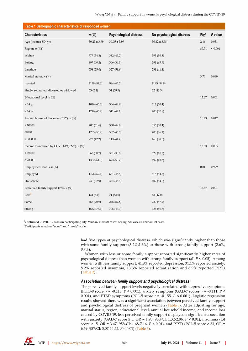
Wang YN et al. Family support in women’s psychological distress during the COVID-19
WJP https://www.wjgnet.com 369 July 19, 2021 Volume 11 Issue 7
Table 1 Demographic characteristics of responded women
Characteristics n (%) Psychological distress No psychological distress F/χ2 P value
Age (mean ± SD, yr) 30.25 ± 3.99 30.05 ± 3.99 30.42 ± 3.98 2.16 0.031
Region, n (%)1 89.71 < 0.001
Wuhan 777 (34.8) 382 (49.2) 395 (50.8)
Peking 897 (40.2) 306 (34.1) 591 (65.9)
Lanzhou 558 (25.0) 327 (58.6) 231 (41.4)
Marital status, n (%) 3.70 0.069
married 2179 (97.6) 984 (45.2) 1195 (54.8)
Single, separated, divorced or widowed 53 (2.4) 31 (58.5) 22 (41.5)
Educational level, n (%) 13.67 0.001
< 14 yr 1016 (45.6) 504 (49.6) 512 (50.4)
≥ 14 yr 1216 (43.7) 511 (42.1) 705 (57.9)
Annual household income (CNY), n (%) 10.23 0.017
< 80000 706 (31.6) 350 (49.6) 356 (50.4)
80000 1255 (56.2) 552 (43.9) 703 (56.1)
≥ 300000 273 (12.2) 113 (41.4) 160 (58.6)
Income loss caused by COVID-19(CNY), n (%) 13.83 0.003
< 20000 862 (38.7) 331 (38.8) 522 (61.2)
≥ 20000 1362 (61.3) 673 (50.7) 692 (49.3)
Employment status, n (%) 0.01 0.999
Employed 1496 (67.1) 681 (45.5) 815 (54.5)
Housewife 736 (32.9) 334 (45.4) 402 (54.6)
Perceived family support level, n (%) 13.57 0.001
Less2 134 (6.0) 71 (53.0) 63 (47.0)
Some 466 (20.9) 246 (52.8) 220 (47.2)
Strong 1632 (73.1) 706 (43.3) 926 (56.7)
1Confirmed COVID-19 cases in participating city: Wuhan: > 50000 cases; Beijing: 581 cases; Lanzhou: 24 cases.2Participants rated on “none” and “rarely” scale.
had five types of psychological distress, which was significantly higher than those with some family support (3.2%,1.3%) or those with strong family support (2.6%, 0.7%).
Women with less or some family support reported significantly higher rates of psychological distress than women with strong family support (all P < 0.05). Among women with less family support, 41.8% reported depression, 31.1% reported anxiety, 8.2% reported insomnia, 13.3% reported somatization and 8.9% reported PTSD (Table 2).
Association between family support and psychological distress The perceived family support levels negatively correlated with depressive symptoms (PHQ-9 score, r = -0.118, P < 0.001), anxiety symptoms (GAD-7 scores, r = -0.111, P < 0.001), and PTSD symptoms (PCL-5 score r = -0.155, P < 0.001). Logistic regression results showed there was a significant association between perceived family support and psychological distress of pregnant women (Table 3). After adjusting for age, marital status, region, educational level, annual household income, and income loss caused by COVID-19, less perceived family support displayed a significant association with anxiety (GAD-7 score ≥ 5, OR = 1.98, 95%CI: 1.32-2.96, P < 0.01), insomnia (ISI score ≥ 15, OR = 3.47, 95%CI: 1.68-7.16, P < 0.01), and PTSD (PCL-5 score ≥ 33, OR = 6.69, 95%CI: 3.07-14.55, P < 0.01) (Table 3).

Wang YN et al. Family support in women’s psychological distress during the COVID-19
WJP https://www.wjgnet.com 370 July 19, 2021 Volume 11 Issue 7
DISCUSSIONTo our best knowledge, this is the first study to explore the relationship between perceived family support and psychological distress in pregnant women during major life-threatening public health events. In this study, we had 3 main findings: (1) A significant proportion (45.4%) of pregnant women reported at least one psychological distress during COVID-19 pandemic; (2) The risk of insomnia, anxiety and PTSD in women with less family support was 3.46 times, 1.97 times and 6.69 times higher than that in women with strong perceived family support, respectively; and (3) Depression symptoms (PHQ-9 score), anxiety symptoms (GAD-7 score) and PTSD symptoms (PCL-5 score) were significantly negatively associated with perceived family support.
It is speculated that residents living in Wuhan suffered from higher levels of psychological distress, because this place was most affected by the epidemic[7] in mainland China. This may also increase the risk of psychological distress in pregnant women[11]. Interestingly, our study did not find an increase in the rate of maternal psychological distress amongst women in Wuhan. On the contrary, more women living in Lanzhou reported suffering from psychological distress, though it was only 24 confirmed COVID-19 cases in Lanzhou. That may be due to the fact that more women living in Lanzhou reported insufficient support from family numbers than women living in Beijing and Wuhan. The results showed that depression (35.6%) and anxiety (22.0%) were the most common disorders reported by pregnant women. In addition, at the peak of the COVID-19 epidemic, the pregnant women also suffered from somatization (8.1%), insomnia (3.4%) and PTSD (2.3%). The findings were similar to those of previous studies, indicating that pregnant women may be more vulnerable to depression in the context of the COVID-19 epidemic. A recent study reported that during the COVID-19 epidemic, the depression rate amongst Chinese pregnant women rose to 29.6%[11]. Another study showed that 28.8% of Danish women had anxiety/depressive symptoms during the COVID-19 pandemic[26].
There is extensive evidence that psychological distress during pregnancy are thought to increase the risk of developing psychological dysfunction in the offspring[27]. Pregnancy are very special periods for women. Affected by changes in the level and function of the endocrine system during pregnancy, mothers often experience huge mood swings and even mental disorders, such as anxiety[9], depression[10], and sleep disturbance[28]. The prevalence of depression in pregnant and postpartum women was estimated to be between 7% and 25%[10,26,27], and the prevalence of anxiety disorders is estimated to be between 4% and 39%[29]. However, our results showed that with different levels of family support, there were significant differences in the proportion of women with mental disorders during pregnancy. Women with inadequate family support were more likely to suffer from multiple psychological distresses. The strengthening of family support, the reporting rate of maternal psycho-logical distress reduced. These findings highlight the essential role of adequate family support in mental health of pregnant women through major life-threatening public health events.
Social support, including supports from family members, colleagues, friends, neighbors, professionals, and organizations, is important for maternal mental health[15]. Due to the sudden outbreak, features and clinic symptoms of the COVID-19 are still unclear during the peak of the epidemic. The Chinese government intensified its management of the pandemic through public health interventions, such as strengthening some blockades, demand of staying at home. Hospitals had to terminate health care services for pregnant women to prevent infection, and most of them were trapped at home and isolated themselves from society. The confinement conditions have made the support of family members the only source of social support. In the general population, social support is widely regarded as a key factor in relieving perinatal depression[30]. A large number of studies have confirmed that family support has a positive effect on the level of perceived psychological stress[6,31-33]. According to Cohen[34], the relationship between perceived family support and life satisfaction was very strong.
Perceived family support makes a person feel cared for, loved, and dependent on family numbers when needed[33]. The levels of perceived family support can affect the way people deal with and adapt to stressful events, thereby reducing the negative effects on mental and physical health[6]. Therefore, adequate family support can alleviate the pressure caused by the COVID-19 outbreak. However, although physical care for pregnant women has been greatly improved in China in the past few decades, little attention has been paid to emotional care[15]. The findings of this study emphasized the importance of emotional care for pregnant women. The role and mechanism of family support in alleviating the negative psychological effects of

Wang YN et al. Family support in women’s psychological distress during the COVID-19
WJP https://www.wjgnet.com 371 July 19, 2021 Volume 11 Issue 7
Table 2 Self-reported psychological distresses of pregnant women with different perceived family support level, n (%)
Total Less PFS Some PFS Strong PFS F/χ2 P value
Depression 795 (35.6) 56 (41.8) 207 (44.4) 532 (32.6) 24.15 < 0.001
Anxiety 491 (22.0) 42 (31.1) 128 (27.5) 321 (19.6) 19.79 < 0.001
Insomnia 77 (3.4) 11 (8.2) 15 (3.2) 51 (3.1) 9.72 0.008
Somatization 180 (8.1) 18 (13.3) 41(8.8) 121(7.4) 6.34 0.042
PTSD 52 (2.3) 12 (8.9) 14 (3.0) 26 (1.6) 30.41 < 0.001
PFS: Progress-free survival; PTSD: Posttraumatic stress disorder.
Table 3 Association between perceived family support [n (%)] and self-reported maternal psychological distresses
Depression Anxiety Insomnia Somatization PTSD
Spearman correlation coefficient, r
-0.118b -0.111b -0.025 -0.038 -0.155b
Multiple linear regression, β
-0.973b -0.638b -0.195 -0.418 -2.238b
Logistic regression analysis, OR (95%CI)
Strong support 1 1 1 1 1
Some support 1.56 (1.26-1.94)b 1.69 (1.24-2.04)b 1.13 (0.62-2.04) 1.02 (0.65-1.45) 1.91 (0.96-3.79)
Less support 1.33 (0.90-1.92) 1.98 (1.32-2.96)b 3.47 (1.68-7.16)b 1.41 (0.80-2.48) 6.69 (3.07-14.55)b
OR and 95%CI were estimated using binary logistic regression and adjusted for age, marital status, region place of residence, educational level, annual household income, income loss caused by COVID-19.bP < 0.01.PTSD: Posttraumatic stress disorder.
adverse events have not been clearly or fully explained. Further research is needed to understand the role and mechanism of this factor in order to utilize family support to help pregnant women in major life-threatening public health events.
However, several limitations have to be considered when interpreting these findings. Firstly, the data obtained from online surveys and maternal psychological distress relied on self-reported measurements. Women may inaccurately report the presence or absence of psychological distress. Secondly, although internationally valid and reliable questionnaires were utilized in this study, these questionnaires did not provide a diagnosis of mental disorders. Thirdly, the evaluation of family support used the subjective feelings of women rather than the social support rating scale. This may make our results less precise. Fourthly, other influencing factors such as family relationship, family conflict, and family resources are also important variables that affect the psychological well-being of pregnant and postpartum women, which were not taken into account in this study. Finally, a cross-sectional analysis of this study suggests that there is an association between maternal psychological distress and family support, rather than a causal relationship. Future research may focus on a longitudinal study to confirm causality.
CONCLUSIONThe results of this study indicate that the COVID-19 pandemic has a negative impact on the mental health of Chinese pregnant women. Family support was significantly negatively correlated with psychological distress. Compared with women with strong family support, women with insufficient family support were more associated with the risk of anxiety, insomnia, depression and PTSD. This study shows that in major life-threatening public health events, family support has a positive impact on the

Wang YN et al. Family support in women’s psychological distress during the COVID-19
WJP https://www.wjgnet.com 372 July 19, 2021 Volume 11 Issue 7
Figure 1 Frequency for self-reported multiple psychological distresses in different perceived family support level. 1: Report one type of psychological distress; 2: Report two type of psychological distress; 3: Report three type of psychological distress; 4: Report four type of psychological distress; 5: Report five type of psychological distress.
mental health of pregnant women. However, cross-sectional nature limits the interpretation of the causal relationship between family support and mental health. Further longitudinal studies about the causal relationship between family support and mental health will be an essential area of future research, which may help to identify appropriate and effective interventions for pregnant and postpartum women to prevent from the mental health problems.
ARTICLE HIGHLIGHTSResearch backgroundPregnant women may be more vulnerable to psychological distress during major life-threatening public health events. The spread of the corona virus disease 2019 (COVID-19) is likely to cause greater psychological stress, which may cause extra psychological problems for pregnant women.
Research motivationThe spread of the COVID-19 may cause extra psychological problems for pregnant women. Women who have better supportive networks of family may experience less psychological stress. However, the literatures on the role of family support in maternal psychological distress during the COVID-19 pandemic was limited.
Research objectivesThis study aimed to clarify the potential role of family support on psychological distress for women during pregnancy stages at the peak of the COVID-19 epidemic.
Research methodsThe authors retrospectively collected socio-demographic variables, information of pregnancy, and information of psychological distress and the perceived family support of pregnant women in China.
Research resultsAmong 2232 pregnant women, 45.4% women reported having at least one psycho-logical distress during the COVID-19 pandemic. The women who reported having inadequate family support were more likely to suffer from multiple psychological distress than women received adequate family support. Perceived family support was negatively correlated with depressive symptoms, anxiety symptoms, and PTSD symptoms.

Wang YN et al. Family support in women’s psychological distress during the COVID-19
WJP https://www.wjgnet.com 373 July 19, 2021 Volume 11 Issue 7
Research conclusionsFamily support was significantly negatively correlated with psychological distress. Adequate family support has a positive impact on the mental health of pregnant women during the COVID-19 pandemic.
Research perspectivesAdequate family support is important for maternal mental health. Further research is needed to understand the role and mechanism of this factor in order to utilize family support to help pregnant women in major life-threatening public health events.
REFERENCESWang C, Horby PW, Hayden FG, Gao GF. A novel coronavirus outbreak of global health concern. Lancet 2020; 395: 470-473 [PMID: 31986257 DOI: 10.1016/S0140-6736(20)30185-9]
1
World Health Organization. Coronavirus disease (COVID-19) pandemic. Available from: https://www.who.int/
2
Rasmussen SA, Smulian JC, Lednicky JA, Wen TS, Jamieson DJ. Coronavirus Disease 2019 (COVID-19) and pregnancy: what obstetricians need to know. Am J Obstet Gynecol 2020; 222: 415-426 [PMID: 32105680 DOI: 10.1016/j.ajog.2020.02.017]
3
Moccia L, Janiri D, Giuseppin G, Agrifoglio B, Monti L, Mazza M, Caroppo E, Fiorillo A, Sani G, Di Nicola M, Janiri L. Reduced Hedonic Tone and Emotion Dysregulation Predict Depressive Symptoms Severity during the COVID-19 Outbreak: An Observational Study on the Italian General Population. Int J Environ Res Public Health 2020; 18 [PMID: 33396363 DOI: 10.3390/ijerph18010255]
4
Di Nicola M, Dattoli L, Moccia L, Pepe M, Janiri D, Fiorillo A, Janiri L, Sani G. Serum 25-hydroxyvitamin D levels and psychological distress symptoms in patients with affective disorders during the COVID-19 pandemic. Psychoneuroendocrinology 2020; 122: 104869 [PMID: 32956989 DOI: 10.1016/j.psyneuen.2020.104869]
5
Ren JH, Chiang CL, Jiang XL, Luo BR, Liu XH, Pang MC. Mental disorders of pregnant and postpartum women after earthquakes: a systematic review. Disaster Med Public Health Prep 2014; 8: 315-325 [PMID: 25098648 DOI: 10.1017/dmp.2014.62]
6
Qiu J, Shen B, Zhao M, Wang Z, Xie B, Xu Y. A nationwide survey of psychological distress among Chinese people in the COVID-19 epidemic: implications and policy recommendations. Gen Psychiatr 2020; 33: e100213 [PMID: 32215365 DOI: 10.1136/gpsych-2020-100213]
7
Wang C, Pan R, Wan X, Tan Y, Xu L, Ho CS, Ho RC. Immediate Psychological Responses and Associated Factors during the Initial Stage of the 2019 Coronavirus Disease (COVID-19) Epidemic among the General Population in China. Int J Environ Res Public Health 2020; 17 [PMID: 32155789 DOI: 10.3390/ijerph17051729]
8
Kang YT, Yao Y, Dou J, Guo X, Li SY, Zhao CN, Han HZ, Li B. Prevalence and Risk Factors of Maternal Anxiety in Late Pregnancy in China. Int J Environ Res Public Health 2016; 13 [PMID: 27153080 DOI: 10.3390/ijerph13050468]
9
Asaye MM, Muche HA, Zelalem ED. Prevalence and Predictors of Postpartum Depression: Northwest Ethiopia. Psychiatry J 2020; 2020: 9565678 [PMID: 32411780 DOI: 10.1155/2020/9565678]
10
Wu Y, Zhang C, Liu H, Duan C, Li C, Fan J, Li H, Chen L, Xu H, Li X, Guo Y, Wang Y, Li J, Zhang T, You Y, Yang S, Tao X, Xu Y, Lao H, Wen M, Zhou Y, Wang J, Chen Y, Meng D, Zhai J, Ye Y, Zhong Q, Yang X, Zhang D, Zhang J, Wu X, Chen W, Dennis CL, Huang HF. Perinatal depressive and anxiety symptoms of pregnant women during the coronavirus disease 2019 outbreak in China. Am J Obstet Gynecol 2020; 223: 240.e1-240. e9 [PMID: 32437665 DOI: 10.1016/j.ajog.2020.05.009]
11
Horton R. Offline: 2019-nCoV-"A desperate plea". Lancet 2020; 395: 400 [PMID: 32035537 DOI: 10.1016/s0140-6736(20)30299-3]
12
Lau Y, Yin L, Wang Y. Antenatal depressive symptomatology, family conflict and social support among Chengdu Chinese women. Matern Child Health J 2011; 15: 1416-1426 [PMID: 20957512 DOI: 10.1007/s10995-010-0699-z]
13
Lee SH, Kuo CP, Hsiao CY, Lu YC, Hsu MY, Kuo PC, Lee MS, Lee MC. Development of a Chinese childbearing attitude questionnaire for infertile women receiving in vitro fertilization treatment. J Transcult Nurs 2013; 24: 127-133 [PMID: 23460456 DOI: 10.1177/1043659612472060]
14
Xie RH, Yang J, Liao S, Xie H, Walker M, Wen SW. Prenatal family support, postnatal family support and postpartum depression. Aust N Z J Obstet Gynaecol 2010; 50: 340-345 [PMID: 20716261 DOI: 10.1111/j.1479-828X.2010.01185.x]
15
Hu Y, Wang Y, Wen S, Guo X, Xu L, Chen B, Chen P, Xu X. Association between social and family support and antenatal depression: a hospital-based study in Chengdu, China. BMC Pregnancy Childbirth 2019; 19: 420 [PMID: 31744468 DOI: 10.1186/s12884-019-2510-5]
16
Bastien CH, Vallières A, Morin CM. Validation of the Insomnia Severity Index as an outcome measure for insomnia research. Sleep Med 2001; 2: 297-307 [PMID: 11438246 DOI: 10.1016/S1389-9457(00)00065-4]
17

Wang YN et al. Family support in women’s psychological distress during the COVID-19
WJP https://www.wjgnet.com 374 July 19, 2021 Volume 11 Issue 7
Morin CM, Belleville G, Bélanger L, Ivers H. The Insomnia Severity Index: psychometric indicators to detect insomnia cases and evaluate treatment response. Sleep 2011; 34: 601-608 [PMID: 21532953 DOI: 10.1093/sleep/34.5.601]
18
Kroenke K, Spitzer RL, Williams JB, Monahan PO, Löwe B. Anxiety disorders in primary care: prevalence, impairment, comorbidity, and detection. Ann Intern Med 2007; 146: 317-325 [PMID: 17339617 DOI: 10.7326/0003-4819-146-5-200703060-00004]
19
Choueiry N, Salamoun T, Jabbour H, El Osta N, Hajj A, Rabbaa Khabbaz L. Insomnia and Relationship with Anxiety in University Students: A Cross-Sectional Designed Study. PLoS One 2016; 11: e0149643 [PMID: 26900686 DOI: 10.1371/journal.pone.0149643]
20
Kroenke K, Spitzer RL, Williams JB. The PHQ-9: validity of a brief depression severity measure. J Gen Intern Med 2001; 16: 606-613 [PMID: 11556941 DOI: 10.1046/j.1525-1497.2001.016009606.x]
21
Derogatis LR, Lipman RS, Covi L. SCL-90: an outpatient psychiatric rating scale--preliminary report. Psychopharmacol Bull 1973; 9: 13-28 [PMID: 4682398]
22
Blevins CA, Weathers FW, Davis MT, Witte TK, Domino JL. The Posttraumatic Stress Disorder Checklist for DSM-5 (PCL-5): Development and Initial Psychometric Evaluation. J Trauma Stress 2015; 28: 489-498 [PMID: 26606250 DOI: 10.1002/jts.22059]
23
Ibrahim H, Ertl V, Catani C, Ismail AA, Neuner F. The validity of Posttraumatic Stress Disorder Checklist for DSM-5 (PCL-5) as screening instrument with Kurdish and Arab displaced populations living in the Kurdistan region of Iraq. BMC Psychiatry 2018; 18: 259 [PMID: 30115040 DOI: 10.1186/s12888-018-1839-z]
24
Wang XD, MaH. Social Support Rating Scale. Beijing: Chinese Mental Health Publishing House, 1999
25
Gavin NI, Gaynes BN, Lohr KN, Meltzer-Brody S, Gartlehner G, Swinson T. Perinatal depression: a systematic review of prevalence and incidence. Obstet Gynecol 2005; 106: 1071-1083 [PMID: 16260528 DOI: 10.1097/01.AOG.0000183597.31630.db]
26
Aktar E, Qu J, Lawrence PJ, Tollenaar MS, Elzinga BM, Bögels SM. Fetal and Infant Outcomes in the Offspring of Parents With Perinatal Mental Disorders: Earliest Influences. Front Psychiatry 2019; 10: 391 [PMID: 31316398 DOI: 10.3389/fpsyt.2019.00391]
27
Krawczak EM, Minuzzi L, Simpson W, Hidalgo MP, Frey BN. Sleep, daily activity rhythms and postpartum mood: A longitudinal study across the perinatal period. Chronobiol Int 2016; 33: 791-801 [PMID: 27097327 DOI: 10.3109/07420528.2016.1167077]
28
van Heyningen T, Honikman S, Myer L, Onah MN, Field S, Tomlinson M. Prevalence and predictors of anxiety disorders amongst low-income pregnant women in urban South Africa: a cross-sectional study. Arch Womens Ment Health 2017; 20: 765-775 [PMID: 28852868 DOI: 10.1007/s00737-017-0768-z]
29
Mann JR, Mannan J, Quiñones LA, Palmer AA, Torres M. Religion, spirituality, social support, and perceived stress in pregnant and postpartum Hispanic women. J Obstet Gynecol Neonatal Nurs 2010; 39: 645-657 [PMID: 21039849 DOI: 10.1111/j.1552-6909.2010.01188.x]
30
Cohen S, Wills TA. Stress, social support, and the buffering hypothesis. Psychol Bull 1985; 98: 310-357 [PMID: 3901065 DOI: 10.1037/0033-2909.98.2.310]
31
Isobel S, Goodyear M, Foster K. Psychological Trauma in the Context of Familial Relationships: A Concept Analysis. Trauma Violence Abuse 2019; 20: 549-559 [PMID: 29333976 DOI: 10.1177/1524838017726424]
32
Campos B, Ullman JB, Aguilera A, Dunkel Schetter C. Familism and psychological health: the intervening role of closeness and social support. Cultur Divers Ethnic Minor Psychol 2014; 20: 191-201 [PMID: 24773004 DOI: 10.1037/a0034094]
33
Cohen J. Statistical power analysis for the behavioral sciences. 2nd ed. Lawrence Erlbaum Associates, 1988
34

WJP https://www.wjgnet.com 375 July 19, 2021 Volume 11 Issue 7
World Journal of
PsychiatryW J PSubmit a Manuscript: https://www.f6publishing.com World J Psychiatr 2021 July 19; 11(7): 375-387
DOI: 10.5498/wjp.v11.i7.375 ISSN 2220-3206 (online)
ORIGINAL ARTICLE
Prospective Study
Classification of subtypes of patients with eating disorders by correspondence analysis
Josune Martín, Ane Anton-Ladislao, Ángel Padierna, Belén Berjano, José María Quintana
ORCID number: Josune Martín 0000-0001-5281-4426; Ane Anton-Ladislao 0000-0000-0000-0001; Ángel Padierna 0000-0000-0000-0002; Belén Berjano 0000-0000-0000-0003; José María Quintana 0000-0003-2170-7876.
Author contributions: Martín J, Padierna A, and Quintana JM designed the study; Martín J, Padierna A and Berjano B performed the data collection; Martín J and Antón-Ladislao A performed data analyses; Martín J and Padierna A were responsible for manuscript writing; all the authors contributed to interpreting the data; and all authors read and approved the final manuscript.
Supported by the Carlos III Health Institute Project “Adaptation and Validation of the Clinical Assessment Inventory for Eating Disorders”, No. PI09/90832.
Institutional review board statement: The study was approved by the Ethics Committee of the Galdakao-Usansolo Hospital.
Clinical trial registration statement: The clinical trial is registered with ClinicalTrials.gov, using identifier NCT02483117. Details can be found at https://clinicaltrials.gov/ct2/show/NCT02483117.
Josune Martín, Ane Anton-Ladislao, José María Quintana, Department of Research, Galdakao-Usansolo Hospital, Galdakao 48960, Spain
Josune Martín, Kronikgune Institute for Health Services Research, Barakaldo 48902, Spain
Josune Martín, Ángel Padierna, José María Quintana, Health Services Research on Chronic Diseases Network – REDISSEC, Galdakao 48960, Spain
Ángel Padierna, Belén Berjano, Department of Psychiatry, Galdakao-Usansolo Hospital, Galdakao 48960, Spain
Corresponding author: Josune Martín, PhD, Research Scientist, Department of Research, Galdakao-Usansolo Hospital, B Labeaga s/n, Galdakao 48960, Spain. [email protected]
AbstractBACKGROUND Grouping eating disorders (ED) patients into subtypes could help improve the establishment of more effective diagnostic and treatment strategies.
AIM To identify clinically meaningful subgroups among subjects with ED using multiple correspondence analysis (MCA).
METHODS A prospective cohort study was conducted of all outpatients diagnosed for an ED at an Eating Disorders Outpatient Clinic to characterize groups of patients with ED into subtypes according to sociodemographic and psychosocial impairment data, and to validate the results using several illustrative variables. In all, 176 (72.13%) patients completed five questionnaires (clinical impairment assessment, eating attitudes test-12, ED-short form health-related quality of life, metacog-nitions questionnaire, Penn State Worry Questionnaire) and sociodemographic data. ED patient groups were defined using MCA and cluster analysis. Results were validated using key outcomes of subtypes of ED.
RESULTS Four ED subgroups were identified based on the sociodemographic and psychosocial impairment data.
CONCLUSION

Martín J et al. Subtypes of patients with eating disorders
WJP https://www.wjgnet.com 376 July 19, 2021 Volume 11 Issue 7
Informed consent statement: All study participants provided informed written consent prior to study enrollment.
Conflict-of-interest statement: The authors of this manuscript having no conflicts of interest to disclose.
Data sharing statement: There is no additional data available.
CONSORT 2010 statement: The authors have read the CONSORT 2010 statement, and the manuscript was prepared and revised according to the CONSORT 2010 statement.
Open-Access: This article is an open-access article that was selected by an in-house editor and fully peer-reviewed by external reviewers. It is distributed in accordance with the Creative Commons Attribution NonCommercial (CC BY-NC 4.0) license, which permits others to distribute, remix, adapt, build upon this work non-commercially, and license their derivative works on different terms, provided the original work is properly cited and the use is non-commercial. See: http://creativecommons.org/Licenses/by-nc/4.0/
Manuscript source: Invited manuscript
Specialty type: Psychiatry
Country/Territory of origin: Spain
Peer-review report’s scientific quality classificationGrade A (Excellent): 0 Grade B (Very good): 0 Grade C (Good): C Grade D (Fair): 0 Grade E (Poor): 0
Received: February 8, 2021 Peer-review started: February 8, 2021 First decision: March 30, 2021 Revised: April 9, 2021 Accepted: June 16, 2021 Article in press: June 16, 2021 Published online: July 19, 2021
P-Reviewer: Crenn PP S-Editor: Zhang H
ED patients were differentiated into well-defined outcome groups according to specific clusters of compensating behaviours.
Key Words: Multiple correspondence analysis; Eating disorders; Compensating behaviour; Observational descriptive study
©The Author(s) 2021. Published by Baishideng Publishing Group Inc. All rights reserved.
Core Tip: This is the first study to apply multiple correspondence analysis to eating disorders (ED) diagnostic data and to use cluster analysis (CA) in such detail to search for ED patient groups in this area. Multiple correspondence analysis and CA made it possible to identify different typologies of patients with specific features. Grouping ED patients into subtypes could help improve the establishment of more effective strategies of diagnosis and treatment, and improve patient care and prognosis in clinical practice.
Citation: Martín J, Anton-Ladislao A, Padierna Á, Berjano B, Quintana JM. Classification of subtypes of patients with eating disorders by correspondence analysis. World J Psychiatr 2021; 11(7): 375-387URL: https://www.wjgnet.com/2220-3206/full/v11/i7/375.htmDOI: https://dx.doi.org/10.5498/wjp.v11.i7.375
INTRODUCTIONEating disorders (ED) are serious psychiatric conditions; clinical presentations of persons with ED[1,2] vary substantially and they may be associated with many factors, e.g., sociodemographic as gender[3] or clinical as personality profiles[4]. The Diagnostic and Statistical Manual of Mental Disorders (DSM)-5[5] aims to better capture the presentations of ED symptoms observed by modifying previous ED diagnostic criteria. However, as Turner et al[6] have, noted some researchers remain concerned, that these adjustments will fail adequately to address the substantial heterogeneity in clinical presentations amongst patients with ED[1]. It is important to research subtypes of ED, since otherwise the field might merely end up ‘studying what it defines’ (or failing to study anything it does not define)[7]. Thus, removing any reference to non-purging compensatory behaviors would reinforce the impression-(created by subtyping) that bulimic-type ED characterized by purging behaviors is more severe than that involving non-purging behaviors when there is actually little empirical evidence to support this view[7,8]. Insofar as distinct subgroups of ED patients can be reliably identified, it is possible that these groupings might be used to inform assessment, treatment and future diagnostic nosologies[9]. Multiple corres-pondence analysis (MCA) is an exploratory technique that offers descriptive patterns based on the categories of the original active variables[10,11]. It transforms the information on the categorical active variables into continuous factors. The relative positions of the categories given by the MCA factors are used to perform the cluster analysis (CA) which classifies information into relatively homogenous groups. By combining MCA and CA it might be possible to arrive at a classification of the subjects suggested by the data, rather than defined a priori, where subjects in each group are similar to one another but dissimilar to those of other groups[10,12].
Grouping ED patients into subtypes could help improve the establishment of more effective strategies of diagnosis and treatment, and improve patient care and prognosis in clinical practice. The aim of this study was to identify ED patient subtypes. To this end, MCA and CA statistical techniques were combined to analyze clinical data obtained in a prospective cohort study of ED patients treated in an ED Outpatient Clinic. The subtypes were then validated by estimating their relationships to key outcomes such as health-related quality of life (HRQoL), psychosocial impairment due to ED, worry and metacognitions, eating problems, and sociodemographic variables.

Martín J et al. Subtypes of patients with eating disorders
WJP https://www.wjgnet.com 377 July 19, 2021 Volume 11 Issue 7
L-Editor: A P-Editor: Ma YJ
MATERIALS AND METHODSParticipantsA prospective cohort study was conducted of all patients diagnosed with and treated for an ED at the Eating Disorders Outpatient Clinic The clinic forms part of the psychiatric services at the hospital, which serves a population of 300000. It is part of the Basque Health Care Service, which provides unlimited free care to nearly 100% of the population. Outpatients recruited between January 2010 and January 2011 were considered eligible for the study if they had received a diagnosis of anorexia nervosa (AN), bulimia nervosa (BN), or an ED not otherwise specified (EDNOS) by a psychi-atrist, on the basis of the criteria listed in the Diagnostic and Statistical Manual of Mental Disorders, 4th edition text revision[5]. Patients were required to provide written informed consent before participating. They were excluded if they had a malignant, severe organic disease were unable to complete the questionnaires because of langua-gedifficulties, or had not given their written informed consent to participate in the study.
The study received approval from the institutional review board of the Hospital.
MeasuresED patients gave their sociodemographic data, including age, gender, marital status, education, employment status, and living situation.
The clinical impairment assessment (CIA v.3.0)[13,14] is a 16-item self-report instrument specifically designed to assess psychosocial impairment secondary to features of an ED. A higher score indicates greater impairment. The CIA report of psychometric properties indicated that the measure was both adequate and valid[13,15].
Eating pathologies were measured using the eating attitudes test-12 (EAT-12)[16]. This is a 12-item instrument, which uses a 4-point scale, with scores from 0 (never) to 3 (always). Higher scores indicate more disordered eating. Its validity as a measure of disordered eating has been backed by previous studies[17,18].
The quality of life of ED patients was evaluated using the health-related quality of life in ED-short form)[19] ,a 20-item questionnaire divided into two domains: social maladjustment and mental health and functionality. The lower the quality of life, the higher the score[15,20].
The metacognitions questionnaire (MCQ-30)[21] is a brief multidimensional measure of a range of metacognitive processes and metacognitive beliefs related to worry and cognition relevant to vulnerability to and maintenance of emotional disorders. Higher scores reflect a more dysfunctional metacognitive belief. The subscales have good psychometric properties[21,22].
The Penn State Worry Questionnaire (PSWQ-R) is a 16-item self-report measure of trait worry that is widely used to measure pathological worry[23]. A Spanish version reduced to 11 items was used[24]. Higher scores indicate greater levels of pathological worry. The PSWQ-R has been shown to have good psychometric properties[25,26].
ProcedureData gathering began in 2010. Psychiatrists who collaborated in the study informed their patients of the aims of the study and recorded sociodemographic information. Patients agreeing participate were mailed questionnaires and an informed consent form ,which they were asked to mail back using an enclosed, pre- franked envelope. Two reminders were mailed out at 15-d intervals to patients who failed to reply to the first mailing.
Statistical analysisVarious multivariate techniques are used in order to synthesize the information contained in a large set of explanatory variables into a few components, also called factors. One of them is the technique selected for this analysis, MCA, which is designed for categorical explanatory variables, while others, as principal component analysis, are designed for continuous variables. Based on the categories of the original variables, MCA provides descriptive patterns by factors. In the continuous factors, therefore, each category of variables is represented by a numerical value and a positive/negative sign, used for interpretation. Graphical displays of these factors are very useful in interpretation, as the association between the categories is indicated by their relative position on the graph. The closer the categories are to one another, the stronger the association. Variables included in the analysis are known as active variables, whilst those not included in the analysis but used to verify the relationship

Martín J et al. Subtypes of patients with eating disorders
WJP https://www.wjgnet.com 378 July 19, 2021 Volume 11 Issue 7
with active variables are termed illustrative variables or outcomes[10]. A descriptive analysis was made of sociodemographic and psychosocial impairment data, using frequencies and percentages. Means and standard deviations were also used as additional information for questionnaires of psychosocial impairment. The active variables in the MCA were gender, age (13-25, 26-35, 35-63), marital status [single, spouse/partner, divorced/widow(er)], education completed (primary, secondary, higher), employment status (employed, unemployed, student, disabled, unpaid work/housewife), living situation (living alone, with partner/children, friends, parents/siblings), MCQ-30 questionnaire (≤ 57, 58-75, > 75), CIA questionnaire (< 16, ≥ 16), EAT-12 questionnaire (< 8, ≥ 8), HeRQoLED-SocM (≤ 50, > 50), HeRQoLED-MHF (≤ 50, > 50) and PSWQ-R (≤ 28, > 28). Type (AN, BN and EDNOS) and subtype (restrictive, purgative and binge) of ED patients were used as illustrative variables.
For classification purposes, CA organizes information into relatively homogeneous groups based on their values in a range of variables — in this case, based on the factors derived from the MCA. In other words, the objective of the CA is to assign individuals into different groups, in the way that individuals from the same group are similar to each other, but dissimilar from individuals of other groups. The number of groups derived from the CA is selected using the minimum inertia lost method[27].
The association between the active variables and the groups derived from the CA was evaluated using the chi-square test (or Fisher’s exact test when expected frequencies were less than 5). The non-parametric Kruskal-Wallis test was used for the scores of the psychosocial impairment questionnaires. In addition, the relationship of outcomes or illustrative variables was evaluated according to the groups obtained from the CA. In order to see the stability of the groups obtained and since we had all the active variables measured at 12 mo of follow up too, the same analysis was replicated with the variables at 12 mo of follow up. Statistical analyses were carried out with R v3.0.2 and SAS 9.4 software (copyright, SAS Institute Inc.). “SAS” and the names of all other SAS Institute Inc. products and services are registered trademarks or trademarks of SAS Institute Inc., Cary, NC, United States.
The study was approved by the Ethics Committee of the Galdakao-Usansolo Hospital. Written consent for participation was obtained. ClinicalTrials.gov Identifier: NCT02483117. All methods were used in accordance with the relevant guidelines and regulations.
RESULTSA total of 244 patients with ED were invited to take part in the study. Of these, 176 filled out the questionnaires. Early dropouts were largely due to patients failing to consent to participation. The mean of the CIA questionnaire was 19.5 (SD 13.6), which indicates a high level of impairment due to ED.
Results from the MCA showed that 74% of data variability could be explained by two factors, the first primarily associated with the HRQoL and the second with socio-demographic data. Figure 1 shows the map created by the first and second factors. The first factor is represented on the horizontal axis and the second on the vertical axis. Variables that were well-represented in the first factor were: the questionnaires related to psychosocial impairment; eating problems; HRQoL; worry; and metacognitions. Categories located in the positive part (right) of the map included lower values at CIA, EAT-12, HeRQoLED-s, MCQ-30, PSWQ. In contrast, higher values of the question-naires were in the negative part (left). This axis was defined as “Psychosocial impairment: from high to low”.Moreover, the relative position of the illustrative variables on the graph indicates that some subtypes of diagnosis according to compensatory behaviour as well as some subtypes of diagnosis related to DSM-IV-TR classification were well represented by this factor. Indeed, restrictive behaviour (AN, EDNOS) was located in the right side of the axis, whereas purgative behaviour (BN, EDNOS) stood on the left of the axis. The variables that were well-represented in the second factor were socio-demographic variables. Categories such as being male, having a spouse, having secondary studies, being a housewife and living with a partner/children were related to the positive part (top). In contrast, the categories of being single, having higher studies, being a student and living with friends or parents or/and a sibling, were related to the negative part of the axis (bottom). This axis was therefore interpreted as “socio-demographic data”. As in the other axis, some categories of the illustrative variables were well-represented by this axis. Purgative AN was located in the negative part, while binge behaviour (only in patients with EDNOS diagnosis) was located in the positive part.

Martín J et al. Subtypes of patients with eating disorders
WJP https://www.wjgnet.com 379 July 19, 2021 Volume 11 Issue 7
Figure 1 Graphical displays of the two factors/components derived from the multiple correspondence analysis. Active variables (sociodemographic variables). Age (≤ 25, 26-35, > 35); Marital status (single, spouse/partner, divorced/widow(er); Educational level (primary education, secondary education, higher education); Employment status (employed, unemployed, student, disabled, non-paid work/housewife); Living situation (alone, partner/children, friends, parents/siblings). Active variables (questionnaires). Metacognitions questionnaire, (0: ≤ 57, 1: 58-75, 2: > 75). Clinical impairment assessment, (0: ≤ 16, 1: ≥ 16); Eating attitudes test, (0: ≤ 8, 1: ≥ 8). Health-related quality of life in eating disorder-short form, [social maladjustment domain, (0: ≤ 50, 1: > 50); Mental health and functionality domain, (0: ≤ 50, 1: > 50); Penn state worry questionnaire (0: ≤ 28, 1: > 28). Red triangles represent the categories of the active variables. White triangles represent the categories of the illustrative or outcome variables. TP1: B EDNOS: Type of patient 1: Binge-eating disorder not-otherwise specified; TP1: P EDNOS: Type of patient 1: Purgative- eating disorder not-otherwise specified; TP1: R EDNOS: Type of patient 1: Restrictive- eating disorder not-otherwise specified; TP1: P BN: Type of patient 1: Purging-bulimia nervosa; TP1: P AN: Type of patient 1: Purging-anorexia nervosa, TP1: R AN: Type of patient 1: restrictive-anorexia nervosa; TP2: R: Type of patient 2: restrictive; TP2: P: Type of patient 2: Purgative; TP2: B: Type of patient 2: binge; G: Gender; F: Female; M: Male; MS: Marital status; EL: Educational level; ES: Employment status; LS: Living situation; MCQ: Metacognitions questionnaire; CIA: Clinical impairment assessment; EAT: Eating attitudes test; HeRQoLED-s: Health-related quality of life in eating disorder-short form; SocM: Social maladjustment domain; MHF: Mental health and functionality domain; PSWQ: Penn State Worry Questionnaire.
Following application of CA to the factors derived from the MCA, four ED patient types were identified (Figure 2) and labelled from A to D. Types A and C were patients with high psychosocial impairment, while types B and D were patients with

Martín J et al. Subtypes of patients with eating disorders
WJP https://www.wjgnet.com 380 July 19, 2021 Volume 11 Issue 7
Figure 2 Dendogram obtained from the cluster analysis. The dendogram represents the results from the cluster analysis performed with the two components obtained from the multiple correspondence analyses. The graphical display includes an easy interpretation of the clustering and a brief description of the identified groups. 1Having < 35 years, being single, having higher studies, being a student and living with friends or parents or/and a sibling; 2Having > 35 years, being male, having a spouse, having secondary studies, being a housewife and living with partner/children.CIA: Clinical impairment assessment; EAT: Eating attitudes test.
low psychosocial impairment. However, types A and C, and B and D, differed in their socio-demographic characteristics. Figure 3 shows the two-dimensional distribution resulting from graphing the first and second factors. Types were represented by colours and the relative positions of the two illustrative variables were projected on the graph.
Tables 1 and 2 summarize the variables collected for all ED patients across the four ED subtypes. Statistically significant differences between subtypes were observed in all socio-demographic and psychosocial impairment variables, except for gender. Table 2 shows the associations between the subtypes and the illustrative variables and subtypes. Among patients in subtypes A (n = 42) and C (n = 47), 71.43% and 70.21% respectively had purgative behaviours. Subtype D included 35 patients, of whom 65.71% had restrictive behaviours. Among the 52 patients in subtype B, 58.85% had purgative behaviours, 30.77% had restrictive behaviours, and 15.38% had binge behaviours. The distribution of patients across the subtypes was significantly associated with the illustrative variable (P < 0.0001). In order to see the stability of the groups obtained in another way, results at 12 mo of follow-up showed that factors created by the MCA with the 12 mo follow-up data were the same as in the baseline (see material online, Supplementary Tables 1 and 2, Supplementary Figure 1). So, the characteristics that define the groups derived from the CA (A, B, C, D) are stable. Table 3 shows the differences in quality of life among the three groups defined in the literature (AN, BN and EDNOS).
DISCUSSIONThe purpose of this study was to identify clinically meaningful subgroups among subjects with ED using multiple correspondence analyses. MCA is a well-established statistical technique that is suitable for suggesting possible diagnostic categories, as

Martín J et al. Subtypes of patients with eating disorders
WJP https://www.wjgnet.com 381 July 19, 2021 Volume 11 Issue 7
Table 1 Distribution of the active variables related to the patients with eating disorders: Sociodemographic and health-related quality of life variables
Type of patientn (%)
A B C DP value
Active variables 176 42 (23.86) 52 (29.55) 47 (26.70) 35 (19.89)
Sociodemographic variables
Gender (Female) 166 (94.32) 42 (100) 47 (90.38) 44 (93.62) 33 (94.29) 0.25
Age < 0.0001
≤ 25 42 (23.86) 9 (21.43) 0 (0) 0 (0) 33 (94.29)
26-35 62 (35.23) 22 (52.38) 25 (48.08) 13 (27.66) 2 (5.71)
> 35 72 (40.91) 11 (26.19) 27 (51.92) 34 (72.34) 0 (0)
Marital status < 0.0001
Single 102 (57.95) 36 (85.71) 26 (50.00) 6 (12.77) 34 (97.14)
Spouse/partner 64 (36.36) 5 (11.90) 25 (48.08) 33 (70.21) 1 (2.86)
Divorced/Widow(er) 10 (5.68) 1 (2.38) 1 (1.92) 8 (17.02) 0 (0)
Educational level 0.007
Primary education 36 (20.45) 7 (16.67) 12 (23.08) 10 (21.28) 7 (20.00)
Secondary education 56 (31.82) 7 (16.67) 17 (32.69) 24 (51.06) 8 (22.86)
Higher education 84 (47.73) 28 (66.67) 23 (44.23) 13 (27.66) 20 (57.14)
Employment status < 0.0001
Employed 72 (40.91) 20 (47.62) 33 (63.46) 17 (36.17) 2 (5.71)
Unemployed 25 (14.20) 11 (26.19) 8 (15.38) 6 (12.77) 0 (0)
Student 41 (23.30) 6 (14.29) 2 (3.85) 0 (0) 33 (94.29)
Disabled 18 (10.23) 4 (9.52) 2 (3.85) 12 (25.53) 0 (0)
Non-paid work/housewife 20 (11.36) 1 (2.38) 7 (13.46) 12 (25.53) 0 (0)
Living situation < 0.0001
Alone 13 (7.39) 4 (9.52) 5 (9.62) 3 (6.38) 1 (2.86)
Partner/children 82 (46.59) 8 (19.05) 33 (63.46) 40 (85.11) 1 (2.86)
Friends 14 (7.95) 8 (19.05) 1 (1.92) 3 (6.38) 2 (5.71)
Parents/siblings 67 (38.07) 22 (52.38) 13 (25.00) 1 (2.13) 31 (88.57)
Health-related quality of life variables
MCQ-30 < 0.0001
≤ 57 59 (33.52) 4 (9.52) 30 (57.69) 7 (14.89) 18 (51.43)
58-75 58 (32.95) 23 (54.76) 11 (21.15) 10 (21.28) 14 (40.00)
> 75 59 (33.52) 15 (35.71) 11 (21.15) 30 (63.83) 3 (8.57)
MCQ-301 67.0 (18.5) 72.2 (12.4)BD 57.6 (18.6)AC 78.4 (18.4)BD 58.6 (12.8)AC < 0.0001
CIA (≥ 16) 100 (56.82) 39 (92.86) 10 (19.23) 39 (82.98) 12 (34.29) < 0.0001
CIA1 19.5 (13.6) 30.7 (9.6)BD 9.2 (8.0)AC 26.0 (12.8)BD 12.2 (9.2)AC < 0.0001
EAT-12 (≥ 8) 111 (63.07) 41 (97.62) 11 (21.15) 44 (93.62) 15 (42.86) < 0.0001
EAT-121 10.7 (7.5) 16.4 (5.9)BD 4.9 (4.6)AC 14.1 (6.2)BD 7.6 (6.2)AC < 0.0001
HeRQoLED-s
SocM (> 50) 84 (47.73) 35 (83.33) 4 (7.69) 35 (74.47) 10 (28.57) < 0.0001
SocM1 48.1 (24.0) 63.9 (16.4)BD 29.0 (15.8)AC 63.1 (19.4)BD 36.6 (20.6)AC < 0.0001

Martín J et al. Subtypes of patients with eating disorders
WJP https://www.wjgnet.com 382 July 19, 2021 Volume 11 Issue 7
MHF (> 50) 61 (34.66) 28 (66.67) 2 (3.85) 25 (53.19) 6 (17.14) < 0.0001
MHF1 43.5 (21.7) 56.8 (17.8)BD 29.0 (15.2)AC 56.9 (17.8)BD 30.4 (17.0)AC < 0.0001
PSWQ-R (> 28) 110 (62.50) 38 (90.48) 18 (34.62) 39 (82.98) 15 (42.86) < 0.0001
PSWQ-R1 29.7 (8.3) 34.8 (4.1)BD 24.4 (8.4)AC 33.6 (5.9)BD 25.9 (8.2)AC < 0.0001
1Results showed as mean (standard deviation). Types of patients have been labeled in alphabetical order. The four subtypes (A, B, C and D) identified for the MCA factors “Psychosocial impairment” (first factor), and “Socio-demographic data” (second factor) provide a typology of eating disorders patients. MCQ: Metacognitions questionnaire; CIA: Clinical impairment assessment; EAT-12: Eating attitudes test; HeRQoLED: Health-related quality of life in eating disorder-short form; SocM: Social maladjustment domain; MHF: Mental health and functionality domain; PSWQ-R: Penn state worry questionnaire.
Table 2 Distribution of the illustrative variables, by subtype
Type of patientn (%)
A B C DP value
Illustrative variables
Type of ED 0.11
AN 53 (30.11) 17 (40.48) 13 (25.00) 8 (17.02) 15 (42.86)
BN 34 (19.32) 6 (14.29) 10 (19.23) 13 (27.66) 5 (14.29)
EDNOS 89 (50.57) 19 (45.24) 29 (55.77) 26 (55.32) 15 (42.86)
Subtype of ED < 0.0001
Restrictive 51 (28.98) 8 (19.05) 16 (30.77) 4 (8.51) 23 (65.71)
Purgative 103 (58.52) 30 (71.43) 28 (53.85) 33 (70.21) 12 (34.29)
Binge 22 (12.50) 4 (9.52) 8 (15.38) 10 (21.28) 0 (0)
Types of patients have been labeled in alphabetical order. The four subtypes (A, B, C and D) identified for the MCA factors “Psychosocial impairment” (first factor), and “Socio-demographic data” (second factor) provide a typology of eating disorders patients. ED: Eating disorder; AN: Anorexia nervosa; BN: Bulimia nervosa; EDNOS: Eating disorder not otherwise specified. Type and subtype of eating disorder are based on DMS-IV-TR.
seeks to identify clusters of individuals with similar features. In this study, ED outpatients can be categorized by two main components: one related to sociodemo-graphic data (in graphical terms, shown by the second factor, in which negative values were associated with being old and living with partner/children and positive values were associated with being young and living with parents/siblings), and the other related to psychosocial impairment data (shown by the first factor, in which positive values were associated with better HRQoL and negative values were associated with worse HRQoL). In the hierarchy used, patients were first classified based on HRQoL variables, followed by the sociodemographic variables. The four subtypes (A, B, C, and D) provide a typology of ED patients.
Moreover MCA and CA made it possible to identify different typologies of patients with specific features. Types D and A were similar with regard to sociodemographic data, while Types A and C (D and C) were similar with regard to psychosocial impairment variables. In relation to the sociodemographic variables, Types D and A were characterized by being younger, having a higher education level, being single, and living with their parents or siblings. Types B and C, in contrast, were charac-terized by being older (> 35 years), having secondary education and living with their partner/children. According to psychosocial impairment variables, Types A and C had the most severe ED and were characterized by higher psychosocial impairment, ED severity, lower HRQoL, higher dysfunctional metacognitive belief and level of pathological worry, while Types D and B had a lower psychosocial impairment and less severe ED. As regards the diagnostic, as in this study, other research[28,29] have also failed to identify specific differences between the HRQoL effect of distinct ED diagnostic groups. With regard to the type of compensating behaviour, 65.71% of patients in Group D, and 53.85% of patients in Group B belong to the group of restrictive patients; while 71.43% of patients in Group A and 70.21% of those in Group C belong to the group of purgative patients.

Martín J et al. Subtypes of patients with eating disorders
WJP https://www.wjgnet.com 383 July 19, 2021 Volume 11 Issue 7
Table 3 Differences in quality of life among the three groups defined in the literature
Total Type of ED P value
n (%) AN, n (%) BN, n (%) EDNOS, n (%)
Health-related quality of life variables
176 53 (30.11) 34 (19.32) 89 (50.57)
MCQ-30 0.25
≤ 57 59 (33.52) 17 (32.08) 8 (23.53) 34 (38.20)
58-75 58 (32.95) 22 (41.51) 11 (32.35) 25 (28.09)
> 75 59 (33.52) 14 (26.42) 15 (44.12) 30 (33.71)
MCQ-301 67.0 (18.5) 65.3 (19.5) 72.1 (18.5) 66.1 (17.7) 0.19
CIA (≥ 16) 100 (56.82) 32 (60.38) 20 (58.82) 48 (53.93) 0.73
CIA1 19.5 (13.6) 21.7 (14.4) 21.3 (13.9) 17.6 (12.8) 0.17
EAT-12 (≥ 8) 111 (63.07) 35 (66.04) 22 (64.71) 54 (60.67) 0.8
EAT-121 10.7 (7.5) 12.9 (8.6) 12.1 (8.1) 8.9 (6.1) 0.02
HeRQoLED-s
SocM (> 50) 84 (47.73) 25 (47.17) 19 (55.88) 40 (44.94) 0.55
SocM1 48.1 (24.0) 48.4 (25.2) 52.0 (25.0) 46.5 (22.9) 0.58
MHF (> 50) 61 (34.66) 21 (39.62) 12 (35.29) 28 (31.46) 0.61
MHF1 43.5 (21.7) 43.3 (24.5) 44.2 (23.3) 43.4 (19.5) 0.99
PSWQ-R (> 28) 110 (62.50) 32 (60.38) 24 (70.59) 54 (60.67) 0.56
PSWQ-R1 29.7 (8.3) 28.6 (8.7) 31.1 (9.0) 29.8 (7.7) 0.23
1Results showed as mean (standard deviation). MCQ: Metacognitions questionnaire; CIA: Clinical impairment assessment; EAT: Eating attitudes test; HeRQoLED-s: Health-related quality of life in eating disorder-short form; SocM: Social maladjustment domain; MHF: Mental health and functionality domain; PSWQ: Penn state worry questionnaire; ED: Eating disorders; AN: Anorexia nervosa; BN: Bulimia nervosa; EDNOS: Eating disorder not otherwise specified.
These findings are consistent with those of DeJong et al[30], who explored whether purge spectrum groups have a higher degree of clinical severity than restrictive groups. Indeed, a major meta-analysis concluded that vomiting and purgative abuse suggested an unfavourable prognosis[31].
Patients with restrictive subtypes of ED are known to tend to underestimate the impact of their illness on their everyday activities and often continue to work and to maintain an active lifestyle, even at extreme levels of starvation[32]. There is some evidence that individuals with bingeing and/or purging forms of AN are more impaired than those with restrictive AN[30,33]. Several authors have suggested that restrictive EDs are often experienced as ego-syntonic as a result of the highly valued weight loss associated with these disorders[30,34,35]. In the study by DeJong et al[30], there were no differences in the CIA scores of different diagnostic groups (AN, BN, EDNOS). However when the groups were divided into restrictive and binge-purge subtypes, significant differences were found, as in this study. This suggests a greater degree of functional impairment amongst binge-purge spectrum diagnoses. This is consistent with an apparently higher degree of clinical severity amongst binge-purge spectrum groups than restrictive groups[30]. As Fairburn et al[2] have suggested, is that EDs are not stable. As Fairburn et al[36] one possible explanation note, the current arrangement used for classifying EDs is a historical accident that poorly reflects the clinical reality. They propose a (transdiagnostic) model highlighting similarities amongst diagnoses rather than focusing on differences between EDs[36]. Such similarities include extreme dietary restraint and restriction, binge eating, self-induced vomiting and misuse of laxatives, driven exercising, body checking and avoidance, and an over-evaluation of control over eating, shape and weight[37,38].
In DSM-5 the subtypes of BN disappear, since in clinical practice, the non-purging subtype was uncommon and tended to be confused with the diagnosis of binge ED[39]. Although it is important to clarify that fasting and/or excessive exercise are still considered as control behavior in order not to gain weight in BN, so that this type of

Martín J et al. Subtypes of patients with eating disorders
WJP https://www.wjgnet.com 384 July 19, 2021 Volume 11 Issue 7
Figure 3 Map created by the first and second factors derived from the multiple correspondence analysis. Black dots in the plane represent the categories of the illustrative variable “subtypes of diagnosis according to compensatory behaviour and Diagnostic and Statistical Manual of Mental Disorders-IV classification”. Grey dots represent the categories of the illustrative variable “subtype of diagnosis according to compensatory behaviour”. The relative positions of the patients in this plane are represented by different colours, depending on the type derived from the cluster. B EDNOS: Eating disorder not otherwise specified, subtype binge; P EDNOS: Eating disorder not otherwise specified, subtype purgative; P BN: Bulimia nervosa, subtype purgative; P: Purgative; P AN: Anorexia nervosa, subtype purgative; R EDNOS: Eating disorder not otherwise specified, subtype restrictive; R: Restrictive; R AN: Anorexia nervosa, subtype restrictive.
patients could continue to be diagnosed. The main purpose of the DSM is to be clinically useful, i.e., to improve the assessment and care of individuals with mental disorders[39]. The current focus of the DSM on “clinical utility” may incentivize the use of MCA and CA methods; the groups of ED patients formed after applying this methodology, do so based on common characteristics (sociodemographic, clinical and HRQoL), which may or may not coincide with the clinical diagnosis of each patient (DSM criteria). The data of this study may have important implications for ED patient care. The development of compensating behaviour-oriented treatments may prove useful for management of ED patients. But before these findings can be used to justify adjustments in therapeutic interventions, they will have to be replicated using the DSM-5 criteria to examine whether similar, or different clusters are present in different populations. Furthermore, future studies are needed to evaluate our ability to use this CA prospectively to classify disease severity and improve ED control by personalizing ED management. It would be interesting to determine whether the cluster groups have a differential response to one or more specific ED treatments. The potential interest in clinical practice is the usefulness that this method can have for clinicians, detecting typologies that may be useful for decision-making in these types of patients.
Strenghts and limitationsThis study has several strengths. The MCA sought to identify groups of patients with homogeneous characteristics. For quality of life, the MCA methodology shows groups that are more discriminating, i.e., patients of each group (A, B, C, D) are more similar/homogeneous among themselves and dissimilar/heterogeneous among the different groups. This methodology has proven useful for eliminating superfluous variables and retaining significant ones[10]. Traditional statistical methods, such as regression models, are designed to test the relationship between explanatory or independent variables and one outcome or dependent variable. In contrast, the aim in this study was to create ED patient typologies that were not strictly related to a specific outcome. The utility of this approach lies in the fact that the classification does not depend on a specific outcome, but is instead related to several[10]. Appropriate validation of the subtypes identified was provided by statistically significant relationships between the subtypes and several key outcomes.
To the authors’ knowledge, this is the first study to apply MCA to ED diagnostic data and to use CA in such detail to search for ED patient groups in this area. Based on a review of the existing literature, only one study[40] used MCA in ED patients, but only in AN patients, and for another purpose (the aim was to differentiate patients with AN into well-defined outcome groups according to specific clusters of prognostic factors).
This study has a number of limitations. The first of these is that it only included patients who were attending a dedicated ED outpatient care program. It may therefore not necessarily be possible to extrapolate the results to other settings, such as

Martín J et al. Subtypes of patients with eating disorders
WJP https://www.wjgnet.com 385 July 19, 2021 Volume 11 Issue 7
inpatients or patients treated as part of primary care. Another limitation is the large number of non-completors, and that there were no analyses of those patients who did and did not participate to determine if they differ based on certain characteristics. The third limitation is that this research was conducted prior to the publication of the DSM-5, and thus used DSM-IV-TR criteria for ED. An examination of patient subtypes across a range of ED patients using the new DSM-5 criteria, will be helpful.
CONCLUSIONIn conclusion, four subtypes of ED patients were identified, which were associated with different illustrative variables. The classification was primarily driven by two components: (1) The HRQoL status; and (2) The sociodemographic data. As Fairburn et al[37] have noted, a classificatory scheme that reflects the clinical reality would greatly facilitate research and clinical practice.
ARTICLE HIGHLIGHTSResearch backgroundEating disorders (ED) pose special problems for patients and have serious implic-ations, including impaired health, psychiatric comorbidity and poor quality of life. Some authors assert that there is heterogeneity in clinical presentations that charac-terize patients with ED. It is relevant to research subtypes of ED, and these groupings might possibly be used to inform assessment, treatment and future diagnostic nosologies.
Research motivationThis is the first study to apply multiple correspondence analysis to EDs diagnostic data and to use cluster analysis (CA) in such detail to search for EDs patient groups in this area.
Research objectivesThe aim of our study was to characterize groups of patients with ED into subtypes according to sociodemographic and psychosocial impairment data using multiple correspondence analysis (MCA), and to validate the results using several illustrative variables and arrive at a classification of the subjects that is suggested by the data, rather being defined a priori, where subjects in each group are similar to one another but dissimilar to those from other groups.
Research methodsThis study involved ED patients, who were receiving psychiatric care at the Hospital Galdakao-Usansolo in Biscay, Spain, all of whom were informed of the nature of this research by their psychiatrist before agreeing to participate. MCA provides descriptive patterns based on categories of the original variables, and CA organizes information from apparently heterogeneous individuals into relatively homogeneous groups based on their values in different variables.
Research resultsOf 176 ED patients were differentiated into well-defined outcome groups according to specific clusters of compensating behaviours. Types D and A were similar with respect to sociodemographic data, while types D and B were similar with respect to psychosocial impairment variables. Types B and D had the least severe ED (according to psychosocial impairment variables); Types A and C had the most severe.
Research conclusionsIn our study, the MCA methodology shows groups that are more discriminating, i.e., patients of each group (A, B, C, D) are more similar or homogeneous among themselves and dissimilar or heterogeneous among the different groups. A technique such as MCA synthesizes information on the original variables into a small number of components, making data interpretation easier and more viable.

Martín J et al. Subtypes of patients with eating disorders
WJP https://www.wjgnet.com 386 July 19, 2021 Volume 11 Issue 7
Research perspectivesGrouping ED patients into subtypes could help improve the establishment of more effective diagnostic and treatment strategies, and improve patient care and prognosis in clinical practice.
ACKNOWLEDGEMENTSWe thank the Research Committee of Galdakao-Usansolo Hospital for help in editing this article. We are most grateful to the individuals with an eating disorder who collab-orated with us in our research.
REFERENCESFairburn CG, Cooper Z, Bohn K, O'Connor ME, Doll HA, Palmer RL. The severity and status of eating disorder NOS: implications for DSM-V. Behav Res Ther 2007; 45: 1705-1715 [PMID: 17374360 DOI: 10.1016/j.brat.2007.01.010]
1
Fairburn CG, Cooper Z. Therapist competence, therapy quality, and therapist training. Behav Res Ther 2011; 49: 373-378 [PMID: 21492829 DOI: 10.1016/j.brat.2011.03.005]
2
Davison KM, Marshall-Fabien GL, Gondara L. Sex differences and eating disorder risk among psychiatric conditions, compulsive behaviors and substance use in a screened Canadian national sample. Gen Hosp Psychiatry 2014; 36: 411-414 [PMID: 24856439 DOI: 10.1016/j.genhosppsych.2014.04.001]
3
Barajas Iglesias B, Jáuregui Lobera I, Laporta Herrero I, Santed Germán MÁ. Eating disorders during the adolescence: personality characteristics associated with anorexia and bulimia nervosa. Nutr Hosp 2017; 34: 1178-1184 [PMID: 29130718 DOI: 10.20960/nh.1037]
4
American Psychiatric Association. Diagnostic and Statistical Manual of Mental Disorders. 4th ed. Washington, DC: American Psychiatric Association, 2010: 553-564
5
Turner H, Tatham M, Lant M, Mountford VA, Waller G. Clinicians' concerns about delivering cognitive-behavioural therapy for eating disorders. Behav Res Ther 2014; 57: 38-42 [PMID: 24793719 DOI: 10.1016/j.brat.2014.04.003]
6
Mond JM. Classification of bulimic-type eating disorders: from DSM-IV to DSM-5. J Eat Disord 2013; 1: 33 [PMID: 24999412 DOI: 10.1186/2050-2974-1-33]
7
Mond JJ, Hay PJ, Rodgers B, Owen C, Mitchell J. Correlates of the use of purging and non-purging methods of weight control in a community sample of women. Aust N Z J Psychiatry 2006; 40: 136-142 [PMID: 16476131 DOI: 10.1080/j.1440-1614.2006.01760.x]
8
Turner BJ, Claes L, Wilderjans TF, Pauwels E, Dierckx E, Chapman AL, Schoevaerts K. Personality profiles in Eating Disorders: further evidence of the clinical utility of examining subtypes based on temperament. Psychiatry Res 2014; 219: 157-165 [PMID: 24878298 DOI: 10.1016/j.psychres.2014.04.036]
9
Arostegui I, Esteban C, García-Gutierrez S, Bare M, Fernández-de-Larrea N, Briones E, Quintana JM; IRYSS-COPD Group. Subtypes of patients experiencing exacerbations of COPD and associations with outcomes. PLoS One 2014; 9: e98580 [PMID: 24892936 DOI: 10.1371/journal.pone.0098580]
10
Altobelli E, Rapacchietta L, Marziliano C, Campagna G, Profeta VF, Fagnano R. Differences in colorectal cancer surveillance epidemiology and screening in the WHO European Region. Oncol Lett 2019; 17: 2531-2542 [PMID: 30675315 DOI: 10.3892/ol.2018.9851]
11
Sendín-Hernández MP, Ávila-Zarza C, Sanz C, García-Sánchez A, Marcos-Vadillo E, Muñoz-Bellido FJ, Laffond E, Domingo C, Isidoro-García M, Dávila I. Cluster Analysis Identifies 3 Phenotypes within Allergic Asthma. J Allergy Clin Immunol Pract 2018; 6: 955-961. e1 [PMID: 29133218 DOI: 10.1016/j.jaip.2017.10.006]
12
Bohn K, Doll HA, Cooper Z, O'Connor M, Palmer RL, Fairburn CG. The measurement of impairment due to eating disorder psychopathology. Behav Res Ther 2008; 46: 1105-1110 [PMID: 18710699 DOI: 10.1016/j.brat.2008.06.012]
13
Bohn K, Fairburn CG. Clinical impairment assessment questionnaire (CIA 3.0). Fairburn CG, editor. Cognitive behavior therapy for eating disorders. New York: The Guilford Press, 2008: 315-317
14
Martín J, Padierna A, Unzurrunzaga A, González N, Berjano B, Quintana JM. Adaptation and validation of the Spanish version of the Clinical Impairment Assessment Questionnaire. Appetite 2015; 91: 20-27 [PMID: 25839732 DOI: 10.1016/j.appet.2015.03.031]
15
Lavik NJ, Clausen SE, Pedersen W. Eating behaviour, drug use, psychopathology and parental bonding in adolescents in Norway. Acta Psychiatr Scand 1991; 84: 387-390 [PMID: 1746292 DOI: 10.1111/j.1600-0447.1991.tb03164.x]
16
Wichstrøm L, Skogen K, Øia T. Social and cultural factors related to eating problems among adolescents in Norway. J Adolesc 1994; 17: 471 [DOI: 10.1006/jado.1994.1040]
17
Wichstrøm L. Social, psychological and physical correlates of eating problems. A study of the general adolescent population in Norway. Psychol Med 1995; 25: 567-579 [PMID: 7480437 DOI:
18

Martín J et al. Subtypes of patients with eating disorders
WJP https://www.wjgnet.com 387 July 19, 2021 Volume 11 Issue 7
10.1017/s0033291700033481]Las Hayas C, Quintana JM, Padierna JA, Bilbao A, Muñoz P. Use of Rasch methodology to develop a short version of the health related quality of life for eating disorders questionnaire: a prospective study. Health Qual Life Outcomes 2010; 8: 29 [PMID: 20298556 DOI: 10.1186/1477-7525-8-29]
19
Engel SG, Adair CE, Las Hayas C, Abraham S. Health-related quality of life and eating disorders: a review and update. Int J Eat Disord 2009; 42: 179-187 [PMID: 18949768 DOI: 10.1002/eat.20602]
20
Wells A, Cartwright-Hatton S. A short form of the metacognitions questionnaire: properties of the MCQ-30. Behav Res Ther 2004; 42: 385-396 [PMID: 14998733 DOI: 10.1016/S0005-7967(03)00147-5]
21
Martín J, Padierna A, Unzurrunzaga A, González N, Berjano B, Quintana JM. Adaptation and validation of the Metacognition Questionnaire (MCQ-30) in Spanish clinical and nonclinical samples. J Affect Disord 2014; 167: 228-234 [PMID: 24997225 DOI: 10.1016/j.jad.2014.06.009]
22
Meyer TJ, Miller ML, Metzger RL, Borkovec TD. Development and validation of the Penn State Worry Questionnaire. Behav Res Ther 1990; 28: 487-495 [PMID: 2076086 DOI: 10.1016/0005-7967(90)90135-6]
23
Nuevo R, Montorio I, Ruiz MA. Application of the Penn State Worry Questionnaire (PSWQ) to elderly population. Ansiedad y Estrés 2002; 8: 157-172
24
Fresco DM, Heimberg RG, Mennin DS, Turk CL. Confirmatory factor analysis of the Penn State Worry Questionnaire. Behav Res Ther 2002; 40: 313-323 [PMID: 11863241 DOI: 10.1016/s0005-7967(00)00113-3]
25
Sandín B, Chorot P, Valiente R, Lostao L. Spanish validation of the PSWQ: Factor structure and psychometric properties. Rev Psicopatol Psicol Clín 2009; 14: 107-122 [DOI: 10.5944/rppc.vol.14.num.2.2009.4070]
26
Ward JH. Hierarchical grouping to optimize an objective function. J Am Stat Assoc 1963; 58: 236-244 [DOI: 10.1080/01621459.1963.10500845]
27
Winkler LA, Christiansen E, Lichtenstein MB, Hansen NB, Bilenberg N, Støving RK. Quality of life in eating disorders: a meta-analysis. Psychiatry Res 2014; 219: 1-9 [PMID: 24857566 DOI: 10.1016/j.psychres.2014.05.002]
28
de la Rie SM, Noordenbos G, van Furth EF. Quality of life and eating disorders. Qual Life Res 2005; 14: 1511-1522 [PMID: 16110931 DOI: 10.1007/s11136-005-0585-0]
29
DeJong H, Oldershaw A, Sternheim L, Samarawickrema N, Kenyon MD, Broadbent H, Lavender A, Startup H, Treasure J, Schmidt U. Quality of life in anorexia nervosa, bulimia nervosa and eating disorder not-otherwise-specified. J Eat Disord 2013; 1: 43 [PMID: 24999421 DOI: 10.1186/2050-2974-1-43]
30
Steinhausen HC. The outcome of anorexia nervosa in the 20th century. Am J Psychiatry 2002; 159: 1284-1293 [PMID: 12153817 DOI: 10.1176/appi.ajp.159.8.1284]
31
Moser CM, Lobato MI, Rosa AR, Thomé E, Ribar J, Primo L, Santos AC, Brunstein MG. Impairment in psychosocial functioning in patients with different subtypes of eating disorders. Trends Psychiatry Psychother 2013; 35: 111-118 [PMID: 25923301 DOI: 10.1590/s2237-60892013000200004]
32
Mond J, Rodgers B, Hay P, Korten A, Owen C, Beumont P. Disability associated with community cases of commonly occurring eating disorders. Aust N Z J Public Health 2004; 28: 246-251 [PMID: 15707171 DOI: 10.1111/j.1467-842x.2004.tb00703.x]
33
Mond JM, Hay PJ, Rodgers B, Owen C, Beumont PJ. Assessing quality of life in eating disorder patients. Qual Life Res 2005; 14: 171-178 [PMID: 15789951 DOI: 10.1007/s11136-004-2657-y]
34
Williams S, Reid M. Understanding the experience of ambivalence in anorexia nervosa: the maintainer's perspective. Psychol Health 2010; 25: 551-567 [PMID: 20204933 DOI: 10.1080/08870440802617629]
35
Fairburn CG, Bohn K. Eating disorder NOS (EDNOS): an example of the troublesome "not otherwise specified" (NOS) category in DSM-IV. Behav Res Ther 2005; 43: 691-701 [PMID: 15890163 DOI: 10.1016/j.brat.2004.06.011]
36
Fairburn CG, Harrison PJ. Eating disorders. Lancet 2003; 361: 407-416 [PMID: 12573387 DOI: 10.1016/S0140-6736(03)12378-1]
37
Herzog DB, Keller MB, Sacks NR, Yeh CJ, Lavori PW. Psychiatric comorbidity in treatment-seeking anorexics and bulimics. J Am Acad Child Adolesc Psychiatry 1992; 31: 810-818 [PMID: 1400111 DOI: 10.1097/00004583-199307000-00020]
38
van Hoeken D, Veling W, Sinke S, Mitchell JE, Hoek HW. The validity and utility of subtyping bulimia nervosa. Int J Eat Disord 2009; 42: 595-602 [PMID: 19621467 DOI: 10.1002/eat.20724]
39
Errichiello L, Iodice D, Bruzzese D, Gherghi M, Senatore I. Prognostic factors and outcome in anorexia nervosa: a follow-up study. Eat Weight Disord 2016; 21: 73-82 [PMID: 26253365 DOI: 10.1007/s40519-015-0211-2]
40

WJP https://www.wjgnet.com 388 July 19, 2021 Volume 11 Issue 7
World Journal of
PsychiatryW J PSubmit a Manuscript: https://www.f6publishing.com World J Psychiatr 2021 July 19; 11(7): 388-402
DOI: 10.5498/wjp.v11.i7.388 ISSN 2220-3206 (online)
SYSTEMATIC REVIEWS
Mental health of parents of children with autism spectrum disorder during COVID-19 pandemic: A systematic review
Büşra Yılmaz, Merve Azak, Nevin Şahin
ORCID number: Büşra Yılmaz 0000-0001-6937-5351; Merve Azak 0000-0003-4299-3322; Nevin Şahin 0000-0002-6845-2690.
Author contributions: Yılmaz B, Azak M, and Şahin N contributed to the study design, data collection, and manuscript writing; Yılmaz B and Azak M conducted the data analyses; Şahin N supervised study and made critical revisions for important intellectual content; All authors read and approved the final manuscript.
Conflict-of-interest statement: All authors declare that they have no competing interests.
PRISMA 2009 Checklist statement: The authors have read the PRISMA 2009 Checklist, and the manuscript was prepared and revised according to the PRISMA 2009 Checklist.
Open-Access: This article is an open-access article that was selected by an in-house editor and fully peer-reviewed by external reviewers. It is distributed in accordance with the Creative Commons Attribution NonCommercial (CC BY-NC 4.0) license, which permits others to distribute, remix, adapt, build upon this work non-commercially, and license their derivative works on different terms, provided the
Büşra Yılmaz, Nevin Şahin, Department of Women's Health and Gynecologic Nursing, Florence Nightingale Faculty of Nursing, Istanbul University-Cerrahpasa, Istanbul 34381, Turkey
Merve Azak, Department of Pediatric Nursing, Florence Nightingale Faculty of Nursing, Istanbul University- Cerrahpasa, Istanbul 34381, Turkey
Corresponding author: Merve Azak, MSc, RN, BSN, Research Assistant, Department of Pediatric Nursing, Florence Nightingale Faculty of Nursing, Istanbul University- Cerrahpasa, Abide-i Hurriyet St. Sisli, Istanbul 34381, Turkey. [email protected]
AbstractBACKGROUND Children diagnosed with autism spectrum disorder (ASD) may have great difficulties when their routines change, and this may affect the psychological well-being of their parents. For this reason, it is important to examine studies that address the mental health of parents in order to adapt to the coronavirus disease 2019 (COVID-19) pandemic.
AIM To determine the mental health status of parents with children diagnosed with ASD in the COVID-19 pandemic.
METHODS The study, which is a systematic review, was conducted between December 15, 2020 and December 30, 2020 by scanning articles in English. The Scopus, Science Direct, PubMed, Cochrane, Web of Science, and Google Scholar databases were used for scanning. The keywords COVID-19 AND (“autism” OR “autistic” OR “autism spectrum disorder”) AND parent AND (“mental health” OR “anxiety” OR “stress”) were used in the search process. The inclusion criteria in the study were findings regarding the mental health of parents with children diagnosed with ASD in the COVID-19 pandemic, addressing their anxiety and stress situations, being a research article, and accessing the full text of the article.
RESULTS In the study, a total of 6389 articles were reached, and the full texts of 173 articles were evaluated for eligibility. After the articles excluded by the full-text search were eliminated, 12 studies involving 7105 parents were included in the analysis. The findings obtained from the articles containing data on mental health in the COVID-19 pandemic of parents with children with autism spectrum disorder

Yılmaz B et al. Mental health and ASD in COVID-19
WJP https://www.wjgnet.com 389 July 19, 2021 Volume 11 Issue 7
original work is properly cited and the use is non-commercial. See: http://creativecommons.org/Licenses/by-nc/4.0/
Manuscript source: Invited manuscript
Specialty type: Psychology
Country/Territory of origin: Turkey
Peer-review report’s scientific quality classificationGrade A (Excellent): 0 Grade B (Very good): B, B Grade C (Good): C, C Grade D (Fair): 0 Grade E (Poor): 0
Received: February 7, 2021 Peer-review started: February 7, 2021 First decision: March 16, 2021 Revised: March 26, 2021 Accepted: June 3, 2021 Article in press: June 3, 2021 Published online: July 19, 2021
P-Reviewer: Afzal MS, Saad K S-Editor: Liu M L-Editor: Filipodia P-Editor: Wang LYT
were discussed in three groups. These were findings on the experiences of parents with children with ASD in the COVID-19 pandemic regarding the areas where parents with children with ASD need support in the COVID-19 pandemic and methods of coping with the COVID-19 pandemic for parents with children with ASD. In the systematic review, it was determined that the anxiety and stress of the parents increased, they needed more support compared to the pre-pandemic period, and they had difficulty coping.
CONCLUSION In this systematic review, it was concluded that the COVID-19 pandemic negatively affected the mental health of the parents of children with ASD.
Key Words: Autism spectrum disorder; COVID-19; Mental health; Pandemic; Parents
©The Author(s) 2021. Published by Baishideng Publishing Group Inc. All rights reserved.
Core Tip: Parents with a child diagnosed with autism spectrum disorder (ASD) in the coronavirus disease 2019 (COVID-19) pandemic may have a hard time explaining the COVID-19 pandemic, changes in routines and safety measures to their children in a comprehensible way. Parents may feel stressed and anxious as they have difficulty managing the process and need support in using effective coping methods. It is important to analyze the results of studies on the mental health of parents with children with ASD in the COVID-19 pandemic period. This is the first study to systematically examine the mental health status of the parents of children with ASD in the COVID-19 pandemic period.
Citation: Yılmaz B, Azak M, Şahin N. Mental health of parents of children with autism spectrum disorder during COVID-19 pandemic: A systematic review. World J Psychiatr 2021; 11(7): 388-402URL: https://www.wjgnet.com/2220-3206/full/v11/i7/388.htmDOI: https://dx.doi.org/10.5498/wjp.v11.i7.388
INTRODUCTIONThe coronavirus disease 2019 (COVID-19) pandemic and disaster processes significantly adversely affect individuals with severe and chronic mental illness. Data on individuals diagnosed with autism spectrum disorder (ASD), which is one of the most common neurodevelopmental disorders in the world, and their parents’ experiences in extraordinary conditions are still limited[1]. The Diagnostic and Statistical Manual of Mental Disorders Fifth Edition defines autism as a pervasive developmental disorder characterized by disturbances in social interaction and communication (difficulty in social-emotional response, inability to use and understand non-verbal communication behaviors, inability to initiate and maintain communication appropriate to the level of development), limited and repetitive behaviors. The prevalence of autism, which causes limitations in performing the daily life functions of the individual with its early symptoms, continues to increase[2]. Autism begins in childhood and affects one in every 160 children in the world[3]. In a study conducted throughout the United States, it was reported that ASD was observed in one of every 54 children[4,5].
The World Health Organization (WHO) declared the COVID-19 epidemic as a global pandemic on March 11, 2020[6]. With the announcement of the pandemic, some restrictions started to be applied in countries. Initiatives to reduce the rate of transmission include behavioral recommendations such as comprehensive and frequent hand washing, maintaining social distance, avoiding in-person contact and wearing a facemask[7]. Groups with special needs have faced bigger problems during the pandemic. Children with ASDs and their parents are among the groups adversely affected by the COVID-19 pandemic[8-10]. The pandemic that caused mental problems even in healthy individuals has caused much more distress in children with ASD, who are extremely sensitive to changes in their routine[11,12]. Children with ASD may

Yılmaz B et al. Mental health and ASD in COVID-19
WJP https://www.wjgnet.com 390 July 19, 2021 Volume 11 Issue 7
present the psychosocial distress they experience due to the pandemic in the form of aggression, tantrums or refusing to participate in daily activities[8]. Furthermore, children with ASD may have difficulties without training in hand washing, avoiding eye contact, and especially wearing a facemask. The necessity to wear a mask is partic-ularly disturbing for any child who has sensory sensitivity and does not understand the rationale for wearing a mask[13,14].
Parenting a child with ASD may be stressful[15]. Meeting the needs of these children may be more difficult for parents due to the seriousness of their condition and the chronic spectrum, mental health comorbidities, intense interventions that children need and the difficulty of getting services[16]. During the pandemic, disruption in daily routines, difficulty in accessing health services, inability to access private care and the increased anxiety of parents worsen the psychological wellbeing of children and increase their behavioral problems[17]. Parents have a difficult period in management of children with special needs during the pandemic where people are confined to homes. Normally, the burden of parents was shared with care centers, schools and special education centers. Social distancing is placed between caregivers who can support parents in the care process, and individuals who support these parents in care such as grandparents and parents are left alone due to the risk of COVID-19 transmission[18,19]. As the burden of parents increases, their coping capacity may decrease[20]. The parents of children with ASD, who had felt isolated before the COVID-19 pandemic, may feel more lonely and stressed during the pandemic with compulsory social distancing[21].
Parental health and well-being are directly related to the quality of care parents can provide to their children[21]. In the COVID-19 pandemic, international organizations such as WHO, the United Nations International Children's Emergency Fund and the American Academy of Pediatrics have created guidelines to support parents in management of the pandemic and stress management[22-24]. Similarly, Narzisi[12] recommended the parents of children with ASD to explain what COVID-19 is to the child, structure their daily routines according to this process and maintain their connections with educational institutions so that they can have a healthy pandemic process. It is important for parents to be able to explain the pandemic process to their children so that they and their children feel safer. For this reason, parents should be supported in teaching their children social distancing rules[21]. It is very important that the parents of children with ASD receive social support and have access to profes-sional health services in reducing their stress and improving their emotional wellbeing[16,25]. In the COVID-19 pandemic, anxiety and stress situations, needs and coping methods should be determined, as well as mental health conditions in parents with children with ASD.
This systematic review determined the mental health status of the parents of children with ASD in the COVID-19 pandemic.
MATERIALS AND METHODSThis study is a systematic review. The Preferred Reporting Items for Systematic Reviews and Meta-Analyses protocol was followed in preparation of the study and the report. In this systematic review, to reduce the risk of possible bias, the processes of literature review, article selection, data extraction and evaluation of article quality were carried out independently by two researchers, each stage was checked in a session with the third researcher, and a consensus was established. In a session where the three researchers were together for carrying out the process in appropriate form and quality, a pilot study was conducted including searching through the PubMed search engine with a keyword (COVID-19, autism, parent) within the scope of the study, selecting an article, extracting data with five research articles and evaluating the quality of the articles. The differences of opinion and information that emerged after the pilot study were resolved through discussion.
Search strategyThe search processes for this systematic review were conducted between December 15, 2020 and December 30, 2020 and updated on 17 March 2021 to include the latest publications in the publication process. The search procedure was achieved by browsing the Web of Science, PubMed (including MEDLINE), Cochrane, Scopus, Science direct and Google Scholar databases using the keywords COVID-19 AND (“autism” OR “autistic” OR “autism spectrum disorder”) AND parent AND (“mental health” OR “anxiety” OR “stress”). The list of references of the included studies was

Yılmaz B et al. Mental health and ASD in COVID-19
WJP https://www.wjgnet.com 391 July 19, 2021 Volume 11 Issue 7
reviewed to access additional studies.
Selection criteria and selection of studiesInclusion and exclusion criteria: The articles in English, which could be accessed in full text without any limitation of publication year and country, were included in the study. The systematic review was created according to the PICOS strategy. Participants: the parents of children with ASD; Interventions: effects of the COVID-19 pandemic on mental health; Comparators: the parents of children without ASD; Main outcomes: anxiety, stress, difficulty in coping, loneliness, inadequacy of support systems, social isolation, change in routines and financial difficulties; Additional outcomes: exercise at home, practicing yoga and meditation, reading newspapers, getting support from a therapist, cooking meals, talking to their loved ones on online platforms during the pandemic. These results have been reported in studies and included and presented in this systematic review.
Studies suitable for this systematic review were included based on the following inclusion criteria. In the COVID-19 pandemic, the findings related to the mental health of parents with children with ASD and the anxiety and stress situations they experienced were handled, being a research article, the publication language was English, and the full text of the article was available. Studies with an unknown method and those dealing with the experiences of children with ASD in the COVID-19 pandemic were excluded.
Identification and selection of the studies were achieved independently by two researchers (Yılmaz B, Azak M) in accordance with the inclusion criteria. After repetitive studies were removed from the search results, selection was made according to the title, abstract and full text, respectively. The selection process followed in the systematic review is given in Figure 1.
Data extractionA data extraction tool developed by the researchers was used to obtain data in the study. With this data extraction tool, data about the authors of the studies and the publication year, research type, method, sample size, number of cases, the country in which the data were collected, the year the data were collected, the data collection instrument and the main results were obtained.
Evaluation of methodological qualityThe methodological quality of the articles included in this systematic review was evaluated by one of the researchers and checked by the other two researchers. The Strengthening the Reporting of Observational Studies in Epidemiology (STROBE) Statement[26] was used to evaluate the quality of the observational (descriptive, cross-sectional and case-control) studies, and the “Critical Appraisal Skills Program: Quality Appraisal Criteria for a Qualitative research” (CASP) was used for qualitative research (https://casp-uk.net/wp-content/uploads/2018/01/CASP-Qualitative-Checklist-2018.pdf). The STROBE Statement is a checklist of 22 items that indicate the sections that should be written in the article during the preparation of observational research articles. CASP is a form consisting of 10 items that may be used to evaluate the quality of qualitative research.
Data analysisThe narrative synthesis method was used in the analysis of the data. Narrative synthesis is a method that may be used to synthesize both quantitative and qualitative studies, and it can be used when the findings of studies included in the systematic review are not similar enough for meta-analysis[27]. The pattern, data collection methods and data collection instruments of the studies examined in this systematic review were different. Therefore, the findings are presented with the narrative synthesis method.
Ethical aspect of researchIn the study, the research articles included in the sample did not require ethics committee approval because they were obtained from accessible electronic databases and search engines. All stages of the study were carried out in accordance with the principles in the Declaration of Helsinki.

Yılmaz B et al. Mental health and ASD in COVID-19
WJP https://www.wjgnet.com 392 July 19, 2021 Volume 11 Issue 7
Figure 1 PRISMA flow diagram.
RESULTSScanning resultsAs a result of the search, 6389 records were initially reached. As a result of the examination made according to the title, abstract and full text, respectively, 173 articles were reached. Repetitive records were removed, and data extraction was performed with twelve studies after the examination according to the inclusion criteria. Explan-ations about the selection process of the articles are shown in Figure 1. In this review, twelve studies that met the inclusion criteria were analyzed, and Table 1 shows the country, type, sample characteristics, data collection instruments and main results of these studies. A total of 12 studies were included in a quantitative and qualitative design (cross-sectional descriptive: 8; comparative cross-sectional descriptive: 1; cohort study: 1; quasi-experimental design: 1; qualitative design: 1). A total of 7105 parents who have children with a diagnosis of ASD were enrolled in the studies that were included in this study. The included studies were carried out in United States, China, Spain, Italy, Saudi Arabia, Portugal and Turkey. While the quantitative research data included in the study were collected through online questionnaires, the qualitative research data were obtained by phone call via semi-structured forms.
Methodological quality assessment resultsWhen the reporting quality of the observational studies was evaluated over 22 points with the 22-item STROBE, the mean score was found to be 19.54 (range: 17-22). In the evaluation of qualitative research using CASP, it was found that the quality score was 9.0 (Table 1). While disasters and epidemics affect the whole society negatively, they affect individuals with ASD in the disadvantaged group and their parents mentally. In the systematic review, the results obtained from the articles containing data on the mental health of parents with children with ASD in the COVID-19 pandemic were discussed in three groups. These were results regarding the experiences of parents with children with ASD in the COVID-19 pandemic, results regarding the areas where parents with children with ASD need support in the COVID-19 pandemic and

Yılmaz B et al. Mental health and ASD in COVID-19
WJP https://www.wjgnet.com 393 July 19, 2021 Volume 11 Issue 7
Table 1 Summary of the studies included in this systematic review
Ref. Country Research type
Data collection instrument Sample Main results
Critical appraisal toll
Demographic Form
The Parent Stress Index-Short Form
Alhuzimi[28], 2021
Saudi Arabia
Cross-sectional descriptive
The General Health Questionnaire
150 parents of children with ASD
While 94% of the parents reported that their stress levels increased in the COVID-19 pandemic, 78.7% of them reported that the pandemic negatively affected their emotional well-being. The parents stated that the support they received from their relatives during this process reduced their stress levels, fatigue and improved emotional their wellbeing
19/22 STROBE
Demographic Form
The General Health Questionnaire
The Hospital Anxiety and DepressionScale
Althiabi[34], 2021
Saudi Arabia
Cross-sectional descriptive
The Family Impact Questionnaire
211 parents of children with ASD
The parents reported that, in the COVID-19 pandemic, they had difficulty calming their children due to changes in routine and experienced fear and anxiety. They reported that they had difficulties in finding activities and games to keep their children entertained at home. During this process, the parents reported that they sought help from their friends, teachers, doctors and psychologists
21/22 STROBE
Amorim et al[29], 2020
Portugal Comparative, cross-sectional descriptive
Questionnaire form created by researchers
43 parents of children with ASD and 56 control group participants (parents of children without neurodevelopmental problems)
In the study, it was determined that the parents of children with ASD had higher anxiety levels than those with healthy children. The parents reported that they felt tired during the pandemic as they had to spend extra time on their children with ASD. In the study, 55.8% of the parents of children with ASD and 29.6% of the control group reported that the pandemic had a negative effect on emotion management. Social isolation, inability to spend time outside, sudden changes in routines, boredom and distance education practices were reported as the most difficult areas for parents. The parents stated that they frequently talked to their families, close friends, colleagues, and some parents received support from a therapist
17/22 STROBE
Bent et al[31], 2020
China Qualitative Interview formcreated byresearchers
15 parentsofchildren withASD
The parents had trouble explaining COVID-19 and safety measures to their children. Sudden changes in routines increased children’s crying spells and aggression. The parents had difficulty coping with these behavioral problems and adapting to e-learning. The participants reported that they exercised, practiced yoga, meditation, prayed, read newspapers and talked with their close friends online during this period.
9/10 CASP
Colizzi et al[30], 2020
Italy Cross-sectional descriptive
40-question questionnaire created by researchers
527 parents of children with ASD
The parents reported that having a single child, the inability of the child with ASD to speak, the male gender of the child and a single parent having the child's responsibility were among the factors increasing their stress during the pandemic. 19.1% of the parents reported that they received support from a neuropsychiatrist due to the onset of new behavioral problems in their children and due to feeling helpless. The sudden curfew restrictions caused stress for the parents and their children. 47.4% of the participants stated that they needed more health services during the pandemic, 30% needed to strengthen their home support systems, and 16.8% needed more state support in quarantine
18/22 STROBE
The Self-rating Anxiety Scale
The Self-rating Depression Scale
The Parenting Stress Index-Short Form
Liu et al[36], 2021
China Quasi-experimental
125 mothers having children with ASD
With web-based support, it was found that the mothers' stress and anxiety levels decreased in the COVID-19 pandemic period. The parents reported feeling relaxed as the program facilitated parent-child interaction
20/22 STROBE
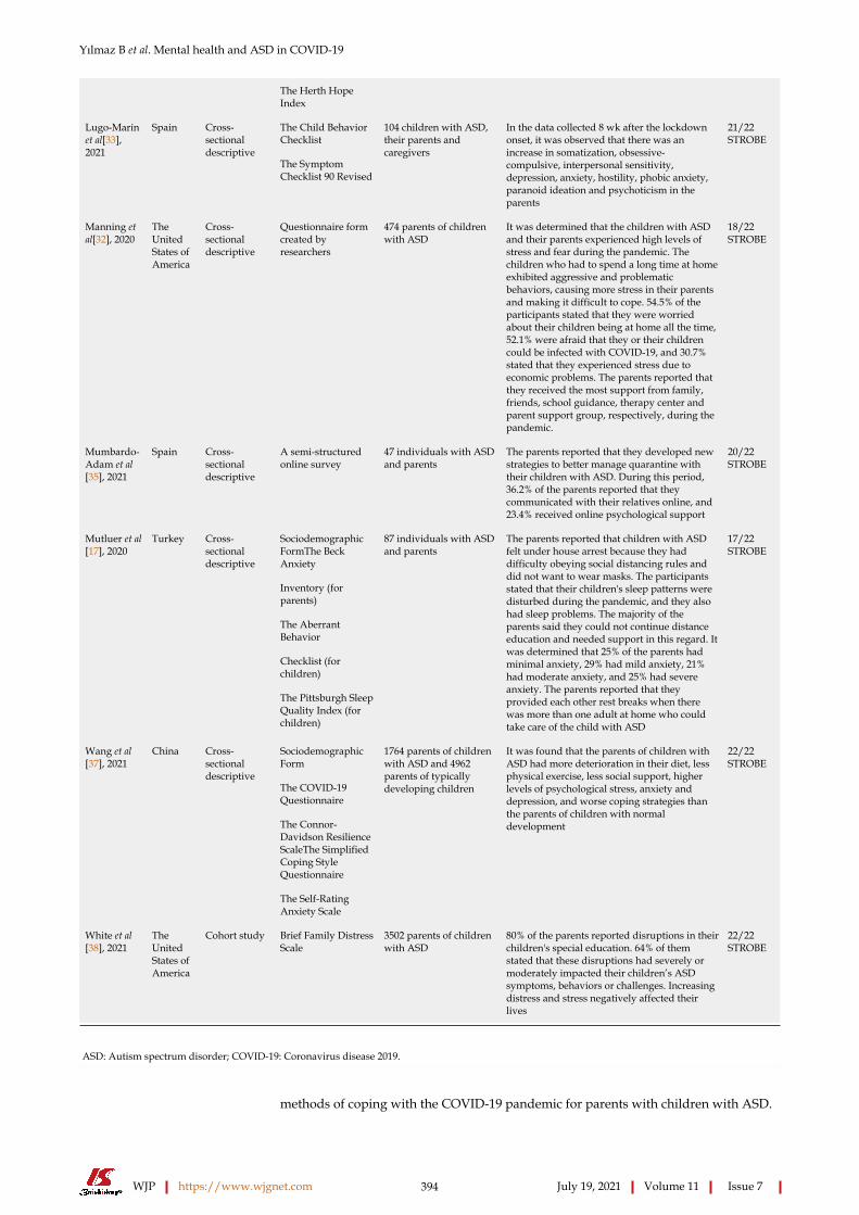
Yılmaz B et al. Mental health and ASD in COVID-19
WJP https://www.wjgnet.com 394 July 19, 2021 Volume 11 Issue 7
The Herth Hope Index
The Child Behavior Checklist
Lugo-Marín et al[33], 2021
Spain Cross-sectional descriptive
The Symptom Checklist 90 Revised
104 children with ASD, their parents and caregivers
In the data collected 8 wk after the lockdown onset, it was observed that there was an increase in somatization, obsessive-compulsive, interpersonal sensitivity, depression, anxiety, hostility, phobic anxiety, paranoid ideation and psychoticism in the parents
21/22 STROBE
Manning et al[32], 2020
The United States of America
Cross-sectional descriptive
Questionnaire form created by researchers
474 parents of children with ASD
It was determined that the children with ASD and their parents experienced high levels of stress and fear during the pandemic. The children who had to spend a long time at home exhibited aggressive and problematic behaviors, causing more stress in their parents and making it difficult to cope. 54.5% of the participants stated that they were worried about their children being at home all the time, 52.1% were afraid that they or their children could be infected with COVID-19, and 30.7% stated that they experienced stress due to economic problems. The parents reported that they received the most support from family, friends, school guidance, therapy center and parent support group, respectively, during the pandemic.
18/22 STROBE
Mumbardo-Adam et al[35], 2021
Spain Cross-sectional descriptive
A semi-structured online survey
47 individuals with ASD and parents
The parents reported that they developed new strategies to better manage quarantine with their children with ASD. During this period, 36.2% of the parents reported that they communicated with their relatives online, and 23.4% received online psychological support
20/22 STROBE
Sociodemographic FormThe Beck Anxiety
Inventory (for parents)
The Aberrant Behavior
Checklist (for children)
Mutluer et al[17], 2020
Turkey Cross-sectional descriptive
The Pittsburgh Sleep Quality Index (for children)
87 individuals with ASD and parents
The parents reported that children with ASD felt under house arrest because they had difficulty obeying social distancing rules and did not want to wear masks. The participants stated that their children's sleep patterns were disturbed during the pandemic, and they also had sleep problems. The majority of the parents said they could not continue distance education and needed support in this regard. It was determined that 25% of the parents had minimal anxiety, 29% had mild anxiety, 21% had moderate anxiety, and 25% had severe anxiety. The parents reported that they provided each other rest breaks when there was more than one adult at home who could take care of the child with ASD
17/22 STROBE
Sociodemographic Form
The COVID-19 Questionnaire
The Connor-Davidson Resilience ScaleThe Simplified Coping Style Questionnaire
Wang et al[37], 2021
China Cross-sectional descriptive
The Self-Rating Anxiety Scale
1764 parents of children with ASD and 4962 parents of typically developing children
It was found that the parents of children with ASD had more deterioration in their diet, less physical exercise, less social support, higher levels of psychological stress, anxiety and depression, and worse coping strategies than the parents of children with normal development
22/22 STROBE
White et al[38], 2021
The United States of America
Cohort study Brief Family Distress Scale
3502 parents of children with ASD
80% of the parents reported disruptions in their children's special education. 64% of them stated that these disruptions had severely or moderately impacted their children’s ASD symptoms, behaviors or challenges. Increasing distress and stress negatively affected their lives
22/22 STROBE
ASD: Autism spectrum disorder; COVID-19: Coronavirus disease 2019.
methods of coping with the COVID-19 pandemic for parents with children with ASD.

Yılmaz B et al. Mental health and ASD in COVID-19
WJP https://www.wjgnet.com 395 July 19, 2021 Volume 11 Issue 7
Results on the experiences of parents of children with ASD in the COVID-19 pandemicThe COVID-19 pandemic affected the parents of children with ASD mentally. In the
study conducted in Saudi Arabia, 94% of the parents reported that their stress levels increased, while 78.7% stated that the pandemic affected their emotional wellbeing negatively[28]. Amorim et al[29] found that parents with children with ASD had higher anxiety levels than those with healthy children. Colizzi et al[30] reported that having a single child, the child with ASD not being able to speak, the child with ASD being male and a single parent having the child's responsibility were among factors that increased stress in the parents. Curfew restrictions that emerged suddenly during the pandemic caused stress in parents and children. Bent et al[31] stated that parents had difficulty explaining the restrictions and security measures to their children. The parents reported that their children had difficulty in obeying the social distancing rules, they could not take their children to the market, etc. because they did not want to wear a mask and felt under house arrest[17,31]. In the study by Manning et al[32], 54.5% of the participants stated that they were worried about their children being at home all the time, 52.1% were afraid that they or their children could be infected with COVID-19, and 30.7% reported that they had stress due to economic difficulties. In Spain, it was observed that there was an increase in somatization, obsessive behavior, depression, anxiety, hostile behavior, paranoid ideas, phobic anxiety and aggression in the parents according to the data collected 8 wk after the start of the lockdown[33].
For individuals with ASDs, changes in their routine may be very disturbing and challenging. It was reported that this situation led to serious behavioral disorders such as crying episodes, increase in aggression and rejection of transition[31]. In the study by Amorim et al[29] the mean anxiety score (8.75 ± 0.96) of parents with a child with ASD in the COVID-19 pandemic was found to be significantly higher than the mean score of parents with healthy children (5.36 ± 2.71). Althiabi[34] also reported that the stress levels of parents increased, and they had difficulty in maintaining their mental wellbeing. Children's adaptation levels affect the parents’ anxiety. It was determined that children who could continue their routines (7.72 ± 1.84) had significantly higher levels of adaptation than those who could not (5.25 ± 2.75). In the same study, the parents reported that they felt helpless because there was not enough time to change routines. In the study, some parents reported that their children adapted to the situation without any major problems and thought of this process as a school holiday. The majority of parents, on the other hand, stated that other children had to continue their education in the home environment due to the pandemic, and they were tired of housework. One of the changing routines during the pandemic is that e-learning has been placed at the center of life. Parents reported that they felt tired as they had to spend extra time on their children with ASD during the pandemic. Parents revealed that they felt lonely and bored as they had to assume the role of educators during the pandemic. Some participants stated that their children who spent more time at home during the pandemic felt better as they took more responsibility in housework. In another study, 40.4% of the parents reported that they could spend more time with their ASD-diagnosed children, and 31.9% calmed their children by creating school-related activities[35]. In the study by Mutluer et al[17] parents stated that their children’s sleep patterns were disturbed during the pandemic, and they also had sleep problems. In the study by Amorim et al[29], 55.8% of the parents of children with ASD and 29.6% of the control group reported that the COVID-19 pandemic had a negative effect on emotion management. The most difficult areas for parents were expressed as social isolation (41.4%), not being able to spend time outside (13.1%), sudden changes in routines (11.1%), boredom (9.1%) and distance education practices (7.1%). In the quasi-experimental study by Liu et al[36] in two sessions per week for 12 wk, relaxation-muscle exercises, home activities with the child, protection strategies, emotional management, parental stress coping strategies and psychological counseling strategies to cope with the pandemic situation were used. With web-based support, it was found that mothers' stress and anxiety levels decreased in the COVID-19 pandemic period. The parents reported feeling relaxed as the program facilitated the parent-child interaction. In a study conducted in China, it was found that the parents of children with ASD had more deterioration in their diet, less physical exercise, less social support, higher levels of psychological stress, anxiety and depression, and worse coping strategies than the parents of children with normal development[37].
Results regarding the areas where parents of children with ASD need support in the COVID-19 pandemicParents stated that they had difficulties during the COVID-19 pandemic due to deficiencies in support systems[30]. In the study conducted in Italy, it was reported

Yılmaz B et al. Mental health and ASD in COVID-19
WJP https://www.wjgnet.com 396 July 19, 2021 Volume 11 Issue 7
that parents with children with ASD needed local health service support, supportive training for e-learning and decrease in quarantine restrictions during the COVID-19[30]. In the study conducted in Saudi Arabia, it was found that the support that parents received from their relatives reduced parental stress, fatigue and contributed to improving emotional wellbeing[28]. In the study conducted in Turkey, the majority of parents reported that they could not continue distance education and needed support. In the same study, it was determined that 25% of the parents had minimal anxiety, 29% had mild anxiety, 21% had moderate anxiety, and 25% had severe anxiety according to the Beck Anxiety Inventory[17].
In the study conducted in Portugal, 13% of the parents stated that they took their children to a specialist psychologist, and 1.5% of them visited emergency health services at the moment of their children's tantrums[29]. In the study conducted in Saudi Arabia, parents reported that their children with ASD mostly experienced problems in maintaining their care skills, coping with tantrums, controlling their negative behaviors and maintaining communication with their children. Additionally, the parents stated that they needed psychological and financial support the most, respectively[34]. In the study conducted in the United States, it was observed that tele-health services were insufficient in meeting the education and treatment needs of the children of parents. In the study, most of the parents of children with ASD stated that they had difficulty coping with the increasing negative behaviors of their children, and distress and stress negatively affected their lives[37].
In the study conducted in Italy, it was found that 19.1% of parents visited a neuropsychiatrist due to the onset of new behavioral problems in their children and feeling helpless[30]. It was stated by 47.4% of the participants that they needed more health services during the pandemic, 30% needed to strengthen their home support systems, and 16.8% needed more state support in quarantine. It was stated by 25% of the parents that one of the parents had to quit their job (26.1% of mothers, 27.5% of fathers) to take care of their children with ASD. The majority of the participants (94%) stated that this situation was financially difficult for them. During the pandemic, only 27.7% of the parents reported that they could get support from local health services. It was specified by 23% of the parents that they had difficulties in regulating the eating behaviors of their children, 31% had difficulties in providing authority to their children, 78.1% had difficulties in making use of spare time, and 75.7% had difficulties in having their children do homework[30].
The coping methods of parents with children with ASD with the COVID-19 pandemicThe parents of children with ASD resorted to various coping methods in the COVID-19 pandemic process. In the study by Bent et al[31], it was found that parents exercised at home, practiced yoga and meditation, read newspapers, cooked meals, talked to their loved ones on online platforms and spent time (bathing, nail care and online shopping) during the pandemic. In the study of Mutluer et al[17], it was reported that parents provided each other with rest breaks when there was more than one adult at home who could take care of the child with ASD. In the study by Amorim et al[29], it was determined that parents frequently talked to their family, friends and colleagues by video, and some of them received support from a therapist. In another study, it was observed that parents received the most support from their families, friends, school guidance services, therapy centers and parent support groups, respectively, during the pandemic process[32]. In the study conducted in Saudi Arabia, most parents reported that they received online counseling and guidance services to deal with the child's behavior at home and tantrums[34]. In another study, 36.2% of parents stated that they communicated with their relatives online, 23.4% received online psychological support, and 6.4% occasionally walked with their children[37].
DISCUSSIONThis study is the first systematic review aimed at determining the mental health status of the parents of children with ASD in the COVID-19 pandemic. The systematic review showed that the difficulties associated with the COVID-19 pandemic negatively affect the mental health of the parents of children with ASD. Restriction of face-to-face education, social activities, and child monitoring during the pandemic process threaten the physical and mental health of children. Parents worldwide are concerned about how to support their children best under these difficult conditions[8-10]. The distinctive features of the diagnosis of ASD put children with ASD and their parents at risk of more adverse effects from the COVID-19 pandemic. Since these children may

Yılmaz B et al. Mental health and ASD in COVID-19
WJP https://www.wjgnet.com 397 July 19, 2021 Volume 11 Issue 7
have difficulties in social communication, they can develop when they are in environments that support their social interactions[38]. Although regular physical activity may provide a calming and regulating effect on children, they lack this opportunity due to the pandemic[8]. Considering all of these effects on children diagnosed with ASD, the inability of parents, who are deprived of support during the pandemic process, to maintain their mental health becomes prominent. Parents often experience mental distress as they postpone their own needs for their children with ASD and their families[21]. Disruption in healthcare support or services is an additional stressor on families with a child or dependent with ASD during an already stressful time because of the COVID-19 pandemic[39]. Restrictive social and economic regulations, discomfort caused by COVID-19, fear of transmission, isolated family life, closure of schools, lack of support programs for parents, loss of loved ones, work from home and the resulting increase in full-time childcare and home responsibility, as well as financial difficulties, are the main problems that parents experience in this process[8,17,28-32,39,40]. There has been a consensus on this issue in the studies included in this systematic review[17,28-37,39].
Sleep problems significantly affect children with ASD[41]. Studies on children with ASD reported that sleep problems lead to symptoms such as social withdrawal, anxiety and depression and behavioral problems such as self-aggression, hyperactivity, aggression and high levels of irritability during the day[42,43]. Another study noted that disruption in the circadian sleep rhythm increased the symptoms of ASD[44]. All of these effects of sleep problems negatively influence parents, causing an increase in the burden of these parents and an increase in their stress levels. In a study, it was determined that the situation of being home during the pandemic process caused sleep problems in children with ASD, and this situation increased the severity of ASD symptoms[45]. In the study conducted in Italy, it was reported that more behavioral problems were observed during the COVID-19 pandemic process, and these problems were associated with sleep[17]. The occurrence of these problems creates a sense of loss of control in parents and increases their stress[45]. All of these changes in the lives of parents and children also hinder the physical and cognitive development of the child. Therefore, it is clear that there is also an increase in parental stress levels during a pandemic that directly affects children's quality of life[46]. In a study, it was reported that, during the COVID-19 pandemic process, the parents of children with ASD experienced higher levels of anxiety about themselves and their children in comparison to the parents of children without neurobehavioral disorders[29,37]. Similarly, in the study by Alhuzimi[28] it was emphasized that the parents of children with ASD have higher stress levels and lower welfare levels. Manning et al[32] stated that children with ASD and their parents experienced high levels of stress during the pandemic process and reported that there were disruptions in their daily life routines. In this process, the issues regarding which they experienced the most stress were related to illness, isolation and materiality[32]. The fear of getting infected in the event of a pandemic is significant in the parents of children with ASD, and this fear increases stress significantly[47]. This was supported by the findings of the study by Manning et al[32] on the COVID-19 pandemic. Although staying at home during the isolation process and the obligation to quarantine are measures taken to reduce the transmission of COVID-19, harmful effects of social isolation on the mental health of children and parents may be observed[48]. This may further increase stress in children diagnosed with ASD and their parents[29,30,32]. Lugo-Marín et al[33] reported that there was an increase in somatization, obsessive behavior, depression, anxiety, hostile behavior, paranoid ideas, phobic anxiety and aggression in parents in 2 mo after the lockdown onset. Interruptions in the care process are an important need in the parents of children with ASD[49] and have an important role in reducing stress for the parents of children with ASD[50]. However, the decrease in the support received from caregivers during the isolation process and the fact that parents have taken on the entirety of the care burden have made it difficult for these parents to cope. Parents had difficulties in regulating their children's eating behaviors, providing authority to their children, making use of spare time and finding activities, and having their children do homework, and they felt lonely[30,34,35]. In the studies included in the systematic review, it was reported that, in order to cope with this situation, parents had rest breaks when there was another adult at home who could take care of their children with ASD[17,31].
An increase in the severity of ASD symptoms in children has an important role in affecting the stress and wellbeing levels of parents[51-54]. The COVID-19 pandemic process have caused more intense and more frequent behavioral problems in children with ASD[30]. In the study by Manning et al[32] one of the studies included in the systematic review, it was reported that the increasing severity in the symptoms of

Yılmaz B et al. Mental health and ASD in COVID-19
WJP https://www.wjgnet.com 398 July 19, 2021 Volume 11 Issue 7
children with ASD along with the pandemic caused an increase in the stress reported by parents. Children who are confined at home exhibit aggressive and problematic behaviors, causing more stress for their parents and making it difficult to cope. In other studies supporting these results, the pandemic and the increasing behavioral changes in children with ASD were found to play a significant role in the increase of stress in parents[17,30]. Parental stress also reportedly had an effect on emotional well-being[28]. In this sense, there has been a consensus that there is a lack of support for children and parents.
The positive attitude of parents during the COVID-19 pandemic is necessary for them to manage their stress and also control their children's behavior at home[34]. Pottie and Ingram[55] pointed out the importance of social support in helping a parent with a child with ASD cope with stress and raise their mood. All kinds of support from the community and from parents who experience similar situations facilitate the home care processes of children with ASD and their parents[56]. For this reason, during the pandemic, it is necessary to maintain contact in online environments with all individuals and institutions that parents can receive support from such as other parents, teachers, therapists and health professionals who experience similar situations[12]. In a qualitative study, parents stated that, although they could not get together with other parents who were in the same situation during the pandemic, they supported each other in home care through groups established via online messaging applications and mentioned how this helped them[57]. Similarly, in the studies included in the systematic review, parents reported that they frequently made video calls with their families and friends[29,31]. Althiabi[34] found that parents sought support from teachers, family members and therapists to take care of their children with ASD during the pandemic outbreak. Bent et al[31] stated in their study that it is important to seek help from friends, family, co-workers, therapists and healthcare professionals, as parents must remain strong for their families and children. With the closure of schools, parents are struggling to find different activities at home to support the development of their children with ASD and prevent their symptoms from appearing[57]. In the studies included in the systematic review[30,31,34,35], in accordance with the literature, parents stated that they sought daily activities and events that their children enjoyed. All family members including parents who leave their jobs or work from home due to the pandemic and children whose schools are closed are at home all together. In this process, the support of all family members for each other in the care of the child with ASD also provides a significant reduction in their burden of care[57].
Children diagnosed with ASD can only survive the pandemic with healthy parents. Therefore, parents should take care of their health, both for themselves and their children. Firstly, parents should not disrupt their physical care with adequate nutrition and rest and accept the emotional burden they experience. They should practice spiritual self-care by planning an activity they enjoy. Parents should be encouraged to plan activities they can do with their children in the home environment. Moreover, they should be encouraged to seek help from healthcare professionals when they feel they have mental problems that they cannot cope with[21]. Consistent with the literature, Bent et al[31] reported that parents spend time with activities such as bathing, nail care and online shopping as a method of coping with stress. In the qualitative study, the parents stated that they used methods such as walking, cycling, yoga, meditation, reading a newspaper and praying to cope with stress during the COVID-19 pandemic[31]. With the web-based support program prepared by Liu et al[36] relaxation-muscle exercises, home activities with the child, protection strategies, emotional management, parental stress coping strategies and psychological counseling strategies to cope with the pandemic situation were implemented. Their study stated that mothers' stress and anxiety levels decreased, and parents reported feeling relaxed as the program facilitated parent-child interaction.
LimitationsThe limitations of this study were that studies whose full texts were not available and those published in languages other than the English language were not included in the systematic review, 12 databases were searched, and the gray literature was not screened. Since the pandemic has only the last year, studies in the field are limited. Therefore, the results should be interpreted with caution, as the systematic review was performed with a limited number of descriptive studies (twelve articles) with lower levels of evidence.

Yılmaz B et al. Mental health and ASD in COVID-19
WJP https://www.wjgnet.com 399 July 19, 2021 Volume 11 Issue 7
CONCLUSIONThe COVID-19 pandemic has negatively affected the mental health of children with ASD and their parents. While many factors play a role in increasing the stress levels of parents, stress may also lead to different problems. In this case, it is possible for children with ASD to continue their lives in a healthy way with mentally healthy parents. In the systematic review, it was concluded that parents with children with ASD had difficulty with their children being at home all day long and financial difficulties as they had to quit their jobs. The parents were also able to devote less time to themselves during this process, and their stress and anxiety levels increased. During the pandemic where face-to-face services are interrupted, governments and relevant institutions should provide support for parents with children with ASD, and institutions that provide support should also work to improve the quality of the support they provide. In this process, it may be recommended to research new ways such as online health monitoring, online diagnosis systems, support groups for children and parents, increased tele-health services, tele-therapies and e-health support. Additionally, after the restrictions imposed by the pandemic are removed, it is important to support children with ASD and their parents while they are getting used to their social lives. Support services, such as counseling and helplines, may be created to help parents share their concerns and receive assistance in dealing with specific situations. Parents should be evaluated in terms of mental health, and profes-sional help should be provided for individuals who need support.
ARTICLE HIGHLIGHTSResearch backgroundAlthough staying at home prevents the spread of coronavirus disease 2019 (COVID-19), this poses a number of challenges, especially for children with special needs such as autism spectrum disorders (ASD) and their parents. The parents of children with ASD participate in special education practices that involve physical activity in order to cope with the behavioral, cognitive and mental problems of their children. However, during COVID-19 pandemic, this process was disrupted, and the mental health of the parents was affected.
Research motivationAlthough there were many studies on the effects of the COVID-19 pandemic in the literature search, it was observed that there was very limited information on mental health effects on the parents of children with ASD, and there was no systematic review on this topic.
Research objectivesIn this systematic review, it is aimed to determine the mental health status of the parents of children with ASD in the COVID-19 pandemic.
Research methodsArticles in English, which could be accessed in full text without any limitation of publication year and country, were included in the study. The systematic review was conducted according to the PICOS strategy (Participants: The parents of children with ASD; Interventions: Effects of COVID-19 pandemic on mental health; Comparators: The parents of children without ASD; Main outcomes: Anxiety, stress, difficulty in coping, loneliness, inadequacy of support systems, social isolation, change in routines and financial difficulties; Additional outcomes: Exercise at home, practicing yoga and meditation, reading newspapers, receiving support from a therapist, cooking meals, talking to their loved ones on online platforms during the pandemic. These results have been reported in studies and included and presented in this systematic review. Study design: Quantitative/qualitative studies). The search results were reached by browsing the Web of Science, PubMed (including MEDLINE), Cochrane, Scopus, Science direct and Google Scholar databases using the keywords COVID-19 AND (“autism” OR “autistic” OR “autism spectrum disorder”) AND parent AND (“mental health” OR “anxiety” OR “stress”). The list of the references of the included studies was reviewed to access additional studies.

Yılmaz B et al. Mental health and ASD in COVID-19
WJP https://www.wjgnet.com 400 July 19, 2021 Volume 11 Issue 7
Research resultsThe systematic review was conducted according to the PICOS strategy, and a total of 12 studies were included in a quantitative and qualitative design. The studies have revealed that parents are negatively affected by the COVID-19 pandemic. It was reported that the parents of children with ASD had increased anxiety and stress during the pandemic, children became aggressive as their routines changed, and the parents had difficulty coping with this process. During the pandemic, the parents met with their friends via online platforms, practiced yoga and meditation, the spouses provided rest breaks to each other and received support from therapists.
Research conclusionsThe COVID-19 pandemic negatively affected the mental health of children with ASD and their parents. It may be recommended to plan more interventions that will positively affect the mental health of parents and support them.
Research perspectivesGiven the uncertainty of how long the COVID-19 pandemic will last, it is important to conduct a large number of descriptive and interventional studies on the mental health of parents with children who have ASD. In this systematic review, it was revealed that the number of studies on this topic is quite limited. It is thought that this systematic review will form the basis for future studies.
REFERENCESDruss BG. Addressing the COVID-19 Pandemic in Populations With Serious Mental Illness. JAMA Psychiatry 2020; 77: 891-892 [PMID: 32242888 DOI: 10.1001/jamapsychiatry.2020.0894]
1
American Psychiatric Association. Diagnostic and statistical manual of mental disorders. 5th ed. Arlington, VA: American Psychiatric Association, 2013
2
World Health Organization. Autism spectrum disorders. 2019. [cited 20 January 2021]. Available from: https://www.who.int/news-room/fact-sheets/detail/autism-spectrum-disorders
3
Centers for Disease Control and Prevention. Autism Spectrum Disorder (ASD), Data & Statistics on Autism Spectrum Disorder. 2020. [cited 26 January 2021]. Available from: https://www.cdc.gov/ncbddd/autism/data.html
4
James CV, Moonesinghe R, Wilson-Frederick SM, Hall JE, Penman-Aguilar A, Bouye K. Racial/Ethnic Health Disparities Among Rural Adults-United States, 2012-2015. MMWR Surveill Summ 2017; 66: 1–9 [PMID: 29145359 DOI: 10.15585/mmwr.ss6623a1]
5
World Health Organization. WHO Director-General's opening remarks at the media briefing on COVID-19. 2020 May 11. [cited 23 January 2021]. Available from: https://www.who.int/director-general/speeches/detail/who-director-general-s-opening-remarks-at-the-media-briefing-on-covid-19---11-may-2020
6
Güner R, Hasanoğlu I, Aktaş F. COVID-19: Prevention and control measures in community. Turk J Med Sci 2020; 50: 571-577 [PMID: 32293835 DOI: 10.3906/sag-2004-146]
7
Bellomo TR, Prasad S, Munzer T, Laventhal N. The impact of the COVID-19 pandemic on children with autism spectrum disorders. J Pediatr Rehabil Med 2020; 13: 349-354 [PMID: 32986631 DOI: 10.3233/PRM-200740]
8
den Houting J. Stepping Out of Isolation: Autistic People and COVID-19. Autism in Adulthood 2020; 2: 1-3 [DOI: 10.1089/aut.2020.29012.jdh]
9
Liu JJ, Bao Y, Huang X, Shi J, Lu L. Mental health considerations for children quarantined because of COVID-19. Lancet Child Adolesc Health 2020; 4: 347-349 [PMID: 32224303 DOI: 10.1016/S2352-4642(20)30096-1]
10
Duan L, Shao X, Wang Y, Huang Y, Miao J, Yang X, Zhu G. An investigation of mental health status of children and adolescents in china during the outbreak of COVID-19. J Affect Disord 2020; 275: 112-118 [PMID: 32658812 DOI: 10.1016/j.jad.2020.06.029]
11
Narzisi A. Handle the Autism Spectrum Condition During Coronavirus (COVID-19) Stay At Home period: Ten Tips for Helping Parents and Caregivers of Young Children. Brain Sci 2020; 10: 1-4 [PMID: 32244776 DOI: 10.3390/brainsci10040207]
12
Puts NA, Wodka EL, Tommerdahl M, Mostofsky SH, Edden RA. Impaired tactile processing in children with autism spectrum disorder. J Neurophysiol 2014; 111: 1803-1811 [PMID: 24523518 DOI: 10.1152/jn.00890.2013]
13
Sivaraman M, Virues-Ortega J, Roeyers H. Telehealth mask wearing training for children with autism during the COVID-19 pandemic. J Appl Behav Anal 2021; 54: 70-86 [PMID: 33241588 DOI: 10.1002/jaba.802]
14
Bonis S. Stress and Parents of Children with Autism: A Review of Literature. Issues Ment Health Nurs 2016; 37: 153-163 [PMID: 27028741 DOI: 10.3109/01612840.2015.1116030]
15
Vohra R, Madhavan S, Sambamoorthi U, St Peter C. Access to services, quality of care, and family 16
Yılmaz B et al. Mental health and ASD in COVID-19
WJP https://www.wjgnet.com 401 July 19, 2021 Volume 11 Issue 7
impact for children with autism, other developmental disabilities, and other mental health conditions. Autism 2014; 18: 815-826 [PMID: 24353274 DOI: 10.1177/1362361313512902]Mutluer T, Doenyas C, Aslan Genc H. Behavioral Implications of the Covid-19 Process for Autism Spectrum Disorder, and Individuals' Comprehension of and Reactions to the Pandemic Conditions. Front Psychiatry 2020; 11: 561882 [PMID: 33304279 DOI: 10.3389/fpsyt.2020.561882]
17
Akçay E, Başgül ŞS. Pandemic and Children with Special Needs/At Risk. Turkiye Klinikleri 2020; 55-61
18
Juneja M, Gupta A. Managing Children with Special Needs in COVID-19 Times. Indian Pediatr 2020; 57: 971 [PMID: 32710532 DOI: 10.1007/s13312-020-2009-1]
19
Hiraoka D, Tomoda A. Relationship between parenting stress and school closures due to the COVID-19 pandemic. Psychiatry Clin Neurosci 2020; 74: 497-498 [PMID: 32779846 DOI: 10.1111/pcn.13088]
20
Lim T, Tan MY, Aishworiya R, Kang YQ. Autism Spectrum Disorder and COVID-19: Helping Caregivers Navigate the Pandemic. Ann Acad Med Singap 2020; 49: 384-386 [PMID: 32712636]
21
American Academy of Pediatrics. AAP Offers Parent Tips and Resources for Dealing With Covid-19 and Its Stresses. 2020. [cited 26 January 2021]. Available from: https://services.aap.org/en/news-room/news-releases/aap/2020/aap-offers-parent-tips-and-resources-for-dealing-with-covid-19-and-its-stresses/
22
United Nations International Children's Emergency Fund. Explore the parenting tips. 2020. [cited 25 January 2021]. Available from: https://www.unicef.org/coronavirus/covid-19-parenting-tips
23
World Health Organization. Coronavirus disease (COVID-19) advice for the public: Advocacy. 2020. [cited 26 January 2021]. Available from: https://www.who.int/emergencies/diseases/novel-coronavirus-2019/advice-for-public/healthy-parenting
24
Mackintosh VH, Goin-Kochel RP, Myers BJ. “What do you like/dislike about the treatments you’re currently using? Focus Autism Other Dev Disabl 2012; 27: 51-60 [DOI: 10.1177/1088357611423542]
25
von Elm E, Altman DG, Egger M, Pocock SJ, Gøtzsche PC, Vandenbroucke JP; STROBE Initiative. The strengthening the reporting of observational studies in epidemiology (STROBE) statement: guidelines for reporting observational studies. Int J Surg 2014; 12: 1495-1499 [PMID: 25046131 DOI: 10.1016/j.ijsu.2014.07.013]
26
Snilstveit B, Oliver S, Vojtkova M. Narrative approaches to systematic review and synthesis of evidence for international development policy and practice. Focus Autism Other Dev Disabil 2012; 4: 409-429 [DOI: 10.1080/19439342.2012.710641]
27
Alhuzimi T. Stress and emotional wellbeing of parents due to change in routine for children with Autism Spectrum Disorder (ASD) at home during COVID-19 pandemic in Saudi Arabia. Res Dev Disabil 2021; 108: 103822 [PMID: 33271447 DOI: 10.1016/j.ridd.2020.103822]
28
Amorim R, Catarino S, Miragaia P, Ferreras C, Viana V, Guardiano M. The impact of COVID-19 on children with autism spectrum disorder. Rev Neurol 2020; 71: 285-291 [PMID: 33034366 DOI: 10.33588/rn.7108.2020381]
29
Colizzi M, Sironi E, Antonini F, Ciceri ML, Bovo C, Zoccante L. Psychosocial and behavioral impact of COVID-19 in Autism Spectrum Disorder: An online parent survey. Brain Sci 2020; 10: 341-357 [PMID: 32503172 DOI: 10.3390/brainsci10060341]
30
Bent S, Hossain B, Chen Y, Widjaja F, Breard M, Hendren R. The experience of parents of children with autism spectrum disorder during the COVID-19 pandemic: A qualitative analysis. 2020 Preprint. Available from: Research square [DOI: 10.21203/rs.3.rs-46426/v1]
31
Manning J, Billian J, Matson J, Allen C, Soares N. Perceptions of families of individuals with autism spectrum disorder during the COVID-19 crisis. J Autism Dev Disord 2020; 1-9 [PMID: 33090358 DOI: 10.1007/s10803-020-04760-5]
32
Lugo-Marín J, Gisbert-Gustemps L, Setien-Ramos I, Español-Martín G, Ibañez-Jimenez P, Forner-Puntonet M, Arteaga-Henríquez G, Soriano-Día A, Duque-Yemail JD, Ramos-Quiroga JA. COVID-19 pandemic effects in people with Autism Spectrum Disorder and their caregivers: Evaluation of social distancing and lockdown impact on mental health and general status. Res Autism Spectr Disord 2021; 83: 101757 [PMID: 33649707 DOI: 10.1016/j.rasd.2021.101757]
33
Althiabi Y. Attitude, anxiety and perceived mental health care needs among parents of children with Autism Spectrum Disorder (ASD) in Saudi Arabia during COVID-19 pandemic. Res Dev Disabil 2021; 111: 103873 [PMID: 33540358 DOI: 10.1016/j.ridd.2021.103873]
34
Mumbardó-Adam C, Barnet-López S, Balboni G. How have youth with Autism Spectrum Disorder managed quarantine derived from COVID-19 pandemic? Res Dev Disabil 2021; 110: 1-8 [PMID: 33486395 DOI: 10.1016/j.ridd.2021.103860]
35
Liu G, Wang S, Liao J, Ou P, Huang L, Xie N, He Y, Lin J, He HG, Hu R. The efficacy of WeChat-Based parenting training on the psychological well-being of mothers with children with autism during the COVID-19 Pandemic: Quasi-experimental study. JMIR Ment Health 2021; 8: e23917 [PMID: 33481751 DOI: 10.2196/23917]
36
Wang L, Li D, Pan S, Zhai J, Xia W, Sun C, Zou M. The relationship between 2019-nCoV and psychological distress among parents of children with autism spectrum disorder. Global Health 2021; 17: 23-37 [PMID: 33632259 DOI: 10.1186/s12992-021-00674-8]
37
Hyman SL, Levy SE, Myers SM. Identification, evaluation, and management of children with autism spectrum disorder. Pediatrics 2020; 145: e20193447. [PMID: 31843864 DOI: 10.1542/peds.2019-3447]
38

Yılmaz B et al. Mental health and ASD in COVID-19
WJP https://www.wjgnet.com 402 July 19, 2021 Volume 11 Issue 7
White LC, Law JK, Daniels AM, Toroney J, Vernoia B, Xiao S; SPARK Consortium, Feliciano P, Chung WK. Brief Report: Impact of COVID-19 on individuals with ASD and their caregivers: A perspective from the SPARK cohort. J Autism Dev Disord 2021; 1-8 [PMID: 33387233 DOI: 10.1007/s10803-020-04816-6]
39
Wang G, Zhang Y, Zhao J, Zhang J, Jiang F. Mitigate the effects of home confinement on children during the COVID-19 outbreak. Lancet 2020; 395: 945-947 [PMID: 32145186 DOI: 10.1016/S0140-6736(20)30547-X]
40
Sivertsen B, Posserud MB, Gillberg C, Lundervold AJ, Hysing M. Sleep problems in children with autism spectrum problems: a longitudinal population-based study. Autism 2012; 16: 139-150 [PMID: 21478225 DOI: 10.1177/1362361311404255]
41
Henderson JA, Barry TD, Bader SH, Jordan SS. The relation among sleep, routines, and externalizing behavior in children with an autism spectrum disorder. Res Autism Spectr Disord 2011; 5: 758-767 [DOI: 10.1016/j.rasd.2010.09.003]
42
Johnson CR. Sleep problems in children with mental retardation and autism. Child Adolesc Psychiatr Clin N Am 1996; 5: 673-684 [DOI: 10.1016/S1056-4993(18)30355-9]
43
Carmassi C, Palagini L, Caruso D, Masci I, Nobili L, Vita A, Dell'Osso L. Systematic review of sleep disturbances and circadian sleep desynchronization in autism spectrum disorder: toward an integrative model of a self-reinforcing loop. Front Psychiatry 2019; 10: 366-401 [PMID: 31244687 DOI: 10.3389/fpsyt.2019.00366]
44
Türkoğlu S, Uçar HN, Çetin FH, Güler HA, Tezcan ME. The relationship between chronotype, sleep, and autism symptom severity in children with ASD in COVID-19 home confinement period. Chronobiol Int 2020; 37: 1207-1213 [PMID: 32746638 DOI: 10.1080/07420528.2020.1792485]
45
Wang C, Pan R, Wan X, Tan Y, Xu L, Ho CS, Ho RC. Immediate psychological responses and associated factors during the initial stage of the 2019 Coronavirus Disease (COVID-19) epidemic among the general population in China. Int J Environ Res Public Health 2020; 17: 1-25 [PMID: 32155789 DOI: 10.3390/ijerph17051729]
46
Weiss JA, Wingsiong A, Lunsky Y. Defining crisis in families of individuals with autism spectrum disorders. Autism 2014; 18: 985-995 [PMID: 24254639 DOI: 10.1177/1362361313508024]
47
Leigh-Hunt N, Bagguley D, Bash K, Turner V, Turnbull S, Valtorta N, Caan W. An overview of systematic reviews on the public health consequences of social isolation and loneliness. Public Health 2017; 152: 157-171 [PMID: 28915435 DOI: 10.1016/j.puhe.2017.07.035]
48
Lindly OJ, Chavez AE, Zuckerman KE. Unmet health services needs among US children with developmental disabilities: Associations with family impact and child functioning. J Dev Behav Pediatr 2016; 37: 712-723 [PMID: 27801721 DOI: 10.1097/DBP.0000000000000363]
49
Harper A, Taylor Dyches T, Harper J, Olsen Roper S, South M. Respite care, marital quality, and stress in parents of children with autism spectrum disorders. J Autism Dev Disord 2013; 43: 2604-2616 [PMID: 23529841 DOI: 10.1007/s10803-013-1812-0]
50
Osborne LA, Reed P. The relationship between parenting stress and behavior problems of children with autistic spectrum disorders. Except Child 2009; 76: 54-73 [DOI: 10.1177/001440290907600103]
51
Hall HR, Graff JC. The relationships among adaptive behaviors of children with autism, family support, parenting stress, and coping. Issues Compr Pediatr Nurs 2011; 34: 4-25 [PMID: 21341964 DOI: 10.3109/01460862.2011.555270]
52
Ingersoll B, Hambrick DZ. The relationship between the broader autism phenotype, child severity, and stress and depression in parents of children with autism spectrum disorders. Res Autism Spectr Disord 2011; 5: 337-344 [DOI: 10.1016/j.rasd.2010.04.017]
53
Rivard M, Terroux A, Parent-Boursier C, Mercier C. Determinants of stress in parents of children with autism spectrum disorders. J Autism Dev Disord 2014; 44: 1609-1620 [PMID: 24384673 DOI: 10.1007/s10803-013-2028-z]
54
Pottie CG, Ingram KM. Daily stress, coping, and well-being in parents of children with autism: a multilevel modeling approach. J Fam Psychol 2008; 22: 855-864 [PMID: 19102606 DOI: 10.1037/a0013604]
55
Arora T. Elective home education and special educational needs. J Res Spec Educ Needs 2006; 6: 55-66 [DOI: 10.1111/j.1471-3802.2006.00059.x]
56
Majoko T, Dudu A. Parents’ strategies for home educating their children with autism spectrum disorder during the COVID-19 period in Zimbabwe. Int J Dev Disabil 2020; 1-6 [DOI: 10.1080/20473869.2020.1803025]
57

Published by Baishideng Publishing Group Inc
7041 Koll Center Parkway, Suite 160, Pleasanton, CA 94566, USA
Telephone: +1-925-3991568
E-mail: [email protected]
Help Desk: https://www.f6publishing.com/helpdesk
https://www.wjgnet.com
© 2021 Baishideng Publishing Group Inc. All rights reserved.