World Journal of Clinical Pediatrics - BPG Management System
126
World Journal of Clinical Pediatrics ISSN 2219-2808 (online) World J Clin Pediatr 2022 March 9; 11(2): 93-214 Published by Baishideng Publishing Group Inc
-
Upload
khangminh22 -
Category
Documents
-
view
0 -
download
0
Transcript of World Journal of Clinical Pediatrics - BPG Management System

World Journal ofClinical Pediatrics
ISSN 2219-2808 (online)
World J Clin Pediatr 2022 March 9; 11(2): 93-214
Published by Baishideng Publishing Group Inc
WJCP https://www.wjgnet.com I March 9, 2022 Volume 11 Issue 2
World Journal of
Clinical PediatricsW J C PContents Bimonthly Volume 11 Number 2 March 9, 2022
OPINION REVIEW
Current status of nitrous oxide use in pediatric patients93
Gupta N, Gupta A, Narayanan RMV
REVIEW
Non-pharmacological management of pediatric functional abdominal pain disorders: Current evidence and future perspectives
105
Cordeiro Santos ML, da Silva Júnior RT, de Brito BB, França da Silva FA, Santos Marques H, Lima de SouzaGonçalves V, Costa dos Santos T, Ladeia Cirne C, Silva NOE, Oliveira MV, de Melo FF
Classification, prevalence and integrated care for neurodevelopmental and child mental health disorders: A brief overview for paediatricians
120
Ogundele MO, Morton M
Druggable monogenic immune defects hidden in diverse medical specialties: Focus on overlap syndromes136
Boz V, Zanchi C, Levantino L, Riccio G, Tommasini A
ORIGINAL ARTICLE
Retrospective Study
Barriers and challenges affecting parents’ use of adrenaline auto-injector in children with anaphylaxis151
Narchi H, Elghoudi A, Al Dhaheri K
Observational Study
Functional constipation in Bangladeshi school aged children: A hidden misty at community160
Benzamin M, Karim AB, Rukunuzzaman M, Mazumder MW, Rana M, Alam R, Islam MM, Alam MS, Hossen K, Yasmin A, Fathema K, Khadga M, Aishy AS
SYSTEMATIC REVIEWS
Epidemiology and phenotypes of diabetes in children and adolescents in non-European-origin populations in or from Western Pacific region
173
James S, Maniam J, Cheung PT, Urakami T, von Oettingen J, Likitmaskul S, Ogle G
META-ANALYSIS
Pediatric Anesthesia Emergence Delirium Scale: A diagnostic meta-analysis196
Russell PSS, Mammen PM, Shankar SR, Viswanathan SA, Rebekah G, Russell S, Earnest R, Chikkala SM
Prevalence of intellectual disability in India: A meta-analysis206
Russell PSS, Nagaraj S, Vengadavaradan A, Russell S, Mammen PM, Shankar SR, Viswanathan SA, Earnest R, Chikkala SM, Rebekah G
WJCP https://www.wjgnet.com II March 9, 2022 Volume 11 Issue 2
World Journal of Clinical PediatricsContents
Bimonthly Volume 11 Number 2 March 9, 2022
ABOUT COVER
Editorial Board Member of World Journal of Clinical Pediatrics, Theresa DeLorenzo, PhD, Academic Research, Director, Professor, College of Health Sciences, Logan University, Clifton Park, Ny 12065, United States. [email protected]
AIMS AND SCOPE
The primary aim of the World Journal of Clinical Pediatrics (WJCP, World J Clin Pediatr) is to provide scholars and readers from various fields of pediatrics with a platform to publish high-quality clinical research articles and communicate their research findings online. WJCP mainly publishes articles reporting research results and findings obtained in the field of pediatrics and covering a wide range of topics including anesthesiology, cardiology, endocrinology, gastroenterology, hematology, immunology, infections and infectious diseases, medical imaging, neonatology, nephrology, neurosurgery, nursing medicine, perinatology, pharmacology, respiratory medicine, and urology.
INDEXING/ABSTRACTING
The WJCP is now abstracted and indexed in PubMed, PubMed Central, Scopus, China National Knowledge Infrastructure (CNKI), China Science and Technology Journal Database (CSTJ), and Superstar Journals Database.
RESPONSIBLE EDITORS FOR THIS ISSUE
Production Editor: Yi-Xuan Cai; Production Department Director: Xu Guo; Editorial Office Director: Yu-Jie Ma.
NAME OF JOURNAL INSTRUCTIONS TO AUTHORS
World Journal of Clinical Pediatrics https://www.wjgnet.com/bpg/gerinfo/204
ISSN GUIDELINES FOR ETHICS DOCUMENTS
ISSN 2219-2808 (online) https://www.wjgnet.com/bpg/GerInfo/287
LAUNCH DATE GUIDELINES FOR NON-NATIVE SPEAKERS OF ENGLISH
June 8, 2012 https://www.wjgnet.com/bpg/gerinfo/240
FREQUENCY PUBLICATION ETHICS
Bimonthly https://www.wjgnet.com/bpg/GerInfo/288
EDITORS-IN-CHIEF PUBLICATION MISCONDUCT
Toru Watanabe, Consolato M Sergi, Elena Daniela Serban, Surjit Singh https://www.wjgnet.com/bpg/gerinfo/208
EDITORIAL BOARD MEMBERS ARTICLE PROCESSING CHARGE
https://www.wjgnet.com/2219-2808/editorialboard.htm https://www.wjgnet.com/bpg/gerinfo/242
PUBLICATION DATE STEPS FOR SUBMITTING MANUSCRIPTS
March 9, 2022 https://www.wjgnet.com/bpg/GerInfo/239
COPYRIGHT ONLINE SUBMISSION
© 2022 Baishideng Publishing Group Inc https://www.f6publishing.com
© 2022 Baishideng Publishing Group Inc. All rights reserved. 7041 Koll Center Parkway, Suite 160, Pleasanton, CA 94566, USA
E-mail: [email protected] https://www.wjgnet.com

WJCP https://www.wjgnet.com 93 March 9, 2022 Volume 11 Issue 2
World Journal of
Clinical PediatricsW J C PSubmit a Manuscript: https://www.f6publishing.com World J Clin Pediatr 2022 March 9; 11(2): 93-104
DOI: 10.5409/wjcp.v11.i2.93 ISSN 2219-2808 (online)
OPINION REVIEW
Current status of nitrous oxide use in pediatric patients
Nishkarsh Gupta, Anju Gupta, R M Vishnu Narayanan
Specialty type: Anesthesiology
Provenance and peer review: Invited article; Externally peer reviewed.
Peer-review model: Single blind
Peer-review report’s scientific quality classificationGrade A (Excellent): 0 Grade B (Very good): 0 Grade C (Good): C Grade D (Fair): D, D Grade E (Poor): 0
P-Reviewer: Mapesa WA, Kenya; Mondardini MC, Italy
Received: April 25, 2021 Peer-review started: April 25, 2021 First decision: June 17, 2021 Revised: July 4, 2021 Accepted: February 25, 2022 Article in press: February 25, 2022 Published online: March 9, 2022
Nishkarsh Gupta, Department of Onco-Anesthesiology and Palliative Medicine, AIIMS, New Delhi 110029, Delhi, India
Anju Gupta, R M Vishnu Narayanan, Department of Anesthesiology, Pain Medicine and Critical Care, AIIMS, New Delhi 110029, Delhi, India
Corresponding author: Anju Gupta, MD, Assistant Professor, Department of Anesthesiology, Pain Medicine and Critical Care, AIIMS, Room No. 6, Porta Cabin, Fourth floor teaching block, New Delhi 110029, Delhi, India. [email protected]
AbstractNitrous oxide is one of the most commonly used inhalational anesthetic agents used in practice. It is a cost-effective, pleasant, safe, and versatile anesthetic agent with many desirable properties like good quality analgesia, decreased awareness, accelerated induction and recovery from anesthesia, and reduced utilization of other expensive inhalational agents with potential cost savings. The use of nitrous oxide has been questioned by a lot of studies and case reports perceiving its adverse systemic, hematological, immune, and neurologic adverse effects. However, the literature in the recent past has tried to resolve the controversies related to its use. The concerns over an increase in cardiovascular complications and mortality following nitrous oxide use have been negated by recent data. However, its use in certain vulnerable populations like children with cobalamin and folate deficiency or defects in their metabolic pathways remains a cause of concern for its toxic effects. In this narrative review, we aim to discuss the pharmacological properties of nitrous oxide, the potential advantages and drawbacks of the use of nitrous oxide in children, address the neurodevelop-mental and other systemic effects, and throw light on the evidence regarding the safety of nitrous oxide use and its current role in pediatric procedural sedation and anesthesia practice. The literature related to its use in the pediatric population for painful procedures and surgeries has been summarized.
Key Words: Child; Nitrous oxide; Vitamin B12; Vulnerable populations; Anesthesiology; Anesthetics; Folic acid; Metabolic networks and pathways
©The Author(s) 2022. Published by Baishideng Publishing Group Inc. All rights reserved.
Gupta N et al. Nitrous oxide and anesthesia in children
WJCP https://www.wjgnet.com 94 March 9, 2022 Volume 11 Issue 2
Core Tip: The literature is insufficient presently to advise either the routine use or complete elimination of nitrous oxide, and further research is needed to fully establish its role in pediatric anesthesia practice. No major adverse effects have been reported in large trials on the use of nitrous oxide in children despite the prevailing concerns over its safety in this population. A reasonable and balanced approach should be adopted to individualize its use considering its risks and benefits as related to a particular case.
Citation: Gupta N, Gupta A, Narayanan RMV. Current status of nitrous oxide use in pediatric patients. World J Clin Pediatr 2022; 11(2): 93-104URL: https://www.wjgnet.com/2219-2808/full/v11/i2/93.htmDOI: https://dx.doi.org/10.5409/wjcp.v11.i2.93
INTRODUCTIONNitrous oxide has been a part of the routine anesthetic practice for over 15 decades. From being the fad of recreational use at parties, nitrous oxide has evolved to hold an important place in contemporary practice of anesthesia[1]. It was first synthesized by Joseph Priestly in 1772, and 7 years later Humphrey Davy established its analgesic and psychotropic potential. However, Davy’s suggestion on using it as an anesthetic did not gain popularity until 1844 when Gardner Colton demonstrated its analgesic properties and Horace Wells demonstrated the first use of nitrous oxide for analgesia for painless tooth extraction. From the year 1868, the commercial availability of compressed nitrous oxide cylinders led to its universal adoption as an ether adjunct. Consequently, it was widely used for general procedural sedation in dentistry, obstetric analgesia, and during general anesthesia with other anesthetic agents. Its additive use with ether provided smoother induction, reduced ether requirements, cardiorespiratory stability, and faster emergence.
While its advantages were being appreciated, various concerns about its metabolic and other adverse effects begin to be recognized in the middle of the nineteenth century, including reports of fatalities from the faulty delivery systems, which led to an ongoing debate on whether it should be abandoned. Results of a few large-scale trials further fueled the debate and challenged its continued use in anesthesia practice. Nitrous oxide can also have a direct environmental impact as it is a major contributor of greenhouse gases. This has questioned its role in sustainable and eco-friendly anesthetic practice. However, the anesthetic use of nitrous oxide contributes to only 2% of the nitrous oxide source in the atmosphere.
The use of nitrous oxide continues to be a vacillation for many anesthesiologists due to the inconclus-iveness of the currently available data. In this review, we discuss the present status of nitrous oxide in pediatric anesthesia practice. We will go through the pharmacological properties of nitrous oxide followed by the pros and cons of using nitrous oxide, addressing the neurodevelopmental and other systemic effects. The conclusions of the landmark trials regarding nitrous oxide will be summarized followed by the literature related to its use in pediatric procedural sedation and surgeries.
METHODSStudies published prior to August 2019 were retrieved from the electronic databases (Google Scholar, Cochrane Central Register of Controlled Trials on The Cochrane Library, PubMed and EMBASE), and their references were additionally scrutinized for any further relevant articles that investigated nitrous oxide. The literature search was done by independent authors, and the following search terms were used in various combinations using Boolean operators (such as AND, OR, NOT): Pediatric patients, pediatric, children, neonates, infants, adolescents, nitrous oxide, laughing gas, N2O, sedation, conscious sedation, procedural sedation, pain, analgesia, anesthesia, homocysteine, methionine synthase, teratogenic, teratogen, teratogens, teratogenesis, postoperative nausea and vomiting, postoperative nausea and/or vomiting (PONV), postoperative vomiting, postoperative nausea, postoperative emesis, environmental effects, ozone depletion, occupational, occupation, exposure, hazard, anesthesia dental, emergency service, post-traumatic stress disorder, chronic postsurgical pain, and CPSP. We got 779 results, and after eliminating duplication, adult trials, and articles in languages other than English, 137 articles were found suitable and were studied.
PHARMACOLOGICAL PROPERTIES OF NITROUS OXIDENitrous oxide occurs as a colorless, odorless gas at room temperature and pressure. Though the exact
Gupta N et al. Nitrous oxide and anesthesia in children
WJCP https://www.wjgnet.com 95 March 9, 2022 Volume 11 Issue 2
mechanism of action is not known, it is postulated to act on dopaminergic, Gamma aminobutyric acid, alpha 2, and N-methyl-d-aspartate (NMDA) receptors to produce sedation and analgesia. However, nitrous oxide does not produce skeletal muscle relaxation. After inhalation, nitrous oxide is primarily excreted via the lungs unchanged. Nitrous oxide is the least potent volatile agent with a minimum alveolar concentration of 105%. Nitrous oxide has a blood gas partition coefficient of 0.47, which confers it low solubility.
Interaction with anesthetic agentsUse of nitrous oxide in combination with other inhalational agents provides an additive anesthetic action since the minimum alveolar concentration of nitrous oxide is directly additive to theirs. Nitrous oxide in 60%-70% concentration equals a minimum alveolar concentration value of around 0.55-0.65[1,2]. It accelerates the time of anesthetic induction when used in conjunction with poorly soluble inhala-tional agents. Nitrous oxide as a component of anesthesia has shown to reduce the utilization of inhala-tional agents, propofol, and opioids[2,3]. During inhalational induction with mask in children, high concentration of nitrous oxide facilitates a faster loss of consciousness by concentration effect and second gas effect. The use of nitrous oxide during induction has proven to increase the mask acceptance in children and lower incidence of airway related complications. However, nitrous oxide favors the incidence of excitatory phenomena with sevoflurane during inhalational induction. It has been seen that adding up nitrous oxide to other inhalational anesthetic agents decreases the occurrence of hemodynamic suppression as compared to use of equipotent doses of volatile agents alone[2].
Advantages and disadvantages of nitrous oxide Nitrous oxide is a cheap anesthetic agent and reduces the utilization of other potent volatile agents and opioids. Therefore, the overall expenses and associated adverse effects are lowered. Along with the additive action with other inhalational agents, the major advantage of nitrous oxide is that it provides good amnesia and hence prevents awareness. Nitrous oxide has been a popular agent for use in pediatric anesthesia during surgical procedures as a constituent of anesthetic gas mixture in addition to other volatile agents and opioids. In addition, it has been used for providing procedural sedation in the emergency room and for various urological procedures and ontological procedures. Nitrous oxide also has been used for mild sedation and analgesia in children undergoing dental procedures, upper gastrointestinal endoscopy, fiberoptic bronchoscopy, and venipuncture procedures. Nitrous oxide has been shown to significantly reduce chronic postsurgical pain (CPSP) in recent studies due to its antagonist action on NMDA receptors, which have been purported to have a role in central sensitization and establishment of CPSP[4].
Nevertheless, nitrous oxide has numerous detrimental effects that may limit its overall clinical application. These consist of an increased risk of PONV, neurologic and hematologic complications, diffusion hypoxia, its property of expanding closed spaces, ozone depletion potential, and recent concerns of adverse consequences on the developing brain[5,6]. There were also concerns of immunosuppression and impairment of wound healing due to inhibition of mononuclear cell prolif-eration and neutrophil chemotaxis[5-7]. The advantages and disadvantages of nitrous oxide have been summed up in the Table 1. Some of the disadvantages quoted are controversial as discussed later in the chapter.
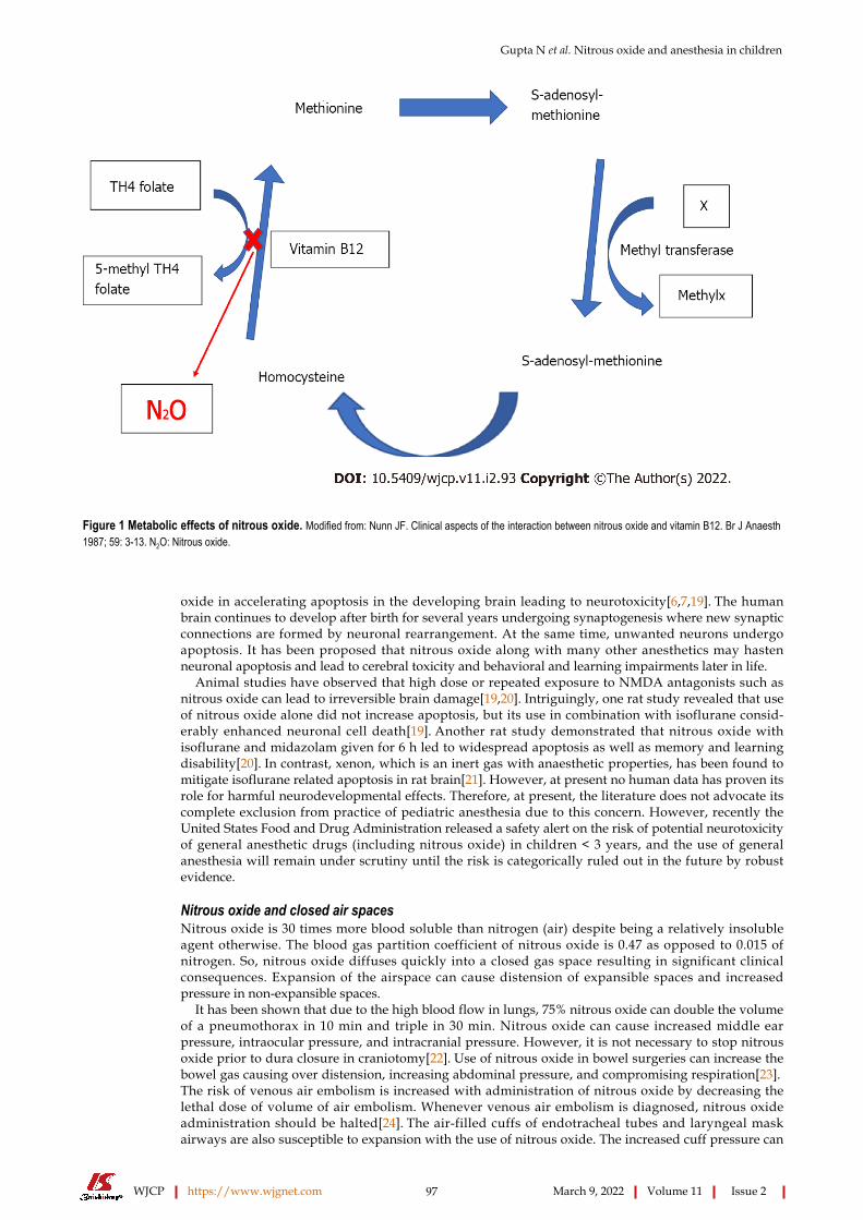
Systemic effectsThe systemic effects of nitrous oxide are summarized in the table below (Table 2). Nitrous oxide oxidizes the cobalt atom of the enzyme methionine synthetase and thereby permanently inactivates it, which in turn interferes with the metabolism of vitamin B12 and folate (Figure 1). Hence, the transformation of homocysteine to S-adenosylmethionine is impaired, which is a substrate for the chemical reaction involving tetrahydrofolate and thymidine during DNA synthesis. A short nitrous oxide exposure of only 30 min was found to decrease the methionine synthetase enzyme activity by 50% in rats, while it became almost untraceable after 6 h[8].
Acute neurologic signs and pancytopenia were seen in an infant after nitrous oxide anesthesia, and vitamin B12 supplementation treated the symptoms[9]. The problem would be magnified in patients having preexisting methionine synthase deficiency where nitrous oxide exposure can precipitate pernicious anemia (manifesting as spinal cord subacute combined degeneration and megaloblastic anemia), psychomotor delay, growth retardation, and neurological symptoms[10,11].
Nitrous oxide has also been noticed to increase blood homocysteine levels. Similarly, nitrous oxide facilitated reduction in methionine synthase enzyme activity in patients with Type-III Homocystinuria (due to a defect in methylene tetrahydrofolate reductase), can complicate into myelopathy, macrocytic anemia, and death. A report described a cataclysmic event in a child who was anesthetized with nitrous oxide and developed convulsions and apneic episodes postoperatively and later succumbed[11].
A preliminary study on metabolic effects of repeated exposure to nitrous oxide concluded that homocysteine levels did not consistently correlate with cumulative nitrous oxide exposure and children predisposed to metabolic and nutritional disturbance[12]. Though this finding is reassuring, considering the gravity of consequences, nitrous oxide should be used with caution in children with congenital

Gupta N et al. Nitrous oxide and anesthesia in children
WJCP https://www.wjgnet.com 96 March 9, 2022 Volume 11 Issue 2
Table 1 Advantages and disadvantages of nitrous oxide use for anesthesia
Advantages Disadvantages
Analgesia Low potency
Reduced awareness Risk of diffusion hypoxia
Colorless and odorless PONV [risk ratio 1.21 (CI: 1.04-1.40); P = 0.014]2
Inexpensive (Rs 50/patient)1 Ability to expand air filled cavities
Faster onset and emergence (elimination half-life 5 min) Increases cuff pressure of ETT and LMA
Minimal metabolism (< 0.004%) Hematological/neurological toxicity
Cardiorespiratory stability Immune deficiency?
Prevents CPSP Reproductive effects
Treatment-resistant refractory depression Myocardial ischemia?
Greenhouse gas
Apoptosis in developing brains
1Cost of nitrous oxide used in dentistry in Indian rupees per patient.2Risk ratio for the overall effect of nitrous oxide on postoperative nausea/vomiting.PONV: Postoperative nausea/vomiting; CPSP: Chronic postsurgical pain; ETT: Endotracheal tube; LMA: Laryngeal mask airway; CI: Confidence interval.
Table 2 Systemic effects of nitrous oxide
Decreases tidal volume and respiratory rate Respiratory system
Reduced ventilatory response to carbon dioxide and hypoxia
Loss of awareness
Analgesia
Increased cerebral blood flow and intracranial pressure
Central nervous system
(Concentration > 70%)
SympathomimeticCardiovascular system
Direct myocardial depression
Hemodynamic effects Combination with other inhalational agents reduce the incidence of hypotension when compared to administration of the agents alone
deficiency or defective enzymes that are involved in the pathway to DNA synthesis or in patients at risk of vitamin B12 deficiency (e.g., pernicious anemia, post-illeal resection surgery, vegetarians, malnourished children, and infants on complete breast feeds).
Postoperative nausea and vomiting: Nitrous oxide administration is considered an independent risk factor for PONV. Nitrous oxide heightens the risk of PONV by up to 20% in adults[13]. Notwith-standing, nitrous oxide did not increase the incidence of PONV in children when used as an adjuvant to other volatile agents[14]. The incidence and severity of PONV did not vary between those receiving 70% nitrous oxide during anesthesia as compared to those who did not[15]. Nonetheless, in combination with propofol it did increase the occurrence of PONV[15].
Environmental and occupational exposure safety: The National Institute of Occupational Safety and Health has set an upper limit for safe workplace exposure to nitrous oxide of 25 ppm. However, the environmental levels may reach up to 2000 ppm in the absence of scavenging, and many grave problems like neurological, hematologic, genotoxic, and reproductive may develop in exposed team[16,17]. Pediatric anesthesiologists may be at the highest risk because of exposure to nitrous oxide and other inhalation agents at high concentrations and flows during the inhalation induction process and during anesthesia. In addition, nitrous oxide has been implicated in ozone destruction in the atmospheric stratosphere[18]. However, all clinical applications of nitrous oxide combined amount to < 2% of pollution related to its use and is probably of little significance, if any.
Neurodevelopmental effects: Similar to other inhalational agents, there has been a concern of nitrous
Gupta N et al. Nitrous oxide and anesthesia in children
WJCP https://www.wjgnet.com 97 March 9, 2022 Volume 11 Issue 2
Figure 1 Metabolic effects of nitrous oxide. Modified from: Nunn JF. Clinical aspects of the interaction between nitrous oxide and vitamin B12. Br J Anaesth 1987; 59: 3-13. N2O: Nitrous oxide.
oxide in accelerating apoptosis in the developing brain leading to neurotoxicity[6,7,19]. The human brain continues to develop after birth for several years undergoing synaptogenesis where new synaptic connections are formed by neuronal rearrangement. At the same time, unwanted neurons undergo apoptosis. It has been proposed that nitrous oxide along with many other anesthetics may hasten neuronal apoptosis and lead to cerebral toxicity and behavioral and learning impairments later in life.
Animal studies have observed that high dose or repeated exposure to NMDA antagonists such as nitrous oxide can lead to irreversible brain damage[19,20]. Intriguingly, one rat study revealed that use of nitrous oxide alone did not increase apoptosis, but its use in combination with isoflurane consid-erably enhanced neuronal cell death[19]. Another rat study demonstrated that nitrous oxide with isoflurane and midazolam given for 6 h led to widespread apoptosis as well as memory and learning disability[20]. In contrast, xenon, which is an inert gas with anaesthetic properties, has been found to mitigate isoflurane related apoptosis in rat brain[21]. However, at present no human data has proven its role for harmful neurodevelopmental effects. Therefore, at present, the literature does not advocate its complete exclusion from practice of pediatric anesthesia due to this concern. However, recently the United States Food and Drug Administration released a safety alert on the risk of potential neurotoxicity of general anesthetic drugs (including nitrous oxide) in children < 3 years, and the use of general anesthesia will remain under scrutiny until the risk is categorically ruled out in the future by robust evidence.
Nitrous oxide and closed air spacesNitrous oxide is 30 times more blood soluble than nitrogen (air) despite being a relatively insoluble agent otherwise. The blood gas partition coefficient of nitrous oxide is 0.47 as opposed to 0.015 of nitrogen. So, nitrous oxide diffuses quickly into a closed gas space resulting in significant clinical consequences. Expansion of the airspace can cause distension of expansible spaces and increased pressure in non-expansible spaces.
It has been shown that due to the high blood flow in lungs, 75% nitrous oxide can double the volume of a pneumothorax in 10 min and triple in 30 min. Nitrous oxide can cause increased middle ear pressure, intraocular pressure, and intracranial pressure. However, it is not necessary to stop nitrous oxide prior to dura closure in craniotomy[22]. Use of nitrous oxide in bowel surgeries can increase the bowel gas causing over distension, increasing abdominal pressure, and compromising respiration[23]. The risk of venous air embolism is increased with administration of nitrous oxide by decreasing the lethal dose of volume of air embolism. Whenever venous air embolism is diagnosed, nitrous oxide administration should be halted[24]. The air-filled cuffs of endotracheal tubes and laryngeal mask airways are also susceptible to expansion with the use of nitrous oxide. The increased cuff pressure can

Gupta N et al. Nitrous oxide and anesthesia in children
WJCP https://www.wjgnet.com 98 March 9, 2022 Volume 11 Issue 2
lead to surrounding mucosal ischemia due to impaired perfusion[25].Hence, it is advisable to avoid the use of nitrous oxide in laparoscopic, bowel, middle ear, and vitreo-
retinal surgeries and to use with caution in neurosurgeries.
DISCUSSIONThe present review identified the literature explaining why the usage of nitrous oxide has been under constant scrutiny, the current role of nitrous oxide in contemporary pediatric anesthesia, procedural sedation, and exploring its potential novel benefits like prevention of CPSP in the pediatric population.
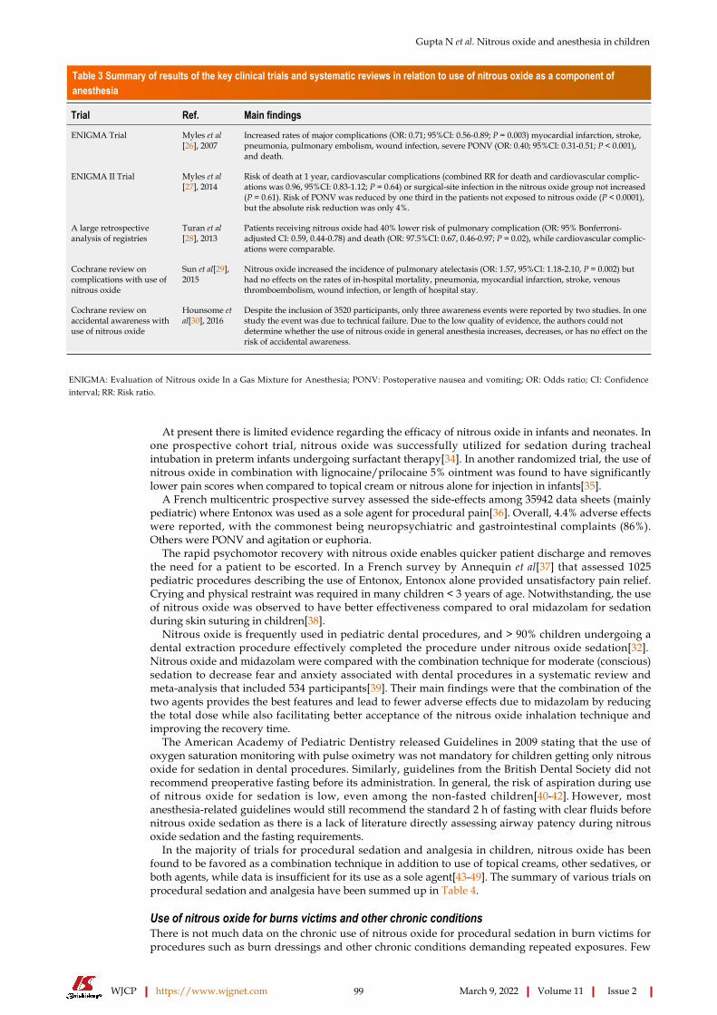
Landmark trials and systematic reviews on undesirable effects of nitrous oxide as a component of general anesthetic gas mixtureMany large-scale studies and meta-analyses have been conducted to study the unfavorable effects of nitrous oxide[26-30]. The results of these trials and meta-analyses highlight why the usage of nitrous oxide have been contentious despite its remarkably safe journey of over one and a half centuries in anesthesia and its multiple advantages as a component of balanced anesthesia. A summary of the most landmark articles exploring the effects of use of nitrous oxide as a component of anesthesia have been complied in Table 3. These trials have been labelled as ‘landmark’ trials for nitrous oxide because of the vast magnitude of data studied and since they turned out to be trailblazers in the history of nitrous oxide use and had a direct influence on the worldwide practice of nitrous oxide anesthesia.
The ENIGMA trial by Myles et al[26] was the first major trial that recruited 2050 patients and compared no nitrous oxide (80% oxygen with 20% nitrogen) and nitrous oxide-based anesthesia (70% N2
O and 30% oxygen). The primary endpoint of this trial was the length of hospital stay. The secondary outcomes comprised of the length of intensive care unit stay and the incidence of postsurgical complic-ations including death within 30 d of surgery. This trial set up a major controversary as use of nitrous oxide as a part of anesthetic gas mixture led to an increased incidence of cardiopulmonary complic-ations, stroke, wound infection, and even mortality in the nitrous oxide cohort. This trial questioned the use of nitrous oxide and was followed by a period of nitrous oxide free anesthesia almost globally.
However, the authors countered their own findings in their next multicentric randomized study with a larger sample size of 7112 patients who had a history of coronary artery disease and were undergoing any major non-cardiac surgery[27]. They assessed the effect of the use of nitrous oxide on the incidence of mortality and any cardiovascular complication (e.g., stroke, myocardial infarction, pulmonary embolism, or cardiac arrest) that occurred within 30 d of undergoing surgery. They found that the risk of cardiovascular complications, surgical-site infection, or death at 1 year were not found to be increased in the nitrous oxide group, and the risk of PONV was found to be only mildly increased[27].
To the great relief of proponents of nitrous oxide, a large trial by Turan et al[28], which evaluated 49016 patients who underwent noncardiac surgery, evaluated the relationship between intraoperative nitrous oxide use and 30d mortality and major postoperative complications. They documented a reduction in pulmonary complications and mortality rates with the use of nitrous oxide, while cardiac risk was not found to be increased.
A Cochrane review further substantiated the fact that use of nitrous oxide was not associated with an increased risk of pneumonia, acute myocardial infarction, stroke, wound infection, venous thromboembolic phenomenon, or increased length of hospital stay or in-hospital mortality[29]. The effect of nitrous oxide on intraoperative awareness is also contentious with some studies reporting increased incidence while others finding a protective effect of nitrous oxide. A recent Cochrane review by Hounsome et al[30] assessed the effect of nitrous oxide on the risk of accidental awareness under anesthesia in 5-year-old and older patients. However, despite the inclusion of 3520 patients, they found only three awareness events and could not come to a definitive conclusion regarding this.
Role of nitrous oxide in procedural sedation and analgesia Nitrous oxide is frequently used for procedural pain relief (e.g., bone marrow aspiration, intercostal drain insertion, venipuncture, lumber puncture, wound sutures, dental extraction, etc.)[31-35]. If used with proper precautions, no major adverse effects have been reported with nitrous oxide use for sedation[36-39].
The use of nitrous oxide in concentrations up to 50% with oxygen during pediatric procedures is an effective substitute for parenteral sedation in minor surgical procedures as it provides pain and anxiety alleviation, maintains protective airway reflexes, and is safe[31-33]. Entonox, which is a mixture of 50% nitrous oxide with 50% oxygen in equal proportions, is a good analgesic agent described in pediatric minor procedures like wound and burn dressing, suturing and suture removal, urinary catheterization, change of gastrostomy tube, synovial fluid and bone marrow aspiration, acute trauma, fracture reduction, lumbar puncture, and minor dental procedures. However, there is evidence on safe adminis-tration of nitrous oxide in delivered concentrations of 20%-70% in children without any major reported adverse events, and hence the cut-off value for procedural sedation should not be arbitrarily limited to 50% for fear of complications[33].
Gupta N et al. Nitrous oxide and anesthesia in children
WJCP https://www.wjgnet.com 99 March 9, 2022 Volume 11 Issue 2
Table 3 Summary of results of the key clinical trials and systematic reviews in relation to use of nitrous oxide as a component of anesthesia
Trial Ref. Main findings
ENIGMA Trial Myles et al[26], 2007
Increased rates of major complications (OR: 0.71; 95%CI: 0.56-0.89; P = 0.003) myocardial infarction, stroke, pneumonia, pulmonary embolism, wound infection, severe PONV (OR: 0.40; 95%CI: 0.31-0.51; P < 0.001), and death.
ENIGMA II Trial Myles et al[27], 2014
Risk of death at 1 year, cardiovascular complications (combined RR for death and cardiovascular complic-ations was 0.96, 95%CI: 0.83-1.12; P = 0.64) or surgical-site infection in the nitrous oxide group not increased (P = 0.61). Risk of PONV was reduced by one third in the patients not exposed to nitrous oxide (P < 0.0001), but the absolute risk reduction was only 4%.
A large retrospective analysis of registries
Turan et al[28], 2013
Patients receiving nitrous oxide had 40% lower risk of pulmonary complication (OR: 95% Bonferroni-adjusted CI: 0.59, 0.44-0.78) and death (OR: 97.5%CI: 0.67, 0.46-0.97; P = 0.02), while cardiovascular complic-ations were comparable.
Cochrane review on complications with use of nitrous oxide
Sun et al[29], 2015
Nitrous oxide increased the incidence of pulmonary atelectasis (OR: 1.57, 95%CI: 1.18-2.10, P = 0.002) but had no effects on the rates of in-hospital mortality, pneumonia, myocardial infarction, stroke, venous thromboembolism, wound infection, or length of hospital stay.
Cochrane review on accidental awareness with use of nitrous oxide
Hounsome et al[30], 2016
Despite the inclusion of 3520 participants, only three awareness events were reported by two studies. In one study the event was due to technical failure. Due to the low quality of evidence, the authors could not determine whether the use of nitrous oxide in general anesthesia increases, decreases, or has no effect on the risk of accidental awareness.
ENIGMA: Evaluation of Nitrous oxide In a Gas Mixture for Anesthesia; PONV: Postoperative nausea and vomiting; OR: Odds ratio; CI: Confidence interval; RR: Risk ratio.
At present there is limited evidence regarding the efficacy of nitrous oxide in infants and neonates. In one prospective cohort trial, nitrous oxide was successfully utilized for sedation during tracheal intubation in preterm infants undergoing surfactant therapy[34]. In another randomized trial, the use of nitrous oxide in combination with lignocaine/prilocaine 5% ointment was found to have significantly lower pain scores when compared to topical cream or nitrous alone for injection in infants[35].
A French multicentric prospective survey assessed the side-effects among 35942 data sheets (mainly pediatric) where Entonox was used as a sole agent for procedural pain[36]. Overall, 4.4% adverse effects were reported, with the commonest being neuropsychiatric and gastrointestinal complaints (86%). Others were PONV and agitation or euphoria.
The rapid psychomotor recovery with nitrous oxide enables quicker patient discharge and removes the need for a patient to be escorted. In a French survey by Annequin et al[37] that assessed 1025 pediatric procedures describing the use of Entonox, Entonox alone provided unsatisfactory pain relief. Crying and physical restraint was required in many children < 3 years of age. Notwithstanding, the use of nitrous oxide was observed to have better effectiveness compared to oral midazolam for sedation during skin suturing in children[38].
Nitrous oxide is frequently used in pediatric dental procedures, and > 90% children undergoing a dental extraction procedure effectively completed the procedure under nitrous oxide sedation[32]. Nitrous oxide and midazolam were compared with the combination technique for moderate (conscious) sedation to decrease fear and anxiety associated with dental procedures in a systematic review and meta-analysis that included 534 participants[39]. Their main findings were that the combination of the two agents provides the best features and lead to fewer adverse effects due to midazolam by reducing the total dose while also facilitating better acceptance of the nitrous oxide inhalation technique and improving the recovery time.
The American Academy of Pediatric Dentistry released Guidelines in 2009 stating that the use of oxygen saturation monitoring with pulse oximetry was not mandatory for children getting only nitrous oxide for sedation in dental procedures. Similarly, guidelines from the British Dental Society did not recommend preoperative fasting before its administration. In general, the risk of aspiration during use of nitrous oxide for sedation is low, even among the non-fasted children[40-42]. However, most anesthesia-related guidelines would still recommend the standard 2 h of fasting with clear fluids before nitrous oxide sedation as there is a lack of literature directly assessing airway patency during nitrous oxide sedation and the fasting requirements.
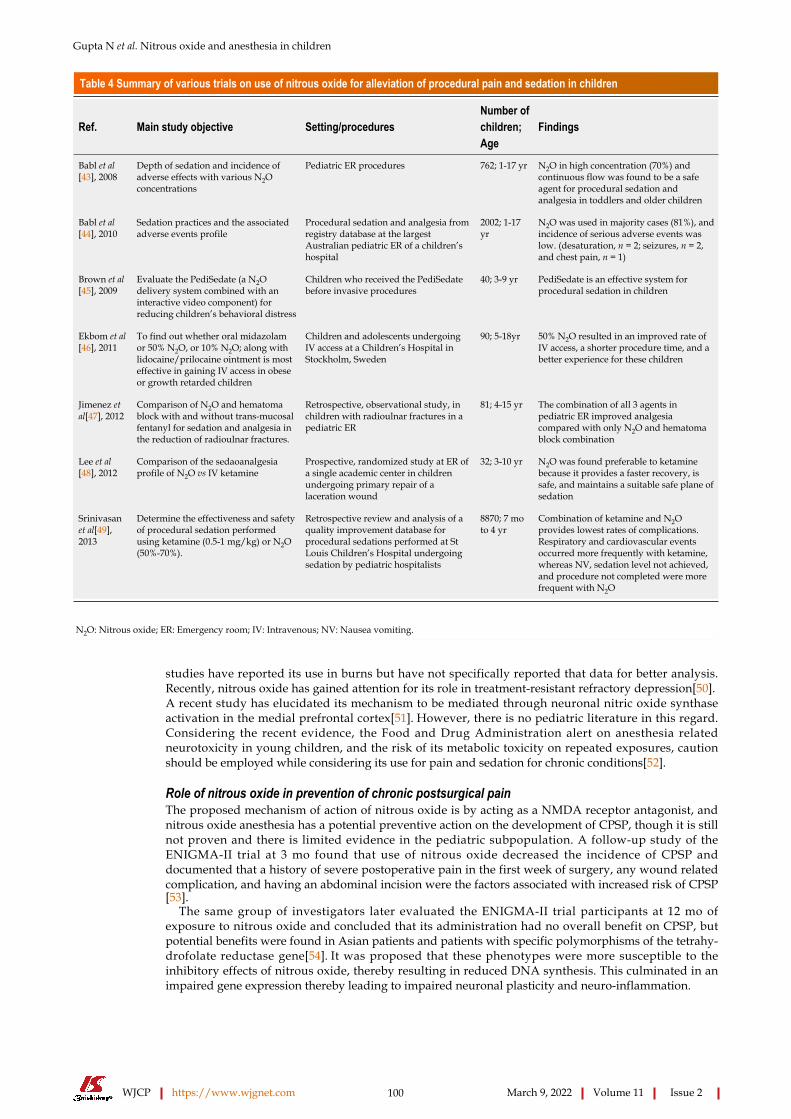
In the majority of trials for procedural sedation and analgesia in children, nitrous oxide has been found to be favored as a combination technique in addition to use of topical creams, other sedatives, or both agents, while data is insufficient for its use as a sole agent[43-49]. The summary of various trials on procedural sedation and analgesia have been summed up in Table 4.
Use of nitrous oxide for burns victims and other chronic conditionsThere is not much data on the chronic use of nitrous oxide for procedural sedation in burn victims for procedures such as burn dressings and other chronic conditions demanding repeated exposures. Few
Gupta N et al. Nitrous oxide and anesthesia in children
WJCP https://www.wjgnet.com 100 March 9, 2022 Volume 11 Issue 2
Table 4 Summary of various trials on use of nitrous oxide for alleviation of procedural pain and sedation in children
Ref. Main study objective Setting/proceduresNumber of children; Age
Findings
Babl et al[43], 2008
Depth of sedation and incidence of adverse effects with various N2O concentrations
Pediatric ER procedures 762; 1-17 yr N2O in high concentration (70%) and continuous flow was found to be a safe agent for procedural sedation and analgesia in toddlers and older children
Babl et al[44], 2010
Sedation practices and the associated adverse events profile
Procedural sedation and analgesia from registry database at the largest Australian pediatric ER of a children’s hospital
2002; 1-17 yr
N2O was used in majority cases (81%), and incidence of serious adverse events was low. (desaturation, n = 2; seizures, n = 2, and chest pain, n = 1)
Brown et al[45], 2009
Evaluate the PediSedate (a N2O delivery system combined with an interactive video component) for reducing children’s behavioral distress
Children who received the PediSedate before invasive procedures
40; 3-9 yr PediSedate is an effective system for procedural sedation in children
Ekbom et al[46], 2011
To find out whether oral midazolam or 50% N2O, or 10% N2O; along with lidocaine/prilocaine ointment is most effective in gaining IV access in obese or growth retarded children
Children and adolescents undergoing IV access at a Children’s Hospital in Stockholm, Sweden
90; 5-18yr 50% N2O resulted in an improved rate of IV access, a shorter procedure time, and a better experience for these children
Jimenez et al[47], 2012
Comparison of N2O and hematoma block with and without trans-mucosal fentanyl for sedation and analgesia in the reduction of radioulnar fractures.
Retrospective, observational study, in children with radioulnar fractures in a pediatric ER
81; 4-15 yr The combination of all 3 agents in pediatric ER improved analgesia compared with only N2O and hematoma block combination
Lee et al[48], 2012
Comparison of the sedaoanalgesia profile of N2O vs IV ketamine
Prospective, randomized study at ER of a single academic center in children undergoing primary repair of a laceration wound
32; 3-10 yr N2O was found preferable to ketamine because it provides a faster recovery, is safe, and maintains a suitable safe plane of sedation
Srinivasan et al[49], 2013
Determine the effectiveness and safety of procedural sedation performed using ketamine (0.5-1 mg/kg) or N2O (50%-70%).
Retrospective review and analysis of a quality improvement database for procedural sedations performed at St Louis Children’s Hospital undergoing sedation by pediatric hospitalists
8870; 7 mo to 4 yr
Combination of ketamine and N2O provides lowest rates of complications. Respiratory and cardiovascular events occurred more frequently with ketamine, whereas NV, sedation level not achieved, and procedure not completed were more frequent with N2O
N2O: Nitrous oxide; ER: Emergency room; IV: Intravenous; NV: Nausea vomiting.
studies have reported its use in burns but have not specifically reported that data for better analysis. Recently, nitrous oxide has gained attention for its role in treatment-resistant refractory depression[50]. A recent study has elucidated its mechanism to be mediated through neuronal nitric oxide synthase activation in the medial prefrontal cortex[51]. However, there is no pediatric literature in this regard. Considering the recent evidence, the Food and Drug Administration alert on anesthesia related neurotoxicity in young children, and the risk of its metabolic toxicity on repeated exposures, caution should be employed while considering its use for pain and sedation for chronic conditions[52].
Role of nitrous oxide in prevention of chronic postsurgical painThe proposed mechanism of action of nitrous oxide is by acting as a NMDA receptor antagonist, and nitrous oxide anesthesia has a potential preventive action on the development of CPSP, though it is still not proven and there is limited evidence in the pediatric subpopulation. A follow-up study of the ENIGMA-II trial at 3 mo found that use of nitrous oxide decreased the incidence of CPSP and documented that a history of severe postoperative pain in the first week of surgery, any wound related complication, and having an abdominal incision were the factors associated with increased risk of CPSP[53].
The same group of investigators later evaluated the ENIGMA-II trial participants at 12 mo of exposure to nitrous oxide and concluded that its administration had no overall benefit on CPSP, but potential benefits were found in Asian patients and patients with specific polymorphisms of the tetrahy-drofolate reductase gene[54]. It was proposed that these phenotypes were more susceptible to the inhibitory effects of nitrous oxide, thereby resulting in reduced DNA synthesis. This culminated in an impaired gene expression thereby leading to impaired neuronal plasticity and neuro-inflammation.
Gupta N et al. Nitrous oxide and anesthesia in children
WJCP https://www.wjgnet.com 101 March 9, 2022 Volume 11 Issue 2
DO WE HAVE A BETTER ALTERNATIVE? There are several drugs being used presently as supplements to general anesthesia that have the potential to reduce the incidence of intraoperative awareness like benzodiazepines, opioids, and alpha2 adrenoceptor agonists. Nevertheless, none of these would offer comparable amnesia, analgesia and cardiovascular stability of the same degree provided by nitrous oxide[20,27,33,36,54]. Recently, xenon, which is an inert gas, has been proposed as a suitable alternative to nitrous oxide. Xenon has profound analgesic properties and superior cardiovascular stability than nitrous oxide. Furthermore, its use has not been associated with harmful neurodevelopmental consequences on developing brain. Hence, it can be considered an attractive option to nitrous oxide in pediatric anesthesia in the future[21]. Presently, its clinical value has been limited mainly by its expense.
CONCLUSIONThe present narrative review summarized the data related to usage of nitrous oxide in pediatric patients. At present there is insufficient evidence to support or refute its continued usage in pediatric practice. Though several new anesthetic agents have been developed, an alternative as flexible and cost-effective as nitrous oxide is yet to be discovered. Certain adverse effects of nitrous oxide like diffusion hypoxia, its ability to expand closed airspaces, increased risk of PONV, ozone depletion, hematologic and neurologic complications, adverse effects on developing brain, and immunosuppression remain a concern to pediatric anesthesiologists. At clinically used concentrations and duration, its use does not appear to be related to hematologic complications and neurobehavioral effects on the developing brain. Its use in children seems justified as a constituent of anesthetic gas mixture and for procedural sedation in the pediatric population for light to moderate pain procedures barring its well-recognized contrain-dications. Combination techniques utilizing nitrous oxide in addition to topical local anesthetics and/or other sedatives have been found to be most effective for procedural sedation, and no major adverse effects reported from even large-scale trials. An individualized approach weighing the risks and benefits of nitrous oxide would be optimal in a particular case. Future perspectives include large-scale research into its specific long-term adverse effects on the developing brain in children in different conditions of administrations, research to fill the gaps in knowledge related to procedural sedation and exploring its potential novel benefits like prevention of CPSP in the pediatric subpopulation.
FOOTNOTESAuthor contributions: Gupta N and Gupta A contributed equally to this work; Gupta N contributed to the concept and data retrieval; Gupta N and Gupta A designed the narrative review, analyzed the data and wrote the manuscript; Gupta A and Narayanan M R V retrieved the data and performed the data analysis and research; all authors have read and approved the final manuscript.
Conflict-of-interest statement: There are no conflicts of interest for any of the authors. None of the authors has received any fees for serving as a speaker, a position, such as consultant and/or an advisory board member or research funding from any organization. Also, the authors do not hold stocks and/or shares in any such firm and do not have a patent.
Open-Access: This article is an open-access article that was selected by an in-house editor and fully peer-reviewed by external reviewers. It is distributed in accordance with the Creative Commons Attribution NonCommercial (CC BY-NC 4.0) license, which permits others to distribute, remix, adapt, build upon this work non-commercially, and license their derivative works on different terms, provided the original work is properly cited and the use is non-commercial. See: http://creativecommons.org/Licenses/by-nc/4.0/
Country/Territory of origin: India
ORCID number: Nishkarsh Gupta 0000-0002-8444-2564; Anju Gupta 0000-0003-1726-1488; Vishnu Narayanan M R 0000-0002-6538-5357.
S-Editor: Wu YXJ L-Editor: Filipodia P-Editor: Wu YXJ
Gupta N et al. Nitrous oxide and anesthesia in children
WJCP https://www.wjgnet.com 102 March 9, 2022 Volume 11 Issue 2
REFERENCESBecker DE, Rosenberg M. Nitrous oxide and the inhalation anesthetics. Anesth Prog 2008; 55: 124-130; quiz 131 [PMID: 19108597 DOI: 10.2344/0003-3006-55.4.124]
1
Kihara S, Yaguchi Y, Inomata S, Watanabe S, Brimacombe JR, Taguchi N, Komatsuzaki T. Influence of nitrous oxide on minimum alveolar concentration of sevoflurane for laryngeal mask insertion in children. Anesthesiology 2003; 99: 1055-1058 [PMID: 14576538 DOI: 10.1097/00000542-200311000-00008]
2
Davidson JA, Macleod AD, Howie JC, White M, Kenny GN. Effective concentration 50 for propofol with and without 67% nitrous oxide. Acta Anaesthesiol Scand 1993; 37: 458-464 [PMID: 8356858 DOI: 10.1111/j.1399-6576.1993.tb03746.x]
3
Inada T, Inada K, Kawachi S, Takubo K, Tai M, Yasugi H. Haemodynamic comparison of sevoflurane and isoflurane anaesthesia in surgical patients. Can J Anaesth 1997; 44: 140-145 [PMID: 9043725 DOI: 10.1007/BF03013001]
4
Lehmberg J, Waldner M, Baethmann A, Uhl E. Inflammatory response to nitrous oxide in the central nervous system. Brain Res 2008; 1246: 88-95 [PMID: 18929548 DOI: 10.1016/j.brainres.2008.09.064]
5
Tramèr MR. Do we need to know whether nitrous oxide harms patients? Lancet 2014; 384: 1407-1409 [PMID: 25142709 DOI: 10.1016/S0140-6736(14)61061-8]
6
Munson ES. Complications of nitrous oxide anesthesia for ear surgery. Anesth Clin North Am 1993; 11: 559-572 [DOI: 10.1016/s0889-8537(21)00751-3]
7
Nunn JF. Clinical aspects of the interaction between nitrous oxide and vitamin B12. Br J Anaesth 1987; 59: 3-13 [PMID: 3548788 DOI: 10.1093/bja/59.1.3]
8
Mosca VS. Letter to the JPO editors re: article by Andreacchio et al entitled "lateral column lengthening as treatment for planovalgus foot deformity in ambulatory children with spastic cerebral palsy"(J Pediatr Orthop 2000;20:501-505). J Pediatr Orthop 2006; 26: 412 [PMID: 16670559 DOI: 10.1097/01.bpo.0000217720.18352.89]
9
Ilniczky S, Jelencsik I, Kenéz J, Szirmai I. MR findings in subacute combined degeneration of the spinal cord caused by nitrous oxide anesthesia--two cases. Eur J Neurol 2002; 9: 101-104 [PMID: 11784385 DOI: 10.1046/j.1468-1331.2002.00336.x]
10
Chen H, Lovell M, Baines D. Metabolic effects of repeated exposure to nitrous oxide: a preliminary report. Pediatr Anesth 2010; 20: 365-366 [DOI: 10.1111/j.1460-9592.2010.03280_2.x]
11
Selzer RR, Rosenblatt DS, Laxova R, Hogan K. Adverse effect of nitrous oxide in a child with 5,10-methylenetetrahydrofolate reductase deficiency. N Engl J Med 2003; 349: 45-50 [PMID: 12840091 DOI: 10.1056/NEJMoa021867]
12
Fernández-Guisasola J, Gómez-Arnau JI, Cabrera Y, del Valle SG. Association between nitrous oxide and the incidence of postoperative nausea and vomiting in adults: a systematic review and meta-analysis. Anaesthesia 2010; 65: 379-387 [PMID: 20151955 DOI: 10.1111/j.1365-2044.2010.06249.x]
13
Bortone L, Picetti E, Mergoni M. Anesthesia with sevoflurane in children: nitrous oxide does not increase postoperative vomiting. Paediatr Anaesth 2002; 12: 775-779 [DOI: 10.1046/j.1460-9592.2002.00939.x]
14
Crawford MW, Lerman J, Sloan MH, Sikich N, Halpern L, Bissonnette B. Recovery characteristics of propofol anaesthesia, with and without nitrous oxide: a comparison with halothane/nitrous oxide anaesthesia in children. Paediatr Anaesth 1998; 8: 49-54 [PMID: 9483598 DOI: 10.1046/j.1460-9592.1998.00708.x]
15
Krajewski W, Kucharska M, Wesolowski W. Occupational exposure to nitrous oxide: the role of scavenging and ventilation systems in reducing the exposure level in operating rooms. Int J Hygiene Environ Health 2007; 210: 133-138 [PMID: 17045524 DOI: 10.1016/j.ijheh.2006.07.004]
16
Perić M, Vranes Z, Marusić M. Immunological disturbances in anaesthetic personnel chronically exposed to high occupational concentrations of nitrous oxide and halothane. Anaesthesia 1991; 46: 531-537 [PMID: 1862889 DOI: 10.1111/j.1365-2044.1991.tb09649.x]
17
Ravishankara AR, Daniel JS, Portmann RW. Nitrous oxide (N2O): the dominant ozone-depleting substance emitted in the 21st century. Science 2009; 326: 123-125 [PMID: 19713491 DOI: 10.1126/science.1176985]
18
Jevtovic-Todorovic V, Hartman RE, Izumi Y, Benshoff ND, Dikranian K, Zorumski CF, Olney JW, Wozniak DF. Early exposure to common anesthetic agents causes widespread neurodegeneration in the developing rat brain and persistent learning deficits. J Neurosci 2003; 23: 876-882 [PMID: 12574416 DOI: 10.1523/JNEUROSCI.23-03-00876.2003]
19
Ghoneim MM, Dhanaraj J, Choi WW. Comparison of four opioid analgesics as supplements to nitrous oxide anesthesia. Anesth Analg 1984; 63: 405-412 [PMID: 6230953]
20
Ma D, Williamson P, Januszewski A, Nogaro MC, Hossain M, Ong LP, Shu Y, Franks NP, Maze M. Xenon mitigates isoflurane-induced neuronal apoptosis in the developing rodent brain. Anesthesiology 2007; 106: 746-753 [PMID: 17413912 DOI: 10.1097/01.anes.0000264762.48920.80]
21
Steffey EP, Johnson BH, Eger EI 2nd, Howland D Jr. Nitrous oxide: effect on accumulation rate and uptake of bowel gases. Anesth Analg 1979; 58: 405-408 [PMID: 573566 DOI: 10.1213/00000539-197909000-00012]
22
Pasternak JJ, Lanier WL. Is nitrous oxide use appropriate in neurosurgical and neurologically at-risk patients? Curr Opin Anaesthesiol 2010; 23: 544-550 [PMID: 20689409 DOI: 10.1097/ACO.0b013e32833e1520]
23
Rodgers L, Dangel-Palmer MC, Berner N. Acute circulatory and respiratory collapse in obstetrical patients: a case report and review of the literature. AANA J 2000; 68: 444-450 [PMID: 11759129]
24
Mosby EL, Schelkun PM, Vincent SK. Nitrous oxide use and endotracheal tube rupture. Anesth Prog 1988; 35: 14-16 [PMID: 3422793]
25
Myles PS, Leslie K, Chan MT, Forbes A, Paech MJ, Peyton P, Silbert BS, Pascoe E; ENIGMA Trial Group. Avoidance of nitrous oxide for patients undergoing major surgery: a randomized controlled trial. Anesthesiology 2007; 107: 221-231 [PMID: 17667565 DOI: 10.1097/01.anes.0000270723.30772.da]
26
Myles PS, Leslie K, Chan MT. ANZCA Trials Group for the ENIGMA-II investigators. The safety of addition of nitrous oxide to general Anesthesia in at-risk patients having major noncardiac surgery (ENIGMA-II): a randomised, single-blind
27
Gupta N et al. Nitrous oxide and anesthesia in children
WJCP https://www.wjgnet.com 103 March 9, 2022 Volume 11 Issue 2
trial. Lancet 2014; 384: 1446-1454 [DOI: 10.1016/s0140-6736(14)60893-x]Turan A, Mascha EJ, You J, Kurz A, Shiba A, Saager L, Sessler DI. The association between nitrous oxide and postoperative mortality and morbidity after noncardiac surgery. Anesth Analg 2013; 116: 1026-1033 [PMID: 22822187 DOI: 10.1213/ANE.0b013e31824590a5]
28
Sun R, Jia WQ, Zhang P, Yang K, Tian JH, Ma B, Liu Y, Jia RH, Luo XF, Kuriyama A. Nitrous oxide-based techniques versus nitrous oxide-free techniques for general anaesthesia. Cochrane Database Syst Rev 2015; CD008984 [PMID: 26545294 DOI: 10.1002/14651858.CD008984.pub2]
29
Hounsome J, Greenhalgh J, Schofield-Robinson OJ, Lewis SR, Cook TM, Smith AF. Nitrous oxide-based vs. nitrous oxide-free general anaesthesia and accidental awareness in surgical patients: an abridged Cochrane systematic review. Anaesthesia 2018; 73: 365-374 [PMID: 29034449 DOI: 10.1111/anae.14065]
30
Bruce E, Franck L. Self-administered nitrous oxide (Entonox®) for the management of procedural pain. Paediatric Nursing 2000; 12: 15-19 [DOI: 10.7748/paed2000.09.12.7.15.c696]
31
Foley J. A prospective study of the use of nitrous oxide inhalation sedation for dental treatment in anxious children. Eur J Paediatr Dent 2005; 6: 121-128 [PMID: 16216091]
32
Buhre W, Disma N, Hendrickx J, DeHert S, Hollmann MW, Huhn R, Jakobsson J, Nagele P, Peyton P, Vutskits L. European Society of Anaesthesiology Task Force on Nitrous Oxide: a narrative review of its role in clinical practice. Br J Anaesth 2019; 122: 587-604 [PMID: 30916011 DOI: 10.1016/j.bja.2019.01.023]
33
Milesi C, Pidoux O, Sabatier E, Badr M, Cambonie G, Picaud JC. Nitrous oxide analgesia for intubating preterm neonates: a pilot study. Acta Paediatr 2006; 95: 1104-1108 [PMID: 16938758 DOI: 10.1080/08035250600698818]
34
Carbajal R, Biran V, Lenclen R, Epaud R, Cimerman P, Thibault P, Annequin D, Gold F, Fauroux B. EMLA cream and nitrous oxide to alleviate pain induced by palivizumab (Synagis) intramuscular injections in infants and young children. Pediatrics 2008; 121: e1591-e1598 [PMID: 18458035 DOI: 10.1542/peds.2007-3104]
35
Onody P, Gil P, Hennequin M. Safety of inhalation of a 50% nitrous oxide/oxygen premix: a prospective survey of 35 828 administrations. Drug Saf 2006; 29: 633-640 [PMID: 16808555 DOI: 10.2165/00002018-200629070-00008]
36
Annequin D, Carbajal R, Chauvin P, Gall O, Tourniaire B, Murat I. Fixed 50% nitrous oxide oxygen mixture for painful procedures: A French survey. Pediatrics 2000; 105: E47 [PMID: 10742368 DOI: 10.1542/peds.105.4.e47]
37
Luhmann JD, Kennedy RM, Porter FL, Miller JP, Jaffe DM. A randomized clinical trial of continuous-flow nitrous oxide and midazolam for sedation of young children during laceration repair. Ann Emerg Med 2001; 37: 20-27 [PMID: 11145766 DOI: 10.1067/mem.2001.112003]
38
Sivaramakrishnan G, Sridharan K. Nitrous Oxide and Midazolam Sedation: A Systematic Review and Meta-Analysis. Anesth Prog 2017; 64: 59-65 [PMID: 28604098 DOI: 10.2344/anpr-63-03-06]
39
Babl FE, Puspitadewi A, Barnett P, Oakley E, Spicer M. Preprocedural fasting state and adverse events in children receiving nitrous oxide for procedural sedation and analgesia. Pediatr Emerg Care 2005; 21: 736-743 [PMID: 16280947 DOI: 10.1097/01.pec.0000186427.07636.fc]
40
Babl FE, Grindlay J, Barrett MJ. Laryngospasm With Apparent Aspiration During Sedation With Nitrous Oxide. Ann Emerg Med 2015; 66: 475-478 [PMID: 26003005 DOI: 10.1016/j.annemergmed.2015.04.029]
41
Tsze DS, Mallory MD, Cravero JP. Practice Patterns and Adverse Events of Nitrous Oxide Sedation and Analgesia: A Report from the Pediatric Sedation Research Consortium. J Pediatr 2016; 169: 260-5.e2 [PMID: 26547401 DOI: 10.1016/j.jpeds.2015.10.019]
42
Babl FE, Oakley E, Seaman C, Barnett P, Sharwood LN. High-concentration nitrous oxide for procedural sedation in children: adverse events and depth of sedation. Pediatrics 2008; 121: e528-e532 [PMID: 18310173 DOI: 10.1542/peds.2007-1044]
43
Babl FE, Belousoff J, Deasy C, Hopper S, Theophilos T. Paediatric procedural sedation based on nitrous oxide and ketamine: sedation registry data from Australia. Emerg Med J 2010; 27: 607-612 [PMID: 20515915 DOI: 10.1136/emj.2009.084384]
44
Brown SC, Hart G, Chastain DP, Schneeweiss S, McGrath PA. Reducing distress for children during invasive procedures: randomized clinical trial of effectiveness of the PediSedate. Paediatr Anaesth 2009; 19: 725-731 [PMID: 19624359 DOI: 10.1111/j.1460-9592.2009.03076.x]
45
Ekbom K, Kalman S, Jakobsson J, Marcus C. Efficient intravenous access without distress: a double-blind randomized study of midazolam and nitrous oxide in children and adolescents. Arch Pediatr Adolesc Med 2011; 165: 785-791 [PMID: 21536947 DOI: 10.1001/archpediatrics.2011.56]
46
Jimenez A, Blazquez D, Cruz J. Use of combined transmucosal fentanyl, nitrous oxide, and hematoma block for fracture reduction in a pediatric emergency department. Pediatr Emerg Care 2012; 28: 676-679 [DOI: 10.1097/pec.0b013e31825d20f6]
47
Lee JH, Kim K, Kim TY, Jo YH, Kim SH, Rhee JE, Heo CY, Eun SC. A randomized comparison of nitrous oxide versus intravenous ketamine for laceration repair in children. Pediatr Emerg Care 2012; 28: 1297-1301 [PMID: 23187987 DOI: 10.1097/PEC.0b013e3182768a86]
48
Srinivasan M, Carlson DW. Procedural sedation by pediatric hospitalists: analysis of the nature and incidence of complications during ketamine and nitrous oxide sedation. Hosp Pediatr 2013; 3: 342-347 [PMID: 24435192 DOI: 10.1542/hpeds.2013-0025]
49
Lew V, McKay E, Maze M. Past, present, and future of nitrous oxide. Br Med Bull 2018; 125: 103-119 [PMID: 29528367 DOI: 10.1093/bmb/ldx050]
50
Liu W, Li Q, Ye B, Cao H, Shen F, Xu Z, Du W, Guo F, Liu J, Li T, Zhang B, Liu Z. Repeated Nitrous Oxide Exposure Exerts Antidepressant-Like Effects Through Neuronal Nitric Oxide Synthase Activation in the Medial Prefrontal Cortex. Front Psychiatry 2020; 11: 837 [PMID: 33088274 DOI: 10.3389/fpsyt.2020.00837]
51
US FDA. FDA Drug Safety Communication: FDA review results in new warnings about using general anesthetics and sedation drugs in young children and pregnant women 2018. Available from: https://www.fda.gov/Drugs/DrugSafety/ ucm532356.htm [DOI: 10.31525/fda2-ucm612193.htm]
52
Chan MTV, Wan ACM, Gin T, Leslie K, Myles PS. Chronic postsurgical pain after nitrous oxide anesthesia. Pain 2011; 53

Gupta N et al. Nitrous oxide and anesthesia in children
WJCP https://www.wjgnet.com 104 March 9, 2022 Volume 11 Issue 2
152: 2514-2520 [PMID: 21889262 DOI: 10.1016/j.pain.2011.07.015]Chan MT, Peyton PJ, Myles PS, Leslie K, Buckley N, Kasza J, Paech MJ, Beattie WS, Sessler DI, Forbes A, Wallace S, Chen Y, Tian Y, Wu WK; and the Australian and New Zealand College of Anaesthetists Clinical Trials Network for the ENIGMA-II investigators. Chronic postsurgical pain in the Evaluation of Nitrous Oxide in the Gas Mixture for Anaesthesia (ENIGMA)-II trial. Br J Anaesth 2016; 117: 801-811 [PMID: 27956679 DOI: 10.1093/bja/aew338]
54

WJCP https://www.wjgnet.com 105 March 9, 2022 Volume 11 Issue 2
World Journal of
Clinical PediatricsW J C PSubmit a Manuscript: https://www.f6publishing.com World J Clin Pediatr 2022 March 9; 11(2): 105-119
DOI: 10.5409/wjcp.v11.i2.105 ISSN 2219-2808 (online)
REVIEW
Non-pharmacological management of pediatric functional abdominal pain disorders: Current evidence and future perspectives
Maria Luísa Cordeiro Santos, Ronaldo Teixeira da Silva Júnior, Breno Bittencourt de Brito, Filipe Antônio França da Silva, Hanna Santos Marques, Vinícius Lima de SouzaGonçalves, Talita Costa dos Santos, Carolina Ladeia Cirne, Natália Oliveira e Silva, Márcio Vasconcelos Oliveira, Fabrício Freire de Melo
Specialty type: Pediatrics
Provenance and peer review: Invited article; Externally peer reviewed.
Peer-review model: Single blind
Peer-review report’s scientific quality classificationGrade A (Excellent): A Grade B (Very good): B Grade C (Good): 0 Grade D (Fair): 0 Grade E (Poor): 0
P-Reviewer: Kesavelu D, Pavlovic M
Received: March 20, 2021 Peer-review started: March 20, 2021 First decision: July 18, 2021 Revised: July 19, 2021 Accepted: February 11, 2022 Article in press: February 11, 2022 Published online: March 9, 2022
Maria Luísa Cordeiro Santos, Ronaldo Teixeira da Silva Júnior, Breno Bittencourt de Brito, Filipe Antônio França da Silva, Talita Costa dos Santos, Carolina Ladeia Cirne, Natália Oliveira e Silva, Márcio Vasconcelos Oliveira, Fabrício Freire de Melo, Instituto Multidisciplinar em Saúde, Universidade Federal da Bahia, Vitória da Conquista 45029-094, Bahia, Brazil
Hanna Santos Marques, Vinícius Lima de SouzaGonçalves, Campus Vitória da Conquista, Universidade Estadual do Sudoeste da Bahia, Vitória da Conquista 45083-900, Bahia, Brazil
Corresponding author: Fabrício Freire de Melo, PhD, Professor, Instituto Multidisciplinar em Saúde, Universidade Federal da Bahia, Rua Hormindo Barros, 58, Quadra 17, Lote 58, Vitória da Conquista 45029-094, Bahia, Brazil. [email protected]
AbstractFunctional abdominal pain disorders (FAPDs) are an important and prevalent cause of functional gastrointestinal disorders among children, encompassing the diagnoses of functional dyspepsia, irritable bowel syndrome, abdominal migraine, and the one not previously present in Rome III, functional abdominal pain not otherwise specified. In the absence of sufficiently effective and safe pharmacological treatments for this public problem, non-pharmacological therapies emerge as a viable means of treating these patients, avoiding not only possible side effects, but also unnecessary prescription, since many of the pharma-cological treatments prescribed do not have good efficacy when compared to placebo. Thus, the present study provides a review of current and relevant evidence on non-pharmacological management of FAPDs, covering the most commonly indicated treatments, from cognitive behavioral therapy to meditation, acupuncture, yoga, massage, spinal manipulation, moxibustion, and physical activities. In addition, this article also analyzes the quality of publications in the area, assessing whether it is possible to state if non-pharmacological therapies are viable, safe, and sufficiently well-based for an appropriate and effective prescription of these treatments. Finally, it is possible to observe an increase not only in the number of publications on the non-pharmacological treatments for FAPDs in recent years, but also an increase in the quality of these publications. Finally, the sample selection of satisfactory age groups in these studies enables the formulation of specific guidelines for this age group, thus avoiding the need for adaptation of prescriptions initially made for adults, but for children use.

Cordeiro Santos ML et al. Non-pharmacological management of pediatric FAPDs
WJCP https://www.wjgnet.com 106 March 9, 2022 Volume 11 Issue 2
Key Words: Functional abdominal pain disorder; Pediatrics; Rome IV; Behavioral intervention; Non-pharmacological treatment; Complementary medicine
©The Author(s) 2022. Published by Baishideng Publishing Group Inc. All rights reserved.
Core Tip: Functional abdominal pain disorders are an important and prevalent cause of functional gastrointestinal disorders among children. In the absence of sufficiently effective and safe pharmaco-logical treatments for this public problem, non-pharmacological therapies emerge as a viable means of treating these patients. Thus, the present study provides a review of current and relevant evidence on non-pharmacological management of these disorders, as cognitive behavioral therapy, meditation, acupuncture, and others. This article also analyzes the quality of publications in the area, assessing whether it is possible to state if non-pharmacological therapies are viable, safe, and sufficiently well-based for an appropriate and effective prescription.
Citation: Cordeiro Santos ML, da Silva Júnior RT, de Brito BB, França da Silva FA, Santos Marques H, Lima de SouzaGonçalves V, Costa dos Santos T, Ladeia Cirne C, Silva NOE, Oliveira MV, de Melo FF. Non-pharmacological management of pediatric functional abdominal pain disorders: Current evidence and future perspectives. World J Clin Pediatr 2022; 11(2): 105-119URL: https://www.wjgnet.com/2219-2808/full/v11/i2/105.htmDOI: https://dx.doi.org/10.5409/wjcp.v11.i2.105
INTRODUCTIONFunctional gastrointestinal disorders (FGIDs) are a group of diseases defined by morphological and physiological changes that affect from the gastrointestinal tract (GIT) to the central nervous system (CNS). Among the main changes listed in this group, there are disorders of intestinal motility, visceral hypersensitivity, and changes in the mucosa and in the host's immune responses, in addition to possible changes in the normal microbiome of the intestinal environment[1].
FGIDs are stratified in alphabetical letters from A to H, with the present article focused on non-pharmacological treatment specifically for group H (FGIDs in children or adolescents), subtype H2, defined as functional abdominal pain disorders (FAPDs) by the Rome IV criteria. This group consists of functional dyspepsia (H2a), irritable bowel syndrome (IBS) (H2b), abdominal migraine (H2c), and functional abdominal pain not otherwise specified (H2d), with the latter not previously present in Rome III[2].
With regard to the diagnosis of FGIDs, the 2016 Rome IV criteria removed the obligation to rule out organic causes using complementary tests, making the clinical evaluation criteria sufficient for diagnosis, thus avoiding the exposure of these patients to unnecessary testing[1]. In this sense, comple-mentary/laboratory examinations are not required for diagnosis after careful clinical examination and in the absence of alarm criteria that suggest organic causes or complications of FAPDs. The following is considered alarm criteria: Family history of inflammatory bowel disease, celiac disease, or peptic ulcer disease; persistent right upper or right lower quadrant pain; dysphagia; odynophagia; persistent vomiting; gastrointestinal blood loss; nocturnal diarrhea; arthritis; perirectal disease; involuntary weight loss; deceleration of linear growth; delayed puberty and unexplained fever[1]. The stratified diagnostic criteria for FAPDs are shown in Table 1.
Regarding the prevalence of FAPDs, it is estimated that about 13.5% (95% confidence interval [CI]: 11.8-15.3) of the children worldwide present one of the diseases in this group, with emphasis on IBS, representing 8.8% (95%CI: 6.2-11.9) of that number. In addition, risk factors for the development of FADPs were identified as being female (15.9% prevalence vs 11.5% male) and the presence of anxiety, depression, stress symptoms, or traumatic life events[3].
In view of the important prevalence of FAPDs, it is necessary to establish effective and adequate treatments, ensuring not only the control of symptoms but also the safety of patients. In addition, because studies of pharmacological safety in an age group are insufficient, the use of efficient non-pharmacological therapies in the treatment of the pediatric public is ideal. Thus, the aim of this article is to understand, through a review of the literature available in the main databases, the use of different non-pharmacological therapies in the treatment of FAPDs in children, analyzing from how they have been indicated to the levels of evidence that sustains their prescription.
Cordeiro Santos ML et al. Non-pharmacological management of pediatric FAPDs
WJCP https://www.wjgnet.com 107 March 9, 2022 Volume 11 Issue 2
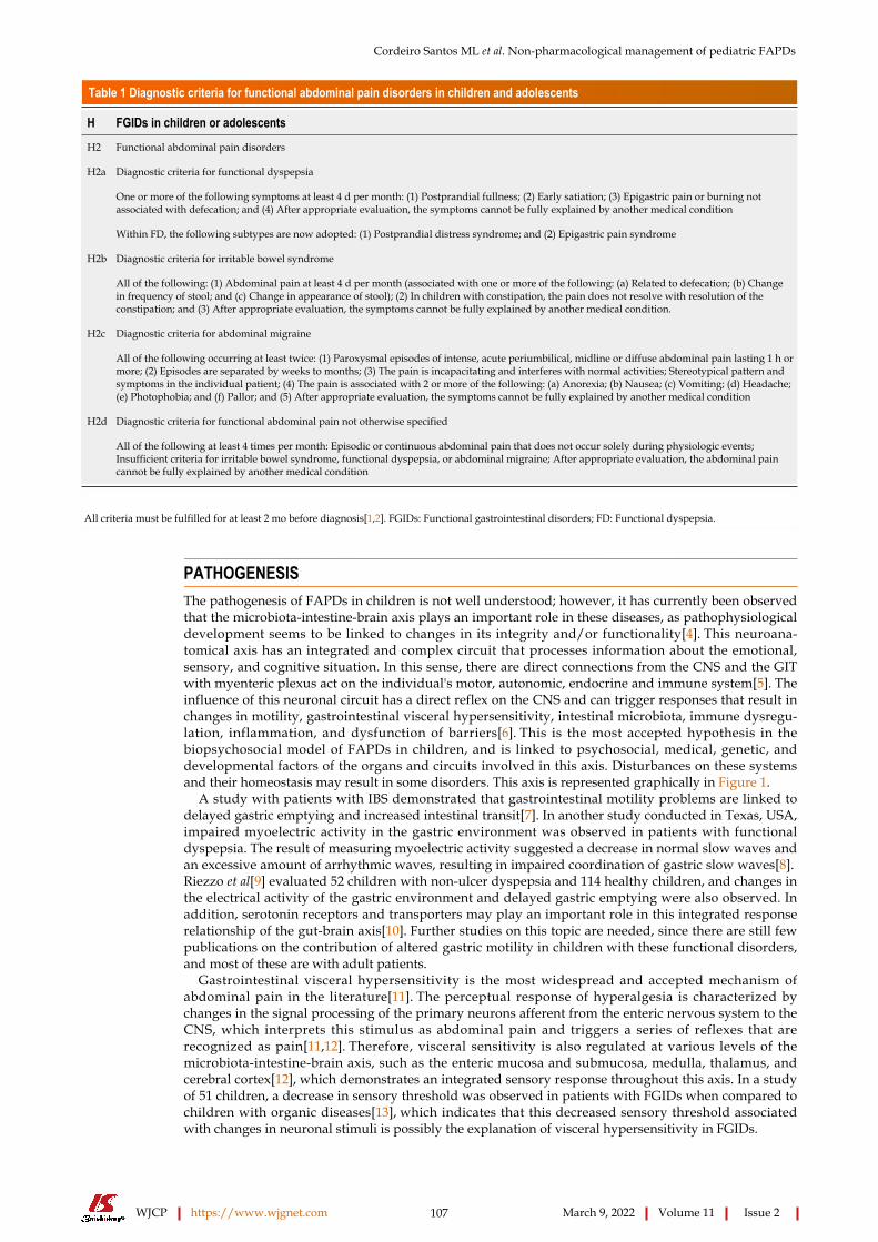
Table 1 Diagnostic criteria for functional abdominal pain disorders in children and adolescents
H FGIDs in children or adolescents
H2 Functional abdominal pain disorders
H2a Diagnostic criteria for functional dyspepsia
One or more of the following symptoms at least 4 d per month: (1) Postprandial fullness; (2) Early satiation; (3) Epigastric pain or burning not associated with defecation; and (4) After appropriate evaluation, the symptoms cannot be fully explained by another medical condition
Within FD, the following subtypes are now adopted: (1) Postprandial distress syndrome; and (2) Epigastric pain syndrome
H2b Diagnostic criteria for irritable bowel syndrome
All of the following: (1) Abdominal pain at least 4 d per month (associated with one or more of the following: (a) Related to defecation; (b) Change in frequency of stool; and (c) Change in appearance of stool); (2) In children with constipation, the pain does not resolve with resolution of the constipation; and (3) After appropriate evaluation, the symptoms cannot be fully explained by another medical condition.
H2c Diagnostic criteria for abdominal migraine
All of the following occurring at least twice: (1) Paroxysmal episodes of intense, acute periumbilical, midline or diffuse abdominal pain lasting 1 h or more; (2) Episodes are separated by weeks to months; (3) The pain is incapacitating and interferes with normal activities; Stereotypical pattern and symptoms in the individual patient; (4) The pain is associated with 2 or more of the following: (a) Anorexia; (b) Nausea; (c) Vomiting; (d) Headache; (e) Photophobia; and (f) Pallor; and (5) After appropriate evaluation, the symptoms cannot be fully explained by another medical condition
H2d Diagnostic criteria for functional abdominal pain not otherwise specified
All of the following at least 4 times per month: Episodic or continuous abdominal pain that does not occur solely during physiologic events; Insufficient criteria for irritable bowel syndrome, functional dyspepsia, or abdominal migraine; After appropriate evaluation, the abdominal pain cannot be fully explained by another medical condition
All criteria must be fulfilled for at least 2 mo before diagnosis[1,2]. FGIDs: Functional gastrointestinal disorders; FD: Functional dyspepsia.
PATHOGENESISThe pathogenesis of FAPDs in children is not well understood; however, it has currently been observed that the microbiota-intestine-brain axis plays an important role in these diseases, as pathophysiological development seems to be linked to changes in its integrity and/or functionality[4]. This neuroana-tomical axis has an integrated and complex circuit that processes information about the emotional, sensory, and cognitive situation. In this sense, there are direct connections from the CNS and the GIT with myenteric plexus act on the individual's motor, autonomic, endocrine and immune system[5]. The influence of this neuronal circuit has a direct reflex on the CNS and can trigger responses that result in changes in motility, gastrointestinal visceral hypersensitivity, intestinal microbiota, immune dysregu-lation, inflammation, and dysfunction of barriers[6]. This is the most accepted hypothesis in the biopsychosocial model of FAPDs in children, and is linked to psychosocial, medical, genetic, and developmental factors of the organs and circuits involved in this axis. Disturbances on these systems and their homeostasis may result in some disorders. This axis is represented graphically in Figure 1.
A study with patients with IBS demonstrated that gastrointestinal motility problems are linked to delayed gastric emptying and increased intestinal transit[7]. In another study conducted in Texas, USA, impaired myoelectric activity in the gastric environment was observed in patients with functional dyspepsia. The result of measuring myoelectric activity suggested a decrease in normal slow waves and an excessive amount of arrhythmic waves, resulting in impaired coordination of gastric slow waves[8]. Riezzo et al[9] evaluated 52 children with non-ulcer dyspepsia and 114 healthy children, and changes in the electrical activity of the gastric environment and delayed gastric emptying were also observed. In addition, serotonin receptors and transporters may play an important role in this integrated response relationship of the gut-brain axis[10]. Further studies on this topic are needed, since there are still few publications on the contribution of altered gastric motility in children with these functional disorders, and most of these are with adult patients.
Gastrointestinal visceral hypersensitivity is the most widespread and accepted mechanism of abdominal pain in the literature[11]. The perceptual response of hyperalgesia is characterized by changes in the signal processing of the primary neurons afferent from the enteric nervous system to the CNS, which interprets this stimulus as abdominal pain and triggers a series of reflexes that are recognized as pain[11,12]. Therefore, visceral sensitivity is also regulated at various levels of the microbiota-intestine-brain axis, such as the enteric mucosa and submucosa, medulla, thalamus, and cerebral cortex[12], which demonstrates an integrated sensory response throughout this axis. In a study of 51 children, a decrease in sensory threshold was observed in patients with FGIDs when compared to children with organic diseases[13], which indicates that this decreased sensory threshold associated with changes in neuronal stimuli is possibly the explanation of visceral hypersensitivity in FGIDs.

Cordeiro Santos ML et al. Non-pharmacological management of pediatric FAPDs
WJCP https://www.wjgnet.com 108 March 9, 2022 Volume 11 Issue 2
Figure 1 Graphical representation of the gut-brain axis in the pathogenesis of functional abdominal pain in pediatric populations.
Evidence shows that the microbiota of patients with FGIDs differs from healthy people[14]. In a recent systematic review with patients with IBS, with three studies included with children, a significant increase in the bacterial population of the family Enterobacteriaceae and Lactobacillaceae and genus Bacteroides in patients with IBS when compared with the control group was observed. In addition, there was a decrease in bacterial colonization of Bifidobacterium spp., Faecalibacterium spp., and Faecalibacterium prausnitzii[15], which plays an important role in the balance of the immune system in the intestine[16]. However, the role of the microbiota in relation to functional diseases in children is not well established. Most studies evaluated fecal samples from adult patients with IBS and have limitations in relation to sample collection, and diet and medication used by the patient. In this sense, more studies would be important in order to understand the influence that the way of delivery, metabolome and other microor-ganisms have on the intestinal microbiota of these children.
Homeostasis of the microbiota-intestine-brain axis is essential to maintain the integrity of the immune system, and disturbances in this balance can generate uncontrolled inflammation in the gastrointestinal mucosa. Interestingly, infiltration of mast cells, eosinophils, and lymphocytes has been observed in intestinal environment of patients with functional disorders. In particular, mast cell recruitment is involved in epithelial and neuromuscular dysfunction[17]. These inflammatory cells are close to neurosensorial fibers of the GIT mucosa and have a relevant role in altering neurogenic inflammatory pathways and in perception of pain in response to harmful stimuli[18]. In addition, the degree of inflam-mation in the GIT mucosa can cause injuries and, consequently, a rupture of barriers that restrict bacterial colonization under normal conditions. As a result, bacterial overgrowth can be observed, which can culminate in FGIDs[12,19].
High self-perceived prevalence of food intolerances has been reported in children with IBS[20]. These symptoms are associated with nutritional behavioral changes and children's diet[21]; however, the knowledge about how nutritional factors influence functional gastrointestinal diseases is still unclear. Therefore, greater knowledge about a possible adequate nutritional pattern for maintaining the balance of the microbiota-intestine-brain axis may be ideal for a better understanding of the relationship between food and the intestinal microbiota in that axis.
The psychological factors and their relationship to intestinal motility are well understood. Some studies have already demonstrated the physiological effects on the GIT, triggered by anger, fear, and anxiety[12]. In a recent review, the authors concluded that although it is still unclear whether FGIDs may have psychological factors as their etiology, there is strong evidence that these factors exacerbate and contribute to maintenance of pain[22]. An interesting correlational study found a decrease in the symptoms of functional disorders in children in summer, when compared to spring (P = 0.017). The authors correlated this improvement to the vacation period when they are exposed to fewer stressors, but were unable to distinguish what is the cause and what is the effect of decreasing symptoms[23]. Also, the hypothalamus-pituitary-adrenal axis may have an interaction with the microbiota-intestine-brain axis, releasing cortisol and corticotropin, which stimulate metabolic stress and cause a release of mast cells and pro-inflammatory cytokines[4]. Stress factors can also deregulate the balance of the intestinal microbiota, increasing the permeability of epithelial tissue, and facilitating the entry of pathogens that can create an inflammatory environment. In addition, the release of cytokines such as IFNγ, IL-1, and IL-6 can stimulate an immune response in the CNS and reflect an exacerbation of psychological symptoms[24]. With this, it becomes increasingly important that children with symptoms related to FGIDs receive the integration of the psychological examination in their global care[25].
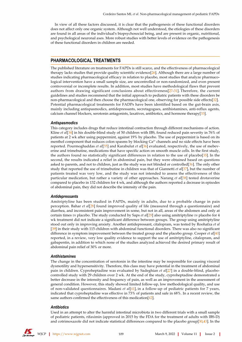
Cordeiro Santos ML et al. Non-pharmacological management of pediatric FAPDs
WJCP https://www.wjgnet.com 109 March 9, 2022 Volume 11 Issue 2
In view of all these factors discussed, it is clear that the pathogenesis of these functional disorders does not affect only one organic system. Although not well understood, the etiologies of these disorders are found in all areas of the individual's biopsychosocial being, and are present in organic, nutritional, and psychological neuronal axes. More robust studies with better levels of evidence on the pathogenesis of these functional disorders in children are needed.
PHARMACOLOGICAL TREATMENTSThe published literature on treatments for FAPDs is still scarce, and the effectiveness of pharmacological therapy lacks studies that provide quality scientific evidence[26]. Although there are a large number of studies indicating pharmacological efficacy in relation to placebo, most studies that analyze pharmaco-logical intervention have a small sample size, are uncontrolled or non-randomized, and even present controversial or incomplete results. In addition, most studies have methodological flaws that prevent authors from drawing significant conclusions about effectiveness[27-31]. Therefore, the current guidelines and studies recommend that the initial approach to pediatric patients with these disorders be non-pharmacological and then choose the pharmacological one, observing for possible side effects[32]. Potential pharmacological treatments for FADPs have been identified based on the gut-brain axis, mainly including antispasmodics, antidepressants, secretagogues, antihistamines, anti-reflux agents, calcium channel blockers, serotonin antagonists, laxatives, antibiotics, and hormone therapy[33].
AntispasmodicsThis category includes drugs that reduce intestinal contraction through different mechanisms of action. Kline et al[34] in his double-blind study of 50 children with IBS, found reduced pain severity in 76% of patients at 2 wk after using peppermint, against 19% by placebo. The use of peppermint is based on its menthol component that reduces colon spasms by blocking Ca2+ channels and no side effects have been reported. Pourmoghaddas et al[35] and Karabulut et al[36] evaluated, respectively, the use of mebev-erine and trimebutine, medications that have specific action on smooth muscle cells. In the first study, the authors found no statistically significant differences in relation to the use of placebo[35]. In the second, the results indicated a relief in abdominal pain, but they were obtained based on questions asked to parents, and not to children, just as the study was not blinded or controlled[36]. The only other study that reported the use of trimebutine in children was that of Giannetti et al[37], but the number of patients treated was very low, and the study was not intended to assess the effectiveness of this particular medication, but rather a variety of other approaches. Narang et al[38] tested drotaverine compared to placebo in 132 children for 4 wk, and although the authors reported a decrease in episodes of abdominal pain, they did not describe the intensity of the pain.
AntidepressantsAmitriptyline has been studied in FAPDs, mainly in adults, due to a probable change in pain perception. Bahar et al[28] found improved quality of life (measured through a questionnaire) and diarrhea, and inconsistent pain improvement in some, but not in all, areas of the abdomen and only at certain times vs placebo. The study conducted by Saps et al[29] also using amitriptyline vs placebo for 4 wk treatment did not indicate a significant difference between groups. The group using amitriptyline stood out only in improving anxiety. Another antidepressant, citalopram, was tested by Roohafza et al[39] in their study with 115 children with abdominal functional disorders. There was also no significant difference in symptom improvement between the treated group and the placebo group. Cooper et al[40] reported, in a review, very low quality evidence to support the use of amitriptyline, citalopram, and gabapentin, in addition to which none of the studies analyzed achieved the desired primary result of abdominal pain relief of 30% or more.
AntihistaminesThe change in the concentration of serotonin in the intestine may be responsible for causing visceral dysmotility and hypersensitivity. Therefore, this class may have potential in the treatment of abdominal pain in children. Cyproheptadine was evaluated by Sadeghian et al[27] in a double-blind, placebo-controlled study with 29 children over 2 wk. At the end of the study, cyproheptadine demonstrated a better decrease in the intensity and frequency of pain, as well as an improvement in the assessment of general condition. However, this study showed limited follow-up, low methodological quality, and use of non-validated questionnaires. Madani et al[41], in a follow-up of pediatric patients for 7 years, indicated that cyproheptadine was effective in 73% of patients and safe in 68%. In a recent review, the same authors confirmed the effectiveness of this medication[42].
AntibioticsUsed in an attempt to alter the harmful intestinal microbiota in two different trials with a small sample of pediatric patients, rifaximin (approved in 2015 by the FDA for the treatment of adults with IBS-D) and cotrimoxazole did not indicate statistical differences compared to the placebo group[30,43]. In the
Cordeiro Santos ML et al. Non-pharmacological management of pediatric FAPDs
WJCP https://www.wjgnet.com 110 March 9, 2022 Volume 11 Issue 2
first study, the authors assessed abdominal pain, episodes of diarrhea and constipation, feeling of incomplete or effective evacuation, urgency to evacuate, effort to evacuate, and the presence of some fecal secretion. Erythromycin, another antibiotic that appears to have agonist properties of the motilin receptor in the stomach, has been reported to be useful in relieving symptoms of abdominal pain and dyspepsia in adults, but there is still insufficient pediatric data for clinical indication[44].
Serotonin antagonistsOne of the only medications in this class reported in studies was pizotifen. Symon et al[45] comparing it to placebo, reported that the children in the study showed a reduction in the “Severity Index” related to abdominal pain. However, this study presented a small sample of 16 patients with abdominal migraine, was interrupted before more patients were included in the study, and used a scale with no validation[45].
H2 receptor antagonistsIn the study of See et al[46], 25 children with dyspepsia and functional abdominal pain were treated with famotidine or placebo. At the end of the study, there was no significant difference in treatment regarding the frequency of abdominal pain, pain intensity, and dyspeptic symptoms. In addition, the group that received famotidine had an improvement in the overall assessment of 66.7%, while the group that received placebo had a 15.4% improvement in this same parameter. However, the authors provided insufficient data to establish a confidence interval between these percentages[46].
ProkineticsDomperidone was used in a placebo controlled trial to assess the response in children with functional dyspepsia[47]. According to the results, patients did not show a different cure rate between domperidone and placebo after 8 wk, and no data was reported to indicate improvement in nausea - an important symptom in this pathology. However, after being followed for 6 mo, the children who received the medication showed an increase in the cure rate and in the overall assessment. Prokinetics have been used mainly in situations where functional abdominal pain is accompanied by constipation or delayed gastric emptying, as in IBS. Nevertheless, in several regions of the world, they have their commercialization restricted due to its side effects, including cardiovascular events[48].
Proton pump inhibitorsProton pump inhibitors are usually indicated for the treatment of dyspeptic symptoms, since they end up acting in the acidic environment of the digestive tract. Karjoo and Kane[49], in their study with 153 patients aged 6 to 18 years with abdominal pain and dyspepsia, reported a significant improvement in symptoms, especially those who were resistant to the use of H2 antagonists. The patients in this study were treated with high-dose ranitidine hydrochloride and omeprazole as the main proton-pump inhibitor[49].
Hormonal treatmentsMelatonin, a hormone produced by the pineal gland, has also been studied in the treatment of these pathologies, and its use is justified by a possible improvement in sleep. Zybach et al[50] analyzed the efficacy of melatonin in children with functional dyspepsia for 2 wk in a double-blind, randomized, placebo-controlled crossover study with a small sample of 12 patients. They found a positive clinical response in 42% of individuals with melatonin vs 50% of individuals who received placebo. In this sense, no efficacy was observed in the use of melatonin for the relief of abdominal pain in functional conditions[50].
SecretagoguesSome secretagogues have been evaluated in functional conditions that generate abdominal pain, such as IBS with a predominance of constipation. In an uncontrolled trial in children and adolescents, the use of lubiprostone was shown to be beneficial in increasing the frequency of spontaneous evacuation and pain[31]. In another double-blind, randomized, controlled study[51], the frequency of bowel movements, pain, effort, and consistency of stools did not show a statistically significant difference when comparing lubiprostone and placebo. Thus, further controlled studies are needed to confirm the effectiveness of this medication for the treatment of abdominal pain specifically, as they are already confirmed to be beneficial in the treatment for constipation. On the other hand, linaclotide is currently approved by the FDA and the European Medicines Agency for the therapy of chronic constipation in adults. At the moment, there are no published studies reporting efficacy and safety of the use of linaclotide in children, but there is a double-blind multicenter study under development, with children and adolescents, to assess this (NCT02559817)[52].
In view of the high prevalence of FAPDs in children, their high impact on quality of life, and lack of significant studies, there is still a gap in the search for safe and effective pharmacological therapies, with well-developed, randomized, controlled and multicenter studies. It is also important to highlight that this process is important to prevent maleficent effects of these drugs in a system that is still ongoing

Cordeiro Santos ML et al. Non-pharmacological management of pediatric FAPDs
WJCP https://www.wjgnet.com 111 March 9, 2022 Volume 11 Issue 2
neuroplasticity changes and growth development. In addition, it is necessary to pay attention to the cost-benefit ratio that the medication will offer, especially in relation to placebo and non-pharmaco-logical therapies[51,52].
NON-PHARMACOLOGICAL TREATMENTSThe incorporation of integrative and complementary non-pharmacological interventions in manage-ment of the pediatric chronic pain has demonstrated to be viable and effective for this population[53,54]. Such methods can lead to long-term results due to changes in the neural circuits that regulate habits, affection, and cognitive responses to pain[55]. Thereby, treatments such as cognitive behavioral therapy (CBT), acupuncture, spinal manipulation, exercise, among others come to assist the health professionals in pediatric chronic pain therapy. As an aim of this study, we based the classification of integrative and complementary practices on the structure proposed by the database Biblioteca Virtual de Saúde – Medicinas Tradicional Complementares e Integrativas das Américas (BVS-MTCI), developed by the Traditional Complementary and Integrative Medicine Web of America[56].
Mind-body therapiesCognitive behavioral therapy: CBT is based on the premise that thoughts, emotions, and behaviors are linked, as well as how someone perceives a situation can significantly influence emotional, behavioral and physiological responses[54]. CBT involves the teaching of coping and distraction strategies and relaxation techniques; identification and change of pain-related thoughts; and modification of family responses to pain. This method can involve the family itself or may focus only on the child, as well as be performed face-to-face or remotely[57,58]. Family approach seeks to alter environmental factors that might reinforce the child’s pain behavior within the family and to identify and treat factors that may precipitate in it[58].
There is growing support for CBT for children with FAPDs[59]. Multiple components are typically used in CBT, such as education about the pain, increasing self-confidence[60], cognitive restructuring of maladaptive thoughts, exposure exercises, relaxation, and parent management techniques[61]. In exposure-based CBT for FAPDs, the patients gradually expose themselves to symptom-provoking stimuli (such as eating pizza) and approach situations in which symptoms are perceived as intolerable (such as being in school). This approach is hypothesized to decrease fear and avoidance related to symptoms and thereby enables symptom reduction[61].
A randomized clinical trial with 104 children aged 7-18 years investigated the effectiveness of a 6 weekly session CBT protocol compared with 6 visits to a pediatric gastroenterologist and the impact of these interventions on pain. This CBT session resulted in a significant reduction of abdominal pain in 60% of children with FAP up to 1 year after treatment, and the CBT is more effective than intensive medical care directly after treatment[62]. Another study showed that children who received CBT improved significantly more than the control group on abdominal pain-related symptoms and coping strategies, as well as parental solicitousness in response to pain behaviors. Moreover, many of these differences were maintained 6 mo after intervention[63].
Furthermore, Internet-delivered CBT (Internet-CBT) may help to bridge this treatment gap. Internet-CBT holds several advantages over traditional face-to-face therapy: It can be delivered to people in remote areas, patients can access the treatment without taking time off from school or work, and it requires fewer therapist hours per patient[64].
One study reported outcomes in adolescents aged 13–17 years with IBS who received a 10-wk session of Internet-delivered exposure CBT, compared with wait-list controls. There was a large change before and after treatment in gastrointestinal symptoms, with a medium effect size, and improved anxiety, school absenteeism, and adolescent-rated and parent-rated quality of life. After 6 mo, the results were stable or significantly improved[65].
Furthermore, a randomized clinical trial with 90 children diagnosed with FAPDs, based on the Rome IV criteria, found that Internet-CBT has the potential to increase the availability of treatment for a number of patients and reduce health care costs[66]. Moreover, more than half of the children in the Internet-CBT group reported a 30% or greater improvement of their gastrointestinal symptom severity at the 10-wk follow-up evaluation vs 32% of the children in the treatment-as-usual group[66].
Meditation: Meditation can be defined as a form of mental training that aims to improve an individual’s core psychological capacities, such as attentional and emotional self-regulation[67]. Meditative techniques include transcendental meditation, mindfulness-based stress reduction, and mindfulness-based cognitive therapy[68]. Of these practices, mindfulness meditation has received most attention in neuroscience[69]. In current clinical and research contexts, mindfulness meditation is typically described as non-judgemental attention to experiences in the present moment and requires both the regulation of attention and the ability to approach one’s experiences with openness and acceptance[69,70]. This nonjudgmental focus on present-moment experience appears to be a potentially avenue in helping adolescents attend to pain adaptively[71].

Cordeiro Santos ML et al. Non-pharmacological management of pediatric FAPDs
WJCP https://www.wjgnet.com 112 March 9, 2022 Volume 11 Issue 2
An increasing body of literature has demonstrated that mindfulness interventions are feasible and efficacious in adult pain populations[72,73]. On the other hand, pediatric populations that experienced chronic pain conditions, as neuropathic and abdominal pain, have demonstrated initial feasibility and acceptability of mindfulness-based interventions (MBIs)[74,75]. While preliminary research among pediatric pain populations demonstrates feasibility and acceptability of MBIs, additional studies are necessary to investigate mindfulness in children and adolescents with chronic pain. Thus, this intervention may be low cost or free adjunctive treatments that have fewer side effects as compared to pharmacological interventions[54].
Traditional health systemAcupuncture: Acupuncture is an ancient medical procedure that has been practiced in China and other East Asian countries. The technique involves the placement of small needles at various locations in the body and related therapies include electroacupuncture, acupressure, moxibustion (i.e., burning of an herb near an acupoint to create local warming), laser stimulation of acupoints, and non-invasive stimulation of acupoints utilizing a transcutaneous electrical nerve stimulator[54].
The mechanisms of the relationship between acupuncture and improvement of the pain remain uncertain. Studies have shown that the acupuncture may involve normalization of activity in areas of the limbic system often referred to as the “pain matrix” (i.e., the insula, anterior cingulate gyrus, and prefrontal cortex)[76], or can stimulate endorphin release[77]. This method is also postulated to have effects on acid secretion, gastrointestinal motility, and sensation of visceral pain, possibly mediated through the release of opioid peptides in the central and enteric nervous system[38].
However, while substantial research has shown acupuncture to be an effective therapy for pain among the adult population, there is limited research on acupuncture with regard to the treatment of pain among pediatric patients[78]. Despite this scarce literature, a systematic review published identified common minor adverse effects and rare serious harms in pediatric acupuncture[79]. Puncture redness is the most commonly reported side effect, followed by needle pain and light-headedness[80].
A systematic review of randomized controlled trials on the use of acupuncture in infantile colic shows that acupuncture appears to be effective in alleviating the symptoms of colic, including crying and feeding and stooling problems. However, due to the small sample sizes of the included studies, more randomized clinical trials are necessary[81]. Another case series study found that minimal acupuncture in infantile colic is an effective and easy treatment procedure[82].
A difficulty in treating the pediatric population is children’s fear of needles. The treatment periods are reduced compared with the treatment of adults and closely monitored. Non-invasive modalities, such as electrical stimulation or laser, on acupoints and acupressure seem to be well accepted by younger children[80].
Although acupuncture is safe when administered by appropriately trained and credentialed practi-tioners, there are some children who have a fear of needles or for medical reasons such as low platelet count or immunodeficiency that may not be recommended to receive acupuncture. For those patients, other techniques such as acupressure[78], laser acupuncture, topical magnets, and acupressure beads may be used. They may also be used as adjunctive treatments following needle placement[83].
Moxibustion: A meta-analysis compared the effectiveness of the use of moxibustion with conventional drugs for inflammatory bowel disease and concluded that this method may be useful in the treatment of the disease. There was a significant improvement of general symptoms related to the disease (P = 0.0001); however, regarding specific symptoms, only abdominal distension (P = 0.03) and frequency of defecation (P = 0.02) were significant. Moreover, the authors highlighted that there is a low number of clinical trials evaluating this treatment[84]. In a recent study, Liu and Zeng[85] evaluated the effect-iveness of the umbilical therapy combined with moxibustion for diarrhea in pediatric patients. The results showed that the treatment significantly improved the symptoms of diarrhea (P = 0.05) and was associated with a shorter recovery time for the children (P = 0.05)[85]. Another Chinese study used moxibustion to treat 120 children with abdominal pain. The effectiveness rate of the treatment after 3 mo was 94.78%, compared to 80.77% in the control group[86].
Yoga: Yoga has been shown to be an exercise that provides several benefits for children, including improvements in the emotional control, anxiety, and depression[87,88]. Moreover, it seems to be effective in assuaging pain associated with some abdominal disturbances in that population[26]. A study carried out by Brands et al[89] evaluated the repercussions of Yoga practice in 20 children (age range: 8-18 years) with inflammatory bowel syndrome or abdominal pain. The children participated in 10 Yoga sessions, lasting 1.5 h each, being observed that the exercises reduced the severity and frequency of abdominal pain immediately after the classes. Moreover, after 3 mo of continued exercises at home, the children continued to report improvements; however, the status of the preexisting conditions was not modified[89]. A recent study enrolling adolescents aged from 14 to 17 years with inflammatory bowel disease demonstrated that the use of Yoga for 6 wk resulted in an improvement of abdominal pain, sleep, and visceral hypersensitivity among responding participants. Nonetheless, the findings for the abdominal symptoms were not statistically significant (P = 0.8) and the study sample was small (n = 18)[90]. As for Evans et al[91], the study showed that an intervention with Yoga as a

Cordeiro Santos ML et al. Non-pharmacological management of pediatric FAPDs
WJCP https://www.wjgnet.com 113 March 9, 2022 Volume 11 Issue 2
complementary treatment benefits young adults with IBD with a reduction of symptoms. In contrast, a systematic review has stated that the existing studies on Yoga and inflammatory bowel disease do not present satisfactory scientific quality. Therefore, it concluded that, although Yoga is a safe practice for pediatric patients, there are no official recommendations for its use[92].
Manual therapyMassage: Therapeutic massage has been associated with a significant improvement among pediatric patients with chronic pain due to several diseases, including abdominal disturbances. A study evaluated various techniques such as compression, triggering points, petrissage, tapotement, and effleurage, and concluded that the use of massages is a reasonable option as an adjuvant treatment since they reduce pain, agony, discomfort, and humor alterations[93]. Nam et al[94] observed the effects of flavoring massages of the abdominal meridians in children with cerebral lesions and concluded that the use of the therapy 3 to 5 times a week was associated with an improvement in constipation. Another study including patients aged from 4 to 18 years demonstrated that the combined use of isometric training of abdominal muscles, respiratory exercises, and abdominal massages resulted in the reduction of the frequency of evacuation among patients with chronic functional constipation (P = 0.01). The treatment was based on sessions of 40 min each, two times a week, for 12 wk[95]. A systematic review evaluated the occurrence of adverse events related to massotherapy in premature babies and reported that it may lead to mild and severe side effects including hematoma, status epilepticus, and volvulus. However, the study identified publication bias and, therefore, it was not possible to identify the causal relationship between the adverse events and massotherapy, though authors recommend caution to perform this method in premature newborns[96]. Moreover, a study including 40 babies showed that abdominal massage with lavender oil has the potential to reduce colic in children aged from 2 to 6 mo. The results were obtained based on the frequency of weekly cries of the patients, and those who underwent massage with lavender oil used to cry less often than the individuals from the control group (P < 0.05)[97]. In a recently published study, Al Qahtani and Ahmed recommended the development of educational programs aiming to teach abdominal massage and feet reflexology techniques for parents, since it is an effective way to improve abdominal colic in babies[98].
Spinal manipulation: The relationship between spinal manipulation and improvement of symptoms related to abdominal disorders is controversial. Some studies have indicated that the therapy may be associated with an improvement of abdominal pain among children[26,99]. On the other hand, a systematic review of clinical trials was not able to conclude that the spinal manipulation is an effective therapeutic practice against infant colic, and the author stated that the low quality of the studies contributes to the lack of consistent recommendations on that issue[100]. Another recent systematic review showed that the spinal manipulation has some advantages in the control of some types of pain such as lumbar and cervical pain; however, the knowledge on the benefit of that technique against infant colic is still limited since there are many contradictory and low-quality studies evaluating that therapy[101]. In that context, a review evaluated the safety of the performance of spinal manipulation in children and concluded that most side effects reported were mild and that moderate-to-severe adverse events linked to this technique remain unknown[102]. In contrast, Vohra et al[103] suggested that the manipulation of the spine may be related to severe side effects in the pediatric population. However, important limitations were highlighted in both studies.
Physical activities: Boradyn et al[104] carried out a study including 25 children aged from 5 to 11 years to evaluate the impact of the lifestyle in pediatric patients diagnosed with functional abdominal pain. The results showed that the practice of physical activities might increase the frequency of evacuation among children (P = 0.031); however, the data regarding the relationship between exercises and abdominal pain were not statistically significant[104]. A recent study observed an association between the practice of physical activities and the development of constipation among children and identified that infants who often practice exercises had a lower odds of acquiring the disorder than sedentary individuals (P = 0.016)[105]. Complementally, a study that evaluated the effectiveness of alternative complementary medicine for functional abdominal pain observed that 49% of the patients enrolled used to practice exercises to improve their symptoms. Moreover, individuals who rated their condition as severe tend to practice exercises more often than those who rate their disorders as mild or moderate (P = 0.043)[106]. Kichline et al[107] recently observed that young individuals with chronic abdominal pain did not use to practice physical activities 60 min per day. In addition, another study evaluated socioeconomic factors involved in the probability of occurrence of gastrointestinal disorders related to abdominal pain and concluded that the low practice of exercises is positively associated with the disorder (P = 0.028)[108,109].
In view of the individual discussion of each of these non-pharmacological therapies, the level of evidence for each of them is stratified in Table 2, in addition to the analysis of the public of each study (adults/children) and the timeline of the publications in each of those areas.
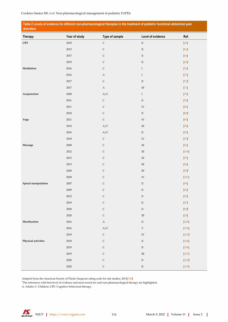
Cordeiro Santos ML et al. Non-pharmacological management of pediatric FAPDs
WJCP https://www.wjgnet.com 114 March 9, 2022 Volume 11 Issue 2
Table 2 Levels of evidence for different non-pharmacological therapies in the treatment of pediatric functional abdominal pain disorders
Therapy Year of study Type of sample Level of evidence Ref.
2010 C II [62]
2013 C II [61]
2017 C II [64]
CBT
2019 C II [65]1
2016 C I [74]
2016 A I [72]
2017 C II [73]1
Meditation
2017 A III [71]
2008 A/C I [79]
2011 C II [78]
2011 C IV [81]
Acupuncture
2018 C II [80]1
2011 C IV [92]
2014 A/C III [94]
2016 A/C II [95]
Yoga
2018 C IV [93]1
2008 C III [96]
2012 C III [100]
2013 C III [97]
2013 C III [98]
2020 C III [99]1
Massage
2020 C IV [101]
2007 C II [99]
2009 C II [96]
2012 C II [95]
2019 C II [97]
2020 C II [98]1
Spinal manipulation
2020 C III [26]
2016 A II [100]
2016 A/C V [102]
Moxibustion
2019 C IV [101]1
2018 C II [104]
2019 C II [106]
2019 C III [107]
2020 C II [103]1
Physical activities
2020 C II [105]
Adapted from the American Society of Plastic Surgeons rating scale for risk studies, 2011[108].1The references with best-level of evidence and most recent for each non-pharmacological therapy are highlighted.A: Adults; C: Children; CBT: Cognitive behavioral therapy.
Cordeiro Santos ML et al. Non-pharmacological management of pediatric FAPDs
WJCP https://www.wjgnet.com 115 March 9, 2022 Volume 11 Issue 2
CONCLUSIONIt is possible to conclude that there is a need for safe and effective treatments for the management of FAPDs in the pediatric public. In this sense, and in view of the low quality and insufficient satisfactory results of pharmacological therapies, non-pharmacological treatments emerge as a viable and important solution to this problem of increasing numbers worldwide. In the meantime, it is possible to see a stimulus and an increasing amount of better evidence to support the prescription of these therapies in clinical practice, achieving better results and greater safety for patients. Finally, with these studies being made with sample selections of satisfactory age groups, the formulation of specific guidelines for this age group is made possible, as there is no need for adaptation of prescriptions initially made for adults for children.
FOOTNOTESAuthor contributions: All authors equally contributed to this paper with conception and design, literature review and analysis, manuscript drafting, critical revision, and editing, and approval of the final version.
Conflict-of-interest statement: No potential conflicts of interest exist.
Open-Access: This article is an open-access article that was selected by an in-house editor and fully peer-reviewed by external reviewers. It is distributed in accordance with the Creative Commons Attribution NonCommercial (CC BY-NC 4.0) license, which permits others to distribute, remix, adapt, build upon this work non-commercially, and license their derivative works on different terms, provided the original work is properly cited and the use is non-commercial. See: https://creativecommons.org/Licenses/by-nc/4.0/
Country/Territory of origin: Brazil
ORCID number: Maria Luísa Cordeiro Santos 0000-0001-7078-9789; Ronaldo Teixeira da Silva Júnior 0000-0003-0835-4092; Breno Bittencourt de Brito 0000-0002-1831-7909; Filipe Antônio França da Silva 0000-0002-0550-1109; Hanna Santos Marques 0000-0001-5741-1570; Vinícius Lima de Souza Gonçalves 0000-0002-6445-9318; Talita Costa dos Santos 0000-0002-9102-589X; Carolina Ladeia Cirne 0000-0002-9850-1190; Natália Oliveira e Silva 0000-0003-0106-7509; Márcio Vasconcelos Oliveira 0000-0002-8959-0478; Fabrício Freire de Melo 0000-0002-5680-2753.
S-Editor: Ma YJ L-Editor: Wang TQ P-Editor: Ma YJ
REFERENCESRasquin A, Di Lorenzo C, Forbes D, Guiraldes E, Hyams JS, Staiano A, Walker LS. Childhood functional gastrointestinal disorders: child/adolescent. Gastroenterology 2006; 130: 1527-1537 [PMID: 16678566 DOI: 10.1053/j.gastro.2005.08.063]
1
Hyams JS, Di Lorenzo C, Saps M, Shulman RJ, Staiano A, van Tilburg M. Functional Disorders: Children and Adolescents. Gastroenterology 2016 [PMID: 27144632 DOI: 10.1053/j.gastro.2016.02.015]
2
Korterink JJ, Diederen K, Benninga MA, Tabbers MM. Epidemiology of pediatric functional abdominal pain disorders: a meta-analysis. PLoS One 2015; 10: e0126982 [PMID: 25992621 DOI: 10.1371/journal.pone.0126982]
3
Thapar N, Benninga MA, Crowell MD, Di Lorenzo C, Mack I, Nurko S, Saps M, Shulman RJ, Szajewska H, van Tilburg MAL, Enck P. Paediatric functional abdominal pain disorders. Nat Rev Dis Primers 2020; 6: 89 [PMID: 33154368 DOI: 10.1038/s41572-020-00222-5]
4
Jones MP, Dilley JB, Drossman D, Crowell MD. Brain-gut connections in functional GI disorders: anatomic and physiologic relationships. Neurogastroenterol Motil 2006; 18: 91-103 [PMID: 16420287 DOI: 10.1111/j.1365-2982.2005.00730.x]
5
Drossman DA. Functional Gastrointestinal Disorders: History, Pathophysiology, Clinical Features and Rome IV. Gastroenterology 2016 [PMID: 27144617 DOI: 10.1053/j.gastro.2016.02.032]
6
DuPont AW, Jiang ZD, Harold SA, Snyder N, Galler GW, Garcia-Torres F, DuPont HL. Motility abnormalities in irritable bowel syndrome. Digestion 2014; 89: 119-123 [PMID: 24503633 DOI: 10.1159/000356314]
7
Sha W, Pasricha PJ, Chen JD. Rhythmic and spatial abnormalities of gastric slow waves in patients with functional dyspepsia. J Clin Gastroenterol 2009; 43: 123-129 [PMID: 18719512 DOI: 10.1097/MCG.0b013e318157187a]
8
Riezzo G, Chiloiro M, Guerra V, Borrelli O, Salvia G, Cucchiara S. Comparison of gastric electrical activity and gastric emptying in healthy and dyspeptic children. Dig Dis Sci 2000; 45: 517-524 [PMID: 10749327 DOI: 10.1023/a:1005493123557]
9
Gershon MD. Review article: serotonin receptors and transporters -- roles in normal and abnormal gastrointestinal motility. Aliment Pharmacol Ther 2004; 20 Suppl 7: 3-14 [PMID: 15521849 DOI: 10.1111/j.1365-2036.2004.02180.x]
10
Bueno L, Fioramonti J. Visceral perception: inflammatory and non-inflammatory mediators. Gut 2002; 51 Suppl 1: i19-11
Cordeiro Santos ML et al. Non-pharmacological management of pediatric FAPDs
WJCP https://www.wjgnet.com 116 March 9, 2022 Volume 11 Issue 2
i23 [PMID: 12077058 DOI: 10.1136/gut.51.suppl_1.i19]Karantanos T, Markoutsaki T, Gazouli M, Anagnou NP, Karamanolis DG. Current insights in to the pathophysiology of Irritable Bowel Syndrome. Gut Pathog 2010; 2: 3 [PMID: 20465787 DOI: 10.1186/1757-4749-2-3]
12
Halac U, Noble A, Faure C. Rectal sensory threshold for pain is a diagnostic marker of irritable bowel syndrome and functional abdominal pain in children. J Pediatr 2010; 156: 60-65.e1 [PMID: 19800076 DOI: 10.1016/j.jpeds.2009.06.062]
13
Enck P, Mazurak N. Dysbiosis in Functional Bowel Disorders. Ann Nutr Metab 2018; 72: 296-306 [PMID: 29694952 DOI: 10.1159/000488773]
14
Pittayanon R, Lau JT, Yuan Y, Leontiadis GI, Tse F, Surette M, Moayyedi P. Gut Microbiota in Patients With Irritable Bowel Syndrome-A Systematic Review. Gastroenterology 2019; 157: 97-108 [PMID: 30940523 DOI: 10.1053/j.gastro.2019.03.049]
15
Miquel S, Martín R, Rossi O, Bermúdez-Humarán LG, Chatel JM, Sokol H, Thomas M, Wells JM, Langella P. Faecalibacterium prausnitzii and human intestinal health. Curr Opin Microbiol 2013; 16: 255-261 [PMID: 23831042 DOI: 10.1016/j.mib.2013.06.003]
16
Santos J, Alonso C, Guilarte M, Vicario M, Malagelada JR. Targeting mast cells in the treatment of functional gastrointestinal disorders. Curr Opin Pharmacol 2006; 6: 541-546 [PMID: 16956793 DOI: 10.1016/j.coph.2006.08.001]
17
Wouters MM, Vicario M, Santos J. The role of mast cells in functional GI disorders. Gut 2016; 65: 155-168 [PMID: 26194403 DOI: 10.1136/gutjnl-2015-309151]
18
Husebye E. The pathogenesis of gastrointestinal bacterial overgrowth. Chemotherapy 2005; 51 Suppl 1: 1-22 [PMID: 15855746 DOI: 10.1159/000081988]
19
Chumpitazi BP, Weidler EM, Lu DY, Tsai CM, Shulman RJ. Self-Perceived Food Intolerances Are Common and Associated with Clinical Severity in Childhood Irritable Bowel Syndrome. J Acad Nutr Diet 2016; 116: 1458-1464 [PMID: 27316779 DOI: 10.1016/j.jand.2016.04.017]
20
Reed-Knight B, Squires M, Chitkara DK, van Tilburg MA. Adolescents with irritable bowel syndrome report increased eating-associated symptoms, changes in dietary composition, and altered eating behaviors: a pilot comparison study to healthy adolescents. Neurogastroenterol Motil 2016; 28: 1915-1920 [PMID: 27353222 DOI: 10.1111/nmo.12894]
21
Newton E, Schosheim A, Patel S, Chitkara DK, van Tilburg MAL. The role of psychological factors in pediatric functional abdominal pain disorders. Neurogastroenterol Motil 2019; 31: e13538 [PMID: 30729663 DOI: 10.1111/nmo.13538]
22
Pollard KL, Campbell C, Squires M, Palsson O, van Tilburg MAL. Seasonal Association of Pediatric Functional Abdominal Pain Disorders and Anxiety. J Pediatr Gastroenterol Nutr 2018; 67: 18-22 [PMID: 29287016 DOI: 10.1097/MPG.0000000000001886]
23
Dinan TG, Cryan JF. The Microbiome-Gut-Brain Axis in Health and Disease. Gastroenterol Clin North Am 2017; 46: 77-89 [PMID: 28164854 DOI: 10.1016/j.gtc.2016.09.007]
24
Cunningham NR, Moorman E, Brown CM, Mallon D, Chundi PK, Mara CA, Pentiuk S, Lynch-Jordan AM, Dykes DMH, Elfers J, Farrell MK. Integrating Psychological Screening Into Medical Care for Youth With Abdominal Pain. Pediatrics 2018; 142 [PMID: 30045930 DOI: 10.1542/peds.2017-2876]
25
Santucci NR, Saps M, van Tilburg MA. New advances in the treatment of paediatric functional abdominal pain disorders. Lancet Gastroenterol Hepatol 2020; 5: 316-328 [PMID: 31859185 DOI: 10.1016/S2468-1253(19)30256-0]
26
Sadeghian M, Farahmand F, Fallahi GH, Abbasi A. Cyproheptadine for the treatment of functional abdominal pain in childhood: a double-blinded randomized placebo-controlled trial. Minerva Pediatr 2008; 60: 1367-1374 [PMID: 18971897]
27
Bahar RJ, Collins BS, Steinmetz B, Ament ME. Double-blind placebo-controlled trial of amitriptyline for the treatment of irritable bowel syndrome in adolescents. J Pediatr 2008; 152: 685-689 [PMID: 18410774 DOI: 10.1016/j.jpeds.2007.10.012]
28
Saps M, Youssef N, Miranda A, Nurko S, Hyman P, Cocjin J, Di Lorenzo C. Multicenter, randomized, placebo-controlled trial of amitriptyline in children with functional gastrointestinal disorders. Gastroenterology 2009; 137: 1261-1269 [PMID: 19596010 DOI: 10.1053/j.gastro.2009.06.060]
29
Collins BS, Lin HC. Double-blind, placebo-controlled antibiotic treatment study of small intestinal bacterial overgrowth in children with chronic abdominal pain. J Pediatr Gastroenterol Nutr 2011; 52: 382-386 [PMID: 21240023 DOI: 10.1097/MPG.0b013e3181effa3b]
30
Hyman PE, Di Lorenzo C, Prestridge LL, Youssef NN, Ueno R. Lubiprostone for the treatment of functional constipation in children. J Pediatr Gastroenterol Nutr 2014; 58: 283-291 [PMID: 24048162 DOI: 10.1097/MPG.0000000000000176]
31
Tack J, Camilleri M. New developments in the treatment of gastroparesis and functional dyspepsia. Curr Opin Pharmacol 2018; 43: 111-117 [PMID: 30245474 DOI: 10.1016/j.coph.2018.08.015]
32
Chiou E, Nurko S. Management of functional abdominal pain and irritable bowel syndrome in children and adolescents. Expert Rev Gastroenterol Hepatol 2010; 4: 293-304 [PMID: 20528117 DOI: 10.1586/egh.10.28]
33
Kline RM, Kline JJ, Di Palma J, Barbero GJ. Enteric-coated, pH-dependent peppermint oil capsules for the treatment of irritable bowel syndrome in children. J Pediatr 2001; 138: 125-128 [PMID: 11148527 DOI: 10.1067/mpd.2001.109606]
34
Pourmoghaddas Z, Saneian H, Roohafza H, Gholamrezaei A. Mebeverine for pediatric functional abdominal pain: a randomized, placebo-controlled trial. Biomed Res Int 2014; 2014: 191026 [PMID: 25089264 DOI: 10.1155/2014/191026]
35
Karabulut GS, Beşer OF, Erginöz E, Kutlu T, Cokuğraş FÇ, Erkan T. The Incidence of Irritable Bowel Syndrome in Children Using the Rome III Criteria and the Effect of Trimebutine Treatment. J Neurogastroenterol Motil 2013; 19: 90-93 [PMID: 23350053 DOI: 10.5056/jnm.2013.19.1.90]
36
Giannetti E, Maglione M, Sciorio E, Coppola V, Miele E, Staiano A. Do Children Just Grow Out of Irritable Bowel Syndrome? J Pediatr 2017; 183: 122-126.e1 [PMID: 28108106 DOI: 10.1016/j.jpeds.2016.12.036]
37
Narang M, Shah D, Akhtar H. Efficacy and Safety of Drotaverine Hydrochloride in Children with Recurrent Abdominal Pain: A Randomized Placebo Controlled Trial. Indian Pediatr 2015; 52: 847-851 [PMID: 26499007 DOI: 10.1007/s13312-015-0730-y]
38
Cordeiro Santos ML et al. Non-pharmacological management of pediatric FAPDs
WJCP https://www.wjgnet.com 117 March 9, 2022 Volume 11 Issue 2
Roohafza H, Pourmoghaddas Z, Saneian H, Gholamrezaei A. Citalopram for pediatric functional abdominal pain: a randomized, placebo-controlled trial. Neurogastroenterol Motil 2014; 26: 1642-1650 [PMID: 25244442 DOI: 10.1111/nmo.12444]
39
Cooper TE, Heathcote LC, Clinch J, Gold JI, Howard R, Lord SM, Schechter N, Wood C, Wiffen PJ. Antidepressants for chronic non-cancer pain in children and adolescents. Cochrane Database Syst Rev 2017; 8: CD012535 [PMID: 28779487 DOI: 10.1002/14651858.CD012535.pub2]
40
Madani S, Cortes O, Thomas R. Cyproheptadine Use in Children With Functional Gastrointestinal Disorders. J Pediatr Gastroenterol Nutr 2016; 62: 409-413 [PMID: 26308312 DOI: 10.1097/MPG.0000000000000964]
41
Krasaelap A, Madani S. Cyproheptadine: A Potentially Effective Treatment for Functional Gastrointestinal Disorders in Children. Pediatr Ann 2017; 46: e120-e125 [PMID: 28287686 DOI: 10.3928/19382359-20170213-01]
42
Heyland K, Friedt M, Buehr P, Braegger CP. No advantage for antibiotic treatment over placebo in Blastocystis hominis-positive children with recurrent abdominal pain. J Pediatr Gastroenterol Nutr 2012; 54: 677-679 [PMID: 22002479 DOI: 10.1097/MPG.0b013e31823a29a7]
43
Tack J. Prokinetics and fundic relaxants in upper functional GI disorders. Curr Opin Pharmacol 2008; 8: 690-696 [PMID: 18940266 DOI: 10.1016/j.coph.2008.09.009]
44
Symon DN, Russell G. Double blind placebo controlled trial of pizotifen syrup in the treatment of abdominal migraine. Arch Dis Child 1995; 72: 48-50 [PMID: 7717738 DOI: 10.1136/adc.72.1.48]
45
See MC, Birnbaum AH, Schechter CB, Goldenberg MM, Benkov KJ. Double-blind, placebo-controlled trial of famotidine in children with abdominal pain and dyspepsia: global and quantitative assessment. Dig Dis Sci 2001; 46: 985-992 [PMID: 11341669 DOI: 10.1023/a:1010793408132]
46
Karunanayake A, Devanarayana NM, de Silva A, Gunawardena S, Rajindrajith S. Randomized Controlled Clinical Trial on Value of Domperidone in Functional Abdominal Pain in Children. J Pediatr Gastroenterol Nutr 2018; 66: 725-731 [PMID: 29112086 DOI: 10.1097/MPG.0000000000001819]
47
Bor S, Demir M, Ozdemir O, Yuksel K. A meta-analysis on the cardiac safety profile of domperidone compared to metoclopramide. United European Gastroenterol J 2018; 6: 1331-1346 [PMID: 30386606 DOI: 10.1177/2050640618799153]
48
Karjoo M, Kane R. Omeprazole treatment of children with peptic esophagitis refractory to ranitidine therapy. Arch Pediatr Adolesc Med 1995; 149: 267-271 [PMID: 7858685 DOI: 10.1001/archpedi.1995.02170150047007]
49
Zybach K, Friesen CA, Schurman JV. Therapeutic effect of melatonin on pediatric functional dyspepsia: A pilot study. World J Gastrointest Pharmacol Ther 2016; 7: 156-161 [PMID: 26855822 DOI: 10.4292/wjgpt.v7.i1.156]
50
Benninga MA, Hussain SZ, Sood MR, Samuel N, Hyman PE, Taryn LB, Peter L, Di LC. Efficacy and safety of lubiprostone in children with functional constipation: a multicenter, randomized, placebo-controlled, double-blind pivotal study. Gastroenterology 2018; 154: S559–560 [DOI: 10.1016/s0016-5085(18)32065-1]
51
Saps M, Miranda A. Gastrointestinal Pharmacology. Handb Exp Pharmacol 2017; 239: 147-176 [PMID: 28236087 DOI: 10.1007/164_2016_119]
52
Odell S, Logan DE. Pediatric pain management: the multidisciplinary approach. J Pain Res 2013; 6: 785-790 [PMID: 24250232 DOI: 10.2147/JPR.S37434]
53
Wren AA, Ross AC, D'Souza G, Almgren C, Feinstein A, Marshall A, Golianu B. Multidisciplinary Pain Management for Pediatric Patients with Acute and Chronic Pain: A Foundational Treatment Approach When Prescribing Opioids. Children (Basel) 2019; 6 [PMID: 30795645 DOI: 10.3390/children6020033]
54
Garland EL. Disrupting the downward spiral of chronic pain and opioid addiction with mindfulness-oriented recovery enhancement: a review of clinical outcomes and neurocognitive targets. J Pain Palliat Care Pharmacother 2014; 28: 122-129 [PMID: 24845547 DOI: 10.3109/15360288.2014.911791]
55
Traditional Complementary e Integrative Medicine Web of America. Biblioteca Virtual de Saúde – Medicinas Tradicional Complementares e Integrativas das Américas (BVS-MTCI). Available from: https://mtci.bvsalud.org/pt/
56
Groß M, Warschburger P. Evaluation of a cognitive-behavioral pain management program for children with chronic abdominal pain: a randomized controlled study. Int J Behav Med 2013; 20: 434-443 [PMID: 22328460 DOI: 10.1007/s12529-012-9228-3]
57
Abbott RA, Martin AE, Newlove-Delgado TV, Bethel A, Thompson-Coon J, Whear R, Logan S. Psychosocial interventions for recurrent abdominal pain in childhood. Cochrane Database Syst Rev 2017; 1: CD010971 [PMID: 28072460 DOI: 10.1002/14651858.CD010971.pub2]
58
Reed-Knight B, Claar RL, Schurman JV, van Tilburg MA. Implementing psychological therapies for functional GI disorders in children and adults. Expert Rev Gastroenterol Hepatol 2016; 10: 981-984 [PMID: 27356273 DOI: 10.1080/17474124.2016.1207524]
59
Hermann C. Psychological interventions for chronic pediatric pain: state of the art, current developments and open questions. Pain Manag 2011; 1: 473-483 [PMID: 24645713 DOI: 10.2217/pmt.11.48]
60
Lalouni M, Hesser H, Bonnert M, Hedman-Lagerlöf E, Serlachius E, Olén O, Ljótsson B. Breaking the vicious circle of fear and avoidance in children with abdominal pain: A mediation analysis. J Psychosom Res 2021; 140: 110287 [PMID: 33227558 DOI: 10.1016/j.jpsychores.2020.110287]
61
van der Veek SM, Derkx BH, Benninga MA, Boer F, de Haan E. Cognitive behavior therapy for pediatric functional abdominal pain: a randomized controlled trial. Pediatrics 2013; 132: e1163-e1172 [PMID: 24127467 DOI: 10.1542/peds.2013-0242]
62
Levy RL, Langer SL, Walker LS, Romano JM, Christie DL, Youssef N, DuPen MM, Feld AD, Ballard SA, Welsh EM, Jeffery RW, Young M, Coffey MJ, Whitehead WE. Cognitive-behavioral therapy for children with functional abdominal pain and their parents decreases pain and other symptoms. Am J Gastroenterol 2010; 105: 946-956 [PMID: 20216531 DOI: 10.1038/ajg.2010.106]
63
Vigerland S, Lenhard F, Bonnert M, Lalouni M, Hedman E, Ahlen J, Olén O, Serlachius E, Ljótsson B. Internet-delivered cognitive behavior therapy for children and adolescents: A systematic review and meta-analysis. Clin Psychol Rev 2016; 50: 1-10 [PMID: 27668988 DOI: 10.1016/j.cpr.2016.09.005]
64

Cordeiro Santos ML et al. Non-pharmacological management of pediatric FAPDs
WJCP https://www.wjgnet.com 118 March 9, 2022 Volume 11 Issue 2
Bonnert M, Olén O, Lalouni M, Benninga MA, Bottai M, Engelbrektsson J, Hedman E, Lenhard F, Melin B, Simrén M, Vigerland S, Serlachius E, Ljótsson B. Internet-Delivered Cognitive Behavior Therapy for Adolescents With Irritable Bowel Syndrome: A Randomized Controlled Trial. Am J Gastroenterol 2017; 112: 152-162 [PMID: 27845338 DOI: 10.1038/ajg.2016.503]
65
Lalouni M, Ljótsson B, Bonnert M, Ssegonja R, Benninga M, Bjureberg J, Högström J, Sahlin H, Simrén M, Feldman I, Hedman-Lagerlöf E, Serlachius E, Olén O. Clinical and Cost Effectiveness of Online Cognitive Behavioral Therapy in Children With Functional Abdominal Pain Disorders. Clin Gastroenterol Hepatol 2019; 17: 2236-2244.e11 [PMID: 30502501 DOI: 10.1016/j.cgh.2018.11.043]
66
Gu Q, Hou JC, Fang XM. Mindfulness Meditation for Primary Headache Pain: A Meta-Analysis. Chin Med J (Engl) 2018; 131: 829-838 [PMID: 29578127 DOI: 10.4103/0366-6999.228242]
67
Chiesa A, Malinowski P. Mindfulness-based approaches: are they all the same? J Clin Psychol 2011; 67: 404-424 [PMID: 21254062 DOI: 10.1002/jclp.20776]
68
Tang YY, Hölzel BK, Posner MI. The neuroscience of mindfulness meditation. Nat Rev Neurosci 2015; 16: 213-225 [PMID: 25783612 DOI: 10.1038/nrn3916]
69
Tang YY, Posner MI. Tools of the trade: theory and method in mindfulness neuroscience. Soc Cogn Affect Neurosci 2013; 8: 118-120 [PMID: 23081977 DOI: 10.1093/scan/nss112]
70
Petter M, McGrath PJ, Chambers CT, Dick BD. The effects of mindful attention and state mindfulness on acute experimental pain among adolescents. J Pediatr Psychol 2014; 39: 521-531 [PMID: 24599947 DOI: 10.1093/jpepsy/jsu007]
71
Hilton L, Hempel S, Ewing BA, Apaydin E, Xenakis L, Newberry S, Colaiaco B, Maher AR, Shanman RM, Sorbero ME, Maglione MA. Mindfulness Meditation for Chronic Pain: Systematic Review and Meta-analysis. Ann Behav Med 2017; 51: 199-213 [PMID: 27658913 DOI: 10.1007/s12160-016-9844-2]
72
Veehof MM, Trompetter HR, Bohlmeijer ET, Schreurs KM. Acceptance- and mindfulness-based interventions for the treatment of chronic pain: a meta-analytic review. Cogn Behav Ther 2016; 45: 5-31 [PMID: 26818413 DOI: 10.1080/16506073.2015.1098724]
73
Ruskin DA, Gagnon MM, Kohut SA, Stinson JN, Walker KS. A Mindfulness Program Adapted for Adolescents With Chronic Pain: Feasibility, Acceptability, and Initial Outcomes. Clin J Pain 2017; 33: 1019-1029 [PMID: 28328699 DOI: 10.1097/AJP.0000000000000490]
74
Chadi N, McMahon A, Vadnais M, Malboeuf-Hurtubise C, Djemli A, Dobkin PL, Lacroix J, Luu TM, Haley N. Mindfulness-based Intervention for Female Adolescents with Chronic Pain: A Pilot Randomized Trial. J Can Acad Child Adolesc Psychiatry 2016; 25: 159-168 [PMID: 27924146]
75
Hui KK, Liu J, Marina O, Napadow V, Haselgrove C, Kwong KK, Kennedy DN, Makris N. The integrated response of the human cerebro-cerebellar and limbic systems to acupuncture stimulation at ST 36 as evidenced by fMRI. Neuroimage 2005; 27: 479-496 [PMID: 16046146 DOI: 10.1016/j.neuroimage.2005.04.037]
76
Wang SM, Kain ZN, White P. Acupuncture analgesia: I. The scientific basis. Anesth Analg 2008; 106: 602-610 [PMID: 18227322 DOI: 10.1213/01.ane.0000277493.42335.7b]
77
Brown ML, Rojas E, Gouda S. A Mind-Body Approach to Pediatric Pain Management. Children (Basel) 2017; 4 [PMID: 28632194 DOI: 10.3390/children4060050]
78
Adams D, Cheng F, Jou H, Aung S, Yasui Y, Vohra S. The safety of pediatric acupuncture: a systematic review. Pediatrics 2011; 128: e1575-e1587 [PMID: 22106073 DOI: 10.1542/peds.2011-1091]
79
Jindal V, Ge A, Mansky PJ. Safety and efficacy of acupuncture in children: a review of the evidence. J Pediatr Hematol Oncol 2008; 30: 431-442 [PMID: 18525459 DOI: 10.1097/MPH.0b013e318165b2cc]
80
Lee D, Lee H, Kim J, Kim T, Sung S, Leem J, Kim TH. Acupuncture for Infantile Colic: A Systematic Review of Randomised Controlled Trials. Evid Based Complement Alternat Med 2018; 2018: 7526234 [PMID: 30473718 DOI: 10.1155/2018/7526234]
81
Reinthal M, Lund I, Ullman D, Lundeberg T. Gastrointestinal symptoms of infantile colic and their change after light needling of acupuncture: a case series study of 913 infants. Chin Med 2011; 6: 28 [PMID: 21835014 DOI: 10.1186/1749-8546-6-28]
82
Yeh AM, Golianu B. Integrative Treatment of Reflux and Functional Dyspepsia in Children. Children (Basel) 2014; 1: 119-133 [PMID: 27417471 DOI: 10.3390/children1020119]
83
Tang B, Zhang J, Yang Z, Lu Y, Xu Q, Chen X, Lin J. Moxibustion for Diarrhea-Predominant Irritable Bowel Syndrome: A Systematic Review and Meta-Analysis of Randomized Controlled Trials. Evid Based Complement Alternat Med 2016; 2016: 5105108 [PMID: 27293460 DOI: 10.1155/2016/5105108]
84
Liu XZ, Zeng Z. [Umbilical therapy combined with moxibustion for autumn diarrhea in children]. Zhongguo Zhen Jiu 2019; 39: 832-836 [PMID: 31397127 DOI: 10.13703/j.0255-2930.2019.08.009]
85
Liu T, Wang N, Zhang L, Zhong L. Chinese Herbal Medicine for Functional Abdominal Pain Syndrome: From Clinical Findings to Basic Understandings. Evid Based Complement Alternat Med 2016; 2016: 8652523 [PMID: 27366194 DOI: 10.1155/2016/8652523]
86
Nanthakumar C. The benefits of yoga in children. J Integr Med 2018; 16: 14-19 [PMID: 29397087 DOI: 10.1016/j.joim.2017.12.008]
87
Reindl D, Hamm A, Lewis R, Gellar L. Elementary student and teacher perceptions of a mindfulness and yoga-based program in school: A qualitative evaluation. Explore (NY) 2020; 16: 90-93 [PMID: 31377300 DOI: 10.1016/j.explore.2019.07.009]
88
Brands MM, Purperhart H, Deckers-Kocken JM. A pilot study of yoga treatment in children with functional abdominal pain and irritable bowel syndrome. Complement Ther Med 2011; 19: 109-114 [PMID: 21641514 DOI: 10.1016/j.ctim.2011.05.004]
89
Evans S, Seidman LC, Lung K, Sternlieb B, Zeltzer LK. Yoga for Teens With Irritable Bowel Syndrome: Results From a Mixed-Methods Pilot Study. Holist Nurs Pract 2018; 32: 253-260 [PMID: 30113959 DOI: 10.1097/HNP.0000000000000288]
90
Cordeiro Santos ML et al. Non-pharmacological management of pediatric FAPDs
WJCP https://www.wjgnet.com 119 March 9, 2022 Volume 11 Issue 2
Evans S, Lung KC, Seidman LC, Sternlieb B, Zeltzer LK, Tsao JC. Iyengar yoga for adolescents and young adults with irritable bowel syndrome. J Pediatr Gastroenterol Nutr 2014; 59: 244-253 [PMID: 25025601 DOI: 10.1097/MPG.0000000000000366]
91
Schumann D, Anheyer D, Lauche R, Dobos G, Langhorst J, Cramer H. Effect of Yoga in the Therapy of Irritable Bowel Syndrome: A Systematic Review. Clin Gastroenterol Hepatol 2016; 14: 1720-1731 [PMID: 27112106 DOI: 10.1016/j.cgh.2016.04.026]
92
Suresh S, Wang S, Porfyris S, Kamasinski-Sol R, Steinhorn DM. Massage therapy in outpatient pediatric chronic pain patients: do they facilitate significant reductions in levels of distress, pain, tension, discomfort, and mood alterations? Paediatr Anaesth 2008; 18: 884-887 [PMID: 18768049 DOI: 10.1111/j.1460-9592.2008.02638.x]
93
Nam MJ, Bang YIe, Kim TI. [Effects of abdominal meridian massage with aroma oils on relief of constipation among hospitalized children with brain related disabilities]. J Korean Acad Nurs 2013; 43: 247-255 [PMID: 23703602 DOI: 10.4040/jkan.2013.43.2.247]
94
Silva CA, Motta ME. The use of abdominal muscle training, breathing exercises and abdominal massage to treat paediatric chronic functional constipation. Colorectal Dis 2013; 15: e250-e255 [PMID: 23375005 DOI: 10.1111/codi.12160]
95
Karkhaneh M, Zorzela L, Jou H, Funabashi M, Dryden T, Vohra S. Adverse events associated with paediatric massage therapy: a systematic review. BMJ Paediatr Open 2020; 4: e000584 [PMID: 32864478 DOI: 10.1136/bmjpo-2019-000584]
96
Çetinkaya B, Başbakkal Z. The effectiveness of aromatherapy massage using lavender oil as a treatment for infantile colic. Int J Nurs Pract 2012; 18: 164-169 [PMID: 22435980 DOI: 10.1111/j.1440-172X.2012.02015.x]
97
Al Qahtani AM, Ahmed HM. The Effect of Educational Program for New Mothers about Infant Abdominal Massage and Foot Reflexology for Decreasing Colic at Najran City. Compr Child Adolesc Nurs 2021; 44: 63-78 [PMID: 32213142 DOI: 10.1080/24694193.2020.1740827]
98
Dobson D, Lucassen PL, Miller JJ, Vlieger AM, Prescott P, Lewith G. Manipulative therapies for infantile colic. Cochrane Database Syst Rev 2012; 12: CD004796 [PMID: 23235617 DOI: 10.1002/14651858.CD004796.pub2]
99
Ernst E. Chiropractic spinal manipulation for infant colic: a systematic review of randomised clinical trials. Int J Clin Pract 2009; 63: 1351-1353 [PMID: 19691620 DOI: 10.1111/j.1742-1241.2009.02133.x]
100
Smith MS, Olivas J, Smith K. Manipulative Therapies: What Works. Am Fam Physician 2019; 99: 248-252 [PMID: 30763049]
101
Corso M, Cancelliere C, Mior S, Taylor-Vaisey A, Côté P. The safety of spinal manipulative therapy in children under 10 years: a rapid review. Chiropr Man Therap 2020; 28: 12 [PMID: 32093727 DOI: 10.1186/s12998-020-0299-y]
102
Vohra S, Johnston BC, Cramer K, Humphreys K. Adverse events associated with pediatric spinal manipulation: a systematic review. Pediatrics 2007; 119: e275-e283 [PMID: 17178922 DOI: 10.1542/peds.2006-1392]
103
Boradyn KM, Przybyłowicz KE, Jarocka-Cyrta E. The role of selected dietary and lifestyle factors in the occurrence of symptoms in children with functional abdominal pain - a pilot study. Acta Sci Pol Technol Aliment 2020; 19: 291-300 [PMID: 32978912 DOI: 10.17306/J.AFS.0833]
104
Seidenfaden S, Ormarsson OT, Lund SH, Bjornsson ES. Physical activity may decrease the likelihood of children developing constipation. Acta Paediatr 2018; 107: 151-155 [PMID: 28898506 DOI: 10.1111/apa.14067]
105
Ciciora SL, Yildiz VO, Jin WY, Zhao B, Saps M. Complementary and Alternative Medicine Use in Pediatric Functional Abdominal Pain Disorders at a Large Academic Center. J Pediatr 2020; 227: 53-59.e1 [PMID: 32798564 DOI: 10.1016/j.jpeds.2020.08.027]
106
Kichline T, Cushing CC, Ortega A, Friesen C, Schurman JV. Associations Between Physical Activity and Chronic Pain Severity in Youth With Chronic Abdominal Pain. Clin J Pain 2019; 35: 618-624 [PMID: 31008726 DOI: 10.1097/AJP.0000000000000716]
107
Chouliaras G, Kondyli C, Bouzios I, Spyropoulos N, Chrousos GP, Roma-Giannikou E. Dietary Habits and Abdominal Pain-related Functional Gastrointestinal Disorders: A School-based, Cross-sectional Analysis in Greek Children and Adolescents. J Neurogastroenterol Motil 2019; 25: 113-122 [PMID: 30646482 DOI: 10.5056/jnm17113]
108
American Society of Plastic Surgeons. ASPS Evidence Rating Scales 2011. Available from: https://www.plasticsurgery.org/Documents/medical-professionals/health-policy/evidence-practice/ASPS-Rating-Scale-March-2011.pdf
109
WJCP https://www.wjgnet.com 120 March 9, 2022 Volume 11 Issue 2
World Journal of
Clinical PediatricsW J C PSubmit a Manuscript: https://www.f6publishing.com World J Clin Pediatr 2022 March 9; 11(2): 120-135
DOI: 10.5409/wjcp.v11.i2.120 ISSN 2219-2808 (online)
REVIEW
Classification, prevalence and integrated care for neurodevelopmental and child mental health disorders: A brief overview for paediatricians
Michael O Ogundele, Michael Morton
Specialty type: Pediatrics
Provenance and peer review: Invited article; Externally peer reviewed.
Peer-review model: Single blind
Peer-review report’s scientific quality classificationGrade A (Excellent): 0 Grade B (Very good): B, B Grade C (Good): 0 Grade D (Fair): 0 Grade E (Poor): 0
P-Reviewer: Rodrigues AT, Verrotti A
Received: March 23, 2021 Peer-review started: March 23, 2021 First decision: October 17, 2021 Revised: October 29, 2021 Accepted: January 13, 2022 Article in press: January 13, 2022 Published online: March 9, 2022
Michael O Ogundele, Department of Community Paediatrics, Bridgewater Community Healthcare NHS Foundation Trust, Runcorn WA7 1TW, Halton, United Kingdom
Michael Morton, Institute of Health & Wellbeing, University of Glasgow, Child and Adolescent Psychiatry, Yorkhill Hospital, Glasgow G3 8SJ, United Kingdom
Corresponding author: Michael O Ogundele, MBBS, MRCP, MSc, Doctor, Department of Community Paediatrics, Bridgewater Community Healthcare NHS Foundation Trust, Lister Road, Runcorn WA7 1TW, Halton, United Kingdom. [email protected]
Abstract‘Neurodevelopmental disorders’ comprise a group of congenital or acquired long-term conditions that are attributed to disturbance of the brain and or neuromuscular system and create functional limitations, including autism spectrum disorder, attention deficit/ hyperactivity disorder, tic disorder/ Tourette’s syndrome, developmental language disorders and intellectual disability. Cerebral palsy and epilepsy are often associated with these conditions within the broader framework of paediatric neurodisability. Co-occurrence with each other and with other mental health disorders including anxiety and mood disorders and behavioural disturbance is often the norm. Together these are referred to as neurodevelopmental, emotional, behavioural, and intellectual disorders (NDEBIDs) in this paper. Varying prevalence rates for NDEBID have been reported in developed countries, up to 15%, based on varying methodologies and definitions. NDEBIDs are commonly managed by either child health paediat-ricians or child/ adolescent mental health (CAMH) professionals, working within multidisciplinary teams alongside social care, education, allied healthcare practi-tioners and voluntary sector. Fragmented services are common problems for children and young people with multi-morbidity, and often complicated by sub-threshold diagnoses. Despite repeated reviews, limited consensus among clinicians about classification of the various NDEBIDs may hamper service improvement based upon research. The recently developed “Mental, Behavioural and Neurodevelopmental disorder” chapter of the International Classification of Diseases-11 offers a way forward. In this narrative review we search the extant literature and discussed a brief overview of the aetiology and prevalence of NDEBID, enumerate common problems associated with current classification systems and provide recommendations for a more integrated approach to the

Ogundele MO et al. Childhood neurodevelopmental and child MHD
WJCP https://www.wjgnet.com 121 March 9, 2022 Volume 11 Issue 2
nosology and clinical care of these related conditions.
Key Words: Neurodevelopmental disorders; Mental health disorders; Adolescents; Child health; Mental health services; Emotional problems; Behavioural problem; Sub-threshold diagnosis; Sleep disorders; Integrated care
©The Author(s) 2022. Published by Baishideng Publishing Group Inc. All rights reserved.
Core Tip: Neurodevelopmental, emotional, behavioural, and intellectual disorders (NDEBID) in this paper refers to many congenital or acquired long-term neurodevelopmental, neurological or muscular disorders, with the often co-occurring mental health disorders presenting in Community Child Health or child/ adolescent mental health settings. This paper provides a brief overview of the aetiology and prevalence of NDEBIDs, highlights common problems associated with the current classification systems and aims to stimulate discussion among professionals towards consensus agreement on how best to classify the NDEBIDs. It makes a strong case for integrated care between paediatric and mental health services for optimal assessment and management of children and young people with NDEBIDs.
Citation: Ogundele MO, Morton M. Classification, prevalence and integrated care for neurodevelopmental and child mental health disorders: A brief overview for paediatricians. World J Clin Pediatr 2022; 11(2): 120-135URL: https://www.wjgnet.com/2219-2808/full/v11/i2/120.htmDOI: https://dx.doi.org/10.5409/wjcp.v11.i2.120
INTRODUCTIONChildhood mental health and neurodevelopmental disorders are very common and represent a significant public health challenge. These disorders encompass a wide range of clinical entities of diverse aetiologies and pathogenesis. There are arguments for and against the clinical utility of a paediatric approach of grouping the emotional and mood disorders arising in childhood and adolescence (including anxiety and depression), neurobehavioural disorders [including attention deficit hyperactivity disorder (ADHD)], neurodisabilities [including cerebral palsy, epilepsy, autism spectrum disorder (ASD) and sensory processing disorders] with the typical neurodevelopmental disorders (such as intellectual and language disorders), considering their complex aetiologies and pathogenesis[1-5]. Some researchers have argued for the use of the term Early Symptomatic Syndromes Eliciting Neurodevelopmental Clinical Examinations, to encourage the early identification of neurodevelop-mental, emotional, behavioural, and intellectual disorders (NDEBIDs) in vulnerable children (e.g., those exposed to abuse or neglect) leading to multidisciplinary evaluations and potentially long-term follow-up by paediatricians, psychologists, speech therapists and other allied health care professionals[6-9]. Children and young people (CYP) with mental health and neurodevelopmental disorders are usually seen by teams in Community Child Health (CCH) services (with paediatricians and allied health profes-sionals - physiotherapists, occupational therapists, speech and language therapists, dieticians and specialist nurses) or child and adolescent mental health service (CAMHS) with psychiatrists, psycho-logists, therapists, nurses and social workers. They also need to work closely with other multi-agency teams with professionals from social care, education, the voluntary sector and allied healthcare practi-tioners.
Mental health disorders (MHD) including behavioural and emotional problems, anxiety, depression, substance misuse disorder, eating disorders, self-harm, post-traumatic disorders, bipolar disorder, schizophrenia and some developmental disorders (often including autism and ADHD) among other difficulties are usually managed by the CAMHS teams[10]. MHDs are common and increasing in the United Kingdom child and adolescent population[11], leading to pressure on CAMHS. CAMHS in the United Kingdom may set boundaries to manage their work stream and if services decline referrals these may remain with CCH[12].
CCH paediatricians are specialists managing CYP with neuro-behavioural and neurodevelopmental disorders, disabilities, those with complex health needs (including end of life care), special educational needs, safeguarding, child sexual abuse, child public health[13]. They form part of integrated teams involving the education, social care and voluntary sectors[2,9,14]. The range of services offered within the CCH is variable across the United Kingdom with each team providing a unique range of statutory and non-statutory functions[13]. CCH paediatricians invariably have to deal with CYP with MHD and behavioural problems as they work with child safeguarding services or CYP under the care of the public system[9,15]. However, they are less likely to regard themselves as having expertise to manage “mental health” disorders and may avoid making some mental health diagnoses. Nevertheless, some common

Ogundele MO et al. Childhood neurodevelopmental and child MHD
WJCP https://www.wjgnet.com 122 March 9, 2022 Volume 11 Issue 2
MHDs including presentations that may fall below the threshold of clinical diagnoses are commonly managed under the care of CCH including self-harm, substance misuse and attachment difficulties.
In this paper, we have taken the pragmatic approach of referring to the CYP who are likely to come under the radar of joint care between CCH and CAMHS as having NDEBID. Different terminologies of “disorders”, “difficulties” and “problems” may be used when referring to childhood NDEBID conditions. We will restrict ourselves to the “disorder” terminology in this paper.
Classification systems for childhood MHD continue to receive considerable attention from three main global professional bodies, including the World Health Organization (WHO), the American Psychiatric Association (APA) and the United States National Institute of Mental Health, using both varying and overlapping frameworks[16]. Their latest publications respectively, the eleventh revision of the interna-tional classification of diseases and related health problems (ICD-11), the Fifth Edition of the Diagnostic and Statistical Manual of Mental Disorders (DSM-5) and Research Domain Criteria (RDoC), constitute the most widely used standardised classification systems used by researchers and clinicians worldwide. Revision of these classification systems has been accompanied by vigorous debates in the scientific literature, among clinicians and health advocates, and in the lay media[17]. Though the RDoC system is not intended for immediate clinical use, it provides a basis for research framework which accommodates the study of all causal factors together including the neurological, biological, psycho-logical, social, and cultural structures and processes that underlie mental illness broadly[16].
This narrative review documents findings from a search of the extant literature and discusses a brief overview of the aetiology and prevalence of NDEBIDs, enumerate common problems faced by clinicians in reference to the current classification systems and management of common NDEBIDs and proffers some recommendations for addressing these problems. The content derives from a review of relevant published literature indexed by Ovid, pubmed, pubmed medical central, CINAHL, Embase, Database of Abstracts and Reviews, and the Cochrane Database of Systematic reviews and other online sources, with relevant themes identified.
We note an argument for bringing sleep disorders under the same wider umbrella with NDEBID. We make a case for a more integrated approach to the nosology and clinical care of these related conditions. We also argue for the necessity of simultaneous interventions for the total profile of difficulties and impairments that accompany the primary diagnosis, even if these do not reach the required threshold for a so-called comorbid diagnosis.
Genetic and environmental causes of NDEBIDsThough the exact causes of various NDEBID are unknown, studies have identified a complex interplay between genetic vulnerability and adverse environmental factors that increase the risk of developing any of these disorders. These include perinatal, maternal, family, parenting, socio-economic, biologic and personal risk factors. Genetics can play an important role in many neurodevelopmental disorders, and some cases of certain conditions such as intellectual disability are associated with specific genes. There are many genetic causes of intellectual disabilities such as Down’s, Prader-Willi, Williams and Fragile X syndromes. The co-existence of disorders and the development of one problem into another raise important research questions, such as the possibility of shared aetiologies and risk factors associated with heterogeneous phenotypes[18,19].
The evidence is clear that the early years are critical for brain development, with a profound impact on children’s cognitive, social and emotional development, which affects them into later life[20]. Maternal use of alcohol, tobacco, or illicit drugs during pregnancy and more subtle effects such as maternal stress or anxiety; exposure to socioeconomic adversity; parental maladaptive behaviour; childhood exposure to abuse and inter-parental violence; cognitive ability, and affiliation with deviant peers in early adolescence have been shown to predispose to childhood behavioural disorders[21,22]. Other risk factors include preterm birth; low birthweight and the effects of nutrition[23] and chronic disease[24] on child development. Lead, methyl-mercury, and polychlorinated biphenyls are widespread environmental contaminants associated with adverse effects on a child’s developing brain and nervous system in multiple studies[19]. Effects of adverse prenatal adverse factors are mediated in the foetus by stress hormones such as cortisol. However, it is often difficult to say definitively what constitutes a risky level of prenatal exposure for any given child[25].
Global prevalence of NDEBID conditionsThe global rate of mental disorders among CYP aged 5-17 years has been estimated to be 6.7% (including conduct disorders: 5.0%, ADHD: 5.5%, ASD: 16.1%, depression: 6.2%, anxiety: 3.2%)[26]. In England, rates are increasing; one in eight (12.8%) 5-19 years old had at least one MHD assessed in a 2017 study, with 17-19 years old girls having the highest prevalence rate of one in four (25%). Rates of emotional disorders (anxiety and depression) showed the biggest increase, from 3.9% in 2004 to 5.8% in 2017[11]. Rates have increased further during the coronavirus disease pandemic to rates of 1 in 6 of 5-16 years old with a probable mental disorder (2020-wave-1-follow-up). Limited consensus among clinicians and researchers about the classification of the various NDEBID conditions has hampered universal comparison of service-based research findings and population-based studies[27]. A wide range of prevalence rates for NDEBIDs have been reported in developed countries, up to 15% of children’s population), including up to 10% prevalence for developmental delay[28-30]. The commonest childhood
Ogundele MO et al. Childhood neurodevelopmental and child MHD
WJCP https://www.wjgnet.com 123 March 9, 2022 Volume 11 Issue 2
neurodevelopmental disorders are ADHD, ASD, tic disorders (TD)/Tourette’s syndrome (TS), intellectual (learning) disability (ID), developmental delay and developmental coordination disorder (DCD)[2]. ADHD is the commonest childhood neuro-behavioural disorder, affecting up to 5% of school-age children. Reported prevalence of these conditions varies (for example, prevalence of DCD from 1.5% to 20% depending on how it is defined)[31]. Conflicting prevalence rates have been reported in both developed and developing countries worldwide, due to differences in study methodology and definitions used[27]. Table 1 shows the wide range of reported prevalence rates for a selected group of NDEBIDs, including some extreme cases such as attachment difficulties and disorders, where there are diffe-rences in terminology that lead to apparent variations in prevalence up to 100 times or more.
Evidence-based assessmentDiagnosis of most NDEBIDs remains primarily clinical, based on detailed history-taking as well as observation of a child’s appearance and performance. This should include general medical, develop-mental, family, social, educational and emotional history. Physical and neurological examination should include assessment of vision, hearing, dysmorphic features, neurocutaneous stigmata, motor skills, mental state and cognitive assessment. Condition-specific and generic observer feedback on rating scales and questionnaires can be used to complement direct clinical observations to arrive at a diagnosis.
There is no single diagnostic tool available for the confirmation of childhood behavioural disorders. Diagnosis is usually based on various combinations of more or less subjective reports of parental, teacher, professional or other observer feedback on a variety of psychometric questionnaires or screening tools[32] and all such assess-ments may be prone to biases. There is often a marked discrepancy between various respondents giving feedback on screening questionnaires. The published literature suggests that parents often report more symptoms and diagnoses of oppositional defiant disorder (ODD) and conduct disorder than teachers, and parent-teacher agreement is often low except when behaviour report feedback is obtained within the same context[33].
There are several well validated screening tools that are designed to identify children and adolescents who are at-risk of having MHD and/or those who would most benefit from more in-depth assessment[34]. These have potential usefulness in early identification of NDEBIDs among vulnerable groups of CYP, leading to effective interventions[9]. There are also many established rating scales and clinical instruments to assess NDEBIDs (e.g., the Autism-Tics, ADHD, and other Co-morbidities inventory is reported to have a good to excellent sensitivity and specificity[18]).
Recent advances in computerized Continuous Performance Task (CPT) tests have greatly improved their clinical utility in the assessment of some NDEBIDs[35]. Such objective representation of the symptoms of NDEBIDs visually presented with the aid of diagrams and graphs, could enable parents, and often patients, to gain a better understanding of their condition and to better appreciate and comply with the medical management proposed by the clinician[36].
PROBLEMS ASSOCIATED WITH THE CURRENT CLASSIFICATION OF NDEBID CONDITIONSConfusing terminologies: “Disorders”, “difficulties” and “problems”Some authors have questioned the differences in the use of terminologies of “disorders”, “difficulties” and “problems” when referring to childhood NDEBID conditions. Detailed discussion about the merits and demerits of each term is outside the scope of this paper. “Difficulties” or “problems” tend to be used in research or clinical settings where approved or validated diagnostic tools based on one or more classification systems for disorder diagnoses have not been formally used, but clinical impressions have been based on the experienced clinicians’ appraisal of the CYP’s profile of difficulties and multi-modal impairments[37,38]. Clinical expertise determines clinicians’ use of diagnoses; paediatricians and psychiatrists each have areas of competence and these areas overlap incompletely (Figure 1). Another situation where the term “difficulties” may be preferred is in preschool children where the outcome of problems identified at an early stage is less certain. Challenging behaviours and emotional difficulties are common but these are therefore more likely to be recognized as “problems” rather than “disorders”, as it is thought that psychiatric diagnoses need to be used cautiously in the pre-school age group[39].
Sub-clinical presentations and sub-threshold diagnosisNDEBID are often diagnosed by using various methods relying on observation and questioning such as compilation of sufficient numbers of symptoms and reaching thresholds on psychometric tests, with recognition of a specific impairment. Sub-threshold diagnoses (insufficient symptoms to make a diagnosis but some evidence of impairment) are common in CYP, and are clinically important in terms of predicting poorer adult mental health and functional outcomes[40]. A group of child develop-ment multidisciplinary professions have emphasized that “a specific diagnosis may not be identified” in many neurodisabilities[41]. It has been observed that children suffer some significant neurodevelop-mental disabilities that may not reach the threshold for a specific diagnosis but still require compre-

Ogundele MO et al. Childhood neurodevelopmental and child MHD
WJCP https://www.wjgnet.com 124 March 9, 2022 Volume 11 Issue 2
Table 1 The reported prevalence rates and some definition of neurodevelopmental, emotional, behavioural, and intellectual disorders conditions commonly seen in Community Child Health settings
Categories/diagnosis Characteristics Reported prevalence Ref.
All NDEBIDs Four broad categories: emotional (8.1%), behavioural (4.6%), hyperactivity and other less common disorders
12.8% to 18% [11,30,85]
Behaviour difficulties/disorders
Externalising disorders; Disruptive behavioural disorders (including ADHD, CD and ODD) 7.5 to 10% [11,32]
Attention deficit/hyperactive disorder
Pervasive symptoms, onset before age of 12, causing significant impairment and categorised into: (1) Predominantly inattentive; (2) Predominantly hyperactive-impulsive; or (3) Combined type
1% to 9% [51,86-88]
Autism spectrum disorder Early onset, pervasive and persistent deficits in: (1) Social communication and social interaction across multiple contexts; and (2) Restricted, repetitive patterns of behaviour, interests or activities
0.76% to 3.5 % [51,89-91]
Emotional disorders Internalising disorders; Including anxiety, depression and mood disorders 8.1% [11]
Attachment difficulties/disorders
Attachment difficulties include insecure attachment patterns and disorganised attachments, which can often evolve into coercive or compulsive caregiving patterns; Attachment disorders in DSM5: Reactive attachment disorder and disinhibited social engagement disorder; ICD-10 classification: Reactive attachment disorder and disinhibited attachment disorder
0.005% to 1.4%1 [7,85,92]
Substance abuse Someone who has ever taken drugs; Someone who has taken drugs in the last year; Someone who has taken drugs in the last month
7% to 37%: 11-15 yr; 20%: 16-24 yr
[93]
Self harm A range of behaviours when someone hurts themselves on purpose 6.4% to 22% [94-96]
All neurodisabilities A group of congenital or acquired long-term conditions that are attributed to impairment of the brain and/or neuromuscular system and create functional limitations
3% to 15% [41,51,97,98]
Visual impairments Any cause of visual acuity to a level of 0·5 logMAR (6/18 Snellen) in each eye; Any specific visual processing, or eye movement problems e.g., nystagmus
5.19 per 10000 (0.05%) to 5.7%1
[99-101]
Developmental coordination disorder
Early onset of coordinated motor skills is far below expected level for age; Motor skill difficulties significantly interfere with daily activities, academic/school productivity, prevocational and vocational activities, leisure and play; Not better explained by intellectual delay, visual impairment, or other neurological conditions that affect movement
0.8% to 6% [31,91,102,103]
Hearing impairments Any hearing loss greater than 30 (or 35) dB in the better ear, including to glue ear (otitis media); Hearing loss: Reduced ability to hear sounds in the same way as other people at 20 dB or better; Hearing loss that adversely affects a child's educational performance
0.05 to 0.3% [10,51,71,104]
Sensory processing disorder
A condition in which the brain and nervous system have trouble processing or integrating stimulus with 3 possible components: Sensory modulation disorder is a problem with turning sensory messages into controlled behaviours that match the nature and intensity of the sensory information; Sensory-based motor disorder is a problem with stabilising, moving or planning a series of movements in response to sensory demands; Sensory discrimination disorder is a problem with sensing similarities and differences between sensations; Not currently recognised as a distinct medical diagnosis
3.2% to 16% [105-108]
Epilepsy A disease characterized by an enduring predisposition to generate epileptic seizures and typical neurobiological, cognitive, psychological, and social consequences, fulfilling any of the following: (1) At least two unprovoked (or reflex) seizures occurring greater than 24 h apart; (2) One unprovoked (or reflex) seizure and a probability of further seizures similar to the general recurrence risk (at least 60%) after two unprovoked seizures, occurring over the next 10 yr; (3) Diagnosis of an epilepsy syndrome
0.05% to 0.7% [51,109,110]
Cerebral palsy A neurological disorder of body movement and muscle coordination caused by a non-progressive brain injury or malformation that occurs while the child’s brain is under development. Cerebral palsy primarily affects, with related intellectual disability, seizures; problems with vision, hearing, or speech; changes in the spine (such as scoliosis); or joint problems
0.1% to 0.4% [51,111]
Sleep difficulties/disorders Parent report of difficulty falling and/or staying asleep; Repeated difficulty with sleep initiation, duration, consolidation, or quality that occurs despite age-appropriate time and opportunity for sleep and results in daytime functional impairment for the child and/or family
3% to 36%2 [112,113]
Foetal alcohol spectrum disorders
Group of disorders due to permanent brain damage in individuals exposed to alcohol during pregnancy resulting in a spectrum of physical, emotional, memory, language, behavioural and neurological impairments
0.77% to 6% [114-117]
Also called developmental disabilities or disorders; Group of conditions due to impairment in physical, learning, language, or behaviour areas beginning during the developmental period and may impact day-to-day functioning, and usually last throughout a person’s lifetime; Any delay in
[28,29,118,
All developmental delays 10% to 17% (5.7% to 7% in infancy)
Ogundele MO et al. Childhood neurodevelopmental and child MHD
WJCP https://www.wjgnet.com 125 March 9, 2022 Volume 11 Issue 2
developmental milestones 119]
Speech and language disorder/delay
Also called Specific language impairment; A communication disorder that interferes with the development of language skills in children who have no hearing loss or intellectual disabilities. It can affect a child’s speaking, listening, reading, and writing
1.7% to 7% [51,120]
Intellectual (learning) disability
3 core criteria of reduced ability to understand new or complex information, impaired social independence, starting in childhood; Intelligence quotient of less than 70
2.1% to 3.6% [121,122]
Specific intellectual (learning) disability/disorder
Experience of any problems in a traditional classroom setting, including dyslexia, dyscalculia and generalized intellectual disability
1% [51]
Global developmental delays
Delay in two or more developmental domains of gross/fine motor, speech/language, cognition, social/personal and activities of daily living; Used in early childhood suggesting need for specific diagnosis in later in life
1 to 3% (< 5 yr) to 12% by 9 mo
[28,29,118,123]
1More than 100 times differences.2More than 10 times differences.NDEBIDs: Neurodevelopmental, emotional, behavioural, and intellectual disorders; ADHD: Attention deficit hyperactivity disorder; ICD: International classification of diseases; CD: Conduct disorder; ODD: Oppositional defiant disorder.
hensive assessments[42]. For example, the National Institute for Health and Care Excellence encourages professionals to recognize, assess and offer treatment for attachment difficulties in CYP who are in public care, many of which would not reach the threshold for formal diagnosis of reactive attachment disorder and disinhibited social engagement disorder, as defined in DSM-5 or ICD-11[38] which would typically only be diagnosed by a CAMHS teams[43].
One value of a diagnosis is that it enables confidence in using evidence-based interventions. Very little research is available to support psychiatric interventions when there is no diagnosis, even though most interventions are evaluated by the use of scales that measure change in dimensions of difficulty rather than a diagnosis changing. However excessive reliance on diagnostic labels can lead clinicians to focus on narrow checklists of symptoms, with little consideration of what is actually causing the patient's problems, thereby impeding holistic care and complete recovery of the patient[44]. Many authors have raised concerns about "unpredictable over-diagnosis" and "systematic medicalization of normality" due to overreliance on diagnostic labels[45].
Symptoms of co-morbidities not achieving the threshold for a diagnosis are an important source of heterogeneity that may be captured in RDoC for the purpose of research but are missed in classifications used in clinical practice. This highlights the need to extend clinical assessments beyond core diagnostic criteria; considering dimensions of symptoms, functioning, and social factors will lead to a more comprehensive management plan. If CYP have symptoms which fall just below the diagnostic threshold and are interfering with function, then interventions typically used for those diagnosed might be helpful.
There is still a need for better clinical classification of 'sub-threshold' presentations, which raises the question of how to gather and collate evidence for intervention in such cases. In ICD-11, this difficulty is partly addressed for some conditions (for example, the development of classification for personality traits, that do not reach criteria for a “disorder” diagnosis). However, the use of these categories has not yet been established in CAMHS. It might be argued that similar categories could be of value in other areas of classification, such as the specific neurodevelopmental disorders. The necessity of compre-hensive assessment and simultaneous interventions for the total profile of difficulties that accompany the primary diagnosis, even if the comorbidities do not reach the required threshold for a specific diagnosis, has been emphasised[40].
Conflicts within current classification systemsClassification of diseases involves the categorization of relevant concepts for the purposes of systematic recording or analysis based on one or more logical rules. Definitions of various childhood MHDs have not been consistent in the published literature and there is a wide overlap among various classification systems. The much wider terminology of neurodevelopmental, emotional, behavioural and intellectual problems has been suggested by some authors, emphasizing the overlap and common co-morbidity between Neurodevelopmental and MHD[9,30,46,47].
DSM-5 recognizes the place of neurodevelopmental disorders including ASD, ADHD, com-munication, motor and learning disorders within its classification of mental disorders and has a chapter for them[48]. However, other conditions that have their onset during childhood and adolescence, including conduct disorder and reactive attachment disorder, are located elsewhere in the manual.
The ICD-11 has a new chapter title “Mental, Behavioural or Neurodevelopmental Disorders“ (06) grouping together many of the NDEBID including behavioural issues like ADHD, (conduct disorder and ODD), anxiety and mood disorders, developmental disorders including ASD, ID and specific conditions such as DCD with a link to the chapter on diseases of the nervous system (08) for TD/TS.
Ogundele MO et al. Childhood neurodevelopmental and child MHD
WJCP https://www.wjgnet.com 126 March 9, 2022 Volume 11 Issue 2
New diagnoses of gaming and hoarding disorders, as well as substance misuse disorders have also been brought under this chapter[49].
Sleep disorders have been brought together under a separate chapter in ICD-11 titled “Sleep-wake Disorders” (07), while epilepsy and cerebral palsy (often included in the definition of neurodevelop-mental disabilities) are classified under a different chapter in ICD-11 (08) and are not coded in DSM-5.
Peculiar case of sleep disordersIt is well recognized that sleep problems are disproportionately more common among CYP with NDEBID and require particular attention in the clinic. Sleep disorders have been traditionally classified under different systems but now have their own chapter in ICD-11. Both the DSM-5 (APA 2013) and the International Classification of Sleep Disorders-third edition (ICSD-3)[50] are key reference standards for the diagnosis of sleep disorders. DSM-5 has 3 different categorical classifications for sleep disorders including “sleep-wake disorders”, “breathing-related sleep disorders” and “parasomnias”[48]. Other sleep difficulties including excessive daytime sleepiness, circadian rhythm sleep disorders and sleep-related movement disorders are also included. Similar terminologies are found in ICD-11. It is a welcome development that the DSM-5 and ICD-11 criteria for sleep disorders now mirror more closely the ICSD-3 classification system. This should enable a more consistent approach to the labelling of sleep disorders in the future. From a child health perspective, the common occurrence of sleep disorders with NDEBIDs makes an argument for bringing these together under the same wider umbrella.
Conflicting definitions of NDEBIDs and varying prevalence ratesThe ICD, like other classification systems, is designed to allow the systematic recording, analysis, interpretation and comparison of mortality and morbidity data collected in different countries and at different times[49]. Classification systems have invaluable roles in epidemiological studies, including monitoring of incidence and prevalence of diseases, and other health problems in relation to other variables. Criteria and labels for many of the NDEBIDs have changed with each revision of classification systems. This together with the lack of consensus among clinicians about the classification of the various childhood NDEBIDs, has led to widely varying estimates of disease prevalence rates, and has made universal comparison of research findings almost impossible[5]. It is therefore not surprising that a wide range of prevalence rates have been reported for different conditions (Table 1).
Recorded prevalence of childhood disabilities is an example of where diverse rates have been reported within the same country. In the United Kingdom, one study reported 7.3% of CYP aged 0-18 years (8.8% of boys and 5.8% of girls) as disabled[41] while on the other hand, Blackburn et al[51] reported that 6% of all children were disabled with 3%-4% having neurodevelopmental impairments in England. Furthermore, worldwide comparison is difficult to find as different countries have varying definitions for “disabilities”[41].
Multiple terms have been used to describe the “Neurodevelopmental disorders (NDD)”; these include neurodevelopmental “disorders”, “impairments” and “disabilities”. Other authors have used the term “Neurodisabilities”. It is difficult to be sure that these terminologies are used to describe the same group of disorders. For example, the following three definitions appear to be referring to the same conditions. The term ‘neurodevelopmental disorders’ applies to a group of disorders of early onset that affect both cognitive and social communicative development, are multi-factorial in origin, display important sex differences where males are more commonly affected than females, and have a chronic course with impairment generally lasting into adulthood[5]. The European Union defined “neurodevel-opmental disorders” as disabilities in the functioning of the brain that affect a child’s behaviour, memory or ability to learn e.g., mental retardation, dyslexia, ADHD, learning deficits and autism. In the United Kingdom, “neurodisability” has been described as a group of congenital or acquired long-term conditions that are attributed to impairment of the brain and/or neuromuscular system and create functional limitations. Conditions may vary over time, occur alone or in combination, and include a broad range of severity and complexity. Their impact may include difficulties with movement, cognition, hearing and vision, communication, emotion, and behaviour[41]. Similarly, there has been little consensus among international researchers about the definition of individual “neurodevelop-mental disorders”. Many authors have argued that the NDDs lack precise boundaries in their clinical definitions, epidemiology and genetics. Many symptoms are not unique to any single NDD, and several NDDs have clusters of symptoms in common[52]. Some have argued that the term NDD is unhelpful and should be abandoned[5].
Traditional segregation of CCH and CAMH services despite overlapping clinical rolesDespite the high prevalence of long-term co-occurring mental disorders in CYP with NDD and intellectual disorders[29,53,54], the involvement of psychiatric and psychological professionals, who are mostly part of CAMHS rather than paediatric services, in the provision of support for the health disorders problems comorbid with NDDs is not consistent throughout the United Kingdom and other advanced countries. Services that are designed to support these CYP often tend to be fragmented and disjointed such that the CYP have to attend multiple clinic appointments with different health-care providers and professional groups each looking at only one aspect of their complex need often without
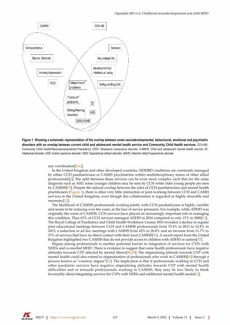
Ogundele MO et al. Childhood neurodevelopmental and child MHD
WJCP https://www.wjgnet.com 127 March 9, 2022 Volume 11 Issue 2
Figure 1 Showing a schematic representation of the overlap between some neurodevelopmental, behavioural, emotional and psychiatric disorders with an overlap between current child and adolescent mental health service and Community Child Health services. CCH-ND: Community Child Health/Neurodevelopmental Paediatrics; OCD: Obsessive compulsive disorder; CAMHS: Child and adolescent mental health service; ID: Intellectual disorder; ASD: Autism spectrum disorder; ODD: Oppositional defiant disorder; ADHD: Attention deficit hyperactivity disorder.
any coordination[9,42].In the United Kingdom and other developed countries, NDEBID conditions are commonly managed
by either CCH paediatricians or CAMH psychiatrists within multidisciplinary teams of other allied professionals[2]. The split between these services can be even more complex such that for the same diagnosis such as ASD, some younger children may be seen by CCH while older young people are seen by CAMHS[55]. Despite the natural overlap between the roles of CCH paediatricians and mental health practitioners (Figure 1), there is often very little interaction or joint-working between CCH and CAMH services in the United Kingdom, even though this collaboration is regarded as highly desirable and necessary[12].
The likelihood of CAMHS professionals working jointly with CCH paediatricians is highly variable and seems to be reducing over the years, in the face of service pressures. For example, while ADHD was originally the remit of CAMHS, CCH services have played an increasingly important role in managing this condition. Thus 63% of CCH services managed ADHD in 2016 compared to only 15% in 2006[13]. The Royal College of Paediatrics and Child Health Workforce Census 2013 revealed a decline in regular joint educational meetings between CCH and CAMHS professionals from 15.4% in 2011 to 12.8% in 2013, a reduction in ad hoc meetings with CAMHS from 42% to 26.8% and an increase from 11.7% to 15% of services that have no direct contact with their local CAMHS[56]. A recent report from the United Kingdom highlighted two CAMHS that do not provide access to children with ADHD or autism[57].
Stigma among professionals is another potential barrier to integration of services for CYPs with NDDs and co-morbid MHD. There is evidence to suggest that some health professionals have negative attitudes towards CYP affected by mental illness[58,59]. The stigmatising attitude towards CYP with mental health could also extend to stigmatisation of professionals who work in CAMHS[60] through a process known as “courtesy stigma”[61]. The implication is that if professionals working in CCH and other paediatric services have negative stigmatising attitudes towards CYP with mental health difficulties and or towards professionals working in CAMHS, they may be less likely to think favourably about integrating services for CYPs with NDDs and additional mental health needs[62].

Ogundele MO et al. Childhood neurodevelopmental and child MHD
WJCP https://www.wjgnet.com 128 March 9, 2022 Volume 11 Issue 2
RECOMMENDATIONS FOR ADDRESSING CLASSIFICATION-RELATED PROBLEMS FOR NDEBID CONDITIONSValue of a unified classification of mental health and NDDThere are grounds for agreement on aspects of the scientific basis for the grouping together of neurodevelopmental and some MHD. First, clinical overlap between these disorders is high and they also behave as highly correlated traits. Thus, research that focuses on a single diagnosis (e.g., autism) should allow for testing the contribution of accompanying neurodevelopmental difficulties. Secondly, NDD share common features with some related MHD including onset early in development, tendency to show a steady course and affecting males more commonly than females. Thirdly, there is a strong genetic overlap across different neurodevelopmental problems[37]. Finally, comorbidity between neurodevelopmental and MHD is well recognized as a factor in the care of children with certain neurological diagnoses, with epilepsy the most prominent example[63], thus grouping them together could help to better enhance the study of the scientific basis and epidemiology of their co-occurrence, as well as improving clinical management.
Studies have shown that CYP with NDEBIDs are at increased risk of developing sleep disorders as well as secondary MHDs such as anxiety, depression, obsessive compulsive disorder (OCD), self-harming, suicidal behaviours, and conduct disorder in up to 50% of those affected[29,53,64,65]. The clinical and research advantages from considering NDDs together with the MHD[40] form the basis for our use of the NDEBID terminology in this paper.
Many clinicians and researchers have questioned the fundamental reason for having more than one classification system used worldwide[5]. Unifying classification systems based on empirical and scientific foundations agreed by consensus among global specialists would probably aid rapid advancement of research across all countries and regions worldwide. There is also evidence that patients and families of CYP with NDEBIDs would also prefer a more unified and integrated approach to their care. When a wide range of stakeholders including families, referrers and CAMHS professionals were requested to state their priority values, “a need for a common language for all agencies when discussing mental health” and “a holistic approach where problems are not inappropriately medicalised” were some of the regular themes found[66]. The global status of the WHO means that ICD is the system most likely to meet this aim and the most recent revision of ICD-11 has made a clear departure from the preceding versions with the new chapter heading of “Mental, Behavioural and NDD” and a sub-heading that brings together a range of conditions previously classified under various headings such as “behavioural and emotional disorders” and “pervasive developmental disorder”. This approach is based on assumption of improved clinical utility and global applicability. While this should be regarded as a welcome development, there are still arguments from some clinicians and researchers against this. For example, various conditions (from severe ASD to mild coordination disorder) contained under this grouping differ from each other such that they have little in common[5].
Focusing on impairments and complexities over diagnosisComplexity and comorbidities are common features of many NDEBIDs and pose a great challenge to clinicians. It is often the complexity of a case that leads to a need for intervention in sub-threshold disorders. Unfortunately, this problem has not been properly addressed in research. Many families, referrers and CAMHS professionals have been reported placing high values on “a holistic approach where problems are not inappropriately medicalised” and “services that take into account what is important in CYP’s lives“[66].
Research methodologies using small N studies may help to explore the value of interventions in complex cases and agreement on a shared language for sub-threshold disorders would facilitate this kind of research[67]. In this regard, DSM-5 has introduced the concepts of “clinical case formulation” and “clinical significance”. It defines clinical significance of a disorder in terms of consideration of thresholds of a person's distress or impairment in his or her social, occupational and/or other important areas of functioning in daily life. The clinical formulation can co-exist with diagnostic classification and provides an alternative to a multiaxial system requirement, with a clinical summary of the social, psychological and biological factors that contribute to the development of a mental disorder. It allows more homogeneous subgroupings of a disorder to indicate shared features[68].
Need for greater care integration for CYP with NDEBIDsThere is strong evidence that children with neurodevelopmental and intellectual disorders have three to four-fold increase in the prevalence of co-occurring mental disorders into adulthood[2,9,69]. For example, pooled prevalence for co-occurring MHD in autism is estimated at 28% [95% cumulative incidence (CI): 25-32] for ADHD; 20% (17-23) for anxiety disorders; 13% (9-17) for sleep-wake disorders; 12% (10-15) for disruptive, impulse-control, and conduct disorders; 11% (9-13) for depressive disorders; 9% (7-10) for OCD; 5% (3-6) for bipolar disorders; and 4% (3-5) for schizophrenia spectrum disorders[64]. In a Swedish community sample, 87% of children with ADHD had at least one co-morbid condition, with rates of ODD of 60%, DCD (47%), ‘reading/writing disorders’ (40%) and TD (33%), even “sub-threshold” ADHD was associated with a similar rate of co-morbid DCD[70].
Ogundele MO et al. Childhood neurodevelopmental and child MHD
WJCP https://www.wjgnet.com 129 March 9, 2022 Volume 11 Issue 2
Effective management of CYP with MHD and behavioural difficulties requires access to psychological therapies and sometimes, psychotropic medications, which most CCH paediatricians are not trained to use. Similarly, CAMHS teams may lack the expertise required to deal with children with sensory or motor impairments. These conditions are best seen and treated within a comprehensive integrated CCH/CAMH service with teams of specialist professionals working together to provide holistic care[9].
The need for integrated care for CYP with NDEBIDs and mental health difficulties has been recognized for many years and is a priority goal for the WHO[71]. Integrated care involves overcoming the breakdown in communication and collaboration that can arise between different parts of the system and different groups of professionals, whilst respecting necessary professional boundaries. An important feature of integrated care is moving beyond pathways for specific diseases[72,73]. System integration across borders/barriers between different sectors of the health services and other systems such as social care and education is the ideal way of preventing adverse outcomes and poor patient experience due to systemic barriers[74]. Close integration of preventive and therapeutic mental health into traditional CCH services accessible to vulnerable CYP and their families within the public care system been identified as the best way to provide them with optimal holistic care they need[75].
Since co-occurrence of NDD is the rule rather than the exception in clinical practice, grouping profes-sional expertise, services and resources for CYP with NDEBIDs organized as part of a neurodevelop-mental hub of expertise has been advocated as the optimal option for achieving holistic and compre-hensive care[40]. The bio-psycho-social and ecological origins of NDEBIDs and associated mental health difficulties make it imperative that assessment and treatment of affected CYP should be multimodal, comprehensive and holistic, to capture the full range of CYP’s needs in order to produce a full formulation and profile to inform their care plans.
Integrated CCH/CAMH care would provide a framework for a more joined-up assessment and treatment in a manner that is more compatible with the complex needs of CYP with NDEBIDs conditions[9,15,76]. Of course, this should not impede the independent professional activities of CAMHS and CCH where joint working is not required.
Evidence from many countries and cultures show that fear of mental health stigma can prevent CYP from seeking help[77]. The negative impact of stigma on help-seeking may be more noticeable among minority ethnic groups living in Western Europe and North America[78-80]. Provision of holistic care within integrated CCH/CAMH services could help to mitigate negative impact of mental health stigma on help-seeking behaviour among CYP with NDEBID[81,82]. Primary care settings such as routine paediatric clinic or family medicine/general practitioner have been reported to possess several desirable characteristics that make them ideal settings for providing effective mental health services to CYP. They are not associated with the stigma typical for bespoke CAMHS, they are often in a local familiar setting, with access to friendly healthcare providers[32,83,84].
It is pleasing to note that a few services across the United Kingdom are beginning to pilot or implement holistic multi-disciplinary clinical pathways for all NDEBID, rather than restricted pathways for individual conditions[42].
CONCLUSIONRecent progress made in the current classification of NDEBIDs has been described. Previous attempts at classifying NDEBID conditions have been fraught with difficulties as there are many possible constructs that need to be taken into consideration. Classification based on causality is particularly problematic because the aetiology of these disorders is not only multi-causal but also incompletely understood[5]. The ICD-11 (and less so with DSM-5) have taken the lead in following a pragmatic approach where the NDEBID conditions are grouped together based on their similar neurobiological phenotypes, until further advances in neurosciences permit more categorical classifications based on aetiologies.
In many countries worldwide, one or more of the NDEBIDs would be assessed and treated by CCH/paediatric services while others and any associated mental health difficulties may be addressed by CAMHS separately and often in a disjointed fashion[9]. Diagnosis of NDEBIDs based on subjective assessment of behaviour by clinicians and carers is prone to biases but reliable standardized instruments can support diagnosis. Recent advances such as computerized CPT tests have potential in the assessment of some NDEBIDs.
Despite the concerns of some authors, it might be reasonable to suggest that the latest WHO classi-fication (ICD-11) could form the basis for a shared understanding acceptable to both the CCH and CAMHS. A more unified approach to classification offers a basis for an integrated care approach, with more consistent collaboration between CCH and CAMH services to address stigma and ensure more holistic care for CYP with NDEBIDs. We note the case for bringing sleep disorders in CYP under the same wider umbrella as the NDEBIDs. We also argue for simultaneous interventions for the total profile of difficulties that accompany the primary diagnosis, even if these do not reach the required threshold for a so-called comorbid diagnosis.
Ogundele MO et al. Childhood neurodevelopmental and child MHD
WJCP https://www.wjgnet.com 130 March 9, 2022 Volume 11 Issue 2
FOOTNOTESAuthor contributions: Ogundele MO conceived the idea, Ogundele MO and Morton M reviewed the literature and prepared the manuscript.
Conflict-of-interest statement: The authors declare no conflict of interest for this article.
Open-Access: This article is an open-access article that was selected by an in-house editor and fully peer-reviewed by external reviewers. It is distributed in accordance with the Creative Commons Attribution NonCommercial (CC BY-NC 4.0) license, which permits others to distribute, remix, adapt, build upon this work non-commercially, and license their derivative works on different terms, provided the original work is properly cited and the use is non-commercial. See: https://creativecommons.org/Licenses/by-nc/4.0/
Country/Territory of origin: United Kingdom
ORCID number: Michael O Ogundele 0000-0003-0744-1434; Michael Morton 0000-0002-3649-9013.
Corresponding Author's Membership in Professional Societies: Bridgewater Community Healthcare NHS Foundation Trust, Halton District, WA7 1TW, UK, ; Institute of Health & Wellbeing, University of Glasgow, (Yorkhill Hospital), Glasgow, Scotland G3 8SJ .
S-Editor: Wang JJ L-Editor: A P-Editor: Wang JJ
REFERENCESAyyash H, Barrett E, Ogundele MO. Sensory Processing of Children with Autism: Uniting Evidence and Practice. Arch Dis Child 2012; 97: A70-A71 [DOI: 10.1136/archdischild-2012-302724.0243]
1
Ogundele M. A Profile of Common Neurodevelopmental Disorders Presenting in a Scottish Community Child Health Service –a One Year Audit (2016/2017). Health Res Policy Syst 2018; 2 [DOI: 10.31058/j.hr.2018.21001]
2
Ogundele MO. Co-occurrence and co-morbidities among children and adolescents with ADHD and ASD in a scottish local authority. Arch Dis Child 2018; 103: A192 [DOI: 10.1136/archdischild-2018-rcpch.458]
3
Ogundele MO, Ayyash HF. Review of the evidence for the management of co-morbid Tic disorders in children and adolescents with attention deficit hyperactivity disorder. World J Clin Pediatr 2018; 7: 36-42 [PMID: 29456930 DOI: 10.5409/wjcp.v7.i1.36]
4
Stein DJ, Szatmari P, Gaebel W, Berk M, Vieta E, Maj M, de Vries YA, Roest AM, de Jonge P, Maercker A, Brewin CR, Pike KM, Grilo CM, Fineberg NA, Briken P, Cohen-Kettenis PT, Reed GM. Mental, behavioral and neurodevelopmental disorders in the ICD-11: an international perspective on key changes and controversies. BMC Med 2020; 18: 21 [PMID: 31983345 DOI: 10.1186/s12916-020-1495-2]
5
Gillberg C, Fernell E, Minnis H. Early symptomatic syndromes eliciting neurodevelopmental clinical examinations. ScientificWorldJournal 2014; 2014: 710570 [PMID: 24453934 DOI: 10.1155/2013/710570]
6
Minnis H, Macmillan S, Pritchett R, Young D, Wallace B, Butcher J, Sim F, Baynham K, Davidson C, Gillberg C. Prevalence of reactive attachment disorder in a deprived population. Br J Psychiatry 2013; 202: 342-346 [PMID: 23580380 DOI: 10.1192/bjp.bp.112.114074]
7
Gillberg C. The ESSENCE in child psychiatry: Early Symptomatic Syndromes Eliciting Neurodevelopmental Clinical Examinations. Res Dev Disabil 2010; 31: 1543-1551 [PMID: 20634041 DOI: 10.1016/j.ridd.2010.06.002]
8
Ogundele M, Ayyash H, Ani C. Integrated Services for Children and Young People with Neurodevelopmental and Co-Morbid Mental Health Disorders: Review of the Evidence. J Psychiatry Mental Disord 2020; 5: 1027
9
World Health Organization. Mental disorders. [cited 11 February 2021]. Available from: https://www.who.int/news-room/fact-sheets/detail/mental-disorders
10
NHS Digital. Mental Health of Children and Young People in England, 2017 [PAS]. [cited 11 February 2021]. Available from: https://digital.nhs.uk/data-and-information/publications/statistical/mental-health-of-children-and-young-people-in-england/2017/2017
11
Ayyash HF, Ogundele MO, Lynn RM, Schumm TS, Ani C. Involvement of community paediatricians in the care of children and young people with mental health difficulties in the UK: implications for case ascertainment by child and adolescent psychiatric, and paediatric surveillance systems. BMJ Paediatr Open 2021; 5: e000713 [PMID: 33614992 DOI: 10.1136/bmjpo-2020-000713]
12
British Association for Community Child Health. Covering All Bases - Community Child Health: A Paediatric Workforce Guide. [cited 1 March]. Available from: https://www.rcpch.ac.uk/sites/default/files/2018-03/covering_all_bases_community_child_health_-_a_paediatric_workforce_guide.pdf
13
Paget A, Emond A. The role of community paediatrics in supporting schools to avoid exclusions that have a basis in health. EBDs 2016; 21: 8-21 [DOI: 10.1080/13632752.2016.1139281]
14
Ogundele M. Profile of neurodevelopmental and behavioural problems and associated psychosocial factors among a cohort of newly looked after children in an English local authority. Adoption & Fostering 2020; 44: 255-271 [DOI: 10.1177/0308575920945187]
15
Ogundele MO et al. Childhood neurodevelopmental and child MHD
WJCP https://www.wjgnet.com 131 March 9, 2022 Volume 11 Issue 2
Clark LA, Cuthbert B, Lewis-Fernández R, Narrow WE, Reed GM. Three Approaches to Understanding and Classifying Mental Disorder: ICD-11, DSM-5, and the National Institute of Mental Health's Research Domain Criteria (RDoC). Psychol Sci Public Interest 2017; 18: 72-145 [PMID: 29211974 DOI: 10.1177/1529100617727266]
16
Stein DJ, Billieux J, Bowden-Jones H, Grant JE, Fineberg N, Higuchi S, Hao W, Mann K, Matsunaga H, Potenza MN, Rumpf HM, Veale D, Ray R, Saunders JB, Reed GM, Poznyak V. Balancing validity, utility and public health considerations in disorders due to addictive behaviours. World Psychiatry 2018; 17: 363-364 [PMID: 30192089 DOI: 10.1002/wps.20570]
17
Larson T, Lundström S, Nilsson T, Selinus EN, Råstam M, Lichtenstein P, Gumpert CH, Anckarsäter H, Kerekes N. Predictive properties of the A-TAC inventory when screening for childhood-onset neurodevelopmental problems in a population-based sample. BMC Psychiatry 2013; 13: 233 [PMID: 24066834 DOI: 10.1186/1471-244X-13-233]
18
America’s Children and the Environment. Neurodevelopmental Disorders. [cited 25 February 2021]. Available from: https://www.epa.gov/sites/default/files/2015-10/documents/ace3_neurodevelopmental.pdf
19
Department for Education. Children in need of help and protection: data and analysis. [cited 12 March 2021]. Available from: https://assets.publishing.service.gov.uk/government/uploads/system/uploads/attachment_data/file/809108/CIN_review_final_analysis_publication.pdf
20
Boden JM, Fergusson DM, Horwood LJ. Risk factors for conduct disorder and oppositional/defiant disorder: evidence from a New Zealand birth cohort. J Am Acad Child Adolesc Psychiatry 2010; 49: 1125-1133 [PMID: 20970700 DOI: 10.1016/j.jaac.2010.08.005]
21
Raudino A, Woodward LJ, Fergusson DM, Horwood LJ. Childhood conduct problems are associated with increased partnership and parenting difficulties in adulthood. J Abnorm Child Psychol 2012; 40: 251-263 [PMID: 21904828 DOI: 10.1007/s10802-011-9565-8]
22
Nyaradi A, Li J, Hickling S, Foster J, Oddy WH. The role of nutrition in children's neurocognitive development, from pregnancy through childhood. Front Hum Neurosci 2013; 7: 97 [PMID: 23532379 DOI: 10.3389/fnhum.2013.00097]
23
Bell MF, Bayliss DM, Glauert R, Harrison A, Ohan JL. Chronic Illness and Developmental Vulnerability at School Entry. Pediatrics 2016; 137 [PMID: 27244787 DOI: 10.1542/peds.2015-2475]
24
Woolgar M. The practical implications of the emerging findings in the neurobiology of maltreatment for looked after and adopted children: recognising the diversity of outcomes. Adoption & Fostering 2013; 37: 237-252 [DOI: 10.1177/0308575913500021]
25
Erskine HE, Baxter AJ, Patton G, Moffitt TE, Patel V, Whiteford HA, Scott JG. The global coverage of prevalence data for mental disorders in children and adolescents. Epidemiol Psychiatr Sci 2017; 26: 395-402 [PMID: 26786507 DOI: 10.1017/S2045796015001158]
26
Global Research on Developmental Disabilities Collaborators. Developmental disabilities among children younger than 5 years in 195 countries and territories, 1990-2016: a systematic analysis for the Global Burden of Disease Study 2016. Lancet Glob Health 2018; 6: e1100-e1121. [PMID: 30172774 DOI: 10.1016/S2214-109X(18)30309-7]
27
Parsons S, Platt L. Disability among Young Children: Prevalence, Heterogeneity and Socio-Economic Disadvantage. [cited 12 March 2021]. Available from: https://nls.ldls.org.uk/welcome.html?ark:/81055/vdc_100062895205.0x000001
28
Eapen V. Developmental and mental health disorders: two sides of the same coin. Asian J Psychiatr 2014; 8: 7-11 [PMID: 24655619 DOI: 10.1016/j.ajp.2013.10.007]
29
World Health Organization. Children and Neurodevelopmental Behavioural Intellectual Disorders (NDBID). [cited 10 February 2021]. Available from: https://www.who.int/ceh/capacity/neurodevelopmental.pdf
30
Lingam R, Hunt L, Golding J, Jongmans M, Emond A. Prevalence of developmental coordination disorder using the DSM-IV at 7 years of age: a UK population-based study. Pediatrics 2009; 123: e693-e700 [PMID: 19336359 DOI: 10.1542/peds.2008-1770]
31
Ogundele MO. Behavioural and emotional disorders in childhood: A brief overview for paediatricians. World J Clin Pediatr 2018; 7: 9-26 [PMID: 29456928 DOI: 10.5409/wjcp.v7.i1.9]
32
Strickland J, Hopkins J, Keenan K. Mother-teacher agreement on preschoolers' symptoms of ODD and CD: does context matter? J Abnorm Child Psychol 2012; 40: 933-943 [PMID: 22661105 DOI: 10.1007/s10802-012-9622-y]
33
Mental Health Screening and Assessment Tools for Primary Care. [cited 15 February 2021]. Available from: https://www.heardalliance.org/wp-content/uploads/2011/04/Mental-Health-Assessment.pdf
34
Ogundele MO, Ayyash HF, Banerjee S. Role of computerised continuous performance task tests in ADHD. Progress Neurol Psychiatry 2011; 15 (3): 8-13 [DOI: 10.1002/pnp.198]
35
Hollis C, Hall CL, Guo B, James M, Boadu J, Groom MJ, Brown N, Kaylor-Hughes C, Moldavsky M, Valentine AZ, Walker GM, Daley D, Sayal K, Morriss R; the AQUA Trial Group. The impact of a computerised test of attention and activity (QbTest) on diagnostic decision-making in children and young people with suspected attention deficit hyperactivity disorder: single-blind randomised controlled trial. J Child Psychol Psychiatry 2018; 59: 1298-1308 [PMID: 29700813 DOI: 10.1111/jcpp.12921]
36
Eyre O, Hughes RA, Thapar AK, Leibenluft E, Stringaris A, Davey Smith G, Stergiakouli E, Collishaw S, Thapar A. Childhood neurodevelopmental difficulties and risk of adolescent depression: the role of irritability. J Child Psychol Psychiatry 2019; 60: 866-874 [PMID: 30908655 DOI: 10.1111/jcpp.13053]
37
Children's Attachment: Attachment in Children and Young People Who Are Adopted from Care, in Care or at High Risk of Going into Care. London: National Institute for Health and Care Excellence (NICE). 2015 [PMID: 26741018]
38
Bagner DM, Rodríguez GM, Blake CA, Linares D, Carter AS. Assessment of behavioral and emotional problems in infancy: a systematic review. Clin Child Fam Psychol Rev 2012; 15: 113-128 [PMID: 22262040 DOI: 10.1007/s10567-012-0110-2]
39
Thapar A, Cooper M, Rutter M. Neurodevelopmental disorders. Lancet Psychiatry 2017; 4: 339-346 [PMID: 27979720 DOI: 10.1016/S2215-0366(16)30376-5]
40
Morris C, Janssens A, Tomlinson R, Williams J, Logan S. Towards a definition of neurodisability: a Delphi survey. Dev Med Child Neurol 2013; 55: 1103-1108 [PMID: 23909744 DOI: 10.1111/dmcn.12218]
41
Embracing Complexity. A new report from Embracing Complexity - a coalition of neurodevelopment and mental health 42
Ogundele MO et al. Childhood neurodevelopmental and child MHD
WJCP https://www.wjgnet.com 132 March 9, 2022 Volume 11 Issue 2
charities - launched a report today looking at multi-diagnostic pathways for neurodevelopmental conditions. [cited 15 February 2021]. Available from: https://www.tourettes-action.org.uk/news-419-.htmlRatnayake A, Bowlay-Williams J, Vostanis P. When are attachment difficulties an indication for specialist mental health input? Adoption & Fostering 2014 [DOI: 10.1177/0308575914532405]
43
Caplan P. Psychiatry’s bible, the DSM, is doing more harm than good. [cited 17 February 2021]. Available from: https://mindfreedom.org/kb/caplan-wash-post/
44
Frances A. The new crisis of confidence in psychiatric diagnosis. Ann Intern Med 2013; 159: 221-222 [PMID: 23685989 DOI: 10.7326/0003-4819-159-3-201308060-00655]
45
Halvorsen M, Mathiassen B, Myrbakk E, Brøndbo PH, Sætrum A, Steinsvik OO, Martinussen M. Neurodevelopmental correlates of behavioural and emotional problems in a neuropaediatric sample. Res Dev Disabil 2019; 85: 217-228 [PMID: 30580152 DOI: 10.1016/j.ridd.2018.11.005]
46
Palumbi R, Peschechera A, Margari M, Craig F, Cristella A, Petruzzelli MG, Margari L. Neurodevelopmental and emotional-behavioral outcomes in late-preterm infants: an observational descriptive case study. BMC Pediatr 2018; 18: 318 [PMID: 30296934 DOI: 10.1186/s12887-018-1293-6]
47
Marty MA, Segal DL. Encyclopedia of Clinical Psychology. In: R Cautin, S Lilienfeld. DSM-5: Diagnostic and Statistical Manual of Mental Disorders. 2015: 965-970
48
World Health Organization. ICD-11. [cited 14 February 2021]. Available from: https://icd.who.int/en49 International Classification of Sleep Disorders – Third Edition (ICSD-3) (Online). [cited 17 February 2021]. Available from: https://learn.aasm.org/Public/Catalog/Details.aspx?id=%2FgqQVDMQIT%2FEDy86PWgqgQ%3D%3D&returnurl=%2FUsers%2FUserOnlineCourse.aspx%3FLearningActivityID%3D%252fgqQVDMQIT%252fEDy86PWgqgQ%253d%253d&returnurl=%2fUsers%2fUserOnlineCourse.aspx%3fLearningActivityID%3d%252fgqQVDMQIT%252fEDy86PWgqgQ%253d%253d
50
Blackburn C, Read J, Spencer N. Children with neurodevelopmental disabilities. Annual report of the Chief Medical Officer (CMO). 2012
51
Mullin AP, Gokhale A, Moreno-De-Luca A, Sanyal S, Waddington JL, Faundez V. Neurodevelopmental disorders: mechanisms and boundary definitions from genomes, interactomes and proteomes. Transl Psychiatry 2013; 3: e329 [PMID: 24301647 DOI: 10.1038/tp.2013.108]
52
Mayes SD, Gorman AA, Hillwig-Garcia J, Syed E. Suicide ideation and attempts in children with Autism. Res Autism Spectrum Disord 2013; 7 (1): 109-119 [DOI: 10.1016/j.rasd.2012.07.009]
53
Agnew-Blais JC, Polanczyk GV, Danese A, Wertz J, Moffitt TE, Arseneault L. Young adult mental health and functional outcomes among individuals with remitted, persistent and late-onset ADHD. Br J Psychiatry 2018; 213: 526-534 [PMID: 29957167 DOI: 10.1192/bjp.2018.97]
54
Schumm T, Morton MJS. National survey of child and adolescent psychiatrists’ clinical activity using the child and adolescent psychiatry surveillance system (CAPSS). Arch Dis Child 2017; 102 (Suppl 1): A50-A50 [DOI: 10.1136/archdischild-2017-313087.121]
55
Royal College of Paediatrics and Child Health (RCPCH-UK). RCPCH Medical Workforce Census 2013. RCPCH. [cited 19 February 2021]. Available from: https://www.rcpch.ac.uk/sites/default/files/RCPCH_medical_workforce_census_2013_-_main_findings.pdf
56
Children’s Commissioner. Lightning Review: Access to Child and Adolescent Mental Health Services. [cited 18 February 2021]. Available from: https://www.childrenscommissioner.gov.uk/report/lightning-review-access-to-child-and-adolescent-mental-health-services/
57
Henderson C, Noblett J, Parke H, Clement S, Caffrey A, Gale-Grant O, Schulze B, Druss B, Thornicroft G. Mental health-related stigma in health care and mental health-care settings. Lancet Psychiatry 2014; 1: 467-482 [PMID: 26361202 DOI: 10.1016/S2215-0366(14)00023-6]
58
Tungchama FP, Egbokhare O, Omigbodun O, Ani C. Health workers' attitude towards children and adolescents with mental illness in a teaching hospital in north-central Nigeria. J Child Adolesc Ment Health 2019; 31: 125-137 [PMID: 31570087 DOI: 10.2989/17280583.2019.1663742]
59
Schulze B. Stigma and mental health professionals: a review of the evidence on an intricate relationship. Int Rev Psychiatry 2007; 19: 137-155 [PMID: 17464792 DOI: 10.1080/09540260701278929]
60
Corrigan PW, Miller FE. Shame, blame, and contamination: A review of the impact of mental illness stigma on family members. Journal of Men Health 2004; 13 (6): 537-548 [DOI: 10.1080/09638230400017004]
61
Jenkins R, Mussa M, Haji SA, Haji MS, Salim A, Suleiman S, Riyami AS, Wakil A, Mbatia J. Developing and implementing mental health policy in Zanzibar, a low income country off the coast of East Africa. Int J Ment Health Syst 2011; 5: 6 [PMID: 21320308 DOI: 10.1186/1752-4458-5-6]
62
Åndell Jason E. Neurodevelopmental and psychiatric comorbidities negatively affect outcome in children with unprovoked seizures-A non-systematic review. Acta Paediatr 2021; 110: 2944-2950 [PMID: 34337792 DOI: 10.1111/apa.16026]
63
Lai MC, Kassee C, Besney R, Bonato S, Hull L, Mandy W, Szatmari P, Ameis SH. Prevalence of co-occurring mental health diagnoses in the autism population: a systematic review and meta-analysis. Lancet Psychiatry 2019; 6: 819-829 [PMID: 31447415 DOI: 10.1016/S2215-0366(19)30289-5]
64
Ayyash HF, Preece P, Morton R, Cortese S. Melatonin for sleep disturbance in children with neurodevelopmental disorders: prospective observational naturalistic study. Expert Rev Neurother 2015; 15: 711-717 [PMID: 25938708 DOI: 10.1586/14737175.2015.1041511]
65
Hindley P, Whitaker F. Editorial: Values based child and adolescent mental health systems. Child Adolesc Ment Health 2017; 22: 115-117 [PMID: 32680377 DOI: 10.1111/camh.12235]
66
Byiers BJ, Reichle J, Symons FJ. Single-subject experimental design for evidence-based practice. Am J Speech Lang Pathol 2012; 21: 397-414 [PMID: 23071200 DOI: 10.1044/1058-0360(2012/11-0036)]
67
Harris JC. New classification for neurodevelopmental disorders in DSM-5. Curr Opin Psychiatry 2014; 27: 95-97 68
Ogundele MO et al. Childhood neurodevelopmental and child MHD
WJCP https://www.wjgnet.com 133 March 9, 2022 Volume 11 Issue 2
[PMID: 24441422 DOI: 10.1097/YCO.0000000000000042]Betts KS, Williams GM, Najman JM, Alati R. Predicting spectrums of adult mania, psychosis and depression by prospectively ascertained childhood neurodevelopment. J Psychiatr Res 2016; 72: 22-29 [PMID: 26519766 DOI: 10.1016/j.jpsychires.2015.10.013]
69
Kadesjö C, Kadesjö B, Hägglöf B, Gillberg C. ADHD in Swedish 3- to 7-year-old children. J Am Acad Child Adolesc Psychiatry 2001; 40: 1021-1028 [PMID: 11556625 DOI: 10.1097/00004583-200109000-00010]
70
World Health Organization. Framework on integrated people-centred health services. [cited 20 February 2021]. Available from: https://apps.who.int/gb/ebwha/pdf_files/WHA69/A69_39-en.pdf?ua=1&ua=1
71
Curry N, Ham C. Clinical and Service Integration: The Route to Improved Outcomes. [cited 23 February 2021]. Available from: https://www.kingsfund.org.uk/sites/default/files/Clinical-and-service-integration-Natasha-Curry-Chris-Ham-22-November-2010.pdf
72
Klaber RE, Blair M, Lemer C, Watson M. Whole population integrated child health: moving beyond pathways. Arch Dis Child 2017; 102: 5-7 [PMID: 27217582 DOI: 10.1136/archdischild-2016-310485]
73
Viner RM, Hargreaves DS. A forward view for child health: integrating across the system to improve health and reduce hospital attendances for children and young people. Arch Dis Child 2018; 103: 117-118 [PMID: 29102963 DOI: 10.1136/archdischild-2017-314032]
74
Department for Education. Help, protection, education: concluding the Children in Need review. [cited 26 February 2021]. Available from: https://assets.publishing.service.gov.uk/government/uploads/system/uploads/attachment_data/file/809236/190614_CHILDREN_IN_NEED_PUBLICATION_FINAL.pdf
75
Ogundele MO, Ayyash HF. Evidence-based multidisciplinary assessment and management of children and adolescents with neurodevelopmental disorders. Arch Dis Child 2019; 104: A268 [DOI: 10.1136/archdischild-2019-rcpch.638]
76
Corrigan P. How stigma interferes with mental health care. Am Psychol 2004; 59: 614-625 [PMID: 15491256 DOI: 10.1037/0003-066X.59.7.614]
77
Gary FA. Stigma: barrier to mental health care among ethnic minorities. Issues Ment Health Nurs 2005; 26: 979-999 [PMID: 16283995 DOI: 10.1080/01612840500280638]
78
Memon A, Taylor K, Mohebati LM, Sundin J, Cooper M, Scanlon T, de Visser R. Perceived barriers to accessing mental health services among black and minority ethnic (BME) communities: a qualitative study in Southeast England. BMJ Open 2016; 6: e012337 [PMID: 27852712 DOI: 10.1136/bmjopen-2016-012337]
79
Bradby H, Varyani M, Oglethorpe R, Raine W, White I, Helen M. British Asian families and the use of child and adolescent mental health services: a qualitative study of a hard to reach group. Soc Sci Med 2007; 65: 2413-2424 [PMID: 17766019 DOI: 10.1016/j.socscimed.2007.07.025]
80
Juengsiragulwit D. Opportunities and obstacles in child and adolescent mental health services in low- and middle-income countries: a review of the literature. WHO South East Asia J Public Health 2015; 4: 110-122 [PMID: 28607309 DOI: 10.4103/2224-3151.206680]
81
Ventevogel P. Integration of mental health into primary healthcare in low-income countries: avoiding medicalization. Int Rev Psychiatry 2014; 26: 669-679 [PMID: 25553784 DOI: 10.3109/09540261.2014.966067]
82
A Guide to Building Collaborative Mental Health Care Partnerships in Pediatric Primary Care. [cited 24 February 2021]. Available from: http://integratedcareforkids.org/library/docs/A_Guide_to_Building_Collaborative_Mental_Health_Care_Partnerships_in_Pediatric_Primary_Care.pdf
83
Kolko DJ, Perrin E. The integration of behavioral health interventions in children's health care: services, science, and suggestions. J Clin Child Adolesc Psychol 2014; 43: 216-228 [PMID: 24588366 DOI: 10.1080/15374416.2013.862804]
84
Skovgaard AM. Mental health problems and psychopathology in infancy and early childhood. An epidemiological study. Dan Med Bull 2010; 57: B4193 [PMID: 21040689]
85
Polanczyk GV, Salum GA, Sugaya LS, Caye A, Rohde LA. Annual research review: A meta-analysis of the worldwide prevalence of mental disorders in children and adolescents. J Child Psychol Psychiatry 2015; 56: 345-365 [PMID: 25649325 DOI: 10.1111/jcpp.12381]
86
Polanczyk G, de Lima MS, Horta BL, Biederman J, Rohde LA. The worldwide prevalence of ADHD: a systematic review and metaregression analysis. Am J Psychiatry 2007; 164: 942-948 [PMID: 17541055 DOI: 10.1176/ajp.2007.164.6.942]
87
Sayal K, Prasad V, Daley D, Ford T, Coghill D. ADHD in children and young people: prevalence, care pathways, and service provision. Lancet Psychiatry 2018; 5 (2): 175-186 [PMID: 29033005 DOI: 10.1016/S2215-0366(17)30167-0]
88
Rydzewska E, Hughes-McCormack LA, Gillberg C, Henderson A, MacIntyre C, Rintoul J, Cooper SA. Age at identification, prevalence and general health of children with autism: observational study of a whole country population. BMJ Open 2019; 9: e025904 [PMID: 31289063 DOI: 10.1136/bmjopen-2018-025904]
89
Dillenburger K, Jordan JA, McKerr L, Keenan M. The Millennium child with autism: early childhood trajectories for health, education and economic wellbeing. Dev Neurorehabil 2015; 18: 37-46 [PMID: 25289682 DOI: 10.3109/17518423.2014.964378]
90
Baxter AJ, Brugha TS, Erskine HE, Scheurer RW, Vos T, Scott JG. The epidemiology and global burden of autism spectrum disorders. Psychol Med 2015; 45: 601-613 [PMID: 25108395 DOI: 10.1017/S003329171400172X]
91
Hong M, Moon DS, Chang H, Lee SY, Cho SW, Lee KS, Park JA, Lee SM, Bahn GH. Incidence and Comorbidity of Reactive Attachment Disorder: Based on National Health Insurance Claims Data, 2010-2012 in Korea. Psychiatry Investig 2018; 15: 118-123 [PMID: 29475227 DOI: 10.30773/pi.2017.11.01]
92
NHS Digital. Statistics on Drug Misuse: England, 2018 [PAS]. [cited 13 March 2021]. Available from: https://digital.nhs.uk/data-and-information/publications/statistical/statistics-on-drug-misuse/2018
93
Public Health England. Intentional self-harm in adolescence: An analysis of data from the Health Behaviour in School-aged Children (HBSC) survey for England. [cited 13 March 2021]. Available from: https://assets.publishing.service.gov.uk/government/uploads/system/uploads/attachment_data/file/621068/Health_behaviour_in_school_age_children_self-harm.pdf
94
McManus S, Hassiotis A, Jenkins R, Dennis M, Aznar C, Appleby L. Suicidal thoughts, suicide attempts, and self-harm. 95
Ogundele MO et al. Childhood neurodevelopmental and child MHD
WJCP https://www.wjgnet.com 134 March 9, 2022 Volume 11 Issue 2
In: Mental Health, Wellbeing. Adult Psychiatric Morbidity Survey 2014 Leeds. United Kingdom: NHS Digital, 2016: 29The Children’s Society. The Good Childhood Report. [cited 16 February 2021]. Available from: https://www.childrenssociety.org.uk/good-childhood
96
Blackburn CM, Spencer NJ, Read JM. Prevalence of childhood disability and the characteristics and circumstances of disabled children in the UK: secondary analysis of the Family Resources Survey. BMC Pediatr 2010; 10: 21 [PMID: 20398346 DOI: 10.1186/1471-2431-10-21]
97
Life opportunities survey interim results 2009/10. The British Library. [cited 25 February 2021]. Available from: https://www.bl.uk/collection-items/life-opportunities-survey-interim-results-200910
98
Cumberland PM, Pathai S, Rahi JS; Millennium Cohort Study Child Health Group. Prevalence of eye disease in early childhood and associated factors: findings from the millennium cohort study. Ophthalmology 2010; 117: 2184-90.e1 [PMID: 20561688 DOI: 10.1016/j.ophtha.2010.03.004]
99
Teoh LJ, Solebo AL, Rahi JS; British Childhood Visual Impairment and Blindness Study Interest Group. Visual impairment, severe visual impairment, and blindness in children in Britain (BCVIS2): a national observational study. Lancet Child Adolesc Health 2021; 190-200 [PMID: 33524322 DOI: 10.1016/S2352-4642(20)30366-7]
100
Rosemary T, Liam S, Jennifer E, Astrid F. The Prevalence of Visual Impairment in the UK: A Review of the Literature. [cited 13 March 2021]. Available from: https://nanopdf.com/download/doc-14-mb_pdf
101
Girish S, Raja K, Kamath A. Prevalence of developmental coordination disorder among mainstream school children in India. J Pediatr Rehabil Med 2016; 9: 107-116 [PMID: 27285803 DOI: 10.3233/PRM-160371]
102
Blank R, Barnett AL, Cairney J, Green D, Kirby A, Polatajko H, Rosenblum S, Smits-Engelsman B, Sugden D, Wilson P, Vinçon S. International clinical practice recommendations on the definition, diagnosis, assessment, intervention, and psychosocial aspects of developmental coordination disorder. Dev Med Child Neurol 2019; 61: 242-285 [PMID: 30671947 DOI: 10.1111/dmcn.14132]
103
Montana Govt Department of Public Health and Human Services. Hearing Impairment. Montana Govt Department of Public Health and Human Services. [cited 13 March 2021]. Available from: https://dphhs.mt.gov/schoolhealth/chronichealth/developmentaldisabilities/hearingimpairment
104
Ahn RR, Miller LJ, Milberger S, McIntosh DN. Prevalence of parents' perceptions of sensory processing disorders among kindergarten children. Am J Occup Ther 2004; 58: 287-293 [PMID: 15202626 DOI: 10.5014/ajot.58.3.287]
105
Pollock MR, Metz AE, Barabash T. Association between dysfunctional elimination syndrome and sensory processing disorder. Am J Occup Ther 2014; 68: 472-477 [PMID: 25005511 DOI: 10.5014/ajot.2014.011411]
106
Tomchek SD, Dunn W. Sensory processing in children with and without autism: a comparative study using the short sensory profile. Am J Occup Ther 2007; 61: 190-200 [PMID: 17436841 DOI: 10.5014/ajot.61.2.190]
107
Owen JP, Marco EJ, Desai S, Fourie E, Harris J, Hill SS, Arnett AB, Mukherjee P. Abnormal white matter microstructure in children with sensory processing disorders. Neuroimage Clin 2013; 2: 844-853 [PMID: 24179836 DOI: 10.1016/j.nicl.2013.06.009]
108
Aaberg KM, Gunnes N, Bakken IJ, Lund Søraas C, Berntsen A, Magnus P, Lossius MI, Stoltenberg C, Chin R, Surén P. Incidence and Prevalence of Childhood Epilepsy: A Nationwide Cohort Study. Pediatrics 2017; 139 [PMID: 28557750 DOI: 10.1542/peds.2016-3908]
109
Epilepsy society. Epilepsy in Children, Epilepsy in childhood. [cited 13 March 2021]. Available from: https://epilepsysociety.org.uk/about-epilepsy/information-parents/epilepsy-childhood
110
Jonsson U, Eek MN, Sunnerhagen KS, Himmelmann K. Cerebral palsy prevalence, subtypes, and associated impairments: a population-based comparison study of adults and children. Dev Med Child Neurol 2019; 61: 1162-1167 [PMID: 30950519 DOI: 10.1111/dmcn.14229]
111
Mindell JA, Li AM, Sadeh A, Kwon R, Goh DY. Bedtime routines for young children: a dose-dependent association with sleep outcomes. Sleep 2015; 38: 717-722 [PMID: 25325483 DOI: 10.5665/sleep.4662]
112
Meltzer LJ, Mindell JA. Systematic review and meta-analysis of behavioral interventions for pediatric insomnia. J Pediatr Psychol 2014; 39: 932-948 [PMID: 24947271 DOI: 10.1093/jpepsy/jsu041]
113
Popova S, Lange S, Poznyak V, Chudley AE, Shield KD, Reynolds JN, Murray M, Rehm J. Population-based prevalence of fetal alcohol spectrum disorder in Canada. BMC Public Health 2019; 19: 845 [PMID: 31253131 DOI: 10.1186/s12889-019-7213-3]
114
May PA, Gossage JP, Kalberg WO, Robinson LK, Buckley D, Manning M, Hoyme HE. Prevalence and epidemiologic characteristics of FASD from various research methods with an emphasis on recent in-school studies. Dev Disabil Res Rev 2009; 15: 176-192 [PMID: 19731384 DOI: 10.1002/ddrr.68]
115
Lange S, Probst C, Gmel G, Rehm J, Burd L, Popova S. Global Prevalence of Fetal Alcohol Spectrum Disorder Among Children and Youth: A Systematic Review and Meta-analysis. JAMA Pediatr 2017; 171: 948-956 [PMID: 28828483 DOI: 10.1001/jamapediatrics.2017.1919]
116
McQuire C, Mukherjee R, Hurt L, Higgins A, Greene G, Farewell D, Kemp A, Paranjothy S. Screening prevalence of fetal alcohol spectrum disorders in a region of the United Kingdom: A population-based birth-cohort study. Prev Med 2019; 118: 344-351 [PMID: 30503408 DOI: 10.1016/j.ypmed.2018.10.013]
117
Valla L, Wentzel-Larsen T, Hofoss D, Slinning K. Prevalence of suspected developmental delays in early infancy: results from a regional population-based longitudinal study. BMC Pediatr 2015; 15: 215 [PMID: 26678149 DOI: 10.1186/s12887-015-0528-z]
118
Zablotsky B, Black LI, Maenner MJ, Schieve LA, Danielson ML, Bitsko RH, Blumberg SJ, Kogan MD, Boyle CA. Prevalence and Trends of Developmental Disabilities among Children in the United States: 2009-2017. Pediatrics 2019; 144 [PMID: 31558576 DOI: 10.1542/peds.2019-0811]
119
Meschi E, Micklewright J, Vignoles A, Lindsay G. The Transitions between Categories of Special Educational Needs of Pupils with Speech, Language and Communication Needs (SLCN) and Autism Spectrum Disorder (ASD) as They Progress through the Education System. 2011. [cited 15 March 2021]. Available from: https://assets.publishing.service.gov.uk/government/uploads/system/uploads/attachment_data/file/219626/DFE-RR247-BCRP11.pdf
120
Ogundele MO et al. Childhood neurodevelopmental and child MHD
WJCP https://www.wjgnet.com 135 March 9, 2022 Volume 11 Issue 2
Public Health England. People with Learning Disabilities in England 2015. [cited 16 March 2021]. Available from: https://www.gov.uk/government/publications/people-with-learning-disabilities-in-england-2015
121
Department for Education. Schools, Pupils and Their Characteristics. [cited 16 March 2021]. Available from: https://www.gov.uk/government/statistics/schools-pupils-and-their-characteristics-january-2020
122
Mithyantha R, Kneen R, McCann E, Gladstone M. Current evidence-based recommendations on investigating children with global developmental delay. Arch Dis Child 2017; 102: 1071-1076 [PMID: 29054862 DOI: 10.1136/archdischild-2016-311271]
123
WJCP https://www.wjgnet.com 136 March 9, 2022 Volume 11 Issue 2
World Journal of
Clinical PediatricsW J C PSubmit a Manuscript: https://www.f6publishing.com World J Clin Pediatr 2022 March 9; 11(2): 136-150
DOI: 10.5409/wjcp.v11.i2.136 ISSN 2219-2808 (online)
REVIEW
Druggable monogenic immune defects hidden in diverse medical specialties: Focus on overlap syndromes
Valentina Boz, Chiara Zanchi, Laura Levantino, Guglielmo Riccio, Alberto Tommasini
Specialty type: Immunology
Provenance and peer review: Invited article; Externally peer reviewed.
Peer-review model: Single blind
Peer-review report’s scientific quality classificationGrade A (Excellent): 0 Grade B (Very good): B Grade C (Good): 0 Grade D (Fair): 0 Grade E (Poor): 0
P-Reviewer: Pongcharoen S
Received: May 26, 2021 Peer-review started: May 26, 2021 First decision: July 30, 2021 Revised: August 3, 2021 Accepted: January 8, 2022 Article in press: January 8, 2022 Published online: March 9, 2022
Valentina Boz, Laura Levantino, Guglielmo Riccio, Alberto Tommasini, Department of Medical, Surgical and Health Sciences, University of Trieste, Trieste 34137, Italy
Chiara Zanchi, Alberto Tommasini, Department of Pediatrics, Institute for Maternal and Child Health, IRCCS Burlo Garofolo, Trieste 34137, Italy
Corresponding author: Alberto Tommasini, MD, PhD, Full Professor, Medical Assistant, Senior Scientist, Department of Medical, Surgical and Health Sciences, University of Trieste, IRCCS Burlo Garofolo, via dell’Istria 65/1, Trieste 34137, Italy. [email protected]
AbstractIn the last two decades two new paradigms changed our way of perceiving primary immunodeficiencies: An increasing number of immune defects are more associated with inflammatory or autoimmune features rather than with infections. Some primary immune defects are due to hyperactive pathways that can be targeted by specific inhibitors, providing innovative precision treatments that can change the natural history of diseases. In this article we review some of these “druggable” inborn errors of immunity and describe how they can be suspected and diagnosed in diverse pediatric and adult medicine specialties. Since the availability of precision treatments can dramatically impact the course of these diseases, preventing the development of organ damage, it is crucial to widen the awareness of these conditions and to provide practical hints for a prompt detection and cure.
Key Words: Inborn errors of immunity; Primary immunodeficiency diseases; Precision treatments; Immunodysregulation; Autoimmunity; Overlap syndromes
©The Author(s) 2022. Published by Baishideng Publishing Group Inc. All rights reserved.

Boz V et al. Druggable inborn errors of immunity
WJCP https://www.wjgnet.com 137 March 9, 2022 Volume 11 Issue 2
Core Tip: High-throughput genetic testing have allowed to describe monogenic immune disorders, charac-terized by combinations of infective, inflammatory, autoimmune, lymphoproliferative, neoplastic features. The term “inborn errors of immunity” (IEIs) is increasingly proposed instead of “primary immunodefi-ciency” to include defects with a prevalently dysregulatory pathogenesis, resulting in autoimmunity, inflammation, lymphoproliferation, risk of malignancies. It is crucial to widen the awareness of these disorders, as they may mimic multifactorial disorders (rheumatology, gastroenterology, hematology, dermatology, allergology) and some of these are druggable. The awareness of druggable IEIs is the focus of this review, with the aim of favoring a prompter diagnosis and a better cure.
Citation: Boz V, Zanchi C, Levantino L, Riccio G, Tommasini A. Druggable monogenic immune defects hidden in diverse medical specialties: Focus on overlap syndromes. World J Clin Pediatr 2022; 11(2): 136-150URL: https://www.wjgnet.com/2219-2808/full/v11/i2/136.htmDOI: https://dx.doi.org/10.5409/wjcp.v11.i2.136
INTRODUCTIONPrimary immunodeficiencies are a growing group of monogenic disorders related to dysregulated immune processes, which can result in autoinflammation, autoimmunity, lymphoproliferation and/or risk of malignancy in addition to the paradigmatic recurrent infections: In this sense, the term “inborn errors of immunity” (IEIs) has recently been proposed to underline the heterogeneous phenotype of immune deficiencies[1,2].
Improved diagnostics of monogenic immune disorders, together with the availability of medications acting on disease-related mechanisms recently led to the development of precision therapies which can improve or correct the phenotype of some IEIs[3-6]. The mutations involved in these disorders are usually associated with gain-of-function (GOF) of proteins (often kinases) or hyperactivity of pathways, which can be targeted by specific medications and thus sometimes referred to as “druggable”. For concision, we indicate IEIs with druggable pathogenic mechanisms as druggable IEI (D-IEIs).
Of note, since immunodeficiency may develop significant organ damage due to infection or autoim-munity, early detection of D-IEIs is crucial to benefit from appropriate treatments[7]. Although a deep clinical-laboratory evaluation can help an experienced immunologist to concentrate suspicion on one of these disorders, the use of next generation sequencing (NGS) offers a powerful tool to diagnose D-IEIs, allowing to examine all the candidate genes at once[8-10]. However, due to the wide heterogeneity of IEIs, it may be difficult to select patients for genetic analysis.
In fact, from a phenotypic point of view, due to their origin from general disturbances in immune regulation, D-IEIs tend to affect multiple organs and systems, composing complex clinical pictures that overlap disorders of distinct medical specialties, and tend to fully manifest over the time, with the definition of typical clinical pictures only in adults. Thus, patients with D-IEI can initially be diagnosed – especially in pediatric age - with common multifactorial disorders, pertaining to various medical specialties and displaying atypical clinical presentations such as unusual age of development, multiorgan involvement and response to therapies. These factors are congruent with the immune dysregulation theory.
In light of this, the aim of this review is to widen the awareness of “druggable” IEIs which may be hidden in various medical specialties, in order to promote an earlier diagnosis and a better therapy in this field.
IEI may present druggable autoimmune, inflammatory and/or lymphoproliferative manifestationsWe present a list of druggable IEIs, with prevalent autoimmune, inflammatory and/or lymphoprolif-erative aspects, which may mimic common multifactorial disorders, and therefore are at risk of being missed, until significant organ damage manifests. Since effective treatments are now available for immune disorders, it is of crucial importance to consider the possibility of a primary immune defect in subjects presenting with clinical pictures suggestive of immune dysregulation, particularly those that overlap distinct rheumatological, gastroenterological, endocrinological and dermatological/allergic disorders (Table 1). As described elsewhere, there is now a trend of anticipating the time of genetic analysis, reserving more in-depth immunological investigations for a later time, with the aim of determining the role of any variants of uncertain significance found in candidate genes[7].
IPEX is a monogenic immune disorder (due to mutations in FOXP3) characterized by an impaired development of Treg cells, resulting in failure of peripheral immune tolerance, with autoimmunity and allergic manifestations[11-13]. The disease typically presents in infancy with enteropathy, cutaneous disorders with eczema and nail changes, and endocrinopathies [e.g., type 1 diabetes mellitus (T1DM), thyroiditis]. Several other autoimmune manifestations may also be found. The treatment may benefit from sirolimus or tacrolimus, in addition to nutrition and glucocorticoids, but only hematopoietic stem

Boz V et al. Druggable inborn errors of immunity
WJCP https://www.wjgnet.com 138 March 9, 2022 Volume 11 Issue 2
Table 1 Characteristics of pathologies
Gene Meccanism Immune assessment Clinicsautoimmunity Lymphoproliferation Infections Therapy
APDS PIK3CDPIK3R1 PI3K delta hyperactivation
Hypogammaglobulinemia IgA and IgG lowSen-escent CD8 T cellsDNT
IBD; diabetes; arthritis Lymphadenopathy, spleno-megaly
Recurrent respiratory infections; herpes virus infections
HSCT; antibioticsrituximab and rapamycin;PI3Kδ inhibitors
STAT3 GOF
STAT3 STAT3 hyperactivation
Hypogammaglobulinemia; decrease NK cells; decrease memory B cells; decrease regulatory T cells
Autoimmune cytopenia; diabetes; thyroiditis; arthritis
Adenopathy, hepatospleno-megaly
Herpes virus infections; fungal infections; bacterial infections; respiratory infections
JACK inhibitors
APECED AIRE Decrease of negative selection of autoreactive T cells in thymus
Autoantibodies;CD8+ effector T cells;FOXP3+ regulatory T cells
Autoimmune hypopara-thyroidism;Addison’s disease
Chronic Candida infection
Hormone replacement therapy according to affected organs; immunosuppressive therapies; rituximab
CTLA4 deficiency
CTLA4 Defective switch off of lymphocyte activation
Hypogammaglobulinemia; DNT;increase of regulatory T cells with reduced expression of FOXP3;CD19+ B cells and switched memory B
Autoimmunecytopenia; hemolytic anemia and thrombo-cytopenia
Splenomegaly;chronic lymphadenopathy;hepatomegaly
Respiratory tract infections
Sirolimus; abatacept; HSCT
LRBA deficiency
LRBA Defective switch off of lymphocyte activation
Hypogammaglobulinemia; DNT; FOXP3+regulatory T cells;CD19+ B cells;Natural Killer cells; increase of CD4+ and CD8+ memory T cells
Autoimmune gastritis;autoim-munecytopenia; hemolytic anemia; IBD;Autoimmune enteropathy
Splenomegaly;hepatomegaly Respiratory infections
sirolimus; abatacept; HSCT
IPEX FOXP3 Failure of immune tolerance
Loss of FOXP3+ T cells;increased of Th2 and Th17 cells;autoantibodiesHypergammaglobulinemia IgA, IgE
Autoimmune enteropathy; autoimmune hemolytic anemia; autoimmune thrombocytopenia; autoimmune neutropenia; autoimmune thyroiditis; nephropathy; hepatitis
Skin infections Glucocorticoids;Msirolimus;Mtacrolimus; abatacept; HSCT
STAT1 GOF
STAT1 STAT1 hyperactivation due to increase STAT1 phosphorylation
Low Th17 cells; low switched memory B cells;Hypergammaglobulinemia IgG
Chronic mucocutaneous candidiasis; hypothyroidism; autoimmune cytopenia, hepatopathy; psoriasis
Hepatomegaly; splenomegaly Fungal, viral and mycobac-terial infections; skin infections; Respiratory infections
Antifungal treatment; antibiotic prophylaxis; JACK inhibitors
Hypogammaglobulinemia; increases macrophage release of TNF-α; upregulation of neutrophil activity; upregulation of pro-inflammatory cytokines; upregulation of type 1 interferon
Verrucosis; herpes virus infections; increased
Anti-TNF treatment (etanercept, infliximab,adalimumab); high-dose of glucocorticoids; HSCT; immunosuppressive drugs in isolated cases (mycophenolate,
DADA2 ADA2 Reduced activity level of the adenosine deaminase 2
Vasculitis, immunodeficiency; autoimmune neutropenia; autoimmune cytopenia
Splenomegaly; lymphaden-opathy; hepatomegaly

Boz V et al. Druggable inborn errors of immunity
WJCP https://www.wjgnet.com 139 March 9, 2022 Volume 11 Issue 2
stimulated genes; aberrant B cell development and differentiation; decrease in NK
susceptibility to infection with dsDNA viruses
azathioprine, cyclosporine, rituximab, sirolimus, tacrolimus)
TNFAIP3 deficiency
TNFAIP3 Excessive activation of NF-kB signalling
Antinuclear and anti-DNA antibodies; increased production of interferons and proinflammatory cytokines
Autoimmune cytopenias Anti-TNF treatment; anti-IL1 treatment; glucocorticoid; colchicine
Characteristics of pathologies[10,20,28,31,37,44,49,52,53]. HSCT: Hematopoietic stem cell transplantation; IBD: Inflammatory bowel disease.
cell transplantation allows a cure, the success of which is related to an early diagnosis[14-16]. Proof of concept for immunoregulation with abatacept was obtained in scurfy mice, which are considered a good animal model for the IPEX[17,18].
APECED is a monogenic immune disorder (due to mutations in AIRE) characterized by an abnormal presentation of self-antigens in the thymus, resulting in the failure of central immune tolerance, with autoimmunity[19-21]. The disease usually presents in infancy with recurrent and severe candidiasis (with susceptibility associated to IL17-neutralizing antibodies) and parathyroid and adrenal autoim-munity, but over time other autoimmune disorders (e.g., hepatitis, thyroiditis, vitiligo, alopecia, gastritis) are also observed[22,23]. Even if there is still no precision therapy for APECED, it is important to make an early diagnosis to establish a proper follow-up with prompt detection of new autoimmune phenomena, infections and malignancies[24,25].
CTLA4 and LRBA deficiencies are monogenic immune disorders associated with an impaired regulation of lymphocyte activation and development, resulting in autoimmunity and lymphoprolif-eration, but also infections[26-30]. Clinical features include hepatosplenomegaly, enteropathy, eczema, autoimmune cytopenia, arthritis, lupus-like features, hypogammaglobulinemia recurrence of infections and risk of malignancies (particularly due to chronic EBV infection). hematopoietic stem cell transplantation (HSCT) can cure the disease, however the treatment of milder cases may benefit from the use of CTLA4-Ig (abatacept)[28,30,31].
APDS (type I and II) are monogenic immune disorders associated with an impaired regulation of T and B cells maturation and survival, resulting in lymphoproliferation, autoimmunity and infections[32-35]. Clinical features include recurrent infections (especially respiratory, often complicated by the development of bronchiectasis and cutaneous) lymphoproliferative manifestations with risk of lymphoma[36-38], enteropathy and systemic lupus erythematosus (SLE)-like features. The immune defect is complex, with hypogammaglobulinemia with normal or increased IgM, reduced number of recent thymic emigrants and accumulation of senescent CD8 T cells. The pathogenic mechanisms can be partially reversed with drugs inhibiting the PIK3delta kinase, with a great potential in reducing the disease severity[39].
Monoallelic GOF mutations in STAT1 are associated with susceptibility to infections from bacteria and fungi, autoimmune disorders and rheumatologic manifestations, due to increased activation of interferon stimulated genes[40,41]. Since hyperactive STAT1 still depends on the trigger from Janus kinases, the use of JAK inhibitors can partially restore a physiological balance with great clinical benefit both on inflammatory and on infectious symptoms[42,43]. In a recent report, treatment with JAK

Boz V et al. Druggable inborn errors of immunity
WJCP https://www.wjgnet.com 140 March 9, 2022 Volume 11 Issue 2
inhibitors led to the reversal of autoimmune diabetes in a boy with STAT1 GOG[40].Monoallelic GOF mutations in STAT3 are associated with autoimmune and lymphoproliferative
disorders[44]. Patients may present autoimmune enteropathy, celiac disease-like changes in the jejunum, eczema, autoimmune polyendocrinopathy, lymphoproliferation with increased CD4- CD8- double negative T cells and risk of hematologic malignancies and hypogammaglobulinemia with recurrent infection. The use of JAK inhibitors can lead to significant clinical improvement in this case too[45].
DADA2 deficiency is a combined immunodeficiency due to the defective function of the adenosine deaminase-2enzyme. The disease is associated with lymphoproliferation, variable hypogammaglobu-linemia and susceptibility to infection, arthritis, livedo reticularis, erythema nodosum, purpura and vasculitis with a picture of polyarteritis nodosa and ischemic strokes[46,47]. The main complaints of the disease are driven by TNF-alpha: Thus, there is a formal recommendation to start anti-TNF treatment as early as possible[48].
A20 haploinsufficiency is a monogenic immune disorder (due to mutations in TNFAIP3) charac-terized by an abnormal activation of NF-kB signalling, resulting in a phenotype similar to Behcet’s disease (BD)[49]. Indeed, clinical features include uveitis, recurrent oral and genital ulcerations, rash, abscesses and periodic fever. However, some patients may present with ulcerative colitis or with signs of SLE-like autoantibodies, increased production of interferons, autoimmune cytopenias and sometimes nephritis[50-52]. Anti-TNF treatment has been proven of great efficacy in several patients, even if it could be ineffective on lupus-related complaints[53].
There are many other rare IEIs that may present with complex features of immune dysregulation, such as lymphoproliferation, autoimmunity, inflammation, and risk of malignancies. However, the therapeutic implications of diagnosing these IEIs are not as straightforward as those for druggable diseases.
IEI MAY MIMIC COMMON MULTIFACTORIAL DISORDERS IN DIVERSE MEDICAL SPECIALTIESIEIs may present in diverse medical specialties mimicking more common multifactorial disorders. However, there are typical clinical pictures or peculiar sets of features that can raise suspicion of an IEI. After discussing the relevance of such conditions to specific medical specialties, we will propose red flags to help address the suspicion of an IEI in a multidisciplinary setting (Figure 1).
RheumatologySLE: SLE is quite rare in children before pubertal age[54-56]. Cases with very early onset should always raise suspicion of an underpinning genetic disorder, with particular reference to complement deficiencies and interferonopathies[57,58]. Some cases may be anticipated by blood cells cytopenia or liver involvement. Arthritis may present a devious clinical course with slow development of contractures. A significant history of infections can sometimes be recorded. In many cases, the clinical picture is not the one that is the most typical of SLE, and classification criteria for SLE are not always completely met. NGS gene panels or whole exome sequencing have been proposed to allow an early detection of monogenic mimics of SLE[59,60]. A recent large study demonstrated that a monogenic cause could be found in 23% of patients meeting at least one of the following inclusion criteria: i.e., (1) Age of disease onset under 5 years; (2) Family history of autoimmune disease; (3) Syndromic SLE; and (4) Complicated conditions, such as life-threatening and refractory SLE[61]. Of particular importance is a prompt detection of druggable disorders like interferonopathies or STAT1 GOF immunodeficiency, which can benefit from the use of JAK inhibitors[42,60,62,63], or immune dysregulation deficiencies such as activated PIK3d syndrome that can benefit from PIK3δ inhibitors[61,64,65].
BD: BD is a complex inflammatory and autoimmune disorder with great clinical heterogeneity. BD is rare in pediatrics and often presents for many years in an incomplete form, mainly with recurrent oral and/or genital ulcerations and sometimes periodic fevers. Vasculitis, central nervous system or eye involvement are more typical of older children and adults. BD occurring in early childhood can also be underpinned by monogenic immune defects. Mounting evidence supports the opportunity of searching for monogenic mimics of BD in pediatrics, in particular in subjects with very early disease onset, positive familial history and severe phenotypes[52,66-68]. Some of these monogenic cases may present clinical pictures overlapping with SLE or Inflammatory bowel disease (IBD), as is in the case of STAT1 GOF and A20 haploinsufficiency. The molecular diagnosis in these cases can allow a targeted therapeutic choice and a proper follow-up.
Juvenile idiopathic arthritis is not so rare and is rarely associated with an underlying monogenic disorder. However, there are rare atypical cases, usually with polyarticular involvement refractory to conventional therapies, which may be associated with inflammatory involvement of liver or lungs, uncovering a more complex inflammatory pathogenesis, as in the cases of interferon-related disorders like COPA syndrome (also known as Autoimmune Interstitial Lung, Joint, and Kidney disease, OMIM #
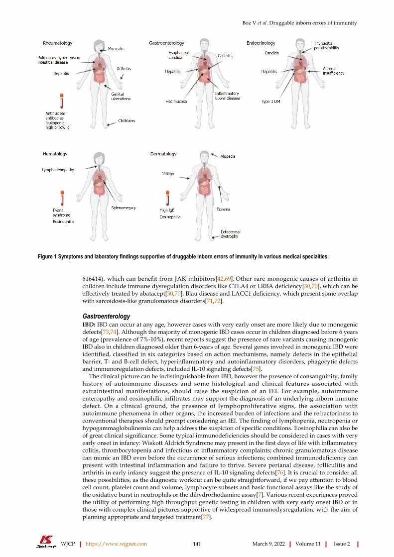
Boz V et al. Druggable inborn errors of immunity
WJCP https://www.wjgnet.com 141 March 9, 2022 Volume 11 Issue 2
Figure 1 Symptoms and laboratory findings supportive of druggable inborn errors of immunity in various medical specialties.
616414), which can benefit from JAK inhibitors[42,69]. Other rare monogenic causes of arthritis in children include immune dysregulation disorders like CTLA4 or LRBA deficiency[30,70], which can be effectively treated by abatacept[30,70], Blau disease and LACC1 deficiency, which present some overlap with sarcoidosis-like granulomatous disorders[71,72].
GastroenterologyIBD: IBD can occur at any age, however cases with very early onset are more likely due to monogenic defects[73,74]. Although the majority of monogenic IBD cases occur in children diagnosed before 6 years of age (prevalence of 7%-10%), recent reports suggest the presence of rare variants causing monogenic IBD also in children diagnosed older than 6-years of age. Several genes involved in monogenic IBD were identified, classified in six categories based on action mechanisms, namely defects in the epithelial barrier, T- and B-cell defect, hyperinflammatory and autoinflammatory disorders, phagocytic defects and immunoregulation defects, included IL-10 signaling defects[75].
The clinical picture can be indistinguishable from IBD, however the presence of consanguinity, family history of autoimmune diseases and some histological and clinical features associated with extraintestinal manifestations, should raise the suspicion of an IEI. For example, autoimmune enteropathy and eosinophilic infiltrates may support the diagnosis of an underlying inborn immune defect. On a clinical ground, the presence of lymphoproliferative signs, the association with autoimmune phenomena in other organs, the increased burden of infections and the refractoriness to conventional therapies should prompt considering an IEI. The finding of lymphopenia, neutropenia or hypogammaglobulinemia can help address the suspicion of specific conditions. Eosinophilia can also be of great clinical significance. Some typical immunodeficiencies should be considered in cases with very early onset in infancy: Wiskott Aldrich Syndrome may present in the first days of life with inflammatory colitis, thrombocytopenia and infectious or inflammatory complaints; chronic granulomatous disease can mimic an IBD even before the occurrence of serious infections; combined immunodeficiency can present with intestinal inflammation and failure to thrive. Severe perianal disease, folliculitis and arthritis in early infancy suggest the presence of IL-10 signaling defects[76]. It is crucial to consider all these possibilities, as the diagnostic workout can be quite straightforward, if we pay attention to blood cell count, platelet count and volume, lymphocyte subsets and basic functional assays like the study of the oxidative burst in neutrophils or the dihydrorhodamine assay[7]. Various recent experiences proved the utility of performing high throughput genetic testing in children with very early onset IBD or in those with complex clinical pictures supportive of widespread immunodysregulation, with the aim of planning appropriate and targeted treatment[77].
Boz V et al. Druggable inborn errors of immunity
WJCP https://www.wjgnet.com 142 March 9, 2022 Volume 11 Issue 2
Autoimmune enteropathy: Autoimmune enteropathy is a rare disorder characterized by intractable diarrhea, growth failure, presence of anti-enterocyte autoantibodies and typical mucosal changes with lymphocyte infiltrates and increased apoptotic cells[78,79]. Most cases occur during the first year of life with severe primarily secretory diarrhea[78]. The association with extra-intestinal diseases like insulin-dependent diabetes, thyroiditis, membranous glomerulopathy, interstitial nephritis and the presence of numerous autoantibodies (e.g., antinuclear, anti-smooth-muscle, anti-parietal cells, pancreatic islets...), should raise suspicion of IPEX syndrome. Furthermore, early diarrhea and malabsorption can occur in up to 25% of patients with APECED, due to the destruction of intestinal endocrine cells; in these cases, small bowel biopsies show mild damage, in contrast with the inflammation present in autoimmune enteropathy. An early enteropathy with a relative paucity of inflammatory cells in a patient with a history of recurrent infections should be suspicious for CVID[80,81]. Recent literature reports a series of patients with both LRBA deficiency and CTLA-4 haploinsufficiency with gastrointestinal manifest-ations, including autoimmune enteropathy, lymphocytic duodenitis resembling celiac disease and autoimmune gastritis[82,83].
Parenteral nutrition, steroids and immunosuppressants like cyclosporin A and tacrolimus are the cornerstones of the therapy. If an IEI can be found in a significant proportion of children with early onset IBD, this is even more true for autoimmune enteropathy[78]. In most cases underpinned by IEIs, HSCT can be the treatment of choice. However, when HSCT is not possible or has to be delayed, a treatment with abatacept or sirolimus may be a valuable option for CTLA4 and LRBA deficiency or for IPEX respectively[30].
Atrophic autoimmune gastritis: Atrophic autoimmune gastritis is an autoimmune disorder associated with chronic gastric autoimmunity, vitamin B12-dependent anemia and increased risk of developing gastric cancer[84]. This condition is often found associated with other immune disorders like common variable immunodeficiency, autoimmune thyroid disease and T1DM[85]. However, some patients may initially present only gastrointestinal complaints with gastritis[83]. Considering that this is a rare disorder in children, the likelihood of finding a monogenic cause is high, and an immunologic and genetic workup should be carried out before the patient develops further autoimmune phenomena. APECED, IPEX and immune dysregulatory disorders are examples of monogenic diseases that can present with autoimmune atrophic gastritis, even if it is rare for autoimmune gastritis to be the sole complaint[83,86,87].
Non celiac flat mucosa: The main cause of flat mucosa in jejunum is active celiac disease (CD), due to gluten-dependent immune activation in the lamina propria of the intestinal mucosa[88,89]. Similar findings can be found in subjects in whom CD has been ruled out, based on negative testing for anti-transglutaminase antibodies and/or absence of the predisposing HLA haplotypes and/or refractoriness to gluten free diet[83,90]. In these cases, intestinal inflammation may be related to an immune defect like common variable immunodeficiency. A flat jejunal mucosa has been described in subjects with immune dysregulatory diseases including IPEX, CTLA4 and LRB immunodeficiency, often in association with other gastrointestinal immune-mediated diseases, like autoimmune gastritis, autoimmune enteropathy or inflammatory bowel disease[83,91].
Esophageal candidiasis: Muco-cutaneous candidiasis is rarely observed in healthy children above the age of 1 year. Seldomly, therapies with oral glucocorticoids may facilitate the development of candidiasis in older children too, however the recurrence of the problem and the extension of the infection to the esophagus should always prompt the suspicion of an underlying immune defect. The underlying causes of chronic muco-cutaneous candidiasis may be monogenic, such as single gene mutations in the autoimmune regulator, signal transducer and activator of transcription-1 (STAT1) and -3 (STAT3), and many others genes (CARD9, TYK2, DOCK8, CD25, IL-1RA, RORC..), or the result of polymorphisms in genes encoding Dectin-1, NACHT LRR and PYD-containing protein 3, protein tyrosine phosphatase non receptor type-22, and Toll-like receptors which contribute to candida infection susceptibility[92-94]. It is worth noting that candida infections can sometimes be misinterpreted as the results of glucocorticoid treatments administered for other immune complaints, as some patients may present SLE-like phenomena (IL12RB1, STAT1 GOF) or autoimmune manifestations (APECED). Indeed, it is uncertain whether severe diffuse mucosal candidiasis reported in a subset of subjects with SLE are the result of immunosuppressive therapy or the marker of a possible underlying immunodeficiency[95].
EndocrinologyAutoimmune polyglandular syndromes: Endocrine glands are the most typical targets of organ-specific autoimmunity, probably related to the cell-specific expression of proteins involved in the highly specialized machinery of hormone production. Based on distinct patterns of involvement of diverse endocrine systems, autoimmune polyglandular syndromes (APS) have been classified in three groups (APS1-3). Overall, APS have been associated with a general failure of maintaining immune tolerance to specialized tissue. This can be due to a defective presentation of tissue antigens in the thymus during lymphocyte development (as in APECED), improper control of autoreactive lymphocytes in target organs (as in IPEX and IPEX-like disorders) or to breakdown of tolerance by medications (as with
Boz V et al. Druggable inborn errors of immunity
WJCP https://www.wjgnet.com 143 March 9, 2022 Volume 11 Issue 2
checkpoint inhibitors used to induce anti-cancer immunity).The combination of hypoparathyroidism and adrenal insufficiency (APS1) with muco-cutaneous
candidiasis and ectodermal dystrophy is typical of APECED, however patients may initially present only with a single autoimmune disease. In these cases, the search for autoantibodies can help anticipate further autoimmune disorders, avoiding the risks of a hyperacute onset of disease. APS2 is charac-terized by T1DM, autoimmune thyroiditis and Addison Disease and is considered a multifactorial disorder associated with the HLA class II locus. APS3 is characterized by T1DM and autoimmune thyroiditis and can be either due to monogenic druggable immune defects (IPEX-like disorders) or to multifactorial causes including HLA class II variants. The presence of dermatitis, autoimmune cytopenia or lymphoproliferation in addition to autoimmune endocrine diseases should always raise suspicion of a monogenic immune dysregulation disorder.
The therapy is mainly based on the replacement of defective hormones. However, in cases associated with significant immune dysregulation, a prompt immune modulation can prevent the development of further autoimmune or infectious diseases, in particular in the cases of druggable IEIs responsive to abatacept and/or sirolimus.
HematologyEvans syndrome: Evans syndrome is characterized by the association of autoimmune hemolytic anemia with immune thrombocytopenic purpura. The two autoimmune conditions can occur simultaneously or in sequence. In some cases, autoimmune neutropenia can also be present. The term “Evans syndrome” refers to cases in which another definite diagnosis has not been made. However, a search for underlying immune defects may reveal the presence of a monogenic disease in a significant proportion of cases, in particular among those associated with signs of lymphoproliferation, that may be due to immune dysregulation immunodeficiencies[96]. The diagnosis of autoimmune lymphoproliferative syndromes (ALPS), activated PI3Kδ syndromes, IPEX syndrome, and CTLA4 or LRBA deficiencies can pave the way to the administration of targeted therapies like sirolimus, PI3Kδ inhibitors, and abatacept. Of note, sirolimus has been proven effective also in subjects with idiopathic Evans syndrome, suggesting that the disease may share relevant pathogenic features with ALPS. Since autoimmune cytopenias may be the presenting clinical condition in subjects with a common variable immunodeficiency or in SLE, a study of immunoglobulins, antinuclear antibodies and lymphocyte subpopulations is warranted in all subjects with Evans syndrome. Specific cytometric analyses may also give significant hints for rare IEIs[7].
Dermatology and allergologyRefractory eczema: Eczema is a common complaint in young children. If not treated properly, an active eczema can favor the development of allergies and other immune disturbances, fueling a vicious circle of inflammation, scratch injuries and infection. In most cases the disease can be easily treated until it wanes and disappears with age. However, in some rare cases the eczematous dermatitis shows a severe course and a refractoriness to treatments from the first weeks of life. These cases are often associated with poor growth or failure to thrive and sometimes with a history of infections. Blood examinations are usually performed to rule out a severe combined immunodeficiency or a Wiskott Aldrich Syndrome. However, other immune dysregulations are not as easy to diagnose. Peripheral eosinophilia is a clue to diagnosis, but a genetic panel for primary immune defects may be worthwhile in all severe cases. A diagnostic algorithm for IEIs associated with atopic phenotypes has been recently proposed by the Immunology Task Force of the Italian Society of Pediatric Allergy and Immunology[97].
Alopecia, vitiligo: Autoimmune alopecia and vitiligo may present in children at any age. The presence of autoimmune disorders in relatives, like thyroiditis is also common. However, if these complaints present together with other autoimmune or inflammatory disorders or with laboratory abnormalities like eosinophilia or a positive inflammatory index, it may be reasonable to perform an immunological and genetic investigation. For example, when associated with panniculitis, alopecia can rise suspicion of an interferon related disease like CANDLE syndrome, which can benefit from JAK inhibitors; if associated with severe dermatitis it may make you think of an immune dysregulation, like IPEX or CTLA4 or LRBA deficiency. It is worth noting that tofacitinib and other JAK inhibitors, and to a lesser extent abatacept, have been successfully used in subjects with alopecia areata even in the absence of a known underlying immune defect[98-100].
OVERLAP SYNDROMES MAY HIDE MONOGENIC DISORDERSAs described in the paragraphs above, IEIs may present with simultaneous or sequential involvements of various organs and systems. Many cases actually present substantial clinical overlap between disorders pertaining to different medical specialties. Whilst overlap syndromes are not rare in adult rheumatology or gastroenterology, this kind of conditions are not frequently encountered in children. Thus, we propose that IEIs should be considered in every child with immunological complaints overlapping diagnoses that are not usually seen in combination at this age. Since not all cases appear
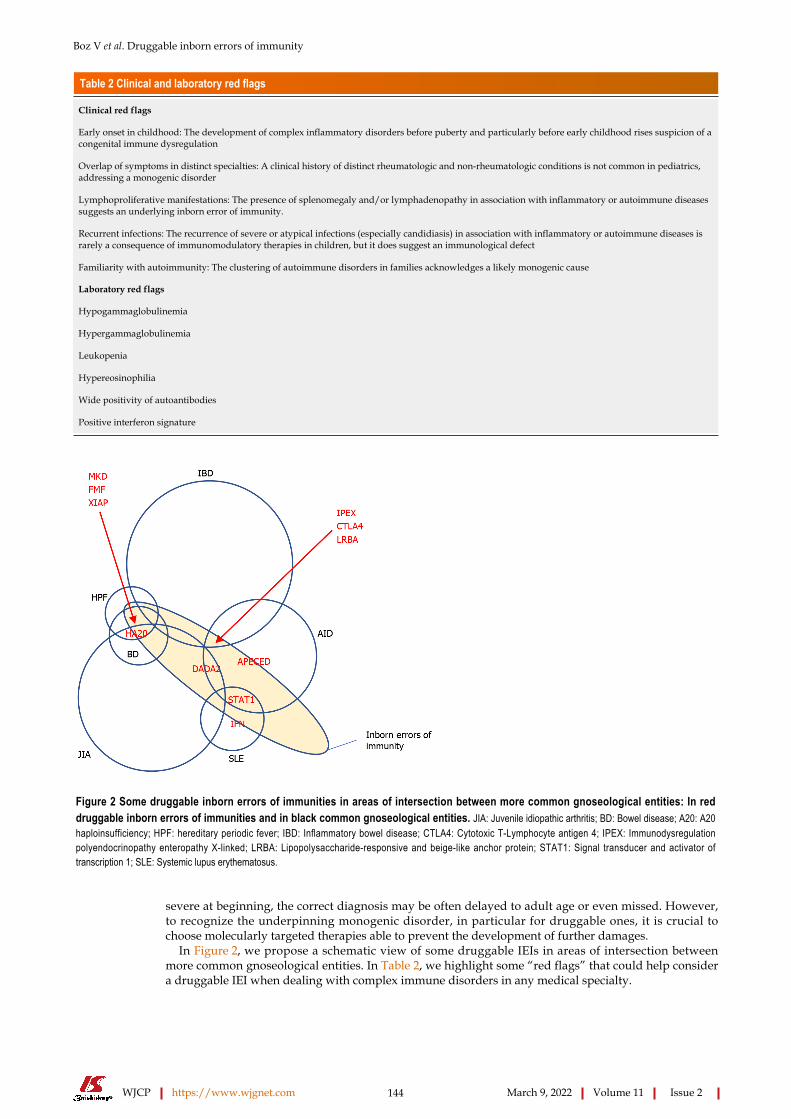
Boz V et al. Druggable inborn errors of immunity
WJCP https://www.wjgnet.com 144 March 9, 2022 Volume 11 Issue 2
Table 2 Clinical and laboratory red flags
Clinical red flags
Early onset in childhood: The development of complex inflammatory disorders before puberty and particularly before early childhood rises suspicion of a congenital immune dysregulation
Overlap of symptoms in distinct specialties: A clinical history of distinct rheumatologic and non-rheumatologic conditions is not common in pediatrics, addressing a monogenic disorder
Lymphoproliferative manifestations: The presence of splenomegaly and/or lymphadenopathy in association with inflammatory or autoimmune diseases suggests an underlying inborn error of immunity.
Recurrent infections: The recurrence of severe or atypical infections (especially candidiasis) in association with inflammatory or autoimmune diseases is rarely a consequence of immunomodulatory therapies in children, but it does suggest an immunological defect
Familiarity with autoimmunity: The clustering of autoimmune disorders in families acknowledges a likely monogenic cause
Laboratory red flags
Hypogammaglobulinemia
Hypergammaglobulinemia
Leukopenia
Hypereosinophilia
Wide positivity of autoantibodies
Positive interferon signature
Figure 2 Some druggable inborn errors of immunities in areas of intersection between more common gnoseological entities: In red druggable inborn errors of immunities and in black common gnoseological entities. JIA: Juvenile idiopathic arthritis; BD: Bowel disease; A20: A20 haploinsufficiency; HPF: hereditary periodic fever; IBD: Inflammatory bowel disease; CTLA4: Cytotoxic T-Lymphocyte antigen 4; IPEX: Immunodysregulation polyendocrinopathy enteropathy X-linked; LRBA: Lipopolysaccharide-responsive and beige-like anchor protein; STAT1: Signal transducer and activator of transcription 1; SLE: Systemic lupus erythematosus.
severe at beginning, the correct diagnosis may be often delayed to adult age or even missed. However, to recognize the underpinning monogenic disorder, in particular for druggable ones, it is crucial to choose molecularly targeted therapies able to prevent the development of further damages.
In Figure 2, we propose a schematic view of some druggable IEIs in areas of intersection between more common gnoseological entities. In Table 2, we highlight some “red flags” that could help consider a druggable IEI when dealing with complex immune disorders in any medical specialty.
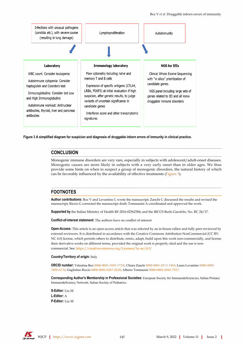
Boz V et al. Druggable inborn errors of immunity
WJCP https://www.wjgnet.com 145 March 9, 2022 Volume 11 Issue 2
Figure 3 A simplified diagram for suspicion and diagnosis of druggable inborn errors of immunity in clinical practice.
CONCLUSIONMonogenic immune disorders are very rare, especially in subjects with adolescent/adult-onset diseases. Monogenic causes are more likely in subjects with a very early onset than in older ages. We thus provide some hints on when to suspect a group of monogenic disorders, the natural history of which can be favorably influenced by the availability of effective treatments (Figure 3).
FOOTNOTESAuthor contributions: Boz V and Levantino L wrote the manuscript; Zanchi C discussed the results and revised the manuscript; Riccio G corrected the manuscript draft; Tommasini A coordinated and approved the work.
Supported by the Italian Ministry of Health RF-2016-02362384; and the IRCCS Burlo Garofolo, No. RC 24/17.
Conflict-of-interest statement: The authors have no conflict of interest
Open-Access: This article is an open-access article that was selected by an in-house editor and fully peer-reviewed by external reviewers. It is distributed in accordance with the Creative Commons Attribution NonCommercial (CC BY-NC 4.0) license, which permits others to distribute, remix, adapt, build upon this work non-commercially, and license their derivative works on different terms, provided the original work is properly cited and the use is non-commercial. See: https://creativecommons.org/Licenses/by-nc/4.0/
Country/Territory of origin: Italy
ORCID number: Valentina Boz 0000-0003-3695-372X; Chiara Zanchi 0000-0001-8513-1963; Laura Levantino 0000-0002-3498-6176; Guglielmo Riccio 0000-0002-8267-2630; Alberto Tommasini 0000-0002-6943-7927.
Corresponding Author's Membership in Professional Societies: European Society for Immunodeficiencies; Italian Primary Immunodeficiency Network; Italian Society of Pediatrics.
S-Editor: Liu M L-Editor: A P-Editor: Liu M

Boz V et al. Druggable inborn errors of immunity
WJCP https://www.wjgnet.com 146 March 9, 2022 Volume 11 Issue 2
REFERENCESAzizi G, Tavakol M, Yazdani R, Delavari S, Moeini Shad T, Rasouli SE, Jamee M, Pashangzadeh S, Kalantari A, Shariat M, Shafiei A, Mohammadi J, Hassanpour G, Chavoshzadeh Z, Mahdaviani SA, Momen T, Behniafard N, Nabavi M, Bemanian MH, Arshi S, Molatefi R, Sherkat R, Shirkani A, Alyasin S, Jabbari-Azad F, Ghaffari J, Mesdaghi M, Ahanchian H, Khoshkhui M, Eslamian MH, Cheraghi T, Dabbaghzadeh A, Nasiri Kalmarzi R, Esmaeilzadeh H, Tafaroji J, Khalili A, Sadeghi-Shabestari M, Darougar S, Moghtaderi M, Ahmadiafshar A, Shakerian B, Heidarzadeh M, Ghalebaghi B, Fathi SM, Darabi B, Fallahpour M, Mohsenzadeh A, Ebrahimi S, Sharafian S, Vosughimotlagh A, Tafakoridelbari M, Rahimi Haji-Abadi M, Ashournia P, Razaghian A, Rezaei A, Salami F, Shirmast P, Bazargan N, Mamishi S, Khazaei HA, Negahdari B, Shokri S, Nabavizadeh SH, Bazregari S, Ghasemi R, Bayat S, Eshaghi H, Rezaei N, Abolhassani H, Aghamohammadi A. Autoimmune manifestations among 461 patients with monogenic inborn errors of immunity. Pediatr Allergy Immunol 2021; 32: 1335-1348 [PMID: 33774840 DOI: 10.1111/pai.13510]
1
Bousfiha A, Jeddane L, Picard C, Al-Herz W, Ailal F, Chatila T, Cunningham-Rundles C, Etzioni A, Franco JL, Holland SM, Klein C, Morio T, Ochs HD, Oksenhendler E, Puck J, Torgerson TR, Casanova JL, Sullivan KE, Tangye SG. Human Inborn Errors of Immunity: 2019 Update of the IUIS Phenotypical Classification. J Clin Immunol 2020; 40: 66-81 [PMID: 32048120 DOI: 10.1007/s10875-020-00758-x]
2
Lenardo M, Lo B, Lucas CL. Genomics of Immune Diseases and New Therapies. Annu Rev Immunol 2016; 34: 121-149 [PMID: 26735698 DOI: 10.1146/annurev-immunol-041015-055620]
3
Valencic E, Smid A, Jakopin Z, Tommasini A, Mlinaric-Rascan I. Repositioning Drugs for Rare Immune Diseases: Hopes and Challenges for a Precision Medicine. Curr Med Chem 2018; 25: 2764-2782 [PMID: 28875839 DOI: 10.2174/0929867324666170830101215]
4
Ballow M, Leiding JW. Precision Medicine in the Treatment of Primary Immune Deficiency Patients With Disorders of Immune Dysregulation. Clin Rev Allergy Immunol2021 epub ahead of print [PMID: 34169440 DOI: 10.1007/s12016-021-08871-4]
5
Chellapandian D, Chitty-Lopez M, Leiding JW. Precision Therapy for the Treatment of Primary Immunodysregulatory Diseases. Immunol Allergy Clin North Am 2020; 40: 511-526 [PMID: 32654696 DOI: 10.1016/j.iac.2020.04.001]
6
Rispoli F, Valencic E, Girardelli M, Pin A, Tesser A, Piscianz E, Boz V, Faletra F, Severini GM, Taddio A, Tommasini A. Immunity and Genetics at the Revolving Doors of Diagnostics in Primary Immunodeficiencies. Diagnostics (Basel) 2021; 11 [PMID: 33809703 DOI: 10.3390/diagnostics11030532]
7
Quinn J, Modell V, Holle J, Truty R, Aradhya S, Johnson B, Orange J, Modell F. Jeffrey's insights: Jeffrey Modell Foundation's global genetic sequencing pilot program to identify specific primary immunodeficiency defects to optimize disease management and treatment. Immunol Res 2020; 68: 126-134 [PMID: 32462469 DOI: 10.1007/s12026-020-09131-x]
8
Chinn IK, Orange JS. A 2020 update on the use of genetic testing for patients with primary immunodeficiency. Expert Rev Clin Immunol 2020; 16: 897-909 [PMID: 32822560 DOI: 10.1080/1744666X.2020.1814145]
9
Bosch B, Itan Y, Meyts I. Whole-exome sequencing for detecting inborn errors of immunity: overview and perspectives. F1000Res 2017; 6: 2056 [PMID: 29225788 DOI: 10.12688/f1000research.12365.1]
10
Gambineri E, Torgerson TR, Ochs HD. Immune dysregulation, polyendocrinopathy, enteropathy, and X-linked inheritance (IPEX), a syndrome of systemic autoimmunity caused by mutations of FOXP3, a critical regulator of T-cell homeostasis. Curr Opin Rheumatol 2003; 15: 430-435 [PMID: 12819471 DOI: 10.1097/00002281-200307000-00010]
11
Bennett CL, Christie J, Ramsdell F, Brunkow ME, Ferguson PJ, Whitesell L, Kelly TE, Saulsbury FT, Chance PF, Ochs HD. The immune dysregulation, polyendocrinopathy, enteropathy, X-linked syndrome (IPEX) is caused by mutations of FOXP3. Nat Genet 2001; 27: 20-21 [PMID: 11137993 DOI: 10.1038/83713]
12
Wildin RS, Ramsdell F, Peake J, Faravelli F, Casanova JL, Buist N, Levy-Lahad E, Mazzella M, Goulet O, Perroni L, Bricarelli FD, Byrne G, McEuen M, Proll S, Appleby M, Brunkow ME. X-linked neonatal diabetes mellitus, enteropathy and endocrinopathy syndrome is the human equivalent of mouse scurfy. Nat Genet 2001; 27: 18-20 [PMID: 11137992 DOI: 10.1038/83707]
13
Barzaghi F, Amaya Hernandez LC, Neven B, Ricci S, Kucuk ZY, Bleesing JJ, Nademi Z, Slatter MA, Ulloa ER, Shcherbina A, Roppelt A, Worth A, Silva J, Aiuti A, Murguia-Favela L, Speckmann C, Carneiro-Sampaio M, Fernandes JF, Baris S, Ozen A, Karakoc-Aydiner E, Kiykim A, Schulz A, Steinmann S, Notarangelo LD, Gambineri E, Lionetti P, Shearer WT, Forbes LR, Martinez C, Moshous D, Blanche S, Fisher A, Ruemmele FM, Tissandier C, Ouachee-Chardin M, Rieux-Laucat F, Cavazzana M, Qasim W, Lucarelli B, Albert MH, Kobayashi I, Alonso L, Diaz De Heredia C, Kanegane H, Lawitschka A, Seo JJ, Gonzalez-Vicent M, Diaz MA, Goyal RK, Sauer MG, Yesilipek A, Kim M, Yilmaz-Demirdag Y, Bhatia M, Khlevner J, Richmond Padilla EJ, Martino S, Montin D, Neth O, Molinos-Quintana A, Valverde-Fernandez J, Broides A, Pinsk V, Ballauf A, Haerynck F, Bordon V, Dhooge C, Garcia-Lloret ML, Bredius RG, Kałwak K, Haddad E, Seidel MG, Duckers G, Pai SY, Dvorak CC, Ehl S, Locatelli F, Goldman F, Gennery AR, Cowan MJ, Roncarolo MG, Bacchetta R; Primary Immune Deficiency Treatment Consortium (PIDTC) and the Inborn Errors Working Party (IEWP) of the European Society for Blood and Marrow Transplantation (EBMT). Long-term follow-up of IPEX syndrome patients after different therapeutic strategies: An international multicenter retrospective study. J Allergy Clin Immunol 2018; 141: 1036-1049.e5 [PMID: 29241729 DOI: 10.1016/j.jaci.2017.10.041]
14
Battaglia M, Stabilini A, Roncarolo MG. Rapamycin selectively expands CD4+CD25+FoxP3+ regulatory T cells. Blood 2005; 105: 4743-4748 [PMID: 15746082 DOI: 10.1182/blood-2004-10-3932]
15
Bevacqua M, Baldo F, Pastore S, Valencic E, Tommasini A, Maestro A, Rabusin M, Arbo A, Barbi E. Off-Label Use of Sirolimus and Everolimus in a Pediatric Center: A Case Series and Review of the Literature. Paediatr Drugs 2019; 21: 185-193 [PMID: 31124053 DOI: 10.1007/s40272-019-00337-7]
16
Singh N, Chandler PR, Seki Y, Baban B, Takezaki M, Kahler DJ, Munn DH, Larsen CP, Mellor AL, Iwashima M. Role of CD28 in fatal autoimmune disorder in scurfy mice. Blood 2007; 110: 1199-1206 [PMID: 17463170 DOI: 10.1182/blood-2006-10-054585]
17
Ochs HD, Gambineri E, Torgerson TR. IPEX, FOXP3 and regulatory T-cells: a model for autoimmunity. Immunol Res 18
Boz V et al. Druggable inborn errors of immunity
WJCP https://www.wjgnet.com 147 March 9, 2022 Volume 11 Issue 2
2007; 38: 112-121 [PMID: 17917016 DOI: 10.1007/s12026-007-0022-2]Anderson MS, Su MA. AIRE expands: new roles in immune tole rance and beyond. Nat Rev Immunol 2016; 16: 247-258 [PMID: 26972725 DOI: 10.1038/nri.2016.9]
19
Malchow S, Leventhal DS, Lee V, Nishi S, Socci ND, Savage PA. Aire Enforces Immune Tolerance by Directing Autoreactive T Cells into the Regulatory T Cell Lineage. Immunity 2016; 44: 1102-1113 [PMID: 27130899 DOI: 10.1016/j.immuni.2016.02.009]
20
Notarangelo LD, Mazza C, Forino C, Mazzolari E, Buzi F. AIRE and immunological tolerance: insights from the study of autoimmune polyendocrinopathy candidiasis and ectodermal dystrophy. Curr Opin Allergy Clin Immunol 2004; 4: 491-496 [PMID: 15640689 DOI: 10.1097/00130832-200412000-00004]
21
Sarkadi AK, Taskó S, Csorba G, Tóth B, Erdős M, Maródi L. Autoantibodies to IL-17A may be correlated with the severity of mucocutaneous candidiasis in APECED patients. J Clin Immunol 2014; 34: 181-193 [PMID: 24493573 DOI: 10.1007/s10875-014-9987-5]
22
Mazza C, Buzi F, Ortolani F, Vitali A, Notarangelo LD, Weber G, Bacchetta R, Soresina A, Lougaris V, Greggio NA, Taddio A, Pasic S, de Vroede M, Pac M, Kilic SS, Ozden S, Rusconi R, Martino S, Capalbo D, Salerno M, Pignata C, Radetti G, Maggiore G, Plebani A, Badolato R. Clinical heterogeneity and diagnostic delay of autoimmune polyendocrinopathy-candidiasis-ectodermal dystrophy syndrome. Clin Immunol 2011; 139: 6-11 [PMID: 21295522 DOI: 10.1016/j.clim.2010.12.021]
23
Bruserud Ø, Oftedal BE, Landegren N, Erichsen MM, Bratland E, Lima K, Jørgensen AP, Myhre AG, Svartberg J, Fougner KJ, Bakke Å, Nedrebø BG, Mella B, Breivik L, Viken MK, Knappskog PM, Marthinussen MC, Løvås K, Kämpe O, Wolff AB, Husebye ES. A Longitudinal Follow-up of Autoimmune Polyendocrine Syndrome Type 1. J Clin Endocrinol Metab 2016; 101: 2975-2983 [PMID: 27253668 DOI: 10.1210/jc.2016-1821]
24
Borchers J, Pukkala E, Mäkitie O, Laakso S. Patients With APECED Have Increased Early Mortality Due to Endocrine Causes, Malignancies and infections. J Clin Endocrinol Metab 2020; 105 [PMID: 32185376 DOI: 10.1210/clinem/dgaa140]
25
Verma N, Burns SO, Walker LSK, Sansom DM. Immune deficiency and autoimmunity in patients with CTLA-4 (CD152) mutations. Clin Exp Immunol 2017; 190: 1-7 [PMID: 28600865 DOI: 10.1111/cei.12997]
26
Schwab C, Gabrysch A, Olbrich P, Patiño V, Warnatz K, Wolff D, Hoshino A, Kobayashi M, Imai K, Takagi M, Dybedal I, Haddock JA, Sansom DM, Lucena JM, Seidl M, Schmitt-Graeff A, Reiser V, Emmerich F, Frede N, Bulashevska A, Salzer U, Schubert D, Hayakawa S, Okada S, Kanariou M, Kucuk ZY, Chapdelaine H, Petruzelkova L, Sumnik Z, Sediva A, Slatter M, Arkwright PD, Cant A, Lorenz HM, Giese T, Lougaris V, Plebani A, Price C, Sullivan KE, Moutschen M, Litzman J, Freiberger T, van de Veerdonk FL, Recher M, Albert MH, Hauck F, Seneviratne S, Pachlopnik Schmid J, Kolios A, Unglik G, Klemann C, Speckmann C, Ehl S, Leichtner A, Blumberg R, Franke A, Snapper S, Zeissig S, Cunningham-Rundles C, Giulino-Roth L, Elemento O, Dückers G, Niehues T, Fronkova E, Kanderová V, Platt CD, Chou J, Chatila TA, Geha R, McDermott E, Bunn S, Kurzai M, Schulz A, Alsina L, Casals F, Deyà-Martinez A, Hambleton S, Kanegane H, Taskén K, Neth O, Grimbacher B. Phenotype, penetrance, and treatment of 133 cytotoxic T-lymphocyte antigen 4-insufficient subjects. J Allergy Clin Immunol 2018; 142: 1932-1946 [PMID: 29729943 DOI: 10.1016/j.jaci.2018.02.055]
27
Jamee M, Hosseinzadeh S, Sharifinejad N, Zaki-Dizaji M, Matloubi M, Hasani M, Baris S, Alsabbagh M, Lo B, Azizi G. Comprehensive comparison between 222 CTLA-4 haploinsufficiency and 212 LRBA deficiency patients: a systematic review. Clin Exp Immunol 2021; 205: 28-43 [PMID: 33788257 DOI: 10.1111/cei.13600]
28
Krone KA, Winant AJ, Vargas SO, Platt CD, Bartnikas LM, Janssen E, Lillehei C, Lee EY, Fishman MP, Casey A. Pulmonary manifestations of immune dysregulation in CTLA-4 haploinsufficiency and LRBA deficiency. Pediatr Pulmonol 2021; 56: 2232-2241 [PMID: 33710794 DOI: 10.1002/ppul.25373]
29
Tesch VK, Abolhassani H, Shadur B, Zobel J, Mareika Y, Sharapova S, Karakoc-Aydiner E, Rivière JG, Garcia-Prat M, Moes N, Haerynck F, Gonzales-Granado LI, Santos Pérez JL, Mukhina A, Shcherbina A, Aghamohammadi A, Hammarström L, Dogu F, Haskologlu S, İkincioğulları AI, Köstel Bal S, Baris S, Kilic SS, Karaca NE, Kutukculer N, Girschick H, Kolios A, Keles S, Uygun V, Stepensky P, Worth A, van Montfrans JM, Peters AMJ, Meyts I, Adeli M, Marzollo A, Padem N, Khojah AM, Chavoshzadeh Z, Avbelj Stefanija M, Bakhtiar S, Florkin B, Meeths M, Gamez L, Grimbacher B, Seppänen MRJ, Lankester A, Gennery AR, Seidel MG; Inborn Errors, Clinical, and Registry Working Parties of the European Society for Blood and Marrow Transplantation and the European Society for Immunodeficiencies. Long-term outcome of LRBA deficiency in 76 patients after various treatment modalities as evaluated by the immune deficiency and dysregulation activity (IDDA) score. J Allergy Clin Immunol 2020; 145: 1452-1463 [PMID: 31887391 DOI: 10.1016/j.jaci.2019.12.896]
30
Lo B, Zhang K, Lu W, Zheng L, Zhang Q, Kanellopoulou C, Zhang Y, Liu Z, Fritz JM, Marsh R, Husami A, Kissell D, Nortman S, Chaturvedi V, Haines H, Young LR, Mo J, Filipovich AH, Bleesing JJ, Mustillo P, Stephens M, Rueda CM, Chougnet CA, Hoebe K, McElwee J, Hughes JD, Karakoc-Aydiner E, Matthews HF, Price S, Su HC, Rao VK, Lenardo MJ, Jordan MB. AUTOIMMUNE DISEASE. Patients with LRBA deficiency show CTLA4 Loss and immune dysregulation responsive to abatacept therapy. Science 2015; 349: 436-440 [PMID: 26206937 DOI: 10.1126/science.aaa1663]
31
Tessarin G, Rossi S, Baronio M, Gazzurelli L, Colpani M, Benvenuto A, Zunica F, Cardinale F, Martire B, Brescia L, Costagliola G, Luti L, Casazza G, Menconi MC, Saettini F, Palumbo L, Girelli MF, Badolato R, Lanzi G, Chiarini M, Moratto D, Meini A, Giliani S, Bondioni MP, Plebani A, Lougaris V. Activated Phosphoinositide 3-Kinase Delta Syndrome 1: Clinical and Immunological Data from an Italian Cohort of Patients. J Clin Med 2020; 9 [PMID: 33080915 DOI: 10.3390/jcm9103335]
32
Ewertowska M, Grześk E, Urbańczyk A, Dąbrowska A, Bąbol-Pokora K, Łęcka M, Kołtan S. Activated phosphoinositide 3-kinase delta syndrome 1 and 2 (APDS 1 and APDS 2): similarities and differences based on clinical presentation in two boys. Allergy Asthma Clin Immunol 2020; 16: 22 [PMID: 32265996 DOI: 10.1186/s13223-020-00420-6]
33
Maccari ME, Abolhassani H, Aghamohammadi A, Aiuti A, Aleinikova O, Bangs C, Baris S, Barzaghi F, Baxendale H, Buckland M, Burns SO, Cancrini C, Cant A, Cathébras P, Cavazzana M, Chandra A, Conti F, Coulter T, Devlin LA,
34
Boz V et al. Druggable inborn errors of immunity
WJCP https://www.wjgnet.com 148 March 9, 2022 Volume 11 Issue 2
Edgar JDM, Faust S, Fischer A, Garcia-Prat M, Hammarström L, Heeg M, Jolles S, Karakoc-Aydiner E, Kindle G, Kiykim A, Kumararatne D, Grimbacher B, Longhurst H, Mahlaoui N, Milota T, Moreira F, Moshous D, Mukhina A, Neth O, Neven B, Nieters A, Olbrich P, Ozen A, Pachlopnik Schmid J, Picard C, Prader S, Rae W, Reichenbach J, Rusch S, Savic S, Scarselli A, Scheible R, Sediva A, Sharapova SO, Shcherbina A, Slatter M, Soler-Palacin P, Stanislas A, Suarez F, Tucci F, Uhlmann A, van Montfrans J, Warnatz K, Williams AP, Wood P, Kracker S, Condliffe AM, Ehl S. Disease Evolution and Response to Rapamycin in Activated Phosphoinositide 3-Kinase δ Syndrome: The European Society for Immunodeficiencies-Activated Phosphoinositide 3-Kinase δ Syndrome Registry. Front Immunol 2018; 9: 543 [PMID: 29599784 DOI: 10.3389/fimmu.2018.00543]Lucas CL, Chandra A, Nejentsev S, Condliffe AM, Okkenhaug K. PI3Kδ and primary immunodeficiencies. Nat Rev Immunol 2016; 16: 702-714 [PMID: 27616589 DOI: 10.1038/nri.2016.93]
35
Angulo I, Vadas O, Garçon F, Banham-Hall E, Plagnol V, Leahy TR, Baxendale H, Coulter T, Curtis J, Wu C, Blake-Palmer K, Perisic O, Smyth D, Maes M, Fiddler C, Juss J, Cilliers D, Markelj G, Chandra A, Farmer G, Kielkowska A, Clark J, Kracker S, Debré M, Picard C, Pellier I, Jabado N, Morris JA, Barcenas-Morales G, Fischer A, Stephens L, Hawkins P, Barrett JC, Abinun M, Clatworthy M, Durandy A, Doffinger R, Chilvers ER, Cant AJ, Kumararatne D, Okkenhaug K, Williams RL, Condliffe A, Nejentsev S. Phosphoinositide 3-kinase δ gene mutation predisposes to respiratory infection and airway damage. Science 2013; 342: 866-871 [PMID: 24136356 DOI: 10.1126/science.1243292]
36
Carpier JM, Lucas CL. Epstein-Barr Virus Susceptibility in Activated PI3Kδ Syndrome (APDS) Immunodeficiency. Front Immunol 2017; 8: 2005 [PMID: 29387064 DOI: 10.3389/fimmu.2017.02005]
37
Durandy A, Kracker S. Increased activation of PI3 kinase-δ predisposes to B-cell lymphoma. Blood 2020; 135: 638-643 [PMID: 31942637 DOI: 10.1182/blood.2019002072]
38
Valencic E, Grasso AG, Conversano E, Lucafò M, Piscianz E, Gregori M, Conti F, Cancrini C, Tommasini A. Theophylline as a precision therapy in a young girl with PIK3R1 immunodeficiency. J Allergy Clin Immunol Pract 2018; 6: 2165-2167 [PMID: 29510232 DOI: 10.1016/j.jaip.2018.02.029]
39
Chaimowitz NS, Ebenezer SJ, Hanson IC, Anderson M, Forbes LR. STAT1 Gain of Function, Type 1 Diabetes, and Reversal with JAK Inhibition. N Engl J Med 2020; 383: 1494-1496 [PMID: 33027576 DOI: 10.1056/NEJMc2022226]
40
van de Veerdonk FL, Plantinga TS, Hoischen A, Smeekens SP, Joosten LA, Gilissen C, Arts P, Rosentul DC, Carmichael AJ, Smits-van der Graaf CA, Kullberg BJ, van der Meer JW, Lilic D, Veltman JA, Netea MG. STAT1 mutations in autosomal dominant chronic mucocutaneous candidiasis. N Engl J Med 2011; 365: 54-61 [PMID: 21714643 DOI: 10.1056/NEJMoa1100102]
41
Pin A, Tesser A, Pastore S, Moressa V, Valencic E, Arbo A, Maestro A, Tommasini A, Taddio A. Biological and Clinical Changes in a Pediatric Series Treated with Off-Label JAK Inhibitors. Int J Mol Sci 2020; 21 [PMID: 33092242 DOI: 10.3390/ijms21207767]
42
Weinacht KG, Charbonnier LM, Alroqi F, Plant A, Qiao Q, Wu H, Ma C, Torgerson TR, Rosenzweig SD, Fleisher TA, Notarangelo LD, Hanson IC, Forbes LR, Chatila TA. Ruxolitinib reverses dysregulated T helper cell responses and controls autoimmunity caused by a novel signal transducer and activator of transcription 1 (STAT1) gain-of-function mutation. J Allergy Clin Immunol 2017; 139: 1629-1640.e2 [PMID: 28139313 DOI: 10.1016/j.jaci.2016.11.022]
43
Flanagan SE, Haapaniemi E, Russell MA, Caswell R, Allen HL, De Franco E, McDonald TJ, Rajala H, Ramelius A, Barton J, Heiskanen K, Heiskanen-Kosma T, Kajosaari M, Murphy NP, Milenkovic T, Seppänen M, Lernmark Å, Mustjoki S, Otonkoski T, Kere J, Morgan NG, Ellard S, Hattersley AT. Activating germline mutations in STAT3 cause early-onset multi-organ autoimmune disease. Nat Genet 2014; 46: 812-814 [PMID: 25038750 DOI: 10.1038/ng.3040]
44
Forbes LR, Vogel TP, Cooper MA, Castro-Wagner J, Schussler E, Weinacht KG, Plant AS, Su HC, Allenspach EJ, Slatter M, Abinun M, Lilic D, Cunningham-Rundles C, Eckstein O, Olbrich P, Guillerman RP, Patel NC, Demirdag YY, Zerbe C, Freeman AF, Holland SM, Szabolcs P, Gennery A, Torgerson TR, Milner JD, Leiding JW. Jakinibs for the treatment of immune dysregulation in patients with gain-of-function signal transducer and activator of transcription 1 (STAT1) or STAT3 mutations. J Allergy Clin Immunol 2018; 142: 1665-1669 [PMID: 30092289 DOI: 10.1016/j.jaci.2018.07.020]
45
Caorsi R, Penco F, Grossi A, Insalaco A, Omenetti A, Alessio M, Conti G, Marchetti F, Picco P, Tommasini A, Martino S, Malattia C, Gallizzi R, Podda RA, Salis A, Falcini F, Schena F, Garbarino F, Morreale A, Pardeo M, Ventrici C, Passarelli C, Zhou Q, Severino M, Gandolfo C, Damonte G, Martini A, Ravelli A, Aksentijevich I, Ceccherini I, Gattorno M. ADA2 deficiency (DADA2) as an unrecognised cause of early onset polyarteritis nodosa and stroke: a multicentre national study. Ann Rheum Dis 2017; 76: 1648-1656 [PMID: 28522451 DOI: 10.1136/annrheumdis-2016-210802]
46
Insalaco A, Moneta GM, Pardeo M, Caiello I, Messia V, Bracaglia C, Passarelli C, De Benedetti F. Variable Clinical Phenotypes and Relation of Interferon Signature with Disease Activity in ADA2 Deficiency. J Rheumatol 2019; 46: 523-526 [PMID: 30647181 DOI: 10.3899/jrheum.180045]
47
Sahin S, Adrovic A, Kasapcopur O. A monogenic autoinflammatory disease with fatal vasculitis: deficiency of adenosine deaminase 2. Curr Opin Rheumatol 2020; 32: 3-14 [PMID: 31599797 DOI: 10.1097/BOR.0000000000000669]
48
Yu MP, Xu XS, Zhou Q, Deuitch N, Lu MP. Haploinsufficiency of A20 (HA20): updates on the genetics, phenotype, pathogenesis and treatment. World J Pediatr 2020; 16: 575-584 [PMID: 31587140 DOI: 10.1007/s12519-019-00288-6]
49
Aeschlimann FA, Batu ED, Canna SW, Go E, Gül A, Hoffmann P, Leavis HL, Ozen S, Schwartz DM, Stone DL, van Royen-Kerkof A, Kastner DL, Aksentijevich I, Laxer RM. A20 haploinsufficiency (HA20): clinical phenotypes and disease course of patients with a newly recognised NF-kB-mediated autoinflammatory disease. Ann Rheum Dis 2018; 77: 728-735 [PMID: 29317407 DOI: 10.1136/annrheumdis-2017-212403]
50
Franco-Jarava C, Wang H, Martin-Nalda A, Alvarez SD, García-Prat M, Bodet D, García-Patos V, Plaja A, Rudilla F, Rodriguez-Sureda V, García-Latorre L, Aksentijevich I, Colobran R, Soler-Palacín P. TNFAIP3 haploinsufficiency is the cause of autoinflammatory manifestations in a patient with a deletion of 13Mb on chromosome 6. Clin Immunol 2018; 191: 44-51 [PMID: 29572183 DOI: 10.1016/j.clim.2018.03.009]
51
Girardelli M, Valencic E, Moressa V, Margagliotta R, Tesser A, Pastore S, Spadola O, Athanasakis E, Severini GM, Taddio A, Tommasini A. Genetic and immunologic findings in children with recurrent aphthous stomatitis with systemic inflammation. Pediatr Rheumatol Online J 2021; 19: 70 [PMID: 33971891 DOI: 10.1186/s12969-021-00552-y]
52
Boz V et al. Druggable inborn errors of immunity
WJCP https://www.wjgnet.com 149 March 9, 2022 Volume 11 Issue 2
Aeschlimann FA, Laxer RM. Haploinsufficiency of A20 and other paediatric inflammatory disorders with mucosal involvement. Curr Opin Rheumatol 2018; 30: 506-513 [PMID: 29916847 DOI: 10.1097/BOR.0000000000000532]
53
Massias JS, Smith EMD, Al-Abadi E, Armon K, Bailey K, Ciurtin C, Davidson J, Gardner-Medwin J, Haslam K, Hawley DP, Leahy A, Leone V, McErlane F, Mewar D, Modgil G, Moots R, Pilkington C, Ramanan AV, Rangaraj S, Riley P, Sridhar A, Wilkinson N, Beresford MW, Hedrich CM. Clinical and laboratory characteristics in juvenile-onset systemic lupus erythematosus across age groups. Lupus 2020; 29: 474-481 [PMID: 32233733 DOI: 10.1177/0961203320909156]
54
Malattia C, Martini A. Paediatric-onset systemic lupus erythematosus. Best Pract Res Clin Rheumatol 2013; 27: 351-362 [PMID: 24238692 DOI: 10.1016/j.berh.2013.07.007]
55
Charras A, Smith E, Hedrich CM. Systemic Lupus Erythematosus in Children and Young People. Curr Rheumatol Rep 2021; 23: 20 [PMID: 33569643 DOI: 10.1007/s11926-021-00985-0]
56
Hiraki LT, Silverman ED. Genomics of Systemic Lupus Erythematosus: Insights Gained by Studying Monogenic Young-Onset Systemic Lupus Erythematosus. Rheum Dis Clin North Am 2017; 43: 415-434 [PMID: 28711143 DOI: 10.1016/j.rdc.2017.04.005]
57
Tak AS, Satapathy J, Jana M, Sinha A, Jat KR, Bagri NK. Monogenic lupus with homozygous C4A deficiency presenting as bronchiectasis and immune-mediated thrombocytopenia. Rheumatol Int2021 epub ahead of print [PMID: 34287686 DOI: 10.1007/s00296-021-04943-y]
58
Batu ED, Koşukcu C, Taşkıran E, Sahin S, Akman S, Sözeri B, Ünsal E, Bilginer Y, Kasapcopur O, Alikaşifoğlu M, Ozen S. Whole Exome Sequencing in Early-onset Systemic Lupus Erythematosus. J Rheumatol 2018; 45: 1671-1679 [PMID: 30008451 DOI: 10.3899/jrheum.171358]
59
Tirosh I, Spielman S, Barel O, Ram R, Stauber T, Paret G, Rubinsthein M, Pessach IM, Gerstein M, Anikster Y, Shukrun R, Dagan A, Adler K, Pode-Shakked B, Volkov A, Perelman M, Greenberger S, Somech R, Lahav E, Majmundar AJ, Padeh S, Hildebrandt F, Vivante A. Whole exome sequencing in childhood-onset lupus frequently detects single gene etiologies. Pediatr Rheumatol Online J 2019; 17: 52 [PMID: 31362757 DOI: 10.1186/s12969-019-0349-y]
60
Li G, Liu H, Li Y, Zhang T, Yao W, Guan W, Shi Y, Wu B, Xu H, Sun L. Genetic heterogeneity in Chinese children with systemic lupus erythematosus. Clin Exp Rheumatol 2021; 39: 214-222 [PMID: 33337996]
61
Omarjee O, Picard C, Frachette C, Moreews M, Rieux-Laucat F, Soulas-Sprauel P, Viel S, Lega JC, Bader-Meunier B, Walzer T, Mathieu AL, Cimaz R, Belot A. Monogenic lupus: Dissecting heterogeneity. Autoimmun Rev 2019; 18: 102361 [PMID: 31401343 DOI: 10.1016/j.autrev.2019.102361]
62
Tesser A, de Carvalho LM, Sandrin-Garcia P, Pin A, Pastore S, Taddio A, Roberti LR, de Paula Queiroz RG, Ferriani VPL, Crovella S, Tommasini A. Higher interferon score and normal complement levels may identify a distinct clinical subset in children with systemic lupus erythematosus. Arthritis Res Ther 2020; 22: 91 [PMID: 32334613 DOI: 10.1186/s13075-020-02161-8]
63
Li G, Li Y, Liu H, Shi Y, Guan W, Zhang T, Yao W, Wu B, Xu H, Sun L. Genetic heterogeneity of pediatric systemic lupus erythematosus with lymphoproliferation. Medicine (Baltimore) 2020; 99: e20232 [PMID: 32443356 DOI: 10.1097/MD.0000000000020232]
64
Jamee M, Moniri S, Zaki-Dizaji M, Olbrich P, Yazdani R, Jadidi-Niaragh F, Aghamahdi F, Abolhassani H, Condliffe AM, Aghamohammadi A, Azizi G. Clinical, Immunological, and Genetic Features in Patients with Activated PI3Kδ Syndrome (APDS): a Systematic Review. Clin Rev Allergy Immunol 2020; 59: 323-333 [PMID: 31111319 DOI: 10.1007/s12016-019-08738-9]
65
Pain CE. Juvenile-onset Behçet's syndrome and mimics. Clin Immunol 2020; 214: 108381 [PMID: 32165216 DOI: 10.1016/j.clim.2020.108381]
66
Papadopoulou C, Omoyinmi E, Standing A, Pain CE, Booth C, D'Arco F, Gilmour K, Buckland M, Eleftheriou D, Brogan PA. Monogenic mimics of Behçet's disease in the young. Rheumatology (Oxford) 2019; 58: 1227-1238 [PMID: 30715505 DOI: 10.1093/rheumatology/key445]
67
Kim HY, Song JY, Kim WI, Ko HC, Park SE, Jang JH, Kim SH. The First Case of an Infant with Familial A20 Haploinsufficiency in Korea. J Korean Med Sci 2020; 35: e252 [PMID: 32743991 DOI: 10.3346/jkms.2020.35.e252]
68
Krutzke S, Rietschel C, Horneff G. Baricitinib in therapy of COPA syndrome in a 15-year-old girl. Eur J Rheumatol 2019; 1-4 [PMID: 31449490 DOI: 10.5152/eurjrheum.2019.18177]
69
Semo Oz R, S Tesher M. Arthritis in children with LRBA deficiency - case report and literature review. Pediatr Rheumatol Online J 2019; 17: 82 [PMID: 31847838 DOI: 10.1186/s12969-019-0388-4]
70
Hou X, Qu H, Zhang S, Qi X, Hakonarson H, Xia Q, Li J. The Multi-Omics Architecture of Juvenile Idiopathic Arthritis. Cells 2020; 9 [PMID: 33076506 DOI: 10.3390/cells9102301]
71
Rosé CD, Wouters CH, Meiorin S, Doyle TM, Davey MP, Rosenbaum JT, Martin TM. Pediatric granulomatous arthritis: an international registry. Arthritis Rheum 2006; 54: 3337-3344 [PMID: 17009307 DOI: 10.1002/art.22122]
72
Ouahed J, Spencer E, Kotlarz D, Shouval DS, Kowalik M, Peng K, Field M, Grushkin-Lerner L, Pai SY, Bousvaros A, Cho J, Argmann C, Schadt E, Mcgovern DPB, Mokry M, Nieuwenhuis E, Clevers H, Powrie F, Uhlig H, Klein C, Muise A, Dubinsky M, Snapper SB. Very Early Onset Inflammatory Bowel Disease: A Clinical Approach With a Focus on the Role of Genetics and Underlying Immune Deficiencies. Inflamm Bowel Dis 2020; 26: 820-842 [PMID: 31833544 DOI: 10.1093/ibd/izz259]
73
Nambu R, Warner N, Mulder DJ, Kotlarz D, McGovern DPB, Cho J, Klein C, Snapper SB, Griffiths AM, Iwama I, Muise AM. A Systematic Review of Monogenic Inflammatory Bowel Disease. Clin Gastroenterol Hepatol 2021 [PMID: 33746097 DOI: 10.1016/j.cgh.2021.03.021]
74
Nambu R, Muise AM. Advanced Understanding of Monogenic Inflammatory Bowel Disease. Front Pediatr 2020; 8: 618918 [PMID: 33553075 DOI: 10.3389/fped.2020.618918]
75
Glocker EO, Kotlarz D, Klein C, Shah N, Grimbacher B. IL-10 and IL-10 receptor defects in humans. Ann N Y Acad Sci 2011; 1246: 102-107 [PMID: 22236434 DOI: 10.1111/j.1749-6632.2011.06339.x]
76
Lega S, Pin A, Arrigo S, Cifaldi C, Girardelli M, Bianco AM, Malamisura M, Angelino G, Faraci S, Rea F, Romeo EF, Aloi M, Romano C, Barabino A, Martelossi S, Tommasini A, Di Matteo G, Cancrini C, De Angelis P, Finocchi A, Bramuzzo M. Diagnostic Approach to Monogenic Inflammatory Bowel Disease in Clinical Practice: A Ten-Year
77
Boz V et al. Druggable inborn errors of immunity
WJCP https://www.wjgnet.com 150 March 9, 2022 Volume 11 Issue 2
Multicentric Experience. Inflamm Bowel Dis 2020; 26: 720-727 [PMID: 31375816 DOI: 10.1093/ibd/izz178]Singhi AD, Goyal A, Davison JM, Regueiro MD, Roche RL, Ranganathan S. Pediatric autoimmune enteropathy: an entity frequently associated with immunodeficiency disorders. Mod Pathol 2014; 27: 543-553 [PMID: 24051695 DOI: 10.1038/modpathol.2013.150]
78
Uchida T, Suzuki T, Kikuchi A, Kakuta F, Ishige T, Nakayama Y, Kanegane H, Etani Y, Mizuochi T, Fujiwara SI, Nambu R, Suyama K, Tanaka M, Yoden A, Abukawa D, Sasahara Y, Kure S. Comprehensive targeted sequencing identifies monogenic disorders in patients with early-onset refractory diarrhea. J Pediatr Gastroenterol Nutr 2020; 71: 333-339 [PMID: 32487952 DOI: 10.1097/MPG.0000000000002796]
79
Uzzan M, Ko HM, Mehandru S, Cunningham-Rundles C. Gastrointestinal Disorders Associated with Common Variable Immune Deficiency (CVID) and Chronic Granulomatous Disease (CGD). Curr Gastroenterol Rep 2016; 18: 17 [PMID: 26951230 DOI: 10.1007/s11894-016-0491-3]
80
Fernando SL, Jang HS, Li J. The Immune Dysregulation of Common Variable Immunodeficiency Disorders. Immunol Lett 2021; 230: 21-26 [PMID: 33333111 DOI: 10.1016/j.imlet.2020.12.002]
81
Miyazaki H, Hoshi N, Kohashi M, Tokunaga E, Ku Y, Takenaka H, Ooi M, Yamamoto N, Uemura S, Nishimura N, Iijima K, Jimbo K, Okano T, Hoshino A, Imai K, Kanegane H, Kobayashi I, Kodama Y. A case of autoimmune enteropathy with CTLA4 haploinsufficiency. Intest Res 2021 [PMID: 33476510 DOI: 10.5217/ir.2020.00041]
82
Boz V, Valencic E, Girardelli M, Pin A, Gàmez-Diaz L, Tommasini A, Lega S, Bramuzzo M. Case Report: Refractory Autoimmune Gastritis Responsive to Abatacept in LRBA Deficiency. Front Immunol 2021; 12: 619246 [PMID: 33717114 DOI: 10.3389/fimmu.2021.619246]
83
Lahner E, Conti L, Annibale B, Corleto VD. Current Perspectives in Atrophic Gastritis. Curr Gastroenterol Rep 2020; 22: 38 [PMID: 32542467 DOI: 10.1007/s11894-020-00775-1]
84
Lam-Tse WK, Batstra MR, Koeleman BP, Roep BO, Bruining MG, Aanstoot HJ, Drexhage HA. The association between autoimmune thyroiditis, autoimmune gastritis and type 1 diabetes. Pediatr Endocrinol Rev 2003; 1: 22-37 [PMID: 16437010]
85
Kutluǧ Ş, Boztuǧ K, Yıldıran A. Development of multiple gallstones in a child with lipopolysaccharide-responsive beige-like anchor protein mutation. Cent Eur J Immunol 2019; 44: 332-335 [PMID: 31871423 DOI: 10.5114/ceji.2019.89613]
86
Kluger N, Jokinen M, Krohn K, Ranki A. Gastrointestinal manifestations in APECED syndrome. J Clin Gastroenterol 2013; 47: 112-120 [PMID: 23314667 DOI: 10.1097/MCG.0b013e31827356e1]
87
Uche-Anya E, Lebwohl B. Celiac disease: clinical update. Curr Opin Gastroenterol 2021; 37: 619-624 [PMID: 34456226 DOI: 10.1097/MOG.0000000000000785]
88
Troncone R, Greco L, Auricchio S. Gluten-sensitive enteropathy. Pediatr Clin North Am 1996; 43: 355-373 [PMID: 8614605 DOI: 10.1016/s0031-3955(05)70410-7]
89
Sari S, Dogu F, Hwa V, Haskologlu S, Dauber A, Rosenfeld R, Polat M, Kuloglu Z, Kansu A, Dalgic B, Ikinciogullari A. A Successful HSCT in a Girl with Novel LRBA Mutation with Refractory Celiac Disease. J Clin Immunol 2016; 36: 8-11 [PMID: 26686526 DOI: 10.1007/s10875-015-0220-y]
90
Costa-Carvalho BT, de Moraes-Pinto MI, de Almeida LC, de Seixas Alves MT, Maia RP, de Souza RL, Barreto M, Lourenço L, Vicente AM, Coutinho A, Carneiro-Sampaio M. A remarkable depletion of both naïve CD4+ and CD8+ with high proportion of memory T cells in an IPEX infant with a FOXP3 mutation in the forkhead domain. Scand J Immunol 2008; 68: 85-91 [PMID: 18489537 DOI: 10.1111/j.1365-3083.2008.02055.x]
91
Glocker E, Grimbacher B. Chronic mucocutaneous candidiasis and congenital susceptibility to Candida. Curr Opin Allergy Clin Immunol 2010; 10: 542-550 [PMID: 20859203 DOI: 10.1097/ACI.0b013e32833fd74f]
92
Okada S, Asano T, Moriya K, Boisson-Dupuis S, Kobayashi M, Casanova JL, Puel A. Human STAT1 Gain-of-Function Heterozygous Mutations: Chronic Mucocutaneous Candidiasis and Type I Interferonopathy. J Clin Immunol 2020; 40: 1065-1081 [PMID: 32852681 DOI: 10.1007/s10875-020-00847-x]
93
Pichard DC, Freeman AF, Cowen EW. Primary immunodeficiency update: Part II. Syndromes associated with mucocutaneous candidiasis and noninfectious cutaneous manifestations. J Am Acad Dermatol 2015; 73: 367-81; quiz 381 [PMID: 26282795 DOI: 10.1016/j.jaad.2015.01.055]
94
Errante PR, Perazzio SF, Frazão JB, da Silva NP, Andrade LE. Primary immunodeficiency association with systemic lupus erythematosus: review of literature and lessons learned by the Rheumatology Division of a tertiary university hospital at São Paulo, Brazil. Rev Bras Reumatol Engl Ed 2016; 56: 58-68 [PMID: 27267335 DOI: 10.1016/j.rbre.2015.07.006]
95
Abraham RS. How to evaluate for immunodeficiency in patients with autoimmune cytopenias: Laboratory evaluation for the diagnosis of inborn errors of immunity associated with immune dysregulation. Hematology Am Soc Hematol Educ Program 2020; 2020: 661-672 [PMID: 33275711 DOI: 10.1182/hematology.2020000173]
96
Castagnoli R, Lougaris V, Giardino G, Volpi S, Leonardi L, La Torre F, Federici S, Corrente S, Cinicola BL, Soresina A, Cancrini C, Marseglia GL, Cardinale F; Immunology Task Force of the Italian Society of Pediatric Allergy and Immunology (SIAIP). Inborn errors of immunity with atopic phenotypes: A practical guide for allergists. World Allergy Organ J 2021; 14: 100513 [PMID: 33717395 DOI: 10.1016/j.waojou.2021.100513]
97
Mackay-Wiggan J, Sallee BN, Wang EHC, Sansaricq F, Nguyen N, Kim C, Chen JC, Christiano AM, Clynes R. An open-label study evaluating the efficacy of abatacept in alopecia areata. J Am Acad Dermatol 2021; 84: 841-844 [PMID: 33045294 DOI: 10.1016/j.jaad.2020.09.091]
98
Dai Z, Chen J, Chang Y, Christiano AM. Selective inhibition of JAK3 signaling is sufficient to reverse alopecia areata. JCI Insight 2021; 6 [PMID: 33830087 DOI: 10.1172/jci.insight.142205]
99
King B, Guttman-Yassky E, Peeva E, Banerjee A, Sinclair R, Pavel AB, Zhu L, Cox LA, Craiglow B, Chen L, Banfield C, Page K, Zhang W, Vincent MS. A phase 2a randomized, placebo-controlled study to evaluate the efficacy and safety of the oral Janus kinase inhibitors ritlecitinib and brepocitinib in alopecia areata: 24-week results. J Am Acad Dermatol 2021; 85: 379-387 [PMID: 33757798 DOI: 10.1016/j.jaad.2021.03.050]
100
WJCP https://www.wjgnet.com 151 March 9, 2022 Volume 11 Issue 2
World Journal of
Clinical PediatricsW J C PSubmit a Manuscript: https://www.f6publishing.com World J Clin Pediatr 2022 March 9; 11(2): 151-159
DOI: 10.5409/wjcp.v11.i2.151 ISSN 2219-2808 (online)
ORIGINAL ARTICLE
Retrospective Study
Barriers and challenges affecting parents’ use of adrenaline auto-injector in children with anaphylaxis
Hassib Narchi, Ahmed Elghoudi, Klithem Al Dhaheri
Specialty type: Pediatrics
Provenance and peer review: Invited article; Externally peer reviewed.
Peer-review model: Single blind
Peer-review report’s scientific quality classificationGrade A (Excellent): 0 Grade B (Very good): B Grade C (Good): 0 Grade D (Fair): 0 Grade E (Poor): 0
P-Reviewer: Nag DS
Received: April 13, 2021 Peer-review started: April 13, 2021 First decision: July 27, 2021 Revised: August 2, 2021 Accepted: January 19, 2022 Article in press: January 19, 2022 Published online: March 9, 2022
Hassib Narchi, Ahmed Elghoudi, College of Medicine and Health Sciences, United Arab Emirates University, Al Ain 17666, United Arab Emirates
Ahmed Elghoudi, Department of Pediatrics, Sheikh Khalifa Medical City, Abu Dhabi 51900, United Arab Emirates
Ahmed Elghoudi, Child Health Institute, Al Ain Hospital, Al Ain 1006, United Arab Emirates
Klithem Al Dhaheri, Department of Pediatric, Tawam Hospital, Al Ain 15258, United Arab Emirates
Corresponding author: Ahmed Elghoudi, MBB Ch, MSc, Doctor, Department of Pediatric, Sheikh Khalifa Medical City, Al Karama Street, Abu Dhabi 51900, United Arab Emirates. [email protected]
AbstractBACKGROUND Anaphylaxis is a life-threatening condition that develops as a reaction to exposure to an allergen which can be found in common foods such as cow's milk, egg, fish, and nuts in children. The use of an intramuscular adrenaline auto-injector (AAI) is considered the most essential treatment in these situations and parents and caregivers are always encouraged to carry this device for use in an emergency which commonly takes place in public places such as restaurants, schools, and parks, where medical staff are not guaranteed to be available. However, previous studies, in different settings, have reported underuse of the AAI by parents.
AIM To explore the reasons for underutilisation of the AAI in our community.
METHODS A cohort of parents attending the paediatric allergy clinic at Al Ain Hospital in the United Arab Emirates completed a questionnaire survey aimed at assessing their understanding and knowledge of their child's allergy management, including their aptitude with the use of the AAI, as well as their competence and comfort in providing this treatment in an emergency.
RESULTS Of 47 parents participating in the study, 39 were Emirati parents (83% and most
Narchi H et al. Challenges of AAI in children with anaphylaxis
WJCP https://www.wjgnet.com 152 March 9, 2022 Volume 11 Issue 2
parents who completed the survey were mothers (66%). As expected, food was the main cause of allergic reactions requiring prescription of the auto-injector device. Tree nuts and peanuts were noted to be the most common offending food in these children (62% and 38%, respectively). A doctor provided demonstrations and training on using the auto-injector device to 94% of the parents. More than two-thirds of the parents and caregivers (79%) were deemed knowledgeable on the indication for use of the device. Reluctance to administer the device was expressed by many of the parents, despite their satisfaction with the coaching they received on using the device in the study.
CONCLUSION Ongoing coaching and teaching of parents on use of the AAI is paramount. However, this should be carried out together with psychological support to aid the parents to eliminate their hesitancy and acquire sufficient confidence in using the device when needed. Group teaching and sharing experiences is an excellent educational technique in a non-formal setting. Paediatric clinic play therapists can also have a role in needle phobia desensitisation for parents and children. More research is needed to explore the lack of empowerment and other reasons behind their fear and anxiety in using the device to plan effective interventions.
Key Words: Anaphylaxis; Adrenaline; Food allergy; Barriers; Education; Management
©The Author(s) 2022. Published by Baishideng Publishing Group Inc. All rights reserved.
Core Tip: This is a retrospective study evaluating parents’ knowledge of the indications and use of adrenaline auto-injectors in children with anaphylaxis. The state of mind of parents towards the use of the device during anaphylactic episodes in terms of stress, anxiety, comfort, and confidence with the use of the adrenaline auto-injector (AAI) were also evaluated. The study concluded that training and education on how to use the AAI are important, but the psychological status of these parents should not be overlooked, and that sufficient psychological support should be provided in order to assist them to overcome stress and anxiety.
Citation: Narchi H, Elghoudi A, Al Dhaheri K. Barriers and challenges affecting parents’ use of adrenaline auto-injector in children with anaphylaxis. World J Clin Pediatr 2022; 11(2): 151-159URL: https://www.wjgnet.com/2219-2808/full/v11/i2/151.htmDOI: https://dx.doi.org/10.5409/wjcp.v11.i2.151
INTRODUCTIONAnaphylaxis is a potentially severe and deadly condition that affects multiple body systems with a rapid onset allergic reaction[1]. Adrenaline is the only and immediate choice of drug in the community and the healthcare system, considered for all ages. Moreover, in children, the leading cause of anaphylaxis is food allergy that commonly occurs in public settings such as shopping centres, public parks, restaurants, and schools by triggering food allergens such as peanuts, tree nuts, shrimp, fish, sesame, cow's milk, and egg. In addition, the main food allergens may vary according to ethnicity. For patients with other underlying allergic conditions such as asthma, the risk of anaphylaxis can be higher. Age is also considered a risk factor, with teenagers being the highest risk group. Within our local community in Al Ain city, the prevalence of food allergy has been reported to be up to 8% in children, mainly caused by fish, fruit, and egg[2]. Additionally, information from a systematic review shows that in children, the incidence of food-induced anaphylaxis globally ranges between 1-77 per 100000 people and about 1-761 per 100000 people may face anaphylaxis from any cause[3].
MATERIALS AND METHODSFollowing the effective and efficient diagnosis and treatment of the initial food allergic reaction, children with anaphylaxis are mostly referred to their local child allergy service, where age-appropriate advice is delivered to these children and their parents. The allergy action plan focuses on avoiding the offending food and promptly administering the adrenaline auto-injector (AAI) when and as needed. Food remains the main trigger of anaphylaxis in public places[4]. To overcome this condition, it is mandatory for parents of children with food allergies, caregivers, and school nurses to remain vigilant and detect and
Narchi H et al. Challenges of AAI in children with anaphylaxis
WJCP https://www.wjgnet.com 153 March 9, 2022 Volume 11 Issue 2
treat food-induced anaphylaxis in children using the AAI. Potential or manifested anaphylaxis with hives along with respiratory, cardiovascular symptoms or neurological symptoms such as a reduced level of consciousness, is ideally treated by lying the patient flat and raising the legs to help restore the circulation along with immediate administration of the AAI into the mid outer thigh with the preferred route being an intramuscular injection[4]. According to the European Academy of Allergy and Clinical Immunology (EAACI), the prescribing indications for AAI are either relative or absolute indications. The relative signs include previous mild or moderate reactions to traces of food, mild or moderate reaction to a food with asthma, medical help remoteness, and teenagers as a risk-taker group. The absolute indications exclusively include allergy to venom in adults, food allergy with moderate to severe asthma, a disorder of mast cells with high baseline serum tryptase, previous idiopathic anaphylaxis, previous exercise-induced anaphylaxis, and previous anaphylaxis to food allergens[4]. Our practice is that we usually prescribe two AAI devices, one for school and one for the home. In some exceptional circumstances, the number may change; for example, as per family conditions such as divorce or separation where the child may stay and spend a long time in two different places; or obesity as in these situations the child sometimes needs more than two AAI devices. At each clinic visit, the parents are provided with repeated technical and medical information and a demonstration on use of the AAI device.
We always prescribe AAI to all the children who need it; it was observed that the parents sometimes do not pay attention to the expiry date and renewal of their children's AAI[5]. Moreover, in an actual anaphylaxis situation, some do not appreciate the urgent need for AAI administration. Instead, they prefer to rush to the nearest emergency department. The delay in providing lifesaving treatment may potentially result in life-threatening events. Some do not carry the device all the time, and they do not even know how to use it properly[6]. According to recent studies, mortality has been linked with anaphylaxis cases; it was primarily related to AAI delay, underuse, and faulty administration techniques[7], and more than 50% of anaphylaxis patients did not receive the required amount of AAI.
However, the findings of previous reports from other populations with different traditions, nation-alities, and cultures are different to our current population. As a result, we decided to explore our local population's current practice and compliance in Al Ain city using the AAI to treat children with anaphylaxis.
Statistical analysisA previous clinic audit revealed that, despite prior training, approximately 3% of parents remained unsure how and when to use the AAI. Therefore, we calculated that a representative sample size of 45 families in our clinic population would be required to give the study an adequate power of 80% with 5% error and 95% confidence. In addition, groups were reported as percentages and numbers, and to quantify the qualitative information, a Likert scale was used. The statistical analysis also compared the univariate association between the outcomes of interest and the explanatory variables using ANOVA (analysis of variance) for more than three groups and an unpaired Students t-test for two variables. Moreover, an ordered logistic model tested the association between explanatory variables and outcomes while correcting for potential cofounders. Therefore, the statistical analysis was performed with the two-tailed P value < 0.05 as significant and STATA 15.0 software (StataCorp, Texas, United States).
RESULTSThe results were obtained after conducting 47 questionnaires that were mainly (83%, n=39) completed by Emirati families, with mothers accounting for 66% of the respondents. Furthermore, after performing the analysis, it was found that (45%, n = 21) of the children experienced anaphylaxis. 89% of the children prescribed with the AAI were under five (Table 1). On answering the question of their willingness to use the AAI in unintentional allergic response causing anaphylaxis, 79% (n = 37) declared that they would use the AAI. In the questions intended to assess knowledge of the AAI indication, approximately 19% of the respondents stated that they would use the AAI device in a situation of rashes that affected the lips and caused swelling. About 2% of the respondents admitted a complete lack of knowledge of AAI usage. The allergy doctors trained 94% of the parents, and 36% of the parents felt confident and competent to use the AAI after experiencing AAI usage at least once in an actual situation. Most of the children were prescribed two AAI devices, one for school and one for the home.
As shown in Table 2, the most common food allergens reported were tree nuts (62%) followed by peanuts (38.5%). The data showed that sesame was a typical offending food which was not the case in the region previously.
Figure 1 and Table 3 detail parental perceptions and insights of their satisfaction, capability, and comfort with the training sessions on use of the AAI device. About 72% of the respondents strongly admitted being comfortable in using the AAI device, and 59.6% moderately agreed. In comparison, 12.8% felt neutral, 19% were unsure about usage, and 8.5% did not show interest in using the device. However, approximately 80.8% of the respondents demonstrated their overall competency in using the AAI device, 14.9% showed competency, while 66% were moderately competent, and 19% were unsure
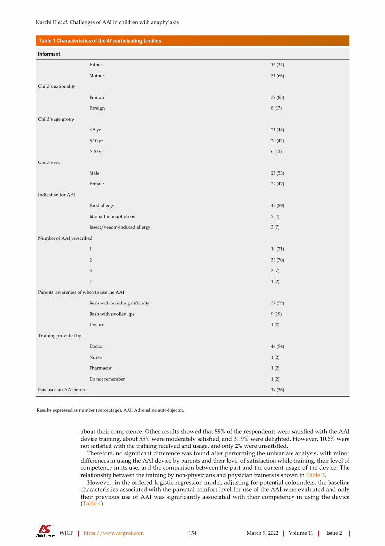
Narchi H et al. Challenges of AAI in children with anaphylaxis
WJCP https://www.wjgnet.com 154 March 9, 2022 Volume 11 Issue 2
Table 1 Characteristics of the 47 participating families
Informant
Father 16 (34)
Mother 31 (66)
Child’s nationality
Emirati 39 (83)
Foreign 8 (17)
Child’s age group
< 5 yr 21 (45)
5-10 yr 20 (42)
> 10 yr 6 (13)
Child’s sex
Male 25 (53)
Female 22 (47)
Indication for AAI
Food allergy 42 (89)
Idiopathic anaphylaxis 2 (4)
Insect/venom-induced allergy 3 (7)
Number of AAI prescribed
1 10 (21)
2 33 (70)
3 3 (7)
4 1 (2)
Parents’ awareness of when to use the AAI
Rash with breathing difficulty 37 (79)
Rash with swollen lips 9 (19)
Unsure 1 (2)
Training provided by
Doctor 44 (94)
Nurse 1 (2)
Pharmacist 1 (2)
Do not remember 1 (2)
Has used an AAI before 17 (36)
Results expressed as number (percentage). AAI: Adrenaline auto-injector.
about their competence. Other results showed that 89% of the respondents were satisfied with the AAI device training, about 55% were moderately satisfied, and 31.9% were delighted. However, 10.6% were not satisfied with the training received and usage, and only 2% were unsatisfied.
Therefore, no significant difference was found after performing the univariate analysis, with minor differences in using the AAI device by parents and their level of satisfaction while training, their level of competency in its use, and the comparison between the past and the current usage of the device. The relationship between the training by non-physicians and physician trainers is shown in Table 3.
However, in the ordered logistic regression model, adjusting for potential cofounders, the baseline characteristics associated with the parental comfort level for use of the AAI were evaluated and only their previous use of AAI was significantly associated with their competency in using the device (Table 4).
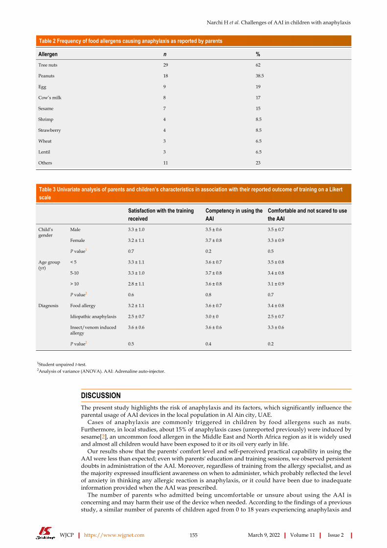
Narchi H et al. Challenges of AAI in children with anaphylaxis
WJCP https://www.wjgnet.com 155 March 9, 2022 Volume 11 Issue 2
Table 2 Frequency of food allergens causing anaphylaxis as reported by parents
Allergen n %
Tree nuts 29 62
Peanuts 18 38.5
Egg 9 19
Cow’s milk 8 17
Sesame 7 15
Shrimp 4 8.5
Strawberry 4 8.5
Wheat 3 6.5
Lentil 3 6.5
Others 11 23
Table 3 Univariate analysis of parents and children’s characteristics in association with their reported outcome of training on a Likert scale
Satisfaction with the training received
Competency in using the AAI
Comfortable and not scared to use the AAI
Male 3.3 ± 1.0 3.5 ± 0.6 3.5 ± 0.7
Female 3.2 ± 1.1 3.7 ± 0.8 3.3 ± 0.9
Child’s gender
P value1 0.7 0.2 0.5
< 5 3.3 ± 1.1 3.6 ± 0.7 3.5 ± 0.8
5-10 3.3 ± 1.0 3.7 ± 0.8 3.4 ± 0.8
> 10 2.8 ± 1.1 3.6 ± 0.8 3.1 ± 0.9
Age group (yr)
P value2 0.6 0.8 0.7
Food allergy 3.2 ± 1.1 3.6 ± 0.7 3.4 ± 0.8Diagnosis
Idiopathic anaphylaxis 2.5 ± 0.7 3.0 ± 0 2.5 ± 0.7
Insect/venom induced allergy
3.6 ± 0.6 3.6 ± 0.6 3.3 ± 0.6
P value2 0.5 0.4 0.2
1Student unpaired t-test.2Analysis of variance (ANOVA). AAI: Adrenaline auto-injector.
DISCUSSIONThe present study highlights the risk of anaphylaxis and its factors, which significantly influence the parental usage of AAI devices in the local population in Al Ain city, UAE.
Cases of anaphylaxis are commonly triggered in children by food allergens such as nuts. Furthermore, in local studies, about 15% of anaphylaxis cases (unreported previously) were induced by sesame[2], an uncommon food allergen in the Middle East and North Africa region as it is widely used and almost all children would have been exposed to it or its oil very early in life.
Our results show that the parents' comfort level and self-perceived practical capability in using the AAI were less than expected; even with parents' education and training sessions, we observed persistent doubts in administration of the AAI. Moreover, regardless of training from the allergy specialist, and as the majority expressed insufficient awareness on when to administer, which probably reflected the level of anxiety in thinking any allergic reaction is anaphylaxis, or it could have been due to inadequate information provided when the AAI was prescribed.
The number of parents who admitted being uncomfortable or unsure about using the AAI is concerning and may harm their use of the device when needed. According to the findings of a previous study, a similar number of parents of children aged from 0 to 18 years experiencing anaphylaxis and

Narchi H et al. Challenges of AAI in children with anaphylaxis
WJCP https://www.wjgnet.com 156 March 9, 2022 Volume 11 Issue 2
Table 4 Association between parents’ reported outcome of training and baseline characteristics in an ordered logistic regression model
Satisfaction with the training received Competence in using the AAI Comfortable and not scared to use the AAI
Informant 0.7 (-5.6,2.1); 0.2 -0.9 (-2.2, 0.4); 0.2 -0.4 (-1.7, 0.8); 0.4
Nationality -0.5 (-2.1, 1.0); 0.5 0.5 (-1.1, 2.1); 0.5 -0.1 (-1.6, 1.3); 0.8
Age group -0.5 (-1.4, 0.4), 0.3 0.1 (-0.8, 1.1); 0.7 -0.3 (-1.2, 0.6); 0.5
Gender -0.2 ((-1.4, 1.0); 0.7 -0.2 (-1.4, 1.0); 0.7 0.7 (-0.4, 1.9); 0.2
Diagnosis 0.2 (-0.9, 1.4); 0.6 -0.4 (-1.5, 0.8); 0.5 -0.4 (-1.6, 0.7); 0.5
Training provider 0.4 (-2.2, 3.0); 0.7 -16.4 (-4320, 4287); 0.9 -1.2 (-4.0, 1.5); 0.3
Has used AAI before 0.3 (-0.9, 1.6); 0.6 1.6 (0.2, 2.9); 0.02 0.1 (-1.1, 1.4); 0.8
AAI: Adrenaline auto-injector.
Figure 1 Parental responses regarding the outcome of training they received on how to use an adrenaline auto-injector.
food allergies used an AAI device. Furthermore, 75% of participants displayed proper technique and abilities for using the auto-injector device[9].
Considering the anxiety and psychological effect of having a child with a food allergy, and as many of the respondents were mothers, it is possible that the psychological effect of anxiety and stress encountered by these mothers affected mental function reflected in the lack of confidence in using the AAI despite satisfaction with the training received. The effect of having a child with a food allergy and potential anaphylaxis in limiting the mental capacity in making sound decisions regarding the child's care has been well documented in the literature[10], and it may even outweigh the stress associated with having a child experiencing diabetes mellitus type I and other chronic diseases[9].
The highlighted difficulties associated with training demonstrate that more effort is needed to sufficiently train parents in anaphylaxis management of their children by providing them with a quality-training session in a calm and friendly atmosphere aided by audio-visual and tactile experiences using the trainer AAI device in the training sessions. Although parents did not admit to suffering undefined psychosomatic factors undermining their use of the device, it has been shown that it is not information or earlier anaphylaxis experience that increases the ease of parents in using the AAI. We deduce that authorisation together with continuous education on the use of the device and preventing anxiety correlate with parents' level of comfort[10]. Therefore, to address these issues, caregivers and parents of these children should be encouraged to practice with an outdated AAI on objects such as an orange, apple, and cardboard instead of discarding them as this type of practice may help improve their level of confidence.
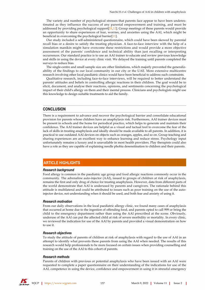
Narchi H et al. Challenges of AAI in children with anaphylaxis
WJCP https://www.wjgnet.com 157 March 9, 2022 Volume 11 Issue 2
The variety and number of psychological stresses that parents face appear to have been underes-timated as they influence the success of any parental empowerment and training, and must be addressed by providing psychological support[11,12]. Group meetings of these parents would provide an opportunity to share experiences of fear, worries, and anxieties using the AAI, which might be beneficial in overcoming the psychological barrier[11].
Our study included a self-administered questionnaire, which could have been skewed by parental recall bias or a desire to satisfy the treating physician. A face-to-face interview with the help of a simulation manikin might have overcome these restrictions and would provide a more objective assessment of the parents' confidence and technical ability than just recalling or interpreting occurrences. Our standard practice is to use an AAI trainer to educate and review previous knowledge and skills in using the device at every clinic visit. We delayed the training until parents completed the surveys to reduce bias.
The single-centre and small sample size are other limitations, which majorly prevented the generaliz-ability of the findings to our local community in our city or the UAE. More extensive multicentre research involving other local paediatric clinics would have been beneficial to address such constraints.
Qualitative research, including face-to-face interviews, will be required to better understand the parents' attitudes and beliefs in controlling allergic reactions in their children. The goal would be to elicit, document, and analyse their reactions, opinions, and sentiments concerning the psychological impact of their child's allergy on them and their mental process. Clinicians and psychologists might use this knowledge to design suitable treatments to aid the family.
CONCLUSIONThere is a requirement to advance and recover the psychological barrier and consolidate educational provision for parents whose children have an anaphylaxis risk. Furthermore, AAI trainer devices must be present in schools and the home for periodical practice, which helps to generate and maintain their confidence. The AAI trainer devices are helpful as a visual and tactual tool to overcome the fear of the lack of skills in treating anaphylaxis and ideally should be made available to all parents. In addition, it is practical to use outdated AAI devices on objects such as oranges, apples, and so on. Group teaching and sharing experiences are an excellent way to enhance learning and reduce stress. Psychology input unfortunately remains a luxury and is unavailable in most health providers. Play therapists could also have a role as they are capable of explaining needle phobia desensitisation to children and their parents.
ARTICLE HIGHLIGHTSResearch backgroundFood allergy is common in the paediatric age group and food allergic reactions commonly occur in the community. The adrenaline auto-injector (AAI), issued to groups of children at risk of anaphylaxis, remains the first and only drug of choice for treating anaphylaxis. However, data from different parts of the world demonstrate that AAI is underused by parents and caregivers. The rationale behind this attitude is multilateral and could be attributed to issues such as poor training on the use of the auto-injector device, not understanding when it should be used, and both fear and anxiety of using it.
Research motivationFrom our daily observations in the local paediatric allergy clinic, we found many cases of anaphylaxis that occurred at home due to the ingestion of offending food, and parents opted to call 999 or bring the child to the emergency department rather than using the AAI prescribed at the scene. Obviously, underuse of the AAI can put the affected child at risk of severe morbidity or mortality. In every clinic, we reviewed the indication for use of the AAI by parents and provided a visual demonstration on how to use it.
Research objectivesTo study the attitude of parents of children at risk of anaphylaxis with regard to the use of AAI in an attempt to identify what prevents these parents from using the AAI when needed. The results of this research would help professionals to be more focused on certain issues when providing counselling and training on the use of the AAI to this cohort of parents.
Research methodsParents of children with previous or potential anaphylaxis who have been issued with an AAI were requested to complete a paper questionnaire on their understanding of the indications for use of the AAI, competence in using the device, confidence and empowerment in using it in stressful emergency
Narchi H et al. Challenges of AAI in children with anaphylaxis
WJCP https://www.wjgnet.com 158 March 9, 2022 Volume 11 Issue 2
situations.
Research resultsThe vast majority of parents admitted receiving good and informative training on using the device and demonstrated good knowledge on its indications. However, that was not enough to provide them with the confidence and courage to use the device due to other factors such as anxiety, fear, or not wanting to hurt the child with the AAI needle. Psychological uneasiness in using the device can limit parents’ ability to use it.
Research conclusionsIn addition to routine training in these groups of parents on the indication of using the AAI and the technique on how to use it, health professionals need to pay attention to the psychological factor which could prevent these parents from underusing the device when needed. Psychological, behavioural therapy and needle phobia desensitisation would help to overcome the barriers of phobia and anxiety which could interfere with sound decision-making in the treatment of their children in emergency situations.
Research perspectivesParent training on the use of AAI should be structured and focused. Audio-visual tools should be available in the clinic to help with training. However, the fear factor and the psychological status of these parents should not be overlooked. A routine referral or referral of selected cases to the local psychological service should be accessible to these parents. Play therapists can also have an important role in both children and parents by reducing needle phobia when present.
ACKNOWLEDGEMENTSWe are indebted to the parents who participated in the study by sharing their experiences and feelings.
FOOTNOTESAuthor contributions: Elghoudi A designed the research protocol, sought ethical approval, collected the data, and was involved in writing the manuscript; Narchi H conducted the statistical analyses, constructed the tables and the graph, and participated in writing the manuscript; Dhaheri KA participated in data collection and writing the manuscript; all authors read and approved the final manuscript.
Institutional review board statement: The study was reviewed and approved by Al Ain Hospital Ethical Research Committee Review board (reference number AAHHEC-01-20-001).
Institutional animal care and use committee statement: No animals were involved in the study.
Informed consent statement: Signed informed consent was obtained from all parents who participated in the study.
Conflict-of-interest statement: The authors declare that they have no conflicts of interest to disclose.
Data sharing statement: The anonymised data can be obtained from the principal investigator ([email protected]) upon reasonable request.
Open-Access: This article is an open-access article that was selected by an in-house editor and fully peer-reviewed by external reviewers. It is distributed in accordance with the Creative Commons Attribution NonCommercial (CC BY-NC 4.0) license, which permits others to distribute, remix, adapt, build upon this work non-commercially, and license their derivative works on different terms, provided the original work is properly cited and the use is non-commercial. See: http://creativecommons.org/Licenses/by-nc/4.0/
Country/Territory of origin: United Arab Emirates
ORCID number: Hassib Narchi 0000-0001-6258-2657; Ahmed Elghoudi 0000000290786152; Klithem Al Dhaheri 0000-0001-8610-6268.
S-Editor: Wang LL L-Editor: Webster JR P-Editor: Wang LL
Narchi H et al. Challenges of AAI in children with anaphylaxis
WJCP https://www.wjgnet.com 159 March 9, 2022 Volume 11 Issue 2
REFERENCESLeung DS, HA; Geha, RS; Szefler, SJ. Pediatric Allergy: Principles and Practice. St. Louis: Mosby, 20031 Al-Hammadi S, Al-Maskari F, Bernsen R. Prevalence of food allergy among children in Al-Ain city, United Arab Emirates. Int Arch Allergy Immunol 2010; 151: 336-342 [PMID: 19851075 DOI: 10.1159/000250442]
2
Wang C, Lin W, Wang Y, Fu L. Suppression of Hippo Pathway by Food Allergen Exacerbates Intestinal Epithelia Instability and Facilitates Hypersensitivity. Mol Nutr Food Res 2021; 65: e2000593 [PMID: 33245584 DOI: 10.1002/mnfr.202000593]
3
Muraro A, Roberts G, Worm M, Bilò MB, Brockow K, Fernández Rivas M, Santos AF, Zolkipli ZQ, Bellou A, Beyer K, Bindslev-Jensen C, Cardona V, Clark AT, Demoly P, Dubois AE, DunnGalvin A, Eigenmann P, Halken S, Harada L, Lack G, Jutel M, Niggemann B, Ruëff F, Timmermans F, Vlieg-Boerstra BJ, Werfel T, Dhami S, Panesar S, Akdis CA, Sheikh A; EAACI Food Allergy and Anaphylaxis Guidelines Group. Anaphylaxis: guidelines from the European Academy of Allergy and Clinical Immunology. Allergy 2014; 69: 1026-1045 [PMID: 24909803 DOI: 10.1111/all.12437]
4
Simons FE, Peterson S, Black CD. Epinephrine dispensing patterns for an out-of-hospital population: a novel approach to studying the epidemiology of anaphylaxis. J Allergy Clin Immunol 2002; 110: 647-651 [PMID: 12373275 DOI: 10.1067/mai.2002.127860]
5
Curtis C, Stukus D, Scherzer R. Epinephrine preparedness in pediatric patients with food allergy: an ideal time for change. Ann Allergy Asthma Immunol 2014; 112: 560-562 [PMID: 24860922 DOI: 10.1016/j.anai.2014.04.009]
6
Simons FE. First-aid treatment of anaphylaxis to food: focus on epinephrine. J Allergy Clin Immunol 2004; 113: 837-844 [PMID: 15131564 DOI: 10.1016/j.jaci.2004.01.769]
7
Hourihane JO, Kilburn SA, Dean P, Warner JO. Clinical characteristics of peanut allergy. Clin Exp Allergy 1997; 27: 634-639 [PMID: 9208183]
8
Avery NJ, King RM, Knight S, Hourihane JO. Assessment of quality of life in children with peanut allergy. Pediatr Allergy Immunol 2003; 14: 378-382 [PMID: 14641608 DOI: 10.1034/j.1399-3038.2003.00072.x]
9
Esenboga S, Ocak M, Cetinkaya PG, Sahiner UM, Soyer O, Buyuktiryaki B, Sekerel BE. Physicians prescribe adrenaline autoinjectors, do parents use them when needed? Allergol Immunopathol (Madr) 2020; 48: 3-7 [PMID: 31611040 DOI: 10.1016/j.aller.2019.07.009]
10
Kim JS, Sinacore JM, Pongracic JA. Parental use of EpiPen for children with food allergies. J Allergy Clin Immunol 2005; 116: 164-168 [PMID: 15990790 DOI: 10.1016/j.jaci.2005.03.039]
11
Knibb R, Halsey M, James P, du Toit G, Young J. Psychological services for food allergy: The unmet need for patients and families in the United Kingdom. Clin Exp Allergy 2019; 49: 1390-1394 [PMID: 31454459 DOI: 10.1111/cea.13488]
12
WJCP https://www.wjgnet.com 160 March 9, 2022 Volume 11 Issue 2
World Journal of
Clinical PediatricsW J C PSubmit a Manuscript: https://www.f6publishing.com World J Clin Pediatr 2022 March 9; 11(2): 160-172
DOI: 10.5409/wjcp.v11.i2.160 ISSN 2219-2808 (online)
ORIGINAL ARTICLE
Observational Study
Functional constipation in Bangladeshi school aged children: A hidden misty at community
Md Benzamin, ASM Bazlul Karim, Md Rukunuzzaman, Md Wahiduzzaman Mazumder, Masud Rana, Rubaiyat Alam, Mohammad Majharul Islam, Md Shafiul Alam, Kamal Hossen, Afsana Yasmin, Kaniz Fathema, Mukesh Khadga, Aisharza Sultana Aishy
Specialty type: Gastroenterology and hepatology
Provenance and peer review: Unsolicited article; Externally peer reviewed.
Peer-review model: Single blind
Peer-review report’s scientific quality classificationGrade A (Excellent): 0 Grade B (Very good): 0 Grade C (Good): C Grade D (Fair): 0 Grade E (Poor): 0
P-Reviewer: Wang FC
Received: April 22, 2021 Peer-review started: April 22, 2021 First decision: May 24, 2021 Revised: June 2, 2021 Accepted: January 5, 2022 Article in press: January 5, 2022 Published online: March 9, 2022
Md Benzamin, ASM Bazlul Karim, Md Rukunuzzaman, Md Wahiduzzaman Mazumder, Rubaiyat Alam, Md Shafiul Alam, Kamal Hossen, Afsana Yasmin, Kaniz Fathema, Mukesh Khadga, Department of Paediatric Gastroenterology and Nutrition, Bangabandhu Shiekh Mujib Medical University, Dhaka 1000, Bangladesh
Masud Rana, Department of Outpatient, Hazi Asmot Medical Centre, Bhairab 2350, Bangladesh
Mohammad Majharul Islam, Department of Paediatric Nephrology, Bangabandhu Shiekh Mujib Medical University, Dhaka 1000, Bangladesh
Aisharza Sultana Aishy, Jalalabad Ragib Rabeya Medical College, Sylhet 3100, Bangladesh
Corresponding author: Md Benzamin, MBBS, MD, Doctor, Pediatric Gastroenterology Fellow, Department of Paediatric Gastroenterology and Nutrition, Bangabandhu Shiekh Mujib Medical University, Shahbag, Dhaka 1000, Bangladesh. [email protected]
AbstractBACKGROUND Constipation is a common problem in children and a frequent cause of hospital visit in both primary & specialized care, which needs proper evaluation & management. Presentation of constipation is variable among children. In Bangladesh there has been no published data regarding constipation in community among school aged children.
AIM To determine the magnitude of functional constipation and its risk factors in community among Bangladeshi school children.
METHODS This descriptive cross sectional study was conducted in different schools of Dhaka division, Bangladesh. All school aged children between 5-16 years of age who attended school were included in this study. Samples were collected randomly. Proper clinical history & physical examinations (without digital rectal exam-ination) & available investigations (if done previously) were recorded. Diagnosis of functional constipation was done by Rome IV criteria and was compared with children without constipation. Children with any red flag sign, known chronic
Benzamin M et al. Functional constipation in Bangladeshi school aged children
WJCP https://www.wjgnet.com 161 March 9, 2022 Volume 11 Issue 2
disease or any findings suggestive of organic disease and on treatment of constipation were excluded. Statistical analysis of the results was done by using Windows based software device with Statistical Packages for Social Science 20. For all statistical tests, P value of less than 0.05 was considered as statistically significant.
RESULTS Total study populations were 707 and male was 443 and female 264. Among them, 134 (19%) children had constipation. In constipated children, 78 children fulfilled the Rome IV criteria for functional constipation and it was 11% of total population. Mean age of children having functional constipation was 11.24 ± 3.54 years and Male female ratio was 1:1.78. Anorexia, nausea, abdominal pain, hard stool, blood with hard stool, alternate hard and loose stool and fecal mass in left iliac fossa were analyzed between two group and all were significantly higher in children with functional constipation group. Children of school, where toilet numbers were inadequate had 2.5 times more constipation risk in comparison to children of school with adequate toilet number (OR = 2.493, 95%CI: 1.214-5.120). Children who feel embarrassed to use toilet at school, had 3.6 times higher risk of constipation (OR = 3.552, 95%CI: 1.435-8.794). Here children with H/O affected sibs and parents/grandparents had 4 and 2.6 times more chance of constipation respectively in comparison to children without H/O affected sibs (OR = 3.977, 95%CI: 1.884–8.397) and parents/grandparents (OR = 2.569, 95%CI: 1.172-5.629). Children with inadequate fluid intake had 2 times more risk of constipation in comparison to children with adequate fluid intake (OR = 1.972, 95%CI: 1.135-3.426). Children who passed electronic screen time of > 2 h/d had 2 times more chance of constipation in comparison to children who passed electronic screen time < 2 h (OR = 2.138, 95%CI: 1.063-4.301).
CONCLUSION Constipation is not uncommon in Bangladeshi school aged children. Inadequate toilet number, family history of constipation, inadequate fluid intake, feeling embarrassed to use toilet at school, and electronic screen time for > 2 h/d were found as risk factors in the present study for functional constipation.
Key Words: Bangladesh; Children; Functional constipation
©The Author(s) 2022. Published by Baishideng Publishing Group Inc. All rights reserved.
Core Tip: The current study is the first population-based study of childhood constipation in Bangladesh. Frequency of constipation and functional constipation was 19% and 11% respectively. Inadequate toilet number, family history of constipation, inadequate fluid intake, feeling embarrassed to use toilet at school, and electronic screen time for > 2 h/d were found as risk factors in the present study for functional constipation. Alternate hard and loose stool as one of the presentation of functional constipation.
Citation: Benzamin M, Karim AB, Rukunuzzaman M, Mazumder MW, Rana M, Alam R, Islam MM, Alam MS, Hossen K, Yasmin A, Fathema K, Khadga M, Aishy AS. Functional constipation in Bangladeshi school aged children: A hidden misty at community. World J Clin Pediatr 2022; 11(2): 160-172URL: https://www.wjgnet.com/2219-2808/full/v11/i2/160.htmDOI: https://dx.doi.org/10.5409/wjcp.v11.i2.160
INTRODUCTIONConstipation is a common problem in children and it is frequently overlooked. Constipation is not a disease; rather, it’s only a symptom. Patients have variable perception regarding constipation, some regard constipation as straining or hard pellet like stool or infrequent defecation or inability to defecate when desire. Constipation is generally defined as infrequent stool, passage of hard stool or both[1]. But North American Society for Pediatric Gastroenterology, Hepatology, and Nutrition (NASPGHAN) defined constipation as delay or difficulty in defecation, present for 2 or more weeks and sufficient to cause significant distress to the patient[2].
Children with constipation quite often visit a general practitioner or pediatrician. These children are also regularly seen on the emergency ward or even admitted to the hospital for treatment. Although functional constipation is not related to mortality but significantly hamper the quality of life. In children constipation may be functional or due to organic causes. In contrast to organic causes, functional
Benzamin M et al. Functional constipation in Bangladeshi school aged children
WJCP https://www.wjgnet.com 162 March 9, 2022 Volume 11 Issue 2
constipation (FC) is not a result of a structural or biochemical abnormality. Constipation due to organic causes may contribute to mortality of patient. In functional constipation, onset of symptoms is within the first year in half of the cases, and the prevalence is highest in 2nd and 4–5 years of age[3]. FC is often not a self-limiting condition: despite treatment, one-third to half of the patients has significant problems after 5 years and symptoms persist into adulthood in approximately 25% of cases[4].
The prevalence of childhood constipation has been documented, with highly variable results from study to study and from country to country, ranging from 1% to 30%[3]. Despite the variations of prevalence in different countries, there is a global trend of increasing rate of childhood constipation, and this increase remains unexplained. The marked socioeconomic, cultural, political and demographic variations that exist between and within the different continents could influence the risk factors and prevalence of childhood FC[5]. The common belief is that constipation is not common in South-Asian countries like India, Bangladesh as diet is rich in fibre here. There are very few studies related to constipation in developing countries specially in South-Asian countries[6].
Most recently Rajindrajith[7] and Khanna et al[8] showed that it is not uncommon in sub-continental countries. On departmental survey in out-patient department of paediatric gastroenterology and nutrition, Bangabandhu Sheikh Mujib Medical University (BSMMU), Dhaka, almost 40% patients presented with constipation. But there is no published data in Bangladesh about childhood constipation. The current study is the first population-based study of childhood constipation in Bangladesh. The present study has been undertaken to observe the clinical profile and risk factors of functional constipation in community among the Bangladeshi school aged children.
MATERIALS AND METHODSThis cross sectional study was conducted at different primary school and high school of Dhaka division of Bangladesh, from August 2018 to July 2019. The inclusion criteria were children of age 5-16 years who attended the school. The exclusion criteria were children already on treatment for constipation and any red flag sign or known chronic disease or symptoms suggestive of disease.
Sampling techniqueA multistage sampling technique was used to select participants. Study place was selected by simple random sampling. Four schools and one madrasa were randomly selected. The schools were then stratified based on location as urban or rural and based on ownership as private or public schools. The participants were selected randomly from different class. Only those students, whose parents gave written consent willingly, were recruited in the study. The detailed clinical history, physical examination findings and investigation reports (if available) were recorded in a predesigned standard data sheet.
History was obtained directly from the students and parents, which included basic demography, age at onset of constipation/symptoms, duration of symptoms, consistency, frequency, volume/size of stool, straining, pain during defecation, bleeding per rectum/blood mixed stool, fecal soiling, abdominal pain, withholding behavior, urinary incontinence/burning urine, history of other sibs/family members affected, detailed family history.
Also history was taken regarding diet pattern (on 3 d recall method), outdoor activity/exercise, any school related condition, social history, past medical and surgical history, history regarding the red flag signs.
Physical examination of all samples was done by researcher himself. The following data were obtained during physical examination: fever, mouth ulcer, abnormal thyroid gland, growth parameters, skin survey, per abdominal examination, tone/reflex of lower limb, spine of vertebra, abdominal distension. Other significant physical findings were also recorded.
Diagnosis of constipation by NASPGHAN and functional constipation was done by Rome IV criteria and if there was red flag sign, organic cause was considered.
Among them who fulfilled the criteria of functional constipation were included in group 1 (children with functional constipation) and others were included in group 2 (children without constipation).
Operational definitionConstipation: NASPGHAN defines constipation as a delay or difficulty in defecation, present for 2 or more weeks and sufficient to cause significant distress to the patient[2]. Functional Constipation: As per Rome IV criteria, functional constipation is defined as presence of at least two of the followings at least once per week for a minimum period of one month: Two or fewer defecations in the toilet per week in a child of a developmental age of at least 4 years; At least one episode of fecal incontinence per week; History of retentive posturing or excessive volitional stool retention; History of painful or hard bowel movements; Presence of a large fecal mass in the rectum; History of large-diameter stools that may obstruct the toilet. The symptoms cannot be fully explained by another medical condition.
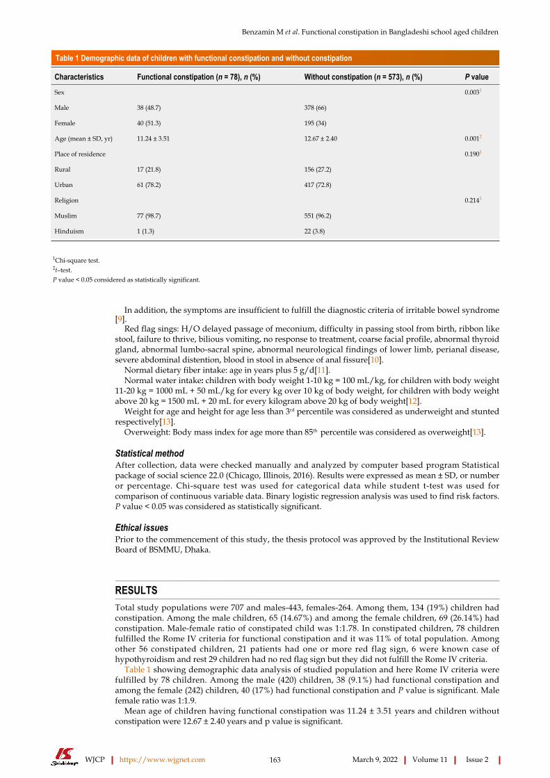
Benzamin M et al. Functional constipation in Bangladeshi school aged children
WJCP https://www.wjgnet.com 163 March 9, 2022 Volume 11 Issue 2
Table 1 Demographic data of children with functional constipation and without constipation
Characteristics Functional constipation (n = 78), n (%) Without constipation (n = 573), n (%) P value
Sex 0.0031
Male 38 (48.7) 378 (66)
Female 40 (51.3) 195 (34)
Age (mean ± SD, yr) 11.24 ± 3.51 12.67 ± 2.40 0.0012
Place of residence 0.1901
Rural 17 (21.8) 156 (27.2)
Urban 61 (78.2) 417 (72.8)
Religion 0.2141
Muslim 77 (98.7) 551 (96.2)
Hinduism 1 (1.3) 22 (3.8)
1Chi-square test.2t–test.P value < 0.05 considered as statistically significant.
In addition, the symptoms are insufficient to fulfill the diagnostic criteria of irritable bowel syndrome[9].
Red flag sings: H/O delayed passage of meconium, difficulty in passing stool from birth, ribbon like stool, failure to thrive, bilious vomiting, no response to treatment, coarse facial profile, abnormal thyroid gland, abnormal lumbo-sacral spine, abnormal neurological findings of lower limb, perianal disease, severe abdominal distention, blood in stool in absence of anal fissure[10].
Normal dietary fiber intake: age in years plus 5 g/d[11].Normal water intake: children with body weight 1-10 kg = 100 mL/kg, for children with body weight
11-20 kg = 1000 mL + 50 mL/kg for every kg over 10 kg of body weight, for children with body weight above 20 kg = 1500 mL + 20 mL for every kilogram above 20 kg of body weight[12].
Weight for age and height for age less than 3rd percentile was considered as underweight and stunted respectively[13].
Overweight: Body mass index for age more than 85th percentile was considered as overweight[13].
Statistical methodAfter collection, data were checked manually and analyzed by computer based program Statistical package of social science 22.0 (Chicago, Illinois, 2016). Results were expressed as mean ± SD, or number or percentage. Chi-square test was used for categorical data while student t-test was used for comparison of continuous variable data. Binary logistic regression analysis was used to find risk factors. P value < 0.05 was considered as statistically significant.
Ethical issuesPrior to the commencement of this study, the thesis protocol was approved by the Institutional Review Board of BSMMU, Dhaka.
RESULTSTotal study populations were 707 and males-443, females-264. Among them, 134 (19%) children had constipation. Among the male children, 65 (14.67%) and among the female children, 69 (26.14%) had constipation. Male-female ratio of constipated child was 1:1.78. In constipated children, 78 children fulfilled the Rome IV criteria for functional constipation and it was 11% of total population. Among other 56 constipated children, 21 patients had one or more red flag sign, 6 were known case of hypothyroidism and rest 29 children had no red flag sign but they did not fulfill the Rome IV criteria.
Table 1 showing demographic data analysis of studied population and here Rome IV criteria were fulfilled by 78 children. Among the male (420) children, 38 (9.1%) had functional constipation and among the female (242) children, 40 (17%) had functional constipation and P value is significant. Male female ratio was 1:1.9.
Mean age of children having functional constipation was 11.24 ± 3.51 years and children without constipation were 12.67 ± 2.40 years and p value is significant.

Benzamin M et al. Functional constipation in Bangladeshi school aged children
WJCP https://www.wjgnet.com 164 March 9, 2022 Volume 11 Issue 2
Table 2 Symptoms analysis of children with functional constipation and without constipation
Characteristics Functional constipation (n = 78), n (%) Without constipation (n = 573), n (%) P value
Anorexia
Yes 35 (44.9) 124 (21.6)
No 43 (55.1) 449 (78.4)
0.0011
Nausea
Yes 17 (21.8) 46 (8)
No 61 (78.2) 527 (92)
0.0011
Abdominal pain
Yes 37 (47.4) 122 (21.3)
No 41 (52.6) 451 (78.7)
0.0011
Hard stool
Yes 63 (80.8) 32 (5.6%)
No 15 (19.2) 541 (94.4)
0.0011
Blood with hard stool
Yes 6 (7.7) 3 (0.5)
No 72 (92.3) 570 (99.5)
0.0011
Alternative hard and loose stool
Yes 22 (28.2) 10 (1.7)
No 56 (71.8) 563 (98.3)
0.0011
Abdominal distension
Yes 0 (0) 78 (100)
No 78 (100) 566 (98.8)
0.5371
Fecal mass in LIF
Yes 12 (15.4) 0 (0%)
No 66 (84.6) 573 (100)
0.0011
1Chi-square test, P value < 0.05 considered as statistically significant.
Residential area and religion of the studied group had no significant influence on constipation.Table 2 showing symptoms analysis of studied population and here anorexia, nausea, abdominal
pain, hard stool, blood with hard stool, alternative hard and loose stool, abdominal distension and fecal mass in left iliac fossa were analyzed between two groups and all were significantly higher in children with functional constipation group except abdominal distension.
Table 3 showing descriptive data of bowel habits of studied group and here defecation frequency at 2 d interval, 3 d interval, incontinence, painful bowel movements, H/O retentive posturing and large diameter stool all were significantly higher in children with functional constipation group.
Table 4 showing the school related factors analysis of studied population and here children with long periods of school, less number toilets at school/dormitory and feel embarrassed to use toilet at school had higher percentage of constipation and p value is significant.
Table 5 showing family related factors analysis of studied population. Here history of constipation in other siblings, history of constipation in parents/grandparents, family size, birth order, parent’s education, household income, single or joint family was considered. But only children having history of constipation in other siblings and history of constipation in parents/grandparents were significant.
Table 6 showing diet related factors analysis of studied population. Here children with less fiber intake and less fluid intake had higher percentage of constipation and p value is significant.
Table 7 showing physical activity related factors analysis of studied group and children who preferred television; mobile watching for more than 2 h per day had higher percentage of constipation and P value is significant.
Table 8 showing Binary logistic regression analysis done for age, sex, residential school, long duration school, toilet number, feeling embarrassed to use toilet, H/O affected sibs and grandparents, fluid and
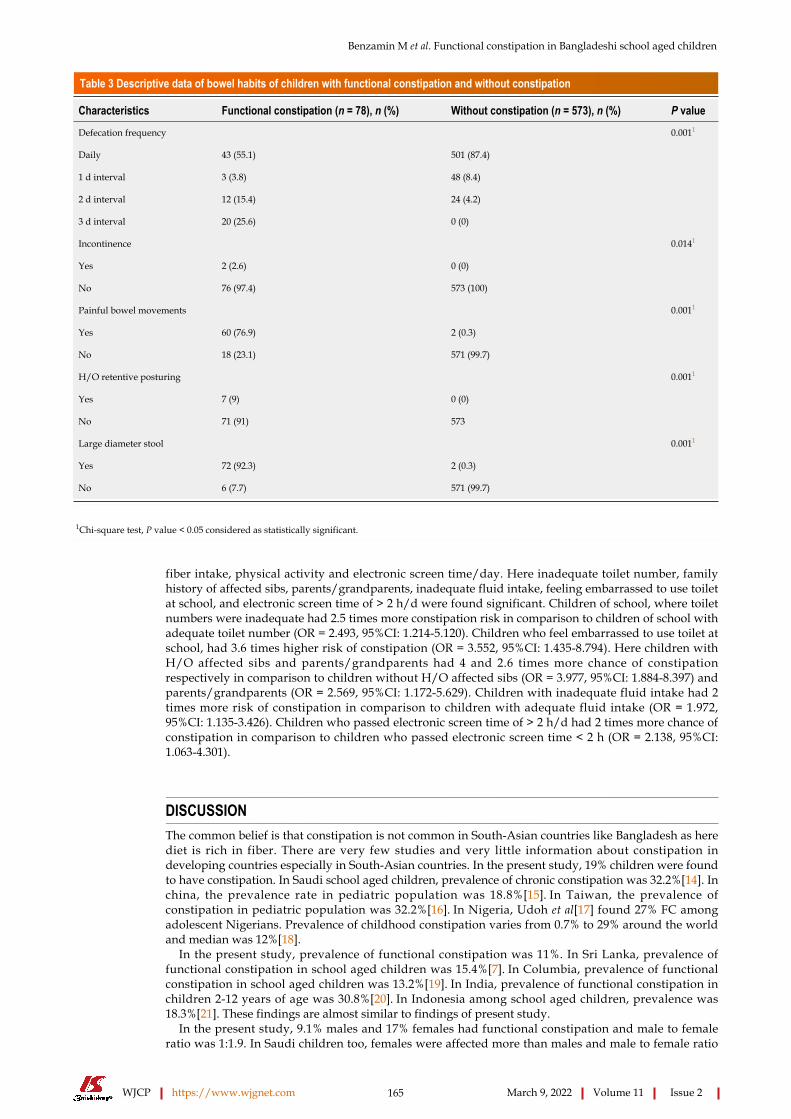
Benzamin M et al. Functional constipation in Bangladeshi school aged children
WJCP https://www.wjgnet.com 165 March 9, 2022 Volume 11 Issue 2
Table 3 Descriptive data of bowel habits of children with functional constipation and without constipation
Characteristics Functional constipation (n = 78), n (%) Without constipation (n = 573), n (%) P value
Defecation frequency
Daily 43 (55.1) 501 (87.4)
1 d interval 3 (3.8) 48 (8.4)
2 d interval 12 (15.4) 24 (4.2)
3 d interval 20 (25.6) 0 (0)
0.0011
Incontinence
Yes 2 (2.6) 0 (0)
No 76 (97.4) 573 (100)
0.0141
Painful bowel movements
Yes 60 (76.9) 2 (0.3)
No 18 (23.1) 571 (99.7)
0.0011
H/O retentive posturing
Yes 7 (9) 0 (0)
No 71 (91) 573
0.0011
Large diameter stool
Yes 72 (92.3) 2 (0.3)
No 6 (7.7) 571 (99.7)
0.0011
1Chi-square test, P value < 0.05 considered as statistically significant.
fiber intake, physical activity and electronic screen time/day. Here inadequate toilet number, family history of affected sibs, parents/grandparents, inadequate fluid intake, feeling embarrassed to use toilet at school, and electronic screen time of > 2 h/d were found significant. Children of school, where toilet numbers were inadequate had 2.5 times more constipation risk in comparison to children of school with adequate toilet number (OR = 2.493, 95%CI: 1.214-5.120). Children who feel embarrassed to use toilet at school, had 3.6 times higher risk of constipation (OR = 3.552, 95%CI: 1.435-8.794). Here children with H/O affected sibs and parents/grandparents had 4 and 2.6 times more chance of constipation respectively in comparison to children without H/O affected sibs (OR = 3.977, 95%CI: 1.884-8.397) and parents/grandparents (OR = 2.569, 95%CI: 1.172-5.629). Children with inadequate fluid intake had 2 times more risk of constipation in comparison to children with adequate fluid intake (OR = 1.972, 95%CI: 1.135-3.426). Children who passed electronic screen time of > 2 h/d had 2 times more chance of constipation in comparison to children who passed electronic screen time < 2 h (OR = 2.138, 95%CI: 1.063-4.301).
DISCUSSIONThe common belief is that constipation is not common in South-Asian countries like Bangladesh as here diet is rich in fiber. There are very few studies and very little information about constipation in developing countries especially in South-Asian countries. In the present study, 19% children were found to have constipation. In Saudi school aged children, prevalence of chronic constipation was 32.2%[14]. In china, the prevalence rate in pediatric population was 18.8%[15]. In Taiwan, the prevalence of constipation in pediatric population was 32.2%[16]. In Nigeria, Udoh et al[17] found 27% FC among adolescent Nigerians. Prevalence of childhood constipation varies from 0.7% to 29% around the world and median was 12%[18].
In the present study, prevalence of functional constipation was 11%. In Sri Lanka, prevalence of functional constipation in school aged children was 15.4%[7]. In Columbia, prevalence of functional constipation in school aged children was 13.2%[19]. In India, prevalence of functional constipation in children 2-12 years of age was 30.8%[20]. In Indonesia among school aged children, prevalence was 18.3%[21]. These findings are almost similar to findings of present study.
In the present study, 9.1% males and 17% females had functional constipation and male to female ratio was 1:1.9. In Saudi children too, females were affected more than males and male to female ratio

Benzamin M et al. Functional constipation in Bangladeshi school aged children
WJCP https://www.wjgnet.com 166 March 9, 2022 Volume 11 Issue 2
Table 4 School related factors analysis of children with functional constipation and without constipation
Characteristics Functional constipation (n = 78), n (%) Without constipation (n = 573), n (%) P value
Type of school
Govt 50 (64.1) 396 (69.1)
Non Govt 28 (35.9) 177 (30.9)
0.2211
Residential
Yes 32 (41) 187 (32.6)
No 46 (59) 386 (67.4)
0.0911
Long periods of school
Yes 13 (16.7) 166 (29)
No 65 (83.3) 407 (71)
0.0131
Unhygienic toilet
Yes 19 (24.4) 93 (16.2)
No 58 (75.6) 480 (83.8)
0.0561
Toilet number
Adequate 64 (82.1) 523 (91.3)
Inadequate 18 (17.9) 50 (8.7)
0.0131
Feeling embarrassed to use toilet
Yes 9 (11.5) 31 (5.4)
No 69 (88.5) 542 (94.6)
0.0391
1Chi-square test, P value < 0.05 considered as statistically significant.
was 1:3.5[14]. In India, Kondapalli et al[20] also found female predominance. In China pediatric population with functional constipation, ratio between male and female was 1:1.1[15]. Khanna et al[8] and Roma-Giannikou et al[22] also showed a male preponderance in functional constipation.
In the present study, mean age of children having functional constipation was 11.24 ± 3.54 years. Peralta-Palmezano et al[19] found mean age was 12.3 ± 2.7 years. In the present study, residential area (rural-urban) and religion had no significance association with constipation. But Rajindrajith[7], Udoh et al[17] and Kondapalli et al[20] found prevalence of constipation being higher in children living in urban areas.
Regarding bowel habits of functional constipated (78) children of present study, large diameter stool was found in 92.3%, painful bowel movements in 76.9%, incontinence in 2.6%, retentive posturing in 9% and defecation frequency daily was in 55.1% cases, 1 d interval in 3.8% cases, at 2 d interval in 15.4% cases, 3 d interval in 25.6% cases. Kondapalli et al[20] found, 58.4% of functional constipation children had retentive behavior in the form of abnormal posturing, fecal soiling was present in 44 % of children and 80.1% of children had stool frequency of < 3 per week.
Oswari et al[21], showed withholding behaviour in 68.3%, defecation of less than 3 times per week in 64.6% of subjects and passage of hard stools in 63.4% cases.
The most common symptoms associated with constipation, found were anorexia, nausea, abdominal pain, hard stool, blood with hard stool, alternate hard and loose stool, abdominal distension and fecal mass in left iliac fossa and these findings were analyzed between two groups and all were significantly higher in children with functional constipation group except abdominal distention.
Oswari et al[21], showed abdominal pain, loss of appetite and straining during defecation were associated with constipation. Kondapalli et al[20] also found, abdominal pain as the presenting complaint which was present in 30.6% of children, blood streaked stools in 10.8% children. About 26% of functional constipation children had abdominal pain in the study of Kokkonen et al[23]. Rajindrajith[7] showed, patients with functional constipation had more somatic symptoms than controls.
In the present study, school related factors like government or private school, residential or non-residential school, long periods of school, unhygienic toilet, toilet numbers, feeling embarrassed to use toilet were analyzed, and here children with long periods of school/home works, feel embarrassed to use toilet at school, and inadequate number toilet at school/dormitory had higher percentage of constipation and P value was significant on univariant analysis. But on regression analysis feeling embarrassed to use toilet at school and inadequate number of toilet at school/dormitory was found

Benzamin M et al. Functional constipation in Bangladeshi school aged children
WJCP https://www.wjgnet.com 167 March 9, 2022 Volume 11 Issue 2
Table 5 Family related factors analysis of children with functional constipation and without constipation
Characteristics Functional constipation (n = 78), n (%) Without constipation (n = 573), n (%) P value
History of constipation in other sibling
Yes 18 (23.1) 25 (4.4)
No 60 (76.9) 548 (95.6)
0.0011
History of constipation in parents/grand parents
Yes 17 (21.8) 27 (4.7)
No 61 (78.2) 545 (95.1)
0.0011
Family size
Only child 3 (3.8) 28 (4.9)
2-3 child 46 (59) 284 (49.6)
≥ 4 child 29 (37.2) 261 (45.5)
0.2961
Birth order
Elder 29 (37.2) 209 (36.5)
Youngest 27 (34.6) 182 (31.8)
Other 22 (28.2) 182 (31.8)
0.7941
Mother’s education
Primary 59 (75.6) 426 (74.3)
SSC 11 (14.1) 83(14.5)
HSC 5(6.4) 28 (4.9)
Honors 3 (3.8) 36 (6.3)
0.7971
Father’s education
Primary 53 (67.9) 392 (68.4)
SSC 10 (12.8) 82 (14.3)
HSC 9 (11.5) 43 (7.5)
Honors 6 (7.7) 56 (9.8)
0.6101
Mother’s occupation
Employed 9 (11.5) 73 (12.7)
Housewife 69 (88.5) 500 (87.3)
0.8311
Household income (taka/mo)
< 30000 49 (62.8) 384 (67)
30000-60000 23 (29.5) 131 (22.9)
> 60000 6 (7.7) 58 (10.1)
0.3931
Family status
Single 72 (92.3) 510 (89)
Joint 6 (7.7) 63 (11)
0.2511
1Chi-square test, P value < 0.05 considered as statistically significant.
significant.Children who feel embarrassed to use toilet at school and where toilet number inadequate,
voluntarily hold the defecation reflex. The withholding behavior causes contraction of the external anal sphincter and gluteal and pelvic floor muscles. The fecal mass then moves out of the rectal ampulla and back into the rectosigmoid colon, where the stool becomes harder and larger[24].
Hasosah et al[14], showed cleanliness and the facilities of their school toilets and homework of > 3 h/d as risk factors of FC.

Benzamin M et al. Functional constipation in Bangladeshi school aged children
WJCP https://www.wjgnet.com 168 March 9, 2022 Volume 11 Issue 2
Table 6 Diet related factors analysis of children with functional constipation and without constipation
Characteristics Functional constipation (n = 78), n (%) Without constipation (n = 573), n (%) P value
Cow’s milk intake
Yes 40 (51.3) 301 (52.5)
No 38 (48.7) 272 (47.5)
0.4691
Fiber
Adequate 45 (57.7) 428 (74.7)
Inadequate 38 (42.3) 145 (25.3)
0.0021
Junk foods intake
Yes 26 (33.3) 209 (36.5)
No 52 (66.7) 364 (63.5)
0.3411
Fluid intake
Adequate 37 (47.4) 424 (74)
Inadequate 41 (52.6) 149 (26)
0.0011
1Chi-square test, P value < 0.05 considered as statistically significant.
Table 7 Physical activity related factors analysis of children with functional constipation and without constipation
Characteristics Functional constipation (n = 78), n (%) Without constipation (n = 573), n (%) P value
Games
Outdoor 49 (62.8) 0 (0)
Indoor 29 (37.2) 160 (27.9)
Physical disability 0 (0) 2 (0.3)
0.2161
Electronic screen time
< 1 h 42 (53.8) 410 (71.6)
1-2 h 12 (15.4) 87 (15.2)
> 2 h 24 (30.8) 76 (13.2)
0.0011
1Chi-square test, P value < 0.05 considered as statistically significant.
In the present study, family related factors like, history of constipation in other siblings, history of constipation in parents/grandparents, family size, birth order, parents education, household income, single or joint family were analyzed but only children having history of constipation in other siblings and history of constipation in parents/grandparents were found significant in both univariate and regression analysis.
As family members share the same food and similar life style which may explain familial aggregation of constipation. But there is no scientific explanation for this, but some researchers suggested that there was a significant genetic and familial connection in patients with constipation that might have been exacerbated by environmental factors[25].
Rajindrajith[7] and Dehghani et al[26], showed positive family history of constipation as a risk factor for FC. Rajindrajith[7] and Oswari et al[21], also did not find any association with family size, birth order, parent’s job. But Kilincaslan et al[27] found that maternal education (elementary) and employed mother were risk factors for FC. Kondapalli et al[20] found that 75% of constipated children belonged to nuclear family.
In the present study, diet related factors were analyzed. Here children with less fiber intake and inadequate fluid intake had higher percentage of constipation and p value was significant on univariate analysis. But on regression analysis only inadequate fluid intake was found significant
The normal stool consists of about 70% of water. Comparatively a small change of water content of stool lead to considerable change in consistency, inadequate fluid intake results in hard stool that can be difficult to pass[28,29].
Benzamin M et al. Functional constipation in Bangladeshi school aged children
WJCP https://www.wjgnet.com 169 March 9, 2022 Volume 11 Issue 2
Table 8 Binary logistic regression analysis for risk factors
95%CICharacteristics P value
Lower UpperExp ß
Age 0.051 1.000 1.204 1.097
Sex 0.056 0.985 3.280 1.798
Long duration of school period 0.746 0.415 1.876 0.883
Inadequate number of toilet 0.013 1.214 5.120 2.493
Feeling embarrassed to use toilet 0.006 1.435 8.794 3.552
H/O affect sib 0.001 1.884 8.397 3.977
H/O affect parents/grandparents 0.018 1.172 5.629 2.569
Inadequate fiber intake 0.286 0.403 1.307 0.726
Inadequate fluid intake 0.016 1.135 3.426 1.972
Electronic screen time > 2 h/d 0.033 1.063 4.301 2.138
P value < 0.05 considered as statistically significant.
Wu et al[16] found that constipation was associated with lower intake of vegetables, fruits, soybean products, and eggs. Kondapalli et al[20] showed milk being consumed by 74.8% constipated children, vegetables and fruits intake were inadequate in 75% of children, junk foods in the form of fried items in 46% of children. de Araújo Sant'Anna et al[30] found dietary fiber intake was insufficient in all children and even lower in those with constipation. Olaru et al[31] showed that cow’s milk intake was a risk factor for FC.
In the present study, physical activity related factors were analyzed and children who preferred electronic media more than 2 h/d had higher percentage of constipation and p value was significant on both univariate analysis and regression analysis. Olaru et al[31] found lack of exercise and television watching more than 3 h/d constitutes a risk factor in the occurrence of constipation. Children when watching television and mobile games, they frequently withheld the defecation urge, which initiate the vicious cycle of constipation.
CONCLUSIONFrequency of constipation and functional constipation was 19% and 11% respectively. Inadequate toilet number, family history of constipation, inadequate fluid intake, feeling embarrassed to use toilet at school, and electronic screen time for > 2 h/d were found as risk factors in the present study for functional constipation. A country wide study is recommended to find out actual burden and risk factors of functional constipation in Bangladeshi pediatric population.
ARTICLE HIGHLIGHTSResearch backgroundConstipation is a common problem in children and a frequent cause of hospital visit in both primary & specialized care, which needs proper evaluation & management. Presentation of constipation is variable among children. In Bangladesh there has been no published data regarding constipation in community among school aged children.
Research motivationNo published data or study regarding the magnitude and etiology of functional constipation till date in Bangladesh.
Research objectivesThe present study has been undertaken to determine the magnitude of functional constipation and it’s risk factors in community among Bangladeshi school children.
Benzamin M et al. Functional constipation in Bangladeshi school aged children
WJCP https://www.wjgnet.com 170 March 9, 2022 Volume 11 Issue 2
Research methodsThis descriptive cross sectional study was conducted in different schools of Dhaka division, Bangladesh. All school aged children between 5-16 years of age who attended school were included in this study. Samples were collected randomly. Proper clinical history & physical examinations (without digital rectal exam-ination) & available investigations (if done previously) were recorded. Diagnosis of functional constipation was done by Rome IV criteria and was compared with children without constipation. Children with any red flag sign, known chronic disease or any findings suggestive of organic disease and on treatment of constipation were excluded. Statistical analysis of the results was done by using Windows based software device with Statistical Packages for Social Science 20. For all statistical tests, P value of less than 0.05 was considered as statistically significant.
Research resultsTotal study populations were 707 and male was 443 and female 264. Among them, 134 (19%) children had constipation. In constipated children, 78 children fulfilled the Rome IV criteria for functional constipation and it was 11% of total population. Mean age of children having functional constipation was 11.24 ± 3.54 years and Male female ratio was 1:1.78. Anorexia, nausea, abdominal pain, hard stool, blood with hard stool, alternate hard and loose stool and fecal mass in left iliac fossa were analyzed between two group and all were significantly higher in children with functional constipation group. Children of school, where toilet numbers were inadequate had 2.5 times more constipation risk in comparison to children of school with adequate toilet number (OR = 2.493, 95%CI: 1.214-5.120). Children who feel embarrassed to use toilet at school, had 3.6 times higher risk of constipation (OR = 3.552, 95%CI: 1.435-8.794). Here children with H/O affected sibs and parents/grandparents had 4 and 2.6 times more chance of constipation respectively in comparison to children without H/O affected sibs (OR = 3.977, 95%CI: 1.884-8.397) and parents/grandparents (OR = 2.569, 95%CI: 1.172-5.629). Children with inadequate fluid intake had 2 times more risk of constipation in comparison to children with adequate fluid intake (OR = 1.972, 95%CI: 1.135-3.426). Children who passed electronic screen time of > 2 h/d had 2 times more chance of constipation in comparison to children who passed electronic screen time < 2 h (OR = 2.138, 95%CI: 1.063-4.301).
Research conclusionsFrequency of constipation and functional constipation was 19% and 11% respectively. Inadequate toilet number, family history of constipation, inadequate fluid intake, feeling embarrassed to use toilet at school, and electronic screen time for > 2 h/d were found as risk factors in the present study for functional constipation. A country wide study is recommended to find out actual burden and risk factors of functional constipation in Bangladeshi pediatric population.
Research perspectivesFrequency of constipation in Bangladeshi school children; Frequency of functional constipation (FC) in Bangladeshi school children; Alternate hard and loose stool as one of the presentation of FC; Inadequate toilet number is risk factor for FC.
ACKNOWLEDGEMENTSDr. Mohammad Kamrul Hassan Shabuj, Associate Professor, Department of Neonatology, Bangabandhu Sheikh Mujib Medical University, Bangladesh.
FOOTNOTESAuthor contributions: Benzamin M was the guarantor and designed the study; Rana M, Alam R, Hossen K, Yasmin A, Fathema K, Khadaga M, Aishy AS participated in data collection; Benzamin M and Alam R participated in the acquisition, analysis, and interpretation of the data, and drafted the initial manuscript; Mazumder MW, Rukunuzzaman M and Karim AS revised the article critically for important intellectual content.
Institutional review board statement: Prior to the commencement of this study, the thesis protocol was approved by the Institutional Review Board of BSMMU, Dhaka.
Informed consent statement: Written informed consent for publication was obtained from the parents.
Conflict-of-interest statement: The authors declare they have no conflict of interest.
Data sharing statement: No additional data are available.
STROBE statement: The authors have read the STROBE statement, and the manuscript was prepared and revised
Benzamin M et al. Functional constipation in Bangladeshi school aged children
WJCP https://www.wjgnet.com 171 March 9, 2022 Volume 11 Issue 2
according to the STROBE statement.
Open-Access: This article is an open-access article that was selected by an in-house editor and fully peer-reviewed by external reviewers. It is distributed in accordance with the Creative Commons Attribution NonCommercial (CC BY-NC 4.0) license, which permits others to distribute, remix, adapt, build upon this work non-commercially, and license their derivative works on different terms, provided the original work is properly cited and the use is non-commercial. See: http://creativecommons.org/Licenses/by-nc/4.0/
Country/Territory of origin: Bangladesh
ORCID number: Md Benzamin 0000-0002-8239-6541; ASM Bazlul Karim 0000-0002-8899-9052; Md Rukunuzzaman 0000-0003-0330-5080; Md Wahiduzzaman Mazumder 0000-0001-6947-9572; Masud Rana 0000-0001-5711-9297; Rubaiyat Alam 0000-0002-6140-7571; Mohammad Majharul Islam 0000-0002-6094-7226; Md Shafiul Alam 0000-0003-1119-2055; Kamal Hossen 0000-0002-4122-6829; Afsana Yasmin 0000-0003-2852-3287; Kaniz Fathema 0000-0002-2596-9788; Mukesh Khadga 0000-0001-8966-8389; Aisharza Sultana Aishy 0000-0001-9823-9463.
S-Editor: Zhang H L-Editor: A P-Editor: Zhang H
REFERENCESBrandt LJ, Prather CM, Quigley EM, Schiller LR, Schoenfeld P, Talley NJ. Systematic review on the management of chronic constipation in North America. Am J Gastroenterol 2005; 100 Suppl 1: S5-S21 [PMID: 16008641 DOI: 10.1111/j.1572-0241.2005.50613_2.x]
1
North American Society for Pediatric Gastroenterology, Hepatology and Nutrition. Evaluation and treatment of constipation in children: summary of updated recommendations of the North American Society for Pediatric Gastroenterology, Hepatology and Nutrition. J Pediatr Gastroenterol Nutr 2006; 43: 405-407 [PMID: 16954970 DOI: 10.1097/01.mpg.0000232574.41149.0a]
2
van den Berg MM, Benninga MA, Di Lorenzo C. Epidemiology of childhood constipation: a systematic review. Am J Gastroenterol 2006; 101: 2401-2409 [PMID: 17032205 DOI: 10.1111/j.1572-0241.2006.00771.x]
3
Bongers ME, Benninga MA. Long-term follow-up and course of life in children with constipation. J Pediatr Gastroenterol Nutr 2011; 53 Suppl 2: S55-S56 [PMID: 22470932]
4
Rajindrajith S, Devanarayana NM, Crispus Perera BJ, Benninga MA. Childhood constipation as an emerging public health problem. World J Gastroenterol 2016; 22: 6864-6875 [PMID: 27570423 DOI: 10.3748/wjg.v22.i30.6864]
5
Poddar U. Approach to Constipation in Children. Indian Pediatr 2016; 53: 319-327 [PMID: 27156546 DOI: 10.1007/s13312-016-0845-9]
6
Rajindrajith S. Constipation in children: From misty to understanding. Sri Lanka Journal of Child Health 2014; 43: 121-141 [DOI: 10.4038/sljch.v43i3.7372]
7
Khanna V, Poddar U, Yachha SK. Etiology and clinical spectrum of constipation in Indian children. Indian Pediatr 2010; 47: 1025-1030 [PMID: 20453267 DOI: 10.1007/s13312-010-0175-2]
8
Hyams JS, Di Lorenzo C, Saps M, Shulman RJ, Staiano A, van Tilburg M. Functional Disorders: Children and Adolescents. Gastroenterology 2016 [PMID: 27144632 DOI: 10.1053/j.gastro.2016.02.015]
9
Tabbers MM, DiLorenzo C, Berger MY, Faure C, Langendam MW, Nurko S, Staiano A, Vandenplas Y, Benninga MA; European Society for Pediatric Gastroenterology, Hepatology, and Nutrition; North American Society for Pediatric Gastroenterology. Evaluation and treatment of functional constipation in infants and children: evidence-based recommendations from ESPGHAN and NASPGHAN. J Pediatr Gastroenterol Nutr 2014; 58: 258-274 [PMID: 24345831 DOI: 10.1097/MPG.0000000000000266]
10
Williams CL, Bollella M, Wynder EL. A new recommendation for dietary fiber in childhood. Pediatrics 1995; 96: 985-988 [PMID: 7494677]
11
Holliday MA, Segar WE. The maintenance need for water in parenteral fluid therapy. Pediatrics 1957; 19: 823-832 [PMID: 13431307]
12
Kuczmarski RJ, Ogden CL, Grummer-Strawn LM, Flegal KM, Guo SS, Wei R, Mei Z, Curtin LR, Roche AF, Johnson CL. CDC growth charts: United States. Adv Data 2000; 1-27 [PMID: 11183293]
13
Hasosah M, Alsahafi A, Alghiribi A, Alqarni N, Babatin A, Matrafi A, Alamri A, AlQurashi MA, Atiah N, Sarkhy A. Prevalence, characterization and risk factors of chronic constipation among Saudi children: a cross-sectional study. Int J Adv Res 2018; 6: 1319-1324 [DOI: 10.21474/IJAR01/6986]
14
Chu H, Zhong L, Li H, Zhang X, Zhang J, Hou X. Epidemiology characteristics of constipation for general population, pediatric population, and elderly population in china. Gastroenterol Res Pract 2014; 2014: 532734 [PMID: 25386187 DOI: 10.1155/2014/532734]
15
Wu TC, Chen LK, Pan WH, Tang RB, Hwang SJ, Wu L, Eugene James F, Chen PH. Constipation in Taiwan elementary school students: a nationwide survey. J Chin Med Assoc 2011; 74: 57-61 [PMID: 21354081 DOI: 10.1016/j.jcma.2011.01.012]
16
Udoh EE, Rajindrajith S, Devanarayana NM, Benninga MA. Prevalence and risk factors for functional constipation in adolescent Nigerians. Arch Dis Child 2017; 102: 841-844 [PMID: 28446425 DOI: 10.1136/archdischild-2016-311908]
17
Meyer JC, Mashaba T, Makhele M, Sibanda M. Functional constipation in children. S Afr Pharm J 2017; 84: 51-5718
Benzamin M et al. Functional constipation in Bangladeshi school aged children
WJCP https://www.wjgnet.com 172 March 9, 2022 Volume 11 Issue 2
Peralta-Palmezano JJ, Guerrero-Lozano R. Prevalence of Functional Gastrointestinal Disorders in School Children and Adolescents. Korean J Gastroenterol 2019; 73: 207-212 [PMID: 31030457 DOI: 10.4166/kjg.2019.73.4.207]
19
Kondapalli CS, Gullapalli S. Constipation in children: incidence, causes in relation to diet pattern and psychosocial aspects. Int J Contemp Pediatrics 2018; 5: 6-13 [DOI: 10.18203/2349-3291.ijcp20175055]
20
Oswari H, Alatas FS, Hegar B, Cheng W, Pramadyani A, Benninga MA, Rajindrajith S. Epidemiology of Paediatric constipation in Indonesia and its association with exposure to stressful life events. BMC Gastroenterol 2018; 18: 146 [PMID: 30285647 DOI: 10.1186/s12876-018-0873-0]
21
Roma-Giannikou E, Adamidis D, Gianniou M, Nikolara R, Messaritakis A. Epidemiology of chronic constipation in Greek children. Hellenic Journal of Gastroenterology 1999; 12: 58-62
22
Kokkonen J, Haapalahti M, Tikkanen S, Karttunen R, Savilahti E. Gastrointestinal complaints and diagnosis in children: a population-based study. Acta Paediatr 2004; 93: 880-886 [PMID: 15303801]
23
Philichi L. Management of Childhood Functional Constipation. J Pediatr Health Care 2018; 32: 103-111 [PMID: 29229066 DOI: 10.1016/j.pedhc.2017.08.008]
24
Chan AO, Hui WM, Lam KF, Leung G, Yuen MF, Lam SK, Wong BC. Familial aggregation in constipated subjects in a tertiary referral center. Am J Gastroenterol 2007; 102: 149-152 [PMID: 17037990 DOI: 10.1111/j.1572-0241.2006.00886.x]
25
Dehghani SM, Moravej H, Rajaei E, Javaherizadeh H. Evaluation of familial aggregation, vegetable consumption, legumes consumption, and physical activity on functional constipation in families of children with functional constipation versus children without constipation. Prz Gastroenterol 2015; 10: 89-93 [PMID: 26557939 DOI: 10.5114/pg.2015.48996]
26
Kilincaslan H, Abali O, Demirkaya SK, Bilici M. Clinical, psychological and maternal characteristics in early functional constipation. Pediatr Int 2014; 56: 588-593 [PMID: 24373103 DOI: 10.1111/ped.12282]
27
Müller-Lissner SA, Kamm MA, Scarpignato C, Wald A. Myths and misconceptions about chronic constipation. Am J Gastroenterol 2005; 100: 232-242 [PMID: 15654804 DOI: 10.1111/j.1572-0241.2005.40885.x]
28
Aichbichler BW, Wenzl HH, Santa Ana CA, Porter JL, Schiller LR, Fordtran JS. A comparison of stool characteristics from normal and constipated people. Dig Dis Sci 1998; 43: 2353-2362 [PMID: 9824119 DOI: 10.1023/a:1026699525487]
29
de Araújo Sant'Anna AM, Calçado AC. Constipation in school-aged children at public schools in Rio de Janeiro, Brazil. J Pediatr Gastroenterol Nutr 1999; 29: 190-193 [PMID: 10435657 DOI: 10.1097/00005176-199908000-00016]
30
Olaru C, Diaconescu S, Trandafir L, Gimiga N, Stefanescu G, Ciubotariu G, Burlea M. Some Risk Factors of Chronic Functional Constipation Identified in a Pediatric Population Sample from Romania. Gastroenterol Res Pract 2016; 2016: 3989721 [PMID: 27994619 DOI: 10.1155/2016/3989721]
31
WJCP https://www.wjgnet.com 173 March 9, 2022 Volume 11 Issue 2
World Journal of
Clinical PediatricsW J C PSubmit a Manuscript: https://www.f6publishing.com World J Clin Pediatr 2022 March 9; 11(2): 173-195
DOI: 10.5409/wjcp.v11.i2.173 ISSN 2219-2808 (online)
SYSTEMATIC REVIEWS
Epidemiology and phenotypes of diabetes in children and adolescents in non-European-origin populations in or from Western Pacific region
Steven James, Jayanthi Maniam, Pik-To Cheung, Tatsuhiko Urakami, Julia von Oettingen, Supawadee Likitmaskul, Graham Ogle
Specialty type: Pediatrics
Provenance and peer review: Invited article; Externally peer reviewed.
Peer-review model: Single blind
Peer-review report’s scientific quality classificationGrade A (Excellent): 0 Grade B (Very good): B Grade C (Good): 0 Grade D (Fair): 0 Grade E (Poor): 0
P-Reviewer: Yao K
Received: April 30, 2021 Peer-review started: April 30, 2021 First decision: July 27, 2021 Revised: August 9, 2021 Accepted: January 5, 2022 Article in press: January 5, 2022 Published online: March 9, 2022
Steven James, School of Nursing, Midwifery and Paramedicine, University of the Sunshine Coast, Petrie 4502, Queensland, Australia
Jayanthi Maniam, Graham Ogle, Life for a Child Program, Diabetes NSW & ACT, Glebe 2017, New South Wales, Australia
Pik-To Cheung, Department of Paediatric Endocrinology, Genetics and Metabolism, Virtus Medical Group, Hong Kong, China
Tatsuhiko Urakami, Department of Pediatrics, Nihon University School of Medicine, Tokyo 173-8610, Japan
Julia von Oettingen, Research Institute, McGill University Health Centre, Montreal H4A 3JI, Quebec, Canada
Supawadee Likitmaskul, Siriraj Diabetes Center, Faculty of Medicine, Siriraj Hospital, Mahidol University, Bangkok 10700, Thailand
Corresponding author: Steven James, PhD, RN, Lecturer, School of Nursing, Midwifery and Paramedicine, University of the Sunshine Coast, 1 Moreton Parade, Petrie 4502, Queensland, Australia. [email protected]
AbstractBACKGROUND Type 1 diabetes (T1D) incidence varies substantially between countries/ territories, with most studies indicating increasing incidence. In Western Pacific region (WPR), reported rates are much lower than European-origin populations. In contrast, there are reports of substantial numbers of young people with type 2 diabetes (T2D). A deeper understanding of T1D and T2D in the WPR may illuminate factors important in pathogenesis of these conditions. Furthermore, with varying resources and funding for diabetes treatment in this region, there is a need to more clearly determine the current burden of disease and also any gaps in knowledge.
AIM To compile and summarise published epidemiologic and phenotypic data on
James S et al. Childhood diabetes in WPR
WJCP https://www.wjgnet.com 174 March 9, 2022 Volume 11 Issue 2
childhood diabetes in non-European populations in and from WPR.
METHODS Research articles were systematically searched from PubMed (MEDLINE), Embase, Cochrane library, and gray literature. Primary outcome measures were incidence and prevalence, with secondary measures including phenotypic descriptions of diabetes, including diabetes type categorization, presence of diabetic ketoacidosis (DKA) at onset, autoantibody positivity, C-peptide levels, and human leucocyte antigen phenotype. Extracted data were collected using a customized template. Three hundred and thirty relevant records were identified from 16 countries/territories, with analysis conducted on 265 (80.3%) records published from the year 2000.
RESULTS T1D incidence ranged from < 1-7.3/100000 individuals/year, rates were highest in emigrant/ mixed populations and lowest in South-East Asia, with most countries/territories (71.4%) having no data since 1999. Incidence was increasing in all six countries/territories with data (annual increases 0.5%-14.2%, highest in China). Peak age-of-onset was 10-14 years, with a female case excess. Rate of DKA at onset varied from 19.3%-70%. Pancreatic autoantibodies at diagnosis were similar to European-origin populations, with glutamic acid decarboxylase-65 autoantibody frequency of 44.1%-64.5%, insulinoma-associated 2 autoantibody 43.5%-70.7%, and zinc transporter-8 autoantibody frequency 54.3% (one study). Fulminant T1D also occurs. T2D was not uncommon, with incidence in Japan and one Chinese study exceeding T1D rates. Monogenic forms also occurred in a number of countries.
CONCLUSION T1D is less common, but generally has a classic phenotype. Some countries/ territories have rapidly increasing incidence. T2D is relatively common. Registries and studies are needed to fill many information gaps.
Key Words: Epidemiology; Phenotypes; Diabetes; Children; Adolescents; Western Pacific
©The Author(s) 2022. Published by Baishideng Publishing Group Inc. All rights reserved.
Core Tip: This systematic review found type 1 diabetes (T1D) incidence was generally low in countries/ territories in the Western Pacific region. However, incidence is rising in most countries where this has been studied. Many countries do not have data or data are quite old. Peak age-of-onset was in later childhood. Rates of diabetic ketoacidosis vary but can be quite high (up to 70%). Autoantibody status is generally like European-origin populations. Fulminant and slowly progressive forms of T1D also occur in the region. Of note, type 2 diabetes was sometimes more common in countries than T1D. Establishment of registers will facilitate incidence studies and also define prevalence and mortality, and assist in outcome assessment. Such data will inform quality of care improvements, health professional training, and assist advocacy.
Citation: James S, Maniam J, Cheung PT, Urakami T, von Oettingen J, Likitmaskul S, Ogle G. Epidemiology and phenotypes of diabetes in children and adolescents in non-European-origin populations in or from Western Pacific region. World J Clin Pediatr 2022; 11(2): 173-195URL: https://www.wjgnet.com/2219-2808/full/v11/i2/173.htmDOI: https://dx.doi.org/10.5409/wjcp.v11.i2.173
INTRODUCTIONA diagnosis of diabetes is particularly challenging in young people. An estimated 1.1 million children and adolescents aged < 20 years are estimated to have type 1 diabetes (T1D) globally, with the number with type 2 diabetes (T2D) unknown[1]. Published information on diabetes in this age group is from European origin populations, and yet over half of the global burden is from non-European origin populations.
The commonest form of diabetes in this age group is T1D but other forms do occur[2]. T1D incidence and prevalence varies substantially between countries/territories, with most studies indicating that incidence is increasing at an average of 3%-4%[3], but this appears to be tailoring off in some high-
James S et al. Childhood diabetes in WPR
WJCP https://www.wjgnet.com 175 March 9, 2022 Volume 11 Issue 2
income nations/territories[1].Increasing incidence in countries/territories with previously low rates offer a chance to better
understand the link between genetics and environment in T1D development, especially in countries/territories with little population admixture[1,4]. Additionally, some studies have shown differences in diabetes incidence among migrant populations relative to the native population, which gives further support to the role of environment in T1D causation[5,6].
In the Western Pacific region (WPR), early studies had reported T1D being very rare in young people[4,7], and subsequent reports have shown incidence rates much lower than most European-origin populations[4,8-10]. In contrast, there are reports of substantial numbers of young people with T2D in some countries in the WPR[11,12].
A deeper understanding of both the epidemiology and phenotypes/endotypes of T1D and T2D in non-European populations such as those in WPR may illuminate factors important in pathogenesis of these conditions. Furthermore, with varying resources and funding for diabetes treatment in this region, there is a need to more clearly determine the current burden of disease and also any gaps in knowledge in related epidemiology and phenotypes/endotypes[1].
The objective of this systematic review is to compile and summarise current published epidemiologic and phenotypic data on childhood diabetes in non-European populations in and from the Western Pacific. Primary outcome measures were incidence and prevalence of diabetes in people < 20 years of age. Secondary measures included diabetes type categorisation and phenotype/endotype features including presence of diabetic ketoacidosis (DKA) at diagnosis, pancreatic autoantibody positivity rates, C-peptide levels, and human leucocyte antigen (HLA) phenotypes.
MATERIALS AND METHODSPopulationNon-European populations in and recently emigrated from the WPR.
Inclusion/exclusion criteriaAny relevant published study conducted in one or more of the 37 countries/territories of the Western Pacific, as determined by the World Health Organization[13], extending from the Mongolian steppes in central Asia, east to the Pitcairn Islands in the Pacific Ocean and south to New Zealand. The included countries/territories were Australia, Brunei, Cambodia, Cook Islands, Democratic People’s Republic of Korea, Federated States of Micronesia, Fiji, Guam, Hong Kong, Indonesia, Japan, Kiribati, Laos, Macau, Malaysia, Marshall Islands, Mongolia, Myanmar, Nauru, New Caledonia, New Zealand, Niue, North Korea, Palau, Papua New Guinea, South Korea, Samoa, Singapore, Solomon Islands, Taiwan, Thailand, Timor-Leste, Philippines, Tonga, Tuvalu, Vanuatu and Vietnam. Studies on recent emigrant populations from these countries/territories to others were also included.
Publications were included if they focused on incidence, prevalence, diabetes type, clinical presentation (presence/rate of DKA), pancreatic autoantibody status, and HLA phenotype. Studies that did not include data on at least one of these factors were excluded.
Data from Australia and New Zealand exclusively were only included if children and adolescents < 20 years of age identified as being an Aboriginal and/or Torres Strait Islander, or Maori, respectively.
Studies were of any study design and in any language. There was no restriction on publication date or type.
Types of outcome measures (primary and secondary)Primary outcomes: Incidence and prevalence of T1D, T2D and other forms in children and youth < 20 years in and from the WPR.
Secondary outcomes: Phenotypic descriptions of childhood- and youth-onset diabetes, including diabetes type categorization, the presence of DKA at onset, autoantibody positivity, C-peptide levels, and HLA phenotype.
Search strategy for identification of studiesResearch articles were systematically searched in the following databases: PubMed (MEDLINE), Embase, and the Cochrane library. The search terms below were developed for PubMed and then adapted for other databases. The MeSH terminologies include Diabetes Mellitus, Epidemiology, Diagnosis, Symptoms, and Clinical Chemistry. The search strategy was: (Diabetes Mellitus) AND (Epidemiology OR Diagnosis OR symptom OR antibod* OR autoantibod* OR Ketoacidosis OR clinical chemistry OR HLA) AND (Country) AND (child* OR adolesc*).
For Embase database, the search terminology for “Diabetes Mellitus” was replaced with “insulin dependent diabetes mellitus”.
James S et al. Childhood diabetes in WPR
WJCP https://www.wjgnet.com 176 March 9, 2022 Volume 11 Issue 2
To search the gray literature, we searched the following: (1) ProQuest Dissertations and Theses Global for theses; (2) Citation searching, including reference list searching and forward citation searching in Google Scholar, Scopus and Web of Science Core Collection; and (3) Hand-searched paediatric diabetes conference abstracts not indexed in the above databases: International Society for Pediatric and Adolescent Diabetes (ISPAD, available in Pediatric Diabetes); Pediatric Endocrine Society (PES, available in Hormone Research in Children); European Society for Pediatric Endocrinology (ESPE, available in Hormone Research in Children); Asia Pacific Paediatric Endocrine Society (APPES, abstracts available in member’s area).
For each database, the years searched included the earliest available online year of indexing up to December 2019.
Data extraction and synthesisThe Covidence systematic review platform (Covidence systematic review software, Veritas Health Innovation, Melbourne, Australia. Available at www.covidence.org) was used to assist with data management. Two independent reviewers reviewed the titles and abstract of the identified studies for relevance. The same reviewers independently reviewed the full text of these studies in a first screen to assess if they met inclusion and exclusion criteria. The reasons for excluding articles were recorded in Covidence. Any disagreements or queries were discussed until a consensus was reached. Thereafter, a final list of studies was produced.
The extracted data was collected using a customized template in Microsoft Excel (Microsoft, Redmond, United States). The extracted data included the following: Country/territory, city/region, type of study, year of publication, time period of study, diagnosis criteria used, T1D incidence and/or prevalence, T2D studies, other forms of diabetes, age range distribution, sex distribution, DKA at diagnosis, pancreatic autoantibody test results, and HLA phenotype. Additional information about the derivation of each value was collected to help qualify the data. Descriptive analyses were performed using Excel. A qualitative comparison of the results across the collected variables is the main focus of this review.
A total of 14252 records were identified, downloaded to EndNote version X9 and screened by reading titles and abstracts. Of these, 2924 records were excluded based upon duplication, language, and contents of titles/abstracts indicating they did not meet inclusion criteria. The remaining 11328 full-text articles were assessed for eligibility; their reference lists and citations were searched, and an additional 105 papers identified. Of these records, 11104 did not meet review inclusion criteria, leaving 330 relevant records. The search process and outcomes are summarised in Figure 1.
The 330 papers were from 16 WPR countries/territories (Table 1), with 204 (62.1%) papers from three countries/territories only. These were from China (n = 72), Japan (n = 94) and South Korea (n = 38). Two-hundred and sixty-five (80.3%) of the 330 studies were published in or after the year 2000. Table 1 summarises the number of papers for each variable and other characteristics of the included studies.
RESULTST1DIncidence: Table 2 summarises the 25 studies from ten WPR countries/territories that had information about T1D incidence with data from 2000 or afterwards. Six studies were from China, five from South Korea, two from Thailand and Taiwan, and one each from four other countries/territories. Most studies (n = 18) reported data for youth aged < 15 years, and only 16 had been published within the past decade.
Incidence ranged from < 1 to 7.3 per 100000 individuals per year. An incidence of < 1 per 100000 were reported in four countries: Fiji[14], Indonesia[15], Thailand[16,17] and Papua New Guinea[18]. However in Fiji, the rate in Indo-Fijians was 9.3 times higher than the rate in Native Fijians[14].
T1D rates of approximately two per 100000 were observed in Japan[19], three in South Korea[20,21], four in Hong Kong[22], five in Taiwan[23,24] and seven in mixed population immigrants in the United States[25]. In China, average T1D rates were variable, with rates ranging from 0.7 to 3.1[26-31].
Single-study data looking at changes in incidence rates over time was available from six countries/territories. In China, rates rose 7.4% per annum (pa) in Harbin from 1990-2000[26], 12.0% pa in Zhejiang from 2007-2013[32], 4.4% pa in Beijing from 1995-2010[28], and 14.2% pa in Shanghai from 1997-2011[31]. In South Korea, rates rose by 7.6% pa from 2001-2010[33]. In Hong Kong, data was presented from 2008-2017 in an abstract published in 2018[22], with full data published in 2020[34]. The annual increment between 2008-2017 was 3.5% pa, with authors noting that this was less than the increment of 4.3% in the period 1997-2007. In Taiwan, rates rose 8.7% every two years from 1999-2000 to 2009-2010[23]. In Thailand, incidence almost quadrupled between 1996 and 2005, however, the authors commented that increased diagnosis likely contributed to this[16]. However, in Japan, incidence barely changed from 2005-2010 with a 0.5% pa increase[19].
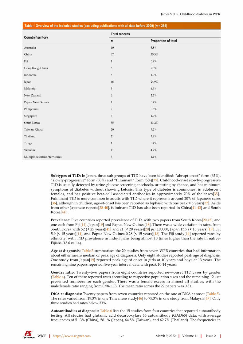
James S et al. Childhood diabetes in WPR
WJCP https://www.wjgnet.com 177 March 9, 2022 Volume 11 Issue 2
Table 1 Overview of the included studies (excluding publications with all data before 2000) (n = 265)
Total recordsCountry/territory
n Proportion of total
Australia 10 3.8%
China 67 25.3%
Fiji 1 0.4%
Hong Kong, China 6 2.3%
Indonesia 5 1.9%
Japan 66 24.9%
Malaysia 5 1.9%
New Zealand 6 2.3%
Papua New Guinea 1 0.4%
Philippines 2 0.8%
Singapore 5 1.9%
South Korea 35 13.2%
Taiwan, China 20 7.5%
Thailand 21 7.9%
Tonga 1 0.4%
Vietnam 11 4.2%
Multiple countries/territories 3 1.1%
Subtypes of T1D: In Japan, three sub-groups of T1D have been identified: “abrupt-onset” form (65%), “slowly-progressive” form (30%) and “fulminant” form (5%)[35]. Childhood-onset slowly-progressive T1D is usually detected by urine-glucose screening at schools, or testing by chance, and has minimum symptoms of diabetes without showing ketosis. This type of diabetes is commonest in adolescent females, and has positive beta-cell associated antibodies in approximately 70% of the cases[35]. Fulminant T1D is more common in adults with T1D where it represents around 20% of Japanese cases[36], although in children, age-of-onset has been reported as biphasic with one peak < 5 years[37]. Aside from other Japanese reports[38-40], fulminant T1D has also been reported in China[41-43] and South Korea[44].
Prevalence: Five countries reported prevalence of T1D, with two papers from South Korea[20,45], and one each from Fiji[14], Japan[19] and Papua New Guinea[18]. There was a wide variation in rates, from South Korea with 52 (< 25 years)[45] and 21 (< 20 years)[20] per 100000, Japan 13.5 (< 15 years)[19], Fiji 5.9 (< 15 years)[14], and Papua New Guinea 0.28 (< 15 years)[18]. The Fiji study[14] reported rates by ethnicity, with T1D prevalence in Indo-Fijians being almost 10 times higher than the rate in native-Fijians (13.6 vs 1.4).
Age at diagnosis: Table 3 summarises the 20 studies from seven WPR countries that had information about either mean/median or peak age of diagnosis. Only eight studies reported peak age of diagnosis. One study from Japan[19] reported peak age of onset in girls at 10 years and boys at 13 years. The remaining nine papers reported five-year interval data with peak 10-14 years.
Gender ratio: Twenty-two papers from eight countries reported new-onset T1D cases by gender (Table 4). Ten of these reported rates according to respective population sizes and the remaining 12 just presented numbers for each gender. There was a female excess in almost all studies, with the male:female ratio ranging from 0.58-1.13. The mean ratio across the 22 papers was 0.81.
DKA at diagnosis: Twenty papers from seven countries reported on the rate of DKA at onset (Table 5). The rates varied from 19.3% in one Taiwanese study[46] to 75.3% in one study from Malaysia[47]. Only three studies had rates below 33%.
Autoantibodies at diagnosis: Table 6 lists the 15 studies from four countries that reported autoantibody testing. All studies had glutamic acid decarboxylase 65 autoantibody (GAD65) data, with average frequencies of 51.3% (China), 58.1% (Japan), 64.5% (Taiwan), and 62.7% (Thailand). The frequencies in
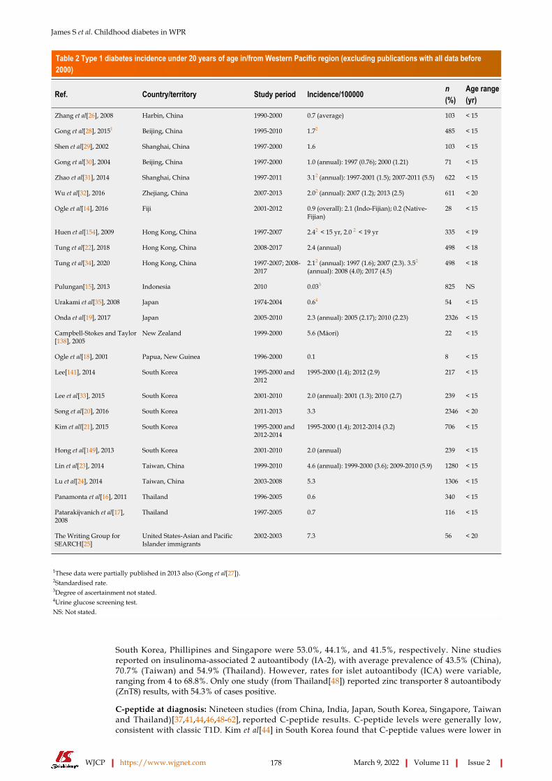
James S et al. Childhood diabetes in WPR
WJCP https://www.wjgnet.com 178 March 9, 2022 Volume 11 Issue 2
Table 2 Type 1 diabetes incidence under 20 years of age in/from Western Pacific region (excluding publications with all data before 2000)
Ref. Country/territory Study period Incidence/100000 n (%)
Age range (yr)
Zhang et al[26], 2008 Harbin, China 1990-2000 0.7 (average) 103 < 15
Gong et al[28], 20151 Beijing, China 1995-2010 1.72 485 < 15
Shen et al[29], 2002 Shanghai, China 1997-2000 1.6 103 < 15
Gong et al[30], 2004 Beijing, China 1997-2000 1.0 (annual): 1997 (0.76); 2000 (1.21) 71 < 15
Zhao et al[31], 2014 Shanghai, China 1997-2011 3.12 (annual): 1997-2001 (1.5); 2007-2011 (5.5) 622 < 15
Wu et al[32], 2016 Zhejiang, China 2007-2013 2.02 (annual): 2007 (1.2); 2013 (2.5) 611 < 20
Ogle et al[14], 2016 Fiji 2001-2012 0.9 (overall): 2.1 (Indo-Fijian); 0.2 (Native-Fijian)
28 < 15
Huen et al[154], 2009 Hong Kong, China 1997-2007 2.42 < 15 yr, 2.0 2 < 19 yr 335 < 19
Tung et al[22], 2018 Hong Kong, China 2008-2017 2.4 (annual) 498 < 18
Tung et al[34], 2020 Hong Kong, China 1997-2007; 2008-2017
2.12 (annual): 1997 (1.6); 2007 (2.3). 3.52 (annual): 2008 (4.0); 2017 (4.5)
498 < 18
Pulungan[15], 2013 Indonesia 2010 0.033 825 NS
Urakami et al[35], 2008 Japan 1974-2004 0.64 54 < 15
Onda et al[19], 2017 Japan 2005-2010 2.3 (annual): 2005 (2.17); 2010 (2.23) 2326 < 15
Campbell-Stokes and Taylor[138], 2005
New Zealand 1999-2000 5.6 (Māori) 22 < 15
Ogle et al[18], 2001 Papua, New Guinea 1996-2000 0.1 8 < 15
Lee[141], 2014 South Korea 1995-2000 and 2012
1995-2000 (1.4); 2012 (2.9) 217 < 15
Lee et al[33], 2015 South Korea 2001-2010 2.0 (annual): 2001 (1.3); 2010 (2.7) 239 < 15
Song et al[20], 2016 South Korea 2011-2013 3.3 2346 < 20
Kim et all[21], 2015 South Korea 1995-2000 and 2012-2014
1995-2000 (1.4); 2012-2014 (3.2) 706 < 15
Hong et al[149], 2013 South Korea 2001-2010 2.0 (annual) 239 < 15
Lin et al[23], 2014 Taiwan, China 1999-2010 4.6 (annual): 1999-2000 (3.6); 2009-2010 (5.9) 1280 < 15
Lu et al[24], 2014 Taiwan, China 2003-2008 5.3 1306 < 15
Panamonta et al[16], 2011 Thailand 1996-2005 0.6 340 < 15
Patarakijvanich et al[17], 2008
Thailand 1997-2005 0.7 116 < 15
The Writing Group for SEARCH[25]
United States-Asian and Pacific Islander immigrants
2002-2003 7.3 56 < 20
1These data were partially published in 2013 also (Gong et al[27]).2Standardised rate.3Degree of ascertainment not stated.4Urine glucose screening test.NS: Not stated.
South Korea, Phillipines and Singapore were 53.0%, 44.1%, and 41.5%, respectively. Nine studies reported on insulinoma-associated 2 autoantibody (IA-2), with average prevalence of 43.5% (China), 70.7% (Taiwan) and 54.9% (Thailand). However, rates for islet autoantibody (ICA) were variable, ranging from 4 to 68.8%. Only one study (from Thailand[48]) reported zinc transporter 8 autoantibody (ZnT8) results, with 54.3% of cases positive.
C-peptide at diagnosis: Nineteen studies (from China, India, Japan, South Korea, Singapore, Taiwan and Thailand)[37,41,44,46,48-62], reported C-peptide results. C-peptide levels were generally low, consistent with classic T1D. Kim et al[44] in South Korea found that C-peptide values were lower in
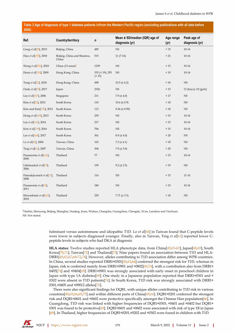
James S et al. Childhood diabetes in WPR
WJCP https://www.wjgnet.com 179 March 9, 2022 Volume 11 Issue 2
Table 3 Age of diagnosis of type 1 diabetes patients in/from the Western Pacific region (excluding publications with all data before 2000)
Ref. Country/territory n Mean ± SD/median (IQR) age of diagnosis (yr)
Age range (yr)
Peak age of diagnosis (yr)
Gong et al[28], 2015 Beijing, China 485 NS < 15 10-14
Huo et al[155], 2018 Beijing, China and Shantou, China
515 11 (7-14) < 21 10-14
Weng et al[156], 2018 China (13 areas)1 1239 NS < 15 10-14
Huen et al[154], 2009 Hong Kong, China 335 (< 19); 293 (< 15)
NS < 19 10-14
Tung et al[22], 2018 Hong Kong, China 498 10.5 (± 4.2) < 18 NS
Onda et al[19], 2017 Japan 2326 NS < 15 13 (boys); 10 (girls)
Lee et al[157], 2006 Singapore 211 7.9 (± 4.0) < 17 NS
Kim et al[21], 2012 South Korea 110 10.6 (± 0.9) < 18 NS
Kim and Kim[158], 2012 South Korea 113 9.26 (± 0.99) < 18 NS
Hong et al[149], 2013 South Korea 239 NS < 15 10-14
Lee et al[141], 2014 South Korea 217 NS < 15 10-14
Kim et al[159], 2016 South Korea 706 NS < 15 10-14
Lee et al[160], 2017 South Korea 361 8.9 (± 4.0) < 20 NS
Lo et al[46], 2004 Taiwan, China 165 7.3 (± 4.1) < 18 NS
Ting et al[61], 2007 Taiwan, China 304 7.9 (± 3.8) < 20 NS
Panamonta et al[161], 2000
Thailand 77 NS < 15 10-14
Likitmaskul et al[79], 2006
Thailand 195 9.2 (± 2.5) < 19 NS
Patarakijvanich et al[17], 2008
Thailand 116 NS < 15 11-14
Panamonta et al[16], 2011
Thailand 340 NS < 15 10-14
Khwanhatai et al[162], 2018
Thailand 229 7.71 (± 3.3) < 18 NS
1Harbin, Shenyang, Beijing, Shanghai, Nanjing, Jinan, Wuhan, Changsha, Guangzhou, Chengdu, Xi’an, Lanzhou and Yinchuan.NS: Not stated.
fulminant versus autoimmune and idiopathic T1D. Lo et al[46] in Taiwan found that C-peptide levels were lower in subjects diagnosed younger. Finally, also in Taiwan, Ting et al[61] reported lower C-peptide levels in subjects who had DKA at diagnosis.
HLA status: Twelve studies reported HLA phenotype data, from China[49,63-67], Japan[68,69], South Korea[70,71], Taiwan[72] and Thailand[73]. Nine papers found an association between T1D and HLA-DRB1[49,63,67,69-72,74]. However, alleles contributing to T1D association differ among WPR countries. In China, several studies reported DRB1*0301[49,63,64] conferred the strongest risk for T1D, whereas in Japan, risk is conferred mainly from DRB1*0901 and *0802[69,74], with a contribution also from DRB1*0405[74] and *0404[69]. DRB1*0901 was strongly associated with early onset in preschool children in Japan with type 1A diabetes[68]. One study in a Japanese population reported that DRB1*0301 and *0302 were absent in T1D patients[74]. In South Korea, T1D risk was strongly associated with DRB1*0301,*0405 and *09012 alleles[70].
There were also significant findings for DQB1, with unique alleles contributing to T1D risk in various countries[49,65,66,69,73] and within different parts of China[49,66]. DQB1*0201 conferred the strongest risk and DQB1*0601 and *0602 were protective specifically amongst the Chinese Han population[66]. In Guangdong, T1D risk was linked with higher frequencies of DQB1*0303, *0401 and *0402 but DQB1*0301 was found to be protective[49]. DQB1*0601 and *0602 were associated with risk of type 1B in Japan[69]. In Thailand, higher frequencies of DQB1*0201,*0202 and *0302 were found in children with T1D.
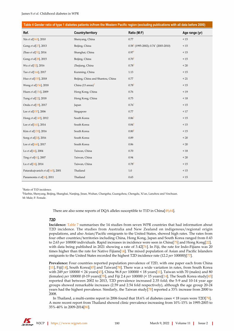
James S et al. Childhood diabetes in WPR
WJCP https://www.wjgnet.com 180 March 9, 2022 Volume 11 Issue 2
Table 4 Gender ratio of type 1 diabetes patients in/from the Western Pacific region (excluding publications with all data before 2000)
Ref. Country/territory Ratio (M:F) Age range (yr)
Xin et al[163], 2010 Shenyang, China 0.77 < 15
Gong et al[27], 2013 Beijing, China 0.581 (1995-2002); 0.741 (2003-2010) < 15
Zhao et al[31], 2014 Shanghai, China 0.971 < 15
Gong et al[28], 2015 Beijing, China 0.701 < 15
Wu et al[32], 2016 Zhejiang, China 0.781 < 20
Tao et al[164], 2017 Kunming, China 1.13 < 15
Huo et al[155], 2018 Beijing, China and Shantou, China 0.77 < 21
Weng et al[156], 2018 China (13 areas)2 0.781 < 15
Huen et al[154], 2009 Hong Kong, China 0.76 < 19
Tung et al[22], 2018 Hong Kong, China 0.75 < 18
Onda et al[19], 2017 Japan 0.761 < 15
Lee et al[157], 2006 Singapore 0.77 < 17
Hong et al[149], 2012 South Korea 0.861 < 15
Lee et al[141], 2014 South Korea 0.841 < 15
Kim et al[159], 2016 South Korea 0.801 < 15
Song et al[20], 2016 South Korea 0.89 < 20
Lee et al[160], 2017 South Korea 0.86 < 20
Lo et al[46], 2004 Taiwan, China 0.70 < 18
Ting et al[61], 2007 Taiwan, China 0.94 < 20
Lu et al[24], 2014 Taiwan, China 0.781 < 15
Patarakujvanich et al[165], 2001 Thailand 1.0 < 15
Panamonta et al[16], 2011 Thailand 0.65 < 15
1Ratio of T1D incidence.2Harbin, Shenyang, Beijing, Shanghai, Nanjing, Jinan, Wuhan, Changsha, Guangzhou, Chengdu, Xi’an, Lanzhou and Yinchuan.M: Male; F: Female.
There are also some reports of DQA alleles susceptible to T1D in China[49,64].
T2DIncidence: Table 7 summarises the 14 studies from seven WPR countries that had information about T2D incidence. The studies from Australia and New Zealand on indigenous/regional origin populations, and also Asian/Pacific emigrants to the United States, showed high rates. The rates from four other countries/territories including China, Hong Kong, Japan and South Korea ranged from 0.43 to 2.63 per 100000 individuals. Rapid increases in incidence were seen in China[75] and Hong Kong[22], with data being published in 2021 showing a rate of 3.42[76]. In Fiji, the rate for Indo-Fijians was 20 times higher than the rate for Native Fijians[14]. The mixed population of Asian and Pacific Islanders emigrants to the United States recorded the highest T2D incidence rate (12.2 per 100000)[77].
Prevalence: Four countries reported population prevalence of T2D, with one paper each from China[11], Fiji[14], South Korea[45] and Taiwan[78]. There was a wide variation in rates, from South Korea with 249 per 100000 < 24 years[45], China 96.8 per 100000 < 18 years[11], Taiwan with 70 (males) and 80 (females) per 100000 (0-19 years)[78], and Fiji 2.4 per 100000 (< 15 years)[14]. The South Korea study[45] reported that between 2002 to 2013, T2D prevalence increased 2.35 fold; the 5-9 and 10-14 year age groups showed remarkable increases (2.59 and 2.54 fold respectively), although the age group 20-24 years had the highest prevalence. Similarly, the Taiwan study[78] reported a 33% increase from 2000 to 2008.
In Thailand, a multi-centre report in 2006 found that 18.6% of diabetes cases < 18 years were T2D[79]. A more recent report from Thailand showed clinic prevalence increasing from 10%-15% in 1995-2003 to 35%-40% in 2009-2014[80].
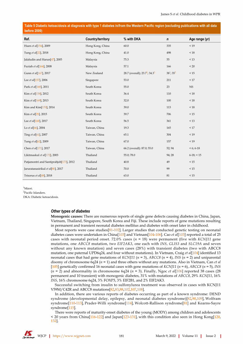
James S et al. Childhood diabetes in WPR
WJCP https://www.wjgnet.com 181 March 9, 2022 Volume 11 Issue 2
Table 5 Diabetic ketoacidosis at diagnosis with type 1 diabetes in/from the Western Pacific region (excluding publications with all data before 2000)
Ref. Country/territory % with DKA n Age range (yr)
Huen et al[154], 2009 Hong Kong, China 60.0 335 < 19
Tung et al[22], 2018 Hong Kong, China 41.0 498 < 18
Jalaludin and Harun[47], 2005 Malaysia 75.3 55 < 13
Fuziah et al[166], 2008 Malaysia 57.1 166 < 20
Gunn et al[167], 2017 New Zealand 28.7 (overall); 23.71; 34.32 381; 352 < 15
Lee et al[157], 2006 Singapore 53.0 211 < 17
Park et al[168], 2011 South Korea 55.0 23 NS
Kim et al[158], 2012 South Korea 36.4 110 < 18
Kim et al[169], 2013 South Korea 32.0 100 < 18
Kim and Kim[170], 2014 South Korea 39.0 113 < 18
Kim et al[21], 2015 South Korea 39.7 706 < 15
Lee et al[160], 2017 South Korea 56.5 361 < 13
Lo et al[46], 2004 Taiwan, China 19.3 165 < 17
Ting et al[61], 2007 Taiwan, China 65.1 304 < 19
Tung et al[62], 2009 Taiwan, China 67.0 157 < 19
Chen et al[171], 2017 Taiwan, China 66.2 (overall): 87.0; 55.0 52; 94 < 6; 6-18
Likitmaskul et al[172], 2003 Thailand 55.0; 78.0 94; 28 6-18; < 15
Patjamontri and Santiprabjob[173], 2012 Thailand 40.8 49 < 15
Jaruratanasirikul et al[80], 2017 Thailand 70.0 99 < 15
Trisorus et al[48], 2018 Thailand 63.0 81 < 15
1Māori.2Pacific Islanders.DKA: Diabetic ketoacidosis.
Other types of diabetesMonogenic causes: There are numerous reports of single gene defects causing diabetes in China, Japan, Vietnam, Thailand, Singapore, South Korea and Fiji. These include reports of gene mutations resulting in permanent and transient neonatal diabetes mellitus and diabetes with onset later in childhood.
Most reports were case studies[81-102]. Larger studies that conducted genetic testing on neonatal diabetes cases were undertaken in China[103] and Vietnam[104-106]. Cao et al[103] reported a total of 25 cases with neonatal period onset. 72.0% cases (n = 18) were permanent (five with KCNJ11 gene mutations, one ABCC8 mutation, two EIF2AK3, one each with INS, GLIS3 and SLC19A and seven without any known mutation) and seven cases (28%) with transient diabetes (two with ABCC8 mutation, one paternal UPD6q24, and four without mutations). In Vietnam, Craig et al[104] identified 13 neonatal cases that had gene mutations of KCNJ11 (n = 3), ABCC8 (n = 4), INS (n = 2) and uniparental disomy of chromosome 6q24 (n = 1) and three others without any mutations. Also in Vietnam, Can et al[105] genetically confirmed 16 neonatal cases with gene mutations of KCNJ11 (n = 6), ABCC8 (n = 5), INS (n = 2) and abnormality in chromosome 6q24 (n = 3). Finally, Ngoc et al[106] reported 38 cases (28 permanent and 10 transient) with monogenic diabetes, 31% with mutations of ABCC8, 29% KCNJ11, 16% INS, 16% chromosome 6q24, 3% FOXP3, 3% EIF2B1, and 2% EIF2AK3.
Successful switching from insulin to sulfonylurea treatment was observed in cases with KCNJ11 V59M/C42R and ABCC8 mutations[82,83,88,102,107,108].
In addition, there are various reports of diabetes occurring as part of a known syndrome: DEND syndrome (developmental delay, epilepsy, and neonatal diabetes syndrome)[82,90,109], Wolfram syndrome[110-113], Prader-Willi syndrome[114], Wolcott-Rallison syndrome[81] and Kearns-Sayre syndrome[115].
There were reports of maturity-onset diabetes of the young (MODY) among children and adolescents < 20 years from China[116-122] and Japan[123-131], with this condition also seen in Hong Kong[120,132].
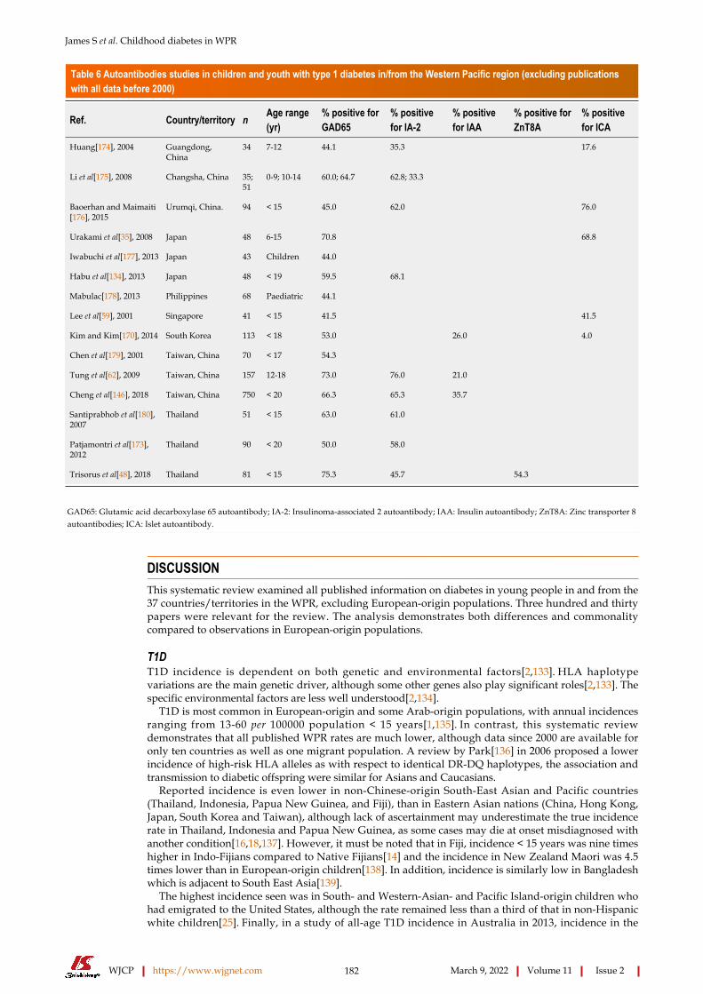
James S et al. Childhood diabetes in WPR
WJCP https://www.wjgnet.com 182 March 9, 2022 Volume 11 Issue 2
Table 6 Autoantibodies studies in children and youth with type 1 diabetes in/from the Western Pacific region (excluding publications with all data before 2000)
Ref. Country/territory n Age range (yr)
% positive for GAD65
% positive for IA-2
% positive for IAA
% positive for ZnT8A
% positive for ICA
Huang[174], 2004 Guangdong, China
34 7-12 44.1 35.3 17.6
Li et al[175], 2008 Changsha, China 35; 51
0-9; 10-14 60.0; 64.7 62.8; 33.3
Baoerhan and Maimaiti[176], 2015
Urumqi, China. 94 < 15 45.0 62.0 76.0
Urakami et al[35], 2008 Japan 48 6-15 70.8 68.8
Iwabuchi et al[177], 2013 Japan 43 Children 44.0
Habu et al[134], 2013 Japan 48 < 19 59.5 68.1
Mabulac[178], 2013 Philippines 68 Paediatric 44.1
Lee et al[59], 2001 Singapore 41 < 15 41.5 41.5
Kim and Kim[170], 2014 South Korea 113 < 18 53.0 26.0 4.0
Chen et al[179], 2001 Taiwan, China 70 < 17 54.3
Tung et al[62], 2009 Taiwan, China 157 12-18 73.0 76.0 21.0
Cheng et al[146], 2018 Taiwan, China 750 < 20 66.3 65.3 35.7
Santiprabhob et al[180], 2007
Thailand 51 < 15 63.0 61.0
Patjamontri et al[173], 2012
Thailand 90 < 20 50.0 58.0
Trisorus et al[48], 2018 Thailand 81 < 15 75.3 45.7 54.3
GAD65: Glutamic acid decarboxylase 65 autoantibody; IA-2: Insulinoma-associated 2 autoantibody; IAA: Insulin autoantibody; ZnT8A: Zinc transporter 8 autoantibodies; ICA: Islet autoantibody.
DISCUSSIONThis systematic review examined all published information on diabetes in young people in and from the 37 countries/territories in the WPR, excluding European-origin populations. Three hundred and thirty papers were relevant for the review. The analysis demonstrates both differences and commonality compared to observations in European-origin populations.
T1DT1D incidence is dependent on both genetic and environmental factors[2,133]. HLA haplotype variations are the main genetic driver, although some other genes also play significant roles[2,133]. The specific environmental factors are less well understood[2,134].
T1D is most common in European-origin and some Arab-origin populations, with annual incidences ranging from 13-60 per 100000 population < 15 years[1,135]. In contrast, this systematic review demonstrates that all published WPR rates are much lower, although data since 2000 are available for only ten countries as well as one migrant population. A review by Park[136] in 2006 proposed a lower incidence of high-risk HLA alleles as with respect to identical DR-DQ haplotypes, the association and transmission to diabetic offspring were similar for Asians and Caucasians.
Reported incidence is even lower in non-Chinese-origin South-East Asian and Pacific countries (Thailand, Indonesia, Papua New Guinea, and Fiji), than in Eastern Asian nations (China, Hong Kong, Japan, South Korea and Taiwan), although lack of ascertainment may underestimate the true incidence rate in Thailand, Indonesia and Papua New Guinea, as some cases may die at onset misdiagnosed with another condition[16,18,137]. However, it must be noted that in Fiji, incidence < 15 years was nine times higher in Indo-Fijians compared to Native Fijians[14] and the incidence in New Zealand Maori was 4.5 times lower than in European-origin children[138]. In addition, incidence is similarly low in Bangladesh which is adjacent to South East Asia[139].
The highest incidence seen was in South- and Western-Asian- and Pacific Island-origin children who had emigrated to the United States, although the rate remained less than a third of that in non-Hispanic white children[25]. Finally, in a study of all-age T1D incidence in Australia in 2013, incidence in the

James S et al. Childhood diabetes in WPR
WJCP https://www.wjgnet.com 183 March 9, 2022 Volume 11 Issue 2
Table 7 Type 2 diabetes incidence in non-European populations in/from the Western Pacific region (excluding publications with all data before 2000)
Ref. Country/territory Study period n Incidence/100000 Age range (yr)
Craig et al[181], 2007 Australia Torres Straits Islands 2001-2006 23 12.7 < 19
Tran et al[182], 2014 Australia, Torres Straits Islands 2001-2008 31 20.7 < 19
Haynes et al[183], 2016 Australia, Torres Straits Islands 1990-2012 76 12.6 < 17
Wu et al[75], 2017 Zhejiang, China 2007-2013 392 1.73 (overall): 0.62 (2007); 3.62 (2013)
< 20
Ogle et al[14], 2016 Fiji 2001-2012 13; 11; 1; 1
0.43 (overall): 1.171; 0.062; 0.703 < 15
Huen et al[154], 2009 Hong Kong, China 1997-2007 198 1.2 < 19
Tung et al[22], 2018; Tung et al[76], 2021
Hong Kong, China 2008-2017; 2008-2017
391; 391 1.9 (3.42) < 18
Urakami et al[184], 2005 Japan 1974-2002 232 2.63 (overall): 1.73 (< 1980); 2.76 (> 1981)
< 16
Urakami et al[148], 2018 Japan 1975-2015 301 2.6 < 16
Campbell-Stokes and Taylor[138], 2005
New Zealand 1999-2000 74 1.78 < 15
Jefferies et al[185], 2012 New Zealand 1995-2007 434,5 3.4 < 15
344 3.3 (overall): 3.4 (1995-2007); 4.0 (2008-2015)
Sjardin et al[186], 2018 New Zealand 1995-2015
475 3.6 (overall): 3.4 (1995-2007); 4.0 (2008-2015)
< 15
Hong et al[149], 2013 South Korea 2001-2010 89 0.76 < 15
Liu et al[77], 2009 United States-Asian and Pacific Islander immigrants
2002-2003 73 12.2 < 15
1Indo-Fijian.2Native-Fijian.3Fijian of European descent.4Māori.5Pacific Islanders.
Aboriginal population was only 70% of that in the non-indigenous population[140], despite the extensive admixture between the two populations. Therefore, in these populations, changes in environment that could potentially increase incidence do not appear to fully overcome the impact of varying genetic susceptibility.
In the absence of large-scale immigration, genetic factors will remain essentially constant. Therefore, any changes in incidence will be due to changing environmental factors. Incidence in European-origin populations has increased by 3%-4% pa in many European-origin populations[135], although this is tailing off now in some countries[3]. There is some evidence that the rate of increase is higher in some lower-incidence countries[3].
The four studies from China[26,30-32] show that T1D incidence is rising quickly (from 4.4%-14.2% pa). South Korea[141] and Taiwan[23] also had high rates of increase at 7.6% and 8.7% pa respectively. However, the rate of rise was slowing in Hong Kong[22] and was virtually zero in Japan[19]. This may be due to evolving environmental factors which then approach a peak effect, as has been seen with slowing or peaking rates in some high-incidence countries[3].
Slowly-progressive diabetes that is clearly T1D is well described from Japan[35], and fulminant T1D (which occurs more in adults and in younger children) is well reported from Japan[36-40], China[41-43] and South Korea[44].
These distinct subtypes, as opposed to acute-onset T1D, do not have exact correlates in European-origin populations, although it is possible that to some extent these represent the more general hetero-geneity of T1D, which is being increasingly recognised[142]. For instance, onset is more rapid in younger European-origin populations[143]. A study done in Western Asia and also in a European-origin population that used identical methodology to assess genotype, phenotype and endotype could help illuminate this and add to global knowledge of T1D pathogenesis.
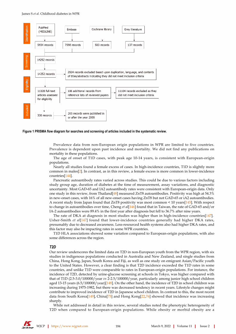
James S et al. Childhood diabetes in WPR
WJCP https://www.wjgnet.com 184 March 9, 2022 Volume 11 Issue 2
Figure 1 PRISMA flow diagram for searches and screening of articles included in the systematic review.
Prevalence data from non-European origin populations in WPR are limited to five countries. Prevalence is dependent upon past incidence and mortality. We did not find any publications on mortality in these populations.
The age of onset of T1D cases, with peak age 10-14 years, is consistent with European-origin populations.
Nearly all studies found a female excess of cases. In high-incidence countries, T1D is slightly more common in males[2]. In contrast, as in this review, a female excess is more common in lower-incidence countries[144].
Pancreatic autoantibody rates varied across studies. This could be due to various factors including study group age, duration of diabetes at the time of measurement, assay variations, and diagnostic uncertainty. Most GAD-65 and IA2 autoantibody rates were consistent with European-origin data. Only one study in this review, from Thailand[48] measured ZnT8 autoantibodies. Positivity was high at 54.3% in new-onset cases, with 16% of all new-onset cases having ZnT8 but not GAD-65 or iA2 autoantibodies. A recent study from Japan found that ZnT8 positivity was most common < 10 years[145]. With respect to change in autoantibodies over time, Cheng et al[146] found that in Taiwan, the rate of GAD-65 and/or IA-2 autoantibodies were 89.4% in the first year after diagnosis but fell to 36.7% after nine years.
The rate of DKA at diagnosis in most studies was higher than in high-incidence countries[147]. Usher-Smith et al[147] found that lower-incidence countries generally had higher DKA rates, presumably due to decreased awareness. Less-resourced health systems also had higher DKA rates, and this factor may also be impacting rates in some WPR countries.
T1D HLA associations showed some variation compared to European-origin populations, with also some differences across the region.
T2D Our review underscores the limited data on T2D in non-European youth from the WPR region, with six studies in indigenous populations conducted in Australia and New Zealand, and single studies from China, Hong Kong, Japan, South Korea and Fiji, as well as one study on emigrant Asian/Pacific youth to the United States. However, a clear finding is that T2D incidence exceeded the T1D rates in some countries, and unlike T1D were comparable to rates in European-origin populations. For instance, the incidence of T2D, detected by urine-glucose screening at schools in Tokyo, was higher compared with that of T1D (2.5-3.0/100000/year vs 2-2.5/100000/year, particularly among junior high school children aged 13-15 years (6.5/100000/year)[148]. On the other hand, the incidence of T2D in school children was increasing during 1975-1982, but there was decreased tendency in recent years. Lifestyle changes might contribute to improved incidence of T2D in Japanese school children. In contrast to this, the most recent data from South Korea[149], China[75] and Hong Kong[22,76] showed that incidence was increasing sharply.
While not addressed in detail in this review, several studies noted the phenotypic heterogeneity of T2D when compared to European-origin populations. While obesity or morbid obesity are a
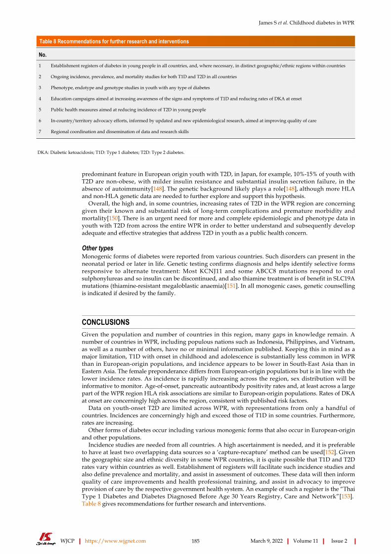
James S et al. Childhood diabetes in WPR
WJCP https://www.wjgnet.com 185 March 9, 2022 Volume 11 Issue 2
Table 8 Recommendations for further research and interventions
No.
1 Establishment registers of diabetes in young people in all countries, and, where necessary, in distinct geographic/ethnic regions within countries
2 Ongoing incidence, prevalence, and mortality studies for both T1D and T2D in all countries
3 Phenotype, endotype and genotype studies in youth with any type of diabetes
4 Education campaigns aimed at increasing awareness of the signs and symptoms of T1D and reducing rates of DKA at onset
5 Public health measures aimed at reducing incidence of T2D in young people
6 In-country/territory advocacy efforts, informed by updated and new epidemiological research, aimed at improving quality of care
7 Regional coordination and dissemination of data and research skills
DKA: Diabetic ketoacidosis; T1D: Type 1 diabetes; T2D: Type 2 diabetes.
predominant feature in European origin youth with T2D, in Japan, for example, 10%-15% of youth with T2D are non-obese, with milder insulin resistance and substantial insulin secretion failure, in the absence of autoimmunity[148]. The genetic background likely plays a role[148], although more HLA and non-HLA genetic data are needed to further explore and support this hypothesis.
Overall, the high and, in some countries, increasing rates of T2D in the WPR region are concerning given their known and substantial risk of long-term complications and premature morbidity and mortality[150]. There is an urgent need for more and complete epidemiologic and phenotype data in youth with T2D from across the entire WPR in order to better understand and subsequently develop adequate and effective strategies that address T2D in youth as a public health concern.
Other typesMonogenic forms of diabetes were reported from various countries. Such disorders can present in the neonatal period or later in life. Genetic testing confirms diagnosis and helps identify selective forms responsive to alternate treatment: Most KCNJ11 and some ABCC8 mutations respond to oral sulphonylureas and so insulin can be discontinued, and also thiamine treatment is of benefit in SLC19A mutations (thiamine-resistant megaloblastic anaemia)[151]. In all monogenic cases, genetic counselling is indicated if desired by the family.
CONCLUSIONSGiven the population and number of countries in this region, many gaps in knowledge remain. A number of countries in WPR, including populous nations such as Indonesia, Philippines, and Vietnam, as well as a number of others, have no or minimal information published. Keeping this in mind as a major limitation, T1D with onset in childhood and adolescence is substantially less common in WPR than in European-origin populations, and incidence appears to be lower in South-East Asia than in Eastern Asia. The female preponderance differs from European-origin populations but is in line with the lower incidence rates. As incidence is rapidly increasing across the region, sex distribution will be informative to monitor. Age-of-onset, pancreatic autoantibody positivity rates and, at least across a large part of the WPR region HLA risk associations are similar to European-origin populations. Rates of DKA at onset are concerningly high across the region, consistent with published risk factors.
Data on youth-onset T2D are limited across WPR, with representations from only a handful of countries. Incidences are concerningly high and exceed those of T1D in some countries. Furthermore, rates are increasing.
Other forms of diabetes occur including various monogenic forms that also occur in European-origin and other populations.
Incidence studies are needed from all countries. A high ascertainment is needed, and it is preferable to have at least two overlapping data sources so a ‘capture-recapture’ method can be used[152]. Given the geographic size and ethnic diversity in some WPR countries, it is quite possible that T1D and T2D rates vary within countries as well. Establishment of registers will facilitate such incidence studies and also define prevalence and mortality, and assist in assessment of outcomes. These data will then inform quality of care improvements and health professional training, and assist in advocacy to improve provision of care by the respective government health system. An example of such a register is the “Thai Type 1 Diabetes and Diabetes Diagnosed Before Age 30 Years Registry, Care and Network”[153]. Table 8 gives recommendations for further research and interventions.
James S et al. Childhood diabetes in WPR
WJCP https://www.wjgnet.com 186 March 9, 2022 Volume 11 Issue 2
ARTICLE HIGHLIGHTSResearch backgroundType 1 diabetes (T1D) incidence varies, with most studies indicating increasing incidence. Reported rates are much lower in the Western Pacific region (WPR), than European-origin populations. Conversely, there are reports of substantial numbers of young people with type 2 diabetes (T2D).
Research motivationA greater understanding of T1D and T2D in the WPR may highlight factors important in pathogenesis of these conditions. There is a need to determine the current burden of disease more clearly and also any gaps in knowledge, in view of varying funding and resources for diabetes treatment in this region.
Research objectivesTo gather and summarise epidemiologic and phenotypic data on childhood diabetes in non-European populations in and from WPR.
Research methodsA comprehensive systematic search of available literature was undertaken.
Research resultsThere are both differences and similarities compared to observations in European-origin populations. T1D was found to be less common, but generally has a classic phenotype. Some countries/territories have rapidly increasing incidence. T2D is relatively common. There are, however, many information gaps.
Research conclusionsGiven the population and number of countries in this region, many gaps in knowledge remain.
Research perspectivesRegistries and studies are needed to fill many information gaps. Establishment of registers will facilitate incidence studies and also define prevalence and mortality, and assist in outcome assessment. Such data will inform quality of care improvements, health professional training, and assist advocacy.
FOOTNOTESAuthor contributions: James S and Maniam J contributed equally to the manuscript; James S, Maniam J and Ogle G co-designed the study; all authors collected/extracted data and contributed to the manuscript.
Conflict-of-interest statement: No conflicts of interest.
PRISMA 2009 Checklist statement: The authors have read the PRISMA 2020 Checklist, and the manuscript was prepared and revised according to the PRISMA 2020 Checklist.
Open-Access: This article is an open-access article that was selected by an in-house editor and fully peer-reviewed by external reviewers. It is distributed in accordance with the Creative Commons Attribution NonCommercial (CC BY-NC 4.0) license, which permits others to distribute, remix, adapt, build upon this work non-commercially, and license their derivative works on different terms, provided the original work is properly cited and the use is non-commercial. See: https://creativecommons.org/Licenses/by-nc/4.0/
Country/Territory of origin: Australia
ORCID number: Steven James 0000-0002-3928-9206; Jayanthi Maniam 0000-0002-1118-0364; Pik-To Cheung 0000-0003-0929-6402; Tatsuhiko Urakami 0000-0001-7976-9557; Julia von Oettingen 0000-0003-0631-1435; Supawadee Likitmaskul 0000-0002-9453-8470; Graham Ogle 0000-0002-2022-0866.
S-Editor: Fan JR L-Editor: A P-Editor: Fan JR
James S et al. Childhood diabetes in WPR
WJCP https://www.wjgnet.com 187 March 9, 2022 Volume 11 Issue 2
REFERENCESInternational Diabetes Federation. IDF Diabetes Atlas. Ninth edition. International Diabetes Federation: Brussels. [cited 22 April 2021]. Available from: https://www.diabetesatlas.org/en/resources/
1
Mayer-Davis EJ, Kahkoska AR, Jefferies C, Dabelea D, Balde N, Gong CX, Aschner P, Craig ME. ISPAD Clinical Practice Consensus Guidelines 2018: Definition, epidemiology, and classification of diabetes in children and adolescents. Pediatr Diabetes 2018; 19 Suppl 27: 7-19 [PMID: 30226024 DOI: 10.1111/pedi.12773]
2
Tuomilehto J, Ogle GD, Lund-Blix NA, Stene LC. Update on Worldwide Trends in Occurrence of Childhood Type 1 Diabetes in 2020. Pediatr Endocrinol Rev 2020; 17: 198-209 [PMID: 32208564 DOI: 10.17458/per.vol17.2020.tol.epidemiologychildtype1diabetes]
3
Chan JC, Cho NH, Tajima N, Shaw J. Diabetes in the Western Pacific Region--past, present and future. Diabetes Res Clin Pract 2014; 103: 244-255 [PMID: 24418400 DOI: 10.1016/j.diabres.2013.11.012]
4
Oilinki T, Otonkoski T, Ilonen J, Knip M, Miettinen PJ. Prevalence and characteristics of diabetes among Somali children and adolescents living in Helsinki, Finland. Pediatr Diabetes 2012; 13: 176-180 [PMID: 21595807 DOI: 10.1111/j.1399-5448.2011.00783.x]
5
Zung A, Elizur M, Weintrob N, Bistritzer T, Hanukoglu A, Zadik Z, Phillip M, Miller K, Koren I, Brautbar C, Israel S. Type 1 diabetes in Jewish Ethiopian immigrants in Israel: HLA class II immunogenetics and contribution of new environment. Hum Immunol 2004; 65: 1463-1468 [PMID: 15603874 DOI: 10.1016/j.humimm.2004.09.006]
6
Tajima N, LaPorte RE, Hibi I, Kitagawa T, Fujita H, Drash AL. A comparison of the epidemiology of youth-onset insulin-dependent diabetes mellitus between Japan and the United States (Allegheny County, Pennsylvania). Diabetes Care 1985; 8 Suppl 1: 17-23 [PMID: 4053949 DOI: 10.2337/diacare.8.1.s17]
7
Ramachandran A, Ma R, Snehalatha C. Diabetes in Asia. Lancet 20108 Chan JC, Malik V, Jia W, Kadowaki T, Yajnik CS, Yoon KH, Hu FB. Diabetes in Asia: epidemiology, risk factors, and pathophysiology. JAMA 2009; 301: 2129-2140 [PMID: 19470990 DOI: 10.1001/jama.2009.726]
9
Yoon KH, Lee JH, Kim JW, Cho JH, Choi YH, Ko SH, Zimmet P, Son HY. Epidemic obesity and type 2 diabetes in Asia. Lancet 2006; 368: 1681-1688 [PMID: 17098087 DOI: 10.1016/S0140-6736(06)69703-1]
10
Fu JF, Liang L, Gong CX, Xiong F, Luo FH, Liu GL, Li P, Liu L, Xin Y, Yao H, Cui LW, Shi X, Yang Y, Chen LQ, Wei HY. Status and trends of diabetes in Chinese children: analysis of data from 14 medical centers. World J Pediatr 2013; 9: 127-134 [PMID: 23677831 DOI: 10.1007/s12519-013-0414-4]
11
Mayer-Davis EJ, Lawrence JM, Dabelea D, Divers J, Isom S, Dolan L, Imperatore G, Linder B, Marcovina S, Pettitt DJ, Pihoker C, Saydah S, Wagenknecht L; SEARCH for Diabetes in Youth Study. Incidence Trends of Type 1 and Type 2 Diabetes among Youths, 2002-2012. N Engl J Med 2017; 376: 1419-1429 [PMID: 28402773 DOI: 10.1056/NEJMoa1610187]
12
World Health Organization. Western Pacific. [cited 20 January 2020]. Available from: https://www.who.int/westernpacific/about/where-we-work
13
Ogle GD, Morrison MK, Silink M, Taito RS. Incidence and prevalence of diabetes in children aged <15 yr in Fiji, 2001-2012. Pediatr Diabetes 2016; 17: 222-226 [PMID: 25597929 DOI: 10.1111/pedi.12257]
14
Pulungan A. Increasing incidence of DM type 1 in Indonesia. 7th Asia Pacific Paediatric Endocrine Society Biennial Scientific Meeting (APPES) 2012 Indonesia, 2013. Int J Ped Endo 2013
15
Panamonta O, Thamjaroen J, Panamonta M, Panamonta N, Suesirisawat C. The rising incidence of type 1 diabetes in the northeastern part of Thailand. J Med Assoc Thai 2011; 94: 1447-1450 [PMID: 22295730]
16
Patarakijvanich N, Tunyapanit W, Kaewjungwad L. Rising of the incidence of diabetes mellitus type 1 in children of Southern Thailand. APPES Hormone Research 2008 South Korea. 2008
17
Ogle GD, Lesley J, Sine P, McMaster P. Type 1 diabetes mellitus in children in Papua New Guinea. P N G Med J 2001; 44: 96-100 [PMID: 12422979]
18
Onda Y, Sugihara S, Ogata T, Yokoya S, Yokoyama T, Tajima N; Type 1 Diabetes (T1D) Study Group. Incidence and prevalence of childhood-onset Type 1 diabetes in Japan: the T1D study. Diabet Med 2017; 34: 909-915 [PMID: 27925270 DOI: 10.1111/dme.13295]
19
Song SO, Song YD, Nam JY, Park KH, Yoon JH, Son KM, Ko Y, Lim DH. Epidemiology of Type 1 Diabetes Mellitus in Korea through an Investigation of the National Registration Project of Type 1 Diabetes for the Reimbursement of Glucometer Strips with Additional Analyses Using Claims Data. Diabetes Metab J 2016; 40: 35-45 [PMID: 26912154 DOI: 10.4093/dmj.2016.40.1.35]
20
Kim J, Lee Y, Yang S. Incidence of type 1 diabetes among Korean children and adolescents in 2012-2013: Analysis of data from the nationwide registry of Korea. 54th Annual Meeting of the European Society for Paediatric Endocrinology, ESPE 2015 Spain. Horm Res Paediatr 2015; 84: 190
21
Tung J, Wong W, Wong S, Chung J, Ching-yin L, Chan P. The Hong Kong childhood diabetes registry 2008 to 2017. APPES 2018 Chang Mai Conference Abstract Book. 2018. [cited 12 January 21]. Available from: https://www.appes.org/members/meeting-archive/scientific-meetings/2018-chiang-mai-thailand/
22
Lin WH, Wang MC, Wang WM, Yang DC, Lam CF, Roan JN, Li CY. Incidence of and mortality from Type I diabetes in Taiwan from 1999 through 2010: a nationwide cohort study. PLoS One 2014; 9: e86172 [PMID: 24465941 DOI: 10.1371/journal.pone.0086172]
23
Lu CL, Shen HN, Chen HF, Li CY. Epidemiology of childhood Type 1 diabetes in Taiwan, 2003 to 2008. Diabet Med 2014; 31: 666-673 [PMID: 24499185 DOI: 10.1111/dme.12407]
24
Writing Group for the SEARCH for Diabetes in Youth Study Group, Dabelea D, Bell RA, D'Agostino RB Jr, Imperatore G, Johansen JM, Linder B, Liu LL, Loots B, Marcovina S, Mayer-Davis EJ, Pettitt DJ, Waitzfelder B. Incidence of diabetes in youth in the United States. JAMA 2007; 297: 2716-2724 [PMID: 17595272 DOI: 10.1001/jama.297.24.2716]
25
Zhang H, Xia W, Yu Q, Wang B, Chen S, Wang Z, Love EJ. Increasing incidence of type 1 diabetes in children aged 0-14 years in Harbin, China (1990-2000). Prim Care Diabetes 2008; 2: 121-126 [PMID: 18779035 DOI:
26
James S et al. Childhood diabetes in WPR
WJCP https://www.wjgnet.com 188 March 9, 2022 Volume 11 Issue 2
10.1016/j.pcd.2008.06.001]Gong C, Meng X, Saenger P, Wu D, Cao B, Wei L. Trends in the incidence of childhood type 1 diabetes mellitus in Beijing based on hospitalization data from 1995 to 2010. Horm Res Paediatr 2013; 80: 328-334 [PMID: 24216776 DOI: 10.1159/000355388]
27
Gong C, Meng X, Jiang Y, Wang X, Cui H, Chen X. Trends in childhood type 1 diabetes mellitus incidence in Beijing from 1995 to 2010: a retrospective multicenter study based on hospitalization data. Diabetes Technol Ther 2015; 17: 159-165 [PMID: 25545069 DOI: 10.1089/dia.2014.0205]
28
Shen S, Chen Z, Zhi D, Zhao Z, Luo F. The epidemiology of type 1 diabetes mellitus in Shanghai children: a two decades retrospective. Abstracts of the 28th Annual Meeting of the International Society for Pediatric and Adolescent Diabetes (ISPAD). Graz, Austria. J Pediatr Endocrinol Metab 2002; 15
29
Gong CX, Zhu C, Yan C, Liang JP, Ni GC, Gao J, Li YC, Liu M, Peng XX, Yang Z. [Survey of type 1 diabetes incidence in children from 1997 to 2000 in Beijing area]. Zhonghua Er Ke Za Zhi 2004; 42: 113-116 [PMID: 15059486]
30
Zhao Z, Sun C, Wang C, Li P, Wang W, Ye J, Gu X, Wang X, Shen S, Zhi D, Lu Z, Ye R, Cheng R, Xi L, Li X, Zheng Z, Zhang M, Luo F. Rapidly rising incidence of childhood type 1 diabetes in Chinese population: epidemiology in Shanghai during 1997-2011. Acta Diabetol 2014; 51: 947-953 [PMID: 24777734 DOI: 10.1007/s00592-014-0590-2]
31
Wu HB, Zhong JM, Hu RY, Wang H, Gong WW, Pan J, Fei FR, Wang M, Guo LH, Yang L, Yu M. Rapidly rising incidence of Type 1 diabetes in children and adolescents aged 0-19 years in Zhejiang, China, 2007 to 2013. Diabet Med 2016; 33: 1339-1346 [PMID: 26499360 DOI: 10.1111/dme.13010]
32
Lee JH, Kim YM, Kwak MJ, Kim SY, Kim HJ, Cheon CK, Chung WY, Choi IJ, Hong SY, Chueh HW, Yoo JH. Incidence trends and associated factors of diabetes mellitus in Korean children and adolescents: a retrospective cohort study in Busan and Gyeongnam. Ann Pediatr Endocrinol Metab 2015; 20: 206-212 [PMID: 26817007 DOI: 10.6065/apem.2015.20.4.206]
33
Tung JY, Kwan EY, But BW, Wong WH, Fu AC, Pang G, Tsang JW, Yau HC, Belaramani K, Wong LM, Wong SM, Lo P, Ng KL, Yeung WK, Chan KT, Chan AM, Wong SW, Tay MK, Chung J, Lee CY, Lam YY, Cheung PT. Increasing incidence of type 1 diabetes among Hong Kong children and adolescents: The Hong Kong Childhood Diabetes Registry 2008 to 2017. Pediatr Diabetes 2020; 21: 713-719 [PMID: 32267057 DOI: 10.1111/pedi.13016]
34
Urakami T, Suzuki J, Yoshida A, Saito H, Mugishima H. Incidence of children with slowly progressive form of type 1 diabetes detected by the urine glucose screening at schools in the Tokyo Metropolitan Area. Diabetes Res Clin Pract 2008; 80: 473-476 [PMID: 18359120 DOI: 10.1016/j.diabres.2008.01.029]
35
Imagawa A, Hanafusa T, Uchigata Y, Kanatsuka A, Kawasaki E, Kobayashi T, Shimada A, Shimizu I, Maruyama T, Makino H. Different contribution of class II HLA in fulminant and typical autoimmune type 1 diabetes mellitus. Diabetologia 2005; 48: 294-300 [PMID: 15688210 DOI: 10.1007/s00125-004-1626-x]
36
Shiga K, Urakami T, Suzuki J, Igarashi Y, Tajima H, Amemiya S, Sugihara S; Japanese Study Group of Insulin Therapy for Childhood and Adolescent Diabetes (JSGIT). Fulminant type 1 diabetes mellitus in Japanese children and adolescents: multi-institutional joint research of the Japanese Study Group of Insulin Therapy for Childhood and Adolescent Diabetes. Endocr J 2018; 65: 795-803 [PMID: 29794414 DOI: 10.1507/endocrj.EJ18-0029]
37
Tsutsumi C, Imagawa A, Ikegami H, Makino H, Kobayashi T, Hanafusa T; Japan Diabetes Society Committee on Type 1 Diabetes Mellitus Research. Class II HLA genotype in fulminant type 1 diabetes: A nationwide survey with reference to glutamic acid decarboxylase antibodies. J Diabetes Investig 2012; 3: 62-69 [PMID: 24843547 DOI: 10.1111/j.2040-1124.2011.00139.x]
38
Miyamoto S, Asayama K, Sasaki N, Shiga K, Someya T, Yasusada K. Newly onset diabetic ketoacidotic children without elevation of HbA1c levels. Abstracts of the 29th Annual Meeting of the International Society for Pediatric and Adolescent Diabetes (ISPAD). St. Malo, France. J Pediatr Diabetes Endocrinol Metab 2003; 16: 919-955
39
Imagawa A, Hanafusa T, Iwahashi H, Uchigata Y, Kanatsuka A, Kawasaki E, Kobayashi T, Shimada A, Shimizu I, Maruyama T, Makino H. Uniformity in clinical and HLA-DR status regardless of age and gender within fulminant type 1 diabetes. Diabetes Res Clin Pract 2008; 82: 233-237 [PMID: 18789552 DOI: 10.1016/j.diabres.2008.08.003]
40
Luo S, Zhang Z, Li X, Yang L, Lin J, Yan X, Wang Z, Zheng C, Huang G, Zhou Z. Fulminant type 1 diabetes: a collaborative clinical cases investigation in China. Acta Diabetol 2013; 50: 53-59 [PMID: 22193926 DOI: 10.1007/s00592-011-0362-1]
41
Zheng C, Zhou Z, Yang L, Lin J, Huang G, Li X, Zhou W, Wang X, Liu Z. Fulminant type 1 diabetes mellitus exhibits distinct clinical and autoimmunity features from classical type 1 diabetes mellitus in Chinese. Diabetes Metab Res Rev 2011; 27: 70-78 [PMID: 21218510 DOI: 10.1002/dmrr.1148]
42
Wang T, Xiao XH, Li WH, Yuan T, Sun XF, Wang H. Fulminant type 1 diabetes: report of two cases. Chin Med J (Engl) 2008; 121: 181-182 [PMID: 18272049]
43
Kim MS, Kim CJ, Ko CW, Hwang PH, Lee DY. Fulminant type 1 diabetes mellitus in Korean adolescents. J Pediatr Endocrinol Metab 2011; 24: 679-681 [PMID: 22145456 DOI: 10.1515/jpem.2011.233]
44
Chung S. Prevalence of diabetes among children and adolescents from 2002 to 2013 in Korea. 98th Annual Meeting and Expo of the Endocrine Society, ENDO 2016. United States. Endo Reviews, 2016: 37
45
Lo FS, Yang MH, Chang LY, Ou YC, Van YH. Clinical features of type 1 diabetic children at initial diagnosis. Acta Paediatr Taiwan 2004; 45: 218-223 [PMID: 15624368]
46
Jalaludin M, Harun F. Clinical presentation and frequency of diabetic ketoacidosis at first diagnosis of diabetes. Abstracts of the 31st Annual Meeting of the International Society for Pediatric and Adolescent Diabetes (ISPAD). Krakow, Poland. Pediatr Diabetes 2005; 6: 1-75
47
Trisorus C, Aroonparkmongkol S, Kongmanas HB, Sahakitrungruang T. Prevalence of islet autoantibodies in Thai juvenile-onset type 1 diabetes. Pediatr Int 2018; 60: 1002-1007 [PMID: 30151912 DOI: 10.1111/ped.13687]
48
Li X, Huang C, Liu L. The distributions of HLA-DQ, DR alleles in type 1 diabetes children in Guangdong China. Abstracts of the 30th Annual Meeting of the International Society for Pediatric and Adolescent Diabetes (ISPAD). Singapore. Pediatr Diabetes 2004; 5: 1-66
49
Sang Y, Yang W, Yan J, Wu Y. KCNJ11 gene mutation analysis on nine Chinese patients with type 1B diabetes 50

James S et al. Childhood diabetes in WPR
WJCP https://www.wjgnet.com 189 March 9, 2022 Volume 11 Issue 2
diagnosed before 3 years of age. J Pediatr Endocrinol Metab 2014; 27: 519-523 [PMID: 24698822 DOI: 10.1515/jpem-2013-0163]Luo Y, He XX, Li LY. [Fulminant type 1 diabetes in a child]. Zhongguo Dang Dai Er Ke Za Zhi 2014; 16: 435-436 [PMID: 24750847 DOI: 10.7499/j.issn.1008-8830.2014.04.027]
51
Ren W, Xu H, Yan J, Yang D, Luo S, Zheng X. Differential clinical phenotypes between adult-onset and childhood-onset type 1 diabetes mellitus (T1DM) in a Chinese population. 76th Scientific Sessions of the American Diabetes Association (ADA). United States. Diabetes 2016; 65: A345 [DOI: 10.2337/db16-861-1374]
52
Hu T, Cheng Y, Huang G, Li X, Zhou Z, Yang L. [Clinical features for hospitalized type 1 diabetic patients with different ages of onset]. Zhong Nan Da Xue Xue Bao Yi Xue Ban 2019; 44: 813-817 [PMID: 31413221 DOI: 10.11817/j.issn.1672-7347.2019.180541]
53
Lubis S, Deliana M, Hakimi H. The obstacles in managing type 1 diabetes mellitus patients in H. Adam Malik Hospital, North Sumatera, Indonesia. 7th Asia Pacific Paediatric Endocrine Society Biennial Scientific Meeting (APPES) 2012. Indonesia. Int J Ped Endo 2013
54
Urakami T, Suzuki J, Yoshida A, Saito H, Wada M, Takahashi S, Mugishima H. Autoimmune characteristics in Japanese children diagnosed with type 1 diabetes before 5 years of age. Pediatr Int 2009; 51: 460-463 [PMID: 19400823 DOI: 10.1111/j.1442-200X.2008.02758.x]
55
Park Y, Lee H, Takino H, Abiru N, Kawasaki E, Eisenbarth GS. Evaluation of the efficacy of the combination of multiple autoantibodies to islet-specific antigens in Korean type 1 diabetic patients. Acta Diabetol 2001; 38: 51-56 [PMID: 11487177 DOI: 10.1007/s005920170029]
56
Ahn CW, Kim HS, Nam JH, Song YD, Lim SK, Kim KR, Lee HC, Huh KB. Clinical characteristics, GAD antibody (GADA) and change of C-peptide in Korean young age of onset diabetic patients. Diabet Med 2002; 19: 227-233 [PMID: 11918625 DOI: 10.1046/j.1464-5491.2002.00670.x]
57
Yu J, Lee S. Clinical features of childhood diabetes mellitus according to the classification focusing on autoantibody status. 98th Annual Meeting and Expo of the Endocrine Society, ENDO 2016. United States. Endo Reviews 2016; 372
58
Lee YS, Ng WY, Thai AC, Lui KF, Loke KY. Prevalence of ICA and GAD antibodies at initial presentation of type 1 diabetes mellitus in Singapore children. J Pediatr Endocrinol Metab 2001; 14: 767-772 [PMID: 11453527 DOI: 10.1515/jpem.2001.14.6.767]
59
Lee S, Yu J. Clinical characteristics of slowly progressive autoimmune diabetes mellitus of youth in a single center. Abstracts for the 42nd Annual Meeting of the International Society for Pediatric and Adolescent Diabetes (ISPAD), 26-29 October 2016, Valencia, Spain. Pediatr Diabetes 2016; 17: 1-129
60
Ting WH, Huang CY, Lo FS, Hung CM, Chan CJ, Li HJ, Lin CH, Lee HC, Lee YJ. Clinical and laboratory characteristics of type 1 diabetes in children and adolescents: experience from a medical center. Acta Paediatr Taiwan 2007; 48: 119-124 [PMID: 17912982]
61
Tung YC, Chen MH, Lee CT, Tsai WY. Beta-cell autoantibodies and their function in Taiwanese children with type 1 diabetes mellitus. J Formos Med Assoc 2009; 108: 856-861 [PMID: 19933029 DOI: 10.1016/S0929-6646(09)60417-4]
62
Zhang H, Wang B, Zhao X, Sun J, Liang Z, Zhang D, Yang Z, Sun Y, Shen J. [The susceptible alleles on HLA-DRB 1 of type I diabetes in children in Harbin]. Zhonghua Liu Xing Bing Xue Za Zhi 2000; 21: 267-269 [PMID: 11860796]
63
Sang Y, Yan C, Zhu C, Ni G. Relationship between HLA-DRB1 and DQ alleles and the genetic susceptibility to type 1 diabetes. Chin Med J (Engl) 2001; 114: 407-409 [PMID: 11780465]
64
Wang JP, Zhang C, Lin J, Yuan Y, Zhou HF, Huang G, Zhou M, Zhou ZG. [Relationship between autoantibodies and HLA-DQ genotypes in patients with type 1 diabetes mellitus]. Zhonghua Yi Xue Za Zhi 2007; 87: 2380-2384 [PMID: 18036312 DOI: 10.3760/cma.j.issn.0366-6999.2009.08.019]
65
Liu CL, Yu YR, Liu H, Zhang XX, Zhao GZ. [The associations of HLA-DQB1 gene with onset age and autoantibodies in type 1 diabetes]. Zhonghua Yi Xue Yi Chuan Xue Za Zhi 2004; 21: 368-371 [PMID: 15300636 DOI: 10.3760/j.issn:1003-9406.2004.04.016]
66
Chen BH, Chiang W, Yen JH, Chao MG. The influence of age and gender on HLA-DR in Chinese child-onset type 1 diabetes mellitus patients. Kaohsiung J Med Sci 2000; 16: 393-399 [PMID: 11221543]
67
Sugihara S, Ogata T, Kawamura T, Urakami T, Takemoto K, Kikuchi N, Takubo N, Tsubouchi K, Horikawa R, Kobayashi K, Kasahara Y, Kikuchi T, Koike A, Mochizuki T, Minamitani K, Takaya R, Mochizuki H, Nishii A, Yokota I, Kizaki Z, Mori T, Shimura N, Mukai T, Matsuura N, Fujisawa T, Ihara K, Kosaka K, Kizu R, Takahashi T, Matsuo S, Hanaki K, Igarashi Y, Sasaki G, Soneda S, Teno S, Kanzaki S, Saji H, Tokunaga K, Amemiya S; Japanese Study Group of Insulin Therapy for Childhood and Adolescent Diabetes (JSGIT). HLA-class II and class I genotypes among Japanese children with Type 1A diabetes and their families. Pediatr Diabetes 2012; 13: 33-44 [PMID: 22128760 DOI: 10.1111/j.1399-5448.2011.00833.x]
68
Sugihara S, Amemiya S, Ogata T, Kawamura T, Urakami T, Kikuchi N; The Japanese Study Group of Insulin Therapy for Childhood and Adolescent Diabetes. The first nationwide multicenter study on the HLADRB1, DQB1, DPB1 genotypes in Japanese children with type 1 diabetes and their families. Abstracts of the 36th Annual Meeting of the International Society for Pediatric and Adolescent Diabetes (ISPAD). 27-30 October 2010. Buenos Aires, Argentina. Pediatr Diabetes 2010; 11: 1-120
69
Yu J, Shin CH, Yang SW, Park MH, Eisenbarth GS. Analysis of children with type 1 diabetes in Korea: high prevalence of specific anti-islet autoantibodies, immunogenetic similarities to Western populations with "unique" haplotypes, and lack of discrimination by aspartic acid at position 57 of DQB. Clin Immunol 2004; 113: 318-325 [PMID: 15507397 DOI: 10.1016/j.clim.2004.08.009]
70
Park Y, She JX, Wang CY, Lee H, Babu S, Erlich HA, Noble JA, Eisenbarth GS. Common susceptibility and transmission pattern of human leukocyte antigen DRB1-DQB1 haplotypes to Korean and Caucasian patients with type 1 diabetes. J Clin Endocrinol Metab 2000; 85: 4538-4542 [PMID: 11134105 DOI: 10.1210/jcem.85.12.7024]
71
Tung YC, Fann CS, Chang CC, Chu CC, Yang WS, Hwu WL, Chen PL, Tsai WY. Comprehensive human leukocyte antigen genotyping of patients with type 1 diabetes mellitus in Taiwan. Pediatr Diabetes 2018; 19: 699-706 [PMID: 29383806 DOI: 10.1111/pedi.12645]
72
James S et al. Childhood diabetes in WPR
WJCP https://www.wjgnet.com 190 March 9, 2022 Volume 11 Issue 2
Thammarakcharoen T, Hirankarn N, Sahakitrungruang T, Thongmee T, Kuptawintu P, Kanoonthong S, Chongsrisawat V. Frequency of HLA-DQB1*0201/02 and DQB1*0302 alleles and tissue transglutaminase antibody seropositivity in children with type 1 diabetes mellitus. Asian Pac J Allergy Immunol 2017; 35: 82-85 [PMID: 27543737 DOI: 10.12932/AP0751]
73
Mochizuki M, Amemiya S, Kobayashi K, Ishihara T, Aya M, Kato K, Kasuga A, Nakazawa S. The association of Ala45Thr polymorphism in NeuroD with child-onset Type 1a diabetes in Japanese. Diabetes Res Clin Pract 2002; 55: 11-17 [PMID: 11755474 DOI: 10.1016/s0168-8227(01)00242-x]
74
Wu H, Zhong J, Yu M, Wang H, Gong W, Pan J, Fei F, Wang M, Yang L, Hu R. Incidence and time trends of type 2 diabetes mellitus in youth aged 5-19 years: a population-based registry in Zhejiang, China, 2007 to 2013. BMC Pediatr 2017; 17: 85 [PMID: 28330444 DOI: 10.1186/s12887-017-0834-8]
75
Tung JY, Kwan EY, But BW, Wong WH, Fu AC, Pang G, Tsang JW, Yau HC, Belaramani K, Wong LM, Wong SM, Lo P, Ng KL, Yeung WK, Chan KT, Chan AM, Wong SW, Tay MK, Chung J, Lee CY, Lam YY, Cheung PT. Incidence and clinical characteristics of pediatric-onset type 2 diabetes in Hong Kong: The Hong Kong childhood diabetes registry 2008 to 2017. Pediatr Diabetes 2021 [PMID: 33978300 DOI: 10.1111/pedi.13231]
76
Liu LL, Yi JP, Beyer J, Mayer-Davis EJ, Dolan LM, Dabelea DM, Lawrence JM, Rodriguez BL, Marcovina SM, Waitzfelder BE, Fujimoto WY; SEARCH for Diabetes in Youth Study Group. Type 1 and Type 2 diabetes in Asian and Pacific Islander U.S. youth: the SEARCH for Diabetes in Youth Study. Diabetes Care 2009; 32 Suppl 2: S133-S140 [PMID: 19246578 DOI: 10.2337/dc09-S205]
77
Jiang YD, Chang CH, Tai TY, Chen JF, Chuang LM. Incidence and prevalence rates of diabetes mellitus in Taiwan: analysis of the 2000-2009 Nationwide Health Insurance database. J Formos Med Assoc 2012; 111: 599-604 [PMID: 23217595 DOI: 10.1016/j.jfma.2012.09.014]
78
Likitmaskul S, Wacharasindhu S, Rawdaree P, Ngarmukos C, Deerochanawong C, Suwanwalaikorn S, Chetthakul T, Bunnag P, Kosachunhanun N, Plengvidhaya N, Leelawatana R, Krittiyawong S, Benjasuratwong Y, Pratipanawatr T. Thailand diabetes registry project: type of diabetes, glycemic control and prevalence of microvascular complications in children and adolescents with diabetes. J Med Assoc Thai 2006; 89 Suppl 1: S10-S16 [PMID: 17715829]
79
Jaruratanasirikul S, Thammaratchuchai S, Sriplung H. Trends of childhood diabetes in Southern Thailand: 20-year experience in a tertiary medical center. World J Pediatr 2017; 13: 566-570 [PMID: 29058250 DOI: 10.1007/s12519-017-0049-y]
80
Feng DR, Meng Y, Zhao SM, Shi HP, Wang WC, Huang SZ. [Two novel EIF2AK3 mutations in a Chinese boy with Wolcott-Rallison syndrome]. Zhonghua Er Ke Za Zhi 2011; 49: 301-305 [PMID: 21624209 DOI: 10.3760/cma.j.issn.0578-1310.2011.04.014]
81
Sang Y, Ni G, Gu Y, Liu M. AV59M KCNJ11 gene mutation leading to intermediate DEND syndrome in a Chinese child. J Pediatr Endocrinol Metab 2011; 24: 763-766 [PMID: 22145471 DOI: 10.1515/jpem.2011.258]
82
Yang W, Wei H, Sang Y. KCNJ11 in-frame 15-bp deletion leading to glibenclamide-responsive neonatal diabetes mellitus in a Chinese child. J Pediatr Endocrinol Metab 2013; 26: 743-746 [PMID: 24266052]
83
Huang K, Liang L, Fu JF, Dong GP. Permanent neonatal diabetes mellitus in China. BMC Pediatr 2014; 14: 188 [PMID: 25052923 DOI: 10.1186/1471-2431-14-188]
84
Cao BY, Gong CX, Wu D, Li XQ. Permanent neonatal diabetes caused by abnormalities in chromosome 6q24. Diabet Med 2017; 34: 1800-1804 [PMID: 29048742 DOI: 10.1111/dme.13530]
85
Chen T, Zhang D, Bai Z, Wu S, Wu H, Xie R, Li Y, Wang F, Chen X, Sun H, Wang X, Chen L. Successful Treatment of Diabetic Ketoacidosis and Hyperglycemic Hyperosmolar Status in an Infant with KCNJ11-Related Neonatal Diabetes Mellitus via Continuous Renal Replacement Therapy. Diabetes Ther 2018; 9: 2179-2184 [PMID: 30094785 DOI: 10.1007/s13300-018-0484-3]
86
Yorifuji T, Nagashima K, Kurokawa K, Kawai M, Oishi M, Akazawa Y, Hosokawa M, Yamada Y, Inagaki N, Nakahata T. The C42R mutation in the Kir6.2 (KCNJ11) gene as a cause of transient neonatal diabetes, childhood diabetes, or later-onset, apparently type 2 diabetes mellitus. J Clin Endocrinol Metab 2005; 90: 3174-3178 [PMID: 15784703 DOI: 10.1210/jc.2005-0096]
87
Okuno M, Kuwabara R, Habu M, Yoshida A, Suzuki J, Yorifuji T, Urakami T, Takahashi S, Mugishima H. Successful treatment with oral glibenclamide in neonatal diabetes mellitus caused by KCNJ11 gene mutation. Abstracts of the 38th Annual Meeting of the International Society for Pediatric and Adolescent Diabetes (ISPAD). 10-13 October 2012. Istanbul, Turkey. Pediatr Diabetes 2012; 13: 1-173
88
Takagi M, Takeda R, Yagi H, Ariyasu D, Fukuzawa R, Hasegawa T. A case of transient neonatal diabetes due to a novel mutation in ABCC8. Clin Pediatr Endocrinol 2016; 25: 139-141 [PMID: 27780984 DOI: 10.1297/cpe.25.139]
89
Cho JH, Kang E, Lee BH, Kim GH, Choi JH, Yoo HW. DEND Syndrome with Heterozygous KCNJ11 Mutation Successfully Treated with Sulfonylurea. J Korean Med Sci 2017; 32: 1042-1045 [PMID: 28480665 DOI: 10.3346/jkms.2017.32.6.1042]
90
Heo JW, Kim SW, Cho EH. Unsuccessful switch from insulin to sulfonylurea therapy in permanent neonatal diabetes mellitus due to an R201H mutation in the KCNJ11 gene: a case report. Diabetes Res Clin Pract 2013; 100: e1-e2 [PMID: 23434183 DOI: 10.1016/j.diabres.2013.01.016]
91
Santiprabhob J, Sawathiparnich P, Likitmaskul S, Chaichanwattanakul K, Nunloi S, Weerakulwattana L. Etiology and metabolic control of childhood and adolescent diabetes mellitus: an experience in Siriraj Hospital, Bangkok, Thailand. Abstracts for the 31st Annual Meeting of the International Society for Pediatric and Adolescent Diabetes (ISPAD), Krakow, Poland, 31 August-3 September, 2005. Pediatr Diabetes 2005; 6 Suppl 3: 1-71 [PMID: 16109065 DOI: 10.1111/j.1399-543X.2005.00112a.x]
92
Mangla P, Tripathy M, Sudhanshu S, Joshi K. Neonatal diabetes: Some unique presentations. 9th Biennial Scientific Meeting of the Asia Pacific Paediatric Endocrine Society (APPES) and the 50th Annual Meeting of the Japanese Society for Pediatric Endocrinology (JSPE). Japan. Int J Ped Endo 2017; 15
93
Ahn SY, Kim GH, Yoo HW. Successful sulfonylurea treatment in a patient with permanent neonatal diabetes mellitus with a novel KCNJ11 mutation. Korean J Pediatr 2015; 58: 309-312 [PMID: 26388896 DOI: 10.3345/kjp.2015.58.8.309]
94
James S et al. Childhood diabetes in WPR
WJCP https://www.wjgnet.com 191 March 9, 2022 Volume 11 Issue 2
Lo FS. Mutation screening of INS and KCNJ11 genes in Taiwanese children with type 1B diabetic onset before the age of 5 years. J Formos Med Assoc 2018; 117: 734-737 [PMID: 29361385 DOI: 10.1016/j.jfma.2018.01.002]
95
Lee JH, Tsai WY, Chou HC, Tung YC, Hsieh WS. Permanent neonatal diabetes mellitus manifesting as diabetic ketoacidosis. J Formos Med Assoc 2003; 102: 883-886 [PMID: 14976569]
96
Jeerawongpanich K, De Franco E. Case report: Transient neonatal diabetes in a 31 weeks old Thai premature baby. 9th Biennial Scientific Meeting of the Asia Pacific Paediatric Endocrine Society (APPES) and the 50th Annual Meeting of the Japanese Society for Pediatric Endocrinology (JSPE). Japan. Int J Ped Endo 2017
97
Ngoc C, Dung V, Thao N, Khanh N, Craig M, Hattersley A, NT H. Transient neonatal diabetes: A report of two cases. Abstracts of the 38th Annual Meeting of the International Society for Pediatric and Adolescent Diabetes (ISPAD). 10-13 October 2012. Istanbul, Turkey. Pediatr Diabetes 2012; 13: 1-173
98
Ngoc C, Dung V, Flanagan S. Neonatal diabetes in Wolcott-Rallison syndrome: A case report. Abstracts of the 39th Annual Meeting of the International Society for Pediatric and Adolescent Diabetes (ISPAD). Gothenburg, Sweden. Pediatr Diabetes 2013; 14: 1-162
99
Ngoc C, Dung V, Thao B, Khanh N, Dat N, Craig M. Phenotype, genotype of neonatal diabetes mellitus due to insulin gene mutation. 8th Biennial Scientific Meeting of the Asia Pacific Paediatric Endocrine Society (APPES). 2014. Australia. Int J Ped Endo 2015; 12
100
Can N, Vu D, Bui T, Nguyen K, Nguyen D, De Franco E. Molecular Characteristics in Vietnamese Patients with Neonatal Diabetic. 10th Joint Meeting of Paediatric Endocrinology, PES-APEG-APPES-ASPAE-CSPEM-ESPEJSPE- SLEP. United States. Horm Res Paediatr 2017; 88: 465
101
Takeda R, Takagi M, Miyai K, Shinohara H, Yagi H, Moritani M. A case of a Japanese patient with neonatal diabetes mellitus caused by a novel mutation in the ABCC8 gene and successfully controlled with oral glibenclamide. Clin Pediatr Endocrinol 2015; 244: 191-193 [DOI: 10.1297/cpe.24.191]
102
Cao B, Gong C, Di W, Lu C, Fang L. Genetic analysis and follow-up of 23 neonatal diabetes mellitus patients in China. ESPE Conference 2015 poster Spain. 2015. [cited 2 January 2021]. Available from: https://abstracts.eurospe.org/hrp/0084/hrpp2-248
103
Craig M, Tran F, Vu D, Nguyen H, Bui T, Can N. Neonatal diabetes in Vietnam. Diabetes. 70th Scientific Sessions of the American Diabetes Association. Orlando, United States. [cited 12 January 2021]. Available from: https://professional.diabetes.org/search/site/Neonatal
104
Can N, Vu D, Bui T, Nguyen K, Nguyen D, Nguyen H, Craig M, Ellard S. Molecular genetics in children with neonatal diabetes at Vietnam National Hospital of Pediatrics. Abstracts of the LWPES/ESPE 9th Joint Meeting Global Care in Paediatric Endocrinology, in collaboration with APEG, APPES, JSPE and SLEP. Italy. Horm Res Paediatr 2013; 80: 1-489
105
Ngoc C, Dung V, Thao B, Khanh N, Ellard S, Houghton J. Neonatal diabetes mellitus in Vietnam national children's hospital. ESPE Conference 2018 poster. Belgium. 2018. [cited 12 February 2021]. Available from: https://abstracts.eurospe.org/hrp/0089/hrpp3-p175.htm
106
Li X, Liu L, Cheng J, Zhang W. Neonatal diabetes mellitus: A clinical analysis of 13 cases. Abstracts of the 36th Annual Meeting of the International Society for Pediatric and Adolescent Diabetes (ISPAD). 27-30 October 2010. Buenos Aires, Argentina. Pediatr Diabetes 2010; 11: 1-120
107
Yoon JS, Park KJ, Sohn YB, Lee HS, Hwang JS. Successful switching from insulin to sulfonylurea in a 3-month-old infant with diabetes due to p.G53D mutation in KCNJ11. Ann Pediatr Endocrinol Metab 2018; 23: 154-157 [PMID: 30286572 DOI: 10.6065/apem.2018.23.3.154]
108
Choi J, Seo G, Oh A, Gu-Hwan K, Han-Wook Y. Frequency and etiologic spectrum of monogenic diabetes in pediatric diabetes in a single academic center. ESPE Conference 2018 poster Belgium. 2018. [cited 12 February 2021]. Available from: https://abstracts.eurospe.org/hrp/0089/hrpp2-p070
109
Rochmah N, Faizi M. Diabetes mellitus, deafness in 2 years old child: Wolfram syndrome? Abstracts of the 39th Annual Meeting of the International Society for Pediatric and Adolescent Diabetes (ISPAD). Gothenburg, Sweden. Pediatr Diabetes 2013; 14: 1-162
110
Liao Y, Ko Y. A rare genetic disorder in juvenile diabetes: Wolfram syndrome - case report. Hong Kong J Paediatr 2015; 203: 172-175
111
Lin CH, Lee YJ, Huang CY, Shieh JW, Lin HC, Wang AM, Shih BF. Wolfram (DIDMOAD) syndrome: report of two patients. J Pediatr Endocrinol Metab 2004; 17: 1461-1464 [PMID: 15526727 DOI: 10.1515/jpem.2004.17.10.1461]
112
Morikawa S, Nakamura A, Ishizu K. A novel heterozygous mutation of WFS1 gene in a Japanese infant of wolfram syndrome. Conference: 99th annual meeting of the endocrine society, ENDO 2017. United states. Endo Reviews 2015; 36
113
Urakami T, Morimoto S, Kubota S, Owada M, Harada K, Nakagawa M. Pathogensis, prevention and treatment for diabetes melllitus in prader-willi syndrome. Abstracts of the 28th Annual Meeting of the International Society for Pediatric and Adolescent Diabetes (ISPAD). Graz, Austria. Pediatr Diabetes 2002; 15
114
Rahul Reddy C, Loke K, Lim Y, Goh S, Ho W. A rare case of Kearns Sayre syndrome with three co-existing endocrine complications in a child. Abstracts of the 44th Annual Meeting of the International Society for Pediatric and Adolescent Diabetes (ISPAD), 11-14 October 2018, Hyderabad, India. Pediatr Diabetes 2018; 19: 1-110
115
Li X, Ting TH, Sheng H, Liang CL, Shao Y, Jiang M, Xu A, Lin Y, Liu L. Genetic and clinical characteristics of Chinese children with Glucokinase-maturity-onset diabetes of the young (GCK-MODY). BMC Pediatr 2018; 18: 101 [PMID: 29510678 DOI: 10.1186/s12887-018-1060-8]
116
Li X, Liu L, Liang C, Sheng H, Zhao X. [Maturity-onset diabetes of the young 2 with a novel mutation of glucokinase gene in a Chinese boy and the clinical follow-up]. Zhonghua Er Ke Za Zhi 2014; 52: 867-871 [PMID: 25582477]
117
Xiuzhen L, Liu L, Sheng H, Liang C. Six Chinese children with MODY2 due to GCK gene mutations. Abstracts of the Joint Annual Conference of the International Society for Pediatric and Adolescent Diabetes and Australasian Paediatric Endocrine Group (ISPAD+APEG 2015), 7-10 October 2015, Brisbane, Australia. Pediatr Diabetes 2015; 16: 1-101
118
Zhang M, Zhou JJ, Cui W, Li Y, Yang P, Chen X, Sheng C, Li H, Qu S. Molecular and phenotypic characteristics of 119
James S et al. Childhood diabetes in WPR
WJCP https://www.wjgnet.com 192 March 9, 2022 Volume 11 Issue 2
maturity-onset diabetes of the young compared with early onset type 2 diabetes in China. J Diabetes 2015; 7: 858-863 [PMID: 25588466 DOI: 10.1111/1753-0407.12253]Lee L, Wong W, Yau H, Tsang W, Yuen Y. The importance of awaring monogenic diabetes in Chinese pediatric population-a case series. Abstracts for the 42nd Annual Meeting of the International Society for Pediatric and Adolescent Diabetes (ISPAD), Valencia, Spain. Pediatr Diabetes 2016; 17: 1-129
120
Deng M, Xiao X, Zhou L, Wang T. First Case Report of Maturity-Onset Diabetes of the Young Type 4 Pedigree in a Chinese Family. Front Endocrinol (Lausanne) 2019; 10: 406 [PMID: 31333579 DOI: 10.3389/fendo.2019.00406]
121
Ming-Qiang Z, Yang-Li D, Ke H, Wei W, Jun-Fen F, Chao-Chun Z, Guan-Ping D. Maturity onset diabetes of the young (MODY) in Chinese children: genes and clinical phenotypes. J Pediatr Endocrinol Metab 2019; 32: 759-765 [PMID: 31216263 DOI: 10.1515/jpem-2018-0446]
122
Fujiwara M, Namba N, Miura K, Kitaoka T, Hirai H, Kondou H, Shimotsuji T, Numakura C, Ozono K. Detection and characterization of two novel mutations in the HNF4A gene in maturity-onset diabetes of the young type 1 in two Japanese families. Horm Res Paediatr 2013; 79: 220-226 [PMID: 23652628 DOI: 10.1159/000350520]
123
Iwabuchi A, Kamoda T, Shinohara H, Sumazaki R. Japanese boy with maturity-onset diabetes of the young type 3 who developed diabetes at 19 months old. Pediatr Int 2013; 55: e32-e34 [PMID: 23679181 DOI: 10.1111/j.1442-200X.2012.03741.x]
124
Yokota I, Moritani M, Sugihara S, Amemiya S; the Japanese Study Group of Insulin Therapy for Children and Adolescent Diabetes. Mutations of monogenic forms of diabetes, especially INS gene mutation, in Japanese children with type 1B diabetes. Abstracts of the LWPES/ESPE 9th Joint Meeting of Paediatric Endocrinology, in collaboration with APEG, APPES, JSPE and SLEP. Italy. Horm Res Paediatr 2013; 80: 1-489 [DOI: 10.1159/000354131]
125
Mine Y, Habu M, Suzuki J, Okuno M, Urakami T, Takahasi S. Clinical characteristics of 13 children with MODY. Abstracts of the Joint Annual Conference of the International Society for Pediatric and Adolescent Diabetes and Australasian Paediatric Endocrine Group (ISPAD+APEG), 7-10 October 2015, Brisbane, Australia. Pediatr Diabetes 2015; 16: 1-101
126
Ushijima K, Fukami M, Ayabe T, Okuno M, Narumi S, Ogata T. Next-generation sequencing-based screening of monogenic mutations in 43 Japanese children clinically diagnosed with type 1B diabetes. Abstracts of the 42nd Annual Meeting of the International Society for Pediatric and Adolescent Diabetes (ISPAD), 2016. Valencia, Spain. Pediatr Diabetes 2016; 17: 165-176
127
Horikawa Y, Enya M, Mabe H, Fukushima K, Takubo N, Ohashi M, Ikeda F, Hashimoto KI, Watada H, Takeda J. NEUROD1-deficient diabetes (MODY6): Identification of the first cases in Japanese and the clinical features. Pediatr Diabetes 2018; 19: 236-242 [PMID: 28664602 DOI: 10.1111/pedi.12553]
128
Konno H, Ohsugi K, Shiga K. Comparison of differences in clinical characters and courses between a girl with GCK-MODY and the normal sibling. Abstracts of the 44th Annual Meeting of the International Society for Pediatric and Adolescent Diabetes (ISPAD), Hyderabad, India. Pediatr Diabetes 2018; 19: 1-110
129
Ushijima K, Kawamura T, Ogata T, Yokota I, Sugihara S, Narumi S. Functional characterization of a novel KLF11 mutation identified in a family with autoantibody-negative type 1 diabetes. Abstracts of the 57th Annual Meeting of the European Society for Paediatric Endocrinology (ESPE), 2018. Greece. Horm Res Paediatr 2018; 90: 84-85
130
Nam H, Baek J, Rhie Y. Characteristics according to autoantibodies and C-peptide level in children and adolescents with diabetes mellitus. 96th Annual Meeting and Expo of the Endocrine Society, ENDO 2014. United States. Endo Reviews 2014; 35
131
Wong W. Early identification of monogenic diabetes: Implications on medical treatment and genetic counselling for an adolescent girl with MODY3. Abstracts of the 8th Biennial Scientific Meeting of the Asia Pacific Paediatric Endocrine Society (APPES) 2014. Australia. Int J Ped Endo 2015
132
Katsarou A, Gudbjörnsdottir S, Rawshani A, Dabelea D, Bonifacio E, Anderson BJ, Jacobsen LM, Schatz DA, Lernmark Å. Type 1 diabetes mellitus. Nat Rev Dis Primers 2017; 3: 17016 [PMID: 28358037 DOI: 10.1038/nrdp.2017.16]
133
Habu M, Kuwabara R, Okuno M, Suzuki J, Urakami T. Prevalences of antibodies to IA-2 and GAD at the time of diagnosis in children with type1 diabetes. 39th Annual Conference of the International Society for Pediatric and Adolescent Diabetes (ISPAD). Gothenburg, Sweden. Pediatr Diabetes 2013; 14: 1-162
134
Patterson CC, Harjutsalo V, Rosenbauer J, Neu A, Cinek O, Skrivarhaug T, Rami-Merhar B, Soltesz G, Svensson J, Parslow RC, Castell C, Schoenle EJ, Bingley PJ, Dahlquist G, Jarosz-Chobot PK, Marčiulionytė D, Roche EF, Rothe U, Bratina N, Ionescu-Tirgoviste C, Weets I, Kocova M, Cherubini V, Rojnic Putarek N, deBeaufort CE, Samardzic M, Green A. Trends and cyclical variation in the incidence of childhood type 1 diabetes in 26 European centres in the 25 year period 1989-2013: a multicentre prospective registration study. Diabetologia 2019; 62: 408-417 [PMID: 30483858 DOI: 10.1007/s00125-018-4763-3]
135
Park Y. Why is type 1 diabetes uncommon in Asia? Ann N Y Acad Sci 2006; 1079: 31-40 [PMID: 17130529 DOI: 10.1196/annals.1375.005]
136
Ogle GD, von Oettingen JE, Middlehurst AC, Hanas R, Orchard TJ. Levels of type 1 diabetes care in children and adolescents for countries at varying resource levels. Pediatr Diabetes 2019; 20: 93-98 [PMID: 30471084 DOI: 10.1111/pedi.12801]
137
Campbell-Stokes PL, Taylor BJ; New Zealand Children's Diabetes Working Group. Prospective incidence study of diabetes mellitus in New Zealand children aged 0 to 14 years. Diabetologia 2005; 48: 643-648 [PMID: 15759108 DOI: 10.1007/s00125-005-1697-3]
138
Zabeen B, Maniam J, Balsa AMM, Tayyeb S, Huda K, Azad K, Ogle GD. Incidence of diabetes in children and adolescents in Dhaka, Bangladesh. J Pediatr Endocrinol Metab 2021; 34: 509-515 [PMID: 33662193 DOI: 10.1515/jpem-2020-0671]
139
Australian Institute of Health and Welfare. Incidence of Type 1 Diabetes in Australia 2000-2013. [cited 14 April 2021]. Available from: https://www.aihw.gov.au/reports/diabetes/incidence-type-1-diabetes-australia-2000-2013/contents/table-of-contents
140
Lee S. Increasing incidence of type 1 diabetes mellitus among Korean children and adolescents in 2012: Analysis of data 141
James S et al. Childhood diabetes in WPR
WJCP https://www.wjgnet.com 193 March 9, 2022 Volume 11 Issue 2
from the nationwide registry of Korea. 96th Annual Meeting and Expo of the Endocrine Society (ENDO). Chicago, United States. Endo Reviews 2014; 35Battaglia M, Ahmed S, Anderson MS, Atkinson MA, Becker D, Bingley PJ, Bosi E, Brusko TM, DiMeglio LA, Evans-Molina C, Gitelman SE, Greenbaum CJ, Gottlieb PA, Herold KC, Hessner MJ, Knip M, Jacobsen L, Krischer JP, Long SA, Lundgren M, McKinney EF, Morgan NG, Oram RA, Pastinen T, Peters MC, Petrelli A, Qian X, Redondo MJ, Roep BO, Schatz D, Skibinski D, Peakman M. Introducing the Endotype Concept to Address the Challenge of Disease Heterogeneity in Type 1 Diabetes. Diabetes Care 2020; 43: 5-12 [PMID: 31753960 DOI: 10.2337/dc19-0880]
142
Couper JJ, Haller MJ, Greenbaum CJ, Ziegler AG, Wherrett DK, Knip M, Craig ME. ISPAD Clinical Practice Consensus Guidelines 2018: Stages of type 1 diabetes in children and adolescents. Pediatr Diabetes 2018; 19 Suppl 27: 20-27 [PMID: 30051639 DOI: 10.1111/pedi.12734]
143
Karvonen M, Pitkäniemi M, Pitkäniemi J, Kohtamäki K, Tajima N, Tuomilehto J. World Health Organization. DIAMOND Project Group. Sex difference in the incidence of insulin-dependent diabetes mellitus: an analysis of the recent epidemiological data. Diabetes Metab Rev 1997; 13: 275-291 [PMID: 9509279 DOI: 10.1002/(sici)1099-0895(199712)13:4<275::aid-dmr197>3.0.co;2-v]
144
Kawasaki E, Oikawa Y, Okada A, Kanatsuna N, Kawamura T, Kikuchi T, Terasaki J, Miura J, Ito Y, Hanafusa T. Zinc transporter 8 autoantibodies complement glutamic acid decarboxylase and insulinoma-associated antigen-2 autoantibodies in the identification and characterization of Japanese type 1 diabetes. J Diabetes Investig 2020; 11: 1181-1187 [PMID: 32175683 DOI: 10.1111/jdi.13251]
145
Cheng BW, Lo FS, Wang AM, Hung CM, Huang CY, Ting WH, Yang MO, Lin CH, Chen CC, Lin CL, Wu YL, Lee YJ. Autoantibodies against islet cell antigens in children with type 1 diabetes mellitus. Oncotarget 2018; 9: 16275-16283 [PMID: 29662644 DOI: 10.18632/oncotarget.24527]
146
Usher-Smith JA, Thompson M, Ercole A, Walter FM. Variation between countries in the frequency of diabetic ketoacidosis at first presentation of type 1 diabetes in children: a systematic review. Diabetologia 2012; 55: 2878-2894 [PMID: 22933123 DOI: 10.1007/s00125-012-2690-2]
147
Urakami T, Miyata M, Yoshida K, Mine Y, Kuwabara R, Aoki M, Suzuki J. Changes in annual incidence of school children with type 2 diabetes in the Tokyo Metropolitan Area during 1975-2015. Pediatr Diabetes 2018; 19: 1385-1392 [PMID: 30101568 DOI: 10.1111/pedi.12750]
148
Hong S, Kim H, Lee J, Yoo J. Epidemiologic characteristics of diabetes in children aged 0–14 years in Busan and Gyeonnam Province, Korea (2001–2010). APPES International journal of Pediatric Endocrinology 2012: Abstracts from the 7th Meeting, Indonesia. Int J Ped Endo 2013; 13 [DOI: 10.1186/687-9856-2013-S1-P13]
149
Zeitler P, Arslanian S, Fu J, Pinhas-Hamiel O, Reinehr T, Tandon N, Urakami T, Wong J, Maahs DM. ISPAD Clinical Practice Consensus Guidelines 2018: Type 2 diabetes mellitus in youth. Pediatr Diabetes 2018; 19 Suppl 27: 28-46 [PMID: 29999228 DOI: 10.1111/pedi.12719]
150
Hattersley AT, Greeley SAW, Polak M, Rubio-Cabezas O, Njølstad PR, Mlynarski W, Castano L, Carlsson A, Raile K, Chi DV, Ellard S, Craig ME. ISPAD Clinical Practice Consensus Guidelines 2018: The diagnosis and management of monogenic diabetes in children and adolescents. Pediatr Diabetes 2018; 19 Suppl 27: 47-63 [PMID: 30225972 DOI: 10.1111/pedi.12772]
151
International Diabetes Federation. International Diabetes Federation’s diabetes epidemiology guide. [cited 29 April 2021]. Available from: https://www.idf.org/our-activities/epidemiology-research/idf-guide-for-diabetes-epidemiology-studies.html
152
Dejkhamron P, Santiprabhob J, Likitmaskul S, Deerochanawong C, Rawdaree P, Tharavanij T, Reutrakul S, Kongkanka C, Suprasongsin C, Numbenjapon N, Sahakitrungruang T, Lertwattanarak R, Engkakul P, Sriwijitkamol A, Korwutthikulrangsri M, Leelawattana R, Phimphilai M, Potisat S, Khananuraksa P, Nopmaneejumruslers C, Nitiyanant W; Thai Type 1 Diabetes and Diabetes Diagnosed Before Age 30 Years Registry, Care, and Network (T1DDAR CN). Type 1 diabetes management and outcomes: A multicenter study in Thailand. J Diabetes Investig 2021; 12: 516-526 [PMID: 32815278 DOI: 10.1111/jdi.13390]
153
Huen K, Low L, Cheung P, Wong G, But W, Kwan E. An update on the epidemiology of childhood diabetes in Hong Kong. Hong Kong J Paediatr 2009; 4: 252-259
154
Huo L, Ji L, Deng W, Shaw JE, Zhang P, Zhao F, McGuire HC, Kissimova-Skarbek K, Whiting D. Age distribution and metabolic disorders in people with Type 1 diabetes in Beijing and Shantou, China: a cross-sectional study. Diabet Med 2018; 35: 721-728 [PMID: 29512926 DOI: 10.1111/dme.13616]
155
Weng J, Zhou Z, Guo L, Zhu D, Ji L, Luo X, Mu Y, Jia W; T1D China Study Group. Incidence of type 1 diabetes in China, 2010-13: population based study. BMJ 2018; 360: j5295 [PMID: 29298776 DOI: 10.1136/bmj.j5295]
156
Lee W, Oh B, Lim S, Lim P, Tan W, Yap K. Changes in the epidemiology of childhood and adolescent diabetes in Singapore. Abstracts of the 32nd Annual Meeting of the International Society for Pediatric and Adolescent Diabetes (ISPAD). Cambridge, United Kingdom. Pediatr Diabetes 2006; 7: 71-93
157
Kim S, Kim E. Clinical characteristics and laboratory findings of children and adolescents with diabetes mellitus. Horm Res Paed 2012; 78: 263
158
Kim JH, Lee CG, Lee YA, Yang SW, Shin CH. Increasing incidence of type 1 diabetes among Korean children and adolescents: analysis of data from a nationwide registry in Korea. Pediatr Diabetes 2016; 17: 519-524 [PMID: 26420382 DOI: 10.1111/pedi.12324]
159
Lee HJ, Yu HW, Jung HW, Lee YA, Kim JH, Chung HR, Yoo J, Kim E, Yu J, Shin CH, Yang SW, Lee SY. Factors Associated with the Presence and Severity of Diabetic Ketoacidosis at Diagnosis of Type 1 Diabetes in Korean Children and Adolescents. J Korean Med Sci 2017; 32: 303-309 [PMID: 28049242 DOI: 10.3346/jkms.2017.32.2.303]
160
Panamonta O, Laopaiboon M, Tuchinda C. Incidence of childhood type 1 (insulin dependent) diabetes mellitus in northeastern Thailand. J Med Assoc Thai 2000; 83: 821-824
161
Khwanhatai K, Charoentawornpanich P, Pornpimol K, Narkdontri T, Tangjittipokin W, Preechasuk L. Sirraj pediatric diabetes registry: A tertiary care experience in Thailand. APPES 2018 Chang Mai Conference Abstract Book. 2018. [cited 12 January 21]. Available from: https://www.appes.org/members/meeting-archive/scientific-meetings/2018-chiang-mai-
162

James S et al. Childhood diabetes in WPR
WJCP https://www.wjgnet.com 194 March 9, 2022 Volume 11 Issue 2
thailand/Xin Y, Yang M, Chen XJ, Tong YJ, Zhang LH. Clinical features at the onset of childhood type 1 diabetes mellitus in Shenyang, China. J Paediatr Child Health 2010; 46: 171-175 [PMID: 20546479 DOI: 10.1111/j.1440-1754.2009.01657.x]
163
Tao N, Wang AP, Sun MY, Zhang HH, Chen YQ. [An investigation of ketoacidosis in children with newly diagnosed type 1 diabetes]. Zhongguo Dang Dai Er Ke Za Zhi 2017; 19: 1066-1069 [PMID: 29046202 DOI: 10.7499/j.issn.1008-8830.2017.10.007]
164
Patarakujvanich N, Tuchinda C. Incidence of diabetes mellitus type 1 in children of southern Thailand. J Med Assoc Thai 2001; 84: 1071-1074 [PMID: 11758838]
165
Fuziah MZ, Hong JY, Zanariah H, Harun F, Chan SP, Rokiah P, Wu LL, Rahmah R, Jamaiyah H, Geeta A, Chen WS, Adam B. A national database on children and adolescent with diabetes (e-DiCARE): results from April 2006 to June 2007. Med J Malaysia 2008; 63 Suppl C: 37-40 [PMID: 19230245]
166
Gunn ER, Albert BB, Hofman PL, Cutfield WS, Gunn AJ, Jefferies CA; Starbase Diabetes Working Group, Paediatric Diabetes Service, Starship Children's Hospital, Auckland, New Zealand. Pathways to reduce diabetic ketoacidosis with new onset type 1 diabetes: Evidence from a regional pediatric diabetes center: Auckland, New Zealand, 2010 to 2014. Pediatr Diabetes 2017; 18: 553-558 [PMID: 27726271 DOI: 10.1111/pedi.12456]
167
Park J, Oh J, Seong I. Autoantibody positivity and clinical characteristics of childhood diabetes. Abstracts of the 50th Annual Meeting of the European Society for Paediatric Endocrinology (ESPE). Glasgow, Scotland, United Kingdom. September 25-28, 2011. Horm Res Paediatr 2011; 76 Suppl 2: 1-356 [PMID: 22005014 DOI: 10.1159/000329429]
168
Kim S, Kim E, Hwang J. Clinical characteristics and laboratory findings of children and adolescents with diabetes. Int J Ped Endo 2013; 15 [DOI: 10.1186/687-9856-2013-S1-P15]
169
Kim S, Kim E. Clinical characteristics and laboratory findings of children and adolescents with diabetes mellitus and obesity. Abstracts of the 40th Annual Conference of the International Society for Pediatric and Adolescent Diabetes (ISPAD), 3-6 September 2014, Toronto, Canada. Pediatr Diabetes 2014; 15: 1-137
170
Chen YC, Tung YC, Liu SY, Lee CT, Tsai WY. Clinical characteristics of type 1 diabetes mellitus in Taiwanese children aged younger than 6 years: A single-center experience. J Formos Med Assoc 2017; 116: 340-344 [PMID: 27521183 DOI: 10.1016/j.jfma.2016.07.005]
171
Likitmaskul S, Kiattisathavee P, Chaichanwatanakul K, Punnakanta L, Angsusingha K, Tuchinda C. Increasing prevalence of type 2 diabetes mellitus in Thai children and adolescents associated with increasing prevalence of obesity. J Pediatr Endocrinol Metab 2003; 16: 71-77 [PMID: 12585343 DOI: 10.1515/jpem.2003.16.1.71]
172
Patjamontri S, Santiprabhob J, Likitmaskul S. Diabetes mellitus among children and adolescents at Siriraj Hospital etiologies, clinical characteristics, glycemic control, and complications. Abstracts of the 38th Annual Meeting of the International Society for Pediatric and Adolescent Diabetes (ISPAD). 10-13 October 2012. Istanbul, Turkey. Pediatr Diabetes. Pediatr Diabetes 2012; 13: 1-173
173
Huang L. The frequency of autoantibodies positive (IAA, ICA, GADA) in type 1 diabetes children in Guangdong China. Abstracts of the 30th annual meeting of the International Society for Pediatric and Adolescent Diabetes (ISPAD). Singapore. Pediatr Diabetes 2004; 5: 18
174
Li H, Huang Q, Zhang S. The clinical significance of tyrosine phosphatase 2β antibody detection in patients with type 1 diabetes. Zhonghua Yixue Zazhi 2008; 14: 939-942
175
Baoerhan R, Maimaiti M. [Risk factors for type 1 diabetes among Uyghur children in Xinjiang, China]. Zhongguo Dang Dai Er Ke Za Zhi 2015; 17: 266-269 [PMID: 25815498 DOI: 10.7499/j.issn.1008-8830.2015.03.014]
176
Iwabuchi A, Kamoda T, Tamai K, Shinohara H, Izumi I, Hirano T. Serum dipeptidyl peptidase 4 activity in children with type 1 diabetes mellitus indicates insulin insensitivity. Abstracts from the 9th Biennial Scientific Meeting of the Asia Pacific Paediatric Endocrine Society (APPES) and the 50th Annual Meeting of the Japanese Society for Pediatric Endocrinology (JSPE), Japan. Int J Pediatr Endocrinol 2017; 15 [DOI: 10.1186/s13633-017-0054-x]
177
Mabulac M. Frequency of glutamic acid dehydrogenaseantibodies among pediatric Filipino type 1 diabetes mellitus. Abstracts of the 39th Annual Meeting of the International Society for Pediatric and Adolescent Diabetes (ISPAD). Gothenburg, Sweden. Pediatr Diabetes 2013; 14: 1-162
178
Chen BH, Chung SB, Chiang W, Chao MC. GAD65 antibody prevalence and association with thyroid antibodies, HLA-DR in Chinese children with type 1 diabetes mellitus. Diabetes Res Clin Pract 2001; 54: 27-32 [PMID: 11532327 DOI: 10.1016/s0168-8227(01)00272-8]
179
Santiprabhob J, Weerakulwattana P, Nunloi S, Kiattisakthavee P, Wongarn R, Wekawanich J, Nakavachara P, Chaichanwattanakul K, Likitmaskul S. Etiology and glycemic control among Thai children and adolescents with diabetes mellitus. J Med Assoc Thai 2007; 90: 1608-1615 [PMID: 17926991]
180
Craig ME, Femia G, Broyda V, Lloyd M, Howard NJ. Type 2 diabetes in Indigenous and non-Indigenous children and adolescents in New South Wales. Med J Aust 2007; 186: 497-499 [PMID: 17516894 DOI: 10.5694/j.1326-5377.2007.tb01021.x]
181
Tran F, Stone M, Huang CY, Lloyd M, Woodhead HJ, Elliott KD, Crock PA, Howard NJ, Craig ME. Population-based incidence of diabetes in Australian youth aged 10-18 yr: increase in type 1 diabetes but not type 2 diabetes. Pediatr Diabetes 2014; 15: 585-590 [PMID: 24636643 DOI: 10.1111/pedi.12131]
182
Haynes A, Kalic R, Cooper M, Hewitt JK, Davis EA. Increasing incidence of type 2 diabetes in Indigenous and non-Indigenous children in Western Australia, 1990-2012. Med J Aust 2016; 204: 303 [PMID: 27125801 DOI: 10.5694/mja15.00958]
183
Urakami T, Kubota S, Nitadori Y, Harada K, Owada M, Kitagawa T. Annual incidence and clinical characteristics of type 2 diabetes in children as detected by urine glucose screening in the Tokyo metropolitan area. Diabetes Care 2005; 28: 1876-1881 [PMID: 16043726 DOI: 10.2337/diacare.28.8.1876]
184
Jefferies C, Carter P, Reed PW, Cutfield W, Mouat F, Hofman PL, Gunn AJ. The incidence, clinical features, and treatment of type 2 diabetes in children <15 yr in a population-based cohort from Auckland, New Zealand, 1995–2007. Pediatr Diabetes 2012; 13: 294-300 [PMID: 22646236 DOI: 10.1111/j.1399-5448.2012.00851.x]
185

James S et al. Childhood diabetes in WPR
WJCP https://www.wjgnet.com 195 March 9, 2022 Volume 11 Issue 2
Sjardin N, Reed P, Albert B, Mouat F, Carter PJ, Hofman P, Cutfield W, Gunn A, Jefferies C. Increasing incidence of type 2 diabetes in New Zealand children <15 years of age in a regional-based diabetes service, Auckland, New Zealand. J Paediatr Child Health 2018; 54: 1005-1010 [PMID: 29689124 DOI: 10.1111/jpc.13924]
186
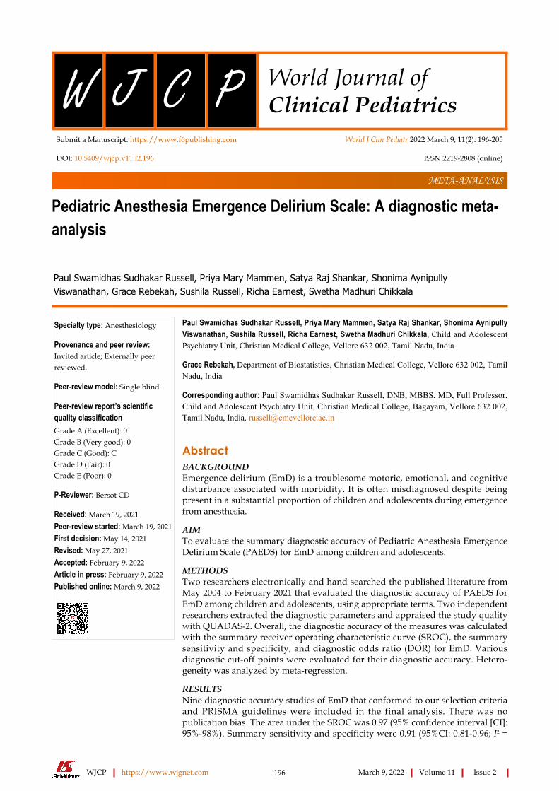
WJCP https://www.wjgnet.com 196 March 9, 2022 Volume 11 Issue 2
World Journal of
Clinical PediatricsW J C PSubmit a Manuscript: https://www.f6publishing.com World J Clin Pediatr 2022 March 9; 11(2): 196-205
DOI: 10.5409/wjcp.v11.i2.196 ISSN 2219-2808 (online)
META-ANALYSIS
Pediatric Anesthesia Emergence Delirium Scale: A diagnostic meta-analysis
Paul Swamidhas Sudhakar Russell, Priya Mary Mammen, Satya Raj Shankar, Shonima Aynipully Viswanathan, Grace Rebekah, Sushila Russell, Richa Earnest, Swetha Madhuri Chikkala
Specialty type: Anesthesiology
Provenance and peer review: Invited article; Externally peer reviewed.
Peer-review model: Single blind
Peer-review report’s scientific quality classificationGrade A (Excellent): 0 Grade B (Very good): 0 Grade C (Good): C Grade D (Fair): 0 Grade E (Poor): 0
P-Reviewer: Bersot CD
Received: March 19, 2021 Peer-review started: March 19, 2021 First decision: May 14, 2021 Revised: May 27, 2021 Accepted: February 9, 2022 Article in press: February 9, 2022 Published online: March 9, 2022
Paul Swamidhas Sudhakar Russell, Priya Mary Mammen, Satya Raj Shankar, Shonima Aynipully Viswanathan, Sushila Russell, Richa Earnest, Swetha Madhuri Chikkala, Child and Adolescent Psychiatry Unit, Christian Medical College, Vellore 632 002, Tamil Nadu, India
Grace Rebekah, Department of Biostatistics, Christian Medical College, Vellore 632 002, Tamil Nadu, India
Corresponding author: Paul Swamidhas Sudhakar Russell, DNB, MBBS, MD, Full Professor, Child and Adolescent Psychiatry Unit, Christian Medical College, Bagayam, Vellore 632 002, Tamil Nadu, India. [email protected]
AbstractBACKGROUND Emergence delirium (EmD) is a troublesome motoric, emotional, and cognitive disturbance associated with morbidity. It is often misdiagnosed despite being present in a substantial proportion of children and adolescents during emergence from anesthesia.
AIM To evaluate the summary diagnostic accuracy of Pediatric Anesthesia Emergence Delirium Scale (PAEDS) for EmD among children and adolescents.
METHODS Two researchers electronically and hand searched the published literature from May 2004 to February 2021 that evaluated the diagnostic accuracy of PAEDS for EmD among children and adolescents, using appropriate terms. Two independent researchers extracted the diagnostic parameters and appraised the study quality with QUADAS-2. Overall, the diagnostic accuracy of the measures was calculated with the summary receiver operating characteristic curve (SROC), the summary sensitivity and specificity, and diagnostic odds ratio (DOR) for EmD. Various diagnostic cut-off points were evaluated for their diagnostic accuracy. Hetero-geneity was analyzed by meta-regression.
RESULTS Nine diagnostic accuracy studies of EmD that conformed to our selection criteria and PRISMA guidelines were included in the final analysis. There was no publication bias. The area under the SROC was 0.97 (95% confidence interval [CI]: 95%-98%). Summary sensitivity and specificity were 0.91 (95%CI: 0.81-0.96; I2 =
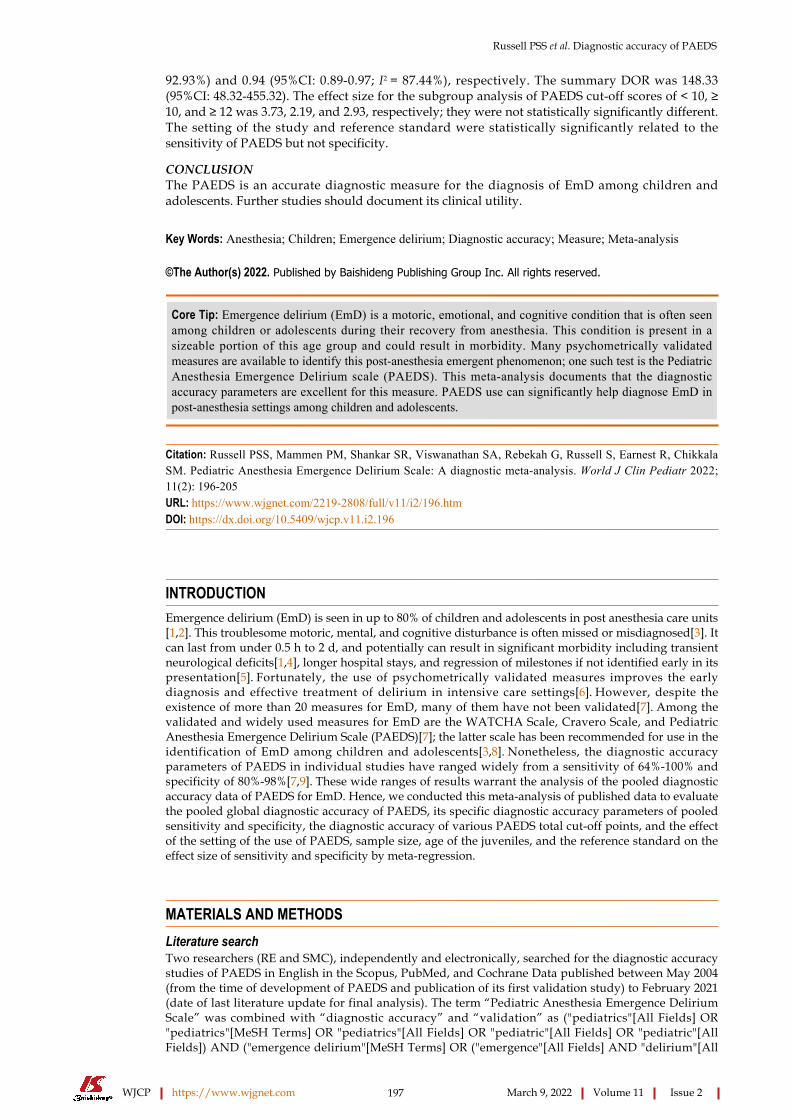
Russell PSS et al. Diagnostic accuracy of PAEDS
WJCP https://www.wjgnet.com 197 March 9, 2022 Volume 11 Issue 2
92.93%) and 0.94 (95%CI: 0.89-0.97; I2 = 87.44%), respectively. The summary DOR was 148.33 (95%CI: 48.32-455.32). The effect size for the subgroup analysis of PAEDS cut-off scores of < 10, ≥ 10, and ≥ 12 was 3.73, 2.19, and 2.93, respectively; they were not statistically significantly different. The setting of the study and reference standard were statistically significantly related to the sensitivity of PAEDS but not specificity.
CONCLUSION The PAEDS is an accurate diagnostic measure for the diagnosis of EmD among children and adolescents. Further studies should document its clinical utility.
Key Words: Anesthesia; Children; Emergence delirium; Diagnostic accuracy; Measure; Meta-analysis
©The Author(s) 2022. Published by Baishideng Publishing Group Inc. All rights reserved.
Core Tip: Emergence delirium (EmD) is a motoric, emotional, and cognitive condition that is often seen among children or adolescents during their recovery from anesthesia. This condition is present in a sizeable portion of this age group and could result in morbidity. Many psychometrically validated measures are available to identify this post-anesthesia emergent phenomenon; one such test is the Pediatric Anesthesia Emergence Delirium scale (PAEDS). This meta-analysis documents that the diagnostic accuracy parameters are excellent for this measure. PAEDS use can significantly help diagnose EmD in post-anesthesia settings among children and adolescents.
Citation: Russell PSS, Mammen PM, Shankar SR, Viswanathan SA, Rebekah G, Russell S, Earnest R, Chikkala SM. Pediatric Anesthesia Emergence Delirium Scale: A diagnostic meta-analysis. World J Clin Pediatr 2022; 11(2): 196-205URL: https://www.wjgnet.com/2219-2808/full/v11/i2/196.htmDOI: https://dx.doi.org/10.5409/wjcp.v11.i2.196
INTRODUCTIONEmergence delirium (EmD) is seen in up to 80% of children and adolescents in post anesthesia care units[1,2]. This troublesome motoric, mental, and cognitive disturbance is often missed or misdiagnosed[3]. It can last from under 0.5 h to 2 d, and potentially can result in significant morbidity including transient neurological deficits[1,4], longer hospital stays, and regression of milestones if not identified early in its presentation[5]. Fortunately, the use of psychometrically validated measures improves the early diagnosis and effective treatment of delirium in intensive care settings[6]. However, despite the existence of more than 20 measures for EmD, many of them have not been validated[7]. Among the validated and widely used measures for EmD are the WATCHA Scale, Cravero Scale, and Pediatric Anesthesia Emergence Delirium Scale (PAEDS)[7]; the latter scale has been recommended for use in the identification of EmD among children and adolescents[3,8]. Nonetheless, the diagnostic accuracy parameters of PAEDS in individual studies have ranged widely from a sensitivity of 64%-100% and specificity of 80%-98%[7,9]. These wide ranges of results warrant the analysis of the pooled diagnostic accuracy data of PAEDS for EmD. Hence, we conducted this meta-analysis of published data to evaluate the pooled global diagnostic accuracy of PAEDS, its specific diagnostic accuracy parameters of pooled sensitivity and specificity, the diagnostic accuracy of various PAEDS total cut-off points, and the effect of the setting of the use of PAEDS, sample size, age of the juveniles, and the reference standard on the effect size of sensitivity and specificity by meta-regression.
MATERIALS AND METHODSLiterature searchTwo researchers (RE and SMC), independently and electronically, searched for the diagnostic accuracy studies of PAEDS in English in the Scopus, PubMed, and Cochrane Data published between May 2004 (from the time of development of PAEDS and publication of its first validation study) to February 2021 (date of last literature update for final analysis). The term “Pediatric Anesthesia Emergence Delirium Scale” was combined with “diagnostic accuracy” and “validation” as ("pediatrics"[All Fields] OR "pediatrics"[MeSH Terms] OR "pediatrics"[All Fields] OR "pediatric"[All Fields] OR "pediatric"[All Fields]) AND ("emergence delirium"[MeSH Terms] OR ("emergence"[All Fields] AND "delirium"[All

Russell PSS et al. Diagnostic accuracy of PAEDS
WJCP https://www.wjgnet.com 198 March 9, 2022 Volume 11 Issue 2
Fields]) OR "emergence delirium"[All Fields]) AND ("scale s"[All Fields] OR "scaled"[All Fields] OR "scaling"[All Fields] OR "scalings"[All Fields] OR "weights and measures"[MeSH Terms] OR ("weights"[All Fields] AND "measures"[All Fields]) OR "weights and measures"[All Fields] OR "scale"[All Fields] OR "scales"[All Fields]) AND ("diagnosis"[MeSH Terms] OR "diagnosis"[All Fields] OR "diagnostic"[All Fields] OR "diagnostical"[All Fields] OR "diagnostically"[All Fields] OR "diagnostics"[All Fields]) AND ("accuracies"[All Fields] OR "accuracy"[All Fields]); and ("paediatrics"[All Fields] OR "pediatrics"[MeSH Terms] OR "pediatrics"[All Fields] OR "paediatric"[All Fields] OR "pediatric"[All Fields]) AND ("emergence delirium"[MeSH Terms] OR ("emergence"[All Fields] AND "delirium"[All Fields]) OR "emergence delirium"[All Fields]) AND ("scale s"[All Fields] OR "scaled"[All Fields] OR "scaling"[All Fields] OR "scalings"[All Fields] OR "weights and measures"[MeSH Terms] OR ("weights"[All Fields] AND "measures"[All Fields]) OR "weights and measures"[All Fields] OR "scale"[All Fields] OR "scales"[All Fields]) AND ("valid"[All Fields] OR "validate"[All Fields] OR "validated"[All Fields] OR "validates"[All Fields] OR "validating"[All Fields] OR "validation"[All Fields] OR "validational"[All Fields] OR "validations"[All Fields] OR "validator"[All Fields] OR "validators"[All Fields] OR "validities"[All Fields] OR "validity"[All Fields]).
The electronic search did not incorporate any search filter to improve the retrieval of as many articles as possible. After a review of the identified titles and abstracts, those articles deemed potentially relevant were collected. We augmented our electronic search with a hand search for additional relevant articles in reference lists of collected articles and from conference abstracts.
Study selection, data extraction, and quality appraisalTwo other researchers (Mammen PM and Shankar SR) extracted the required details independently, resolved any difference in extraction by consultation with another researcher (PSSR), and entered the information as electronic data. They extracted the information including participants, index measure, comparative reference measure, and outcome of diagnostic accuracy details. To be included in the final meta-analysis, studies had to compare the ability of PAEDS as the index test and DSM IV/DSM-IV-TR/DSM 5/ICD-10 or clinical consensus/clinical observation as the reference standard (using clinical interview, semi-structured interview, or interviewing schedules) among children and adolescents (1-18 years). Those diagnostic accuracy studies of PAEDS to identify EmD only were included and studies on PAEDS in the context of other emergent conditions like emergent agitation and emergent pain were excluded. Finally, the study had to report sufficient data to construct 2 x 2 tables for calculating the true positive, false positive, false negative, and true negative values. Two researchers (SR and SAV) appraised the quality of the studies with Quality Assessment of Diagnostic-Accuracy Studies, version 2 (QUADAS-2); differences in appraisal were resolved by consensus with the third researcher (Russell PSS).
Statistical analysisWe constructed the true positive, false positive, false negative, and true negative values, for each included study using 2 × 2 tables. We calculated the area under the curve (AUC) using the summary receiver operating characteristic curve (SROC) to establish the global diagnostic accuracy for all PAEDS cut-offs together; we calculated the confidence and prediction contour for the SROC as well[10]. The pooled sensitivity and specificity were estimated. We calculated the pooled diagnostic odds ratio (DOR) as the diagnostic accuracy parameter for various PAEDS cut-off scores and presented it as a forest plot. An I2 value of > 50 was considered as substantial heterogeneity. For exploring the heterogeneity and subgroup analysis, the effect of the setting of the use of PAEDS, sample size, reference standard, as well as age of children and adolescents (as independent variables) on the effect size of sensitivity and specificity (as dependent variables) was done using univariate meta-regression. In addition, as the heterogeneity was substantial, it was reasoned that the summary statistics might not represent the individual studies adequately. Therefore, as a post hoc test to parametrise the summary DOR, we conducted a leave-one-out cross validation. We calculated the 95% confidence interval (95%CI) when indicated. The analyses were done with the METANDI module of STATA (version 16). We conducted the leave-one-out cross validation using the software Open-Meta meta-analysis software (Brown University, Providence RI, United States)[11].
RESULTSLiterature searchTotally we identified 232 studies from all the data bases, and nine studies (K = 9; n = 1251) were included for the final meta-analysis[7,9,12-17]. Two studies were excluded as they did not satisfy the selection criteria[18,19]. Augmentation strategies of checking the cross references and conference abstracts did not supplement to the eligible article list. The PRISMA flowchart of studies for the final meta-analysis is represented in Figure 1.

Russell PSS et al. Diagnostic accuracy of PAEDS
WJCP https://www.wjgnet.com 199 March 9, 2022 Volume 11 Issue 2
Figure 1 PRISMA flow chart of studies included in the diagnostic meta-analysis for Pediatric Anesthesia Emergence Delirium Scale.
The studies were conducted either in the out-patient (K = 2) or in-patient settings (K = 7) and the sample size varied from 90-260 participants. Four studies had children as participants and the remaining five had children as well as adolescents. Six studies had used a PAEDS cut-off of < 10, two studies ≥ 10, and two studies ≥ 12 for the diagnosis of EmD; except two studies, all had used clinical observation by trained professionals in identifying EmD as the reference standard (Table 1).
Publication bias and quality appraisalThe quality appraisal using QUADAS-2 is pictorially represented for individual studies and across studies in Figure 2A and 2B, respectively; the most common bias across studies was documenting the reference standards and applicability of the reference standards. The Deek’s plot did not show publication bias [coefficient = 39.10 (95%CI: -6.05-84.25); P = 0.08] for the studies included in the final analysis as noted in Figure 3.
Diagnostic accuracyThe AUC for the HSROC was 0.97 (95%CI: 95%-98%) (Figure 4). The summary sensitivity and specificity (95%CI for sensitivity/specificity; I2 for heterogeneity) for the PAEDS were 0.91 (95%CI: 0.81-0.96; I2 = 92.93%) and 0.94 (95%CI: 0.89-0.97; I2 = 87.44%), respectively, for diagnosing EmD. When we analyzed the sensitivity-specificity pair within studies, most of the studies had a higher specificity than sensitivity[8,12,15,16,18]. However, two studies each had a higher sensitivity than specificity[9,17] or equal sensitivity and specificity[13,14].
The summary DOR for all PAEDS cut-off scores together was 148.33 (95%CI: 48.32-455.32). With the leave-one-out cross validation, the individual studies significantly contributed to the summary DOR in a descending order from the study by Sikich et al[8] at the top [DOR = 152.23 (95%CI: 76.23-304.82)], followed by Bajwa et al[13] [DOR = 148.48 (95%CI: 82.18-268.27)], Bong et al[12] [DOR = 134.04 (95%CI: 66.53-270.02)], Somaini et al[17] [DOR = 133.30 (95%CI: 66.95-265.41)], Janssen et al[14] [DOR = 131.35 (95%CI: 64.70-266.64)], Locatelli et al[15] [DOR = 121.36 (95%CI: 59.72-249.32)], Simonsen et al[18] [DOR = 117 (95%CI: 76.23-304.82)], Joo et al[16] [DOR = 111.78 (95%CI: 62.25-200.73)], and finally Blankespoor et al[9] [DOR = 111.72 (95%CI: 63.47-196.65)].
The effect size for the subgroup analysis of PAEDS cut-off scores of < 10, ≥ 10 and ≥ 12 was 3.73, 2.19, and 2.93 respectively. Although the < 10 PEDS cut-off score had the largest effect size, the three studied cut-off scores were not statistically significantly different in their diagnostic accuracy; however, they were statistically significantly different when individual studies with varying cut-off PAEDS scores were studied (Figure 5).
Meta-regressionIn the meta-regression, the setting of the study and reference standard used were statistically significantly related to the sensitivity of PAEDS and not to its specificity, but the age of the children and adolescents and the sample size of the studies were neither related to the sensitivity nor specificity (Figure 6).

Russell PSS et al. Diagnostic accuracy of PAEDS
WJCP https://www.wjgnet.com 200 March 9, 2022 Volume 11 Issue 2
Table 1 Data on methodology and epidemiology of included studies
Ref. Sample size Prevalence of EmD Sn (%) Sp (%) Setting Age (yr) PEDS Cut-off Reference standard
Sikich et al[8] 100 11% 64 86 OP 1.6-2 ≥ 10 Dimenhydrinate treatment
Bong et al[12] 136 8.6% 85 96 OP 2-12 ≥ 10 Clinical observation
Bajwa et al[13] 117 32% 100 95 IP 1-18 ≥ 12 Clinical observation
Janssen et al[14] 154 16.9% 91 98 IP 1-17 ≥ 8 DSM-IVinterview for delirium
Blankespoor et al[9] 144 16% 100 97 IP 1-18 ≥ 8 Clinical observation
Locatelli et al[15] 260 25% 93 94 IP 1-3 ≥ 9 Clinical observation
Joo et al[16] 90 25.5% 94 97 IP 2-5 ≥ 16 Clinical observation
Somaini et al[17] 150 21% 96 80 IP 1-7 ≥ 9 Clinical observation
Simonsen et al[18] 100 13.2% 86 100 IP 2 mo-16 yr ≥ 10 Clinical observation
Figure 2 Quality appraisal using the revised diagnostic accuracy studies (quality assessment of diagnostic accuracy studies-2) for individual studies (A) and average quality across studies (B). QUADAS-2: Quality assessment of diagnostic accuracy studies-2; PS: Patient selection - Describe methods of patient selection; IT: Index text -Describe the index test and how it was conducted and interpreted; RS: Reference standard - Describe the reference standard and how it was conducted and interpreted; FAT: Flow and timing; ACRS: Describe the applicability concerns about reference standard and how it was conducted and interpreted; ACPS: Describe the applicability concerns about patient selection and how it was conducted and interpreted; ACIT: Describe the applicability concerns about Index test and how it was conducted and interpreted; Low: Low bias; High: High bias UC: Unclear (if insufficient data were reported to permit our judgment).
DISCUSSIONCurrently, the diagnostic methods for EmD are evolving, and there is more clarity in differentiating EmD from other emergent phenomena. This meta-analysis included only those studies where PAEDS was used as a diagnostic measure for EmD only. This meta-analysis on PAEDS supports the evidence obtained from previously documented diagnostic accuracy parameters based on individual studies that the measure can be used as an effective diagnostic measure for EmD among children and adolescents.
There was no publication bias. The quality appraisal showed that the most common bias across studies was documenting the reference standards and applicability of the reference standards. Overall, the studies were of moderate quality. The absence of very large studies, duplicated data sets, same study sample/population, and similar selection process of participants or same group of authors with similar interpretation of results has minimized the skewing of our summary findings.
The AUC-SROC for PAEDS in diagnosing EmD was 0.97. As this AUC is much above the random predictor value of 0.5, the classification of EmD by PAEDS is not by random chance of 50% or toss of a coin but instead the classification is because of the excellent inherent global diagnostic accuracy of PAEDS. Thus, PAEDS succeeds as a diagnostic test for pediatric EmD with the various diagnostic cut-off scores used currently.
The pooled sensitivity of PAEDS in our study was 91%, which is an excellent sensitivity meaning that 91/100 children with EmD were correctly identified. Similarly, the pooled specificity of PAEDS was 94%, which is an excellent specificity and it means that 94/100 healthy children were identified as not having EmD. Such excellent sensitivity and specificity again support the use of PAEDS as a diagnostic measure for EmD among the pediatric population. This pooled sensitivity and specificity are
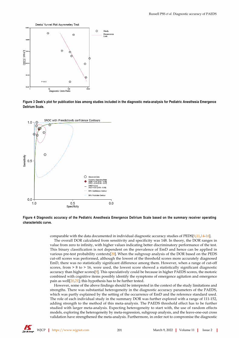
Russell PSS et al. Diagnostic accuracy of PAEDS
WJCP https://www.wjgnet.com 201 March 9, 2022 Volume 11 Issue 2
Figure 3 Deek’s plot for publication bias among studies included in the diagnostic meta-analysis for Pediatric Anesthesia Emergence Delirium Scale.
Figure 4 Diagnostic accuracy of the Pediatric Anesthesia Emergence Delirium Scale based on the summary receiver operating characteristic curve.
comparable with the data documented in individual diagnostic accuracy studies of PEDS[9,11,14-16].The overall DOR calculated from sensitivity and specificity was 148. In theory, the DOR ranges in
value from zero to infinity, with higher values indicating better discriminatory performance of the test. This binary classification is not dependent on the prevalence of EmD and hence can be applied in various pre-test probability contexts[20]. When the subgroup analysis of the DOR based on the PEDS cut-off scores was performed, although the lowest of the threshold scores more accurately diagnosed EmD, there was no statistically significant difference among them. However, when a range of cut-off scores, from > 8 to > 16, were used, the lowest score showed a statistically significant diagnostic accuracy than higher scores[9]. This speculatively could be because in higher PAEDS scores, the motoric combined with cognitive items possibly identify the symptoms of emergence agitation and emergence pain as well[20,21]; this hypothesis has to be further tested.
However, some of the above findings should be interpreted in the context of the study limitations and strengths. There was substantial heterogeneity in the diagnostic accuracy parameters of the PAEDS, which was partly explained by the setting of the occurrence of EmD and the reference standard used. The role of each individual study in the summary DOR was further explored with a range of 111-152, adding strength to the method of this meta-analysis. The PAEDS threshold effect has to be further studied with larger meta-analysis. Expecting heterogeneity to start with, the use of random effects models, exploring the heterogeneity by meta-regression, subgroup analysis, and the leave-one-out cross validation have strengthened the meta-analysis. Furthermore, in order not to compromise the diagnostic
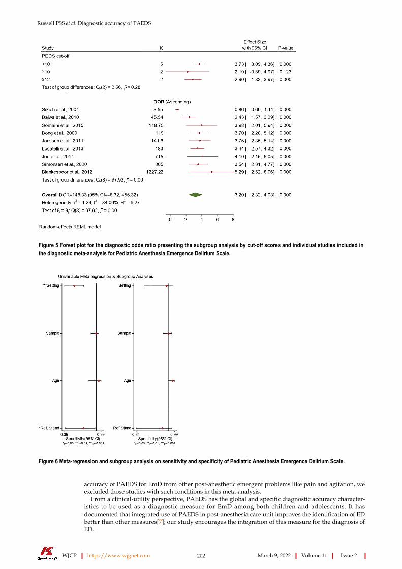
Russell PSS et al. Diagnostic accuracy of PAEDS
WJCP https://www.wjgnet.com 202 March 9, 2022 Volume 11 Issue 2
Figure 5 Forest plot for the diagnostic odds ratio presenting the subgroup analysis by cut-off scores and individual studies included in the diagnostic meta-analysis for Pediatric Anesthesia Emergence Delirium Scale.
Figure 6 Meta-regression and subgroup analysis on sensitivity and specificity of Pediatric Anesthesia Emergence Delirium Scale.
accuracy of PAEDS for EmD from other post-anesthetic emergent problems like pain and agitation, we excluded those studies with such conditions in this meta-analysis.
From a clinical-utility perspective, PAEDS has the global and specific diagnostic accuracy character-istics to be used as a diagnostic measure for EmD among both children and adolescents. It has documented that integrated use of PAEDS in post-anesthesia care unit improves the identification of ED better than other measures[7]; our study encourages the integration of this measure for the diagnosis of ED.
Russell PSS et al. Diagnostic accuracy of PAEDS
WJCP https://www.wjgnet.com 203 March 9, 2022 Volume 11 Issue 2
CONCLUSIONIn conclusion, PAEDS has excellent diagnostic accuracy for emergent delirium among children and adolescents.
ARTICLE HIGHLIGHTSResearch backgroundThere are various measures to identify emergence delirium (EmD) among children and adolescents as they recover from anesthesia. Pediatric Anesthesia Emergence Delirium Scale (PAEDS) is one such measure and has been found to have varying accuracy for diagnosing EmD.
Research motivationThe diagnosis of EmD is often missed or misdiagnosed. This can result in significant morbidity. The widely used PAEDS across the world has been proven to have the ability of early identification of EmD.
Research objectivesThe aims of this meta-analysis were to document the summary global and specific diagnostic accuracy parameters of PAEDS, diagnostic accuracy for various diagnostic threshold scores of the measure, and factors associated with these summary parameters of PAEDS in diagnosing EmD.
Research methodsNine studies were included in the analysis following the PRISMA guidelines. We used the summary area under the receiver operating characteristic curve, with a random effects model, to summarize the global diagnostic accuracy of PAEDS along with its diagnostic odds ratio, sensitivity, and specificity.
Research resultsThe area under the SROC was 0.97 (95%CI: 95-98%). The summary sensitivity and specificity were 0.91 (95%CI: 0.81-0.96; I2 = 92.93%) and 0.94 (95%CI: 0.89-0.97; I2 = 87.44%), respectively. The summary DOR was 148.33 (95%CI: 48.32-455.32). The effect size for the subgroup analysis of PAEDS cut-off scores of < 10, ≥ 10, and ≥ 12 was 3.73, 2.19, and 2.93, respectively; they were not statistically significantly different. The setting of the study and reference standard were statistically significantly related to the sensitivity of PAEDS but not specificity.
Research conclusionsThe authors have established the summary global diagnostic accuracy of PAEDS for EmD among children and adolescents.
Research perspectivesThe PAEDS could be used for diagnosing EmD among children and adolescents. The specific diagnostic cut-off scores have to be further studied.
ACKNOWLEDGEMENTSWe acknowledge with gratitude Ms. Mary Pauline Paul and Mr. George Devadoss in coordinating the certification processes.
FOOTNOTESAuthor contributions: Russell PSS and Mammen PM conceived and designed the study; Chikkala SM and Earnest R did the literature search and collected the data; Mammen PM and Shankar SR extracted the data; Viswanathan SA and Russell S appraised the quality of the studies; Mammen PM resolved the conflicts in data extraction and quality appraisal; Russell PSS and Rebekah G did the statistical analyses; all authors contributed to the writing and approval of the final manuscript.
Conflict-of-interest statement: All authors declare that there are no any conflicts of interest to disclose.
PRISMA 2009 Checklist statement: The authors have read the PRISMA 2009 Checklist, and the manuscript was prepared and revised according to the PRISMA 2009 Checklist.

Russell PSS et al. Diagnostic accuracy of PAEDS
WJCP https://www.wjgnet.com 204 March 9, 2022 Volume 11 Issue 2
Open-Access: This article is an open-access article that was selected by an in-house editor and fully peer-reviewed by external reviewers. It is distributed in accordance with the Creative Commons Attribution NonCommercial (CC BY-NC 4.0) license, which permits others to distribute, remix, adapt, build upon this work non-commercially, and license their derivative works on different terms, provided the original work is properly cited and the use is non-commercial. See: https://creativecommons.org/Licenses/by-nc/4.0/
Country/Territory of origin: India
ORCID number: Paul Swamidhas Sudhakar Russell 0000-0001-6463-2750; Priya Mary Mammen 0000-0001-7182-6959; Satya Raj Shankar 0000-0001-9454-7610; Shonima Aynipully Viswanathan 0000-0002-3792-0009; Grace Rebekah 0000-0001-6279-4326; Sushila Russell 0000-0003-0055-1542; Richa Earnest 0000-0003-4389-3913; Swetha Madhuri Chikkala 0000-0002-1462-1751.
S-Editor: Ma YJ L-Editor: Wang TQ P-Editor: Cai YX
REFERENCESVlajkovic GP, Sindjelic RP. Emergence delirium in children: many questions, few answers. Anesth Analg 2007; 104: 84-91 [PMID: 17179249 DOI: 10.1213/01.ane.0000250914.91881.a8]
1
Dahmani S, Stany I, Brasher C, Lejeune C, Bruneau B, Wood C, Nivoche Y, Constant I, Murat I. Pharmacological prevention of sevoflurane- and desflurane-related emergence agitation in children: a meta-analysis of published studies. Br J Anaesth 2010; 104: 216-223 [PMID: 20047899 DOI: 10.1093/bja/aep376]
2
Wong DD, Bailey CR. Emergence delirium in children. Anaesthesia 2015; 70: 383-387 [PMID: 25764401 DOI: 10.1111/anae.13043]
3
Drobish JK, Kelz MB, DiPuppo PM, Cook-Sather SD. Emergence delirium with transient associative agnosia and expressive aphasia reversed by flumazenil in a pediatric patient. A A Case Rep 2015; 4: 148-150 [PMID: 26035220 DOI: 10.1213/XAA.0000000000000140]
4
Hudek K. Emergence delirium: a nursing perspective. AORN J 2009; 89: 509-16; quiz 517 [PMID: 19326585 DOI: 10.1016/j.aorn.2008.12.026]
5
van den Boogaard M, Pickkers P, van der Hoeven H, Roodbol G, van Achterberg T, Schoonhoven L. Implementation of a delirium assessment tool in the ICU can influence haloperidol use. Crit Care 2009; 13: R131 [PMID: 19664260 DOI: 10.1186/cc7991]
6
Stamper MJ, Hawks SJ, Taicher BM, Bonta J, Brandon DH. Identifying pediatric emergence delirium by using the PAED Scale: a quality improvement project. AORN J 2014; 99: 480-494 [PMID: 24674794 DOI: 10.1016/j.aorn.2013.08.019]
7
Sikich N, Lerman J. Development and psychometric evaluation of the pediatric anesthesia emergence delirium scale. Anesthesiology 2004; 100: 1138-1145 [PMID: 15114210 DOI: 10.1097/00000542-200405000-00015]
8
Blankespoor RJ, Janssen NJ, Wolters AM, Van Os J, Schieveld JN. Post-hoc revision of the pediatric anesthesia emergence delirium rating scale: clinical improvement of a bedside-tool? Minerva Anestesiol 2012; 78: 896-900 [PMID: 22415436 DOI: 10.1213/ANE.0b013e31825b3d08]
9
Takwoingi Y, Guo B, Riley RD, Deeks JJ. Performance of methods for meta-analysis of diagnostic test accuracy with few studies or sparse data. Stat Methods Med Res 2017; 26: 1896-1911 [PMID: 26116616 DOI: 10.1177/0962280215592269]
10
Wallace BC, Schmid CH, Lau J, Trikalinos TA. Meta-Analyst: software for meta-analysis of binary, continuous and diagnostic data. BMC Med Res Methodol 2009; 9: 80 [PMID: 19961608 DOI: 10.1186/1471-2288-9-80]
11
Bong CL, Ng AS. Evaluation of emergence delirium in Asian children using the Pediatric Anesthesia Emergence Delirium Scale. Paediatr Anaesth 2009; 19: 593-600 [PMID: 19645978 DOI: 10.1111/j.1460-9592.2009.03024.x]
12
Bajwa SA, Costi D, Cyna AM. A comparison of emergence delirium scales following general anesthesia in children. Paediatr Anaesth 2010; 20: 704-711 [PMID: 20497353 DOI: 10.1111/j.1460-9592.2010.03328.x]
13
Janssen NJ, Tan EY, Staal M, Janssen EP, Leroy PL, Lousberg R, van Os J, Schieveld JN. On the utility of diagnostic instruments for pediatric delirium in critical illness: an evaluation of the Pediatric Anesthesia Emergence Delirium Scale, the Delirium Rating Scale 88, and the Delirium Rating Scale-Revised R-98. Intensive Care Med 2011; 37: 1331-1337 [PMID: 21567109 DOI: 10.1007/s00134-011-2244-y]
14
Locatelli BG, Ingelmo PM, Emre S, Meroni V, Minardi C, Frawley G, Benigni A, Di Marco S, Spotti A, Busi I, Sonzogni V. Emergence delirium in children: a comparison of sevoflurane and desflurane anesthesia using the Paediatric Anesthesia Emergence Delirium scale. Paediatr Anaesth 2013; 23: 301-308 [PMID: 23043512 DOI: 10.1111/pan.12038]
15
Joo J, Lee S, Lee Y. Emergence delirium is related to the invasiveness of strabismus surgery in preschool-age children. J Int Med Res 2014; 42: 1311-1322 [PMID: 25298011 DOI: 10.1177/0300060514549783]
16
Somaini M, Sahillioğlu E, Marzorati C, Lovisari F, Engelhardt T, Ingelmo PM. Emergence delirium, pain or both? Paediatr Anaesth 2015; 25: 524-529 [PMID: 25580984 DOI: 10.1111/pan.12580]
17
Simonsen BY, Skovby P, Lisby M. An evaluation of the Danish version of the Pediatric Anesthesia Emergence Delirium scale. Acta Anaesthesiol Scand 2020; 64: 613-619 [PMID: 31886528 DOI: 10.1111/aas.13543]
18
Aouad MT, Yazbeck-Karam VG, Nasr VG, El-Khatib MF, Kanazi GE, Bleik JH. A single dose of propofol at the end of surgery for the prevention of emergence agitation in children undergoing strabismus surgery during sevoflurane anesthesia. Anesthesiology 2007; 107: 733-738 [PMID: 18073548 DOI: 10.1097/01.anes.0000287009.46896.a7]
19

Russell PSS et al. Diagnostic accuracy of PAEDS
WJCP https://www.wjgnet.com 205 March 9, 2022 Volume 11 Issue 2
Lee-Archer PF, von Ungern-Sternberg BS, Reade MC, Law KC, Long D. An observational study of hypoactive delirium in the post-anesthesia recovery unit of a pediatric hospital. Paediatr Anaesth 2021; 31: 429-435 [PMID: 33405250 DOI: 10.1111/pan.14122]
20
Glas AS, Lijmer JG, Prins MH, Bonsel GJ, Bossuyt PM. The diagnostic odds ratio: a single indicator of test performance. J Clin Epidemiol 2003; 56: 1129-1135 [PMID: 14615004 DOI: 10.1016/s0895-4356(03)00177-x]
21
WJCP https://www.wjgnet.com 206 March 9, 2022 Volume 11 Issue 2
World Journal of
Clinical PediatricsW J C PSubmit a Manuscript: https://www.f6publishing.com World J Clin Pediatr 2022 March 9; 11(2): 206-214
DOI: 10.5409/wjcp.v11.i2.206 ISSN 2219-2808 (online)
META-ANALYSIS
Prevalence of intellectual disability in India: A meta-analysis
Paul Swamidhas Sudhakar Russell, Sahana Nagaraj, Ashvini Vengadavaradan, Sushila Russell, Priya Mary Mammen, Satya Raj Shankar, Shonima Aynipully Viswanathan, Richa Earnest, Swetha Madhuri Chikkala, Grace Rebekah
Specialty type: Psychiatry
Provenance and peer review: Invited article; Externally peer reviewed.
Peer-review model: Single blind
Peer-review report’s scientific quality classificationGrade A (Excellent): A Grade B (Very good): 0 Grade C (Good): 0 Grade D (Fair): 0 Grade E (Poor): 0
P-Reviewer: Swai JD
Received: March 21, 2021 Peer-review started: March 21, 2021 First decision: May 6, 2021 Revised: May 18, 2021 Accepted: February 11, 2022 Article in press: February 11, 2022 Published online: March 9, 2022
Paul Swamidhas Sudhakar Russell, Sahana Nagaraj, Ashvini Vengadavaradan, Sushila Russell, Priya Mary Mammen, Satya Raj Shankar, Shonima Aynipully Viswanathan, Swetha Madhuri Chikkala, Child and Adolescent Psychiatry Unit, Christian Medical College, Vellore 632 002, Tamil Nadu, India
Richa Earnest, Department of Psychiatry, Christian Medical College, Vellore 632 002, Tamil Nadu, India
Grace Rebekah, Department of Biostatistic, Christian Medical College, Vellore 632 002, Tamil Nadu, India
Corresponding author: Paul Swamidhas Sudhakar Russell, DNB, MBBS, MD, Full Professor, Child and Adolescent Psychiatry Unit, Christian Medical College, Bagayam, Vellore 632 002, Tamil Nadu, India. [email protected]
AbstractBACKGROUND Burden due to intellectual disability (ID) is only third to the depressive disorders and anxiety disorders in India. This national burden significantly contributes to the global burden of ID and hence one has to think globally and act locally to reduce this burden. At its best the collective prevalence of ID is in the form of narrative reviews. There is an urgent need to document the summary prevalence of ID to enhance further policymaking, national programs and resource allocation.
AIM To establish the summary prevalence of ID during the past 60 years in India.
METHODS Two researchers independently and electronically searched PubMed, Scopus, and the Cochrane library from January 1961 to December 2020 using appropriate search terms. Two other investigators extracted the study design, setting, part-icipant characteristics, and measures used to identify ID. Two other researchers appraised the quality of the studies using the Joanna Briggs Institute critical appraisal format for Prevalence Studies. Funnel plot and Egger’s regression test were used to ascertain the publication and small study effect on the prevalence. To evaluate the summary prevalence of ID, we used the random effects model with arcsine square-root transformation. Heterogeneity of I2 ≥ 50% was considered substantial and we determined the heterogeneity with meta-regression. The
Russell PSS et al. Prevalence of intellectual disability in India
WJCP https://www.wjgnet.com 207 March 9, 2022 Volume 11 Issue 2
analyses were performed using STATA (version 16).
RESULTS Nineteen studies were included in the meta-analysis. There was publication bias; the trim-and-fill method was used to further ascertain bias. Concerns with control of confounders and the reliable measure of outcome were noted in the critical appraisal. The summary prevalence of ID was 2% [(95%CI: 2%, 3%); I2 = 98%] and the adjusted summary prevalence was 1.4%. Meta-regression demonstrated that age of the participants was statistically significantly related to the prevalence; other factors did not influence the prevalence or heterogeneity.
CONCLUSION The summary prevalence of ID in India was established to be 2% taking into consideration the individual prevalence studies over the last six decades. This knowledge should improve the existing disability and mental health policies, national programs and service delivery to reduce the national and global burden associated with ID.
Key Words: India; Intellectual disability; Prevalence; Children and adolescents; Meta-analysis
©The Author(s) 2022. Published by Baishideng Publishing Group Inc. All rights reserved.
Core Tip: Intellectual disability (ID) is prevalent in India and earlier epidemiological studies on mental disorders have documented the lifetime prevalence of ID. However, the documented prevalence of ID in the country shows a wide range. The burden posed by ID is only third to the depressive disorders and anxiety disorders among mental disorders. The burden of ID in India significantly contributes to the global burden of ID; to decrease this we need to think globally and act locally in an evidence-based manner. To date, the prevalence of ID in India has been shown in narrative reviews. This suggests that the summary prevalence of ID in India has to be ascertained to help improve the existing disability and mental health policies, national programs and service delivery. This meta-analysis established that the summary lifetime prevalence of ID in India is 2%.
Citation: Russell PSS, Nagaraj S, Vengadavaradan A, Russell S, Mammen PM, Shankar SR, Viswanathan SA, Earnest R, Chikkala SM, Rebekah G. Prevalence of intellectual disability in India: A meta-analysis. World J Clin Pediatr 2022; 11(2): 206-214URL: https://www.wjgnet.com/2219-2808/full/v11/i2/206.htmDOI: https://dx.doi.org/10.5409/wjcp.v11.i2.206
INTRODUCTIONIntellectual disability (ID) contributes to 10.8% of the burden of mental disorders, measured by disability-adjusted life-years, in India. The burden caused by ID in India is only third to the depressive disorders and anxiety disorders[1]. Improving access to evidence-based mental health services for those with mental disorders is the best approach to address the burden of mental disorders in India[2]. However, the evidence based data for ID is difficult to build and most of the prevalence reviews for ID in India are narrative.
In narrative reviews, ID is prevalent in 1%-3.2% of the population in India depending on the definition of prevalence, study population, study design, and measures used to identify ID[3]. Among individual studies, the prevalence of ID in the country varied from 0.28%-20%[4,5]. This variation in prevalence is significant and does not help in planning precise policies, national programs and service delivery models for ID; the best way forward is to determine the summary prevalence of ID in India.
The summary prevalence of ID has not been documented in India and only an attempt to extrapolate from the 2002 Disability data report of the National Sample Survey Organization was made[6]. This meta-analysis documents the summary prevalence of ID in India for policy making and developing nation-wide clinical programs.

Russell PSS et al. Prevalence of intellectual disability in India
WJCP https://www.wjgnet.com 208 March 9, 2022 Volume 11 Issue 2
Figure 1 Preferred reporting items for systematic reviews and meta-analyses flow-chart for studies in the final meta-analysis.
MATERIALS AND METHODSLiterature search Two researchers (Chikkala SM and Earnest R) independently and electronically searched for relevant published studies in PubMed, Scopus, and the Cochrane library over the past 60 years (January 1961 to December 2020). The search terms were as follows: “prevalence of intellectual disability in India”, combined and included as: ("epidemiology"[MeSH Subheading] OR "epidemiology"[All Fields] OR "prevalence"[All Fields] OR "prevalence"[MeSH Terms] OR "prevalance"[All Fields] OR "prevalences"[All Fields] OR "prevalence s"[All Fields] OR "prevalent"[All Fields] OR "prevalently"[All Fields] OR "prevalents"[All Fields]) AND ("intellectual disability"[MeSH Terms] OR ("intellectual"[All Fields] AND "disability"[All Fields]) OR "intellectual disability"[All Fields]) AND ("india"[MeSH Terms] OR "india"[All Fields] OR "india s"[All Fields] OR "indias"[All Fields]). In addition, a hand-search was conducted for any potential study for inclusion from cross references and conference publications.
Study selection and data extractionThe studies retrieved during the searches were screened for relevance, and those identified as being potentially eligible were fully assessed for inclusion/exclusion from the titles. Two researchers (Russell S and Shankar SR) individually extracted the required data from the studies selected from inclusion. Any difference in the data extracted between the researchers was resolved through consultation with a third researcher (Mammen PM). Details on the prevalence of ID, sampling method, sample size, participant characteristics; setting of the study, definition of ID (borderline intelligence was excluded) measures/criteria used for diagnosis of ID, from each study were extracted. To be included in the final analysis, the studies required all these details available for extraction. Those studies with age of participant above 18 years, hospital setting and participants with borderline intelligence but not ID, and studies carried out on special illness populations were excluded.
Quality appraisal and risk of biasTwo researchers (Nagaraj S and Vengadavaradan A) independently appraised the studies using the Joanna Briggs Institute (JBI) Critical Appraisal Checklist for Studies Reporting Prevalence Data for the quality of the studies included in the final analysis[7]; and any discrepancies in the critical appraisal was resolved by consensus through discussion with another researcher (Mammen PM). A contour-enhanced funnel plot was developed for publication bias and Egger’s regression analysis was performed for the analysis of small study bias[8].
Statistical analysis To evaluate the summary prevalence, we used the random effects model with arcsine square-root transformation; heterogeneity (I2 ≥ 50%) was expected to be substantial in this prevalence meta-analysis and hence the transformation was used. As the contour-enhanced funnel plot and Egger’s regression test demonstrated significant publication bias, as a post hoc test, the trim-and-fill technique was used to

Russell PSS et al. Prevalence of intellectual disability in India
WJCP https://www.wjgnet.com 209 March 9, 2022 Volume 11 Issue 2
Figure 2 The contour-enhanced funnel plot (A) and trim-and-fill plot (B) for publication bias.
Figure 3 The Joanna Briggs Institute critical appraisal for prevalence meta-analysis for individual studies (A) and average quality across studies (B). IC: Were the criteria for inclusion in the sample clearly defined? SS: Were the study subjects and the setting described in detail? EX: Was the exposure measured in a valid and reliable way? ME: Were objective, standard criteria used for measurement of the condition? CO: Were confounding factors identified? MC: were strategies to deal with confounding factors stated? OT: Were the outcomes measured in a valid and reliable way? ST: Was appropriate statistical analysis used? High: High bias; No: Low bias; Unclear: Unclear bias; NA: Not applicable.
explore the nature of the bias[9]. We determined the heterogeneity with meta-regression. The analyses were carried out using the STATA (version 16) software package.
RESULTSIn total, we identified 290 studies from all the databases and 19 studies[10-28] were included in the final meta-analysis. Thirteen studies were excluded as they had either age group above 18 years and ID prevalence could not be calculated, a setting other than community or school, or the prevalence was studied in specific disease populations. The PRISMA (Preferred Reporting Items for Systematic Reviews and Meta-Analyses) details regarding the selection of studies for the final analysis are presented in Figure 1; the methodology and prevalence data are given in Table 1. The visual examination of the contour-enhanced funnel plot (Figure 2A) and the Egger’s test [coefficient = 5.92 (standard error = 1.19), t = -5.0; P = 0.0001] revealed publication bias or a small study effect, respectively, on the prevalence of

Russell PSS et al. Prevalence of intellectual disability in India
WJCP https://www.wjgnet.com 210 March 9, 2022 Volume 11 Issue 2
Table 1 The methodological and prevalence details of included studies
Ref. Area; setting Age(yr) Sampling, diagnostic method Prevalence of ID (%) Sample size
Surya et al[10] Urban; community 0-15 Screening schedule + CI 0.7 2731
Sethi et al[11] Urban; community 0-10 Comprehensive questionnaire + CI
5.74 541
Dube et al[12] Mixed; community 44693 CI 0.37 8035
Elnagar et al[11] Rural; community 0-14 CI + WHO ECH 0.86 635
Sethi et al[14] Rural; community 0-10 Comprehensive questionnaire, CI
6.84 877
Verghese et al[15] Urban; community 44663 Comprehensive questionnaire + ICD-9
2.01 747
Nandi et al[16] Rural; community 0-11 Comprehensive questionnaire + WHO ECH
0.28 462
Thacore et al[17] Urban; community 0-15 CI + DSM II 2.94 2696
Jiloha et al[18] Rural; school 44693 Comprehensive questionnaire + ICD-9
5.87 715
Singh et al[19] Urban; community 44575 CI + ICD-9 4.7 279
Deivasigamani et al[20] Urban; school 44785 Rutter B + ICD 9 2.9 755
Uma et al[21] Mixed; School 44624 PBCL (parent version) 2.91 155
Banerjee et al[22] Urban; school 44783 CI + CBQ + ICD-9 5.4 460
Gaur et al[23] Mixed; community 44726 CPMS + DISC + ICD-10 schedule
3.25 800
SriP-Editornath et al[24] Mixed; community 0-16 CBCL + DISC + VSMS + CGAS + ICD-10
2.3 2064
Bansal et al[25] Rural; community 44849 CPMS +ICD-10
Patil et al[26] Urban; community 44695 CI + DSM-IV 2.4 257
Sarda et al[27] Mixed; school 44667 CBS + CBCL + DISC + ICD-10 0.99 1110
Arora et al[28] Mixed; community INCLEN Measures + DSM-IV-TR
3.6 3964
CI: Clinical Interview; CBQ: Children’s Behaviour Questionnaire; CBCL: Child Behaviour Check List; CPMS: Indian Adaptation of CBCL; DSM: Diagnostic and Statistical Manual (edition IV and IV-TR); CGAS: Children’s Global Assessment Scale; DISC: Diagnostic Interview Schedule for Children; ECH: WHO expert committee on mental health criteria; ICD: International Classification of Diseases (edition 9 and 10); INCLEN: The International Clinical Epidemiology Network; PBCL: Preschool Behaviour Check List; VSMS: Vineland Social Maturity Scale.
ID in India. The trim-and-fill method demonstrated that four more studies were required on the left side of the funnel to overcome this bias (Figure 2B). The JBI critical appraisal for each study and the average quality included in the final analysis are depicted in Figure 3. Concerns with control of confounders and reliable measurement of outcome were noted in the critical appraisal as common biases.
The summary prevalence of ID among children and adolescents in India was 2% [0.02 (95%CI: 0.02%, 0.03%); I2 = 98%] and is presented as a forest plot (Figure 4). When the required six studies were included using the trim-and-fill method, the imputed prevalence of ID in India was 1.4%. The prevalence of ID has changed over the last 60-year period in India from 5% to 2%.
There was substantial heterogeneity in the meta-analysis; our meta-regression demonstrated that the heterogeneity between the prevalence studies was statistically significantly related to the age of the participants (children vs adolescents; coefficient =-0.019 (s.e) =0.009; P = 0.03), the area of residence or school, setting of the study in the community or school, and the diagnostic assessment used was not significantly related to the prevalence of ID.
DISCUSSIONThis meta-analysis documents that the summary life-time prevalence of ID is 2% and the adjusted summary life-time prevalence is 1.4% in India. This prevalence is, thus, within the range of 1%-3% in the
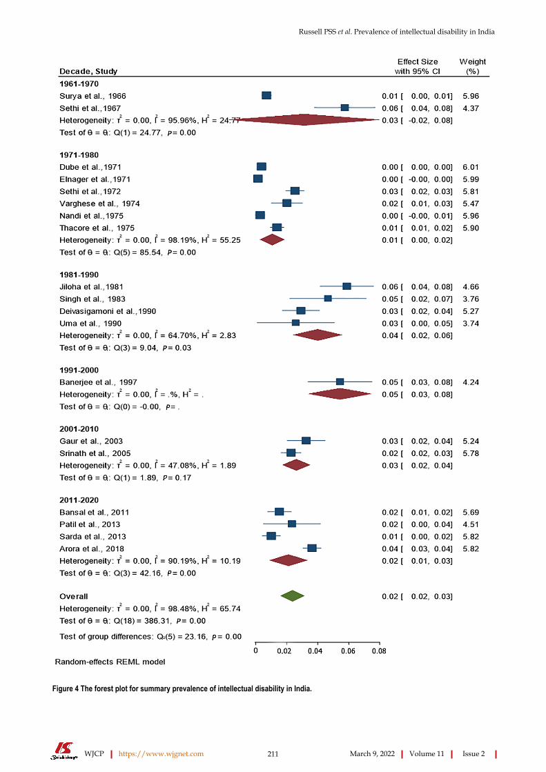
Russell PSS et al. Prevalence of intellectual disability in India
WJCP https://www.wjgnet.com 211 March 9, 2022 Volume 11 Issue 2
Figure 4 The forest plot for summary prevalence of intellectual disability in India.
Russell PSS et al. Prevalence of intellectual disability in India
WJCP https://www.wjgnet.com 212 March 9, 2022 Volume 11 Issue 2
narrative review[3] and the extrapolated data of 1.5% from the National Sample Survey Organization by the Ministry of Social Justice and Empowerment, Government of India[6]. With the above epidemi-ological insights, it is important to mitigate the burden with prevention and effective habilitation.
The lifetime prevalence of ID despite being small when compared with many other development disabilities of childhood[28], the burden caused by this disability is a significant 10.8% among mental disorders in India. The population size of India is so huge this burden adds to the global burden of ID; therefore, it has been suggested to enhance the disability programs to have sustainable burden reduction practices at the national level[29].
This meta-analysis was based on only published studies in the English language. Our funnel plot and test for the influence of small studies was significant and six more studies were required to prevent the publication and small study bias. However, we used the trim and fill method to impute the number of studies required to improve this meta-analysis and adjust the prevalence in our study. Thus, in this meta-analysis, the impact of four missing studies was simulated, and the original prevalence of 2% was revised to an adjusted prevalence of 1.4%.
From the utility perspective of this meta-analysis, it is important that the systematic survey we carried out as part of the meta-analysis shows there is a significant paucity of studies on the prevalence of ID in certain states of India. Moreover, the mental health programs at the national and state levels in India have to bridge the gap between identification and management need of those with ID, with focused polices, programs and capacity building. Although there has been noticeable progress in the policy, national program, and service programs, most of them are focused on the secondary and tertiary prevention of ID[3]. It has been documented that up to 25% of ID is preventable in India and 305 are acquired forms of ID[30]; this underscores the need for approaches such as the modified Finnish method in the context of identifying the aetiology of ID in India[31].
The findings of this study should be interpreted from the perspective of the lifetime prevalence of ID. We decided on this definition to build the systematic-survey, as ID is a lifetime condition, where the condition can be improved but cannot be reversed[32]. Secondly, although we searched for grey literature we did not search for unpublished data and thus could have limited the national data on this disorder.
CONCLUSIONIn conclusion, the lifetime prevalence of ID in India is consistent with narrative reviews. Addressing the ID burden requires delivery of integrated disability and mental health care services at the community level. This summary lifetime prevalence should further enhance policymaking and resource allocation for ID in India.
ARTICLE HIGHLIGHTSResearch backgroundIndia has a population of more than one billion with a significant disability burden similar to other low- and middle-income countries. The summary prevalence of intellectual disability (ID) in India has not been established.
Research motivationID contributes to 10.8% of the burden due to mental disorders in India. This national burden significantly contributes to the global burden of ID and hence one has to think globally and act locally to reduce this burden. At its best the collective prevalence of ID is in the form of narrative reviews. There is an urgent need to document the summary prevalence of ID to enhance further policymaking, national programs and resource allocation.
Research objectivesThe aim of the meta-analysis was to establish the summary prevalence of ID in India over the past 60 years.
Research methodsNineteen studies were included in the meta-analysis following the PRISMA guidelines. To analyse the summary prevalence of ID, we used the random effects model with arcsine square-root transformation. Heterogeneity of I2 ≥ 50% was considered substantial and we determined the heterogeneity with meta-regression.
Russell PSS et al. Prevalence of intellectual disability in India
WJCP https://www.wjgnet.com 213 March 9, 2022 Volume 11 Issue 2
Research resultsThe summary prevalence of ID was 2% [(95%CI: 2%, 3%); I2 = 98%] and the adjusted summary prevalence was 1.4%. Meta-regression demonstrated that age of the participants was statistically significantly related to the prevalence; other factors did not influence the prevalence or heterogeneity.
Research conclusionsThe authors established the summary prevalence of ID in India as 2% taking into consideration the individual prevalence studies over the last 6 decades. This knowledge should improve the existing disability and mental health policies, national programs and service delivery models to mitigate the burden related to ID.
Research perspectivesFuture research should focus on the role of the summary prevalence of ID in the reduction of burden due to this disability in India and globally.
FOOTNOTESAuthor contributions: Russell PSS conceived and designed the study; Chikkala SM and Earnest R performed the literature search and collected data; Russell S and Shankar SR extracted data; Nagaraj S and Vengadavaradan A appraised the quality of studies; Mammen PM resolved the conflicts in data extraction and quality appraisal; Russell PSS, Viswanathan SA and Rebekah G carried out the statistical analyses; and all authors contributed to the writing and approval of the final manuscript.
Conflict-of-interest statement: The authors declare that they have no conflict of interest.
PRISMA 2009 Checklist statement: The authors have read the PRISMA 2009 Checklist, and the manuscript was prepared and revised according to the PRISMA 2009 Checklist.
Open-Access: This article is an open-access article that was selected by an in-house editor and fully peer-reviewed by external reviewers. It is distributed in accordance with the Creative Commons Attribution NonCommercial (CC BY-NC 4.0) license, which permits others to distribute, remix, adapt, build upon this work non-commercially, and license their derivative works on different terms, provided the original work is properly cited and the use is non-commercial. See: https://creativecommons.org/Licenses/by-nc/4.0/
Country/Territory of origin: India
ORCID number: Paul Swamidhas Sudhakar Russell 0000-0001-6463-2750; Sahana Nagaraj 0000-0003-4742-4815; Ashvini Vengadavaradan 0000-0002-064-984X; Sushila Russell 0000-0003-0055-1542; Priya Mary Mammen 0000-0001-7182-6959; Satya Raj Shankar 0000-0001-9454-7610; Shonima Aynipully Viswanathan 0000-0002-3792-0009; Richa Earnest 0000-0002-1701-8392; Swetha Madhuri Chikkala 0000-0002-1462-1751; Grace Rebekah 0000-0001-6279-4326.
S-Editor: Ma YJ L-Editor: Webster JR P-Editor: Cai YX
REFERENCESIndia State-Level Disease Burden Initiative Mental Disorders Collaborators. The burden of mental disorders across the states of India: the Global Burden of Disease Study 1990-2017. Lancet Psychiatry 2020; 7: 148-161 [PMID: 31879245 DOI: 10.1016/S2215-0366(19)30475-4]
1
Shidhaye R. Unburden mental health in India: it's time to act now. Lancet Psychiatry 2020; 7: 111-112 [PMID: 31879246 DOI: 10.1016/S2215-0366(19)30524-3]
2
Girimaji SC, Srinath S. Perspectives of intellectual disability in India: epidemiology, policy, services for children and adults. Curr Opin Psychiatry 2010; 23: 441-446 [PMID: 20489642 DOI: 10.1097/YCO.0b013e32833ad95c]
3
Nandi DN, Banerjee G, Mukherjee SP, Sarkar S, Boral GC, Mukherjee A, Mishra DC. A study of psychiatric morbidity of a rural community at an interval of ten years. Indian J Psychiatry 1986; 28: 179-194 [PMID: 21927173]
4
Lal N, Sethi BB. Estimate of mental ill health in children of an urban community. Indian J Pediatr 1977; 44: 55-64 [PMID: 914355 DOI: 10.1007/BF02753627]
5
Lakhan R, Ekúndayò OT, Shahbazi M. An estimation of the prevalence of intellectual disabilities and its association with age in rural and urban populations in India. J Neurosci Rural Pract 2015; 6: 523-528 [PMID: 26752897 DOI: 10.4103/0976-3147.165392]
6
Munn Z, Barker TH, Moola S, Tufanaru C, Stern C, McArthur A, Stephenson M, Aromataris E. Methodological quality of case series studies: an introduction to the JBI critical appraisal tool. JBI Evid Synth 2020; 18: 2127-2133 [PMID: 33038125 DOI: 10.11124/JBISRIR-D-19-00099]
7

Russell PSS et al. Prevalence of intellectual disability in India
WJCP https://www.wjgnet.com 214 March 9, 2022 Volume 11 Issue 2
Peters JL, Sutton AJ, Jones DR, Abrams KR, Rushton L. Contour-enhanced meta-analysis funnel plots help distinguish publication bias from other causes of asymmetry. J Clin Epidemiol 2008; 61: 991-996 [PMID: 18538991 DOI: 10.1016/j.jclinepi.2007.11.010]
8
Duval S, Tweedie R. Trim and fill: A simple funnel-plot-based method of testing and adjusting for publication bias in meta-analysis. Biometrics 2000; 56: 455-463 [PMID: 10877304 DOI: 10.1111/j.0006-341x.2000.00455.x]
9
Surya NC, Datta SP, Gopalakrishnan R, Sundaram D, Kutty J. Mental morbidity in Pondicherry (1962-63). Trans All India Institute 4: 50- 61
10
Sethi BB, Gupta SC, Kumar R. Three hundred urban families - a psychiatric study. Indian J Psychiatry 1967; 9: 28011 Dube KC. A study of prevalence and biosocial variables in mental illness in a rural and an urban community in Uttar Pradesh--India. Acta Psychiatr Scand 1970; 46: 327-359 [PMID: 5502780 DOI: 10.1111/j.1600-0447.1970.tb02124.x]
12
Elnagar MN, Maitra P, Rao MN. Mental health in an Indian rural community. Br J Psychiatry 1971; 118: 499-503 [PMID: 5580365 DOI: 10.1192/bjp.118.546.499]
13
Sethi BB, Gupta SC, Kumar R, Kumarr P. A psychiatric survey of 500 rural families. Indian J Psychiatry 1972; 14: 183-196
14
Verghese A, Beig A. Psychiatric disturbance in children--an epidemiological study. Indian J Med Res 1974; 62: 1538-1542 [PMID: 4455615]
15
Nandi N, Aimany S, Ganguly H, Banerjee G, Boral GC, Ghosh A, Sarkar S. Psychiatric disorder in a rural community in West Bengal: An epidemiological study. Indian J Psychiatry 1975; 17: 87-89
16
Thacore VR, Gupta SC, Suraiya M. Psychiatric morbidity in a north Indian community. Br J Psychiatry 1975; 126: 365-369 [PMID: 1148570 DOI: 10.1192/bjp.126.4.364]
17
Jiloha RC, Murthy RS. An epidemiological study of psychiatric problems in primary school children. Child Psychiatry Quarterly 1981; 14: 108-119
18
Singh AJ, ShuWaj D, Verma BI, Kumar A, Srivastava N. An epidemiological study in childhood psychiatric disorders. Indian Pediatr 1983; 20: 167-172
19
Deivasigamani TR. Psychiatric morbidity in primary school children - an epidemiological study. Indian J Psychiatry 1990; 32: 235-240 [PMID: 21927463]
20
Uma H, Kapur M. A Study of Behaviour Problems in Pre-School Children. NIMHANS J 1990; 8: 69-7321 Banerjee T. Psychiatric morbidity among rural primary school children in west bengal. Indian J Psychiatry 1997; 39: 130-135 [PMID: 21584059]
22
Gaur DR, Vohra AK, Subhash S and Khurana H. Prevalence of psychiatric morbidity among 6-14 years old children. Indian J Community Med 2003; 28: 133-137
23
Srinath S, Girimaji SC, Gururaj G, Seshadri S, Subbakrishna DK, Bhola P, Kumar N. Epidemiological study of child & adolescent psychiatric disorders in urban & rural areas of Bangalore, India. Indian J Med Res 2005; 122: 67-79 [PMID: 16106093]
24
Bansal PD, Barman R. Psychopathology of school going children in the age group of 10-15 years. Int J Appl Basic Med Res 2011; 1: 43-47 [PMID: 23776772 DOI: 10.4103/2229-516X.81980]
25
Patil RN, Nagaonkar SN, Shah NB, Bhat TS. A Cross-sectional Study of Common Psychiatric Morbidity in Children Aged 5 to 14 Years in an Urban Slum. J Family Med Prim Care 2013; 2: 164-168 [PMID: 24479072 DOI: 10.4103/2249-4863.117413]
26
Sarda R, Kimmatkar N, Hemnani JT, Hemnani TJ, Mishra P, Jain SK. Prevalence of Psychiatric Disorders in Western U.P. Region- A School Based Study. Int J Sci Study 2013; 1: 52-57
27
Arora NK, Nair MKC, Gulati S, Deshmukh V, Mohapatra A, Mishra D, Patel V, Pandey RM, Das BC, Divan G, Murthy GVS, Sharma TD, Sapra S, Aneja S, Juneja M, Reddy SK, Suman P, Mukherjee SB, Dasgupta R, Tudu P, Das MK, Bhutani VK, Durkin MS, Pinto-Martin J, Silberberg DH, Sagar R, Ahmed F, Babu N, Bavdekar S, Chandra V, Chaudhuri Z, Dada T, Dass R, Gourie-Devi M, Remadevi S, Gupta JC, Handa KK, Kalra V, Karande S, Konanki R, Kulkarni M, Kumar R, Maria A, Masoodi MA, Mehta M, Mohanty SK, Nair H, Natarajan P, Niswade AK, Prasad A, Rai SK, Russell PSS, Saxena R, Sharma S, Singh AK, Singh GB, Sumaraj L, Suresh S, Thakar A, Parthasarathy S, Vyas B, Panigrahi A, Saroch MK, Shukla R, Rao KVR, Silveira MP, Singh S, Vajaratkar V. Neurodevelopmental disorders in children aged 2-9 years: Population-based burden estimates across five regions in India. PLoS Med 2018; 15: e1002615 [PMID: 30040859 DOI: 10.1371/journal.pmed.1002615]
28
Dandona R, Pandey A, George S, Kumar GA, Dandona L. India's disability estimates: Limitations and way forward. PLoS One 2019; 14: e0222159 [PMID: 31491011 DOI: 10.1371/journal.pone.0222159]
29
Srinath S, Girimaji SC. Epidemiology of child and adolescent mental health problems and mental retardation. NIMHANS J 1999; 17: 355-366
30
Kishore MT, Udipi GA, Seshadri SP. Clinical Practice Guidelines for Assessment and Management of intellectual disability. Indian J Psychiatry 2019; 61: 194-210 [PMID: 30745696 DOI: 10.4103/psychiatry.IndianJPsychiatry_507_18]
31
Streiner DL, Patten SB, Anthony JC, Cairney J. Has 'lifetime prevalence' reached the end of its life? Int J Methods Psychiatr Res 2009; 18: 221-228 [PMID: 20052690 DOI: 10.1002/mpr.296]
32

Published by Baishideng Publishing Group Inc
7041 Koll Center Parkway, Suite 160, Pleasanton, CA 94566, USA
Telephone: +1-925-3991568
E-mail: [email protected]
Help Desk: https://www.f6publishing.com/helpdesk
https://www.wjgnet.com
© 2022 Baishideng Publishing Group Inc. All rights reserved.