
Introduction to Pediatrics
495
2017 YSMU AFTER M. HERATSI, DEPARTMENT OF PEDIATRICS №1 - 1 - Introduction to Pediatrics Pediatrics (paediatrics) is a branch of medicine that deals with the medical care of infants, children and adolescents. According to UNICEF definition, the age limit of childhood ranges from birth up to 18. Children are considered “national wealth” and hence pediatric care has its implication on the ultimate progress of the nation. “Children’s health should be defined as the extent to which individual children or groups of children are able or enabled to (a) develop and realize their potential, (b) satisfy their needs, and (c) develop the capacities to allow them to interact successfully with their biological, physical, and social environments.” [National Research Council and Institute of Medicine. Children's Health, the Nation's Wealth: Assessing and Improving Child Health. Washington, DC: National Academies Press; 2004]. The key health dangers for children and adolescents 5.9 million children under the age of 5 died in 2015. 75% (4.5 million) of all under-five deaths occurred within the first year of life. More than half of these early child deaths are due to conditions that could be prevented or treated with access to simple, affordable interventions. Children in sub-Saharan Africa are about over 14 times more likely to die before the age of five than children in developed regions.
-
Upload
khangminh22 -
Category
Documents
-
view
0 -
download
0
Transcript of Introduction to Pediatrics

2017 YSMU AFTER M. HERATSI, DEPARTMENT OF PEDIATRICS 1
- 1 -
Introduction to Pediatrics
Pediatrics (paediatrics) is a branch of medicine that deals with the medical care
of infants, children and adolescents.
According to UNICEF definition, the age limit of childhood ranges from birth
up to 18.
Children are considered “national wealth” and hence pediatric care has its
implication on the ultimate progress of the nation.
“Children’s health should be defined as the extent to which individual children or groups of children are
able or enabled to (a) develop and realize their potential, (b) satisfy their needs, and (c) develop the
capacities to allow them to interact successfully with their biological, physical, and social environments.” [National Research Council and Institute of Medicine. Children's Health, the Nation's Wealth: Assessing and Improving Child Health. Washington, DC: National
Academies Press; 2004].
The key health dangers for children and adolescents
5.9 million children under the age of 5 died in 2015. 75% (4.5 million) of all under-five deaths occurred
within the first year of life.
More than half of these early child deaths are due to conditions that could be prevented or treated
with access to simple, affordable interventions.
Children in sub-Saharan Africa are about over 14 times more likely to die before the age of five than
children in developed regions.

2017 YSMU AFTER M. HERATSI, DEPARTMENT OF PEDIATRICS 1
- 2 -
The risk of death is highest in the first month of life. Preterm birth, birth asphyxia and infections cause
most newborn deaths.
Health risks to newborns are minimized by:
- quality care during pregnancy;
- safe delivery by a skilled birth attendant; and
- strong neonatal care: immediate attention to breathing and warmth, hygienic cord and skin care,
and early initiation of exclusive breastfeeding.
From one month to five years of age, the main causes of death are pneumonia, diarrhoea, malaria, measles,
congenital anomalies, injuries.
* For some of the most deadly childhood diseases, such as measles, vaccines are available and
timely completion of immunization protects a child from this illness and death.
* Pneumonia is the prime cause of death in children under five years of age. Addressing the major
risk factors –including malnutrition and indoor air pollution – is essential to preventing pneumonia,
as are vaccination and breastfeeding. Antibiotics and oxygen are vital tools for effectively managing
the illness.
* Diarrhoeal diseases are a leading cause of sickness and death among children in developing
countries. Breastfeeding helps prevent diarrhoea among young children. Treatment for sick children
with Oral Rehydration Solutions (ORS) combined with zinc supplements is safe, cost-effective, and
saves lives.
* One child dies every minute from malaria. Insecticide-treated nets prevent transmission and
increase child survival.
* Over 90% of children with HIV are infected through mother-to-child transmission; this can be
prevented with antiretrovirals, as well as safer delivery and feeding practices.
* Malnutrition is estimated to contribute to more than one third of all child deaths.
Worldwide, about 20% of deaths among children under-five could be avoided if feeding guidelines are
followed. WHO recommends exclusive breastfeeding for six months, introducing age-appropriate and safe
complementary foods at six months, and continuing breastfeeding for up to two years or beyond.
Adolescents – young people between the ages of 10 and 19 years – are often thought of as a healthy
group. Nevertheless, many adolescents do die prematurely due to accidents, suicide, violence, pregnancy
related complications and other illnesses that are either preventable or treatable. Many more suffer
chronic ill-health and disability. In addition, many serious diseases in adulthood have their roots in
adolescence. For example, tobacco use, sexually transmitted infections including HIV, poor eating and
exercise habits, obesity lead to illness (hypertension, coronary heart disease, diabetes mellitus,
osteoporosis etc.) or premature death later in life.

2017 YSMU AFTER M. HERATSI, DEPARTMENT OF PEDIATRICS 1
- 3 -
Indicators of infant and child health
Infant and child mortality rates are leading indicators of the level of child health and overall development
in countries.
Infant mortality rate (IMR) is the number of deaths of children less than one year of age per 1000 live births in a given year.
𝑰𝑴𝑹 =𝑵𝒖𝒎𝒃𝒆𝒓 𝒐𝒇 𝒅𝒆𝒂𝒕𝒉𝒔 𝒐𝒇 𝒊𝒏𝒇𝒂𝒏𝒕𝒔 𝒖𝒏𝒅𝒆𝒓 𝒐𝒏𝒆 𝒚𝒆𝒂𝒓 𝒐𝒍𝒅 𝒊𝒏 𝒂 𝒈𝒊𝒗𝒆𝒏 𝒚𝒆𝒂𝒓
𝑻𝒐𝒕𝒂𝒍 𝒏𝒖𝒎𝒃𝒆𝒓 𝒐𝒇 𝒍𝒊𝒗𝒆 𝒃𝒊𝒓𝒕𝒉𝒔 𝒊𝒏 𝒕𝒉𝒆 𝒔𝒂𝒎𝒆 𝒚𝒆𝒂𝒓𝒙 𝟏𝟎𝟎𝟎
Neonatal mortality rate (NMR) is the number of neonatal deaths per 1000 live births in a given year.
𝑵𝑴𝑹 =𝑵𝒖𝒎𝒃𝒆𝒓 𝒐𝒇 𝒏𝒆𝒐𝒏𝒂𝒕𝒂𝒍 𝒅𝒆𝒂𝒕𝒉𝒔 𝟎 − 𝟐𝟖 𝒅𝒂𝒚𝒔 𝒊𝒏 𝒂 𝒈𝒊𝒗𝒆𝒏 𝒚𝒆𝒂𝒓
𝑻𝒐𝒕𝒂𝒍 𝒏𝒖𝒎𝒃𝒆𝒓 𝒐𝒇 𝒍𝒊𝒗𝒆 𝒃𝒊𝒓𝒕𝒉𝒔 𝒊𝒏 𝒕𝒉𝒆 𝒔𝒂𝒎𝒆 𝒚𝒆𝒂𝒓𝒙 𝟏𝟎𝟎𝟎
Under-five mortality rate (U5MR) is the number of deaths of children under 5 years of age per 1000 live births in a given year.
𝑼𝟓𝑴𝑹 =𝑵𝒖𝒎𝒃𝒆𝒓 𝒐𝒇 𝒅𝒆𝒂𝒕𝒉𝒔 𝒐𝒇 𝒄𝒉𝒊𝒍𝒅𝒓𝒆𝒏 𝒖𝒏𝒅𝒆𝒓 𝟓 𝒚𝒆𝒂𝒓𝒔 𝒊𝒏 𝒂 𝒈𝒊𝒗𝒆𝒏 𝒚𝒆𝒂𝒓
𝑻𝒐𝒕𝒂𝒍 𝒏𝒖𝒎𝒃𝒆𝒓 𝒐𝒇 𝒍𝒊𝒗𝒆 𝒃𝒊𝒓𝒕𝒉𝒔 𝒊𝒏 𝒕𝒉𝒆 𝒔𝒂𝒎𝒆 𝒚𝒆𝒂𝒓𝒙 𝟏𝟎𝟎𝟎
Perinatal mortality rate (PMR) is the number of perinatal deaths per 1000 total births in a given year.
𝑷𝑴𝑹 =𝑵𝒖𝒎𝒃𝒆𝒓 𝒐𝒇 𝒑𝒆𝒓𝒊𝒏𝒂𝒕𝒂𝒍 𝒅𝒆𝒂𝒕𝒉𝒔 𝒊𝒏 𝒂 𝒈𝒊𝒗𝒆𝒏 𝒚𝒆𝒂𝒓
𝑻𝒐𝒕𝒂𝒍 𝒃𝒊𝒓𝒕𝒉𝒔 𝒊𝒏 𝒕𝒉𝒆 𝒔𝒂𝒎𝒆 𝒚𝒆𝒂𝒓𝒙 𝟏𝟎𝟎𝟎
The World Health Organization defines perinatal mortality as the "number of stillbirths and deaths in the
first week of life per 1,000 total births, the perinatal period commences at 22 completed weeks (154 days) of
gestation and ends seven completed days after birth", but other definitions have been used*.
* Wanda D. Barfield and COMMITTEE ON FETUS AND NEWBORN. Standard Terminology for Fetal, Infant, and Perinatal Deaths. Pediatrics 2016;137.

2017 YSMU AFTER M. HERATSI, DEPARTMENT OF PEDIATRICS 1
- 4 -
“Children are not little adults”
Major physiological characteristics of children are
their intense growth and development. Their needs
for energy, water and oxygen are higher, because they
go through an intense anabolic process. At the same
time, these characteristics put children at greater risk
of damage during differentiation and maturation of
organs and systems.
UNIQUE EXPOSURE PATHWAYS
They can be exposed in utero to toxic environmental agents that cross the placenta. Such exposures can be chemical
(pollutants and pharmaceuticals), physical agents (radiation, heat) and biological (viral, parasitic). They can also be
exposed, after birth, to pollutants that pass into their mother’s milk. Neither of these routes of exposure occur in
adults or older children.
Children also have pathways of exposure that differ from those of adults due to their size and developmental stage.
For example, young children engage in normal exploratory behaviours including hand-to-mouth and object-to-mouth
behaviours, and non-nutritive ingestion which may dramatically increase exposure over that in adults.
Children’s physical differences also cause them to reside in a different location in the world, i.e. closer to the ground.
Pollutants such as mercury, solvents, pesticides are concentrated in their breathing zone and deliberate applications of
pesticides and cleaning solutions make them more readily accessible to small children. Because they are small, they
have a high surface area to volume ratio and can have dramatically higher absorption through dermal contact than
adults.
And, they may have much more limited ability to understand and move out of danger, both from toxic agents and
dangerous situations which could result in injury. This characteristic is obvious in the pre-ambulatory phase, but
persists through exploratory toddler behaviour and even into the high-risk behaviours seen in adolescence.
DYNAMIC DEVELOPMENTAL PHYSIOLOGY
Children have a dynamic physiology that is not only turned up to “high” because of growth demands, but also vulnerable
to damage during differentiation and maturation of organs and systems.
They inhale and ingest larger quantities of potentially contaminated air, food, and water for their weight than do adults;
they absorb toxins more readily because of increased skin permeability and greater proportionate body-surface area; they
are smaller in size, stature, and muscle mass than adults; they have less fluid reserve.
LONGER LIIFE EXPECTANCY
Children, ideally, are around longer in the world than adults. Not only do they live longer, allowing more time in which
to develop diseases with long latency, but they also have longer to live with disabilities. In addition, they inherit the
world we are creating, with all its problems and promises.
POLITICALLY POWERLESS
Children are defenceless. With no political standing of their own, they must rely on adults to protect them
from toxic environmental agents.
1
2
4
3

2017 YSMU AFTER M. HERATSI, DEPARTMENT OF PEDIATRICS 1
- 5 -
Childhood Periods
The main feature of the child's body is that it grows and develops.
Isolation of developmental stages and age periods with their anatomical and physiological features, allows
us a differentiated approach to the child. There are intrauterine (gestational, prenatal) and extrauterine
(postnatal) stages of human development.
Prenatal stage Postnatal stage
On average, this stage lasts 280 days (40 weeks).
The gestational age is expressed in completed weeks.
Prenatal stage of development consists of three
periods:
1. Initial period (first 2 weeks) - includes
fertilization, cleavage, and implantation.
Pathology seen in this period is called blastopathy.
2. Embryonic period (3-8 weeks) – organogenesis of
almost all organs occurs.
Pathology seen in this period is called embryopathy.
3. Fetal period (from the 9th week to the birth) –
formation of placenta occurs, and the organs mature
to the stage of development, which will allow the
infant to survive outside the womb.
Pathology seen in this period is called fetopathy.
After ligation of the umbilical cord postnatal stage or
actual childhood begins.
1. Early childhood – 0-5 years – includes the following
periods:
Infant — birth to 1 year.
This includes the neonatal period (from birth to the
28th day).
Early neonatal period refers to the period before 7
days of age*. Late neonatal period refers to the
period from completion of 7 days up to 28 days of
life.
Newborn classification based on gestational age: Preterm (premature) — gestational age of less than 37
completed weeks
Term — gestational age of 37 to less than 42
completed weeks
Post-term (postmature) — gestational age of 42
completed weeks or more
Toddler — age 1 to 3 years
Preschool age — 3(1) to 5 years
2. School-age — 5 to 10 years
3. Adolescence — 10 to 18 (19) years
* The perinatal period
The perinatal period commences at 22 completed weeks (154 days) of gestation and ends seven completed
days after birth. Perinatal health and maternal health are closely linked.

2017 YSMU AFTER M. HERATSI, DEPARTMENT OF PEDIATRICS 1
- 6 -
Clinical Assessment of Children
‘I’ve learned that people will forget what you said, people will forget what
you did, but people will never forget how you made them feel.’
Maya Angelou
I. Conduct a Patient/Parent/Caretaker Interview
HELLO
Children are quick to assess adults, and often very accurate. Approach the child with courtesy, a smile and a friendly
greeting.
INTRODUCE YOURSELF
Introduce yourself and find out to whom you are speaking. What does the child like to be called?
OBTAIN A HISTORY FROM A SECOND PARTY (PARENT), AS WELL AS DIRECTLY FROM THE
PATIENT
Principles
At all times the doctor must show genuine concern and interest when speaking with parents. The
parents/caretakers and the child must feel that the doctor has the time, interest and competence to help them.
Use different styles of questioning - open ended, directed, follow-up and summary.
Close-ended questions are those which can be answered by a simple "yes" or "no," while open-ended questions are those which require more thought and more than a simple one-word answer.
Communicate information to parents/patients.
o Insure that both the child and the parent understand the diagnosis and treatment, and have an
opportunity to ask questions.
o Incorporate anticipatory guidance as a part of health supervision visits and discharge from the nursery.
o Direct an interview and exam for an acute specific complaint, or for a specific purpose (e.g., evaluation of
heart disease, preschool physical and pre-sport physical, etc).

2017 YSMU AFTER M. HERATSI, DEPARTMENT OF PEDIATRICS 1
- 7 -
What to ask about when taking a history?
Presenting complaint Record the main problems in the family’s own words as they describe them
Temporal profile of the present illness
Try to get an exact chronology from the time the child was last completely well.
Allow the family to describe events themselves, using questions to direct them, and
probe for specific information. Try to use open ended questions: ‘ Tell me about the
cough ’ rather than ‘ Is the cough worse in the mornings? ’
Epidemiology Recent infections; contacts; trips; environmental factors
Social history Which school or nursery does the child attend? Ask about jobs and smoking, and try
to get a feel for the financial situation at home. The social context of illness is very
important in paediatrics
Family history Who is in the family and who lives
at home?
Ask about consanguinity, as cousin
marriages increase the risk of
genetic disorders. Ask if there are
any illnesses that run in the family.
Does anyone have a disability, and
have there been any deaths in
childhood?
Draw up a family tree
Past medical history In young children and infants this should start from the pregnancy and include
birthweight and details of the delivery and neonatal period, including any feeding or
breathing problems
Ask about all illnesses, hospital attendances including accidents and admissions, and
immunizations
Drugs and allergies What drugs is the child taking and are there any allergies?
Dietary history Feeding; appetite; weight changes
Developmental history Ask about milestones and school performance. Are there any areas of concern?
Elinimation Ask about voiding and defecation
Dailty activity and sleep What does the child do during a day?
How many hours does he/she sleep?

2017 YSMU AFTER M. HERATSI, DEPARTMENT OF PEDIATRICS 1
- 8 -
II. Perform a physical exam (PE)
This includes:
General appearance Vital signs (VS) and body
measurements
Examination of individual body parts
and organs
First impressions
Conciousness/awakeness
Behavior (calm, irritable)
Crying (loudly, weak)
Respiratory effort
Posture at rest
Movements
Deformations and
malformations
Assessment of color
VS: temperature (T); respiratory rate
(RR); heart rate (HR); capillary refill
time (CRT); blood pressure (BP);
oxygen saturation (SpO2)
Anthropometry: weight; length; head
circumference (HC); chest
circumference
• skin
• skull
• ears, eyes, nose and mouth
• neck
• chest and heart sounds
• abdomen
• genitalia
• extremities
• spine
• neurological status
ADAPTATION OF THE EXAMINATION TO THE CHILD
The examination must be adapted to the child, his/her temperament, and developmental level. This requires knowing
the characteristics of different age groups.
CHILDHOOD PERIODS AGE SPECIFIC EXAM SKILLS
NEONATAL PERIOD is characterized by:
1. Adaptive processes – the newborn adapts to extrauterine life conditions, the fetal type of
physiological functions transfers to the post-natal type: formation of pulmonary function
with an effective gas exchange, the complete "inclusion" of the pulmonary circulation,
closing of the fetal communications (foramen ovale, ductus arteriosus), the commencement
of enteral nutrition, the establishment of renal homeostatic functions etc. 2. Transient events – transitional physiological changes that manifest as pathological
features, and do not require any therapy. These include:
- Physiological weight loss 3-9%. Within 10 days to two weeks, the baby should have
regained enough weight so that he weighs at least what he did at birth.
- Physiological jaundice – appears between the 2nd and 5th days of life in most newborns,
and disappears by 1 to 2 weeks of age.
- Benign skin lesions, e.g.: - toxic erythema – small (1–3 mm), firm, yellow or white raised bumps filled with pus
on top of a red area of skin (resolves over 1–4 weeks);
- sebaceous gland hyperplasia – pinpoint yellow papules on the nose, cheeks, upper
lip and forehead (resolves during the first few months of life).
o Assess the stability of vital
functions, e.g. respirations, heart
rate, temperature, feeding and
stooling
o Assess and interpret APGAR
scores
o Assess infant maturity
o Elicit newborn reflexes
MAJOR PHYSIOLOGICAL PARAMETERS OF THE NEWBORN
Parameter Reference range Parameter Reference range
Body Temperature (T) 36.50C - 37.50C Micturitions 20-24 times daily
Respiratory Rate (RR) 40-60 per minute Defecations 3-5 times daily
Heart Rate (HR) 100-160 per minute Sleep 17-18 hours/day

2017 YSMU AFTER M. HERATSI, DEPARTMENT OF PEDIATRICS 1
- 9 -
CHILDHOOD PERIODS AGE SPECIFIC EXAM SKILLS
INFANCY is characterized by rapid
growth and development.
During the first year of life body length
increases by 50%, while the body
weight is tripled.
Their growth and needs for energy,
water and oxygen are higher, because
they go through an intense anabolic
process.
o Infants primarily communicate through nonverbal
vocalization and crying and respond to nonverbal
communication behaviors of adults such as holding,
rocking, and patting. It is useful to observe the
parent’s or caregiver’s interpretation of the infant’s
nonverbal cues and the nonverbal communications of
the parents. o Young infants respond well to gentle physical contact
with any adult, but older infants can demonstrate
strong separation and stranger anxieties.
o Although infants younger than 6 months usually
tolerate lying on the examining table, older infants
and toddlers are more comfortable when held or
sitting in the parent’s or caregiver’s lap.
TODDLER years are characterized by
great cognitive, emotional and social development. The word is derived from "to toddle", which means to walk unsteadily, like a child of this age.
o Use techniques for building rapport with children who have stranger anxiety.
Their communication is rich with expressive nonverbal gestures and simple verbal communications. Toddlers accept the verbal communications of others literally. Toddlers have the beginnings of memory and imagination, but they are unable to understand abstractions and become frustrated and frightened by phrases that seem ordinary to adults.
Communication with toddlers requires that the physician use short, concrete terms. Explanations and descriptions need to be repeated several times. Visual aids such as puppets and dolls assist explanations. Children of this age attribute magical qualities to inanimate objects, so it is useful to allow them to handle instruments and to tell them exactly, in concrete terms, what the instrument does and how it feels.
o Assess motor, language, and social development.
PRESCHOOL AGE is characterized
by: • Rapidly improving motor skills
• Development of the intellectual
sphere and language
• High rates of growth, although it is
lower than in the first year of life
• Increased periods of wakefulness (1.5
year-old children sleep 3 hours during
the day and 11 hours at night)
• Formation of hygiene practices: toilet
training
o Many of the guidelines for communicating with
toddlers apply to preschoolers as well.
o The older preschooler, in particular, likes to conform,
knows most external body parts, and might be
interested in the purpose of various parts of the
assessment.
o Allowing the preschooler to handle the equipment
eases fears and helps answer questions about how the
equipment is used.
o Preschool-age children are often very modest. They
should be exposed minimally during examination and
requested to undress themselves. They need to know
exactly what is being examined and benefit from
opportunities for questions. Parental proximity is still
important for this age group.

2017 YSMU AFTER M. HERATSI, DEPARTMENT OF PEDIATRICS 1
- 10 -
CHILDHOOD PERIODS AGE SPECIFIC EXAM SKILLS
SCHOOL-AGE is characterized by:
• Rapidly evolving intelligence, improved
memory, development of complex
coordination of movements of the small
muscles.
• Eruption of the permanent teeth begins
between 5 and 7 years and usually
finishes by 13 to 14 years of age.
o School-age children think in concrete terms but at a more sophisticated level.
Generally they have had enough contact with health care personnel that they
can rely on past experiences to guide them. Depending on the quality of their
past experiences, they might appear shy or reticent during health assessment.
Children might fear injury or embarrassment. Allowing time for composure
and privacy (perhaps even from parents) aids in communication. Reassurance
and third-person speech are helpful in eliciting worries and anxieties and in
allowing the child to express fear or pain.
o The purpose of the health assessment should be related to the child’s condition.
It is useful to determine what the child already knows about the health contact
and to proceed from there. Simple medical diagrams and teaching dolls are
useful in explaining the assessment process. Specific information should be
given about body parts affected by the assessment.
o Children of this age are often curious about the function of equipment and its
usefulness.
ADOLESCENCE is characterized by • Pubertal growth spurt (after 11-12
years)
• Pronounced reorganization of the
endocrine system, the intense sexual
maturation, the formation of the
reproductive system and sexual behavior
of the individual.
Normal puberty begins at about 9-14
years in boys, and about 8-12 years in
girls.
o There are several methods that
might be used to elicit both health
information and psychosocial
information. One approach that was
developed at Childrens Hospital of
Los Angeles is to obtain psychosocial
information using the HEADS
interview. This includes the topics of
H – Home (living arrangement,
family relationships, support)
E – Education (school issues, study
habits, achievement, expectations)
A – Activities (recreation, friends, exercise, employment)
D – Drugs (alcohol, tobacco, marijuana, cocaine, pills, etc.)
Depression
S – Sexuality (sexual activity, sexual orientation)
Self-esteem (body image)
Safety (abuse, intimate partner violence, risk of self-harm)
Suicidality
o Asess and stage secondary sexual characteristic.
o Communicating with adolescents can feel more difficult than communicating
with a parent of a young child, but similar interviewing techniques can be
used. Open-ended answers require engagement between the patient and
provider.
QUIZ 2 year old girl becomes distressed when you attempt to palpate her abdomen. You know the abdominal findings are
important to your ability to establish a diagnosis in this case. You should:
(A) Enlist the assistance of a parent or nurse to restrain her for the examination
(B) Omit the abdominal examination
(C) Sedate her to enable an adequate examination
(D) Explain the purpose of the examination and obtain her cooperation
(E) Return to examine the abdomen later

2017 YSMU AFTER M. HERATSI, DEPARTMENT OF PEDIATRICS 1
- 11 -
Three Main Approaches to Patient Assessment
Rapid initial assessment in emergencies
(eg. ABC DE approach)
Focused assessment Comprehensive Assessment
• Airway
• Breathing
• Circulation
• Disability
(deficiency of cerebral function)
• Exposure
Detailed assessment of specific
body system(s) relating to the
presenting problem or current
concern(s) of the patient. This
may involve one or more body
system
This includes both clinical and
psycho-social-cultural
assessment of the child and the
family
CLINICAL MANIFESTATIONS OF DISEASE
Symptoms Signs
Something the patient feels or observes which is
abnormal, e.g. pain, vomiting, loss of function. A
good history provides a clue to the diagnosis in 80%
of patients.
Physical or functional abnormalities detected by
examination: inspection, palpation, percussion,
auscultation, laboratory and instrumental
investigations.
Emergency signs include:
severe respiratory distress
central cyanosis
signs of shock (cold hands, capillary refill time longer than 3
s, high heart rate with weak pulse, and low or unmeasurable
blood pressure)
coma (or seriously reduced level of consciousness)
convulsions
signs of severe dehydration in a child with diarrhoea
(lethargy, sunken eyes, very slow return after pinching the
skin or any two of these).
Children with these signs require immediate emergency treatment to avert death.
A 1-year-old baby with severe
respiratory distress
A 22-month-old child with coma
Opisthotonus in a child with meningitis Diffuse scarlatiniform eruption in a
child with staphylococcal toxic
shock syndrome
Hypovolemic shock in a
child with diarrhea and
severe dehydration
A three-year-old boy with
central cyanosis

2017 YSMU AFTER M. HERATSI, DEPARTMENT OF PEDIATRICS 1
- 12 -
Psycho-Social-Cultural Assessment of the Child and the Family
Pediatricians live and work in a multicultural world. Among the world’s 7 billion people residing in >200 countries,
>6,000 languages are spoken.
FAMILY ASSESSMENT
The family and community provide the foundation for the growth and development of a pediatric patient.
Understanding the basic concepts of family and community dynamics helps the physician to provide comprehensive
care to the pediatric patient and family.
A family consists of two or more members who interact and are dependent upon one another socially, financially,
and emotionally. There are several types of families:
_ Nuclear family: Husband (usually the provider), wife (usually homemaker although frequently works also), and
child/children.
_ Reconstituted/binuclear/blended family: Child or children and one parent in one home and another parent in a
different home. A stepparent and step-siblings may be present in one or both homes, reconstituting two families into
one and resulting in two blended nuclear families.
_ Cohabitation family: A man and woman who live together with a child or children without being married.
_ Single-parent family: A man or woman living with one or more children.
_ Gay/lesbian family: Two men or two women who live together as parents to one or more biological or adopted
children.
_ Extended family: Multigenerational groups consisting of parents and children with other relatives (i.e.,
grandparents, aunts, uncles, cousins, grandchildren).
A nuclear family (mother, father, child or
children)
An extended family may have three
generations of a family living together
This nontraditional family represents the
diversity in family types
Guidelines for Communicating with Families
Display a sincere sense of warmth, caring, and encouragement.
Demonstrate neutrality; perceptions of partiality toward particular family members can interfere with assessment
and assistance.
Use active and reflective listening.
Convey a sense of cooperation and partnership with the family.
Promote participatory decision making.
Promote the competencies of the family.
Encourage the family’s use of natural support networks.

2017 YSMU AFTER M. HERATSI, DEPARTMENT OF PEDIATRICS 1
- 13 -
Table. Assessment of the Family
ASSESSMENT FINDINGS
Family Composition Refers to everyone in the household. Ask
who is in the family.
Extended families and multigenerational households are common among
many cultures such as Vietnamese, Chinese, and South Asians.
Clinical Alert
Losses or additions to families can result in crisis.
Rank Order Refers to the arrangement of children
according to age and gender.
Family position is thought to influence relationships and even careers.
Eldest children are considered more conscientious, perfectionistic; middle
children are sometimes considered nonconformist, and to have many
friends; and youngest children are sometimes seen as precocious, less
responsible with resources, and playful.
Clinical Alert
Frequent references to rank order (“She’s the eldest”) might signify a role
assignment that is uncomfortable for the individual who is involved. The
first child can be at increased risk for abuse in abusive families.
Subsystems The parents and children are part of
subsystems within a larger family system.
The family should be viewed as
interacting, complex elements. Studying a
child and a parent as separate units does
not constitute family assessment because it
neglects observation of interactions.
It is important to remain open to the
multiple interpretations of reality within a
family, recognizing that family members
might not fully realize how their behavior
affects others or how others affect them.
Ask if the family has special smaller groups. Mothers who are highly
involved with their infants and who form tight subsystems with the
infants can unintentionally push the father to an outside position. This can
exacerbate marital dissatisfaction and conflict.
Behavior is reciprocal; each family member’s behavior influences the
others. If mother responds angrily to her toddler because he turned on the
hot water tap while her infant was in the tub, the toddler reciprocates
with a response that further influences the mother.
Clinical Alert
A child acting as a parent surrogate might signify family dysfunction or
abuse.
Boundaries Refers to who is part of what system or
subsystem.
Need to consider if family boundaries and
subsystems are closed, open, rigid, or
permeable.
Knowledge of the family’s boundaries can
help the physician predict the level of
social support that the family might
perceive and receive.
Strength of boundaries might be influenced by culture. East Indian
families, for example, tend to be close-knit and highly interdependent.
Cambodians and Laotians consider family problems very personal, private,
and off limits to outsiders.
Clinical Alert
Families with rigid, closed boundaries might have few contacts with the
community suprasystem and might require tremendous assistance to
network appropriately for help.
Conversely, families with very loose, permeable boundaries might be
caught between many opinions as they seek to make health-related
decisions. Members within family systems might similarly experience
extremely closed or permeable boundaries. In enmeshed families,
boundaries between parent and child subsystems might be blurred to the
extent that children adopt inappropriate parental roles. In more rigid
families, the boundaries between adult and child subsystems might be so
closed that the developing child is unable to assume more mature roles.

2017 YSMU AFTER M. HERATSI, DEPARTMENT OF PEDIATRICS 1
- 14 -
Table. Assessment of the Family (continued)
ASSESSMENT FINDINGS
Culture - Way of life for a group.
Ask if other languages are spoken.
Ask how long family has lived in
area/country.
Ask if family identifies with a
particular ethnic group.
Ask how ethnic background
influences their lifestyle.
Ask what they believe causes
health/illness.
Ask what they do to prevent/treat
illness.
Medical care is significantly affected by ethnicity.
For example, cultural norms in muslims:
Description of Norm Consequences of Failure to Appreciate
Fasting during the holy month of
Ramadan
Inappropriate therapy; will not take
medicines during daytime
misinterpreted as noncompliance;
misdiagnosed
Modesty: Women’s body including hair,
body, arms, and legs not to be seen by
men other than in immediate family.
Female chaperone and/or husband must
be present during exam and only that
part of the body being examined should
be uncovered
Deep personal outrage, seeking
alternative care
Touch: Forbidden to touch members of
the opposite sex other than close family.
Even a handshake may be inappropriate
Patient discomfort, seeking care
elsewhere
God’s will: God causes all to happen for a
reason, and only God can bring about
healing
Allopathic medicine will be rejected if
it conflicts with religious beliefs, family
may not seek healthcare
Patriarchal, extended family: Older male
typically is head of household, and
family may defer to him for decision
making
Child’s mother or even both parents
may not be able to make decisions
about child’s care; emergency decisions
may require additional time
Religion Influences family values and
beliefs. Might affect care of the
infant/ child.
Ask if family is involved in a
church or if they identify with a
particular religious group.
In families who are Jehovah’s Witnesses, blood transfusions are not allowed.
Christian Scientists believe that healing is a religious function and oppose drugs,
blood transfusions, and extensive physical examinations.
Buddhists might be reluctant to consent to treatments on holy days.
Families who are Black Muslim prefer vegan diets and might refuse pork-base
medicines.
Islamic families might refuse narcotics and any other medicines that are deemed
addictive or to have an alcohol base.
Hindu families might refuse beefbased medical products.
Social Class Status and Mobility Mold family values.
Inquire about work moves,
satisfaction, and aspirations.
Clinical Alert
Family dysfunction might be associated with job instability.
Migrancy can result in social isolation and lack of health care.
Environment Refers to home, neighborhood, and
community.
Clinical Alert
Chipped paint, heavy street traffic, uncertain water supplies, and sanitation can
all affect family health. Temporary shelters or lack of dwelling might indicate
homelessness of family, related to physical or substance abuse, job layoffs,
domestic conflicts, parental illness, or other crisis.

2017 YSMU AFTER M. HERATSI, DEPARTMENT OF PEDIATRICS 1
- 15 -
Table. Assessment of the Family (continued)
ASSESSMENT FINDINGS
Family Development Like individuals, families experience a developmental
sequence, which can be divided into eight distinct stages. Stage One: Marriage (Joining of Families) Stage Two: Families with Infants Stage Three: Families with Preschoolers Stage Four: Families with School-Age Children Stage Five: Families with Teenagers Stage Six: Families as Launching Centers Stage Seven: Middle-Age Families Stage Eight: Aging Families
Clinical Alert
A family that resists change might become stuck in a
stage. The adolescent, for example, might be treated the as
a young child, producing great distress.
Family breakdown and divorce affect the family
differently depending on the timing in the family cycle.
Expressive Functioning Refers to the affective issues and is useful in delineating
functional families and those families who are
experiencing distress and who would benefit from
intervention or referral.
Clinical Alert
A family might refuse to show emotion appropriately or
allow members to do so, which can suggest dysfunction.
In alcoholic families, for example, members might show an
unusually bland response to extremes in circumstances or
behavior.
Expression of emotions might be influenced by culture. In
some cultures (e.g., Japanese), expression of emotions
might be restrained.
Problem Solving Refers to ability of family to solve own problems.
Ask who first notices problems, how decisions are made,
who makes decisions.
Decision making is culturally influenced. In many cultures
(e.g., Hispanic, Vietnamese, Puerto Rican) the father is the
main family decision maker.
Clinical Alert
Dysfunctional families might tend to employ a narrow
range of strategies, consistently apply inappropriate
strategies, or fail to adapt strategies to needs and stages of
family members.
Adaptation to changes
Families attempt to maintain balances between change
and stability.
The crisis of illness might temporarily produce a state of
great change within a family. Efforts at stability, such as
emphatic attempts at maintenance of usual feeding
routines during the illness of an infant, might seem
paradoxical to the period of change; however, both
change and stability can and do coexist in family
systems. Overwhelming change or rigid equilibrium can
contribute to and be symptomatic of severe family
dysfunction. Sustained change usually produces a new
level of balance as the family regroups and reorganizes
to cope with the change.

2017 YSMU AFTER M. HERATSI, DEPARTMENT OF PEDIATRICS 1
- 16 -
Supporting the Grieving Child and Family
Virtually all children experience the death of a
family member or friend. Pediatricians and other
pediatric health care providers can play a vital
role by building on their preexisting relationship
with the child and family to ensure that the child
understands accurately what has occurred,
provide advice to families on how to help
promote adjustment and coping, identify
misconceptions and reactions (eg, unwarranted
fears, guilt, somatization, depression) that would
benefit from clarification or additional services,
and assist the child and family in identifying
supportive resources within the community.
Adjustment requires that the children first understand what has occurred and its implications. There are
four basic concepts about death that children must come to understand: (1) death is irreversible—very
young children may equate death with separation and await the deceased’s return; (2) all life functions end
completely at the time of death (termed nonfunctionality or finality)—if this is not understood, children
may worry about the physical suffering of the deceased; (3) all living things eventually die—if children do
not understand the inevitability of death, they may question what the deceased individual or the child
himself or herself did that was responsible for this person being selected to die; and (4) a realistic
understanding of the cause of death, which helps to minimize the attribution of the cause to unrelated
thoughts or actions of the victim or the child. While most children, on average, come to learn these
concepts by the age of 5 to 7 years, personal experience and educational interventions can accelerate
comprehension. For this reason, children with a terminal condition generally have a precocious
understanding of these concepts and an appreciation of their own mortality.
Pediatricians can begin by creating an
environment where children and adolescents feel
it is safe and welcome to discuss their thoughts
and feelings related to the death. Physicians often
worry, though, that they do not know what to say
that will be helpful and do not wish to make
matters worse by raising the topic. Approaches to
initiate discussion by adults that may be less
helpful include (1) trying to “cheer up” those who
are actively grieving (eg, “I’m sure you will feel
better soon” or “At least your father is no longer
in pain”); (2) encouraging people to be strong or
to hide or minimize their expressions of distress
(eg, “You don’t want to have your son see you
cry” or “You are the man of the house now that
your father has died”); and (3) telling people how
they should or do feel rather than asking them
about their own feelings (eg, “You must be angry”
is often not helpful, whereas stating “I have the
sense you may be angry —is that the case?” or “I
wonder if you are angry” is more likely to be well
received). Much can be accomplished by a
genuine and empathic statement of concern (eg,
“I’m sorry to hear that your brother died”), a
willingness to be with the individual who is
actively grieving without trying to change his or
her feelings immediately, active listening, and an
offer to provide assistance now and in the future.

2017 YSMU AFTER M. HERATSI, DEPARTMENT OF PEDIATRICS 1
- 17 -
Professional Conduct and Attitudes
Knowledge, skills, clinical reasoning, and informed decision making while crucial to a physician's practice
of medicine, are insufficient to guarantee successful clinical interactions. A physician must have well-
developed interpersonal skills that facilitate communication, and must also demonstrate attitudes,
behaviors and beliefs that serve to promote the patient's best interest. Students can learn to be professional,
at least to a certain degree, in the abstract, but will acquire professional characteristics most effectively
through contact with physicians chosen to serve as role models. Historically the most privileged professions
have depended on their legitimacy for serving the public interest. The public trust of physicians is based on
the physician’s commitment to altruism. Many medical schools include variations on the traditional
Hippocratic Oath as part of the commencement ceremonies as a recognition of a physician’s responsibility
to put the interest of others ahead of self-interest.
The American Academy of Pediatrics (AAP), the American Board of Pediatrics (ABP), the American Board of Internal Medicine, the LCME, the Medical School Objectives Project of the Association of American Medical Colleges, and the ACGME Outcome Project have called for increasing attention to professionalism in the practice of medicine and in the education of physicians.
PROFESSIONAL STANDARDS IN THE PRACTICE OF PEDIATRICS (ABP 2000; AAP 2007)
• Honesty/integrity is the consistent regard for the highest standards of behavior and the refusal to violate
one’s personal and professional codes. Maintaining integrity requires awareness of situations that may
result in conflict of interest or that may result in personal gain at the expense of the best interest of the
patient.
• Reliability/responsibility includes accountability to one’s patients and their families, to society to ensure
that the public’s needs are addressed, and to the profession to ensure that the ethical precepts of practice
are upheld. Inherent in this responsibility is reliability in completing assigned duties or fulfilling
commitments. There also must be a willingness to accept responsibility for errors.
• Respect for others is the essence of humanism. The pediatrician must treat all persons with respect and
regard for their individual worth and dignity; be aware of emotional, personal, family, and cultural
influences on a patient’s well being, rights, and choices of medical care; and respect appropriate patient
confidentiality.
• Compassion/empathy is a crucial component of medical practice. The pediatrician must listen attentively,
respond humanely to the concerns of patients and family members, and provide appropriate empathy for
and relief of pain, discomfort, and anxiety as part of daily practice.
• Self-improvement is the pursuit of and commitment to providing the highest quality of health care
through lifelong learning and education. The pediatrician must seek to learn from errors and aspire to
excellence through self-evaluation and acceptance of the critiques of others.
• Self-awareness/knowledge of limits includes recognition of the need for guidance and supervision when
faced with new or complex responsibilities. The pediatrician also must be insightful regarding the impact of
his or her behavior on others and cognizant of appropriate professional boundaries.
• Communication/collaboration is crucial to providing the best care for patients. Pediatricians must work
cooperatively and communicate effectively with patients and their families and with all health care
providers involved in the care of their patients.
• Altruism/advocacy refers to unselfish regard for and devotion to the welfare of others. It is a key element
of professionalism. Self-interest or the interests of other parties should not interfere with the care of one’s
patients and their families.

2017 YSMU AFTER M. HERATSI, DEPARTMENT OF PEDIATRICS 1
- 18 -
Examples of professional attitudes and corresponding behaviors for medical students
Attitude Behavior
Honesty Behaviors that demonstrate honesty and trustworthines
Accountability Takes responsibility for actions
Caring Volunteering
Desire for self-improvement Continued learning
Self-instruction
Respect Dresses appropriately
Punctual
Maintains confidentiality
Open-minded Increased receptiveness to new ideas
Resposibility to learn Comes to class prepared
Actively participates in class activities, such es engages in discussion
Team player Engages in constructive peer assessment
Accepts and applies constructive critique
Values of experiences Desire to seek out and take on new challenges
QUESTIONS
1. In a crowded elevator a fellow medical student begins discussing a fascinating patient that he had seen earlier in the day. How would
you respond?
2. While on attending rounds with the Pediatric Clerkship director (who assigns the final grade for the rotation), you are asked if one of
your patients has been febrile during the past 24 hours. You cannot remember if the patient has been afebrile or not. What should you
tell the attending?
3. You and two other students are alone waiting for attending rounds to begin. One of the students makes a racist remark about a
patient he had seen earlier in the day. What should your response be?
4. During a routine health care supervision visit, a sixteen-year old girl confides to you confidentiallythat she has been sexually active,
has tried marijuana, and on a few occasions snorted cocaine. That evening her mother calls you. She is very concerned about her
daughter's behavior and demands to know if the daughter is using drugs or having sex. What are your ethical and legal obligations?
What would you tell the mother?
5. The mother of a six-year-old boy is upset that you examined his testicles and penis during a well-child examination. She feels that this
part of the examination is private and best left to family discussions. What would you say to her?
6. Brothers aged 10 and 16 present for a routine health care supervision visit with their mother. How would you interview these
patients? How would your interview strategy or questions differ?
7. After informing the mother of a two-year-old infant that the child has a viral infection, the mother demands antibiotic for the child.
How would you respond?
8. A previously healthy 16 year-old girl presents for a routine health care supervision visit with her mother. When you ask the mother
to leave the room, she refuses. How would you approach this situation?
9. The clerkship director has scheduled a mandatory meeting with all the students on the rotation to discuss the final examination. Just
before the meeting time, a sixteen-year old girl with cystic fibrosis whom you have been following on the ward says that she needs to
talk with you right away and begins to cry. What should you do?
10. During bedside attending rounds, a girl admitted the previous night with a diagnosis of cellulitis isdiagnosed with pernio. The
mother requests more information about this topic. What would you do? What resources are available?

2017 YSMU AFTER M. HERATSI, DEPARTMENT OF PEDIATRICS 1
- 19 -
ANSWERS
1. Consider the patient‘s privacy and confidentiality. Suggest to your colleague that he wait until the two of you are in a more
private setting before discussing the case. Never use/discuss a patient‘s name or anyother identifying information in public areas
of medical facilities such as elevators, cafeterias or hallways.
2. Be honest with the attending and let him/her know that you do not remember. Reporting false information can potentially
result in harm to the patient.
3. Pull your colleague aside and inform him/her that the comment was not only inappropriate and unprofessional, but
disrespectful as well. If this behavior continues, bring it to the attention of your teamleader.
4. The healthcare provider is ethically and legally obligated to maintain confidentiality unless the pt. threatens to harm self or
others. Suggest to the mother that she ask her daughter about what‘s been going on, and to try having a discussion with her
daughter about her behavior and her concerns regarding the behavior.
5. Explain to the mother that as a healthcare provider you are responsible for evaluating each of your patients from head to toe
and documenting all that is normal and abnormal. It may be helpful to describe the importance of evaluating growth and
symmetry of the male genitalia, as well as potential pathological processes that can involve male genitalia in the pediatric age
group (i.e. signs of child abuse, infection, etc.)
6. The ten year old can be interviewed and examined with his mother in the room. The sixteen year old should be interviewed
(especially for questions involving HEADS) and examined in a separate room. Consider asking the mother if she has any
concerns regarding development or behavior of the sixteen year old before interviewing him so that those issues can be
addressed when you do interview him.
7. Explain to the mother that infections can be caused by viruses and bacteria. Bacteria are living microorganisms whereas
viruses are not. Antibiotics only fight living bugs (i.e. bacteria). It may be helpful to educate the mother about breeding
resistance to antibiotics. Giving him unnecessary antibiotics jeopardizes the future usefulness in treating bacterial diseases.
8. Inform the mother that in order to provide optimal care for her daughter, you, as the healthcare provider,need her daughter to
be completely honest and open with you. Explain that adolescents are often reluctant to answer certain questions, or answer
them untruthfully when a parent/guardian is present in the room because of multiple reasons such as shame, guilt, or fear of
being reprimanded. Explain to the mother that you would like her daughter to feel as comfortable as possible during the
examination in order to develop a trusting patient-physician relationship.
9. Address the patient‘s concerns. Report the encounter to a team leader. Explain situation to clerkship director and have the
team leader address clerkship director on your behalf if necessary.
10. Inform the mother of educational websites, and books online that she can visit for more info. If she doesnot feel comfortable
doing this, offer to retrieve an article from the web for her. Make sure the article iswritten to be understood by patients rather
than physicians. The physician may have pamphlets with more info as well.
REFERENCE
1. The State of the World’s Children 2016. UNISEF. 2. Levels and trends in child mortality 2015. UNICEF, WHO, World Bank, UN-DESA Population Division. 3. COMSEP Clinical Cases and Instructor Guides. http://www.comsep.org/educationalresources/clinicalcases.cfm 4. Teaching and Assessing Professionalism: A Program Director’s Guide. 2008. American Board of Pediatrics, 111 Silver Cedar Court, Chapel Hill, NC,
27514. 5. Ellen M. Chiocca - Advanced Pediatric Assessment, 2nd edition - Springer Publishing Company, LLC (2015). 6. Richard B. Goldbloom - Pediatric Clinical Skills: With STUDENT CONSULT Online Access, 4e - by Saunders, an imprint of Elsevier Inc (2011). 7. Performing Preventive Services: A Bright Futures Handbook - Edited by Susanne Tanski, Lynn C. Garfunkel, Paula M. Duncan and Michael
Weitzman. AAP (2010). 8. Joseph F. Hagan, Judith S. Shaw, Paula M. Duncan. Bright Futures Guidelines for Health Supervision of Infants, Children, and Adolescents, 4th Ed,
AAP (2017).

2016 YSMU AFTER M. HERATSI, DEPARTMENT OF PEDIATRICS 1
− 1 −
Growth
COMPETENCIES
You must…
Know Be able to Appreciate
• When a child՚s growth is of concern
• How to diagnose the common and important
conditions responsible for poor growth in
infants and children, and the principles of
managing them
• The causes of poor weight gain in young
children and babies
• How to advise a child who is suffering from
obesity
• Plot measures on a growth chart
• Weigh and measure a baby and
child accurately and correct for
prematurity
• Calculate BMI
• The stress and anxiety of having a
child with weight faltering (FTT),
especially if there are eating difficulties

2016 YSMU AFTER M. HERATSI, DEPARTMENT OF PEDIATRICS 1
− 2 −
Growth is the progressive increase in the size of a child or parts of a child.
The assessment of growth is very helpful in finding out the state of health and nutrition of a child. Continuous normal growth and development indicate a good state of health and nutrition of a child. Abnormal growth or growth failure is a sign of disease. Hence, measurement of growth is an essential component of the physical examination.
Factors affecting growth and development
Each child’s path or pattern of growth and development is determined by genetic and environmental
factors. The genetic factors determine the potential and limitations of growth. If favourable, the
environmental factors, such as adequate nutrition, facilitate the achievement of the genetic potential of
growth. Unfavourable factors, acting singly or in combination, slow or stop growth. Some of the
unfavourable factors are malnutrition, infections, congenital malformations, hormonal disturbances, disability, lack of emotional support, lack of play, and lack of language training. To promote optimum
growth, these environmental factors can be removed or minimized. Once they are removed, there follows a
period of catch up growth. During this period the growth rate is greater than normal. This growth rate
continues until the previous growth pattern is reached. Then the growth rate is reduced to the normal rate
determined by the individual’s genetic factors. A child genetically determined to be tall grows slightly
more rapidly than a child genetically determined to be short.
Phases of human growth
Growth phase Growth rate Main determinants of growth
1 Fetal (intrauterine) phase
- the fastest period of growth,
accounting for about 30% of
eventual height
- Mother’s size and nutrition
- Placental circulation and nutrient supply
2 Infancy phase
- the fastest period of postnatal
growth, accounting for about 15%
of eventual height
- Adequate nutrition
- Normal thyroid function
* An inadequate rate of weight gain during this period is called ‘failure to thrive’.
3 Childhood phase
- a slow, steady but prolonged
period of growth that contributes
40% of final height
- Hormones: growth hormone (GH), thyroxine
and insulin
4 Pubertal growth spurt
- occurs from the onset of puberty
to fusion of the epiphyses. This
adds 15% to final height
- Growth hormone and sex hormones
(androgens and oestrogens) * The same sex hormones cause fusion of the epiphyseal growth plates and a cessation of growth. If puberty is early, which is not uncommon in girls, the final height is reduced because of early fusion of the epiphyses.

2016 YSMU AFTER M. HERATSI, DEPARTMENT OF PEDIATRICS 1
− 3 −
Sex differences in growth
Growth spurt starts in early puberty but
maximal velocity does not occur untill
middle puberty.
Puberty begins 2 years later in males than
in females. The growth spurt can last 2 to
3 years, but occurs earlier in females, and
their peak height velocity (PHV) is less,
so their adult height is usually shorter
than males.
The peak height velocity occurs at a mean
of 13.5 years in boys and 11.5 years in
girls. The weight gain follows the height
gain a few months later.
Adult males are taller than females as
they have a longer childhood growth
phase, their peak height velocity is higher
and their growth ceases later.
Endocrine regulation of growth
Growth hormone (GH) exerts the major influence on
postnatal growth. GH stimulates the synthesis from
the liver and a number of other organs of insulin-like
growth factors (IGF-1 and IGF-2), which share a
degree of structural homology with proinsulin and
may exert weak insulinlike effects. IGF-1 is thought
to be a major regulator of GH action. It circulates
bound to IGF binding protein-3 (IGFBP-3), whose
concentrations are also regulated by GH. This
complex associates with another GH-dependent
glycoprotein known as acid-labile subunit, the
combination forming a ternary complex.
Fig. The growth hormone/IGF-1 axis. Cytokines act on (1) appetite centres in the brain affecting appetite and calorie intake; (2) growth hormone signal transduction in the hepatocyte; (3) proteolysis of IGFBP-3; (4) IGF-1 expression in the growth plate; (5) proliferation of growth plate chondrocytes.

2016 YSMU AFTER M. HERATSI, DEPARTMENT OF PEDIATRICS 1
− 4 −
Genetic mutations associated with pituitary hormone deficiencies
• PROP1 gene – the most common, autosomal recessive inheritance, associated with deficiencies of GH (growth
hormone), TSH (thyroid-stimulating hormone), LH (luteinizing hormone), FSH (follicle-stimulating hormone),
ACTH (adrenocorticotrophic hormone) and prolactin.
• POU1F1 (previously known as PIT1) – autosomal dominant or recessive, causes deficiencies of GH, prolactin and
the β-subunit of TSH.
• HESX1 – autosomal dominant or recessive, is expressed in the oral ectoderm that gives rise to Rathke’s pouch,
causes GH deficiency in association with septo-optic dysplasia.
• LHX3 and LHX4 – regulate the proliferation and differentiation of pituitary specific cell lineages, associated with
combined pituitary hormone deficiencies.
Major parameters of physical growth. Anthropometry
1. Body weight
Weigh babies naked (a wet nappy could change
their weight significantly)
Weigh older children in only their underwear
Make sure that the scales you are using have been
properly calibrated
2. Length/height Length
If a child is less than 2 years old you should measure
their length instead of their height.
You need a special piece of equipment and two
people in order to do this properly
This can be really tricky to do well and often best to
have an experienced person help you if length needs
to be measured
Height
From 2 years onwards you can measure a child’s
height
Measure the child’s height with no shoes on
Make sure that their knees and heels are flat against
the wall or back of the measuring frame
Use a proper standing frame to measure the child’s
height
Lift slightly at the child’s head to encourage them to
stand straight but make sure they keep their feet flat
on the floor
Before 2 years of age

2016 YSMU AFTER M. HERATSI, DEPARTMENT OF PEDIATRICS 1
− 5 −
3. Calculation of body mass index (BMI) — is accepted as best clinical indicator for measure
of under- and overweight
4. Head circumference
Use a tape measure which is not stretchy! Most units have
disposable paper versions
Measure around most prominent part of the occiput to the most
prominent part of the forehead
Take the tape off and reposition to take three measurements
Record the largest of the three measurements as the head
circumference
5. Dentition
The normal sequence of primary tooth eruption
The normal sequence of permanent tooth eruption
6. Bone age
The bone age of a child indicates his/her level of biological and structural maturity better than the
chronological age calculated from the date of birth. Radiography of the hand & wrist is the commonest
modality used to calculate bone age (see "The musculoskeletal system").
7. Mid upper arm circumference (MUAC)
MUAC is the circumference of the left upper arm, measured at the mid-
point between the tip of the shoulder and the tip of the elbow (olecranon
process and the acromium). In children, MUAC is useful for the assessment
of nutritional status. For children aged 6 to 60 months, values below the
cut-offs of 125 mm and 115 mm are used to define moderate and severe
acute malnutrition, respectively (WHO).

2016 YSMU AFTER M. HERATSI, DEPARTMENT OF PEDIATRICS 1
− 6 −
8. Body proportions
Proportionality can be assessed by
measuring the lower body segment, defined
as the length from the symphysis pubis to
the floor, and the upper body segment,
defined as the height minus the lower body
segment. The ratio of upper body segment
divided by lower body segment (U/L ratio)
equals approximately 1.7 at birth, 1.3 at 3 yr
of age, and 1.0 after 7 yr of age. Higher U/L
ratios are characteristic of short-limb
dwarfism or bone disorders such as rickets.
As stature and weight increase, the individual's proportions also change,
from the relatively large head and small torso and limbs of the neonate,
to the adult's relatively small head and long torso and limbs.
CASE STUDY
A mother brings in her 7-day-old, full-term newborn with concerns that the infant’s
current weight is 10% less than birth weight. What is the next step?
Average growth measurements of normal children
Newborn infants can lose up to 10% of their birth weight soon after birth, which is due to loss of
extracellular water. The infant should stop losing weight by 5 to 7 days and regain birth weight by 10 to
14 days.
The average infant doubles their birth weight by 5 to 6 months of age and triples their birth weight by
12 months. Birth height increases 50% by 1 year, doubles by 4 years, and triples by 13 years.
Age Weight Length/height Head circumference
Birth 3 kg 50 cm 35 cm
6 Months 7.0 kg
1 year 10.0 kg 75 cm 46 cm
2 years 12.0 kg
3 years 14.0 kg
4 years 16.0 kg 100 cm
5 years 18.0 kg
50 cm

2016 YSMU AFTER M. HERATSI, DEPARTMENT OF PEDIATRICS 1
− 7 −
CENTILE CHARTS AND ASSESSING GROWTH
There is ample evidence that the growth (height and weight) of well-fed, healthy children from different ethnic
backgrounds and different continents is remarkably similar, at least up to six years of age.
The most powerful tool in growth assessment is the growth chart.
Growth in children is typically assessed by plotting a child’s measurement and age on a gender-specific growth curve.
Growth curves allow clinicians to compare a child’s measurements with those of other children of the same age and
to evaluate patterns in an individual child’s growth if measurements from multiple points in time are plotted on the
same curve.
There are two standard forms commonly used:
- the Centers for Disease Control and Prevention (CDC) charts published in 2000 based on data from multiple
national cross-sectional studies including both healthy children and those with medical problem; the charts consist of
seven centile lines (3rd, 10th, 25th , 50th 75th, 97th).
- the World Health Organization (WHO) charts published in 2006 based on a prospective longitudinal study of
healthy, breastfed children on six continents; the charts consist of five centile lines (3rd, 15th, 50th, 85th, 97th).
Growth indicators
The growth measurements are presented in five standard charts:
- Length/height-for-age - Weight-for-age - Weight-for-length/height - Body mass index-for-age (BMI, kg/m2) - Head circumference
Gender Girls – pink charts Boys – blue charts
Because centile charts are usually used to assess a parameter over time, they are normally presented graphically. The parameter is shown on the y axis and the age on the x-axis.
Each chart is composed of five or seven percentile curves, representing the distribution of weight, length, stature, or head circumference values at each age.
The percentile curve indicates the percentage of children at a given age on the x-axis whose measured value falls below the corresponding value on the y-axis.
By definition, the 50th percentile is the median. It is also termed the standard value. The weight-for-height charts are constructed in an analogous fashion, with length or stature in place of age on the x-axis. Centile charts show the position of a measured parameter within a statistical distribution. They do not show if that
parameter is normal or abnormal. They merely show how it compares with that measurement in other individuals. If a parameter such as height is on the 3rd centile, this means that for every 100 children of that age, 3% would be expected to be shorter and 97% - taller. On the 97th centile, 97% would be shorter and 3% - taller.
Specialized charts have also been developed for children with various conditions, including Down, Turner, and Klinefelter syndromes and achondroplasia.

2016 YSMU AFTER M. HERATSI, DEPARTMENT OF PEDIATRICS 1
− 8 −
Growth monitoring
Centile charts are very useful for assessing growth velocity over time.
Growth velocity varies during childhood and adolescence, being the fastest during the first year of life,
approximately 25 cm. The rate of growth then decreases, averaging 5 cm/year after age 6 until puberty.
Child is genetically programmed to stay on one to two growth curves after age 2 yrs and any deviation
should prompt further assessment of growth abnormalities.
Height percentile at 2 years of age correlates with final adult height percentile.
Final height and target height
Final height is the height reached after the completion of puberty and is estimated to be achieved when
growth velocity has slowed to <2.0cm/year. This can be confirmed by finding epiphyseal fusion of the small
bones of the hand and wrist on assessing the bone age X-ray.
Final height is largely genetically determined. A target height range can be estimated in each individual
from their parent’s heights, first calculating the mid-parental height (MPH).This is calculated using:
MPH (boys) = [(Mother’s height (cm) + Father’s height (cm))/2] + 6.5cm
MPH (girls) = [(Mother’s height (cm) + Father’s height (cm))/2] – 6.5cm
Target height range = MPH ± 10cm

2016 YSMU AFTER M. HERATSI, DEPARTMENT OF PEDIATRICS 1
− 9 −
Use of Z -scores in anthropometry
Z-scores (also known as standard deviation scores, or ‘SD’ scores) are a measure of the distance between the
child’s value and the expected value of the reference population.
SDS (Z-score) = (observed value - median reference value) / z-score of the reference population
Z-scores allow more precision in describing
anthropometric status than does the
customary placement “near” or “below” a
certain percentile curve. For example, the
phrase “below the 3rd percentile” does not
distinguish between a child just below this
point (whose z-score may be -2.1) from one
with severe growth faltering (whose z-score
may be -3.5 or lower)
Fig. Comparison of per-centiles vs. standard deviation or z-scores.
Two SDs below (or above) the mean corresponds to the 3rd (or
97th) percentile.
Expressing anthropometric measures in terms of z-scores is recommended by the World Health
Organization (WHO), especially when describing groups of subjects.
There are CDC computer programs that calculate anthropometric data such as weight for height for age and
weight for height (http://www.cdc.gov/growthcharts/computer_programs.htm); these are expressed as
percentiles, z-scores, and percentage of the median without making recourse to plotting points by hand.
Software for palm-based computers is also available.
1.A newborn presents with weight, length, and head circumference significantly below age-matched norms. What is the
most likely cause?
2. A newborn presents with weight and length below average and head circumference within normal limits. What are some
possible causes?
3. A newborn presents with weight significantly below average with sparing of the height and head circumference. What
are some possible causes?
4. A 1-year-old boy is failing to meet expected norms for weight and height and is noted to have loss of subcutaneous fat,
loss of muscle mass, edema, distended abdomen, and hair loss. These signs are a characteristic of which condition?

2016 YSMU AFTER M. HERATSI, DEPARTMENT OF PEDIATRICS 1
− 10 −
Growth Disorders
I knew a little elfman once Down where the lilies blow. I asked him why he was so small, And why he didn’t grow He slightly frowned, and with his eyes He looked me through and through: ‘ I ’ m quite as big for me, ’ he said, ‘ As you are big for you. ’
ASSESSING GROWTH DISORDERS BY CENTILE CHARTS
Growth disorder Parameters
1. Body weight
changes
Underweight weight-for-age,
weight-for length/height,
BMI-for-age
<3rd centile
Overweight >85th centile
Obesity >97th centile
2. Length/height
changes
Short stature (stunted growth) length-for-age,
height-for-age
<3rd centile
Tall stature >97th centile
3. Head
circumference
changes
Microcephaly head circumference
<3rd centile
Macrocephaly >97th centile
ASSESSING GROWTH DISORDERS BY Z-SCORES
Z-score
Growth indicators
Length/height
for-age Weight-for age
Weight-for
length/height BMI-for-age
Above 3 Very tall A child whose weight-for-age falls in
this range may have a growth problem,
but this is better assessed from weight-
for-length/height or BMI-for-age.
Obese Obese
Above 2 Overweight Overweight
Above 1 Possible risk of
overweight
Possible risk of
overweight
0 (median)
Below -1
Below -2 Stunted Underweight Wasted Wasted
Below -3 Severily stunted Severily underweight Severily wasted Severily wasted

2016 YSMU AFTER M. HERATSI, DEPARTMENT OF PEDIATRICS 1
− 11 −
Failure to thrive (FTT) or faltering growth
This applies to a young child who is not growing well, usually for weight gain.
In practice, this means:
weight is below the 3rd or 5th percentile for age on more than one consecutive occasion
weight drops down two major percentile lines* weight is less than 80% of the ideal weight for age a child who is below the 3rd or 5th percentile on the weight-for-length
curve body mass index (BMI) for age less than the 3rd or 5th percentile weight velocity less than the 3rd or 5th percentile
* Unfortunately, no standard uniform approach exists to identify reliably each child who has FTT solely by
use of growth curves. Based on strong research evidence, infants and young children may cross major
percentile lines on growth curves during a normal course of growth. Mei and associates described shifts in growth curves during the first 60 months of age in a cohort of 10,844 children. Between birth and 6 months of age, 39% of healthy children crossed two major percentile lines (up or down) on the weight-for-age curve, as did 6% to 15% of children between 6 and 24 months of age. Similar shifts occurred with the length-for-age curve. Strikingly, on the weight-for-height curve, 62% of children between birth and 6 months and 20% to 27% of children between 6 and 24 months crossed two major percentile lines.
Therefore, documentation of weights or lengths falling off of growth channels is not, by itself, proof of
FTT.
FTT is a physical sign of undernutrition, NOT a diagnosis!
In nutritional insufficiency, weight is generally the first to be affected, and the weight for height is low.
But with prolonged duration of malnourishment, length and head circumference may also adversely be
affected. The head circumference declines only in severe FTT.
Nutritional insufficiency must be differentiated from congenital, constitutional, familial, and endocrine
causes of decreased linear growth. In the latter cases, the length declines first or at the same time as the
weight; weight for height is normal or elevated.
Major causes of undernutrition
Illness-related Non-illness-related
Malabsorbtive diseases, e.g., celiac disease, cystic fibrosis
Congenital heart defects (CHD)
Gastroesophageal reflux disease (GERD)
Neurologic disorders
Metabolic disease
Poverty and neglect are important
issues to consider in the evaluation
of a child with failure to thrive.

2016 YSMU AFTER M. HERATSI, DEPARTMENT OF PEDIATRICS 1
− 12 −
Overweight and obesity
Overweight and obesity are defined as abnormal or excessive fat accumulation that may impair health.
Body mass index (BMI) is a simple index of weight-for-height that is commonly used to classify overweight
and obesity in adults. It is defined as a person's weight in kilograms divided by the square of his height in
meters (kg/m2).
WHO defines overweight and obesity as follows:
OVERWEIGHT OBESITY
For adults BMI greater than or equal to 25 BMI greater than or equal to 30
For children under 5
years of age
Weight-for-height greater than 2 standard
deviations above WHO Child Growth
Standards median
Weight-for-height greater than 3 standard deviations
above the WHO Child Growth Standards median
For children aged
between 5–19 years:
BMI-for-age greater than 1 standard
deviation above the WHO Growth
Reference median
BMI-for-age greater than 2 standard deviation above
the WHO Growth Reference median
High BMI correlates with excess body fat in all age groups and in both genders, with the exception of
persons with very high muscle mass (e.g., "body builders").

2016 YSMU AFTER M. HERATSI, DEPARTMENT OF PEDIATRICS 1
− 13 −
Key facts
Worldwide obesity has more than doubled since 1980.
In 2014, more than 1.9 billion adults, 18 years and older, were overweight. Of these over 600 million
were obese.
39% of adults aged 18 years and over were overweight in 2014, and 13% were obese.
Most of the world's population live in countries where overweight and obesity kills more people than
underweight. Once considered a high-income country problem, overweight and obesity are now on the
rise in low- and middle-income countries, particularly in urban settings.
41 million children under the age of 5 were overweight or obese in 2014.
Obesity is preventable.
Common health consequences of overweight and obesity
Raised BMI is a major risk factor for noncommunicable diseases such as:
cardiovascular diseases (mainly heart disease and stroke), which were the leading cause of death in
2012;
diabetes;
musculoskeletal disorders (especially osteoarthritis – a highly disabling degenerative disease of the
joints);
some cancers (including endometrial, breast, ovarian, prostate, liver, gallbladder, kidney, and colon).
The risk for these noncommunicable diseases increases, with increases in BMI.
Childhood obesity is associated with a higher chance of obesity, premature death and disability in
adulthood. But in addition to increased future risks, obese children experience breathing difficulties,
increased risk of fractures, hypertension, early markers of cardiovascular disease, insulin resistance and
psychological effects.
Short stature (stunted growth)
Because height is a continuous variable, the definition of “short” involves a selected cutoff; a height that is
more than 2 standard deviations (SD) below the mean for age and sex is the most commonly accepted
threshold.
MAJOR CATEGORIES OF CAUSES OF SHORT STATURE
Normal growth variants Abnormal growth variants
Bone age = Chronologic age Familial short stature Genetic syndromes associated
with short stature
Bone age < Chronologic age Constitutional delay of
growth and puberty
Chronic illness
Nutritional deficiencies
Endocrine disorders

2016 YSMU AFTER M. HERATSI, DEPARTMENT OF PEDIATRICS 1
− 14 −
Normal variants of short stature
Familial short stature (FSS) Constitutional delay of growth and puberty (CDGP)
The child’s pattern of growth is consistent
with that of his/her parents (and often
siblings, too).
The child’s growth velocity is usually normal,
so that the growth curve is low, but parallel to
the normal lines. As skeletal growth is not
delayed, the bone age should be within the
normal range (BA=CA).
The predicted adult height is in keeping with
midparental target height.
In the absence of a family history of delayed
puberty, the timing of puberty is usually
average.
Delayed puberty - the absence of pubertal
development by 14 years of age in females and
15 years in males.
Pubertal growth spurt is also delayed, and these
children continue to grow at the prepubertal
rate of 4 to 6 cm/year while their peers’ height
velocity increases, resulting in a gap between
the heights of children with CDGP and the
heights of age-matched peers — a transient
relative short stature.
These children usually have a normal birth length, begin to cross height percentiles early in life, and settle in the lower percentiles by the age of 2 years. Between the ages of 2 years and the onset of puberty in their peers, the children’s height velocity usually places them along the lower margins of the growth curve (around or sometimes below the 5th to 10th percentile). Then, as their peers begin the pubertal growth spurt and the growth curve slope increases, the children with CDGP commonly fall further below the fifth percentile.

2016 YSMU AFTER M. HERATSI, DEPARTMENT OF PEDIATRICS 1
− 15 −
Pathologic variants of short stature
Endocrine disorders Hypothyroidism (congenital/autoimmune thyroiditis)
Growth hormone deficiency (possibly secondary craniopharyngioma
affecting pituitary),
Corticosteroid excess = Cushing syndrome (usually iatrogenic)
Pseudohypoparathyroidism
Intrauterine growth
retardation (IUGR)
33% of infants with severe IUGR/extremely premature infants remain short.
Nutritional/chronic
illness
Relatively common cause; children short and underweight secondary to
malnutrition from insufficient food intake, unbalanced diets or anorexia
associated with a underlying chronic disease (coeliac disease, Crohn.s disease,
chronic renal failure, cystic fibrosis, congestive cardiac failure and chronic
hypoxia).
Psychological Emotional deprivation/neglect
Chromosomal disorders Trisomy 21, Turner syndrome (45XO), Noonan syndrome …
Disproportion Skeletal dysplasias (e.g., achondroplasia, chondrodystrophy)
Mucopolysaccharidoses
Growth hormone deficiency Albright hereditary osteodystrophy Growth hormone deficiency that appears during
the first year of life is associated with
hypoglycemia; after the age of 5 years, it is
associated with short stature.
A type of pseudohypoparathyroidism, is characterized by short
stature, obesity, developmental delay, and brachydactyly, specifically
a shortening of the fourth and fifth metacarpals.

2016 YSMU AFTER M. HERATSI, DEPARTMENT OF PEDIATRICS 1
− 16 −
Turner syndrome (45XO) Chondrodystrophy Pituitary dwarfism
Detailed evaluation for short stature warranted when:
• Severe height deficit (<1st percentile for age)
• Abnormally slow growth rate (<10th percentile for bone age)
• Predicted height is significantly different from midparental height
• Body proportions are abnormal
Extensive laboratory tests are generally not indicated unless the growth velocity is abnormally low.
Laboratory testing may include any or all of the following: complete blood count, urinalysis, chemistry panel, sedimentation rate, thyroxine, thyroid-stimulating hormone, insulin-like growth factor-1 (IGF-1), and IGF-binding protein-3 (IGFBP-3).
Depending on the ethnic background of the child or the clinical history, testing might also be done for
celiac disease, inflammatory bowel disease, renal tubular acidosis, or other occult conditions.
Random growth hormone levels are of little value because they are generally low in the daytime, even
in children of average height.
IGF-1 mediates the anabolic effects of growth hormone, and levels correlate well with growth hormone
status. However, IGF-1 can also be low in nonendocrine conditions (e.g., malnutrition, liver disease).
IGFBP-3, which is the major binding protein for IGF-1 in serum, is also regulated by growth hormone.
IGFBP-3 levels generally indicate growth hormone status and are less affected by nutritional factors
than IGF-1. Most endocrinologists now use IGF-1 and IGFBP-3 as their initial screening tests for
growth hormone deficiency.

2016 YSMU AFTER M. HERATSI, DEPARTMENT OF PEDIATRICS 1
− 17 −
Tall stature
• Tall stature is defined as height beyond 97th. percentile (i.e., over 2 standard deviations) of mean for age
and sex.
• Bone age ≥ Chronologic age
− Normal growth variant: familial tall stature, obesity
− Abnormal growth variant: genetic syndromes, endocrine disorders, CNS lesions
Children who are growing above the 97th percentile should be examined carefully for signs of precocious
puberty or adrenal androgen excess.
1. Most children have familial tall stature. Occasionally, girls in early adolescence who have familial tall
stature may request treatment to reduce final adult stature. High-dose estrogen may induce premature
epiphyseal fusion and reduce final height.
2. Rare causes of tall stature are:
GH excess (causing acromegaly and gigantism)
Marfan syndrome
Homocystinuria
Klinefelter syndrome (XXY)
Beckwith-Wiedemann syndrome
Cerebral gigantism (Sotos syndrome)
Fragile X syndrome
etc.
Klinefelter syndrome (XXY) Facial appearance of patient with cerebral gigantism
(Sotos syndrome)
Excess GH secretion

2016 YSMU AFTER M. HERATSI, DEPARTMENT OF PEDIATRICS 1
− 18 −
Abnormal head growth
Normal head circumferences (HC) in term infants range from 32 to 38 cm. Head circumference is an
important indicator of brain development and should be monitored over time, especially if a fontanel closes
early. Bright Futures recommendations state that head circumference measurements should be obtained at
each health supervision visit from birth to 24 months of age, but the Centers for Disease Control and
Prevention growth charts extend to 36 months.
Abnormalities in Head Size
Microcephaly Macrocephaly
HC > 2 standard deviations
(SDs) below the mean for age or
roughly less than the 2nd
percentile
HC > 2 SD above the mean for age
on the standard curve or greater
than the 98th percentile
In the newborn, this generally reflects abnormal
growth due to genetic, infectious, or teratogenic
forces. It is often, but not always, associated with
developmental delay.
Normal genetic variants may also occur (e.g. in
pygmies and dwarfs).
It is important to determine HC of both parents in
order to interpret this finding. A common etiology
is benign familial macrocephaly. However, one
must also consider the possibility of pathologic
conditions such as hydrocephalus, intracranial
cysts, arteriovenous malformation.
Abnormalities in Head Shape due to Craniosynostosis
Craniosynostosis is the premature fusion of one or more cranial sutures, typically resulting in an abnormal
head shape.
Scaphocephaly: Brachycephaly: Plagiocephaly: Trigonocephaly:
Condition in which the head
is elongated from front to
back in the sagittal plane;
most normal skulls are
scaphocephalic
Condition in which head
shape is shortened from
front to back along the
sagittal plane; the skull is
rounder than normal
Condition in which head
shape is asymmetric in the
sagittal or coronal planes; can
result from asymmetry in
suture closure or from
asymmetry of brain growth
Condition in which head
shape is triangular due to
premature fusion of the
metopic suture

2016 YSMU AFTER M. HERATSI, DEPARTMENT OF PEDIATRICS 1
− 19 −
Growth Charts Quiz
Test your ability to identify growth charts. They are all Girls: Birth to 36 Month CDC US Growth Charts.
Which of the following explanations is the most likely description of the growth charts below?
a) Normal catch up growth pattern
b) Constitutional short stature
c) Overweight due to excessive cereal mixed with
the formula
d) Benign familial megalocephaly
e) Environmental failure to thrive
f) Growth hormone deficiency
g) Hydrocephalus
h) Normal breast fed baby
3 4
1 2

2016 YSMU AFTER M. HERATSI, DEPARTMENT OF PEDIATRICS 1
− 20 −
ANSWERS
1. c
2. b
3. f
4. h
5. e
6. g
7. a
8. d
5 6
7 8

2016 YSMU AFTER M. HERATSI, DEPARTMENT OF PEDIATRICS 1
− 21 −
ANSWERS TO QUESTIONS on page 9
1. An intrauterine insult or genetic abnormality is most likely responsible when multiple growth
parameters are below average at birth.
2. Likely causes include constitutional growth delay, genetic short stature, or endocrine causes of growth
failure.
3. Likely causes include insufficient caloric intake or a hypermetabolic state.
4. This child is presenting with failure to thrive (FTT), defined as weight, height, or head circumference
below the third to fifth percentile, falling off the growth curve by crossing two major percentiles, or weight
less than 80% of ideal body weight for age. It is not a diagnosis; it is a sign of an underlying organic or non-
organic disorder.
REFERENCE 1. World Health Organization. WHO child growth standards: length/height-for-age, weight-for-age, weight-for-height and body mass index-for-age:
Methods and development. Geneva, Switzerland: World Health Organization; 2006. Available at http://www.who.int/childgrowth/pub-lications/technical_report_pub/en/index.html. Accessed June 1, 2010.
2. Kuczmarski RJ, Ogden CL, Guo SS, et al. 2000 CDC growth charts for the United States: methods and development. Vital Health Stat 2002;246:1–190. 3. Jaffe A.C., 2011. Failure to thrive: current clinical concepts. Pediatr. Rev. 2011;32;100-108 4. Mei Z, Grummer-Strawn LM, Thompson D, Dietz WH. Shifts in percentiles of growth during early childhood: analysis of longitudinal data from the
California Child Health and Development Study. Pediatrics. 2004;113:e617–e627 5. Olsen EM, Petersen J, Skovgaard AM, Weile B, Jørgensen T, Wright CM. Failure to thrive: the prevalence and concurrence of anthropometric criteria
in a general infant population. Arch Dis Child. 2007;92(2):109-114. 6. Failure to thrive. Criteria for determining disability in infants and children summary. Evidence report/technology assessment: number 72. AHRQ
publication no. 03-E019. Rockville, Md.: Agency for Healthcare Research and Quality; March 2003. http://www.ahrq.gov/clinic/epcsums/fthrivesum.htm. Accessed January 6, 2010.

2016 YSMU AFTER M. HERATSI. DEPARTMENT OF PEDIATRICS 1
- 1 -
DEVELOPMENTAL AND BEHAVIORAL PEDIATRICS
CASE STUDY
The parents of a 12-month-old are concerned that she is not walking yet. They report that she sat independently at 7 months and began crawling at 8 months. She can pull herself up to stand while holding on to furniture but is not cruising. Her birth and medical history are unremarkable. The physical examination is within normal limits, and review of your records reveals no concerns on a developmental screening test administered at 9 months of age.
Questions 1. What are the major areas in which development is assessed? 2. What are the gross motor, fine motor, and
personal/ social milestones for a 12-month-old? 3. What developmental screening tests could you administer to further
assess her development? 4. How is developmental delay in children defined?
Developmental assessment and screening
Development refers to the acquisition of functional skills during childhood. Monitoring the growth and
development of children is an integral part of the assessment of pediatric patients.
For ease of monitoring, these developmental milestones may be divided into 5 major domains or areas:
gross motor, fine motor, language, social, and self-help.
1. Gross motor skills are overall movements
of large muscles (eg, sitting, crawling,
walking, running).
2. Fine motor skills involve use of the small
muscles of the hands, the ability to
manipulate small objects, problem-solving
skills, and eye-hand coordination (e.g.
holding an object, drawing a shape).
3. Language skills include hearing,
understanding, and use of language.
4. Social skills involve socialization and
ability to care for personal needs.
5. Self-help skills (e.g. washing hands,
feeding with spoon, dressing and undressing
without help, toilet training)
CHILD DEVELOPMENT CHART (by Harold Ireton) outlines the normal pattern of development for each
of these skills. The chart lists the average age of attainment of these skills.

2016 YSMU AFTER M. HERATSI. DEPARTMENT OF PEDIATRICS 1
- 2 -

2016 YSMU AFTER M. HERATSI. DEPARTMENT OF PEDIATRICS 1
- 3 -
Four principles apply to all aspects of development.
First, motor development is a continuous process that proceeds in the cephalocaudal direction and
parallels neuronal myelination; therefore, developmental milestones reflect the maturation of the
nervous system.
Second, the sequence of development is the same in all children, but the rate of development may vary
from child to child (eg, all children must walk before they run, but the age at which children walk or
run varies from child to child).
Third, the rate of attainment of milestones in one area may not parallel that in another.
Fourth, certain primitive reflexes must be lost before corresponding voluntary movements can be
attained (eg, the asymmetric tonic neck reflex must disappear before children can roll over).
Development in Newborns and Infants
Normal, full-term newborns enter the world capable of responding to visual, auditory, olfactory, oral, and
tactile stimuli. They can be quieted and can even soothe themselves. Newborns can signal needs (eg, crying
when hungry or wet), but they have a limited ability to respond to caregivers, primarily exhibiting
disorganized and seemingly purposeless movements when stimulated. The newborn’s reflexive generalized
symmetric movements (eg, arm waving and kicking) in response to environmental stimuli are eventually
replaced by cortically mediated voluntary actions in older infants and children. Additionally, in newborns,
certain primitive reflexes can be elicited by appropriate peripheral stimuli. Eventually, primitive reflexes
are replaced by reactions that allow children to maintain postural stability in response to a variety of
sensory inputs (proprioceptive, visual, and vestibular).
Primitive reflexes are mediated by the brain stem; they are
involuntary motor responses that are elicited by
appropriate peripheral stimuli and are present at birth but
disappear during the first 6 months of life (Table 1).
Normal motor development seems to be related to the
suppression of these reflexes. Persistence or reappearance
of these reflexes may indicate the presence of brain
damage.
Figure 1. Primitive reflex profile
(Capute AJ et al. 1978)
Postural reactions, which are ultimately smoothly integrated into adult motor function (Figure 1), appear
between 2 and 9 months of age. These reactions help maintain the orientation of the body in space and the
interrelationship of one body part to another (Table 2). The profile generated by combining primitive
reflexes and postural reactions can be used to monitor the course of normal development and identify cases
of problematic development. Persistence of primitive reflexes or failure of development of postural
reactions can signal developmental problems.

2016 YSMU AFTER M. HERATSI. DEPARTMENT OF PEDIATRICS 1
- 4 -
Table 1. Selected Primitive Reflexes
REFLEX DESCRIPTION AGE AT
APPEARANCE
AGE AT
DISAPPEARANCE
Moro
Allowing the baby’s head to drop back
suddenly results in abduction and
upward movement of the arms followed
by adduction and flexion.
Birth 3-6 mo
Palmar
Grasp
Placing finger in infant’s palm results in
flexing of infant’s fingers
Birth 6 mo
Plantar
Grasp
To elicit this, stroke the sole of the foot
– the toes will curl to grasp the object
Birth 6-8 mo
Rooting
Tactile stimulus about infant’s mouth
results in the mouth pursuing the
stimulus
Birth 4-6 mo
Trunk
incurvation
(Gallant)
Stroking the skin along the edge of
vertebrae produces curvature of the
spine with concavity on the side of the
stroke
Birth 4 mo
Stepping
Hold the baby in a standing position and
slowly move the baby forwards with the
toes touching the bed – the baby will lift
the feet and place them in front of each
other, as if walking.
Birth 3-4 mo
Asymmetrical
tonic neck
With infant supine, turning of the head
results in ipsilateral extension of the arm
and leg with flexion of opposite
extremities in a “fencing” posture
Birth 3-4 mo
Babinski
Stroking lateral aspect of sole from heel
up results in dorsiflexion of the great toe
and fanning of the remaining toes
Birth 12-18 mo

2016 YSMU AFTER M. HERATSI. DEPARTMENT OF PEDIATRICS 1
- 5 -
Table 2. Selected Postural Reactions
REACTION DESCRIPTION AGE AT
APPEARANCE
AGE AT
DISAPPEARANCE
Righting
Reactions
These allow the body to maintain normal postural relationships of
the head, trunk, and extremities during all activities. The different
reactions appear at different ages, beginning shortly after birth and
ranging up to 12 months of age.
Never
Protective
Equilibrium
Response
When gently pushed toward one side
while in a sitting position, infants
increase trunk flexor tone toward that
side to regain their center of gravity and
extend the arm on the same side to
protect against falling.
4-6 mo Never
Parachute
Refactions
When held in ventral suspension and
suddenly lowered (downward
parachute), infants extend their arms as
if to protect themselves from a fall;
similar reactions are seen with forward
and backward stimulation.
8–10 mo Never
Gross Motor Skills
During the first year of life, the ultimate goal of gross
motor development is walking. The first step toward
this goal is head control. By 6 months of age, children
are able to sit without support for a few seconds (Figure
2). At 9 to 10 months of age, children are able to pull
themselves to a standing position, and by 12 to 17
months of age, they are able to walk. Children then
learn to run, negotiate stairs, hop on one foot, and skip
— in that order.
Figure 2. Stages in the development of sitting. A, Head
control. B, “Tripod sitting.” C, Head steady and back
straight without support
Variation
There is wide variability in the ‘normal ranges’. This is
particularly so with walking – normal children may
walk from 11–18 months (Figure 3). If they bottom
shuffle (instead of crawling), they generally walk late
as bottom shuffling is so efficient. Figure 3. Windows of milestone achievement expressed in months [WHO Motor Development Study. Acta Pædiatrica, 2006; Suppl 450: 86_/95]

2016 YSMU AFTER M. HERATSI. DEPARTMENT OF PEDIATRICS 1
- 6 -
Fine Motor Skills
Development of the 2-finger pincer grasp is the major
goal of fine motor development during the first year
(Figure 4). The hands primarily remain in a fisted
position until 3 months of age. Infants also discover the
midline at this age, and shortly thereafter they may
play with the hands in the midline. Four-month-olds
begin reaching for desired objects; by 6 months of age,
they are able to transfer objects from one hand to the
other. By 7 months, they have a 3-finger pincer grasp,
and by 9 to 10 months, they have developed the 2-
finger grasp, which allows them to manipulate small
objects such as raisins and pencils.
Figure 4. Development of the pincer grasp. A, Rake (4
months). B, Inferior pincer grasp (7 months). C, Fine
pincer grasp (9–12 months).
By 14 months, they begin to scribble, and by 3 to 5 years, they are able to copy geometric shapes. Children
with early preference for the use of one hand over another, especially prior to about 18 months of age,
should be assessed for the presence of paresis or other neuromuscular problems. Handedness may develop
by 3 years but often is not firmly established until the age of 4 to 5 years.
Social Skills
These skills enable children to interact and respond to the surrounding world. Deficits in the development of age-
appropriate social skills/social relatedness (eg, social orienting, social referencing, joint attention, pretend play) are a
defining feature of autism spectrum disorders (ASDs). For children within this spectrum, the development of social
skills is characteristically “out of sync” with their overall level of functioning. Joint attention is the inclination to
share enjoyment, interests, or achievement with other people, and like other developmental skills, it seems to
develop in graduated stages (early skills include reciprocal smiling at the sight of a familiar person, followed by later
emerging skills like the ability to isolate one’s index finger and point by 12–15 months of age). Lack of joint attention
seems to be a core deficit in ASDs; it also seems to be specific to ASDs.
Toilet Training
This is also very variable. Bladder and bowel control is usually attained by 2 years of age in girls and by 3 years in
boys. Nocturnal enuresis until 5 years is relatively common. Boys are notoriously slow.
The American Academy of Pediatrics (AAP) has outlined the following as indicating a toddler's readiness for toilet
training: Child stays dry for at least 2 hours, has regular bowel movements, follows simple instructions, walks to and
from bathroom and helps undress, is uncomfortable with dirty diapers, asks to use the toilet or potty, and asks to
wear "big girl" or "big boY' underwear.
CASE RESOLUTION
The parents of the child described in the case history may be reassured that their child is developing normally for her age. Although
most children begin walking at about 12 months of age, commencement of walking anywhere up to the age of 17 months is
considered to be within normal limits. The American Academy of Pediatrics (AAP) recommends that standardized developmental
screening should be performed when developmental surveillance identifies high risk factors and routinely at the 9-, 18-, and 24- or
30-month health maintenance visits. Administration of a formal screening tool is probably not necessary at this visit but can be
considered again at the 15-month visit if there is a lack of progression in her gross motor skills or if any other risk factor is identified
at that time.

2016 YSMU AFTER M. HERATSI. DEPARTMENT OF PEDIATRICS 1
- 7 -
Speech and Language Skills
CASE STUDY
The parents of a 3-year-old girl bring her to see you. They are concerned because their daughter has only an 8- to 10-word vocabulary and she does not put words together into phrases or sentences. They report that she seems to have no hearing problems; she responds to her name and follows directions well. In general, she has been in good health. Her development, aside from delayed speech, is normal. During the physical examination, which is also normal, the girl does not speak.
Questions 1. What language skills should children have at 1, 2, and 3 years? 2. Approximately how many words should
3-year-olds have in their vocabulary? 3. By what age should children’s speech be intelligible to strangers at least 75% of
the time? 4. What factors may be associated with delayed speech development? 5. What tests are used to assess
children’s hearing, speech, and language development?
The ability to communicate through language is a uniquely human skill. Speech refers to the production of
sounds, whereas language involves comprehension and expression; it is the use of words, phrases, and
gestures to convey intent. Receptive language refers to the ability to understand others, while expressive
language is the ability to produce communication to convey meaning to others. Normal hearing is essential
for the development of speech and language.
The left hemisphere of the brain is responsible for language skills in 94% of right-handed adults and
approximately 75% of left-handed adults. Peripheral auditory stimuli are transmitted to the primary
auditory areas in both temporal lobes. Sounds then undergo a series of analyses, primarily in 3 main areas in
the left cerebral cortex: Wernicke area (or auditory association area), which is responsible for the
comprehension of language; Broca motor speech (or motor encoding) area, which is responsible for the
preliminary conversion of language into motor activity; and the primary and supplementary motor
cortexes, which control the movements necessary for speech. This complex process is responsible for the
comprehension and production of language.
Speech and language develop in a predictable, orderly sequence. Language skills can be receptive or
expressive. Early receptive milestones refer to ability to hear and respond to sound, whereas later
milestones reflect ability to understand spoken words. Early expressive milestones relate to speech
production; later, children use language to convey their intent to others. In the first year of life, receptive
skills are more advanced than expressive skills.
Children’s language skills evolve primarily through parent-child interactions. Language begets language;
children must first receive language before they are able to express themselves with language. Eye contact,
social smiling, and ability to share attention with others, or joint attention (developed by 12–15 months of
age), are children’s first experiences with shared meaning, crucial for language development. Evidence
exists that by the time a child is 4 years of age, the differences in word exposure between children living in
higher socioeconomic environments as compared with those living in lower socioeconomic environments
may be up to 30 million words. Research also suggests that expressive language vocabulary at age 3 predicts
language and reading achievement up to 9 to 10 years of age.
Knowledge of normal receptive and expressive language skills (Table 3) is essential to recognition and
identification of developmental delays.

2016 YSMU AFTER M. HERATSI. DEPARTMENT OF PEDIATRICS 1
- 8 -
Table 3. Receptive and Expressive Language Milestones (Birth to 3 Years)
Skill Mean Age Normal Range
Receptive Milestones
Alerts to sound Newborn
Orients to sound/turns to voice 4 mo 3–6 mo
Responds to name 4 mo 4–9 mo
Understands “no” 10 mo 9–18 mo
Follows 1-step command with gesture 12 mo 10–16 mo
Follows 1-step command without gesture 15 mo 12–20 mo
Points to several body parts 18 mo 12–24 mo
Follows 2-step command with gesture 24 mo 22–30 mo
Expressive Milestones
Cooing (vowel sounds) 3 mo 1–4 mo
Laughs 4 mo 3–6 mo
Babbling (consonants added to vowel sounds) 6 mo 5–9 mo
Dada/Mama nonspecifically 8 mo 6–10 mo
Dada/Mama specifically 10 mo 9–14 mo
3- to 5-word vocabulary 12 mo
Immature jargoning (gibberish with inflection) 13 mo 10–18 mo
Mature jargoning (gibberish with occasional word) 18 mo 16–24 mo
50-word vocabulary 24 mo
2-word phrases 24 mo 20–30 mo
Uses pronouns indiscriminately 24 mo 22–30 mo
States first name 34 mo 30–40 mo
Uses pronouns appropriately 36 mo 30–42 mo
250-word vocabulary 36 mo
75% of speech intelligible to strangers 36 mo

2016 YSMU AFTER M. HERATSI. DEPARTMENT OF PEDIATRICS 1
- 9 -
DEVELOPMENTAL DISORDERS. BEHAVIORAL AND
PSYCHOSOCIAL ISSUES IN CHILDREN
Concerning signs in early development
No social smiling by 2 months
Not sitting alone by 9 months
Not crawling by 1 year
Not walking by 18 months
No speech by 18 months
Hand preference development < 1 year (this usually develops at 18–24 months)
DEVELOPMENTAL DELAY
Children are said to be developmentally delayed if they fail to reach developmental milestones at the
expected age. This age ranges widely because of the wide variation among normal children. Individual
children may be delayed in one area or several areas of development.
Transient developmental delay
Some children have a transient delay in their development. For example, some extremely premature babies
may show a delay in the area of sitting, crawling and walking but then progress on at a normal rate. Other
causes of transient delay may be related to physical illness and prolonged hospitalisation, immaturity,
family stress or lack of opportunities to learn.
Persistent developmental delay
Disorders which cause persistent developmental delay are often termed developmental disabilities.
Examples are cerebral palsy, muscle disorders, language disorders, autism, emotional problems and
disorders of vision and hearing. All these conditions can cause developmental delay. However, one of the
most common causes is an intellectual disability (formerly mental retardation).
Intellectual disability (ID) is defined as significantly subaverage intellectual functioning existing
concurrently with deficits in adaptive behavior (e.g., communication, self-care, self-direction) and
manifested during the developmental period.
Differential diagnosis
Three factors are involved in the differential diagnosis of children with developmental delays:
determination of the area or areas of development in which delay is apparent; if motor delay is evident, determination of whether the condition is progressive or nonprogressive; assessment to see if developmental milestones previously achieved are lost or if age-appropriate
milestones were achieved at all.

2016 YSMU AFTER M. HERATSI. DEPARTMENT OF PEDIATRICS 1
- 10 -
Children with an early history of normal development who subsequently experience a slowing of
developmental progression, often associated with cognitive delays or seizures, may have a metabolic defect.
Children who attain developmental milestones and subsequently lose them may have a neurodegenerative
disease (eg, multiple sclerosis, adrenoleukodystrophy) or a lesion of the spinal cord or brain. The presence
of habitual rhythmic body movements (eg, body rocking, head banging) may be a sign of a pervasive
developmental disorder such as ASDs.
Cerebral palsy, the classic example of nonprogressive motor abnormality, is a form of static encephalopathy
characterized by abnormal movement and posture. The type of cerebral palsy depends on which area of the
brain is injured. Spastic cerebral palsy, seen most commonly, is secondary to upper motor neuron injury.
The ataxic form of the disease is related to lesions of the cerebellum or its pathways. Dyskinetic cerebral
palsy manifests as uncontrolled and purposeless movements that often result from a basal ganglia lesion (eg,
athetosis following bilirubin deposition in the basal ganglia). Onset of symptoms is in infancy or early
childhood. The key factor in making the diagnosis is establishing that the motor deficits are static and not
progressing.
Autism spectrum disorders (ASDs) are characterized by profound impairment in social interaction (e.g.,
lack of eye contact, poor peer relationships); restricted, repetitive, and stereotyped patterns of behavior
(e.g., unusual preoccupations, inflexibility, stereotyped motor movements); and altered communication
(verbal and nonverbal) ranging from nospeech to jargon, echolalia, and "pedantic speech."
CAUSES OF DEVELOPMENTAL DELAY
Motor
Normal, e.g. delay in walking in commando crawlers or bottom shufflers
Neurological disorder, e.g. cerebral palsy
Neuromuscular disorder, e.g. Duchenne muscular dystrophy
Any cause of global developmental delay
Communication (speech, language and non-verbal)
Hearing disorder
Visual disorder
Lack of stimulation
Articulation defect – neuromuscular disorder, physical abnormality, e.g. cleft palate
General developmental delay
Autism
Communication is also affected by general intelligence and motor function
Global
Genetic low intelligence
Lack of stimulation
Chronic illness
Psychological upset
Genetic disorder or syndrome, e.g. Down syndrome; metabolic disorder, e.g. phenylketonuria; brain abnormality, e.g.
hydrocephalus
Antenatal disorder – congenital infection; teratogens
Birth asphyxia
Prematurity
Hypothyroidism
Neurological insult – head trauma; meningitis, encephalitis; metabolic, e.g. hypoglycaemia

2016 YSMU AFTER M. HERATSI. DEPARTMENT OF PEDIATRICS 1
- 11 -
COMMON BEHAVIORAL AND PSYCHOSOCIAL PROBLEMS
Infants Colic
A late afternoon, early evening fussy period is very common in many babies between 2 weeks
and 3 to 4 months of life. Generally these babies respond to soothing by the parent.
Colic can be defined as > 3 hours of crying per day for more than 3 days in a week. This can be
viewed as an exaggerated form of the normal fussy period described above. In colic, the crying
episodes are more intense, of longer duration, and less responsive to the parent's attempt to
soothe. The age interval tends to be the same (2 weeks to 3 or 4 months).
History and physical exam should rule out feeding problems (under- or overfeeding), medical
conditions (e.g., gastroesophageal reflux), or other causes of pain (e.g., hair tourniquet).
Toddlers Feeding problems
Parental concerns about decreased food intake in toddlers is extremely common.
Power struggles over feeding reflect the toddler's emerging mastery (desire to feed self) and
autonomy (having control over what foods he or she will accept).
Temper tantrums
Tantrums are normative behavior in the toddler age range. They include crying, screaming,
hitting, kicking, and throwing self to floor. Tantrums are an expression of acute frustration,
often in response to a parental "no" or the child's inability to master a specific skill or task.
Some potential "red flags" that tantrums are beyond the normative range include:
− Frequent, prolonged tantrums (more than three times a day or longer than 15 minutes) − Occurring before 1 year of age or after age 4 − Excessive emotional response (e.g., anger, guilt) by parent to the tantrums − Injury to self or others (parents should remove child to a safe location and not allow child to
hit the parent or anyone else)
Preschool years Lying at this age is generally not a cause for concern, as the preschooler does not yet have a firm
grasp on the distinction between fantasy and reality.
Aggression, which is normative in the 18-month-old toddler, should be greatly diminished in the
preschool years. Persistent aggressive behavior in a preschooler is an indication for both
behavioral and developmental assessment.
School-age
years
Behavioral concerns in the school-aged child can present as academic difficulties (e.g., due to
attentional weakness), social concerns, and/or disruptive behaviors.
Depression and anxiety disorders are important psychiatric concerns confronting the schoolage
children. They have different clinical presentations than seen in older adolescents and adults. For
example, symptoms of inattention and overactivity can be seen in children with anxiety,
depression, or both. Such children are at risk for being mistakenly diagnosed and treated for
ADHD.
Attention deficit hyperactivity disorder (ADHD) (Table 4) is a behavioral syndrome characterized
by inadequate attention span, impulsivity, and hyperactivity. The syndrome gives rise to
challenges in academic performance, behavior, and social functioning. Boys are affected more
frequently than girls.

2016 YSMU AFTER M. HERATSI. DEPARTMENT OF PEDIATRICS 1
- 12 -
Table 4. Selected Symptoms Associated with Attention Deficit Hyperactivity Disorder
Symptoms Associated with Inattention Symptoms Associated with Hyperactivity/lmpulsivity
• Often makes careless mistakes
• Problems sustaining attention in school and at play
• Problems Mth organization and forgetfulness
• Lack of follow-through on schoolwork and chores
• Easily distracted
• Often fidgets, squirms, or is out of seat
• Often "on the go" and frequently running
• Excessive talking
• Often blurts out answers or interrupts others
• Problems awaiting a tum
CASE RESOLUTION
The child described in the case history has delayed development of expressive language skills. At the age of 3 years, she
should have a 250-word vocabulary and speak in 3-word sentences; in addition, her speech should be primarily
intelligible to strangers. Because of the delay, she should be referred immediately for a hearing assessment and speech
and language evaluation. Hearing loss is an important diagnosis to rule out. Simply because her parents report no
hearing problems does not mean she does not have a deficit. She may have learned to respond to nonverbal cues, or she
may hear only some things.
Danger Signals in Language Development
• Inconsistent or lack of response to auditory stimuli at any age
• No babbling by 9 months
• No intelligible speech by 18 months
• Inability to respond to simple directions or commands (eg, “sit down,” “come here”) by 24 months
• Speech predominantly unintelligible at 36 months
• Dysfluency (stuttering) of speech noticeable after 5 years
• Hypernasality; inappropriate vocal quality, pitch, or intensity at any age
REFERENCE
1. American Academy of Pediatrics Council on Children With Disabilities, Section on Developmental Behavioral Pediatrics, Bright Futures Steering Committee, Medical Home Initiatives for Children With Special Needs Project Advisory Committee. Identifying infants and young children with developmental disorders in the medical home: an algorithm for developmental surveillance and screening. Pediatrics. 2006;118(1):405–420
2. Gerber RJ, Wilks T, Erdie-Lalena C. Developmental milestones: motor development. Pediatr Rev. 2010;31(7):267–277 3. Gerber RJ, Wilks T, Erdie-Lalena C. Developmental milestones 3: socialemotional development. Pediatr Rev. 2011;32(12):533–536 4. Johnson CP, Myers SM; American Academy of Pediatrics Council on Children With Disabilities. Identification and evaluation of children with autism spectrum disorders.
Pediatrics. 2007;120(5):1183–1215 5. Roberts G, Palfrey J, Bridgemohan C. A rational approach to the medical evaluation of a child with developmental delay. Contemp Pediatr. 2004;21:76–100 6. Shevell M, Ashwal S, Donley D, et al. Practice parameter: evaluation of the child with global developmental delay: report of the Quality Standards Subcommittee of the
American Academy of Neurology and the Practice Committee of the Child Neurology Society. Neurology. 2003;60(3):367–380 7. Wilks T, Gerber RJ, Erdie-Lalena C. Developmental milestones: cognitive development. Pediatr Rev. 2010;31(9):364–367 8. Scharf RJ, Scharf GJ, Stroustrup A. Developmental Milestones. Pediatrics in Review 2016;37;25

2016 YSMU AFTER M. HERATSI. DEPARTMENT OF PEDIATRICS 1
- 1 -
Child's Nervous
System
1.
2. 3. 4.
A pregnant woman is found to have an elevated alpha-fetoprotein (AFP) at her 16-week obstetrics
visit. An ultrasound done at the visit shows that the fetus is anencephalic. What supplements
should this mother have taken in early pregnancy to decrease the chance of this defect from
occurring?
A baby suffers from meconium aspiration with presumed hypoxic damage to her brain. She is
hypotonic for several months but then develops bilateral hypertonicity, especially in her lower
extremities. Is this condition progressive?
A baby is born with a “port-wine” stain in the distribution of the first branch of cranial nerve
V. What syndrome must you consider and what sequelae of the disease should the parents
anticipate in the coming years?
A 10-month-old boy is seen in a paediatric outpatient clinic because the health visitor has agreed
with the mother that he is not yet sitting properly and is not particularly interested in his
surroundings. On pulling to sit he has a tendency to be pulled along the examination couch and
fall backwards. He will not support his own weight on supported standing. His asymmetrical
tonic neck reflex is pronounced to the right and not present to the left. He has a flexed posture of
his left upper limb at rest and does not seem to move this arm.
A) What is the most likely neurological diagnosis?
B) What should be done?
He has also had significant problems in establishing feeding and will only take milk from a bottle
in small amounts with a number of aspiration-like events. His weight and length are therefore
progressing poorly and he is moving downwards across the centiles.
C) What nutritional approach might be recommended, which would also protect his airway?

2015 YSMU AFTER M. HERATSI. DEPARTMENT OF PEDIATRICS 1
- 2 -
EMBRYOLOGY AND DEVELOPMENT
I. Neurulation: complete by three weeks
The nervous system develops from a group of ectodermal cells extending the entire length of the embryo
along the dorsal midline. The central core of this ‘neural plate’ folds inwards to form a tube – the basic
neuraxis – while the more laterally placed ‘neural crest’ cells migrate inwards to form the main sensory
elements of the central, peripheral and autonomic nervous systems. The remainder of the central nervous
system (CNS) and the motor elements of the peripheral nervous system develop from the neural tube.
Fig. Formation of the neural tube and neural crest (a) The primitive embryonic CNS begins as a thin sheet of ectoderm. (b) The first important step in the development of the nervous system is the formation of the neural groove. (c) The walls of the groove, called neural folds, come together and fuse, forming the neural tube. (d) The bits of neural ectoderm that are pinched off when the tube rolls up is called the neural crest, from which the PNS will develop.
Failure of normal neurulation gives rise to spina bifida (spinal dysraphism) and, in extreme cases, anencephaly. A recent discovery of enormous public health importance is that many neural tube defects can be traced to a deficiency of the vitamin folic acid (or folate) in the maternal diet during the weeks immediately after conception. It has been estimated that dietary supplementation of folic acid during this period could reduce the incidence of neural tube defects by 90%. Fig. Spina bifida (A) normal, (B) spina bifida occulta (failure of complete mesodermal closure), (C) meningocele (failure of mesodermal and ectodermal closure) and (D) myelocele (failure of complete neural tube closure)

2016 YSMU AFTER M. HERATSI. DEPARTMENT OF PEDIATRICS 1
- 3 -
II. Encephalization: complete by five weeks
The rostral end of the primitive
neuraxis undergoes a process of
enlargement and organization to form
pairs of vesicles from which the main
structures of the mature brain will
develop.
Failure of encephalization results in incomplete development of the cerebral hemispheres (holoprosencephaly) and a variable degree of associated motor and cognitive disability. Facial dysmorphism and hypothalamic/ pituitary dysfunction may also be present.
Fig. Magnetic resonance image – holoprosencephaly. Note the unusually shaped (fused) lateral ventricles, and the continuous band of cortex crossing the midline anteriorly. Note also the small nodule of heterotopic grey matter (arrow) – a neural migration defect
III. Proliferation and migration: complete by 20 weeks
Neural stem cells surrounding the primitive ventricles divide and differentiate to form neurones and glial
cells. The number of cells is greatly in excess of that which will be needed in the mature brain.
Simultaneously, neuronal migration begins and continues for some time after proliferation is complete.
Oligodendrocytes begin this process, leaving behind ‘glial tubes’ which guide the following neurones and
will eventually form the myelin sheaths of the developing axons. By this process neurones become
concentrated in the cortex and central grey matter while their interconnecting axons form the
commissures and tracts of the white matter. The characteristic ‘wrinkled’ appearance of the mature cortex
is a consequence of this migration and subsequent growth and branching of cortical neurones. Involution of
redundant neurones takes place throughout subsequent brain development – a process that is influenced by
environmental and early learning processes.
Disorders of neuronal proliferation and migration are among the most common prenatal causes of learning disability, particularly when associated with severe epilepsy. The underlying cause may be genetic (for example tuberous sclerosis) or environmental (as in fetal alcohol effects) but is frequently unknown. In severe cases it is readily apparent on brain imaging (heterotopias or abnormal gyral patterns). In less severe or more diffuse cases imaging may be normal. Abnormal head size in a child with severe learning disability and epilepsy may be an important clue.
Fig. Magnetic resonance image – lissencephaly. Note the complete absence of gyri, a consequence of severe disruption to neuronal migration with greatly reduced numbers of cortical neurones.

2015 YSMU AFTER M. HERATSI. DEPARTMENT OF PEDIATRICS 1
- 4 -
IV. Myelination: complete by 7 years
Most myelination takes place in postnatal life but prenatal
events can have a profound influence on its rate and
extent of completion. Having blazed a trail for its
migrating neurone, each oligodendrocyte remains with its
axon processes which it will later invest with its myelin
sheath. Oligodendrocytes, being metabolically more
active, are vulnerable, particularly to ischaemia or
hypoxia. Delayed and incomplete myelination are non-
specific effects of a variety of prenatal and postnatal
insults.
Because of the vulnerability of oligodendrocytes during prenatal development, disorders of myelination may originate many months before myelination should normally commence. A common example of this is periventricular leukomalacia (PVL) – the most frequent cause of diplegic cerebral palsy – which most often results from an insult sustained between five and eight months post conception. The corticospinal tracts do not myelinate until the end of the first year of postnatal life – the time when diplegia is most often first apparent.
Fig. Magnetic resonance image – periventricular leukomalacia. Note the selective, ex-vacuo dilatation of the posterior horns of the lateral ventricles and the marked reduction in white matter volume posteriorly
V. Dendritic branching and neuronal pruning: complete by 7 years
For much of early childhood two further processes shape ongoing brain development. Neurones that are not used
involute, while those which are, grow and increase in complexity. Axonal growth occurs to a limited extent but
extensive growth and branching of dendrites results in the increasing ‘connectivity’ which underpins brain
function.
Dendritic branching and neuronal pruning are examples of early ‘learning’ processes. Common examples of the effect of experience and environment on brain development include visual and motor development. An infant denied normal visual input (for example because of untreated cataracts) will never develop good visual function unless visual input is provided before three months. An infant who sustains a brachial plexus injury will never acquire full function of the affected arm, even if nerve recovery is complete, unless nerve recovery has occurred before six months. In both these illustrations, failure to utilize central neural connections within a critical timescale results in irreversible axonal loss and permanent functional deficit.

2016 YSMU AFTER M. HERATSI. DEPARTMENT OF PEDIATRICS 1
- 5 -
POSTNATAL DEVELOPMENT OF THE NERVOUS SYSTEM
The neurologic system is anatomically complete at birth; however, since it is not fully myelinated, it is
functionally immature; myelination is rapid in the first 2 years of life and is completed by
approximately age 7 years. Nerve impulses do not travel as quickly down unmyelinated nerves; these
impulses are slower and less predictable. Myelination occurs cephalocaudally and proximodistally and
corresponding advances in gross and fine motor function are seen, as evidenced by more localized
stimulus response, increasing sphincter control, and better balance, memory, and comprehension; most
actions in newborns are primitive reflexes.
The brain weighs approximately 12–20% of the newborn’s body weight in comparison with an adult,
where the brain comprises only 2% of the total body weight. Neurogenesis, the formation of new
neurones, continues after birth, doubling the size of the brain in the first year of life. To accommodate
this initial growth within the rigid skull, the anterior fontanelle closes slowly over a period of 18
months. A 2-year-old child’s brain is 75% of its future adult brain weight, increasing to 90% by the age
of 6, reaching adult size by the age of 10 (MacGregor, 2008).
The brain volume is reflected in head circumference measured at the greatest circumference from the
top of the eyebrows and pinna of the ears to the occipital prominence of the skull. The head
circumference increases by 8–9 cm in the first year of life. It is an important measurement of brain
development.
Vertebral column growth exceeds spinal column
growth, resulting in ascension of the conus
medullaris from the L3 vertebral segment at birth to
the adult level of L1–L2 by age 8 years.
This necessitates altered approach for lumbar
puncture and epidural anesthesia in children younger
than 8 years.
Developmental changes in cerebral blood flow
At birth, blood flow to the brain is low compared
with adult levels of cerebral blood flow. During the
first years of life, blood flow rates increase sharply to
a peak at 4 years of age and then taper off during
adolescence to adult values.

2015 YSMU AFTER M. HERATSI. DEPARTMENT OF PEDIATRICS 1
- 6 -
When an infant is born prematurely, especially at the limits of
viability near the beginning of the third trimester, residual germinal
matrix is present in the caudothalamic groove adjacent to the lateral
ventricles. This ongoing germinal activity is accompanied by a
transient network of fragile vascular overgrowth, and is a site of
hemorrhage in the premature neonate.
In term infants the germinal layer has largely involuted and
intraventricular hemorrhage in the term infant usually originates from
the choroid plexus.
Blood–brain barrier (BBB)
– Astrocytes wrap around synaptic endings of neurones and blood capillaries. They mediate the
permeability of endothelial cells in the blood capillaries which form the blood–brain barrier. This
prevents potentially harmful substances such as bacteria and toxic agents entering the CNS by limiting
the free diffusion of substances. They provide nutrients to neurones, maintain extracellular ion balance
and provide structural support in both the grey and white matter of the brain.
– Ependymal cells line the fluid-filled spaces in the brain and spinal cord. They assist with cerebrospinal
fluid (CSF) circulation and are part of the blood–brain barrier.
– In the fetus and newborn the blood–brain barrier is indiscriminately permeable, allowing passage of
protein and other large and small molecules to pass freely between the cerebral vessels and the brain.
Conditions such as hypertension, hypercapnia, hypoxia and acidosis cause cerebral vasodilation and
disrupt the blood–brain barrier.
More permeable BBB allows passage
of large, lipid-soluble molecules (e.g.,
bilirubin) and some drugs (e.g., some
antibiotics, barbiturates, opioids),
causing some drugs to have an
increased and variable central
nervous system effect or
unpredictable duration of action.

2016 YSMU AFTER M. HERATSI. DEPARTMENT OF PEDIATRICS 1
- 7 -
Perception of pain
The nociceptor system is functional in fetuses by 20 to 24 weeks of gestation although the cortical
experience may be minimal. Repetitive, painful experiences and prolonged exposure to analgesic drugs
in infants, in children, and during the neonatal period may permanently alter synaptic and neuronal
organization and fetal pain may have an enduring effect on behavior and pain perception.
− Facial expression, crying, body movements, and lack
of consolability are the most consistent expressions of
pain in infants. The painful facial expression includes
lowered brows drawn together; presence of a vertical
bulge and furrows in the forehead between the
brows; broadened nasal root; tightly closed, scourged
eye fissures; and angular, tongue cupping, squarish
mouth and chin quiver. Physiologic responses
include increases in heart rate, blood pressure, and
respiratory rate although these measures lack
sensitivity and specificity. There may be flushing or
pallor, sweating, and decreased oxygen saturation.
− Toddlers also express pain with crying, facial expression, and body language (tensed body, guarding,
and hands holding body).
− Older children, between ages 5 and 18 years, tend to have a lower pain threshold than do adults.
Children, like adults, have highly individual responses to pain. Any behavioral and physiologic
indicators of pain must be carefully and accurately assessed and adequately treated for children of all ages.
Sleep patterns
Newborns sleep about 16 to 18 hours per day. About 53%
of that time is spent in active sleep (REM sleep), 23% in
quiet sleep (NREM sleep), and the remainder in an
indeterminate phase. The infant sleep cycle is
approximately 50 to 60 minutes long, with 20 minutes of
NREM sleep and 10 to 45 minutes of REM sleep, in
contrast to the adult sleep cycle. Newborns enter REM
sleep immediately on falling asleep. At about 1 year of
age, an infant spends approximately 45% of total sleep
time in quiet sleep and 41% in REM sleep. Total sleep
time decreases slightly from birth to 1 year. In the young
child, the sleep cycle length is 45 to 60 minutes, in
contrast to 90 to 100 minutes in the adult.
The child assumes the adult sleep pattern at some point
during the first 2 to 5 years of life.
Sleep for infants and children is important for growth and
neurocognitive development. Sleep disorders are common
in children and include insomnia and obstructive sleep
apnea syndrome (OSAS). OSAS is related to
adenotonsillar inflammation (i.e., enlargement of the
adenoids and tonsils) or to obesity.

2015 YSMU AFTER M. HERATSI. DEPARTMENT OF PEDIATRICS 1
- 8 -
GENERAL PRINCIPLES OF PEDIATRIC NEUROLOGIC DIAGNOSIS
The common neurologic conditions seen in the ambulatory setting include (a) headaches; (b) abnormal
development (motor, cognitive, language); (c) seizures; and (d) movement disorders (tics).
History
1. A well-performed history should emphasize whether the neurologic problem being analyzed is:
a. Focal or diffuse
b. Acute or insidious
c. Static or progressive
2. An attempt should be made to obtain eyewitness accounts of "spells" or suspect behaviors. Special
attention should be given to the developmental history and school function.
Physical examination
Special aspects of the pediatric neurologic examination include evaluation of the developmental reflexes
and postural reactions, measurement of head circumference, assessment of developmental milestones (see
"DEVELOPMENTAL AND BEHAVIORAL PEDIATRICS"), and search for birthmarks (neurocutaneous
markers), which can signal a neurologic defect.
Assessment of meningeal irritation should be done in cases of CNS irritation, infection, or with
intracranial hemorrhage. If the provider suspects any of these conditions, he or she should begin by
assessing for general signs of meningeal irritation, such as irritability, lethargy, severe headache, or
photophobia. With bacterial meningitis, fever, nausea, and vomiting may also accompany these signs.
Neurocutaneous markers
Neurocutaneous markers are specific lesions or appearances of the skin that are associated with
neurological disease.
Café-au-lait spot Port-wine stain Ash leaf macule
Large, flat freckles
Purple-red birthmark
across the face
Ash-leaf shaped flat area of
depigmentation, often over the sacrum
Numerous conditions, including
neurofibromatosis
Sturge–Weber syndrome Tuberous sclerosis

2016 YSMU AFTER M. HERATSI. DEPARTMENT OF PEDIATRICS 1
- 9 -
Syndrome of increased intracranial pressure (ICP) in children
Presentation of the infant with increased ICP Bulging scalp veins and fontanels, but it is normal for the fontanel to bulge with crying
Cranial sutures separation (diastasis)
High pitched cry
The large cranium of a child with
hydrocephalus
Bulging anterior fontanelle
and scalp veins
Sunsetting eyes
The infant with hydrocephalus typically has suture separation (diastasis) and a bulging fontanel. The child
typically has signs and symptoms of increased intracranial pressure and a cranial vault that is large in
proportion to the face. Common causes of congenital hydrocephalus include aqueductal stenosis (CSF
obstruction at the cerebral aqueduct), Dandy–Walker malformation (obstruction of the outlet of the 4th
ventricle with cerebellar hypoplasia), and Chiari II (small posterior fossa with cerebellar tonsils and
medulla well below the foreman magnum and associated with myelomeningocele). Chiari III is associated
with occipital encephalocele. Acquired hydrocephalus can result from any disease process that obstructs
the CSF pathways including tumor, meningitis, and subarachnoid hemorrhage.
Assessment of meningeal irritation in children
Neonates and infants 2 months of age and younger are not able to localize infection, so meningeal
irritation may not be recognized until late stages, for example, when a bulging anterior fontanelle is
visible.
Opisthotonos, or severe arching of the back, is a sign of meningeal irritation but can
also occur with tetanus, subarachnoid hemorrhage, brain tumor, and severe head
injury. Opisthotonos is more common in infants and children than in adults because of
their immature neurologic systems.
Signs of meningeal irritation also include Kernig sign
and Brudzinski sign.
(A) Kernig sign is tested by flexing legs at the hip and knee
(A1), then extending the knee (A2). A positive report of pain
along the vertebral column is a positive sign and indicates
irritation of meninges. (B) Brudzinski sign is tested by the child
lying supine with the neck flexed (B1). A positive sign occurs if
resistance or pain is met (nuchal rigidity). The child may also
passively flex hip and knees in reaction, indicating meningeal
irritation (B2).

2015 YSMU AFTER M. HERATSI. DEPARTMENT OF PEDIATRICS 1
- 10 -
SPECIAL DIAGNOSTIC PROCEDURES
Useful procedures, depending on the clinical problem, can include:
1. Lumbar puncture and cerebrospinal fluid ( CSF) examination (e.g., for infectious, metabolic, and
degenerative diseases)
2. Electroencephalography (EEG) (for epilepsy)
3. Electromyography (EMG) and nerve conduction studies (for motor unit diseases)
4. Measurement of cortical evoked potentials (for assessment of central nervous system [CNS] function)
5. Neuroimaging studies
a. Skull radiography (e.g., for depressed skull fracture) b. Computed tomography (CT) scan (useful in emergencies and detection of calcification and blood or bony abnormalities) c. Magnetic resonance imaging (MRI) scan (for evaluating anatomic abnormalities, especially in the midline structures; optimal for assessing gliosis or other abnormalities of gray and white matter; new techniques [magnetic resonance angiography] also permit assessment of the cerebral vasculature) d. Arteriography (for vascular disease) e. Positron emission tomography (PET) scan (research tool for assessment of brain metabolism)
6. Biopsies of muscle, peripheral nerve, skin, liver, bone marrow, rectal mucosa, and, rarely, brain (for
evaluation of a degenerative disease)
Sagittal T1-weighted MRI showing the
characteristic features of Dandy–Walker
malformation. The arrow denotes the large,
retrocerebellar CSF collection (cyst). This
child also has agenesis of the corpus
callosum
An infant with a large occipital encephalocele Noncontrast CT showing benign extra-axial
fluid collections of infancy — a common condition associated with macrocrania, can sometimes be confused with chronic subdural hematomas or hygromas

2016 YSMU AFTER M. HERATSI. DEPARTMENT OF PEDIATRICS 1
- 11 -
ANSWERS TO QUIZZES
1. This baby has a neural tube defect (NTD) that results in elevated AFP levels in the amniotic fluid andmaternal serum. NTD appears to be associated with a combination of genetic and environmental factors. Folic acid fortification for women 3 months before as well as during pregnancy is necessary to reduce NTD.
2. Cerebral palsy (CP) is a nonprogressive, static disorder affecting motor activity and posture. Physical and occupational therapy will be extremely important for maximizing independent skill.
3. Sturge-Weber must be considered. The parents should be aware of the possible development of ophthalmologic disease, such as glaucoma, as well as neurological sequelae such as seizures, hemiparesis, and mental retardation. 4. A) The most likely diagnosis is right hemiparesis with possible diplegia accounting for the increased lower limb tone. B) Referral to multidisciplinary developmental team. C) The use of PEG (percutaneous endoscopic gastrostomy).
REFERENCE
1. Ellen M. Chiocca - Advanced Pediatric Assessment, 2nd edition - Springer Publishing Company, LLC (2015). 2. Richard B. Goldbloom - Pediatric Clinical Skills: With STUDENT CONSULT Online Access, 4e - by Saunders, an imprint of Elsevier Inc (2011). 3. Wong's Essentials of Pediatric Nursing, 9th Edition from Marilyn Hockenberry, DavidWilson. 2012. 4. Paediatric Neurology 2nd edn, Oxford Specialist Handbooks in Paediatrics - Edited by Rob Forsyth and Richard Newton Oxford University Press 2012. 5. Paul A. Young, Paul H. Young, Daniel L. Tolbert. Basic Clinical Neuroscience, 3E, Lippincott Williams & Wilkins (2015). 6. George A. Gregory, Dean B. Andropoulos-Gregory's Pediatric Anesthesia, With Wiley Desktop Edition-Wiley-Blackwell (2012). 7. Huether Sue E, McCance Kathryn L. Pathophysiology_ The Biologic Basis for Disease in Adults and Children, 7e-Elsevier, Mosby (2014).

Basics of Adolescent Health
Dr Sergey Sargsyan

Who is an adolescent?
According to WHO definitions
Adolescents are persons from 10 to 19 years
Early adolescence: 10 -13
Middle: 14-16
Late: 16 and upper

Why it’s important
20% of all world population are adolescents
Timeframe of establishing behaviors which affect all life
70% of all adults’ mortality and morbidity are related to the behaviors or health problems of adolescence period

WHO, 1974
Adolescent period is timeframe, when:
Pubertal maturation is going on since developing secondary signs until full maturity
Child behavioral and psychology changes to the type of psychology and behavioral, typical for adult
From period of complete social and economical dependence a person become relatively independent
Definitions

What changes happen in adolescent
Pubertal
Physical / somatic
Cognitive and moral
Identity formation
Family
Sexual
Peers
Relationship to society

Just before puberty period, physical growth retards, with puberty - accelerates
In girls the peak is at age of 11.5 years with average annual growth 8.3 cm; stops ate age of 16 years
In boys, the peak is at age of 13.5 years with average growth 9.5 cm; stops at age of 18 years
Assessment according to the national or NCHS percentile curves (from 3rd to 97th)
Physical growth

Significantly increased activity of autonomic neural system, but with higher risk for dysfunctions
Rapid endocrine changes
Connective tissue: more sensitive; results in susceptibility to arthritis
There is some discrepancy in growth of bones and muscles ; bones growth earlier under STG effects until age of 15 years in girls and 18 years in boys
CVS : changes in pulse, blood pressure. In result of neural, endocrine changes is very unstable
Respiratory system: in boys lung volume intensively increases
GIT: functional instability
Immune system: due to androgen effects, the humoral system activates and cell immunity depresses. It results in higher susceptibility to autoimmune diseases
Physiological changes in adolescents

Include appearance of the secondary sexual characteristics, increase to adult size, and development of reproductive capacity.
Adrenal production of androgen may occur as early as 6 yr of age
Levels of luteinizing hormone and follicle-stimulating hormone rise progressively throughout middle childhood
Rapid pubertal changes begin with increased sensitivity of the pituitary to gonadotropin-releasing hormone (GnRH); pulsatile release of GnRH, LH, and FSH during sleep; and corresponding increases in gonadal androgens and estrogens
The triggers for these changes are incompletely understood
Pubertal development

Normally starts between ages of 8-12 years in girls and 9- 14 in boys
Telarche: increasing sizes of breasts is the first sign in girls
Adrenarche: androgen-dependant signs at pubic areas in girls and boys
Testicular enlargement is the first sign in boys, which in average is late for 6 mo
Menarche takes place at ages 11-13 in average (with variations from 10 to 15), usually after 1-3 years of initial signs of maturation; it becomes regular in 2-2.5 years, but girl acquires normal reproductive capacities in 1.5 – 5 years after menarche
Nowadays sexual maturation is going on faster than before (for 6-12 mo) and depends on environment and racial factors20
Pubertal development

Classification of Sex Maturity States in Girls (the Tanner Scale)
SMR
STAGE PUBIC HAIR BREASTS
1 Preadolescent Preadolescent
2 Sparse, lightly pigmented, straight,
medial border of labia
Breast and papilla elevated as
small mound; diameter of
areola increased
3 Darker, beginning to curl, increased
amount
Breast and areola enlarged, no
contour separation
4 Coarse, curly, abundant, but less than
in adult
Areola and papilla form
secondary mound
5 Adult feminine triangle, spread to
medial surface of thighs
Mature, nipple projects, areola
part of general breast contour

Classification of Sex Maturity States in Boys (the Tanner Scale)
Pubic hairs Penis Testes
1 None Preadolescent Preadolescent
2 Scanty, long, slightly
pigmented
Minimal
change/enlargement
Enlarged
scrotum, pink,
texture altered
3 Darker, starting to curl,
small amount
Lengthens Larger
4 Resembles adult type,
but less quantity;
coarse, curly
Larger; glans and
breadth increase in
size
Larger, scrotum
dark
5 Adult distribution,
spread to medial
surface of thighs
Adult size Adult size

Sex maturity ratings (1 to 5)
of breast changes in
adolescent girls.

Sex maturity
ratings (2 to 5) of
pubic hair
changes in
adolescent boys and girls

Sequence of maturational events in boys.

Sequence of maturational events in girls

Psychological / cognitive changes
Early adolescence Middle adolescence Late adolescence
Concrete operations
Unable to perceive
long-term outcome
of current decision-
making
Conventional
morality
Emergence of
abstract thought
(formal operations)
May perceive future
implications, but
may not apply in
decision-making
Questioning mores
Future-oriented
with sense of
perspective
Idealism;
absolutism
Able to think things
through
independently

Self-concept/identity formation
Early adolescence Middle adolescence Late adolescence
Preoccupied with
changing body
Self-consciousness
about appearance
and attractiveness
Fantasy and
present-oriented
Concern with
attractiveness
Increasing
introspection
"Stereotypical
adolescent"
More stable body
image
Attractiveness may
still be of concern
Emancipation
complete
Firmer identity

Family
Early adolescence Middle adolescence Late adolescence
Increased need for
privacy
Increased bid for
independence
Conflicts over
control and
independence
Struggle for
acceptance of
greater autonomy
Emotional and
physical separation
from family
Increased autonomy

Peers
Early adolescence Middle adolescence Late adolescence
Seeks same-sex
peer affiliation to
counter instability
Intense peer group
involvement
Preoccupation with
peer culture
Peers provide
behavioral example
Peer group and
values recede in
importance
Intimacy/possible
commitment takes
precedence

Sexual
Early adolescence Middle adolescence Late adolescence
Increased interest in
sexual anatomy
Anxieties and
questions about
genital changes,
size
Limited dating and
intimacy
Testing ability to
attract partner
Initiation of
relationships and
sexual activity
Questions of sexual
orientation
Consolidation of
sexual identity
Focus on intimacy
and formation of
stable relationships
Planning for future
and commitment

Social / relation to society
Early adolescence Middle adolescence Late adolescence
Middle school
adjustment
Gauging skills and
opportunities
Career decisions
(e.g., college, work)

Physical health problems
Nutritional problems
Substance use
Psychological / psychiatric disorders
Violence
STDs / HIV
Health and health-related problems in adolescents

Unintentional injuries / road traffics
Suicide
Somatic diseases
Homicides
Main causes of mortality

Growth problems
Excessive / low weight and / or height
Specific / complicated course of many chronic diseases
Related problem: lack of physical activity, time spend for watching TV and using computers
Physical health problems

Unhealthy eating behavior very common among masses of adolescents, including wrong regimen, avoiding of healthy food and use / overuse of fast-food, soft-drinks etc
Prevalence of obesity is increasing year by year worldwide, up to 20% of adolescents in some developed countries. Obesity leads to developing metabolic syndrome (obesity, hyperlipidemia, hypertension, II type diabetus)
Nutritional and associated with nutrition problems

Anorexia, is common especially in girls with frequency 1:100; male: female ratio is 1:10
Leads to developing amenorrhea and fertile problems in the future
Nutritional and associated with nutrition problems

Lonely feeling
Depression
Suicide: one of main reasons of mortality in adolescents
Causes problems with peers, family, school
Related problem: violence, especially in schools
Psychological problems

Violence among adolescents is common worldwide
Affect both physical health (traumas) and mental health (depression, suicides)
Unintentional injuries are other leading cause of mortality and morbidity in this period
Violence / injuries

Use of alcohol, tobacco, and illicit drugs and carrying a weapon
Pregnancy rates among adolescents aged 13–19 have decreased since 1990
Strong relationship between the age at onset of drinking and the risk of alcohol-related problems in both adolescence and adulthood.
Risk taking behaviour

Chronic diseases’ / conditions prevalence in adolescents and school-aged children is on rise
National Institute of Health (USA) estimates prevalence among school-aged children as high as from 20 to 30 %
Chronic diseases in adolescents

Delay in physical and pubertal growth
Psychological problems: depression, neurotics, infantilism, egocentrism
Risky behaviors
Family problem
Educational problems
Peer problems
Compliance problems %
Consequences of chronic diseases in adolescents
Any clinician, treating adolescents should be well familiar with specificity of this age and know principles of working with them
Basic principles include; the concept of Adolescent Friendly Health Services and HEADSS approach
What are outcomes for clinicians?

Basic principles
Confidentiality
Privacy
Accessibility, affordability
Right to be informed
Effective services
Equal access for both sexes
Adolescent Friendly Health Services;

HEEADSS approach Harvey Berman, 1972, Eric Cohen, 1985
Psychosocial questionnaire, including:
Home environment
Education
Employment
Eating
Activities (peer-related)
Affect (mood)
Drugs
Sexuality
Suicide
Safety from violence and Injury

YSMU AFTER M. HERATSI, DEPARTMENT OF PEDIATRICS 1
1
SKIN
CHARACTERISTICS
IN CHILDREN
COMPETENCIES
You must…
Know Be able to Appreciate
• Peculiarities of the skin and
subcutaneous fat in children
• How to recognize acute and chronic
skin rashes in childhood
• How to recognize other systemic
conditions presenting with rash
• Differentiate and describe lesions of
the skin
• Identify the common childhood
exanthems clinically
• Recognize common birth marks
• Advise a parent about caring for a
baby’s nappy rash
• The social consequences of having a
chronic skin condition
• The danger of excessive use of topical
steroid creams
• The significance of finding purpuric and
petechial rashes
KEY POINTS
1. The skin is anatomically mature at birth but continues functional maturity during the first year of life.
2. In contrast to adults, infant skin is in a constant state of flux with changes in transepidermal water loss,
hydration, lipid content and skin acidity.
3. Mature barrier function is critical for maintenance of thermoregulation, hydration and protection against
infection.
4. Impaired barrier function and skin desiccation increases risk of pathology including atopic or contact
dermatitis, infection and, even, lethal excess water loss.

YSMU AFTER M. HERATSI, DEPARTMENT OF PEDIATRICS 1
2
FETAL DEVELOPMENT OF THE SKIN
[From Facial Plast Surg Clin North Am. 2013 February ; 21(1): 1–6]
The skin is composed of two layers: the periderm and basal epidermis.
Keratinization
Stratification of the epidermal layer Budding of the basal layer and
formation of primordial hair follicle
Mesenchymal cells may be seen associated with the epidermal bud.
Sebaceous glands become apparent, along with hair follicle elongation.
The hair follicle continues to elongate while the basal layer bud to form primordial eccrine glands.
The eccrine glands continue to elongate and coil.
Gestation Timeline
16 wk
9 wk
14 wk
4 wk
18 wk
23 wk
30 wk

YSMU AFTER M. HERATSI, DEPARTMENT OF PEDIATRICS 1
3
THE STRUCTURE OF THE SKIN
Frequently referred to as the largest organ in the body, the skin covers all of the body’s external surfaces and is
approximately 10% of the body mass. By adulthood the skin will be almost 2 m2. The ratio of skin surface to body
weight is highest at birth, and this will decline progressively during infancy. At birth the surface area is nearly three
times greater than that of an older child, whereas at 37 weeks’ gestation or less it is proportionally five times greater
than that of a term baby (Wong, 1999).
Skin Layers Pediatric Peculiarities Clinical Impact
EPIDERMIS
There are five distinct layers in the
epidermis, all present at birth:
The infant's epidermis is thinner and
less waterproof (increased hydration);
has alkaline or neutral pH at birth
(protective ‘‘acid mantle’’ develops
within 4 weeks of birth)
Increased risk of skin maceration
and damage
Increased permeability to
infective agents and topically
applied drug
Epidermis is loosely bound to the
dermis
Friction may easily cause
separation of the layers, resulting
in blistering or skin breakdown
Epidermis is less pigmented than that of
the adult (in all races)
Increased risk of skin damage
from ultraviolet radiation
DERMIS Rich vascularization
(increased perfusion)
Increased resorption of topically
applied drugs
Prevalence of type III collagen (vs adult
skin, which will only have 15% type III
collagen and 85% type I collagen)
Less tensill strength (less
protection from direct injury)
Collagen and elastin are produced more
rapidly in children; as a result of this,
granulation tissue forms more quickly.
Rapid wound healing and scars
formation
PANNICULUS
(SUBCUTANEOUS FAT)
Decreased amount of subcutaneous fat,
thus blood vessels lie closer to the
surface
The infant loses heat more readily
through the skin's surface than
the older child or adult does
A specialized adipose tissue referred to as brown fat is found in the nape of the neck, posterior to the sternum and perineal area. This forms during the 17th–20th weeks of gestation. These cells have a high number of mitochondria. As the fatty acids break down, energy is released in the form of heat. This is essential for thermoregulation in the newborn.

YSMU AFTER M. HERATSI, DEPARTMENT OF PEDIATRICS 1
4
THE SKIN APPENDAGES
Hair
Very fine hairs appear at 20 weeks’ gestation, first
on the eyebrow, chin and upper lip, and then
followed by appearance on the forehead and scalp.
This fine downy unpigmented hair is referred to as
lanugo, and by 26 weeks’ gestation this is replaced
by the vellus hair (secondary hair) which covers the
entire body. The lanugo is shed both before and
after birth, with the scalp, eyebrows and eyelashes
shed last (England, 1996). At birth the newborn’s
hair lies flat and has a silky feel. Individual strands
are identifiable.
Lanugo
Terminal hair is the hair on the head, eyebrows and eyelashes; it remains present throughout the lifespan and is more
deeply pigmented than vellus hair.
The vellus hair that is present in the axillae, pubic area and limbs remains there until puberty, but then the
circulating sex hormones stimulate the hair follicles to produce terminal hair, which is often curly.
Human hair growth is cyclic, with alternate periods of growth (anagen), transition (catagen), and rest (telogen). The length of the anagen phase varies from months to years. At birth, all hairs are in the anagen phase. Subsequent generative activity lacks synchrony, so an overall random pattern of growth and shedding prevails. At any time, approximately 85% of hairs are in the anagen phase. Scalp hair usually grows about 1 cm per month.
The nails
The nail reaches the fingertip by 32 weeks’ gestation and the top of the toe by 36 weeks’ gestation, so nails not
reaching the finger or toe tips indicate prematurity. Nail growth is more rapid in the infant and decreases with age. A
newborn baby may be born with scratches on their face from their own nails.
An infant born after 42 weeks will have keratinized nails, which may also be long and stained green due to the
passage of meconium in utero.

YSMU AFTER M. HERATSI, DEPARTMENT OF PEDIATRICS 1
5
The glands in the dermis
Sebaceous glands function immaturely at birth. The sebum secreted serves to lubricate the skin and hair. Sebum production
increases in the preadolescent and adolescent years, which is why acne develops at that time.
The infant's eccrine sweat glands are somewhat functional and will produce sweat as a response to emotional stimuli and heat.
They become fully functional in the middle childhood years. Until that time, temperature regulation is less effective compared to
older children and adults.
The apocrine sweat glands are small and nonfunctional in the infant. They mature during puberty, at which time body odor
develops in response to the fluid secreted by these glands.
Sweat Glands Sebaceous
Eccrine Apocrine Apoeccrine
The sweat gland first secretes a primary fluid that is isotonic with the interstitial fluid. Along the way, the ductal cells modify the
fluid (absorb NaCl) so that the final sweat is normally hypotonic to plasma.
In cystic fibrosis (CF) the process of NaCl absorption is characteristically impeded by the absence or malfunction of the mutated CF gene product, the CFTR Cl-channel, in the apical and basal membranes of the duct cells. Consequently, sweat emerges from these glands with the characteristically high salt concentration generally used in diagnosis (Quinton, 2007).

YSMU AFTER M. HERATSI, DEPARTMENT OF PEDIATRICS 1
6
ASSESSMENT OF THE SKIN
ASSESSMENT FINDINGS
1. Enquire about itching (pruritus) Dermatological causes (eg, atopic dermatitis, urticaria, scabies)
Systemic causes (eg, renal and liver diseases)
2. Observe for skin odor The presence of odor can indicate poor hygiene or infection.
3. Observe for skin color Overall skin color normally varies between and within races and affects
assessment findings.
4. Palpate for skin moistness The skin is normally slightly dry. Exposed areas normally feel dryer than body
creases.
5. Palpate for skin temperature with the back of the hand
Compare each side of the body with the other, and the upper with the lower extremities.
6. Inspect and palpate for skin
texture
An infant’s or child’s skin is normally smooth and even.
7.Palapate for skin turgor Palpate for turgor by grasping a fold on the upper arm or abdomen between the fingers and quickly releasing. A skinfold that returns slowly to place or retains marks commonly indicates
dehydration or malnutrition.
8. Inspect and palapte for edema,
skin lesions and birthmarks
Note the distribution, arrangement, shape, color, size, and consistency of the lesions and birthmarks.
Determination of the primary lesion and secondary change is the cornerstone of dermatologic diagnosis.
A primary lesion is defined as the basic lesion that arises de novo and is most characteristic of the disease process.
PRIMARY SKIN LESIONS
A macule represents an alteration in skin color but cannot be felt. When the lesion is >1 cm, the term patch is used.
Papules are palpable solid lesions <1 cm.
Plaques are palpable lesions >1 cm in size and have a flat surface.
Nodules are palpable lesions >1 cm with a rounded surface. The word tumor may be used for a large nodule that is
suspected to be neoplastic in origin.
Vesicles are raised, fluid-filled lesions <1 cm in diameter; when larger, they are called bullae.
Pustules contain purulent material.
Wheals are flat-topped, palpable lesions of variable size, duration, and configuration that represent dermal
collections of edema fluid.
Cysts are circumscribed, thick-walled lesions; they are covered by a normal epidermis and contain fluid or
semisolid material.
Primary lesions may change into secondary lesions, or secondary lesions may develop over time where no primary
lesion existed.

YSMU AFTER M. HERATSI, DEPARTMENT OF PEDIATRICS 1
7
SECONDARY SKIN LESIONS
Scales consist of compressed layers of stratum corneum cells that are retained on the skin surface.
Purpura are the result of bleeding into the skin and have a red-purple color; they may be flat or palpable.
Petechiae are small purpura <2-3 mm.
Erosions involve focal loss of the epidermis, and they heal without scarring.
Ulcers extend into the dermis and tend to heal with scarring. Ulcerated lesions inflicted by scratching are often
linear or angular in configuration and are called excoriations.
Fissures are caused by splitting or cracking.
Crusts consist of matted, retained accumulations of blood, serum, pus, and epithelial debris on the surface of a
weeping lesion.
Scars are end-stage lesions that can be thin, depressed, and atrophic; raised and hypertrophic; or flat and pliable.
Lichenification is a thickening of skin with accentuation of normal skin lines that is caused by chronic irritation
(rubbing, scratching) or inflammation.
If the diagnosis is not clear after a thorough examination, 1 or more diagnostic procedures may be indicated: skin
biopsy; Wood lamp; potassium hydroxide preparation (for rapid detection of fungi); immunofluorescence studies.
COLOR VARIATIONS IN LIGHT AND DARK SKIN
Light skin appearance Dark skin appearance
Cyanosis Bluish tinge, especially in palpebral conjuctiva, nail
beds, earlobes, lips oral membranes, soles, palms
Ashen gray lips and tongue
Pallor Loss of rosy glow in skin, especially in the face Ashen gray in black skin color;
More yellowish brown color in brown skin
Erythema Redness easily visible anywhere on the body Much more difficult to assess; Rely on palpation for
warmth or edema
Ecchymosis Purplish to yellow-green areas; may be seen
anywhere on the skin
Very difficult to see unless in mouth or conjuctiva
Petechia Purplish pinpoint markings most easily seen
anywhere on the skin.
Usually invisible except in oral mucosa, conjunctiva
of eyelids, and conjuctiva covering eyeball
Jaundice Yellow staining seen in sclera of eyes, skin, soles,
palms, fingernails, oral mucosa.
Mostly reliably assessed in sclera, hard palate, palms
and soles.
Differences in Dark-Skinned Children
Children with dark skin tend to have more pronounced cutaneous reactions compared to
children with lighter skin. Hypopigmentation or hyperpigmentation in the affected area
following healing of a dermatologic condition is common in dark-skinned children. This
change in pigmentation may be temporary (a few months following a superficial skin
disorder) or permanent (following a more involved skin condition). Dark-skinned
children tend to have more prominent papules (rounded, nonpustular elevation on the
skin), follicular responses, lichenification, and vesicular or bullous reactions than lighter-
skinned children with the same disorder. Hypertrophic scarring and keloid formation
(fig.) occur more often in dark-skinned children (Burns et al., 2009).
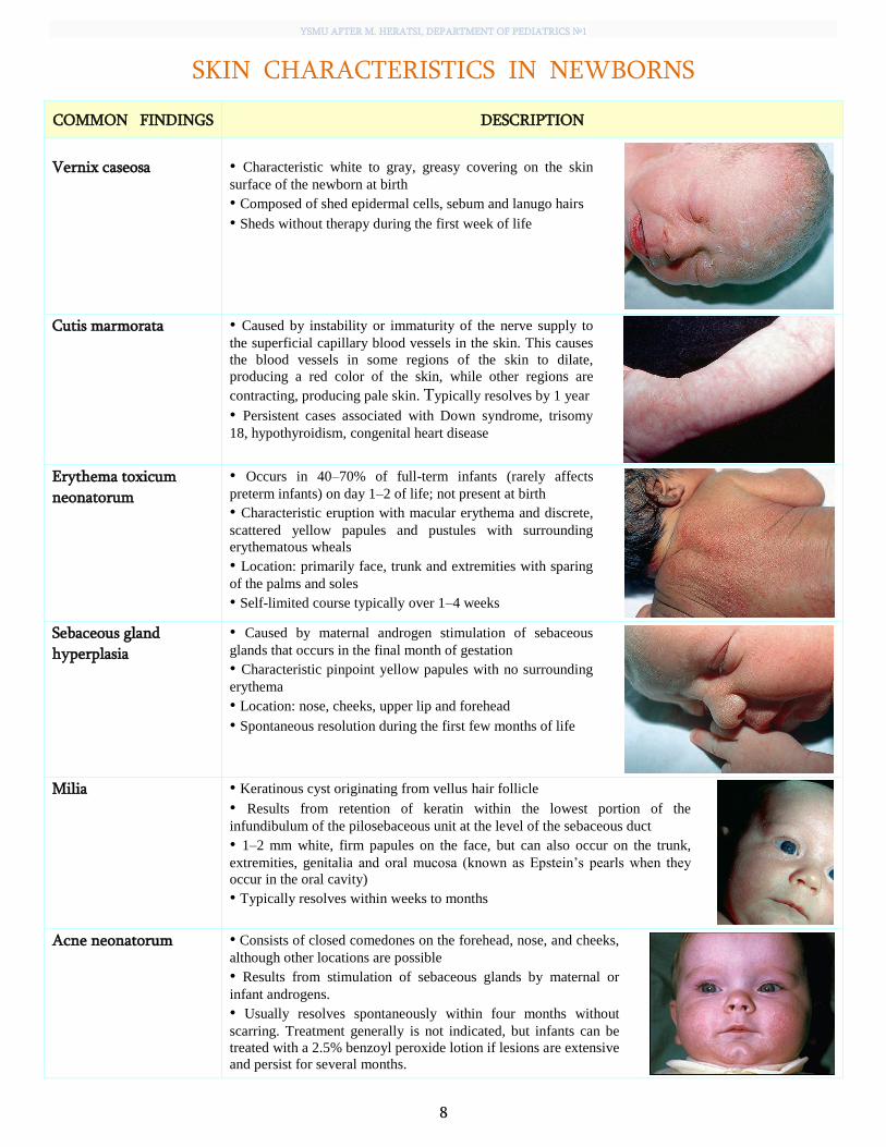
YSMU AFTER M. HERATSI, DEPARTMENT OF PEDIATRICS 1
8
SKIN CHARACTERISTICS IN NEWBORNS
COMMON FINDINGS DESCRIPTION
Vernix caseosa • Characteristic white to gray, greasy covering on the skin
surface of the newborn at birth • Composed of shed epidermal cells, sebum and lanugo hairs
• Sheds without therapy during the first week of life
Cutis marmorata • Caused by instability or immaturity of the nerve supply to
the superficial capillary blood vessels in the skin. This causes
the blood vessels in some regions of the skin to dilate,
producing a red color of the skin, while other regions are
contracting, producing pale skin. Typically resolves by 1 year
• Persistent cases associated with Down syndrome, trisomy
18, hypothyroidism, congenital heart disease
Erythema toxicum
neonatorum
• Occurs in 40–70% of full-term infants (rarely affects
preterm infants) on day 1–2 of life; not present at birth
• Characteristic eruption with macular erythema and discrete,
scattered yellow papules and pustules with surrounding
erythematous wheals
• Location: primarily face, trunk and extremities with sparing
of the palms and soles
• Self-limited course typically over 1–4 weeks
Sebaceous gland
hyperplasia
• Caused by maternal androgen stimulation of sebaceous
glands that occurs in the final month of gestation
• Characteristic pinpoint yellow papules with no surrounding
erythema
• Location: nose, cheeks, upper lip and forehead
• Spontaneous resolution during the first few months of life
Milia • Keratinous cyst originating from vellus hair follicle
• Results from retention of keratin within the lowest portion of the
infundibulum of the pilosebaceous unit at the level of the sebaceous duct
• 1–2 mm white, firm papules on the face, but can also occur on the trunk,
extremities, genitalia and oral mucosa (known as Epstein’s pearls when they
occur in the oral cavity)
• Typically resolves within weeks to months
Acne neonatorum • Consists of closed comedones on the forehead, nose, and cheeks,
although other locations are possible
• Results from stimulation of sebaceous glands by maternal or
infant androgens.
• Usually resolves spontaneously within four months without
scarring. Treatment generally is not indicated, but infants can be
treated with a 2.5% benzoyl peroxide lotion if lesions are extensive
and persist for several months.

YSMU AFTER M. HERATSI, DEPARTMENT OF PEDIATRICS 1
9
COMMON PEDIATRIC RASHES
I. ERYTHEMATOUS MACULOPAPULAR LESIONS
‘Erythema’ means redness, ‘macules’ are flat lesions, and ‘papules’ are raised lesions. Erythematous
maculopapular rashes are therefore erythematous, the redness being due to inflammation, and they may be
raised (papular), flat (macular) or a mixture of the two (maculopapular).
There are three major types of erythematous maculopapular rashes:
presents as tiny, sometimes infiltrated macules converging rapidly to form broad expanses of bright or dark red skin with rare patches of healthy skin in between, particularly in the skin folds; it is rarely pruriginous
pale pink, more or less widespread, tiny, flat macules separated by areas of healthy skin; it is not pruriginous
erythematous macules that tend to merge and are separated by patches of non pruriginous, healthy skin
Key causes include:
• eczema • drug eruptions • scarlet fever • viral exanthems • Kawasaki disease • Still's rash in systemic-onset juvenile idiopathic arthritis • malar rash (butterfly rash) • erythema chronicum migrans in Lyme disease • Gianotti-Crosti syndrome • Napkin rashes
It is important to stress that a single cause may give rise to a variety of rashes (for example, parvovirus B19) and that
the same rash can be induced by different causes (for instance, Gianotti-Crosti syndrome).
Viral exanthems can be difficult to distinguish from a drug eruption. However, viral exanthems are more common in
children, and drug eruptions tend to be more common in adults. A thorough history will aid in the diagnosis.

YSMU AFTER M. HERATSI, DEPARTMENT OF PEDIATRICS 1
10
Eczema (atopic dermatitis)
The fundamental problem in eczema is skin dryness,
which leads to the development of a scratch–itch
cycle. This in turn leads to erythema, excoriation,
bleeding and a risk of infection.
No itch? – then it’s not eczema
Drug eruptions
Immediate reactions: occur less
than 1 hour of the last
administered dose
Urticaria, angioedema, anaphylaxis
Delayed reactions: occur after 1
hour, but usually more than 6
hrs and occasionally weeks to
months after the start of
administration
Exanthematous eruptions (~90%)
Systemic reactions, eg, drug-induced hypersensitivity sindrome (DIHS), Stevens-Johnson syndrome (SJS), toxic epidermal necrolysis (TEN)
Vasculitis (may also be systemic)
Erythematous raised skin lesion
(Wheal) in acute urticaria
Localised or generalised
Well circumscribed but often coalescent
May be intensely pruritic with
excoriation
Vary in size from tiny flat papules to
large raised plaques
Flat centre with raised erythematous
edge
DIHS – (also known as drug reaction
with eosinophilia and systemic
symptoms - DRESS) is a severe,
multiorgan reaction to oral
antiepileptics that manifests as fever,
rash, lymphadenopathy, and hepatitis.
Scarlet fever is characterized by sore throat, fever,
and a characteristic red rash:
is fine, red, and rough-textured
on the face, often shows as red cheeks with a
characteristic pale area around the mouth
(circumoral pallor)
is worse in the skin folds (so-called Pastia lines)

YSMU AFTER M. HERATSI, DEPARTMENT OF PEDIATRICS 1
11
Classic Childhood Viral Exanthems
Historically, there were six childhood exanthems whose etiologies are now well-defined:
NUMBER NAME ETIOLOGY
First Disease Measles (Rubeola) Measles virus
Second Disease Scarlet Fever Streptococcus pyogenes Third Disease Rubella Rubella virus
Fourth Disease Duke’s Disease No longer accepted as a distinct disorder
Fifth Disease Erythema Infectiosum Parvovirus B19
Sixth Disease Roseola Infantum HHV-6 and HHV-7
Measles (rubeola)
Erythematous macules and papules begin on the face
and spread cephalocaudally and centrifugally, Koplik
spots.
Rubella (german measles)
Pruritic, pink to red macules and papules which
begin on the face and spread to neck, trunk and
extremities over 24hrs, 20% with Forchheimer sign
(petechial lesions on soft palate and uvula).
Erythema Infectiosum (fifth disease)
Begins with bright red cheeks and as the facial rash
fades over 1-4 days, a symmetric, erythematous,
reticular eruption appears on the trunk and
extremities.
Roseola infantum (sixth disease)
Pink macules and papules surrounded by white
halos. Begins on trunk, spreads to neck and proximal
extremities.
This is caused by HHV 6 and HHV 7. Several other
viruses – particularly the arboviruses and
enteroviruses - can cause roseoliform rashes.

YSMU AFTER M. HERATSI, DEPARTMENT OF PEDIATRICS 1
12
Kawasaki disease is a medium- and small-
vessel vasculitis.
The rash of Kawasaki disease is non-specific,
and diagnosis is made when the associated
features are also present:
• red and swollen peripheries, with skin peeling
• fevers continuing for more than 5 days
• cervical lymphadenopathy
• bilateral non-purulent conjunctivitis
• redness or cracking of lips
Still's rash in systemic-onset juvenile idiopathic arthritis
Skin rash that coincides with the fever develop in about of 95% of patients.
Characteristics of the rash are:
small, non-itchy spots or patches up to 5cm in diameter
salmon-pink coloured
usually appear on the limbs and trunk, but may occur on the face or neck
Malar rash (butterfly rash)
It is often seen in lupus erythematosus
(SLE) but is not pathognomonic - it is
also seen in other diseases such as
dermatomyositis (DM).
Malar rash in SLE Malar rash in DM
Gianotti-Crosti syndrome
It is a characteristic response of the skin to viral infection in which there is
a papular rash which lasts for several weeks. The specific viruses causing
Gianotti-Crosti syndrome include:
Hepatitis B infection
Epstein Barr virus (the cause of glandular fever)
Enterovirus infections
Echo viruses
Respiratory syncytial virus
Gianotti-Crosti syndrome mainly affects children between the ages of 6
months and 12 years.

YSMU AFTER M. HERATSI, DEPARTMENT OF PEDIATRICS 1
13
Napkin dermatitis (diaper rash)
Napkin rashes are common, although irritant reactions are much less of a problem with the widespread use of
disposable nappies, as they are more absorbent.
Causes of napkin rashes
Common Rare
• Irritant (contact)
dermatitis
• Infantile seborrhoeic
dermatitis
• Candida infection
• Atopic dermatitis
• Acrodermatitis
enteropathica
• Langerhans cell
histiocytosis
Irritant dermatitis, the most common napkin rash, may occur if nappies are not changed frequently enough or if the
infant has diarrhoea. However, irritant dermatitis can occur even when the napkin area is cleaned regularly. The rash
is due to the irritant effect of urine on the skin of susceptible infants. Urea-splitting organisms in faeces increase the
alkalinity and likelihood of a rash. The irritant eruption affects the convex surfaces of the buttocks, perineal region,
lower abdomen and top of thighs. Characteristically, the flexures are spared, which differentiates it from other causes
of napkin rash. The rash is erythematous and may have a scalded appearance. More severe forms are associated with
erosions and ulcer formation. Mild cases respond to the use of a protective emollient, whereas more severe cases may
require mild topical corticosteroids. While leaving the child without a napkin will accelerate resolution, it is rarely
practical at home.
Seborrhoeic dermatitis
Red moist rash with fine
yellow scale
Non-pruritic ± rash elsewhere
and cradle cap
Cradle cap
Involvement of face, axillae and napkin area
Candida infection may cause and often complicates
napkin rashes. The rash is erythematous, includes
the skin flexures and there may be satellite lesions.
Treatment is with a topical antifungal agent.

YSMU AFTER M. HERATSI, DEPARTMENT OF PEDIATRICS 1
14
II. VESICULOBULLOUS LESIONS
Vesicobullous lesions are rashes in which the lesions are small blisters (vesicles) or larger blisters (bullae).
Key causes include:
• varicella • shingles • hand-foot-mouth disease (HFMD) • eczema herpeticum • bullous impetigo • bullous pemphigoid • Staphylococcal scalded skin syndrome (SSSS) • Stevens–Johnson syndrome
Varicella
Varicella is an acute infectious disease caused by varicella zoster
virus (VZV). In primary infection (chickenpox) the rash is
generalized and pruritic and progresses rapidly from macules to
papules to vesicular lesions before crusting. The recurrent infection
(herpes zoster, also known as shingles) occurs when latent VZV
reactivates (mostly in adults).
Hand-foot-mouth disease
This is a common childhood illness. It
most frequently affects children
younger than 5 years. It starts with a
fever, followed by painful mouth sores
and a non-itchy rash. The rash blisters
on hands, feet, and sometimes buttocks
and legs.
It is caused by one of several serotypes
of enterovirus, typically coxsackievirus
A16.
Staphylococcal scalded skin syndrome (SSSS)
- also known as Ritter disease (in newborns). SSSS is a
generalized form of bullous impetigo. Children are more at risk
because of lack of immunity and immature renal clearance
capability (exfoliative toxins are renally excreted). Adults with
SSSS are most often chronically ill, are immunocompromised, or
have renal failure. SSSS can also appear in adults in cases with a
high burden of staphylococcal infection where the quantity of
exotoxin is significant.

YSMU AFTER M. HERATSI, DEPARTMENT OF PEDIATRICS 1
15
Severe drug
reactions
Stevens-Johnson syndrome (SJS),
Toxic epidermal necrolysis (TEN)
SJS and TEN are variants of a single
hypersensitivity disorder. SJS is defined
by epidermal detachment on less than
10% of the total body surface area; TEN
is defined by epidermal detachment on
more than 30% of the body surface area.
Mucosal involvement is more common
in SJS. In children, SJS is more common
than TEN; however, both conditions
have been described in all age-groups
from neonates to adults.
Onset is within the first 8 weeks of
exposure to the causative agent. The
usual disease course is 1 month.
III. PURPURIC LESIONS
Purpura are small areas in which blood has extravasated into the skin. Pinprick-sized areas are called petechiae.
Because the blood is outside the blood vessel, these lesions do not blanch on pressure (since the red blood cells are
fixed in place).
The key causes are:
• Henoch–Schönlein purpura • thrombocytopenia • meningococcal septicaemia
Petechiae - a small (1-2 mm) red
or purple spot on the body, caused
by a minor hemorrhage
Ecchymosis - a subcutaneous purpura larger than 1 centimeter or a hematoma,
commonly, but erroneously, called a bruise. That is, bruises are caused by trauma whereas
ecchymoses, a type of purpura, are not caused by trauma

YSMU AFTER M. HERATSI, DEPARTMENT OF PEDIATRICS 1
16
IV. SCALY LESIONS
In some conditions, a scale may form on top of a rash.
Key causes of a scaly rash include:
Pityriasis rosea Tinea versicolor Tinea capitis Psoriasis
There is a ‘Christmas tree’ distribution to the rash
is a fungal infection of the skin there are small circular areas of
depigmentation
is a fungal infection of the scalp
localised area of hair loss or alopecia, with a scaly border
the skin is thickened and inflamed, with a silvery, scaly appearance
typically occurs in patches on extensor areas
V. COMMON BIRTHMARKS
PORT-WINE STAIN (PWS) — is a flat red area due to ectatic dermal capillaries,
i.e. a vascular anomaly. These are present from birth and remain throughout life.
There may be associated eye and brain abnormalities:
Glaucoma PWS of the face with eyelid involvement may be associated
with glaucoma of the affected eye
Sturge–Weber
syndrome
PWS of the face always including the area covered by the
ophthalmic branch of the trigeminal nerve, and Ipsilateral leptomeningeal vascular abnormality with neurological
symptoms, e.g. seizures, hemiplegia
SALMON PATCH
Common pale pink vascular anomalies, present at birth on the face
(eyelids, nose, forehead) or nape of the neck
Generally fade during the first few weeks of life, except those on the nape
of the neck (known popularly as a stork bite and present in around half of
newborns) that usually persist for life

YSMU AFTER M. HERATSI, DEPARTMENT OF PEDIATRICS 1
17
Case One
Ana is 4-year-old previously healthy girl who presents with a 1 week history of cough, runny nose, fever, sore
throat and red eyes. She went to her pediatrician 2 days ago and was prescribed Augmentin (amoxicillin and
clavulanate) for presumed pharyngitis.
Yesterday, Ana developed a red rash which started on her face and has spread to her trunk. Her mother would like
to know if the rash is from her new medication.
Upon further questioning you discover that Ana has never received vaccinations due to her mother’s fear regarding
autism.
The augmentin was started 24 hours before the onset of her rash.
You also discover that a close family member recently visited from the Netherlands, who also developed a similar
rash.
Ana is an ill-appearing child who presents with a
morbilliform rash with erythematous macules and
papules.
Lesions have coalesced on the face and neck.
Rash has spread to her trunk and extremities (not
shown)
Inspection of Ana’s mouth reveals, bluish-white dots
on the mucosal surface. These are called Koplik spots.
Based on the history and exam, what is the most likely diagnosis (differential diagnosis)?
1. Drug Eruption For exanthematous drug eruptions, the initiation of the medication is often
7˗10 days before the rash
2. Erythema Infectiosum Eruption begins with bright red cheeks followed by a reticular eruption on the
trunk and extremities
3. Roseola Tends to occur in younger children with high fevers preceding a sudden rash
that begins on the trunk
4. Rubella Rash tends to spread more quickly, covering the body in 24hrs
5. Measles Rash tends to spread over a period of days
RIGHT ANSWER:

YSMU AFTER M. HERATSI, DEPARTMENT OF PEDIATRICS 1
18
Case Two
Keith is a 10-year-old boy who was brought to the pediatrician by his mother because he developed low grade
fevers, red cheeks and a new rash on his body.
Keith is up to date with his vaccinations.
How would you describe Keith’s rash?
- Confluent, erythematous, edematous plaques on the malar eminences - “slapped cheeks.” Erythematous reticular eruption on the trunk and extremities
Based on the history and exam, what is the most likely diagnosis (differential diagnosis)?
1. Drug Eruption. 2. Erythema Infectiosum. 3. Roseola. 4. Rubella. 5. Measles.
Case Three
Caleb is a 9-month-old boy who presents for evaluation of fever
and rash. His mother noted a fever of 40˚C two days ago. He
appeared well and was eating and playing normally, so his
mother was not alarmed. After the fever resolved, Caleb
developed a red rash that progressed rapidly over the past 24
hours.
Based on the history and exam, what is the most likely
diagnosis (differential diagnosis)?
1. Drug Eruption
2. Erythema Infectiosum
3. Roseola
4. Rubella
5. Measles

2016 YSMU AFTER M. HERATSI, DEPARTMENT OF PEDIATRICS 1
- 1 -
Nutrition in Children
Mother feeding baby is an art image
symbolizing peace, kindness, happiness, joy of life. But how many women often
thoughtlessly or willingly deprive themselves and the child of the gift of nature!
Kildiyarova RR
The 1,000 days between a woman’s pregnancy and her child’s
2nd birthday offer a unique window of opportunity to shape
healthier and more prosperous futures. The right nutrition
during this 1,000 day window can have a profound impact on a
child’s ability to grow, learn, and rise out of poverty. It can also
shape a society’s long-term health, stability and prosperity.
Worldwide, about 20% of deaths among children under-five could be avoided if feeding guidelines are followed.
WHO recommends:
- exclusive breastfeeding for six months,
- introducing age-appropriate and safe complementary foods at six months,
- continuing breastfeeding for up to two years or beyond.
Advantages of breastfeeding (BF)
Fig. Risk of diarrhoea by feeding method, Philippines, infants aged 0-2 months
[Popkin 1990]

2016 YSMU AFTER M. HERATSI, DEPARTMENT OF PEDIATRICS 1
- 2 -
Benefits to baby: Benefits to mother:
1. Decreases the incidence and/or severity of a lot of diseases: diarrhea, lower respiratory infection, otitis
media, bacterial meningitis, botulism, urinary tract infection, necrotizing enterocolitis
2. Possible protective effect against: sudden infant death syndrome, insulin-dependent diabetes mellitus,
Crohn's disease, ulcerative colitis, lymphoma, allergic diseases, other chronic digestive diseases
3. Promotes: childhood growth, cognitive development
4. The Influence on the development of the oral cavity:
* Breastfed babies have better jaw alignment and are less likely to need orthodontic work as they get
older. The sucking action used to breastfeed involves complex motions of the facial muscles and
tongue. This improves the development of facial muscles and the shape of the palate. The better jaw
alignment associated with BF can even mean less snoring and a lowered risk for a condition known as
obstructive sleep apnea–the blockage of air flow during sleep, which can disturb sleep patterns and lead
to other health problems.
* The action of BF also reduces the incidence of ear infections; assists with clear speech.
Increases levels of oxytocin, resulting in less
postpartum bleeding and more rapid uterine involution
Less menstrual blood loss over the months after
delivery
Earlier return to pre-pregnant weight
Delayed resumption of ovulation with increased child
spacing
Improved bone remineralization postpartum
Reduction in hip fractures in the postmenopausal
period
Reduced risk of ovarian cancer/pre-menopausal breast
cancer
Composition of breast milk
Breast milk contains all the nutrients that an infant needs in the first 6 months of life, including fat, carbohydrates, proteins, vitamins, minerals and water. It
is easily digested and efficiently used. Breast milk also contains bioactive factors that augment the infant’s immature immune system, providing protection
against infection, and other factors that help digestion and absorption of nutrients.
Colostrum Mature milk Differences between colostrum
and mature milk
During the first few days after delivery, the mother
produces colostrum. This is a thin yellowish fluid that is the
same fluid that sometimes leaks from the breasts during
pregnancy. It is rich in protein and antibodies that provide
passive immunity to the baby (the baby's immune system is
not fully developed at birth). It contains more white blood
cells than mature milk. Colostrum also helps the newborn's
digestive system to grow and function properly. It has a mild
purgative effect, which helps to clear the baby's gut of
meconium (the first dark stools). This clears bilirubin from
the gut, and helps to prevent jaundice from becoming severe.
Colostrum is rich in vitamin A which helps to reduce the
severity of any infections the baby might have.
Colostrum will gradually change to become
mature milk. In the first 3–4 days it will appear
thin and watery and will taste very sweet; later,
the milk will be thicker and creamier. Human
milk quenches the baby's thirst and hunger.
In the 1980s and 1990s, lactation professionals
(De Cleats) used to make a differentiation
between foremilk and hindmilk. But this
differentiation causes confusion as there are not
two types of milk. Instead, as a baby breastfeeds,
the fat content very gradually increases, with the
milk becoming fattier and fattier over time.

2016 YSMU AFTER M. HERATSI, DEPARTMENT OF PEDIATRICS 1
- 3 -
Anti-infective properties of breast milk Sterile feed Maternal antibodies Macrophages, neutrophils, lymphocytes Complement, Lactoferrin, Lyzozyme Promotes colonization with laktobacilli and bifidobacter
Breast milk contains white blood cells, and a number of anti-
infective factors, which help to protect a baby against many
infections.
Breast milk also contains antibodies against infections that the
mother has had in the past.
The baby should not be separated from his mother when
she has an infection, because her breast milk protects
him against the infection!
Human milk after preterm delivery
The composition of human milk for preterm infants differs from that for term infants in a number of ways. For every 100 mL, breast milk from
women delivering preterm infants is higher in calories (67 to 72 kcal versus 62 to 68 kcal), higher in protein (1.7 to 2.1 g versus 1.2 to 1.7 g),
higher in lipids (3.4 to 4.4 g versus 3.0 to 4.0 g), lower in carbohydrates, higher in multiple minerals and trace elements (especially sodium [Na],
chloride [Cl], iron [Fe], Zinc [Zn], and copper [Cu]), and higher in vitamins (especially vitamins A and E). However, as breast milk becomes
mature, many of these nutritional advantages are lost.

2016 YSMU AFTER M. HERATSI, DEPARTMENT OF PEDIATRICS 1
- 4 -
Comparison of breast milk to cow milk
Component Human Milk Cow Milk
Note
LCFAs – long chain fatty
acids
MCFAs – medium chain fatty
acids
Water/solids Same Same
Calories 20 cal/oz 20 cal/oz Protein 1–1.5% (whey dominant) 3.3% (casein dominant) Carbohydrate 6.5–7% lactose 4.5% lactose
Fat high in LCFAs high in MCFAs Minerals Iron better absorbed Low iron and copper Vitamins Diet dependent, low in K, D Low in C, D
Digestibility Faster emptying Same after 45 days Renal solute load Low (aids in renal function) Higher
Nutrients in human and animal milks
Summary of Major Differences Between Human Milk and
Commercial Substitutes Marketed for Normal Term infants
Quality of proteins in different milks

2016 YSMU AFTER M. HERATSI, DEPARTMENT OF PEDIATRICS 1
- 5 -
Physiology of Lactation
Some mothers think their breasts are too small to produce enough milk. What is the difference between large breasts and small breasts?
It is the fat and other tissue which gives the breast its shape, and which makes most of the difference between large and small breasts. Small breasts and large breasts both contain about the same amount of gland tissue, so they can both make plenty of milk.
Lactation (milk production) occurs after childbirth and involves two components: milk secretion and milk removal.
1) Milk secretion (synthesis and release) is a continuous process, which is promoted by PROLACTIN. Prolactin secretion, in turn, increases by
continued breastfeeding
2) Milk removal or ejection (“let-down” refex) is intermittent and triggered by OXYTOCIN.
Normal term infants are born with a number of reflexes and behaviors to help assure that the newborn survives the transition from intra to
extra uterine life. These reflexes enable him to begin feeding immediately after birth (see "The digestive system").
Breastfeeding involves a set of reflexes and hormones that also drive the milk supply.
SUCKLING REFLEX
The stimulation of sensory nerves in the
breast by the infant initiates the suckling
reflex. Unlike ordinary reflexes with
only neural components, the afferent arc
of the suckling reflex is neural and the
efferent arc is hormonal. The suckling
reflex increases the release of prolactin,
oxytocin, and ACTH and inhibits the
secretion of gonadotropins.
Suckling on one breast causes milk flow
not only in that breast but also in the
opposite breast.
Fig. Effect of suckling on hypothalamic, pituitary, and adrenal hormones.
GnRH, gonadotropinreleasing hormone; CRH, corticotropin-releasing
hormone; DA, dopamine; PRF, prolactin-releasing factor; FSH, follicle-
stimulating hormone; LH, luteinizing hormone; ACTH,
adrenocorticotropic hormone; PRL, prolactin; OT, oxytocin.

2016 YSMU AFTER M. HERATSI, DEPARTMENT OF PEDIATRICS 1
- 6 -
“THE LAW OF DEMAND AND SUPPLY”
Milk production is influenced positively by early frequent and effective milk removal and negatively
by late infrequent feeds or by the feeding the baby other liquids or foods before six months of age.
The frequency of feeding regulates milk supply. The more often a baby removes the milk the greater
the milk supply. Conversely, a baby who sleeps many hours at a time in the early weeks or feeds less
than an average of eight times in 24 hours does not have the opportunity to stimulate the breast,
causing the milk supply to drop. Because each breast responds to the amount of milk demanded by
the infant, it is possible to exclusively breastfeed more than one baby at a time or to use only one
breast. Initially, if the milk is not removed the breast becomes full and eventually engorged. At that
point, a local factor known as the feedback inhibitor of lactation (FIL) begins to decrease milk
secretion. The exact mechanism of FIL is still under study.
Sometimes people suggest that to make a mother produce more milk, we should give her more to eat, more to drink, more rest, or medicines. It
is important for a mother to eat and drink enough, but these things do not help her to produce milk if her baby does not suckle.
Some special things to remember about
prolactin are:
The prolactin level is highest about 30
minutes after the beginning of the feed, so its
most important effect is to make milk for the
next feed. For this feed, the baby takes the
milk which is already in the breast.
More prolactin is produced at night; so
breastfeeding at night is especially helpful for
keeping up the milk supply.
Hormones related to prolactin suppress
ovulation so breastfeeding can help to delay a
new pregnancy (lactational amenorrhea).
Breastfeeding at night is important for this.
Why is it important to understand the oxytocin reflex in the way we care for mothers after delivery? A mother needs to have her baby near her all the time, so that she can see, touch and
respond to him. If a mother is separated from her baby between feeds, her oxytocin
reflex may not work so easily.
You need to remember a mother’s feelings whenever you talk to her. Try to make her
feel good and build her confidence. Try not to say anything which may make her doubt
her breast milk supply.

2016 YSMU AFTER M. HERATSI, DEPARTMENT OF PEDIATRICS 1
- 7 -
Normal variations in breastfeeding
Frequency of feeds
Breastfed infants usually feed every 2–5 hour from both breasts each feed.
Young babies feed frequently, but demand feeding (i.e. feeding when
hungry) will usually have the baby settle into a fairly predictable pattern
of feeds. The frequency of feeds is determined by the baby’s appetite and
gastric capacity, as well as the amount of mother’s milk available.
Length of feeds
The duration of the feed is determined by the rate of transfer of milk from
the breast to the baby, which, in turn, depends on the strength of the
baby’s suck and the force of the mother’s ‘let down’ or flow of milk. This
may vary from 10–30 minutes. Young infants tend to feed for longer. It is
the cessation of strong drawing sucks and the appearance of shorter-
duration bursts of sucking that indicate the ‘end’ of the feed – not the
time.
Appetite spurts
Babies seem to experience appetite spurts at 2 weeks, 6 weeks and 3
months. During a growth spurt, a baby will suddenly begin to feed more
frequently. It is crucial that parents are aware of this or the baby’s natural
increase in feed frequency may be mistaken for diminution of milk
supply. This is especially true at 6 weeks when breasts are no longer
carrying extra fluid and the supply is settling to the demands of the baby.
Unfortunately, many women wean at this time through poor advice.
During appetite spurts, let the baby feed on demand, even 2 hourly, and
this pattern should settle in 48 hours.
How to assess good breastfeeding
Source: Murray S. Breast Feeding Information and Guidelines for Paediatric Units, Royal Children’s Hospital,
Melbourne, Australia, 1992.
The most important criterion for assessing the milk
transfer during a feeding at the breast is audible swallow!

2016 YSMU AFTER M. HERATSI, DEPARTMENT OF PEDIATRICS 1
- 8 -
Contraindications to breastfeeding
Infant Conditions Maternal Conditions
Inborn errors of metabolism:
Classic galactosemia: a special
galactose-free formula is needed
Maple syrup urine disease: a
special formula free of leucine,
isoleucine and valine is needed
Phenylketonuria: a special
phenylalanine-free formula is
needed (some breastfeeding is
possible, under careful
monitoring)
• Infections: Mothers with human T-cell lymphotropic virus (HTLV) types I and II should not
breast-feed. For mothers with active tuberculosis (before treatment), peripartum development of varicella, and herpes simplex (when lesions are present on the breast), expressed breast milk
can be used, but infants should not feed directly from the breast. In the industrialized world, it
is not recommended that HIV-positive mothers breast-feed. However, in the developing world,
the risks of malnutrition and infectious diseases may outweigh the risk of acquiring HIV from
breastfeeding. A 2009 Cochrane review demonstrated that in areas endemic for HIV, infants
who were exclusively breast-fed for 3 months had a lower risk of HIV acquisition than infants
fed a combination diet of human milk and other foods.
• Substance abuse or use: Cocaine, stimulants, and marijuana.
• Medications: Sulfonamides (for infants with hyperbilirubinemia or glucose-6-phosphate
dehydrogenase deficiency), radioactive medicines, chemotherapeutic agents (alkylating agents),
bromocriptine (suppresses lactation), and lithium (in general, psychotropic drugs should be
used with caution).
Feeding terms and definitions
EXCLUSIVE BREASTFEEDING: the infant takes only breast milk and no additional food, water, or other fluids with the exception of
medicines and vitamin or mineral drops.
PARTIAL BREASTFEEDING or MIXED FEEDING: the infant is given some breast feeds and some artificial feeds, either milk or cereal, or
other food or water.
BOTTLE-FEEDING: the infant is feeding from a bottle, regardless of its contents, including expressed breast milk.
ARTIFICIAL FEEDING: the infant is given breast-milk substitutes and not breastfeeding at all.
REPLACEMENT FEEDING: the process of feeding a child of an HIV-positive mother who is not receiving any breast milk with a diet that
provides all the nutrients the child needs.
COMPLEMENTARY FEEDING: the process of giving an infant food in addition to breast milk or infant formula, when either becomes
insufficient to satisfy the infant's nutritional requirements.

2016 YSMU AFTER M. HERATSI, DEPARTMENT OF PEDIATRICS 1
- 9 -
Artificial feeding (infant furmula)
Infant formula is usually made from cows' milk that has been treated to make it suitable for babies. The concentration of protein and
electrolytes such as sodium, potassium and chloride are lower than in cow’s milk, while the levels of certain minerals, primarily iron and to a
lesser extent zinc are higher.
Infant formula is available in two forms:
ready-to-feed liquid infant formula, sold in cartons, which is sterile;
powdered infant formula, which is not sterile.
First infant formula
This is often described as suitable for newborns. It is based on the whey of cows' milk and is thought to be easier to digest than
other types of infant formula.
The baby can stay on this formula when you start to introduce solid foods at around six months and continue on it throughout
the first year. When the baby is one year old, they can start to drink whole cows' milk. There is no evidence to suggest that
changing the brand of infant formula the baby drinks does any good or harm.
Casein-based infant
formula
Infant formula that is mostly based on casein is thought to take the baby longer to digest than whey-based formula. It is not
recommended for young babies. There is little nutritional difference between this formula and first infant formula. Although it
is often described as suitable for "hungrier babies", there is no evidence that babies settle better or sleep longer when fed this
type of formula.
Follow-on formula
This is also casein-based and should never be fed to babies under six months old. Research has found no clear benefit from its
use. However, the labels on this formula can look very similar to those on first infant formula, so read them carefully to avoid
making a mistake.
Specialized formula
Specialised formulas are available to use in conditions such as reflux, high-energy need, lactose intolerance, allergic conditions
and metabolic diseases like phenylketonuria. These formulas are altered in one or more nutrients and should only be used for
infants with the specific conditions under medical supervision.
Complementary feeding
Complementary feeding means giving other food in addition to breast milk.
After 6 months of age to meet their evolving nutritional requirements, all infants should receive nutritionally adequate and safe
complementary food while breastfeeding continues until up to 2 years of age or beyond.
HONEY SHOULD NOT BE FED TO INFANTS YOUNGER THAN 1 YEAR OLD !

2016 YSMU AFTER M. HERATSI, DEPARTMENT OF PEDIATRICS 1
- 10 -
Table: Introducing complementary food
Baby’s Age Number of meals per day Average meal size Consistency Suitable foods
Around 6
months
(Stage 1)
2-3 meals/ day 5-10 teaspoons Smooth thin purée without
any lumps
Cereals e.g. baby rice, porridge, baby cereal. Puréed vegetables e.g. carrots, squash, potato. Peeled and puréed fruit e.g. banana and pear. Well cooked chicken, fish, meat and eggs.
6-9 months
(Stage 2) 3 meals/ day
2-4 tablespoons
Thick with soft lumps Well-cooked eggs, chicken or mince. White and oily fish (boned and skinless). Yoghurt, pasteurised cheese, bread, pasta, noodles, rice.
9-12 months
(Stage 3)
3 plus 2-3 snacks
4-6 tablespoons
Chunky, mashed texture, moving to chopped, bite size
pieces
Most family foods are now suitable but do not add sugar or salt. Finger Foods: mango, melon, banana, toast fingers, cheese, pasta shapes.
Risks of starting complementary foods too early
Take the place of breast milk, making it difficult to meet the child’s
nutritional needs and result in a low nutrient diet;
Increase risk of illness because less of the protective factors in breast
milk are consumed;
Increase the risk of diarrhea because the complementary foods may
not be as clean or as easy to digest as breast milk;
Increase the risk of wheezing and other allergic conditions because
the baby cannot yet digest and absorb other foods well;
Increase the mother’s risk of another pregnancy if breastfeeding is
less frequent.
Risks of starting complementary foods too late
Delaying the introduction of complementary foods for too long is also not
advisable because:
Breast milk alone may not provide enough energy and nutrients
and may lead to growth faltering and malnutrition;
Breast milk alone may not meet the infant’s growing requirements
of some micronutrients, especially iron and zinc;
The optimal development of oral motor skills, such as the ability
to chew, and the infant’s ready acceptance of new tastes and
textures may be adversely affected.
Infants should, therefore, be started on complementary foods at around six
months of age.

2016 YSMU AFTER M. HERATSI, DEPARTMENT OF PEDIATRICS 1
- 11 -
Dental health and nutrition
Breastfeeding Bottle-feeding
Although breast milk alone is not cariogenic, it may become
cariogenic when combined with other carbohydrate sources.
When a child is breastfed on demand, with high frequency
and duration at night, it is important to implement oral
hygiene following feedings.
Stop night feedings once teeth erupt. The majority of infants
are physiologically able to tolerate a prolonged fast around 6
months of age, which is when teeth typically begin to erupt.
Bottles should only be used with formula, breast milk, or water. Fruit juices, soft
drinks, sweet teas, formula, or milk should not be put in a baby’s bottle during
bedtime or nap time. At these times, bottles should only contain water.
Infants should be held when bottle-fed. If a bottle is given with anything other than
water at nap timeor bedtime, parents should use a cloth to wipe the baby’s mouth
prior to laying the baby down.
Bottles should not be propped with infants in cribs or car seats. Prolonged and
frequent exposure to sugary liquids contributes to the caries process, and children
who drink bottles while lying down may be more prone to ear infections.
Feeding infants with craniofacial anomalies
Babies with isolated clefts of the lip and/or palate can usually feed by mouth with some adjustments to bottle‐feeding techniques. Tube feeding is rarely
required.
Fig. Upright positioning
during breastfeeding of a
baby with cleft lip/palate
The strategies that have been developed to feed infants with clefts of the palate are designed to
overcome the lack of negative pressure developed during sucking. These include:
a. Cross‐cutting fissured nipples b. Squeezing a soft bottle to help with the flow of milk c. Pumping the breasts to deliver breast milk via bottle d. Developing patience in feeding e. Feeding instruction and follow-up with a feeding specialist on the cleft palate team
It is important to ensure that the energy that a child expends during feeding does not exceed the
nutritional and caloric intake from the feeding. This problem may occur if feeding takes more
than 30 minutes.
Steady weight gain is the most important indicator of adequate food intake. Close follow-up
with a pediatrician or other health care provider is necessary to ensure that consistent weight
gain is achieved.
Often a cleft palate is temporarily covered by a palatal obturator (a prosthetic device made to fit
the roof of the mouth covering the gap).
Fig. Proper bottle-feeding
technique of a baby with
cleft lip/palate

2016 YSMU AFTER M. HERATSI, DEPARTMENT OF PEDIATRICS 1
- 12 -
Calculation of breast-milk substitutes daily requirements
Volumetric method
The daily volume of breastmilk sunstitute for infants from 10 days to 2 months of age makes 1/5 of body weight, 2-4 months -1/6, 4-6
months - 1/7
2-6 old child should get 150 ml of breastmilk suntitute per kg of body weight
Calorimetric method (energy density in a formula is 65 kcal/100 ml)
Premature infants up to 3-4 months demand about 120-140 kcal per kg of weight per day
Term infants up to 3-4 months demand 110-120 kcal per kg per day
As the infant grows the demand per body weight decreases and for the one years old infant is about 100 kcal per kg per day
The daily volume of the milk (ml) = weight (kg) x caloric requirment (kcal/kg/day) / 0. 7
The calculated amount should not be more than one litre and has only orientational meaning.
Home-prepared formula
The milk used as the base for home-prepared formula may be:
Fresh milk that is heat-treated at home,
Commercially heat-treated whole milk (such as UHT or sterilized milk),
Powdered full cream milk, or
Non-sweetened evaporated milk.
o The animal milks used may be from cows, buffalo, goat, ewe, camel or other animal.
o In full strength full cream milk, the level of protein and some minerals is too high, and it is difficult for an infant's immature kidneys to
excrete the extra waste. These milks require some modification to make the proportions more appropriate.
o WHO recommends the following recipe for home prepared formula:
Boil 70 ml of water
Add 130 ml of boiled cow’s milk to make 200 ml of feed
Add 1 level teaspoonful (5g) of sugar
For sheep’s milk the milk and water amounts should be 100 ml to100 ml

2016 YSMU AFTER M. HERATSI, DEPARTMENT OF PEDIATRICS 1
- 13 -
Principles of a Balanced Nutritious Diet for Children over 1 Year
Foods can be divided into five food groups based on the nutrients
they contain, and an average daily combination of a number of
servings from each food group provides nutritional adequacy.
The food groups and number of servings children require are:
• bread, rice, potatoes, pasta and other starchy foods – include at
each meal and some snacks: 3–5 servings
• fruit and vegetables – include at each meal and some snacks:
about five servings
• milk, cheese and yogurt – three servings per day
• meat, fish, eggs, nuts and beans – two servings per day or three
for vegetarians
• foods high in fat and foods high in sugar – small amounts that do
not replace the other food groups.
Sweet foods should be limited to four times per day.
Between six and eight drinks should be offered each day.
The number of average daily servings of each food group is the same throughout childhood from 12 months of age but portion sizes of food
and drinks increase as children grow and their energy and nutrient requirements increase.
Assessing the average number of daily servings from each food group can be used to help parents and children make changes to improve the
nutritional adequacy of their diets.
Nutrient-dense vs. Energy-dense Food
Nutrient-dense foods are characterized by a high amount of
nutrients such as vitamins, calcium, fiber (e.g. mg/g, g/L):
Fruits, vegetables, whole grains
Fat-free or low-fat milk and milk products, yogurt
Lean meats, poultry, fish, beans, eggs, and nuts
Foods with added sugar or fat are usually not nutrient-dense.
Energy-dense food items are characterized by a high amount of
calories (e.g. kcal/g, kcal/L):
Chocolate
French fries, crackers
Regular sodas and alcoholic drinks
Most nutrient-dense foods are not energy-dense, but nuts are an exception.

2016 YSMU AFTER M. HERATSI, DEPARTMENT OF PEDIATRICS 1
- 14 -
Table. Food groups and recommended daily servings [Adapted from More J. Infant, Child and Adolescent Nutrition: A Practical Handbook. CRC Press, 2013.]
Food groups Foods included Main nutrients
supplied included supplied
Recommendations
Infants 6–12 months
Preschoolers 1–4 years
School children and adolescents
Group 1: Bread, rice potatoes, pasta and other starchy foods
Bread, chapatti, breakfast cereals, rice, couscous, pasta, millet, potatoes, yam, and foods made with flour such as pizza bases, buns, pancakes
Carbohydrate B vitamins Fibre Some iron, zinc and calcium
3–4 servings a day Serve at each meal and some snacks
Serve at each meal and some snacks
Group 2: Fruit and vegetables
Fresh, frozen, tinned and dried fruits and vegetables
Vitamin C Phytochemicals Fibre Carotenes
3–4 servings a day Offer at each meal and some snacks – about 5 small servings a day
Serve at each meal and some snacks – aim for 5 servings a day
Group 3: Milk, cheese and yogurt
Breast milk, infant formulas, follow-on milks, cow’s milk, goat’s milk, yogurts, cheese, milk puddings, toddler milks, calcium-enriched soya milks, tofu
Calcium Protein Iodine Riboflavin
Demand feeds of breast milk or infant formula as main milk drink (decreases from about 1000 mls/day down to about 500 mls/day as food in take increases) Some yogurt and cheese
3 servings a day 1 serving is: – 120 mL milk drink – 1 pot yogurt (120 g) – a serving of cheese in a sandwich or on a pizza – a milk-based pudding – a serving of tofu
3 servings a day 1 serving is: – 150–250 mL milk drink – 1–2 pots yogurt (120 g) – a serving of cheese in a sandwich or on a pizza – a milk-based pudding – a serving of tofu
Group 4: Meat, fish, eggs, nuts and pulses
Meat, fish, eggs, nuts and pulses (lentils, dahl, chickpeas, hummus, kidney beans and other similar starchy beans)
Iron Protein Zinc Magnesium B vitamins Vitamin A Omega 3 longchain fatty acids: EPA and DHA* from oily fish
1–2 servings a day 2–3 for vegetarians
2 servings a day or 3 for vegetarians Fish should be offered twice per week and oily fish at least once per week
2 servings a day or 3 for vegetarians Fish should be offered twice per week and oily fish at least once per week
Group 5: Foods and drinks high in fat and/or sugar
Cream, butter, margarines, cooking and salad oils, mayonnaise, chocolate, confectionery, jam, honey, syrup, crisps and other high-fat savoury snacks, biscuits, cakes Fruit juices and sweetened drinks
Some foods provide: – vitamin E – omega 3 fatty acids: alphalinolenic acid
In addition to but not instead of the other food groups
In addition to but not instead of the other food groups
Fluids
Drinks Water
Fluoride in areas with fluoridated tap water
Milk feeds and drinks of water offered with meals
6–8 drinks per day – each of 100–120 mL. More in hot weather or after extra physical activity
6–8 drinks per day – each of 150–250 mL. More in hot weather or after extra physical activity
Vitamin supplements
Vitamins D for breastfed
infants and formula-fed infants drinking less than 500 mL formula milk/day
Folic acid and vitamin D for adolescent girls who could become pregnant and during pregnancy
* DHA, docosahexaenoic acid; EPA, eicosapentaenoic acid

2016 YSMU AFTER M. HERATSI, DEPARTMENT OF PEDIATRICS 1
- 15 -
Nutrition for Preschool Children 1–4 Years Nutrition for School Age Children 5-10 Years
1–4 year olds need a balanced diet based on a combination of
foods from the five food groups.
A daily meal and snack routine of 3 meals and 2–3 planned
nutritious snacks will provide adequate calories and nutrients and prevent toddlers becoming over hungry or too tired to eat.
Toddlers’ appetites vary from day to day and they should be
allowed to eat to their appetite and finish eating when they have had enough.
Snacks given should always be nutritious snacks as toddlers
are unlikely to eat an adequate amount of nutrients in just three meals.
High-calorie low-nutrient foods should be limited to small
quantities and offered as part of a meal.
Teeth of under-fives are vulnerable to dental decay and sugary
and acidic foods should be limited to four occasions per day (e.g. three meals and one snack).
Children prefer familiar foods and most need to be motivated to try
new foods.
Children innately prefer sweet, salty and energy-dense foods.
Parents and carers are responsible for food offered to primary
school age children and have the power and influence to change eating habits. In a new policy statement, the American Academy of Pediatrics (AAP) urges schools and families to take a broader approach to nutrition, considering children's whole diet pattern – rather than the amount of sugar, fat or specific nutrients in individual foods [Murray et al. 2015]. The AAP recommends a five-step approach parents and schools can take in selecting food for packed lunches and social events: Select a mix of foods from the five food groups: vegetables, fruits,
grains, low-fat dairy, and quality protein sources, including lean meats, fish, nuts, seeds and eggs.
Offer a variety of food experiences. Avoid highly processed foods. Use small amounts of sugar, salt, fats and oils with highly
nutritious foods to enhance enjoyment and consumption. Offer appropriate portions.

2016 YSMU AFTER M. HERATSI, DEPARTMENT OF PEDIATRICS 1
- 16 -
Nutrition for Adolescents Adolescents' nutritional needs are influenced by a number of factors.
Biologic considerations include enhanced needs related to an increased growth rate and
change in body composition; males have a greater blood volume and leaner body mass than
females, who need a minimum fat mass for menstruation and reproduction. In addition to
increased energy needs, growth in lean body mass will require increased intake of calcium, iron,
nitrogen, zinc, magnesium, fluoride, and vitamins A, C, and D. Acute and chronic illness,
trauma, and stress will add to the needs imposed by growth and physical activity.
Behavioral considerations during the adolescent years include changes in food habits related
to levels of activity, changes in schedules, and a desire for independence, resulting in skipped
meals, more frequent snacking, and more meals outside the home (including fast food).
Psychological changes and emotional difficulties at this age can lead to eating disorders. Last, dissatisfaction with body image may be associated
with frequent dieting. Because boys usually eat more food than girls, their risk of deficiencies is reduced.
QUIZ
You are seeing a 16-year-old girl for her annual health supervision visit. The girl has become a vegan, and her mother is concerned about possible
nutritional deficiencies. The girl has no symptoms and has not lost any weight in the last 6 months. Her menses are regular and her physical examination
findings are unremarkable. Of the following, the girl's diet puts her at GREATEST risk of a deficiency of
A. calcium
B. iron
C. vitamin A
D. vitamin B12
E. vitamin D
Typically, vegetarian diets are broken into three major groups including lacto-ovo vegetarians (whose diets include eating dairy products and
eggs), lacto vegetarians (who eat dairy, but avoid eggs), and vegan vegetarians (those who avoid animal products altogether).
As a vegan, the girl described in the vignette is most likely to develop a deficiency in B12, a vitamin almost exclusively found in animal
products. Vegans require supplements or fortified food products to meet their need for this vitamin. Because the high intake of folic acid in
vegan diets may mask the hematologic aspects of vitamin B12 deficiency, diagnosis may be delayed until neurologic symptoms occur. Other
potential deficiencies associated with a vegan diet include calcium, iron, zinc, vitamin A and D, and perhaps other trace elements, but adequate
intake can be ensured by following recommended vegan food group allowances and serving sizes.

2016 YSMU AFTER M. HERATSI, DEPARTMENT OF PEDIATRICS 1
- 17 -
PEARLS
• Adolescents' eating habits put them at risk
for nutritional deficiencies.
• Vegan diets pose potentially more serious
health risks (deficiency of vitamin B12 but to
a lesser extent vitamins A and D and calcium,
iron, and zinc) compared with vegetarian
diets.
• Vegans/vegetarians may have an underlying
eating disorder.
Table. Eating behaviors of adolescents
REFERENCE
1. Lawrence RM, Lawrence RA. Breastfeeding: more than just good nutrition. Pediatrics in review/American Academy of Pediatrics. 2011 Jul;32(7):267.
2. AAP: Pediatric Nutrition Handbook, ed 7. Elk Grove Village IL, 2014.
3. World Health Organization (2009). The optimal duration of exclusive breastfeeding: report of an expert consultation. 2002. World Health Organization: Geneva.
4. American Academy of Pediatrics: Policy statement: Breastfeeding and the use of human milk, Pediatrics, 129: e827–e841, 2012.
5. Horvath T, Madi BC, Iuppa IM, et al: Interventions for preventing late postnatal mother-to-child transmission of HIV, Cochrane Database Syst Rev 1:CD006734, 2009.
6. Murray et al. "Snacks, sweetened beverages, added sugars, and schools." Pediatrics 135.3 (2015): 575-583.

2016 YSMU AFTER M. HERATSI, DEPARTMENT OF PEDIATRICS 1
- 18 -
CASE: FOREMILK SYNDROME
CLINICAL PRESENTATION
A 20-day-old newborn was brought to outpatient department (OPD) for the 3rd time
with many complaints. Baby passes motion frequently with every feed and few drops of
motion with every cry, cough and otherwise. Baby regurgitates small amounts of milk
often and is cranky most of the time.
Mother gives history that baby is sucking vigorously, but, only for few minutes, goes off
to sleep and demands after an hour again. The baby’s perianal area is red and ulcerated
(fig).
TREATMENT AND FOLLOW-UP
Mother is educated that 1st milk (foremilk) is rich in ‘sugars’ and has short gastric emptying time, baby gets hungry frequently, but frequent
feeding finds the baby with only little hunger. Baby sucks for a short while (<10 minutes) and gets only foremilk. Drinking only foremilk is the
cause of frequent stool and regurgitation. Mother gradually spaces the feeds to 2.5–3 hours, feeds the baby for 15 minutes from one breast in one
sitting. The baby’s symptoms rapidly improve.
DIAGNOSIS
Foremilk syndrome.
INVESTIGATION
Good history.
DISCUSSION
Educating mothers on lactation by demand than by schedule, from one breast in one sitting and recognizing that every cry is not hunger is very
important.
LEARNING POINT
Hindmilk is important for weight gain and is possibly a cure for ‘colic’.

Grigoryan M E
DE
PA
RT
ME
NT
OF
PE
DIA
TR
ICS
1
20
17
YE
RE
VA
N S
TA
TE
ME
DIC
AL
UN
IVE
RSI
TY
AF
TE
R M
KH
ITA
R H
ER
AT
SI

GRIGORYAN M E YSMU AFTER M. HERATSI, DEPARTMENT OF PEDIATRICS 1
~2~
THE SKELETAL SYSTEM
The skeletal system is comprised of different structures: bones, joints, cartilage , and ligaments.
Most bones form by replacing cartilage. The presence of cartilage is essential for bone growth and development. The cartilage ability to grow fast, while maintaining a sufficient degree of density makes it exceptionally favorable skeletal material for the fetus.
BONE TISSUE
Bone tissue is a unique connective tissue, 90-98% of which consists of intercellular substance – bone matrix. It contains large
amounts of minerals, mainly calcium phosphate salts.
Bone matrix Bone cells (2-10%)
Osteoid (organic matrix) Minerals Osteoblasts Osteocytes Osteoclasts
90-95% Collagen type1
5-10% Noncollagenous proteins: osteocalcin, osteopontin,
osteonectin etc.
95% Calcium
phosphate salts 1. Plump cuboidal single
nucleated cells that synthesize
and deposit bone matrix.
2. Derived from mesenchymal
stem cell lineage.
Bone lining cells are inactive
osteoblasts that cover all
available bone surfaces.
1. A derivative of
osteoblasts created
when osteoblasts are
encased in bone matrix
2. Connected to other
osteocytes in a
network via cell
processes running
through canaliculi
1. Giant multi-
nucleated cells
2. Secrete acids and
enzymes
(collagenase) to
break down bone
matrix
3. Derived from
macrophage lineage
Within the bone tissue phosphate salts of calcium are presented in two forms: amorphous calcium phosphate Ca3(PO4)2; crystals of hydroxyapatite Ca10(PO4)6(OH)2. In early childhood amorphous masses of calcium phosphate are more predominant than crystals of hydroxyapatite. So their bones are demineralized easier than those in adults.
TYPES OF BONE TISSUE
In comparison with adults, in the skeleton of children trabecular bone is more predominant than the cortical bone. A child's bones are more plastic and more porous than adult bone, with wider haversian canals.
Bone tissues
Trabecular (spongy or cancellous) bone - consists of irregularly arranged lamellae
Cortical (compact) bone - consists of concentrically arranged lamellae with central
(Haversian) canals
It’s mineralized by
15-25% and predominates
in the axial skeleton
It’s mineralized by
80-90% and predominates
in the extremities

GRIGORYAN M E YSMU AFTER M. HERATSI, DEPARTMENT OF PEDIATRICS 1
~3~
BONE DEVELOPMENT
During the development of bones three main processes occur: osteogenesis, modeling and remodeling.
OSTEOGENESIS MODELING REMODELING
Bone formed on soft tissue:
Two major subclassifications:
- Intramembranous: bone formed on soft
fibrous tissue
- Endochondral: bone formed on cartilage
Bone formed on existing bone
tissue
Bone both resorbed and
formed at the same site
Occurs during embryonic development, early
stages of growth, and during healing
Occurs during growth, and during
healing
Occurs from growth
through death
In adults only remodeling occurs, while in children all processes mentioned above occur with high intensity.
Endochondral ossification, in which mesenchymal cells
condense and undergo chondrogenesis to form cartilage
that matures and hypertrophies, becomes calcified, and
is then replaced by bone. Most bones in the axial and
appendicular skeleton are formed in this way.
Intramembranous ossification, in which osteoblasts
are formed by direct differentiation of
mesenchymal cells with no cartilage precursor or
model. Flat bones of the skull and clavicle are
formed in this way.
Centers of ossification:
Primary centers of ossification: At the beginning of the fetal period the chondrocytes in the midshaft of long bones
from the primary centers, growth from which eventually lengthens the bone.
Secondary centers of ossification: These appear in the chondroepiphysis and mostly appear postnatally. They direct the
formation of bone throughout growth.
The ossification centers that are typically present at term birth are the distal femur, proximal tibia, calcaneus, and talus.

GRIGORYAN M E YSMU AFTER M. HERATSI, DEPARTMENT OF PEDIATRICS 1
~4~
The many ossification centers of the body—hand, foot, knee, elbow, and pelvis, for example—are not visible by
radiography (x rays) until they begin to mineralize or ossify, even though they are actually present long before such
mineralization begins. The age at appearance of individual ossification centers then becomes a useful measure of
skeletal development and especially in the form of "bone age" assessments of the hand, foot, or knee. Such assessments,
made by taking a series of radiographs and comparing them against appropriate standards, are both highly reliable and
useful estimates of the stage of physical development. Bone age assessments are, therefore, used in pediatric evaluation,
especially when malnutrition, malabsorption, food intolerance, or endocrinopathies (such as hypopituitarism or
hypothyroidism) are suspected. Bone age assessments also have forensic application, such as estimating the
chronological age of a cadaver. In addition, they can provide data for making age assessments for children whose birth
date in unknown or for whom a birth certificate does not exist or is suspected of being inaccurate. Families adopting
infants or children from countries in which there has been socioeconomic stress may find bone age assessment helpful
in establishing the chronological age their adopted child has attained.
Depiction of the order of appearance of the individual carpal bones. The
usual sequence is: capitate (1), hamate (2), triquetral (3), lunate (4),
trapezium (5), trapezoid (6), navicular or scaphoid (7) and pisiform (8). The
distal epiphysis of the radius ossifies before the triquetum and that of the
ulna before the pisiform.
The Greulich-Pyle atlas is commonly used for bone age assessment.
Assessments of skeletal maturity in pre-pubertal children are primarily based
on the epiphyseal size of the phalanges as they relate to the adjacent
metaphyses. During this stage of development, the ossification centers for the
epiphyses increase in width and thickness, and eventually assume a transverse
diameter as wide as the metaphyses.
As in pre-pubertal children, assessments of skeletal maturity in early and mid-
puberty are also based on the size of the epiphyses in the distal phalanges (first)
and the middle phalanges (second). The epiphyses at this stage continue to
grow and their widths become greater than the metaphyses.
Assessments of skeletal maturity in late puberty stage are primarily based on
the degree of epiphyseal fusion of the distal phalanges.
In post-pubertyall carpals, metacarpals and phalanges are completely
developed, their physes are closed, and assessments of skeletal maturity are
based on the degree of epiphyseal fusion of the ulna and radius.
The normal variability of skeletal age is about 10 percent of attained chronological age. Thus, some chronological
12-year-olds may be assessed as 14 years of age in terms of skeletal development, while others may be assessed as
ten. Bone age is useful in projecting final stature; research has shown that it is more meaningful in making such
projections than chronological age alone.

GRIGORYAN M E YSMU AFTER M. HERATSI, DEPARTMENT OF PEDIATRICS 1
~5~
Typical long bones are
divided into:
Physis, which is the
growth plate located at
the end of bone
Epiphysis, which is
typically the secondary
ossification center
Metaphysis, which is the
bone adjacent to the
physis on the side away
from the joint
Diaphysis, which is the
central part or shaft of
long bones
The long bones of the extremities (humerus, radius-ulna, femur, tibia-fibula) have
growth plates or physes at each end. The ends of each long bone are composed of
the epiphyses. These are covered by articular cartilage and form the associated
joints. Epiphyses are almost entirely cartilaginous in the beginning and become
progressively more ossified during growth.
The perichondrial ring, which surrounds the physes, as well as the perichondrium
around the epiphyses and periosteum, which surrounds the metaphysis and
diaphyseal regions of the bone, contributes to appositional or circumferential
growth. Bones without physes (pelvis, scapulae, carpals, tarsals) grow by
appositional bone growth from their surrounding perichondrium and periosteum.
Other bones (metacarpals, metatarsals, phalanges, spine) grow by a combination of
both appositional and endochondral ossification.
The periosteal sleeve is much thicker in children than in adults and acts as a
restraint to displacement. Angular deformation of a child's bone may cause fracture
of the cortices without displacement ("greenstick" fracture). This is due to high
bone plasticity and thick fibrous structure of periosteal sleeve in children.
GROWTH PLATE (PHYSIS, EPIPHYSEAL PLATE)
The most important difference between children and adults is the presence of growth plates. Fusion of epiphyses is a
gradual process initiated by the secretion of oestrogen from the adrenals in boys and ovaries in girls. Epiphyseal fusion
occurs at 18-19 years in boys and 16-17 years in girls. It limits final adult height.
Three separate vascular systems supply the metaphysis,
perichondrium and epiphysis and their respective plate
sections. The metaphyseal and epiphyseal vascular
systems communicate with each other via the
perichondrial system. But individual small vessels also
pass directly through the epiphyseal plate, particularly
during infancy, although such vessels also appear to be
present during adolescence.
Vessels in the metaphysis have a tortuous course and
slow, turbulemt flow. This is a common site of
osteomyelitis in children, because bacteria or emboli
easily gets trap in the hair-pin bends, causing infarction.
The perichondrial vascular system breaks down during
puberty and is gradually replaced by vessels which, after
closure of the epiphyseal plate, grow through the plate
from the metaphyseal to the epiphyseal side. The vascular
connection between the metaphysis and epiphysis is at its
weakest, and the number of vessels penetrating the plate
at its lowest, during the pubertal growth spurt. Any
disruption of the perichondrial vascular system at this
time can have disastrous consequences for the epiphysis,
possibly resulting in avascular necrosis.

GRIGORYAN M E YSMU AFTER M. HERATSI, DEPARTMENT OF PEDIATRICS 1
~6~
BIOMECHANICAL PECULIARITIES OF THE MUSCULOSKELETAL SYSTEM
The musculoskeletal system consists of tissues with widely differing characteristics. Overloading causes failure to occur at the
weakest point. The mechanical properties of the various structures change during growth.
! The critical structure in small children under 10 years of age is bone tissue, while this role is assumed by growth cartilage in
adolescents. In young adults, the ligaments can ultimately be described as the weakest point in the tissue system of the
musculoskeletal apparatus. In old age – because of osteoporosis – bone again becomes the organ with the lowest loading tolerance.
The bone in children is more plastic and less elastic than mature bone. In clinical terms, this plastic deformation is demonstrated by the greenstick and torus fractures typically seen in children (lat. torus - protuberance). This is owing in large part to the thick fiborous periosteum of immature bone. The bone of small children fractures at lower loads compared to bone in adults.
"Greenstick"
fracture
(a bend in the bone
on one side and a
visible break in the
bone cortex on the
other side)
Torus fracture
(a buckle injury
without a visible
break in the bone
cortex)
POSTNATAL DEVELOPMENT OF THE SKELETON
CRANIOFACIAL SKELETON
At birth, the ratio between cranial
volume and facial volume is 8:1,
where as in adults, this ratio is 2.5:1.
Eighty percent of cranial growth
occurs during the first years of life.
Only after the first 2 years of life the
rate of facial growth begins to exceed
that of skull growth. The orbit and
the brain are near growth completion
by age 7; however, lower facial
growth continues into the early 20s.
In newborns the cranial sutures are patent. They serve as the most important centers of growth in the skull. Some cranial bones of the newborn do not completely cover the brain but are connected by large fibrocartilaginous membranes, called fontanelles (fonticuli crania). The fontanelles provide two important functions: - they provide space between the bones and make the newborn skull flexible during the childbirth, - they support the fast growth of the brain by their expansion (the brain expands faster than the surrounding bone can grow):
A newborn has 6 primary fontanelles: two along the midline of the top of the vault (the anterior or bregmatic and the posterior
or lambdoid fontanelles) and two on each side of the lateral vault (right and left sphenoidal or anterolateral fontanelles and
right and left mastoid or posterolateral fontanelles). The two midline fontanelles both participate in anteroposterior and medioateral brain growth. The fontanelles on the lateral vault permit superoinferior brain growth.
Two additional fontanelles (metopic fontanelle and sagittal or third fontanelle) can also be present in humans.
- Posterior fontanelle generally closes 2-3 months after birth posterior fontanelle generally closes 2-3 months after birth.
- Anterior fontanelle is generally the last to close between 1-3 years of age (in one recent human sample, the anterior fontanelle
was closed in most individuals by 31 months postnatally, in another sample most individuals older than 17 months exhibited
closure of this fontanelle) (The Center for Academic Research and Training in Anthropogeny). - With the exception of the metopic suture (fusion of which normally occurs in the first 12-24 months of life), the vault sutures
remain patent until the third decade of life.
Delayed closure of the anterior fontanelle is seen in rickets and in skeletal dysplasias.
Premature fusion of one or more cranial sutures is called craniosynostosis, resulting in an abnormal head shape.
Craniosynostosis and abnormal brain development are associated with a small fontanel or early fontanel closure.

GRIGORYAN M E YSMU AFTER M. HERATSI, DEPARTMENT OF PEDIATRICS 1
~7~
Measurement of the anterior fontanel
a – anteroposterior diameter b – transverse diameter
On the first day of an infant’s life, the normal fontanel ranges from 0.6 cm to 3.6 cm, with a mean of 2.1 cm.17 Black infants have larger fontanels (1.4 cm to 4.7 cm). The fontanel can enlarge in the first few months of life.
TEETH AND JAWS
The first tooth eruption is usually between 4 and 15 months of age.
If eruption of the first tooth has not occurred by 18 months, the child should be referred to a dentist for evaluation.
Premature and low birth weight babies can have delayed primary tooth eruption and enamel defects, putting them at higher
risk for decay.
Eruption is usually symmetrical (lower teeth usually before upper) in the following pattern for primary teeth: central incisors,
lateral incisors, first molars, canines, second molars. Exfoliation often follows a similar pattern.
A helpful mnemonic to remember the timing of primary eruption is the 7+4 rule. At 7 months of age, children should have
their first teeth; at 11 months (4 months later), they should have 4 teeth. At 15 months of age (4 months later), they should
have 8 teeth; at 19 months, they should have 12 teeth; at 23 months, they should have 16 teeth; and at 27 months, they should
have 20 teeth.
Some healthy babies may have tender and swollen gums when teething.
Diarrhea, rashes, and a fever are not normal for a teething baby (A Pediatric Guide to Children’s Oral Health. AAP; 2009).
Eruption is similar for the permanent teeth, beginning between 5 and 7 years and usually finishing by 13 to 14 years of age.
The typical pattern is central incisors, lateral incisors, first molars, premolars, canines, second molars, and third molars (wisdom
teeth), although not everyone develops or erupts third molars.
It is common to see permanent teeth erupt behind the primary incisor teeth in the lower jaw. This typically resolves itself
without intervention, although professional dental monitoring is indicated.
The first permanent molars erupt around 6 years of age.
Teeth will sometimes erupt entirely out of the “normal” anticipated sequence; this should not be a concern.
Tooth loss (also known as shedding or exfoliation) usually starts with the lower primary central incisors.
Teething begins as early as 3 months and continues until the child is approximately 3 years of age.
The spacing between children’s baby teeth is important because it allows enough room for the bigger, permanent teeth.
Primary teeth have thinner enamel and appear whiter (translucent/almost bluish) than permanent teeth.
Disease may progress more quickly in primary teeth.
Permanent teeth have wavy edges (mamelons) when they erupt, which smooth out with normal wear and tear.
Delayed teeth eruption can be seen both in local dental problems and systemic or hereditary diseases (eg. rickets, hypothyroidism, osteogenesis imperfecta, skeletal dysplasias, Down syndrome etc.).
Rarely premature teeth eruption occurs: - natal teeth (observed at birth), - neonatal teeth (observed during the first month of life).
Most often they are the mandibular incisors of the normal dentition, rarely – supernumerary teeth that should be extracted. Radiographic examination is an essential tool for the differential diagnosis between supernumerary primary teeth and teeth of the normal dentition.
Normal primary teeth Natal teeth

GRIGORYAN M E YSMU AFTER M. HERATSI, DEPARTMENT OF PEDIATRICS 1
~8~
SPINAL COLUMN
At birth the spinal column has only anterior curvature (total kyphosis). Cervical and lumbar curvatures begin with holding head up and walking. All curvatures are completely developed by age 10. Various conditions may exaggerate the normal curves of the vertebral column (hyperkyphosis, hyperlordosis), or the column may acquire a lateral bend (scoliosis).
Q: Leading scientists and car safety organizations (including the American Association of Pediatrics) all recommend keeping children rear-facing in their car seats for as long as possible. Why?
CHEST
- In infants the anteroposterior
diameter is approximately the
same as the lateral diameter.
- In adults the anteroposterior
diameter is less than the lateral
diameter.
The infant’s rib cage is softer and thus more compliant compared to an adult’s.
Although a soft, highly compliant chest wall is beneficial to a baby in its passage through the birth canal and allows future lung
growth, it places the young infant in a vulnerable situation under certain pathologic conditions.
EXTREMITIES
There are some normal variants of lower limb development (such as in-toeing, out-toeing, flat feet, bow-legs and knock knees) that are a common cause for parental concern.
The alignment of the lower limbs changes as the child progresses from crawling to the unsteady bipedal gait of the toddler and,
finally, to the established bipedal gait of the child. The toddler demonstrates a varus alignment (physiologic genu varum) at the
knee and a waddle in the gait; the foot arches are yet not developed. By about 2 years of age, knee alignment of the knee
becomes neutral. Over the next few years, knee alignment progresses to a physiologic genu valgum, which spontaneously
corrects itself to a normal tibiofemoral alignment by about 7 years of age. These variations are important to understand because
parents often consult an orthopedist for these issues, which require nothing more than careful clinical examination and
reassurance. The varus and valgus alignments are believed to be dictated by the relative growth rates of the articular cartilage
and the adjacent physeal zone. The physis grows almost 5 times faster than the articular cartilage does. This difference may
control varus and valgus alignment: varus develops if the medial sides grow more slowly than the lateral sides, and valgus
develops if they grow more quickly.
Physiologic genu varum Physiologic genu valgum In-toeing Flat feet

GRIGORYAN M E YSMU AFTER M. HERATSI, DEPARTMENT OF PEDIATRICS 1
~9~
pGALS – MUSCULOSKELETAL SCREENING FOR SCHOOL-AGED CHILDREN
- pGALS- pediatric Gait, Arms, Legs, Spine (Differences from adult GALS highlighted in bold)
Screening questions
• Do you (or your child) have any pain or stiffness in your joints, muscles or your back? • Do you (or your child) have any difficulty getting yourself dressed without any help? • Do you (or your child) have any difficulty going up and down stairs?

GRIGORYAN M E YSMU AFTER M. HERATSI, DEPARTMENT OF PEDIATRICS 1
~10~
Ivestigate a child according to pGALS screening and record the results in the table below:

GRIGORYAN M E YSMU AFTER M. HERATSI, DEPARTMENT OF PEDIATRICS 1
~11~
Following the screening examination, the observer is directed to a more detailed examination of the relevant area,
based on the ‘look, feel, move’ principle as in the adult Regional Examination of the Musculoskeletal System (called
REMS).
REGIONAL MUSCULOSKELETAL ASSESSMENT
Look:
• For signs of discomfort
• Skin abnormalities – rashes, scars, bruising, colour,
nail abnormalities
• Limb alignment, leg length, muscle bulk and
evidence of asymmetry
• Bony deformity, soft tissue, joint swelling or
muscle changes
Move:
• For each joint, ask to move the joint first (active
movement).
Observe for discomfort, symmetry and range of
movement.
• Passively move the joint, noting range of any
restriction of movement (compare sides but note
bilateral changes)
• Lateral and rotational movements may be as
important as flexion and extension.
Feel:
Each joint, long bones and neighbouring soft tissues:
• Palpate along bones and joint line for tenderness
• Feel for warmth (infection or inflammation) • Delineate bony or soft tissue swellings
• Check for joint effusion, most readily at the knee
Function:
• For lower limb joints – check gait
• For small joints such as hands - check grip
RED FLAGS (Raise concern about infection, malignancy or nonaccidental injury)
• Fever, malaise, systemic upset (reduced appetite, weight loss, sweats)
• Bone or joint pain with fever
• Refractory or unremitting pain, persistent night-waking
• Incongruence between history and presentation (such as the pattern of the physical
findings and a previous history of neglect)

GRIGORYAN M E YSMU AFTER M. HERATSI, DEPARTMENT OF PEDIATRICS 1
~12~
MOTOR EXAMINATION
Muscle bulk Muscle bulk represents the volume of muscle tissue. In many lower motor neuron
conditions (neuropathies), muscle bulk is diminished or atrophic. Excessive muscle bulk is
seen in rare conditions, such as myotonia congenita; boys with Duchenne muscular
dystrophy have pseudohypertrophy of their calves.
Muscle tone Tone represents the dynamic resistance of muscles to passive stretch.
Lower motor and cerebellar lesions produce decreased tone (hypotonia).
Upper motor lesions produce increased tone (spasticity).
In extrapyramidal disease, an increase in resistance is present throughout passive
movement of a joint (rigidity).
Muscle
power
In infants, power is assessed by observation of spontaneous movements and movements
against gravity. Arm and leg movements should be symmetrical, seen best when the infant
is held supine with one hand supporting the buttocks and one supporting the shoulders.
The limbs should easily lift up.
Strength in toddlers is assessed by observing functional abilities, such as walking, stooping
to pick up an object, and standing up from the floor. An older child should be able to
easily reach high above his or her head, wheelbarrow walk, run, hop, go up and down
stairs, and arise from the ground.
Gowers sign is a sign of significant proximal weakness: child arises from lying on the floor
by using his arms to climb up his legs and body.
Subtle asymmetry can be detected when the child extends arms out in front with the
palms upward and eyes closed. The hand on the weaker side cups and begins to pronate
slowly (pronator drift).
Cooperative children can undergo individual muscle strength testing.
Fig. Assessing the tone and
range of motion of a young
child by alternately abducting
and adducting the hips
Fig. Watching for
pronator drift, an upper
motor neuron sign, by
having the patient stand
with their eyes closed and
arms extended
Fig. Gowers sign in a boy with hip girdle weakness
because of Duchenne muscular dystrophy.
When asked to rise from a prone position, the patient
uses his hands to walk up his legs to compensate for
proximal lower-extremity weakness.

GRIGORYAN M E YSMU AFTER M. HERATSI, DEPARTMENT OF PEDIATRICS 1
~13~
Gait
Watching a child creep, crawl, cruise, or walk is the best global assessment for the motor and coordination
systems. Subtle deficits and asymmetries in power, tone, or balance may be observed.
The toddler gait is normally widebased and unsteady. The base of the gait narrows with age. By 6 years, a
child is able to walk on toes, walk on heels, and tandem walk (heel to toe).
Cerebellar dysfunction results in a broad-based, unsteady gait accompanied by difficulty in executing
turns.
Corticospinal tract dysfunction produces a stiff, scissoring gait and toe walking. Arm swing is decreased,
and the affected arm is flexed across the body.
Extrapyramidal dysfunction produces a slow, stiff, shuffling gait with dystonic postures.
A waddling gait occurs with hip weakness due to lower motor neuron or neuromuscular disorders.
A steppage gait results from weakness of ankle dorsiflexors (common peroneal palsy).
A normal toddler’s gait Performing the tandem gait by having
the adolescent walk heel-to-toe
A wide-based gait in a young child
with Angelman syndrome

GRIGORYAN M E YSMU AFTER M. HERATSI, DEPARTMENT OF PEDIATRICS 1
~14~
DISORDERS OF BONE MINERALIZATION: RICKETS/ OSTEOMALACIA
BONE MINERALIZATION
Biomineralization occurs in two phases:
1. During Phase I, intravesicular calcium concentration
is increased by its affinity for lipids and Ca-binding
proteins of the vesicle membrane interior. Phosphatase,
for example alkaline phosphatase, pyrophosphatase, or
adenosine triphosphatase at the vesicle membrane, acts
on ester phosphate of matrix or vesicle fluid to produce
a local increase in PO4 in the vicinity of the vesicle
membrane. The intravesicular ionic product (Ca2+) x
(PO43-) is thereby raised, resulting in initial deposition
of CaPO4 near the membrane. Alkaline phosphatase is inhibited by phosphate. Thus, in phosphate deficiency there is increased activity of alkaline phosphatase.
2. With mineral crystal accumulation and growth,
intravesicular crystals are exposed to the extravesicular
environment.
RICKETS
Rickets is a disease of growing bone that is unique to children and adolescents and is due to defective mineralization
of growing bone.
Normal bone growth and mineralization require adequate calcium and phosphate, the two major constituents of the
crystalline component of bone. Deficient mineralization can result in rickets and osteomalacia.
Rickets refers to deficient mineralization at the growth plate, whereas osteomalacia refers to impaired mineralization
of the bone matrix. Rickets and osteomalacia usually occur together as long as the growth plates are open; only
osteomalacia occurs after the growth plates have fused.
Growth plate thickness is determined by two opposing
processes: chondrocyte proliferation and hypertrophy on the
one hand and vascular invasion of the growth plate on the
other. Vascular invasion requires mineralization of the growth
plate cartilage and is delayed or prevented by deficiency of
calcium or phosphorus. In these circumstances, growth plate
cartilage accumulates and the growth plate thickens. In the
bone tissue below the growth plate (metaphysis), the
mineralization defect leads to the accumulation of osteoid.
These abnormalities decrease the biomechanical resistance of
the involved skeletal sites, leading to a secondary increase in the
diameters of the growth plate and metaphysis.

GRIGORYAN M E YSMU AFTER M. HERATSI, DEPARTMENT OF PEDIATRICS 1
~15~
Rickets can be classified according to the underlying pathology into three main groups: vitamin D deficiency, calcium
deficiency, or phosphate deficiency.
Many cases are due to poor vitamin D intake or calcium deficient diets and can be corrected by administration of
calcium and vitamin D. However, some cases are refractory to vitamin D therapy and are related to renal defects. These
include rickets of renal tubular acidosis (RTA), hypophosphatemic rickets, and vitamin D dependent rickets (VDDR).
Vitamin D deficiency is the most common cause of rickets.
Vitamin D is mostly obtained through skin synthesis initiated by ultraviolet B (UVB) radiation exposure, and typically only smaller amounts are obtained through diet. UVB radiation from the sun induces vitamin D synthesis by converting 7-dehydrocholesterol (7-DHC, present in the skin) to vitamin D3.
However, there are limitations of cutaneous synthesis of vitamin D: Ineffectiveness of winter sun Avoidance of sunlight in muslim women Decreased cutaneous synthesis due to increased skin pigmentation
! BESIDES, direct exposure to sunlight is not recommended for infants and young children because of the risk of skin cancer. Infant skin is very sensitive and burns easily and should not be directly exposed to the sun. Therefore infants do not have the capacity to safely get vitamin D from the sun.
Major Food Sources of Vitamin D
- Cod liver oil - Fish - Caviar
- Oyster / crustaceans - Eggs - Milk
- Mushrooms - Dairy products, cereals, beans
fortified with vitamin D
Some fish oils also contain large amounts of vitamin A, which further can harm the liver, cause osteopenia/ osteoporosis and bone fractures.
THE METABOLIC PATHWAY OF VITAMIN D
Vit D is a physiologically inactive prohormone. In the liver, D3 is hydroxylated by 25 hydroxylase to 25 hydroxy-D3 (calcidiol). Further hydroxylation of calcidiol by 1-hydroxylase in the kidneys results in the formation of 1,25 dihydroxy-D3 (calcitriol), which is the physiologically active hormone and acts via target organ receptors. NOTE Hydroxylation of vitamin D to 25(OH)D in the liver has little regulation and circulating concentrations of 25(OH)D increase in proportion with vitamin D intake. Therefore, it is used for assessment of Vit D status.
Calcitriol regulates calcium and phosphate metabolisms by :
- releasing Ca and P from the bones into blood
- increasing Ca and P reabsorption in the kidney
- icreasing Ca and P absorption in the gut
- increasing calcification of growth plates (main antirachitic effect)

GRIGORYAN M E YSMU AFTER M. HERATSI, DEPARTMENT OF PEDIATRICS 1
~16~
Vit D also has many noncalcemic functions, such as stimulation of innate immune system. Vit D deficiency leads to
abnormal immune function with greater susceptibility to infections.
CONTROL OF BLOOD CALCIUM AND PHOSPHATE
The main determinants of serum levels of Ca
and P are:
- Calcitriol _ increases Ca & P concentrations
- Parathyroid hormone (PTH) _ increases Ca
concentration by mobilizing Ca from the
bones, increases Ca reabsorption and
synthesis of calcitriol in the kidney
- PTH and Fibroblas Growth Factor (FGF) 23
_ decrease P concentration by inhibition of
tubular reabsorption of P
Vitamin D disorders can be classified into four main groups:
I. Vitamin D Deficiency Rickets a. Nutritional Vitamin D Deficiency
b. Fat malabsorption (eg. in cystic fibrosis)
c. Inadequate Sunlight Exposure
II. 25-hydroxylase deficiency (due to gene mutation or severe liver disease) → calcidiol synthesis↓
III. 1α-hydroxylase deficiency → calcitriol synthesis↓
This is due to: a. gene mutation with autosomal recessive inheritance (vitamin D dependant rickets type1)
b. chronic kidney disease
IV. Finally, calcitriol actions can be disturbed by the defect of the target organ receptors. It is known as receptor-defect rickets (formerly Vit D dependant rickets type 2). It has AR inheritance.
In the vitamin D deficiency state, hypocalcemia develops, which stimulates excess secretion of parathyroid hormone
(secondary hyperparathyroidism). It lowers extracellular P and usually normalizes serum Ca. All these lead to
defective mineralization of the newly formed bone and demineralization of existing bone. In these circumstances
osteoblastic activity and ALP production is increased. In severe vitamin D deficiency calcium reserves are depleted
and stable hypocalcemia develops (especially in the periods of rapid growth).

GRIGORYAN M E YSMU AFTER M. HERATSI, DEPARTMENT OF PEDIATRICS 1
~17~
The main causes of Ca deficiency:
- Vitamin D deficiency
- Nutritional Ca deficiency/ Malabsorption
- Reduced PTH production
- Impaired PTH action due to end organ resistance
Hypocalcemic signs include: carpopedal spasm
(tetany), seizures, laryngeal spasm, muscular hypotonia
and weakness.
Other signs of hypocalcemia:
Chvostek sign - tapping of the facial nerve → twitch of upper lip of mouth
Trousseau sign -blood pressure cuff is inflated slightly above Systolic BP for more than 3 min → carpopedal
spasm (ischemia of motor nerve)
PHOSPHATE DEFICIENCY
Phosphate depletion may occur from inadequate intake or absorption or increased losses from either the kidney
or the gut.
Phosphorus is abundant in many foods; thus, nutritional phosphate deficiency is very rare. It occurs mainly in
preterm infants, in long-term ingestion of P-binding Al- or Mg-containing antacids. In addition, soy-based
formulas, which contain phytates, may impair absorption of minerals, including Ca, P, Fe.
The main cause of P deficiency is excess renal excretion of phosphate. It occurs in:
- Hyperparathyroidism
- Fanconi renal syndrome, in which there is proximal tubular dysfunction with excess loss of P, bicarbonate,
amino acids etc
- X-linked hypophosphatemic rickets, in which there is increased FGF 23 (due to mutation in the PHEX gene)
- Autosomal dominant hypophosphatemic rickets (ADHPR)
- Autosomal recessive hypophosphatemic rickets (ARHPR)
Assesment of mineral metabolism in rickets
PARAMETERS VITAMIN D DEFICIENCY RICKETS HYPOPHOSPHATEMIC RICKETS
Serum
Ca ↓ or N N
P ↓ ↓↓
Alkaline phosphatase (ALP) ↑ ↑
Parathyroid hormone (PTH) ↑ N
25(OH)D ↓ N
1,25(OH)2D N or ↑ N
Urine Ca ↓ N or ↑
P ↑ ↑↑

GRIGORYAN M E YSMU AFTER M. HERATSI, DEPARTMENT OF PEDIATRICS 1
~18~
Skeletal abnormalities in rickets
Rickets/osteomalacia softens the bones and cause skeletal deformities from top to toe.
Bone pain Demineralized collagen matrix is prone to hydration and swelling, which causes the periosteal
covering to expand outward, and bone pain occurs, mediated by periosteal sensory pain fibers.
Craniotabes Softening of the skull bones: when pressure is applied they will collapse underneath it; when
the pressure is relieved, the bones will usually snap back into place. It is the first sign in infants
with rickets.
Frontal bossing - gives squared appearance of the head. It is due to excess osteoid in frontal bones.
Delayed closure of
fontanelles
NB Rickets may also be associated with both premature closure of fontanelles and craniosynostosis
Dental problems Delayed teeth eruption
Enamel hypoplasia → decay (caries)
Dental abscesses – seen in hypophosphatemic rickets
Rachitic rosary Overgrowth of carlilage osteoid tissue at costochondral junctions
Harrison
groove/sulcus
In rickets the diaphragm, which is always in tension, pulls the softened bone inward causing a
horizontal groove along the lower border of the thorax.
Rachitic bracelets Wrist and ankle enlargement (enlargement of distal epiphyses)
Spinal deformities kyphosis
scoliosis - due to vertebral softening and muscle hypotonia
Deformities of
lower extremities
Genu varum (bow-legs), genu valgum (knock knees) and windswept deformity
Pelvic deformities This can predispose the girls to later difficulty during childbirth.
The diagnosis of rickets is made on the basis of history, physical examination, and biochemical testing and is confirmed
by radiographs.
CHARACTERISTIC RADIOGRAPHIC SIGNS OF
RICKETS:
FRAYING - Edge of metaphysis loses its sharp border.
CUPPING - Edge of metaphysis changes from convex
or flat surface to a more concave surface.
SPLAYING - Widening of Metaphyseal end of bone
CORTICAL SPURS - Metaphyseal lines spread laterally.
Infants with rickets often suffer bone fractures. This
sometimes leads to child abuse allegations.

GRIGORYAN M E YSMU AFTER M. HERATSI, DEPARTMENT OF PEDIATRICS 1
~19~
Vitamin D deficiency should be considered a major global public health priority. It can have
severe consequences, including death from cardiomyopathy or obstructed labor, myopathy,
seizures, pneumonia, lifelong deformity and disability, impaired growth, and pain.
TREATMENT OF VITAMIN D DEFICIENCY
1. Vitamin D replacement [Munns et al. J Clin Endocrinol Metab. 2016 Feb;101(2):394-415]
Age Daily dose for 90 days, IU Single dose*, IU Maintenance daily dose, IU
<3 months
3-12 months
>12 months to 12years
>12 years
2000
2000
3000-6000
6000
-
50 000
150 000
300 000
400
400
600
600
* After the first month of age, high doses can be used in patients in whom compliance is an issue.
The minimal recommended dose of vitamin D is 2,000 IU/day (50 μg) for a minimum of 3 months.
For daily treatment, both D2 and D 3 are equally effective. When single large doses are used, D3 appears to be
preferable compared to D2 because the former has a longer half-life.
There should be radiologic evidence of bone healing after 2 to 4 weeks of adequate therapy.
2. Calcium
Oral calcium, 500 mg/day, either as dietary intake or supplement, should be routinely used in conjunction with
vitamin D in the treatment regardless of age or weight to prevent the development of "hungry bone" syndrome.
In cases of convulsions or tetany, parental boluses of calcium (10–20 mg of elemental calcium/ kilogram of 10%
calcium gluconate over 5–10 minutes) are recommended until calcium levels are normalized. Oral calcium
maintenance therapy is required until the PTH level has normalized, 25(OH)D stores have been replenished, and
vitamin D has been transitioned to maintenance therapy. Patients also may require 20 to 100 ng/kg of calcitriol in
two to three divided doses until calcium levels normalize.
3. Monitoring Therapy [M Misra et al. Pediatrics 2008;122;398-417]
Timeframe Investigations
1 month after initiatl therapy Serum Ca, P, ALP
3 months Serum Ca, P, Mg, ALP, PTH, 25(OH)D
Urine Ca/Cr
X-ray
Yearly 25(OH)D
If radiographic evidence of some healing is not observed with vitamin D and calcium replacement in 3 months, considerations should include malabsorption, liver disease, or a lack of compliance with replacement therapy.

GRIGORYAN M E YSMU AFTER M. HERATSI, DEPARTMENT OF PEDIATRICS 1
~20~
Treatments of Other Types of Rickets
Vitamin D-dependent rickets Type I - Calcitriol (Rocaltrol) - Ca
Vitamin D-dependent rickets Type II or
receptor- defect rickets - Massive doses of calcitriol and calcium
Hypophosphatemic rickets - calcitriol
- oral phosphate
PREVENTION OF VITAMIN D DEFICIENCY
1. Vitamin D Supplementation for the Prevention of Rickets and Osteomalacia
400 IU/day (10 μg) is adequate to prevent rickets and is recommended for all infants from birth to 12 months of
age, independently of their mode of feeding.
Beyond 12 months of age, all children and adults need to meet their nutritional requirement for vitamin D through
diet and/or supplementation, which is at least 600 IU/day (15 μg), as recommended by the Institute of Medicine
(IOM).
Candidates for Preventative Vitamin D Supplementation beyond 12 Months of Age
In the absence of food fortification, vitamin D supplementation should be given to:
• Children with a history of symptomatic vitamin D deficiency requiring treatment
• Children and adults at high risk of vitamin D deficiency, with factors or conditions that reduce synthesis or intake
of vitamin D
• Pregnant women
• Maternal vitamin D deficiency should be avoided by ensuring that women of childbearing age meet intakes of 600 IU/day recommended by the IOM. • Pregnant women should receive 600 IU/day of vitamin D, preferably as a combined preparation with other recommended micronutrients such as iron and folic acid.
Lactating women should ensure they meet the dietary recommendations for vitamin D (600 IU/day) for their own
needs, but not for the needs of their infant. Lactating women should not take high amounts of vitamin D as a means
of supplementing their infant.
2. Dietary Calcium Intake to Prevent Rickets
In addition to an intake of 400 IU/day of vitamin D, complementary foods introduced no later than 26 weeks should
include sources rich in calcium.
For infants 0–6 and 6–12 months of age, the adequate calcium intake is 200 and 260 mg/day, respectively. • For children over 12 months of age, dietary calcium intake of <300 mg/day increases the risk of rickets independently of serum 25(OH)D levels. • For children over 12 months of age, the panel recommends e following classification of dietary calcium intake: – Sufficiency, >500 mg/day – Insufficiency, 300–500 mg/day – Deficiency, <300 mg/day
An intake of at least 500 mg/day of elemental calcium must be ensured during childhood and adolescence.
Pregnant women do not need calcium intakes above recommended nonpregnant intakes to improve neonatal bone.
Maternal calcium intake during pregnancy or lactation is not associated with breast milk calcium concentrations.

GRIGORYAN M E YSMU AFTER M. HERATSI, DEPARTMENT OF PEDIATRICS 1
~21~
Vitamin D Toxicity
Toxicity is defined as hypercalcemia and
serum 25OHD >250 nmol/l, with
hypercalciuria and suppressed parathyroid
hormone (PTH). It leads to malignant
calcification of soft tissues: heart, blood
vessels, renal tubules, stomach etc.
Patients with chronic granulomatous
disorders (eg, sarcoidosis, tuberculosis) are
more sensitive to serum 25(OH)D levels
above 30 ng per milliliter because of
macrophage production of calcitriol,
which causes hypercalciuria and
hypercalcemia.
Therapy consists of discontinuing of Vit D, decreasing Ca intake, rare - aluminum hydrochloride
Acute toxicity Chronic toxicity + Calcinosis
Muscle weakness
Apathy
Headache
Anorexia
Irritability
Nausea, vomiting
Bone pain
aortic valvular stenosis
retinopathy
clouding of the cornea and conjunctiva
nephrocalcinosis
metastatic calcification
Manifestations of Hypervitaminosis D/Hypercalcemia
CONDITIONS MIMICKING RICKETS
Hypophosphatasia
- An autosomal recessive disorder that radiographically resembles
rickets and is defined by low serum alkaline phosphatase activity.
- It is an inborn error of metabolism in which activity of the tissue-
nonspecific (liver/bone/kidney) alkaline phosphatase isoenzyme
(TNSALP) is deficient (although activity of the intestinal and placental
isoenzymes is normal).
- The disease may appear in a lethal neonatal or perinatal form
(congenital lethal hypophosphatasia), a severe infantile form, or a
milder form occurring in childhood or late adolescence
(hypophosphatasia tarda).
Fig. This 2 yo boy was brought to the clinic with an abnormal gait. His labs revealed mildly elevated calcium levels and low alkaline phosphatase activity.
Osteogenesis Imperfecta
- A genetic disorder of type I collagen. It has the triad
of fragile bones, blue sclerae, and early deafness. Serum
calcium and phosphate concentrations are normal.

GRIGORYAN M E YSMU AFTER M. HERATSI, DEPARTMENT OF PEDIATRICS 1
~22~
Blount Disease (Idiopathic Tibia Vara)
- Abnormal growth of the medial part of the proximal tibial epiphysis
resulting in progressive varus angulation below the knee (bilateral or
unilateral bowleg deformity).
- Tibia vara can occur in any age group in a growing child.
- Serum values of calcium, phosphorus, and alkaline phosphatase are
normal.
Skeletal Dysplasias
Some of these conditions may be confused with rickets because of bone deformity and occasionally because of
metaphyseal changes on radiography (metaphyseal chondrodysplasia). However, serum Ca, P, ALP, PTH, and vitamin
D metabolites are normal.
CASE STUDY
A 5-year-old boy presented with the signs of rickets
and hypocalcemia (calcium 5.5mg/dL, PTH 798
pg/mL, vitamin D 7pg/mL), and a normal phosphorous
level of 4.5mg/dL. Radiographs were abnormal (Fig).
Oral and intravenous supplementations of calcium
were started, but the patient’s calcium levels remained
refractory, even after correction of other electrolytes,
including potassium and magnesium. He was placed
on a continuous intravenous calcium infusion for 3
days with eventual normalization of his calcium level.
WHAT IS YOUR DIAGNOSIS?
For a patient who is chronically deprived of enteral calcium and vitamin D, as in the case of this patient, increasing the supplementation of
calcium can cause a paradoxical drop in serum calcium levels. This occurs as the bones begin to reabsorb the calcium and vitamin D that were
lost from the bones in an attempt to maintain adequate serum concentrations during the period of poor enteral intake. An increase in serum
calcium leads to a decrease in serum PTH, which in turn allows serum calcium to be taken up into bone. This phenomenon is known as hungry
bone syndrome and is usually seen in adults following a parathyroidectomy for primary hyperparathyroidism, with the surgical procedure
leading to a sudden decrease in parathyroid hormone levels and an uptake of calcium. However, this disorder may be seen in children with
food aversion, particularly those with autism spectrum disorder or other developmental delays. These children are at high risk for electrolyte
abnormalities including those listed for our case, as well as others such as vitamin C deficiency, which may result in a presentation of scurvy.
REFERENCE 1. Nelson Textbook of PEDIATRICS 20th ed. (2015). 2. Gerard J. Tortora, Bryan H. Derrickson. Principles of Anatomy and Physiology, 14th Edition. John Wiley & Sons, Inc. (2014). 3. Vicente Gilsanz, Osman Ratib (auth.): Hand Bone Age_ A Digital Atlas of Skeletal Maturity. Springer-Verlag Berlin Heidelberg (2012). 4. Lippincott’s illustrated Q&A review of anatomy and embryology/H. Wayne Lambert, Lawrence E. Wineski; with special contributions from Jeffery P. Hogg, Pat Abramson,
Bruce Palmer. — 1st ed (2011). 5. Fritz Hefti (auth.): Pediatric Orthopedics in Practice. Springer-Verlag Berlin Heidelberg (2015). 6. Foster and Jandial. pGALS – paediatric Gait Arms Legs and Spine: a simple examination of the musculoskeletal system. Pediatric Rheumatology 2013, 11:44. 7. Munns et al. Global Consensus Recommendations on Prevention and Management of Nutritional Rickets. J Clin Endocrinol Metab. 2016 Feb;101(2):394-415. 8. Joseph F. Hagan, Judith S. Shaw, Paula M. Duncan. Bright Futures Guidelines for Health Supervision of Infants, Children, and Adolescents, 4th Ed, AAP (2017). 9. Wagner CL, Greer FR. Prevention of rickets and vitamin D deficiency in infants, children, and adolescents. Pediatrics. 2008 Nov;122(5):1142-52. 10. Policy Statement—Ultraviolet Radiation: A Hazard to Children and Adolescents. Council on Environmental Health and Section on Dermatology. Pediatrics Feb 2011. 11. IOM (Institute of Medicine). 2011. Dietary Reference Intakes for Calcium and Vitamin D. Washington, DC: The National Academies Press.

GRIGORYAN ME YSMU AFTER M. HERATSI, DEPARTMENT OF PEDIATRICS 1 2017
- 1 -
EMBRYONAL AND FETAL HEMATOPOIESIS (HAEMOPOIESIS)
Haemopoiesis is the process which maintains lifelong production of haemopoietic (blood)
cells. All haemopoietic cells are derived from pluripotent haemopoietic stem cells, which
are crucial for normal blood production.
Hematopoiesis begins by 3 weeks of gestation with erythropoiesis in the yolk sac. This
period of development, the mesoblastic period, produces primitive erythroblasts and
embryonic hemoglobins such as Hb Gower I and Gower II and Hb Portland (HbP). These Hgbs are constructed as tetramers with two alpha chains combined with either
epsilon or zeta chains.
By 2 months’ gestation, the primary site of hematopoiesis migrates to the liver (hepatic
period). The main site of haemopoiesis in fetal life is the liver. White cells and
megakaryocytes begin to appear in small numbers. The liver serves as an erythroid-
producing organ primarily but also gives rise to fetal Hb (HbF), which consists of alpha
and gamma chains. The spleen, thymus, and lymph nodes also become hematopoietically
active during this stage, producing red cells and lymphocytes.
From 7 months until birth, the bone marrow assumes the primary role in hematopoiesis,
a role that continues into adult life. Additionally, HbA, the majority adult Hb (alpha 2,
beta 2), begins to form.
At birth, the types of Hb are: HbF, HbA and HbA2 (HbF is the predominant form of Hb
in a fetus and a newborn). HbF is gradually replaced by HbA and HbA2 during the first
year of life. By 1 year of age, the percentage of HbF is very low in healthy children and
increased proportions of HbF are a sensitive indicator of some inherited disorders of
hemoglobin production (hemoglobinopathies).
Several cytokines play an important role in hematopoiesis. They are granulocyte colony-stimulating factor, interleukin (IL)-6, IL-1, IL-4, IL-9, insulin growth factor-1, and erythropoietin (EPO). EPO regulates RBC production in the bone marrow. Prior to birth, EPO is produced in the liver, and after birth, it is produced in the kidney. Tissue hypoxia induces formation of EPO via production of the transcriptional factor, hypoxia-inducible factor-1 (HIF-1).

GRIGORYAN ME YSMU AFTER M. HERATSI, DEPARTMENT OF PEDIATRICS 1 2017
- 2 -
Hematopoiesis within the bone marrow is termed intramedullary hematopoiesis. The term
extramedullary hematopoiesis describes hematopoiesis outside the bone marrow environment,
primarily the liver and spleen. Because these organs play major roles in early fetal hematopoiesis,
they retain their hematopoietic memory and capability.
An extremely premature infant may have significant extramedullary hematopoiesis due to limited bone marrow hematopoiesis.
During infancy, virtually all marrow cavities are actively hematopoietic and the proportion of hematopoietic to stromal elements is quite high. As the child grows, hematopoiesis moves to the central bones of the body (vertebrae, sternm, ribs, and pelvis), and the marrow is gradually replaced with fat.
In diseases characterized by hemolysis, erythrocyte production can increase as much as eight times the normal because erythropoietin causes hematopoietic marrow to increase in volume. Initially, hematopoietic marrow expands from the ends of the long bones toward the middle of the shafts, replacing fatty marrow. Next, blood cell production begins to occur outside the marrow cavities, especially in the liver and spleen. Extramedullary hematopoiesis is more likely to occur in children than in adults because the bony cavities of children already are filled with red marrow. This is why hemolytic disease causes especially pronounced enlargement of the spleen and liver in children.
Health Illness
DEVELOPMENTAL CHANGES IN BLOOD VOLUME
In term newborn infants, the average blood volume is 85 mL/kg. Premature
newborns have an average blood volume of 90 mL/kg body weight. The blood
volume increases to a mean of 105 mL/kg during the first few days of life and
then decreases during the first few months. After these initial postnatal
adjustments, blood volume maintains a relatively constant relationship to
body weight throughout the remainder of life. The average blood volume of
infants after 6 months of age is 75 to 77 mL/kg, similar to older children and
adults.
Table. Calculation of circulating blood volumes in children
[Hazinski 2013]
Age of the child Blood volume (ml/kg)
Neonates 85-90
Infants 75-80
Children 70-75
Adults 65-70
Riley AA et al (2010). Circulating blood volumes: a review of measurement techniques and a meta-analysis in children. ASAIO Journal 1;56(3):260-4.

GRIGORYAN ME YSMU AFTER M. HERATSI, DEPARTMENT OF PEDIATRICS 1 2017
- 3 -
DEVELOPMENTAL CHANGES IN OXYGEN TRANSPORT
Hb is an oxygen-transport protein. It must have a high enough affinity to oxygen to
bind it in the lungs, but also a low enough affinity to oxygen to release it to the
tissues. This reversible binding of oxygen is described by the oxy-Hb dissociation
curve. This curve describes the relationship between the percent oxygen saturation of
Hb within a suspension of RBCs and the oxygen tension (pO2) of the solution.
To determine whether a Hb has altered oxygen affinity, one must measure its p50
from the oxy-Hb dissociation curve. The p50 is the partial pressure of oxygen at which Hb is 50% saturated with oxygen. HbA is 50% saturated at an oxygen tension
of about 26 mm Hg, which is the normal value of p50.
The affinity of Hb to oxygen is a function of the particular Hb (eg, HbA or HbF), but
it is also regulated physiologically by pH, temperature, and concentration of
2,3−disphosphoglycerate (2,3-DPG, or BPG, biphosphoglycerate).
An increased p50 indicates a lower oxygen affinity, which shifts the oxygen-Hb dissociation curve to the right. A reduced p50 indicates higher oxygen affinity, which shifts the curve to the left.
Fig. Oxyhemoglobin dissociation curve and its physiologic modulation
and pathophysiologic perturbations. BPG, biphosphoglycerate
Hb can adopt two distinct quaternary structures, termed the tense (T) and relaxed (R)
states or conformations. Quaternary structural changes in Hb regulate the O2-binding
affinity of the subunit chains.
Tense (T) state: Deoxygenated, under conditions where the oxygen concentration is low enough that none of the 4 binding sites are occupied, the binding affinity to oxygen is relatively low, T-conformation is present due to inter-and intra-salt bonds, hydrogen bonding, and hydrophobic interactions within the molecule.
Relaxed (R): Oxygenated, as oxygen becomes more available, 1 oxygen binds, the conformation changes and the other sites have higher binding affinity for oxygen. The sequential breaking of salt bonds leads to the R-conformation.
Mutations that alter the oxygen affinity of Hb typically alter amino acids at either the
contact points between the α- and β-globin chains or at the C-terminal end of the
β−globin chain. These sites are critical for stabilization of Hb in either the oxygenated
or deoxygenated state and for interaction with 2,3-DPG.

GRIGORYAN ME YSMU AFTER M. HERATSI, DEPARTMENT OF PEDIATRICS 1 2017
- 4 -
• 2,3-DPG, a by-product of glycolysis, binds Hb to reduce its O2 affinity.
Hypoxia stimulates 2,3-DPG production to enhance O2 delivery to tissues.
• The p50 of fetal blood is about 20 mm Hg. This high oxygen affinity, relative
to blood of older children and adults, allows the extraction of oxygen from the
maternal circulation. The high affinity is largely a function of HbF, which
binds less avidly to 2,3-DPG than HbA. The relatively high total Hb
concentration of the fetus, which increases total oxygen-carrying capacity,
helps maintains oxygen delivery to the tissues despite the high oxygen affinity
of the Hb.
• The extrauterine environment is relatively oxygen rich, so it is not necessary
to maintain the high oxygen affinity needed for the intrauterine environment.
Relative to HbF, HbA is more sensitive to the effects of 2,3-DPG, so p50
increases as HbA production increases. The postnatal change in the p50 of
infants to normal adult values takes 2 to 3 months.
Oxy-Hb Deoxy-Hb
An abnormal Hb with altered oxygen affinity should be included in the differential diagnosis of erythrocytosis (high affinity) or anemia with or without
cyanosis (low affinity).
High–oxygen affinity hemoglobin variants Low–oxygen affinity hemoglobin variants Erythrocytosis with normal hemoglobin
Almost 200 qualitatively abnormal Hbs
with high oxygen affinity have been
discovered
More than 50 qualitatively abnormal Hbs
with low oxygen affinity have been
discovered
Erythrocytosis of high altitudes
Cyanotic cardiac disease
If oxygen affinity of Hb is increased, then its oxygen delivery to tissues is decreased. The body compensates physiologically by increasing erythropoietin
production, stimulating the bone marrow to increase erythropoiesis, thereby increasing the total Hb concentration and the oxygen-carrying capacity of
the blood. Hence, patients with high oxygen affinity Hbs have erythrocytosis, but this is functionally appropriate because they maintain appropriate
oxygen delivery to their tissues. Note that although the terms erythrocytosis and polycythemia are often used interchangeably, they are not equivalent.
In contrast, if the oxygen affinity of Hb is decreased (e.g., M-type Hbs), its oxygen delivery to tissues is increased. The body compensates physiologically
by decreasing erythropoietin production, thereby decreasing the total Hb concentration and the oxygen-carrying capacity of the blood. Hence,
individuals with low oxygen affinity Hbs may have a total Hb concentration that is lower than normal. Despite a low Hb concentration, affected
individuals are not functionally anemic because they maintain appropriate oxygen delivery to their tissues. The anemia is usually mild, but some may
have total Hb concentrations as low as 90 to 100 g/L. Individuals who have Hbs with greatly decreased oxygen affinity may also have cyanosis because a
substantial fraction of Hb is deoxygenated.

GRIGORYAN ME YSMU AFTER M. HERATSI, DEPARTMENT OF PEDIATRICS 1 2017
- 5 -
In patients with hemoglobinopathies, aplastic anemia or thalassemias, fetal hemoglobin declines much slower and may persist throughout life.
Hereditary persistence of fetal haemoglobin (HPFH) – A condition in which fetal haemoglobin continues into adult life caused by a genetic mutation on β−globin gene
cluster. The proportion of HbF can range from 10% to 100% depending on severity or the condition. It is mostly harmless and symptomless, usually only discovered when
searching for other haemoglobin pathologies. HPFH can reduce severity of β-globin hemoglobinopathies (sickle cell disease; β thalassemias) and is therefore more
prevalent in populations where incidence of these conditions is high, such as African populations with high incidence of β-thalassemia. However, high HbF may increase
the risk of sudden infant death syndrome (SIDS) [Perry, 1997].
DEVELOPMENTAL CHANGES IN RED BLOOD CELLS (RBCs)
A 4-week-old newborn comes into your office for a regularly scheduled newborn visit. The infant is growing and feeding well, but you decide to check a CBC anyway. Compared to his CBC at birth, his hemoglobin has decreased from 170 g/L to 120 g/L. What is wrong with this infant?
Erythrocyte
size
Early in embryogenesis, the size and volume of erythrocytes are significantly greater than that of neonatal and adult erythrocytes, with
the mean cell volume (MCV) ranging from 150 to 180 fl and cellular diameter ranging from 20 to 25 μ. Erythrocyte size and volume
decrease throughout gestation to values of MCV between 108 to 118 fl and diameter between 8 to 10 μ at term. After birth, erythrocytes
continue to decrease in size and volume, with values similar to those of adults by 1 year of age.
Erythrocyte
shape, color
and
deformability
Marked variability exists in the shape of fetal and neonatal erythrocytes. Irregularly
shaped erythrocytes, acanthocytes, target cells, and immature erythroid cells with
various membrane projections are frequently seen. These variations have been
attributed to poor or absent splenic function in neonates. RBCs of the normal neonate show color variability as a population (polychromasia):
some (usually the majority) are the usual red color, while others are bluish.
As erythrocytes age, neonatal cells lose more volume, have a higher mean cell
hemoglobin concentration (MCHC), and become less deformable than adult RBCs,
with decreased deformability leading to increased splenic sequestration. Together
these properties lead to accelerated membrane loss and decreased erythrocyte life
span.
The life span of the erythrocyte in a term neonate is between 60 and 90 days, with preterm infants demonstrating even shorter life spans, from 35 to 50 days [Pearson, 1967].
Membrane
differences
Fetal and neonatal erythrocytes contain more phospholipid and cholesterol per cell, resulting in a larger surface to volume ratio and
rendering them slightly more osmotically resistant.
The surface charge of neonatal erythrocytes is more negative than that of adult cells because of a higher sialic acid content. This
increased negative charge contributes to the decreased erythrocyte sedimentation rate (ESR) observed in newborns.

GRIGORYAN ME YSMU AFTER M. HERATSI, DEPARTMENT OF PEDIATRICS 1 2017
- 6 -
RBC count
Red blood cell count shows a great variability at the time of birth and ranges from 4.6 to 5.2 million/mm3. The RBC count increases during the first 24 hours
of life, remains at this plateau for about 2 weeks, and then slowly declines. This “polycythemia of the newborn” may be explained by in utero hypoxia,
which becomes more pronounced as the fetus grows.
Hemoglobin (Hb)
Hemoglobin concentration rises gradually throughout gestation and peaks shortly after birth. The
mean hemoglobin at 10 weeks’ gestation is 90 g/L, increasing to 110 to 120 g/L by approximately 23
weeks and to 130 to 140 g/L by 30 weeks’ gestation. Hemoglobin concentration is relatively stable
the last 6 to 8 weeks of gestation, with a mean concentration 160 to 170 g/L at term. Hemoglobin
may increase by 10 to 20 g/L at birth as a result of placental transfusion. Decreased plasma volume
leads to a peak in hemoglobin between 2 to 6 hours of life, with levels stabilizing by 8 to 12 hours of
age.
• Failure of hemoglobin to rise during this period is a marker of blood loss. • Hemoglobin levels return to cord blood values by the end of the first week.
During the first few days after birth, there are significant differences in the Hb concentrations between venous and capillary blood. Capillary Hb is about 20 g/L higher than venous Hb because of loss of plasma from the capillaries and relative hemoconcentration. During the first 2 weeks of life, a venous hemoglobin value below 130 g/L or a capillary hemoglobin value below 145 g/L should be regarded as anemic.
Erythropoiesis decreases at birth, leading to a gradual decrease in hemoglobin concentration during
the next several weeks of life. The lowest level of hemoglobin is reached at 8 weeks of life, with
levels of approximately 110—95 g/L in healthy term infants. This fall, which reflects the physiologic
transition from the relatively hypoxic intrauterine environment to the oxygen-replete extrauterine
state, is appropriately called the physiological anemia of infancy.
After 6–8 weeks, red cell production resumes as indicated by a rise in reticulocyte count, resulting in
stabilization of the hemogloboin level, and then an increase in hemoglobin level. The mean Hb
concentration increases slowly with age in prepubertal boys and girls from about 125 g/L at 1 year of
age to about 135 g/L by 10 years of age. With the onset of puberty, the Hb concentration of boys and
girls begins to diverge, becoming higher in boys because of the erythroid-stimulating effects of
androgenic hormones.
Fig. Mean Hb concentration in children as a
function of age and sex.
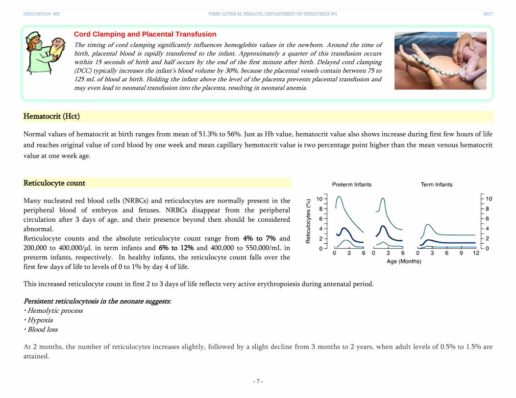
GRIGORYAN ME YSMU AFTER M. HERATSI, DEPARTMENT OF PEDIATRICS 1 2017
- 7 -
Cord Clamping and Placental Transfusion
The timing of cord clamping significantly influences hemoglobin values in the newborn. Around the time of birth, placental blood is rapidly transferred to the infant. Approximately a quarter of this transfusion occurs within 15 seconds of birth and half occurs by the end of the first minute after birth. Delayed cord clamping (DCC) typically increases the infant’s blood volume by 30%, because the placental vessels contain between 75 to 125 mL of blood at birth. Holding the infant above the level of the placenta prevents placental transfusion and may even lead to neonatal transfusion into the placenta, resulting in neonatal anemia.
Hematocrit (Hct)
Normal values of hematocrit at birth ranges from mean of 51.3% to 56%. Just as Hb value, hematocrit value also shows increase during first few hours of life
and reaches original value of cord blood by one week and mean capillary hemotocrit value is two percentage point higher than the mean venous hematocrit
value at one week age.
Reticulocyte count
Many nucleated red blood cells (NRBCs) and reticulocytes are normally present in the
peripheral blood of embryos and fetuses. NRBCs disappear from the peripheral
circulation after 3 days of age, and their presence beyond then should be considered
abnormal.
Reticulocyte counts and the absolute reticulocyte count range from 4% to 7% and
200,000 to 400,000/μL in term infants and 6% to 12% and 400,000 to 550,000/mL in
preterm infants, respectively. In healthy infants, the reticulocyte count falls over the
first few days of life to levels of 0 to 1% by day 4 of life.
This increased reticulocyte count in first 2 to 3 days of life reflects very active erythropoiesis during antenatal period.
Persistent reticulocytosis in the neonate suggests: • Hemolytic process • Hypoxia • Blood loss
At 2 months, the number of reticulocytes increases slightly, followed by a slight decline from 3 months to 2 years, when adult levels of 0.5% to 1.5% are
attained.

GRIGORYAN ME YSMU AFTER M. HERATSI, DEPARTMENT OF PEDIATRICS 1 2017
- 8 -
RBC indices
Parameter Definition Units Formula
Mean cell volume
(MCV)
Average volume of
the red blood cell
Femtoliters (fL)
or
10-15 Liter
MCV = Hct (%) x 10/ RBC (x1012)
e.g., 42x10/4.2 = 100 fL
Mean cell
hemoglobin
(MCH)
Amount of
hemoglobin in a
single red blood cell
Picograms (pg)
or
10-12 grams
MCH = Hb (g/L)/ RBC (x1012)
e.g., 125/4.1 = 30.5 pg
Mean cell
hemoglobin
concentration
(MCHC)
Average
concentration of Hb
in the RBC volume
g/L MCHC = Hb (g/L) x 100/ Hct (%)
e.g., 125x100/37 = 340 g/L = 34 g/dL
MCV at birth ranges from 104 to 118 fL compared to normal adult value of 82 to 92 fL. When an MCV less than 95 fL is observed in a term neonate,
α−thalassemia trait or iron deficiency should be considered.
Mean corpuscular volume rapidly decreases in the first week of life and at the age of 2 months, cell size is comparable to those in adult cells. Similar to the MCV, the MCH is higher in preterm and term neonates, ranging from 33 to 41 pg/cell compared with 27 to 31 pg/cell in adults. MCHC does not vary between neonates and adults. Although neonatal erythrocytes are larger and contain more hemoglobin, the hemoglobin in the
erythrocyte is not more concentrated.
Table. Normal RBS parameters during the first-two weeks of life in the term infants [From Oski FA, Naiman JL: Hematologic problems in the newborn, ed 2, Philadelphia, 1972, WB Saunders, p. 13]
Value Cord blood Day 1 Day 3 Day 7 Day 14
Hb g/dL 16.8 18.4 17.8 17.0 16.8
Hematocrit (%) 53.0 58.0 55.0 54.0 52.0
Red cells (mm3) 5.25 5.8 5.6 5.2 5.1
MCV (fL) 107 108 99.0 98.0 96.0
MCH (Pg) 34 35 33 32.5 31.5
MCHC (g/dL) 31.7 32.5 33 33 33
Reticulocytes 3–7 3–7 1–3 0–1 0–1
Nucleated RBCs 500 200 0–5 0 0

GRIGORYAN ME YSMU AFTER M. HERATSI, DEPARTMENT OF PEDIATRICS 1 2017
- 9 -
Table. Sex- and age-stratified pediatric reference ranges for hematocrit, hemoglobin, and mean corpuscular volume [From Cembrowski GS,Chan J, Cheng C, et al: NHANES 1999-2000 data used to create comprehensive health-associated race-, sex- and age-stratified pediatric reference intervals for the Coulter MAXM. Laboratory Hematol 10:245–6, 2004. Presented at the annual meeting of the International Society for Laboratory Hematology, Barcelona, Spain, May 2004. Numerical summary provided by GS Cembrowski, MS Cembrowski, KA Versluys, 2013.]

GRIGORYAN ME YSMU AFTER M. HERATSI, DEPARTMENT OF PEDIATRICS 1 2017
- 10 -
ANAEMIA (ANEMIA). IRON DEFICIENCY IN CHILDREN
Anemia is defined as an Hb level below the normal range. The normal range varies with age.
Table. Hemoglobin levels to diagnose anemia at sea level [WHO, 2001]
Age Hemoglobin (g/L)
Non-Anemia Mild Anemia* Moderate Anemia Severe Anemia
6 months to 5 years ≥ 110 100-109 70-99 < 70
5 to 11 years ≥ 115 110-114 80-109 < 80
12 to 14 years ≥ 120 110-119 80-109 < 80
Nonpregnant females ≥ 15 years ≥ 120 110-119 80-109 < 80
Pregnant females ≥ 110 100-109 70-99 < 70
Men ≥ 15 years ≥ 130 110-129 80-109 < 80
* "Mild" is a misnomer: iron deficiency is already advanced by the time anemia is detected. The deficiency has consequences even when no
anemia is clinically apparent.
Table. Hemoglobin adjustments [From Nestel P. Adjusting hemoglobin values in program surveys. Washington, DC: International Nutritional Anaemia Consultative Group, ILSI Human Nutrition Institute. 2002 Jun 23:2-4.]
For altitude
For pregnancy in women
living at sea level For smokers
For healthy people of african
extraction living at sea level
Altitude (m) Hb g/L Stage of pregnancy
(trimester) Hb g/L Amount smoked Hb g/L Hb g/L
m < 1000 No adjustment First -10 1/2–1 pack/day +3 Everyone –10
1000< m <1250 +2 Second -15 1–2 packs/day +5
1250< m <1750 +5 Third -10 >2 packs/day +7
1750< m <2250 +8 Trimester unknown -10
Smoker, amount
unknown
+3
2250< m <2750 +13
2750< m <3250 +19
3250< m <3750 +27
3750< m <4250 +35
4250< m <4750 +45
4750< m <5250 +55 Lake Sevan - at the altitude of 1900m above sea level
Yerevan - at the altitude of 900-1300m above sea level 5250< m +67

GRIGORYAN ME YSMU AFTER M. HERATSI, DEPARTMENT OF PEDIATRICS 1 2017
- 11 -
A global health problem
Anemia is common throughout the world.
For the year 2011, it is estimated that roughly 43% of
children, 38% of pregnant women, and 29% of
nonpregnant women and 29% of all women of
reproductive age have anaemia globally,
corresponding to 273 million children, 496 million
non-pregnant women and 32 million pregnant
women.
Fig. Global estimates of the prevalence of anemia in infants and children aged 6‒59
months, 2011 [Source: WHO. The global anemia prevalence in 2011. Geneva: World Health Organization; 2015.]
Causes of anemia
Anemia may result from a number of causes, with the most
significant contributor being iron deficiency. Approximately
50% of cases of anaemia are considered to be due to iron
deficiency. The most susceptible groups are pregnant women
and young children. Women of childbearing age need to
absorb 2-3 times the amount of iron required by men or older
women.
Other causes of anemia include other micronutrient deficiencies
(e.g. folate, riboflavin, vitamins A and B12), acute and chronic
infections (e.g. malaria, tuberculosis and HIV), cancer, and
inherited or acquired disorders that affect Hb synthesis, RBC
production or survival (e.g. hemoglobinopathies).
Fig. Causes of anemia in countries with low or middle incomes [From Balarajan Y et al: Anemia in low-income and middle-income countries. Lancet 378:2123–2134, 2011, Fig. 3.]
A 12-month-old infant is found to have Hb 80 g/L and mean corpuscular volume (MCV) 65 fL on routine complete blood (cell) count (CBC)
screen. What is the likely cause of these abnormal laboratory values?

GRIGORYAN ME YSMU AFTER M. HERATSI, DEPARTMENT OF PEDIATRICS 1 2017
- 12 -
Although the most reliable indicator of anemia at the population level is blood hemoglobin concentration, measurements of this concentration alone do not
determine the cause of anaemia. The first step in diagnosis of anemia is to establish whether the abnormality is isolated to a single cell line (red blood cells
only) or whether it is part of a multiple cell line abnormality (red cells, white cells and platelets). Abnormalities of two or three cell lines usually indicate
one of the following:
• bone marrow involvement, (e.g., aplastic anemia, leukemia) • an immunologic disorder (e.g., SLE, immune hemolytic anemia singly or in combination) • sequestration of cells (e.g., hypersplenism)
To narrow the diagnostic possibilities, anemias may be classified on the basis of their morphology and physiology.
Anemias may be morphologically categorized on the basis of RBC size (mean corpuscular volume [MCV]), and microscopic appearance. They can be
classified as microcytic, normocytic, or macrocytic based on whether the MCV is low, normal, or high, respectively.
Fig. Examples of variations in RBC morphology
Normal RBCs Macrocytes
Hypochromic microcytes Sickle cells Fragmented RBCs
(Schistocytes)
Normal mean cell volume
(MCV)
Macrocytes are large red cells
with a high MCV. Their
hemoglobin concentration
(MCHC) is normal.
The central pallor zone of
the erythrocyte must be
greater than one-third of the
diameter of the cell before it
is classified as hypochromic.
Not all hypochromic cells
are microcytic, but all
microcytic cells are
hypochromic.
Sickle cells are red cells with
two pointed ends which are in
the shape of a crescent or
sickle. This is due to the
polymerization of
deoxygenated HbS causing
changes to the red blood cell
making it less deformable and
much more rigid.
Fragmented red cells are red
cells that are injured and torn
due to a microangiopathic
process in which fibrin strands
are generated and are
responsible for injury to the
red cells.
• Normal blood
• Normocytic anemia
• Normal newborn
• Folic acid or vitamin B12
deficiency
• Iron deficiency anemia
• Lead poisoning
• Thalassemias
• Sideroblastic anemia
• Homozygous hemoglobin S
disease
• Microangiopathic hemolytic
anemia (in HUS, TTP, DIC)

GRIGORYAN ME YSMU AFTER M. HERATSI, DEPARTMENT OF PEDIATRICS 1 2017
- 13 -
Anemias may also be further divided on the basis of underlying physiology. The 2 major categories are decreased production and increased
destruction or loss.
Decreased RBC production (hypoproliferative anemia) Increased RBC destruction or loss
Nutrient deficiency (iron, folate, vitamin B12),
Bone marrow failure (acquired or inherited)
Bone marrow infiltration (eg, malignancy)
Hemolysis
Sequestration
Bleeding
The peripheral blood reticulocyte percentage or absolute number will help to make a distinction between the 2 physiologic categories. The normal
reticulocyte percentage of total RBCs during most of childhood is approximately 1%, with an absolute reticulocyte count of 25,000-75,000/mm3. In the
presence of anemia, EPO production and the absolute number of reticulocytes should rise. Low or normal numbers of reticulocytes generally represent an inadequate response to anemia that is associated with relative bone marrow failure or ineffective
erythropoiesis. Increased numbers of reticulocytes represent a normal bone marrow response to ongoing RBC destruction (hemolysis), sequestration, or loss (bleeding).
Fig. Anaemia classification based on MCV and reticulocyte count [From Dignass AU et al. European consensus on the diagnosis and management of iron deficiency and anaemia in inflammatory bowel diseases. Journal of Crohn's and Colitis. 2015 Mar 1;9(3):211-22.]
The reticulocyte count tells whether the
bone marrow can respond by increasing
erythropoiesis, which gives early and
important information on the direction of
the investigation. All deficiency states are
excluded by increased reticulocytes.
Retic, reticulocyte count; N, normal; Tsat, transferrin saturation; S−ferritin, serum ferritin; LDH, lactate dehydrogenase; MCV, mean corpuscular volume; DAT, direct antibody test; Hb, hemoglobin; IDA, iron deficiency anaemia; FID, functional iron deficiency; MDS, myelodysplastic syndrome; * anemia secondary to malignancy, infection, kidney disease etc.

GRIGORYAN ME YSMU AFTER M. HERATSI, DEPARTMENT OF PEDIATRICS 1 2017
- 14 -
Major forms of anemia in childhood
Acquired hypoproliferative anemia
Nutritional deficiency (see p. 15) Lead poisoning (see p. 25) Acquired aplastic anemia: associated with bicytopenia or pancytopenia
(as opposed to pure RBC aplasia)
Transient erythroblastopenia of childhood: generally occurs during the
first 3 years of life in otherwise healthy children, although it can be
seen in children from 6 months to 10 years old. It is thought to have a
viral or immunologic cause and resolves without specific intervention.
Anemia of acute inflammation: may be encountered in children who are
hospitalized and is generally transient, resolving when the underlying
condition has improved. Acute infection may cause anemia through a
variety of mechanisms, including bone marrow suppression, shortened
RBC lifespan, red-cell fragmentation, and immune-mediated RBC
destruction.
Anemia of chronic inflammation: multifactorial anemia associated with
increased cytokine production, up-regulation of hepcidin, and abnormal
iron homeostasis.
Marrow replacement caused by malignancy (leukemia) — is usually
associated with abnormalities in other cell lineages in addition to RBCs.
Congenital hypoproliferative anemia
Diamond-Blackfan anemia — is congenital pure RBC aplasia, which usually presents in the first 3 months of life; although often macrocytic, it may be
normocytic.
Refractory sideroblastic anemia
Congenital dyserythropoietic anemias
Hemolytic anemia
Inherited disorders of hemoglobin or the RBC membrane
Acquired causes: autoimmune hemolytic anemia; microangiopathic hemolytic anemia, particularly Shiga toxin–associated hemolytic uremic syndrome
(HUS).

GRIGORYAN ME YSMU AFTER M. HERATSI, DEPARTMENT OF PEDIATRICS 1 2017
- 15 -
IRON-DEFICIENCY
Fe vs
Iron deficiency affects more than 2 billion people worldwide, and iron-deficiency anemia remains the top cause of anemia.
In childhood iron deficiency occurs most frequently during late infancy through the first few years of life and again during adolescence. This prevalence
pattern corresponds to periods of rapid growth, and when combined with poor dietary intake can predispose to iron deficiency. Other contributing factors
to iron deficiency include prematurity (low iron reserves), blood loss (most commonly menstrual or gastrointestinal), and GI conditions associated with
decreased iron absorption.
Definitions of iron deficiency state [Camaschella C. (2015). Iron-deficiency anemia. N Engl J Med, 2015(372), 1832-1843.]
Iron deficiency (ID) - Depressed levels of total body iron, especially iron stores, with preservation of levels of erythroid iron.
Iron deficiency anemia
(IDA)
- Depressed levels of total body iron in the presence of anemia.
Functional iron
deficiency (FID)
- Insufficient mobilization of erythroid iron in the presence of increased requests, as occurs after treatment with
erythropoiesis-stimulating agents. Iron is sequestered in the RES as a consequence of immune activation due to infection,
autoimmune disorders, cancer etc.
Iron-restricted
erythropoiesis (IRE)
- A reduced supply of iron for the purpose of erythropoiesis, regardless of the level of iron stores. Stores may be normal or
even increased because of iron sequestration in cases of anemia of chronic inflammation, which is observed in patients
with autoimmune disorders, cancer, infections, and chronic kidney disease.
Thus, both absolute ID and FID cause IRE.
Iron-refractory
iron-deficiency anemia
(IRIDA)
- Iron-deficiency anemia that is unresponsive to oral iron treatment, in most cases referring to the genetic disease caused
by a mutation in TMPRSS6, the gene encoding transmembrane protease, serine 6, also known as matriptase-2. This protein is a transmembrane serine protease that plays an essential role in down-regulating hepcidin, the key regulator of iron homeostasis. Hallmarks of this disease are microcytic hypochromic anemia, low transferrin saturation and normal/high serum hepcidin values. The anemia appears in the post-natal period, although in some cases it is only diagnosed in adulthood. The disease is refractory to oral iron treatment but shows a slow response to intravenous iron injections and partial correction of the anemia.

GRIGORYAN ME YSMU AFTER M. HERATSI, DEPARTMENT OF PEDIATRICS 1 2017
- 16 -
Physiological role of iron
Iron is an essential micronutrient, as it is crucial to biologic functions, including respiration, energy production, DNA synthesis, and cell proliferation.
• Required for oxygen binding to hemoglobin (oxygen
transport)
• Essential component of myoglobin (oxygen storage)
• Component of oxidative enzymes and respiratory chain
proteins (transfer of electrons/redox cycling):
Fe2+ (ferrous, reduced state) ↔ Fe3+ (ferric, oxidised state)
• Energy metabolism
• Enzyme activity and cellular metabolism
• Neurodevelopmental and cognitive function
• Growth
• Required for enzymes and proteins involved in DNA
synthesis and DNA repair mechanisms
• Essential component of cytochrome P450 enzymes (drug
metabolism, steroid hormone production)
Dietary Recommended Intake of Iron [Institute of Medicine. Dietary reference intakes. 2003]
Age Gender Level of iron intake
0-6 mo Male and female 0.27 mg/day
7-12 mo Male and female l l mg/day
1-3y Male and female 7 mg/day
4-8y Male and female 10 mg/day
9-13y Male and female 8 mg/day
14-18y Male 11 mg/day
14-18y Female 15 mg/day

GRIGORYAN ME YSMU AFTER M. HERATSI, DEPARTMENT OF PEDIATRICS 1 2017
- 17 -
Main pathways of iron homoeostasis
The human body has evolved to conserve iron in several ways, including the recycling of iron after the breakdown of red cells and the retention of iron in the absence of an excretion mechanism. However, since excess levels of iron can be toxic, its absorption is limited to 1 to 2 mg daily, and most of the iron needed daily is provided through recycling by macrophages that phagocytose senescent erythrocytes. The latter two mechanisms are controlled by the hormone hepcidin, which maintains total-body iron within normal ranges, avoiding both iron deficiency and excess.
Iron absorption
Dietary iron is found in heme (10%) and non-heme (ionic, 90%) forms
and their absorption occurs at the apical surface of duodenal enterocytes
via different mechanisms.
(1) Dietary non-heme iron primarily exists in an oxidised (Fe3+) form
that is not bioavailable and must first be reduced to the Fe2+ form by a
ferrireductase enzyme, which uses vitamin C as coenzyme, before being
transported across the intestinal epithelium. This is accomplished by a
carrier protein called divalent metal transporter 1 (DMT1), which also
traffics other metal ions such as zinc, copper and cobalt by a proton-
coupled mechanism.
(2) Heme iron is absorbed independently by mechanisms that remain
uncertain (proposed transporter — heme carrier protein 1, HCP1).
(3) Once inside the intestinal epithelial cell, most Fe2+ is exported by
ferroportin across the basolateral membrane of the enterocyte (absorbed
iron), and re-oxidised to Fe3+ (4) by hephaestin before being bound by
plasma transferrin for delivery to cells.
Ferroportin is also expressed in hepatocytes, reticuloendothelial
macrophages and placental syncytiotrophoblasts (where it regulates iron
entry into fetal circulation).
(5) Levels of iron absorption are controlled by the iron-regulatory
peptide hormone, hepcidin, which is synthesised in the liver. Hepcidin
binds to a specific extracellular domain of ferroportin and induces its
degradation in the lysosome.

GRIGORYAN ME YSMU AFTER M. HERATSI, DEPARTMENT OF PEDIATRICS 1 2017
- 18 -
Heme iron is mainly derived from hemoglobin and myoglobin
and is found in animal foods, such as red meat, fish and poultry.
Non-heme iron is obtained from plant foods, such as lentils and
beans.
The relative absorption of iron in the gut differs between these
dietary forms of iron; heme iron is absorbed better than non-
heme iron. This is because non-heme iron is predominantly
found as Fe3+ and therefore must be reduced to Fe2+ before it can
be absorbed.
The absorption of heme iron is largely unaffected by diet,
whereas non-heme iron absorption can be influenced by several
dietary factors. Ascorbic acid (vitamin C) is an enhancer of iron
absorption, as it reduces Fe3+ to Fe2+. In contrast, the intake of
bran, phytates (high-fibre diets), phenolic compounds (coffee,
tea), calcium and phosphates can result in the inhibition of non-
heme iron absorption owing to their ability to chelate metals
such as iron.
Non-heme iron absorption can also be reduced by drugs, such as
antacids and proton pump inhibitors which decrease stomach
acid levels, as gastric acid enhances the dissociation and
solubilisation of iron salts in non-haem iron, allowing them to
be reduced to the Fe2+ oxidation state. In addition, Helicobater
pylori infection produces gastric atrophy, which can lead to
profound IDA.
The acquisition of iron absorption capacity (mediated by
DMT1) increases linearly after birth, reaching adult levels by
early childhood.
Fig. Iron absorption from different food sources [From Scrimshaw NS. Iron deficiency. Scientific American 1991;265:46–52.]
Fig. Age-dependent enteral absorption of iron expressed as a percentage of the dose
dministered [Funk et al. 2012]

GRIGORYAN ME YSMU AFTER M. HERATSI, DEPARTMENT OF PEDIATRICS 1 2017
- 19 -
Body iron distribution and recycling
Once in circulation, iron is transported bound to transferrin to the site of use
or storage. Erythrocyte precursors express high levels of transferrin receptor 1
(TfRl) and, thus, have preferred access to circulating iron.
The major portion of iron is found in the erythron as hemoglobin iron
dedicated to oxygen transport and delivery. Small amounts of erythron iron are
also present in heme and nonheme enzymes in developing RBCs.
The remainder of functional iron is found as myoglobin iron in muscle and as
iron-containing and iron-dependent enzymes throughout the cells of the body.
Most storage iron is held in reserve by hepatocytes and macrophages (ferritin
and hemosiderin).
The small fraction of transport iron in the plasma and extracellular fluid is
bound to the protein transferrin (Tf).
The recycling of iron from heme and amino acids from globin
following the lysis of aged red cells is a very efficient process.
Heme is returned to the bone marrow, and the amino acids of
the globin chain are returned to the amino acid pool. Each of
these products will later be recruited for hemoglobin formation
and red cell production.
In the adult, approximately 95% of recycled iron is used for red
cell production, whereas in the infant, only 70% is used for this
purpose. Because of this relationship, it is easy to understand
the significance of adequate iron sources in the early years of
development.

GRIGORYAN ME YSMU AFTER M. HERATSI, DEPARTMENT OF PEDIATRICS 1 2017
- 20 -
Ferritin
Ferritin is an intracellular protein and each ferritin complex can store about
4500 iron (Fe3+) ions
Ferritin is found in all cells of the body
− Liver hepatocytes and RES macrophages of the liver and spleen are the most
prominent sites of ferritin accumulation
Ferritin can also be found in serum
− Serum ferritin contains very little iron
− Serum ferritin is increased in patients with inflammation
In healthy individuals, serum ferritin levels correlate with body iron store levels
− Serum ferritin levels decrease as iron stores become depleted
− Therefore it is the most useful estimate of body iron stores in individuals
without inflammation
Hepcidin is the principal regulator for systemic iron homeostasis
Hepcidin synthesis is induced by iron loading and
inflammation and suppressed by erythropoiesis.
In the case of an iron load, increased hepcidin
levels limit further enteral iron absorption and
release of iron from the liver and the
reticuloendothelial system to normalize plasma
iron levels.
With increased inflammation, elevated hepcidin
levels cause the same sequence of events, leading
to reticuloendothelial blockade and anemia of
inflammation.
Conversely, by suppressing hepcidin production,
erythropoietic activity increases the availability of
iron by enhancing enteral absorption and release
of iron from the liver and the reticuloendothelial
system.
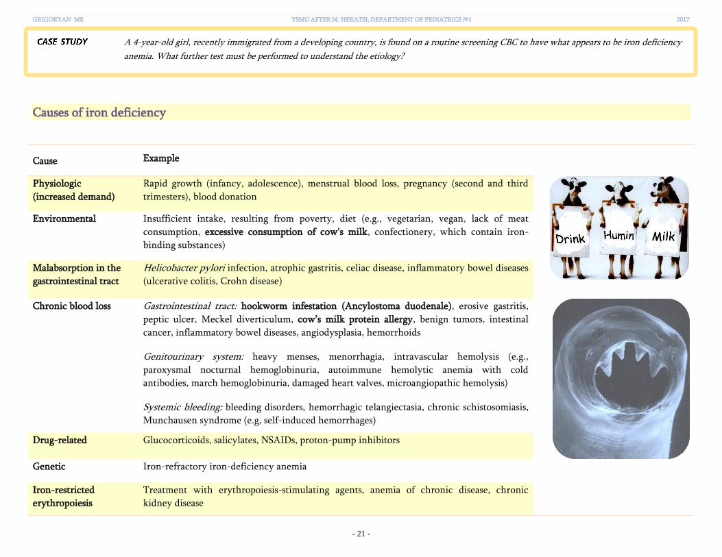
GRIGORYAN ME YSMU AFTER M. HERATSI, DEPARTMENT OF PEDIATRICS 1 2017
- 21 -
A 4-year-old girl, recently immigrated from a developing country, is found on a routine screening CBC to have what appears to be iron deficiency
anemia. What further test must be performed to understand the etiology?
Causes of iron deficiency
Cause Example
Physiologic
(increased demand)
Rapid growth (infancy, adolescence), menstrual blood loss, pregnancy (second and third
trimesters), blood donation
Environmental Insufficient intake, resulting from poverty, diet (e.g., vegetarian, vegan, lack of meat
consumption, excessive consumption of cow's milk, confectionery, which contain iron-
binding substances)
Malabsorption in the
gastrointestinal tract
Helicobacter pylori infection, atrophic gastritis, celiac disease, inflammatory bowel diseases
(ulcerative colitis, Crohn disease)
Chronic blood loss Gastrointestinal tract: hookworm infestation (Ancylostoma duodenale), erosive gastritis,
peptic ulcer, Meckel diverticulum, cow’s milk protein allergy, benign tumors, intestinal
cancer, inflammatory bowel diseases, angiodysplasia, hemorrhoids
Genitourinary system: heavy menses, menorrhagia, intravascular hemolysis (e.g.,
paroxysmal nocturnal hemoglobinuria, autoimmune hemolytic anemia with cold
antibodies, march hemoglobinuria, damaged heart valves, microangiopathic hemolysis)
Systemic bleeding: bleeding disorders, hemorrhagic telangiectasia, chronic schistosomiasis,
Munchausen syndrome (e.g, self-induced hemorrhages)
Drug-related Glucocorticoids, salicylates, NSAIDs, proton-pump inhibitors
Genetic Iron-refractory iron-deficiency anemia
Iron-restricted
erythropoiesis
Treatment with erythropoiesis-stimulating agents, anemia of chronic disease, chronic
kidney disease

GRIGORYAN ME YSMU AFTER M. HERATSI, DEPARTMENT OF PEDIATRICS 1 2017
- 22 -
Clinical presentation of iron deficiency
Patients with iron deficiency may present with
(1) no signs or symptoms, coming to medical attention only because of abnormalities
noted on laboratory tests
(2) features of the underlying disorder responsible for the development of iron deficiency
(3) manifestations common to all anemias:
Fatigue, weakness, dizziness and drowsiness, anorexia
Loss of normal colour in the skin (in fair skinned people) and also in the lips, tongue,
nail beds and the blood vessels in the white of the eye
In severe cases growth impairment may occur and resistance to infection may be decreased
• Decreased oxygen transport leads to fatigue, dyspnea, angina pectoris, and syncope • Decreased blood volume leads to pallor, postural hypotension, and shock • Increased cardiac output leads to palpitation, strong pulse, and heart murmurs
Pallor of the skin and mucous membranes is an unreliable indicator and is usually seen only when the hemoglobin is less than 80 g/L.
Mucosal conditions, such as glossitis, recurrent aphthae, candidal infections, and
angular stomatitis may be more common in patients with anemia. The tongue appears
reddened, and the papillae are atrophic, producing a smooth ("bald") appearance.
Angular stomatitis is commonly caused by a candidal infection, and it has been linked
to iron deficiency.
As cells of the tongue papillae have a high rate of turnover, deficiencies in micronutrients needed for cell proliferation or cell membrane stabilisation may lead to depapillation. Nutritional deficiency is also thought to change the pattern of microbial flora, thus contributing to glossitis.
(4) Features unique to the IDA: pagophagia, koilonychias (spooning of the nail bed), and
blue sclerae.
Additionally, evidence suggests that iron deficiency with or without anemia may result in
developmental delays and behavioral disturbances.
Fig. Severe anemia. A, Pale conjunctiva may be seen in the patient with
severe anemia. B, Pale palmar creases are also visible in cases of severe
anemia. The child in this photograph has a hemoglobin level of 40 g/L.
Fig. Spooning of the
fingernails occurs in
children with severe
iron deficiency anemia
(koilonychia)
Fig. Angular stomatitis
Fig. Atrophic glossitis

GRIGORYAN ME YSMU AFTER M. HERATSI, DEPARTMENT OF PEDIATRICS 1 2017
- 23 -
IRON DEFICIENCY WITHOUT ANEMIA ALSO HAS SIGNIFICANT EFFECTS
Iron is a necessary cofactor for many enzymes. It is particularly
crucial to the dopaminergic system in the central nervous system.
Among the nonhematologic neurologic conditions that have been
associated with iron deficiency are breath-holding spells, attention-deficit/hyperactivity disorder, febrile seizures, pagophagia*, and restless legs syndrome (RLS)** [Zehetner AA 2010; Lozoff B 2011].
Some studies have indicated that even after iron stores are rebuilt the developmental consequences of early iron deficiency may not be reversible. In
addition to the direct effects of inadequate iron stores, iron deficiency promotes lead absorption and its neurologic consequences.
* Pagophagia, a variant of pica in which ice is the substance obsessively consumed, is a behavioral abnormality that is considered to be a highly specific symptom of iron deficiency, resolving within a few days to 2 weeks after beginning iron therapy.
** Restless legs syndrome (RLS) is a neurologic disorder characterized by a distressing need or urge to move the legs. The etiology of RLS is complex. One major potential pathophysiological pathway in the development of RLS is the iron deficiency-dopamine interaction [Earley CJ 2014].
Common clinical laboratory tests to assess iron status
Test What It Indicates Result in Iron Deficiency
Serum iron Iron recycling/usage, stores (indirect) ↓
Total iron-binding capacity (TIBC) Serum/plasma transferrin ↑
Transferrin saturation:
Tsat(%) = (100 × serum iron)/TIBC % plasma iron-binding sites on transferrin that are occupied ↓ - ↓↓
Free or zinc protoporphyrin (ZnPP) Functional mitochondrial iron status ↑
Soluble transferrin receptor (sTfR) Adequacy of marrow activity for any level of anemia ↑
Serum ferritin Iron stores (indirect) ↓ - ↓↓
Bone marrow iron Macrophage iron stores/retention, RBC iron uptake ↓
Hepcidin Hepatocyte iron ↓ - ↓↓
Red blood cell distribution width (RDW) The degree of variation in RBC size ↑ (anisocytosis)

GRIGORYAN ME YSMU AFTER M. HERATSI, DEPARTMENT OF PEDIATRICS 1 2017
- 24 -
Phases of development of iron deficiency (without coexisting disorders)
Parameter NORMAL PRELATENT IRON DEFICIENCY LATENT IRON DEFICIENCY IRON DEFICIENCY ANEMIA
Iron-deficient erythropoiesis
Iron stores Present Decreased Absent Absent
Ferritin N ↓ ↓ ↓↓
Transferrin saturation N Normal ↓ ↓
sTfR N ↑ ↑↑ ↑↑↑
Zinc protoporphyrin N Normal ↑ ↑
Ratio of ZnPP to heme N Normal ↑ ↑
Hemoglobin N Normal Normal ↓
MCV N Normal Normal ↓
Iron deficiency progresses through three discernible phases:
1. First, in the pre–latent phase, iron stores are depleted and ferritin levels are typically low. Importantly, ferritin levels can remain normal or elevated if there is concurrent inflammation.
2. This is followed by latent iron deficiency, which is evident in a decrease in transferrin saturation. The final step of heme formation is the incorporation of an iron molecule into the porphyrin ring. As the amount of iron being transported in the plasma decreases, there is less iron available to be incorporated into the porphyrin ring and a resulting rise in the ratio of zinc protoporphyrin to heme.
3. Finally, with overt iron-deficiency anemia, there is a diminution in red blood cell volume and quantity (anemia and microcytosis are late findings in iron deficiency).
No single test value will completely characrerize iron status. A set of tests is required to establish the iron starus of an individual. The tests most easily
obtained are:
Hb concentration to establish presence of anemia serum ferritin concentration to suggest iron as the cause of the anemia CRP to ensure that an elevated serum ferritin concentration is truly attributable to lack of iron and not secondary to an inflammatory state

GRIGORYAN ME YSMU AFTER M. HERATSI, DEPARTMENT OF PEDIATRICS 1 2017
- 25 -
An 8-month-old infant is found to have low hemoglobin and low MCV on a routine CBC. He is treated with iron supplementation for 5 months, but on repeat CBC, he is still anemic. What is the likely cause of his anemia?
Differential diagnosis of IDA
Iron deficiency is the only microcytic hypochromic disorder in which mobilizable iron stores are absent; in all other disorders, storage iron is normal or
increased.
Diagnosis MCV Serum iron Serum ferritin TIBC Tsat sTfR
IDA ↓ ↓ ↓ ↑ ↓ ↑
Anemia of inflammation Normal (may be
mildely low) ↓
Normal or
elevated Normal ↓ Normal
Microcytic hemoglobinopathies
(eg, thalassemia) Low or normal Normal
Normal or
elevated Normal
Normal or
elevated
Normal or
elevated
IDA with coexistent
inflammation ↓ ↓ Low or normal Normal or high ↓ ↑
Lead poisoning (plumbism)
In the developing world, lead is a much more significant public health problem. Sources vary from
country to country, but lead-glazed ceramics are a common cause, particularly as their production is often
a home-based industry in which children are actively employed. Other sources include leaded petrol,
groundwater contamination from mining, smelting and battery factories, as well as exposure to other
occupational sources via parents.
The toxic effects of lead span several different systems:
• Hematological – Lead inhibits the enzymes 5-aminolaevulinic acid dehydratase (ALAD) and
ferrochelatase, which are essential for the production of heme. This results in a microcytic, hypochromic
anemia with elevated plasma concentrations of 5-aminolaevulinic acid (ALA) and zinc protoporphyrin.
• Neurological – Children may present with cognitive impairment as a result of chronic low-level lead
exposure or an acute encephalopathy. The mechanism of neurotoxicity is not known.
• Renal – Acute severe lead exposure may give rise to proximal tubular dysfunction resulting in glycosuria
and aminoaciduria. Chronic exposure leads to interstitial nephritis.
The toxic effects of lead are dose-related.

GRIGORYAN ME YSMU AFTER M. HERATSI, DEPARTMENT OF PEDIATRICS 1 2017
- 26 -
Therapeutic trial of iron and recovery from IDA
The diagnosis of iron deficiency often is confirmed by the
outcome of a therapeutic trial of iron. A specific orderly response
to, and only to, treatment with iron constitutes the final definitive
proof that a lack of iron is the cause of anemia. The unequivocal
diagnostic response consists of:
(1) a reticulocytosis, which begins approximately 3 to 5 days after
adequate iron therapy is instituted, reaches a maximum on days 8
to 10, and then declines;
(2) a significant increase in hemoglobin concentration, which
should begin shortly after the reticulocyte peak, is invariably
present by 3 weeks after iron therapy is begun, and persists until
the hemoglobin concentration is restored to normal.
Fig. Impact of iron supplementation on RBC population dynamics [Clark MA et al, 2014]
After iron supplementation for IDA, the reticulocyte count should double in 1 to 2 weeks, and hemoglobin should increase by 20 g/L after 3
weeks. Common reasons for persistence of iron deficiency anemia are: poor compliance with supplementation; malabsorption of therapeutic
iron; continuing blood loss; and the effects of coexisting conditions, especially infectious, inflammatory, or malignant disorders.
Iron Supplements
Compound Trade name Formulation Elemental Fe (mg) Other ingredients
FERROUS SULFATE Aktiferrin
Sirup 6.87 mg/ml
Drops 0.53 mg/drop
Capsule 34.5 mg/caps
Sorbifer durules Tablets 100 mg/tab Vitamin C
FERROUS GLUCONATE To'thema Solution 5 mg/ml Manganese
Copper
FERROUS FUMARATE Heferol Capsule 115 mg/caps
POLYSACCHARIDE/ IRON COMPLEX Ferrum Lek Sirup 10 mg/ml
Chewable tablets 100 mg/tab

GRIGORYAN ME YSMU AFTER M. HERATSI, DEPARTMENT OF PEDIATRICS 1 2017
- 27 -
Management of anemia
Non-severe anemia
Give (home) treatment with oral iron supplementation. Oral iron therapy should begin with a ferrous iron salt taken separately from meals in three or
four divided doses and supplying a daily total of 150 to 200 mg of elemental iron in adults or 2-6 mg of iron per kilogram of body weight in children.
Simple ferrous preparations are the best absorbed and least expensive. Ferrous sulfate is the most widely used, either as tablets for adults or as a liquid
preparation for children. Administration between meals maximizes absorption.
It takes 2–4 weeks to correct anemia. After the anemia has been fully corrected, oral iron should be continued to replace storage iron, either empirically for an additional 3-4 months or until the plasma ferritin concentration exceeds approximately 50 μg/L.
If the child is ≥1 year and has not received mebendazole in the previous 6 months, give one dose of mebendazole (500 mg) for possible hookworm or
whipworm infestation.
Advise the mother about good feeding practice.
Severe anemia
Give a blood transfusion as soon as possible to:
• all children with an Hct of ≤12% or Hb of ≤40 g/L
• less severely anemic children (Hct, 13–18%; Hb, 40–60 g/L) with any of the following clinical features:
clinically detectable dehydration; shock; impaired consciousness; heart failure; deep, laboured breathing; very high malaria parasitemia (> 10% of red cells with parasites).
• If packed cells are available, give 10 ml/kg over 3–4 h in preference to whole blood. If not available, give fresh
whole blood (20 ml/kg) over 3–4 h.
• Check the respiratory rate and pulse rate every 15 min. If either rises or there is other evidence of heart failure,
such as basal lung crepitations, enlarged liver or raised jugular venous pressure, transfuse more slowly. If there is any
evidence of fluid overload due to the blood transfusion, give IV furosemide at 1–2 mg/kg, up to a maximum total of
20 mg.
• After the transfusion, if the Hb remains as low as before, repeat the transfusion.
• In children with severe acute malnutrition, fluid overload is a common and serious complication. Give packed cells
when available or whole blood at 10 ml/kg (rather than 20 ml/kg), and do not repeat transfusion based on the Hb
level, or within 4 days of transfusion.
Giving a blood transfusion. Note: A burette is used to measure the blood volume, and the arm is splinted to prevent flexion of the elbow.

GRIGORYAN ME YSMU AFTER M. HERATSI, DEPARTMENT OF PEDIATRICS 1 2017
- 28 -
Parenteral iron therapy, with the risk of adverse reactions, should be reserved for the exceptional patient who (1) remains intolerant of oral iron
despite repeated modifications in dosage regimen; (2) has iron needs that cannot be met by oral therapy because of either chronic uncontrollable
bleeding or other sources of blood loss, such as hemodialysis, or a coexisting chronic inflammatory state; or (3) malabsorbs iron.
In addition to iron dextran, which can be administered intravenously or intramuscularly, there are newer intravenous formulations, including iron
gluconate and iron sucrose, that are primarily used for iron replacement in the setting of hemodialysis.
• Iron supplementation should never be taken with milk, food, or proton pump inhibitors as these all inhibit the absorption of iron.
• Taking iron supplementation with orange juice can improve absorption (ascorbic acid enhances iron absorption).
• Oral iron may cause staining of the teeth, but this is temporary and can be avoided by rinsing the mouth or brushing the teeth after the medication is given.
• Adequate dosage will turn the stools a tarry green color.
Iron deficiency, with or without anemia, is associated with
growth impairment, neurocognitive delays, and behavioral
problems as well as other neurologic abnormalities.
An increase in the hemoglobin concentration of at least 20
g/L after 3 weeks of therapy generally is used as the
criterion for an adequate therapeutic response.
Some iron deficiency-related conditions such as
cognitive delay may not be reversible even
when iron stores have been restored.
The most common worldwide cause of chronic
gastrointestinal (GI) blood loss is hookworm infection,
which is often associated with iron deficiency anemia.
Restless leg syndrome has been associated with low ferritin
levels, and treatment with iron has been demonstrated in many
cases to improve both the ferritin level and the symptoms of the
condition.

GRIGORYAN ME YSMU AFTER M. HERATSI, DEPARTMENT OF PEDIATRICS 1 2017
- 29 -
DEVELOPMENTAL CHANGES IN WHITE BLOOD CELLS (WBCs)
The normal leukocyte and neutrophil counts vary tremendously, even by the hour of life, in the first few days after birth. Therefore, it is
necessary to consult appropriate nomograms in the immediate neonatal period to properly interpret leukocyte counts. Outside the neonatal
period, the normal absolute neutrophil count has a lower limit of normal of approximately 1000/mm3, except in African American infants,
where it may normally be as low as 700 to 800/mm3. This has been called “ethnic pseudoneutropenia,” an arcane name unnecessarily ascribed
to a biologically normal state. The absolute neutrophil count (ANC) is the product of the total leukocyte count and the percentage of neutrophils (segmented neutrophils and band forms):
Number of neutrophils and lymphocytes at different periods of childhood
The relative amount of neutrophils at birth ranges from 60 to 65%. During the first days of
life it decreases. The number of lymphocytes at birth ranges from 20 to 30%. At the age of
5−6 days the curves of neutrophils and lymphocytes counts intersect — so-called "the first
cross". Then, the number of lymphocytes continues to grow (by the end of the second week
of life it reaches approximately 55%) and the number of neutrophils decreases.
At the age of 5−7 years, the amount of neutrophils and
lymphocytes is the same ("the second cross" of neutrophils and
lymphocytes curves). Then, the number
of neutrophils continues to grow, and the number of
lymphocytes decreases.

GRIGORYAN ME YSMU AFTER M. HERATSI, DEPARTMENT OF PEDIATRICS 1 2017
- 30 -
Fig. Reference ranges for blood neutrophil concentration during the first 72 hours of life [From Christensen RD, Henry E, Jopling J, et al: The CBC: reference ranges for neonates. Semin Perinatol 33:3–11, 2009.]
< 28-week gestation preterm neonates 28 to 36 weeks’ gestation preterm neonates ≥36 weeks’ gestation neonates
Table. WBC Manual Differential Counts in
Normal Fetuses at Different Gestational Ages [From Forestier F, Daffos F, Catherine N, et al: Developmental hematopoiesis in normal human fetal blood. Blood 77:2360, 1991. RBCs, Red blood cells; WBCs, white blood cells.]
Week of Gestation Lymphocytes (%) Neutrophils (%) Eosinophils (%) Basophils (%) Monocytes (%)
18-21 88 ± 7 6 ± 4 2 ± 3 0.5 ± 1 3.5 ± 2
22-25 87 ± 6 6.5 ± 3.5 3 ± 3 0.5 ± 1 3.5 ± 2.5
26-29 85 ± 6 8.5 ± 4 4 ± 3 0.5 ± 1 3.5 ± 2.5
> 30 68.5 ± 15 23 ± 15 5 ± 3 0.5 ± 1 3.5 ± 2
Table. White blood cell count in children (x109/L) [From Cembrowski GS, Chan J, Cheng C, Bamforth FJ. NHANES 1999-2000 Data used to create comprehensive health-associated race-, sex-, and age-stratified pediatric reference intervals for the Coulter MAXM. Lab Hematol 10:245–246, 2004. Presented at the annual meeting of the International Society for Laboratory Hematology, Barcelona, Spain, May 2004. Numerical summary kindly provided by GS Cembrowski, MS Cembrowski, KA Versluys, 2013.]
Age
African American
Age
Mexican American and White
2.5 Percentile 50 Percentile 95 Percentile 97.5 Percentile 2.5 Percentile 50 Percentile 95 Percentile 97.5 Percentile
M F M F M F M F M F M F M F M F
1y 5.0 8.0 11.5 13.2 1y 6.0 9.5 16.5 17.5
2-3y 4.0 6.8 10.0 11.0 2-3y 5.5 8.0 12.0 13.0
4-6y 4.0 6.5 9.2 10.0 4-6y 5.0 7.5 11.5 12.5
7-10y 3.3 6.0 9.3 11.4 7-10y 4.5 7.3 11.0 11.5
11-14y 3.3 6.0 9.0 10.0 11-14y 4.5 7.0 11.0 11.5
15-18y 3.0 4.0 5.5 6.3 9.0 9.7 15-18y 5.1 7.0 11.0 12.0

GRIGORYAN ME YSMU AFTER M. HERATSI, DEPARTMENT OF PEDIATRICS 1 2017
- 31 -
Neutrophil count in children (x109/L)
Age
African American
Age
Mexican American & White
2.5 Percentile 50 Percentile 95 Percentile 97.5 Percentile 2.5 Percentile 50 Percentile 95 Percentile 97.5 Percentile
M F M F M F M F M F M F M F M F
1y 1.0 2.6 4.6 4.6 1y 1.5 3.1 6.2 7.4
2-3y 1.0 2.4 4.8 7.5 2-3y 1.5 3.3 6.3 7.5
4-6y 1.0 2.8 5.2 5.8 4-6y 1.7 3.5 6.7 7.9
7-10y 1.1 2.5 5.7 6.8 7-10y 1.8 3.6 6.7 7.2
11-14y 1.2 2.8 5.7 5.6 7.0 11-14y 1.9 3.4 4.1 6.7 7.5 7.4 8.2
15-18y 1.1 1.6 2.7 3.2 5.7 6.8 5.9 15-18y 2.3 3.5 4.6 7.0 7.9 7.9 8.3
Lymphocyte count in children (x109/L)
Age
African American
Age
Mexican American & White
2.5 Percentile 50 Percentile 95 Percentile 97.5 Percentile 2.5 Percentile 50 Percentile 95 Percentile 97.5 Percentile
M F M F M F M F M F M F M F M F
1y 1.8 4.1 6.0 6.7 1y 2.5 5.0 8.5 9.5
2-3y 1.1 3.4 5.0 5.4 2-3y 5.0 3.6 6.0 6.3
4-6y 1.1 2.7 4.6 5.1 4-6y 1.5 3.0 4.8 4.7
7-10y 1.3 2.3 3.7 4.0 7-10y 1.5 2.7 4.0 4.5
11-14y 1.3 2.3 3.4 3.7 11-14y 1.3 2.4 3.6 3.8
15-18y 1.4 2.1 3.2 3.3 15-18y 1.2 2.0 3.6 3.5
Monocyte count in children (x109/L)
Age
African American
Age
Mexican American & White
2.5 Percentile 50 Percentile 95 Percentile 97.5 Percentile 2.5 Percentile 50 Percentile 95 Percentile 97.5 Percentile
M F M F M F M F M F M F M F M F
1y 0.4 0.6 1.38 1.66 1y 0.45 0.8 1.4 1.5
2-3y 0.3 0.6 0.92 1.03 2-3y 0.35 0.6 0.95 0.95
4-6y 0.3 0.5 0.9 0.9 4-6y 0.35 0.6 0.95 1.1
7-10y 0.2 0.5 0.9 1.0 7-10y 0.3 0.6 0.9 0.9
11-14y 0.25 0.5 0.8 0.85 11-14y 0.3 0.55 0.9 0.97
15-18y 0.3 0.5 0.88 0.95 15-18y 0.35 0.55 0.87 0.9
Eosinophil count in children (x109/L)
Age
African American
Age
Mexican American & White
2.5 Percentile 50 Percentile 95 Percentile 97.5 Percentile 2.5 Percentile 50 Percentile 95 Percentile 97.5 Percentile
M F M F M F M F M F M F M F M F
1y 0.1 0.3 1.0 1.1 1y 0.1 0.2 0.7 0.8
2-3y 0.0 0.2 0.7 0.9 2-3y 0.1 0.2 0.75 0.9
4-6y 0.05 0.3 0.8 0.95 4-6y 0.05 0.25 0.75 0.8
7-10y 0.1 0.2 0.9 1.0 7-10y 0.1 0.2 0.75 1.0
11-14y 0.0 0.2 0.55 0.6 11-14y 0.05 0.15 0.7 0.8 0.7
15-18y 0.0 0.15 0.45 0.55 15-18y 0.05 0.15 0.55 0.4 0.5

GRIGORYAN ME YSMU AFTER M. HERATSI, DEPARTMENT OF PEDIATRICS 1 2017
- 32 -
Table. Quantitative alterations of WBCs
White Blood Cells (Leukocytes) Brief characteristics High WBC count (leukocytosis) Low WBC count (leukopenia)
Myeloid
cells
Segmented
neutrophils
(segs)
• The predominant WBCs in the peripheral blood
• The nucleus is lobulated (between 2 and 5 lobes) and the lobes are connected by a thin filament
Neutrophilia Infection/inflammation, necrosis Any stressor/ heavy exercise Drugs (steroids, heparin, epinephrine) Chronic myeloid leukaemia (CML)
Neutropenia (see p. 33) Viral (overt or occult) Autoimmune/idiopathic Chemotherapeutic agents Bone marrow defects (pancytopenia)
Band
neutrophils
(bands)
• Constitute 0—5% of the nucleated cells under normal conditions in the peripheral blood
Bandemia (>500/mm3) Severe infections Inflammation
—
Immature
neutrophil
forms
• These include myelocytes and metamyelocytes, that do not belong in circulation even with severe infection
• The presence of these cells in the circulation is an ominous marker for life-threatening bacterial infection (severe left shift)
The neutrophils tend to be more immature, as they are being released earlier. This is called a left shift.
Basophils
• Lobate nuclear granulocyte with granules stainable by basic dyes
Basophilia Myeloproliferative disorders
Basopenia Difficult to demonstrate as the normal basophil count is so low
Eosinophils
• Segmented polymorphonuclear granulocyte with granules stainable by eosin dyes
Eosinophilia Allergy/atopy, asthma/hayfever Parasites (less common in developed countries)
Eosinopenia Stress Drugs (Steroids)
Monocytes
• Mononuclear phagocytes Monocytosis Usually not significant Watch levels >1.5×10⁹/L more closely, consider chronic infection or inflammation
Monocytopenia Not clinically significant if other cell counts are normal
Lymphocytes
• There are two broad morphologic categories of lymphocytes which can be distinguished under the light microscope, large granular lymphocytes and small lymphocytes.
Lymphocytosis Acute infections (EBV, viral hepatitis, pertussis) Chronic intracellular bacterial infections (tuberculosis, brucellosis) Chronic lymphocytic leukaemia (in the Elderly)
Lymphocytopenia Immunodeficiency syndrome AIDS, agammaglobulinemia Some types of chemotherapy or malignancies

GRIGORYAN ME YSMU AFTER M. HERATSI, DEPARTMENT OF PEDIATRICS 1 2017
- 33 -
In a severe infection the neutrophils may show and other toxic changes such as:
(1) toxic granulation
(2) toxic vacuolation
(3) the presence of Döhle bodies
Major forms of neutropenia in children and adolescents
Acute forms — usually fatal.
The most common form of neutropenia in infants and young children is transient neutropenia with
or following a viral illness. It resolves itself within 2 weeks.
Chronic forms last over 6 months and have indolent progression.
The most common type of chronic neutropenia in pediatric patients is chronic benign neutropenia of
childhood (also known as primary autoimmune neutropenia). Infections associated with the primary
form are usually limited and mild. The mean age at diagnosis of autoimmune neutropenia is 6-12
months, with a range of 3-30 months. Spontaneous recovery occurs by age 5 years, and the mean
duration of neutropenia is approximately 20 months.
Cyclic neutropenia is characterized by an episodic decrease in the number of neutrophils every 3–4
weeks and lasts for 5 to10 days. Peripheral neutrophil counts usually drop to zero and during this time
the child is extremely susceptible to infection. Painful oral ulcerations covered by a whitish membrane
and surrounded by slight erythema are the most constant findings. The lips, tongue, buccal mucosa, and
gingiva are the areas most commonly affected.
Fig. Gross gingival inflammation in an adolescent with
cyclic neutropenia. This girl lost most of her primary
teeth by the age of 7 years.
Intermittent forms — as part of Shwachman-Diamond syndrome (a rare autosomal recessive disorder characterized by exocrine pancreatic insufficiency,
bone marrow dysfunction, and skeletal abnormalities).
KEY RECOMMENDATIONS FOR PRACTICE
[From Sills RH, editor. Practical algorithms in pediatric hematology and oncology. Karger Medical and Scientific Publishers; 2003. Riley LK, Rupert J. Evaluation of Patients with Leukocytosis. American family
physician. 2015 1;92(11).]
Each blood count should be evaluated on the basis of the absolute number of cells/μl and NOT on the basis of the differential count percentage (e.g., a
relatively high percentage of neutrophils may not be abnormal if the total WBC is not high). Consideration must be made of the age of the patient
because the leukocyte values change during childhood (see tables of normal values).
Leukocytosis greater than100.0 × 109/L (hyperleukocytosis) is almost always caused by leukemias or myeloproliferative disorders.
Patients with leukocytosis and no other signs of systemic inflammatory response syndrome do not require blood cultures.
1 2 3

GRIGORYAN ME YSMU AFTER M. HERATSI, DEPARTMENT OF PEDIATRICS 1 2017
- 34 -
Qualitative leukocyte disorders
Qualitative leukocyte disorders consist of disruptions of leukocyte function (primary and secondary
immune deficiencies).
Phagocytic cells (granulocytes, monocytes, macrophages) may lose their phagocytic capacity to
function, e.g., leukocyte adhesion defect — a rare autosomal recessive condition associated with a
reduced level of adhesion molecules on peripheral leukocytes resulting in severely reduced
resistance to infection. Children present with delayed wound healing, persistent severe oral
ulceration, cellulitis without pus formation, severe gingival inflammation, periodontitis and
premature loss of primary teeth. Also present is a persistently high leukocytosis and reactive
marrow, without evidence of leukaemia. One important indicator of this condition is late
separation of the umbilical cord after birth.
Lymphocytes may lose their capacity to respond to antigens (T-cell defects, B-cell defects,
combined T- and B-cell defects).
Fig. Severe unexplained palatal ulceration in a child
with a leukocyte adhesion defect. The maxillary left
incisor exfoliated a short time later
Other leukocyte alterations include infectious mononucleosis and cancers of the blood — leukemia and multiple myeloma (the latter is seen only in adults).
Infectious mononucleosis (IM) is an acute, self-limiting, neoplastic lymphoproliferative clinical
syndrome characterized by acute viral infection of B lymphocytes (B cells). Common etiologic agents
are Epstein-Barr virus (EBV) that (85% of all IM cases), cytomegalovirus (CMV), adenovirus, HIV,
hepatitis A, influenza A and B, and rubella, as well as Toxoplasma gondii, Corynebacterium diphtheriae, and Coxiella burnetii. The major manifestations of EBV-induced IM are the classic triad of
symptoms of pharyngitis, lymphadenopathy, and fever. Approximately 50% to 85% of children are
infected with EBV by age 4.
The blood of affected individuals contains an increased number of atypical lymphocytes (Fig). Diagnosis
of IM is commonly based on Hoagland’s criteria of at least 50% lymphocytes and at least 10% atypical
lymphocytes in the blood in the presence of fever, pharyngitis, and adenopathy confirmed by a positive
serologic test.
Fig. Atypical lymphocytes (also referred to as
“reactive” lymphs) in IM
Leukemia and Lymphoma in Children
− The types of childhood leukemia include, in order of their rate of incidence, acute lymphoblastic leukemia (ALL), acute myeloid leukemia (AML),
and chronic myeloid leukemia (CML). Acute lymphoblastic leukemia is a potentially curable disease, with more than 80% of cases cured.
− The lymphomas of childhood are non-Hodgkin lymphoma and Hodgkin lymphoma. Non-Hodgkin lymphoma has a favorable prognosis, with a 70% to 80% cure rate. The risk of Hodgkin lymphoma is associated in part with infectious diseases, immune deficits, and genetic susceptibility. Hodgkin lymphoma is a readily curable disease with longterm cure rates of 90% to 95%.

GRIGORYAN ME YSMU AFTER M. HERATSI, DEPARTMENT OF PEDIATRICS 1 2017
- 35 -
DEVELOPMENTAL CHANGES IN PLATELETS AND HEMOSTASIS
Platelets (PLT) Platelet counts at birth and throughout the childhood are
within the normal adult range (150–450×109/L).
Thrombocytosis (platelet counts >450×109/L) may occur in
thriving preterm infants without adverse consequences.
This 3 month old boy was brought to the hospital with history of increase in the head size over last 1 week. He does not have altered mental status, seizures, weakness or vomiting. He was born at home to a 30 year old G3P2 mother vaginally at term. He is exclusively breastfed and immunized till date. What is the diagnosis ?
Blood
coagulation
system
Low activity of clotting factors (the blood clotting factors of
newborn babies are 25—75% of adult values) - protects
infants from thrombosis that can occur when tissues are
damaged during delivery.
Levels of the coagulation factors gradually increase over the
first year of life approaching the normal adult range.
Table. Pro- and anti-coagulant levels in the neonate compared to adult values [Adapted from Resontoc LP, Yap HK. Renal vascular thrombosis in the newborn. Pediatric Nephrology. 2016, 1;31(6):907-15.]
Procoagulant factors Anticoagulant factors
Increased
(compared to adult values)
Decreased
(25–70 % of adult values)
Increased
(compared to adult values)
Decreased
(≈50 % of adult values)
• Factors V, VIII and XIII
• vonWillebrand factor (vWF)
• Vitamin K-dependent clotting factors
(II,VII, IX, X)
• Contact factors (XI, XII, prekallikrein,
high-molecular-weight kininogen)
• α-2 macroglobulin
• Tissue plasminogen activator
• Plasminogen activator inhibitor-1
• Vitamin K-dependent anticoagulant factors
(protein C, protein S, and protein Z)
• Antithrombin
• Heparin cofactor II
• α-1-antiplasmin
QUIZ
Which of the following clinical features is
most likely to be associated with a benign
condition?
A. Bleeding 7 days after a tonsillectomy.
B. Bruises over the bony prominences of the extremities,
both proximal and distal.
C. Epistaxis (worse in winter).
D. Hemarthrosis.
E. Menstrual bleeding that lasts 8 days.

GRIGORYAN ME YSMU AFTER M. HERATSI, DEPARTMENT OF PEDIATRICS 1 2017
- 36 -
Bleeding disorders in children
CAUSES OF BLEEDING
HEMATOLOGIC VASCULAR-NONHEMATOLOGIC
Thrombocytopenia (<150×109/L) Coagulopathy
Increased platelet destruction Decreased
platelet
production
• Hemophilia A (factor VIII
deficiency)
• Hemophilia B (factor IX
deficiency)
• von Willebrand disease (vWD)
• Platelet function defect
(thrombocytopathy) • DIC
• Anticoagulants
• Vitamin K deficiency, including
hemorrhagic disease of the
newborn
• Hepatic failure
• Renal failure
• Trauma
• Vasculitis (Henoch-Schönlein
purpura)
• Ulcer
• Varices
• Polyps, tumors
• Angiodysplasia
Immune-Mediated Microangiopathic
• Immune
thrombocytopenic
purpura (ITP)
• Neonatal
alloimmune
thrombocytopenia
(maternal
antibodies)
• Systemic lupus
erythematosus
• Hemolytic uremic
syndrome
• Disseminated
intravascular
coagulation (DIC)
• Thrombotic
thrombocytopenic
purpura (TTP)
• TAR syndrome
• Wiskott-Aldrich
syndrome
• Malignancy
• Aplastic anemia
• Fanconi anemia
Vitamin K deficiency bleeding (aka “hemorrhagic disease of the newborn”)
DEFINITIONS RISK FACTORS
Early onset disease – onset within first 24 hours of
life
Maternal anti-convulsants that interfere with vitamin K metabolism (phenytoin,
phenobarbital, carbamezepine, or primidone)
Maternal anti-coagulants (warfarin, aspirin)
Maternal antibiotics, especially cephalosporins
Classic disease – onset between day 2 – 7 Breastfeeding exclusively
Late onset disease – onset between 2 weeks and 6
months
Marginal levels of vitamin K in breast milk
Fat malabsorption (eg, in cystic fibrosis, cholestasis)

GRIGORYAN ME YSMU AFTER M. HERATSI, DEPARTMENT OF PEDIATRICS 1 2017
- 37 -
Vitamin K prophylaxis (1mg vitamin K IM at birth)
Newborn infants are at risk of developing vitamin K deficiency, and this coagulation abnormality leads to
serious bleeding (eg, intracranial hemorrhage).
• Transplacental transfer of vitamin K is very limited during pregnancy, and the storage of vitamin K in neonatal liver is also limited.
• Breastmilk contains only small amounts of vitamin K (1 – 9 mcg/L).
This makes the newborn infant uniquely vulnerable to hemorrhagic disorders unless exogenous vitamin K is
given for prevention of bleeding immediately after birth. Once the infantile gut is colonized with bacterial
flora, the microbial production of vitamin K results in a lower risk of infantile vitamin K deficiency bleeding.
Many antibiotics have been reported to have caused vitamin K deficiency and hypoprothrombinemia by eliminating the gut flora if used for long enough
times (eg, in recurrent attacks of tonsillitis or other infections with frequent or long-term antibiotic therapy).
Pattern of bleeding
Mucous membrane bleeding (epistaxis; menorrhagia; oral, genitourinary, or rectal bleeding) and skin haemorrhage – characteristic of platelet disorders,
von Willebrand disease, or vessel wall defect.
Fig. Petechiae. This infant
with severe immune
thrombocytopenia has
visible petechiae, as well as
large ecchymoses
Henoch-Schönlein purpura (HSP) — is a
form of vasculitis (IgA vasculitis) characterized
by:
(1) Palpable purpura (100%) — the hallmark of HSP, caused by small vessel inflammation in the skin leading to extravasation of blood into the surrounding tissues.
(2) Arthritis (≈80%).
(3) Gastrointestinal vasculitis (≈50%) — may presents as abdominal pain, bloody diarrhea, intussusception (rare).
(4) Renal vasculitis (≈35%) —
Fig. Purpuric lesions on the
oral mucosa or retina are
called “wet purpura” and
may suggest an increased
tendency for major bleeding
in the thrombocytopenic
patient.
is mild in most cases, can manifest by hematuria, proteinuria, hypertension, or acute renal failure (rare).

GRIGORYAN ME YSMU AFTER M. HERATSI, DEPARTMENT OF PEDIATRICS 1 2017
- 38 -
Deep-tissue bleeding (hematomas, joint and muscle hemorrhages) and “delayed” surgical bleeding are more suggestive of a coagulation factor
abnormality such as hemophilia.
Surgical bleeding in children is associated most often with circumcision,
tonsillectomy, and dental extractions. In addition to uncontrolled bleeding in
the surgical field, bleeding in an affected individual may extend beyond the
surgical site (ie, drains, vascular access), with associated poor wound healing
and infection.
The need for transfusion during or after surgery that normally does not cause
significant blood loss can suggest an underlying bleeding disorder. Bleeding
after tonsillectomy or adenoidectomy often is delayed until 7 to 10 days
postoperatively when there is an underlying bleeding disorder.
Fig. Bleeding after
tonsillectomy in a
5-year-old patient
Epistaxis is a common childhood complaint and most likely is due to local factors such as drying of the nasal mucosa, trauma, or allergic rhinitis. However,
among patients referred to a pediatric hematology clinic for recurrent epistaxis, 25% to 33% are diagnosed as having a bleeding disorder. Epistaxis requiring
an emergency department visit, occurring in both nostrils, and occurring in association with other bleeding signs and a family history of similar bleeding
increases the likelihood of an underlying bleeding disorder.
Menorrhagia may be the presenting sign in an adolescent girl who has a bleeding disorder and often can occur with the first cycle at menarche.
Menorrhagia frequently is associated with anemia and a suboptimal quality of life. A pictorial blood flow assessment chart can be used in the office to
provide a semiquantitative assessment of menstrual blood loss. Frequent pad changes (2 h frequency), menses lasting more than 7 days, or more than one
menstrual period per month all are consistent with menorrhagia. The American College of Obstetrics and Gynecology recommended that women who have
menorrhagia be evaluated for vWD. Platelet function disorders and other coagulopathies also are frequent causes of menorrhagia.
Intramuscular hematomas may be more difficult to
see, but they cause swelling of the muscle group
and pain with use of the muscle.
Hemarthrosis (bleeding into a joint) causes joint
effusion, warmth, and pain with passive
movement of the joint and is a common feature of
hemophilia. For young children, refusal to walk or
use the affected limb may be the only apparent
sign.
Repeated hemarthroses lead to chronic
arthropathy, joint deformity, muscle atrophy, and
soft tissue contractures.
Fig. Scrotal hematoma in a severe
hemophilia patient
Fig. Hematoma in a child with
hemophilia after injection
Fig. Intracranial bleeding in
hemophilia

GRIGORYAN ME YSMU AFTER M. HERATSI, DEPARTMENT OF PEDIATRICS 1 2017
- 39 -
Basic coagulation tests (screening tests)
The original model of blood coagulation, entailing a separation between extrinsic, intrinsic, and common pathways, has long represented the most common
basis for first-line tests, as these were mostly developed for screening of patients with suspected hemorrhagic disorders. These essentially include the
prothrombin time (PT), international normalized ratio (INR), the activated partial thromboplastin time (APTT), the thrombin time (TT), fibrinogen (Fbg), and the bleeding time (BT).
Test Principle Required factors Fig. Organization of the coagulation system based
on current screening assays
The intrinsic coagulation system consists of the
protein factors XII, XI, IX, and VIII and prekallikrein
(PK) and high-molecular-weight kininogen (HK).
The extrinsic coagulation system consists of tissue
factor and factor VII.
The common pathway of the coagulation system
consists of factors X, V, and II and fibrinogen (I).
PT Measures the activity of the extrinsic and common pathways
of coagulation (the time it takes plasma to clot after addition
of tissue factor)
VII, X, V, II, and
fibrinogen
INR Measures the ratio of a patient's prothrombin time to a
normal (control) sample:
The INR was devised to standardize the results.
—//—//—//—//—
APTT Measures the activity of the intrinsic and common pathways
of coagulation (performed by adding a “partial
thromboplastin” reagent and recording the clotting time)
Presence of every
protein except tissue
factor and factor VII
TT Measures the thrombin-induced conversion of fibrinogen to
fibrin (performed by adding bovine thrombin to the patient’s
citrated plasma and recording the clotting time)
Fibrinogen
BT • A measurement of length of time that bleeding continues
after a standardized wound is made on the forearm or ear
lobe. Normal range is between 2 to 9.0 minutes.
• No longer in clinical use in developed countries, as it is
unreliable. It has been replaced by in vitro tests of platelet
function on a platelet function analyser, which can be
performed on a peripheral blood sample.
PLT

GRIGORYAN ME YSMU AFTER M. HERATSI, DEPARTMENT OF PEDIATRICS 1 2017
- 40 -
Table. Interpretation of the clotting screen
Bleeding disorders are characterized by prolongation of clotting times.
Condition Coagulation defect PT INR APTT TT PLT count BT
Vitamin K Deficiency Deficiency of factors II,
VII, IX, X Prolonged Elevated Prolonged Normal Normal Normal
Hemophilia A Deficiency of factor VIII Normal Normal Prolonged Normal Normal Normal
Hemophilia B Deficiency of factor IX Normal Normal Prolonged Normal Normal Normal
von Willebrand disease
(vWD) Deficiency of vWF Normal Normal Prolonged Normal Normal Prolonged
Disseminated
intravascular coagulation
(DIC)
Consumption of clotting
factors and platelets Prolonged Elevated Prolonged Prolonged Decreased Prolonged
Immune
thrombocytopenic
purpura (ITP)
Thrombocytopenia Normal Normal Normal Normal Decreased Prolonged
Thrombocytopathy Platelet function defect Normal Normal Normal Normal Normal Prolonged
Specific coagulation tests
Interpretation of the screening tests will provide a differential diagnosis and inform further testing for:
specific clotting factors
platelet function tests (eg, platelet aggregometry)
1. A newborn boy continues to bleed after routine circumcision. What is the most likely diagnosis?
2. Shilpa has recently been on broad-spectrum antibiotics for a recurrent urinary bladder infection. While slicing vegetables,
she cut herself and had difficulty stopping the bleeding. How could the antibiotics have played a role in her bleeding?

GRIGORYAN ME YSMU AFTER M. HERATSI, DEPARTMENT OF PEDIATRICS 1 2017
- 41 -
Thrombocytosis in children
Primary thrombocytosis Secondary (reactive) thrombocytosis
Exceedingly rare Common (particularly in infants)
Occurs in myeloproliferative disorder (eg, essential thrombocythemia,
myelofibrosis with myeloid metaplasia, polycythemia vera, chronic
myelocytic leukemia [rare]) or, in rare cases, of acute myelocytic leukemia
Occurs in infection, chronic inflammation, vasculitis, iron deficiency, tissue
damage, cancer, drugs and surgical or functional splenectomy
Thrombosis and bleeding are common complications of primary
thrombocytosis
Usually transient and subsides when the primary stimulus ceases
Despite the strikingly high platelet count (on occasions exceeding 1000 X
109/L, or 1 million/mcL), thrombotic and/or hemorrhagic complications are
highly exceptional
Common thrombotic disorders (thrombophilia) in children
Thrombotic events are less common in children than adults.
Congenital thrombophilic disorders Acquired thrombophilic disorders
• Activated protein C resistance (APC-R) — is the most
common congenital thrombophilic disorder; occurs in
3% to 8% of Caucasians and is less common in other
ethnic groups.
• Prothrombin G20210A mutation — present in
approximately 1% to 2% of Caucasians.
• Iatrogenic (such as the use of central venous access devices)
• A combination of factors that can include an underlying disease (eg, malignancy, infection,
congenital heart defects, nephrotic syndrome) and immobility, dehydration, obesity, or use
of estrogen-containing medications
• Elevation of procoagulant proteins such as FVIII and vWF as an acute-phase reactant
phenomenon
• Acquired deficiency of antithrombin secondary to medications (eg, asparaginase in
leukemia therapy), or protein loss such as through nephrotic syndrome
• Antiphospholipid syndrome — can arise spontaneously (primary) or secondary to another
condition (secondary) such as autoimmune disorders (eg, SLE).
REFERENCE
1. Nathan D, Oski FA: Hematology of infancy and childhood. Philadelphia, WB Saunders, 2015. 2. Hoffman R, Benz EJ Jr, Silberstein LE, Heslop H, Weitz J, Anastasi J. Hematology: Basic Principles and Practice. 6th ed. Philadelphia, Pa.: Elsevier/Saunders; 2013. 3. Sills RH, editor. Practical algorithms in pediatric hematology and oncology. Karger Medical and Scientific Publishers; 2003. 4. Philip Lanzkowsky-Manual of Pediatric Hematology and Oncology, Sixth Edition; Elsevier 2016. 5. AAP: Pediatric Nutrition Handbook, ed 7. Elk Grove Village IL, 2014. 6. WHO, Pocket book of hospital care for children. Guidelines for the management of common childhood illnesses 2nd ed. World Health Organization 2013.

GRIGORYAN ME YSMU AFTER M. HERATSI, DEPARTMENT OF PEDIATRICS 1 2016
- 1 -
The Digestive System
COMPETENCIES
You must…
Know Be able to Appreciate
• The normal and abnormal development of
the digestive system
• Characteristic features of gastrointestinal
disorders in children
• How to manage a child who has a
gastrointestinal disorder
• Carry out a good examination of the digestive system
• Identify the signs and symptoms of gastrointestinal
disorders in children
• Interpret results of diagnostic tests and procedures
• Advise a parent about oral rehydration and diet for a
child with gastroenteritis
• Differentiate non-organic from organic recurrent
abdominal pain
• Develop an approach to managing recurrent
abdominal pain
• The difference between vomiting and posseting (regurgitation)
• That constipation is not diagnosed on infrequent stools alone
• That antiemetics and antidiarrhoeal agents are generally
inappropriate for children
• That encopresis is a sign of severe behavioural problems
• That most recurrent abdominal pain is non - organic but children
still benefit from medical attention
1. What diagnosis would you suspect in a newborn presenting with accumulation of frothy saliva in the mouth?
2. A 1-month-old newborn presents with projectile, nonbilious vomiting after feeds that has progressively
worsened over the past week. He is found to have a hypochloremic, hypokalemic metabolic alkalosis, and
dehydration. An olive-shaped mobile mass is palpable in the epigastric area. What diagnosis would you suspect?
3. An 8-month-old infant presents with episodes of bilious vomiting, abdominal pain, and currant jelly stool. He
has a right upper quadrant mass on physical examination. What diagnosis do you suspect?
4. A 14-year-old adolescent girl presents with episodic abdominal pain and diarrhea interspersed with periods of
constipation over the past year. She has undergone multiple workups in the past with no diagnosis. What is a
probable cause of her symptoms?

GRIGORYAN ME YSMU AFTER M. HERATSI, DEPARTMENT OF PEDIATRICS 1 2016
- 2 -
Introduction
By the time a baby is born, the gastrointestinal
tract must be ready to supply fully the baby’s
needs for fluid and electrolyte intake, to digest
its food, initially completely milk, to provide
sufficient energy and biochemical raw materials
for extremely rapid growth and development,
and to do so efficiently enough not to upset the
immature homeostatic control mechanisms. At
the same time, a sudden onslaught of
pathogenic organisms and potential poisons has
to be resisted.
Within a few months of birth, the
gastrointestinal tract has to be ready for
weaning from its comforting milk diet to
whatever the environment can be made to
provide – one of the first crucial steps to
independent existence.
Thereafter, the demands made on the
gastrointestinal tract do not alter so radically,
and although there is a lot of growing to be
done and full functional maturity will not be
attained for some years, the processes remain
more or less unchanged for the rest of the
individual’s life. This timescale means that the
pace of development in utero is hectic, and after
birth many of the remaining functional changes
normally happen soon.
Organs of The Digestive System
Gastrointestinal tract
(gut)
Accessory organs
Mouth
Pharynx
Esophagus
Stomach
Small intestine
Large intestine (colon)
Major salivary glands
Liver & biliary system
Pancreas
The gastrointestinal
tract is consists of the
wall and the lumen that
contains chyme, gas
and microbiome.
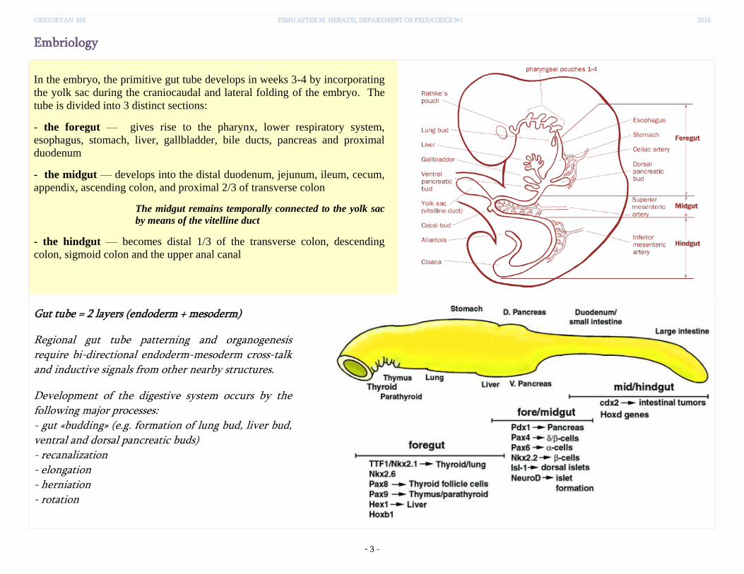
GRIGORYAN ME YSMU AFTER M. HERATSI, DEPARTMENT OF PEDIATRICS 1 2016
- 3 -
Embriology
In the embryo, the primitive gut tube develops in weeks 3-4 by incorporating
the yolk sac during the craniocaudal and lateral folding of the embryo. The
tube is divided into 3 distinct sections:
- the foregut — gives rise to the pharynx, lower respiratory system,
esophagus, stomach, liver, gallbladder, bile ducts, pancreas and proximal
duodenum
- the midgut — develops into the distal duodenum, jejunum, ileum, cecum,
appendix, ascending colon, and proximal 2/3 of transverse colon
The midgut remains temporally connected to the yolk sac
by means of the vitelline duct
- the hindgut — becomes distal 1/3 of the transverse colon, descending
colon, sigmoid colon and the upper anal canal
Gut tube = 2 layers (endoderm + mesoderm)
Regional gut tube patterning and organogenesis require bi-directional endoderm-mesoderm cross-talk and inductive signals from other nearby structures.
Development of the digestive system occurs by the following major processes: - gut «budding» (e.g. formation of lung bud, liver bud, ventral and dorsal pancreatic buds) - recanalization - elongation - herniation - rotation

GRIGORYAN ME YSMU AFTER M. HERATSI, DEPARTMENT OF PEDIATRICS 1 2016
- 4 -
RECANALIZATION
Proliferation of the epithelial lining of the gut tube results in
obliteration of the lumen by week 6. The central cells then
degenerate via apoptosis and the tube is recanalized by week 8.
Abnormalities in this process result in: stenosis, atresia, fistulas, and duplications.
Region of foregut just caudal to lung bud develops into esophagus. Errors in forming the esophagotracheal septa and/or recanalization lead to tracheoesophageal fistulas (TEF) and/or esophageal atresia (EA), respectively.
Fig. Formation of esophagotracheal septa and recanalization
Fig. Types of esophageal atresia and esophagotracheal fistulae
NOTE: this process of recanalization occurs throughout the gut tube, so occlusion can occur anywhere along the GI tract (e.g. duodenal stenosis).
Obstruction of the upper gastrointestinal tract (e.g. esophageal, duodenal, or intestinal atresia) leads to polyhydramnios (excessive volume of amniotic fluid) due to decreased swallowing and absorption of fluid. The diagnosis is made prenatally by ultrasound examination.
Fig. "Double-bubble" sign, typical for duodenal atresia. Dilated stomach (S) and duodenum (D)
with pylorus in between.

GRIGORYAN ME YSMU AFTER M. HERATSI, DEPARTMENT OF PEDIATRICS 1 2016
- 5 -
Classically, the neonate with esophageal atresia presents with copious, fine, white, frothy bubbles of mucus in the mouth and, sometimes, the nose. These secretions may clear with aggressive suctioning but eventually return. There are episodes of cough, choking and cyanosis, typically exaggerated with attempted feeding. Cyanosis is a result of laryngospasm (a protective mechanism that body has to prevent aspiration into trachea). Over the time respiratory distress develops. Abdomen is distended if there is associated TEF (as air builds up in the stomach) and scaphoid if there is isolated EA.
Duplications and cysts may occur
anywhere along the length of the gut
tube. They are most frequently found
in the región of the ileum, where they
may vary from a long segment to a
small diverticulum. Manifestations
usually occur early in life, and 33% are
associated with other defects.
Fig. Illustration depicting the many
locations where alimentary tract
duplications may be found
Fig. A - abdominal radiograph of an infant with acute onset of bilious vomiting
and abdominal distention secondary to a duplication cyst at the terminal ileum.
B - a small ileal duplication cyst causing complete obstruction of the small
bowel (the same patient)
Stomach appears first as a fusiform dilation of the foregut endoderm which
undergoes a 90° rotation such that the left side moves ventrally and the right
side moves dorsally (the vagus nerves follow this rotation which is how the
left vagus becomes anterior and the right becomes posterior).
Differential growth occurs to establish the greater and lesser curvatures.
Unlike other parts of the gut tube, the dorsal and ventral mesenteries are
retained to become the greater and lesser omenta, respectively.
Caudal end of the stomach separated from the duodenum by formation of the
pyloric sphincter (dependent on factors such as SOX-9, NKX-2.5, and BMP-4
signaling) – errors in this process lead to hypertrophic pyloric stenosis.
A B

GRIGORYAN ME YSMU AFTER M. HERATSI, DEPARTMENT OF PEDIATRICS 1 2016
- 6 -
• Rather common malformation: present in 0.5% - 0.1% of infants. More common
in males than females. Stenosis is due to overproliferation / hypertrophy of
pyloric sphincter… NOT an error in recanalization.
• Characterized by non-bilious postprandial vomiting between birth - 12 weeks
(usually begins between 2-4 weeks). The emesis becomes increasingly frequent
and forceful (aka “projectile”) as time passes.
NOTE: the presence of bile would indicate POST-duodenal blockage of some sort. • Affected infants are ravenously hungry early in the course of the illness, but
become more lethargic with increasing malnutrition and dehydration.
• The stomach becomes massively enlarged with retained food and secretions, and
gastric peristaltic waves are often visible in the epigastric area.
• A hypertrophied pylorus (the “olive” 5-15 mm in longest dimension) may be
palpated in the right upper abdomen under the liver edge.
• Jaundice can occur in 5% of infants due to indirect hyperbilirubinemia.
• As the illness progresses, very little of each feeding is able to pass through the
pylorus, and the child becomes progressively thinner and more dehydrated. The
classic metabolic findings in pyloric stenosis are hypochloremic alkalosis with potassium depletion.
Two imaging studies are commonly used to establish the diagnosis. Ultrasound is
the modality of choice in right clinical setting because of its advantages over a
barium meal are that it directly visualises the pyloric muscle and does not use
ionising radiation. The hypertrophied muscle is hypoechoic, and the central
mucosa is hyperechoic. Normal measurements include: pyloric muscle thickness
<3 mm (most accurate); length: <15 mm; width <7 mm.
Fig. Gastric peristaltic wave in
an infant with pyloric stenosis
Fig. Pyloric stenosis. Note the
huge, gas-filled stomach extending
across the midline, with minimal
air in the intestine downstream
Fig. A - Horizontal sonogram demonstrating a pyloric channel length
>14 mm in an infant with pyloric stenosis. B - Transverse sonogram
demonstrating a pyloric muscle wall thickness of >4 mm and target sign.
Fig. Barium in the stomach
of an infant with the
attenuated pyloric canal.
A B

GRIGORYAN ME YSMU AFTER M. HERATSI, DEPARTMENT OF PEDIATRICS 1 2016
- 7 -
HERNIATION AND ROTATION
The primitive gut undergoes rapid elongation and
extension through the umbilical orifice between 5
and 10 weeks, and subsequently between 10 and 12
weeks the gut returns to the abdominal cavity.
Failure of this process results in omphalocele (or
exomphalos).
Omphalocele is covered by a membranous sac and is frequently associated with other structural and chromosomal anomalies. This differentiates it from gastroschisis (a congenital anterior abdominal wall defect resulting in herniation of the abdominal contents into the amniotic sac). Gastroschisis has no covering sac and no associated syndromes.
Fig. A,B – Omphalocele. C – Gastroschisis
The gut undergoes a counter-clockwise rotation through 270 degrees (around the superior
mesenteric artery), which leaves the small intestine positioned centrally with the cecum in the
right iliac fossa and the colon lying in a lateral position. The failure of this process, called
malrotation, can lead to torsion or obstruction of the gut, and presents as a surgical emergency.
Vitelline Duct Anomalies are secondary to the persistence of the
embryonic vitelline duct.
The most frequent malformation is Meckel diverticulum. Remember the rule of 2s: occurs in 2% of the population, is 2 inches in length, is 2 feet from the ileocecal valve, most commonly presents at age 2, and 2% are symptomatic. It most commonly presents with intermittent painless rectal bleeding, but may also present as intestinal obstruction, volvulus, or diverticulitis.

GRIGORYAN ME YSMU AFTER M. HERATSI, DEPARTMENT OF PEDIATRICS 1 2016
- 8 -
LIVER AND PANCREAS DEVELOPMENT
Liver and pancreas arise from foregut endoderm in response
to signals from nearby mesoderm.
Pancreas actually has ventral and dorsal components, each specified in a different manner. Rotation of the duodenum brings the ventral and dorsal pancreas together. Aberrations in this process may result in an annular pancreas, which can constrict the duodenum.
Biliary tract anomalies
Atresias of biliary tract Choledochal cysts
A-F, Extrahepatic biliary atresias.
G, Intrahepatic atresia with normal
extrahepatic ducts. Defects A-C are
"correctable"; at least one patent duct
emerges from the liver. D-G are termed
"noncorrectable."
[Modified from Skandalakis JE, Gray SW.
Embryology for Surgeons. 2nd ed. Baltimore:
Williams & Wilkins, 1994]
I, Single cystic dilatation of the common bile
duct
II, Isolated diverticulum protruding from the
common bile duct
III, Cystic biliary dilatation within the duodenal
wall (choledochocele)
IV, Any combination of multiple cysts
V, Caroli disease/multiple intrahepatic cysts

GRIGORYAN ME YSMU AFTER M. HERATSI, DEPARTMENT OF PEDIATRICS 1 2016
- 9 -
HINDGUT AND ANORECTUM DEVELOPMENT
The terminal part of the hindgut enters into the cloaca.
Between the 4th and 6th weeks the urorectal septum separates
the cloaca into a primary urogenital sinus (ventrally) and
the rectum (dorsally). Errors in this process can lead to
imperforate anus, atresia, and/or fistulas.
As with the rest of the GI tract, enteric neurons arise from vagal neural crest. Distalmost
portions of the hindgut are farthest away and therefore more sensitive to perturbations in
migration (e.g. mutations in RET gene), resulting in congenital megacolon (Hirschsprung
disease).
Intestinal smooth muscle contraction and relaxation are under the control of enteric nervous system (ENS). Most enteric nervous activation causes muscle relaxation, mediated by nitric oxide and other enteric neurotransmitters. Extrinsic neural afferents to the ENS contain cholinergic and adrenergic fibers. The cholinergic fibers generally cause contraction, while the adrenergic fibers mainly cause inhibition. In patients with Hirschsprung disease, both myenteric and submucosal plexuses are absent. In the absence of ENS reflexes, control of the intestinal smooth muscle is overwhelmingly extrinsic. With the loss of the intrinsic enteric relaxing impulses, the increased muscle tone is unopposed. This phenomenon leads to an imbalance of smooth muscle contractility, uncoordinated peristalsis, and a functional obstruction.

GRIGORYAN ME YSMU AFTER M. HERATSI, DEPARTMENT OF PEDIATRICS 1 2016
- 10 -
Postnatal development of the digestive system
The maturation of the gastrointestinal tract takes several months after birth in humans and includes maturation of feeding abilities and
swallowing, changes in the composition of the intestinal microbiota, the function of epithelial barrier mechanisms, and the intestinal immune
system.
ORAL CAVITY
At birth the infant’s lips are well adapted to closing round a nipple to feed. At rest, the back
of the mouth is firmly closed with the tongue against the palate if not swallowing.
The buccal fat pad (BFP) (also known as the Bichat or masticatory fat pad) is an
encapsulated mass of adipose tissue located between the masticatory muscles. Its perceived
function in fetuses and infants is for sucking and enhancing the capabilities of the
buccinators and other masticatory muscles [Ponrartana et al. 2014].
When feeding, the baby draws the nipple far back into the mouth, then squeezes milk by
elevating the dorsum of the tongue from the front to the back against the hard palate. The
gums then open, the tongue slides forward and the system refills with milk. This stripping
and swallowing cycle then repeats. There is usually a rhythm of one breath to one or two
swallows.
Infants appear to accept both breast and bottle nipple quite readily, but as the skill is
perfected nipple preference becomes evident and breastfeeding may become more difficult
if the baby is fed complementary or supplementary infant formula milk from a bottle.
The ability to suck semi-solid food, bite and chew, appears at five to six months and lumpy
food can be tolerated at six to seven months, a ‘sensitive period’ for this motor ability.
However, many children will gag or spit out lumps for some months over their first year.
Digestion in the mouth
Tongue moves
forward to draw
nipple in
Nipple and
areola move
toward palate
Tongue moves along
nipple pressing it
against hard palate with
peristaltic action
Rubber nipple fills mouth to stop
tongue action
Flow of formula occurs
even if lips not tight around
nipple hub
Only small amounts of saliva are produced by the salivary glands in the neonate with little enzyme function; these develop full function by the age of two
years.
Some drooling in infants and toddlers is normal. It may occur with teething.
Saliva contains enzymes:
lingual lipase (hydrolyzes the milk fat in the the stomach),
alfa-amylase.
All babies have taste and smell present at birth, and all young babies can soon learn to like sweet tastes distinguished by the taste buds at the tongue tip,
soft palate and inside of the cheek.

GRIGORYAN ME YSMU AFTER M. HERATSI, DEPARTMENT OF PEDIATRICS 1 2016
- 11 -
The parent of a newborn asks, “Will my baby spit out the formula if it is too hot or too cold?” What is your best response?
Reflexes of the mouth and throat
1. Rooting - touching cheek stimulates movement towards touch (goes at four to twelve months). 2. Sucking and swallowing (see below). 3. Retching (gag): Spasmodic respiratory movements against a closed glottis with contractions of the abdominal
musculature without expulsion of any gastric contents (elicited by touching the soft palate or posterior pharynx).
4. Extrusion – if tongue is touched or depressed the response is to force it out of mouth (goes at 4−6 months).
SUCKING AND SWALLOWING
Eating requires active effort by infants who must have exquisite timing and coordination for sucking, swallowing, and breathing at the breast or bottle.
Swallowing movements are evident in the fetus at 16 or 17 weeks ’ gestation, and there is a steady increase in the volume of amniotic fluid swallowed, from less than 20 mL daily at 20 weeks to around 450 mL daily at term. Amniotic fluid contains nutrition and growth factors, and the development of the gut is promoted by it. An interruption of fetal swallowing can lead to polyhydramnios. Swallowing is an autonomic reflex for the first three months of life until the striated muscles in the throat establish cerebral connections but by six
months the baby is capable of swallowing, holding food in the mouth or spitting it out.
The mature swallow consists of a voluntary oral-preparatory phase, a voluntary oral phase, and involuntary pharyngeal and esophageal phases. The infant swallow does not have a voluntary oral-preparatory and oral phase but is otherwise similar.
Sucking movements can be seen from 28 to 30 weeks’ gestation but effective coordination with swallowing and breathing is generally not achieved until 37 weeks or so. Babies more premature than this may still be able to suckle by stopping breathing for short periods and then resting.
Sucking helps babies feel secure and happy and helps them learn about their world. Babies may suck their thumbs, fingers, or hands, or a pacifier or
other inanimate object such as a blanket or toy (non-nutritive sucking). Most children discontinue their nonnutritive sucking habit between the ages of
2 and4 years. Babies who suck their thumbs usually continue the habit longer than pacifier users. Prolonged thumb sucking may cause problems with
proper growth of the mouth and the alignment of teeth. The most common dental effect of nonnutritive sucking is anterior, upward movement of the
maxillary central incisors and palatal bone, which may result in an anterior open bite. Other possible effects include maxillary constriction and posterior
crossbite.
Pacifiers should never be used to replace or delay meals and should be offered only when the caregiver is certain
the child is not hungry. Pacifiers should never be dipped in sugary substances such as honey and sugar. Pacifier
use during sleep is associated with a decreased incidence of sudden infant death syndrome [A Pediatric Guide to
Children’s Oral Health. AAP; 2009]. Physiologic pacifiers are preferable to conventional pacifiers because they
may have less dental effects.

GRIGORYAN ME YSMU AFTER M. HERATSI, DEPARTMENT OF PEDIATRICS 1 2016
- 12 -
A premature baby born at 32 wks of gestation with a birth weight of 1500 gm has stable vitals. Which is the initial feeding method of
choice for this baby?
Infants less than 34 weeks gestation often do not suck effectively, lack co-ordination of sucking, swallowing and breathing and have delayed gastric
emptying [Spence 2000]. Therefore, most infants in the NICU require naso/orogastric tube feeding. Infants are obligatory nose breathers and the presence of
a nasogastric tube can increase airway resistance [Rogahn 1998]. Therefore, nasogastric tubes should be used in infants who are ventilated; infants who are establishing breast/bottle feeding; bigger infants with no respiratory problems. Orogastric tubes should be used in infants on nasal CPAP; those with respiratory problems; those who are small and sick.
The mode of initial feeding depends upon the gestational maturity and hemodynamic stability of the baby. Infants with severe birth asphyxia, respiratory
distress, apneic attack, sezures, sepsis, NEC, shock babies should be maintained on intravenous infusion till their clinical condition stabilizes [Ref: Meharban
Singh 7th, Pg. 191].
Table. Mode of Initial Feeding Based on Gestational Maturity
Gestational age
(Birth weight) Maturation of feeding skills Initial feeding method
<30 wks
(<1200 g)
No proper sucking efforts
No propulsive gut motility
Intravenous fluids (parenteral nutrition)
30–32 wks
(1200–1500 g)
Sucking bursts develop
No coordination between suck/swallow
and breathing
Oro-gastric Or Naso - gastric
feeds
33–34 wks
(1500–1800 g)
Slightly mature sucking
Coordination between breathing and
swallowing begins
Spoon, syringe, paladai Or
cup feeding
>34 wks
(1800 g)
More mature sucking
More coordination between breathing
and swallowing
Breast feeding

GRIGORYAN ME YSMU AFTER M. HERATSI, DEPARTMENT OF PEDIATRICS 1 2016
- 13 -
Feeding is hard work for babies! Feeding requires caloric expenditure because of the work of sucking: using and coordinating all these muscles
requires extra oxygen. The demand for increased oxygen leads to changes in heart rate and breathing rate. This is why babies with heart failure
so frequently have feeding difficulties (prolonged feeding times, fatigue, tachypnea or diaphoresis with feeding).
Types of medical trauma such as prolonged intravenous or nasogastric feeding, gastrointestinal and respiratory
disorders that may be associated with discomfort with eating may contribute to the development of oral aversion (oral-
tactile hypersensitivity): reluctance, avoidance, or fear of eating, drinking, or accepting sensation in or around the
mouth. An infant with oral aversion may not take anything into the mouth; not the breast, a pacifier, bottle nipple,
spoon, or finger. Some infants also will not tolerate anything touching their lips, such as a cup [Lefton-Greif 2007].
ESOPHAGUS
The bolus of food moves down the oesophagus into the stomach through the lower esophageal
sphincter (LES). When food reaches the LES, it relaxes so that food enters the stomach. The
muscle then squeezes shut to prevent food and acid from backing up into the esophagus.
However, the muscle does not always stay completely closed, allowing the stomach juices and
acid to back up into the esophagus occasionally (gastro-esophageal reflux [GER]). It is a
common physiological event due to transient relaxations in the lower esophageal sphincter. It
happens at all ages from infancy to old age, and is often asymptomatic. It occurs more
frequently after feeds/meals. In many infants, GER is associated with a tendency to
intermittent vomiting and repeated, efortless regurgitation of feeds. Most manifestations of
uncomplicated GER in infancy resolve without treatment between 12, and 18 months of age.
Acid reflux becomes gastro-esophageal reflux disease (GERD) when the reflux causes
irritation, injures the esophagus, or causes extraesophageal problems, such as chronic cough
and asthma.
It is important to take a complete history to determine if the reflux falls into the spectrum of
normal physiologic reflux or has characteristics of gastroesophageal reflux disease ( GERD).
Historical points that would increase the concern for GERD requiring treatment would be posturing or back arching, excessive crying or fussing around feedings, and failure to gain weight. If one or more of these symptoms is present, then a diagnosis of GERD should be considered, and the infant may be a candidate for acid suppression therapy.

GRIGORYAN ME YSMU AFTER M. HERATSI, DEPARTMENT OF PEDIATRICS 1 2016
- 14 -
STOMACH
The position and shape of the infant stomach is high in the abdomen
and is orientated transversely rather than vertically as in the older
(seven to ten years) child.
As the child feeds so gas is taken in. A 3.5kg baby will take in 100ml of
milk in fifteen minutes and the same volume of air. The best position to
‘burp’ a baby is to hold it sitting supported at the back and neck so as to
allow the gas to bubble up a straight esophagus.
The capacity of the stomach changes with age. Infants should be fed
often and in low portions as their gastric capacity is lower, than in
older children. Stomach capacity of the neonate gradually increases to
200 to 350 ml by age 12 months and to 1,500 ml as an adolescent.
A newborn’s stomach capacity
Day 1
Size of a cherry
5-7 ml
Day 3
Size of a walnut
22-27 ml
Week 1
Size of an apricot
45-60 ml
Month 1
Size of a large egg
80-150 ml
Enzymes in gastric juice:
proteolytic enzymes (pepsinogen/pepsin, gastrixin, cathepsins);
gastric lipase, together with lingual lipase, comprise the two acidic lipases. These lipases, unlike alkaline lipases (such as pancreatic lipase
and breastmilk lipase), do not require bile acid or colipase for optimal enzymatic activity.
Gastric acidity is reduced at birth (gastric pH is greater than 4). Adult levels of acidity are not reached until 3 to 7 years of age. In addition,
consumption of alkaline milk likely contributes to decreased gastric acidity in the child.
Gastric emptying in the neonate is prolonged and varies according to meal type (slower in formula feeding) [Cavell 1981; Bonner 2015].
Adult rates of gastric emptying (10 to 20 minutes of rapid phase) are not approached until 6 to 8 months of age [Fernandez 2011].
Clinical correlations Reduced gastric acid decreases barrier function of gastric mucus and increases the risk of enteric
infections.
High gastric pH and slow emptying can facilitate rapid absorption of acid-labile compounds (such
as penicillins).
Acid secretion limitations in premature infants should be kept in mind when considering the use
of histamine-2 (H2) blockers, which are widely prescribed in many neonatal intensive care units.
Studies suggest that critically ill premature infants treated with H2 blockers have a higher incidence of nosocomial sepsis and necrotizing enterocolitis. Although speculative, it is possible that with the already limited hydrogen ion production in the stomach of the premature infant, additional blockage further diminishes the acid barrier to microorganisms and allows for a higher load of bacteria in the more distal regions of the intestine [Beck-Sague 1994; Guillet R 2006].
Fig. Penicillin plasma concentrations following the
oral administration of a single 22,000 units/kg dose in
neonates, infants, and children [Funk et al. 2012]

GRIGORYAN ME YSMU AFTER M. HERATSI, DEPARTMENT OF PEDIATRICS 1 2016
- 15 -
INTESTINE & BOWELS
The intestine is relatively longer with well developed circular muscles.
Greater omentum is shorter (it predisposes to diffuse peritonitis in local
inflammatory abdominal processes, e.g. in appendicitis).
The sigmoid colon is relatively longer and forms additional loops (it
predisposes to constipation, especially in fat-rich diet). The rectum in infants
is also longer and poorly fixed that predisposes to rectal prolapses in tenesmus
and constipation.
Transverse mesentery is longer and predisposes to volvulus or intussusception
of bowels.
Intussusception is telescoping of one segment of intestine into another, usually in children between ages 6 mo and 3 yr. It is the most common cause of intestinal obstruction in this age group.
Most cases are idiopathic and no anatomic lead point can be identified. In about 25% of children who have intussusception, typically very young and older children, a lead point (i.e. a mass or other intestinal abnormality) triggers the telescoping. Examples include polyps, lymphoma, Meckel diverticulum, and IgA–associated vasculitis (Henoch-Schönlein purpura) when purpura involve the bowel wall. Cystic fibrosis is also a risk factor.
Peak incidence coincides with the viral enteritis season. An older rotavirus vaccine was associated with a marked increase in risk of intussusception and was taken off the market in the US. The newer vaccines, when given in the recommended sequence and timing, are not associated with any clinically significant increased risk.
Children typically present with colicky abdominal pain and vomiting, followed by passage of currant-jelly stool.
Diagnosis is made by ultrasonography (target sign).
Treatment is reduction by air contrast enema (pneumoreduction) and sometimes surgery. Therapeutic enema is of no value in patients with small bowel–to–small bowel intussusception, which usually occurs in older children who have other associated diseases (eg, HSP, hemophilia, Peutz-Jeghers syndrome, malignancies).

GRIGORYAN ME YSMU AFTER M. HERATSI, DEPARTMENT OF PEDIATRICS 1 2016
- 16 -
Postnatal maturation of the intestinal mucosa
After birth, enteral nutrition is critical in sustaining normal gut mucosal growth. Fasting, starvation and exclusive parenteral nutrition all deprive the gut of luminal nutrients resulting in a loss of gut tissue mass with blunted villi, net loss of protein, decreased cell proliferation, decreased brush border disaccharidase activity, increased intestinal permeability and increased catabolism [Sanderson IR, Walker WA, 2000].
Intestinal enzyme activity increases with age, except lactase (an enzyme of the microvillous
brush border that is involved in hydrolysis of milk lactose). Breastfed and formula-fed infants
have comparable levels of lactase activity. This lactase enzyme activity is high in the small
intestine at birth, but declines during infancy and is lost by adult life in many individuals.
Amylase, a pancreatic enzyme secreted into the duodenum that breaks down carbohydrates,
and enterokinase, which is secreted from the ileum mucosa to activate trypsin for the
breakdown of protein to peptides, are also present in the neonate gut.
All infants have efficient protein digestion and absorption; there is no difference between
breastfed and formula-fed infants.
Both breastfed and formula-fed newborn infants have relatively lower pancreatic lipase and
bile salt secretion, which limits the baby’s capacity to convert fat into fatty acids and glycerol. Breastfed infants absorb significantly more dietary lipid when compared with formula-fed infants
because they ingest a unique breast milk lipase that assists in the majority (up to two thirds) of lipid hydrolysis.
Specific long-chain polyunsaturated fatty acids (LCPs) are present in breast milk to feed the large developing brain; thus nature has matched the milk supply to the young human’s physiology.
Both breastfed and formula-fed infants are able to absorb medium-chain fatty acids directly into the portal system, whereas long-chain fatty acids require transport proteins for absorption.
In infants absorptive capacity of the intestine is higher than in adults: they have increased
intestinal permeability to macromolecules, e.g. milk proteins, maternal antibodies, as well as
toxins and antigens [de Zwart et al., 2004]. This permeability is mediated by a specific
transport process (receptor-mediated endocytosis). The permeability of the gut decreases
significantly during the first months of life (gut closure). An important factor that modulates
the gut closure is breastfeeding by its direct effects on epithelium [Roberton et al. 1982].
Fig. Growth of the small intestine [Cummins,
2002].
Fig. Lipases from different sources.

GRIGORYAN ME YSMU AFTER M. HERATSI, DEPARTMENT OF PEDIATRICS 1 2016
- 17 -
Bacterial colonization
The colonization of the intestinal tract
starts after birth, and the intestinal tract
of the newborn is first colonized by
facultative anaerobes and then by
bifidobacteria, clostridia, bacteroides,
and other strict anaerobes. The source of
nutrition plays a role in the type of
bacteria that populate the intestine. For
example, the small intestine of breast-fed
infants is primarily populated by
beneficial bacteria such as bifidobacteria
and lactobacilli because human milk
promotes growth of these types of
bacteria. One of the many benefits of
bifidobacteria and lactobacilli are that
they can limit the growth of numerous
pathogens by lowering the intestinal pH.
During the first year of life, the enteric
microflora is highly dynamic, but the
microbial diversity is low.
Fig. Spatial and temporal aspects of intestinal microbiota composition [Sekirov et al. 2010]
A: variations in microbial numbers and composition across the length of the gut. B: longitudinal variations in microbial
composition in the intestine. C: temporal aspects of microbiota establishment and maintenance and factors influencing
microbial composition
The introduction of solid food profoundly impacts on the microbial ecology of breast-fed infants. Once dietary supplementation begins,
microbiota profile of breast-fed infants changes toward formula-fed-infants profile, with the significant increase in the count of Enterococci,
Enterobacteria, Bacteroides, Clostridia, and anaerobic Streptococci. Between the first and the second year of life, differences between breast-
and formula-fed infants are lost. By the age of 2 to 3 years the microbial population stabilizes and starts to resemble that of the adult.
HONEY SHOULD NOT BE FED TO INFANTS YOUNGER THAN 1 YEAR OLD!
It contains spores of Clostridium botulinum that readily grow in baby’s intestine and produce toxins. Older children are believed to be less susceptible to botulism due to their mature gastrointestinal tracts, which can excrete the spores before they germinate.

GRIGORYAN ME YSMU AFTER M. HERATSI, DEPARTMENT OF PEDIATRICS 1 2016
- 18 -
Intestinal motor activity & stools
Intestinal motor activity matures throughout early infancy, with consequent
increases in the frequency, amplitude, and duration of propagating contractions.
The stools of the newborn (meconium) are odourless, dark green, have a
smooth, paste-like appearance and are passed within the first twenty-four hours
in about 87 per cent of infants and within 48 hours by 99 per cent. This is not
influenced by whether the infant is breast- or formula-fed [Catto-Smith 2005].
After this, the method of feeding will have impact on stool frequency, colour
and consistency of the stool.
The normal stools of breastfed infants are never formed, may be passed at hourly intervals, may contain mucus and may be green. When lactation becomes established between the third and fifth days intestinal hurry is common, resulting in frequent stools. No treatment is needed. Later the stools tend to become less frequent and more pasty and by the age of 3 weeks they may be passed once every 2 or 3 days.
In contrast, the normal stools of formula-fed infants are formed and do not contain fluid or mucus. With certain cow’s milk preparations the stools may be dark green, but this has no sinister meaning.
Defecation requires two coordinated events: pelvic floor relaxation, and an increase in intraabdominal
pressure (bearing down to have a bowel movement). Children with infant dyschezia have not yet
developed this coordination so they are unable to enjoy easy defecation.
Infant dyschezia is a functional condition characterized by at least 10 minutes of straining and crying before successful passage of soft stools in an otherwise healthy infant less than six months of age. These episodes, exhausting for the infant and anxiety provoking for the parents, occur several times daily. They may prompt parents to visit their child’s clinician during the infant’s first 2 to 3 months of life with concerns that their child is constipated. The parents describe a healthy infant who cries for 20 to 30 minutes, turns red in the face, and screams, seemingly in pain, before defecation takes place.
Breastfed babies’ stools have a lower pH than babies fed on infant
formula milk, due to the fermentative gut flora, which produces
neutral and acid metabolic waste products. When the pH drops, the
faecal enzymes become less active and thus these breastfed babies
usually have less nappy rash.
Nappy rash occurs when urine and faeces
mix; the urea splitting organisms in urine
raise the pH and this irritates the infant’s
delicate skin.

GRIGORYAN ME YSMU AFTER M. HERATSI, DEPARTMENT OF PEDIATRICS 1 2016
- 19 -
LIVER & BILIARY TRACT
The liver is relatively larger and is richly vascularised. Liver occupies more of
abdominal cavity than in adults. Liver reaches adult size and function by
adolescence.
The liver is functionally immature at birth:
The liver’s slow development of glycogen storage capacity makes the infant prone to hypoglycemia. The suckling neonate is entirely dependent on glucose supplied via the milk and on his capacity for an efficient gluconeogenesis to maintain his blood glucose concentration in the normal range
Decreased synthesis of coagulation factors (prothrombin levels in the neonate are only 20%–40% of adult levels, which affects clotting)
Decreased synthesis of albumins ⇒ Protein binding of drugs is decreased; high levels of free drug remain in bloodstream, which can lead to toxic level of drug
Decreased hepatic enzyme function: 1) Enzyme systems for biotransformation of drugs are not fully developed, which affects drug dosing; infants and children metabolize drugs more slowly than adults; can easily build up toxic levels of drugs 2) Liver conjugation reactions are impaired ⇒ neonatal hyperbilirubinemia; long drug half-lives
Immaturity of bile synthesis and secretion ⇒ susceptibility of the sick neonate to develop cholestasis in response to sepsis, administration of parenteral nutrition
The composition of bile in the newborn is different to that in the adult, with
higher concentrations of hyocholic acid as well as some unusual bile acids. Soon
after birth, the concentrations of the bile acids cholate and chenodeoxycholate
increase rapidly. They are involved in the absorption of long chain fatty acids and
fat-soluble vitamins as well as a number of drugs and hormones.
The ability of the gallbladder to empty in response to a test feed occurs soon after
birth, but is reduced in preterm neonates of 27 – 32 weeks’ gestation.
Fig. The activity of many cytochrome P-450 (CYP) isoforms
and a single glucuronosyltransferase (UGT) isoform is
markedly diminished during the first two months of life.

GRIGORYAN ME YSMU AFTER M. HERATSI, DEPARTMENT OF PEDIATRICS 1 2016
- 20 -
Assessment of the Digestive System in Children
"The colon is the playing field for all human emotions." - Cyrus Kapadia, MD
General Considerations
Interactions with other organ systems serve the needs both of the gut and
the body. Symptoms apparently related to the gastrointestinal tract may
arise fromdisease elsewhere. For example, vomiting may be a symptom of
infection, poisoning or head injury, and abdominal pain may occur in a
child with pneumonia or diabetic ketoacidosis.
Subtle alterations in gastrointestinal function may adversely affect growth
while causing minimal symptoms. Only careful examination of the child's
growth curve may alert the physician to the possibility of underlying
gastrointestinal disease.
The limited ability of the young child to describe his or her symptoms may
make diagnosis more difficult.
Gastrointestinal symptoms are determined by:
Structural changes (eg, inflammation, destruction, impaired gut blood flow, neoplasia)
Functional changes (eg, impaired digestion and absorption, altered secretion, altered gut motor function)
Visceral hypersensitivity Psychogenic factors
Fig. A biopsychosocial model of pain: for use with parents and patients
The biopsychosocial model of pain explains how abdominal pain occurs as a result
of interplay between multiple factors surrounding the child, including genetic
predisposition, life events, the family and the child’s coping mechanisms for
dealing with stress and pain. These biological, social and psychological factors
impact the development and recognition of gut pain through altered gut
physiology via the brain–gut axis [Mulak & Bonaz, 2004].
Biopsychosocial Approach to GI disorders
There is a link between "psyche" and "soma" in gut function (brain–gut axis mechanism), and this is true in children as it is in adults. Interactions
between physiological (e.g., motility, sensation) and psychological factors (e.g., life stress, emotional state, coping, social support) are seen as shaping the
nature and outcome of the child’s gastrointestinal symptoms. Factors such as the child’s adoption of a sick role, their sensitivity to pain, their level of
psychological functioning, and the family’s ability to deal with the illness can all contribute to differences among children’s reaction to illness
[Drossman, 1998; Hyams, 1996]. The commonest manifestation is with recurrent abdominal pain, sometimes associated with vomiting ("periodic
syndrome"), which occurs in school-age children in association with emotional triggers such as bullying, anxiety over parental stress, or exams. Some
adolescents with severe Crohn’s disease do not limit their functioning and will not let the illness define them, participating in a wide range of academic
and extracurricular activities. However, others with less severe physical symptoms but who are anxious and whose parents have significant concerns
about their health status are home schooled and have little peer contact.

GRIGORYAN ME YSMU AFTER M. HERATSI, DEPARTMENT OF PEDIATRICS 1 2016
- 21 -
NUTRITIONAL ASSESSMENT
Feeding history 24-hour recall, food frequency
questionnaire, etc.
Anthropometry Weight, height, weight-for-
height, body mass index
(BMI), skinfold thickness,
mid-upper arm circumference
(MUAC), waist circumference
Clinical Physical appearance, signs of
malnutriution
Biochemical and
haematological
Vitamin status tests
Lipid status
Iron status – haemoglobin,
ferritin, etc. Fig. Kwashiorkor. This patient has a typical edematous appearance in the periorbital area, extremities, and abdomen.
Fig. Marasmus. Note the profound wasting and sparse hair
Fig. Potbelly and muscle wasting in a child with celiac disease
Infant feeding history
Which milk? Breast or formula?
If formula, note which one and details of reconstitution
How much feed? In breast-fed infants, does the mother have a good milk supply?
In formula-fed infants, note volumes offered and taken
How often? Note the times of feeds in the previous 24 hours
How long does the feed take to complete?
Characteristics of feeding? Hungry, windy, apathetic, slow, sleepy, etc.

GRIGORYAN ME YSMU AFTER M. HERATSI, DEPARTMENT OF PEDIATRICS 1 2016
- 22 -
CLINICAL EXAMINATION OF THE CHILD'S ORAL CAVITY
Lips, Tongue, and Oral Mucosa
The lips and tongue should be soft, pink, and moist. The oral mucosa may be pink or
brown, depending on the infant’s or child’s skin color. It should be smooth and
moist.
Fig. Healthy Lips and Tongue
Fig. Healthy Mucosa
Palate
To observe the palate, you should gently bend the infant’s or child’s head back.
Directing the light onto the surface of the palate, observe pink, moist surfaces. The
soft, fleshy mass that hangs from the rear of the soft palate is the uvula.
Fig. Healthy Palate
Major Salivary Glands
The major salivary glands are located in the cheeks and on the floor of the mouth.
The ducts should be raised and pink. Youl should first look at them and then press
them lightly. It is common to observe clear secretions from the glands. Pressing the
glands should not cause discomfort.
A number of normal anatomic variations may be noted
in the mouth.
A short lingual frenulum (“tongue-tie”) may be
worrisome to parents but only rarely interferes with
eating or speech, generally requiring no treatment.
Fissured tongue (+/-geographic tongue) is usually a
normal finding.
A bifid uvula may be normal or associated with a
submucous cleft of the soft palate.

GRIGORYAN ME YSMU AFTER M. HERATSI, DEPARTMENT OF PEDIATRICS 1 2016
- 23 -
Abnormalities and complaints related to the mouth and throat
Manifestation Definition Causes
Halitosis
(fetor oris)
Oral malodor Poor oral hygiene, tooth decay, gingivitis, oral candidiasis,
postnasal drip, sinusitis, tobacco use, xerostomia …
Xerostomia
Dry mouth Thirst, mouth breathing, allergic rhinitis, adenoiditis,
dehydration, diabetes, or mumps; may be associated with drug
use
Dysphagia Dysphagia refers either to the difficulty someone may have with
the initial phases of a swallow (usually described as “oropharyngeal
dysphagia”) or to the sensation that foods and or liquids are
somehow being obstructed in their passage from the mouth to the
stomach (usually described as “esophageal dysphagia”).
Viral or bacterial pharyngitis, peritonsillar or retropharyngeal
abscess, epiglottitis, GERD, cleft palate, cerebral palsy …
Stomatitis Inflammation of the mucous membrane of the mouth.
Inflammation of the lips is known as cheilitis, inflammation of the tongue is glossitis, inflammation of the gums is gingivitis.
Coxsackievirus (herpangina; hand, foot, and mouth disease),
aphthous ulcers, oral candidiasis, herpesvirus 1 or 2, measles …
Tooth decay Tooth decay associated with sore throat may be caused by self-
induced vomiting, poor oral hygiene, or early childhood caries
Candidiasis
Fig. Candidiasis, a fungal infection that may be present in
infancy, appears as a soft white plaque coating the mucosa
and palate. When the coating is removed with a gauze pad
or tongue blade, the undersurface is red and raw.
Primary herpetic gingivostomatitis
Fig. A, Infection with primary herpes often occurs at the time of eruption of
primary teeth. B, Common presentation with multiple small ulcers on the lower
lip, and gingival swelling and inflammation.

GRIGORYAN ME YSMU AFTER M. HERATSI, DEPARTMENT OF PEDIATRICS 1 2016
- 24 -
Infections caused by Coxsackie group A viruses
Fig. A, Herpangina with characteristic palatal and pharyngeal ulceration and
inflammation. B, Cutaneous lesions in hand, foot and mouth disease.
A. Herpangina B. Hand, foot and mouth disease (HFMD)
ORAL MANIFESTATIONS OF GASTROINTESTINAL DISEASES
Gastroesophageal
reflux disease (GERD)
It causes enamel erosion due to gastric acid influence.
Inflammatory bowel
disease (IBD)
1. Crohn disease (CD) – Oral manifestations of Crohn disease include
multifocal, linear, nodular, polypoid or diffuse mucosal thickenings. They are
characteristically firm, pink and painless to palpation unless there are
ulcerations that may cause pain on touch, or when eating acidic, spicy or hot
foods.
2. Ulcerative colitis (UC) – Rare oral manifestation of ulcerative colitis is
pyostomatitis vegetans.
Painless swelling of the
lips (CD)
Pyostomatitis - tiny yellow
pustules on the oral mucosa
(UC)
Celiac disease Oral and dental manifestations of celiac disease: enamel defects; delayed eruption; recurrent aphthous ulcers; cheilosis; oral lichen planus; atrophic glossitis
Peutz-Jeghers
syndrome
– An autosomal dominant disease, characterized by
pigmentation of the skin and mucosals
intestinal polyposis with high risk of malignancy
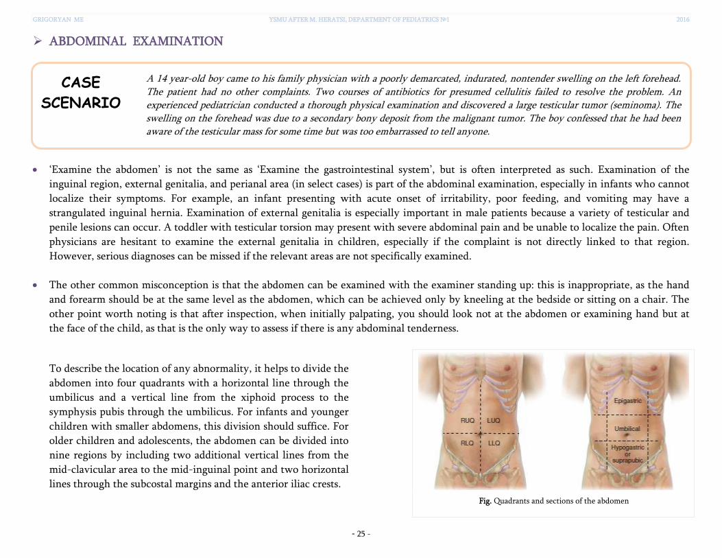
GRIGORYAN ME YSMU AFTER M. HERATSI, DEPARTMENT OF PEDIATRICS 1 2016
- 25 -
ABDOMINAL EXAMINATION
CASE
SCENARIO
A 14 year-old boy came to his family physician with a poorly demarcated, indurated, nontender swelling on the left forehead. The patient had no other complaints. Two courses of antibiotics for presumed cellulitis failed to resolve the problem. An experienced pediatrician conducted a thorough physical examination and discovered a large testicular tumor (seminoma). The swelling on the forehead was due to a secondary bony deposit from the malignant tumor. The boy confessed that he had been aware of the testicular mass for some time but was too embarrassed to tell anyone.
‘Examine the abdomen’ is not the same as ‘Examine the gastrointestinal system’, but is often interpreted as such. Examination of the
inguinal region, external genitalia, and perianal area (in select cases) is part of the abdominal examination, especially in infants who cannot
localize their symptoms. For example, an infant presenting with acute onset of irritability, poor feeding, and vomiting may have a
strangulated inguinal hernia. Examination of external genitalia is especially important in male patients because a variety of testicular and
penile lesions can occur. A toddler with testicular torsion may present with severe abdominal pain and be unable to localize the pain. Often
physicians are hesitant to examine the external genitalia in children, especially if the complaint is not directly linked to that region.
However, serious diagnoses can be missed if the relevant areas are not specifically examined.
The other common misconception is that the abdomen can be examined with the examiner standing up: this is inappropriate, as the hand
and forearm should be at the same level as the abdomen, which can be achieved only by kneeling at the bedside or sitting on a chair. The
other point worth noting is that after inspection, when initially palpating, you should look not at the abdomen or examining hand but at
the face of the child, as that is the only way to assess if there is any abdominal tenderness.
To describe the location of any abnormality, it helps to divide the
abdomen into four quadrants with a horizontal line through the
umbilicus and a vertical line from the xiphoid process to the
symphysis pubis through the umbilicus. For infants and younger
children with smaller abdomens, this division should suffice. For
older children and adolescents, the abdomen can be divided into
nine regions by including two additional vertical lines from the
mid-clavicular area to the mid-inguinal point and two horizontal
lines through the subcostal margins and the anterior iliac crests.
Fig. Quadrants and sections of the abdomen

GRIGORYAN ME YSMU AFTER M. HERATSI, DEPARTMENT OF PEDIATRICS 1 2016
- 26 -
INSPECTION
Simply looking at the abdomen can provide important clues to the underlying diagnosis.
The abdominal wall normally moves with respiration, especially in infants. Loss of this movement secondary
to pain can be an important indicator of abdominal inflammation, such as peritonitis.
Abdominal symmetry is best assessed by inspection from the foot of the bed. Asymmetry can result from an
intraabdominal mass. Bulging of the flanks can occur in patients with ascites.
Distention [1] can be caused by air in the intestine, fluid in the abdominal cavity (ascites), or solid organ
enlargement.
Until about 4 years of age, the abdomen continues to protrude in both the supine and standing positions.
After 4 years of age, the abdomen still protrudes with standing due to lumbar lordosis, but it is flat when the
child is lying down. Do not mistake this appearance for pathologic distention.
Abdominal distention is an important sign of intestinal obstruction. The lower the obstruction in the gut,
the more marked the distention. Also, associated symptoms will be present, including vomiting and
abdominal pain/irritability. Peristalsis may be visible through the abdominal wall in patients with intestinal
obstruction. This peristalsis is most commonly observed in the upper abdomen in infants with hypertrophic
pyloric stenosis.
A depressed (scaphoid) abdomen [2] in a newborn suggests a diaphragmatic hernia with some abdominal
organs sitting in the chest cavity.
Umbilical hernias [3] are common in infants. In some ethnic groups, they are virtually universal. Umbilical
hernias are reducible and, although they are sometimes quite large in size, they typically resolve without
treatment during the first few years of life.
Slight separation of the rectus abdominis muscle (diastasis recti) [4] is normal in children, especially in
premature infants.
Examine the inguinal area for swelling. Indirect inguinal hernias are common in children and always
require surgical correction. A reducible hernia may not be obvious in the supine position. Older children
should be examined standing up. Increasing the intraabdominal pressure by coughing or straining makes the
swelling more obvious. Small, nontender, soft, mobile inguinal lymph nodes are common in children. They
can be bilateral and often are benign. Check for abnormal lymphadenopathy elsewhere (e.g., the axilla).
Examine the back, because diseases of the spine may present with abdominal symptoms. A tuft of hair in the
midline often is a clue to an underlying congenital spinal abnormality (e.g., diastematomyelia) in a child
with chronic constipation.
1
2
3
4

GRIGORYAN ME YSMU AFTER M. HERATSI, DEPARTMENT OF PEDIATRICS 1 2016
- 27 -
PERCUSSION
Indirectly percuss all areas of the abdomen. Normal findings include dullness along the costal margins and tympany
over the remainder of the abdomen. A full bladder may yield dullness to percussion. Percussion can be useful in
delineating the edge of the liver or spleen and in examining for ascites.
PALPATION
Palpate the abdomen with the child in a supine position. If the child’s legs are small enough,
the knees may be brought up with the nondominant hand to flex the hips and relax the
abdomen. The examiner is always at the patient’s right side.
Look at the child's face while palpating the abdomen. Start palpating at a point furthest away
from any tenderness. A reluctant child may be put at ease if their own hand is used, with the
examiner 's hand overlying.
Palpate all four quadrants of the abdomen in a systematic fashion, first lightly and then deeply.
Apply light pressure with the fingertips to perform light palpation, assessing for tenderness
and muscle tone.
Note skin turgor by gently elevating a piece of skin and allowing it to fall back into place.
Perform deep palpation to assess the organs and any masses. Place one hand on top of the
other and palpate from the lower quadrants to the upper. The edge of the liver may be felt at the right costal margin, and the tip of the spleen can be felt
at the left costal margin. The liver often is palpable 1 to 2 cm below the right costal margin in normal infants and toddlers. Its edge is usually soft, and it moves downward with respiration. The liver edge is often palpable in healthy children and adolescents [Goldbloom RB, 2010].
The descending colon may be felt in the left lower quadrant as a small column and the bladder as a soft balloon below the umbilicus.
The kidneys are rarely palpable. The abdomen should be soft and nontender to palpation. Report firmness, tenderness, or
masses.
Fig. (A) Light and (B) deep palpation of the abdomen.
Palpate the inguinal area for the presence of hernia or enlarged lymph nodes.
B
A

GRIGORYAN ME YSMU AFTER M. HERATSI, DEPARTMENT OF PEDIATRICS 1 2016
- 28 -
Bimanual Palpation of the Right Upper Quadrant
Table. Physical signs and clinical significance on abdominal
examination
Physical sign Clinical significance
Abdominal mass Fecal mass in constipation
Hydronephrosis
Renal/suprarenal tumor
Crohn disease
Splenomegaly Hemolytic anemia
Neoplastic disease
Portal hypertension
Hepatomegaly Chronic liver disease
Neoplastic disease
Storage disease
Congestive heart failure
Ascites Liver cirrhosis
Nephrotic syndrome
Congestive heart failure
Anal fissure Chronic constipation
Crohn disease
Proper palpation of the right upper quadrant consists of placing the right hand at the right costal margin and the left hand under the patient’s right flank, lifting it toward the examiner.
Bimanual Palpation of the Left Upper Quadrant
Palpate the left upper quadrant by placing the right hand toward the patient’s left costal margin while the left hand is placed at the patient’s left flank, pushing the spleen anteriorly.
On palpation of either upper quadrant, it is advisable to have the patient take a
deep breath. Palpate the lower quadrants with similar technique to evaluate the
kidneys for enlargement. Palpation of the right lower quadrant is a critical part
of the evaluation for appendicitis, with the classic finding of tenderness over
McBurney’s point.
RECTAL EXAMINATION IN CHILDHOOD
This is never a routine examination and is rarely helpful. Careful thought must be given to undertaking this examination in any child.
In older children, a rectal examination is best performed with the child
in the left lateral position with the spine and knees fully flexed. Infants
and younger children can be examined in a knee-chest position.
Examine the perianal area for excoriations, skin tags, or fistulae. Sentinel
skin tag(s) overlying a chronic anal fissure in older children are
pathognomonic of Crohn disease.
Fig. Crohn disease. (A) Perianal abscess, (B) sentinel skin tags, (C) draining
perianal fistula.
A B C

GRIGORYAN ME YSMU AFTER M. HERATSI, DEPARTMENT OF PEDIATRICS 1 2016
- 29 -
CASE
SCENARIO
15-year-old Emily was diagnosed with an eating disorder by her family doctor and pediatrician. She had presented with severe weight loss. Her appetite had decreased but she denied abdominal pain, vomiting, or diarrhea. She was markedly wasted. She was admitted to the psychiatric ward of a pediatric tertiary care hospital for further management. A referral was made to the gastroenterology service. Physical examination revealed large perianal skins tags and an underlying anal fissure. A perianal examination had not been performed previously, delaying the diagnosis of Crohn's disease by months.
COMMON PRESENTATIONS OF ABDOMINAL CONDITIONS.
FUNCTIONAL VS ORGANIC DISORDERS
ABDOMINAL PAIN
My 3-year-old girl has fever and abdominal pain with smoky coloured urine.
My 9-year-old daughter has been thirsty for days and passing lots of urine. Now she has
tummy ache and is drowsy.
My 13-month-old child is getting thinner, with frequent tummy aches and diarrhoea.
My 7-year-old son keeps getting tummy ache on the way to school.
My 10-year-old daughter is constipated, and this is very distressing. Can you give her
something?
Questions
1. What types of abdominal pain occur in children?
2. What characteristics distinguish functional from organic
abdominal pain?
3. What are the common organic causes of recurrent abdominal
pain in children?
4. What functional gastrointestinal disorders manifest with
recurrent abdominal pain in children?

GRIGORYAN ME YSMU AFTER M. HERATSI, DEPARTMENT OF PEDIATRICS 1 2016
- 30 -
1. What types of abdominal pain occur in children?
Acute abdominal pain resolves within 4 weeks since onset of symptoms.
Common causes of acute abdominal pain include:
non-surgical conditions, e.g. gastroenteritis, urinary tract infection (UTI), Henoch–Schönlein purpura (HSP)
surgical pathology, e.g. appendicitis, intussusception
Chronic abdominal pain is characterized by intermittent or persistent
pain that occurs over a period greater than 2 months. The majority of
children with chronic abdominal pain have so-called functional
abdominal pain (FAP).
Table. Common causes of abdominal pain in accordance with the
patient’s age
The sensation of abdominal pain is transmitted to the central nervous system via somatic and visceral afferent fibers. The visceral afferent system innervates the visceral peritoneum and its
structures. Visceral pain localizes poorly. Pain originating from the parietal peritoneum from inflammation or
abdominal wall pain is well localized (parietal or somatic pain). Referred pain results from the convergence of visceral and somatic
pain pathways in the spinal cord, so pain originating in abdominal viscera may be perceived as originating at a distant, well-isolated somatic location. For example: Diaphragmatic irritation secondary to pancreatitis, a bleeding spleen,
cholecystitis, or liver abscess may be interpreted as pain arising in the vicinity of the lower neck and shoulders because the diaphragm and shoulder pain pathways converge in the spinothalamic tracts at C4. Similarly, gallbladder inflammation may be sensed in the right infrascapular region, a migrating ureteral stone may be felt progressing toward the ipsilateral groin, and rectal and gynecologic discomfort may be sensed in the vicinity of the sacrum.
Conversely, pain originating in somatic locations, such as the right pleural surface with pneumonia, may be perceived as originating in the lower abdomen because pain afferents from both regions converge at T10-11.

GRIGORYAN ME YSMU AFTER M. HERATSI, DEPARTMENT OF PEDIATRICS 1 2016
- 31 -
2. What characteristics distinguish functional from organic abdominal pain?
Chronic or recurrent abdominal pain (RAP) is reported to occur in 10% to 15% of children between the ages of 4 and 16 years. RAP is not a
diagnosis but is a descriptive term that applies to intermittent, severe, episodic pain.
In most cases, the pain is functional, without demonstrable evidence of a pathological condition.
Multiple studies have demonstrated that less than 5% of these children have an organic disorder.
Clues to ORGANIC disease Clues to FUNCTIONAL disease
Age younger than 4 years
Localized abdominal pain
Nocturnal pain, eg, awakening from sleep to pass loose
stool at night
Recurrent emesis
Chronic diarrhea
Heme-positive stools
Weight loss
Abnormal physical examination findings — clubbing,
perianal skin tags, abdominal mass
Abnormal screening laboratory test results — decreased
albumin, increased ESR/CRP, anemia, abnormal liver or
kidney function tests, elevated amylase or lipase
Typically the pain is episodic, unrelated to meals, and periumbilical in location
lasting a few hours to several days with intervening symptom-free intervals lasting
days to months. Pain may be severe enough to cause significant school absenteeism
History of irritable bowel syndrome (IBS) in first-degree relative. IBS is
characterized by an abnormal frequency and/or consistency of stools (diarrhea or
constipation), straining, urgency, relief of pain with defecation, a feeling of
incomplete evacuation, passage of mucus, or a feeling of bloating or abdominal
distention. Commonly associated symptoms include headache, pallor, dizziness, and
fatigue.
Although many children complain of pain at the time of office visits, their behavior,
affect, and activity are seldom consistent with the degree of expressed discomfort.
Poorly localized pressure tenderness is frequently elicited during abdominal
palpation. Between episodes, the abdominal examination is normal
3. What are the common organic causes of recurrent abdominal pain in children?
Cause Clinical clues
Food allergy (eg, cow’s milk protein
allergy)
Vomiting, diarrhea, Hemoccult-positive stools, failure to thrive, rhinitis, eczema, pallor, irritability, and a
positive family history of allergies
Lactose intolerance Crampy pain and/or bloating and/or intestinal gas related to meals, dairy products and foods containing dairy
products Giardiasis
Peptic origin Early morning pain, pain awakens at night. Early satiety, nausea, sour breath, belching
Genitourinary problems Back pain, dysuria, cervical motion tenderness, adnexal tenderness, or adnexal mass on pelvic examination
Familial Mediterranean fever (FMF) Recurrent episodes of fever and serositis, resulting in pain in the abdomen, chest, joints and muscles
Inflammatory bowel disease (IBD) Fever, weight loss, no increase in height, joint complaints and rash, blood in stool

GRIGORYAN ME YSMU AFTER M. HERATSI, DEPARTMENT OF PEDIATRICS 1 2016
- 32 -
4. What functional gastrointestinal disorders manifest with recurrent abdominal pain in children?
The Rome III diagnostic criteria for abdominal pain-related functional gastrointestinal disorders

GRIGORYAN ME YSMU AFTER M. HERATSI, DEPARTMENT OF PEDIATRICS 1 2016
- 33 -
CONSTIPATION AND FECAL INCONTINENCE (ENCOPRESIS)
Average stooling frequency is 3 to 4 per day during the first weeks of life and decreases to 1 to 2 per day with introduction of table foods by 1
year of age. After age 4 and into adulthood, normal stool frequency can range from 3 per day to 3 per week.
Constipation is defined as infrequent passage of stool associated with pain and difficulty, or delay in defaecation. The majority of children with
chronic constipation have no clearly identifiable disorder, and their condition is therefore labeled functional or idiopathic constipation.
Table. Causes of constipation
Idiopathic Gastrointestinal Non-gastrointestinal
Commonest due to a combination of:
• Low fibre diet
• Lack of mobility and exercise
• Poor colonic motility (55% have a positive
family history)
• Meconium plug syndrome (often in preterm babies)
• Hirschsprung’s disease
• Anal disease (infection, stenosis, ectopic, fissure,
hypertonic sphincter)
• Partial intestinal obstruction
• Coeliac disease
• Hypothyroidism
• Hypercalcaemia
• Neurological disease, e.g. spinal disease
• Chronic dehydration, e.g. diabetes insipidus
• Drugs, e.g. opiates and anticholinergics
• Sexual abuse
Rome III criteria for functional constipation*
1) Defecation frequency 2 times or fewer
per week
2) Fecal incontinence (fecal soiling in
undergarments)
3) Episodes of retentive posturing during
which the child crosses legs and screams
4) Pain during defecation
5) Large-diameter stools that sometimes
clog the toilet
6) Palpable rectal fecal mass
* Children younger than 4 years need to meet 2 criteria for at least 1 month Children older than 4 years need to meet 2 criteria for at least 2 months
Children with constipation may present
with decreased frequency of defecation
and hard pellet-like or large torpedo-
like stools that are difficult to pass,
often causing pain and fresh blood per
rectum. Repeated painful experiences
can scare the child and prompt stool-
avoidance behavior.
Fig. The vicious cycle of constipation

GRIGORYAN ME YSMU AFTER M. HERATSI, DEPARTMENT OF PEDIATRICS 1 2016
- 34 -
Fig. Child exhibiting
retentive posture
Younger children often develop a stool-
withholding behavior without pre-existing
constipation (some children can withhold
stool for up to 2 to 3 weeks), crouching down
to prevent their bowels opening.
Stool-withholding behavior can lead to a
very impacted rectum. Liquid stool from
above can leak out into their underwear
(overflow incontinence), often causing
diagnostic confusion.
Fig. Fecal incontinence results from
the rectal fecal mass stretching open
the anal sphincter; the liquid stool
seeps around it and leaks out of the
anal canal
It is important to distinguish between the common problem of functional constipation and less
common organic causes of constipation, including Hirschsprung disease. Organic cause of
constipation is more likely if: delayed passage of meconium beyond 24hr of age; onset in infancy;
severe; associated with faltering growth or abnormal physical signs (include per anal examination).
A diagnosis of Hirschsprung disease is suggested by small-caliber soft stool, episodes of explosive soft stool preceded by a lack of stool passage and abdominal distension, and history of delayed meconium passage in the first few days of life. If Hirschsprung disease is suspected, suction rectal biopsy should be considered as the next step in evaluation.
Constipation complications
Overflow incontinence - this should be distinguished from fecal incontinence secondary to fecal urgency seen in patients with irritable
bowel syndrome, neurologic disorders, and spinal disorders. Children with behavioral disorders and sexual abuse can also present with
fecal incontinence.
Anal fissures.
Urinary tract infection occurs in 5% to 10% of chronically constipated children, probably secondary to partial urethral obstruction or
ascending infection from chronically soiled underwear.
Chronic anorexia and failure to thrive.
Affected psychosocial functioning (poor self-esteem, poor school performance, and family stress and conflict).

GRIGORYAN ME YSMU AFTER M. HERATSI, DEPARTMENT OF PEDIATRICS 1 2016
- 35 -
VOMITING
Definitions
Vomiting: forceful expulsion of gastric contents from the mouth
Regurgitation: effortless flow of gastric contents from the mouth (also referred to as infant gastroesophageal reflux [GER], spitting,
posseting)
Although the end result of vomiting and regurgitation is similar, they have completely different characteristics. Vomiting is usually preceded
by nausea and is accompanied by forceful gagging and retching. Regurgitation, on the other hand, is effortless and not preceded by nausea.
Physiologic GER is common in normal infants. It presents between 1 and 2 months of age, increases over the next few months, and resolves
spontaneously. The emesis is gastric contents without blood or bile. GER does not cause excessive crying, poor eating, slow weight gain, or
apnea. If such symptoms or signs are being evaluated, alternative explanations should be sought. Physiologic GER requires no intervention (i.e.,
no dietary change in infant or mother, no suppression of gastric acid, etc.). Multiple studies document the absence of esophagitis or other
complications in such infants. Acid suppression may result in increased risk of pneumonia or gastroenteritis [Smith et al. 2013].
The most common causes of vomiting: Less common and serious diseases that present with vomiting:
Gastrointestinal infections (eg, viral, bacterial, and parasitic gastroenteritis)
Non-gastrointestinal infections* (eg, pyelonephritis, sepsis, streptococcal pharyngitis, otitis media)
Acid-peptic disorders (eg, GERD, Helicobacter pylori gastritis)
Allergic disorders (eg, formula protein sensitivity, eosinophilic esophagitis)
Cyclic vomiting syndrome: severe, discrete episodes associated with pallor, lethargy +/– abdominal pain. The child is well in between episodes. Often there is a family history of migraine or vomiting
* In young children a simple fever can cause vomiting, as
can almost any infection of any system.
o Small bowel obstruction due to anatomic abnormality or prior intestinal surgery with
adhesions presents with repeated bilious emesis, abdominal pain, and, often, distension
with tympany. Urgent evaluation should proceed. Intussusception produces reflex
vomiting prior to evolution to obstruction.
o Posterior fossa tumor: In all children and adolescents, acute onset of daily vomiting
with any associated neurologic symptom (headache, irritability, lethargy, ataxia,
decreased activity, diplopia) should prompt careful neurologic exam for cerebellar signs
of nystagmus, dysmetria, and ataxia. Fundoscopic exam should be attempted and
imaging considered. Posterior fossa brain tumors produce vomiting with or without
increased intracranial pressure.
o Caustic ingestion: Acute vomiting, dysphagia, refusal to swallow, and drooling
characterize esophageal injury due to caustic ingestion. Oral burns or erosions are often
but not always present. Usually, caregivers recognize or witness the event.
o Esophageal foreign body can present similarly to caustic ingestion, but without evident
burns or erosions. A disk battery retained in the esophagus is a medical emergency

GRIGORYAN ME YSMU AFTER M. HERATSI, DEPARTMENT OF PEDIATRICS 1 2016
- 36 -
There are some key red flags to watch for:
• persisting vomiting and fever (of more than 24 hours’ duration) but without diarrhoea – this can suggest bacterial meningitis
• bile-stained vomitus – in such cases, bowel obstruction (e.g. from a volvulus) must be excluded
• blood-stained vomit (see “Gastrointestinal bleeding”)
Important Disorders Presenting with Vomiting by Age
Infant Vomiting Childhood Vomiting
Bilious emesis: intestinal malrotation with or without
volvulus
Meconium ileus (nearly always indicates underlying
cystic fibrosis)
Hirschsprung disease
Pyloric stenosis
Bilious emesis
Prior abdominal surgery (intraabdominal adhesions and associated obstruction)
Other acquired small bowel obstructions (e.g., intussusception, HSP)
Nonbilious emesis
Gastroenteritis is the most common cause of acute nonbilious emesis. Many other GI illnesses (e.g., pancreatitis, chole(docho)lithiasis, peptic ulcer disease, and others) have vomiting as a component.
AN APPROACH TO EVALUATION OF THE PATIENT WITH VOMITING
Bloody Bilious Non-bloody, non-bilious
↓ ↓ Neonate or infant Child or adolescent
Evaluate for upper GI
bleeding
Rule out
obstruction
Evidence of sepsis,
meningitis, UTI
Lethargy or altered
mental status
No Yes No Yes
Evidence of
obstruction Treat with antibiotics
Diarrhea or
fever?
No Yes No
Yes Consider: metabolic, endocrine,
neurologic etiologies
Consider: metabolic,
endocrine, neurologic
etiologies
Surgical
intervention
Consider: peptic disease,
toxins or drugs, anatomic
causes
Consider
infectious
causes

GRIGORYAN ME YSMU AFTER M. HERATSI, DEPARTMENT OF PEDIATRICS 1 2016
- 37 -
DIARRHEA
Normally, a young infant has approximately 5 mL/kg/day of stool output; the volume increases to 200 g/24 hr in an adult.
Diarrhea is best defined as excessive loss of fluid and electrolyte in the stool. Acute diarrhea is defined as sudden onset of excessively loose
stools of >10 mL/kg/day in infants and >200 g/24 hr in older children, which lasts <14 days (see "Acute diarrheal diseases in children").
When the episode lasts longer than 14 days, it is called chronic diarrhea.
In general, the approach to the child with chronic diarrhea involves first trying to determine the primary underlying mechanism
(malabsorptive, secretory, osmotic, inflammatory, dysmotility) and then the specific cause.
Important Disorders Presenting with Chronic Diarhea by Age
Infant Child Adolescent
• Postinfectious lactase deficiency
• Milk/soy intolerance
• Chronic diarrhea of infancy
(Toddler diarrhea)*
• Cystic fibrosis
• Celiac disease (after the introduction of
foods that contain gluten)
• Postinfectious lactase deficiency
• Irritable bowel syndrome
• Celiac disease
• Lactose intolerance
• Giardiasis • Inflammatory bowel disease
• Irritable bowel syndrome
• Inflammatory bowel disease
• Lactose intolerance
• Giardiasis • Laxative abuse
* Toddler diarrhea
Toddlers often experience non-specific diarrhoea, probably due to a rapid gastrocolic
reflex. Features are drinking excessive fluids, particularly fruit juices, and food particles
in the stool. The diagnosis should only be made if the child is thriving. This syndrome
generally has its onset between ages 6 and 24 months. It is a self-limiting disorder,
usually ceasing spontaneously between ages 2 and 4 years, but occasionally it may persist
beyond age 4. Reassurance is all that is required.

GRIGORYAN ME YSMU AFTER M. HERATSI, DEPARTMENT OF PEDIATRICS 1 2016
- 38 -
UPPER AND LOWER GASTROINTESTINAL BLEEDING
A 2-year-old boy presents with a 1-week history of painless rectal bleeding. Physical examination is unremarkable. The abdomen is soft and
nontender. Rectal examination is unremarkable.
Upper gastroiuntestinal (UGI) bleeding refers to a source proximal
to the ligament of Treitz (duodenojejunal junction) and may
present as:
hematemesis (vomiting of frank blood and suggests a rapidly bleeding lesion)
coffee-ground vomiting (vomited blood that has been coagulated by gastric acid)
melena (black, tarry stool produced by the oxidation of heme by intestinal flora)
hematochezia (from rapid transit of blood through the GI tract)
Lower gastrointestinal (LGI) bleeding, that is, bleeding distal to
the ligament of Treitz, presents with hematochezia or melena.
Blood limited to the outside of otherwise unremarkable stool
suggests a rectal origin; blood mixed throughout the stool suggests
a colonic source.
Occult GI bleeding can present with pallor, fatigue, or microcytic
(iron deficiency) anemia.
Bleeding that is apparently painful may be associated with
infectious colitis, malrotation with volvulus, anal fissure, and
intussusception.
Painless bleeding may be seen with a Meckel diverticulum,
intestinal duplication, or lymphonodular hyperplasia.
Table. Probable source of GI bleeding within the gut
Clinical Indicator Probability of Upper
GI Source
Probability of Lower
GI Source
Hematemesis Almost certain Rare
Melena Probable Possible
Hematochezia Possible Probable
Blood-streaked stool Rare Almost certain
Occult blood in stool Possible Possible

GRIGORYAN ME YSMU AFTER M. HERATSI, DEPARTMENT OF PEDIATRICS 1 2016
- 39 -
Common Laboratory and Instrumental Studies Used to Evaluate Digestive Conditions
Laboratory and instrumental investigations are done in a volume of diagnoses supposed.
SCREENING TESTS
A complete blood count may provide evidence for inflammation (white blood cell [WBC] and platelet count), poor nutrition or bleeding
(hemoglobin, red blood cell volume, reticulocyte count), and infection (WBC number and differential, presence of toxic granulation).
Serum electrolytes, blood urea nitrogen (BUN), and creatinine help define hydration status.
Tests of liver dysfunction include total and direct bilirubin, alanine aminotransferase, aspartate aminotransferase for evidence of
hepatocellular injury, and γ-glutamyltransferase or alkaline phosphatase for evidence of bile duct injury. Hepatic synthetic function can be
assessed by coagulation factor levels, prothrombin time, and albumin level.
Pancreatic enzyme tests (amylase, lipase) provide evidence of pancreatic injury or inflammation.
DIAGNOSTIC IMAGING
Radiology Consultation with a radiologist is often advisable to discuss appropriate imaging, decide what variants of the technique to use,
and learn how to prepare the patient for the study. A plain abdominal x-ray to document excessive retained stool when
history is consistent with constipation and encopresis is not necessary, as examination alone can confirm the diagnosis.
Endoscopy
Endoscopy permits the direct visualization of the interior of the gut. Video endoscopes may be used even in very small infants
by pediatric gastroenterologists. Wireless capsule endoscopy enables visualization of lesions beyond the reach of conventional
endoscopes. Consultation with a pediatric gastroenterologist for endoscopy is recommended for further evaluation of
suspected esophageal or gastric inflammation unresponsive to medications and to confirm the diagnosis of eosinophilic
esophagitis or celiac disease, evaluate gastrointestinal bleeding, evaluate suspected inflammatory bowel disease, and screen for
polyp disorders. In addition, a trained endoscopist can remove esophageal and gastric foreign bodies and place feeding tubes.
REFERENCE
1. Goldbloom RB. Pediatric clinical skills. Elsevier Health Sciences; 2010. 2. Ellen M. Chiocca - Advanced Pediatric Assessment, 2nd edition - Springer Publishing Company, LLC (2015). 3. Guandalini, Stefano, Anil Dhawan, and David Branski. Textbook of Pediatric Gastroenterology, Hepatology and Nutrition. Springer International Publishing, 2016. 4. Garg, Shalabh, and Sunil Sinha. "Gastroenterology and Nutrition: Neonatology Questions and Controversies." Seminars in Fetal and Neonatal Medicine. Vol. 20. No. 3. Elsevier, 2015. 5. Alastair J.J. Wood. Developmental Pharmacology — Drug Disposition, Action, and Therapy in Infants and Children. N Engl J Med 2003;349:1157-67. 6. Funk RS, Brown JT, Abdel-Rahman SM. Pediatric pharmacokinetics: human development and drug disposition. Pediatr Clin North Am. 2012;59(5):1001–16. 7. Wagner, Carol L., Sarah N. Taylor, and Donna Johnson. "Host factors in amniotic fluid and breast milk that contribute to gut maturation." Clinical reviews in allergy & immunology 34.2 (2008): 191-204. 8. Walthall, Karen, et al. "Postnatal development of the gastrointestinal system: a species comparison." Birth Defects Research Part B: Developmental and Reproductive Toxicology 74.2 (2005): 132-156. 9. Delaney, A. L., & Arvedson, J. C. (2008). Development of swallowing and feeding: prenatal through first year of life. Developmental disabilities research reviews, 14(2), 105-117. 10. Lefton-Greif MA, Arvedson JC. Pediatric feeding and swallowing disorders: state of health, population trends, and application of the international classification of functioning, disability, and health. Semin Speech Lang.2007;28(3):161-165. 11. Sekirov, I., Russell, S. L., Antunes, L. C. M., & Finlay, B. B. (2010). Gut microbiota in health and disease. Physiological reviews, 90(3), 859-904. 12. Faure, Christophe, Carlo Di Lorenzo, and Nikhil Thapar, eds. Pediatric Neurogastroenterology: Gastrointestinal Motility and Functional Disorders in Children. Springer Science & Business Media, 2012.

2016 YSMU AFTER M. HERATSI, DEPARTMENT OF PEDIATRICS 1
1
Acute diarrheal diseases in children
Diarrhoeal diseases are a leading cause of sickness and death among children in developing countries.
During the past three decades, factors such as the widespread distribution and use of oral rehydration
solutions (ORS) combined with zinc supplements, improved rates of breastfeeding, improved nutrition,
better sanitation and hygiene, have contributed to a consistent decline in the mortality rate in developing
countries.
From 2000 to 2015, the total annual number of deaths from diarrhoea among children under 5 decreased by more than 50 per cent – from over 1.2 million to half a million. Many more children could be saved through basic interventions to improve drinking water, sanitation and hygiene (WASH) for diarrhoea prevention, and the widespread use of a simple solution of oral rehydration salts (ORS) and zinc supplementation during episodes of diarrhoea.
Diarrhoea is usually defined in epidemiological studies as the passage of three or more loose or watery stools in a 24-hour period, a loose stool being one that would take the shape of a container. However,
mothers may use a variety of terms to describe diarrhoea, depending, for example, upon whether the stool
is loose, watery, bloody or mucoid, or there is vomiting. It is important to be familiar with these terms
when asking whether a child has diarrhoea. Exclusively breast-fed infants normally pass several soft, semi-
liquid stools each day; for them, it is practical to define diarrhoea as an increase in stool frequency or
liquidity that is considered abnormal by the mother.
Three clinical syndromes of diarrhoea have been defined, each reflecting a different pathogenesis and
requiring different approaches to treatment.
1. Acute watery diarrhoea
This term refers to diarrhoea that begins acutely, lasts less than 14 days (most episodes last less than seven
days), and involves the passage of frequent loose or watery stools without visible blood. Vomiting may
occur and fever may be present. Acute watery diarrhoea causes dehydration; when food intake is reduced,
it also contributes to undernutrition. When death occurs, it is usually by acute dehydration. The most
important causes of acute watery diarrhoea in young children in developing countries are rotavirus,
enterotoxigenic Escherichia coli, Shigella, C ampylobacter jejuni, and cryptosporidia. In some areas, Vibrio
cholerae 01, Salmonella and enteropathogenic E. coli are also important causes.

2016 YSMU AFTER M. HERATSI, DEPARTMENT OF PEDIATRICS 1
2
2. Dysentery
The term dysentery refers to diarrhoea with visible blood in the faeces. Important effects of dysentery
include anorexia, rapid weight loss, and damage to the intestinal mucosa by the invasive bacteria. A
number of other complications may also occur. The most important cause of acute dysentery is Shigella;
other causes are Campylobacter jejuni and, infrequently, enteroinvasive E. coli or Salmonella. Entamoeba
histolytica can cause serious dysentery in young adults but is rarely a cause of dysentery in young children.
3. Persistent diarrhoea
This term refers to diarrhoea that begins acutely but is of unusually long duration (at least 14 days). The
episode may begin either as watery diarrhoea or as dysentery. Marked weight loss is frequent. Diarrhoeal
stool volume may also be great, with a risk of dehydration. There is no single microbial cause for persistent
diarrhoea; enteroadherent E. coli and cryptosporadia may play a greater role than other agents. Persistent
diarrhoea should not be confused with chronic diarrhoea, which refers to recurrent or long-lasting
diarrhoea due to non-infectious causes, such as sensitivity to gluten or inherited metabolic disorders.
CONSEQUENCES OF WATERY DIARRHOEA
Isotonic dehydration there is a balanced deficit of water and sodium;
serum sodium concentration is normal (130-150 mmol/l);
serum osmolality is normal (275-295 mOsmol/l);
hypovolaemia occurs as a result of a substantial loss of extracellular fluid.
Hypertonic
(hypernatraemic)
dehydration
there is a deficit of water and sodium, but the deficit of water is greater;
serum sodium concentration is elevated (>150 mmol/l);
serum osmolality is elevated (>295 mOsmol/l);
thirst is severe and out of proportion to the apparent degree of dehydration;
the child is very irritable;
seizures may occur, especially when the serum sodium concentration exceeds
165 mmol/l.
Hypotonic
(hyponatraemic)
dehydration
there is a deficit of water and sodium, but the deficit of sodium is greater;
serum sodium concentration is low (<130 mmol/l);
serum osmolality is low (<275 mOsmol/l);
may lead to cerebral edema.
Base-deficit acidosis
(metabolic acidosis)
the serum bicarbonate concentration is reduced - it may be less than 10
mmol/l;
arterial pH is reduced - it may be less than 7.10;
breathing becomes deep and rapid, which helps to raise arterial pH by causing
a compensating respiratory alkalosis;
there is increased vomiting.
Potassium depletion Patients with diarrhoea often develop potassium depletion owing to large faecal and
renal losses of this ion. The signs of hypokalaemia may include:
general muscular weakness;
cardiac arrhythmias;
paralytic ileus.

2016 YSMU AFTER M. HERATSI, DEPARTMENT OF PEDIATRICS 1
3
ASSESSMENT OF DEHYDRATION
Gold standard is measurement of acute weight loss!
• Patients with dehydration are divided into subgroups:
– mild (3%-5% fluid deficit)
– moderate (6%-9% fluid deficit)
– severe (>10% fluid deficit, shock, or near shock)
Studies that have evaluated the correlation of clinical signs of dehydration with post-treatment weight gain indicate that the first signs of dehydration might not be evident until 3%-4%, with more numerous clinical signs evident at 5% and signs indicating severe dehydration not evident until fluid loss reaches 9%-10% (Duggan C et al, 1996). Because of this threshold effect, it is recommended to group together patients with mild to moderate dehydration (or some dehydration) over a relatively wide range of fluid loss (i.e., from 3%-9%). Practically we rely on clinical assessment. The main signs to be evaluated are as follows:
Condition and behaviour Eyes Thirst Skin pinch (skin turgor)
Table 1: Assessment of diarrhoea patients for dehydration*
A B C
LOOK AT:
CONDITION
EYES
THIRST
Well, alert
Normal
Drinks normally, not thirsty
Restless, irritable
Sunken
Thirsty, drinks eagerly
Lethargic or unconscious
Sunken
Drinks poorly, or not able to drink
FEEL: SKIN PINCH Goes back quickly Goes back slowly Goes back very slowly (≥ 2 s)
DECIDE Fluid deficit <3% or <30 ml/kg
(MINIMAL OR NO
DEHYDRATION)
Fluid deficit 3-9% or 30-90 ml/kg
(MILD TO MODERATE OR
SOME DEHYDRATION)
Fluid deficit >9% or >90 ml/kg
(SEVERE DEHYDRATION)
TREAT Plan A Plan B Plan C
* Adapted from Caleb et al. Managing acute gastroenteritis among children. MMWR 2003; 52: 1–16; and World Health Organization. The treatment of diarrhea:
a manual for physicians and other senior health workers. Geneva, Switzerland: World Health Organization, 2005.
This clinical approach to the diagnosis and management of diarrhea in young children is a critical
component of the Integrated Management of Childhood Illnesses (IMCI) package that is being
implemented in developing countries that have a high burden of diarrhea mortality.

2016 YSMU AFTER M. HERATSI, DEPARTMENT OF PEDIATRICS 1
4
MANAGEMENT OF ACUTE DIARRHOEA AND DEHYDRATION IN CHILDREN
(WHO/IMCI RECOMMENDATIONS)
Plan A
Counsel the mother 4 rules of HOME TREATMENT
1. Give Extra fluid (as much as the child takes)
2. Give Zinc supplements
3. Continue Feeding
4. Advise mother when to return immediately (see page 8)
Plan B
Treat Some Dehydration with ORS
1. Calculate the amount of ORS required in some dehydration: 30-90 ml/kg (average 75 ml/kg).
2. Advise the mother to give calculated amount in 4 hrs in small quantities at a time either with a spoon or
in small sips. Never give large amounts with a bottle or cup. This may be vomited out or may stimulate gastro-colic reflex and result in a large watery stool.
– If the child vomits, wait 10 min, then continue, but more slowly.
– Continue breastfeeding whenever the child wants.
When you do not know the weight, use the child’s age.
3. Replenish ongoing losses by advising the mother to give 50-100 ml of ORS to a child < 2 yrs and 100-200
ml in children between 2-10 yrs. after passage of each diarrhoea stool.
After 4 h:
– Reassess the child and classify him or her for dehydration.
– Select the appropriate plan to continue treatment.
– Begin feeding the child in the clinic.
5. Give Zinc supplements
When is oral rehydratin therapy (ORT) ineffective ?
High stool purge rate
Persistent vomitings
Incorrect preparation of ORS
Abdominal distension
Glucose malabsorption

2016 YSMU AFTER M. HERATSI, DEPARTMENT OF PEDIATRICS 1
5
Plan C
Start IV fluids immediately
The best IV fluid solutions for rehydration are isotonic solutions: Ringer’s lactate solution (called Hartmann’s solution for Injection) and normal saline solution (0.9% NaCl). Do not use 5% glucose (dextrose) solution or 0.18% saline with 5% dextrose solution, as they increase the risk for hyponatraemia, which can cause cerebral oedema. Give 100 ml/kg of the chosen solution, divided as shown in Table:
Age (months) First,
give 30 ml/kg in: Then,
give 70 ml/kg in:
< 12 1 h 5 h
≥ 12 30 min 2.5 h
Reassess the child every 15–30 min. If hydration status is not improving, give the IV drip more rapidly.
Also watch for over-hydration.
Also give ORS (about 5 ml/kg per h) as soon as the child can drink: usually after 3–4 h (infants) and 1–2 h
(children).
Reassess an infant after 6 h and a child after 3 h. Classify dehydration. Then choose the appropriate plan
(A, B or C) to continue treatment.
WHEN IS TESTING INDICATED IN CHILDREN WITH DIARRHEA?
Most children with acute diarrhea have viral gastroenteritis. Microbiological examination is not helpful in
the majority of cases and should be reserved for special circumstances. In fact, regardless of etiology, most
children do not require any etiology-based treatment and therefore identification of a specific pathogen is
not generally needed. Microbiological investigation should however be performed during outbreaks,
especially in childcare settings, schools, hospitals, or residential settings to identify the pathogen and
establish its source in the attempt to reduce transmission. Stool samples should also be taken from children
with bloody diarrhea, a history of recent foreign travel, and from young or immunocompromised children
with high fever for whom antibiotic treatment is considered. Finally, it is also recommended to investigate
children in whom diarrhea persists for more than 10–14 days, or when a noninfectious etiology for diarrhea
is suspected, such as inflammatory bowel disease (IBD).
Several techniques are available to identify the specific etiology of viral diarrhea. The gold standard is viral
culture but its clinical application is limited, due to the costs, the delay in the results and the complexity of
the procedures. Immunofluorescence or latex agglutination is widely used to identify fecal viruses.
Polymerase chain reaction (PCR) is becoming a common diagnostic tool for virus identification. Specific
PCR are currently available for norovirus, rotavirus, adenovirus, cytomegalovirus, and other less common
viruses.

2016 YSMU AFTER M. HERATSI, DEPARTMENT OF PEDIATRICS 1
6
ANTIBIOTIC THERAPY OF BACTERIAL GASTROENTERITIS
Zinc supplements
Zinc is an important micronutrient for a child’s
overall health and development but is lost in
greater quantities during diarrhoea. Replacement
helps the child’s recovery, reduces the duration
and severity of the episode, and lowers the
incidence of diarrhoea in the following 2–3
months.
_ Give zinc and advise the mother how much to
give:
≤ 6 months: half tablet (10 mg) per day for 10–14
days
≥ 6 months: one tablet (20 mg) per day for 10–14
days

2016 YSMU AFTER M. HERATSI, DEPARTMENT OF PEDIATRICS 1
7
Feeding
Continuation of nutritious feeding is an
important element in the management of
diarrhoea.
_ In the initial 4-h rehydration period, do not
give any food except breast milk.
Breastfed children should continue to breastfeed
frequently throughout the episode of diarrhoea.
If they cannot suck from the breast, consider
giving expressed breast milk either orally from a
cup or by nasogastric tube.
_ After 4 h, if the child still has some
dehydration and ORS continues to be given, give
food every 3–4 h.
_ All children > 6 months should be given some
food before being sent home.
If the child is not normally breastfed, explore the
feasibility of relactation (i.e. restarting
breastfeeding after it was stopped) or give the
usual breast milk substitute. If the child is ≥ 6
months or already taking solid food, give freshly
prepared foods – cooked, mashed or ground. The
following are recommended:
• cereal or another starchy food mixed with
pulses, vegetables and meat or fish, if possible,
with 1–2 teaspoons of vegetable oil added to each
serving
• fresh fruit juice or mashed banana to provide
potassium.
_ Encourage the child to eat by offering food at
least six times a day. Give the same foods after
the diarrhoea stops, and give an extra meal a day
for 2 weeks.
Additional therapies
The use of probiotic nonpathogenic bacteria for
prevention and therapy of diarrhea has been
successful in some settings although the evidence
is inconclusive to recommend their use in all
settings. In addition to restoring beneficial
intestinal flora, probiotics can enhance host
protective immunity such as downregulation of
proinflammatory cytokines and upregulation of
antiinflammatory cytokines. A variety of
organisms (Lactobacillus, Bifidobacterium) have
a good safety record; therapy has not been
standardized and the most effective (and safe)
organism has not been identified. Saccharomyces boulardii is effective in antibiotic-associated and
in C. difficile diarrhea, and there is some
evidence that it might prevent diarrhea in
daycare centers. Lactobacillus rhamnosus GG is
associated with reduced diarrheal duration and
severity, which reduction is more evident in
cases of childhood rotavirus diarrhea.
Antimotility agents (loperamide) are
contraindicated in children with dysentery and
probably have no role in the management of
acute watery diarrhea in otherwise healthy
children. Similarly, antiemetic agents, such as
the phenothiazines, are of little value and are
associated with potentially serious side effects
(lethargy, dystonia, malignant hyperpyrexia).
Nonetheless, ondansetron is an effective and
less-toxic antiemetic agent and as indicated
previously, is a useful adjunct to the treatment of
vomiting in ambulatory settings with reduced
risk of intravenous fluid requirements and
hospitalization. Because persistent vomiting can
limit oral rehydration therapy, a single
sublingual dose of an oral dissolvable tablet of
ondansetron (4 mg 4-11 yr and 8 mg for children
older than 11 yr [generally 0.2 mg/kg]) may be
given. However, most children do not require
specific antiemetic therapy; careful oral
rehydration therapy is usually sufficient.

2016 YSMU AFTER M. HERATSI, DEPARTMENT OF PEDIATRICS 1
8
REFERENCE
1. Berkowitz’s Pediatrics: A Primary Care Approach, 5th Edition. AAP 2014. 2. Recommendations for management of common childhood conditions. World Health Organization 2012. 3. POCKET BOOK OF Hospital care for children. GUIDELINES FOR THE MANAGEMENT OF COMMON CHILDHOOD ILLNESSES 2nd ed. World Health
Organization 2013. 4. IMCI Chart Booklet. WHO 2014. 5. Guarino et al. European Society for Pediatric Gastroenterology, Hepatology, and Nutrition/European Society for Pediatric Infectious Diseases Evidence-
Based Guidelines for the Management of Acute Gastroenteritis in Children in Europe: Update 2014. JPGN 2014;59: 132–152. 6. Ukey UU, Chitre DS. The various etiological agents in the causation of gastroenteritis. Ann Trop Med Public Health 2013;6:112-116. 7. Farthing et al; WGO. Acute diarrhea in adults and children: a global perspective. J Clin Gastroenterol. 2013 Jan;47(1):12-20.

GRIGORYAN ME YSMU AFTER M. HERATSI, DEPARTMENT OF PEDIATRICS 1 2016
- 1 -
The Respiratory System
Upper Respiratory
Tract
Lower Respiratory
Tract
Nose
Paranasal Sinuses
Pharynx
Larynx
Trachea
Bronchi
Bronchioles
Alveoli
COMPETENCIES
You must…
Know Be able to Appreciate
• The normal and abnormal development of the
respiratory system
• How to diagnose and manage the common
and serious respiratory conditions
• How to read a chest X – ray
• Examine the respiratory system competently
• Interpret pulmonary function tests
• Recognise the signs of respiratory distress
• Carry out peak flow measurements
• Carry out the Heimlich procedure
• Show a child how to use an inhaler
• The principles involved in managing chronic
respiratory disorders

GRIGORYAN ME YSMU AFTER M. HERATSI, DEPARTMENT OF PEDIATRICS 1 2016
- 2 -
Breathing in utero
The fetus is fully immersed in amniotic fluid within the uterus. Consequently, fetal lungs do not function as blood gas exchangers.
The placenta enables gas еxchange to occur between maternal and fetal blood. Just before birth, approximately one-half of the
cardiac output passes to the placenta for gas exchange.
Breathing at birth
Normally, 3 major changes begin immediately after birth.
The fluid in the alveoli is absorbed into pulmonary lymphatics and
replaced by air. At term, the lungs contain about 50 ml of fluid, about a quarter of this fluid is thought to be expelled as the chest wall is compressed during passage through the birth canal (Bhutani, 1997, Rudolph et al., 2003). After delivery, half is absorbed by the pulmonary lymphatics and the rest by the pulmonary capillaries. The amount of remaining lung fluid helps to partially distend the alveoli so that the opening pressure of the first breath can effectively expand the lungs.
The umbilical arteries constrict and then the umbilical arteries and vein are closed when the
umbilical cord is clamped. This removes the low resistance placental circuit and results in an
increase in systemic blood pressure.
As a result of the distention of the alveoli with oxygen-containing gas and subsequent
increased oxygen levels in the alveoli, the blood vessels in the lung tissue relax, decreasing
resistance to blood flow.
A number of structural characteristics of the pulmonary system influence the way in which infants and children respond to respiratory
disturbances. These include structural characteristics of the upper and lower respiratory tracts, chest wall and lung dynamics, metabolic requirements, immunologic immaturity, and physiologic control of respiration.

GRIGORYAN ME YSMU AFTER M. HERATSI, DEPARTMENT OF PEDIATRICS 1 2016
- 3 -
I. Upper Airway
All conducting airways are present at birth and change only in size throughout childhood.
Because infants and children naturally have smaller-diameter airways than adults, they suffer more obstruction for a given degree of mucosal
edema or secretion accumulation. The relative sizes of tonsils, adenoids, and epiglottis likewise are proportionately greater in the young child
and with swelling can impose a significant site of obstruction.
NOSE AND PHARYNX
CASE
STUDY
A 2-hour-old full-term newborn infant is noted by the nursing staff to be having episodes of cyanosis and apnea. Per nursery protocol they place an oxygen saturation monitor on him. When they attempted to feed him, his oxygen levels drop into the 60s. When he is stimulated and cries, his oxygen levels increase into the 90s. What is the most important next step to quickly establish the diagnosis?
In neonates and infants even nasal blocking can cause respiratory distress!
Children and adults preferentially breathe through their nose unless nasal obstruction interferes.
Most newborn infants are obligate nasal breathers and significant nasal obstruction presenting at
birth, such as choanal atresia, may be a life-threatening situation for the infant unless an alternative
to the nasal airway is established. Nasal congestion with obstruction is common in the 1st year of
life and can affect the quality of breathing during sleep; it may be associated with a narrow nasal
airway, viral or bacterial infection, enlarged adenoids, or maternal estrogenic stimuli similar to
rhinitis of pregnancy. The internal nasal airway doubles in size in the 1st 6 mo of life, leading to
resolution of symptoms in many infants. Supportive care with a bulb syringe and saline nose drops,
topical nasal decongestants, and antibiotics, when indicated, improve symptoms in affected infants.
Epistaxis
Nosebleeds are rare in infancy and common in childhood. Their incidence decreases after puberty
and rises again after age 50 yr. Diagnosis and treatment depend on the location and cause of the
bleeding.
The most common site of bleeding is the Kiesselbach plexus, an area in the anterior septum where
vessels from both the internal carotid (anterior and posterior ethmoid arteries) and external carotid
(sphenopalatine and terminal branches of the internal maxillary arteries) converge. The thin mucosa
in this area, as well as the anterior location, makes it prone to exposure to dry air and trauma. In
children under 6 the Kiesselbach plexus is underdeveloped.

GRIGORYAN ME YSMU AFTER M. HERATSI, DEPARTMENT OF PEDIATRICS 1 2016
- 4 -
The nose, mouth, and pharynx are exposed to circulating
viruses and are normally colonized by large numbers of
bacteria including potential pathogens such as S. aureus, S.
pneumoniae, H. influenzae, and group A streptococci
(Fig). Mucosal damage caused by viral infection, allergy,
or other factors compromises the mucociliary barriers
designed to maintain sterility of the middle
ears, paranasal sinuses, and lungs. Most URIs are self-
limited but progression to life-threatening acute illness
occurs and progression to chronic disease is common.
Fig. Anatomic predispositions
to upper respiratory tract
infections (orange = normal
colonizing flora)
PARANASAL SINUSES
Both the ethmoidal and maxillary sinuses are present at birth, but only the ethmoidal sinuses are pneumatized. The maxillary sinuses are not
pneumatized until 4 yr of age (Fig). The sphenoidal sinuses are present by 5 yr of age, whereas the frontal sinuses begin development at age 7-8
yr and are not completely developed until adolescence. The ostia draining the sinuses are narrow (1-3 mm) and drain into the ostiomeatal
complex in the middle meatus. The paranasal sinuses are normally sterile, maintained by the mucociliary clearance system.
Fig. Coronal CT scan of a normal 3 yr old child. Arrows point to middle meatus. E,
ethmoid sinuses; M, maxillary sinuses. (From Isaacson G: Sinusitis in childhood, Pediatr Clin North Am 43:1297–1317, 1996.)
Sinusitis is a common illness of childhood and adolescence with significant acute and chronic morbidity as well as the potential for serious
complications. There are 2 types of acute sinusitis: viral and bacterial. The common cold produces a viral, selflimited rhinosinusitis.
Approximately 0.5-2% of viral upper respiratory tract infections in children and adolescents are complicated by acute bacterial sinusitis. Some
children with underlying predisposing conditions have chronic sinus disease that does not appear to be infectious.

GRIGORYAN ME YSMU AFTER M. HERATSI, DEPARTMENT OF PEDIATRICS 1 2016
- 5 -
TONSILS AND ADENOIDS
Waldeyer ring refers to the lymphoid tissue that surrounds the opening of the oral and nasal cavities into
the pharynx. It is composed of the palatine tonsils, the pharyngeal tonsil or adenoid, lymphoid tissue
surrounding the eustachian tube orifice in the lateral walls of the nasopharynx, the lingual tonsil at the
base of the tongue, and scattered lymphoid tissue throughout the remainder of the pharynx, particularly
behind the posterior pharyngeal pillars and along the posterior pharyngeal wall. Lymphoid tissue of
Waldeyer ring is most immunologically active between 4 and 10 yr of age, with a decrease after puberty.
No major immunologic deficiency has been demonstrated after removal of either or both of the tonsils and
adenoid.
PATHOLOGY
Acute Infection
Most episodes of acute pharyngotonsillitis are caused by viruses. Group A beta-hemolytic streptococcus (GABHS) is the most common cause of bacterial infection in the
pharynx.
Chronic Infection
The tonsils and adenoids can be chronically infected by multiple microbes, which can include a high incidence of beta-lactamase–producing organisms. Both aerobic
species, such as streptococci and Haemophilus influenzae, and anaerobic species, such as Peptostreptococcus, Prevotella, and Fusobacterium, predominate. The tonsillar
crypts can accumulate desquamated epithelial cells, lymphocytes, bacteria, and other debris, causing cryptic tonsillitis. With time, these cryptic plugs can calcify into
tonsillar concretions or tonsillolith. There is growing evidence that biofilms might also play a role in chronic inflammation of the tonsils.
Airway Obstruction
Both the tonsils and adenoids are a major cause of upper airway obstruction in children. Airway obstruction in children is typically manifested in sleep-disordered
breathing, including obstructive sleep apnea, obstructive sleep hypopnea, and upper airway resistance syndrome. Sleep-disordered breathing secondary to adenotonsillar
breathing is a cause of growth failure.
Tonsillar Neoplasm
Rapid enlargement of one tonsil is highly suggestive of a tonsillar malignancy, typically lymphoma in children.
Fig. Pharyngotonsillitis. This common syndrome
has a number of causative pathogens and a wide
spectrum of severity.
A, The diffuse tonsillar and pharyngeal erythema
seen here is a nonspecific finding that can be
produced by a variety of pathogens.
B, This intense erythema, seen in association with
acute tonsillar enlargement and palatal petechiae, is
highly suggestive of GABHS infection, though
other pathogens can produce these findings.
C, This picture of exudative tonsillitis is most
commonly seen with either group A streptococcal
or Epstein-Barr virus infection.

GRIGORYAN ME YSMU AFTER M. HERATSI, DEPARTMENT OF PEDIATRICS 1 2016
- 6 -
LARYNX AND TRACHEA
• Larynx and trachea are funnel shaped
• Larynx located higher in neck (at C4
vs. C6 in adults)
• Epiglottis is relatively large and
floppy
• Narrowest part of the pediatric upper
airway is at cricoid cartilage (until
age 5). In adults the narrowest part is
at the level of vocal cords
• Well vascularized
Infant Adult
Major adjustments in the relative positions of upper respiratory structures begin to take place by the second year of life. The posterior third of the tongue descends into the neck and forms the upper anterior wall of the pharynx. The epiglottis descends from C1 as the neck elongates until it rests between C4 and C7 in adulthood.
CASE
STUDY
A 3-year-old boy with incomplete immunization presents with respiratory distress, drooling, and stridor on inspiration. On examination, he is toxic-appearing, extending his neck with an open mouth and leaning forward. A lateral radiograph of his neck is shown in figure. What is the next step in treatment?
Infant

GRIGORYAN ME YSMU AFTER M. HERATSI, DEPARTMENT OF PEDIATRICS 1 2016
- 7 -
II. Lower Airways and Lung Parenchyma
During fetal development the lung is transformed from
a somewhat dense organ to one that is more delicately
structured to facilitate air exchange. Beginning in the
second trimester, there is loss of interstitial
(mesenchymal) tissue with concomitant expansion of
the future air spaces. Capillaries grow into the distal
respiratory units that keep subdividing (alveolarization)
to maximize surface area for gas exchange.
In addition to the structural development of the lung in
utero, there is accompanying functional maturation
during which specialized cell types, such as type II cells,
manifest. Surfactant is a lipid-protein mix that is produced by type II alveolar cells and is critical for maintaining alveolar expansion (thus allowing normal gas exchange). It lines alveoli and reduces surface tension, preventing alveolar collapse at the end of each exhalation. Without surfactant the alveoli tend to stay closed, demanding greater inspiratory force and work of breathing to reexpand on the next breath. Deficiency of surfactant is often seen in premature infants and causes respiratory distress syndrome (RDS), also known as hyaline membrane disease. Surfactant is produced by 20 to 24 weeks of gestation and is secreted into the fetal airways by 30 weeks. The more premature the infant, the higher the risk of RDS.
Lungs aren’t fully developed at birth. The number of
alveoli continues to increase during the first 5 to 8 years
of life, after which the alveoli increase in size and
complexity. Newborns have 25 million alveoli (adults –
300 million). Newborns from 32 to 36 wks gestation
have enough alveoli to maintain gas exchange.
FIG. Alveolar development and stages of fetal lung development A, Epithelial cells differentiate into type II and type I cells. Mature type II cells are cuboidal, have apical
microvilli, and contain lamellar bodies for surfactant storage and secretion. Type I cells are derived from type II
cells and consist of flattened epithelium overlying capillaries, thus forming part of the desired thin air-blood
barrier. During fetal development the pulmonary capillaries initially are randomly distributed in mesenchyme.
They progressively arrange around the epithelial tubes and establish close contacts to the lining epithelium.
Overall the volume of mesenchyme decreases and that of the potential air space increases.
B, Stages of fetal lung development.

GRIGORYAN ME YSMU AFTER M. HERATSI, DEPARTMENT OF PEDIATRICS 1 2016
- 8 -
Bifurcation of trachea in
children is at T3 level
Right mainstem bronchus
in children has a steeper
slope, than in adults
Bifurcation of trachea in
adults is at T6 level
The narrow airway makes the young child prone to airway obstruction and respiratory distress from inflammation, mucus secretion, or a
foreign body.
Newborns lower airways don't have enough smooth muscle bundles to trap airway invaders; by 5 months bundles increase.
Elastic connective tissue becomes more abundant with age in the peripheral part of the lung.
Children have a more complaint trachea, larynx, and bronchi due to poor cartilaginous integrity. This in turn allows for dynamic airway
compression, i.e. a greater negative inspiratory force “sucks in” the floppy airway and decreases airway diameter. This in turn increases the
work of breathing by increasing the negative inspiratory pressure generated.
Normal pulmonary function values [Adapted from Rennie JM, Robertson NRC, eds. A manual of neonatal intensive care, 4th edn. London: Hodder Education, 2002.]
The neonate has a reduced inspiratory reserve volume compared with adults, indicated by similar tidal volume and functional residual capacity values with a diminished total lung capacity. Significant increases in minute volume must be achieved by an increase in respiratory rate rather than tidal volume.

GRIGORYAN ME YSMU AFTER M. HERATSI, DEPARTMENT OF PEDIATRICS 1 2016
- 9 -
III. Chest Wall and Lung Dynamics
FIG. (A) The newborn’s chest is round. (B) The adult chest has an anterior-posterior
diameter about half the transverse diameter.
The newborn՚s chest should be smooth and round, with the transverse
diameter nearly equal to the anterior-posterior diameter.The shape of the
chest progresses to that of the adult by age 5 to 6 years. At that time the
anterior-posterior diameter is about half the transverse diameter. At the
point where the xiphoid process and the right and left costal margins meet,
the costal angle should measure 90 degrees or less.
Children’s ribs lie more horizontally and do not contribute as much to expansion of their chests.
FIG. Differences in lung mechanics caused by differences in chest
wall compliance (degree of rigidity) in premature infants and adults.
Chest wall compliance is high in infants, particularly premature infants. The cartilaginous structures of the thoracic cage are not yet well ossified (ossification continues to occur throughout childhood), and the chest wall is easily collapsible. During inspiration in the young child, air is drawn in by the downward movement of the diaphragm, but the resulting negative pressure causes the “soft” chest wall to be drawn inward; this produces so-called paradoxic breathing, or diaphragmatic breathing. Paradoxic breathing is especially seen during rapid eye movement (REM) sleep of premature infants.
With pulmonary compromise the accessory muscles are drawn inward, creating retraction of the intercostal and supraclavicular spaces. Resting lung volume, or functional residual capacity (FRC), represents the balance point between the natural elastic recoil of the lungs (to collapse) and the elastic recoil of the chest wall (to expand). In the face of an overly compliant chest wall, infants up to about 1 year of age are thought to maintain their FRC and avoid atelectasis by muscular “braking” of their expiration. This may occur either by active glottic narrowing or by increased activity of the inspiratory intercostal muscles.
FIG. Areas of Chest Muscle Retraction.

GRIGORYAN ME YSMU AFTER M. HERATSI, DEPARTMENT OF PEDIATRICS 1 2016
- 10 -
Infants are dependent on functional diaphragms for adequate ventilation. The accessory muscles contribute less to the overall work of
breathing in infants as compared to older children and adults. Therefore, a non-functional diaphragm often leads to respiratory failure.
Diaphragmatic fatigue is one amongst several potential causes of respiratory failure and apnea in young patients with RSV bronchiolitis.
Finally, the respiratory muscles themselves have a significant oxygen and metabolite requirement in children. In pediatric patients the
work of breathing can account for up to 40% of the cardiac output, particularly in stressed conditions.
IV. Metabolic Characteristics
The basal metabolic rate of a child is greater than that of an adult, and thus oxygen consumption (VO2) is greater per unit of body weight. The
VO2 of the child’s normal breathing accounts for up to 25% of the total VO2. The work of breathing increases VO2 exponentially with
respiratory distress. Children have less muscle glycogen reserve, which limits the efficiency of accessory muscles, such that fatigue with lactic
acidosis can occur quickly. Children also have a high proportion of extracellular fluid and therefore more quickly lose fluid and become
dehydrated as a result of fever, from environmental heat, or in association with tachypnea (which causes evaporation from the respiratory
tract).
CASE
STUDY
A 3-day-old infant, born at 32 weeks’ gestation and weighing 1700 g, has three episodes of apnea, each lasting 20 to 25 seconds and occurring after a feeding. During these episodes, the heart rate drops from 140 to 100 beats per minute, and the child remains motionless; between episodes, however, the child displays normal activity. Blood sugar is 50 mg/dL and serum calcium is normal. Give an explanation of the child’s apneic periods.
V. Physiologic Control of Respiration
For up to 3 weeks of age, the newborn has a blunted ventilatory response to hypoxia compared with older children and adults. The mechanisms
for this are not well understood but may reflect reduced activity of the peripheral chemoreceptors (in the carotid body) and nonadaptive
responses in the respiratory center (in the brainstem). Ventilatory response to hypercarbia is normal in term infants but may be reduced in
premature infants. Congenital or acquired lesions of the central nervous system may cause hypoventilation or apnea.
Periods of apnea are generally thought to be secondary to an incompletely developed respiratory center, particularly when they are seen, as is
common, associated with prematurity.
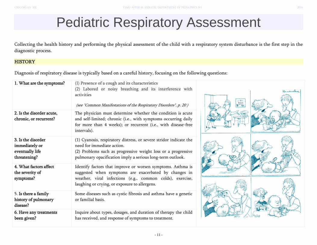
GRIGORYAN ME YSMU AFTER M. HERATSI, DEPARTMENT OF PEDIATRICS 1 2016
- 11 -
Pediatric Respiratory Assessment
Collecting the health history and performing the physical assessment of the child with a respiratory system disturbance is the first step in the
diagnostic process.
HISTORY
Diagnosis of respiratory disease is typically based on a careful history, focusing on the following questions:
1. What are the symptoms? (I) Presence of a cough and its characteristics
(2) Labored or noisy breathing and its interference with
activities
(see "Common Manifestations of the Respiratory Disorders", p. 20 )
2. Is the disorder acute,
chronic, or recurrent?
The physician must determine whether the condition is acute
and self-limited; chronic (i.e., with symptoms occurring daily
for more than 4 weeks); or recurrent (i.e., with disease-free
intervals).
3. Is the disorder
immediately or
eventually life
threatening?
(1) Cyanosis, respiratory distress, or severe stridor indicate the
need for immediate action.
(2) Problems such as progressive weight loss or a progressive
pulmonary opacification imply a serious long-term outlook.
4. What factors affect
the severity of
symptoms?
Identify factors that improve or worsen symptoms. Asthma is
suggested when symptoms are exacerbated by changes in
weather, viral infections (e.g., common colds), exercise,
laughing or crying, or exposure to allergens.
5. Is there a family
history of pulmonary
disease?
Some diseases such as cystic fibrosis and asthma have a genetic
or familial basis.
6. Have any treatments
been given?
Inquire about types, dosages, and duration of therapy the child
has received, and response of symptoms to treatment.

GRIGORYAN ME YSMU AFTER M. HERATSI, DEPARTMENT OF PEDIATRICS 1 2016
- 12 -
PHYSICAL EXAMINATION
1. Vital signs – respiratory rate, heart rate, blood pressure and
temperature.
Respiratory rate is the best indicator of pulmonary function in young
infants. As the respiratory rate is influenced by activity when the
child is awake, the most reliable and reproducible rate is the sleeping
respiratory rate. A child’s respiratory rate decreases as body size
increases.
Normal respiratory rate (RR) and heart rate (HR) at rest [From Soghier L. Reference Range Values for Pediatric Care, first ed. AAP 2014]
Respiratory Rate
(breaths/min)
Heart Rate
(beats/min)
Newborn 40–60 100-160
1 – 12 mo 35–40
1 – 3 y 25–30
70-110 4 – 6 y 21–23
7 – 12 y 19–21
13 – 19 y 16–18 55-90
2. Visual assessment of the signs of respiratory distress:
Increases in respiratory rate (tachypnea) and work of breathing:
dyspnea, chest retractions (suprasternal, supraclavicular,
intercostal, subcostal), grunting, nasal flaring, head bobbing,
thoracoabdominal asynchrony,
Signs of poor oxygenation include alterations in mental status, and
change in skin color: pallor, mottling, and cyanosis. They are
often late signs indicating respiratory failure and shock.

GRIGORYAN ME YSMU AFTER M. HERATSI, DEPARTMENT OF PEDIATRICS 1 2016
- 13 -
3. Anatomic changes of significance include the following:
(1) A change in tracheal position signals mediastinal shift and an inequality between the two sides of the chest, as with a pneumothorax or
atelectasis.
(2) A change in thoracic configuration. A barrel-chest deformity suggests hyperinflation and overdistention of the lungs due to chronic airway
obstruction.
(3) Clubbing of the fingers and toes is caused by lifting of the nail base by tissue proliferation on the dorsal surface of the terminal phalanx. As a
sign of pulmonary disease in children, clubbing most often is caused by cystic fibrosis.
CAUSES OF CLUBBING
Cardiac Respiratory Gastrointestinal Other
Cyanotic congenital heart disease Bacterial endocarditis
Bronchiectasis Cystic fibrosis/ciliary dyskinesia Tuberculosis Empyema/abscess, malignancy
Inflammatory bowel disease Chronic liver disease (CLD) Primary sclerosing cholangitis
Familial
4. Ears, nose and throat (ENT) examination
A successful ENT examination requires younger children to be held gently but firmly by the
parents.
Ears
Ask the parents to sit the child on their lap at 90° to their chest. With one arm, ask them to gently
hold the child's head against their chest. Their other arm should hold onto the child's free arm.
Gentle traction should be applied to the ear. The pinna should be pulled in an upward and outward
direction. The auriscope should be gently held in a pincer grip. The ring finger of this hand should
rest on the cheek of the child while the auriscope is within the ear. This allows the examiner's hand
to quickly follow any sudden movements the child may make.
Throat
This is an unpleasant examination. It should be left until last. A tongue depressor is necessary for a
complete examination. If throat disease is suspected, it is useful to have a swab to hand. A child may
comply with throat examination once. They may resist a further request a few minutes later for a
throat swab.
Fig. Positions for the ear and throat in
the ENT examination

GRIGORYAN ME YSMU AFTER M. HERATSI, DEPARTMENT OF PEDIATRICS 1 2016
- 14 -
5. Percussion
Percussion of the chest wall assesses acoustic response to a vibratory force applied to
lung tissue. The technique requires practice and consists of tapping of the middle
finger of one hand with the middle finger of the other while it is applied to the chest
wall. It is essential that one does this symmetrically and that the response is assessed
for resonance (hollowness) and dullness (flatness). This is often not useful in young
infants and frequently will elicit poor cooperation and crying. The technique is of
more use in older children.
6. Auscultation of the lungs
Auscultation of the lungs is always necessary for evaluating the child’s respiratory
status. Use the diaphragm of the stethoscope to auscultate the lung fields (front, back,
and axillary areas) by systematically and symmetrically (comparing one side with
another), listening for breath sounds as the child takes a deep breath in and blows out.
Pretending to blow out the light of an otoscope or penlight is a useful game that
encourages deep breathing in younger children.
In infants and young children, lack of subcutaneous fat and smaller distances between
structures may make breath sounds readily transmitted across lung fields. Keep this in
mind when assessing the presence, location, and nature of breath sounds in infants
and young children. To detect transmitted sounds from the upper airways, place the
diaphragm next to the child’s mouth or nose to determine whether what is heard in
the lung fields is just like the sound as heard near the mouth or nose. Referred sounds
are loudest near their origin.
Respiratory sounds are related to chest air movement, either normal or adventitious,
heard at the mouth, the trachea and the chest; they include sounds produced by
cough, snoring, sneezing or respiratory muscle contraction, but exclude voiced
sounds. Lung sounds are the respiratory sounds heard (or otherwise detected) over the
chest.
Fig. Surface anatomy of the pulmonary lobes and
suggested auscultation sites

GRIGORYAN ME YSMU AFTER M. HERATSI, DEPARTMENT OF PEDIATRICS 1 2016
- 15 -
Characteristics of Normal Breath Sounds
Sound Vesicular * Bronchovesicular Bronchotubular/ bronchial/ tracheal
Quality Soft, swishing Louder and higher pitch than vesicular;
mixed
Tubular, harsh, hollow
Relationship of Inspiration
(I) to Expiration (E)
I is longer, louder, and higher pitched
than E (I > E)
I and E are equal (I = E) I is short; E is long (I < E)
Diagram
Normal Location Throughout lung field Over manubrium and upper
intrascapular region where trachea and
bronchi bifurcate (in older children)
Over trachea
Pediatric
Considerations
Usually heard in older children but not in
infants and young children
Heard throughout lung field in infants
and young children
* Vesicular breath sound is a misnomer, as it does not originate in vesicles, i.e. the alveoli
I – inspiration; E - expiration
Breath sounds normally have equal intensity, pitch, and rhythm bilaterally. Absent or diminished breath sounds generally indicate a partial or
total obstruction, such as from a foreign body or mucus, that does not permit airflow.
Adventitious sounds are additional sounds superimposed on normal breath sounds; they are usually associated with pulmonary disorders.
Adventitious sounds are primarily divided into continuous and discontinuous sounds.

GRIGORYAN ME YSMU AFTER M. HERATSI, DEPARTMENT OF PEDIATRICS 1 2016
- 16 -
Characteristics of Adventitious Breath Sounds
Continuous sounds (whezes, ronchi, stridor) Discontinuous sounds (crackles)
Sinusoidal, musical, prolonged (but not necessarily persisting
throughout the respiratory cycle)
Like dashes in time — — — — — —
Intermittent, nonmusical, and brief. Crackles (other terms in use are
‘‘crepitations’’ or ‘‘rales’’) are usually auscultated during inspiration, that
represent local phenomena.
Like dots in time ………
1. Wheezes: relatively high-pitchet (≥400 Hz) with hissing or shrill
quality (>80 ms).
They usually are expiratory in origin but can be inspiratory when the airway obstruction is fixed and rigid, as in airway edema.
2. Ronchi: relatively low-pitched (150-200 Hz) with snoring
quality.
Rhonchi are generated by intraluminal secretions and collapse of large airways.
2. Stridor: see p. 23
1. Fine crackles (subcrepitant crackles): soft, high-pitched (≈ 650 Hz), very brief
(5-10 ms).
They are produced by the explosive opening of small airways collapsed by surface forces (increased elastic lung recoil pressure or inflammation/oedema in the lung); they are gravity dependent and the sound is rarely transmitted to the mouth. Fine, late inspiratory crackles are typical of interstitial/fibrotic lung disease. However, they may also be present in normal subjects who inhale slowly from their residual lung volume.
2. Coarse crackles (crepitant crackles): somewhat louder, lower in pitch (≈ 350
Hz), brief (15-30 ms) sounds.
They are generated by a different mechanism to that of fine crackles, i.e. movement of thin secretions in the bronchi or the bronchioles. They start early and continue until mid-inspiration but may be heard during expiration. A typical example of coarse crackles can be heard in bronchiectasis and chronic airway obstruction (e.g. CF). Similar auscultatory findings can be found focally early in pneumonia but shift into more endinspiratory crackles of variable duration that progress to fine crackles during recovery. Acoustic analysis has characterised the crackles of cardiac failure as coarse, of long duration during inspiration and appearing late in the course of the disease.
Other adventitious sounds are squawk and the pleural friction sound.
A squawk (sometimes classified as a type of wheeze) is a ‘‘composite’’, short (50–400 ms), inspiratory adventitious sound with a musical character (short
inspiratory wheeze) that is preceded by a crackle. It is not associated with airway obstruction but rather with pulmonary fibrosing (restrictive) disease. It
is thought to result from the vibrations set in motion by the sudden opening of a collapsed airway.
Pleural friction sound (or friction rub) is coarse crackles (often described as ‘‘leathery’’) produced by inflamed parietal and visceral pleura that cause
vibration of the chest wall and local pulmonary parenchyma. It can be auscultated during inspiration or in both phases of breathing. Pleural friction
precedes pleural effusion and disappears when fluid is formed. The ‘‘rub’’ is synchronous with breathing and does not disappear with cough, but is
modified by the breathing pattern and posture.

GRIGORYAN ME YSMU AFTER M. HERATSI, DEPARTMENT OF PEDIATRICS 1 2016
- 17 -
IMAGING PROCEDURES
(1) Chest X-ray. This simple investigation is easily obtained in hospital but may be overused. Consider what you hope to learn before
requesting a radiograph. It is not necessary, for instance, to X-ray every wheezy child, unless the episode is moderate or severe, or there are
focal signs. Do not send an unstable child, particularly with upper airway obstruction, across the hospital to the X-ray department – stabilize
the child first or request a portable X-ray. If there is any question of an inhaled foreign body, a single film is never sufficient. Except in those
who require immediate bronchoscopy, X-ray screening in the young child, or inspiratory and expiratory films in the cooperative older child,
are needed to confidently exclude or confirm a foreign body.
(2) Fluoroscopy is useful for dynamic studies (e.g., to evaluate diaphragmatic movements, identify tracheal collapse).
(3) Ultrasonography is used to confirm pleural effusion and to guide thoracentesis; it can be used instead of fluoroscopy to evaluate
diaphragmatic motility.
(4) Chest computed tomography: useful for assessing abnormalities in airways as well as abnormalities in parenchymal tissue density.
(5) Thoracic MRI: useful for looking at airway–blood interface, and vascular and mediastinal anatomy.
(6) Nuclear imaging: useful for assessing regional ventilation (V) and perfusion (Q), as well as V/Q matching.
ENDOSCOPIC EVALUATION OF THE AIRWAYS
Endoscopic evaluation of the upper airways (nasopharyngoscopy) is performed with a flexible fiberoptic nasopharyngoscope to assess adenoid
size, patency of the nasal passages, and abnormalities of the glottis. It is especially useful in evaluating stridor and assessing vocal cord
motion/function, and it does not require sedation.
Endoscopic evaluation of the subglottic space and intrathoracic airways can be done with either a flexible or rigid bronchoscope under
anesthesia. Bronchoscopy is useful in identifying airway abnormalities (stenosis, malacia, endobronchial lesions, excessive secretions) and in
obtaining airway samples for culture (bronchoalveolar lavage), especially in immunocompromised patients. Rigid bronchoscopy is the method
of choice for removing foreign bodies from the airways and performing other interventions, and flexible bronchoscopy is most useful as a
diagnostic tool and for obtaining lower airway cultures. Transbronchial biopsies are rarely performed in children. There are few absolute
contraindications to bronchoscopy. Relative contraindications include bleeding diatheses, thrombocytopenia (<50,000/cm3), and clinical
conditions when the patient is too unstable to tolerate the procedure.
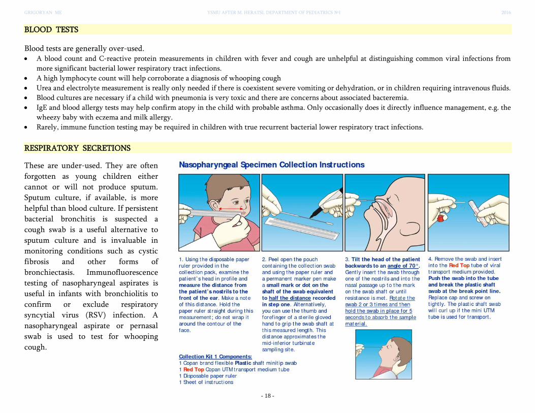
GRIGORYAN ME YSMU AFTER M. HERATSI, DEPARTMENT OF PEDIATRICS 1 2016
- 18 -
BLOOD TESTS
Blood tests are generally over-used. A blood count and C-reactive protein measurements in children with fever and cough are unhelpful at distinguishing common viral infections from
more significant bacterial lower respiratory tract infections.
A high lymphocyte count will help corroborate a diagnosis of whooping cough
Urea and electrolyte measurement is really only needed if there is coexistent severe vomiting or dehydration, or in children requiring intravenous fluids.
Blood cultures are necessary if a child with pneumonia is very toxic and there are concerns about associated bacteremia.
IgE and blood allergy tests may help confirm atopy in the child with probable asthma. Only occasionally does it directly influence management, e.g. the
wheezy baby with eczema and milk allergy.
Rarely, immune function testing may be required in children with true recurrent bacterial lower respiratory tract infections.
RESPIRATORY SECRETIONS
These are under-used. They are often
forgotten as young children either
cannot or will not produce sputum.
Sputum culture, if available, is more
helpful than blood culture. If persistent
bacterial bronchitis is suspected a
cough swab is a useful alternative to
sputum culture and is invaluable in
monitoring conditions such as cystic
fibrosis and other forms of
bronchiectasis. Immunofluorescence
testing of nasopharyngeal aspirates is
useful in infants with bronchiolitis to
confirm or exclude respiratory
syncytial virus (RSV) infection. A
nasopharyngeal aspirate or pernasal
swab is used to test for whooping
cough.

GRIGORYAN ME YSMU AFTER M. HERATSI, DEPARTMENT OF PEDIATRICS 1 2016
- 19 -
PULMONARY FUNCTION TESTING
TEST MEASUREMENTS KEY RESULTS
Spirometry Assessment of dynamic lung volumes and ventilation:
The patient inhales to total lung capacity and then forcibly exhales until no more air can be expelled. During the forced expiratory maneuver, forced vital capacity (FVC), forced expired volume in the first second (FEV1), and peak expiratory flow rate (PEFR) are measured. These are compared to predicted values based on patient age, gender, and race, but rely mostly on height.
Most children above 6 years of age can perform spirometry. Infant pulmonary function testing is possible, using sedation and sophisticated equipment.
Forced vital capacity (FVC) - the volume of air expired
in a complete expiration
Forced expiratory volume in 1 second (FEV1) - the
volume of air that can be expired in 1 second
Ratio of FEV1 to FVC (FEV1/FVC) - this test can be used
to distinguish between obstructive airway disease (eg asthma, COPD) and restrictive lung disease (eg pulmonary fibrosis). In an obstructive defect, the FEV1/FVC will be less than 70%. The percentage is greater than 80% with a restrictive defect. But, spirometric restrictive defect ≠ restrictive lung disease.
Body
plethysmography
Assessment of static lung volumes: TLC (total lung capacity), FRC (functional residual capacity), RV (residual volume), and SVC (slow vital capacity)
Static lung volumes are reduced in restrictive lung
disease. They cannot be measured with spirometry.
Abnormal results on pulmonary function testing can be used to categorize obstructive airway disease (low flow rates and increased RV or FRC) or a restrictive defect (low FVC and TLC, with relative preservation of flow rates and FRC). Pulmonary function testing can detect reversible airway obstruction characteristic of asthma with a significant improvement in FEV1 (>12%-15%) or in FEF25–75% (>25%) following inhalation of a bronchodilator. Spirometry is also useful for longitudinal patient management. The peak expiratory flow rate (PEFR) can also be obtained with a simple handheld device (peak flow meter) and may be useful for home monitoring of older children with asthma. However, it is highly dependent on patient effort, and values must be interpreted with caution. Inhalation challenge tests using methacholine, histamine, or cold, dry air are used to assess airway hyperreactivity, but require sophisticated equipment and special expertise and should be performed only in a pulmonary function laboratory with experienced technicians.
Arterial blood
gases (ABGs) and
gas exchange tests
ABGs provide 3 assessments of the function of the respiratory
system:
evaluation of oxygenation (PaO2, SaO2, SpO2) evaluation of the adequacy of ventilation (PaCO2) evaluation of the lung's role in acid-base balance of the arterial
blood (pH, PaCO2)
Normal Arterial Blood Gas Values
pH: 7.4 (7.38 to 7.42)
PO2: 80 to 100 mm Hg
PCO2: 35 to 45 mm Hg
O2 Saturation: 95% on room air
HCO3: 22 to 26 mEq/L
Base Excess: - 2 to + 2 mEq/L
Diffusing capacity
of the lung (DLCO)
Assessment of the ability of the lungs to transfer gas. DLCO is reduced in restrictive diseases with diminished
pulmonary blood flow.

GRIGORYAN ME YSMU AFTER M. HERATSI, DEPARTMENT OF PEDIATRICS 1 2016
- 20 -
Common Manifestations of the Respiratory Disorders
The lungs have a limited repertoire of responses to disease. The key presentations in pediatrics are:
• Sore throat
• Cough
• Noisy breathing (e.g., stridor, wheeze)
• Respiratory distress
COUGH
Cough is a very non-specific symptom and can occur as a result of any pathology at any site in the respiratory tract – cough is one of the
clinical features of most respiratory pathologies. However, the timing of the cough and its associated features (such as the age of the patient and
other symptoms) can give clues to the diagnosis.
It is often informative to hear a cough (or see video recording of a cough), allowing one to distinguish between dry and wet coughs and wheezy
or brassy coughs.
Cough type Suggested underlying process
Barking or brassy cough Croup, tracheomalacia, habit cough
Honking Psychogenic
Paroxysmal (with or without inspiratory ‘whoop’) Pertussis and parapertussis
Staccato Chlamydia in infants
Cough productive of casts Plastic bronchitis/asthma
Chronic wet cough in mornings only Suppurative lung disease
Based on Chang AB, Landau LI, Van Asperen PP et╯al, Med J Aust 2006; 184(8): 398–403; with permission.
The duration of the cough also suggests its possible cause. Most acute coughs are infectious in origin. Upper respiratory infections can initiate
an acute cough through stimulation of the cough receptors in the nose and posterior pharynx. If nasal congestion and cough persist, a diagnosis
of allergic rhinitis or sinusitis should be considered. Serous otitis media can also cause a persistent cough and may occur in children with
chronic congestion.

GRIGORYAN ME YSMU AFTER M. HERATSI, DEPARTMENT OF PEDIATRICS 1 2016
- 21 -
Acute cough
In upper respiratory tract infection with bronchitis, cough usually lasts more than 2–3 weeks in 10% of normal children. Provided the child is otherwise
well with no pyrexia, tachypnoea or crackles, it is likely best to wait for resolution as aetiology is most often viral. There is limited evidence that any
therapy is beneficial.
Erythromycin is useful for early pertussis cases.
Honey medications and vapour rub may reduce the severity of acute cough.
Antibiotics may be beneficial for acute bacterial bronchitis but most bacterial cases resolve naturally anyway.
An inhaled foreign body is a possibility when there is a sudden onset of cough with no upper respiratory tract infection or after a choking episode;
bronchoscopy is needed to remove the foreign body. In allergic rhinitis and post-nasal drip syndrome (throatclearing type cough), intranasal steroids and/or
antihistamines may be beneficial. 10% of normal children with acute cough due to upper respiratory tract infections are still coughing 3–4 weeks later.
Some children with a ‘‘post-infectious cough’’ (prolonged acute coughing after an obvious upper respiratory infection) cough for much longer and this is
especially true for those with pertussis (Hay et al., 2005). Providing the child is otherwise well, waiting for a period of time allows natural resolution of post-
infectious coughing and pertussis to occur.
Do not use a wait-and-see approach if there is:
weight loss
night sweats
haemoptysis
sudden-onset cough or cough after a choking episode
relentlessly progressive coughing (e.g. TB, expanding intrathoracic mass, retained foreign body, collapsed lobe or pertussis)
clinical history of symptoms or signs (or are at risk) of underlying chronic lung disease (e.g. finger clubbing, barrel-shaped chest, Harrison’s sulci,
recurrent pneumonia and immunodeficiency)
Chronic cough
A chronic cough is one that lasts ≥ 14 days (WHO, 2013).
Chronic cough is very common and often there are no pointers to a specific diagnosis (e.g. normal chest radiograph, normal lung function and dry isolated
cough in otherwise well child). In such cases often a ‘‘trial of treatment’’ is used to confirm a diagnosis as it is neither feasible nor desirable to extensively
investigate all such children. However, it is important to realise that natural resolution typically occurs with the passage of time and, therefore, a response to
treatment must not be taken as confirming a diagnosis. Children responding to a trial of therapy should have the treatment stopped and only a second
clearcut response should be used to suggest a diagnosis.
There is little evidence that either nonspecific isolated cough or postinfectious coughing responds to any currently available treatment (inhaled
corticosteroids (ICS), b2-agonists, leukotriene antagonists, anti-gastrooesophageal reflux therapy, cromones and environmental modification). Most of these
coughs resolve naturally but over a considerable period of time. Ultra-high-dose ICS may have a small benefit but the sideeffects seem to outweigh the
benefits.
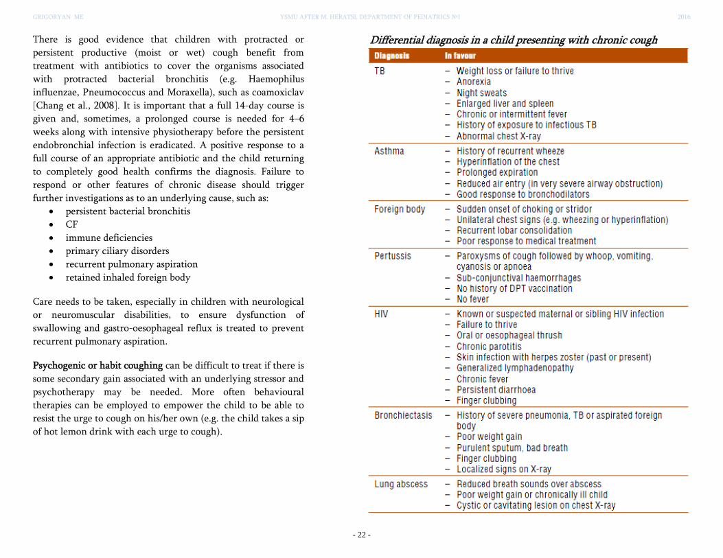
GRIGORYAN ME YSMU AFTER M. HERATSI, DEPARTMENT OF PEDIATRICS 1 2016
- 22 -
There is good evidence that children with protracted or
persistent productive (moist or wet) cough benefit from
treatment with antibiotics to cover the organisms associated
with protracted bacterial bronchitis (e.g. Haemophilus
influenzae, Pneumococcus and Moraxella), such as coamoxiclav
[Chang et al., 2008]. It is important that a full 14-day course is
given and, sometimes, a prolonged course is needed for 4–6
weeks along with intensive physiotherapy before the persistent
endobronchial infection is eradicated. A positive response to a
full course of an appropriate antibiotic and the child returning
to completely good health confirms the diagnosis. Failure to
respond or other features of chronic disease should trigger
further investigations as to an underlying cause, such as:
persistent bacterial bronchitis
CF
immune deficiencies
primary ciliary disorders
recurrent pulmonary aspiration
retained inhaled foreign body
Care needs to be taken, especially in children with neurological
or neuromuscular disabilities, to ensure dysfunction of
swallowing and gastro-oesophageal reflux is treated to prevent
recurrent pulmonary aspiration.
Psychogenic or habit coughing can be difficult to treat if there is
some secondary gain associated with an underlying stressor and
psychotherapy may be needed. More often behavioural
therapies can be employed to empower the child to be able to
resist the urge to cough on his/her own (e.g. the child takes a sip
of hot lemon drink with each urge to cough).
Differential diagnosis in a child presenting with chronic cough

GRIGORYAN ME YSMU AFTER M. HERATSI, DEPARTMENT OF PEDIATRICS 1 2016
- 23 -
NOISY BREATHING
The term ‘‘noisy breathing’’ is used to describe respiratory sounds that are audible to the ‘‘naked ear’’ without the use of a stethoscope.
Table. Different kinds of noisy breathing
Noise Description Site of origin Common causes
Stertor It is a snuffly or snoring sound Oro/nasopharyngeal airway
Snuffles Blocked nasal passages Upper respiratory tract infections
Allergic rhinitis
Snoring Snoring is produced during sleep and is due to obstructed air
movement in the naso- and oropharynx; children who snore
tend to have more collapsible airways and/ or increased size
of adenotonsillar tissue
Oro/nasopharyngeal airway
Collapsible airways with increased size
of adenotonsillar tissue
Obesity
Craniofacial disorders
Grunting Glottis Respiratory distress syndrome (neonates)
Pneumonia
Bacterial infection
Rattle Rattle is created by the movement of excessive secretions
during normal airflow in the central and extrathoracic
airways; it has a ‘‘rattling’’, noncontinuous quality, but quite
commonly is mislabelled by parents as wheezing
Intra- and extrathoracic
airways
Acute viral bronchitis
Protracted bacterial bronchitis
Neurologic disorders with swallowing
dysfunction and/or chronic aspiration
Stridor Stridor is a musical, monophonic, high-pitched sound that
can be heard without a stethoscope; its presence suggests
significant obstruction of airflow in the larynx and trachea
Larynx and trachea Croup
Inhaled foreign body – toys, food
Epiglottitis
Bacterial tracheitis
Laryngomalacia/Tracheomalacia
Vocal cord dysfunction
Vascular ring
Wheezing
(wheeze)
Wheeze is a continuous, usually highpitched whistling
sound that is accompanied by prolongation of the expiratory
phase; it is believed to originate from oscillation of large
airways in response to turbulent airflow in partially blocked
intrathoracic airways.
Intrathoracic airways
(primarily expiratory)
Asthma
Viral wheeze
Bronchiolitis
Foreign body
Protracted bacterial bronchitis
Tracheo/bronchomalacia

GRIGORYAN ME YSMU AFTER M. HERATSI, DEPARTMENT OF PEDIATRICS 1 2016
- 24 -
FIG. Listening Can Help Locate the Site of Airway Obstruction. [Eavey RD, 1986]
A loud, gasping snore suggests enlarged tonsils or adenoids.
Stridor during inspiration (inspiratory stridor) suggests the airway is compromised at the level of the
supralaryngeal structures (epiglottis and arytenoid cartilages), vocal cords, subglottic region, or upper
trachea. With forced inspiration, intrathoracic pressure becomes quite negative and is less than
atmospheric pressure, promoting collapse at or just above the site of obstruction.
Expiratory stridor or central wheeze results from narrowing or collapse of the lower trachea or
bronchi. During forced exhalation, rising pleural pressure may exceed intratracheal pressure.
Airway noise during both inspiration and expiration (biphasic stridor) often represents a fixed
obstruction of the vocal cords or subglottic space.
Hoarseness or a weak cry is a byproduct of obstruction at the vocal cords. If a cough is croupy or low
pitched, suspect tracheal pathology.
FIG. (a) Normal Inspiration. At end-expiration, intrapleural pressure is less
than atmospheric pressure, so it should maintain airway patency. In infants the highly compliant chest wall does not provide the support required. Thus airway closure occurs with each breath. Descent of the diaphragm and contraction of the intercostals muscles develop a greater negative intrathoracic pressure relative to intraluminal and atmospheric pressure. The net result is a longitudinal stretching of the larynx and trachea, dilation of the intrathoracic trachea and bronchi, movement of air into the lungs, and some dynamic collapse of the extrathoracic trachea due to the increased compliance of the trachea ad the negative intraluminal pressure in relation to atmospheric pressure. (b) Normal expiration. Intraluminal pressures are slightly positive in relation to atmospheric pressure, so air is forced out of the lungs. (c) Extrathoracic obstruction (obstructed inspiration). Respiratory dynamics occurring with upper airway obstruction; note the severe dynamic collapse of the extrathoracic trachea below the level of obstruction. This collapse is greatest at the thoracic inlet, where the largest pressure gradient exists between negative intratracheal pressure and atmospheric pressure. (d) Intrathoracic obstruction (obstructed expiration). Respiratory dynamics occurring with lower airway obstruction. Breathing through a partially obstructed lower airway (such as occurs in bronchiolitis or asthma) results in greater positive intrathoracic pressures, with dynamic collapse of the intrathoracic airways (prolonged expiration or wheezing) [Zalzal GH, 2003]

GRIGORYAN ME YSMU AFTER M. HERATSI, DEPARTMENT OF PEDIATRICS 1 2016
- 25 -
Differential diagnosis in a child presenting with stridor
Diagnosis In favour
Viral croup – Barking cough
– Respiratory distress
– Hoarse voice
– If due to measles, signs of measles
Retropharyngeal
abscess
– Soft tissue swelling in back of the throat
– Diffi culty in swallowing
– Fever
Foreign body – Sudden history of choking
– Respiratory distress
Laryngomalacia/
Tracheomalacia
– Underdeveloped cartilage in the airway wall, which collapses during inspiration, thereby causing turbulent airflow
– Stridor present since birth
Diphtheria – Bull neck appearance due to enlarged cervical nodes and oedema
– Red throat
– Grey pharyngeal membrane
– Blood-stained nasal discharge
– No evidence of DPT vaccination
Epiglottitis – Soft stridor
– ‘Septic’ child
– Little or no cough
– Drooling of saliva
– Inability to drink
Anaphylaxis – History of allergen exposure
– Wheeze
– Shock
– Urticaria and oedema of lips and face
Burns – Swollen lips
– Smoke inhalation

GRIGORYAN ME YSMU AFTER M. HERATSI, DEPARTMENT OF PEDIATRICS 1 2016
- 26 -
Respiratory Distress & Respiratory Failure
CASE STUDY A 6-month-old boy has been coughing and breathing fast for the past day. This morning he refused feeding and has been irritable. On examination, the infant is fussy. He has an oxygen saturation of 92%, a respiratory rate of 60 breaths/min, a pulse of 140 beats/min, and a normal blood pressure and temperature. In addition, he has nasal flaring, intercostal and supraclavicular retractions, and occasional grunting.
Questions
1. What are the causes of respiratory distress in infants and children? 2. What are the signs and symptoms of respiratory distress in infants and
children? 3. What are the signs and symptoms of impending respiratory failure in infants and children? 4. What are the critical interventions for
infants and children in respiratory distress?
The signs and symptoms of respiratory compromise may be subtle, particularly in small infants. Decompensation may occur rapidly if
ventilation or oxygenation is inadequate but may be prevented by prompt recognition and treatment.
Respiratory distress is defined as increased work of breathing, and it usually precedes respiratory failure.
Respiratory failure occurs when ventilation or oxygenation is not sufficient to meet the metabolic demands of the tissues. Thus, oxygenation of
the blood is inadequate or carbon dioxide is not eliminated. Respiratory failure may lead to cardiopulmonary arrest if not corrected promptly.
RESPIRATORY DISTRESS RESPIRATORY FAILURE
• Increased respiratory rate (tachypnea)
• Presence of chest retractions
• Nasal flaring
• Grunting
• Diaphoresis
• Alterations in mental status
• Poor feeding
• Inability to speak in sentences
• Presence of pale or cyanotic skin
• Presence of central cyanosis
Respiratory failure describes any impairment in
oxygenation or ventilation in which the arterial
oxygen tension falls below 60 mm Hg (hypoxemia),
the carbon dioxide tension rises above 50 mm Hg
(hypercarbia, hypercapnia) and the pH drops below
7.35, or both.
• PaO2 < 60 mm Hg
• PaCO2 > 50 mm Hg
• pH < 7.35

GRIGORYAN ME YSMU AFTER M. HERATSI, DEPARTMENT OF PEDIATRICS 1 2016
- 27 -
Common Causes of Respiratory Distress in Infants and Children
Respiratory diseases NonRespiratory diseases
• Epiglottitis
• Croup
• Bronchiolitis
• Pneumonia
• Reactive airway disease (asthma)
• Foreign body aspiration
• Cardiac diseases
• Metabolic disease with acidosis
• Neuromuscular disease
Epidemiology Respiratory distress continues to be the most common reason for hospital admission. Such admissions usually involve
young infants with acute infections such as bronchiolitis or croup. Reactive airway disease (asthma) accounts for
respiratory distress–related admission commonly in older children.
Clinical
Presentation
(see p. 12)
Increases in respiratory rate and work of breathing are the most common signs of respiratory disease. Effortless
tachypnea may be a sign of respiratory compensation for metabolic acidosis rather than an indication of pulmonary
pathology. Similarly, hypoxia that fails to improve with supplemental oxygen may suggest a primary cardiac lesion.
Tachycardia is often present, although bradycardia, if present, may be an ominous sign of impending cardiopulmonary
failure. Signs of poor oxygenation include alterations in mental status, head bobbing, and change in skin color. Pallor,
mottling, and cyanosis are often late signs indicating respiratory failure and shock. Children who are severely hypoxemic
may first appear dusky or pale. If children are anemic, cyanosis may not be evident, although oxygen saturation is low.
Pathophysiology Decrease in tidal volume → Compensatory increase in respiratory rate → Maintaining adequate minute ventilation
If the minute ventilation is still insufficient for adequate gas exchange or the child can no longer sustain the increased work of breathing, respiratory failure ensues. Respiratory failure may then lead to acidosis, myocardial dysfunction, and shock and may
progress to complete cardiopulmonary arrest.
Younger children oppose airway collapse by having higher respiratory rates, thus reducing the time allowed for
expiration. If this is insufficient to maintain FRC then infants will attempt to reduce expiratory flow rates using partial
closure of the glottis or upper airway. This leads to grunting, as seen in neonates with respiratory distress, where glottic
closure maintains a positive expiratory pressure but reduces expiratory flow.

GRIGORYAN ME YSMU AFTER M. HERATSI, DEPARTMENT OF PEDIATRICS 1 2016
- 28 -
Different types of respiratory failure
Some clinicians divide respiratory failure into two categories.
• The hypoxemic type is generally caused by mismatch of ventilation and perfusion in the lung. Hypoxemic respiratory failure from
mismatch of ventilation to perfusion is often associated with normal or low PCO2.
• Other patients with respiratory failure have an overall decrease in alveolar ventilation that is usually the result of upper airway obstruction,
neuromuscular disease, thoracic trauma, or muscle fatigue. These patients have increases in PCO2 and relatively proportional decreases in
PO2 (hypercapnic type).
The physiology in most children with respiratory failure is a combination of these two types, because one type often leads to the other. For
instance, an infant with bronchiolitis initially may have hypoxemia from atelectasis and ventilation-perfusion mismatch, but may progress to
inadequate alveolar ventilation when airway resistance is high and respiratory muscle fatigue supervenes.
Can respiratory failure be present without respiratory distress?
Absolutely. Children may hypoventilate because of reduced level of consciousness (ingestion, metabolic derangements, and head trauma) or
neuromuscular dysfunction. After prolonged respiratory distress, children may become fatigued, and their work of breathing may appear
normal in the presence of significant hypoventilation. Elevation of the PCO2 from hypoventilation may signal worsening fatigue and
impending respiratory arrest.
MANAGEMENT OF RESPIRATORY DISTRESS
All infants or children in respiratory distress should be managed emergently. As stated earlier, in such situations, assessment and intervention
often occur simultaneously. All children in respiratory distress should be reassessed frequently. The highest possible oxygen concentration
should be delivered. Children who are able to maintain their own airway should never be forced to use an airway adjunct because this may
cause increased anxiety and distress. Patients with clear airways can be maintained with simple interventions such as oxygen blown by the face
or given by mask or nasal prongs. More advanced airway management such as bagvalve- mask ventilation or endotracheal intubation may be
necessary for children who need assisted ventilation, airway protection, or hyperventilation.
Position
Children in respiratory distress who are alert and breathing spontaneously should be allowed to choose a position of comfort. Small infants
who are incapable of positioning themselves are best placed upright with care taken not to flex or extend the neck. Children and their
caregivers should be kept together to reduce anxiety.

GRIGORYAN ME YSMU AFTER M. HERATSI, DEPARTMENT OF PEDIATRICS 1 2016
- 29 -
The proper position for unconscious children is the “sniffing position,” with the neck slightly flexed and the head extended to open the airway.
This can be facilitated by placing a towel under the occiput of the head or shoulders. If simple positioning does not relieve an obstruction, the
airway should be opened using the chin lift or jaw thrust. If spinal trauma is a possibility, only the jaw thrust should be used. If this fails,
airway adjuncts, such as nasopharyngeal or oropharyngeal airways, can be placed to help prevent the soft tissue of the oropharynx from
collapsing against the posterior pharyngeal wall.
Monitoring
All infants and children in respiratory distress should be carefully monitored. Pulse oximetry will assist the clinician in determining the degree
of oxygen saturation, and, if available, a cardiac and respiratory monitor will provide constant readings of respirations and heart rate. Frequent
assessments of the patient are critical to ensure a good outcome.
Oxygen Administration
Oxygen should be delivered by any method tolerated by children.
Nasal prongs have 2 advantages: They are noninvasive and allow maintenance of a constant gas flow
even when talking and eating. The concentration of oxygen delivered is limited, however, and
irritation and drying of the mucous membranes may result. Oxygen masks deliver a higher
concentration of humidified oxygen. Disadvantages include obstruction of children’s visual field,
potential for carbon dioxide retention, and anxiety because the face is covered. Various types of masks
are available.
The simple mask can deliver 30% to 60% oxygen concentration at flow rates of 6 to 10 L/min. Room air is drawn into the mask through the
exhalation ports in the side of the mask.
A non-rebreathing mask has valves that allow only oxygen (85%–95%) to
flow from the reservoir bag to the patient on inhalation and additional valves
on the exhalation ports of the mask that prevent entrapment of room air.
Fig. A non-rebreathing mask
can deliver a high
concentration of oxygen to a
patient in respiratory distres.

GRIGORYAN ME YSMU AFTER M. HERATSI, DEPARTMENT OF PEDIATRICS 1 2016
- 30 -
The face tent is a soft plastic bucket shaped to the chin that is well tolerated by
children. The face tent allows up to 40% oxygen to be delivered, and it has the
advantage of allowing access to the face and mouth.
Fig. Use of a face tent
A Venturi mask is rarely used in children but has the advantage of precisely titrating
the oxygen concentration to be delivered from 24% to 60%.
Fig. Venturi mask
Children with potential respiratory failure require assisted
ventilation with bag-valve-mask devices or endotracheal
intubation. Masks of the proper size should be used. The upper edge
of the mask should fit snugly over the bridge of the nose without
touching the eyes. The lower edge should rest directly on or just
above the mandible. An oropharyngeal airway should be inserted in
unconscious children to prevent the tongue from obstructing the
upper airway. An appropriately sized oropharyngeal airway should
reach from the patient’s earlobe to the corner of the mouth. A
nasopharyngeal airway may be inserted if the patient has an intact
gag reflex to achieve the same goal. Measure the nasopharyngeal
airway from the child’s earlobe to the tip of the nostril. If a bag-
valve-mask is not available, assisted ventilation can be given with a
pocket mask with a one-way valve. Oxygen can be attached to the
mask at the side port. Endotracheal intubation is indicated in those
children who require control of the airway, need airway protection,
or require hyperventilation.
Fig. A, Oropharyngeal airways.
B, Nasopharyngeal airways.
Fig. A pocket mask with a one-way
valve and side oxygen port that can
be used for assisted ventilation in the
office setting.

GRIGORYAN ME YSMU AFTER M. HERATSI, DEPARTMENT OF PEDIATRICS 1 2016
- 31 -
CASE RESOLUTION
The fussy infant discussed in the case history has obvious signs of respiratory distress, including tachypnea, tachycardia, grunting, nasal
flaring, and retractions. The differential diagnosis includes foreign body, infection such as croup or bronchiolitis, and reactive airway
disease. It is important to place the infant in a position of comfort and provide supplemental oxygen before any diagnostic studies, such as
chest radiograph, are performed. Appropriate treatment, such as albuterol, should be initiated while further evaluation proceeds. Pulse
oximetry should be monitored. The clinical status of the infant should be reassessed periodically to prevent further deterioration.
REFERENCES
1. Pediatric Clinical Practice Guidelines & Policies. A Compendium of Evidence-based Research for Pediatric Practice 15th ed. AAP 2015
2. Recommendations for management of common childhood conditions. World Health Organization 2012
3. POCKET BOOK OF Hospital care for children. GUIDELINES FOR THE MANAGEMENT OF COMMON CHILDHOOD ILLNESSES 2nd ed. World Health
Organization 2013
4. Hull J et al. Paediatric Respiratory Medicine, 2nd ed. Oxford University Press 2015
5. ERS Handbook: Paediatric Respiratory Medicine, 1st ed. European Respiratory Society 2013
6. American Heart Association. Pediatric Advanced Life Support (PALS). http://www.heart.org/HEARTORG/CPRAndECC/HealthcareProviders/Pediatrics/
7. Pediatric-Advanced-Life-Support-PALS_UCM_303705_Article.jsp. Accessed March 10, 2014
8. American Heart Association. Pediatric Emergency Assessment, Recognition and Stabilization (PEARS).
http://www.heart.org/HEARTORG/CPRAndECC/HealthcareProviders/
9. Pediatrics/Pediatric-Emergency-Assessment-Recognition-and-StabilizationPEARS_UCM_308135_Article.jsp. Accessed March 10, 2014
10. Berg MD, Schexnayder SM, Chameides L, et al. Part 13: pediatric basic life support: 2010 American Heart Association guidelines for cardiopulmonary resuscitation and
emergency cardiovascular care. Circulation. 2010;122(18 Suppl 3):S862–S875
11. Choi J, Lee GL. Common pediatric respiratory emergencies. Emerg Med Clin North Am. 2012;30(2):529—563
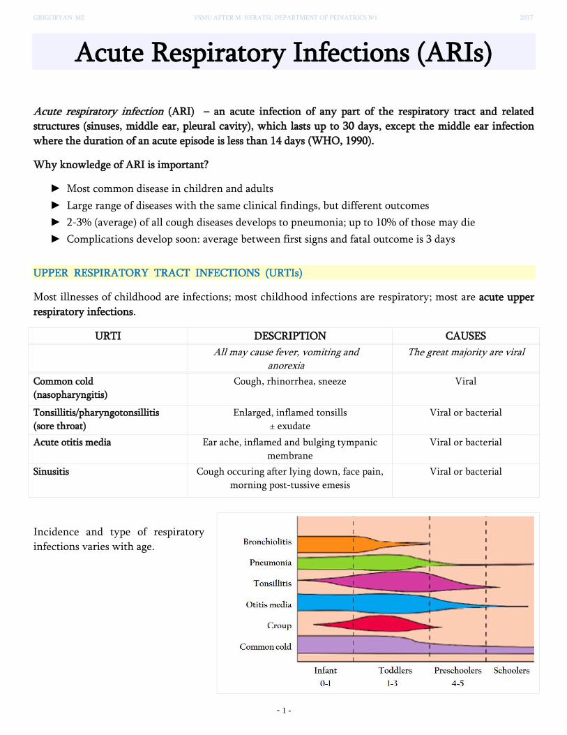
GRIGORYAN ME YSMU AFTER M. HERATSI, DEPARTMENT OF PEDIATRICS 1 2017
- 1 -
Acute Respiratory Infections (ARIs)
Acute respiratory infection (ARI) – an acute infection of any part of the respiratory tract and related
structures (sinuses, middle ear, pleural cavity), which lasts up to 30 days, except the middle ear infection
where the duration of an acute episode is less than 14 days (WHO, 1990).
Why knowledge of ARI is important?
Most common disease in children and adults
Large range of diseases with the same clinical findings, but different outcomes
2-3% (average) of all cough diseases develops to pneumonia; up to 10% of those may die
Complications develop soon: average between first signs and fatal outcome is 3 days
UPPER RESPIRATORY TRACT INFECTIONS (URTIs)
Most illnesses of childhood are infections; most childhood infections are respiratory; most are acute upper
respiratory infections.
URTI DESCRIPTION CAUSES
All may cause fever, vomiting and anorexia
The great majority are viral
Common cold
(nasopharyngitis)
Cough, rhinorrhea, sneeze Viral
Tonsillitis/pharyngotonsillitis
(sore throat)
Enlarged, inflamed tonsills
± exudate
Viral or bacterial
Acute otitis media Ear ache, inflamed and bulging tympanic
membrane
Viral or bacterial
Sinusitis Cough occuring after lying down, face pain,
morning post-tussive emesis
Viral or bacterial
Incidence and type of respiratory
infections varies with age.

GRIGORYAN ME YSMU AFTER M. HERATSI, DEPARTMENT OF PEDIATRICS 1 2017
- 2 -
Upper respiratory tract infections (URTIs) may be summarized as snuffles, fever and misery.
Viral URTIs are the commonest conditions encountered in primary care. Adults are immune to most of the
upper respiratory tract viruses in their immediate environment – they only produce symptoms when they
contract a new virus. Children have to acquire immunity to a host of viruses in their first few years, and
have, on average, six to ten episodes of infection annually. Changes of environment – such as starting at the
nursery or school – will produce an even higher infection rate. Although often trivial illnesses in medical
terms, the symptoms of snuffles, fever and sore throat may make young children thoroughly miserable.
Nasal obstruction may cause feeding problems in babies as they are obligate nose breathers.
Symptomatic treatment includes paracetamol for the relief of discomfort and fever. Decongestant nose
drops and oral decongestants are ineffective and best avoided. Saline nose drops are probably as effective as
more expensive proprietary remedies.
Tonsillitis
Tonsillitis is a form of pharyngitis where there is intense inflammation of the tonsils, often with a purulent
exudate. Common pathogens are group A β-haemolytic streptococci and the Epstein–Barr virus (infectious
mononucleosis). Group A β-haemolytic streptococcus can be cultured from many tonsils; however, it is
uncertain why it causes recurrent tonsillitis in some children but not in others.
Although the surface exudates seen in infectious mononucleosis are reported to be more membranous in
appearance compared to bacterial tonsillitis, in reality it is not possible to distinguish clinically between
viral and bacterial causes. Marked constitutional disturbance, such as headache, apathy and abdominal
pain, white tonsillar exudate and cervical lymphadenopathy, is more common with bacterial infection.
Antibiotics (often penicillin, or erythromycin if there is penicillin allergy) are often prescribed for severe
pharyngitis and tonsillitis even though only a third are caused by bacteria. They may hasten recovery from
streptococcal infection. In order to eradicate the organism to prevent rheumatic fever, 10 days of treatment
is required, but this is not indicated in the developed countries, where rheumatic fever is now exceedingly
rare. In severe cases, children may require hospital admission for intravenous fluid administration and
analgesia if they are unable to swallow solids or liquids. Amoxicillin is best avoided as it may cause a
widespread maculopapular rash if the tonsillitis is due to infectious mononucleosis – look for generalized
lymphadenopathy or hepatosplenomegaly.
Patient Started Antibiotics Prior to Diagnosis
Occasionally, patients may have started "leftover" antibiotics at home on the assumption that the diagnosis is group A beta streptococcal pharyngitis prior to presenting for diagnosis. This can make the diagnosis of group A beta streptococcal more difficult. Snellman et al. have reported that cultures of patients on anti-group A beta streptococcal active antibiotics may remain positive for a short period of time.
If the patient has started antibiotics (two or more doses) before a laboratory test is done, the laboratory test results may be invalidated; therefore, a clinician should be consulted (Snellman, 1993 [High Quality Evidence]).

GRIGORYAN ME YSMU AFTER M. HERATSI, DEPARTMENT OF PEDIATRICS 1 2017
- 3 -
Sore Throat for More Than Five Days Duration
Patients with pharyngitis persisting over five days are less likely to have group A beta streptococcal pharyngitis and should be seen to be evaluated. Infectious mononucleosis can be difficult to differentiate from group A beta streptococcal pharyngitis on clinical grounds, and some patients with infectious mononucleosis may have a positive throat culture for group A beta streptococcal. Serologic evidence of infectious mononucleosis should be sought in patients when splenomegaly is present or if pharyngitis symptoms persist over five to seven days. Other possibilities include other viral etiologies, bacterial sinusitis and other causes of postnasal drip.
Persistent Infection/Treatment Failure
Patients who have been treated with antibiotics for streptococcal pharyngitis within the last month may represent a treatment failure, recurrent disease or carrier state, and further evaluation may be necessary.
Treatment failure is defined as recurrence of symptoms within seven days of completing antibiotic therapy. Possible reasons include: medication non-compliance, and pharyngeal flora producing beta-lactamase.
Recurrent Strep Pharyngitis
Recurrent strep pharyngitis is defined as recurrence of culture-positive group A beta streptococcal pharyngitis greater than seven days but within four weeks of completing antibiotic therapy. The patient is likely to be experiencing recurrent episodes of acute group A beta streptococcal pharyngeal infection when:
• clinical findings suggest group A beta streptococcal as the etiology, • epidemiologic findings suggest group A beta streptococcal as etiology (e.g., age 5-15 and winter/spring season), • there is a repeated marked clinical response to antibiotic therapy, • throat cultures are negative between episodes of pharyngitis, and • there is a serologic response to group A beta streptococcal extra cellular antigens (ASO, anti-DNAase B) if measured.
Tonsillectomy and adenoidectomy
Children with recurrent tonsillitis are often referred for removal of their tonsils, one of the commonest
operations performed in children. Many children have large tonsils but this in itself is not an indication for
tonsillectomy, as they shrink spontaneously in late childhood. The indications for tonsillectomy are
controversial, and must be balanced against the risks of surgery, but include:
• Recurrent severe tonsillitis (as opposed to recurrent URTIs) – tonsillectomy reduces the number of episodes of tonsillitis by a third, e.g. from three to two per year, but is unlikely to benefit mild symptoms.
• A peritonsillar abscess (quinsy) • Obstructive sleep apnoea (the adenoids will also normally be removed).
Like the tonsils, adenoids increase in size until about the age of 8 years and then gradually regress. In young
children, the adenoids grow proportionately faster than the airway, so that their effect of narrowing the
airway lumen is greatest between 2 and 8 years of age. They may narrow the posterior nasal space
sufficiently to justify adenoidectomy. Indications for the removal of both the tonsils and adenoids are
controversial but include:
• Recurrent otitis media with effusion with hearing loss, where it gives a significant long-term additional benefit, especially if reinsertion of grommets is considered
• Obstructive sleep apnoea (an absolute indication).

GRIGORYAN ME YSMU AFTER M. HERATSI, DEPARTMENT OF PEDIATRICS 1 2017
- 4 -
Otitis media
This presents with fever, misery and acute pain in the involved ear. Any viral URTI may produce a pink or
red drum, but check for pain and look for evidence of loss of light reflex and bulging of the drum.
Pathogens include viruses, especially RSV and rhinovirus, and bacteria including pneumococcus,
nontypeable H. influenzae and Moraxella catarrhalis.
Serious complications are mastoiditis and meningitis, but are now uncommon. Pain should be treated with
an analgesic such as paracetamol or ibuprofen. Regular analgesia is more effective than intermittent (as
required) and may be needed for up to a week until the acute inflammation has resolved. Most cases of
acute otitis media resolve spontaneously. Antibiotics marginally shorten the duration of pain but have not
been shown to reduce the risk of hearing loss. It is often useful to give the parents a prescription, but ask
them to use it only if the child remains unwell after 2–3 days. Amoxicillin is widely used. Neither
decongestants nor antihistamines are beneficial.
Sometimes the tympanic membrane perforates during an acute infection, releasing a little pus which
discharges from the ear. Perforation produces dramatic relief of symptoms and the damaged tympanic
membrane usually heals spontaneously. Provided the ear discharge is transient and other symptoms resolve
promptly no additional measures are needed. Glue ear may be a chronic consequence of otitis media or
occur without acute infection. Hearing loss is the commonest symptom, and evidence suggests that
prophylactic antibiotics for a few months can reduce the need for grommets or other surgery.
Fig. Appearance of the eardrum
Normal Acute otitis media Otitis media with effusion Grommet
Sinusitis
Infection of the paranasal sinuses may occur with viral URTIs. Occasionally there is secondary bacterial
infection, with pain, swelling and tenderness over the cheek from infection of the maxillary sinus. As the
frontal sinuses do not develop until late childhood, frontal sinusitis is uncommon in the first decade of life.
Antibiotics and analgesia are used for acute sinusitis in addition to topical decongestants. There is some
recent evidence that the concurrent use of intranasal corticosteroids or antihistamines together with
antibiotics hasten recovery.

GRIGORYAN ME YSMU AFTER M. HERATSI, DEPARTMENT OF PEDIATRICS 1 2017
- 5 -
CROUP (LARYNGOTRACHEOBRONCHITIS)
Croup is the most common infectious cause of acute stridor and upper airway obstruction seen in children.
Croup is usually considered to exist in two forms:
• viral croup
• recurrent (or spasmodic) croup.
Viral Croup
The term laryngotracheobronchitis refers to viral infection of the glottic and subglottic regions. Some
clinicians use the term laryngotracheitis for the most common and most typical form of croup and reserve
the term laryngotracheobronchitis for the more severe form that is considered an extension of
laryngotracheitis associated with bacterial superinfection that occurs 5-7 days into the clinical course.
It is seen in the early fall and winter months, when viral upper respiratory tract infections reach their peak.
The age group most frequently affected is between 6 months and 6 years of age, and it is seen in males more
than in females.
Several viruses can cause croup; however, parainfluenza type I is the most common organism.
Parainfluenza types II and III, respiratory syncytial virus, adenovirus, and influenza can also cause croup.
Mycoplasma pneumoniae has also been implicated in croup.
Many children have a 1- to 3-day history of viral prodrome consisting of nasal symptoms such as
congestion or rhinorrhea and possibly fever. Subsequently, there is development of a harsh, barky cough
that is often described to be similar to “a barking seal or dog.” They may also have inspiratory stridor as
well as respiratory distress indicated by nasal flaring and suprasternal and subcostal retractions. Stridor is
often worsened with activity, crying, and increased anxiety or agitation. Typically, the course of illness
lasts for no more than 1 week.
Croup is diagnosed clinically by history and physical examination. An x-ray of the upper airway can be
useful to distinguish croup from other entities such as a retropharyngeal abscess or foreign body. In croup, a
“steeple sign”, which is the tapering of the subglottic airway, may be seen, but many patients will also have
normal x-rays.
Fig. The classic ‘steeple sign’ of croup as shown on posterior-anterior neck radiography, resulting in a narrowed column of subglottic air (top arrow) and an enlargement of the column (bottom arrow).

GRIGORYAN ME YSMU AFTER M. HERATSI, DEPARTMENT OF PEDIATRICS 1 2016
- 6 -
Diagnosis
Mild croup is characterized by:
fever
a hoarse voice
a barking or hacking cough
stridor that is heard only when the child is agitated.
Severe croup is characterized additionally by:
stridor even when the child is at rest
rapid breathing and lower chest indrawing
cyanosis or oxygen saturation ≤ 90%.
Because croup is a disease of the upper airway and gas exchange in the alveoli is usually unaffected,
decreased oxygen saturation is a late sign of severity. Oxygenation may be maintained even in severe croup.
The differential diagnosis of croup includes retropharyngeal abscess, epiglottitis, foreign body, angioedema,
and structural abnormalities such as laryngomalacia or subglottic stenosis. Obtaining a thorough and
careful history greatly helps to differentiate croup from these other conditions.
Children who have recurrent croup should be investigated for other problems beyond simply recurring
viral infections, such as anatomic abnormalities or GERD.
Treatment
Mild croup can be managed at home with supportive care, including encouraging oral fluids, breastfeeding
or feeding, as appropriate.
A child with severe croup should be admitted to hospital. Try to avoid invasive procedures unless
undertaken in the presence of an anaesthetist, as they may precipitate complete airway obstruction.
_ Steroid treatment. Give one dose of oral dexamethasone (0.6 mg/kg) or equivalent dose of some other
steroid: dexamethasone or prednisolone. If available, use nebulized budesonide at 2 mg. Start the steroids as
soon as possible. It is preferable to dissolve the tablet in a spoonful of water for children unable to swallow
tablets. Repeat the dose of steroid for children who vomit.
_ Adrenaline. As a trial, give the child nebulized adrenaline (2 ml of 1:1000 solution). If this is effective,
repeat as often as every hour, with careful monitoring. While this treatment can lead to improvement
within 30 min in some children, it is often temporary and may last only about 2 h.
_ Antibiotics. These are not effective and should not be given.
_ Monitor the child closely and ensure that facilities for an emergency intubation and/or tracheostomy are
immediately available if required, as airway obstruction can occur suddenly.

GRIGORYAN ME YSMU AFTER M. HERATSI, DEPARTMENT OF PEDIATRICS 1 2017
- 7 -
In a child with severe croup who is deteriorating, consider the following:
_ Intubation and/or tracheostomy: If there are signs of incipient complete airway obstruction, such as
severe lower chest wall indrawing and restlessness, intubate the child immediately.
_ If this is not possible, transfer the child urgently to a hospital where intubation or emergency
tracheostomy can be done. Tracheostomy should be done only by experienced staff.
_ Avoid using oxygen unless there is incipient airway obstruction. Signs such as severe lower chest wall
indrawing and restlessness are more likely to indicate the need for intubation or tracheostomy than
oxygen. Nasal prongs or a nasal or nasopharyngeal catheter can upset the child and precipitate obstruction
of the airway.
_ However, oxygen should be given if there is incipient complete airway obstruction and intubation or
tracheostomy is deemed necessary. Call for help from an anaesthetist and surgeon to intubate or perform a
tracheostomy.
Supportive care
_ Keep the child calm, and avoid disturbance as much possible.
_ If the child has fever (≥ 39 °C or ≥ 102.2 °F) that appears to be causing distress, give paracetamol.
_ Encourage breastfeeding and oral fluids. Avoid parenteral fl uids, as this involves placing an IV cannula,
which can cause distress that might precipitate complete airway obstruction.
_ Encourage the child to eat as soon as food can be taken. Avoid using mist tents, which are not effective,
which separate the child from the parents and which make observation of the child’s condition difficult. Do
not give sedatives or antitussive medicines.
Monitoring
The child’s condition, especially respiratory status, should be assessed by nurses every 3 h and by doctors
twice a day. The child should occupy a bed close to the nursing station, so that any sign of incipient airway
obstruction can be detected as soon as it develops.
Spasmodic croup, an entirely different disease, is common, frequently occurs at night, and may be
recurrent. It is characterized by the sudden onset of hoarseness, barking cough, and stridor. This condition
may resolve when children are exposed to humid air. The etiology is unknown but is probably either a
reaction to a viral infection or an allergic phenomenon. Gastroesophageal reflux (GER) has also been
associated with recurrent croup. There may be a family history of recurrent stridor in children with
spasmodic croup and it tends to occur in older aged children. Parents can attempt supportive care at home,
such as exposure to cool night air to improve symptoms. Treatment with racemic epinephrine and steroids
is helpful, as in viral croup. If symptoms are frequent and recurrent, structural abnormalities of the airway
should also be considered.
Bacterial tracheitis (pseudomembranous croup)
This rare but dangerous condition is similar to severe viral croup except that the child has a high fever,
appears toxic and has rapidly progressive airways obstruction with copious thick airway secretions. It is
caused by infection with Staphylococcus aureus. Treatment is by intravenous antibiotics and intubation
and ventilation if required.

GRIGORYAN ME YSMU AFTER M. HERATSI, DEPARTMENT OF PEDIATRICS 1 2017
- 8 -
Acute epiglottitis
Acute epiglottitis is a life-threatening emergency due to the high risk of respiratory obstruction. It is caused
by H. influenzae type b. In many countries, the introduction of universal Hib immunisation in infancy has
led to a >99% reduction in the incidence of epiglottitis and other invasive H. influenzae type b infections.
There is intense swelling of the epiglottis and surrounding tissues associated with septicaemia. Epiglottitis is
most common in children aged 1–6 years but affects all age groups. It is important to distinguish clinically
between epiglottitis and croup, as they require quite different treatment.
The onset of epiglottitis is often very acute, with:
• high fever in an ill, toxic-looking child • an intensely painful throat that prevents the child from speaking or swallowing; saliva drools down the chin • soft inspiratory stridor and rapidly increasing respiratory difficulty over hours • the child sitting immobile, upright, with an open mouth to optimise the airway.
If the diagnosis of epiglottitis is suspected, urgent hospital admission and treatment are required. A senior
anaesthetist, paediatrician and ENT surgeon should be summoned and treatment initiated without delay.
The child should be transferred directly to the intensive care unit or an anaesthetic room, and must be
accompanied by senior medical staff in case respiratory obstruction occurs. The child should be intubated
under controlled conditions with a general anaesthetic. Rarely, this is impossible and urgent tracheostomy
is life-saving. Only after the airway is secured should blood be taken for culture and intravenous antibiotics
such as cefuroxime started. The tracheal tube can usually be removed after 24 h and antibiotics given for
3−5 days. With appropriate treatment, most children recover completely within 2–3 days. As with other
serious H. influenzae infections, prophylaxis with rifampicin is offered to close household contacts.
CASE STUDY
This 5-year-old girl developed a severe sore throat, drooling of saliva, a high fever and increasing difficulty breathing over 8 h. Epiglottitis was diagnosed and her airway was guaranteed with a nasotracheal tube. Antibiotics were started immediately. She made a full recovery.
At presentation At 16 h, with nasotracheal and nasogastric tubes and an
indwelling cannula for intravenous antibiotics At 36 h, following removal of the
nasotracheal and nasogastric tubes

GRIGORYAN ME YSMU AFTER M. HERATSI, DEPARTMENT OF PEDIATRICS 1 2017
- 9 -
LOWER RESPIRATORY TRACT INFECTIONS (LRTIs)
BRONCHITIS
Nonspecific bronchial inflammation is termed bronchitis and occurs in multiple childhood conditions.
Acute bronchitis is a syndrome, usually viral in origin, with cough as a prominent feature.
Acute tracheobronchitis is a term used when the trachea is prominently involved. Nasopharyngitis may
also be present, and a variety of viral and bacterial agents, such as those causing influenza, pertussis, and
diphtheria, may be responsible. Isolation of common bacteria such as pneumococcus, Staphylococcus aureus, and Streptococcus pneumoniae from the sputum might not imply a bacterial cause that requires
antibiotic therapy.
The tracheobronchial epithelium can become significantly damaged or hypersensitized, leading to a
protracted cough lasting 1-3 wk.
The principal objective of the clinician is to exclude pneumonia, which is more likely caused by bacterial
agents requiring antibiotic therapy.
There is no specific therapy for acute bronchitis. The disease is self-limited, and antibiotics, although
often prescribed, do not hasten improvement. Frequent shifts in position can facilitate pulmonary
drainage in infants. Older children are sometimes more comfortable with humidity, but this does not
shorten the disease course. Cough suppressants can relieve symptoms but can also increase the risk of
suppuration and inspissated secretions and, therefore, should be used judiciously. Antihistamines dry
secretions and are not helpful; expectorants are likewise not indicated.
PLASTIC BRONCHITIS
Plastic bronchitis is a rare condition characterized by recurrent
episodes of airway obstruction secondary to the formation of
large proteinaceous branching casts that take on the shape of and
obstruct the tracheobronchial tree. It is not a single disease
entity, but rather represents an altered state of respiratory
epithelial function and is most frequently encountered in the
setting of underlying pulmonary or congenital cardiac disease,
although there have been reports of plastic bronchitis
complicating lymphangitic disorders, pulmonary infections, and
the acute chest syndrome of sickle cell disease. In comparison to
the smaller bronchial and bronchiolar casts seen with mucus
plugging, the lesions of plastic bronchitis are more extensive,
with casts that can outline large segments of the airway to the
level of the terminal bronchioles.
Fig. Tracheobronchial casts following
bronchoscopic extraction. Casts show branched
architecture corresponding to the bronchial tree.
These casts may be spontaneously expectorated or may require bronchoscopic removal for relief of
potentially fatal airway obstruction.

GRIGORYAN ME YSMU AFTER M. HERATSI, DEPARTMENT OF PEDIATRICS 1 2017
- 10 -
BRONCHIOLITIS
Bronchiolitis is a lower respiratory viral infection,
which is typically most severe in young infants,
occurs in annual epidemics and is characterized
by airways obstruction and wheezing. It is most
commonly caused by respiratory syncytial virus
(RSV). Secondary bacterial infection may occur.
The management of bronchiolitis associated with
fast breathing or other sign of respiratory distress
is therefore similar to that of pneumonia.
Episodes of wheeze may occur for months after
an attack of bronchiolitis (post-RSV reactive
airway disease), but will eventually stop.
Diagnosis
Typical features of bronchiolitis, on examination,
include:
wheezing that is not relieved by up to three
doses of a rapid-acting bronchodilator
hyperinflation of the chest, with increased
resonance to percussion
lower chest wall indrawing
fine crackles and wheeze on auscultation of the
chest
difficulty in feeding, breastfeeding or drinking
owing to respiratory distress
nasal discharge, which can cause severe nasal
obstruction.
Treatment
Most children can be treated at home, but those with the following signs of severe pneumonia should be
treated in hospital:
oxygen saturation < 90% or central cyanosis
apnoea or history of apnoea
inability to breastfeed or drink, or vomiting everything
convulsions, lethargy or unconsciousness
gasping and grunting (especially in young infants).

GRIGORYAN ME YSMU AFTER M. HERATSI, DEPARTMENT OF PEDIATRICS 1 2017
- 11 -
Oxygen
Give oxygen to all children with severe
respiratory distress or oxygen saturation ≤
90%. The recommended method for
delivering oxygen is by nasal prongs or a nasal
catheter.
The nurse should check, every 3 h, that the
prongs are in the correct position and not
blocked with mucus, and that all connections
are secure.
Antibiotic treatment is indicated only if the child
has signs of pneumonia (fast breathing and lower
chest wall indrawing).
Supportive care
If the child has fever (≥ 39 °C or ≥ 102.2 °F) that
appears to be causing distress, give paracetamol.
Ensure that the hospitalized child receives daily
maintenance fluids appropriate for age, but avoid
overhydration. Encourage breastfeeding and oral
fluids.
Encourage the child to eat as soon as food can be
taken. Nasogastric feeding should be considered
in any patient who is unable to maintain oral
intake or hydration (expressed breast milk is the
best).
Gentle nasal suction should be used to clear
secretions in infants where nasal blockage appears
to be causing respiratory distress. Monitoring A
hospitalized child should be assessed by a nurse
every 6 h (or every 3 h if there are signs of very
severe illness) and by a doctor at least once a day.
Monitor oxygen therapy. Watch for signs of
respiratory failure, i.e. increasing hypoxia and
respiratory distress leading to exhaustion.
Complications
If the child fails to respond to oxygen therapy or
the child’s condition worsens suddenly, obtain a
chest X-ray to look for evidence of
pneumothorax. Tension pneumothorax associated
with severe respiratory distress and shift of the
heart requires immediate relief by placing a
needle to allow the air that is under pressure to
escape (needle thoracocentesis). Following this, a
continuous air exit should be assured by inserting
a chest tube with an underwater seal until the air
leak closes spontaneously and the lung expands. If
respiratory failure develops, continuous positive
airway pressure may be helpful.
Infection control
Bronchiolitis is very infectious and dangerous to
other young children in hospital with other
conditions. The following strategies may reduce
cross-infection:
hand-washing by personnel between patients
ideally isolate the child, but maintain close
observation
during epidemics, restrict visits to children by
parents and siblings with symptoms of upper
respiratory tract infection.
Discharge
An infant with bronchiolitis can be discharged
when respiratory distress and hypoxaemia have
resolved, when there is no apnoea and the infant
is feeding well. Infants are at risk for recurrent
bronchiolitis if they live in families where adults
smoke or if they are not breastfed. So, advise the
parents against smoking. Follow-up Infants with
bronchiolitis may have cough and wheeze for up
to 3 weeks. As long as they are well with no
respiratory distress, fever or apnoea and are
feeding well they do not need antibiotics.

GRIGORYAN ME YSMU AFTER M. HERATSI, DEPARTMENT OF PEDIATRICS 1 2017
- 12 -
WHEEZING DISORDERS (LOWER AIRWAY OBSTRUCTION) IN CHILDREN
Wheeze is a high-pitched whistling sound on expiration. To hear a wheeze, even in mild cases, place your
ear next to the child’s mouth and listen to the breathing while the child is calm, or use a stethoscope.
Increased resistance to air ow can be caused by conditions (1) inside the lumen, (2) in the wall of the
airway, and (3) in the peribronchial region.
Fig. Mechanisms of airway obstruction.
A. The lumen is partly blocked, for example, by excessive secretions. B. The airway wall is thickened , for example , by edema or hypertrophy of smooth muscle. C. The abnormality is outside the airway; in this example, the lung parenchyma is partly destroyed and the airway has narrowed because of the loss of radial traction.
In the first 2 years of life, wheezing is most commonly caused by acute viral respiratory infections such as
bronchiolitis or coughs and colds. After 2 years of age, most wheezing is due to asthma. Some children with
pneumonia present with wheeze. It is important always to consider treatment for pneumonia, particularly
in the first 2 years of life. Children with wheeze but no fever, chest indrawing or danger signs are unlikely
to have pneumonia and should therefore not be given antibiotics.
Causes of wheezing
Infection Reactive Airways
Disease
Congenital
Structural
Anomalies
Chronic
Aspiration
Other Causes
• Bronchiolitis
• Pneumonia
• Bronchitis
• Laryngo-
tracheobronchitis
• Bacterial
tracheitis
• Toxocariasis
• Ascariasis
• Asthma
• Exercise-induced
asthma
• Anaphylaxis
• Nighttime cough
asthma
• Toxic exposure
(smoke,
organophosphate
poisoning)
• Allergic
aspergillosis
• Vascular rings
• Bronchiectasis
• Lung cysts
• Laryngotracheo-
esophageal cleft
• Tracheobroncho-
malacia
• Gastro-
esophageal
reflux
• Bulbar palsy
• Tracheo-
esophageal
fistula
• Foreign Body • Cardiac Disease • Bronchopulmonary
dysplasia
• α1-Antitrypsin
deficiency
• Laryngeal Dysfunction
• Cystic fibrosis
• Immotile cilia syndrome
• Mediastinal tumors
(lymphoma, teratoma,
neuroblastoma, thymoma)
• Pulmonary hemosiderosis
• Sarcoidosis

GRIGORYAN ME YSMU AFTER M. HERATSI, DEPARTMENT OF PEDIATRICS 1 2017
- 13 -
Reactive airways disease is the most common cause of wheezing in childhood. Childhood asthma typically
falls into 1 of 3 categories.
Transient wheezers are infants who wheeze when ill with lower respiratory tract infections but experience
no further wheezing after age 3 years. More than 80% of infants with a history of wheezing in their first 12
months fall into this category.
Nonatopic wheezers are children with somewhat more reactive airways, a history of previous respiratory
syncytial virus (RSV) infections, and persistent wheezing beyond 3 years of age, but symptoms may still
resolve over time.
Atopic wheezers are those children who are most likely to develop persistent asthma.
Differential diagnosis in a child presenting with wheeze

GRIGORYAN ME YSMU AFTER M. HERATSI, DEPARTMENT OF PEDIATRICS 1 2017
- 14 -
MANAGEMENT OF ACUTE RESPIRATORY ILLNESSES ACCORDING TO
WHO/IMCI GUIDELINES
Patients and/or parents of children presenting or calling with symptoms suggestive of the common cold
should be evaluated for other symptoms and the presence of more serious illness.
THE PRINCIPAL OBJECTIVE OF THE CLINICIAN IS TO EXCLUDE PNEUMONIA!
ASSESS CLASSIFY IDENTIFY TREATMENT
Cough or difficulty in breathing
with:
− Oxygen saturation < 90% or central cyanosis
− Severe respiratory distress (e.g. grunting, very severe chest indrawing)
− Signs of pneumonia with a general danger sign (inability to breastfeed or drink, lethargy or reduced level of consciousness, convulsions)
Stridor in calm child
SEVERE PNEUMONIA
OR
VERY SEVERE DISEASE
– Admit to hospital
– Give oxygen if saturation < 90%.
– Manage airway as appropriate
– Give recommended antibiotic
– Treat high fever if present
Fast breathing:
– ≥ 50 breaths/min in a child aged 2–11 months
– ≥ 40 breaths/min in a child aged 1–5 years
Chest indrawing
PNEUMONIA – Home care
– Give appropriate antibiotic
– Advise the mother when to return
immediately if symptoms of severe
pneumonia
– Follow up after 3 days
No signs of pneumonia or very severe
disease
No pneumonia:
cough or cold
– Home care
– Soothe the throat and relieve
cough with safe remedy
– Advise the mother when to
return.
– Follow up after 5 days if not
improving
– If coughing for more than 14 days,
refer to chronic cough

GRIGORYAN ME YSMU AFTER M. HERATSI, DEPARTMENT OF PEDIATRICS 1 2017
- 15 -
Recurrent Respiratory Infections (RRI) in Children
In the clinical practice, most of the children suffer from the recurrent infections of the upper airways, but
in approximately 10-30%, the lower tract is also affected. There are two peaks of the incidence of RRI
[Couriel, 2002]: • 6-12 months of age → after consumption of the maternal passively transferred immunoglobulins with
concomitant postponed synthesis of own antibodies, • the involvement of the child in to the group of children at nursery or school.
Approximately 50% of the children who present with
recurrent infection, are actually “normal” children. A
further 30% of recurrences result from underlying
allergic inflammation, 10% from the primary
immunodeficiency (PID) and the remaining 10% are
caused by a variety of non-immune related chronic
conditions [Stiehm et al, 2015].
The most frequent PID (selective IgA deficiency; mannose-binding lectin deficiency) are usually
asymptomatic or have only mild clinical symptoms.
An underlying immunodeficiency is more likely when some of the following “warning” symptoms or signs
occur [Champi et al., 2002; Slatter & Gennery, 2008]:
• eight or more new ear infections (otitis media) within 12 months • two or more serious sinus infections within 12 months • two or more episodes of pneumonia within 12 months • two or more invasive infections in the history (meningitis, cellulitis, osteomyelitis, septicaemia) • failure of an infant to gain weight or grow normally ± chronic diarrhoea • recurrent deep skin or organ abscesses • persistent superficial candidiasis after age 1 year • two or more months on antibiotics with little or no effect • need for intravenous antibiotics to clear infections • a family history of primary immunodeficiency.
A pattern of recurrent or persistent infection is the major manifestation of primary immunodeficiencies.
While most children with RRI have normal immunity, it is essential to recognise the child with underlying
PID and investigate and treat appropriately. Prompt, accurate diagnosis of PID helps to direct the most
appropriate treatment, predict prognosis and facilitate genetic counselling for the family.

GRIGORYAN ME YSMU AFTER M. HERATSI, DEPARTMENT OF PEDIATRICS 1 2017
- 16 -
There are also overlaps between allergy and PID with coexisting allergy in 31% of PID patients. Allergic
features may be a componentt of selective IgA deficiency, common variable immunodeficiency (CVID),
chronic granulomatous disorder (CGD) and Di George syndrome. Elevated IgE is seen in the hyper-IgE
syndrome (HIES), Wiskott Aldrich syndrome, Omenn syndrome and immunodysregulation
polyendocrinopathy enteropathy X-linked syndrome (IPEX).
Table. NON-IMMUNE RELATED CAUSES AND RRI
MECHANISM CONDITION
Ineffective mucus clearance Primary ciliary dyskinesia (PCD)
Cystic fibrosis (CF)
Nervous system and muscular abnormalities with ineffective cough
Bronchiectasis
Airway obstruction
Eustachian tube dysfunction
Sinus ostia obstruction
Tonsil and adenoid hypertrophy
Lymph node hyperplasia
Tumours
Foreign body aspiration
Vascular rings
Airway malacia
Increased pulmonary blood
flow
Cardiovascular abnormality
Congenital airway abnormality Developmental abnormalities of the airways and lungs
Chronic infection
Mycobacterium tuberculosis
Persistent bacterial bronchitis (PBB)
Recurrent re-infection Day-care attendance
Exposure to irritants
Cigarette smoke
GERD
Protracted bacterial bronchitis (PBB) has recently been recognised as an important entity and cause for
RRI. It often follows after an initial viral airway infection in younger children and manifests with an
isolated wet cough that lasts for longer than 4 weeks. The cough is usually more prominent on reclining, in
the early morning hours and during exercise. PBB patients may cough for the entire night and suffer from
disrupted sleep. It is a neutrophil disease with associated biofilm production in the airway. Non-typable H.
influenzae, S. pneumoniae and Moraxella catarrhalis are the most frequent colonising bacteria. PBB
patients are commonly misdiagnosed as being asthmatic. Associated findings include wet rattles in both
lung fields, stained sputum and other biofilm manifestations like chronic middle ear effusion and sinusitis.
It remains a diagnosis of exclusion and should be differentiated from bronchiectasis and chronic
suppurative lung disease. Recurrent PBB raises concern, as underlying risk factors to PBB may be present.
Detailed investigation is needed in children who present with recurrent PBB.
REFERENCE
1. POCKET BOOK OF Hospital care for children. GUIDELINES FOR THE MANAGEMENT OF COMMON CHILDHOOD ILLNESSES 2nd ed. World Health Organization 2013. 2. Hull J et al. Paediatric Respiratory Medicine, 2nd ed. Oxford University Press 2015. 3. ERS Handbook: Paediatric Respiratory Medicine, 1st ed. European Respiratory Society 2013. 4. Lissauer T, Carroll W. Illustrated textbook of paediatrics. Mosby/Elsevier; 5th ed. 2017. 5. Bush A. Recurrent respiratory infections. Pediatr Clin N Am 2009;56:67-100. 6. Stiehm ER, Wood RA, Kaplan SL, et al. Approach to the child with recurrent infections www.uptodate.com (2017). 7. West’s Pulmonary Pathophysiology-The Essentials, 9e (Feb 17, 2017).

2017 YSMU AFTER M. HERATSI, DEPARTMENT OF PEDIATRICS 1
1
Pediatric Pneumonia
Pneumonia is a generic term that refers to acute
inflammation and consolidation (solidification) of the
pulmonary parenchyma, including alveoli and
interstitial tissue*.
*There are two major types of parenchymal lung disease:
- alveolar (airspace) disease - interstitial disease
Pneumonia often occurs in three settings:
Community-acquired pneumonia (CAP): occurs in persons
with no primary disorder of the immune system
Nosocomial pneumonia: caused by organisms spread in a
hospital environment to susceptible persons
Opportunistic pneumonia: affects immunocompromised
persons
Pneumonia is a severe form of acute lower respiratory infection, and is a major cause of childhood
mortality under 5 years.
Death rates caused by pneumonia by European region and age group [Institute of Health Metrics and Evaluation 2010]

2017 YSMU AFTER M. HERATSI, DEPARTMENT OF PEDIATRICS 1
2
WHAT AGENTS CAUSE COMMUNITY-ACQUIRED PNEUMONIA (CAP) IN CHILDREN ?
The etiology of community-acquired pneumonia in pediatrics is highly dependent on the age of the child (Table 1). Most studies cannot determine an etiology in about 50% of patients.
• Newborn – organisms from the mother’s genital tract, particularly group B streptococcus, but also Gram-
negative enterococci.
• Infants and young children – respiratory viruses, particularly RSV, are most common, but bacterial
infections include Streptococcus pneumoniae or Haemophilus influenzae. Bordetella pertussis and
Chlamydia trachomatis can also cause pneumonia at this age. An infrequent but serious cause is
Staphylococcus aureus. • Children over 5 years – Mycoplasma pneumoniae, Streptococcus pneumoniae and Chlamydia pneumoniae are the main causes.
• At all ages Mycobacterium tuberculosis should be considered.
Table 1 . ETIOLOGIC AGENTS OF CAP GROUPED BY AGE OF THE PATIENT [Kliegman RM et al: Nelson textbook of pediatrics, ed 20, p2090]
AGE GROUP FREQUENT PATHOGENS
(IN ORDER OF FREQUENCY)
Neonates
(<3 wk)
Group B streptococcus, Escherichia coli, other Gram-negative bacilli, Streptococcus pneumoniae, Haemophilus influenzae (type b,* nontypeable)
3 wk-3 mo Respiratory syncytial virus, other respiratory viruses (rhinoviruses, parainfluenza viruses,
influenza viruses, adenovirus), S. pneumoniae, H. influenza (type b,* nontypeable);
if patient is afebrile, consider Chlamydia trachomatis
4 mo-4 yr Respiratory syncytial virus, other respiratory viruses (rhinoviruses, parainfluenza viruses,
influenza viruses, adenovirus), S. pneumoniae, H. influenza (type b,* nontypeable),
Mycoplasma pneumoniae, group A streptococcus
≥5 yr M. pneumoniae, S. pneumoniae, Chlamydophila pneumoniae, H. influenzae (type b,*
nontypeable), influenza viruses, adenovirus, other respiratory viruses, Legionella pneumophila
*H. influenzae type b is uncommon with routine H. influenzae type b immunization.
The cause of pneumonia in an individual patient is often difficult to determine because direct culture of
lung tissue is invasive and rarely performed. Cultures performed on specimens obtained from the upper
respiratory tract or “sputum” often do not accurately reflect the cause of lower respiratory tract infection.
Healthy children may be colonized with a wide variety of potentially pathologic bacteria (e.g.,
Staphylococcus aureus, Haemophilus influenzae), which can be considered part of the normal flora;
Bordetella pertussis is an exception. Cultures or antigen detection systems to identify respiratory viruses or
chlamydia, however, are highly informative because these organisms are rarely carried asymptomatically.

2017 YSMU AFTER M. HERATSI, DEPARTMENT OF PEDIATRICS 1
3
PATHOGENESIS
The lungs have far greater contact with the
surrounding environment and its microbial threats
than any other part of the body. An average
newborn baby has a body surface area of 0.2 m2.
The surface area of the infant’s lungs is 3–4 m2, as
much as 20 times greater. As the infant matures, the
disparity increases. An adult with a body surface of
2 m2 has a pulmonary surface area of 70 m2. This
huge pulmonary surface area contacts the outside
world with every tidal breath, drawing in potential
contagion dozens of times each minute. It is no
surprise that respiratory illnesses are the most
common cause of death for children in the
developing world.
The lower respiratory tract is normally kept sterile
by physiologic defense mechanisms, including
mucociliary clearance, the properties of normal
secretions such as secretory immunoglobulin A
(IgA), and clearing of the airway by coughing.
Immunologic defense mechanisms of the lung that
limit invasion by pathogenic organisms include
macrophages that are present in alveoli and
bronchioles, secretory IgA, and other
immunoglobulins. Additional factors that promote
pulmonary infection include trauma, anesthesia,
and aspiration.
Viral pneumonia usually results from spread of
infection along the airways, accompanied by direct
injury of the respiratory epithelium, which results
in airway obstruction from swelling, abnormal
secretions, and cellular debris. The small caliber of
airways in young infants makes such patients
particularly susceptible to severe infection.
Atelectasis, interstitial edema, and ventilation-
perfusion mismatch causing significant hypoxemia
often accompany airway obstruction. Viral
infection of the respiratory tract can also predispose
to secondary bacterial infection by disturbing
normal host defense mechanisms, altering
secretions, and modifying the bacterial flora.
Bacterial pneumonia most often occurs when
respiratory tract organisms colonize the trachea and
subsequently gain access to the lungs, but
pneumonia may also result from direct seeding of
lung tissue after bacteremia. When bacterial
infection is established in the lung parenchyma, the
pathologic process varies according to the invading
organism.
M. pneumoniae attaches to the respiratory
epithelium, inhibits ciliary action, and leads to
cellular destruction and an inflammatory response
in the submucosa. As the infection progresses,
sloughed cellular debris, inflammatory cells, and
mucus cause airway obstruction, with spread of
infection occurring along the bronchial tree, as it
does in viral pneumonia.
S. pneumoniae produces local edema that aids in
the proliferation of organisms and their spread
into adjacent portions of lung, often resulting in
the characteristic focal lobar involvement.
Group A streptococcus infection of the lower
respiratory tract results in more diffuse infection
with interstitial pneumonia. The pathology
includes necrosis of tracheobronchial mucosa;
formation of large amounts of exudates, edema,
and local hemorrhage, with extension into the
interalveolar septa; and involvement of lymphatic
vessels and the increased likelihood of pleural
involvement.
S. aureus pneumonia manifests in confluent
bronchopneumonia, which is often unilateral and
characterized by the presence of extensive areas of
hemorrhagic necrosis and irregular areas of
cavitations of the lung parenchyma, resulting in
pneumatoceles, empyema, or, at times,
bronchopulmonary fistulas.

2017 YSMU AFTER M. HERATSI, DEPARTMENT OF PEDIATRICS 1
4
RECURRENT PNEUMONIA is defined as 2 or more episodes in a single year or 3 or more episodes ever, with
radiographic clearing between occurrences. An underlying disorder should be considered if a child
experiences recurrent pneumonia (Table 2).
Table 2. DIFFERENTIAL DIAGNOSIS OF RECURRENT PNEUMONIA [Kliegman RM et al: Nelson textbook of pediatrics, ed 20, p2091]
HEREDITARY
DISORDERS
DISORDERS OF IMMUNITY DISORDERS OF CILIA ANATOMIC DISORDERS
Cystic fibrosis
Sickle cell disease
HIV/AIDS
Bruton agammaglobulinemia
Selective immunoglobulin G
subclass deficiencies
Common variable
immunodeficiency syndrome
Severe combined
immunodeficiency syndrome
Chronic granulomatous disease
Hyperimmunoglobulin E
syndromes
Leukocyte adhesion defect
Immotile cilia syndrome
Kartagener syndrome
Pulmonary sequestration
Lobar emphysema
Gastroesophageal reflux
Foreign body
Tracheoesophageal fistula (H
type)
Bronchiectasis
Aspiration (oropharyngeal
incoordination)
Aberrant bronchus
CLINICAL FEATURES Fever and difficulty in breathing are the commonest
presenting symptoms, usually preceded by an upper
respiratory tract infection. Other symptoms include
cough, lethargy, poor feeding and an ‘unwell’ child.
Localised chest, abdominal, or neck pain is a feature of
pleural irritation and suggests bacterial infection.
Examination reveals signs of respiratory distress -
tachypnoea, nasal flaring, chest indrawing (Table 3).The
best clinical sign of pneumonia in children is increased
respiratory rate, and pneumonia can sometimes be missed
if the respiratory rate is not measured in a febrile child
(so-called ‘silent pneumonia’).
There may be end-inspiratory coarse crackles over the
affected area, but the classic signs of consolidation with
dullness on percussion, decreased breath sounds and
bronchial breathing over the affected area are often
absent in young children. There may also be wheezing in
children with pneumonia.
Oxygen saturation readings may be decreased; this is an
indication for hospital admission.
Table 3. CRITERIA FOR RESPIRATORY
DISTRESS IN CHILDREN WITH
PNEUMONIA [Adapted from WHO criteria]
1. Tachypnea, RR (breaths/min)
Age 0-2 months ≥ 60 Age 2-11 months ≥ 50 Age 1-5 years ≥ 40
2. Dyspnea
3. Chest retractions
4. Grunting
5. Nasal flaring
6. Apnea
7. Altered mental status
8. Pulse oximetry measurement <90% on
room air

2017 YSMU AFTER M. HERATSI, DEPARTMENT OF PEDIATRICS 1
5
DIAGNOSIS
An infiltrate on chest radiograph supports the diagnosis of pneumonia (Fig. 1, 2); the film may also indicate a
complication such as a pleural effusion or empyema.
Viral pneumonia is usually characterized by hyperinflation with bilateral interstitial infiltrates and peribronchial
cuffing (Fig. 3). Confluent lobar consolidation is typically seen with pneumococcal pneumonia (Fig. 4). The
radiographic appearance alone is not diagnostic, and other clinical features must be considered. A chest X-ray may
confirm the diagnosis, but with the exception of a classic lobar pneumonia characteristic of Streptococcus pneumoniae, a chest X-ray cannot differentiate between bacterial and viral pneumonia.
Routine chest radiographs are not necessary for the confirmation of suspected CAP in patients well enough to be
treated in the outpatient setting (after evaluation in the office, clinic, or emergency department setting).
Chest radiographs, posteroanterior and lateral, should be performed in patients with suspected or documented
hypoxemia or significant respiratory distress and in patients with failed initial antibiotic therapy to verify the
presence or absence of complications of pneumonia, including parapneumonic effusions, necrotizing pneumonia,
and pneumothorax.
Repeat chest radiographs are not required for proof of cure for patients with uncomplicated pneumonia.
In younger children, a nasopharyngeal aspirate is useful to identify viral causes, but blood tests, including full blood
count and acute phase reactants, are generally unhelpful in differentiating between a viral and bacterial cause.
Radiographic patterns of lobar consolidation and collapse on chest X-ray It is very useful to understand the patterns of the CXR silhouette:
Consolidation Increased shadowing, may have air bronchogram but no loss of volume or shift of mediastinum or other lobes
Collapse Dense increased shadowing, but contracted, loss of lung volume, no bronchogram, shift of fissures and
mediastinal structures
Fig. 1. Pneumonia shadowing patterns seen on chest X-ray.
(a) Normal chest X-ray. (b) Left upper lobe segmental
consolidation. (c) Lingular consolidation. (d) Right upper lobe
consolidation with collapse (horizontal fissure and right hilar
pulled up). (e) Left lower lobe collapse and consolidation (left
hilar pulled down). (f) Loss of distinct right cardiac border (right
middle lobe consolidation). (g) Right cardiac border still distinct.
Right hemidiaphragm may be raised (right lower lobe
consolidation)
Fig. 2. Pneumonia. (a) Right upper lobe consolidation. (b) Left lower lobe (LLL) collapse consolidation. Note the left hilar is displaced
downwards and the LLL is contracted, indicating collapse in addition to consolidation. (c) Right middle lobe consolidation

2017 YSMU AFTER M. HERATSI, DEPARTMENT OF PEDIATRICS 1
6
Fig. 3. A, Radiographic findings characteristic of respiratory syncytial
virus pneumonia in a 6 mo old infant with rapid respirations and
fever.
Anteroposterior radiograph of the chest shows hyperexpansion of the
lungs with bilateral fine air space disease and streaks of density,
indicating the presence of both pneumonia and atelectasis. An
endotracheal tube is in place. B, One day later, the AP radiograph of
the chest shows increased bilateral pneumonia.
Fig. 4. Radiographic findings characteristic of pneumococcal
pneumonia in a 14 yr old boy with cough and fever.
Posteroanterior (A) and lateral (B) chest radiographs reveal
consolidation in the right lower lobe, strongly suggesting bacterial
pneumonia.
Accuracy of Clinical Signs and Symptoms in the Diagnosis of Pneumonia in Children
1. Crain EF, Bulas D, Bijur PE, Goldman HS. Is a chest radiograph necessary in the evaluation of every febrile infant less than 8 weeks of age?
Pediatrics. 1991;88(4):821-824.
2. Lynch T, Platt R, Gouin S, Larson C, Patenaude Y. Can we predict which children with clinically suspected pneumonia will have the presence of
focal infiltrates on chest radiographs? Pediatrics. 2004;113(3 pt 1):e186-e189.
3. Mahabee-Gittens EM, Grupp-Phelan J, Brody AS, et al. Identifying children with pneumonia in the emergency department. Clin Pediatr (Phila).
2005; 44(5):427-435.

2017 YSMU AFTER M. HERATSI, DEPARTMENT OF PEDIATRICS 1
7
AIRSPACE VERSUS INTERSTITIAL DISEASE
These patterns often overlap, but recognition of one or the other helps with the differential diagnosis.
Airspace Disease Interstitial Lung Disease (ILD)
Soft tissue opacities with hazy and indistinct margins
Discrete particles.
Made up of dots (nodular) or lines (reticular) or both
(reticulonodular)
Tend to respect segmental or lobar boundaries Does not respect lobar boundaries
May contain air bronchograms
Bronchi are usually not visible, as their walls are thin, they contain air and are surrounded by air. When something of fluid dencity fills alveoli, air in bronchus becomes visible
Usually no air bronchograms
Common airspace diseases:
Pneumonia
Pulmonary edema
Pulmonary hemorrhage
Aspiration – gastric juice
Common ILDs:
Acute interstitial pneumonitis
Cystic fibrosis
Miliary tuberculosis
Sarcoidosis
Lung cancer – primary or metastatic

2017 YSMU AFTER M. HERATSI, DEPARTMENT OF PEDIATRICS 1
8
TREATMENT
Treatment of suspected bacterial pneumonia is
based on the presumptive cause and the age and
clinical appearance of the child.
For mildly ill children who do not require
hospitalization, amoxicillin is recommended. In
communities with a high percentage of
penicillin-resistant pneumococci, high doses of
amoxicillin (90 mg/kg/24 hr) should be
prescribed. Therapeutic alternatives include
cefuroxime axetil and amoxicillin/clavulanate.
For school-aged children and in children in
whom infection with M. pneumoniae or C. pneumoniae is suggested, a macrolide antibiotic
such as azithromycin is an appropriate choice.
In adolescents, a respiratory fluoroquinolone
(levofloxacin, moxifloxacin, gemifloxacin) may
be considered as an alternative.
The empiric treatment of suspected bacterial
pneumonia in a hospitalized child requires an
approach based on the clinical manifestations at
the time of presentation. Parenteral cefotaxime or ceftriaxone is the mainstay of therapy when
bacterial pneumonia is suggested.
If clinical features suggest staphylococcal
pneumonia (pneumatoceles, empyema), initial
antimicrobial therapy should also include
vancomycin or clindamycin.
If viral pneumonia is suspected, it is reasonable
to withhold antibiotic therapy, especially for
those patients who are mildly ill, have clinical
evidence suggesting viral infection, and are in
no respiratory distress. Up to 30% of patients
with known viral infection may have coexisting
bacterial pathogens. Therefore, if the decision is
made to withhold antibiotic therapy on the
basis of presumptive diagnosis of a viral
infection, deterioration in clinical status should
signal the possibility of superimposed bacterial
infection, and antibiotic therapy should be
initiated.
Indications for admission to a hospital are noted
in Table 4. Empiric outpatient antibiotic therapy for pediatric
CAP is noted in Table 5.
Table 4. FACTORS SUGGESTING NEED FOR
HOSPITALIZATION OF CHILDREN WITH
PNEUMONIA [Adapted from Baltimore RS: Pneumonia. In Jenson HB, Baltimore RS,
editors: Pediatric infectious diseases: principles and practice, Philadelphia, 2002, WB Saunders, p 801]
Age <6 mo Sickle cell anemia with acute chest syndrome Multiple lobe involvement Immunocompromised state Toxic appearance Moderate to severe respiratory distress Requirement for supplemental oxygen Complicated pneumonia* Dehydration Vomiting or inability to tolerate oral fluids or
medications No response to appropriate oral antibiotic therapy
Social factors (e.g., inability of caregivers to administer medications at home or follow-up appropriately)
In developing countries, oral zinc (20 mg/day)
helps accelerate recovery from severe
pneumonia.
The optimal duration of antibiotic treatment for
pneumonia has not been well-established in
controlled studies. For pneumococcal
pneumonia, antibiotics should probably be
continued until the patient has been afebrile for
72 hours, and the total duration should not be
less than 10 to 14 days (or 5 days if
azithromycin is used). Available data do not
support prolonged courses of treatment for
uncomplicated pneumonia.
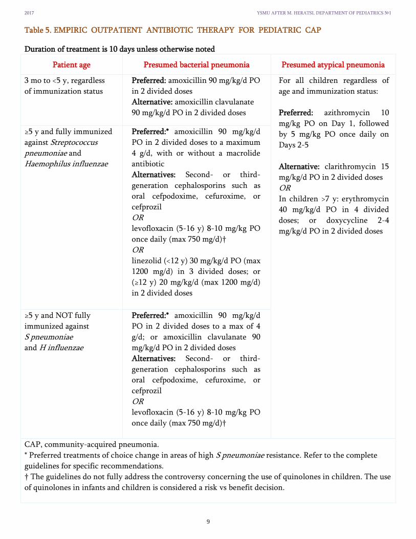
2017 YSMU AFTER M. HERATSI, DEPARTMENT OF PEDIATRICS 1
9
Table 5. EMPIRIC OUTPATIENT ANTIBIOTIC THERAPY FOR PEDIATRIC CAP
Duration of treatment is 10 days unless otherwise noted
Patient age Presumed bacterial pneumonia Presumed atypical pneumonia
3 mo to <5 y, regardless
of immunization status
Preferred: amoxicillin 90 mg/kg/d PO
in 2 divided doses
Alternative: amoxicillin clavulanate
90 mg/kg/d PO in 2 divided doses
For all children regardless of
age and immunization status:
Preferred: azithromycin 10
mg/kg PO on Day 1, followed
by 5 mg/kg PO once daily on
Days 2-5
Alternative: clarithromycin 15
mg/kg/d PO in 2 divided doses
OR In children >7 y: erythromycin
40 mg/kg/d PO in 4 divided
doses; or doxycycline 2-4
mg/kg/d PO in 2 divided doses
≥5 y and fully immunized
against Streptococcus pneumoniae and
Haemophilus influenzae
Preferred:* amoxicillin 90 mg/kg/d
PO in 2 divided doses to a maximum
4 g/d, with or without a macrolide
antibiotic
Alternatives: Second- or third-
generation cephalosporins such as
oral cefpodoxime, cefuroxime, or
cefprozil
OR levofloxacin (5-16 y) 8-10 mg/kg PO
once daily (max 750 mg/d)†
OR linezolid (<12 y) 30 mg/kg/d PO (max
1200 mg/d) in 3 divided doses; or
(≥12 y) 20 mg/kg/d (max 1200 mg/d)
in 2 divided doses
≥5 y and NOT fully
immunized against
S pneumoniae and H influenzae
Preferred:* amoxicillin 90 mg/kg/d
PO in 2 divided doses to a max of 4
g/d; or amoxicillin clavulanate 90
mg/kg/d PO in 2 divided doses
Alternatives: Second- or third-
generation cephalosporins such as
oral cefpodoxime, cefuroxime, or
cefprozil
OR levofloxacin (5-16 y) 8-10 mg/kg PO
once daily (max 750 mg/d)†
CAP, community-acquired pneumonia.
* Preferred treatments of choice change in areas of high S pneumoniae resistance. Refer to the complete
guidelines for specific recommendations.
† The guidelines do not fully address the controversy concerning the use of quinolones in children. The use
of quinolones in infants and children is considered a risk vs benefit decision.

2017 YSMU AFTER M. HERATSI, DEPARTMENT OF PEDIATRICS 1
10
PROGNOSIS
Typically, patients with uncomplicated community-acquired bacterial pneumonia show response to therapy, with
improvement in clinical symptoms (fever, cough, tachypnea, chest pain), within 48-96 hr of initiation of antibiotics
(Fig. 5). Radiographic evidence of improvement lags substantially behind clinical improvement.
Mortality from community-acquired pneumonia in developed nations is rare, and most children with pneumonia do
not experience long-term pulmonary sequelae. Some data suggest that up to 45% of children have symptoms of
asthma 5 yr after hospitalization for pneumonia; this finding may reflect either undiagnosed asthma at the time of
presentation or a propensity for development of asthma after pneumonia.
NONRESOLVING OR SLOWLY RESOLVING PNEUMONIA
Treatment response has traditionally been difficult to define because radiographic changes, which are used to define
the presence of pneumonia, can take up to 6 weeks to resolve and often lag behind the clinical recovery of patients
[Menéndez R et al, 2003].
A number of factors must be considered when a patient does not improve with appropriate antibiotic therapy: (1)
complications, such as empyema; (2) bacterial resistance; (3) nonbacterial etiologies such as viruses and aspiration of
foreign bodies or food; (4) bronchial obstruction from endobronchial lesions, foreign body, or mucous plugs; (5) pre-
existing diseases such as immunodeficiencies, ciliary dyskinesia, cystic fibrosis, pulmonary sequestration, or cystic
adenomatoid malformation; and (6) other noninfectious causes (including bronchiolitis obliterans, hypersensitivity
pneumonitis, eosinophilic pneumonia, aspiration, and granulomatosis with polyangiitis).
A repeat chest radiograph is the 1st step in determining the reason for delay in response to treatment.
Fig. 5. A schematic representation of recovery from community-acquired pneumonia

2017 YSMU AFTER M. HERATSI, DEPARTMENT OF PEDIATRICS 1
11
COMPLICATIONS
Complications of pneumonia (Table 6) are usually the result of direct spread of bacterial infection within the thoracic
cavity (pleural effusion, empyema, pericarditis) or bacteremia and hematologic spread (Fig. 6). Meningitis,
suppurative arthritis, and osteomyelitis are rare complications of hematologic spread of pneumococcal or H. influenzae type b infection.
S. aureus, S. pneumoniae, and S. pyogenes are the most common causes of parapneumonic effusions and of empyema.
The treatment of empyema is based on the stage (exudative, fibrinopurulent, organizing). Imaging studies including
ultrasonography and CT are helpful in determining the stage of empyema. The mainstays of therapy include
antibiotic therapy and drainage with tube thoracostomy. Additional approaches include the use of intrapleural
fibrinolytic therapy (urokinase, streptokinase, tissue plasminogen activator) and selected videoassisted thoracoscopy
(VATS) to debride or lyse adhesions, and drain loculated areas of pus. Early diagnosis and intervention, particularly
with fibrinolysis or VATS, may obviate the need for thoracotomy and open debridement. Fibrinolysis may be more
cost effective than VATS.
Table 6. COMPLICATIONS ASSOCIATED WITH COMMUNITY-ACQUIRED PNEUMONIA
Pulmonary Metastatic Systemic
Pleural effusion or empyema
Pneumothorax
Lung abscess
Bronchopleural fistula
Necrotizing pneumonia
Acute respiratory failure
Meningitis
Central nervous system abscess
Pericarditis
Endocarditis
Osteomyelitis
Septic arthritis
Systemic inflammatory response
syndrome or sepsis
Hemolytic uremic syndrome
Fig. 6. Pneumococcal empyema on the chest radiography of a 3 yr old child
who has had upper respiratory symptoms and fever for 3 days.
A pleural fluid collection can be seen on the right side. The patient had a
positive pleural tap and blood culture result for pneumococci. The child
recovered completely within 3 wk.
REFERENCE
1. Kliegman RM et al: Nelson textbook of pediatrics, ed 20 (2016). Chapter 400: Community-Acquired Pneumonia, Matthew S. Kelly and Thomas J. Sandora
2. Bradley, John S., et al. "The management of community-acquired pneumonia in infants and children older than 3 months of age: clinical practice guidelines by the Pediatric Infectious Diseases Society and the Infectious Diseases Society of America." Clinical Infectious Diseases(2011): cir531.
3. Revised WHO classification and treatment of pneumonia in children at health facilities: evidence summaries. World Health Organization 2014. 4. Zohra S Lassi, Batool A Haider, Zulfiqar A Bhutta. Zinc supplementation for the prevention of pneumonia in children aged 2 months to 59 months.
Cochrane Database of Systematic Reviews, 2010. 5. Reynolds, J. H., McDonald, G., Alton, H., & Gordon, S. B. (2014). Pneumonia in the immunocompetent patient. The British journal of radiology. 6. Finch, S., & Chalmers, J. D. (2014). Brief clinical review: non-responding pneumonia. EMJ Respir, 2, 104-111.

2017 YSMU AFTER M. HERATSI, DEPARTMENT OF PEDIATRICS 1
12
CASE-BASED LEARNING
CASE 1. Newborn with Pneumonia
A pediatric intern was called to the delivery room to assess a term newborn infant with
respiratory difficulty. The infant was less than 2 hours old. His respiratory effort
remained labored and he grunted with each exhalation. His mother’s prenatal history
was unknown, and her labor was too rapid for her to have had any prenatal
antimicrobial screening or antibiotics. She reported having had a baby die in the first
days of life from an infection. This infant was tachypneic with a respiratory rate of 75
breaths per minute. He had decreased breath sounds bilaterally with rare fine crackles.
His airway was intubated for severe respiratory distress and his radiograph
demonstrated bilateral “ground-glass” alveolar filling (Fig. 7). His white blood cell count
was 2200 cells/hpf and his platelet count was 66,000/ml. His clinical condition
deteriorated despite antibiotic therapy. Both initial blood cultures grew group B beta-
hemolytic streptococcus (GBBS) within 6 hours. He died before he was 12 hours old.
Fig. 7. Chest radiograph of an infant
with GBBS pneumonia
The pretreatment and screening of mothers for GBBS has made a significant impact on the prevalence of “early” GBBS disease in
the newborn, so that residents training today are much less likely to see a child with this condition than those who trained 15
years ago. Prenatal and perinatal pneumonia can also be caused by coliforms such as Escherichia coli, anerobes, and Listeria
monocytogenes. Babies can present with very subtle initial symptoms including the full spectrum of respiratory distress from
mild to life-threatening, temperature instability, feeding intolerance, and metabolic derangements including hypoglycemia.
Often, there is little that can be done once they are delivered, so that maternal prenatal screening and treatment become the
most important factors in infant survival. Since newborns are immunocompromised by definition, a number of different
opportunists including Pseudomonas aeruginosa can cause infection in infants hospitalized for extended periods. Viruses
including cytomegalovirus and fungi such as Candida albicans can also cause pneumonia in infants. These agents should be
suspected in babies who do not respond to standard therapy. Initial therapy should include aggressive evaluation for sepsis
including blood and urine cultures, and antibiotics targeting the most common causes. Typically, an aminoglycoside or third-
generation cephalosporin and ampicillin are chosen as initial therapy. Newborns with pneumonia should be cared for in neonatal
intensive care units where expertise in mechanical ventilation is available if needed.
CASE 2. Well-Appearing Infant with Pneumonia
A 7-week-old infant was admitted to the pediatric ward with hypoxemia and cough. The child
was still eating fairly well and there was no history of fever. On examination, the child was not
ill-appearing but his respiratory rate was 70–80 breaths per minute when awake. He had some
scattered crackles on examination, and mild subcostal retractions. The chest radiograph showed
patchy opacifications and hyperinflation (Fig. 8). Evaluation for bacterial infection was negative;
however, a urine culture grew cytomegalovirus (CMV) and the serum CMV IgM was positive.
The child recovered from this illness, but had recurrent episodes of wheezing and dyspnea with
variable responsiveness to bronchodilators and oral corticosteroids. Stagno and his colleagues
described the “afebrile pneumonia syndrome of infancy” more than two decades ago.
The investigators isolated four agents from these infants with pneumonia: Chlamydia
trachomatis (25%), Ureaplasma urealyticum (21%), cytomegalovirus (20%), and Pneumocystis
carinii (18%). This last organism, long felt to be a protozoan, has recently been identified to be a
phylogenetic fungal species and reclassified as Pneumocystis jiroveci. Pneumonia from this
organism is still referred to as “PCP”: Pneumocystis pneumonia.
Fig. 8. Chest radiograph of a
child with cytomegalovirus
pneumonia.
In many cases, multiple organisms were recovered from the same infant with this clinical syndrome. Organisms were identified by
culture and in the case of Pneumocystis, by counter immunoelectrophoresis and indirect immunofluorescence of serum. These studies
are not available in every clinical setting today, so aside from culture for CMV, the other organisms are seldom isolated. Thus the
diagnosis is often made clinically. Infants with this clinical syndrome will often require hospitalization because of hypoxemia or
decreased feeding due to respiratory difficulty. If the child is to be managed as an outpatient, he or she should be followed daily until
the clinical symptoms begin to improve significantly. Additionally, the parents should be given specific, concrete signs and symptoms to
watch for that might herald deterioration. Treatment is usually empiric, and includes oral erythromycin for 3 weeks primarily to treat
chlamydial infection. In a follow-up study, 46% of the children had recurrent episodes of wheezing and airway obstruction, requiring
medical therapy.

2017 YSMU AFTER M. HERATSI, DEPARTMENT OF PEDIATRICS 1
13
CASE 3. Ill-Appearing Infant with Pneumonia
An 11-month old girl presented to the emergency room
with a fever of 102°F (39°C), congestion, cough and a
history of “grunting” respirations. The infant was not
eating well. On examination the child was ill, but not
septic appearing. The chest was clear to auscultation and
a chest radiograph was normal. The child was
discharged with a diagnosis of viral respiratory tract
infection and instructions to follow-up in a pediatric
clinic in the morning. The parents did not return to the
doctor the next morning, but instead returned a day
later, after the child developed progressive respiratory
distress. A chest radiograph showed a large left-sided
pneumonia. The child’s blood culture grew
Staphylococcus aureus, and despite heroic measures the
child succumbed to overwhelming sepsis 48 hours later.
It is easy to forget that pneumonia in children is
potentially a life-threatening illness.
Most bacterial infections in this age group are preceded
by a viral respiratory tract infection. Historically,
influenza virus has been identified as a frequent
preceding infection. Many of the near 25 million deaths
worldwide during the 1918–1919 influenza epidemic are
thought to have been from secondary bacterial
pneumonia. It is likely that the viral infection alters the
ability of the respiratory tract to resist infection from
inhaled or aspirated bacterial pathogens. Infants with
pneumonia will often be tachypneic and may have
grunting respirations. They can also have other signs
associated with sepsis: fever, poor feeding, and lethargy.
Initial radiographs may be normal despite findings on
physical examination. The diagnosis of a respiratory
tract infection will often be made on the basis of clinical
findings, and the decision to attempt to diagnose or treat
for bacterial disease may be a subjective one.
Children with fever should be evaluated according to
established protocols. Under 1 month of age, all children
with fever and no identifiable source should undergo a
sepsis evaluation, and receive inpatient intravenous
antibiotic therapy. In older infants and children, a high
index of suspicion for pneumonia should be maintained.
Any infant or young child with pneumonia should be
followed closely. The child with a lower respiratory
tract infection should be seen again within 24 hours of
the initial visit for a reassessment, regardless of
whether bacterial disease is suspected on the initial
presentation. Serious, even life-threatening bacterial
illness may not be apparent at first. Pneumonia in an
ill-appearing infant is most often due to Streptococcus
pneumoniae. Haemophilus influenzae, Type B (HIB)
was seen previously but has diminished with the
successful vaccination programs for this organism.
Pockets of invasive HIB disease still occur in the
developed world, and an immunization history is an
important part of the evaluation of a child with
pneumonia. Staphylococcus aureus may also cause
severe pneumonia in infants and young children.
Fortunately, it is not a common cause in the developed
world. It should be suspected in a child who develops a
rapid “white-out” pattern of consolidation
radiographically. Children with staphylococcal
pneumonia develop pleural disease in up to 90% of
cases, and pneumatoceles develop in half of children.
This agent should be suspected especially in children
with pneumonia and pulmonary abscess or empyema.
When bacterial pneumonia is suspected and the child
appears only mildly or moderately ill, ampicillin is an
acceptable initial intravenous agent to use pending
culture results. Penicillin-resistant Pneumococcus,
however, has been recognized with increasing
frequency since 1978. Organisms produce one or more
proteins with variable affinity for penicillin.
Approximately 10% of Pneumococcus isolates are
penicillin resistant. Resistance to ceftriaxone is
uncommon, and occurs in less than 2.5% of isolates.
Therefore, when the child appears ill, initial treatment
should be with a second- or thirdgeneration
cephalosporin (cefuroxime or ceftriaxone) for
Pneumococcus or HIB. If the child is septic and
Pneumococcus is suspected, treatment should begin
with vancomycin. If Staphylococcus is suspected,
treatment with penicillinase resistant penicillin
(nafcillin or oxacillin) should be started.

2017 YSMU AFTER M. HERATSI, DEPARTMENT OF PEDIATRICS 1
14
CASE 4. Well-Appearing Toddler or Child with Pneumonia
A 4-year-old preschooler stayed home from his nursery school for 2 days with low-grade fever, rhinitis, and cough. His symptoms became
worse and his mother brought him to your office. He had paroxysms of cough, especially at night, but was relatively well between episodes. He
did not cough during the visit, and he had no previous history of severe cough with respiratory infections, or with exercise. On examination he
was not tachypneic. He had mild rhinitis. Auscultation of his chest revealed diffuse heterophonous wheezing (small airway obstruction) and
fine crackles. He had no hepatomegaly and no digital clubbing. A chest radiograph revealed scattered interstitial infiltrates and mild
hyperinflation. Viral pneumonias are not the focus of this chapter, yet deserve mention because they are the most common cause of pneumonia
in children of any age. The child in this case was well-appearing, but viruses can certainly cause severe, life-threatening infections. The most
commonly isolated viruses in children with LRIs are parainfluenza (35%), RSV (22%), influenza virus (12%), adenovirus (7%) and assorted
others (enterovirus, rhinovirus, 9%). Mycoplasma pneumoniae is responsible for 15% of infections. The absence of severe episodes of cough
with other respiratory tract infections and exercise makes a diagnosis of asthma less likely. Most viral pneumonia begins with inhaled
infectious agents, and the airways are commonly involved. Thus wheezing and hyperinflation (subcostal retractions) are frequently noted. A
chest radiograph may be normal. It may also show interstitial or airspace disease. Children with LRI managed in an ambulatory setting should
be followed daily until the symptoms of respiratory tract infection begin to resolve. Follow-up is critical because it is difficult to predict the
course of pneumonia. In addition, viral pneumonia may become secondarily infected with a bacterial pathogen and parents may not be
sophisticated enough to recognize respiratory deterioration in their children.
CASE 5. Child with Pneumonia
A 7-year-old girl with a viral respiratory tract infection awoke one night with shaking chills, worsening of cough, malaise and fever. She also
complained of right-sided chest pain. On presentation to her pediatrician in the morning, she was ill-appearing and had a temperature of 104°F
(40°C). Her respiratory rate was 42 breaths per minute and auscultation of her chest revealed crackles on her posterior right lung fields. A chest
radiograph showed a right-sided opacification. Her white blood cell count was 21,000 WBC/hpf. A course of amoxicillin/ clavulanate PO was
started, but she returned to her physician 2 days later with persistent fever, worsening cough, and increased difficulty breathing. A repeat
radiograph revealed complete opacification of her right lung (Fig. 9). Room air pulse oximetry was 87%. Blood cultures obtained on her original
presentation grew Streptococcus pneumoniae. She was admitted to the hospital for intravenous antibiotics, but her respiratory condition
deteriorated. She was transferred to the pediatric intensive care unit where she was intubated and started on mechanical ventilation. Her
hypoxemia was unresponsive to increasing levels of positive pressure on conventional ventilation so she was started on a high-frequency
oscillator ventilator. She stabilized, but a pleural effusion and eventual pneumothorax on the right side necessitated chest tube placement 2
days later. She remained on high-frequency ventilation for 2 weeks, before returning to conventional ventilation and extubation after a total of
more than 3 weeks of ventilator therapy. She recovered but her radiograph remained abnormal, with decreased volume on the right (Fig. 10).
Pneumococcal pneumonia in childhood usually presents as a distinct, clinical syndrome characterized by an acute onset and fever. It is not rare.
Laboratory studies reveal leukocytosis, and sputum Gram stain in older children will demonstrate more than 25 WBC/hpf and Grampositive
diplococci. The spectrum of pneumonia from this organism can vary from a very mild course managed in an ambulatory setting, to the severe
case of complicated pneumococcal pneumonia presented here. Complicated pneumonia is characterized by significant pleural effusion often
requiring chest tube placement and, in some cases, pulmonary necrosis. Children with complicated pneumonia are more likely to be older (45
vs. 27 months in one series)2, are more likely to present with chest pain and to require decortication for pleural disease. In the same series,
complicated pneumonia was caused by pneumococcal serotypes 1, 6, 14, and 19 in 24% of cases, as opposed to 3.6% of the children with
uncomplicated pneumonia. Serotype 1, which accounted for 24% of complicated pneumonia, is not included in the currently licensed
pneumococcal vaccine. Children who develop pneumococcal pneumonia are often carriers of this organism. Twenty to forty percent of
children are carriers, and risk factors for the development of invasive pneumococcal disease include frequent episodes of otitis or upper
respiratory tract infections (>3 in 6 months) and daycare attendance. Organisms are aspirated or inhaled, and escape the lung ’s extensive
defense systems, including the mechanical barriers of the upper airway, the mucociliary blanket and cough, and the macrophages and humoral
defenses of the distal airways. Onset of symptoms is usually abrupt with shaking chills and spiking fever. Younger children may present with
febrile seizure2. Ambulatory children with suspected pneumococcal pneumonia should be treated with high-dose amoxicillin or a
cephalosporin. Children ill enough to require hospitalization should receive a second- or third-generation cephalosporin or ampicillin in
combination with sulbactam. Vancomycin or clindamycin should be used for severely ill children with suspected complicated pneumococcal
disease.
Fig. 9. Chest radiograph of
a child with overwhelming
Streptococcus pneumoniae pneumonia.
Fig. 10. Chest radiograph
of resolved complicated
Streptococcus pneumoniae pneumonia.

2017 YSMU AFTER M. HERATSI, DEPARTMENT OF PEDIATRICS 1
15
CASE 6. Adolescent with Pneumonia
A previously healthy 16-year-old stayed home from school with headache, abdominal pain, and sore throat. She did not have
any nasal congestion, but developed paroxysms of cough. She developed a fever of 101°F (38.5°C) and generalized malaise. After
4 days of symptoms and progressive cough, her mother took her to the physician. She was uncomfortable, but not ill-appearing.
She was not hypoxemic or tachypneic. Auscultation of her chest revealed crackles and heterophonous wheezing. She had a mild
erythematous maculopapular exanthem. A chest radiograph revealed mild, bilateral increased interstitial infiltrates (Fig. 11). A
complete blood count disclosed a mild anemia and the smear suggested hemolysis. Bedside serum cold agglutinins were positive
(Figure 10-8), and serologic testing for Mycoplasma pneumoniae IgM was positive >1:32. She was treated with oral doxycycline,
and recovered uneventfully within 2 weeks, although episodes of cough persisted for two additional weeks.
Fig. 11. Chest radiograph of an adolescent with Mycoplasma pneumonia.
Pneumonia due to Mycoplasma pneumoniae was called “atypical” initially because, in 1935, it did not respond to the typical
pneumonia treatment, sulfonamides. It is also sometimes called “walking” pneumonia because it does not render affected
children so sick that they are confined to bed. The onset of symptoms, in contrast to Pneumococcus, is usually insidious with
malaise, low-grade fever and cough. Rhinitis is significant for its absence, and can help the clinician discern Mycoplasma from
viral pneumonia due to adenovirus, which can have a similar clinical course. Pharyngitis is frequently noted, but may be more
prominent in atypical pneumonia due to Chlamydia pneumoniae, another of the causes of this clinical syndrome. Headache and
abdominal pain are also frequently seen. The physical examination will often provide no additional clues to help discern the
specific cause of pneumonia. Clinical signs of pneumonia will often be present, including crackles, wheezes or coarse breath
sounds. However, they may be absent early in the course of the disease. A range of different rashes may be noted from a slight
erythematous eruption in mildest cases, to life-threatening Stevens–Johnson syndrome. Cardiac involvement has also been noted
in some cases, including myocarditis and pericarditis leading to signs and symptoms of congestive heart failure. Rarely, hepatitis
may occur and hepatomegaly will be noted on physical examination.
There is no pathognomonic pattern of the chest radiograph for children with Mycoplasma pneumonia. The radiograph can be
completely normal. Bilateral interstitial infiltrates may be noted, or there may be a unilateral lobar infiltrate. Parapneumonic
pleural effusion may occur in up to 20% of children with Mycoplasma pneumonia when it is carefully sought. There is usually no
reason for pleurocentesis in these children. Serologically, the immunogenicity of Mycoplasma leads to production of a host of
antibodies, including one to the red blood cell “I/I antigen” that causes cold agglutination of the blood cells. While this antibody
may be absent in up to half of children with Mycoplasma, its presence in this clinical setting would make the diagnosis of a
Mycoplasma infection all but assured.
Chlamydia pneumoniae is the other agent commonly associated with atypical or “walking” pneumonia, although the specific
diagnosis is even more difficult to make. Pharyngitis and hoarseness in an adolescent or young adult with interstitial pneumonia
who does not have rhinitis should prompt the clinician to consider Chlamydia in addition to Mycoplasma. While erythromycin
is generally considered the first choice for therapy in this syndrome, there is some evidence that Chlamydia is more susceptible
to tetracycline than to erythromycin16. Doxycycline is an excellent agent for the treatment of this syndrome in children 12
years and older. Other macrolides are also effective and the newer agent, azithromycin, shows greatest effectiveness in
experimental settings.

2017 YSMU AFTER M. HERATSI, DEPARTMENT OF PEDIATRICS 1
16
Pediatric Pleural Effusion The pleural space normally contains 0.3 mL per kg body weight of pleural fluid. Lymphatic vessels can cope
with several hundred millilitres of extra fluid per 24 h. An imbalance between pleural fluid formation and
drainage will result in a pleural effusion.
In a previously well child, pleural effusions are usually secondary to acute bacterial pneumonia and less
often due to chronic infection such as pulmonary tuberculosis (TB). Other causes usually considered in
adults, such as malignancies, cardiovascular diseases, or systemic inflammatory conditions, are uncommon
in children.
Clinical picture
There are two usual patterns of presentation.
In the first, the child has classic symptoms of pneumonia (fever, cough, breathlessness, abdominal pain
and malaise) but they are usually more unwell than those with simple pneumonia alone, with pleuritic
chest pain and even cyanosis.
In the other clinical presentation, the child has been diagnosed with pneumonia but does not respond
to 48 h of an appropriate treatment. On examination, a pleural effusion is suggested by unilateral signs
of decreased chest expansion and dullness to percussion, reduced or absent breath sounds, and scoliosis.
Diagnosis
Contrary to community-acquired pneumonia (CAP), which may be diagnosed on clinical grounds only, the
diagnosis of parapneumonic pleural effusion requires an imaging technique to demonstrate the presence of
fluid in the pleural space. The first imaging technique should be a posteroanterior chest radiograph. The
earliest sign of a pleural effusion is obliteration of the costophrenic angle. A rim of fluid may be seen
ascending the lateral chest wall (meniscus sign). If the film is taken in a supine position, the appearance can
be of a homogeneous increase in opacity over the whole lung field without blunting the costophrenic
angle, or a classic pleural-based shadow. A lateral chest radiograph rarely adds anything extra and should
not be routinely obtained.
Once pleural effusion has been diagnosed or suspected by a
chest radiograph, chest ultrasonography should be obtained
to confirm the diagnosis, estimate the size of the effusion,
differentiate between free and loculated pleural fluid and
determine its echogenicity (Fig. 12). It may also be used to
guide chest drain insertion or thoracentesis.
Fig. 12. Chest ultrasound showing a loculated
pleural effusion.

2017 YSMU AFTER M. HERATSI, DEPARTMENT OF PEDIATRICS 1
17
Chest CT scans involve radiation exposure that can be equivalent to 20–400 chest radiographs depending
on technical factors and should not be performed routinely. It may have a role in complicated cases,
including immunocompromised children where a CT scan can detect airway or parenchymal lung
abnormalities, such as endobronchial obstruction or a lung abscess, or before surgery to delineate the
anatomy.
Management
All children with parapneumonic pleural effusion or empyema should be admitted to hospital and managed
following local or national guidelines.
Intravenous antibiotics and careful consideration of pleural drainage procedures are the most important
aspects of parapneumonic effusion/ empyema management.
Intravenous empirical antibiotic treatment should begin as soon as possible. In the most common setting of
a pleural effusion arising from CAP, empirical treatment must cover Streptococcus pneumoniae, S.
pyogenes and Staphylococcus aureus. In most cases, cefotaxime (150 mg/kg), coamoxiclav or cefuroxime are
appropriate. Penicillin allergic patients can be treated with clindamycin alone. If pneumatoceles are
evident, anti-staphylococcal cover is mandatory (cloxaciline or flucoxaciline). However, in cases of
hospital-acquired pneumonia or following surgery, trauma or aspiration, broader spectrum agents should be
used to cover aerobic Gram-negative rods.
An important issue is whether to insert a pleural drain or not. It is generally accepted that isolated pleural
taps for diagnostic purposes are not recommended in children with a small, uncomplicated parapneumonic
pleural effusion, except if there are any atypical features suggesting the presence of malignancy, such as the
absence of acute fever or pneumonia and evidence of an underlying mediastinal mass or lymphadenopathy.
In these uncommon situations it is important to remember that large volume aspiration and general
anaesthesia pose a significant risk of sudden death in children with superior mediastinal obstruction due to
malignancy, therefore, the volume of aspirated pleural fluid should be small (5 mL) and general anaesthesia
should be avoided.
As a general rule, there is a good deal of evidence suggesting that a pleural drain is not always necessary
and that antibiotics alone can be enough to provide excellent clinical outcomes when there is not a clear
indication for chest drainage. Tube thoracostomy must be performed if the child is in respiratory distress
due to lung compression by the pleural effusion, or if toxic appearance and sepsis is suspected. It also may
be considered if the effusion size is large (definitions vary from 10 mm thickness in ultrasonography or
radiography to one-third of the hemithorax in radiography) or is enlarging, and the child is not responding
after 48 h of antibiotic treatment.
Further reading
1. Hull J et al. Paediatric Respiratory Medicine, 2nd ed. Oxford University Press 2015
2. ERS Handbook: Paediatric Respiratory Medicine, 1st ed. European Respiratory Society 2013

2016 YSMU AFTER M. HERATSI, DEPARTMENT OF PEDIATRICS 1
- 1 -
Chronic Respiratory Diseases in Children
Chronic respiratory diseases are a group of chronic diseases affecting the airways and the other structures of the lungs.
Major chronic respiratory diseases in children
Chronic lung disease (CLD) of prematurity (bronchopulmonary dysplasia)
Asthma
Chronic bronchitis
Chronic obstructive pulmonary disease (COPD)
Emphysema
Bronchiectasis
Hypersensitivity pneumonitis (extrinsic allergic alveolitis)
Pulmonary eosinophilia
Pulmonary heart disease and diseases of pulmonary
circulation including pulmonary embolism,
pulmonary hypertension and cor pulmonale
Interstitial lung disease (diffuse parenchymal lung
diseases)
Sarcoidosis
Sleep apnea syndrome
Congenital thoracic malformations

2016 YSMU AFTER M. HERATSI, DEPARTMENT OF PEDIATRICS 1
- 2 -
ROLE OF MUCOCILIARY DYSFUNCTION IN CHRONIC AIRWAY DISEASES
Mucociliary clearance is an important primary innate defense mechanism that
protects the lungs from the harmful effects of inhaled pollutants, allergens, and
pathogens. Mucociliary dysfunction is a common feature of chronic airway
diseases.
Normal airway mucus has two layers: outer more viscous layer, the gel phase,
which is transported by ciliary beat, and inner serous layer called the sol phase,
in which cilia recover from their active beat. Mucus is moved proximally by the
effective coordinated beating of cilia.
Airway surface liquid (ASL) layer dehydration has a critical role in the
pathogenesis of mucociliary dysfunction and chronic airway disease. ASL
depletion resulted in reduced mucus clearance and histologic signs of chronic
airway disease, including mucous obstruction, goblet cell hyperplasia, and
chronic inflammatory cell infiltration.
Fig. Epithelial (mucosal) layer of airway is shown (pseudostratified
columnar ciliated epithelium). Goblet cells were not included for clarity
and to show ciliary movement in gel and sol phases. 1 = gel phase; 2 = sol
phase; 3 = basement membrane; 4 = cilia in recovery stage within sol
phase; 5 = submucosal layer.
ROLE OF MATERNAL SMOKING IN CHRONIC AIRWAY DISEASES
Exposure to maternal smoking both during and after fetal development can have significant deleterious effects on lung development and
subsequent susceptibility to pulmonary disorders.
Maternal smoking during pregnancy is a risk factor for several adverse developmental outcomes, including abnormal fetal lung development. Lung development is most critical during late fetal and early postnatal life—the saccular and alveolar developmental phases. Abnormalities occurring during these stages can have long-term effects on lung function, contributing to the development of asthma in children and lung disease in adults. Maternal smoking and altered fetal pulmonary structure are related to nicotine that crosses the placenta and is expressed in breast milk. There is up-regulation of nicotinic acetylcholine receptors in the fetal lung. Animal models have demonstrated alterations in lung structure including decreased alveolarization, thickening of the alveolocapillary membrane, decreased synthesis of surfactant, decreased airway diameter, decreased vessel density, and increased airway hyperresponsiveness. Smoke exposure also changes the genetics that control lung growth and maintenance of lung structure, and accelerates lung aging. Prevention of maternal smoking during pregnancy and lactation is required and nicotine replacement therapy is not advised for these women. [Abbott LC, Winzer-Serhan UH 2012; Harding R, Maritz G 2012; Kajeka R 2007; Maritz GS, Harding R 2011; Wongtrakool C et al 2012.]

2016 YSMU AFTER M. HERATSI, DEPARTMENT OF PEDIATRICS 1
- 3 -
CHRONIC BRONCHITIS
Chronic bronchitis is recurring inflammation and degeneration of the bronchial tubes that may be associated with active infection. Patients
with chronic bronchitis have more mucus than normal because of either increased production or decreased clearance. Coughing is the
mechanism by which excess secretion is cleared.
Chronic bronchitis is often associated with asthma, cystic fibrosis, dyskinetic cilia syndrome, foreign body aspiration, or exposure to an airway
irritant. Recurrent tracheobronchitis may occur with tracheostomies or immunodeficiency states.
Defining chronic bronchitis and its prevalence in childhood has been complicated by the significant clinical overlap with asthma and reactive
airway disease states. In adults, chronic bronchitis is defined as daily production of sputum for at least 3 months in 2 consecutive years. Some
have applied this definition to childhood chronic bronchitis. Others limit the definition to a productive cough that lasts more than 2 weeks
despite medical therapy.
Chronic bronchitis has also been defined as a complex of symptoms that includes cough that lasts more than 1 month or recurrent productive
cough that may be associated with wheezing or crackles on auscultation. Elements of these descriptors are present in the working definitions of
asthma, as well.
Normal airway color and
architecture (in a child with
mild tracheomalacia).
Airway of a child with chronic bronchitis
shows erythema, loss of normal architecture,
and swelling.
CHRONIC OBSTRUCTIVE PULMONARY DISEASE (COPD)
Chronic obstructive pulmonary disease (COPD) is conventionally thought of as a disease of adult smokers, related to airway inflammation and
structural airway changes (remodeling). However, there is important epidemiological evidence, from a series of studies with overlapping age
groups from birth to late middle age that early life events, including antenatal influences on lung growth, program the child to be at increased
risk for future COPD [Bush A, 2008].

2016 YSMU AFTER M. HERATSI, DEPARTMENT OF PEDIATRICS 1
- 4 -
CONGENITAL MALFORMATIONS OF THE LUNG AND THE AIRWAY
The respiratory tree arises from the ventral surface of the foregut. Abnormal embryological development may result in congenital thoracic
malformations (CTMs), which include:
• Failure of complete separation of foregut and
bronchial structures, leading to different types of
tracheo-oesophageal fistula
• Congenital pulmonary airway malformations
(CPAM), previously known as congenital cystic
adenomatoid malformations (CCAM), from
abnormal development of alveolar or bronchial
tissues
• Pulmonary sequestration from an abnormal
blood supply to part of the lung (usually systemic
rather than pulmonary)
• Congenital diaphragmatic hernia from
maldevelopment of the pleuroperitoneal canal
with or without associated deficiency of the
diaphragm itself
• Cysts, bronchogenic or foregut
• Congenital lobar emphysema from partial
obstruction of the developing airway, most
commonly due to a deficiency of bronchial
cartilage development
• Lung agenesis/bronchial atresia from
maldevelopment of the tracheobronchial tree in
early fetal life.
Fig. Phases of lung development.

2016 YSMU AFTER M. HERATSI, DEPARTMENT OF PEDIATRICS 1
- 5 -
PARENCHYMAL DISEASES
Chronic obliterative bronchiolitis
Chronic obliterative bronchiolitis (COB) is a rare form of chronic obstructive lung
disease that follows an insult to the lower respiratory tract. It is characterised by
inflammation and fibrosis of the terminal and respiratory bronchioles that lead to
narrowing and/or complete obliteration of the airway lumen.
A child with COB presents with cough, wheeze, pyrexia and tachypnoea which fail to
resolve. Known causes include infection (particularly adenovirus) and chronic
aspiration.
It results in characteristic changes on the high resiolution CT scan (HRCT), with patchy hyperinflation with a characteristic mosaic attenuation
appearance of the lungs from adjacent lobes being either normal or hyperinflated. Lung function shows very severe irreversible obstruction.
Treatment is supportive, with many children requiring oxygen therapy and, in a few cases, eventual lung transplantation.
Interstitial lung disease (diffuse parenchymal lung diseases)
Interstitial lung disease (ILD) is exceptionally rare in childhood, affecting about 1 in 100,000 children. It occurs due to a wide variety of
pathological causes. In the light of more recent understanding of these etiologies, a new term – ‘diffuse parenchymal lung diseases’ (DPLD) –
has been introduced. Approximately a third of patients present in the first two years of life and the etiology of 40–50% remains unknown.
Known genetic defects which cause DPLD include abnormalities in surfactant B (SP-B), surfactant protein C (SP-C), surfactant protein ABCA3
and thyroid transcription factor-1 (TTF-1). These disorders are characterized by inflammatory and fibrotic changes that affect alveolar walls.
There is usually a gradual decrease in lung compliance that results in increased work of breathing followed by a fall in arterial oxygen
saturation. The diagnosis is suggested by a combination of failed response to treatment, poor growth, cough, tachypnea, reduced oxygen
saturations, finger clubbing and fine crackles.
As with adults, DPLD in childhood may present as extrinsic allergic alveolitis. Care should be taken to ensure there is no exposure to potential
allergens, particularly fungal spores and pigeon droppings. DPLD may complicate systemic diseases, including juvenile dermatomyositis,
sarcoidosis, systemic lupus erythematosus and scleroderma. It can also occur as a reaction to drugs (azathioprine, methotrexate) or as a
consequence of radiotherapy to the chest. Radiological changes tend to be non-specific, including generalized ground glass shadowing with
reticular nodular infiltrates and honeycombing on CXR. HRCT provides more detail, but to establish a specific tissue diagnosis, a lung biopsy is
required.
Older children may present with any number of respiratory signs. They frequently have finger clubbing and a restrictive pattern on
spirometry. Two thirds of children respond to a combination of corticosteroids with or without hydroxychloroquine.

2016 YSMU AFTER M. HERATSI, DEPARTMENT OF PEDIATRICS 1
- 6 -
CHRONIC INFECTION
Bronchiectasis occurs when there is abnormal dilatation of the bronchi. It can be suspected on clinical grounds (persistent moist cough,
clubbing and focal chest signs) but is usually diagnosed using HRCT scanning. Bronchiectasis arises from chronic airway inflammation that is
driven by persistent infection. This leads to intense neutrophilic inflammation within the airways. It has been proposed that most
bronchiectasis arises because a vicious cycle of infection and inflammation develops within the lung leading to impaired mucociliary clearance,
followed by bacterial proliferation and more inflammation. Any condition that results in either impaired mucociliary clearance or abnormal
response to infection can lead to bronchiectasis. Once bacterial growth is established within the mucus, clearance will be impaired by a
combination of factors. Haemophilus influenzae, Streptococcus pneumoniae and Pseudomonas aeruginosa release mediators which directly
inhibit ciliary function. They also lead to increases in local mucus production and the production of interleukin-8, which recruits neutrophils
to the site of inflammation. In contrast to asthma, chronic infection is characterized by a neutrophilic inflammation.
Table. Causes of bronchiectasis
Post-infectious – measles, pertussis, severe pneumonia
Immune dysfunction – hypogammaglobulinaemia, neutrophil dysfunction, HIV infection
Impaired mucociliary clearance – primary ciliary dyskinesia (PCD), cystic fibrosis (CF)
Systemic disorders – rheumatic arthritis, inflammatory bowel disease
Undiagnosed foreign body or recurrent aspiration
CASE
STUDY
Here are chest x-rays of a 16-year-old female. Her 11y old
sister has the same condition too. What condition is this?

2016 YSMU AFTER M. HERATSI, DEPARTMENT OF PEDIATRICS 1
- 7 -
CILIARY DYSKINESIAS
Almost every cell in the body is equipped with a membrane
bound, finger-like projection called a primary cilium. Cilia
have been equated to cellular antennae that detect
molecular signals in the environment and thus influence
how cells behave. Normally functioning cilia have a fast
‘upstroke’ followed by a slower recovery phase in the
opposite direction. The normal cilial beat frequency is
between 12 and 14 times/second.
Ciliary defects, referred to as ciliopathies, are associated
with a broad spectrum of human disease.
Primary ciliary dyskinesia (PCD) describes a heterogeneous
group of conditions with a primary defect in the structure
or function of the cilia. PCD is mainly inherited as an
autosomal recessive condition. The incidence is 1 in
15,000–30,000 live births.
Children with PCD have recurrent sinopulmonary infections. Boys may have
reduced fertility due either to sperm immotility or vas deferens abnormalities.
Almost half of all affected children have laterality defects such as situs inversus.
This is thought to occur because the functional defects seen in PCD reduce the
effectiveness of nodal cilia during embryogenesis. A combination of PCD with
situs inversus, chronic sinusitis, and bronchiectasis is known as Kartagener
syndrome.
PCD is a genetic disease, but although several causative genes have been
described, the mainstay of diagnosis remains the visualization of abnormal ciliary
function demonstrated by electron microscopy of cilia from brush samples from
inside the nose.
Treatment for respiratory infection is similar to cystic fibrosis. Optimal management requires rapid treatment and airway clearance techniques.

2016 YSMU AFTER M. HERATSI, DEPARTMENT OF PEDIATRICS 1
- 8 -
CYSTIC FIBROSIS
Cystic fibrosis (CF) is an inherited disease caused by a genetic mutation (defect) on chromosome 7. The defective gene results in abnormalities
in the production and function of a protein called the cystic fibrosis transmembrane conductance regulator (CFTR). In healthy cells CFTR acts
as a chloride channel and a regulator of sodium, chloride and bicarbonate transport.
To date, over 1900 possible mutations of this gene have been described, of which 1300 are thought to be pathogenic. Specific mutation
frequencies vary between different geographical locations and ethnic groups. ΔF508 (deletion of three nucleotides which code for
phenylalanine at position 508 in the amino acid sequence) is the most common CFTR mutation. It accounts for nearly two thirds of the
mutations in individuals with CF.
The widespread presence of CFTR throughout the body (lungs, salivary glands,
pancreas, liver, kidneys, sweat ducts and reproductive tract) helps to explain why CF is
a multisystem condition affecting many organs. The two major systems affected are the
lungs and the gastrointestinal tract.
Fig. In CF, impairment of CFTR function causes reduced fluid production. Enhanced sodium
absorption through epithelial Na+ channels (ENaC) and basolateral Na/K ATPase pumps results
in increased fluid absorption leading to drier airways and impaired ciliary clearance.
Normal airway epithelium CF airway epithelium

2016 YSMU AFTER M. HERATSI, DEPARTMENT OF PEDIATRICS 1
- 9 -
In the lungs, inactive or inefficient functioning CFTR results in impaired chloride
transport and enhanced sodium absorption across airway epithelial cells. This leads to a
net increase in water absorption. The volume of the liquid that sits on the airway
surface is reduced and the mucus in the airways becomes more viscid (sticky). In
healthy lungs the cilia (small hairs on the surface of the airways) beat in a coordinated
fashion so that they continually move mucus up and out of the lungs. This cleansing
action of the cilia is impaired in CF because of the presence of very sticky mucus and
dry airways which provide a favourable environment for bacterial infection.
Structural changes in the CFTR protein have also been linked to defective phagocytosis
(ingestion and destruction of bacteria by the white blood cells) of bacteria such as
Pseudomonas aeruginosa and to reduced clearance of infection [Di et al, 2006;
Painter et al, 2006].
Overall these changes in lung physiology lead to dry airways, sticky secretions, a predisposition to chronic chest infections, bronchiectasis and
scarring.
Individuals with CF may remain relatively asymptomatic
despite significant decline in lung function and only develop
breathlessness when a critical point is reached and lung
reserve is lost. Symptoms are often a poor marker of disease
severity, and other factors such as lung function, pseudomonas
status and chest radiology should be taken into account.
Fig. Some of chest X-ray and CT scan changes in CF:
1 -bronchiectasis, 2 - mucus plugging

2016 YSMU AFTER M. HERATSI, DEPARTMENT OF PEDIATRICS 1
- 10 -
Gastrointestinal manifestations are:
• Pancreatic disease: Due to increased viscosity of secretions from the pancreatic duct, there is autodigestion of the pancreas from enzymes that
are normally secreted into the gastrointestinal tract. Consequently, islet cells may be damaged leading to type 1 diabetes mellitus. Due to
reduced pancreatic enzymes within the intestinal lumen, malabsorption of fat occurs, which clinically manifests as steatorrhoea and faltering
growth. Treatment with enzyme replacement therapy (e.g. Creon) is necessary to allow the child to thrive.
• Intestinal disease: The chloride cotransporter (CFTR) within the small bowel that is involved in secretion of water into the gut is ineffective
and may result in meconium ileus at birth (15% of children with cystic fibrosis present with intestinal obstruction within the neonatal period)
or distal intestinal obstruction syndrome (DIOS) in childhood and adolescence. In DIOS there is ineffective secretion of water and chloride into
the small intestine, which results in obstruction around the area of the terminal ileum, caecum and ascending colon. Gastrografin or intestinal
lavage is necessary in some cases; however, severe cases require surgery and stoma formation.
Diagnosis of CF
The diagnosis of CF is made on the basis of two positive sweat chloride tests using pilocarpine
iontophoresis (60 mmol/L) along with classic clinical findings and a history of CF in an immediate
family member.
o False-positive sweat test results are uncommon but may occur in the presence of adrenal insufficiency, nephrogenic diabetes insipidus, type I glycogen storage disease, hypothyroidism, hypoparathyroidism, familial cholestasis, and malnutrition.
Additional diagnostic tests include neonatal screening with increased circulating levels of
immunoreactive trypsinogen, genotyping for CFTR mutations (two mutations confirm the diagnosis),
nasal potential difference testing, a computed tomography scan of sinuses demonstrating pansinusitis,
24-hour fecal fat measurement looking for signs of pancreatic insufficiency, and ultrasound to assess
absence of the vas deferens in males.
CFTR-related disease include diseases that are associated with CFTR mutations but do not meet
diagnostic criteria for CF including chronic pancreatitis, allergic bronchopulmonary aspergillosis
(ABPA), idiopathic bronchiectasis, chronic sinusitis, and congenital bilateral absence of the vas
deference.
CFTR-related metabolic syndrome includes infants with abnormal newborn screening that
subsequently show (1) intermediate sweat chloride level with one CF-causing mutation or (2) normal
sweat chloride level with two CFTR mutations (one CF causing and one non CF causing).

2016 YSMU AFTER M. HERATSI, DEPARTMENT OF PEDIATRICS 1
- 11 -
Treatment of CF
Treatment goals. These include to delay or prevent lung disease, to promote good nutrition and growth, and to treat complications.
Maintenance treatment for patients with classic CF
o Airway clearance. Daily airway clearance is one of the most important methods of prevention of respiratory tract infections.
There are many different methods, including manual chest physiotherapy, postural drainage, autogenic drainage, high-frequency chest
oscillation vests, and manual percussion therapy.
Adjunctive therapies include the Flutter valve and Acapella device.
The use of a specific method is mostly dependent on patient preferences; no studies demonstrate superiority of one method over another.
Dornase alfa promotes airway clearance by cleaving DNA released by degenerating neutrophils, thus decreasing mucus viscosity. Its use has been
shown to improve pulmonary function. It should be considered in children 6 years and older as a daily inhalation (2.5 mg).
Hypertonic saline (7%) promotes airway clearance by hydrating inspissated airway mucus. It has shown to reduce frequency of pulmonary
exacerbations. It should be considered in children 6 years and older who have a chronic cough and reduction in pulmonary function testing.
o Optimization of nutrition. Nutritional failure has been proven to be closely related to increased morbidity and frequency of pulmonary
exacerbations. Therefore, it is important to maintain adequate nutrition via encouragement of a high-calorie and high-protein diet.
For patients who are not able to achieve appropriate oral caloric intake, a feeding gastrostomy tube may be an option.
o Pancreatic enzyme supplementation. Patients with the pancreatic-insufficient form of CF manifest signs of malabsorption. Pancreatic enzyme
supplementation is essential for these patients.
Usual dose ranges from 1,500 to 2,500 U of lipase per kilogram of patient's weight per meal.
Dosing is usually started at the lowest level and titrated up as needed, and it should not exceed 2,500 lipase units/kg/meal because high doses
have been associated with chronic intestinal strictures.
o Lipid-soluble vitamin supplementation (vitamins A, D, E, and K). Lipid-soluble vitamins are not well absorbed in patients with pancreatic
insufficiency.
o Antimicrobials. Chronic antimicrobial therapy is frequently used in patients with increased morbidity from colonizing microorganisms to attempt
prevention of pulmonary exacerbation. These are commonly used against methicillin-resistant S. aureus, methicillin-sensitive S. aureus (Panton-
Valentine leukocidin positive), Pseudomonas, and Aspergillus. In addition, chronic azithromycin therapy has proven beneficial in terms of its
immunomodulatory effects; it interferes with Pseudomonas biofilm formation in the CF airways.
o Anti-inflammatory agents Oral glucocorticoid therapy and nonsteroidal anti-inflammatory drugs such as high-dose ibuprofen have proven benefits for some patients;
however, the side effects of long-term therapy should be weighed against the benefits.
Azithromycin has been shown to improve respiratory function and reduce frequency of exacerbation, and its use is recommended for children 6
years and older. Its mechanism of action remains unclear.
o CFTR modulators Ivacaftor (VX-770) has shown to be effective at potentiating chloride channel function in cells with expressing the following mutations: G551D,
G1244E, G1349D, G178R, G551S, S1251N, S1255P, S549N, S549R, or R117H. Its use is recommended with patients who carry an approved
mutation and are 6 years and older (dose 150 mg q12h).

2016 YSMU AFTER M. HERATSI, DEPARTMENT OF PEDIATRICS 1
- 12 -
Therapy for a pulmonary exacerbation
o This should always include intensive chest physiotherapy 3-4 times a day along with good nutritional support. Outpatient antibiotic
therapy should always be attempted first if there are no signs of respiratory distress or decompensation. Choice of therapy should be
based on previous sputum cultures.
o The duration of therapy depends on clinical improvement but is generally between 2 and 3 weeks.
If there is failure to improve clinically while on outpatient therapy, the patient should be admitted to initiate IV antibiotic therapy
for a total of 2-4 weeks.
All patients should be hospitalized in separate rooms with strict isolation measures as needed for resistant organisms.
The duration of admission depends on the severity of the patient's illness and clinical judgment (clinical improvement, improvement
in spirometry, easiness of completing IV treatment at home).
Special considerations
o Allergic bronchopulmonary aspergillosis (ABPA)
ABPA is an exaggerated immunologic response in the lungs against Aspergillus that results in signs of airway obstruction. It occurs
in 6%-25% of patients with CF. Unlike in adults where asthma with ABPA is common, ABPA is rarely seen in children other than
complicating CF.
Criteria for diagnosis include positive skin prick testing against Aspergillus, along with detection of specific Aspergillus anti-IgG and
anti-IgE in serum. Radiographic evidence of central bronchiectasis is suggestive of the diagnosis.
Treatment includes oral corticosteroids and antifungals such as itraconazole.
o Cystic fibrosis-related diabetes mellitus (CFRD)
CFRD is caused by destruction of pancreatic islet cells and resultant insulin deficiency. Patients with CF should undergo frequent
(annual) oral glucose tolerance tests to screen for evidence of CFRD.
Treatment is generally managed by a pediatric endocrinologist. It frequently involves administration of insulin and carbohydrate
counting without compromising lipid intake and high caloric necessities.
o Lung transplantation
The most common cause of death related to CF is advanced lung disease, and for these patients, lung transplantation may be the only
alternative to prolong survival.
The most commonly used model for CF survival was published by Kerem et al. and describes high mortality risk for patients with an
FEV1 < 30% of predicted, hypercarbia (>50 mm Hg), hypoxemia (<55 mm Hg), young age, female gender, and nutritional failure.
These patients should be referred for evaluation of lung transplantation.

2016 YSMU AFTER M. HERATSI, DEPARTMENT OF PEDIATRICS 1
- 13 -
Table. Differential diagnoses, presenting symptoms, and findings [Leigh et al. Clinical and genetic aspects of primary ciliary dyskinesia/Kartagener syndrome. Genetics in Medicine (2009) 11, 473–487]
REFERENCE
1. Hull J et al. Paediatric Respiratory Medicine, 2nd ed. Oxford University Press 2015
2. ERS Handbook: Paediatric Respiratory Medicine, 1st ed. European Respiratory Society 2013

2017 YSMU AFTER M. HERATSI, DEPARTMENT OF PEDIATRICS 1
1
INTRODUCTION TO PEDIATRIC ALLERGY AND IMMUNOLOGY
Characteristics of the immune system in childhood
The immune system is made up of organs and tissues that are
connected by the blood and lymph systems. The organs and tissues
where immature (or naïve) lymphocytes are produced are described
as the primary (or central) lymphoid organs, and where they come
into contact with harmful organisms as mature lymphocytes as the
secondary (or peripheral) lymphoid organs (Nairn and Helbert,
2007).
The primary lymphoid tissues consist of the bone marrow and the
thymus, while the secondary lymphoid tissues consist of the lymph
nodes, spleen and mucosal-associated lymphoid tissue (MALT).
The fetal immune system develops in a sterile and
protected environment, and therefore lacks antigenic
experience. It must also be modulated in order to co-
exist with the mother's immune system. Soon after
birth, the newborn is exposed to the "hostile world" of
bacteria, viruses, fungi, and parasites, and must
immediately defend itself.
During this period, the neonate mainly depends upon
components of the innate or antigen-independent
immune system, including phagocytes, natural killer
cells, antigen presenting cells, humoral mediators of
inflammation, and complement. Also, during the initial
postpartum phase, the infant relies on maternal
antibodies and the mother’s breast milk, which is rich
with immunoglobulins and WBCs.
The overall performance of the immune system in the neonatal period is diminished in several important
respects. As a result, infants in the first three months of life are more susceptible than older infants to
serious bacterial infection, as well as some viral and fungal infections. Specifically, organisms that can cause
significant morbidity in neonates include: group B streptococci, Escherichia coli, herpes simplex virus
(HSV), cytomegalovirus (CMV), varicella-zoster virus (VZV), respiratory syncytial virus (RSV), and
Candida species.

2017 YSMU AFTER M. HERATSI, DEPARTMENT OF PEDIATRICS 1
2
What makes a neonate’s immune system immature?
The immunological function of the neonate undergoes maturation in both the cellular and humoral
components with the child’s development.
Neutrophils (polymorphonuclear leukocytes)
Neonatal neutrophils are deficient in their ability to adhere to vessel walls at site of infection. Further
release of neutrophils depletes a neonatal storage pool because the bone marrow storage of a neonate is only
20-30% of the pool in an adult. Neonatal neutrophils have a decreased ability to “deform” & migrate into
tissues. Neonatal neutrophils have decreased chemotaxis due to decreased chemoattractant production.
Bacterial killing by polymorphonuclear leukocytes, which depends on the generation of oxygen-derived
free radicals, is intact in healthy-term and most premature newborns.
Monocytes
Neonates have a sufficient amount of monocytes and full capability to kill organisms, but because of a
neonates deficiencies previously discussed very few monocytes get to the site of infection.
Lymphocytes
T cell-mediated immunity is not transferred from mother to fetus, in contrast to humoral immunity. Thus,
young infants rely exclusively on their own T cells plus elements of the innate immune system to fight
infections caused by intracellular pathogens, respond to vaccination, and reject foreign tissue.
The neonate’s T-cells are unable to produce certain cytokines, which affects the interaction between T-
cells and B-cells. In addition, there is a greater reactivity of T-suppressor cells relative to T-helper cells
compared with those of the normal adult.
Activated T cells may be subdivided into distinct functional categories depending upon the profile of
cytokines they secrete. The Th1 response promotes production of opsonizing antibodies (eg, IgG1),
induction of cellular cytotoxicity, and macrophage activation. This T cell response provides defense against
pathogens that replicate intracellularly, particularly mycobacteria and viruses, and also some bacteria and
parasites. In comparison, Th2 responses promote the production of IgE and IgG4, and stimulate eosinophil
development.
Fig. The differentiation of naïve T cells.
Depending on the adjuvanticity of the
substances co-exposed with the antigen and
status of the cells and cytokines in the
microenvironment, naïve T cells can
differentiate into T-helper (Th)1, Th2, Th9,
Th17, and Th22 types of T cells. Based on their
respective cytokine profiles, responses to
chemokines, and interactions with other cells,
these T-cell subsets can promote different
types of inflammatory responses.
IFN interferon, IL interleukin, TGF
transforming growth factor, TNF tumor
necrosis factor

2017 YSMU AFTER M. HERATSI, DEPARTMENT OF PEDIATRICS 1
3
Complement system
Premature and full-term infants are deficient in all of the measurable products of complement activation.
Complement levels increase after birth and reach adult levels between 6 to 18 months of age.
Immunoglobulin (Ig) production in the foetus and newborn
The developmental pattern of immunoglobulins (Igs) is as follows: IgG transfer across the placenta
occurs as early as 8 weeks gestational age. Its level is directly proportional to gestational age, but is still
less than 50% of term levels at 28 weeks gestation. The IgG levels fall during the first four months of
extrauterine life reaching adult levels by 4 – 6 years of age.
By the 10th week of gestation, the fetus is capable of producing IgM and may make large quantities in
the presence of a congenital infection. IgM production by the non-infected newborn does not reach
adult levels until 1–2 years of life.
IgA is not measurable until late in gestational life and is very limited in the infant, failing to reach adult
values until puberty. Secretory IgA appears later than serum IgA (already limited in the infant). Diseases whose defense depends primarily upon secretory IgA, such as some of the viral respiratory agents (e.g., respiratory syncytial virus) and infectious diarrheas, remain prevalent throughout infancy. The infant is at risk for encapsulated organisms and cannot localize infections well.
Fig. Immunoglobulin levels in newborn infants fall to low levels at about 6 months of age.
Decreased Complement
(50%-75% of concentration in adult )
↓ Chemotaxins ↓ Lysis ↓ Opsonization
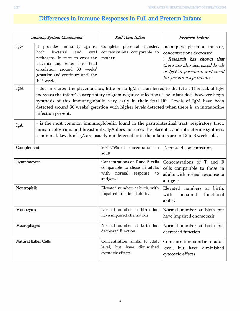
2017 YSMU AFTER M. HERATSI, DEPARTMENT OF PEDIATRICS 1
4
Differences in Immune Responses in Full and Preterm Infants
Immune System Component Full Term Infant Preterm Infant
IgG It provides immunity against
both bacterial and viral
pathogens. It starts to cross the
placenta and enter into fetal
circulation around 30 weeks’
gestation and continues until the
40th week.
Complete placental transfer,
concentrations comparable to
mother
Incomplete placental transfer,
concentrations decreased
! Research has shown that there are also decreased levels of IgG in post-term and small for gestation age infants
IgM - does not cross the placenta thus, little or no IgM is transferred to the fetus. This lack of IgM
increases the infant’s susceptibility to gram negative infections. The infant does however begin
synthesis of this immunoglobulin very early in their fetal life. Levels of IgM have been
detected around 30 weeks’ gestation with higher levels detected when there is an intrauterine
infection present.
IgA - is the most common immunoglobulin found in the gastrointestinal tract, respiratory tract,
human colostrum, and breast milk. IgA does not cross the placenta, and intrauterine synthesis
is minimal. Levels of IgA are usually not detected until the infant is around 2 to 3 weeks old.
Complement 50%-75% of concentration in
adult Decreased concentration
Lymphocytes Concentrations of T and B cells
comparable to those in adults
with normal response to
antigens
Concentrations of T and B
cells comparable to those in
adults with normal response to
antigens
Neutrophils Elevated numbers at birth, with
impaired functional ability Elevated numbers at birth,
with impaired functional
ability
Monocytes Normal number at birth but
have impaired chemotaxis Normal number at birth but
have impaired chemotaxis
Macrophages Normal number at birth but
decreased function Normal number at birth but
decreased function
Natural Killer Cells Concentration similar to adult
level, but have diminished
cytotoxic effects
Concentration similar to adult
level, but have diminished
cytotoxic effects

2017 YSMU AFTER M. HERATSI, DEPARTMENT OF PEDIATRICS 1
5
DEVELOPMENT OF CHILDREN'S IMMUNOCOMPETENCE BALANCE: LESSONS
LEARNED FROM EARLY INFECTIONS
Decreased postnatal microbial stimulation results in an increased possibility of ongoing postnatal Th2
reactions. Modern lifestyle resulting in decreased bacterial stimulation (improvements in public health,
reduction in family size with fewer infectious contacts and the early use of antibiotics) can more easily lead
to this situation. This is the so-called ‘hygiene hypothesis’ which is the subject of much current interest.
IMMUNOCOMPETENCE BALANCE
Immune response is dependent on the interaction of genetic and environmental factors.
IMMUNE
RESPONSE
According to the hygiene hypothesis, an increased incidence of allergic pathology in westernized societies
may be explained in part by a reduced microbial load early in infancy.
Allergic diseases are caused by inappropriate
immunological responses to innocuous antigens
driven by a Th2-mediated immune response. Many
bacteria and viruses elicit a Th1-mediated immune
response, which down-regulates Th2 responses.
Observations of this down-regulation led to the
development of the first proposed mechanism of
action of the hygiene hypothesis, which stated that
insufficient stimulation of the Th1 arm of the
immune system lead to an overactive Th2 arm,
which in turn led to allergic disease.
More pets = less asthma risk Some data support the hypothesis that exposure to dogs
and farm animals during the first year of life reduces the
risk of asthma in children at age 6 years*. This information
might be helpful in decision making for families and
physicians on the appropriateness and timing of early
animal exposure.
*Jhun I, Phipatanakul W. Early exposure to
dogs and farm animals reduces risk of childhood asthma. Evidence-based medicine. 2016;21(2):80.
GENETIC FACTORS
ENVIRONMENTAL FACTORS Stress Microorganisms Radiation Chemicals
Pharmaceutical Environmental contaminants

2017 YSMU AFTER M. HERATSI, DEPARTMENT OF PEDIATRICS 1
6
ATOPY. ATOPIC MARCH
Atopy is a result of a complex interaction between multiple genes and environmental factors. It implies
specific IgE-mediated diseases, including allergic rhinitis, asthma, and atopic dermatitis. An allergen is an
antigen that triggers an IgE response in genetically predisposed individuals.
The term “Allergic March” (also called “Atopic March”) refers to the natural history of atopic
manifestations, which is characterized by a typical sequence of immunoglobulin E (IgE) antibody responses
and clinical symptoms which may appear early in life, persist over years or decades and often remit
spontaneously with age.
Fig. Allergic children develop individual allergic disorders at different ages
[Weinberg EG. The atopic march. Curr Allergy Clin Immunol. 2005;18:4Y5]
• Eczema and food allergy usually
develop in infancy; both are often
present
• Allergic rhinitis and conjunctivitis
and asthma occur most often in
preschool and primary school years
• Rhinitis and conjunctivitis often
preceede the development of asthma,
and in children with asthma, up to
80% have coexistent rhinitis.
! “Allergic March” is frequently misunderstood as the development from minor symptoms over a
mild disease expression towards more severe chronic manifestations. It also has been misinterpreted
as the exclusive development from atopic dermatitis in infancy to airway disease, particularly
asthma in school-age. These interpretations have been shown to underestimate the variations and
heterogeneity of atopy development during the first decade of life.
INITIAL DIAGNOSTIC EVALUATION
Screening Tests
Atopy is characterized by elevated levels of IgE (Table 1) and eosinophilia (3% to 10% of white blood cells
or an absolute eosinophil count of >250 eosinophils/mm3) with a predominance of Th2 cytokines,
including interleukin (IL)-4, IL-5, and IL-13. Extreme eosinophilia suggests a nonallergic disorder such as
infections with tissue-invasive parasites, drug reactions, or malignancies (Table 2).
Table 1. Disorders Associated with Elevated Serum Immunoglobulin E
Allergic disease
Atopic dermatitis (eczema)
Tissue-invasive helminthic infections
Hyperimmunoglobulin-E syndromes
Allergic bronchopulmonary aspergillosis
Wiskott-Aldrich syndrome
Bone marrow transplantation
Hodgkin disease
Bullous pemphigoid
Idiopathic nephrotic syndrome

2017 YSMU AFTER M. HERATSI, DEPARTMENT OF PEDIATRICS 1
7
Table 2. Disorders Associated with Eosinophilia
ALLERGIC DISEASE
Allergic rhinitis
Atopic dermatitis
Asthma
GASTROINTESTINAL
Eosinophilic gastroenteritis
Allergic colitis
Inflammatory bowel disease
INFECTIOUS
Tissue-invasive helminthic infections
NEOPLASTIC
Eosinophilic leukemia
Hodgkin disease
RESPIRATORY
Eosinophilic pneumonia
Allergic bronchopulmonary aspergillosis
SYSTEMIC
Idiopathic hypereosinophilic syndrome
Adrenal insufficiency
Mastocytosis
IATROGENIC
Drug-induced
There are two methods for identifying allergen-specific IgE: in vivo skin testing and in vitro serum testing
(Table 3).
In vivo skin testing introduces allergen into the skin via a prick/puncture or intradermal injection. The
allergen diffuses through the skin to interact with IgE that is bound to mast cells. Cross-linking of IgE
causes mast cell degranulation, which results in a histamine release; this prompts the development of a
central wheal and erythematous flare. The wheal and flare are measured 15 to 20 minutes after the allergen
has been placed. Properly performed skin tests are the best available method for detecting the presence of
allergen-specific IgE.
In vitro serum testing, such as immunoassays like the radioallergosorbent test (also known as RAST) and
enzymelinked immunosorbent assay (also known as ELISA), measures levels of antigen-specific IgE. Many
allergists and laboratories regard the ImmunoCAP System as the method of choice. This method uses a solid
phase and shows higher sensitivity, specificity, and reproducibility. The assay uses a quantitative
fluorescent immunoassay (FEIA); FEIA is more sensitive than other assays. These tests are indicated for
patients who have dermatographism or extensive dermatitis; who cannot discontinue medications, such as
antihistamines, that interfere with skin test results; who are very allergic by history, where anaphylaxis is a
possible risk; or who are noncompliant for skin testing. The presence of specific IgE antibodies alone is not
sufficient for the diagnosis of allergic diseases. Diagnosis must be based on the physician’s assessment of the
entire clinical picture, including the history and physical examination, the presence of specific IgE
antibodies, and the correlation of symptoms to IgE-mediated inflammation.
Table 3. Comparison of In Vivo Skin Tests and In Vitro Serum IgE Antibody Immunoassay in Allergic
Diagnosis
IN VIVO SKIN TEST IN VITRO SERUM IMMUNOASSAY
Less expensive
Greater sensitivity
Wide allergen selection
Results available immediately
No patient risk
Patient/physician convenience
Not suppressed by antihistamines
Preferable to skin testing for
dermatographism
Widespread dermatitis
Uncooperative children
From Skoner DP: Allergic rhinitis: definition, epidemiology, pathophysiology,detection, and diagnosis, J Allergy Clin Immunol 108:S2−S8, 2001.

2017 YSMU AFTER M. HERATSI, DEPARTMENT OF PEDIATRICS 1
8
PEDIATRIC ASTHMA
Asthma is a disease of chronic airway inflammation, bronchial hyper-reactivity, and reversible airway
obstruction. It affects 10% of the population and can develop at any age, but about 50% of childhood
asthma develops before the age of 3 years, and nearly all has developed by the age of 7 years.
The signs and symptoms of asthma, including chronic cough, may be evident much earlier than the actual
diagnosis but may be erroneously attributed to recurrent pneumonia [American Lung Association: www.lungusa.org. Accessed
on Jan. 13, 2015].
Although about one-third of children will have an episode of wheezing before they are 1 year old, most
(80%) do not develop persistent wheezing after age 3 years.
Risks factors for persistence include the following:
• Positive family history of asthma (especially maternal)
• Increased IgE levels
• Atopic dermatitis
• Rhinitis not associated with colds
• Secondhand smoke exposure
If both parents are asthmatic, the risk that their child will have asthma is 60%. For a child with only one
parent with asthma, the risk is estimated to be about 20%. If neither parent has asthma, the risk is 6% to
7%.
Asthma is an atopic condition and other atopic conditions are often coexistent, e.g. eczema and allergic
rhinitis.
The airways in asthma
undergo significant
structural remodeling:
Increase in smooth
muscle mass
Mucus gland
hyperplasia
Persistent chronic
inflammatory infiltrates
Release of fibrogenic
growth factors
Collagen deposition
Elastocytosis
Fig. Medium-sized airways from a
normal and severe asthmatic patient
were sectioned and stained using
Movat’s pentachrome stain. The
epithelium (Ep) in asthma shows
mucous hyperplasia and hyper
secretion (blue), and significant
basement membrane (Bm)
thickening. Smooth muscle (Sm)
volume is also increased in asthma.
Bv = blood vessel.
A 5-year-old girl with a past medical history of multiple episodes of RSV-induced bronchiolitis presents with a cough for 6 months that is exacerbated by exercise, cold air, and seasonal allergies. A chest x-ray taken during an episode of coughing reveals hyperinflation. What is the cause of the patient’s cough?

2017 YSMU AFTER M. HERATSI, DEPARTMENT OF PEDIATRICS 1
9
ASTHMA TRIGGERS
Historical points suggestive of
an allergic basis for asthma
• Seasonal nature with
concurrent rhinitis (suggesting
pollen)
• Symptoms worsen when
visiting a family with pets
(suggesting animal dander)
• Wheezing occurs when
carpets are vacuumed or bed is
made (suggesting mites)
• Symptoms develop in damp
basements or barns (suggesting
molds)
• Cold air
• Emotional extremes (stress, fear, crying,
laughing)
• Environmental (pollutants, cigarette
smoke)
• Exercise
• Foods, food additives
• Gastroesophageal reflux disease
• Hormonal (menstrual, premenstrual)
• Irritants (strong odors, paint fumes,
chlorine)
• Medications (nonsteroidal anti-
inflammatory drugs, aspirin, β-blockers)
• Substance abuse
• Upper airway infections (rhinitis,
sinusitis)
• Weather changes
Clinical features
The features can be chronic with frequent wheeze and cough (usually present if asthma is being
undertreated), or acute with fast onset often associated with URTI. The disease varies from being extremely
mild to very severe, with frequent and even life-threatening exacerbations, and interrupting daily life
considerably.
Chronic features Recurrent wheeze
Difficulty in breathing Both often with exercise
If longstanding:
Chest hyperinflation
Harrison sulci (a permanent groove in the chest wall just above the costal
margins at the insertion of the diaphragm)
Faltering growth
Nocturnal wheeze with cough
Exacerbation Expiratory wheeze (NB: Babies have crackles with bronchiolitis, not infant
wheeze)
Respiratory distress (dyspnoea, tachypnoea, recession, cyanosis)
Life-threatening
attack
Unable to speak or feed
Central cyanosis
Exhaustion/confusion/decreasing level of consciousness
Silent chest on auscultation (due to minimal air entry)
Peak flow < 30% of predicted
Pulsus paradoxus (fall of inspiratory systolic BP greater than 10 mmHg from
expiratory systolic BP)

2017 YSMU AFTER M. HERATSI, DEPARTMENT OF PEDIATRICS 1
10
DIAGNOSTIC CRITERIA FOR ASTHMA IN ADULTS, ADOLESCENTS, AND CHILDREN 6–11 YEARS
Asthma is a heterogeneous disease, usually characterized by chronic airway inflammation. It is defined by
the history of respiratory symptoms such as wheeze, shortness of breath, chest tightness and cough that
vary over time and in intensity, together with variable expiratory airflow limitation.
DIAGNOSTIC FEATURE CRITERIA FOR MAKING THE DIAGNOSIS OF ASTHMA
1. History of variable respiratory symptoms
Wheeze, shortness of breath, chest tightness and
cough
Descriptors may vary between cultures and by
age, e.g. children may be described as having
heavy breathing
• Generally more than one type of respiratory symptom (in
adults, isolated cough is seldom due to asthma)
• Symptoms occur variably over time and vary in intensity
• Symptoms are often worse at night or on waking
• Symptoms are often triggered by exercise, laughter, allergens,
cold air
• Symptoms often appear or worsen with viral infections
2. Confirmed variable expiratory airflow limitation
Documented excessive variability in lung
function* (one or more of the tests below)
The greater the variations, or the more occasions
excess variation is seen, the more confident the
diagnosis AND documented airflow limitation*
At least once during diagnostic process when FEV1 is low,
confirm that FEV1/FVC is reduced (normally >0.75–0.80 in
adults, >0.90 in children)
Positive bronchodilator (BD) reversibility test*
(more likely to be positive if BD medication is
withheld before test: SABA ≥4 hours, LABA ≥15
hours)
Adults: increase in FEV1 of >12% and >200 mL from baseline,
10–15 minutes after 200–400 mcg albuterol or equivalent
(greater confidence if increase is >15% and >400 mL).
Children: increase in FEV1 of >12% predicted
Excessive variability in twice-daily PEF over 2
weeks*
Adults: average daily diurnal PEF variability >10%**
Children: average daily diurnal PEF variability >13%**
Significant increase in lung function after 4 weeks
of anti-inflammatory treatment
Adults: increase in FEV1 by >12% and >200 mL (or PEF† by
>20%) from baseline after 4 weeks of treatment, outside
respiratory infections
Positive exercise challenge test* Adults: fall in FEV1 of >10% and >200 mL from baseline
Children: fall in FEV1 of >12% predicted, or PEF >15%
Positive bronchial challenge test (usually only
performed in adults)
Fall in FEV1 from baseline of ≥20% with standard doses of
methacholine or histamine, or ≥15% with standardized
hyperventilation, hypertonic saline or mannitol challenge
Excessive variation in lung function between
visits* (less reliable)
Adults: variation in FEV1 of >12% and >200 mL between visits,
outside of respiratory infections
Children: variation in FEV1 of >12% in FEV1 or >15% in PEF†
between visits (may include respiratory infections)
BD: bronchodilator (short-acting SABA or rapid-acting LABA); FEV1: forced expiratory volume in 1 second; LABA: long-
acting beta2-agonist; PEF: peak expiratory flow (highest of three readings); SABA: short-acting beta2-agonist.
*These tests can be repeated during symptoms or in the early morning.
**Daily diurnal PEF variability is calculated from twice daily PEF as ([day’s highest minus day’s lowest] / mean of day’s highest
and lowest), and averaged over one week. †For PEF, use the same meter each time, as PEF may vary by up to 20% between
different meters. BD reversibility may be lost during severe exacerbations or viral infections.11 If bronchodilator reversibility
is not present at initial presentation, the next step depends on the availability of other tests and the urgency of the need for
treatment. In a situation of clinical urgency, asthma treatment may be commenced and diagnostic testing arranged within the
next few weeks, but other conditions that can mimic asthma (Box 1-3) should be considered, and the diagnosis of asthma
confirmed as soon as possible.

2017 YSMU AFTER M. HERATSI, DEPARTMENT OF PEDIATRICS 1
11
CLINICAL DIAGNOSIS OF ASTHMA IN CHILDREN 5 YEARS AND YOUNGER
It may be difficult to make a confident diagnosis of asthma in children 5 years and younger, because
episodic respiratory symptoms such as wheezing and cough are also common in children without asthma,
particularly in those 0–2 years old.
Furthermore, it is not possible to routinely assess airflow limitation in this age group. A probability-based
approach, based on the pattern of symptoms during and between viral respiratory infections, may be
helpful for discussion with parents/carers. This approach allows individual decisions to be made about
whether to give a trial of controller treatment. It is important to make decisions for each child individually,
to avoid either over- or under-treatment.
Probability of asthma diagnosis or response to asthma treatment in children 5 years and younger

2017 YSMU AFTER M. HERATSI, DEPARTMENT OF PEDIATRICS 1
12
Peak expiratory flow (PEF)
Peak flow measurements can be
used in most children from about 5–
7 years and are helpful in diagnosing
and monitoring the severity of
asthma. The child takes a deep
breath, and then breathes out
quickly through the mouthpiece of
the peak flow meter, ensuring a
good seal. The best of three
measurements is taken.
Performed much less nowadays due
to relative lack of reproductibility.
Peak flow chart
Fig. The normal range of
peak flow measurements
according to height (Godfrey
S, Kamburoff PL, Nairn JR.
1970)
Important questions to ask in an asthma history What triggers the asthma?
How often and how severe are the attacks?
Does the asthma affect daily living, e.g. sport, school, sleep?
Can they measure their peak flow properly to monitor their asthma?
Can they use their device properly (get them to demonstrate this)?
Do they understand the difference between quick relief and preventative medications?
Do they recognize a deterioration, and have a good management plan for this?
Do they recognize a severe attack and know to seek prompt medical attention?
Management
Acute attack o Oxygen
o β2-agonist, e.g. salbutamol: either 10 puffs from a metered dose inhaler (MDI)
via spacer device or nebulized as frequently as necessary (initially every 15 min)
o Ipatropium bromide 6 hourly can be helpful, more in younger children
o Systemic steroids (oral prednisolone 1–2 mg/kg [max. dose 40 mg] or IV
hydrocortisone)
If severe attack, then may need:
IV infusion or bolus of salbutamol or aminophylline infusion (if on oral
theophylline, no loading dose)
Intubation and ventilation if deterioration in general condition, i.e. peak flow,
blood gases, drowsiness, tiring, despite above measures
Long term Essentially divided into:
o Immediate relief medications, e.g. salbutamol inhaler (to take whenever
necessary [prn])
o Long term preventative medications, e.g. beclomethasone inhaler (to take
regularly each day)

2017 YSMU AFTER M. HERATSI, DEPARTMENT OF PEDIATRICS 1
13
Asthma medications
IMMEDIATE RELIEF BRONCHODILATORS
(= Reliever (rescue) medications)
β-2 agonists: – Bronchodilators acting directly on the β-2 receptors in the bronchi:
– Side effects due to stimulation of b-receptors: tachycardia and
arrhythmias, peripheral vasodilatation, headache, fine tremor,
excitement, hypokalaemia if used frequently
Anticholinergics: – Antimuscarinic bronchodilators
– Slower onset (30–60 min), last up to 6 h
– Rarely used in children unless < 1 year old
– Sometimes used in acute attacks
Example
o Short acting, e.g. salbutamol, terbutaline –
used as acute relievers of symptoms
o Longer acting, e.g. salmeterol, formoterol
– used to prevent daily symptoms, e.g.
exercise induced wheeze
e.g. ipratropium bromide, oxitropin
LONG TERM PREVENTATIVE MEDICATIONS
(= Controller medications)
Inhaled corticosteroids (ICS): – Anti-inflammatory effect on airways
– Steroid side effects minimal unless high-dose inhaled or oral steroids
given regularly
Methylxanthines: – Bronchodilators, smooth muscle relaxer
– Narrow margin between toxicity (arrhythmias, convulsions) and
therapeutic dose
! Due to its high toxicity, theophylline is not recommended for use in children, unless ICS are not available.
Leukotriene receptor antagonists (LTRA): – Selectively block the action of cysteinyl leukotrienes preventing
bronchoconstriction, mucus secretion and oedema
Anti-immunoglobulin E (anti-IgE): – is a treatment option for patients aged ≥6 years with severe persistent
allergic asthma and elevated serum IgE whose asthma is uncontrolled
on treatment with corticosteroids (inhaled and/or oral) and LABA, or
who require high dose treatment to maintain good asthma control
Anti-interleukin 5 (anti-IL5): – is a neutralizing antibody targeting IL-5, which is required for
eosinophil maturation and survival. It is a treatment option for
patients aged ≥12 years with severe eosinophilic asthma whose asthma
is uncontrolled on treatment with corticosteroids (inhaled and/or oral)
and LABA, or who require high dose corticosteroid treatment to
maintain good asthma control.
Example
e.g. beclomethasone, budesonide, fluticasone
e.g. aminophylline, theophylline
e.g. montelukast
e.g. omalizumab
e.g. mepolizumab, reslizumab

2017 YSMU AFTER M. HERATSI, DEPARTMENT OF PEDIATRICS 1
14
Delivery devices. Inhaler therapy
Inhaled drugs may be administered via a variety of devices, chosen according to the child’s age and
preference:
• Pressurised metered dose inhaler (pMDI) and spacer
– Appropriate for all age groups: 0–2 years, spacer and facemask; >2 years,
spacer alone
– A spacer is recommended for all children as it increases drug deposition
to the lungs
– Useful for acute asthma attacks when poor inspiratory effort may
impair the use of inhalers direct to the mouth
• Breath-actuated metered dose inhalers (e.g. Autohaler, Easibreath): 6+
years. Less coordination needed than a pMDI without spacer. Useful for
delivering β-agonists when ‘out and about’ in older children
• Dry powder inhaler: 4+ years
Needs a good inspiratory flow, therefore less good in severe asthma and
during an asthma attack. Also easy to use when ‘out and about’ in older
children
• Nebuliser: any age
Only used in acute asthma where oxygen is needed in addition to inhaled
drugs; occasionally used at home as part of an acute management plan in
those with rapidonset severe asthma (brittle asthma).
Many children fail to gain the benefit of their treatment because they cannot use the inhaler correctly.
This must be demonstrated and the child’s ability to use it checked. In young children, parents need to
be skilled in assisting their child to use the inhaler correctly. Assessing and reassessing inhaler
technique is vital to good management and should be a routine part of any review.

2017 YSMU AFTER M. HERATSI, DEPARTMENT OF PEDIATRICS 1
15
STEPWISE APPROACH TO LONG-TERM MANAGEMENT OF ASTHMA IN CHILDREN ≥6 YEARS AND ADULTS
ICS: inhaled corticosteroids; LABA: long-acting beta2-agonist; med: medium dose; OCS: oral corticosteroids; anti-IgE: anti-immunoglobulin E therapy. *Not for children <12 years **For children 6-11 years, the preferred Step 3 treatment is medium dose ICS #For patients prescribed BDP/formoterol or BUD/ formoterol maintenance and reliever therapy Tiotropium by mist inhaler is an add-on treatment for patients ≥12 years with a history of exacerbations
STEPWISE APPROACH TO LONG -TERM MANAGEMENT OF ASTHMA IN CHILDREN 5 YEARS AND YOUNGER
KEY ISSUES • Assess symptom control, future risk, comorbidities
• Self-management: education, inhaler skills, written asthma action plan, adherence
• Regular review: assess response, adverse events, establish minimal effective treatment
• (Where relevant): environmental control for smoke, allergens, indoor/outdoor air pollution

2017 YSMU AFTER M. HERATSI, DEPARTMENT OF PEDIATRICS 1
16
Low, Medium & High Daily Doses of Inhaled Corticosteroids
o At present, Step 1 treatment is with as-needed short-acting beta2-agonist (SABA) alone. However,
chronic airway inflammation is found even in patients with infrequent or recent-onset asthma
symptoms, and there is a striking lack of studies of inhaled corticosteroids (ICS) in such populations.
o Treatment with regular daily low dose ICS is highly effective in reducing asthma symptoms and
reducing the risk of asthma-related exacerbations, hospitalization and death.
o For patients with persistent symptoms and/or exacerbations despite low dose ICS, consider step up but
first check for common problems such as inhaler technique, adherence, persistent allergen exposure and
comorbidities.
For adults and adolescents, the preferred step-up treatment is combination ICS/long-acting beta2-agonist (LABA).
For adults and adolescents with exacerbations despite other therapies, the risk of exacerbations is reduced with combination low dose ICS/formoterol (with beclometasone or budesonide) as both maintenance and reliever, compared with maintenance controller treatment plus as-needed SABA.
For children 6–11 years, increasing the ICS dose is preferred over combination ICS/LABA. o Consider step down once good asthma control has been achieved and maintained for about 3 months, to
find the patient’s lowest treatment that controls both symptoms and exacerbations.
Provide the patient with a written asthma action plan, monitor closely, and schedule a follow-up visit.
Do not completely withdraw ICS unless this is needed temporarily to confirm the diagnosis of asthma.

2017 YSMU AFTER M. HERATSI, DEPARTMENT OF PEDIATRICS 1
17
GINA assessment of asthma control in adults, adolescents and children 6–11 years
GINA assessment of asthma control in children 5 years and younger
Example of patient management plan for asthma
Example A 6-year-old boy with well-controlled asthma: he has • daytime asthma symptoms no more
than twice a week • no night waking due to asthma • reliever needed for symptoms no
more than twice a week • no activity limitation due to asthma
Example A 5-year-old boy with well-controlled asthma: he has • daytime asthma symptoms for few
minutes no more than once a week • no night waking due to asthma • reliever needed for symptoms no
more than once a week • no activity limitation due to asthma

2017 YSMU AFTER M. HERATSI, DEPARTMENT OF PEDIATRICS 1
18
ALLERGIC RHINITIS (AR)
AR is a chronic inflammatory disorder of the upper airway caused by IgE sensitization to airborne allergens
in genetically susceptible individuals.
The upper and lower airways exist as a continuum and show similar histopathologic changes when inflamed, including epithelial damage, thickening of the basement membrane, and a predominantly eosinophilic cellular infiltrate. Epidemiologic studies support a strong association between allergic rhinitis and asthma.
Diagnosis
History
The clinical presentation is associated with frequent sneezing, nasal congestion, and nasal discharge. The majority of patients with AR also display or complain of ocular symptoms (itchy or watery eyes). Thus, it is termed allergic rhinoconjunctivitis (ARC). AR has been subdivided into seasonal and perennial types based on the time and duration of symptom occurrence. Seasonal symptoms occur in the fall and spring whereas perennial AR is caused by an allergic response to allergens that are present throughout the year, such as dust mites and pet dander.
Fig. Allergic facies. (a) There is a habitually open mouth
due to mouth breathing. (b) An allergic salute, from
rubbing an itchy nose. (Courtesy Dr George Du Toit.)
Physical exam findings on nasal exam - pale, enlarged nasal turbinates and clear rhinorrhea are usually a sign of AR.
Investigation
• Skin tests for specific antigens • Specific serum IgE measurements
Differential diagnosis
Chronic sinusitis - often has other clinical features including headache, facial pain, and purulent or green rhinorrhea.
Viral upper respiratory infections - can cause sneezing, nasal congestion, and rhinorrhea, but they are not usually associated with ocular pruritus. The duration of a viral upper respiratory infection is also brief and would not be expected to be present consistently year round.
Adenoid hypertrophy - can produce nasal airway obstruction and can lead to significant pediatric morbidity including chronic sinusitis, recurrent otitis media with effusion, and chronic serous otitis media. Ocular pruritus would not be expected with adenoid hypertrophy.

2017 YSMU AFTER M. HERATSI, DEPARTMENT OF PEDIATRICS 1
19
Treatment
Allergen avoidance
• Dust covers on bedding
• Avoid stuffed toys
Symptom relief
• Antihistamines
• Montelukast
• Intranasal steroids
Antihistamines block the histamine receptors.
Histamine is a primary amine produced by mast cells and basophils that orchestrates many aspects of the allergic response by binding to specific receptors present on the surface of its target cells. The 4 types of histamine receptors are H1, H2, H3, and H4. Signals induced via the H1 receptor (and to a lesser extent the H2 receptor) mediate many of the acute symptoms and signs of allergic disease. Histamine receptor antagonists are widely used for the treatment of allergic disorders and many antihistamines are acceptable for use in children.
Pretreatment with oral H1 antihistamines reduces early response to allergen and administering the
medication during the course of an allergic response curbs the symptoms triggered by acute allergic
inflammation. First-generation H1 blockers (diphenhydramine, brompheniramine, cyproheptadine,
chlorpheniramine, hydroxyzine, promethazine) are lipophilic and readily penetrate the CNS causing
sedation, and in some patients a paradoxical excitation.
Second-generation H1 antihistamines (loratadine, cetirizine, fexofenadine, and levocetirizine) penetrate the
CNS poorly and therefore are less likely to cause sedation.
Montelukast is approved for the treatment of asthma and for relief of symptoms of perennial allergic
rhinitis. Its mechanism of action is as a Leukotriene Receptor Antagonist (LTRA). As a single agent, it is less
effective than nasal corticosteroids, yet the safety profile of LTRAs make them a suitable alternative for
patients who cannot receive steroids or who are wary of their side effects.
There are now medications such as zileuton that are able to partially block the synthesis of leukotrienes,
and can be used in allergic conditions such as asthma.
Allergen specific subcutaneous immunotherapy (SCIT) is an effective therapy for AR. It is currently the
only treatment that modifies the course of AR by redirecting the immune system toward a tolerant state. Its
clinical benefits may be sustained for years after discontinuation of treatment. SCIT is a time-consuming
therapy that requires long-term commitment (minimum of 2 years). The subcutaneous administration is an
added drawback for children who are fearful of injections.
REFERENCE 1. Donald Y. M. et al. Pediatric Allergy: Principles and Practice, 3rd ed. © 2016, Elsevier Inc. 2. ERS Handbook: Paediatric Respiratory Medicine, 1st ed. European Respiratory Society 2013. 3. Global Strategy for Asthma Management and Prevention (GINA 2017). 4. Janeway's Immunobiology, 9th edition - By Kenneth Murphy, Casey Weaver. Garland Science 2016.

2017 YSMU AFTER M. HERATSI, DEPARTMENT OF PEDIATRICS 1
1
NEONATOLOGY
COMPETENCIES
You must…
Know Be able to Appreciate
• The difference between prematurity
and growth restriction
• The basics of neonatal resuscitation
• The key congenital and perinatal
conditions that present in the newborn
• When a jaundiced baby requires
investigation
• Examine a newborn baby
• Recognize a baby in need of
resuscitation
• Recognize when a ‘septic
workup’ is needed
• The newborn is susceptible to
infection but may not show classic
signs
• A small baby may be either
premature or growth retarded or a
combination of the two
The neonatal period is a highly vulnerable time for infants as they are completing many of the physiologic
adjustments required for extrauterine existence. The high neonatal morbidity and mortality rates attest to
the fragility of life during this period; of all deaths occurring in the 1st yr of life, two-thirds are in the
neonatal period.
Basic terms and classification of newborn infants
Basic terminology
LIVE BIRTH
A live birth is defined as the complete expulsion or extraction from the mother of a product of human
conception, irrespective of the duration of pregnancy, which, after such expulsion or extraction, shows
evidence of life, such as breathing, beating of the heart, pulsation of the umbilical cord, or definite
movement of voluntary muscles, regardless of whether the umbilical cord has been cut or the placenta is
attached. Heartbeats are to be distinguished from transient cardiac contractions; respirations are to be
distinguished from fleeting respiratory efforts or gasps.
PRETERM BIRTH (PTB)
Preterm birth (PTB) occurs between fetal viability and 37 completed weeks of gestation.

2017 YSMU AFTER M. HERATSI, DEPARTMENT OF PEDIATRICS 1
2
LIMIT OF VIABILITY
The limit of viability is defined as the stage of fetal maturity that ensures a reasonable chance of
extrauterine survival. Determining the limit of viability is desirable so that interventions that are costly and
painful can be avoided in the infant who does not have a chance of survival. However, deciding upon a
threshold of viability is challenging because of the increasing frequency of survival at progressively lower
gestational ages.
Factors that affect survival rates in extremely premature infants (gestational age [GA] <26 weeks) include
GA, birth weight, gender, plurality, and the use of antenatal corticosteroid therapy.
The major factor in determining viability is GA. Most countries define it as a lower limit of 20 to 22 weeks,
but this varies, preventing straightforward comparison of reported rates of neonatal mortality and
morbidity.
A recent influential report has suggested that a less arbitrary definition of preterm birth would include all births (including live births, stillbirths, and pregnancy terminations) occurring from 16 weeks 0 days to 38 weeks 6 days (i.e., 112 to 272 days). The rationale for the latter limit is that births between 37 and 39 weeks are associated with greater short- and long-term morbidity than those after 39 weeks, whereas the rationale for the early limit is that the pathologies inducing spontaneous abortion between 16 and 20 weeks are similar to those inducing PTB at a later gestation.
Where accurate recording of gestational age is not possible — for example, in resourcepoor countries — a
birth weight of 500 g has historically been used to define the lower limit of viability. However, this
approach leads to inaccuracies, because viable neonates born after 24 weeks may be affected by intrauterine
growth restriction (IUGR), and some pre-viable infants may weigh more than 500 g.
FETAL DEATH
A fetus is defined from 8 weeks after conception until term while in the uterus. Fetal death is defined as
death before the complete expulsion or extraction from the mother of a product of human conception,
irrespective of the duration of pregnancy that is not an induced termination of pregnancy. The death is
indicated by the fact that, after such expulsion or extraction, the fetus does not breathe or show any other
evidence of life such as beating of the heart, pulsation of the umbilical cord, or definite movement of
voluntary muscles. Heartbeats are to be distinguished from transient cardiac contractions; respirations are
to be distinguished from fleeting respiratory efforts or gasps. For statistical purposes, fetal deaths are further
subdivided as “early” (20–27 weeks’ gestation) or “late” (≥28 weeks’ gestation).
The term “stillbirth” is also used to describe fetal deaths at 20 weeks’ gestation or more. Stillbirth is not
specifically divided into early and late gestations, but for international comparisons the World Health
Organization defines stillbirth as at or after 28 weeks’ gestation. Fetuses that die in utero before 20 weeks’
gestation are categorized specifically as miscarriages. A live birth that results in death within the first year
(<365 days) is defined as an infant death.
INFANT DEATH
Infant deaths are characterized as neonatal (<28 days) and further subdivided into early neonatal (<7 days),
late neonatal (7–27 days), or postneonatal (28–364 days).

2017 YSMU AFTER M. HERATSI, DEPARTMENT OF PEDIATRICS 1
3
Assessement of the live born child
Classification of the vitality of the newborn immediately after birth
Assessment of the newborn by the Apgar score at 1 and 5 minutes after birth
Classification of newborn infant by weight and gestation
Physical maturity signs
The Apgar score
Developed by Dr. Virginia Apgar, an anesthesiologist, in 1953 for the purpose of providing a simple, clear
classification or grading of newborn infants which can be used as a basis for discussion and comparison of
the results of obstetric practices, types of maternal pain relief and the effects of resuscitation.
A newborn infant at birth is noted to have acrocyanosis, a heart rate of 140, grimaces to
stimulation, and is active and with a lusty cry. What is her Apgar score?
The scoring system comprises 5
signs: heart rate, respiratory effort,
muscle tone, reflex irritability and
color, each of which is given zero,
one or two points. The total score
ranges from zero to ten points.
The baby is scored at 1 and 5 minutes after birth, and every 5 minutes thereafter as long as resuscitation is
continuing. • The 1-minute score gives an idea of what was going on during labor and delivery. • The 5-minute score gives an idea of response to therapy (resuscitation).
A score of 7 – 10 is considered normal, 4 – 7 intermediate, 0 – 3 poor; the infant requires immediate resuscitation
The Apgar score significance
Rapid standardized assassement of the clinical status of the newborn infant and the need for prompt
resuscitation. A change in score is a useful index of the response to resuscitation. On the other hand the
Apgar score has limitations. It is affected by gestational age, maternal medication, resuscitation, congenital
anomalies, infections and trauma.
Apgar score in preterm infants
Signs as tone, color or reflex irritability paritally depend on the physiologic maturity of the infant. The
healthy preterm infants may receive a low score only because of immaturity.

2017 YSMU AFTER M. HERATSI, DEPARTMENT OF PEDIATRICS 1
4
Apgar score and asphyxia
In is innapropriate to use an Apgar score alone to establish the diagnosis of asphyxia. Other factors such as
abnormalities in umbilical arterial blood gases, clinical cerebral function, placental pathology and
multisystem organ dysfunction need to be considered.
Apgar score and prediction of neurological outcome in the term infant
A low 1-minute Apgar score alone does not correlate with the infants future outcome. An Apgar score of 0
to 3 at 5 minutes may correlate with neonatal mortality but alone does not predict later neurologic
dysfunction. (75% of children with cerebral palsy have normal scores at 5 minutes). The risk of poor
neurological outcome increases when the Apgar is 3 or less in 10, 15 and 20 minutes.
Classification of newborn infants by weight and gestation
Newborn classification based on gestational age
Preterm (premature) — born at 37 weeks' gestation or less
Term — born between the beginning of week 38 and the end of week 41 of gestation Post-term (postmature) — born at 42 weeks' gestation or more
Newborn classification based on birth weight
Low birth weight (LBW) — less than 2500 g Very low birth weight (VLBW) — less than 1500 g Extremely low birth weight (ELBW) — less than 1000 g
Normal weight for a term infant is 2500-4000 g. Newborn classification based on birth weight and gestation is valuable in predicting the outcome. At
any gestation the poorest outcome is seen in infants with marked intrauterine growth retardation.
Appropriate for gestational age (AGA) — infant is one that falls anywhere between the 10th and
the 90th percentile for his or her given age
Small for gestational age (SGA) — infant falls below the 10th percentile for his or her gestational
age
Large for gestational age (LGA) — infant weighs more than the 90th percentile at any given
gestational age
Evaluation of newborns enables to predict complications which may occur during fetal to neonatal
transition period. Premature infants are at main risk of organ system immaturity espacially respiratory.
Hyperbilirubinemia and hypothermia is a frequent problem, there is an increased risk of infection and
sepsis. SGA infants are at high risk of birth asphyxia, there is often transient hypoglycemia and
hypothermia even in term hypotrophic infants and increased incidence of cogenital anomalies and
intrauterine infections as the underlying condition of growth failure.

2017 YSMU AFTER M. HERATSI, DEPARTMENT OF PEDIATRICS 1
5
Fig. Gender-specific intrauterine growth curves
Physical maturity
Physiological term newborn
average birth weight 3500 g, birth lenght 50 cm
skin — pink, few visible veins, covered with vernix
caseosa, subcutaneous fat is present, nails reach the
fingertips
lanugo — thinning of lanugo, balding areas
plantar creases — covering at least the anterior 2/3 of
foot
breast — raised areola with 3 – 4 mm breast bud
ear — cartilage present within pinna with ability for
natural recoil when folded
genitalia male — pendulous scrotum with rugae,
descended testes
genitalia female — large labia majora covering labia
minora
posture — flexed position with good muscle tone

2017 YSMU AFTER M. HERATSI, DEPARTMENT OF PEDIATRICS 1
6
Pathology of gestation length
Prematurity
Gross appearance of premature infant
Posture — hypotonic with extension
of extremities
Skin — very thin, red, wrinkled and
translucent with easily visible veins.
There is little subcutaneous fat.
Lanugo — none or abundant
Plantar creases — smooth feet with
few creases
Breast buds — flat areola, little or no
breast bud
Ear — little cartilage, stays folded
Genitalia male — smooth scrotum,
undescended testes
Genitalia female — prominent
clitoris, labia minora not covered by
labia majora
Etiology:
chorioamnionitis — the major cause of premature labor
multiple gestation pregnancy — often due to assisted reproductive technology, 40% are delivered
prematurely
maternal age: adolescents or mother older than 40 years
diabetes mellitus
polyhydramnios
abruptio placentae
low socioeconomic status
tobacco abuse
Spontaneous preterm birth
Partus prematurus in cursu with uterine contractions
Preterm rupture of membranes (PROM) — spontaneous rupture of membranes before the onset of
labor prior to 37 weeks. PPROM is most likely due to chorioamnionitis.
Elective preterm delivery because of problems with the pregnancy.
The most common reason is preeclampsia, eclamptic convulsions or HELLP syndrome, placental
abruption, intrauterine growth retardation or certain fetal anomalies (hydrops).

2017 YSMU AFTER M. HERATSI, DEPARTMENT OF PEDIATRICS 1
7
Postmaturity
Gross appearance of newborn in case of true postmaturity
skin — wrinkled, dry and peeling. Fingernails are
long. Little vernix remains.
lanugo — almost absent
meconium staining is frequently seen
there is often little subcutaneous fat but some
infants are obviously macrosomic (LGA)
Etiology:
the causes of prolonged gestation are unknown
certain fetal anomalies (anencephaly) predispose to prolonged gestation but these cases are rare
Possible complications
meconium aspiration syndrome
birth asphyxia
oligohydramnios, cord accidents
macrosomia increases risk of birth trauma
hypoglycemia, hypothermia in the early postnatal period
increased perinatal mortality compared to term gravidity
Fetal growth disorders
I. Intrauterine growth retardation (IUGR), small for gestational age infant (SGA)
Type Symmetric Asymmetric (head sparing)
Reason Early, in utero insult that affects
growth of most organs
Relatively late onset after fetal organ
development; abnormal delivery of
nutritional substances and oxygen to the
fetus
Main
Etiologies
Genetic syndromes, chromosomal
abnormalities, congenital infections,
teratogens, toxins
Uteroplacental insufficiency secondary to
maternal diseases (malnutrition, cardiac,
renal, anemia) and/or placental
dysfunction (hypertension, autoimmune
disease, abruption)
Complications Etiology dependent; delivery of
oxygen and nutrients to vital organs
usually normal
Neurologic (asphyxia) if significant
decreased delivery of oxygen to brain

2017 YSMU AFTER M. HERATSI, DEPARTMENT OF PEDIATRICS 1
8
II. Large for gestational age infant (LGA)
Etiology:
habitus (tall and heavier women have larger babies)
maternal obesity
maternal diabetes
rare macrosomic syndromes (Beckwidth — Wiedemann syndrome etc.)
Potential complications cephalopelvic disproportion and shoulder dystocia in vaginal delivery
increased incidence of birth injuries (skeletal system, peripheral nerves) and birth asphyxia as a
result of difficult extraction
cesarean delivery may be necessary
diabetic fetopathy — problems of macrosomia and relative organ immaturity
SGA: 1700 g at 40 weeks AGA: 3500 g at 40 weeks LGA: 5000 g at 40 weeks
When it comes to evaluating multiples, comparison of size between individuals is also important. Discordance between siblings may indicate placental inequalities, twin-twin transfusion syndrome, or other problems. These twins differ in weight by about 1 pound (0.5kg); the smaller twin, on the bottom in this photo, weighed 5 pounds and the larger one was over 6 pounds. Hematocrits were checked on both (a routine in our nursery for twins of any size) and were normal. The infants were otherwise healthy and did well.
Why are monozygotic twins considered higher risk than dizygotic twins?
Monozygotic twins (identical twins) arise from the division of a single fertilized egg. Depending on the
timing of the division of the single ovum into separate embryos, the amnionic and chorionic
membranes can either be shared (if division occurs >8 days after fertilization), separate (if division
occurs <72 hours after fertilization), or mixed (separate amnion, shared chorion if division occurs 4 to 8
days after fertilization). Sharing of the chorion and/or amnion is associated with potential problems of
vascular anastomoses (and possible twin-twin transfusions), cord entanglements, and congenital
anomalies. These problems increase the risk for IUGR and perinatal death.
Dizygotic twins, however, result from two separately fertilized ova and, as such, usually have a separate
amnion and chorion.

2017 YSMU AFTER M. HERATSI, DEPARTMENT OF PEDIATRICS 1
9
Transitional events in newborn infants
Normal transitional events at birth begin with initial lung expansion, generally requiring large, negative
intrathoracic pressures, followed by a cry (expiration against a partially closed glottis). Umbilical cord
clamping is accompanied by a rise in blood pressure and massive stimulation of the sympathetic nervous
system. With onset of respiration and lung expansion, pulmonary vascular resistance decreases, followed by
a gradual transition (over minutes to hours) from fetal to adult circulation, with closure of the foramen
ovale and ductus arteriosus.
Some transitional changes manifest as pathological features, and do not require any specific therapy.
COMMON
EVENTS DESCRIPTION
- Physiological weight loss 3-9%.
Immediate adaptation processes after birth affect the metabolism of water and
electrolytes as a result of discontinuation of placental exchange and the onset of
considerable insensible water loss and thermoregulation. Subsequent adaptation
includes the onset of autonomic renal regulation of fluids and electrolytes, and
intake of fluids and other nutrients.
The time course of adaptation may be divided into three major phases:
Phase I: transition. The immediate postnatal phase is characterised by a relative
oliguria followed by a diuretic phase, during which body fluid compartments are
rearranged by isotonic or hypertonic (i.e. hypernatremic and hyperchloremic)
contraction (duration hours to days). These changes are caused by considerable
evaporative water loss via the immature skin as well as by continuing natriuresis (as
present during foetal life). Phase I usually ends when maximum weight loss has
occurred. The generally accepted water loss is up to 10% of body weight.
Phase II: the intermediate phase is characterized by diminished insensible water loss
along with increasing cornification of the epidermis, a fall in urine volume to less
than 1–2 ml/kg per hour, and a low sodium excretion.
Phase III: stable growth is characterized by continuous weight gain with a positive
net balance for water and sodium.
Within 10 days to two weeks, the baby should have regained enough weight so that
he weighs at least what he did at birth.
- Acrocyanosis Peripheral cyanosis of the hands and feet is a
common clinical finding in normal infants in the
first 24 hours of life, but may be a nonspecific
sign of illness. This finding is the result of a
combination of high fetal hemoglobin
concentrations and relatively sluggish peripheral
circulation from arteriolar vasoconstriction. - Physiological jaundice
Physiological jaundice appears between the 2nd and 5th days of life in most
newborns, and disappears by 1 to 2 weeks of age. It should be differentiated from
pathological jaundice.

2017 YSMU AFTER M. HERATSI, DEPARTMENT OF PEDIATRICS 1
10
- Toxic erythema (erythema toxicum)
It appears as small (1–3 mm), firm, yellow or white
raised bumps filled with pus on top of a red area of skin.
There may be a few to many lesions, and they may be
found on any area of the body, with the exception of
palms and soles. Although it most frequently appears
during the first 3–4 days of life, toxic erythema can be
seen at birth and may not be present until 10 days of
life.
Erythema toxicum is an extremely common rash that
does not require any treatment, as it will spontaneously
go away in 5–7 days.
- Unstable body temperature
Normal newborn body temperature is defined by the World Health Organization as
within the range of 36.50C to 37.50C. Thermoregulatory abilities of the newborn are
limited and, without external support, newborns can readily lose heat (hypothermia)
or undergo overheating (hyperthermia).
- Neonatal breast enlargement & galactorrhea
It occurs in about 5% of neonates and in both sexes.
Galactorrhea is the result of the influence of the
mother’s hormones on the baby before birth. The
mother’s hormones can persist in the neonate’s body for
weeks. The condition usually resolves spontaneously
within a few months. No treatment is necessary unless
the area becomes red or tender. Discourage massage or
manipulation of the breast tissue because it may force
bacteria into the milk glands, which can lead to
mastitis.
- Vaginal withdrawal bleeding
This occurs in some female infants on the third day
after birth.
Signs of normal vaginal bleeding in infants include a
few drops of blood per day, for less than 3 days.
Signs of abnormal vaginal bleeding in infants include
bleeding for more than 2 days, bleeding that starts after
1 week of age, fever, fussiness, vomiting, or weight loss.
- Occasional misalignment of one eye
Occasional misalignment of one eye may be normal
until 3 months of age. After 3 months, refer to an
ophthalmologist to prevent amblyopia. ! CONSISTENT EYE DEVIATION (STRABISMUS) IS NOT
NORMAL AT ANY AGE.
You have been asked to see a 12 hour old term European baby girl who looks jaundiced. What should
you do next?

2017 YSMU AFTER M. HERATSI, DEPARTMENT OF PEDIATRICS 1
11
NEWBORN CARE
IMMEDIATE POST-BIRTH CARE
Approximately 85% to 90% of infants make the transition from intrauterine to extrauterine life with
no assistance necessary. However, for the remaining few newborns, some assistance may be required,
ranging from simple stimulation to complete resuscitation.
In neonates born through clear amniotic fluid who start breathing on their own after birth, suctioning of the mouth and nose should not be performed. Suctioning should be done only if the mouth or nose
is full of secretions.
Newborn is placed on mother’s abdomen. Because newborns are wet when they are born, they can suffer rapid heat loss if a warm environment is not maintained. Therefore, it is critical to maintain a warm, or thermoneutral, environment for the infant throughout the first hours and days of life. This can be accomplished by placing the infant on the mother’s abdomen, with warm blankets placed over them both to maintain body heat.
The newborn is placed on the mother’s chest. Newborns without complications should be kept in skin-to-skin contact with their mothers during the first hour after birth to prevent hypothermia and promote breastfeeding.
Umbilical cord care In newly-born term or preterm babies who do not require positive-pressure ventilation, the cord should not be clamped earlier than one minute after birth. When newly-born term or preterm babies require positive-pressure ventilation, the cord should be clamped and cut to allow effective ventilation to be performed.
RESUSCITATION
All nurses should be familiar with the ABCs of resuscitation: airway, breathing, and circulation.
Rapid assessment and action is needed for any baby who does not breathe within the 1st min of birth,
or who exhibits slow or irregular gasping. Bradycardia indicates hypoxia.
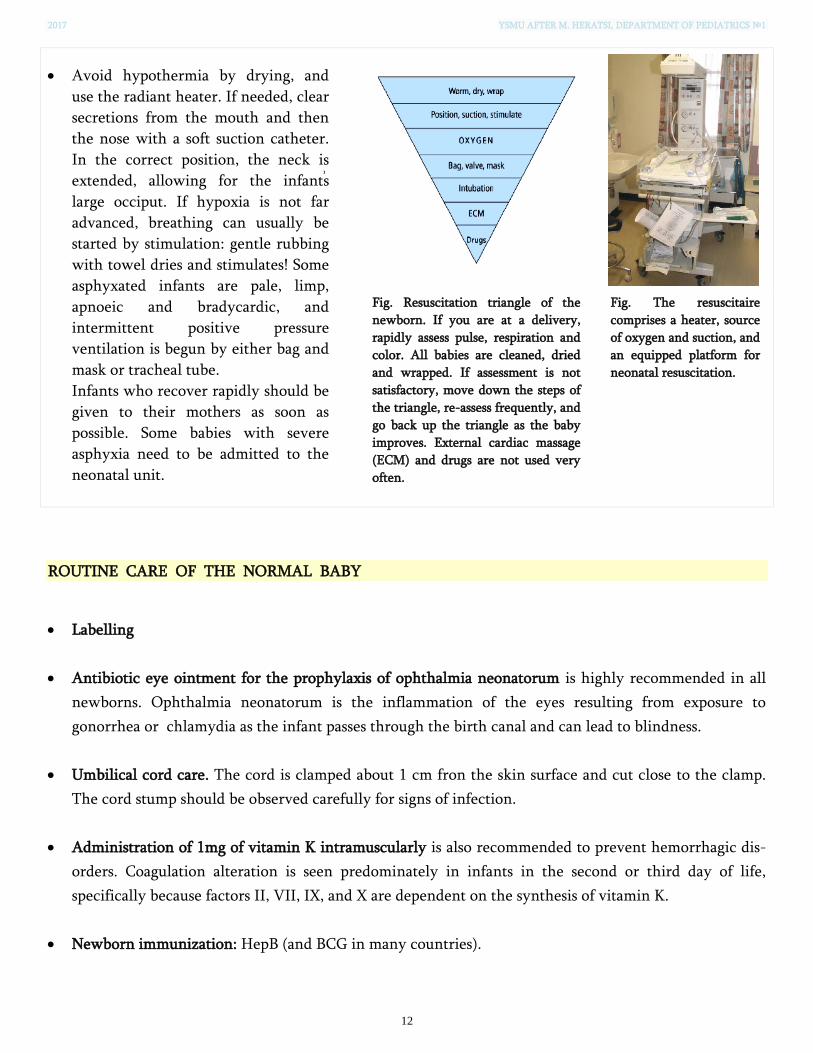
2017 YSMU AFTER M. HERATSI, DEPARTMENT OF PEDIATRICS 1
12
Avoid hypothermia by drying, and
use the radiant heater. If needed, clear
secretions from the mouth and then
the nose with a soft suction catheter.
In the correct position, the neck is
extended, allowing for the infant ՚s
large occiput. If hypoxia is not far
advanced, breathing can usually be
started by stimulation: gentle rubbing
with towel dries and stimulates! Some
asphyxated infants are pale, limp,
apnoeic and bradycardic, and
intermittent positive pressure
ventilation is begun by either bag and
mask or tracheal tube.
Infants who recover rapidly should be
given to their mothers as soon as
possible. Some babies with severe
asphyxia need to be admitted to the
neonatal unit.
Fig. Resuscitation triangle of the
newborn. If you are at a delivery,
rapidly assess pulse, respiration and
color. All babies are cleaned, dried
and wrapped. If assessment is not
satisfactory, move down the steps of
the triangle, re-assess frequently, and
go back up the triangle as the baby
improves. External cardiac massage
(ECM) and drugs are not used very
often.
Fig. The resuscitaire
comprises a heater, source
of oxygen and suction, and
an equipped platform for
neonatal resuscitation.
ROUTINE CARE OF THE NORMAL BABY
Labelling
Antibiotic eye ointment for the prophylaxis of ophthalmia neonatorum is highly recommended in all
newborns. Ophthalmia neonatorum is the inflammation of the eyes resulting from exposure to
gonorrhea or chlamydia as the infant passes through the birth canal and can lead to blindness.
Umbilical cord care. The cord is clamped about 1 cm fron the skin surface and cut close to the clamp.
The cord stump should be observed carefully for signs of infection.
Administration of 1mg of vitamin K intramuscularly is also recommended to prevent hemorrhagic dis-
orders. Coagulation alteration is seen predominately in infants in the second or third day of life,
specifically because factors II, VII, IX, and X are dependent on the synthesis of vitamin K.
Newborn immunization: HepB (and BCG in many countries).

2017 YSMU AFTER M. HERATSI, DEPARTMENT OF PEDIATRICS 1
13
Bathing. Bathing should be delayed until after 24 hours of birth. If this is not possible due to cultural
reasons, bathing should be delayed for at least six hours. Appropriate clothing of the baby for ambient
temperature is recommended. This means one to two layers of clothes more than adults, and use of
hats/caps. The mother and baby should not be separated and should stay in the same room 24 hours a
day.
Passage of meconium and urine. It is important to note the time of first passing meconium and urine,
and often this occurs at or soon after delivery. Both are usually passed within 24 h of birth, and delay
should prompt a search for underlying pathology.
Feeding. All newborns, including low-birth-weight (LBW) babies who are able to breastfeed, should be
put to the breast as soon as possible after birth when they are clinically stable, and the mother and baby
are ready.
Newborn screening tests
Hearing screening
Pulse oxymetry screening to detect critical congenital heart defect (CHD)
Screening for Developmental Dysplasia of the Hip
Screenings for genetic, metabolic, or endocrine disorders
NEWBORN SCREENING
Hearing screening
Family questionnaires – parents or other caregivers may be asked about
the response of their neonate or infant to sounds and their use of
language, including early indicators of language such as babbling and
other vocalizations.
Behavioural measures – the responses of babies to behavioural measuring
devices (ranging from simple noisemakers to more sophisticated
audiological equipment and procedures) can also be used to identify
hearing loss.
Physiological measures – measures of otoacoustic emissions (OAE) or auditory brainstem response (ABR) have
been shown to be effective methods of screening for hearing loss in neonates and infants. OAE measures are
obtained from the ear canal by using a sensitive microphone within a probe assembly that records cochlear
responses to acoustic stimuli. OAEs measure the status of the peripheral auditory system extending to the cochlear
outer hair cells. ABR measurements are obtained from surface electrodes that record neural activity generated in
the auditory nerve and brainstem in response to acoustic stimuli delivered via an earphone. Screening ABR
measurements are usually automated (AABR) and reflect the status of the peripheral auditory system, the eighth
nerve, and the brainstem auditory pathway.

2017 YSMU AFTER M. HERATSI, DEPARTMENT OF PEDIATRICS 1
14
Examination of the hips
Examination of the hips often causes a baby to cry, and it is therefore best left until last. The examination
aims to determine whether the baby has developmental dysplasia of the hip (DDH), in which the
acetabulum is too shallow and the hip can be dislocated. There are two tests, the Ortolani test and the
Barlow test.
The Ortolani test The Barlow test
Ultrasonography
Because it is superior to radiographs for evaluating cartilaginous structures, ultrasonography is the
diagnostic modality of choice for DDH before the appearance of the femoral head ossific nucleus (4-6 mo).
During the early newborn period (0-4 wk), however, physical examination is preferred over
ultrasonography because there is a high incidence of false-positive sonograms in this age group.
Newborn metabolic screening
• Every newborn before discharge or day 4 of life
• More reliable if done after 48 hours of oral feedings (substrates for
metabolic diseases)
• Total diseases screened are determined by individual states. Some
examples:
− Phenylketonuria
− Tyrosinemia
− 21-hydroxylase deficiency
− Galactosemia
− Hb SS
− Hb C
− Hypothyroidism
− Cystic fibrosis
A 1-month-old fair-haired, fair-skinned baby presents with projectile vomiting of 4 days’
duration. Physical exam reveals a baby with eczema and a musty odor. Which screening test
would most likely be abnormal?

2017 YSMU AFTER M. HERATSI, DEPARTMENT OF PEDIATRICS 1
15
THE BABY CHECK (NEONATAL EXAMINATION)
Every newborn infant should undergo a routine examination in the first few days of life. This is a screening
process to look for congenital conditions and to ensure normal adaptation from fetal life. The baby check is
also called the neonatal examination.
The 6-week check
The examination is usually repeated between 6 and 8 weeks of age to ensure that there are no new
problems and that the child is growing and feeding well. In addition, there are some important problems
that may not have been apparent in the first few days of life for physiological reasons, such as the murmur
of a left-to-right shunt due to a ventricular septal defect.
The structure described here allows for a complete assessment of any congenital or adaptive problems and
will also identify infants at particular risk of some important neonatal problems, such as sepsis.
Maternal history
Check for all maternal factors which can cause
illness in the neonate.
History relating to the fetus
Fetal health
— Any concerns relating to the health of the
fetus, such as threatened miscarriage, poor growth
or breech position
— Reports of the antenatal scans – were any
abnormalities identified?
Labour and delivery
— Check whether the delivery was normal
vaginal, instrumental or Caesarean
— Check whether any resuscitation of the
newborn was required following birth
— Look at the Apgar score.
— See if there are any risk factors for sepsis in the
baby.
Risk factors for sepsis in newborn babies include:
• premature labour (earlier than 37 weeks’ gestation)
• prolonged rupture of membranes (>24 hours)
• maternal group B Streptococcus colonisation (on vaginal swab)
• maternal fever during labour

2016 YSMU AFTER M. HERATSI, DEPARTMENT OF PEDIATRICS 1
16
PHYSICAL EXAMINATION
General appearance Vital signs (VS) and body measurements Examination of individual body parts and
organs
* Conciousness/awakeness
Behavior (calm, irritable)
Crying (loudly, weak)
* Respiratory effort
* Posture at rest
* Movements
* Deformations and
malformations
* Assessment of color
* VS: temperature (T); respiratory rate
(RR); heart rate (HR); capillary refill time
(CRT); blood pressure (BP); oxygen
saturation (SpO2)
* Anthropometry: weight; length; head
circumference (HC); chest circumference
• skin
• skull; fontanelles; sutures
• ears, eyes, nose and mouth
• shoulders, arms and hands
• chest and heart sounds
• abdomen
• groin
• genitalia
• femoral pulses
• legs and feet
• spine
• reflexes
• hips
Examination of the reflexes, together with the hip examination, are best left until the end of the baby
check.
Main features of routine examination of the newborn
REFERENCE
1. Wanda D. Barfield and COMMITTEE ON FETUS AND NEWBORN. Standard Terminology for Fetal, Infant, and Perinatal Deaths. Pediatrics 2016;137. 2. Creasy, Robert K, Robert Resnik, and Jay D. Iams. Creasy and Resnik's Maternal-Fetal Medicine: Principles and Practice. Philadelphia, PA: Saunders/Elsevier, 2014. 3. Hockenberry MJ, Wilson D. Wong’s Nursing Care of Infants and Children. 10th ed. St Louis, MO: Elsevier Mosby; 2015. 4. Gary M. Weiner. Textbook of Neonatal Resuscitation, 7th ed. Published by the American Academy of Pediatrics and American Heart Association 2016. 5. Guidelines on basic newborn resuscitation. World Health Organization 2012. 6. Guidelines for Perinatal Care, 7th ed. By AAP Committee on Fetus and Newborn and ACOG Committee on Obstetric Practice 2012. 7. Anne Lomax. Examination of the Newborn - An Evidence-Based Guide 2ed, 2015. 8. WHO guidelines on maternal, newborn, child and adolescent health approved or under review by the WHO Guidelines Review Committee: recommendations on newborn health. Geneva: World
Health Organization; 2013. 9. Newborn and infant hearing screening: current issues and guiding principles for action. World Health Organization; 2010.

2016 YSMU AFTER M. HERATSI, DEPARTMENT OF PEDIATRICS 1
1
BIRTH (PERINATAL) ASPHYXIA There are few moments in life when humans are as vulnerable and as dependent on the help of others as at birth. In a
few seconds the newborn must adapt from life in the warm, moist, sterile environment of the mother’s womb to wholly
different extrauterine surroundings. Every organ must adjust to this change over a few minutes or a few days; but the key
to immediate survival of the newborn is a smooth cardiopulmonary transition. The neonate must successfully inflate his
or her lungs and rearrange the fetal circulation.
The asphyxiated newborn undergoes an abnormal transition.
CAUSES OF NEWBORN DEPRESSION (delayed onset of regular respiration)
• Perinatal asphyxia
• Excessive suctioning (vagal stimulation causes bradycardia and apnea)
• Trauma, especially to central nervous system (tentorial tears) or cervical cord
• Drugs which depress breathing (anesthetic, narcotic, alcohol, magnesium sulfate, tranquilizers)
• Severe immaturity (surfactant-deficient lung, inadequate respiratory effort)
• Perinatally acquired sepsis: group B streptococci, Listeria monocytogenes
• Primary neuromuscular disease (Werdnig-Hoffmann disease, congenital myopathies, myotonic dystrophy, congenital
muscular dystrophy)
• Congenital malformation obstructing the airway or preventing lung expansion (choanal atresia, Pierre-Robin syndrome,
laryngeal atresia, diaphragmatic hernia, pulmonary hypoplasia)
Intrapartum asphyxia cannot be easily differentiated clinically from neonatal depression. Initial resuscitative measures
are, nevertheless, similar in all situations and should be initiated without delay.
DEFINITION OF PERINATAL ASPHYXIA
The American College of Obstetricians and Gynecologists’ (ACOG) Committee have defined certain criteria that must be
met if neurologic deficits are to be attributed to perinatal asphyxia. All of the characteristics must be present, including
evidence of at least one organ dysfunction. If such evidence is lacking, it is difficult to conclude that perinatal asphyxia is
the cause of later developmental disability.
Essential characteristics of perinatal asphyxia
1) Presence of metabolic or mixed acidosis with a pH < 7.00 in the umbilical artery.
2) An Apgar score of 3 or less for longer than 5 minutes.
3) The presence of neurologic sequelae such as seizures, coma or hypotonia in the immediate neonatal period.
4) Multisystem organ dysfunction in one or more of the following systems: cardiovascular, gastrointestinal, hematologic,
pulmonary, or renal.
There are numerous risk factors that allow a health care worker to anticipate the possibility of the delivery of an
asphyxiated or depressed newborn.
Some of the major antepartum factors associated with this risk include: maternal age > 35 yrs, maternal
diabetes, pregnancy-induced hypertension, fetal anemia, bleeding in the 2nd or 3rd trimester, polyhydramnios,
oligohydramnios, premature rupture of membranes, intra-uterine infection, post-dates gestation, multiple
gestation, small for gestational age or intrauterine growth restriction, maternal substance abuse, fetal
malformations, decreased fetal activity and no prenatal care.
Some of the major intrapartum factors associated with this risk include: emergency cesarean section, breech
presentation, premature labor, prolonged rupture of membranes > 24 hrs, intrauterine infection, precipitous
labor, prolonged labor > 24 hrs, prolonged 2nd stage of labor >2 hrs, nonreassuring fetal heart rate patterns,
general anesthesia, maternal analgesia with narcotics within 4 hrs of delivery, meconium stained amniotic fluid,
prolapsed cord, placenta previa and abruptio placentae.

2016 YSMU AFTER M. HERATSI, DEPARTMENT OF PEDIATRICS 1
2
PATHOPHYSIOLOGY OF ASPHYXIA
In order to undertake the necessary and appropriate measures for resuscitation of the newborn, a firm understanding of
pathophysiologic events that occur during asphyxia is vital.
Five principal mechanisms of asphyxia have been described in the human infant during labor, delivery, and the
immediate postpartum period:
1) interruption of umbilical blood flow (cord compression, prolapse);
2) failure of gas exchange across the placenta (placental insufficiency, abruption, or previa);
3) inadequate perfusion of the maternal side of the placenta (severe maternal hypotension, hypertension from any cause,
or excess uterine contractility);
4) impaired maternal oxygenation (cardiopulmonary disease, anemia);
5) failure of adequate cardiopulmonary adaptation after birth (insufficient lung inflation and faulty rearrangement of the
fetal circulation).
The neonatal brain is highly resistant to hypoxia–ischaemia compared with that of an adult. The degree of hypoxia–
ischaemia necessary to damage the neonatal brain usually leads to impairment of other organs.
Multisystem organ dysfunction occurs from the lack of blood flow to the kidneys, liver and gastrointestinal tract in an
initial attempt to preserve cerebral and cardiac blood flow. As this “dive reflex” begins to fail the cerebral and cardiac
systems soon become affected. Organ failure ensues from the lack of oxygen delivery to the tissues as a consequence
of hypoxemia—not enough oxygen in the blood or from ischemia—inadequate delivery of oxygenated blood to the
tissues from low cardiac output or anemia. Multiple organ systems are affected by perinatal asphyxia with 82% of
neonates incurring injury to one or more organs. These include the brain (72%), kidneys (42%), lungs (26%), heart (29%),
bone marrow (<20%), bowel (29%), and liver (<20%).
Fig. Pathophysiology of hypoxic-ischemic brain injury in the developing brain. During the initial phase of energy failure,
glutamate mediated excitotoxicity and Na+/K+ ATPase failure lead to necrotic cell death. After transient recovery of
cerebral energy metabolism, a secondary phase of apoptotic neuronal death occurs. ROS = Reactive oxygen species.

2016 YSMU AFTER M. HERATSI, DEPARTMENT OF PEDIATRICS 1
3
Brain—Hypoxic Ischemic Encephalopathy (HIE)
Hypoxic ischemic encephalopathy is the neurologic injury that results from the lack of oxygen delivery to the brain.
Evaluating the severity of the neurologic insult from hypoxic ischemic encephalopathy with the criteria from Sarnat is a
very useful tool for prognosis. In the Sarnat Stages, a poor outcome is defined as mental retardation, cerebral palsy,
chronic seizures, or death.
Sarnat Stages of HIE (Arch Neurol 33:696, 1976)
The initial stabilization of an infant with HIE from perinatal asphyxia includes the following steps.
1. Maintain normal ventilation. It is important to avoid low PaCO2 secondary to further risk of cerebral ischemia
from decreased cerebral blood flow.
2. Diagnose and treat seizures. An EEG can help to guide diagnosis, therapy and prognosis. Ativan, phenobarbital
and phenytoin should be considered for treatment of seizures, always watching closely for apnea as a side effect
of therapy.
3. Maintain normal glucose levels. Hypoglycemia can cause neurologic injury and hyperglycemia can worsen
neurologic outcomes after HIE.
4. Maintain normal blood pressure to ensure adequate cerebral blood flow.
5. Obtain head imagining studies (head ultrasound, CT scan or MRI) to rule out intracranial hemorrhage, to detect
cerebral edema or ischemia and to establish a baseline for long-term follow-up.

2016 YSMU AFTER M. HERATSI, DEPARTMENT OF PEDIATRICS 1
4
EXTRACEREBRAL COMPLICATIONS ASSOCIATED WITH PERINATAL ASPHYXIA
COMPLICATIONS MANAGEMENT
Renal Acute kidney injury (AKI) from acute tubular
necrosis (ATN)
Strict monitoring of fluid intake, urine output,
serum levels of electrolytes, pH
Pulmonary Persistent pulmonary hypertension (also
known as persistent fetal circulation -PFC) →
right to left shunt → severe cyanosis and life-threatening hypoxic respiratory failure
Maintaining of adequate oxygenation and
ventilation
Antibiotics
Surfactant replacement therapy
Chest x-ray
Cardiac Cardiac dysfunction → hypotension Cardiogenic shock or heart failure →
pulmonary edema
Rhythm abnormalities
Volume resuscitation with normal saline
Inotropic agents: dopamine, dobutamine
Hydrocortisone
Hematologic Thrombocytopenia and clotting
abnormalities → diffuse intravascular coagulation (DIC); severe bleeding complications: pulmonary hemorrhage, intracranial hemorrhage, hematuria, hematochezia
Anemia
Use of blood products (fresh frozen plasma,
cryoprecipitate or even activated Factor VII for
severe cases of DIC)
Platelet transfusions if platelet count < 50,000
In an emergency it may be necessary to give
uncrossmatched O negative blood to prevent
cardiovascular collapse from an acute
hemorrhage.
Gastrointestinal Liver injury (elevated ALT, AST, bilirubin)
Necrotizing enterocolitis (NEC)
Phototherapy, vitamin K
Parenteral feeding
Metabolic Lactic acidosis
Hypoglycemia
Hypothermia
Hypocalcemia
Hyponatremia
Strict monitoring of body temperature, serum
levels of electrolytes, pH and glucose
REFERENCE
1. Martha Douglas-Escobar; Michael D.Weiss. Hypoxic-Ischemic Encephalopathy A Review for the Clinician. JAMA Pediatr. 2015;169(4):397-
403.
2. Creasy, Robert K, Robert Resnik, and Jay D. Iams. Creasy and Resnik's Maternal-Fetal Medicine: Principles and Practice. Philadelphia, PA:
Saunders/Elsevier, 2014. Print.
3. Gary M. Weiner. Textbook of Neonatal Resuscitation, 7th ed. Published by the American Academy of Pediatrics and American Heart
Association 2016.
4. Guidelines on basic newborn resuscitation. World Health Organization 2012.
5. Guidelines for Perinatal Care, 7th ed. By AAP Committee on Fetus and Newborn and ACOG Committee on Obstetric Practice 2012.

2016 YSMU AFTER M. HERATSI, DEPARTMENT OF PEDIATRICS 1
1
PATHOLOGY OF PREMATURITY
A preterm infant has difficulties with fetal to neonatal transition and maintaining homeostasis i.e.
temperature control, euglycemia, fluid and electrolyte balance. Preterm infants usually exhibit significant
jaundice and profound anemia. Full enteral nutrition is a problem even in healthy a near-term infant.
Internal organs immaturity
Lung
o hyaline membrane disease (HMD)
Intestine
o necrotizing enterocolitis (NEC)
Brain
o intraventricular hemorrhage (IVH)
o periventricular leukomalacia (PVL) - infarction of the periventricular white matter
Cardiovascular
o patent ductus arteriosus (PDA)
Iatrogenic damage
o barotrauma
o pneumothorax
o interstitial lung emphysema
o bronchopulmonary dysplasia (BPD)
o retinopathy of prematurity (ROP) - unmonitored high oxygen supplementation contributes to ROP
Hyaline membrane disease (HMD)
Also known as neonatal respiratory distress syndrome (NRDS). The incidence is inversely proportional to
gestational age.
Neonatal Respiratory Distress Syndrome -
NRDS (Hyaline membrane disease) is
characterized by collapsed alveoli alternating
with hyperaerated alveoli, vascular congestion
and hyaline membranes (resulted from fibrin,
cellular debris, red blood cells). Hyaline
membranes appear like an eosinophilic,
amorphous material, lining or filling the
alveolar spaces and blocking the gases exchange.

2016 YSMU AFTER M. HERATSI, DEPARTMENT OF PEDIATRICS 1
2
Etiology:
Deficiency of pulmonary surfactant.
Vicious cycle: Decreased alveolar surfactant — lungs collapse at end expiration with each breath —
increasing difficulty in breathing — exhaustion — atelectases (airless areas) — hypoxemia —
endothelial and epitethelial damage: hyaline membrane formation — profound hypoxemia, hypercarbia
and acidosis — progressive atelectases, abundant hyaline membranes.
Surfactant synthesized by type II pneumocytes
consists of lecithin, sfingomyelin and surfactant associated proteins
reduces surface tension at the air-liquid barrier in the alveoli
produced by 20 to 24 weeks of gestation and is secreted into the fetal airways by 30 weeks
- produced in considerable amounts after 35 weeks of gestation but modulation by variety of stimuli is possible (hormones, intrauterine stress including natural labor)
Clinical signs:
Signs of respiratory distress: tachypnea, cyanosis, intercostal and subcostal recession, nasal flaring,
grunting, rapid increase in oxygen requirement
Prevention of HMD
o administration of surfactant (prophylactic or treatment of symptoms)
o the incidence of HMD has been reduced by 50% with the use of antenatal corticosteroids to
promote lung maturity. The corticosteroids are administered to pregnant women with
threatened premature delivery at 24 – 34 weeks of gestation
Fig. The classic radiographic findings of RDS
include diffuse symmetric reticulogranular
densities, prominent central air bronchograms
and generalized hypoventilation. Neonatal
pneumonia can have a similar appearance. The
classic findings may not be present because of the
early intervention with surfactant and ventilatory
support with intubation.

2016 YSMU AFTER M. HERATSI, DEPARTMENT OF PEDIATRICS 1
3
Bronchopulmonary dysplasia (BPD)
BPD is a chronic lung disease that occurs in infants who received respiratory support with mechanical
ventilation and prolonged oxygenation. It is seen in babies recovering form respiratory distress syndrome,
sepsis or prolonged apnea and most babies who develop BPD nowadays have birthweights below 1000 g.
Etiology:
extreme lung immaturity
hyperoxia injury
barotrauma
patent ductus arteriosus
fluid overload
fetal inflammatory response — antenatal exposure to proinflammatory cytokines as found in
choriamnionitis or funisitis effects pulmonary developement and contributes to the developement of
BPD. There is also strong association of BPD and postnatal sepsis.
Classic BPD
Originally described in 1967 as severe lung injury caused by oxygen toxicity and barotrauma with prolonged aggresive
ventilation in the treatment of RDS. Its etiopathogenesis was abnormal reparative process in response to injury and
inflammation. There was a progress from an acute exsudative phase of acinar injury to reparative and chronic fibroproliferative
phase. The histopathologic findings originally reported were airway epithelial lesions, smooth muscular hyperplasia, extensive
peribronchiolar and instersticial fibrosis, focal hemorrhage, areas of overdistension and atelectasis and hypertensive vascular
disease.
New BPD
In recent years with gentler ventilation techniques, antental glucocoricoid therapy and surfactant therapy the histologic changes
seen in infants differ. New BPD is characterised by decrease in alveolar number (enlarged simplified alveoli), abnormal
microvasculature and interstitium with less prominent celularity and fibroproliferation. The current view is that new BPD is
caused by interruption of normal developemental pathways for terminal maturation and alveolarization of lungs of very preterm
infants. The maximum rate of accretion of alveoli is seen in a period from 25 weeks to 4 months after birth.
Clinical signs:
there is delayed resolution of NRDS
classified as mild, moderate or severe depending
on the need for supplemental oxygen and positive
pressure ventilation
most patients with BPD survive
increased risk for repated and serious pulmonary
infections and asthma during childhood
poor growth and psychomotoric delay is a frequent
problem
severe BPD is complicated by cor pulmonale and
secondary pulmonary hypertension
Fig. Chest X-ray of bronchopulmonary dysp.lasia (BPD)
showing fibrosis and lung collapse, cystic changes and
overdistension of the lungs

2016 YSMU AFTER M. HERATSI, DEPARTMENT OF PEDIATRICS 1
- 1 -
NEONATAL INFECTIONS
CONGENITAL INFECTIONS (TORCH)
The most common congenital infections are described by the acronym TORCH:
T Toxoplasmosis
O Other: syphilis, hepatitis B, varicella zoster virus (VZV), human immunodeficiency virus (HIV), listeria
monocytogenes, parvovirus B19, enteroviruses, lymphocytic choriomeningitic virus, zika virus …
R Rubella
C Cytomegalovirus (CMV)
H Herpes simplex virus (HSV)
The infection can be transmitted during intrauterine life via the placenta or by contamination of amniotic
fluid via the cervix or during delivery via the birth canal.
Infections acquired in the first two trimesters of pregnancy generally result in malformations, while
infections occurring during the third trimester cause destructive lesions. However, because the
infection can persist over time, it is possible to observe a heterogeneous spectrum of abnormalities,
which include both malformations and destructive lesions.
Common Features
While each of the congenital infections possesses distinct clinical manifestations and sequelae, some of
these infections share characteristics.
It is important to think of one or more of these infections when a neonate presents with:

2016 YSMU AFTER M. HERATSI, DEPARTMENT OF PEDIATRICS 1
- 2 -
Fig. One-day-old term infant who exhibits cholestatic
jaundice and “blueberry muffin” spots consistent with
extramedullary (dermal) hematopoiesis after
congenital human cytomegalovirus infection
Table. Clinical Manifestations of Common Congenital Infections
However, many congenital infections may be silent at birth (asymptomatic), with symptoms manifesting
years later (e.g. hearing loss, chorioretinitis and visual impairment, or diminished mental and motor
capabilities).
Prenatal imaging is a valuable tool for the assessment of brain damage in cases of diagnosed fetal
infection or it may lead to the suspicion of an infection in those cases with suggestive findings in the
absence of infection history. Apart from CNS involvement, different unspecific signs of a fetal infection can
be detected by fetal MR. They usually are diffuse plancental thickening, oligohydramnios, fetal ascites or
hydrops, hepatomegaly or cardiomegaly, and pericardial or peritoneal effusion.
TREATMRNT CMV Gancyclovir; Foscarnet
Congenital Toxoplasmosis A combination treatment with pyrimethamine-sulfadiazine and folinic acid for
about 1 year
Congenital Rubella Syndrome There is no specific treatment for congenital rubella infection. Care involves
supportive treatment. Heart defects often can be corrected surgically, but damage
to the nervous system is permanent.
HSV Acyclovir
REFERENCE
1. Del Pizzo, J. (2011). Focus on diagnosis: congenital infections (TORCH).Pediatrics in review/American Academy of Pediatrics, 32(12), 537.
2. Neu, N., Duchon, J., & Zachariah, P. (2015). TORCH infections. Clinics in perinatology, 42(1), 77-103.
3. Triulzi, F., Doneda, C., Parazzini, C., & Righini, A. (2016). Congenital Infections. In Perinatal Neuroradiology (pp. 221-235). Springer Milan.

2016 YSMU AFTER M. HERATSI, DEPARTMENT OF PEDIATRICS 1
- 3 -
LOCALIZED BACTERIAL INFECTIONS IN NEWBORNS
1. Omphalitis
The umbilical cord may become colonized by a variety of potentially
pathogenic bacteria mainly Staph, Streptococci and E. coli.
Occasionally infection of the umbilical cord becomes disseminated,
either by blood stream or by direct spread via the umbilical vessels
to the peritoneal cavity. Tetanus, diphtheria and necrotizing fasciitis
may also occur as complications of umbilical infection.
Such infections are still responsible for a high proportion of deaths
in the neonatal period in developing countries.
CLINICAL FEATURES
The skin lesion presents with cellulitis where the skin around the umbilical area becomes indurated.
Bullae may appear in the center of the indurated area followed by necrosis.
Purpura: Purpuric reactions are common manifestations of omphalitis and may be related to bacterial
toxins.
TREATMENT
The most important aspect of treatment is early surgical excision of necrotic tissue.
The use of Hexachlorophene as an antiseptic was popular until it became apparent that this could lead to
serious neurotoxicity, particularly in the preterm infant. The best substitute may be Chlorhexidine that
can be applied as a dusting powder rather than as an alcoholic solution. Antibiotics are of limited value.
2. Staphylococcal pustulosis
Staphylococcal pustulosis manifests on any area of skin as a papulopustular rash that may coalesce and
form bullae. Physicians should obtain a Gram stain and culture of any pustular lesions in a newborn.
3. Bullous impetigo
Bullous Impetigo is a cutaneous condition that characteristically occurs
in the newborn, and is caused by a bacterial infection, presenting
with bullae. It can be caused by Exfoliative toxin A. The pyogenic
superficial infection can be divided into two other
subdivisions; Impetigo, and non-bullous impetigo. Bullous impetigo is
caused by Staphylococcus aureus, which produces exfoliative toxins,
whereas non-bullous impetigo is caused by either Staphylococcus
aureus, or Streptococcus pyogenes. Bullous Impetigo can cause deaths
in fewer than 3% of infected children, but up to a 60% death rate in
adults. 30% of all Impetigo cases are related to Bullous impetigo.
4. Staphylococcal scalded skin syndrome
(SSSS) or Ritter disease
SSSS is a generalized form of bullous impetigo.
Children are more at risk because of lack of
immunity and immature renal clearance capability
(exfoliative toxins are renally excreted).

2016 YSMU AFTER M. HERATSI, DEPARTMENT OF PEDIATRICS 1
- 4 -
5. Periporitis or pseudofurunculosis
(sweat gland abscesses)
Periporitis is the term applied to pustular lesions appearing in the
neonatal skin as a result of secondary infection of sweat glands by
staphylococcus aureus.
The commonest sites involved are the buttocks, upper part of the
trunk and the scalp. The lesion affects mainly malnourished infants
and young children. Skin lesions may progress to sweat gland
abscesses.
6. Neonatal mastitis
Influx of maternal hormones through the placenta into the fetal
circulation often causes the newborn's breasts to be enlarged. In
addition, some secretion (ie, witch's milk) may be evident. These
changes disappear with time.
Mastitis neonatorum or infections of the breast tissue may also occur
during the newborn period. Treatment includes antibiotics. If an
abscess occurs, needle aspiration should be performed. Surgical
drainage should be considered only when needle aspiration is
unsuccessful, because an operation may damage the breast bud and
result in reduction of adult breast size.
7. Dacryocystitis
Neonatal dacryocystitis is a special type of dacryocystitis that occurs
in less than 1% of all newborns. The onset is usually acute, and the
neonate has a swollen mass in the inferior medial canthal area. Often,
there is tearing and a mucopurulent discharge. Significant morbidity
and even mortality can be associated with this condition. However,
the usual course is more indolent with chronic tearing, mattering,
failure to thrive, and even amblyopia. Due to anatomic differences,
blacks are less likely to develop the condition. Although incomplete
canalization of the nasolacrimal duct is often the basis for this
condition, neonatal infection can also be causative.
Untreated lacrimal sac infection may result in orbital cellulitis. In
addition to antibiotics, probing and irrigation are required.
8. Neonatal acute
osteomyelitis
In both neonates and older children, osteomyelitis due to bacteremia
is much more common than that due to spread from contiguous
infections; in the neonate, almost all cases arise following bacteremic
spread of infection. Accordingly, factors that predispose to
bacteremia have also been identified as risk factors for osteomyelitis.
In preterm neonates, umbilical catheterization is associated with a
higher incidence of bone infection. The hips, knees, and neighboring
bones are most commonly involved. Urinary tract infection,
periumbilical skin infections, and venous cut-downs for intravenous
access have also been identified as risk factors in various
retrospective studies.
Most bone infections in neonates are localized to the metaphysis of long bones. The femur and tibia are
the most frequently involved bones, accounting for about 50% of all cases. The humerus and the fibula
are the next most commonly infected long bones. The predilection for infection in the metaphysis is
explained by the developing circulatory patterns near the ends of the bones.
DIAGNOSIS:
1) XRAY (Bone changes usually not present until > 1 week)
2) Bone scans
3) MRI
4) Aspiration

2016 YSMU AFTER M. HERATSI, DEPARTMENT OF PEDIATRICS 1
- 5 -
NEONATAL SEPSIS
The Third International Consensus Definitions for Sepsis (2016) Sepsis is defined as life-threatening organ dysfunction caused by a dysregulated host response to
infection.
In lay terms, sepsis is a life-threatening condition that arises when the body ՚s response to an infection
injures its own tissues and organs.
Newborn infants are at much higher risk for developing sepsis than children and adults. Prematurely
born neonates experience the highest incidence and mortality of sepsis among all age groups.
The overall incidence of neonatal sepsis ranges from one to five cases per 1000 live births. The
estimated incidence is lower in term infants, with a reported rate of one to two cases per 1000 live
births.
Neonates are more susceptible for developing sepsis for 3 reasons:
Potential risk factors of transition period
Immature immune response
Genetic predisposition
Risk Factors for Neonatal Sepsis
Prenatal/maternal Intrapartum Neonatal
Poor prenatal care
Poor nutrition
Substance abuse
Prolonged rupture of membranes (>12 to
18 hours)
Maternal fever
Prolonged labor
Colonization with Group B Streptococcus
(GBS)
Maternal infections, eg. maternal UTI
Low birth weight
Prematurity
Birth asphyxia
Meconium staining
Resuscitation
endotracheal tubes or
indwelling catheters
Genetic Predisposition to Sepsis
The body’s first response to infection requires recognition of the presence of a pathogen. After
recognition has occurred the body responds appropriately to resolve the problem. Many polymorphisms
have been recognized within both of these phases and they have been implicated in influencing the
susceptibility to and/or outcome from sepsis.

2016 YSMU AFTER M. HERATSI, DEPARTMENT OF PEDIATRICS 1
- 6 -
Let’s look further into these two phases to see the effect polymorphisms have on neonatal sepsis:
Recognition Phase Response Phase
The body’s initial response to infection requires
recognition of the presence of a pathogen.
Polymorphisms in genes coding for proteins
involved in the recognition of pathogens can
influence the susceptibility to and/or outcome of
neonatal sepsis.
Let’s look into two of these:
After the initial recognition of a pathogen occurs
the body responds by releasing elevated levels of
proinflammatory cytokines followed by a release
of anti-inflammatory cytokines. This dual release
of opposite cytokines helps the cytokines return to
a baseline level and that enables the start of tissue
repair to start.
It is generally accepted that an imbalance between
proinflammatory and anti-inflammatory cytokines
result in clinical manifestations of sepsis. This
Imbalance is due to polymorphisms in various
proteins involved in the response to pathogens.
Let’s look into two of these :
Lipopolysaccharide
(LPS)
Mannose-Binding Lectine
(MBL)
LPS, a major component of bacteria, is a
powerful stimulator of the innate immune
response. LPS elicits it’s response by binding to
a cell surface receptor that is compromised of 3
proteins. One of these proteins is TLR4. TLR4 is
required for LPS to respond. When there are
polymorphisms in TLR4 there is a reduced
response to LPS and that enhances the
susceptibility to infection!
MBL has two primary immunodefensive roles:
– involved with opsonization
– leads to activation of complement system,
independent of antibodies.
Polymorphisms cause deficiencies in MBL level.
This results in decreased levels of MBL. This
deficiency is associated with increased
susceptibility to infections!
Tumor Necrosis
Factor (TNF) Interleukin 10
(IL-10)
TNF is a proinflammatory cytokine that is
responsible for the initial activation of the
inflammatory response. There are several
polymorphisms associated with an increased
secretion of TNF resulting in the susceptibility to
sepsis.
IL-10 is an anti-inflammatory cytokine produced
by primarily monocytes. IL-10 helps regulate the
over expression of proinflammatory cytokines.
There are three polymorphisms noted that result in
an over expression of IL-10. This over expression
is proposed to induce immunosuppression in
bacterial sepsis and therefore increasing mortality
by inhibiting bacterial clearance.
Disseminated intravascular coagulation (DIC) development in sepsis
Pathogen enters the body
Inflammatory mediators released (cytokines)
Inflammatory mediators
released (cytokines)
Endothelial injury
Inflammatory mediators
released (cytokines)
Tissue factors released
Inflammatory mediators
released (cytokines)
Production of thrombin
Clot formation
Increased activity of fibrinolysis
inhibitors
Decreased fibrinolysis
athogen enters body
DIC
Inflammatory mediators
released (cytokines)

2016 YSMU AFTER M. HERATSI, DEPARTMENT OF PEDIATRICS 1
- 7 -
Early versus Late Onset Neonatal Sepsis
Characteristics EOS LOS
Routes of
acquisition
Typically acquired during the intrapartum period, often from
organisms in the maternal genital tract
Postnatal environment (nosocomial
or community-acquired infections)
Onset Occurs within the first few days of life:
85% of newborns with early onset sepsis begin to show
symptoms within the first 24 hours of life, 5% have symptoms
between 24 and 48 hours, a very small percentage of infants
show symptoms between 48 hours and 6 days of life.
Premature infants have the most rapid onset.
Similar to early-onset sepsis, there is
variability in its definition ranging
from an onset at >72 hours or ≥7
days of age
Presentation A sudden onset and rapid progression to septic shock often
characterize these infections. The main clinical feature of a
newborn with early onset sepsis is respiratory distress, which
often presents as mild nasal flaring and tachypnea. The
respiratory distress can quickly progress to multisystem failure
and shock if not caught early.
Sepsis and septic shock may manifest as respiratory distress
by causing acute respiratory distress syndrome [ARDS],
hypovolemic stimulation of baroreceptors, stimulation of
respiratory centers by cytokines, and lactic acidosis.
The clinical manifestations may be
acute physiological deterioration or
manifestations of a more localized
infection that has progressed to
sepsis.
Mortality
Rate
up to 50% 10% to20%
Common
causative
organisms
GBS
E. coli
Gram-positive bacteria:
Coagulase-negative Staphylococci
(CoNS)
Methicillin-resistant Staphylococcus
aureus (MRSA)
Gram-negative bacteria:
E. coli, Klebsiella, Pseudomonas,
Enterobacter
Fungal organisms: Candida
Initial
Empirical
Therapy
Ampicillin (or amoxycillin) + gentamicin
Rare – third-gen. cephalosporins
Vancomycin
Aminoglycosides
Aminopenicillins
Third-gen. cephalosporins (should be
discouraged outside of suspected
meningitis)
How is Neonatal Sepsis Diagnosed?
There is no definite marker in neonatal sepsis, but there are determinants of organ dysfunction, infection and
SIRS (systemic inflammatory response syndrome).
Septic work-up:
* Complete Blood Count (CBC)
* Blood & Urine cultures
* Lumbar Puncture (LP)
* Chest X-Ray
* Line cultures

2016 YSMU AFTER M. HERATSI, DEPARTMENT OF PEDIATRICS 1
- 8 -
INFECTION - a suspected or proven (by positive culture, tissue stain, or polymerase chain reaction
test) infection caused by any pathogen OR a clinical syndrome associated with a high probability of
infection. Evidence of infection includes positive findings on clinical exam, imaging, or laboratory tests
(e.g., white blood cells in a normally sterile body fluid, perforated viscus, chest radiograph consistent
with pneumonia, petechial or purpuric rash, or purpura fulminans).
Infection can be associated with SIRS.
SIRS was proposed by the American College of Chest Physicians and Society of Critical Care Medicine to describe the
nonspecific inflammatory process occurring in adults after trauma, infection, burns, pancreatitis, and other diseases.
The SIRS criteria were developed for use in adults and therefore contain a number of clinical signs and laboratory
values specific to adults. A number of modifications of these criteria for a pediatric population can be found in the
literature.
The major difference in the definition of SIRS between adults and children is that the diagnosis of pediatric SIRS requires
that temperature or leukocyte abnormalities be present (i.e., SIRS should not be diagnosed if a pediatric patient exhibits
only elevated heart and respiratory rates). In addition, numeric values for each criterion need to be modified to account
for the different physiology of children. Finally, bradycardia may be a sign of SIRS in the newborn age group but not
in older children (in whom it is a near terminal event).
SIRS CRITERIA
The presence of at least two of the following four criteria, one of which must be abnormal temperature or
leukocyte count:
Core temperature of > 38.5°C or < 36°C.
Tachycardia, defined as a mean heart rate > 2 SD above normal for age in the absence of external stimulus, chronic
drugs, or painful stimuli; or otherwise unexplained persistent elevation over a 0.5- to 4-hr time period OR for
children <1 yr old: bradycardia, defined as a mean heart rate <10th percentile for age in the absence of
external vagal stimulus, β-blocker drugs, or congenital heart disease; or otherwise unexplained persistent
depression over a 0.5-hr time period.
Mean respiratory rate > 2 SD above normal for age or mechanical ventilation for an acute process not related to
underlying neuromuscular disease or the receipt of general anesthesia.
Leukocyte count elevated or depressed for age (not secondary to chemotherapy-induced leukopenia) or >10%
immature neutrophils.
SEPSIS is now defined as life-threatening organ dysfunction associated with a dysregulated host
response to infection.
• The original conceptualization of sepsis as infection with at least 2 of the 4 SIRS criteria focused solely on
inflammatory excess. However, the validity of SIRS as a descriptor of sepsis pathobiology has been challenged.
Sepsis is now recognized to involve early activation of both pro- and anti-inflammatory responses, along with
major modifications in nonimmunologic pathways such as cardiovascular, neuronal, autonomic, hormonal,
bioenergetic, metabolic, and coagulation, all of which have prognostic significance. In addition, 1 in 8 patients
admitted to critical care units in Australia and New Zealand with infection and new organ failure did not have the
requisite minimum of 2 SIRS criteria to fulfill the definition of sepsis (poor concurrent validity) yet had protracted
courses with significant morbidity and mortality.
• Nonspecific SIRS criteria such as pyrexia or neutrophilia will continue to aid in the general diagnosis of
infection. These findings complement features of specific infections (eg, rash, lung consolidation, dysuria,
peritonitis) that focus attention toward the likely anatomical source and infecting organism. However, SIRS may
simply reflect an appropriate host response that is frequently adaptive. Sepsis involves organ dysfunction,
indicating a pathobiology more complex than infection plus an accompanying inflammatory response alone.
• Sepsis-induced organ dysfunction may be occult; therefore, its presence should be considered in any patient
presenting with infection. Conversely, unrecognized infection may be the cause of new-onset organ dysfunction.
Any unexplained organ dysfunction should thus raise the possibility of underlying infection.
• Specific infections may result in local organ dysfunction without generating a dysregulated systemic host
response.
SEPTIC SHOCK Septic shock is a subset of sepsis in which underlying circulatory and cellular/metabolic abnormalities
are profound enough to substantially increase mortality. Patients with septic shock can be identified
with a clinical construct of sepsis with persisting hypotension requiring vasopressors and having
hyperlactatemia despite adequate volume resuscitation.

2016 YSMU AFTER M. HERATSI, DEPARTMENT OF PEDIATRICS 1
- 9 -
Organ Dysfunction Criteria in Children
Cardiovascular dysfunction
Despite administration of isotonic intravenous fluid bolus ≥40 mL/kg in 1 hr
Decrease in BP (hypotension) <5th percentile for age or systolic BP <2 SD below normal for age OR
Need for vasoactive drug to maintain BP in normal range (dopamine >5 mcg/kg/min or dobutamine,
epinephrine, or norepinephrine at any dose) OR
Two of the following
Unexplained metabolic acidosis: base deficit > 5.0 mEq/L
Increased arterial lactate >2 times upper limit of normal
Oliguria: urine output <0.5 mL/kg/hr
Prolonged capillary refill: >5 secs
Core to peripheral temperature gap >3°C
Respiratory
PaO2/FIO2 <300 in absence of cyanotic heart disease or preexisting lung disease OR
PaCO2 >65 torr or 20 mm Hg over baseline PaCO2 OR
Proven need or >50% FIO2 to maintain saturation ≥92% OR
Need for nonelective invasive or noninvasive mechanical ventilationd
Neurologic
Glasgow Coma Score ≤11 OR
Acute change in mental status with a decrease in Glasgow Coma Score ≥3 points from abnormal baseline
Hematologic
Platelet count <80,000/mm3 or a decline of 50% in platelet count from highest value recorded over the
past 3 days (for chronic hematology/oncology patients) OR
International normalized ratio >2
Renal
Serum creatinine ≥2 times upper limit of normal for age or 2-fold increase in baseline creatinine
Hepatic
Total bilirubin ≥4 mg/dL (not applicable for newborn) OR
ALT 2 times upper limit of normal for age
Age-specific Vital Signs and Laboratory Variables
REFERENCE
1. Singer et al. The Third International Consensus Definitions for Sepsis and Septic Shock (Sepsis-3). JAMA. 2016;315(8):801-810. doi:10.1001/jama.2016.0287.
2. Goldstein et al; and the Members of the International Consensus Conference on Pediatric Sepsis. International pediatric sepsis consensus conference:
Definitions for sepsis and organ dysfunction in pediatrics. Pediatr Crit Care Med 2005 Vol. 6, No. 1

2016 YSMU AFTER M. HERATSI, DEPARTMENT OF PEDIATRICS 1
- 10 -
NEONATAL PNEUMONIA
Pneumonia is an important cause of neonatal infection and accounts for significant morbidity and mortality,
especially in developing countries.
Neonatal pneumonia can have early or late onset.
Characteristics Early-onset pneumonia Late-onset pneumonia
Routes of
acquisition Intrauterine aspiration of infected amniotic fluid
Transplacental transmission of organisms from
the mother to the fetus through the placental
circulation
Aspiration during or after birth of infected
amniotic fluid.
Postnatal environment (nosocomial or community-
acquired infections)
Onset Within the first 3 or 7 days of life, mostly within
48 hours
Congenital or intrauterine pneumonia can be
considered a variant of early onset pneumonia
Usually occurs after 7 days of age
Presentation Clinical signs are unspecific and present as respiratory distress of various degree, suspicious appearing
tracheal aspirates, cough, apnea, high or low temperature, poor feeding, abdominal distension, and
lethargy.
Chest x-ray The radiographical appearance may also vary, showing reticulogranular-nodular infiltrates, and bilateral
streaky or hazy lungs.
As small bronchioli tend to collapse there may be compensatory hyperaeration in areas free of
pneumonial infiltration. In addition there may be pleural effusions and/or pneumatocele formation in
more complicated cases. Alveolar patterns with coarse, patchy parenchymal infiltrates, consolidation,
and diffuse granularity are more typical for bacterial infections while parahilar streakiness, diffuse
hazy lungs or reticulo-nodularity are more common in viral disease.
Common
causative
organisms
Bacterial infections
Group B Streptococcus (g +)
Escherichea coli (g-)
Staphylococcus aureus (g+)
Listeria monocytogenes (g+)
Enterococcus (g +)
Ureaplasma urealyticum (g+)
Herpes simplex virus (HSV)
Fungal infections
Predominance of gram positive bacteria including
S. pyogenes, S. aureus, and S. pneumoniae
Citrobacter diversus (assoc. with brain and lung
abscesses)
Bacillus cereus (assoc. with necrotizing
pneumonia in preterm infants)
Chlamydia trachomatis has a long incubation
period and typically is associated with pneumonia
occurring between two and four weeks of age
Viral infections (adenovirus, parainfluenza,
rhinovirus, enteroviruses, influenza, respiratory
syncytial virus [RSV])
Fungal infections
Initial Empirical
Therapy
As pneumonia is often associated with or non distinguishable from bacterial sepsis initial therapy at the
NICU includes broad spectrum intravenous antibiotics according to local protocols.
REFERENCE 1. Barnett ED, Klein JO. Bacterial infections of the respiratory tract. In: Remington JS, Klein JO (eds). Infectious diseases of the fetus
and newborn infant. Philadelphia: WB Saunders, 5th edition 2001: 1006-1018.
2. Nissen MD. Congenital and neonatal pneumonia. Pediatrics Resp. Reviews 2007; 8:195- 203
3. Duke T. Neonatal pneumonia in developing countries. Arch Dis Child Fetal Neonatal Ed.2005; 90: F2011-F219

2016 YSMU AFTER M. HERATSI, DEPARTMENT OF PEDIATRICS 1
- 11 -
NOSOCOMIAL INFECTIONS
Nosocomial infection is defined as an infection acquired in the hospital that was neither present nor incubating
at the time of hospital admission. For most bacterial nosocomial infections, this means that the infection usually becomes evident 48 hours (i.e., the
typical incubation period) or more after admission. However, because the incubation period varies with the type
of pathogen and to some extent with the patient’s underlying condition, each infection must be assessed
individually for evidence that links it to the hospitalization.
Neonates, especially those requiring NICU care, are more susceptible to nosocomial infections. Multiple factors
contribute to this population’s high risk for infection, including prematurity and the related relative
immunodeficiency, use of central venous catheters, ventilator support, use of urinary catheters, receipt of parenteral
nutrition and lipids, and exposure to broadspectrum antimicrobials.
There are two special situations in which an infection is considered nosocomial:
(a) infection that is acquired in the hospital but does not become evidence until after hospital discharge and
(b) infection in a neonate that results from passage through the birth canal.
Some would argue that neonatal infections acquired during vaginal delivery are inevitable and, therefore, should
not be counted as nosocomial. However, these neonatal infections (e.g., group B streptococcal bacteremias with
early onset) are considered nosocomial, they can be identified as maternally acquired, and the analysis of their
incidence can be disseminated to obstetricians for interventional strategies.
Table. Nosocomial Bacterial Infections in Patients in the Neonatal Intensive Care Unit (NICU)
Site of Infection Common Pathogens
Bloodstream/sepsis
CONS
S. aureus
Pseudomonas aeruginosa
Candida sp
Pneumonia CONS
S. aureus
P. aeruginosa
Respiratory syncytial virus
Skin/soft tissue/surgical site CONS S. aureus
Conjunctivitis/Ocular CONS P. aeruginosa
Urinary tract Gram-negative bacilli Enterococci
Endocarditis CONS S. aureus
Central nervous system CONS S. aureus
Osteoarthritis S. aureus Group B streptococci
Infection control practices in the NICU
1. Hand Hygiene Practices
Removal of all rings; no nail polish or false nails
Initial 3-minute scrub to the elbow
A 10-second scrub before and after handling each infant
Use of alcohol-based hand gels before and after handling infant
2. Personal Protective Equipment and Universal Gloving
3. Antisepsis for Central Catheter Insertion
4. Health-care Worker Vaccination (influenza immunization, a tetanus, diphtheria, and acellular pertussis vaccine)
REFERENCE 1. Garner JS, Jarvis WR, Emori TG, Horan TC, Hughes JM: CDC definitions for nosocomial infections, 1988, Am J Infect Control 16:28-40, 1988. 2. Guzman-Cottrill, J. A. (2010). Infection Control Practices in the NICU: What is Evidence-based?. NeoReviews, 11(8), e419-e425.
3. Polin, R. A., & Saiman, L. (2003). Nosocomial infections in the neonatal intensive care unit. NeoReviews, 4(3), e81-e89.
4. Ducel G et al. Prevention of hospital-acquired infections- A practical guide 2nd edition. World Health Organization /Department of Communicable Disease, Surveillance and Response; 2002.

2016 YSMU AFTER M. HERATSI, DEPARTMENT OF PEDIATRICS 1
- 1 -
Jaundice is quite common in neonatal period
60% of term infants and 80% of preterm infants
Visible jaundice from sclera, mucosa and skin
Can cause serious consequences:
[1] Actue phase
• respiratory failure
• bilirubin encephalopathy
• neonatal death
[2] Lifelong neurologic sequelae — kernicterus
• cerebral palsy
• epilepsy
• mental retardation
• auditory and visual disfunctions
Why jaundice is so prevalent in neonatal period ???
Bilirubin metabolism in neonates
Bilirubin is produced in the reticuloendothelial system as the end product of heme catabolism and is
formed through oxidation-reduction reactions. Approximately 75% of bilirubin is derived from
hemoglobin, but degradation of myoglobin, cytochromes, and catalase also contributes.
Case Scenario. A 2-day-old infant is noticed to be jaundiced. He is nursing and stooling well. Indirect
bilirubin is 11.2 mg/dL; direct is 0.4 mg/dL. Physical exam is unremarkable except for visible jaundice.

2016 YSMU AFTER M. HERATSI, DEPARTMENT OF PEDIATRICS 1
- 2 -
In the first oxidation step, biliverdin is formed from heme through the action of heme oxygenase, the
rate-limiting step in the process, releasing iron and carbon monoxide. The iron is conserved for reuse,
whereas carbon monoxide is excreted through the lungs and can be measured in the patient's breath to
quantify bilirubin production.
Next, water-soluble biliverdin is reduced to bilirubin, which, because of the intramolecular hydrogen
bonds, is almost insoluble in water in its most common isomeric form (bilirubin IXα Z,Z).
Because of its hydrophobic nature, unconjugated bilirubin is transported in the plasma tightly bound to
albumin. Binding to other proteins and erythrocytes also occurs, but the physiologic role is probably
limited. Binding of bilirubin to albumin increases postnatally with age and is reduced in infants who
are ill. The presence of endogenous and exogenous binding competitors, such as certain drugs, also
decreases the binding affinity of albumin for bilirubin. A minute fraction of unconjugated bilirubin in
serum is not bound to albumin. This free bilirubin is able to cross lipid-containing membranes,
including the blood-brain barrier, leading to neurotoxicity. In fetal life, free bilirubin crosses the
placenta, apparently by passive diffusion, and excretion of bilirubin from the fetus occurs primarily
through the maternal organism.
When it reaches the liver, bilirubin is transported into liver cells, where it binds to ligandin. Uptake of
bilirubin into hepatocytes increases with increasing ligandin concentrations. Ligandin concentrations
are low at birth but rapidly increase over the first few weeks of life. Ligandin concentrations may be
increased by the administration of pharmacologic agents such as phenobarbital.
Bilirubin is bound to glucuronic acid (conjugated) in the hepatocyte endoplasmic reticulum in a
reaction catalyzed by uridine diphosphoglucuronyltransferase (UDPGT). Monoconjugates are formed
first and predominate in the newborn. Diconjugates appear to be formed at the cell membrane and may
require the presence of the UDPGT tetramer.
Bilirubin conjugation is biologically critical because it transforms a water-insoluble bilirubin molecule
into a water-soluble molecule. Water-solubility allows conjugated bilirubin to be excreted into bile.
UDPGT activity is low at birth but increases to adult values by age 4-8 weeks. In addition, certain drugs
(phenobarbital, dexamethasone, clofibrate) can be administered to increase UDPGT activity.
At birth, the gut is sterile, and normal gut flora takes time to establish. The bacteria in the adult gut
convert conjugated bilirubin to urobilinogen (stercobilinogen) which is then oxidized to stercobilin and
excreted in the stool. In the absence of sufficient bacteria, the bilirubin is de-conjugated by brush
border β-glucuronidase and reabsorbed. This process of re-absorption is called enterohepatic
circulation. It has been suggested that bilirubin uptake in the gut (enterohepatic circulation) is
increased in breast fed babies, possibly as the result of increased levels of epidermal growth factor (EGF)
in breast milk.
Unconjugated (indirect) bilirubin Conjugated (direct) bilirubin
– Fat-soluble
– Is not easily excreted
– Neurotoxic
– Water soluble
– It is mostly excreted in stool and some in the urine
– Nontoxic

2016 YSMU AFTER M. HERATSI, DEPARTMENT OF PEDIATRICS 1
- 3 -
Table. Bilirubin metabolism in neonates.
BILIRUBIN METABOLISM The Metabolic Characteristics of Bilirubin in
Newborns
1. Increased bilirubin production:
2. Insufficient binding between bilirubin and
albumin:
[1] low content of serum albumin —> bilirubin-
albumin complex↓
[2] different degree of acidosis at birth —>
binding affinity↓
3. Deficient bilirubin conjugation in the
hepatocyte:
[1] Low uptake of bilirubin into hepatocytes
[2] Low UDPGT activity at birth
4. Increased enterohepatic circulation of bilirubin
[1] intestinal flora is not fully developed to
convert bilirubin to urobilinogen
[2] abundant β-glucuronidase —> deconjugation of
conjugated bilirubin
[3] Low intestinal motility —> increased absorption
of bilirubin

2016 YSMU AFTER M. HERATSI, DEPARTMENT OF PEDIATRICS 1
- 4 -
NONPATHOLOGIC JAUNDICE
I. Physiologic jaundice
Physiologic jaundice is an exaggerated normal process seen in 60% of term infants, and 80% of premature
infants. It is characterized by unconjugated hyperbilirubinemia that peaks by the third or fourth day of life in
full-term newborns and then steadily declines by 1 week of age. Asian newborns tend to have higher peak
bilirubin concentrations and more prolonged jaundice. Premature infants are more likely to develop jaundice
than full-term babies.
Neonatal physiologic jaundice results from simultaneous occurrence of the following two major phenomena: Bilirubin production is elevated because of increased breakdown of fetal erythrocytes. This is the result of the
shortened lifespan of fetal erythrocytes and the higher erythrocyte mass in neonates. Hepatic excretory capacity is low both because of low concentrations of the binding protein ligandin in the
hepatocytes and because of low activity of glucuronyl transferase (UDPGT), the enzyme responsible for binding bilirubin to glucuronic acid, thus making bilirubin water soluble (conjugation).
II. «Breastfeeding failure jaundice» vs «Breast milk jaundice»
Breast-feeding failure jaundice is caused by insufficient breast milk intake, resulting in inadequate quantities of
bowel movements to remove bilirubin from the body. This can usually be ameliorated by frequent breastfeeding
sessions of sufficient duration to stimulate adequate milk production. Passage of the baby through the vagina
during birth helps stimulate milk production in the mother's body, so infants born by cesarean section are at
higher risk for this condition.
Breast milk jaundice Whereas breast feeding failure jaundice is a mechanical problem, breast milk jaundice is more of a biochemical
problem. The term applies to jaundice in a newborn baby.
First, at birth, the gut is sterile, and normal gut flora takes time to establish. The bacteria in the adult gut
convert conjugated bilirubin to stercobilinogen which is then oxidized to stercobilin and excreted in the stool.
In the absence of sufficient bacteria, the bilirubin is de-conjugated by brush border β-glucuronidase and
reabsorbed. This process of re-absorption is called enterohepatic circulation. It has been suggested that bilirubin
uptake in the gut (enterohepatic circulation) is increased in breast fed babies, possibly as the result of increased
levels of epidermal growth factor (EGF) in breast milk.
Second, the breast-milk of some women contains a metabolite of progesterone called 3-alpha-20-beta
pregnanediol. This substance inhibits the action of the enzyme UDPGT responsible for conjugation and
subsequent excretion of bilirubin. In the newborn liver, activity of glucuronyl transferase is only at 0.1-1% of
adult levels, so conjugation of bilirubin is already reduced. Further inhibition of bilirubin conjugation leads to
increased levels of bilirubin in the blood . However, these results have not been supported by subsequent
studies.
Third, an enzyme in breast milk called lipoprotein lipase produces increased concentration of nonesterified free
fatty acids that inhibit hepatic glucuronyl transferase, which again leads to decreased conjugation and
subsequent excretion of bilirubin.
Despite the advantages of breast feeding, there is a strong association of breast feeding with neonatal
hyperbilirubinemia and thus risk of kernicterus, though this is uncommon. Serum bilirubin levels may reach as
high as 30 mg/dL. Jaundice should be managed either with phototherapy or with exchange blood transfusion as
is needed. Breast feeds however need not be discontinued. The child should be kept well hydrated and extra
feeds given.

2016 YSMU AFTER M. HERATSI, DEPARTMENT OF PEDIATRICS 1
- 5 -
PATHOLOGIC JAUNDICE
Pathologic jaundice usually appears within the first 24 hours after birth and is characterized by a rapidly
rising serum bilirubin concentration (>5 mg/dL per day), prolonged jaundice (>7 to 10 days in a full-term
infant), or an elevated direct bilirubin concentration(>2mg/dL). Conjugated hyperbilirubinemia never has a
physiologic cause and must always be investigated.
PHYSIOLOGIC JAUNDICE VERSUS PATHOLOGIC JAUNDICE
Physiologic Jaundice Pathologic Jaundice
Appears on second to third day of life (term) May appear in first 24 hours of life
Disappears by fifth day of life (term) — 7th Variable
Peaks at second to third day of life Variable
Peak bilirubin <13 mg/dL (term) Unlimited
Rate of bilirubin rise <5 mg/dL/d Usually >5 mg/dL/d
CAUSES OF PATHOLOGIC JAUNDICE
I. UNCONJUGATED HYPERBILIRUBINEMIA
Increased Bilirubin Production Decreased Bilirubin Conjugation Increased Enterohepatic
Circulation
• Blood group incompatibility —
Rh, ABO, and minor blood group
• Red blood cell (RBC) enzyme
abnormalities — glucose-6-phosphate
dehydrogenase (G6PD),
pyruvate kinase, and hexokinase
deficiency
• RBC membrane defects —
hereditary spherocytosis,
elliptocytosis, and pyknocytosis
• Hemoglobinopathies —
α-thalassemia, and sickle cell disease
• Increased RBC load —
cephalohematoma, polycythemia, and
ecchymosis
• Infants of diabetic mothers
• Hormonal deficiency —
hypothyroidism and
panhypopituitarism
• Bilirubin metabolism disorders —
Gilbert syndrome, Crigler–Najjar
(types 1 and 2), and
Lucey-Driscoll syndrome
• Sepsis — bacterial, viral, and fungal
• Urinary tract infection (UTI)
• Bowel obstruction or ileus
• Pyloric stenosis

2016 YSMU AFTER M. HERATSI, DEPARTMENT OF PEDIATRICS 1
- 6 -
II. CONJUGATED HYPERBILIRUBINEMIA
Conjugated hyperbilirubinemia is defined as a level ≥ 1 mg/dL (≥17.1 µmol/L) if the total bilirubin is < 5
mg/dL (<85.5 µmol/L), or a conjugated fraction that accounts for > 20% of the total bilirubin if the total
bilirubin is > 5 mg/dL (>85.5 µmol/L).
Bile Duct
Abnormalities Infection
Metabolic/Genetic
Disorders Miscellaneous
• Biliary atresia
• Choledochal cyst
• TORCH infections
• Sepsis
• Urinary tract infection
• Galactosemia
• Tyrosinemia
• α 1 -Antitrypsin deficiency
• Alagille syndrome
• Total parenteral nutrition
(TPN)-related cholestasis
• Neonatal hemochromatosis
• Idiopathic neonatal hepatitis
EVALUATION OF A NEWBORN WITH JAUNDICE
CBC, complete blood count; LGA, large for gestational age; SGA, small for gestational age; TORCH, toxoplasmosis, other
infections, rubella, cytomegalovirus, herpes simplex; UA, urinalysis; UTI, urinary tract infection; WBC, white blood cell; UGI, upper gastrointestinal

2016 YSMU AFTER M. HERATSI, DEPARTMENT OF PEDIATRICS 1
- 7 -
DIAGNOSIS
1. Bilirubin Level - In term infants a normal total serum bilirubin (TSB) level is between 1.0 - 10.0 mg/dL
(17.1-171 µmol/L)
• There is NO safe bilirubin level identified (see page 9)
2. Complete Blood Count
• This test will determine if the infant has increased red blood cells in the circulatory system
(polycythemia)
• If an infant has a hematocrit greater than 65% this places that infant at risk for hyperbilirubinemia
3. Reticulocyte Count
• This test measures young non-nucleated red blood cells
• If the reticulocyte count is greater than 5% in the first week of life, this identifies the infant as
trying to replace destroyed red blood cells
4. Blood Groups & Types
ABO grouping and Rh types are confirmed by examining RBCs for presence of blood group antigens and
RBCs and antibodies against these antigens
5. Coombs Test
Direct Coombs Test Indirect Coombs Test
• The direct coombs test is a direct measure of the amount of maternal antibody coating the infant’s red
blood cell. If the antibody is present, the test is positive.
• The indirect coombs test measures the effect of a sample of the infant’s serum (which is thought to
contain maternal antibodies) on unrelated adult RBCs. If the infant’s serum contains antibodies, they will
interact with and coat these adult RBCs (positive test).

2016 YSMU AFTER M. HERATSI, DEPARTMENT OF PEDIATRICS 1
- 8 -
6. G6PD Level
- it can be false-positive in patients who are actively hemolysing. It can therefore only be done several
weeks after a hemolytic episode
7. Albumin Level
• This test indicates the reserve amount of serum albumin available for binding indirect bilirubin
• A normal albumin level in a term infant is between 2.6 - 3.6 g/dL
8. Visual Assessment
• “Visual assessment of jaundice is most accurate when the infant’s skin is blanched with light digital
pressure in a well-lit room”
• “As bilirubin levels rise, the accuracy of visual assessment decreases”
Kramer Zones Showing Progression of Jaundice
• Jaundice proceeds in a cephalopedal progression, meaning jaundice progresses from the head down
to the toes
• This diagram demonstrates what level the bilirubin is at depending on what areas of the infant’s
body is jaundiced
• For example, if the infant was noted to be jaundiced from the head to the neck that would be zone 1
and the bilirubin level would be between 4 – 6 mg/dL
ZONE 1 2 3 4 5
SBR
(mcmol/L) 100 150 200 250 >250
SBR
(mg/dL) 4-6 6-8 8-14 14-19 >19
9. Tests for
- Blood chemistry - UTI - sepsis - thyroid functions - metabolic diseases ……….

2016 YSMU AFTER M. HERATSI, DEPARTMENT OF PEDIATRICS 1
- 9 -
Transcutaneous Bilirubin Measurement
Transcutaneous bilirubinometry (TcB) is a
noninvasive method of measuring total bilirubin
levels. Some concerns have been raised regarding
the accuracy of these readings at levels >15 mg/dL
and in African American and Hispanic
populations. However, this technique has gained
widespread use as a screening tool to help detect
early hyperbilirubinemia in the newborn nursery.
The AAP has recently advocated the use of TcB to assist screening for hyperbilirubinemia in healthy
newborn infants ≥35 weeks’ gestation prior to discharge from the newborn nursery. However, elevated
levels must be confirmed with serum measurements if the TcB value is at 70% of the TSB level
recommended for the use of phototherapy or in the high-intermediate zone on the Bhutani risk nomogram.
Fig. Bhutani risk nomogram [Bhutani VK, Johnson L, Sivieri EM. Predictive ability of a predischarge hour-
specific serum bilirubin for subsequent significant hyperbilirubinemia in healthy term and near-term
neonates. Pediatrics. 1999;103:6–14.]

2016 YSMU AFTER M. HERATSI, DEPARTMENT OF PEDIATRICS 1
- 10 -
Hemolytic Disease of the Fetus and Newborn (HDFN)
Once called red cell isoimmunization in pregnancy, the formation of antibodies to red cell antigens is now
termed red cell alloimmunization. The perinatal consequence of this process is hemolysis and anemia in the
fetus and newborn, or hemolytic disease of the fetus and newborn (HDFN).
Hemolytic disease of the fetus and newborn (HDFN) due to red cell alloimmunization has been a fascinating clinical picture for many centuries. The first report of a condition called hydrops fetalis dates back to 1609, when a French midwife described the delivery of twins. The first twin was hydropic and stillborn and the second suffered from jaundice and subsequently died of kernicterus. These two conditions were not linked until 1932, when Diamond et al. described that hydrops and kernicterus were manifestations of the same disease, which they called erythroblastosis fetalis. However, the exact cause was still unknown. Since 1940, the analyses of Landsteiner and Wiener contributed largely to a better understanding of the pathogenesis of HDFN. In their studies with rhesus monkeys Landsteiner and Wiener observed that agglutination of human red blood cells occurred in the presence of rhesus monkey red cell antiserum, whereas subjects who lacked the antigen on their red cells did not show agglutination.4,7 The authors called this antigen ‘Rhesus’ (Rh) and consequently the Rh-blood group system was born. Since then it has become clear that the most common cause of severe HDFN is ‘Rhesus disease’, resulting from maternal immunization to the Rhesus D (Rh D) antigen.
EPIDEMIOLOGY
The widespread adoption of guidelines for the administration of antenatal and postpartum rhesus immune
globulin (RhIG) has resulted in a marked decline in the prevalence of red cell alloimmunization caused by
the RhD antigen. Cases, however, continue to occur because of maternal sensitization in the first two
trimesters of pregnancy, inadvertent omission of RhIG, and inadequate dosing after delivery when there
has been an excessive fetomaternal hemorrhage.
PATHOGENESIS
It is well established that the fetal-maternal interface is not an absolute barrier,
and there is evidence that considerable trafficking of many types of cells occurs
between the fetus and the mother throughout gestation.
More than 60 different anti–red cell antibodies have been associated with
HDFN.

2016 YSMU AFTER M. HERATSI, DEPARTMENT OF PEDIATRICS 1
- 11 -
In most cases, the antigenic load of a putative antigen on the fetal erythrocytes and erythrocytic precursors
is insufficient to stimulate the maternal immune system. However, in the case of a large fetomaternal
hemorrhage before birth or at delivery, B lymphocyte clones that recognize the foreign red cell antigen are
established. The initial maternal production of IgM is short-lived and is followed by a rapid change to an
IgG response. A human antiglobulin titer can usually be detected by 5 to 16 weeks after the sensitizing
event.
After the initial antigenic exposure, memory B lymphocytes await the appearance of red cells containing
the putative antigen in a subsequent pregnancy. If stimulated by fetal erythrocytes, these B lymphocytes
differentiate into plasma cells that can rapidly proliferate and produce IgG antibodies and an increase in the
maternal titer. Maternal IgG crosses the placenta and attaches to fetal erythrocytes that have expressed the
paternal antigen. These cells are then sequestered by macrophages in the fetal spleen, where they undergo
extravascular hemolysis, producing fetal anemia.
Rh Incompatibility
Rh incompatibility occurs when an Rh-negative mother carries an Rh-positive fetus. The mother must
have had previous sensitization to the Rh antigen, usually via a prior pregnancy, which results in the
production of antibodies directed against the Rh antigen. Rh-positive infants born to Rh-negative mothers
display a wide spectrum of disease, ranging from unaffected (15% to 20%) to severe disease, including
erythroblastosis fetalis, and fetal death (25%).

2016 YSMU AFTER M. HERATSI, DEPARTMENT OF PEDIATRICS 1
- 12 -
PREVENTION
All current rhesus immune globulin (RhIG) products (RhoGAM; HyperRHO S/D; Rhophlac; WinRho-SDF)
are polyclonal antibody products derived from human plasma. The latter two products are purified by ion-
exchange chromatography and can therefore be administered by either the intravenous or the
intramuscular route. All current products undergo micropore filtration to eliminate viral transmission.
A novel polyclonal recombinant antibody known as rozrolimupab has been used in phase one and two
clinical trials and has demonstrated no serious or adverse effects.
All pregnant patients should undergo an antibody screen at the first prenatal visit. If there is no evidence of anti-D alloimmunization in the RhD-negative woman, patients should receive RhIG at 28 weeks’ gestation. The American Association of Blood Banks (AABB) recommends that a repeat antibody screen be obtained before antenatal RhIG administration, even though the incidence of alloimmunization before 28 weeks is very low. Severe maternal sensitization does occasionally occur before 28 weeks, and by not performing the antibody screen, the clinician loses the opportunity to detect a potentially salvageable anemic fetus. It is therefore prudent to repeat the antibody screen.
The RhD antigen is expressed on the fetal red blood cell as early as 38 days after conception. This has led to the recommendation to administer RhIG for early pregnancy events such as spontaneous abortion, elective abortion, threatened abortion, and ectopic pregnancy, where the background rate of subsequent sensitization is 2% to 3%.
RhIG should also be administered for such events as hydatidiform mole, genetic amniocentesis and chorion villus biopsy, fetal death in the second or third trimester, blunt trauma to the abdomen, late amniocentesis, and external cephalic version. In ongoing pregnancies when RhIG is administered in the first or second trimester for one of these indications, a repeat dose should still be given at 28 weeks’ gestation. Alternatively, if the antenatal dose was given in the late second trimester (e.g., at 22 weeks for suspected placental abruption), the dose should be repeated 12 weeks later (i.e., at 34 weeks’ gestation in that example).
RhIG should be administered within 72 hours of delivery if umbilical cord blood typing reveals an RhD-positive infant.
ABO Incompatibility
ABO incompatibility occurs when a mother with type O blood carries a fetus with type A or B blood. This
condition is confined to mothers with type O blood because these women carry anti-A and anti-B IgG
antibodies that cross the placenta. Mothers with type A or type B blood produce mostly IgM antibodies
against their respective antigens, and these IgM antibodies fail to cross the placenta. Because A and B
antigens are common in nature, group O mothers are previously sensitized to these antigens and hemolysis
may occur in the first pregnancy.
Although ABO incompatibility occurs in 15% of all pregnancies, ABO hemolytic disease occurs in less than
5% of ABO-incompatible mother–infant pairs. Hemolysis tends to be less severe than with Rh
incompatibility. The classic presentation is anemia, reticulocytosis, and hyperbilirubinemia occurring in
the first 24 to 72 hours of life.

2016 YSMU AFTER M. HERATSI, DEPARTMENT OF PEDIATRICS 1
- 13 -
TREATMENT
• Phototherapy is treatment of choice
• Encourage frequent feedings
• Intravenous hydration
• Exchange transfusion
1. Phototherapy
In the mid-1950s, Sister Jean at Rochford General Hospital in England noted that infants exposed to
sunlight were less jaundiced in the uncovered skin areas than their nonexposed counterparts.
Phototherapy works by converting indirect bilirubin to lumirubin, a water-soluble compound that
is a more excretable form of bilirubin.
Only certain wavelengths (colors) of light are absorbed by bilirubin; as bilirubin is a yellow
pigment, blue is absorbed more effectively, however, green light is more deeply absorbed into the
skin.
The only true contraindication to phototherapy is congenital porphyria or a family history of
porphyria. Phototherapy in these patients could result in severe blistering and photosensitivity.
2. Frequent Feedings
Encouraging frequent feedings at least eight times per day helps to stimulate intestinal motility and
removal of meconium, thus reducing reabsorption of direct bilirubin into the system.
3. Intravenous Hydration
Intravenous hydration of infants with hyperbilirubinemia was thought to decrease bilirubin levels,
however, unless an infant is dehydrated intravenous hydration is not indicated.
4. Exchange Transfusion
• An exchange transfusion is used only in extreme cases when phototherapy has failed
• The process for an exchange transfusion involves small amounts of blood being removed from the
infant and then replaced with the same amount of donor RBCs and plasma
• The process continues until twice the circulating volume has been replaced
• The exchange replaces ≈87% of the circulating blood volume and decreases the bilirubin level by
≈55%

2016 YSMU AFTER M. HERATSI, DEPARTMENT OF PEDIATRICS 1
- 14 -
OBSTRUCTIVE JAUNDICE
A high conjugated bilirubin, pale stools (lack of stercobilinogen) and dark urine (presence of bilirubin)
indicates an obstructive cause which may be either intrahepatic (e.g. hepatitis) or extrahepatic (e.g.
biliary atresia).
The conjugated component of total bilirubin should always be checked in any baby with jaundice at
more than 2 weeks of age. A delay in the diagnosis of biliary atresia beyond 6 weeks of age reduces the
likelihood of successful surgery for that condition (biliary drainage by a Kasai portoenterostomy).
It can be difficult to distinguish clinically between neonatal hepatitis and biliary atresia, and a
radionucleotide scan (or occasionally a liver biopsy) is usually indicated. Urgent referral to a specialist
paediatric hepatology service is advised.
Fig. Biliary atresia (Triangular cord sign= Echogenic area anterior to right portal vein)
HEPATITIS
Neonatal hepatitis can occur in a wide range of disease processes including congenital infections (e.g.
toxoplasmosis, cytomegalovirus [CMV]) and metabolic diseases (e.g. α-1-antitrypsin deficiency).
What advice should be given to a mother who is breastfeeding her jaundiced baby?
Continue breastfeeding. Evaluate for adequate latching and audible swallowing of milk by the
baby, and assess whether the infant seems to be consoled after feeding.
Use an electric breast pump to facilitate “let-down of milk” and to collect expressed breast milk
for extra supplementation.
Avoid maternal use of opioid analgesics (e.g., Percocet, Tylenol III, and other codeine
preparations) that could have an impact on the newborn’s feeding and stooling.
Identify ways to reduce maternal stress and anxiety to promote lactation.
RERFERENCE
1. Wong RJ, DeSandre GH, Sibley E, et al. Neonatal jaundice and liver disease. In Martin RJ, Fanaroff AA, Walsh MC, editors. Fanaroff and Martin’s neonatal perinatal medicine. Philadelphia: Mosby Elsevier; 2006. p. 1425-26.
2. Wong RJ et al. Neonatal jaundice: bilirubin physiology and clinical chemistry. NeoReviews. 2007; 8 ( 2 ): e58 – e67. 3. Lauer, B. J., & Spector, N. D. (2011). Hyperbilirubinemia in the newborn.Pediatrics in Review-Elk Grove, 32(8), 341. 4. American Academy of Pediatrics Subcommittee on Hyperbilirubinemia. (2004). Management of hyperbilirubinemia in the newborn infant 35 or more
weeks of gestation. Pediatrics, 114(1), 297. 5. Creasy, Robert K, Robert Resnik, and Jay D. Iams. Creasy and Resnik's Maternal-Fetal Medicine: Principles and Practice. Philadelphia, PA:
Saunders/Elsevier, 2014.

2016 YSMU AFTER M. HERATSI, DEPARTMENT OF PEDIATRICS 1
- 1 -
PEDIATRIC EMERGENCIES
Triage
Triage is the process of rapidly screening sick children soon after their arrival in hospital, in order to identify:
– those with emergency signs, who require immediate emergency treatment
– those with priority signs, who should be given priority in the queue so that they can be assessed and treated without delay
– non-urgent cases, who have neither emergency nor priority signs
Emergency signs include:
obstructed or absent breathing
severe respiratory distress
central cyanosis
signs of shock (cold hands, capillary refill
time longer than 3 s, high heart rate with
weak pulse, and low or unmeasurable blood
pressure)
coma (or seriously reduced level of
consciousness)
convulsions
signs of severe dehydration in a child with
diarrhoea (lethargy, sunken eyes, very slow
return after pinching the skin or any two of
these)
Children with these signs require immediate
emergency treatment to avert death.
Priority signs include:
Tiny infant: any sick child aged < 2 months
Temperature: child is very hot
Trauma or other urgent surgical condition
Pallor (severe)
Poisoning (history of)
Pain (severe)
Respiratory distress
Restless, continuously irritable or lethargic
Referral (urgent)
Malnutrition: visible severe wasting
Oedema of both feet
Burns (major)
The priority signs identify children who are at
higher risk of dying. These children should be
assessed without unnecessary delay.
If a child has one or more emergency signs,
don’t spend time looking for priority signs.

2016 YSMU AFTER M. HERATSI, DEPARTMENT OF PEDIATRICS 1
- 2 -
FIRST CHECK FOR EMERGENCY SIGNS IN THREE STEPS:
• Step 1. Check whether there is any airway or breathing
problem; start immediate treatment to restore breathing.
Manage the airway and give oxygen.
• Step 2. Quickly check whether the child is in shock or has
diarrhoea with severe dehydration. Give oxygen and start IV
fluid resuscitation. In trauma, if there is external bleeding,
compress the wound to stop further blood loss.
• Step 3. Quickly determine whether the child is unconscious
or convulsing. Give IV glucose for hypoglycaemia and/or an
anti-convulsant for convulsing.
IF EMERGENCY SIGNS ARE FOUND:
• Call for help from an experienced health professional if
available, but do not delay starting treatment. Stay calm and
work with other health workers who may be required to give
the treatment, because a very sick child may need several
treatments at once. The most experienced health professional
should continue assessing the child, to identify all underlying
problems and prepare a treatment plan.
• Carry out emergency investigations (blood glucose, blood
smear, haemoglobin [Hb]). Send blood for typing and cross-
matching if the child is in shock, appears to be severely
anaemic or is bleeding significantly.
• After giving emergency treatment, proceed immediately to
assessing, diagnosing and treating the underlying problem.
Chart. Stages in the management of a sick child admitted to hospital: key elements

2016 YSMU AFTER M. HERATSI, DEPARTMENT OF PEDIATRICS 1
- 3 -
Assessment of emergency signs
Assess the airway and breathing (A, B)
Does the child’s breathing appear to be obstructed? Look at the chest wall movement, and listen to breath
sounds to determine whether there is poor air movement during breathing. Stridor indicates obstruction.
Is there central cyanosis? Determine whether there is bluish or purplish discoloration of the tongue and the
inside of the mouth.
Is the child breathing? Look and listen to determine whether the child is breathing.
Is there severe respiratory distress? The breathing is very laboured, fast or gasping, with chest indrawing,
nasal flaring, grunting or the use of auxiliary muscles for breathing (head nodding). Child is unable to feed
because of respiratory distress and tires easily.
Assess circulation (for shock) (C)
Children in shock who require bolus fluid resuscitation are lethargic and have cold skin, prolonged capillary refill, fast weak pulse and hypotension.
Check whether the child’s hand is cold. If so, determine whether the child is in shock.
Check whether the capillary refill time is longer than 3 s. Apply pressure to whiten the nail of the thumb or the big toe for 5 s. Determine the time
from the moment of release until total recovery of the pink colour.
If capillary refill is longer than 3 s, check the pulse. Is it weak and fast? If the radial
pulse is strong and not obviously fast, the child is not in shock. If you cannot feel
the radial pulse of an infant (< 1 year old), feel the brachial pulse or, if the infant is
lying down, the femoral pulse. If you cannot feel the radialpulse of a child, feel the
carotid. If the room is very cold, rely on the pulse to determine whether the child is
in shock.
Check whether the systolic blood pressure is low for the child’s age. Shock may be
present with normal blood pressure, but very low blood pressure means the child is
in shock.
Normal blood pressure ranges in infants and children
Assess for coma or convulsions or other abnormal mental status (D)
Is the child in coma? Check the level of consciousness on the ‘AVPU’ scale:
A alert,
V responds to voice,
P responds to pain,
U unconscious.
Is the child convulsing? Are there spasmodic repeated movements in an
unresponsive child?
If the child is not awake and alert, try to rouse the child by talking or shaking the arm. If the child is not alert but responds to voice, he or she is lethargic. If there is no response, ask the mother whether the child has been abnormally sleepy or difficult to wake. Determine whether the child responds to pain or is unresponsive to a painful stimulus. If this is the case, the child is in coma (unconscious) and needs emergency treatment.

2016 YSMU AFTER M. HERATSI, DEPARTMENT OF PEDIATRICS 1
- 4 -
Common Acute and Emergent Problems in Pediatrics Common complaints leading to acute care visits for potential emergencies include:
fever altered mental status seizures respiratory distress (see "The Respiratory System") vomiting, and abdominal pain (see "The Digestive System")
CASE STUDY Questions
1. What are the serious bacterial infections in febrile newborns and infants?
2. What has been the effect of conjugated vaccines against Haemophilus influenzae and Streptococcus
pneumoniae on the incidence of bacteremia and meningitis in febrile newborns and infants?
3. What are the challenges in differentiating between serious and benign febrile illnesses in young
children?
4. What diagnostic studies are recommended in the evaluation of febrile newborns, infants, and
children?
5. When are empiric antibiotics indicated and when should febrile newborns and infants be
hospitalized?
An 8-month-old girl is brought to the emergency
department with a 2-day history of fever and increased
fussiness. She is irritable but consolable by parents. Her
parents believe that her immunizations are up-to-date, but
they do not have the immunization record with them. On
examination, she has a rectal temperature of 39.5°C
(103.1°F). The rest of the physical examination is within
normal limits, and no source for the fever is apparent.
FEVER is the most common reason for a sick child visit. Most fevers are
the result of self-limited viral infections. However, pediatricians need to
be aware of the age-dependent potential for serious bacterial infections
(e.g., urinary tract infections, sepsis, meningitis, pneumonia, dysentery,
osteoarticular infection). During the first 2-3 mo of life, the neonate is at
risk for sepsis caused by pathogens that are uncommon in older children.
These organisms include group B streptococcus, Escherichia coli, Listeria
monocytogenes, and herpes simplex virus. In neonates, the history must
include maternal obstetric information and the patient’s birth history.
Risk factors for sepsis include maternal group B streptococcus
colonization, prematurity, chorioamnionitis, and prolonged rupture of
membranes. If there is a maternal history of sexually transmitted
infections during the pregnancy, the differential diagnosis must be
expanded to include those pathogens. Septic infants can present with
lethargy, poor feeding, grunting respirations, and cool or mottled
extremities, in addition to fever (or hypothermia). Infants with fever,
irritability, and a bulging fontanel should be evaluated for meningitis. As
the infant matures beyond 3 mo of age, the bacterial pathogens that
usually cause bacteremia, sepsis, and meningitis are Streptococcus
pneumoniae, Haemophilus influenzae type b, and Neisseria meningitidis.
Urosepsis secondary to an E. coli urinary tract infection also needs to be
considered. Immunization against some serotypes of S. pneumoniae has
markedly reduced the occurrence of occult bacteremia and serious
infections caused by that organism, as has immunization against H.
influenzae type b. These remain potential concerns in those children not
fully immunized against these pathogens. Other ailments that manifest
with fever include septic arthritis and osteomyelitis, juvenile idiopathic
arthritis, and Kawasaki disease. Children with a septic joint generally
present with only 1 joint that is painful and often have pseudoparalysis of
that joint. In contrast, patients with juvenile rheumatoid arthritis may
present with pain, stiffness, swelling, and warmth of several joints. The
diagnosis of Kawasaki disease should be considered if the patient meets
the diagnostic criteria for this illness although some patients may have an
atypical or incomplete presentation.

2016 YSMU AFTER M. HERATSI, DEPARTMENT OF PEDIATRICS 1
- 5 -
For patients presenting with ALTERED MENTAL STATUS, the
pediatrician should inquire about the presence of other symptoms, such as
fever or headache. Screening questions should be asked regarding feeding
changes, medications in the household, or the possibility of trauma.
Parents will often describe a febrile child as “lethargic,” but further
questioning will reveal a tired-appearing child who interacts
appropriately when the child has defervesced. Febrile patients need to be
differentiated from the lethargic patient who presents with sepsis or
meningitis. Infants with meningitis or sepsis may have a history of
irritability, inconsolability, poor feeding, grunting respirations, seizures,
decreased urine output, and/or color changes such as pallor, mottling, or
cyanosis. Patients with poisonings or inborn errors of metabolism can also
present with lethargy, poor feeding, unusual odors, seizures, and/or
vomiting. Nonaccidental trauma should always be considered in a
lethargic infant. Older children may present with altered mental status as
a result of meningitis/encephalitis, trauma, or ingestions. Children with
meningitis may have a history of fever and complaints of neck pain; other
associated symptoms can include rash, headache, photophobia and/or
vomiting. Children with ingestions can present with other abnormal
neurologic symptoms such as ataxia, slurred speech, seizures, or
characteristic constellations of vital sign changes and other physical
findings (toxidromes).
SEIZURES IN CHILDHOOD
A seizure is an episode of neurologic dysfunction caused by abnormal
neuronal activity that results in a sudden change in behavior, sensory
perception, or motor activity.
Seizure disorder is a general term that is usually used to include any one
of several disorders including epilepsy, febrile seizures, and possibly single
seizures and seizures secondary to metabolic, infectious, or other
etiologies (e.g., hypocalcemia, meningitis).
The term “epilepsy” refers to recurrent, unprovoked seizures from known or unknown causes. The term “ictus” describes the period in which the seizure occurs, and the term “postictal” refers to the period after the seizure has ended but before the patient has returned to his or her baseline mental status. A focal or partial seizure consists of abnormal neuronal firing that is limited to 1 hemisphere or area of the brain and that manifests itself as seizure activity on 1 side of the body or one extremity. These seizures are classified as simple partial if there is no change in mental status or complex partial if there is some degree of impaired consciousness.
A generalized seizure consists of abnormal electrical activity involving both cerebral hemispheres that causes an alteration in mental status. Traditionally, the patient with 30 minutes of continuous seizure activity or a series of seizures without a return to full consciousness is defined as being in status epilepticus (SE). Newer definitions suggest that SE is defined by duration of 5 continuous minutes of generalized seizure activity or 2 or more separate seizure episodes without return to baseline.
Clinical Presentation
1. Motor Changes Repetitive non-purposeful movements
Staring
Lip-smacking
Falling down without cause
Stiffening of any or all extremities
Rhythmic shaking of any or all extremities
. If these motor behaviors can be interrupted by verbal or physical
stimulation, they are not considered seizure activity.
Seizure activity cannot be interrupted with verbal or physical stimulation

2016 YSMU AFTER M. HERATSI, DEPARTMENT OF PEDIATRICS 1
- 6 -
2. Sensory and Autonomic Common sensory symptoms described by a parent or
caregiver may include: feeling nauseous, odd or peculiar;
losing bowel or bladder control; or feeling numb or
tingling anywhere on the body. Also, experiencing odd
smells or sounds may be noted.
3. Consciousness Consciousness is the usual alertness or
responsiveness the child demonstrates.
Parents/caregivers may report or you may observe
the child to have:
Baseline alertness
Diminished level of consciousness
Unresponsive and unconscious
Events That Mimic Seizures
A careful history from a reliable witness is essential in evaluating a child who presents with a reported seizure. Since witnessing a seizure can create a highly
charged emotional environment, it is important to identify characteristics of seizures and consider conditions which may mimic seizures in order to make an
accurate diagnosis. Breath holding, apnea, syncope, and dizziness should be considered as alternative diagnoses. Tics, myoclonus, rigors, and shuddering
can be misinterpreted as changes in motor control. Psychogenic and pseudoseizures are clinically similar to epileptic seizures, but do not result from an
abnormal electrical discharge from the brain.
PEDIATRIC SEIZURES
Febrile seizure (SFS) Acute symptomatic seizures (eg. in meningitis, electrolyte imbalance, hypoglicemia) Unprovoked seizures (UnS) Status epilepticus (SE)

2016 YSMU AFTER M. HERATSI, DEPARTMENT OF PEDIATRICS 1
- 7 -
FEBRILE SEIZURE
Febrile seizures are the most common seizure disorder in childhood, affecting less than five percent of children between the ages of six months and five
years.
Caused by the increase in the core body temperature greater than 100.4F or 38C
Threshold of temperature which may trigger seizures is unique to each individual
Can occur within the first 24 hours of an illness
Can be the first sign of illness in 25 - 50% of patients
Febrile seizures are a benign condition. For a febrile seizure diagnosis to be established, the child must have no other history of seizures, metabolic diseases,
or signs of central nervous system infections, such as meningitis.
Febrile Seizure: Two Types
Simple Febrile Complex Febrile
6 months – 5 years of age
Febrile before, during or after seizure
Generalized seizure lasting less than 15 minutes, and
Occurs once in a 24-hour period
6 months – 5 years of age
Febrile before, during or after seizure
Prolonged (lasting more than 15 minutes),
Focal seizure, or
Occurs more than once in 24 hours
Febrile Seizure: Prehospital Assessment
Assess A,B,C’s
Assess neurological status (D = Disability using AVPU)
Obtain seizure history from a dependable witness:
How long was the seizure?
What did it look like (movements, eye deviation)?
History of previous seizures (child and family)?
Does the child have a current illness/fever?
Any indications of trauma or abuse?
Length of postictal phase?
List current medications
Include any antipyretics given (time and dose)
Febrile Seizure: Prehospital Management
Monitor A, B, C, D’s
Position with C-Spine protection (if trauma)
Follow seizure and aspiration precautions (per protocol)
Physical exam
Check blood glucose
If blood glucose < 60 mg/dL, treat as appropriate

2016 YSMU AFTER M. HERATSI, DEPARTMENT OF PEDIATRICS 1
- 8 -
Simple Febrile Seizure: Diagnostic Testing
(Evidence-based recommendations from the 2011 AAP Subcommittee on Febrile Seizures)
Lumbar Puncture EEG CT/MRI
A lumbar puncture should be performed in any child who presents with a (simple febrile) seizure and a
fever and has meningeal signs and symptoms (e.g., neck stiffness, Kernig and/or Brudzinski signs) or in
any child whose history or examination suggests the presence of meningitis or intracranial infection.
In any infant between 6 and 12 months of age who presents with a (simple febrile) seizure and fever, a
lumbar puncture is an option when:
- the child is considered deficient in Haemophilus influenza type b or Streptococcus pneumoniae immunizations (i.e., has not received scheduled immunizations as recommended) or
- when the immunization status cannot be determined because of an increased risk of bacterial meningitis.
A lumbar puncture is an option in the child who presents with a (simple febrile) seizure and fever and
is pretreated with antibiotics, because antibiotic treatment can mask the signs and symptoms of
meningitis.
Should not be performed in
a neurologically healthy
child.
Results are not predictive of
recurrence or development
of epilepsy
Not
indicated
FEBRILE SEIZURE: TREATMENT
In general, antiepileptic therapy, continuous or intermittent, is not
recommended for children with one or more simple febrile seizures.
Parents should be counseled about the relative risks of recurrence of
febrile seizures and recurrence of epilepsy, educated on how to handle a
seizure acutely, and given emotional support. If the seizure lasts for >5
min, then acute treatment with diazepam, lorazepam, or midazolam is
needed. Rectal diazepam is often prescribed to be given at the time of
recurrence of febrile seizure lasting >5 min. Alternatively, buccal or
intranasal midazolam may be used and is often preferred by parents.
Intravenous benzodiazepines, phenobarbital, phenytoin, or valproate may
be needed in the case of febrile status epilepticus. If the parents are very
anxious concerning their child’s seizures, intermittent oral diazepam can
be given during febrile illnesses (0.33 mg/kg every 8 hr during fever) to
help reduce the risk of seizures in children known to have had febrile
seizures with previous illnesses. Intermittent oral nitrazepam, clobazam,
and clonazepam (0.1 mg/kg/day) have also been used. Other therapies
have included intermittent diazepam prophylaxis (0.5 mg/kg administered
as a rectal suppository every 8 hr), phenobarbital (4-5 mg/kg/day in 1 or 2
divided doses), and valproate (20-30 mg/kg/day in 2 or 3 divided doses). In
the vast majority of cases it is not justified to use these medications owing
to the risk of side effects and lack of demonstrated long-term benefits,
even if the recurrence rate of febrile seizures is expected to be decreased
by these drugs. Other antiepileptic drugs (AEDs) have not been shown to
be effective. Antipyretics can decrease the discomfort of the child but do
not reduce the risk of having a recurrent febrile seizure, probably because
the seizure often occurs as the temperature is rising or falling.

2016 YSMU AFTER M. HERATSI, DEPARTMENT OF PEDIATRICS 1
- 9 -
SIMPLE FEBRILE SEIZURE: FAMILY EDUCATION
Here are some frequently asked questions parents/ caregivers may have prior to discharge:
Is my child brain damaged?
There is no evidence of impact on learning abilities after seizure from SFS.
Will this happen again?
If child is under 12 months of age at time of first seizure, recurrence rate is
50%
If child is greater than 12 months of age at time of first seizure, recurrence
rate is 30%
Most recurrences occur within 6-12 months of the initial febrile seizure
Will my child get epilepsy?
For simple febrile seizures, there is no increased risk of epilepsy
Why not treat for possible seizures or fever?
Anticonvulsants can reduce recurrence. However potential side effects of
medications outweigh the minor risk of recurrence Prophylactic use of antipyretics does not have impact on recurrence
For complex febrile seizures, there is a slight increase in the risk of epilepsy.
Instruct parent/caregivers to prevent injury during a seizure:
Position child while seizing in a side-lying position
Protect head from injury
Loosen tight clothing about the neck
Prevent injury from falls
Reassure child during event
Do not place anything in the child’s mouth
Prior to discharge home…
Educate regarding use of:
Thermometer
Antipyretics for fever management
When to contact 9-1-1 or ambulance
Identify Primary Care Provider for follow-up appointment and stress importance of follow-up

2016 YSMU AFTER M. HERATSI, DEPARTMENT OF PEDIATRICS 1
- 10 -
FIRST UNPROVOKED SEIZURE
A first unprovoked seizure is a seizure that occurs without an immediate
precipitating event in the absence of fever. Occurrence suggests a
potential underlying neurological condition that predisposes the child to
recurrent seizures. An unprovoked seizure may be due to a yet
undiagnosed brain abnormality, genetic or cryptogenic. Predictors of
recurrence include having an abnormal EEG, an abnormal neurologic
exam, and a prolonged first seizure.
Presentation may be consistent with partial seizure, generalized tonic-
clonic or tonic seizure. To be classified as first unprovoked, there must be
no history of previous seizures and no immediate precipitating cause such
as fever, trauma or infection.
Lumbar puncture is only indicated if there are other symptoms that
suggest a diagnosis of meningitis.
If neuroimaging is recommended, MRI is the modality of choice. Due to
the potential need for sedation with MRI, this study is usually done on an
outpatient basis. Children in whom an MRI may be indicated include
those under the age of one year, children with significant cognitive or
motor impairment, or those with unexplained abnormalities on their
neurological exam. Also, consider an MRI if the seizure is focal in nature
and in those with an abnormal EEG.
An emergent CT scan should be considered as part of the Emergency
Department management if the child has significant cognitive or motor
impairment, exhibits a focal deficit not resolving or has not returned to
baseline, or if the MRI is not available.
An EEG should be obtained on all children who have had a nonfebrile
seizure. This can be arranged as an outpatient and should be done in
coordination with a neurologist, preferably a pediatric neurologist, who
will then read the EEG. The EEG results will help predict the risk of
recurrence, classify the seizure type, and influence decisions about further
imaging studies.
Here are some frequently asked questions parents may have prior to
discharge:
How likely is it that my child will have seizures again?
The risk of recurrence relates to the underlying etiology and EEG
results (normal or abnormal). The majority of children who
experience an unprovoked seizure will have few or no
recurrences. Approximately 10% will go on to have additional
seizures regardless of therapy.
Is there a risk of dying from the seizure if we don’t start medication today? Sudden unexpected death is very uncommon (usually related to an
underlying neurologic handicap rather than seizure activity).
There are no studies showing treatment after a first seizure alters
the small risk of sudden death.
REFERENCE
1. Updated guideline: paediatric emergency triage, assessment and treatment. Geneva: World Health Organization; 2016. 2. POCKET BOOK OF Hospital care for children. GUIDELINES FOR THE MANAGEMENT OF COMMON CHILDHOOD ILLNESSES 2nd ed. World Health Organization 2013. 3. Febrile Seizures: Guideline for the Neurodiagnostic Evaluation of the Child With a Simple Febrile Seizure Subcommittee on Febrile Seizures. Pediatrics 127.2 (2011): 389-394.

GRIGORYAN M E YSMU AFTER M. HERATSI, DEPARTMENT OF PEDIATRICS 1
- 1 -
The
Pediatric Cardiovascular System
Grigoryan M E
YEREVAN STATE MEDICAL UNIVERSITY AFTER MKHITAR HERATSI
DEPARTMENT OF PEDIATRICS 1
2017

GRIGORYAN M E YSMU AFTER M. HERATSI, DEPARTMENT OF PEDIATRICS 1
- 2 -
Contents
1. Cardiovascular health and risk in children and adolescents ………………………………………. 4
2. Developmental anatomy and physiology of the cardiovascular system …………………………. 7
3. Cardiovascular assessment in children ………………………………………………………………. 21
4. Common syndromes associated with cardiac lesions in children ………………………………..…
38
5. Heart failure in children ……………………………………………………………………………... 42
COMPETENCIES
You must…
Know Be able to Appreciate
• How children with
cardiovascular diseases present
• The normal and abnormal
development of CVS
• Carry out a good cardiac examination
• Differentiate clinically between innocent and
pathological murmurs
• Take a good history to differentiate syncope
from other fits, faints and funny turns
• That finding a murmur may induce
parental anxiety

GRIGORYAN M E YSMU AFTER M. HERATSI, DEPARTMENT OF PEDIATRICS 1
- 3 -
A 14-year-old girl has a
sudden loss of
consciousness during dental
treatment. What to do?
You are evaluating a 16-year-
old boy for preparticipation
sports screening. What are
the most common causes of
sudden death during
competitive exercise?
A 15-year-old boy presents with
a 10-year history of uncontrolled
DM type 1, hypertension,
dyspnea, generalized edema,
normal ejection fraction, and
low-grade proteinuria. Does he
have heart failure?
A 12-year-old patient presents with premature atrial
contractions on an ECG. There is no significant
murmur and a normal examination. Doppler velocity by
echo is 2 m/s.Would you let him participate in all
sports?

GRIGORYAN M E YSMU AFTER M. HERATSI, DEPARTMENT OF PEDIATRICS 1
- 4 -
1. Cardiovascular Health and Risk in Children and Adolescents
Types of Heart Disease Observed in Children and Adolescents
All heart diseases, known in adults, may occur in children, but the structure of childhood cardiovascular
morbidity is different. For example, congenital heart disease is more common in childhood versus myocardial
infarction commonly seen in adults.
Most cardiac disease in childhood is congenital. Congenital heart disease (CHD) is the type of heart disease that a
baby is born with. In reality, it is a defect, or abnormality of the heart or blood vessels near the heart, and not a
disease, so the term “congenital heart defect” is often used.
Cardiovascular defects occur in 0.8% of live births. However, this estimate does not include:
affected fetuses who die in utero. In autopsy studies, the incidence of congenital heart disease in the fetus
approaches 30/1,000. In patients with diagnosed intrauterine cardiac malformations, 17.5% do not survive to
birth. Of those who do survive to delivery, 30% die before 1 year of life.
relatively “silent” abnormalities, such as a bicuspid aortic valve, small atrial septal defects, or subtly abnormal
mitral valves, which may present as heart disease in adults or may be noted as incidental findings on
postmortem examination.
Two major types of acquired heart disease in children are rheumatic heart disease and Kawasaki disease.
Rheumatic heart disease is the most common acquired heart disease in many countries of the world, especially
in developing countries. It is responsible for about 233,000 deaths annually. The worst affected areas are sub-
Saharan Africa, south-central Asia, the Pacific and indigenous populations of Australia and New Zealand.
Kawasaki disease is a systemic vasculitis, with a predilection for coronary arteries. It is the leading cause of
acquired heart disease among children in developed countries. It predominantly affects children under the age
of 5 years.
Ideal cardiovascular (CV) health* in childhood is related to lower prevalence of cardiovascular disease (CVD)
factors in adulthood (Laitinen TT et al, 2012).
* Ideal CV health was defined as the simultaneous presence of four beneficial health measures that included never having tried smoking, body mass index
<85 percentile, physical activity at goal levels, and diet consistent with current dietary recommendations.
In adults, the increased cardiovascular risk associated with factors such as high blood pressure, elevated lipid levels,
smoking, a sedentary lifestyle, and being overweight is well known; however, it is important to recognize that these
risk factors often develop and begin to detrimentally affect health in childhood (table 1).
Table 1 Known risk factors for CVD (From Expert Panel on Integrated Guidelines for Cardiovascular Health and Risk Reduction in Children and Adolescents. Summary Report, 2012)
Family history
Age
Gender
Nutrition/Diet
Physical inactivity
Tobacco exposure
Blood pressure
Lipids
Overweight (BMI 85th-95th)/Obesity (BMI ≥ 95th)
Diabetes mellitus and other predisposing conditions
(eg. Kawasaki disease, chronic inflammatory diseases,
HIV)
Metabolic syndrome
Inflammatory markers
Perinatal factors
(eg. prematurity, maternal smoking)
Unhealthy behavioral habits begin at a very early age. lack of exercise and a poor diet lead to excess weight and
obesity, which in turn contribute to raised blood pressure, glucose intolerance, hyperlipidemia, atherosclerosis.

GRIGORYAN M E YSMU AFTER M. HERATSI, DEPARTMENT OF PEDIATRICS 1
- 5 -
More than 42 million children under the age
of 5 years are overweight, predisposing them
to the early onset of cardiovascular
conditions. Close to 31 million of these are
living in developing countries (WHO, 2013).
More than 10% of adolescents currently use
tobacco products, with almost 25% of these
children lighting their first cigarette before
the age of 10 (The Global Youth Tobacco Survey).
Pathological changes in the cardiovascular
system, such as the development of fatty
streaks and early atherosclerotic lesions,
occur in children as young as 5 years of age
and that these changes are correlated with
the presence of risk factors (The Korean War study by Enos; The Bogalusa Heart Study; The Pathological Determinants of Atherosclerosis in Youth Study).
For most children, atherosclerotic vascular changes are minor and can be minimized or even prevented with
adherence to a healthy lifestyle. However, in some children, the atherosclerotic process is accelerated because of the
presence of identifiable risk factors (eg, obesity and hypertension) and/or specific diseases that are associated with
premature CVD (eg, diabetes mellitus and Kawasaki disease).
The co-occurrence of the risk factors that increase a patient's chance of developing heart disease and type 2 diabetes is called metabolic syndrome. Among the variety of diagnostic criteria, the most used are that from WHO and that of
NCEP-ATP III (US National Cholesterol Education Program - Adult Treatment Panel III) (table 2).
Table 2 Definition of metabolic syndrome (MS) in children and adolescents (From Silveira LS et al. Metabolic Syndrome: Criteria for Diagnosing in Children and Adolescents. Endocrinol Metab Synd 2: 118, 2013)
WHO NCEP-ATP III
AGE 12-19
MS DIAGNOSIS 3 or more risk factors 3 or more risk factors
RIS
K F
AC
TO
RS
Obesity BMI >95th percentile Waist circumference ≥ 90th percentile
Glycemic
homeostasis
• Hyperinsulinemia
prepubertal (stage 1 Tanner) > 15 mU/L pubertal (stages 2-4 Tanner) > 30 mU/L post-pubertal (stage 5 Tanner) ≥ 20 mU/L • Fasting glucose ≥ 6.1 mM/L
• Glucose intolerance (at 120 min ≥ 7.8 mM/L)
Fasting glucose ≥ 110 mg/dL
Elevated Arterial
Pressure
SBP >95th percentile for age, sex and stature SBP/DBP ≥90th percentile for age and
sex and stature
Dyslipidemia • TG > 105 mg/dL for < 10 years
> 136 mg/dL for ≥ 10 years
• HDL < 35 mg/dL
• Total cholesterol > 95th percentile
• TG ≥ 110 mg/dL
• HDL ≤ 40 mg/dL

GRIGORYAN M E YSMU AFTER M. HERATSI, DEPARTMENT OF PEDIATRICS 1
- 6 -
Pediatric and Young Adult Sudden Cardiac Arrest (SCA)
There are many cardiac disorders predisposing to pediatric and young adult SCA (table 3). Excluding the Sudden Infant Death
Syndrome (SIDS), which has an incidence around 1–1.5/1000 infants, sudden death in a young person is a rare event. The
incidence of SCA in the young is widely debated, ranging from 0.5 to 20 per 100,000 person-years. In comparison, adults
experience SCA at a rate of 135 per 100,000 person-years.
Approximately 20–25% of the deaths occur during sports [Gajewski, Saul, 2010]. SCA ranges from 1:160,000 to 1:300,000 deaths
per year in competitive athletes 12 to 35 years of age [Meyer et al, 2012]. High risk groups include males, black athletes, and
basketball athletes. The most common finding in this cohort at autopsy after sudden cardiac death is autopsy negative sudden
unexplained death - AN−SUD [(Harmon et al, 2015].
A number of risk markers are used to assess the magnitude of risk for sudden death: family history of premature sudden death; extreme LV hypertrophy (>30 mm); nonsustained ventricular tachycardia on Holter electrocardiographic recording; unexplained (not neurally mediated) syncope; and blood pressure decrease or inadequate increase during upright exercise [European Heart Journal (2010) 31, 3084–3093].
Table 3 Cardiac disorders predisposing to pediatric and young adult SCA [From American Academy of Pediatrics policy statement on sudden cardiac arrest in children. Pediatrics 2012;129;e1094]
Structural/functional Electrical Other
1. Hypertrophic cardiomyopathy*
2. Coronary artery anomalies
3. Aortic rupture/Marfan syndrome*
4. Dilated cardiomyopathy or restrictive
cardiomyopathy*
5. Myocarditis
6. Left ventricular outflow tract obstruction
7. Mitral valve prolapse
8. Coronary artery atherosclerotic disease
9. Arrhythmogenic right ventricular cardiomyopathy*
10. Postoperative congenital heart disease
11. Wolff-Parkinson-White (WPW)
syndrome
12. LQTS*
13. Brugada syndrome*
14. Catecholaminergic polymorphic
ventricular
tachycardia*
15. Short QT syndrome*
16. Complete heart block
17. Drugs and stimulants; some
prescription medications
18. Primary pulmonary hypertension*
19. Commotio cordis
* Familial/genetic
Fig. Detailed causes of cardiac arrest
by age group [Meyer et al, 2012]
HCM – hypertrophic cardiomyopathy
DCM – dilated cardiomyopathy
CAD – coronary artery disease

GRIGORYAN M E YSMU AFTER M. HERATSI, DEPARTMENT OF PEDIATRICS 1
- 7 -
2. Developmental Anatomy & Physiology
of the Cardiovascular System
The cardiovascular system (CVS) is the first organ system to form and function in the developing embryo, at around
the third week of gestation.
Congenital heart and vascular anomalies are borne from embryonic derangements that occur early in the
developmental cycle. Several genetic derangements, drugs, alcohol, radiation, viruses, environmental factors,
chemicals, folic acid deficiency, and others play a major role in the generation of cardiovascular anomalies.
Heart Development (Cardiogenesis)
Cardiac progenitor cells arise in the mesodermal layer and have the potential to form the major cell types of the heart
(cardiomyocytes, smooth muscle, and endothelium/endocardium).
I. Formation of Tubular Heart
Heart development begins with the formation of two endocardial tubes which merge to form the tubular heart,
through which blood eventually flows in a cranial direction.
Endothelial tube The mesenchyme surrounding the tube condenses to form the
myoepicardial mantle (the future myocardium). Gelatinous
extracellular matrix called cardiac jelly separates this mantle from
the endothelial heart tube (the future endocardium).
In addition, a series of constrictions (sulci) divide the heart tube
into sections: sinus venosus; primitive common atrium; primitive ventricle; bulbus cordis; and truncus arteriosus.
Cardiac jelly
Myoepicardial
mantle
Primary hert tube
Truncus
arteriosus →
will form the roots and proximal portion of the aorta and pulmonary artery
Bulbus cordis
distal portion (conus cordis) — will form the outflow tracts of both ventricles
proximal portion— will form the RV
Primitive
ventricle → will form the LV
Primitive
atrium → will become the front parts of the left and right atria and their appendages
Sinus
venosus → will develop into the posterior part of the
right atrium, the sinoatrial node and the coronary sinus
20 days 21 days 22 days
The heart is beating at Day 22. Contractions are of myocardic origin and are likened to peristalsis.

GRIGORYAN M E YSMU AFTER M. HERATSI, DEPARTMENT OF PEDIATRICS 1
- 8 -
II. Looping of Heart Tube
The heart tube continues stretching and by day 23 cardiac looping begins. Under normal circumstances the tube
loops to the right (D-loop).The cephalic portion curves in a frontal clockwise direction. The atrial portion starts
moving in a cephalic ally and then moves to the left from its original position. This curved shape approaches the
heart and finishes its growth on day 28.
Cardiac looping represents the first visible sign of left-right asymmetry.
When cardiac looping is abnormal, the ventricles are inverted (L-loop): morphological right ventricle is on the left,
and morphological left ventricle is on the right (situs inversus, heterotaxy).
Fig. Cardiac loop formation.
(A) Straight heart tube or preloop stage
(B) D -loop, with normally related ventricles
(C) L -loop, with inverted (mirror image) ventricles
A, atrium
Ao, aorta
BC, bulbus cordis
LV, morphological left ventricle
RV, morphologic right ventricle
PA, main pulmonary artery
TA, truncus arteriosus
In situs inversus, the position of the visceral organs is reversed. The arrangement is a mirror image of the normal
situs. The left lung is trilobed, and the right has two lobes. Pulmonary venous return is again to the left atrium but
the left atrium is on the right side of the heart.
The majority of patients with ventricular inversion has
visceroatrial heterotaxy (also known as atrial isomerism):
lack of differentiation of right-sided and left-sided organs
during fetal development.
There are two subtypes: Right atrial isomerism (bilateral right-sidedness) is
associated with absence of a spleen (asplenia), two right
atria (RA), two right lungs, midline liver.
Left atrial isomerism (bilateral left-sidedness) is
associated with multiple splenic tissues (polysplenia)
which may or may not be functional, two left atria (LA),
two left lungs, midline liver.
In both cases the ventricles have either concordant or
discordant position.
RIGHT ISOMERISM LEFT ISOMERISM

GRIGORYAN M E YSMU AFTER M. HERATSI, DEPARTMENT OF PEDIATRICS 1
- 9 -
III. Development of Chambered Hearts (Cardiac Septation)
When looping is complete, the external appearance of the heart is similar to that of a
mature heart; internally, the structure resembles a single tube, although it now has several
bulges resulting in the appearance of primitive chambers.
The common atrium (comprising both the right and left atria) is connected to the
primitive ventricle (future left ventricle) via the atrioventricular canal. The primitive
ventricle is connected to the bulbus cordis (future right ventricle) via the bulboventricular
foramen. The distal portion of the bulbus cordis is connected to the truncus arteriosus via
an outlet segment (the conus).
As the linear heart tube loops rightward, the
endocardial cushions of the inflow and outflow tracts
become adjacent to one another. These cushions consist
of protrusions of cardiac jelly. Complete septation of the
atrioventricular canal occurs with fusion of the
endocardial cushions.
Failure of fusion of the superior and inferior endocardial cushions results in
complete atrioventricular canal defect (also referred to as complete
atrioventricular septal defect): the presence of both atrial and ventricular septal
defects with a common AV valve.
Septation of the ventricles begins at about embryonic day 25. A muscular
interventricular septum begins to grow superiorly from the ventricular floor
between the presumptive right and left ventricles. This septum stops short of
the atrioventricular canal, leaving a space called the interventricular foramen
which permits blood from both ventricles to exit via the conus cordis.
Ventricular septal defects (VSD) can occur in any portion of the developing
interventricular septum.
Septation of the atria begins at ≈30 days with growth of the septum primum
downward toward the endocardial cushions. The orifice that remains is the
ostium primum. The endocardial cushions then fuse and, together with the
completed septum primum, divide the atrioventricular canal into right and left
segments.
Before the foramen primum closes, a 2nd opening appears in the posterior portion of the septum primum, the ostium
secundum, and it allows a portion of the fetal venous return to the right atrium to pass across to the left atrium. Finally,
the septum secundum grows downward, just to the right of the septum primum. Together with a flap of the septum
primum, the ostium secundum forms the foramen ovale, through which fetal blood passes from the inferior vena cava to
the left atrium.

GRIGORYAN M E YSMU AFTER M. HERATSI, DEPARTMENT OF PEDIATRICS 1
- 10 -
Failure of atrial septation results in atrial septal defects (ASD).
Complete absence of the
atrial septum results in a
common atrium
Lack of fusion of the septum
primum with the absent
endocardial cushion results
in ostium primum ASD
A combination of excessive
resorption of the septum
primum and large foramen
ovale defects results in ostium
secundum ASD
Defects in the sinus venosus
portion of the atrial septum
results in sinus venosus ASD
IV. Conotruncal septation
The cardiac outflow tract consists of the muscularized conus and the adjacent truncus arteriosus, collectively termed
the conotruncus. The outlet or conotruncal septum develops from ridges of cardiac jelly, similar to the
atrioventricular cushions. These ridges fuse to form a spiral septum that brings the future pulmonary artery into
communication with the anterior and rightward right ventricle and the future aorta into communication with the
posterior and leftward left ventricle.
Abnormalities in septation or incomplete spiraling of the conotruncus result in several different defects. For example:
Failure of septation of the truncus arteriosus results in persistent truncus arteriosus.
If the truncus septates, but fails to rotate, transposition of the great arteries (TGA) occurs (communication of the
right ventricle with the aorta and left ventricle with the pulmonary artery).
If the conotruncal septum fails to fuse with the muscular ventricular septum, perimembranous VSD occurs. Unlike
muscular ventricular septal defects, this does not have the potential to close spontaneously after birth.
If the conotruncal septum is deviated to the PA side, tetralogy of Fallot occurs. The conotruncal septum between
the aorta and pulmonary artery forms in tetralogy of Fallot, but because of malalignment of the great vessels, the
conotruncal septum fails to connect to the muscular ventricular septum, resulting in a ventricular septal defect.
Fig. TGA (2) with associated
patent foramen ovale (1) and
patent ductus arteriosus (3)
Fig. Tetralogy of Fallot (TOF)
1- VSD
2- pulmonary valve stenosis
2a- stenosis of the outflow tract
(infundibular stenosis) from the
right ventricle into the
pulmonary artery
3- overriding aorta
4- right ventricular hypertrophy

GRIGORYAN M E YSMU AFTER M. HERATSI, DEPARTMENT OF PEDIATRICS 1
- 11 -
Development of Myocardium
During development, the majority of the heart muscle is a sponge-like meshwork of interwoven myocardial fibers.
As normal development progresses, these trabeculated structures undergo significant compaction that transforms
them from spongy to solid. The compaction process coincides with the invasion of the epicardial coronary arteries
and vascularization of the myocardium. Failure of the process of compaction results in non-compaction
cardiomyopathy (NCC), also called spongiform cardiomyopathy - a rare congenital cardiomyopathy that affects both
children and adults.
At 6 weeks At 8 weeks Early fetal period
Abundant fine trabeculations Trabeculae start to solidify Compaction almost completed
Anatomic Hallmarks of Cardiac Chambers
The morphologically right atrium (RA) The morphologically left atrium (LA)
The best hallmarks: limbus of the fossa ovalis,
and ostium of the IVC* The best and most specific hallmark is the valve of the fossa ovalis
* Since some patients with congenital heart disease may have absence of the typical left atrial (valve of the fossa ovalis) and right atrial (limbus of the fossa ovalis) characteristics, the next best marker for the right atrium is the ostium of the IVC. The suprahepatic IVC nearly always connects directly to the right atrium. This rule is particularly useful in the evaluation of complex heterotaxy patients.
Fig. The process of compaction [Sedmera et al, 2008]
]

GRIGORYAN M E YSMU AFTER M. HERATSI, DEPARTMENT OF PEDIATRICS 1
- 12 -
The morphologically right ventricle (RV) The morphologically left ventricle (LV)
Inlet
Tricuspid valve Apical trabecular component
Course apical trabeculations Outlet (right ventricular outflow tract - RVOT)
– a muscular tube (infundibulum), which connects to the pulmonary artery
Inlet
Mitral valve Apical trabecular component
Fine apical trabeculations Outlet (left ventricular outflow tract - LVOT) – consists partially of muscular, partially of fibrous tissue. Dorsally it is incomplete, so that the mitral and aortic valves are connected to each other by fibrous tissue.
The most reliable anatomic feature that distinguishes the normal right ventricle from the left ventricle is the level of insertion of AV valve insertion at cardiac crux (*): the tricuspid valve is always lower than the mitral valve and always enters a morphological RV. The other patterns are not reliable in many forms of congenital heart disease.
The right and left ventricular pressures are nearly equal in utero (the foramen ovale
allows equalization of preload of the right and left ventricles). After the 31st week of
gestation until term, the right ventricle of the fetus gains myocardial mass because it
pumps against the high resistance of the small muscular pulmonary arteries. The left
ventricle, on the other hand, pumps against the low resistance of the placenta’s blood
vessels. At birth the mass difference between the right and left ventricles is a ratio of 1 to
1.3 (while, in adults the right ventricle muscle mass is approximately one-sixth that of
the left ventricle because of different loading conditions).
The right ventricle, being thin walled and the most anterior structure, is more commonly ruptured than the other chambers in the accidents.

GRIGORYAN M E YSMU AFTER M. HERATSI, DEPARTMENT OF PEDIATRICS 1
- 13 -
Fetal Circulation
The fetal and maternal circulations are connected to the
placenta, which provides the fetus with nutrition and
oxygen and removes waste metabolites and carbon
dioxide.
A single umbilical vein carries oxygenated blood from
the placenta to the fetus and two umbilical arteries
carry non-oxygenated blood from the fetus to the
placenta.
The umbilical vein enters the liver, where it joins with the portal sinus. Some of this blood is shunted directly to
the inferior vena cava through the ductus venosus, while some enter the right and left portal veins and flow into
the hepatic parenchyma.
The inferior vena cava blood enters the right atrium, and approximately 40% is diverted to the left atrium
through the foramen ovale. Most of the blood crossing the foramen ovale corresponds to the stream of well-
oxygenated blood in the inferior vena cava coming from the ductus venosus.
In the left atrium, this blood mixes with a relatively small quantity of pulmonary venous blood, enters the left
ventricle, and then proceeds to the coronary circulation and vessels supplying the head, neck, and upper
extremities.
Fig. Subdiaphragmatic venous system in the fetus
[Mavrides et al. 2001].
Note the constriction of the ductus venosus which (1) acts to control the amount of blood entry into the heart and into the liver (This sphincter closes when a uterine contraction renders the venous return too high, preventing a sudden overloading of the heart.) (2) creates a streaming effect directing oxygen-rich blood preferentially across the foramen ovale to the left atrium.
DV ductus venosus, EPV extrahepatic portal vein, GB gall bladder, FO foramen
ovale, HV hepatic vein, IVC inferior vena cava, LPV left portal vein, PS portal
sinus, RA right atrium, RPV right portal vein, UV umbilical vein
Fig. Frontal (anterior view) of the ductus venosus
and central venous system in a rhesus fetus at
term [Tchirikov et al. 2005].
The foramen ovale is marked by metal wire with a diameter of 2 mm. A, ductus venosus; B, umbilical vein; C, abdominal
and cardiac portions of the inferior vena cava; D,
left branch of the portal vein; E, collectus venosus.
In the fetus the most oxygenated blood is supplied to the liver, head and upper extremities. Thus, they
grow faster than other organs!

GRIGORYAN M E YSMU AFTER M. HERATSI, DEPARTMENT OF PEDIATRICS 1
- 14 -
Blood entering the right atrium from the superior vena cava joins
with the remaining (60%) blood in the inferior vena cava, which
corresponds mainly to the less-oxygenated bloodstream from the
distal inferior vena cava (fetal lower body). This blood enters the
right ventricle1.
From the right ventricle, the blood is ejected into the pulmonary
artery. Because the pulmonary arterial circulation is
vasoconstricted, only approximately 5% of right ventricular outflow
enters the lungs. The major portion of this blood bypasses the lungs
and flows right-to-left through the ductus arteriosus (DA) into the
descending aorta to perfuse the lower part of the fetal body,
including providing flow to the placenta via the 2 umbilical arteries
(approximately 65% of descending aortic blood flow returns to the
placenta).
In the fetus, ≈ 60% of total fetal cardiac output is derived from the
right ventricle, while 40% is being derived from the fetal left
ventricle2.
Fig. The distribution of blood flow in the
heart and major vessels of the fetal sheep2
[Somoza, 2007]
1 There is a flap of tissue at the right atrial–inferior vena caval junction - the Eustachian valve. The leftward and superior position of the Eustachian valve directs 95% of blood flow caudally from the superior vena cava away from the foramen ovale and toward the tricuspid valve. 2 The distribution of blood flow in the fetus is generally described as a percentage of combined ventricular output (CVO), which is the combined output of left and right ventricles. The estimated cardiac output of the human fetus (553 mL/kg/min−1) is higher than that of sheep (450 mL/kg/min−1). In addition, the right and left ventricular outputs are more similar in the human
compared with the sheep [De Smedt et al. 1987]. The ratio of the right-to-left ventricular outputs decreases with advancing gestation, from 1.3 at 15 weeks to 1.1 at 40 weeks. These data are consistent with the fact that the larger human brain requires a higher left ventricular output than the brain of the sheep.
There are 5 components of venous return in the fetus: the
upper body systemic venous return via the superior vena cava
- SVC (PO2 = 12 to 14 mm Hg, O2 saturation = 40%); the lower
body systemic venous return, via the inferior vena cava - IVC
(O2 saturation = 70%); the placental return, also via the IVC
(PO2 = 32 to 35 mm Hg, O2 saturation = 80%); the coronary
venous return, primarily via the coronary sinus (O2 saturation
≈ 20%); and the pulmonary venous return, via the pulmonary
veins (O2 saturation = 45%).
Despite the fact that over 90% of combined venous return
drains exclusively into the right atrium, the differential
streaming, in association with the foramen ovale, which
shunts blood from the right atrium into the left, allow for the
left ventricle to receive a large amount of relatively highly
saturated (O2 saturation = 65%) blood and the right ventricle
to receive primarily poorly oxygenated blood (O2 saturation =
55%). Besides, the foramen ovale allows equalization of
preload of the right and left ventricles.
Fig. Oxygen saturation in the heart chambers
and major vessels of the fetus
The least saturated blood in the fetus is in the coronary sinus and the superior vena cava, the oxygen having
been used by the head and brain or the myocardium.

GRIGORYAN M E YSMU AFTER M. HERATSI, DEPARTMENT OF PEDIATRICS 1
- 15 -
ASSESSMENT OF FETAL HEART RATE AND RHYTHM
The fetal heart rate (FHR) can be monitored by cardiotocography (electronic fetal
monitor), and doppler ultrasonography.
Although the myocardium begins to contract rhythmically by 3 weeks after conception
(from spontaneously depolarizing myocardial pacemaker cells in the embryonic heart) it
is first visible on sonography around 6 weeks of gestation. The FHR is then usually
around 100 to 120 beats per minute (bpm). With further growth and maturation of the
conduction system, including definition of the sinoatrial node as the primary cardiac
pacemaker with its highest intrinsic rate of spontaneous depolarisation, there is a
subsequent increase in the rate to 170 bpm by 9–10 weeks. The rise in heart rate is
followed by a decrease to 150 bpm by 14 weeks, likely as a consequence of increasing
parasympathetic control and improved myocardial contractility. By 20 weeks the average
fetal heart rate is 140 bpm with a gradual decrease to 130 (110-150) bpm by term.
The normal fetal heart rate (FHR) pattern is
characterized by:
- a baseline frequency of between 110 and 150 bpm
- presence of periodic accelerations (increased FHR)
- a normal heart rate variability with a bandwidth of
between 5 and 25 bpm
- episodic decelerations (decreased FHR) caused by a a
vagal nerve reflex as a result of fetal head compression
by the uterine contraction (such as during labor).
The FHR pattern is abnormal when one or more of the
following features are observed:
- a baseline frequency of below 110 or above 150 beats
per minute,
- absence of accelerations for more than 45 minutes,
- decreased or absent FHR variability
- the existence of repeated variable or late decelerations.
Fetal hypoxemia can have a direct depressing effect on the function of the central nervous system and fetal
myocardium, which can result in decrease or loss of fetal heart rate variability.
Doppler ultrasound provides a non-invasive method for the study of fetal hemodynamics.
Fig. Ultrasound image with color
Doppler showing the umbilical cord,
red umbilical arteries and blue
umbilical vein
Fig. Normal flow velocity waveforms
from the umbilical vein (bottom) and
artery (top) at 32 weeks of gestation
Fig. Doppler assessment of fetal rhythm with
simultaneous sampling of the left ventricular
inflow (A: flow during atrial systole) and
outflow (V: ventricular outflow)

GRIGORYAN M E YSMU AFTER M. HERATSI, DEPARTMENT OF PEDIATRICS 1
- 16 -
Circulatory Changes after Birth
Changes in the cardiovascular system at birth are caused by cessation of placental blood flow and the beginning of
respiration.
I. Shunts at all the three levels are closed.
Closure of the ductus venosus Closure of the ductus arteriosus Closure of the oval foramen
- occurs shortly after that of the umbilical arteries and vein. Hence, blood from the placenta may enter the newborn for some time after birth.
- closure by contraction of its muscular wall occurs almost immediately after birth (it is induced by oxygen). Complete anatomical obliteration by proliferation of the intima is thought to take 1 to 3 months. Because of closure of the ductus arteriosus, the amount of blood flowing through the lung vessels increases rapidly. This, in turn, raises pressure in the left atrium. Simultaneously, pressure in the right atrium decreases as a result of interruption of placental blood flow.
- is caused by an increased pressure in the left atrium, combined with a decrease in pressure on the right side: the septum primum is pressed against the septum secundum. During the first days of life, however, this closure is reversible. Crying by the baby creates a shunt from right to left, which accounts for cyanotic periods in the newborn. Constant apposition gradually leads to fusion of the two septa in about 1 year. In 20% of individuals, however, perfect anatomical closure may never be obtained (patent foramen ovale).
II. The circulation changes from parallel to series.
Separate ventricular output occurs (not combined).
As pulmonary vascular resistance falls immediately at delivery and during
the first few weeks of life, pulmonary blood flow increases and pulmonary
arterial muscle thickness decreases.
Systemic vascular resistance increases as there is no placental circulation.
Blood vessels lengthen and thicken in response to increased pressures.
Approximately 24 hours after birth, a normal infant’s mean pulmonary
arterial blood pressure should decrease to about half of systemic arterial
pressure. It takes anywhere from 2 to 6 weeks for the pulmonary arterial
blood pressure to reach adult levels.
Children living at elevation have been shown to have higher mean
pulmonary artery pressures as compared to those living at sea level. This
increase in pulmonary pressure is even more exacerbated with exercise. This
may have a role in explaining the relatively increased risk of persistence of
the PDA in children living at altitude.

GRIGORYAN M E YSMU AFTER M. HERATSI, DEPARTMENT OF PEDIATRICS 1
- 17 -
The failure of the normal circulatory transition after
birth is called Persistent Fetal Circulation (PFC) or
Persistent Pulmonary Hypertension of the Newborn
(PPHN). This is most commonly associated with the
following etiologies [Teng RJ, Wu TJ. 2013]:
acute pulmonary vasoconstriction in meconium aspiration syndrome, neonatal respiratory distress syndrome, pneumonia etc
hypoplasia of the pulmonary vascular bed constriction, or premature closure of the ductus
arteriosus in utero, which can occur after exposure to nonsteroidal anti-inflammatory drugs (NSAIDs) (eg, ibuprofen, naproxen) during the third trimester.
Fig. Diagram showing postnatal structural changes and the
effect of chronic hypoxia on pulmonary arteries [Gao
Y, Raj JU. 2011]
Failure of the ductus arteriosus to close within 48-96 hours of postnatal age results in a left to right shunt across the
ductus and overloading of the pulmonary circulation.
The various factors contributing to an increased incidence of patent ductus arteriosus (PDA) in preterms include:
Increased sensitivity of the ductus to prostaglandins as compared to term neonates
Higher incidence of hypoxia and acidosis
Higher incidence of neonatal respiratory distress syndrome
Defective smooth muscle migration resulting in compromised anatomical closure
Excessive fluid administration
Some forms of congenital heart disease (such as hypoplastic left heart syndrome and coarctation of the aorta) rely on blood flowing through the ductus arteriosus (ductal-dependent lesions) and thus will deteriorate rapidly when the ductus arteriosus closes within the first few weeks of life. These infants can present pale, have poor perfusion, and have a similar presentation to an infant with septic shock. While it would be important to work this patient up for septic shock and start antibiotics, congenital heart disease should be considered, in which case starting PGE to reopen a closed duct could be lifesaving.
CASE A 1-week-old neonate is brought to the emergency
department by the paramedics limp and pale. His parents report that he had been doing well until the past day when he started feeding poorly. His breathing became more labored per
parents. In the emergency department, the patient is intubated and given multiple normal saline boluses because of
poor perfusion.

GRIGORYAN M E YSMU AFTER M. HERATSI, DEPARTMENT OF PEDIATRICS 1
- 18 -
Cardiac Myocyte Structure During Development
Fig. Ultrastructure of the working mature myocardial cell
[Katz, 1975]
Contractile proteins are arranged in a regular array of thick
and thin filaments.
The A-band represents the region of the sarcomere occupied
by the thick filaments into which thin filaments extend from
either side. The I-band is occupied only by thin filaments
that extend toward the center of the sarcomere from the Z-
lines, which bisect each I-band.
The sarcomere, the functional unit of the contractile
apparatus, is defined as the region between two Z-lines, and
contains two half I-bands and one A-band.
The sarcoplasmic reticulum, a membrane network that
surrounds the contractile proteins, consists of the
sarcotubular network at the center of the sarcomere and the
subsarcolemmal cisternae, which abut on the transverse
tubular system (t-tubules) and the sarcolemma. The
membrane surrounding the t-tubules is continuous with the
sarcolemma, so that the lumen of the t-tubules carries the
extracellular space toward the center of the myocardial cell.
The neonatal myocycte is quite different structurally from the mature myocyte.
Fig. Histology (A,
hematoxylin and eosin
stained) and electron
micrographs (B, EM) of
ventricular myocytes in early
embryonic, late embryonic,
fetal, neonatal, and adult
hearts [Porter GA et al. 2011]
IMMATURE CARDIAC MYOCYTE MATURE CARDIAC MYOCYTE
Rounded, relatively short Slender and longer shape
Quite disorganized intracellularly
- myofibrils are relatively less dense and are more likely to be situated along the periphery of the cell; the more central portion of the myocyte is made up of disorganized clumps of mitochondria and nuclei - lower number of sarcomeres with expression of fetal isoforms of contractile proteins
More organized ultrastructural appearance
- myofibrils are densely concentrated and are aligned in parallel with the axis of the cell, organized into alternating rows of mitochondria - increased number of sarcomeres with expression of different isoforms of contractile proteins (in some conditions re−expression of fetal proteins may occur)
Lower number and volume of mitochondria Increased number and volume of mitochondria with
thickened cristae
Predominant source of energy is glucose. Neonatal cells have a higher glucose uptake than adult cells and are less sensitive to hypoxia.
Predominant source of energy are activated long chain fatty
acids. This is accompanied by a decrease in the amount of stored glycogen in the heart after birth.

GRIGORYAN M E YSMU AFTER M. HERATSI, DEPARTMENT OF PEDIATRICS 1
- 19 -
Cardiac Ion Channels During Development
Normal cardiac excitation and relaxation involves a delicate
balance of complex dynamic interactions between ionic currents
passing through a variety of membrane channels and the cellular
environment.
Five major types of ion channels determine the transmembrane
potential of a cell. Under physiologic conditions, calcium and
sodium ions flow into the cell and depolarize the membrane
potential, whereas potassium ions flow outward to repolarize the
cell. Many genes are involved in maintaining this dynamic balance.
Major depolarizing currents & their corresponding genes
INa (fast inward Na current) SCN5A
ICa-L (slow inward Ca current) CACNA1C
Major repolarizing currents & their corresponding genes
IKr (inward rapid rectifier) KCNH2/KCNE2
IKs (inward slow rectifier) KCNQ1/KCNE1
In general, the action potentials and resting potentials in cardiomyocytes are altered greatly during development.
These electrophysiological changes are mainly produced by developmental changes in ion channels.
The fast sodium current (mainly encoded by SCN5a and SCN1b), which is responsible for the upstroke of the AP, is
markedly increased during development. There are few functional sodium channels present at the earliest stage,
but the density increases progressively during development.
The main potassium current in fetal ventricular myocytes is IKr (mainly encoded by KCNH2 and KCNE2), whereas
IKs (encoded by KCNQ1 and KCNE1) is lacking or very small, though in the early neonate IKs becomes the
dominant repolarizing current.
In mature myocytes, the sarcoplasmic reticulum stores the most important source of calcium involved in the
initiation of myocyte contraction. Calcium enters the myocyte during the action potential through L-type voltage-
gated calcium channels. This calcium then activates the calcium-release channel (also called the ryanodine
receptor), causing release of calcium from the sarcoplasmic reticulum. In immature cardiac myocytes, the function
and organization of the sarcoplasmic reticulum is not yet to mature levels, and activation is more dependent on
extracellular calcium influx through the L-type calcium channels. These developmental differences further explain
the extreme sensitivity of neonates to calcium channel antagonists. Indeed, some authors have suggested that
calcium chloride is an effective inotrope in neonates after cardiopulmonary bypass.

GRIGORYAN M E YSMU AFTER M. HERATSI, DEPARTMENT OF PEDIATRICS 1
- 20 -
Developmental Changes in Cardiac Systolic and Diastolic Functions
Two relatively simple but crucial relationships are essential
to an understanding of cardiovascular physiology. These can
be stated as follows:
(1) Cardiac Output = Stroke Volume × Heart Rate (2) Stroke Volume ∝ Preload, Afterload, and Contractility
Cardiac output (CO) is an integrated indicator of overall
cardiac function.
Although the cardiac output increases with increasing age
and body weight, the cardiac output per kilogram falls (fig.).
Fig. Developmental changes in cardiac output and cardiac
index with regard to weight and age [Rudolph, 1974]
Immature heart is characterized by limited functional reserves:
Decreased density of contractile elements results in reduced contractility at all cardiac muscle lengths. Additionally, at any given tension, the velocity of shortening is diminished compared to the adult.
Higher percentage of noncontractile elements results in reduced ventricular compliance and therefore higher resting or passive tension, but lower ability to generate tension during contraction. This limits the size of the stroke volume. Cardiac output is therefore rate dependent, and bradycardia is associated with reduced cardiac output. At all ages the right ventricle is more compliant than the left. Despite a relatively reduced ability to develop tension, the neonatal myocardial contractility can be augmented with inotropes.
In normal infants, sympathetic cardiac control decreases with postnatal age and parasympathetic control over heart rate
increases [Yiallourou SR et al, 2012].
The compensatory cardiovascular responses of the child to states of decreased ventricular preload, impaired myocardial
contractility, and alterations in vascular tone differ from those of adults. For example, in septic adults cardiac output is
usually maintained despite myocardial dysfunction because of reduced systemic vascular resistance. There is a decreased
ejection fraction but maintenance of cardiac output via ventricular dilatation and an increased heart rate. Hyperdynamic–
low systemic vascular resistance sepsis or ‘warm shock’ is frequently encountered in adults. Septic children, however, most
frequently present with a low cardiac output–high systemic vascular resistance or ‘cold shock’ picture. Children do not
develop the ventricular dilatation seen in adults that maintains cardiac output and, whilst tachycardia contributes to
compensatory mechanisms, the proportional increase in heart rate seen in adults is not sustainable in children. Because
vasomotor regulatory mechanisms usually remain intact, the mainstay of treatment is aggressive fluid resuscitation and
positive inotropes.
Tachycardia is the child’s principal means of maintaining an adequate CO in conditions of decreased ventricular preload, impaired myocardial contractility, or congenital heart disease categorized by an anatomic left-to-right shunt. In addition to CO, the primary regulator of blood pressure is systemic vascular resistance (SVR). Children maximize SVR to maintain a normal blood pressure, even with significant decreases in their CO. Increases in SVR are due to peripheral vasoconstriction mediated by the sympathetic nervous system and angiotensin. As a result, blood flow is redistributed from nonessential vascular beds such as the skin, skeletal muscles, kidneys, and splanchnic organs, to the brain, heart, lungs, and adrenal glands. Such regulation of vascular tone, either endogenously or exogenously via vasoactive medications, can normalize blood pressure independent of CO. Therefore, in pediatric patients, blood pressure is a poor indicator of cardiovascular homeostasis. The evaluation of heart rate and end-organ perfusion, including capillary re-fill, the quality of the peripheral pulses, mentation, urine output, and acid-base status, is more valuable than blood pressure in determining a child’s circulatory status.
GRIGORYAN M E YSMU AFTER M. HERATSI, DEPARTMENT OF PEDIATRICS 1
- 21 -
3. Cardiovascular Assessment in Children
BASIC TOOLS IN ROUTINE EVALUATION OF PEDIATRIC CARDIAC PATIENTS
HISTORY TAKING
I. Gestational and natal history Maternal
conditions
1. There is a high incidence of cardiomyopathy in infants born to mothers with diabetes. In
addition, these babies have a higher incidence of structural heart defects (e.g., TGA, VSD, PDA).
2. Maternal lupus erythematosus and mixed connective tissue disease have been associated with a
high incidence of congenital heart block in offspring.
3. Maternal infections
- Maternal rubella infection during the first trimester of pregnancy commonly results in multiple
anomalies, including cardiac defects.
- Infections by cytomegalovirus, herpesvirus, and coxsackievirus B are suspected to be teratogenic
if they occur in early pregnancy. Infections by these viruses later in pregnancy may cause
myocarditis.
- HIV infection (in illicit drug users) has been associated with infantile cardiomyopathy.
Maternal intake
of medications
during
pregnancy
1. Several medications are suspected teratogens: amphetamines; anticonvulsants; angiotensin-
converting enzyme (ACE) inhibitors (captopril, enalapril, lisinopril) and angiotensin II receptor
antagonists (losartan); lithium; retinoic acid; progesterone and estrogen.
2. Excessive alcohol intake during pregnancy has been associated with CHD (fetal alcohol
syndrome).
3. Although cigarette smoking has not been proved to be teratogenic, it does cause intrauterine
growth retardation.
Birth weight 1. If an infant is small for gestational age, it may indicate intrauterine infections or use of
chemicals or drugs by the mother. Rubella syndrome and fetal alcohol syndrome are typical
examples.
2. Infants with high birth weight, often seen in offspring of mothers with diabetes, show a higher
incidence of cardiac anomalies.
II. Postnatal history Growth and
development
Weight gain and general development may be delayed in infants and children with congestive
heart failure (CHF) or severe cyanosis. Weight is affected more significantly than height.
Feeding pattern Poor feeding of recent onset may be an early sign of congestive heart failure (CHF) in infants,
especially if the poor feeding is the result of fatigue and dyspnea.

GRIGORYAN M E YSMU AFTER M. HERATSI, DEPARTMENT OF PEDIATRICS 1
- 22 -
General medical history
A history of cyanosis, “cyanotic spells” (see p. 38)
Tachypnea, dyspnea, and puffy eyelids – signs of CHF (see p. 39)
Frequency of respiratory infections. Congenital heart diseases with large left-to-right shunt and increased
pulmonary blood flow predispose to lower respiratory tract infections.
Exercise Intolerance – may result from any significant heart disease. Obese children may be inactive and have
decreased exercise tolerance in the absence of heart disease. With infants who do not walk or run, an estimate of
exercise tolerance can be gained from the infant’s history of feeding pattern.
Chest pain (see p. 39)
Syncope (see p. 39)
Palpitation. Paroxysms of tachycardia (e.g., supraventricular tachycardia) or single premature beats commonly
cause palpitation. Children with hyperthyroidism or mitral valve prolaps (MVP) may first be taken to a physician
because of complaints of palpitation.
A history of recent sore throat and joint symptoms.
Neurologic symptoms. A history of stroke suggests thromboembolism secondary to cyanotic CHD with polycythemia or infective
endocarditis. In the absence of cyanosis, stroke can rarely be caused by paradoxical embolism of a venous
thrombus through an ASD.
A history of headache may be a manifestation of cerebral hypoxia with cyanotic heart disease, severe
polycythemia, or brain abscess in cyanotic children. Although it is claimed to occur in adults, hypertension
with or without COA rarely causes headaches in children.
Choreic movement strongly suggests rheumatic fever.
Medications. Medications may be responsible for the chief complaint of the visit or certain physical findings. Tachycardia and palpitation may be caused by cold medications or antiasthmatic drugs.
Behavioral changes, depression, and mood swings are the most common side effects of β-blockers in children.
III. Family history CHD A history of CHD in close relatives increases the chance of CHD in a child. In general, when one
child is affected, the risk of recurrence in siblings is about 3%, which is a threefold increase.
Hereditary
diseases
Some hereditary diseases may be associated with certain forms of CHD. For example, Marfan’s
syndrome is frequently associated with aortic aneurysm or with aortic or mitral insufficiency.
long-QT syndrome (sudden death caused by ventricular arrhythmias), and idiopathic sudden
death in the family should be inquired about.
Hypertension
and
atherosclerosis
Essential hypertension and coronary artery disease show a strong familial pattern. The most
important risk factor for atherosclerosis is a positive family history, with coronary heart disease
occurring before age 55 years in one’s father or grandfather and before age 65 years in one’s
mother or grandmother. Clustering of cardiovascular risk factors occurs frequently in the same
individual (metabolic syndrome), which calls for investigation for other risk factors when one risk
factor is found.

GRIGORYAN M E YSMU AFTER M. HERATSI, DEPARTMENT OF PEDIATRICS 1
- 23 -
PHYSICAL EXAMINATION
I. Inspection General
appearance and
nutritional state
The physician should note whether the child is in distress, well nourished or undernourished, and happy or
cranky. Obesity should also be noted; besides being associated with other cardiovascular risk factors such as
dyslipidemia, hypertension, and hyperinsulinemia, obesity is also an independent risk factor for coronary
artery disease.
Respiratory
Rate, Dyspnea,
and Retraction
Tachypnea, along with tachycardia, is the earliest sign of left-sided heart failure. If the child has dyspnea or
retraction, this may be a sign of a more severe degree of left-sided heart failure or a significant lung
pathology.
Dysmorphic
signs
Obvious chromosomal and other genetic abnormalities known to be associated with certain congenital heart
defects should be noted by the physician. For example:
- about 40% to 50% of children with Down syndrome have a congenital heart defect; the two most common defects are atrioventricular canal defect and ventricular septal defect (VSD)
- about 35% of girls with Turner syndrome (X0 syndrome) has CHD: coarctation of the aorta, bicuspid aortic valve, aortic stenosis; aortic dissection later in life
- about 90% of people with Marfan syndrome develop changes in their heart and blood vessels: aortic aneurysm (increased risk of aortic dissection or rupture); mitral valve prolaps
Color The physician should note whether the child is cyanotic (see p. 38), pale, or jaundiced.
Pallor may be seen in infants with vasoconstriction from CHF or circulatory shock or in severely anemic infants.
Newborns with severe CHF and those with congenital hypothyroidism may have prolonged physiologic jaundice. Patent ductus arteriosus (PDA) and pulmonary stenosis (PS) are common in newborns with congenital hypothyroidism.
Hepatic disease with jaundice may cause arterial desaturation because of the development of pulmonary arteriovenous fistula (e.g., arteriohepatic dysplasia).
Inspection of
the chest
Precordial bulge, with or without actively visible cardiac activity, suggests chronic cardiac enlargement.
Acute dilatation of the heart does not cause precordial bulge.
Harrison’s groove, a line of depression in the bottom of the rib cage along the attachment of the diaphragm,
indicates poor lung compliance of long duration, such as that seen in large left-to-right shunt lesions.
Clubbing Long-standing arterial desaturation (usually longer than 6
months’ duration), even if too mild to be detected by an
inexperienced person, results in clubbing of the fingernails and
toenails. When fully developed, clubbing is characterized by a
widening and thickening of the ends of the fingers and toes, as
well as by convex fingernails and loss of the angle between the
nail and nail bed. Reddening and shininess of the terminal
phalanges are seen in the early stages of clubbing.

GRIGORYAN M E YSMU AFTER M. HERATSI, DEPARTMENT OF PEDIATRICS 1
- 24 -
Bilateral pitting edema
Pitting edema is graded on a scale of one to four. The scaling depends
on both the “pit” leaves and depth and how long the pit will remain.
Grade Definition 1+ 2mm or less: slight pitting, no visible distortion,
disappears rapidly.
2+ 2-4mm indent: somewhat deeper pit, no readably detectable
distortion, disappears in 10-25 seconds.
3+ 4-6mm: pit is noticeably deep. May last more than a minute.
Dependent extremity looks swollen and fuller.
4+ 6-8mm: pit is very deep. Lasts for 2-5 minutes. Dependent
extremity is grossly distorted.
In slow edema, the pitting remains for more than 1 min, and it is most likely due to congestion. If, however, the pitting disappears in less than
40 s (fast edema), the cause is almost certainly a low albumin level (the venous pressure presumably controls the rate of tissue fluid flow from
the legs) [Henry JA, Altmann P, 1978].
II. Palpation
Peripheral pulses
The physician should count the pulse rate and note any irregularities in
the rate and volume. The normal pulse rate varies with the patient’s age
and status. The younger the patient, the faster the pulse rate.
Increased pulse rate is seen in excitement, fever, heart failure,
hyperthyrosis, anemia or arrhythmia.
Bradycardia may mean heart block, effects of drugs, hypothyrosis,
intracranial hypertension, hyperkalemia and so on.
Irregularity of the pulse suggests arrhythmias, but sinus arrhythmia (an
acceleration with inspiration) is normal.
Аge Heart Rate at Rest (beats/min)
0- 12 mo 100-160
1-12 y 70-110
13-19 y 55-90
[From Soghier L. Reference Range Values for Pediatric Care, first ed. AAP 2014]
Peripheral pulses are easily palpable in all extremities, including the feet, in every normal infant. The peripheral pulses normally
appear to be bounding in premature babies because of the lack of subcutaneous tissue.
Femoral pulses Popliteal pulse Dorsalis pedis pulse Posterior tibial pulse
The right and left arm and an arm and a leg should be compared for the
volume of the pulse. Every patient should have palpable pedal pulses, of the
dorsalis pedis, tibialis posterior, or both. It is often easier to feel pedal pulses
than femoral pulses. Attempts at palpating a femoral pulse often wake up a
sleeping infant or upset a toddler.
If a good pedal pulse is felt, coarctation of the aorta (COA) is effectively
ruled out, especially if the blood pressure (BP) in the arm is normal.
Weak leg pulses and strong arm pulses suggest COA.
Brachial pulse

GRIGORYAN M E YSMU AFTER M. HERATSI, DEPARTMENT OF PEDIATRICS 1
- 25 -
Capillary refill
time (CRT)
Skin perfusion may be assessed by the
temperature of the skin or by CRT (the time
required for color to return to the skin after
pressure blanching that part of the skin is
released).
Normal CRT is 2 s or less; however, low
environmental temperature may cause
peripheral vasoconstriction and lengthening of
capillary refill.
Apical impulse
Palpation of the apical impulse is usually superior to
percussion in the detection of cardiomegaly. Its location
and diffuseness should be noted. Percussion in infants and
children is inaccurate and adds little.
The apical impulse is normally at the fifth intercostal space
in the midclavicular line after age 7 years. Before this age,
the apical impulse is in the fourth intercostal space just to
the left of the midclavicular line. An apical impulse
displaced laterally or downward suggests cardiac
enlargement.
Point of maximal
impulse (PMI)
The PMI is helpful in determining whether the RV or LV
is dominant. With RV dominance, the impulse is maximal
at the lower left sternal border or over the xiphoid process;
with LV dominance, the impulse is maximal at the apex.
Normal newborns and infants have RV dominance and
therefore more RV impulse than older children.
If the impulse is more diffuse and slow rising, it is called a
heave. If it is well localized and sharp rising, it is called a
tap. Heaves are often associated with volume overload.
Taps are associated with pressure overload.
Hyperactive
Precordium
The presence of a hyperactive precordium characterizes heart disease with volume overload, such as that
seen in defects with large left-to-right shunts (e.g., PDA, VSD) or heart disease with severe valvular
regurgitation (e.g., aortic or mitral regurgitation).
Thrills Thrills are vibratory sensations that represent palpable
manifestations of loud, harsh murmurs.
A thrill on the chest is felt better with the palm of the hand
than with the tips of the fingers. However, the fingers are
used to feel a thrill in the suprasternal notch and over the
carotid arteries.
Thrills in the upper left sternal border are seen in
pulmonic stenosis (PS).
Thrills in the upper right sternal border are seen in aortic
stenosis (AS).
Thrills in the lower left sternal border are characteristic
of a VSD.
Thrills in the intercostal spaces are found in older
children with severe COA and extensive intercostal
collaterals.

GRIGORYAN M E YSMU AFTER M. HERATSI, DEPARTMENT OF PEDIATRICS 1
- 26 -
III. Blood Pressure (BP) Measurement Arterial pressure varies continuously over the cardiac cycle, but in clinical practice only systolic and diastolic
pressures are routinely reported. These are invariably measured in the brachial artery using cuff
sphygmomanometry.
Auscultatory BP measurement techniques
The right size cuff will have a bladder width that is at least 40% of the child’s midarm circumference and a bladder
length that encircles 80% to 100% of the midarm circumference. When in doubt, choose a larger cuff because a cuff
that is too-small may result in artificially elevated BP.
After the child has been resting for 5 minutes:
1. Locate the child’s radial pulse, quickly inflate the sphygmomanometer to 60 mmHg, then slowly continue to
inflate in increments of 10 mmHg until the pulse disappears.
2. Note the peak inflation level, which is the value at which the pulse disappears þ 30 mmHg.
3. Deflate the cuff, and after 30 seconds inflate the sphygmomanometer to the peak inflation level.
4. Deflate by 2 to 3 mmHg/second to a level that is 10 mmHg lower than the last Korotkoff sound (K5).
a. Systolic BP (SBP) = onset of Korotkoff sounds (K1).
b. Diastolic BP (DBP) = disappearance of Korotkoff sounds (K5).
Oscillometric measurements of BP are generally easier to obtain (especially in infants and small children) and
correlate well with auscultatory measurements. One caution is that not all oscillometric devices in clinical use
have been validated for their accuracy.
Averaging three BP readings makes the BP readings more reproducible.
BP levels in children and adolescents are assessed via BP tables that are based on auscultatory measurements
(http://www.nhlbi.nih.gov/files/docs/guidelines/child_tbl.pdf).
There are also percentile values of normative oscillometric BPs for neonates and children up to 5 years
[Park et al, 2005].

GRIGORYAN M E YSMU AFTER M. HERATSI, DEPARTMENT OF PEDIATRICS 1
- 27 -
Classification of BP
Children and adolescents [National High Blood Pressure Education Program Working Group on High Blood Pressure
in Children and Adolescents. 2004]
Adults (mmHg) [ JNC 7 report. 2003]
Normal BP SBP and DBP less than the 90th percentile SBP < 120 and DBP < 80
Prehypertension SBP or DBP ≥ the 90th percentile but < the 95th percentile
or BP exceeds 120/80 mmHg
SBP 120-139 or DBP 80-89
Hypertension SBP or DBP ≥ 95th percentile plus 5 mmHg SBP ≥ 140 or DBP ≥ 90
Common Causes of Hypertension by Age
Infants 1 – 7 yr 7 – 12 yr Adolescence
Thrombosis of renal artery or
vein
Congenital renal malformations
COA
Bronchopulmonary dysplasia
Renal artery stenosis
Renal parenchymal disease
Wilms tumor
Neuroblastoma
COA
Renal parenchymal disease
Renovascular abnormalities
Endocrine causes
Essential hypertension
Essential hypertension
Renal parenchymal disease
Endocrine cause
Interpretation of arm and leg BP values
Four-extremity BP measurements are often obtained to rule out COA. The same cuff selection criterion (i.e., 40% to 50% of the
circumference) applies for calf or thigh pressure determination, often requiring the use of a larger cuff for the lower extremity.
The patient should be in the supine position for BP measurements in the arm and leg. When using the auscultatory method, the
thigh pressure is obtained with the stethoscope placed in the popliteal fossa (over the popliteal artery) with the legs bent and in
the supine or prone position. Calf BP is difficult to obtain by the auscultatory method.
How do BP levels in the arm and leg compare in normal children? Even when a considerably wider cuff is selected for the thigh,
the systolic pressure in the thigh or calf is about 5 to 10 mm Hg higher than that in the arm except in the newborns in whom the
arm and calf pressures are the same [Park et al, 1993]. This partly reflects the peripheral amplification of systolic pressure. Thus,
the systolic pressure in the thigh (or calf) should be higher than or at least equal to that in the arm except in newborns (the
absence of a higher systolic pressure in the leg in newborns may be related to the presence of a normally narrow aortic isthmus).
If the systolic pressure is lower in the leg, COA may be present. The presence of a femoral pulse does not rule out a coarctation.
The phenomenon of systolic pressure amplification The shape of the pressure waveform changes continuously throughout
the arterial tree.
Although diastolic and mean arterial pressures are relatively constant,
systolic pressure may be up to 40 mmHg higher in the brachial artery
than in the aorta (Kroeker EJ,Wood EH, 1955; Ohte N et al, 2007). This
phenomenon of systolic pressure amplification arises principally because
of an increase in arterial stiffness moving away from the heart. As the
pressure wave travels from the highly elastic central arteries to the
stiffer brachial artery, the upper portion of the wave becomes narrower,
the systolic peak becomes more prominent, and systolic pressure
increases. This phenomenon is used in new techniques for non-invasive assessment
of central blood pressure.
Fig. Techniques for assessing central blood
pressure.
(A) invasive cardiac catheterization;
(B) direct applanation tonometry of the carotid artery;
(C) applanation tonometry of the radial artery;
(D) cuff-based oscillometry at the brachial artery.

GRIGORYAN M E YSMU AFTER M. HERATSI, DEPARTMENT OF PEDIATRICS 1
- 28 -
IV. Auscultation of the Heart
The listening areas used in cardiac auscultation
The diaphragm of the stethoscope is placed firmly on the
chest for high-pitched sounds; a lightly placed bell is
optimal for low-pitched sounds.
The physician should initially concentrate on the
characteristics of the individual heart sounds and their
variation with respirations and later concentrate on
murmurs.
The patient should be supine, lying quietly, and breathing
normally.
The 1st heart sound is best heard at the apex or lower left
sternal border, whereas the 2nd heart sound should be
evaluated at the upper left and right sternal borders.
Normal and abnormal heart sounds
First Heart Sound (S1)
The S1 is caused by closure of the atrioventricular valves
(mitral and tricuspid).
Splitting of the S1 may be found in normal children, but it is
infrequent.
Abnormally wide splitting of S1 may be found in right bundle
branch block (RBBB) or Ebstein’s anomaly.
Splitting of S1 should be differentiated from ejection click or
S4.
Second Heart Sound (S2)
The S2 is caused by closure of the semilunar valves (aortic and
pulmonary). The S2 in the upper left sternal border (i.e.,
pulmonary valve area) is of critical importance in pediatric
cardiology.
During inspiration, the decrease in intrathoracic pressure
results in increased filling of the right side of the heart, which
leads to an increased right ventricular ejection time and thus
delayed closure of the pulmonary valve; consequently, splitting
of the 2nd heart sound increases during inspiration and
decreases during expiration.
A widely split and fixed S2 is found in conditions that prolong
the RV ejection time or that shorten the LV ejection, e.g., atrial
septal defect (ASD), pulmonic stenosis (PS).
An accentuated pulmonic component of the 2nd sound with
narrow splitting is a sign of pulmonary hypertension.
A single 2nd sound occurs in pulmonary or aortic atresia or
severe stenosis, truncus arteriosus, and, often, transposition of
the great arteries.

GRIGORYAN M E YSMU AFTER M. HERATSI, DEPARTMENT OF PEDIATRICS 1
- 29 -
Third Heart Sound (S3)
The S3 is a somewhat low-frequency sound in early diastole and is related to rapid filling of the ventricle. It is best heard at the
apex or lower left sternal border. It is commonly heard in normal children and young adults (due to rapid ventricular filling).
A loud S3 is abnormal and is audible in conditions with dilated ventricles and decreased ventricular compliance (e.g., large-shunt
VSD or CHF). When tachycardia is present, it forms a “Kentucky” gallop*.
Fourth Heart Sound (S4) or Atrial Sound
The S4 is a relatively low-frequency sound of late diastole (i.e., presystole) and is rare in infants and children. When present, it is
always pathologic and is seen in conditions with decreased ventricular compliance or CHF. With tachycardia, it forms a
“Tennessee” gallop*.
Gallop Rhythm
A gallop rhythm is a rapid triple rhythm resulting from the combination of a loud S3, with or without an S4, and tachycardia. It
generally implies a pathologic condition and is commonly present in CHF.
* A common aid in distinguishing S3 and S4 is to remember that S3 has the same cadence as the word "Kentucky" ("Ken-tu-cky" = S1-S2-S3) and S4 sounds like "Tennessee" ("Ten-nes-see" = S4-S1-S2).
KEN—TUC—KY gallop
(accent on 2nd syllable)
TEN—NES—SEE gallop
(accent on 1st syllable)
A summation gallop (SG) - tachycardia
and a superimposed S3 and S4
Ejection clicks
Ejection clicks are high-pitched sounds that occur at the moment of maximal
opening of the aortic or pulmonary valves. They are heard just after the first
heart sound. The sounds occur in the presence of a dilated aorta or pulmonary
artery or in the presence of a bicuspid or flexible stenotic aortic or pulmonary
valve. They are heard so close to the 1st heart sound that they may be mistaken
for a split 1st sound.
Non-ejection clicks
Systolic nonejection clicks are most commonly produced by the mitral or
tricuspid valve apparatus. These clicks usually occur in mid to late systole and
appear to be related to tensing of the chordae tendineae or valve leaflets when
mitral or tricuspid valve prolapse is present.
Heart murmurs Cardiac murmurs that arise from turbulence or vibrations within the heart and vascular system may be innocent or pathologic.
Each heart murmur must be analyzed in terms of intensity (grade 1 to 6), timing (systolic or diastolic), location, transmission, and
quality (musical, vibratory, blowing, and so on).
Intensity of the murmur is customarily graded from 1 to 6:
Grade 1: heard only with intense concentration
Grade 2: faint, but heard immediately
Grade 3: easily heard, of intermediate intensity
Grade 4: easily heard and associated with a thrill (a palpable vibration on the chest wall)
Grade 5: audible with the stethoscope barely on the chest
Grade 6: audible with the stethoscope off the chest

GRIGORYAN M E YSMU AFTER M. HERATSI, DEPARTMENT OF PEDIATRICS 1
- 30 -
Types of Systolic Murmurs Description Examples
1. Holosystolic or pansystolic
murmur
Mitral regurgitation
Tricuspid regurgitation
VSD
2. Ejection systolic or crescendo-
decrescendo murmur
Aortic stenosis
Pulmonic stenosis
3. Early systolic murmur
(short regurgitant murmurs)
Tricuspid regurgitation (without
pulmonary hypertension)
Large VSD or very small VSD
4. Late systolic murmur
Mitral valve prolaps
Types of Diastolic Murmurs Description Examples
1. Early diastolic or decrescendo
murmur
- Aortic regurgitation
- Pulmonary regurgitation
2. Mid-diastolic murmur - Mitral stenosis
- Tricuspid stenosis
- Atrial myxoma
3. Presystolic (late diastolic)
murmur
- Mitral stenosis
- Tricuspid stenosis
- Atrial myxoma
* Aortic regurgitation is sometimes associated with Austin Flint murmur: mid-diastolic and presystolic murmur heard at the apex. This is caused by the aortic regurgitant jet impinging on the normal mitral inflow and augmenting mitral inflow turbulence.
Continuous Murmurs Description Examples
The continuous murmur begins in systole and proceeds up to and through the second heart sound (S2) proceeding through part or all of diastole.
- Patenet ductus arteriosus (PDA)
- Arteriovenous fistulae
- Some types of innocent murmurs
(venous hum, mammary arterial
souffle´)

GRIGORYAN M E YSMU AFTER M. HERATSI, DEPARTMENT OF PEDIATRICS 1
- 31 -
Innocent or normal murmurs of childhood
More than 80% of children have innocent murmurs of one type or
another sometime during childhood.
Normal murmurs of childhood can be both systolic and continuous
but are never solely diastolic.
The intensity or loudness of the murmur is grade 3 or less and
consequently is never associated with a palpable thrill.
Most murmurs, both innocent and organic, are accentuated by
fever, anemia, or increased cardiac output.
Innocent systolic murmurs
1. The vibratory Still’s
murmur
cause: turbulence of blood being ejected from the left ventricle
most typically audible in children between ages 2 and 6 years but may be present as late as
adolescence or as early as infancy
maximal at the lower left sternal edge and extending to the apex
generally loudest in the supine position and often changes in character, pitch, and intensity with
upright positioning
2. Pulmonary flow
murmur
originate from the right ventricular outflow tract and radiate along the pulmonary arteries and
thus may be well heard in the back and axilla bilaterally
can occur at any age, but they are common particularly in adolescents or in children with pectus
excavatum
3. Peripheral pulmonary
arterial stenosis murmur
audible in newborns and infants less than 1 year of age
turbulence is caused by relative disparity between pulmonary trunk and its small branches
4. Supraclavicular or
brachiocephalic systolic
murmur
due to normal blood flow into the aorta and into the head and neck vessels
heard best high up in the chest and above the clavicles
5. Aortic systolic murmur arise from the outflow tract in older children and adults
- in children, these murmurs may arise secondarily to extreme anxiety, anemia, hyperthyroidism, fever, or any condition of increased systemic cardiac output
- in trained athletes, slower heart rates with increased stroke volume may give rise to short crescendo-decrescendo murmurs of low to medium pitch
Innocent continuous murmurs
1. Venous hum
commonly audible in children between the ages of 3 and 6 years
originates from turbulence in the jugular venous system
diastolic component is louder than the systolic component
maximally audible at the right or left infraclavicular and supraclavicular areas
heard only in the upright position and disappears in the supine position
can be obliterated by rotating the head or by gently occluding the neck veins with the fingers
2. Mammary arterial
souffle´
occurs most frequently late in pregnancy and in lactating women but may rarely occur in
adolescence
originates from the plethoric vessels of the chest wall
resolves with termination of lactation

GRIGORYAN M E YSMU AFTER M. HERATSI, DEPARTMENT OF PEDIATRICS 1
- 32 -
ELECTROCARDIOGRAPHY (ECG, EKG)
Changes occur in the normal ECG from birth to
adult life. They relate to developmental changes
in cardiovascular physiology, body size, the
position and size of the heart relative to the body,
and variations in the size and position of the
cardiac chambers relative to each other.
Attention must be paid to the QRS complex
(ventricular depolarization) and the T wave
(ventricular repolarization), which change
mainly during the first year of life.
Three patterns can be distinguished through the
morphology of the QRS complex and the T wave:
• The neonatal pattern
• The infant pattern
• The adult pattern
Fig. Cardiac axis during childhood
The cardiac axis is the average direction of spread of the
depolarization wave through the ventricles and it changes
significantly during childhood, from right and anterior in
infants, to left and posterior in adults.
Neonatal Pattern
(the first month)
The Infant Pattern
(after 1st month)
The Adult Pattern
(after 2-3 years)
Prevalent electrical activity in the
right ventricle
Electrical forces of the ventricles are
balanced
Prevalent electrical activity in the
left ventricle
In the V1 lead the R wave is
dominant over the S wave
In the V6 lead the S wave is
dominant over the R wave
In the V1 lead, the R wave will still
be dominant over the S wave
In the V6 lead, the R wave will be
dominant over the S wave
A narrow Q wave (up to 10 mm)
deep to appear in the II, III, aVF and
V6 leads
In the V1 lead the S wave will
dominate over the R
In the V6 lead, the R wave will
dominate over the S wave
T wave:
For the first week of life, T waves are positive throughout the precordial leads
After the first week, the T waves become negative in V1-V2-V3 (= the “juvenile T-wave pattern”)*
This T-wave inversion usually remains until ~ age 8-10; thereafter the T waves become positive in V1-V2-V3.
However, the juvenile T-wave pattern can persist into adolescence and early adulthood (= “persistent juvenile
T waves”).
* After the first week of life a positive T wave in V1–V2 indicates raised systolic pressure in the right ventricle, and
can be a sign of congenital heart disease causing right ventricular pressure load (changes in the T wave in V1–V2 are
correlated with systolic pressure in the right ventricle and thus correlate with changes in pulmonary vascular
resistance).

GRIGORYAN M E YSMU AFTER M. HERATSI, DEPARTMENT OF PEDIATRICS 1
- 33 -
Normal Parameters of Pediatric ECG
Basic parameters* In adults In children
PR interval The normal duration range of
the adult PR interval is between
120 ms and 220 ms.
PR interval is generally shorter: the first month 80 – 120 ms
2 months – 1 yr 80 – 140 ms
1 – 5 yr 100 - 160 ms
6 – 12 yr 110 – 180 ms
QRS duration The normal adult QRS duration
is 100 ms at the highest.
Upper limit of the norm newborn 65 ms
1 month – 1 year 80 ms
> 8 yr 100 ms
QT interval QT interval is an important parameter in pediatric ECGs because its increase is an indicator
of cardiac electrical instability, and thus of fatal arrhythmia risk. It is generally measured in
the II, V5 and V6 leads.
The QT interval is dependent on heart rate and is corrected by using Bazett’s formula:
The corrected QT interval (QTc) normal value is less than 440 ms in children and adult
males, while in women the upper limit of the norm is 470 ms.
* At the usual paper speed of the ECG at 25 mm/s, 1 mm (little square) = 40 ms and 5 mm = 200 ms.
QUIZ
Recognize the corresponding electrocardiogram of: a 2-day-old newborn ___ a 5-month-old infant ___ a 4-year-old child ___
1 2 3

GRIGORYAN M E YSMU AFTER M. HERATSI, DEPARTMENT OF PEDIATRICS 1
- 34 -
CARDIAC INTERPRETATION OF CHEST RADIOGRAPHY
This also depends on developmental changes of cardiovascular system:
the heart is large in relation to body size in the infant. It lies somewhat horizontally and occupies a large portion of
the thoracic cavity
growth of the lungs causes the heart to assume a lower position, and by 7 years of age the heart has assumed an
adult position that is more oblique and lower
When assessing the cardiovascular system on a chest X-ray, the following must be noted:
–– the size of the heart (small, normal, or large)
–– the contours of the heart reflecting various cardiovascular components, which can be enlarged, absent, or displaced
–– the pulmonary vascularity, which can be diminished, normal, or increased
Heart size
Heart size is estimated by measurement of the cardiothoracic (CT) ratio, which
is the maximal cardiac width compared to the widest internal diameter of the
rib cage (normally it is 50% or 0.5). A CT ratio of more than 0.5 indicates
cardiomegaly.
Cardiac size should be evaluated only when the film is taken during inspiration
with the patient in an upright position. However, the CT ratio cannot be used
with accuracy in newborns and small infants, in whom a good inspiratory chest
film is rarely obtained. Furthermore, the thymus may overlap not only the base
of the heart but also virtually the entire mediastinum, thus obscuring the true
cardiac silhouette.
Heart contours
The right cardiac silhouette is formed
- superiorly by the superior vena cava (SVC)
- inferiorly by the right atrium (RA)
The left cardiac border is formed from the top to the bottom by
- the aortic arch
- the main pulmonary artery (PA)
- the left ventricle (LV)
Normally the right ventricle (RV)
and the left atrium (LA) sit right
in the middle of the heart (RV-
anteriorily, LA- posteriorly) and
do not form a normal border on
the frontal film.
When the LA enlarges, it may
produce a “double-density” on
the right side of the heart.

GRIGORYAN M E YSMU AFTER M. HERATSI, DEPARTMENT OF PEDIATRICS 1
- 35 -
The contour of the main pulmonary artery (PA) If we draw a tangent line from the apex of the aortic arch and measure along a perpendicular to that tangent line, the
distance between the tangent and main PA falls in a range between 0 mm (touching the tangent line) to as much as
15 mm (away from the tangent line).
Pulmonary blood flow
We must evaluate:
1. Right descending pulmonary artery - RDPA (normally,
should not be more than 17 mm in diameter)
2. Distribution of flow in the lungs:
• in erect position, blood flow to bases more than to apices
• size of vessels at bases is normally greater than size of vessels at apex
• we can ՚t measure size of vessels at the left base because the heart obscures them
3. Central versus peripheral:
• central vessels give rise to progressively smaller peripheral vessels
An increase in pulmonary blood flow or congestion of the pulmonary veins will cause prominence of the pulmonary blood
vessels. A significant increase in pulmonary blood flow will cause dilation of peripheral pulmonary vessels, allowing their
visualization in the normally dark peripheral lung fields.
Increased flow:
- all of the vessesls everywhere in the lung
are bigger than normal, but distribution of
flow is maintained as in normal
Pulmonary arterial hypertension:
- RDPA >17 mm
- Main PA projects beyond the
tangent line
Venous hypertension:
- upper lobe vessels equal to or larger
than the size of lower lobe vessels
(cephalization)

GRIGORYAN M E YSMU AFTER M. HERATSI, DEPARTMENT OF PEDIATRICS 1
- 36 -
SPECIAL TOOLS IN EVALUATION OF PEDIATRIC CARDIAC PATIENTS
Pulse Oxymetry Screening to Detect Critical CHD
Although there is no universally agreed on
definition of critical CHD (CCHD), it usually
refers to CHD that requires surgical or
interventional cardiologic management in
the first year after birth, typically in early
infancy, to prevent mortality and/or severe
morbidity.
The most common types of CCHD include
transposition of the great arteries (TGA),
hypoplastic left heart (HLH), total
anomalous pulmonary venous drainage, and
critical coarctation/interruption of the aorta.
Echocardiography (EchoCG)
Echocardiography has become the most important noninvasive tool in the diagnosis and management of cardiac disease,
providing a full anatomic evaluation in one, two, three, or four dimensions, which are called M-mode, 2D, three-dimensional
(3D), and four-dimensional (4D) echocardiography, respectively. Many congenital heart defects now are surgically repaired
based on the echocardiogram without need for cardiac catheterization.
Transesophageal echocardiography (TEE) provides better imaging when transthoracic imaging is inadequate. It is used
intraoperatively to assess results and cardiac function after surgery. TEE and intracardiac echocardiography are used to guide
interventional catheterization and radiofrequency ablation of dysrhythmias.
TRANSTHORACIC 2D ECHOCARDIOGRAMS
Parasternal long axis view (PLAX) Parasternal short axis view (PSAX) Apical 4 chamber view (A4C)
Physiologic data on the direction and velocity of blood flow can be obtained with the use of Doppler.
When flow across the mitral and tricuspidal valves is assessed with Doppler echoCG, two
waves are characteristically seen. These represent passive filling of the ventricle (early [E]
wave) and active filling with atrial systole (atrial [A] wave). Classically, the E-wave
velocity is slightly greater than that of the A wave. They are change with respiration.
With inspiration, there is a fall in pleural pressure and a rise in intraabdominal pressure. These changes lead to increased right-
sided venous return and increased RV stroke volume. Doppler E velocity across the tricuspid valve is increased, while the
Doppler E velocity across the mitral valve is slightly decreased.

GRIGORYAN M E YSMU AFTER M. HERATSI, DEPARTMENT OF PEDIATRICS 1
- 37 -
MRI, MRA, CT, and Radionuclide Studies
Fig. Sagittal normal MRI [Bisset GS, 1990]. AO, aorta; BV, brachiocephalic vein; LA, left atrium; LCA,
left coronary artery; LV, left ventricle; MPA, main
pulmonary artery; RV, right ventricle
Magnetic resonance imaging (MRI) is useful in detecting the cardiac
chamber morphology and evaluating areas that are less well visualized by
echocardiography, such as distal branch pulmonary artery anatomy and
anomalies in systemic and pulmonary venous return.
Magnetic resonance angiography (MRA) displays and analyzes cardiac
wall thickening, chamber volume, and valve function. Blood flow
velocity and blood flow volume can be calculated.
Computer processing of MRA images allows the noninvasive visualization
of the cardiovascular system from inside of the heart or vessels.
Cardiac computed tomography (CT) includes:
- Coronary computed tomographic angiography (CTA)
- Coronary artery calcium (CAC) scoring
- Assessment of ventricular structure and systolic function
Radionuclide scintigraphy is used primarily for assessing right and left
ventricular function, provides valuable information for the evaluation of
intra- and extracardiac shunts and assessment of myocardial perfusion.
Cardiac Catheterization
Cardiac catheterization is performed in patients who need additional anatomic information or precise hemodynamic information
before operating or establishing a management plan. Pressures, oxygen saturations, and oxygen content are measured in each
chamber and blood vessel entered. This information is used to calculate systemic and pulmonary blood flow and resistance.
Angiography is performed by injecting contrast material into selected sites to define anatomy and supplement noninvasive
information.
An increasing percentage of cardiac catheterizations are done to perform an intervention, including balloon dilation of stenotic
valves and vessels, ballooning and stenting of stenotic lesions, closure of collateral vessels by coil embolization, and device
closure of PDAs, secundum ASDs, patent foramen ovales, and muscular VSDs.
Catheterization with electrophysiologic studies allows for precise mapping of the electrical activity, can assess the risk of
abnormal heart rhythms, and often is done in anticipation of radiofrequency ablation of the site of a dysrhythmia.
Intracardiac Electrophysiologic Study
Here, one surface ECG lead is shown, as well as
intracardiac electrograms from the high right
atrium (RA), His bundle, coronary sinus, and
right ventricular apex.
Conduction intervals are as follows:
1. BCL (basic cycle length) – is the interval
between successive A waves (measured from
the RA catheter);
2. PR and QRS on the surface ECG;
3. IACT (intraatrial conduction time) – is the
interval from the SA node to the AV node
(measured from the beginning of the P wave on
the surface ECG to the A deflection on the His
bundle electrogram);
4. AH interval (from the atrium to the His
bundle);
5. HV interval (from the His bundle to the
ventricles).

GRIGORYAN M E YSMU AFTER M. HERATSI, DEPARTMENT OF PEDIATRICS 1
- 38 -
4. Common Syndromes Associated with Cardiac Lesions
in Children
Cardiac disease presents in a wide variety of ways, including postnatal collapse, cyanosis, heart failure, syncope or,
rarely, sudden death.
CYANOSIS Cyanosis is a bluish discoloration of the skin and mucous membranes resulting from an increased concentration of
deoxygenated hemoglobin in the cutaneous veins. To visualize true cyanosis, there must be at least 5 g/dL of deoxygenated hemoglobin. This level of deoxygenated hemoglobin in the cutaneous vein may result from either desaturation of arterial blood or increased
extraction of oxygen by peripheral tissue in the presence of normal arterial saturation.
Cyanosis associated with desaturation of arterial blood is called central cyanosis; cyanosis with normal arterial oxygen
saturation is called peripheral cyanosis.
TWO CATEGORIES OF CYANOSIS
Peripheral Central***
Pathophysiology Increased O2 extraction by the tissues Systemic arterial oxygen desaturation
Distribution Distal extremities
+/- circumoral or perioral area*
(but pink mucous membranes)
Globally cyanotic including mucous membranes
Associations — Clubbing, polycythemia
Causes Congestive heart failure (CHF)
Circulatory shock
Hypovolemia
Vasoconstriction from cold etc.
1. Pulmonary diseases
2. Right to left shunts (cyanotic CHD)**
3. Hemoglobin disorder (eg, methemoglobinemia)
4. Central nervous system disorders (hypoventilation)
* Circumoral cyanosis, cyanosis around the mouth, is found in normal children with fair skin. Isolated circumoral cyanosis is not
significant. In a newborn, acrocyanosis may cause confusion. Acrocyanosis is a bluish or red discoloration of the fingers and toes
of normal newborns in the presence of normal arterial oxygen saturation.
** The most common cyanotic heart defect is tetralogy of Fallot (TOF), which includes the
following four abnormalities: a large VSD, right ventricular outflow tract (RVOT) obstruction,
right ventricular hypertrophy, and overriding of the aorta. Blood is ejected from both ventricles
to both pulmonary artery and aorta. Cyanosis is dependent on the severity of RVOT obstruction
(in severe stenosis more deoxygenated blood from the RV flows into the aorta).
*** Unlike pulmonary and CNS diseases, cyanosis in right-to-left shunts and Hb disorders (such as
methemoglobinemia) does not respond to inhalation of 100% O2.
Main factors affecting cyanosis detection
1. Hemoglobin level Cyanosis does not occur in severe anemia (Hb level is 5g/dL or less).
Cyanosis can be seen in polycythemic patients with normal O2 saturation.
2. Skin pigmentation Cyanosis is more difficult to detect in children with dark pigmentation. The best indicator of cyanosis is the tongue due to
the rich vascular supply and lack of pigmented cells (the color of the tongue is not affected by race or ethnic background).

GRIGORYAN M E YSMU AFTER M. HERATSI, DEPARTMENT OF PEDIATRICS 1
- 39 -
HEART FAILURE (HF) Heart failure is a syndrome caused not only by compromised cardiac performance but also by the effects of compensatory
mechanisms.
The 2013 ACC/ AHA definitions:
• Heart failure with reduced ejection fraction (HFrEF): EF ≤40%, systolic HF
• Heart failure with preserved ejection fraction (HFpEF): EF ≥50%, diastolic HF
HF has has a variety of age dependent clinical presentations (see p.43). • Infants: respiratory distress (tachypnea, grunting, nasal flaring, chest retractions);
diaphoresis; feeding difficulties due to increased respiratory efforts and fatigability,
which eventually results in poor weight gain (failure to thrive).
Wheezing or persistent cough at night may be an early sign of CHF.
Physical findings: tachycardia, gallop rhythm (S3, S4); signs of systemic venous
congestion: hepatomegaly, puffy eyelids and sacral edema. Ankle edema, which is
commonly seen in adults, is not found in infants.
• Older children: dyspnea; exercise intolerance; somnolence; anorexia; cough;
wheezing, or crackles (rales); gallop rhythm; hepatomegaly; peripheral edema and
jugular venous distention.
CHEST PAIN Cardiac causes of chest pain are rare in children and adolescents.
The three most common noncardiac causes of chest pain in children are costochondritis, trauma to the chest wall or
muscle strain, and respiratory diseases with cough (e.g., bronchitis, asthma, pneumonia, pleuritis).
Cardiac conditions that may cause chest pain include severe AS (usually associated with activity), pulmonary
hypertension, mitral valve prolapse (MVP), severe PS, pericarditis, and Kawasaki’s disease (in which stenosis or aneurysm
of the coronary artery is common).
Chest pain of cardiac origin is not sharp; it manifests as a deep, heavy pressure or the feeling of choking or a squeezing
sensation, and it is usually triggered by exercise. Pain of cardiac origin, except for pericarditis, is not affected by
respiration.
SYNCOPE Syncope is defined as a transient loss of consciousness and muscle tone that reverses without intervention. Syncope in children
is most often neurally mediated.
Neurocardiogenic (vasovagal) syncope is very common in the teenage years. These patients may have a seizure when syncopal.
These convulsions, triggered by a sudden reduction of oxygen to the brain, are clinically similar to and can be misdiagnosed as
generalized epileptic seizures.
Neurocardiogenic syncope is usually triggered by dehydration, heat, standing for a long time without movement, hot showers,
the sight of blood, pain, swallowing, vomiting, and or sudden stress.
History is usually the clue to distinguishing syncope from epileptic seizures: There is initially pallor and sweating followed by
blurring of vision, dizziness, nausea, and then gradual collapse with loss of consciousness.
Conservative measures should be tried before introducing pharmacotherapy. Recommending liberalization of fluid intake may
be adequate.
Cardiac syncope is potentially life-threatening (see p. 40) and can usually be suspected after the use of simple evaluative
measures, including history, physical examination and ECG. Cardiac syncope is usually sudden without the gradual onset and
the symptoms that accompany vagal syncope.
Compromised heart pump
Accumulation of blood in the venous circulation
Sympathetic nervous system stimulation,
activation of the renin-angiotensin system, and vasopressin secretion
Renal vasoconstriction and reduction of glomerular filtration,
sodium and water retention

GRIGORYAN M E YSMU AFTER M. HERATSI, DEPARTMENT OF PEDIATRICS 1
- 40 -
COMMON LIFE-THREATENING CARDIAC CONDITIONS IN CHILDREN AND YOUNG ADULTS
Cardiac Ion Channelopathies
Ion channel diseases are inherited electrophysiologic abnormalities associated with mutations in genes encoding proteins that
form or interact with the specialised channels that conduct sodium, potassium and/or calcium ions. This heterogeneous group
includes Brugada syndrome, long QT syndrome and catecholaminergic polymorphic ventricular tachycardia.
Channelopathies have features in common: a genetic basis; a structurally normal heart; and predisposition to life-threatening
cardiac arrhythmias.
Genetic testing is predominantly used to confirm diagnosis by identifying a causative mutation in a patient with overt clinical
phenotype.
Brugada syndrome (BS) – is linked to mutations causing loss of
function in a sodium channel (SCN5A). This results in very rapid
repolarization in the outer (epicardial) layer of heart muscle.
Electrical current flows from normally repolarizing inner-layer
(endocardial) cells to the early-repolarizing epicardium. The
resulting large voltage gradient produces depolarization and
reactivation of the endocardial cell, causing extra beats that can
initiate ventricular tachyarrhythmias.
As in acute myocardial ischaemia, electrophysiological
deterioration in the Brugada syndrome can be precipitated by
Na+−channel blocking drugs (quinidine, lidocaine, flecainide).
Typical ECG findings, like acute myocardial ischaemia, include
coved ST segment elevations in leads V1-V3 (reflecting
abnormal local repolarization); death results from either
ventricular fibrillation or tachycardia.
Typically causes sudden death during sleep, after a large meal,
or associated with fever and drugs.
Long QT syndrome (LQTS) - has more than 10 subtypes, but
the most common types are loss-of-function mutations in KCNQ1
(encoding the IKs), loss-of-function mutations in KCNH2
(encoding the IKr), and gain−of−function mutations in SCN5A
(encoding the INa).
Long QT syndromes cause paroxysmal ventricular
tachyarrhythmias of characteristic, polymorphic morphology
(Torsades de Pointes) by impairing repolarization and causing
arrhythmogenic early after-depolarizations (EADs) and ectopic
beats.
Sudden death typically occurs with exertion (especially
swimming), excitement, but also at rest.
All persons with QT-interval prolongation should be screened
for acquired causes such as hypocalcemia, hypothyroidism, and
the use of drugs that can prolong the QT interval: chloral
hydrate, erythromycin, clarithromycin, terfenadine, sotalol,
amiodarone etc.
Catecholaminergic polymorphic ventricular tachycardia (CPVT) – is linked to mutations causing disturbances of
intracellular calcium homeostasis and excitation−contraction coupling (eg, gene, encoding the cardiac ryanodine receptor).
Calcium leakage from the sarcoplasmic reticulum is exacerbated during adrenergic stimulation, causing calcium overload and
delayed afterdepolarisations.
Sudden death occurs during exertion or excitement.

GRIGORYAN M E YSMU AFTER M. HERATSI, DEPARTMENT OF PEDIATRICS 1
- 41 -
Wolff -Parkinson-White (WPW) syndrome
This is the most common accessory pathway syndrome
affecting the heart. The classic features are a shortened PR
interval and a slurring of the upstroke of the QRS complex,
called the delta wave, that leads to prolongation of the QRS to
greater than 100 ms. Approximately 70% of individuals with
this condition have no underlying cardiac disease.
Only 50% of patients will ever experience symptoms.
Symptoms that most commonly occur are related to
supraventricular tachycardias that accessory may occur due
to participation of the pathway in a re-entrant circuit.
There is a small but lifetime risk of catastrophic events
(syncope, hemodynamic collapse) and/or sudden cardiac
death from ventricular tachyarrhythmia and fibrillation.
The most common tachycardias that present are narrow complex tachycardias where normal conduction occurs through the
AV node and retrograde conduction occurs through the accessory pathway (orthodromic tachycardia).
Wide complex tachycardias may also occur where the antegrade conduction is through the accessory pathway and retrograde
conduction occurs through the AV node (antidromic tachycardia).
Amiodarone or procainamide are the recommended treatments for wide complex tachycardias since medications that act on
the AV node (beta-blockers, calcium antagonists) may enhance conduction through the accessory pathway.
Electrophysiologic study of patients with WPW may allow for ablation of the accessory pathway and elimination of the
associated arrhythmias.
Other Conditions Predisposing to Sudden Cardiac Arrest
Coronary artery anomalies The most common coronary anomaly, accounting for one-
third of all coronary anomalies is the origin of the left
circumflex coronary artery from the right main coronary
artery. This has no general clinical significance.Much less
common (accounting for 1% to 3% of coronary anomalies)
but of greater clinical significance is the origin of the left
main coronary artery (LMCA) from the right sinus of
Valsalva. When the LMCA passes between the aorta and the
pulmonary artery, it is associated with sudden death.
Myocarditis o Associated with atrial and ventricular tachyarrhythmias or bradyarrhythmias
o Sudden deaths probably related to conduction system inflammation
o Should abstain from sports for 6 months
Dilated Cardiomyopathy Sudden death from ventricular tachyarrhythmia
Hypertrophic
cardiomyopathy
Sudden death from myocardial ischemia and secondary ventricular arrhythmias
Arrhythmogenic
Right Ventricular
Cardiomyopathy
o Sudden death from ventricular tachyarrhythmia
o Can cause death yet have normal or near normal cardiac examination, before and after death
Mitral Valve Prolapse
(MVP)
Not all patients with MVP are excluded from competitive sports, but some risk factors include
mitral regurgitation, arrythmogenic syncope, history of ventricular arrhythmia or supraventricular
tachycardia (SVT).

GRIGORYAN M E YSMU AFTER M. HERATSI, DEPARTMENT OF PEDIATRICS 1
- 1 -
Heart Failure in Children
Heart failure occurs when the heart is unable to pump blood at a rate commensurate with metabolic needs (oxygen
delivery). It may be due to a change in myocardial contractility that results in low cardiac output or to abnormal
loading conditions being placed on the myocardium. The abnormal loading conditions may be afterload (pressure
overload, such as with aortic stenosis, pulmonary stenosis, or coarctation of the aorta) or preload (volume overload,
such as in ventricular septal defect [VSD], patent ductus arteriosus [PDA], or valvular insufficiency). Volume
overload is the most common cause of heart failure in children.
The age of presentation is helpful in creating the differential diagnosis. In the first weeks of life, excessive afterload
being placed on the myocardium is most common. Heart failure presenting around 2 months of age is usually due to
increasing left-to-right shunts that become apparent as the pulmonary vascular resistance decreases. Acquired heart
disease, such as myocarditis and cardiomyopathy, can present at any age.
In adults, cardiac failure usually involves failure of the left ventricle, with the most common causes in developed
nations being coronary artery disease; hypertension‐induced cardiac stress, arrhythmias and valvular disease. In
developing nations, it has been reported that other causes are frequently implicated, including rheumatic heart
disease and cardiomyopathy.
In children, the causes of cardiac failure are significantly different and many cases are due to congenital
malformations, such as left‐to‐right shunts. In these patients the function of both the right and the left ventricles will
be affected and these children suffer from high‐output cardiac failure. Other significant causes of heart failure in
children are cardiomyopathy and anthracycline toxicity, which lead to low‐output cardiac failure. In developing
nations, many cases are caused or exacerbated by anaemia, often secondary to malaria and malnutrition. It has also
recently been identified that infants in ethnic minority groups in developed countries may be at risk of heart failure
linked with hypocalcaemia and vitamin D deficiency.
Table Causes of heart failure
Volume overload – Left-to-right shunts
– Valvar regurgitation
– Complex congenital cardiac lesions
– Arteriovenous malformation, e.g. vein of Galen, haemangioma
Pressure overload – Left heart obstruction, e.g. aortic stenosis, coarctation of the aorta, hypoplastic
left heart
– Acute hypertension, e.g. haemolytic uraemic syndrome, glomerulonephritis
– Right heart obstruction, e.g. pulmonary stenosis
Cardiac arrhythmias – Congenital complete heart block
– Supraventricular tachycardia
– Ventricular tachycardia
Ventricular dysfunction
– Myocarditis
– Cardiomyopathy: dilated, hypertrophic, restrictive
– Sepsis or anaemia
– Pericardial effusion/cardiac tamponade
– Ischaemia, e.g. birth asphyxia, anomalous left coronary artery

GRIGORYAN M E YSMU AFTER M. HERATSI, DEPARTMENT OF PEDIATRICS 1
- 2 -
Heart failure is a syndrome caused not only by compromised cardiac performance but also by the effects of compensatory mechanisms.
FIGURE. Schematic representation of heart failure syndrome. Regardless of the etiology, the pathogenesis of heart failure has similar mechanisms. A decrease in cardiac output results in decreased end organ perfusion and activation of a neurohormonal cascade. Stimulation of endogenous catecholamines and activation of renin angiotensin aldosterone system causes increasing heart rate, preload and afterload. These compensatory mechanisms increase myocardial oxygen consumption and eventually lead to reverse remodeling, ventricular dilatation, increased propensity for arrhythmias, and decreased coronary reserve.
The 2013 ACC/ AHA definitions:
• Heart failure with reduced ejection fraction (HFrEF): EF ≤40%, systolic HF
• Heart failure with preserved ejection fraction (HFpEF): EF ≥50%, diastolic HF
Clinical Manifestations
Signs of heart failure include tachycardia, tachypnea, pallor, cool extremities, hepatomegaly and peripheral edema.
o Typically presents with predominantly respiratory symptoms (tachypnea) and feeding difficulties. The
history will often reveal that infant is feeding for longer periods of time than normal, and that feedings are
terminated due to respiratory distress. Infants frequently become diaphoretic with feedings. Diaphoresis
suggests adrenergic activation and is a major sign of CHF in infants. Ultimately, due to poor feeding and
increased caloric expenditure, poor weight gain ensues (failure to thrive).
o Hepatomegaly is a reliable finding in infants with heart failure:
o Decreased renal blood flow via activation of renin-angiotensin system leads to fluid retention, systemic
venous congestion, and hepatomegaly.
o Young infants may have periorbital or facial edema. Jugular venous distension can be noted in older children.
Modified Ross Classification of Heart Failure for Children
Class I Asymptomatic
Class II Mild tachypnea or diaphoresis with feeding in infants
Dyspnea on exertion in older children
Class III Marked tachypnea or diaphoresis with feeding in infants
Marked dyspnea on exertion
Prolonged feeding times with growth failure
Class IV Symptoms such as tachypnea, retractions, grunting, or diaphoresis at rest

GRIGORYAN M E YSMU AFTER M. HERATSI, DEPARTMENT OF PEDIATRICS 1
- 3 -
Heart Failure Staging in Pediatric Heart Disease (AHA, ACC)
Stage Interpretation Clinical Examples
A At risk for developing HF Congenital heart defects
Family history of cardiomyopathy
Anthracycline exposure
B Abnormal cardiac structure or function
No symptoms of HF
Univentricular hearts
Asymptomatic cardiomyopathy
Repaired congenital heart disease
C Abnormal cardiac structure or function
Past or present symptoms of HF
Repaired and unrepaired congenital
heart defects
Cardiomyopathies
D Abnormal cardiac structure or function
Continuous infusion of intravenous inotropes or
prostaglandin E1 to maintain patency of a ductus arteriosus
Mechanical ventilatory and/or mechanical circulatory
support
Same as stage C
Laboratory Studies
• Pulse oximetry is helpful in identifying cyanosis in infants who have HF caused by increased pulmonary blood
flow (left-to-right shunts) because recognizing cyanosis in an infant is nearly impossible by physical examination
alone. Decreased percutaneous oxygen saturation never is associated with acyanotic heart disease unless poor tissue
perfusion or intrapulmonary right-to-left shunting occurs.
• The 12-lead electrocardiogram is essential to assess arrhythmiainduced HF.
• The chest radiograph may demonstrate cardiac enlargement, increased pulmonary blood flow, venous congestion,
or pulmonary edema. However, chest radiographs generally have a high specificity but low sensitivity for detecting
cardiac enlargement.
• Although not useful for the evaluation of HF, which is a clinical diagnosis, echocardiography is essential for
identifying causes of HF such as structural heart disease, ventricular dysfunction (both systolic and diastolic),
chamber dimensions, and effusions (both pericardial and pleural).
• Recently, a number of HF biomarkers have been identified that aid in assessing the severity of HF and predicting
the course of the disease. BNP (brain natriuretic peptide) measurement is a readily available test that can
distinguish between primary respiratory disease and cardiac-induced tachypnea. Because this peptide is released
primarily in response to atrial stretching, it is a sensitive marker of cardiac filling pressure and diastolic
dysfunction. C-reactive protein and TNF-alpha are both sensitive markers of systemic inflammation that correlate
positively with a worse HF outcome in adult studies. C-reactive protein may augment interleukin-B, which can
damage myocardium directly. TNF-alpha depresses nitric oxide endothelial relaxation acutely, an effect that can
lead to ventricular remodeling and dysfunction over time.

GRIGORYAN M E YSMU AFTER M. HERATSI, DEPARTMENT OF PEDIATRICS 1
- 4 -
Principles of Managing Heart Failure
Recognition and Treatment of Underlying Systemic Disease Preload Reduction
Diuretics
BNP
Timely Surgical Repair of Structural Anomalies Sympathetic Inhibition
Beta blockers
BNP
Digoxin
Afterload Reduction
Angiotensin-converting enzyme inhibitors
Angiotensin receptor blockers
Milrinone
Nitrates
Brain natriuretic peptide (BNP)
Cardiac Remodeling Prevention
Mineralocorticoid inhibitors
Digoxin
FIGURE. A symptom-based guide to the introduction of oral maintenance therapy for children with chronic heart failure (HF). The
introduction of bblocker therapy might be considered in patients already taking ACE inhibitor therapy with asymptomatic persistent moderate
reduction in left ventricular ejection fraction (usually ejection fraction less than 40%), especially if the etiology is considered to be ischemic
heart disease. Lighter shading for each drug indicates when the possibility of a lower target dose, or slower increment than usual is needed,
because of advanced HF with severely compromised systolic function. Narrowing of the bars indicates the possibility that fewer patients will
tolerate the introduction of that agent at that given symptomatic stage. Note the preferred use of pulsed diuretic therapy, which becomes more
frequent as symptoms advance. Red arrows indicate the point at which admission to hospital and additional support therapies might be
required. ACE, angiotensin-converting enzyme; IV, intravenous; NHYA, New York Heart Association.

2016 YSMU AFTER M. HERATSI, DEPARTMENT OF PEDIATRICS 1
1
SYSTEMIC LUPUS ERYTHEMATOSUS (SLE)
Definition & Epidemiology
Systemic lupus erythematosus (SLE) is a chronic, autoimmune disease affecting multiple organ
systems.
Can be life threatening. Pediatric patients with SLE have a more severe clinical course in comparison
with their adult counterparts.
Twenty percent of SLE cases are diagnosed during the first 2 decades of life. Pediatric SLE (pSLE)
usually presents in post-pubescent females, with an average age of onset of 12 years. Before puberty,
the male:female ratio is 1:3, but after puberty it increases to 1:9. Ethnicity plays an important role in
the incidence of SLE. The incidence of SLE before age 19 years is between 6.0 and 18.9 cases per
100,000 in white girls but is higher in African American (20–30/100,000) and Puerto Rican girls (16.0–
36.7/100,000). In addition, the incidence of SLE is higher in Hispanic, Native American, Pacific
Islander, and Asian individuals than in white individuals.
Pathogenesis
“classical” autoimmune disease: antibodies to any thinkable proteins of our body have been described
in SLE-patients
High amount of antibodies => immune complexes
Complement consumption => susceptibility to infections
Cause is unknown but thought to be related to environmental, genetic, and hormonal factors
Pathology
Immunoglobulin deposits are commonly seen
Occasionally there is evidence of vasculitis both on pathology and clinically evident

2016 YSMU AFTER M. HERATSI, DEPARTMENT OF PEDIATRICS 1
2
Clinical Features
1. Constitutional symptoms: fatigue, fever, weight loss
2. Cutaneous
Malar rash: photosensitive "butterlly" rash on cheeks and nasal bridge but sparing nasolabial folds
Discoid rash: annular, scaly rash on scalp, face, and extremities that can lead to scarring
Photosensitivity, alopecia, painless oral and/or nasal ulcers
3. Cardiopulmonary: pericarditis, Libman-Sacks endocarditis, myocarditis, Raynaud phenomenon
(vasospasm in hands triggered by cold exposure), pleuritis, pleural effusion, pulmonary hypertension,
pulmonary hemorrhage
4. Renal: proteinuria, hematuria, pyuria, hypertension, renal insufficiency
5. GI: pancreatitis, hepatitis, intestinal vasculitis, protein-losing enteropathy
6. Musculoskeletal: arthritis, myositis, avascular necrosis
7. CNS: stroke, psychosis, seizures, chorea, transverse myelitis, cranial neuropathies, depression
8. Hematologic: leukopenia (particularly lymphopenia), anemia, thrombocytopenia, thrombosis

2016 YSMU AFTER M. HERATSI, DEPARTMENT OF PEDIATRICS 1
3
Diagnosis
The clinical diagnosis of SLE is based on the American College of Rheumatology criteria, which include the
following: malar rash; discoid rash; pleuritis or pericarditis; oral ulcers; arthritis; a positive ANA titer;
photosensitivity; proteinuria or evidence of nephritis; hemolytic anemia, thrombocytopenia, leukopenia;
positive anti–double-stranded DNA, anti-Smith, or antiphospholipid antibody/lupus anticoagulant test results;
and seizure or psychosis. Of the 11 listed criteria, 4 are sufficient for classification.
Laboratory findings
Leukopenia, anemia, and thrombocytopenia are common
Electrolyte abnormalities, elevated kidney function tests, or hypoalbuminemia due to renal involvement
ESR is usually elevated
CRP is rarely elevated in SLE and warrants investigation for infection
C3 and C4 are low with active disease due to complement consumption in process of clearing
immune complexes
ANA is positive in >95% of patients, usually with titers of 1:320 or higher
Negative ANA virtually excludes SLE, but false positives are common
Obtain testing for antiphospholipid antibodies because 50%-60% of pediatric SLE patients have these
antibodies, which puts them at increased risk for arterial and venous thrombosis

2016 YSMU AFTER M. HERATSI, DEPARTMENT OF PEDIATRICS 1
4
Kidney biopsy is performed to classify histologic subtype of lupus nephritis; different subtypes have
different prognosis
Treatment
1. Dependent on organ system involved
2. Mainstay is with oral prednisone (2 mg/kg/day) or intravenous (IV) methylprednisolone for significant organ
involvement
3. Antimalarials such as hydroxychloroquine (5-7 mg/kg/day) can be used for skin manifestations, fatigue,
arthritis, and antiphospholipid antibodies
4. NSAIDs help manage pleuritic chest pain, headaches, and arthritis
5. For persistent disease, mycophenolate mofetil, methotrexate, azathioprine, cyclophosphamide, or
rituximab should be added
6. Presence of antiphospholipid antibodies should be treated with baby aspirin every day to prevent
thrombosis
7. Sun precautions should be discussed because skin manifestations can be exacerbated by sun exposure
Neonatal Lupus Syndrome
Transplacental transfer of antibodies from mother to child
Congenital heart block => hydrops fetalis, neonatal problems
Postnatal rash (3-6 weeks after birth)
Neonatal lupus is not a „disease“. Complete recovery except for the heart block. No increased risk for
connective tissue disease
REFERENCES
1. Nelson Textbook of PEDIATRICS 20th ed. 2015
2. Donald Y. M. et al. Pediatric Allergy: Principles and Practice, 3rd ed. © 2016, Elsevier Inc.
3. Ross et al. Textbook of PEDIATRIC RHEUMATOLOGY, 7th ed. 2016 by Elsevier

GRIGORYAN ME 2017 YSMU AFTER M. HERATSI, DEPARTMENT OF PEDIATRICS 1
Abdominal Aspects of Familial Mediterranean Fever (FMF)
FMF is the most widespread and best-characterized monogenic auto-inflammatory disease (AID), affecting
mainly ethnic groups originating in or around the Mediterranean basin: Sephardic Jews, Armenians, Turks,
and Arabs. This disease is characterized by irregular, selflimited febrile episodes of inflammation of serous
membranes and marked elevation of acute-phase proteins.
Although FMF is considered a periodic disease, some forms of FMF morbidity may be persistent rather than
episodic. These include protracted febrile myalgia, FMF-associated vasculitis, and persistently elevated inflammatory markers without apparent clinical signs. Other long-term complications include peritoneal
adhesions, which may lead to intestinal obstructions and female infertility; and amyloidosis involving the
gastrointestinal tract, liver and spleen. These complications occur mainly in untreated or colchicine-non-
adherent patients, and are associated with certain ethnicities and countries of residence. Amyloidosis of the
kidney is the most serious long-term complication of FMF, potentially progressing to end-stage renal
disease.
FMF is inherited with an autosomal recessive pattern and is caused by mutations in the MEFV gene located
on chromosome 16p13.3. More than 310 MEFV sequence variants have been reported to Infevers
(http://fmf.igh.cnrs.fr/ISSAID/infevers/), an online registry of AID mutations. Most are located in exon 10,
including the most common M694V, V726A, M680I and M694I mutations. The wide clinical variability in
FMF is partly explained by genetic heterogeneity. More severe disease is associated with the M694V as well
as the M680I mutation and the complex allele V726A-E148Q.
FMF is generally considered to be an autosomal recessive disease. However, Stoeffels and colleagues
described an autosomal dominant auto-inflammatory disease associated with MEFV mutations affecting
amino acid 577 in three families, each of different ethnic background. Febrile attacks in these patients were
prevented by colchicine, but differed from typical FMF in that attacks were prolonged, lasting up to weeks.
The dominant inheritance pattern implies these patients had a gain-of-function mutation, suggesting that
the mechanism of disease in these patients may also be different to that of classical FMF.
The diagnosis of FMF is based on clinical symptoms and supported by ethnic origin and family history. As
discussed above, some patients may present with incomplete or atypical attacks, especially small children
and patients with mild disease. In such cases, diagnosis can be difficult, resulting in delayed initiation of
treatment. Since the discovery of the MEFV gene, molecular genetic testing is used as a diagnostic adjunct
in these cases. In patients with possible clinical disease, a finding of two mutations in MEFV gene confirms
the diagnosis of FMF. In patients with atypical attacks and one mutation (heterozygotes) or no mutation,
the genetic test is considered non-diagnostic, and a therapeutic trial of colchicine is given for 3–6 months
to ascertain whether there is a decrease in the severity and frequency of attacks.
Clinical rather than genetic criteria have remained the basis for diagnosis of FMF; the use of genetic testing
for diagnosing asymptomatic patients or for screening of unaffected family members of patients with FMF
is not currently recommended.

GRIGORYAN ME 2017 YSMU AFTER M. HERATSI, DEPARTMENT OF PEDIATRICS 1
The minimal and most current criteria for diagnosis of FMF are the Tel Hashomer clinical criteria.
Genetic testing only has a 70-80 % positive predictive value.
Major criteria
Recurrent febrile episodes associated with peritonitis, pleuritis or synovitis
Amyloidosis of AA-type without a predisposing disease
Favorable response to daily colchicine
Minor criteria
Recurrent febrile episodes
Erysipelas-like erythema
Positive history of familial Mediterranean fever in a first degree relative
Definite Diagnosis: 2 major or 1 major + 2 minor criteria.
Probable Diagnosis: 1 major + 1 minor criteria.
It is important to make the correct diagnosis in individuals with recurrent monoarthritis. The
criteria that suggest a diagnosis of FMF in persons with monoarthritis include a high fever,
favorable response to colchicine, history of FMF in sibs and other family members, and an
appropriate genotype.
ABDOMINAL ATTACKS AND RELATED MANIFESTATIONS
Acute Peritonitis
The most frequent manifestation of FMF is episodic abdominal pain of sudden onset and short duration
(days). Abdominal attacks are experienced by 95% of FMF patients during at least some of their attacks,
with about 50% citing such episodes as the first symptom. The pain may be diffuse, spreading over the
entire abdomen, or much less commonly, localized to a limited area. The intensity of abdominal pain is
usually severe. Patients prefer to stay in bed, motionless and flexed, in an attempt to reduce the pain. Most
patients experience either constipation or intact bowel habit, yet in 20% of the patients diarrhea may
occur. After 12–24 h, the intensity of the signs and symptoms is reduced and usually within 72 h the attack
recedes, leaving the patient in the same condition as before the attack. The time interval to the next attack
ranges from days to months or even longe.
Pelvic Attacks in Female FMF Patients
An FMF attack, when restricted to the pelvic region, simulates acute pelvic inflammatory disease (PID). In
theses cases, attacks are frequently precipitated by menstruation or pelvic instrumentation. Many of the
attacks last between several hours and up to 24 h. The awareness of the possibility of pelvic attacks is
important to avoid unnecessary surgery.

GRIGORYAN ME 2017 YSMU AFTER M. HERATSI, DEPARTMENT OF PEDIATRICS 1
Mechanical Intestinal Obstruction
Peritonitis results in sterile exudate in the peritoneal cavity, containing fibrin and polymorphonuclear cells.
The organization of this exudate may lead to adhesions, which may in turn cause adhesive small bowel
obstruction.
Involvement of the Alimentary System with Amyloidosis
Amyloidosis in FMF develops independently of the frequency, duration, and intensity of flare-ups.
Established data indicate that male sex, recurrent arthritis, and homozygosity to the M694V mutation of
MEFV and to the -allele of serum amyloid A1 increase the risk of developing amyloidosis.
FMF amyloidosis affects many organs, including the kidneys, adrenals, intestine, spleen, liver, stomach,
thyroid gland, heart, and lungs. Of these, kidney involvement is by far the most clinically significant. The
amyloid gradually fills the glomerular tufts, causing proteinuria and nephrotic syndrome at an early stage
and ultimately leading to renal failure. Colchicine treatment has prevented the development of kidney
amyloidosis. Because of the present widespread use of colchicine, only a minority of FMF patients now
presents with end stage renal disease.
The GI tract is a site of amyloid deposition early in the course of FMF. However, amyloidosis of the GI tract
becomes clinically overt only in some patients and only after many years of asymptomatic amyloid
deposition. Hemodialysis, kidney transplantation, and colchicine treatment have prolonged lives of FMF
patients with amyloidosis. Therefore, more patients with non-renal amyloidosis-related clinical
manifestations are currently seen. In some of them, the digestive system becomes the center of the clinical
picture. FMF-related amyloid deposition throughout the entire wall of the small intestine may cause
constipation, intractable diarrhea, and severe malabsorption. Reduced motility permits bacterial
overgrowth, bile acid deconjugation, and, consequently, worsening diarrhea and steatorrhea. The main
practical issue is to differentiate amyloid-induced from colchicine-induced diarrhea. Parenteral
alimentation is the only answer for patients in whom amyloidosis is the culprit.
FMF-Associated Diseases Affecting the Alimentary Tract
1. Inflammatory Bowel Disease (IBD)
IBD is a condition that has recently been shown to be more frequent and severe in non–Ashkenazi Jewish
patients with FMF than in an ethnically matched general population. IBD was present in 0.5% of FMF
patients, compared to < 0.1% in the general population.
FMF associated with IBD shows a higher attack rate and is more frequently complicated with amyloidosis.

GRIGORYAN ME 2017 YSMU AFTER M. HERATSI, DEPARTMENT OF PEDIATRICS 1
2. Vasculitides Associated With FMF
3. Irritable bowel syndrome
It is a GI disorder that is characterized by altered bowel habits and abdominal pain without any detectable
structural abnormalities.
IBS is commonly associated with, or may arise from fibromyalgia, which is another FMF-related disease. It
is characterized by widespread musculoskeletal pain, stiffness, and fatigue, along with multiple tender
points that are widely and symmetrically distributed. In addition to IBS, fibromyalgia is also associated with
other diseases including irritable bladder, premenstrual syndrome, restless legs syndrome, sicca syndrome,
rheumatoid arthritis, and other connective tissue diseases.
COLCHICINE AND THE GI TRACT
Therapeutic doses of colchicine for FMF patients, ranging from 1 to 2 mg p.o., may cause abdominal pain
and cramping, hyperperistalsis, heartburn, vomiting, bulky stools, and frank diarrhea. The overall
incidence of these GI complaints is around 20%. GI side effects are reversible, at least in part, upon dose
adjustment or by administration of colchicine in specifically prepared capsules.

2016 YSMU AFTER M. HERATSI, DEPARTMENT OF PEDIATRICS 1
1
Pediatric Pancreatitis
Pancreatitis is an inflammatory condition of the pancreas. Two major forms of pancreatitis, acute and
chronic, are recognized. Acute pancreatitis is a reversible process, whereas chronic pancreatitis (CP) is
irreversible. Acute pancreatitis is more prevalent, and most patients have a single episode of pancreatitis. A
small number of patients have recurrent episodes of acute pancreatitis and are at risk of developing CP.
Acute Pancreatitis (AP)
Definition of Acute Pancreatitis
Acute pancreatitis (AP), defined as the acute nonbacterial inflammatory condition of the pancreas, is
derived from the early activation of digestive enzymes found inside the acinar cells, with variable
compromise of the gland itself, nearby tissues and other organs.
AP is broadly classified as mild or severe. Mild AP is often referred to as interstitial pancreatitis, based on
its radiographic appearance. Severe AP implies persistent failure of one or more end organs. Most children
(≥90%) have mild disease.
Epidemiology of Acute Pancreatitis
Acute pancreatitis occurs in all age groups, even in infants. Currently, the best estimates suggest that there
are 3.6 to 13.2 pediatric cases per 100,000 individuals per year, an incidence that approaches the incidence
of disease in adults.
Causes of Acute Pancreatitis
Common Less common Rare
Biliary disorders
Systemic conditions
Medications
Trauma
Idiopathic
Infection
Metabolic diseases
Genetic/hereditary disorders
Autoimmune pancreatitis
Anatomic pancreaticobiliary
abnormalities
Pathophysiology of Acute Pancreatitis
Acute pancreatitis results from injury of the pancreas and a subsequent inflammatory response that may
involve adjacent and distant tissues and organs. The prevailing theory of the pathophysiology of
pancreatitis includes several distinct steps. First, an event initiates a process of acinar cell injury. The cell
injury produces pancreatic edema and a local inflammatory response, with release of inflammatory
mediators. The production of cytokines and chemokines provoke a systemic inflammatory response. The
magnitude of this inflammatory response determines the clinical severity of acute pancreatitis and can lead
to complications such as pancreatic necrosis, shock, and distant organ failure.

2016 YSMU AFTER M. HERATSI, DEPARTMENT OF PEDIATRICS 1
2
Figure 1. Pathophysiology of acute pancreatitis. Multiple causes
of acute pancreatitis can lead to abnormal intra-acinar calcium
signaling. This signaling leads to intra-acinar zymogen
activation and resulting pancreatic injury and cytokine
response, as well as potential systemic inflammatory response.
from Bai HX et al. What have we learned about acute pancreatitis in children? J Pediatr Gastroenterol Nutr. 2011;52(3):263
Diagnosis of Acute Pancreatitis
Acute pancreatitis in pediatric patients requires at least two of three criteria: (1) abdominal pain suggestive
of or compatible with acute pancreatitis (ie, abdominal pain of acute onset, especially in the epigastric
region); (2) serum amylase or lipase* activity at least three times greater than the upper limit of normal; and
(3) imaging findings** compatible with acute pancreatitis.
* Other pancreatic products like phospholipase A2, trypsin, trypsinogen activation peptide and elastase are elevated in acute pancreatitis, but none have gained widespread use in the clinical laboratory. Although levels of lipase and amylase that are three times the upper reference limit suggest pancreatitis, the level of elevation is not diagnostic. Both enzymes can be elevated in conditions unrelated to pancreatitis (table1) and both can be normal when there is radiographic evidence of acute pancreatitis.
** Computed tomography (CT) and ultrasound images of the pancreas serve to confirm the presence of
pancreatitis, to identify complications and to investigate other causes for the symptoms.
• Ultrasound findings included enlargement of the pancreas, altered echogenicity of the pancreas, a dilated
main pancreatic duct, gallstones, biliary sludge, dilated common and intrahepatic ducts, pancreatic
calcification, choledochal cysts, and fluid collections.
• A CT scan will show similar findings, except that abnormal attenuation is seen rather than altered
echogenicity.
• Studies in animals indicate the CT contrast given early in the course of acute pancreatitis may further
diminish blood flow to ischemic areas of the pancreas and increase the likelihood of necrosis. Although
similar studies have not been done in humans, it is reasonable to avoid CT scans early in the course of
pancreatitis and save this study for patients that do not show improvement. Endoscopic retrograde
cholangiopancreatography (ERCP) is reserved for patients with unexplained recurrent episodes of
pancreatitis, prolonged episodes of pancreatitis where a structural defect or duct disruption is suspected,
and in some cases of gallstone pancreatitis. Magnetic resonance cholangiopancreatography (MRCP) can be
helpful in defining abnormalities of the ductal system and with the development of improved software
MRCP may supplant ERCP as the method of choice for evaluating the anatomy of the ductal system.

2016 YSMU AFTER M. HERATSI, DEPARTMENT OF PEDIATRICS 1
3
Table1 Conditions Associated With Elevation of Amylase or Lipase Levels
Management of Acute Pancreatitis
The management of acute pancreatitis traditionally has consisted of pancreatic rest (no enteral feeding),
antiemetics, analgesia, fluid support, and monitoring for complications.
Complications of Acute Pancreatitis
Local Systemic
Inflammation
• Localized to pancreas
• Systemic extension
Ileus
Pancreatic edema
Pancreatic necrosis
Pancreatic abscess
Fat necrosis pancreatic
hemorrhage
Pancreatic pseudocyst
Pancreatic duct rupture
Pancreatic duct stricture
Thrombosis of adjacent
blood vessels
Shock
Sepsis
Hypermetabolic state
Hypocalcemia
Hyperglycemia
Vascular leak syndrome
Multiorgan system failure
Disseminated
intravascular coagulation
(DIC)
Pleural effusions
Acute renal failure
Splenic artery
pseudoaneurysm

2016 YSMU AFTER M. HERATSI, DEPARTMENT OF PEDIATRICS 1
4
Acute Recurrent Pancreatitis (ARP)
ARP is defined as at least two episodes of acute pancreatitis per year, or more than three episodes over a
lifetime, in a patient without CP or a pancreatic pseudocyst.
The diagnostic criteria for each ARP episode and its treatment are the same as described earlier for acute
pancreatitis. It is important to thoroughly explore all potential causes and triggers because many are
preventable and knowledge of the cause can guide management and prognosis.
Causes of Acute Recurrent and Chronic Pancreatitis
Biliary calculi
• Macrolithiasis
• Microlithiasis (<2 mm)a
• Sludgea
Congenital pancreaticobiliary
abnormalities
• Anomalous
pancreaticobiliary junction
• Choledochal cyst
• Annular pancreas
• Pancreas divisumb
Geneticc
• Hereditary pancreatitis,
PRSS-1 mutation
• SPINK-1 mutation
• CFTR mutation
Duodenal inflammation
• Crohn disease
• Celiac disease
• Infection
Medications
Sphincter of Oddi dysfunction
Metabolic
• Hypercalcemia
• Hypertriglyceridemia
Intestinal duplication cyst
• Gastric
• Duodenal
Autoimmune
• Localized to pancreas
• Systemic disorder
Idiopathicc
CFTR = cystic fibrosis transmembrane conductance regulator; PRSS-1= cationic trypsinogen; SPINK-1=
serine protease inhibitor Kazal type 1.
a Controversial associations.
b Only causative if present with another predisposing factor (eg, CFTR heterozygote mutation).
c Most common causes of chronic pancreatitis in pediatric patients.
Normally, the pancreas synthesizes trypsinogen and SPINK1 at a molar ratio of 5 to 1. If trypsinogen
activation is brisk, the capacity of SPINK1 to inhibit trypsin becomes overwhelmed and the next tiers of
defense mechanisms come into play.
In cases of ARP, genetic screening for PRSS-1 and SPINK-1 mutations should be conducted.
Although complete gene sequencing of CFTR is available, this investigation is not necessary for all patients.
A sweat test should be performed. Patients who have mild/variable CFTR mutations will have values in the
indeterminate or low positive zones. The presence of a CFTR mutation can then be confirmed by using
complete gene sequencing. Patients who have CFTR mutations should be referred to a CF center for
additional evaluation.

2016 YSMU AFTER M. HERATSI, DEPARTMENT OF PEDIATRICS 1
5
Chronic Pancreatitis (CP)
CP is defined as a process leading to irreversible destruction of the pancreatic parenchyma and ducts and loss of exocrine
function. Many of these patients have a history of ARP before the irreversible changes in pancreatic anatomy and function
become apparent.
CP can present at all ages in children. Classic cystic fibrosis is the most common cause in children. The incidence and prevalence
of CP in childhood are not known.
The causes of CP are the same as those of ARP. In children, CP is usually idiopathic or associated with mutations in PRSS-1,
SPINK-1, CFTR, or CTRC genes, alone or in combination.
In pediatrics, cystic fibrosis is the most common cause of chronic pancreatitis!
CP results from the sequelae of long-standing destructive inflammation. Current theory suggests that CP begins with acute
pancreatitis and progresses to fibrosis. Instead of resolution, as in acute pancreatitis, the destructive process continues in
susceptible individuals. Susceptibility and rate of progression are likely influenced by genetic and environmental modifiers.
Diagnosis
The diagnosis of CP is clinical and based on a combination of symptoms, imaging studies, and functional insufficiency. It is
important to consider all of these parameters when CP is suspected in a patient, because diagnosis often is delayed. With
advanced disease, amylase and lipase levels will not be elevated, even in the presence of disabling pain.
CLINICAL FEATURES. For many patients, recurrent episodes of pancreatitis will raise concerns about CP. Patients present with
mild to intense abdominal pain, usually epigastric. The pain can be constant or intermittent and often is described as deep and
penetrating, with radiation to the back. Many times the pain is episodic, as in ARP. There are numerous causes of this pain. The
pain can result from obstruction of pancreatic ducts by fibrosis or stones, inflammation of the parenchyma (acute-on-CP),
perineural inflammation, or pain imprinting in the peripheral or central nervous system. Rarely, patients present with symptoms
of malabsorption, such as weight loss, fatty stools, or diarrhea. Even rarer are patients who present with jaundice from
extrahepatic biliary obstruction caused by pancreatic fibrosis or a pseudocyst. An occasional patient will have an upper
gastrointestinal hemorrhage from venous thrombosis as the presenting sign. Diabetes develops late in the course of CP, and
children seldom, if ever, present with symptoms of diabetes.
IMAGING. Imaging studies provide evidence of morphologic change in the gland or ducts. Currently, MRCP is the imaging
method of choice. This modality has limitations in that the side branches of the main pancreatic duct are not well defined. ERCP
is better at defining ductal anatomy but usually is not required. CT can reliably detect calcification, gland atrophy, fat
replacement, and ductal dilation but is not as sensitive for duct changes as MRCP or ERCP. In adults, endoscopic
ultrasonography (EUS) has gained acceptance for detecting changes in CP, although there is disagreement about the standards for
diagnosing chronic changes by using this method.
PANCREATIC FUNCTION TESTING. Pancreatic function testing can identify pancreatic insufficiency and support the diagnosis
of CP.
In recent years, fecal elastase has been used to screen for pancreatic insufficiency. This test is widely available, easy to conduct,
and can be performed even if patients are taking pancreatic enzyme supplements. Like all indirect tests, fecal elastase has poor
sensitivity for detecting mild to moderate pancreatic insufficiency.
72-hour fecal fat collection remains the best test for steatorrhea. As with other noninvasive tests, the 72-hour fecal fat
collection result is abnormal only with advanced disease. Fat testing should not be used alone for diagnosis because disease of
the intestinal mucosa can cause steatorrhea.

2016 YSMU AFTER M. HERATSI, DEPARTMENT OF PEDIATRICS 1
6
Management of CP
The stage and etiology of CP determine its management.
When recurrent episodes of acute pancreatitis dominate the clinical course, the management is identical to that of acute
pancreatitis.
With disease progression, chronic pain management and therapy for pancreatic insufficiency are required. In a few pediatric
patients, diabetes will require treatment. Because unrelenting pain affects many patients, most of the therapeutic effort centers
on pain control.
- At first, acetaminophen may be effective, but therapy generally advances to narcotics.
- Pancreatic insufficiency is treated with pancreatic enzyme replacement therapy. The goal is to restore digestive function and
maintain weight gain and growth. Because no studies of the effective dose range exist for patients who have pancreatitis, the
recommendations for treating patients who have cystic fibrosis are used for enzyme dosing in patients who have CP.
- Antioxidant therapy (selenium, ascorbic acid, b-carotene, a-tocopherol, and methionine) is prescribed frequently as a
therapeutic trial.
Endoscopic treatment for CP should be considered only when ductal strictures or pancreatic duct stones are present or for
symptomatic pseudocysts. The role of endoscopic sphincterotomy and stent placement remains controversial.
Surgical approaches are still used in select patients.
- Localized disease can be treated with partial pancreatic resection.
- Total pancreatectomy with islet cell autotransplant is currently offered to patients who have genetic causes of pancreatitis
and to those afflicted with unrelenting pain.
Complications of CP
Long-term natural history studies are beginning to delineate the prognosis of CP. Contrary to previous teaching, the pain of CP
does not “burn out.” The pain may vacillate in intensity and frequency, but it will not resolve with time.
Both pancreatic insufficiency and diabetes appear later in the course. Diabetes may take 2 or 3 decades to become clinically
significant. Even so, pediatric patients will likely develop diabetes in their lifetime.
Pancreatic cancer is a long-term risk for all pediatric patients who have CP. In hereditary pancreatitis, pancreatic cancer
appears first in the fourth decade (incidence of 0.5%), and the incidence increases with age. The high probability of pancreatic
cancer is a factor in deciding whether to proceed with total pancreatectomy and islet cell autotransplant.
IMAGING FINDINGS
Scans of pancreatic pseudocyst (arrow). A. Computed tomography. B. Ultrasonography. Chronic pancreatitis. Ultrasound image
with an enlarged pancreatic duct (+)
with a paucity of surrounding
pancreatic tissue.

2017 YSMU AFTER M. HERATSI, DEPARTMENT OF PEDIATRICS 1
− 1 −
FEATURES OF METABOLISM IN CHILDREN.
FLUIDS AND ELECTROLYTES
Metabolism involves:
• all chemical reactions that provide energy and substances needed for growth
• catabolic reactions that break down large, complex molecules to provide energy and smaller molecules
• anabolic reactions that use ATP energy to build larger molecules
MAJOR STAGES OF METABOLISM:
Pediatric peculiarities of metabolism:
1. intense anabolic process and growth => higher needs for energy, nutrients, water and oxygen
2. higher metabolic rate
Fluid Physiology in Children
Water is the most abundant substance in the human body,
constituting about 60% of body mass in a reference man, or
50% in a reference woman [Wilmore and Costill, 1994],
although this percentage can change, depending on sex, age,
and percentage of body fat. As a person grows older and
develops a greater percentage of body fat, the percentage of
total body water (TBW) gradually decreases.
Neonates contain more water than adults: 75-80% water with
proportionately more extracellular fluid (ECF) than adults. By
the age of 12 months, the percentage of total body water
(TBW) has decreased to 60% which is the adult value.
The TBW is distributed among two major components:
1) intracellular fluid (ICF) - 2/3 (40 %),
2) extracellular fluid (ECF) - 1/3 (20 %), which includes:
blood volume (5%) interstitial volume (15%)

2017 YSMU AFTER M. HERATSI, DEPARTMENT OF PEDIATRICS 1
− 2 −
Water turnover is considered in terms of external balance and internal fluxes.
External balance refers to the comparison between the water input from and the water output to the
external environment. Over any period of time, input equals output and the organism is in water balance.
Internal balance or flux refers to the movement of water across the capillaries of the body (including the
secretion and absorption of the various transcellular fluids) and movement of water between interstitial and
intracellular fluids.
I Why Infants are more vulnerable to water loss?
1. Larger water turnover (1/2 of ECF every day versus 1/7 of ECF in adults)
Table. Body Water Turnover per Body Weight [J Physiol Anthropol 19(5):207-212, 2000]
Water turnover is high in neonates and decreases with increasing age and the concomitant decreases of metabolic
rate and growth velocity.
2. Larger body surface area
3. Poorly developed thirst mechanism
4. Reduced ability to concentrate urine
Immaturity of the distal nephron with an
anatomically shortened loop of Henle lead to reduced
ability to concentrate urine. Maximum urinary
concentrations are up to 550 mosm/l in preterm
infants, and 700 mosm/l in term infants, compared to
1200 mosm/l in adults.
FIG. Maturation of the concentrating ability during the first year of life in
term infants. (Adapted from Polácek E, Vocel J, Neugebauerová L, et al:
The osmotic concentrating ability in healthy infants and children,Arch Dis Child 40:291-295, 1965.)

2017 YSMU AFTER M. HERATSI, DEPARTMENT OF PEDIATRICS 1
− 3 −
An infant’s renal system can maintain a healthy fluid and electrolyte status. However, it doesn’t
function as efficiently as an adult’s during periods of stress. For example, if a child doesn’t receive
enough fluid to meet his needs, his kidneys can’t adequately concentrate urine to prevent
dehydration. Conversely, if a child receives too much fluid, he may be unable to dilute urine
appropriately to get rid of the increased volume.
Infants are also vulnerable to water gain due to reduced GFR and frequent development of syndrome of
inappropriate antidiuretic hormone secretion (SIADH).
Changes in the osmolality of plasma lead to antidiuretic
hormone (ADH) secretion at a much lower threshold
than they lead to thirst.
The value of 280 mOsm/kg is the threshold value (or
set-point) of the osmoreceptor.
Very small increases in ADH lead to very large changes
in urine volume.
In addition to changes in osmolality, there are several
other non-osmotic factors which increase ADH
secretion (SIADH). These include input from higher
cerebral centres and various drugs.
SIADH is the most frequent cause of hyponatremia.

GRIGORYAN M E YSMU AFTER M. HERATSI, DEPARTMENT OF PEDIATRICS 1
1
The Pediatric Urinary System
Development of the Renal Anlage
The embryonic development of the kidney commences during the third week of gestation from the
intermediate mesoderm (Mac-Gregor, 2010). The developing baby’s kidney will begin to produce urine
between 9 and 12 weeks of gestation, and this is excreted into the amniotic fluid. Failure of any part of the
renal system to develop correctly will have implications for the infant. As kidney development and
genitourinary tract development are interdependent, if there is any abnormality of one system, then
abnormalities in the other system should also be considered.
The kidneys develop along a cranio-caudal
gradient. Typically, the development passes
through three stages:
• Pronephros • Mesonephros • Metanephros
The first two developmental stages have a
transitory character and the definitive
kidneys actually develop from the
metanephros stage.

GRIGORYAN M E YSMU AFTER M. HERATSI, DEPARTMENT OF PEDIATRICS 1
2
(a) Development of the definitive kidney
begins with the ureteric bud (which develops
into the ureter and collecting ducts) moving
into the metanephric blastema. (b) In a two-
way inductive interaction, the blastema
induces the ureteric bud to branch, and the
bud induces the blastema to condense around
it and begin to differentiate.
The metanephric blastema cells are normally
induced to differentiate into epithelial cells
(which go on to form the mature nephrons)
or stromal cells (which make up the
connective tissue). Those cells that are not
induced to differentiate undergo apoptosis
(programmed cell death).
The cranial end of the ureter anlage
subdivides dichotomically many times due to
the inductive effect of the metanephric
blastema. As a result major and minor calices
are formed. Further dichotomic branching
follow - up to the 15th generation (roughly
32 weeks). Thereby the caliber of the tubules
is reduced more and more so that finally 1-3
millions collecting ducts are formed in the
periphery of the metanephric blastema.
At birth the kidneys have a multilobular appearance, due to the
development of the ureter anlage in the metanephric blastema.
Normally, towards the end of the fetal period, the lobes are considerably
smoothed, but they still exist until after birth.
Completion of the smoothing follows during childhood by the increase
in volume of the connective tissue and the increase in size of the
nephrons without any change in their number. With only few
exceptions, adult kidneys no longer exhibit any lobulation.

GRIGORYAN M E YSMU AFTER M. HERATSI, DEPARTMENT OF PEDIATRICS 1
3
Location
The kidneys form in the pelvis, and by week 9 of gestation they have risen into the posterior abdominal
wall to meet the suprarenal (adrenal) glands (England, 1996). As they ascend into the abdominal cavity
they also rotate and locate themselves on either side of the vertebra between T12 and L3 when fully
developed. During the migration into the abdominal cavity, changes to the blood supply to the kidneys
occur. Initially blood is supplied from the common iliac artery, but as they migrate upwards the blood
supply now comes from branches of the abdominal aorta, and this becomes the permanent renal artery
eventually with all the accessory branches deleted (Moore and Persaud, 2003). When migration is complete
the kidneys are retroperitoneal organs, as they are situated on the posterior aspect of the abdominal wall
behind the peritoneum. The left kidney is located slightly more superior to the right kidney, with the right
kidney slightly inferior due to the positioning of the right lobe of the liver. The adrenal glands are located
on the superior aspects of each kidney.
Clinical application Failure of the kidney to migrate upwards into the abdominal cavity can lead to several conditions arising.
These include:
• horseshoe kidneys (kidneys that fuse during ascent);
• pelvic kidney (kidney that fails to ascend);
• malrotation of the kidney;
• accessory branches of the renal artery.
The kidney is designed to perform a number of essential functions:
• Excretion of waste products
• Regulation of body fluid volume and composition (salt, water and pH balance)
• Endocrine and metabolic (renin, prostaglandins, erythropoietin, vitamin D metabolism)
All these functions of the kidney are due to its unique microanatomy. The main structural and functional
unit of the kidney is the nephron. There are 200 000 to 2 million nephrons in each kidney.
No new nephrons are formed after 34-36 weeks of gestation and in extrauterine life.
Each nephron is composed of an initial filtering component (the "renal corpuscle")
and a tubule specialized for reabsorption and secretion (the "renal tubule").
The renal corpuscle consists of two structures: a glomerulus and a Bowman's capsule.
It filters out solutes from the blood, delivering water and small solutes to the renal
tubule for reabsorption and modification.
Urine output (diuresis) is a net result of glomerular filtration and tubular
reabsorption.
Despite the reduced rate of glomerular filtration, the relative magnitude of diuresis per kg of body weight is
higher in children than in adults. This is due to a high rate of water turnover and reduced tubular
reabsorption of water in children.

GRIGORYAN M E YSMU AFTER M. HERATSI, DEPARTMENT OF PEDIATRICS 1
4
The kidney and urine production at birth
Newborn infant Renal function
The neonate has an immature kidney function at birth which makes it vulnerable to water loss and
fluid gain, such as losing fluid through rapid breathing or failure to feed.
The neonate’s kidneys weigh about 23 g but have their full complement of filtering units (nephrons);
this weight will double in six months and treble by the end of the first year eventually growing to its
adult size by puberty which shows a ten-fold increase from birth. The growth of the kidney depends on
its work; if one kidney is removed the other will double in size and take on the function of both.
When the infant is born the loss of placenta flow, followed by a rapid increase in the infant’s own renal
blood flow, causes a high vascular resistance in the neonate kidney. This results in a temporarily
reduced renal blood flow and filtration through the filtering units to produce urine; however, as the
infant starts to feed and the load presented to the kidney increases, 95% of infants will pass urine in the
first 24 hours after birth.
The kidney capillary network resistance reduces over the first few weeks of life, which allows
increasing filtration ability by the glomeruli, however, the newborn kidney glomeruli capsules are
formed of cuboid epithelium and are not fully replaced by thin pavement epithelium and fully
functional until after the first year. These small, immature nephrons also have short Loops of Henle
where water and sodium are normally adjusted; salt (sodium) should not be added to an infant’s diet as
it cannot excrete the excess to requirements easily and the excess sodium will retain water in the
arteries and veins, raise blood pressure and dilate the developing heart. The infant’s distal convoluted
tubules in the nephron are relatively resistant to the hormone aldosterone, released from the adrenal
cortex in response to high blood sodium, which results in limited excretion of sodium and thus
concentrating ability of urine in the infant.
A reduced glomerulus filtration rate (GFR) 28–30 ml/min/m2 at birth increases over time to
approximately 100 ml/min/m2 at nine months; the GFR increases fourfold during the first 1–2 years of
life, at which point the adult glomerular filtration rate is attained (Dean RFA and McCance RA1947;
Rubin MI 1949). This developmental change in glomerular filtration rate is not due to an increase in

GRIGORYAN M E YSMU AFTER M. HERATSI, DEPARTMENT OF PEDIATRICS 1
5
glomerular number as no new nephrons are formed after 34–36 weeks of gestation. However, there are
changes in renal hemodynamics and the glomerular basement membrane that result in an increase in
the glomerular filtration rate during postnatal development. The most important factor that leads to an
increase in glomerular filtration rate is the postnatal increase in glomerular surface area. If there was no
increase in solute and water reabsorption that parallels the increase in glomerular filtration, the neonate
would become volume-depleted with a minor developmental increase in glomerular filtration rate.
Thus, there is a balance between changes in glomerular filtration rate and transport designated
glomerulotubular balance. Glomerulotubular balance persists in the adult nephron so that changes in
glomerular filtration rate seen with changes in extracellular fluid volume are paralleled by appropriate
changes in tubular transport. However, in very premature neonates, there is some glomerulotubular
imbalance evidenced by glucosuria,with normal serum glucose levels.
Difficulty in excretion of acids impairs the ability of the kidney to correct acidosis – for example, if the
infant is underfed it will breakdown body fat for energy (ketones are released as a waste product in fat
breakdown which reduces the blood pH acidity and may irritate the brain). The infant blood pH
average is slightly lower (more acidic) than the older child at 7.3–7.35 (normal range 7.35–7.45) thus
the blood acidity will fall more quickly the younger the child and body responses will occur more
rapidly.
Fluid homeostasis in the neonate is compromised because
• The kidneys receive only 15–20 % of the cardiac output (25 % in adults).
• The GFR (ml/min/1.73m 2 ) in the premature infant is 10–15 and 15–20 in the term infant. These values
double over the first 2 weeks after birth and reach adult values of 80–120 by 1–2 years of age.
• Maximum urine concentrating capacity is low at birth (up to 600mOsm/kg) and increases over the first 2
months of life and then progressively over the first year of life.
Neonatal hypernatraemic dehydration
• A complication of inadequate breast milk production that may go unrecognized, particularly in the first
child who is exclusively breast fed.
• If severe, venous and/or arterial thrombosis, and AKI (acute kidney injury) may occur.
Developing continence
Bladder capacity and volume increase with age.
Urinary frequency is closely related to urine formation and bladder capacity. The neonate will pass 20–35
ml of urine four times a day while intake is low and milk production establishes in the mother, but this
soon rises to 100–200 ml ten times a day by the tenth day of life. Then the newborn urinates almost every
hour: 20-24 times per day. Later, when the baby is not fed at night, the total number of voids decreases.
After toilet training normal urinary frequency ranges from 4 to 7 times per day. This is due to an increase
in bladder capacity and the acquisition of bladder control. Daily control of bladder function appears at the
age of 2-3 years, while the night control mechanism is generated at the age of 3−7 years.

GRIGORYAN M E YSMU AFTER M. HERATSI, DEPARTMENT OF PEDIATRICS 1
6
Assessment of the Urinary Tract in Children
Urinary tract diseases occur with or without appearent clinical manifestations (eg., urine frequency;
urgency; changes of urine output and urine color; edema; hypertension; tachypnea etc.). Asymptomatic
renal diseases may be detected via urinary screening tests.
Important factors in the antenatal and birth history
• 90-95 % of infants pass urine within 24h.
• Weight loss over 10 % of birth weight is significant.
• Transient proteinuria may occur in the first days of life.
• Plasma creatinine reflects maternal levels at birth.
• Plasma creatinine may rise in the first 3 weeks of life in premature infants due to tubular reabsorption.
• Plasma bicarbonate may be lower than expected as the renal threshold for bicarbonate is 18–20mmol/L,
rising to 24–26mmol/L by 1 year of age.
Commonly used drugs administered during pregnancy that affect the fetal kidney
• Angiotensin converting enzyme inhibitors (ACEIs) affect placental perfusion and inhibit nephrogenesis.
• Non-steroidal anti-inflammatory drugs (NSAIDs) affect fetal renal perfusion. May cause renal cysts and
chronic kidney disease (CKD).
Polyhydramnios
Polyhydramnios occurs in approximately 1 % of pregnancies. In the majority of cases there is no
underlying fetal abnormality, but maternal problems such as diabetes may be present. If a fetal abnormality
is present, the majority are in the gastrointestinal (GI) tract (decreased swallowing of liquor). Only a small
proportion of polyhydramnios is caused by fetal renal disease, typically Bartter syndrome.
Oligohydramnios
• Bilateral urinary tract obstruction.
• Renal agenesis.
Raised alfa-fetoprotein
Present in all conditions with leakage of fetal protein, such as:
• Neural tube defects.
• Congenital nephrotic syndrome (CNS).
• Epidermolysis bullosa.

GRIGORYAN M E YSMU AFTER M. HERATSI, DEPARTMENT OF PEDIATRICS 1
7
Assessment of renal function in children
Radiological investigation of the kidneys and urinary tract
Renal biopsy with histology and immunohistochemistry helps in confirmation of the
diagnosis and are indicated:
in case of the renal failure of unknown etiology
to determine the case of the renal hematuria and proteinuria
etc.

GRIGORYAN M E YSMU AFTER M. HERATSI, DEPARTMENT OF PEDIATRICS 1
8
Collecting urine
Urine samples are often required of small children, and there are many ways to catch them! A ‘mid-stream’
sample is the best, but difficult to obtain if the child is uncooperative. Older children can be washed with
water round the perineal area and then instructed to allow some urine to pass into the toilet before they
sample the urine into a sterile pot before completing their voiding. Another way is to attach a collecting
plastic bag over the urethral opening, easier in boys than girls, or place a collection pad/cotton ball in the
nappy/pants/knickers. The parent/carer can also be requested to catch a clean sample from a toddler who is
left without a nappy until they void. This method is time-consuming but less traumatic for some children
who are sore.
Observing urine
Routine observation of the child’s urine is helpful in detecting change in general health as well as for the
laboratory test.
• Smell that is strongly ammoniacal may be the sign of infection; or if the child is taking medication it may
reflect that of the oral preparation, for example antibiotics. Some foods also produce a characteristic smell
in the urine, for example asparagus.
• Appearance should be straw coloured; if concentrated it will be a dark orange, and if diluted a pale lemon
colour. Red urine may reflect the diet of the previous day, for example beetroot. Jaundiced babies will have
dark orange/brown urine due to the excretion of bile salts which should be excreted through the gut. Pink
deposits from small babies are urates, not blood.
• Children normally void urine five or six times per day depending on their drinking volumes and
environment conditions (on hot days they may pee less frequently). Those who are dehydrated may not
void for eighteen to twenty-four hours and still not have a distended bladder. However, some children are
embarrassed to void in the presence of strangers or in strange environments; standing them or sitting them
in an appropriate position will often get results.

GRIGORYAN M E YSMU AFTER M. HERATSI, DEPARTMENT OF PEDIATRICS 1
9
• Protein in the urine is normal in small amounts (normally 100 mg/m2/day). In children, two-thirds of the
physiologic urine protein consists of albumin and one-third represents a mixture of Tamm-Horsfall protein
and globulin. This ratio is reversed in adults.
Normal protein excretion must be differentiated from that which is caused by disease states. Persons who
do not have renal disease may have proteinuria after strenuous exercise or during dehydration. Functional
(nonrenal) proteinuria may also be seen in congestive heart failure (CHF), cold exposure, and fever.
Postural (orthostatic) proteinuria may also occur in a small percentage of normal individuals. In this
situation, the client spills protein while in an upright posture but not when recumbent. Postural
proteinuria is evaluated by having the client collect a urine sample on first arising and then approximately
2 hours later after having been up and about. The second sample should be positive for protein; the first
should be negative. Orthostatic proteinuria is generally a benign condition, although the client should be
reevaluated periodically for persistent, nonpostural proteinuria.
Renal disease resulting in proteinuria may be a result of damage to the glomerulus or to the renal tubules.
When the glomerular membrane is damaged, greater amounts of albumin pass into the glomerular filtrate.
If damage is more extensive, large globulin molecules are also excreted. Nephrotic syndrome is an example
of renal disease primarily associated with glomerular damage. In this disorder there is heavy proteinuria
accompanied by decreased serum albumin. In contrast, renal disease resulting from tubular damage is
characterized by loss of proteins that are normally reabsorbed by the tubules (i.e., low-molecular-weight
proteins). An example of renal disease primarily associated with tubular damage is pyelonephritis. The
proteinuria that occurs in disorders involving the renal tubules is generally not as profound as that
associated with glomerular damage.
• Glucose is not usually present unless the child is anxious, for example from injury or chronic stress, or has
eaten a large amount of sugary foods and the blood sugar levels are higher than the usual limits.
• Ketones are commonly found in children’s urine where they have not eaten recently, for example in the
morning if they have not had breakfast or in the evening after a long day at school. Children who are
feverish have a raised metabolic demand (their bodies are working faster), as do children who are very
active. Their bodies are breaking down their fat reserves to release energy; their glycogen (carbohydrate
energy reserves in the muscles and liver) stores are quickly used up if they do not eat. Release of ketones is
the by-product of this fat metabolism.
• The acidity of normal urine is 5.5 – acidic. Small children often have a higher score; their urine is more
alkaline due to their diet of milk and milk products. Children who have a high vegetable diet may also have
an alkaline result. Urine is acid because the kidney is filtering hydrogen ions out to maintain a blood pH of
approximately 7.3.
• The specific gravity (SG) of urine is useful to detect dehydration, together with other observable signs
such as small and infrequent volume passed. The specific gravity of water is 1,000, so the more solutes in
the urine the higher the number will be.

GRIGORYAN M E YSMU AFTER M. HERATSI, DEPARTMENT OF PEDIATRICS 1
10
Poststreptococcal Glomerulonephritis (PSGN)
PSGN is an immune-mediated inflammatory disease of the kidneys that develops 1-3 weeks after acute
infection with specific nephritogenic strains of group A beta−hemolytic streptococci (pharyngotonsillitis,
scarlet fever, skin infections). The disease damages the glomeruli, letting protein and red blood cells leak
into the urine.
Clinical manifestations may range from asymptomatic microscopic hematuria to severe nephritic syndrome
with
- gross hematuria (red to dark brown urine),
- proteinuria (may reach to nephrotic values),
- edema,
- hypertension,
- impaired renal function, up to the development of acute renal failure (ARF).
The overwhelming majority of patients develop a full recovery. Very rarely, the disease progresses to end-
stage renal failure, and mortality is seen in less than 1% of patients at the onset of the disease as a result of
pulmonary or brain edema.
Clinical Course of PSGN
Nephritogenic strains of
group A beta−hemolytic
streptococci
Full recovery Progression to end-stage renal
failure:
- in children - 2% of cases
- in adults - 30% of cases
Mortality at the onset of the
disease: <1% of cases
latent phase 1-3 wk
PSGN
Microscopic hematuria Gross hematuria Edema Hypertension Reduced glomerular
filtration rate
Proteinuria

GRIGORYAN M E YSMU AFTER M. HERATSI, DEPARTMENT OF PEDIATRICS 1
11
General investigations in acute nephritic syndrome
Specific investigations to look for previous streptococcal infection:
– Antistreptolysin O titre (ASOT)
– Anti-DNAse B antibodies
– Strep test
* M, C & S microscopy, culture and sensitivities
U&E urea and electrolytes
NEPHROTIC SYNDROME
Nephrotic syndrome is characterized by heavy proteinuria and the consequences of hypoalbuminaemia. It
occurs in 1 in 50 000 children, is twice as common in boys, and the typical age of presentation is 1–6 years.
Diagnostic tetrad
Proteinuria >40 mg/m2/h, or >1000 mg/m2/day, or >50 mg/kg/day, or >3.5 g/1.73m2/day
Hypoalbuminaemia <25 g/L
Generalized edema
Hyperlipidaemia (raised LDL and triglycerides)
Nephrotic syndrome as a glomerular disorder can be clasified according to etiology:
1. primary or idiopathic
2. secondary (in systemic diseases, eg. SLE)
3. congenital

GRIGORYAN M E YSMU AFTER M. HERATSI, DEPARTMENT OF PEDIATRICS 1
12
Major types of idiopathic nephrotic syndrome in childhood:

GRIGORYAN M E YSMU AFTER M. HERATSI, DEPARTMENT OF PEDIATRICS 1
13

GRIGORYAN M E YSMU AFTER M. HERATSI, DEPARTMENT OF PEDIATRICS 1
14
Urinary Tract Infection (UTI) in Children
Definition
Urinary tract infection (UTI) – bacterial growth in different parts of urinary system with a potential
inflammation producing clinical symptoms corresponding to bacterial inflammation locus (pyelonephritis,
cystitis, urethritis etc.).
Epidemiology of UTI
Urinary tract infection will occur in up to 2% of boys and 8% of girls prior to sexual activity, and 20% to
50% of these will have VUR if examined. Seven percent of febrile infants will have UTI, making the
urinary tract second only to the respiratory tract in areas affected by bacterial infection at this age.
During the first 3 months of life, uncircumcised boys have the highest incidence of UTI at any age, which
has been estimated as 20 to 30 times higher than in circumcised boys and higher than in female infants.
The female/male ratio of UTI in children of all ages is 5:1 in populations in which most boys are
circumcised at birth but only 3:1 in populations in which boys are usually not circumcised.
There is no plausible explanation for why UTI is observed less commonly in black children (both boys
and girls) in Africa or North America, but all other races are affected similarly.
Most UTIs are caused by Gram-negative bacteria representing the usual colorectal flora: Escherichia coli
predominates, followed by Klebsiella, Enterobacter, Pseudomonas, and Proteus; Enterococcus is the only
Gram-positive organism considered a urinary pathogen in a normal urinary tract. Bacteria gain access to
the urinary tract either by blood (rarely) or the bladder (usually). Bacteria gain entry into the bladder
through the urethral meatus, which accounts for the higher rate of UTI in the girl whose bladder is
relatively less well protected due to a short urethra. Fortunately, most girls do not develop UTI unless they
empty their bladders infrequently or incompletely. Obstruction to urine flow anywhere in the urinary
tract, but especially in the bladder outflow tract, results in residual urine, which provides a medium
conducive to bacterial growth. Relative obstruction occurs with VUR when the bladder urine refluxes into
the ureter during micturition and subsequently returns to the empty bladder. When the infected urine
reaches the papilla, pyelitis, and the papillary collecting duct, pyelonephritis may develop. P-fimbriated E
coli attach to uroepithelial cells in the urethra and bladder and have been reported to cross the UVJ, even
in the absence of VUR, to cause pyelonephritis. In the urinary tract, bacteria double in number every 20
minutes but must reach about 100,000 organisms/mL of urine before the inflammatory response produces
symptoms. A single organism remaining undisturbed in bladder urine would exceed 100,000 organisms in
less than 6 hours or even more overnight in a person who fails to empty the bladder completely before
going to sleep. This is primarily the case in girls. Although much has been made over the causality of
cystitis due to bubble bath, there is no direct evidence for this. There are 2 more likely explanations. First,
the chemical in bubble bath remains on the vaginal or urethral mucosa when a girl gets out of a bath and
dries off without rinsing her skin and genitalia with clean water. Later, dysuria is experienced as urine
flows over the irritated mucosa, but in this case, the urine remains sterile. Unless dysuria leads to
micturition avoidance or partial urinary retention, UTI more likely results from the prolonged time that
girls spend in dirty bath water enjoying the bubbles and allowing dirty bath water to enter the urethra and
bladder. If a girl does not empty her bladder soon after a bubble bath, bacterial growth may occur.

GRIGORYAN M E YSMU AFTER M. HERATSI, DEPARTMENT OF PEDIATRICS 1
15
Risk factors of UTI
• Urodynamic disturbancies (VUR, bladder dysfunction, obstruction, stones)
• Constipation
• Iatrogenic (bladder catheters and procedures)
• Immune deficiency (?)
• Bacterial virulence (adhesion, a\b resistance etc.)
Descriptions and Criteria for Urinary Tract Colonization/Infection [Feld, 1989; Leonard and Mattoo, 2010; Tsigin, 2015]
Symptomatic bacteriuria Upper or lower urinary tract symptoms and urine culture with significant
bacterial colony counts
Significant bacteriuria Presence of 50 000 microbial cells in 1 ml of urine
Urethritis Inflammation of the urethral mucosa, with symptoms of dysuria,
frequency, secondary enuresis, pyuria, and low urine colony counts (<103)
(rule out vaginitis in sexually active patients)
Acute cystitis Inflammation of bladder mucosa and symptoms of lower tract infections:
urgency, dysuria, frequency, and hematuria
Parenchyma
infection/acute
pyelonephritis
Inflammation of renal parenchyma and symptoms of upper tract infection:
high fever (temperature >39°C), abdominal or flank pain, and other
systemic symptoms (eg, vomiting)
Chronic pyelonephritis Long lasting or recurrent inflammation, usually with anatomic
abnormalities, reflux or obstruction leading to scarring and pelvic and
calyceal deformation (The term no more used by most)
Reflux nephropathy Renal scarring associated with vesico-uretheral reflux (VUR)
Urosepsis Bacteremia with acute systemic inflammation and predominant renal
involvement
Asymptomatic bacteriuria Urine culture with significant bacterial colony count in an asymptomatic
patient
Complicated bacteriuria Urine culture with significant bacterial colony count and associated
urologic abnormalities (hydroureter, hydronephrosis, and vesicoureteral
reflux)

GRIGORYAN M E YSMU AFTER M. HERATSI, DEPARTMENT OF PEDIATRICS 1
16
Diagnosis of UTI
Presenting symptoms and signs in infants and children with UTI
The symptoms of a febrile UTI are very non-specific, particularly in small children. The clinician assessing
a febrile infant with no apparent source for the fever should always consider the possibility of a UTI, and in
all these children a urine sample should be obtained.
Urine tests
Leukocyturia/pyuria
- boys > 10 WBCs/μl
- girls > 50 WBCs/μl
- may be nonspecific due to fever of any origin, nephrocalcinosis, urolithiasis
Leukocyte esterase (LE) test (for pyuria)
Nitrite test (for bacteriuria)
Hematuria and proteinuria per se are not a signs of UTI but does not exclude it
Urine culture
For several decades, the accepted cut-off limit for a true UTI has been a bacterial count of more than
105 bacteria per millilitre. AAP acknowledges that this definition is operational and not absolute and
recommends that a lower threshold of 5×10⁴ bacteria per millilitre should be used as this will
increase the sensitivity of the culture but at the expense of decreased specificity.

GRIGORYAN M E YSMU AFTER M. HERATSI, DEPARTMENT OF PEDIATRICS 1
17
Urine collection for culture [Hellerstein S. 1982; Johnson CE et al. 2003, AAP 2011]
Bag non reliable due to high contamination risk
Suprapubic
Aspiration
Any growth of gram-negative bacilli or >103 colony forming units/mL of gram-
positive cocci
Urethral
Catheterization
Greater than 103 colony forming units/mL for circumcised males and all females,
>105 colony forming units/mL for uncircumcised males
(if 104 to 105 colony forming units/mL, consider repeat sample)
Midstream Clean
Catch
> 50000 (5×10⁴) colony forming units/mL.
These values pertain to pure, one-pathogen colony growth and should be interpreted based on the child’s
symptom complex.
Sensitivity and Specificity of Components of the Urinalysis
Ultrasound
The AAP recommends that all small children who have had a febrile UTI should undergo a renal and
bladder ultrasound (RBUS).
Other imaging
New guidelines (AAP, NICE) recommend radically reducing the number of further investigations. AAP
recommends no more MCUG in children with a first time acute febrile UTI if there are no additional
features indicated by the RBUS, i.e. hydronephrosis, scarring or other signs of high grade VUR or urinary
tract obstruction. NICE agrees and recommends a MCUG only in the youngest infants below the age of 6
month if they have had an atypical UTI (NICE provides a list of situations when a UTI should be regarded
as atypical, including a seriously ill child, poor urine flow, abdominal or bladder mass, raised serum
creatinine, septicaemia, failure to respond to treatment with antibiotics within 48 h and an infection with a
non-Escherichia coli organism). An MCUG is not recommended in slightly older infants, aged more than 6
months, even in the presence of an atypical infection. Further evaluation should, according to AAP, be
performed after a recurrent infection, and NICE agrees on the need for a MCUG but only in those with
recurrences below the age of 6 months. Regarding nuclear imaging, typically DMSA, NICE recommends
this be done 4–6 months after the acute infection in children younger than 3 years who have had a
recurrent or atypical infection. AAP is much less impressed by the need to do a DMSA and states that this
is not recommended as part of the routine evaluation of infants after their first febrile UTI.

GRIGORYAN M E YSMU AFTER M. HERATSI, DEPARTMENT OF PEDIATRICS 1
18
Differential Findings in Urinary Tract Infection
Arguments in favor of pyelonephritis
• High fever
• General illness
• Age
• Blood test (neutrophilic leukocytosis, ↑ ESR)
• Ultrasound
• Elevated C reactive protein
• Procalcitonin test
Treatment
Oral antibiotic treatment works equally well as IV in infants and children that are not septic and who
reliably can take the drug by mouth. Children who need IV treatment should be switched to oral
medication as soon as feasible. The choice should take into account local bacterial susceptibility patterns in
collaboration with the local bacteriological laboratory. There is no scientific evidence for whether 7, 10 or
14 days are better.
Some Empiric Antimicrobial Agents for Parenteral Treatment of UTI
Some Empiric Antimicrobial Agents for Oral Treatment of UTI
Prophylactic antibiotics
Prophylactic antibiotics should not be routinely used even in children with VUR.
Who may benefit from prophylaxis?
• Not recommended routinely but may be efficient in several groups (girls under 2 years without toilet
training)
• Unlikely may help in boys after 2 years and in VUR I-II
• Consider individual indication
• More extended studies are required
Antimicrobial drugs for long term low-dose prophylaxis in VUR
• Nitrofurantoin 1 mg/kg once a day
• Trimethoprim/co-trimoxazol 2 mg/kg once a day

GRIGORYAN M E YSMU AFTER M. HERATSI, DEPARTMENT OF PEDIATRICS 1
19
Vesicoureteric Reflux (VUR) in Children
Vesicoureteric reflux (VUR) is retrograde flow of urine from the bladder into the ureters ± renal pelvis, due
to incompetence at the vesicoureteric junction or abnormality of the whole ureter.
Normal Ureterovesical Junction (UVJ)
In the normal ureterovesical junction, the ureter tunnels through the detrusor muscle of the bladder before terminating at the ureteral hiatus; this is the ‘intramural’ ureter. The intramural ureter is compressed passively as the bladder fills, preventing retrograde flow of urine.
Refluxing Ureterovesical Junction (UVJ)
If the intramural tunnel is too short, this flapvalve mechanism fails and VUR ensues. Primary VUR is, therefore, caused by an anatomic abnormality. Secondary VUR results from any situation that creates an abnormally high pressure within the bladder (neurogenic bladder, posterior urethral valves). This pressure is transmitted to the ureter and causes a failure of the flap-valve mechanism.
Traditional teaching is that reflux can result in renal scarring (reflux nephropathy) because:
- Renal pelvis is exposed to high pressures (during urination)
- Reflux facilitates the passage of bacteria into the renal pelvis
But many of the kidneys are already abnormal at birth because of combined maldevelopment of the lower
urinary tract (ureters and bladder) and kidneys, i.e. urinary tract ‘field defect’.

GRIGORYAN M E YSMU AFTER M. HERATSI, DEPARTMENT OF PEDIATRICS 1
20
Figure Severe bilateral vesicoureteric reflux with gross ureteric dilatation seen on MCUG
Management of reflux and renal scarring
NB: Most centres have specific local protocols, hence this is only a guide.
Cystoscopic injection of reinforcing material around ureteric orifices in the bladder or surgical
reimplantation of ureters (old fashioned) if medical management fails (rarely necessary)
If there is bilateral scarring, perform regular renal growth and function tests

GRIGORYAN ME 2017 YSMU AFTER M. HERATSI, DEPARTMENT OF PEDIATRICS 1
1
BLADDER DYSFUNCTION (VOIDING DYSFUNCTION).
URINARY INCONTINENCE
Normal voiding is a complex, tightly orchestrated neuromuscular cascade of events coordinating low-
pressure storage of urine in the bladder and efficient emptying of stored urine.
Bladder filling and urine storage require the following:
1. Accommodation of increasing volumes of urine at a low intravesical pressure and with appropriate sensation 2. A bladder outlet that is closed at rest and remains so during increases in intra-abdominal pressure 3. Absence of involuntary bladder contractions
Bladder emptying requires the following:
1. Coordinated contraction of the bladder smooth musculature of adequate magnitude and duration 2. Concomitant lowering of resistance at the level of the smooth and striated sphincter 3. Absence of anatomic (as opposed to functional) obstruction
The smooth sphincter refers to the smooth musculature of the bladder neck and proximal urethra. This is a physiologic but not an
anatomic sphincter and one that is not under voluntary control. The striated sphincter refers to the striated musculature, which is a
part of the outer wall of the proximal urethra in males and females (this portion is often referred to as the intrinsic or intramural striated sphincter), and the bulky skeletal muscle group that surrounds the urethra at the level of the membranous portion in the
male and the middle segment in the female (often referred to as the extrinsic or extramural striated sphincter). The extramural
portion is the classically described external urethral sphincter and is under voluntary control.
Coordination of micturition involves control by two
main centres in the CNS:
(1) the Pontine Centre in the brainstem, which is
responsible for co-ordinated activity between the
detrusor and the bladder outlet, and
(2) the Sacral Centre which controls local reflexes
and initiates detrusor contraction.

GRIGORYAN ME 2017 YSMU AFTER M. HERATSI, DEPARTMENT OF PEDIATRICS 1
2
A problem in bladder filling or emptying is called a voiding dysfunction. It is a common problem in
children and constitutes up to 40% of pediatric urology clinic visits.
ETIOLOGY — There are neurogenic, anatomic, or functional causes.
Neurogenic causes — Neurogenic causes of voiding dysfunction disrupt the innervation of the
bladder or external sphincter. Neurogenic causes are due to either congenital anomalies, such as
myelomeningocele, or trauma to the central nervous system (eg, spinal cord injury). Any child with a
suspected neurologic abnormality should be evaluated for occult neurologic lesion.
NEUROGENIC
BLADDER = VOIDING DYSFUNCTION + NEUROLOGIC DIAGNOSIS
↓
spinal dysraphism sacral agenesis cerebral palsy spinal cord lesions
…
Anatomic causes — Children with an anatomic abnormality generally have a history of never
gaining urinary control because the anatomic defect either bypasses the bladder outlet, such as ectopic
ureter with insertion distal to the bladder neck, or there is obstruction of the bladder outlet (eg, posterior
urethral valves).
Functional causes — Functional refers to idiopathic bladder dysfunction with no known anatomic
or neurologic cause. Proposed theories regarding the pathogenesis of functional voiding dysfunction
include maturation delay, prolongation of infantile bladder behavior, or abnormal acquired toilet training
habits.
In voiding dysfunction there may
be different combinations of
activity/contractility of bladder
muscle (detrusor) and urethral
sphinter, for example:
- overactive detrusor and normo-
active sphincter
- underactive (atonic) detrusor and
overactive sphincter
- overactive detrusor and overactive
sphincter (detrusor-sphincter
dyssynergia):

GRIGORYAN ME 2017 YSMU AFTER M. HERATSI, DEPARTMENT OF PEDIATRICS 1
3
DEFINITIONS OF SYMPTOMS OF VOIDING DYSFUNCTION [The International Children's Continence Society (ICCS), 2006]
SYMPTOM AGE
Increased daytime frequency - voiding eight or more times
during waking hours after
relevant from age 5 onwards or from the attainment of bladder control
Decreased daytime frequency - three or fewer voids
Incontinence -
uncontrolled
leakage of urine
continuous applicable to children of all ages
intermittent daytime applicable to children who are at least 5 years old
nocturnal (enuresis)
Nocturia – awakening to void at night
relevant from the age of 5 years
Urgency - the sudden and unexpected experience of an
immediate need to void
relevant from age 5 onwards or from the attainment of bladder control
Hesitancy - difficulty in the initiation of voiding or if a child
must wait a considerable amount of time before voiding starts
relevant from age 5 onwards or from the attainment of bladder control
Weak stream - the observed ejection of urine with a weak
force
applicable to children of all ages
Straining - the application of abdominal pressure (Valsalva
maneuver) by the child to initiate and maintain voiding
applicable to children of all ages
Intermittency - a voiding stream of urine that occurs in several
discrete bursts rather than the normal continuous stream
This may be described in all age groups but is regarded as physiological up to 3 years of age if not accompanied by straining
Holding maneuvers - observed behavior used to either
postpone voiding or suppress urgency. Common maneuvers
include standing on tiptoe, forcefully crossing the legs, or
squatting with a hand or heel pressed into the perineum
These may be observed in children who have achieved bladder control regardless of age
Different types of voiding dysfunction
Daytime wetting: Daytime wetting can consist of either small urine leaks that spot or dampen
underwear to the complete soaking of undergarments. Wetting occurs more commonly in the
afternoon, as most children are anxious about wetting in school and work hard to stay dry.
Giggle/stress incontinence: This is the complete emptying of the bladder that occurs with vigorous
laughter or giggling.
Urge syndrome: frequent attacks of the need to void (at least seven times a day) countered by hold
maneuvers, such as squatting. Urine loss is mild, represented by a dampening of undergarments.
Bedwetting (enuresis): a sleeping child cannot control his/her urination at night. This problem
begins to be considered abnormal after the age of five.

GRIGORYAN ME 2017 YSMU AFTER M. HERATSI, DEPARTMENT OF PEDIATRICS 1
4
URODYNAMIC TESTS
Bladder function is estimated by urodynamic tests.
THE BLADDER DIARY FORM

GRIGORYAN ME 2017 YSMU AFTER M. HERATSI, DEPARTMENT OF PEDIATRICS 1
5
UROFLOWMETRY
Types of flow curve patterns
CYSTOMETRY
* bladder filling at low pressures = normal bladder compliance
* normal bladder contraction with voiding
DRUGS AFFECTING BLADDER FUNCTION

2017 YSMU AFTER M. HERATSI, DEPARTMENT OF PEDIATRICS 1
1
GLOMERULAR DISEASES
Synonims
- Glomerulonephritis (GN)
- Glomerulopathy
Definition
- A group of conditions in which glomerular injury occurs
Pathogenesis
Glomerular injury includes several mechanisms:
- Damage by immune complexes
- Damage by autoantibodies
- Cell-mediated immune injury
- Damage by complement and proinflammatory mediators
Normal Glomerulus
Light micrograph of a normal glomerulus. There
are only 1 or 2 cells per capillary tuft, the
capillary lumens are open, the thickness of the
glomerular capillary wall (long arrow) is similar to
that of the tubular basement membranes (short
arrow), and the mesangial cells and mesangial
matrix are located in the central or stalk regions
of the tuft (arrows).
Electron micrograph of a normal glomerular
capillary loop showing the fenestrated
endothelial cell (Endo), the glomerular
basement membrane (GBM), and the epithelial
cells with its interdigitating foot processes
(arrow). The GBM is thin and no electron
dense deposits are present. Two normal
platelets are seen in the capillary lumen.

2017 YSMU AFTER M. HERATSI, DEPARTMENT OF PEDIATRICS 1
2
Classification of glomerular diseases
I. Classification according to etiology
Primary (idiopathic) Hereditary Secondary
Idiopathic forms of
• Crescentic (rapidly progressive) GN • Membranous GN • Minimal change disease (MCD) • Focal segmental glomerulosclerosis (FSGS) • Membranoproliferative GN • IgA nephropathy
Alport's syndrome Infections Hepatitis B, C Human immunodeficiency virus Malaria Toxoplasmosis Syphilis Drugs Gold Non-steroidal anti-inflammatory drugs Pamidronate Interferon Heroin Lithium Malignancies Lymphoma Leukemia Associated with Systemic Diseases: SLE Diabetes Mellitus Goodpasture's syndrome Amyloidosis Polyarteritis nodosa Wegener's granulomatosis Henoch-Schonlein purpura Post-Infectious Glomeruloneph
II. Classification according to light (LM) and electron microscopy (EM)
Distribution of glomerular lesions (LM)
“focal”: <50% of all glomeruli
“diffuse”: ≥50% of all glomeurli
“segmental”: part of individual glomerulus
“global”: entire glomerulus

2017 YSMU AFTER M. HERATSI, DEPARTMENT OF PEDIATRICS 1
3
“proliferative” GN: increased glomerular cells (hypercellularity):
a) “intracapillary/endocapillary” (proliferation of endothelial or mesangial cells)
b) “extracapillary” cells in Bowman’s space -> “crescent” formation: half-moon-shaped
collection of cells in Bowman’s space (often associated with rapidly progressive GN)
“membranous” GN: - expansion of glomerular basement membrane as a dominant feature
“sclerosis”: - increased amount of homogenous non-fibrillar extracellular material (similar to GBM
and mesangeal matrix)
“fibrosis” - deposition of type I and III collagen - commonly as a consequence of healing of
crescents or tubulointerstitial inflammation
EM delineates:
- specific basement membrane abnormalities (thinning, thickening)
- immune complex deposits (subepithelial, subendothelial)
- changes of endothelium and podocytes (foot process effacement)
III. Classification according to serology testing and immunofluoresence microscopy (immunologic
classification)
Complement-mediated glomerulonephritis, that is, glomerulonephritis characterized by predominant deposition
of complement factors with little or no immunoglobulin (Ig) can therefore be further subdivided into C3
glomerulopathy and C4 glomerulopathy. C3 glomerulopathy is characterized by bright staining for C3 with
minimal or no immunoglobulin. However, C4 glomerulopathy is characterized by bright staining for C4d with
minimal or no immunoglobulin. The difference in C4 glomerulopathy and C3 glomerulopathy lies in the
essentially absent or minimal staining for C3 in C4 glomerulopathy. Why there is no staining for C3, because
activation of C4 should presumably lead to the formation of C4 convertase and activation of C3, is not known at
this time. GBM: glomerular basement membrane; DDD: dense deposit disease; GN: glomerulonephritis. Reproduced from: Sethi S, Quint PS, O'Seaghdha CM, et al. C4 glomerulopathy: A disease entity associated With C4d deposition. Am J Kidney Dis 2016. Illustration used with the permission of Elsevier Inc. All rights reserved.

2017 YSMU AFTER M. HERATSI, DEPARTMENT OF PEDIATRICS 1
4

2017 YSMU AFTER M. HERATSI, DEPARTMENT OF PEDIATRICS 1
5
III. Classification according to clinical features (glomerular syndromes)
1. Asymptomatic urinary
abnormalities
Isolated glomerular hematuria or/and proteinuria
2. Nephritic syndrome - Nephritic pattern has variable proteinuria, and an "active" urine sediment.
An "active sediment" is urine that contains proteinuria; red cells > I0/hpf;
white cells; and often red cell, white cell, or granular casts. Casts always originate in the tubules. Know the following: • Red cell casts are a very specific finding. They are seen only in glomerulonephritis. • White cell casts are typically seen in pyelonephritis or AIN. • Granular casts can be nonspecific; in the setting of acute kidney injury with an appropriate clinical picture, they are characteristic of acute tubular necrosis. • Waxy casts typically indicate advanced renal disease. • In patients with a lot of proteinuria, free fat can assemble into a cast (called a fatty cast) or oval fat bodies, characterized by "Maltese crosses" under polarized light; or fat can suspend in the urine as droplets. • Hyaline casts do not indicate disease and are seen with concentrated urine.
3. Nephrotic syndrome - is characterized by heavy proteinuria, edema, hypoalbuminemia,
hyperlipidemia
4. Combined Nephrotic-
nephritic syndrome
Features of both nephrotic and nephritic syndromes
5. Rapidly progressive GN
(RPGN)
- is characterized clinically by a rapid decrease in the glomerular filtration
rate (GFR) of at least 50% over a short period, from a few days to 3 months.
The main pathologic finding is extensive glomerular crescent formation.
RPGN is classified pathologically into three categories:
(1) Anti-GBM antibody disease
(approximately 3% of cases)
Goodpasture syndrome (lung and kidney involvement)
Anti-GBM disease (only kidney involvement)
Note: 10-40% of patients may be ANCA positive
(2) Immune complex disease
(45% of cases)
Postinfectious (staphylococci/streptococci)
Collagen-vascular disease
Lupus nephritis
Henoch-Schönlein purpura (immunoglobulin A and systemic vasculitis)
Immunoglobulin A nephropathy (no vasculitis)
Mixed cryoglobulinemia
Membranoproliferative glomerulonephritis
Fibrillary glomerulonephritis
Idiopathic
Note: Of all patients with crescentic immune complex GN, 25% are ANCA
positive; however, less than 5% of patients with noncrescentic immune
complex glomerulonephritis are ANCA positive
(3) Pauci-immune disease
(50% of cases)
Granulomatosis with polyangiitis (Wegener granulomatosis)
Microscopic polyangiitis (MPA)
Renal-limited necrotizing crescentic glomerulonephritis (NCGN)
Churg-Strauss syndrome
Note: 80-90% of patients are ANCA positive
* ANCA - antineutrophil cytoplasmic antibodies

2017 YSMU AFTER M. HERATSI, DEPARTMENT OF PEDIATRICS 1
6
QUESTIONS 1. Which of the following are complications of nephrotic syndrome?
1. Peritonitis
2. Pleural Effusions
3. Pulmonary Edema
4. Thromboembolism
A. 2 and 3
B. 1, 2, and 3
C. 2, 3, and 4
D. 1, 2, and 4
2. A boy has returned home from visiting his grandmother in a rural area. He spent most of his time
swimming, playing in the yard, helping in the gardens, and chasing his Chihuahua; his grandma says “he was
generally dirty!” He was noted 2 weeks ago to have “infected mosquito bites” on his neck and chin for which
the local doctor had him just scrub with soap; a few remain and are shown in the photograph below. His
mother brings him into the office with the complaint of dark urine, swelling around his eyes, and shortness of
breath. You also find him to have hypertension and hepatomegaly. Which of the following is the most likely
cause of his problem?
a. IgA nephropathy
b. Poststreptococcal glomerulonephritis
c. Idiopathic hypercalciuria
d. Pyelonephritis
e. Sexually transmitted disease

Renal Amyloidosis
In pediatrics, the most common form of amyloidosis is reactive AA amyloidosis due to
− chronic infections (e.g. tuberculosis, bronchiectasis), − chronic inflammatory diseases, (e.g. JIA), − autoinflammatory diseases (e.g. FMF) − neoplasias
Amyloid fibrils are most commonly deposited in the kidneys, but may also attack the heart, peripheral
nerves, thyroid, gastrointestinal system, and bone marrow.
Major manifestations of renal amyloidosis:
asymptomatic proteinuria (initial presentation)
nephrotic syndrome
chronic renal failure
The most common and earliest sign of amyloidosis in FMF is proteinuria, gradually progressing to
nephrotic syndrome and/or renal dysfunction.
Signs of tubular dysfunction such as type 1 (distal) renal tubular acidosis, nephrogenic diabetes insipidus
related polyuria, and acquired Fanconi syndrome have been rarely reported as well.
Glomerular amyloidosis Vascular renal amyloidosis Tubular amyloidosis
The amount of proteinuria and renal function vary in each case, which is related to the amount and/or site
of amyloid deposition. Patients having glomerular amyloid deposition are more common and have a poorer
prognosis than patients having vascular and tubular amyloid deposition in AA amyloidosis.
Risk factors for amyloidosis in familial Mediterranean fever (FMF): - early onset of the disease, - frequent attacks, - protracted articular syndrome, - homozygous genotype of MEFV mutant alleles (M694V/M694V) and SAA1 (α/α): Colchicine is the mainstay treatment for the prevention of amyloidosis in FMF.

2017 YSMU AFTER M. HERATSI, DEPARTMENT OF PEDIATRICS 1
1
INTRODUCTION TO PREVENTIVE PEDIATRICS.
CHILDHOOD IMMUNIZATION
Keeping our children healthy and safe is the responsibility of all of us - parents, doctors, nurses, teachers. Each of
these five components of preventive pediatrics will help us accomplish this task.
Nutrition promotes good health
Good nutrition is every child’s birthright Every child will instinctively choose the right food in the right amount at the right time if it is available
to him
Health care and health maintenance
Benefits of well child checks-ups
Schedule for check-ups
Each assessment will include:
− growth/development − head-to-toe physical assessment − health teaching − immunizations − screening for problems
Safety and accident prevention
Emotional climate in the home
Immunizations prevents childhood diseases
CHILDHOOD IMMUNIZATION
Immunisation is one of the most important weapons for protecting individuals and the community from serious
diseases. Immunity to an infectious disease can be acquired through a natural process, e.g. active clinical infection by
a microorganism or a subclinical inapparent infection.
Immunisation is a process of inducing immunity against an infectious agent, and is generally used in reference to the
artificial means of inducing immunity by giving vaccines, i.e. vaccination. Immunisation can also be achieved by a
passive process wherein antibodies to the infectious agent produced by another individual or animal who has been
exposed to it, are extracted, and are used to provide protection. These antibodies provide protection for a short
duration as their level decreases over a period of time leading to waning of immunity. Also, the level of protection
provided by such methods is not as good as by the individual's own response.
The examples of passive immunisation are: 1. Immunoglobulin from human source
• General non-specific pooled immunoglobulin, e.g. intravenous immunoglobulin.
• Specific antibodies against an infectious agent, e.g. antirabies or antitetanus globulins.
• Transplacental transfer from mother to foetus of various immune globulins.

2017 YSMU AFTER M. HERATSI, DEPARTMENT OF PEDIATRICS 1
2
2. From animal sources
• Pooled sera, e.g. anti-diphtheritic serum (ADS—diphtheria antitoxin).
Various Types of Vaccines Used for Active Immunisation
Killed Vaccines The whole infectious agent is killed artificially, and made into a suitable vaccine, e.g. whole cell pertussis vaccine,
cholera vaccine.
Live Attenuated Vaccines In this type of vaccine, the microorganisms are subjected to processes which attenuate their disease causing
capabilities while retaining the immunity generating components. After administration, the microorganisms multiply
in the recipient, and thus generate an immune response similar to a natural infection, e.g. BCG, measles vaccine.
Toxoids Toxoids are detoxified toxins with the capacity to stimulate formation of antitoxin in the recipient, e.g. tetanus
toxoid, diphtheria toxoid.
Sub-unit Vaccines A part of the microorganism, which has the capability to generate the immune response, is utilised for making the
vaccine, e.g. acellular pertussis, vi antigen typhoid vaccine.
Recombinant Vaccines The recombinant vaccines are synthesised using a nonpathogenic organism carrying immunogenic components of
the pathogenic organism, e.g. Hepatitis B vaccine. These can be Live Attenuated Vector Vaccines which involve
incorporation of a pathogen’s antigenic peptides into a harmless carrier virus or bacteria, or Chimeric Vaccines where
genes from the target pathogen are substituted for similar genes in a safe but closely related organism. In DNA
vaccines, a DNA plasmid encodes a viral gene that can be expressed inside cells of the animal to be immunised.
Vaccination schedule
There are ten routine childhood vaccines that protect children from the 14 diseases described in this booklet:
DTP: Protects against Diphtheria, Tetanus & Pertussis
MMR: Protects against Measles, Mumps & Rubella
HepA: Protects against Hepatitis A
HepB: Protects against Hepatitis B
Hib: Protects against Haemophilus influenzae type b
Flu: Protects against Influenza
PCV13: Protects against Pneumococcal disease
Polio: Protects against Polio
RV: Protects against Rotavirus
Varicella: Protects against Chickenpox
(also BCG in many countries: protects against tuberculosis)
Some bacteria or viruses – for example, pneumococcal, rotavirus, and influenza – have many strains, and existing
vaccines protect only against selected strains . . . generally the most common or those most likely to cause illness in
children. All childhood vaccines are given as a series of 2 or more doses. For some of these vaccines, a booster dose at 4-6 years
is also recommended.
- Influenza (flu) vaccine is recommended every winter for children 6 months of age and older.

2017 YSMU AFTER M. HERATSI, DEPARTMENT OF PEDIATRICS 1
3
Here is the routine childhood schedule. (For a more detailed and comprehensive version of this schedule, you can visit the CDC website at:
http://www.cdc.gov/vaccines/recs/schedules/default. htm#child)
at birth HepB (and BCG in many countries)
2 months HepB (1-2 mos) + DTP + PCV13 + Hib + Polio + RV
4 months DTP + PCV13 + Hib + Polio + RV
6 months HepB (6-18 mos) + DTP + PCV13 + Hib + Polio (6-18 mos) + RV
12 Months MMR (12-15 mos) + PCV13 (12-15 mos) + Hib (12-15 mos) + Varicella (12-15 mos) + HepA (12-23 mos)
15 months DTP (15-18 mos)
Route of Administration
Intramuscular (IM) Subcutaneous (SC) Oral Intradermal
DTP
Hep A & B
HiB
Influenza
Pneumococcal
Meningococcal
Typhoid
Measles
Mumps
Rubella
MMR
Varicella
Pneumococcal
Meningococcal
OPV
Typhoid
Rotavirus
BCG
Where to inject?
General rules:
For children < 1 year old – lateral thigh
For children > 1 year old - deltoid
Buttocks should not be used for active vaccinations because of the potential risk of injury to the sciatic nerve
If the buttocks are to used – use only the upper outer quadrant
Contraindications
Every child has a right for immunisation, and withholding it for some common minor illness or for any other reason
is not justifiable. There are few contraindications to vaccination, and one must apply them judiciously so as not to
have a missed opportunity for immunisation in a child.
• Severe acute illness—infectious or noninfectious
• Immunocompromised states, especially for live vaccines
• History of allergic reaction to vaccine
• Egg allergy in case of egg/chicken protein containing vaccines
• History of previous severe reaction to DPT
Live attenuated vaccines is contraindicated in:
Pregnant woman
Immunocompromised person – leukemia, lymphoma, malignancy, therapy with steroids, alkylating agents,
antimetabolites
Radiotherapy

2017 YSMU AFTER M. HERATSI, DEPARTMENT OF PEDIATRICS 1
4
Adverse events
The present day vaccines, which have been approved for use in children, are expected to be safe. Sometimes, they
can cause certain mild adverse reactions and rarely serious events. Various components of the vaccine can lead to an
allergic reaction, e.g. the microorganism, antibiotics or other stabilising agents used in the vaccine. The usual adverse
events and the causative vaccines are shown here:
Fever of short duration Local reaction Transient rash Shock like state Rare events
• DTP
• Measles
• Typhoid
• T. toxoid
• DTP
• Typhoid
• T. toxoid
• Measles
• Varicella
• DTP
• Measles
(contaminated)
• Seizure DTP
• Paralysis OPV
• Anaphylaxis measles
• Guillian Barre T. toxoid
• Inconsolable crying DTP
Post-immunisation pyrexia in infants
The parent(s) should be advised that if pyrexia develops after childhood immunisation, the infant can be given a dose
of paracetamol and if necessary, a second dose given 6 hours later; ibuprofen may be used if paracetamol is
unsuitable. For post-immunisation pyrexia in an infant aged 2–3 months, the dose of paracetamol is 60 mg; the dose
of ibuprofen is 50 mg.