World Journal of Gastroenterology - BPG Management System
155
World Journal of Gastroenterology World J Gastroenterol 2011 March 21; 17(11): 1383-1518 ISSN 1007-9327 (print) ISSN 2219-2840 (online) www.wjgnet.com
-
Upload
khangminh22 -
Category
Documents
-
view
2 -
download
0
Transcript of World Journal of Gastroenterology - BPG Management System
World Journal of Gastroenterology
World Journal of G
astroenterology ww
w.w
jgnet.com Volum
e 17 Num
ber 11 Mar 21 2011
Volume 17 Number 11March 21, 2011
ISSN 1007-9327 CN 14-1219/R Local Post Offices Code No. 82-261
Published by Baishideng Publishing Group Co., Limited,Room 1701, 17/F, Henan Building,
No. 90 Jaffe Road, Wanchai, Hong Kong, ChinaFax: +852-3115-8812
Telephone: +852-5804-2046E-mail: [email protected]
http://www.wjgnet.com
World Journal of GastroenterologyWorld J Gastroenterol 2011 March 21; 17(11): 1383-1518
ISSN 1007-9327 (print)ISSN 2219-2840 (online)
www.wjgnet.com
I S S N 1 0 0 7 - 9 3 2 7
9 7 7 1 0 07 9 3 2 0 45
1 1
The World Journal of Gastroenterology Editorial Board consists of 1144 members, representing a team of worldwide experts in gastroenterology and hepatology. They are from 60 countries, including Albania (1), Argentina (8), Australia (29), Austria (14), Belgium (12), Brazil (10), Brunei Darussalam (1), Bulgaria (2), Canada (20), Chile (3), China (69), Colombia (1), Croatia (2), Cuba (1), Czech (4), Denmark (8), Ecuador (1), Egypt (2), Estonia (2), Finland (8), France (24), Germany (75), Greece (14), Hungary (10), India (26), Iran (6), Ireland (7), Israel (12), Italy (101), Japan (112), Jordan (1), Kuwait (1), Lebanon (3), Lithuania (2), Malaysia (1), Mexico (10), Moldova (1), Netherlands (29), New Zealand (2), Norway (11), Pakistan (2), Poland (11), Portugal (4), Romania (3), Russia (1), Saudi Arabia (3), Serbia (3), Singapore (10), South Africa (2), South Korea (32), Spain (38), Sweden (18), Switzerland (11), Thailand (1), Trinidad and Tobago (1), Turkey (24), United Arab Emirates (2), United Kingdom (82), United States (249), and Uruguay (1).
Editorial Board2010-2013
HONORARY EDITORS-IN-CHIEFJames L Boyer, New HavenKe-Ji Chen, BeijingMartin H Floch, New HavenEmmet B Keeffe, Palo AltoGeng-Tao Liu, BeijingLein-Ray Mo, TainanEamonn M Quigley, CorkRafiq A Sheikh, SacramentoNicholas J Talley, RochesterMing-Lung Yu, Kaohsiung
PRESIDENT AND EDITOR-IN-CHIEFLian-Sheng Ma, Beijing
ACADEMIC EDITOR-IN-CHIEFTauseef Ali, Oklahoma CityMauro Bortolotti, BolognaTarkan Karakan, AnkaraWeekitt Kittisupamongkol, BangkokAnastasios Koulaouzidis, EdinburghBo-Rong Pan, Xi’anSylvia LF Pender, SouthamptonMax S Petrov, AucklandGeorge Y Wu, Farmington
STRATEGY ASSOCIATE EDITORS-IN-CHIEFPeter Draganov, FloridaHugh J Freeman, VancouverMaria C Gutiérrez-Ruiz, MexicoKazuhiro Hanazaki, KochiAkio Inui, KagoshimaKalpesh Jani, BarodaJavier S Martin, Punta del Este
Natalia A Osna, OmahaWei Tang, TokyoAlan BR Thomson, EdmontonHarry HX Xia, HanoverJesus K Yamamoto-Furusho, MexicoYoshio Yamaoka, Houston
ASSOCIATE EDITORS-IN-CHIEFYou-Yong Lu, BeijingJohn M Luk, SingaporeHiroshi Shimada, Yokohama
GUEST EDITORIAL BOARD MEMBERSChien-Jen Chen, TaipeiYang-Yuan Chen, ChanghuaJen-Hwey Chiu, TaipeiSeng-Kee Chuah, KaohsiungWan-Long Chuang, KaohsiunMing-Chih Hou, TaipeiKevin Cheng-Wen Hsiao, TaipeiPo-Shiuan Hsieh, TaipeiTsung-Hui Hu, KaohsiungWen-Hsin Huang, TaichungChao-Hung Hung, KaohsiungI-Rue Lai, TaipeiTeng-Yu Lee, TaichungChing Chung Lin, TaipeiHui-Kang Liu, TaipeiHon-Yi Shi, KaohsiungChih-Chi Wang, KaohsiungJin-Town Wang, TaipeiCheng-Shyong Wu, Chia-YiJaw-Ching Wu, TaipeiJiunn-Jong Wu, TainanMing-Shiang Wu, Taipei
Ta-Sen Yeh, TaoyuanHsu-Heng Yen, ChanghuaMing-Whei Yu, Taipei
MEMBERS OF THE EDITORIAL BOARD
Albania
Bashkim Resuli, Tirana
Argentina
Julio H Carri, CórdobaEduardo de Santibañes, Buenos AiresBernardo Frider, Buenos AiresCarlos J Pirola, Buenos AiresBernabe Matias Quesada, Buenos AiresSilvia Sookoian, Buenos AiresAdriana M Torres, RosarioMaria Ines Vaccaro, Buenos Aires
Australia
Leon Anton Adams, NedlandsRichard Anderson, VictoriaMinoti V Apte, New South WalesAndrew V Biankin, SydneyFilip Braet, SydneyChristopher Christophi, MelbournePhilip G Dinning, KoagarahGuy D Eslick, SydneyMichael A Fink, Melbourne
January 7, 2011IWJG|www.wjgnet.com
Robert JL Fraser, Daw ParkJacob George, WestmeadMark D Gorrell, SydneyAlexander G Heriot, MelbourneMichael Horowitz, AdelaideJohn E Kellow, SydneyWilliam Kemp, MelbourneFinlay A Macrae, VictoriaDaniel Markovich, BrisbaneVance Matthews, MelbournePhillip S Oates, PerthShan Rajendra, TasmaniaRajvinder Singh, Elizabeth ValeRoss C Smith, SydneyKevin J Spring, BrisbaneNathan Subramaniam, BrisbanePhil Sutton, MelbourneCuong D Tran, North AdelaideDebbie Trinder, FremantleDavid Ian Watson, Bedford Park
Austria
Herwig R Cerwenka, GrazAshraf Dahaba, GrazPeter Ferenci, ViennaValentin Fuhrmann, ViennaAlfred Gangl, ViennaAlexander M Hirschl, WienKurt Lenz, LinzDietmar Öfner, SalzburgMarkus Peck-Radosavljevic, ViennaMarkus Raderer, ViennaStefan Riss, ViennaGeorg Roth, ViennaMichael Trauner, GrazThomas Wild, Kapellerfeld
Belgium
Rudi Beyaert, GentBenedicte Y De Winter, AntwerpInge I Depoortere, LeuvenOlivier Detry, LiègePhilip Meuleman, GhentMarc Peeters, De PintelaanFreddy Penninckx, LeuvenJean-Yves L Reginster, LiègeMark De Ridder, BrusselsEtienne M Sokal, BrusselsKristin Verbeke, LeuvenEddie Wisse, Keerbergen
Brazil
José LF Caboclo, São José do Rio PretoRoberto J Carvalho-Filho, São PauloJaime Natan Eisig, São PauloAndre Castro Lyra, SalvadorMarcelo Lima Ribeiro, Braganca Paulista Joao Batista Teixeira Rocha, Santa MariaHeitor Rosa, GoianiaDamiao C Moraes Santos, Rio de JaneiroAna Cristina Simões e Silva, Belo HorizonteEduardo Garcia Vilela, Belo Horizonte
Brunei Darussalam
Vui Heng Chong, Bandar Seri Begawan
Bulgaria
Zahariy Krastev, SofiaMihaela Petrova, Sofia
Canada
Alain Bitton, MontrealMichael F Byrne, VancouverKris Chadee, CalgaryWangxue Chen, OttawaRam Prakash Galwa, OttawaPhilip H Gordon, MontrealWaliul Khan, OntarioQiang Liu, SaskatoonJohn K Marshall, OntarioAndrew L Mason, AlbertaKostas Pantopoulos, QuebecNathalie Perreault, SherbrookeBaljinder Singh Salh, VancouverEldon Shaffer, CalgaryMartin Storr, CalgaryPingchang Yang, HamiltonEric M Yoshida, VancouverClaudia Zwingmann, Montreal
Chile
Marcelo A Beltran, La SerenaXabier De Aretxabala, SantiagoSilvana Zanlungo, Santiago
China
Hui-Jie Bian, Xi’anSan-Jun Cai, ShanghaiGuang-Wen Cao, ShanghaiXiao-Ping Chen, WuhanChi-Hin Cho, Hong KongZong-Jie Cui, Beijing Jing-Yuan Fang, ShanghaiDe-Liang Fu, ShanghaiZe-Guang Han, ShanghaiChun-Yi Hao, BeijingMing-Liang He, Hong KongChing-Lung Lai, Hong KongSimon Law, Hong KongYuk-Tong Lee, Hong KongEn-Min Li, ShantouFei Li, BeijingYu-Yuan Li, GuangzhouZhao-Shen Li, ShanghaiXing-Hua Lu, BeijingYi-Min Mao, ShanghaiQin Su, BeijingPaul Kwong-Hang Tam, Hong KongYuk Him Tam, Hong KongRen-Xiang Tan, NanjingWei-Dong Tong, ChongqingEric WC Tse, Hong Kong
Fu-Sheng Wang, BeijingXiang-Dong Wang, ShanghaiNathalie Wong, Hong KongJustin CY Wu, Hong KongWen-Rong Xu, ZhenjiangAn-Gang Yang, Xi’an Wei-Cheng You, BeijingChun-Qing Zhang, JinanJian-Zhong Zhang, Beijing Xiao-Peng Zhang, BeijingXuan Zhang, Beijing
Colombia
Germán Campuzano-Maya, Medellín
Croatia
Tamara Cacev, ZagrebMarko Duvnjak, Zagreb
Cuba
Damian C Rodriguez, Havana
Czech
Jan Bures, Hradec KraloveMilan Jirsa, PrahaMarcela Kopacova, Hradec KralovePavel Trunečka, Prague
Denmark
Leif Percival Andersen, CopenhagenAsbjørn M Drewes, AalborgMorten Frisch, CopenhagenJan Mollenhauer, OdenseMorten Hylander Møller, HolteSøren Rafaelsen, VejleJorgen Rask-Madsen, SkodsborgPeer Wille-Jørgensen, Copenhagen
Ecuador
Fernando E Sempértegui, Quito
Egypt
Zeinab Nabil Ahmed, CairoHussein M Atta, El-Minia
Estonia
Riina Salupere, TartuTamara Vorobjova, Tartu
Finland
Saila Kauhanen, Turku
January 7, 2011IIWJG|www.wjgnet.com

Thomas Kietzmann, OuluKaija-Leena Kolho, HelsinkiJukka-Pekka Mecklin, JyvaskylaMinna Nyström, HelsinkiPauli Antero Puolakkainen, TurkuJuhani Sand, TampereLea Veijola, Helsinki
France
Claire Bonithon-Kopp, DijonLionel Bueno, ToulouseSabine Colnot, ParisCatherine Daniel, Lille CedexAlexis Desmoulière, LimogesThabut Dominique, ParisFrancoise L Fabiani, AngersJean-Luc Faucheron, GrenobleJean Paul Galmiche, Nantes cedexBoris Guiu, DijonPaul Hofman, NiceLaurent Huwart, ParisJuan Iovanna, MarseilleAbdel-Majid Khatib, ParisPhilippe Lehours, BordeauxFlavio Maina, MarseillePatrick Marcellin, ParisRene Gerolami Santandera, MarseilleAnnie Schmid-Alliana, Nice cedexAlain L Servin, Châtenay-MalabryStephane Supiot, NantesBaumert F Thomas, StrasbourgJean-Jacques Tuech, RouenFrank Zerbib, Bordeaux Cedex
Germany
Erwin Biecker, SiegburgHubert Blum, Freiburg Thomas Bock, TuebingenDean Bogoevski, HamburgElfriede Bollschweiler, KölnJürgen Borlak, HannoverChrista Buechler, RegensburgJürgen Büning, LübeckElke Cario, EssenBruno Christ, Halle/SaaleChristoph F Dietrich, Bad Mergentheim Ulrich R Fölsch, Kiel Nikolaus Gassler, AachenMarkus Gerhard, MunichDieter Glebe, GiessenRalph Graeser, FreiburgAxel M Gressner, AachenNils Habbe, MarburgThilo Hackert, HeidelbergWolfgang Hagmann, HeidelbergDirk Haller, FreisingPhilip D Hard, GiessenClaus Hellerbrand, RegensburgKlaus R Herrlinger, StuttgartEberhard Hildt, BerlinAndrea Hille, GoettingenJoerg C Hoffmann, BerlinPhilipe N Khalil, MunichAndrej Khandoga, MunichJorg Kleeff, MunichIngmar Königsrainer, TübingenPeter Konturek, Erlangen
Stefan Kubicka, HannoverJoachim Labenz, SiegenMichael Linnebacher, RostockJutta Elisabeth Lüttges, RiegelsbergPeter Malfertheiner, MagdeburgOliver Mann, HamburgPeter N Meier, HannoverSabine Mihm, GöttingenKlaus Mönkemüller, BottropJonas Mudter, ErlangenSebastian Mueller, HeidelbergRobert Obermaier, FreiburgMatthias Ocker, ErlangenStephan Johannes Ott, KielGustav Paumgartner, MunichChristoph Reichel, Bad Brückenau Markus Reiser, BochumSteffen Rickes, MagdeburgElke Roeb, GiessenChristian Rust, MunichHans Scherubl, BerlinMartin K Schilling, HomburgJoerg F Schlaak, EssenRene Schmidt, FreiburgAndreas G Schreyer, RegensburgKarsten Schulmann, BochumHenning Schulze-Bergkamen, MainzManfred V Singer, MannheimJens Standop, BonnJurgen M Stein, Frankfurt Ulrike S Stein, BerlinWolfgang R Stremmel, Heidelberg Harald F Teutsch, Ulm Hans L Tillmann, LeipzigChristian Trautwein, AachenJoerg Trojan, FrankfurtArndt Vogel, HannoverSiegfried Wagner, DeggendorfFrank Ulrich Weiss, GreifswaldFritz von Weizsäcker, BerlinThomas Wex, MagdeburgStefan Wirth, WuppertalMarty Zdichavsky, Tübingen
Greece
Helen Christopoulou-Aletra, ThessalonikiT Choli-Papadopoulou, ThessalonikiTsianos Epameinondas, IoanninaIoannis Kanellos, ThessalonikiElias A Kouroumalis, Heraklion Ioannis E Koutroubakis, HeraklionMichael Koutsilieris, AthensAndreas Larentzakis, AthensEmanuel K Manesis, AthensSpilios Manolakopoulos, AthensKonstantinos Mimidis, AlexandroupolisGeorge Papatheodoridis, AthensSpiros Sgouros, Athens Evangelos Tsiambas, Ag Paraskevi Attiki
Hungary
György M Buzás, BudapestLászló Czakó, SzegedGyula Farkas, SzegedPeter Hegyi, SzegedPeter L Lakatos, Budapest
Yvette Mándi, SzegedZoltan Rakonczay, SzegedFerenc Sipos, BudapestZsuzsa Szondy, DebrecenGabor Veres, Budapest
India
Philip Abraham, MumbaiVineet Ahuja, New DelhiGiriraj Ratan Chandak, HyderabadDevinder Kumar Dhawan, ChandigarhRadha K Dhiman, Chandigarh Pankaj Garg, PanchkulaPramod Kumar Garg, New DelhiDebidas Ghosh, MidnporeUday C Ghoshal, LucknowBhupendra Kumar Jain, DelhiAshok Kumar, LucknowBikash Medhi, ChandigarhSri P Misra, Allahabad Gopal Nath, VaranasiSamiran Nundy, New DelhiJagannath Palepu, MumbaiVandana Panda, MumbaiBenjamin Perakath, Tamil NaduRamesh Roop Rai, JaipurNageshwar D Reddy, HyderabadBarjesh Chander Sharma, New DelhiVirendra Singh, ChandigarhRupjyoti Talukdar, GuwahatiRakesh Kumar Tandon, New DelhiJai Dev Wig, Chandigarh
Iran
Mohammad Abdollahi, TehranPeyman Adibi, IsfahanSeyed-Moayed Alavian, TehranSeyed Mohsen Dehghani, ShirazReza Malekzadeh, TehranAlireza Mani, Tehran
Ireland
Billy Bourke, DublinTed Dinan, CorkCatherine Greene, DublinRoss McManus, DublinAnthony P Moran, GalwayMarion Rowland, Dublin
Israel
Simon Bar-Meir, HashomerAlexander Becker, AfulaAbraham R Eliakim, Haifa Sigal Fishman, Tel AvivBoris Kirshtein, Beer ShevaEli Magen, AshdodMenachem Moshkowitz, Tel-AvivAssy Nimer, SafedShmuel Odes, Beer ShevaMark Pines, Bet DaganRon Shaoul, HaifaAmi D Sperber, Beer-Sheva
January 7, 2011IIIWJG|www.wjgnet.com
Italy
Donato F Altomare, BariPiero Amodio, PadovaAngelo Andriulli, San Giovanni RotondoPaolo Angeli, PadovaBruno Annibale, RomePaolo Aurello, RomeSalvatore Auricchio, NaplesAntonio Basoli, RomeClaudio Bassi, VeronaGabrio Bassotti, Perugia Mauro Bernardi, BolognaAlberto Biondi, RomeLuigi Bonavina, Milano Guglielmo Borgia, NaplesRoberto Berni Canani, NaplesMaria Gabriella Caruso, BariFausto Catena, BolognaGiuseppe Chiarioni, ValeggioMichele Cicala, RomeDario Conte, Milano Francesco Costa, PisaAntonio Craxì, PalermoSalvatore Cucchiara, RomeGiuseppe Currò, MessinaMario M D’Elios, FlorenceMirko D’Onofrio, VeronaSilvio Danese, MilanoRoberto de Franchis, MilanoPaola De Nardi, MilanGiovanni D De Palma, NaplesGiuliana Decorti, TriesteGianlorenzo Dionigi, VareseMassimo Falconi, VeronaSilvia Fargion, MilanGiammarco Fava, AnconaFrancesco Feo, SassariAlessandra Ferlini, FerraraAlessandro Ferrero, TorinoMirella Fraquelli, MilanLuca Frulloni, VeronaGiovanni B Gaeta, NapoliAntonio Gasbarrini, RomeEdoardo G Giannini, Genoa Alessandro Granito, BolognaFabio Grizzi, MilanSalvatore Gruttadauria, PalermoPietro Invernizzi, MilanAchille Iolascon, NaplesAngelo A Izzo, NaplesEzio Laconi, CagliariGiovanni Latella, L’AquilaMassimo Levrero, RomeFrancesco Luzza, CatanzaroLucia Malaguarnera, CataniaFrancesco Manguso, NapoliPier Mannuccio Mannucci, MilanGiancarlo Mansueto, VeronaGiulio Marchesini, Bologna Mara Massimi, CoppitoGiovanni Milito, RomeGiuseppe Montalto, Palermo Giovanni Monteleone, RomeLuca Morelli, TrentoGiovanni Musso, TorinoMario Nano, TorinoGerardo Nardone, NapoliRiccardo Nascimbeni, BresciaValerio Nobili, RomeFabio Pace, MilanNadia Peparini, Rome
Marcello Persico, NaplesMario Pescatori, RomeRaffaele Pezzilli, Bologna Alberto Piperno, MonzaAnna C Piscaglia, RomePiero Portincasa, Bari Michele Reni, MilanVittorio Ricci, PaviaOliviero Riggio, RomeMario Rizzetto, TorinoBallarin Roberto, ModenaGerardo Rosati, PotenzaFranco Roviello, SienaCesare Ruffolo, TrevisoMassimo Rugge, PadovaMarco Scarpa, PadovaC armelo Scarpignato, ParmaGiuseppe Sica, RomeMarco Silano, RomePierpaolo Sileri, RomeVincenzo Stanghellini, BolognaFiorucci Stefano, PerugiaGiovanni Tarantino, NaplesAlberto Tommasini, TriesteGuido Torzilli, Rozzano MilanCesare Tosetti, Porretta TermeAntonello Trecca, RomeVincenzo Villanacci, BresciaLucia Ricci Vitiani, RomeMarco Vivarelli, Bologna
Japan
Kyoichi Adachi, Izumo Yasushi Adachi, SapporoTakafumi Ando, Nagoya Akira Andoh, OtsuMasahiro Arai, Tokyo Hitoshi Asakura, TokyoKazuo Chijiiwa, MiyazakiYuichiro Eguchi, SagaItaru Endo, YokohamaMunechika Enjoji, FukuokaYasuhiro Fujino, AkashiMitsuhiro Fujishiro, TokyoKouhei Fukushima, SendaiMasanori Hatakeyama, TokyoKeiji Hirata, KitakyushuToru Hiyama, HigashihiroshimaMasahiro Iizuka, Akita Susumu Ikehara, OsakaKenichi Ikejima, Bunkyo-kuYutaka Inagaki, KanagawaHiromi Ishibashi, Nagasaki Shunji Ishihara, Izumo Toru Ishikawa, Niigata Toshiyuki Ishiwata, Tokyo Hajime Isomoto, NagasakiYoshiaki Iwasaki, OkayamaSatoru Kakizaki, GunmaTerumi Kamisawa, TokyoMototsugu Kato, Sapporo Naoya Kato, TokyoTakumi Kawaguchi, KurumeYohei Kida, KainanShogo Kikuchi, AichiTsuneo Kitamura, Chiba Takashi Kobayashi, TokyoYasuhiro Koga, IseharaTakashi Kojima, SapporoNorihiro Kokudo, TokyoMasatoshi Kudo, OsakaShin Maeda, Tokyo
Satoshi Mamori, HyogoAtsushi Masamune, SendaiYasushi Matsuzaki, Tsukuba Kenji Miki, TokyoToshihiro Mitaka, SapporoHiroto Miwa, Hyogo Kotaro Miyake, TokushimaManabu Morimoto, YokohamaYoshiharu Motoo, Kanazawa Yoshiaki Murakami, HiroshimaYoshiki Murakami, KyotoKunihiko Murase, Tusima Akihito Nagahara, TokyoYuji Naito, Kyoto Atsushi Nakajima, YokohamaHisato Nakajima, Tokyo Hiroki Nakamura, Yamaguchi Shotaro Nakamura, FukuokaAkimasa Nakao, NagogyaShuhei Nishiguchi, HyogoMikio Nishioka, Niihama Keiji Ogura, TokyoSusumu Ohmada, Maebashi Hirohide Ohnishi, AkitaKenji Okajima, NagoyaKazuichi Okazaki, OsakaMorikazu Onji, EhimeSatoshi Osawa, Hamamatsu Hidetsugu Saito, TokyoYutaka Saito, TokyoNaoaki Sakata, SendaiYasushi Sano, ChibaTokihiko Sawada, TochigiTomohiko Shimatan, HiroshimaYukihiro Shimizu, KyotoShinji Shimoda, FukuokaYoshio Shirai, Niigata Masayuki Sho, NaraShoichiro Sumi, KyotoHidekazu Suzuki, TokyoMasahiro Tajika, NagoyaYoshihisa Takahashi, TokyoToshinari Takamura, KanazawaHiroaki Takeuchi, KochiYoshitaka Takuma, OkayamaAkihiro Tamori, OsakaAtsushi Tanaka, TokyoShinji Tanaka, Hiroshima Satoshi Tanno, HokkaidoShinji Togo, YokohamaHitoshi Tsuda, TokyoHiroyuki Uehara, OsakaMasahito Uemura, KashiharaYoshiyuki Ueno, SendaiMitsuyoshi Urashima, TokyoTakuya Watanabe, NiigataSatoshi Yamagiwa, NiigataTaketo Yamaguchi, ChibaMitsunori Yamakawa, YamagataTakayuki Yamamoto, Yokkaichi Yutaka Yata, MaebashiHiroshi Yoshida, Tokyo Norimasa Yoshida, Kyoto Yuichi Yoshida, OsakaKentaro Yoshika, ToyoakeHitoshi Yoshiji, NaraKatsutoshi Yoshizato, HigashihiroshimaTomoharu Yoshizumi, Fukuoka
Jordan
Ismail Matalka, Irbid
January 7, 2011IVWJG|www.wjgnet.com

Kuwait
Islam Khan, Safat
Lebanon
Bassam N Abboud, BeirutAla I Sharara, BeirutRita Slim, Beirut
Lithuania
Giedrius Barauskas, KaunasLimas Kupcinskas, Kaunas
Malaysia
Andrew Seng Boon Chua, Ipoh
Mexico
Richard A Awad, MexicoAldo Torre Delgadillo, MexicoDiego Garcia-Compean, MonterreyPaulino M Hernández Magro, CelayaMiguel Angel Mercado, Distrito FederalArturo Panduro, JaliscoOmar Vergara-Fernandez, TlalpanSaúl Villa-Trevio, Mexico
Moldova
Igor Mishin, Kishinev
Netherlands
Ulrich Beuers, AmsterdamLee Bouwman, LeidenAlbert J Bredenoord, NieuwegeinLodewijk AA Brosens, UtrechtJ Bart A Crusius, AmsterdamWouter de Herder, RotterdamPieter JF de Jonge, RotterdamRobert J de Knegt, RotterdamWendy W Johanna de Leng, UtrechtAnnemarie de Vries, RotterdamJames CH Hardwick, LeidenFrank Hoentjen, HaarlemMisha Luyer, SittardJeroen Maljaars, MaastrichtGerrit A Meijer, AmsterdamServaas Morré, AmsterdamChris JJ Mulder, Amsterdam John Plukker, Groningen Albert Frederik Pull ter Gunne, TilburgPaul E Sijens, GroningenBW Marcel Spanier, ArnhemShiri Sverdlov, MaastrichtMaarten Tushuizen, AmsterdamJantine van Baal, HeidelberglaanAstrid van der Velde, The HagueKarel van Erpecum, Utrecht Loes van Keimpema, Nijmegen
Robert Christiaan Verdonk, GroningenErwin G Zoetendal, Wageningen
New Zealand
Andrew S Day, Christchurch
Norway
Olav Dalgard, OsloTrond Peder Flaten, TrondheimReidar Fossmark, TrondheimRasmus Goll, TromsoOle Høie, ArendalAsle W Medhus, OsloEspen Melum, OsloTrine Olsen, TromsoEyvind J Paulssen, TromsoJon Arne Søreide, StavangerKjetil Soreide, Stavanger
Pakistan
Shahab Abid, KarachiSyed MW Jafri, Karachi
Poland
Marek Bebenek, WroclawTomasz Brzozowski, Cracow Halina Cichoż-Lach, LublinAndrzej Dabrowski, BialystokHanna Gregorek, WarsawMarek Hartleb, KatowiceBeata Jolanta Jablońska, KatowiceStanislaw J Konturek, KrakowJan Kulig, KrakowDariusz M Lebensztejn, BialystokJulian Swierczynski, Gdansk
Portugal
Raquel Almeida, PortoAna Isabel Lopes, Lisboa CodexRicardo Marcos, PortoGuida Portela-Gomes, Estoril
Romania
Dan L Dumitrascu, ClujAdrian Saftoiu, CraiovaAndrada Seicean, Cluj-Napoca
Russia
Vasiliy I Reshetnyak, Moscow
Saudi Arabia
Ibrahim A Al Mofleh, RiyadhAbdul-Wahed Meshikhes, QatifFaisal Sanai, Riyadh
Serbia
Tamara M Alempijevic, BelgradeDusan M Jovanovic, Sremska KamenicaZoran Krivokapic, Belgrade
Singapore
Madhav Bhatia, SingaporeKong Weng Eu, SingaporeBrian Kim Poh Goh, SingaporeKhek-Yu Ho, Singapore Kok Sun Ho, SingaporeFock Kwong Ming, SingaporeLondon Lucien Ooi, SingaporeNagarajan Perumal, SingaporeFrancis Seow-Choen, Singapore
South Africa
Rosemary Joyce Burnett, PretoriaMichael Kew, Cape Town
South Korea
Sang Hoon Ahn, SeoulSung-Gil Chi, SeoulMyung-Gyu Choi, SeoulHoon Jai Chun, SeoulYeun-Jun Chung, SeoulYoung-Hwa Chung, SeoulKim Donghee, SeoulKi-Baik Hahm, IncheonSun Pyo Hong, Geonggi-doSeong Gyu Hwang, SeongnamHong Joo Kim, SeoulJae J Kim, SeoulJin-Hong Kim, Suwon Nayoung Kim, Seongnam-siSang Geon Kim, SeoulSeon Hahn Kim, SeoulSung Kim, SeoulWon Ho Kim, SeoulJeong Min Lee, SeoulKyu Taek Lee, Seoul Sang Kil Lee, SeoulSang Yeoup Lee, Gyeongsangnam-doYong Chan Lee, SeoulEun-Yi Moon, SeoulHyoung-Chul Oh, SeoulSeung Woon Paik, SeoulJoong-Won Park, GoyangJi Kon Ryu, SeoulSi Young Song, SeoulMarie Yeo, Suwon Byung Chul Yoo, SeoulDae-Yeul Yu, Daejeon
Spain
Maria-Angeles Aller, MadridRaul J Andrade, MálagaLuis Aparisi, ValenciaGloria González Aseguinolaza, NavarraMatias A Avila, Pamplona
January 7, 2011VWJG|www.wjgnet.com

Fernando Azpiroz, Barcelona Ramon Bataller, BarcelonaBelén Beltrán, ValenciaAdolfo Benages, ValenciaJosep M Bordas, Barcelona Lisardo Boscá, MadridLuis Bujanda, San SebastiánJuli Busquets, BarcelonaMatilde Bustos, PamplonaJosé Julián calvo Andrés, SalamancaAndres Cardenas, BarcelonaAntoni Castells, Barcelona Fernando J Corrales, PamplonaJ E Domínguez-Muñoz, Santiago de CompostelaJuan Carlos Laguna Egea, BarcelonaIsabel Fabregat, BarcelonaAntoni Farré, BarcelonaVicente Felipo, ValenciaLaureano Fernández-Cruz, BarcelonaLuis Grande, BarcelonaAngel Lanas, Zaragoza Juan-Ramón Larrubia, GuadalajaraMaría IT López, JaénJuan Macías, SevilleJavier Martin, GranadaJosé Manuel Martin-Villa, MadridJulio Mayol, MadridMireia Miquel, SabadellAlbert Parés, BarcelonaJesús M Prieto, Pamplona Pedro L Majano Rodriguez, MadridJoan Roselló-Catafau, BarcelonaEva Vaquero, Barcelona
Sweden
Lars Erik Agréus, StockholmMats Andersson, StockholmRoland Andersson, LundMauro D’Amato, HuddingeEvangelos Kalaitzakis, GothenburgGreger Lindberg, Stockholm Annika Lindblom, StockholmSara Lindén, GöteborgHanns-Ulrich Marschall, StockholmPär Erik Myrelid, LinköpingÅke Nilsson, LundHelena Nordenstedt, StockholmKjell Öberg, UppsalaLars A Pahlman, UppsalaStefan G Pierzynowski, LundSara Regnér, MalmöBobby Tingstedt, LundZongli Zheng, Stockholm
Switzerland
Pascal Bucher, GenevaMichelangelo Foti, GenevaJean L Frossard, GenevaAndreas Geier, ZürichPascal Gervaz, GenevaGerd A Kullak-Ublick, ZürichFabrizio Montecucco, GenevaPaul M Schneider, ZürichFelix Stickel, BerneBruno Stieger, ZürichInti Zlobec, Basel
Trinidad and Tobago
Shivananda Nayak, Mount Hope
Turkey
Sinan Akay, TekirdagMetin Basaranoglu, IstanbulYusuf Bayraktar, AnkaraA Mithat Bozdayi, AnkaraHayrullah Derici, BalıkesirEren Ersoy, AnkaraMukaddes Esrefoglu, MalatyaCan Goen, KutahyaSelin Kapan, IstanbulAydin Karabacakoglu, KonyaCuneyt Kayaalp, MalatyaKemal Kismet, AnkaraSeyfettin Köklü, AnkaraMehmet Refik Mas, Etlik-AnkaraOsman C Ozdogan, IstanbulBülent Salman, AnkaraOrhan Sezgin, MersinIlker Tasci, AnkaraMüge Tecder-Ünal, AnkaraAhmet Tekin, MersinMesut Tez, AnkaraEkmel Tezel, AnkaraÖzlem Yilmaz, Izmir
United Arab Emirates
Fikri M Abu-Zidan, Al-AinSherif M Karam, Al-Ain
United Kingdom
Simon Afford, BirminghamNavneet K Ahluwalia, StockportMohamed H Ahmed, SouthamptonBasil Ammori, SalfordLesley A Anderson, BelfastChin Wee Ang, LiverpoolYeng S Ang, WiganAnthony TR Axon, Leeds Kathleen B Bamford, LondonJim D Bell, LondonJohn Beynon, SwanseaChris Briggs, SheffieldGeoffrey Burnstock, LondonAlastair D Burt, NewcastleJeff Butterworth, ShrewsburyJeremy FL Cobbold, LondonJean E Crabtree, LeedsTatjana Crnogorac-Jurcevic, LondonWilliam Dickey, LondonderrySunil Dolwani, Cardiff Emad M El-Omar, AberdeenA M El-Tawil, BirminghamCharles B Ferguson, BelfastAndrew Fowell, SouthamptonPiers Gatenby, LondonDaniel R Gaya, EdinburghAnil George, LondonRob Glynne-Jones, NorthwoodJason CB Goh, BirminghamGianpiero Gravante, Leicester
Brian Green, BelfastWilliam Greenhalf, Liverpool Indra N Guha, NottinghamStefan G Hübscher, BirminghamRobin Hughes, LondonPali Hungin, StocktonNawfal Hussein, NottinghamClement W Imrie, GlasgowJanusz AZ Jankowski, Oxford Sharad Karandikar, BirminghamPeter Karayiannis, LondonShahid A Khan, LondonPatricia F Lalor, BirminghamJohn S Leeds, SheffieldIan Lindsey, OxfordHong-Xiang Liu, Cambridge Dileep N Lobo, NottinghamGraham MacKay, GlasgowMark Edward McAlindon, SheffieldAnne McCune, BristolDonald Campbell McMillan, GlasgowGiorgina Mieli-Vergani, London Jamie Murphy, LondonGuy Fairbairn Nash, PooleJames Neuberger, Birmingham Patrick O’Dwyer, GlasgowChristos Paraskeva, BristolRichard Parker, North StaffordshireThamara Perera, BirminghamKondragunta Rajendra Prasad, LeedsD Mark Pritchard, LiverpoolAlberto Quaglia, LondonAkhilesh B Reddy, CambridgeKevin Robertson, GlasgowSanchoy Sarkar, LiverpoolJohn B Schofield, KentMarco Senzolo, PadovaVenkatesh Shanmugam, DerbyPaul Sharp, LondonChew Thean Soon, ManchesterAravind Suppiah, East YorkshireNoriko Suzuki, MiddlesexSimon D Taylor-Robinson, London Frank I Tovey, LondonA McCulloch Veitch, WolverhamptonVamsi R Velchuru, LowestoftSumita Verma, BrightonCatherine Walter, CheltenhamJulian RF Walters, LondonRoger Williams, London
United States
Kareem M Abu-Elmagd, PittsburghSami R Achem, FloridaGolo Ahlenstiel, BethesdaBhupinder S Anand, HoustonM Ananthanarayanan, New YorkBalamurugan N Appakalal, MinneapolisDimitrios V Avgerinos, New YorkShashi Bala, WorcesterAnthony J Bauer, PittsburghKevin E Behrns, GainesvilleRoberto Bergamaschi, New York Henry J Binder, New HavenEdmund J Bini, New YorkWojciech Blonski, PhiladelphiaMark Bloomston, ColumbusEdward L Bradley III, SarasotaCarla W Brady, Durham
January 7, 2011VIWJG|www.wjgnet.com

David A Brenner, San DiegoAdeel A Butt, PittsburghShi-Ying Cai, New HavenJustin MM Cates, NashvilleEugene P Ceppa, DurhamJianyuan Chai, Long BeachRonald S Chamberlain, LivingstonFei Chen, MorgantownXian-Ming Chen, Omaha Ramsey Chi-man Cheung, Palo AltoDenesh Chitkara, East BrunswickClifford S Cho, MadisonParimal Chowdhury, ArkansasJohn David Christein, BirminghamThomas Clancy, BostonAna J Coito, Los AngelesRicardo Alberto Cruciani, New YorkJoseph J Cullen, Iowa CityMark J Czaja, New YorkMariana D Dabeva, BronxJessica A Davila, HoustonConor P Delaney, ClevelandLaurie DeLeve, Los AngelesAnthony J Demetris, PittsburghSharon DeMorrow, TempleBijan Eghtesad, ClevelandYoram Elitsur, HuntingtonMohamad A Eloubeidi, AlabamaWael El-Rifai, NashvilleSukru H Emre, New HavenGiamila Fantuzzi, ChicagoAshkan Farhadi, Irvine Ronnie Fass, TucsonMartín E Fernández-Zapico, RochesterAlessandro Fichera, ChicagoJosef E Fischer, BostonPiero Marco Fisichella, Maywood Fritz Francois, New YorkGlenn T Furuta, AuroraT Clark Gamblin, Pittsburgh Henning Gerke, Iowa CityJean-Francois Geschwind, BaltimoreR Mark Ghobrial, TexasJohn F Gibbs, BuffaloShannon S Glaser, TempleAjay Goel, DallasJon C Gould, MadisonEileen F Grady, San FranciscoJames H Grendell, New YorkJohn R Grider, RichmondAnna S Gukovskaya, Los Angeles Chakshu Gupta, St. JosephGrigoriy E Gurvits, New YorkHai-Yong Han, PhoenixYuan-Ping Han, Los AngelesImran Hassan, SpringfieldCharles P Heise, MadisonLisa J Herrinton, OaklandOscar Joe Hines, Los AngelesSamuel B Ho, San DiegoSteven Hochwald, GainesvilleRichard Hu, Los AngelesEric S Hungness, ChicagoJamal A Ibdah, ColumbiaAtif Iqbal, Omaha Hartmut Jaeschke, TucsonDonald M Jensen, ChicagoRobert Jensen, BethesdaLeonard R Johnson, MemphisAndreas M Kaiser, Los AngelesJingXuan Kang, CharlestownJohn Y Kao, MichiganRandeep Singh Kashyap, New YorkRashmi Kaul, Tulsa
Jonathan D Kaunitz, Los AngelesStephen M Kavic, BaltimoreAli Keshavarzian, ChicagoAmir Maqbul Khan, MarshallKusum K Kharbanda, OmahaChang Kim, West LafayetteDean Y Kim, DetroitMiran Kim, ProvidenceBurton I Korelitz, New York Josh Korzenik, BostonRichard A Kozarek, Seattle Alyssa M Krasinskas, PittsburghShiu-Ming Kuo, Buffalo Michelle Lai, BostonMichael Leitman, New YorkDong-Hui Li, HoustonMing Li, New Orleans Zhiping Li, BaltimoreGary R Lichtenstein, Philadelphia Chen Liu, GainesvilleZhang-Xu Liu, Los AngelesCraig D Logsdon, HoustonKaye M Reid Lombardo, RochesterMichael R Lucey, MadisonKirk Ludwig, WisconsinJames D Luketich, Pittsburgh Patrick M Lynch, HoustonJohn S Macdonald, New YorkWillis C Maddrey, DallasMercedes Susan Mandell, AuroraChristopher Mantyh, DurhamWendy M Mars, PittsburghJohn Marshall, ColumbiaRobert CG Martin, LouisvilleLaura E Matarese, PittsburghCraig J McClain, LouisvilleLynne V McFarland, WashingtonDavid J McGee, ShreveportValentina Medici, SacramentoStephan Menne, New YorkDidier Merlin, AtlantaGeorge Michalopoulos, PittsburghJames M Millis, ChicagoPramod K Mistry, New HavenEmiko Mizoguchi, BostonHuanbiao Mo, DentonRobert C Moesinger, OgdenSmruti R Mohanty, ChicagoJohn Morton, StanfordPeter L Moses, BurlingtonSandeep Mukherjee, OmahaMillion Mulugeta, Los AngelesMichel M Murr, TampaPete Muscarella, ColumbusEce A Mutlu, ChicagoMasaki Nagaya, BostonLaura E Nagy, ClevelandAejaz Nasir, TampaUdayakumar Navaneethan, CincinnatiStephen JD O’Keefe, PittsburghRobert D Odze, BostonGiuseppe Orlando, Winston SalemPal Pacher, RockvilleGeorgios Papachristou, PittsburghJong Park, TampaWilliam R Parker, DurhamMansour A Parsi, ClevelandMarco Giuseppe Patti, ChicagoZhiheng Pei, New York CS Pitchumoni, New Brunswiuc Parviz M Pour, OmahaXiaofa Qin, NewarkFlorencia Georgina Que, RochesterMassimo Raimondo, Jacksonville
Raymund R Razonable, MinnesotaKevin Michael Reavis, OrangeRobert V Rege, DallasDouglas K Rex, IndianapolisVictor E Reyes, Galveston Basil Rigas, New YorkRichard A Rippe, Chapel HillAlexander S Rosemurgy, TampaPhilip Rosenthal, San FranciscoRaul J Rosenthal, WestonJoel H Rubenstein, Ann ArborShawn D Safford, NorfolkRabih M Salloum, RochesterBruce E Sands, BostonTor C Savidge, GalvestonMichael L Schilsky, New HavenBeat Schnüriger, CaliforniaRobert E Schoen, PittsburghMatthew James Schuchert, PittsburghEkihiro Seki, La JollaLe Shen, ChicagoPerry Shen, Winston-SalemStuart Sherman, Indianapolis Mitchell L Shiffman, RichmondShivendra Shukla, ColumbiaBronislaw L Slomiany, NewarkScott Steele, Fort LewisBranko Stefanovic, TallahasseeLygia Stewart, San FranciscoLuca Stocchi, ClevelandDaniel S Straus, RiversideRobert Todd Striker, MadisonJonathan Strosberg, TampaChristina Surawicz, SeattlePatricia Sylla, BostonWing-Kin Syn, DurhamYvette Taché, Los AngelesKazuaki Takabe, RichmondKam-Meng Tchou-Wong, New York Klaus Thaler, ColumbiaCharles Thomas, OregonNatalie J Torok, SacramentoGeorge Triadafilopoulos, Stanford Chung-Jyi Tsai, LexingtonThérèse Tuohy, Salt Lake CityAndrew Ukleja, FloridaSanthi Swaroop Vege, RochesterAaron Vinik, NorfolkDinesh Vyas, WashingtonArnold Wald, WisconsinScott A Waldman, PhiladelphiaJack R Wands, ProvidenceJiping Wang, BostonIrving Waxman, ChicagoWilfred M Weinstein, Los AngelesSteven D Wexner, Weston John W Wiley, Ann ArborJackie Wood, OhioJian Wu, SacramentoWen Xie, PittsburghGuang-Yin Xu, GalvestonFang Yan, NashvilleRadha Krishna Yellapu, New YorkAnthony T Yeung, PhiladelphiaZobair M Younossi, VirginiaLiqing Yu, Winston-SalemRun Yu, Los AngelesRuben Zamora, Pittsburgh Michael E Zenilman, New YorkMark A Zern, SacramentoLin Zhang, PittsburghMartin D Zielinski, RochesterMichael A Zimmerman, Colorado
January 7, 2011VIIWJG|www.wjgnet.com

1383 Molecularcross-talkbetweenHelicobacterpylori andhumangastricmucosa
Ricci V, Romano M, Boquet P
1400 Nodularregenerativehyperplasia:Evolvingconceptsonunderdiagnosed
causesofportalhypertension
Hartleb M, Gutkowski K, Milkiewicz P
1410 Portalductopathy:Clinicalimportanceandnomenclature
Bayraktar Y
1416 Crohn’sdisease:Evidenceforinvolvementofunregulatedtranscytosisin
diseaseetio-pathogenesis
Pravda J
1427 Colorectalcancerand18FDG-PET/CT:WhataboutaddingtheTtotheN
parameterinloco-regionalstaging?
Mainenti PP, Iodice D, Segreto S, Storto G, Magliulo M, De Palma GD, Salvatore M,
Pace L
1434 Proteomicanalysisofpancreaticintraepithelialneoplasiaandpancreatic
carcinomainratmodels
Wang L, Liu HL, Li Y, Yuan P
1442 Serialobservationsonanorthotopicgastriccancermodelconstructedusing
improvedimplantationtechnique
Li Y, Li B, Zhang Y, Xiang CP, Li YY, Wu XL
1448 Howwecanimprovepatients’comfortafterMilligan-Morganopen
haemorrhoidectomy
A ba-bai-ke-re MMTJ, Huang HG, Re WN, Fan K, Chu H, Ai EHT, KE Li-Mu
MMTTEX, Wang YR, Wen H
1457 Impairedgluconeogenesisinaporcinemodelofparacetamolinducedacute
liverfailure
Dabos KJ, Whalen HR, Newsome PN, Parkinson JA, Henderson NC, Sadler IH,
Hayes PC, Plevris JN
Contents
EDITORIAL
Weekly Volume 17 Number 11 March 21, 2011
� March 21, 2011|Volume 17|�ssue 11|WJG|www.wjgnet.com
REVIEW
ORIGINAL ARTICLE
BRIEF ARTICLE
TOPIC HIGH LIGHT

ContentsWorld Journal of Gastroenterology
Volume 17 Number 11 March 21, 2011
1462 Twocamerasdetectmorelesionsinthesmall-bowelthanone
Triantafyllou K, Papanikolaou IS, Papaxoinis K, Ladas SD
1468 Factoranalysisidentifiessubgroupsofconstipation
Dinning PG, Jones M, Hunt L, Fuentealba SE, Kalanter J, King DW, Lubowski DZ,
Talley NJ, Cook IJ
1475 Anteriorresectionforrectalcarcinoma-riskfactorsforanastomoticleaks
andstrictures
Kumar A, Daga R, Vijayaragavan P, Prakash A, Singh RK, Behari A, Kapoor VK,
Saxena R
1480 Protonpumpinhibitorstep-downtherapyforGERD:Amulti-centerstudyin
Japan
Tsuzuki T, Okada H, Kawahara Y, Takenaka R, Nasu J, Ishioka H, Fujiwara A,
Yoshinaga F, Yamamoto K
1488 PrevalenceandimpactofmusculoskeletalpaininJapanesegastrointestinal
endoscopists:Acontrolledstudy
Kuwabara T, Hiyama T, Urabe Y, Tanaka S, Shimomura T, Oka S, Yoshihara M,
Chayama K
1494 Long-termresultofendoscopicHistoacryl®(N-butyl-2-cyanoacrylate)
injectionfortreatmentofgastricvarices
Kang EJ, Jeong SW, Jang JY, Cho JY, Lee SH, Kim HG, Kim SG, Kim YS, Cheon YK,
Cho YD, Kim HS, Kim BS
1501 ClinicopathologicsignificanceofHER-2/neuproteinexpressionandgene
amplificationingastriccarcinoma
Yan SY, Hu Y, Fan JG, Tao GQ, Lu YM, Cai X, Yu BH, Du YQ
1507 EpigallocatechingallateinhibitsHBVDNAsynthesisinaviralreplication-
induciblecellline
He W, Li LX, Liao QJ, Liu CL, Chen XL
1515 Spontaneousresolutionofmultiplelymphangiomasofthecolon:Acase
report
Lee JM, Chung WC, Lee KM, Paik CN, Kim YJ, Lee BI, Cho YS, Choi HJ
�� March 21, 2011|Volume 17|�ssue 11|WJG|www.wjgnet.com
CASE REPORT

ContentsWorld Journal of Gastroenterology
Volume 17 Number 11 March 21, 2011
FLYLEAF
APPENDIX
EDITORS FOR THIS ISSUE
Responsible Assistant Editor: Xiao-Fang Liu Responsible Science Editor: Zhong-Fang ShiResponsible Electronic Editor: Hong Sun Proofing Editorial Office Director: Jian-Xia ChengProofing Editor-in-Chief: Lian-Sheng Ma
NAMEOFJOURNALWorld Journal of Gastroenterology
LAUNCHDATEOctober 1, 1995
RESPONSIBLEINSTITUTIONDepartment of Science and Technology of Shanxi Province
SPONSORTaiyuan Research and Treatment Center for Digestive Diseases, 77 Shuangta Xijie, Taiyuan 030001, Shanxi Province, China
EDITINGEditorial Board of World Journal of Gastroenterology, Room 903, Building D, Ocean International Center, No. 62 Dongsihuan Zhonglu, Chaoyang District, Beijing 100025, ChinaTelephone: +86-10-5908-0039Fax: +86-10-8538-1893E-mail: [email protected]://www.wjgnet.com
PUBLISHINGBaishideng Publishing Group Co., Limited,Room 1701, 17/F, Henan Building, No.90 Jaffe Road, Wanchai, Hong Kong, ChinaFax: +852-3115-8812Telephone: +852-5804-2046E-mail: [email protected]://www.wjgnet.com
SUBSCRIPTIONBeijing Baishideng BioMed Scientific Co., Ltd., Room 903, Building D, Ocean International Center, No. 62 Dongsihuan Zhonglu, Chaoyang District, Beijing 100025, ChinaTelephone: +86-10-8538-1892Fax: +86-10-8538-1893E-mail: [email protected]://www.wjgnet.com
PRINTSUBSCRIPTIONRMB 245 Yuan for each issue, RMB 11760 Yuan for one year.
ONLINESUBSCRIPTIONOne-Year Price 864.00 USD
PUBLICATIONDATEMarch 21, 2011
SERIALPUBLICATIONNUMBERISSN 1007-9327 (print)ISSN 2219-2840 (online)
HONORARYEDITORS-IN-CHIEFJames L Boyer, New HavenKe-Ji Chen, BeijingMartin H Floch, New Haven Geng-Tao Liu, BeijingEmmet B Keeffe, Palo AltoLein-Ray Mo, TainanEamonn M Quigley, CorkRafiq A Sheikh, SacramentoNicholas J Talley, RochesterMing-Lung Yu, Kaohsiung
PRESIDENTANDEDITOR-IN-CHIEFLian-Sheng Ma, Beijing
ACADEMICEDITOR-IN-CHIEFTauseef Ali, OklahomaMauro Bortolotti, BolognaTarkan Karakan, AnkaraWeekitt Kittisupamongkol, BangkokAnastasios Koulaouzidis, EdinburghGerd A Kullak-Ublick, ZürichBo-Rong Pan, Xi’anSylvia LF Pender, Southampton Max S Petrov, AucklandGeorge Y Wu, Farmington
STRATEGYASSOCIATEEDITORS-IN-CHIEFPeter Draganov, FloridaHugh J Freeman, VancouverMaria Concepción Gutiérrez-Ruiz, MéxicoKazuhiro Hanazaki, Kochi
Akio Inui, KagoshimaKalpesh Jani, BarodaJavier S Martin, Punta del EsteNatalia A Osna, OmahaWei Tang, TokyoAlan BR Thomson, EdmontonHarry HX Xia, Hanover
ASSOCIATEEDITORS-IN-CHIEFYou-Yong Lu, BeijingJohn M Luk, PokfulamHiroshi Shimada, Yokohama
EDITORIALOFFICEJian-Xia Cheng, DirectorWorld Journal of GastroenterologyRoom 903, Building D, Ocean International Center, No. 62 Dongsihuan Zhonglu, Chaoyang District, Beijing 100025, ChinaTelephone: +86-10-5908-0039Fax: +86-10-8538-1893E-mail: [email protected]://www.wjgnet.com
COPYRIGHT© 2011 Baishideng. Articles published by this Open-Access journal are distributed under the terms of the Creative Commons Attribution Non-commercial License, which permits use, distribution, and repro-duction in any medium, provided the original work is properly cited, the use is non commercial and is otherwise in compliance with the license.
SPECIALSTATEMENTAll articles published in this journal represent the viewpoints of the authors except where indicated otherwise.
INSTRUCTIONSTOAUTHORSFull instructions are available online at http://www.wjgnet.com/1007-9327/g_info_20100315215714.htm. If you do not have web access please contact the editorial office.
ONLINESUBMISSIONhttp://www.wjgnet.com/1007-9327office
ABOUT COVER
ACKNOWLEDGMENTS I AcknowledgmentstoreviewersofWorldJournalofGastroenterology
I Meetings
I-VI Instructionstoauthors
KangEJ,JeongSW,JangJY,ChoJY,LeeSH,KimHG,KimSG,KimYS,CheonYK,ChoYD,KimHS,KimBS.LongtermresultofendoscopicHistoacryl®(N-butyl-2-cyanoacrylate)injectionfortreatmentofgastricvarices:focusonprimaryprophylaxis.WorldJGastroenterol 2011;17(11):1494-1500http://www.wjgnet.com/1007-9327/full/v17/i11/1494.htm
World Journal of Gastroenterology (World J Gastroenterol, WJG, print ISSN 1007-9327, DOI: 10.3748) is a weekly, open-access, peer-reviewed journal supported by an editorial board of 1144 experts in gastroenterology and hepatology from 60 countries.
The major task of WJG is to report rapidly the most recent results in basic and clinical research on esophageal, gastrointestinal, liver, pancreas and biliary tract diseases, Helicobacter pylori, endoscopy and gastrointestinal surgery, including: gastroesophageal reflux disease, gastrointestinal bleeding, infection and tumors; gastric and duodenal disorders; intestinal inflammation, microflora and immunity; celiac disease, dyspepsia and nutrition; viral hepatitis, portal hypertension, liver fibrosis, liver cirrhosis, liver transplantation, and metabolic liver disease; molecular and cell biology; geriatric and pediatric gastroenterology; diagnosis and screening, imaging and advanced technology.
I-VII EditorialBoard
��� March 21, 2011|Volume 17|�ssue 11|WJG|www.wjgnet.com
AIM AND SCOPE

Molecular cross-talk between Helicobacter pylori and human gastric mucosa
Vittorio Ricci, Marco Romano, Patrice Boquet
Vittorio Ricci, Department of Physiology, Human Physiology Section, University of Pavia Medical School, 27100 Pavia, ItalyMarco Romano, Department of Internal Medicine, Chair of Gastroenterology, Second University of Naples, 80131 Naples, ItalyPatrice Boquet, Department of Clinical Bacteriology, Nice University Medical School, 06202 Nice, FranceAuthor contributions: All authors equally contributed to this paper.Supported by University of Pavia (Fondo d’Ateneo per la Ri-cerca; to Ricci V) and Second University of Naples (CIRANAD; to Romano M)Correspondence to: Vittorio Ricci, MD, PhD, Department of Physiology, Human Physiology Section, University of Pavia Medical School, Via Forlanini 6, 27100 Pavia, Italy. [email protected]: +39-382-987254 Fax: +39-382-987254Received: September 17, 2010 Revised: December 19, 2010Accepted: December 26, 2010Published online: March 21, 2011
AbstractHelicobacter pylori (H. pylori ) has co-evolved with hu-mans to be transmitted from person to person and to colonize the stomach persistently. A well-choreographed equilibrium between the bacterial effectors and host responses permits microbial persistence and health of the host, but confers a risk for serious diseases includ-ing gastric cancer. During its long coexistence with humans, H. pylori has developed complex strategies to limit the degree and extent of gastric mucosal damage and inflammation, as well as immune effector activity. The present editorial thus aims to introduce and com-ment on major advances in the rapidly developing area of H. pylori /human gastric mucosa interaction (and its pathological sequelae), which is the result of millennia of co-evolution of, and thus of reciprocal knowledge between, the pathogen and its human host.
© 2011 Baishideng. All rights reserved.
Key words: Helicobacter pylori ; Gastric mucosa; Patho-gen/host interaction; Gastric diseases; Bacterial viru-lence factors; CagA; VacA
Peer reviewer: Dr. Nawfal Hussein, PhD, Centre for Biomo-lecular Sciences, University of Nottingham, University Park, Nottingham, NG7 2RD, United Kingdom
Ricci V, Romano M, Boquet P. Molecular cross-talk between Helicobacter pylori and human gastric mucosa. World J Gastroenterol 2011; 17(11): 1383-1399 Available from: URL: http://www.wjgnet.com/1007-9327/full/v17/i11/1383.htm DOI: http://dx.doi.org/10.3748/wjg.v17.i11.1383
INTRODUCTIONHelicobacter pylori (H. pylori) is a Gram-negative microaero-philic, spiral bacterium that specifically colonizes the gas-tric mucosa, and it is the most common bacterial infection worldwide. Typically acquired during childhood, the infec-tion can persist in the gastric ecosystem throughout the life span of the host, if untreated[1]. Colonization of the stomach by H. pylori causes chronic gastritis that, during the decades that follow initial infection, can remain silent, due to the dynamic equilibrium between the bacterium and its human host, or evolve into more severe diseases, such as atrophic gastritis, peptic ulcer, lymphoma of the mucosa-associated lymphoid tissue or gastric adenocar-cinoma[2]. Gastric cancer, despite its declining incidence rate, remains the fourth most common cancer, the second leading cause of cancer-related death, and the 14th most common cause of death overall worldwide, which kills > 700 000 people each year[3,4]. Early stages of the disease are often clinically silent, with patients having advanced stage disease at the time of diagnosis, and reported 5-year survival rates are approximately 20%[5]. H. pylori infec-tion is the strongest known risk factor for gastrointestinal malignancies that arise within the stomach, and epidemio-
EDITORIAL
World J Gastroenterol 2011 March 21; 17(11): 1383-1399ISSN 1007-9327 (print) ISSN 2219-2840 (online)
© 2011 Baishideng. All rights reserved.
Online Submissions: http://www.wjgnet.com/[email protected]:10.3748/wjg.v17.i11.1383
1383 March 21, 2011|Volume 17|Issue 11|WJG|www.wjgnet.com

Ricci V et al . H. pylori /human gastric mucosa interaction
logical studies have determined that the attributable risk for gastric cancer conferred by H. pylori is approximately 75%[6]. While H. pylori infection increases the risk of developing both types of gastric cancer (i.e. diffuse and intestinal), chronic inflammation is not a prerequisite for development of diffuse-type cancer, thus suggesting that different mechanisms underlie the ability of H. pylori to induce gastric malignancies. Also, it is likely that H. pylori influences early stages in gastric carcinogenesis, as sug-gested by the demonstration that eradication of the infec-tion significantly decreases the incidence of gastric cancer only in patients without premalignant lesions at the time of diagnosis[7].
Development of gastric adenocarcinoma occurs in < 1% of H. pylori-infected subjects[8]. Also, incidences of gastric carcinoma in H. pylori-infected individuals may vary dramatically among different geographical areas[9]. This might be accounted for by H. pylori strain diversity within different geographical areas and/or within different indi-viduals[10], and further suggests that factors other than the bacterium may be involved in the carcinogenic process.
Evidence increasingly indicates that H. pylori-related gastric carcinogenesis is likely to be the result of a well-choreographed interaction between the pathogen and host, which is in turn, dependent on strain-specific bacte-rial factors, host genotypic traits and permissive environ-mental factors.
The present editorial thus aims to introduce and com-ment on major advances in the rapidly developing area of H. pylori/human gastric mucosa interaction (and its patho-logical sequelae), which is the result of millennia of co-evolution of, and thus of reciprocal knowledge between, the pathogen and its human host.
HOST FACTORSThe basic process that mediates H. pylori-induced damage is gastritis with its associated humoral and cell-mediated immune mechanisms. The outcome of H. pylori infection depends on the severity and the anatomical distribution of the gastritis induced by the bacterium. Individuals with corpus-predominant gastritis (so-called “gastric cancer phenotype”), which accounts for almost 1% of infected subjects, are more likely to develop hypochlorhydria, gastric atrophy, and eventually, gastric cancer; those with antrum-predominant gastritis (so-called “duodenal ulcer phenotype”), which accounts for up to 15% of infected subjects, have excessive acid secretion and are more likely to develop duodenal ulcer. Finally, subjects with mild, mixed antrum and corpus gastritis (so-called “benign gastritis phenotype”), which accounts for up to 85% of infected subjects, have almost normal acid secretion and, generally, no serious disease. These clinical outcomes seem to be mutually exclusive, and are largely influenced by a genetically regulated inflammatory response of the host gastric mucosa to the infection[5,6].
A combination of polymorphisms in the host inter-leukin-1 (IL1) gene cluster (i.e. in the IL1B gene, which
encodes IL-1β, a pro-inflammatory cytokine and a power-ful inhibitor of gastric secretion, and in IL1RN, which encodes IL-1ra, the naturally occurring receptor antago-nist of IL-1), and in the genes that encode tumor necrosis factor (TNF)-α, and IL-10, which result in elevated levels of IL-1β and TNF-α and in low levels of IL-10 (which inhibits production of pro-inflammatory cytokines), con-fer a 27-fold increased risk of developing gastric cancer[11]. Also, it has recently been demonstrated that polymor-phisms in the promoter of IL-8 gene, which enhance the transcriptional activity in response to IL-1β or TNF-α, are associated with increased risk of gastric cancer in pa-tients with H. pylori infection[12,13]. This chemokine belongs to the CXC family and is a potent chemoattractant for neutrophils and lymphocytes, and has effects on cell pro-liferation, migration and tumor angiogenesis.
H. pylori infection is first handled by receptors of the innate immune response, and it is therefore conceivable that functionally relevant polymorphisms in genes of this arm of the immune system could affect the magnitude and subsequent direction of the host’s response against the infection. In particular, toll-like receptor (TLR)4 is a member of a family of pattern recognition receptors that activate pro-inflammatory signaling pathways in response to microbes or pathogen-associated molecular patterns[14]. TLR4 transduces signals that promote transcription of genes that are involved in immune activation, including nuclear factor (NF)-κB and mitogen-activated protein (MAP) kinase pathways[15]. A functional polymorphism at position +896 in exon 4 of the TLR4 gene has been demonstrated[16], which renders carriers hyporesponsive to lipopolysaccharide challenge by disrupting transport of TLR4 to the cell membrane, or impairing ligand binding or protein interactions[16]. The defective signaling through TLR4 leads to an exaggerated inflammatory response with severe tissue destruction that causes gastric atrophy and severe hypochlorhydria. Two independent case-control studies have demonstrated that TLR4+896G carriers have an almost eightfold increase in OR for hypochlorhydria and gastric atrophy, and a 2.5-fold increase for gastric can-cer[17].
It is likely that subjects with an overall pro-inflamma-tory genetic makeup based on a combination of markers from the adaptive and innate immune systems, respond to H. pylori infection by creating an environment within the stomach that is chronically inflamed and with markedly reduced acidity. These environmental conditions favor the growth of other bacteria within the gastric milieu, which leads to sustained inflammation and decreased levels of vitamin C in gastric juice. This facilitates the formation of mutagenic N-nitroso compounds and reactive oxygen species (ROS)[6,11], which ultimately leads to increased oxidative/genotoxic stress. Moreover, the profound inhi-bition of acid secretion that is associated with these pro-inflammatory genotypes favors a shift from an antrum-predominant to corpus-predominant gastritis with the onset of gastric atrophy and intestinal metaplasia (i.e. precancerous lesions).
1384 March 21, 2011|Volume 17|Issue 11|WJG|www.wjgnet.com

H. PYLORI-INDUCED CELL SIGNALING IN GASTRIC CARCINOGENESIS The host response to H. pylori infection may contribute to gastric carcinogenesis by promotion of a chronic inflam-matory response that contributes to mucosal cell damage, or by interference with the mechanisms of proliferation and/or survival that regulate epithelial cell homeosta-sis[2,6,10].
H. pylori proteins and induced responses play a major role in the increased disease risk associated with the infec-tion, but they are not absolute determinants of gastric carcinogenesis. The chronic inflammation that develops in response to this organism greatly contributes to transfor-mation. In this respect, bone-marrow-derived cells (BM-DCs) have been demonstrated to home to and engraft the inflamed gastric mucosa of mice infected with Helicobacter felis. This phenomenon takes place within foci in which tissue injury induces excessive apoptosis and overwhelms the population of endogenous tissue stem cells[18]. Subse-quently, BMDCs in the inflamed gastric environment de-generate into adenocarcinoma, thus suggesting that gastric adenocarcinoma originates from BMDCs.
It is generally accepted that H. pylori infection results in a Th1-dominant response and that gastric inflamma-tion largely depends on Th1 cell response with increased production of IL-1β, TNF-α and IL-8, but not IL-4 and IL-10[2,6,10,19,20]. A novel subset of effector T cells, identi-fied by secretion of IL-17, has been defined as Th17 cells, which are distinct from Th1 and Th2 cells in their differ-entiation and function[21]. TNF-α and IL-6 from activated macrophages/dendritic cells (DCs) are required for Th17 cell differentiation, whereas IL-12 and interferon-γ pro-mote Th1 cell development, and Il-4 primes Th2 cell dif-ferentiation. Recently, it has been suggested that H. pylori infection mainly leads to a specific Th17/Th1 immune response that plays a major role in H. pylori infection, which promotes mucosal inflammation and contributes to bacterial colonization[22]. In fact, H. pylori burden and inflammation are both reduced when IL-17 activity is blocked in vivo or IL-17-/- mice are used[22]. The dynam-ics of Th cell immune responses to H. pylori suggest that Th17 cell responses are induced earlier than Th1 cell re-sponses, thus implying that Th17 and Th1 cells promote inflammation at different stages. It is likely that the Th17/Il-17 pathway modulates Th1 cell responses, and Th17 and Th1 cells may act synergistically to induce gastritis during H. pylori infection, by triggering the recruitment of inflammatory cells, including Th1 cells, into the gastric mucosa through the induction of chemokines. Also, the activated Th17/Th1 pathway may destroy gastric tissue by inducing matrix metalloproteinase (MMP) production, which favors subsequent pathogen dissemination and persistent infection. This might have implications also for the carcinogenic process associated with H. pylori infec-tion. In fact, Th17 cells have been reported to contribute to gastric cancer pathogenesis[23].
A concomitant helminthic infection that triggers a
Th2 cell response may blunt the Th17/Th1 cell responses associated with H. pylori infection, thus limiting the patho-logical consequences of H. pylori gastric colonization; in particular, gastric atrophy. This might partially explain why in African countries, where the prevalence of H. pylori infection acquired during childhood is close to 80%, the prevalence of gastric cancer is very low and accounts for < 2% of all malignant tumors[24].
Variation in the ability of H. pylori strains to trigger the production of chemokines from gastric epithelium depends on the presence of a functional type Ⅳ secre-tion system (TFSS), which is encoded by the cag patho-genicity island (PAI). Although the cag PAI facilitates the translocation of CagA, the effect of this bacterial protein on cytokine synthesis is controversial; the majority of reports show no effect of CagA on cytokine synthesis, thus suggesting that other effectors are involved in the epithelial cytokine and chemokine response to H. pylori infection[10,25,26]. Indeed, it has been shown that induction of pro-inflammatory responses in epithelial cells infected by H. pylori is mediated by the host protein CARD4 (also known as Nod1), an intracellular pathogen-recognition molecule, and that the effect is dependent on the delivery of peptidoglycan to host cells by the TFSS[27]. Consistent with involvement of CARD4 in host defense, Card4-de-ficient mice are more susceptible to infection by H. pylori strains that contain the cag PAI than are wild-type mice[27]. However, two studies have demonstrated that CagA di-rectly induces IL-8 release from gastric mucosal cells[28,29].
Although H. pylori elicits innate and acquired immune responses, the host is unable to eliminate the organ-ism from the gastric mucosa, and chronic infection is the usual outcome[2,6,30]. H. pylori antigenic variation (i.e. modification of bacterial antigenic determinants due to mutation or intragenomic homologous recombination) and mimicry of host antigens (i.e. bacterial expression of antigens similar to those expressed by the host)[31], as well as induction of apoptosis by H. pylori in DC precur-sor monocytes[20] and the intracellular persistence of the bacteria in gastric epithelial progenitor cells[32], might be crucial for evasion of the immune response. Also, it has recently been demonstrated that H. pylori evades TLR5 innate immunity[33]. In fact H. pylori, although being a flagellated organism, does not release flagellin, and re-combinant H. pylori flagellin is much less active than Sal-monella typhimurium flagellin in activating TLR5-mediated IL-8 secretion[33].
Although it fails to eliminate the organism, the inflam-matory response induced by H. pylori increases cellular damage. Activation of the TNF receptor by TNF-α re-sults in the induction of apoptosis and mucosal cell dam-age. Increased apoptosis through direct or cytochrome-c-mediated activation of caspases is also contributed to by CD95/FAS and VacA. Recently, we have shown that H. pylori infection upregulated IL-21 levels in gastric epithelial cells in vitro, as well as in the gastric mucosa of H. pylori-infected humans[34]. IL-21 overexpression is associated with increased production of MMP-2 and
1385 March 21, 2011|Volume 17|Issue 11|WJG|www.wjgnet.com
Ricci V et al . H. pylori /human gastric mucosa interaction

MMP-9, through an NF-κB-dependent mechanism. In-creased MMP-2 and MMP-9 levels might contribute to chronic gastric damage and inflammation by degrading extracellular matrix proteins and by favoring the recruit-ment of circulating cells into inflamed tissue[35,36].
Other ways in which pro-apoptotic pathways are induced during H. pylori infection include superoxide production by infiltrating neutrophils, elevated nitric oxide production by inducible nitric oxide synthase (iNOS), which is over-expressed in infected gastric mucosa[2,6,37], and generation of ROS by bacterial secretion of γ-glutamyltranspeptidase (γ-GT) in the presence of glutathione (GSH) and transfer-rin, as a source of iron[38]. In fact, it has been shown that γ-GT is important for H. pylori-mediated apoptosis of AGS (a cell line derived from a human gastric adenocarci-noma) gastric epithelial cells[39]. It is also interesting to note that inflammatory stimuli that activate cytokine receptors and p38 (a stress-activated MAPK) can induce apoptosis. Conversely, activation of cytokine receptors and p38 might also inhibit apoptosis through NF-κB and c-Jun activa-tion[6].
Inhibition or induction of apoptosis could both be relevant to H. pylori-related carcinogenesis. Induction of apoptosis might favor the development of atrophic gastri-tis and gastric gland recruitment of bone-marrow-derived precursor cells that might ultimately develop into intraepi-thelial cancer[18]. Inhibition of apoptosis, on the other hand, represents the loss of a physiological safeguard against perpetuating the acquisition of DNA damage that can lead to the malignant transformation of cells[2,6,10].
Proliferative responseSeveral signal transduction pathways are activated dur-ing the proliferative response of gastric epithelial cells to H. pylori-induced cell damage[2,6]. The compensatory hyperproliferative response of gastric epithelial cells during H. pylori infection might be sustained by hyper-gastrinemia, increased expression of epidermal growth factor (EGF)-related peptides, and activation of the EGF receptor (EGFR) signal transduction pathway in gastric epithelial cells[2,6,40-42]. In addition, translocation of CagA into gastric epithelial cells induces a growth-factor-like-re-sponse through activation of the Ras-MAPK pathway[26]. Finally, it has been shown that H. pylori upregulates the expression of cyclooxygenase (COX)-2, the inducible iso-form of the enzyme that is responsible for prostaglandin production, in human gastric epithelial cells in vitro and in human gastric mucosa in vivo[37,43].
EGFR-related pathway is upregulated in a number of malignancies and is an important target for treatment of several neoplasms of the gastrointestinal tract. H. pylori has been shown to upregulate amphiregulin and HB-EGF, members of the EGFR ligands family, and to activate EGFR in MKN 28 cells[40]. Subsequently, it has been dem-onstrated that EGFR transactivation by H. pylori is medi-ated through metalloproteinase-dependent cleavage of HB-EGF. The required metalloproteinases are likely to be members of a disintegrin and metalloproteinase (ADAM)
family. In particular ADAM17 is the ideal candidate en-zyme for the regulation pathway[42].
H. pylori-induced upregulation of COX-2 and EGF-related peptide expression in human gastric epithelial cells depends on the bacterial production of γ-GT[38]. Activa-tion of phosphatidylinositol-3’-kinase (PI3K)-dependent and/or p38-dependent pathways is responsible for H. pylori γ-GT-induced upregulation of COX-2 and EGF-related peptide expression in gastric mucosal cells[38]. However, another study has demonstrated that upregula-tion of HB-EGF by H. pylori in human gastric epithelial cells is dependent on MAP kinase kinase activation, thus suggesting that EGF-related peptide expression might be regulated by different signal transduction pathways[42]. In keeping with this, it has been demonstrated that COX-2 expression in gastric epithelial cells is also regulated by extracellular signal-regulated kinase (ERK)/MAPK acti-vation[43,44].
The upregulation of growth factor and COX-2 ex-pression might increase the mitogenic activity of H. pylori-infected gastric mucosa, and protect cells from H. pylori-induced cell damage, which might therefore be regarded as early events in the development of H. pylori-associated gastric carcinogenesis[2,6]. Upregulation of COX-2 and iNOS expression might also contribute to the high levels of oxidative DNA damage seen during H. pylori infec-tion, and this could increase the mutation rate in infected hyperproliferative gastric mucosa[2,37]. More recently, blockade of EGFR activation by HB-EGF neutralizing antibody or by abrogating ADAM17 expression, has been shown to protect gastric epithelial cells from H. pylori-induced apoptosis. The anti-apoptotic effect of EGFR activation seems to depend on PI3K-dependent activation of the anti-apoptotic factor Akt, increased expression of the anti-apoptotic factor Bcl-2, and decreased expression of the pro-apoptotic factor Bax[45]. Because EGFR activa-tion is linked to increased proliferation, reduced apoptosis, disruption of cell polarity and enhancement of cell migra-tion, transactivation of EGFR by H. pylori might represent an attractive target for studying early events that may precede transformation. Also, the EGFR-related pathway might be regarded as a molecular target for treatment of gastric cancer[46]. However, surveys of human gastric can-cer specimens for evidence of overexpression or muta-tions of EGFR have found both events to be rare[47].
The activation of a pathway mediated by EGFR, MAPK and COX-2 is also responsible for the induction of vascular endothelial growth factor (VEGF) expression in H. pylori-infected gastric epithelial cells[44]. This effect is specifically related to the VacA toxin of H. pylori[44], and is associated with a significant increase in blood vessel for-mation, suggesting that neoangiogenesis might contribute to tumor growth in H. pylori-related gastric carcinogen-esis[48]. H. pylori infection also promotes gastric epithelial cell invasion by inducing the production of MMP-7 through MAPK activation[49], and MMP-9 and VEGF ex-pression through an NF-κB- and COX-2-mediated path-way[50].
1386 March 21, 2011|Volume 17|Issue 11|WJG|www.wjgnet.com
Ricci V et al . H. pylori /human gastric mucosa interaction

Another host effector that is aberrantly activated dur-ing H. pylori-induced gastric carcinogenesis is β-catenin, a ubiquitously expressed molecule that regulates the expres-sion of several genes, including c-myc, the cyclin D genes, MMP7 and PTGS2 (which encodes COX-2)[51]. Mem-brane-bound β-catenin is a component of adherens junc-tions that link cadherin receptors to the actin cytoskeleton. Cytoplasmic β-catenin is a downstream component of the Wnt signal transduction pathway. In the absence of Wnt ligand, the inhibitory complex composed of axin, adeno-matous polyposis coli (APC) and glycogen synthase kinase 3β (GSK3β) induces the degradation of β-catenin, and maintains low steady-state levels of free β-catenin in the cytoplasm or nucleus. After binding of Wnt to its receptor Frizzled, Dishevelled is activated, thus preventing GSK3β from phosphorylating β-catenin. This allows β-catenin to translocate to the nucleus and activate the transcription of target genes that are involved in carcinogenesis. Increased β-catenin expression or APC mutations are present in up to 50% of gastric adenocarcinomas[52]. Moreover, an oncogenic H. pylori strain can induce nuclear translocation of β-catenin and activation of the LEF/TCF transcrip-tion factor that regulates the expression of β-catenin-responsive genes[51]. β-catenin activation is dependent on the translocation of CagA into host epithelial cells, which reinforces the evidence that cagA+ H. pylori strains induce stronger activation of the signal transduction pathways that regulate the proliferation, invasion and survival of gastric epithelial cells[51]. Recently, activation of PI3K and Akt has been shown to induce phosphorylation and inac-tivation of GSK3β, which permits β-catenin to accumu-late in the cytosol and nucleus[53]. Sustained induction of PI3K/Akt signaling in response to H. pylori infection with subsequent β-catenin activation has been demonstrated to be due to the interaction of CagA with Met, the hepa-tocyte growth factor receptor, via CRPIA (for conserved repeat responsible for phosphorylation-independent activity), a conserved motif in the C-terminal region of CagA[53]. Also, EGFR transactivation by H. pylori leads to activation of PI3K/Akt signaling, which ultimately leads to β-catenin activation[45,54].
H. pylori has co-evolved with humans to be transmit-ted from person to person and to colonize the stomach persistently. A well-choreographed equilibrium between the bacterial effectors and host responses permits micro-bial persistence and health of the host, but confers a risk for serious diseases including gastric cancer. During its long coexistence with humans, H. pylori has evolved com-plex strategies to limit the degree and extent of gastric mucosal damage and inflammation, as well as of immune effector activity. Severe disease, associated with bacterial colonization, might reflect loss of this control[31]. In this respect, we have recently demonstrated that Hp(2-20), a cationic a-helical peptide that has been isolated from the N-terminal region of the H. pylori ribosomal protein, L1, by interacting with formyl peptide receptors (FPRs), stim-ulates migration and proliferation of gastric epithelial cells in vitro and accelerates gastric mucosal healing in vivo[55].
This raises the intriguing possibility that H. pylori, through the production of Hp(2-20) and its interaction with FPRs is also able to modulate the capacity of gastric mucosa to maintain or recover its integrity.
H. PYLORI INVASIVENESS AND INTERACTIONS WITH NON-EPITHELIAL CELLS: IN VIVO VERITASH. pylori is commonly considered an essentially extracel-lular, non-invasive bacterium. In an infection, 80%-90% of the bacteria freely swim into the mucus layer of the stomach, while the residual 10%-20% are in intimate con-tact with surface epithelial cells[54,56]. As a consequence, a prominent immune-inflammatory response is invariably mounted in the underlying lamina propria. An intact epithelium should form a structural barrier that prevents direct contact between the bacterium on the luminal side and reactive inflammatory cells on the stromal side. Therefore, to explain how a strong mucosal and systemic reaction may be elicited, H. pylori-induced functional changes in the epithelium have been considered, such as a bacterial activation of the accessory immune competence inherent in the gastric epithelium, which in turn may modulate underlying stroma cells. Among pertinent epi-thelial changes so far documented are de novo or increased expression of pro-inflammatory cytokines, proteases (like cathepsins E, B, L, S and D) known to be involved in antigen processing, or HLA-DR and co-stimulatory molecules like B7-1 and B7-2 that are involved in antigen presentation[57]. However, a few early light and electron microscopy studies have suggested the presence of H. pylori cells inside the gastric mucosa, either in epithelial cells and intraepithelial intercellular spaces, or in the un-derlying lamina propria[57-59]. Nevertheless, the scientific community has exhibited a widespread reluctance to ac-cept this published evidence. By contrast, a lot of in vitro studies have been carried out to investigate epithelial cell invasion by H. pylori, as well as roles and molecular mech-anisms of direct interactions between H. pylori or H. pylori products/virulence factors and immune cells such as T or B lymphocytes, DCs, macrophages, monocytes, and mast cells[57,59,60]. However, despite this interest, the importance of these in vitro studies has been questioned because of a lack of convincing evidence that H. pylori actually in-vades the gastric mucosa and interacts with non-epithelial cells, thus it is highly questionable whether any of these observations have clinical relevance[60]. In this respect, Lu et al[60] have strongly stressed the fact that in vitro studies are in theory designed to allow deeply detailed molecular study of in vivo events, whereas it is easy but meaning-less to generate in vitro data irrespective of whether the experiment has an in vivo correlation. Similarly, other lead-ing investigators[61,62] have underlined the problem that in-vitro-derived findings and considerations may make sense in terms of cell biology, but it remains to be established how and to what extent they apply to the in vivo situation,
1387 March 21, 2011|Volume 17|Issue 11|WJG|www.wjgnet.com
Ricci V et al . H. pylori /human gastric mucosa interaction

which is now needed to be investigated in detail and in quantitative terms. Thus, only in vivo veritas!
Indeed, recently there has been a re-emerging inter-est for in vivo investigations of H. pylori/human gastric mucosa interaction. By microscopy and molecular in-vestigations, Semino-Mora et al[63] have demonstrated the presence of intracellular and interstitial H. pylori in preneoplastic as well as in neoplastic lesions of human gastric mucosa, with persistent intracellular expression of H. pylori virulence genes. These findings may substantially expand current concepts on the role of the bacterium in gastric carcinogenesis.
In a transmission electron microscopy study, Necchi et al[57] have unequivocally detected H. pylori in intraepi-thelial, intercellular and stromal sites of the majority of human gastric biopsies, and have shown bacteria on their luminal side (Figures 1 and 2). In keeping with in vitro demonstration that H. pylori can functionally disrupt tight junctions, possibly through intraepithelial delivery of CagA[64], CagA has been found to accumulate over and around tight junctions of colonized gastric epithelium, as well as ultrastructural alterations of the tight junctions that cover intercellular spaces that have been penetrated by H. pylori cells[57]. These in vivo findings support the hy-pothesis that functional and structural alterations of the tight junctions may open the way to bacterial penetration into deep intercellular spaces, up to the underlying lamina propria. Consistent transepithelial penetration of H. pylori into infected gastric mucosa may help understand how the colonized mucosa invariably mounts a prominent local immune-inflammatory response, as well as a systemic im-mune reaction and, under special genetic conditions, even extra-gastric autoimmune pathology. In fact, direct con-tact (including intracellular penetration in some cases) be-tween H. pylori or its remnants and immune-inflammatory stroma cells has been observed[57].
The morphological and cytochemical detection of H. pylori (often well preserved, VacA- and CagA-storing, and apparently vital) inside intact epithelial cells of hu-man gastric mucosa (Figure 2) has confirmed the in-tracellular pathogenic potential of the bacterium. This
observation confirms and extends previous in vitro obser-vations that H. pylori exhibits an invasion frequency simi-lar to that of Yersinia and greater than that of Shigella[65]; both bona fide invasive pathogens. This is also in keeping with the expression of Nod1, a known intraepithelial pathogen-recognition molecule, by H. pylori-colonized epithelial cells[27], as well as with the predominantly Th1 type of immune response that is elicited. In addition, confirmation of the intracellular occurrence of H. pylori is crucial when choosing appropriate antibiotics for bac-terial eradication, as well as when monitoring their in vivo effectiveness after completion of treatment.
Of high interest is the presence of H. pylori in intes-tinal metaplasia (IM). Indeed, this finding substantially extends the possible interaction between H. pylori and IM in gastric carcinogenesis, from an early preneoplastic step that corresponds to epithelial progenitors[32] and IM gen-esis, to the whole process of its progression to neoplasia. Indeed, it supports a persistent in vivo activity of several H. pylori-activated molecular mechanisms of gastric carcinogenesis such as CagA-mediated intracellular dis-ruption of growth regulation[26,66], inflammation-elicited NF-κB transcription factor activation[67], and silencing of DNA mismatch repair genes[68]. The demonstration by Necchi et al[57] of H. pylori occurrence in dysplastic as well as in neoplastic growths, in agreement with that of Semino-Mora et al[63], further substantiates and extends the H. pylori contribution to gastric carcinogenesis, and may explain some beneficial effects of bacterial eradication on IM-related cancer risk, as well as on cancer recurrence[57].
Another in vivo study[69] has recently shown that, in human superficial-foveolar gastric epithelium and its metaplastic or dysplastic foci, H. pylori products/viru-lence factors (sometimes coupled with bacterial bodies or remnants) accumulate in a discrete cytoplasmic structure that is characterized by 13-nm-thick cylindrical particles of regular punctate-linear substructure. This structure resembles the proteasome complex in size and struc-ture, while being different from VacA-induced vacuoles, phagosomes, aggresomes or related bodies. Inside this novel cell compartment, named PaCS (for particle-rich cytoplasmic structure), co-localization of VacA, CagA,
1388 March 21, 2011|Volume 17|Issue 11|WJG|www.wjgnet.com
Figure 1 Helicobacter pylori penetration in human gastric epithelium in vivo. Three Helico-bacter pylori (H. pylori) organisms (enlarged in a; 16 800 ×) lying in the deep intercellular intraepithe-lial space, just above the basal membrane. Note also luminal bacteria (top) overlying an apparently preserved tight junction, dilation of the underlying intercellular space, filled with lateral membrane plications, and an intraepithelial granulocyte (middle right, 6300 ×). Reprinted from Necchi et al[57], with permission from Elsevier.
aa
a
a
a
aFigure 2 Intracellular Helicobacter pylori in human gastric epithelium in vivo. A well-preserved Helico-bacter pylori (H. pylori) organism in a cytoplasmic vacuole, enlarged in a (55 200 ×; 15 nm gold particles) to show immunoreactivity with anti-VacA antibody. Note two H. pylori in a lumi-nal cleft (top left, 13 500 ×). Reprinted from Necchi et al[57], with permission from Elsevier.
Ricci V et al . H. pylori /human gastric mucosa interaction

1389 March 21, 2011|Volume 17|Issue 11|WJG|www.wjgnet.com
urease and outer membrane proteins (OMPs) with NOD1 receptor, ubiquitin-activating enzyme E1, polyubiqui-tinated proteins, proteasome components and potentially oncogenic proteins like SHP2 and ERKs has been dem-onstrated[69]. These findings suggest that PaCS is a novel, proteasome-enriched structure arising in ribosome-rich cytoplasm at sites of H. pylori product accumulation. As a site of selective concentration of bacterial virulence fac-tors, the ubiquitin-proteasome system and interactive pro-teins, PaCS is likely to modulate immune-inflammatory and proliferative responses of the gastric epithelium of potential pathological relevance; also taking the mounting evidence for a role of the ubiquitin-proteasome system in cancer origin or progression[70].
A hotly debated question waiting for a definitive an-swer is whether human gastric DCs are able to send cell processes that cross the epithelium, reach the lumen and directly contact and engulf H. pylori, as shown to occur for DC-mediated pathogen-sensing at the intestinal level. The dendritic, epithelial, granulocytic or macrophagic nature of the cell that first interacts with the bacterium seems to be important, given the different type and cel-lular distribution of microbial product receptors shown by different immunocompetent cells, and the different chemokines and interleukins that they release when ac-tivated[27,62,71]. In fact, DCs have been found to respond differently when interacting directly with bacteria rather than secondarily to epithelium-bacteria contact, with IL-12 secretion and Th1 response preferentially activated only in the former case[72].
By ultrastructural immunocytochemistry on endo-scopic biopsy samples, clear-cut in vivo evidence has been provided[73] of direct DC contact with H. pylori in the human gastric mucosa (Figure 3), which greatly extends the relevance of previous in vitro studies carried out on purified DCs. DCs have been shown to be present inside superficial-foveolar epithelium of H. pylori-infected (but not H. pylori-free) human gastric mucosa, and to send cytoplasmic extensions to the lumen, to which bacteria preferentially adhere (Figure 3). In addition, intraepi-thelial DCs are found to accumulate bacterial products like VacA, urease and OMPs in their cytoplasm. The importance of intraepithelial, lumen-contacting DCs lies in the well-known crucial role of these cells as sen-sors of pathogens, and as the first line of antibacterial immune defense of both innate and adaptive type[74]. In fact, DCs have been shown to act as main processing and presenting cells of internalized antigens, and major regulators of cells involved in the mucosal inflamma-tory response. Depending on their mode of activation, they may secrete pro-inflammatory or anti-inflammatory cytokines and chemokines that dictate the composition of the cellular infiltrate, in addition to activating NK and T cells with either Th1 or Th2 effector and T regulatory cell responses[73]. Thus, the direct interaction of DCs with H. pylori during active gastritis may be of key relevance. Also worth noting is the finding of a close adherence of DCs to surrounding epithelial cells along all or most of
their confronting membranes, with involvement of lateral folds and formation of focal interdigitating complexes. It has been speculated that this pattern, possibly related to the capacity of immature DCs to produce tight and ad-herens junction proteins, may be finalized to preserve the barrier function of the epithelium[73].
Granulocytes have also been seen to contact and heavily phagocytose luminal H. pylori, while macrophages remain confined to basal epithelium, although taking up bacteria and bacterial products[73]. The inhibitory role of VacA on phagolysosome formation and acidification with resulting persistence of engulfed H. pylori has been out-lined[75]; this may well allow macrophages and DCs to re-tain and translocate vital H. pylori to gastric lymph nodes, from which they have been cultivated[76]. The substantial restriction of intraepithelial DCs to active, granulocyte-rich gastritis seems of interest, especially considering the dominant role of granulocytes in early, first-contact H. pylori gastritis. Although no direct morphological in-teractions have been observed between DCs and granu-locytes, both have shown coexistence in the same mucosa and direct interaction with H. pylori, resulting in phagocy-tosis and intracellular accumulation of bacterial virulence factors[73]. DC intracellular accumulation of H. pylori viru-lence factors like VacA, urease or OMPs, as documented in this paper, may also be of relevance for DC function and integrity; either as a source of antigenic material promoting the immune response or of cytotoxic damage. Indeed, signs of cellular damage have been observed in several intraepithelial DCs, including focal mitochondrial swelling, cytoplasmic edema and vacuolation and forma-tion of autophagic vacuoles. These findings raise the issue of a possible impairment of DC function by accu-mulated bacterial toxins, which may reduce the effective-ness of the immune response and favor persistence of bacterial infection. Thus, this complex in vivo interaction of DCs, granulocytes and macrophages with H. pylori and H. pylori-infected epithelium is likely to have an important role in the origin, type specification, and outcome of the innate and adaptive immune responses.
VacA TOxIN AND ITS FUNCTIONAL INTERACTION WITH CagA: THE ART OF PRESSING THE bRAkE AND GAS PEDALS AT ONCE VacA, the “vacuolating cytotoxin”, and CagA, the product of the cytotoxin-associated gene A, are virulence factors playing a pivotal role in H. pylori-induced pathogenesis. While VacA is defined as an A-B toxin (even though no enzymatic activity has been so far identified for any of its domain), CagA is usually defined as an effector protein: a bacterial protein delivered into host cells by a specialized bacterial machine so as to modulate a variety of cellular functions. Indeed, CagA is supposed to be directly inject-ed into host cells by a TFSS. Many excellent reviews have been recently published on these two H. pylori virulence
Ricci V et al . H. pylori /human gastric mucosa interaction
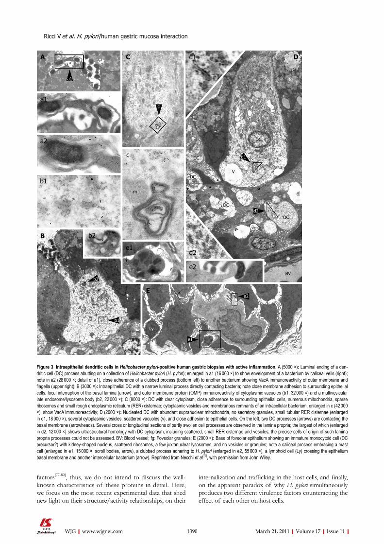
1390 March 21, 2011|Volume 17|Issue 11|WJG|www.wjgnet.com
factors[77-80], thus, we do not intend to discuss the well-known characteristics of these proteins in detail. Here, we focus on the most recent experimental data that shed new light on their structure/activity relationships, on their
internalization and trafficking in the host cells, and finally, on the apparent paradox of why H. pylori simultaneously produces two different virulence factors counteracting the effect of each other on host cells.
d2
e2
e1
b1
b2
d1
c
a1
a1
A
a2
b1
b2
c
m
C D
fg
DC
V
DC
DC
DC
DC
BV
e1
e2
d2
B
E
d1
Ly
Figure 3 Intraepithelial dendritic cells in Helicobacter pylori-positive human gastric biopsies with active inflammation. A (5000 ×): Luminal ending of a den-dritic cell (DC) process abutting on a collection of Helicobacter pylori (H. pylori); enlarged in a1 (16 000 ×) to show envelopment of a bacterium by caliceal veils (right); note in a2 (28 000 ×; detail of a1), close adherence of a clubbed process (bottom left) to another bacterium showing VacA immunoreactivity of outer membrane and flagella (upper right); B (3000 ×): Intraepithelial DC with a narrow luminal process directly contacting bacteria; note close membrane adhesion to surrounding epithelial cells, focal interruption of the basal lamina (arrow), and outer membrane protein (OMP) immunoreactivity of cytoplasmic vacuoles (b1, 32 000 ×) and a multivesicular late endosome/lysosome body (b2, 22 000 ×); C (8000 ×): DC with clear cytoplasm, close adherence to surrounding epithelial cells, numerous mitochondria, sparse ribosomes and small rough endoplasmic reticulum (RER) cisternae; cytoplasmic vesicles and membranous remnants of an intracellular bacterium, enlarged in c (42 000 ×), show VacA immunoreactivity; D (2000 ×): Nucleated DC with abundant supranuclear mitochondria, no secretory granules, small tubular RER cisternae (enlarged in d1, 18 000 ×), several cytoplasmic vesicles, scattered vacuoles (v), and close adhesion to epithelial cells. On the left, two DC processes (arrows) are contacting the basal membrane (arrowheads). Several cross or longitudinal sections of partly swollen cell processes are observed in the lamina propria; the largest of which (enlarged in d2, 12 000 ×) shows ultrastructural homology with DC cytoplasm, including scattered, small RER cisternae and vesicles; the precise cells of origin of such lamina propria processes could not be assessed. BV: Blood vessel; fg: Foveolar granules; E (2000 ×): Base of foveolar epithelium showing an immature monocytoid cell (DC precursor?) with kidney-shaped nucleus, scattered ribosomes, a few juxtanuclear lysosomes, and no vesicles or granules; note a caliceal process embracing a mast cell (enlarged in e1, 15 000 ×; scroll bodies, arrow), a clubbed process adhering to H. pylori (enlarged in e2, 55 000 ×), a lymphoid cell (Ly) crossing the epithelium basal membrane and another intercellular bacterium (arrow). Reprinted from Necchi et al[73], with permission from John Wiley.
Ricci V et al . H. pylori /human gastric mucosa interaction

1391 March 21, 2011|Volume 17|Issue 11|WJG|www.wjgnet.com
VacA toxinVacA has three well-confirmed cell activities, namely: cell vacuolation, apoptosis, and CD4+ T-lymphocyte ac-tivation and proliferation[77,81]. This toxin was discovered as a secreted protein of H. pylori (at that time known as Campylobacter pylori) that induces a massive cytoplasmic vacuolation in vitro[82]. About 55% of isolates of H. pylori are able to induce that cytopathic effect[82]. VacA was shown later to form anionic channels into artificial lipid membranes, as well as cell membranes[77]. VacA chan-nel drives first an hyperacidification of late endocytic compartments by inducing, via the transport of anions into the lumen of these organelles, overactivity of the V-ATPase. Accumulation, by protonation, of weak bases such as ammonia (produced by H. pylori urease activity) into these compartments creates an osmotic imbalance that results in their swelling[77,81].
Deep-etch electron microscopy of VacA prepara-tions has revealed that the toxin forms regular oligomers with either six- or sevenfold radial symmetry[77]. Oligo-merization of the toxin molecules creates a cavity in the center of the oligomer and mimics the structure of a pore-forming toxin (Figure 4) such as the α hemolysin
of Staphylococcus aureus[83]. In its mature state, VacA is a 88-kDa protein that can be cleaved into two subunits (Figure 4). According to their molecular mass, the N-ter-minal subunit is denominated “p33”, whereas “p55” is the C-terminal one (Figure 4). The N terminus of the mature p33 toxin subunit consists of a stretch of 32 uncharged hydrophobic amino acids (found in the VacA s1 subtype[77,80]). The presence of additional residues to p33 (found in the s2 subtype), ablation deletions within the hydrophobic residues, or specific point mutations of amino acids in this stretch inhibit the vacuolating activ-ity of VacA. From these results, it has been concluded that VacA channel formation involves the N-terminal 32 amino acids of p33[77,80,84,85]. Other observations have led to the hypothesis that, in addition to the p33 N-terminal part and p33 itself[85], a toxin domain localized in about the first 110 residues of the p55 is also required for the proper oligomerization of the toxin, for channel for-mation. This is supported by the fact that the minimal intracellular active domain of VacA contains the full p33 domain and about 110 residues of the N-terminal part of p55[86,87]. In these experiments, the p33 toxin subunit, produced alone within cells, does not induce vacuola-tion[86,87].
In addition to its vacuolating activity, VacA exhibits apoptotic activity[88-90]. Importantly, these studies have revealed that the p33 VacA subunit is targeted to mi-tochondria and only is required for apoptosis[88], and that a toxin mutant without vacuolating activity is non-apoptotic[89]. Collectively, these data indicate that VacA induces, through the p33 subunit alone, apoptosis by act-ing on mitochondria, and most likely involves the toxin channel activity. Recently, it has been demonstrated that p33 oligomerizes without the need of p55 and forms an anionic channel with characteristics identical to that of the whole VacA molecule[91]. Furthermore, ablation of the 32 N-terminal residues of p33 does not alter the pore-forming activity of the VacA subunit, but impairs the mitochondrial targeting activity of p33[91]. Altogether, these new data indicate that the portion of p33 without its first 32 N-terminal residues (that we call the “core p33”) oligomerizes and contains all the channel activity of VacA. Domańska et al[91] have clarified the following, formerly puzzling, observations: (1) that a mutant toxin with a major deletion in the p33 N-terminal hydrophobic amino acids (VacA delta 6-27), albeit inhibiting the vacu-olation process, nonetheless induces anionic channels in artificial bilayers, even though exhibiting a longer delay of formation than the wild-type toxin[85]; and (2) that a single deletion (δ 49-57) in the p33 core domain inhibits the oligomerization of the toxin and the vacuolation[92]. Indeed, in the light of the study of Domańska et al[91], these observations are well explained by the role of the p33 core without the N-terminal hydrophobic amino-acid stretch in forming the toxin channel by oligomerization.
As depicted in Figure 4, the structure-activity of VacA would thus be the following: starting from the N terminus, the 32-amino-acid stretch consists of a mem-
Lipid raft
Ch CytoplasmAP
Top view
OutsideAP
S
Cleavagep55p33
NH2+ COO-
(s) MAS PF RB AT (m)Loop
1 821AA 32 311-312 429
CB
A
Figure 4 Structure-function relationships in the VacA toxin. A: VacA is an 88-kDa protein that can be cleaved into two subunits designated p33 (red and light blue) and p55 (light green and dark green). The cleavage between the sub-unit occurs in a flexible loop between residues 311 and 312. The two subunits are probably attached by non-covalent bonds between the p33 carboxy termi-nus and the N-p55 terminus. The amino-terminal part of p33 consists of 32 hy-drophobic amino-acid stretch that is involved in the recognition of mitochondria (red) (MAS) (where the “s” toxin subtype site is located) followed by the p33 “core” subunit that forms, upon entry into the lipid membrane and oligomeriza-tion, an anionic channel (PF) (light blue). The toxin nicking site is located in the flexible loop domain. The amino-terminal part of p55 contains the cell receptor domain (about 110 amino acids) (RB; light green) followed by the type Va auto-transporter domain (AT; dark green) that is involved in the secretion of the toxin by the bacterium (where the “m” toxin subtype site is located); B and C: VacA binds as a monomer to its cell surface receptor sphingomyelin (S) with low af-finity, then the p33 core progressively is embedded (light blue curve arrow) in the lipid membrane bilayers, at the level of lipid rafts [containing saturated lipid such as sphingomyelin and cholesterol (Ch)] and forms an anionic channel (AP) by oligomerization of p33. This multiplies by 6 the number of toxin cell receptors associated with the oligomerized VacA, thus increasing greatly the toxin affinity for the target cells.
Ricci V et al . H. pylori /human gastric mucosa interaction
1392 March 21, 2011|Volume 17|Issue 11|WJG|www.wjgnet.com
brane/mitochondrial targeting motif (which however does not participate in the toxin channel formation)[91]. Then, the p33 channel core forms the channel activity, ei-ther in endosomal membranes or in the inner mitochon-drial membrane (IMM)[91]. At the end of the p33 subunit, there is the loop domain (58 residues) in which the toxin-nicking site is located (between residues 311 and 312). We speculate that a peptide motif just after the nicking site (i.e. residing in the 110 residues of the N-terminal part of p55 subunit) is implicated in the recognition of the toxin cell receptor. This is at odds with the current view that the toxin receptor domain is located in the so-called mid-region (m1/m2; i.e. residues 470-662) of p55[77,80]. How to reconcile the recent data of Domańska et al[91] with previ-ous results indicating that the intracellular minimum por-tion of the VacA molecule active in the cytosol requires, in addition to p33, the 110 N-terminal residues of p55 for VacA oligomerization[84,86,87]? Taking in account that the p33 toxin subunit oligomerizes alone and supports all the pore-forming ability of VacA[91], why does expression of this toxin subunit alone into cells not lead to cell vacu-olation[86,87]? We speculate that the different intracellularly expressed (by cDNA transfection) toxin domains might induce their vacuolating activities not after intracellular synthesis, but rather only after leaking out of the produc-ing cells, followed by endocytosis via the VacA cognate receptor. This would point to the 110 residues present at the N-terminal part of p55 as containing the VacA cell receptor motif (Figure 4). This position would indeed be ideally suited to recognize a cell surface molecule close to the lipid bilayers (Figure 4). The 3D structure of the p55 VacA subunit has been recently resolved[93] and shows that a large part of this toxin subunit has the classical fold of autotransporter structures of the bacterial Va se-cretion system (by which the toxin is transported), which is mostly implicated in the bacterial secretion of VacA. This is supported by previous experiments showing that several deletions in the p55 subunit abolish the produc-tion of VacA by the bacterium[85]. Gangwer et al[93] have proposed that a conserved pocket, located in the m1/m2 regions of p55, is the receptor domain of VacA, which becomes fully accessible to the cell receptor when the toxin is assembled in either a single- or double-layered oligomeric structure. However, it is well known that it is necessary to disrupt the oligomeric structure of VacA (by acidic or alkaline treatments) to render it able to bind to its cell receptor. Furthermore, the mutant toxin δ 49-57, which does not form oligomeric structures, does not require to be acid-activated to bind the VacA cell recep-tor[92]. This demonstrates that the toxin receptor domain is probably not fully accessible in the VacA oligomeric form. More recently, González-Rivera et al[94] have shown that a mixture of purified p33 and p55 does not form oligomeric structures in aqueous solvent, but only mono-meric structures, whereas, upon addition of a detergent that mimicks a lipid membrane environment, there is formation of oligomeric structures. This result suggests that the hydrophobic environment induces the oligomer-
ization, most likely by the p33-dependent formation of the channel. Importantly, it has also been shown that the p33 subunit markedly increases the cell binding of p55[94]. Thus, VacA as a monomer first binds one cell receptor with low affinity, while upon oligomerization, bound to six receptors, it exhibit a very high affinity (Figure 4).
An important breakthrough, seminal for the identi-fication of the VacA receptor, has been the finding that the toxin requires the integrity of lipid rafts and glyco-sylphosphatidylinositol-anchored proteins (GPI-APs) for its full vacuolating activity, which suggests that VacA follows the GPI-AP pathway of endocytosis by a lipid raft-dependent, clathrin-independent pathway[95]; later de-scribed in detail by Sabharanjak et al[96]. The first vacuolar organelles collecting the incoming GPI-APs involve new intracellular compartments that have been named GPI-AP-enriched early endosomal compartments (GEECs), which differ from classical early endosomes[96]. This en-docytic pathway, which also requires the small Rho GT-Pase Cdc42[96], is currently named the GEEC endocytic pathway[97,98], and has been confirmed to be exploited by VacA for its internalization[99-102] (Figure 5). It was later shown that the GEEC pathway requires the presence, in the cell plasma membrane, of the unsaturated lipid sphin-gomyelin, and that the length of the sphingomyelin acyl chain determines its endocytic intracellular trafficking[103]. This prompted an investigation that has demonstrated the role of sphingomyelin as the VacA cell receptor[103]. Accordingly, it has been demonstrated that the length of the sphingomyelin acyl chain also determines the VacA intracellular trafficking[102]. Normal cells contain in their lipid membrane a high proportion of long acyl chain sphingomyelin (C18), and VacA bound to this lipid fol-lows the GEEC pathway[102]. In contrast, when cells are artificially enriched in short-acyl-chain sphingomyelin (C2), the toxin does not follow the GEEC pathway but is recycled back to the plasma membrane in a Cdc42-independent fashion[102].
Upon VacA treatment, transformed human CD4+ T lymphocytes (Jurkat cells) block their constitutive production of IL-2[104]. It is well established that, in T lymphocytes, the nuclear factor NFAT, upon processing by the calcium-activated calcineurin protease, activates the transcription of the IL-2 gene. Massive entry of calcium, through the voltage-dependent Ca2+ release-activated Ca2+ (CRAC) channels, is required to activate calcineurin. However, a VacA mutant without vacuolat-ing activity has no effect on NFAT inhibition in Jurkat cells[105]. Possibly, inhibition of NFAT by VacA is due to the electric depolarization of the lymphocytic plasma membrane, induced by the toxin anionic channel, which inhibits CRAC channels. Human primary T lymphocytes are, however, not sensitive to the deactivation of NFAT induced by VacA[106]. Treatment of primary human T lymphocytes by phorbol myristate acetate, which induces their migratory activity, together with the expression of the CD18 β-integrin at their surface, restores the VacA-induced inhibition of NFAT[107]. Furthermore, the direct
Ricci V et al . H. pylori /human gastric mucosa interaction

1393 March 21, 2011|Volume 17|Issue 11|WJG|www.wjgnet.com
expression of CD18 into primary human T lymphocytes renders them able to respond to VacA[107]. It has there-fore been concluded that CD18 is the receptor for VacA in immune cells[107]. This result is puzzling because, for all the different toxins studied so far, the cell binding activ-ity always depends on a unique molecule (or a family of closely related molecules). However, a few observations do not support the role of CD18 as the sole VacA recep-tor in immune cells. Indeed, it has been demonstrated that, although human primary lymphocytes do not deac-tivate the nuclear factor NFAT, they respond perfectly to VacA by blocking their induced proliferation (i.e. block-age of their cell cycle) due to a mitochondria-induced depletion of ATP[106,108,109]. This fact clearly indicates that VacA binds and penetrates into primary lymphocytes without needing CD18 (probably using the sphingomy-
elin receptor). The fact that VacA, entering via sphingo-myelin in primary T cells, does not inactivate NFAT can be explained by the possibility that the toxin channel is probably not recycled back (or only weakly recycled) to the plasma membrane, and therefore, is unable to block calcium cell entry through CRAC channel inhibition. We speculate that CD18 may not be a real receptor for VacA, but rather activates a signaling pathway (perhaps via the direct activation of CD18 by VacA, because association of VacA with CD18 has been reported[107]) that modifies the intracellular trafficking of the toxin. For instance, stimulation of the migratory activity of T lymphocytes by CD18 signaling might activate Cdc42 boosting of the entry of VacA[99], and increasing its recycling to the cell surface. It has recently been reported that the GEEC pathway is pivotal during cell migration[98]. Another pos-sibility would be that CD18-driven signaling drastically changes the lipid membrane content of long-acyl-chain sphingomyelin molecules toward short ones forcing the toxin to recycle back to the plasma membrane as recently described[102].
How is VacA transferred from early endosomes to mitochondria and/or late endosomes to induce apoptosis and vacuolation? It is well known that ligands or mem-brane receptors taken up and routed for degradation into lysosomes are endocytosed by the clathrin-dependent pathway and targeted to the luminal vesicles of endo-some-forming multivesicular bodies (MVBs) by the en-dosomal sorting complex required for transport (ESCRT) complex[110] (Figure 5). Propelled along microtubules, MVBs then move to lysosomes where the content of MVB internal vesicles is selectively delivered for degrada-tion. Several lines of evidences indicate that MVBs and lysosomes are not the cell compartments that undergo vacuolation by VacA activity: (1) no internal vesicles are observed by electron microscopy in VacA-induced vacu-oles and the enlarged compartment does not have the structure of lysosomes[95,111]; (2) the vacuolated compart-ment contains markers of both late endosomes and ly-sosomes[112]; and (3) disruption of microtubules does not block VacA-induced vacuolation[113]. Thus, the organelles that are vacuolated by VacA are likely post-lysosomal compartments that are required to recycle the membrane of MVBs from lysosomes. VacA is probably transferred directly to that compartment by an F-actin-dependent mechanism of endosome motility[101]. Indeed, upon entry of VacA into early endosomes via the GEEC pathway, VacA-containing early endosomes exhibit F-actin comet tails associated with their cytosolic membrane surface, and are rapidly moving in the cytosol[101]. Inhibition of these F-actin structures blocks the VacA transfer into the late endosomal compartment and vacuole genesis[101], but also VacA-induced apoptosis and the localization of tox-in molecules in mitochondria[114]. These findings suggest that VacA is addressed to late endosomes and mitochon-dria by the same mechanism that relies on F-actin-driven vesicular motility. F-actin structures have recently been shown to take place at the level of the early endosomal
Inhibition of NFATin Jurkat cellsVacA monomer
VacA poreVacA
Maxirafts
MiniraftsRac
Actin
Apoptosis Cdc42
Actin
GEECs
Rab5ESCRT
CRAC
Ca2+
R
Bax/Bak?
M
Rab7 LE VacuolationF-actin comet
Lysosomes
EE
CP
Figure 5 Endocytosis and intracellular trafficking of VacA. Monomeric VacA may bind sphingomyelin on small rafts. Then, by formation of membrane extensions by actin polymerization via Rac activation, VacA is clustered in macrorafts where the p33 subunit oligomerizes and forms a channel that enters the membrane lipid bilayers. By a Cdc42-dependent process, VacA bound to sphingomelin associated to lipid rafts is transferred into cell membrane invagi-nations (tubules?), which are then pinched out from the membrane, with the help of F-actin filament, which leading to the formation of the glycosylphospha-tidylinositol-anchored protein-enriched early endosomal compartment (GEEC) compartment that contains VacA. The toxin is then transferred to Rab5-positive early endosomes (EEs). In EEs, VacA is selectively addressed to EE tubular extensions that are formed by an F-actin process. These tubular extensions are pinched out from the EEs and form highly motile vacuoles that are propelled by F-actin comets. These motile vesicles then fuse with mitochondria (M) or late endosomes (LEs), where VacA induces apoptosis or vacuolation. The pro-apoptotic channels Bax and Bak may be brought to mitochondria by binding on VacA-containing motile vacuoles. Some VacA molecules may be recycled (R) back to the plasma membrane where the channel activity of the toxin, by alter-ing the electric transmembrane potential, inhibits the voltage-dependent Ca2+ release-activated Ca2+ (CRAC) channel. This blocks entry of calcium that acti-vates the calcineurin protease, which is required for processing of the nuclear factor (NFAT), and therefore inhibits the transcription of the interleukin 2 (IL-2) gene in Jurkat T-lymphocytes. Ligands entering the coated-pit pathway (CP) are directed, by the endosomal sorting complex required for transport (ESCRT) complex, towards internal vesicles of EEs and form the multivesicular body multivesicular body (MVB). MVBs are directed to lysosomes, by being propelled along microtubules, where the contents of EE internal vesicles are transferred and degraded.
Ricci V et al . H. pylori /human gastric mucosa interaction

1394 March 21, 2011|Volume 17|Issue 11|WJG|www.wjgnet.com
surface to induce budding of tubular structures followed by their cleavage to yield vesicles that retain F-actin tails[115]. It is thus tempting to speculate that VacA might be first transferred from GEECs to the early endosomal limiting membrane, then into these tubular extensions, and finally into the membrane of the ensuing F-actin motile vesicles (Figure 5). These vesicles are then brought to late endosomes and/or mitochondria via stochastic fu-sion events by F-actin-driven vesicular motility (Figure 5). In this model, the transfer of VacA (or only of its p33 subunit) from the donor membrane to the recipient membrane (late endosomes or mitochondria) would be achieved by the membrane/mitochondrial N-terminal 32 residues of p33[91] that may stick out of the membrane (Figure 4). This would explain why point mutations, or deletion or modification of the length of this amino-acid stretch might modulate the vacuolating process without altering the pore-forming capacity of the toxin[91]. Im-portantly, according to this model, the toxin would never be released free in the cytosol[90], which differs from the enzymatic subunits of canonical A-B toxin. By remaining associated with lipid membranes, out of reach of deg-radative processes, the VacA channel would keep its full activity for a long time, as observed several years ago[111].
Inside mitochondria, the p33 subunit (or the full VacA molecule) induces, probably by the rupture of IMM integrity[91], the release of cytochrome c (cyt c) and induction of apoptosis[88]. However, pro-apoptotic channels must be activated or transferred into the outer membrane of mitochondria to ensure the release into the cytosol of cyt c, which, by activation of the Apaf1 molecule, induces the stimulation of the caspase cascade that results in cell death. Accordingly, the pro-apoptotic channels Bax and Bak have been shown to be activated during VacA-induced apoptosis[116,117], and VacA is un-able to induce apoptosis in Bax- and Bak-deficient mouse embryonic fibroblasts[117]. It has been recently proposed that Bax and Bak might be directly recruited from the cytosol by endosomes containing functional VacA chan-nels in their membranes, which allows their juxtaposition with mitochondria[117]. This would be an ingenious way to combine the transfer of VacA channels to the IMM (and thereby the release of cyt c in the mitochondrial inter-membrane space) and the leakage of cyt c in the cytosol.
Thus VacA is probably not really a multifunctional toxin, as previously defined[77]. The diversity of VacA ac-tivities observed may be simply explained by the channel formed by the p33 subunit that is addressed to different cell membranes by the intracellular trafficking of the toxin, which may be different in different cell types or during cell differentiation (Figure 5).
Functional relationship between VacA and CagAThe gene encoding VacA is present in all the H. pylori strains, although only 55% express an active vacuolating toxin. This is due to either additions or deletions of pep-tide sequences in the toxin that probably impair the VacA intracellular trafficking or its binding to the cell surface,
as we have described above. It is now well-established that the most severe gastric pathology (i.e. peptic ulcer and gastric cancer) is restricted to H. pylori type 1 strains, which contain in their chromosome a PAI named cag PAI, and which secrete an active vacuolating VacA toxin[10]. The cag PAI encodes for the CagA protein and for the TFSS machinery that is implicated in the transport of CagA out of the bacterium, and its transfer to the host cell cytosol[78,79]. CagA is a 135-145-kDa protein that con-tains its full intracellular signaling activity in its 35-45-kDa C-terminal part[78,79,118] (Figure 6). Nevertheless, it is still unclear how and where the cag TFSS recognizes and tar-get gastric epithelial cells so as to transfer CagA into their cytosol. Recently, two mechanisms have been proposed. One points to the recognition and binding of the TFSS to an integrin[119,120], then, through a mechanical sliding due to bound integrins, the TFSS might punch the cell surface and deliver CagA into the cytosol[120]. One caveat
COO-NH2+
EPIYAP
100 kDa 35-45 kDaCleavage
CagAEGF
EGFRH. pylori
VacA
TFSS
PSPS
INT
EE
TJ
LE
+YP -YP
MAP/ERKc-Src
M
Proliferation,NF-κB activation,Anti-apoptotic, pro- inflammatory effects
?
Figure 6 Structure of CagA and signaling cross-talk between VacA and CagA. A: CagA encompasses two fragments: the N-terminal 100-kDa fragment may contain the cell binding domain [to phosphatidylserine (PS)?]. Cleavage of CagA may take place just at the beginning of the first EPIYA motif, which can be tyrosine-phosphorylated by the c-Src tyrosine kinase. The C-terminal portion of CagA, which contains all the signaling activity of the molecule, may have a different molecular mass (up to 45 kDa) due to the repetition of the EPIYA-containing domain; B: CagA produced within the bacterium is transferred in the external medium by the type Ⅳ secretion system (TFSS) machinery. Two possibilities for CagA transfer into the target epithelial cell: (a) by binding to an integrin (INT), the TFSS punches the cell membrane and injects CagA; or (b) the TFSS induces the flipping of PS on the outer cell surface. By its 100-kDa N-terminal fragment, CagA binds PS and, upon endocytosis, which is trans-ferred into the gastric cells. In the cytosol, the CagA signaling domain can be tyrosine-phosphorylated (+YP) and inhibits the c-Src kinase activity that is re-quired to allow the transfer of VacA from glycosylphosphatidylinositol-anchored protein-enriched early endosomal compartments (GEECs) to early endosomes (EEs). This blocks vacuolation in late endosomes (LEs) and mitochondria (M)-dependent apoptosis induced by VacA. In an unphosphorylated (-YP) state, CagA activates mitogen-activated protein (MAP)/extracellular signal-regulated kinase (ERK) and nuclear factor (NF)-κB anti-apototic and pro-inflammatory pathways, which also counteract VacA-induced apoptosis. VacA interferes with epidermal growth factor (EGF) receptor (EGFR) activation and endocytosis, thus impairing the signaling pathway that is triggered by this receptor. Free EGFR ligands (EGF) are liberated from the cell-surface-bound molecules via cleavage triggered by Helicobacter pylori (H. pylori). TJ: Tight junction.
B
A
Ricci V et al . H. pylori /human gastric mucosa interaction

1395 March 21, 2011|Volume 17|Issue 11|WJG|www.wjgnet.com
for this mechanism is that H. pylori might deliver CagA at the apical pole of colonized epithelial gastric cells, whereas integrins are localized on the basolateral pole. On the other hand, it has been proposed that the TFSS does not punch the cell surface but induces the flipping out of the lipid phosphatidylserine (PS) from the inner (where it normally resides) to the outer leaflet of the tar-get cell membrane[121]. In this model, CagA, which is se-creted constitutively by the TFSS, binds to PS through a lipid-binding peptide motif that is present in its 100-kDa N-terminal part[121]. Following this binding step, a novel type of endocytosis (not yet better characterized nor de-fined) is induced that carries CagA, associated with PS, into epithelial gastric cells[121]. If this is true, CagA should no longer be defined as an effector protein, but rather as a new type of A-B toxin that is released only in close proximity to the target cells.
Inside the cell, CagA may be tyrosine phosphorylated by the p60 c-Src kinase[118]. Phosphorylation occurs on the 35-45-kDa C-terminal part of CagA on specific five-ami-no-acid motifs called EPIYA, which can be repeated sev-eral-fold and increase the size of this fragment[26,78]. CagA can be split into two fragments by proteases[122,123]. This generates a 100-kDa N-terminal fragment and a C-terminal 35-45-kDa one that contain all the cell signaling activity of CagA[114]. Proteolytic cleavage of CagA does not require its tyrosine phosphorylation, and the nicking site is local-ized just at the onset of the first EPIYA motif (Boquet and Ricci, unpublished data). The 100-kDa N-terminal part of CagA might be involved in the cell recognition and transfer of the 35-45-kDa C-terminal part to the cytosol, as suggested by recent data[121]. Therefore, CagA would indeed behave as a typical A-B toxin.
Two main intracellular activities of CagA are now well established. In its tyrosine phosphorylated state, CagA may act, either directly or upon association and activation of the tyrosine phosphatase SHP2[78], to inhibit the c-Src kinase and therefore to affect many regulating proteins that depend on this enzyme, among which are those in-volved in the actin cytoskeleton assembly/regulation[124]. In its non-tyrosine-phosphorylated form (or independent of the phosphorylation state), CagA activates the MAP/ERK and NF-κB pathways[53,125], which induce cell prolif-eration, inhibition of apoptosis, a typical cellular elonga-tion called “hummingbird phenotype”[66], and inflamma-tory response[28] (Figure 6).
An increasing body of evidence suggests that CagA and VacA have several opposing cellular effects. It has been shown that, while CagA activates the NFAT nuclear factor, VacA inhibits it[126]. Then, it has been demonstrat-ed that CagA decreases VacA-induced vacuolation, while in turn, VacA reduces CagA-induced hummingbird phe-notype formation[127]. It has also been shown that VacA can downregulate CagA effects on epithelial cells by in-terfering with activation and endocytosis of EGFR, thus impairing the signaling pathway triggered by this recep-tor, and which plays a pivotal role in cell proliferation and hummingbird phenotype formation[128]. Taken together,
these findings raise the hypothesis that VacA and CagA may downregulate the cell effects of the other, allowing H. pylori interaction with epithelial cells, while avoiding excessive cell damage.
We have recently shown that CagA interferes with VacA action by two complementary mechanisms[114]. In its tyrosine-phosphorylated state, CagA inhibits the c-Src kinase and, in doing so, blocks VacA in the GEECs and thus its delivery to early and late endosomes (inhibition of the vacuolation) or mitochondria (inhibition of apop-tosis). In its unphosphorylated state, CagA stimulates the NF-κB pathway and, in doing so, induces anti-apoptotic activity (mediated possibly by the anti-apoptotic factor Bcl2) that also blocks VacA-induced apoptosis. We hy-pothesize that, once bacteria have colonized the gastric niche, the apoptotic action of VacA might be detrimental for the survival of H. pylori that are adherent to the mu-cosa, and thus the CagA counteracting action gives a ra-tionale for the association of these two virulence factors in the most pathogenic H. pylori strains[114]. This would be a new, highly ingenious mechanism by which a bacterium locally protects its ecological niche against the action of one of its own virulence factors. However, while exerting a beneficial role for survival and growth of the bacterium by counteracting VacA toxin, CagA injection in the gas-tric epithelial cells triggers pro-inflammatory and anti-apoptotic responses that are detrimental for the human host in the long-term, because they favor the develop-ment of ulcer and cancer.
Because it simultaneously delivers to its host two in-dependent virulence factors that antagonize each other, H. pylori appears to act like a driver that presses the brake and gas pedals at once. This apparently paradoxi-cal behavior is now emerging as an intriguing strategy to achieve the best fit between the bacterium and the hostile gastric environment that represents its ecological niche.
CONCLUSIONWith regard to the pathogen, illness is often inadvertent; the result of exquisite tricks learned long ago and played on human cells to achieve the paramount goal of the microorganism: conservation of the species[56,129]. Taken together, all the new findings described above strongly re-inforce the notion that H. pylori is a skilled bacterium that has been smart enough to: (1) learn about the physiology of its host; (2) keep this information in its genetic library; (3) share the gene book with other organisms; (4) be “open minded” about acquiring new knowledge and discarding obsolete information; and, of key importance; and (5) know how to communicate with its human host, realizing that killing the host would oblige it to find a new one very quickly to ensure its reproductive success[56,129,130]. H. pylori makes a long and hard, but also successful, journey in the human stomach, given that H. pylori is widespread and has probably been part of the human biota since time im-memorial[31,56,130]. Although H. pylori seeking survival may be sometimes achieved at the expense of the well-being
Ricci V et al . H. pylori /human gastric mucosa interaction

1396 March 21, 2011|Volume 17|Issue 11|WJG|www.wjgnet.com
of the stomach and the host, human physiology and im-munology have co-evolved in the presence of persistent gastric H. pylori colonization, and so are disrupted by its absence[130]. As emphasized by Atherton et al[130], this imbalance of a long-lasting pathogen/host equilibrium, caused by progressive disappearance of H. pylori from some populations, may however result in an increased incidence of other diseases such as reflux esophagitis and esophageal carcinoma, as well as obesity, type 2 diabetes, and allergic disorders.
REFERENCES1 Everhart JE. Recent developments in the epidemiology of
Helicobacter pylori. Gastroenterol Clin North Am 2000; 29: 559-578
2 Zarrilli R, Ricci V, Romano M. Molecular response of gastric epithelial cells to Helicobacter pylori-induced cell damage. Cell Microbiol 1999; 1: 93-99
3 Herszényi L, Tulassay Z. Epidemiology of gastrointestinal and liver tumors. Eur Rev Med Pharmacol Sci 2010; 14: 249-258
4 Herrera V, Parsonnet J. Helicobacter pylori and gastric ad-enocarcinoma. Clin Microbiol Infect 2009; 15: 971-976
5 Correa P, Piazuelo MB, Camargo MC. The future of gastric cancer prevention. Gastric Cancer 2004; 7: 9-16
6 Peek RM Jr, Blaser MJ. Helicobacter pylori and gastrointesti-nal tract adenocarcinomas. Nat Rev Cancer 2002; 2: 28-37
7 Wong BC, Lam SK, Wong WM, Chen JS, Zheng TT, Feng RE, Lai KC, Hu WH, Yuen ST, Leung SY, Fong DY, Ho J, Ch-ing CK, Chen JS. Helicobacter pylori eradication to prevent gastric cancer in a high-risk region of China: a randomized controlled trial. JAMA 2004; 291: 187-194
8 Ernst PB, Peura DA, Crowe SE. The translation of Helico-bacter pylori basic research to patient care. Gastroenterology 2006; 130: 188-206; quiz 212-213
9 Van Doorn LJ, Figueiredo C, Mégraud F, Pena S, Midolo P, Queiroz DM, Carneiro F, Vanderborght B, Pegado MD, San-na R, De Boer W, Schneeberger PM, Correa P, Ng EK, Ather-ton J, Blaser MJ, Quint WG. Geographic distribution of vacA allelic types of Helicobacter pylori. Gastroenterology 1999; 116: 823-830
10 Peek RM Jr, Crabtree JE. Helicobacter infection and gastric neoplasia. J Pathol 2006; 208: 233-248
11 El-Omar EM, Rabkin CS, Gammon MD, Vaughan TL, Risch HA, Schoenberg JB, Stanford JL, Mayne ST, Goedert J, Blot WJ, Fraumeni JF Jr, Chow WH. Increased risk of noncardia gastric cancer associated with proinflammatory cytokine gene polymorphisms. Gastroenterology 2003; 124: 1193-1201
12 Lu W, Pan K, Zhang L, Lin D, Miao X, You W. Genetic poly-morphisms of interleukin (IL)-1B, IL-1RN, IL-8, IL-10 and tumor necrosis factor {alpha} and risk of gastric cancer in a Chinese population. Carcinogenesis 2005; 26: 631-636
13 Ohyauchi M, Imatani A, Yonechi M, Asano N, Miura A, Iijima K, Koike T, Sekine H, Ohara S, Shimosegawa T. The polymorphism interleukin 8 -251 A/T influences the suscep-tibility of Helicobacter pylori related gastric diseases in the Japanese population. Gut 2005; 54: 330-335
14 Pasare C, Medzhitov R. Toll-like receptors: linking innate and adaptive immunity. Adv Exp Med Biol 2005; 560: 11-18
15 Takeda K, Akira S. Toll-like receptors in innate immunity. Int Immunol 2005; 17: 1-14
16 Arbour NC, Lorenz E, Schutte BC, Zabner J, Kline JN, Jones M, Frees K, Watt JL, Schwartz DA. TLR4 mutations are as-sociated with endotoxin hyporesponsiveness in humans. Nat Genet 2000; 25: 187-191
17 Hold GL, Rabkin CS, Chow WH, Smith MG, Gammon MD, Risch HA, Vaughan TL, McColl KE, Lissowska J, Zatonski W,
Schoenberg JB, Blot WJ, Mowat NA, Fraumeni JF Jr, El-Omar EM. A functional polymorphism of toll-like receptor 4 gene increases risk of gastric carcinoma and its precursors. Gastro-enterology 2007; 132: 905-912
18 Houghton J, Stoicov C, Nomura S, Rogers AB, Carlson J, Li H, Cai X, Fox JG, Goldenring JR, Wang TC. Gastric cancer origi-nating from bone marrow-derived cells. Science 2004; 306: 1568-1571
19 Lindholm C, Quiding-Järbrink M, Lönroth H, Hamlet A, Svennerholm AM. Local cytokine response in Helicobacter pylori-infected subjects. Infect Immun 1998; 66: 5964-5971
20 Galgani M, Busiello I, Censini S, Zappacosta S, Racioppi L, Zarrilli R. Helicobacter pylori induces apoptosis of human monocytes but not monocyte-derived dendritic cells: role of the cag pathogenicity island. Infect Immun 2004; 72: 4480-4485
21 Park H, Li Z, Yang XO, Chang SH, Nurieva R, Wang YH, Wang Y, Hood L, Zhu Z, Tian Q, Dong C. A distinct lineage of CD4 T cells regulates tissue inflammation by producing interleukin 17. Nat Immunol 2005; 6: 1133-1141
22 Shi Y, Liu XF, Zhuang Y, Zhang JY, Liu T, Yin Z, Wu C, Mao XH, Jia KR, Wang FJ, Guo H, Flavell RA, Zhao Z, Liu KY, Xiao B, Guo Y, Zhang WJ, Zhou WY, Guo G, Zou QM. He-licobacter pylori-induced Th17 responses modulate Th1 cell responses, benefit bacterial growth, and contribute to pathol-ogy in mice. J Immunol 2010; 184: 5121-5129
23 Zhang B, Rong G, Wei H, Zhang M, Bi J, Ma L, Xue X, Wei G, Liu X, Fang G. The prevalence of Th17 cells in patients with gastric cancer. Biochem Biophys Res Commun 2008; 374: 533-537
24 Holcombe C. Helicobacter pylori: the African enigma. Gut 1992; 33: 429-431
25 Covacci A, Telford JL, Del Giudice G, Parsonnet J, Rappuoli R. Helicobacter pylori virulence and genetic geography. Sci-ence 1999; 284: 1328-1333
26 Hatakeyama M. Oncogenic mechanisms of the Helicobacter pylori CagA protein. Nat Rev Cancer 2004; 4: 688-694
27 Viala J, Chaput C, Boneca IG, Cardona A, Girardin SE, Mo-ran AP, Athman R, Mémet S, Huerre MR, Coyle AJ, DiStefa-no PS, Sansonetti PJ, Labigne A, Bertin J, Philpott DJ, Ferrero RL. Nod1 responds to peptidoglycan delivered by the Heli-cobacter pylori cag pathogenicity island. Nat Immunol 2004; 5: 1166-1174
28 Brandt S, Kwok T, Hartig R, König W, Backert S. NF-kappaB activation and potentiation of proinflammatory responses by the Helicobacter pylori CagA protein. Proc Natl Acad Sci USA 2005; 102: 9300-9305
29 Kim SY, Lee YC, Kim HK, Blaser MJ. Helicobacter pylori CagA transfection of gastric epithelial cells induces interleu-kin-8. Cell Microbiol 2006; 8: 97-106
30 Cooke CL, Huff JL, Solnick JV. The role of genome diversity and immune evasion in persistent infection with Helico-bacter pylori. FEMS Immunol Med Microbiol 2005; 45: 11-23
31 Blaser MJ, Atherton JC. Helicobacter pylori persistence: biol-ogy and disease. J Clin Invest 2004; 113: 321-333
32 Oh JD, Karam SM, Gordon JI. Intracellular Helicobacter py-lori in gastric epithelial progenitors. Proc Natl Acad Sci USA 2005; 102: 5186-5191
33 Gewirtz AT, Yu Y, Krishna US, Israel DA, Lyons SL, Peek RM Jr. Helicobacter pylori flagellin evades toll-like receptor 5-mediated innate immunity. J Infect Dis 2004; 189: 1914-1920
34 Caruso R, Fina D, Peluso I, Fantini MC, Tosti C, Del Vecchio Blanco G, Paoluzi OA, Caprioli F, Andrei F, Stolfi C, Romano M, Ricci V, MacDonald TT, Pallone F, Monteleone G. IL-21 is highly produced in Helicobacter pylori-infected gastric mucosa and promotes gelatinases synthesis. J Immunol 2007; 178: 5957-5965
35 Nagase H, Woessner JF Jr. Matrix metalloproteinases. J Biol Chem 1999; 274: 21491-21494
36 Kruidenier L, MacDonald TT, Collins JE, Pender SL, Sander-son IR. Myofibroblast matrix metalloproteinases activate the neutrophil chemoattractant CXCL7 from intestinal epithelial cells. Gastroenterology 2006; 130: 127-136
Ricci V et al . H. pylori /human gastric mucosa interaction

1397 March 21, 2011|Volume 17|Issue 11|WJG|www.wjgnet.com
37 Fu S, Ramanujam KS, Wong A, Fantry GT, Drachenberg CB, James SP, Meltzer SJ, Wilson KT. Increased expression and cellular localization of inducible nitric oxide synthase and cyclooxygenase 2 in Helicobacter pylori gastritis. Gastroenter-ology 1999; 116: 1319-1329
38 Busiello I, Acquaviva R, Di Popolo A, Blanchard TG, Ricci V, Romano M, Zarrilli R. Helicobacter pylori gamma-gluta-myltranspeptidase upregulates COX-2 and EGF-related pep-tide expression in human gastric cells. Cell Microbiol 2004; 6: 255-267
39 Shibayama K, Kamachi K, Nagata N, Yagi T, Nada T, Doi Y, Shibata N, Yokoyama K, Yamane K, Kato H, Iinuma Y, Arakawa Y. A novel apoptosis-inducing protein from Heli-cobacter pylori. Mol Microbiol 2003; 47: 443-451
40 Romano M, Ricci V, Di Popolo A, Sommi P, Del Vecchio Blanco C, Bruni CB, Ventura U, Cover TL, Blaser MJ, Coffey RJ, Zarrilli R. Helicobacter pylori upregulates expression of epidermal growth factor-related peptides, but inhibits their proliferative effect in MKN 28 gastric mucosal cells. J Clin Invest 1998; 101: 1604-1613
41 Keates S, Sougioultzis S, Keates AC, Zhao D, Peek RM Jr, Shaw LM, Kelly CP. cag+ Helicobacter pylori induce trans-activation of the epidermal growth factor receptor in AGS gastric epithelial cells. J Biol Chem 2001; 276: 48127-48134
42 Wallasch C, Crabtree JE, Bevec D, Robinson PA, Wagner H, Ullrich A. Helicobacter pylori-stimulated EGF receptor transactivation requires metalloprotease cleavage of HB-EGF. Biochem Biophys Res Commun 2002; 295: 695-701
43 Romano M, Ricci V, Memoli A, Tuccillo C, Di Popolo A, Sommi P, Acquaviva AM, Del Vecchio Blanco C, Bruni CB, Zarrilli R. Helicobacter pylori up-regulates cyclooxygen-ase-2 mRNA expression and prostaglandin E2 synthesis in MKN 28 gastric mucosal cells in vitro. J Biol Chem 1998; 273: 28560-28563
44 Caputo R, Tuccillo C, Manzo BA, Zarrilli R, Tortora G, Blan-co Cdel V, Ricci V, Ciardiello F, Romano M. Helicobacter pylori VacA toxin up-regulates vascular endothelial growth factor expression in MKN 28 gastric cells through an epider-mal growth factor receptor-, cyclooxygenase-2-dependent mechanism. Clin Cancer Res 2003; 9: 2015-2021
45 Yan F, Cao H, Chaturvedi R, Krishna U, Hobbs SS, Dempsey PJ, Peek RM Jr, Cover TL, Washington MK, Wilson KT, Polk DB. Epidermal growth factor receptor activation protects gastric epithelial cells from Helicobacter pylori-induced apoptosis. Gastroenterology 2009; 136: 1297-1307, e1-3
46 Romano M, Ricci V, Zarrilli R. Mechanisms of disease: Heli-cobacter pylori-related gastric carcinogenesis--implications for chemoprevention. Nat Clin Pract Gastroenterol Hepatol 2006; 3: 622-632
47 Mammano E, Belluco C, Sciro M, Mencarelli R, Agostini M, Michelotto M, Marchet A, Nitti D. Epidermal growth factor receptor (EGFR): mutational and protein expression analysis in gastric cancer. Anticancer Res 2006; 26: 3547-3550
48 Tuccillo C, Cuomo A, Rocco A, Martinelli E, Staibano S, Mas-colo M, Gravina AG, Nardone G, Ricci V, Ciardiello F, Del Vecchio Blanco C, Romano M. Vascular endothelial growth factor and neo-angiogenesis in H. pylori gastritis in humans. J Pathol 2005; 207: 277-284
49 Crawford HC, Krishna US, Israel DA, Matrisian LM, Wash-ington MK, Peek RM Jr. Helicobacter pylori strain-selective induction of matrix metalloproteinase-7 in vitro and within gastric mucosa. Gastroenterology 2003; 125: 1125-1136
50 Wu CY, Wang CJ, Tseng CC, Chen HP, Wu MS, Lin JT, In-oue H, Chen GH. Helicobacter pylori promote gastric cancer cells invasion through a NF-kappaB and COX-2-mediated pathway. World J Gastroenterol 2005; 11: 3197-3203
51 Franco AT, Israel DA, Washington MK, Krishna U, Fox JG, Rogers AB, Neish AS, Collier-Hyams L, Perez-Perez GI, Hatakeyama M, Whitehead R, Gaus K, O'Brien DP, Romero-Gallo J, Peek RM Jr. Activation of beta-catenin by carcino-
genic Helicobacter pylori. Proc Natl Acad Sci USA 2005; 102: 10646-10651
52 Tsukashita S, Kushima R, Bamba M, Nakamura E, Mukaisho K, Sugihara H, Hattori T. Beta-catenin expression in intramu-cosal neoplastic lesions of the stomach. Comparative analysis of adenoma/dysplasia, adenocarcinoma and signet-ring cell carcinoma. Oncology 2003; 64: 251-258
53 Suzuki M, Mimuro H, Kiga K, Fukumatsu M, Ishijima N, Morikawa H, Nagai S, Koyasu S, Gilman RH, Kersulyte D, Berg DE, Sasakawa C. Helicobacter pylori CagA phosphor-ylation-independent function in epithelial proliferation and inflammation. Cell Host Microbe 2009; 5: 23-34
54 Polk DB, Peek RM Jr. Helicobacter pylori: gastric cancer and beyond. Nat Rev Cancer 2010; 10: 403-414
55 de Paulis A, Prevete N, Rossi FW, Rivellese F, Salerno F, Delfino G, Liccardo B, Avilla E, Montuori N, Mascolo M, Staibano S, Melillo RM, D'Argenio G, Ricci V, Romano M, Marone G. Helicobacter pylori Hp(2-20) promotes migration and proliferation of gastric epithelial cells by interacting with formyl peptide receptors in vitro and accelerates gastric mu-cosal healing in vivo. J Immunol 2009; 183: 3761-3769
56 Ricci V, Zarrilli R, Romano M. Voyage of Helicobacter pylori in human stomach: odyssey of a bacterium. Dig Liver Dis 2002; 34: 2-8
57 Necchi V, Candusso ME, Tava F, Luinetti O, Ventura U, Fiocca R, Ricci V, Solcia E. Intracellular, intercellular, and stromal invasion of gastric mucosa, preneoplastic lesions, and cancer by Helicobacter pylori. Gastroenterology 2007; 132: 1009-1023
58 Petersen AM, Krogfelt KA. Helicobacter pylori: an invading microorganism? A review. FEMS Immunol Med Microbiol 2003; 36: 117-126
59 Dubois A, Borén T. Helicobacter pylori is invasive and it may be a facultative intracellular organism. Cell Microbiol 2007; 9: 1108-1116
60 Lu H, Yamaoka Y, Graham DY. Helicobacter pylori viru-lence factors: facts and fantasies. Curr Opin Gastroenterol 2005; 21: 653-659
61 Montecucco C, de Bernard M. Immunosuppressive and proinflammatory activities of the VacA toxin of Helicobacter pylori. J Exp Med 2003; 198: 1767-1771
62 Wilson KT, Crabtree JE. Immunology of Helicobacter py-lori: insights into the failure of the immune response and perspectives on vaccine studies. Gastroenterology 2007; 133: 288-308
63 Semino-Mora C, Doi SQ, Marty A, Simko V, Carlstedt I, Dubois A. Intracellular and interstitial expression of Helico-bacter pylori virulence genes in gastric precancerous intes-tinal metaplasia and adenocarcinoma. J Infect Dis 2003; 187: 1165-1177
64 Amieva MR, Vogelmann R, Covacci A, Tompkins LS, Nelson WJ, Falkow S. Disruption of the epithelial apical-junctional complex by Helicobacter pylori CagA. Science 2003; 300: 1430-1434
65 Wilkinson SM, Uhl JR, Kline BC, Cockerill FR 3rd. Assess-ment of invasion frequencies of cultured HEp-2 cells by clini-cal isolates of Helicobacter pylori using an acridine orange assay. J Clin Pathol 1998; 51: 127-133
66 Segal ED, Cha J, Lo J, Falkow S, Tompkins LS. Altered states: involvement of phosphorylated CagA in the induc-tion of host cellular growth changes by Helicobacter pylori. Proc Natl Acad Sci USA 1999; 96: 14559-14564
67 Maeda S, Yoshida H, Ogura K, Mitsuno Y, Hirata Y, Yamaji Y, Akanuma M, Shiratori Y, Omata M. H. pylori activates NF-kappaB through a signaling pathway involving IkappaB kinases, NF-kappaB-inducing kinase, TRAF2, and TRAF6 in gastric cancer cells. Gastroenterology 2000; 119: 97-108
68 Kim JJ, Tao H, Carloni E, Leung WK, Graham DY, Sepul-veda AR. Helicobacter pylori impairs DNA mismatch repair in gastric epithelial cells. Gastroenterology 2002; 123: 542-553
Ricci V et al . H. pylori /human gastric mucosa interaction

1398 March 21, 2011|Volume 17|Issue 11|WJG|www.wjgnet.com
69 Necchi V, Sommi P, Ricci V, Solcia E. In vivo accumulation of Helicobacter pylori products, NOD1, ubiquitinated pro-teins and proteasome in a novel cytoplasmic structure. PLoS One 2010; 5: e9716
70 Mani A, Gelmann EP. The ubiquitin-proteasome pathway and its role in cancer. J Clin Oncol 2005; 23: 4776-4789
71 Uronen-Hansson H, Allen J, Osman M, Squires G, Klein N, Callard RE. Toll-like receptor 2 (TLR2) and TLR4 are present inside human dendritic cells, associated with microtubules and the Golgi apparatus but are not detectable on the cell surface: integrity of microtubules is required for interleu-kin-12 production in response to internalized bacteria. Im-munology 2004; 111: 173-178
72 Rimoldi M, Chieppa M, Larghi P, Vulcano M, Allavena P, Rescigno M. Monocyte-derived dendritic cells activated by bacteria or by bacteria-stimulated epithelial cells are func-tionally different. Blood 2005; 106: 2818-2826
73 Necchi V, Manca R, Ricci V, Solcia E. Evidence for transepi-thelial dendritic cells in human H. pylori active gastritis. Helicobacter 2009; 14: 208-222
74 Pulendran B, Palucka K, Banchereau J. Sensing pathogens and tuning immune responses. Science 2001; 293: 253-256
75 Allen LA. Phagocytosis and persistence of Helicobacter py-lori. Cell Microbiol 2007; 9: 817-828
76 Ito T, Kobayashi D, Uchida K, Takemura T, Nagaoka S, Kobayashi I, Yokoyama T, Ishige I, Ishige Y, Ishida N, Furu-kawa A, Muraoka H, Ikeda S, Sekine M, Ando N, Suzuki Y, Yamada T, Suzuki T, Eishi Y. Helicobacter pylori invades the gastric mucosa and translocates to the gastric lymph nodes. Lab Invest 2008; 88: 664-681
77 Cover TL, Blanke SR. Helicobacter pylori VacA, a paradigm for toxin multifunctionality. Nat Rev Microbiol 2005; 3: 320-332
78 Hatakeyama M. SagA of CagA in Helicobacter pylori patho-genesis. Curr Opin Microbiol 2008; 11: 30-37
79 Backert S, Tegtmeyer N, Selbach M. The versatility of Heli-cobacter pylori CagA effector protein functions: The master key hypothesis. Helicobacter 2010; 15: 163-176
80 Isomoto H, Moss J, Hirayama T. Pleiotropic actions of He-licobacter pylori vacuolating cytotoxin, VacA. Tohoku J Exp Med 2010; 220: 3-14
81 Boquet P, Ricci V, Galmiche A, Gauthier NC. Gastric cell apoptosis and H. pylori: has the main function of VacA fi-nally been identified? Trends Microbiol 2003; 11: 410-413
82 Leunk RD, Johnson PT, David BC, Kraft WG, Morgan DR. Cytotoxic activity in broth-culture filtrates of Campylobacter pylori. J Med Microbiol 1988; 26: 93-99
83 Gouaux E. Channel-forming toxins: tales of transformation. Curr Opin Struct Biol 1997; 7: 566-573
84 Ivie SE, McClain MS, Torres VJ, Algood HM, Lacy DB, Yang R, Blanke SR, Cover TL. Helicobacter pylori VacA subdo-main required for intracellular toxin activity and assembly of functional oligomeric complexes. Infect Immun 2008; 76: 2843-2851
85 Vinion-Dubiel AD, McClain MS, Czajkowsky DM, Iwa-moto H, Ye D, Cao P, Schraw W, Szabo G, Blanke SR, Shao Z, Cover TL. A dominant negative mutant of Helicobacter pylori vacuolating toxin (VacA) inhibits VacA-induced cell vacuolation. J Biol Chem 1999; 274: 37736-37742
86 de Bernard M, Burroni D, Papini E, Rappuoli R, Telford J, Montecucco C. Identification of the Helicobacter pylori VacA toxin domain active in the cell cytosol. Infect Immun 1998; 66: 6014-6016
87 Ye D, Willhite DC, Blanke SR. Identification of the minimal intracellular vacuolating domain of the Helicobacter pylori vacuolating toxin. J Biol Chem 1999; 274: 9277-9282
88 Galmiche A, Rassow J, Doye A, Cagnol S, Chambard JC, Contamin S, de Thillot V, Just I, Ricci V, Solcia E, Van Obber-ghen E, Boquet P. The N-terminal 34 kDa fragment of Helico-bacter pylori vacuolating cytotoxin targets mitochondria and induces cytochrome c release. EMBO J 2000; 19: 6361-6370
89 Cover TL, Krishna US, Israel DA, Peek RM Jr. Induction of gastric epithelial cell apoptosis by Helicobacter pylori vacu-olating cytotoxin. Cancer Res 2003; 63: 951-957
90 Chiozzi V, Mazzini G, Oldani A, Sciullo A, Ventura U, Ro-mano M, Boquet P, Ricci V. Relationship between Vac A toxin and ammonia in Helicobacter pylori-induced apoptosis in human gastric epithelial cells. J Physiol Pharmacol 2009; 60: 23-30
91 Domańska G, Motz C, Meinecke M, Harsman A, Pa-patheodorou P, Reljic B, Dian-Lothrop EA, Galmiche A, Kepp O, Becker L, Günnewig K, Wagner R, Rassow J. Helicobacter pylori VacA toxin/subunit p34: targeting of an anion chan-nel to the inner mitochondrial membrane. PLoS Pathog 2010; 6: e1000878
92 Genisset C, Galeotti CL, Lupetti P, Mercati D, Skibinski DA, Barone S, Battistutta R, de Bernard M, Telford JL. A Helico-bacter pylori vacuolating toxin mutant that fails to oligomer-ize has a dominant negative phenotype. Infect Immun 2006; 74: 1786-1794
93 Gangwer KA, Mushrush DJ, Stauff DL, Spiller B, McClain MS, Cover TL, Lacy DB. Crystal structure of the Helicobacter pylori vacuolating toxin p55 domain. Proc Natl Acad Sci USA 2007; 104: 16293-16298
94 González-Rivera C, Gangwer KA, McClain MS, Eli IM, Chambers MG, Ohi MD, Lacy DB, Cover TL. Reconstitution of Helicobacter pylori VacA toxin from purified components. Biochemistry 2010; 49: 5743-5752
95 Ricci V, Galmiche A, Doye A, Necchi V, Solcia E, Boquet P. High cell sensitivity to Helicobacter pylori VacA toxin depends on a GPI-anchored protein and is not blocked by inhibition of the clathrin-mediated pathway of endocytosis. Mol Biol Cell 2000; 11: 3897-3909
96 Sabharanjak S, Sharma P, Parton RG, Mayor S. GPI-anchored proteins are delivered to recycling endosomes via a distinct cdc42-regulated, clathrin-independent pinocytic pathway. Dev Cell 2002; 2: 411-423
97 Mayor S, Pagano RE. Pathways of clathrin-independent en-docytosis. Nat Rev Mol Cell Biol 2007; 8: 603-612
98 Howes MT, Mayor S, Parton RG. Molecules, mechanisms, and cellular roles of clathrin-independent endocytosis. Curr Opin Cell Biol 2010; 22: 519-527
99 Gauthier NC, Monzo P, Kaddai V, Doye A, Ricci V, Boquet P. Helicobacter pylori VacA cytotoxin: a probe for a clathrin-in-dependent and Cdc42-dependent pinocytic pathway routed to late endosomes. Mol Biol Cell 2005; 16: 4852-4866
100 Gauthier NC, Ricci V, Landraud L, Boquet P. Helicobacter pylori VacA toxin: a tool to study novel early endosomes. Trends Microbiol 2006; 14: 292-294
101 Gauthier NC, Monzo P, Gonzalez T, Doye A, Oldani A, Gounon P, Ricci V, Cormont M, Boquet P. Early endosomes associated with dynamic F-actin structures are required for late trafficking of H. pylori VacA toxin. J Cell Biol 2007; 177: 343-354
102 Gupta VR, Wilson BA, Blanke SR. Sphingomyelin is impor-tant for the cellular entry and intracellular localization of Helicobacter pylori VacA. Cell Microbiol 2010; 12: 1517-1533
103 Gupta VR, Patel HK, Kostolansky SS, Ballivian RA, Eichberg J, Blanke SR. Sphingomyelin functions as a novel receptor for Helicobacter pylori VacA. PLoS Pathog 2008; 4: e1000073
104 Gebert B, Fischer W, Weiss E, Hoffmann R, Haas R. Helico-bacter pylori vacuolating cytotoxin inhibits T lymphocyte activation. Science 2003; 301: 1099-1102
105 Boncristiano M, Paccani SR, Barone S, Ulivieri C, Patrussi L, Ilver D, Amedei A, D'Elios MM, Telford JL, Baldari CT. The Helicobacter pylori vacuolating toxin inhibits T cell activa-tion by two independent mechanisms. J Exp Med 2003; 198: 1887-1897
106 Sundrud MS, Torres VJ, Unutmaz D, Cover TL. Inhibition of primary human T cell proliferation by Helicobacter pylori vacuolating toxin (VacA) is independent of VacA effects on
Ricci V et al . H. pylori /human gastric mucosa interaction
1399 March 21, 2011|Volume 17|Issue 11|WJG|www.wjgnet.com
IL-2 secretion. Proc Natl Acad Sci USA 2004; 101: 7727-7732107 Sewald X, Gebert-Vogl B, Prassl S, Barwig I, Weiss E, Fab-
bri M, Osicka R, Schiemann M, Busch DH, Semmrich M, Holzmann B, Sebo P, Haas R. Integrin subunit CD18 Is the T-lymphocyte receptor for the Helicobacter pylori vacuolat-ing cytotoxin. Cell Host Microbe 2008; 3: 20-29
108 Oswald-Richter K, Torres VJ, Sundrud MS, VanCompernol-le SE, Cover TL, Unutmaz D. Helicobacter pylori VacA toxin inhibits human immunodeficiency virus infection of primary human T cells. J Virol 2006; 80: 11767-11775
109 Torres VJ, VanCompernolle SE, Sundrud MS, Unutmaz D, Cover TL. Helicobacter pylori vacuolating cytotoxin inhibits activation-induced proliferation of human T and B lympho-cyte subsets. J Immunol 2007; 179: 5433-5440
110 Luzio JP, Piper SC, Bowers K, Parkinson MD, Lehner PJ, Bright NA. ESCRT proteins and the regulation of endocytic delivery to lysosomes. Biochem Soc Trans 2009; 37: 178-180
111 Sommi P, Ricci V, Fiocca R, Necchi V, Romano M, Telford JL, Solcia E, Ventura U. Persistence of Helicobacter pylori VacA toxin and vacuolating potential in cultured gastric epi-thelial cells. Am J Physiol 1998; 275: G681-G688
112 Molinari M, Galli C, Norais N, Telford JL, Rappuoli R, Luzio JP, Montecucco C. Vacuoles induced by Helicobacter pylori toxin contain both late endosomal and lysosomal markers. J Biol Chem 1997; 272: 25339-25344
113 Li Y, Wandinger-Ness A, Goldenring JR, Cover TL. Cluster-ing and redistribution of late endocytic compartments in re-sponse to Helicobacter pylori vacuolating toxin. Mol Biol Cell 2004; 15: 1946-1959
114 Oldani A, Cormont M, Hofman V, Chiozzi V, Oregioni O, Canonici A, Sciullo A, Sommi P, Fabbri A, Ricci V, Boquet P. Helicobacter pylori counteracts the apoptotic action of its VacA toxin by injecting the CagA protein into gastric epithe-lial cells. PLoS Pathog 2009; 5: e1000603
115 Derivery E, Sousa C, Gautier JJ, Lombard B, Loew D, Gau-treau A. The Arp2/3 activator WASH controls the fission of endosomes through a large multiprotein complex. Dev Cell 2009; 17: 712-723
116 Yamasaki E, Wada A, Kumatori A, Nakagawa I, Funao J, Nakayama M, Hisatsune J, Kimura M, Moss J, Hirayama T. Helicobacter pylori vacuolating cytotoxin induces activation of the proapoptotic proteins Bax and Bak, leading to cyto-chrome c release and cell death, independent of vacuolation. J Biol Chem 2006; 281: 11250-11259
117 Calore F, Genisset C, Casellato A, Rossato M, Codolo G, Es-posti MD, Scorrano L, de Bernard M. Endosome-mitochon-dria juxtaposition during apoptosis induced by H. pylori VacA. Cell Death Differ 2010; 17: 1707-1716
118 Backert S, Meyer TF. Type IV secretion systems and their ef-fectors in bacterial pathogenesis. Curr Opin Microbiol 2006; 9: 207-217
119 Kwok T, Zabler D, Urman S, Rohde M, Hartig R, Wessler S, Misselwitz R, Berger J, Sewald N, König W, Backert S. He-
licobacter exploits integrin for type IV secretion and kinase activation. Nature 2007; 449: 862-866
120 Jiménez-Soto LF, Kutter S, Sewald X, Ertl C, Weiss E, Kapp U, Rohde M, Pirch T, Jung K, Retta SF, Terradot L, Fischer W, Haas R. Helicobacter pylori type IV secretion apparatus exploits beta1 integrin in a novel RGD-independent manner. PLoS Pathog 2009; 5: e1000684
121 Murata-Kamiya N, Kikuchi K, Hayashi T, Higashi H, Hatakeyama M. Helicobacter pylori exploits host membrane phosphatidylserine for delivery, localization, and patho-physiological action of the CagA oncoprotein. Cell Host Mi-crobe 2010; 7: 399-411
122 Moese S, Selbach M, Zimny-Arndt U, Jungblut PR, Meyer TF, Backert S. Identification of a tyrosine-phosphorylated 35 kDa carboxy-terminal fragment (p35CagA) of the Helico-bacter pylori CagA protein in phagocytic cells: processing or breakage? Proteomics 2001; 1: 618-629
123 Odenbreit S, Gebert B, Püls J, Fischer W, Haas R. Interaction of Helicobacter pylori with professional phagocytes: role of the cag pathogenicity island and translocation, phosphoryla-tion and processing of CagA. Cell Microbiol 2001; 3: 21-31
124 Selbach M, Moese S, Hurwitz R, Hauck CR, Meyer TF, Backert S. The Helicobacter pylori CagA protein induces cor-tactin dephosphorylation and actin rearrangement by c-Src inactivation. EMBO J 2003; 22: 515-528
125 Mimuro H, Suzuki T, Nagai S, Rieder G, Suzuki M, Nagai T, Fujita Y, Nagamatsu K, Ishijima N, Koyasu S, Haas R, Sasakawa C. Helicobacter pylori dampens gut epithelial self-renewal by inhibiting apoptosis, a bacterial strategy to enhance colonization of the stomach. Cell Host Microbe 2007; 2: 250-263
126 Yokoyama K, Higashi H, Ishikawa S, Fujii Y, Kondo S, Kato H, Azuma T, Wada A, Hirayama T, Aburatani H, Hatakeya-ma M. Functional antagonism between Helicobacter pylori CagA and vacuolating toxin VacA in control of the NFAT signaling pathway in gastric epithelial cells. Proc Natl Acad Sci USA 2005; 102: 9661-9666
127 Argent RH, Thomas RJ, Letley DP, Rittig MG, Hardie KR, Atherton JC. Functional association between the Helico-bacter pylori virulence factors VacA and CagA. J Med Micro-biol 2008; 57: 145-150
128 Tegtmeyer N, Zabler D, Schmidt D, Hartig R, Brandt S, Backert S. Importance of EGF receptor, HER2/Neu and Erk1/2 kinase signalling for host cell elongation and scat-tering induced by the Helicobacter pylori CagA protein: antagonistic effects of the vacuolating cytotoxin VacA. Cell Microbiol 2009; 11: 488-505
129 Fasano A. Cellular microbiology: can we learn cell physiol-ogy from microorganisms? Am J Physiol 1999; 276: C765-C776
130 Atherton JC, Blaser MJ. Coadaptation of Helicobacter pylori and humans: ancient history, modern implications. J Clin Invest 2009; 119: 2475-2487
S- Editor Tian L L- Editor Kerr C E- Editor Zheng XM
Ricci V et al . H. pylori /human gastric mucosa interaction
Nodular regenerative hyperplasia: Evolving concepts on underdiagnosed cause of portal hypertension
Marek Hartleb, Krzysztof Gutkowski, Piotr Milkiewicz
Marek Hartleb, Krzysztof Gutkowski, Department of Gas-troenterology and Hepatology, Medical University of Silesia, 40-752 Katowice, PolandPiotr Milkiewicz, Liver Unit, Pomeranian Medical University, 70-111 Szczecin, PolandAuthor contributions: Hartleb M designed and wrote the re-view; Gutkowski K wrote the references, table and figure; Milk-iewicz P revised the paper.Correspondence to: Marek Hartleb, Professor of Medicine, Head of the Department of Gastroenterology and Hepatology, Silesian University of Medicine, ul. Medyków 14, 40-752 Kato-wice, Poland. [email protected]: +48-32-7894401 Fax: +48-32-7894402Received: October 7, 2010 Revised: December 22, 2010Accepted: December 29, 2010Published online: March 21, 2011
AbstractNodular regenerative hyperplasia (NRH) is a rare liver condition characterized by a widespread benign trans-formation of the hepatic parenchyma into small regen-erative nodules. NRH may lead to the development of non-cirrhotic portal hypertension. There are no pub-lished systematic population studies on NRH and our current knowledge is limited to case reports and case series. NRH may develop via autoimmune, hemato-logical, infectious, neoplastic, or drug-related causes. The disease is usually asymptomatic, slowly or non-progressive unless complications of portal hypertension develop. Accurate diagnosis is made by histopathology, which demonstrates diffuse micronodular transforma-tion without fibrous septa. Lack of perinuclear collagen tissue distinguishes NRH from typical regenerative nod-ules in the cirrhotic liver. While the initial treatment is to address the underlying disease, ultimately the therapy is directed to the management of portal hypertension. The prognosis of NRH depends on both the severity of the underlying illness and the prevention of secondary complications of portal hypertension. In this review we detail the epidemiology, pathogenesis, diagnosis, man-agement, and prognosis of NRH.
© 2011 Baishideng. All rights reserved.
Key words: Nodular regenerative hyperplasia; Portal hy-pertension; Comorbidities
Peer reviewer: Qin Su, Professor, Department of Pathology, Cancer Hospital and Cancer Institute, Chinese Academy of Medical Sciences and Peking Medical College, PO Box 2258, Beijing 100021, China
Hartleb M, Gutkowski K, Milkiewicz P. Nodular regenerative hyperplasia: Evolving concepts on underdiagnosed cause of por-tal hypertension. World J Gastroenterol 2011; 17(11): 1400-1409 Available from: URL: http://www.wjgnet.com/1007-9327/full/v17/i11/1400.htm DOI: http://dx.doi.org/10.3748/wjg.v17.i11.1400
INTRODUCTIONNodular regenerative hyperplasia (NRH) belongs to the category of liver diseases responsible for non-cirrhotic in-trahepatic portal hypertension (NCIPH)[1], which include sinusoidal obstruction syndrome, perisinusoidal fibrosis, hepatoportal sclerosis and incomplete septal cirrhosis (Figure 1). In these conditions, the etiology is ascribed to an intrahepatic hypercoaguable state, possibly secondary to sinusoidal endothelial injury. In many Asian countries, the most frequent cause of NCIPH is schistosomiasis.
NRH was first defined by Steiner[2] in 1959 as a con-dition characterized by diffuse benign transformation of the hepatic parenchyma into small regenerative nodules distributed evenly throughout the liver with minimal or no fibrosis in the perisinusoidal or periportal areas. This feature distinguishes NRH from other causes of NCI-PH[1]. The presence of fibrous septa between the nodules definitively excludes NRH. However, in rare cases, one patient may exhibit histopathologic features of both NRH and other NCIPH disorders. These observations suggest that similar etiological factors may induce various adaptive liver reactions.
EDITORIAL
World J Gastroenterol 2011 March 21; 17(11): 1400-1409ISSN 1007-9327 (print) ISSN 2219-2840 (online)
© 2011 Baishideng. All rights reserved.
Online Submissions: http://www.wjgnet.com/[email protected]:10.3748/wjg.v17.i11.1400
1400 March 21, 2011|Volume 17|Issue 11|WJG|www.wjgnet.com

Hartleb M et al . Nodular regenerative hyperplasia
Over the last two decades, multiple labels have been applied to describe what we now define as NRH. Terms such as “miliary hepatocellular adenomatosis”, “non-cirrhotic nodulation”, “hepatocellular adenomatosis”, or “adenomatous hyperplasia” have been previously used. Although our current knowledge is limited to single case reports and case series, the number of patients being given the diagnosis of NRH has dramatically increased in recent years. Up to the year 2000 approximately 200 pa-tients had been reported, whereas in the last decade more than 260 new cases of NRH were reported worldwide.
EPIDEMIOLOGYAs stated previously, our understanding of the epidemi-ology of NRH is based upon case reports rather than systematic population studies. In the United States Na-tional Library of Medicine/National Library of Medicine (PubMed) database (http://www.ncbi.nlm.nih.gov) 375 case reports have been published since 1975, however, not all cases meet the strict histopathologic criteria for NRH. For example, earlier publications used the term “nodular regenerative hyperplasia” as a misnomer for large regenerative nodules (LRN), associated with post-sinusoidal obstructive conditions such as congestive car-diomyopathy or Budd-Chiari syndrome.
NRH comprises 27% of all cases of non-cirrhotic portal hypertension in Europe and about 14% in Ja-pan[3-5]. Autopsy studies indicate an overall incidence ranging between 0.72% and 2.6%[6-8]. Timely clinical diag-nosis of NRH is challenging, because the majority of pa-tients do not present with symptoms of portal hyperten-sion. In cases where the etiology of portal hypertension was unclear the histology disclosed NRH in less than 1%[9,10]. While NRH is rare in comparison to other causes of portal hypertension, its presence is being increasingly recognized.
In the case reports we reviewed, the majority of pa-tients were between 25 and 60 years old at diagnosis, with rare cases in children and even fetuses[1]. According to autopsy studies, the risk of development of NRH and its potential complications increases with age. In 2500 autop-sies the incidence of NRH after 80 years of age was 6%, seven times greater than in people under 60 years of age[6]. One case series reported the prevalence of NRH in six siblings distributed in three unrelated families indicating the possibility of a family distribution of this disease[11]. Sex and ethnicity seem to play no role in development of NRH.
ETIOLOGYPortal vasculopathy NRH appears to be a result of an adaptive hyperplastic reaction of hepatocytes. Normally, the mitotic activity of hepatocytes is very low; hyperplasia is considered to be a physiological response to injury. Increased oxygen and nutrient demand, chronic inflammation, hormone-medi-ated dysfunction or compensation for damage or disease
elsewhere play an important role in this process.The pathogenesis of NRH seems to be related to
abnormalities of portal hepatic blood flow akin to the “atrophy-hypertrophy complex”. Hemodynamic distur-bances at the level of the hepatic microvasculature occur either secondary to a mechanical obstruction or func-tional blood flow alterations. One hypothesis is that local portal venous hypoperfusion leads to apoptosis and he-patocyte atrophy, coexisting with maintained or increased blood supply to adjacent acini cells. Local hyperperfusion leads, in turn, to elevated levels of cell growth activa-tors which act as autocrine or paracrine peptides. This hypothesis has been supported by both histopathologic examinations of liver biopsies as well as animal experi-ments, which showed microvascular changes involving either portal vein radicles or less frequently, arterial or hepatic vein branches. Wanless coined a “portal oblitera-tive venopathy” phenomenon of recurrent embolization of the portal venules by platelet aggregates or thrombi originating in the portal venous system or in the spleen. The ensuing vascular inflammation and fibrosis results in reduced luminal patency of portal vein radicles and local reduction in blood supply to the liver, confirmed in 64 autopsies[6]. Nakanuma et al[5] also provided evidence of obliterated portal venules in 107 liver biopsies of patients with NRH. There are few case reports of NRH without vasculopathy[12], e.g. diffuse carcinoid tumor, where mul-tifocal liver ischemia is due to a functional and not an organic cause[13].
NRH is commonly found in patients with Abernathy’s Syndrome a condition that includes the rare anomaly of congenital absence of the portal vein. The intestinal and splenic veins drain directly into the inferior vena cava, by-passing the liver entirely. It is an extreme model of vascular pathology, where the entire liver relies upon high-pressure arterial perfusion. Rare cases of NRH were also reported in patients with thrombosis of portal vein trunk[14,15].
Immunosuppressant and chemotherapeutic drugsImmunosuppressive medications may induce NRH by damaging endothelial cells of small hepatic veins. There are several reports of NRH developing in response to prolonged treatment with thiopurines, including aza-thioprine (AZA), 6-mercaptopurine and 6-thioguanine (6-TG). NRH was found in a single case among 30 pa-tients treated with thiopurines for Crohn’s disease. Ac-cumulation of toxic metabolites may be a factor, as NRH was found more frequently in post-transplant patients with impaired metabolism of AZA due to the thiopurine-methyltransferase mutation[16]. In another study NRH was found on liver biopsy in three patients treated with AZA for inflammatory bowel disease for more than 1 year, who presented with elevated liver enzymes[17]. Gane et al[18] demonstrated histological regression of NRH with normalization of liver enzymes in four patients after withdrawal of AZA, after being used for an average of 64 mo. While the etiology of this remains to be eluci-dated, it has been stated that 6-TG, used in treatment
1401 March 21, 2011|Volume 17|Issue 11|WJG|www.wjgnet.com
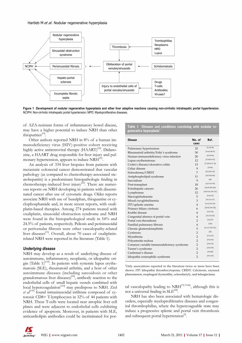
of AZA-resistant forms of inflammatory bowel disease, may have a higher potential to induce NRH than other thiopurines[19].
Other authors reported NRH in 8% of a human im-munodeficiency virus (HIV)-positive cohort receiving highly active antiretroviral therapy (HAART)[20]. Didano-zine, a HAART drug responsible for liver injury and pul-monary hypertension, appears to induce NRH[21].
An analysis of 334 liver biopsies from patients with metastatic colorectal cancer demonstrated that vascular pathology (as compared to chemotherapy-associated ste-atohepatitis) is a predominant histopathologic finding in chemotherapy-induced liver injury[22]. There are numer-ous reports on NRH developing in patients with dissemi-nated cancer after use of cytostatic drugs. Older reports associate NRH with use of busulphan, thioguanine or cy-clophosphamide and, in more recent reports, with oxali-platin-based therapies. Among 274 patients treated with oxaliplatin, sinusoidal obstruction syndrome and NRH were found in the histopathological study in 54% and 24.5% of patients, respectively. Peliosis and perisinusoidal or perivenular fibrosis were other vasculopathy-related liver diseases[23]. Overall, about 70 cases of oxaliplatin-related NRH were reported in the literature (Table 1).
Underlying diseaseNRH may develop as a result of underlying disease of autoimmune, inflammatory, neoplastic, or idiopathic ori-gin (Table 1)[143]. In patients with systemic lupus erythe-matosis (SLE), rheumatoid arthritis, and a host of other autoimmune diseases (including sarcoidosis or other granulomatous liver diseases)[90], antibody reaction to the endothelial cells of small hepatic vessels combined with local hypercoagulation[144] may predispose to NRH. Ziol et al[145] found intrasinusoidal infiltrate composed of cy-totoxic CD8+ T-lymphocytes in 32% of 44 patients with NRH. These T-cells were located near atrophic liver cell plates and were adjacent to endothelial cells exhibiting evidence of apoptosis. Moreover, in patients with SLE, anticardiolipin antibodies could be incriminated for por-
tal vasculopathy leading to NRH[78,79,86], although this is not a universal finding in SLE[68].
NRH has also been associated with hematologic dis-orders, especially myeloproliferative diseases and congen-ital thrombophilias, where the hypercoaguable state may induce a progressive splenic and portal vein thrombosis and subsequent portal hypertension[2].
1402 March 21, 2011|Volume 17|Issue 11|WJG|www.wjgnet.com
Figure 1 Development of nodular regenerative hyperplasia and other liver adaptive reactions causing non-cirrhotic intrahepatic portal hypertension. NCIPH: Non-cirrhotic intrahepatic portal hypertension; MPD: Myeloproliferative diseases.
NCIPH
Nodular regenerativehyperplasia
Sinusoidal obstruction syndrome
Perisinusoidal fibrosis
Hepato-portal sclerosis
Incomplete fibrotic septa
Obliteration of portalvenules/sinusoids
Injury to endothelial cells of portal venules/sinusoids
Thrombosis
Schistomatosis
TrombophiliasNeoplasmsMPD Lupus
DrugsT-cells AntibodiesViruses?
Table 1 Diseases and conditions coexisting with nodular re-generative hyperplasia1
Disease No. of cases
Ref.
Pulmonary hypertension 32 [11,24-44]
Rheumatoid arthritis/Felty’s syndrome 30 [31,41,44-55]
Human immunodeficiency virus infection 20 [21,56-64]
Lupus erythematosus 12 [27,40,65-70]
Crohn’s disease/ulcerative colitis 13 [17,28,56,71-76]
Celiac disease 9 [76-80]
Scleroderma/CREST 7 [25,32,81-85]
Antiphospholipid syndrome 11 [78,79,86-89]
Sarcoidosis 9 [90]
Post-transplant 18 [16,37,91-99]
Extrahepatic cancers 26 [26,59,100-104]
Lymphomas 12 [29,53,81,105-113]
Macroglobulinemia 5 [114,115]
Mixed cryoglobulinemia 3 [25,116,117]
ITP/aplastic anemia 4 [3,12,118,119]
Primary biliary cirrhosis 6 [25,82,120-122]
Krabbe disease 4 [123,124]
Congenital absence of portal vein 4 [15,125,126]
Portal vein thrombosis 2 [14,127]
Familial pulmonary fibrosis 4 [128]
Chronic glomerulonephritis 5 [51,117,129-131]
Cystinosis 2 [24]
Myasthenia 2 [132,133]
Polyarteritis nodosa 2 [134,135]
Common variable immunodeficiency syndrome 2 [136,137]
Turner’s syndrome 2 [138,139]
Castleman’s disease 2 [78,140]
Idiopathic eosinophilic syndrome 2 [141,142]
1Only associations reported in the literature twice or more have been shown. ITP: Idiopathic thrombocytopenia. CREST: Calcinosis, raynaud phenomenon, esophageal dysmotility, sclerodactyly, and telangiectasia.
Hartleb M et al . Nodular regenerative hyperplasia

DIAGNOSIS One should consider a diagnosis of NRH in all patients with clinical symptoms of portal hypertension (sple-nomegaly, esophageal varices, ascites) but with normal transaminases and no manifestations of cirrhosis (gyne-comastia, palmar erythema, spider nevi). There are mildly increased liver enzymes, usually alkaline phosphatase, in 11%-25% of patients[4,127,146]. It is estimated that NRH is complicated by clinically overt portal hypertension in at least 50% of cases, with an augmented hepatic venous pressure gradient confirming sinusoidal obstruction[147,148]. Close surveillance of patients with predisposing condi-tions is important for early diagnosis, especially in situa-tions where drug toxicity may play a role. In patients on AZA for autoimmune hepatitis, the occurrence of sple-nomegaly should alert the clinician to the development of portal hypertension secondary to NRH.
In all cases of NCIPH, more common treatable causes (viruses, alcohol, metabolic and autoimmune disorders) should be eliminated first, followed by an assessment of the usual exposures (acetaminophen, vitamin A, copper sul-fate, vinyl chloride, arsenic salt). Portal and hepatic venous thrombosis may be excluded on radiographic imaging.
ImagingImaging methods have poor sensitivity and specificity for NRH. A diffusely heterogeneous hepatic parenchyma may be the only imaging abnormality. On ultrasound, regenerative nodules are usually not visible due to a small size or isoechogenicity. The presence of well-delineated hypoechoic or isoechoic tiny lesions with a sonoluscent rim are indistinguishable from metastases[149]. Hyper-echoic nodules have been reported in very rare cases of NRH[150]. On computed tomography (CT), regenerative nodules remain isodense or hypodense in both arterial and portal venous phases, distinguishing NRH from focal nodular hyperplasia and adenomas[3].
The significance of magnetic resonance imaging in the diagnosis of NRH is still controversial, although because of its inherent propensity to resolve soft tissue details it may be superior to CT in visualization of regenera-tive nodules. NRH lesions appear hyperintense on T1-weighted images and iso- or hypointense on T2-weighted images[67,151], with a sensitivity and specificity of 70%-80% when using gadolinium contrast[152]; others found more disappointing results[153].
Histopathology Grossly, NRH presents as diffuse fine nodularity of the liver with 1-3 mm diameter nodules. Granularity of the hepatic surface may resemble micronodular cirrho-sis[77,144]. Rarely, nodules are larger[7,144], and may coalesce into a large tumor[154,155]. NRH nodules appear paler than the surrounding normal hepatic tissue. Mild hepatomega-ly may be present.
The diagnosis of NRH is secured by histopathology demonstrating regenerative nodules without parietal thick-
ening of portal venules, and no or minimal perisinusoidal and portal fibrosis on reticulin staining. Hepatocytes commonly show feathery degeneration of cytoplasm suggesting impairments of bile production or transport. Two morphologically distinct populations of hepatocytes coexist within the nodules: hypertrophied hepatocytes centrally surrounded by atrophic hepatocytes peripher-ally. Hypertrophic cells may compress terminal hepatic venules, which frequently appear shrunken and may be undetectable. Heterogeneity may be explained by uneven perfusion. Dilated sinusoids and thrombosed portal vein radicles are occasionally present[24,156,157].
If a dominant large regenerative nodule is sampled, adenoma-like features with discrete cytoplasmatic and/or nuclear atypia can be seen. Therefore, sampling a single nodule may yield an incorrect diagnosis and multiple liver sampling becomes necessary. Dysplastic large hepatocytes were seen in 42% of NRH samples, without high-degree dysplasia[9]. NRH should also be distinguished from LRN occurring in livers with disturbed hepatic venous outflow (e.g. Budd-Chiari syndrome, veno oclusive disease, or congestive pericarditis) but well developed compensatory arterialization and preserved hepatic venous collaterals.
It should be kept in mind that in small biopsy samples histologic features of NRH may be lacking or incom-plete[158]. The diagnosis can be made after careful exami-nation by the experienced hematopathologist with a high index of suspicion. In justified cases a laparoscopy with open wedge biopsy should be done.
Overlap syndromes, involving both NRH and portal or pericentral fibrosis due to hepatitis C viral infection, alcoholic, or non-alcoholic liver disease, can occur and in such cases the diagnosis of NRH may be easily over-looked[24,77,156]. Fibroscan or fibrotest panels may rule out cirrhosis, but have limited clinical value[153].
TREATMENTTreatment of NRH therapy is directed towards elimina-tion of the causative factor, once established. Concomi-tant diseases should be treated appropriately and with attention to minimizing drug toxicity. Long-term antico-agulation treatment is usually indicated in the thrombo-philias. Anticoagulation therapy was found to be benefi-cial in early stages of NRH induced by HAART in HIV-infected patients[57].
In patients with NRH, the mainstay of management is directed primarily to prevention and treatment of complications related to portal hypertension, i.e. variceal bleeding, the main source of mortality. Treatment of portal hypertension is standard: low sodium diet, diuret-ics, and endoscopic banding of esophageal varices. Sple-nectomy is not indicated. A portosystemic surgical shunt or transjugular intrahepatic portosystemic shunt may of-fer a significant therapeutic benefit, especially in the case of severe recurrent esophageal variceal hemorrhage[56]. Liver transplantation is rarely necessary and is reserved for patients with hepatic failure[159-162].
1403 March 21, 2011|Volume 17|Issue 11|WJG|www.wjgnet.com
Hartleb M et al . Nodular regenerative hyperplasia

NATURAL HISTORY AND PROGNOSISGenerally, the prognosis of NRH is better than that of chronic liver disease and is related to the complications of portal hypertension and the severity of the associated diseases, if present. In most cases the disease is slowly progressive, although the rate of nodular growth may be accelerated for unknown reasons[163]. The long-term prognosis is uncertain and considers the level of underly-ing myeloproliferative, thrombophilic, or autoimmune processes. There are several case reports demonstrating the presence of both NRH and hepatocellular carcinoma without underlying cirrhosis, although a neoplastic pro-cess has yet to be proven[163-167].
CONCLUSIONNRH is a rare condition of NCIPH liver diseases. Our understanding of the epidemiology of NRH is based upon case reports rather than systematic population stud-ies. Timely diagnosis is challenging, because the major-ity of patients do not present with overt signs of portal hypertension. The pathogenesis of NRH is not well es-tablished, but an adaptive hyperplastic reaction of hepa-tocytes appears to be related to abnormalities of hepatic venous perfusion. Immunosuppressive medications may induce NRH by damaging endothelial cells of small he-patic veins. NRH may also develop as a result of underly-ing autoimmune, inflammatory, neoplastic, or idiopathic disease.
The knowledge on tumorigenesis in NRH is virtually non-existent as compared with hepatocellular carcinoma, adenoma and even cirrhotic regeneration. Genetic studies indicate that RASSF1A, a gene acting in the proapop-totic pathway, is increasingly methylated in hepatocellular hyperplastic hepatocytes[168]. Moreover, in ceramide syn-thase 2 null mice that are unable to synthesize very long acyl chain ceramides, an extensive hepatocellular anisocy-tosis with widespread formation of regenerative nodules composed of hyperplastic hepatocytes was found[169]. This finding emphasizes the role of ceramide synthase 2 activity and altered hepatic sphingolipid profile for liver proliferative homeostasis.
Common causes of extrahepatic and intrahepatic portal hypertension should be excluded before making a diag-nosis of NRH. Imaging may be helpful during the initial clinical encounter with liver biopsy as the gold standard for diagnosis. An accurate diagnosis can only be made on histopathology, which shows diffuse micronodular trans-formation without fibrous septa. Management is directed primarily for prevention and treatment of complications related to portal hypertension. Outcome and prognosis is related to the severity of both portal hypertensive com-plications and the underlying associated diseases. Further studies may be helpful to elucidate a molecular mecha-nism for the vasculopathy that appears to play a central role in the adaptive hyperplastic reaction universally seen in NRH.
REFERENCES1 Hillaire S, Bonte E, Denninger MH, Casadevall N, Cadranel
JF, Lebrec D, Valla D, Degott C. Idiopathic non-cirrhotic in-trahepatic portal hypertension in the West: a re-evaluation in 28 patients. Gut 2002; 51: 275-280
2 Steiner PE. Nodular regenerative hyperplasia of the liver. Am J Pathol 1959; 35: 943-953
3 Mahamid J, Miselevich I, Attias D, Laor R, Zuckerman E, Shaoul R. Nodular regenerative hyperplasia associated with idiopathic thrombocytopenic purpura in a young girl: a case report and review of the literature. J Pediatr Gastroenterol Nutr 2005; 41: 251-255
4 Naber AH, Van Haelst U, Yap SH. Nodular regenerative hyperplasia of the liver: an important cause of portal hyper-tension in non-cirrhotic patients. J Hepatol 1991; 12: 94-99
5 Nakanuma Y, Hoso M, Sasaki M, Terada T, Katayanagi K, Nonomura A, Kurumaya H, Harada A, Obata H. Histopa-thology of the liver in non-cirrhotic portal hypertension of unknown aetiology. Histopathology 1996; 28: 195-204
6 Wanless IR. Micronodular transformation (nodular regen-erative hyperplasia) of the liver: a report of 64 cases among 2,500 autopsies and a new classification of benign hepatocel-lular nodules. Hepatology 1990; 11: 787-797
7 Reshamwala PA, Kleiner DE, Heller T. Nodular regenera-tive hyperplasia: not all nodules are created equal. Hepatol-ogy 2006; 44: 7-14
8 Nakanuma Y. Nodular regenerative hyperplasia of the liv-er: retrospective survey in autopsy series. J Clin Gastroenterol 1990; 12: 460-465
9 Colina F, Alberti N, Solis JA, Martinez-Tello FJ. Diffuse nodular regenerative hyperplasia of the liver (DNRH). A clinicopathologic study of 24 cases. Liver 1989; 9: 253-265
10 Elariny HA, Mizrahi SS, Hayes DH, Boudreaux JP, Hussey JL, Farr GH Jr. Nodular regenerative hyperplasia: a contro-versial indication for orthotopic liver transplantation. Transpl Int 1994; 7: 309-313
11 Dumortier J, Boillot O, Chevallier M, Berger F, Potier P, Valette PJ, Paliard P, Scoazec JY. Familial occurrence of nod-ular regenerative hyperplasia of the liver: a report on three families. Gut 1999; 45: 289-294
12 González-Huezo MS, Villela LM, Zepeda-Florencio Mdel C, Carrillo-Ponce CS, Mondragón-Sánchez RJ. Nodular re-generative hyperplasia associated to aplastic anemia: a case report and literature review. Ann Hepatol 2006; 5: 166-169
13 Al-Hamoudi WK, Pasieka JL, Urbanski SJ, Lee SS. Hepatic nodular regenerative hyperplasia in a patient with advanced carcinoid tumor. Eur J Gastroenterol Hepatol 2009; 21: 1083-1085
14 Fukai T, Ishibashi H, Noda T, Dohmen K, Kudo J, Nagafu-chi S, Sueishi K, Yamasaki T, Sameshima H. Nodular regen-erative hyperplasia of the liver with portal vein thrombosis and hyperplasia of the adrenal gland. Gastroenterol Jpn 1992; 27: 108-114
15 McDonald JA, Painter DM, Gallagher ND, McCaughan GW. Nodular regenerative hyperplasia mimicking cirrhosis of the liver. Gut 1990; 31: 725-727
16 Buster EH, van Vuuren HJ, Zondervan PE, Metselaar HJ, Tilanus HW, de Man RA. Thiopurine-methyltransferase and inosine triphosphate pyrophosphatase polymorphism in a liver transplant recipient developing nodular regenerative hyperplasia on low-dose azathioprine. Eur J Gastroenterol Hepatol 2008; 20: 68-72
17 Shastri S, Dubinsky MC, Fred Poordad F, Vasiliauskas EA, Geller SA. Early nodular hyperplasia of the liver occurring with inflammatory bowel diseases in association with thio-guanine therapy. Arch Pathol Lab Med 2004; 128: 49-53
18 Gane E, Portmann B, Saxena R, Wong P, Ramage J, Williams R. Nodular regenerative hyperplasia of the liver graft after liver transplantation. Hepatology 1994; 20: 88-94
19 Teml A, Schwab M, Hommes DW, Almer S, Lukas M, Feich-
1404 March 21, 2011|Volume 17|Issue 11|WJG|www.wjgnet.com
Hartleb M et al . Nodular regenerative hyperplasia

tenschlager T, Florin T, Seiderer J, Petritsch W, Bokemeyer B, Kreisel W, Herrlinger KR, Knoflach P, Bonaz B, Klugmann T, Herfarth H, Pedarnig N, Reinisch W. A systematic survey evaluating 6-thioguanine-related hepatotoxicity in patients with inflammatory bowel disease. Wien Klin Wochenschr 2007; 119: 519-526
20 Mallet V, Blanchard P, Verkarre V, Vallet-Pichard A, Fon-taine H, Lascoux-Combe C, Pol S. Nodular regenerative hyperplasia is a new cause of chronic liver disease in HIV-infected patients. AIDS 2007; 21: 187-192
21 Vispo E, Moreno A, Maida I, Barreiro P, Cuevas A, Alber-tos S, Soriano V. Noncirrhotic portal hypertension in HIV-infected patients: unique clinical and pathological findings. AIDS 2010; 24: 1171-1176
22 Ryan P, Nanji S, Pollett A, Moore M, Moulton CA, Gallinger S, Guindi M. Chemotherapy-induced liver injury in meta-static colorectal cancer: semiquantitative histologic analysis of 334 resected liver specimens shows that vascular injury but not steatohepatitis is associated with preoperative che-motherapy. Am J Surg Pathol 2010; 34: 784-791
23 Rubbia-Brandt L, Lauwers GY, Wang H, Majno PE, Tanabe K, Zhu AX, Brezault C, Soubrane O, Abdalla EK, Vauthey JN, Mentha G, Terris B. Sinusoidal obstruction syndrome and nodular regenerative hyperplasia are frequent oxali-platin-associated liver lesions and partially prevented by bevacizumab in patients with hepatic colorectal metastasis. Histopathology 2010; 56: 430-439
24 O'Brien K, Hussain N, Warady BA, Kleiner DE, Kleta R, Bernardini I, Heller T, Gahl WA. Nodular regenerative hy-perplasia and severe portal hypertension in cystinosis. Clin Gastroenterol Hepatol 2006; 4: 387-394
25 Riviere E, Vergniol J, Reffet A, Lippa N, Le Bail B, de Led-inghen V. Gastric variceal bleeding uncovering a rare asso-ciation of CREST syndrome, primary biliary cirrhosis, nodu-lar regenerative hyperplasia and pulmonary hypertension. Eur J Gastroenterol Hepatol 2010; 22: 1145-1148
26 Arotçarena R, Calès V, Berthelémy P, Parent Y, Malet M, Etcharry F, Ferrari S, Pariente A. Severe sinusoidal lesions: a serious and overlooked complication of oxaliplatin-contain-ing chemotherapy? Gastroenterol Clin Biol 2006; 30: 1313-1316
27 Colmegna I, deBoisblanc BP, Gimenez CR, Espinoza LR. Slow development of massive splenomegaly, portal and pulmonary hypertension in systematic lupus erythematosus: can nodular regenerative hyperplasia of the liver explain all these findings? Lupus 2005; 14: 976-978
28 Daniel F, Cadranel JF, Seksik P, Cazier A, Duong Van Huyen JP, Ziol M, Coutarel P, Loison P, Jian R, Marteau P. Azathioprine induced nodular regenerative hyperplasia in IBD patients. Gastroenterol Clin Biol 2005; 29: 600-603
29 González-Alegre MT, Guerra-Vales JM, Martínez-González, Gutiérrez A, Guillen V. Nodular regenerative hyperplasia of the liver and Hodgkin's disease: a case report. Acta Gastroen-terol Belg 2004; 67: 358-360
30 Jawaid Q, Saeed ZA, Di Bisceglie AM, Brunt EM, Ram-rakhiani S, Varma CR, Solomon H. Biliary-venous fistula complicating transjugular intrahepatic portosystemic shunt presenting with recurrent bacteremia, jaundice, anemia and fever. Am J Transplant 2003; 3: 1604-1607
31 Gento Peña E, Martín Lorente JL, Echevarría Iturbe C, Pérez Alvarez JC, Sáez-Royuela F, López Morante A, Ojeda Gimé-nez C. [Sinusoidal portal hypertension secondary to nodular regenerative hyperplasia of the liver]. Gastroenterol Hepatol 1999; 22: 183-185
32 Kaburaki J, Kuramochi S, Fujii T, Kuwana M, Tojo T, Ikeda Y, Hosada Y. Nodular regenerative hyperplasia of the liver in a patient with systemic sclerosis. Clin Rheumatol 1996; 15: 613-616
33 Henkel M, Paquet KJ, Rühl U. [Correlation between pulmo-nary hypertension and portal hypertension--2 case reports of different forms of pre-sinusoidal portal hypertension].
Leber Magen Darm 1994; 24: 10-1434 Calès P, Voigt JJ, Payen JL, Bloom E, Berg P, Vinel JP, Pradère
B, Broussy P, Pascal JP. Diffuse vascular ectasia of the an-trum, duodenum, and jejunum in a patient with nodular regenerative hyperplasia. Lack of response to portosystemic shunt or gastrectomy. Gut 1993; 34: 558-561
35 Portmann B, Stewart S, Higenbottam TW, Clayton PT, Lloyd JK, Williams R. Nodular transformation of the liver associated with portal and pulmonary arterial hypertension. Gastroenterology 1993; 104: 616-621
36 Trauner M, Stepan KM, Resch M, Ebner F, Pristautz H, Klimpfinger M. Diagnostic problems in nodular regenerative hyperplasia (nodular transformation) of the liver. Review of the literature and report of two cases. Z Gastroenterol 1992; 30: 187-194
37 Duvoux C, Kracht M, Lang P, Vernant JP, Zafrani ES, Dhumeaux D. [Nodular regenerative hyperplasia of the liver associated with azathioprine therapy]. Gastroenterol Clin Biol 1991; 15: 968-973
38 Bedossa P, Ngendayo L, Hervé P, Rain B, Poynard T, Si-monneau G. [Nodular regenerative hyperplasia and pulmo-nary arterial hypertension]. Gastroenterol Clin Biol 1990; 14: 881-884
39 Yutani C, Imakita M, Ishibashi-Ueda H, Okubo S, Naito M, Kunieda T. Nodular regenerative hyperplasia of the liver as-sociated with primary pulmonary hypertension. Hum Pathol 1988; 19: 726-731
40 Kuramochi S, Tashiro Y, Torikata C, Watanabe Y. Systemic lupus erythematosus associated with multiple nodular hy-perplasia of the liver. Acta Pathol Jpn 1982; 32: 547-560
41 Qizilbash AH, Castelli M. Nodular regenerative hyperpla-sia of the liver: diagnosis by liver biopsy. Can Med Assoc J 1980; 122: 1151-1154
42 Lebrec D, Capron JP, Dhumeaux D, Benhamou JP. Pulmo-nary hypertension complicating portal hypertension. Am Rev Respir Dis 1979; 120: 849-856
43 Shorey J, Weinberg MN, Frenkel EP, Fallis BD. Nodular re-generative hyperplasia of the liver in a case of myelofibrosis with extramedullary hematopoiesis and secondary portal venous hypertension. Am J Clin Pathol 1979; 72: 122-125
44 Wechsler J, Belaiche J, Vesin P, Cattan D, Pinaudeau Y. [Anatomo-clinical study of a case of regenerative nodular hyperplasia of the liver with Felty's syndrome and portal hypertension]. Ann Anat Pathol (Paris) 1979; 24: 325-336
45 Riesco Díaz M, Manzano Gómez F, Tirado Domínguez M, Suárez García E, Pujol de la Llave E. [Rheumatoid arthritis and hepatic nodular regenerative hyperplasia]. Rev Clin Esp 1994; 194: 70
46 Bills LJ, Seibert D, Brick JE. Liver disease, erroneously at-tributed to methotrexate, in a patient with rheumatoid ar-thritis. J Rheumatol 1992; 19: 1963-1965
47 Young ID, Segura J, Ford PM, Ford SE. The pathogenesis of nodular regenerative hyperplasia of the liver associated with rheumatoid vasculitis. J Clin Gastroenterol 1992; 14: 127-131
48 Perez Ruiz F, Orte Martinez FJ, Zea Mendoza AC, Ruiz del Arbol L, Moreno Caparros A. Nodular regenerative hyper-plasia of the liver in rheumatic diseases: report of seven cases and review of the literature. Semin Arthritis Rheum 1991; 21: 47-54
49 Forbes GM, Shilkin KB, Reed WD. Nodular regenerative hyperplasia of the liver: the importance of combined mac-roscopic and microscopic findings. Med J Aust 1991; 154: 415-417
50 Nakad A, Moens P, Najm S, Haot J, Janssen J, Geubel AP. Nodular regenerative hyperplasia of the liver and esophageal varices in Felty's syndrome: a clinicopathologic case report with review of the literature. Acta Clin Belg 1988; 43: 45-50
51 Reynolds WJ, Wanless IR. Nodular regenerative hyperplasia of the liver in a patient with rheumatoid vasculitis: a mor-
1405 March 21, 2011|Volume 17|Issue 11|WJG|www.wjgnet.com
Hartleb M et al . Nodular regenerative hyperplasia
1406 March 21, 2011|Volume 17|Issue 11|WJG|www.wjgnet.com
phometric study suggesting a role for hepatic arteritis in the pathogenesis. J Rheumatol 1984; 11: 838-842
52 Cohen MD, Ginsburg WW, Allen GL. Nodular regenerative hyperplasia of the liver and bleeding esophageal varices in Felty's syndrome: a case report and literature review. J Rheu-matol 1982; 9: 716-718
53 Wanless IR, Godwin TA, Allen F, Feder A. Nodular regen-erative hyperplasia of the liver in hematologic disorders: a possible response to obliterative portal venopathy. A mor-phometric study of nine cases with an hypothesis on the pathogenesis. Medicine (Baltimore) 1980; 59: 367-379
54 Belaiche J, Vesin P, Fischer D, Wechsler J, Franco D, Bismuth H, Cattan D. [Nodular regenerative hyperplasia of the liver with portal hypertension associated with Felty's syndrome. Report of a case (author's transl)]. Gastroenterol Clin Biol 1978; 2: 63-70
55 Reisman T, Levi JU, Zeppa R, Clark R, Morton R, Schiff ER. Noncirrhotic portal hypertension in Felty's syndrome. Am J Dig Dis 1977; 22: 145-148
56 Schouten JN, Verheij J, Janssen HL. [Non-cirrhotic portal hypertension: rare cause of upper gastrointestinal bleeding]. Ned Tijdschr Geneeskd 2010; 154: A1276
57 Bihl F, Janssens F, Boehlen F, Rubbia-Brandt L, Hadengue A, Spahr L. Anticoagulant therapy for nodular regenerative hy-perplasia in a HIV-infected patient. BMC Gastroenterol 2010; 10: 6
58 Mendizabal M, Craviotto S, Chen T, Silva MO, Reddy KR. Noncirrhotic portal hypertension: another cause of liver dis-ease in HIV patients. Ann Hepatol 2009; 8: 390-395
59 Kochin I, Magid M, Arnon R, Glasscock A, Kerkar N, Miloh T. Variceal bleeding in an adolescent with HIV diagnosed with hepatoportal sclerosis and nodular regenerative hyper-plasia. J Pediatr Gastroenterol Nutr 2010; 50: 340-343
60 Dinh MH, Stosor V, Rao SM, Miller FH, Green RM. Crypto-genic liver disease in HIV-seropositive men. HIV Med 2009; 10: 447-453
61 Ding A, Lee A, Callender M, Loughrey M, Quah SP, Dins-more WW. Hepatic encephalopathy as an unusual late com-plication of transjugular intrahepatic portosystemic shunt insertion for non-cirrhotic portal hypertension caused by nodular regenerative hyperplasia in an HIV-positive patient on highly active antiretroviral therapy. Int J STD AIDS 2010; 21: 71-72
62 Tateo M, Sebagh M, Bralet MP, Teicher E, Azoulay D, Mal-let V, Pol S, Castaing D, Samuel D, Duclos-Vallée JC. A new indication for liver transplantation: nodular regenerative hyperplasia in human immunodeficiency virus-infected pa-tients. Liver Transpl 2008; 14: 1194-1198
63 Podevin P, Spiridon G, Terris B, Chauvelot-Moachon L, Guil-levin L, Chaussade S, Sogni P, Salmon-Ceron D. Nodular regenerative hyperplasia of the liver after IL-2 therapy in an HIV-infected patient. AIDS 2006; 20: 313-315
64 Fernandez-Miranda C, Colina F, Delgado JM, Lopez-Carreira M. Diffuse nodular regenerative hyperplasia of the liver as-sociated with human immunodeficiency virus and visceral leishmaniasis. Am J Gastroenterol 1993; 88: 433-435
65 Leung VK, Loke TK, Luk IS, Ng WL, Chau TN, Law ST, Chan JC. Nodular regenerative hyperplasia of the liver asso-ciated with systemic lupus erythematosus: three cases. Hong Kong Med J 2009; 15: 139-142
66 Leung VK, Ng WL, Luk IS, Chau TN, Chan WH, Kei SK, Loke TK. Unique hepatic imaging features in a patient with nodular regenerative hyperplasia of the liver associating with systemic lupus erythematosus. Lupus 2007; 16: 205-208
67 Horita T, Tsutsumi A, Takeda T, Yasuda S, Takeuchi R, Amasaki Y, Ichikawa K, Atsumi T, Koike T. Significance of magnetic resonance imaging in the diagnosis of nodular regenerative hyperplasia of the liver complicated with sys-temic lupus erythematosus: a case report and review of the literature. Lupus 2002; 11: 193-196
68 Sekiya M, Sekigawa I, Hishikawa T, Iida N, Hashimoto H, Hirose S. Nodular regenerative hyperplasia of the liver in systemic lupus erythematosus. The relationship with anti-cardiolipin antibody and lupus anticoagulant. Scand J Rheu-matol 1997; 26: 215-217
69 Guitton P, Touchard G, Beauchant M. [Diffuse nodular re-generative hyperplasia of the liver and disseminated lupus erythematosus]. Gastroenterol Clin Biol 1987; 11: 102-103
70 Klemp P, Timme AH, Sayers GM. Systemic lupus erythe-matosus and nodular regenerative hyperplasia of the liver. Ann Rheum Dis 1986; 45: 167-170
71 Bryant DL, Miles CJ, Gearry RB. Nodular regenerative hy-perplasia of the liver secondary to azathioprine in a patient with inflammatory bowel disease. N Z Med J 2010; 123: 74-76
72 Schumann M, Preiss JC, Loddenkemper C, Günther U, So-masundaram R, Siegmund B, Zeitz M. [Nodular regenerative hyperplasia as a side effect of azathioprine in a patient with Crohn's disease]. Dtsch Med Wochenschr 2008; 133: 1897-1900
73 Ansari A, Elliott T, Fong F, Arenas-Hernandez M, Rot-tenberg G, Portmann B, Lucas S, Marinaki A, Sanderson J. Further experience with the use of 6-thioguanine in patients with Crohn's disease. Inflamm Bowel Dis 2008; 14: 1399-1405
74 Ehmsen L, Marko C, Breidert M. [Portal vein hypertension during azathioprine therapy in patients with Crohn's dis-ease--a frequent phenomenon?]. Dtsch Med Wochenschr 2008; 133: 950-953
75 Arnott ID, Ghosh S. Portal hypertension in the presence of minimal liver damage in Crohn's disease on long-term aza-thioprine: possible endothelial cell injury. Eur J Gastroenterol Hepatol 2000; 12: 569-573
76 Tinsa F, Brini I, El May A, Bousnina D, Boussetta K, Bous-nina S. Nodular regenerative hyperplasia of the liver, celiac disease and Sjogren's syndrome in a child. Gastroenterol Clin Biol 2010; 34: 40-42
77 Biecker E, Trebicka J, Fischer HP, Sauerbruch T, Lammert F. Portal hypertension and nodular regenerative hyperpla-sia in a patient with celiac disease. Z Gastroenterol 2006; 44: 395-398
78 Cancado EL, Medeiros DM, Deguti MM, Dos Santos MS, de Mello ES, Vendramini MB, Carrilho FJ. Celiac disease as-sociated with nodular regenerative hyperplasia, pulmonary abnormalities, and IgA anticardiolipin antibodies. J Clin Gastroenterol 2006; 40: 135-139
79 Austin A, Campbell E, Lane P, Elias E. Nodular regenera-tive hyperplasia of the liver and coeliac disease: potential role of IgA anticardiolipin antibody. Gut 2004; 53: 1032-1034
80 Riestra S, Domínguez F, Rodrigo L. Nodular regenerative hyperplasia of the liver in a patient with celiac disease. J Clin Gastroenterol 2001; 33: 323-326
81 Agard C, Ponge T, Mahé B, Barrier J. [Lymphocytic lympho-ma and regenerative liver nodular hyperplasia in systemic scleroderma]. Rev Med Interne 2000; 21: 301-303
82 McMahon RF, Babbs C, Warnes TW. Nodular regenerative hyperplasia of the liver, CREST syndrome and primary bili-ary cirrhosis: an overlap syndrome? Gut 1989; 30: 1430-1433
83 García Díaz JD, Praga Terente M, Colina Ruizdelgado F, Lizasoaín Hernández M, Mesa Latorre J. [Nodular regenera-tive hyperplasia of the liver associated with a CREST syn-drome with multiorgan involvement]. An Med Interna 1989; 6: 203-206
84 Cadranel JF, Grippon P, Wechsler B, Bidegaray E, Kark-ouche B, Opolon P. [The CRST syndrome and nodular re-generative hyperplasia of the liver. A case]. Presse Med 1987; 16: 1656
85 Friguet JL, Deugnier Y, Messner M, Lauvin R, Launois B, Brissot P, Ferrand B, Bourel M. [Regenerative nodular hy-perplasia in scleroderma : a new case]. Gastroenterol Clin Biol 1984; 8: 979
86 Gaya DR, Oien KA, Stanley AJ, Morris AJ. Bleeding gastric varices and antiphospholipid syndrome. Nat Clin Pract Gas-
Hartleb M et al . Nodular regenerative hyperplasia

1407 March 21, 2011|Volume 17|Issue 11|WJG|www.wjgnet.com
troenterol Hepatol 2005; 2: 156-159; quiz 1 p following 15987 Morlà RM, Ramos-Casals M, García-Carrasco M, Cervera R,
Font J, Bruguera M, Rojas-Rodríguez J, Ingelmo M. Nodular regenerative hyperplasia of the liver and antiphospholipid antibodies: report of two cases and review of the literature. Lupus 1999; 8: 160-163
88 Cadranel JF, Demontis R, Guettier C, Bouraya D, Dautreaux M, Ghazali A, Poux JM, Coutarel P, Devergie B, Fievet P. [Nodular regenerative hyperplasia associated with primary antiphospholipid syndrome]. Gastroenterol Clin Biol 1996; 20: 901-904
89 Keegan AD, Brooks LT, Painter DM. Hepatic infarction and nodular regenerative hyperplasia of the liver with associ-ated anticardiolipin antibodies in a young woman. J Clin Gastroenterol 1994; 18: 309-313
90 Devaney K, Goodman ZD, Epstein MS, Zimmerman HJ, Ishak KG. Hepatic sarcoidosis. Clinicopathologic features in 100 patients. Am J Surg Pathol 1993; 17: 1272-1280
91 Toso C, Isse K, Demetris AJ, Dinyari P, Koh A, Imes S, Kin T, Emamaullee J, Senior P, Shapiro AM. Histologic graft as-sessment after clinical islet transplantation. Transplantation 2009; 88: 1286-1293
92 Pezzullo L, Muretto P, De Rosa G, Picardi M, Lucania A, Rotoli B. Liver nodular regenerative hyperplasia after bone marrow transplant. Haematologica 2000; 85: 669-670
93 Ichikawa Y, Kyo M, Hanafusa T, Kohro T, Kishikawa H, Fu-kunishi T, Nagano S, Shinji Y. A 20-year case study of a kidney transplant recipient with chronic active hepatitis C: clinical course and successful treatment for late acute rejection induced by interferon therapy. Transplantation 1998; 65: 134-138
94 Sebagh M, Farges O, Samuel D, Bismuth H, Reynès M. Nod-ular regenerative hyperplasia of the liver following ortho-topic liver transplantation. Transplant Proc 1995; 27: 2510-2511
95 Xerri L, Payan MJ, Choux R, Gros N, Hassoun J, Chamlian A, Blin D, Monties JR. An exceptional 18-year follow-up after cardiac transplantation. How can malignancies occur during immunosuppressive therapy? Cancer 1989; 63: 1697-1699
96 Jones MC, Best PV, Catto GR. Is nodular regenerative hy-perplasia of the liver associated with azathioprine therapy after renal transplantation? Nephrol Dial Transplant 1988; 3: 331-333
97 Buffet C, Cantarovitch M, Pelletier G, Fabre M, Martin E, Charpentier B, Etienne JP, Fries D. Three cases of nodular regenerative hyperplasia of the liver following renal trans-plantation. Nephrol Dial Transplant 1988; 3: 327-330
98 Morales JM, Prieto C, Colina F, Mestre MJ, Lopez I, Perez-Sola A, Solis Herruzo JA, Ruilope LM, Rodicio JL. Nodular regenerative hyperplasia of the liver in renal transplanta-tion. Transplant Proc 1987; 19: 3694-3696
99 Bredfeldt JE, Havey AL. Nodular regenerative hyperplasia of the liver following renal transplantation. Dig Dis Sci 1981; 26: 271-274
100 van den Broek MA, Olde Damink SW, Driessen A, Dejong CH, Bemelmans MH. Nodular regenerative hyperplasia secondary to neoadjuvant chemotherapy for colorectal liver metastases. Case Report Med 2009; 2009: 457975
101 Citak EC, Karadeniz C, Oguz A, Boyunaga O, Ekinci O, Okur V. Nodular regenerative hyperplasia and focal nodu-lar hyperplasia of the liver mimicking hepatic metastasis in children with solid tumors and a review of literature. Pediatr Hematol Oncol 2007; 24: 281-289
102 Chu WC, Roebuck DJ. Nodular regenerative hyperplasia of the liver simulating metastases following treatment for bilateral Wilms tumor. Med Pediatr Oncol 2003; 41: 85-87
103 Minato H, Nakanuma Y. Nodular regenerative hyperplasia of the liver associated with metastases of pancreatic endo-crine tumour: report of two autopsy cases. Virchows Arch A Pathol Anat Histopathol 1992; 421: 171-174
104 Cervera R, Abós J, Bruguera M, Grau JM. [Regenerative nodular hepatic hyperplasia and gastric carcinoma]. Rev Clin
Esp 1987; 181: 341-342105 Kataoka TR, Tsukamoto Y, Kanazawa N, Izumi T, Awata
N, Nishizawa Y, Ohsawa M, Ishiguro S. Concomitant hepa-tocellular carcinoma and non-Hodgkin's lymphoma in a pa-tient with nodular regenerative hyperplasia. Pathol Int 2006; 56: 279-282
106 Kiyuna A, Sunagawa T, Hokama A, Touyama M, Tomiya-ma R, Sakugawa H, Kinjo F, Saito A. Nodular regenerative hyperplasia of the liver and Castleman's disease: potential role of interleukin-6. Dig Dis Sci 2005; 50: 314-316
107 Iwabe C, Hashimoto E, Aoka K, Watanabe U, Hayashi N. [A case of non-Hodgkin's lymphoma with portal hypertension and nodular regenerative hyperplasia]. Nippon Shokakibyo Gakkai Zasshi 1994; 91: 2239-2243
108 Feng WJ, Takayasu K, Konda C, Yamazaki S, Sakamoto M, Hirohashi S, Takenaka T. CT of nodular hyperplasia of the liver in non-Hodgkin lymphoma. J Comput Assist Tomogr 1991; 15: 1031-1034
109 Mesa Latorre J, de Dios García Díaz J, Larregla Garraus S, Ortiz Conde MC. [Nodular regenerative hyperplasia of the liver and acute lymphoblastic leukemia]. Rev Clin Esp 1990; 186: 192-193
110 Zuber M, Gherardi R, Defer G, Gaulard P, Divine M, Zaf-rani ES. Peripheral neuropathy, coagulopathy and nodular regenerative hyperplasia of the liver in a patient with mul-tiple serologic auto-antibody activities and IgM B-cell lym-phoma. J Intern Med 1989; 226: 291-295
111 Ramos Soria F, Puig Pujol X, Vida Mombiela F, Badal Alter JM. [Hemoperitoneum as the first manifestation of hepatic regenerative nodular hyperplasia associated with Hodgkin's lymphoma]. Rev Clin Esp 1989; 184: 333
112 Rozman M, Montserrat E, Bruguera M, Grau JM, Terés J, Rozman C. [Chronic lymphatic leukemia and portal hyper-tension: a causal association?]. Med Clin (Barc) 1989; 92: 26-28
113 Kitazono M, Saito Y, Kinoshita M, Nakashima T, Kojiro M, Tanaka H. Nodular regenerative hyperplasia of the liver in a patient with multiple myeloma and systemic amyloidosis. Acta Pathol Jpn 1985; 35: 961-967
114 Voinchet O, Degott C, Scoazec JY, Feldmann G, Benhamou JP. Peliosis hepatis, nodular regenerative hyperplasia of the liver, and light-chain deposition in a patient with Walden-ström's macroglobulinemia. Gastroenterology 1988; 95: 482-486
115 Wanless IR, Solt LC, Kortan P, Deck JH, Gardiner GW, Prokipchuk EJ. Nodular regenerative hyperplasia of the liver associated with macroglobulinemia. A clue to the pathogenesis. Am J Med 1981; 70: 1203-1209
116 García Buey L, García Sánchez A, Moreno Otero R, González Estecha A, Pozo A, Pajares JM. [Hepatic nodular regenera-tive hyperplasia associated with essential mixed cryoglobuli-nemia]. Rev Clin Esp 1987; 181: 285-287
117 Mignon F, Ronco P, Verroust P, Poupon R, Prud'homme de Saint-Maur P, Morel-Maroger L. [Nephropathy and nodular regenerative hyperplasia of the liver (author's transl)]. Ne-phrologie 1980; 1: 23-26
118 Carbone J, Escudero A, Mayayo M, Ballesteros M, Perez-Corral A, Sanchez-Ramon S, Sarmiento E, Micheloud D, Fernandez-Cruz E. Partial response to anti-CD20 monoclo-nal antibody treatment of severe immune thrombocytopenic purpura in a patient with common variable immunodefi-ciency. Ann N Y Acad Sci 2005; 1051: 666-671
119 Hilario Rodríguez E, Velasco Oses A, Coma y del Corral MJ, Sáenz García JA. [Diffuse nodular regenerative hyperplasia of the liver associated with autoimmune hemolytic anemia. Presentation of a case and review of the literature]. Rev Esp Enferm Apar Dig 1983; 63: 45-51
120 Castellano G, Colina F, Moreno D, Pinedo F, Canga F, Solís Herruzo JA. [Nodular regenerative hepatic hyperplasia as-sociated with primary biliary cirrhosis]. Rev Clin Esp 1992; 191: 433-434
121 Buffet C, Sinico M, Adda G, Fabre M, Roge J, Pelletier G,
Hartleb M et al . Nodular regenerative hyperplasia

1408 March 21, 2011|Volume 17|Issue 11|WJG|www.wjgnet.com
Martin E, Etienne JP. [Nodular regenerative hyperplasia and primary biliary cirrhosis]. Gastroenterol Clin Biol 1991; 15: 856-858
122 Chazouillères O, Andreani T, Legendre C, Poupon R, Dar-nis F. [Nodular regenerative hyperplasia of the liver and primary biliary cirrhosis. A fortuitous association?]. Gastro-enterol Clin Biol 1986; 10: 764-766
123 Mones JM, Saldana MJ, Albores-Saavedra J. Nodular regen-erative hyperplasia of the liver. Report of three cases and re-view of the literature. Arch Pathol Lab Med 1984; 108: 741-743
124 Mones JM, Saldana MJ. Nodular regenerative hyperplasia of the liver in a 4-month-old infant. Am J Dis Child 1984; 138: 79-81
125 Grazioli L, Alberti D, Olivetti L, Rigamonti W, Codazzi F, Matricardi L, Fugazzola C, Chiesa A. Congenital absence of portal vein with nodular regenerative hyperplasia of the liver. Eur Radiol 2000; 10: 820-825
126 Arana E, Martí-Bonmatí L, Martínez V, Hoyos M, Montes H. Portal vein absence and nodular regenerative hyperplasia of the liver with giant inferior mesenteric vein. Abdom Imaging 1997; 22: 506-508
127 Buchel O, Roskams T, Van Damme B, Nevens F, Pirenne J, Fevery J. Nodular regenerative hyperplasia, portal vein thrombosis, and avascular hip necrosis due to hyperhomo-cysteinaemia. Gut 2005; 54: 1021-1023
128 Talbot-Smith A, Syn WK, MacQuillan G, Neil D, Elias E, Ryan P. Familial idiopathic pulmonary fibrosis in association with bone marrow hypoplasia and hepatic nodular regen-erative hyperplasia: a new "trimorphic" syndrome. Thorax 2009; 64: 440-443
129 Haboubi NY, Smith VJ, Coyne JD, Ackrill P. Nodular regen-erative hyperplasia of the liver in a non-treated patient with mesangiocapillary glomerulonephritis. Histopathology 1991; 18: 367-368
130 Haratake J, Horie A, Takasugi M, Hiroshige K. Nodular regenerative hyperplasia of the liver associated with idio-pathic membranous glomerulonephritis. Am J Gastroenterol 1987; 82: 677-680
131 McCulloch AJ, Morley AR, Wilkinson R, Lesna M. Nodular regenerative hyperplasia of the liver with membranous glo-merulonephritis. Postgrad Med J 1981; 57: 402-403
132 Fonseca V, Havard CW. Portal hypertension secondary to azathioprine in myasthenia gravis. Postgrad Med J 1988; 64: 950-952
133 Eliakim R, Ligumsky M, Jurim O, Shouval D. Nodular re-generative hyperplasia with portal hypertension in a patient with myasthenia gravis. Am J Gastroenterol 1987; 82: 674-676
134 Goritsas CP, Repanti M, Papadaki E, Lazarou N, Andonopou-los AP. Intrahepatic bile duct injury and nodular regenerative hyperplasia of the liver in a patient with polyarteritis nodosa. J Hepatol 1997; 26: 727-730
135 Nakanuma Y, Ohta G, Sasaki K. Nodular regenerative hy-perplasia of the liver associated with polyarteritis nodosa. Arch Pathol Lab Med 1984; 108: 133-135
136 Sasaki M, Hodo Y, Nakanuma Y. Hepatocellular carcinoma arising in primary biliary cirrhosis presenting with nodular regenerative hyperplasia: report of an autopsy case. J Gas-troenterol Hepatol 2006; 21: 1866-1868
137 Ravindran J, Gillis D, Rowland R, Heddle R. Common vari-able immunodeficiency associated with nodular regenera-tive hyperplasia of the liver. Aust N Z J Med 1995; 25: 741
138 Thevenot T, Dhote R, Tulliez M, Baverel F, Permal S, Rabi-neau D, Christoforov B. [Turner syndrome and nodular re-generative hyperplasia of the liver]. Ann Med Interne (Paris) 1998; 149: 295-296
139 de Lédinghen V, Levillain P, Besson I, Palazzo L, Fabre M, Silvain C, Morichau-Beauchant M. [Nodular regenerative hyperplasia of the liver and Turner syndrome]. Gastroenterol Clin Biol 1994; 18: 898-899
140 Molina T, Delmer A, Le Tourneau A, Texier P, Degott C,
Audoin J, Zittoun R, Diebold J. Hepatic lesions of vascular origin in multicentric Castleman's disease, plasma cell type: report of one case with peliosis hepatis and another with perisinusoidal fibrosis and nodular regenerative hyperpla-sia. Pathol Res Pract 1995; 191: 1159-1164
141 Nakamura Y, Takeuchi T, Kanaoka H, Okanoue T, Kashima K. [A case of nodular regenerative hyperplasia of liver with idiopathic hypereosinophilic syndrome]. Nippon Shokakibyo Gakkai Zasshi 1997; 94: 44-48
142 Baker BL, Axiotis C, Hurwitz ES, Leavitt R, Di Bisceglie AM. Nodular regenerative hyperplasia of the liver in idiopathic hypereosinophilic syndrome. J Clin Gastroenterol 1991; 13: 452-456
143 Morris JM, Oien KA, McMahon M, Forrest EH, Morris J, Stanley AJ, Campbell S. Nodular regenerative hyperplasia of the liver: survival and associated features in a UK case series. Eur J Gastroenterol Hepatol 2010; 22: 1001-1005
144 Wang HM, Lo GH, Hsu PI, Lin CK, Chan HH, Chen WC, Lai KH, Wang BW, Lin SL. Nodular regenerative hyperpla-sia of the liver. J Chin Med Assoc 2008; 71: 523-527
145 Ziol M, Poirel H, Kountchou GN, Boyer O, Mohand D, Mouthon L, Tepper M, Guillet JG, Guettier C, Raphael M, Beaugrand M. Intrasinusoidal cytotoxic CD8+ T cells in nod-ular regenerative hyperplasia of the liver. Hum Pathol 2004; 35: 1241-1251
146 Al-Mukhaizeem KA, Rosenberg A, Sherker AH. Nodular regenerative hyperplasia of the liver: an under-recognized cause of portal hypertension in hematological disorders. Am J Hematol 2004; 75: 225-230
147 Ferlitsch A, Teml A, Reinisch W, Ulbrich G, Wrba F, Ho-moncik M, Gangl A, Peck-Radosavljevic M, Vogelsang H. 6-thioguanine associated nodular regenerative hyperplasia in patients with inflammatory bowel disease may induce portal hypertension. Am J Gastroenterol 2007; 102: 2495-2503
148 Ueno S, Tanabe G, Sueyoshi K, Yoshinaka H, Yamamoto S, Kurita K, Yoshidome S, Nuruki K, Aikou T. Hepatic hemo-dynamics in a patient with nodular regenerative hyperpla-sia. Am J Gastroenterol 1996; 91: 1012-1015
149 Clouet M, Boulay I, Boudiaf M, Soyer P, Nemeth J, Kiselman R, Rymer R. Imaging features of nodular regenerative hy-perplasia of the liver mimicking hepatic metastases. Abdom Imaging 1999; 24: 258-261
150 Casillas C, Martí-Bonmatí L, Galant J. Pseudotumoral pre-sentation of nodular regenerative hyperplasia of the liver: imaging in five patients including MR imaging. Eur Radiol 1997; 7: 654-658
151 Rha SE, Lee MG, Lee YS, Kang GH, Ha HK, Kim PN, Auh YH. Nodular regenerative hyperplasia of the liver in Budd-Chiari syndrome: CT and MR features. Abdom Imaging 2000; 25: 255-258
152 Zech CJ, Seiderer J, Reinisch W, Ochsenkuhn T, Schima W, Diebold J, Wrba F, Reiser MF, Schoenberg SO. Thioguanin-induced nodular regenerative hyperplasia of the liver-ROC analysis of different MR techniques. Eur Radiol 2007; 17: 1898-1905
153 Laharie D, Vergniol J, Bioulac-Sage P, Diris B, Poli J, Fouch-er J, Couzigou P, Drouillard J, de Lédinghen V. Usefulness of noninvasive tests in nodular regenerative hyperplasia of the liver. Eur J Gastroenterol Hepatol 2010; 22: 487-493
154 Ames JT, Federle MP, Chopra K. Distinguishing clinical and imaging features of nodular regenerative hyperplasia and large regenerative nodules of the liver. Clin Radiol 2009; 64: 1190-1195
155 Dachman AH, Ros PR, Goodman ZD, Olmsted WW, Ishak KG. Nodular regenerative hyperplasia of the liver: clinical and radiologic observations. AJR Am J Roentgenol 1987; 148: 717-722
156 Ibarrola C, Colina F. Clinicopathological features of nine cases of non-cirrhotic portal hypertension: current defini-tions and criteria are inadequate. Histopathology 2003; 42:
Hartleb M et al . Nodular regenerative hyperplasia

1409 March 21, 2011|Volume 17|Issue 11|WJG|www.wjgnet.com
251-264157 Arvanitaki M, Adler M. Nodular regenerative hyperplasia
of the liver. A review of 14 cases. Hepatogastroenterology 2001; 48: 1425-1429
158 Seiderer J, Zech CJ, Diebold J, Schoenberg SO, Brand S, Tillack C, Göke B, Ochsenkühn T. Nodular regenerative hyperplasia: a reversible entity associated with azathioprine therapy. Eur J Gastroenterol Hepatol 2006; 18: 553-555
159 Krishna M, Keaveny AP, Genco PV, Rosser BG, Dickson RC, Nguyen JH, Steers JL, Nakhleh RE. Clinicopathological review of 18 cases of liver allografts lost due to bile duct ne-crosis. Transplant Proc 2005; 37: 2221-2223
160 Radomski JS, Chojnacki KA, Moritz MJ, Rubin R, Armenti VT, Wilson GA, Herrine S, Conn M. Results of liver trans-plantation for nodular regenerative hyperplasia. Am Surg 2000; 66: 1067-1070
161 Loinaz C, Colina F, Musella M, Lopez-Rios F, Gomez R, Jimenez C, Gonzalez Pinto I, Garcia I, Moreno Gonzalez E. Orthotopic liver transplantation in 4 patients with portal hypertension and non-cirrhotic nodular liver. Hepatogastro-enterology 1998; 45: 1787-1794
162 Dumortier J, Bizollon T, Scoazec JY, Chevallier M, Bancel B, Berger F, Ducerf C, Claudel-Bonvoisin S, Paliard P, Boillot O, Trepo C. Orthotopic liver transplantation for idiopathic portal hypertension: indications and outcome. Scand J Gas-troenterol 2001; 36: 417-422
163 Nzeako UC, Goodman ZD, Ishak KG. Hepatocellular carci-
noma and nodular regenerative hyperplasia: possible patho-genetic relationship. Am J Gastroenterol 1996; 91: 879-884
164 Kobayashi S, Saito K, Nakanuma Y. Nodular regenerative hyperplasia of the liver in hepatocellular carcinoma. An au-topsy study. J Clin Gastroenterol 1993; 16: 155-159
165 Stromeyer FW, Ishak KG. Nodular transformation (nodular "regenerative" hyperplasia) of the liver. A clinicopathologic study of 30 cases. Hum Pathol 1981; 12: 60-71
166 Russmann S, Zimmermann A, Krähenbühl S, Kern B, Reichen J. Veno-occlusive disease, nodular regenerative hy-perplasia and hepatocellular carcinoma after azathioprine treatment in a patient with ulcerative colitis. Eur J Gastroen-terol Hepatol 2001; 13: 287-290
167 Thung SN, Gerber MA. Enzyme pattern and marker anti-gens in nodular "regenerative" hyperplasia of the liver. Can-cer 1981; 47: 1796-1799
168 Di Gioia S, Bianchi P, Destro A, Grizzi F, Malesci A, Laghi L, Levrero M, Morabito A, Roncalli M. Quantitative evaluation of RASSF1A methylation in the non-lesional, regenerative and neoplastic liver. BMC Cancer 2006; 6: 89
169 Pewzner-Jung Y, Brenner O, Braun S, Laviad EL, Ben-Dor S, Feldmesser E, Horn-Saban S, Amann-Zalcenstein D, Raanan C, Berkutzki T, Erez-Roman R, Ben-David O, Levy M, Holz-man D, Park H, Nyska A, Merrill AH Jr, Futerman AH. A critical role for ceramide synthase 2 in liver homeostasis: II. insights into molecular changes leading to hepatopathy. J Biol Chem 2010; 285: 10911-10923
S- Editor Tian L L- Editor Cant MR E- Editor Zheng XM
Hartleb M et al . Nodular regenerative hyperplasia

Portal ductopathy: Clinical importance and nomenclature
Yusuf Bayraktar
Yusuf Bayraktar, Department of Internal Medicine, Gastroen-terology Clinic, Hacettepe University Hospitals, Ankara 06100, TurkeyAuthor contributions: Bayraktar Y is the sole contributor to this manuscript, having been primarily responsible for the diagnosis and management of patients with portal vein cavernous transfor-mation.Correspondence to: Yusuf Bayraktar, MD, Department of In-ternal Medicine, Gastroenterology Clinic, Hacettepe University Hospitals, Sihhiye, Ankara 06100, Turkey. [email protected]: +90-312-4429428 Fax: +90-312-4429429Received: August 5, 2010 Revised: December 9, 2010Accepted: December 16, 2010Published online: March 21, 2011
AbstractNon-cirrhotic portal hypertension (PHT) accounts for about 20% of all PHT cases, portal vein thrombosis (PVT) resulting in cavernous transformation being the most common cause. All known complications of PHT may be encountered in patients with chronic PVT. However, the effect of this entity on the biliary tree and pancreatic duct has not yet been fully established. Additionally, a dispute remains regarding the nomenclature of common bile duct abnormalities which occur as a result of chron-ic PVT. Although many clinical reports have focused on biliary abnormalities, only a few have evaluated both the biliary and pancreatic ductal systems. In this review the relevant literature evaluating the effect of PVT on both ductal systems is discussed, and findings are con-sidered with reference to results of a prominent center in Turkey, from which the term “portal ductopathy” has been put forth to replace “portal biliopathy”.
© 2011 Baishideng. All rights reserved.
Key words: Portal hypertension; Portal vein thrombosis; Portal vein cavernous transformation; Congenital he-patic fibrosis; Non-cirrhotic portal hypertension; Portal ductopathy; Portal double ductopathy; Portal biliopathy
Peer reviewer: Gianpiero Gravante, BSc (Hons), MBBS (Hons), Clinical Fellow in Upper Gastrointestinal Surgery, Frenchay Hos-pital, North Bristol NHS Trust, Flat 8 Room 25, Frenchay Park Road, Bristol, BS16 1LE, United Kingdom
Bayraktar Y. Portal ductopathy: Clinical importance and no-menclature. World J Gastroenterol 2011; 17(11): 1410-1415 Available from: URL: http://www.wjgnet.com/1007-9327/full/v17/i11/1410.htm DOI: http://dx.doi.org/10.3748/wjg.v17.i11.1410
INTRODUCTIONAlthough liver cirrhosis is a major cause of portal hyper-tension (PHT), in 20% of cases PHT is classified as non-cirrhotic, occurring as a result of portal vein thrombosis (PVT), congenital hepatic fibrosis, idiopathic PHT and other rare disorders. The portal vein, which is 12 mm in diameter, carries blood from intra-abdominal organs to the liver at a rate of approximately 1200 mL/min. Thrombotic occlusion of the portal vein, whatever the cause, is rapidly followed by compensatory mechanisms such as attempts at re-canalization and the development of new collaterals around the occluded portal vein, bile ducts and gall bladder, aimed at reestablishing portal blood flow to the liver. The portal vein is eventually re-placed by a “cavernoma” after what is now known as portal vein cavernous transformation (PVCT). Spleno-megaly, esophageal and gastric varices, portal gastropathy, and rarely ascites, are well recognized and extensively studied complications of PHT due to PVCT. However, the effects of PVCT on the biliary tree and pancreatic duct are as yet to be unequivocally identified. Further-more, a dispute remains regarding the nomenclature of common bile duct (CBD) abnormalities which occur as a result of PVCT. Till today, many of the published case series have described biliary abnormalities resulting from PVT, but only a few have focused on both duct systems, the biliary and pancreatic[1,2].
TOPIC HIGHLIGHT
World J Gastroenterol 2011 March 21; 17(11): 1410-1415ISSN 1007-9327 (print) ISSN 2219-2840 (online)
© 2011 Baishideng. All rights reserved.
Online Submissions: http://www.wjgnet.com/[email protected]:10.3748/wjg.v17.i11.1410
1410 March 21, 2011|Volume 17|Issue 11|WJG|www.wjgnet.com
Yusuf Bayraktar, MD, Series Editor

Bayraktar Y. Portal ductopathy
In a prospective study published in 1992, abnormalities of the biliary tree in patients with PVCT which resulted in an appearance mimicking cholangiocellular carcinoma on endoscopic retrograde cholangiopancreatography (ERCP) were described[3]. Meanwhile the descriptive terms “pseu-dosclerosing cholangitis”[4] and “portal biliopathy” have also been introduced into the literature[5]. Till today, the issue of proper nomenclature of this phenomenon has not been sufficiently discussed, and there is a dire need for clarification.
DEFINITION AND NOMENCLATURESince the introduction of the term “pseudocholangiocel-lular carcinoma sign” to describe radiologic abnormalities mimicking cholangiocarcinoma caused by the compres-sion of bile ducts by the thrombosed portal vein and its collaterals[3], several different terms have been coined, including “portal biliopathy”[6], “cholangiopathy associ-ated with portal hypertension”[7], and “portal cavernoma-associated cholangiopathy”[8]. Finally, Dhiman et al[9] pro-posed the term “portal hypertensive biliopathy” to refer to abnormalities of the biliary tree, cystic duct and gall bladder in patients with PHT.
It would seem that “pseudo sclerosing cholangitis” and “portal biliopathy” do not appropriately represent or define abnormalities of the biliary system which oc-cur as a result of PHT. Biliary strictures in patient with PVCT are smooth rather than irregular, making the term “pseudosclerosing cholangitis” an erroneous descrip-tion[4]. “Portal biliopathy” is also a misnomer as it implies abnormal content of bile, which has never been reported before in any of the studies describing abnormalities of the biliary tree. Although in cases of PVCT jaundice is a common clinical finding, bile composition is consid-ered to be normal. Additionally, the term “biliopathy,” suggests that the pathology is limited to the biliary tree, whereas PVCT has been shown to also affect the pan-creatic ducts in most patients. Moreover, ERCP findings of cholangiocarcinomas rarely resemble those associated with PVCT, which also renders the term “pseudo-cholan-giocarcinoma sign” inadequate.
The pancreatic ducts of patients with PVCT have been thoroughly evaluated at Hacettepe University for the past two decades (since 1992), where 78 patients with PVCT have undergone ERCP procedures. Seventy of the 78 (90%) patients had involvement of the biliary tree, 54 of whom (70%) had both biliary and pancreatic duct involvement. Considering that PVCT results in “morpho-logical” abnormalities in both ductal systems, it would be expected the nomenclature should reflect the ductal changes observed in these patients instead of misleading physicians to associate this entity with changes in biliary content. The term “portal ductopathy” may provide a more satisfactory means of depicting abnormalities seen in the biliary and pancreatic duct systems in patients with PVT.
PATHOGENESIS OF PORTAL DUCTOPATHYPVT was first described by Balfour et al[10] in 1964. The re-canalization of the thrombosed portal vein at the hepatic hilum leads to this clinical and radiological condi-tion. Ohnishi et al[11] demonstrated that after complete obstruction, the “venous rescue” begins immediately and is completed in about 5 wk. The newly formed small collaterals are mostly observed around the intrahepatic and extrahepatic biliary tract, cystic duct and around the gall bladder. There are two venous plexuses of the bile ducts and gall bladder; the so-called epicholedochal ve-nous plexus of Saint[12], and the paracholedochal veins of Petren[13]. Saint’s plexus, which forms a fine reticular web located on the outer surface of the CBD and hepatic ducts, becomes dilated and causes fine irregularities in the biliary tract[12-14]. Petren’s plexus, on the other hand, runs parallel to the CBD and is connected to the gastric, pancreaticoduodenal and portal veins and to the liver di-rectly. When the portal vein is occluded by a thrombus or tumor, both plexuses become dilated and cause extrinsic compression of the CBD. External compression and pro-trusion of these newly formed vessels on the biliary tree have been shown to be responsible for portal ductopathy in the biliary tree. However, the reasons behind changes to the pancreatic duct are yet to be elucidated. Extension of newly formed vessels towards the pancreas may be implicated, although this remains largely speculative.
In spite of the well established role of newly de-veloped vessels around the bile system in the develop-ment of biliary abnormalities (more appropriately called portal ductopathy), most studies have overlooked other important factors. Ischemia, fibrosis, direct compres-sion of the thrombosed vessels and excessive connective tissue formation around the biliary system have a major impact on the formation of biliary abnormalities. These factors contribute to the formation of a “frozen portal hilum” so that even if the PHT is relieved by any effec-tive means, the cholestasis usually does not improve[15-17]. The entire process mimics the reaction of wound healing in which neovascularization, collagen formation and tis-sue turnover occur and re-cycle for a long period of time. Duration of PVT does not seem to have an effect on the extent of the radiological appearance of the ductal ab-normalities. Biliary strictures leading to complete biliary obstruction may be caused by ischemia or by encasement within a solid tumor-like cavernoma[18]. The mechanism of ischemia causing bile duct changes in patients with PVCT remains unexplained. Venous damage due to por-tal thrombosis results in ischemic necrosis of bile ducts by compressing the vascular supply at the level of capil-laries and the arterioles[9], resulting in biliary strictures and cholangiectasis[19]. Segmental strictures and dilatations seen on ERCP may involve both intra- and extra-hepatic bile ducts, morphologically very similar to those seen in ischemic cholangiopathy after liver transplantation[20].
1411 March 21, 2011|Volume 17|Issue 11|WJG|www.wjgnet.com

In a prospective study[21], the biliary tree, either in-tra- or extra-hepatic, was found to be affected in almost all patients with known PVCT. Additionally, pancreatic duct abnormalities were apparent in a large proportion of this patient group. Use of the term “portal double ductopathy” was suggested to describe involvement of both systems. Involvement of any of the ductal systems individually would be referred to as either “portal biliary ductopathy” or “portal pancreatic ductopathy”.
CLINICAL IMPLICATIONS OF PARTIAL BILIARY DUCTOPATHYIt is well known that PHT, whatever its etiology, results in many complications such as ascites, portal gastropathy, esophageal and gastric varices, hypersplenism and severe coagulopathy, which pose a great challenge for clinicians in daily medical practice. Although PVT has been associ-ated with many biliary abnormalities, the majority of cas-es are asymptomatic and only a small percentage of this patient population develop signs and symptoms of biliary obstruction, presenting with jaundice, pruritus, fever and abdominal pain.
Cholestasis, one of the main clinical features of portal biliary ductopathy, may be explained by the mass effect of enlarged collaterals or chronic thrombus compressing on the intra- and/or extra-hepatic biliary lumen. Ensuing ischemia and fibrosis may also be implicated. In some cases compression may be so severe as to result in sec-ondary biliary cirrhosis due to longstanding severe cho-lestasis. Fortunately this complication is rare; one which at Hacettepe University has been encountered in only 2 cases, both of whom eventually underwent liver trans-plantation. Both patients are still under follow-up and are healthy, productive members of society. Mild jaundice seen in these cases because of incomplete obstruction of CBD is not uncommon, usually leading to unnecessary investigative tests towards the cause of the direct hyper-bilirubinemia. Cholestatic enzymes are generally elevated in parallel with bilirubin levels.
PVCT has been reported to result in an increase in the frequency of biliary stone disease and related com-plications. Regardless of age, sex and underlying etiology, an association between PVCT and an increased incidence of biliary tree diseases has consistently been reported in the literature[4,6,7,15,22]. In such cases, direct bilirubin lev-els are quite elevated. Stone formation is facilitated by the chronic but incomplete obstruction caused by the above-mentioned factors. It is necessary to stress that in-complete obstruction at multiple levels of the intra- and extra-hepatic biliary system may exist simultaneously. The occurrence of fever and abdominal pain during follow-up of a patient with biliary stones associated with PVCT should raise a suspicion of cholangitis.
According to Dhiman et al[9], choledocal varices were observed in 7.5% of cases with PVCT. It is important to note that these varices may bleed severely, thus compli-cating the clinical picture. Additionally, endoscopic pro-
cedures such as stenting and stone extraction may also result in bleeding from these otherwise silent varices[23-25]. Endoscopic ultrasonography (EUS) with Doppler is a particularly useful technique in diagnosing bile duct vari-ces and differentiating them from bile duct stones. Great care should be taken when undertaking interventional procedures such as stone extraction and sphincterotomy, as even gentle balloon dilatation may lead to bleeding from these small varices. Of note, such patients may al-ready have thrombocytopenia and some degree of coagu-lopathy because of splenomegaly and tissue congestion caused by PHT. Liver function tests are typically normal in patients with PVT in the absence of an underlying disorder such as polycythemia vera or Behçet’s disease. However, according to unpublished data at Hacettepe University, most patients have prolonged prothrombin times compared to healthy controls, without having any other signs of compromised liver function, an entity which as yet remains unexplained.
CLINICAL IMPLICATIONS OF PARTIAL PANCREATIC DUCTOPATHYChronic congestion due to PHT affects almost all intra-abdominal organs, including the pancreas. The effects of PVCT on pancreatic parenchyma and duct have not been fully established. In the only study to investigate pancre-atic exocrine function in this patient group, Egesel et al[21] demonstrated that the pancreatic ducts of PVCT patients tended to be smaller than normal controls. Additionally, in 15 of the 18 patients with chronic PVT who had pan-creatic atrophy, they found that urinary excretion of para-aminobenzoic acid was significantly less than in control subjects. Moreover, the authors have attributed some uncertain symptoms in these patients, such as abdominal discomfort, abdominal pain and anorexia, to latent pan-creatic insufficiency shown by bentiromide test. As the pancreas has a tremendous capacity to manipulate the body needs, the manifestation of symptoms occurs after a latent period, requiring significant pancreatic parenchy-mal pathology. More sensitive tests are needed to clarify the extent of exocrine and endocrine dysfunction of the pancreas associated with this disorder.
Three other studies have demonstrated significant changes in the pancreatic ducts of patients with PVCT[1,2,21]. In a report published in 2008[1], 31 of 36 (86.1%) patients with PVT had luminal narrowing throughout the pancre-atic duct, local atrophy at the head of pancreas with mod-erate dilatation behind the narrowed segment and other unclassified pancreatic duct abnormalities. Since 2008, 22 more patients with PVCT due to portal thrombosis have been evaluated at Hacettepe University, 16 of whom (72%) had pancreatic duct abnormalities demonstrated by ERCP. In total, 78 patients with PVCT have been seen since 1992, all of whom underwent ERCP as part of a work-up for unexplained elevations in ALP, GGT and direct bilirubin levels. Approximately 70% of patients had pancreatic abnormalities. Although the clinical sig-
1412 March 21, 2011|Volume 17|Issue 11|WJG|www.wjgnet.com
Bayraktar Y. Portal ductopathy

nificance of these ductal abnormalities has not been well delineated, as stated above, partial pancreatic insufficiency and some other patient complaints such as abdominal pain and dyspepsia may be explained by chronic conges-tion due to PHT. It is possible that PVT contributes to more severe pancreatic congestion when compared to cirrhotic causes of PHT, as extension of the thrombus to the splenic vein may hinder pancreatic venous drainage. As a result, pancreatic duct and parenchyma may be more significantly affected. Further studies are needed to inves-tigate this phenomenon.
DIAGNOSIS OF PORTAL DUCTOPATHYBiochemical testsCharacteristically, the majority of patients with PVCT have a predominant cholestatic pattern of elevated liver enzymes. This biochemical finding reflects the biliary duct changes secondary to PVCT. Despite the presence of PHT manifesting as massive splenomegaly and large esophageal varices, serum albumin levels are usually with-in normal limits, unless massive bleeding occurs. Mild ALP and GGT elevations occur at any one time during the follow-up period of such patients. Clinicians must bear in mind that portal ductopathy may be responsible for such mild elevations, in order to avoid further unneces-sary testing towards a cause of the cholestatic picture. In the presence of biliary strictures or stones, more marked elevations in cholestatic enzymes and bilirubin levels may be observed.
Liver biopsyAlthough not part of the diagnostic work-up for por-tal ductopathy, a liver biopsy is essential in establishing whether PVT is due to cirrhotic or non-cirrhotic causes. It is necessary to perform this procedure to rule out the presence of liver cirrhosis, particularly in patients with atrophic livers with heterogeneous parenchyma on sono-graphic examination. Usually liver biopsies show normal or nearly normal histology, sometimes with signs of por-tal vein dilatation in the portal tract, or segmental luminal narrowing of bile ducts with dilated small intrahepatic bile ducts. In congenital hepatic fibrosis, where histo-pathological examination is vital for making a diagnosis, portal vein abnormalities mimicking PVCT are relatively more common[26,27].
UltrasonographyUltrasonography has traditionally been the most widely utilized modality for demonstrating biliary duct abnor-malities in patients with chronic PVT. However, this technique has many shortcomings. For example, the pres-ence of a high level of echoes in the porta hepatis may obscure the biliary system. Similarly, CBD may be hidden behind multiple collaterals demonstrating themselves as anechoic tubular and fibrotic structures. Color Doppler examination may help confirm the presence of multiple tortuous structures in the porta hepatis of patients with
PVCT. The nature of these tubular structures may not be correctly identified as blood vessels initially on grayscale. Real time and Doppler sonographic findings compatible with PVCT may prompt further evaluation by splenopor-tography, either with digital subtraction angiography or by computed tomography to confirm the diagnosis.
ERCPThis modality has established itself as one the most im-portant procedures in diagnosing and defining the extent of involvement of the intrahepatic and/or extrahepatic biliary tree. As PVT may occur in either or both intra- and extra-hepatic portions of the portal vein, any part of the biliary system, including the gall bladder, may be affected by this thrombotic event. The development of cavernous changes, located at the portal hilum, may still affect the left and right intrahepatic biliary channels, usually mani-festing as dilatations. Changes described so far include undulation on the CBD along with narrowing and irregu-larity of various lengths and degree, sometimes leading to nearly complete obstruction (Figure 1), as well as segmen-tal upstream and asymmetrical dilatation.
In contrast to obstruction of the CBD where both intrahepatic and extrahepatic bile ducts are proportion-ately dilated above the level of the obstruction, the most consistent radiologic finding that has been encountered at Hacettepe University is that the CBD tended to be nar-rower than the intrahepatic biliary ducts. In other words, the left or right hepatic ducts were usually dilated either alone or in combination with a dilated common hepatic duct.
Irregularities on the gallbladder wall may be seen, most probably because of intraluminal varices in a few cases. These varices may also be present in the CBD, manifesting themselves as filling defects, although rarely leading to bleeding, or so-called hemobilia.
There are mainly three types of pancreatic duct ab-normalities: (1) Diffuse pancreatic duct abnormality in which the whole duct is narrowed with kinking, distortion of the normal anatomic pathway, thumb-printing type
1413 March 21, 2011|Volume 17|Issue 11|WJG|www.wjgnet.com
Figure 1 Endoscopic retrograde cholangiopancreatography showing typi-cal portal ductopathy with external compression of the common bile duct (white arrow) leading to almost complete obstruction and dilatation of the intrahepatic bile ducts. The pancreatic duct is atrophic (black arrow).
Bayraktar Y. Portal ductopathy
of compression and with local luminal irregularities. The whole pancreatic duct is atrophic (Figure 1); (2) Proximal pancreatic duct abnormality in which the narrow part of the duct is limited to the level of head of pancreas, in contrast to the normal pancreatic duct in which the wid-est caliber is at the head of pancreas. Interestingly, the distal segment of these ducts appeared relatively dilated compared to the head region; and (3) unclassified abnor-malities in which multiple small ducts connect to each other (different from pancreas divisum). Indentation, dis-placement and angulations occur.
Splenoportography with digital subtraction angiographyAlthough splenoportography is invasive with several reported complications, we have been utilizing this pro-cedure in our hospital for a long time for the diagnosis of PVCT without the occurrence of any adverse effects. This technique is best for providing a clear image of the PVCT as well as other collaterals; however it does not evaluate the biliary system. Nowadays the use of CT portography as a less invasive imaging modality may be preferred.
Computed tomography with contrastThis procedure which helps to diagnose portal vein ob-struction is particularly useful in identifying the presence of cavernous transformation, as well as for evaluating the dimension of bile duct abnormalities. Recently, CT portography has been introduced as an alternative to conventional angiographic splenoportography.
Magnetic resonance cholangiography with magnetic resonance portography Not only is this technique non–invasive, but it is also more informative in that it allows for clear visualization of portal vein collaterals when confirming the presence of a cavernoma. However, in some cases ERCP is supe-rior with regards to evaluation of the bile duct system. With the advent of high-resolution magnetic resonance (MR), MR cholangiography (MRCP) may eventually replace ERCP as the modality of choice for examining abnormalities of the intra-hepatic bile ducts, as it offers the advantage of being less invasive with fewer associated complications. If available, MRCP with MR portographic evaluation should follow real time or Doppler ultraso-nography when investigating the bile duct system and portal vein.
EUS with Doppler After conventional ultrasonography and Doppler ultra-sound, the advent of EUS has been particularly useful in identifying CBD varices and/or bile duct stones, both of which may be the primary cause of biliary obstruction in patients with PVCT[28]. Recognizing stones and differen-tiating them from varices is important as any intervention in the form of balloon dilatation or stone extraction may result in severe bleeding. In patients where an obstructive clinical picture is predominant, EUS with Doppler should
be performed to properly identify the cause of the ob-struction, whether it is due to bile duct varices, stones, strictures or a tumor. In this perspective, EUS is a quite useful, even inevitable procedure.
TREATMENTMost patients with portal ductopathy who are asymptom-atic do not require any treatment. When symptoms due to stone formation, obstructive jaundice and cholangitis occur, treatment should be adjusted individually accord-ing to patient characteristics. Naturally, the occurrence of PVT warrants investigation into the cause, whether there is an underlying myeloproliferative disorder, deficiency of anticoagulant proteins, or an autoimmune disease. In the presence of an underlying thrombophilic condition, anticoagulant treatment may be indicated. On a differ-ent note, a proportion of patients may first present with variceal bleeding from the upper GI tract. Although the management of variceal bleeding is beyond the scope of this paper, it is important to stress that bleeding from gastric and esophageal varices may prove very challeng-ing.
As mentioned before, PVCT results in a pathological condition involving ductular organs; CBD and pancreatic duct. Since bile composition is not an issue, all efforts should instead focus on the management of strictures, stones or sludge in the biliary tree. In cases such as these, endoscopic papillotomy and, if indicated, stone extrac-tion and balloon dilatation, are the treatment modalities of choice. In some cases, concomitant presence of severe biliary stricture and biliary stones may be observed[14]. This poses a challenge for the endoscopist, since after performing a sphincterotomy followed by stricture dila-tation, great care should be taken while extracting any stone, since underlying thrombocytopenia due to hyper-splenism and the presence of small or large varices in the peri-ampullary area and inside the CBD result in the risk of severe bleeding, further complicating an already com-plex and delicate clinical condition. In fact, some cases may even require the use of a mechanical lithotripter to crush large stones into small pieces.
Liver transplantation should be reserved for patients who develop secondary biliary cirrhosis or severe liver failure. At Hacettepe University, only 2 female patients developed secondary biliary cirrhosis due to biliary stric-tures and stone formation, both of whom underwent successful liver transplantation. To date, both patients are under follow-up with no significant restrictions in their daily activities.
CONCLUSIONSeveral disorders result in thrombosis of the portal vein which eventually undergoes cavernous transformation. The intra- and extra-hepatic bile ducts are affected by these changes in almost all cases, with relatively less fre-quent involvement of the pancreatic duct. Although sev-
1414 March 21, 2011|Volume 17|Issue 11|WJG|www.wjgnet.com
Bayraktar Y. Portal ductopathy

eral terms such as “portal biliopathy” and “pseudochol-angiocarcinoma sign” have been postulated to describe these changes, to allay any doubts regarding abnormali-ties in bile composition, use of the term “portal duc-topathy” may be more appropriate. Involvement of both duct systems may be further described as “portal double ductopathy”. Clinical implications of portal ductopathy consist of cholestasis, stone formation, and consequently cholangitis.
REFERENCES1 Bayraktar Y, Harmanci O, Ersoy O, Aydinli M, Balkanci F.
"Portal double ductopathy sign" in patients with portal vein cavernous transformation. Hepatogastroenterology 2008; 55: 1193-1200
2 Bayraktar Y, Balkanci F, Bayraktar M, Koseoglu T, Serap Ar-slan, Uzunalimoglu B, Van Thiel DH, Kaythan B. Cavernous transformation of the portal vein is associated with pancreatic duct atrophy. Hepatogastroenterology 1996; 43: 954-960
3 Bayraktar Y, Balkanci F, Kayhan B, Ozenç A, Arslan S, Telatar H. Bile duct varices or "pseudo-cholangiocarcinoma sign" in portal hypertension due to cavernous transformation of the portal vein. Am J Gastroenterol 1992; 87: 1801-1806
4 Dilawari JB, Chawla YK. Pseudosclerosing cholangitis in ex-trahepatic portal venous obstruction. Gut 1992; 33: 272-276
5 Chandra R, Kapoor D, Tharakan A, Chaudhary A, Sarin SK. Portal biliopathy. J Gastroenterol Hepatol 2001; 16: 1086-1092
6 Sarin SK, Bhatia V, Makwana U. Poratal biliopathy in extra-hepatic portal venous obstruction [abstract]. Indian J Gastroen-terol 1992: 11 (Suppl 1): A82
7 Malkan GH, Bhatia SJ, Bashir K, Khemani R, Abraham P, Gandhi MS, Radhakrishnan R. Cholangiopathy associated with portal hypertension: diagnostic evaluation and clinical implications. Gastrointest Endosc 1999; 49: 344-348
8 Condat B, Vilgrain V, Asselah T, O'Toole D, Rufat P, Zappa M, Moreau R, Valla D. Portal cavernoma-associated cholangi-opathy: a clinical and MR cholangiography coupled with MR portography imaging study. Hepatology 2003; 37: 1302-1308
9 Dhiman RK, Behera A, Chawla YK, Dilawari JB, Suri S. Por-tal hypertensive biliopathy. Gut 2007; 56: 1001-1008
10 Balfour GW, Stewart TG. Case of enlarged spleen complicat-ed with ascites, both depending upon varicose dilatation and thrombosis of portal vein. Edinburgh Med J 1869; 14: 589-598
11 Ohnishi K, Okuda K, Ohtsuki T, Nakayama T, Hiyama Y, Iwama S, Goto N, Nakajima Y, Musha N, Nakashima T. For-mation of hilar collaterals or cavernous transformation after portal vein obstruction by hepatocellular carcinoma. Obser-vations in ten patients. Gastroenterology 1984; 87: 1150-1153
12 Saint JH. The epicholedochal venous plexus and its impor-tance as a means of identifying the common duct during operations on the extrahepatic biliary tract. Br J Surg 1961; 48:
489-49813 Petren T. Die extrahepatischen Gallengangsvenen und ihre
pathologisch-anatomische Bedeutung. Verh Anat Ges 1932; 41: 139-143
14 Dhiman RK, Puri P, Chawla Y, Minz M, Bapuraj JR, Gupta S, Nagi B, Suri S. Biliary changes in extrahepatic portal venous obstruction: compression by collaterals or ischemic? Gastroin-test Endosc 1999; 50: 646-652
15 Khuroo MS, Yattoo GN, Zargar SA, Javid G, Dar MY, Khan BA, Boda MI. Biliary abnormalities associated with extrahe-patic portal venous obstruction. Hepatology 1993; 17: 807-813
16 Mörk H, Weber P, Schmidt H, Goerig RM, Scheurlen M. Cavernous transformation of the portal vein associated with common bile duct strictures: report of two cases. Gastrointest Endosc 1998; 47: 79-83
17 Henne-Bruns D, Kremer B, Soehendra N. [Cavernous trans-formation of the portal vein. A rare cause of mechanical ob-structive jaundice]. Chirurg 1989; 60: 704-706
18 Condat B, Pessione F, Helene Denninger M, Hillaire S, Valla D. Recent portal or mesenteric venous thrombosis: increased recognition and frequent recanalization on anticoagulant therapy. Hepatology 2000; 32: 466-470
19 Batts KP. Ischemic cholangitis. Mayo Clin Proc 1998; 73: 380-38520 Cameron AM, Busuttil RW. Ischemic cholangiopathy after
liver transplantation. Hepatobiliary Pancreat Dis Int 2005; 4: 495-501
21 Egesel T, Unsal I, Dikmen G, Bayraktar Y. The assessment of pancreatic exocrine function by bentiromide test in pa-tients with chronic portal vein thrombosis. Pancreas 2002; 25: 355-359
22 Nagi B, Kochhar R, Bhasin D, Singh K. Cholangiopathy in extrahepatic portal venous obstruction. Radiological appear-ances. Acta Radiol 2000; 41: 612-615
23 Sezgin O, Oğuz D, Altintaş E, Saritaş U, Sahin B. Endoscopic management of biliary obstruction caused by cavernous transformation of the portal vein. Gastrointest Endosc 2003; 58: 602-608
24 Tighe M, Jacobson I. Bleeding from bile duct varices: an unexpected hazard during therapeutic ERCP. Gastrointest En-dosc 1996; 43: 250-252
25 Mutignani M, Shah SK, Bruni A, Perri V, Costamagna G. En-doscopic treatment of extrahepatic bile duct strictures in pa-tients with portal biliopathy carries a high risk of haemobilia: report of 3 cases. Dig Liver Dis 2002; 34: 587-591
26 Yonem O, Bayraktar Y. Is portal vein cavernous transforma-tion a component of congenital hepatic fibrosis? World J Gas-troenterol 2007; 13: 1928-1929
27 Yönem O, Ozkayar N, Balkanci F, Harmanci O, Sökmensüer C, Ersoy O, Bayraktar Y. Is congenital hepatic fibrosis a pure liver disease? Am J Gastroenterol 2006; 101: 1253-1259
28 Palazzo L, Hochain P, Helmer C, Cuillerier E, Landi B, Ro-seau G, Cugnenc PH, Barbier JP, Cellier C. Biliary varices on endoscopic ultrasonography: clinical presentation and out-come. Endoscopy 2000; 32: 520-524
S- Editor Sun H L- Editor Logan S E- Editor Zheng XM
1415 March 21, 2011|Volume 17|Issue 11|WJG|www.wjgnet.com
Bayraktar Y. Portal ductopathy
Crohn’s disease: Evidence for involvement of unregulated transcytosis in disease etio-pathogenesis
Jay Pravda
Jay Pravda, Inflammatory Disease Research Centre, PO Box 32632 West Palm Beach, Florida, FL 33420, United StatesAuthor contributions: Pravda J performed the systematic literature review, analyzed the data, wrote the paper and originated the novel transcytotic mechanism underlying Crohn’s disease detailed herein.Correspondence to: Jay Pravda, MD, Inflammatory Disease Research Centre, PO Box 32632 West Palm Beach, Florida, FL 33420, United States. [email protected]: +1-214-6769544 Fax: +1-888-7005813Received: September 1, 2010 Revised: December 11, 2010Accepted: December 18, 2010Published online: March 21, 2011
AbstractCrohn’s disease (CD) is a chronic inflammatory bowel dis-ease. Research has identified genetic predisposition and environmental factors as key elements in the develop-ment of the disease. However, the precise mechanism that initiates immune activation remains undefined. One pathway for luminal antigenic molecules to enter the sterile lamina propria and activate an immune response is via transcytosis. Transcytosis, although tightly regulat-ed by the cell, has the potential for transepithelial trans-port of bacteria and highly antigenic luminal molecules whose uncontrolled translocation into the lamina propria can be the source of immune activation. Viewed as a whole, the evidence suggests that unregulated intesti-nal epithelial transcytosis is involved in the inappropriate presentation of immunogenic luminal macromolecules to the intestinal lamina propria. Thus fulfilling the role of an early pre-morbid mechanism that can result in antigenic overload of the lamina propria and initiate an immune response culminating in chronic inflammation characteristic of this disease. It is the aim of this paper to present evidence implicating enterocyte transcytosis in the early etio-pathogenesis of CD.
© 2011 Baishideng. All rights reserved.
Key words: Crohn’s; Transcytosis; Endocytosis
Peer reviewers: Enzo Ierardi, Professor, Section of Gastroen-terology, Department of Medical Sciences, University of Fog-gia, AOU Ospedali Riuniti, Viale Pinto, 71100 Foggia, Italy; Yanfang Guan, PhD, Research Scientist, Department of Molec-ular and Cellular Physiology, University of Cincinnati College of Medicine, Cincinnati, OH 45267, United States; Reiko Mi-yazawa, Department of Pediatrics, Gunma University Graduate School of Medicine, 3-39-22, Showa-machi, Maebashi, Gunma 371-8511, Japan
Pravda J. Crohn’s disease: Evidence for involvement of un-regulated transcytosis in disease etio-pathogenesis. World J Gastroenterol 2011; 17(11): 1416-1426 Available from: URL: http://www.wjgnet.com/1007-9327/full/v17/i11/1416.htm DOI: http://dx.doi.org/10.3748/wjg.v17.i11.1416
INTRODUCTIONCrohn’s disease (CD) is a chronic, lifelong, and unrelenting inflammatory bowel disease that mainly strikes young peo-ple in the prime of their productive life[1]. There are over 600 000 individuals with CD in North America, with up to 40 000 new cases being diagnosed each year, and double that amount at risk, based on a monozygotic concordance rate of 50%[1-3]. The effects on the patient and their family are devastating, with lifelong medication and repeated ab-dominal surgeries to relieve obstructed bowel, intestinal bleeding, or abdominal pain. Studies have shown that the age adjusted mortality risk from CD is over 50% greater than in the general population[2,4].
Current treatment for CD consists of inhibiting the im-mune response with powerful immunosuppressive agents. These medications have serious side effects and can even-tually lose their therapeutic effect[5]. More importantly, immunosuppressive agents do not alter the natural his-tory of this disease, suggesting that the immune response in these individuals is a secondary reaction to an, as yet
REVIEW
World J Gastroenterol 2011 March 21; 17(11): 1416-1426ISSN 1007-9327 (print) ISSN 2219-2840 (online)
© 2011 Baishideng. All rights reserved.
Online Submissions: http://www.wjgnet.com/[email protected]:10.3748/wjg.v17.i11.1416
1416 March 21, 2011|Volume 17|Issue 11|WJG|www.wjgnet.com

Pravda J. Crohn’s disease and transcytosis
undiscovered, primary cause, which results in chronic im-mune activation within the intestinal wall[5,6].
The etiology of CD is currently unknown. It is gener-ally attributed to a faulty immune system; however, despite decades of research, no antecedent immune abnormality has been identified in these individuals. A closer review of pertinent studies reveals that the immediate causal mecha-nism of CD may have its origin in a process of unregulat-ed transcytosis of intestinal luminal contents by intestinal epithelial cells.
The data presented in this paper suggests that a pri-mary inherited cell membrane defect may be responsible for the initiation and perpetuation of a pathological transcytotic state, allowing bacteria and other intestinal contents to persistently penetrate into the bowel wall and initiate a chronic life-long inflammatory response charac-teristic of this disease.
The aim of this paper, therefore, is to present evi-dence that unregulated intestinal transcytosis may have a significant role in the initiation of CD, and to provide data implicating an antecedent cell membrane abnormality as a primary inheritable factor capable of initiating this patho-logically altered transcytotic state.
WHAT IS TRANSCYTOSIS?Transcytosis is the transport of macromolecules (cargo) from one side of a cell to the other within a vesicular cell membrane-bound carrier. Conceptually, transcytosis can be divided into three distinct processes; absorption of molecules (endocytosis), conveyance of cargo through the cell (transcellular transport), and extrusion of cargo at the other side of the cell (exocytosis). During endocytosis, extracellular macromolecules converge upon the cell as a portion of the plasma membrane is invaginated at the point of contact and pinched off forming a cytoplasmic membrane-bounded vesicle called an endosome, which contains the engulfed cargo. Material packaged within the endosome may undergo processing by the cell, after which the cytoplasmic vesicle can move to the opposite side and fuse with the plasma membrane, releasing its contents to the extracellular space during the process of exocytosis (Figure 1).
Cytoplasmic material can also be extruded from the cell, forming plasma membrane bound extracellular ves-icles called exosomes, in a similar, but reverse process, to endocytosis[7,8].
Endocytosis is required for a vast amount of cellular functions that are paramount to the survival and wellbe-ing of the cell. Internalizing thousands of molecules, and up to five times its surface area per minute, the cell membrane is a seething cauldron of continuous endocy-totic invaginations involved in the non-stop absorption of nutrients and water, in addition to cell surface receptors, antigens, cell signaling molecules, protein homeostasis, and maintenance of plasma membrane lipids[9-14].
While inside the cell, the cargo is enclosed in a vesicle formed by the invagination of membrane lipids, called an
endosome. The endosome can be destined for transport to areas within the cell or undergo transcellular transport to the opposite side, where fusion with the cell membrane results in exocytosis and deposition of the cargo (in the intestinal epithelium) into the lamina propria[15-17].
A multitasking membrane machine, the intestinal epithelium simultaneously integrates the transcytosis and processing of hundreds of distinct nutrient molecules into the lamina propria, while providing a single cell thick-ness physical barrier to luminal antigens that maintains sub-epithelial sterility, and does so while serving as a non-stop conveyer belt for the continual migration of new enterocytes to the surface epithelium.
Despite its highly dynamic physical state, the intestinal epithelium is very efficient at preventing translocation of luminal antigens into the lamina propria. This efficiency
1417 March 21, 2011|Volume 17|Issue 11|WJG|www.wjgnet.com
Figure 1 Antigenic exposure (a) of luminal antigens to the immune sys-tem can occur through either transcellular (through cells) or paracellular (between cells) pathways. Transcellular transport is mediated via transcytosis of processed antigen within discrete intracellular vesicles, termed endosomes, which are transported from apical to basal membrane prior to undergoing exo-cytosis, where immune recognition may occur within the lamina propria. Bac-teria and macromolecules are transported in this fashion as they are too large to penetrate the tight intercellular space (paracellular pathway) maintained by tight junctional proteins. An immune response (b) can ensue subsequent to an-tigenic penetration into the lamina propria. Continuous unregulated transcytosis of luminal antigens and Mycobacterium avium paratuberculosis (MAP) into the lamina propria can initiate a chronic immune reaction. Inhibition of immune response (b) with immunosuppressive agents may temporarily restore epithelial integrity, but will not prevent transcytosis of luminal antigen, which continues upon discontinuation of immunosuppressive therapy. Mitigation of luminal anti-genic exposure via restrictive diets or parenteral therapy cannot prevent subse-quent transcytosis and relapse once normal dietary activity is resumed. A three pronged approach for acute therapy consisting of: (1) reduction of antigenic exposure via temporary dietary restriction; (2) temporary immunosuppression to inhibit immune mediated tissue damage and speed epithelial healing; and (3) Inhibition of transcytosis via endocytosis blocking agents to prevent future immune activation, may offer a therapeutic paradigm on which to base clinical decision making. Anti-MAP antibiotics may additionally be utilized to speed systemic removal of this bacterium and hasten downregulation of the immune response. Limited evidence suggests that inhibition of transcytosis may reduce intestinal antigenic exposure sufficiently to allow long term dietary activity with minimal restrictions. Maintenance therapy with endocytosis inhibiting agents may provide a safe and effective means of inducing long-term remission and interrupting the natural history of Crohn’s disease. TNF: Tumor necrosis factor.
Antigenendocytosis
Antigenprocessing
Plasmamembrane
Exosome
MAP
Intestinal lumen
Laminapropria
Lateendosome
Intestinalepithelium
Exocytosisa Immune → presentation
b Immune → response
LocalSystemic
TNFCytokinesCytotoxicity
Lymphocytes

however notwithstanding, the translocation of intact pro-teins through the intestinal epithelium has been demon-strated and cell mechanisms exist for the endocytosis of entire bacteria. At least ten mechanistically distinct endo-cytotic mechanisms have been described, from phagocy-tosis and macropinocytosis, employed for the absorption of cargo over 500 nm, to clathrin coated pits and caveoli mediated endocytosis for smaller particulate macromol-ecules[9,15,18].
Endocytosis is highly dependent on the composition and organization of the cell membrane and experimentally induced changes in membrane properties can alter the cell’s ability to appropriately engage extracellular particulate matter leading to alterations in one or more mechanisms of endocytosis[9,15,19].
This implies that pathological endocytosis of luminal antigenic material (endo cytopathy) may result in tran-scytotic antigenic overload of the lamina propria, with subsequent immune activation and the establishment of a chronic inflammatory state. An endo cytopathic state would, in theory, present as an increased permeability of the intestinal epithelium prior to the start of inflammation, such as that observed in CD. It is therefore reasonable to speculate that an inherited endo cytopathy may be involved as an early etiopathogenetic mechanism in CD, with the participation of one or more endocytotic mechanisms contributing to the disease process.
The following section examines studies of early lesions of Crohn’s epithelium for evidence of endocytopathy.
IS ABERRANT TRANSCYTOSIS INVOLVED IN CD?Transcytosis is a normal endosomal generating process that is regulated by the cell. Pathologically triggered, unregulated, transcytosis can differ from its normal physiological counterpart by an increased endosomal generation rate; manifesting as an excess amount of cel-lular endosomes when compared to normal cells. Thus, a defining characteristic of unregulated transcytosis can be an increased cellular endosomal load.
This aspect of cellular function was evaluated by sev-eral studies, which documented multiple endosomes in 19 out of 19 cases of CD. No endosomes were observed in six control cases. Nor were they observed in radiation ileitis, celiac disease, experimental Yersinia enteritis, or in all of 155 cases of diagnosed ulcerative colitis[20]. Normal colonocytes had no visible endosomes.
The cellular location of endosomes in apical, basal, and lateral portions of the cell suggests a process of ac-tive transcytosis. Although diminished exocytosis could account for increased endosomes, the presence of con-current inflammation suggests ongoing deposition of an-tigenic material into the lamina propria, along with active exocytosis. The absence of endosomes in both negative and active control groups suggests that the endosomal structures observed in all CD study patients are the result
of a pathological transcytotic state, triggered by inappro-priate endocytosis.
A more detailed analysis of the role of transcytosis in the pathogenesis of CD was undertaken in a study that examined the effects of autologous infusion of intesti-nal contents into the excluded ileum of three individuals who had undergone a resection of the terminal ileum with the creation of a temporary loop ileostomy. In this surgical procedure, the two openings from a bisected loop of ileum are brought through the abdominal wall to the surface[21]. After 3-6 mo of diversion, the patients were infused, via a catheter, with 60cc of ileal effluent (collected from the proximal limb) into the distal limb four times daily for 7 d.
Distal ileal biopsies obtained prior to infusion were histologically normal without evidence of inflammation. Biopsies obtained 1 d after the last infusion (day eight) revealed a moderate increase of mononuclear cells, eo-sinophils, and polymorphonuclear cells in the lamina pro-pria. Neutrophils were also noted in the small vessels and epithelium without cryptitis or crypt abscesses.
Epithelial electron micrographs revealed dilation of rough endoplasmic reticulum (ER) and Golgi apparatus (GA), in addition to basally located transport vesicles (en-dosomes). Mitochondria appeared damaged and dilated.
These ultrastructural changes are characteristic of bio-synthesis [i.e. major histocompatibility complexes (MHC) peptides] and antigen processing during transcytosis prior to presentation of antigen-MHC complexes on the cell surface[17,22]. Basal transport vesicles suggest late endo-somes prior to exocytosis and exposure of antigen within the lamina propria (Figure 1).
Dilated mitochondria suggests sudden elevated meta-bolic activity, which is consistent with high ATP demand resulting from protein synthesis and the processing of a large influx of antigen by TAP (transporter associated peptide) during intracellular conjugation of processed antigen to MHC molecules. This process is estimated to consume 50 000 molecules of ATP per second/cell in prokaryotes during normal antigen processing, and is like-ly to consume more in eukaryotic (human) cells that may be undergoing excessive antigenic processing[17,23-25]. The appearance of damaged mitochondria suggests that excess reactive oxygen species (i.e. superoxide, hydrogen perox-ide, and hydroxyl radicals), generated during an acute ex-cessive demand for ATP, had overwhelmed mitochondrial reductive (antioxidant) capacity, with resultant activation of the mitochondrial permeability transition pore leading to the observed structural mitochondrial and epithelial cell damage. The presence of minute collections of lympho-cytes within the lamina propria (aphthous lesions), which are not associated with superficial erosions or lymphoid follicles in CD, is compatible with local antigenic presenta-tion to the lamina propria[26].
The above studies suggest that excessive transcytosis of luminal antigens is an early concomitant in the patho-genesis of CD intestinal inflammation. However, are luminal antigens actually transcytosed in CD and is this
1418 March 21, 2011|Volume 17|Issue 11|WJG|www.wjgnet.com
Pravda J. Crohn’s disease and transcytosis

transcytosis pathological and able to account for the initia-tion of inflammation in CD?
An elegant study designed to evaluate enterocyte tran-scytosis of luminal antigens in CD was performed on mucosal biopsies taken from ileal mucosa after in-vivo in-cubation with luminally applied ovalbumin (OVA) in pa-tients undergoing ileoscopy[27]. The authors found OVA associated with MHC within cytoplasmic late endosomes (the stage prior to exocytosis) and with exosomes in the intercellular space (Figure 1). OVA was also found in the lamina propria. Importantly, OVA cytoplasmic trafficking and antigen processing showed no qualitative differences between CD patients (active disease or histologic remis-sion) and controls.
This study indicated that a luminal antigen can be tran-scytosed by intestinal epithelial cells and the intracellular antigenic processing in both CD and normal controls is qualitatively similar, regardless of disease state. The ab-sence of a significant demonstrable difference between normal and CD enterocyte antigenic processing suggests that a disorder may exist during the initiation of transcyto-sis at the level of the cell membrane. This is suggested by studies documenting multiple endosomes in CD entero-cytes which were not present in normal controls implying a quantitative difference in antigenic transcytosis rather than a qualitative one[20].
This interpretation is supported by studies in indi-viduals undergoing small intestinal allograft transplanta-tion as a result of short bowel syndrome subsequent to surgery required to treat CD[28]. In a series of four CD patients receiving small intestinal allografts, early char-acteristic CD lesions were documented in two patients, via allograft biopsy, at 3 and 5 wk after transplantation. None of the four transplant recipients developed clinical or endoscopic recurrence during the follow-up period of 20-40 mo. No similar histological findings were observed in any of 57 non-CD patients receiving small intestinal allografts at this institution.
The appearance of lamina propria lymphoplasmacy-tosis and inter-epithelial neutrophil infiltration suggests antigenic presentation by the allograft to the recipient’s immune system. This would not appear to be the result of aberrant immune processing by allograft tissue, since allograft donors are carefully screened prior to transplan-tation, and studies have documented similar enterocyte antigenic processing in both CD and normal controls (above). Likewise, there is no indication of abnormally increased intestinal permeability in allograft donors and no antecedent immune abnormality has been identified in CD to account for this immune reaction in the allografts.
An allograft immune reaction so soon after transplan-tation suggests a normal constitutive process of antigen presentation to the recipient’s immune system and studies have shown the presence of a constitutive apical inter-nalization pathway in enterocytes[16,27]. This suggests that the allograft immune reaction in CD recipients is due to a heightened immune response to normal intestinal anti-genic presentation as a result of normal constitutive en-
docytosis in CD individuals hypersensitized as a result of previous excessive exposure to intestinal luminal antigens.
Therefore, if normal antigenic transcytosis from a normal transplanted gut can cause characteristic histolog-ical CD lesions in allograft recipients, then it is reasonable to speculate that excessive luminal antigenic exposure via unregulated transcytosis may play a significant role in early pathogenic events leading to the development of CD.
Why individuals with CD may have unregulated trans-cytosis is examined in the next section.
WHAT CAUSES UNREGULATED TRANSCYTOSIS?The cell membrane is the gateway that mediates all inter-action with the external environment. This is largely ac-complished via membrane coated vesicles that constantly bud off from the cell membrane and enter the cytoplasm, while others arrive from the cytoplasm and fuse with the cell membrane (transcytosis). The composition, integrity, and 3-dimensional arrangement of plasma membrane components are crucial to control of the vesicular fission/fusion process, which in turn contributes to maintaining the composition of the plasma membrane as vesicles are recycled back into the cell membrane[10,15,29].
Luminal antigenic sampling by enterocytes depends upon a controlled transcytotic antigenic presentation to the immune system; therefore, any alteration in the composition, properties, or structure of enterocyte cell membranes can have a significant impact on the immu-nological functionality of the entire gastrointestinal tract. Too little antigenic presentation and the gut cannot fulfill its role of immune surveillance; too much presentation can lead to a heightened immune response and chronic inflammation[9].
Studies have shown that alterations in cell membrane properties can have a profound effect on endocytosis and transcytosis. Treatment of cells with the cell membrane intercalating agent phorbol ester can initiate spontaneous endocytosis[15,30]. Enhanced endocytosis of non-targeted membrane enzymes has been observed during fat absorp-tion, suggesting that this physiological process, which increases cell membrane fatty acid content, disturbs local membrane organization[16,31]. A change in cell membrane fatty acid composition can modify membrane properties and its interaction with cytosolic proteins involved in en-docytosis[18].
Analysis of synthetic liposome transcytosis across cell membranes revealed that cell membrane fluidity is the most important factor influencing transcytosis, followed by surface charge[19]. Other significant cell surface proper-ties affecting transcytosis, such as lipid composition and surface density, have been described by other researchers in the field[19]. These studies indicate that biochemical and biophysical properties of cell membranes are major fac-tors controlling cellular uptake and transcytosis. Combined
1419 March 21, 2011|Volume 17|Issue 11|WJG|www.wjgnet.com
Pravda J. Crohn’s disease and transcytosis

with data suggesting unregulated intestinal transcytosis in CD, it is reasonable to consider the possibility of entero-cyte cell membrane abnormalities as a contributing factor to unregulated transcytosis in the early pathogenesis of CD.
The next section will explore this aspect.
ARE CELL MEMBRANE ABNORMALITIES PRESENT IN CD? Significant decreases in cell membrane fluidity, in addition to disturbances in cell membrane lipid composition, were observed in a study conducted on erythrocytes of indi-viduals with active and inactive CD[32]. A separate study examining the mucosal fatty acid profile in uninvolved (never inflamed) colonic mucosa in individuals with CD demonstrated altered lipid composition compared to healthy controls[33].
Studies regarding cell membrane properties and lipid composition in CD are rare. Abnormal lipid profiles in uninvolved (never inflamed) colonic mucosa raises the possibility of an antecedent metabolic defect. Erythro-cyte cell membrane fluidity abnormalities in CD are con-sistent with the possibility that membrane abnormalities may be present in other tissues, including the intestinal epithelium, resulting in deleterious effects on enterocyte transcytosis.
Further basic scientific data are necessary to identify potential cell membrane abnormalities and their role in enterocyte transcytosis. However, if unregulated trans-cytosis is involved in the pathogenesis of CD, then endo-cytosis blocking agents should have a beneficial therapeu-tic effect on the clinical parameters and progression of disease.
In the next section we evaluate the therapeutic effect of endocytosis blocking agents upon CD.
DO ENDOCYTOSIS BLOCKING AGENTS REDUCE INFLAMMATION IN CD?Therapeutic measures available for the treatment of CD can be divided into three general categories; dietary mea-sures, antibiotics, and immunosuppressive agents. No mechanism of action employed by these therapeutic inter-ventions, either singly or in combination, has been proven to modify the natural history of CD. This suggests that an unrecognized pathogenetic mechanism is involved in the development of this disease. Clinical reports of complete remission in refractory CD after administration of non-conventional agents have been documented. One of these agents, thalidomide, a known endocytosis blocker, has been shown to be effective for induction and long term maintenance of remission in both intestinal and extraint-estinal CD[34-44].
Studies have demonstrated a reduction in inflamma-tion and inflammatory parameters in CD using the choles-terol lowering agent atorvastatin[45,46]. These agents inhibit
the biosynthesis of cholesterol, a critical membrane lipid constituent required for the formation of endosomal vesicles[29].
The polyene antibiotic Nystatin, which inhibits en-docytosis by cholesterol sequestration within cell mem-branes, has been used in combination therapy to reduce inflammatory activity in CD[29,47].
Azithromycin, a macrolide antibiotic, was observed to markedly inhibit endocytosis and has also been used in therapeutic regimens to reduce inflammation in CD[48-50].
Macrolide antibiotics, including azithromycin, are con-sidered among the most effective therapeutic agents for the treatment of CD[51,52]. The rationale for the use of en-docytosis blocking agents in CD is to prevent enterocyte transcytosis of luminal antigens into the lamina propria. A reduction in intestinal transcytotic antigenic load is also consistent with the mucosal healing and decrease in in-flammatory cytokines, mucosal permeability, and clinical disease activity observed with the use of specific dietary exclusion measures in the treatment of active CD[53-56].
To date, no study has evaluated the effect of endocy-tosis blocking agents in the treatment of CD. The limited amount of data in which therapeutic agents with adjunct endocytotic blocking activity show a beneficial effect in the treatment of CD is consistent with the involvement of transcytosis in the pathogenesis of this disease. Fur-ther research is required to determine if specific endo-cytosis blocking agents are beneficial in the treatment of CD.
Certain questions, however, remain unanswered. For instance, what is the role of Mycobacterium avium para-tuberculosis (MAP), a bacterium that has been uniquely associated with CD, and what is the genetic nature of the putative membrane abnormality proposed for CD?
A transcytosis mechanism for CD suggests answers to these questions, which are explored in following section.
THE ROLE OF MAP IN CD?CD is the result of a complex interaction between the body’s immune system and environmental (luminal) fac-tors, played out at the gastrointestinal epithelial interface. The data presented in this paper suggests that unregu-lated transcytosis of luminal antigens plays a significant role in the early pathogenesis of this disease.
The adult mammalian intestinal epithelium is nor-mally very selective regarding the absorption of luminal molecules, with macromolecules being degraded prior to entering the bloodstream. The neonatal intestinal epithe-lium, however, has a greater capacity for non-selective ab-sorption, and undergoes a gradual change in permeability that restricts the uptake of macromolecules. This process of decline in intestinal permeability to immunologically recognizable molecules is called intestinal closure and, for most species, is complete by the end of the perinatal period 1 to 4 wk after birth[16,57,58].
The process of intestinal closure is age dependent, and is accompanied by developmental changes and re-
1420 March 21, 2011|Volume 17|Issue 11|WJG|www.wjgnet.com
Pravda J. Crohn’s disease and transcytosis

modeling in membrane phospholipids, which become a potential regulator of intestinal transport[57]. Thus, altera-tions in the composition of cell membrane lipids can al-ter membrane properties, such as hydrophobicity, molec-ular structure, and fluidity. These alterations can lead to dysfunction of cell membrane dependent processes, such as intestinal epithelial transcytosis resulting in enhanced uptake of immunologically active molecules or luminal organisms.
One organism that stands out regarding its associa-tion with CD is Mycobacterium avium paratuberculosis (MAP). Since its initial isolation from CD patients in 1984, detection studies have shown that up to 95% of CD patients harbor this bacterium[59-61]. The association with CD is unique to MAP, and has not been described for other species of mycobacteria.
MAP is ubiquitously present in the environment, the water supply, and the human food chain[60,62,63]. It infects many wild and domesticated animals, including up to 68% of milk producing dairy herds in any geographical area. Infected animals can develop chronic diarrhea and wasting called Johne’s disease[52,64]. Although MAP can be acquired from drinking water, contaminated vegetables, and aerosol droplets, the principal reservoir of MAP for transmission to humans is the intestinal tract of infected cattle, which serves as a distribution point for dissemina-tion of MAP into diary products such as milk, cheese, and other dairy by-products[60,63,65].
Studies suggest that, once in the GI tract, the interac-tion of MAP with CD patients is specific and not the result of a random generic process[66-69]. In other words, once contact is made, unique characteristics inherent in both Crohn’s intestinal epithelium and the MAP cell wall favor continued adherence and transcytosis into the lamina propria, leading to an immune reaction.
Exposure of normal human, 12 wk old, fetal intesti-nal epithelia to MAP revealed almost no internalization of MAP by enterocytes, with uptake limited to goblet cells[70]. This is consistent with resistance of normal hu-man intestinal epithelium to MAP transcytosis, with a transitory gestational permeability effect on goblet cells, since oral Crohn’s lesions can appear on stratified squa-mous gingival mucosa, which is devoid of both dendritic and goblet cells. Goblet cells have not been observed to serve as unique foci of inflammation in early Crohn’s lesions[21,71-76]. This suggests that specific inherited cell membrane abnormalities in Crohn’s intestinal epithelium interact with unique MAP cell wall constituents that are not present in other mycobacterial species, conferring upon MAP the role of a transcytosis triggering agent when in contact with Crohn’s intestinal epithelium. Once processed within intestinal epithelial cells, luminal derived antigens are deposited within the lamina propria, which evokes an immune response characteristic of CD. This implies a predisposing disease genotype whose phenotyp-ic expression is associated with cell membrane composi-tion; a consideration that is further developed below.
WHAT ARE THESE DISTINCTIVE CELL WALL COMPONENTS AND WHAT CELL WALL PROPERTIES MIGHT ENHANCE MAP TRANSCYTOSIS BY CROHN’S INTESTINAL EPITHELIUM? MAP is unique in having a cell wall that differs signifi-cantly from that of other bacteria[77,78]. The cell wall is composed of a thick layer of extremely hydrophobic lipid molecules. Long chain α branched lipids (mycolic acids) and species-specific glycopeptidolipids contribute to the extreme hydrophobicity of this organism[77,78].
MAP is also highly negatively charged, and studies have demonstrated a greater attachment of MAP to like-charged particles compared to similarly charged bacteria because of the extreme hydrophobic nature of the MAP cell wall[77]. Additional studies have demonstrated that MAP is highly predisposed to surface adherence, being the primary colonizers on a variety of external surfaces, the degree of which varies with the characteristics of the surface material being colonized[79].
Thus, the combination of uniquely strong hydro-phobicity and high electronegative charge present on the MAP outer surface can aid in its adherence to a genetically altered Crohn’s intestinal epithelium, facilitating its tran-scytosis and ultimate dispersal from a negatively charged basal surface epithelium into the lamina propria, where an immune reaction can ensue[80]. Studies have shown that cell surface hydrophobicity may play an important in the rate of internalization of bacteria[81].
Consequently, MAP can be considered an environ-mental response modifier that interacts with the biological expression (phenotype) of certain susceptibility genes, whose genotype contributes to the composition of cell membranes[82-84]. To be consistent with this interpreta-tion, alterations in cell membrane function should elicit a Crohn’s inflammatory phenotype. This has been observed with epidemiological studies linking the ingestion of as-pirin, a nonsteroidal anti-inflammatory drug (NSAID) with increased risk of developing CD[85]. Aspirin, which is strongly lipophilic, binds to, and accumulates within, cell membranes altering their microviscosity, molecular struc-ture, physical state, and biological function[86,87]. Aspirin, and other NSAIDs, have also been demonstrated to in-duce disorders of cell membrane lipid assembly, as well as rearrangements in membrane protein patterns[88,89].
NSAID-induced enteropathy is reported to have close similarities to CD and is not infrequently reported by the pathologist as consistent with CD[90,91]. NSAID-induced alterations in model cell membranes provoke membrane fusion, suggesting the possibility that pre-existing cell membrane anomalies may also contribute to the high oc-currence of intestinal fistulas in CD[92]. NSAIDs increase the risk of developing de-novo CD, induce an enteropathy that can be histologically indistinguishable from CD, and alter cell membrane properties known to be involved in
1421 March 21, 2011|Volume 17|Issue 11|WJG|www.wjgnet.com
Pravda J. Crohn’s disease and transcytosis
1422 March 21, 2011|Volume 17|Issue 11|WJG|www.wjgnet.com
transcytosis (fluidity, viscosity, composition). It is reason-able, therefore, to speculate that the genetic predisposition in CD is an inherited membrane abnormality that gives rise to unregulated enterocyte transcytosis, which ultimately leads to chronic intestinal inflammation.
IF A CELL MEMBRANE ABNORMALITY DOES EXIST IN CD; IS THERE ANY EVIDENCE THAT MIGHT PROVIDE SOME IDENTIFYING CHARACTERISTICS FOR A CANDIDATE GENE? It has been known for some time that smoking increases the risk of developing CD, and will worsen existing dis-ease[93,94]. A clue to a potential underlying mechanism is the clinical observation of significant disease worsening above a threshold of 10-15 cigarettes per day[93,95]. A phe-notypic threshold effect is characteristic of mitochondrial involvement in disease pathogenesis[96,97].
Smoking has been documented to cause considerable mitochondrial dysfunction, with up to 80% inhibition of electron transport chain activity and significant decreases in ATP production and availability[98-105]. Conversely, smok-ing cessation is associated with both normalization of mitochondrial function and clinical improvement in CD activity, suggesting a cause and effect relationship between mitochondrial bioenergetics and CD severity[93,95,106]. This unveils the possibility that an energy (ATP)-requiring en-zyme participating in a cell membrane lipid biosynthetic pathway may be involved in CD pathogenesis.
A candidate enzyme fulfilling these criteria is long chain acyl Co-A fatty acid synthetase isoform 6 (ACSL6, E.C.6.2.1.3). The gene for this enzyme is located on 5q31, which has been designated as IBD susceptibility locus 5 (IBD5)[107-109].
These isoenzymes, located on the peroxisomal surface membrane, have a crucial role in plasma membrane phos-pholipid turnover, de novo lipid biosynthesis, fatty acid ca-tabolism, and remodeling of biological membranes. They also use ATP to convert fatty acids into an activated form that can be incorporated into cell membranes[110-116].
Studies have shown a decrease in peroxisomal frequency in Crohn’s mucosal biopsies[117]. This suggests a mechanism in which a genetically dysfunctional or depleted ACSL6 enzyme may be further compromised by smoking-induced depletion of ATP, leading to cell membrane abnormalities that can enhance transcytosis of luminal antigens, result-ing in development or worsening of the disease.
Smoking, however, does not affect all individuals with CD[93]. This suggests the involvement of other, non-ATP-requiring enzymes involved in cell membrane biogenesis, such as LPCAT2 (Lysophosphatidylcholine acyltransferase2, E.C. 2.3.1.67)[118]. The gene for this en-zyme is located on 16q12, which has been designated IBD susceptibility locus 1 (IBD1); a locus that also contains the NOD2/CARD15 gene, which has been linked to CD.
LPCAT2 is involved in the biosynthesis of membrane lip-ids, suggesting the possibility of membrane abnormalities as a result of a compromised LPCAT2 enzyme, that may disrupt cell membrane properties regulating transcytosis leading to enhanced antigen deposition in the intestinal lamina propria and a chronic immune reaction[119].
Finally, studies demonstrating that up to 50% of healthy individuals harbor MAP in their blood suggest a spectrum of intestinal mucosal affinity for MAP, in addition to variations in MAP exposure and interindividual immune response, as determining factors contributing to disease development and severity (Figure 1)[120,121].
CONCLUSIONEvidence presented in this paper provides a reasonable basis for the premise that unregulated transcytosis may be fundamentally involved in the development and early pathogenesis of CD. Unregulated transcytosis is compat-ible with the repeated clinical observation that mucosal healing does not alter the fundamental disorder present within the intestinal epithelium, leading to disease relapse upon discontinuation of treatment[5,122]. Present at birth, a genetic predisposition towards unregulated transcytosis (transcellular defect) can increase local concentrations of inflammatory cytokines that are known to trigger tight junctional barrier (paracellular) defects, which have been reported in CD patients and healthy first-degree rela-tives[123]. The presence of MAP in a significant percentage of mucosal biopsies and blood of children with CD is consistent with an inherited congenital mucosal anomaly favoring mucosal adherence and transcytosis of MAP[124].
Within this pathogenetic chronology, the immune-mediated paracellular permeability defect present in CD arises as a consequence of a primary inherited pathological transcellular transport of luminal antigens, resulting in a vicious cycle of paracellular antigenic overload, which is exacerbated by continuous unregulated transcytosis of im-munogenic luminal macromolecules. Increased intestinal permeability to polyethylene glycol (a transcellular perme-ability probe) has been demonstrated in CD[125-128].
Supporting this interpretation is a report of prodro-mal symptoms, including fever, occurring from 7-10 years prior to a diagnosis of CD in almost 28% of 29 patients enrolled in a questionnaire based study. In contrast, none of the 15 ulcerative colitis patients enrolled in the study reported fever occurring prior to diagnosis[129].
This is compatible with a continuous stream of tran-scytosed immunogenic macromolecules resulting in sys-temic subclinical immune activation. Unregulated trans-cytosis is also consistent with other studies that implicated an inherent cell membrane permeability defect indepen-dent of inflammation[130,131].
Since the term “natural history” was introduced for CD in 1965, there has been no hard evidence for change in disease outcome[4,6]. The urgent need for a natural-history modifying therapy remains a priority. Although immune-mediated tissue damage is clearly evident, in a
Pravda J. Crohn’s disease and transcytosis

1423 March 21, 2011|Volume 17|Issue 11|WJG|www.wjgnet.com
complex condition such as CD it may not be what you see when you look, but how you look at what you see that provides the answer.
ACKNOWLEDGMENTSI wish to thank Dr. William W Taylor for his continuous constructive feedback and support. I am deeply indebted to Kimberly D Shepherd whose courageous existence made this work possible.
REFERENCES1 Loftus EV Jr, Schoenfeld P, Sandborn WJ. The epidemiology
and natural history of Crohn's disease in population-based patient cohorts from North America: a systematic review. Ali-ment Pharmacol Ther 2002; 16: 51-60
2 Canavan C, Abrams KR, Mayberry JF. Meta-analysis: mortal-ity in Crohn's disease. Aliment Pharmacol Ther 2007; 25: 861-870
3 Tysk C, Lindberg E, Järnerot G, Flodérus-Myrhed B. Ulcer-ative colitis and Crohn's disease in an unselected population of monozygotic and dizygotic twins. A study of heritability and the influence of smoking. Gut 1988; 29: 990-996
4 Peyrin-Biroulet L, Loftus EV Jr, Colombel JF, Sandborn WJ. The natural history of adult Crohn's disease in population-based cohorts. Am J Gastroenterol 2010; 105: 289-297
5 Vermeire S, van Assche G, Rutgeerts P. Review article: Alter-ing the natural history of Crohn's disease--evidence for and against current therapies. Aliment Pharmacol Ther 2007; 25: 3-12
6 Wolters FL, Russel MG, Stockbrügger RW. Systematic review: has disease outcome in Crohn's disease changed during the last four decades? Aliment Pharmacol Ther 2004; 20: 483-496
7 Tuma PL, Hubbard AL. Transcytosis: crossing cellular barri-ers. Physiol Rev 2003; 83: 871-932
8 Sircar S. Principles of medical physiology. New York, NK: Thieme Medical Publishers, 2008: 36-42
9 Doherty GJ, McMahon HT. Mechanisms of endocytosis. Annu Rev Biochem 2009; 78: 857-902
10 Grant BD, Donaldson JG. Pathways and mechanisms of en-docytic recycling. Nat Rev Mol Cell Biol 2009; 10: 597-608
11 Dowler BC. Endocytosis: structural components, functions and pathways. Hauppauge, NY: Nova Science Publishers, 2010
12 Dunn WA, Hubbard AL. Receptor-mediated endocytosis of epidermal growth factor by hepatocytes in the perfused rat liver: ligand and receptor dynamics. J Cell Biol 1984; 98: 2148-2159
13 Sawyer ST, Krantz SB. Transferrin receptor number, synthe-sis, and endocytosis during erythropoietin-induced matura-tion of Friend virus-infected erythroid cells. J Biol Chem 1986; 261: 9187-9195
14 Miaczynska M, Stenmark H. Mechanisms and functions of endocytosis. J Cell Biol 2008; 180: 7-11
15 Kumari S, Mg S, Mayor S. Endocytosis unplugged: multiple ways to enter the cell. Cell Res 2010; 20: 256-275
16 Hansen GH, Rasmussen K, Niels-Christiansen LL, Danielsen EM. Endocytic trafficking from the small intestinal brush border probed with FM dye. Am J Physiol Gastrointest Liver Physiol 2009; 297: G708-G715
17 Burgdorf S, Kurts C. Endocytosis mechanisms and the cell biology of antigen presentation. Curr Opin Immunol 2008; 20: 89-95
18 Sandvig K, Torgersen ML, Raa HA, van Deurs B. Clathrin-independent endocytosis: from nonexisting to an extreme degree of complexity. Histochem Cell Biol 2008; 129: 267-276
19 Orthmann A, Zeisig R, Koklic T, Sentjurc M, Wiesner B, Lemm M, Fichtner I. Impact of membrane properties on
uptake and transcytosis of colloidal nanocarriers across an epithelial cell barrier model. J Pharm Sci 2010; 99: 2423-2433
20 Roediger WE. A new hypothesis for the aetiology of Crohn's disease--evidence from lipid metabolism and intestinal tu-berculosis. Postgrad Med J 1991; 67: 666-671
21 D'Haens GR, Geboes K, Peeters M, Baert F, Penninckx F, Rutgeerts P. Early lesions of recurrent Crohn's disease caused by infusion of intestinal contents in excluded ileum. Gastroen-terology 1998; 114: 262-267
22 Pichler H, Riezman H. Where sterols are required for endo-cytosis. Biochim Biophys Acta 2004; 1666: 51-61
23 Abele R, Tampé R. The ABCs of immunology: structure and function of TAP, the transporter associated with antigen pro-cessing. Physiology (Bethesda) 2004; 19: 216-224
24 Procko E, Gaudet R. Antigen processing and presentation: TAPping into ABC transporters. Curr Opin Immunol 2009; 21: 84-91
25 Chen M, Abele R, Tampé R. Peptides induce ATP hydrolysis at both subunits of the transporter associated with antigen processing. J Biol Chem 2003; 278: 29686-29692
26 Okada M, Maeda K, Yao T, Iwashita A, Nomiyama Y, Ki-tahara K. Minute lesions of the rectum and sigmoid colon in patients with Crohn's disease. Gastrointest Endosc 1991; 37: 319-324
27 Hundorfean G, Zimmer KP, Strobel S, Gebert A, Ludwig D, Büning J. Luminal antigens access late endosomes of intesti-nal epithelial cells enriched in MHC I and MHC II molecules: in vivo study in Crohn's ileitis. Am J Physiol Gastrointest Liver Physiol 2007; 293: G798-G808
28 Harpaz N, Schiano T, Ruf AE, Shukla D, Tao Y, Fishbein TM, Sauter BV, Gondolesi GE. Early and frequent histological re-currence of Crohn's disease in small intestinal allografts. Trans-plantation 2005; 80: 1667-1670
29 Ivanov AI. Pharmacological inhibition of endocytic pathways: is it specific enough to be useful? Methods Mol Biol 2008; 440: 15-33
30 Goel G, Makkar HP, Francis G, Becker K. Phorbol esters: structure, biological activity, and toxicity in animals. Int J Toxicol 2007; 26: 279-288
31 Hansen GH, Niels-Christiansen LL, Immerdal L, Nystrøm BT, Danielsen EM. Intestinal alkaline phosphatase: selective endocytosis from the enterocyte brush border during fat absorption. Am J Physiol Gastrointest Liver Physiol 2007; 293: G1325-G1332
32 Aozaki S. Decreased membrane fluidity in erythrocytes from patients with Crohn's disease. Gastroenterol Jpn 1989; 24: 246-254
33 Bühner S, Nagel E, Körber J, Vogelsang H, Linn T, Pichl-mayr R. Ileal and colonic fatty acid profiles in patients with active Crohn's disease. Gut 1994; 35: 1424-1428
34 Avalos-Díaz E, Guzmán-Enríquez L, López-Robles E, Herrera-Esparza R. Inhibitory effect on endocytosis in polymorpho-nuclear cells caused by thalidomide. Arch Invest Med (Mex) 1985; 16: 139-143
35 Plamondon S, Ng SC, Kamm MA. Thalidomide in luminal and fistulizing Crohn's disease resistant to standard thera-pies. Aliment Pharmacol Ther 2007; 25: 557-567
36 Kabuki T, Ogimi C, Tanaka R, Ikematsu K, Joh K, Kagi-moto S, Oh-Ishi T. [Thalidomide therapy for infantile-onset Crohn's disease]. Nihon Rinsho Meneki Gakkai Kaishi 2005; 28: 92-98
37 Kolivras A, De Maubeuge J, André J, Song M. Thalidomide in refractory vulvar ulcerations associated with Crohn's dis-ease. Dermatology 2003; 206: 381-383
38 Hegarty A, Hodgson T, Porter S. Thalidomide for the treat-ment of recalcitrant oral Crohn's disease and orofacial granu-lomatosis. Oral Surg Oral Med Oral Pathol Oral Radiol Endod 2003; 95: 576-585
39 Sabate JM, Villarejo J, Lemann M, Bonnet J, Allez M, Modi-gliani R. An open-label study of thalidomide for maintenance
Pravda J. Crohn’s disease and transcytosis

1424 March 21, 2011|Volume 17|Issue 11|WJG|www.wjgnet.com
therapy in responders to infliximab in chronically active and fistulizing refractory Crohn's disease. Aliment Pharmacol Ther 2002; 16: 1117-1124
40 Fishman SJ, Feins NR, D' Amato RJ, Folkman J. Long-term remission of Crohn's disease treated with thalidomide: a seminal case report. Angiogenesis 1999; 3: 201-204
41 Ehrenpreis ED, Kane SV, Cohen LB, Cohen RD, Hanauer SB. Thalidomide therapy for patients with refractory Crohn's dis-ease: an open-label trial. Gastroenterology 1999; 117: 1271-1277
42 Vasiliauskas EA, Kam LY, Abreu-Martin MT, Hassard PV, Papadakis KA, Yang H, Zeldis JB, Targan SR. An open-label pilot study of low-dose thalidomide in chronically active, steroid-dependent Crohn's disease. Gastroenterology 1999; 117: 1278-1287
43 Weinstein TA, Sciubba JJ, Levine J. Thalidomide for the treatment of oral aphthous ulcers in Crohn's disease. J Pediatr Gastroenterol Nutr 1999; 28: 214-216
44 Odeka EB, Miller V. Thalidomide in oral Crohn's disease refractory to conventional medical treatment. J Pediatr Gas-troenterol Nutr 1997; 25: 250-251
45 Grip O, Janciauskiene S, Bredberg A. Use of atorvastatin as an anti-inflammatory treatment in Crohn's disease. Br J Phar-macol 2008; 155: 1085-1092
46 Grip O, Janciauskiene S. Atorvastatin reduces plasma levels of chemokine (CXCL10) in patients with Crohn's disease. PLoS One 2009; 4: e5263
47 Saverymuttu S, Hodgson HJ, Chadwick VS. Controlled trial comparing prednisolone with an elemental diet plus non-absorbable antibiotics in active Crohn's disease. Gut 1985; 26: 994-998
48 Fa N, Ronkart S, Schanck A, Deleu M, Gaigneaux A, Goor-maghtigh E, Mingeot-Leclercq MP. Effect of the antibiotic azithromycin on thermotropic behavior of DOPC or DPPC bilayers. Chem Phys Lipids 2006; 144: 108-116
49 Tyteca D, Schanck A, Dufrêne YF, Deleu M, Courtoy PJ, Tulkens PM, Mingeot-Leclercq MP. The macrolide antibiotic azithromycin interacts with lipids and affects membrane organization and fluidity: studies on Langmuir-Blodgett monolayers, liposomes and J774 macrophages. J Membr Biol 2003; 192: 203-215
50 Gui GP, Thomas PR, Tizard ML, Lake J, Sanderson JD, Hermon-Taylor J. Two-year-outcomes analysis of Crohn's disease treated with rifabutin and macrolide antibiotics. J Antimicrob Chemother 1997; 39: 393-400
51 Mendoza JL, Lana R, Díaz-Rubio M. Mycobacterium avium subspecies paratuberculosis and its relationship with Crohn's disease. World J Gastroenterol 2009; 15: 417-422
52 Greenstein RJ. Is Crohn's disease caused by a mycobacte-rium? Comparisons with leprosy, tuberculosis, and Johne's disease. Lancet Infect Dis 2003; 3: 507-514
53 Fell JM, Paintin M, Arnaud-Battandier F, Beattie RM, Hollis A, Kitching P, Donnet-Hughes A, MacDonald TT, Walker-Smith JA. Mucosal healing and a fall in mucosal pro-inflam-matory cytokine mRNA induced by a specific oral polymeric diet in paediatric Crohn's disease. Aliment Pharmacol Ther 2000; 14: 281-289
54 Sanderson IR, Boulton P, Menzies I, Walker-Smith JA. Im-provement of abnormal lactulose/rhamnose permeability in active Crohn's disease of the small bowel by an elemental diet. Gut 1987; 28: 1073-1076
55 Yamamoto T, Nakahigashi M, Umegae S, Kitagawa T, Mat-sumoto K. Acute duodenal Crohn's disease successfully man-aged with low-speed elemental diet infusion via nasogastric tube: a case report. World J Gastroenterol 2006; 12: 649-651
56 Yamamoto T, Nakahigashi M, Umegae S, Kitagawa T, Mat-sumoto K. Impact of elemental diet on mucosal inflamma-tion in patients with active Crohn's disease: cytokine produc-tion and endoscopic and histological findings. Inflamm Bowel Dis 2005; 11: 580-588
57 Pácha J. Development of intestinal transport function in
mammals. Physiol Rev 2000; 80: 1633-166758 Sanderson I, Walker W. Development of the gastrointesti-
nal tract. Hamilton Ontario: B.C. Decker Inc., 1999: 123-146, 245-260
59 Chiodini RJ, Van Kruiningen HJ, Merkal RS, Thayer WR Jr, Coutu JA. Characteristics of an unclassified Mycobacterium species isolated from patients with Crohn's disease. J Clin Microbiol 1984; 20: 966-971
60 Hermon-Taylor J, El-Zaatari FAK. The mycobacterium avi-um subspecies paratuberculosis problem and its relation to the causation of Crohn disease. In: Bartram J, Cotruvo J, Du-four A, Rees G, Pedley S, editors. Pathogenic mycobacterium in water: a guide to public health consequences, monitoring and management. London: IWA Publishing, 2004: 77-94
61 Karp SM, Koch TR, Pang G. Is there a MAP ( Mycobacteri-um Avium Subspecies Paratuberculosis) for treating Crohn's disease? Practical Gastroenterol 2007; 31: 40-50
62 Eltholth MM, Marsh VR, Van Winden S, Guitian FJ. Con-tamination of food products with Mycobacterium avium paratuberculosis: a systematic review. J Appl Microbiol 2009; 107: 1061-1071
63 Pierce ES. Possible transmission of Mycobacterium avium sub-species paratuberculosis through potable water: lessons from an urban cluster of Crohn's disease. Gut Pathog 2009; 1: 17
64 Hermon-Taylor J. Mycobacterium avium subspecies paratu-berculosis, Crohn's disease and the Doomsday scenario. Gut Pathog 2009; 1: 15
65 Abubakar I, Myhill DJ, Hart AR, Lake IR, Harvey I, Rhodes JM, Robinson R, Lobo AJ, Probert CS, Hunter PR. A case-control study of drinking water and dairy products in Crohn's Disease--further investigation of the possible role of Mycobacterium avium paratuberculosis. Am J Epidemiol 2007; 165: 776-783
66 Pott J, Basler T, Duerr CU, Rohde M, Goethe R, Hornef MW. Internalization-dependent recognition of Mycobacterium avium ssp. paratuberculosis by intestinal epithelial cells. Cell Microbiol 2009; 11: 1802-1815
67 Abubakar I, Myhill D, Aliyu SH, Hunter PR. Detection of Mycobacterium avium subspecies paratuberculosis from patients with Crohn's disease using nucleic acid-based techniques: a systematic review and meta-analysis. Inflamm Bowel Dis 2008; 14: 401-410
68 Feller M, Huwiler K, Stephan R, Altpeter E, Shang A, Furrer H, Pfyffer GE, Jemmi T, Baumgartner A, Egger M. Myco-bacterium avium subspecies paratuberculosis and Crohn's disease: a systematic review and meta-analysis. Lancet Infect Dis 2007; 7: 607-613
69 Bull TJ, McMinn EJ, Sidi-Boumedine K, Skull A, Durkin D, Neild P, Rhodes G, Pickup R, Hermon-Taylor J. Detection and verification of Mycobacterium avium subsp. paratuber-culosis in fresh ileocolonic mucosal biopsy specimens from individuals with and without Crohn's disease. J Clin Micro-biol 2003; 41: 2915-2923
70 Golan L, Livneh-Kol A, Gonen E, Yagel S, Rosenshine I, Shpigel NY. Mycobacterium avium paratuberculosis invades human small-intestinal goblet cells and elicits inflammation. J Infect Dis 2009; 199: 350-354
71 Sanders DS. Mucosal integrity and barrier function in the pathogenesis of early lesions in Crohn's disease. J Clin Pathol 2005; 58: 568-572
72 Rehberger A, Püspök A, Stallmeister T, Jurecka W, Wolf K. Crohn's disease masquerading as aphthous ulcers. Eur J Der-matol 1998; 8: 274-276
73 Nagel E, Bartels M, Pichlmayr R. Scanning electron-micro-scopic lesions in Crohn's disease: relevance for the interpre-tation of postoperative recurrence. Gastroenterology 1995; 108: 376-382
74 Schattenfroh S, Bartels M, Nagel E. Early morphological changes in Crohn's disease. Transmission electron-micro-scopic findings and their interpretation: an overview. Acta
Pravda J. Crohn’s disease and transcytosis

1425 March 21, 2011|Volume 17|Issue 11|WJG|www.wjgnet.com
Anat (Basel) 1994; 149: 237-24675 Sankey EA, Dhillon AP, Anthony A, Wakefield AJ, Sim R,
More L, Hudson M, Sawyerr AM, Pounder RE. Early muco-sal changes in Crohn's disease. Gut 1993; 34: 375-381
76 Dvorak AM, Dickersin GR. Crohn's disease: transmission electron microscopic studies. I. Barrier function. Possible changes related to alterations of cell coat, mucous coat, epi-thelial cells, and Paneth cells. Hum Pathol 1980; 11: 561-571
77 Bolster CH, Cook KL, Haznedaroglu BZ, Walker SL. The transport of Mycobacterium avium subsp. paratuberculosis through saturated aquifer materials. Lett Appl Microbiol 2009; 48: 307-312
78 Brennan PJ, Nikaido H. The envelope of mycobacteria. Annu Rev Biochem 1995; 64: 29-63
79 Cook KL, Britt JS, Bolster CH. Survival of Mycobacterium avium subsp. paratuberculosis in biofilms on livestock wa-tering trough materials. Vet Microbiol 2010; 141: 103-109
80 Biet F, Bay S, Thibault VC, Euphrasie D, Grayon M, Ganneau C, Lanotte P, Daffé M, Gokhale R, Etienne G, Reyrat JM. Li-popentapeptide induces a strong host humoral response and distinguishes Mycobacterium avium subsp. paratuberculosis from M. avium subsp. avium. Vaccine 2008; 26: 257-268
81 Etienne G, Villeneuve C, Billman-Jacobe H, Astarie-Dequek-er C, Dupont MA, Daffé M. The impact of the absence of glycopeptidolipids on the ultrastructure, cell surface and cell wall properties, and phagocytosis of Mycobacterium smeg-matis. Microbiology 2002; 148: 3089-3100
82 Booth FW, Shanely RA. The biochemical basis of the health effects of exercise: an integrative view. Proc Nutr Soc 2004; 63: 199-203
83 Tiret L. Gene-environment interaction: a central concept in multifactorial diseases. Proc Nutr Soc 2002; 61: 457-463
84 Beaudet AL, Scriver CR, Sly WS, Valle D. Genetics, biochem-istry, and molecular basis of variant human phenotypes. In: Scriver CR, Beaudet AL, Sly WS, Valle D, editors. The meta-bolic and molecular bases of inherited disease. 7th ed. New York, NY: McGraw-Hill, 1995: 53-118
85 Chan SS. Aspirin in the aetiology of Crohn's disease and ul-cerative colitis - results from a european prospective cohort study. Gastroenterology 2010; 138: S17
86 Kaur G, Kaur J, Mittal N, Nath Sanyal S. The effect of prosta-glandin synthase inhibitor, aspirin on the rat intestinal mem-brane structure and function. Nutr Hosp 2010; 25: 290-298
87 Modi DN, Merchant MA. In vitro effects of aspirin and sa-licylate on erythrocytes: size and Na+/ K+ ATPase activity. Indian J Pharmacol 2003; 35: 27-31
88 Gamerdinger M, Clement AB, Behl C. Effects of sulindac sulfide on the membrane architecture and the activity of gamma-secretase. Neuropharmacology 2008; 54: 998-1005
89 Watała C, Gwoździński K. Effect of aspirin on conformation and dynamics of membrane proteins in platelets and eryth-rocytes. Biochem Pharmacol 1993; 45: 1343-1349
90 Stolte M, Hartmann FO. Misinterpretation of NSAID-in-duced Colopathy as Crohn's disease. Z Gastroenterol 2010; 48: 472-475
91 Bjarnason I, Peters TJ. Intestinal permeability, non-steroidal anti-inflammatory drug enteropathy and inflammatory bowel disease: an overview. Gut 1989; 30 Spec No: 22-28
92 Chakraborty H, Mondal S, Sarkar M. Membrane fusion: a new function of non steroidal anti-inflammatory drugs. Bio-phys Chem 2008; 137: 28-34
93 Cosnes J, Carbonnel F, Carrat F, Beaugerie L, Cattan S, Gen-dre J. Effects of current and former cigarette smoking on the clinical course of Crohn's disease. Aliment Pharmacol Ther 1999; 13: 1403-1411
94 Seksik P, Nion-Larmurier I, Sokol H, Beaugerie L, Cosnes J. Effects of light smoking consumption on the clinical course of Crohn's disease. Inflamm Bowel Dis 2009; 15: 734-741
95 Reddy RP, Tremaine WJ. Is there a threshold for the deleteri-ous effect of smoking in Crohn's disease? Inflamm Bowel Dis
2008; 14 Suppl 2: S16-S1796 Zeviani M, Di Donato S. Mitochondrial disorders. Brain 2004;
127: 2153-217297 Rossignol R, Faustin B, Rocher C, Malgat M, Mazat JP, Le-
tellier T. Mitochondrial threshold effects. Biochem J 2003; 370: 751-762
98 van der Toorn M, Rezayat D, Kauffman HF, Bakker SJL, Gans ROB, Koëter GH, Choi AMK, van Oosterhout AJM, Slebos DJ. Mitochondria are essential in cigarette smoke induced ROS generation. In: van der Toorn M, editor. Ciga-rette smoke-induced mitochondrial dysfunction and oxida-tive stress in epithelial cells. Groningen, Netherlands: The University of Groningen, 2009: 39-52
99 van der Toorn M, Slebos DJ, de Bruin HG, Leuvenink HG, Bakker SJ, Gans RO, Koëter GH, van Oosterhout AJ, Kauff-man HF. Cigarette smoke-induced blockade of the mitochon-drial respiratory chain switches lung epithelial cell apoptosis into necrosis. Am J Physiol Lung Cell Mol Physiol 2007; 292: L1211-L1218
100 Slebos DJ, Ryter SW, van der Toorn M, Liu F, Guo F, Baty CJ, Karlsson JM, Watkins SC, Kim HP, Wang X, Lee JS, Postma DS, Kauffman HF, Choi AM. Mitochondrial localization and function of heme oxygenase-1 in cigarette smoke-induced cell death. Am J Respir Cell Mol Biol 2007; 36: 409-417
101 Pravda J. Radical induction theory of ulcerative colitis. World J Gastroenterol 2005; 11: 2371-2384
102 Bouhours-Nouet N, May-Panloup P, Coutant R, de Casson FB, Descamps P, Douay O, Reynier P, Ritz P, Malthièry Y, Simard G. Maternal smoking is associated with mitochon-drial DNA depletion and respiratory chain complex III de-ficiency in placenta. Am J Physiol Endocrinol Metab 2005; 288: E171-E177
103 Miró O, Alonso JR, Jarreta D, Casademont J, Urbano-Márquez A, Cardellach F. Smoking disturbs mitochondrial respiratory chain function and enhances lipid peroxidation on human circulating lymphocytes. Carcinogenesis 1999; 20: 1331-1336
104 Noronha-Dutra AA, Epperlein MM, Woolf N. Effect of ciga-rette smoking on cultured human endothelial cells. Cardio-vasc Res 1993; 27: 774-778
105 Pryor WA, Arbour NC, Upham B, Church DF. The inhibitory effect of extracts of cigarette tar on electron transport of mi-tochondria and submitochondrial particles. Free Radic Biol Med 1992; 12: 365-372
106 Cardellach F, Alonso JR, López S, Casademont J, Miró O. Ef-fect of smoking cessation on mitochondrial respiratory chain function. J Toxicol Clin Toxicol 2003; 41: 223-228
107 Barrett JC, Hansoul S, Nicolae DL, Cho JH, Duerr RH, Rioux JD, Brant SR, Silverberg MS, Taylor KD, Barmada MM, Bit-ton A, Dassopoulos T, Datta LW, Green T, Griffiths AM, Kistner EO, Murtha MT, Regueiro MD, Rotter JI, Schumm LP, Steinhart AH, Targan SR, Xavier RJ, Libioulle C, Sandor C, Lathrop M, Belaiche J, Dewit O, Gut I, Heath S, Laukens D, Mni M, Rutgeerts P, Van Gossum A, Zelenika D, Franchi-mont D, Hugot JP, de Vos M, Vermeire S, Louis E, Cardon LR, Anderson CA, Drummond H, Nimmo E, Ahmad T, Prescott NJ, Onnie CM, Fisher SA, Marchini J, Ghori J, Bumpstead S, Gwilliam R, Tremelling M, Deloukas P, Man-sfield J, Jewell D, Satsangi J, Mathew CG, Parkes M, Georges M, Daly MJ. Genome-wide association defines more than 30 distinct susceptibility loci for Crohn's disease. Nat Genet 2008; 40: 955-962
108 NCBI: Crohn's genetic linkage database. Available from: URL: http://www.ncbi.nlm.nih.gov/omim/266600
109 Human Genome Organization Nomenclature Committee IBD5 gene website. Available from: URL: http://www.gen-enames.org/data/hgnc_data.php?hgnc_id=5334
110 Soupene E, Dinh NP, Siliakus M, Kuypers FA. Activity of the acyl-CoA synthetase ACSL6 isoforms: role of the fatty acid Gate-domains. BMC Biochem 2010; 11: 18
Pravda J. Crohn’s disease and transcytosis
1426 March 21, 2011|Volume 17|Issue 11|WJG|www.wjgnet.com
111 Soupene E, Kuypers FA. Mammalian long-chain acyl-CoA synthetases. Exp Biol Med (Maywood) 2008; 233: 507-521
112 Soupene E, Kuypers FA. Multiple erythroid isoforms of human long-chain acyl-CoA synthetases are produced by switch of the fatty acid gate domains. BMC Mol Biol 2006; 7: 21
113 Marszalek JR, Kitidis C, Dirusso CC, Lodish HF. Long-chain acyl-CoA synthetase 6 preferentially promotes DHA me-tabolism. J Biol Chem 2005; 280: 10817-10826
114 Mannaerts GP, van Veldhoven PP. Functions and organiza-tion of peroxisomal beta-oxidation. Ann N Y Acad Sci 1996; 804: 99-115
115 Hashimoto T. Peroxisomal beta-oxidation: enzymology and molecular biology. Ann N Y Acad Sci 1996; 804: 86-98
116 NCBI: Entrez Gene ID # 23305 (ACSL6) website. Available from: URL: http://www.ncbi.nlm.nih.gov/gene/23305
117 Aimone-Gastin I, Cable S, Keller JM, Bigard MA, Cham-pigneulle B, Gaucher P, Gueant JL, Dauça M. Studies on per-oxisomes of colonic mucosa in Crohn's disease. Dig Dis Sci 1994; 39: 2177-2185
118 Uniprot Protein Knowledge database. Protein ID Q7L5N7 (Lpcat2-Human). Available from: URL: http://www.uni-prot.org/uniprot/Q7L5N7
119 Shindou H, Hishikawa D, Nakanishi H, Harayama T, Ishii S, Taguchi R, Shimizu T. A single enzyme catalyzes both platelet-activating factor production and membrane biogen-esis of inflammatory cells. Cloning and characterization of acetyl-CoA:LYSO-PAF acetyltransferase. J Biol Chem 2007; 282: 6532-6539
120 Juste RA, Elguezabal N, Garrido JM, Pavon A, Geijo MV, Sevilla I, Cabriada JL, Tejada A, García-Campos F, Casado R, Ochotorena I, Izeta A, Greenstein RJ. On the prevalence of M. avium subspecies paratuberculosis DNA in the blood of healthy individuals and patients with inflammatory bowel disease. PLoS One 2008; 3: e2537
121 Naser SA, Ghobrial G, Romero C, Valentine JF. Culture of Mycobacterium avium subspecies paratuberculosis from the blood of patients with Crohn's disease. Lancet 2004; 364: 1039-1044
122 Rutgeerts P, Vermeire S, Van Assche G. Mucosal healing in inflammatory bowel disease: impossible ideal or therapeutic target? Gut 2007; 56: 453-455
123 Shen L, Su L, Turner JR. Mechanisms and functional impli-cations of intestinal barrier defects. Dig Dis 2009; 27: 443-449
124 Kirkwood CD, Wagner J, Boniface K, Vaughan J, Michalski WP, Catto-Smith AG, Cameron DJ, Bishop RF. Mycobacteri-um avium subspecies paratuberculosis in children with early-onset Crohn's disease. Inflamm Bowel Dis 2009; 15: 1643-1655
125 Irvine EJ, Marshall JK. Increased intestinal permeability pre-cedes the onset of Crohn's disease in a subject with familial risk. Gastroenterology 2000; 119: 1740-1744
126 Bjarnason I, MacPherson A, Hollander D. Intestinal perme-ability: an overview. Gastroenterology 1995; 108: 1566-1581
127 Bjarnason I. Intestinal permeability. Gut 1994; 35: S18-S22128 Hollander D, Vadheim CM, Brettholz E, Petersen GM,
Delahunty T, Rotter JI. Increased intestinal permeability in patients with Crohn's disease and their relatives. A possible etiologic factor. Ann Intern Med 1986; 105: 883-885
129 Pimentel M, Chang M, Chow EJ, Tabibzadeh S, Kirit-Kiriak V, Targan SR, Lin HC. Identification of a prodromal period in Crohn's disease but not ulcerative colitis. Am J Gastroen-terol 2000; 95: 3458-3462
130 Suenaert P, Bulteel V, Vermeire S, Noman M, Van Assche G, Rutgeerts P. Hyperresponsiveness of the mucosal barrier in Crohn's disease is not tumor necrosis factor-dependent. In-flamm Bowel Dis 2005; 11: 667-673
131 Söderholm JD, Peterson KH, Olaison G, Franzén LE, Weström B, Magnusson KE, Sjödahl R. Epithelial permeability to pro-teins in the noninflamed ileum of Crohn's disease? Gastroen-terology 1999; 117: 65-72
S- Editor Tian L L- Editor Stewart GJ E- Editor Zheng XM
Pravda J. Crohn’s disease and transcytosis

Colorectal cancer and 18FDG-PET/CT: What about adding the T to the N parameter in loco-regional staging?
Pier Paolo Mainenti, Delfina Iodice, Sabrina Segreto, Giovanni Storto, Mario Magliulo, Giovanni Domenico De Palma, Marco Salvatore, Leonardo Pace
Pier Paolo Mainenti, Mario Magliulo, Istituto di Biostrutture e Bioimmagini Consiglio Nazionale delle Ricerche, Naples 80131, ItalyDelfina Iodice, Sabrina Segreto, Marco Salvatore, Leonardo Pace, Department of Biomorphological and Functional Sciences, University of Naples “Federico II”, Naples 80131, ItalyGiovanni Storto, Istitituto di Ricovero e Cura a carattere scienti-fico, Centro di Riferimento Oncologico della Basilicata, Rionero in Vulture 85028 PZ, ItalyGiovanni Domenico De Palma, Department of General Sur-gery, Geriatry and Endoscopy, University of Naples “Federico II”, Naples 80131, ItalyAuthor contributions: Mainenti PP, Iodice D and Storto G de-signed the research; Iodice D, Segreto S and Magliulo M per-formed the research; De Palma GD and Pace L analyzed the data; Mainenti PP and Salvatore M wrote the paper; Pace L revised the manuscript. Correspondence to: Pier Paolo Mainenti, MD, Istituto di Bio-strutture e Bioimmagini Consiglio Nazionale delle Ricerche, Via Pansini 5, Naples 80131, Italy. [email protected]: +39-81-7613060 Fax: +39-81-7616013Received: August 22, 2010 Revised: October 16, 2010Accepted: October 23, 2010Published online: March 21, 2011
AbstractAIM: To evaluate whether FDG-positron emission tom-ography (PET)/computed tomography (CT) may be an accurate technique in the assessment of the T stage in patients with colorectal cancer.
METHODS: Thirty four consecutive patients (20 men and 14 women; mean age: 63 years) with a histologi-cally proven diagnosis of colorectal adenocarcinoma and scheduled for surgery in our hospital were enrolled in this study. All patients underwent FDG-PET/CT preop-eratively. The primary tumor site and extent were evalu-ated on PET/CT images. Colorectal wall invasion was analysed according to a modified T classification that considers only three stages (≤ T2, T3, T4). Assessment
of accuracy was carried out using 95% confidence inter-vals for T.
RESULTS: Thirty five/37 (94.6%) adenocarcinomas were identified and correctly located on PET/CT images. PET/CT correctly staged the T of 33/35 lesions identified showing an accuracy of 94.3% (95% CI: 87%-100%). All T1, T3 and T4 lesions were correctly staged, while two T2 neoplasms were overstated as T3.
CONCLUSION: Our data suggest that FDG-PET/CT may be an accurate modality for identifying primary tu-mor and defining its local extent in patients with color-ectal cancer.
© 2011 Baishideng. All rights reserved.
Key words: Cancer; Colorectal; Positron emission tom-ography/computed tomography; Staging
Peer reviewers: Guideng Li, MD, Department of Biological Chemistry, University of California, 140 Sprague Hall, 839 Health Sciences Road, 92617 Irvine, United States; Ali Harlak, MD, Department of General Surgery, Gulhane Military Medical Academy, 06018 Ankara, Turkey
Mainenti PP, Iodice D, Segreto S, Storto G, Magliulo M, De Palma GD, Salvatore M, Pace L. Colorectal cancer and 18FDG-PET/CT: What about adding the T to the N parameter in loco-regional staging? World J Gastroenterol 2011; 17(11): 1427-1433 Available from: URL: http://www.wjgnet.com/1007-9327/full/v17/i11/1427.htm DOI: http://dx.doi.org/10.3748/wjg.v17.i11.1427
INTRODUCTIONIn patients with colorectal cancer, accurate preoperative staging is essential for the planning of optimal therapy considering the many therapeutic options available.
ORIGINAL ARTICLE
World J Gastroenterol 2011 March 21; 17(11): 1427-1433ISSN 1007-9327 (print) ISSN 2219-2840 (online)
© 2011 Baishideng. All rights reserved.
Online Submissions: http://www.wjgnet.com/[email protected]:10.3748/wjg.v17.i11.1427
1427 March 21, 2011|Volume 17|Issue 11|WJG|www.wjgnet.com

Mainenti PP et al . PET/CT and colorectal cancer: T staging
Correct evaluation of the local extent (T) of the tumor and of the regional lymph nodes (N) is crucial since it influences the local surgical approach as well as the thera-peutic management of the distant metastases (M). Various treatments of the primary tumor are available including radical or limited resection, palliative derivative surgery, local excision, laparoscopic surgical approach, preopera-tive neoadjuvant chemotherapy and/or radiotherapy: for example the laparoscopic surgical approach is used prefer-entially for T1 and T2 staged tumors; preoperative neoad-juvant therapy for advanced rectal cancer (T3, T4); limited resection or palliative derivative surgery for diffusely inva-sive cancers[1-7]. Moreover, only if the whole primary tu-mor mass can be completely removed, the surgical option for liver, lung or non-regional lymph nodes metastases is considered[8-10].
In daily practice, tumor size and infiltration of adja-cent structures in colorectal cancer are assessed initially by contrast-enhanced computed tomography (ceCT)[11-14]. Magnetic resonance imaging (MRI) and endorectal ultra-sound (US) represent an accurate diagnostic option for rectal cancer[15-19].
Non-enhanced FDG-positron emission tomography (PET)/CT[20-21] and more recently contrast-enhanced FDG-PET/CT (PET/ceCT)[22-24], are gaining a progres-sively more important role in the evaluation of the N and the M stages in the staging and follow-up of colorectal cancer, however, the performance of this modality in the evaluation of the T parameter has not been extensively investigated. A single study has recently proposed the evaluation of TNM staging by PET/CT colonography in patients with colorectal cancer and reported good assess-ment of the T parameter[6-8]; the PET/CT colonography protocol requested previous bowel preparation with a solution containing polyethylene glycol-electrolytes, the iv injection of N-butyl scopolamine, administration of a rec-tal water enema, a water-based negative oral contrast agent assumption and the iv injection of iodinated contrast medium. This protocol may not be tolerated well by some patients, can be time consuming, is not recommended in patients with impaired renal function and requires addi-tional costs.
The aim of this study was to evaluate whether FDG-PET/CT, without iv contrast medium or colonography technique, may be considered a proper diagnostic tool in the assessment of local primary tumor extent (T) in pa-tients with colorectal cancer.
MATERIALS AND METHODSPatientsThe study protocol was approved by our institutional re-view board and informed consent was obtained from all patients.
The study population consisted of 34 consecutive patients (20 men and 14 women; age range, 29-81 years; mean age: 63 years) with a histologically proven diagnosis (the histological specimen was obtained during conven-tional colonoscopy) of colorectal adenocarcinoma and
scheduled for surgery in our hospital. Exclusion criteria were refusal to participate in the study.
Before surgery all patients underwent FDG-PET/CT in our Institution. Surgery was scheduled within 10 d of the examination, with the exception of three patients with rectal cancer who underwent neoadjuvant radio-chemo-therapy after PET/CT and before surgery.
PET/CT techniqueDual modality imaging was performed with a PET/CT system (Discovery-LS, GE-Medical-Systems, Milwaukee, USA) consisting of a PET scanner and a four-row MDCT system.
All patients had been instructed to fast for a minimum of 6 h prior to the examination. Blood glucose levels were found to be in the normal range prior to 18FDG injection by blood sampling. PET/CT was carried out 60 min after iv administration of 370 MBq of 18FDG.
MDCT scans were acquired from the base of the skull to the upper thighs using the following parameters: 4 mm × 5 mm collimation (140 kV, 80 mAs), 0.5 s rotation time, a pitch of 6. 18FDG-PET data were acquired with the patient in the same position on the table at four bed posi-tions (5 min for each bed position) covering the same field of view as CT.
Data obtained from the CT acquisition were used for attenuation correction of 18FDG-PET emission data. 18FDG-PET images were reconstructed with a 4.5 mm thickness.
18FDG-PET, CT and fused 18FDG-PET/CT images were reviewed on the dedicated workstation (Xeleris, GE Medical System).
Imaging interpretationPET/CT examinations were interpreted before surgery by two pairs of observers, each pair composed of a radiolo-gist and a nuclear medicine physician (each with > 5 years of experience). The images were interpreted jointly within each pair and independently by the two pairs of observers. In cases of disagreement, a consensus panel consisting of the original four observers plus a third blinded party (with 10 years experience) made the final decision.
PET/CT images were evaluated to determine the pri-mary tumor site and extent and the lymph nodes status.
For determination of the primary tumor site and ex-tent, and the lymph nodes status, each pair of observers could use the CT, the PET and the fused PET/CT images individually and simultaneously in no established order. This approach was dictated by the aim of our study which was to evaluate PET/CT exclusively as a single complex technique for local colorectal cancer staging.
For the localization of each lesion, the large intestine was divided into nine anatomic segments: rectum, rectos-igmoid colon junction, sigmoid colon, descending colon, splenic flexure, transverse colon, hepatic flexure, ascend-ing colon and caecum.
For the identification of each lesion, the tumor had to be depicted either morphologically or metabolically (a polypoid, annular, semiannular or flat lesion had to be
1428 March 21, 2011|Volume 17|Issue 11|WJG|www.wjgnet.com

associated with focal abnormal FDG uptake, or focal ab-normal FDG uptake had to be associated with a polypoid, annular, semiannular or flat lesion).
Abnormal FDG uptake was defined as focal increased activity higher than the background activity of soft tis-sues. The evaluation of each focal radiotracer uptake was qualitative and quantitative (the standard maximum uptake value (SUVmax) was calculated; the SUVmax was defined as abnormal when it appeared to be higher than the SU-Vmax of the background activity of soft tissues). Diffuse radiotracer uptake was assumed to represent normal or non-malignant bowel activity.
Colorectal wall invasion was analysed according to a modified T classification)[1,2] that considers only three stages (≤ T2, T3, T4). A parietal lesion concentrating the 18-FDG in the absence of extra-parietal radio-tracer uptake, was considered as a tumor confined to the bowel wall and defined as a ≤ T2 lesion. A tumor either with a spiculated outer contour or with rounded or nodular advancing edges showing intra- and extra-parietal radio-tracer uptake was defined as a T3 lesion. A tumor infiltrat-ing into adjacent organs as suggested by their increased glucose metabolism was defined as a T4 lesion.
Lymph node metastasis was evaluated in regional lymph nodes. The diagnosis of an abnormal lymph node on PET/CT was based on the presence of focal increased FDG uptake at a location that corresponded to a lymph node regardless of its size on CT scan. N1 was defined as focal FDG uptake in not more than three lymph nodes, while N2 was defined as focal FDG uptake in more than three lymph nodes.
On co-registered PET images, the SUVmax was cal-culated on the primary tumor as well as on contiguous organs appearing to be involved as well as on the lymph nodes with focal increased glucose metabolism.
Standard of referenceThe standard of reference was represented by surgical findings and histopathological analysis of the surgical speci-mens.
Localization of the tumor was defined during surgical exploration.
Tumour invasion (T) and lymph node status (N) were based on the TNM classification of the surgical speci-men.
Three patients with rectal cancer underwent neoadju-vant radio-chemotherapy after PET/CT and before sur-gery: considering the potential downstaging of the tumor, the T and the N evaluation obtained with both MRI and endorectal US was used as the standard of reference.
Statistical analysisData are expressed as mean ± one SD or as proportion as appropriate. A commercial statistical software package was used (MedCalc®). Differences between continuous data were assessed using analysis of variance with post-hoc multiple groups comparison (Student-Newman-Keuls test). Categorical data were evaluated by χ2 analysis, Fish-er exact test, and McNemar test, as appropriate. Logistic
analysis was used to evaluate significant determinants. A P value < 0.05 was considered significant.
RESULTSStandard of referenceA total of 37 adenocarcinomas were found in the surgical specimens. Two synchronous lesions were found in 3 out of 34 patients (8.8%). Three adenocarcinomas showed a mucinous component on histopathological examination.
The regional distribution of the 37 tumors was as follows: rectum (n = 6), rectosigmoid colon junction (n = 4), sigmoid colon (n = 15), descending colon (n = 3), transverse colon (n = 1), hepatic flexure (n = 3), ascending colon (n = 2) and caecum (n = 3).
Five out of 37 (13.5%) tumors were classified as stage T1, 5 out of 37 (13.5%) as stage T2, 21 out of 37 (56.8%) as stage T3 and 6 out of 37 (16.2%) as stage T4. All three adenocarcinomas with a mucinous component were classi-fied as T4.
Twenty one out of 37 (57%) lesions were classified as N- and 16 out of 37 (43%) as N+ (13/16 as N1 and 3/16 as N2).
Tumour identification and locationThirty five out of 37 (94.6%) adenocarcinomas were iden-tified and correctly located on PET/CT images. In two patients no lesions were disclosed on PET/CT images: the two lesions which were missed were located in the trans-verse colon and the sigmoid colon, respectively, and were flat and confined to the colonic wall resulting in T1 stage on histopathological examination, and measured 15 mm and 16 mm, respectively.
Tumour T stagingPET/CT correctly staged (Table 1) the T of 33/35 le-sions identified, showing an accuracy of 94.3% (95% CI: 87%-100%). All T1, T3 and T4 lesions were correctly staged, while two T2 neoplasms, located in the sigmoid colon and rectum, respectively, were overstated as T3 (Fig-ures 1 and 2).
For the six lesions correctly classified as T4, PET/CT showed infiltration of the uterus (n = 1), of the ovary (n = 2), of the small bowel (n = 1) and of the peritoneum (n = 2) (Figures 3 and 4).
1429 March 21, 2011|Volume 17|Issue 11|WJG|www.wjgnet.com
Table 1 T stage: positron emission tomography/computed tomography vs histology
Histology
T ≤ 2 T3 T4 Total
PET/CT T ≤ 2 6 0 0 6 T3 2 21 0 23 T4 0 0 6 6 Total 8 21 6 35
PET/CT: Positron emission tomography/computed tomography. χ2: 58.9, (P < 0.0001). Accuracy: 94.3% (95% CI: 87%-100%).
Mainenti PP et al . PET/CT and colorectal cancer: T staging

The SUVmax of the identified lesions ranged from 1.8 to 27 with a mean of 14.3 ± 5.8. Stratifying for T stages, the SUVmax of the T1 tumors ranged from 1.8 to 14 with a mean of 6.9 ± 6.4, the T2 tumors ranged from 10 to 25 with a mean of 13.9 ± 6.3, the T3 tumors ranged from 5.7 to 27 with a mean of 15.3 ± 5.5, and the T4 tumors ranged from 9.4 to 21 with a mean of 14.7 ± 4.7. No statistically significant difference between the SUV-max of each T group was found.
The SUVmax of the contiguous organs infiltrated ranged between 3.6 and 20 with a media of 11.5 ± 6.2.
Tumour N stagingPET/CT correctly staged (Tables 2 and 3) the N of 27/34
patients, showing an accuracy of 79.4% (95% CI: 66%- 93%). There were three false positive and 4 false negative results. 15/18 N0, 9/13 N1 and 3/3 N2 lesions were cor-rectly classified.
The SUVmax of the lymph nodes with focal increased radio-tracer uptake ranged from 1.6 to 10.3 with a mean of 5.5 ± 3.1. No statistically significant differences be-tween the SUVmax of each N group were observed.
DISCUSSIONOur data shows that FDG-PET/CT is a useful diagnostic tool in identifying primary tumor extent in patients with colorectal cancer: the T stage of 33 out of 35 (94.3%)
1430 March 21, 2011|Volume 17|Issue 11|WJG|www.wjgnet.com
B
A
Figure 1 The computed tomography (A) and fused positron emission tomography/computed tomography (B) images. A: Neoplastic thickening of sigmoid colon walls, which show regular profiles with normal appearance of perilesional fat (arrow); B: Uptake of 18-FDG (arrow) (SUVmax 11) in the ab-sence of extra-parietal radio-tracer uptake. The lesion was correctly classified as ≤ T2.
Figure 2 The computed tomography (A) and fused positron emission tomography/computed tomography (B) images show diffuse and irregular neoplastic thickening of sigmoid colon walls, which show rounded ad-vancing margins with intense radiotracer uptake (arrows) (SUVmax 13). The lesion was correctly classified as T3.
B
A
Table 2 N stage: positron emission tomography/computed tomography vs histology
Istologia Histology
N+ N- Total
PET/CT N+ 12 3 15 N- 4 15 19 Total 16 18 34
PET/CT: Positron emission tomography/computed tomography. χ2: 9.5, (P < 0.005). Accuracy: 79.4% (95% CI: 66%-93%).
Table 3 N stage: positron emission tomography/computed tomography vs histology with N1 and N2 stratification
Histology
N0 N1 N2 Total
PET/CT N0 15 4 0 19 N1 3 9 0 12 N2 0 0 3 3 Total 18 13 3 34
PET/CT: Positron emission tomography/computed tomography. χ2: 43.6, (P < 0.001). Accuracy: 79.4% (95% CI: 66%-93%).
Mainenti PP et al . PET/CT and colorectal cancer: T staging

neoplastic colorectal lesions was correctly identified as well as the N stage of 27/34 (79.4%).
One of the major strengths of PET/CT as a cancer staging modality is its ability to identify systemic metas-tases. At any phase of cancer evaluation, the demonstra-tion of systemic metastases has profound therapeutic and prognostic implications. Only in the absence of systemic metastases does nodal status become important, and only when unresectable nodal metastasis has been excluded does T stage become important. There are now accumu-lating data to suggest that PET/CT could be used as the first, rather than the last test to assess M and N stage in the evaluation of cancers[25]. In this scenario, it would also be desirable for PET/CT to be accurate in the evaluation of the T stage. As a consequence, there is a great oppor-tunity to use PET/CT as an all-in-one staging imaging modality in oncologic patients, and subsequently selecting and tailoring the performance of anatomically-based im-aging modalities without or with iv contrast medium (US, MR, CT) to better define the abnormalities identified by PET/CT, when this information would be of relevance to management planning.
For these reasons, neither comparing the performance of PET/CT with other non-invasive imaging modalities nor investigating the added value of FDG information to CT data nor defining what was the contribution of
each component (PET, CT and fused PET/CT images) were our goals. On the contrary, the aim of our study was exclusively to evaluate PET/CT as a single complex technique in colorectal cancer T staging, comparing its performance directly with the gold standard of histologi-cal results.
In colorectal cancer, accurate assessment of the T stage and tumor size may aid in determining the cor-rect way to access the lesion (local endoscopic excision, laparotomy, laparoscopy, or transanally), or the modality of surgery (radical or limited resection, palliative derivative surgery). Moreover, for rectal cancer, the T stage will be of major clinical relevance since its accurate preoperative assessment may help to select patients who will benefit from neoadjuvant therapy compared with resection alone.
In colorectal cancer, although various imaging modali-ties have been proposed for TNM staging, ceCT widely represents the first diagnostic step due to relatively low cost and widespread availability. For T staging, endorectal US and MRI are becoming mandatory in the management of rectal cancer[15-19,26]. For N staging, malignant lymph node identification remains a problem and the use of the size criteria may lead to misdiagnosis: evaluation of the outline of the node and the features of signal inten-sity with MRI[15-19,26] as well as the assessment of glucose metabolism with PET/CT[22,23,27] have been shown to be more reliable. For M staging, particularly for liver me-tastases, the optimal imaging staging strategy has not yet been defined and the role of CT, MRI, PET/CT and US is still debated[22,28-32].
As a result, if PET/CT is used as the all-in-one im-
1431 March 21, 2011|Volume 17|Issue 11|WJG|www.wjgnet.com
B
A
Figure 3 The computed tomography (A) and fused positron emission tomography/computed tomography (B) images show a neoplastic lesion of the caecum concentrating the radio-tracer (short arrows) (SUVmax 9.6). Focal radio-tracer uptake (SUVmax 3.6) of a contiguous ileal loop was disclosed on positron emission tomography/computed tomography (B) (long arrow) consistent with a short concentric wall thickening (A) (long arrow): the finding was suggestive of small bowel infiltration. The lesion was correctly clas-sified as T4.
B
A
Figure 4 The fused positron emission tomography/computed tomography images show a neoplastic lesion of the sigmoid colon concentrating the radio-tracer (arrow) (SUVmax 14.6) (A) and focal radio-tracer uptake of the left ovary (arrow) (SUVmax 9) (B): the finding was suggestive of ovarian infiltration. The lesion was correctly classified as T4.
Mainenti PP et al . PET/CT and colorectal cancer: T staging

aging modality in the staging of colo-rectal cancer, it is mandatory that it is demonstrated to be accurate in the evaluation of the T parameter, it adopts a minimal com-plex procedure, is well tolerated by patients, is less time consuming and is as inexpensive as possible.
Thus, the choice to perform PET/CT without the aid of iv contrast medium and colonography has been dictated by our interest in evaluating how accurate this modality, in its basal condition, is in T staging. The use of iv contrast medium allows better definition of the boundaries of structures and colonography permits easy identification of the primary tumor and a more accurate assessment of its local extent[13,14]. However, the radio-tracer may play the role of “metabolic contrast agent” and is able to increase the contrast resolution of the structures, to characterize the perilesional tissues and to compensate for the absence of luminal distension on the unenhanced CT images, as demonstrated in our series in which 95% of adenocarcinomas were correctly identified and staged.
Moreover, a basal PET/CT protocol also offers the chance of colorectal cancer staging to those patients in whom the administration of iv contrast medium is con-traindicated or not recommended, such as patients with impaired renal function or with an allergic history.
CT and PET/CT have limitations in distinguishing the wall layers of the colon, as a consequence, the differ-entiation of T1 and T2 tumors is not accurate[13,14,22]. For this reason we decided to classify T1 and T2 tumors as a single group (≤ T2). Further technique developments concerning CT and PET resolution may improve their ability to differentiate the colonic wall layers and as a re-sult T1 from T2 tumors. The distinction between T1 and T2 stage is not crucial for the therapeutic management of colorectal cancer because the mandatory information relates to whether the tumor is confined to the colonic wall or infiltrates the surrounding tissues.
In our series, a significant difference in SUVmax be-tween each T group was not observed. As a result, it was not possible to identify a potential cut-off value of SUV-max for each T stage in our population.
It is well known that normal gastro-intestinal tract can accumulate FDG extensively, hindering pathological focal tracer uptake or simulating the presence of a tumor. The physiological FDG gastro-intestinal uptake was not the cause of misinterpretation in our series, as tumors were identified either morphologically or metabolically by the readers.
Neither bowel peristaltism nor respiratory motion re-sulted in mis-registered PET and CT datasets, hindering the interpretation of images in our population.
In our series, two lesions located in the transverse colon and in the sigmoid colon, respectively, were missed. In both cases, retrospective re-evaluation of the colonic segment involved allowed us to conclude that the flat morphology of the two lesions rather than their dimen-sions or the physiological bowel uptake hindered the identification of these lesions.
Although mucinous adenocarcinoma is a histopatho-logical type of colorectal cancer known to have limited FDG PET sensitivity, the three mucinous adenocarcino-mas were correctly identified and staged in our series.
Nodal status was correctly evaluated in 27/34 patients with an accuracy of 79.4%. The use of an un-enhanced PET/CT protocol did not invalidate the N stage as shown by our findings which are similar to those reported in other papers[22,23]. Moreover, it has been demonstrated that contrast-enhanced PET/CT shows a trend towards more accurate N-staging of rectal cancer compared with non-contrast-enhanced PET/CT[23]. Subcentimeter posi-tive nodes are the major source of false negative results in nodal staging, being missed by both PET/CT with and PET/CT without contrast enhancement[23]. As a result, the spatial resolution of PET/CT is not sufficient to detect small lymph node metastases and this limitation can not be obviated by the administration of iv contrast material.
In conclusion, our data suggest that FDG-PET/CT, without administration of iv contrast medium or colonography may be an accurate modality for identify-ing primary tumor and defining its local extent in patients with colorectal cancer. Further investigations using larger populations and PET/CT devices with improved spatial resolution need to be performed to confirm our observa-tions.
COMMENTSBackgroundIn oncologic patients, the accurate evaluation of the T (tumor depth), the N (lymph node status) and M (distant metastases) of the primary neoplasm is a crucial point relative to therapeutic management. Although multiple imaging modalities, such as ultrasound (US), computed tomography (CT), magnetic re-sonance imaging and positron emission tomography (PET)/CT, are available for local tumor and distant staging, an all-in-one staging imaging modality would be a great opportunity to reduce the costs and the time of hospitalization. Research frontiersUn-enhanced FDG-PET/CT and more recently contrast-enhanced FDG-PET/CT (PET/ceCT) are gaining a progressively more important role in the evalua-tion of the N and the M stages in the staging and follow-up of colorectal cancer, however, the performance of this modality in the evaluation of the T parameter has not been extensively investigated.Innovations and breakthroughsOur findings suggest that FDG-PET/CT may be an accurate modality for defin-ing local extent (T) of colorectal cancer. ApplicationsIf PET/CT accurately evaluated the T parameter, this would be a great op-portunity to use PET/CT as an all-in-one staging imaging modality in colorectal cancer patients, and subsequently selecting and tailoring the performance of anatomically-based imaging modalities without or with iv contrast medium (US, magnetic resonance, CT) to better define the abnormalities identified by PET/CT, when this information would be of relevance to management planning. Peer reviewThe article gives a new idea for the diagnosis and staging of colorectal cancer.
REFERENCES1 Cunningham D, Atkin W, Lenz HJ, Lynch HT, Minsky B,
Nordlinger B, Starling N. Colorectal cancer. Lancet 2010; 375: 1030-1047
2 A comparison of laparoscopically assisted and open colec-tomy for colon cancer. N Engl J Med 2004; 350: 2050-2059
1432 March 21, 2011|Volume 17|Issue 11|WJG|www.wjgnet.com
COMMENTS
Mainenti PP et al . PET/CT and colorectal cancer: T staging

1433 March 21, 2011|Volume 17|Issue 11|WJG|www.wjgnet.com
3 Leung KL, Kwok SP, Lam SC, Lee JF, Yiu RY, Ng SS, Lai PB, Lau WY. Laparoscopic resection of rectosigmoid carcinoma: prospective randomised trial. Lancet 2004; 363: 1187-1192
4 Bosset JF, Collette L, Calais G, Mineur L, Maingon P, Rados-evic-Jelic L, Daban A, Bardet E, Beny A, Ollier JC. Chemo-therapy with preoperative radiotherapy in rectal cancer. N Engl J Med 2006; 355: 1114-1123
5 Sauer R, Becker H, Hohenberger W, Rödel C, Wittekind C, Fietkau R, Martus P, Tschmelitsch J, Hager E, Hess CF, Karstens JH, Liersch T, Schmidberger H, Raab R. Preopera-tive versus postoperative chemoradiotherapy for rectal can-cer. N Engl J Med 2004; 351: 1731-1740
6 Habr-Gama A, de Souza PM, Ribeiro U Jr, Nadalin W, Gansl R, Sousa AH Jr, Campos FG, Gama-Rodrigues J. Low rectal cancer: impact of radiation and chemotherapy on surgical treatment. Dis Colon Rectum 1998; 41: 1087-1096
7 Cunningham D, Starling N. Adjuvant chemotherapy of colorectal cancer. Lancet 2007; 370: 1980-1981
8 Penna C, Nordlinger B. Colorectal metastasis (liver and lung). Surg Clin North Am 2002; 82: 1075-1090
9 Guenette JP, Dupuy DE. Radiofrequency ablation of colorec-tal hepatic metastases. J Surg Oncol 2010; 102: 978-987
10 Inoue M, Ohta M, Iuchi K, Matsumura A, Ideguchi K, Ya-sumitsu T, Nakagawa K, Fukuhara K, Maeda H, Takeda S, Minami M, Ohno Y, Matsuda H. Benefits of surgery for pa-tients with pulmonary metastases from colorectal carcinoma. Ann Thorac Surg 2004; 78: 238-244
11 Hundt W, Braunschweig R, Reiser M. Evaluation of spiral CT in staging of colon and rectum carcinoma. Eur Radiol 1999; 9: 78-84
12 Kulinna C, Eibel R, Matzek W, Bonel H, Aust D, Strauss T, Reiser M, Scheidler J. Staging of rectal cancer: diagnostic po-tential of multiplanar reconstructions with MDCT. AJR Am J Roentgenol 2004; 183: 421-427
13 Filippone A, Ambrosini R, Fuschi M, Marinelli T, Genovesi D, Bonomo L. Preoperative T and N staging of colorectal cancer: accuracy of contrast-enhanced multi-detector row CT colonography--initial experience. Radiology 2004; 231: 83-90
14 Mainenti PP, Cirillo LC, Camera L, Persico F, Cantalupo T, Pace L, De Palma GD, Persico G, Salvatore M. Accuracy of single phase contrast enhanced multidetector CT colonog-raphy in the preoperative staging of colo-rectal cancer. Eur J Radiol 2006; 60: 453-459
15 Brown G, Richards CJ, Newcombe RG, Dallimore NS, Rad-cliffe AG, Carey DP, Bourne MW, Williams GT. Rectal car-cinoma: thin-section MR imaging for staging in 28 patients. Radiology 1999; 211: 215-222
16 Bartram C, Brown G. Endorectal ultrasound and magnetic resonance imaging in rectal cancer staging. Gastroenterol Clin North Am 2002; 31: 827-839
17 Brown G, Richards CJ, Bourne MW, Newcombe RG, Rad-cliffe AG, Dallimore NS, Williams GT. Morphologic pre-dictors of lymph node status in rectal cancer with use of high-spatial-resolution MR imaging with histopathologic comparison. Radiology 2003; 227: 371-377
18 Smith N, Brown G. Preoperative staging of rectal cancer. Acta Oncol 2008; 47: 20-31
19 Bipat S, Glas AS, Slors FJ, Zwinderman AH, Bossuyt PM, Stoker J. Rectal cancer: local staging and assessment of lymph node involvement with endoluminal US, CT, and MR
imaging--a meta-analysis. Radiology 2004; 232: 773-78320 Cohade C, Osman M, Leal J, Wahl RL. Direct comparison of
(18)F-FDG PET and PET/CT in patients with colorectal car-cinoma. J Nucl Med 2003; 44: 1797-1803
21 Gearhart SL, Frassica D, Rosen R, Choti M, Schulick R, Wahl R. Improved staging with pretreatment positron emission to-mography/computed tomography in low rectal cancer. Ann Surg Oncol 2006; 13: 397-404
22 Veit-Haibach P, Kuehle CA, Beyer T, Stergar H, Kuehl H, Schmidt J, Börsch G, Dahmen G, Barkhausen J, Bockisch A, Antoch G. Diagnostic accuracy of colorectal cancer staging with whole-body PET/CT colonography. JAMA 2006; 296: 2590-2600
23 Tateishi U, Maeda T, Morimoto T, Miyake M, Arai Y, Kim EE. Non-enhanced CT versus contrast-enhanced CT in in-tegrated PET/CT studies for nodal staging of rectal cancer. Eur J Nucl Med Mol Imaging 2007; 34: 1627-1634
24 Soyka JD, Veit-Haibach P, Strobel K, Breitenstein S, Tschopp A, Mende KA, Lago MP, Hany TF. Staging pathways in re-current colorectal carcinoma: is contrast-enhanced 18F-FDG PET/CT the diagnostic tool of choice? J Nucl Med 2008; 49: 354-361
25 Hicks RJ, Ware RE, Lau EW. PET/CT: will it change the way that we use CT in cancer imaging? Cancer Imaging 2006; 6: S52-S62
26 Taylor FG, Swift RI, Blomqvist L, Brown G. A systematic ap-proach to the interpretation of preoperative staging MRI for rectal cancer. AJR Am J Roentgenol 2008; 191: 1827-1835
27 Davey K, Heriot AG, Mackay J, Drummond E, Hogg A, Ngan S, Milner AD, Hicks RJ. The impact of 18-fluorodeoxyglu-cose positron emission tomography-computed tomography on the staging and management of primary rectal cancer. Dis Colon Rectum 2008; 51: 997-1003
28 Kinkel K, Lu Y, Both M, Warren RS, Thoeni RF. Detection of hepatic metastases from cancers of the gastrointestinal tract by using noninvasive imaging methods (US, CT, MR imag-ing, PET): a meta-analysis. Radiology 2002; 224: 748-756
29 Bipat S, van Leeuwen MS, Comans EF, Pijl ME, Bossuyt PM, Zwinderman AH, Stoker J. Colorectal liver metastases: CT, MR imaging, and PET for diagnosis--meta-analysis. Radiol-ogy 2005; 237: 123-131
30 Rappeport ED, Loft A, Berthelsen AK, von der Recke P, Larsen PN, Mogensen AM, Wettergren A, Rasmussen A, Hillingsoe J, Kirkegaard P, Thomsen C. Contrast-enhanced FDG-PET/CT vs. SPIO-enhanced MRI vs. FDG-PET vs. CT in patients with liver metastases from colorectal cancer: a prospective study with intraoperative confirmation. Acta Ra-diol 2007; 48: 369-378
31 Mainenti PP, Mancini M, Mainolfi C, Camera L, Maurea S, Manchia A, Tanga M, Persico F, Addeo P, D'Antonio D, Speranza A, Bucci L, Persico G, Pace L, Salvatore M. Detec-tion of colo-rectal liver metastases: prospective comparison of contrast enhanced US, multidetector CT, PET/CT, and 1.5 Tesla MR with extracellular and reticulo-endothelial cell spe-cific contrast agents. Abdom Imaging 2010; 35: 511-521
31 Niekel MC, Bipat S, Stoker J. Diagnostic imaging of colorec-tal liver metastases with CT, MR imaging, FDG PET, and/or FDG PET/CT: a meta-analysis of prospective studies includ-ing patients who have not previously undergone treatment. Radiology 2010; 257: 674-684
S- Editor Sun H L- Editor Webster JR E- Editor Zheng XM
Mainenti PP et al . PET/CT and colorectal cancer: T staging

ORIGINAL ARTICLE
Proteomic analysis of pancreatic intraepithelial neoplasia and pancreatic carcinoma in rat models
Lei Wang, Hai-Lin Liu, Ya Li, Ping Yuan
Lei Wang, Hai-Lin Liu, Ya Li, Department of Gastroenterology, the Ninth People’s Hospital Affiliated to the School of Medi-cine, Shanghai Jiao Tong University, Shanghai 200011, ChinaPing Yuan, Department of Pathology, Ruijin Hospital Affili-ated to the School of Medicine, Shanghai Jiao Tong University, Shanghai 200025, ChinaAuthor contributions: Liu HL designed the study; Wang L and Li Y established animal models and conducted the proteomic research; Yuan P did the the pathological tests; Wang L and Liu HL wrote the manuscript.Supported by Shanghai Science and Technology Commission, No. 06JC14047Correspondence to: Hai-Lin Liu, Professor, Department of Gastroenterology, the Ninth People’s Hospital Affiliated to the School of Medicine, Shanghai Jiao Tong University, Shanghai 200011, China. [email protected]: +86-21-23271699 Fax: +86-21-63087768Received: November 4, 2010 Revised: December 16, 2010Accepted: December 23,2010Published online: March 21, 2011
AbstractAIM: To detect the proteomic variabilities of pancreatic intraepithelial neoplasia (PanIN) and pancreatic carcino-ma (PC) induced by 7,12-dimethylbenzanthracene (DMBA) in rat models and to identify potential biomarkers.
METHODS: Sixty adult male Sprague Dawley rats were randomized into three groups. The rats had DMBA implanted into their pancreas for one (n = 20) or two months (n = 20) or assigned to the normal group (n = 20). The rats were killed after one or two months, and were evaluated histopathologically. Three tissue samples from each group of rats with either normal pancreas, PanIN (PanIN-2) or PC were examined by 2D-DIGE. The different expression spot features were analyzed by matrix-assisted laser desorption/ionization-time of flight/time of flight (MALDI-TOF/TOF) tandem mass spectrometry. The expression of enolase 1, a dif-ferentially expressed protein, was identified by immu-
nohistochemistry.
RESULTS: There was significant difference in the propor-tions of neoplastic changes between the 1- and 2-mo-groups (P = 0.0488). There was an increase in the fre-quency of adenocarcinomas in the 2-mo group compared with the 1-mo group (P = 0.0309). No neoplastic changes were observed in any of the animals in the normal group. Enolase 1, pancreatic ELA3B, necdin, Hbp23, CHD3, hnRNP A2/B1, Rap80, and Gnb2l1 were up-regulated in the PanIN and PC tissues, and CEL, TPT1, NME2, PCK2, an unnamed protein product, and glycine C-acetyltransferase were down-regulated in the PanIN and PC tissues. The immu-nohistochemical results showed that enolase 1 expression was up-regulated in the pancreatic cancer tissues of rats and humans.
CONCLUSION: The pancreatic protein expression changes induced by DMBA suggest potential molecular targets for the early diagnosis and treatment of PC.
© 2011 Baishideng. All rights reserved.
Key words: 7,12-dimethylbenzanthracene; Pancreatic intraepithelial neoplasia; Pancreatic carcinoma; Pro-teomics
Peer reviewer: Dr. Ashok Kumar, MD, Department of Sur-gical Gastroenterology, Sanjay Gandhi Post Graduate Insti-tute of Medical Sciences, Lucknow, 226014, India
Wang L, Liu HL, Li Y, Yuan P. Proteomic analysis of pancreatic intraepithelial neoplasia and pancreatic carcinoma in rat models.World J Gastroenterol 2011; 17(11): 1434-1441 Available from: URL: http://www.wjgnet.com/1007-9327/full/v17/i11/1434.htm DOI: http://dx.doi.org/10.3748/wjg.v17.i11.1434
INTRODUCTIONPancreatic carcinoma (PC) is one of the most lethal hu-
1434
World J Gastroenterol 2011 March 21; 17(11): 1434-1441 ISSN 1007-9327 (print) ISSN 2219-2840 (online)
© 2011 Baishideng. All rights reserved.
Online Submissions: http://www.wjgnet.com/[email protected]:10.3748/wjg.v17.i11.1434
March 21, 2011|Volume 17|Issue 11|WJG|www.wjgnet.com

man cancers, and 80%-90% of PCs are pancreatic ductal adenocarcinomas. Pancreatic cancer is characterized by a late presentation, and for all stages of this cancer, the five-year survival rate is less than 5%. It is currently the fourth most common cause of cancer death for both men and women[1]. This poor prognosis relates to the uniformly advanced disease stage at the time of diagno-sis and its profound resistance to the existing therapies. An early detection of pancreatic cancer is required to improve its poor prognosis.
As the clinical diagnosis of pancreatic cancer is most-ly at advanced stages, to study the mechanisms of PC and screen tumors for early diagnostic markers, it is impor-tant to establish animal models of precancerous pancre-atic lesions and the early stages of PC with pathological characteristics resembling that of human PC. Pancreatic intraepithelial neoplasia (PanIN) has been identified as the precursor of pancreatic ductal adenocarcinoma[2,3], and animal models of PanIN and pancreatic ductal ad-enocarcinoma can provide opportunities to investigate earlier pancreatic cancers that are rare in human samples. Animal models involve successful chemical carcinogens such as 7,12-dimethylbenzanthracene (DMBA). DMBA implantation into the pancreas is known to induce duc-tal PanIN and PC in mice and rats through pathways that consistently involve K-ras gene mutations and the activation of Notch signaling, as in human pancreatic cancer[4-10]. At present, DMBA-induced pancreatic carci-nogenesis models have been used in the identification of possible promoters or suppressors of PC[8,11,12], stable iso-tope glucose-tracer studies[13] and molecular analyses[10].
Proteomic techniques are emerging as important tools for studying the mechanisms of disease and finding potential biomarkers and new therapeutic targets, which enable investigators to determine whether a particular protein level is increased or decreased when compar-ing two different conditions (e.g. a diseased state and a non-diseased state)[14,15]. Although proteomic studies of PC in human samples have been reported, most of the samples were acquired from advanced PC but not pre-cancerous pancreatic lesions or the early stages of PC. The protein expression changes from normal pancreas to PanIN and early stages of PC remain unclear. In the present research, we studied the proteomic variabilities of rat models of DMBA-induced PanIN and PC using proteomic techniques, two-dimensional fluorescence dif-ference gel electrophoresis (2D-DIGE) and mass spec-trometry, to reveal new potential biomarkers for PanIN and PC.
MATERIALS AND METHODS Animal treatment Sixty adult male Sprague Dawley (SD) rats (100-110 g) were randomized into three groups. The rats in the 1- (n = 20) and 2-mo groups (n = 20) had DMBA (10 mg/100 g) directly implanted into their pancreas, accord-ing to a previously established protocol[9], and the surviving rats were killed after one month and two months, respective-
ly. A normal group of 20 rats was killed after two months. The carcinogen implant site was separated from the rest of the pancreas, the pancreatic nodules were fixed in formalin and embedded in paraffin, and multiple 5-mm sections were prepared and stained with hematoxylin and eosin for routine histological examinations. The pancreas slides were reviewed by a single pathologist and evaluated histologically according to the PanIN classification system[2,3].
2D-DIGE and image analysisThree rat models with either normal pancreas (group A), PanIN (group B, PanIN-2), or PC (group C) were estab-lished, and three samples per group were collected. Ap-proximately 300 mg of each tissue sample was divided into 3-mm3 pieces and then homogenized with a Dounce’s ho-mogenizer on ice in 1 mL lysis solution containing 7 mol/L urea, 2 mol/L thiourea, 4% CHAPS and a protease inhibi-tor mixture (Bio-Rad). The sample lysates were then placed into Eppendorf tubes and sonicated on ice for 10 s, and this procedure was repeated eight times. The sample lysates were centrifuged at 14 000 rpm for 60 min, and the super-natants were collected. The total protein concentration in each sample was determined by the Bio-Rad method.
The protein concentration in each sample was dis-solved to 5 μg/μL with lysis buffer, and equal amounts of protein from each individual sample were pooled to make the internal standard. The pH of all the samples and the internal standard was adjusted to 8.0-9.0. Five DIGE gels were included in the experimental design, as shown in Table 1. Two different samples in each gel contained 50 μg protein labeled with 1 μL green (Cy3) or red (Cy5) fluorescent dyes (Amersham Biosciences). The third sample contained 50 μg internal standard la-beled with yellow (Cy2) fluorescent dye. The labeling re-action was performed on ice for 30 min in the dark and quenched with 1 μL of 10 mmol/L lysine for 10 min.
One-dimensional isoelectric focusing was carried out on an Amersham Biosciences Ettan IPGphor IEF system. The samples (100 μg) were loaded using 13-cm pH-3-10 IPG strips. Isoelectric focusing was performed at 30 V for 12 h, 500 V for 1 h, 1000 V for 1 h, 8000 V for 8 h and 500 V for 4 h. After IEF, the IPG strips were equilibrated with an equilibration buffer containing 50 mmol/L Tris-HCl, 6 mol/L urea, 30% glycerol, 2% SDS, and 1% DTT for 15 min at room temperature, followed by 2.5% iodo-acetamide instead of 1% DTT in equilibration buffer for another 15 min. The equilibrated strip was transferred to the top of 1 mm of a 12.5% SDS-polyacrylamide gel and
Table 1 Sample markers and gel distribution in 2D-DIGE analysis
Gel Cy2 Cy3 Cy5
Gel1 Internal standard A1 B3Gel2 Internal standard B3 C2Gel3 Internal standard C1 A2Gel4 Internal standard B2 C3Gel5 Internal standard A3 B1
1435 March 21, 2011|Volume 17|Issue 11|WJG|www.wjgnet.com
Wang L et al. Proteomic analysis of pancreatic carcinoma

fixed with 0.5% agarose. The second dimension of SDS-PAGE was performed at a constant current of 15 mA/gel for 30 min and 30 mA/gel until the bromphenol blue fronts reached 0.5 cm from the bottom of the gel.
The fluorescent dye-labeled proteins in each gel were scanned with a Typhoon 9400 fluorescence scan-ner (Amersham Biosciences) at different wavelengths specific for Cy2 (488/520 nm), Cy3 (532/580 nm) and Cy5 (633/670 nm). Image analysis of 2D-DIGE was performed with Decyder 5.0 software (GE Healthcare) according to the manufacturer’s recommendations. Dif-ferential protein analysis of the three groups was carried out using one-way ANOVA. Differentially expressed protein spots (P < 0.01) were marked.
Protein identificationTwo-dimensional electrophoresis of preparative gels containing 1 mg protein was performed like 2D-DIGE, and the gels were stained with Coomassie brilliant blue. The protein spots to be identified were manually excised from the preparative gels. The gel pieces were de-stained in 3% acetic acid and 100 mmol/L ammonium bicar-bonate, and in-gel digestion was performed for 20 h at 37℃ in 40 mmol/L ammonium bicarbonate containing 0.01 μg/μL trypsin. The tryptic peptides were extracted with 0.1% TFA (60% ACN from the supernatants), and the extracts were lyophilized and saved at -80℃.
The lyophilized peptide mixtures were re-dissolved in 0.1% TFA, the peptide solution was washed with 0.1% TFA, and the 60% ACN was mixed with HCCA (5 mg/mL). Matrix-assisted laser desorption/ionization-time of flight (MALDI-TOF) and MALDI-TOF/TOF mass spectrometry were obtained using a Bruker-Daltonics Autoflex TOF-TOF Lift mass spectrometer. Peptide mass fingerprints (PMFs) were acquired for each differentially expressed protein in positive-reflection mode (20 kV accelerating voltage, 23 kV reflecting volt-age). The PMFs were searched in the database of the National Center for Biotechnology Information (NCBI) of Rattus using the software Mascot. Carbamidomethyl was specified as fixed modification, and the oxidation as variable modifications, and the peptide mass toler-ance was ± 100 ppm. The statistically significant protein scores were found (P < 0.05), and, if more than one protein was identified, the single protein with the top score was matched to the protein spot.
Immunohistochemistry Three pairs of rat normal pancreas and DMBA-induced PC tissues and 30 pairs of PC and adjacent non-carcino-ma tissues from humans in a tissue microarray (Shanghai Outdo Biotech Co. Ltd, OD-CT-DgPan03-002, 19 male and 11 female) were used for immunohistochemisical staining. All of the samples were formalin-fixed and em-bedded in paraffin. The sample sections were deparaf-finized and successively rehydrated in xylene and alcohol and washed three times in phosphate buffered saline (PBS). The endogenous peroxidase activity was blocked with 3% hydrogen peroxide in PBS for 10 min, and the sec-tions were then incubated with 0.05% trypsin for 30 min
at 37℃. After three washes with PBS, nonspecific binding was blocked with 10% BSA in PBS for 30 min at 37℃. The sections were then incubated overnight at 4℃ with an enolase 1 antibody at a dilution of 1:400, and sections incubated with PBS instead of the enolase 1 antibody, served as control. DAB (1:50) was used to detect the enolase 1 with the deposition of a brown reaction prod-uct in the nuclei and cytoplasm of the positive cells. The proportion of positively stained cells in each section was averaged from three high-magnification images.
Statistical analysisDifferences between groups were analyzed by two-tailed Chi-square tests and two-tailed Fisher’s exact tests. The significance level was defined as P < 0.05.
RESULTSRat models of DMBA-induced PanIN and PCIn the 1-mo group, 15% (3/20) of the rats died in the postoperative period, whereas 30% (6/20) died in the 2-mo group. No statistically significant difference in the death rate was observed between the groups (P = 0.4489). Pathologic evaluation revealed that 71% (12/17) of the rats had PanIN lesions and 18% (3/17) of the rats had adenocarcinomas in the 1-mo group. Of these, 24% (4/17) had PanIN1 lesions, 18% (3/17) had PanIN2 le-sions and 29% (5/17) had PanIN3 lesions. In the 2-mo group, 14% (2/14) had PanIN2 lesions, 29% (4/14) had PanIN3 lesions, and 57% (8/14) had adenocarcinomas (Figure 1). The difference in the proportions of neoplas-tic changes was statistically significant between the two groups (P = 0.0488), and there was an increase in the frequency of adenocarcinomas in the 2-mo group com-pared with the 1-mo group (P = 0.0309). No neoplastic changes were observed in any of the animals in the nor-mal group.
2D-DIGE analysis of differential proteomic expression of DMBA-induced PanIN and PCAfter spot quantification and statistical analysis using the 2D-DIGE approach described above, 1445, 1469, 1380,
60
50
40
30
20
10
0PanIN1 PanIN2 PanIN3 Carcinoma
%
1-mo groups (n = 17) 2-mo groups (n = 14)
Figure 1 Pathological diagnoses in 1- and 2-mo groups after 7,12-dimeth-ylbenzanthracene implantation. The difference between two groups was sta-tistically significant (P = 0.0488), and there was an increase in the frequency of adenocarcinomas in the 2-mo group compared with the 1-mo group (P = 0.0309). PanIN: Pancreatic intraepithelial neoplasia.
1436 March 21, 2011|Volume 17|Issue 11|WJG|www.wjgnet.com
Wang L et al. Proteomic analysis of pancreatic carcinoma
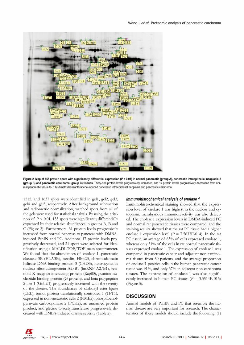
1512, and 1637 spots were identified in gel1, gel2, gel3, gel4 and gel5, respectively. After background subtraction and radiometric normalization, matched spots from all of the gels were used for statistical analysis. By using the crite-rion of P < 0.01, 155 spots were significantly differentially expressed by their relative abundances in groups A, B and C (Figure 2). Furthermore, 31 protein levels progressively increased from normal pancreas to pancreas with DMBA-induced PanIN and PC. Additional 17 protein levels pro-gressively decreased, and 21 spots were selected for iden-tification using a MALDI-TOF/TOF mass spectrometer. We found that the abundances of enolase 1, pancreatic elastase 3B (ELA3B), necdin, Hbp23, chromodomain helicase DNA-binding protein 3 (CHD3), heterogeneous nuclear ribonucleoprotein A2/B1 (hnRNP A2/B1), reti-noid X receptor-interacting protein (Rap80), guanine nu-cleotide-binding protein (G protein), and beta polypeptide 2-like 1 (Gnb2l1) progressively increased with the severity of the disease. The abundances of carboxyl ester lipase (CEL), tumor protein translationally controlled 1 (TPT1), expressed in non-metastatic cells 2 (NME2), phosphoenol-pyruvate carboxykinase 2 (PCK2), an unnamed protein product, and glycine C-acetyltransferase progressively de-creased with DMBA-induced disease severity (Table 2).
Immunohistochemical analysis of enolase 1Immunohistochemical staining showed that the expres-sion level of enolase 1 was highest in the nucleus and cy-toplasm; membranous immunoreactivity was also detect-ed. The enolase 1 expression levels in DMBA-induced PC and normal rat pancreatic tissues were compared, and the staining results showed that the rat PC tissue had a higher enolase 1 expression level (P = 7.5633E-014). In the rat PC tissue, an average of 83% of cells expressed enolase 1, whereas only 31% of the cells in rat normal pancreatic tis-sues expressed enolase 1. The expression of enolase 1 was compared in pancreatic cancer and adjacent non-carcino-ma tissues from 30 patients, and the average proportion of enolase 1-positive cells in the human pancreatic cancer tissue was 91%, and only 37% in adjacent non-carcinoma tissues. The expression of enolase 1 was also signifi-cantly increased in human PC tissues (P = 3.3514E-015) (Figure 3).
DISCUSSIONAnimal models of PanIN and PC that resemble the hu-man disease are very important for research. The charac-teristics of these models should include the following: (1)
Figure 2 Map of 155 protein spots with significantly differential expression (P < 0.01) in normal pancreatic (group A), pancreatic intraepithelial neoplasia-2 (group B) and pancreatic carcinoma (group C) tissues. Thirty-one protein levels progressively increased, and 17 protein levels progressively decreased from nor-mal pancreatic tissue to 7,12-dimethylbenzanthracene-induced pancreatic intraepithelial neoplasia and pancreatic carcinoma.
1437 March 21, 2011|Volume 17|Issue 11|WJG|www.wjgnet.com
Wang L et al. Proteomic analysis of pancreatic carcinoma

A pancreatic ductal phenotype that arises from pancreatic ductal cells; (2) Extensive reactive proliferation of the
Figure 3 Enolase 1 expression was significantly increased in human and rat pancreatic carcinoma (immunohistochemistry, magnification × 400). A: Posi-tive expression in rat pancreatic cancer; B: Weakly positive expression in the normal rat pancreas; C: Positive expression in human pancreatic cancer; D: Weakly positive expression in human adjacent non-carcinoma tissues.
A B
C D
1438 March 21, 2011|Volume 17|Issue 11|WJG|www.wjgnet.com
Wang L et al. Proteomic analysis of pancreatic carcinoma
Table 2 Differentially expressed proteins identified by matrix-assisted laser desorption/ionization-time of flight/time of flight tan-dem mass spectrometry in normal pancreatic, pancreatic intraepithelial neoplasia-2 and pancreatic cancer tissues
Spot position Protein number in NCBI Protein name MW PI Mascot PMF score
P
230 Mixture,gi|6753406 + gi|74205924
Carboxyl ester lipase+unnamed protein product 70 6 136 4.10E-5
780 gi|158186649 Enolase 1 40 5.5 76 5.40E-5
1205 gi|6678437 Tumor protein translationally controlled 1 25 5 98 7.90E-5
1402 gi|55926145 Expressed in non-metastatic cells 2 20 8 185 0.000
1132 gi|149024340 Pancreatic elastase 3B 25 5.5 91 0.000
1066 gi|56676354 Necdin 30 8 63 0.000
1272 gi|6435547 Hbp23 25 7.5 62 0.001
226 gi|6753406 Carboxyl ester lipase 90 5.5 118 0.001
1405 gi|109488364 Chromodomain helicase DNA-binding protein 3 20 5.5 72 0.001
755 Mixture,gi|149033753 + gi|149039895
Albumin+retinoid X receptor-interacting protein 40 6.5 181 0.001
1259 gi|12832572 Unnamed protein product 25 6 38 0.001
298 gi|149063967 Phosphoenolpyruvate carboxykinase 2 66 7.5 229 0.002
1057 gi|4504447 Heterogeneous nuclear ribonucleoprotein A2/B1 30 8.5 62 0.002
993 gi|5174447 Guanine nucleotide binding-protein (G-protein), beta polypeptide 2-like 1
30 7.5 146 0.003
1040 gi|5174447 Guanine nucleotide binding-protein (G-protein), beta polypeptide 2-like 1
30 5.5 142 0.004
658 gi|66730435 Glycine C-acetyltransferase 40 8 211 0.008
NCBI:National Center for Biotechnology Information; PMF:Peptide mass fingerprint; MW: Molecular weight ; PI: Isoelectric point.

connective tissue; (3) Obstruction of the bile duct and stomach during disease progression; (4) Early neural, peri-toneal, and liver metastases; and (5) High rates of K-ras, P16 and P53 gene mutations[16]. The experimental models of PanIN and PC in the present study are chemical carci-nogenesis models. DMBA-induced pancreatic carcinogen-esis models resembled human PC in several previous stud-ies. Rivera et al[6] found that DMBA was one of the three different carcinogens reliably producing PC histology that is similar to human ductal adenocarcinoma. DMBA was either implanted directly into the pancreas or infused into the pancreatic ducts of SD rats, and the development of ductal hyperplastic, atypical, and dysplastic changes pre-ceding and accompanying the invasive pancreatic ductal adenocarcinoma could be observed in this experimental model. Jimenez et al[7] demonstrated that DMBA-induced rat tubular complexes and pancreatic adenocarcinomas strongly expressed ductal cell markers (keratin, cytokera-tins 19 and 20) but not acinar cell markers (chymotryp-sin), suggesting that these tumors arose from ductal cell transformation. K-ras mutations were significantly more frequent in DMBA-induced PC tissues than in normal tissues, with a prevalence of up to 91%[8]. Pancreatic car-cinogenesis induced by DMBA implantation in mice was re-evaluated according to the PanIN classification system after its establishment, and extensive pathological changes characteristic of PanIN could be observed one month after carcinogen implantation[9]. We used the PanIN classification system in the histological analysis, and our pathologic evaluation showed that 71% (12/17) of the rats had PanIN lesions and 18% (3/17) had adenocarci-nomas in the 1-mo group. In the 2-mo group, 43% (6/14) had PanIN lesions and 57% (8/14) had adenocarcinomas. The frequencies of PanIN and PC in our rat models were similar to a previous study in mice[9]. Our models were satisfactory because the development of precursor lesions (PanIN) and an invasive adenocarcinoma were observed in all groups. There was an increase in the frequency of adenocarcinomas, and the PanIN classification increased with DMBA induction, reflecting the dynamic develop-ment of a normal pancreas into a cancerous pancreas.
There are several techniques available for protein separation, both gel- and non-gel-based. Gel-based tech-niques include traditional two-dimensional polyacrylamide gel electrophoresis (2D-PAGE) and, more recently, 2D-DIGE. 2D-DIGE usually contains three samples labeled with three distinct fluorescent dyes: Cy2, Cy3 and Cy5. The Cy2 dye is typically used to label an internal standard; the strength of the internal standard helps map the spots/proteins between the gels, thus rendering different gels more comparable[17-19]. In the current study, we used 2D-DIGE and MALDI-TOF/TOF tandem mass spectrome-try to profile pancreatic proteins in rat models of DMBA-induced PanIN and PC and compared these profiles with those of normal rats. Our 2D-DIGE data showed that 155 spots were significantly differentially expressed based on their relative abundances in the three groups. Of these, 31 protein levels progressively increased from normal to PanIN and then to PC tissue, whereas 17 protein lev-
els progressively decreased. These results demonstrated synchronous dynamic changes in the pancreatic protein expression profile after DMBA induction. A number of novel proteins were identified that may be involved in important aspects of mRNA transcription, DNA damage repair, chromatin remodeling, oxidative stress, regulation of tumor growth and metastasis, glucose metabolism, and the synthesis and secretion of pancreatic enzymes.
Enolase 1, also known as α-enolase or non-neuronal enolase (NNE), is an isoenzyme of enolase, which is a glycolytic enzyme catalyzing the conversion of 2-phos-phoglycerate into phosphoenolpyruvate. Recent re-searches have shown that enolase 1 plays an important role in several biological and pathophysiological processes. Enolase 1 is thought to play a potential role in tumorigen-esis, cancer invasion and metastasis. Previous proteomic studies reported that enolase 1 was up-regulated in several cancers, such as hepatocellular carcinoma[20-22], non-small lung cancer[23,24], esophageal adenocarcinoma[25], prostate cancer[26], colon cancer[27], oral epithelial and squamous cell carcinoma[28]. In our study, we found a significant up-regulation of enolase 1 in rat models of DMBA-induced PanIN and PC with further verification by the immuno-histochemical analysis of human and rat tissues. These results agree with other proteomic researches on human PC. Mikuriya et al[29] found that the expression levels of glycolytic enzymes, including enolase 1, increased in the cancerous pancreatic tissues of 10 patients compared with the paired non-cancerous tissues, as determined by proteomic profiling using two-dimensional electrophore-sis and liquid chromatography-mass spectrometry/mass spectrometry. Shen et al[30] used a proteomic approach using two-dimensional gel electrophoresis and mass spec-trometry to identify differentially expressed proteins in six PC cases, two normal adjacent tissues, seven cases of pan-creatitis, and six normal pancreatic tissues. Alpha-enolase was also specifically overexpressed in tumors compared with normal and pancreatic tissues.
The hnRNP A2/B1 protein was shown to be up-regulated in our models. This protein plays an important role in the biogenesis and transport of mRNA. The over-expression of hnRNP A2/B1 indicates that normal transcriptional regulation is altered. A previous study also found high levels of hnRNP A2/B1 expression in a lim-ited number of human pancreatic adenocarcinomas from smokers and two pancreatic tumor cell lines, HPAF-11 and SU 86.86[31]. CEL is one kind of pancreatic exocrine enzyme, and we found that it was down-regulated after DMBA induction. Reuss et al[32] verified strong CEL gene expression in the acinar cells of the normal pancreas, and adenocarcinomas showed no expression, which agrees with our results. The other differentially expressed pro-teins that we identified are rarely reported in PC, and their relationships with PC await further clarification.
In summary, our data have shown DMBA implanta-tion into the pancreas is an effective method to establish rat models of PanIN and PC, and the protein changes observed in DMBA-induced PanIN and PC, such as enolase 1 up-regulation, demonstrate the feasibility of
1439 March 21, 2011|Volume 17|Issue 11|WJG|www.wjgnet.com
Wang L et al. Proteomic analysis of pancreatic carcinoma

identifying potential molecular targets for the early diag-nosis and treatment of PC.
COMMENTSBackgroundThe early detection of pancreatic cancer (PC) is still difficult, but proteomic techniques are emerging as important tools to find potential biomarkers and new therapeutic targets for PC and precancerous pancreatic lesions.Research frontiers7,12-dimethylbenzanthracene (DMBA) implantation into the pancreas is known to induce PC and pancreatic ductal intraepithelial neoplasia (PanIN) in mice and rats that resemble human PC with K-ras gene mutations and the activation of Notch signaling.Innovations and breakthroughsPrecancerous pancreas lesions and the early stages of PC among clinical diagnoses are rarely found. In the present research, the authors studied the proteomic variabilities of rat models of DMBA-induced PanIN and PC.Applications The protein changes in DMBA-induced PanIN and PC reported in this article, such as enolase 1 up-regulation, demonstrate the feasibility of identifying po-tential molecular targets for early PC diagnostics and therapeutics.TerminologyTwo-dimensional fluorescence difference gel electrophoresis is a gel-based technique that has recently been used for protein separation. It contains three samples labeled with three distinct fluorescent dyes: Cy2, Cy3 and Cy5.Peer reviewThis is a good study to define the stages of pancreatic carcinoma in experimen-tal models and its correlation with various markers.
REFERENCES1 JemalA, Siegel R, Ward E, Hao Y, Xu J, Thun MJ. Cancer
statistics, 2009. CA Cancer J Clin 2009; 59: 225-2492 HrubanRH, Adsay NV, Albores-Saavedra J, Compton C,
Garrett ES, Goodman SN, Kern SE, Klimstra DS, Klöppel G, Longnecker DS, Lüttges J, Offerhaus GJ. Pancreatic intraepi-thelial neoplasia: a new nomenclature and classification system for pancreatic duct lesions. Am J Surg Pathol 2001; 25: 579-586
3 HrubanRH, Takaori K, Klimstra DS, Adsay NV, Albores-Saavedra J, Biankin AV, Biankin SA, Compton C, Fukushi-ma N, Furukawa T, Goggins M, Kato Y, Klöppel G, Long-necker DS, Lüttges J, Maitra A, Offerhaus GJ, Shimizu M, Yonezawa S. An illustrated consensus on the classification of pancreatic intraepithelial neoplasia and intraductal papil-lary mucinous neoplasms. Am J Surg Pathol 2004; 28: 977-987
4 DissinJ, Mills LR, Mains DL, Black O Jr, Webster PD 3rd. Experimental induction of pancreatic adenocarcinoma in rats. J Natl Cancer Inst 1975; 55: 857-864
5 Bockman DE. Cells of origin of pancreatic cancer: experi-mental animal tumors related to human pancreas. Cancer 1981; 47: 1528-1534
6 RiveraJA, Graeme-Cook F, Werner J, Z’graggen K, Rustgi AK, Rattner DW, Warshaw AL, Fernández-del Castillo C. A rat model of pancreatic ductal adenocarcinoma: targeting chemical carcinogens. Surgery 1997; 122: 82-90
7 Jimenez RE, Z’graggen K, Hartwig W, Graeme-Cook F, Warshaw AL, Fernandez-del Castillo C. Immunohisto-chemical characterization of pancreatic tumors induced by dimethylbenzanthracene in rats. Am J Pathol 1999; 154: 1223-1229
8 Z’graggenK, Warshaw AL, Werner J, Graeme-Cook F, Ji-menez RE, Fernández-Del Castillo C. Promoting effect of a high-fat/high-protein diet in DMBA-induced ductal pancre-atic cancer in rats. Ann Surg 2001; 233: 688-695
9 OsvaldtAB, Wendt LR, Bersch VP, Backes AN, de Cássia A Schumacher R, Edelweiss MI, Rohde L. Pancreatic intraepi-
thelial neoplasia and ductal adenocarcinoma induced by DMBA in mice. Surgery 2006; 140: 803-809
10 KimuraK, Satoh K, Kanno A, Hamada S, Hirota M, Endoh M, Masamune A, Shimosegawa T. Activation of Notch sig-naling in tumorigenesis of experimental pancreatic cancer induced by dimethylbenzanthracene in mice. Cancer Sci 2007; 98: 155-162
11 WendtLR, Osvaldt AB, Bersch VP, Schumacher Rde C, Edelweiss MI, Rohde L. Pancreatic intraepithelial neoplasia and ductal adenocarcinoma induced by DMBA in mice: ef-fects of alcohol and caffeine. Acta Cir Bras 2007; 22: 202-209
12 BerschVP, Osvaldt AB, Edelweiss MI, Schumacher Rde C, Wendt LR, Abreu LP, Blom CB, Abreu GP, Costa L, Picci-nini P, Rohde L. Effect of nicotine and cigarette smoke on an experimental model of intraepithelial lesions and pancreatic adenocarcinoma induced by 7,12-dimethylbenzanthracene in mice. Pancreas 2009; 38: 65-70
13 Boros LG, Lerner MR, Morgan DL, Taylor SL, Smith BJ, Postier RG, Brackett DJ. [1,2-13C2]-D-glucose profiles of the serum, liver, pancreas, and DMBA-induced pancreatic tu-mors of rats. Pancreas 2005; 31: 337-343
14 RajcevicU, Niclou SP, Jimenez CR. Proteomics strategies for target identification and biomarker discovery in cancer. Front Biosci 2009; 14: 3292-3303
15 LatterichM, Abramovitz M, Leyland-Jones B. Proteomics: new technologies and clinical applications. Eur J Cancer 2008; 44: 2737-2741
16 Ottenhof NA, Milne AN, Morsink FH, Drillenburg P, Ten Kate FJ, Maitra A, Offerhaus GJ. Pancreatic intraepithelial neoplasia and pancreatic tumorigenesis: of mice and men. Arch Pathol Lab Med 2009; 133: 375-381
17 KondoT. Tissue proteomics for cancer biomarker develop-ment: laser microdissection and 2D-DIGE. BMB Rep 2008; 41: 626-634
18 Westermeier R, Scheibe B. Difference gel electrophoresis based on lys/cys tagging. Methods Mol Biol 2008; 424: 73-85
19 MindenJ. Comparative proteomics and difference gel elec-trophoresis. Biotechniques 2007; 43: 739, 741, 743 passim
20 KuramitsuY, Nakamura K. Current progress in proteomic study of hepatitis C virus-related human hepatocellular car-cinoma. Expert Rev Proteomics 2005; 2: 589-601
21 Takashima M, Kuramitsu Y, Yokoyama Y, Iizuka N, Fu-jimoto M, Nishisaka T, Okita K, Oka M, Nakamura K. Overexpression of alpha enolase in hepatitis C virus-related hepatocellular carcinoma: association with tumor progres-sion as determined by proteomic analysis. Proteomics 2005; 5: 1686-1692
22 HamaguchiT, Iizuka N, Tsunedomi R, Hamamoto Y, Mi-yamoto T, Iida M, Tokuhisa Y, Sakamoto K, Takashima M, Tamesa T, Oka M. Glycolysis module activated by hypoxia-inducible factor 1alpha is related to the aggressive phenotype of hepatocellular carcinoma. Int J Oncol 2008; 33: 725-731
23 ChangGC, Liu KJ, Hsieh CL, Hu TS, Charoenfuprasert S, Liu HK, Luh KT, Hsu LH, Wu CW, Ting CC, Chen CY, Chen KC, Yang TY, Chou TY, Wang WH, Whang-Peng J, Shih NY. Identification of alpha-enolase as an autoantigen in lung cancer: its overexpression is associated with clinical outcomes. Clin Cancer Res 2006; 12: 5746-5754
24 HeP,NakaT, Serada S, Fujimoto M, Tanaka T, Hashimoto S, Shima Y, Yamadori T, Suzuki H, Hirashima T, Matsui K, Shiono H, Okumura M, Nishida T, Tachibana I, Norioka N, Norioka S, Kawase I. Proteomics-based identification of alpha-enolase as a tumor antigen in non-small lung cancer. Cancer Sci 2007; 98: 1234-1240
25 ZhaoJ, Chang AC, Li C, Shedden KA, Thomas DG, Misek DE, Manoharan AP, Giordano TJ, Beer DG, Lubman DM. Comparative proteomics analysis of Barrett metaplasia and esophageal adenocarcinoma using two-dimensional liquid mass mapping. Mol Cell Proteomics 2007; 6: 987-999
26 van den Bemd GJ, Krijgsveld J, Luider TM, van Rijswijk AL, Demmers JA, Jenster G. Mass spectrometric identifica-tion of human prostate cancer-derived proteins in serum
1440 March 21, 2011|Volume 17|Issue 11|WJG|www.wjgnet.com
Wang L et al. Proteomic analysis of pancreatic carcinoma
COMMENTS

of xenograft-bearing mice. Mol Cell Proteomics 2006; 5: 1830-1839
27 Katayama M, Nakano H, Ishiuchi A, Wu W, Oshima R, Sakurai J, Nishikawa H, Yamaguchi S, Otsubo T. Protein pattern difference in the colon cancer cell lines examined by two-dimensional differential in-gel electrophoresis and mass spectrometry. Surg Today 2006; 36: 1085-1093
28 ItoS, Honma T, Ishida K, Wada N, Sasaoka S, Hosoda M, Nohno T. Differential expression of the human alpha-eno-lase gene in oral epithelium and squamous cell carcinoma. Cancer Sci 2007; 98: 499-505
29 Mikuriya K, Kuramitsu Y, Ryozawa S, Fujimoto M, Mori S, Oka M, Hamano K, Okita K, Sakaida I, Nakamura K. Expression of glycolytic enzymes is increased in pancreatic cancerous tissues as evidenced by proteomic profiling by two-dimensional electrophoresis and liquid chromatog-
raphy-mass spectrometry/mass spectrometry. Int J Oncol 2007; 30: 849-855
30 Shen J, Person MD, Zhu J, Abbruzzese JL, Li D. Protein expression profiles in pancreatic adenocarcinoma compared with normal pancreatic tissue and tissue affected by pan-creatitis as detected by two-dimensional gel electrophoresis and mass spectrometry. Cancer Res 2004; 64: 9018-9026
31 Yan-SandersY, Hammons GJ, Lyn-Cook BD. Increased ex-pression of heterogeneous nuclear ribonucleoprotein A2/B1 (hnRNP) in pancreatic tissue from smokers and pancreatic tumor cells. Cancer Lett 2002; 183: 215-220
32 ReussR, Aberle S, Klingel K, Sauter M, Greschniok A, Fran-ke FE, Padberg W, Blin N. The expression of the carboxyl ester lipase gene in pancreas and pancreatic adenocarcino-mas. Int J Oncol 2006; 29: 649-654
S-EditorSun H L-Editor Ma JY E-EditorMa WH
1441 March 21, 2011|Volume 17|Issue 11|WJG|www.wjgnet.com
Wang L et al. Proteomic analysis of pancreatic carcinoma

ORIGINAL ARTICLE
Serial observations on an orthotopic gastric cancer model constructed using improved implantation technique
Yan Li, Bo Li, Yu Zhang, Chun-Ping Xiang, Yuan-Yuan Li, Xiao-Ling Wu
Yan Li, Xiao-Ling Wu, Department of Gastroenterology, the Sec-ond Affiliated Hospital, Chongqing Medical University, Chong-qing 400010, ChinaBo Li, Department of Surgery, the First People’s Hospital, Jiang-bei District, Chongqing 400020, China Yu Zhang, Department of Pathology, the First People’s Hospital, Jiangbei District, Chongqing 400020, ChinaChun-Ping Xiang, Department of Pathology, Children’s Hospi-tal, Chongqing Medical University, Chongqing 400014, ChinaYuan-Yuan Li, Department of Pathology, Chongqing Medical University, Chongqing 400016, ChinaAuthor contributions: Li Y conceived of the study, constructed animal models, and drafted the manuscript; Li B participated and established the animal model; Zhang Y and Xiang CP performed paraffin sections and hematoxylin and eosin staining; Li YY performed immunohistochemical staining; Wu XL guided and inspected the whole experiments. Supported by the Natural Science Foundation of China, No. 30830040Correspondence to: Xiao-Ling Wu, Professor, Department of Gastroenterology, the Second Affiliated Hospital, Chongqing Medical University, Chongqing 400010, China. [email protected]: +86-23-63693325 Fax: +86-23-63711527 Received: October 12, 2010 Revised: December 30, 2010Accepted: January 6, 2011Published online: March 21, 2011
Abstract AIM: To establish a gastric cancer nude-mouse model with improved orthotopic implantation and investigate its biological characteristics at different time points.
METHODS: Human gastric cancer SGC-7901 cell sus-pensions were injected subcutaneously into a nude mouse to develop solid tumors, and the tumor tissue pieces were implanted under the serous coat. The nude mice were then euthanized in group every two weeks to observe the primary tumor growth and metastases.
RESULTS: Within 2-4 wk, there were no obvious chang-
es about the primary tumor in stomach. At the sixth week, the primary tumor began to grow fast, resulting in incrassation of the gastric wall and stenosis of the gastric cavity, and metastases into the liver and lymph nodes were detected. The tumor, which compressed the adjacent organs, gradually became bigger and bigger followed by stenosis or vanishment of the gastric cavity from 8 to 12 wk. There were massive metastases, and the rate of metastasis was 58% in lymph nodes, 78% in liver, 39% in kidney, and 81% in peritoneum or septum.
CONCLUSION: A gastric cancer model is established, which can simulate the clinical tumor behavior and provide experimental carrier for clinical trials of gastric cancer treatment.
© 2011 Baishideng. All rights reserved.
Key words: Gastric cancer; Orthotopic implantation; Mouse model; Metastasis; Cell line
Peer reviewers: Fabio Grizzi, PhD, Laboratories of Quantita-tive Medicine, Istituto Clinico Humanitas IRCCS, Via Manzoni 56, 20089 Rozzano, Milan,Italy; Dr. Jianyuan Chai, PhD, MS, BS, Assistant Professor, Research (09-151), VA Long Beach Healthcare System, 5901 E. 7th St, Long Beach, CA 90822, United States
Li Y, Li B, Zhang Y, Xiang CP, Li YY, Wu XL. Serial observa-tions on an orthotopic gastric cancer model constructed using improved implantation technique. World J Gastroenterol 2011; 17(11): 1442-1447 Available from: URL: http://www.wjgnet.com/1007-9327/full/v17/i11/1442.htm DOI: http://dx.doi.org/10.3748/wjg.v17.i11.1442
INTRODUCTIONMetastasis is not only still the barrier to tumor therapy, but also linked with the prognosis of patients. Patients with gastric cancer hardly have symptoms in their early stage
1442
World J Gastroenterol 2011 March 21; 17(11): 1442-1447 ISSN 1007-9327 (print) ISSN 2219-2840 (online)
© 2011 Baishideng. All rights reserved.
Online Submissions: http://www.wjgnet.com/[email protected]:10.3748/wjg.v17.i11.1442
March 21, 2011|Volume 17|Issue 11|WJG|www.wjgnet.com

Li Y et al . Orthotopic gastric cancer model
so that they are always found with metastases after clinical examinations. Therefore, novel tumor models are needed to study the metastasis mechanism as well as therapeutic approaches. It is well known that orthotopic transplanta-tion technique has gain popularity in the field of the ani-mal model[1-12]. Orthotopic tumor models not only mimic clinical cancer course, but also promote metastasis[13]. In recent years, the procedure of orthotopic implantation has been improved from the“sewing”method to the“ad-hering”one[2-4,14]. The progress greatly shortens the course of surgical operation and decreases the mortality of ani-mals. To our knowledge, gastric cancer models of ortho-topic implantation with intact tumor tissue have been well established[1-3,5-8]. However, serial examination of tumor development and metastasis needs to be made in order to tailor the treatment strategies. Based on the previous researches, we intend to establish an orthotopic model of gastric cancer using tissue glue and observe tumor prog-ress and metastasis consecutively to detect an appropriate therapeutic target for clinical trials.
MATERIALS AND METHODSCell lineHuman gastric cancer cell lines SGC-7901 (poorly dif-ferentiated) was used for this study. The cell was obtained from the Centre of Cell Cultures of Chinese Academy of Medical Sciences, Shanghai, China, and cultured at 37℃ in a humidified air with 5% CO2, in RPMI-1640 medium (Gibco, Grand Island, NY) supplemented with 10% fetal calf serum (FCS; Gibco, Grand Island, NY), 100 U/mL penicillin, 100 μg/mL streptomycin, 2 mmol/L gluta-mine, and 1 mmol/L sodium pyruvate.
AnimalsFive to six-week-old male Balb/c nu-nu mice weighing 18-20 g were obtained from the Shanghai Experimental Animal Center of the Chinese Academy of Medical Sci-ences, China. All mice were maintained in a pathogen-free environment (temperature 25-27℃, humidity 45%-50%) and supplied with food and water ad libitum. All experi-mental procedures were conducted in conformity with institutional guidelines for the care and use of laboratory animals in Chongqing Medical University, China, and the National Institutes of Health Guide for Care and Use of Laboratory Animals.
Orthotopic implantation in stomachSGC-7901 cells were collected at the log phase and inject-ed subcutaneously into the mice at 107/0.2 mL. Two weeks later, tumors (about 2.0 cm × 2.0 cm × 1.0 cm) were har-vested from the mice under anesthesia and minced into small pieces (1mm3) in RPMI-1640 basal medium. All pro-cedures were then performed under anesthesia with Sumi-anxin Ⅱ (0.02 mL per animal; China Academy of Military Medical Sciences). For implantation, the mouse stomach was gently exteriorized via a left-side upper abdominal incision, and one small tissue pocket was formed in the
middle wall of the greater curvature using a microscissor. One tumor fragment was placed into the pocket and fixed with a drop of medical tissue glue (gifts from Shunkang Corporation of Biological Adhesive, Beijing, China). The quantity of the tissue adhesive should be strictly controlled to avoid adhering to adjacent normal tissues. The stomach was then relocated into the abdominal cavity followed by the abdominal closure with 4-0 absorbable sutures.
Evaluation of tumor growth and metastasisAll mice were divided into 6 groups of 6 animals each after orthotopic implantation. One group was sacrificed every two weeks. At autopsy, the primary tumor, lymph nodes, and other organs were examined in detail. The samples were fixed in 10% formalin for paraffin sections, and stained with hematoxylin and eosin for microscopic examination.
ImmunohistochemistryParaffin sections were examined histologically with the Cy-tokeratin 20 (CK20) mAb KS20.8 and Epithelial Membrane Antigen (EMA) mAb GP1.4 (MAB-0057, MAB-0061; Maixin Inc., Fuzhou, China), which are usually combined to diagnose the gastrointestinal adenocarcinoma. Ultrasen-sitive streptavidin-peroxidase (SP) kit (KIT-9710; Maixin Inc., Fuzhou, China) and DAB kit (DAB-0031; Maixin Inc., Fuzhou, China) were used in the immunohistochemical stain according to the manufacture’s instructions. CK20 and EMA expression was defined as positive if the stained region of tumor cells was in the cytoplasm.
Assessment of primary tumor growthThe primary tumor volume was calculated by the fol-lowing formula: V = 0.4 × ab2 (a: maximum diameter; b: minimum diameter)[15]. Then the tumor growth curve was depicted reflecting the tumor growth trend.
RESULTSMacroscopic examinationFrom 2 to 4 wk, the primary tumor developed without obvious volume change, and showed insufficient blood supply. The gastric cavity of tumor-bearing mice ap-peared almost normal in size. At the 2nd wk after ortho-topic implantation, metastatic infiltration was not found in all detected organs. However, tumor invasion into the liver was found at the 4th wk. Six weeks after trans-plantation, the tumor in situ grew increasingly in size, with sparse blood vessels. The stomach wall of mice was thickened and gastric cavity decreased, companied with enlargement of lymph nodes from gastric area and hilus pulmonis. The tumor invaded into the liver, resulting in metastatic nodules formation. At the 8th wk, the stomach tumor was characterized by large-size, irregular volume and rich blood supply. The resulting tumor squeezed the adjacent organs and deformed the stomach contributing to distal stenosis. The lymph nodes of the gastric area, hilus pulmonis and mesenterium presented enlargement caused by tumor metastasis. Metastatic lesions were
1443 March 21, 2011|Volume 17|Issue 11|WJG|www.wjgnet.com
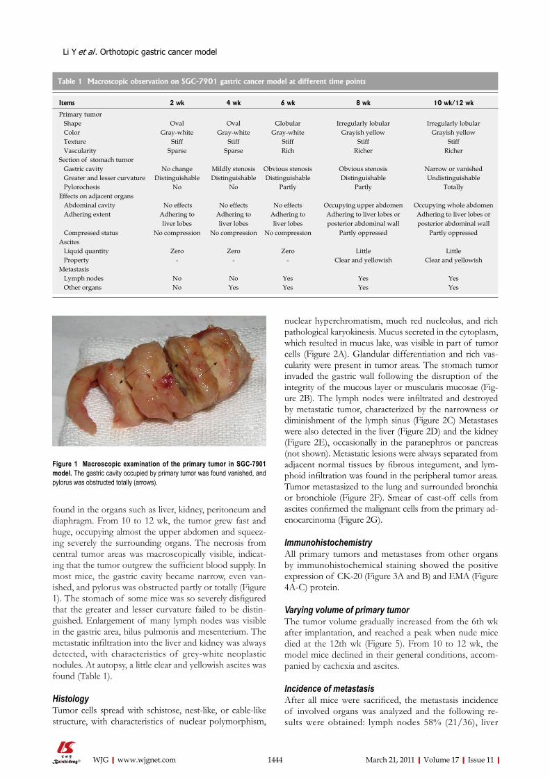
found in the organs such as liver, kidney, peritoneum and diaphragm. From 10 to 12 wk, the tumor grew fast and huge, occupying almost the upper abdomen and squeez-ing severely the surrounding organs. The necrosis from central tumor areas was macroscopically visible, indicat-ing that the tumor outgrew the sufficient blood supply. In most mice, the gastric cavity became narrow, even van-ished, and pylorus was obstructed partly or totally (Figure 1). The stomach of some mice was so severely disfigured that the greater and lesser curvature failed to be distin-guished. Enlargement of many lymph nodes was visible in the gastric area, hilus pulmonis and mesenterium. The metastatic infiltration into the liver and kidney was always detected, with characteristics of grey-white neoplastic nodules. At autopsy, a little clear and yellowish ascites was found (Table 1).
HistologyTumor cells spread with schistose, nest-like, or cable-like structure, with characteristics of nuclear polymorphism,
nuclear hyperchromatism, much red nucleolus, and rich pathological karyokinesis. Mucus secreted in the cytoplasm, which resulted in mucus lake, was visible in part of tumor cells (Figure 2A). Glandular differentiation and rich vas-cularity were present in tumor areas. The stomach tumor invaded the gastric wall following the disruption of the integrity of the mucous layer or muscularis mucosae (Fig-ure 2B). The lymph nodes were infiltrated and destroyed by metastatic tumor, characterized by the narrowness or diminishment of the lymph sinus (Figure 2C) Metastases were also detected in the liver (Figure 2D) and the kidney (Figure 2E), occasionally in the paranephros or pancreas (not shown). Metastatic lesions were always separated from adjacent normal tissues by fibrous integument, and lym-phoid infiltration was found in the peripheral tumor areas. Tumor metastasized to the lung and surrounded bronchia or bronchiole (Figure 2F). Smear of cast-off cells from ascites confirmed the malignant cells from the primary ad-enocarcinoma (Figure 2G).
ImmunohistochemistryAll primary tumors and metastases from other organs by immunohistochemical staining showed the positive expression of CK-20 (Figure 3A and B) and EMA (Figure 4A-C) protein.
Varying volume of primary tumorThe tumor volume gradually increased from the 6th wk after implantation, and reached a peak when nude mice died at the 12th wk (Figure 5). From 10 to 12 wk, the model mice declined in their general conditions, accom-panied by cachexia and ascites.
Incidence of metastasisAfter all mice were sacrificed, the metastasis incidence of involved organs was analyzed and the following re-sults were obtained: lymph nodes 58% (21/36), liver
1444 March 21, 2011|Volume 17|Issue 11|WJG|www.wjgnet.com
Figure 1 Macroscopic examination of the primary tumor in SGC-7901 model. The gastric cavity occupied by primary tumor was found vanished, and pylorus was obstructed totally (arrows).
Table 1 Macroscopic observation on SGC-7901 gastric cancer model at different time points
Items 2 wk 4 wk 6 wk 8 wk 10 wk/12 wk
Primary tumor Shape Oval Oval Globular Irregularly lobular Irregularly lobular Color Gray-white Gray-white Gray-white Grayish yellow Grayish yellow Texture Stiff Stiff Stiff Stiff Stiff Vascularity Sparse Sparse Rich Richer RicherSection of stomach tumor Gastric cavity No change Mildly stenosis Obvious stenosis Obvious stenosis Narrow or vanished Greater and lesser curvature Distinguishable Distinguishable Distinguishable Distinguishable Undistinguishable Pylorochesis No No Partly Partly TotallyEffects on adjacent organs Abdominal cavity No effects No effects No effects Occupying upper abdomen Occupying whole abdomen Adhering extent Adhering to
liver lobesAdhering to liver lobes
Adhering to liver lobes
Adhering to liver lobes or posterior abdominal wall
Adhering to liver lobes or posterior abdominal wall
Compressed status No compression No compression No compression Partly oppressed Partly oppressedAscites Liquid quantity Zero Zero Zero Little Little Property - - - Clear and yellowish Clear and yellowishMetastasis Lymph nodes No No Yes Yes Yes Other organs No Yes Yes Yes Yes
Li Y et al . Orthotopic gastric cancer model

78% (28/36), kidney 39% (14/36), and peritoneum and diaphragm 81% (29/36).
DISCUSSIONThere are little proofs supporting serial observations on gastric cancer models although many researchers have constructed orthotopic models using intact tumor tis-sues. This makes it difficult to learn the whole process of
tumor growth and metastasis occurrence. In this study, we presented in detail such evidences as the process of primary tumor growth, the time point of metastasis, and the sites and incidence of metastasis.
The patients with gastric cancer hardly have symp-toms in their early stage, whereas they are always tor-tured by gastrointestinal symptom and metastasis in their terminal stage. In the present study, an animal model of stomach cancer was established to replicate the tumor
1445 March 21, 2011|Volume 17|Issue 11|WJG|www.wjgnet.com
Figure 2 Histopathologic structure of SGC-7901 tumors by HE staining. A: Tumor cells showed nuclear polymorphism, nuclear hyperchromatism, and much mucus in the cytoplasm (× 200); B: The primary tumor destroyed muscularis mucosae (arrows) (× 100); C: The lymph nodes were infiltrated by metastatic tumor (arrows) (× 100); D: Metastases (arrows) infiltrated into the liver (× 200); E: Metastatic tumor (arrows) invaded the kidney (× 100); F: Tumor (arrows) metastasized to the lung and surrounded bronchia or bronchiole (× 40); G: Cast-off tumor cells were detected in ascites (× 200).
A B C
FED
G
A B
Figure 3 Immunohistochemistry of SGC-7901 tumor by streptavidin-peroxidase method. Expression of Cytokeratin 20 (CK20) protein was positive, which was stained brown in the cytoplasm in the tumor of stomach (A × 40) and liver (B × 100).
Li Y et al . Orthotopic gastric cancer model

behavior of clinical patients. It showed that the primary tumor grew into the log phrase from the 6th wk after implantation and thereafter the tumor volume gradually increased. From the 6th wk, lymph node metastases and distant metastases were detected, accompanied by the in-crease of involved organs with the tumor progress. The tested mice revealed obvious cachexia from the 10th wk, and systemic failure at the 12th wk or so.
There are several pathways such as direct infiltration, lymphatic metastasis, vascular spread, and implantation dissemination which play important roles in metastasis of gastric cancer. Previous studies recapitulated the metastatic incidence of systematic organs, but did not mention the metastatic pathways of gastric cancer models. Moreover, there are uneven metastasis rates among different labo-ratories resulting from different experimental conditions, animal species and cell lines[1,2,3,10,11,14]. The SGC-7901 cell line used in our study was derived from metastatic lymph node of poorly differentiated human gastric carcinoma. We could detect the metastatic lesions in the lymph nodes from gastroepiploic plexus, hilus pulmonis and mesente-rium, which accorded with the feature of this cell line. It is well known that liver is the most sensitive organ to vascu-lar metastasis. In the present study, we found that the liver was not only invaded by the neoplastic cells from blood pathway but also directly infiltrated by the primary tumor. In addition, the fringe of kidney also showed metastatic
spread, yet only under renal capsule rather than in paren-chyma. Hereby, we consider that metastatic tumor may float in the ascites by breaking through the stomach serous coat and then invade straightly the kidney, or metastasize to the lymphatic system of renal capsule via lymphatic route, instead of invading kidney through blood vessel.
The model constructed in our study could simulate the clinical tumor behavior and provide basic theory on gastric cancer models. However, orthotopic implantation models still have some limitations such as failure in clini-cal staging. It is well known that gastric cancer stage (early, median, late) is judged by tumor growth and infiltration and metastasis rather than disease course, i.e. TNM stag-ing published by Union for International Cancer Control (UICC). The morphological changes of gastric carcino-genesis are as follows: diffuse hyperplasia → focal pro-liferation and metaplasia → benign tumor → grade Ⅰ , Ⅱ or Ⅲ dysplasia → carcinoma in situ → invasive car-cinoma, which reveals that the pathological changes of gastric cancer originate from mucous layer and progress layer-by-layer towards serous coat[16]. However, orthotopic implantation was performed with tumor fragments under the stomach serous coat, which is unable to mimic the histopathological mechanism of gastric cancer. In fact, the procedure of orthotopic implantation mimics the artificial advanced stage which is characterized by tumor growth from serous coat to mucous layer or outside membrane serosa. The histological origin of primary tumor was not consistent with the principle of TMN stage although lymph node metastasis or distant dissemination was also detected. So the orthotopic model of gastric cancer is not suitable for clinical staging.
Some researchers had established the models of stomach cancer with chemical carcinogens which could simulate the histological origin of gastric carcinoma[16-20]. But there are some disadvantages such as long period and accidental ani-mal death. Some authors employed transgenic techniques in attempt to replicate the mechanism of gastric cancer[18,21,22]. However, transgenic method is limited due to expensive cost, long period and advanced technical requirements for mo-lecular biology. Compared with other methods for construct-ing gastric cancer model, the surgical orthotopic implantation is still a desired technique characteristic of short period, low cost and simple practice.
Some experimental skills for this animal model should
1446 March 21, 2011|Volume 17|Issue 11|WJG|www.wjgnet.com
A B C
Figure 4 Immunostaining of SGC-7901 tumor using streptavidin-peroxidase method. Epithelial membrane antigen signal stained brown in the cytoplasm was positive in the tumor of stomach (A × 40), lymph node (B × 40) and lung (C × 100).
Primary tumor volume
12
10
8
6
4
2
0
Volu
me
(cm
3 )
2wk 4wk 6wk 8wk 10wk 12wk
Figure 5 The curve of tumor growth at different time points. The tumor volume gradually increased from the 6th wk after implantation, and reached a peak at the 12th wk.
t /wk
Li Y et al . Orthotopic gastric cancer model
be noticed. For example, necrotic area should be removed from tumor tissues used for orthotopic implantation; the quantity of medical glue should be strictly controlled to avoid adhering to adjacent organs; and the middle part of the greater gastric curvature is the first choice for ortho-topic implantation because of the operational convenience and rich blood supply.
In conclusion, we can succeed in establishing a gas-tric cancer model if only we master the experimental skills above and avoid the adverse factors.
COMMENTSBackgroundMetastasis is still the barrier to the treatment of patients with gastric cancer. Gastric cancer models of orthotopic implantation with intact tumor tissues have been well established. Moreover, the procedure of orthotopic implantation has been improved from the “sewing” method to the “adhering” one. However, there are little evidences regarding serial observations on gastric cancer models to tailor treatment strategies. Research frontiersIt is well known that orthotopic transplantation technique plays an important role in establishing animal models of various tumors. Gastric cancer model of orthotopic implantation could mimic clinical cancer process and contribute to metastasis occurrence. The procedure greatly facilitates surgical operation course and decreases the mortality of animals. Innovations and breakthroughsOrthotopic implantation with “glue paste technique” is a popular and new method in recent years which is characterized by easy performance, low cost and short operation course. This study makes it possible to help researchers learn the whole process of tumor development and metastasis, thus choosing the appropriate therapy target for clinical trials. ApplicationsSerial observations on the models can provide evidences for researchers to treat stomach cancer and test the therapeutic effects. Peer reviewThe study is interesting and well done. The model used by the Authors well mimics the dynamics of local growth and spread of gastric cancer cells at both lymph-nodes and distant organs.
REFERENCES1 Shi J, Wei PK, Zhang S, Qin ZF, Li J, Sun DZ, Xiao Y, Yu
ZH, Lin HM, Zheng GJ, Su XM, Chen YL, Liu YF, Xu L. OB glue paste technique for establishing nude mouse human gastric cancer orthotopic transplantation models. World J Gastroenterol 2008; 14: 4800-4804
2 Chen YL, Wei PK, Xu L, Su XM. [Nude mouse model of hu-man gastric carcinoma metastasis constructed by orthotopic transplantation using organism glue paste technique]. Ai Zheng 2005; 24: 246-248
3 Bhargava S, Hotz B, Buhr HJ, Hotz HG. An orthotopic nude mouse model for preclinical research of gastric cardia can-cer. Int J Colorectal Dis 2009; 24: 31-39
4 Yang B, Tuo S, Tuo CW, Zhang N, Liu QZ. A liver-metastat-ic model of human primary gastric lymphoma in nude mice orthotopically constructed by using histologically intact pa-tient specimens. Chin J Cancer 2010; 29: 579-584
5 Illert B, Otto C, Thiede A, Timmermann W. Detection of disseminated tumor cells in nude mice with human gastric cancer. Clin Exp Metastasis 2003; 20: 549-554
6 Takemura S, Yashiro M, Sunami T, Tendo M, Hirakawa K. Novel models for human scirrhous gastric carcinoma in
vivo. Cancer Sci 2004; 95: 893-9007 Furukawa T, Fu X, Kubota T, Watanabe M, Kitajima M,
Hoffman RM. Nude mouse metastatic models of human stomach cancer constructed using orthotopic implantation of histologically intact tissue. Cancer Res 1993; 53: 1204-1208
8 Koyama T, Tsubota A, Nariai K, Yoshikawa T, Mitsunaga M, Sumi M, Nimura H, Yanaga K, Yumoto Y, Mabashi Y, Takahashi H. Detection of sentinel nodes by a novel red-fluorescent dye, ATX-S10Na (Ⅱ), in an orthotopic xenograft rat model of human gastric carcinoma. Lasers Surg Med 2007; 39: 76-82
9 Hoffman RM. Orthotopic metastatic mouse models for anti-cancer drug discovery and evaluation: a bridge to the clinic. Invest New Drugs 1999; 17: 343-359
10 An Z, Jiang P, Wang X, Moossa AR, Hoffman RM. Develop-ment of a high metastatic orthotopic model of human renal cell carcinoma in nude mice: benefits of fragment implanta-tion compared to cell-suspension injection. Clin Exp Metasta-sis 1999; 17: 265-270
11 Rashidi B, Gamagami R, Sasson A, Sun FX, Geller J, Moossa AR, Hoffman RM. An orthotopic mouse model of remetas-tasis of human colon cancer liver metastasis. Clin Cancer Res 2000; 6: 2556-2561
12 Fu X, Guadagni F, Hoffman RM. A metastatic nude-mouse model of human pancreatic cancer constructed orthotopi-cally with histologically intact patient specimens. Proc Natl Acad Sci USA 1992; 89: 5645-5649
13 Kozlowski JM, Fidler IJ, Campbell D, Xu ZL, Kaighn ME, Hart IR. Metastatic behavior of human tumor cell lines grown in the nude mouse. Cancer Res 1984; 44: 3522-3529
14 Fujihara T, Sawada T, Hirakawa K, Chung YS, Yashiro M, Inoue T, Sowa M. Establishment of lymph node metastatic model for human gastric cancer in nude mice and analysis of factors associated with metastasis. Clin Exp Metastasis 1998; 16: 389-398
15 Kyriazis AP, Kyriazis AA, McCombs WB 3rd, Kereiakes JA. Biological behavior of human malignant tumors grown in the nude mouse. Cancer Res 1981; 41: 3995-4000
16 Kossoy G, Ben-Hur H, Elhayany A, Schneider DF, Zusman I. The morphological pathway for mouse forestomach cancer. Oncol Rep 2006; 15: 479-483
17 Zaidi NH, O'Connor PJ, Butler WH. N-methyl-N'-nitro-N-nitrosoguanidine-induced carcinogenesis: differential pat-tern of upper gastrointestinal tract tumours in Wistar rats after single or chronic oral doses. Carcinogenesis 1993; 14: 1561-1567
18 Pritchard DM, Przemeck SM. Review article: How useful are the rodent animal models of gastric adenocarcinoma? Aliment Pharmacol Ther 2004; 19: 841-859
19 Tsuzuki T, Egashira A, Igarashi H, Iwakuma T, Nakatsuru Y, Tominaga Y, Kawate H, Nakao K, Nakamura K, Ide F, Kura S, Nakabeppu Y, Katsuki M, Ishikawa T, Sekiguchi M. Spontaneous tumorigenesis in mice defective in the MTH1 gene encoding 8-oxo-dGTPase. Proc Natl Acad Sci USA 2001; 98: 11456-11461
20 Xu X, Brodie SG, Yang X, Im YH, Parks WT, Chen L, Zhou YX, Weinstein M, Kim SJ, Deng CX. Haploid loss of the tu-mor suppressor Smad4/Dpc4 initiates gastric polyposis and cancer in mice. Oncogene 2000; 19: 1868-1874
21 Koike K, Hinrichs SH, Isselbacher KJ, Jay G. Transgenic mouse model for human gastric carcinoma. Proc Natl Acad Sci USA 1989; 86: 5615-5619
22 Maruta F, Sugiyama A, Ishida K, Ikeno T, Murakami M, Kawasaki S, Ota H, Tatematsu M, Katsuyama T. Timing of N-methyl-N-nitrosourea administration affects gastric car-cinogenesis in Mongolian gerbils infected with Helicobacter pylori. Cancer Lett 2000; 160: 99-105
S- Editor Tian L L- Editor Ma JY E- Editor Ma WH
1447 March 21, 2011|Volume 17|Issue 11|WJG|www.wjgnet.com
COMMENTS
Li Y et al . Orthotopic gastric cancer model
How we can improve patients’ comfort after Milligan-Morgan open haemorrhoidectomy
Ma-Mu-Ti-Jiang A ba-bai-ke-re, Hong-Guo Huang, Wen-Ni Re, Kai Fan, Hui Chu, Er-Ha-Ti Ai, Mai-Mai-Ti-Tu-Er-Xun KE Li-Mu, Yi-Rui Wang, Hao Wen
Ma-Mu-Ti-Jiang A ba-bai-ke-re, Hong-Guo Huang, Kai Fan, Hui Chu, Er-Ha-Ti Ai, Department of Anorectal Surgical, First Affiliated Hospital of Xinjiang Medical University, Urumqi 830011, Xinjiang Uygur Autonomous Region, ChinaWen-Ni Re, Department of Otolaryngeological, Urumqi Gen-eral Hospital, Lanzhou Military Command, Urumqi 830011, Xinjiang Uygur Autonomous Region, ChinaMai-Mai-Ti-Tu-Er-Xun KE Li-Mu, Yi-Rui Wang, Department of Pharmocological, First Affiliated Hospital of Xinjiang Medi-cal University, Urumqi 830011, Xinjiang Uygur Autonomous Region, ChinaHao Wen, Digestive and Vascular Surgical Center, First Affili-ated Hospital, Xinjiang Medical University, Urumqi 830011, Xinjiang Uygur Autonomous Region, ChinaAuthor contributions: A ba-bai-ke-re MMTJ contributed to the conception and design of this trial and drafted the manuscript; Huang HG performed the research, collected and recorded data and gave final approval of the article; Chu H Wen H, Fan K and Ai EHT analyzed and interpreted the data; Wen H revised the article; Re WN, KE Li-Mu MMTTEX and Wang YR con-tributed to intervention control and observed the side effects of Diosmin; all authors read and approved the final manuscript.Supported by The Biological Medical Engineering Foundation of First Affiliated Hospital of Xinjiang Medical UniversityCorrespondence to: Ma-Mu-Ti-Jiang A ba-bai-ke-re, MD, Department of Anorectal Surgical, First Affiliated Hospital of Xinjiang Medical University, Urumqi 830011, Xinjiang Uygur Autonomous Region, China. [email protected]: +86-991-4366142 Fax: +86-991-4366062Received: September 14, 2010 Revised: December 9, 2010Accepted: December 16, 2010Published online: March 21, 2011
AbstractAIM: To demonstrate the value of Diosmin (flavonidic fraction) in the management of post-haemorhoidectomic symptoms.
METHODS: Eighty-six consecutive patients with grades Ⅲ and Ⅳ acute mixed hemorrhoids admitted to the
Anorectal Surgical Department of First Affiliated Hos-pital, Xinjiang Medical University from April 2009 to April 2010, were enrolled in this study. An observer-blinded, randomized trial was conducted to compare post-haemorhoidectomic symptoms with use of Diosmin flavonidic fraction vs placebo. Eighty-six patients were randomly allocated to receive Diosmin flavonidic frac-tion 500 mg for 1 wk (n = 43) or placebo (n = 43). The Milligan-Morgan open haemorrhoidectomy was per-formed by a standardized diathermy excision method. Pain, bleeding, heaviness, pruritus, wound edema and mucosal discharge were observed after surgery. The postoperative symptoms and hospitalization time were recorded.
RESULTS: The mean age of the Diosmin group and controls was 53.2 and 51.3 years, respectively. In Dios-min group, haemorrhoid piles were of the third degree in 33 patients and the fourth degree in 10; and in the control group, 29 were of the third degree and 14 were of the fourth degree. There was no statistically signifi-cance in age, gender distribution, degree and number of excised haemorrhoid piles, and the mean duration of haemorrhoidal disease between the two groups. There was a statistically significant improvement in pain, heaviness, bleeding, pruritus from baseline to the 8th week after operation (P < 0.05). Patients taking Dios-min had a shorter hospitalization stay after surgery (P < 0.05). There was also a significant improvement on the proctoscopic appearance (P < 0.001). However, there was no statistical difference between the two groups in terms of wound mucosal discharge. Two patients experienced minor bleeding at the 8th week in Diosmin group, and underwent surgery.
CONCLUSION: Diosmin is effective in alleviating post-operational symptoms of haemorrhoids. Therefore, it should be considered for the initial treatment after haem-orrhoid surgery. However, further prospective randomized trials are needed to confirm the findings of this study.
ORIGINAL ARTICLE
World J Gastroenterol 2011 March 21; 17(11): 1448-1456ISSN 1007-9327 (print) ISSN 2219-2840 (online)
© 2011 Baishideng. All rights reserved.
Online Submissions: http://www.wjgnet.com/[email protected]:10.3748/wjg.v17.i11.1448
1448 March 21, 2011|Volume 17|Issue 11|WJG|www.wjgnet.com
A ba-bai-ke-re MMTJ et al . Patients’ comfort improvement after Milligan-Morgan open haemorrhoidectomy
© 2011 Baishideng. All rights reserved.
Key words: Flavonidic fraction; Postoperative complica-tion; Haemorrhoids
Peer reviewers: Giuseppe Brisinda, MD, Catholic Medical School, University Hospital “Agostino Gemelli”, Largo Agos-tino Gemelli 8, Rome 00168, Italy; Mariusz Madalinski, MD, Department of Gastroenterology, IpswichHospital, Heath Road, IpswichIP4 5DP, United Kingdom
A ba-bai-ke-re MMTJ, Huang HG, Re WN, Fan K, Chu H, Ai EHT, KE Li-Mu MMTTEX, Wang YR, Wen H. How we can improve patients’ comfort after Milligan-Morgan open haemor-rhoidectomy. World J Gastroenterol 2011; 17(11): 1448-1456 Available from: URL: http://www.wjgnet.com/1007-9327/full/v17/i11/1448.htm DOI: http://dx.doi.org/10.3748/wjg.v17.i11.1448
INTRODUCTIONHemorrhoid is one of the most common anorectal dis-orders. Although haemorrhoidectomy is considered as a minor inpatient procedure, it is usually associated with significant postoperative complications, including pain, bleeding, heaviness, pruritus, mucosal discharge and anal stenosis, resulting in a protracted period of recovery. The Milligan-Morgan open haemorrhoidectomy is the most widely practiced surgical approach for the management of hemorrhoids and is considered the “gold standard”. Hem-orrhoids are divided into 4 stages depending on symp-toms and degree of prolapse. The 3rd and 4th stages are indicated for Milligan-Morgan open haemorrhoidectomy. Hemorrhoidectomy is usually associated with considerable pain, bleeding, and mucosal discharge after operation[1], which seem to be multifactorial, such as individual toler-ance, mode of anesthesia, postoperative analgesics, and surgical technique[2].
Pain is a major postoperative complication after hae-morhoidectomy. Although Longo’s procedure (the proce-dure for prolapse and hemorrhoids, PPH) has been widely used in recent years, it can also be confronted with the postoperative management dilemma after the procedure. A Meta-analysis comparing the PPH procedures and open haemorrhoidectomy did not show any significant differ-ences in terms of post-operative pain. Although the post-operative bleeding and blood loss were significantly lower in the PPH group, there was no statistical difference in the aspect of other complications such as pain, pruritus, and mucosal discharge. In identifying approaches to reduce the symptoms after haemorrhoidectomy, published studies have mainly focused on the choice of surgical technique or the prevention of secondary infection in the wound[3-8]. The superiority of stapled haemorrhoidectomy in terms of less post-operative pain and quicker recovery was con-firmed by a more recent systematic review of 25 random-ized trials that compared stapled haemorrhoidectomy and conventional haemorrhoidectomy[9]. However, the control
of hemorrhoid symptoms is not striking. Both open and closed haemorrhoidectomy have been evaluated in terms of postoperative pain. Two predominant factors respon-sible for post-operative pain include discomfort from the surgical wound in the sensitive anoderm as well as peri-anal skin and edema from tissue inflammation around the wound[10]. Alleviation of pain from the surgical incision should be achieved by minimizing tissue dissection and using different electrosurgical devices, such as diathermy, Harmonic scalpel®, and ligature™, which diminishes thermal injury to the subjacent tissues[11]. For reduction of pain from the open wound of haemorrhoidectomy, vari-ous kinds of medication, including metronidazole, glyceryl trinitrate (0.2%), steroids, local anesthetics (bupivacaine), anti-inflammatory drugs, hemorrhoid creams, are being used with variable outcome[11-13]. These studies indicated some limitations with these medications such as short du-ration of action and occurrence of serious side effects.
Postoperative bleeding is another important compli-cation in haemorroids due to its frequency, which varies between 0.6% and 10%[14,15]. Post-haemorrhoidectomic bleeding is commonly associated with the passage of a hard stool. Sometimes bleeding may be alarming, because it may cause anemia very rapidly in patients. The causes of post-operative bleeding are not easily explained: in some cases it should be attributed to falling off of a scar due to electrocoagulation, whereas in other cases it is due to the lack of a thrombus, its expulsion or its dissolution, con-comitant with the falling or reabsorption of the transfixed stitch.
Diosmin, flavonidic fraction, which is derived from some plants or the flavonoid Hesperidin, is promoted as a high-quality active ingredient in vein improvement supple-ments. Diosmin reduces inflammation and increases vein tonicity, two important factors that contribute to hemor-rhoids. Researches indicate that Diosmin also appears to significantly shorten the duration of haemorrhoid bleed-ing as well as reduce the postoperative pain[16]. A 2000 Italian study of 66 haemorrhoid patients reported that Diosmin decreased pain by 79% and bleeding by 67% during the first week of treatment, followed by an aston-ishing 98% and 86% reduction in these symptoms by the second week[17]. After haemorrhoid surgery, flavonoids were found to relieve pain, bleeding and other symptoms more rapidly than standard antibiotic/anti-inflammatory treatment alone, with especially significant symptom relief during the first 3 d after surgery[18].
This study was designed to evaluate the influence of Diosmin on reducing postoperative pain, bleeding, heavi-ness, pruritus, and mucosal discharge after the Milligan-Morgan open haemorrhoidectomy in a randomized, observer-blinded, placebo-controlled clinical trial.
MATERIALS AND METHODSStudy designProtocol synopsis for this trial and supporting CON-SORT checklist were used as supporting information
1449 March 21, 2011|Volume 17|Issue 11|WJG|www.wjgnet.com

(Figure 1). Diosmin clinical trial was a phase Ⅱ random-ized, prospective, observer-blinded, placebo-controlled clinical trial.
Inclusion/exclusion criteriaPatients aged 12-75 years with an indication for haemor-rhoidectomy were eligible for the study, provided that they met the following inclusion criteria: symptomatic and pro-lapsing hemorrhoids Grade Ⅲ or Ⅳ and informed con-sent. Patients complicated with fistula or anal fissure, in-flammatory bowel disease, dermatitis, proctitis, pregnancy or severe cardiovascular state or pulmonary complication were excluded from the study.
Patients The study was conducted using a computer-randomized design. A total of 86 consecutive patients with the grades Ⅲ and Ⅳ acute mixed hemorrhoids admitted to Anorec-tal Surgical Department of First Affiliated Hospital, Xin-jiang Medical University from April 2009 to April 2010, were enrolled in this study. Demographic data (age and gender), disease grades, preoperative constipation status, mean duration of disease, operating time and number of resected piles were recorded for each patient. There were no statistical differences between the two groups in these aspects (Table 1).
Medical and research ethics The trial was conducted in accordance with the principles of the Declaration of Helsinki[19], the Guidelines for Good Clinical Practice (GCP) for Trials on Pharmaceu-tical Products[20]. The study protocol was approved by China governmental law and local regulations of Xinjiang Uygur Autonomous Region. This study was also con-ducted according to a protocol approved by the Medical Ethics Committee of First Affiliated Hospital of Xinjiang Medical University. Informed consent was obtained from all the patients or their relatives before the trial. The whole study consisted of two periods of observation (4 wk for each period). The last visit should be terminated in 90 d after operation.
1450 March 21, 2011|Volume 17|Issue 11|WJG|www.wjgnet.com
Assessed for study eligibility (n = 89)
Milligan-Morgan openhaemorrhoidectomy
Enrollment
Randomized(n = 86)
Excluded (n = 3) Cardiac dysfunction (n = 2) Inflammatory bowel disease (n = 1)
Enro
llmen
t
Allocation
Analysis
Dat
a an
alys
isRe
cord
ing
and
follo
w-u
pAl
loca
tion
Complication parameters recording(pain score, bleeding, heaviness, pruritus, and mucosal discharge)Lost to follow-up (n = 0)
Allocated to Diosmin (n = 43) Received allocated intervention (n = 43) Did not receive allocated intervention (n = 0)
Analyzed (n = 43)Excluded from analysis (n = 0)
Analyzed (n = 43)Excluded from analysis (n = 0)
Allocated to placebo (n = 43) Received allocated intervention (n = 43) Did not receive allocated intervention (n = 0)
Complication parameters recording(pain score, bleeding, heaviness, pruritus, and mucosal discharge)Lost to follow-up (n = 0)
Table 1 Demographic data in Diosmin and control groups
Diosmin group1 (n = 43)
Control group2 (n = 43)
P value
Mean age( yr) 53.2 (11.2) 51.3 (10.4) 0.6332Male/female ratio 26/17 24/19 0.6620No. of resected hemorrhoids 3.4 ± 0.4 3.8 ± 0.5 0.1522Grades Ⅲ/Ⅳ hemorrhoids 33/10 29/14 0.9247Mean duration of hemorrhoidal disease (mo)
21.6 (12.6) 22.8 (14.5) 0.6821
Operating time (min) 15.8 (1.9) 16.9 (1.5) 0.1295No. of constipation 28 26 0.8235
1Patients treated with Diosmin; 2Patients treated with placebo drug.
A ba-bai-ke-re MMTJ et al . Patients’ comfort improvement after Milligan-Morgan open haemorrhoidectomy

Formal written informed consent was obtained from each patient after the preliminary assessment of patient’s detailed history of the disease and general and systemic examination. The patients were subjected to a few baseline investigations (haemoglobin, bleeding time, clotting time, urine complete examination). They were randomly sub-jected to Diosmin or placebo depending on their choice, after discussing the advantages and disadvantages of both drugs with them. The study was “blind” and the observ-ers evaluating the complication symptom parameters were unaware of the individual treatment schedules. Blinding and coding of the drugs were done by an independent monitor who was not an investigator after repacking the look-alike capsules by a pharmaceutical company in Xinji-ang. The codes were broken only after completion of the study.
SurgeryTwo fixed anorectal surgeons performed all the proce-dures with the patient in the supine lithotomy position or jackknife position. All patients underwent proctoscopy in order to exclude other diseases in the rectum before sur-gery. The operations were carried out under spinal anes-thesia with 15 mL bupivacaine with 1:200 000 adrenalines. Further 5 mL of the same solution was used to dissect the haemorrhoidal nodules from the internal sphincter. Except 3 patients, who had a considerable rectal mucosal prolapse and were treated with stapled haemorrhoidopexy, all the patients underwent the standard Milligan-Morgan haemorrhoidectomy.
We removed the haemorrhoidal nodule using an upside-down V-shaped incision on the anal dermis, with-out widening the surgical wound while approaching the sphincters. This was done in order to maintain ample mucous membrane bridges. Possible secondary nodules were removed through submucosa. Ligature of the vascu-lar pedicle was performed clear of the internal sphincter. The extent of surgical incision was tailored according to the number of haemorrhoidal complexes. Hemorrhoids were excised to the anorectal junction or dentate line. The intervening skin and anoderm bridges were preserved adequately. Coagulation with electrotome on the anal sphincters was avoided for all patients. The edges of the residual surgical wound have to be as sharp as possible. No packs were left in the anus postoperatively. After operation, patients in both groups were prescribed fiber supplements and naproxen sodium 550 mg tablets or in-tramuscular pethidine (1 mg/kg body weight) as required. All patients were also advised to gently shower their peri-anal wounds with lukewarm water twice daily, and after bowel movements. The data concerning the complications were compared between the two groups of patients. No antibiotic prophylaxis or any kind of analgesics has ever been administered.
Randomization Computer-based sequential method was used for the ran-domization at the completion of surgery into one of the
two groups. This computer generated random codes used for envelopes containing the information “Diosmin” or “Placebo”. These envelopes were prepared by a statisti-cian who was not involved the patient’s treatment or other work specific to the study. The computer randomization was completed in the Medical Statistical Center of Xinji-ang Medical University.
Diosmin treatmentAll patients were routinely discharged on the first postop-erative day unless otherwise clinically indicated. Eighty-six patients each were either given Diosmin 500 mg or received placebo medication according to the computer-randomized result. Diosmin 500 mg was given at a dose of 3 tablets twice daily, after meals, for 3 d followed by 2 tablets twice daily from day 4 to day 7. Each complication symptom was recorded at hours 6 and 12 and on days 1, 2, 7 and 14 after operation. On the 7th day, the symptoms and any relief were recorded and the dose was further reduced to one tablet twice daily for the next 15 d. Conse-quent follow-ups were made on days 15, 30 and 90.
Assessment of postoperative symptomsThe Milligan-Morgan open haemorrhoidectomy was performed by a standardized diathermy excision method. Diosmin was started on the 6th day after surgery. A stan-dardized questionnaire was completed which included postoperative information about pain, bleeding, heaviness, pruritus, wound edema and mucosal discharge. The evolu-tion of these symptoms during the postoperative period was assessed by means of patient’s self-questionnaires. Two predominant observatory parameters were postop-erative pain and bleeding. Pain was assessed using verbal response and visual analog scale at hours 6 and 12 and on days 1, 2, 7 and 14, respectively after operation. The verbal response scales had four options: no pain, mild pain, moderate pain, and severe pain. The visual analogue scale consisted of a 10-cm line with the words “no pain” on the left hand side and “worst pain imaginable” on the right. Two types of pain were assessed, pain on defecation and pain during the preceding 24 h. The scales for pain on defecation were completed immediately after defecation and the scales for 24 h pain completed each evening. In order that pain could be assessed for seven postoperative days, patients were asked to complete the forms at hospi-tal. Patients were discharged from hospital at the discre-tion of their consultants.
The use of narcotic drugs, antibiotics and laxatives, complication symptoms and hospital stay in all the pa-tients were recorded after surgery. At the conclusion of the study, the codes were broken and the results were ana-lyzed. The visual analogue scores were measured in cm, and the score for each 24 h was a single value. The score for pain on defecation was the mean value of scores dur-ing that day.
Statistical analysisBefore initiating the trial, sample size was calculated us-
1451 March 21, 2011|Volume 17|Issue 11|WJG|www.wjgnet.com
A ba-bai-ke-re MMTJ et al . Patients’ comfort improvement after Milligan-Morgan open haemorrhoidectomy

ing SPSS software 15.0 version. A power calculation es-timated that 40 patients would be needed in each group to demonstrate a reduction of 20% pain with a power of 80% and at a 5% significance level. Discrete variables were analyzed using χ2 test with Yates correction when appropriate. Continuous variables were analyzed by Wil-coxon signed tests for pared observations. Pain scores at each time interval were compared between groups with Wilcoxon’s rank-sum test (nonparametric analysis of ranked data). A two-tailed Spearman’s correlation coef-ficient was calculated where indicated. Statistical signifi-cance was assumed when P < 0.05. Statistical evaluation was done as intend-to-treat analysis. When not otherwise specified, data were presented as median and range.
RESULTSAfter standard hemorrhoid surgery, 86 patients were al-located to receive Diosmin (experimental group, n = 43) and placebo capsules (control group, n = 43). None of the patients in either study group complained of any severe symptoms during the 90-d follow-up after treatment. The two groups were well matched for age, sex, disease grades, and number of piles. There were no statistical differences between the two groups in these aspects (Table 1).
The Diosmin group defecated earlier (P = 0.00), had more frequent bowel actions in the first postoperative week (P = 0.00), and had a shorter hospital stay (P = 0.03)
compared with the placebo group (Table 2). No patient withdrew from the study because of any complaints. Sig-nificantly more placebo patients were troubled by minor rectal bleeding at 2 wk, although these rates were similar at 8 wk. The follow-up after 8 wk found that two patients experienced minor bleeding in Diosmin group, and there-fore, underwent surgery. There was a statistically signifi-cant (P < 0.05) improvement in pruritus and heaviness at both 2 and 8 wk (Table 3). In addition, there was no statistical difference between the two groups in terms of wound mucosal discharge.
Patients in Diosmin group experienced significantly less pain at 6 and 12 h after surgery (P = 0.03, P < 0.01), and on days 1, 2, 7, 10, and 14 (P = 0.02, P < 0.01, P = 0.02, P = 0.03, P < 0.01) (Figure 2). There was also significant (P < 0.001) improvement on the proctoscopic appear-ance. Patients in the experimental group had significantly less defecation pain compared with the placebo group on days 1, 2, 7, 10, 14, and 30 (P = 0.03, P < 0.01, P < 0.01, P = 0.02, P = 0.04, P = 0.04) (Figure 3). No significant Diosmin-related complications or allergic reactions were reported by any patient. A similar number of patients re-ported burning with both treatments at the 2nd week after surgery (3 in Diosmin and 2 in placebo).
DISCUSSIONHaemorrhoid is a common disease affecting people of
1452 March 21, 2011|Volume 17|Issue 11|WJG|www.wjgnet.com
Table 2 Postoperative course of Diosmin and control groups
Diosmin group1 (n = 43)
Control group2 (n = 43)
P value
Median hospital stay (d) 6.1 ( 1.3) 7.3 (3.4) 0.0306Median time to first bowel action (h)
48 (4.2) 56 (4.6) 0
Median No. of bowel actions in the first week
9 (2.3) 14 (3.1) 0
1Patients treated with Diosmin; 2Patients treated with placebo drug. P value was the result of Mann-Whitney U test.
Table 3 Postoperative symptoms of Diosmin and control groups n (%)
Diosmin group1 (n = 43)
Control group2 (n = 43)
P value
Minor bleeding At 2 wk 3 (6.97) 11 (25.58) 0.0409 At 8 wk 2 (4.65) 3 (4.64) 0.6449Heaviness At 2 wk 4 (9.30) 14 (32.56) 0.0171 At 8 wk 2 (4.65) 8 (18.60) 0.0436Pruritus At 2 wk 9 (20.93) 18 (41.86) 0.0365 At 8 wk 3 (6.97) 10 (23.25) 0.0351Mucosal discharge At 2 wk 7 (16.28) 11 (25.58) 0.2890 At 8 wk 2 (4.64) 4 (9.30) 0.3972
1Patients treated with Diosmin; 2Patients treated with placebo drug.
8
6
4
2
0
Pain
sco
res
(vis
ual a
nalo
g sc
ale) Diosmin
Placebo
Hour 6 Hour 12 Day 1 Day 2 Day 7 Day 14
Figure 2 Postoperative pain scores in Diosmin and placebo groups. Pain scores ranged from 0 (no pain) to 10 (very severe pain).
Pain
sco
res
(vis
ual a
nalo
g sc
ale) Diosmin
Placebo
Day 1 Day 2 Day 7 Day 10 Day 14 Day 30
Figure 3 Pain on defecation in Diosmin and placebo groups. Pain scores ranged from 0 (no pain) to 10 (very severe pain).
6
5
4
3
2
1
0
A ba-bai-ke-re MMTJ et al . Patients’ comfort improvement after Milligan-Morgan open haemorrhoidectomy

all ages and both sexes. It is estimated that 50% of the people older than 50 years have hemorrhoid symptoms at least for a period of time. The causes of haemorrhoidal disease are multiple, but most are attributable to difficult passage of stool or constipation. Over the last few years, there has been increasing attention on surgical procedures to treat haemorrhoids. Several comparative studies have been performed to evaluate the available procedures to treat grades Ⅱ, Ⅲ, and Ⅳ hemorrhoids, and new surgical techniques, such as haemorrhoidectomy with Harmonic scalpel®[21-23] and Ligasure™[24], Doppler-guided haem-orrhoidal plexus ligation[25,26] and the stapled haemor-rhoidopexy[27-31]. The most recent medium- and long-term studies on ample case series provide data on the efficacy, the results and complications of these techniques[24-31]. None of them proved to reduce complications such as pain and bleeding[21]. The ideal method should combine the high safety and efficacy of the treatment, yielding low postoperative pain and bringing comfort to the patients. Such considerations are related to the severity of the dis-ease and can be addressed by evidence-based medicine by randomized, controlled trial. According to a recent meta-analysis of the Cochrane library[32,33], conventional haem-orrhoidectomy as first described by Milligan and Morgan, is still the most widely used, effective, and definite surgical treatment for patients with symptomatic grades Ⅲ and Ⅳ haemorrhoids. However, it is associated with significant postoperative complications such as pain, bleeding and mucous discharge. Although there is a consensus on the treatment of grades Ⅲ and Ⅳ haemorrhoids, there is still confusion regarding the ideal treatment for these compli-cations after surgery.
In 1971, Daflon, consisting of 90% Diosmin and 10% Hesperidin (Daflon 500; Serdia Pharmaceuticals, India and Vinosmin; Elder Pharmaceuticals, India), was firstly introduced in France by Bensaude et al[34] for the treat-ment of haemorrhoids and other capillovenous diseases. Diosmin mainly works on the inflammatory pathology of haemorrhoids by increasing the contraction of veins and local lymphatic drainage and decreasing the synthesis of prostaglandins such as PGE2 and thromboxane B2[35-37]. The anti-inflammatory effects of Diosmin are reflected in the reduction of capillary hyperpermeability and fragil-ity in controlled clinical studies. Damom et al[38] found the same effects of Diosmin in increasing the duration of vascular contraction and prostaglandin components which are responsible for the inflammatory process. Diosmin also increases the local lymphatic drainage. Side-effects of the drugs are limited according to Meyer[39] who was first used Diosmin to treat hemorrhoid symptoms. He reported mild gastrointestinal and autonomic disturbances in 10% cases. A fixed micronized combination of the cit-rus bioflavonoids Diosmin (90%) and hesperidin (10%) has been widely used in Europe to treat diseases of the blood vessels and lymphatic system since 2000. The com-bination also appears to be beneficial for chronic venous insufficiency and venous stasis ulcers. Extensive safety evaluations have found that Diosmin/hesperidin was free
from toxicological risk. From 2005, Diosmin was used to treat vascular diseases[40-45]. The obtained evidence strongly supports its use in haemorrhoids in recent years although only several randomized controlled trials were available.
In this randomized trial, we concluded that Diosmin leads to the rapid cessation of haemorrhoidal bleeding, alleviation of the associated symptoms and gives objective relief from complications of post-haemorrhoidectomy. This result is similar to Mlakar’s study[46]. In their study, Flavonoids was found very effective in the first 30 d of treatment and led to the rapid relief of various associ-ated symptoms of haemorrhoid surgery. Because, up to now, there have only a few randomized controlled studies to investigate the effectiveness and safety of Diosmin to treat symptoms after haemorrhoidectomy in the world, we could not perform meta-analysis for these studies. In our study, Diosmin was more effective to control postopera-tive pain than placebo capsules during the early phase of the surgery. This is a highlight in our study. The Diosmin group defecated earlier (P < 0.05), had more frequent bowel actions in the first postoperative week (P < 0.05), and had a shorter hospital stay (P < 0.05) compared with the placebo group. This is and a striking result compared with Mlakar’s study[46]. In spite of some minor bleeding, no patient withdrew from the study because of any kind of adverse events. This may be associated with proper drug usage after surgery, especially in the early phases. Significantly more placebo patients were troubled by minor rectal bleeding at 2 wk, although these rates were similar at 8 wk. However, during the follow-up after 8 wk, we found that two patients had minor bleeding in experi-mental group, therefore, they underwent surgery. Postop-erative bleeding is a particularly important complication in hemorrhoids treatment due to its frequency varying between 0.6% and 10%[15,47]. Sometimes bleeding may be alarming, because it may cause anemia very rapidly in the patients. Several randomized controlled studies evaluated the use of oral micronized, purified flavonoid fraction in the treatment of haemorrhoidal bleeding. In these studies, bleeding was relieved rapidly, and no complication was re-ported. This is somewhat conflict with our bleeding cases. However, we used 500 mg Diosmin capsules compared with 450 mg micronized purified flavonoid fraction in Yo YH’s study[16]. The most important reason of our poor result related with bleeding is that we included grades Ⅲ and Ⅳ piles, but Yo YH’s study included only grades Ⅰ, Ⅱ, and Ⅲ piles. A similar study of 100 patients reported that acute bleeding had subsided by the third day of treat-ment in 80% of patients receiving micronized flavonoids, 2 d sooner than in patients receiving a placebo. But, the different points compared with our trial, which were also disadvantages of their studies, were associated with the difference of their study designs. They compared micron-ized flavonoids medication with hemorrhoid surgery itself. Although we think Milligan-Morgan open haemorrhoid-ectomy is the most widely practiced “gold standard” sur-gical approach and the stages Ⅲ and Ⅳ are the clear in-dication for this procedure, it is not necessary to alter the
1453 March 21, 2011|Volume 17|Issue 11|WJG|www.wjgnet.com
A ba-bai-ke-re MMTJ et al . Patients’ comfort improvement after Milligan-Morgan open haemorrhoidectomy
1454 March 21, 2011|Volume 17|Issue 11|WJG|www.wjgnet.com
indication for hemorrhoid surgery to medication. Another point is the cost of medication. A limitation of the drug is the lack of patient compliance due to the long duration of treatment and the high cost of the drug. The safety of the drug has already been proved but more studies need to be done to see if the total dose of Diosmin can be increased so as to increase the response rate and decrease the dura-tion of postoperative treatment. A decrease in the cost of the drug should also be considered.
Purified flavonoid fraction is a botanical extract from citrus. It exerts its effects on both diseased and intact vas-culature, increasing vascular tone, lymphatic drainage, and capillary resistance; it is also assumed to have anti-inflam-matory effects and promote wound healing. In another recent randomized controlled trial, postoperative use of micronized, purified flavonoid fraction, in combination with short-term routine antibiotic and anti-inflammatory therapy, reduced both the duration and extent of postop-erative symptoms and wound bleeding after haemorrhoid-ectomy, compared with antibiotic and anti-inflammatory treatment alone[18].
Postoperative pain is the most important uncomfort-ableness which was also our predominant observatory parameter. Post haemorrhoid pain is difficult to assess, though verbal response scales and visual analogue scales are recognized methods. Maxwell concluded that the t test is “very robust” when comparing differences be-tween visual analogue scale scores[48], and we therefore used this method of analysis. On two occasions, the ver-bal response scale in pain was a day less than the visual analogue scale. This may be because the discrete verbal response scale is less sensitive than the continuous visual analogue scale. Another highlight in our study was that patients in Diosmin group experienced significantly less pain at hours 6 and 12 (P < 0.05), and on days 1, 2, 7, 10, and 14 after surgery (P < 0.05). At the same time, patients in the experimental group had significantly less defecation pain compared with placebo groups on days 1, 2, 7, 10, 14 and 30 after surgery (P < 0.05). The exact cause of pain after haemorrhoidectomy has not yet to be defined. Vari-ous factors believed to be responsible for the pain includ-ing incarceration of smooth muscle fibers and mucosa in the transfixed vascular pedicle, epithelial denudation of the anal canal, and spasm of the internal sphincter[3]. An-other reason for pain could be the development of linear wounds extending up to the anorectal ring, which appear similar to those of a chronic anal fissure[18]. Postopera-tive pain was also associated with bacterial fibrinolysis and defecation stress[49]. In our study, postoperative pain in the placebo group can be explained by the traction of the nonsensitive sliding haemorrhoidal tissue at the highly sensitive anal skin. The diminished postoperative pain in the Diosmin group might be related to its capillary resis-tance and diminished tissue edema and anti-inflammatory process. There was significant difference in different postsurgical days and weeks. Based on these results, we suggested that Diosmon has a clear action against anorec-tic postoperative pain. Therefore, it should be considered
initially for patients presenting with haemorrhoidal symp-toms after surgery. In addition, there was also a significant improvement on the proctoscopic appearance (P < 0.001).
Although there was a statistically significant improve-ment in heaviness and pruritus from baseline to the 8th week postoperatively, however, there were no statistical differences between the two groups in terms of wound mucosal discharge (P < 0.05). Our hypothesis was that our nonabsorbable suture used for internal mucosa ligation is responsible for this poor result.
In a 12-wk study of 50 pregnant women suffering from acute hemorrhoids, micronized Diosmin/hesperidin therapy was reported to be a “safe, acceptable and effec-tive” treatment, and 66% obtained relief from symptoms within 4 d[50]. However, we suggest not using Diosmin for pregnant women, considering Diosmin is a new alterna-tive for hemorrhoids.
This study has shown that Diosmin can reduce the complications from haemorrhoidectomy, especially in the early phase. We therefore suggest that this regimen should be a part of the routine postoperative management of pa-tients for haemorrhoidectomy.
In conclusion, Diosmin (flavonidic fraction) has shown to be effective in alleviating symptoms after haemorrhoid-al surgery and improving the proctoscopic appearance. Therefore, it should be considered initially for patients presenting with haemorrhoidal symptoms after surgery. However, further prospective randomized trials and lon-ger follow-up are needed to confirm the findings of this study and observe the side effects of this drug.
COMMENTSBackgroundOver the past few years, there has been increasing attention on surgical proce-dures to treat haemorrhoids. The Milligan-Morgan haemorrhoidectomy is still a major surgical approach for haemorrhoids. This study was designed to evaluate the influence of Diosmin on reducing postoperative pain, bleeding, heaviness, pruritus, and mucosal discharge after the Milligan-Morgan open haemorrhoid-ectomy in a randomized, observer-blinded, placebo-controlled clinical trial.Research frontiersPhlebotropic activity, protective effect on the capillaries and the anti-inflamma-tory effect of Diosmin have been reported in several studies in recent years. More recent clinical studies showed that flavonoid fraction such as Dalfon (phlebotropic agent) can reduce postoperative pain, bleeding and heaviness after haemorrhoidectomy.Innovations and breakthroughsThis clinical trial has confirmed that Diosmin (flavonoid fraction) can reduce postoperative pain, bleeding and heaviness after Milligan-Morgan open haem-orrhoidectomy. ApplicationsDiosmin (flavonidic fraction) has shown to be effective in alleviating symptoms after haemorrhoidal surgery and improving the proctoscopic appearance. Therefore, it should be considered initially for patients presenting with haem-orrhoidal symptoms after surgery. However, further prospective randomized trials and longer follow-up are needed to confirm the findings of this study and observe the side effects of this drug.TerminologyDiosmin is derived from some plants or used as a high-quality active ingredient in vein improvement supplements. Diosmin reduces inflammation and increases duration of vascular contraction that contributes to hemorrhoids.
COMMENTS
A ba-bai-ke-re MMTJ et al . Patients’ comfort improvement after Milligan-Morgan open haemorrhoidectomy
1455 March 21, 2011|Volume 17|Issue 11|WJG|www.wjgnet.com
Peer reviewThe authors engagingly described a pathomechanism of anal pain after haem-orrhoidectomy and Diosmin’s action. The article is worth publishing.
REFERENCES1 Davies J, Duffy D, Boyt N, Aghahoseini A, Alexander D,
Leveson S. Botulinum toxin (botox) reduces pain after hem-orrhoidectomy: results of a double-blind, randomized study. Dis Colon Rectum 2003; 46: 1097-1102
2 Dimitroulopoulos D, Tsamakidis K, Xinopoulos D, Karaitia-nos I, Fotopoulou A, Paraskevas E. Prospective, randomized, controlled, observer-blinded trial of combined infrared pho-tocoagulation and micronized purified flavonoid fraction versus each alone for the treatment of hemorrhoidal disease. Clin Ther 2005; 27: 746-754
3 Nicholson TJ, Armstrong D. Topical metronidazole (10 per-cent) decreases posthemorrhoidectomy pain and improves healing. Dis Colon Rectum 2004; 47: 711-716
4 Abo-hashem AA, Sarhan A, Aly AM. Harmonic Scalpel compared with bipolar electro-cautery hemorrhoidectomy: a randomized controlled trial. Int J Surg 2010; 8: 243-247
5 Ozer MT, Yigit T, Uzar AI, Mentes O, Harlak A, Kilic S, Co-sar A, Arslan I, Tufan T. A comparison of different hemor-rhoidectomy procedures. Saudi Med J 2008; 29: 1264-1269
6 Sohn VY, Martin MJ, Mullenix PS, Cuadrado DG, Place RJ, Steele SR. A comparison of open versus closed techniques using the Harmonic Scalpel in outpatient hemorrhoid sur-gery. Mil Med 2008; 173: 689-692
7 Kwok SY, Chung CC, Tsui KK, Li MK. A double-blind, ran-domized trial comparing Ligasure and Harmonic Scalpel hemorrhoidectomy. Dis Colon Rectum 2005; 48: 344-348
8 Batori M, Straniero A, Pipino R, Chatelou E, Sportelli G. Stapled hemorrhoidopexy in the treatment of hemorrhoid disease. Our eight-year experience. Minerva Chir 2010; 65: 21-25
9 Tjandra JJ, Chan MK. Systematic review on the procedure for prolapse and hemorrhoids (stapled hemorrhoidopexy). Dis Colon Rectum 2007; 50: 878-892
10 Pattana-Arun J, Sooriprasoet N, Sahakijrungruang C, Tanti-phlachiva K, Rojanasakul A. Closed vs ligasure hemorrhoid-ectomy: a prospective, randomized clinical trial. J Med Assoc Thai 2006; 89: 453-458
11 Chung CC, Cheung HY, Chan ES, Kwok SY, Li MK. Stapled hemorrhoidopexy vs. Harmonic Scalpel hemorrhoidectomy: a randomized trial. Dis Colon Rectum 2005; 48: 1213-1219
12 Cheetham MJ, Phillips RK. Evidence-based practice in haem-orrhoidectomy. Colorectal Dis 2001; 3: 126-134
13 Carapeti EA, Kamm MA, McDonald PJ, Chadwick SJ, Phil-lips RK. Randomized trial of open versus closed day-case haemorrhoidectomy. Br J Surg 1999; 86: 612-613
14 Chik B, Law WL, Choi HK. Urinary retention after haemor-rhoidectomy: Impact of stapled haemorrhoidectomy. Asian J Surg 2006; 29: 233-237
15 Pescatori M. Closed vs. open hemorrhoidectomy: associated sphincterotomy and postoperative bleeding. Dis Colon Rec-tum 2000; 43: 1174-1175
16 Ho YH, Foo CL, Seow-Choen F, Goh HS. Prospective ran-domized controlled trial of a micronized flavonidic fraction to reduce bleeding after haemorrhoidectomy. Br J Surg 1995; 82: 1034-1035
17 Diana G, Catanzaro M, Ferrara A, Ferrari P. [Activity of purified diosmin in the treatment of hemorrhoids]. Clin Ter 2000; 151: 341-344
18 La Torre F, Nicolai AP. Clinical use of micronized purified flavonoid fraction for treatment of symptoms after hemor-rhoidectomy: results of a randomized, controlled, clinical trial. Dis Colon Rectum 2004; 47: 704-710
19 World Medical Association Declaration of Helsinki. Recom-
mendations guiding physicians in biomedical research in-volving human subjects. Cardiovasc Res 1997; 35: 2-3
20 Idänpään-Heikkilä JE. WHO guidelines for good clinical practice (GCP) for trials on pharmaceutical products: respon-sibilities of the investigator. Ann Med 1994; 26: 89-94
21 Khan S, Pawlak SE, Eggenberger JC, Lee CS, Szilagy EJ, Wu JS, Margolin M D DA. Surgical treatment of hemorrhoids: prospective, randomized trial comparing closed excisional hemorrhoidectomy and the Harmonic Scalpel technique of excisional hemorrhoidectomy. Dis Colon Rectum 2001; 44: 845-849
22 Ramadan E, Vishne T, Dreznik Z. Harmonic scalpel hemor-rhoidectomy: preliminary results of a new alternative meth-od. Tech Coloproctol 2002; 6: 89-92
23 Tan JJ, Seow-Choen F. Prospective, randomized trial com-paring diathermy and Harmonic Scalpel hemorrhoidectomy. Dis Colon Rectum 2001; 44: 677-679
24 Palazzo FF, Francis DL, Clifton MA. Randomized clinical trial of Ligasure versus open haemorrhoidectomy. Br J Surg 2002; 89: 154-157
25 Wilkerson PM, Strbac M, Reece-Smith H, Middleton SB. Doppler-guided haemorrhoidal artery ligation: long-term outcome and patient satisfaction. Colorectal Dis 2009; 11: 394-400
26 Wałega P, Scheyer M, Kenig J, Herman RM, Arnold S, Nowak M, Cegielny T. Two-center experience in the treat-ment of hemorrhoidal disease using Doppler-guided hemor-rhoidal artery ligation: functional results after 1-year follow-up. Surg Endosc 2008; 22: 2379-2383
27 Burger JW, Eddes EH, Gerhards MF, Doornebosch PG, de Graaf EJ. [Two new treatments for haemorrhoids. Doppler-guided haemorrhoidal artery ligation and stapled anopexy]. Ned Tijdschr Geneeskd 2010; 154: A787
28 Ganio E, Altomare DF, Milito G, Gabrielli F, Canuti S. Long-term outcome of a multicentre randomized clinical trial of stapled haemorrhoidopexy versus Milligan-Morgan haemor-rhoidectomy. Br J Surg 2007; 94: 1033-1037
29 Ho YH, Seow-Choen F, Tsang C, Eu KW. Randomized trial assessing anal sphincter injuries after stapled haemorrhoid-ectomy. Br J Surg 2001; 88: 1449-1455
30 Bove A, Bongarzoni G, Palone G, Chiarini S, Calisesi EM, Corbellini L. Effective treatment of haemorrhoids: early complication and late results after 150 consecutive stapled haemorrhoidectomies. Ann Ital Chir 2009; 80: 299-303
31 Laughlan K, Jayne DG, Jackson D, Rupprecht F, Ribaric G. Stapled haemorrhoidopexy compared to Milligan-Morgan and Ferguson haemorrhoidectomy: a systematic review. Int J Colorectal Dis 2009; 24: 335-344
32 Jayaraman S, Colquhoun PH, Malthaner RA. Stapled versus conventional surgery for hemorrhoids. Cochrane Database Syst Rev 2006; CD005393
33 Tan EK, Cornish J, Darzi AW, Papagrigoriadis S, Tekkis PP. Meta-analysis of short-term outcomes of randomized con-trolled trials of LigaSure vs conventional hemorrhoidectomy. Arch Surg 2007; 142: 1209-1218; discussion 1218
34 Bensaude A, Vigule R, Naouri J. The Medical Treatment of Acute Haemorrhoidal Premenstrual episodes and haemor-rhoidal congestion. La Vie Medicate 1971-1972; 52: 39-45
35 Manthey JA. Biological properties of flavonoids pertaining to inflammation. Microcirculation 2000; 7: S29-S34
36 Korthuis RJ, Gute DC. Adhesion molecule expression in postischemic microvascular dysfunction: activity of a micron-ized purified flavonoid fraction. J Vasc Res 1999; 36 Suppl 1: 15-23
37 Kavutcu M, Melzig MF. In vitro effects of selected flavonoids on the 5'-nucleotidase activity. Pharmazie 1999; 54: 457-459
38 Damom M, Flandre O, Michel F, Perdix L, Labrid C, Cras-tes de. Paulet. Effect of chronic treatment with a flavanoid fraction on inflammatory granuloma. Drug Res 1987; 37: 1149-1153
A ba-bai-ke-re MMTJ et al . Patients’ comfort improvement after Milligan-Morgan open haemorrhoidectomy

1456 March 21, 2011|Volume 17|Issue 11|WJG|www.wjgnet.com
39 Meyer OC. Safety and security of Daflon 500 mg in venous insufficiency and in hemorrhoidal disease. Angiology 1994; 45: 579-584
40 Benavente-García O, Castillo J. Update on uses and proper-ties of citrus flavonoids: new findings in anticancer, cardio-vascular, and anti-inflammatory activity. J Agric Food Chem 2008; 56: 6185-6205
41 Inan A, Sen M, Koca C, Akpinar A, Dener C. The effect of purified micronized flavonoid fraction on the healing of anastomoses in the colon in rats. Surg Today 2006; 36: 818-822
42 Fernández SP, Wasowski C, Loscalzo LM, Granger RE, John-ston GA, Paladini AC, Marder M. Central nervous system de-pressant action of flavonoid glycosides. Eur J Pharmacol 2006; 539: 168-176
43 Lahouel M, Boulkour S, Segueni N, Fillastre JP. [The fla-vonoids effect against vinblastine, cyclophosphamide and paracetamol toxicity by inhibition of lipid-peroxydation and increasing liver glutathione concentration]. Pathol Biol (Paris) 2004; 52: 314-322
44 Kanaze FI, Gabrieli C, Kokkalou E, Georgarakis M, Niopas I. Simultaneous reversed-phase high-performance liquid chromatographic method for the determination of diosmin,
hesperidin and naringin in different citrus fruit juices and pharmaceutical formulations. J Pharm Biomed Anal 2003; 33: 243-249
45 Korthui RJ, Gute DC. Anti-inflammatory actions of a micron-ized, purified flavonoid fraction in ischemia/reperfusion. Adv Exp Med Biol 2002; 505: 181-190
46 Mlakar B, Kosorok P. Flavonoids to reduce bleeding and pain after stapled hemorrhoidopexy: a randomized con-trolled trial. Wien Klin Wochenschr 2005; 117: 558-560
47 Chik B, Law WL, Choi HK. Urinary retention after haemor-rhoidectomy: Impact of stapled haemorrhoidectomy. Asian J Surg 2006; 29: 233-237
48 Maxwell C. Sensitivity and accuracy of the visual analogue scale: a psycho-physical classroom experiment. Br J Clin Pharmacol 1978; 6: 15-24
49 Balfour L, Stojkovic SG, Botterill ID, Burke DA, Finan PJ, Sagar PM. A randomized, double-blind trial of the effect of metronidazole on pain after closed hemorrhoidectomy. Dis Colon Rectum 2002; 45: 1186-1190; discussion 1190-1191
50 Buckshee K, Takkar D, Aggarwal N. Micronized flavonoid therapy in internal hemorrhoids of pregnancy. Int J Gynaecol Obstet 1997; 57: 145-151
S- Editor Sun H L- Editor Ma JY E- Editor Zheng XM
A ba-bai-ke-re MMTJ et al . Patients’ comfort improvement after Milligan-Morgan open haemorrhoidectomy

BRIEF ARTICLE
Impaired gluconeogenesis in a porcine model of paracetamol induced acute liver failure
Konstantinos J Dabos, Henry R Whalen, Philip N Newsome, John A Parkinson, Neil C Henderson, Ian H Sadler, Peter C Hayes, John N Plevris
Konstantinos J Dabos, Henry R Whalen, Philip N Newsome, Neil C Henderson, Peter C Hayes, John N Plevris, Liver Cell Biology Laboratory, Department of Hepatology, University of Edinburgh, Edinburgh, EH16 4SA, United Kingdom Konstantinos J Dabos, Gastroenterology Unit, Ioannina Gen-eral Hospital Hatzikosta, 450-01 Ioannina, GreeceKonstantinos J Dabos, Gastroenterology Department, Hatz-ikosta General Hospital, Leoforos Makrygianni, 450-01 Ioan-nina, GreeceJohn A Parkinson, Ian H Sadler, Department of Chemistry, Uni-versity of Edinburgh, Edinburgh, EH16 6AA, United KingdomAuthor contributions: Dabos KJ, Whalen HR, Hayes PC and Plevris JN designed the research; Dabos KJ, Whalen HR, Par-kinson JA and Sadler IH performed the NMR experiments; New-some PN and Henderson NC were responsible for the animal experiments; Dabos KJ and Whalen HR performed the statistical analysis; Dabos KJ and Henderson NC wrote the manuscript.Correspondence to: Konstantinos J Dabos, MD, PhD, Gas-troenterology Department, Hatzikosta General Hospital, Leo-foros Makrygianni, 450-01 Ioannina, Greece. [email protected]: +30-26510-80738 Fax: +30-26510-80642Received: November 21, 2010 Revised: December 22, 2010Accepted: December 29, 2010Published online: March 21, 2011
AbstractAIM: To investigate glucose homeostasis and in partic-ular gluconeogenesis in a large animal model of acute liver failure (ALF).
METHODS: Six pigs with paracetamol induced ALF under general anaesthesia were studied over 25 h. Plasma samples were withdrawn every five hours from a central vein. Three animals were used as controls and were maintained under anaesthesia only. Using 1H NMR spectroscopy we identified most gluconeogenic amino acids along with lactate and pyruvate in the ani-mal plasma samples.
RESULTS: No significant changes were observed in the concentrations of the amino acids studied in the animals maintained under anaesthesia only. If we look at the ALF animals, we observed a statistically signifi-cant rise of lactate (P < 0.003) and pyruvate (P < 0.018) at the end of the experiments. We also observed sta-tistically significant rises in the concentrations of ala-nine (P < 0.002), glycine (P < 0.005), threonine (P < 0.048), tyrosine (P < 0.000), phenylalanine (P < 0.000) and isoleucine (P < 0.01). Valine levels decreased sig-nificantly (P < 0.05).
CONCLUSION: Our pig model of ALF is characterized by an altered gluconeogenetic capacity, an impaired tricarboxylic acid (TCA) cycle and a glycolytic state.
© 2011 Baishideng. All rights reserved.
Key words: Lactate; Pyruvate; Branch chain amino ac-ids; Aromatic amino acids
Peer reviewer: Christa Buechler, PhD, Regensburg University Medical Center, Internal Medicine I, Franz Josef Strauss Allee 11, 93042 Regensburg, Germany
Dabos KJ, Whalen HR, Newsome PN, Parkinson JA, Henderson NC, Sadler IH, Hayes PC, Plevris JN. Impaired gluconeogenesis in a porcine model of paracetamol induced acute liver failure. World J Gastroenterol 2011; 17(11): 1457-1461 Available from: URL: http://www.wjgnet.com/1007-9327/full/v17/i11/1457.htm DOI: http://dx.doi.org/10.3748/wjg.v17.i11.1457
INTRODUCTIONAcute liver failure (ALF) is a clinical syndrome defined by massive cell death in the absence of chronic liver disease, resulting in hepatic encephalopathy[1]. Although brain oedema and brain herniation are common causes
1457
World J Gastroenterol 2011 March 21; 17(11): 1457-1461 ISSN 1007-9327 (print) ISSN 2219-2840 (online)
© 2011 Baishideng. All rights reserved.
Online Submissions: http://www.wjgnet.com/[email protected]:10.3748/wjg.v17.i11.1457
March 21, 2011|Volume 17|Issue 11|WJG|www.wjgnet.com

Dabos KJ et al . Gluconeogenesis in acute liver failure
of death in ALF multi organ failure is also common[2,3]. Previous studies have shown that metabolic pathways are affected in ALF and systemic changes in metabolite levels could be the pathophysiological reason behind multi organ failure in ALF[4-6].
Orthotopic liver transplantation remains the most widely accepted treatment for ALF[1,2,7]. Chronic donor shortages however, motivate the search for alternative non-surgical therapies. In that quest animal models of ALF play an important role. Most studies have explored mouse models of ALF which although useful do not adequately address the issue[4-6]. We have recently devel-oped in our laboratory a porcine model of paracetamol induced ALF[7]. It is characterized histologically by se-vere centrilobular necrosis with coagulative necrosis. The animals invariably develop acidosis, hypoglycaemia, coagulopathy and acute renal failure.
Acidosis could be explained by lactate production and energy depletion in the liver of our animal model[5]. Coagu-lopathy is due to loss of production of factors of the co-agulation cascade namely Factors Ⅴ and Ⅷ[8]. Acute renal failure is a characteristic of paracetamol overdose. Often patients develop acute renal failure through toxic injury and require dialysis in that setting without serious liver injury[9].
The hypoglycaemia observed in our model is a hall-mark of disrupted hepatic glucose metabolism. Studies on other animal models of ALF and cirrhosis have shown that, despite a relative glucose homeostasis, there is a de-crease in gluconeogenesis and tricarboxylic acid (TCA) cycle coupled with an increase in lactate production[6,10-12].
We hypothesized that there was a loss of gluconeoge-netic capacity in our large animal model with an increase in lactate production, an inhibited TCA cycle and a switch to glycolysis during injury to compensate for energy demands.
MATERIALS AND METHODSAnimalsLarge white pigs (median body mass 35 kg) were used for this study. Animal experiments were performed in accordance with the Home Office regulations under the Animal (Scientific Procedures) Act 1986 as per Project Licence 60/2389. All animals received humane care and study protocols complied with our institution’s guidelines.
Our experimental model was described in detail else-where[7]. Briefly animals were anaesthetized with ketamine and midazolam as induction agents and maintained with isoflurane and nitrous oxide. Animals were hydrated with normal saline and glucose. All animals received similar amounts of glucose. Haemodynamic variables and intra-cranial pressure were continuously monitored.
Animals were pretreated with phenobarbital 20 mg orally for 5 d to induce cytochrome P450 activity. In six an-imals intravenous paracetamol was administered while the three animals used as controls were monitored but did not receive any paracetamol. A loading dose of paracetamol was administered by intravenous infusion (0.1875 g/kg) followed by an infusion for 12 h (1.8 mg/kg per min). Ex-
periments lasted 28 h and at that time point any surviving animals were euthanized.
Sample preparation for NMR spectroscopySamples were prepared by adding a D2O solution of (150 μL) to plasma (600 μL) thus providing an internal field frequency lock for the spectrometer. As a refer-ence substance 20 μL of sodium 3-(trimethylsilyl 2, 2, 3, 3-2H4)-1 propionate (TSP) were added to the plasma. Chemical shifts were referenced internally to the singlet methyl resonance of TSP at zero ppm.
The following potentially gluconeogenic amino acids were quantified by NMR: leucine, isoleucine, valine, tyro-sine, phenylalanine, histidine, methionine, alanine, threo-nine, glutamate and glutamine. Lactate, pyruvate and the gluconeogenic amino acids were measured to provide information on glycolysis and gluconeogenesis.
Proton NMR spectroscopy1H-NMR spectra were measured from plasma samples taken from a large central vein at 5 hourly intervals until the experiments were terminated at t = 28 h. Data were acquired on a Varian INOVA 600 NMR Spectrometer operating at 600 MHz for protons. All spectra were ac-quired at ambient probe temperature (298 ± 0.2 oK). For each sample 128 transients (FID’s) were acquired into 32 K complex data points over a spectral width of 6 KHz. 300 pulses were applied with an acquisition time of 2.5 s to achieve better resolution followed by an additional pulse recycle time of 4 s to allow for complete T1 relax-ation. Water signal suppression was achieved by applying a gated secondary irradiation field at the water resonance frequency. Spectral assignments were made by reference to literature values of chemical shifts in various media and biological fluids and coupling constants[13]. The coefficient of variation between samples was < 6% and reproduc-ibility for the same sample was good with a difference of < 2% on the same sample. The CMPG (Carr-Purcell-Mei-boom-Gunn) sequence was applied for data acquisition, as this sequence enabled observation of a flat baseline in our spectra from plasma samples by minimising the signals acquired from macromolecules present in the plasma such as proteins and lipoproteins[14]. NMR spectra analysis was performed using the MNova platform for NRM analysis (Mestrelab, Santiago de Compostela, Spain).
Statistical analysisTo compare between groups in the initial sample the Student’s t-test for parameters with non-missing values and the Mann Witney U test for parameters with missing values were used. Values were expressed as mean (range and standard error). A P value of < 0.05 was taken as statistically significant (two-tail test of significance). Nu-meric results are expressed as μmol/L. All analysis was done using the SPSS statistical package (Version 9.0).
RESULTSOn all samples studied we were able to identify the fol-
1458 March 21, 2011|Volume 17|Issue 11|WJG|www.wjgnet.com

lowing metabolites: lactate, pyruvate, leucine, isoleucine, valine, tyrosine, phenylalanine, histidine, arginine, gly-cine, alanine, threonine, glutamate and glutamine. Reso-nances from proline, methionine and ornithine were not suitably characterised and results from those amino acids are not available. Figure 1 shows a sample spectrum.
In control pigs there were no significant differences in the concentrations of the substrates studied at any time point sampled. Animals who received paracetamol showed statistically significant differences in the concen-trations of lactate, pyruvate and the amino acids.
Lactate and pyruvateFigure 2 shows the results for lactate and pyruvate. In-creases in the concentration of lactate became significant at t = 15 h and at t = 25 h; compared to t = 0 an average increase of 405% was seen (P < 0.003). Increases in the concentration of pyruvate became significant at t = 20 h and at t = 25 h; compared to t = 0 an average increase of 150% was seen (P < 0.018).
Amino acidsFigure 3 shows the results for threonine, alanine and gly-
cine. Increases in the concentration of threonine became significant at t = 20 h and at t = 25 h; compared to t = 0 an average increase of 82% was seen (P < 0.048). In-creases in the concentration of alanine became significant at t = 10 h and at t = 25 h; compared to t = 0 an average increase of 410% was seen (P < 0.002). Finally, increases in the concentration of glycine became significant at t = 5 h and at t = 25 h; compared to t = 0 an average increase of 390% was seen (P < 0.005).
Figure 4 shows the results for the aromatic amino acids. Tyrosine levels significantly increased at t = 5 h and at t = 25 h; There was an average increase of 1330% (P < 0.000). Phenylalanine levels also increased significantly at t = 5 h and at t = 25 h; There was an average increase of 1420% (P < 0.000).
Figure 5 shows the results for the branch chain ami-no acids. There were no statistically significant changes in the concentration of leucine between the beginning and the end of the experiments (0.17 ± 0.02 vs 0.175 ± 0.02). Isoleucine levels increased significantly at t = 10 h and at t = 25 h; an average increase of 250% was seen (P < 0.01). Valine levels, on the contrary, significantly de-creased at t = 20 h and at t = 25 h; an average decrease of 150% was seen (P < 0.05)
1459 March 21, 2011|Volume 17|Issue 11|WJG|www.wjgnet.com
223
11 10 9 8 7 6 5 4 3 2 1 -0 -1
ppm
Pyruvate
5 10 15 20 25
500450400350300250200150100500
-50
mm
ol
Lactate
Threonine
Alanine
Glycine
Amino acids
Figure 1 A representative 1H-NMR spectrum of plasma taken at t = 15 h from one of the acute liver failure animals. Relevant peaks identified and used for analysis are as follows: Lactate CH3 @1.33 ppm doublet Pyruvate CH3 @ 2.38 ppm singlet; Alanine CH @1.48 ppm doublet Threonine CH3 @ 1.34 ppm doublet; Glycine CH2 @ 3.57 ppm singlet Leucine CH3 @ 0.96 ppm triplet; Isoleucine CH3 @ 1.01 ppm doublet Valine CH3 @1.04 ppm doublet; Tyrosine H3/H5 @ 6.91 ppm PhenyalalnineH4 @ 7.38 ppm multiplet doublet.
Figure 2 There was a net increase of lactate and pyruvate concentrations during the experiments.
Figure 3 There was a net increase in the concentrations of alanine, threo-nine and glycine during the experiments.
80
70
60
50
40
30
20
10
0
mm
ol/L
0 5 10 15 20 25
t /h
Phenylalanine
Tyrosine
Figure 4 There was a net increase in the concentrations of tyrosine and phenylalanine during the experiments.
Dabos KJ et al . Gluconeogenesis in acute liver failure
t /h
mm
ol
500450400350300250200150100500
-50
0 5 10 15 20 25
t /h

No statistically significant differences were observed in the concentrations of glutamate, glutamine, arginine and histidine up to the end of the experiments.
DISCUSSIONIn this study in a large animal model of paracetamol induced acute liver failure we have shown that glucose homeostasis is impaired with hypoglycaemia, increased concentrations of lactate and pyruvate and increased concentrations of most gluconeogenic amino acids.
It is common knowledge that patients with severe acute liver injury due to paracetamol overdose quickly develop hypoglycaemia. In contrast, most animal mod-els of ALF do not develop hypoglycaemia. In ALF the hepatic production of glucose is inhibited but there is compensation by kidney gluconeogenesis[4,5,14,15].
In our model the concentration of lactate, despite a temporary decline at the beginning of the experiments, increased quickly and was significantly increased at t = 15 h. Likewise, pyruvate concentration increased and that increase became significant at t = 20 h. This sug-gests that early on glycolysis is switched on in the liver. As paracetamol also affects the kidney and an early kid-ney toxic injury is usually seen in cases of paracetamol induced ALF[16], attempted compensatory gluconeogen-sis maybe occurring in the kidney but it is not sufficient to maintain normal glucose levels in this model of ALF.
We have also observed that major gluconeogenic amino acids were not taken up by the liver. Concentra-tions of alanine increased significantly throughout the study along with concentrations of threonine and gly-cine. Likewise the concentrations of the aromatic amino acids tyrosine and phenylalanine increased very early on and rose dramatically at the end of the experiments. This is in accordance with previous studies in mice that showed similar results[4-6,17]. This is a good indication that gluconeogenesis in the liver is impaired and the TCA cycle is not functioning properly.
If we look at the branch chain amino acids the re-
sults are somehow different. No change was seen in the concentration of leucine and a decrease was observed in the concentration of valine. Significant increases at the end of the experiments were only seen in the concentra-tions of isoleucine.
ALF is characterized by the development of encepha-lopathy. In that respect Fischer’s ratio (the ratio of branch chain to aromatic amino acids) is believed to be a good in-dex[18]. A decrease of the ratio is a good marker of severe hepatic encephalopathy. As we have shown previously in our model a significant decrease in the Fischer’s ratio was observed by t = 15 h of the experiments[7]. In a previous study we have shown that although in patients with non-paracetamol induced ALF all branch chain amino acids are increased, in patients with paracetamol induced ALF valine is an exception and decreases over time[19]. We be-lieve our results to be in accordance with this observation. We should point out though that the attempts to correct hepatic encephalopathy with correction of the Fischer’s ratio by exogenous substation of branch chain amino acids were not a success and the reason might lie in the fact that it is the aromatic and not the branch chain amino acids that are disturbed[20].
An interesting finding was that we observed no sig-nificant changes in the concentrations of glutamate, glutamine, histidine and arginine, key metabolic compo-nents of the urea cycle. We unfortunately were unable to quantify aspartate but there is strong evidence that the urea cycle in this model remains largely unaffected by paracetamol poisoning. This is in accordance with other studies that have shown that ALF induced hyperam-monemia is caused by gut production of ammonia, a by-product of the production of alanine from glutamate[21]. Novel attempts to correct the metabolic abnormalities of hepatic encephalopathy by providing ornithine as a substrate for urea synthesis might be a good path[22-25].
The major drawback of this study is that we per-formed a study that describes changes in amino acids that pertain to gluconeogenesis and that we assumed that the changes were provoked by the inhibition of gluconeogen-esis by the acute liver injury. We had no means to exclude the possibility that some other metabolic function could be responsible for the observed amino acid disturbances.
In conclusion, in this large animal model of paracetamol induced ALF we have observed an inhibition of gluconeogensis in the liver with subsequent dysfunc-tion of the TCA cycle. This has led to ATP and energy depletion and the liver switching to glycolysis to compen-sate. Further studies in animals and also in humans are needed to fully characterize the metabolic abnormalities of liver failure and to provide a pathophysiological basis for the new emerging metabolically centred therapies for hepatic encephalopathy[26].
COMMENTSBackgroundAcute liver failure in humans is a deadly condition which could lead to multi-organ failure and death of the patient. Abnormalities in many metabolic
1460 March 21, 2011|Volume 17|Issue 11|WJG|www.wjgnet.com
25
20
15
10
5
0
mm
ol/L
0 5 10 15 20 25
t /h
Leucine Isoleucine Valine
Figure 5 Isoleucine concentrations significantly increased during the experiments. Valine concentrations significantly decreased during the experi-ments. No changes were seen in the concentration of leucine.
COMMENTS
Dabos KJ et al . Gluconeogenesis in acute liver failure

pathways are known to exist but there are very few large animal models where the authors are able to reproduce the acute liver injury. Recently the authors have developed such a porcine model in our laboratory.Research frontiersParacetamol induced acute liver injury in humans and animal models is characterised by profound hypoglycaemia in a very short time from the liver injury. The exact mechanism of hypoglycaemia remains debatable.Innovations and breakthroughsIn this article the authors were able to provide some evidence that gluconeo-genesis is impaired in a porcine model of acute liver injury as manifested by a relative increase in the plasma concentrations of the gluconeogenic amino ac-ids. Gluconeogenic amino acids are the main substrate for glucose production after the exhaustion of glycogen stock very early in the acute liver injury.Applications This paper provides some insight into the metabolism in acute liver failure. It shows that gluconeogenesis a key metabolic pathway in paracetamol induced acute liver failure fails, early on in the disease. This needs to be confirmed in human studies and it could have implications in the way these patients are managed.Peer reviewThe article by Dabos et al describes increased lactate, pyruvate and distinct amino acids in a pig model of paracetamol induced acute liver injury.
REFERENCES1 Lee WM, Squires RH Jr, Nyberg SL, Doo E, Hoofnagle JH.
Acute liver failure: Summary of a workshop. Hepatology 2008; 47: 1401-1415
2 Plevris JN, Schina M, Hayes PC. Review article: the man-agement of acute liver failure. Aliment Pharmacol Ther 1998; 12: 405-418
3 Bernal W, Wendon J, Rela M, Heaton N, Williams R. Use and outcome of liver transplantation in acetaminophen-induced acute liver failure. Hepatology 1998; 27: 1050-1055
4 Arai K, Lee K, Berthiaume F, Tompkins RG, Yarmush ML. Intrahepatic amino acid and glucose metabolism in a D-galactosamine-induced rat liver failure model. Hepatology 2001; 34: 360-371
5 Yokoyama T, Banta S, Berthiaume F, Nagrath D, Tompkins RG, Yarmush ML. Evolution of intrahepatic carbon, nitro-gen, and energy metabolism in a D-galactosamine-induced rat liver failure model. Metab Eng 2005; 7: 88-103
6 Carvalho RA, Jones JG, McGuirk C, Sherry AD, Malloy CR. Hepatic gluconeogenesis and Krebs cycle fluxes in a CCl4 model of acute liver failure. NMR Biomed 2002; 15: 45-51
7 Newsome PN, Henderson NC, Nelson LJ, Dabos C, Filippi C, Bellamy C, Howie F, Clutton RE, King T, Lee A, Hayes PC, Plevris JN. Development of an invasively monitored porcine model of acetaminophen-induced acute liver failure. BMC Gastroenterol 2010; 10: 34
8 Bismuth H, Samuel D, Castaing D, Adam R, Saliba F, Jo-hann M, Azoulay D, Ducot B, Chiche L. Orthotopic liver transplantation in fulminant and subfulminant hepatitis. The Paul Brousse experience. Ann Surg 1995; 222: 109-119
9 Blantz RC. Acetaminophen: acute and chronic effects on renal function. Am J Kidney Dis 1996; 28: S3-S6
10 Krähenbühl S, Reichen J. Decreased hepatic glucose pro-duction in rats with carbon tetrachloride-induced cirrhosis. J Hepatol 1993; 19: 64-70
11 Petrides AS, Schulze-Berge D, Vogt C, Matthews DE, Stroh-meyer G. Glucose resistance contributes to diabetes mellitus
in cirrhosis. Hepatology 1993; 18: 284-29112 Petersen KF, Krssak M, Navarro V, Chandramouli V, Hun-
dal R, Schumann WC, Landau BR, Shulman GI. Contribu-tions of net hepatic glycogenolysis and gluconeogenesis to glucose production in cirrhosis. Am J Physiol 1999; 276: E529-E535
13 Lindon JC, Nicholson JK, Everet JR. NMR spectroscopy of biofluids. Ann Reports NMR Spectroscopy 1999; 38: 1-87
14 Tréhout D, Desille M, Doan BT, Mahler S, Frémond B, Mal-lédant Y, Campion JP, Desbois J, Beloeil JC, de Certaines J, Clément B. Follow-up by one- and two-dimensional NMR of plasma from pigs with ischemia-induced acute liver fail-ure treated with a bioartificial liver. NMR Biomed 2002; 15: 393-403
15 Dabos KJ, Newsome PN, Parkinson JA, Mohammed HH, Sadler IH, Plevris JN, Hayes PC. Biochemical prognostic markers of outcome in non-paracetamol-induced fulminant hepatic failure. Transplantation 2004; 77: 200-205
16 Bernal W, Donaldson N, Wyncoll D, Wendon J. Blood lactate as an early predictor of outcome in paracetamol-induced acute liver failure: a cohort study. Lancet 2002; 359: 558-563
17 Shito M, Balis UJ, Tompkins RG, Yarmush ML, Toner M. A fulminant hepatic failure model in the rat: involvement of interleukin-1beta and tumor necrosis factor-alpha. Dig Dis Sci 2001; 46: 1700-1708
18 Fischer JE, Rosen HM, Ebeid AM, James JH, Keane JM, Soeters PB. The effect of normalization of plasma amino acids on hepatic encephalopathy in man. Surgery 1976; 80: 77-91
19 Morgan MY, Blei A, Grüngreiff K, Jalan R, Kircheis G, Marchesini G, Riggio O, Weissenborn K. The treatment of hepatic encephalopathy. Metab Brain Dis 2007; 22: 389-405
20 Battezzati A, Caumo A, Fattorini A, Sereni LP, Coppa J, Romito R, Ammatuna M, Regalia E, Mazzaferro V, Luzi L. Amino acid kinetics during the anhepatic phase of liver transplantation. Diabetes 2002; 51: 1690-1698
21 McFarlane Anderson N, Bennett FI, Alleyne GA. Ammonia production by the small intestine of the rat. Biochim Biophys Acta 1976; 437: 238-243
22 Jiang Q, Jiang XH, Zheng MH, Chen YP. L-Ornithine-l-aspartate in the management of hepatic encephalopathy: a meta-analysis. J Gastroenterol Hepatol 2009; 24: 9-14
23 Acharya SK, Bhatia V, Sreenivas V, Khanal S, Panda SK. Efficacy of L-ornithine L-aspartate in acute liver failure: a double-blind, randomized, placebo-controlled study. Gas-troenterology 2009; 136: 2159-2168
24 Davies NA, Wright G, Ytrebø LM, Stadlbauer V, Fuskevåg OM, Zwingmann C, Davies DC, Habtesion A, Hodges SJ, Jalan R. L-ornithine and phenylacetate synergistically pro-duce sustained reduction in ammonia and brain water in cirrhotic rats. Hepatology 2009; 50: 155-164
25 Ytrebø LM, Kristiansen RG, Maehre H, Fuskevåg OM, Kal-stad T, Revhaug A, Cobos MJ, Jalan R, Rose CF. L-ornithine phenylacetate attenuates increased arterial and extracellular brain ammonia and prevents intracranial hypertension in pigs with acute liver failure. Hepatology 2009; 50: 165-174
26 Jalan R, Wright G, Davies NA, Hodges SJ. L-Ornithine phenylacetate (OP): a novel treatment for hyperammone-mia and hepatic encephalopathy. Med Hypotheses 2007; 69: 1064-1069
S- Editor Sun H L- Editor O’Neill M E- Editor Ma WH
1461 March 21, 2011|Volume 17|Issue 11|WJG|www.wjgnet.com
Dabos KJ et al . Gluconeogenesis in acute liver failure

Two cameras detect more lesions in the small-bowel than one
Konstantinos Triantafyllou, Ioannis S Papanikolaou, Kostis Papaxoinis, Spiros D Ladas
Konstantinos Triantafyllou, Ioannis S Papanikolaou, Hepato-gastroenterology Unit, 2nd Department of Internal Medicine - Pro-paedeutic, Attikon University General Hospital, Medical School, Athens University, 12464 Athens, GreeceKostis Papaxoinis, Spiros D Ladas, 1st Department of Inter-nal Medicine - Propaedeutic, Laiko General Hospital, Medical School, Athens University, 11527 Athens, GreeceAuthor contributions: Triantafyllou K designed the research, performed the research, analyzed the data, wrote the paper and approved the final manuscript; Papanikolaou IS and Papaxoinis K performed the research, critically reviewed the paper and ap-proved the final manuscript; Ladas SD critically reviewed the paper and approved the final manuscript.Correspondence to: Dr. Konstantinos Triantafyllou, Hepato-gastroenterology Unit, 2nd Department of Internal Medicine - Pro-paedeutic, Attikon University General Hospital, Medical School, Athens University, Rimini 1, 12464 Athens, Greece. [email protected] Telephone: +30-21-5832090 Fax: +30-21-5326422Received: April 19, 2010 Revised: December 15, 2010Accepted: December 22, 2010Published online: March 21, 2011
AbstractAIM: To explore the feasibility of dual camera capsule (DCC) small-bowel (SB) imaging and to examine if two cameras complement each other to detect more SB le-sions.
METHODS: Forty-one eligible, consecutive patients underwent DCC SB imaging. Two experienced inves-tigators examined the videos and compared the total number of detected lesions to the number of lesions detected by each camera separately. Examination toler-ability was assessed using a questionnaire.
RESULTS: One patient was excluded. DCC cameras detected 68 positive findings (POS) in 20 (50%) cases. Fifty of them were detected by the “yellow” camera, 48 by the “green” and 28 by both cameras; 44% (n =
22) of the “yellow” camera’s POS were not detected by the “green” camera and 42% (n = 20) of the “green” camera’s POS were not detected by the “yellow” cam-era. In two cases, only one camera detected significant findings. All participants had 216 findings of unknown significance (FUS). The “yellow”, “green” and both cameras detected 171, 161, and 116 FUS, respectively; 32% (n = 55) of the “yellow” camera’s FUS were not detected by the “green” camera and 28% (n = 45) of the “green” camera’s FUS were not detected by the “yellow” camera. There were no complications related to the examination, and 97.6% of the patients would repeat the examination, if necessary.
CONCLUSION: DCC SB examination is feasible and well tolerated. The two cameras complement each other to detect more SB lesions.
© 2011 Baishideng. All rights reserved.
Key words: Small-bowel capsule endoscopy; Dual cam-era capsule endoscope; Feasibility; Lesion detection; Diagnostic yield
Peer reviewers: Zvi Fireman, MD, Associate Professor of Medicine, Head, Gastroenterology Department, Hillel Yaffe Med Ctr, POB 169, 38100, Hadera, Israel; Richard Hu, MD, MSc, Di-vision of Gastroenterology, Department of Medicine, Olive view-UCLA Medical Center, 14445 Olive View Drive, Los Angeles, CA 91342, United States; William Dickey, Altnagelvin Hospital, Londonderry, BT47 6SB, Northern Ireland, United Kingdom
Triantafyllou K, Papanikolaou IS, Papaxoinis K, Ladas SD. Two cameras detect more lesions in the small-bowel than one. World J Gastroenterol 2011; 17(11): 1462-1467 Available from: URL: http://www.wjgnet.com/1007-9327/full/v17/i11/1462.htm DOI: http://dx.doi.org/10.3748/wjg.v17.i11.1462
INTRODUCTIONSmall bowel video capsule endoscopy is an established
BRIEF ARTICLE
World J Gastroenterol 2011 March 21; 17(11): 1462-1467ISSN 1007-9327 (print) ISSN 2219-2840 (online)
© 2011 Baishideng. All rights reserved.
Online Submissions: http://www.wjgnet.com/[email protected]:10.3748/wjg.v17.i11.1462
1462 March 21, 2011|Volume 17|Issue 11|WJG|www.wjgnet.com

Triantafyllou K et al . Dual camera small-bowel capsule endoscopy
method for examining the small-bowel (SB). However, a recent systematic review of 227 studies showed that the examination’s detection rate of positive findings (POS) (that explain the symptoms for which the test was per-formed and assist the further management of the patient) varies from 55.3% to 60.5% (overall 59.4%)[1]. To increase the examination’s diagnostic yield, several methods have been proposed, including the use of cathartics and proki-netics, changing body posture, repeating a negative exam, with varying results[2].
Recently, a dual camera capsule (DCC) endoscope (PillCam Colon, Given Imaging, Yoqneam, Israel) de-signed for colon examination has been introduced to the market. This DCC is 6 mm longer than the conventional SB capsule (PillCam Small Bowel 2, Given Imaging, Yoqneam, Israel). Its specific technological properties include: two cameras - the “yellow” and the “green”, enabling the device to acquire video images from both ends, optics with more than twice the coverage area of the small bowel capsule, automatic light control, a frame acquisition rate of four frames per second (double the rate of the SB capsule), and a total operating duration of approximately 10 h. After initial capsule activation and several minutes of image transmission, the capsule enters a delay mode of approximately 2 h, after which it spon-taneously restarts the transmission of images. Similarly to the SB capsule, the system includes a sensor array and data recorder connected to the patient during the proce-dure. The recorded data are downloaded into the Rapid Viewer (Given Imaging, Yoqneam, Israel) workstation for review of the video. The reviewer can review images captured from each individual camera and from both cameras simultaneously[3].
Given these properties, this capsule has the theoreti-cal potential to detect more lesions than the conventional SB capsule during SB capsule examination. Whether this advantage is relevant clinically remains to be proven. The aim of our study was to explore the feasibility and toler-ability of DCC SB imaging and to examine if the capsule’s two cameras can complement each other to detect more SB lesions.
MATERIALS AND METHODSEthicsThe study was approved by the Ethics Committees of both Hospitals and each participant gave informed con-sent.
PatientsThis prospective, non-randomized, feasibility study on consecutive outpatients was conducted in two Academic Departments in Athens, Greece, over a 5-mo period from September 2008 to January 2009. We included 41 patients with indications for SB capsule endoscopy. Exclusion cri-teria were: known or suspected gastrointestinal obstruc-tion, stricture or fistula, conditions associated with de-layed capsule endoscopy gastric/small bowel transit time (i.e. diabetes mellitus), gastrectomy, pregnancy, paralysis
or impaired mobility, medication use that could affect gastrointestinal motility (i.e. prokinetics, antidepressants, anticholinergics, and narcotics) or mucosa visibility (iron, sucralfate), and renal impairment in which sodium phos-phate is contraindicated.
Bowel preparation and DCC procedureAll patients received detailed written instructions on their bowel preparation. They ingested only clear liquids the day before the procedure and they fasted for 8 h over-night, prior to testing.
Capsule examinations started at between 8 and 10 o’clock the next morning. After its initial activation, the DCC remained beside the patients during the delay mode period. Patients ingested the DCC, after its definite re-ac-tivation, approximately 2 h later. Thereafter, patients were checked every 10 min with the Real Time Viewer (Given Imaging, Yoqneam, Israel) until the entrance of the cap-sule into the duodenum. At this time, they drank 45 mL of sodium phosphate (Phospholaxat, Pharma Line, Ath-ens, Greece) and were advised to drink two liters of water during the following 2 h[4]. This type of SB preparation has been shown to improve mucosa visibility during SB capsule endoscopy in a prospective, randomized, double blind study[4] and it is the preferred preparation strategy in our Departments. Patients ate a light snack 4 h after DCC ingestion and they dined when recording ceased. Before discharge, patients were asked to fill a questionnaire, an-swering the following questions: (1) “Will you repeat the examination, if needed?” (yes - no); (2) “How comfort-able were you during the examination” (very comfortable - comfortable - neither comfortable nor uncomfortable - uncomfortable - very uncomfortable); and (3) “Did you experience any unwanted symptoms during the examina-tion?” (yes - no, if yes, please describe). In cases where the DCC had not been expelled before discharge, we advised patients to search for the capsule in their feces during the following days. In cases of capsule retention, patients were followed clinically and radiologically until capsule ex-cretion.
Video reviewDownloaded videos were first read locally for patients’ clinical management and thereafter, they were coded and submitted to two investigators for video review and data analysis.
Both investigators had experience of at least 200 capsule endoscopy studies (SB and colon), they were aware of the study indication, but they were blinded to the results of the local review. They examined the videos independently, in random order, using the Rapid Reader, version 5.1 (Given Imaging Ltd, Yoqneam, Israel) soft-ware, as outlined below: (1) they rated the overall quality of SB mucosa visibility using a four steps scale: bad - fair - good - excellent[5]; (2) they counted the POS and the findings of unknown significance (FUS)[6] detected by either the “yellow” or the “green” camera only, or by both cameras. POS are those that explain the symptoms for which the test was performed, assist the further man-
1463 March 21, 2011|Volume 17|Issue 11|WJG|www.wjgnet.com
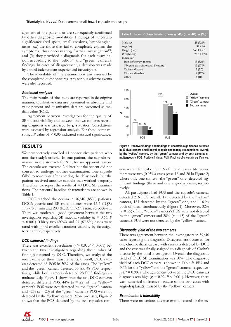
agement of the patient, or are subsequently confirmed by other diagnostic modalities. Findings of uncertain significance (red spots, small erosions, lymphangiec-tasias, etc.) are those that fail to completely explain the symptoms, thus necessitating further investigation[6]; and (3) they provided a diagnosis for each examina-tion according to the “yellow” and “green” camera’s findings. In cases of disagreement, a decision was made by a third independent experienced investigator.
The tolerability of the examinations was assessed by the completed questionnaires. Any serious adverse events were also recorded.
Statistical analysisThe main results of the study are reported in descriptive manner. Qualitative data are presented as absolute and value percent and quantitative data are presented as me-dian value (IQR).
Agreement between investigators for the quality of SB mucosa visibility and between the two cameras regard-ing diagnosis was assessed by κ statistics. Correlations were assessed by regression analysis. For these compari-sons, a P value of < 0.05 indicated statistical significance.
RESULTSWe prospectively enrolled 41 consecutive patients who met the study’s criteria. In one patient, the capsule re-mained in the stomach for 9 h, for no apparent reason. The capsule was excreted 2 d later but the patient did not consent to undergo another examination. One capsule failed to re-activate after entering the delay mode, but the patient received another capsule that worked properly. Therefore, we report the results of 40 DCC SB examina-tions. The patients’ baseline characteristics are shown in Table 1.
DCC reached the cecum in 36/40 (85%) patients. DCC’s gastric and SB transit times were 45.5 (IQR: 17.7-78.5) min and 200 (IQR: 117-277) min, respectively. There was moderate - good agreement between the two investigators regarding SB mucosa visibility (κ = 0.66, P < 0.001). Thirty two (80%) and 27 (67.5%) cases were rated with good-excellent mucosa visibility by investiga-tors 1 and 2, respectively.
DCC cameras’ findingsThere was excellent correlation (r > 0.9, P < 0.001) be-tween the two investigators regarding the number of findings detected by DCC. Therefore, we analyzed the mean value of their measurements. Overall, DCC cam-eras detected 68 POS in 50% of the cases. The “yellow” and the “green” camera detected 50 and 48 POS, respec-tively, while both cameras detected 28 POS findings si-multaneously. Figure 1 shows that the two DCC cameras detected different POS: 44% (n = 22) of the “yellow” camera’s POS were not detected by the “green” camera and 42% (n = 20) of the “green” camera’s POS were not detected by the “yellow” camera. More precisely, Figure 2 shows that the POS detected by the two capsule’s cam-
eras were identical only in 6 of the 20 cases. Moreover, there were two (0.05%) cases (case 18 and 20 in Figure 2) where only one camera -the “green” one- detected sig-nificant findings (three and one angiodysplasias, respec-tively).
All participants had FUS and the capsule’s cameras detected 216 FUS overall; 171 detected by the “yellow” camera, 161 detected by the “green” one, and 116 by both of them simultaneously (Figure 1). Moreover, 32% (n = 55) of the “yellow” camera’s FUS were not detected by the “green” camera and 28% (n = 45) of the “green” camera’s FUS were not detected by the “yellow” camera.
Diagnostic yield of the two camerasThere was agreement between the investigators in 39/40 cases regarding the diagnosis. Disagreement occurred for one chronic diarrhea case with erosions detected by DCC and the case was finally assigned to a diagnosis of Crohn’s disease by the third investigator. Overall, the diagnostic yield of DCC SB examination was 50%. The diagnostic yield of each DCC camera is shown in Table 2: 45% and 50% for the “yellow” and the “green” camera, respective-ly (P = 0.987). The agreement between the DCC cameras diagnosis was high (κ = 0.92, P < 0.001). However, there was numerical difference because of the two cases with angiodysplasia(s) missed by the “yellow” camera.
Examination’s tolerabilityThere were no serious adverse events related to the ex-
1464 March 21, 2011|Volume 17|Issue 11|WJG|www.wjgnet.com
Table 1 Patients’ characteristics (mean ± SD) (n = 40) n (%)
Male sex 29 (72.5)Age (yr) 58 ± 16Height (cm) 168.1 ± 9.3Weight (kg) 73.6 ± 12.8Indication Iron deficiency anemia 13 (32.5) Obscure gastrointestinal bleeding 15 (37.5) Crohn’s disease 1 (2.5) Chronic diarrhea 7 (17.5) Other 4 (10)
250
200
150
100
50
0
n
Overall“Yellow” camera“Green” cameraBoth cameras
6850 48
28
216
171161
116
POS FUS
Figure 1 Positive findings and findings of uncertain significance detected in 40 dual camera small-bowel capsule endoscopy examinations; overall, by the “yellow” camera, by the “green” camera, and by both cameras si-multaneously. POS: Positive findings; FUS: Findings of uncertain significance.
Triantafyllou K et al . Dual camera small-bowel capsule endoscopy
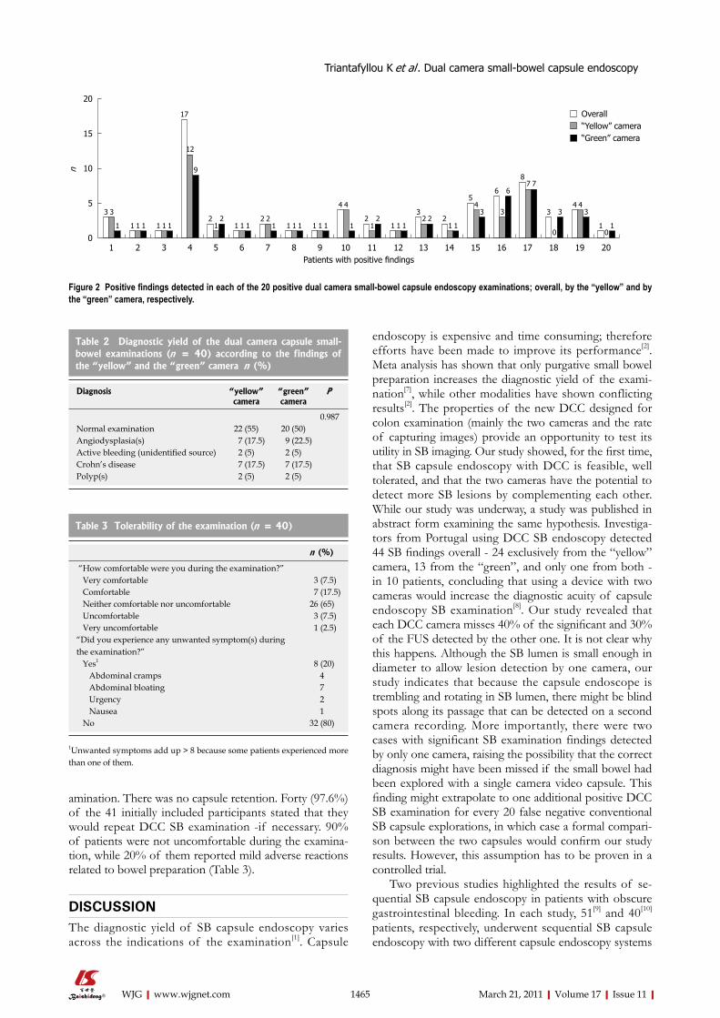
amination. There was no capsule retention. Forty (97.6%) of the 41 initially included participants stated that they would repeat DCC SB examination -if necessary. 90% of patients were not uncomfortable during the examina-tion, while 20% of them reported mild adverse reactions related to bowel preparation (Table 3).
DISCUSSIONThe diagnostic yield of SB capsule endoscopy varies across the indications of the examination[1]. Capsule
endoscopy is expensive and time consuming; therefore efforts have been made to improve its performance[2]. Meta analysis has shown that only purgative small bowel preparation increases the diagnostic yield of the exami-nation[7], while other modalities have shown conflicting results[2]. The properties of the new DCC designed for colon examination (mainly the two cameras and the rate of capturing images) provide an opportunity to test its utility in SB imaging. Our study showed, for the first time, that SB capsule endoscopy with DCC is feasible, well tolerated, and that the two cameras have the potential to detect more SB lesions by complementing each other. While our study was underway, a study was published in abstract form examining the same hypothesis. Investiga-tors from Portugal using DCC SB endoscopy detected 44 SB findings overall - 24 exclusively from the “yellow” camera, 13 from the “green”, and only one from both - in 10 patients, concluding that using a device with two cameras would increase the diagnostic acuity of capsule endoscopy SB examination[8]. Our study revealed that each DCC camera misses 40% of the significant and 30% of the FUS detected by the other one. It is not clear why this happens. Although the SB lumen is small enough in diameter to allow lesion detection by one camera, our study indicates that because the capsule endoscope is trembling and rotating in SB lumen, there might be blind spots along its passage that can be detected on a second camera recording. More importantly, there were two cases with significant SB examination findings detected by only one camera, raising the possibility that the correct diagnosis might have been missed if the small bowel had been explored with a single camera video capsule. This finding might extrapolate to one additional positive DCC SB examination for every 20 false negative conventional SB capsule explorations, in which case a formal compari-son between the two capsules would confirm our study results. However, this assumption has to be proven in a controlled trial.
Two previous studies highlighted the results of se-quential SB capsule endoscopy in patients with obscure gastrointestinal bleeding. In each study, 51[9] and 40[10] patients, respectively, underwent sequential SB capsule endoscopy with two different capsule endoscopy systems
1465 March 21, 2011|Volume 17|Issue 11|WJG|www.wjgnet.com
20
15
10
5
0
n
Overall“Yellow” camera“Green” camera
3 3
1 1 1 1 1 1 1 1 1 1 1 1 1 1 1 1 1 1 12
1 1 10
1
3
0
3
17
12
9
21
2 2 21
4 4
12
12
32 2
54
34 4
3
6
3
6
87 7
1 2 3 4 5 6 7 8 9 10 11 12 13 14 15 16 17 18 19 20 Patients with positive findings
Figure 2 Positive findings detected in each of the 20 positive dual camera small-bowel capsule endoscopy examinations; overall, by the “yellow” and by the “green” camera, respectively.
Table 2 Diagnostic yield of the dual camera capsule small-bowel examinations (n = 40) according to the findings of the “yellow” and the “green” camera n (%)
Diagnosis “yellow” camera
“green” camera
P
0.987Normal examination 22 (55) 20 (50)Angiodysplasia(s) 7 (17.5) 9 (22.5)Active bleeding (unidentified source) 2 (5) 2 (5)Crohn’s disease 7 (17.5) 7 (17.5)Polyp(s) 2 (5) 2 (5)
Table 3 Tolerability of the examination (n = 40)
n (%)
“How comfortable were you during the examination?” Very comfortable 3 (7.5) Comfortable 7 (17.5) Neither comfortable nor uncomfortable 26 (65) Uncomfortable 3 (7.5) Very uncomfortable 1 (2.5)“Did you experience any unwanted symptom(s) during the examination?” Yes1 8 (20) Abdominal cramps 4 Abdominal bloating 7 Urgency 2 Nausea 1 No 32 (80)
1Unwanted symptoms add up > 8 because some patients experienced more than one of them.
Triantafyllou K et al . Dual camera small-bowel capsule endoscopy

(PillCam SB and Olympus EndoCapsule). Patients ingest-ed the capsules in a randomized order and data showed that the DY of the two systems was similar. However in the German study[10], the second capsule detected angio-dysplasias in two (5%) more patients. The results of these studies differ from the results of studies that investigated the value of a second capsule endoscopy, using the same endoscopy system, in patients with obscure gastrointesti-nal bleeding or iron deficiency anemia with one previous non-diagnostic capsule investigation. Bar-Meir et al[11] showed significant lesions in seven of the 20 included severe iron deficiency patients, Jones et al[12] revealed posi-tive finding in 18 of 24 obscure gastrointestinal bleeders, while Viazis et al[13] established a diagnosis in 37 of 76 obscure gastrointestinal bleeders who underwent second look capsule endoscopy. Whether dual camera SB capsule endoscopy can reduce repeat SB examination (including capsule endoscopy) until a confirmed diagnosis remains to be investigated.
Apart from establishing a diagnosis, SB capsule en-doscopy also estimates the extent of the disease in cases of multiple angiodysplasias and Crohn’s disease, in which the involvement of different parts of the small bowel might dictate different therapeutic approaches. Therefore, missing lesions may underestimate disease extent. For ex-ample in case 3 with multiple small bowel ulcers/erosions shown in Figure 2, the “yellow” camera detected erosions throughout the recording, while all “green” camera’s POS were detected by the end of the recording (terminal ileum).
In our study, mucosa visibility was good-excellent in 68% and 80% of the cases, as rated by each investigator, respectively, with a half dose colonoscopy preparation regimen given after the insertion of the capsule at the duodenum. While this purge is given as a boost in colon capsule endoscopy studies, in our departments we use it as the standard preparation scheme for SB capsule en-doscopy to both avoid prolonging the capsule’s gastric transit time and to improve mucosa visibility, specifically in the ileum[4]. This preparation was both effective and well tolerated, causing mild adverse reactions in only 20% of the participants, which is similar to the complication rate published in the literature[2,7].
Finally, we addressed patient’s acceptance, which is one of the major issues for a successful diagnostic mo-dality[14]. The acceptance rate of DCC SB examination in our study was high: all participants (apart from one who was excluded initially) agreed to repeat the study if neces-sary. More importantly, there was no capsule retention and 90% of the patients did not feel uncomfortable dur-ing the examination.
Our study was a prospective feasibility trial aiming to find grounds for further formal investigation of DCC SB imaging. Therefore, the results should be interpreted with caution in clinical practice and some reservations should be noted. Firstly, the study design is not optimal for de-tecting firm conclusions and the sample size is small. The ideal trial to explore whether DCC endoscopy of the SB results in higher DY compared to the conventional
capsule, should include patients with one specific indica-tion (e.g. obscure gastrointestinal bleeding) who would ingest a single camera capsule operating on a different frequency than the DCC initially and the DCC later, or vice versa. Secondly, the capsule is 6 mm longer and this might result in swallowing difficulties, in a higher reten-tion rate of the capsule in the stomach, and in a higher rate of incomplete SB examinations[3]. All our patients swallowed the capsule easily. However, in one case, the capsule remained in the stomach during the life span of the capsule’s battery, without any predisposing fac-tor. We did not detect increased DCC gastric and small bowel transit time, compared to those published with the conventional SB capsule[7], and the rate of complete SB examinations with cecal visualization was 85%, similar to that reported in the literature with the conventional SB capsule[7]. Furthermore, complete colon examination was observed in 42.5% of the cases without any further purgative boost; however, bad-moderate bowel cleansing in 79%-85% of the cases prevented any thorough colon examination (data not shown in the results). Thirdly, the longer time required to review the videos from each in-dividual camera and from both cameras simultaneously might be an issue. However, we have not addressed this parameter. Lastly, it might be not acceptable by many pa-tients to wait 2 h until the definite DCC activation. How-ever for the purpose of our study, we have not tried the capsule’s initial activation in advance.
In conclusion, we showed that DCC SB endoscopy is feasible and well tolerated. Moreover, the two capsule’s cameras complement each other to detect more SB le-sions. It remains to be determined whether DCC SB endoscopy can increase the diagnostic yield of capsule en-doscopy SB examination and if it can improve the clinical outcome of patients undergoing the examination.
ACKNOWLEDGMENTSPart of our study has been presented in the 2009 Diges-tive Diseases Week, Chicago, IL, USA (in poster form) and in the GASTRO 2009, London, UK (oral presenta-tion).
COMMENTSBackgroundWireless video-capsule endoscopy is a non-invasive and patient friendly ex-amination, which has revolutionized small-bowel (SB) exploration. It has been proved to be superior to any other radiographic or endoscopic modality for SB mucosa examination. However, it is expensive and its diagnostic yield varies across the indications. Until recently several methods have been introduced to improve the method’s diagnostic yield, with conflicting results. Research frontiersWireless capsule endoscopy is an evolving technology. Recently, a new capsule endoscope designed for colon examination has been introduced to the market. It is a dual camera capsule (DCC) that compared to the single camera conven-tional SB capsule has a theoretical advantage to detect more SB lesions.Innovations and breakthroughsThis theoretical advantage has been tested in 41 consecutive patients with an indication for SB mucosa examination and DCC endoscopy was performed to examine whether the two cameras complement each other to detect more le-
1466 March 21, 2011|Volume 17|Issue 11|WJG|www.wjgnet.com
COMMENTS
Triantafyllou K et al . Dual camera small-bowel capsule endoscopy
sions than each of them alone. The results showed that each camera missed 40% of the findings that explain patient’s symptoms detected by the other one. More importantly, there were two cases with significant SB examination findings detected by only one camera, raising the possibility that the correct diagnosis might have been missed if the small bowel had been explored with a single camera video-capsule. In conclusion, this study revealed that DCC SB endos-copy is feasible, well tolerated, and that the two capsule’s cameras complement each other to detect more SB lesions.ApplicationsGiven that a feasibility trial can not reach firm conclusions, the hypothesis that DCC may increase the diagnostic yield of wireless capsule SB endoscopy must be tested in a formal way. If proven, DCC SB endoscopy will be the standard for SB mucosa exploration.TerminologyWireless video-capsule endoscopy is an endoscopy system equipped with a capsule endoscope that acquires images from the gut lumen and transmits them to a data recorder using wireless emission technology. Images are pro-cessed and reviewed in a video format using a commercially available com-puter.Peer reviewThe trial of a colon capsule to view the small intestine is an innovative idea and the results indicate that it has considerable clinical importance. This is a well designed and clinically relevant study.
REFERENCES1 Liao Z, Gao R, Xu C, Li ZS. Indications and detection, com-
pletion, and retention rates of small-bowel capsule endosco-py: a systematic review. Gastrointest Endosc 2010; 71: 280-286
2 Ladas SD, Triantafyllou K, Spada C, Riccioni ME, Rey JF, Niv Y, Delvaux M, de Franchis R, Costamagna G. European Society of Gastrointestinal Endoscopy (ESGE): recommen-dations (2009) on clinical use of video capsule endoscopy to investigate small-bowel, esophageal and colonic diseases. Endoscopy 2010; 42: 220-227
3 Adler DG, Chand B, Conway JD, Diehl DL, Kantsevoy SV, Kwon RS, Mamula P, Rodriguez SA, Shah RJ, Song LM, Tierney WM. Capsule endoscopy of the colon. Gastrointest Endosc 2008; 68: 621-623
4 Triantafyllou K, Kalli T, Ladas SD. Small bowel purge after the entrance of the capsule in the duodenum results to better
quality of bowel preparation for video -capsule endoscopy. Prospective, randomized, double-blind, placebo-controlled, real time viewer assisted study. Gastrointest Endosc 2008; 134 (Suppl 1): A-339
5 Minimal standard terminology in digestive endoscopy. En-doscopy 2000; 32: 162-188
6 Costamagna G, Shah SK, Riccioni ME, Foschia F, Mutignani M, Perri V, Vecchioli A, Brizi MG, Picciocchi A, Marano P. A prospective trial comparing small bowel radiographs and video capsule endoscopy for suspected small bowel disease. Gastroenterology 2002; 123: 999-1005
7 Rokkas T, Papaxoinis K, Triantafyllou K, Pistiolas D, Ladas SD. Does purgative preparation influence the diagnostic yield of small bowel video capsule endoscopy?: A meta-analysis. Am J Gastroenterol 2009; 104: 219-227
8 Almeida N, Figueiredo P, Lopes S, Freire P, Gouveia H, Leitão MC. Is it the end of the single camera small bowel capsule as we know it? Endoscopy 2008; 40 (Suppl 1): A30
9 Cave DR, Fleischer DE, Leighton JA, Faigel DO, Heigh RI, Sharma VK, Gostout CJ, Rajan E, Mergener K, Foley A, Lee M, Bhattacharya K. A multicenter randomized comparison of the Endocapsule and the Pillcam SB. Gastrointest Endosc 2008; 68: 487-494
10 Hartmann D, Eickhoff A, Damian U, Riemann JF. Diagnosis of small-bowel pathology using paired capsule endoscopy with two different devices: a randomized study. Endoscopy 2007; 39: 1041-1045
11 Bar-Meir S, Eliakim R, Nadler M, Barkay O, Fireman Z, Scapa E, Chowers Y, Bardan E. Second capsule endoscopy for patients with severe iron deficiency anemia. Gastrointest Endosc 2004; 60: 711-713
12 Jones BH, Fleischer DE, Sharma VK, Heigh RI, Shiff AD, Hernandez JL, Leighton JA. Yield of repeat wireless video capsule endoscopy in patients with obscure gastrointestinal bleeding. Am J Gastroenterol 2005; 100: 1058-1064
13 Viazis N, Papaxoinis K, Vlachogiannakos J, Efthymiou A, Theodoropoulos I, Karamanolis DG. Is there a role for second-look capsule endoscopy in patients with obscure GI bleeding after a nondiagnostic first test? Gastrointest Endosc 2009; 69: 850-856
14 van Tuyl SA, den Ouden H, Stolk MF, Kuipers EJ. Optimal preparation for video capsule endoscopy: a prospective, ran-domized, single-blind study. Endoscopy 2007; 39: 1037-1040
S- Editor Wang JL L- Editor Stewart GJ E- Editor Zheng XM
1467 March 21, 2011|Volume 17|Issue 11|WJG|www.wjgnet.com
Triantafyllou K et al . Dual camera small-bowel capsule endoscopy

Factor analysis identifies subgroups of constipation
Philip G Dinning, Mike Jones, Linda Hunt, Sergio E Fuentealba, Jamshid Kalanter, Denis W King, David Z Lubowski, Nicholas J Talley, Ian J Cook
Philip G Dinning, Linda Hunt, Sergio E Fuentealba, Ian J Cook, Department of Gastroenterology, St. George Hospital, University of New South Wales, Kogarah, NSW 2217, AustraliaMike Jones, Department of Psychology, Macquarie University, NSW 2109, AustraliaJamshid Kalanter, Gastroenterology Research Unit, Nepean Hospital, University of Sydney, Sydney, NSW 2050, AustraliaDenis W King, David Z Lubowski, Department of Colorectal Surgery, St. George Hospital, Kogarah, NSW 2217, AustraliaNicholas J Talley, Gastroenterology and Hepatology, Mayo Clinic, Jacksonville, FL 32224, United StatesAuthor contributions: Dinning PG, Jones M, Talley NJ and Cook IJ designed the study and wrote the paper; Hunt L and Fuentealba SE collated all data and maintained the database; Kalanter J, King DW, Lubowski DZ and Cook IJ distributed the majority of questionnaires; Jones M performed all analysis.Supported by National Health and Medical Research Council Australia (ID 455213)Correspondence to: Dr. Phil G Dinning, PhD, Department of Gastroenterology, St George Hospital, University of New South Wales, Kogarah, NSW 2217, Australia. [email protected]: +61-2-91132817 Fax: +61-2-91133993Received: June 16, 2010 Revised: January 13, 2011Accepted: January 20, 2011Published online: March 21, 2011
AbstractAIM: To determine whether distinct symptom group-ings exist in a constipated population and whether such grouping might correlate with quantifiable pathophysi-ological measures of colonic dysfunction.
METHODS: One hundred and ninety-one patients pre-senting to a Gastroenterology clinic with constipation and 32 constipated patients responding to a newspaper advertisement completed a 53-item, wide-ranging self-report questionnaire. One hundred of these patients had colonic transit measured scintigraphically. Factor analysis determined whether constipation-related symp-toms grouped into distinct aspects of symptomatology. Cluster analysis was used to determine whether indi-
vidual patients naturally group into distinct subtypes.
RESULTS: Cluster analysis yielded a 4 cluster solution with the presence or absence of pain and laxative unre-sponsiveness providing the main descriptors. Amongst all clusters there was a considerable proportion of pa-tients with demonstrable delayed colon transit, irritable bowel syndrome positive criteria and regular stool fre-quency. The majority of patients with these characteris-tics also reported regular laxative use.
CONCLUSION: Factor analysis identified four consti-pation subgroups, based on severity and laxative un-responsiveness, in a constipated population. However, clear stratification into clinically identifiable groups re-mains imprecise.
© 2011 Baishideng. All rights reserved.
Key words: Factor analysis; Constipation; Symptoms; Clusters; Laxatives
Peer reviewer: Dr. Uday C Ghoshal, MD, DNB, DM, FACG, Additional Professor, Department of Gastroenterology, Sanjay Gandhi Postgraduate Institute of Medical Science, Lucknow 226014, India
Dinning PG, Jones M, Hunt L, Fuentealba SE, Kalanter J, King DW, Lubowski DZ, Talley NJ, Cook IJ. Factor analysis identi-fies subgroups of constipation. World J Gastroenterol 2011; 17(11): 1468-1474 Available from: URL: http://www.wjgnet.com/1007-9327/full/v17/i11/1468.htm DOI: http://dx.doi.org/10.3748/wjg.v17.i11.1468
INTRODUCTIONConstipation is a heterogeneous disorder, the most consis-tent generic descriptor for which is difficult or infrequent passage of stool[1,2]. A fundamental aim in researching and treating any heterogeneous disorder is to subclassify
BRIEF ARTICLE
World J Gastroenterol 2011 March 21; 17(11): 1468-1474ISSN 1007-9327 (print) ISSN 2219-2840 (online)
© 2011 Baishideng. All rights reserved.
Online Submissions: http://www.wjgnet.com/[email protected]:10.3748/wjg.v17.i11.1468
1468 March 21, 2011|Volume 17|Issue 11|WJG|www.wjgnet.com
Dinning PG et al . Constipation symptoms
the condition into categories that are predictive of either pathophysiology or treatment outcome. Constipation is currently conceptualized in three broad categories: nor-mal transit constipation (NTC), slow transit constipation (STC) and disorders of defecation or rectal evacuation[3-5]. This distinction among subgroups, in some cases, has proven beneficial in planning treatment and in predicting therapeutic outcome[4,6-12].
It remains unknown whether subtypes of constipa-tion can be identified reliably on the basis of symptoms. The mathematical techniques of factor and/or cluster analysis, to determine whether certain symptoms and/or subjects do group together more than expected by chance, can provide empiric evidence of the existence of true syndromes. Studies applying these techniques have found conflicting results, with Mertz et al[13] identifying 3 subtypes while Eltringham et al[14] did not. These findings suggest that the a priori assumption that these subtypes are distinct or distinguishable from each other on the basis of symptoms may be incorrect, or that investigators have yet to combine the correct symptoms to identify pathophysiologically distinct abnormalities.
Utilizing a 53-item specific constipation question-naire and colonic scintigraphy our aim was to identify symptoms or groups of symptoms that identify un-derlying severe constipation subgroups. Specifically, we hypothesized that within this constipation population at least three distinct subgroups, STC, evacuation disorder and irritable bowel syndrome (IBS), can be identified by groups of symptoms and that these symptom groups will be predictive of specific underlying objective physiologi-cal measures such as colonic transit time.
MATERIALS AND METHODSDesign of the questionnaire and assessment of symptoms The Sydney Constipation Questionnaire (SCQ) was de-rived from the previously validated Bowel Disease Ques-tionnaire[15] and the Bowel Symptom Questionnaire[16]. The SCQ comprises 53 items, including 42 symptom items and the validated Bristol stool scale[17]. Additional constipation items were then added and the face valid-ity was assessed by responses from 29 internationally recognised constipation experts who were asked to rate each question according to clinical relevance. The test-retest reliability of the final questionnaire was evaluated in 47 patients who repeated the questionnaire within 3 wk of initial testing[18]. The design of the questionnaire took place long before the release of Rome Ⅲ criteria for bowel[19] and anorectal[20] dysfunction, nevertheless the questionnaire does contain all of the symptomatic ques-tions that define constipation, IBS and outlet obstruction by the Rome Ⅲ criteria.
Population sample Subjects were obtained through two sources: (1) via refer-rals from specialists in the Sydney area; and (2) by direct
advertisement that specified criteria approximating ter-tiary-referred subjects. The impact of subject source was considered in the statistical analysis. Overall, the sample was designed to reflect community members suffering serious constipation symptoms. Subjects were deliberately not selected based on Rome Ⅲ constipation criteria, but rather on the basis of serious symptoms to avoid mak-ing assumptions and because we wanted also to examine the proportion of the study cohort and its clusters that would be positive for Rome Ⅲ criteria for constipation or IBS.
Referrals to colorectal surgeons or gastroenterologists in Sydney metropolitan region, seeking treatment for con-stipation, were recruited over a 4-year period and were given a questionnaire if they fulfilled the following crite-ria: (1) had not undergone any form of colectomy, rec-tocele or rectal prolapse repair; and (2) their constipation was not deemed secondary to a metabolic or neurological disease or pregnancy. Additionally, the questionnaire was given to potential patients who responded to advertise-ments in Sydney newspapers recruiting subjects for a ran-domized control trial of sacral nerve stimulation for the treatment of constipation (in progress; to be reported in future). These potential patients were screened during an initial phone conversation and if they fulfilled the criteria listed above the questionnaires were mailed out to them. As these were chronically constipated patients, who for the most part had been taking laxatives for many years, they were not asked to report on what their symptoms are, were or might be, in the absence of laxatives. Studies have shown that the correlation between a patient’s re-ported stool frequency and actual measures is poor[21] and in our experience a patient’s ability to recall symptoms from the years prior to laxative use is also poor.
Allowing 3 wk for the questionnaire to be completed and returned, each non-responder was then phoned and asked if they intended to return the questionnaire. A proportion of community responders had decided that their symptoms were not severe enough to warrant sacral nerve stimulation and these patients did not return the questionnaire. Allowing an additional 4 wk, all other non-responders were excluded from the study. All participants in this study gave written, informed consent and the study was approved by the Human Ethics Committees of the South Eastern Area Health Service, Sydney and the University of New South Wales.
A priori symptom groups Three symptom groups were defined a priori: (1) STC was considered to comprise infrequent bowel movements (less than 2 defecations/wk), lack of defecation urge, excessive straining and hard stools[22-24]; (2) Obstructed defecation was classified when all of the following symptoms were present: (a) an inability to initiate defecation following the urge to do so, or difficulty with stool evacuation; (b) excessive straining at stool more than 25% of the time or self-digitation to facilitate defecation more than 25% of the time; and (c) a feeling of incomplete evacuation after
1469 March 21, 2011|Volume 17|Issue 11|WJG|www.wjgnet.com

defecation[1,22,25-29]; and (3) IBS classified those patients with symptoms of abdominal pain and bloating that improve with defecation or in whom the onset of such symptoms is associated with a change in frequency or form of the stool[19,22,24,30].
Transit studiesColonic transit was measured using a standard nuclear medicine technique[31,32]. Briefly, the subjects attended a Nuclear Medicine Department on a Monday morning and were given 4 MBq 111In-DTPA orally at approximate-ly 9:00 am. They returned to the department the same day at approximately 3:00 pm and on subsequent days at approximately 24, 48, 72 and 96 h following the oral administration. On each occasion, anterior and posterior abdominal images were obtained each for 10 min using a large field-of-view gamma camera and medium energy collimator. Background images were obtained for back-ground correction. All laxative medications were stopped 3 d before and during the scan. No effort was made to control the patient’s diet during the scan period. Method of the scintigraphic analysis is reported elsewhere[33]. Briefly, at each time point, profiles of the activity along the colon were derived from a geometric mean image obtained from the anterior and posterior images. Total percent retention (T%R) and mean activity position (MAP) at each time were calculated for each subject. T%R indicates the retained activity in the colon using the value at 6 h as 100%. MAP indicates the geometric center of the activity, where position 0 is at the cecum, position 98 at the anus, and position 99 being excreted activity. The scintigraphic definition of delayed transit constipa-tion was met if the study showed isotope retention of greater than 9% in the right (cecum to mid-transverse) or left colon (mid-transverse to distal descending colon) at 72 h[31,32].
Data analysisSince this has been a vexed question to date, a naturalistic approach was adopted in which multivariate statistical techniques were used to identify distinct clusters of indi-viduals with respect to bowel symptom patterns and these clusters were then examined with respect to their symp-tom profiles to determine what, if any, clinically meaning-ful differences existed between them. Analysis proceeded in two steps; the first reduced the data dimensionality from 42 observed symptom variables to 15 latent vari-ables which were then used in the second step in a cluster analysis to form internally homogeneous clusters of in-dividuals: details follow. The first step identified indepen-dent dimensions of symptom profile through principle components analysis followed by orthogonal rotation of the factor space. Identifying independent dimensions of bowel symptoms has several advantages. Bowel symptom questionnaires typically ask several questions around a given bowel symptom to fully characterize patients’ symp-tomatology. Despite the clinical value of these questions, they are typically strongly correlated, leading to statistical
redundancy amongst them. In addition, cluster analysis is prone to dominance by scales that are numerically large and factor scores calculated for individuals follow a unit of normal distribution (mean zero, standard deviation 1.0) and are therefore standardized. Factors were interpreted and used in the subsequent cluster analysis if the cor-responding eigenvalue was > 1.0, which corresponds to approximately 2% explained variance in the original data. The variance in the original data explained by each factor is reported in Table 1, as well as the total explained vari-ance across all 15 factors used. A score was calculated for each factor for each subject, which is in effect a weighted sum across all original data items deemed to load on a given factor. These are reported in Table 1, along with the rotated factor loading for each item, using a criterion of rotated factor loadings > 0.4 (in absolute value). Factor loadings can be interpreted as the correlation between a given original data item and its corresponding latent vari-able. In the second analytic step, these scores were used in a non-hierarchical (K-Means) cluster analysis. It is im-portant to note that only symptom latent variables were utilized in cluster formation, not transit times. Based on an a priori expectation of a moderate number of distinct clusters, solutions between 1 and 6 clusters were consid-ered. A single cluster solution would imply the subjects were not differentiated in any systematic fashion, while six clusters would represent a quite complex system of constipation subgroups. The choice of cluster solution adopted was a trade-off of within-cluster homogeneity and minimizing unnecessary complexity. In a K-Means analysis the algorithm assigns individuals to clusters such that the overall within-cluster variance is minimized and the between-cluster variance is maximized, given the pre-specified number of clusters. Euclidean distance was used to measure the distance between individual points and their cluster centroid.
Determination of the clinical value of the clusters identified was through a comparison of profiles of indi-vidual symptom items and by comparing rates of a priori criteria, Rome Ⅲ-defined constipation, IBS and outlet dysfunction, and rates of slow transit measured as de-scribed earlier.
RESULTSThe questionnaire was given to a total of 326 individuals suffering from constipation. Of these, 246 were referrals to colorectal surgeons or gastroenterologists and 80 were recruited from the community by advertisements. Of these, a total of 223 responded, representing an overall response rate of 68% [191 (78%) response rate for clinic cases; 32 (40%) response rate for community cases]. Of the 223 returned questionnaires, 4 were not able to be utilized (2 incomplete; 2 were multivariate outliers) leav-ing n = 217 for final analysis. There was no significant age or gender difference between the clinic group (45 ± 17 years; range 18-81 years; 13 M:178 F) and community groups (56 ± 20 years; range 24-82 years; 5 M:27 F). The
1470 March 21, 2011|Volume 17|Issue 11|WJG|www.wjgnet.com
Dinning PG et al . Constipation symptoms

mean age of the entire group was 47 ± 17 years (range 18-82 years).
Colonic transit was performed in all patients who had access to a nuclear medicine facility; in total 100 patients (52%) underwent the procedure study.
Factor analysis The factor analysis produced a rotated factor matrix com-prising 15 factors. Table 1 lists these 15 factors and the labels that we have attached to each factor, along with the loadings of the associated questions. For brevity, we have only shown questions within each factor for which the absolute value of the loading was ≥ 0.35. While this process is simply an intermediary step towards cluster analysis, there are a number of potentially relevant obser-vations to be made from this matrix. An IBS-like factor emerged, indicating that IBS-like symptoms stand out as a distinct entity in this population. Despite the popula-tion being selected for its constipation, a diarrhea factor emerged, probably accounted for by laxative usage (see below).
Cluster analysis Cluster 1: Patients in this cluster are less likely than av-erage to report preservation of their defecatory urge (compared to cluster 4) despite 80% using laxatives regu-larly. This cluster is relatively laxative-responsive with 83% describing laxatives as somewhat or very effective. Although clusters 1 and 4 share some similarities, they differ dramatically in their responsiveness to laxatives. Cluster 1 patients experience less upper abdominal pain than patients in clusters 2 and 4. Patients in cluster 1 have more IBS-like features than those in other clusters and they gain pain relief following a bowel action.
Cluster 2: This group has features similar to those of cluster 1 but when compared with cluster 1, their pain is somewhat less severe and less prevalent and they de-scribe a shift in the site of their pain from lower to the upper abdomen. They describe similar laxative usage and responsiveness to patients in clusters 1 and 3. These pa-tients are least likely to visit the toilet daily (63%) despite the fact that they report the highest rate of “urge prior to attempting to defecate” (72%). They are relatively laxative-responsive with 76% reporting them “somewhat or very effective”.
Cluster 3: Patients in this cluster have the lowest pain scores of all 4 clusters, have a short history of consti-pation and are more likely to report a weekly stool fre-quency within the Rome Ⅲ defined range for constipa-tion. They report the lowest laxative usage, but they are the most laxative-responsive of all patients. They rarely report a feeling of rectal blockage and are less likely to adopt self digitation to facilitate evacuation. These pa-tients never report diarrhea and rarely report hard stool.
Cluster 4: These patients report the highest pain scores and are strikingly unresponsive to laxatives, despite re-
1471 March 21, 2011|Volume 17|Issue 11|WJG|www.wjgnet.com
Table 1 Factor loadings derived from the rotated factor matrix
Factor loading
Percent variance
Factor 1: Straining 17.3 Strain hard: How often 0.82 Straining: How bad usually 0.80 Straining: How long 0.76 Straining: How often 0.71 How long to bowel motions take 0.59 How often incomplete evacuation 0.55 How often unsuccessful attempts 0.52 Frequency of any bowel problems 0.47 How troubling is constipation 0.41 Change positions: How often 0.35Factor 2: Pain frequency and severity 8.4 Abdominal pain: How often 0.80 Pain in belly: Past 3 mo 0.79 Abdominal pain: Severity 0.79 Abdominal pain: Length 0.65 Rectal mucus: How often 0.41 Rectal pain: How often 0.39 Pain in belly: Past 12 mo -0.58Factor 3: Duration of constipation 6.5 Constipation: How many years 0.89 Straining: How many years 0.87 Abdominal pain: First occurrence 0.57Factor 4: Irritable bowel syndrome symptoms 5.6 Abdominal pain: Improved after bowel motion 0.75 Abdominal pain: Improved after passing gas 0.70 Experience lower abdominal pain 0.63 Bowel motions: Harder than usual past 12 mo 0.40Factor 5: Urge frequency 4.1 Urge for bowel motion: How often 0.72 Perceive an urge before attempt to open bowels 0.68 Visits to toilet: How often 0.56 Urgency for bowel motion: How often 0.42Factor 6: Diarrhea frequency 3.8 Loose/watery stool: How often 0.75 Pebble-like stool: How often -0.42 Rectal disimpaction: How often required -0.45 Hard/lumpy stool: How often -0.63Factor 7: Alternating between diarrhea and constipation 3.4 Usually alternating 0.87 Usually constipated -0.86Factor 8: Bloating frequency 3.2 Stomach swelling in the last 12 mo: How often 0.71 Felt bloated in the last 12 mo: How often 0.68 Felt blocked: How often 0.47Factor 9: Laxative efficacy 2.9 Laxative use: Longest gap between taking and bowel motion
0.70
Bowel motions: Fewer than usual past 12 mo 0.56 Laxative use: How often 0.44Factor 10: Rectal urgency 2.7 Urge from rectum 0.89 Urge from abdomen and rectum -0.80Factor 11: Bowel motion frequency 2.6 Longest gap between bowel motions 0.81 Usual bowel frequency -0.56Factor 12: Change in bowel frequency 2.4 Bowel motions: More than usual past 12 mo 0.74 Bowel motions: Looser than usual past 12 mo 0.62Factor 13: Abdominal urge 2.3 Experience an urge from abdomen 0.89Factor 14: Diarrhea predominance 2.2 Usually experience diarrhea 0.79Factor 15: Antecedent to constipation 2.0 An antecedent for the constipation 0.71 Experience upper abdominal pain 0.51Total (1-15) 69.3
Dinning PG et al . Constipation symptoms

porting the highest laxative usage of all (98% use them > 50% of the time). Abdominal pain is described as more severe, more frequent and lasting longer than in the other clusters. Importantly, patients in this cluster report that a bowel motion does not relieve their pain. They are less likely to have an antecedent (e.g. hysterectomy, pregnan-cy) than those in the other clusters. In comparison with other clusters, these patients report increased frequency of defecatory urge with 30% reporting an urge to defe-cate more than 3 times/d, more frequent toilet visits (60% > once/d), frequent unsuccessful attempts at defecation (73%) and frequent sense of blockage (66%) during pas-sage of stool.
Correlation with Rome Ⅲ criteria for constipation or IBSIn this population with severe constipation, the positivity rate for Rome Ⅲ constipation was 59%-85% across the 4 clusters (Table 2). In this severely constipated population, with the exception of cluster 3, 78%-98% were habitual laxative users. Clusters 1 and 4 are characterized by high rates of Rome Ⅲ IBS (over 50%, Table 2) compared with 20%-27% in clusters 2 and 3 (Table 2). The major-ity (92%) of patients that met Rome Ⅲ IBS criteria were also associated with heavy laxative use.
Correlation with symptomatically-defined obstructed defecationA remarkably constant 49%-57% of patients reported symptoms that have traditionally been attributed to ob-structed defecation. The prevalence of this pattern did not differ among the four clusters.
Correlation with scintigraphically confirmed slow transitBetween 63% and 79% of patients had slow transit (Table 2). The prevalence of slow transit was least in cluster 3 (63%), the cluster with the mildest symptoms, but this still repre-sents a sizable majority of this group. Of the 25% of se-verely constipated patients that report > 3 bowel motions a day, 75% have demonstrable STC. The vast majority of
these patients are also heavy laxative users, report mainly liquid stool, and a feeling of incomplete evacuation.
DISCUSSIONThe ability to subtype severely constipated patients based upon symptoms has merit because it focuses and system-atizes epidemiological enquiry, and has the potential to dictate logical and cost effective investigation algorithms for clinicians, to influence management and to predict therapeutic outcome. However, this study highlights the difficulties in using symptoms as discriminators of severe constipation subtypes. Although four groupings were identified by cluster analysis, it is difficult to attach clearly recognizable pathophysiological labels to these clusters. The major finding of this study is the identification of a group (cluster 4) with long history of constipation, a profound lack of response to laxatives despite extremely high laxative usage, and the highest pain scores. In con-trast, cluster 3 was characterized by low pain scores, low rates of co-morbidity and the lowest rate of delayed colonic transit. Overall, our data appear to suggest sub-types based on severity and chronicity of disease which is reflected in rates of, and responsiveness to, laxative therapy.
The positivity rate for Rome Ⅲ-defined constipa-tion was 59%-85% across the four clusters. Interestingly, of the patients that did not meet the Rome criteria, 63%-80% across the 4 clusters have demonstrable de-layed colonic transit. In other words, Rome Ⅲ criteria will not pick up a substantial proportion of people who are severely troubled by constipation and who clearly have markedly disturbed physiology as confirmed by de-layed colonic transit. In addition, a large proportion of patients, particularly in clusters 1 (52%) and 4 (54%), met the Rome Ⅲ criteria for IBS. In our experience this over-all high prevalence of criteria satisfying the definition of IBS is in keeping with the situation commonly encoun-tered by clinicians and has been reported previously[34].
One of the potential problems with subtyping consti-pated patients into categories based on questionnaire data is the prevalence of laxative use. Given that the approach to constipation subtyping used in this study relied upon symptom patterns, laxative use is a potential confounder because these agents can induce symptoms of bloating and pain and alter stool frequency/consistency. While laxative use is mentioned in previous studies[13,14,34-36], little or no attempt is made to discern their impact upon symptoms. For example, of those patients that met IBS criteria in this study, 92% were heavy laxative users. Ask-ing patients to detail their symptoms in the absence of laxatives has been attempted[14]. However, in our experi-ence such questions are difficult to answer for a chroni-cally constipated population who, for the most part, have been taking laxatives for many years. Indeed, previous studies have shown a poor concordance between a pa-tient’s recollection of events and actual measures[21].
Furthermore, high rates of laxative use may also con-
1472 March 21, 2011|Volume 17|Issue 11|WJG|www.wjgnet.com
Table 2 Summary of the proportion of patients within each cluster who were positive for constipation1 or irritable bowel syndrome on the basis of Rome Ⅲ criteria, as well as propor-tions in each group with slow transit constipation
Clusters (4-fold solution)
1 (n = 71)
2 (n = 44)
3 (n = 34)
4 (n = 43)
Rome Ⅲ constipation Yes 84.50% 77.30% 58.80% 72.10%Rome Ⅲ IBS Yes 52.10% 27.30% 20.60% 53.50%Outlet dysfunction Yes 52.10% 56.80% 52.90% 48.80%Colonic transit n = 43 n = 28 n = 16 n = 29 Delayed transit 74.40% 71.40% 62.50% 79.30% Normal transit 25.60% 28.60% 37.50% 20.70%
1With the exception that symptoms were assessed irrespective of laxative usage. IBS: Irritable bowel syndrome.
Dinning PG et al . Constipation symptoms

found attempts to identify symptom-based distinct sub-types of constipation that correspond to pathophysiolog-ical subtypes. This becomes evident when examining the correlation between delayed transit and infrequent stool frequency. There is literature to support infrequent bowel movements in predicting delayed transit[36-38]. Those stud-ies found that < 3 bm/wk predicted delayed transit in 85%-100% of patients. Indeed, it is the practice of some groups to only evaluate transit formally in patients with infrequent stools[39]. However, this study indicates that a high percentage (75%) of patients who use their bowel > 3/d have demonstrable slow colonic transit. Importantly, these patients also report high laxative use. Therefore, these data suggest that normal stool frequency, in the context of concurrent laxative use, certainly does not preclude STC.
Delayed colonic transit was found in the majority of patients in whom scintigraphy was performed, which is comparable with the reports of others[38,40,41]. While it was lowest in cluster 3, a sizable 63% in this relatively mildly affected group had delayed transit; hence, our findings suggest that delayed transit cannot be predicted accu-rately on the basis of a combination of symptoms.
Using existing criteria for obstructed defecation based purely on symptoms[1,22,25-28], this syndrome was both common and equiprevalent across all four clusters (49%- 57%). This lack of discriminatory ability of clusters to co-localize with these symptoms supports the consistent findings of a number of investigators that symptoms are not predictive of pelvic dyssynergia demonstrated by anorectal manometry and balloon expulsion testing[34-36,42] or demonstrated by defecography[43]. Grotz et al[34] found that only a sense of anal blockage correlated with proven pelvic floor dysfunction. However, as Grotz et al[34] point out, the usefulness of this finding must be questioned because this symptom was present in 67% of pelvic floor dysfunction but was found in 50% of patients with STC and in 53% with NTC. Indeed, in their multivariate analysis, they could not identify any colonic symptoms as discriminators of constipation subtypes.
Clinical history remains a “blunt instrument” and while combinations of symptoms do point towards 4 subsets of constipation, we are some distance yet from defining those subsets in unequivocal and specific terms. In light of the overlap with IBS symptoms in this study, it behooves the clinician to consider severe delayed transit in patients presenting with constipation-predominant IBS as this may influence management and avoid mislabeling the patient. Currently, combinations of symptoms cannot predict accurately whether the patient that is categorized into one of these 4 clusters has normal or delayed transit. However, laxative use in severely constipated patients may influence the reported symptoms and this potential confounder needs to be taken into consideration when interpreting these results.
COMMENTSBackgroundConstipation is often perceived as a benign, easily treated condition; however,
a number of studies confirm that constipation has a significant adverse effect on a patient’s quality of life. Constipation is a heterogeneous disorder and a fundamental aim in researching and treating any heterogeneous disorder is to subclassify the condition into categories that will help guide treatment options. As a patient’s symptoms are the first point of discussion with their doctor, the ability to subclassify on the basis of symptoms is a primary goal. Research frontiersThe mathematical techniques of factor and/or cluster analysis have been used in an attempt to determine whether certain symptoms can predict constipation subtypes. However, studies applying these techniques have found conflicting results suggesting that the a priori assumption that these subtypes are distinct or distinguishable from each other on the basis of symptoms may be incor-rect, or that investigators have yet to combine the correct symptoms to identify pathophysiologically distinct abnormalities. Utilizing a 53-item specific consti-pation questionnaire our aim was to identify groups of symptoms that identify underlying severe constipation subgroups, such as slow transit constipation, evacuation disorder and irritable bowel syndrome.Innovations and breakthroughsFactor analysis of 221 questionnaires yielded a 4 cluster solution with the pres-ence or absence of pain and laxative unresponsiveness providing the main de-scriptors. Amongst all clusters there was a considerable proportion of patients with demonstrable delayed colon transit, irritable bowel syndrome positive cri-teria and regular stool frequency. Therefore, as with previous studies, we have demonstrated that significant overlap exists between mathematically defined clusters of symptoms and globally accepted sub-types of constipation.ApplicationsLaxative use in severely constipated patients may influence the reported symp-toms and this potential confounder needs to be taken into consideration when interpreting these results.Peer reviewThis is an important study in which authors attempted to evaluate whether clus-ter of symptoms can help to understand pathophysiology of constipation.
REFERENCES1 Talley NJ, Weaver AL, Zinsmeister AR, Melton LJ 3rd.
Functional constipation and outlet delay: a population-based study. Gastroenterology 1993; 105: 781-790
2 Pare P, Ferrazzi S, Thompson WG, Irvine EJ, Rance L. An epidemiological survey of constipation in canada: defini-tions, rates, demographics, and predictors of health care seeking. Am J Gastroenterol 2001; 96: 3130-3137
3 American College of Gastroenterology Chronic Constipa-tion Task Force. An evidence-based approach to the man-agement of chronic constipation in North America. Am J Gastroenterol 2005; 100 Suppl 1: S1-S4
4 Nyam DC, Pemberton JH, Ilstrup DM, Rath DM. Long-term results of surgery for chronic constipation. Dis Colon Rectum 1997; 40: 273-279
5 Cook IJ, Talley NJ, Benninga MA, Rao SS, Scott SM. Chronic constipation: overview and challenges. Neurogastroenterol Motil 2009; 21 Suppl 2: 1-8
6 Rao SS, Seaton K, Miller M, Brown K, Nygaard I, Stumbo P, Zimmerman B, Schulze K. Randomized controlled trial of biofeedback, sham feedback, and standard therapy for dyssynergic defecation. Clin Gastroenterol Hepatol 2007; 5: 331-338
7 Chiarioni G, Salandini L, Whitehead WE. Biofeedback ben-efits only patients with outlet dysfunction, not patients with isolated slow transit constipation. Gastroenterology 2005; 129: 86-97
8 Battaglia E, Serra AM, Buonafede G, Dughera L, Chistolini F, Morelli A, Emanuelli G, Bassotti G. Long-term study on the effects of visual biofeedback and muscle training as a thera-peutic modality in pelvic floor dyssynergia and slow-transit constipation. Dis Colon Rectum 2004; 47: 90-95
9 Kamm MA, Hawley PR, Lennard-Jones JE. Outcome of colectomy for severe idiopathic constipation. Gut 1988; 29: 969-973
1473 March 21, 2011|Volume 17|Issue 11|WJG|www.wjgnet.com
COMMENTS
Dinning PG et al . Constipation symptoms

1474 March 21, 2011|Volume 17|Issue 11|WJG|www.wjgnet.com
10 Knowles CH, Scott M, Lunniss PJ. Outcome of colectomy for slow transit constipation. Ann Surg 1999; 230: 627-638
11 Kuijpers HC. Application of the colorectal laboratory in di-agnosis and treatment of functional constipation. Dis Colon Rectum 1990; 33: 35-39
12 Knowles CH, Dinning PG, Pescatori M, Rintala R, Rosen H. Surgical management of constipation. Neurogastroenterol Motil 2009; 21 Suppl 2: 62-71
13 Mertz H, Naliboff B, Mayer EA. Symptoms and physiology in severe chronic constipation. Am J Gastroenterol 1999; 94: 131-138
14 Eltringham MT, Khan U, Bain IM, Wooff DA, Mackie A, Jefferson E, Yiannakou Y. Functional defecation disorder as a clinical subgroup of chronic constipation: analysis of symptoms and physiological parameters. Scand J Gastroen-terol 2008; 43: 262-269
15 Talley NJ, Phillips SF, Melton J 3rd, Wiltgen C, Zinsmeister AR. A patient questionnaire to identify bowel disease. Ann Intern Med 1989; 111: 671-674
16 Talley NJ, Boyce PM, Owen BK, Newman P, Paterson KJ. Initial validation of a bowel symptom questionnaire and measurement of chronic gastrointestinal symptoms in Aus-tralians. Aust N Z J Med 1995; 25: 302-308
17 Heaton KW, Ghosh S, Braddon FE. How bad are the symp-toms and bowel dysfunction of patients with the irritable bowel syndrome? A prospective, controlled study with em-phasis on stool form. Gut 1991; 32: 73-79
18 Dinning PG, Burnette A, Talley NJ, Lubowski DZ, King DW, Cook IJ. Development and validation of a questionnaire for assessing symptom subgroups in severe constipation. Neurogastroenterol Mot 2003; 15: A332
19 Longstreth GF, Thompson WG, Chey WD, Houghton LA, Mearin F, Spiller RC. Functional bowel disorders. Gastroen-terology 2006; 130: 1480-1491
20 Bharucha AE, Wald A, Enck P, Rao S. Functional anorectal disorders. Gastroenterology 2006; 130: 1510-1518
21 Ashraf W, Park F, Lof J, Quigley EM. An examination of the reliability of reported stool frequency in the diagnosis of idiopathic constipation. Am J Gastroenterol 1996; 91: 26-32
22 Drossman DA, Thompson WG, Talley NJ, Funch-Jensen P, Janssens J, Whitehead WE. Identification of subgroups of functional bowel disorders. Gastroenterol Int 1990; 3: 159-172
23 Drossman DA, Li Z, Andruzzi E, Temple RD, Talley NJ, Thompson WG, Whitehead WE, Janssens J, Funch-Jensen P, Corazziari E. U.S. householder survey of functional gas-trointestinal disorders. Prevalence, sociodemography, and health impact. Dig Dis Sci 1993; 38: 1569-1580
24 Talley NJ. Definitions, epidemiology, and impact of chronic constipation. Rev Gastroenterol Disord 2004; 4 Suppl 2: S3-S10
25 Whitehead WE, Chaussade S, Corazziari E, Kumar D. Re-port of an international workshop on management of con-stipation. Gastroenterol Int 1991; 4: 99-113
26 Talley NJ, O'Keefe EA, Zinsmeister AR, Melton LJ 3rd. Prevalence of gastrointestinal symptoms in the elderly: a population-based study. Gastroenterology 1992; 102: 895-901
27 Bartolo DC, Kamm MA, Kuijpers H, Lubowski DZ, Pem-berton JH, Rothenberger D. Working Party report: defeca-tion disorders. Am J Gastroenterol 1994; 89: S154-S159
28 Lubowski DZ, King DW. Obstructed defecation: current status of pathophysiology and management. Aust N Z J Surg
1995; 65: 87-9229 Dinning PG, Bampton PA, Andre J, Kennedy ML, Lubows-
ki DZ, King DW, Cook IJ. Abnormal predefecatory colonic motor patterns define constipation in obstructed defecation. Gastroenterology 2004; 127: 49-56
30 Whitehead WE, Wald A, Diamant NE, Enck P, Pemberton JH, Rao SS. Functional disorders of the anus and rectum. Gut 1999; 45 Suppl 2: II55-II59
31 McLean RG, Smart RC, Lubowski DZ, King DW, Barbagal-lo S, Talley NA. Oral colon transit scintigraphy using indi-um-111 DTPA: variability in healthy subjects. Int J Colorectal Dis 1992; 7: 173-176
32 McLean RG, Smart RC, Gaston-Parry D, Barbagallo S, Baker J, Lyons NR, Bruck CE, King DW, Lubowski DZ, Talley NA. Colon transit scintigraphy in health and constipation using oral iodine-131-cellulose. J Nucl Med 1990; 31: 985-989
33 Smart RC, McLean RG, Gaston-Parry D, Barbagallo S, Bruck CE, King DW, Lubowski DZ, Talley NA. Comparison of oral iodine-131-cellulose and indium-111-DTPA as tracers for colon transit scintigraphy: analysis by colon activity profiles. J Nucl Med 1991; 32: 1668-1674
34 Grotz RL, Pemberton JH, Talley NJ, Rath DM, Zinsmeister AR. Discriminant value of psychological distress, symptom profiles, and segmental colonic dysfunction in outpatients with severe idiopathic constipation. Gut 1994; 35: 798-802
35 Koch A, Voderholzer WA, Klauser AG, Müller-Lissner S. Symptoms in chronic constipation. Dis Colon Rectum 1997; 40: 902-906
36 Glia A, Lindberg G, Nilsson LH, Mihocsa L, Akerlund JE. Clinical value of symptom assessment in patients with con-stipation. Dis Colon Rectum 1999; 42: 1401-1408; discussion 1408-1410
37 Chaussade S, Khyari A, Roche H, Garret M, Gaudric M, Couturier D, Guerre J. Determination of total and segmental colonic transit time in constipated patients. Results in 91 patients with a new simplified method. Dig Dis Sci 1989; 34: 1168-1172
38 Ducrotte P, Rodomanska B, Weber J, Guillard JF, Lerebours E, Hecketsweiler P, Galmiche JP, Colin R, Denis P. Colonic transit time of radiopaque markers and rectoanal manome-try in patients complaining of constipation. Dis Colon Rectum 1986; 29: 630-634
39 Vaizey CJ, Kamm MA. Prospective assessment of the clinical value of anorectal investigations. Digestion 2000; 61: 207-214
40 Rao SS, Mudipalli RS, Stessman M, Zimmerman B. Investi-gation of the utility of colorectal function tests and Rome II criteria in dyssynergic defecation (Anismus). Neurogastroen-terol Motil 2004; 16: 589-596
41 Zarate N, Knowles CH, Newell M, Garvie NW, Gladman MA, Lunniss PJ, Scott SM. In patients with slow transit con-stipation, the pattern of colonic transit delay does not dif-ferentiate between those with and without impaired rectal evacuation. Am J Gastroenterol 2008; 103: 427-434
42 Knowles CH, Scott SM, Williams NS, Lunniss PJ. Clinical and physiological heterogeneity in slow transit constipation: a review of 122 patients. Colorectal Dis 2000; 2: 212-219
43 Karlbom U, Påhlman L, Nilsson S, Graf W. Relationships be-tween defecographic findings, rectal emptying, and colonic transit time in constipated patients. Gut 1995; 36: 907-912
S- Editor Wang JL L- Editor Logan S E- Editor Zheng XM
Dinning PG et al . Constipation symptoms

Anterior resection for rectal carcinoma - risk factors for anastomotic leaks and strictures
Ashok Kumar, Ram Daga, Paari Vijayaragavan, Anand Prakash, Rajneesh Kumar Singh, Anu Behari, Vinay K Kapoor, Rajan Saxena
Ashok Kumar, Ram Daga, Paari Vijayaragavan, Anand Prakash, Rajneesh Kumar Singh, Anu Behari, Vinay K Ka-poor, Rajan Saxena, Department of Surgical Gastroenterology, Sanjay Gandhi Post Graduate Institute of Medical Sciences, Luc-know 226014, IndiaAuthor contributions: Kumar A designed the study; Kumar A, Daga R and Vijayragavan P drafted the manuscript; Daga R maintained the database and analyzed the data; Prakash A, Singh RK, Behari A and Kapoor VK provided the patients; Kumar A and Saxena R edited the manuscript.Correspondence to: Dr. Ashok Kumar, MS, MCh, FACS, Ad-ditional Professor, Department of Surgical Gastroenterology, Sanjay Gandhi Post Graduate Institute of Medical Sciences, Rae-bareli Road, Lucknow 226014, India. [email protected] Telephone: +91-522-2668700 Fax: +91-522-2668017Received: April 2, 2010 Revised: January 7, 2011Accepted: January 14, 2011Published online: March 21, 2011
AbstractAIM: To determine the incidence and factors respon-sible for anastomotic leaks and stricture following ante-rior resection (AR) and its subsequent management.
METHODS: Retrospective analysis of data from 108 patients with rectal carcinoma who underwent AR or low anterior resection (LAR) to identify the various pre-operative, operative, and post operative factors that might have influence on anastomotic leaks and stric-tures.
RESULTS: There were 68 males and 40 females with an average of 47 years (range 21-75 years). The me-dian distance of the tumor from the anal verge was 8 cm (range 3-15 cm). Sixty (55.6%) patients under-went handsewn anastomosis and 48 (44.4%) were stapled. The median operating time was 3.5 h (range
2.0-7.5 h). Sixteen (14.6%) patients had an anasto-motic leak. Among these, 11 patients required re-exploration and five were managed expectantly. The anastomotic leak rate was similar in patients with and without diverting stoma (8/60, 13.4% with stoma and 8/48; 16.7% without stoma). In 15 (13.9%) patients, resection margins were positive for malignancy. Nin-teen (17.6%) patients developed anastomotic stric-tures at a median duration of 8 mo (range 3-20 mo). Among these, 15 patients were successfully managed with per-anal dilatation. On multivariate analysis, ad-vance age (> 60 years) was the only risk factor for anastomotic leak (P = 0.004). On the other hand, anastomotic leak (P = 0.00), mucin positive tumor (P = 0.021), and lower rectal growth (P = 0.011) were found as risk factors for the development of an anas-tomotic stricture.
CONCLUSION: Advance age is a risk factor for an anastomotic leak. An anastomotic leak, a mucin-secret-ing tumor, and lower rectal growth predispose patients to develop anastomotic strictures.
© 2011 Baishideng. All rights reserved.
Key words: Rectal carcinoma; Anterior resection; Anas-tomotic leak; Stricture
Peer reviewer: Vamsi R Velchuru, MRCS, FRCSEd, FRCS (Gen Surg), James Paget University Hospital, Great Yarmouth, 6 Pick-wick Drive, Off Market Lane, Blundeston, NR32 5BX, United Kingdom
Kumar A, Daga R, Vijayaragavan P, Prakash A, Singh RK, Be-hari A, Kapoor VK, Saxena R. Anterior resection for rectal car-cinoma - risk factors for anastomotic leaks and strictures. World J Gastroenterol 2011; 17(11): 1475-1479 Available from: URL: http://www.wjgnet.com/1007-9327/full/v17/i11/1475.htm DOI: http://dx.doi.org/10.3748/wjg.v17.i11.1475
BRIEF ARTICLE
World J Gastroenterol 2011 March 21; 17(11): 1475-1479ISSN 1007-9327 (print) ISSN 2219-2840 (online)
© 2011 Baishideng. All rights reserved.
Online Submissions: http://www.wjgnet.com/[email protected]:10.3748/wjg.v17.i11.1475
1475 March 21, 2011|Volume 17|Issue 11|WJG|www.wjgnet.com
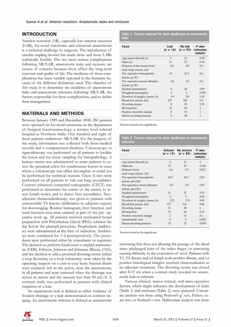
Kumar A et al . Anterior resection: Anastomotic leaks and strictures
INTRODUCTIONAnterior resection (AR), especially low anterior resection (LAR), for rectal carcinoma and colorectal anastomosis is a technical challenge to surgeons. The introduction of circular stapling devices has made more and more LARs technically feasible. The two most serious complications following AR/LAR, anastomotic leaks and stenosis, are causes of concern because these affect the long-term outcome and quality of life. The incidence of these com-plications has been variably reported in the literature be-cause of the different definitions used. The objective of this study is to determine the incidence of anastomotic leaks and anastomotic strictures following AR/LAR, the factors responsible for these complications, and to define their management.
MATERIALS AND METHODSBetween January 1989 and December 2008, 280 patients were operated on for rectal carcinoma in the department of Surgical Gastroenterology, a tertiary level referral hospital in Northern India. One hundred and eight of these patients underwent AR/LAR. For the purpose of the study, information was collected both from medical records and a computerized database. Colonoscopy or sigmoidoscopy was performed on all patients to localize the lesion and for tissue sampling for histopathology. A barium enema was administered to some patients to as-sess the proximal colon for synchronous lesions in cases where a colonoscopy was either incomplete or could not be performed for technical reasons. Chest X-rays were performed on all patients to rule out lung secondaries. Contrast enhanced computed tomography (CECT) was performed to determine the extent of the tumor, to as-sess lymph nodes, and to detect liver secondaries. Neo-adjuvant chemoradiotherapy was given to patients with unresectable T4 lesions (infiltration to adjacent organs) for downstaging. Routine hemogram, liver function, and renal function tests were ordered as part of the pre- op-erative work up. All patients received mechanical bowel preparation with Polyethylene Glycol (PEG) solution the day before the planned procedure. Prophylactic antibiot-ics were administered at the time of induction. Antibiot-ics were continued for 5 d postoperatively. The proce-dures were performed either by consultants or registrars. The decision to perform hand-sewn vs stapled anastomo-sis (CDH, Ethicon, Johnson and Johnson, Illinois, USA), and the decision to add a proximal diverting stoma (either a loop ileostomy or a loop colostomy) were taken by the operating surgeon on a case-to-case basis. Suction drains were routinely left in the pelvis, near the anastomosis, in all patients and were removed when the drainage was serous in nature and the amount less than 50 mL/d. A contrast study was performed in patients with clinical suspicion of a leak.
An anastomotic leak is defined as either evidence of feculent drainage or a leak demonstrated on contrast im-aging. An anastomotic stricture is defined as anastomotic
narrowing that does not allowing the passage of the distal inter- phalangeal joint of the index finger, or narrowing causing difficulty in the evacuation of stool. Patients with T3, T4 disease and/or lymph node positive disease, and/or positive histological margins received chemoradiation as an adjuvant treatment. The diverting stoma was closed after 8-12 wk when a contrast study revealed no anasto-motic leak or stricture.
Various clinical, tumor related, and intra-operative factors, which might influence the development of leaks (Table 1) and strictures (Table 2), were analyzed. Univari-ate analysis was done using Pearson’s χ2 test, Fishers ex-act test, or Student’s t test. Multivariate analysis was done
1476 March 21, 2011|Volume 17|Issue 11|WJG|www.wjgnet.com
Table 1 Factors analyzed for their significance in anastomotic leaks
Factor Leak (n = 16)
No leak (n = 92)
P value (univariate analysis)
Age (more than 60 yr) 7 13 0.011
Male sex 11 57 0.78Distance of the tumor from 8.2 8.9 0.5anal verge (mean, cm)Pre- operative hemoglobin (mean, g/dL)
10 10.2 0.6
Pre- operative serum albumin (mean, g/dL)
3.6 3.5 0.4
Stapled anastomosis 6 42 0.59Doughnut incomplete 3 2 0.032Duration of surgery (mean, h) 1.9 2.8 0.28Blood loss (mean, mL) 157 168 0.9Diverting stoma 8 52 0.78R0 resection 12 65 1Positive resection margin 2 13 1Mucin secreting tumour 5 28 1
1Factors found to be significant.
Table 2 Factors analyzed for their significance in anastomotic strictures
Factor Stricture (n = 19)
No stricture (n = 89)
P value (univariate analysis)
Age (more than 60 yr) 3 17 1Male sex 15 53 0.126Distance from 6.6 9.3 0.0111
anal verge (mean, cm)Pre-operative hemoglobin (mean, gm/dL)
10.7 10.1 0.23
Pre-operative serum albumin (mean, gm/dL)
3.5 3.5 0.92
Stapled anastomosis 11 37 0.21Doughnut incomplete 2 3 0.63Duration of surgery (mean, h) 2.25 2.76 0.43Blood loss (mean, mL) 177 164 0.86Diverting stoma 11 49 1R0 resection 15 62 0.57Positive resection margin 2 13 0.1Anastomotic leak 8 8 0.0011
Mucin secreting tumour 10 23 0.0291
1Factors found to be significant.

using the binary logistic regression method. SPSS 15 soft-ware was used for the analysis. Significance was calculated at the 95% CI and P value < 0.05.
RESULTSAmong 108 patients who underwent AR/LAR, there were 60 males and 48 females with a median age of 47 years (range 21 to 75 years). The median distance of the tumor from the anal verge was 8 cm (range 3-15 cm). The tumor was situated within 5 cm from the anal verge in 30 patients, between 5 and 10 cm in 46 patients, and above 10 cm in 32 patients. Endoscopic biopsy revealed adenocarcinoma in 100 and adenoma with dysplasia in four. The biopsy was inconclusive in remaining four patients. No patient had lung metastasis on X-ray chest. Based on pre-operative staging, five patients received neo-adjuvant treatment.
LAR (anastomosis below the level of the peritoneal reflection) was performed in 93 patients (86%). Fifteen patients (14%) underwent anterior resection (anasto-mosis above the level of the peritoneal reflection). Sixty patients (55.6%) underwent hand-sewn anastomosis and 48 (44.4%) had stapled anastomosis. The median dis-tance of the tumor from the anal verge in the hand sewn anastomosis group was 10.2 cm (range 4-15 cm) and it was 6 cm (range 3-15 cm) in the stapled group. Twenty-nine anastomoses were performed using CDH29, ten with CDH31, and three with CDH33. In the remaining six patients, no information regarding the size of the stapler was available. Information regarding anastomotic doughnuts was available in 39 patients. Doughnuts were complete in 34 patients and incomplete in five. Divert-ing stomas were created in 60 patients (55.6%); 33 (69%) with stapled anastomosis and 27 (45%) with hand-sewn anastomosis. The median duration of operation was 3.5 h (range 2.0-7.5 h). Final histopathology revealed adeno-carcinoma in all patients. Resection margins were positive for malignancy in 15 (13.9%) patients. Seven patients with stapled anastomosis and eight with hand-sewn anas-tomosis had positive resection margins. The resections were R0 (no microscopic or gross residual disease) in 77 patients (71.3%) and R1/R2 (microscopic/macroscopic residual disease left) in 31 (28.7%) patients. Nineteen patients (17.6%) had evidence of distant metastases at operation (liver in 13, peritoneal in three, and both liver and peritoneal in three).
Fifty Six patients (51.8%) had post operative compli-cations. Major complications included wound infection (n = 27, 25%), intra-abdominal bleed (n = 4, 3.7%), anas-tomotic leak (n = 16, 14.6%), anastomotic stricture (n = 19, 17.6%), and intestinal obstruction (n = 16, 14.6%). Of the 16 patients who had anastomotic leaks, eight had diverting stoma. Overall, 18 patients (16.7%) required re- exploration for the management of post-operative complications. Eleven patients with anastomotic leaks required re-exploration, and in seven of these, diverting stomas were created at second surgery. The remaining
five patients with leaks were managed expectantly. There were two (1.85 %) postoperative deaths (one due to an intra-abdominal bleed and the other due to pneumonitis). Local pelvic recurrence developed in eight patients during the follow up (follow up duration: 1-15 years). Ninteen (17.6%) patients presented with anastomotic strictures at a median duration of 8 mo (range 3-20 mo) after surgery. Biopsy from these strictures revealed no evidence of ma-lignancy in any of them. Seven of these strictures were managed with dilatation using Hegar’s dilators under gen-eral anesthesia. In others, the dilatations were carried out on an outpatient basis. The median number of dilatation required was 1 (1-4). Diverting stomas were closed in all patients, except for four who had severe fibrotic strictures and did not respond to dilatations even after multiple ses-sions.
Advanced age (greater than 60 years) and incomplete doughnuts were found to be significant risks for anasto-motic leaks on univariate analysis (Table 1). However, age was the only significant risk factor for anastomotic leaks on multivariate analysis (Table 3). On the other hand, increased distance of the tumor from the anal verge, a mucin secreting tumor, and an anastomotic leak were the factors found significant for the development of stricture, both on uni- and multivariate analysis (Tables 2 and 3).
DISCUSSIONIncidence of anastomotic leaks following AR/LAR has been reported to be 3%-21%[1-7]. Various patient-related, tumor-related, and technique-related factors have been enumerated as predisposing factors for anastomotic leaks. Male sex has been reported to be one such factor because of their unfavorable pelvic anatomy[1,2]. No significant difference in the number of leaks between males and fe-males was observed in our study.
Distance of the tumor from the anal verge and the position of the anastomosis were found to be associ-ated with the development of anastomotic leaks[2-4]. The reported higher incidence of leaks as the anastomosis becomes lower may be because of the increasing techni-cal difficulty and ischemia of the distal end. In our series, distance of the tumor from the anal verge was not as-sociated with leaks on univariate analysis. A leak rate of 3%-18% following stapled anastomosis in AR has been reported by various authors[1,2,5,6]. Law et al[1] and Rullier et al[2] demonstrated a higher leak rate following stapled anastomosis compared to hand-sewn. They attributed
1477 March 21, 2011|Volume 17|Issue 11|WJG|www.wjgnet.com
Table 3 Significant factors determined by multivariate analysis
Factors P value Odds ratio
Factors affecting anastomotic leak Advance age (> 60 yr) 0.004 7.23Factors affecting anastomotic stricture Anastomotic leak 0.000 13.6 Distance of growth from anal verge 0.011 6.5 Mucin secreting tumor 0.021 5.3
Kumar A et al . Anterior resection: Anastomotic leaks and strictures

this to the difficulty of the cases undergoing stapled anastomosis. A systematic review of nine randomized controlled trials could not find any significant difference in leak rates between the two groups[8]. The leak rates of 16.7% for stapled anastomosis and 12.5% for hand-sewn anastomosis in our study were comparable to the published series[8]. The high leak rates in our study may reflect the experience of operating surgeons (both trainee and consultants). We assume that, as the number of pa-tients and experience increase, the leak rate will decrease.
Although the use of a protective stoma has not been shown to decrease the overall anastomotic leak rate, it re-duces the rate of re-operation and postoperative mortali-ty in the event of a leak[9,10]. In the present series, creation of a protective stoma did not reduce the anastomotic leak rate. The majority of authors recommend a selective policy in providing covering stoma after AR/LAR, re-serving it for patients with high risk for of leaks (anasto-mosis within 5 cm from the anal verge, male gender, and incomplete doughnut)[4,7,11].
Although anastomotic leak and positive resection margin were implicated as factors promoting recurrence after AR[12,13], the relation between the leak and a positive resection margin is not well studied. A positive resection margin was not a significant risk factor for anastomotic leak in our patients.
Over all, the anastomotic stricture rate after hand-sewn anastomosis varies from 5%-9%[14]. The incidence of strictures after hand-sewn anastomosis that necessitat-ed treatment, was 0.6% in Goligher’s experience[15]. The low stricture rate in Goligher’s series compared to recent experience could be due to the lower number tumors close to the anal verge. Today, as more and more low rectal tumors are being submitted for LAR and ultra low AR, the stricture rate may also show a similar increase. Stricture rates of up to 20% have been reported[16]. Some animal studies have suggested that healing by scarring of the of the exposed seromuscular layer with poor epi-thelial bridging might explain the significant incidence of strictures following stapled anastomosis[17]. A meta-anal-ysis of 13 randomized controlled trials showed increased stricture rates following stapled anastomosis compared to hand-sewn[18]. In our series, 22.9% developed stric-tures after stapled anastomosis, which is higher than the 13.3% developed after hand-sewn anastomosis; however, it was not statistically significant. Waxmann et al[19], in a review of 10 series, reported 6% incidence of strictures with stapled anastomosis. They also noticed a reduced stricture rate with Russian staples, which deliver a single row of staples, compared to the new generation staples, which deliver two rows of staples. The size of the stapler has also some bearing on the stricture rate, according to some published series. Miller et al[20], in their report of 103 patients with stapled anastomosis, had a 4 % stricture rate, and the strictures were more common when a 28 mm di-ameter stapler was used as compared to one of 31 mm.
The high incidence of anastomotic leaks (14.6%) was another reason for the high stricture rate (17.6%), be-cause a leak predisposes the patient to intense inflamma-
tion and scarring. The stricture rate will therefore invari-ably increase.
The need for permanent stoma because of anasto-motic stricture has been variably reported (1%-9%)[21-23]. Although the incidence of anastomotic stricture was high in our series (17.6%), it is noteworthy that 15/19 (78.9%) strictures were successfully managed by per-anal dilata-tions. The incidence of anastomotic leaks and strictures are bound to be high in a teaching hospital where there will always be an influx of new trainees.
In conclusion, in our study, advance age was the only factor significantly associated with anastomotic leaks. On the other hand, anastomotic leaks, an aggressive tumor (mucin secreting), and growth in the lower rectum were predisposing factors for development of anastomotic strictures.
COMMENTSBackgroundTwo major complications of anterior resection (AR) and Low AR, anastomotic leaks and anastomotic strictures, were analyzed retrospectively. The effects of various factors that can lead to anastomotic leaks and anastomotic strictures were analyzed.Research frontiersNeoadjuvant Chemotherapy is under evaluation for locally advance tumors. Intersphincteric resections are under evaluation for patients with lower rectal tumors.Innovations and breakthroughsIntroduction of total mesorectal excision was a major achievement in the devel-opment of surgery for lower rectal growth, leading to significant decreases in the complications rate and a significant decrease in local recurrence. Develop-ment of end-to-end staplers was also a significant breakthrough, which made anastomosis possible in the lower rectum, thus facilitating sphincter preserva-tion.ApplicationsAs advance age is a significant risk factor for anastomotic leaks, so attention to detail is important while performing surgery on elderly patients. Similarly, special precautions are needed while performing anastomosis on low-lying growths, and mucin positive tumors; these patients should be operated on only by an experienced surgeon.Peer reviewThis is a good paper, but the study is over a period of 20 years, and changes may have happened regarding management.
REFERENCES1 Law WI, Chu KW, Ho JW, Chan CW. Risk factors for anas-
tomotic leakage after low anterior resection with total meso-rectal excision. Am J Surg 2000; 179: 92-96
2 Rullier E, Laurent C, Garrelon JL, Michel P, Saric J, Parneix M. Risk factors for anastomotic leakage after resection of rectal cancer. Br J Surg 1998; 85: 355-358
3 Pakkastie TE, Luukkonen PE, Järvinen HJ. Anastomotic leakage after anterior resection of the rectum. Eur J Surg 1994; 160: 293-297; discussion 299-300
4 Karanjia ND, Corder AP, Bearn P, Heald RJ. Leakage from stapled low anastomosis after total mesorectal excision for carcinoma of the rectum. Br J Surg 1994; 81: 1224-1226
5 Memon AA, Marks CG. Stapled anastomoses in colorectal surgery: a prospective study. Eur J Surg 1996; 162: 805-810
6 Nesbakken A, Nygaard K, Lunde OC. Outcome and late functional results after anastomotic leakage following meso-rectal excision for rectal cancer. Br J Surg 2001; 88: 400-404
7 Laxamana A, Solomon MJ, Cohen Z, Feinberg SM, Stern HS,
1478 March 21, 2011|Volume 17|Issue 11|WJG|www.wjgnet.com
COMMENTS
Kumar A et al . Anterior resection: Anastomotic leaks and strictures

McLeod RS. Long-term results of anterior resection using the double-stapling technique. Dis Colon Rectum 1995; 38: 1246-1250
8 Lustosa SA, Matos D, Atallah AN, Castro AA. Stapled ver-sus handsewn methods for colorectal anastomosis surgery: a systematic review of randomized controlled trials. Sao Paulo Med J 2002; 120: 132-136
9 Gastinger I, Marusch F, Steinert R, Wolff S, Koeckerling F, Lippert H. Protective defunctioning stoma in low anterior resection for rectal carcinoma. Br J Surg 2005; 92: 1137-1142
10 Marusch F, Koch A, Schmidt U, Geibetaler S, Dralle H, Sae-ger HD, Wolff S, Nestler G, Pross M, Gastinger I, Lippert H. Value of a protective stoma in low anterior resections for rectal cancer. Dis Colon Rectum 2002; 45: 1164-1171
11 Fielding LP, Stewart-Brown S, Hittinger R, Blesovsky L. Covering stoma for elective anterior resection of the rectum: an outmoded operation? Am J Surg 1984; 147: 524-530
12 Akyol AM, McGregor JR, Galloway DJ, Murray GD, George WD. Anastomotic leaks in colorectal cancer surgery: a risk factor for recurrence? Int J Colorectal Dis 1991; 6: 179-183
13 Phang PT, MacFarlane JK, Taylor RH, Cheifetz RE, Davis N, Hay JH, McGregor G, Speers C, Sullivan BJ, Pitts J, Coldman AJ. Effects of positive resection margin and tumor distance from anus on rectal cancer treatment outcomes. Am J Surg 2002; 183: 504-508
14 Keighley MRB, Williams NS. Surgery of one anus rectum and colon. 2nd ed. London: WB Saunders, 1993: 1212
15 Goligher JC. Surgery of the anus, rectum and colon. 5th ed. London: Bailliere Tindall, 1984: 512-514
16 Marchena Gómez J, Ruiz de la Cuesta E, Gómez Guerra G, Vallejo Gallego I, García-Anguíano F, Hernández Romero JM. Anastomotic stricture with the EEA-Stapler after colorec-tal anastomosis. Rev Esp Enferm Dig 1997; 89: 835-842
17 Polglase AL, Hughes ES, McDermott FT, Pihl E, Burke FR. A comparison of end-to-end staple and suture colorectal anas-tomosis in the dog. Surg Gynecol Obstet 1981; 152: 792-796
18 MacRae HM, McLeod RS. Handsewn vs. stapled anastomo-ses in colon and rectal surgery: a meta-analysis. Dis Colon Rectum 1998; 41: 180-189
19 Waxman BP. Large bowel anastomoses. II. The circular sta-plers. Br J Surg 1983; 70:64-67
20 Miller K, Moritz E. Circular stapling techniques for low ante-rior resection of rectal carcinoma. Hepatogastroenterology 1996; 43: 823-831
21 Köhler A, Athanasiadis S, Ommer A, Psarakis E. Long-term results of low anterior resection with intersphincteric anas-tomosis in carcinoma of the lower one-third of the rectum: analysis of 31 patients. Dis Colon Rectum 2000; 43: 843-850
22 Bailey CM, Wheeler JM, Birks M, Farouk R. The incidence and causes of permanent stoma after anterior resection. Colorectal Dis 2003; 5: 331-334
23 Ohman U, Svenberg T. EEA stapler for mid-rectum carcino-ma. Review of recent literature and own initial experience. Dis Colon Rectum 1983; 26: 775-784
S- Editor Wang JL L- Editor Stewart GJ E- Editor Zheng XM
1479 March 21, 2011|Volume 17|Issue 11|WJG|www.wjgnet.com
Kumar A et al . Anterior resection: Anastomotic leaks and strictures

Proton pump inhibitor step-down therapy for GERD: A multi-center study in Japan
Takao Tsuzuki, Hiroyuki Okada, Yoshiro Kawahara, Ryuta Takenaka, Junichiro Nasu, Hidehiko Ishioka, Akiko Fujiwara, Fumiya Yoshinaga, Kazuhide Yamamoto
Takao Tsuzuki, Hiroyuki Okada, Yoshiro Kawahara, Ju-nichiro Nasu, Kazuhide Yamamoto, Department of Gastroen-terology and Hepatology, Okayama University Graduate School of Medicine, Dentistry, and Pharmaceutical Sciences, Okayama 700-8558, JapanRyuta Takenaka, Department of Internal Medicine, Tsuyama Central Hospital, Okayama 708-0841, JapanHidehiko Ishioka, Ishioka clinic, Hiroshima 721-0926, JapanAkiko Fujiwara, Department of Internal Medicine, Okayama Saiseikai General Hospital, Okayama 700-8511, JapanFumiya Yoshinaga, Deparment of Internal Medicine, Mihara Red Cross Hospital, Hiroshima 723-8512, JapanAuthor contributions: Tsuzuki T contributed to statistical analysis and drafting the manuscript; Okada H contributed to planning, data collection, and drafting manuscript; Kawahara Y, Takenaka R, Naus J, Ishioka H, Fujiwara A and Yoshinaga F contributed to data collection; Yamamoto K and Okada H con-tributed to manuscript direction.Correspondence to: Dr. Takao Tsuzuki, Department of Gas-troenterology and Hepatology, Okayama University Graduate School of Medicine, Dentistry, and Pharmaceutical Sciences, 2-5-1, Shikata-cho, Kita-ku, Okayama 700-8558, Japan. [email protected]: +81-86-2357219 Fax: +81-86-2255991Received: September 25, 2010 Revised: December 11, 2010Accepted: December 18, 2010Published online: March 21, 2011
AbstractAIM: To investigate the predictors of success in step-down of proton pump inhibitor and to assess the quality of life (QOL).
METHODS: Patients who had heartburn twice a week or more were treated with 20 mg omeprazole (OPZ) once daily for 8 wk as an initial therapy (study 1). Pa-tients whose heartburn decreased to once a week or less at the end of the initial therapy were enrolled in study 2 and treated with 10 mg OPZ as maintenance therapy for an additional 6 mo (study 2). QOL was in-
vestigated using the gastrointestinal symptom rating scale (GSRS) before initial therapy, after both 4 and 8 wk of initial therapy, and at 1, 2, 3, and 6 mo after starting maintenance therapy.
RESULTS: In study 1, 108 patients were analyzed. Their characteristics were as follows; median age: 63 (range: 20-88) years, sex: 46 women and 62 men. The suc-cess rate of the initial therapy was 76%. In the patients with successful initial therapy, abdominal pain, indiges-tion and reflux GSRS scores were improved. In study 2, 83 patients were analyzed. Seventy of 83 patients completed the study 2 protocol. In the per-protocol analysis, 80% of 70 patients were successful for step-down. On multivariate analysis of baseline demographic data and clinical information, no previous treatment for gastroesophageal reflux disease (GERD) [odds ratio (OR) 0.255, 95% CI: 0.06-0.98] and a lower indigestion score in GSRS at the beginning of step-down therapy (OR 0.214, 95% CI: 0.06-0.73) were found to be the pre-dictors of successful step-down therapy. The improved GSRS scores by initial therapy were maintained through the step-down therapy.
CONCLUSION: OPZ was effective for most GERD pa-tients. However, those who have had previous treat-ment for GERD and experience dyspepsia before step-down require particular monitoring for relapse.
© 2011 Baishideng. All rights reserved.
Key words: Gastroesophageal reflux disease; Proton pump inhibitor; Omeprazole; Step-down therapy; Gas-trointestinal symptom rating scale
Peer reviewers: Josep M Bordas, MD, Department of Gastro-enterology IMD, Hospital Clinic”, Llusanes 11-13 at, Barcelona 08022, Spain; Myung-Gyu Choi, MD, Professor of Medicine, Division of Gastroenterology, Department of Internal Medicine, Seoul St. Mary’s Hospital, The Catholic University of Korea, 505, Banpo-Dong, Seocho-Gu, Seoul 137-040, South Korea
BRIEF ARTICLE
World J Gastroenterol 2011 March 21; 17(11): 1480-1487ISSN 1007-9327 (print) ISSN 2219-2840 (online)
© 2011 Baishideng. All rights reserved.
Online Submissions: http://www.wjgnet.com/[email protected]:10.3748/wjg.v17.i11.1480
1480 March 21, 2011|Volume 17|Issue 11|WJG|www.wjgnet.com

Tsuzuki T et al . PPI step-down therapy for GERD
Tsuzuki T, Okada H, Kawahara Y, Takenaka R, Nasu J, Ishioka H, Fujiwara A, Yoshinaga F, Yamamoto K. Proton pump inhibi-tor step-down therapy for GERD: A multi-center study in Japan. World J Gastroenterol 2011; 17(11): 1480-1487 Available from: URL: http://www.wjgnet.com/1007-9327/full/v17/i11/1480.htm DOI: http://dx.doi.org/10.3748/wjg.v17.i11.1480
INTRODUCTIONGastroesophageal reflux disease (GERD) is defined as the reflux of gastric contents into the esophagus; it may lead to esophagitis, reflux symptoms sufficient to impair quality of life (QOL), and/or long-term complications[1]. In Japan, GERD was, in the past, considered to be a rare disease, but is now one of the most common chronic disorders. One systematic review of Japan[2] reported that 15.4% (725) of 4723 patients had heartburn twice or more per week; 42.2% had symptoms of heartburn, in-cluding those who had heartburn once or less per week; and 16.7% (602) of 3608 patients had reflux esophagitis. Some papers have suggested that the increasing preva-lence of GERD is a result of rising acid secretion in the general Japanese population, caused by the westerniza-tion of lifestyle and diet (to include dairy products) and decline in Helicobacter pylori (H. pylori) infection[3].
Proton pump inhibitors (PPIs) provide the high-est levels of symptom relief, healing of esophagitis, and prevention of relapse and complications[4]. They are the first choice for therapy in most GERD patients with or without esophageal mucosal injury[5,6]. However, PPIs cost more than do other acid-inhibiting agents. Some papers suggest that long-term PPI therapy, particularly at high doses, is associated with increased risks of community-acquired pneumonia[7] and hip fracture[8]. Therefore, if possible, PPIs should be administered at the lower dose. Full-dose PPI first “step-down” therapy is superior, both to administering histamine-2-receptor antagonist (H2RA) first and low-dose PPI first “step-up” strategy, with regard to both efficacy and cost-effectiveness[9,10]. However, some patients experience a recurrence after step-down[11,12].
On the other hand, there is increasing interest in evalu-ating the QOL of patients with GERD. It is widely ac-cepted that patients with GERD have a decreased QOL compared with the general population[13-15], similar to that seen in patients with other chronic diseases[16]. Conse-quently, patient-reported symptoms and QOL are impor-tant in assessing treatment outcome.
In this article, we describe 2 series of studies devised to investigate the predictors of success in step-down PPI therapy and to assess symptom-related QOL as deter-mined by a questionnaire administered during GERD therapy.
MATERIALS AND METHODSStudy 1 (PPI initial therapy for GERD)Subjects: This multicenter trial was conducted at Okaya-
ma University Hospital and 20 affiliated hospitals in Japan. The study protocol was approved by the local institution-al review boards of Okayama University Hospital and of each of the affiliated hospitals. Written informed consent was obtained from each patient before their enrollment in this study.
The major eligibility criteria were the following: heart-burn that occurs twice a week or more; age 20 years or older; and either erosive esophagitis (EE) or non-erosive reflux disease. The exclusion criteria were as follows: use of PPIs within 1 mo of the start of the study; open gastric or duodenal ulcer; malignant neoplasm; serious systemic disease; and pregnancy, signs of pregnancy, or occurrence during a lactation period.
Study design: Patients who met the above-mentioned inclusion criteria were asked to complete the Gastroin-testinal Symptom Rating Scale (GSRS) to assess gastroin-testinal symptom-related QOL. Additional information, including demographic data, height and weight measure-ments used to assess body mass index (BMI), comorbid conditions, concurrent medication use, history of GERD symptoms, and previous GERD medication, was col-lected at the initial visit. Upper gastrointestinal endoscopy was performed to assess hiatus hernia, esophagitis, and upper gastrointestinal disorders before treatment.
PPI therapy was performed according to the follow-ing strategy (Figure 1): Patients were treated with 20 mg of omeprazole (OPZ) once daily for 8 wk as an initial therapy. GSRS results were evaluated before the initial therapy and after both 4 and 8 wk of treatment.
The primary analysis was to investigate the rate of success of initial PPI therapy. Success was defined as hav-ing heartburn once a week or less at the end of the initial therapy. Secondary analyses examined changes in QOL during the initial treatment of PPI.
Study 2 (PPI step-down therapy as maintenance therapy of GERD)Subjects: Patients whose incidence of heartburn de-
1481 March 21, 2011|Volume 17|Issue 11|WJG|www.wjgnet.com
Study 1 Study 2
Esophagogastroduodenoscopy
QOL assessment at every visit
(n = 83)
(n = 108) Dropped out: n = 13(Completed: n = 70)
Omeprazole 10 mg for 6 moOmeprazole
20 mg for 8 wk (initial therapy)
Patients achieving heartburn resolution in Study 1 were enrolled
in Study 2 and treated with PPI maintenance therapy
t /mo0 1 2 0 1 2 3 6
Visit 1 Visit 2 Visit 3 Visit 4 Visit 5 Visit 6 Visit 7
Visit at symptom relapse
Figure 1 Overview of the study design. QOL: Quality of life; PPI: Proton pump inhibitor.

creased to once a week or less at the end of study 1 (the initial therapy) were enrolled in this study. Study 2 sub-jects included patients who achieved complete remission and those who achieved partial remission of GERD symptoms.
Study design: Patients received PPI step-down therapy: namely, 10 mg of OPZ once daily for 6 mo (Figure 1). QOL related to gastrointestinal symptoms was evaluated at 1, 2, 3, and 6 mo after the beginning of maintenance therapy. If heartburn recurred twice a week or more dur-ing maintenance therapy, the treatment was stopped and the QOL at that time was evaluated.
The primary analysis was designed to identify predic-tors of subjects who were successful in PPI step-down therapy-defined as having no recurrence of heartburn 6 mo after PPI step-down. Secondary analyses examined changes in QOL during the maintenance treatment of PPI.
Endoscopic assessment The severity of esophagitis was established by endo-scopic examination using the modified Los Angeles (LA) classification of Grade A to D, with Grade M and N[17]. This adds an additional grade N (defined as no appar-ent mucosal change) and grade M (defined as minimal changes in the mucosa, such as erythema and/or whitish turbidity). The modified LA system has recently become widely used in Japan[18]. A diagnosis of hiatus hernia was made when the retroflexed endoscope, under the condi-tion of gastric inflation, showed gaping esophageal lumen allowing the squamous epithelium to be viewed below[19]. Atrophic gastritis was diagnosed by endoscopy using the Kimura-Takemoto endoscopic classification[20], in which atrophic gastritis was divided largely into closed type (C-type) and open type (O-type) by the location of an atro-phic border detected by endoscopy. C-type means that the atrophic border remains on the lesser curvature of the stomach, while O-type means that the atrophic bor-der no longer exists on the lesser curvature but extends along the anterior and posterior walls of the stomach. In this study, atrophic gastritis was defined as only the O-type of the Kimura-Takemoto classification: the endoscopic diagnosis of C-type atrophic gastritis may be unreliable because of interobserver variation.
QOL assessmentSymptom-related QOL was assessed using the GSRS. The GSRS is a disease-specific 15-item questionnaire de-veloped, based on reviews of gastrointestinal symptoms and clinical experience, to evaluate common symptoms of gastrointestinal disorders[21,22]. Patients are asked to numerically score their subjective symptoms on a Likert-type scale of 1-7. The sum of the scores for all 15 items is regarded as the total GSRS score. Because each of the 15 questions can be scored from 1 to 7, the minimum to-tal score obtainable is 15, and the maximum total score is 105. This total is then divided by 15 to obtain the overall
GSRS score, from a minimum of 1 to a maximum overall score of 7. Furthermore, the scores for the 5 symptom categories (reflux, abdominal pain, dyspepsia, diarrhea, and constipation) are obtained by calculating the means of the scores on the items for each symptom category. The higher the overall score, the more severe the symp-toms[23].
Statistical analysis All categorical variables were analyzed using chi-square or Fisher’s exact test.
Analyses of efficacy parameters were performed on 2 populations: the intention-to-treat (ITT) and the per-protocol (PP) populations.
The time to first symptom relapse after PPI step-down was assessed by Kaplan-Meier analysis. To deter-mine the predictors of successful step-down, univari-ate and multivariable logistic regression analyses were planned. Variables found in the univariate analysis to be significantly associated with successful step-down were included in a multivariable logistic regression analysis. Changes in QOL were analyzed with paired t tests. An overall significance level of 5% was used in all tests. Sta-tistical analysis was performed with SPSS version 11.0 using Windows.
RESULTSStudy 1 Between April 2004 and March 2007, 108 eligible patients were entered into study 1. For reasons that were appar-ently not causally related to the medication, 21 out of 108 patients dropped out of the study. The demographic characteristics of the patients are shown in Table 1. In the ITT analysis, 83 (76%) of 108 patients completed the 8 wk of initial therapy successfully. In the PP analysis, 83 (95.4%) of 87 patients were successful. The reasons for failure of the 4 patients in initial therapy were insuffi-ciency of GERD symptoms in 2 patients and side effects of PPI in the other 2. The adverse reactions were diar-rhea (one patient) and tinnitus (one patient). The reasons for dropout of 21 patients in study 1 were as follows: one patient withdrew her consent before the initial therapy, 12 patients never came to the hospital after the initial visit without excuse, 8 patients never came to the hospital after the second visit without excuse (3 of them still had symptoms of reflux at that time), the others got relief of reflux symptoms.
Symptom-related QOL analysis was performed for subjects who successfully completed the initial therapy. The changes in QOL during therapy are shown in Figure 2. The GSRS total score significantly improved from the baseline after 4-wk treatment (P < 0.0001). Of the 83 patients who had heartburn once a week or less at the end of the initial therapy, 55 patients (66%) had a reflux score of one, indicating no symptoms, 23 patients (28%) had a score of two, and the others had a score of three. In other words, 78 (94%) of 83 patients having heartburn
1482 March 21, 2011|Volume 17|Issue 11|WJG|www.wjgnet.com
Tsuzuki T et al . PPI step-down therapy for GERD

once a week or less complained of little or no symptoms of reflux at the end of the initial therapy. Not only the GSRS reflux scores but also the abdominal pain, indiges-tion, and constipation scores showed significant improve-ments (P < 0.0001).
Study 2 Eighty three eligible patients who had heartburn resolu-tion after the initial 8 wk of therapy were recruited for study 2-step-down treatment as maintenance therapy. Thirteen patients dropped out during the maintenance therapy without recurrence or adverse reactions; 70 pa-tients completed the study 2 protocol. Of 13 dropout pa-tients, one patient experienced exaggerated symptoms of reflux before dropout, nine experienced complete resolu-tion of reflux symptoms before dropout, and the others never came to the hospital after starting the step-down therapy.
Demographic and baseline characteristics of the study 2 population are summarized in Table 2. In the ITT analysis, 56 (67.5%) of 83 patients did not suffer a recur-rence during maintenance therapy. In the PP analysis, 56 (80%) of 70 patients were successful in step-down thera-py (Figure 3). Of 14 failures, 10 occurred within the first 3 mo of maintenance therapy. The mean time to failure was 56 d (range, 16-180 d).
In the univariate analysis of the ITT population, there were no significant predictors for successful step-down. In the univariate analysis of the PP population (n = 70), the significant predictors for successful step-down were:
1483 March 21, 2011|Volume 17|Issue 11|WJG|www.wjgnet.com
6
5
4
3
2
1
Scor
e P < 0.0001
Baseline 1 2 t /mo
Total score6
5
4
3
2
1
Scor
e
P < 0.0001
Baseline 1 2 t /mo
Reflux6
5
4
3
2
1
Scor
e P < 0.0001
Baseline 1 2 t /mo
Indigestion
6
5
4
3
2
1
Scor
e
Baseline 1 2 t /mo
Diarrhea6
5
4
3
2
1
Scor
e P = 0.001
Baseline 1 2 t /mo
Constipation6
5
4
3
2
1
Scor
e P < 0.0001
Baseline 1 2 t /mo
Abdominal pain
Figure 2 Gastrointestinal Symptom Rating Scale scores during the initial treatment (study 1).
Table 1 Population characteristics of study 1 n (%)
Characteristic ITT population (n = 108)
Gender Male/female 62 (57.4)/46 (42.6)Age (yr) Range 20-88 Median 63BMI (kg/m2) ≤ 24.9 (normal or below normal) 77 (71.3) 25-29.9 (overweight) 28 (25.9) ≥ 30 (obese) 3 (2.8)Previous treatment for GERD 38 (35.2)Duration of GERD symptoms before initial treatment (mo) < 1 21 (19.4) 1-11 47 (43.5) ≥ 12 40 (37.0)Tobacco use 24 (22.2) Alcohol use 44 (40.7) Helicobacter pylori infection 31 (28.7)The modified Los Angeles Classification N/M 20 (18.5)/17 (15.7) A/B 32 (29.6)/32 (29.6) C/D 5 (4.6)/2 (1.9)Atrophic gastritis1 18 (16.7)Hiatus hernia 57 (52.8)Ischemic heart disease 9 (8.3)Diabetes mellitus 11 (10.2)Bronchial asthma 4 (3.7)
1Atrophic gastritis was defined as only the O-type of the Kimura-Takemoto classification[20]. BMI: Body mass index; GERD: Gastroesophageal reflux disease; ITT: Intention-to-treat.
Tsuzuki T et al . PPI step-down therapy for GERD

complete remission of GERD symptoms before step-down (P = 0.042), no past history of GERD (P = 0.019), no H2RA use at baseline (P = 0.023), and better GSRS total (P = 0.045) and indigestion (P = 0.011) scores at the beginning of step-down (Table 3). In a multivariate analy-sis of the PP population, no past history of GERD (P = 0.047) and a better GSRS indigestion score at the begin-
ning of step-down (P = 0.014) were predictive factors for successful step-down (Table 4).
Symptom-related QOL analysis was performed on subjects for whom maintenance therapy was successful. The changes in QOL during therapy are shown in Figure 3. The improved GSRS scores of initial therapy were main-tained throughout step-down therapy (Figure 4).
DISCUSSIONIn study 1, we investigated the effectiveness of OPZ (20 mg) for the initial treatment of GERD and changes in symp-tom-related QOL during the initial therapy. Previous studies of patients with GERD using PPIs showed that 64%-88% of patients with typical reflux symptoms re-spond to PPI therapy[24-26]. The heartburn resolution rate of the present study (95.4%) was higher than that of pre-vious studies. It is reasonable to assume that a less rigor-ous end-point of the present study, namely having heart-burn once a week or less, would have resulted in a higher rate of symptom response.
1484 March 21, 2011|Volume 17|Issue 11|WJG|www.wjgnet.com
Table 2 Population characteristics of study 2 n (%)
Characteristic ITT population (n = 83)
Gender Male/female 50 (60.2)/33 (39.8)Age (yr) Range 25-88 Median 64BMI (kg/m2) ≤ 24.9 (normal or below normal) 60 (72.3) 25-29.9 (overweight) 20 (24.1) ≥ 30 (obese) 3 (3.6)Previous treatment for GERD 27 (32.5)Duration of GERD symptoms before initial treatment (mo) < 1 14 (16.9) 1-11 42 (50.6) ≥ 12 27 (32.5)Tobacco use 21 (25.3) Alcohol use 39 (47.0) Helicobacter pylori infection 27 (32.5)The modified Los Angeles Classification N/M 15 (18.1)/9 (10.8) A/B 24 (28.9)/30 (36.1) C/D 3 (3.6)/2 (2.4)Atrophic gastritis1 15 (18.1)Hiatus hernia 44 (53.0)Ischemic heart disease 8 (9.6)Diabetes mellitus 7 (8.4)Bronchial asthma 3 (3.6) GERD symptom before step-down Complete remission 55 (66.3) Partial remission 28 (33.7)
1Atrophic gastritis was defined as only the O-type of the Kimura-Takemoto classification[20]. BMI: Body mass index; GERD: Gastroesophageal reflux disease; ITT: Intention-to-treat.
100
80
60
40
20
0
Recu
rren
t-fr
ee r
ate
(%)
80%
0 1 2 3 4 5 6 t /mo
Figure 3 Proportion of subjects without recurrent gastroesophageal reflux disease symptoms after proton pump inhibitor step-down (Study 2 per-protocol population: n = 70).
Table 3 Predictors of successful step-down (univariate analysis)
Variable ITT population (n = 83)
PP population (n = 70)
OR P OR P
Age 0.984 0.428 0.965 0.212BMI 3.667 0.073 2.108 0.251Past history of treatment for GERD 0.497 0.161 0.228 0.019Duration of GERD symptoms 1.004 0.416 1.199 0.536Alcohol use 1.182 0.732 1.486 0.487Tobacco use 1.125 0.834 0.692 0.531Helicobacter pylori infection 0.963 0.942 1.500 0.577Erosive esophagitis 2.599 0.062 1.402 0.620LA classification 1.431 0.070 1.238 0.360Atrophic gastritis 0.930 0.906 0.568 0.411Hiatus hernia 1.333 0.566 1.476 0.545complete remission of GERD symptoms before step-down
1.355 0.539 3.676 0.042
GSRS total score (at the beginning of step-down)
0.771 0.647 0.208 0.045
Reflux score in GSRS (at the beginning of step-down)
0.917 0.803 0.477 0.072
Indigestion score in GSRS (at the beginning of step-down)
0.594 0.209 0.218 0.011
BMI: Body mass index; GERD: Gastroesophageal reflux disease; GSRS: Gastrointestinal Symptom Rating Scale; ITT: Intention-to-treat; PP: Per-protocol; OR: Odds ratio; LA: Los Angeles.
Table 4 Predictors of successful step-down (multivariate analysis, per-protocol population: n = 70)
Variable OR 95% CI P
Past history of treatment for GERD 0.255 0.066-0.980 0.047Indigestion score in GSRS (at the beginning of step down)
0.214 0.063-0.731 0.014
GERD: Gastroesophageal reflux disease; GSRS: Gastrointestinal Symptom Rating Scale; OR: Odds ratio; CI: Confidence interval.
Tsuzuki T et al . PPI step-down therapy for GERD
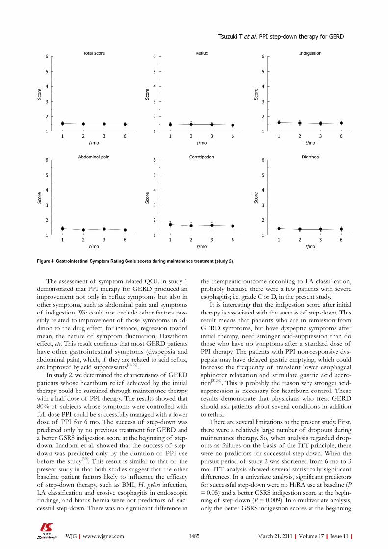
The assessment of symptom-related QOL in study 1 demonstrated that PPI therapy for GERD produced an improvement not only in reflux symptoms but also in other symptoms, such as abdominal pain and symptoms of indigestion. We could not exclude other factors pos-sibly related to improvement of those symptoms in ad-dition to the drug effect, for instance, regression toward mean, the nature of symptom fluctuation, Hawthorn effect, etc. This result confirms that most GERD patients have other gastrointestinal symptoms (dyspepsia and abdominal pain), which, if they are related to acid reflux, are improved by acid suppressants[27-29].
In study 2, we determined the characteristics of GERD patients whose heartburn relief achieved by the initial therapy could be sustained through maintenance therapy with a half-dose of PPI therapy. The results showed that 80% of subjects whose symptoms were controlled with full-dose PPI could be successfully managed with a lower dose of PPI for 6 mo. The success of step-down was predicted only by no previous treatment for GERD and a better GSRS indigestion score at the beginning of step-down. Inadomi et al. showed that the success of step-down was predicted only by the duration of PPI use before the study[30]. This result is similar to that of the present study in that both studies suggest that the other baseline patient factors likely to influence the efficacy of step-down therapy, such as BMI, H. pylori infection, LA classification and erosive esophagitis in endoscopic findings, and hiatus hernia were not predictors of suc-cessful step-down. There was no significant difference in
the therapeutic outcome according to LA classification, probably because there were a few patients with severe esophagitis; i.e. grade C or D, in the present study.
It is interesting that the indigestion score after initial therapy is associated with the success of step-down. This result means that patients who are in remission from GERD symptoms, but have dyspeptic symptoms after initial therapy, need stronger acid-suppression than do those who have no symptoms after a standard dose of PPI therapy. The patients with PPI non-responsive dys-pepsia may have delayed gastric emptying, which could increase the frequency of transient lower esophageal sphincter relaxation and stimulate gastric acid secre-tion[31,32]. This is probably the reason why stronger acid-suppression is necessary for heartburn control. These results demonstrate that physicians who treat GERD should ask patients about several conditions in addition to reflux.
There are several limitations to the present study. First, there were a relatively large number of dropouts during maintenance therapy. So, when analysis regarded drop-outs as failures on the basis of the ITT principle, there were no predictors for successful step-down. When the pursuit period of study 2 was shortened from 6 mo to 3 mo, ITT analysis showed several statistically significant differences. In a univariate analysis, significant predictors for successful step-down were no H2RA use at baseline (P = 0.05) and a better GSRS indigestion score at the begin-ning of step-down (P = 0.009). In a multivariate analysis, only the better GSRS indigestion scores at the beginning
1485 March 21, 2011|Volume 17|Issue 11|WJG|www.wjgnet.com
6
5
4
3
2
1
Scor
e
1 2 3 6 t /mo
Total score6
5
4
3
2
1
Scor
e
1 2 3 6 t /mo
Reflux6
5
4
3
2
1
Scor
e
1 2 3 6 t /mo
Indigestion
6
5
4
3
2
1
Scor
e
1 2 3 6 t /mo
Diarrhea6
5
4
3
2
1
Scor
e
1 2 3 6 t /mo
Constipation6
5
4
3
2
1
Scor
e
1 2 3 6 t /mo
Abdominal pain
Figure 4 Gastrointestinal Symptom Rating Scale scores during maintenance treatment (study 2).
Tsuzuki T et al . PPI step-down therapy for GERD

1486 March 21, 2011|Volume 17|Issue 11|WJG|www.wjgnet.com
of step-down (P = 0.016) were predictive factors for suc-cessful step-down. In the present study, most patients had mild esophagitis, which would probably benefit from the mild treatment or even by non-drug therapy, for in-stance, light and early dinner, inclined bed, and straight posture[33]. This may explain why many patients dropped out. Indeed, most patients discontinued therapy because they had no further symptoms. Furthermore, it cannot be denied that some patients with successful step-down may have had good control of their GERD symptoms by non-drug therapy because we did not take into account the recommended life-style and dietary changes in our study protocol.
Second, in this study, many patients (75% of patients who received 8 wk of initial therapy) did not have the post-treatment endoscopic assessment. Some patients in clinical remission after the initial therapy did not have endoscopic remission. If all subjects underwent post-treatment endoscopy, the endoscopic findings could be the candidate predictor of successful step-down.
Third, cytochrome P450 2C19 genotypic differences, which are related to the cure rates for GERD with PPIs (OPZ and lansoprazole)[11,34,35], are not considered in this study. This factor could also be the candidate predic-tor[11,36]. The analysis of these factors may provide more information for GERD treatment.
In conclusion, our study revealed that the majority of patients whose heartburn decreased to once a week or less on full-dose OPZ therapy can be successfully stepped-down to a half-dose of PPI. The predictors of successful step-down are no previous treatment for GERD and a better indigestion score in the GSRS at the beginning of step-down. Improvement of symptom-related QOL was maintained throughout the step-down therapy.
ACKNOWLEDGMENTSThe authors gratefully acknowledge individuals who helped identify cases and collected clinical data: Shuhei Ishiyama at Okayama Saiseikai General Hospital, Okaya-ma; Hiroshi Yamamoto at Kurashiki Central Hospital, Okayama; Takayuki Imada at Nippon Kokan Fukuyama Hospital, Hiroshima; Hirofumi Tsugeno at Tsuyama Cen-tral Hospital, Okayama; Ryoji Tanaka at Kumayama Hos-pital of Akaiwa City, Okayama; Akira Takahashi at Bizen City Hospital, Okayama; Ryusuke Nasu at Kurashiki Riv-erside Hospital, Okayama; Toshie Araki at Shigei Medi-cal Research Hospital, Okayama; Tomoyuki Sawayama at Hiroshima Teishin Hospital, Hiroshima; Jun Tomoda at National Hospital Organization Fukuyama Medical Center, Hiroshima; Takechiyo Morita at Fukuyama City Hospital, Hiroshima; Fumiya Yoshinaga at Mihara Red Cross Hospital, Hiroshima; Yuka Sugiyama at Izumo City General Medical Center, Shimane; Masahiro Takatani at Himeji Red Cross Hospital, Hyogo; Tatsuya Toyokawa at Mitoyo General Hospital, Kagawa; Masaki Wato at Kaga-wa Prefectural Central Hospital, Kagawa; Toshimi Hasui at Social Insurance Ritsurin Hospital, Kagawa; Eiichiro
Yumoto and Fusao Ikeda at Sumitomo Besshi Hospital, Ehime; Jiro Miyaji at Saiseikai Imabari Hospital, Ehime; Naoko Yunoki at Akaiwa Ishikai Hospital, Okayama.
COMMENTSBackgroundGastroesophageal reflux disease (GERD) is one of the most common disorders not only in the Western world but also in Japan, with increasing prevalence and incidence in the last decades. Typical GERD symptoms include heartburn, acid regurgitation into the pharynx, and dysphagia. The GERD patients some-times suffer from chest pain, dyspepsia, and other atypical symptoms including coughing, wheezing, hoarseness, etc. Research frontiersProton pump inhibitors (PPIs) are the first choice of therapy for most GERD patients with or without esophageal mucosal injury. Full dose PPI-first “step-down” therapy is superior to histamin-2-receptor antagonist (H2RA) or low dose PPI-first “step-up” strategy with regard to both efficacy and cost-effectiveness. However, there is no clear consensus regarding the predictors for successful step-down. Treatment of GERD aims not only at healing esophagitis but also at managing the symptoms of GERD associated with decreased quality of life (QOL). In this study, we investigated the predictors of success in step-down PPI therapy and assessed the symptom-related QOL.Innovations and breakthroughsMany studies evaluating the efficacy of PPI in terms of the symptoms of GERD patients in the Western world did not evaluate the grade of esophagitis with endoscopy. Recently, many studies in Japan have focused mainly on non-erosive reflux disease (NERD) patients. The data of this study originated from GERD patients (both erosive gastritis and NERD) who were diagnosed with endoscopy. Therefore, the results may reflect those of the GERD patients in the Japanese general population and can be considered useful for GERD treatment in the clinical practice. ApplicationsBy considering the clinical features of GERD patients with recurrence during the step-down therapy, this study’s findings may suggest new strategies for addi-tional PPI dose reduction including every-other-day administration, on-demand therapy, step-down to H2RA, and drug-free management.Peer reviewThe authors investigated the effect of omeprazole and subsequent step-down maintenance therapy in patients with GERD. This study must be a very inter-esting topic for readers in view of the rarity of PPI outcome studies in the Asia pacific region.
REFERENCES1 Dent J, Armstrong D, Delaney B, Moayyedi P, Talley NJ,
Vakil N. Symptom evaluation in reflux disease: workshop background, processes, terminology, recommendations, and discussion outputs. Gut 2004; 53 Suppl 4: iv1-i24
2 Kouzu T, Hishikawa E, Watanabe Y, Inoue M, Satou T. [Epidemiology of GERD in Japan]. Nippon Rinsho 2007; 65: 791-794
3 Furukawa N, Iwakiri R, Koyama T, Okamoto K, Yoshida T, Kashiwagi Y, Ohyama T, Noda T, Sakata H, Fujimoto K. Proportion of reflux esophagitis in 6010 Japanese adults: prospective evaluation by endoscopy. J Gastroenterol 1999; 34: 441-444
4 Klinkenberg-Knol EC, Nelis F, Dent J, Snel P, Mitchell B, Prichard P, Lloyd D, Havu N, Frame MH, Romàn J, Walan A. Long-term omeprazole treatment in resistant gastroesopha-geal reflux disease: efficacy, safety, and influence on gastric mucosa. Gastroenterology 2000; 118: 661-669
5 Savarino V, Dulbecco P. Optimizing symptom relief and preventing complications in adults with gastro-oesophageal reflux disease. Digestion 2004; 69 Suppl 1: 9-16
6 DeVault KR, Castell DO. Updated guidelines for the diag-nosis and treatment of gastroesophageal reflux disease. Am J
COMMENTS
Tsuzuki T et al . PPI step-down therapy for GERD
1487 March 21, 2011|Volume 17|Issue 11|WJG|www.wjgnet.com
Gastroenterol 2005; 100: 190-2007 Laheij RJ, Sturkenboom MC, Hassing RJ, Dieleman J, Strick-
er BH, Jansen JB. Risk of community-acquired pneumonia and use of gastric acid-suppressive drugs. JAMA 2004; 292: 1955-1960
8 Yang YX, Lewis JD, Epstein S, Metz DC. Long-term proton pump inhibitor therapy and risk of hip fracture. JAMA 2006; 296: 2947-53
9 Habu Y, Maeda K, Kusuda T, Yoshino T, Shio S, Yamazaki M, Hayakumo T, Hayashi K, Watanabe Y, Kawai K. "Proton-pump inhibitor-first" strategy versus "step-up" strategy for the acute treatment of reflux esophagitis: a cost-effectiveness analysis in Japan. J Gastroenterol 2005; 40: 1029-1035
10 Mine S, Iida T, Tabata T, Kishikawa H, Tanaka Y. Manage-ment of symptoms in step-down therapy of gastroesopha-geal reflux disease. J Gastroenterol Hepatol 2005; 20: 1365-1370
11 Furuta T, Sugimoto M, Kodaira C, Nishino M, Yamade M, Ikuma M, Shirai N, Watanabe H, Umemura K, Kimura M, Hishida A. CYP2C19 genotype is associated with symptom-atic recurrence of GERD during maintenance therapy with low-dose lansoprazole. Eur J Clin Pharmacol 2009; 65: 693-698
12 Lauritsen K, Devière J, Bigard MA, Bayerdörffer E, Mózsik G, Murray F, Kristjánsdóttir S, Savarino V, Vetvik K, De Freitas D, Orive V, Rodrigo L, Fried M, Morris J, Schneider H, Eklund S, Larkö A. Esomeprazole 20 mg and lansoprazole 15 mg in maintaining healed reflux oesophagitis: Metropole study results. Aliment Pharmacol Ther 2003; 17: 333-341
13 Revicki DA, Wood M, Maton PN, Sorensen S. The impact of gastroesophageal reflux disease on health-related quality of life. Am J Med 1998; 104: 252-258
14 Prasad M, Rentz AM, Revicki DA. The impact of treatment for gastro-oesophageal reflux disease on health-related quality of life: a literature review. Pharmacoeconomics 2003; 21: 769-790
15 Pace F, Negrini C, Wiklund I, Rossi C, Savarino V. Quality of life in acute and maintenance treatment of non-erosive and mild erosive gastro-oesophageal reflux disease. Aliment Phar-macol Ther 2005; 22: 349-356
16 Kulig M, Leodolter A, Vieth M, Schulte E, Jaspersen D, La-benz J, Lind T, Meyer-Sabellek W, Malfertheiner P, Stolte M, Willich SN. Quality of life in relation to symptoms in patients with gastro-oesophageal reflux disease-- an analysis based on the ProGERD initiative. Aliment Pharmacol Ther 2003; 18: 767-776
17 Lundell LR, Dent J, Bennett JR, Blum AL, Armstrong D, Galmiche JP, Johnson F, Hongo M, Richter JE, Spechler SJ, Tytgat GN, Wallin L. Endoscopic assessment of oesophagi-tis: clinical and functional correlates and further validation of the Los Angeles classification. Gut 1999; 45: 172-180
18 Miwa H, Yokoyama T, Hori K, Sakagami T, Oshima T, To-mita T, Fujiwara Y, Saita H, Itou T, Ogawa H, Nakamura Y, Kishi K, Murayama Y, Hayashi E, Kobayashi K, Tano N, Matsushita K, Kawamoto H, Sawada Y, Ohkawa A, Arai E, Nagao K, Hamamoto N, Sugiyasu Y, Sugimoto K, Hara H, Tanimura M, Honda Y, Isozaki K, Noda S, Kubota S, Hi-meno S. Interobserver agreement in endoscopic evaluation of reflux esophagitis using a modified Los Angeles classifi-cation incorporating grades N and M: a validation study in a cohort of Japanese endoscopists. Dis Esophagus 2008; 21: 355-363
19 Kinoshita Y, Adachi K. Hiatal hernia and gastroesophageal flap valve as diagnostic indicators in patients with gastro-esophageal reflux disease. J Gastroenterol 2006; 41: 720-721
20 Kimura K, Takemoto T. An endoscopic recognition of the atrophic border and its significance in chronic gastritis. En-doscopy 1969; 1: 87-97
21 Svedlund J, Sjödin I, Dotevall G. GSRS--a clinical rating scale
for gastrointestinal symptoms in patients with irritable bowel syndrome and peptic ulcer disease. Dig Dis Sci 1988; 33: 129-134
22 Revicki DA, Wood M, Wiklund I, Crawley J. Reliability and validity of the Gastrointestinal Symptom Rating Scale in patients with gastroesophageal reflux disease. Qual Life Res 1998; 7: 75-83
23 Suzuki H, Masaoka T, Sakai G, Ishii H, Hibi T. Improvement of gastrointestinal quality of life scores in cases of Helico-bacter pylori-positive functional dyspepsia after successful eradication therapy. J Gastroenterol Hepatol 2005; 20: 1652-1660
24 Chiba N, De Gara CJ, Wilkinson JM, Hunt RH. Speed of heal-ing and symptom relief in grade II to IV gastroesophageal reflux disease: a meta-analysis. Gastroenterology 1997; 112: 1798-1810
25 Armstrong D, Paré P, Pericak D, Pyzyk M. Symptom relief in gastroesophageal reflux disease: a randomized, controlled comparison of pantoprazole and nizatidine in a mixed pa-tient population with erosive esophagitis or endoscopy-negative reflux disease. Am J Gastroenterol 2001; 96: 2849-2857
26 Fennerty MB, Johanson JF, Hwang C, Sostek M. Efficacy of esomeprazole 40 mg vs. lansoprazole 30 mg for healing moderate to severe erosive oesophagitis. Aliment Pharmacol Ther 2005; 21: 455-463
27 Kusano M, Shimoyama Y, Kawamura O, Maeda M, Kurib-ayashi S, Nagoshi A, Zai H, Moki F, Horikoshi T, Toki M, Sugimoto S, Mori M. Proton pump inhibitors improve acid-related dyspepsia in gastroesophageal reflux disease pa-tients. Dig Dis Sci 2007; 52: 1673-1677
28 Quigley EM. Functional dyspepsia (FD) and non-erosive reflux disease (NERD): overlapping or discrete entities? Best Pract Res Clin Gastroenterol 2004; 18: 695-706
29 Neumann H, Monkemuller K, Kandulski A, Malfertheiner P. Dyspepsia and IBS symptoms in patients with NERD, ERD and Barrett's esophagus. Dig Dis 2008; 26: 243-247
30 Inadomi JM, McIntyre L, Bernard L, Fendrick AM. Step-down from multiple- to single-dose proton pump inhibitors (PPIs): a prospective study of patients with heartburn or acid regurgitation completely relieved with PPIs. Am J Gastroen-terol 2003; 98: 1940-1944
31 Holloway RH, Hongo M, Berger K, McCallum RW. Gastric distention: a mechanism for postprandial gastroesophageal reflux. Gastroenterology 1985; 89: 779-784
32 Kahrilas PJ, Shi G, Manka M, Joehl RJ. Increased frequency of transient lower esophageal sphincter relaxation induced by gastric distention in reflux patients with hiatal hernia. Gastroenterology 2000; 118: 688-695
33 Kaltenbach T, Crockett S, Gerson LB. Are lifestyle measures effective in patients with gastroesophageal reflux disease? An evidence-based approach. Arch Intern Med 2006; 166: 965-971
34 Furuta T, Ohashi K, Kosuge K, Zhao XJ, Takashima M, Kimura M, Nishimoto M, Hanai H, Kaneko E, Ishizaki T. CYP2C19 genotype status and effect of omeprazole on intra-gastric pH in humans. Clin Pharmacol Ther 1999; 65: 552-561
35 Furuta T, Shirai N, Watanabe F, Honda S, Takeuchi K, Iida T, Sato Y, Kajimura M, Futami H, Takayanagi S, Yamada M, Ohashi K, Ishizaki T, Hanai H. Effect of cytochrome P4502C19 genotypic differences on cure rates for gastro-esophageal reflux disease by lansoprazole. Clin Pharmacol Ther 2002; 72: 453-460
36 Saitoh T, Otsuka H, Kawasaki T, Endo H, Iga D, Tomimatsu M, Fukushima Y, Katsube T, Ogawa K, Otsuka K. Influences of CYP2C19 polymorphism on recurrence of reflux esopha-gitis during proton pump inhibitor maintenance therapy. Hepatogastroenterology 2009; 56: 703-706
S- Editor Tian L L- Editor O’Neill M E- Editor Zheng XM
Tsuzuki T et al . PPI step-down therapy for GERD

BRIEF ARTICLE
Prevalence and impact of musculoskeletal pain in Japanese gastrointestinal endoscopists: A controlled study
Takayasu Kuwabara,Yuji Urabe, Toru Hiyama, Shinji Tanaka, Takako Shimomura, Shiro Oko, Masaharu Yoshi-hara, Kazuaki Chayama
Takayasu Kuwabara,Yuji Urabe,Takako Shimomura, Ka-zuaki Chayama, Department of Gastroenterology and Me-tabolism, Hiroshima University Hospital, Hiroshima 739-8514, JapanToru Hiyama, Masaharu Yoshihara, Health Service Center, Hiroshima University, Higashihiroshima 739-8514, JapanShiro Oko,Shinji Tanaka, Department of Endoscopy, Hiro-shima University Hospital, Hiroshima 739-8514, JapanAuthor contributions: Kuwabara T designed the study, per-formed the data compiling and wrote the manuscript; Urabe Y designed the study and performed the data compiling, Hiyama T, Tanaka S, Shimomura T and Oka S were involved in editing the manuscript; Yoshihara M performed the statistical analysis; Chayama K organized the study.Correspondence to: Toru Hiyama, MD, PhD, Health Service Center, Hiroshima University, 1-7-1 Kagamiyama, Higashihiro-shima 739-8514, Japan. [email protected]: +81-82-4246191 Fax: +81-82-4227156Received: September 9, 2010 Revised: December 1, 2010Accepted: December 8, 2010Published online: March 21, 2011
AbstractAIM: To examine the frequency and prevention of mus-culoskeletal pain in Japanese gastrointestinal endosco-pists and non-endoscopist physicians.
METHODS: Questionnaires were sent to 275 endosco-pists and 173 non-endoscopists working in Hiroshima University Hospital and its affiliated hospitals.
RESULTS: The completed questionnaires were returned by 190 (69%) endoscopists and 120 (69%) non-endos-copists. The frequency of pain in the hand and wrist, and especially the left thumb, was significantly higher in endoscopists than in non-endoscopists (17% vs 6%, P = 0.004). Using multivariate analysis, the only signifi-cant factor associated with this pain was the age of the endoscopist (odds ratio 2.77, 95% confidence interval,
1.23-6.71, P = 0.018). Interestingly, endoscopists had made significantly fewer modifications to their endo-scopic practices than non-endoscopists (12% vs 33%, P < 0.0001) to prevent pain.
CONCLUSION: Pain in the hand and wrist may be endoscopy-related. However, endoscopists made little modifications in practice to prevent such pain. More at-tention to prevention appears necessary.
© 2011 Baishideng. All rights reserved.
Key words: Endoscopy; Musculoskeletal pain; Pain pre-vention
Peer reviewer: Dr. Herwig R Cerwenka, Professor, Department of Surgery, Medical University of Graz, Auenbruggerplatz 29, A-8036 Graz, Austria
Kuwabara T, Hiyama T, Urabe Y, Tanaka S, Shimomura T, Oka S, Yoshihara M, Chayama K. Prevalence and impact of muscu-loskeletal pain in Japanese gastrointestinal endoscopists: A con-trolled study. World J Gastroenterol 2011; 17(11): 1488-1493 Available from: URL: http://www.wjgnet.com/1007-9327/full/v17/i11/1488.htm DOI: http://dx.doi.org/10.3748/wjg.v17.i11.1488
INTRODUCTIONMuscle and joint pain is a common complaint in individu-als whose jobs require repetitive isometric maneuvers or awkward body positions[1]. Such pain has been reported in individuals with various occupations such as bus drivers[2], unskilled laborers[3], musicians[4], physical therapists[5], and computer keyboard operators[6]. Recently, ergonomic mechanisms related to the development of work-related musculoskeletal disorders have drawn substantial interest.
Endoscopy in clinical gastroenterology is becoming more important not only in Western countries but also in
1488
World J Gastroenterol 2011 March 21; 17(11): 1488-1493 ISSN 1007-9327 (print) ISSN 2219-2840 (online)
© 2011 Baishideng. All rights reserved.
Online Submissions: http://www.wjgnet.com/[email protected]:10.3748/wjg.v17.i11.1488
March 21, 2011|Volume 17|Issue 11|WJG|www.wjgnet.com
Kuwabara T et al . Musculoskeletal pain in Japanese endoscopists
Eastern countries including Japan[7,8], and the number of endoscopic examinations and treatments has increased rapidly. In addition, procedures requiring long perfor-mance time such as endoscopic submucosal dissection[7,9], endoscopic sphincterotomy, and endoscopic papillary balloon dilatation are commonly performed. The work burden of endoscopists has significantly increased.
Evidence suggests that endoscopists frequently report a variety of musculoskeletal problems including neck pain, low back pain, and thumb and hand pain[10,11]. On the basis of previous reports, it has been suggested that the performance of endoscopy predisposes the operator to the development of musculoskeletal pain. However, there is little published evidence of this, and most of this evi-dence comes from Western countries; little evidence has come from Eastern countries including Japan. More at-tention needs to be paid to the impact of musculoskeletal pain and its prevention in Eastern countries. Therefore, we examined the frequency of musculoskeletal pain in Japanese gastrointestinal endoscopists and non-endosco-pist physicians. The use of pain prevention strategies and physician requests to improve endoscopic design were also examined.
MATERIALS AND METHODSGeneral overviewQuestionnaires were sent to endoscopists and non-endoscopist physicians working in Hiroshima University Hospital and its affiliated hospitals. The questionnaires were sent with an introductory letter explaining the study. The survey was conducted between March and May, 2010.
SubjectsSubjects consisted of endoscopists and non-endoscopist physicians in Hiroshima University Hospital and its affiliated hospitals. A control group was also included and consisted of a random sample of non-endoscopist physicians.
QuestionnaireThe questionnaire for endoscopists consisted of questions on the endoscope model primarily used, mean number of endoscopic examinations and treatments per week, mean amount of time spent performing endoscopic procedures per week, location and description of pain present at the time of the survey, impact of pain on the endoscopist, type of treatment needed for the pain, and injury prevention strategies currently in place, if any. Non-identifying demographic information including age, sex, body mass index (BMI), hand dominance, physical activity level, and practice years was obtained. A modified survey was sent to the non-endoscopists.
Statistical analysisStudent t-tests were performed to evaluate differences in weight, height, and BMI between the groups. c2 tests were used to assess variables including age, sex, hand dominance, physical activity level, and practice years. To identify the factors associated with musculoskeletal pain,
especially pain in the hand and wrist among endoscopists, univariate and multivariate logistic regression analyses were performed with JMP IN (Cary, NC, USA) software. All significance levels were set at P < 0.05.
RESULTSSubject characteristics questionnaires were sent to 275 endoscopists and 173 non-endoscopist physicians. One hundred ninety (69%) endoscopists and 120 (69%) non-endoscopists returned
1489 March 21, 2011|Volume 17|Issue 11|WJG|www.wjgnet.com
Table 1 Non-endoscopist physicians who participated in this survey
n (%)
Internist 65 (54)Surgeon 17 (14)Dermatologist 10 (8) Psychiatrist 7 (6)Radiologist 4 (3) Resident 4 (3) Pathologist 3 (3)Pediatrician 1 (1)Orthopedic surgeon 1 (1)Gynecologist 1 (1)Emergency physician 1 (1)NI 6 (5)Total 120 (100)
NI: Not informed.
Table 2 Subject characteristics
Characteristics Endoscopists Non-endoscopists P value (n = 190) (n = 120)
Age (yr, mean ± SD) 41.4 ± 6.7 40.1 ± 7.6 0.202Years in practice (yr, mean ± SD)
16.2 ± 8.1 14.8 ± 9.4 0.083
Sex 0.197 Male/female 164/26 97/23Dominant hand 0.077 Right/left/NI 179/9/2 108/12/0Activity level 0.046 Mild 131 (69%) 70 (58%) Moderate 50 (26%) 43 (36%) Remarkable 5 (3%) 5 (4%) NI 4 (2%) 4 (3%)Height (cm) 168.6 ± 6.9 168.1 ± 7.0 0.202Weight (kg) 66.3 ± 10.6 64.9 ± 11.3 0.287Body mass index (kg/m2) 23.2 ± 2.2 22.8 ± 3.0 0.306
NI: Not informed.
Table 3 Number of endoscopic procedures completed per week (mean ± SD)
Procedure type No. per week
Esophagogastroduodenoscopy (n = 190) 15.0 ± 8.7Colonoscopy (n = 161) 6.7 ± 3.7Endoscopic retrograde cholangiopancreatog-raphy (n = 63)
2.1 ± 1.8
Endoscopic ultrasonography (n = 48) 2.2± 1.9

completed questionnaires. The details of non-endoscopist physicians who participated in this survey are shown in Table 1. The two groups were similar in terms of age, years in practice, sex, hand dominance, and BMI (Table 2). Physical activity level was significantly lower in endoscopists than in non-endoscopists (P = 0.046).
Endoscopy informationThe number of esophagogastroduodenoscopies completed per week was 15.0 ± 8.7 (mean ± SD) (Table 3). Of the 190 endoscopists, 161 (85%) performed colonoscopies, and the mean number of colonoscopies performed per week was 6.7 ± 3.7 (mean ± SD). Sixty-three (33%) endoscopists performed endoscopic retrograde cholangiopancreatography (ERCP). Forty-eight (25%) endoscopists performed endoscopic ultrasonography (EUS). On average, the amount of time spent per week performing endoscopy was 11.9 ± 8.7 h (mean ± SD).
Environmental factors affecting endoscopic proceduresMost (98%) endoscopists indicated that they used height-adjustable examination tables (Table 4). As for monitor position, 133 of 190 (70%) endoscopists replied that the monitor was directly in front of the endoscopist and at eye level. With regard to the manufacturer of the endoscope used, 138 (73%) endoscopists used Olympus, 47 (25%) used Fujifilm, and 5 (3%) gave no response.
Musculoskeletal pain in endoscopistsNone of the endoscopists reported a history of preexisting musculoskeletal conditions. Eighty-one (43%) endoscopists had musculoskeletal pain at the time of the survey, and 21 (26%) of those reporting pain sought medical care as a consequence.
The most frequent sites of pain identified by the endoscopists were the lower back (n = 50, 26%), the neck (n = 18, 9%), the right shoulder (n = 18, 9%), and the left thumb (n = 16, 8%) (Table 5). Pain in the hand and wrist was reported by 32 (17%) endoscopists.
Musculoskeletal pain in non-endoscopistsNone of the non-endoscopists reported a history of preexisting musculoskeletal conditions. Forty-nine (41%) non-endoscopists reported musculoskeletal pain at the time of the survey, and 7 (14%) of these physicians sought medical care as a consequence.
The most frequent sites of pain identified in the non-endoscopists were the lower back (n = 24, 20%), the right shoulder (n = 14, 12%), the neck (n = 13, 11%), and the left shoulder (n = 12, 10%) (Table 5). Pain in the hand and wrist was reported by 7 (6%) non-endoscopists.
Comparison of musculoskeletal pain between endoscopists and non-endoscopistsThe frequency of overall musculoskeletal pain did not differ significantly between the endoscopists and non-endoscopists (81 of 190 (43%) vs 49 of 120 (41%), P = 0.755). However, the frequency of pain in the left thumb was significantly higher in the endoscopists than in the non-endoscopists (16 of 190 (8%) vs 0 of 120 (0%), P = 0.001). Pain in the hand and wrist was significantly higher in the endoscopists than in the non-endoscopists (32 of 190 (17%) vs 7 of 120 (6%), P = 0.004). There were no significant differences between the groups in the frequency of pain at other locations, such as the shoulder and lower back.
Univariate and multivariate logistic regression analyses for factors associated with thumb, finger, hand, and wrist pain in endoscopistsResults of univariate analysis for factors related to thumb, finger, hand, and wrist pain in endoscopists are shown in Table 6. The only significant factor was age of the endoscopist (> 40 years vs ≤ 40 years, P = 0.011). The mean number of colonoscopies per week and dominant hand tended to be significant (> 6.7 vs ≤ 6.7, P = 0.091, left vs right, P = 0.098, respectively). Thus, endoscopists who performed more than 6.7 colonoscopies per week or who were left-handed tended to have more frequent pain in the hand and wrist.
Results of multivariate analysis for factors related to pain in the hand and wrist in endoscopists are shown in Table 7. Only age was found to be a significant factor associated with pain in endoscopists (> 40 years vs ≤ 40 years, odds ratio (OR) 2.77, 95% confidence interval, 1.23-6.71, P = 0.018) by multivariate analysis.
Pain preventionOnly 23 (12%) endoscopists had made modifications to their endoscopic practice in response to pain arising due to performing endoscopic procedures. The most common practice modifications endorsed are shown in Table 7. These modifications were stretching exercises (n = 11, 6%), taking breaks (n = 11, 6%), wearing athletic shoes (n = 8, 4%), and less work (n = 5, 3%). Interestingly, endoscopists had made significantly fewer modifications to their practice than non-endoscopists to address pain prevention.
Requests to improve endoscopic design from endoscopistsThe requests made by Japanese endoscopists to improve endoscope design are shown in Table 8. The most frequent request was to make the operating part lighter (n = 85, 45%), followed by making the operating part
1490 March 21, 2011|Volume 17|Issue 11|WJG|www.wjgnet.com
Table 4 Environmental factors affecting endoscopic proce-dures
Factor No. per week (n = 190)
Use of height-adjustable examination table (yes/no)
186 (98)/4 (2)
Position of monitor (directly in front ofendoscopist, at eye level/other position)
133 (70)/57 (30)
Manufacturer of endoscope primarily used (Olympus/Fujifilm/NI)
138 (73)/47 (25)/5 (3)
NI: Not informed.
Kuwabara T et al . Musculoskeletal pain in Japanese endoscopists

smaller (n = 41, 22%), and reducing the resistance of the angulation controller (n = 36, 19%). There were no significant differences between the frequency of requests and the manufacturers of the endoscope primarily used (data not shown).
DISCUSSIONOur data indicate that the frequency of pain in the hand and wrist, and especially in the left thumb, was significantly higher in endoscopists than non-endoscopists (17% vs 6%, P = 0.004). By multivariate analysis, the only significant factor associated with pain was the age of the endoscopist (OR 2.77, P = 0.018).
Previous studies have reported that the incidence of symptoms or injuries attributable to the performance of endoscopy ranges from 39% to 78%[11]. These results suggest that musculoskeletal pain is prevalent in
endoscopists. In the present study, 43% of endoscopists had musculoskeletal pain, which is similar to that in previous reports. In addition, a U.S. study showed that the frequency of reported musculoskeletal pain was higher in endoscopists than non-endoscopists (74% vs 35%, P < 0.001)[10]. In the present study, pain in the hand and wrist was significantly more frequent in endoscopists than in non-endoscopists, indicating that endoscopists may be at higher risk for musculoskeletal pain, especially in the hand and wrist, than non-endoscopists.
The most frequent sites of pain reported in our study were the lower back, neck, right shoulder, and left thumb. Previous studies have reported similar anatomic sites of pain associated with performing endoscopy, including a case report of de Quervain’s syndrome or “endoscopists’ thumb”[11-14]. Buschbacker[15] found that 27% of responders reported low back pain, 19% thumb pain, and 19% shoulder pain. Preliminary results from an American Society for Gastrointestinal Endoscopy web-based survey found that 43% reported hand or carpal tunnel injuries, 29% reported back pain, and 28%
1491 March 21, 2011|Volume 17|Issue 11|WJG|www.wjgnet.com
Table 5 Frequency of major types of musculoskeletal pain in Japanese endoscopists and non-endoscopists n (%)
Site of pain Endoscopists (n = 190) Non-endoscopists (n = 120) P value
Neck (present/absent) 18 (9)/172 (91) 13 (11)/107 (89) 0.698Right shoulder (present/absent) 18 (9)/172 (91) 14 (12)/106 (88) 0.536Left shoulder (present/absent) 15 (8)/175 (92) 12 (10)/108 (90) 0.522Right wrist (present/absent) 3 (2)/187 (98) 1 (1)/119 (99) 0.571Left wrist (present/absent) 13 (7)/177 (93) 4 (3)/116 (97) 0.186Right thumb (present/absent) 3 (2)/187 (98) 0 (0)/120 (100) 0.167Left thumb (present/absent) 16 (8)/174 (92) 0 (0)/120 (100) 0.001Right hand/fingers (present/absent) 5 (3)/185 (97) 1 (1)/119 (99) 0.263Left hand/fingers (present/absent) 4 (2)/186 (98) 1 (1)/119 (99) 0.387Lower back (present/absent) 50 (26)/140 (74) 24 (20)/96 (80) 0.204Hand and wrist (present/absent) 32 (17)/158 (83) 7 (6)/113 (94) 0.004Total (present/absent) 81 (43)/109 (57) 49 (41)/71 (59) 0.755
Table 6 Univariate analysis of factors associated with pain in the hand and wrist in endoscopists
Characteristics P value
Age (> 40 yr vs < 40 yr) 0.011Sex (male vs female) 0.378Dominant hand (left vs right) 0.098Activity level (moderate/remarkable vs mild) 0.338Height (> 168 cm vs < 168 cm) 0.796Weight (> 66 kg vs < 66 kg) 0.642Body mass index (> 25 kg/m2 vs < 25 kg/m2) 0.61Mean number of esophagogastroduodenoscopies per wk (> 15 vs < 15)
0.215
Mean number of colonoscopies per wk (> 6.7 vs < 6.7) 0.091Do ERCP (yes vs no) 0.871Do EUS (yes vs no) 0.969Mean time performing endoscopic procedures per week (> 12 h vs < 6.7 h)
0.636
Use of height-adjustable examination table (no vs yes) 0.836Position of monitor (other position vs directly in front of endoscopist, at eye level)
0.8
Manufacturer of endoscope primarily used (fujifilm vs olympus)
0.28
Table 7 Modifications to prevent musculoskeletal pain
Modification Endoscopists (n = 190)
Non-endoscopists (n = 120)
P value
No modifications 167 (88) 80 (67) < 0.0001Stretching exercises 11 (6) 26 (22) < 0.0001Taking breaks 11 (6) 8 (7) 0.754Wearing athletic shoes 8 (4) 6 (5) 0.744Less work 5 (3) 1 (1) 0.263
Table 8 Requests to improve endoscopic design from endos-copists n (%)
Request No. of endoscopists (n = 190)
Make the operating part heavier/lighter 0 (0)/85 (45)Make the operating part larger/smaller 1 (1)/41 (22) Make the angulation controller larger/smaller 15 (8)/27 (14) Make the resistance of the angulation controller heavier/lighter
0 (0)/36 (19)
ERCP: Endoscopic retrograde cholangiopancreatography; EUS: Endoscop-ic ultrasonography.
Kuwabara T et al . Musculoskeletal pain in Japanese endoscopists

reported neck pain[16]. Specific aspects of performing endoscopy which may contribute to musculoskeletal pain are as follows: adjusting the tip of angulation controls, torquing with the right hand, and standing for prolonged periods of time. Manipulation of the tip angulation controls and torquing of the endoscope may lead to hand and wrist pain, whereas standing for prolonged periods of time may lead to back and neck pain.
In the present study, only age of the endoscopist was significantly associated with pain. Several researchers reported that age did not have an impact on the musculoskeletal pain identified. However, the location of pain has been reported to be different between a beginner group (duration of practicing endoscopy, < 39 mo) and an experienced group[11]. The left thumb and fingers were the most common painful areas in beginners (43%), whereas the left shoulder was most painful in experienced endoscopists (33%). Furthermore, older age and sex were associated with many musculoskeletal disorders[17]. Age may be an important factor associated with the musculoskeletal pain reported by endoscopists.
ERCP and EUS are both procedures requiring long endoscopy times with special devices. Some may think that these procedures are more likely to lead to musculoskeletal pain than other procedures. However, in the present study, performance of these procedures was not a significant factor associated with pain, whereas the mean number of colonoscopies performed per week and dominant hand tended to be significant by univariate analysis. In other words, the performance of colonoscopy may be a risk factor for the development of pain in the hand and wrist rather than the performance of ERCP or EUS. The torquing technique is very important in the performance of colonoscopy, and it is done frequently and for long periods of time. Furthermore, colonoscopy may require stronger torquing power than any other procedures, including ERCP and EUS. Therefore, torquing of the colonoscope appears to be another risk factor associated with pain. The colonoscope is torqued with the right hand, which is the weaker hand for left-handed endoscopists, and it is plausible that left-handed endoscopists tend to have more pain in the hand and wrist than right-handed endoscopists. A larger study is needed to investigate this issue.
Methods to prevent back pain include the use of a height-adjustable examination table, rubberized floor mats, and a short foot stool to alternate weight distribution[18]. Monitor placement is an especially important determinant of torso and head posture. Monitors should be placed directly in front of the endoscopist while in the working position to avoid rotation and flexion of the cervical spine and should be adjusted to eye level[18]. Most (98%) responders in the present study reported use of an adjustable examination table. However, 30% of responders reported that the position of monitors was inadequate. None of the endoscopists used rubberized floor mats or a short foot stool. There is much room for improvement in relation to these factors.
Improving posture, increasing physical activity, and stretching exercises before endoscopy have been suggested to prevent disability[18]. Interestingly, in the present study, endoscopists made significantly fewer modifications to their endoscopic practices to prevent musculoskeletal pain, especially in regard to performing stretching exercises, than non-endoscopists. The reason for this is unknown. One possibility is that Japanese endoscopists may be busier than other non-endoscopist physicians and may not have enough time or willingness to exercise or stretch. Of course, selection bias may be undeniable. Education on methods to reduce endoscopic-related injuries must be carried out in a positive manner.
Endoscopic techniques that can help to prevent pain include minimizing torque and having an assistant apply torque when necessary. Ultimately, reevaluating the design of the endoscope with regard to ergonomics may be the best long-term strategy to reduce overuse injuries in an era of high-volume endoscopy. Endoscopists in the present study requested a lighter and smaller operating part and reduced resistance of the angulation controller. These requests may be useful for improving the design of the endoscope.
In conclusion, our data suggest that pain in the hand and wrist may be endoscopy-related. However, endoscopists made few modifications to their practices to prevent pain. More attention to pain prevention appears to be needed. Given the importance of endoscopy in the clinical setting, our results support a further controlled study on this subject in a much larger and more diverse population of endoscopists.
COMMENTSBackgroundIt has been suggested that the performance of endoscopy predisposes the op-erator to the development of musculoskeletal pain.Research frontiersThere is little published evidence of this, especially from Eastern countries in-cluding Japan.Innovations and breakthroughsFrequency of occurrence of pain in the hand and wrist, and especially the left thumb, was significantly higher in endoscopists than in non-endoscopists (17% vs 6%, P = 0.004). By multivariate analysis, the only significant factor associ-ated with the pain was the age of the endoscopist (odds ratio 2.77, 95% confi-dence interval, 1.23-6.71, P = 0.018).Applications The authors’ data suggest that pain in the hand and wrist may be endoscopy-related. More attention toward pain prevention appears necessary.Peer reviewThis is a study on an important issue, musculoskeletal pain in endoscopists. As authors described, more attention toward pain prevention in endoscopists should be paid.
REFERENCES1 Smith AC, Wolf JG, Xie GY, Smith MD. Musculoskeletal
pain in cardiac ultrasonographers: results of a random survey. J Am Soc Echocardiogr 1997; 10: 357-362
2 Anderson R. The back pain of bus drivers. Prevalence in an urban area of California. Spine (Phila Pa 1976) 1992; 17: 1481-1488
1492 March 21, 2011|Volume 17|Issue 11|WJG|www.wjgnet.com
COMMENTS
Kuwabara T et al . Musculoskeletal pain in Japanese endoscopists

3 Mahbub MH, Laskar MS, Seikh FA, Altaf MH, Inoue M, Yokoyama K, Wakui T, Harada N. Prevalence of cervical spondylosis and musculoskeletal symptoms among coolies in a city of Bangladesh. J Occup Health 2006; 48: 69-73
4 Sakai N, Liu MC, Su FC, Bishop AT, An KN. Hand span and digital motion on the keyboard: concerns of overuse syndrome in musicians. J Hand Surg Am 2006; 31: 830-835
5 Salik Y, Ozcan A. Work-related musculoskeletal disorders: a survey of physical therapists in Izmir-Turkey. BMC Musculoskelet Disord 2004; 5: 27
6 Arvidsson I, Arvidsson M, Axmon A, Hansson GA, Johansson CR, Skerfving S. Musculoskeletal disorders among female and male air traffic controllers performing identical and demanding computer work. Ergonomics 2006; 49: 1052-1067
7 Urabe Y, Hiyama T, Tanaka S, Oka S, Yoshihara M, Arihiro K, Chayama K. Metachronous multiple esophageal squamous cell carcinomas and Lugol-voiding lesions after endoscopic mucosal resection. Endoscopy 2009; 41: 304-309
8 Fukuhara T, Hiyama T, Tanaka S, Oka S, Yoshihara M, Arihiro K, Chayama K. Characteristics of esophageal squamous cell carcinomas and lugol-voiding lesions in patients with head and neck squamous cell carcinoma. J Clin Gastroenterol 2010; 44: e27-e33
9 Tanaka S, Oka S, Chayama K. Colorectal endoscopic submu-cosal dissection: present status and future perspective, including its differentiation from endoscopic mucosal resection. J Gastroenterol 2008; 43: 641-651
10 Hansel SL, Crowell MD, Pardi DS, Bouras EP, DiBaise JK.
Prevalence and impact of musculoskeletal injury among endoscopists: a controlled pilot study. J Clin Gastroenterol 2009; 43: 399-404
11 Byun YH, Lee JH, Park MK, Song JH, Min BH, Chang DK, Kim YH, Son HJ, Rhee PL, Kim JJ, Rhee JC, Hwang JH, Park DI, Shim SG, Sung IK. Procedure-related musculoskeletal symptoms in gastrointestinal endoscopists in Korea. World J Gastroenterol 2008; 14: 4359-4364
12 Cappell MS. Colonoscopist's thumb: DeQuervains's syndrome (tenosynovitis of the left thumb) associated with overuse during endoscopy. Gastrointest Endosc 2006; 64: 841-843
13 Shergill AK, McQuaid KR, Rempel D. Ergonomics and GI endoscopy. Gastrointest Endosc 2009; 70: 145-153
14 Liberman AS, Shrier I, Gordon PH. Injuries sustained by colorectal surgeons performing colonoscopy. Surg Endosc 2005; 19: 1606-1609
15 Buschbacher R. Overuse syndromes among endoscopists. Endoscopy 1994; 26: 539-544
16 Keate RF, Dryden GW, Wang K, Chen YK. Occupational injuries to endoscopists: report from the ASGE web survey. Gastrointest Endosc 2006; 63: AB111
17 Lötters F, Burdorf A. Prognostic factors for duration of sickness absence due to musculoskeletal disorders. Clin J Pain 2006; 22: 212-221
18 Lipp MJ. The risks of GI endoscopy. Gastrointest Endosc 2009; 69: 1149-1151
S- Editor Sun H L- Editor Webster JR E- Editor Ma WH
1493 March 21, 2011|Volume 17|Issue 11|WJG|www.wjgnet.com
Kuwabara T et al . Musculoskeletal pain in Japanese endoscopists

BRIEF ARTICLE
Long-term result of endoscopic Histoacryl® (N-butyl-2-cyanoacrylate) injection for treatment of gastric varices
Eun Jung Kang, Soung Won Jeong, Jae Young Jang, Joo Young Cho, Sae Hwan Lee, Hyun Gun Kim, Sang Gyune Kim, Young Seok Kim, Young Koog Cheon, Young Deok Cho, Hong Soo Kim, Boo Sung Kim
Eun Jung Kang, Soung Won Jeong, Jae Young Jang, Joo Young Cho, Sae Hwan Lee, Hyun Gun Kim, Sang Gyune Kim, Young Seok Kim, Young Koog Cheon, Young Deok Cho, Hong Soo Kim, Boo Sung Kim, Institute for Digestive Research, Department of Internal Medicine, Soonchunhyang University College of Medicine, Seoul Hospital, Seoul 140-743, South KoreaAuthor contributions: Kang EJ and Jang JY contributed equally to this work; Jeong SW, Jang JY, Cho JY, Lee SH, Kim SG, Kim YS, Cheon YK, Cho YD, Kim HS and Kim BS provided clinical advice; Kang EJ, Jeong SW and Jang JY performed the research; Kang EJ and Jang JY wrote the paper; Kim HG was responsible for much of the work (statistics) done in response to the critiques.Correspondence to: Dr. Jae Young Jang, Institute for Diges-tive Research, Department of Internal Medicine, Soonchun-hyang University College of Medicine, Seoul Hospital, 657, Hannam-dong, Yongsan-gu, Seoul 140-743, South Korea. [email protected]: +82-2-7099863 Fax: +82-2-7099696Received: October 22, 2010 Revised: November 12, 2010 Accepted: November 19, 2010Published online: March 21, 2011
AbstractAIM: To evaluate the long-term efficacy and safety of endoscopic obliteration with Histoacryl® for treatment of gastric variceal bleeding and prophylaxis.
METHODS: Between January 1994 and March 2010 at SoonChunHyang University Hospital, a total of 127 patients with gastric varices received Histoacryl® injec-tions endoscopically. One hundred patients underwent endoscopic Histoacryl® injections because of variceal bleeding, the other 27 patients received such injections as a prophylactic procedure.
RESULTS: According to Sarin classification, 56 patients were GOV1, 61 patients were GOV2 and 10 patients were IGV. Most of the varices were large (F2 or F3, 111 pa-tients). The average volume of Histoacryl® per each ses-sion was 1.7 ± 1.3 cc and mean number of sessions was 1.3 ± 0.6. (1 session-98 patients, 2 sessions-25 patients,
≥ 3 sessions-4 patients). Twenty-seven patients with high risk of bleeding (large or fundal or RCS+ or Child C) received Histoacryl® injection as a primary prophylactic procedure. In these patients, hepatitis B virus was the major etiology of cirrhosis, 25 patients showed GOV1 or 2 (92.6%) and F2 or F3 accounted for 88.9% (n = 24). The rate of initial hemostasis was 98.4% and recurrent bleeding within one year occurred in 18.1% of patients. Successful hemostasis during episodes of rebleeding was achieved in 73.9% of cases. Median survival was 50 mo (95% CI 30.5-69.5). Major complications occurred in 4 patients (3.1%). The rebleeding rate in patients with he-patocellular carcinoma or GOV2 was higher than in those with other conditions. None of the 27 subjects who were treated prophylactically experienced treatment-related complications. Cumulative survival rates of the 127 pa-tients at 6 mo, 1, 3, and 5 years were 92.1%, 84.2%, 64.2%, and 45.3%, respectively. The 6 mo cumulative survival rate of the 27 patients treated prophylactically was 75%.
CONCLUSION: Histoacryl® injection therapy is an effective treatment for gastric varices and also an ef-fective prophylactic treatment of gastric varices which carry high risk of bleeding.
© 2011 Baishideng. All rights reserved.
Key words: Gastric varix; Prophylaxis; Histoacryl® (N-butyl-2-cyanoacrylate); Histoacryl® injection; Treat-ment of gastric varix
Peer reviewer: Dr. Yuk Him Tam, Department of Surgery, Prince of Wales Hospital, Shatin, Hong Kong, China
Kang EJ, Jeong SW, Jang JY, Cho JY, Lee SH, Kim HG, Kim SG, Kim YS, Cheon YK, Cho YD, Kim HS, Kim BS. Long-term result of endoscopic Histoacryl® (N-butyl-2-cyanoacrylate) injection for treatment of gastric varices. World J Gastroenterol 2011; 17(11): 1494-1500 Available from: URL: http://www.wjgnet.com/1007-9327/full/v17/i11/1494.htm DOI: http://dx.doi.org/10.3748/wjg.v17.i11.1494
1494
World J Gastroenterol 2011 March 21; 17(11): 1494-1500 ISSN 1007-9327 (print) ISSN 2219-2840 (online)
© 2011 Baishideng. All rights reserved.
Online Submissions: http://www.wjgnet.com/[email protected]:10.3748/wjg.v17.i11.1494
March 21, 2011|Volume 17|Issue 11|WJG|www.wjgnet.com

Kang EJ et al . Endoscopic Histoacryl® injection for gastric varices
INTRODUCTIONGastric varices occur in 18%-70% of patients with portal hypertension[1,2] and are classified as gastro-esophageal vari-ces (GOV) 1 (esophageal varix extending down to the cardia or lesser curve) or GOV2 (esophageal and fundal varices). Isolated gastric varices (IGV) may be located either in the fundus (IGV1) or elsewhere in the stomach (IGV2)[1]. Al-though the incidence of bleeding from gastric varices is rel-atively low (10%-36%), when it occurs it tends to be more severe, to require more transfusion, and to have a higher mortality rate than esophageal variceal bleeding[1,3-5] while acute bleeding is controlled. Gastric varices have a high rate of rebleeding (38%-89%)[6-8]. Precisely what triggers a bleed from a gastric varix is not known. However a number of risk factors for gastric variceal bleeding have been identi-fied: location in the fundus, advanced Child’s stage, pres-ence of red spots, and increasing size of the varices are the important factors in determining the risk of a first bleed from gastric varices[9,10].
A number of treatment modalities for acute gastric variceal bleeding and prevention of bleeding are available. These include endoscopic treatment (sclerotherapy, band ligation, etc), TIPS (transjugular intrahepatic portosystemic shunt), and B-RTO (balloon-occluded retrograde trans-venous obliteration). Of these, the first-line treatment for gastric variceal bleeding is endoscopic obliteration with Histoacryl® (N-butyl-2-cyanoacrylate)[11,12].
Though it has been used worldwide for the treatment for gastric varices, data regarding the long-term efficacy and safety of this procedure are still lacking. Indeed, al-though rare, fatal complications do occur[13,14].
We evaluated the long-term efficacy and safety of endoscopic obliteration with Histoacryl® for treatment of gastric variceal bleeding in terms of the prevalence of complications, ability to predict rebleeding and survival rates. Furthermore, the efficacy, prevalence of complica-tions and survival rates of prophylactic endoscopic injec-tion sclerotherapy with Histoacryl for high risk gastric varices found incidentally also assessed.
MATERIALS AND METHODSPatientsThis study was a retrospective analysis of patients with cirrhosis who underwent endoscopic Histoacryl® injec-tion. From January 1994 to March 2010, patients (127) who presented to our hospital with acute gastric varices as evidenced by hematemesis or melena, or who had gastric varices without bleeding evidence, were considered for enrollment. This included 100 patients with active gastric variceal bleeding and 27 with high-risk varices (large, lo-cated in the fundus, red color sign (RCS), Child C)[10] who underwent primary prophylaxis.
Of the 127 patients, 97 (76.4%) were male and 30 (23.6%) female, and the mean age was 55.69 years. The most com-mon etiology of gastric varices was hepatitis B-related cir-rhosis in 50 patients (39.3%), followed by alcoholic liver cirrhosis in 48 (37.8%), unknown in 19 (15%) and hepatitis C virus in 10 (7.9%). Twenty-one patients (16.5%) had been
diagnosed with hepatocellular carcinoma. The lesions of 42 patients (33.1%) were classified as Child-Pugh class A, 59 (46.4%) as Child-Pugh B, and 26 (20.5%) as Child-Pugh C. Endoscopic Histoacryl® injection was conducted within 24 h of gastric variceal bleeding in 48 patients (37.8%). Of the 127 patients, GOV1 was present in 56 (44.1%), GOV2 in 61 (48%), IGV1 in 8 (6.3%), and IGV2 in 2 (1.6%). Most vari-ces (n = 111) were large (F2 or F3) (Table 1)[9].
Twenty-seven patients with high risk gastric varices un-derwent endoscopic Histoacryl® injection as a primary pro-phylaxis. Of these, 21 (71.4%) were male and six (28.6%) female, and the mean age was 55.29 years. The most com-mon etiology of gastric varices was hepatitis B-related cir-rhosis (14 patients, 51.9%), followed by alcoholic liver cir-rhosis in 6 (22.2%). Five of the 27 (18.5%) patients were diagnosed with hepatocellular carcinoma. Twelve (44.4%) were classified as Child-Pugh A and 11 (40.7%) as Child-Pugh B to Child-Pugh C. Of the 27, GOV1 was seen in 15 patients (55.6%) and GOV2 in 10 (37%). Most varices (n = 24) were categorized as size F2 or F3 (Table 2).
Treatment methodsHistoacryl® injection was performed using a GIFQ 230 or GIFQ 240 endoscope (Olympus C, Tokyo, Japan) and 23-gauge disposable injection needle catheter (1650 mm in length). Each shot contained 1.0-2.0 cc Histoacryl® and an equal volume of Lipiodol (Guerbet, Aulnay-Sous-Bois, France). The endoscopic suction channel was flushed with lubricant (olive oil) before insertion of the catheter. The preload sclerotherapy catheter was advanced through the endoscope into the stomach, and the needle was inserted directly in the gastric varix (Figure 1A and B). Immediately
1495 March 21, 2011|Volume 17|Issue 11|WJG|www.wjgnet.com
Table 1 Baseline characteristics-127 patients treated with Histoacryl® -injection therapy
Clinical characteristics of patients Number %
No. of patients (male/female) 127(97/30) 76.4/23.6Mean age (yr) 55.69Etiology of gastric varices Liver cirrhosis 127 100 Hepatitis B 50 39.3 Hepatitis C 10 7.9 Alcoholic 48 37.8 Unknown 19 15Association of hepatocellular carcinoma 21 16.5Child-pugh classification (A/B/C) 42/59/26 33.1/46.4/20.5Bleeding status < 24 h 48 37.8 > 24 h 52 41 Prevention 27 21.2Sarin classification of gastro-esophageal varices GOV1 56 44.1 GOV2 61 48 IGV1 8 6.3 IGV2 2 1.6Form of gastric varices F1 15 12 F2 56 44.4 F3 55 43.6
GOV: Gastro-esophageal varices; IGV: Isolated gastric varices.

following injection of cyanoacrylate mixture into the gastric varix, the location of lipiodol in the gastric varix was assessed using fluoroscopic visualization (Figure 2). Lipiodol was given through the catheter to deliver the glue mixture into the varix, and the needle was then withdrawn while glue was still flow-ing to decrease the risk of needle embedment. The catheter was then flushed with sterile water. Typically 2 cc of a 1:1 mixture of lipiodol and Histoacryl® was administered per in-jection. Injections were repeated until gastric varices appeared to occlude, as judged by probing with a blunt probe. Outcome measuresSuccessful hemostasis was defined as vital sign stability, no drop in hemoglobin and no rebleeding within 24 h of after sclerotherapy. Rebleeding was defined as hemateme-sis and/or melena, hypotension (a drop in systolic blood pressure of > 20 mm Hg from baseline), 2 mg/dL fall in hemoglobin and/or transfusion requirement of ≥ 2 units of packed red blood cells after 24 h.
Statistical analysisStatistical interpretation of data was performed using the Statistical Program for Social Sciences (SPSS) version 14. Results were expressed as mean ± SD for continuous variables and as numbers (percentage) for categorical data. Analysis was performed using the independent t-test, χ2 test and Fisher’s exact test, as appropriate. A P-value < 0.05 was considered statistically significant. Kaplan-Meier method and then log rank test were used to examine predictive fac-tors of rebleeding rate within 1 year and survival rates.
RESULTSHemostatic outcomesA total of 162 endoscopies were performed and the mean
number of sessions was 1.3 ± 0.6. Obliteration of gastric varices was achieved with one session in 98 patients and two sessions in 25. Four patients required three or more sessions. The mean volume of Histoacryl® administered was 1.7 ± 1.3 cc. Initial hemostasis was achieved in 125 patients (98.4%). Total number of rebleeding episodes in 127 patients was 29 (22.8%). The mean follow-up pe-riod of rebleeding was 322.71 ± 551.14 d. Episodes of rebleeding from 1 d to 1 year post-therapy occurred in 23 (18.1%) subjects with recurrent bleeding. One patient of these was considered to have treatment failure, whereas the others (22, 73.9%) experienced successful hemostasis after rebleeding (Table 3).
Only one Histoacryl® treatment was needed in 21 of 27 subjects (77.8%) who underwent primary prophylaxis. The other 6 patients received several sessions of Histo-
1496 March 21, 2011|Volume 17|Issue 11|WJG|www.wjgnet.com
Table 2 Baseline Characteristics-27 patients who underwent primary prophylaxis
Clinical characteristics Number %
No. of patients (male/female) 27(21/6) 71.4/28.6Mean age (yr) 55.29Etiology of gastric varices Liver cirrhosis 27 100 Hepatitis B 14 51.9 Hepatitis C 3 11.1 Alcoholic 6 22.2 Unknown 4 14.8Association of hepatocellular carcinoma 5 18.5Child-Pugh classification (A/B/C) 12/11/4 44.4/40.7/14.9Sarin classification of gastro-esophageal varices GOV1 15 55.6 GOV2 10 37 IGV1 2 7.4 IGV2 0 0Form of gastric varices F1 3 11.1 F2 9 33.3 F3 15 55.6
Figure 1 Endoscopic Histoacryl® injection for treatment of gastric vari-ces. A: Endoscopic image showing a large gastric varix; B: Injection of Histoac-ryl® into the gastric varix using the catheter.
A
B
Figure 2 X-ray showing lipiodol in the gastric varix during the procedure.
GOV: Gastro-esophageal varices; IGV: Isolated gastric varices.
Kang EJ et al . Endoscopic Histoacryl® injection for gastric varices

acryl® injection therapy because of incomplete obliteration of varices. The mean volume of Histoacryl® administered to these individuals was 2.1 cc. Three patients died due to hepatic failure and one due to hepatocellular carcinoma (HCC). No prophylactic treatment-related complications were noted (Table 4).
ComplicationsComplications were observed in 73/127 patients. The most common was fever (n = 44), followed by mild ab-dominal pain (n = 22) which subsided within 24 h. Two patients developed post-treatment infections; Klebsiella pneumonia and Escherichia coli were indentified through blood cultures. One developed pulmonary embolism (Fig-ure 3A) and one patient suffered splenic infarction (Figure 3B); two patients developed adrenal abscesses (Figure 4A). Adrenal abscesses were drained through the catheter per-cutaneously, and both individuals recovered fully (Figure 4B). A conservative treatment strategy for both pulmonary embolism and splenic infarction was conducted (Table 5).
Predictive factors of rebleedingRebleeding occurred in 9 of 21 patients with HCC (P = 0.000). Also it occurred in 15 patients with GOV2 and 8 patients with GOV 1 (P = 0.009). The presence of HCC or GOV2 by Sarin classification was significantly associ-ated with rebleeding within 1 year of the initial Histoac-ryl® injection (Figure 5A and B).
One year rebleeding rates were not associated with sex (P = 0.597), the cause of cirrhosis (P = 0.707), forms of varices (P = 0.269), time from bleeding to hemostasis (P = 0.815) or CTP grade (P = 0.230) (Table 6).
SurvivalThe median survival duration was 50 mo (95% CI 30.5-69.5). The cumulative survival rates of the 127 patients at 6 mo
1497 March 21, 2011|Volume 17|Issue 11|WJG|www.wjgnet.com
Table 3 Results of 127 Histoacryl®-injection patients
Result of Histoacryl® injection Number %
No. of sessions 1 98 77.1 2 25 19.7 3-5 (3/5) 4 (3/1) 3.2Mean number of sessions 1.3 ± 0.6Mean injection volume (cc) 1.7 ± 1.3Initial hemostasis 125 98.4Number of rebleeding 29 22.8Duration of rebleeding < 5 d 4 3.1 < 6 wk 10 7.9 < 1 yr 9 7.1Successful hemostasis of rebleeding 22 73.9
Table 4 Results of 27 patients with primary prophylaxis
Result of Histoacryl® injection Number %
No. of sessions 1 21 77.8 2 4 14.8 3-5 (3/5) 2 (1/1) 3.7/3.7Mean number of sessions 1.37Mean injection volume (cc) 2.1Number of patients 27Death 4 14.8Hepatic failure 3 11.1Hepatocellular carcinoma 1 3.7Prophylactic treatment- related complication 0 0
Figure 3 Embolic complications of Histoacryl® injection. A: Chest radiography showing pulmonary embolism after Histoacryl® injection; B: Computed tomography showing the presence of lipiodol in the splenic vein after Histoacryl® injection.
A
B
Figure 4 Adrenal abscess after Histoacryl® injection. A: Computed to-mography scan showing adrenal abscess after Histoacryl® injection; B: Adrenal abscess was resolved after PCD insertion.
A
B
Kang EJ et al . Endoscopic Histoacryl® injection for gastric varices

and at 1, 3, and 5 years were 92.1%, 84.2%, 64.2%, and 45.3%, respectively (Figure 6A). Four of the 27 patients who underwent primary prophylaxis died of hepatic fail-ure or HCC (Table 4). The 6-mo cumulative survival rate of these patients was 75% (Figure 6B).
DISCUSSIONGastric varices and their association with portal hyperten-sion were first described in 1931[15]. The overall incidence of gastric varices in patients with portal hypertension is 18%-70%[1,2]. The incidence of bleeding from gastric vari-ces is relatively lower, ranging from 10%-36%, compared with bleeding from esophageal varices[1]. Mortality associ-ated with a first variceal bleed appears to have improved in recent years but remains as high as 20% within 6 wk of the index bleed[3,4].
Endoscopic injection of Histoacryl® is the currently recommended in recent consensus and guidelines as the initial treatment for acute gastric variceal bleeding[11,12]. Endoscopic injection of tissue glue for gastric variceal
bleeding was first reported in 1986 by Soehendra et al[16]. However, the rebleeding rate remains high, at 22%-34% and episodic complications such as systemic embolism have been the main issues[13]. Cerebral stroke, portal vein embolization, splenic infarction, coronary embolism, and nonfatal pulmonary emboli in 4.6% of cases were report-ed as complications of tissue adhesive use[14,17-19]. But the complication rate was relatively low and most of the com-plications were not severe. The common adverse effects of Histoacryl® injection therapy include fever and abdomi-nal discomfort. In this study, unusual adverse effects, such as pulmonary embolism, splenic infarction, and adrenal abscess were noted. We already reported two cases of ad-renal abscess after NBC injection (Gut and Liver, article in press). The routes for gastric variceal drainage are gastro-renal shunt or gastro-caval shunt. In our cases, the adrenal abscesses developed 4-6 mo after the Histoacryl® injection. The reason why adrenal abscesses after Histoacryl® injec-tion were so delayed is that insufficient drainage and stasis of adrenal vein blood by the Histoacryl® material made a significant contribution to abscess formation. In theory, using undiluted Histoacryl® may prevent distal emboliza-tion because the polymer solidifies rapidly upon contact with blood[20]. D’Imperio et al[21]studied undiluted Histo-acryl® in 80 patients with bleeding gastric, esophageal, and duodenal varices. Distal embolization to distant abdominal
1498 March 21, 2011|Volume 17|Issue 11|WJG|www.wjgnet.com
Table 5 Complications of Histoacryl® injection
Complications Number
Pulmonary embolism 1Adrenal abscess 2Splenic infarction 1Bacteremia 2Fever 44Abdominal pain 22
Table 6 Predictive factors of rebleeding within 1 year
Clinical characteristics Rebleeding Non rebleeding P
Sex 0.597 Male 17 80 Female 6 24 Etiology 0.707 Hepatitis B 11 39 Hepatitis C 2 8 Alcoholic 6 6 Unknown 4 4Association of hepatocellular carcinoma
0.000
Yes 9 12 No 14 92Sarin classification of gastro-esophageal varices
0.009
GOV1 8 48 GOV2 15 46Form of gastric varices 0.269 F1 1 14 F2 11 45 F3 11 44Duration 0.815 < 24 h 17 80 > 24 h 6 24 CTP grade 0.230 A 6 36 B 9 52 C 8 16
GOV: Gastro-esophageal varices.
B
A1.0
0.8
0.6
0.4
0.2
0.0
0.00 1000.00 2000.00 3000.00 4000.00
Cum
sur
viva
l
Survival functions
Rebleeding (day)
HCC0.001.000.00-Censored1.00-Censored
1.0
0.8
0.6
0.4
0.2
0.0
0.00 1000.00 2000.00 3000.00 4000.00
Cum
sur
viva
l
Survival functions
Rebleeding (day)
GOV121.002.001.00-censored2.00-censored
Figure 5 The 1-year rebleeding rate using the Kaplan-Meier method and then log rank test. A: Comparing the groups with hepatocellular carcinoma (HCC) or without HCC; B: Comparing GOV1 and GOV2. GOV: Gastro-esopha-geal varices.
Kang EJ et al . Endoscopic Histoacryl® injection for gastric varices
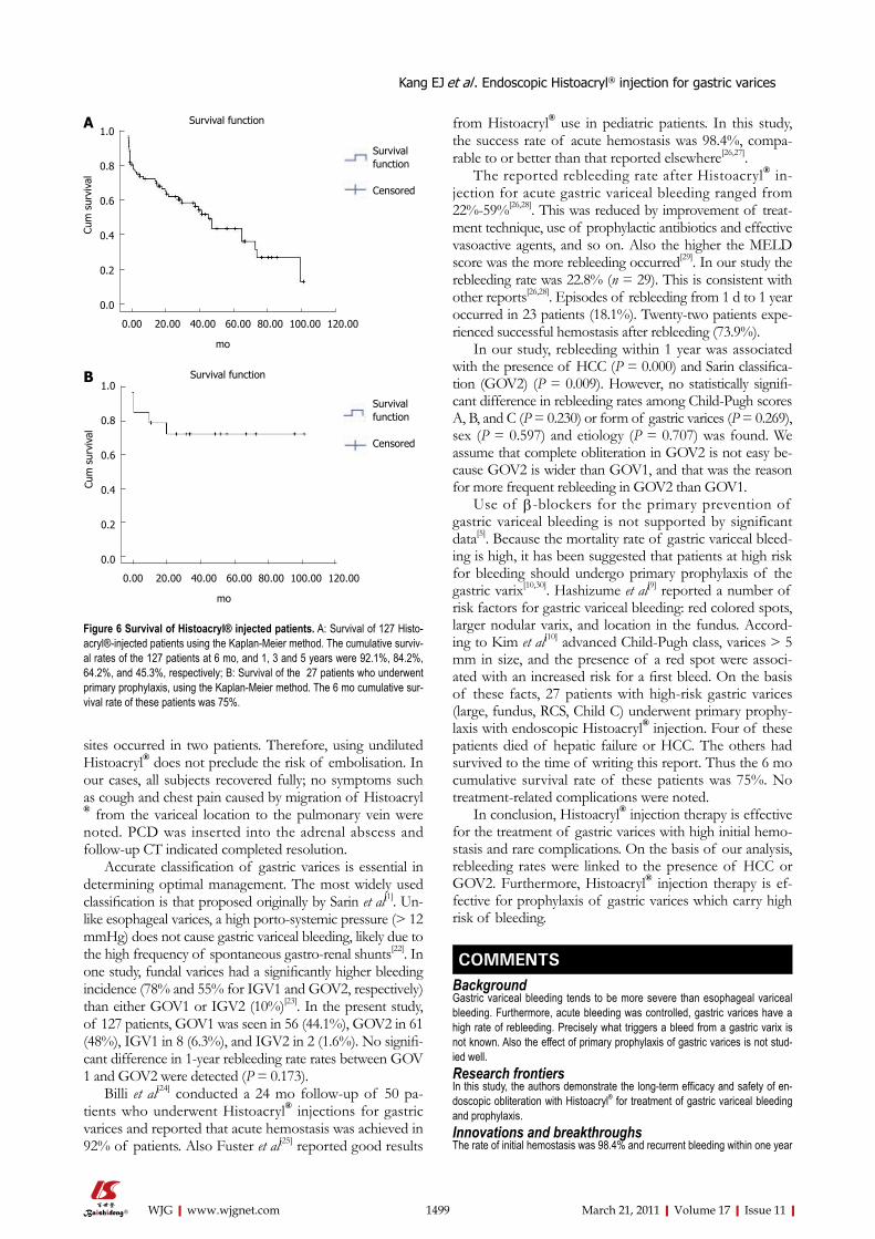
sites occurred in two patients. Therefore, using undiluted Histoacryl® does not preclude the risk of embolisation. In our cases, all subjects recovered fully; no symptoms such as cough and chest pain caused by migration of Histoacryl® from the variceal location to the pulmonary vein were noted. PCD was inserted into the adrenal abscess and follow-up CT indicated completed resolution.
Accurate classification of gastric varices is essential in determining optimal management. The most widely used classification is that proposed originally by Sarin et al[1]. Un-like esophageal varices, a high porto-systemic pressure (> 12 mmHg) does not cause gastric variceal bleeding, likely due to the high frequency of spontaneous gastro-renal shunts[22]. In one study, fundal varices had a significantly higher bleeding incidence (78% and 55% for IGV1 and GOV2, respectively) than either GOV1 or IGV2 (10%)[23]. In the present study, of 127 patients, GOV1 was seen in 56 (44.1%), GOV2 in 61 (48%), IGV1 in 8 (6.3%), and IGV2 in 2 (1.6%). No signifi-cant difference in 1-year rebleeding rate rates between GOV 1 and GOV2 were detected (P = 0.173).
Billi et al[24] conducted a 24 mo follow-up of 50 pa-tients who underwent Histoacryl® injections for gastric varices and reported that acute hemostasis was achieved in 92% of patients. Also Fuster et al[25] reported good results
from Histoacryl® use in pediatric patients. In this study, the success rate of acute hemostasis was 98.4%, compa-rable to or better than that reported elsewhere[26,27].
The reported rebleeding rate after Histoacryl® in-jection for acute gastric variceal bleeding ranged from 22%-59%[26,28]. This was reduced by improvement of treat-ment technique, use of prophylactic antibiotics and effective vasoactive agents, and so on. Also the higher the MELD score was the more rebleeding occurred[29]. In our study the rebleeding rate was 22.8% (n = 29). This is consistent with other reports[26,28]. Episodes of rebleeding from 1 d to 1 year occurred in 23 patients (18.1%). Twenty-two patients expe-rienced successful hemostasis after rebleeding (73.9%).
In our study, rebleeding within 1 year was associated with the presence of HCC (P = 0.000) and Sarin classifica-tion (GOV2) (P = 0.009). However, no statistically signifi-cant difference in rebleeding rates among Child-Pugh scores A, B, and C (P = 0.230) or form of gastric varices (P = 0.269), sex (P = 0.597) and etiology (P = 0.707) was found. We assume that complete obliteration in GOV2 is not easy be-cause GOV2 is wider than GOV1, and that was the reason for more frequent rebleeding in GOV2 than GOV1.
Use of β-blockers for the primary prevention of gastric variceal bleeding is not supported by significant data[5]. Because the mortality rate of gastric variceal bleed-ing is high, it has been suggested that patients at high risk for bleeding should undergo primary prophylaxis of the gastric varix[10,30]. Hashizume et al[9] reported a number of risk factors for gastric variceal bleeding: red colored spots, larger nodular varix, and location in the fundus. Accord-ing to Kim et al[10] advanced Child-Pugh class, varices > 5 mm in size, and the presence of a red spot were associ-ated with an increased risk for a first bleed. On the basis of these facts, 27 patients with high-risk gastric varices (large, fundus, RCS, Child C) underwent primary prophy-laxis with endoscopic Histoacryl® injection. Four of these patients died of hepatic failure or HCC. The others had survived to the time of writing this report. Thus the 6 mo cumulative survival rate of these patients was 75%. No treatment-related complications were noted.
In conclusion, Histoacryl® injection therapy is effective for the treatment of gastric varices with high initial hemo-stasis and rare complications. On the basis of our analysis, rebleeding rates were linked to the presence of HCC or GOV2. Furthermore, Histoacryl® injection therapy is ef-fective for prophylaxis of gastric varices which carry high risk of bleeding.
COMMENTSBackground Gastric variceal bleeding tends to be more severe than esophageal variceal bleeding. Furthermore, acute bleeding was controlled, gastric varices have a high rate of rebleeding. Precisely what triggers a bleed from a gastric varix is not known. Also the effect of primary prophylaxis of gastric varices is not stud-ied well.Research frontiers In this study, the authors demonstrate the long-term efficacy and safety of en-doscopic obliteration with Histoacryl® for treatment of gastric variceal bleeding and prophylaxis.Innovations and breakthroughs The rate of initial hemostasis was 98.4% and recurrent bleeding within one year
1499 March 21, 2011|Volume 17|Issue 11|WJG|www.wjgnet.com
A1.0
0.8
0.6
0.4
0.2
0.0
0.00 20.00 40.00 60.00 80.00 100.00 120.00
Cum
sur
viva
l
mo
Survival function
Survival function
Censored
B1.0
0.8
0.6
0.4
0.2
0.0
0.00 20.00 40.00 60.00 80.00 100.00 120.00
Cum
sur
viva
l
mo
Survival function
Survival function
Censored
Figure 6 Survival of Histoacryl® injected patients. A: Survival of 127 Histo-acryl®-injected patients using the Kaplan-Meier method. The cumulative surviv-al rates of the 127 patients at 6 mo, and 1, 3 and 5 years were 92.1%, 84.2%, 64.2%, and 45.3%, respectively; B: Survival of the 27 patients who underwent primary prophylaxis, using the Kaplan-Meier method. The 6 mo cumulative sur-vival rate of these patients was 75%.
Kang EJ et al . Endoscopic Histoacryl® injection for gastric varices
COMMENTS

occurred in 18.1% of patients. The rebleeding rate in patients with hepatocel-lular carcinoma (HCC) or GOV2 was higher than in those with other conditions. Median survival was 50 months (95% CI 30.5–69.5). Cumulative survival rates of the 127 patients at 6 months, 1, 3, and 5 years were 92.1%, 84.2%, 64.2%, and 45.3%, respectively. The 6-month cumulative survival rate of the 27 pa-tients treated prophylactically was 75%.ApplicationsHistoacryl® injection therapy is effective for the treatment of gastric varices with high initial hemostasis and rare complications. On the basis of our analysis, rebleeding rates were linked to the presence of HCC or GOV2. Furthermore, Histoacryl® injection therapy is effective for prophylaxis of gastric varices which carry high risk of bleeding. TerminologyHistoacryl® (N-butyl-2-cyanoacrylate) is a watery solution, which polymerizes and hardens within 20 seconds in a physiological milieu and instantaneously upon contact with blood. This makes it ideal for obliterating vessels and control-ling bleeding. It is necessary to dilute it with the oily contrast agent Lipiodol® is not only compatible with the tissue glue for dilution but also allows fluoroscopic monitoring of delivery of the substance.Peer review The authors reported their results of a retrospective study on patients who un-derwent endoscopic NBC injection for gastric varices. Important data including the success rate of initial haemostasis, rebleeding rate, long-term complications and prognostic predictors were reported. Though being limited by the retro-spective nature of the study, the strength of the study was the relatively large number of patients with active bleeding.
REFERENCES1 Sarin SK, Kumar A. Gastric varices: profile, classification,
and management. Am J Gastroenterol 1989; 84: 1244-12492 Watanabe K, Kimura K, Matsutani S, Ohto M, Okuda K. Por-
tal hemodynamics in patients with gastric varices. A study in 230 patients with esophageal and/or gastric varices using portal vein catheterization. Gastroenterology 1988; 95: 434-440
3 Smith JL, Graham DY. Variceal hemorrhage: a critical eval-uation of survival analysis. Gastroenterology 1982; 82: 968-973
4 D’Amico G, de Franchis R. End-of the-century reappraisal of the 6-week outcome of upper gastrointestinal bleeding in cir-rhosis. A prospective study (abstr). Gastroenterology 1999; 117: A1199
5 Ryan BM, Stockbrugger RW, Ryan JM. A pathophysiologic, gastroenterologic, and radiologic approach to the manage-ment of gastric varices. Gastroenterology 2004; 126: 1175-1189
6 Trudeau W, Prindiville T. Endoscopic injection sclerosis in bleeding gastric varices. Gastrointest Endosc 1986; 32: 264-268
7 Bretagne JF, Dudicourt JC, Morisot D, Thevenet F, Raoul JL, Gastard J. Is endoscopic variceal sclerotherapy effective for the treatment of gastric varices (abstr). Dig DIs Sci 1986; 31: A505S
8 Sarin SK. Long-term follow-up of gastric variceal sclerothera-py: an eleven-year experience. Gastrointest Endosc 1997; 46: 8-14
9 Hashizume M, Kitano S, Yamaga H, Koyanagi N, Sugima-chi K. Endoscopic classification of gastric varices. Gastroin-test Endosc 1990; 36: 276-280
10 Kim T, Shijo H, Kokawa H, Tokumitsu H, Kubara K, Ota K, Akiyoshi N, Iida T, Yokoyama M, Okumura M. Risk factors for hemorrhage from gastric fundal varices. Hepatology 1997; 25: 307-312
11 Laine L, Planas R, Nevens F, Banares R, Patch D, Bosch J. Treatment of the acute bleeding episode. In: Franchis R, editor. Consensus Workshop on Definitions, Methodology and Thera-peutic Strategies. Oxford: Blackwell Science, 2006: 217-242
12 Garcia-Tsao G, Sanyal AJ, Grace ND, Carey W. Prevention and management of gastroesophageal varices and variceal hemorrhage in cirrhosis. Hepatology 2007; 46: 922-938
13 Naga M, Foda A. An unusual complication of histoacryl in-
jection. Endoscopy 1997; 29: 14014 Yu LK, Hsu CW, Tseng JH, Liu NJ, Sheen IS. Splenic infarc-
tion complicated by splenic artery occlusion after N-butyl-2-cyanoacrylate injection for gastric varices: case report. Gastrointest Endosc 2005; 61: 343-345
15 Soehendra N, Grimm H, Nam VC, Berger B. N-butyl-2-cyanoacrylate: a supplement to endoscopic sclerotherapy. Endoscopy 1987; 19: 221-224
16 Soehendra N, Nam VC, Grimm H, Kempeneers I. Endo-scopic obliteration of large esophagogastric varices with bucrylate. Endoscopy 1986; 18: 25-26
17 Roesch W, Rexroth G. Pulmonary, cerebral and coronary emboli during bucrylate injection of bleeding fundic varices. Endoscopy 1998; 30: S89-S90
18 Hwang SS, Kim HH, Park SH, Kim SE, Jung JI, Ahn BY, Kim SH, Chung SK, Park YH, Choi KH. N-butyl-2-cyano-acrylate pulmonary embolism after endoscopic injection sclerotherapy for gastric variceal bleeding. J Comput Assist Tomogr 2001; 25: 16-22
19 Palejwala AA, Smart HL, Hughes M. Multiple pulmonary glue emboli following gastric variceal obliteration. Endos-copy 2000; 32: S1-S2
20 Abdullah A, Sachithanandan S, Tan OK, Chan YM, Khoo D, Mohamed Zawawi F, Omar H, Tan SS, Oemar H. Cerebral embolism following N-butyl-2-cyanoacrylate injection for esophageal postbanding ulcer bleed: a case report. Hepatol Int 2009; 3:504-508
21 D'Imperio N, Piemontese A, Baroncini D, Billi P, Borioni D, Dal Monte PP, Borrello P. Evaluation of undiluted N-butyl-2-cyanoacrylate in the endoscopic treatment of upper gas-trointestinal tract varices. Endoscopy 1996; 28: 239-243
22 Stanley AJ, Jalan R, Ireland HM, Redhead DN, Bouchier IA, Hayes PC. A comparison between gastric and oesophageal variceal haemorrhage treated with transjugular intrahepatic portosystemic stent shunt (TIPSS). Aliment Pharmacol Ther 1997; 11: 171-176
23 Sarin SK, Lahoti D, Saxena SP, Murthy NS, Makwana UK. Prevalence, classification and natural history of gastric vari-ces: a long-term follow-up study in 568 portal hypertension patients. Hepatology 1992; 16: 1343-1349
24 Billi P, Milandri GL, Borioni D, Fabbri C, Baroncini D, Cen-namo V, Piemontese A, D’Imperio N. Endoscopic treatment of gastric varices with N-butyl-2-cyanoacrylate: a long term follow up. Gastrointest Endosc 1998; 47: AB26
25 Fuster S, Costaguta A, Tabacco O, Sclerotherapy of bleeding gastric varices with cyanoacrylate in children (abstr). Gastro-enterology 1998; 114: A1244
26 Tan PC, Hou MC, Lin HC, Liu TT, Lee FY, Chang FY, Lee SD. A randomized trial of endoscopic treatment of acute gastric variceal hemorrhage: N-butyl-2-cyanoacrylate injec-tion versus band ligation. Hepatology 2006; 43: 690-697
27 Paik CN, Kim SW, Lee IS, Park JM, Cho YK, Choi MG, Chung IS. The therapeutic effect of cyanoacrylate on gastric variceal bleeding and factors related to clinical outcomes. J Clin Gastroeneterol 2008; 42: 916-922
28 Lo GH, Liang HL, Chen WC, Chen MH, Lai KH, Hsu PI, Lin CK, Chan HH, Pan HB. A prospective, randomized con-trolled trial of transjugular intrahepatic portosystemic shunt versus cyanoacrylate injection in the prevention of gastric variceal rebleeding. Endoscopy 2007; 39: 679-685
29 Bambha K, Kim WR, Pedersen R, Bida JP, Kremers WK, Ka-math PS. Predictors of early re-bleeding and mortality after acute variceal haemorrhage in patients with cirrhosis. Gut 2008; 57: 814-820
30 Matsumoto A, Matsumoto H, Hamamoto N, Kayazawa M. Management of gastric fundal varices associated with a gas-trorenal shunt. Gut 2001; 48: 440-441
S- Editor Tian L L- Editor O’Neill M E- Editor Ma WH
1500 March 21, 2011|Volume 17|Issue 11|WJG|www.wjgnet.com
Kang EJ et al . Endoscopic Histoacryl® injection for gastric varices
Clinicopathologic significance of HER-2/neu protein expression and gene amplification in gastric carcinoma
Shi-Yan Yan, Ying Hu, Jian-Gao Fan, Guo-Quan Tao, Yong-Ming Lu, Xu Cai, Bao-Hua Yu, Yi-Qun Du
Shi-Yan Yan, Ying Hu, Jian-Gao Fan, Department of Gastroen-terology, Xinhua Hospital, Shanghai Jiaotong University School of Medicine, Shanghai 200092, ChinaGuo-Quan Tao, Department of General Surgery, Huaian No.1 People's Hospital affiliated to Nanjing Medical University, Huaian 223300, Jiangsu Province, ChinaYong-Ming Lu, Xu Cai, Bao-Hua Yu, Department of Pathology, Cancer Hospital, Fudan University, Shanghai 200032, ChinaYi-Qun Du, Department of Pathology, Institute of Traditional Chinese Medicine, Nanjing 210029, Jiangsu Province, ChinaAuthor contributions: Yan SY participated in the treatment of patients, analyzed the data, performed the research and wrote the manuscript; Hu Y and Fan JG participated in the treatment of pa-tients, collected the data and designed the research; Tao GQ, Du YQ and Cai X collected all the cancer samples and Clinicopatho-logic data of gastric carcinoma patients; Other authors contrib-uted to the new reagents/analytic tools.Supported by National Natural Science Foundation of China, No. 81071888Correspondence to: Shi-Yan Yan, MD, Department of Gas-troenterology, Xinhua Hospital, Shanghai Jiaotong University School of Medicine, Shanghai 200092, China. [email protected]: +86-21-25078999 Fax: +86-21-25078999Received: August 8, 2010 Revised: December 13, 2010Accepted: December 20, 2010Published online: March 21, 2011
AbstractAIM: To study the HER-2/neu protein expression and gene amplification in gastric carcinoma and their rela-tion.
METHODS: One hundred and forty-five formalin-fixed and paraffin- embedded tumor tissue samples from Chinese gastric carcinoma patients were studied with immunohistochemistry (IHC) and fluorescence in situ hybridization (FISH) methods. Clinicopathologic data about all patients were collected.
RESULTS: The levels of HER-2 3+, HER-2 2+ and HER2
1+ were measurable in 6.9%, 8.3% and 17.2% of the samples, respectively. No HER-2 was stained in 67.6% of the samples. FISH showed that HER-2 gene was am-plified in 18 samples, 10 HER-2 3+ samples, 5 HER-2 2+ samples, and 3 HER-2 1+ samples with IHC stain-ing. HER-2 status was not correlated with the sex and age of patients, and tumor size, location or differentia-tion, but with the depth of invasion, TNM stage, lymph node and distant metastasis as well as histopathologi-cal classification of gastric cancer (P < 0.05).
CONCLUSION: All samples with IHC as HER-2 expres-sion should be analyzed with FISH. Detection of HER-2 gene amplification can assess the malignant biological behaviors and prognosis of gastric cancer.
© 2011 Baishideng. All rights reserved.
Key words: Gastric carcinoma; HER-2; Clinocopatho-logic significance; Immunohistochemistry; Fluorescence in situ hybridization
Peer reviewer: Jean François Beaulieu, Professor, Department of Anatomy and Cell Biology, Faculty of Medicine and Heath Sciences, Université de Sherbrooke, Sherbrooke, Qué, J1H 5N4, Canada
Yan SY, Hu Y, Fan JG, Tao GQ, Lu YM, Cai X, Yu BH, Du YQ. Clinicopathologic significance of HER-2/neu protein ex-pression and gene amplification in gastric carcinoma. World J Gastroenterol 2011; 17(11): 1501-1506 Available from: URL: http://www.wjgnet.com/1007-9327/full/v17/i11/1501.htm DOI: http://dx.doi.org/10.3748/wjg.v17.i11.1501
INTRODUCTIONGastric cancer is the second most common cause of cancer-related death worldwide. Surgery is the only curative procedure for localized gastric cancer, which is at the advanced stage when diagnosed in most cases. Currently
BRIEF ARTICLE
World J Gastroenterol 2011 March 21; 17(11): 1501-1506ISSN 1007-9327 (print) ISSN 2219-2840 (online)
© 2011 Baishideng. All rights reserved.
Online Submissions: http://www.wjgnet.com/[email protected]:10.3748/wjg.v17.i11.1501
1501 March 21, 2011|Volume 17|Issue 11|WJG|www.wjgnet.com
Yan SY et al . HER-2 status in gastric carcinoma
available agents are not very effective, resulting in a high recurrence rate, a low survival rate, and a poor prognosis of advanced gastric cancer patients. Thus, treatment of gastric cancer remains a challenge for physicians. New targeted therapies for advanced gastric carcinoma are needed, which may open up a new avenue for cancer treatment. Current targeted therapy for advanced gastric carcinoma depends on the evaluation of target gene sta-tus[1-3].
HER-2/neu or CerbB-2 is a member of growing fac-tors (EGFR, erbB-2, erbB-3 and erbB-4) in the HER family with intrinsic protein tyrosine kinase activity, and its increased activity is the assumed mechanism underly-ing cell transformation[4]. It is an oncogene that regulates the biological functions such as cell proliferation, dif-ferentiation, motility, and apoptosis. It was reported that HER-2 is over-expressed in cancer of breast, lungs, sali-vary gland, ovary, colon, prostate and pancreas[5,6]. HER-2 plays an important role in activation of HER-2 protein, and is over-expressed in 10%-38% of gastric cancer pa-tients[7]. However, the correlation between the expression of HER-2 protein and the prognosis of gastric cancer is still controversial. Different studies showed different HER-2 expression levels in gastric carcinoma[8-10].
Detecting the HER-2 status in gastric carcinoma is a prerequisite of monoclonal antibody therapy for gastric cancer. In this study, immunohistochemistry (IHC) was used in detecting HER-2 oncoprotein and fluorescent in situ hybridization (FISH) was used as a follow-up test for ambiguous results.
MATERIALS AND METHODSMaterialsParaffin-embedded tumor tissue blocks were provided by Cancer Hospital of Fudan University and Institute of Traditional Chinese Medicine of Jiangsu Province. One hundred and forty-five (95 males and 50 females) out of the 476 Chinese patients with gastric adenocarcinoma, admitted to Department of Surgery, Cancer Hospital of Fudan University, and Institute of Traditional Chinese Medicine of Jiangsu Province in 2006-2009 for surgery, were enrolled in this study. Tumor tissue blocks from pa-tients with full clinical data (including diagnosis, age, sex, address, disease history, etc.) were cut into 20 slides. The average age of the patients was 60 years. Cancer was clas-sified as stage Ⅰ in 32 cases, stage Ⅱ in 35 cases, stage Ⅲ in 37 cases, and stage Ⅳ in 41 cases, respectively, accord-ing to the TNM Cancer Staging System of the American Joint Committee of Cancer. The study was approved by The Review Board of Fudan University Cancer Hospital and Institute of Traditional Chinese Medicine of Jiangsu Province. Informed consent was obtained from all pa-tients.
Tumor tissue was cut into 4-μm thick sections which were stained with hematoxylin and eosin for histopatho-logical types, differentiation stage, IHC and FISH evalua-tion.
Immunohistochemical stainingSlides were deparaffinized, rehydrated, and heated in a microwave oven containing 0.01 mol/L citrate buffer (pH 6.0) for 5 min at 100℃ for antigen retrieval, cooled for 20 min and washed with water and a buffer solution. Per-oxidase was applied for 5 min and washed with a buffer solution for 2 × 5 min. Primary antibody diluted at 1:200 was applied for 30 min, link and streptavidin were applied for 10 min, respectively, and then washed with a buffer solution for 2 × 5 min. The bound antibody was visual-ized using a DAB-chromogen substrate. The sections were then counterstained with hematoxylin, covered with a cover-slip. Negative control was stained by omission of the primary antibody. Over-expression of HER-2 protein in paraffin-embedded invasive breast carcinoma tissue slides was used as a positive control.
A strong brown staining was located in cell mem-brane of malignant cells using this staining method. The DAKO Hercep Test™ Protocol System[11] was used to grade the membrane staining. The staining was scored as negative (0) when no membrane was stained or when membrane was stained in less than 10% of tumor cells, weakly positive (1+) if focal membrane was stained in more than 10% of tumor cells, intermediately positive (2+) if complete membrane was weakly- moderately stained in more than 10% of tumor cells, and strongly positive (3+) if complete membrane was intensely stained in more than 10% of tumor cells. Scores 0 and 1 were considered negative, while scores 2 and 3 were consid-ered positive.
FISH analysisHER-2 gene was amplified with dual-color FISH using a Passvision HER-2 DNA probe kit (Vysis Inc. Downers Grove, IL, USA) according to its manufacturer’s instruc-tions. Briefly, hybridization buffer, DNA probe, and puri-fied water were centrifuged, and heated to 73℃ for 5 min in a water bath. Slides were immersed in a denaturing bath (70% formamide 2 × SSC) for 5 min at 73℃, fol-lowed by dehydration in increasing ethanol concentra-tions, and then dried. The probe mixture was applied to each slide. The slides were placed in a 42℃ incubator for 30 min, washed with 0.4 × SSC/0.3% NP-40 for 2 min, air-dried in darkness, counterstained with 4’,6-diamidino-2-phenylindole (DAPI), and covered with a cover-slip. HER-2/neu-spectrum orange probe contains a DNA sequence specific for the HER-2 human gene locus and hybridized to the region 17q11.2-q12 of human chromo-somes. CEP17 (chromosome enumeration probe 17)/spectrum green probe containing alpha-satellite DNA that hybridizes to the D17Z1 locus (centromere region of chromosome 17) was used as a control. Nuclei were counterstained with DAPI. The slides were observed under a B × 60 fluorescence microscope equipped with a digital camera (DP50; Olympus, Tokyo, Japan) and the images were captured on a Windows PC with the View-finder Lite software. A cell was considered to be ampli-fied when a definite cluster or more than 10 signals for
1502 March 21, 2011|Volume 17|Issue 11|WJG|www.wjgnet.com

HER-2 were found. Known positive and negative cells were used as controls for each FISH. Gene amplification was scored when a minimum of 20 cancer cell nuclei exhibited a HER-2/CEP17 ratio ≥ 2, or when a HER-2 signal cluster was observed.
Statistical analysisStatistical analysis of data was performed by Student’s t test. P < 0.05 was considered statistically significant.
RESULTSExpression of HER-2 protein in gastric carcinomaHER-2 protein status in 145 gastric carcinoma tissue samples was determined with immunohistochemical staining (Figure 1). Of the 145 gastric carcinoma tissue samples, 98 (67.6%) were scored as 0, 25 (17.2%) as 1, 12 (8.3%) as 2, and 10 (6.9%) as 3. The positive rate was ap-proximately 15.2% (22/145).
HER-2 gene amplification in gastric carcinomasThe HER-2/CEP17 ratio was determined using the rate of HER-2/neu signals and CEP17 signals in 20 nuclei. The total number of HER-2/neu signals was divided by the total number of CEP17 signals, with a ratio was ≥ 2 according to the indications given by the Abbott-Vysis Company. IHC showed complete membrane immunos-taining (2+ and 3+). In addition, 123 samples negative for HER-2 over-expression with IHC staining were also analyzed by FISH. HER-2 gene was amplified in 18 out of the 145 gastric carcinoma tissue samples, including HER-2 3+ in 10 samples, HER2 2+ in 5 samples, and HER2 1+ in 3 samples with IHC staining (Table 1). The 10 tumors with strong complete membrane immunostain-ing (3+) exhibited HER-2 gene amplification, accounting for 55.6% (10/18) of all HER-2 amplifications. The posi-tive amplification rate of HER-2 gene was about 12.4% (18/145) in all gastric carcinoma tissue samples (Figure 2).
Correlation between clinicopathologic findings and HER-2 status The clinicopathological differences were observed in gastric carcinoma tissue samples with or without HER-2 protein expression or HER-2 gene amplification. The HER-2 protein expression and HER-2 gene amplification rates were 86.4 % (19/22) and 94.4 % (17/18) in intesti-nal type gastric carcinomas, respectively (P <0.05). The HER-2 status was correlated with the depth of invasion, TNM stage, lymph node and distant metastasis (P <0.05). However, no significant relation was found between clini-
1503 March 21, 2011|Volume 17|Issue 11|WJG|www.wjgnet.com
DC
BA
Figure 1 Immunohistochemistry staining of gastric carcinoma tissue samples showing negative HER-2 protein expression (A, B), positive HER-2 protein expression (C, D) (original magnification × 200).
Table 1 Correlation between HER-2 protein expression and HER-2 gene amplification
HER-2
FISH
Immunohistochemistry for HER-2 Total
0 (n = 98) 1 (n = 25) 2 (n = 12) 3 (n = 10)
Negative 98 22 7 0 127Positive 0 3 5 10 18
FISH: Fluorescence in situ hybridization.
Yan SY et al . HER-2 status in gastric carcinoma
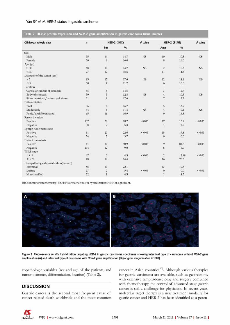
copathologic variables (sex and age of the patients, and tumor diameter, differentiation, location) (Table 2).
DISCUSSIONGastric cancer is the second most frequent cause of cancer-related death worldwide and the most common
cancer in Asian countries[12]. Although various therapies for gastric carcinoma are available, such as gastrectomy with extensive lymphadenectomy and surgery combined with chemotherapy, the control of advanced stage gastric cancer is still a challenge for physicians. In recent years, molecular target therapy is a new treatment modality for gastric cancer and HER-2 has been identified as a poten-
1504 March 21, 2011|Volume 17|Issue 11|WJG|www.wjgnet.com
Figure 2 Fluorescence in situ hybridization targeting HER-2 in gastric carcinoma specimens showing intestinal type of carcinoma without HER-2 gene amplification (A) and intestinal type of carcinoma with HER-2 gene amplification (B) (original magnification × 1000).
BA
IHC: Immunohistochemistry; FISH: Fluorescence in situ hybridization; NS: Not significant.
Table 2 HER-2 protein expression and HER-2 gene amplification in gastric carcinoma tissue samples
Clinicopathologic data n HER-2 (IHC) P value HER-2 (FISH) P value
Pos % Amp %
Sex Male 95 14 14.7 NS 10 10.5 NS Female 50 8 16.0 8 16.0Age (yr) > 60 68 10 14.7 NS 7 10.3 NS ≤ 60 77 12 15.6 11 14.3Diameter of the tumor (cm) > 5 85 15 17.6 NS 12 14.1 NS ≤ 5 60 7 11.7 6 10.0Location Cardia or fundus of stomach 55 8 14.5 7 12.7 Body of stomach 39 5 12.8 NS 4 10.3 NS Sinus ventriculi/ostium pyloricum 51 9 17.6 7 13.7Differentiation Well 36 6 16.7 5 13.9 Moderately 44 5 11.4 NS 4 9.1 NS Pooly/undifferentiated 65 11 16.9 9 13.8Serosa invasion Positive 107 20 18.7 < 0.05 17 15.9 < 0.05 Negative 38 2 5.3 1 2.7Lymph node metastasis Positive 91 20 22.0 < 0.05 18 19.8 < 0.05 Negative 54 2 3.7 0 0.0Distant metastasis Positive 11 10 90.9 < 0.05 9 81.8 < 0.05 Negative 134 12 9.0 8 6.0TNM stage Ⅰ + Ⅱ 67 3 4.5 < 0.05 2 2.99 < 0.05 Ⅲ + Ⅳ 78 19 24.4 16 20.5Histopathological classification(Lauren) Intestinal 86 19 22.1 17 19.8 Diffuse 37 2 5.4 < 0.05 0 0.0 < 0.05 Non classified 22 1 4.5 1 4.5
Yan SY et al . HER-2 status in gastric carcinoma
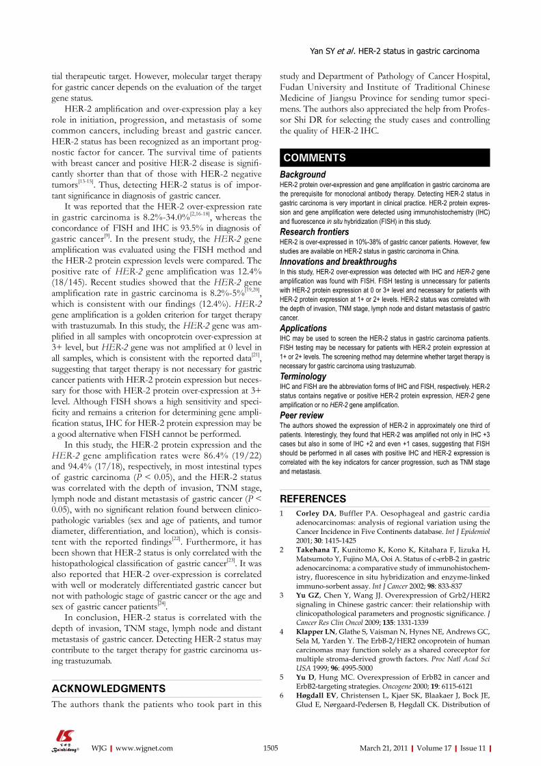
tial therapeutic target. However, molecular target therapy for gastric cancer depends on the evaluation of the target gene status.
HER-2 amplification and over-expression play a key role in initiation, progression, and metastasis of some common cancers, including breast and gastric cancer. HER-2 status has been recognized as an important prog-nostic factor for cancer. The survival time of patients with breast cancer and positive HER-2 disease is signifi-cantly shorter than that of those with HER-2 negative tumors[13-15]. Thus, detecting HER-2 status is of impor-tant significance in diagnosis of gastric cancer.
It was reported that the HER-2 over-expression rate in gastric carcinoma is 8.2%-34.0%[2,16-18], whereas the concordance of FISH and IHC is 93.5% in diagnosis of gastric cancer[9]. In the present study, the HER-2 gene amplification was evaluated using the FISH method and the HER-2 protein expression levels were compared. The positive rate of HER-2 gene amplification was 12.4% (18/145). Recent studies showed that the HER-2 gene amplification rate in gastric carcinoma is 8.2%-5%[19,20], which is consistent with our findings (12.4%). HER-2 gene amplification is a golden criterion for target therapy with trastuzumab. In this study, the HER-2 gene was am-plified in all samples with oncoprotein over-expression at 3+ level, but HER-2 gene was not amplified at 0 level in all samples, which is consistent with the reported data[21], suggesting that target therapy is not necessary for gastric cancer patients with HER-2 protein expression but neces-sary for those with HER-2 protein over-expression at 3+ level. Although FISH shows a high sensitivity and speci-ficity and remains a criterion for determining gene ampli-fication status, IHC for HER-2 protein expression may be a good alternative when FISH cannot be performed.
In this study, the HER-2 protein expression and the HER-2 gene amplification rates were 86.4% (19/22) and 94.4% (17/18), respectively, in most intestinal types of gastric carcinoma (P < 0.05), and the HER-2 status was correlated with the depth of invasion, TNM stage, lymph node and distant metastasis of gastric cancer (P < 0.05), with no significant relation found between clinico-pathologic variables (sex and age of patients, and tumor diameter, differentiation, and location), which is consis-tent with the reported findings[22]. Furthermore, it has been shown that HER-2 status is only correlated with the histopathological classification of gastric cancer[23]. It was also reported that HER-2 over-expression is correlated with well or moderately differentiated gastric cancer but not with pathologic stage of gastric cancer or the age and sex of gastric cancer patients[24].
In conclusion, HER-2 status is correlated with the depth of invasion, TNM stage, lymph node and distant metastasis of gastric cancer. Detecting HER-2 status may contribute to the target therapy for gastric carcinoma us-ing trastuzumab.
ACKNOWLEDGMENTSThe authors thank the patients who took part in this
study and Department of Pathology of Cancer Hospital, Fudan University and Institute of Traditional Chinese Medicine of Jiangsu Province for sending tumor speci-mens. The authors also appreciated the help from Profes-sor Shi DR for selecting the study cases and controlling the quality of HER-2 IHC.
COMMENTSBackgroundHER-2 protein over-expression and gene amplification in gastric carcinoma are the prerequisite for monoclonal antibody therapy. Detecting HER-2 status in gastric carcinoma is very important in clinical practice. HER-2 protein expres-sion and gene amplification were detected using immunohistochemistry (IHC) and fluorescence in situ hybridization (FISH) in this study. Research frontiersHER-2 is over-expressed in 10%-38% of gastric cancer patients. However, few studies are available on HER-2 status in gastric carcinoma in China.Innovations and breakthroughsIn this study, HER-2 over-expression was detected with IHC and HER-2 gene amplification was found with FISH. FISH testing is unnecessary for patients with HER-2 protein expression at 0 or 3+ level and necessary for patients with HER-2 protein expression at 1+ or 2+ levels. HER-2 status was correlated with the depth of invasion, TNM stage, lymph node and distant metastasis of gastric cancer. ApplicationsIHC may be used to screen the HER-2 status in gastric carcinoma patients. FISH testing may be necessary for patients with HER-2 protein expression at 1+ or 2+ levels. The screening method may determine whether target therapy is necessary for gastric carcinoma using trastuzumab.TerminologyIHC and FISH are the abbreviation forms of IHC and FISH, respectively. HER-2 status contains negative or positive HER-2 protein expression, HER-2 gene amplification or no HER-2 gene amplification.Peer reviewThe authors showed the expression of HER-2 in approximately one third of patients. Interestingly, they found that HER-2 was amplified not only in IHC +3 cases but also in some of IHC +2 and even +1 cases, suggesting that FISH should be performed in all cases with positive IHC and HER-2 expression is correlated with the key indicators for cancer progression, such as TNM stage and metastasis.
REFERENCES1 Corley DA, Buffler PA. Oesophageal and gastric cardia
adenocarcinomas: analysis of regional variation using the Cancer Incidence in Five Continents database. Int J Epidemiol 2001; 30: 1415-1425
2 Takehana T, Kunitomo K, Kono K, Kitahara F, Iizuka H, Matsumoto Y, Fujino MA, Ooi A. Status of c-erbB-2 in gastric adenocarcinoma: a comparative study of immunohistochem-istry, fluorescence in situ hybridization and enzyme-linked immuno-sorbent assay. Int J Cancer 2002; 98: 833-837
3 Yu GZ, Chen Y, Wang JJ. Overexpression of Grb2/HER2 signaling in Chinese gastric cancer: their relationship with clinicopathological parameters and prognostic significance. J Cancer Res Clin Oncol 2009; 135: 1331-1339
4 Klapper LN, Glathe S, Vaisman N, Hynes NE, Andrews GC, Sela M, Yarden Y. The ErbB-2/HER2 oncoprotein of human carcinomas may function solely as a shared coreceptor for multiple stroma-derived growth factors. Proc Natl Acad Sci USA 1999; 96: 4995-5000
5 Yu D, Hung MC. Overexpression of ErbB2 in cancer and ErbB2-targeting strategies. Oncogene 2000; 19: 6115-6121
6 Høgdall EV, Christensen L, Kjaer SK, Blaakaer J, Bock JE, Glud E, Nørgaard-Pedersen B, Høgdall CK. Distribution of
1505 March 21, 2011|Volume 17|Issue 11|WJG|www.wjgnet.com
COMMENTS
Yan SY et al . HER-2 status in gastric carcinoma

HER-2 overexpression in ovarian carcinoma tissue and its prognostic value in patients with ovarian carcinoma: from the Danish MALOVA Ovarian Cancer Study. Cancer 2003; 98: 66-73
7 Wang L, Habuchi T, Takahashi T, Kamoto T, Zuo T, Mitsu-mori K, Tsuchiya N, Sato K, Ogawa O, Kato T. No associa-tion between HER-2 gene polymorphism at codon 655 and a risk of bladder cancer. Int J Cancer 2002; 97: 787-790
8 Rüschoff J, Dietel M, Baretton G, Arbogast S, Walch A, Mon-ges G, Chenard MP, Penault-Llorca F, Nagelmeier I, Schlake W, Höfler H, Kreipe HH. HER2 diagnostics in gastric cancer-guideline validation and development of standardized im-munohistochemical testing. Virchows Arch 2010; 457: 299-307
9 Hofmann M, Stoss O, Shi D, Büttner R, van de Vijver M, Kim W, Ochiai A, Rüschoff J, Henkel T. Assessment of a HER2 scoring system for gastric cancer: results from a vali-dation study. Histopathology 2008; 52: 797-805
10 Gürel S, Dolar E, Yerci O, Samli B, Oztürk H, Nak SG, Gülten M, Memik F. The relationship between c-erbB-2 oncogene expression and clinicopathological factors in gastric cancer. J Int Med Res 1999; 27: 74-78
11 Selvarajan S, Bay BH, Chng MJ, Tan PH. The HercepTest and routine C-erbB2 immunohistochemistry in breast cancer: any difference? Ann Acad Med Singapore 2004; 33: 473-476
12 Shibuya K, Mathers CD, Boschi-Pinto C, Lopez AD, Murray CJ. Global and regional estimates of cancer mortality and incidence by site: II. Results for the global burden of disease 2000. BMC Cancer 2002; 2: 37
13 Burstein HJ. The distinctive nature of HER2-positive breast cancers. N Engl J Med 2005; 353: 1652-1654
14 Gschwind A, Fischer OM, Ullrich A. The discovery of recep-tor tyrosine kinases: targets for cancer therapy. Nat Rev Can-cer 2004; 4: 361-370
15 Baselga J, Swain SM. Novel anticancer targets: revisiting ERBB2 and discovering ERBB3. Nat Rev Cancer 2009; 9: 463-475
16 Lee KE, Lee HJ, Kim YH, Yu HJ, Yang HK, Kim WH, Lee KU, Choe KJ, Kim JP. Prognostic significance of p53, nm23, PCNA and c-erbB-2 in gastric cancer. Jpn J Clin Oncol 2003; 33: 173-179
17 Aoyagi K, Kohfuji K, Yano S, Murakami N, Miyagi M, Take-
da J, Shirouzu K. Evaluation of the epidermal growth factor receptor (EGFR) and c-erbB-2 in superspreading-type and penetrating-type gastric carcinoma. Kurume Med J 2001; 48: 197-200
18 Allgayer H, Babic R, Gruetzner KU, Tarabichi A, Schildberg FW, Heiss MM. c-erbB-2 is of independent prognostic rel-evance in gastric cancer and is associated with the expression of tumor-associated protease systems. J Clin Oncol 2000; 18: 2201-2209
19 Kimura M, Tsuda H, Morita D, Ichikura T, Ogata S, Aida S, Yoshizumi Y, Maehara T, Mochizuki H, Matsubara O. A proposal for diagnostically meaningful criteria to classify in-creased epidermal growth factor receptor and c-erbB-2 gene copy numbers in gastric carcinoma, based on correlation of fluorescence in situ hybridization and immunohistochemical measurements. Virchows Arch 2004; 445: 255-262
20 Tanner M, Hollmén M, Junttila TT, Kapanen AI, Tommola S, Soini Y, Helin H, Salo J, Joensuu H, Sihvo E, Elenius K, Isola J. Amplification of HER-2 in gastric carcinoma: association with Topoisomerase IIalpha gene amplification, intestinal type, poor prognosis and sensitivity to trastuzumab. Ann Oncol 2005; 16: 273-278
21 Bizari L, Borim AA, Leite KR, Gonçalves Fde T, Cury PM, Tajara EH, Silva AE. Alterations of the CCND1 and HER-2/neu (ERBB2) proteins in esophageal and gastric cancers. Can-cer Genet Cytogenet 2006; 165: 41-50
22 Chen B, Luo RC, Cui F, Qian XY. [Association of HER-2/neu expression with prognosis of gastric cancer]. Nanfang Yike Daxue Xuebao 2006; 26: 344-347
23 Barros-Silva JD, Leitão D, Afonso L, Vieira J, Dinis-Ribeiro M, Fragoso M, Bento MJ, Santos L, Ferreira P, Rêgo S, Brandão C, Carneiro F, Lopes C, Schmitt F, Teixeira MR. Association of ERBB2 gene status with histopathological parameters and disease-specific survival in gastric carcinoma patients. Br J Cancer 2009; 100: 487-493
24 Kim MA, Jung EJ, Lee HS, Lee HE, Jeon YK, Yang HK, Kim WH. Evaluation of HER-2 gene status in gastric carcinoma using immunohistochemistry, fluorescence in situ hybridiza-tion, and real-time quantitative polymerase chain reaction. Hum Pathol 2007; 38: 1386-1393
S- Editor Sun H L- Editor Wang XL E- Editor Zheng XM
1506 March 21, 2011|Volume 17|Issue 11|WJG|www.wjgnet.com
Yan SY et al . HER-2 status in gastric carcinoma

Epigallocatechin gallate inhibits HBV DNA synthesis in a viral replication - inducible cell line
Wei He, Li-Xia Li, Qing-Jiao Liao, Chun-Lan Liu, Xu-Lin Chen
Wei He, Li-Xia Li, Qing-Jiao Liao, Chun-Lan Liu, Xu-Lin Chen, State Key Laboratory of Virology, Wuhan Institute of Virology, Chinese Academic of Sciences, Wuhan 430071, Hubei Province, ChinaAuthor contributions: Chen XL designed the study; He W, Li LX and Liu CL performed the research; He W, Li LX, Liao QJ and Chen XL analyzed and compiled the data; He W wrote the paper; Chen XL revised the paper.Supported by National Technology and Science Key Project (2008ZX10002-010) and the Important National Science and Technology Specific Projects (2009ZX09301-014)Correspondence to: Xu-Lin Chen, PhD, Professor, State Key Laboratory of Virology, Wuhan Institute of Virology, Chinese Academic of Sciences, No. 44, Xiao Hong Shan Zhong Qu, Wuchang, Wuhan 430071, Hubei Province, China. [email protected]: +86-27-87198772 Fax: +86-27-87198466Received: October 28, 2010 Revised: January 5, 2011Accepted: January 12, 2011Published online: March 21, 2011
AbstractAIM: To analyze the antiviral mechanism of Epigallo-catechin gallate (EGCG) against hepatitis B virus (HBV) replication.
METHODS: In this research, the HBV-replicating cell line HepG2.117 was used to investigate the antiviral mechanism of EGCG. Cytotoxicity of EGCG was analyzed by 3-(4,5-Dimethylthiazol-2-yl)-2,5-diphenyltetrazolium bromide assay. Hepatitis B virus e antigen (HBeAg) and hepatitis B virus surface antigen (HBsAg) in the super-natant were detected by enzyme-linked immunosorbent assay. Precore mRNA and pregenomic RNA (pgRNA) levels were determined by semi-quantitative reverse transcription polymerase chain reaction (PCR) assay. The effect of EGCG on HBV core promoter activity was measured by dual luciferase reporter assay. HBV cova-lently closed circular DNA and replicative intermediates of DNA were quantified by real-time PCR assay.
RESULTS: When HepG2.117 cells were grown in the presence of EGCG, the expression of HBeAg was sup-pressed, however, the expression of HBsAg was not affected. HBV precore mRNA level was also down-regulated by EGCG, while the transcription of precore mRNA was not impaired. The synthesis of both HBV covalently closed circular DNA and replicative interme-diates of DNA were reduced by EGCG treatment to a similar extent, however, HBV pgRNA transcripted from chromosome-integrated HBV genome was not affected by EGCG treatment, indicating that EGCG targets only replicative intermediates of DNA synthesis.
CONCLUSION: In HepG2.117 cells, EGCG inhibits HBV replication by impairing HBV replicative intermediates of DNA synthesis and such inhibition results in reduced production of HBV covalently closed circular DNA.
© 2011 Baishideng. All rights reserved.
Key words: Covalently closed circular DNA; Epigallocat-echin gallate; Hepatitis B virus e antigen; Hepatitis B virus; Precore mRNA; Replicative intermediates of DNA
Peer reviewers: Dr. BS Anand, Professor, Digestive Diseases Section (111D), VA Medical Center, 2002 Holcombe Blvd., Houston, TX 77030, United States; Dr. Jin-Town Wang, Profes-sor, Department of Microbiology and Internal Medicine, National Taiwan University, College of Medicine, 1, Sec 1, Jen-Ai Rd, Taipei 10016, Taiwan, China
He W, Li LX, Liao QJ, Liu CL, Chen XL. Epigallocatechin gal-late inhibits HBV DNA synthesis in a viral replication - inducible cell line. World J Gastroenterol 2011; 17(11): 1507-1514 Available from: URL: http://www.wjgnet.com/1007-9327/full/v17/i11/1507.htm DOI: http://dx.doi.org/10.3748/wjg.v17.i11.1507
INTRODUCTIONHepatitis B virus (HBV) infection remains a major world
BRIEF ARTICLE
World J Gastroenterol 2011 March 21; 17(11): 1507-1514ISSN 1007-9327 (print) ISSN 2219-2840 (online)
© 2011 Baishideng. All rights reserved.
Online Submissions: http://www.wjgnet.com/[email protected]:10.3748/wjg.v17.i11.1507
1507 March 21, 2011|Volume 17|Issue 11|WJG|www.wjgnet.com

He W et al . EGCG inhibits HBV DNA synthesis
health problem despite the availability of an effective vac-cine. Approximately 350 million people are chronically infected with HBV worldwide[1,2]. HBV possesses a 3.2 kb relaxed circular partially double-stranded DNA (RC-DNA) genome[3]. After infection, this viral genome is released into cytoplasm and converted into covalently closed circu-lar DNA (cccDNA) in the host cell nucleus. The cccDNA is the template for production of all viral RNAs. Among HBV RNAs, the pregenomic RNA (pgRNA) is pack-aged into new capsid and serves as the template for HBV DNA synthesis. The nucleocapsid acquires an envelope and then buds as a mature virion. Progeny nucleocapsids may redeliver their RC-DNA genomes to the nucleus for accumulation of a cccDNA pool[4].
Currently, there is no reliable treatment that complete-ly eliminates HBV infection in all chronic carriers. Several nucleos(t)ide analogues (lamivudine, adefovir, entecavir, and telbivudine) and interferon-α (IFN-α) have been ap-proved as antiviral drugs against HBV infection, however, they have their own limitations, e.g. drug resistance and side effects[5]. Several non-nucleoside natural products have also been reported to inhibit HBV replication using various mechanisms. For example, oxymatrine was shown to block HBV DNA synthesis and cccDNA formation[6]. A helioxanthin derivative, 8-1, was demonstrated to di-minish HBV promoter activities and thus suppress HBV gene expression and DNA synthesis[7,8].
Epigallocatechin gallate (EGCG) is one of the most abundant polyphenols present in green tea[9]. EGCG is well known for its anti-tumor activity while other phar-maceutical effects were discovered subsequently[10-14]. Recently, it has also been reported that EGCG could inhibit HBV replication in a HBV stably replicating cell line - HepG2-N10[15]. However, the mechanism by which EGCG inhibits HBV replication is unclear. In fact, it is difficult to dissect the detailed anti-HBV mechanism of EGCG using HepG2-N10 cells, because the processes from cccDNA to antigen expression are severely affected by transcription from integrated HBV DNA[16].
In order to investigate the antiviral mechanism of EGCG against HBV in detail, we adopted a newly report-ed HepG2.117 cell line[17]. HBV replication is controllable using doxycycline in this cell line. More importantly, due to the delicate design, HBV precore mRNA can only be transcripted from replicated HBV DNA but not the inte-grated HBV DNA. As a result, hepatitis B virus e antigen (HBeAg) can only be translated from precore mRNA that is solely produced from replicated HBV DNA. Therefore, the precore mRNA and HBeAg level represents HBV DNA synthesis. On the other hand, PreS1/S2 mRNAs, which are templates for the translation of hepatitis B vi-rus surface antigen (HBsAg), are mainly transcribed from integrated HBV DNA rather than replicated HBV DNA in this cell line, thus, HBsAg has little correlation to HBV DNA synthesis level.
In this study, we investigated the inhibition of HBV replication by EGCG in HepG2.117 cells. Our results showed that HBV replicative intermediates of DNA (RI-
DNA) synthesis was significantly inhibited by EGCG, which resulted in less cccDNA production. In contrast, the production of HBV pgRNA, precore mRNA and the translation of HBeAg were not affected by EGCG.
MATERIALS AND METHODSCell line and cell cultureHepG2.117 (a gift from Dr. Dian-Xing Sun, Univer-sity Hospital Freiburg, Germany) is an inducible HBV-replicating cell line[17]. It contains a slight over-length HBV genome under the control of a tetracycline respon-sive CMV promoter. Transcription of HBV pgRNA is induced upon doxycycline removal from the culture medium, leading to capsid assembly and DNA synthesis. A small portion of the de novo synthesized RC-DNA is transported into the nucleus and converted to cccDNA which serves as a transcriptional template for the produc-tion of viral RNAs.
The HBeAg start codon and precore mRNA tran-scription initiation point are inexistent in chromosome integrated HBV genome in HepG2.117 cells[17]. There-fore, HBeAg and precore mRNA can not be generated from the integrated genome. However, once HBV starts to replicate, a functional precore mRNA would be pro-duced from cccDNA in this cell line, since the promoter and transcription initiation point would be restored in the process of cccDNA formation[17]. Subsequently, HBeAg would be produced from this precore mRNA where HBeAg start codon is also restored.
HepG2.117 cells were cultured in DMEM medium (Invitrogen) supplemented with 10% fetal bovine serum (Gibco), 100 U/mL penicillin and 100 μg/mL strepto-mycin (Sigma). When needed, doxycycline (Sigma) was routinely added at 1.5 μg/mL to suppress HBV pgRNA transcription.
Plasmid constructionTo generate pHBVCP-Luc reporter plasmid, a 442 bp fragment corresponding to the HBV core promoter region containing enhancer Ⅱ was amplified from pCH9-3091 (an ayw subtype HBV over-length genomic plasmid, kindly provided by Professor Michael Nassal, University Hospital, Freiburg)[18] by polymerase chain reaction (PCR) (forward primer: GGGGTACCCGC-GGGACGTCCTTTG; reverse primer: CCAAGCTTA-CAAGAGATGATTAGGCAG). This fragment was then inserted into the pGL3-basic vector (Promega, Madison, WI, USA), producing pHBVCP-Luc reporter construct.
Drug treatment and cytotoxicity assayThree days after the removal of doxycycline, HepG2.117 cells were seeded into 24-well plates. Twenty-four hours later, cells were treated with fresh DMEM medium con-taining various concentrations of EGCG (Hubei Insti-tute of Chemistry, Wuhan), or lamivudine (NIH AIDS Research and Reference Reagent Program, Rockville, MD, USA), respectively. The drug-containing media were
1508 March 21, 2011|Volume 17|Issue 11|WJG|www.wjgnet.com

replaced each day for 3 d. The medium was then collect-ed for antigen detection and attached cells were used for DNA and RNA analysis (see below) or EGCG toxicity analysis using the 3-(4,5-Dimethylthiazol-2-yl)-2,5-diphe-nyltetrazolium bromide (MTT) assay. Cells cultured with DMEM medium without EGCG were used as a negative control.
Enzyme-linked immunosorbent assay detection of HBsAg and HBeAgCollected media were subjected to 10 000 × g centrifu-gation to remove cellular debris. Secreted HBsAg and HBeAg were quantified by enzyme-linked immunosor-bent assay (ELISA) kits (Kehua, Shanghai) following the manufacturer’s instructions. All the data on HBsAg or HBeAg are presented as percentage of antigen level de-tected in the negative control samples.
Semi-quantitative reverse transcription polymerase chain reaction detection of HBV mRNATotal cellular RNA was extracted with Trizol reagents (Invitrogen) and treated with RQ1 RNase-free DNase Ⅰ (Promega) to digest residual DNA molecules. For the semi-quantification of precore mRNA and pgRNA, 1 μg total cellular RNA was subjected to cDNA synthesis using MMLV reverse transcriptase (Promega) and ran-dom hexamer as a primer based on the manufacturer’s instructions. The cDNAs were then measured by semi-quantitative PCR using Ex Taq (Takara, Dalian) and sub-sequently analyzed by agarose gel electrophoresis. The specific primers for semi-quantitative detection of precore mRNA were as follows: forward, 5'-TCTGCGCACCAG-CACCATG; reverse, 5'-TGCCTCGTCGTCTAACAA. The forward primer for pgRNA amplification was 5'-TC-GGGAAGCCTTAGAGTC, while the reverse primer was 5'-TGCCTCGTCGTCTAACAA. Primers used for Glyceraldehyde 3-phosphate dehydrogenase (GAPDH) mRNA amplification were as previously reported[19].
Dual luciferase reporter assay detection of HBV core promoter activityThree days after the removal of doxycycline, HepG2.117 cells were seeded into 24-well plates. Twenty-four hours later, the cells were transiently co-transfected with pH-BVCP-Luc reporter plasmid and normalizing plasmid pRL-TK with FuGENE-HD reagent according to the manufacturer’s instructions (Roche Applied Science, Manheim, Germany). Twelve hours after transfection, cells were subjected to a 3-d period of treatment with EGCG. HBV core promoter activity was determined by measuring luciferase activity using the Dual Luciferase Reporter Assay System (Promega).
Real-time PCR detection of HBV DNAExtraction of cccDNA was carried out using a modi-fied Hirt extraction procedure[16,20]. Briefly, cells from one well in the 24-well plate were lysed in 300 μL of 10 mmol/L Tris-HCl, 10 mmol/L EDTA and 0.7% SDS.
After 30 min incubation at room temperature, the lysate was mixed with 80 μL 5 mol/L NaCl and incubated at 4℃ overnight. The lysate was clarified by centrifugation at 12 000 × g for 30 min at 4℃, and then extracted twice with phenol and once with phenol:chloroform. DNAs were precipitated with ethanol overnight at -20℃ and dissolved in TE buffer, followed by further treatment of plasmid-safe DNase (Epicenter) to digest residual linear and open circular DNA molecules. When the reaction was finished, the plasmid-safe DNase was removed by phenol:chloroform extraction and the DNA was precipi-tated with ethanol at -20℃ overnight and dissolved in TE buffer. Total cellular HBV DNA was extracted from the cell monolayer with the total DNA extraction kit (Takara, Dalian) according to the manufacturer’s instructions.
A pair of primers (corresponding to HBV S ORF) in-troduced by Liu et al[21] was applied to quantify HBV DNA copies. In order to quantify cccDNA, extracted cccDNA samples were subjected to real-time PCR and the quantifi-cation was normalized by cell numbers. The total HBV DNA samples, which contained several types of HBV DNA forms (99% of them were HBV DNA replicative intermediates), were analyzed by real-time PCR and the quantification was normalized to the GAPDH DNA copies.
RESULTSHBeAg expression level in HepG2.117 cells was down-regulated by EGCG In order to choose an appropriate initial concentration of EGCG in the HBV inhibition assay, the MTT assay was used to determine the cytotoxicity of EGCG to HepG2.117 cells. As shown in Figure 1, the cytotoxic effects of EGCG were undetectable when the EGCG concentration was below 100 μmol/L, while slight cyto-toxicity (18% inhibition of cell viability) and significant cytotoxicity (60% inhibition of cell viability) were ob-
1509 March 21, 2011|Volume 17|Issue 11|WJG|www.wjgnet.com
1.2
1.0
0.8
0.6
0.4
0.2
0.0
Rela
tive
cell
viab
ility
0 50 100 200 400 EGCG concentration (μmol/L)
Figure 1 The cytotoxic effect of Epigallocatechin gallate on HepG2.117 cells. Twenty four hours after seeding, cells were incubated with medium con-taining different concentrations of Epigallocatechin gallate (EGCG) for 3 d, and cell viability was determined by the 3-(4,5-Dimethylthiazol-2-yl)-2,5-diphenyltet-razolium bromide assay. Cell viability of the control well not treated with EGCG was determined and arbitrarily designated as 1. Error bars indicate standard deviation of 3 independent experiments.
He W et al . EGCG inhibits HBV DNA synthesis

served when 200 and 400 μmol/L of EGCG were used, respectively. Therefore, the maximum non-toxic concen-tration was 100 μmol/L, and this was used as the highest concentration of EGCG in the HBV inhibition assay.
EGCG was reported to inhibit the expression of both HBeAg and HBsAg in HepG2-N10 cells[15]. To investi-gate the antiviral mechanisms of EGCG against HBV in HepG2.117 cells, a cell-based assay was used. The levels of HBeAg and HBsAg in the supernatant of EGCG-treated HepG2.117 cells were measured by ELISA. As shown in Figure 2A, HBeAg expression was dose-depend-ently inhibited by EGCG with an IC50 of 39.4 μmol/L. However, the expression level of HBsAg was largely un-affected (Figure 2B). These results indicated that EGCG could down-regulate HBeAg level but not HBsAg level in HepG2.117 cells.
HBV precore mRNA level was down-regulated in the presence of EGCGUnlike several HBV DNA stably transfected cell lines (e.g. HepG2.2.15) whose HBeAg is mainly translated from the
long early RNA that is transcribed from the inserted exog-enous promoter (e.g. CMV promoter), in the HepG2.117 cell line, HBeAg is exclusively translated from precore mRNA that is transcribed from cccDNA, akin to the actual HBV life cycle in HBV-infected hepatocytes. Since EGCG down-regulates HBeAg level but not HBsAg level in HepG2.117 cells, we determined whether the down-regulation of HBeAg by EGCG was due to inhibition of the transcription of precore mRNA.
Precore mRNA and pgRNA share the same sequence, except that precore mRNA is 35 nt longer at the 5’ ter-minal[16]. In order to distinguish precore mRNA from pgRNA, a specific forward primer complementary to the 5’ terminal of precore mRNA was designed for the pre-core mRNA specific reverse transcription PCR (RT-PCR) assay. Briefly, the 19 nt forward primer was designed to anneal to the 5’ terminal of precore RNA (nt 1800-1818) which is absent in pgRNA. The reverse primer was com-plementary to a sequence (nt 2345-2362) present in both precore mRNA and in pgRNA. Thus, a 563 bp fragment could be amplified from precore mRNA but not pgRNA.
After HepG2.117 cells were treated with EGCG for 3 d, total RNAs were extracted and subjected to semi-quantitative RT-PCR analysis using the precore mRNA specific primers. As shown in Figure 3, the amount of 563 bp specific RT-PCR products amplified from precore mRNA decreased in a dose-dependent manner with in-creasing concentrations of EGCG. The total RNA sam-ple for RT-PCR was free of DNA contamination as this specific product did not appear in reactions without re-verse transcription (Figure 3, NC lane). These results in-dicated that after EGCG treatment, precore mRNA level was down-regulated in HepG2.117 cells, and the lowered precore mRNA level accounted for the decreased pro-duction of HBeAg.
Transcriptional efficiency of HBV precore mRNA was not affected by EGCGPrecore mRNA is transcribed from cccDNA and this pro-cess is controlled by HBV core promoter[22]. To investigate
1510 March 21, 2011|Volume 17|Issue 11|WJG|www.wjgnet.com
1.2
1.0
0.8
0.6
0.4
0.2
0.0
Rela
tive
HBe
Ag le
vel
0 25 50 100 EGCG concentration (μmol/L)
1.2
1.0
0.8
0.6
0.4
0.2
0.0
Rela
tive
HBs
Ag le
vel
0 25 50 100 EGCG concentration (μmol/L)
B
A
Figure 2 Epigallocatechin gallate inhibited hepatitis B virus e antigen ex-pression level but not hepatitis B virus surface antigen expression level in the HepG2.117 cell line. Twenty four hours after seeding, cells were incubated with medium containing different concentrations of Epigallocatechin gallate (EGCG) for 3 d. The hepatitis B virus e antigen (HBeAg) and hepatitis B virus surface antigen (HBsAg) expression levels in the supernatants were measured using enzyme-linked immunosorbent assay kits for measuring HBeAg and HB-sAg, respectively. The level of HBeAg (A) and HBsAg (B) from cells not treated with EGCG (the control) were arbitrarily designated as 1. The levels of HBeAg (A) and HBsAg (B) in EGCG-treated cells were normalized with the control and expressed as relative levels. Error bars indicate standard deviation of 3 inde-pendent experiments.
600 bp400 bp
400 bp
200 bp
Precore
GAPDH
M NC 0 25 50 100EGCG
concentration (μmol/L)
Figure 3 Epigallocatechin gallate inhibited the production of hepatitis B virus precore mRNA. Twenty four hours after seeding, HepG2.117 cells were treated with medium containing different concentration of Epigallocatechin gal-late (EGCG) for 3 d. Total RNAs were prepared and were quantitated by semi-quantitative reverse transcription polymerase chain reaction (RT-PCR) with a pair of precore mRNA selective primers. Glyceraldehyde 3-phosphate dehydro-genase (GAPDH) mRNA was semi-quantitatively RT-PCR amplified and used for normalization. NC: Total RNA sample was subjected to PCR without reverse transcriptase and used as the negative control.
He W et al . EGCG inhibits HBV DNA synthesis
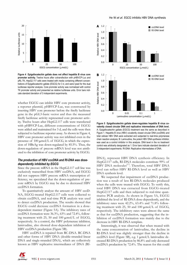
whether EGCG can inhibit HBV core promoter activity, a reporter plasmid, pHBVCP-Luc, was constructed by inserting HBV core promoter before the firefly luciferase gene in the pGL3-basic vector and thus the measured firefly luciferase activity represented core promoter activ-ity. Twelve hours after HepG2.117 cells were transfected with pHBVCP-Luc, different concentrations of EGCG were added and maintained for 3 d, and the cells were then subjected to luciferase reporter assay. As shown in Figure 4, HBV core promoter activity was not inhibited even in the presence of 100 μmol/L of EGCG, in which the expres-sion of HBeAg was down-regulated by 85.5%. Thus, the down-regulation of precore mRNA level was not attrib-uted to the inhibition of core promoter activity by EGCG.
The production of HBV cccDNA and RI-DNA was dose-dependently inhibited by EGCGSince the precore mRNA in the HepG2.117 cell line was exclusively transcribed from HBV cccDNA, and EGCG did not suppress HBV precore mRNA transcription ef-ficiency, we speculated that the down-regulation of pre-core mRNA by EGCG may be due to decreased HBV cccDNA formation.
To quantitatively analyze the amount of HBV cccD-NA, EGCG-treated HepG2.117 cells were collected to obtain cccDNA, and real-time PCR analysis was used to detect cccDNA production. The results showed that EGCG could decrease cccDNA formation in a dose-dependent manner (Figure 5A). The inhibitory rates of cccDNA formation were 36.3%, 63% and 72.4%, follow-ing treatment with 25, 50 and 100 μmol/L of EGCG, respectively. As a control, the HBV polymerase inhibitor, lamivudine, also showed dose-dependent inhibition of HBV cccDNA production (Figure 5B).
HBV cccDNA is repaired from RC-DNA. RC-DNA and other forms of HBV DNA (double-stranded linear DNA and single-stranded DNA), which are collectively known as HBV replicative intermediates of DNA (RI-
DNA), represent HBV DNA synthesis efficiency. In HepG2.117 cells, RI-DNA molecules constitute 99% of HBV DNA molecules[17]. Therefore, total HBV DNA level can reflect HBV RI-DNA level as well as HBV DNA synthesis level.
We suspected that impairment of cccDNA produc-tion was a result of less RI-DNA molecules produced when the cells were treated with EGCG. To verify this, total HBV DNA was extracted from EGCG-treated HepG2.117 cells and then subjected to real-time quan-titative PCR analysis. As shown in Figure 5A, EGCG inhibited the level of RI-DNA dose-dependently, and the inhibitory rates were 42.2%, 63.4% and 71.8% follow-ing treatment with 25, 50 and 100 μmol/L of EGCG, respectively. The inhibitory rates were almost the same as that for cccDNA production, suggesting that the in-hibition of cccDNA formation was mainly due to the decrease in HBV RI-DNA template.
Interestingly, it was observed that when treated with the same concentration of lamivudine, the decline in RI-DNA level was slightly stronger than the decline in cccDNA level (Figure 5B), e.g. 1 μmol/L lamivudine de-creased RI-DNA production by 86.8% and only decreased cccDNA production by 72.4%. The reason for this could
1511 March 21, 2011|Volume 17|Issue 11|WJG|www.wjgnet.com
20
15
10
5
0
Rela
tive
luci
fera
se u
nits
0 25 50 100 EGCG concentration (μmol/L)
Figure 4 Epigallocatechin gallate does not affect hepatitis B virus core promoter activity. Twelve hours after cotransfection with pHBVCP-Luc and pRL-TK, HepG2.117 cells were treated with media containing different concen-trations of Epigallocatechin gallate (EGCG) for 3 d, and were lysed for the dual luciferase reporter analysis. Core promoter activity was normalized with control TK promoter activity and presented as relative luciferase units. Error bars indi-cate standard deviation of 3 independent experiments.
cccDNA levelRI-DNA level
1.2
1.0
0.8
0.6
0.4
0.2
0.0
Rela
tive
HBV
DN
A le
vel
0 25 50 100 EGCG concentration (μmol/L)
cccDNA levelRI-DNA level
1.2
1.0
0.8
0.6
0.4
0.2
0.0
Rela
tive
HBV
DN
A le
vel
0 0.01 0.1 1 Lamivudine concentration (μmol/L)
B
A
Figure 5 Epigallocatechin gallate down-regulates hepatitis B virus co-valently closed circular DNA and replicative intermediates of DNA level. A: Epigallocatechin gallate (EGCG) treatment was the same as described in Figure 1. Hepatitis B virus (HBV) covalently closed circular DNA (cccDNA) and total cellular HBV DNA were extracted and subjected to real-time polymerase chain reaction analysis; B: Lamivudine, the potent HBV DNA synthesis inhibitor, was used as a control inhibitor in this analysis. DNA level in the no treatment control was arbitrarily designated as 1. Error bars indicate standard deviation of 3 independent experiments. RI-DNA: Replicative intermediates of DNA.
He W et al . EGCG inhibits HBV DNA synthesis

be that part of the cccDNA can be repaired from a cer-tain amount of pre-existing RC-DNA[23,24]. The observed minimal difference in the inhibition of cccDNA and RI-DNA production implies that EGCG may partly inhibit cccDNA formation.
Transcription of pgRNA from integrated HBV genome was not affected by EGCGHBV RI-DNA is synthesized from pgRNA and catalyzed by HBV polymerase. In the HepG2.117 cell line, pgRNA is transcribed from chromosome-integrated HBV DNA genome. Since RI-DNA level was down-regulated by EGCG in this cell line, we determined whether this down-regulation was actually caused by inhibition of RI-DNA synthesis rather than suppression of pgRNA tran-scription.
To analyze whether EGCG inhibited pgRNA tran-scription, doxycycline was removed from the culture medium, and EGCG was added at the same time. Three days later, total RNAs were extracted from HepG2.117 cells and pgRNAs were analyzed semi-quantitatively. As shown in Figure 6, EGCG treatment did not inter-fere with pgRNA production even when 100 μmol/L of EGCG was applied. Therefore, the transcription of pgRNA from integrated HBV DNA genome was not affected by EGCG. Taken together, we conclude that the decrease in RI-DNA resulted from the inhibition of RI-DNA synthesis, rather than the down-regulation of pgRNA production.
DISCUSSIONIn this study, we investigated the anti-HBV mechanism of EGCG in HepG2.117 cells, an inducible HBV-repli-cating cell line. Our results showed that EGCG down-regulated HBeAg level, precore mRNA level, cccDNA level and RI-DNA level to a similar extent, however, the production of HBV pgRNA was not affected. Therefore, the inhibition of HBV replication mainly occurred in HBV RI-DNA synthesis. When comparing the down-regulation of HBV cccDNA and RI-DNA production
mediated by EGCG and lamivudine, we speculated that EGCG may inhibit HBV cccDNA formation to a mi-nor extent. Taken together, suppression of HBV DNA synthesis is the major anti-HBV mechanism of EGCG, while cccDNA formation might be slightly impaired in the presence of EGCG. Lowered RI-DNA level leads to decreased cccDNA formation, which in turn leads to a decline in precore mRNA level and HBeAg level. HBV precore mRNA transcription and HBeAg translation were unaffected by EGCG.
In a previous study, the anti-HBV activity of EGCG was reported to down-regulate extracellular HBsAg and HBeAg level, intracellular cccDNA level and RI-DNA level. That research was conducted in the HepG2-N10 cell line which was constructed by stable transfection of a 1.3-fold adw subtype HBV genome into HepG2 cells. We reasoned that EGCG could inhibit HBsAg production in HepG2-N10 but not in HepG2.117 cells, which may be due to the different HBV subtype (adw vs ayw) and DNA length (1.3-fold vs slightly more than 1.0-fold). The results obtained in HepG2-N10 cells were not adequate to elucidate the antiviral mechanism of EGCG against HBV replication, because all HBV mRNAs are mainly transcribed from integrated HBV genomic DNA in HepG2-N10, and thus the antigens (representing mRNA levels) can not truly reflect the levels of HBV RI-DNA synthesis, cccDNA formation and mRNA transcription in the cells.
On the other hand, in the HepG2.117 cell line, HBsAg could be produced from both integral HBV genome and cccDNA, however, cccDNA copy numbers constitute only 1% of the integrated HBV genome copy number. Thus, nearly all of the HBsAg produced from mRNA is transcripted from integrated HBV genome rather than from HBV cccDNA. Therefore, we speculate that the inability of EGCG to inhibit HBsAg production in the HepG2.117 cell line was attributed to the unchanged transcription level of HBsAg mRNA from integrated HBV DNA, even in the presence of EGCG. However, in actual infected hepatocytes, HBsAg can only be pro-duced from cccDNA[25,26], like HBeAg in the HepG2.117 cell line. Thus, we speculate that EGCG is able to inhibit HBsAg in HBV infection because cccDNA can be sup-pressed by EGCG treatment.
HBV DNA stably transfected cell lines are frequently used in cell-based assays for the discovery of antiviral drugs against HBV[27,28]. In such cell lines, all HBV repli-cative intermediate DNA forms, cccDNA, nucleocapsid and infectious virion (Dane particle) can be detected, hence, it is generally accepted that the HBV life cycle is complete. However, all HBV RNAs and antigens can be produced from both chromosome-integrated HBV ge-nomic DNA and replicated HBV genomic DNA. More importantly, the strong HBV DNA synthesis inhibitor, lamivudine, does not affect HBV RNA levels and an-tigen levels in such cell models[29], indicating that HBV RNA transcription from integrated HBV DNA is much stronger than that from cccDNA. The amount of HBV RNAs and antigens in these cell lines do not reflect the
1512 March 21, 2011|Volume 17|Issue 11|WJG|www.wjgnet.com
500 bp250 bp
500 bp250 bp
Pregenomic
GAPDH
M NC 0 25 EGCG concentration (μmol/L)
Figure 6 Epigallocatechin gallate did not inhibit hepatitis B virus prege-nomic RNA transcription. When doxycycline was removed from the cell cul-ture medium, different concentrations of Epigallocatechin gallate (EGCG) were added to the HepG2.117 cells. After 3 d of culture, total RNA were extracted from the treated cells and the pregenomic RNAs (pgRNAs) were analyzed by semi-quantitative reverse transcription polymerase chain reaction. Glyceralde-hyde 3-phosphate dehydrogenase (GAPDH) mRNAs were used for normaliza-tion. NC: Total RNA sample was subjected to polymerase chain reaction without reverse transcription.
50 100
He W et al . EGCG inhibits HBV DNA synthesis
1513 March 21, 2011|Volume 17|Issue 11|WJG|www.wjgnet.com
actual HBV DNA replication level. In contrast, in the HepG2.117 cell line, HBeAg and precore mRNA ex-clusively come from HBV cccDNA, consequently, the pathway from HBV cccDNA to precore mRNA and then HBeAg can be quantitatively analyzed. Although the authenticity of the HBeAg signal in antigen detec-tion is controversial[30], the precore mRNA is a solid and positive parameter in this cell system. In summary, the HepG2.117 cell line is a better cell model to analyze the anti-HBV mechanism of potential drugs against HBV replication.
Currently, researchers have focused mainly on the po-tential application of EGCG in treating cancers. EGCG is known to block each stage of carcinogenesis by modulating signal transduction pathways involved in cell proliferation, transformation, inflammation, apoptosis, metastasis and invasion[31,32]. In this study, we investigated the anti-HBV mechanism of EGCG in detail and found that the suppression of HBV RI-DNA synthesis is the major anti-HBV mechanism of EGCG, while cccDNA formation might be slightly impaired in the presence of EGCG. Although the detailed replication mechanism of HBV remains unclear, it is estimated that multiple cel-lular factors are involved in HBV genome replication. We assume that EGCG may interact with certain cellular proteins and interfere with the function of the DNA rep-lication machinery. As a result, HBV RI-DNA synthesis is impaired and HBV replication is inhibited. To fully understand the antiviral mechanism of EGCG against HBV and explore the potential use of EGCG as anti-HBV drug, further study needs to be done to determine how EGCG impairs HBV RI-DNA synthesis and which cellular factors are affected.
COMMENTSBackgroundHepatitis B virus (HBV) infection remains a major world health problem despite the availability of an effective vaccine. Currently, there is no reliable treatment that completely eliminates HBV infection in all chronic carriers. Epigallocatechin gallate (EGCG) is one of the most abundant polyphenols present in green tea. Recently, it has been reported that EGCG could inhibit HBV replication in a HBV stably replicating cell line. However, the mechanism by which EGCG in-hibits HBV replication is unclear.Research frontiersCurrently, there are very few cell lines which can be used to investigate HBV replication. HepG2.117 is a newly reported cell line in which HBV replication is controllable using doxycycline. Hepatitis B virus e antigen (HBeAg) can only be translated from precore mRNA solely produced from replicated HBV DNA. HBeAg level correlates well with HBV DNA synthesis, whereas hepatitis B virus surface antigen level does not. Hence, this cell line is more convenient when investigating the antiviral mechanisms of leading compounds against HBV.Innovations and breakthroughsResults from this report showed that HBV replicative intermediates of DNA syn-thesis was significantly inhibited by EGCG, which resulted in reduced covalently closed circular DNA (cccDNA) production. In contrast, the production of HBV pregenomic RNA and the translation of HBeAg were not affected by EGCG. ApplicationsThe finding that EGCG can inhibit HBV relaxed circular partially double-strand-ed DNA synthesis is important both when exploring the potential of new drugs against HBV and in understanding which targets might be valid in eliminating viral replication.
Peer reviewThe authors investigated the anti-viral mechanism of epigallocatechin gallate against HBV replication. It was observed that epigallocatechin gallate impairs replicative intermediate DNA synthesis, resulting in reduced production of cccD-NA of the virus. The study is well conducted and the methodology was sound.
REFERENCES1 Ganem D, Prince AM. Hepatitis B virus infection--natural
history and clinical consequences. N Engl J Med 2004; 350: 1118-1129
2 Baumert TF, Thimme R, von Weizsäcker F. Pathogenesis of hepatitis B virus infection. World J Gastroenterol 2007; 13: 82-90
3 Summers J, O'Connell A, Millman I. Genome of hepatitis B virus: restriction enzyme cleavage and structure of DNA ex-tracted from Dane particles. Proc Natl Acad Sci USA 1975; 72: 4597-4601
4 Beck J, Nassal M. Hepatitis B virus replication. World J Gas-troenterol 2007; 13: 48-64
5 Tillmann HL. Antiviral therapy and resistance with hepati-tis B virus infection. World J Gastroenterol 2007; 13: 125-140
6 Xu WS, Zhao KK, Miao XH, Ni W, Cai X, Zhang RQ, Wang JX. Effect of oxymatrine on the replication cycle of hepatitis B virus in vitro. World J Gastroenterol 2010; 16: 2028-2037
7 Ying C, Li Y, Leung CH, Robek MD, Cheng YC. Unique anti-viral mechanism discovered in anti-hepatitis B virus research with a natural product analogue. Proc Natl Acad Sci USA 2007; 104: 8526-8531
8 Tseng YP, Kuo YH, Hu CP, Jeng KS, Janmanchi D, Lin CH, Chou CK, Yeh SF. The role of helioxanthin in inhibiting hu-man hepatitis B viral replication and gene expression by interfering with the host transcriptional machinery of viral promoters. Antiviral Res 2008; 77: 206-214
9 Shankar S, Ganapathy S, Srivastava RK. Green tea polyphe-nols: biology and therapeutic implications in cancer. Front Biosci 2007; 12: 4881-4899
10 Mukhtar H, Ahmad N. Tea polyphenols: prevention of cancer and optimizing health. Am J Clin Nutr 2000; 71: 1698S-1702S; discussion 1703S-1704S
11 Hsieh TC, Wu JM. Targeting CWR22Rv1 prostate cancer cell proliferation and gene expression by combinations of the phytochemicals EGCG, genistein and quercetin. Anticancer Res 2009; 29: 4025-4032
12 Bettuzzi S, Brausi M, Rizzi F, Castagnetti G, Peracchia G, Corti A. Chemoprevention of human prostate cancer by oral administration of green tea catechins in volunteers with high-grade prostate intraepithelial neoplasia: a preliminary report from a one-year proof-of-principle study. Cancer Res 2006; 66: 1234-1240
13 Qiao Y, Cao J, Xie L, Shi X. Cell growth inhibition and gene expression regulation by (-)-epigallocatechin-3-gallate in hu-man cervical cancer cells. Arch Pharm Res 2009; 32: 1309-1315
14 Philips BJ, Coyle CH, Morrisroe SN, Chancellor MB, Yo-shimura N. Induction of apoptosis in human bladder cancer cells by green tea catechins. Biomed Res 2009; 30: 207-215
15 Xu J, Wang J, Deng F, Hu Z, Wang H. Green tea extract and its major component epigallocatechin gallate inhibits hepati-tis B virus in vitro. Antiviral Res 2008; 78: 242-249
16 Zhou T, Guo H, Guo JT, Cuconati A, Mehta A, Block TM. Hepatitis B virus e antigen production is dependent upon co-valently closed circular (ccc) DNA in HepAD38 cell cultures and may serve as a cccDNA surrogate in antiviral screening assays. Antiviral Res 2006; 72: 116-124
17 Sun D, Nassal M. Stable HepG2- and Huh7-based human hepatoma cell lines for efficient regulated expression of in-fectious hepatitis B virus. J Hepatol 2006; 45: 636-645
18 Nassal M. The arginine-rich domain of the hepatitis B virus core protein is required for pregenome encapsidation and productive viral positive-strand DNA synthesis but not for
COMMENTS
He W et al . EGCG inhibits HBV DNA synthesis

1514 March 21, 2011|Volume 17|Issue 11|WJG|www.wjgnet.com
virus assembly. J Virol 1992; 66: 4107-411619 Zhong J, Gastaminza P, Cheng G, Kapadia S, Kato T, Burton
DR, Wieland SF, Uprichard SL, Wakita T, Chisari FV. Robust hepatitis C virus infection in vitro. Proc Natl Acad Sci USA 2005; 102: 9294-9299
20 Hirt B. Selective extraction of polyoma DNA from infected mouse cell cultures. J Mol Biol 1967; 26: 365-369
21 Liu Y, Hussain M, Wong S, Fung SK, Yim HJ, Lok AS. A genotype-independent real-time PCR assay for quantifi-cation of hepatitis B virus DNA. J Clin Microbiol 2007; 45: 553-558
22 Moolla N, Kew M, Arbuthnot P. Regulatory elements of hepatitis B virus transcription. J Viral Hepat 2002; 9: 323-331
23 Zoulim F. New insight on hepatitis B virus persistence from the study of intrahepatic viral cccDNA. J Hepatol 2005; 42: 302-308
24 Levrero M, Pollicino T, Petersen J, Belloni L, Raimondo G, Dandri M. Control of cccDNA function in hepatitis B virus infection. J Hepatol 2009; 51: 581-592
25 Gerlich WH, Heermann KH, Lu X. Functions of hepatitis B surface proteins. Arch Virol Suppl 1992; 4: 129-132
26 Seeger C, Mason WS. Hepatitis B virus biology. Microbiol
Mol Biol Rev 2000; 64: 51-6827 Acs G, Sells MA, Purcell RH, Price P, Engle R, Shapiro M,
Popper H. Hepatitis B virus produced by transfected Hep G2 cells causes hepatitis in chimpanzees. Proc Natl Acad Sci USA 1987; 84: 4641-4644
28 Fu L, Cheng YC. Characterization of novel human hepatoma cell lines with stable hepatitis B virus secretion for evaluat-ing new compounds against lamivudine- and penciclovir-re-sistant virus. Antimicrob Agents Chemother 2000; 44: 3402-3407
29 Doong SL, Tsai CH, Schinazi RF, Liotta DC, Cheng YC. Inhibition of the replication of hepatitis B virus in vitro by 2',3'-dideoxy-3'-thiacytidine and related analogues. Proc Natl Acad Sci USA 1991; 88: 8495-8499
30 Gehring AJ, Sun D, Kennedy PT, Nolte-'t Hoen E, Lim SG, Wasser S, Selden C, Maini MK, Davis DM, Nassal M, Berto-letti A. The level of viral antigen presented by hepatocytes influences CD8 T-cell function. J Virol 2007; 81: 2940-2949
31 Tachibana H. Molecular basis for cancer chemoprevention by green tea polyphenol EGCG. Forum Nutr 2009; 61: 156-169
32 Chen L, Zhang HY. Cancer preventive mechanisms of the green tea polyphenol (-)-epigallocatechin-3-gallate. Molecules 2007; 12: 946-957
S- Editor Tian L L- Editor Webster JR E- Editor Zheng XM
He W et al . EGCG inhibits HBV DNA synthesis

Spontaneous resolution of multiple lymphangiomas of the colon: A case report
Ji Min Lee, Woo Chul Chung, Kang-Moon Lee, Chang Nyol Paik, Yeon-Ji Kim, Bo-In Lee, Young Seok Cho, Hyun Joo Choi
Ji Min Lee, Woo Chul Chung, Kang-Moon Lee, Chang Nyol Paik, Yeon-Ji Kim, Bo-In Lee, Young Seok Cho, Hyun Joo Choi, Department of Internal Medicine, College of Medicine, the Catholic University of Korea, St. Vincent’s Hospital 93-6, Ji-dong, Paldal-gu, Suwon, Gyeonggi-do, 442-723, South KoreaAuthor contributions: Lee JM and Chung WC wrote the paper; Lee KM, Paik CN, Kim YJ, Lee BI and Cho YS contributed to the medical advice and carried out the literature research; Choi HJ reviewed the histopathologic examination.Correspondence to: Woo Chul Chung, MD, Department of Internal Medicine, College of Medicine, the Catholic University of Korea, St. Vincent’s Hospital 93-6, Ji-dong, Paldal-gu, Suwon, Gyeonggi-do, 442-723, South Korea. [email protected]: +82-31-2497138 Fax: +82-31-2538898Received: November 10, 2010 Revised: January 10, 2011Accepted: January 17, 2011Published online: March 21, 2011
AbstractLymphangioma of the colon is a relatively rare non-epithelial tumor and usually presents as a submucosal polypoid lesion. Many cases incidentally discovered are usually asymptomatic. However, they may present as abdominal pain or bleeding, and their resection is nor-mally required. Lymphangioma itself is generally rec-ognized as a benign tumor and no cases of malignant transformation have yet been reported, although its natural history is currently unknown. To the best of our knowledge, this study is the first to describe a case of spontaneous resolution in multiple colonic lymphangio-mas without any specific treatment.
© 2011 Baishideng. All rights reserved.
Key words: Lymphangioma; Colon; Natural history
Peer reviewers: A M El-Tawil, MSc, MRCS, PhD, Depart-ment of Surgery, University Hospital of Birmingham, East Cor-ridor, Ground Floor, Birmingham, B15 2TH, United Kingdom;
Antonio.Basoli, Professor, General Surgery “Paride Stefanini”, Università di Roma - Sapienza, Viale del Policlinico 155, Rome 00161, Italy
Lee JM, Chung WC, Lee KM, Paik CN, Kim YJ, Lee BI, Cho YS, Choi HJ. Spontaneous resolution of multiple lymphangio-mas of the colon: A case report. World J Gastroenterol 2011; 17(11): 1515-1518 Available from: URL: http://www.wjgnet.com/1007-9327/full/v17/i11/1515.htm DOI: http://dx.doi.org/10.3748/wjg.v17.i11.1515
INTRODUCTIONLymphangioma is a benign tumor commonly found in children. The majority of lymphangiomas are considered to be lymph vessel anomalies. The condition involves several expanded lymphatics surrounded by benign en-dothelial cells and usually occurs in the head, neck and axillary regions, and rarely in the gastrointestinal tract[1-3]. Recently, the number of case reports has been increasing with the increased use of endoscopic colon examination. Many lymphangiomas are asymptomatic and require no treatment, because the condition is considered absolutely benign. No cases, in which lymphangioma underwent malignant transformation, have been reported. However, some cases of lymphangioma accompany colon cancer[2]. When symptoms such as bleeding, intussusceptions or protein losing enteropathy are present, resection of lymphangioma is necessary[4-7]. Thus far, the natural his-tory of colonic lymphangioma has remained unknown. Herein, we report the first case of spontaneous reso-lution of multiple colonic lymphangiomas during the follow-up.
CASE REPORTA 54-year-old male patient suffered intermittently from
CASE REPORT
World J Gastroenterol 2011 March 21; 17(11): 1515-1518ISSN 1007-9327 (print) ISSN 2219-2840 (online)
© 2011 Baishideng. All rights reserved.
Online Submissions: http://www.wjgnet.com/[email protected]:10.3748/wjg.v17.i11.1515
1515 March 21, 2011|Volume 17|Issue 11|WJG|www.wjgnet.com

Lee JM et al . Spontaneous resolution of lymphangiomas
blood-tinged stool with no personal or familial history of any specific disease. Physical examination showed no remarkable abdominal abnormality. Laboratory test or chest and abdominal radiography displayed no specific findings. Initial colonoscopy revealed numerous elevated
lesions with smooth surfaces throughout the entire co-lon, which evidenced morphological changes as shown by endoscopy. In particular, the lesions on the left side of the colon evidenced surface petechiae (Figure 1). A diag-nostic procedure and endoscopic mucosal resection were performed for the appropriate diagnosis and treatment of the lesions. Histopathological examination showed that the cystic lesion was lined with cuboid-shaped cells that were positive for CD68 histiocytic markers (Figure 2) but negative for CD34 and CK on immunohistochemical examination, the patient was thus diagnosed with co-lonic lymphangioma. Abdominal computed tomography showed no definite abnormalities, and the possibility of secondary events was excluded. The complaint of mild symptoms led to medical observation and use of stool softener.
After 12 mo, the patient visited our department for colonoscopy, which showed no more remarkable chang-es than the previous findings. Biopsy procedure was repeated, which demonstrated that the cystic lesion had no lining endothelium but was lined with focal multinucle-ated giant cells in the submucosa (Figure 3). After 24 mo, colonoscopy revealed complete resolution of colonic lymphangiomas without any specific treatment (Figure 4).
DISCUSSIONIn most reported cases of colonic lymphangioma, the condition is solitary. Only a few cases of multiple lymph-
1516 March 21, 2011|Volume 17|Issue 11|WJG|www.wjgnet.com
B
A
Figure 1 Initial colonoscopy showing multiple elevated or protruding le-sions with semi-transparant and smooth surfaces (A), and petechiae on the surfaces of lesions in the sigmoid colon (B).
Figure 2 Initial histopathological examination showing a submucosal cys-tic structure lined with cuboidal endothelial cells for the CD68 histiocystic marker (bottom) and a few giant cells (upper, HE stain, × 200).
Figure 3 Histopathological examination 12 mo after initial examination showing cystically dilated lesion lined with endothelium (upper, HE stain, × 40) and focal multinucleated giant cells (bottom, HE stain, × 100).
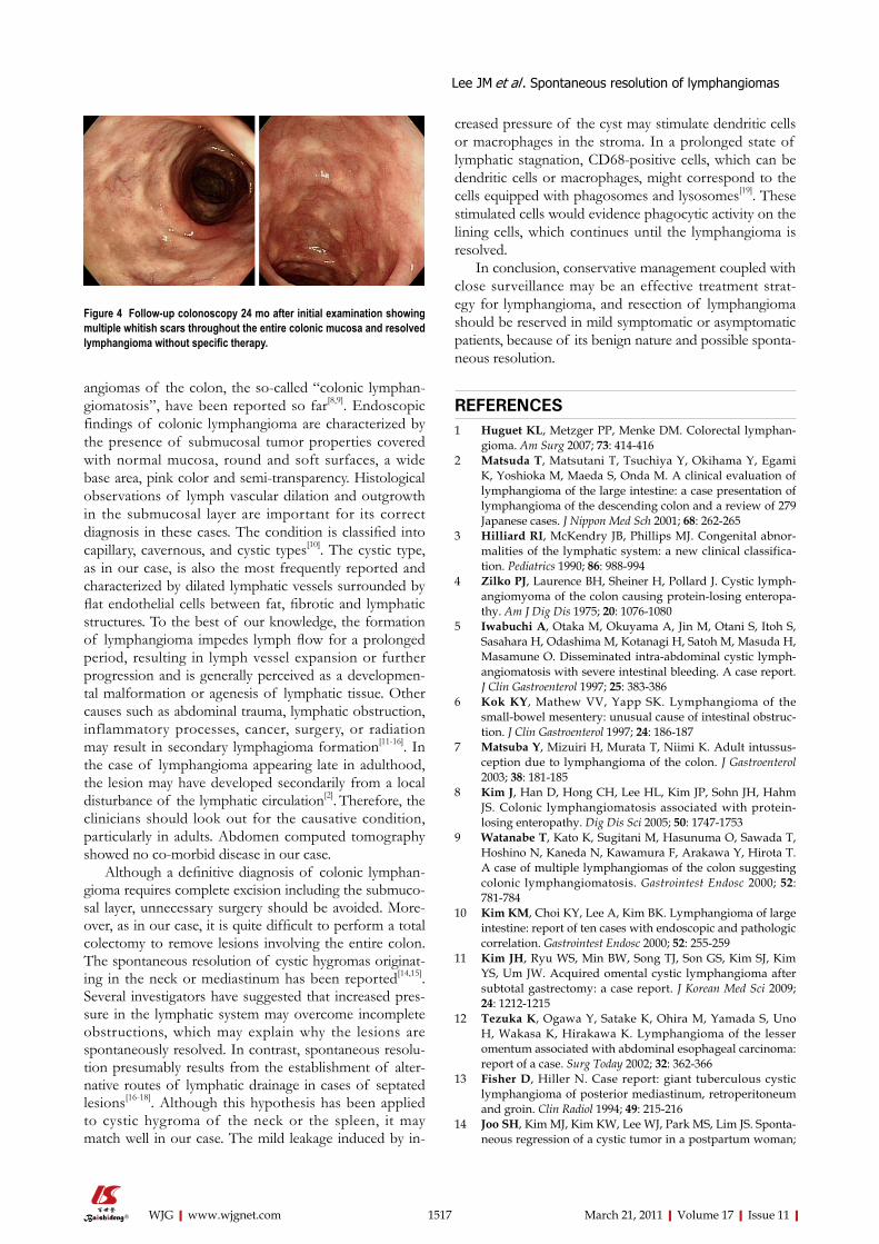
angiomas of the colon, the so-called “colonic lymphan-giomatosis”, have been reported so far[8,9]. Endoscopic findings of colonic lymphangioma are characterized by the presence of submucosal tumor properties covered with normal mucosa, round and soft surfaces, a wide base area, pink color and semi-transparency. Histological observations of lymph vascular dilation and outgrowth in the submucosal layer are important for its correct diagnosis in these cases. The condition is classified into capillary, cavernous, and cystic types[10]. The cystic type, as in our case, is also the most frequently reported and characterized by dilated lymphatic vessels surrounded by flat endothelial cells between fat, fibrotic and lymphatic structures. To the best of our knowledge, the formation of lymphangioma impedes lymph flow for a prolonged period, resulting in lymph vessel expansion or further progression and is generally perceived as a developmen-tal malformation or agenesis of lymphatic tissue. Other causes such as abdominal trauma, lymphatic obstruction, inflammatory processes, cancer, surgery, or radiation may result in secondary lymphagioma formation[11-16]. In the case of lymphangioma appearing late in adulthood, the lesion may have developed secondarily from a local disturbance of the lymphatic circulation[2]. Therefore, the clinicians should look out for the causative condition, particularly in adults. Abdomen computed tomography showed no co-morbid disease in our case.
Although a definitive diagnosis of colonic lymphan-gioma requires complete excision including the submuco-sal layer, unnecessary surgery should be avoided. More-over, as in our case, it is quite difficult to perform a total colectomy to remove lesions involving the entire colon. The spontaneous resolution of cystic hygromas originat-ing in the neck or mediastinum has been reported[14,15]. Several investigators have suggested that increased pres-sure in the lymphatic system may overcome incomplete obstructions, which may explain why the lesions are spontaneously resolved. In contrast, spontaneous resolu-tion presumably results from the establishment of alter-native routes of lymphatic drainage in cases of septated lesions[16-18]. Although this hypothesis has been applied to cystic hygroma of the neck or the spleen, it may match well in our case. The mild leakage induced by in-
creased pressure of the cyst may stimulate dendritic cells or macrophages in the stroma. In a prolonged state of lymphatic stagnation, CD68-positive cells, which can be dendritic cells or macrophages, might correspond to the cells equipped with phagosomes and lysosomes[19]. These stimulated cells would evidence phagocytic activity on the lining cells, which continues until the lymphangioma is resolved.
In conclusion, conservative management coupled with close surveillance may be an effective treatment strat-egy for lymphangioma, and resection of lymphangioma should be reserved in mild symptomatic or asymptomatic patients, because of its benign nature and possible sponta-neous resolution.
REFERENCES1 Huguet KL, Metzger PP, Menke DM. Colorectal lymphan-
gioma. Am Surg 2007; 73: 414-4162 Matsuda T, Matsutani T, Tsuchiya Y, Okihama Y, Egami
K, Yoshioka M, Maeda S, Onda M. A clinical evaluation of lymphangioma of the large intestine: a case presentation of lymphangioma of the descending colon and a review of 279 Japanese cases. J Nippon Med Sch 2001; 68: 262-265
3 Hilliard RI, McKendry JB, Phillips MJ. Congenital abnor-malities of the lymphatic system: a new clinical classifica-tion. Pediatrics 1990; 86: 988-994
4 Zilko PJ, Laurence BH, Sheiner H, Pollard J. Cystic lymph-angiomyoma of the colon causing protein-losing enteropa-thy. Am J Dig Dis 1975; 20: 1076-1080
5 Iwabuchi A, Otaka M, Okuyama A, Jin M, Otani S, Itoh S, Sasahara H, Odashima M, Kotanagi H, Satoh M, Masuda H, Masamune O. Disseminated intra-abdominal cystic lymph-angiomatosis with severe intestinal bleeding. A case report. J Clin Gastroenterol 1997; 25: 383-386
6 Kok KY, Mathew VV, Yapp SK. Lymphangioma of the small-bowel mesentery: unusual cause of intestinal obstruc-tion. J Clin Gastroenterol 1997; 24: 186-187
7 Matsuba Y, Mizuiri H, Murata T, Niimi K. Adult intussus-ception due to lymphangioma of the colon. J Gastroenterol 2003; 38: 181-185
8 Kim J, Han D, Hong CH, Lee HL, Kim JP, Sohn JH, Hahm JS. Colonic lymphangiomatosis associated with protein-losing enteropathy. Dig Dis Sci 2005; 50: 1747-1753
9 Watanabe T, Kato K, Sugitani M, Hasunuma O, Sawada T, Hoshino N, Kaneda N, Kawamura F, Arakawa Y, Hirota T. A case of multiple lymphangiomas of the colon suggesting colonic lymphangiomatosis. Gastrointest Endosc 2000; 52: 781-784
10 Kim KM, Choi KY, Lee A, Kim BK. Lymphangioma of large intestine: report of ten cases with endoscopic and pathologic correlation. Gastrointest Endosc 2000; 52: 255-259
11 Kim JH, Ryu WS, Min BW, Song TJ, Son GS, Kim SJ, Kim YS, Um JW. Acquired omental cystic lymphangioma after subtotal gastrectomy: a case report. J Korean Med Sci 2009; 24: 1212-1215
12 Tezuka K, Ogawa Y, Satake K, Ohira M, Yamada S, Uno H, Wakasa K, Hirakawa K. Lymphangioma of the lesser omentum associated with abdominal esophageal carcinoma: report of a case. Surg Today 2002; 32: 362-366
13 Fisher D, Hiller N. Case report: giant tuberculous cystic lymphangioma of posterior mediastinum, retroperitoneum and groin. Clin Radiol 1994; 49: 215-216
14 Joo SH, Kim MJ, Kim KW, Lee WJ, Park MS, Lim JS. Sponta-neous regression of a cystic tumor in a postpartum woman;
1517 March 21, 2011|Volume 17|Issue 11|WJG|www.wjgnet.com
Figure 4 Follow-up colonoscopy 24 mo after initial examination showing multiple whitish scars throughout the entire colonic mucosa and resolved lymphangioma without specific therapy.
Lee JM et al . Spontaneous resolution of lymphangiomas

is it a cystic lymphangioma? Yonsei Med J 2007; 48: 715-71815 Kiyota A, Tsukimori K, Yumoto Y, Hojo S, Morokuma S,
Fukushima K, Takahata Y, Nakayama H, Wake N. Sponta-neous resolution of cystic hygroma and hydrops in a fetus with Noonan's syndrome. Fetal Diagn Ther 2008; 24: 499-502
16 Brumfield CG, Wenstrom KD, Davis RO, Owen J, Cosper P. Second-trimester cystic hygroma: prognosis of septated and nonseptated lesions. Obstet Gynecol 1996; 88: 979-982
17 Bernstein HS, Filly RA, Goldberg JD, Golbus MS. Progno-
sis of fetuses with a cystic hygroma. Prenat Diagn 1991; 11: 349-355
18 Gallagher PG, Mahoney MJ, Gosche JR. Cystic hygroma in the fetus and newborn. Semin Perinatol 1999; 23: 341-356
19 Kishimoto K, Tate G, Kitamura T, Kojima M, Mitsuya T. Cytologic features and frequency of plasmacytoid dendritic cells in the lymph nodes of patients with histiocytic nec-rotizing lymphadenitis (Kikuchi-Fujimoto disease). Diagn Cytopathol 2010; 38: 521-526
S- Editor Tian L L- Editor Wang XL E- Editor Zheng XM
1518 March 21, 2011|Volume 17|Issue 11|WJG|www.wjgnet.com
Lee JM et al . Spontaneous resolution of lymphangiomas

World J Gastroenterol 2011 March 21; 17(11): I ISSN 1007-9327 (print) ISSN 2219-2840 (online)
© 2011 Baishideng. All rights reserved.
Online Submissions: http://www.wjgnet.com/[email protected]
ACKNOWLEDGMENTS
Acknowledgments to reviewers of World Journal of Gastroenterology
Many reviewers have contributed their expertise and time to the peer review, a critical process to ensure the quality of World Journal of Gastroenterology. The editors and authors of the articles submitted to the journal are grateful to the following reviewers for evaluating the articles (including those published in this issue and those rejected for this issue) during the last editing time period.
Victor E Reyes, PhD, Professor, Departments of Pediatrics and Mi-crobiology & Immunology,Director, GI Immunology Core, Texas Gulf Coast Digestive Diseases Center,Technical Director, Child Health Research Center,University of Texas Medical Branch, 301 University Blvd., Children's Hospital,Galveston, TX 77555-0366, United States
Hong Joo Kim, MD, Professor, Department of Internal Medicine, Sungkyunkwan University Kangbuk Samsung Hospital, 108, Pyung-Dong, Jongro-Ku, Seoul, 110-746, South Korea
Pietro Invernizzi, MD, PhD, Division of Internal Medicine and Hepato-biliary Immunopathology Unit, IRCCS Istituto Clinico Humanitas, via A. Manzoni 113, 20089 Rozzano, Milan, Italy
Peter L Lakatos, MD, PhD, Assistant Professor, 1st Department of Medicine, Semmelweis University, Koranyi S 2A, Budapest H1083, Hungary
Mauro Bortolotti, MD, Professor, Department of Internal Medicine and Gastroenterology, University of Bologna, Via Massarenti 48 Bologna, 40138, Italy
Akihito Nagahara, Associate Professor, Department of Gastroenterol-ogy, Juntendo University School of Medicine, 2-1-1 Hongo Bunkyo-ku, Tokyo 113-8421, Japan
Dr. Barjesh Chander Sharma, Professor, Department of Gastroenterol-ogy, G B Pant Hospital, New Delhi 110002, India
Luis Grande, Professor, Department of Surgery, Hospital del Mar, Passeig Marítim 25-29, Barcelona 08003, Spain
Chris JJ Mulder, Professor, Department of Gastroenterology, VU Univer-sity Medical Center, PO Box 7057, 1007 MB Amsterdam, The Netherlands
Benjamin Perakath, Professor, Dr., Department of Surgery Unit 5, Chris-tian Medical College, Vellore 632004, Tamil Nadu, India
Diego Garcia-Compean, MD, Professor, Faculty of Medicine, University Hospital, Department of Gastroenterology, Autonomous University of Nuevo Leon, Ave Madero y Gonzalitos, 64700 Monterrey, NL, México
Dr. Morten Frisch, MD, PhD, DSc, Department of Epidemiology Research, Statens Serum Institut, Building 206, Room 317, 5 Artillerivej, DK-2300 Copenhagen S, Denmark
Mitsuyoshi Urashima, MD, PhD, MPH, Division of Molecular Epi-demiology, Jikei University School of Medicine, 3-25-8 Nishi-shimbashi, Minato-ku, Tokyo 105-8461, Japan
Olav Dalgard, MD, PhD, Department of Medical, Rikshospitalet, 0027
Oslo, Norway
Kjetil Soreide, MD, PhD, Associate Professor, Department of Surgery, Stavanger University Hospital, Armauer Hansensvei 20, POB 8100, N-4068, Stavanger, Norway
Julian Swierczynski, MD, PhD, Professor, Department of Biochemistry, Medical University of Gdansk, 80-211 Gdansk, Poland
David J McGee, PhD, Associate Professor, Department of Microbiol-ogy & Immunology, Louisiana State University Health Sciences Center-Shreveport, 1501 Kings Highway, Shreveport, LA 71130, United States
Dr. Jeremy FL Cobbold, PhD, Clinical Lecturer in Hepatology, Depart-ment of Hepatology and Gastroenterology, Liver Unit, Imperial College London, St Mary’s Hospital, 10th Floor, QEQM building, Praed Street, London, W2 1NY, United Kingdom
Satoshi Osawa, MD, MD, PhD, Assistant Professor, First Department of Medicine, Hamamatsu University School of Medicine, 1-20-1 Handaya-ma, Higashi-ku, Hamamatsu, 431-3192, Japan
Peter L Lakatos, MD, PhD, Assistant Professor, 1st Department of Medicine, Semmelweis University, Koranyi S 2A, Budapest H1083, Hungary
Dr. Matthias Ocker, MD, Professor, Institute for Surgical Research, Direc-tor, Philipps-University Marburg, Baldingerstrasse, 35033 Marburg, Ger-many
Gianpiero Gravante, BSc (Hons), MBBS (Hons), Clinical Fellow in Up-per Gastrointestinal Surgery, Frenchay Hospital, North Bristol NHS Trust, Flat 8 Room 25, Frenchay Park Road, Bristol, BS16 1LE, United Kingdom
Luca Stocchi, MD, Desk A 30, Department of Colorectal Surgery,Digestive Disease Institute, Cleveland Clinic, 9500 Euclid Avenue, Cleveland OH 44195, United States
Paola De Nardi, MD, Department of Surgery, Scientific Institute San Raf-faele Hospital, Via Olgettina 60, Milan, 20132, Italy
Yukihiro Shimizu, MD, PhD, Kyoto Katsura Hospital, 17 Yamada-Hirao, Nishikyo, Kyoto 615-8256, Japan
Gloria González Aseguinolaza, BSc, MSc, PhD, Department of Gene Therapy in Hepatology, FIMA, CIMA University of Navarra, Navarra, Spain
Satoshi Yamagiwa, MD, PhD, Division of Gastroenterology and Hepa-tology, Niigata University Graduate School of Medical and Dental Sciences, 757 Asahimachi-dori, Chuo-ku, Niigata, 951-8510, Japan
Dr. Dinesh Vyas, Department of Minimally and Endosopic Surgery, St John Mercy Hospital, 851 E Fifth Street, Washington 63090, United States
Dr. Chao-Hung Hung, MD, Associate Professor, Division of Hepa-togastroenterology, Department of Internal Medicine, Kaohsiung Chang Gung Memorial Hospital. 123 Ta Pei Road, Niao Sung, Kaohsiung 833, Taiwan, China
Yoshitaka Takuma, MD, PhD, Department of Gastroenterology, Kura-shiki Central Hospital, 1-1-1 Miwa, Kurashiki, Okayama, 710-8602 Japan
Shoichiro Sumi, MD, PhD, Associate Professor, Department of Organ Reconstruction, Institute for Frontier Medical Sciences, Kyoto University, Sakyo-ku, Kyoto, 606-8507, Japan
I March 21, 2011|Volume 17|Issue 11|WJG|www.wjgnet.com
Meetings
Events Calendar 2011January 14-15, 2011AGA Clinical Congress of Gastroenterology and Hepatology: Best Practices in 2011 Miami, FL 33101, United States
January 20-22, 2011Gastrointestinal Cancers Symposium 2011, San Francisco, CA 94143,United States
January 27-28, 2011Falk Workshop, Liver and Immunology, Medical University, Franz-Josef-Strauss-Allee 11, 93053 Regensburg, Germany
January 28-29, 20119. Gastro Forum München, Munich, Germany
February 4-5, 201113th Duesseldorf International Endoscopy Symposium, Duesseldorf, Germany
February 13-27, 2011Gastroenterology: New Zealand CME Cruise Conference, Sydney, NSW, Australia
February 17-20, 2011APASL 2011-The 21st Conference of the Asian Pacific Association for the Study of the Liver Bangkok, Thailand
February 22, 2011-March 04, 2011Canadian Digestive Diseases Week 2011, Vancouver, BC, Canada
February 24-26, 2011Inflammatory Bowel Diseases 2011-6th Congress of the European Crohn's and Colitis Organisation, Dublin, Ireland
February 24-26, 20112nd International Congress on Abdominal Obesity, Buenos Aires, Brazil
February 24-26, 2011International Colorectal Disease Symposium 2011, Hong Kong, China
February 26-March 1, 2011Canadian Digestive Diseases Week,
Westin Bayshore, Vancouver, British Columbia, Canada
March 21-March 1, 2011Childhood & Adolescent Obesity: A whole-system strategic approach, Abu Dhabi, United Arab Emirates
March 3-5, 201142nd Annual Topics in Internal Medicine, Gainesville, FL 32614, United States
March 7-11, 2011Infectious Diseases: Adult Issues in the Outpatient and Inpatient Settings, Sarasota, FL 34234, United States
March 14-17, 2011British Society of Gastroenterology Annual Meeting 2011, Birmingham, England, United Kingdom
March 17-19, 201141. Kongress der Deutschen Gesellschaft für Endoskopie und Bildgebende Verfahren e.V., Munich, Germany
March 17-20, 2011Mayo Clinic Gastroenterology & Hepatology 2011, Jacksonville, FL 34234, United States
March 18, 2011UC Davis Health Informatics: Change Management and Health Informatics, The Keys to Health Reform, Sacramento, CA 94143, United States
March 25-27, 2011MedicReS IC 2011 Good Medical Research, Istanbul, Turkey
March 26-27, 201126th Annual New Treatments in Chronic Liver Disease, San Diego, CA 94143, United States
April 6-7, 2011IBS-A Global Perspective, Pfister Hotel, 424 East Wisconsin Avenue, Milwaukee, WI 53202, United States
April 7-9, 2011International and Interdisciplinary Conference Excellence in Female Surgery, Florence, Italy
April 15-16, 2011Falk Symposium 177, Endoscopy Live Berlin 2011 Intestinal Disease Meeting, Stauffenbergstr. 26, 10785 Berlin, Germany
April 18-22, 2011Pediatric Emergency Medicine: Detection, Diagnosis and Developing Treatment Plans, Sarasota, FL 34234, United States
April 20-23, 20119th International Gastric Cancer Congress, COEX, World Trade Center, Samseong-dong, Gangnam-gu, Seoul 135-731, South Korea
April 25-27, 2011The Second International Conference of the Saudi Society of Pediatric Gastroenterology, Hepatology & Nutrition, Riyadh, Saudi Arabia
April 25-29, 2011Neurology Updates for Primary Care, Sarasota, FL 34230-6947, United States
April 28-30, 20114th Central European Congress of Surgery, Budapest, Hungary
May 7-10, 2011Digestive Disease Week, Chicago, IL 60446, United States
May 12-13, 20112nd National Conference Clinical Advances in Cystic Fibrosis, London, England, United Kingdom
May 19-22, 20111st World Congress on Controversies in the Management of Viral Hepatitis (C-Hep), Palau de Congressos de Catalunya, Av. Diagonal, 661-671 Barcelona 08028, Spain
May 21-24, 201122nd European Society of Gastrointestinal and Abdominal Radiology Annual Meeting and Postgraduate Course, Venise, Italy
May 25-28, 20114th Congress of the Gastroenterology Association of Bosnia and Herzegovina with international participation, Hotel Holiday Inn, Sarajevo, Bosnia and Herzegovina
June 11-12, 2011The International Digestive Disease Forum 2011, Hong Kong, China
June 13-16, 2011Surgery and Disillusion XXIV SPIGC, II ESYS, Napoli, Italy
June 14-16, 2011International Scientific Conference
on Probiotics and Prebiotics-IPC2011, Kosice, Slovakia
June 22-25, 2011ESMO Conference: 13th World Congress on Gastrointestinal Cancer, Barcelona, Spain
June 29-2, 2011XI Congreso Interamericano de Pediatria "Monterrey 2011", Monterrey, Mexico
September 2-3, 2011 Falk Symposium 178, Diverticular Disease, A Fresh Approach to a Neglected Disease, Gürzenich Cologne, Martinstr. 29-37, 50667 Cologne, Germany
September 10-11, 2011New Advances in Inflammatory Bowel Disease, La Jolla, CA 92093, United States
September 10-14, 2011ICE 2011-International Congress of Endoscopy, Los Angeles Convention Center, 1201 South Figueroa Street Los Angeles, CA 90015, United States
September 30-October 1, 2011Falk Symposium 179, Revisiting IBD Management: Dogmas to be Challenged, Sheraton Brussels Hotel, Place Rogier 3, 1210 Brussels, Belgium
October 19-29, 2011Cardiology & Gastroenterology | Tahiti 10 night CME Cruise, Papeete, French Polynesia
October 22-26, 201119th United European Gastroenterology Week, Stockholm, Sweden
October 28-November 2, 2011ACG Annual Scientific Meeting & Postgraduate Course, Washington, DC 20001, United States
November 11-12, 2011Falk Symposium 180, IBD 2011: Progress and Future for Lifelong Management, ANA Interconti Hotel, 1-12-33 Akasaka, Minato-ku, Tokyo 107-0052, Japan
December 1-4, 20112011 Advances in Inflammatory Bowel Diseases/Crohn's & Colitis Foundation's Clinical & Research Conference, Hollywood, FL 34234, United States
World J Gastroenterol 2011 March 21; 17(11): I ISSN 1007-9327 (print) ISSN 2219-2840 (online)
© 2011 Baishideng. All rights reserved.
Online Submissions: http://www.wjgnet.com/[email protected]
I March 21, 2011|Volume 17|Issue 11|WJG|www.wjgnet.com
Instructions to authors
GENERAL INFORMATIONWorld Journal of Gastroenterology (World J Gastroenterol, WJG, print ISSN 1007-9327, online ISSN 2219-2840, DOI: 10.3748) is a weekly, open-access (OA), peer-reviewed journal supported by an editorial board of 1144 experts in gastroenterology and hepatol-ogy from 60 countries.
The biggest advantage of the OA model is that it provides free, full-text articles in PDF and other formats for experts and the public without registration, which eliminates the obstacle that traditional journals possess and usually delays the speed of the propagation and communication of scientific research results. The open access model has been proven to be a true ap-proach that may achieve the ultimate goal of the journals, i.e. the maximization of the value to the readers, authors and society.
Maximization of personal benefitsThe role of academic journals is to exhibit the scientific levels of a country, a university, a center, a department, and even a scientist, and build an important bridge for communication between scien-tists and the public. As we all know, the significance of the publica-tion of scientific articles lies not only in disseminating and com-municating innovative scientific achievements and academic views, as well as promoting the application of scientific achievements, but also in formally recognizing the “priority” and “copyright” of in-novative achievements published, as well as evaluating research per-formance and academic levels. So, to realize these desired attributes of WJG and create a well-recognized journal, the following four types of personal benefits should be maximized. The maximization of personal benefits refers to the pursuit of the maximum personal benefits in a well-considered optimal manner without violation of the laws, ethical rules and the benefits of others. (1) Maximization of the benefits of editorial board members: The primary task of editorial board members is to give a peer review of an unpublished scientific article via online office system to evaluate its innovative-ness, scientific and practical values and determine whether it should be published or not. During peer review, editorial board members can also obtain cutting-edge information in that field at first hand. As leaders in their field, they have priority to be invited to write articles and publish commentary articles. We will put peer review-ers’ names and affiliations along with the article they reviewed in the journal to acknowledge their contribution; (2) Maximization of the benefits of authors: Since WJG is an open-access journal, readers around the world can immediately download and read, free of charge, high-quality, peer-reviewed articles from WJG official website, thereby realizing the goals and significance of the com-munication between authors and peers as well as public reading; (3) Maximization of the benefits of readers: Readers can read or use, free of charge, high-quality peer-reviewed articles without any lim-its, and cite the arguments, viewpoints, concepts, theories, methods, results, conclusion or facts and data of pertinent literature so as to validate the innovativeness, scientific and practical values of their own research achievements, thus ensuring that their articles have novel arguments or viewpoints, solid evidence and correct conclu-
sion; and (4) Maximization of the benefits of employees: It is an iron law that a first-class journal is unable to exist without first-class editors, and only first-class editors can create a first-class academic journal. We insist on strengthening our team cultivation and con-struction so that every employee, in an open, fair and transparent environment, could contribute their wisdom to edit and publish high-quality articles, thereby realizing the maximization of the personal benefits of editorial board members, authors and readers, and yielding the greatest social and economic benefits.
Aims and scopeThe major task of WJG is to report rapidly the most recent re-sults in basic and clinical research on esophageal, gastrointestinal, liver, pancreas and biliary tract diseases, Helicobacter pylori, endos-copy and gastrointestinal surgery, including: gastroesophageal reflux disease, gastrointestinal bleeding, infection and tumors; gastric and duodenal disorders; intestinal inflammation, micro-flora and immunity; celiac disease, dyspepsia and nutrition; viral hepatitis, portal hypertension, liver fibrosis, liver cirrhosis, liver transplantation, and metabolic liver disease; molecular and cell biology; geriatric and pediatric gastroenterology; diagnosis and screening, imaging and advanced technology.
ColumnsThe columns in the issues of WJG will include: (1) Editorial: To introduce and comment on major advances and developments in the field; (2) Frontier: To review representative achievements, com-ment on the state of current research, and propose directions for future research; (3) Topic Highlight: This column consists of three formats, including (A) 10 invited review articles on a hot topic, (B) a commentary on common issues of this hot topic, and (C) a com-mentary on the 10 individual articles; (4) Observation: To update the development of old and new questions, highlight unsolved problems, and provide strategies on how to solve the questions; (5) Guidelines for Basic Research: To provide guidelines for basic research; (6) Guidelines for Clinical Practice: To provide guidelines for clinical diagnosis and treatment; (7) Review: To review systemi-cally progress and unresolved problems in the field, comment on the state of current research, and make suggestions for future work; (8) Original Article: To report innovative and original find-ings in gastroenterology; (9) Brief Article: To briefly report the novel and innovative findings in gastroenterology and hepatology; (10) Case Report: To report a rare or typical case; (11) Letters to the Editor: To discuss and make reply to the contributions published in WJG, or to introduce and comment on a controversial issue of general interest; (12) Book Reviews: To introduce and comment on quality monographs of gastroenterology and hepatology; and (13) Guidelines: To introduce consensuses and guidelines reached by international and national academic authorities worldwide on basic research and clinical practice gastroenterology and hepatology.
Name of journalWorld Journal of Gastroenterology
Serial publication numberISSN 1007-9327 (print)ISSN 2219-2840 (online)
Online Submissions: http://www.wjgnet.com/[email protected]
World J Gastroenterol 2011 March 21; 17(11): I-VIISSN 1007-9327 (print) ISSN 2219-2840 (online)
© 2011 Baishideng. All rights reserved.
I March 21, 2011|Volume 17|Issue 11|WJG|www.wjgnet.com

Indexed and Abstracted inCurrent Contents®/Clinical Medicine, Science Citation Index Expanded (also known as SciSearch®), Journal Citation Reports®, Index Medicus, MEDLINE, PubMed, PubMed Central, and Di-rectory of Open Access Journals. ISI, Thomson Reuters, 2009 Impact Factor: 2.092 (33/65 Gastroenterology and Hepatol-ogy).
Published byBaishideng Publishing Group Co., Limited
SPECIAL STATEMENTAll articles published in this journal represent the viewpoints of the authors except where indicated otherwise.
Biostatistical editingStatisital review is performed after peer review. We invite an expert in Biomedical Statistics from to evaluate the statistical method used in the paper, including t-test (group or paired com-parisons), chi-squared test, Ridit, probit, logit, regression (linear, curvilinear, or stepwise), correlation, analysis of variance, analysis of covariance, etc. The reviewing points include: (1) Statistical methods should be described when they are used to verify the re-sults; (2) Whether the statistical techniques are suitable or correct; (3) Only homogeneous data can be averaged. Standard deviations are preferred to standard errors. Give the number of observa-tions and subjects (n). Losses in observations, such as drop-outs from the study should be reported; (4) Values such as ED50, LD50, IC50 should have their 95% confidence limits calculated and compared by weighted probit analysis (Bliss and Finney); and (5) The word ‘significantly’ should be replaced by its synonyms (if it indicates extent) or the P value (if it indicates statistical signifi-cance).
Conflict-of-interest statementIn the interests of transparency and to help reviewers assess any potential bias, WJG requires authors of all papers to declare any competing commercial, personal, political, intellectual, or religious interests in relation to the submitted work. Referees are also asked to indicate any potential conflict they might have reviewing a particular paper. Before submitting, authors are sug-gested to read “Uniform Requirements for Manuscripts Submit-ted to Biomedical Journals: Ethical Considerations in the Con-duct and Reporting of Research: Conflicts of Interest” from International Committee of Medical Journal Editors (ICMJE), which is available at: http://www.icmje.org/ethical_4conflicts.html.
Sample wording: [Name of individual] has received fees for serving as a speaker, a consultant and an advisory board member for [names of organizations], and has received research funding from [names of organization]. [Name of individual] is an employ-ee of [name of organization]. [Name of individual] owns stocks and shares in [name of organization]. [Name of individual] owns patent [patent identification and brief description].
Statement of informed consentManuscripts should contain a statement to the effect that all hu-man studies have been reviewed by the appropriate ethics com-mittee or it should be stated clearly in the text that all persons gave their informed consent prior to their inclusion in the study. Details that might disclose the identity of the subjects under study should be omitted. Authors should also draw attention to the Code of Ethics of the World Medical Association (Declara-
tion of Helsinki, 1964, as revised in 2004).
Statement of human and animal rightsWhen reporting the results from experiments, authors should follow the highest standards and the trial should conform to Good Clinical Practice (for example, US Food and Drug Admin-istration Good Clinical Practice in FDA-Regulated Clinical Trials; UK Medicines Research Council Guidelines for Good Clinical Practice in Clinical Trials) and/or the World Medical Association Declaration of Helsinki. Generally, we suggest authors follow the lead investigator’s national standard. If doubt exists whether the research was conducted in accordance with the above stan-dards, the authors must explain the rationale for their approach and demonstrate that the institutional review body explicitly ap-proved the doubtful aspects of the study.
Before submitting, authors should make their study ap-proved by the relevant research ethics committee or institutional review board. If human participants were involved, manuscripts must be accompanied by a statement that the experiments were undertaken with the understanding and appropriate informed consent of each. Any personal item or information will not be published without explicit consents from the involved patients. If experimental animals were used, the materials and methods (experimental procedures) section must clearly indicate that ap-propriate measures were taken to minimize pain or discomfort, and details of animal care should be provided.
SUBMISSION OF MANUSCRIPTSManuscripts should be typed in 1.5 line spacing and 12 pt. Book Antiqua with ample margins. Number all pages consecutively, and start each of the following sections on a new page: Title Page, Abstract, Introduction, Materials and Methods, Results, Discussion, Acknowledgements, References, Tables, Figures, and Figure Legends. Neither the editors nor the publisher are responsible for the opinions expressed by contributors. Manu-scripts formally accepted for publication become the permanent property of Baishideng Publishing Group Co., Limited, and may not be reproduced by any means, in whole or in part, without the written permission of both the authors and the publisher. We reserve the right to copy-edit and put onto our website accepted manuscripts. Authors should follow the relevant guidelines for the care and use of laboratory animals of their institution or national animal welfare committee. For the sake of transparency in regard to the performance and reporting of clinical trials, we endorse the policy of the ICMJE to refuse to publish papers on clinical trial results if the trial was not recorded in a publicly-accessible registry at its outset. The only register now available, to our knowledge, is http://www.clinicaltrials.gov sponsored by the United States National Library of Medicine and we encourage all potential contributors to register with it. However, in the case that other registers become available you will be duly notified. A letter of recommendation from each author’s organization should be provided with the contributed article to ensure the pri-vacy and secrecy of research is protected.
Authors should retain one copy of the text, tables, photo-graphs and illustrations because rejected manuscripts will not be returned to the author(s) and the editors will not be responsible for loss or damage to photographs and illustrations sustained during mailing.
Online submissionsManuscripts should be submitted through the Online Submission System at: http://www.wjgnet.com/1007-9327office. Authors are highly recommended to consult the ONLINE INSTRUC-
Instructions to authors
II March 21, 2011|Volume 17|Issue 11|WJG|www.wjgnet.com

TIONS TO AUTHORS (http://www.wjgnet.com/1007-9327/g_info_20100315215714.htm) before attempting to submit on-line. For assistance, authors encountering problems with the On-line Submission System may send an email describing the prob-lem to [email protected], or by telephone: +86-10-5908-0039. If you submit your manuscript online, do not make a postal contri-bution. Repeated online submission for the same manuscript is strictly prohibited.
MANUSCRIPT PREPARATIONAll contributions should be written in English. All articles must be submitted using word-processing software. All submissions must be typed in 1.5 line spacing and 12 pt. Book Antiqua with ample margins. Style should conform to our house format. Required in-formation for each of the manuscript sections is as follows:
Title pageTitle: Title should be less than 12 words.
Running title: A short running title of less than 6 words should be provided.
Authorship: Authorship credit should be in accordance with the standard proposed by ICMJE, based on (1) substantial contribu-tions to conception and design, acquisition of data, or analysis and interpretation of data; (2) drafting the article or revising it critically for important intellectual content; and (3) final approval of the version to be published. Authors should meet conditions 1, 2, and 3.
Institution: Author names should be given first, then the com-plete name of institution, city, province and postcode. For ex-ample, Xu-Chen Zhang, Li-Xin Mei, Department of Pathology, Chengde Medical College, Chengde 067000, Hebei Province, China. One author may be represented from two institutions, for example, George Sgourakis, Department of General, Viscer-al, and Transplantation Surgery, Essen 45122, Germany; George Sgourakis, 2nd Surgical Department, Korgialenio-Benakio Red Cross Hospital, Athens 15451, Greece.
Author contributions: The format of this section should be: Author contributions: Wang CL and Liang L contributed equally to this work; Wang CL, Liang L, Fu JF, Zou CC, Hong F and Wu XM designed the research; Wang CL, Zou CC, Hong F and Wu XM performed the research; Xue JZ and Lu JR contributed new reagents/analytic tools; Wang CL, Liang L and Fu JF analyzed the data; and Wang CL, Liang L and Fu JF wrote the paper.
Supportive foundations: The complete name and number of supportive foundations should be provided, e.g. Supported by National Natural Science Foundation of China, No. 30224801
Correspondence to: Only one corresponding address should be provided. Author names should be given first, then author title, affiliation, the complete name of institution, city, postcode, prov-ince, country, and email. All the letters in the email should be in lower case. A space interval should be inserted between country name and email address. For example, Montgomery Bissell, MD, Professor of Medicine, Chief, Liver Center, Gastroenterology Division, University of California, Box 0538, San Francisco, CA 94143, United States. [email protected]
Telephone and fax: Telephone and fax should consist of +, country number, district number and telephone or fax number, e.g. Telephone: +86-10-59080039 Fax: +86-10-85381893
Peer reviewers: All articles received are subject to peer review. Normally, three experts are invited for each article. Decision for acceptance is made only when at least two experts recommend an article for publication. Reviewers for accepted manuscripts are acknowledged in each manuscript, and reviewers of articles which were not accepted will be acknowledged at the end of each issue. To ensure the quality of the articles published in WJG, reviewers of accepted manuscripts will be announced by publish-ing the name, title/position and institution of the reviewer in the footnote accompanying the printed article. For example, review-ers: Professor Jing-Yuan Fang, Shanghai Institute of Digestive Disease, Shanghai, Affiliated Renji Hospital, Medical Faculty, Shanghai Jiaotong University, Shanghai, China; Professor Xin-Wei Han, Department of Radiology, The First Affiliated Hospital, Zhengzhou University, Zhengzhou, Henan Province, China; and Professor Anren Kuang, Department of Nuclear Medicine, Huaxi Hospital, Sichuan University, Chengdu, Sichuan Province, China.
AbstractThere are unstructured abstracts (no more than 256 words) and structured abstracts (no more than 480). The specific re-quirements for structured abstracts are as follows:
An informative, structured abstracts of no more than 480 words should accompany each manuscript. Abstracts for original contributions should be structured into the following sections. AIM (no more than 20 words): Only the purpose should be included. Please write the aim as the form of “To investigate/study/…”; MATERIALS AND METHODS (no more than 140 words); RESULTS (no more than 294 words): You should present P values where appropriate and must provide relevant data to illustrate how they were obtained, e.g. 6.92 ± 3.86 vs 3.61 ± 1.67, P < 0.001; CONCLUSION (no more than 26 words).
Key wordsPlease list 5-10 key words, selected mainly from Index Medicus, which reflect the content of the study.
TextFor articles of these sections, original articles and brief arti-cles, the main text should be structured into the following sec-tions: INTRODUCTION, MATERIALS AND METHODS, RESULTS and DISCUSSION, and should include appropri-ate Figures and Tables. Data should be presented in the main text or in Figures and Tables, but not in both. The main text format of these sections, editorial, topic highlight, case report, letters to the editors, can be found at: http://www.wjgnet.com/1007-9327/g_info_20100315215714.htm.
IllustrationsFigures should be numbered as 1, 2, 3, etc., and mentioned clear-ly in the main text. Provide a brief title for each figure on a sep-arate page. Detailed legends should not be provided under the figures. This part should be added into the text where the figures are applicable. Figures should be either Photoshop or Illustra-tor files (in tiff, eps, jpeg formats) at high-resolution. Examples can be found at: http://www.wjgnet.com/1007-9327/13/4520.pdf; http://www.wjgnet.com/1007-9327/13/4554.pdf; http://www.wjgnet.com/1007-9327/13/4891.pdf; http://www.wjgnet.com/1007-9327/13/4986.pdf; http://www.wjgnet.com/1007-9327/13/4498.pdf. Keeping all elements compiled is necessary in line-art image. Scale bars should be used rather than magnification factors, with the length of the bar defined in the legend rather than on the bar itself.
Instructions to authors
III March 21, 2011|Volume 17|Issue 11|WJG|www.wjgnet.com
File names should identify the figure and panel. Avoid layer-ing type directly over shaded or textured areas. Please use uniform legends for the same subjects. For example: Figure 1 Pathological changes in atrophic gastritis after treatment. A:...; B:...; C:...; D:...; E:...; F:...; G: …etc. It is our principle to publish high resolution-figures for the printed and E-versions.
TablesThree-line tables should be numbered 1, 2, 3, etc., and men-tioned clearly in the main text. Provide a brief title for each table. Detailed legends should not be included under tables, but rather added into the text where applicable. The informa-tion should complement, but not duplicate the text. Use one horizontal line under the title, a second under column heads, and a third below the Table, above any footnotes. Vertical and italic lines should be omitted.
Notes in tables and illustrationsData that are not statistically significant should not be noted. aP < 0.05, bP < 0.01 should be noted (P > 0.05 should not be noted). If there are other series of P values, cP < 0.05 and dP < 0.01 are used. A third series of P values can be expressed as eP < 0.05 and fP < 0.01. Other notes in tables or under illustra-tions should be expressed as 1F, 2F, 3F; or sometimes as other symbols with a superscript (Arabic numerals) in the upper left corner. In a multi-curve illustration, each curve should be la-beled with ●, ○, ■, □, ▲, △, etc., in a certain sequence.
AcknowledgmentsBrief acknowledgments of persons who have made genuine contributions to the manuscript and who endorse the data and conclusions should be included. Authors are responsible for obtaining written permission to use any copyrighted text and/or illustrations.
REFERENCESCoding systemThe author should number the references in Arabic numerals ac-cording to the citation order in the text. Put reference numbers in square brackets in superscript at the end of citation content or after the cited author’s name. For citation content which is part of the narration, the coding number and square brackets should be typeset normally. For example, “Crohn’s disease (CD) is associated with increased intestinal permeability[1,2]”. If references are cited directly in the text, they should be put together within the text, for example, “From references[19,22-24], we know that...”.
When the authors write the references, please ensure that the order in text is the same as in the references section, and also ensure the spelling accuracy of the first author’s name. Do not list the same citation twice.
PMID and DOIPleased provide PubMed citation numbers to the reference list, e.g. PMID and DOI, which can be found at http://www.ncbi.nlm.nih.gov/sites/entrez?db=pubmed and http://www.cross-ref.org/SimpleTextQuery/, respectively. The numbers will be used in E-version of this journal.
Style for journal referencesAuthors: the name of the first author should be typed in bold-faced letters. The family name of all authors should be typed with the initial letter capitalized, followed by their abbreviated first and middle initials. (For example, Lian-Sheng Ma is ab-breviated as Ma LS, Bo-Rong Pan as Pan BR). The title of the
cited article and italicized journal title (journal title should be in its abbreviated form as shown in PubMed), publication date, volume number (in black), start page, and end page [PMID: 11819634 DOI: 10.3748/wjg.13.5396].
Style for book referencesAuthors: the name of the first author should be typed in bold-faced letters. The surname of all authors should be typed with the initial letter capitalized, followed by their abbreviated middle and first initials. (For example, Lian-Sheng Ma is abbreviated as Ma LS, Bo-Rong Pan as Pan BR) Book title. Publication number. Publica-tion place: Publication press, Year: start page and end page.
FormatJournalsEnglish journal article (list all authors and include the PMID where ap-
plicable)1 Jung EM, Clevert DA, Schreyer AG, Schmitt S, Rennert J,
Kubale R, Feuerbach S, Jung F. Evaluation of quantitative contrast harmonic imaging to assess malignancy of liver tumors: A prospective controlled two-center study. World J Gastroenterol 2007; 13: 6356-6364 [PMID: 18081224 DOI: 10.3748/wjg.13.6356]
Chinese journal article (list all authors and include the PMID where ap-plicable)
2 Lin GZ, Wang XZ, Wang P, Lin J, Yang FD. Immunolog-ic effect of Jianpi Yishen decoction in treatment of Pixu-diarrhoea. Shijie Huaren Xiaohua Zazhi 1999; 7: 285-287
In press3 Tian D, Araki H, Stahl E, Bergelson J, Kreitman M.
Signature of balancing selection in Arabidopsis. Proc Natl Acad Sci USA 2006; In press
Organization as author4 Diabetes Prevention Program Research Group. Hyper-
tension, insulin, and proinsulin in participants with impaired glucose tolerance. Hypertension 2002; 40: 679-686 [PMID: 12411462 PMCID:2516377 DOI:10.1161/01.HYP.00000 35706.28494.09]
Both personal authors and an organization as author 5 Vallancien G, Emberton M, Harving N, van Moorse-
laar RJ; Alf-One Study Group. Sexual dysfunction in 1, 274 European men suffering from lower urinary tract symptoms. J Urol 2003; 169: 2257-2261 [PMID: 12771764 DOI:10.1097/01.ju.0000067940.76090.73]
No author given6 21st century heart solution may have a sting in the tail. BMJ
2002; 325: 184 [PMID: 12142303 DOI:10.1136/bmj.325. 7357.184]
Volume with supplement7 Geraud G, Spierings EL, Keywood C. Tolerability and
safety of frovatriptan with short- and long-term use for treatment of migraine and in comparison with sumatrip-tan. Headache 2002; 42 Suppl 2: S93-99 [PMID: 12028325 DOI:10.1046/j.1526-4610.42.s2.7.x]
Issue with no volume8 Banit DM, Kaufer H, Hartford JM. Intraoperative frozen
section analysis in revision total joint arthroplasty. Clin Orthop Relat Res 2002; (401): 230-238 [PMID: 12151900 DOI:10.1097/00003086-200208000-00026]
No volume or issue9 Outreach: Bringing HIV-positive individuals into care.
HRSA Careaction 2002; 1-6 [PMID: 12154804]
BooksPersonal author(s)
Instructions to authors
IV March 21, 2011|Volume 17|Issue 11|WJG|www.wjgnet.com
10 Sherlock S, Dooley J. Diseases of the liver and billiary system. 9th ed. Oxford: Blackwell Sci Pub, 1993: 258-296
Chapter in a book (list all authors)11 Lam SK. Academic investigator’s perspectives of medical
treatment for peptic ulcer. In: Swabb EA, Azabo S. Ulcer disease: investigation and basis for therapy. New York: Marcel Dekker, 1991: 431-450
Author(s) and editor(s)12 Breedlove GK, Schorfheide AM. Adolescent pregnancy.
2nd ed. Wieczorek RR, editor. White Plains (NY): March of Dimes Education Services, 2001: 20-34
Conference proceedings13 Harnden P, Joffe JK, Jones WG, editors. Germ cell tu-
mours V. Proceedings of the 5th Germ cell tumours Con-ference; 2001 Sep 13-15; Leeds, UK. New York: Springer, 2002: 30-56
Conference paper14 Christensen S, Oppacher F. An analysis of Koza’s compu-
tational effort statistic for genetic programming. In: Foster JA, Lutton E, Miller J, Ryan C, Tettamanzi AG, editors. Ge-netic programming. EuroGP 2002: Proceedings of the 5th European Conference on Genetic Programming; 2002 Apr 3-5; Kinsdale, Ireland. Berlin: Springer, 2002: 182-191
Electronic journal (list all authors)15 Morse SS. Factors in the emergence of infectious dis-
eases. Emerg Infect Dis serial online, 1995-01-03, cited 1996-06-05; 1(1): 24 screens. Available from: URL: http://www.cdc.gov/ncidod/eid/index.htm
Patent (list all authors)16 Pagedas AC, inventor; Ancel Surgical R&D Inc., as-
signee. Flexible endoscopic grasping and cutting device and positioning tool assembly. United States patent US 20020103498. 2002 Aug 1
Statistical dataWrite as mean ± SD or mean ± SE.
Statistical expressionExpress t test as t (in italics), F test as F (in italics), chi square test as χ2 (in Greek), related coefficient as r (in italics), degree of free-dom as υ (in Greek), sample number as n (in italics), and probabil-ity as P (in italics).
UnitsUse SI units. For example: body mass, m (B) = 78 kg; blood pres-sure, p (B) = 16.2/12.3 kPa; incubation time, t (incubation) = 96 h, blood glucose concentration, c (glucose) 6.4 ± 2.1 mmol/L; blood CEA mass concentration, p (CEA) = 8.6 24.5 mg/L; CO2 volume fraction, 50 mL/L CO2, not 5% CO2; likewise for 40 g/L formal-dehyde, not 10% formalin; and mass fraction, 8 ng/g, etc. Arabic numerals such as 23, 243, 641 should be read 23 243 641.
The format for how to accurately write common units and quantums can be found at: http://www.wjgnet.com/1007-9327/g_info_20100315223018.htm.
AbbreviationsStandard abbreviations should be defined in the abstract and on first mention in the text. In general, terms should not be ab-breviated unless they are used repeatedly and the abbreviation is helpful to the reader. Permissible abbreviations are listed in Units, Symbols and Abbreviations: A Guide for Biological and Medical Editors and Authors (Ed. Baron DN, 1988) published by The Royal Society of Medicine, London. Certain commonly used abbreviations, such as DNA, RNA, HIV, LD50, PCR, HBV, ECG, WBC, RBC, CT, ESR, CSF, IgG, ELISA, PBS, ATP,
EDTA, mAb, can be used directly without further explanation.
ItalicsQuantities: t time or temperature, c concentration, A area, l length, m mass, V volume.Genotypes: gyrA, arg 1, c myc, c fos, etc.Restriction enzymes: EcoRI, HindI, BamHI, Kbo I, Kpn I, etc.Biology: H. pylori, E coli, etc.
Examples for paper writingEditorial: http://www.wjgnet.com/1007-9327/g_info_20100315 220036.htm
Frontier: http://www.wjgnet.com/1007-9327/g_info_20100315 220305.htm
Topic highlight: http://www.wjgnet.com/1007-9327/g_info_20 100315220601.htm
Observation: http://www.wjgnet.com/1007-9327/g_info_201003 12232427.htm
Guidelines for basic research: http://www.wjgnet.com/1007-93 27/g_info_20100315220730.htm
Guidelines for clinical practice: http://www.wjgnet.com/1007- 9327/g_info_20100315221301.htm
Review: http://www.wjgnet.com/1007-9327/g_info_20100315 221554.htm
Original articles: http://www.wjgnet.com/1007-9327/g_info_20 100315221814.htm
Brief articles: http://www.wjgnet.com/1007-9327/g_info_2010 0312231400.htm
Case report: http://www.wjgnet.com/1007-9327/g_info_2010 0315221946.htm
Letters to the editor: http://www.wjgnet.com/1007-9327/g_info_ 20100315222254.htm
Book reviews: http://www.wjgnet.com/1007-9327/g_info_2010 0312231947.htm
Guidelines: http://www.wjgnet.com/1007-9327/g_info_2010 0312232134.htm
RESUBMISSION OF THE REVISED MANUSCRIPTSPlease revise your article according to the revision policies of WJG. The revised version includes manuscript and high-reso-lution image figures. The author should re-submit the revised manuscript online, along with printed high-resolution color or black and white photos; Copyright transfer letter, and responses to the reviewers, and science news are sent to us via email.
Editorial Office World Journal of GastroenterologyEditorial Department: Room 903, Building D,Ocean International Center,No. 62 Dongsihuan Zhonglu, Chaoyang District, Beijing 100025, ChinaE-mail: [email protected]://www.wjgnet.comTelephone: +86-10-5908-0039
Instructions to authors
V March 21, 2011|Volume 17|Issue 11|WJG|www.wjgnet.com

Fax: +86-10-85381893
Language evaluation The language of a manuscript will be graded before it is sent for revision. (1) Grade A: priority publishing; (2) Grade B: minor language polishing; (3) Grade C: a great deal of language polish-ing needed; and (4) Grade D: rejected. Revised articles should reach Grade A or B.
Copyright assignment formPlease download a Copyright assignment form from http://www.wjgnet.com/1007-9327/g_info_20100315222818.htm.
Responses to reviewersPlease revise your article according to the comments/sugges-tions provided by the reviewers. The format for responses to the reviewers’ comments can be found at: http://www.wjgnet.com/1007-9327/g_info_20100315222607.htm.
Proof of financial supportFor paper supported by a foundation, authors should provide a copy of the document and serial number of the foundation.
Links to documents related to the manuscript WJG will be initiating a platform to promote dynamic interac-tions between the editors, peer reviewers, readers and authors. After a manuscript is published online, links to the PDF version of the submitted manuscript, the peer-reviewers’ report and the
revised manuscript will be put on-line. Readers can make com-ments on the peer reviewer’s report, authors’ responses to peer reviewers, and the revised manuscript. We hope that authors will benefit from this feedback and be able to revise the manuscript accordingly in a timely manner.
Science news releasesAuthors of accepted manuscripts are suggested to write a sci-ence news item to promote their articles. The news will be released rapidly at EurekAlert/AAAS (http://www.eurekalert.org). The title for news items should be less than 90 characters; the summary should be less than 75 words; and main body less than 500 words. Science news items should be lawful, ethical, and strictly based on your original content with an attractive title and interesting pictures.
Publication feeWJG is an international, peer-reviewed, Open-Access, online journal. Articles published by this journal are distributed under the terms of the Creative Commons Attribution Non-commer-cial License, which permits use, distribution, and reproduction in any medium, provided the original work is properly cited, the use is non commercial and is otherwise in compliance with the license. Authors of accepted articles must pay a publication fee. The related standards are as follows. Publication fee: 1300 USD per article. Editorial, topic highlights, book reviews and letters to the editor are published free of charge.
Instructions to authors
VI March 21, 2011|Volume 17|Issue 11|WJG|www.wjgnet.com