
World Journal of Gastroenterology - BPG Management System
228
World Journal of Gastroenterology World J Gastroenterol 2016 August 28; 22(32): 7175-7388 ISSN 1007-9327 (print) ISSN 2219-2840 (online) Published by Baishideng Publishing Group Inc
-
Upload
khangminh22 -
Category
Documents
-
view
0 -
download
0
Transcript of World Journal of Gastroenterology - BPG Management System

World Journal of GastroenterologyWorld J Gastroenterol 2016 August 28; 22(32): 7175-7388
ISSN 1007-9327 (print)ISSN 2219-2840 (online)
Published by Baishideng Publishing Group Inc

The World Journal of Gastroenterology Editorial Board consists of 1375 members, representing a team of worldwide experts in gastroenterology and hepatology. They are from 68 countries, including Algeria (2), Argentina (7), Australia (31), Austria (9), Belgium (11), Brazil (20), Brunei Darussalam (1), Bulgaria (2), Cambodia (1), Canada (25), Chile (4), China (165), Croatia (2), Cuba (1), Czech (6), Denmark (2), Egypt (9), Estonia (2), Finland (6), France (20), Germany (58), Greece (31), Guatemala (1), Hungary (14), Iceland (1), India (33), Indonesia (2), Iran (10), Ireland (9), Israel (18), Italy (194), Japan (149), Jordan (1), Kuwait (1), Lebanon (7), Lithuania (1), Malaysia (1), Mexico (11), Morocco (1), Netherlands (5), New Zealand (4), Nigeria (3), Norway (6), Pakistan (6), Poland (12), Portugal (8), Puerto Rico (1), Qatar (1), Romania (10), Russia (3), Saudi Arabia (2), Singapore (7), Slovenia (2), South Africa (1), South Korea (69), Spain (51), Sri Lanka (1), Sudan (1), Sweden (12), Switzerland (5), Thailand (7), Trinidad and Tobago (1), Tunisia (2), Turkey (55), United Kingdom (49), United States (180), Venezuela (1), and Vietnam (1).
Editorial Board2014-2017
EDITORS-IN-CHIEFStephen C Strom, StockholmAndrzej S Tarnawski, Long BeachDamian Garcia-Olmo, Madrid
ASSOCIATE EDITORSYung-Jue Bang, SeoulVincent Di Martino, BesanconDaniel T Farkas, BronxRoberto J Firpi, GainesvilleMaria Gazouli, AthensChung-Feng Huang, KaohsiungNamir Katkhouda, Los AngelesAnna Kramvis, JohannesburgWolfgang Kruis, ColognePeter L Lakatos, BudapestHan Chu Lee, SeoulChristine McDonald, ClevelandNahum Mendez-Sanchez, Mexico CityGeorge K Michalopoulos, PittsburghSuk Woo Nam, SeoulShu-You Peng, HangzhouDaniel von Renteln, MontrealAngelo Sangiovanni, MilanHildegard M Schuller, KnoxvilleDong-Wan Seo, SeoulAdrian John Stanley, GlasgowJurgen Stein, FrankfurtBei-Cheng Sun, NanjingYoshio Yamaoka, Yufu
GUEST EDITORIAL BOARD MEMBERSJia-Ming Chang, TaipeiJane CJ Chao, Taipei
Kuen-Feng Chen, TaipeiTai-An Chiang, TainanYi-You Chiou, TaipeiSeng-Kee Chuah, KaohsiungWan-Long Chuang, KaohsiungHow-Ran Guo, TainanMing-Chih Hou, TaipeiPo-Shiuan Hsieh, TaipeiChing-Chuan Hsieh, Chiayi countyJun-Te Hsu, TaoyuanChung-Ping Hsu, TaichungChien-Ching Hung, TaipeiChao-Hung Hung, KaohsiungChen-Guo Ker, KaohsiungYung-Chih Lai, TaipeiTeng-Yu Lee, Taichung CityWei-Jei Lee, TaoyuanJin-Ching Lee, KaohsiungJen-Kou Lin, TaipeiYa-Wen Lin, TaipeiHui-kang Liu, TaipeiMin-Hsiung Pan, TaipeiBor-Shyang Sheu, TainanHon-Yi Shi, KaohsiungFung-Chang Sung, TaichungDar-In Tai, TaipeiJung-Fa Tsai, KaohsiungYao-Chou Tsai, New Taipei CityChih-Chi Wang, KaohsiungLiang-Shun Wang, New Taipei CityHsiu-Po Wang, TaipeiJaw-Yuan Wang, KaohsiungYuan-Huang Wang, TaipeiYuan-Chuen Wang, Taichung
Deng-Chyang Wu, KaohsiungShun-Fa Yang, TaichungHsu-Heng Yen, Changhua
MEMBERS OF THE EDITORIAL BOARD
AlgeriaSaadi Berkane, AlgiersSamir Rouabhia, Batna
ArgentinaN Tolosa de Talamoni, CórdobaEduardo de Santibanes, Buenos AiresBernardo Frider, Capital FederalGuillermo Mazzolini, PilarCarlos Jose Pirola, Buenos AiresBernabé Matías Quesada, Buenos AiresMaría Fernanda Troncoso, Buenos Aires
AustraliaGolo Ahlenstiel, WestmeadMinoti V Apte, SydneyJacqueline S Barrett, MelbourneMichael Beard, AdelaideFilip Braet, SydneyGuy D Eslick, SydneyChristine Feinle-Bisset, AdelaideMark D Gorrell, SydneyMichael Horowitz, Adelaide
January 1, 2016IWJG|www.wjgnet.com

Gordon Stanley Howarth, RoseworthySeungha Kang, BrisbaneAlfred King Lam, Gold CoastIan C Lawrance, PerthFremantleBarbara Anne Leggett, BrisbaneDaniel A Lemberg, SydneyRupert W Leong, SydneyFinlay A Macrae, VictoriaVance Matthews, MelbourneDavid L Morris, SydneyReme Mountifield, Bedford ParkHans J Netter, MelbourneNam Q Nguyen, AdelaideLiang Qiao, WestmeadRajvinder Singh, AdelaideRoss Cyril Smith, StLeonardsKevin J Spring, SydneyDebbie Trinder, FremantleDaniel R van Langenberg, Box HillDavid Ian Watson, AdelaideDesmond Yip, GarranLi Zhang, Sydney
AustriaFelix Aigner, InnsbruckGabriela A Berlakovich, ViennaHerwig R Cerwenka, GrazPeter Ferenci, WienAlfred Gangl, ViennaKurt Lenz, LinzMarkus Peck-Radosavljevic, ViennaMarkus Raderer, ViennaStefan Riss, Vienna
BelgiumMichael George Adler, BrusselsBenedicte Y De Winter, AntwerpMark De Ridder, JetteOlivier Detry, LiegeDenis Dufrane Dufrane, BrusselsSven M Francque, EdegemNikos Kotzampassakis, LiègeGeert KMM Robaeys, GenkXavier Sagaert, LeuvenPeter Starkel, BrusselsEddie Wisse, Keerbergen
BrazilSMP Balzan, Santa Cruz do SulJLF Caboclo, Sao jose do rio pretoFábio Guilherme Campos, Sao PauloClaudia RL Cardoso, Rio de JaneiroRoberto J Carvalho-Filho, Sao PauloCarla Daltro, SalvadorJosé Sebastiao dos Santos, Ribeirao PretoEduardo LR Mello, Rio de JaneiroSthela Maria Murad-Regadas, FortalezaClaudia PMS Oliveira, Sao PauloJúlio C Pereira-Lima, Porto AlegreMarcos V Perini, Sao PauloVietla Satyanarayana Rao, Fortaleza
Raquel Rocha, SalvadorAC Simoes e Silva, Belo HorizonteMauricio F Silva, Porto AlefreAytan Miranda Sipahi, Sao PauloRosa Leonôra Salerno Soares, NiteróiCristiane Valle Tovo, Porto AlegreEduardo Garcia Vilela, Belo Horizonte
Brunei DarussalamVui Heng Chong, Bandar Seri Begawan
BulgariaTanya Kirilova Kadiyska, SofiaMihaela Petrova, Sofia
CambodiaFrancois Rouet, Phnom Penh
CanadaBrian Bressler, VancouverFrank J Burczynski, WinnipegWangxue Chen, OttawaFrancesco Crea, VancouverJane A Foster, HamiltonHugh J Freeman, VancouverShahrokh M Ghobadloo, OttawaYuewen Gong, WinnipegPhilip H Gordon, QuebecRakesh Kumar, EdmontonWolfgang A Kunze, HamiltonPatrick Labonte, LavalZhikang Peng, WinnipegJayadev Raju, OttawaMaitreyi Raman, CalgaryGiada Sebastiani, MontrealMaida J Sewitch, MontrealEldon A Shaffer, AlbertaChristopher W Teshima, EdmontonJean Sévigny, QuébecPingchang Yang, HamiltonPingchang Yang, HamiltonEric M Yoshida, VancouverBin Zheng, Edmonton
ChileMarcelo A Beltran, La SerenaFlavio Nervi, SantiagoAdolfo Parra-Blanco, SantiagoAlejandro Soza, Santiago
ChinaZhao-Xiang Bian, Hong Kong San-Jun Cai, ShanghaiGuang-Wen Cao, ShanghaiLong Chen, NanjingRu-Fu Chen, GuangzhouGeorge G Chen, Hong Kong
Li-Bo Chen, WuhanJia-Xu Chen, BeijingHong-Song Chen, BeijingLin Chen, BeijingYang-Chao Chen, Hong KongZhen Chen, ShanghaiYing-Sheng Cheng, ShanghaiKent-Man Chu, Hong KongZhi-Jun Dai, Xi’anJing-Yu Deng, TianjinYi-Qi Du, ShanghaiZhi Du, TianjinHani El-Nezami, Hong KongBao-Ying Fei, HangzhouChang-Ming Gao, NanjingJian-Ping Gong, ChongqingZuo-Jiong Gong, WuhanJing-Shan Gong, ShenzhenGuo-Li Gu, BeijingYong-Song Guan, ChengduMao-Lin Guo, LuoyangJun-Ming Guo, NingboYan-Mei Guo, ShanghaiXiao-Zhong Guo, ShenyangGuo-Hong Han, Xi’anMing-Liang He, Hong KongPeng Hou, Xi’anZhao-Hui Huang, WuxiFeng Ji, HangzhouSimon Law, Hong KongYan-Chang Lei, HangzhouYu-Yuan Li, Guangzhou Meng-Sen Li, HaikouShu-De Li, Shanghai Zong-Fang Li, Xi’anQing-Quan Li, ShanghaiKang Li, LasaHan Liang, TianjinXing’e Liu, HangzhouZheng-Wen Liu, Xi’anXiao-Fang Liu, YantaiBin Liu, TianjinQuan-Da Liu, BeijingHai-Feng Liu, BeijingFei Liu, ShanghaiAi-Guo Lu, ShanghaiHe-Sheng Luo, WuhanXiao-Peng Ma, ShanghaiYong Meng, ShantouKe-Jun Nan, Xi’anSiew Chien Ng, Hong KongSimon SM Ng, Hong KongZhao-Shan Niu, QingdaoDi Qu, ShanghaiJu-Wei Mu, BeijingRui-Hua Shi, NanjingBao-Min Shi, ShanghaiXiao-Dong Sun, HangzhouSi-Yu Sun, ShenyangGuang-Hong Tan, HaikouWen-Fu Tang, ChengduAnthony YB Teoh, Hong KongWei-Dong Tong, ChongqingEric Tse, Hong KongHong Tu, Shanghai
January 1, 2016IIWJG|www.wjgnet.com

Rong Tu, HaikouJian-She Wang, ShanghaiKai Wang, JinanXiao-Ping Wang, XianyangXiu-Yan Wang, ShanghaiDao-Rong Wang, YangzhouDe-Sheng Wang, Xi’anChun-You Wang, WuhanGe Wang, ChongqingXi-Shan Wang, HarbinWei-hong Wang, BeijingZhen-Ning Wang, ShenyangWai Man Raymond Wong, Hong KongChun-Ming Wong, Hong KongJian Wu, ShanghaiSheng-Li Wu, Xi’anWu-Jun Wu, Xi’anQing Xia, ChengduYan Xin, ShenyangDong-Ping Xu, BeijingJian-Min Xu, ShanghaiWei Xu, ChangchunMing Yan, JinanXin-Min Yan, KunmingYi-Qun Yan, ShanghaiFeng Yang, ShanghaiYong-Ping Yang, BeijingHe-Rui Yao, GuangzhouThomas Yau, Hong KongWinnie Yeo, Hong KongJing You, KunmingJian-Qing Yu, WuhanYing-Yan Yu, ShanghaiWei-Zheng Zeng, ChengduZong-Ming Zhang, BeijingDian-Liang Zhang, QingdaoYa-Ping Zhang, ShijiazhuangYou-Cheng Zhang, LanzhouJian-Zhong Zhang, BeijingJi-Yuan Zhang, BeijingHai-Tao Zhao, BeijingJian Zhao, ShanghaiJian-Hong Zhong, NanningYing-Qiang Zhong, GuangzhouPing-Hong Zhou, ShanghaiYan-Ming Zhou, XiamenTong Zhou, NanchongLi-Ming Zhou, ChengduGuo-Xiong Zhou, NantongFeng-Shang Zhu, ShanghaiJiang-Fan Zhu, ShanghaiZhao-Hui Zhu, Beijing
CroatiaTajana Filipec Kanizaj, ZagrebMario Tadic, Zagreb
CubaDamian Casadesus, Havana
CzechJan Bures, Hradec KraloveMarcela Kopacova, Hradec Kralove
Otto Kucera, Hradec KraloveMarek Minarik, PraguePavel Soucek, PragueMiroslav Zavoral, Prague
DenmarkVibeke Andersen, OdenseE Michael Danielsen, Copenhagen
EgyptMohamed MM Abdel-Latif, AssiutHussein Atta, CairoAshraf Elbahrawy, CairoMortada Hassan El-Shabrawi, CairoMona El Said El-Raziky, CairoElrashdy M Redwan, New Borg AlrabZeinab Nabil Ahmed Said, CairoRagaa HM Salama, AssiutMaha Maher Shehata, Mansoura
EstoniaMargus Lember, TartuTamara Vorobjova, Tartu
FinlandMarko Kalliomäki, TurkuThomas Kietzmann, OuluKaija-Leena Kolho, HelsinkiEija Korkeila, TurkuHeikki Makisalo, HelsinkiTanja Pessi, Tampere
FranceArmando Abergel Clermont, FerrandElie K Chouillard, PolssyPierre Cordelier, ToulousePascal P Crenn, GarchesCatherine Daniel, LilleFanny Daniel, ParisCedric Dray, ToulouseBenoit Foligne, LilleJean-Noel Freund, StrasbourgHervé Guillou, ToulouseNathalie Janel, ParisMajid Khatib, BordeauxJacques Marescaux, StrasbourgJean-Claude Marie, ParisDriffa Moussata, Pierre BeniteHang Nguyen, Clermont-FerrandHugo Perazzo, ParisAlain L Servin, Chatenay-MalabryChang Xian Zhang, Lyon
GermanyStavros A Antoniou, MonchengladbachErwin Biecker, SiegburgHubert E Blum, Freiburg
Thomas Bock, BerlinKatja Breitkopf-Heinlein, MannheimElke Cario, EssenGüralp Onur Ceyhan, MunichAngel Cid-Arregui, HeidelbergMichael Clemens Roggendorf, MünchenChristoph F Dietrich, Bad MergentheimValentin Fuhrmann, HamburgNikolaus Gassler, AachenAndreas Geier, WuerzburgMarkus Gerhard, MunichAnton Gillessen, MuensterThorsten Oliver Goetze, OffenbachDaniel Nils Gotthardt, HeidelbergRobert Grützmann, DresdenThilo Hackert, HeidelbergClaus Hellerbrand, RegensburgHarald Peter Hoensch, DarmstadtJens Hoeppner, FreiburgRichard Hummel, MuensterJakob Robert Izbicki, HamburgGernot Maximilian Kaiser, EssenMatthias Kapischke, HamburgMichael Keese, FrankfurtAndrej Khandoga, MunichJorg Kleeff, MunichAlfred Koenigsrainer, TuebingenPeter Christopher Konturek, SaalfeldMichael Linnebacher, RostockStefan Maier, KaufbeurenOliver Mann, HamburgMarc E Martignoni, MunicThomas Minor, BonnOliver Moeschler, OsnabrueckJonas Mudter, EutinSebastian Mueller, HeidelbergMatthias Ocker, BerlinAndreas Ommer, EssenAlbrecht Piiper, FrankfurtEsther Raskopf, BonnChristoph Reichel, Bad BrückenauElke Roeb, GiessenUdo Rolle, FrankfurtKarl-Herbert Schafer, ZweibrückenPeter Schemmer, HeidelbergAndreas G Schreyer, RegensburgManuel A Silva, PenzbergGeorgios C Sotiropoulos, EssenUlrike S Stein, BerlinDirk Uhlmann, LeipzigMichael Weiss, Halle Hong-Lei Weng, MannheimKarsten Wursthorn, Hamburg
GreeceAlexandra Alexopoulou, AthensNikolaos Antonakopoulos, AthensStelios F Assimakopoulos, PatrasGrigoris Chatzimavroudis, ThessalonikiEvangelos Cholongitas, ThessalonikiGregory Christodoulidis, LarisaGeorge N Dalekos, LarissaUrania Georgopoulou, AthensEleni Gigi, Thessaloniki
January 1, 2016IIIWJG|www.wjgnet.com

Stavros Gourgiotis, AthensLeontios J Hadjileontiadis, ThessalonikiThomas Hyphantis, IoanninaIoannis Kanellos, ThessalonikiStylianos Karatapanis, RhodesMichael Koutsilieris, AthensSpiros D Ladas, AthensTheodoros K Liakakos, AthensEmanuel K Manesis, AthensSpilios Manolakopoulos, AthensGerassimos John Mantzaris, AthensAthanasios D Marinis, PiraeusNikolaos Ioannis Nikiteas, AthensKonstantinos X Papamichael, AthensGeorge Sgourakis, AthensKonstantinos C Thomopoulos, PatrasKonstantinos Triantafyllou, AthensChristos Triantos, PatrasGeorgios Zacharakis, AthensPetros Zezos, AlexandroupolisDemosthenes E Ziogas, Ioannina
GuatemalaCarlos Maria Parellada, Guatemala
HungaryMihaly Boros, SzegedTamás Decsi, PécsGyula Farkas, SzegedAndrea Furka, DebrecenY vette Mandi, SzegedPeter L Lakatos, BudapestPal Miheller, BudapestTamás Molnar, SzegedAttila Olah, GyorMaria Papp, DebrecenFerenc Sipos, BudapestMiklós Tanyi, DebrecenTibor Wittmann, Szeged
IcelandTryggvi Bjorn Stefánsson, Reykjavík
IndiadBrij B Agarwal, New DelhiDeepak N Amarapurkar, Mumbai Shams ul Bari, SrinagarSriparna Basu, VaranasiRunu Chakravarty, KolkataDevendra C Desai, Mumbai Nutan D Desai, MumbaiSuneela Sunil Dhaneshwar, PuneRadha K Dhiman, ChandigarhPankaj Garg, MohaliUday C Ghoshal, LucknowKalpesh Jani, VadodaraPremashis Kar, New DelhiJyotdeep Kaur, ChandigarhRakesh Kochhar, ChandigarhPradyumna K Mishra, Mumbai
Asish K Mukhopadhyay, KolkataImtiyaz Murtaza, SrinagarP Nagarajan, New DelhiSamiran Nundy, DelhiGopal Pande, HyderabadBenjamin Perakath, VelloreArun Prasad, New DelhiD Nageshwar Reddy, HyderabadLekha Saha, ChandigarhSundeep Singh Saluja, New DelhiMahesh Prakash Sharma, New DelhiSadiq Saleem Sikora, BangaloreSarman Singh, New DelhiRajeev Sinha, JhansiRupjyoti Talukdar, HyderabadRakesh Kumar Tandon, New DelhiNarayanan Thirumoorthy, Coimbatore
IndonesiaDavid Handojo Muljono, JakartaAndi Utama, Jakarta
IranArezoo Aghakhani, TehranSeyed Mohsen Dehghani, ShirazAhad Eshraghian, ShirazHossein Khedmat, TehranSadegh Massarrat, TehranMarjan Mohammadi, TehranRoja Rahimi, TehranFarzaneh Sabahi, TehranMajid Sadeghizadeh, TehranFarideh Siavoshi, Tehran
IrelandGary Alan Bass, DublinDavid J Brayden, DublinRonan A Cahill, DublinGlen A Doherty, DublinLiam J Fanning, CorkBarry Philip McMahon, DublinRossMcManus, DublinDervla O’Malley, CorkSinead M Smith, Dublin
IsraelDan Carter, Ramat GanJorge-Shmuel Delgado, MetarEli Magen, AshdodNitsan Maharshak, Tel AvivShaul Mordechai, Beer ShevaMenachem Moshkowitz, Tel AvivWilliam Bahij Nseir, NazarethShimon Reif, JerusalemRam Reifen, RehovotAriella Bar-Gil Shitrit, JerusalemNoam Shussman, JerusalemIgor Sukhotnik, HaifaNir Wasserberg, Petach TiqwaJacob Yahav, Rehovot
Doron Levi Zamir, GederaShira Zelber-Sagi, HaifaRomy Zemel, Petach-Tikva
ItalyLudovico Abenavoli, CatanzaroLuigi Elio Adinolfi, NaplesCarlo Virginio Agostoni, MilanAnna Alisi, RomePiero Luigi Almasio, PalermoDonato Francesco Altomare, BariAmedeo Amedei, FlorencePietro Andreone, BolognaImerio Angriman, PadovaVito Annese, FlorencePaolo Aurello, RomeSalavtore Auricchio, NaplesGian Luca Baiocchi, BresciaGianpaolo Balzano, MilanAntonio Basoli, RomeGabrio Bassotti, San SistoMauro Bernardi, BolognaAlberto Biondi, RomeEnnio Biscaldi, GenovaMassimo Bolognesi, PaduaLuigi Bonavina, MilanoAldo Bove, ChietiRaffaele Bruno, PaviaLuigi Brusciano, NapoliGiuseppe Cabibbo, PalermoCarlo Calabrese, BolognaDaniele Calistri, MeldolaVincenza Calvaruso, PalermoLorenzo Camellini, Reggio EmiliaMarco Candela, Bologna Raffaele Capasso, NaplesLucia Carulli, ModenaRenato David Caviglia, RomeLuigina Cellini, ChietiGiuseppe Chiarioni, VeronaClaudio Chiesa, RomeMichele Cicala, RomaRachele Ciccocioppo, PaviaSandro Contini, ParmaGaetano Corso, FoggiaRenato Costi, ParmaAlessandro Cucchetti, BolognaRosario Cuomo, NapoliGiuseppe Currò, MessinaPaola De Nardi, MilanoGiovanni D De Palma, NaplesRaffaele De Palma, NapoliGiuseppina De Petro, BresciaValli De Re, AvianoPaolo De Simone, PisaGiuliana Decorti, TriesteEmanuele Miraglia del Giudice, NapoliIsidoro Di Carlo, CataniaMatteo Nicola Dario Di Minno, NaplesMassimo Donadelli, VeronaMirko D’Onofrio, VeronaMaria Pina Dore, SassariLuca Elli, MilanoMassimiliano Fabozzi, AostaMassimo Falconi, Ancona
January 1, 2016IVWJG|www.wjgnet.com
Ezio Falletto, TurinSilvia Fargion, MilanMatteo Fassan, VeronaGianfranco Delle Fave, RomaAlessandro Federico, NaplesFrancesco Feo, SassariDavide Festi, BolognaNatale Figura, SienaVincenzo Formica, RomeMirella Fraquelli, MilanMarzio Frazzoni, ModenaWalter Fries, MessinaGennaro Galizia, NaplesAndrea Galli, FlorenceMatteo Garcovich, RomeEugenio Gaudio, RomePaola Ghiorzo, GenoaEdoardo G Giannini, GenovaLuca Gianotti, MonzaMaria Cecilia Giron, PadovaAlberto Grassi, RiminiGabriele Grassi, TriesteFrancesco Greco, BergamoLuigi Greco, NaplesAntonio Grieco, RomeFabio Grizzi, RozzanoLaurino Grossi, PescaraSimone Guglielmetti, MilanTiberiu Hershcovici, JerusalemCalogero Iacono, VeronaEnzo Ierardi, BariAmedeo Indriolo, BergamoRaffaele Iorio, NaplesPaola Iovino, SalernoAngelo A Izzo, NaplesLoreta Kondili, RomeFilippo La Torre, RomeGiuseppe La Torre, RomeGiovanni Latella, L’AquilaSalvatore Leonardi, CataniaMassimo Libra, CataniaAnna Licata, PalermoC armela Loguercio, NaplesAmedeo Lonardo, ModenaCarmelo Luigiano, CataniaFrancesco Luzza, CatanzaroGiovanni Maconi, MilanoAntonio Macrì, MessinaMariano Malaguarnera, CataniaFrancesco Manguso, NapoliTommaso Maria Manzia, RomeDaniele Marrelli, SienaGabriele Masselli, RomeSara Massironi, MilanGiuseppe Mazzarella, AvellinoMichele Milella, RomeGiovanni Milito, RomeAntonella d’Arminio Monforte, MilanFabrizio Montecucco, GenoaGiovanni Monteleone, RomeMario Morino, TorinoVincenzo La Mura, MilanGerardo Nardone, NaplesRiccardo Nascimbeni, BresciaGabriella Nesi, FlorenceGiuseppe Nigri, Rome
Erica Novo, TurinVeronica Ojetti, RomeMichele Orditura, NaplesFabio Pace, SeriateLucia Pacifico, RomeOmero Alessandro Paoluzi, RomeValerio Pazienza, San Giovanni RotondoRinaldo Pellicano, TurinAdriano M Pellicelli, RomeNadia Peparini, CiampinoMario Pescatori, RomeAntonio Picardi, RomeAlberto Pilotto, PadovaAlberto Piperno, MonzaAnna Chiara Piscaglia, RomeMaurizio Pompili, RomeFrancesca Romana Ponziani, RomeCosimo Prantera, RomeGirolamo Ranieri, BariCarlo Ratto, TomeBarbara Renga, PerugiaAlessandro Repici, RozzanoMaria Elena Riccioni, RomeLucia Ricci-Vitiani, RomeLuciana Rigoli, MessinaMario Rizzetto, TorinoBallarin Roberto, ModenaRoberto G Romanelli, FlorenceClaudio Romano, MessinaLuca Roncucci, ModenaCesare Ruffolo, TrevisoL ucia Sacchetti, NapoliRodolfo Sacco, PisaLapo Sali, FlorenceRomina Salpini, RomeGiulio Aniello, Santoro TrevisoArmando Santoro, RozzanoEdoardo Savarino, PaduaMarco Senzolo, PaduaAnnalucia Serafino, RomeGiuseppe S Sica, RomePierpaolo Sileri, RomeCosimo Sperti, PaduaVincenzo Stanghellini, BolognaCristina Stasi, FlorenceGabriele Stocco, TriesteRoberto Tarquini, FlorenceMario Testini, BariGuido Torzilli, MilanGuido Alberto Massimo, Tiberio BresciaGiuseppe Toffoli, AvianoAlberto Tommasini, TriesteFrancesco Tonelli, FlorenceCesare Tosetti Porretta, TermeLucio Trevisani, ConaGuglielmo M Trovato, CataniaMariapia Vairetti, PaviaLuca Vittorio Valenti, MilanoMariateresa T Ventura, BariGiuseppe Verlato, VeronaMarco Vivarelli, AnconaGiovanni Li Volti, CataniaGiuseppe Zanotti, PaduaVincenzo Zara, LecceGianguglielmo Zehender, MilanAnna Linda Zignego, FlorenceRocco Antonio Zoccali, Messina
Angelo Zullo, Rome
JapanYasushi Adachi, SapporoTakafumi Ando, NagoyaMasahiro Arai, TokyoMakoto Arai, ChibaTakaaki Arigami, KagoshimaItaru Endo,YokohamaMunechika Enjoji, FukuokaShunji Fujimori, TokyoYasuhiro Fujino, AkashiToshiyoshi Fujiwara, OkayamaYosuke Fukunaga, TokyoToshio Fukusato, TokyoTakahisa Furuta, HamamatsuOsamu Handa, KyotoNaoki Hashimoto, OsakaYoichi Hiasa, ToonMasatsugu Hiraki, SagaSatoshi Hirano, SapporoKeiji Hirata, FukuokaToru Hiyama, HigashihiroshimaAkira Hokama, NishiharaShu Hoteya, TokyoMasao Ichinose, WakayamaTatsuya Ide, KurumeMasahiro Iizuka, AkitaToshiro Iizuka, TokyoKenichi Ikejima, TokyoTetsuya Ikemoto, TokushimaHiroyuki Imaeda, SaitamaAtsushi Imagawa, Kan-onjiHiroo Imazu, TokyoShuji Isaji, TsuToru Ishikawa, NiigataToshiyuki Ishiwata, TokyoSoichi Itaba, KitakyushuYoshiaki Iwasaki, OkayamaTatehiro Kagawa, IseharaSatoru Kakizaki, MaebashiNaomi Kakushima, ShizuokaTerumi Kamisawa, TokyoAkihide Kamiya, IseharaOsamu Kanauchi, TokyoTatsuo Kanda, ChibaShin Kariya, OkayamaShigeyuki Kawa, MatsumotoTakumi Kawaguchi, KurumeTakashi Kawai, TokyoSoo Ryang Kim, KobeShinsuke Kiriyama, GunmaTsuneo Kitamura, UrayasuMasayuki Kitano, OsakasayamaHirotoshi Kobayashi, TokyoHironori Koga, KurumeTakashi Kojima, SapporoSatoshi Kokura, KyotoShuhei Komatsu, KyotoTadashi Kondo, TokyoYasuteru Kondo, SendaiYasuhiro Kuramitsu, YamaguchiYukinori Kurokawa, OsakaShin Maeda, YokohamaKoutarou Maeda, Toyoake
January 1, 2016VWJG|www.wjgnet.com

Hitoshi Maruyama, ChibaAtsushi Masamune, SendaiHiroyuki Matsubayashi, SuntogunAkihisa Matsuda, InzaiHirofumi Matsui, TsukubaAkira Matsumori, KyotoYoichi Matsuo, NagoyaY Matsuzaki, AmiToshihiro Mitaka, SapporoKouichi Miura, AkitaShinichi Miyagawa, MatumotoEiji Miyoshi, SuitaToru Mizuguchi, SapporoNobumasa Mizuno, NagoyaZenichi Morise, NagoyaTomohiko Moriyama, FukuokaKunihiko Murase, Tusima Michihiro Mutoh, TsukijiAkihito Nagahara, TokyoHikaru Nagahara, TokyoHidenari Nagai, TokyoKoichi Nagata, Shimotsuke-shiMasaki Nagaya, KawasakiHisato Nakajima, Nishi-ShinbashiToshifusa Nakajima, TokyoHiroshi Nakano, KawasakiHiroshi Nakase, KyotoToshiyuki Nakayama, NagasakiTakahiro Nakazawa, NagoyaShoji Natsugoe, Kagoshima CityTsutomu Nishida, SuitaShuji Nomoto, NaogyaSachiyo Nomura, TokyoTakeshi Ogura, TakatsukishiNobuhiro Ohkohchi, TsukubaToshifumi Ohkusa, KashiwaHirohide Ohnishi, AkitaTeruo Okano, TokyoSatoshi Osawa, HamamatsuMotoyuki Otsuka, TokyoMichitaka Ozaki, SapporoSatoru Saito, YokohamaNaoaki Sakata, SendaiKen Sato, MaebashiToshiro Sato, TokyoTomoyuki Shibata, ToyoakeTomohiko Shimatani, KureYukihiro Shimizu, NantoTadashi Shimoyama, HirosakiMasayuki Sho, NaraIkuo Shoji, KobeAtsushi Sofuni, TokyoTakeshi Suda, NiigataM Sugimoto, HamamatsuKen Sugimoto, HamamatsuHaruhiko Sugimura, HamamatsuShoichiro Sumi, KyotoHidekazu Suzuki, TokyoMasahiro Tajika, NagoyaHitoshi Takagi, TakasakiToru Takahashi, NiigataYoshihisa Takahashi, TokyoShinsuke Takeno, FukuokaAkihiro Tamori, OsakaKyosuke Tanaka, TsuShinji Tanaka, Hiroshima
Atsushi Tanaka, TokyoYasuhito Tanaka, NagoyaShinji Tanaka, TokyoMinoru Tomizawa, Yotsukaido CityKyoko Tsukiyama-Kohara, KagoshimaTakuya Watanabe, NiigataKazuhiro Watanabe, SendaiSatoshi Yamagiwa, NiigataTakayuki Yamamoto, YokkaichiHiroshi Yamamoto, OtsuKosho Yamanouchi, NagasakiIchiro Yasuda, GifuYutaka Yata, Maebashi-cityShin-ichi Yokota, SapporoNorimasa Yoshida, KyotoHiroshi Yoshida, Tama-CityHitoshi Yoshiji, KashiharaKazuhiko Yoshimatsu, TokyoKentaro Yoshioka, ToyoakeNobuhiro Zaima, Nara
JordanKhaled Ali Jadallah, Irbid
KuwaitIslam Khan, Kuwait
LebanonBassam N Abboud, BeirutKassem A Barada, BeirutMarwan Ghosn, BeirutIyad A Issa, BeirutFadi H Mourad, BeirutAIa Sharara, BeirutRita Slim, Beirut
LithuaniaAntanas Mickevicius, Kaunas
MalaysiaHuck Joo Tan, Petaling Jaya
MexicoRichard A Awad, Mexico CityCarlos R Camara-Lemarroy, MonterreyNorberto C Chavez-Tapia, Mexico CityWolfgang Gaertner, Mexico CityDiego Garcia-Compean, MonterreyArturo Panduro, GuadalajaraOT Teramoto-Matsubara, Mexico CityFelix Tellez-Avila, Mexico CityOmar Vergara-Fernandez, Mexico CitySaúl Villa-Trevino, Cuidad de México
MoroccoSamir Ahboucha, Khouribga
NetherlandsRobert J de Knegt, RotterdamTom Johannes Gerardus Gevers, NijmegenMenno Hoekstra, LeidenBW Marcel Spanier, ArnhemKarel van Erpecum, Utrecht
New ZealandLeo K Cheng, AucklandAndrew Stewart Day, ChristchurchJonathan Barnes Koea, AucklandMax Petrov, Auckland
NigeriaOlufunmilayo Adenike Lesi, LagosJesse Abiodun Otegbayo, IbadanStella Ifeanyi Smith, Lagos
NorwayTrond Berg, OsloTrond Arnulf Buanes, KrokkleivaThomas de Lange, RudMagdy El-Salhy, StordRasmus Goll, TromsoDag Arne Lihaug Hoff, Aalesund
PakistanZaigham Abbas, KarachiUsman A Ashfaq, FaisalabadMuhammad Adnan Bawany, HyderabadMuhammad Idrees, LahoreSaeed Sadiq Hamid, KarachiYasir Waheed, Islamabad
PolandThomas Brzozowski, CracowMagdalena Chmiela, LodzKrzysztof Jonderko, SosnowiecAnna Kasicka-Jonderko, SosnowiecMichal Kukla, KatowiceTomasz Hubert Mach, KrakowAgata Mulak, WroclawDanuta Owczarek, KrakówPiotr Socha, WarsawPiotr Stalke, GdanskJulian Teodor Swierczynski, GdanskAnna M Zawilak-Pawlik, Wroclaw
PortugalMarie Isabelle Cremers, SetubalCeu Figueiredo, PortoAna Isabel Lopes, LIsbonM Paula Macedo, LisboaRicardo Marcos, PortoRui T Marinho, LisboaGuida Portela-Gomes, Estoril
January 1, 2016VIWJG|www.wjgnet.com

Filipa F Vale, Lisbon
Puerto RicoCaroline B Appleyard, Ponce
QatarAbdulbari Bener, Doha
RomaniaMihai Ciocirlan, BucharestDan LucianDumitrascu, Cluj-NapocaCarmen Fierbinteanu-Braticevici, BucharestRomeo G Mihaila, SibiuLucian Negreanu, BucharestAdrian Saftoiu, CraiovaAndrada Seicean, Cluj-NapocaIoan Sporea, TimisoaraLetiţia Adela Maria Streba, CraiovaAnca Trifan, Iasi
RussiaVictor Pasechnikov, StavropolVasiliy Ivanovich Reshetnyak, MoscowVitaly Skoropad, Obninsk
Saudi ArabiaAbdul-Wahed N Meshikhes, DammamM Ezzedien Rabie, Khamis Mushait
SingaporeBrian KP Goh, SingaporeRichie Soong, SingaporeKer-Kan Tan, SingaporeKok-Yang Tan, SingaporeYee-Joo Tan, SingaporeMark Wong, SingaporeHong Ping Xia, Singapore
SloveniaMatjaz Homan, LjubljanaMartina Perse, Ljubljana
South KoreaSang Hoon Ahn, SeoulSeung Hyuk Baik, SeoulSoon Koo Baik, WonjuSoo-Cheon Chae, IksanByung-Ho Choe, DaeguSuck Chei Choi, IksanHoon Jai Chun, SeoulYeun-Jun Chung, SeoulYoung-Hwa Chung, SeoulKi-Baik Hahm, SeongnamSang Young Han, Busan
Seok Joo Han, SeoulSeung-Heon Hong, IksanJin-Hyeok Hwang, SeoungnamJeong Won Jang, SeoulJin-Young Jang, SeoulDae-Won Jun, SeoulYoung Do Jung, KwangjuGyeong Hoon Kang, SeoulSung-Bum Kang, SeoulKoo Jeong Kang, DaeguKi Mun Kang, JinjuChang Moo Kang, Seodaemun-guGwang Ha Kim, BusanSang Soo Kim, Goyang-siJin Cheon Kim, SeoulTae Il Kim, SeoulJin Hong Kim, SuwonKyung Mo Kim, SeoulKyongmin Kim, SuwonHyung-Ho Kim, SeongnamSeoung Hoon Kim, GoyangSang Il Kim, SeoulHyun-Soo Kim, WonjuJung Mogg Kim, Seoul Dong Yi Kim, GwangjuKyun-Hwan Kim, SeoulJong-Han Kim, AnsanSang Wun Kim, SeoulJa-Lok Ku, SeoulKyu Taek Lee, SeoulHae-Wan Lee, ChuncheonInchul Lee, SeoulJung Eun Lee, SeoulSang Chul Lee, DaejeonSong Woo Lee, Ansan-siHyuk-Joon Lee, SeoulSeong-Wook Lee, YonginKil Yeon Lee, SeoulJong-Inn Lee, SeoulKyung A Lee, SeoulJong-Baeck Lim, SeoulEun-Yi Moon, SeoulSH Noh, SeoulSeung Woon Paik, SeoulWon Sang Park, SeoulSung-Joo Park, IksanKyung Sik Park, DaeguSe Hoon Park, SeoulYoonkyung Park, GwangjuSeung-Wan Ryu, DaeguIl Han Song, CheonanMyeong Jun Song, DaejeonYun Kyoung Yim, DaejeonDae-Yeul Yu Daejeon
SpainMariam Aguas, ValenciaRaul J Andrade, MálagaAntonio Arroyo, ElcheJosep M Bordas, BarcelonaLisardo Boscá, MadridRicardo Robles Campos, MurciaJordi Camps, ReusCarlos Cervera Barcelona
Alfonso Clemente, Granada Pilar Codoner-Franch, ValenciaFernando J Corrales, PamplonaFermin Sánchez de Medina, GranadaAlberto Herreros de Tejada, MajadahondaEnrique de-Madaria, AlicanteJE Dominguez-Munoz, Santiago de CompostelaVicente Felipo, ValenciaCM Fernandez-Rodriguez, MadridCarmen Frontela-Saseta, MurciaJulio Galvez, GranadaMaria Teresa García, VigoMI Garcia-Fernandez, MálagaEmilio Gonzalez-Reimers, La LagunaMarcel Jimenez, BellaterraAngel Lanas, ZaragozaJuan Ramón Larrubia, GuadalajaraAntonio Lopez-Sanroman, MadridVicente Lorenzo-Zuniga, BadalonaAlfredo J Lucendo, TomellosoVicenta Soledad Martinez-Zorzano, VigoJosé Manuel Martin-Villa, MadridJulio Mayol, MadridManuel Morales-Ruiz, BarcelonaAlfredo Moreno-Egea, MurciaAlbert Pares, BarcelonaMaria Pellise, BarcelonaJosé Perea, MadridMiguel Angel Plaza, ZaragozaMaría J Pozo, CáceresEnrique Quintero, La LagunaJose M Ramia, MadridFrancisco Rodriguez-Frias, BarcelonaSilvia Ruiz-Gaspa, BarcelonaXavier Serra-Aracil, BarcelonaVincent Soriano, MadridJavier Suarez, PamplonaCarlos Taxonera, MadridM Isabel Torres, JaénManuel Vazquez-Carrera, BarcelonaBenito Velayos, ValladolidSilvia Vidal, Barcelona
Sri LankaArjuna Priyadarsin De Silva, Colombo
SudanIshag Adam, Khartoum
SwedenRoland G Andersson, LundBergthor Björnsson, LinkopingJohan Christopher Bohr, ÖrebroMauro D’Amato, StockholmThomas Franzen, NorrkopingEvangelos Kalaitzakis, LundRiadh Sadik, GothenburgPer Anders Sandstrom, LinkopingErvin Toth, MalmöKonstantinos Tsimogiannis, VasterasApostolos V Tsolakis, Uppsala
January 1, 2016VIIWJG|www.wjgnet.com

SwitzerlandGieri Cathomas, LiestalJean Louis Frossard, GeneveChristian Toso, GenevaStephan Robert Vavricka, ZurichDominique Velin, Lausanne
ThailandThawatchai Akaraviputh, BangkokP Yoysungnoen Chintana, PathumthaniVeerapol Kukongviriyapan, MuangVijittra Leardkamolkarn, BangkokVarut Lohsiriwat, BangkokSomchai Pinlaor, Khaon KaenD Wattanasirichaigoon, Bangkok
Trinidad and TobagoB Shivananda Nayak, Mount Hope
TunisiaIbtissem Ghedira, SousseLilia Zouiten-Mekki, Tunis
TurkeyInci Alican, IstanbulMustafa Altindis, SakaryaMutay Aslan, AntalyaOktar Asoglu, IstanbulYasemin Hatice Balaban, IstanbulMetin Basaranoglu, AnkaraYusuf Bayraktar, Ankara Süleyman Bayram, AdiyamanAhmet Bilici, IstanbulAhmet Sedat Boyacioglu, AnkaraZüleyha Akkan Cetinkaya, Kocaeli Cavit Col, BoluYasar Colak, IstanbulCagatay Erden Daphan, KirikkaleMehmet Demir, HatayAhmet Merih Dobrucali, IstanbulGülsüm Ozlem Elpek, AntalyaAyse Basak Engin, AnkaraEren Ersoy, AnkaraOsman Ersoy, AnkaraYusuf Ziya Erzin, IstanbulMukaddes Esrefoglu, IstanbulLevent Filik, AnkaraOzgur Harmanci, AnkaraKoray Hekimoglu, AnkaraAbdurrahman Kadayifci, GaziantepCem Kalayci, IstanbulSelin Kapan, IstanbulHuseyin Kayadibi, AdanaSabahattin Kaymakoglu, IstanbulMetin Kement, IstanbulMevlut Kurt, BoluResat Ozaras, IstanbulElvan Ozbek, Adapazari
Cengiz Ozcan, MersinHasan Ozen, AnkaraHalil Ozguc, BursaMehmet Ozturk, IzmirOrhan V Ozkan, SakaryaSemra Paydas, AdanaOzlem Durmaz Suoglu, IstanbulIlker Tasci, AnkaraMüge Tecder-ünal, AnkaraMesut Tez, AnkaraSerdar Topaloglu, TrabzonMurat Toruner, AnkaraGokhan Tumgor, AdanaOguz Uskudar, AdanaMehmet Yalniz, ElazigMehmet Yaman, ElazigVeli Yazisiz, AntalyaYusuf Yilmaz, IstanbulOzlem Yilmaz, IzmirOya Yucel, IstanbulIlhami Yuksel, Ankara
United KingdomNadeem Ahmad Afzal, SouthamptonNavneet K Ahluwalia, StockportYeng S Ang, LancashireRamesh P Arasaradnam, CoventryIan Leonard Phillip Beales, NorwichJohn Beynon, SwanseaBarbara Braden, OxfordSimon Bramhall, BirminghamGeoffrey Burnstock, LondonIan Chau, SuttonThean Soon Chew, LondonHelen G Coleman, BelfastAnil Dhawan, LondonSunil Dolwani, CardiffPiers Gatenby, LondonAnil T George, LondonPasquale Giordano, LondonPaul Henderson, EdinburghGeorgina Louise Hold, AberdeenStefan Hubscher, BirminghamRobin D Hughes, LondonNusrat Husain, ManchesterMatt W Johnson, LutonKonrad Koss, MacclesfieldAnastasios Koulaouzidis, EdinburghSimon Lal, SalfordJohn S Leeds, AberdeenJK K Limdi, ManchesterHongxiang Liu, CambridgeMichael Joseph McGarvey, LondonMichael Anthony Mendall, LondonAlexander H Mirnezami, SouthamptonJ Bernadette Moore, GuildfordClaudio Nicoletti, NorwichSavvas Papagrigoriadis, LondonSylvia LF Pender, SouthamptonDavid Mark Pritchard, LiverpoolJames A Ross, EdinburghKamran Rostami, WorcesterXiong Z Ruan, LondonFrank I Tovey, LondonDhiraj Tripathi, Birmingham
Vamsi R Velchuru, Great YarmouthNicholas T Ventham, EdinburghDiego Vergani, LondonJack Westwood Winter, GlasgowTerence Wong, LondonLing Yang, Oxford
United StatesDaniel E Abbott, CincinnatiGhassan K Abou-Alfa, New YorkJulian Abrams, New YorkDavid William Adelson, Los AngelesJonathan Steven Alexander, ShreveportTauseef Ali, Oklahoma CityMohamed R Ali, SacramentoRajagopal N Aravalli, MinneapolisHassan Ashktorab, WashingtonShashi Bala, WorcesterCharles F Barish, RaleighP Patrick Basu, New YorkRobert L Bell, Berkeley HeightsDavid Bentrem, ChicagoHenry J Binder, New HavenJoshua Bleier, PhiladelphiaWojciech Blonski, Johnson CityKenneth Boorom, CorvallisBrian Boulay, ChicagoCarla W Brady, DurhamKyle E Brown, Iowa CityAdeel A Butt, PittsburghWeibiao Cao, ProvidenceAndrea Castillo, CheneyFernando J Castro, WestonAdam S Cheifetz, BostonXiaoxin Luke Chen, DurhamRamsey Cheung, Palo AltoParimal Chowdhury, Little RockEdward John Ciaccio, New YorkDahn L Clemens, OmahaYingzi Cong, GalvestonLaura Iris Cosen-Binker, BostonJoseph John Cullen, LowaMark J Czaja, BronxMariana D Dabeva, BronxChristopher James Damman, SeattleIsabelle G De Plaen, ChicagoPunita Dhawan, NashvilleHui Dong, La JollaWael El-Rifai, NashvilleSukru H Emre, New HavenPaul Feuerstadt, HamdenJosef E Fischer, BostonLaurie N Fishman, BostonJoseph Che Forbi, AtlantaTemitope Foster, AtlantaAmy E Foxx-Orenstein, ScottsdaleDaniel E Freedberg, New YorkShai Friedland, Palo AltoVirgilio George, IndianapolisAjay Goel, DallasOliver Grundmann, GainesvilleStefano Guandalini, ChicagoChakshu Gupta, St. JosephGrigoriy E Gurvits, New York
January 1, 2016VIIIWJG|www.wjgnet.com

Xiaonan Han, CincinnatiMohamed Hassan, JacksonMartin Hauer-Jensen, Little RockKoichi Hayano, BostonYingli Hee, AtlantaSamuel B Ho, San DiegoJason Ken Hou, HoustonLifang Hou, ChicagoK-Qin Hu, OrangeJamal A Ibdah, ColumbiaRobert Thomas Jensen, BethesdaHuanguang “Charlie” Jia, GainesvilleRome Jutabha, Los AngelesAndreas M Kaiser, Los AngelesAvinash Kambadakone, BostonDavid Edward Kaplan, PhiladelphiaRandeep Kashyap, RochesterRashmi Kaul, TulsaAli Keshavarzian, ChicagoAmir Maqbul Khan, MarshallNabeel Hasan Khan, New OrleansSahil Khanna, RochesterKusum K Kharbanda, OmahaHyun Sik Kim, PittsburghJoseph Kim, DuarteJae S Kim, GainesvilleMiran Kim, ProvidenceTimothy R Koch, WashingtonBurton I Korelitz, New YorkBetsy Kren, MinneapolisShiu-Ming Kuo, BuffaloMichelle Lai, BostonAndreas Larentzakis, BostonEdward Wolfgang Lee, Los AngelesDaniel A Leffler, BostonMichael Leitman, New YorkSuthat Liangpunsakul, IndianapolisJoseph K Lim, New HavenElaine Y Lin, BronxHenry C Lin, AlbuquerqueRohit Loomba, La JollaJames David Luketich, Pittsburgh
Li Ma, StanfordMohammad F Madhoun, Oklahoma CityThomas C Mahl, BuffaloAshish Malhotra, BettendorfPranoti Mandrekar, WorcesterJohn Marks, WynnewoodWendy M Mars, PittsburghJulien Vahe Matricon, San AntonioCraig J McClain, LouisvilleTamir Miloh, PhoenixAyse Leyla Mindikoglu, BaltimoreHuanbiao Mo, DentonKlaus Monkemuller, BirminghamJohn Morton, StanfordAdnan Muhammad, TampaMichael J Nowicki, JacksonPatrick I Okolo, BaltimoreGiusepp Orlando, Winston SalemNatalia A Osna, OmahaVirendra N Pandey, NewarkMansour A Parsi, Cleveland Michael F Picco, JacksonvilleDaniel S Pratt, BostonXiaofa Qin, NewarkJanardan K Reddy, ChicagoVictor E Reyes, GalvestonJon Marc Rhoads, HoustonGiulia Roda, New YorkJean-Francois Armand Rossignol, TampaPaul A Rufo, BostonMadhusudana Girija Sanal, New York Miguel Saps, ChicagoSushil Sarna, GalvestonAnn O Scheimann, BaltimoreBernd Schnabl, La JollaMatthew J Schuchert, PittsburghEkihiro Seki, La JollaChanjuan Shi, NashvilleDavid Quan Shih, Los AngelesShadab A Siddiqi, OrlandoWilliam B Silverman, Iowa CityShashideep Singhal, New York
Bronislaw L Slomiany, NewarkSteven F Solga, BethlehemByoung-Joon Song, BethesdaDario Sorrentino, RoanokeScott R Steele, Fort LewisBranko Stefanovic, TallahasseeArun Swaminath, New YorkKazuaki Takabe, RichmondNaoki Tanaka, BethesdaHans Ludger Tillmann, DurhamGeorge Triadafilopoulos, StanfordJohn Richardson Thompson, NashvilleAndrew Ukleja, WestonMiranda AL van Tilburg, Chapel HillGilberto Vaughan, AtlantaVijayakumar Velu, AtlantaGebhard Wagener, New YorkKasper Saonun Wang, Los AngelesXiangbing Wang, New BrunswickDaoyan Wei, HoustonTheodore H Welling, Ann ArborC Mel Wilcox, BirminghamJacqueline Lee Wolf, BostonLaura Ann Woollett, CincinnatiHarry Hua-Xiang Xia, East HanoverWen Xie, PittsburghGuang Yu Yang, ChicagoMichele T Yip-Schneider, IndianapolisSam Zakhari, BethesdaKezhong Zhang, DetroitHuiping Zhou, RichmondXiao-Jian Zhou, CambridgeRichard Zubarik, Burlington
VenezuelaMiguel Angel Chiurillo, Barquisimeto
VietnamVan Bang Nguyen, Hanoi
January 1, 2016IXWJG|www.wjgnet.com

S
REVIEW
7175 Pancreaticcancer:Are"liquidbiopsies"readyforprime-time?
Lewis AR, Valle JW, McNamara MG
7186 Modulationofmicrobiotaastreatmentforintestinalinflammatorydisorders:Anuptodate
Gallo A, Passaro G, Gasbarrini A, Landolfi R, Montalto M
7203 Changesincellularmechanicalpropertiesduringonsetorprogressionofcolorectalcancer
Ciasca G, Papi M, Minelli E, Palmieri V, De Spirito M
7215 Multisciplinarymanagementofpatientswithlivermetastasisfromcolorectalcancer
De Greef K, Rolfo C, Russo A, Chapelle T, Bronte G, Passiglia F, Coelho A, Papadimitriou K, Peeters M
7226 Integratedapproachtocolorectalanastomoticleakage:Communication,infectionandhealingdisturbances
Sparreboom CL, Wu ZQ, Ji JF, Lange JF
7236 Transientelastography(FibroScan®)withcontrolledattenuationparameterintheassessmentofliver
steatosisandfibrosisinpatientswithnonalcoholicfattyliverdisease-Wheredowestand?
Mikolasevic I, Orlic L, Franjic N, Hauser G, Stimac D, Milic S
7252 Supportivetherapiesforpreventionofhepatocellularcarcinomarecurrenceandpreservationofliver
function
Takami T, Yamasaki T, Saeki I, Matsumoto T, Suehiro Y, Sakaida I
7264 CurrenthepatitisBvirusinfectionsituationinIndonesiaanditsgeneticdiversity
Lusida MI, Juniastuti, Yano Y
7275 PerspectivesonthecombinationofradiotherapyandtargetedtherapywithDNArepairinhibitorsinthe
treatmentofpancreaticcancer
Yang SH, Kuo TC, Wu H, Guo JC, Hsu C, Hsu CH, Tien YW, Yeh KH, Cheng AL, Kuo SH
7289 Managementofhepatocellularcarcinomawithportalveintumorthrombosis:Reviewandupdateat2016
Chan SL, Chong CCN, Chan AWH, Poon DMC, Chok KSH
7301 Pancreaticcancer:Openorminimallyinvasivesurgery?
Zhang YH, Zhang CW, Hu ZM, Hong DF
Contents Weekly Volume 22 Number 32 August 28, 2016
� August 28, 2016|Volume 22|�ssue 32|WJG|www.wjgnet.com

ContentsWorld Journal of Gastroenterology
Volume 22 Number 32 August 28, 2016
MINIREVIEWS
7311 Technicaladvancesinexternalradiotherapyforhepatocellularcarcinoma
Park SH, Kim JC, Kang MK
ORIGINAL ARTICLE
Basic Study
7322 ProteinandgeneexpressioncharacteristicsofheterogeneousnuclearribonucleoproteinH1inesophageal
squamouscellcarcinoma
Sun YL, Liu F, Liu F, Zhao XH
7332 Effectsofsleevegastrectomywithjejuno-jejunalorjejuno-ileallooponglycolipidmetabolismindiabetic
rats
Zhong MW, Liu SZ, Zhang GY, Zhang X, Hu SY
7342 Synergisticanticancereffectofexogenouswild-typep53 genecombinedwith5-FUinhumancoloncancer
resistantto5-FUinvivo
Xie Q, Wu MY, Zhang DX, Yang YM, Wang BS, Zhang J, Xu J, Zhong WD, Hu JN
7353 Effectsofdifferentdietsonintestinalmicrobiotaandnonalcoholicfattyliverdiseasedevelopment
Liu JP, Zou WL, Chen SJ, Wei HY, Yin YN, Zou YY, Lu FG
Observational Study
7365 Prevalenceofcolorectalneoplasmsinyoung,averageriskindividuals:AturningtidebetweenEastand
West
Leshno A, Moshkowitz M, David M, Galazan L, Neugut AI, Arber N, Santo E
Randomized Controlled Trial
7373 Carbondioxideinsufflationinesophagealendoscopicsubmucosaldissectionreducesmediastinal
emphysema:Arandomized,double-blind,controlledtrial
Maeda Y, Hirasawa D, Fujita N, Ohira T, Harada Y, Yamagata T, Koike Y, Suzuki K
CASE REPORT
7383 Doesmassiveintraabdominalfreegasrequiresurgicalintervention?
Furihata T, Furihata M, Ishikawa K, Kosaka M, Satoh N, Kubota K
�� August 28, 2016|Volume 22|�ssue 32|WJG|www.wjgnet.com

NAMEOFJOURNALWorld Journal of Gastroenterology
ISSNISSN 1007-9327 (print)ISSN 2219-2840 (online)
LAUNCHDATEOctober 1, 1995
FREQUENCYWeekly
EDITORS-IN-CHIEFDamian Garcia-Olmo, MD, PhD, Doctor, Profes-sor, Surgeon, Department of Surgery, Universidad Autonoma de Madrid; Department of General Sur-gery, Fundacion Jimenez Diaz University Hospital, Madrid 28040, Spain
Stephen C Strom, PhD, Professor, Department of Laboratory Medicine, Division of Pathology, Karo-linska Institutet, Stockholm 141-86, Sweden
Andrzej S Tarnawski, MD, PhD, DSc (Med), Professor of Medicine, Chief Gastroenterology, VA
Long Beach Health Care System, University of Cali-fornia, Irvine, CA, 5901 E. Seventh Str., Long Beach, CA 90822, United States
EDITORIALOFFICEJin-Lei Wang, DirectorXiu-Xia Song, Vice DirectorWorld Journal of GastroenterologyRoom 903, Building D, Ocean International Center, No. 62 Dongsihuan Zhonglu, Chaoyang District, Beijing 100025, ChinaTelephone: +86-10-59080039Fax: +86-10-85381893E-mail: [email protected] Desk: http://www.wjgnet.com/esps/helpdesk.aspxhttp://www.wjgnet.com
PUBLISHERBaishideng Publishing Group Inc8226 Regency Drive, Pleasanton, CA 94588, USATelephone: +1-925-223-8242Fax: +1-925-223-8243E-mail: [email protected] Desk: http://www.wjgnet.com/esps/helpdesk.aspxhttp://www.wjgnet.com
Contents
EDITORS FOR THIS ISSUE
Responsible Assistant Editor: Xiang Li Responsible Science Editor: Jing YuResponsible Electronic Editor: Shuai Ma Proofing Editorial Office Director: Jin-Lei WangProofing Editor-in-Chief: Lian-Sheng Ma
PUBLICATIONDATEAugust 28, 2016
COPYRIGHT© 2016 Baishideng Publishing Group Inc. Articles pub-lished by this Open-Access journal are distributed under the terms of the Creative Commons Attribution Non-commercial License, which permits use, distribution, and reproduction in any medium, provided the original work is properly cited, the use is non commercial and is otherwise in compliance with the license.
SPECIALSTATEMENTAll articles published in journals owned by the Baishideng Publishing Group (BPG) represent the views and opin-ions of their authors, and not the views, opinions or policies of the BPG, except where otherwise explicitly indicated.
INSTRUCTIONSTOAUTHORSFull instructions are available online at http://www.wjgnet.com/bpg/gerinfo/204
ONLINESUBMISSIONhttp://www.wjgnet.com/esps/
World Journal of GastroenterologyVolume 22 Number 32 August 28, 2016
EditorialboardmemberofWorldJournalofGastroenterology ,GabrioBassotti,MD,PhD,AssociateProfessor,DepartmentofGastroenterologyandHepatologySection, Clinical and ExperimentalMedicine,University of Perugia, San Sisto06159,Perugia,Italy
World Journal of Gastroenterology (World J Gastroenterol, WJG, print ISSN 1007-9327, online ISSN 2219-2840, DOI: 10.3748) is a peer-reviewed open access journal. WJG was estab-lished on October 1, 1995. It is published weekly on the 7th, 14th, 21st, and 28th each month. The WJG Editorial Board consists of 1375 experts in gastroenterology and hepatology from 68 countries. The primary task of WJG is to rapidly publish high-quality original articles, reviews, and commentaries in the fields of gastroenterology, hepatology, gastrointestinal endos-copy, gastrointestinal surgery, hepatobiliary surgery, gastrointestinal oncology, gastroin-testinal radiation oncology, gastrointestinal imaging, gastrointestinal interventional ther-apy, gastrointestinal infectious diseases, gastrointestinal pharmacology, gastrointestinal pathophysiology, gastrointestinal pathology, evidence-based medicine in gastroenterol-ogy, pancreatology, gastrointestinal laboratory medicine, gastrointestinal molecular biol-ogy, gastrointestinal immunology, gastrointestinal microbiology, gastrointestinal genetics, gastrointestinal translational medicine, gastrointestinal diagnostics, and gastrointestinal therapeutics. WJG is dedicated to become an influential and prestigious journal in gas-troenterology and hepatology, to promote the development of above disciplines, and to improve the diagnostic and therapeutic skill and expertise of clinicians.
World Journal of Gastroenterology (WJG) is now indexed in Current Contents®/Clinical Medicine, Science Citation Index Expanded (also known as SciSearch®), Journal Citation Reports®, Index Medicus, MEDLINE, PubMed, PubMed Central, Digital Object Identifier, and Directory of Open Access Journals. The 2015 edition of Journal Citation Reports® released by Thomson Reuters (ISI) cites the 2015 impact factor for WJG as 2.787 (5-year impact factor: 2.848), rank-ing WJG as 38 among 78 journals in gastroenterology and hepatology (quartile in category Q2).
I-IX EditorialBoard
ABOUT COVER
INDEXING/ABSTRACTING
AIMS AND SCOPE
FLYLEAF
��� August 28, 2016|Volume 22|�ssue 32|WJG|www.wjgnet.com

Pancreatic cancer: Are "liquid biopsies" ready for prime-time?
Alexandra R Lewis, Juan W Valle, Mairead G McNamara
Alexandra R Lewis, Juan W Valle, Mairead G McNamara, The Christie NHS Foundation Trust, M20 4BX Manchester, United Kingdom
Juan W Valle, Mairead G McNamara, Institute of Cancer Sciences, University of Manchester, M20 4BX Manchester, United Kingdom
Author contributions: Lewis AR reviewed articles and wrote the paper; Valle JW and McNamara MG contributed critical revision of the manuscript for important intellectual content.
Conflict-of-interest statement: No conflict of interest.
Open-Access: This article is an open-access article which was selected by an in-house editor and fully peer-reviewed by external reviewers. It is distributed in accordance with the Creative Commons Attribution Non Commercial (CC BY-NC 4.0) license, which permits others to distribute, remix, adapt, build upon this work non-commercially, and license their derivative works on different terms, provided the original work is properly cited and the use is non-commercial. See: http://creativecommons.org/licenses/by-nc/4.0/
Manuscript source: Invited manuscript
Correspondence to: Dr. Mairead G McNamara, The Christie NHS Foundation Trust, 550 Wilmslow Road, M20 4BX Manchester, United Kingdom. [email protected]: +44-161-4463000
Received: April 27, 2016Peer-review started: April 27, 2016First decision: May 30, 2016Revised: June 10, 2016Accepted: July 6, 2016 Article in press: July 6, 2016Published online: August 28, 2016
AbstractPancreatic cancer is a disease that carries a poor prognosis. Accurate tissue diagnosis is required.
Tumours contain a high content of stromal tissue and therefore biopsies may be inconclusive. Circulating tumour cells (CTCs) have been investigated as a potential “liquid biopsy” in several malignancies and have proven to be of prognostic value in breast, prostate and colorectal cancers. They have been detected in patients with localised and metastatic pancreatic cancer with sensitivities ranging from 38%-100% using a variety of platforms. Circulating tumour DNA (ctDNA) has also been detected in pancreas cancer with a sensitivity ranging from 26%-100% in studies across different platforms and using different genetic markers. However, there is no clear consensus on which platform is the most effective for detection, nor which genetic markers are the most useful to use. Potential roles of liquid biopsies include diagnosis, screening, guiding therapies and prognosis. The presence of CTCs or ctDNA has been shown to be of prognostic value both at diagnosis and after treatment in patients with pancreatic cancer. However, more prospective studies are required before this promising technology is ready for adoption into routine clinical practice.
Key words: Pancreatic; Cancer; Liquid biopsy; Circulating; Tumour; Cells; Circulating tumour DNA
© The Author(s) 2016. Published by Baishideng Publishing Group Inc. All rights reserved.
Core tip: Pancreatic cancer is a difficult disease to diagnose and treat. Persistently poor outcomes mean that new biomarkers of disease and treatments are required. Circulating tumour cells and circulating tumour DNA have been investigated as liquid biopsies in pancreatic cancer. Sensitivity is variable but speci-ficity promising. The most effective platform and most informative biomarkers are yet to be identified. There are many potential roles for this technology in the management of patients with pancreatic cancer, including screening, diagnosis, prognosis and monitoring of treatment efficacy; however based on current
REVIEW
Submit a Manuscript: http://www.wjgnet.com/esps/Help Desk: http://www.wjgnet.com/esps/helpdesk.aspxDOI: 10.3748/wjg.v22.i32.7175
7175 August 28, 2016|Volume 22|Issue 32|WJG|www.wjgnet.com
World J Gastroenterol 2016 August 28; 22(32): 7175-7185 ISSN 1007-9327 (print) ISSN 2219-2840 (online)
© 2016 Baishideng Publishing Group Inc. All rights reserved.

available evidence they are not yet ready for routine clinical practice.
Lewis AR, Valle JW, McNamara MG. Pancreatic cancer: Are “liquid biopsies” ready for prime-time? World J Gastroenterol 2016; 22(32): 7175-7185 Available from: URL: http://www.wjgnet.com/1007-9327/full/v22/i32/7175.htm DOI: http://dx.doi.org/10.3748/wjg.v22.i32.7175
INTRODUCTIONPancreatic cancer possesses one of the worst prognoses of all malignancies. Overall 5 year survival rates are approximately 5% and mortality rates continue to rise in both sexes[1], in contrast to the survival trends seen in most other malignancies.
The only potentially curative treatment is surgery; however only 15%20% of patients have resectable disease at presentation and even in those that undergo surgery and adjuvant chemotherapy, the 5year survival is just 16.3%28.9%[2]. In the majority of patients the disease presents as locally advanced or metastatic.
The median survival for patients with metastatic disease treated with FOLFIRINOX chemotherapy is 11.1 mo[3] and 5.6 mo in those treated with singleagent gemcitabine[4].
Known risk factors for developing pancreatic cancer include tobacco use, obesity, newonset diabetes, chronic pancreatitis, hepatitis B and Helicobacter pylori (H. pylori) infection[5].
Other highrisk groups include: patients with ≥ 2 firstdegree relatives with a history of pancreatic cancer; those with a known mutation of the BRCA2 gene or with other familial syndromes known to be associated with pancreatic cancer, e.g., hereditary pancreatitis, hereditary nonpolyposis colorectal cancer, LiFraumeni, PeutzJeghers, familial melanoma; or those with a recognised premalignant lesion, e.g., Intraductal Papillary Mucinous Neoplasms (IPMN)[6,7].
Approximately 95% of pancreatic cancers are ductal adenocarcinomas[8] and tumours contain high percentages of stromal tissue[9]. Over 90% are Kirsten rat sarcoma viral oncogene homolog (KRAS)mutated[10]. Other commonly mutated oncogenes include p16, TP53 and SMAD4[10] and high levels of epidermal growth factor receptor (EGFR) mutations have also been noted[11]. The serum biomarker CA199 is used in the monitoring of treatment response of pancreatic cancer but it has low specificity and is not recommended for use in primary diagnosis[8]. It is subject to false negatives; where the patient does not produce the Lewis enzyme[12] and false positives; with any cause of a raised bilirubin[8] or other malignancies.
Diagnosis of pancreatic cancer can prove challenging. Histological diagnosis often requires invasive tests because of the anatomical position of the
pancreas. Due to the high content of stromal cells within the tumour tissue, biopsies do not always provide sufficient material to confirm a diagnosis.
Endoscopic ultrasound with fineneedle aspiration (EUSFNA) is recommended in the workup of patients with pancreatic cancer and is the only recommended method of obtaining a biopsy in patients with potentially resectable disease; although there are concerns about tumour seeding along the biopsy tract[8], the traversed duodenum is resected at the time of surgery, abrogating this risk. It has been shown that EUS has better sensitivity than computed tomography (CT) for detection of pancreatic masses; the sensitivity of EUSFNA is approximately 85%[2]. However, EUSFNA is an invasive test that requires the use of sedation and can be difficult to tolerate in a patient group who often present with a poor performance status due to the aggressive nature of the disease.
CIRCULATING TUMOUR CELLSThe use of circulating tumour cells (CTCs) isolated in the peripheral blood of patients with cancer as a potential “liquid biopsy” has been under investigation for some time. Their utility was first demonstrated in breast, prostate and more recently lung cancer where they have been shown to be a prognostic marker both in limited stage and metastatic disease[13].
Given the challenges in the investigation and treatment of pancreatic cancer and the need for better biomarkers, many articles have looked at the potential role of CTCs in the disease management pathway (Figure 1).
Circulating tumour cells are present at a rate of approximately 1 CTC per billion blood cells[14] in patients with a malignancy, therefore enumeration requires a process of enrichment prior to detection. There are several ways of detecting CTCs in peripheral blood.
CellSearchCellSearch is the only US Food and Drug Administration (FDA) approved method for CTC analysis and therefore the most widely studied. It is dependent on the expression of epithelial markers by the CTC, specifically the Epithelial Cell Adhesion Molecule (EpCAM)[12].
It uses 7.5 mL of peripheral blood drawn into an ethylenediaminetetraacetic acid (EDTA) blood sampling tube. A cellular preservative is immediately added. Specific EpCAM, CD45, and cytokeratin fluorescent antibody labels are applied. Samples are then analysed to give a count per 7.5 mL blood[15]. High levels of CTC heterogeneity with variable expression or downregulation of EpCAM is a technical challenge within the process [14].
Isolation by size of epithelial tumour cells An alternative method is isolation by size of epithelial
7176 August 28, 2016|Volume 22|Issue 32|WJG|www.wjgnet.com
Lewis AR et al . Biomarkers using in diagnosis of pancreatic cancer

tumour cells (ISET) where blood collected in an EDTA tube is divided into aliquots and diluted with a red cell lysis buffer and then passed through a filtration module. Following washing and staining, CD45negative cells with features of high nucleartocytoplasmic ratio and hyperchromatic nuclei are designated as CTCs and enumerated[12].
Sensitivity of CTC detection There have been a number of studies describing CTC detection in patients with pancreatic cancer. Zhang et al[16] reported CTCs in 16/22 patients diagnosed with pancreatic cancer at all disease stages, using a combined immunostaining/fluorescence in situ hybridisation (FISH) method. They also included 9 patients with premalignant or benign lesions and 30 healthy controls. Overall, they reported a sensitivity of 68.18% and specificity of 94.87% using a cut-off of 4 CTCs/7.5 mL of blood.
Rhim et al[17] detected CTCs in 73% of patients with pancreatic cancer at all stages at the time of diagnosis and 33% of patients with premalignant lesions, using a cutoff of 3 cells/7.5 mL of blood. No CTCs were detected in controls.
Nagrath et al[18] were able to detect CTCs in 15/15
patients with a diagnosis of pancreatic cancer across all stages of disease using microchip technology, whilst Khoja et al[12] reported detection of CTCs in 21/53 patients with all stages of pancreatic cancer using CellSearch and 24/27 different patients with pancreatic cancer using ISET. Patients were tested at the time of diagnosis or at the time of disease progression following previous treatment. The mean number of cells detected using ISET was 26 compared to a mean of 6 using CellSearch[12].
Allard et al[15] found CTCs in 6 of 16 patients with metastatic pancreatic cancer using the CellSearch method. Gall et al[19] detected CTCs in 4/75 (5%) patients with locally advanced pancreatic cancer before starting chemotherapy and 5/59 (9%) from the original 75 patients two months after commencing treatment. Ren et al[20] detected CTCs in 80.5% of 41 patients with locally advanced or metastatic pancreatic cancer before commencing 5fluorouracil chemotherapy and in 29.3% after 1 cycle of chemotherapy. These combined results are summarised in Table 1.
Although the sensitivity of CTCs in the trials discussed is variable, the specificity seems to be significantly better. Trials that have compared samples from patients with pancreatic cancer to healthy
7177 August 28, 2016|Volume 22|Issue 32|WJG|www.wjgnet.com
White blood cellsCirculating tumour cell Circulating tumour DNA
Endothelium
Peripheral circulation
Figure 1 Circulating tumour cells. Tumour cells and DNA fragments circulating in the peripheral bloodstream.
Table 1 Studies reporting detection of circulating tumour cells in patients with pancreatic cancer
Study CTC platform Markers Stage of disease Time of sampling Number of patients, n
Cut-off for positivity per 7. 5 mL blood
CTCs detected, n
Zhang et al[16] 2014 Immunocy-togenetics
CK, CD45, DAPI, CEP8
All stages Pre-operative 22 4 16/22 (72.7%)
Rhim et al[17] 2014 Micro-fluidic "GEM" Chip
DAPI, CD45, CK, PDX-1
All stages Diagnosis 11 ≥ 1 8/11 (73%)
Khoja et al[12] 2011 CellSearch and ISET
EpCAM, CK, vimentin,
E-cadherin
All stages Diagnosis or at time of diagnosis of
progressive disease > 6 wk from therapy
53 ≥ 1 21/53 (40%) using CellSearch
24/27 (93%) using ISET
Nagrath et al[18] 2013 CTC-Chip Cks Metastatic Not stated 15 37.5 (5/mL) 15/15 (100%)Allard et al[15] 2004 CellSearch CK8, CK18, CK19 Metastatic Not stated 16 ≥ 1 6/16 ((38%)Gall et al[19] 2013 CellSearch EpCam, EGFR Locally advanced Pre-treatment 75 ≥ 1 4/75 (5%)Ren et al[20] 2011 Immuno-cyto-
chemistryCA 19-9, CK8,
CK18Locally advanced
and metastaticPre- and post-
treatment41 ≥ 1 33/41 (80.5%) pre-
treatment and 12/41 (29.3%) post-treatment
CTC: Circulating tumour cell.
Lewis AR et al . Biomarkers using in diagnosis of pancreatic cancer

7178 August 28, 2016|Volume 22|Issue 32|WJG|www.wjgnet.com
Zill et al[30] compared ctDNA from peripheral blood samples with tumour biopsy samples in 17 patients with both localised and advanced pancreatic cancer and reported a concordance of 90% in genetic mutations; they also demonstrated a correlation between CA199 levels and ctDNA percentage across time in a group of 8 patients. Sergeant et al[31] used RTPCR to isolate EpCAM and were able to detect this in 25% of patients with stage Ⅰ and Ⅱ pancreatic cancer prior to undergoing surgery. They detected EpCAM in 65% of patients immediately postoperatively and then in 28.6% at day 1, 23.1% at day 7 and 23.5% at 6 wk postsurgery, but they did not demonstrate an association with survival.
Zhou et al[32] were able to detect a range of tumour markers including CK20, CEA and CMET in between 80 and 100% of 25 patients with pancreatic cancer. de Albuquerque et al[22] used a 5marker panel and were able to detect these in 47.1% of patients with locally advanced or metastatic pancreatic cancer using immunomagnetic RTPCR and Chausovsky et al[33] detected CK20 in 22 of 28 patients with pancreatic cancer using RTPCR. These results are summarised in Table 2.
Overall, ctDNA appears to be a promising method for use as a liquid biopsy. However, the sensitivity of tests used is variable and there is no consensus as yet on which is the best marker or group of markers to use for most accurate detection.
POTENTIAL ROLES FOR LIQUID BIOPSIES IN THE MANAGEMENT OF PATIENTS WITH PANCREATIC CANCERTrial evidence to date suggests that identification of CTCs and ctDNA could have roles at many different stages of pancreatic cancer management (Figure 2).
BiopsyGiven the challenges in obtaining suitable biopsy samples from patients with suspected pancreatic cancer, a “liquid biopsy” using a peripheral blood sample would be a highly attractive alternative.
This is a minimally invasive test and the costs are also significantly lower than those of an EUSFNA: US$371.99 for a CellSearch analysis[34] vs US$1405 for EUSFNA[35].
To be truly useful as a diagnostic tool, a liquid biopsy in patients with pancreas cancer would need to have proven utility in both localised as well as metastatic disease. Much of the evidence in favour of CTCs comes from studies including patients with metastatic tumours[13], and indeed the CellSearch system gained FDA approval following use in a trial which included patients with metastatic breast cancer[36]. There is evidence for their use in early breast[24,25,34] and prostate[37] cancer detection, though
controls have shown that healthy controls do not have detectable CTCs[15,18,21,22].
Patients with pancreatic cancer seem to have relatively low numbers of CTCs compared to patients with other tumours including breast, colorectal and prostate cancer[11,14]. This is thought to be due to lower numbers of cells, rather than reduced detection, and it has been hypothesised that this may be related to CTC sequestration as cells pass through the portal circulation in patients with pancreatic cancer[15]. There seems to be no clear consensus on the optimal cutoff for number of CTCs detected in the peripheral circulation of patients with cancer to count as “positive” and similarly there are no significant differences in rate of detection between patients with localised and metastatic disease[2,14,2325].
Clear data on the absolute sensitivity and specificity of utilising CTCs as a liquid biopsy in patients with pancreatic cancer is still lacking. Studies looking at CTCs in a range of malignancies including breast, colorectal, prostate, and hepatocellular carcinoma have reported very high specificity of around 99%[14,17,22]. In addition, few of the studies including patients with pancreatic cancer have analysed samples from healthy controls, and where they have done, these have always proved negative[14,17,19,21]
.
CIRCULATING TUMOUR DNAAn alternative method for obtaining a liquid biopsy is to isolate circulating tumour DNA (ctDNA) detected by polymerase chain reaction (PCR) or next generation sequencing (NGS) as a proxy measure.
A multidisease study (including 155 patients with pancreatic cancer) using PCR assays to search for ctDNA reported that ctDNA was often present where CTCs were not. The study reported detection of ctDNA in > 80% of patients with advanced pancreatic cancer and 48% of patients with localised pancreatic cancer. In a further subanalysis of the study population, KRAS mutations were detected, using this method, with a sensitivity of 87% and specificity of 99.2% in patients with colorectal cancer[26]. In a pilot study of patients with pancreatic cancer, Earl et al[27] were only able to detect KRAS mutations in ctDNA in 26% of patients but they did find a strong correlation between the presence of a KRAS mutation and worse overall survival. Sausen et al[28] used digital PCR and detected KRAS mutations in 43% of 51 patients at time of diagnosis with a specificity of > 99.9%. The presence of ctDNA was also analysed following resection and it was reported that disease relapse was detectable at 3.1 mo using ctDNA compared to 9.6 mo using CT. Kinugasa et al[29] detected KRAS mutations in ctDNA of 47/75 patients (62%) and reported a concordance in KRAS mutation detections with tissue biopsy of 77.3%. Presence of a KRAS mutation in ctDNA in this study was associated with poorer prognosis.
Lewis AR et al . Biomarkers using in diagnosis of pancreatic cancer

7179 August 28, 2016|Volume 22|Issue 32|WJG|www.wjgnet.com
the evidence in other disease sites is less clear[13]. Widespread clinical application is not prevalent. However, multiple studies have used the system of other methods of ctDNA detection in patients with localised, curativeintent pancreatic cancer[12,19,38] and two metaanalyses discussed below incorporated data from studies including patients with resectable and nonresectable disease.
Allard et al[15] commented that patients with pancreatic cancer had relatively low levels of CTCs detected compared to other malignancies, even in the
presence of widespread metastatic disease. Whether patients with pancreatic cancer produce fewer peripheral CTCs than patients with other malignancies remains unclear but this could account for the relatively low detection rates seen in the studies discussed above and may represent a significant challenge in the use of liquid biopsies.
Furthermore, high levels of heterogeneity have been demonstrated in CTCs[3943] from patients with a range of malignancies. This appears to be both spatial and temporal as detectable CTCs may change in appearance over time and with treatment[11]. For example, expression of EpCAM or other tumour markers appears to be variable from cell to cell both in different patients and within the same patient, but also the expression may vary over time within the same patient. This provides a further challenge in the use of CTCs and ctDNA as a biopsy or screening tool as ctDNA might be detected in a serum sample that may not then be found in the tumour sample. This could lead to confusion around the diagnosis and perhaps necessitate more tests and delays to treatment.
Heitzer et al[40] demonstrated that mutations were found in CTCs from the peripheral circulation that weren’t found in the primary tumour of patients with colorectal cancer. On further NGS of the primary tumour, it was demonstrated that these mutations were present at subclonal level, potentially demonstrating that issues around heterogeneity can be overcome.
A successful liquid biopsy (for diagnosis) would therefore need to rely on a combination of CTC enumeration and genetic analysis to provide the most accurate result and better sensitivity and, from currentlyavailable evidence, the best combination of
Table 2 Studies reporting detection of circulating tumour DNA in patients with pancreatic cancer
Study CtDNA platform Markers Stage of disease
Time of sampling Number of patients, n
Markers detected, n
Zhou et al[32] 2011 Nested RT-PCR H-tert, C-MET, CK20, CEA mRNAs
All stages Pre-treatment 25 H-TERT 25/25 (100%)CMET 20/25(80%), CK20 21/25
(84%), CEA 20/25 (80%)de Albuquerque et al[22] 2012
Immunomagnetic RT-PCR
KRT19, MUC1, EpCAM, CEACAM5, BIRC5
Stage Ⅲ and Ⅳ
Pre-treatment 34 16/34 (47%)
Chausovsky et al[33] 1999
RT-PCR CK20 Metastatic Not stated 28 22/28 (79%)
Sausen et al[28] 2015 Digital PCR KRAS Localised Pre-treatment 51 22/51 (43%)Earl et al[27] 2015 Digital PCR KRAS All stages Pre-treatment (NB 7
patients had received chemotherapy)
31 8/31 (26%)
Sergeant et al[31] 2011 RT-PCR EpCAM Stage Ⅰ,Ⅱ and 4
Pre and post treatment
40 10/40 (25%) preoperatively,10/35(28.6%) D1, 9/39 (23.1%)
at D7, 8/34 (23.5%) at 6 wkBettegowda et al[26] 2014
Digital PCR KRAS, NRAS, PIK3CA, BRAF
Localised and metastatic
Not stated 155 48% of those with localised and > 80% metastatic disease
Zill et al[30] 2015 NGS KRAS, TP53, APC, SMAD4, FBXW7
Localised and metastatic
Post-treatment 17 17/17 (100%)
Kinugasa et al[29] 2015 Digital PCR KRAS All stages Pre-treatment 75 47/75 (62%)
CtDNA: Circulating tumour DNA; RT-PCR: Reverse transcription polymerase chain reaction.
Diagnosticbiopsy
Diagnosticmarker
Screening
Developingcell lines
Detection oftherapeutic
targets
Guidingtherapy
Liquidbiopsy
Figure 2 Potential roles of liquid biopsy. As a screening tool: As a non-invasive biopsy in patients not fit for invasive procedures or as an adjunct test where an FNA biopsy has proven inconclusive; As a biomarker used after surgical treatment to identify patients at high risk of local recurrence or distant metastasis; As a marker of prognosis in all patients prior to therapy; As a marker of chemotherapy efficacy; In the development of cell lines from circulating tumour cells; and In the detection of therapeutic targets or resistance mechanisms.
Lewis AR et al . Biomarkers using in diagnosis of pancreatic cancer

7180 August 28, 2016|Volume 22|Issue 32|WJG|www.wjgnet.com
genetic markers is not well defined.
ScreeningSome patient groups are at higher risk of developing pancreatic cancer[6]. A recent review of trials into screening highrisk individuals found that screening these patients resulted in a higher curative resection rate and longer median survival time[44]. For example, a recent study by Vasen et al[45] demonstrates a screening benefit for patients with the CDKN2A mutation.
However other studies have not found screening using a combination of clinical examination, blood tests, EUS and magnetic resonance cholangiopancreatography (MRCP) in high risk individuals useful[46] and in current guidance it is not recommended[8]. The only existing biomarker for pancreatic cancer: CA199, has not been shown to be a reliable screening marker[8].
Evidence showing high levels of specificity combined with a relatively low cost would favour the use of CTCs as a screening tool. The variable sensitivity of this test means that based on current evidence, CTCs would not be suitable for screening and there is no current clinical trial evidence demonstrating the benefit of CTCs as a screening tool in any malignancy.
Prognosis following surgeryAnother potential use would be as a biomarker of prognosis either before or at the time of curativeintent surgery. Sausen et al[28] detected CTCs in 22 patients (43% of cohort) with localised pancreatic cancer at the time of diagnosis and found that presence of CTCs did predict relapse after resection and worse outcome. They also observed that ctDNA could be detected 6 mo before a radiological confirmation of recurrence, suggesting that CTCs could play an important role in detection of residual disease postoperatively or in the detection of early recurrence. Bissolati et al[47] performed intraoperative collection of blood samples from both the systemic circulation and portal vein. They found that CTC positivity in the portal circulation predicted liver metastases but not any significant difference in Disease Free Survival (DFS) or Overall Survival (OS). Sergeant et al[31] detected EpCAM from peripheral blood pre and postoperatively in 40 patients undergoing pancreatectomy and also assessed peritoneal lavage fluid for EpCAM. Although detectable in 65% of patients postoperatively, EpCAM positivity was not associated with a worse prognosis. These findings indicate that a significant majority of CTCs will not survive, and as few as 0.01% may go on to form metastases[48,49].
Marker of chemotherapy efficacyA liquid biopsy could theoretically be a useful marker of disease response to chemotherapy.
Many studies have shown that CTCs can be used to predict treatment outcomes in a range of malignancies including breast, bladder, prostate and
bowel[50,51] but not necessarily as a form of monitoring. Obermayr et al[52] revealed that the ongoing presence of CTCs during followup in patients with ovarian cancer was more common in those with platinumresistant disease. The SouthWest Oncology Group[53] investigated the use of CTCs to guide treatment change in patients with metastatic breast cancer. Patients were treated with one cycle of standard chemotherapy and those who continued to test positive for CTCs after one cycle of chemotherapy were switched to an alternative regimen. However, a benefit in overall survival was not demonstrated. In a recent study by Tie et al[54] Patients with colorectal cancer had serum samples tested for ctDNA and compared with mutations within the primary tumour sample in the postoperative period. Six patients out of a cohort of 52 who went on to have adjuvant chemotherapy had detectable ctDNA. Samples were tested again every 3 mo; all patients who were ctDNA positive became ctDNA negative during chemotherapy. However two patients later became ctDNA positive again and both these patients relapsed.
There is little evidence for the use of liquid biopsies in monitoring of chemotherapy response in patients with pancreatic cancer. Ren et al[20] measured CTCs in patients with pancreatic cancer before and after their first cycle of 5fluorouracil chemotherapy. The CTCs were detected in 80.5% of patients prior to chemotherapy and in 29.3%, one week after the first cycle of chemotherapy. Gall et al[19] detected CTCs in patients with locally advanced pancreatic cancer before, and then after 2 mo of chemotherapy. They reported that 4 of 75 (5%) patients had detectable CTCs before commencing chemotherapy and 5 of 59 (9%) had detectable CTCs after two months of treatment, however there was no crossover between the two groups. The trial was too small to demonstrate a significant difference in survival in these patients.
Overall prognosis Two metaanalyses have reviewed the potential roles of CTCs as a prognostic biomarker: Ma et al[55] analysed 9 papers with a total of 603 patients included, with a range of different stages of pancreatic cancer. Four of the included papers examined prognosis after commencement of systemic treatment. The hazard ratio (HR) for DFS and OS in patients before treatment were 1.82 and 1.93 respectively (P < 0.003) and post treatment were 8.36 and 2.20, suggesting that the presence of CTCs after completing treatment has better predictive value for disease relapse and worse OS compared to pretreatment.
The estimated pooled HR for OS across all 9 papers was 1.64 (95%CI: 1.391.94, P < 0.00001), showing that CTC positivity was associated with worse OS. This metaanalysis did find evidence of publication bias, however[55].
Han et al[56] explored 9 papers, 7 of which were
Lewis AR et al . Biomarkers using in diagnosis of pancreatic cancer

7181 August 28, 2016|Volume 22|Issue 32|WJG|www.wjgnet.com
included in the Ma et al[55] metaanalysis, but did not report any publication bias. Overall, the study included 623 patients with a range of stages of pancreatic cancer, having either surgery or chemotherapy. The HR for progression free survival (PFS) was 1.89 (95%CI: 1.254.00, P < 0.001) and the HR for OS was 1.23 (95%CI: 0.882.08, P < 0.001), suggesting that CTC positivity did predict poorer outcomes. This seems to be consistent whether sampling was performed before or after treatment. On subgroup analysis, no variations between ethnicity or between different methods (CellSearch vs RTPCR) of CTC detection were reported.
Development of cell linesCell lines cultured from tumour biopsy samples have been used in cancer research for many years. A number of small studies have developed cell cultures from CTCs of patients with metastatic breast, lung and prostate cancer[5759] and a pilot study by Cayrefourcq et al[60] demonstrated the development of permanent cell lines from CTC samples of patients with colorectal cancer. This had not previously been reported, perhaps in part because patients with colon cancer, similar to those with pancreatic cancer, seem to have relatively low levels of CTCs in the peripheral circulation.
This technology is at an early stage of development and has not yet been reported for patients with pancreatic cancer. However, these studies indicate that liquid biopsy could form the basis of cell lines upon which investigation of genetic mutations and targets for therapies can take place for patients with pancreatic cancer. A potential issue with this technology would be temporal heterogeneity of the cancer and thus it may be possible that repeated cultures would be required.
Detection of therapeutic targets or resistance mechanismsStudies in patients with a range of malignancies have begun to use CTCs and ctDNA to identify mechanisms of resistance and potential therapeutic targets. Murtaza et al[61] analysed ctDNA from patients with advanced cancer and using PCR were able to identify mutations known to be associated with acquired drug resistance. Heitzer et al[40] used CTC analysis and nextgeneration sequencing of the primary tumour in patients with colorectal cancer to identify mutations that could be of therapeutic interest. However, the issue of heterogeneity remains problematic; mutations were identified in CTCs that were present in the primary tumour, only on subclone analysis of the primary tumour, or were unique to that CTC. This makes the relevance of individual mutations difficult to quantify in the context of a full tumour genome.
Genetic analysis of liquid biopsy samples could, in the future, form part of a “personalised” mutation profile for a patient with pancreatic cancer and identify which targeted agents would be suitable for that
patient. However, studies evaluating this approach are small and have not yet included patients with pancreatic cancer. Therefore, more studies and on a larger scale are required.
The role of microRNAs in pancreatic cancerAlthough not addressed thus far in this article it is worth noting the increasing evidence supporting the potential roles for detection of microRNA (miRNA) in pancreatic cancer.
miRNAs are small molecules consisting of chains of RNA, typically around 20 nucleotides in length[62]. There is evidence that miRNAs play a role in modulating gene expression and thus biological processes[62].
This role seems to be highly variable as miRNAs can act as both an oncogene and a tumour suppressor gene[63]. miRNAs have been implicated in all tumour types including pancreatic cancer[64] and have been isolated in the bloodstream of patients with pancreatic cancer[65].
Small studies have shown that miRNA isolated from serum or biopsy samples can differentiate between pancreatic cancer and chronic pancreatitis[66] and IPMN[67] and small studies have shown they may be prognostic markers[62,65].
Technology for the detection of miRNAs is early in development and there is currently not sufficient evidence to make recommendations but they may in the future work concurrently with ctDNA and CTCs in the management of pancreatic cancer.
DISCUSSIONPancreatic cancer is a disease with a prognosis that remains poor in contrast to the improvements in survival noted in other cancers over recent years[1]. The challenges faced in obtaining a diagnosis and the poor survival following treatment mean that new biomarkers and treatment strategies are necessary.
Current clinical and retrospective trial evidence indicates that CTCs are detectable in patients with pancreatic cancer, at both limited and advanced stages. Many of the studies available are hampered by their small sample size, however metaanalyses have performed statistical analysis on over 600 patients and demonstrated a clear correlation between CTC or ctDNA positivity and poorer outcome[55,56].
There are many potential applications of liquid biopsies in the care pathway of patients with pancreatic cancer. Their strength lies in being a relatively noninvasive test that can be repeated at any time. Therefore, one role could include diagnosis, particularly for patients too unwell to undergo invasive tests, or where these have proven inconclusive. A significant limitation to the use of CTCs and ctDNA as a liquid biopsy is their relatively low sensitivity and a lack of clarity on which is the most effective method of detection in this disease group.
Lewis AR et al . Biomarkers using in diagnosis of pancreatic cancer

7182 August 28, 2016|Volume 22|Issue 32|WJG|www.wjgnet.com
Very few studies have directly compared CTCs to ctDNA. Dawson et al[68] analysed ctDNA and CTCs in patients with metastatic breast cancer and reported that ctDNA levels were more closely related to disease burden. In contrast, Maheswaran et al[69] detected CTCs and ctDNA in patients with nonsmall cell lung cancer and reported that detection of CTCs was more sensitive. Apart from Khoja et al[12], few trials have directly compared methods of CTC or ctDNA analysis and therefore the most sensitive method is not clear. It seems likely that the most effective test would use both CTCs and ctDNA, but data is lacking in this area both around efficacy and feasibility on a larger scale.
Another challenge is the high levels of heterogeneity found in CTCs, that has been shown to be both temporal and spatial[11]. This could impact on the sensitivity and reliability of liquid biopsies. Heterogeneity in CTCs has been found in many malignancies. Indeed it has been reported that between 63% and 69% of mutations are not present at every disease site in patients with metastatic renal cell cancer[70]. This could potentially be overcome using NGS of tumour samples in addition to CTC samples to identify more mutations. However, this has only been demonstrated in a small study[40] and may prove too costly and timeconsuming to be feasible in routine practice. More work is required to evaluate this approach in pancreatic cancer.
Although only a small trial, the fact that ctDNA became detectable months before a radiologicallydetectable relapse in patients who had undergone a resection[28] suggests that in future, liquid biopsies could potentially form an important role in the monitoring of patients after surgery. A larger trial would be needed to validate these results and then a future area of research might look at whether instituting chemotherapy at the point of “liquid biopsy relapse” could alter the outlook for patients with recurrent disease after surgery. Similarly, liquid biopsy could play a potential role as a noninvasive marker of treatment response to chemotherapy or as a marker of disease progression following chemotherapy in advanced disease. However, none of the trials that have detected CTCs following chemotherapy in patients with pancreatic cancer have proven that patients with detectable CTCs after chemotherapy have a worse survival[31], although trials studying this question only included small numbers of patients. Furthermore, as yet there is no evidence to show that moving directly onto secondline chemotherapy in patients with advanced pancreatic cancer with CTCpositivity following firstline chemotherapy would result in a survival benefit. A trial looking at switching chemotherapy in response to presumed resistance (based on ongoing detection of CTCs following one cycle of chemotherapy) has not proven to have an impact on survival in patients with metastatic breast cancer[53]. It may well be that sufficient time for a cytotoxic effect to eliminate all
CTCs from peripheral circulation requires more than one cycle of chemotherapy and there is no comparative data exploring this. A useful trial might measure CTCs following each cycle of chemotherapy and correlate ongoing positivity to PFS and OS in an effort to identify a cutoff point for switching regimen.
Ultimately, liquid biopsies can only be of limited utility whilst there is still a lack of more effective treatments for pancreatic cancer. There is little purpose in demonstrating that a patient with pancreatic cancer is not responding to chemotherapy if there are limited alternatives available, therefore, ongoing prospective studies developing novel therapeutic strategies are imperative.
Disappointingly, the addition of targeted therapies to conventional chemotherapies to date has failed to result in survival benefits in patients with pancreatic cancer. Despite the fact that the majority of pancreatic adenocarcinomas are KRAS positive, and a significant proportion have EGFR mutations, agents targeting this mutation have yet to demonstrate a clinically meaningful benefit.
Although Moore et al[71] reported a benefit to the use of the tyrosine kinase inhibitor, erlotinib, in addition to gemcitabine in patients with advanced pancreatic cancer for both OS and PFS, this has not been replicated in other trials[7274]. Nor has efficacy been demonstrated with other agents; for example, mitogenactivated protein kinase inhibitors[75] or VEGF inhibitors either alone or in combination with other targeted therapies or standard chemotherapy[76,77]. In the study by Infante et al[75], outcomes were independent of KRAS mutations determined by circulating free DNA and archival tumour tissue. Therefore, the potential of CTC utilisation in the identification of resistance mechanisms to these and similar agents may have clinical utility.
In summary, despite offering great promise to alter the outlook of this challenging disease, a significant amount of further data, in many different areas of the management pathway, is needed before liquid biopsies are ready for primetime.
REFERENCES1 Malvezzi M, Bertuccio P, Rosso T, Rota M, Levi F, La Vecchia C,
Negri E. European cancer mortality predictions for the year 2015: does lung cancer have the highest death rate in EU women? Ann Oncol 2015; 26: 779-786 [PMID: 25623049 DOI: 10.1093/annonc/mdv001]
2 Neoptolemos J, Palmer D, Ghaneh P, Valle JW, Buchler MW. ESPAC-4: A multicentre, international, open-label randomized controlled phase III trial of adjuvant combination chemotherapy of gemcitabine (GEM) and capecitabine (CAP) versus monotherapy gemcitabine in patients with resected pancreatic ductal adenocarcinoma. J Clin Oncol 2016; 34 Suppl: abstr LBA4006
3 Conroy T, Desseigne F, Ychou M, Bouché O, Guimbaud R, Bécouarn Y, Adenis A, Raoul JL, Gourgou-Bourgade S, de la Fouchardière C, Bennouna J, Bachet JB, Khemissa-Akouz F, Péré-Vergé D, Delbaldo C, Assenat E, Chauffert B, Michel P, Montoto-Grillot C, Ducreux M. FOLFIRINOX versus gemcitabine for metastatic pancreatic cancer. N Engl J Med 2011; 364: 1817-1825
Lewis AR et al . Biomarkers using in diagnosis of pancreatic cancer

7183 August 28, 2016|Volume 22|Issue 32|WJG|www.wjgnet.com
[PMID: 21561347 DOI: 10.1056/nejmoa1011923]4 Burris HA, Moore MJ, Andersen J, Green MR, Rothenberg ML,
Modiano MR, Cripps MC, Portenoy RK, Storniolo AM, Tarassoff P, Nelson R, Dorr FA, Stephens CD, Von Hoff DD. Improvements in survival and clinical benefit with gemcitabine as first-line therapy for patients with advanced pancreas cancer: a randomized trial. J Clin Oncol 1997; 15: 2403-2413 [PMID: 9196156]
5 Yeo TP. Demographics, epidemiology, and inheritance of pancreatic ductal adenocarcinoma. Semin Oncol 2015; 42: 8-18 [PMID: 25726048 DOI: 10.1053/j.seminoncol.2014.12.002]
6 Lavu H, Yeo CJ. Pancreatic ductal adenocarcinoma treatment--the past, present, and future. Semin Oncol 2015; 42: 4-7 [PMID: 25726047 DOI: 10.1053/j.seminoncol.2014.12.001]
7 Ghiorzo P. Genetic predisposition to pancreatic cancer. World J Gastroenterol 2014; 20: 10778-10789 [PMID: 25152581 DOI: 10.3748/wjg.v20.i31.10778]
8 Ducreux M, Cuhna AS, Caramella C, Hollebecque A, Burtin P, Goéré D, Seufferlein T, Haustermans K, Van Laethem JL, Conroy T, Arnold D. Cancer of the pancreas: ESMO Clinical Practice Guidelines for diagnosis, treatment and follow-up. Ann Oncol 2015; 26 Suppl 5: v56-v68 [PMID: 26314780 DOI: 10.1093/annonc/mdv295]
9 Drumm B, Neumann AW, Policova Z, Sherman PM. Bacterial cell surface hydrophobicity properties in the mediation of in vitro adhesion by the rabbit enteric pathogen Escherichia coli strain RDEC-1. J Clin Invest 1989; 84: 1588-1594 [PMID: 2572606]
10 Rishi A, Goggins M, Wood LD, Hruban RH. Pathological and molecular evaluation of pancreatic neoplasms. Semin Oncol 2015; 42: 28-39 [PMID: 25726050]
11 Ignatiadis M, Lee M, Jeffrey SS. Circulating Tumor Cells and Circulating Tumor DNA: Challenges and Opportunities on the Path to Clinical Utility. Clin Cancer Res 2015; 21: 4786-4800 [PMID: 26527805 DOI: 10.1158/1078-0432.ccr-14-1190]
12 Khoja L, Backen A, Sloane R, Menasce L, Ryder D, Krebs M, Board R, Clack G, Hughes A, Blackhall F, Valle JW, Dive C. A pilot study to explore circulating tumour cells in pancreatic cancer as a novel biomarker. Br J Cancer 2012; 106: 508-516 [PMID: 22187035 DOI: 10.1038/bjc.2011.545]
13 Lianidou ES, Strati A, Markou A. Circulating tumor cells as promising novel biomarkers in solid cancers. Crit Rev Clin Lab Sci 2014; 51: 160-171 [PMID: 24641350 DOI: 10.3109/10408363.2014.896316]
14 Bell K, McKenzie HA, Shaw DC. Haemoglobin, serum albumin and transferrin variants of Bali (Banteng) cattle, Bos (Bibos) javanicus. Comp Biochem Physiol B 1990; 95: 825-832 [PMID: 2344736 DOI: 10.1002/ijc.28134]
15 Allard WJ, Matera J, Miller MC, Repollet M, Connelly MC, Rao C, Tibbe AG, Uhr JW, Terstappen LW. Tumor cells circulate in the peripheral blood of all major carcinomas but not in healthy subjects or patients with nonmalignant diseases. Clin Cancer Res 2004; 10: 6897-6904 [PMID: 15501967 DOI: 10.1158/1078-0432.ccr-04-0378]
16 Zhang Y, Wang F, Ning N, Chen Q, Yang Z, Guo Y, Xu D, Zhang D, Zhan T, Cui W. Patterns of circulating tumor cells identified by CEP8, CK and CD45 in pancreatic cancer. Int J Cancer 2015; 136: 1228-1233 [PMID: 25042121 DOI: 10.1002/ijc.29070]
17 Rhim AD, Thege FI, Santana SM, Lannin TB, Saha TN, Tsai S, Maggs LR, Kochman ML, Ginsberg GG, Lieb JG, Chandrasekhara V, Drebin JA, Ahmad N, Yang YX, Kirby BJ, Stanger BZ. Detection of circulating pancreas epithelial cells in patients with pancreatic cystic lesions. Gastroenterology 2014; 146: 647-651 [PMID: 24333829 DOI: 10.1053/j.gastro.2013.12.007]
18 Nagrath S, Sequist LV, Maheswaran S, Bell DW, Irimia D, Ulkus L, Smith MR, Kwak EL, Digumarthy S, Muzikansky A, Ryan P, Balis UJ, Tompkins RG, Haber DA, Toner M. Isolation of rare circulating tumour cells in cancer patients by microchip technology. Nature 2007; 450: 1235-1239 [PMID: 18097410 DOI: 10.1038/nature06385]
19 Gall TM, Frampton AE, Krell J, Jacob J, Stebbing J, Jiao LR. Is the detection of circulating tumor cells in locally advanced pancreatic
cancer a useful prognostic marker? Expert Rev Mol Diagn 2013; 13: 793-796 [PMID: 24127751 DOI: 10.1586/14737159.2013.845091]
20 Ren C, Han C, Zhang J, He P, Wang D, Wang B, Zhao P, Zhao X. Detection of apoptotic circulating tumor cells in advanced pancreatic cancer following 5-fluorouracil chemotherapy. Cancer Biol Ther 2011; 12: 700-706 [PMID: 21811100 DOI: 10.4161/cbt.12.8.15960]
21 Cauley CE, Pitman MB, Zhou J, Perkins J, Kuleman B, Liss AS, Fernandez-Del Castillo C, Warshaw AL, Lillemoe KD, Thayer SP. Circulating Epithelial Cells in Patients with Pancreatic Lesions: Clinical and Pathologic Findings. J Am Coll Surg 2015; 221: 699-707 [PMID: 26209458 DOI: 10.1016/j.jamcollsurg.2015.05.014]
22 de Albuquerque A, Kubisch I, Breier G, Stamminger G, Fersis N, Eichler A, Kaul S, Stölzel U. Multimarker gene analysis of circulating tumor cells in pancreatic cancer patients: a feasibility study. Oncology 2012; 82: 3-10 [PMID: 22270149 DOI: 10.1159/000335479]
23 Cristofanilli M, Budd GT, Ellis MJ, Stopeck A, Matera J, Miller MC, Reuben JM, Doyle GV, Allard WJ, Terstappen LW, Hayes DF. Circulating tumor cells, disease progression, and survival in metastatic breast cancer. N Engl J Med 2004; 351: 781-791 [PMID: 15317891 DOI: 10.1056/nejmoa040766]
24 Xenidis N, Markos V, Apostolaki S, Perraki M, Pallis A, Sfakiotaki G, Papadatos-Pastos D, Kalmanti L, Kafousi M, Stathopoulos E, Kakolyris S, Mavroudis D, Georgoulias V. Clinical relevance of circulating CK-19 mRNA-positive cells detected during the adjuvant tamoxifen treatment in patients with early breast cancer. Ann Oncol 2007; 18: 1623-1631 [PMID: 17515404 DOI: 10.1093/annonc/mdm208]
25 Xenidis N, Ignatiadis M, Apostolaki S, Perraki M, Kalbakis K, Agelaki S, Stathopoulos EN, Chlouverakis G, Lianidou E, Kakolyris S, Georgoulias V, Mavroudis D. Cytokeratin-19 mRNA-positive circulating tumor cells after adjuvant chemotherapy in patients with early breast cancer. J Clin Oncol 2009; 27: 2177-2184 [PMID: 19332733 DOI: 10.1200/jco.2008.18.0497]
26 Berchtold D, Akovbiantz A. [The nonresectable pancreatic tumor, an interdisciplinary problem. The surgeon’s viewpoint]. Schweiz Med Wochenschr 1988; 118: 773-774 [PMID: 2455338 DOI: 10.1126/scitranslmed.3007094]
27 Earl J , Garcia-Nieto S, Martinez-Avila JC, Montans J, Sanjuanbenito A, Rodríguez-Garrote M, Lisa E, Mendía E, Lobo E, Malats N, Carrato A, Guillen-Ponce C. Circulating tumor cells (Ctc) and kras mutant circulating free Dna (cfdna) detection in peripheral blood as biomarkers in patients diagnosed with exocrine pancreatic cancer. BMC Cancer 2015; 15: 797 [PMID: 26498594 DOI: 10.1186/s12885-015-1779-7]
28 Sausen M, Phallen J, Adleff V, Jones S, Leary RJ, Barrett MT, Anagnostou V, Parpart-Li S, Murphy D, Kay Li Q, Hruban CA, Scharpf R, White JR, O’Dwyer PJ, Allen PJ, Eshleman JR, Thompson CB, Klimstra DS, Linehan DC, Maitra A, Hruban RH, Diaz LA, Von Hoff DD, Johansen JS, Drebin JA, Velculescu VE. Clinical implications of genomic alterations in the tumour and circulation of pancreatic cancer patients. Nat Commun 2015; 6: 7686 [PMID: 26154128 DOI: 10.1038/ncomms8686]
29 Kinugasa H, Nouso K, Miyahara K, Morimoto Y, Dohi C, Tsutsumi K, Kato H, Matsubara T, Okada H, Yamamoto K. Detection of K-ras gene mutation by liquid biopsy in patients with pancreatic cancer. Cancer 2015; 121: 2271-2280 [PMID: 25823825 DOI: 10.1002/cncr.29364]
30 Zill OA, Greene C, Sebisanovic D, Siew LM, Leng J, Vu M, Hendifar AE, Wang Z, Atreya CE, Kelley RK, Van Loon K, Ko AH, Tempero MA, Bivona TG, Munster PN, Talasaz A, Collisson EA. Cell-Free DNA Next-Generation Sequencing in Pancreatobiliary Carcinomas. Cancer Discov 2015; 5: 1040-1048 [PMID: 26109333 DOI: 10.1158/2159-8290.cd-15-0274]
31 Sergeant G, Roskams T, van Pelt J, Houtmeyers F, Aerts R, Topal B. Perioperative cancer cell dissemination detected with a real-time RT-PCR assay for EpCAM is not associated with worse prognosis in pancreatic ductal adenocarcinoma. BMC Cancer 2011; 11: 47 [PMID: 21281486 DOI: 10.1186/1471-2407-11-47]
Lewis AR et al . Biomarkers using in diagnosis of pancreatic cancer

7184 August 28, 2016|Volume 22|Issue 32|WJG|www.wjgnet.com
32 Zhou J, Hu L, Yu Z, Zheng J, Yang D, Bouvet M, Hoffman RM. Marker expression in circulating cancer cells of pancreatic cancer patients. J Surg Res 2011; 171: 631-636 [PMID: 20869080 DOI: 10.1016/j.jss.2010.05.007]
33 Chausovsky G, Luchansky M, Figer A, Shapira J, Gottfried M, Novis B, Bogelman G, Zemer R, Zimlichman S, Klein A. Expression of cytokeratin 20 in the blood of patients with disseminated carcinoma of the pancreas, colon, stomach, and lung. Cancer 1999; 86: 2398-2405 [PMID: 10590383 DOI: 10.1002/(SICI)1097-0142(19991201)86::11<2398::AID-CNCR30>3.0.CO;2-5]
34 Medicaid CfMa. 2015 Medicare Clinical Laboratory Fee Schedule. Centre for Medicare and Medicaid, 2015
35 Chen VK, Arguedas MR, Kilgore ML, Eloubeidi MA. A cost-minimization analysis of alternative strategies in diagnosing pancreatic cancer. Am J Gastroenterol 2004; 99: 2223-2234 [PMID: 15555006 DOI: 10.1111/j.1572-0241.2004.40042.x]
36 Xenidis N, Perraki M, Kafousi M, Apostolaki S, Bolonaki I, Stathopoulou A, Kalbakis K, Androulakis N, Kouroussis C, Pallis T, Christophylakis C, Argyraki K, Lianidou ES, Stathopoulos S, Georgoulias V, Mavroudis D. Predictive and prognostic value of peripheral blood cytokeratin-19 mRNA-positive cells detected by real-time polymerase chain reaction in node-negative breast cancer patients. J Clin Oncol 2006; 24: 3756-3762 [PMID: 16769987 DOI: 10.1200/jco.2005.04.5948]
37 Doyen J, Alix-Panabières C, Hofman P, Parks SK, Chamorey E, Naman H, Hannoun-Lévi JM. Circulating tumor cells in prostate cancer: a potential surrogate marker of survival. Crit Rev Oncol Hematol 2012; 81: 241-256 [PMID: 21680196 DOI: 10.1016/j.critrevonc.2011.05.004]
38 Kuligowski ME, Chang A, Rath R. Multiple fixed drug eruption due to ibuprofen. Contact Dermatitis 1991; 25: 259-260 [PMID: 1839271 DOI: 10.1007/s00534-007-1250-5]
39 Frenel JS, Carreira S, Goodall J, Roda D, Perez-Lopez R, Tunariu N, Riisnaes R, Miranda S, Figueiredo I, Nava-Rodrigues D, Smith A, Leux C, Garcia-Murillas I, Ferraldeschi R, Lorente D, Mateo J, Ong M, Yap TA, Banerji U, Gasi Tandefelt D, Turner N, Attard G, de Bono JS. Serial Next-Generation Sequencing of Circulating Cell-Free DNA Evaluating Tumor Clone Response To Molecularly Targeted Drug Administration. Clin Cancer Res 2015; 21: 4586-4596 [PMID: 26085511 DOI: 10.1158/1078-0432.ccr-15-0584]
40 Heitzer E, Auer M, Gasch C, Pichler M, Ulz P, Hoffmann EM, Lax S, Waldispuehl-Geigl J, Mauermann O, Lackner C, Höfler G, Eisner F, Sill H, Samonigg H, Pantel K, Riethdorf S, Bauernhofer T, Geigl JB, Speicher MR. Complex tumor genomes inferred from single circulating tumor cells by array-CGH and next-generation sequencing. Cancer Res 2013; 73: 2965-2975 [PMID: 23471846 DOI: 10.1158/0008-5472.can-12-4140]
41 Stoecklein NH, Klein CA. Genetic disparity between primary tumours, disseminated tumour cells, and manifest metastasis. Int J Cancer 2010; 126: 589-598 [PMID: 19795462 DOI: 10.1002/ijc.24916]
42 Klein CA, Blankenstein TJ, Schmidt-Kittler O, Petronio M, Polzer B, Stoecklein NH, Riethmüller G. Genetic heterogeneity of single disseminated tumour cells in minimal residual cancer. Lancet 2002; 360: 683-689 [PMID: 12241875 DOI: 10.1016/s0140-6736(02)09838-0]
43 Powell AA, Talasaz AH, Zhang H, Coram MA, Reddy A, Deng G, Telli ML, Advani RH, Carlson RW, Mollick JA, Sheth S, Kurian AW, Ford JM, Stockdale FE, Quake SR, Pease RF, Mindrinos MN, Bhanot G, Dairkee SH, Davis RW, Jeffrey SS. Single cell profiling of circulating tumor cells: transcriptional heterogeneity and diversity from breast cancer cell lines. PLoS One 2012; 7: e33788 [PMID: 22586443 DOI: 10.1371/journal.pone.0033788]
44 Lu C, Xu CF, Wan XY, Zhu HT, Yu CH, Li YM. Screening for pancreatic cancer in familial high-risk individuals: A systematic review. World J Gastroenterol 2015; 21: 8678-8686 [PMID: 26229410 DOI: 10.3748/wjg.v21.i28.8678]
45 Vasen H, Ibrahim I, Ponce CG, Slater EP, Matthäi E, Carrato A, Earl J, Robbers K, van Mil AM, Potjer T, Bonsing BA, de Vos Tot Nederveen Cappel WH, Bergman W, Wasser M, Morreau H,
Klöppel G, Schicker C, Steinkamp M, Figiel J, Esposito I, Mocci E, Vazquez-Sequeiros E, Sanjuanbenito A, Muñoz-Beltran M, Montans J, Langer P, Fendrich V, Bartsch DK. Benefit of Surveillance for Pancreatic Cancer in High-Risk Individuals: Outcome of Long-Term Prospective Follow-Up Studies From Three European Expert Centers. J Clin Oncol 2016; 34: 2010-2019 [PMID: 27114589 DOI: 10.1200/jco.2015.64.0730]
46 Langer P, Kann PH, Fendrich V, Habbe N, Schneider M, Sina M, Slater EP, Heverhagen JT, Gress TM, Rothmund M, Bartsch DK. Five years of prospective screening of high-risk individuals from families with familial pancreatic cancer. Gut 2009; 58: 1410-1418 [PMID: 19470496 DOI: 10.1136/gut.2008.171611]
47 Bissolati M, Sandri MT, Burtulo G, Zorzino L, Balzano G, Braga M. Portal vein-circulating tumor cells predict liver metastases in patients with resectable pancreatic cancer. Tumour Biol 2015; 36: 991-996 [PMID: 25318603 DOI: 10.1007/s13277-014-2716-0]
48 Fidler IJ. Metastasis: quantitative analysis of distribution and fate of tumor emboli labeled with 125 I-5-iodo-2’-deoxyuridine. J Natl Cancer Inst 1970; 45: 773-782 [PMID: 5513503]
49 Gupta PB, Mani S, Yang J, Hartwell K, Weinberg RA. The evolving portrait of cancer metastasis. Cold Spring Harb Symp Quant Biol 2005; 70: 291-297 [PMID: 16869765 DOI: 10.1101/sqb.2005.70.033]
50 Goodman OB, Symanowski JT, Loudyi A, Fink LM, Ward DC, Vogelzang NJ. Circulating tumor cells as a predictive biomarker in patients with hormone-sensitive prostate cancer. Clin Genitourin Cancer 2011; 9: 31-38 [PMID: 21705286 DOI: 10.1016/j.clgc.2011.04.001]
51 de Albuquerque A, Kubisch I, Stölzel U, Ernst D, Boese-Landgraf J, Breier G, Stamminger G, Fersis N, Kaul S. Prognostic and predictive value of circulating tumor cell analysis in colorectal cancer patients. J Transl Med 2012; 10: 222 [PMID: 23146106 DOI: 10.1186/1479-5876-10-222]
52 Obermayr E, Sanchez-Cabo F, Tea MK, Singer CF, Krainer M, Fischer MB, Sehouli J, Reinthaller A, Horvat R, Heinze G, Tong D, Zeillinger R. Assessment of a six gene panel for the molecular detection of circulating tumor cells in the blood of female cancer patients. BMC Cancer 2010; 10: 666 [PMID: 21129172 DOI: 10.1186/1471-2407-10-666]
53 Smerage JB, Barlow WE, Hortobagyi GN, Winer EP, Leyland-Jones B, Srkalovic G, Tejwani S, Schott AF, O’Rourke MA, Lew DL, Doyle GV, Gralow JR, Livingston RB, Hayes DF. Circulating tumor cells and response to chemotherapy in metastatic breast cancer: SWOG S0500. J Clin Oncol 2014; 32: 3483-3489 [PMID: 24888818 DOI: 10.1200/JCO.2014.56.2561]
54 Tie J, Tomasetti C, Li L, Springer S, Kinde I, Wong R et al. (2016) The potential of circulating tumor DNA (ctDNA) to reshape the design of clinical trials testing adjuvant therapy in patients with early stage cancer. J Clin Oncol 2016; 34 Suppl: abstr 3511
55 Ma XL, Li YY, Zhang J, Huang JW, Jia HY, Liu L, Li P. Prognostic role of circulating tumor cells in patients with pancreatic cancer: a meta-analysis. Asian Pac J Cancer Prev 2014; 15: 6015-6020 [PMID: 25124566 DOI: 10.7314/apjcp.2014.15.15.6015]
56 Han L, Chen W, Zhao Q. Prognostic value of circulating tumor cells in patients with pancreatic cancer: a meta-analysis. Tumour Biol 2014; 35: 2473-2480 [PMID: 24218336 DOI: 10.1007/s13277-013-1327-5]
57 Zhang L, Ridgway LD, Wetzel MD, Ngo J, Yin W, Kumar D, Goodman JC, Groves MD, Marchetti D. The identification and characterization of breast cancer CTCs competent for brain metastasis. Sci Transl Med 2013; 5: 180ra48 [PMID: 23576814 DOI: 10.1126/scitranslmed.3005109]
58 Yu M, Bardia A, Aceto N, Bersani F, Madden MW, Donaldson MC, Desai R, Zhu H, Comaills V, Zheng Z, Wittner BS, Stojanov P, Brachtel E, Sgroi D, Kapur R, Shioda T, Ting DT, Ramaswamy S, Getz G, Iafrate AJ, Benes C, Toner M, Maheswaran S, Haber DA. Cancer therapy. Ex vivo culture of circulating breast tumor cells for individualized testing of drug susceptibility. Science 2014; 345: 216-220 [PMID: 25013076 DOI: 10.1126/science.1253533]
59 Gao D, Vela I, Sboner A, Iaquinta PJ, Karthaus WR, Gopalan
Lewis AR et al . Biomarkers using in diagnosis of pancreatic cancer

7185 August 28, 2016|Volume 22|Issue 32|WJG|www.wjgnet.com
A, Dowling C, Wanjala JN, Undvall EA, Arora VK, Wongvipat J, Kossai M, Ramazanoglu S, Barboza LP, Di W, Cao Z, Zhang QF, Sirota I, Ran L, MacDonald TY, Beltran H, Mosquera JM, Touijer KA, Scardino PT, Laudone VP, Curtis KR, Rathkopf DE, Morris MJ, Danila DC, Slovin SF, Solomon SB, Eastham JA, Chi P, Carver B, Rubin MA, Scher HI, Clevers H, Sawyers CL, Chen Y. Organoid cultures derived from patients with advanced prostate cancer. Cell 2014; 159: 176-187 [PMID: 25201530 DOI: 10.1016/j.cell.2014.08.016]
60 Cayrefourcq L, Mazard T, Joosse S, Solassol J, Ramos J, Assenat E, Schumacher U, Costes V, Maudelonde T, Pantel K, Alix-Panabières C. Establishment and characterization of a cell line from human circulating colon cancer cells. Cancer Res 2015; 75: 892-901 [PMID: 25592149 DOI: 10.1158/0008-5472.can-14-2613]
61 Murtaza M, Dawson SJ, Tsui DW, Gale D, Forshew T, Piskorz AM, Parkinson C, Chin SF, Kingsbury Z, Wong AS, Marass F, Humphray S, Hadfield J, Bentley D, Chin TM, Brenton JD, Caldas C, Rosenfeld N. Non-invasive analysis of acquired resistance to cancer therapy by sequencing of plasma DNA. Nature 2013; 497: 108-112 [PMID: 23563269 DOI: 10.1038/nature12065]
62 Passadouro M, Faneca H. Managing Pancreatic Adenocarcinoma: A Special Focus in MicroRNA Gene Therapy. Int J Mol Sci 2016; 17: [PMID: 27187371 DOI: 10.3390/ijms17050718]
63 Zhang B, Pan X, Cobb GP, Anderson TA. microRNAs as oncogenes and tumor suppressors. Dev Biol 2007; 302: 1-12 [PMID: 16989803 DOI: 10.1016/j.ydbio.2006.08.028]
64 Krichevsky AM, Gabriely G. miR-21: a small multi-faceted RNA. J Cell Mol Med 2009; 13: 39-53 [PMID: 19175699 DOI: 10.1111/j.1582-4934.2008.00556.x]
65 Kishikawa T, Otsuka M, Ohno M, Yoshikawa T, Takata A, Koike K. Circulating RNAs as new biomarkers for detecting pancreatic cancer. World J Gastroenterol 2015; 21: 8527-8540 [PMID: 26229396 DOI: 10.3748/wjg.v21.i28.8527]
66 Cao Z, Liu C, Xu J, You L, Wang C, Lou W, Sun B, Miao Y, Liu X, Wang X, Zhang T, Zhao Y. Plasma microRNA panels to diagnose pancreatic cancer: Results from a multicenter study. Oncotarget 2016; Epub ahead of print [PMID: 27223429 DOI: 10.18632/oncotarget.9491]
67 Vila-Navarro E, Vila-Casadesús M, Moreira L, Duran-Sanchon S, Sinha R, Ginés À, Fernández-Esparrach G, Miquel R, Cuatrecasas M, Castells A, Lozano JJ, Gironella M. MicroRNAs for Detection of Pancreatic Neoplasia: Biomarker Discovery by Next-generation Sequencing and Validation in 2 Independent Cohorts. Ann Surg 2016; Epub ahead of print [PMID: 27232245]
68 Dawson SJ, Tsui DW, Murtaza M, Biggs H, Rueda OM, Chin SF, Dunning MJ, Gale D, Forshew T, Mahler-Araujo B, Rajan S, Humphray S, Becq J, Halsall D, Wallis M, Bentley D, Caldas C, Rosenfeld N. Analysis of circulating tumor DNA to monitor metastatic breast cancer. N Engl J Med 2013; 368: 1199-1209 [PMID: 23484797 DOI: 10.1056/nejmoa1213261]
69 Maheswaran S, Sequist LV, Nagrath S, Ulkus L, Brannigan B, Collura CV, Inserra E, Diederichs S, Iafrate AJ, Bell DW, Digumarthy S, Muzikansky A, Irimia D, Settleman J, Tompkins RG, Lynch TJ, Toner M, Haber DA. Detection of mutations in EGFR in circulating lung-cancer cells. N Engl J Med 2008; 359: 366-377 [PMID: 18596266 DOI: 10.1056/nejmoa0800668]
70 Gerlinger M, Rowan AJ, Horswell S, Larkin J, Endesfelder D, Gronroos E, Martinez P, Matthews N, Stewart A, Tarpey P, Varela I, Phillimore B, Begum S, McDonald NQ, Butler A, Jones D, Raine K, Latimer C, Santos CR, Nohadani M, Eklund AC, Spencer-Dene B, Clark G, Pickering L, Stamp G, Gore M, Szallasi Z, Downward J, Futreal PA, Swanton C. Intratumor heterogeneity and branched evolution revealed by multiregion sequencing. N Engl J Med 2012; 366: 883-892 [PMID: 22397650 DOI: 10.1056/nejmoa1113205]
71 Moore MJ, Goldstein D, Hamm J, Figer A, Hecht JR, Gallinger S, Au HJ, Murawa P, Walde D, Wolff RA, Campos D, Lim R, Ding K, Clark G, Voskoglou-Nomikos T, Ptasynski M, Parulekar W. Erlotinib plus gemcitabine compared with gemcitabine alone in patients with advanced pancreatic cancer: a phase III trial of the National Cancer Institute of Canada Clinical Trials Group. J Clin Oncol 2007; 25: 1960-1966 [PMID: 17452677 DOI: 10.1200/jco.2006.07.9525]
72 Van Cutsem E, Vervenne WL, Bennouna J, Humblet Y, Gill S, Van Laethem JL, Verslype C, Scheithauer W, Shang A, Cosaert J, Moore MJ. Phase III trial of bevacizumab in combination with gemcitabine and erlotinib in patients with metastatic pancreatic cancer. J Clin Oncol 2009; 27: 2231-2237 [PMID: 19307500 DOI: 10.1200/jco.2008.20.0238]
73 Philip PA, Benedetti J, Corless CL, Wong R, O’Reilly EM, Flynn PJ, Rowland KM, Atkins JN, Mirtsching BC, Rivkin SE, Khorana AA, Goldman B, Fenoglio-Preiser CM, Abbruzzese JL, Blanke CD. Phase III study comparing gemcitabine plus cetuximab versus gemcitabine in patients with advanced pancreatic adenocarcinoma: Southwest Oncology Group-directed intergroup trial S0205. J Clin Oncol 2010; 28: 3605-3610 [PMID: 20606093 DOI: 10.1200/jco.2009.25.7550]
74 Heinemann V, Vehling-Kaiser U, Waldschmidt D, Kettner E, Märten A, Winkelmann C, Klein S, Kojouharoff G, Gauler TC, von Weikersthal LF, Clemens MR, Geissler M, Greten TF, Hegewisch-Becker S, Rubanov O, Baake G, Höhler T, Ko YD, Jung A, Neugebauer S, Boeck S. Gemcitabine plus erlotinib followed by capecitabine versus capecitabine plus erlotinib followed by gemcitabine in advanced pancreatic cancer: final results of a randomised phase 3 trial of the ‘Arbeitsgemeinschaft Internistische Onkologie’ (AIO-PK0104). Gut 2013; 62: 751-759 [PMID: 22773551 DOI: 10.1136/gutjnl-2012-302759]
75 Infante JR, Somer BG, Park JO, Li CP, Scheulen ME, Kasubhai SM, Oh DY, Liu Y, Redhu S, Steplewski K, Le N. A randomised, double-blind, placebo-controlled trial of trametinib, an oral MEK inhibitor, in combination with gemcitabine for patients with untreated metastatic adenocarcinoma of the pancreas. Eur J Cancer 2014; 50: 2072-2081 [PMID: 24915778 DOI: 10.1016/j.ejca.2014.04.024]
76 Ko AH, Youssoufian H, Gurtler J, Dicke K, Kayaleh O, Lenz HJ, Keaton M, Katz T, Ballal S, Rowinsky EK. A phase II randomized study of cetuximab and bevacizumab alone or in combination with gemcitabine as first-line therapy for metastatic pancreatic adenocarcinoma. Invest New Drugs 2012; 30: 1597-1606 [PMID: 21629990 DOI: 10.1007/s10637-011-9691-8]
77 Ko AH, Venook AP, Bergsland EK, Kelley RK, Korn WM, Dito E, Schillinger B, Scott J, Hwang J, Tempero MA. A phase II study of bevacizumab plus erlotinib for gemcitabine-refractory metastatic pancreatic cancer. Cancer Chemother Pharmacol 2010; 66: 1051-1057 [PMID: 20130876 DOI: 10.1007/s00280-010-1257-5]
P- Reviewer: Furukawa T, Garg PK, Ghiorzo P S- Editor: Yu J L- Editor: A E- Editor: Wang CH
Lewis AR et al . Biomarkers using in diagnosis of pancreatic cancer

modulation of microbiota as treatment for intestinal inflammatory disorders: an uptodate
Antonella Gallo, Giovanna Passaro, Antonio Gasbarrini, Raffaele Landolfi, Massimo Montalto
Antonella Gallo, Giovanna Passaro, Raffaele Landolfi, Massimo Montalto, Institute of Internal Medicine, Fondazione Policlinico “Agostino Gemelli”, Catholic University of Sacred Heart, 00168 Rome, Italy
Antonio Gasbarrini, Institute of Internal Medicine, Gastroenterology and Hepatology, Department of Medical Science, Catholic University, 00168 Rome, Italy
Author contributions: Gallo A and Passaro G collected data and wrote the paper together with Montalto M, Gasbarrini A and Landolfi R; all the authors gave final approval of the version of the article to be published.
Conflict-of-interest statement: Authors declare no conflict of interests for this article.
Open-Access: This article is an open-access article which was selected by an in-house editor and fully peer-reviewed by external reviewers. It is distributed in accordance with the Creative Commons Attribution Non Commercial (CC BY-NC 4.0) license, which permits others to distribute, remix, adapt, build upon this work non-commercially, and license their derivative works on different terms, provided the original work is properly cited and the use is non-commercial. See: http://creativecommons.org/licenses/by-nc/4.0/
Manuscript source: Invited manuscript
Correspondence to: Antonella Gallo, MD, Institute of Internal Medicine, Fondazione Policlinico “Agostino Gemelli”, Catholic University of Sacred Heart, Largo A. Gemelli, 8, 00168, Rome, Italy. [email protected] Telephone: +39-6-30154334Fax: +39-6-35502775
Received: March 25, 2016Peer-review started: March 26, 2016First decision: May 12, 2016Revised: June 23, 2016Accepted: August 1, 2016Article in press: August 1, 2016Published online: August 28, 2016
REVIEW
Submit a Manuscript: http://www.wjgnet.com/esps/Help Desk: http://www.wjgnet.com/esps/helpdesk.aspxDOI: 10.3748/wjg.v22.i32.7186
World J Gastroenterol 2016 August 28; 22(32): 7186-7202 ISSN 1007-9327 (print) ISSN 2219-2840 (online)
© 2016 Baishideng Publishing Group Inc. All rights reserved.
7186 August 28, 2016|Volume 22|Issue 32|WJG|www.wjgnet.com
AbstractAlterations of intestinal microflora may significantly contribute to the pathogenesis of different inflammatory and autoimmune disorders. There is emerging interest on the role of selective modulation of microflora in inducing benefits in inflammatory intestinal disorders, by as probiotics, prebiotics, synbiotics, antibiotics, and fecal microbiota transplantation (FMT). To summarize recent evidences on microflora modulation in main intestinal inflammatory disorders, PubMed was searched using terms microbiota, intestinal flora, probiotics, prebiotics, fecal transplantation. More than three hundred articles published up to 2015 were selected and reviewed. Randomized placebo-controlled trials and meta-analysis were firstly included, mainly for probiotics. A meta-analysis was not performed because of the heterogeneity of these studies. Most of relevant data derived from studies on probiotics, reporting some efficacy in ulcerative colitis and in pouchitis, while disappointing results are available for Crohn’s disease. Probiotic supplementation may significantly reduce rates of rotavirus diarrhea. Efficacy of probiotics in NSAID enteropathy and irritable bowel syndrome is still controversial. Finally, FMT has been recently recognized as an efficacious treatment for recurrent Clostridium difficile infection. Modulation of intestinal flora represents a very interesting therapeutic target, although it still deserves some doubts and limitations. Future studies should be encouraged to provide new understanding about its therapeutical role.
Key words: Microbiota; Inflammation; Gut; Probiotic; Prebiotic
© The author(s) 2016. Published by Baishideng Publishing Group Inc. All rights reserved.
Core tip: Alterations of intestinal microflora may signi-

(hydrogen and carbon dioxide), ethanol and short chain fatty acids (SCFAs)[2,9]. These latter, represented by butyric, propionic, and acetic acid, are important energy sources for colonocytes, and may inflence glucose metabolism[10]. Colonic bacteria are also involved in vitamin synthesis, absorption of calcium, magnesium, and iron[11,12].
Differentiation and proliferation of epithelial cells is greatly influenced by interaction with resident microflora. This effect is mainly mediated by the SCFAs which promote the development of intestinal microvilli[13].
Intestinal microbiota plays also a pivotal role in development of the gut immune system, especially during early life, establishing an efficacious “cross-talk” with the host[14,15]. Germ-free animals are characterized by a different and less complex gut immune system than normal animals, showing defects in gut-associated lymphoid tissue and antibody production[15]. On the contrary, immediately after exposure to luminal microbes, development of a complete and helpful immune system is stimulated by increase in the number of intraepithelial lymphocytes and production of both mucosal immunoglobulin in germinal centers than in serum[2,15]. It is known that cells of innate immunity are able to discriminate between pathogenic and harmless microbial components by “pattern recognition receptors”. Among them, toll-like receptors, mainly present on macrophages, neutrophils, dendritic cells (DCs), intestinal epithelial cells, enable these cells to recognize typical molecules present on microorganism, like lipopolysaccharides, peptidoglycans, flagellins, described as pathogen-associated molecular patterns, or better, microbe-associated molecular patterns[16-19]. It is probably that different populations of DCs are responsible for induction of tolerance for commensal while stimulating immune response for pathogens, by differentiation of naïve T cells into either regulatory T cells or effectors cells indispensable for clearing infections[15,19-22]. Components of normal microflora thus induce in the gut a state of “physiological” inflammatory response, maintained by balanced and controlled responses[19].
Non-pathogenic bacteria can also avoid access of pathogen bacteria into intestinal lumen by attachment to the epithelial cells[2]. Microbiota can regulate the production of the mucins from intestinal goblet cells, thus limiting access to pathogens. Moreover, commensal bacteria compete for nutrient availability in ecological niches[15].
RATIONAL AND APPROACHES OF MODULATION OF INTESTINAL MICROBIOTAIt is now generally accepted that the intestinal flora plays a key role in maintaining the host’s health status, while its alteration or a dysregulation of the intestinal
Gallo A et al . Modulation of microflora in intestinal diseases
7187 August 28, 2016|Volume 22|Issue 32|WJG|www.wjgnet.com
ficantly contribute to the pathogenesis of different inflammatory and autoimmune disorders. It is concei-vable that selective modulation of intestinal microflora may induce benefits. In this article we tried to summarize recent evidences on microflora modulation in main intestinal inflammatory disorders, providing practical perspectives on its therapeutical role in these conditions.
Gallo A, Passaro G, Gasbarrini A, Landolfi R, Montalto M. Modulation of microbiota as treatment for intestinal inflammatory disorders: An uptodate. World J Gastroenterol 2016; 22(32): 7186-7202 Available from: URL: http://www.wjgnet.com/1007-9327/full/v22/i32/7186.htm DOI: http://dx.doi.org/10.3748/wjg.v22.i32.7186
INTRODUCTIONGut microbiotaDefinition: The human intestinal microflora, known as “microbiota”, includes 100 trillion (1014) bacteria, quadrillion viruses, fungi, parasites, and archaea for a total weight of about 1 kg[1]. The stomach and small intestine are colonized only by a few species of bacteria, mainly because of the acid environment, the presence of bile and pancreatic secretions, and the peristaltic activity limiting bacteria stable colonization[2]. On the contrary, the colon harbours about 1012 microorganisms[3]. Also several yeasts (“gut mycome”) are included in the gut microbiota, but their role is still not well established[1,4,5].
Composition: Colonization of human gut starts few days after birth. Pattern of intestinal microbiota may be firstly influenced by type of delivery and type of diet[2] and, later, by immune stimulaton and environmental factors, becoming more stable after the first two years[6], even though it can be modified under some circumstances, such as diarrhea, antibiotic treatment, or dietary interventions. Gut microflora may be characterized by conventional culturing techniques which fail to detect more than 30% of total bacteria in the gut, therefore molecular approaches, such as metagenomic analysis and 16S ribosomal RNA gene sequencing, are now commonly used[7].
Bacteria in stomach, duodenum, and jejunum are mostly represented by oropharyngeal origin aerobic gram-positive ones, whereas in the ileum coliforms are the predominant species. Post ileocecal valve, there is a growing of bacterial anaerobic species, mainly Bacteroides, Bifidobacteria, Clostridi and Lactobacilli[8].
Functions: The main metabolic function is represented by the fermentation of large polysaccharides and some oligosaccharides, unabsorbed sugars, and host-derived carbohydrates from mucus glycoproteins[9]. The major products of carbohydrate metabolism are gasses

immune response to normal bacterial environment may significantly contribute to the pathogenesis of different inflammatory and autoimmune disorders[23,24]. As for example, broad-spectrum antibiotics may suppress components of normal microbiota, leading other pathogenic organisms to grow up and cause disease. Otherwise, normal cross-talk host-microbiota may be altered and the constituents of the normal intestinal flora may be recognized not as friends, thus triggering an inappropriate inflammatory response. Moreover, when gut mucosa is injured, the “leaky gut” lets bacteria to pass from the gastrointestinal tract through the epithelial layer to the submucosa and even to the circulation, potentially disseminating throughout the body and causing sepsis, shock and multisystem organ failure[2,23].
For all these reasons, in the last years, interest on the eventual role of selective modulation of microflora in inducing benefits in inflammatory intestinal disorders has been growing.
At this regards, different approaches have been
investigated, mainly represented by probiotics, but also prebiotics, synbiotics, antibiotics, and, lastly, fecal transplantation.
ProbioticsProbiotics are defined as “live microorganisms that, when administered in adequate amounts, confer a health benefit on the consumer”[25]. Probiotics include both bacteria and yeast. Most of probiotics include Lactobacillus species, Bifidobacterium species, Escherichia coli (E. coli), Streptococcus species, Lactococcus lactis and some Enterococcus species[26]. The most commonly used yeast is Saccharomyces boulardii. Probiotics must survive gastric acid and bile, so they can exert their effect in the small and large intestine[27,28]. Probiotics colonize the gut temporarily and act modifying colonic environment according to the fecal persistence of the ingested strains[28]. Different mechanisms are involved in the protective role of probiotics and are represented both by direct interaction with the host and by indirect modulation of
7188 August 28, 2016|Volume 22|Issue 32|WJG|www.wjgnet.com
Bacterial product
Secretory IgA
Luminal bacteria
TLR
Tj
EC MC
TSLPTGFbIL-10PGE2
Noninflammatory response
Regulatory T cells
Macrophage
lumen
LPSFlagellinPeptidoglycansFormylated peptides
MAMPs
TSLPTGFbIL-10PGE2
Mucus layer
Epithelial cells
TLRsFPRsNODs
PRRs
Lamina propria
Mesenteric lymph node
B cell
T cell
IL-10TGFb
TGFb
CD4 cells
Retin
oic
acid
Adaptive immunity
IgAPlasma cell
Luminal sampling
DC
Switch to IgA
Figure 1 Interactions between luminal bacteria and intestine. The interaction between luminal bacteria and EC causes the production of cytokines, such as TGF-b, TSLP, IL-10 that may induce inflammatory cytokines synthesis from DC and mononuclear cells. These substances, together with retinoic acid produced by DC, can also promote the IgA switch in B cells. In Peyer’s patches DC and macrophages uptake bacterial antigens. The TSLP produced by TLR signaling, enhance APRIL and BAFF production from DC. Anti-inflammatory response is mainly mediated by TGF-b production by epithelial cells and IL-10 from mononuclear cells. Moreover, the interaction with commensal bacteria can result in T-cell expansion and regulation of Th-cells. Probiotics may modulate intestinal function, and in particular mucosal immunity, by secreted factors and by direct interactions with immune cells and the intestinal epithelium. EC: Enterocytes; MC: M cells; Tj: Tight junctions; TGF: Transforming growth factor; TNF: Tumor necrosis factor; TSLP: Thymic stromal lymphopoietin; DC: Dendritic cells; TLR: Toll-like receptors; APRIL: A proliferation-inducing factor; BAFF: B-cell activating factor; IL-10: Interleukin-10; MAMPs: Microbial Associated Molecular Patterns; LPS: Lipopolysaccharide; PRRs: Pattern Recognition Receptors; FPRs: Formylated peptide receptors; NODs: Nucleotide-binding oligomerization domain-like receptors.
Gallo A et al . Modulation of microflora in intestinal diseases

inner membrane of gram-negative bacteria, leading to disruption, or interfere with cell wall synthesis and cause the formation of pores[34]. Finally, probiotics may decrease luminal pH, thus creating an inhospitable environment for pathogens[26-29]; and (4) production of helpful substances: some probiotics may produce enzymatic activities and/or beneficial metabolites for the host, may activate receptors in the enteric nervous system as to alter pain responses and promote pain relief in the setting of visceral hyperalgesia[28,29].
There is great variation in the number and combination (single or multiple strains) of probiotic organisms provided in various supplements. The effects of various probiotics may reflect species-specific properties; however, whether a probiotic mixture may yield more beneficial effects because of a synergistic action among the individual organisms, in respect to a single strain, is still matter of debate[27].
PrebioticsPrebiotic are currently defined as “selectively fermented ingredients that result in specific changes in the composition and/or activity of the GI microbiota, thus conferring benefits upon host health”[36].
Some examples of prebiotics are dietary fiber (as for example arabinoxylan, a non-starch polysaccharide found in many cereal grains[37]) and some types of oligosaccharides, although only inulin-type fructans and galacto-oligosaccharides fulfill all the criteria for prebiotic classification[23,38]. Some polysaccharides can also be found in seaweeds and microalgae[36]. These food ingredients may modify the gut microbiota, mainly at the level of individual strain, selectively stimulating growth of health-promoting species already residing in the colon[23,39-41].
Because of their chemical structure and consequent lack of the host’s capability to digest them, prebiotics are directly fermented in the colon by endogenous bacteria to SCFA[42], with consequent decrease of pH. By this process, they can exert antinflammatory effects, stimulating for example increase of T-regulatory cells (Treg) and reduction of IFN-γ[43,44]. Animal models also showed that some heteropolysaccharide (for example isolated from the fruit body of L. edodes) can restore the age-attenuated immune responses by increasing cytokine levels in peripheral blood[45]. Prebiotics can also inhibit the adherence of pathogens to gut epithelium, preventing them from translocating across the epithelial GI cells[36,37].
Finally, animals studies reported that prebiotics trigger an increase in the mucosa layer, with elongation of the microvilli, and an increase in the number of epithelial cells[36].
SynbioticsAbout 20 years ago, the term “synbiotic” was firstly introduced to describe “a mixture of probiotics and prebiotics that beneficially affects the host by improving
the intestinal microbiota[29] (Figure 1 and Table 1). These mechanisms include: (1) Enhancement of
the natural gastrointestinal barrier function providing a physical barrier, also know as “colonization resistance”; in particular at level of: (a) tight junctions: probiotics can induce structural changes in epithelial tight junctions, mainly by upregulating the expression of zona-occludens 1, a tight junction protein, or by preventing redistribution of the other proteins, so stabilizing the intercellular integrity[27,30,31]; (b) mucus barrier: probiotics can increase mucin expression and secretion by goblet cells, thereby creating a mucus barrier for bacterial passage toward intestinal surface[32] and by trefoil factors and defensins produced by intestinal Paneth cells; (2) Modulation of the local and systemic immune responses; in particular by production of: (a) secretory IgA: probiotics can stimulate production of IgA in the lamina propria and secretion of IgA into the luminal mucous layer, thus limiting bacterial colonization by binding with their antigens[28]; (b) anti-inflammatory cytokines: probiotics may have many other immunomodulatory effects in the gut, helping to keep intestinal homeostasis. They can modulate the immune response, including promoting tolerogenic DCs and regulatory T cell phenotypes, improving activities of macrophages and NK cells, regulating the nuclear factor-κB (NF-κB) pathway, inducing the apoptosis of T cells, and reduce the secretion of proinflammatory factors[32-34]; (3) Antagonism of pathogens: some probiotics can directly antagonize pathogenic bacteria or their growth via expression of antimicrobial factors such as SCFA and “bacteriocins” or “microcins”[35]. SCFA can disrupt the outer membranes of gram-negative pathogens such as Enterohemorrhagic E. coli, P. aeruginosa, and S. typhimurium, causing inhibition of pathogen growth[34]. Bacteriocins can either permeabilize the
7189 August 28, 2016|Volume 22|Issue 32|WJG|www.wjgnet.com
Table 1 Mechanisms of probiotics
Increase in barrier function Maintenance of epithelial tight junctions integrity Increase mucin production (Globet cells) Increase in trefoil factors and defensins production (Paneth Cells)Modulation of immune response Increase secretory IgA Production of anti-inflammatory cytokines and inhibition of pro-inflammatory cytokines Promotion tolerogenic dendritic cells and regulatory T cells Enhance of natural killer activityAntagonism of pathogens Direct killing of bacteria Reduction of pathogen adherence Inhibition of pathogenic bacteria growth by antimicrobial and antitoxin compound production (i.e., SCFA, bacteriocins and microcins)Production of substances Production of enzymatic activities and/or beneficial metabolites to the host Promotion pain relief in visceral hyperalgesia
SCFA: Short chain fatty acids.
Gallo A et al . Modulation of microflora in intestinal diseases

the survival and implantation of live microbial dietary supplements in the gastrointestinal tract, by selectively stimulating the growth and/or by activating the meta-bolism of one or a limited number of health-promoting bacteria, and thus improving host welfare”[28,46]. Commonly combinations include Bifidobacteria and fructooligosaccharides (FOS), Lactobacillus rhamnosus GG (LGG) and inulin, and Bifidobacteria and Lactobacilli with FOS or inulin[28,46,47].
AntibioticsManipulation of the enteric microbial flora by means of antibiotics represents a possible therapeutic option[48]. The most used systemic antibiotics in intestinal disorders are represented by metronidazole and ciprofloxacin, efficacious respectively against anaerobic bacteria and some parasites, and Gram-positive e Gram-negative bacteria such as E. coli and Enterobacteriacee[49]. Nevertheless, use of antibiotics may have some disadvantages, since the occurrence of side effects, antibiotic resistance and Clostridium difficile (C. difficile) superinfection. Metronidazole is known to be associated with various untoward effects (peripheral neuropathy, metallic taste, gastrointestinal disturbances etc.) with a reported incidence of 50% or more[48,50]. Ciprofloxacin is better tolerated, but more expensive, and can induce nausea, diarrhea, skin rashes, with an increasing evidence of tendinitis and tendon rupture[48,51]. In the last years, growing interest has been reserved to rifaximin, a minimally absorbed (< 0.4%) antibiotic, associated with a more favourable safety/tolerability profile than systemic antibiotics[52]. Rifaximin has also demonstrated a broad spectrum antibiotic efficacy, in particular against both Gram-positive and Gram-negative aerobic and anaerobic intestinal bacteria, with minimal potential for development of bacterial resistance[52].
Fecal transplantationFecal microbiota transplantation (FMT) consists of the infusion of a fecal suspension from a healthy individual into gut of another one[53].
FMT can be performed by various way, that are nasogastric or nasoduodenal tube, colonoscope, enema, or capsule[54]. The suggested mechanisms of action include the competition for nutrients, the direct inhibition of growth of the pathogen, the modulation the immune system of the host by interaction with normal flora[52-55].
It has been speculated that FMT may be more effective than probiotics in the restoration of altered gut microbiota, since fecal infusion overcomes the intrinsic quantitative gap of probiotics by a durable alteration of the recipient’s gut microbiota while probiotics are able to colonize the gut lumen only for a temporary period[56]. Molecular techniques demonstrated that intestinal flora of the host and the donor closely resembled about 2 wk after FMT and
persisted for more than a month, with a predominance of Bacteroides spp.[57].
MODULATION OF MICROFLORA IN INTESTINAL INFLAMMATORY DISORDERS Inflammatory bowel diseaseThe involvement of an aberrant immune response to intestinal flora in the pathogenesis of the inflammatory bowel disease (IBD), mainly in susceptible individuals, has been widely suggested[58]. Moreover, IBD is not seen in germ-free animals[26] and different studies reported a decline in bacterial diversity microbial in IBD subjects[58-60]. Nevertheless, disease-specific bacterial or fungal communities have not been identified, although Mycobacterium paratuberculosis and invasive E. coli, have been etiologically linked to Crohn’s disease (CD)[60]. Pathogens may trigger intestinal inflammation in genetically predisposed individuals by disruption of the mucosal barrier and consequent increase of translocation of luminal antigens, mimicry of self-antigens, and activation of the mucosal immune system by modulation of transcription factors such as NF-κB[61].
An extensive literature supports the impact of various probiotics in experimental models of IBD[26,61-64]. Up to now, most of studies in humans were focused on induction of remission and prevention of relapse[65-90]. The strongest evidences derive from clinical trials conducted with VSL#3, a probiotic mixture containing 900 billion viable lyophilized bacteria represented by four strains of Lactobacilli (L. casei, L. Plantarum, L. acidophilus, L. bulgaricus), three of Bifidobacteria (B. longuum, B. breve and B. infantis), and one of Streptococcus salivaris subspecie thermophilus[69-71].
As regarding ulcerative colitis (UC), previous meta-analysis reported insufficient evidence about the efficacy of probiotics both for induction[86] or maintenance of remission[87], while the more recent data support their role[88-90] (Table 2). In particular VSL#3 was effective in maintaining remission in mild to moderately active disease, conversely than for moderate-severe disease[88-90]. Probiotics also showed similarity of efficacy when compared with mesalazine in preventing the relapse of disease[88-90].
Nevertheless, there are many limitations in the different meta-analysis linked to the heterogeneity of the cited studies, including variations in the selection of subjects, dosage, type and concentrations of the probiotics, duration of therapy, concomitant medications.
As for example, Kruis et al[75] found equivalent percentages of efficacy and safety in maintaining re-mission in 162 UC patients giving probiotic Escherichia coli Nissle 1917 (200 mg once daily) in respect to 165 patients giving mesalazine (500 mg three times daily) for 12 mo[75]. Twenty-nine newly diagnosed UC
7190 August 28, 2016|Volume 22|Issue 32|WJG|www.wjgnet.com
Gallo A et al . Modulation of microflora in intestinal diseases

children were randomized by Miele et al[70] to receive either VSL#3 (weight-based dose, range: 450-1800 billion bacteria/day) or placebo together with steroid (induction period) or mesalamine (remission period). Results showed achievement of remission in 13 patients (92.8%) treated with VSL#3 vs 4 patients (36.4%) treated with placebo[70]. Relapse within 1 year of follow-up occurred in 3 of 14 (21.4%) patients on probiotic vs 11 of 15 (73.3%) patients on placebo[70]. Sood et al[71] enrolled adult patients with mild-to-moderate UC who were randomly given 3.6 × 10¹² CFU VSL#3 (n = 77) or placebo (n = 70), twice daily for 12 wk, showing that both at week 6 than week 12, improvement in clinical score was significantly higher in the probiotic group (25; 32.5%) than in the placebo group (32.5% vs 10% and 42.9% vs 15.7%, respectively)[71]. In conclusion, these data are interesting and encouraging, but well-designed randomized controlled trials are still limited about this topic and further works are still warranted to support these promising results
Patients undergoing ileo pouch-anal anastomosis for UCs may develop pouchitis up to 50% of cases[91]. Only a few studies have been published about the use of probiotics in the treatment of active pouchitis. Instead many studies are available on the assessment
of the potential of probiotics in the prevention of relapses. For this indication the most studied is the multispecies preparation VSL#3[26,75-79] (Table 2).
Gionchetti et al[79] studied 40 patients with ileal pouch-anal anastomosis receiving VSL#3 (n.20) or placebo (n.20), reporting occurrence of pouchitis in 10% of patients giving probiotic vs 14% patients receiving placebo. In the study by Mimura et al[80] on 36 patients with pouchitis, randomly assigned to receive VSL#3 6 g (20 patients) or placebo, maintenance of remission after one year was achieved in 17 patients (85%) on VSL#3 and in one patient (6%) on placebo[80]. Protective role of probiotics vs placebo in the management of pouchitis was also confirmed in a meta-analysis by Elahi et al[92] and Nikfar et al[93]. Based on these results, also confirmed by European guidelines[94], use of VSL#3 is suggested for the prevention and the maintenance of remission of pouchitis[94].
In contrast to the encouraging findings in UC, data on probiotics in CD patients are still disappointing[22,58,81-85,89,90] (Table 2). Butterworth et al[95] in the Cochrane Collaboration review concluded for not sufficient evidences supporting role of probiotics in inducing remission in CD[95]. These findings were confirmed in meta-analysis by Rahimi et al[96], as well as, later,
7191 August 28, 2016|Volume 22|Issue 32|WJG|www.wjgnet.com
Table 2 Role of probiotics in inflammatory bowel diseases
Ref. No. (intervention/control) Probiotic Control Results
Remission induction in UCKato et al[68] 20 (10/10) B. breve, B. bifidum, L. acidophilus +
5ASA/SASPPlacebo slight ↑
Tursi et al[69] 90 (30/60) VSL#3 + balsalazide Balsalazide alone ↑5ASA
Miele et al[70] 29 (14/15) VSL#3 Placebo ↑Sood et al[71] 147 (77/70) VSL#3 Placebo ↑Matthes et al[72] 57 (46/11) E. coli Nissle 1917 Placebo ↑Tursi et al[73] 144 (71/73) VSL#3 Placebo ↑Maintenance of remissionKruis et al[74] 103 (50/53) E. coli Nissle 1917 5ASA Similar efficacyKruis et al[75] 327 (162/165) E. coli Nissle 1917 5ASA Similar efficacyZocco et al[76] 187 (127/60) Lactobacillus GG 5ASA Similar efficacy
(LGG more effective than 5ASA alone in prolonging relapse free time)
Lactobacillus GG + 5ASA
Miele et al[70] 29 (14/15) VSL#3 Placebo ↑Wildt et al[77] 32 (20/12) L. acidophilus La5,
B. animalis subs P. lactis BB-12Placebo Similar effect
Maintenance of remission in Pouchitis Gionchetti et al[78] 40 (crossover) VSL#3 Placebo ↑Gionchetti et al[79] 40 (20/20) VSL#3 Placebo ↑Mimura et al[80] 36 (20/16) VSL#3 Placebo ↑Maintenance of remission in CDPrantera et al[81] 45 (23/22) Lactobacillus GG Placebo Similar effectBousvaros et al[82] 75 (39/36) Lactobacillus GG placebo Similar effectRaju et al[83] 90 (43/47) L. johnsonii placebo Similar effectVan Gossum et al[84] 70 (34/36) L. johnsonii placebo Similar effectInduction of remission in CDSteed et al[85] 24 (13/11) B. longum, Synergy 1 +
conventional therapyPlacebo +
conventional therapy
Similar effect
All article are randomized controlled trials. 5ASA: Mesalazine; UC: Ulcerative colitis; CD: Crohn’s disease.
Gallo A et al . Modulation of microflora in intestinal diseases

by Fujiya et al[89] and Shen et al[90]. The most of the analyzed studies showed no significant effects on either the induction or maintenance of remission in CD. In their meta-analysis, Shen et al[90] proposed that the different efficacy of probiotics between UC and CD patients may be explained by the dissimilar inflam-matory pattern in the two conditions. For example an impaired production of the regulatory cytokine interleukin (IL)-10 (not influcenced by probiotics) has been showed in CD4+ T cells of CD patients, as well as presence of circulating antibodies against bacterial antigens and deficiency in human defenses[90].
In conclusion, to date, probiotics have some efficacy in UC and in pouchitis, while disappointing results are available for CD.
Evidences regarding the use of prebiotics for IBD therapy are less stronger than for probiotics[58]. The efficacy of prebiotics in IBD, showed in in vitro and animal models[97,98], was investigated only in few human studies involving a small number of patients[99,100]. In 2002, Bamba et al[100] reported that germinated barley foodstuff (at doses of 20-30 g of Germinated Barley Foodstuff daily) may induce remission in 11 patients with mild to moderate active UC in respect to 7 patients receiving standard therapy[100]. However, sample study was too small to draw any final conclusion. In a more recent study[101], 103 CD patients were treated with 15 g/d FOS or placebo for 4 wk. Results showed no clinical improvement in patients on prebiotics, although differences in inflammatory pattern of lamina propria DCs, with reduced of levels of IL-6-in and increased IL-10 were described[101].
As regarding patients with pouchitis, Preidis et al[101] demonstrated that inulin ingestion may induce increased level of butyrate, lower concentration of Bacteroides fragilis and reduced pH and bile acids.
As for prebiotics, also for role of synbiotics in IBD, there are only few available and inconclusive studies[67,101,102]. It has been speculated that enteral nutrition may influence composition and activity of the gut microbial flora, but it deserves further exploration[103,104]. Leach et al[103] found in six CD children that enteral nutrition may significantly change intestinal bacterial composition, when compared with controls. Tjellström et al[104] found that 79% of the 13 children with small bowel/colonic CD responded clinically positively to enteral nutrition treatment, by a modulation of gut microflora activity with decreased levels of pro-inflammatory acetic acid as well as increased concentrations of anti-inflammatory butyric acids and also of valeric acids.
In patients with IBD, antimicrobial therapy, mainly by ciprofloxacin and metronidazole, is usually reserved for those with septic complications[105]. As regarding human studies, data on antibacterial agents, such as oral vancomycin and intravenous metronidazole in UC subjects, are limited and results are still controversial[64].
Also for pouchitis, available data are not conclusive[64,93,106]. As for example, a single study by Shen et al[107] showed that therapy with either metronidazole (1000 mg/d)
or ciprofloxacin (20 mg/kg per day) for 2 wk induced a significant improvement in clinical and endoscopic scores of pouchitis, while a meta-analysis by Nikfar et al[93] showed that antibiotic were not significantly more effective than placebo in managment of pouchitis.
The role of antibiotics in uncomplicated CD has not been clearly demonstrated[49]. Different randomized trials have shown that metronidazole is efficacious in inducing remission in patients with active CD with colic and ileo-colic localization[49,108,109], while results on ciprofloxacin, alone or in association with metronidazole, in patients with active CD, remain uncertain[49,110-115]. A recent meta-analysis by Khan et al[116] suggested role of antibiotcs as primary therapy of CD, although the 2010 “Consensus on the diagnosis and management of Crohn’s disease”, did not recommend their use except for the treatment of septic complications, symptoms linked to bacterial overgrowth, or perianal disease[117].
Only few data are available for FMT in IBD[57]. Patients with refractory UC may benefit from FMT, although multiple infusions seem to be required. Nevertheless, only few cases have been reported yet and large clinical trials are lacking[57]. Moayyedi et al[118] in the first randomized trial demonstrate efficacy of FMT in UC. FMT induces remission in a significantly greater percentage of patients with active UC than placebo, with no difference in adverse events. Fecal donor and time of UC appear to affect outcomes. Wei et al[119] in an uncontrolled pilot clinical trial in fourteen IBD patients (11 UC and 3 CD) demonstrate that FT improves quality of life in patients with IBD. In conclusion, available data are too scanty and further and well-designed study are necessary.
Infectious diarrheaDifferent probiotics were tested in regard to their efficacy for preventing nosocomial diarrhea and, in particular, rotavirus-associated diarrhea[101]. Most of data derived from pediatric studies. A systematic review of 23 RCTs, involving a total of 1917 participants[120], reported beneficial effects of probiotics in reducing the duration of acute gastroenteritis compared with placebo. Three RCTs (including 1043 children) tested LGG supplementation showing a significant lower incidence of nosocomial rotavirus diarrhea, with a concomitant shortening of the duration of the illness[121-123]. More-over, a recent meta-analysis including 15 RCTs (2963 participants) showed that LGG significantly reduced the duration of diarrhoea when compared with placebo or no treatment, mainly when used at a daily dose ≥ 1010 CFU than at a daily dose < 1010 CFU[124]. Also a meta-analysis by Salari et al[125] showed that probiotics decrease the duration of diarrhea and fever significantly in children, while these results were not confirmed in adults’ diarrhea, amoebiasis, C. difficile-associated diarrhea, diarrhea in HIV positive patients, radiation-induced diarrhea, and chemotherapy-induced diarrhea[125]. On the other hand, few other trials[126,127] did not show any benefit. Taken together, these findings
7192 August 28, 2016|Volume 22|Issue 32|WJG|www.wjgnet.com
Gallo A et al . Modulation of microflora in intestinal diseases

may suggest that that not all probiotics are equally effective for preventing nosocomial diarrhea.
Therefore, up to now, probiotic use may be advised in acute gastroenteritis, mainly rotavirus-associated diarrhea, while it is not recommended for critically ill hospitalized patients in prevention of nosocomial infections.
Clinical studies of both prebiotic and synbiotic prepa-rations in the treatment of specific acute gastroenteritis are limited. One prospective RCT enrolling 496 children in day care centres tested a synbiotic blend of Bifidobacterium lactis oligofructose, and acacia gum, showing a 20% reduction in the number of days with diarrhea[128].
Further studies are necessary to confirm these preliminary results.
Antibiotic treatment strategies are limited, can worsen clinical course of disease, exacerbating toxin-mediated effects. For treatment of travelers’ diarrhea (TD), the Infectious Diseases Society of America (IDSA) guidelines recommended three antibiotics, that are the fluoroquinolones, rifaximin, and azythromicyn[129]. Nevertheless, there is a growing concern about using of systemic antibiotics since the occurrence of side effects; therefore, increasing evidences support the use of rifaximin, which is currently recommended for the empiric treatment of afebrile, non-dysenteric forms of TD[101,129].
Antibiotic-associated diarrhea Antibiotic-associated diarrhea (AAD) and C. difficile-associated diarrhea (CDAD) are common complication linked to antibiotic use, mainly cephalosporins, clindamycin, broad-spectrum penicillins and fluoroquinolones[130].
A large systematic review and meta-analysis on role of probiotics in preventing AAD, including 63 randomized clinical trials and involving almost 12000 participants, showed a significant reduction in AAD in the probiotic groups compared with the control group[131]. However, heterogeneity in effectiveness among the patients, differences in the antibiotics and probiotic strains should be considered.
In more recent meta-analysis, Szajewska et al[132] evaluated the efficacy of LGG in preventing AAD in children and adults. They showed significant results only in children and in the subgroup of adult patients receiving antibiotics for Helicobacter pylori eradication. Also in this work, some questions remained answered, as the best treatment regimens, duration of LGG therapy and the optimal dose of LGG for preventing AAD[132].
Moreover, since AAD does not occur in the majority of patients and when it occurs, it is usually self-limiting, identifying populations most likely to benefit from adjunct probiotics should be desirable.
At this regards, only a small number of RCTs were performed on elderly participants. In a double-blind RCT of 135 hospital elderly patients, administration
of L. casei, L. bulgaricus, and S. thermophilus, in combination with antibiotics and for one week after cessation, significantly reduced the risk of developing antibiotic-associated diarrhea[133].
Otherwise, Ehrhardt et al[134] in a multicenter, ran-domized, placebo-controlled trial, showed no beneficial effect of S. boulardii in preventing AAD or CDAD in a population of hospitalized patients giving systemic antibiotics. As the same, the large PLACIDE trial did not find a lower incidence of AAD or CDAD in elderly inpatients receiving supplementation with Lactobacilli and Bifidobacteria[135].
Finally, a recent meta-analysis by Xie et al[136], showed that probiotics may not reduce the risk of AAD and C. difficile-associated diarrhea in older patients, while Szajewska et al[137] showed that S. boulardii significantly reduces the risk of diarrhoea in patients treated with antibiotics in general, also in those of extreme ages. However, since S. boulardii can cause fungaemia, particular concern is required for more susceptible populations[137].
In conclusion, although available data on probiotics are encouraging, a more prudent and selected use of antibiotics represents the best strategy of preventing AAD.
Antibiotic use can lead to dysbiosis (microbial imbalance), and this allows C. difficile to flourish[138]. Up to now, studies documenting a reduction in C. difficile-associated diarrhea (CDAD) by probiotics are far fewer and remain the subject of controversy[65]. A series of systematic reviews showed promising but no definite results[125,139]. In particular, Johnston et al[139], in a meta-analysis including twenty trials and 3818 participants, concluded that moderate-quality evidence suggests probiotic prophylaxis as strategy in reducing CDAD without an increase in clinically important adverse events.
Waiting for more consistent data, actual clinical practice guidelines do not recommend the use of probio-tics for the primary prevention of C. difficile infection (CDI)[27,65,101].
As regarding prebiotics and synbiotics in preventing CDAD, only few and RCT are available and data are not consistent to support this strategy[140,141].
In the last years, FMT has been recognized as an efficacious treatment for recurrent C. difficile infection (rCDI) by remodulation of intestinal microflora via donor feces. Patients with rCDI showed reduced levels fecal of Bacteroidetes and Firmicutes as compared to patients with just one episode of CDI or antibiotic-associated diarrhoea[57]. There has been an average 87%-90% cure rate (defined by resolution of diarrhea) for the over 500 cases that have been reported in the literature[142,143].
A systematic review on 36 studies evaluating a total of 536 patients receiving with FMT for CDI, showed that 467 (87%) experienced resolution of diarrhea following the first FMT procedure. The higher
7193 August 28, 2016|Volume 22|Issue 32|WJG|www.wjgnet.com
Gallo A et al . Modulation of microflora in intestinal diseases

response rate (93%) was achieved by fecal infusion via colonoscopy[56]. The literature to date supports FMT for use in CDI as a safe, well-tolerated, effective treatment with few adverse events[144].
Irritable bowel syndromeEvidences suggest alterations in gut microbiota in patients with irritable bowel syndrome (IBS)[132] with a dysbiosis of the luminal and mucosal colonic microbiota that is frequently characterised by a reduction in species of Bifidobacteria[145].
This is also supported by the hypothesis that IBS may be induced by bacterial gastroenteritis (postin-fectious IBS)[23]. Therefore, targeting the intestinal microbiota for the treatment of this condition has been hypothesized as a valid approach.
Recent pre-clinical data on mouse reported beneficial effects of probiotics on visceral nociceptive reflexes[23,146]. As regarding human studies, different probiotic species have been shown to improve individual symptoms in
IBS, such as bloating, flatulence, and constipation, but only a few products affect pain and global symptoms[147-170]. Results of more relevant studies were summarized in Table 3.
Results of meta-analyses were still controversial, mainly since the clinical and statistical heterogeneity. Demographic features of the study populations, length of follow up and dosage and type of probiotics used were very different among the reviewed studies. More-over, clinical score was evaluated by different scales.
In a meta-analysis by Nikfar et al[171] including 8 randomized placebo controlled clinical trials, probiotics were reported to significantly increase clinical improvement compared to placebo[171]. One large systematic review including 19 studies on IBS patients concluded that probiotics significantly improved clinical outcomes[172]. This finding was confirmed by Moayyedi et al[173], who reviewed 19 RCT performed in 1650 patients with IBS, and Didari et al[174], who analyzed 1793 IBS patients.
On the other hand, two trials and a meta-analysis
7194 August 28, 2016|Volume 22|Issue 32|WJG|www.wjgnet.com
Table 3 Role of probiotics in irritable bowel syndrome
Ref. Diagnostic criteria
No. participant
Probiotic Time (wk) Results (vs placebo)
O’Sullivan et al[147] Rome 25 Lactobacillus GG 20 No differenceNobaek et al[148] Rome 60 L. plantarum 4 ↓ pain and flatulenceKim et al[149] Rome Ⅱ 25 VSL#3 8 ↓ bloatingO’Mahony et al[150] Rome Ⅱ 80 L. salivarius UCC4331, B. infantis 35624 8 ↓ symptoms and pro-
inflammatory cytokines (only for B. infantis)
Kajarder et al[151] Rome/Rome Ⅱ 103 Mixture of L. rhamnosus GG, L. rhamnosus LC705, B. breve Bb99, P. freudenreichii
24 ↓ symptoms
Kim et al[152] Rome Ⅱ 48 VSL#3 8 ↓ flatulenceNiv et al[153] Rome Ⅱ 54 L. reuteri ATCC55730 24 No differenceWhorwell et al[154] Rome Ⅱ 362 B. infantis 35624 4 ↓ symptoms (only at dose of 1
× 108 CFU)Guyonnet et al[155] Rome Ⅱ (C-IBS) 274 B. animalis DN173010 4 ↑ QoL and bloating scoreKajander et al[156] Rome Ⅱ 86 L. rhamnosus GG, L. rhamnosus Lc705, P.
freudenreichii sp P. Shermanii JS, B. animalis sp P. lactis Bb-12
20 ↓ bloating and abdominal pain
Zeng et al[157] Rome Ⅱ (D-IBS) 29 S. thermophilus, L. bulgaricus, B. longum 4 ↓intestinal permeability and symptoms
Drouault-Holowazcz et al[158] Rome Ⅱ 100 B. longum LA101, L. acidophilus LA102, Lactocuccus lactis LA103, S. thermophilus
LA104
4 ↑ QoL↓ flatulence, pain and bloating
Sinn et al[159] Roma Ⅲ 40 L. acidophilus SDC 2012, 2013 4 ↓ symptomsSimrén et al[160] Rome Ⅱ 74 L. paracasei F19, L. acidophilus La5,
B. lactis Bb-12 8 No difference
Søndergaard et al[161] Rome Ⅱ 52 L. paracasei F19, L. acidophilus La5, B. lactis Bb-12
8 No difference
Guglielmetti et al[162] Rome Ⅲ 122 B. bifidum MIMBb75 4 ↓ symptoms ↑ QoLDucrotté et al[163] Rome Ⅲ (D-IBS) 214 L. plantarum 299v 4 ↓ pain and bloatingKruis et al[164] Rome Ⅱ (D-IBS) 120 E. coli Nissle 1917 12 No differenceKi Cha et al[165] Roma Ⅲ (D-IBS) 50 L. acidophilus, L. plantarum, L. rhamnosus, B.
breve, B. lactis, B. longum, S.thermophilus 8 ↑ QoL
Dapoigny et al[166] Rome Ⅲ 50 L. caseirhamnosus 4 ↓ symptomsRoberts et al[167] Rome Ⅲ 179 B. lactis CNCMI-2494 12 No differenceShavatehi et al[168] Rome Ⅱ 160 Lactobacillus, Bifidobacterium strains and
Streptococcus thermophiles 2 No difference
Yoon et al[169] Rome Ⅲ 80 L. acidophilus, L. rhamnosus, B. breve, B. actis, B. longum, S.thermophilus
4 ↓ symptoms
Pineton de Chambrun et al[170] Rome Ⅲ 179 S. cerevisiae 8 ↓ pain and discomfort
All the studies are randomized controlled trials vs placebo. QoL: Quality of life.
Gallo A et al . Modulation of microflora in intestinal diseases

by Brenner et al[175] in adults did not show clinical benefit above placebo[160,176]. Nevertheless, it is possible that a subgroup of IBS subjects, such as those who developed the disease following gastroenteritis or an antibiotic course, show better beneficial effects by probiotics, as reported in a recent study involving 120 IBS patients[164], thus supporting the hypothesis of a greater efficacy of probiotics in patients with a dysbiosis as main plausible cause of their IBS[177].
In conclusion, probiotics seem to have a beneficial therapeutic role in IBS patients if administered accurately, although these data need to be confirmed by further well-designed trials. Recent United Kingdom guidelines formulated by the British Dietetic Association have made strain specific recommendations; in particular Bifidobacteria lactis DN 173010 showed limited weak evidence in improving overall symptoms, abdominal pain and urgency in constipation-predo-minant IBS, while VSL#3 showed limited weak evidence in reducing flatulence[178].
Few studies evaluated role of prebiotics for IBS. Few available data suggest that higher doses of prebiotics may have a negative impact on symptoms, maybe because increase of luminal gas production following fermentation and consequent worsening of intestinal symptoms[179]. Otherwise, some recent studies suggest that prebiotics may have some benefits in IBS[180,181]. Nevertheless, there are insufficient prospective and adequately powered studies to permit definitive conclusions to be drawn[23].
Also for synbiotics there are promising but too scanty results in IBS management[182].
Based on the hypothesis that alteration of intestinal microbiota could represent a cause of IBS, rifaximin, a nonabsorbable antibiotic, has been studied in IBS subjects and now represents the only antibiotic with a proven long-term benefit in these subjects[101,183]. Two double-blind, placebo-controlled trials (TARGET 1 and TARGET 2), involving more than 1200 patients with IBS (without constipation), showed that treatment with rifaximin for 2 wk provides significant relief of IBS symptoms[183]. It is conceivable that rifaximin exerts its effect by reducing bacterial products that negatively influence the host, or by reducing local immune responses of the host, or both of them[183].
FMT used in the treatment of IBS has been reported in some clinical studies and mainly in case series. Conclusions on the effectiveness cannot be made because the available data are limited and subject to bias[142].
Other conditionsIntestinal bacteria could play a role in initiation of colorectal cancer (CRC) through production of carcino-gens, cocarcinogens, or procarcinogens[2,184]. Up to now, only a human study showed reduction of recurrence of adenoma atypia after 4 years of Lactobacillus casei administration[185]. Also for prebiotic and synbiotics,
studies in CRC patients are not only insufficient but, in several aspects, inconclusive[186]. Therefore, available data are too scanty to draw any final conclusion.
Radiotherapy and chemotherapy kill replicating cells in small and large intestine. Probiotics can reduce side effects of these therapies, with some successful results in mice and animals[187-191] and also some trials on patients undergoing radiation and chemo therapy showed a reduction of radiation induced diarrhea and bowel toxicity at the end of the treatment[188-191]. Also in allergic disorders alterations of gut microbiota have been reported, with an imbalance between “beneficial” and potentially harmful bacteria, dysbiosis is not only a consequence but also a cause of allergy[192]. It has been reported that children with food allergies are found to exhibit, i.e., decreased Lactobacilli, Bifidobacteria and Enterococcus species and increased coliforms, Staphylococcus aureus and Clostridium species, suggesting that microbial inhabitants of the human body, may play either a pathogenic or protective role in allergies[192].
Probiotics have been studied as possible dietary interventions for allergic disorders, although the majority of studies have evaluated eczema as the main outcome rather than food or other allergies[192-194]. However, recently, the World Allergy Organization stated that “probiotics do not have an established role in the prevention or treatment of pediatric allergy. No single probiotic supplement or class of supplements has been demonstrated to efficiently influence the course of any allergic manifestation or long-term disease or to be sufficient to do so”[195].
For prebiotics and synbiotics, very few and not definitive results are available.
Intestinal bacteria are essential for the development of NSAID-induced small bowel lesions, since “germ-free” animals were found to be resistant to indomethacin injuries[196]. Moreover, NSAID ingestion may modify composition of intestinal flora, inducing the overgrowth of Gram-negative and anaerobic bacterial species, responsible for lower mucosal defense and an increase susceptibility to damage[197,198].
Therefore, modulation of intestinal flora could provide protection against NSAID-induced enteropathy, as already reported in vitro studies[199,200]. So far, only few studies have been performed in humans and results are not all concordant[201-204]. Gotteland et al[202] found in 16 healthy volunteers that the ingestion of live LGG may preserve the integrity of the gastric mucosal barrier against indomethacin, but has no effect at intestinal level. More recently, in a double-blind, cross-over trial on twenty healthy volunteers receiving indomethacin, the concomitant ingestion of a probiotic mixture (VSL#3) reduced small bowel inflammation, evaluated by fecal calprotectin concentrations[203]. Later, in a randomized, controlled trial on 35 chronic users of low-dose enteric-coated aspirin (100 mg daily) plus omeprazole (20 mg daily), the Authors reported a
7195 August 28, 2016|Volume 22|Issue 32|WJG|www.wjgnet.com
Gallo A et al . Modulation of microflora in intestinal diseases

significant decrease in the number of mucosal breaks evaluated by videocapsule endoscopy in the L. casei group than in the control group[204]. Nevertheless, as stated in a recent review on this topic, current evidences supporting the role of probiotics in NSAID enteropathy are promising, but still weak e further and larger studies are necessary[14]. No significant data are available as regarding prebiotics, synbiotics and other therapeutic approaches targeting intestinal microflora in NSAID enteropathy.
CONCLUSIONGut microbiota has a pivotal role in inducing a state of “physiological”, balanced and controlled, inflammatory response. Alterations of intestinal bacterial flora, or a dysregulation of the intestinal immune response to normal bacterial environment, may significantly contri-bute to the pathogenesis of different inflammatory and autoimmune disorders. For all these reasons interest is rising on the eventual role of selective modulation of microflora in inducing benefits in inflammatory inte-stinal disorders, mainly by probiotics, prebiotics, syn-biotics, antibiotics, and, lastly, by fecal transplantation.
Most of larger and well-conducted studies, including review and meta-analysis, evaluated the role of pro-biotics in different gastrointestinal disease. However, several limitations are important to consider in inter-preting the results, since the great heterogeneity in pro-biotic strains, dosage, age groups, treatment lengths, and outcomes.
Probiotics have been demonstrated of some efficacy in induction and maintaining of remission in UC and in particular in pouchitis, while disappointing results are available for CD. Moreover a definite role of antibiotics has been established for treatment of pouchitis. It is possible that probiotic supplementation may signi-ficantly reduce rates of rotavirus diarrhea. Efficacy of probiotic in IBS is still controversial, while a significant benefit has been reported for rifaximin in IBS patients without constipation. For food allergic disorders, modulation of intestinal microflora is not convincing and use of probiotics and prebiotics is not recommended. The role of probiotics in NSAID enteropathy is promi-sing but still weak. Finally, FMT has been recently recognized as an efficacious treatment for recurrent C. difficile infection but these finding need to be confirmed by larger studies.
In conclusion, while representing a very interesting therapeutic target, modulation of intestinal flora still deserves some doubts and limitations. Firstly, the actual gut microbiota composition in healthy individuals is still unclear, since high cost for more specific methods of characterization and the lack of a standardization. Then, despite increasing evidences for probiotics, intestinal bioavailability of bacterial strains, the optimal dose, and treatment time are still matter of debate.
It is conceivable that future studies will provide a better gut microbiota characterization, leading to a more selective modulation by the above described means, that could improve its composition and, consequently, the host’s health status.
REFERENCES1 Mondot S, de Wouters T, Doré J, Lepage P. The human gut
microbiome and its dysfunctions. Dig Dis 2013; 31: 278-285 [PMID: 24246975 DOI: 10.1159/000354678]
2 Guarner F, Malagelada JR. Gut flora in health and disease. Lancet 2003; 361: 512-519 [PMID: 12583961]
3 Ley RE, Peterson DA, Gordon JI. Ecological and evolutionary forces shaping microbial diversity in the human intestine. Cell 2006; 124: 837-848 [PMID: 16497592]
4 Suau A, Bonnet R, Sutren M, Godon JJ, Gibson GR, Collins MD, Doré J. Direct analysis of genes encoding 16S rRNA from complex communities reveals many novel molecular species within the human gut. Appl Environ Microbiol 1999; 65: 4799-4807 [PMID: 10543789]
5 Ianiro G, Bruno G, Lopetuso L, Beghella FB, Laterza L, D’Aversa F, Gigante G, Cammarota G, Gasbarrini A. Role of yeasts in healthy and impaired gut microbiota: the gut mycome. Curr Pharm Des 2014; 20: 4565-4569 [PMID: 24180411]
6 Round JL, O’Connell RM, Mazmanian SK. Coordination of tolerogenic immune responses by the commensal microbiota. J Autoimmun 2010; 34: J220-J225 [PMID: 19963349]
7 Hayashi H, Sakamoto M, Benno Y. Phylogenetic analysis of the human gut microbiota using 16S rDNA clone libraries and strictly anaerobic culture-based methods. Microbiol Immunol 2002; 46: 535-548 [PMID: 12363017]
8 D’Aversa F, Tortora A, Ianiro G, Ponziani FR, Annicchiarico BE, Gasbarrini A. Gut microbiota and metabolic syndrome. Intern Emerg Med 2013; 8 Suppl 1: S11-S15 [PMID: 23468402 DOI: 10.1007/s11739-013-0916-z]
9 Cummings JH, Beatty ER, Kingman SM, Bingham SA, Englyst HN. Digestion and physiological properties of resistant starch in the human large bowel. Br J Nutr 1996; 75: 733-747 [PMID: 8695600]
10 Cummings JH, Pomare EW, Branch WJ, Naylor CP, Macfarlane GT. Short chain fatty acids in human large intestine, portal, hepatic and venous blood. Gut 1987; 28: 1221-1227 [PMID: 3678950]
11 Conly JM, Stein K, Worobetz L, Rutledge-Harding S. The contribution of vitamin K2 (menaquinones) produced by the intestinal microflora to human nutritional requirements for vitamin K. Am J Gastroenterol 1994; 89: 915-923 [PMID: 8198105]
12 Younes H, Coudray C, Bellanger J, Demigné C, Rayssiguier Y, Rémésy C. Effects of two fermentable carbohydrates (inulin and resistant starch) and their combination on calcium and magnesium balance in rats. Br J Nutr 2001; 86: 479-485 [PMID: 11591235]
13 Gordon JI, Hooper LV, McNevin MS, Wong M, Bry L. Epithelial cell growth and differentiation. III. Promoting diversity in the intestine: conversations between the microflora, epithelium, and diffuse GALT. Am J Physiol 1997; 273: G565-G570 [PMID: 9316460]
14 Srikanth CV, McCormick BA. Interactions of the intestinal epithelium with the pathogen and the indigenous microbiota: a three-way crosstalk. Interdiscip Perspect Infect Dis 2008; 2008: 626827 [PMID: 19259328 DOI: 10.1155/2008/626827]
15 Purchiaroni F, Tortora A, Gabrielli M, Bertucci F, Gigante G, Ianiro G, Ojetti V, Scarpellini E, Gasbarrini A. The role of intestinal microbiota and the immune system. Eur Rev Med Pharmacol Sci 2013; 17: 323-333 [PMID: 23426535]
16 Akira S, Hemmi H. Recognition of pathogen-associated molecular patterns by TLR family. Immunol Lett 2003; 85: 85-95 [PMID: 12527213]
17 Medzhitov R, Janeway C. Innate immune recognition: mechanisms and pathways. Immunol Rev 2000; 173: 89-97 [PMID: 10719670]
7196 August 28, 2016|Volume 22|Issue 32|WJG|www.wjgnet.com
Gallo A et al . Modulation of microflora in intestinal diseases

18 Tlaskalová-Hogenová H, Stepánková R, Hudcovic T, Tucková L, Cukrowska B, Lodinová-Zádníková R, Kozáková H, Rossmann P, Bártová J, Sokol D, Funda DP, Borovská D, Reháková Z, Sinkora J, Hofman J, Drastich P, Kokesová A. Commensal bacteria (normal microflora), mucosal immunity and chronic inflammatory and autoimmune diseases. Immunol Lett 2004; 93: 97-108 [PMID: 15158604]
19 Tlaskalová-Hogenová H, Stěpánková R, Kozáková H, Hudcovic T, Vannucci L, Tučková L, Rossmann P, Hrnčíř T, Kverka M, Zákostelská Z, Klimešová K, Přibylová J, Bártová J, Sanchez D, Fundová P, Borovská D, Srůtková D, Zídek Z, Schwarzer M, Drastich P, Funda DP. The role of gut microbiota (commensal bacteria) and the mucosal barrier in the pathogenesis of inflammatory and autoimmune diseases and cancer: contribution of germ-free and gnotobiotic animal models of human diseases. Cell Mol Immunol 2011; 8: 110-120 [PMID: 21278760 DOI: 10.1038/cmi.2010.67]
20 Rescigno M, Urbano M, Valzasina B, Francolini M, Rotta G, Bonasio R, Granucci F, Kraehenbuhl JP, Ricciardi-Castagnoli P. Dendritic cells express tight junction proteins and penetrate gut epithelial monolayers to sample bacteria. Nat Immunol 2001; 2: 361-367 [PMID: 11276208]
21 Kamada N , Núñez G. Role of the gut microbiota in the development and function of lymphoid cells. J Immunol 2013; 190: 1389-1395 [PMID: 23378581 DOI: 10.4049/jimmunol.1203100]
22 Schiffrin EJ, Blum S. Interactions between the microbiota and the intestinal mucosa. Eur J Clin Nutr 2002; 56 Suppl 3: S60-S64 [PMID: 12142966]
23 Quigley EM. Prebiotics and probiotics: their role in the management of gastrointestinal disorders in adults. Nutr Clin Pract 2012; 27: 195-200 [PMID: 22127952 DOI: 10.1177/0884533611423926]
24 Sartor RB, Muehlbauer M. Microbial host interactions in IBD: implications for pathogenesis and therapy. Curr Gastroenterol Rep 2007; 9: 497-507 [PMID: 18377803]
25 Health and nutritional properties of probiotics in food including powder milk with live lactic acid bacteria. Report of the Joint Food and Agriculture Organization (FAO) of the United Nations/World Health Organization (WHO) Expert Consultation on Evaluation of Health and Nutritional Properties of Probiotics in Food Including Powder Milk with Live Lactic Acid Bacteria. Cordoba: FAO/WHO, 2001
26 Andrews JM, Tan M. Probiotics in luminal gastroenterology: the current state of play. Intern Med J 2012; 42: 1287-1291 [PMID: 23252997 DOI: 10.1111/imj.12015]
27 Balakrishnan M, Floch MH. Prebiotics, probiotics and digestive health. Curr Opin Clin Nutr Metab Care 2012; 15: 580-585 [PMID: 23037903 DOI: 10.1097/MCO.0b013e328359684f]
28 Ceapa C, Wopereis H, Rezaïki L, Kleerebezem M, Knol J, Oozeer R. Influence of fermented milk products, prebiotics and probiotics on microbiota composition and health. Best Pract Res Clin Gastroenterol 2013; 27: 139-155 [PMID: 23768559 DOI: 10.1016/j.bpg.2013.04.004]
29 Boirivant M, Strober W. The mechanism of action of probiotics. Curr Opin Gastroenterol 2007; 23: 679-692 [PMID: 17906447]
30 Otte JM, Podolsky DK. Functional modulation of enterocytes by gram-positive and gram-negative microorganisms. Am J Physiol Gastrointest Liver Physiol 2004; 286: G613-G626 [PMID: 15010363]
31 Madsen K, Cornish A, Soper P, McKaigney C, Jijon H, Yachimec C, Doyle J, Jewell L, De Simone C. Probiotic bacteria enhance murine and human intestinal epithelial barrier function. Gastroenterology 2001; 121: 580-591 [PMID: 11522742]
32 Neish AS, Gewirtz AT, Zeng H, Young AN, Hobert ME, Karmali V, Rao AS, Madara JL. Prokaryotic regulation of epithelial responses by inhibition of IkappaB-alpha ubiquitination. Science 2000; 289: 1560-1563 [PMID: 10968793]
33 Gareau MG, Sherman PM, Walker WA. Probiotics and the gut microbiota in intestinal health and disease. Nat Rev Gastroenterol Hepatol 2010; 7: 503-514 [PMID: 20664519 DOI: 10.1038/nrgastro.2010.117]
34 Ohland CL, Macnaughton WK. Probiotic bacteria and intestinal
epithelial barrier function. Am J Physiol Gastrointest Liver Physiol 2010; 298: G807-G819 [PMID: 20299599 DOI: 10.1152/ajpgi.00243.2009]
35 Hooper LV, Gordon JI. Commensal host-bacterial relationships in the gut. Science 2001; 292: 1115-1118 [PMID: 11352068]
36 de Jesus Raposo MF, de Morais AM, de Morais RM. Emergent Sources of Prebiotics: Seaweeds and Microalgae. Mar Drugs 2016; 14: pii E27 [PMID: 26828501 DOI: 10.3390/md14020027]
37 Mendis M, Leclerc E, Simsek S. Arabinoxylans, gut microbiota and immunity. Carbohydr Polym 2016; 139: 159-166 [PMID: 26794959 DOI: 10.1016/j.carbpol.2015.11.068]
38 Vieira AT, Teixeira MM, Martins FS. The role of probiotics and prebiotics in inducing gut immunity. Front Immunol 2013; 4: 445 [PMID: 24376446 DOI: 10.3389/fimmu.2013.00445]
39 Xu X, Xu P, Ma C, Tang J, Zhang X. Gut microbiota, host health, and polysaccharides. Biotechnol Adv 2013; 31: 318-337 [PMID: 23280014 DOI: 10.1016/j.biotechadv.2012.12.009]
40 Chung WS, Walker AW, Louis P, Parkhill J, Vermeiren J, Bosscher D, Duncan SH, Flint HJ. Modulation of the human gut microbiota by dietary fibres occurs at the species level. BMC Biol 2016; 14: 3 [PMID: 26754945 DOI: 10.1186/s12915-015-0224-3]
41 Conlon MA , Topping DL. Dietary polysaccharides and polyphenols can promote health by influencing gut microbiota populations. Food Funct 2016; 7: 1730 [PMID: 27068450 DOI: 10.1039/c6fo90009g]
42 Scott KP, Gratz SW, Sheridan PO, Flint HJ, Duncan SH. The influence of diet on the gut microbiota. Pharmacol Res 2013; 69: 52-60 [PMID: 23147033 DOI: 10.1016/j.phrs.2012.10.020]
43 Gibson PR, Rosella O, Wilson AJ, Mariadason JM, Rickard K, Byron K, Barkla DH. Colonic epithelial cell activation and the paradoxical effects of butyrate. Carcinogenesis 1999; 20: 539-544 [PMID: 10223179]
44 Wang HB, Wang PY, Wang X, Wan YL, Liu YC. Butyrate enhances intestinal epithelial barrier function via up-regulation of tight junction protein Claudin-1 transcription. Dig Dis Sci 2012; 57: 3126-3135 [PMID: 22684624 DOI: 10.1007/s10620-012-2259-4]
45 Xu X, Yang J, Ning Z, Zhang X. Lentinula edodes-derived polysaccharide rejuvenates mice in terms of immune responses and gut microbiota. Food Funct 2015; 6: 2653-2663 [PMID: 26135107 DOI: 10.1039/c5fo00689a]
46 Gibson GR, Roberfroid MB. Dietary modulation of the human colonic microbiota: introducing the concept of prebiotics. J Nutr 1995; 125: 1401-1412 [PMID: 7782892]
47 Wang S, Zhu H, Lu C, Kang Z, Luo Y, Feng L, Lu X. Fermented milk supplemented with probiotics and prebiotics can effectively alter the intestinal microbiota and immunity of host animals. J Dairy Sci 2012; 95: 4813-4822 [PMID: 22916885 DOI: 10.3168/jds.2012-5426]
48 Guslandi M. Antibiotics for inflammatory bowel disease: do they work? Eur J Gastroenterol Hepatol 2005; 17: 145-147 [PMID: 15674090]
49 Scribano ML, Prantera C. Antibiotics and inflammatory bowel diseases. Dig Dis 2013; 31: 379-384 [PMID: 24246992]
50 Rutgeerts P, Hiele M, Geboes K, Peeters M, Penninckx F, Aerts R, Kerremans R. Controlled trial of metronidazole treatment for prevention of Crohn’s recurrence after ileal resection. Gastroenterology 1995; 108: 1617-1621 [PMID: 7768364]
51 Prantera C, Berto E, Scribano ML, Falasco G. Use of antibiotics in the treatment of active Crohn’s disease: experience with metronidazole and ciprofloxacin. Ital J Gastroenterol Hepatol 1998; 30: 602-606 [PMID: 10076781]
52 Layer P, Andresen V. Review article: rifaximin, a minimally absorbed oral antibacterial, for the treatment of travellers’ diarrhoea. Aliment Pharmacol Ther 2010; 31: 1155-1164 [PMID: 20331580 DOI: 10.1111/j.1365-2036.2010.04296.x]
53 Borody TJ, Khoruts A. Fecal microbiota transplantation and emerging applications. Nat Rev Gastroenterol Hepatol 2012; 9: 88-96 [PMID: 22183182 DOI: 10.1038/nrgastro.2011.244]
54 Choi HH, Cho YS. Fecal Microbiota Transplantation: Current Applications, Effectiveness, and Future Perspectives. Clin Endosc
7197 August 28, 2016|Volume 22|Issue 32|WJG|www.wjgnet.com
Gallo A et al . Modulation of microflora in intestinal diseases

2016; 49: 257-265 [PMID: 26956193 DOI: 10.5946/ce.2015.117]55 Borgia G, Maraolo AE, Foggia M, Buonomo AR, Gentile I. Fecal
microbiota transplantation for Clostridium difficile infection: back to the future. Expert Opin Biol Ther 2015; 15: 1001-1014 [PMID: 26063385 DOI: 10.1517/14712598.2015.1045872]
56 Cammarota G, Ianiro G, Gasbarrini A. Fecal microbiota transplantation for the treatment of Clostridium difficile infection: a systematic review. J Clin Gastroenterol 2014; 48: 693-702 [PMID: 24440934 DOI: 10.1097/MCG.0000000000000046]
57 Aroniadis OC, Brandt LJ. Fecal microbiota transplantation: past, present and future. Curr Opin Gastroenterol 2013; 29: 79-84 [PMID: 23041678 DOI: 10.1097/MOG.0b013e32835a4b3e]
58 Orel R, Kamhi Trop T. Intestinal microbiota, probiotics and prebiotics in inflammatory bowel disease. World J Gastroenterol 2014; 20: 11505-11524 [PMID: 25206258 DOI: 10.3748/wjg.v20.i33.11505]
59 Kotlowski R, Bernstein CN, Sepehri S, Krause DO. High prevalence of Escherichia coli belonging to the B2+D phylogenetic group in inflammatory bowel disease. Gut 2007; 56: 669-675 [PMID: 17028128]
60 Sekirov I, Russell SL, Antunes LC, Finlay BB. Gut microbiota in health and disease. Physiol Rev 2010; 90: 859-904 [PMID: 20664075 DOI: 10.1152/physrev.00045.2009]
61 Reiff C, Kelly D. Inflammatory bowel disease, gut bacteria and probiotic therapy. Int J Med Microbiol 2010; 300: 25-33 [PMID: 19800289 DOI: 10.1016/j.ijmm.2009.08.004]
62 Sheil B , Shanahan F, O’Mahony L. Probiotic effects on inflammatory bowel disease. J Nutr 2007; 137: 819S-824S [PMID: 17311981]
63 Ghosh S, van Heel D, Playford RJ. Probiotics in inflammatory bowel disease: is it all gut flora modulation? Gut 2004; 53: 620-622 [PMID: 15082574]
64 Andoh A, Fujiyama Y. Therapeutic approaches targeting intestinal microflora in inflammatory bowel disease. World J Gastroenterol 2006; 12: 4452-4460 [PMID: 16874854 DOI: 10.3748/wjg.v12.i28.4452]
65 Sanders ME, Guarner F, Guerrant R, Holt PR, Quigley EM, Sartor RB, Sherman PM, Mayer EA. An update on the use and investigation of probiotics in health and disease. Gut 2013; 62: 787-796 [PMID: 23474420 DOI: 10.1136/gutjnl-2012-302504]
66 Jonkers D, Penders J, Masclee A, Pierik M. Probiotics in the management of inflammatory bowel disease: a systematic review of intervention studies in adult patients. Drugs 2012; 72: 803-823 [PMID: 22512365 DOI: 10.2165/11632710-000000000-00000]
67 Scaldaferri F, Gerardi V, Lopetuso LR, Del Zompo F, Mangiola F, Boškoski I, Bruno G, Petito V, Laterza L, Cammarota G, Gaetani E, Sgambato A, Gasbarrini A. Gut microbial flora, prebiotics, and probiotics in IBD: their current usage and utility. Biomed Res Int 2013; 2013: 435268 [PMID: 23991417 DOI: 10.1155/2013/435268]
68 Kato K, Mizuno S, Umesaki Y, Ishii Y, Sugitani M, Imaoka A, Otsuka M, Hasunuma O, Kurihara R, Iwasaki A, Arakawa Y. Randomized placebo-controlled trial assessing the effect of bifidobacteria-fermented milk on active ulcerative colitis. Aliment Pharmacol Ther 2004; 20: 1133-1141 [PMID: 15569116]
69 Tursi A, Brandimarte G, Giorgetti GM, Forti G, Modeo ME, Gigliobianco A. Low-dose balsalazide plus a high-potency probiotic preparation is more effective than balsalazide alone or mesalazine in the treatment of acute mild-to-moderate ulcerative colitis. Med Sci Monit 2004; 10: PI126-PI131 [PMID: 15507864]
70 Miele E, Pascarella F, Giannetti E, Quaglietta L, Baldassano RN, Staiano A. Effect of a probiotic preparation (VSL#3) on induction and maintenance of remission in children with ulcerative colitis. Am J Gastroenterol 2009; 104: 437-443 [PMID: 19174792 DOI: 10.1038/ajg.2008.118]
71 Sood A, Midha V, Makharia GK, Ahuja V, Singal D, Goswami P, Tandon RK. The probiotic preparation, VSL#3 induces remission in patients with mild-to-moderately active ulcerative colitis. Clin Gastroenterol Hepatol 2009; 7: 1202-1209, 1209.e1 [PMID: 19631292 DOI: 10.1016/j.cgh.2009.07.016]
72 Matthes H, Krummenerl T, Giensch M, Wolff C, Schulze J. Clinical trial: probiotic treatment of acute distal ulcerative colitis with rectally administered Escherichia coli Nissle 1917 (EcN). BMC Complement Altern Med 2010; 10: 13 [PMID: 20398311 DOI: 10.1186/1472-6882-10-13]
73 Tursi A, Brandimarte G, Papa A, Giglio A, Elisei W, Giorgetti GM, Forti G, Morini S, Hassan C, Pistoia MA, Modeo ME, Rodino’ S, D’Amico T, Sebkova L, Sacca’ N, Di Giulio E, Luzza F, Imeneo M, Larussa T, Di Rosa S, Annese V, Danese S, Gasbarrini A. Treatment of relapsing mild-to-moderate ulcerative colitis with the probiotic VSL#3 as adjunctive to a standard pharmaceutical treatment: a double-blind, randomized, placebo-controlled study. Am J Gastroenterol 2010; 105: 2218-2227 [PMID: 20517305 DOI: 10.1038/ajg.2010.218]
74 Kruis W, Schütz E, Fric P, Fixa B, Judmaier G, Stolte M. Double-blind comparison of an oral Escherichia coli preparation and mesalazine in maintaining remission of ulcerative colitis. Aliment Pharmacol Ther 1997; 11: 853-858 [PMID: 9354192]
75 Kruis W, Fric P, Pokrotnieks J, Lukás M, Fixa B, Kascák M, Kamm MA, Weismueller J, Beglinger C, Stolte M, Wolff C, Schulze J. Maintaining remission of ulcerative colitis with the probiotic Escherichia coli Nissle 1917 is as effective as with standard mesalazine. Gut 2004; 53: 1617-1623 [PMID: 15479682]
76 Zocco MA, dal Verme LZ, Cremonini F, Piscaglia AC, Nista EC, Candelli M, Novi M, Rigante D, Cazzato IA, Ojetti V, Armuzzi A, Gasbarrini G, Gasbarrini A. Efficacy of Lactobacillus GG in maintaining remission of ulcerative colitis. Aliment Pharmacol Ther 2006; 23: 1567-1574 [PMID: 16696804]
77 Wildt S, Nordgaard I, Hansen U, Brockmann E, Rumessen JJ. A randomised double-blind placebo-controlled trial with Lactobacillus acidophilus La-5 and Bifidobacterium animalis subsp. lactis BB-12 for maintenance of remission in ulcerative colitis. J Crohns Colitis 2011; 5: 115-121 [PMID: 21453880 DOI: 10.1016/j.crohns.2010.11.004]
78 Gionchetti P, Rizzello F, Venturi A, Brigidi P, Matteuzzi D, Bazzocchi G, Poggioli G, Miglioli M, Campieri M. Oral bacterio-therapy as maintenance treatment in patients with chronic pouchitis: a double-blind, placebo-controlled trial. Gastroenterology 2000; 119: 305-309 [PMID: 10930365]
79 Gionchetti P, Rizzello F, Helwig U, Venturi A, Lammers KM, Brigidi P, Vitali B, Poggioli G, Miglioli M, Campieri M. Prophylaxis of pouchitis onset with probiotic therapy: a double-blind, placebo-controlled trial. Gastroenterology 2003; 124: 1202-1209 [PMID: 12730861]
80 Mimura T, Rizzello F, Helwig U, Poggioli G, Schreiber S, Talbot IC, Nicholls RJ, Gionchetti P, Campieri M, Kamm MA. Once daily high dose probiotic therapy (VSL#3) for maintaining remission in recurrent or refractory pouchitis. Gut 2004; 53: 108-114 [PMID: 14684584]
81 Prantera C, Scribano ML, Falasco G, Andreoli A, Luzi C. Ineffectiveness of probiotics in preventing recurrence after curative resection for Crohn’s disease: a randomised controlled trial with Lactobacillus GG. Gut 2002; 51: 405-409 [PMID: 12171964]
82 Bousvaros A, Guandalini S, Baldassano RN, Botelho C, Evans J, Ferry GD, Goldin B, Hartigan L, Kugathasan S, Levy J, Murray KF, Oliva-Hemker M, Rosh JR, Tolia V, Zholudev A, Vanderhoof JA, Hibberd PL. A randomized, double-blind trial of Lactobacillus GG versus placebo in addition to standard maintenance therapy for children with Crohn’s disease. Inflamm Bowel Dis 2005; 11: 833-839 [PMID: 16116318]
83 Raju KS , Wolfe CD. Is stage I epithelial ovarian cancer overtreated both surgically and systemically? Results of a five-year cancer registry review. Br J Obstet Gynaecol 1992; 99: 530-531 [PMID: 1637777]
84 Van Gossum A, Dewit O, Louis E, de Hertogh G, Baert F, Fontaine F, DeVos M, Enslen M, Paintin M, Franchimont D. Multicenter randomized-controlled clinical trial of probiotics (Lactobacillus johnsonii, LA1) on early endoscopic recurrence of Crohn’s disease after lleo-caecal resection. Inflamm Bowel Dis 2007; 13: 135-142 [PMID: 17206696]
7198 August 28, 2016|Volume 22|Issue 32|WJG|www.wjgnet.com
Gallo A et al . Modulation of microflora in intestinal diseases

85 Steed H, Macfarlane GT, Blackett KL, Bahrami B, Reynolds N, Walsh SV, Cummings JH, Macfarlane S. Clinical trial: the microbiological and immunological effects of synbiotic consumption - a randomized double-blind placebo-controlled study in active Crohn’s disease. Aliment Pharmacol Ther 2010; 32: 872-883 [PMID: 20735782]
86 Sang LX, Chang B, Zhang WL, Wu XM, Li XH, Jiang M. Remission induction and maintenance effect of probiotics on ulcerative colitis: a meta-analysis. World J Gastroenterol 2010; 16: 1908-1915 [PMID: 20397271 DOI: 10.3748/wjg.v16.i15.1908]
87 Naidoo K, Gordon M, Fagbemi AO, Thomas AG, Akobeng AK. Probiotics for maintenance of remission in ulcerative colitis. Cochrane Database Syst Rev 2011; (12): CD007443 [PMID: 22161412 DOI: 10.1002/14651858.CD007443.pub2]
88 Mardini HE, Grigorian AY. Probiotic mix VSL#3 is effective adjunctive therapy for mild to moderately active ulcerative colitis: a meta-analysis. Inflamm Bowel Dis 2014; 20: 1562-1567 [PMID: 24918321 DOI: 10.1097/MIB.0000000000000084]
89 Fujiya M, Ueno N, Kohgo Y. Probiotic treatments for induction and maintenance of remission in inflammatory bowel diseases: a meta-analysis of randomized controlled trials. Clin J Gastroenterol 2014; 7: 1-13 [PMID: 26183502 DOI: 10.1007/s12328-013-0440-8]
90 Shen J, Zuo ZX, Mao AP. Effect of probiotics on inducing remission and maintaining therapy in ulcerative colitis, Crohn’s disease, and pouchitis: meta-analysis of randomized controlled trials. Inflamm Bowel Dis 2014; 20: 21-35 [PMID: 24280877 DOI: 10.1097/01.MIB.0000437495.30052.be]
91 Pardi DS, D’Haens G, Shen B, Campbell S, Gionchetti P. Clinical guidelines for the management of pouchitis. Inflamm Bowel Dis 2009; 15: 1424-1431 [PMID: 19685489 DOI: 10.1002/ibd.21039]
92 Elahi B, Nikfar S, Derakhshani S, Vafaie M, Abdollahi M. On the benefit of probiotics in the management of pouchitis in patients underwent ileal pouch anal anastomosis: a meta-analysis of controlled clinical trials. Dig Dis Sci 2008; 53: 1278-1284 [PMID: 17940902]
93 Nikfar S, Darvish-Damavandi M, Abdollahi M. A review and meta-analysis of the efficacy of antibiotics and probiotics in management of pouchitis. Int J Pharmacol 2010; 6: 826-835 [DOI: 10.3923/ijP. 2010.826.835]
94 Floch MH. The power of poop: probiotics and fecal microbial transplant. J Clin Gastroenterol 2012; 46: 625-626 [PMID: 22874805 DOI: 10.1097/MCG.0b013e3182667a93]
95 Butterworth AD, Thomas AG, Akobeng AK. Probiotics for induction of remission in Crohn’s disease. Cochrane Database Syst Rev 2008; (3) : CD006634 [PMID: 18646162 DOI: 10.1002/14651858.CD006634.pub2]
96 Rahimi R, Nikfar S, Rahimi F, Elahi B, Derakhshani S, Vafaie M, Abdollahi M. A meta-analysis on the efficacy of probiotics for maintenance of remission and prevention of clinical and endoscopic relapse in Crohn’s disease. Dig Dis Sci 2008; 53: 2524-2531 [PMID: 18270836 DOI: 10.1007/s10620-007-0171-0]
97 Casellas F, Borruel N, Torrejón A, Varela E, Antolin M, Guarner F, Malagelada JR. Oral oligofructose-enriched inulin supplementation in acute ulcerative colitis is well tolerated and associated with lowered faecal calprotectin. Aliment Pharmacol Ther 2007; 25: 1061-1067 [PMID: 17439507]
98 Langlands SJ, Hopkins MJ, Coleman N, Cummings JH. Prebiotic carbohydrates modify the mucosa associated microflora of the human large bowel. Gut 2004; 53: 1610-1616 [PMID: 15479681]
99 Benjamin JL, Hedin CR, Koutsoumpas A, Ng SC, McCarthy NE, Hart AL, Kamm MA, Sanderson JD, Knight SC, Forbes A, Stagg AJ, Whelan K, Lindsay JO. Randomised, double-blind, placebo-controlled trial of fructo-oligosaccharides in active Crohn’s disease. Gut 2011; 60: 923-929 [PMID: 21262918 DOI: 10.1136/gut.2010.232025]
100 Bamba T, Kanauchi O, Andoh A, Fujiyama Y. A new prebiotic from germinated barley for nutraceutical treatment of ulcerative colitis. J Gastroenterol Hepatol 2002; 17: 818-824 [PMID: 12164955]
101 Preidis GA, Versalovic J. Targeting the human microbiome with
antibiotics, probiotics, and prebiotics: gastroenterology enters the metagenomics era. Gastroenterology 2009; 136: 2015-2031 [PMID: 19462507]
102 Chermesh I, Tamir A, Reshef R, Chowers Y, Suissa A, Katz D, Gelber M, Halpern Z, Bengmark S, Eliakim R. Failure of Synbiotic 2000 to prevent postoperative recurrence of Crohn’s disease. Dig Dis Sci 2007; 52: 385-389 [PMID: 17211699]
103 Leach ST, Mitchell HM, Eng WR, Zhang L, Day AS. Sustained modulation of intestinal bacteria by exclusive enteral nutrition used to treat children with Crohn’s disease. Aliment Pharmacol Ther 2008; 28: 724-733 [PMID: 19145728]
104 Tjellström B, Högberg L, Stenhammar L, Magnusson KE, Midtvedt T, Norin E, Sundqvist T. Effect of exclusive enteral nutrition on gut microflora function in children with Crohn’s disease. Scand J Gastroenterol 2012; 47: 1454-1459 [PMID: 23016828 DOI: 10.3109/00365521.2012.703234]
105 Isaacs KL, Sartor RB. Treatment of inflammatory bowel disease with antibiotics. Gastroenterol Clin North Am 2004; 33: 335-45, x [PMID: 15177542]
106 Dickinson RJ, O’Connor HJ, Pinder I, Hamilton I, Johnston D, Axon AT. Double blind controlled trial of oral vancomycin as adjunctive treatment in acute exacerbations of idiopathic colitis. Gut 1985; 26: 1380-1384 [PMID: 3910524]
107 Shen B, Achkar JP, Lashner BA, Ormsby AH, Remzi FH, Brzezinski A, Bevins CL, Bambrick ML, Seidner DL, Fazio VW. A randomized clinical trial of ciprofloxacin and metronidazole to treat acute pouchitis. Inflamm Bowel Dis 2001; 7: 301-305 [PMID: 11720319]
108 Blichfeldt P, Blomhoff JP, Myhre E, Gjone E. Metronidazole in Crohn’s disease. A double blind cross-over clinical trial. Scand J Gastroenterol 1978; 13: 123-127 [PMID: 345410]
109 Ambrose NS, Allan RN, Keighley MR, Burdon DW, Youngs D, Barnes P, Lennard-Jones JE. Antibiotic therapy for treatment in relapse of intestinal Crohn’s disease. A prospective randomized study. Dis Colon Rectum 1985; 28: 81-85 [PMID: 3882364]
110 Sutherland L, Singleton J, Sessions J, Hanauer S, Krawitt E, Rankin G, Summers R, Mekhjian H, Greenberger N, Kelly M. Double blind, placebo controlled trial of metronidazole in Crohn’s disease. Gut 1991; 32: 1071-1075 [PMID: 1916494]
111 Turunen U, Farkkila M, Hakala K, Seppälä K, Sivonen A, Ogren M, Vuoristo M, Valtonen VV, Miettinen TA. Ciprofloxacin treatment combined with conventional therapy in Crohn’s disease: a prospective, double blind, placebo controlled study. Gut 1995; 37: A193
112 Prantera C, Zannoni F, Scribano ML, Berto E, Andreoli A, Kohn A, Luzi C. An antibiotic regimen for the treatment of active Crohn’s disease: a randomized, controlled clinical trial of metronidazole plus ciprofloxacin. Am J Gastroenterol 1996; 91: 328-332 [PMID: 8607501]
113 Colombel JF, Lémann M, Cassagnou M, Bouhnik Y, Duclos B, Dupas JL, Notteghem B, Mary JY. A controlled trial comparing ciprofloxacin with mesalazine for the treatment of active Crohn’s disease. Groupe d’Etudes Thérapeutiques des Affections Inflammatoires Digestives (GETAID). Am J Gastroenterol 1999; 94: 674-678 [PMID: 10086650]
114 Arnold GL, Beaves MR, Pryjdun VO, Mook WJ. Preliminary study of ciprofloxacin in active Crohn’s disease. Inflamm Bowel Dis 2002; 8: 10-15 [PMID: 11837933]
115 Steinhart AH, Feagan BG, Wong CJ, Vandervoort M, Mikolainis S, Croitoru K, Seidman E, Leddin DJ, Bitton A, Drouin E, Cohen A, Greenberg GR. Combined budesonide and antibiotic therapy for active Crohn’s disease: a randomized controlled trial. Gastroenterology 2002; 123: 33-40 [PMID: 12105831]
116 Khan KJ, Ullman TA, Ford AC, Abreu MT, Abadir A, Marshall JK, Talley NJ, Moayyedi P. Antibiotic therapy in inflammatory bowel disease: a systematic review and meta-analysis. Am J Gastroenterol 2011; 106: 661-673 [PMID: 21407187 DOI: 10.1038/ajg.2011.72]
117 Dignass A, Van Assche G, Lindsay JO, Lémann M, Söderholm J, Colombel JF, Danese S, D’Hoore A, Gassull M, Gomollón
7199 August 28, 2016|Volume 22|Issue 32|WJG|www.wjgnet.com
Gallo A et al . Modulation of microflora in intestinal diseases

F, Hommes DW, Michetti P, O’Morain C, Oresland T, Windsor A, Stange EF, Travis SP. The second European evidence-based Consensus on the diagnosis and management of Crohn’s disease: Current management. J Crohns Colitis 2010; 4: 28-62 [PMID: 21122489 DOI: 10.1016/j.crohns.2009.12.002]
118 Moayyedi P, Surette MG, Kim PT, Libertucci J, Wolfe M, Onischi C, Armstrong D, Marshall JK, Kassam Z, Reinisch W, Lee CH. Fecal Microbiota Transplantation Induces Remission in Patients With Active Ulcerative Colitis in a Randomized Controlled Trial. Gastroenterology 2015; 149: 102-109.e6 [PMID: 25857665 DOI: 10.1053/j.gastro.2015.04.001]
119 Wei Y, Zhu W, Gong J, Guo D, Gu L, Li N, Li J. Fecal Microbiota Transplantation Improves the Quality of Life in Patients with Inflammatory Bowel Disease. Gastroenterol Res Pract 2015; 2015: 517597 [PMID: 26146498 DOI: 10.1155/2015/517597]
120 Allen SJ, Okoko B, Martinez E, Gregorio G, Dans LF. Probiotics for treating infectious diarrhoea. Cochrane Database Syst Rev 2004; (2): CD003048 [PMID: 15106189]
121 Szajewska H, Kotowska M, Mrukowicz JZ, Armańska M, Mikołajczyk W. Efficacy of Lactobacillus GG in prevention of nosocomial diarrhea in infants. J Pediatr 2001; 138: 361-365 [PMID: 11241043]
122 Mastretta E, Longo P, Laccisaglia A, Balbo L, Russo R, Mazzaccara A, Gianino P. Effect of Lactobacillus GG and breast-feeding in the prevention of rotavirus nosocomial infection. J Pediatr Gastroenterol Nutr 2002; 35: 527-531 [PMID: 12394379]
123 Szajewska H, Wanke M, Patro B. Meta-analysis: the effects of Lactobacillus rhamnosus GG supplementation for the prevention of healthcare-associated diarrhoea in children. Aliment Pharmacol Ther 2011; 34: 1079-1087 [PMID: 21899584 DOI: 10.1111/j.1365-2036.2011.04837.x]
124 Szajewska H, Skórka A, Ruszczyński M, Gieruszczak-Białek D. Meta-analysis: Lactobacillus GG for treating acute gastroenteritis in children--updated analysis of randomised controlled trials. Aliment Pharmacol Ther 2013; 38: 467-476 [PMID: 23841880 DOI: 10.1111/apt.12403]
125 Salari P, Nikfar S, Abdollahi M. A meta-analysis and systematic review on the effect of probiotics in acute diarrhea. Inflamm Allergy Drug Targets 2012; 11: 3-14 [PMID: 22309079]
126 Mihatsch WA, Vossbeck S, Eikmanns B, Hoegel J, Pohlandt F. Effect of Bifidobacterium lactis on the incidence of nosocomial infections in very-low-birth-weight infants: a randomized controlled trial. Neonatology 2010; 98: 156-163 [PMID: 20234140 DOI: 10.1159/000280291]
127 Honeycutt TC, El Khashab M, Wardrop RM, McNeal-Trice K, Honeycutt AL, Christy CG, Mistry K, Harris BD, Meliones JN, Kocis KC. Probiotic administration and the incidence of nosocomial infection in pediatric intensive care: a randomized placebo-controlled trial. Pediatr Crit Care Med 2007; 8: 452-458; quiz 464 [PMID: 17693918]
128 Binns CW, Lee AH, Harding H, Gracey M, Barclay DV. The CUPDAY Study: prebiotic-probiotic milk product in 1-3-year-old children attending childcare centres. Acta Paediatr 2007; 96: 1646-1650 [PMID: 17937689]
129 Paredes-Paredes M, Flores-Figueroa J, Dupont HL. Advances in the treatment of travelers’ diarrhea. Curr Gastroenterol Rep 2011; 13: 402-407 [PMID: 21773708 DOI: 10.1007/s11894-011-0208-6]
130 Bartlett JG. Clinical practice. Antibiotic-associated diarrhea. N Engl J Med 2002; 346: 334-339 [PMID: 11821511]
131 Hempel S, Newberry SJ, Maher AR, Wang Z, Miles JN, Shanman R, Johnsen B, Shekelle PG. Probiotics for the prevention and treatment of antibiotic-associated diarrhea: a systematic review and meta-analysis. JAMA 2012; 307: 1959-1969 [PMID: 22570464 DOI: 10.1001/jama.2012.3507]
132 Szajewska H, Kołodziej M. Systematic review with meta-analysis: Lactobacillus rhamnosus GG in the prevention of antibiotic-associated diarrhoea in children and adults. Aliment Pharmacol Ther 2015; 42: 1149-1157 [PMID: 26365389 DOI: 10.1111/apt.13404]
133 Hickson M, D’Souza AL, Muthu N, Rogers TR, Want S, Rajkumar
C, Bulpitt CJ. Use of probiotic Lactobacillus preparation to prevent diarrhoea associated with antibiotics: randomised double blind placebo controlled trial. BMJ 2007; 335: 80 [PMID: 17604300]
134 Ehrhardt S , Guo N, Hinz R, Schoppen S, May J, Reiser M, Schroeder MP, Schmiedel S, Keuchel M, Reisinger EC, Langeheinecke A, de Weerth A, Schuchmann M, Schaberg T, Ligges S, Eveslage M, Hagen RM, Burchard GD, Lohse AW. Saccharomyces boulardii to Prevent Antibiotic-Associated Diarrhea: A Randomized, Double-Masked, Placebo-Controlled Trial. Open Forum Infect Dis 2016; 3: ofw011 [PMID: 26973849 DOI: 10.1093/ofid/ofw011]
135 Allen SJ, Wareham K, Wang D, Bradley C, Hutchings H, Harris W, Dhar A, Brown H, Foden A, Gravenor MB, Mack D. Lactobacilli and bifidobacteria in the prevention of antibiotic-associated diarrhoea and Clostridium difficile diarrhoea in older inpatients (PLACIDE): a randomised, double-blind, placebo-controlled, multicentre trial. Lancet 2013; 382: 1249-1257 [PMID: 23932219 DOI: 10.1016/S0140-6736(13)61218-0]
136 Xie C, Li J, Wang K, Li Q, Chen D. Probiotics for the prevention of antibiotic-associated diarrhoea in older patients: a systematic review. Travel Med Infect Dis 2015; 13: 128-134 [PMID: 25805164 DOI: 10.1016/j.tmaid.2015.03.00]
137 Szajewska H, Kołodziej M. Systematic review with meta-analysis: Saccharomyces boulardii in the prevention of antibiotic-associated diarrhoea. Aliment Pharmacol Ther 2015; 42: 793-801 [PMID: 26216624 DOI: 10.1111/apt.13344]
138 Kelly CR, Kahn S, Kashyap P, Laine L, Rubin D, Atreja A, Moore T, Wu G. Update on Fecal Microbiota Transplantation 2015: Indications, Methodologies, Mechanisms, and Outlook. Gastroenterology 2015; 149: 223-237 [PMID: 25982290 DOI: 10.1053/j.gastro.2015.05.008]
139 Johnston BC, Goldenberg JZ, Guyatt GH. Probiotics for the prevention of Clostridium difficile-associated diarrhea. In response. Ann Intern Med 2013; 158: 706-707 [PMID: 23648957 DOI: 10.7326/0003-4819-158-9-201305070-00019]
140 Lewis S , Burmeister S, Brazier J. Effect of the prebiotic oligofructose on relapse of Clostridium difficile-associated diarrhea: a randomized, controlled study. Clin Gastroenterol Hepatol 2005; 3: 442-448 [PMID: 15880313]
141 Orrhage K, Sjöstedt S, Nord CE. Effect of supplements with lactic acid bacteria and oligofructose on the intestinal microflora during administration of cefpodoxime proxetil. J Antimicrob Chemother 2000; 46: 603-612 [PMID: 11020259]
142 Gupta S , Allen-Vercoe E, Petrof EO. Fecal microbiota transplantation: in perspective. Therap Adv Gastroenterol 2016; 9: 229-239 [PMID: 26929784 DOI: 10.1177/1756283X15607414]
143 Brandt LJ, Aroniadis OC, Mellow M, Kanatzar A, Kelly C, Park T, Stollman N, Rohlke F, Surawicz C. Long-term follow-up of colonoscopic fecal microbiota transplant for recurrent Clostridium difficile infection. Am J Gastroenterol 2012; 107: 1079-1087 [PMID: 22450732 DOI: 10.1038/ajg.2012.60]
144 Jeffery IB, Quigley EM, Öhman L, Simrén M, O’Toole PW. The microbiota link to irritable bowel syndrome: an emerging story. Gut Microbes 2012; 3: 572-576 [PMID: 22895081 DOI: 10.4161/gmic.21772]
145 Tojo R, Suárez A, Clemente MG, de los Reyes-Gavilán CG, Margolles A, Gueimonde M, Ruas-Madiedo P. Intestinal microbiota in health and disease: role of bifidobacteria in gut homeostasis. World J Gastroenterol 2014; 20: 15163-15176 [PMID: 25386066 DOI: 10.3748/wjg.v20.i41.15163]
146 Eutamene H, Lamine F, Chabo C, Theodorou V, Rochat F, Bergonzelli GE, Corthésy-Theulaz I, Fioramonti J, Bueno L. Synergy between Lactobacillus paracasei and its bacterial products to counteract stress-induced gut permeability and sensitivity increase in rats. J Nutr 2007; 137: 1901-1907 [PMID: 17634262]
147 O’Sullivan MA, O’Morain CA. Bacterial supplementation in the irritable bowel syndrome. A randomised double-blind placebo-controlled crossover study. Dig Liver Dis 2000; 32: 294-301 [PMID: 11515626]
148 Nobaek S, Johansson ML, Molin G, Ahrné S, Jeppsson B.
7200 August 28, 2016|Volume 22|Issue 32|WJG|www.wjgnet.com
Gallo A et al . Modulation of microflora in intestinal diseases

Alteration of intestinal microflora is associated with reduction in abdominal bloating and pain in patients with irritable bowel syndrome. Am J Gastroenterol 2000; 95: 1231-1238 [PMID: 10811333]
149 Kim HJ, Camilleri M, McKinzie S, Lempke MB, Burton DD, Thomforde GM, Zinsmeister AR. A randomized controlled trial of a probiotic, VSL#3, on gut transit and symptoms in diarrhoea-predominant irritable bowel syndrome. Aliment Pharmacol Ther 2003; 17: 895-904 [PMID: 12656692]
150 O’Mahony L, McCarthy J, Kelly P, Hurley G, Luo F, Chen K, O’Sullivan GC, Kiely B, Collins JK, Shanahan F, Quigley EM. Lactobacillus and bifidobacterium in irritable bowel syndrome: symptom responses and relationship to cytokine profiles. Gastroenterology 2005; 128: 541-551 [PMID: 15765388]
151 Kajander K, Hatakka K, Poussa T, Färkkilä M, Korpela R. A probiotic mixture alleviates symptoms in irritable bowel syndrome patients: a controlled 6-month intervention. Aliment Pharmacol Ther 2005; 22: 387-394 [PMID: 16128676]
152 Kim HJ, Vazquez Roque MI, Camilleri M, Stephens D, Burton DD, Baxter K, Thomforde G, Zinsmeister AR. A randomized controlled trial of a probiotic combination VSL# 3 and placebo in irritable bowel syndrome with bloating. Neurogastroenterol Motil 2005; 17: 687-696 [PMID: 16185307]
153 Niv E , Naftali T, Hallak R, Vaisman N. The efficacy of Lactobacillus reuteri ATCC 55730 in the treatment of patients with irritable bowel syndrome--a double blind, placebo-controlled, randomized study. Clin Nutr 2005; 24: 925-931 [PMID: 16051399]
154 Whorwell PJ, Altringer L, Morel J, Bond Y, Charbonneau D, O’Mahony L, Kiely B, Shanahan F, Quigley EM. Efficacy of an encapsulated probiotic Bifidobacterium infantis 35624 in women with irritable bowel syndrome. Am J Gastroenterol 2006; 101: 1581-1590 [PMID: 16863564]
155 Guyonnet D, Chassany O, Ducrotte P, Picard C, Mouret M, Mercier CH, Matuchansky C. Effect of a fermented milk containing Bifidobacterium animalis DN-173 010 on the health-related quality of life and symptoms in irritable bowel syndrome in adults in primary care: a multicentre, randomized, double-blind, controlled trial. Aliment Pharmacol Ther 2007; 26: 475-486 [PMID: 17635382]
156 Kajander K, Myllyluoma E, Rajilić-Stojanović M, Kyrönpalo S, Rasmussen M, Järvenpää S, Zoetendal EG, de Vos WM, Vapaatalo H, Korpela R. Clinical trial: multispecies probiotic supplementation alleviates the symptoms of irritable bowel syndrome and stabilizes intestinal microbiota. Aliment Pharmacol Ther 2008; 27: 48-57 [PMID: 17919270]
157 Zeng J, Li YQ, Zuo XL, Zhen YB, Yang J, Liu CH. Clinical trial: effect of active lactic acid bacteria on mucosal barrier function in patients with diarrhoea-predominant irritable bowel syndrome. Aliment Pharmacol Ther 2008; 28: 994-1002 [PMID: 18671775 DOI: 10.1111/j.1365-2036.2008.03818.x]
158 Drouault-Holowacz S, Bieuvelet S, Burckel A, Cazaubiel M, Dray X, Marteau P. A double blind randomized controlled trial of a probiotic combination in 100 patients with irritable bowel syndrome. Gastroenterol Clin Biol 2008; 32: 147-152 [PMID: 18387426 DOI: 10.1016/j.gcB.2007.06.001]
159 Sinn DH, Song JH, Kim HJ, Lee JH, Son HJ, Chang DK, Kim YH, Kim JJ, Rhee JC, Rhee PL. Therapeutic effect of Lactobacillus acidophilus-SDC 2012, 2013 in patients with irritable bowel syndrome. Dig Dis Sci 2008; 53: 2714-2718 [PMID: 18274900 DOI: 10.1007/s10620-007-0196-4]
160 Simrén M, Ohman L, Olsson J, Svensson U, Ohlson K, Posserud I, Strid H. Clinical trial: the effects of a fermented milk containing three probiotic bacteria in patients with irritable bowel syndrome - a randomized, double-blind, controlled study. Aliment Pharmacol Ther 2010; 31: 218-227 [PMID: 19863495 DOI: 10.1111/j.1365-2036.2009.04183.x]
161 Søndergaard B, Olsson J, Ohlson K, Svensson U, Bytzer P, Ekesbo R. Effects of probiotic fermented milk on symptoms and intestinal flora in patients with irritable bowel syndrome: a randomized, placebo-controlled trial. Scand J Gastroenterol 2011;
46: 663-672 [PMID: 21443416 DOI: 10.3109/00365521.2011.565]162 Guglielmetti S, Mora D, Gschwender M, Popp K. Randomised
clinical trial: Bifidobacterium bifidum MIMBb75 significantly alleviates irritable bowel syndrome and improves quality of life--a double-blind, placebo-controlled study. Aliment Pharmacol Ther 2011; 33: 1123-1132 [PMID: 21418261 DOI: 10.1111/j.1365-2036.2011.04633.x]
163 Ducrotté P, Sawant P, Jayanthi V. Clinical trial: Lactobacillus plantarum 299v (DSM 9843) improves symptoms of irritable bowel syndrome. World J Gastroenterol 2012; 18: 4012-4018 [PMID: 22912552 DOI: 10.3748/wjg.v18.i30.4012]
164 Kruis W, Chrubasik S, Boehm S, Stange C, Schulze J. A double-blind placebo-controlled trial to study therapeutic effects of probiotic Escherichia coli Nissle 1917 in subgroups of patients with irritable bowel syndrome. Int J Colorectal Dis 2012; 27: 467-474 [PMID: 22130826 DOI: 10.1007/s00384-011-1363-9]
165 Ki Cha B, Mun Jung S, Hwan Choi C, Song ID, Woong Lee H, Joon Kim H, Hyuk J, Kyung Chang S, Kim K, Chung WS, Seo JG. The effect of a multispecies probiotic mixture on the symptoms and fecal microbiota in diarrhea-dominant irritable bowel syndrome: a randomized, double-blind, placebo-controlled trial. J Clin Gastroenterol 2012; 46: 220-227 [PMID: 22157240 DOI: 10.1097/MCG.0b013e31823712b1]
166 Dapoigny M, Piche T, Ducrotte P, Lunaud B, Cardot JM, Bernalier-Donadille A. Efficacy and safety profile of LCR35 complete freeze-dried culture in irritable bowel syndrome: a randomized, double-blind study. World J Gastroenterol 2012; 18: 2067-2075 [PMID: 22563194 DOI: 10.3748/wjg.v18.i17.2067]
167 Roberts LM, McCahon D, Holder R, Wilson S, Hobbs FD. A randomised controlled trial of a probiotic ‘functional food’ in the management of irritable bowel syndrome. BMC Gastroenterol 2013; 13: 45 [PMID: 23496803 DOI: 10.1186/1471-230X-13-45]
168 Shavakhi A, Minakari M, Farzamnia S, Peykar MS, Taghipour G, Tayebi A, Hashemi H, Shavakhi S. The effects of multi-strain probiotic compound on symptoms and quality-of-life in patients with irritable bowel syndrome: A randomized placebo-controlled trial. Adv Biomed Res 2014; 3: 140 [PMID: 25161987 DOI: 10.4103/2277-9175.135157]
169 Yoon H, Park YS, Lee DH, Seo JG, Shin CM, Kim N. Effect of administering a multi-species probiotic mixture on the changes in fecal microbiota and symptoms of irritable bowel syndrome: a randomized, double-blind, placebo-controlled trial. J Clin Biochem Nutr 2015; 57: 129-134 [PMID: 26388670 DOI: 10.3164/jcbn.15-14]
170 Pineton de Chambrun G, Neut C, Chau A, Cazaubiel M, Pelerin F, Justen P, Desreumaux P. A randomized clinical trial of Saccharomyces cerevisiae versus placebo in the irritable bowel syndrome. Dig Liver Dis 2015; 47: 119-124 [PMID: 25488056 DOI: 10.1016/j.dld.2014.11.007]
171 Nikfar S, Rahimi R, Rahimi F, Derakhshani S, Abdollahi M. Efficacy of probiotics in irritable bowel syndrome: a meta-analysis of randomized, controlled trials. Dis Colon Rectum 2008; 51: 1775-1780 [PMID: 18465170 DOI: 10.1007/s10350-008-9335-z]
172 Hungin AP, Mulligan C, Pot B, Whorwell P, Agréus L, Fracasso P, Lionis C, Mendive J, Philippart de Foy JM, Rubin G, Winchester C, de Wit N. Systematic review: probiotics in the management of lower gastrointestinal symptoms in clinical practice -- an evidence-based international guide. Aliment Pharmacol Ther 2013; 38: 864-886 [PMID: 23981066 DOI: 10.1111/apt.12460]
173 Moayyedi P, Ford AC, Talley NJ, Cremonini F, Foxx-Orenstein AE, Brandt LJ, Quigley EM. The efficacy of probiotics in the treatment of irritable bowel syndrome: a systematic review. Gut 2010; 59: 325-332 [PMID: 19091823 DOI: 10.1136/gut.2008.167270]
174 Didari T, Mozaffari S, Nikfar S, Abdollahi M. Effectiveness of probiotics in irritable bowel syndrome: Updated systematic review with meta-analysis. World J Gastroenterol 2015; 21: 3072-3084 [PMID: 25780308 DOI: 10.3748/wjg.v21.i10.3072]
175 Brenner DM, Moeller MJ, Chey WD, Schoenfeld PS. The utility of probiotics in the treatment of irritable bowel syndrome: a systematic review. Am J Gastroenterol 2009; 104: 1033-1049; quiz
7201 August 28, 2016|Volume 22|Issue 32|WJG|www.wjgnet.com
Gallo A et al . Modulation of microflora in intestinal diseases

1050 [PMID: 19277023 DOI: 10.1038/ajg.2009.25]176 Ligaarden SC, Axelsson L, Naterstad K, Lydersen S, Farup PG. A
candidate probiotic with unfavourable effects in subjects with irritable bowel syndrome: a randomised controlled trial. BMC Gastroenterol 2010; 10: 16 [PMID: 20144246 DOI: 10.1186/1471-230X-10-16]
177 Whelan K, Quigley EM. Probiotics in the management of irritable bowel syndrome and inflammatory bowel disease. Curr Opin Gastroenterol 2013; 29: 184-189 [PMID: 23286925 DOI: 10.1097/MOG.0b013e32835d7bba]
178 McKenzie YA, Alder A, Anderson W, Wills A, Goddard L, Gulia P, Jankovich E, Mutch P, Reeves LB, Singer A, Lomer MC; Gastroenterology Specialist Group of the British Dietetic Association. British Dietetic Association evidence-based guidelines for the dietary management of irritable bowel syndrome in adults. J Hum Nutr Diet 2012; 25: 260-274 [PMID: 22489905 DOI: 10.1111/j.1365-277X.2012.01242.x]
179 Whelan K. Probiotics and prebiotics in the management of irritable bowel syndrome: a review of recent clinical trials and systematic reviews. Curr Opin Clin Nutr Metab Care 2011; 14: 581-587 [PMID: 21892075 DOI: 10.1097/MCO.0b013e32834b8082]
180 Depeint F, Tzortzis G, Vulevic J, I’anson K, Gibson GR. Prebiotic evaluation of a novel galactooligosaccharide mixture produced by the enzymatic activity of Bifidobacterium bifidum NCIMB 41171, in healthy humans: a randomized, double-blind, crossover, placebo-controlled intervention study. Am J Clin Nutr 2008; 87: 785-791 [PMID: 18326619]
181 Silk DB, Davis A, Vulevic J, Tzortzis G, Gibson GR. Clinical trial: the effects of a trans-galactooligosaccharide prebiotic on faecal microbiota and symptoms in irritable bowel syndrome. Aliment Pharmacol Ther 2009; 29: 508-518 [PMID: 19053980 DOI: 10.1111/j.1365-2036.2008.03911.x]
182 Andriulli A, Neri M, Loguercio C, Terreni N, Merla A, Cardarella MP, Federico A, Chilovi F, Milandri GL, De Bona M, Cavenati S, Gullini S, Abbiati R, Garbagna N, Cerutti R, Grossi E. Clinical trial on the efficacy of a new symbiotic formulation, Flortec, in patients with irritable bowel syndrome: a multicenter, randomized study. J Clin Gastroenterol 2008; 42 Suppl 3 Pt 2: S218-S223 [PMID: 18685503 DOI: 10.1097/MCG.0b013e31817fadd6]
183 Pimentel M, Lembo A, Chey WD, Zakko S, Ringel Y, Yu J, Mareya SM, Shaw AL, Bortey E, Forbes WP; TARGET Study Group. Rifaximin therapy for patients with irritable bowel syndrome without constipation. N Engl J Med 2011; 364: 22-32 [PMID: 21208106 DOI: 10.1056/NEJMoa1004409]
184 Sears CL, Pardoll DM. Perspective: alpha-bugs, their microbial partners, and the link to colon cancer. J Infect Dis 2011; 203: 306-311 [PMID: 21208921 DOI: 10.1093/jinfdis/jiq061]
185 Ishikawa H, Akedo I, Otani T, Suzuki T, Nakamura T, Takeyama I, Ishiguro S, Miyaoka E, Sobue T, Kakizoe T. Randomized trial of dietary fiber and Lactobacillus casei administration for prevention of colorectal tumors. Int J Cancer 2005; 116: 762-767 [PMID: 15828052]
186 Peitsidou K, Karantanos T, Theodoropoulos GE. Probiotics, prebiotics, synbiotics: is there enough evidence to support their use in colorectal cancer surgery?. Dig Surg 2012; 29: 426-438 [PMID: 23258276 DOI: 10.1159/000345580]
187 Delia P, Sansotta G, Donato V, Frosina P, Salatino A, Messina G, De Renzis C, Famularo G. Use of probiotics for prevention of radiation-induced diarrhea. Tumori 2007; 93: suppl 1-6 [PMID: 17557582]
188 Demers M, Dagnault A, Desjardins J. A randomized double-blind controlled trial: impact of probiotics on diarrhea in patients treated with pelvic radiation. Clin Nutr 2014; 33: 761-767 [PMID: 24200199 DOI: 10.1016/j.clnu.2013.10.015]
189 Visich KL, Yeo TP. The prophylactic use of probiotics in the
prevention of radiation therapy-induced diarrhea. Clin J Oncol Nurs 2010; 14: 467-473 [PMID: 20682502 DOI: 10.1188/10.CJON.467-473]
190 Wedlake LJ, Shaw C, Whelan K, Andreyev HJ. Systematic review: the efficacy of nutritional interventions to counteract acute gastrointestinal toxicity during therapeutic pelvic radiotherapy. Aliment Pharmacol Ther 2013; 37: 1046-1056 [PMID: 23611411 DOI: 10.1111/apt.12316]
191 Stein K, Borowicki A, Scharlau D, Schettler A, Scheu K, Obst U, Glei M. Effects of synbiotic fermentation products on primary chemoprevention in human colon cells. J Nutr Biochem 2012; 23: 777-784 [PMID: 21840698 DOI: 10.1016/j.jnutbio.2011.03.022]
192 Compare D, Nardone G. The role of gut microbiota in the pathogenesis and management of allergic diseases. Eur Rev Med Pharmacol Sci 2013; 17 Suppl 2: 11-17 [PMID: 24443062]
193 Hardy H, Harris J, Lyon E, Beal J, Foey AD. Probiotics, prebiotics and immunomodulation of gut mucosal defences: homeostasis and immunopathology. Nutrients 2013; 5: 1869-1912 [PMID: 23760057 DOI: 10.3390/nu5061869]
194 Kuitunen M. Probiotics and prebiotics in preventing food allergy and eczema. Curr Opin Allergy Clin Immunol 2013; 13: 280-286 [PMID: 23594506 DOI: 10.1097/ACI.0b013e328360ed66]
195 Fiocchi A, Burks W, Bahna SL, Bielory L, Boyle RJ, Cocco R, Dreborg S, Goodman R, Kuitunen M, Haahtela T, Heine RG, Lack G, Osborn DA, Sampson H, Tannock GW, Lee BW. Clinical Use of Probiotics in Pediatric Allergy (CUPPA): A World Allergy Organization Position Paper. World Allergy Organ J 2012; 5: 148-167 [PMID: 23282383 DOI: 10.1097/WOX.0b013e3182784ee0]
196 Reuter BK, Davies NM, Wallace JL. Nonsteroidal anti-inflam-matory drug enteropathy in rats: role of permeability, bacteria, and enterohepatic circulation. Gastroenterology 1997; 112: 109-117 [PMID: 8978349]
197 Lanas A, Scarpignato C. Microbial flora in NSAID-induced intestinal damage: a role for antibiotics? Digestion 2006; 73 Suppl 1: 136-150 [PMID: 16498262]
198 Scarpignato C. NSAID-induced intestinal damage: are luminal bacteria the therapeutic target? Gut 2008; 57: 145-148 [PMID: 18192448 DOI: 10.1136/gut.2007.134502]
199 Wallace JL. Prevention of NSAID-Enteropathy: A Soluble Problem? Dig Dis Sci 2016; 61: 1-3 [PMID: 26576552 DOI: 10.1007/s10620-015-3963-7]
200 Montalto M, Maggiano N, Ricci R, Curigliano V, Santoro L, Di Nicuolo F, Vecchio FM, Gasbarrini A, Gasbarrini G. Lactobacillus acidophilus protects tight junctions from aspirin damage in HT-29 cells. Digestion 2004; 69: 225-228 [PMID: 15205571]
201 Montalto M , Gallo A, Gasbarrini A, Landolfi R. NSAID enteropathy: could probiotics prevent it? J Gastroenterol 2013; 48: 689-697 [PMID: 22875474 DOI: 10.1007/s00535-012-0648-2]
202 Gotteland M, Cruchet S, Verbeke S. Effect of Lactobacillus ingestion on the gastrointestinal mucosal barrier alterations induced by indometacin in humans. Aliment Pharmacol Ther 2001; 15: 11-17 [PMID: 11136273]
203 Montalto M, Gallo A, Curigliano V, D’Onofrio F, Santoro L, Covino M, Dalvai S, Gasbarrini A, Gasbarrini G. Clinical trial: the effects of a probiotic mixture on non-steroidal anti-inflammatory drug enteropathy - a randomized, double-blind, cross-over, placebo-controlled study. Aliment Pharmacol Ther 2010; 32: 209-214 [PMID: 20384610 DOI: 10.1111/j.1365-2036.2010.04324.x]
204 Endo H, Higurashi T, Hosono K, Sakai E, Sekino Y, Iida H, Sakamoto Y, Koide T, Takahashi H, Yoneda M, Tokoro C, Inamori M, Abe Y, Nakajima A. Efficacy of Lactobacillus casei treatment on small bowel injury in chronic low-dose aspirin users: a pilot randomized controlled study. J Gastroenterol 2011; 46: 894-905 [PMID: 21556830 DOI: 10.1007/s00535-011-0410-1]
P- Reviewer: Abdollahi M, Cario E, Zhang XW S- Editor: Ma YJ L- Editor: A E- Editor: Ma S
7202 August 28, 2016|Volume 22|Issue 32|WJG|www.wjgnet.com
Gallo A et al . Modulation of microflora in intestinal diseases

Changes in cellular mechanical properties during onset or progression of colorectal cancer
Gabriele Ciasca, Massimiliano Papi, Eleonora Minelli, Valentina Palmieri, Marco De Spirito
Gabriele Ciasca, Massimiliano Papi, Eleonora Minelli, Valentina Palmieri, Marco De Spirito, Istituto di Fisica, Università Cattolica del Sacro Cuore, 00168 Roma, Italy
Author contributions: Papi M and De Spirito M designed research; Ciasca G, Minelli E, Papi M and Palmieri V wrote the paper; De Spirito M supervised the work; all authors discussed and commented on the manuscript.
Conflict-of-interest statement: The authors declare no conflict of interest.
Open-Access: This article is an open-access article which was selected by an in-house editor and fully peer-reviewed by external reviewers. It is distributed in accordance with the Creative Commons Attribution Non Commercial (CC BY-NC 4.0) license, which permits others to distribute, remix, adapt, build upon this work non-commercially, and license their derivative works on different terms, provided the original work is properly cited and the use is non-commercial. See: http://creativecommons.org/licenses/by-nc/4.0/
Manuscript source: Invited manuscript
Correspondence to: Dr. Massimiliano Papi, Professor, Institute of Physics, Università Cattolica del Sacro Cuore, Largo F. Vito 1, 00168 Roma, Italy. [email protected]: +39-6-30154265Fax: +39-6-3013858
Received: May 13, 2016Peer-review started: May 16, 2016First decision: June 20, 2016Revised: July 11, 2016Accepted: August 1, 2016Article in press: August 1, 2016Published online: August 28, 2016
AbstractColorectal cancer (CRC) development represents a multistep process starting with specific mutations that affect proto-oncogenes and tumour suppressor genes.
REVIEW
Submit a Manuscript: http://www.wjgnet.com/esps/Help Desk: http://www.wjgnet.com/esps/helpdesk.aspxDOI: 10.3748/wjg.v22.i32.7203
World J Gastroenterol 2016 August 28; 22(32): 7203-7214 ISSN 1007-9327 (print) ISSN 2219-2840 (online)
© 2016 Baishideng Publishing Group Inc. All rights reserved.
7203 August 28, 2016|Volume 22|Issue 32|WJG|www.wjgnet.com
These mutations confer a selective growth advantage to colonic epithelial cells that form first dysplastic crypts, and then malignant tumours and metastases. All these steps are accompanied by deep mechanical changes at the cellular and the tissue level. A growing consensus is emerging that such modifications are not merely a by-product of the malignant progression, but they could play a relevant role in the cancer onset and accelerate its progression. In this review, we focus on recent studies investigating the role of the biomechanical signals in the initiation and the development of CRC. We show that mechanical cues might contribute to early phases of the tumour initiation by controlling the Wnt pathway, one of most important regulators of cell proliferation in various systems. We highlight how physical stimuli may be involved in the differentiation of non-invasive cells into metastatic variants and how metastatic cells modify their mechanical properties, both stiffness and adhesion, to survive the mechanical stress associated with intravasation, circulation and extravasation. A deep comprehension of these mecha-nical modifications may help scientist to define novel molecular targets for the cure of CRC.
Key words: Colorectal cancer; Biomechanics; Pressure; Mechanical signalling; Atomic force microscopy; Wnt
© The Author(s) 2016. Published by Baishideng Publishing Group Inc. All rights reserved.
Core tip: Physical forces, either within tissues or externally applied, affect all tissues of the body. Cell mechanotransduction converts such forces into cellular responses that affect gene expression, protein synthesis, proliferation and morphogenesis. Here, we focused on recent studies covering the impact of physical stimuli such as compression, shear stress, adhesion and stiffness, in the development of colorectal cancer. We highlight that such stimuli play a major role in the tumor progression, affecting the Wnt pathway, being involved in the differentiation of non-invasive

mechanical behaviour, a large number of studies have focused on isolated cell lines cultured in well-defined in vitro systems where each biomechanical cue, such as compression[6,20,21,24,43,44], ECM stiffness[24,25,45-48], flow conditions could be precisely controlled[26,27,49-51]. These in vitro studies opened the way to more advanced in vivo studies showing how biomechanical cues contribute to the malignant behaviour of colon epithelium by activating detrimental biochemical and genetic signalling pathways[5,42].
In this review, we focus on the most recent studies investigating the role of the biomechanical signals in the development of colorectal cancer. A particular attention is paid to highlight how the modifications of the tumour microenvironment and the extracellular matrix actively contribute to this process. A deep comprehension of the mechanism by which the mechanical cues modulate the onset and the development of the pathology may help to define novel molecular targets for the cure of colorectal cancer.
Mechanical signals contribute to shape healthy colon crypts through a stress-relaxation MechanisM The epithelial layer of the human colon consists of a single sheet of columnar epithelial cells, which are arranged into finger-like invaginations in the underlying connective tissue of the lamina propria forming crypts, the basic functional unit of the intestine[52]. Three different types of cells are found in the epithelium, the goblet cells (secreting mucin into the crypt and intestinal lumen), the enterocytes and the neuroendocrine cells. The base of the crypts contains stem cells, which proliferate continuously producing transit cells, which divided several times before differentiating into the different type of cells that constitute the epithelium[53,54].
Crypt development occurs approximately seven days after birth in mice; before to this, the intestinal wall is smooth[53]. However, the mechanism through which these structures are formed is still not fully understood. It has been hypothesized that crypt growth could be regarded as a stress-relaxation phenomenon. Similarly to what happens with solid inorganic materials, where a tensile layer is coupled with a compressive one[55,56], the epithelial layer coating the intestinal wall might induce compressive residual stress in a tissue that can in turn be relaxed via a buckling instability, which can triggers the formation of crypts[18,57].
The above-described phenomenon has been investi-gated by using continuous mechanics. Edwards and Chapman[18] modelled a cross-section of an unfolded (smooth) colorectal crypt as a beam connected to the underlying tissue by a series of viscoelastic springs. This model was able to predict that an increase in the cellular proliferation rate can initiate buckling.
Ciasca G et al . Biomechanics of colorectal cancer
7204 August 28, 2016|Volume 22|Issue 32|WJG|www.wjgnet.com
cells into metastatic variants and helping metastatic cells to survive the mechanical stress associated with intravasation, circulation and extravasation.
Ciasca G, Papi M, Minelli E, Palmieri V, De Spirito M. Changes in cellular mechanical properties during onset or progression of colorectal cancer. World J Gastroenterol 2016; 22(32): 7203-7214 Available from: URL: http://www.wjgnet.com/1007-9327/full/v22/i32/7203.htm DOI: http://dx.doi.org/10.3748/wjg.v22.i32.7203
introDuctionColorectal cancer (CRC) is the 3th most commonly diagnosed malignancy and the 4th cause of cancer death in the world, with approximately 1.4 million new cases and almost 700000 deaths in 2012. Its burden is expected to increase by 60% by 2030[1].
CRC development is a multistep process that results from genetic alterations that underlie the transforma-tion of normal cells into malignant cells, conferring them growth advantages such as anomalous multipli-cation, self-sufficiency with respect to growth signals, insensitivity to growth-inhibitor signals and evasion of apoptosis[2].
The earliest mutations that occur in CRC are usually in components of the Wnt pathway that regulates colon cell homeostasis, being involved in the control of cell proliferation, differentiation and adhesion (figure 1). A recently published genetic study performed on 224 colorectal tumours indeed confirmed that in 94% of cases a mutation in one or more members of the Wnt signalling pathway is detected[3]. Subsequent mutations occur at the level of the RAS-MAPK, P13K, TGf-β, p53 and DNA mismatch-repairs pathways[4].
These genetic mutations are accompanied by changes in the behaviour of cells which result in deep structural and biomechanical alterations that may occur at the tissue level, such as crypt buckling[5-19] or to more subtle modifications occurring at the cellular level[5,6,20-27] and in the extracellular matrix (ECM)[2,28]. Such modifications are not only a mere consequence of genetic alterations. In fact, there is a growing consensus that an evolving balance between mechanical and genetic cues exists and plays a key role in the genesis and the development of malignancies[2,29-41]. Indeed while the malignant potential is mainly dictated by the intrinsic genetic state of the cells, the tumour phenotype is regulated by a complex interplay between the biomechanical and biochemical properties of the cellular constituents and the ECM, which synergistically alters cellular behaviour stimulating migration, invasion, proliferation and survival[42].
During colorectal cancer development, cells within tissues are exposed to a highly heterogeneous and continuously evolving mechanical landscape. To pro-vide a more in-depth understanding of this complex

A similar method was used by Nelson et al[58] that modelled the unfolded crypt as a bilayer in which a growing cell layer adheres to a thin compressible elastic beam. Authors confirmed that the buckling instability could be induced as a consequence of the stress relaxation driven by the epithelial cells proliferation. Moreover, it was pointed out that non-uniformities in cell growth and variations in cell-substrate adhesion are predicted to have minimal effect on the shape of resulting buckled states. Interestingly the authors provided also an experimental verification of their theoretical model, by culturing a monolayer of epithelial cells on a flexible PDMS-based surface and showing by optical microscopy that cell growth could cause out-of-plane substrate deflection. These results provide another piece of understanding on how mechanical signals has a key role, both, in physiological and pathological processes.
For the sake of completeness, we deem appropriate to mention other mathematical models, such as cell-based methods or lattice-based models[13-17], that characterize the position and behaviour of individual cells within the crypt, lattice-free models[7-12], that allow for a more realistic approach considering interaction between adjacent cells, and kinetic continuum models that take into account stem cells proliferation[19]. These models are deeply described in the comprehensive review from van Leeuwen et al[59].
Mechanical cues coulD have a role in the onset of colorectal cancer through the control of the Wnt signaling pathWay An altered tissue mechanics is one of the key hallmark of cancer. A large body of evidence is emerging that a modified mechanical landscape might be not merely a by-product of the malignant progression, but it could contribute to cancer onset and/or accelerate its progression[29-41].
This is particularly interesting for colon cancer, because gastrointestinal (GI) tract is naturally sub-mitted to significant endogenous mechanical stress as a consequence of intestinal transit[60]. The high-amplitude propagating contractions that periodically move luminal contents from the ascending colon toward the sigmoid, for instance, generate luminal pressures in excess of 80 mmHg (approximately 10 kPa). In pathological conditions, the increase of cell mass due to the deregulated cell proliferation, apoptosis resistance and neoangiogenesis, exerts a considerable stress on adjacent healthy tissues. Moreover, cancer cells of the primary neoplasm are embedded in the tumour “reactive stroma” that is associated with an increased number of fibroblasts, enhanced capillary density and anomalous ECM-molecules deposition, rich in collagen-I
7205 August 28, 2016|Volume 22|Issue 32|WJG|www.wjgnet.com
Figure 1 Canonical Wnt signaling pathway (reproduced with permission from[95]). the WNT pathway consist of two states, one referred to as “off” state where the small lipid modified Wnt protein does not bind to frizzled receptors (A), the other refereed as to “on state” when Wnt binds to frizzled (B). In the off state a large destruction complex is formed by the APC, Axin and GSk3β proteins. This complex binds to free β-catenin, phosphorylates it, thus triggering its degradation and preventing it from entering the nucleus. In the on-state dishevelled is activated, inhibits the formation of the destruction complex and leads to an abundance of cytoplasmic β-catenin, some of which enters the nucleus, binds with TCF, leading to cell proliferation.
A BLRP Frizzled
E-cadherin
Actin β-catenin
a-catenin
GSK-3β
β-catenin
β-cateninDsh
APCAxin
β-catenindegradation
LEF/TCF
WNT target genesOFF
a-catenin
β-catenin
β-catenin
β-catenin
LEF/TCF
GSK-3β
APC
Dsh
Axin
WNT
Actin
WNT target genesON
PP P
Ciasca G et al . Biomechanics of colorectal cancer

helium gas in a culture flask, up to reach a load of 80 mmHg (approximately 10 kPa). Authors showed that such an external pressure induces cell proliferation, probably via the activation of Myc expression, a β-catenin related oncogene[43,44]. Similarly, a pressure of 15 mmHg applied to colon 26 cells implanted in rat model increases liver metastasis suggesting that even a low pressure increase might influence malignant cell proliferation[66].
Other than an altered cellular proliferation, extra-cellular pressure can influence cancer growth by promoting cell adhesion[60,63-65]. In this regard, Basson and co-workers showed that the exposure of non-adherent primary human colon cancer and SW620 cells to 15 mmHg of extracellular pressure increases cell adhesion via src-mediated or cytoskeleton-mediated fAK activation. Both mechanisms promote fAK association with integrin, altering its binding
and fibrin[61]. This “reactive stroma”, together with the uncontrolled cells proliferation, modifies tissue topography, density and stiffness, exerting a mechanical stress of a few kPa on the tumour itself and the adjacent normal tissues[5]. High abdominal pressure are also common during insufflations for laparoscopy and after surgery, as a result of tissue edemas, whereas pressure during surgical manipulations can be as high as 1500 mmHg or more[62].
Many experimental findings suggested that repe-titively applied physical forces, such as those related to GI transit, or constantly applied forces might contri-bute to initiate intracellular signals capable of altering intestinal epithelial proliferation[5,6,27,43,44,60,63-65]. Some of these studies are summarized in Table 1.
Hirokawa et al[43,44] investigated the effect of intraluminal pressure on cultured intestinal epithelial cells (IEC18 cell line). Pressure was applied to cells by
7206 August 28, 2016|Volume 22|Issue 32|WJG|www.wjgnet.com
Table 1 List of experimental studies investigating the effect of external pressure on colon cancer cells proliferation
Ref. Applied load Pressure loading system and experimental conditions
Results
Hirokawa et al[44], 1997 40-120 mmHg (5-16 kPa)
The pressure-loading apparatus consists of a flask of which cap was pierced and connected to a tubing by which compressed He gas was
introduced to raise internal pressure.
Pressurization from 40 to 120 mmHg for 48 h significantly increased cell (IEC18) number with peak
proliferation at 80 mmHg. Pressure-induced DNA synthesis was further enhanced by the addition of
interleukin-2, suggesting the regulation of intestinal epithelial growth by pressure could be dependent on
cytokines.Hirokawa et al[43], 2001 Applied pressure for 48 h induced proliferation
of IEC18 cell, with a significantly peak at 80 mmHg. The pattern of F-actin distribution was not
significantly altered. The pressure-induced increase in phosphorylation of Elk-1 fusion protein corresponding
to the activation of MAPK.Basson et al[63], 2000 15 mmHg (2 kPa) Cell plates was positioned in an airtight acrylic
box, in which pressurized gas was introduced by a tubing to increase pressure.
Increasing ambient pressure stimulated the adhesion of human Caco-2, SW1116, SW620, and HT-29 cells to
Matrigel, type I collagen, laminin, and fibronectin.Whitehead et al[6], 2008 0.8 kPa A controlled mechanical strain was applied on
short segments of colon explants obtained from normal and APC1638N/+ mice. Tissues were placed into a mechanical deformation box and compressed in the z-direction of approximately
half of their relaxed thickness for 20 min.
APC1638N/+ mice showed the expression of the two oncogenes Myc and Twist1, not observed in wild-type colon explants. Myc and Twist1 activation was found to be correlated with an increased presence of nuclear β-catenin . Almost no nuclear β-catenin was detected
in the wild-type colon epithelium.The mechanical stimulation of APC1638N∕+ tissue
leads to the phosphorylation of β-catenin at tyrosine 654, the site of interaction with E-cadherin, affecting
cell adhesions properties.Fernández-Sánchez et al[5], 2015
1.2 kPa A controlled pressure was applied in vivo in APC1638N/+ and control mice by
subcutaneously inserting a magnet close to the mouse colon. The magnet generates a magnetic force on ultra-magnetic liposomes, stabilized in the mesenchymal cells of the connective tissue
surrounding colonic crypts.
The magnetically induced load led to a rapid Ret activation and the phosphorylation of β-catenin on Tyr654, impairing its interaction with E-cadherin.β-catenin nuclear translocation was observed after 15 days with a consequent increased expression of
β-catenin-target genes at 1 month, together with crypt enlargement accompanying the formation of early
tumorous aberrant crypt foci.Such malignant behavior was induced in, both,
APC1638N/+ and control mice, irrespective of the presence of prior genetic abnormalities.
Avvisato et al[27], 2007 1.5 kPa Cells were plated on 38 mm × 76 mm slides and subjected to a laminar shear stress in a
rectangular flow channel for 12 h.
β-catenin signalling of SW480 cells decreased to 22% of control values. The β-catenin signalling were measured for 0-24 h during shear stress exposure, it
decreased significantly following 12 h of flow, reaching a minimum after 24 h.
Ciasca G et al . Biomechanics of colorectal cancer

affinity and facilitating colon cancer cell adhesion[64,65]. As stated above, loss of APC function triggers the
chain of molecular and histological changes leading to colorectal tumours. In this context, Whitehead et al[6] applied a controlled mechanical strain on short segments of colon explants from normal and APC deficient mice (APC1638N/+). Differently from humans, where GI tumours are found primarily in colon, mice develop cancer predominantly in the small intestine. Therefore APC1638N/+ mice colon tissues are both, morphologically normal and APC deficient, thus providing an ideal model system to study the earliest event in colorectal tumorigenesis[6]. Both control and APC deficient tissues were placed into a mechanical deformation box and compressed in the z-direction of approximately half of their relaxed thickness for 20 min with an applied load of approximately 800 Pa. Compressed tissues showed elongated crypt openings hinting at some shape changes at the cellular level. Such modifications were accompanied by the expression of the two oncogenes Myc and Twist1 in APC deficient colon tissue explants, but not in wild-type colon explants. Authors showed that Myc and Twist1 activation is strongly dependent on the presence of nuclear β-catenin, in agreement with[43,44]. In response to mechanical strain, the APC deficient colon tissues showed an increased number of β-catenin positive nuclei per crypt, whereas almost no nuclear β-catenin was detected in the wild-type colon epithelium. The mechanical stimulation of APC1638N∕+ tissues was found to induce a phosphorylation of β-catenin at tyrosine 654, the site of interaction with E-cadherin, thereby dramatically affecting cell adhesions properties. These data demonstrate that, when APC is down expressed, mechanical strain, such as that associated with intestinal transit, presence of polyps or tumour growth, can be interpreted by cells of pre-neoplastic colon tissue as a signal to initiate a β-catenin dependent transcriptional program characteristics of cancer[6].
Even though the above mentioned in vitro experi-ments provided convincing data that establish a clear correlation between endogenous mechanical pressure and tumorigenesis, they cannot take properly into account all factors contributing to the mechanical environment in vivo. To overcome this limitation, fernández-Sánchez et al[5] developed a novel and effective method that allows the delivery of a defined mechanical pressure in vivo by subcutaneously inserting a magnet close to the mouse colon. The implanted magnet generates a magnetic force on ultra-magnetic liposomes, stabilized in the mesenchymal cells of the connective tissue surrounding colonic crypts after intravenous injection (figure 2).
Such method appears to be a significant break-through in the field of cancer biomechanics as it permits to control mechanical stimuli in vivo with same precision that can be achieved in vitro[42]. As pointed out in the recent review by Ou and Weaver, this novel technique has the potential to boost a new
era in tissue biomechanics, providing a direct link between mechanical perturbation occurring in vivo and tumorigenic cell modifications[42].
The authors used this revolutionary method to induce a controlled pressure of approximately 1.2 kPa, mimicking the endogenous stress produced by the early tumour growth on healthy tissues. The applied strain led to a rapid Ret activation and downstream phosphorylation of β-catenin on Tyr654, impairing its interaction with the E-cadherin in adherents junction and promoting β-catenin nuclear translocation. Conse-quently, authors observed an increased expression of β-catenin-target genes, together with the formation of aberrant crypt foci. Interestingly the authors showed that the mechanical induction of a malignant behaviour in normal tissues adjacent to the tumour does not depend on the presence of prior genetic abnormalities, adding another piece of understanding to the growing consensus that the mechanical environment intrinsic to cancerous tissues has the potential to directly modify cells behaviour even in absence of genetic mutations.
Taken together, these results show how mechanical signals can contribute to the onset of a malignancy by affecting the Wnt pathway and triggering the consequent disruption of the physiological crypt dynamics. Intere-stingly, this behaviour may be propagated by a positive feedback loop in which mechanical pressure from the primary tumour and the stroma induce a breakdown of the normal Wnt signalling pathway in non-transformed adjacent cells. This event, in turn, can trigger an abnormal cell growth that generates further mechanical stress.
For the sake of completeness we want to stress that the Wnt/β-catenin pathway - being one of most important regulators of proliferation in various systems - can be modulated by a wide range of factors other than mechanical stimuli. To give an example, recent experimental findings showed that a6A(β4) integrin regulates cell proliferation and the Wnt/β-catenin pathway through the control of DVL2/GSK3β[67].
Mechanical cues coulD play an iMportant role in the early phases of Metastases by tuning the tuMour MicroenvironMent stiffness An effective identification of metastasis triggering-signals appears to be a crucial step in the fight against cancer since metastasis accounts for the most of cancer deaths. The process leading to the formation of metastasis is strongly mediated and supported by the tumour microenvironment, which consists of different structures with different mechanical responses, such as tumour-infiltrating cells, blood vessels, extracellular matrix (ECM) and other matrix-associated proteins[2,28].
A large body of evidence suggests that the tumour
7207 August 28, 2016|Volume 22|Issue 32|WJG|www.wjgnet.com
Ciasca G et al . Biomechanics of colorectal cancer

microenvironment with its mechanical stimuli, including stiffness, might play a major role in initiation of meta-stasis. for instance, a highly aggressive metastatic variant of murine B16-f1 melanoma cells is produced by culturing cells in a soft fibrin scaffold[22]. Weaver and collaborators showed indeed that ECM stiffening obtained through collagen crosslinking promotes malignant behaviour in mammary epithelial cells by modulating integrin’s expression[68,69].
These results suggest that a fine tuning of the microenvironment stiffness might be involved in the differentiation of non-invasive cells into metastatic variants[70,71]. A confirmation of this hypothesis is provided by the experimental findings of Tang and collaborators[23-25], carried out mainly on HCT-8 cells, a
low metastatic colon cancer cell line (Grade I), epithelial in phenotype (E-cells). Previous experimental works demonstrated that when cultured on conventional stiff plastic substrates, low grade HCT-8 E-cells adhere and proliferate, resulting in monolayers covering the entire dish with occasional mounds consisting of 2-3 layers of cells. On top of these mounds, a small number of rounded-shaped metastatic variants of these cells can be detected (1 variant per 2 × 10⁵ epithelial-shaped cells). Due to their shape, such metastatic-like variants are called rounded cells (R-cells)[46-48,72].
By culturing E-cells on polyacrylamide (PA) sub-strates of well-defined stiffness, Tang et al[24] demon-strated that the proportion of the metastatic-like R-cells can be increased by several orders of magnitude up
7208 August 28, 2016|Volume 22|Issue 32|WJG|www.wjgnet.com
Magnetically induced strain
Control UML + magnet
6
3
0
-3
y
ζ = ζUML + M - ζctrl = 4.3% + 2.1%
Myc
Phos
phor
ylat
ed β
-cat
enin
Control UML UML + magnet
Apc+/1638N
1 mo
15 d
A
B
Figure 2 Novel method by Fernández-Sánchez et al[5] to deliver a defined mechanical pressure in vivo (reproduced with permission from[5]). A: Strain maps of non-magnetized (left) and magnetized (right) colon crypt injected with ultra-magnetic liposomes; B: Top: immunefluorescence of Myc expression in APC deficient crypts 1 mo after the ULM injection, in control (left), non-magnetized (middle) and magnetized (left) tissues; Bottom: β-catenin Y654 phosphorylation after 15 d under UML in control (left), non-magnetized (middle) and magnetized (left) tissues.
Ciasca G et al . Biomechanics of colorectal cancer

to reach 70%-90% of the original cell population. To this purpose, authors cultured HCT-8 cells on several fibronectin-coated substrates with a different Young’s modulus, namely a stiff 3.6 GPa polystyrene surface (PS) and a set of polyacrylamide (PA) substrates with a Young’s modulus lying in the range 21-47 kPa. Such stiffness range mimics the rigidity of the tumour microenvironment, thus being suitable to reproduce the mechanical stimuli sensed by cells in pathological conditions[24]. Cells cultured on 21 kPa PA substrates form colonies in 2-4 culture days; after 7 culture days cells begin to dissociate and after 11 d the entire colony dissociates in single round shaped cells. Similarly, HCT-116 cells cultured on 10 kPa sPA fibronectin-coated gel substrates form cell colonies in 2-5 culture days and begin to dissociate from colonies on the 10th day. The metastatic variant is not observed on the 3.6 GPa stiff PS substrate. Similar results were obtained on laminin coated substrates and by using other human colon cancer (SW480) and prostate cancer cell lines[23]. Interestingly, the stiffness-mediated E-to-R transition cannot be reversed by plating the dissociated cells on stiff substrates[24]. The irreversible nature of the transition is likely due to the fact that dissociated cells loose mechano-sensitivity to the substrate[25].
The shape modifications occurring in the E-to-R transition hint at a complex cellular remodeling at the cytoskeleton level. Not-dissociated cells cultured on hard PS substrates indeed show a well-organized cytoske-leton network made of aligned actin stress bundles. Such network is also associated to the presence of large intracellular tension forces that induces significant nuclear stretching[24]. Conversely, R-cells have an almost spherical-shaped nucleus and do not display intracellular stress bundles. The loss of stress bundle, in turn, is associated to a down-expression of E-cadherin along cell-cell contact borders in the metastatic variant[6].
Tang et al[23] studied also the invasive behavior of both cell variants, showing that HCT-8 R cells are remarkably more invasive and tumorigenic than E cells. R cells were found to express many of the molecular signatures associated with resistance to hypoxia, apoptosis, as well as genes linked to metastasis. Of particular interest is the reported down regulation of CKB gene that is linked to the epithelial-to-mesen-chymal transition (EMT) in colon cancer[73]. One of the major elements that characterize EMT of carcinoma cells is the loss of E-cadherin-mediated cell-cell adhesion[74], a second characteristic that is in common with the E-to-R transition, providing further evidence that a metastasis-enhancing gene pattern is activated in R cells and this activation might be associated with the characteristics of in vivo EMT[23]. Some of these studies are summarized in Table 2.
colon cancer cells MoDify their Mechanical properties to resist intravasation, shear stress associateD With circulation anD extravasation following the detachment from the primary tumour, cancer cells intravasate into blood vessels to disse-minate. The process of intravasation is still not fully understood in the case of colon cancer. Several molecular steps involving matrix metalloproteinases and interaction between cancer cells and endothelial adhesion molecules have been described. Such processes are discussed in detail in the comprehensive review from Gout and Hout[2] and involve also the tumor-infiltrated macrophages (TAM) that are stimulated by cancer cells to secrete matrix metalloproteinase MMP-7, MMP-12 and vascular endothelial growth factor (VEGf)[28,75-77].
After ECM degradation, cancer cells can gain access to the blood vessels. At this step, the vessel diameter - often smaller then cell sizes - play a crucial role being a key parameter underling colon cancer intravasation via passive entry[78,79].
Moreover, once entered in the blood stream circu-lating cancer cells are usually not able to generate metastasis. In the most of the cases, indeed, they undergo disruption because of the mechanical stress imposed by circulation, which appears one of the major defence mechanism in the host microenvironment[80,81].
Metastatic cancer cells have developed several strategies to survive the mechanical stress related to both, the migration within the degraded ECM and the shear stress in the blood stream. Such strategies include the occurrence of major modifications at the cytoskeleton level deeply altering the cells viscoelastic properties.
In this context, recent in vitro studies compared the viscoelastic properties of different colon cancer cell lines[20,21,26,27]. To this purpose, two main techniques are used: micropipette aspiration (MA) and atomic force microscopy (AfM). The former permit to investi-gate the mechanical properties of the whole cell[82,83], whereas the latter provides information on the mor-phological and mechanical properties at the cellular and sub-cellular level[20,21,30-33,84-90]. Both methods can be coupled to advanced finite element simulation methods[26,91,92].
Pachenari et al[26] recently studied the viscoelastic properties of grade Ⅰ (HT29) and grade Ⅳ (SW480) cancer cells trough micropipette aspiration (MA) method, showing that SW480 are significantly more deformable
7209 August 28, 2016|Volume 22|Issue 32|WJG|www.wjgnet.com
Ciasca G et al . Biomechanics of colorectal cancer

than HT29. The former are indeed characterized by instantaneous and an equilibrium Young’s modulus of E0 = 331.67 Pa and E∞ = 123.47 Pa, respectively, the latter by E0 = 574.72 Pa and E∞ = 84.76 Pa. The higher compliance of the metastatic cells is accompanied to deep modifications occurring in the cytoskeleton organization, mainly at the level of actin filaments. Authors indeed unveiled a significant decrease in the ratio of actin filaments to microtubules by western
blot analysis and fluorescence measurements. Taken together these results confirm that cancer invasiveness is related with an increased cell deformability that, in turn, is instrumental to squeeze through slim capillaries with diameters less than cell sizes as well as to tolerate frictional forces arising between their outer surface and vessel walls.
Avvisato et al[27] recently investigated the beha-viour of metastatic SW480 cell lines under shear
7210 August 28, 2016|Volume 22|Issue 32|WJG|www.wjgnet.com
Ciasca G et al . Biomechanics of colorectal cancer
Table 2 List of cell properties that depends on substrate stiffness
Substrate-related mechanical properties
Substrate stiffness
Substrate type and composition
Outcomes Related-biochemical and genetic pathway
Ref.
E-to-R transition 1 kPa Laminin The E to R transition is not observed. Not applicable. Tang et al[24], 2015or fibronectin coated PA gel
21 kPa Laminin coated PA gel
Approximately 70%-90% of E cells start transiting to R cells after culturing for 7 d. Transition takes
approximately 5-10 h.
E-Cadherin decreases in dissociated
R cell by a factor 4.73 ± 1.4.
Fibronectin coated PA gel
Approximately 70%-90% of E cells start transiting to R cells after culturing for 15 d. Transition takes
approximately 5-10 h.
Replanted cells retain their dissociated phenotype
irrespective of the substrate stiffness.
3.6 GPa Fibronectin coated PA gel
Not observed. Not applicable.
20 kPa E-cadherin coated PA gel
E cells transit to R cells in 6 h. Vinculin in mainly located at the cell-cell junction.
Fibronectin coated PA gel
E cells transit to R cells in 6 h. Vinculin in mainly located at the cell- substrate junction.
Ali et al[45], 2014
~70 GPa E-cadherin coated stiff glass
substrates
Transition is not observed. Not applicable.
Extremely stiff1
Plastic/glass stiff substrate
Occasionally E cells transit to R cell (1 cell over 2 × 10⁵).
R cells are deficient in aE-catenin (protein linking the cell-cell
adhesion molecule E-cadherin to the action cytoskeleton).
Vermeulen et al[46-48], 1995, 1998, 1999
Cell colony sizes 1-20 kPa Gradient stiffness fibronectin coated
PA gel
E type: colony size positively correlated with substrate stiffness
Not applicable. Tang et al[25], 2012
R type: colony size (smaller than E-colony size) positively correlated
with substrate stiffness.Soft1 Agar gel Equal numbers of E and R cells were
plated and examined after 10 d, 75% of the E cells plated formed colonies
while R cells formed no colonies.
Rosenthal et al[72], 1977
Adhesion 1-20 kPa Gradient stiffness fibronectin coated
PA gel
E type: cells show a strong cell-cell adhesion and cell-substrate adhesion evaluated through the measurement
of the cell-substrate contact area (188.1 ± 80.7 μm2) by confocal microscopy.
Moreover, a strong aspecific adhesion of ~250 nN is detected trough a novel
MEMS system.
Reduced E-cadherin expression on R cells.
Tang et al[25], 2012
R type: cells show a weak cell-cell adhesion on very soft substrate (1
kPa). No cell-cell contact is observed on stiffer substrate (5-10-15-20 kPa).
A weak cell/substrate adhesion is demonstrated through the
measurement of the cell/surface contact area (49.5 ± 20.9 μm2).
A week aspecific adhesion of ~2.5 nN is measured through a MEMS system.
1Young’s modulus not provide.

stress. In particular, cells were cultured on glass and on fibronectin and laminin coated substrates, placed in a rectangular flow channel and exposed to a laminar shear stress lying in the range 0.4 Pa to 3.5 Pa, comparable to human blood shear stress[93]. After 12 h exposure, authors observed a decrease in β-catenin, showing that Wnt signalling pathway is also shear stress dependent (Table 1). Interestingly, such a decrease is greater on laminin-coated substrates, suggesting that the effect of shear stress could be mediated by integrin cell adhesion receptors that in turn have a key role in the intra- and extravasation processes. One way to escape the shear stress associated with circulation is the overexpression of integrin and E-cadherin that allow cells to adhere on the blood vessel wall and epithelial tissues, respectively, favouring extravasation[40,68].
Palmieri et al[20] recently compared the biomecha-nical properties of SW480 and SW620 colon carcinoma cell lines, derived from primary tumour and lymph-node metastasis of the same patient, respectively. The limited genetic variability of these cells makes them an ideal system to analyse phenotypic variations associated with the metastatic process. Authors studied by confocal microscopy the actin organization of both cell lines, demonstrating that SW620 cells show a decreased cytoskeleton organization with respect to SW480, as quantitatively evaluated by measuring the actin filament-junction density and coherency[20]. Such loss of structure affects also the overall mechanical properties of SW620 cells that appear to be significantly more compliant (480 Pa) than SW480 (1.06 kPa) as demonstrated by atomic force spectroscopy measurements. These results point out that cells extracted from metastases undergo a further destructuration process with the respect to those extracted from the primary tumour that might be related to the cell’s ability to escape from primary tumour mass, to resist to blood shear stress and to extravasate. Moreover, authors unveiled that cells from lymph-node metastasis (SW620) exhibit a higher non-specific adhesion force (95 pN) than SW480 (50 pN), suggesting that the non-specific adhesion forces could participate, together with the high specific one (receptor-ligand binding), in the attachment to the blood vessel walls, in the consequent extravasation and in the metastasis formation. Interestingly, two morphologically different sub-populations of SW480 cells having an elongated (E-type) and a rounded (R-type) shape were reported[20,21]. Similarly to HT29, E-type SW480 cells are significantly stiffer (E ~ 1 kPa) that R-type cells (E ~ 0.5 kPa), indicating a less-organized cytoskeleton in the latter case. At variance with the R-type HT29 cells, SW480 E-type cells do not show impaired adhesion properties with the respect to E-type cells[21] and consistently do not metastasize when injected in nude mice[94].
conclusionPhysical forces either within tissues or externally applied, affect all tissues of the body. Cell mechano-transduction indeed converts biophysical forces into cellular responses that may influence gene expression, protein synthesis, proliferation and morphogenesis. In this review, we focused on recent studies covering the impact of physical stimuli such as compression, shear stress, adhesion and stiffness, in the development of colorectal cancer, showing that such stimuli may have a role in each step of the tumour progression.
An anomalous tissue compression due to a modified microenvironment or an altered abdominal pressure can indeed affect cell proliferation and adhesion properties. A large body of experimental evidence show that mechanical strain can activate a β-catenin dependent pathway, characteristic of cancer, that is able to disrupt the physiological crypt dynamics, leading to the formation of aberrant crypt foci. The mechanism behind this process was recently unveiled by a pioneering in vivo study. The application of a controlled strain in vivo was demonstrated to foster the phosphorylation of β-catenin on Tyr654, leading to an impaired interaction with E-cadherin and promoting β-catenin nuclear translocation with the consequent overexpression of β-catenin targeted oncogenes.
Mechanical cues have also the potential to affect the early phases of metastasis. Tumour progression is accompanied by deep modifications in the tumour microenvironment, which is characterized by a rapidly evolving mechanical landscape. In this context, microenvironment stiffness modifications was indicated as one of the signalling-pathways involved in the initiation of metastasis. This hypothesis was confirmed by recent in vitro studies carried out on a wide range of primary colon cancer cell lines cultured on artificial substrates of a given stiffness. Such studies showed that, in these experimental conditions, substrate stiffness is the main responsible of the differentiation of non-invasive cells into metastatic variants, irrespective of the surface chemistry.
We highlighted also how physical stimuli can sup-port metastatic cells dissemination. Metastatic cells undergo deep structural and mechanical modifications occurring mainly at the cytoskeleton level, that allow them to resist the stress related to migration within the degraded ECM, to intravasate and to survive at the shear stress associated with circulation. To this purpose, actin molecules and microtubules are rearranged within the cell cytoskeleton to make the metastatic cell more compliant than the primary tumour.
Taken together, the experimental findings here reviewed show that mechanical forces are an important player in the development of colon cancer. Therefore, a deep comprehension of the role of physical forces may help scientist to develop both novel diagnostic tools
7211 August 28, 2016|Volume 22|Issue 32|WJG|www.wjgnet.com
Ciasca G et al . Biomechanics of colorectal cancer

and innovative pharmacological approaches.
references1 Arnold M, Sierra MS, Laversanne M, Soerjomataram I, Jemal A,
Bray F. Global patterns and trends in colorectal cancer incidence and mortality. Gut 2016; Epub ahead of print [PMID: 26818619 DOI: 10.1136/gutjnl-2015-310912]
2 Gout S, Huot J. Role of cancer microenvironment in metastasis: focus on colon cancer. Cancer Microenviron 2008; 1: 69-83 [PMID: 19308686 DOI: 10.1007/s12307-008-0007-2]
3 Boland CR, Goel A. Microsatellite instability in colorectal cancer. Gastroenterology 2010; 138: 2073-2087.e3 [PMID: 20420947 DOI: 10.1053/j.gastro.2009.12.064]
4 Fearon ER. Molecular genetics of colorectal cancer. Annu Rev Pathol 2011; 6: 479-507 [PMID: 21090969 DOI: 10.1146/annurev-pathol-011110-130235]
5 Fernández-Sánchez ME, Barbier S, Whitehead J, Béalle G, Michel A, Latorre-Ossa H, Rey C, Fouassier L, Claperon A, Brullé L, Girard E, Servant N, Rio-Frio T, Marie H, Lesieur S, Housset C, Gennisson JL, Tanter M, Ménager C, Fre S, Robine S, Farge E. Mechanical induction of the tumorigenic β-catenin pathway by tumour growth pressure. Nature 2015; 523: 92-95 [PMID: 25970250 DOI: 10.1038/nature14329]
6 Whitehead J, Vignjevic D, Fütterer C, Beaurepaire E, Robine S, Farge E. Mechanical factors activate beta-catenin-dependent oncogene expression in APC mouse colon. HFSP J 2008; 2: 286-294 [PMID: 19404440 DOI: 10.2976/1.2955566]
7 Smallwood RH, Holcombe WM, Walker DC. Development and validation of computational models of cellular interaction. J Mol Histol 2004; 35: 659-665 [PMID: 15614621 DOI: 10.1007/s10735-004-2660-1]
8 Walker DC, Southgate J, Hill G, Holcombe M, Hose DR, Wood SM, Mac Neil S, Smallwood RH. The epitheliome: agent-based modelling of the social behaviour of cells. Biosystems 2004; 76: 89-100 [PMID: 15351133 DOI: 10.1016/j.biosystems.2004.05.025]
9 Galle J, Loeffler M, Drasdo D. Modeling the effect of deregulated proliferation and apoptosis on the growth dynamics of epithelial cell populations in vitro. Biophys J 2005; 88: 62-75 [PMID: 15475585 DOI: 10.1529/biophysj.104.041459]
10 Meineke FA, Potten CS, Loeffler M. Cell migration and organization in the intestinal crypt using a lattice-free model. Cell Prolif 2001; 34: 253-266 [PMID: 11529883 DOI: 10.1046/j.0960-7722.2001.00216.x]
11 Morel D, Marcelpoil R, Brugal G. A proliferation control network model: the simulation of two-dimensional epithelial homeostasis. Acta Biotheor 2001; 49: 219-234 [PMID: 11804235 DOI: 10.1023/A:1014201805222]
12 Drasdo D, Loeffler M. Individual-based models to growth and folding in one-layered tissues: intestinal crypts and early development. Nonlinear Analysis: Theory, Methods & Applications 2001; 47: 245-256 [DOI: 10.1016/S0362-546X(01)00173-0]
13 van Leeuwen IM, Byrne HM, Jensen OE, King JR. Crypt dynamics and colorectal cancer: advances in mathematical modelling. Cell Prolif 2006; 39: 157-181 [PMID: 16671995 DOI: 10.1111/j.1365-2184.2006.00378.x]
14 Loeffler M, Stein R, Wichmann HE, Potten CS, Kaur P, Chwalinski S. Intestinal cell proliferation. I. A comprehensive model of steady-state proliferation in the crypt. Cell Tissue Kinet 1986; 19: 627-645 [PMID: 3802185 DOI: 10.1111/j.1365-2184.1986.tb00763.x]
15 Loeffler M, Potten CS, Paulus U, Glatzer J, Chwalinski S. Intestinal crypt proliferation. II. Computer modelling of mitotic index data provides further evidence for lateral and vertical cell migration in the absence of mitotic activity. Cell Tissue Kinet 1988; 21: 247-258 [PMID: 3233644 DOI: 10.1111/j.1365-2184.1988.tb00784.x]
16 Paulus U, Loeffler M, Zeidler J, Owen G, Potten CS. The differentiation and lineage development of goblet cells in the murine small intestinal crypt: experimental and modelling studies. J Cell Sci 1993; 106 (Pt 2): 473-483 [PMID: 8282755]
17 Gerike TG, Paulus U, Potten CS, Loeffler M. A dynamic model of proliferation and differentiation in the intestinal crypt based on a hypothetical intraepithelial growth factor. Cell Prolif 1998; 31: 93-110 [PMID: 9745618 DOI: 10.1046/j.1365-2184.1998.00113.x]
18 Edwards CM, Chapman SJ. Biomechanical modelling of colorectal crypt budding and fission. Bull Math Biol 2007; 69: 1927-1942 [PMID: 17443390 DOI: 10.1007/s11538-007-9199-8]
19 Boman BM, Fields JZ, Bonham-Carter O, Runquist OA. Computer modeling implicates stem cell overproduction in colon cancer initiation. Cancer Res 2001; 61: 8408-8411 [PMID: 11731419]
20 Palmieri V, Lucchetti D, Maiorana A, Papi M, Maulucci G, Calapà F, Ciasca G, Giordano R, Sgambato A, De Spirito M. Mechanical and structural comparison between primary tumor and lymph node metastasis cells in colorectal cancer. Soft Matter 2015; 11: 5719-5726 [PMID: 26083581 DOI: 10.1039/c5sm01089f]
21 Palmieri V, Lucchetti D, Maiorana A, Papi M, Maulucci G, Ciasca G, Svelto M, De Spirito M, Sgambato A. Biomechanical investigation of colorectal cancer cells. Appl Phys Lett 2014; 105: 123701
22 Liu J, Tan Y, Zhang H, Zhang Y, Xu P, Chen J, Poh YC, Tang K, Wang N, Huang B. Soft fibrin gels promote selection and growth of tumorigenic cells. Nat Mater 2012; 11: 734-741 [PMID: 22751180 DOI: 10.1038/nmat3361]
23 Tang X, Kuhlenschmidt TB, Li Q, Ali S, Lezmi S, Chen H, Pires-Alves M, Laegreid WW, Saif TA, Kuhlenschmidt MS. A mechanically-induced colon cancer cell population shows increased metastatic potential. Mol Cancer 2014; 13: 131 [PMID: 24884630 DOI: 10.1186/1476-4598-13-131]
24 Tang X, Kuhlenschmidt TB, Zhou J, Bell P, Wang F, Kuhlenschmidt MS, Saif TA. Mechanical force affects expression of an in vitro metastasis-like phenotype in HCT-8 cells. Biophys J 2010; 99: 2460-2469 [PMID: 20959086 DOI: 10.1016/j.bpj.2010.08.034]
25 Tang X, Wen Q, Kuhlenschmidt TB, Kuhlenschmidt MS, Janmey PA, Saif TA. Attenuation of cell mechanosensitivity in colon cancer cells during in vitro metastasis. PLoS One 2012; 7: e50443 [PMID: 23226284 DOI: 10.1371/journal.pone.0050443]
26 Pachenari M, Seyedpour SM, Janmaleki M, Babazadeh Shayan S, Taranejoo S, Hosseinkhani H. Mechanical properties of cancer cytoskeleton depend on actin filaments to microtubules content: investigating different grades of colon cancer cell lines. J Biomech 2014; 47: 373-379 [PMID: 24315289 DOI: 10.1016/j.jbiomech.2013.11.020]
27 Avvisato CL, Yang X, Shah S, Hoxter B, Li W, Gaynor R, Pestell R, Tozeren A, Byers SW. Mechanical force modulates global gene expression and beta-catenin signaling in colon cancer cells. J Cell Sci 2007; 120: 2672-2682 [PMID: 17635998 DOI: 10.1242/jcs.03476]
28 Peddareddigari VG, Wang D, Dubois RN. The tumor microen-vironment in colorectal carcinogenesis. Cancer Microenviron 2010; 3: 149-166 [PMID: 21209781 DOI: 10.1007/s12307-010-0038-3]
29 Suresh S. Biomechanics and biophysics of cancer cells. Acta Biomater 2007; 3: 413-438 [PMID: 17540628 DOI: 10.1016/j.actbio.2007. 04.002]
30 Lekka M. Atomic force microscopy: A tip for diagnosing cancer. Nat Nanotechnol 2012; 7: 691-692 [PMID: 23132222 DOI: 10.1038/nnano.2012.196]
31 Lekka M, Gil D, Pogoda K, Dulińska-Litewka J, Jach R, Gostek J, Klymenko O, Prauzner-Bechcicki S, Stachura Z, Wiltowska-Zuber J, Okoń K, Laidler P. Cancer cell detection in tissue sections using AFM. Arch Biochem Biophys 2012; 518: 151-156 [PMID: 22209753 DOI: 10.1016/j.abb.2011.12.013]
32 Lekka M, Laidler P. Applicability of AFM in cancer detection. Nat Nanotechnol 2009; 4: 72; author reply 72-73 [PMID: 19197298 DOI: 10.1038/nnano.2009.004]
33 Lekka M, Pogoda K, Gostek J, Klymenko O, Prauzner-Bechcicki S, Wiltowska-Zuber J, Jaczewska J, Lekki J, Stachura Z. Cancer cell recognition--mechanical phenotype. Micron 2012; 43: 1259-1266 [PMID: 22436422 DOI: 10.1016/j.micron.2012.01.019]
34 Goetz JG, Minguet S, Navarro-Lérida I, Lazcano JJ, Samaniego R, Calvo E, Tello M, Osteso-Ibáñez T, Pellinen T, Echarri A,
7212 August 28, 2016|Volume 22|Issue 32|WJG|www.wjgnet.com
Ciasca G et al . Biomechanics of colorectal cancer

Cerezo A, Klein-Szanto AJ, Garcia R, Keely PJ, Sánchez-Mateos P, Cukierman E, Del Pozo MA. Biomechanical remodeling of the microenvironment by stromal caveolin-1 favors tumor invasion and metastasis. Cell 2011; 146: 148-163 [PMID: 21729786 DOI: 10.1016/j.cell.2011.05.040]
35 Butcher DT, Alliston T, Weaver VM. A tense situation: forcing tumour progression. Nat Rev Cancer 2009; 9: 108-122 [PMID: 19165226 DOI: 10.1038/nrc2544]
36 Cross SE, Jin YS, Rao J, Gimzewski JK. Nanomechanical analysis of cells from cancer patients. Nat Nanotechnol 2007; 2: 780-783 [PMID: 18654431 DOI: 10.1038/nnano.2007.388]
37 DuFort CC , Paszek MJ, Weaver VM. Balancing forces: architectural control of mechanotransduction. Nat Rev Mol Cell Biol 2011; 12: 308-319 [PMID: 21508987 DOI: 10.1038/nrm3112]
38 Hayashi K, Iwata M. Stiffness of cancer cells measured with an AFM indentation method. J Mech Behav Biomed Mater 2015; 49: 105-111 [PMID: 26004036 DOI: 10.1016/j.jmbbm.2015.04.030]
39 Katira P, Bonnecaze RT, Zaman MH. Modeling the mechanics of cancer: effect of changes in cellular and extra-cellular mechanical properties. Front Oncol 2013; 3: 145 [PMID: 23781492 DOI: 10.3389/fonc.2013.00145]
40 Wirtz D, Konstantopoulos K, Searson PC. The physics of cancer: the role of physical interactions and mechanical forces in metastasis. Nat Rev Cancer 2011; 11: 512-522 [PMID: 21701513]
41 Friedl P, Alexander S. Cancer invasion and the microenvironment: plasticity and reciprocity. Cell 2011; 147: 992-1009 [PMID: 22118458 DOI: 10.1016/j.cell.2011.11.016]
42 Tian M, Li Y, Liu W, Jin L, Jiang X, Wang X, Ding Z, Peng Y, Zhou J, Fan J, Cao Y, Wang W, Shi Y. The nanomechanical signature of liver cancer tissues and its molecular origin. Nanoscale 2015; 7: 12998-13010 [PMID: 26168746 DOI: 10.1039/c5nr02192h]
43 Hirokawa M, Miura S, Kishikawa H, Yoshida H, Nakamizo H, Higuchi H, Nakatsumi RC, Suzuki H, Saito H, Ishii H. Loading of mechanical pressure activates mitogen-activated protein kinase and early immediate gene in intestinal epithelial cells. Dig Dis Sci 2001; 46: 1993-2003 [PMID: 11575455]
44 Hirokawa M, Miura S, Shigematsu T, Yoshida H, Hokari R, Higuchi H, Kurose I, Kimura H, Saito H, Nakaki T, Ishii H. Pressure stimulates proliferation and DNA synthesis in rat intestinal epithelial cells. Life Sci 1997; 61: 667-672 [PMID: 9252241 DOI: 10.1016/S0024-3205(97)00531-6]
45 Ali MY, Saif MT. Substrate Stiffness Mediated Metastasis Like Phenotype of Colon Cancer Cells is Independent of Cell to Gel Adhesion. Cell Mol Bioeng 2014; 7: 532-543 [DOI: 10.1007/s12195-014-0345-8]
46 Vermeulen SJ, Bruyneel EA, Bracke ME, De Bruyne GK, Vennekens KM, Vleminckx KL, Berx GJ, van Roy FM, Mareel MM. Transition from the noninvasive to the invasive phenotype and loss of alpha-catenin in human colon cancer cells. Cancer Res 1995; 55: 4722-4728 [PMID: 7553655]
47 Vermeulen SJ, Chen TR, Speleman F, Nollet F, Van Roy FM, Mareel MM. Did the four human cancer cell lines DLD-1, HCT-15, HCT-8, and HRT-18 originate from one and the same patient? Cancer Genet Cytogenet 1998; 107: 76-79 [PMID: 9809040]
48 Vermeulen SJ, Nollet F, Teugels E, Vennekens KM, Malfait F, Philippé J, Speleman F, Bracke ME, van Roy FM, Mareel MM. The alphaE-catenin gene (CTNNA1) acts as an invasion-suppressor gene in human colon cancer cells. Oncogene 1999; 18: 905-915 [PMID: 10023666 DOI: 10.1038/sj.onc.1202348]
49 Hartono D, Liu Y, Tan PL, Then XY, Yung LY, Lim KM. On-chip measurements of cell compressibility via acoustic radiation. Lab Chip 2011; 11: 4072-4080 [PMID: 22020269 DOI: 10.1039/c1lc20687g]
50 Nagrath S, Sequist LV, Maheswaran S, Bell DW, Irimia D, Ulkus L, Smith MR, Kwak EL, Digumarthy S, Muzikansky A, Ryan P, Balis UJ, Tompkins RG, Haber DA, Toner M. Isolation of rare circulating tumour cells in cancer patients by microchip technology. Nature 2007; 450: 1235-1239 [PMID: 18097410 DOI: 10.1038/nature06385]
51 Giavazzi R, Foppolo M, Dossi R, Remuzzi A. Rolling and
adhesion of human tumor cells on vascular endothelium under physiological flow conditions. J Clin Invest 1993; 92: 3038-3044 [PMID: 7504697 DOI: 10.1172/JCI116928]
52 Humphries A, Wright NA. Colonic crypt organization and tumori-genesis. Nat Rev Cancer 2008; 8: 415-424 [PMID: 18480839 DOI: 10.1038/nrc2392]
53 Crosnier C, Stamataki D, Lewis J. Organizing cell renewal in the intestine: stem cells, signals and combinatorial control. Nat Rev Genet 2006; 7: 349-359 [PMID: 16619050 DOI: 10.1038/nrg1840]
54 Barker N, van de Wetering M, Clevers H. The intestinal stem cell. Genes Dev 2008; 22: 1856-1864 [PMID: 18628392 DOI: 10.1101/gad.1674008]
55 Ekins-Daukes N, Kawaguchi K, Zhang J. Strain-balanced criteria for multiple quantum well structures and its signature in X-ray rocking curves. Cryst Growth Des 2002; 2: 287-292 [DOI: 10.1021/cg025502y]
56 Ciasca G, De Seta M, Capellini G, Evangelisti F, Ortolani M, Virgilio M, Grosso G, Nucara A, Calvani P. Terahertz intersubband absorption and conduction band alignment in n-type Si/SiGe multiple quantum wells. Physic Rev B 2009; 79: 085302
57 Drasdo D. Buckling instabilities of one-layered growing tissues. Phys Rev Lett 2000; 84: 4244-4247 [PMID: 10990656 DOI: 10.1103/PhysRevLett.84.4244]
58 Nelson MR, Howard D, Jensen OE, King JR, Rose FR, Waters SL. Growth-induced buckling of an epithelial layer. Biomech Model Mechanobiol 2011; 10: 883-900 [PMID: 21191628 DOI: 10.1007/s10237-010-0280-0]
59 van Leeuwen IM, Edwards CM, Ilyas M, Byrne HM. Towards a multiscale model of colorectal cancer. World J Gastroenterol 2007; 13: 1399-1407 [PMID: 17457972 DOI: 10.3748/wjg.v13.i9.1399]
60 Basson MD. Paradigms for mechanical signal transduction in the intestinal epithelium. Category: molecular, cell, and developmental biology. Digestion 2003; 68: 217-225 [PMID: 14739529 DOI: 10.1159/000076385]
61 Kalluri R, Zeisberg M. Fibroblasts in cancer. Nat Rev Cancer 2006; 6: 392-401 [PMID: 16572188 DOI: 10.1038/nrc1877]
62 Dregelid E, Svendsen E. Endothelial cell injury in human saphenous veins after manipulation and tweezer grasping. J Cardiovasc Surg (Torino) 1988; 29: 464-469 [PMID: 3417749]
63 Basson MD, Yu CF, Herden-Kirchoff O, Ellermeier M, Sanders MA, Merrell RC, Sumpio BE. Effects of increased ambient pressure on colon cancer cell adhesion. J Cell Biochem 2000; 78: 47-61 [PMID: 10797565 DOI: 10.1002/(SICI)1097-4644(20000701)78:1<47::AID-JCB5>3.0.CO;2-M]
64 Basson MD, Modlin IM, Madri JA. Human enterocyte (Caco-2) migration is modulated in vitro by extracellular matrix composition and epidermal growth factor. J Clin Invest 1992; 90: 15-23 [PMID: 1634605 DOI: 10.1172/JCI115828]
65 Thamilselvan V, Basson MD. Pressure activates colon cancer cell adhesion by inside-out focal adhesion complex and actin cytoskeletal signaling. Gastroenterology 2004; 126: 8-18 [PMID: 14699482 DOI: 10.1053/j.gastro.2003.10.078]
66 Gutt CN, Kim ZG, Gessmann T, Lorenz M, Paolucci V. Hepatic tumor spread of colorectal cancer in a laparoscopic animal model. Surg Endosc 2000; 14: 448-451 [PMID: 10858469 DOI: 10.1007/s004640000159]
67 Groulx JF, Giroux V, Beauséjour M, Boudjadi S, Basora N, Carrier JC, Beaulieu JF. Integrin a6A splice variant regulates proliferation and the Wnt/β-catenin pathway in human colorectal cancer cells. Carcinogenesis 2014; 35: 1217-1227 [PMID: 24403311 DOI: 10.1093/carcin/bgu006]
68 Levental KR, Yu H, Kass L, Lakins JN, Egeblad M, Erler JT, Fong SF, Csiszar K, Giaccia A, Weninger W, Yamauchi M, Gasser DL, Weaver VM. Matrix crosslinking forces tumor progression by enhancing integrin signaling. Cell 2009; 139: 891-906 [PMID: 19931152 DOI: 10.1016/j.cell.2009.10.027]
69 Paszek MJ, Zahir N, Johnson KR, Lakins JN, Rozenberg GI, Gefen A, Reinhart-King CA, Margulies SS, Dembo M, Boettiger D, Hammer DA, Weaver VM. Tensional homeostasis and the malignant phenotype. Cancer Cell 2005; 8: 241-254 [PMID:
7213 August 28, 2016|Volume 22|Issue 32|WJG|www.wjgnet.com
Ciasca G et al . Biomechanics of colorectal cancer

16169468 DOI: 10.1016/j.ccr.2005.08.010]70 Brábek J, Mierke CT, Rösel D, Veselý P, Fabry B. The role of the
tissue microenvironment in the regulation of cancer cell motility and invasion. Cell Commun Signal 2010; 8: 22 [PMID: 20822526 DOI: 10.1186/1478-811X-8-22]
71 Huang S, Ingber DE. Cell tension, matrix mechanics, and cancer development. Cancer Cell 2005; 8: 175-176 [PMID: 16169461 DOI: 10.1016/j.ccr.2005.08.009]
72 Rosenthal KL, Tompkins WA, Frank GL, McCulloch P, Rawls WE. Variants of a human colon adenocarcinoma cell line which differ in morphology and carcinoembryonic antigen production. Cancer Res 1977; 37: 4024-4030 [PMID: 908038]
73 Mooney SM, Rajagopalan K, Williams BH, Zeng Y, Christudass CS, Li Y, Yin B, Kulkarni P, Getzenberg RH. Creatine kinase brain overexpression protects colorectal cells from various metabolic and non-metabolic stresses. J Cell Biochem 2011; 112: 1066-1075 [PMID: 21308735 DOI: 10.1002/jcb.23020]
74 Black W, Chen Y, Matsumoto A, Thompson DC, Lassen N, Pappa A, Vasiliou V. Molecular mechanisms of ALDH3A1-mediated cellular protection against 4-hydroxy-2-nonenal. Free Radic Biol Med 2012; 52: 1937-1944 [PMID: 22406320 DOI: 10.1016/j.freeradbiomed.2012.02.050]
75 Burke B, Giannoudis A, Corke KP, Gill D, Wells M, Ziegler-Heitbrock L, Lewis CE. Hypoxia-induced gene expression in human macrophages: implications for ischemic tissues and hypoxia-regulated gene therapy. Am J Pathol 2003; 163: 1233-1243 [PMID: 14507633 DOI: 10.1016/S0002-9440(10)63483-9]
76 Mizukami Y, Kohgo Y, Chung DC. Hypoxia inducible factor-1 independent pathways in tumor angiogenesis. Clin Cancer Res 2007; 13: 5670-5674 [PMID: 17908955 DOI: 10.1158/1078-0432.CCR-07-0111]
77 Kessenbrock K, Plaks V, Werb Z. Matrix metalloproteinases: regulators of the tumor microenvironment. Cell 2010; 141: 52-67 [PMID: 20371345 DOI: 10.1016/j.cell.2010.03.015]
78 Tsuji T, Sasaki Y, Tanaka M, Hanabata N, Hada R, Munakata A. Microvessel morphology and vascular endothelial growth factor expression in human colonic carcinoma with or without metastasis. Lab Invest 2002; 82: 555-562 [PMID: 12003996 DOI: 10.1038/labinvest.3780450]
79 Tien YW, Jeng YM, Hu RH, Chang KJ, Hsu SM, Lee PH. Intravasation-related metastatic factors in colorectal cancer. Tumour Biol 2004; 25: 48-55 [PMID: 15192312 DOI: 10.1159/000077723]
80 Mehlen P, Puisieux A. Metastasis: a question of life or death. Nat Rev Cancer 2006; 6: 449-458 [PMID: 16723991 DOI: 10.1038/nrc1886]
81 Chambers AF, Naumov GN, Varghese HJ, Nadkarni KV, MacDonald IC, Groom AC. Critical steps in hematogenous metastasis: an overview. Surg Oncol Clin N Am 2001; 10: 243-55, vii [PMID: 11382585]
82 Hochmuth RM. Micropipette aspiration of living cells. J Biomech 2000; 33: 15-22 [PMID: 10609514 DOI: 10.1016/S0021-9290(99)00175-X]
83 Tomaiuolo G. Biomechanical properties of red blood cells in health and disease towards microfluidics. Biomicrofluidics 2014; 8: 051501 [PMID: 25332724 DOI: 10.1063/1.4895755]
84 Sharma S, Gimzewski JK. Application of AFM to the Nanomechanics of Cancer. MRS Advances: 1-11 [DOI: 10.1557/adv.2016.255]
85 Ciasca G, Papi M, Di Claudio S, Chiarpotto M, Palmieri V, Maulucci G, Nocca G, Rossi C, De Spirito M. Mapping viscoelastic properties of healthy and pathological red blood cells at the nanoscale level. Nanoscale 2015; 7: 17030-17037 [PMID: 26415744 DOI: 10.1039/c5nr03145a]
86 De Spirito M, Brunelli R, Mei G, Bertani FR, Ciasca G, Greco G, Papi M, Arcovito G, Ursini F, Parasassi T. Low density lipoprotein aged in plasma forms clusters resembling subendothelial droplets: aggregation via surface sites. Biophys J 2006; 90: 4239-4247 [PMID: 16533854 DOI: 10.1529/biophysj.105.075788]
87 Di Gaspare L, Ciasca G, Pea M, Notargiacomo A. Ion and plasma based treatments for enhanced chemical speciation of metals in ferritin. Microelectron Eng 2014; 124: 86-89 [DOI: 10.1016/j.mee.2014.06.003]
88 Maiorana A, Bugli F, Papi M, Torelli R, Ciasca G, Maulucci G, Palmieri V, Cacaci M, Paroni Sterbini F, Posteraro B. Effect of alginate lyase on biofilm-grown Helicobacter pylori probed by atomic force microscopy. Int J Poly Sci 2015; 2015 [DOI: 10.1155/2015/989516]
89 Papi M, Lauriola M, Palmieri V, Ciasca G, Maulucci G, De Spirito M. Plasma protein corona reduces the haemolytic activity of graphene oxide nano and micro flakes. RSC Advances 2015; 5: 81638-81641 [DOI: 10.1039/C5RA15083C]
90 Lekka M. Discrimination Between Normal and Cancerous Cells Using AFM. Bionanoscience 2016; 6: 65-80 [PMID: 27014560 DOI: 10.1007/s12668-016-0191-3]
91 Boccaccio A, Papi M, De Spirito M, Lamberti L, Pappalettere C. Effect of the residual stress on soft sample nanoindentation. Appl Phys Lett 2013; 102: 133704 [DOI: 10.1063/1.4801428]
92 Frassanito M, Papi M, De Spirito M, Lamberti L, Boccaccio A, Arcovito G, Pappalettere C. Investigation of Zona Pellucida hardening with Atomic Force Microscopy and nonlinear optimization. 2011. Available from: URL: http://hdl.handle.net/10807/17321
93 Doriot PA, Dorsaz PA, Dorsaz L, De Benedetti E, Chatelain P, Delafontaine P. In-vivo measurements of wall shear stress in human coronary arteries. Coron Artery Dis 2000; 11: 495-502 [PMID: 10966136]
94 Yoon WH, Lee SK, Song KS, Kim JS, Kim TD, Li G, Yun EJ, Heo JY, Jung YJ, Park JI, Kweon GR, Koo SH, Park HD, Hwang BD, Lim K. The tumorigenic, invasive and metastatic potential of epithelial and round subpopulations of the SW480 human colon cancer cell line. Mol Med Rep 2008; 1: 763-768 [PMID: 21479483 DOI: 10.3892/mmr_00000026]
95 McDonald SA, Preston SL, Lovell MJ, Wright NA, Jankowski JA. Mechanisms of disease: from stem cells to colorectal cancer. Nat Clin Pract Gastroenterol Hepatol 2006; 3: 267-274 [PMID: 16673006 DOI: 10.1038/ncpgasthep0473]
P- Reviewer: Beaulieu JF, Chae SC S- Editor: Gong ZM L- Editor: A E- Editor: Ma S
7214 August 28, 2016|Volume 22|Issue 32|WJG|www.wjgnet.com
Ciasca G et al . Biomechanics of colorectal cancer

Multisciplinary management of patients with liver metastasis from colorectal cancer
Kathleen De Greef, Christian Rolfo, Antonio Russo, Thiery Chapelle, Giuseppe Bronte, Francesco Passiglia, Andreia Coelho, Konstantinos Papadimitriou, Marc Peeters
Kathleen De Greef, Thiery Chapelle, Hepatobiliary, Transplant and endocrine surgery Department, Antwerp University Hospital, 2650 Edegem, Belgium
Christian Rolfo, Francesco Passiglia, Andreia Coelho, Phase I - Early Clinical Trials Unit, Oncology Department and Multidisciplinary Oncology Center Antwerp, Antwerp University Hospital, 2650 Edegem, Belgium
Antonio Russo, Giuseppe Bronte, Francesco Passiglia, Department of Surgical, Oncological and Oral Sciences, Section of Medical Oncology, University of Palermo, 90133 Palermo, Italy
Andreia Coelho, Oncology Department, Centro Hospitalar de Trás-os-Montes e Alto Douro, 5000-508 Vila Real, Portugal
Konstantinos Papadimitriou, Marc Peeters, Oncology Department and Multidisciplinary Oncology Center Antwerp, Antwerp University Hospital, 2650 Edegem, Belgium
Author contributions: De Greef K and Rolfo C wrote the manuscript; De Greef K, Rolfo C, Russo A, Chapelle T, Bronte G, Passiglia F, Coelho A, Papadimitriou K and Peeters M contributed to the content of the manuscript, revision and final manuscript approval; De Greef K, Rolfo C and Peeters M conceived the idea and manuscript format; De Greef K and Rolfo C contributed equally to this work.
Conflict-of-interest statement: The authors declare no conflict of interest for this paper.
Open-Access: This article is an open-access article which was selected by an in-house editor and fully peer-reviewed by external reviewers. It is distributed in accordance with the Creative Commons Attribution Non Commercial (CC BY-NC 4.0) license, which permits others to distribute, remix, adapt, build upon this work non-commercially, and license their derivative works on different terms, provided the original work is properly cited and the use is non-commercial. See: http://creativecommons.org/licenses/by-nc/4.0/
Manuscript source: Invited manuscript
Correspondence to: Christian Rolfo, MD, PhD, Professor, Head of Phase I, Early Clinical Trials Unit, Oncology Department, Antwerp University Hospital, Wilrijkstraat 10, 2650 Edegem, Belgium. [email protected]: +32-3-8213646
Received: March 7, 2016Peer-review started: March 8, 2016First decision: May 12, 2016Revised: June 21, 2016Accepted: August 1, 2016Article in press: August 1, 2016Published online: August 28, 2016
AbstractColorectal cancer (CRC) is one of the leading causes of cancer-related death. Surgery, radiotherapy and chemotherapy have been till now the main therapeutic strategies for disease control and improvement of the overall survival. Twenty-five per cent (25%) of CRC patients have clinically detectable liver metastases at the initial diagnosis and approximately 50% develop liver metastases during their disease course. Twenty-thirty per cent (20%-30%) are CRC patients with metastases confined to the liver. Some years ago various studies showed a curative potential for liver metastases resection. For this reason some authors proposed the conversion of unresectable liver meta-stases to resectable to achieve cure. Since those results were published, a lot of regimens have been studied for resectability potential. Better results could be obtained by the combination of chemotherapy with targeted drugs, such as anti-VEGF and anti-EGFR monoclonal antibodies. However an accurate selection for patients to treat with these regimens and to operate for liver metastases is mandatory to reduce the risk of complications. A multidisciplinary team approach represents the best way for a proper patient
REVIEW
Submit a Manuscript: http://www.wjgnet.com/esps/Help Desk: http://www.wjgnet.com/esps/helpdesk.aspxDOI: 10.3748/wjg.v22.i32.7215
World J Gastroenterol 2016 August 28; 22(32): 7215-7225 ISSN 1007-9327 (print) ISSN 2219-2840 (online)
© 2016 Baishideng Publishing Group Inc. All rights reserved.
7215 August 28, 2016|Volume 22|Issue 32|WJG|www.wjgnet.com

initially resectable disease; those with metastases that may become resectable following treatment (“conversion” therapy); and patients whose liver metastasis never will be resectable[10]. Unfortunaly, only a minority of patients (10%-20%) with CLM are considered eligible for resection, while about 85% of them have liver disease considered unresectable at presentation[11,12]. Recent data suggest that of those undergoing resection of CLM, around one out of three patients will be still alive after 5 years from diagnosis. A single center 5-years survival now approaches 60% following hepatectomy, with 10 years survival in excess of 25%; about half of them will be alive after 10 years, so considered as cured[13]. A systematic review and meta-analysis of 142 studies published in 1999-2010 has also confirmed these data, showing 5-year survival rates of 16%-71%, for patients with CRC, after surgical resection of liver metastases[14]. Even more, long-term survival rates for those patients with initially unresectable metastases treated with chemotherapy prior to surgery are similar to those of patients whose metastases were considered to be resectable[15-21]. Indeed, since there is a strong correlation between tumor response and resection rate[3,22], this has led to an increased use of chemotherapeutic and biological agents as “conversion therapies” in patients with mCRC. Indeed, these strategies can facilitate downsizing of CLM and convert initial unresectable metastases to resectable. Hence, the percentage of patients potentially eligible for curative liver resection is increasing. This has been due to advances in surgical and perioperative management, the use of more effective chemotherapies and combination therapies, the incorporation of targeted therapies and new local treatment approaches (e.g., hepatic intra-arterial chemotherapy, RFA, stereotactic radiotherapy)[23]. Nonetheless, difficulties remain in deciding who is a candidate for resection, and often underestimated since many patients with liver metastasis never were referred to a hepatobiliary surgeon[24].
Therefore, the goal for patients with metastatic colorectal disease is a multidisciplinary treatment approach, in order to decrease peri-operative morbi-dity and mortality, as well as long-time survival by increasing the number of patients undergoing potential curative liver resections.
RESECTION OF CLM - CURRENT EVIDENCESSurgical treatmentHepatectomy remains the standard of care for CLM. In the past, post-operative mortality was high but nowadays it has decreased to around 1%[25-27] allowing more extended hepatic resections by more advanced surgical techniques. Nevertheless, liver failure after hepatectomy remains the major concern for the hepatobiliary surgeon. Resection, even partial, can
De Greef K et al . Liver metastasis in CRC management
7216 August 28, 2016|Volume 22|Issue 32|WJG|www.wjgnet.com
management. The team needs to include surgeons, oncologists, diagnostic and interventional radiologists with expertise in hepatobiliary disease, molecular pathologists, and clinical nurse specialists. This review summarizes the most important findings on surgery and systemic treatment of CRC-related liver metastases.
Key words: Liver metastases; Colorectal cancer; Liver resection; Multidisciplinary team; Chemotherapy
© The Author(s) 2016. Published by Baishideng Publishing Group Inc. All rights reserved.
Core tip: Approximately 25% of colorectal cancer patients have liver metastases at the initial diagnosis and almost half develop liver metastases later. Although unresectable liver metastases can be converted into resectable disease with the help of combination chemotherapy with targeted therapy, patients should be accurately selected. Multidisciplinary teams including health professional with expertise in hepatobiliary disease is needed to decide the best way to manage these patients’ treatment.
De Greef K, Rolfo C, Russo A, Chapelle T, Bronte G, Passiglia F, Coelho A, Papadimitriou K, Peeters M. Multisciplinary management of patients with liver metastasis from colorectal cancer. World J Gastroenterol 2016; 22(32): 7215-7225 Available from: URL: http://www.wjgnet.com/1007-9327/full/v22/i32/7215.htm DOI: http://dx.doi.org/10.3748/wjg.v22.i32.7215
INTRODUCTIONGlobally, colorectal cancer (CRC) is the third most commonly diagnosed cancer in males and the second in females[1]. Moreover, CRC is the second leading cause of cancer mortality in the United States, accounting for 9% of cancer deaths[2]. In Europe, it caused nearly 204000 deaths in 2004[3]. The liver is the most common metastatic site[4], probably due to tumor spread via the portal system[5]. Twenty to twenty-five percent of patients have clinically detectable colorectal liver metastases (CLM) at the initial diagnosis and approximately 50% of the patients develop CLM during their disease course[6]. Resection of the CLM, sometimes in combination with other local treatment modalities such as radiofrequency ablation (RFA), has become the standard of care, despite lack of evidence from randomized controlled trials, and offers the only potential for cure[7,8]. The natural history of metastatic colorectal cancer (mCRC) is variable, however, untreated CLM have a poor prognosis with median survival rates of less than 8 mo[6,9]. Only 20%-30% patients with mCRC have disease that is confined to the liver[6]. Patients presenting with CLM can generally be divided into three groups: those with

result in a small postoperative remnant liver function, hence increasing the risk of postoperative liver failure and subsequent very high mortality. In 2006, a national multicenter study by the group of Schroeder et al[28] showed an overall mortality rate after hepatectomy of 8.5% in the perioperative period. This mortality rate increases up to 16% when performing an hepatectomy of 3 segments or more.
Below a critical liver volume, the remnant liver cannot sustain metabolic, synthetic, and detoxifying functions[29]. However liver volume is not the best surrogate for liver function, in particular for patients with concomitant liver disease[30,31]. Based on data from the transplantation literature, it has been postulated empirically that each percent increase in fat content, either microvesicular or macrovesicular, decreases the functional mass of a donor liver by 1%[32]. In patients with cirrhosis, with non-alcoholic steatohepatitis, with obstructing jaundice due to a tumor or livers after chemotherapy regeneration capacity may be impaired (Table 1).
Major partial hepatectomy in combination with underlying parenchymal disease correlate well with increased morbidity and mortality rates[33-37]. In several series, the overall liver failure rate leading to death ranges from 25% to 100% following hepatic resection for hepatocellular carcinoma (HCC)[38-42]. However patients with HCC mostly have underlying cirrhosis as an etiologic factor for their tumors. Instead mortality rates after resection for CRC have a wide range, with up to 50% of deaths from liver failure[43,44]. Prolonged recovery and also mortality can also occur for the same reason as for patients with HCC, further indicating the importance of liver reserve in recovery from hepatic surgery in patients who received chemotherapy[45]. Most chemotherapeutic agents, even 5-fluorouracil, can cause liver damage[15]. Some studies suggest that patients who receive chemotherapy develop
steatosis[35,46-48] whereas others show no correlation between any chemotherapy regimen and severe steatosis[49]. Others found that irinotecan is associated with the development of steatohepatitis in some patients[49,50]. Therefore, successful liver resection implicates correct recognition of remnant liver function. The group of Van Gulik could nicely show that pre-operative measurement of 99mTc-mebrofenin uptake in the future remnant liver on functional hepatobiliary scintigraphy proved more valuable than measurement of the volume of the future remnant in the assessment of the post-hepatectomy risk of liver failure and liver failure-related mortality[30,31,51,52].
In addition, sparing residual liver parenchyma should be of considerable importance in patients who received neo-adjuvant chemotherapy for CLM. The definition of what are resectable lesions is extremely variable and it depends on the experience and aggressiveness of the surgical team. A study of Lalmahomed et al[53], showed that patients treated with liver sparing resections, had to undergo more interventions for local recurrence than patients undergoing anatomical resections. Another population-based study in England showed that of 115 patients undergoing surgery for CRC between 1998 and 2004, 2.7% had minimum 1 hepatic resection. Another disadvantage of liver sparing resections was reported in the literature by DeMatteo et al[54] finding higher incidence of positive resection margins when performing liver sparing resections. Indeed, 50% to 75% of patients develop disease recurrence after initially curative resection of CLM. Anatomical resections may not offer the same advantage for these lesions as for HCC, which arise within a segment of the liver and might benefit from the removal of the complete functional liver unit. Indeed, several studies in patients with CLM have been reported in which no significant difference in morbidity, mortality, recurrence rate, or survival according to resection type and liver
7217 August 28, 2016|Volume 22|Issue 32|WJG|www.wjgnet.com
Table 1 Response and resection rates from trials with first-line chemo/biologic therapy
Study Selection Treatment Response Resection
Van Cutsem et al[88] 2009 Unselected Bevacizumab + chemotherapy (FOLFOX/FOLFIRI) NA 11.8%6% (R0)
Okines et al[89] 2009 Unselected Bevacizumab + Oxaliplatin-based chemotherapy 38% 6.3%Wong et al[90] 2011 Unselected Bevacizumab + XELOX 68% 48.0%Loupakis et al[91] 2013 Unselected Bevacizumab + FOLFOXIRI 64% 15.0%Martin et al[92] 2014 Unselected Bevacizumab + FOLFOX + DEBIRI 78% 35.0%Bokemeyer et al[94] 2009 KRAS WT Cetuximab + FOLFOX 57% 7.3%
4.7 (R0)Van Cutsem et al[95] 2011 KRAS WT Cetuximab + FOLFIRI 57% 16.0%
7% (R0)Folprecht et al[22] 2010 KRAS WT Cetuximab + chemotherapy (FOLFOX/FOLFIRI) 68% 43.0%
34% (R0)Garufi et al[96] 2010 KRAS WT Cetuximab + FOLFOXIRI 79% 60.0%Ye et al[97] 2013 KRAS WT Cetuximab + FOLFOXIRI 57% 25% (R0)Köhne et al[100] 2012 KRAS WT Panitumumab + FOLFIRI 56% 15.0%Douillard et al[101] 2010 KRAS WT Panitumumab + FOLFOX 57% 27.0%
5.2% (R0)NRAS WT Panitumumab + FOLFOX 31.0%
De Greef K et al . Liver metastasis in CRC management
NA: Not available.

was achieved in 38% of patients[75]. Treatment with 5-FU/LV, oxaliplatin and irinotecan (FOLFOXIRI) regimens permitted R0, curative-intent resections, in 15% of patients, and 36% of patients with liver metastases only[76]. Recently, a randomized, phase Ⅱ trial, comparing intensified chemotherapy regimens (high-dose FOLFIRI, FOLFOX7, FOLFIRINOX) with standard chemotherapy regimens (FOLFOX4, FOLFIRI), for initially unresectable mCRC, has shown that FOLFIRINOX appears more active than other regimens (conversion rate to resectability: 67%; mOS > 48 mo; all others < 30 mo). Furthermore, this trial has confirmed that patients who undergo R0/R1 resections do much better than non-operated, or R2 (R0/R1:mOS > 65.2 mo; not-operated/R2:mOS: 18.3 mo, P < 0.001)[77].
For patients with resectable disease, “perioperative chemotherapy” has become an attractive option, in order to reduce the incidence of cancer relapse, occurring in up to 50%-70% of them after resection, through the eradication of occult disease[78,79]. Recently, the randomized, phase Ⅲ trial, EORTC 40983, comparing peri-operative (both neoadjuvant and adjuvant) FOLFOX4 chemotherapy with surgery alone in 364 patients with resectable CLM, has shown a significant increment of PFS, in favour of perioperative treatment, but no significant differences in long term OS between the two treatment arms[80]. In addiction, the risk of post-operative complications has been shown to be significantly more frequent in the chemotherapy arm compared with surgery alone, and also to correlate with the duration of perioperative treatment. Several trials currently ongoing, such as the EORTC trial 40091 (NCT01508000) are investigating the combination of targeted agents such as Bevacizumab and Panitu-mumab with FOLFOX-chemotherapy regimen in peri-operative treatment of patients candidate for resection of CLM, but results are not available yet.
Targeted biological treatmentOur increased understanding of the biology of CRC has led to the development of biologic therapies targeting two different mechanisms, angiogenesis (bevacizumab) and epidermal growth factor receptors (EGFRs) (cetu-ximab and pantumumab)[81]. One strategy to further increase the number of candidates eligible for surgery is the addition of active targeted agents to standard chemotherapy. In general, response rates appear to be highest with the EGFRIs, therefore these agents may potentially also lead to greater resection rates.
Resection rates with anti-angiogenesis agents: BevacizumabAddition of bevacizumab to first and second line chemotherapy for mCRC improves progression-free survival[82-85] and in some studies overall survival[83,84]. However, data on the role of bevacizumab added to chemotherapy in the perioperative setting are limited,
sparing resections has been observed[55-57]. Moreover, the cure rate by initial hepatectomy is only 20% to 30% of cases[58,59]. Several studies in patients with CLM reported no significant difference in morbidity, mortality, recurrence rate, or survival according to resection type[60-62]. Karanjia et al[63] showed that patients who underwent right and extended right hepatectomy had a poorer short-term outcome, with a higher incidence of operative morbidity and mortality, compared to patients, undergoing other types of surgical treatment for the same disease. The degree of hepatic resection seems to influence tumor growth. Indeed, growth factors such as hepatocyte growth factor, epidermal growth factor, and insulin-like growth factor are generally upregulated early in liver regeneration, producing a mitogenic response and resulting in rapid hepatocyte cell proliferation[64,65]. Some other studies with less than 50% hepatectomies showed no tumor growth stimulation[66,67]. A larger resection causes the liver to express higher levels of growth factors and cytokines to restore the liver to its functional size in approximately the same time as for a smaller hepatectomy[68-71]. A number of studies have found that the larger the percentage of resection, the higher the incidence and volume of recurrence[72,73]. In addition, as mentioned before, although neoadjuvant chemotherapy increases resectability for CLM, it is associated with hepatic changes, such as hepatic sinusoidal obstruction, periportal inflammation, and steatohepatitis, which can affect patient outcome[74] and which might increase the risk of progressive hepatic failure and death after major liver resection. An extensive resection can be tolerated with virtually no risk related if the underlying liver is normal. In contrast, even a minor hepatectomy can be dangerous in patients with severely compromised livers[46]. Actually, the assessment of the underlying liver function is critical for the type of surgery.
Given the implications of these recent advances that have extended the indications for hepatectomy in the treatment for CRC metastases, as well as the positive and negative effects of an extended liver resection, there is need of a multimodality approach to treat patients with metastatic liver disease.
Chemotherapy treatmentIn the last years the role of chemotherapy in the mana-gement of CLM is considerably increased. Nowadays, it may be considered for both unresectable and resectable CLM. For patients with unresectable CLM, “conversion chemotherapy” aims to convert unresec-table, to resectable disease, often representing the initial treatment choice. Standard regimens comprising 5-FU/LV plus either irinotecan (FOLFIRI) or oxaliplatin (FOLFOX) can facilitate resection in 7%-40% of patients[24]. In 1999 the group of Giacchetti reported that 5-FU/LV plus oxaliplatin treatment could reduce the size of liver metastases by > 50% in 59% of the patients with unresectable CLM and complete resection
7218 August 28, 2016|Volume 22|Issue 32|WJG|www.wjgnet.com
De Greef K et al . Liver metastasis in CRC management

perhaps as a result of concerns about potential wound healing complications[86,87]. The Bevacizumab Expanded Access Trial showed that resection of hepatic metastasis after first-line bevacizumab plus chemotherapy was feasible and curative-intent hepatic resection of metastasis was performed in (11.8%) of patients overall (R0 in 6%)[88]. However, resection rates were higher in patients treated with bevacizumab plus Oxaliplatin chemotherapy (16.1%), than in those treated with bevacizumab plus Irinotecan chemotherapy (9.7%).
In a further first-line trial comparing oxaliplatin based chemotherapy plus bevacizumab or placebo, 6.3% of patients with bevacizumab and 4.9% of those treated with placebo underwent R0 resection of the metastasis (P = 0.24)[89]. Another study of neoadjuvant CAPOX plus bevacizumab allowed 12 out of 30 (40%) patients with initially unresectable CLM to be converted to resectable[90]. Loupakis et al[91] have recently reported a RR of 64% and a resection rate of 15%, in patients treated with FOLFOXIRI plus bevacizumab, as compared with respectively 53% and 12%, of those treated with FOLFIRI plus bevacizumab. Finally, the combination of intra-arterial infusion of irinotecan-loaded drug-eluting beads (DEBIRI), with the FOLFOX plus Bevacizumab regimen, led to a 78% RR, and 35% of downsizing to resection, in patients with unresectable, liver-limited CRC, representing a new, promising, treatment strategies in this subset of patients[92].
Resection rates with anti-EGFR agents: Cetuximab and panitumumabAnti-EFGR agents, cetuximab and panitumumab, are active both as single agents as well as in combination with chemotherapy in mCRC, with activity is confined to patients with RAS (both KRAS and NRAS) wild type tumors[93]. Five key randomized trials have evaluated the effects of cetuximab in patients with unresectable liver metastasis: (1) OPUS (Oxaliplatin and cetuximab in first-line treatment of MCRC)[94]. Addition of cetuximab to FOLFOX-4 almost doubled the R0 resection rate from 2.4% (FOLFOX-4 alone) to 4.7% (cetuximab plus FOLFOX-4); (2) CRYSTAL (cetuximab combined with irinotecan in first-line therapy for MCRC)[95]. Addition of cetuximab to FOLFIRI led an increase in the R0 resection rate from 3.7% to 7.0%; (3) colorectal Liver Metastases (CELIM)[22]. Patients received neoadjuvant treatment with cetuximab plus either FOLFIRI or FOLFOX6 and resections were performed in 43% of patients overall; 34% had RO resections; (4) POCHER (Cetuximab plus chronomodulated irinotecan, 5-fluorouracil, leucovorin and oxaliplatin as neoadjuvant chemotherapy in CLM)[96]; and (5) a Randomized Controlled Trial of Cetuximab Plus Chemotherapy for Patients With KRAS Wild-Type, Unresectable, Colorectal Liver-Limited Metastases, has recently shown that the addiction of Cetuximab to chemotherapy, significantly improved the R0 resection rate (25.7% vs 7.4%, P =
0.01)[97]. We need to discuss the reasons for the discrepancies
in secondary liver resection rates in KRAS WT liver limited disease between these five studies following CT + Cetuximab. Overall RR was 60%-79% across these 5 studies but hepatectomy rates after CT + Erbitux was 9% in OPUS; 16% in CRYSTAL, 43% (33%R0) in CELIM, 60% in POCHER, and 25% in the recent Chinese trial. In the latter studies, resectability was detected by a multidisciplinary team, including a liver surgeons, while in CRYSTAL and OPUS, it was detected by non-specialist oncologists.
Two other randomized trials (COIN[98] and NORDIC Ⅶ[99]), have recently shown that Cetuximab adds no benefit to the Oxaliplatin chemotherapy regimen, in first-line treatment of mCRC, irrespectively of K-RAS status, even if in the COIN study cetuximab resulted in a higher response rate in patients with wild-type KRAS tumors[98].
Resection rates have also been reported in first-line panitumumab trials in patients with mCRC. Indeed, in a phase Ⅱ single-arm study, panitumumab plus FOLFIRI treatment resulted in resection rates of 15% vs 7% in the KRAS WT and mutant (MT) groups, respectively[100]. In a large, randomized phase Ⅲ study, of the 16% of patients with liver-limited disease, R0 resections were achieved in 32% of patients receiving FOLFOX4 plus panitumumab vs 28% of those receiving FOLFOX4 alone[101]. Baseline resectability was not recorded and so conversion rates could not be assessed. However, in a subsequent post-hoc analysis of the PRIME study, including RAS WT (both KRAS and NRAS) patients with liver metastasis only, Panitumumab plus FOLFOX resulted in conversion of about one-third of initially unresectable patients, enabling metastasectomy in 31% and complete resection in 29%, compared with 22% metastasectomy and 17% complete resection in the chemotherapy arm[102]. Recently, a retrospective-prospective analysis of PRIME study has shown that NRAS mutations predicted a lack of response to anti-EGFR Panitumumab. Infact, the subgroup of patients reporting NRAS mutations, representing 17% of non-mutated KRAS population, reported inferior outcomes, which were consistent with the outcomes of the KRAS mutated patients[93]. This highlights the importance for detecting other RAS mutations to better select a subgroup of patients most likely to benefit from anti-EGFR Mabs.
USE OF RESECTION IN CLINICAL PRACTICEPatient selectionDifficulties remain in deciding who is resectable in clinical practice. Most liver surgeons accept the current AHPBA consensus on definition of resectability[103]. Until recently, CLM resection was mostly offered to those patients with liver-only disease that was (ideally)
7219 August 28, 2016|Volume 22|Issue 32|WJG|www.wjgnet.com
De Greef K et al . Liver metastasis in CRC management

detected metachronously after curative resection of the primary tumor, confined to one lobe of the liver, had less than 3 metastases, the largest of which was smaller than 5 cm in diameter. These patients need to have a margin of healthy liver tissue of more than 1 cm[104,105]. This would restrict CLM resection to < 10% of patients with liver-limited disease. Although the definition of resectable disease is broadening, patient selection guidelines for resection of CLM remain controversial, with an increase in aggressive management approach being used in recent years[8]. The criteria for CLM resectability are not standardized and are related to the experience of the surgeon and of the multidisciplinary team (MDT). Different teams and surgeons might approach the same patient differently. Current guidelines state that resection should be considered for solitary or confined liver metastases[24]. The remaining liver also needs to be healthy (viable vascular inflow and biliary and vascular outflow) and represent 20%-25% of liver volume at presentation[106]. Extra-hepatic disease is no longer an absolute contraindication for CLM resection[27]. This means that at least 20% of patients with liver-only disease are now considered candidates for resection. Multiple resections can also be safely performed if there is sufficient healthy remnant liver[12] and the risks of surgery are not too great. Survival benefit following repeat resection appears similar to that following the first liver resection[107,108]. General factors that influence safe liver resection include patient age, performans status, and concurrent parenchymal liver disease. Contraindications include unresectable extrahepatic disease, significant parenchymal liver disease, or patient unfit to undergo the procedure[12]. As difficulties remain in deciding who is resectable, many studies have examined potential prognostic factors for outcome following resection, with the aim of developing preoperative criteria for the selection of patients who may benefit from resection of CLM. Many clinical and pathological factors have been evaluated as potential prognostic determinants of survival after surgical resection of CLM. Such as: age, sex, primary tumor stage, synchronous or metachronous hepatic metastases, extrahepatic distant metastases, surgical margin, tumor size, number and distribution of CLM; carcinoembryonic antigen level, type of hepatectomy, and adjuvant chemotherapy. In Japan, Fong et al evaluated clinical, pathologic and outcome data for 1001 patients with mCRC undergoing resection[109]. Seven criteria were identified that predicted for worse prognosis after resection. Five of these were subse-quently chosen for a preoperative scoring system (the Clinical Risk Score). These were: node-positive primary, disease-free interval from primary to metastases < 12 mo, number of hepatic tumors > 1, largest hepatic tumor > 5 cm, and carcinoembryonic antigen level > 200 ng/mL. Patients with a score less than 2 had favorable prognostic characteristic after resection, scores of 3-4 were considered candidates for resection
followed by adjuvant therapy. Prognosis was poor for those with scores of 5. This Clinical Risk Score has subsequently been validated and found to be highly predictive of patient outcome and survival[110]. More recently another scoring system was developed in Japan[111,112]. This, included six variables which showed overlap with those used in the Clinical Score (mul-tiple tumors, the largest tumor > 5 cm in diameter, resectable extrahepatic metastases, serosa invasion, local lymph node metastases of primary cancers, and postoperative disease free interval of less than 1 year including synchronous hepatic metastasis). In line with the criteria mentioned above, a recent population-based study of patients with isolated CLM, increasing age, poor performance status, and high initial tumor burden were all associated with a decreased rate of referral to a hepatobiliary surgeon[8]. Novel qualitative morphologic criteria by CT evaluation have also been identified to predict the response to bevacizumab-containing chemotherapy in patients with CLM[113]. More-over, the optimal response to preoperative treatment according to these morphologic criteria translated into a survival benefit following hepatic resection. Finally, a recent study by Karagkounis et al[114], consistently with the findings of other 3 studies[115-117], has shown that both RAS and BRAF mutations are associated with a worse prognosis after resection of CLM. These interesting evidences support the introduction of new treatment decision models in the management of CRC patients with liver metastatic disease, taking into account the new molecular factors as indicators of “biological resectability”, together with the other clinical-pathological factors, in order to predict the outcomes of patients undergoing resection of CLM, and select good candidates for surgery.
MDT APPROACH TO PATIENT MANAGEMENTPatients with cancer have complex needs and so their care cannot be addressed optimally by a single specialty or discipline. To ensure the optimal management and treatment of patients with mCRC throughout their treatment history, a MDT approach is now the norm in most European countries. Colorectal MDTs should also identify/establish a specialised hepatobiliary MDT that can provide the required additional expertise and facilities for patients with CLM[6]. Some studies in patients with liver-only metastases have showed improved survival among patients undergoing resec-tion who are managed by a MDT including a liver surgeon[118,119]. The MDT would normally comprise two or more specialist surgeons with a high level of skills and training in liver resection surgery. Other team members may include an oncologist, diagnostic and interventional radiologists with expertise in hepatobiliary disease, a histopathologist, and clinical nurse specialist[6]. There should be regular interaction
7220 August 28, 2016|Volume 22|Issue 32|WJG|www.wjgnet.com
De Greef K et al . Liver metastasis in CRC management

and discussion within the MDT to ensure that resection is utilized where appropriate and to ensure that patients not initially considered resectable are brought into the resectable category wherever possible. For example the MDT should be consulted regarding choice of combination chemotherapy and targeted agents, duration of chemotherapy break before/after surgery, care choices and follow-up screening etc.
Thus, patients with mCRC may see a colorectal surgeon, a liver surgeon, and a medical oncologist to define optimal therapy. Medical oncologists should use the most active treatment for the shortest time by combination of chemotherapeutic regimens and targeted drugs to achieve tumor shrinkage without harming the normal liver. Defining the acceptable residual functioning liver volume may require assess-ment by a radiologist working with a liver surgeon[6]. Resection can be useful even at later lines of therapy and so it is important that the MDT is consulted at each stage of a patient’s treatment. Repeat resection can be safely and effectively performed with survival races similar to those following initial resection[107,108,120]. Throughout the patient’s disease course, the clinical nurse specialist/nurse practitioner is key to providing them with advice, support and information.
CONCLUSIONPatients with pretreated mCRC have few treatment options available, resection of metastatic disease is the only potentially curative strategy. Criteria for resectability have changed in recent years leading to an increased use of resection in patients with mCRC. No OS differences between simultaneous resection and staged resection of the primary tumour and resectable synchronous liver metastases. Increasing data suggest that biological agents (alone of combined with chemotherapy)-especially those targeting the EGFR-may be particularly useful in facilitating resection of liver metastases. Molecular biomarkers (first KRAS, and more recently NRAS), influences dramatically the anti-EGFR Mab activity and their identification have become mandatory for proper treatment planning in oncology. No OS benefit to adding perioperative chemotherapy to surgery for resectable liver meta-stases. Patients with mCRC should be managed by a MDT to ensure optimal treatment choices are made over their disease course, including optimizing opportunities for potentially curative resection of metastatic disease.
REFERENCES1 Jemal A, Bray F, Center MM, Ferlay J, Ward E, Forman D. Global
cancer statistics. CA Cancer J Clin 2011; 61: 69-90 [PMID: 21296855 DOI: 10.3322/caac.20107]
2 Society AC. Cancer facts and figures 2010. Available from: URL: http://www.cancer.org/research/cancerfactsstatistics/cancerfactsfigures2010/
3 Van Cutsem E, Nordlinger B, Adam R, Köhne CH, Pozzo C,
Poston G, Ychou M, Rougier P. Towards a pan-European consensus on the treatment of patients with colorectal liver metastases. Eur J Cancer 2006; 42: 2212-2221 [PMID: 16904315 DOI: 10.1016/j.ejca.2006.04.012]
4 Adam R, Hoti E, Folprecht G, Benson AB. Accomplishments in 2008 in the management of curable metastatic colorectal cancer. Gastrointest Cancer Res 2009; 3: S15-S22 [PMID: 20011559]
5 Weiss L, Grundmann E, Torhorst J, Hartveit F, Moberg I, Eder M, Fenoglio-Preiser CM, Napier J, Horne CH, Lopez MJ. Haematogenous metastatic patterns in colonic carcinoma: an analysis of 1541 necropsies. J Pathol 1986; 150: 195-203 [PMID: 3806280 DOI: 10.1002/path.1711500308]
6 Garden OJ , Rees M, Poston GJ, Mirza D, Saunders M, Ledermann J, Primrose JN, Parks RW. Guidelines for resection of colorectal cancer liver metastases. Gut 2006; 55 Suppl 3: iii1-iii8 [PMID: 16835351 DOI: 10.1136/gut.2006.098053]
7 Chiappa A, Makuuchi M, Lygidakis NJ, Zbar AP, Chong G, Bertani E, Sitzler PJ, Biffi R, Pace U, Bianchi PP, Contino G, Misitano P, Orsi F, Travaini L, Trifirò G, Zampino MG, Fazio N, Goldhirsch A, Andreoni B. The management of colorectal liver metastases: Expanding the role of hepatic resection in the age of multimodal therapy. Crit Rev Oncol Hematol 2009; 72: 65-75 [PMID: 19147371 DOI: 10.1016/j.critrevonc.2008.11.003]
8 Ksienski D, Woods R, Speers C, Kennecke H. Patterns of referral and resection among patients with liver-only metastatic colorectal cancer (MCRC). Ann Surg Oncol 2010; 17: 3085-3093 [PMID: 20839067 DOI: 10.1245/s10434-010-1304-9]
9 Bengtsson G, Carlsson G, Hafström L, Jönsson PE. Natural history of patients with untreated liver metastases from colorectal cancer. Am J Surg 1981; 141: 586-589 [PMID: 7223955 DOI: 10.1016/0002-9610(81)90057-X]
10 Adam R, Bhangui P, Poston G, Mirza D, Nuzzo G, Barroso E, Ijzermans J, Hubert C, Ruers T, Capussotti L, Ouellet JF, Laurent C, Cugat E, Colombo PE, Milicevic M. Is perioperative chemotherapy useful for solitary, metachronous, colorectal liver metastases? Ann Surg 2010; 252: 774-787 [PMID: 21037433 DOI: 10.1097/SLA.0b013e3181fcf3e3]
11 Van den Eynde M, Hendlisz A. Treatment of colorectal liver metastases: a review. Rev Recent Clin Trials 2009; 4: 56-62 [PMID: 19149763 DOI: 10.2174/157488709787047558]
12 Poston GJ, Figueras J, Giuliante F, Nuzzo G, Sobrero AF, Gigot JF, Nordlinger B, Adam R, Gruenberger T, Choti MA, Bilchik AJ, Van Cutsem EJ, Chiang JM, D’Angelica MI. Urgent need for a new staging system in advanced colorectal cancer. J Clin Oncol 2008; 26: 4828-4833 [PMID: 18711170 DOI: 10.1200/JCO.2008.17.6453]
13 Chua TC, Saxena A, Chu F, Zhao J, Morris DL. Predictors of cure after hepatic resection of colorectal liver metastases: an analysis of actual 5- and 10-year survivors. J Surg Oncol 2011; 103: 796-800 [PMID: 21246567 DOI: 10.1002/jso.21864]
14 Taylor A, Kanas G. Survival after surgical resection of hepatic metastases from colorectal cancer: A systematic review and meta-analysis. Ann Oncol 2010; 21 Suppl 8: 632P
15 Scheele J, Stangl R, Schmidt K, Altendorf-Hofmann A. [Recurrent tumor after R0 resection of colorectal liver metastases. Incidence, resectability and prognosis]. Chirurg 1995; 66: 965-973 [PMID: 8529448]
16 Scheele J, Stang R, Altendorf-Hofmann A, Paul M. Resection of colorectal liver metastases. World J Surg 1995; 19: 59-71 [PMID: 7740812 DOI: 10.1007/BF00316981]
17 Figueras J, Valls C, Rafecas A, Fabregat J, Ramos E, Jaurrieta E. Resection rate and effect of postoperative chemotherapy on survival after surgery for colorectal liver metastases. Br J Surg 2001; 88: 980-985 [PMID: 11442531 DOI: 10.1046/j.0007-1323.2001.01821.x]
18 Bismuth H, Adam R, Lévi F, Farabos C, Waechter F, Castaing D, Majno P, Engerran L. Resection of nonresectable liver metastases from colorectal cancer after neoadjuvant chemotherapy. Ann Surg 1996; 224: 509-520; discussion 520-522 [PMID: 8857855]
19 Nordlinger B, Benoist S. Benefits and risks of neoadjuvant therapy
7221 August 28, 2016|Volume 22|Issue 32|WJG|www.wjgnet.com
De Greef K et al . Liver metastasis in CRC management

for liver metastases. J Clin Oncol 2006; 24: 4954-4955 [PMID: 17075112]
20 Nordlinger B, Guiguet M, Vaillant JC, Balladur P, Boudjema K, Bachellier P, Jaeck D. Surgical resection of colorectal carcinoma metastases to the liver. A prognostic scoring system to improve case selection, based on 1568 patients. Association Française de Chirurgie. Cancer 1996; 77: 1254-1262 [PMID: 8608500 DOI: 10.1002/(SICI)1097-0142(19960401)77]
21 Fong Y, Salo J. Surgical therapy of hepatic colorectal metastasis. Semin Oncol 1999; 26: 514-523 [PMID: 10528899 DOI: 10.3322/canjclin.49.4.231]
22 Folprecht G, Gruenberger T, Bechstein WO, Raab HR, Lordick F, Hartmann JT, Lang H, Frilling A, Stoehlmacher J, Weitz J, Konopke R, Stroszczynski C, Liersch T, Ockert D, Herrmann T, Goekkurt E, Parisi F, Köhne CH. Tumour response and secondary resectability of colorectal liver metastases following neoadjuvant chemotherapy with cetuximab: the CELIM randomised phase 2 trial. Lancet Oncol 2010; 11: 38-47 [PMID: 19942479 DOI: 10.1016/S1470-2045(09)70330-4]
23 Nordlinger B, Van Cutsem E, Rougier P, Köhne CH, Ychou M, Sobrero A, Adam R, Arvidsson D, Carrato A, Georgoulias V, Giuliante F, Glimelius B, Golling M, Gruenberger T, Tabernero J, Wasan H, Poston G. Does chemotherapy prior to liver resection increase the potential for cure in patients with metastatic colorectal cancer? A report from the European Colorectal Metastases Treatment Group. Eur J Cancer 2007; 43: 2037-2045 [PMID: 17766104 DOI: 10.1016/j.ejca.2007.07.017]
24 Van Cutsem E, Nordlinger B, Cervantes A. Advanced colorectal cancer: ESMO Clinical Practice Guidelines for treatment. Ann Oncol 2010; 21 Suppl 5: v93-v97 [PMID: 20555112 DOI: 10.1093/annonc/mdq222]
25 Adam R, Chiche L, Aloia T, Elias D, Salmon R, Rivoire M, Jaeck D, Saric J, Le Treut YP, Belghiti J, Mantion G, Mentha G. Hepatic resection for noncolorectal nonendocrine liver metastases: analysis of 1,452 patients and development of a prognostic model. Ann Surg 2006; 244: 524-535 [PMID: 16998361 DOI: 10.1097/01.sla.0000239036.46827.5f]
26 Adams RB, Haller DG, Roh MS. Improving resectability of hepatic colorectal metastases: expert consensus statement by Abdalla et al. Ann Surg Oncol 2006; 13: 1281-1283 [PMID: 16964448 DOI: 10.1245/s10434-006-9149-y]
27 Isoniemi H, Osterlund P. Surgery combined with oncological treatments in liver metastases from colorectal cancer. Scand J Surg 2011; 100: 35-41 [PMID: 21482503]
28 Schroeder RA, Marroquin CE, Bute BP, Khuri S, Henderson WG, Kuo PC. Predictive indices of morbidity and mortality after liver resection. Ann Surg 2006; 243: 373-379 [PMID: 16495703 DOI: 10.1097/01.sla.0000201483.95911.08]
29 Breitenstein S, Apestegui C, Petrowsky H, Clavien PA. “State of the art” in liver resection and living donor liver transplantation: a worldwide survey of 100 liver centers. World J Surg 2009; 33: 797-803 [PMID: 19172348 DOI: 10.1007/s00268-008-9878-0]
30 Bennink RJ, Dinant S, Erdogan D, Heijnen BH, Straatsburg IH, van Vliet AK, van Gulik TM. Preoperative assessment of postoperative remnant liver function using hepatobiliary scintigraphy. J Nucl Med 2004; 45: 965-971 [PMID: 15181131]
31 Dinant S, de Graaf W, Verwer BJ, Bennink RJ, van Lienden KP, Gouma DJ, van Vliet AK, van Gulik TM. Risk assessment of posthepatectomy liver failure using hepatobiliary scintigraphy and CT volumetry. J Nucl Med 2007; 48: 685-692 [PMID: 17475954 DOI: 10.2967/jnumed.106.038430]
32 Marcos A, Ham JM, Fisher RA, Olzinski AT, Shiffman ML, Sanyal AJ, Luketic VA, Sterling RK, Posner MP. Emergency adult to adult living donor liver transplantation for fulminant hepatic failure. Transplantation 2000; 69: 2202-2205 [PMID: 10852626 DOI: 10.1097/00007890-200005270-00044]
33 Jarnagin WR, Gonen M, Fong Y, DeMatteo RP, Ben-Porat L, Little S, Corvera C, Weber S, Blumgart LH. Improvement in perioperative outcome after hepatic resection: analysis of 1,803 consecutive cases over the past decade. Ann Surg 2002; 236:
397-406; discussion 406-407 [PMID: 12368667]34 Poon RT, Fan ST, Wong J. Selection criteria for hepatic resection
in patients with large hepatocellular carcinoma larger than 10 cm in diameter. J Am Coll Surg 2002; 194: 592-602 [PMID: 12022599 DOI: 10.1016/S1072-7515(02)01163-8]
35 Belghiti J, Hiramatsu K, Benoist S, Massault P, Sauvanet A, Farges O. Seven hundred forty-seven hepatectomies in the 1990s: an update to evaluate the actual risk of liver resection. J Am Coll Surg 2000; 191: 38-46 [PMID: 10898182 DOI: 10.1016/S1072-7515(00)00261-1]
36 Nagino M, Kamiya J, Uesaka K, Sano T, Yamamoto H, Hayakawa N, Kanai M, Nimura Y. Complications of hepatectomy for hilar cholangiocarcinoma. World J Surg 2001; 25: 1277-1283 [PMID: 11596890 DOI: 10.1007/s00268-001-0110-8]
37 Das BC, Isaji S, Kawarada Y. Analysis of 100 consecutive hepatectomies: risk factors in patients with liver cirrhosis or obstructive jaundice. World J Surg 2001; 25: 266-72; discussion 272-3 [PMID: 11343174 DOI: 10.1007/s002680020059]
38 Nagasue N, Yamanoi A, el-Assal ON, Ohmori H, Tachibana M, Kimoto T, Kohno H. Major compared with limited hepatic resection for hepatocellular carcinoma without underlying cirrhosis: a retrospective analysis. Eur J Surg 1999; 165: 638-646 [PMID: 10452257 DOI: 10.1080/11024159950189681]
39 Nagasue N. Liver resection for hepatocellular carcinoma: indications, techniques, complications, and prognostic factors. J Hepatobiliary Pancreat Surg 1998; 5: 7-13 [PMID: 9683747 DOI: 10.1007/PL00009954]
40 Tsao JI , DeSanctis J, Rossi RL, Oberfield RA. Hepatic malignancies. Surg Clin North Am 2000; 80: 603-632 [PMID: 10836009 DOI: 10.1016/S0039-6109(05)70203-6]
41 Tsao JI, Loftus JP, Nagorney DM, Adson MA, Ilstrup DM. Trends in morbidity and mortality of hepatic resection for malignancy. A matched comparative analysis. Ann Surg 1994; 220: 199-205 [PMID: 8053742 DOI: 10.1097/00000658-199408000-00012]
42 Nonami T, Nakao A, Kurokawa T, Inagaki H, Matsushita Y, Sakamoto J, Takagi H. Blood loss and ICG clearance as best prognostic markers of post-hepatectomy liver failure. Hepatogastroenterology 1999; 46: 1669-1672 [PMID: 10430318]
43 Fortner JG, Fong Y. Twenty-five-year follow-up for liver resection: the personal series of Dr. Joseph G. Fortner. Ann Surg 2009; 250: 908-913 [PMID: 19855260 DOI: 10.1097/SLA.0b013e3181b59491]
44 Nordlinger B, Benoist S. [Recent advances in the case management of colorectal cancer liver metastases]. Bull Acad Natl Med 2003; 187: 899-904 [PMID: 14979055]
45 Schneider PD. Preoperative assessment of liver function. Surg Clin North Am 2004; 84: 355-373 [PMID: 15062650 DOI: 10.1016/S0039-6109(03)00224-X]
46 Zorzi D, Laurent A, Pawlik TM, Lauwers GY, Vauthey JN, Abdalla EK. Chemotherapy-associated hepatotoxicity and surgery for colorectal liver metastases. Br J Surg 2007; 94: 274-286 [PMID: 17315288 DOI: 10.1002/bjs.5719]
47 Peppercorn PD, Reznek RH, Wilson P, Slevin ML, Gupta RK. Demonstration of hepatic steatosis by computerized tomography in patients receiving 5-fluorouracil-based therapy for advanced colorectal cancer. Br J Cancer 1998; 77: 2008-2011 [PMID: 9667683 DOI: 10.1038/bjc.1998.333]
48 Kooby DA, Stockman J, Ben-Porat L, Gonen M, Jarnagin WR, Dematteo RP, Tuorto S, Wuest D, Blumgart LH, Fong Y. Influence of transfusions on perioperative and long-term outcome in patients following hepatic resection for colorectal metastases. Ann Surg 2003; 237: 860-869; discussion 869-870 [PMID: 12796583 DOI: 10.1097/01.SLA.0000072371.95588.DA]
49 Vauthey JN, Pawlik TM, Ribero D, Wu TT, Zorzi D, Hoff PM, Xiong HQ, Eng C, Lauwers GY, Mino-Kenudson M, Risio M, Muratore A, Capussotti L, Curley SA, Abdalla EK. Chemotherapy regimen predicts steatohepatitis and an increase in 90-day mortality after surgery for hepatic colorectal metastases. J Clin Oncol 2006; 24: 2065-2072 [PMID: 16648507 DOI: 10.1200/JCO.2005.05.3074]
50 Fernandez FG, Ritter J, Goodwin JW, Linehan DC, Hawkins WG, Strasberg SM. Effect of steatohepatitis associated with irinotecan
7222 August 28, 2016|Volume 22|Issue 32|WJG|www.wjgnet.com
De Greef K et al . Liver metastasis in CRC management

or oxaliplatin pretreatment on resectability of hepatic colorectal metastases. J Am Coll Surg 2005; 200: 845-853 [PMID: 15922194 DOI: 10.1016/j.jamcollsurg.2005.01.024]
51 Dinant S , Gerhards MF, Rauws EA, Busch OR, Gouma DJ, van Gulik TM. Improved outcome of resection of hilar cholangiocarcinoma (Klatskin tumor). Ann Surg Oncol 2006; 13: 872-880 [PMID: 16614876 DOI: 10.1245/ASO.2006.05.053]
52 Erdogan D , Heijnen BH, Bennink RJ, Kok M, Dinant S, Straatsburg IH, Gouma DJ, van Gulik TM. Preoperative assessment of liver function: a comparison of 99mTc-Mebrofenin scintigraphy with indocyanine green clearance test. Liver Int 2004; 24: 117-123 [PMID: 15078475 DOI: 10.1111/j.1478-3231.2004.00901.x]
53 Lalmahomed ZS, Ayez N, van der Pool AE, Verheij J, IJzermans JN, Verhoef C. Anatomical versus nonanatomical resection of colorectal liver metastases: is there a difference in surgical and oncological outcome? World J Surg 2011; 35: 656-661 [PMID: 21161655 DOI: 10.1007/s00268-010-0890-9]
54 DeMatteo RP, Palese C, Jarnagin WR, Sun RL, Blumgart LH, Fong Y. Anatomic segmental hepatic resection is superior to wedge resection as an oncologic operation for colorectal liver metastases. J Gastrointest Surg 2000; 4: 178-184 [PMID: 10675241 DOI: 10.1016/S1091-255X(00)80054-2]
55 Hasegawa K, Kokudo N, Imamura H, Matsuyama Y, Aoki T, Minagawa M, Sano K, Sugawara Y, Takayama T, Makuuchi M. Prognostic impact of anatomic resection for hepatocellular carcinoma. Ann Surg 2005; 242: 252-259 [PMID: 16041216 DOI: 10.1097/01.sla.0000171307.37401.db]
56 Wakai T, Shirai Y, Sakata J, Kaneko K, Cruz PV, Akazawa K, Hatakeyama K. Anatomic resection independently improves long-term survival in patients with T1-T2 hepatocellular carcinoma. Ann Surg Oncol 2007; 14: 1356-1365 [PMID: 17252289 DOI: 10.1245/s10434-006-9318-z]
57 Ueno S, Kubo F, Sakoda M, Hiwatashi K, Tateno T, Mataki Y, Maemura K, Shinchi H, Natsugoe S, Aikou T. Efficacy of anatomic resection vs nonanatomic resection for small nodular hepatocellular carcinoma based on gross classification. J Hepatobiliary Pancreat Surg 2008; 15: 493-500 [PMID: 18836803 DOI: 10.1007/s00534-007-1312-8]
58 Yokoi Y, Suzuki S, Nakamura S. [The impact of hepatic resection on metastatic colorectal cancer]. Gan To Kagaku Ryoho 2002; 29: 848-855 [PMID: 12090034]
59 Suzuki S, Sakaguchi T, Yokoi Y, Kurachi K, Okamoto K, Okumura T, Tsuchiya Y, Nakamura T, Konno H, Baba S, Nakamura S. Impact of repeat hepatectomy on recurrent colorectal liver metastases. Surgery 2001; 129: 421-428 [PMID: 11283532 DOI: 10.1016/S0039-6060(01)83158-5]
60 Zorzi D, Mullen JT, Abdalla EK, Pawlik TM, Andres A, Muratore A, Curley SA, Mentha G, Capussotti L, Vauthey JN. Comparison between hepatic wedge resection and anatomic resection for colorectal liver metastases. J Gastrointest Surg 2006; 10: 86-94 [PMID: 16368496 DOI: 10.1016/j.gassur.2005.07.022]
61 Gold JS, Are C, Kornprat P, Jarnagin WR, Gönen M, Fong Y, DeMatteo RP, Blumgart LH, D’Angelica M. Increased use of parenchymal-sparing surgery for bilateral liver metastases from colorectal cancer is associated with improved mortality without change in oncologic outcome: trends in treatment over time in 440 patients. Ann Surg 2008; 247: 109-117 [PMID: 18156930 DOI: 10.1097/SLA.0b013e3181557e47]
62 Stewart GD, O’Súilleabháin CB, Madhavan KK, Wigmore SJ, Parks RW, Garden OJ. The extent of resection influences outcome following hepatectomy for colorectal liver metastases. Eur J Surg Oncol 2004; 30: 370-376 [PMID: 15063889 DOI: 10.1016/j.ejso.2004.01.011]
63 Karanjia ND, Lordan JT, Quiney N, Fawcett WJ, Worthington TR, Remington J. A comparison of right and extended right hepatectomy with all other hepatic resections for colorectal liver metastases: a ten-year study. Eur J Surg Oncol 2009; 35: 65-70 [PMID: 18222623 DOI: 10.1016/j.ejso.2007.12.002]
64 Harun N, Nikfarjam M, Muralidharan V, Christophi C. Liver regeneration stimulates tumor metastases. J Surg Res 2007; 138:
284-290 [PMID: 17254608 DOI: 10.1016/j.jss.2006.06.024]65 Jiang WG, Hiscox S. Hepatocyte growth factor/scatter factor, a
cytokine playing multiple and converse roles. Histol Histopathol 1997; 12: 537-555 [PMID: 9151142]
66 Castillo MH, Doerr RJ, Paolini N, Cohen S, Goldrosen M. Hepatectomy prolongs survival of mice with induced liver metastases. Arch Surg 1989; 124: 167-169 [PMID: 2916937 DOI: 10.1001/archsurg.1989.01410020037005]
67 Doerr R, Castillo M, Evans P, Paolini N, Goldrosen M, Cohen SA. Partial hepatectomy augments the liver’s antitumor response. Arch Surg 1989; 124: 170-174 [PMID: 2464982 DOI: 10.1001/archsurg.1989.01410020040006]
68 Fausto N, Campbell JS, Riehle KJ. Liver regeneration. Hepatology 2006; 43: S45-S53 [PMID: 16447274 DOI: 10.1002/hep.20969]
69 Fausto N. Growth factors in liver development, regeneration and carcinogenesis. Prog Growth Factor Res 1991; 3: 219-234 [PMID: 1667366 DOI: 10.1016/0955-2235(91)90008-R]
70 Fausto N, Webber EM. Mechanisms of growth regulation in liver regeneration and hepatic carcinogenesis. Prog Liver Dis 1993; 11: 115-137 [PMID: 8272507]
71 Mangnall D, Smith K, Bird NC, Majeed AW. Early increases in plasminogen activator activity following partial hepatectomy in humans. Comp Hepatol 2004; 3: 11 [PMID: 15617575 DOI: 10.1186/1476-5926-3-11]
72 Ikeda Y, Matsumata T, Takenaka K, Sasaki O, Soejima K, Sugimachi K. Preliminary report of tumor metastasis during liver regeneration after hepatic resection in rats. Eur J Surg Oncol 1995; 21: 188-190 [PMID: 7720894 DOI: 10.1016/S0748-7983(95)90468-9]
73 Mizutani J, Hiraoka T, Yamashita R, Miyauchi Y. Promotion of hepatic metastases by liver resection in the rat. Br J Cancer 1992; 65: 794-797 [PMID: 1616850 DOI: 10.1038/bjc.1992.170]
74 Vauthey JN. Colorectal liver metastases: treat effectively up front and consider the borderline resectable. J Clin Oncol 2007; 25: 4524-4525 [PMID: 17925547 DOI: 10.1200/JCO.2007.13.1136]
75 Giacchetti S, Itzhaki M, Gruia G, Adam R, Zidani R, Kunstlinger F, Brienza S, Alafaci E, Bertheault-Cvitkovic F, Jasmin C, Reynes M, Bismuth H, Misset JL, Lévi F. Long-term survival of patients with unresectable colorectal cancer liver metastases following infusional chemotherapy with 5-fluorouracil, leucovorin, oxaliplatin and surgery. Ann Oncol 1999; 10: 663-669 [PMID: 10442188 DOI: 10.1023/A: 1008347829017]
76 Falcone A, Ricci S, Brunetti I, Pfanner E, Allegrini G, Barbara C, Crinò L, Benedetti G, Evangelista W, Fanchini L, Cortesi E, Picone V, Vitello S, Chiara S, Granetto C, Porcile G, Fioretto L, Orlandini C, Andreuccetti M, Masi G. Phase III trial of infusional fluorouracil, leucovorin, oxaliplatin, and irinotecan (FOLFOXIRI) compared with infusional fluorouracil, leucovorin, and irinotecan (FOLFIRI) as first-line treatment for metastatic colorectal cancer: the Gruppo Oncologico Nord Ovest. J Clin Oncol 2007; 25: 1670-1676 [PMID: 17470860 DOI: 10.1200/JCO.2006.09.0928]
77 Ychou M, Rivoire M, Thezenas S, Quenet F, Delpero JR, Rebischung C, Letoublon C, Guimbaud R, Francois E, Ducreux M, Desseigne F, Fabre JM, Assenat E. A randomized phase II trial of three intensified chemotherapy regimens in first-line treatment of colorectal cancer patients with initially unresectable or not optimally resectable liver metastases. The METHEP trial. Ann Surg Oncol 2013; 20: 4289-4297 [PMID: 23955585 DOI: 10.1245/s10434-013-3217-x]
78 Nathan H, de Jong MC, Pulitano C, Ribero D, Strub J, Mentha G, Gigot JF, Schulick RD, Choti MA, Aldrighetti L, Capussotti L, Pawlik TM. Conditional survival after surgical resection of colorectal liver metastasis: an international multi-institutional analysis of 949 patients. J Am Coll Surg 2010; 210: 755-764, 764-766 [PMID: 20421045]
79 House MG, Ito H, Gönen M, Fong Y, Allen PJ, DeMatteo RP, Brennan MF, Blumgart LH, Jarnagin WR, D’Angelica MI. Survival after hepatic resection for metastatic colorectal cancer: trends in outcomes for 1,600 patients during two decades at a single institution. J Am Coll Surg 2010; 210: 744-752, 752-755 [PMID: 20421043]
7223 August 28, 2016|Volume 22|Issue 32|WJG|www.wjgnet.com
De Greef K et al . Liver metastasis in CRC management

80 Nordlinger B, Sorbye H, Glimelius B, Poston GJ, Schlag PM, Rougier P, Bechstein WO, Primrose JN, Walpole ET, Finch-Jones M, Jaeck D, Mirza D, Parks RW, Mauer M, Tanis E, Van Cutsem E, Scheithauer W, Gruenberger T. Perioperative FOLFOX4 chemotherapy and surgery versus surgery alone for resectable liver metastases from colorectal cancer (EORTC 40983): long-term results of a randomised, controlled, phase 3 trial. Lancet Oncol 2013; 14: 1208-1215 [PMID: 24120480 DOI: 10.1016/S1470-2045(13)70447-9]
81 Peeters M, Price T. Biologic therapies in the metastatic colorectal cancer treatment continuum--applying current evidence to clinical practice. Cancer Treat Rev 2012; 38: 397-406 [PMID: 21899955 DOI: 10.1016/j.ctrv.2011.08.002]
82 Kabbinavar FF, Schulz J, McCleod M, Patel T, Hamm JT, Hecht JR, Mass R, Perrou B, Nelson B, Novotny WF. Addition of bevacizumab to bolus fluorouracil and leucovorin in first-line metastatic colorectal cancer: results of a randomized phase II trial. J Clin Oncol 2005; 23: 3697-3705 [PMID: 15738537 DOI: 10.1200/JCO.2005.05.112]
83 Hurwitz H , Fehrenbacher L, Novotny W, Cartwright T, Hainsworth J, Heim W, Berlin J, Baron A, Griffing S, Holmgren E, Ferrara N, Fyfe G, Rogers B, Ross R, Kabbinavar F. Bevacizumab plus irinotecan, fluorouracil, and leucovorin for metastatic colorectal cancer. N Engl J Med 2004; 350: 2335-2342 [PMID: 15175435 DOI: 10.1056/NEJMoa032691]
84 Giantonio BJ, Levy DE, O’dwyer PJ, Meropol NJ, Catalano PJ, Benson AB. A phase II study of high-dose bevacizumab in combination with irinotecan, 5-fluorouracil, leucovorin, as initial therapy for advanced colorectal cancer: results from the Eastern Cooperative Oncology Group study E2200. Ann Oncol 2006; 17: 1399-1403 [PMID: 16873427 DOI: 10.1093/annonc/mdl161]
85 Saltz LB, Clarke S, Díaz-Rubio E, Scheithauer W, Figer A, Wong R, Koski S, Lichinitser M, Yang TS, Rivera F, Couture F, Sirzén F, Cassidy J. Bevacizumab in combination with oxaliplatin-based chemotherapy as first-line therapy in metastatic colorectal cancer: a randomized phase III study. J Clin Oncol 2008; 26: 2013-2019 [PMID: 18421054 DOI: 10.1200/JCO.2007.14.9930]
86 Gordon CR, Rojavin Y, Patel M, Zins JE, Grana G, Kann B, Simons R, Atabek U. A review on bevacizumab and surgical wound healing: an important warning to all surgeons. Ann Plast Surg 2009; 62: 707-709 [PMID: 19461291 DOI: 10.1097/SAP.0b013e3181828141]
87 Tamiya A, Yamazaki K, Boku N, Machida N, Kojima T, Taku K, Yasui H, Fukutomi A, Hironaka S, Onozawa Y. Safety of bevacizumab treatment in combination with standard chemotherapy for metastatic colorectal cancer: a retrospective review of 65 Japanese patients. Int J Clin Oncol 2009; 14: 513-517 [PMID: 19967487 DOI: 10.1007/s10147-009-0911-6]
88 Van Cutsem E, Rivera F, Berry S, Kretzschmar A, Michael M, DiBartolomeo M, Mazier MA, Canon JL, Georgoulias V, Peeters M, Bridgewater J, Cunningham D. Safety and efficacy of first-line bevacizumab with FOLFOX, XELOX, FOLFIRI and fluoropyrimidines in metastatic colorectal cancer: the BEAT study. Ann Oncol 2009; 20: 1842-1847 [PMID: 19406901 DOI: 10.1093/annonc/mdp233]
89 Okines A, Puerto OD, Cunningham D, Chau I, Van Cutsem E, Saltz L, Cassidy J. Surgery with curative-intent in patients treated with first-line chemotherapy plus bevacizumab for metastatic colorectal cancer First BEAT and the randomised phase-III NO16966 trial. Br J Cancer 2009; 101: 1033-1038 [PMID: 19789532 DOI: 10.1038/sj.bjc.6605259]
90 Wong R, Cunningham D, Barbachano Y, Saffery C, Valle J, Hickish T, Mudan S, Brown G, Khan A, Wotherspoon A, Strimpakos AS, Thomas J, Compton S, Chua YJ, Chau I. A multicentre study of capecitabine, oxaliplatin plus bevacizumab as perioperative treatment of patients with poor-risk colorectal liver-only metastases not selected for upfront resection. Ann Oncol 2011; 22: 2042-2048 [PMID: 21285134 DOI: 10.1093/annonc/mdq714]
91 Loupakis F, Cremolini C. FOLFOXIRI plus bevacizumab (bev) versus FOLFIRI plus bev as first line treatment of metastatic
colorectal cancer (mCRC): Results of the phase III randomized TRIBE trial. J Clin Oncol 2013; 30: 2012
92 Martin RC, Scoggins CR, Schreeder M, Rilling WS, Laing CJ, Tatum CM, Kelly LR, Garcia-Monaco RD, Sharma VR, Crocenzi TS, Strasberg SM. Randomized controlled trial of irinotecan drug-eluting beads with simultaneous FOLFOX and bevacizumab for patients with unresectable colorectal liver-limited metastasis. Cancer 2015; 121: 3649-3658 [PMID: 26149602 DOI: 10.1002/cncr.29534]
93 Douillard JY, Oliner KS, Siena S, Tabernero J, Burkes R, Barugel M, Humblet Y, Bodoky G, Cunningham D, Jassem J, Rivera F, Kocákova I, Ruff P, Błasińska-Morawiec M, Šmakal M, Canon JL, Rother M, Williams R, Rong A, Wiezorek J, Sidhu R, Patterson SD. Panitumumab-FOLFOX4 treatment and RAS mutations in colorectal cancer. N Engl J Med 2013; 369: 1023-1034 [PMID: 24024839 DOI: 10.1056/NEJMoa1305275]
94 Bokemeyer C, Bondarenko I, Makhson A, Hartmann JT, Aparicio J, de Braud F, Donea S, Ludwig H, Schuch G, Stroh C, Loos AH, Zubel A, Koralewski P. Fluorouracil, leucovorin, and oxaliplatin with and without cetuximab in the first-line treatment of metastatic colorectal cancer. J Clin Oncol 2009; 27: 663-671 [PMID: 19114683 DOI: 10.1200/JCO.2008.20.8397]
95 Van Cutsem E, Köhne CH, Láng I, Folprecht G, Nowacki MP, Cascinu S, Shchepotin I, Maurel J, Cunningham D, Tejpar S, Schlichting M, Zubel A, Celik I, Rougier P, Ciardiello F. Cetuximab plus irinotecan, fluorouracil, and leucovorin as first-line treatment for metastatic colorectal cancer: updated analysis of overall survival according to tumor KRAS and BRAF mutation status. J Clin Oncol 2011; 29: 2011-2019 [PMID: 21502544 DOI: 10.1200/JCO.2010.33.5091]
96 Garufi C, Torsello A, Tumolo S, Ettorre GM, Zeuli M, Campanella C, Vennarecci G, Mottolese M, Sperduti I, Cognetti F. Cetuximab plus chronomodulated irinotecan, 5-fluorouracil, leucovorin and oxaliplatin as neoadjuvant chemotherapy in colorectal liver metastases: POCHER trial. Br J Cancer 2010; 103: 1542-1547 [PMID: 20959822 DOI: 10.1038/sj.bjc.6605940]
97 Ye LC, Liu TS, Ren L, Wei Y, Zhu DX, Zai SY, Ye QH, Yu Y, Xu B, Qin XY, Xu J. Randomized controlled trial of cetuximab plus chemotherapy for patients with KRAS wild-type unresectable colorectal liver-limited metastases. J Clin Oncol 2013; 31: 1931-1938 [PMID: 23569301 DOI: 10.1200/JCO.2012.44.8308]
98 Maughan TS, Adams RA, Smith CG, Meade AM, Seymour MT, Wilson RH, Idziaszczyk S, Harris R, Fisher D, Kenny SL, Kay E, Mitchell JK, Madi A, Jasani B, James MD, Bridgewater J, Kennedy MJ, Claes B, Lambrechts D, Kaplan R, Cheadle JP. Addition of cetuximab to oxaliplatin-based first-line combination chemotherapy for treatment of advanced colorectal cancer: results of the randomised phase 3 MRC COIN trial. Lancet 2011; 377: 2103-2114 [PMID: 21641636 DOI: 10.1016/S0140-6736(11)60613-2]
99 Tveit KM, Guren T, Glimelius B, Pfeiffer P, Sorbye H, Pyrhonen S, Sigurdsson F, Kure E, Ikdahl T, Skovlund E, Fokstuen T, Hansen F, Hofsli E, Birkemeyer E, Johnsson A, Starkhammar H, Yilmaz MK, Keldsen N, Erdal AB, Dajani O, Dahl O, Christoffersen T. Phase III trial of cetuximab with continuous or intermittent fluorouracil, leucovorin, and oxaliplatin (Nordic FLOX) versus FLOX alone in first-line treatment of metastatic colorectal cancer: the NORDIC-VII study. J Clin Oncol 2012; 30: 1755-1762 [PMID: 22473155 DOI: 10.1200/JCO.2011.38.0915]
100 Köhne CH, Hofheinz R, Mineur L, Letocha H, Greil R, Thaler J, Fernebro E, Gamelin E, Decosta L, Karthaus M. First-line panitumumab plus irinotecan/5-fluorouracil/leucovorin treatment in patients with metastatic colorectal cancer. J Cancer Res Clin Oncol 2012; 138: 65-72 [PMID: 21960318 DOI: 10.1007/s00432-011-1061-6]
101 Douillard JY, Siena S, Cassidy J, Tabernero J, Burkes R, Barugel M, Humblet Y, Bodoky G, Cunningham D, Jassem J, Rivera F, Kocákova I, Ruff P, Błasińska-Morawiec M, Šmakal M, Canon JL, Rother M, Oliner KS, Wolf M, Gansert J. Randomized, phase III trial of panitumumab with infusional fluorouracil, leucovorin, and oxaliplatin (FOLFOX4) versus FOLFOX4 alone as first-line
7224 August 28, 2016|Volume 22|Issue 32|WJG|www.wjgnet.com
De Greef K et al . Liver metastasis in CRC management

treatment in patients with previously untreated metastatic colorectal cancer: the PRIME study. J Clin Oncol 2010; 28: 4697-4705 [PMID: 20921465 DOI: 10.1200/JCO.2009.27.4860]
102 Peeters M, Tabernero J. Resection Rates and Survival in Patients with Wild-Type KRAS/NRAS Metastatic Colorectal Cancer and Liver Metastases: Data from the PRIME Study. Markers in cancer - ASCO, EORTC and NCI meeting, 2013
103 Vauthey JN , Choti MA, Helton WS. AHPBA/SSO/SSAT Consensus Conference on hepatic colorectal metastases: rationale and overview of the conference. January 25, 2006. Ann Surg Oncol 2006; 13: 1259-1260 [PMID: 16952025 DOI: 10.1245/s10434-006-9017-9]
104 Stangl R, Altendorf-Hofmann A, Charnley RM, Scheele J. Factors influencing the natural history of colorectal liver metastases. Lancet 1994; 343: 1405-1410 [PMID: 7515134 DOI: 10.1016/S0140-6736(94)92529-1]
105 Rees M, Plant G, Bygrave S. Late results justify resection for multiple hepatic metastases from colorectal cancer. Br J Surg 1997; 84: 1136-1140 [PMID: 9278662 DOI: 10.1002/bjs.1800840828]
106 Vauthey JN, Nordlinger B, Kopetz S, Poston G. Sequenced chemotherapy and surgery for potentially resectable colorectal liver metastases: a debate over goals of research and an approach while the jury remains out. Ann Surg Oncol 2010; 17: 1983-1986 [PMID: 20232164 DOI: 10.1245/s10434-010-1007-2]
107 Nordlinger B, Vaillant JC, Guiguet M, Balladur P, Paris F, Bachellier P, Jaeck D. Survival benefit of repeat liver resections for recurrent colorectal metastases: 143 cases. Association Francaise de Chirurgie. J Clin Oncol 1994; 12: 1491-1496 [PMID: 8021741]
108 Ruiz-Tovar J, López Hervás P. Repeated liver resection for recurrence of colorectal cancer metastases. Clin Transl Oncol 2010; 12: 634-638 [PMID: 20851805 DOI: 10.1007/s12094-010-0569-6]
109 Fong Y, Fortner J, Sun RL, Brennan MF, Blumgart LH. Clinical score for predicting recurrence after hepatic resection for metastatic colorectal cancer: analysis of 1001 consecutive cases. Ann Surg 1999; 230: 309-318; discussion 318-321 [PMID: 10493478 DOI: 10.1097/00000658-199909000-00004]
110 Mann CD, Metcalfe MS, Leopardi LN, Maddern GJ. The clinical risk score: emerging as a reliable preoperative prognostic index in hepatectomy for colorectal metastases. Arch Surg 2004; 139: 1168-1172 [PMID: 15545561 DOI: 10.1001/archsurg.139.11.1168]
111 Nagashima I, Takada T, Nagawa H, Muto T, Okinaga K. Proposal of a new and simple staging system of colorectal liver metastasis. World J Gastroenterol 2006; 12: 6961-6965 [PMID: 17109517 DOI: 10.3748/wjg.v12.i43.6961]
112 Nagashima I, Takada T, Adachi M, Nagawa H, Muto T, Okinaga K. Proposal of criteria to select candidates with colorectal liver metastases for hepatic resection: comparison of our scoring system to the positive number of risk factors. World J Gastroenterol 2006; 12: 6305-6309 [PMID: 17072953 DOI: 10.3748/wjg.v12.i39.6305]
113 Chun YS, Laurent A, Maru D, Vauthey JN. Management of chemotherapy-associated hepatotoxicity in colorectal liver metastases. Lancet Oncol 2009; 10: 278-286 [PMID: 19261256 DOI: 10.1016/S1470-2045(09)70064-6]
114 Karagkounis G, Torbenson MS, Daniel HD, Azad NS, Diaz LA, Donehower RC, Hirose K, Ahuja N, Pawlik TM, Choti MA. Incidence and prognostic impact of KRAS and BRAF mutation in patients undergoing liver surgery for colorectal metastases. Cancer 2013; 119: 4137-4144 [PMID: 24104864 DOI: 10.1002/cncr.28347]
115 Nash GM, Gimbel M, Shia J, Nathanson DR, Ndubuisi MI, Zeng ZS, Kemeny N, Paty PB. KRAS mutation correlates with accelerated metastatic progression in patients with colorectal liver metastases. Ann Surg Oncol 2010; 17: 572-578 [PMID: 19727962 DOI: 10.1245/s10434-009-0605-3]
116 Stremitzer S, Stift J, Gruenberger B, Tamandl D, Aschacher T, Wolf B, Wrba F, Gruenberger T. KRAS status and outcome of liver resection after neoadjuvant chemotherapy including bevacizumab. Br J Surg 2012; 99: 1575-1582 [PMID: 23027075 DOI: 10.1002/bjs.8909]
117 Vauthey JN, Zimmitti G, Kopetz SE, Shindoh J, Chen SS, Andreou A, Curley SA, Aloia TA, Maru DM. RAS mutation status predicts survival and patterns of recurrence in patients undergoing hepatectomy for colorectal liver metastases. Ann Surg 2013; 258: 619-626; discussion 626-627 [PMID: 24018645]
118 Lordan JT, Karanjia ND, Quiney N, Fawcett WJ, Worthington TR. A 10-year study of outcome following hepatic resection for colorectal liver metastases - The effect of evaluation in a multidisciplinary team setting. Eur J Surg Oncol 2009; 35: 302-306 [PMID: 18328668 DOI: 10.1016/j.ejso.2008.01.028]
119 Adam R, Haller DG, Poston G, Raoul JL, Spano JP, Tabernero J, Van Cutsem E. Toward optimized front-line therapeutic strategies in patients with metastatic colorectal cancer--an expert review from the International Congress on Anti-Cancer Treatment (ICACT) 2009. Ann Oncol 2010; 21: 1579-1584 [PMID: 20219759 DOI: 10.1093/annonc/mdq043]
120 Ruiz-Tovar J, López Hervás P. Value of third metastasectomy of colorectal adenocarcinoma. Clin Transl Oncol 2007; 9: 56-58 [PMID: 17272232 DOI: 10.1007/s12094-007-0011-x]
P- Reviewer: Eefsen RL S- Editor: Qi Y L- Editor: A E- Editor: Ma S
7225 August 28, 2016|Volume 22|Issue 32|WJG|www.wjgnet.com
De Greef K et al . Liver metastasis in CRC management

Integrated approach to colorectal anastomotic leakage: Communication, infection and healing disturbances
Cloë L Sparreboom, Zhou-Qiao Wu, Jia-Fu Ji, Johan F Lange
Cloë L Sparreboom, Johan F Lange, Department of Surgery, Erasmus University Medical Center, 3015 CN Rotterdam, The Netherlands
Zhou-Qiao Wu, Jia-Fu Ji, Department of Gastrointestinal Surgery, Peking University Cancer Hospital and Institute, Haidian District, Beijing 100142, China
Author contributions: Wu ZQ and Ji JF structured this review; Sparreboom CL and Wu ZQ drafted the manuscript, which was further critically revised by all authors.
Conflict-of-interest statement: Authors declare no conflict of interest for this article.
Open-Access: This article is an open-access article which was selected by an in-house editor and fully peer-reviewed by external reviewers. It is distributed in accordance with the Creative Commons Attribution Non Commercial (CC BY-NC 4.0) license, which permits others to distribute, remix, adapt, build upon this work non-commercially, and license their derivative works on different terms, provided the original work is properly cited and the use is non-commercial. See: http://creativecommons.org/licenses/by-nc/4.0/
Manuscript source: Invited manuscript
Correspondence to: Zhou-Qiao Wu, MD, PhD, Department of Gastrointestinal Surgery, Peking University Cancer Hospital and Institute, No. 52 Fu-Cheng Road, Haidian District, Beijing 100142, China. [email protected]: +86-10-88196605Fax: +86-10-88196652
Received: March 27, 2016 Peer-review started: March 28, 2016First decision: May 12, 2016Revised: June 1, 2016 Accepted: June 15, 2016 Article in press: June 15, 2016Published online: August 28, 2016
REVIEW
Submit a Manuscript: http://www.wjgnet.com/esps/Help Desk: http://www.wjgnet.com/esps/helpdesk.aspxDOI: 10.3748/wjg.v22.i32.7226
World J Gastroenterol 2016 August 28; 22(32): 7226-7235 ISSN 1007-9327 (print) ISSN 2219-2840 (online)
© 2016 Baishideng Publishing Group Inc. All rights reserved.
7226 August 28, 2016|Volume 22|Issue 32|WJG|www.wjgnet.com
AbstractColorectal anastomotic leakage (CAL) remains a major complication after colorectal surgery. Despite all efforts during the last decades, the incidence of CAL has not decreased. In this review, we summarize the available strategies regarding prevention, prediction and intervention of CAL and categorize them into three categories: communication, infection and healing disturbances. These three major factors actively interact during the onset of CAL. We aim to provide an integrated approach to CAL based on its etiology. The intraoperative air leak test, intraoperative endoscopy, radiological examinations and stoma construction mainly aim to detect and to prevent communication between the intra- and extra-luminal content. Other strategies including postoperative drainage, antibiotics, and infectious-parameter evaluation are intended to detect and prevent anastomotic or peritoneal infection. Most currently available interventions for CAL focus on the control of communication and infection, while strategies targeting the healing disturbances such as lifestyle changes, oxygen therapy and evaluation of metabolic biomarkers still lack wide clinical application. This simplified categorization may contribute to an integrated understanding of CAL. We strongly believe that this integrated approach should be taken into consideration during clinical practice. An integrated approach to CAL could contribute to a better under-standing of the etiology of CAL and eventually better patient outcome.
Key words: Colorectal anastomotic leakage; Integrated approach; Prevention; Prediction; Intervention
© The Author(s) 2016. Published by Baishideng Publishing Group Inc. All rights reserved.
Core tip: Colorectal anastomotic leakage (CAL) remains

ischemia[15,16], but neither of these seem to explain the whole mechanism[17]. This emphasizes the need for an integrated approach regarding the etiology of CAL.
Based on previous literature and our investigations, we categorized the etiology of CAL into three major components: communication, infection, and healing disturbances (Figure 1).
Communication represents the classic definition of CAL: “communication between the intra- and extra-luminal compartments of the anastomotic bowel”[2]. Infection indicates bacterial infection at the anastomotic site, which is usually shown as anastomotic abscess or peritonitis. Healing disturbances represent pathological factors that may cause delay in wound healing.
We propose these three major components mainly due to two reasons. First, based on our observations and previous studies, evidence regarding these three aspects was always observed in patients with leakage such as lower anastomotic bursting pressure, ana-stomotic abscess, peritonitis, ischemia or anastomotic hypoxia[18-21]. Second, we also found that at least one of these factors can be identified as the main cause in CAL cases, which may also cause the other two as these factors actively interact with each other. For instance, it is known that severe infection significantly reduces organ perfusion[22], which may further worsen the healing process of the anastomosis, resulting in CAL. Furthermore, bacterial endotoxins activate the inflammatory response and cause infiltration of inflammatory cells, including subtype-I macrophages, which produce nitric oxide by inducible nitric oxide synthase (iNOS)[20,23]. This overexpression of iNOS is associated with a decrease in collagen deposition[24,25], which eventually causes a delay in wound healing and subsequent communication between intra- and extra-luminal bowel compartments.
Prevention Nowadays, several techniques are available which could contribute to the prevention of CAL. In previous studies, surgeons and researchers have often categorized these strategies based on the time of application (e.g., preoperative, intraoperative and postoperative)[26]. In addition to that, we divided these strategies into the three proposed categories, which further reveals their underlying mechanism (Figure 2).
Prevent communicationMany preventive strategies aim to prevent communi-cation between intra- and extra-luminal compartments of the anastomosis.
The air leak test (ALT) is most frequently used as an intraoperative test in colorectal surgery to identify a technically failed anastomosis, which may cause direct communication between intra- and extra-luminal compartments. The rate of this intraoperative test varies greatly in studies evaluating the ALT[27]. Surprisingly, our
Sparreboom CL et al . Integrated approach to colorectal anastomotic leakage
7227 August 28, 2016|Volume 22|Issue 32|WJG|www.wjgnet.com
the most dangerous complication after colorectal surgery. In this review, we propose an integrated approach for CAL, consisting of three major parts, communication, infection, and healing disturbances. This simplified categorization is based on the etiology of leakage and may contribute to our integrated understanding of CAL, and eventually facilitate an integrated approach to CAL and eventually better patient outcome.
Sparreboom CL, Wu ZQ, Ji JF, Lange JF. Integrated approach to colorectal anastomotic leakage: Communication, infection and healing disturbances. World J Gastroenterol 2016; 22(32): 7226-7235 Available from: URL: http://www.wjgnet.com/1007-9327/full/v22/i32/7226.htm DOI: http://dx.doi.org/10.3748/wjg.v22.i32.7226
introDUCtionColorectal anastomotic leakage (CAL) still remains a frequent and dangerous complication after gastro-intestinal surgery, occurring in 4%-33% of patients and contributing to one third of postoperative mortality[1]. An anastomotic defect causes leakage of colonic content into the abdominal or pelvic cavity leading to peritonitis, abscess formation or sepsis[2]. CAL substantially prolongs hospital stay - by one to two weeks - and increases medical costs by as much as $24000 within the first period of hospitalization, thereby approximately tripling the expenditure relative to that of normal recovery[3,4]. Moreover, CAL is identified as a risk factor for local recurrence of colorectal cancer and is reported to reduce long-term cancer specific survival[5]. The need for more effective strategies to prevent and detect CAL is undoubtedly urgent. Many previous studies have explored techniques targeting the prevention, detection and intervention of CAL, but little attention has been paid to the systematic categorization of these strategies. To this end, we aim to provide surgeons with an integrated understanding of these strategies by a categorization based on CAL etiology.
integrateD etiologyIn research, many efforts have been devoted to identifying risk factors of CAL such as being male[6], smoking[7], alcohol abuse[7], obesity[8], a high American Society of Anesthesiologists (ASA) score[9], low level (e.g., rectal) anastomosis[10], late tumor stage[6], urgent operation[9], increased blood loss[11], after-hours surgery[12], corticosteroids administration[13], and prolonged duration of surgery[14]. However, these risk factors seem to cover most patients, and thus do not contribute to the understanding of the etiology of CAL.
Doctors and researchers still do not understand the detailed etiology of CAL. In many previous studies, CAL was attributed to technical failure or

on-going study shows that meta-analysis of previous studies did not find a significant decrease in the rate of CAL in patients who underwent the ALT[28]. This may partly be due to marked variation in ALT methodology. However, we also found a much higher CAL rate in patients who had a leak during the test[28], thus the ALT is still necessary in our daily practice.
Similar to the ALT, intraoperative endoscopy (IOE) is another intraoperative test which, ideally, could allow immediate diagnostic and therapeutic interventions. However, relevant studies on this topic are very limited and show a low level of evidence. Several authors suggest the selective use of IOE in patients during surgery based on their retrospective data. However, there are at least two studies which show that routine IOE does not reduce the CAL rate compared to selective use[29,30]. Since performing IOE requires certain facilities and equipment, it still seems too early to draw conclusions regarding this technique, especially for routine use[31]. Further research on this topic is required.
Another way to prevent communication is to rein-
force the anastomosis. One conventional strategy is to perform a second layer anastomosis. This technique has been used for decades, if not centuries, and was once considered the standard technique for colorectal anastomosis. However, studies have shown that the one-layer anastomosis does not result in a higher CAL rate, hence it is as safe as the double-layer techni-que[32,33]. Due to these non-inferior results, both the one-layer and the double-layer techniques have their own followers and are being used by different surgeons.
Reinforcing an anastomosis with tissue adhesives is used as another strategy and may serve as a sealant and prevent possible microscopic leakage. The most frequently used tissue adhesive in clinical practice is fibrin glue, which is considered to both reinforce the strength of the anastomosis and facilitate wound healing due to its ingredients[34]. However, analysis of the clinical data shows no actual beneficial influence of the intraoperative application of fibrin glue[35].
Our ex vivo research demonstrated that fibrin glue, together with many other sealants, were very weak in mechanical tests[36]. Many animal studies have also shown that fibrin glue does not accelerate wound healing[37,38]. Nevertheless, one type of tissue adhesive, cyanoacrylates, has emerged from our series of experiments[39]. This glue is preferred over the other glues in mechanical tests, as it increases the mechanical strength of colorectal anastomosis in both normal and technically insufficient situations[40]. Although animal studies have suggested many promising applications of various tissue adhesives[20,23], clinical data are limited and inconclusive. Further clinical research on this topic is planned by our group.
A temporary stoma is also a technique which prevents communication by diverting the intra-luminal content. Although the effect of preventing CAL with diversion seems unquestionable[41], previous studies on this topic have resulted in different conclusions[42-45]. We should be careful with the unselective use of stomas to prevent CAL as stomas are associated with high complication and comorbidity rates[46]. Therefore, routine diversion with a “temporary” stoma should not
7228 August 28, 2016|Volume 22|Issue 32|WJG|www.wjgnet.com
Communication Communication between the intra- and extra luminal compartments of
the anastomotic bowel
Infection Bacterial infection at the anastomotic site usually shown as anastomotic
abscess or peritonitis
Healing disturbancesRepresent pathological factors that
may cause delay in anastomotic healing
Figure 1 Integrated approach with proposed categorization based on the etiology of colorectal anastomotic leakage (communication, infection and healing disturbances).
Communication- ALT- IOE
- Stoma- One- or two-layer- Tissue adhesives
Infection - Drainage
- SDD- MBP + antibiotics
Healing disturbances
- Lifestyle factors- Laser doppler
- HBOT- ↑ Inspired oxygen
Integrated approach
Figure 2 Preventive strategies for colorectal anastomotic leakage with regard to the proposed categorization based on the etiology of colorectal anastomotic leakage (communication, infection and healing disturbances). ALT: Air leaking test; IOE: Intraoperative endoscopy; SDD: Selective decontamination of the digestive tract; MBP: Mechanical bowel preparation; HBOT: Hyperbaric oxygen therapy.
Sparreboom CL et al . Integrated approach to colorectal anastomotic leakage

in the alternative management of the anastomosis (re-construction or diversion)[54]. However, it is important to note that there is no solid (i.e., high level) evidence supporting such an application. Although observational studies have demonstrated the safety of such a device, it remains unclear whether those detected “ischemic” edges would eventually cause any clinical side effects. Further studies on this topic are necessary before further wide application.
Perioperative tissue oxygen tension measurement could also provide information on anastomotic per-fusion[55]. In 1985, it was demonstrated in rabbits that lower tissue oxygen tension was associated with CAL[56]. Therefore, several animal experiments were performed to establish whether Hyperbaric Oxygen Therapy (HBOT) could prevent CAL[57-59]. All studies demonstrated that HBOT increases tissue oxygen tension and improves anastomotic healing. In addition, it is known that high intraoperative inspired oxygen fraction reduces surgical site infections[60,61]. A double-blinded RCT indicated that perioperative supplemental oxygen administration reduced postoperative anasto-motic dehiscence after total gastrectomy[62]. The same study group performed a RCT on major rectal cancer surgery and found similar results[63]. With these data, the perioperative application of oxygen therapy seems promising; however, its application is still limited in current clinical practice.
PreDiCtion anD early DeteCtion CAL is usually detected between day 5 and day 8 postoperatively, or even later after surgery[64], with more than 50% of cases requiring a reoperation[2,65]. This suggests that with the current strategy many early stages of leakage are not detected until they progress to a severe status. Early diagnosis is necessary as delayed diagnosis of CAL increases postoperative mortality[66]. Figure 3 provides an overview of the methods of prediction and early detection, which have been assessed during the last decades.
In most cases, conventional radiological exami-nations are still required to confirm the occurrence of CAL. However, decision-making on radiological examinations depends on the surgeon’s awareness, which is based on clinical manifestations and laboratory tests. Fever, abdominal pain and prolonged ileus are considered clinical manifestations of CAL but are common after colorectal surgery[67,68]. Based on risk factor analysis and expert opinions, several scoring systems have been developed to predict the individual risk of developing CAL after surgery[69-71]. Dekker et al[69] proposed the Colorectal Leakage Score based on the literature and expert opinions. In 2013 den Dulk et al[70] suggested the modified DULK score, which evaluated postoperative factors to estimate the risk of CAL. These scores may help the surgeon make an individualized decision, but prospective evaluation of
be recommended in regions with sufficient follow-up of the patients.
Prevent infectionPreventing infection is another major area in CAL prevention. One important technique is drainage placement. The purpose of drainage placement seems evident: it helps to eliminate localized toxins and thus prevents infection and its further advancement. Nowadays, drainage placement is omitted in more and more colonic surgeries especially in centers applying the ERAS (Early Recovery After Surgery) program, while in most centers it remains routine practice after anterior rectal resection. However, several contradictory meta-analyses are available regarding the effect of drainage[47-49]. The most recent meta-analysis indicates that a pelvic drain reduces the incidence of extra peritoneal CAL and the rate of re-intervention after anterior rectal resection. These findings are based on the analysis of observational studies. In contrast, the analysis of RCTs did not indicate any benefit of drainage[48].
Another strategy to prevent infection is the appli-cation of preoperative selective decontamination of the digestive tract (SDD), which aims to eradicate pathogenic microorganisms with oral antibiotics before elective resection. There is currently one on-going randomized controlled trial, the SELECT trial[50], which is investigating the use of SDD. The results of this trial are expected to further modify the current clinical regimen.
Bowel preparation also follows the concept of pre-venting infection by eliminating intraluminal pathogens. However, the conventional “mechanical bowel preparation” has been greatly challenged by accumulating evidence which suggests that it may not reduce the risk of CAL, but only substantially delays the return of bowel function[51]. However, evidence for or against the use of oral mechanical bowel preparation is still too weak to change this worldwide clinical practice. Whether bowel preparation should be included into routine preparation for colorectal surgery still requires data from future investigations.
Prevent healing disturbancesMany healing disturbances have been identified as pre-operative risk factors of CAL such as diabetes mellitus and smoking. Therefore, a preoperative assessment of the patient’s condition is important in the prevention of CAL. Many life-style changes and medical interventions should be arranged before admission. However, the clinical influence of many of these strategies remains unclear and is yet to be determined.
Of course, not all healing disturbances are rever-sible before surgery. Bowel ischemia contributes to the occurrence of CAL[16,52,53], and therefore intraoperative measurement of the cutting edges may help to detect ischemic edges and may theoretically assist surgeons
7229 August 28, 2016|Volume 22|Issue 32|WJG|www.wjgnet.com
Sparreboom CL et al . Integrated approach to colorectal anastomotic leakage

these scores is still limited to date.
Early detection of communicationImaging studies aim to show whether communication exists between the intra- and extra-luminal compart-ments of the anastomosis. Routine imaging studies may decrease the interval between diagnosis and treatment of CAL, but are not ideal due to radiation exposure, costs, patient discomfort and false positives because of subclinical CAL[66,72]. In addition, the diagnostic accuracy of imaging tests is still under debate. The sensitivity of CT-scanning for the early detection of CAL varies from 15% to 52%. The main problem for routine use of CT-scanning is the high reported rate of false negatives[73-75]. The other option for radiological evaluation of colorectal anastomoses is contrast radiography. The sensitivity and specificity of this alternative imaging test range from 20% to 52% and from 85% to 87%, respectively, when performed routinely at postoperative day 7 or 8[76,77]. When contrast radiography is performed in patients with clinical symptoms, the diagnostic accuracy is reported to be higher, with a sensitivity of 68% and a specificity of 94%[73]. Nevertheless, we should be aware that the interval between operation and the examination is often more than a week, indicating that this technique may not be adequate in detecting CAL at an early stage, but only when leakage has already progressed to a severe state in which abscesses or free gas are already present and indicated by imaging studies.
Recent studies focus on innovative strategies to detect CAL as routine radiological examinations are not preferred because leakage is detected at a relatively late stage. An early screening tool for CAL could be the detection of colon flora in drain fluid. The presence of colon flora in drain fluid is suggestive of communication between intra- and extra-luminal compartments and causes infection at the anastomotic site in patients with leakage[2]. Although promising, there are few studies which have considered the predictive value of bacterial measurement in drain fluid. Fouda et al[78] evaluated intraperitoneal bacterial colonization using cultures during the early postoperative period after rectal surgery. Escherichia coli, Bacteroides and Pseudomonas showed significant differences between leaking and non-
leaking patients at postoperative day 1, 3 and 5. These results indicate that this method may decrease the period to diagnosis of CAL. However, it takes at least 48 hours before bacteria can be identified on quantitate cultures, resulting in an inevitable delay in diagnosis. Therefore, Komen et al[79] proposed the use of RT-PCR techniques for the detection of bacteria in drain fluid. This technique is much faster, more sensitive and less susceptible to contamination than culture. It achieved a negative predictive value of 98.7%, although its positive predictive value was unsatisfactory (31.6%).
Early detection of infectionLeukocyte count and serum C-reactive protein (CRP) levels are often abnormal after surgery both in CAL patients and in a substantial number of patients with uncomplicated recovery. Therefore, these parameters have a limited predictive value for CAL[67,80]. In 2014, a meta-analysis by Singh et al[81] was published which assessed the predictive value of serum CRP levels for CAL. Rather than determining the positive predictive value, this article reported a negative predictive value of 97% for CRP on postoperative day 3-5, while the corresponding positive predictive value for leakage ranged between 21% and 23%.
In addition to white blood cell count and CRP levels, other innovative inflammatory biomarkers have also been tested in several studies for the early detection of CAL. Inflammatory cytokines such as TNF-a, IL-1b, and IL-6 have been evaluated in both peritoneal drain fluid and blood samples. Cini et al[82] performed a meta-analysis and found that cytokine levels in drain fluid were significantly higher in CAL cases. However, Ellebæk et al[83] reported that serum levels of inflammatory cytokines were the same in patients with CAL and in those with normal recovery. This is because the onset of CAL is a progressive process. A localized response at the site of the anastomosis occurs before systemic changes such as fever, leukocytosis and septic symptoms become manifest[84]. Therefore, monitoring changes in cytokine levels in drain fluid could contribute to the early detection of CAL[85], while systematic changes remain latent until CAL is at an advanced stage[86]. The data from these studies seem promising; however, the main problem with the available literature
7230 August 28, 2016|Volume 22|Issue 32|WJG|www.wjgnet.com
Preclinical Clinical onset Late stage
CommunicationBacteria Imaging studies
Colon flora Free gas
InfectionCytokines Fever, ↑ WBC, ↑ CRP Imaging studies
TNF, IL1, IL6, IL10 Abscesses
Healing Biomarkers
Disturbances pH, lactic acid, O2, MMP’s
Figure 3 Overview of the methods of prediction and early detection of colorectal anastomotic leakage with regard to the proposed categorization based on the etiology of colorectal anastomotic leakage (communication, infection and healing disturbances). TNF: Tumor necrosis factor; IL: Interleukin; WBC: White blood cell; CRP: C-reactive protein; MMP: Matrix metalloproteinase.
Sparreboom CL et al . Integrated approach to colorectal anastomotic leakage

is that these reports provide a low level of evidence due to low sample sizes, poor patient selection and lack of standardization[78,87-91]. Further examination of these parameters may be an interesting topic for future studies.
Early detection of healing disturbancesAs many metabolic biomarkers represent healing disturbances, the detection of metabolic parameters may be another strategy for the early detection of CAL. However, clinical data on this topic are very limited, mainly due to a lack of proper sensors[18]. Daams et al[92] showed promising results using the minimally invasive method of intraperitoneal microdialysis. This technique enabled measurement of real-time local ischemia and changes in metabolism by establishing dialysate levels of lactate, pyruvate, glucose and glycerol[93-95]. Due to a lack of clinical data, how to correctly interpret these metabolic data and associate them with anastomotic healing remains difficult and requires further investigation[96,97].
intervention Once leakage has occurred, an effective intervention should be undertaken to control morbidity and mor-tality. The ultimate goal of prediction or early detection of CAL is to initiate timely treatment to improve patient outcome. The type of intervention strongly depends on the severity of CAL, which as discussed above, is hard to determine and therefore the choice of intervention for a suspicious leakage is quite complex with very limited evidence available at present[2].
Despite individual experience from surgeons, the best knowledge regarding intervention in CAL came from a Delphi analysis, which used an expert panel and aimed to emphasize consensus[98] and to construct evidence-based guidelines[99]. Phitayakorn et al[100] used this technique to develop a treatment algorithm for CAL.
Interventions for CAL can be divided into two main groups: treatment of infection and treatment of commu-nication. Interventions which prevent communication also contribute to infection control, therefore most interventions for CAL require an integrated approach.
Administration of antibiotics is often the first intervention when CAL is suspected. Antibiotics are usually modified after the results of the susceptibility test are obtained when drainage or blood samples are cultured. A retrospective study showed that both surgical and non-surgical interventions based on the presentation of CAL are both effective and safe[101]. There are several surgical intervention options: drainage, repair of the anastomosis, deviating ileostomy or permanent colostomy. It is known that a stoma after colorectal surgery moderates quality of life. Moreover, half of patients who undergo the formation of a stoma due to CAL are left with a permanent stoma[102]. Given
that routine construction of a stoma for CAL repair should not be recommended, alternative surgical strategies should be discussed and considered before reoperation[103].
If surgical re-intervention is indicated, and the surgeon decides to construct a stoma, the choice between diversion of the anastomosis with a loop ileostomy and resection of the anastomosis with end colostomy should be made. A questionnaire completed by members of the Dutch Society for Gastrointestinal Surgery showed that Dutch colorectal surgeons prefer to preserve the anastomosis in young non-septic patients, whereas the anastomosis is broken down and a colostomy is constructed in older patients or in those with abdominal sepsis[104]. Despite the surgeon’s experience, this choice strongly depends on the severity of leakage and comorbidities in the patient[105]. Some data suggest that diversion with loop ileostomy is safe and is associated with less mortality and morbidity if no sepsis or fecal contamination is present[106,107], but no solid evidence or consensus is available in this regard.
Most re-interventions were initiated with an open approach until recently when two retrospective cohort studies showed that laparoscopic re-intervention for CAL was safe and feasible[108,109]. With more and more surgeons experienced in the laparoscopic approach, we may expect laparoscopy as the first choice for re-interventions in the future.
ConClUSionCAL remains the most dangerous complication after colorectal surgery. Surgeons still have to deal with this critical issue mainly based on their experience and limited knowledge from the literature. In this review, we proposed an integrated etiology of CAL, consisting of three major parts including communication, infection, and healing disturbances. Based on the etiology, we categorized the currently available strategies into at least one of these major factors. This simplified categorization may contribute to our integrated under-standing of CAL. All three aspects should be taken into consideration during clinical practice regarding prevention, prediction, early detection and intervention of CAL, which we believe will eventually facilitate an integrated approach for CAL and result in better patient outcome.
reFerenCeS1 Snijders HS, Wouters MW, van Leersum NJ, Kolfschoten NE,
Henneman D, de Vries AC, Tollenaar RA, Bonsing BA. Meta-analysis of the risk for anastomotic leakage, the postoperative mortality caused by leakage in relation to the overall postoperative mortality. Eur J Surg Oncol 2012; 38: 1013-1019 [PMID: 22954525 DOI: 10.1016/j.ejso.2012.07.111]
2 Rahbari NN, Weitz J, Hohenberger W, Heald RJ, Moran B, Ulrich A, Holm T, Wong WD, Tiret E, Moriya Y, Laurberg S, den
7231 August 28, 2016|Volume 22|Issue 32|WJG|www.wjgnet.com
Sparreboom CL et al . Integrated approach to colorectal anastomotic leakage

Dulk M, van de Velde C, Büchler MW. Definition and grading of anastomotic leakage following anterior resection of the rectum: a proposal by the International Study Group of Rectal Cancer. Surgery 2010; 147: 339-351 [PMID: 20004450 DOI: 10.1016/j.surg.2009.10.012]
3 Hammond J, Lim S, Wan Y, Gao X, Patkar A. The burden of gastrointestinal anastomotic leaks: an evaluation of clinical and economic outcomes. J Gastrointest Surg 2014; 18: 1176-1185 [PMID: 24671472 DOI: 10.1007/s11605-014-2506-4]
4 Hashemi L, Mukherjee N, Morseon M, Sirkar R. Economic impact of anastomotic leaks in colectomy procedures in the USA: 2005-2009. SAGES 2012 Meeting, San Diego, CA, United States, 2012
5 Mirnezami A, Mirnezami R, Chandrakumaran K, Sasapu K, Sagar P, Finan P. Increased local recurrence and reduced survival from colorectal cancer following anastomotic leak: systematic review and meta-analysis. Ann Surg 2011; 253: 890-899 [PMID: 21394013 DOI: 10.1097/SLA.0b013e3182128929]
6 Eriksen MT, Wibe A, Norstein J, Haffner J, Wiig JN. Anastomotic leakage following routine mesorectal excision for rectal cancer in a national cohort of patients. Colorectal Dis 2005; 7: 51-57 [PMID: 15606585 DOI: 10.1111/j.1463-1318.2004.00700.x]
7 Sørensen LT, Jørgensen T, Kirkeby LT, Skovdal J, Vennits B, Wille-Jørgensen P. Smoking and alcohol abuse are major risk factors for anastomotic leakage in colorectal surgery. Br J Surg 1999; 86: 927-931 [PMID: 10417567 DOI: 10.1046/j.1365-2168.1999.01165.x]
8 Nickelsen TN, Jørgensen T, Kronborg O. Lifestyle and 30-day complications to surgery for colorectal cancer. Acta Oncol 2005; 44: 218-223 [PMID: 16076692 DOI: 10.1080/02841860510029707]
9 Choi HK , Law WL, Ho JW. Leakage after resection and intraperitoneal anastomosis for colorectal malignancy: analysis of risk factors. Dis Colon Rectum 2006; 49: 1719-1725 [PMID: 17051321 DOI: 10.1007/s10350-006-0703-2]
10 Vignali A, Fazio VW, Lavery IC, Milsom JW, Church JM, Hull TL, Strong SA, Oakley JR. Factors associated with the occurrence of leaks in stapled rectal anastomoses: a review of 1,014 patients. J Am Coll Surg 1997; 185: 105-113 [PMID: 9249076 DOI: 10.1016/S1072-7515(97)00018-5]
11 Daams F, Monkhorst K, van den Broek J, Slieker JC, Jeekel J, Lange JF. Local ischaemia does not influence anastomotic healing: an experimental study. Eur Surg Res 2013; 50: 24-31 [PMID: 23548268 DOI: 10.1159/000348411]
12 Komen N, Dijk JW, Lalmahomed Z, Klop K, Hop W, Kleinrensink GJ, Jeekel H, Ruud Schouten W, Lange JF. After-hours colorectal surgery: a risk factor for anastomotic leakage. Int J Colorectal Dis 2009; 24: 789-795 [PMID: 19301016 DOI: 10.1007/s00384-009-0692-4]
13 Slieker JC, Komen N, Mannaerts GH, Karsten TM, Willemsen P, Murawska M, Jeekel J, Lange JF. Long-term and perioperative corticosteroids in anastomotic leakage: a prospective study of 259 left-sided colorectal anastomoses. Arch Surg 2012; 147: 447-452 [PMID: 22249852 DOI: 10.1001/archsurg.2011.1690]
14 Ho YH, Ashour MA. Techniques for colorectal anastomosis. World J Gastroenterol 2010; 16: 1610-1621 [PMID: 20355239 DOI: 10.3748/wjg.v16.i13.1610]
15 Ricciardi R, Roberts PL, Marcello PW, Hall JF, Read TE, Schoetz DJ. Anastomotic leak testing after colorectal resection: what are the data? Arch Surg 2009; 144: 407-411; discussion 411-2 [PMID: 19451481]
16 Vignali A, Gianotti L, Braga M, Radaelli G, Malvezzi L, Di Carlo V. Altered microperfusion at the rectal stump is predictive for rectal anastomotic leak. Dis Colon Rectum 2000; 43: 76-82 [PMID: 10813128 DOI: 10.1007/BF02237248]
17 Wu Z. An Integrated Approach to Colorectal Anastomotic Leakage. 2015. Available from: URL: http://repub.eur.nl/pub/77525/
18 Wu Z, Boersema GS, Taha D, Fine I, Menon A, Kleinrensink GJ, Jeekel J, Lange JF. Postoperative Hemodynamic Index Measurement With Miniaturized Dynamic Light Scattering
Predicts Colorectal Anastomotic Healing. Surg Innov 2016; 23: 115-123 [PMID: 26603692]
19 Wu Z, Daams F, Boersema GSA, Jeekel J, Lange JF. A rat model of anastomotic leakage created by insufficient sutures after partial colectomy. Proceedings of the British journal of surgery. Hoboken: Wiley-Blackwell, 2013: 211
20 Wu Z, Vakalopoulos KA, Boersema GS, Kroese LF, Lam KH, van der Horst PH, Mulder IM, Bastiaansen-Jenniskens YM, Kleinrensink GJ, Jeekel J, Lange JF. The prevention of colorectal anastomotic leakage with tissue adhesives in a contaminated environment is associated with the presence of anti-inflammatory macrophages. Int J Colorectal Dis 2014; 29: 1507-1516 [PMID: 25255850 DOI: 10.1007/s00384-014-2012-x]
21 Wu Z, Vakalopoulos KA, Boersema GSA, Jeekel J, Lange JF. Prevention of Anastomotic Leakage with Tissue Adhesives in Contaminated Environment. Proceedings of the British journal of surgery. Hoboken: Wiley-Blackwell, 2013: 5
22 Bone RC. Sepsis, the sepsis syndrome, multi-organ failure: a plea for comparable definitions. Ann Intern Med 1991; 114: 332-333 [PMID: 1987879 DOI: 10.7326/0003-4819-114-4-332]
23 Wu Z, Boersema GS, Kroese LF, Taha D, Vennix S, Bastiaansen-Jenn iskens YM, Lam KH, Kle in rens ink GJ , Jeeke l J , Peppelenbosch M, Lange JF. Reducing colorectal anastomotic leakage with tissue adhesive in experimental inflammatory bowel disease. Inflamm Bowel Dis 2015; 21: 1038-1046 [PMID: 25793325 DOI: 10.1097/MIB.0000000000000336]
24 Ahrendt GM, Tantry US, Barbul A. Intra-abdominal sepsis impairs colonic reparative collagen synthesis. Am J Surg 1996; 171: 102-107; discussion 107-108 [PMID: 8554122]
25 Thornton FJ, Ahrendt GM, Schäffer MR, Tantry US, Barbul A. Sepsis impairs anastomotic collagen gene expression and synthesis: a possible role for nitric oxide. J Surg Res 1997; 69: 81-86 [PMID: 9202651]
26 Daams F, Wu Z, Lahaye MJ, Jeekel J, Lange JF. Prediction and diagnosis of colorectal anastomotic leakage: A systematic review of literature. World J Gastrointest Surg 2014; 6: 14-26 [PMID: 24600507 DOI: 10.4240/wjgs.v6.i2.14]
27 Nachiappan S, Askari A, Currie A, Kennedy RH, Faiz O. Intraoperative assessment of colorectal anastomotic integrity: a systematic review. Surg Endosc 2014; 28: 2513-2530 [PMID: 24718665 DOI: 10.1007/s00464-014-3520-z]
28 Wu Z, van de Haar RC, Sparreboom CL, Boersema GS, Li Z, Ji J, Jeekel J, Lange JF. Is the intraoperative air leak test effective in the prevention of colorectal anastomotic leakage? A systematic review and meta-analysis. Int J Colorectal Dis 2016; 31: 1409-1417 [PMID: 27294661 DOI: 10.1007/s00384-016-2616-4]
29 Li VK, Wexner SD, Pulido N, Wang H, Jin HY, Weiss EG, Nogeuras JJ, Sands DR. Use of routine intraoperative endoscopy in elective laparoscopic colorectal surgery: can it further avoid anastomotic failure? Surg Endosc 2009; 23: 2459-2465 [PMID: 19301071 DOI: 10.1007/s00464-009-0416-4]
30 Shamiyeh A, Szabo K, Ulf Wayand W, Zehetner J. Intraoperative endoscopy for the assessment of circular-stapled anastomosis in laparoscopic colon surgery. Surg Laparosc Endosc Percutan Tech 2012; 22 : 65-67 [PMID: 22318063 DOI: 10.1097/SLE.0b013e3182401e20]
31 Wu Z, Menon A, Jeekel J, Lange J. With routine air leak testing of low colorectal anastomosis is routine intra-operative flexible sigmoidoscopy necessary? Colorectal Dis 2015; 17: 265 [PMID: 25530002]
32 Everett WG. A comparison of one layer and two layer techniques for colorectal anastomosis. Br J Surg 1975; 62: 135-140 [PMID: 1090318 DOI: 10.1002/bjs.1800620214]
33 Wayand W, Rieger R, Umlauft M. [Single or double layer? A controlled prospective study on the comparison of 2 suture technics in gastrointestinal anastomoses]. Chirurg 1984; 55: 650-652 [PMID: 6391862]
34 Vakalopoulos KA, Daams F, Wu Z, Timmermans L, Jeekel JJ, Kleinrensink GJ, van der Ham A, Lange JF. Tissue adhesives in gastrointestinal anastomosis: a systematic review. J Surg Res 2013;
7232 August 28, 2016|Volume 22|Issue 32|WJG|www.wjgnet.com
Sparreboom CL et al . Integrated approach to colorectal anastomotic leakage

180: 290-300 [PMID: 23384970 DOI: 10.1016/j.jss.2012.12.043]35 Vakalopoulos KA, Wu Z, Kroese LF, van der Horst PH, Lam
KH, Dodou D, Jeekel JJ, Lange JF. Clinical, mechanical, and immunohistopathological effects of tissue adhesives on the colon: An in-vivo study. J Biomed Mater Res B Appl Biomater 2016; Epub ahead of print [PMID: 26804979 DOI: 10.1002/jbm.b.33621]
36 Vakalopoulos KA, Wu Z, Kroese L, Kleinrensink GJ, Jeekel J, Vendamme R, Dodou D, Lange JF. Mechanical strength and rheological properties of tissue adhesives with regard to colorectal anastomosis: an ex vivo study. Ann Surg 2015; 261: 323-331 [PMID: 24670843 DOI: 10.1097/SLA.0000000000000599]
37 van der Ham AC, Kort WJ, Weijma IM, Jeekel H. Transient protection of incomplete colonic anastomoses with fibrin sealant: an experimental study in the rat. J Surg Res 1993; 55: 256-260 [PMID: 8412107]
38 van der Ham AC, Kort WJ, Weijma IM, van den Ingh HF, Jeekel H. Healing of ischemic colonic anastomosis: fibrin sealant does not improve wound healing. Dis Colon Rectum 1992; 35: 884-891 [PMID: 1511651 DOI: 10.1007/BF02047878]
39 Wu Z, Boersema GS, Vakalopoulos KA, Daams F, Sparreboom CL, Kleinrensink GJ, Jeekel J, Lange JF. Critical analysis of cyanoacrylate in intestinal and colorectal anastomosis. J Biomed Mater Res B Appl Biomater 2014; 102: 635-642 [PMID: 24155114 DOI: 10.1002/jbm.b.33039]
40 Wu Z, Vakalopoulos KA, Kroese LF, Boersema GS, Kleinrensink GJ, Jeekel J, Lange JF. Reducing anastomotic leakage by reinforcement of colorectal anastomosis with cyanoacrylate glue. Eur Surg Res 2013; 50: 255-261 [PMID: 23736305 DOI: 10.1159/000350383]
41 Hüser N, Michalski CW, Erkan M, Schuster T, Rosenberg R, Kleeff J, Friess H. Systematic review and meta-analysis of the role of defunctioning stoma in low rectal cancer surgery. Ann Surg 2008; 248: 52-60 [PMID: 18580207 DOI: 10.1097/SLA.0b013e318176bf65]
42 Graffner H, Fredlund P, Olsson SA, Oscarson J, Petersson BG. Protective colostomy in low anterior resection of the rectum using the EEA stapling instrument. A randomized study. Dis Colon Rectum 1983; 26: 87-90 [PMID: 6822175 DOI: 10.1007/BF02562579]
43 Matthiessen P, Hallböök O, Rutegård J, Simert G, Sjödahl R. Defunctioning stoma reduces symptomatic anastomotic leakage after low anterior resection of the rectum for cancer: a randomized multicenter trial. Ann Surg 2007; 246: 207-214 [PMID: 17667498 DOI: 10.1097/SLA.0b013e3180603024]
44 Pakkastie TE, Ovaska JT, Pekkala ES, Luukkonen PE, Järvinen HJ. A randomised study of colostomies in low colorectal anastomoses. Eur J Surg 1997; 163: 929-933 [PMID: 9449446]
45 Pimentel JM, Duarte A, Patricio J. The role of a protecting stoma in low anterior resection with TME and colonic J-pouch for rectal cancer; results of a prospective randomized trial. Colorectal Dis 2003; 5 Suppl 2: P83
46 Gooszen AW , Geelkerken RH, Hermans J, Lagaay MB, Gooszen HG. Temporary decompression after colorectal surgery: randomized comparison of loop ileostomy and loop colostomy. Br J Surg 1998; 85: 76-79 [PMID: 9462389 DOI: 10.1046/j.1365-2168.1998.00526.x]
47 Karliczek A, Jesus EC, Matos D, Castro AA, Atallah AN, Wiggers T. Drainage or nondrainage in elective colorectal anastomosis: a systematic review and meta-analysis. Colorectal Dis 2006; 8: 259-265 [PMID: 16630227]
48 Rondelli F, Bugiantella W, Vedovati MC, Balzarotti R, Avenia N, Mariani E, Agnelli G, Becattini C. To drain or not to drain extraperitoneal colorectal anastomosis? A systematic review and meta-analysis. Colorectal Dis 2014; 16: O35-O42 [PMID: 24245821]
49 Urbach DR, Kennedy ED, Cohen MM. Colon and rectal anastomoses do not require routine drainage: a systematic review and meta-analysis. Ann Surg 1999; 229: 174-180 [PMID: 10024097 DOI: 10.1097/00000658-199902000-00003]
50 Abis GS, Oosterling SJ, Stockmann HB, van der Bij GJ, van Egmond M, Vandenbroucke-Grauls CM, Bonjer HJ. Perioperative selective decontamination of the digestive tract and standard
antibiotic prophylaxis versus standard antibiotic prophylaxis alone in elective colorectal cancer patients. Dan Med J 2014; 61: A4695 [PMID: 24814583]
51 Nygren J, Thacker J, Carli F, Fearon KC, Norderval S, Lobo DN, Ljungqvist O, Soop M, Ramirez J. Guidelines for perioperative care in elective rectal/pelvic surgery: Enhanced Recovery After Surgery (ERAS(®)) Society recommendations. World J Surg 2013; 37: 285-305 [PMID: 23052796 DOI: 10.1007/s00268-012-1787-6]
52 Guo S, Dipietro LA. Factors affecting wound healing. J Dent Res 2010; 89: 219-229 [PMID: 20139336]
53 Law WI, Chu KW, Ho JW, Chan CW. Risk factors for anastomotic leakage after low anterior resection with total mesorectal excision. Am J Surg 2000; 179: 92-96 [PMID: 10773140]
54 Ambrosetti P, Robert J, Mathey P, Rohner A. Left-sided colon and colorectal anastomoses: Doppler ultrasound as an aid to assess bowel vascularization. A prospective evaluation of 200 consecutive elective cases. Int J Colorectal Dis 1994; 9: 211-214 [PMID: 7876727 DOI: 10.1007/BF00292253]
55 Sheridan WG, Lowndes RH, Young HL. Tissue oxygen tension as a predictor of colonic anastomotic healing. Dis Colon Rectum 1987; 30: 867-871 [PMID: 3677962 DOI: 10.1007/BF02555426]
56 Shandall A, Lowndes R, Young HL. Colonic anastomotic healing and oxygen tension. Br J Surg 1985; 72: 606-609 [PMID: 3896373 DOI: 10.1002/bjs.1800720808]
57 Hamzaoğlu I, Karahasanoğlu T, Aydin S, Sahin DA, Carkman S, Sariyar M, Alemdaroğlu K. The effects of hyperbaric oxygen on normal and ischemic colon anastomoses. Am J Surg 1998; 176: 458-461 [PMID: 9874433 DOI: 10.1016/S0002-9610(98)00234-7]
58 Kimberger O, Fleischmann E, Brandt S, Kugener A, Kabon B, Hiltebrand L, Krejci V, Kurz A. Supplemental oxygen, but not supplemental crystalloid fluid, increases tissue oxygen tension in healthy and anastomotic colon in pigs. Anesth Analg 2007; 105: 773-779 [PMID: 17717239 DOI: 10.1213/01.ane.0000277490.90387.96]
59 Yildiz R, Can MF, Yagci G, Ozgurtas T, Guden M, Gamsizkan M, Ozturk E, Cetiner S. The effects of hyperbaric oxygen therapy on experimental colon anastomosis after preoperative chemoradiotherapy. Int Surg 2013; 98: 33-42 [PMID: 23438274 DOI: 10.9738/CC130.2]
60 Hovaguimian F, Lysakowski C, Elia N, Tramèr MR. Effect of intraoperative high inspired oxygen fraction on surgical site infection, postoperative nausea and vomiting, and pulmonary function: systematic review and meta-analysis of randomized controlled trials. Anesthesiology 2013; 119: 303-316 [PMID: 23719611 DOI: 10.1097/ALN.0b013e31829aaff4]
61 Qadan M, Akça O, Mahid SS, Hornung CA, Polk HC. Perioperative supplemental oxygen therapy and surgical site infection: a meta-analysis of randomized controlled trials. Arch Surg 2009; 144: 359-366; discussion 366-367 [PMID: 19380650]
62 Schietroma M, Cecilia EM, Carlei F, Sista F, De Santis G, Piccione F, Amicucci G. Prevention of anastomotic leakage after total gastrectomy with perioperative supplemental oxygen administration: a prospective randomized, double-blind, controlled, single-center trial. Ann Surg Oncol 2013; 20: 1584-1590 [PMID: 23099730 DOI: 10.1245/s10434-012-2714-7]
63 Schietroma M, Carlei F, Cecilia EM, Piccione F, Bianchi Z, Amicucci G. Colorectal Infraperitoneal anastomosis: the effects of perioperative supplemental oxygen administration on the anastomotic dehiscence. J Gastrointest Surg 2012; 16: 427-434 [PMID: 21975687]
64 Hyman N, Manchester TL, Osler T, Burns B, Cataldo PA. Anastomotic leaks after intestinal anastomosis: it’s later than you think. Ann Surg 2007; 245: 254-258 [PMID: 17245179 DOI: 10.1097/01.sla.0000225083.27182.85]
65 Cong ZJ, Hu LH, Bian ZQ, Ye GY, Yu MH, Gao YH, Li ZS, Yu ED, Zhong M. Systematic review of anastomotic leakage rate according to an international grading system following anterior resection for rectal cancer. PLoS One 2013; 8: e75519 [PMID: 24086552 DOI: 10.1371/journal.pone.0075519]
66 Macarthur DC, Nixon SJ, Aitken RJ. Avoidable deaths still occur
7233 August 28, 2016|Volume 22|Issue 32|WJG|www.wjgnet.com
Sparreboom CL et al . Integrated approach to colorectal anastomotic leakage

after large bowel surgery. Scottish Audit of Surgical Mortality, Royal College of Surgeons of Edinburgh. Br J Surg 1998; 85: 80-83 [PMID: 9462390 DOI: 10.1046/j.1365-2168.1998.00554.x]
67 Erb L, Hyman NH, Osler T. Abnormal vital signs are common after bowel resection and do not predict anastomotic leak. J Am Coll Surg 2014; 218: 1195-1199 [PMID: 24680576]
68 Wu Z, Boersema GS, Dereci A, Menon AG, Jeekel J, Lange JF. Clinical endpoint, early detection, and differential diagnosis of postoperative ileus: a systematic review of the literature. Eur Surg Res 2015; 54: 127-138 [PMID: 25503902 DOI: 10.1159/000369529]
69 Dekker JW, Liefers GJ, de Mol van Otterloo JC, Putter H, Tollenaar RA. Predicting the risk of anastomotic leakage in left-sided colorectal surgery using a colon leakage score. J Surg Res 2011; 166: e27-e34 [PMID: 21195424 DOI: 10.1016/j.jss.2010.11.004]
70 den Dulk M, Witvliet MJ, Kortram K, Neijenhuis PA, de Hingh IH, Engel AF, van de Velde CJ, de Brauw LM, Putter H, Brouwers MA, Steup WH. The DULK (Dutch leakage) and modified DULK score compared: actively seek the leak. Colorectal Dis 2013; 15: e528-e533 [PMID: 24199233]
71 Rojas-Machado SA, Romero-Simó M, Arroyo A, Rojas-Machado A, López J, Calpena R. Prediction of anastomotic leak in colorectal cancer surgery based on a new prognostic index PROCOLE (prognostic colorectal leakage) developed from the meta-analysis of observational studies of risk factors. Int J Colorectal Dis 2016; 31: 197-210 [PMID: 26507962 DOI: 10.1007/s00384-015-2422-4]
72 Goligher JC, Lee PW, Simpkins KC, Lintott DJ. A controlled comparison one- and two-layer techniques of suture for high and low colorectal anastomoses. Br J Surg 1977; 64: 609-614 [PMID: 338092 DOI: 10.1002/bjs.1800640902]
73 Doeksen A, Tanis PJ, Wüst AF, Vrouenraets BC, van Lanschot JJ, van Tets WF. Radiological evaluation of colorectal anastomoses. Int J Colorectal Dis 2008; 23: 863-868 [PMID: 18560844 DOI: 10.1007/s00384-008-0487-z]
74 Khoury W, Ben-Yehuda A, Ben-Haim M, Klausner JM, Szold O. Abdominal computed tomography for diagnosing postoperative lower gastrointestinal tract leaks. J Gastrointest Surg 2009; 13: 1454-1458 [PMID: 19472019 DOI: 10.1007/s11605-009-0925-4]
75 Nicksa GA, Dring RV, Johnson KH, Sardella WV, Vignati PV, Cohen JL. Anastomotic leaks: what is the best diagnostic imaging study? Dis Colon Rectum 2007; 50: 197-203 [PMID: 17164970 DOI: 10.1007/s10350-006-0708-x]
76 Akyol AM, McGregor JR, Galloway DJ, George WD. Early postoperative contrast radiology in the assessment of colorectal anastomotic integrity. Int J Colorectal Dis 1992; 7: 141-143 [PMID: 1402311 DOI: 10.1007/BF00360354]
77 Hoffmann J, Jensen RH, Shokouh-Amiri MH, Damm P. Clinical value of water-soluble contrast enema in assessing the integrity of left colonic anastomoses. J R Coll Surg Edinb 1988; 33: 23-24 [PMID: 3418572]
78 Fouda E, El Nakeeb A, Magdy A, Hammad EA, Othman G, Farid M. Early detection of anastomotic leakage after elective low anterior resection. J Gastrointest Surg 2011; 15: 137-144 [PMID: 20978948 DOI: 10.1007/s11605-010-1364-y]
79 Komen N, Slieker J, Willemsen P, Mannaerts G, Pattyn P, Karsten T, de Wilt H, van der Harst E, van Leeuwen W, Decaestecker C, Jeekel H, Lange JF. Polymerase chain reaction for Enterococcus faecalis in drain fluid: the first screening test for symptomatic colorectal anastomotic leakage. The Appeal-study: analysis of parameters predictive for evident anastomotic leakage. Int J Colorectal Dis 2014; 29: 15-21 [PMID: 24122105 DOI: 10.1007/s00384-013-1776-8]
80 Alves A, Panis Y, Pocard M, Regimbeau JM, Valleur P. Management of anastomotic leakage after nondiverted large bowel resection. J Am Coll Surg 1999; 189: 554-559 [PMID: 10589591 DOI: 10.1016/S1072-7515(99)00207-0]
81 Singh PP, Zeng IS, Srinivasa S, Lemanu DP, Connolly AB, Hill AG. Systematic review and meta-analysis of use of serum C-reactive protein levels to predict anastomotic leak after colorectal
surgery. Br J Surg 2014; 101: 339-346 [PMID: 24311257 DOI: 10.1002/bjs.9354]
82 Cini C, Wolthuis A, D’Hoore A. Peritoneal fluid cytokines and matrix metalloproteinases as early markers of anastomotic leakage in colorectal anastomosis: a literature review and meta-analysis. Colorectal Dis 2013; 15: 1070-1077 [PMID: 23461791]
83 Ellebæk MB, Baatrup G, Gjedsted J, Fristrup C, Qvist N. Cytokine response in peripheral blood indicates different pathophysiological mechanisms behind anastomotic leakage after low anterior resection: a pilot study. Tech Coloproctol 2014; 18: 1067-1074 [PMID: 25148865 DOI: 10.1007/s10151-014-1204-2]
84 Wiik H, Karttunen R, Haukipuro K, Syrjälä H. Maximal local and minimal systemic cytokine response to colorectal surgery: the influence of perioperative filgrastim. Cytokine 2001; 14: 188-192 [PMID: 11396998 DOI: 10.1006/cyto.2001.0870]
85 Slotwiński R, Olszewski WL, Chaber A, Slodkowski M, Zaleska M, Krasnodebski IW. The soluble tumor necrosis factor receptor I is an early predictor of local infective complications after colorectal surgery. J Clin Immunol 2002; 22: 289-296 [PMID: 12405162 DOI: 10.1023/A:1020022006043]
86 Sparreboom CL, Wu Z, Dereci A, Boersema GS, Menon AG, Ji J, Kleinrensink GJ, Lange JF. Cytokines as Early Markers of Colorectal Anastomotic Leakage: A Systematic Review and Meta-Analysis. Gastroenterol Res Pract 2016; 2016: 3786418 [PMID: 27051416 DOI: 10.1155/2016/3786418]
87 Alonso S, Pascual M, Salvans S, Mayol X, Mojal S, Gil MJ, Grande L, Pera M. Postoperative intra-abdominal infection and colorectal cancer recurrence: a prospective matched cohort study of inflammatory and angiogenic responses as mechanisms involved in this association. Eur J Surg Oncol 2015; 41: 208-214 [PMID: 25468742 DOI: 10.1016/j.ejso.2014.10.052]
88 Bertram P, Junge K, Schachtrupp A, Götze C, Kunz D, Schumpelick V. Peritoneal release of TNFalpha and IL-6 after elective colorectal surgery and anastomotic leakage. J Invest Surg 2003; 16: 65-69 [PMID: 12746189 DOI: 10.1080/08941930390194398]
89 Matthiessen P, Strand I, Jansson K, Törnquist C, Andersson M, Rutegård J, Norgren L. Is early detection of anastomotic leakage possible by intraperitoneal microdialysis and intraperitoneal cytokines after anterior resection of the rectum for cancer? Dis Colon Rectum 2007; 50: 1918-1927 [PMID: 17763907 DOI: 10.1007/s10350-007-9023-4]
90 Uğraş B, Giriş M, Erbil Y, Gökpinar M, Citlak G, Işsever H, Bozbora A, Oztezcan S. Early prediction of anastomotic leakage after colorectal surgery by measuring peritoneal cytokines: prospective study. Int J Surg 2008; 6: 28-35 [PMID: 18037067 DOI: 10.1016/j.ijsu.2007.10.001]
91 Yamamoto T, Umegae S, Matsumoto K, Saniabadi AR. Peritoneal cytokines as early markers of peritonitis following surgery for colorectal carcinoma: a prospective study. Cytokine 2011; 53: 239-242 [PMID: 21075004 DOI: 10.1016/j.cyto.2010.10.006]
92 Daams F, Wu Z, Cakir H, Karsten TM, Lange JF. Identification of anastomotic leakage after colorectal surgery using microdialysis of the peritoneal cavity. Tech Coloproctol 2014; 18: 65-71 [PMID: 23633240 DOI: 10.1007/s10151-013-1020-0]
93 Jansson K, Ungerstedt J, Jonsson T, Redler B, Andersson M, Ungerstedt U, Norgren L. Human intraperitoneal microdialysis: increased lactate/pyruvate ratio suggests early visceral ischaemia. A pilot study. Scand J Gastroenterol 2003; 38: 1007-1011 [PMID: 14531543 DOI: 10.1080/00365520310004065]
94 Sommer T, Larsen JF. Intraperitoneal and intraluminal microdialysis in the detection of experimental regional intestinal ischaemia. Br J Surg 2004; 91: 855-861 [PMID: 15227691 DOI: 10.1002/bjs.4586]
95 Ungerstedt J, Nowak G, Ericzon BG, Ungerstedt U. Intraperitoneal microdialysis (IPM): a new technique for monitoring intestinal ischemia studied in a porcine model. Shock 2003; 20: 91-96 [PMID: 12813375 DOI: 10.1097/01.shk.0000070904.21762.36]
96 Baker EA, Leaper DJ. Profiles of matrix metalloproteinases and their tissue inhibitors in intraperitoneal drainage fluid: relationship to wound healing. Wound Repair Regen 2003; 11: 268-274 [PMID: 12846914 DOI: 10.1046/j.1524-475X.2003.11406.x]
7234 August 28, 2016|Volume 22|Issue 32|WJG|www.wjgnet.com
Sparreboom CL et al . Integrated approach to colorectal anastomotic leakage

97 Pasternak B, Matthiessen P, Jansson K, Andersson M, Aspenberg P. Elevated intraperitoneal matrix metalloproteinases-8 and -9 in patients who develop anastomotic leakage after rectal cancer surgery: a pilot study. Colorectal Dis 2010; 12: e93-e98 [PMID: 19508511]
98 de Villiers MR, de Villiers PJ, Kent AP. The Delphi technique in health sciences education research. Med Teach 2005; 27: 639-643 [PMID: 16332558 DOI: 10.1080/13611260500069947]
99 Roddy E, Zhang W, Doherty M, Arden NK, Barlow J, Birrell F, Carr A, Chakravarty K, Dickson J, Hay E, Hosie G, Hurley M, Jordan KM, McCarthy C, McMurdo M, Mockett S, O’Reilly S, Peat G, Pendleton A, Richards S. Evidence-based clinical guidelines: a new system to better determine true strength of recommendation. J Eval Clin Pract 2006; 12: 347-352 [PMID: 16722921 DOI: 10.1111/j.1365-2753.2006.00629.x]
100 Phitayakorn R, Delaney CP, Reynolds HL, Champagne BJ, Heriot AG, Neary P, Senagore AJ; International Anastomotic Leak Study Group. Standardized algorithms for management of anastomotic leaks and related abdominal and pelvic abscesses after colorectal surgery. World J Surg 2008; 32: 1147-1156 [PMID: 18283511 DOI: 10.1007/s00268-008-9468-1]
101 Blumetti J, Chaudhry V, Cintron JR, Park JJ, Marecik S, Harrison JL, Prasad LM, Abcarian H. Management of anastomotic leak: lessons learned from a large colon and rectal surgery training program. World J Surg 2014; 38: 985-991 [PMID: 24305917 DOI: 10.1007/s00268-013-2340-y]
102 Khan AA, Wheeler JM, Cunningham C, George B, Kettlewell M, Mortensen NJ. The management and outcome of anastomotic leaks in colorectal surgery. Colorectal Dis 2008; 10: 587-592 [PMID: 18070185]
103 Brown SR, Mathew R, Keding A, Marshall HC, Brown JM,
Jayne DG. The impact of postoperative complications on long-term quality of life after curative colorectal cancer surgery. Ann Surg 2014; 259: 916-923 [PMID: 24374539 DOI: 10.1097/SLA.0000000000000407]
104 Daams F, Slieker JC, Tedja A, Karsten TM, Lange JF. Treatment of colorectal anastomotic leakage: results of a questionnaire amongst members of the Dutch Society of Gastrointestinal Surgery. Dig Surg 2012; 29: 516-521 [PMID: 23485790 DOI: 10.1159/000346348]
105 Moghadamyeghaneh Z, Hanna MH, Alizadeh RF, Carmichael JC, Mills S, Pigazzi A, Stamos MJ. Contemporary management of anastomotic leak after colon surgery: assessing the need for reoperation. Am J Surg 2016; 211: 1005-1013 [PMID: 26525533 DOI: 10.1016/j.jamcollsurg.2015.08.050]
106 Fraccalvieri D, Biondo S, Saez J, Millan M, Kreisler E, Golda T, Frago R, Miguel B. Management of colorectal anastomotic leakage: differences between salvage and anastomotic takedown. Am J Surg 2012; 204: 671-676 [PMID: 21600561 DOI: 10.1016/j.amjsurg.2010.04.022]
107 Hedrick TL, Sawyer RG, Foley EF, Friel CM. Anastomotic leak and the loop ileostomy: friend or foe? Dis Colon Rectum 2006; 49: 1167-1176 [PMID: 16826334 DOI: 10.1007/s10350-006-0602-6]
108 Vennix S, Abegg R, Bakker OJ, van den Boezem PB, Brokelman WJ, Sietses C, Bosscha K, Lips DJ, Prins HA. Surgical re-interventions following colorectal surgery: open versus laparoscopic management of anastomotic leakage. J Laparoendosc Adv Surg Tech A 2013; 23: 739-744 [PMID: 23859744 DOI: 10.1089/lap.2012.0440]
109 Wind J, Koopman AG, van Berge Henegouwen MI, Slors JF, Gouma DJ, Bemelman WA. Laparoscopic reintervention for anastomotic leakage after primary laparoscopic colorectal surgery. Br J Surg 2007; 94: 1562-1566 [PMID: 17702090]
P- Reviewer: Lakatos PL, Uggeri F S- Editor: Ma YJ L- Editor: Webster JR E- Editor: Ma S
7235 August 28, 2016|Volume 22|Issue 32|WJG|www.wjgnet.com
Sparreboom CL et al . Integrated approach to colorectal anastomotic leakage

Transient elastography (FibroScan®) with controlled attenuation parameter in the assessment of liver steatosis and fibrosis in patients with nonalcoholic fatty liver disease - Where do we stand?
Ivana Mikolasevic, Lidija Orlic, Neven Franjic, Goran Hauser, Davor Stimac, Sandra Milic
Ivana Mikolasevic, Neven Franjic, Goran Hauser, Davor Stimac, Sandra Milic, Department of Gastroenterology, UHC Rijeka, 51000 Rijeka, Croatia
Lidija Orlic, Department of Nephrology, Dialysis and Kidney Transplantation, UHC Rijeka, 51000 Rijeka, Croatia
Author contributions: Mikolasevic I researched the database, wrote the manuscript, and is the guarantor of this work; Milic S, Orlic L, Franjic N, Hauser G and Stimac D contributed to the discussion and reviewed/edited the manuscript.
Conflict-of-interest statement: The authors have no potential conflicts of interest to disclose.
Open-Access: This article is an open-access article which was selected by an in-house editor and fully peer-reviewed by external reviewers. It is distributed in accordance with the Creative Commons Attribution Non Commercial (CC BY-NC 4.0) license, which permits others to distribute, remix, adapt, build upon this work non-commercially, and license their derivative works on different terms, provided the original work is properly cited and the use is non-commercial. See: http://creativecommons.org/licenses/by-nc/4.0/
Manuscript source: Invited manuscript
Correspondence to: Ivana Mikolasevic, MD, PhD, Department of Gastroenterology, UHC Rijeka, 51000 Rijeka, Croatia. [email protected]: +385-51-658122
Received: April 25, 2016Peer-review started: April 26, 2016First decision: May 30, 2016Revised: June 28, 2016Accepted: August 1, 2016 Article in press: August 1, 2016Published online: August 28, 2016
REVIEW
Submit a Manuscript: http://www.wjgnet.com/esps/Help Desk: http://www.wjgnet.com/esps/helpdesk.aspxDOI: 10.3748/wjg.v22.i32.7236
World J Gastroenterol 2016 August 28; 22(32): 7236-7251 ISSN 1007-9327 (print) ISSN 2219-2840 (online)
© 2016 Baishideng Publishing Group Inc. All rights reserved.
7236 August 28, 2016|Volume 22|Issue 32|WJG|www.wjgnet.com
AbstractNon-alcoholic fatty liver disease (NAFLD) is the most common cause of chronic liver disease worldwide. Currently, the routinely used modalities are unable to adequately determine the levels of steatosis and fibrosis (laboratory tests and ultrasonography) or cannot be applied as a screening procedure (liver biopsy). Among the non-invasive tests, transient elastography (FibroScan®, TE) with controlled attenuation parameter (CAP) has demonstrated good accuracy in quantifying the levels of liver steatosis and fibrosis in patients with NAFLD, the factors associated with the diagnosis and NAFLD progression. The method is fast, reliable and reproducible, with good intra- and interobserver levels of agreement, thus allowing for population-wide screening and disease follow-up. The initial inability of the procedure to accurately determine fibrosis and steatosis in obese patients has been addressed with the development of the obese-specific XL probe. TE with CAP is a viable alternative to ultrasonography, both as an initial assessment and during follow-up of patients with NAFLD. Its ability to exclude patients with advanced fibrosis may be used to identify low-risk NAFLD patients in whom liver biopsy is not needed, therefore reducing the risk of complications and the financial costs.
Key words: Non-alcoholic fatty liver disease; Transient elastography; Controlled attenuation parameter
© The Author(s) 2016. Published by Baishideng Publishing Group Inc. All rights reserved.
Core tip: Non-alcoholic fatty liver disease (NAFLD) patients are at risk of NAFLD-related cirrhosis and

resistance (IR), the basic pathogenetic factor underlying NAFLD and MetS. The main problem in assessing MetS risk is that it is not a disease but a clinical syndrome. MetS is manifested by the clustering of acquired metabolic factors that primarily increase the likelihood of cardiovascular events (myocardial infarction, stroke, and peripheral arterial disease). Thus, it has been suggested that NAFLD should be included as a fifth risk factor in the definition of MetS (the other four factors include obesity, dyslipidemia, arterial hypertension and glucose intolerance/diabetes mellitus); however, others consider NAFLD not as a hepatic manifestation of MetS but a separate condition only associated with MetS[3,4]. Whether the former or latter is true, NAFLD is strongly associated with all components of MetS. The prevalence of NAFLD in the general population is 20%-30%, whereas the prevalence rates are approximately 50%, 50%-90%, 30%-50%, and 80%-90% in patients with hypertension, hyperlipidemia, T2DM, and obesity, respectively. Compared with MetS, one-third of patients with NAFLD have MetS, and 90% of NAFLD patients have at least one positive MetS criterion[5-7].
However, cross-sectional studies cannot define the time relationship between NAFLD and MetS components. The general belief that MetS precedes NAFLD has been questioned after a longitudinal study demonstrated an increased risk for the development of MetS in NAFLD patients (HR = 1.70, 95%CI: 1.55-1.87)[8]. The similar, yet opposing effect of MetS increasing the risk for NAFLD development has also been confirmed, with a slightly increased hazard ratio (1.94, 95%CI: 1.78-2.13) compared with that reported in the previous study[9]. This bidirectionality is not limited to the occurrence of NAFLD and MetS; it also affects MetS components and disease severity. The presence of NAFLD increases the risk of developing arterial hypertension (HR = 1.07, 95%CI: 1.00-1.15 for mild steatosis; HR = 1.14, 95%CI: 1.00-1.30 for severe steatosis) and type 2 diabetes mellitus (two-fold risk increase)[10,11]. However, the presence of MetS increases the risk of developing NAFLD, depending on the number of MetS components present, with the combination of dyslipidemia and central obesity carrying a 3.01 hazard ratio (95%CI: 2.68-3.37) of NAFLD development. NAFLD severity is also affected by the presence of MetS, which increases the risk of inflammation - NASH (OR = 3.2, 95%CI: 1.2-8.9) and severe fibrosis (OR = 3.5, 95%CI: 1.1-11.2)[12]. Of the individual components, the strongest correlation appears to occur with abdominal obesity. A 1% increase in visceral fat carries an OR of 2.4 (95%CI: 1.3-4.2) for increasing liver inflammation and an OR of 3.5 (95%CI: 1.7-7.1) for increasing fibrosis. The predictive value regarding advanced steatohepatitis and fibrosis remains after correcting for IR and hepatic steatosis, with an OR of 2.1 (95%CI: 1.1-4.2) for advanced steatohepatitis and an OR of 2.9 (95%CI: 1.1-4.2) for fibrosis[13]. From a clinical viewpoint, the presence of NAFLD increases the risk of cardiovascular
Mikolasevic I et al . TE with CAP in NAFLD
7237 August 28, 2016|Volume 22|Issue 32|WJG|www.wjgnet.com
hepatocellular carcinoma, particularly in the setting of liver fibrosis with concurrent metabolic syndrome. Transient elastography (TE) with controlled attenuation parameter (CAP) is a fast, reliable, repeatable non-invasive method for the assessment of liver steatosis and fibrosis. TE with CAP may be used to diagnose and monitor patients with NAFLD. TE with CAP is a favorable means of excluding advanced fibrosis.
Mikolasevic I, Orlic L, Franjic N, Hauser G, Stimac D, Milic S. Transient elastography (FibroScan®) with controlled attenuation parameter in the assessment of liver steatosis and fibrosis in patients with nonalcoholic fatty liver disease - Where do we stand? World J Gastroenterol 2016; 22(32): 7236-7251 Available from: URL: http://www.wjgnet.com/1007-9327/full/v22/i32/7236.htm DOI: http://dx.doi.org/10.3748/wjg.v22.i32.7236
INTRODUCTIONAn increasingly common cause of chronic liver disease in adults and children is nonalcoholic fatty liver disease (NAFLD). In adults, the prevalence of NAFLD ranges from 17% to 33%[1], whereas in children, it ranges from 2.6% to 9.6%, and from 22.5% to 44% in children with obesity[2]. Because of the ongoing epidemics of metabolic syndrome (MetS) and its individual components, the incidence of NAFLD is increasing in both adults and children. The individual components of MetS include diabetes mellitus type 2 (T2DM), obesity, arterial hypertension and dyslipidemia. The presentation of NAFLD may vary from simple steatosis to nonalcoholic steatohepatitis (NASH), liver cirrhosis and hepatocellular carcinoma (HCC)[1-4]. It is expected that NASH related cirrhosis and NASH related HCC may soon become the most frequent indications for liver transplantation [1]. Interestingly, in patients with NAFLD, liver-related mortality is the third cause of death and malignancy, whereas cardiovascular diseases are the main cause[1]; thus, cumulative evidence indicates that NAFLD is correlated with many extrahepatic diseases[1-3]. Many authors have suggested that NAFLD is not only a marker of MetS but also a risk factor for cardiovascular diseases (CVD), chronic kidney disease (CKD) and T2DM. Moreover, it is a risk factor for malignancy (colorectal carcinoma) and other conditions (sleep apnea, osteoporosis, psoriasis, and polycystic ovary syndrome)[1,3]. The clinical implication of these findings is that NAFLD patients may benefit from more intensive monitoring and early therapeutic interventions to lower the risk of cardiovascular and kidney diseases, as well as the risk for malignancies, HCC and colorectal carcinoma[3].
NAFLD AND METSThe association of NAFLD with MetS has been described with respect to the relation of both conditions with insulin

event-associated deaths by approximately two-fold, independently of other cardiovascular risk factors[14,15].
PATHOGENESIS OF NAFLDThe pathogenesis of NAFLD remains an unsolved problem with a plethora of implications and potential solutions for clinical practice. Many people are affected by NAFLD; however, most (approximately 60%-70%) remain asymptomatic, with simple liver steatosis. The main challenge faced by researchers is the identification of patients in whom the disease will progress and why. Elucidating the details of pathogenesis will provide the answer to this question, thereby allowing researchers to focus on the 30%-40% of NAFLD patients who require intensive observation, follow-up and prevention (treatment) to halt the development of cirrhosis and HCC[16,17].
The central role in the development of NAFLD is reserved for IR. IR disrupts lipid metabolism by increasing peripheral fatty acid release. The increased free fatty acid levels combined with hyperinsulinemia result in the development of hepatic IR and increased hepatic triglyceride synthesis. Hepatic triglycerides accumulate as fat droplets in hepatocytes, thus resulting in what is referred to as a fatty liver[18,19]. Whether IR may represent a consequence of NAFLD appears to have been disproved by the discovery of specific genetic mutations (e.g., PNPLA gene), which result in the development of NAFLD without peripheral IR[20]. This simplistic view is mirrored by the lack of an effect of IR-reducing medications on NAFLD and the current lack of an effective, established anti-NAFLD treatment other than lifestyle modifications and increased physical activity. IR affects NAFLD; however, the interplay of multiple factors appears to affect the character of the disease[3,21].
Another important factor underlying NAFLD is oxidative stress. Multiple studies have demonstrated a close relationship among mitochondrial dysfunction, the overproduction of reactive oxygen species (ROS) and NAFLD, namely, NASH[22,23]. Imperfect fatty acid degradation results in increased free radical production, which is manifested by the production of various lipid peroxides. If sufficient mitochondria are affected, the resulting leak of mitochondrial components may induce hepatocyte apoptosis[24,25].
Extracellular signals, such as proinflammatory cytokines, also appear to affect the development of NAFLD. Studies have demonstrated an effect of free hepatocyte lipids on the induction of various, predominately proinflammatory, intracellular signaling pathways (NF-κB, c-Jun, and diacylglycerol), which in turn worsen IR and contribute to the hepatic pro-duction of various proinflammatory cytokines (e.g., interleukin-6 and tumor necrosis factor α)[26].
In previous years, studies have implicated gut microbiota dysbiosis as a potential building block in NAFLD pathogenesis. The effect does not appear to
be directly mediated by bacteria; instead, it may be mediated by various bacterial products that enter the portal blood stream. Moreover, the mechanisms include the effects of bacteria-produced short-chain fatty acids on the energy balance and intestinal barrier permeability of the host, the effect of bacteria on intestinal motility and the effect of various absorbed toxins (lipopolysaccharides) on the liver[27].
The main challenge with understanding NAFLD pathogenesis is correctly positioning the small pieces (risk factors) in their respective places to “see the big picture”. Currently, there are three main theories regarding how the “pieces” should be positioned: the “two-hit”, “multi-hit” and “distinct-hit” theories[28].
The two-hit theory was initially suggested in the late 1990s. It is based on the finding that only a portion of patients with a fatty liver develop advanced forms of NAFLD, indicating that the “first hit”, IR-induced liver steatosis, is like a barrel of gasoline that requires a “second hit”, ignition (e.g., mitochondrial dysfunction, cytokines, or bacterial endotoxins), for activation[29].
The theory is highly contested because of findings that some patients develop hepatic inflammation without pre-existing IR, and in most patients, more than two factors are typically present. Thus, the second “multiple parallel-hits” theory was created. It is based on the premise that the “first hit” is not a single factor, but a sum of multiple distinct factors that wear down liver defenses. Again, the basic mechanism is IR and its associated metabolic disturbances. The result, a fatty liver, is prone to multiple “hits”, which wear down liver defenses and eventually lead to inflammation (NASH) and fibrosis[30].
In recent years, a third “distinct-hit” theory has been proposed. It is based on the presumption that NAFLD and NASH are two separate diseases, which are associated with IR but unrelated to each other. This theory is based on epidemiological data indicating that patients with NASH have a 10%-20% chance of disease progression to cirrhosis during a 5-10-year period, whereas individuals with NAFLD typically manifest a stable disease, with a low risk of disease progression. Other data include the previously de-scribed genetic NASH predisposition, in which liver inflammation occurs without peripheral IR[31,32].
TRANSIENT ELASTOGRAPHY (FIBROSCAN®) WITH CONTROLLED ATTENUATION PARAMETERIn clinical practice, the initial diagnosis of NAFLD is typically established through laboratory findings (increased levels of aminotransferases and gamma-glutamil transferases) and radiological imaging techniques in the absence of other recognized causes of fatty liver (e.g., alcohol, virus, drugs, or autoim-munity). Because of space limitations, this review will
7238 August 28, 2016|Volume 22|Issue 32|WJG|www.wjgnet.com
Mikolasevic I et al . TE with CAP in NAFLD

on Hook’s law, which states that the velocity of shear waves that travel through an elastic object is proportional to the object’s stiffness (i.e., inversely proportional to the object’s elasticity). The law is mathematically expressed as E = 3φv2, where E represents Young’s modulus (expressed in kPa), φ represents the tissue density (expressed in kg/m3, assumed to be the same as water) and v shear represents the wave velocity (expressed in m/s). Young’s modulus clinically corresponds to the LSM and is typically referred to as E or LSM. The practical application is made possible using a probe that emits two types of waves. The probe (piston) initially causes a slow-spreading low-frequency (50 Hz) shear wave, after which the fast ultrasound waves (emitted from the same probe) in a pulse-echo fashion are used to determine the position of the shear wave front in relation to time; thus, the velocity of the shear wave and hence the LSM are determined. LSM values range from 1.5 to 75 kPa; lower values indicate a more elastic liver. The shear waves spread from the point of skin impact in a spherical manner, whereas the ultrasound waves are released in a straight line along the probe’s axis, i.e., in one dimension. To ensure that the measurements are accurate and reproducible in the same patient and are comparable among different patients, the accompanying software modifies the shear wave characteristics by maintaining the shear wave frequency and shape while modifying the shear wave amplitude and energy output. Thus, the full name of the method is vibration-controlled 1D TE. The results are also affected by the amount of pressure applied to the probe, in which a lack of pressure results in incomplete contact with the underlying skin, whereas too much pressure modifies the shear wave by changing the stiffness of the underlying tissue. These errors are prevented by the software, which displays warning signs and disables probe activation when the applied pressure is not adequate for measurement[36,37]. The applied technical solutions have resulted in high intra- and interobserver levels of agreement, 98% in both cases, according to clinical data[38]. The resulting LSM is translated into an estimate of the level of liver fibrosis in a simple and straightforward manner. However, this is estimation is possible only under the assumption that the liver is homogeneous and non-viscous, and its elasticity is predominantly affected by the level of fibrosis. This feature is true for liver parenchyma; however, a problem arises with regard to the capsule of Glisson. The capsule is a sturdy tissue that provides the liver with its form and protects it from mechanical injuries. It adapts over time to changes in the liver size; however, it does not respond well to abrupt changes. Consequently, a rapidly developing mass effect inside the liver will increase the intrahepatic pressure and thereby reduce the liver elasticity. These conditions include right-sided (global) congestive heart failure, acute inflammation and/or edema of the hepatic
not discuss the use of the various NAFLD diagnostic techniques[3]. In everyday clinical practice, biomarkers are needed to determine excess fat in the liver, as well as inflammation and fibrosis of the liver. However, it is less likely that specific proteins/biomarkers will be identified for the detection of liver steatosis/fibrosis. Whether currently available biomarkers for NAFLD severity are useful in monitoring NAFLD progression (or regression) in people with MetS is uncertain. In recent years, substantial attention has been focused on one dimensional transient elastography (TE). TE is a non-invasive ultrasound-based method that uses shear wave velocity to assess tissue (e.g., liver) stiffness. Shear (secondary or S-) waves were initially discovered in seismology as slow waves that follow the primary compressional wave, hence their name. They are the manifestation of elastic waves that travel through the body of an object, as opposed to the surface waves, which, as the name implies, travel on the surface. In contrast to sound waves, which are longitudinal, shear waves are transverse, thus the motion of the affected tissue is perpendicular to the direction of wave propagation. As a result, shear waves move slowly (< 50 m/s) and are rapidly attenuated by liver parenchyma. This effect depends on the elastic properties of the tissue, with the speed of shear waves inversely proportional to the tissue elasticity. The method was designed at the Langevin institute in 1995 and was initially implemented for quality control in the food industry; however, since 2001, it has been applied in medical practice under the name FibroScan®[33].
In practical terms, the TE device consists of a vertically oriented mobile cuboid main body and one or several cylindrical probes. Measurements are performed on patients lying supine with their right rib cage spread (which is accomplished by elevating the right hand and/or crossing the right leg over the left). After gel application, the probe is positioned perpendicular to the skin surface in one of the intercostal spaces adjacent to the right lobe of the liver (typically the 9th to 11th intercostal space, on the midaxillary line). Shear waves are affected by changes in the medium density, particularly in the presence of liquid medium; thus, the operator must avoid large vascular structures. To avoid this problem and ensure better results, the TE device is equipped with a small scale, real-time, ultrasonographic display of the tissue that underlies the probe in both A- and M-modes. After adequate positioning, a low frequency shear wave is induced by a small piston positioned on the tip of the probe that hits the skin surface. On the basis of the physical characteristics (velocity and intensity attenuation) of the shear wave, the acquired data are processed and displayed on the screen as the liver stiffness measurement (LSM) and controlled attenuation parameter (CAP). Unsuccessful measurements are automatically excluded by the device; the numerical results are not displayed, and the message “invalid measurement” is displayed[34,35].
The measurement of liver stiffness is based
7239 August 28, 2016|Volume 22|Issue 32|WJG|www.wjgnet.com
Mikolasevic I et al . TE with CAP in NAFLD

tissue, and extrahepatic cholestasis[39-41]. Therefore, in everyday practice, LSM is not an absolute measure of liver fibrosis but is instead a component of liver assessment and cannot be interpreted independently of other clinical results (e.g., anamnesis, physical examination, laboratory tests, and imaging methods). Interestingly, food intake and alcohol consumption have also been demonstrated to affect LSM. Regarding food intake, different studies have reported varying results; however, a minimum two-hour fast is currently recommended prior to the exam[42,43]. Active alcohol consumption appears to lead to an overestimation of the LSM because one study has found that patients who were actively drinking at the first TE exam and subsequently stopped had significantly lower LSM values at the control TE exam several months later[44].
The basis for TE development was the measure-ment of liver stiffness; thus, LSM has been present in TE devices from its inception. However, conventional ultrasonography has demonstrated that liver steatosis, another important liver parameter, affects ultrasound waves by strongly attenuating their intensity. The changes in signal attenuation are followed by an increased reflection of incoming ultrasound waves, which results in the liver appearing bright (hype-rechoic). The main problems with conventional ultrasonography are its subjective operator-dependent nature and multiple uncontrolled variables included in the examination, which decrease the sensitivity of the examination in the detection of liver steatosis. The effect is more pronounced when small amounts of fat are observed, and the sensitivity becomes substantially lower (12% in patients with a 5%-9% fat content in contrast to 80% in patients with a ≥ 30% fat content)[45]. The theoretical background consists of the formula for intensity attenuation: Iz = I0e-αf∙z, where Iz represents the ultrasound intensity (expressed in W/m2) at depth z (expressed in m), I0 represents the initial intensity (expressed in W/m2) and αf represents the ultrasound attenuation coefficient (expressed in dB/m). The αf coefficient is primarily affected by two parameters, including the frequency of the emitted ultrasound wave and the properties of the conducing object (liver). With a fixed and known frequency (3.5 MHz), αf is directly affected and proportional to the level of steatosis; thus, it is typically referred to as the CAP. CAP values range from 100 to 400 dB/m, and higher numbers indicate more pronounced steatosis. The advantage of CAP is that it is simultaneously calculated with the LSM and from the same region of interest. The clinical application of CAP began in 2011, ten years after the introduction of LSM[46].
The benefit of TE compared with liver biopsy is that it measures a larger region of interest, namely, a cylindrical liver segment 1 cm wide and 4 cm long at a medium depth of 4.5 cm. This region amounts to a volume of 3 cm3, which is approximately 100 times larger than the volume of the liver cylinder obtained by liver biopsy. The drawback is that the
information (LSM and CAP) cannot be obtained by a single measurement. The final result is obtained as a median of at least 10 measurements. The procedure is deemed a failure if 10 valid measurements cannot be obtained, the percentage of valid measurements compared with the total number of measurements is less than 60% and/or the interquartile range exceeds 30% of the median[47]. Boursier et al[48] have investigated a group of 1165 chronic liver disease patients and have determined that an interquartile median ratio ≤ 10% is the best predictor of accuracy. In addition to the previously described factors that are controlled by the device, two important factors that increase the measurement failure and may be only partially offset by the device include the body size and intercostal space width. Similarly to conventional ultrasonography, the body mass index (BMI) negatively affects TE measurements, resulting in falsely increased LSM values in obese individuals and rendering the standard probe unreliable in patients with a BMI ≥ 28 (30) kg/m2. A study by Castéra et al[49] has reported a 3.1% failure rate for obtaining valid results, which was associated with a BMI ≥ 30 kg/m2 (OR = 7.5) and operator inexperience (defined as having performed fewer than 500 examinations, OR = 2.5). The number of unreliable results was higher and affected 15.8% of the examined patients; again, unreliability was related to a BMI ≥ 30 kg/m2 (OR = 3.3) and operator inexperience (OR = 3.1). Of the obesity measures, LSM failure and LSM unreliability were predominantly related to waist circumference (> 80 cm in women, > 94 cm in men; OR = 3.0). To solve this problem, a new probe was developed with a more sensitive ultrasound transducer using a lower shear wave frequency, increased vibration amplitude, deeper focal length (mean depth 5.5 cm) and a greater depth of measurement. The probes were renamed after clothing sizes, and the standard probe represents the M probe, whereas the new probe represents the XL probe. Similar problems regarding narrow intercostal spaces have been identified in children and asthenic adults, thus necessitating the development of the S probe. Similarly to the M probe, the new probes could initially measure only the LSM; however, this issue has been resolved with the adjustment of CAP measurements for the new probes[50-53]. The advantages and disadvantages of TE are summarized in Tables 1 and 2.
The main initial clinical focus of TE was to assess the level of liver fibrosis (LSM) in patients with chronic viral hepatitis and to reduce the need for invasive procedures (liver biopsy). To date, the studies performed have demonstrated a good correlation of LSM with liver biopsy in the identification of significant liver fibrosis (F ≥ 2) and cirrhosis (F4). The AUROC for the identification of significant fibrosis in hepatitis B patients (cut-off values from 5.2 to 8.0 kPa) ranges from 0.86 to 0.97, with a sensitivity range of 70%-94% and a specificity range of 38%-99%. The AUROC for the identification
7240 August 28, 2016|Volume 22|Issue 32|WJG|www.wjgnet.com
Mikolasevic I et al . TE with CAP in NAFLD

of significant fibrosis in hepatitis C patients (cut-off values from 5.2 to 9.5 kPa) ranges from 0.73 to 0.91, with a sensitivity range of 56%-97% and a specificity range of 32%-91%. Regarding cirrhosis, the AUROC for identification in hepatitis B patients (cut-off values from 9.7 to 14.0 kPa) ranges from 0.80 to 0.97, with a sensitivity range of 52%-98% and a specificity range of 59%-99%. The AUROC for the identification of cirrhosis in hepatitis C patients (cut-off values from 11.9 to 14.8 kPa) ranges from 0.87 to 0.98, with a sensitivity range of 72%-94% and a specificity range of 85%-98%[39,54-69]. In summary, TE is better at the identification of liver cirrhosis compared with significant fibrosis (mean AUROC 94% vs 84%, respectively), and among hepatitis patients, it is better at excluding than confirming liver cirrhosis (negative predictive value 96%, positive predictive value 74%)[70,71]. The main drawback is the lack of clear-cut cut-off values for different stages of liver fibrosis because the ranges for different fibrosis levels often overlap, particularly with lower levels of liver fibrosis. One recently published meta-analysis including 4386 chronic hepatitis B patients has confirmed these statements. The meta-analysis has indicated cut-off values for significant fibrosis (F ≥ 2), a fibrosis level ≥ 3 and cirrhosis in the following ranges: 5.85-8.8 kPa, 7.0-13.5 kPa and 9.0-16.9 kPa, respectively. The respective mean AUROCs for the cut-off values are 0.88, 0.91 and 0.93, respectively. Thus, the increasing accuracy of TE in the diagnosis of higher levels of fibrosis should be noted, as well as the substantial range in the cut-off values used in different studies. The latter finding may be explained by the differences in the cirrhosis prevalence in the studies, which affects the interpretation of the results, as well as the significance of the cut-off
values[72].Despite the shortcomings, the role of TE in the
assessment of the level of fibrosis in viral hepatitis patients has been recognized by the most recent EASL guidelines[54,73,74]. TE is currently considered to be the non-invasive standard for the measurement of liver stiffness, and it is the most accurate non-invasive method for the identification of liver cirrhosis in patients with chronic viral hepatitis[55]. Consequently, initial hepatitis C (HCV) staging includes the performance of TE to exclude liver cirrhosis. The gold standard for the non-invasive assessment of the degree of fibrosis includes performing TE with serum biomarkers because of the superior accuracy in comparison with that of either test alone. However, the use of two tests also results in increased costs, as well as the need to perform a liver biopsy when the tests are not in agreement[59,75,76]. In the case of hepatitis B (HBV), values less than 5-6 kPa indicate absent or minimal liver fibrosis, whereas values greater than 12-14 kPa are highly suggestive of cirrhosis. TE is also recommended in the initial assessment for HBV.
The use of TE in HCV patients to monitor the therapeutic response (reversal of cirrhosis) is discouraged because of a lack of clinical data. Even more disappointing, a single study has demonstrated a low sensitivity of 61% with 95% specificity in determining the reversal of cirrhosis[77]. Regarding HBV, the disease activity is a primary concern because inflammation and increased ALT levels are correlated with the overestimation of liver stiffness. The recommendations prompt the use of TE at least several months after ALT normalization to reduce the number of false positive results and to obtain a realistic value of the liver stiffness. However, the use of TE has been demonstrated to have a good prognostic value regarding the development of HCC. This association was initially identified in cross-sectional studies; however, because of the study design, the prognostic value could not be established[78,79]. Proof has come from subsequent prospective longitudinal studies that have demonstrated a progressive increase in the risk of HCC development with increased initial LSM values (Table 3)[80,81].
7241 August 28, 2016|Volume 22|Issue 32|WJG|www.wjgnet.com
Table 1 Limitations of transient elastography with controlled attenuation parameter
Limitations Explanation
Ascites Elastic waves do not travel through liquidsObesity BMI > 30 kg/m2 is associated with TE failure. With
the development of the XL probe, the failure rate in obese patients has decreased
Acute hepatitis Tissue changes in acute hepatitis may increase LSMChronic hepatitis with transaminases flare
At ALT levels greater than 5 × the upper normal limit, there is a risk of overestimating the fibrosis stage. LSM interpretations in patients with high
ALT levels must be made with cautionExtrahepatic cholestasis
Increases LSM independently of fibrosis stage
Congestive heart failure
May lead to increased LSM because of an increased blood volume in the liver
Narrow intercostal spaces
Associated with a lower success rate or failed acquisition of LSM. Reduced failure rate with the
development of the S probe
BMI: Body mass index; LSM: Liver stiffness measurement; ALT: Alanine-aminotransferase.
Table 2 Advantages of transient elastography with controlled attenuation parameter
Most widely used and validated non-invasive techniqueHigh range of valuesWell defined quality criteriaGood reproducibilityDetects liver stiffness and steatosis from the same region of interestExcellent for the exclusion of cirrhosisPrognostic value in cirrhosisUser-friendlyShort duration, painlessApplicable as a screening method in large populations
Mikolasevic I et al . TE with CAP in NAFLD

LSMS FOR THE PREDICTION OF FIBROSIS STAGE IN NAFLD In daily clinical practice, specific biomarkers are needed that will demonstrate the amount of excess fat present in the liver, the level of fibrosis and the level of inflammation. In patients with NAFLD, the most important factor is the assessment of fibrosis severity and monitoring fibrosis progression. Most patients remain asymptomatic until their liver function is compromised; thus, the identification of the pre-sence and severity of liver fibrosis remains a clinical challenge. This issue is important because efficient treatment for NAFLD has not yet been established. Therefore, the identification of risk factors for HCC and liver cirrhosis, such as liver fibrosis, should facilitate the implementation of risk-reduction mechanisms in NAFLD patients[82,83]. For the evaluation of fibrosis severity, a liver biopsy represents the “gold standard” in various liver diseases. Nevertheless, it is restricted by its complications and costs[84]. It is unrealistic to perform a liver biopsy for the diagnosis or monitoring of disease progression on all patients because 15%-40% of adults have NAFLD.
Despite the potential presence of high risk factors for fibrosis in NAFLD patients, such as diabetes, the population remains too large to implement an invasive method to exclude fibrosis[84,85]. Thus, noninvasive methods have been intensively investigated[84]. The various approaches include standard biochemical and hematological tests, surrogate fibrosis markers in the blood and their algorithms and, the most recently investigated approach, TE[85]. To date, TE assessment of liver fibrosis has predominantly been implemented in patients with chronic viral hepatitis, as well as patients with other chronic liver diseases of different etiologies[86]. Recent studies have examined the usefulness of LSM compared with liver biopsy to identify fibrosis in NAFLD patients. Table 4 shows details of eight studies that have examined the usefulness of LSMs in the identification of different stages of liver fibrosis in NAFLD patients compared with liver biopsy[83,84,87-92]. In these studies, for F ≥ 2, the LSM cut-off values range from 6.2 to 11 kPa, with 62%-90% sensitivity and 74%-100% specificity. For
7242 August 28, 2016|Volume 22|Issue 32|WJG|www.wjgnet.com
Table 3 Hazard ratio of hepatocellular carcinoma development in relation to liver stiffness measurement (according to Masuzaki et al [80] and Jung et al [81])
HCV HBV
LSM (kPa) HR LSM (kPa) HR
10.1-15 16.7 13.1-18 4.6815.1-20 20.9 18.1-23 5.5520.1-25 25.6 > 23 6.60> 25 45.5
HCV: Hepatitis C virus infection; HBV: Hepatitis B virus infection; LSM: Liver stiffness measurement.
F ≥ 3, the LSM cut-off values range from 8 to 12 kPa, with 84%-100% sensitivity and 83%-97% specificity. For F4, the LSM cut-off values range from 9.5 to 20 kPa, with 90%-100% sensitivity and 75.9%-98.4% specificity.
A meta-analysis[93] in 2014 has indicated that TE is excellent in diagnosing F ≥ 3 (85% sensitivity, 82% specificity) and F4 (92% sensitivity, 92% specificity), and it has a moderate accuracy for F ≥ 2 in NAFLD
Study Probe Cut-off (kPa)
Sensitivity Specificity Number of patients with liver
biopsy
Fibrosis stage ≥ F2Imajo et al[83] (2016)
M 11.0 61.7 100 142
Pathik et al[84] (2015)
M 9.1 Not reported
Not reported
110
Yoneda et al[87] (2007)
M 6.65 81.8 91.2 67
Cassinotto et al[88] (2015)
M 6.2 ≥ 90 Not available
291
Wong et al[89] (2010)
M 7.0 88 74 246
Lupsor et al[90] (2010)
M 6.8 67 84 72
Yoneda et al[91] (2008)
M 6.65 88 74 97
Kumar et al[92] (2013)
M 7.0 78 79 205
Fibrosis stage ≥ F3Imajo et al[83] (2016)
M 11.4 85.7 83.8 142
Pathik et al[84] (2015)
M 12.0 90 80 110
Yoneda et al[87] (2007)
M 8.0 87.5 84.3 67
Cassinotto et al[88] (2015)
M 8.2 ≥ 90 Not available
291
Wong et al[89] (2010)
M 8.7 84 83 246
Lupsor et al[90] (2010)
M 10.4 100 97 72
Yoneda et al[91] (2008)
M 9.8 85 81 97
Kumar et al[92] (2013)
M 9.0 85 88 205
Fibrosis stage F4Imajo et al[83] (2016)
M 14.0 100 75.9 142
Pathik et al[84] (2015)
M 20.0 90 80 110
Yoneda et al[87] (2007)
M 17.0 100 98.4 67
Cassinotto et al[88] (2015)
M 9.5 ≥ 90 Not available
291
Wong et al[89] (2010)
M 10.3 92 97 246
Yoneda et al[91] (2008)
M 17.5 100 97 97
Kumar et al[92] (2013)
M 11.8 90 88 205
Mikolasevic I et al . TE with CAP in NAFLD
Table 4 Usefulness of liver stiffness measurement compared with liver biopsy in the detection of fibrosis in nonalcoholic fatty liver disease patients

patients. LSMs were performed with an M probe, and obesity was the major reason for unsuccessful LSM. This problem may be avoided with the use of the novel XL probe. In a study by Wong et al[94], the XL probe was used to identify fibrosis in 193 NAFLD patients with a BMI ≥ 30 kg/m2 compared with liver biopsy, “the gold standard”. Ten valid measurements were obtained in 93% of the patients, with AUROCs of 0.80, 0.85 and 0.91 for F ≥ 2, F ≥ 3 and F4, respectively. In a study conducted by Friedrich-Rust et al[85], the AUROC for significant fibrosis diagnosis (F ≥ 2) for the XL probe was 0.82 compared with 0.84 for a severe fibrosis diagnosis (F ≥ 3) and 0.95 for an F4 diagnosis. This study demonstrates that when measured with the XL probe, the median LSM is significantly lower than that measured with the M probe (6.9 kPa vs 8.4 kPa, respectively). According to these two studies, the LSM cut-off values should be approximately 1.5-2 kPa lower when the XL probe is used rather than the M probe for the same stage of fibrosis. This issue justifies the need for more studies on this topic because the existing cut-off values, which are defined for the M probe, cannot be used for the XL probe. Available data indicate that in patients with NAFLD, TE is a highly accurate, non-invasive method for advanced fibrosis exclusion and a moderately accurate method for significant fibrosis exclusion.
The use of paired biopsies for monitoring the progression of the disease in NAFLD patients has been reported. A prospective four-year study has been conducted by Suzuki et al[95], in which the disease progression in NAFLD patients was evaluated using TE. Ninety-seven NAFLD patients (demonstrated by liver biopsy) had their LSM obtained at the beginning of the study; of the 97 patients, 36 patients were available for reevaluation after four years, in which their stage of fibrosis was compared with that from their initial assessment. The authors concluded that LSM may be used to monitor hepatic fibrosis severity in patients with NAFLD. Nevertheless, additional prospective studies regarding the monitoring of LSM progression in patients with NAFLD are necessary.
CAP FOR THE PREDICTION OF STEATOSIS GRADES IN NAFLDLiver steatosis may be defined radiologically as a fat mass comprising ≥ 5% of the wet weight of the liver or histologically as a fatty deposit presence in ≥ 5% of hepatocytes. Metabolic dysfunction of the liver may develop over time as a result of liver steatosis, which may consequently progress into irreversible damage to the liver, with the development of fibrosis, cirrhosis and HCC[96]. Steatosis of the liver is a key parameter in liver transplantation. Because any liver with a fat content > 30% is automatically ineligible for donation, determining the liver steatosis level is of substantial importance for the evaluation and clinical
prognosis of patients with NAFLD[83]. Efforts regarding the development of reliable non-invasive methods for liver steatosis detection and quantification have been made over the past 10 years[96,97]. A strong correlation of CAP with fat accumulation in the liver (demonstrated by liver biopsy) has been identified in clinical studies investigating TE with CAP; moreover, CAP has been reported to be useful in the diagnosis of steatosis of the liver in numerous chronic liver diseases[83,96-104]. Table 5 shows that (similarly to the LSM cut-off values) the different CAP cut-off values presented by different studies for distinct grades of liver steatosis defined by biopsy (range from S0, which indicates no steatosis, to S3, which indicates the highest level of steatosis); for S ≥ 1 (≥ 10% of hepatocytes with fat), the CAP cut-off values range from 214 to 289 dB/m, with a 64%-91% sensitivity range and a 64%-94% specificity range; for S ≥ 2 (≥ 33% hepatocytes with fat), the CAP cut-off values range from 255 to 311 dB/m, with a 57%-96% sensitivity range and a 62%-94% specificity range; finally, for S3 (≥ 66% hepatocytes with fat), the CAP cut-off values range from 281 to 310 dB/m, with a 64%-100% sensitivity range and a 53%-92% specificity range. According to these studies, CAP is useful in the detection of S ≥ 1, S ≥ 2, and S3 steatosis as a result of its good sensitivity and specificity; however, the exact cut-off values remain to be defined[83,98-103,105].
De Lédinghen et al[104] conducted a study in 2014 regarding the diagnosis of S1, S2 and S3. The cumulative AUROCs of CAP were 0.79 (95%CI: 0.75-0.84), 0.84 (95%CI: 0.80-0.88) and 0.84 (95%CI: 0.80-0.88), respectively. The study included 440 patients who had undergone a liver biopsy. Compellingly, obesity (defined as a BMI >30 kg/m2) was determined to be the main cause of CAP measurement failure. It must be taken into account that both the 2014 de Lédinghen study and all studies described in Table 5 excluded the benefits of the CAP-enabled XL probe by using only the M probe. Furthermore, these studies demonstrate that the CAP cut-off values are not affected by the cause of the chronic liver disease, in contrast to LSM, in which the cut-off values depend on the type of liver disease[101].
WHAT IS THE POSITION OF TE WITH CAP IN THE ASSESSMENT OF NAFLD?The significance of metabolic factors in the patho-genesis of NAFLD has been emphasized by numerous studies. As previously discussed, at least one component of MetS is present in approximately 90% of patients with NAFLD, whereas all diagnostic criteria for MetS are met in 35%-75% of patients. Furthermore, the risk for NAFLD is increased 4-11 times by the presence of MetS[3,106-109]. Mena et al[110] have identified an association between different MetS components and fibrosis in chronic hepatitis B patients. The pre-
7243 August 28, 2016|Volume 22|Issue 32|WJG|www.wjgnet.com
Mikolasevic I et al . TE with CAP in NAFLD

sence of multiple MetS components is associated with fibrosis development, whereas significant fibrosis is uncommon in the absence of MetS.
The occurrence of NAFLD has increased as a result of the rapid increase in the prevalence of metabolic risk factors. Patients with NAFLD are at risk for liver-related morbidity and mortality. Numerous recent studies have indicated a significantly increased incidence of HCC in obese patients and patients with T2DM, and moreover, increasing evidence suggests an increased risk of HCC in NAFLD patients. As a result of the increasing incidence of NAFLD, an increase in NAFLD-related HCC is expected in the future[111].
The risk of developing CVD, CKD and T2DM in long-term follow-up is increased by the presence of NAFLD. The course of these diseases is also affected by NAFLD, because it increases the CVD and CKD risk. Moreover, NAFLD and MetS are more tightly associated because the presence of one condition increases the risk of developing the other condition. From a therapeutic standpoint, the prevention and treatment of hepatic IR, MetS and related complications represent a rational approach in reversing NAFLD, which is why various specialists, such as hepatologists, nephrologists, endocrinologists, cardiologists, general internists, and primary care physicians, should be involved in the care of NAFLD patients[108,112]. The strongest predictor of the progression of liver disease in NAFLD patients is the presence of NASH at the initial liver biopsy. In addition,
the main determinant of all-cause and cause-specific mortality in patients with NAFLD is the severity of liver fibrosis[105-107,113,114]. Complicating matters, a recent study conducted by McPherson et al[114] has reported that 44% of the patients with simple steatosis at the index liver biopsy progressed to NASH, whereas in 37% of the patients, fibrosis progression was present at the follow-up biopsy. Furthermore, at the follow-up biopsy, 22% of the patients with baseline simple steatosis reached stage three fibrosis. The progression potential from simple steatosis to fibrosis and NASH has also been confirmed by other small studies[115,116]. An association between non-cirrhotic NAFLD and HCC risk has been demonstrated in recent studies[117,118]. According to a study in the United States, conducted between 2002 and 2008, the most common underlying risk factor for HCC is NAFLD, followed by T2DM and HCV infection[119]. Thus, contrary to current opinion, simple steatosis may progress to NASH and significant fibrosis, which, in turn, would indicate that most patients with NAFLD are at long-term risk of progressive liver disease[114-116]. Liver biopsy is the gold standard for liver fibrosis assessment; however, its limitations, complications and cost, given that approximately one-third of the population has NAFLD, make it unrea-sonable to perform it on all patients. A substantial number of physicians consider liver biopsy to be a diagnostic tool in patients who persistently exhibit increased liver function tests because of the substantial
7244 August 28, 2016|Volume 22|Issue 32|WJG|www.wjgnet.com
Table 5 Performance of controlled attenuation parameter compared with liver biopsy for the detection of various steatosis grades
Study Etiology of CLD Probe Cut-off (dB/m)
AUC Sensitivity (%)
Specificity (%)
Number of patients with liver biopsy
Steatosis grade ≥ 1Sasso et al[98] (2010) CLD, ALD, NAFLD M 238 0.91 91 81 115de Lédinghen et al[100] (2012) NAFLD, HCV, ALD, other M 266 0.84 69 85 112Shen et al[102] (2014) NAFLD, HBV M 253 0.92 88 83 189Kumar et al[101] (2015) HBV, HCV, NAFLD M 214 0.68 64 64 317Myers et al[99] (2012) Hepatitis, NAFLD, other M 289 0.79 68 88 153Chan et al[103] (2014) NAFLD, control M 263 0.97 91 94 101Imajo et al[83] (2016) NAFLD, control M 236 0.88 82.3 91 127Lupșor-Platon et al[105] HCV, HBV, NAFLD, other CLD M 260 0.81 64.8 82.3 201Steatosis grade ≥ 2Sasso et al[98] (2010) CLD, ALD, NAFLD M 259 0.95 89 86 115de Lédinghen et al[100] (2012) NAFLD, HCV, ALD, other M 311 0.86 57 94 112Shen et al[102] (2014) NAFLD, HBV M 285 0.92 93 83 189Kumar et al[101] (2015) HBV, HCV, NAFLD M 255 0.79 77 80 317Myers et al[99] (2012) Hepatitis, NAFLD, other M 288 0.76 85 62 153Chan et al[103] (2014) NAFLD, control M 263 0.86 96 67 101Imajo et al[83] (2016) NAFLD, control M 270 0.73 64.3 73.6 127Lupșor-Platon et al[105] HCV, HBV, NAFLD, other CLD M 285 0.82 69.7 85.1 201Steatosis grade 3Sasso et al[98] (2010) CLD, ALD, NAFLD M 292 0.89 100 78 115de Lédinghen et al[100] (2012) NAFLD, HCV, ALD, other M 318 0.93 87 91 112Shen et al[102] (2014) NAFLD, HBV M 310 0.88 92 79 189Kumar et al[101] (2015) HBV, HCV, NAFLD M 305 0.91 71 92 317Myers et al[99] (2012) Hepatitis, NAFLD, other M 283 0.70 94 47 153Chan et al[103] (2014) NAFLD, control M 281 0.75 100 53 101Imajo et al[83] (2016) NAFLD, control M 302 0.70 64.3 73.6 127Lupșor-Platon et al[105] (2015) HCV, HBV, NAFLD, other CLD M 294 0.83 83.3 82.5 201
CLD: Chronic liver disease; ALD: Alcoholic liver disease; NAFLD: Nonalcoholic fatty liver disease; HBV: Hepatitis B virus; HCV: Hepatitis C virus.
Mikolasevic I et al . TE with CAP in NAFLD

number of patients at risk (T2DM, obesity, arterial hypertension, dyslipidemia and/or MetS). However, it must be considered that in more than half of NAFLD patients, aminotransferases are within the normal limits; therefore, deciding which patients with NAFLD are candidates for liver biopsy and how to monitor their liver disease progression leaves gastroenterologists with more questions than answers[120].
According to the most recent guidelines by the American Association for the Study of Liver Diseases (AASLD) in 2012[121], liver biopsy should be considered in high-risk patients with NAFLD, patients with an increased risk of NASH and advanced fibrosis. The presence of T2DM, MetS and/or an age > 50 years are risk factors. Systematic screening, at least for higher-risk patients (diabetic and obese patients), has been argued for by many authors. According to the AASLD guidelines, “screening for NAFLD in adults attending primary care clinics or high-risk groups attending diabetes or obesity clinics is not advised at this time because of the uncertainties surrounding diagnostic tests and the cost-effectiveness of screening”[121]. That guidelines were published in 2012 must be considered, and given the new data accumulating, updated guide-lines are urgently needed.
MetS may be used to identify patients as candidates for liver biopsy, specifically when it is present with a noninvasive marker of liver steatosis/fibrosis because it predicts the presence of steatohepatitis in patients with NAFLD. TE with CAP may find its place in this approach, according to recent investigations. Intere-stingly, a study conducted by de Lédinghen et al[104]
has demonstrated that the CAP value significantly increases with the number of parameters of MetS, BMI, waist circumference, presence of arterial hypertension and T2DM. Our recent analysis, including 648 patients with one or more components of MetS, has provided similar results; specifically, the CAP measurements progressively increase with the number of MetS components. In addition, the presence of MetS (or its individual components), IR, increased uric acid levels and an LSM > 7 kPa are factors independently associated with increased CAP (unpublished data). Kwok et al[113] have analyzed 1900 patients with T2DM for NAFLD using TE with CAP and have determined prevalence rates of increased CAP and LSM of 72.8% and 17.7%, respectively. Furthermore, that study included 94 T2DM patients with suspected advanced fibrosis or cirrhosis who had undergone liver biopsy, and of these 94 patients, 56% had NASH, 21% had advanced fibrosis and 29% had cirrhosis. Naveau et al[122] have analyzed 100 patients who were candidates for bariatric surgery. TE was performed 15 d prior to liver biopsy. The AUROC generated by TE was 0.81 ± 0.05 for the prediction of fibrosis stage F ≥ 2 and 0.85 ± 0.04 for the prediction of fibrosis stage F3. The authors have concluded that TE may be used for the early diagnosis of fibrosis in severely obese patients. In a similar study of hospitalized diabetic patients, de
Ledinghen et al[123] have demonstrated an increased prevalence of severe fibrosis (defined by LSM), which was the highest in the > 50 year-old group of T2DM patients. Cho et al[2] have tested the feasibility of TE with CAP in 201 children in a comparison of CAP and LSM values in obese children, non-obese healthy controls and non-obese patients with liver disease. The authors found that the CAP values were increased in the obese group compared with the other two groups. Furthermore, they identified significantly higher LSM values in the obese group compared with the healthy control; however, no statistically significant differences in the LSM values were identified between the group with liver disease and the other two groups. In the obese group, the LSM values were highly correlated with the CAP values, whereas there was no correlation in the healthy control group or the group with liver disease[2].
On the basis of the findings in the previously described studies, CAP and LSM have a good correlation with the presence of MetS and its individual components.
The potential effect of NASH on the course and prognosis of NAFLD is a significant issue. The gold standard for the diagnosis and follow-up of NASH is liver biopsy. According to the study by Friedrich-Rust et al[85], the AUROC of TE for steatohepatitis diagnosis (according to the NAFLD activity score) was 0.79 for the M probe and 0.74 for the XL probe. In the study by Cho et al[2], LSM was mildly increased in patients with steatohepatitis, which may be attributed to inflammation, whereas similar results have been obtained in patients with alcoholic liver disease[124]. Thus, there is a possibility that LSM values in obese patients may be affected by steatohepatitis[2]. The obtained results indicate the urgency to conduct additional research to further clarify the position of TE with CAP in steatohepatitis management.
A subgroup of NAFLD patients (in the population of patients with one or more MetS components) who are at high risk of developing progressive liver disease may be identified by using TE with CAP because CAP and LSM show good correlations among MetS and its individual components and liver biopsy findings. The presence of MetS, which predicts the presence of steatohepatitis in NAFLD patients, in combination with a non-invasive method for liver fibrosis and steatosis detection may be used to identify candidate patients for liver biopsy. The identification of patients who are at risk for the development of NASH and advanced fibrosis and who require a liver biopsy may be performed through using MetS with high CAP values and particularly with increased LSM values. Available data suggest that to exclude advanced fibrosis in patients with NAFLD, TE is a highly accurate, non-invasive method, whereas it is a moderately accurate method for excluding significant fibrosis in patients with NAFLD, which is why TE with CAP may eventually replace liver ultrasound in the initial evaluation of patients with NAFLD. Taking into account the early observations that MetS and its individual components
7245 August 28, 2016|Volume 22|Issue 32|WJG|www.wjgnet.com
Mikolasevic I et al . TE with CAP in NAFLD

(T2DM and obesity) are risk factors for the progression of liver disease in NAFLD patients, the identification of patients in need of a liver biopsy may be accomplished when MetS and its components are present together with increased CAP and particularly increased LSM. As a result of the parallel increasing incidence of both NAFLD and obesity, T2DM and MetS, including the consequences of MetS and NAFLD, i.e., the asso-ciated morbidity and mortality, the consideration of screening for NAFLD in all patients with one or more MetS components by a non-invasive method, such as TE with CAP, appears reasonable[125,126]. Per our analysis, increased CAP values have been found in patients with only one MetS component[126]. There is a disproportionately small number of studies conducted that have investigated TE in the setting of NAFLD and MetS, given that NAFLD is a common disease, and TE is becoming an increasingly used non-invasive method. Moreover, NAFLD is the most common cause of increased liver enzymes; however, it is critical to consider that AST and ALT may be within their normal ranges even in advanced NAFLD. Thus, the earlier opinion that NAFLD patients with persistently increased liver enzymes should be the only patients who undergo liver biopsy should be revised[125,126].
In parallel to the increasing need for a noninvasive assessment of liver fibrosis and steatosis, several imaging methods have emerged. Two other methods, in addition to TE, have shown promising results. The first method, acoustic radiation force impulse imaging (ARFI), is based on shear wave propagation, similarly to TE. Compared with TE, the inspected liver volume is smaller (1 cm in length); however, ARFI can be used on modified commercial ultrasound machines. Thus, the point of interest can be pinpointed using an ultrasound’s B-mode. The downside of this method includes a narrow range of results (0.5-4.4 m/s) with unclear cut-off values for different fibrosis stage levels. Bota et al[127] have summarized the studies comparing the two methods in a meta-analysis, indicating comparable sensitivity (0.87 with 95%CI: 0.79-0.92 for ARFI vs 0.89 with 95%CI: 0.80-0.94 for TE) and specificity values (0.87 with 95%CI: 0.81-0.91 for ARFI vs 0.87 with 95%CI: 0.82-0.91 for TE) of both methods in the detection of liver cirrhosis. The reliability of the measurements is the principal difference between the two methods. ARFI fares better, with 2.1% unreliable results, compared with 6.6% for TE. The main reason for the unreliable results is obesity, and the studies included in the meta-analysis were based on TE measurements performed by the M probe; thus, these percentages should be interpreted with caution. The unreliability highlighted in that study is why the actual reliability difference between TE and ARFI must be re-assessed, including studies using the XL probe. Compared with TE, the inability to determine the level of steatosis is the clear disadvantage of AFRI. The other significant field of noninvasive liver fibrosis and steatosis assessment involves magnetic resonance
imaging (MRI)-based methods. These approaches assess the liver in its entirety. The main advantage of MRI methods is that they are not affected by obesity or the presence of ascites. However, distinct methods are required to properly and independently assess liver fibrosis (e.g., MRI elastography) and steatosis (e.g., proton density fat fraction MRI). The high performance of MRI-based methods in assessment of advanced fibrosis (AUROC 0.957 with 95%CI: 0.918-0.996 for 2D-MRI elastography), as well as steatosis levels (correlation coefficient for the quantification of liver steatosis of 0.82 for proton density fat fraction MRI) in NAFLD patients has been demonstrated in recent studies[128,129]. Although MRI-based methods have demonstrated better diagnostic performances in non-invasive liver fibrosis and steatosis detection in patients with NAFLD compared with TE with CAP, there are major factors that limit this method, particularly in monitoring of the progression of the disease; these factors include cost, patient claustrophobia and duration of the examination[83,125].
Several questions should be addressed with addi-tional studies. First, the question arises as to whether TE with CAP may be used to monitor NAFLD progression and whether the progression of LSM values may be used as a parameter of liver fibrosis severity. Because the only treatment option for NAFLD includes lifestyle changes and individual MetS component treatment, the question arises as to whether monitoring the changes in the CAP and LSM values could be used to assess the treatment of individual MetS components and the effect of treatment on NAFLD.
Second, taking into account portal hypertension, TE may potentially be useful in the identification of patients who are at risk of developing varices, as several studies have demonstrated. Furthermore, some studies have highlighted the potential utility of spleen stiffness measurements in the prediction of esophageal varices and portal hypertension level assessment in liver cirrhosis[124]. Thus, additional studies are required regarding this topic in patients with NAFLD.
The use of TE has been demonstrated to have a good prognostic value regarding the development of HCC in patients who suffer from viral hepatitis[78,79]; however, interestingly, there are no studies regarding the prediction of HCC development in patients with NAFLD via an assessment of the value of high LSM measurements. Thus, additional prospective studies are urgently required to answer this question. If the predictive value of TE with CAP were verified, clinicians would be able to assess and monitor the risk of HCC development and to establish optimal and personalized monitoring and treatment strategies in patients with NAFLD. Additional studies should also focus on investigating the accuracy of TE with CAP for all clinically significant events (i.e., liver cirrhosis and HCC) in patients with one or more MetS components.
Third, what is the place of TE with CAP in steato-hepatitis detection? Yoneda et al[87] have demonstrated
7246 August 28, 2016|Volume 22|Issue 32|WJG|www.wjgnet.com
Mikolasevic I et al . TE with CAP in NAFLD

a substantial increase of LSM in NASH patients, as confirmed by liver biopsy results; however, additional studies must be conducted. According to Yoneda et al[87], the LSM values are not affected by the degree of steatosis; however, additional studies must clarify this issue, and the influence of high grade steatosis on LSM values remains controversial.
Fourth, an investigation regarding whether the increased CAP and LSM values could predict the development of MetS in patients with one or two MetS components is needed. In addition, a question arises as to whether it is possible to monitor the changes in MetS and its individual components by monitoring the changes in CAP and LSM.
Given the associations between NAFLD and CVD and CKD risks, additional studies should determine whether patients with NAFLD with both increased CAP and particularly an increased LSM might benefit from early CKD and CVD screening. Finally, large studies are required for the development of new cut-off values for liver fibrosis staging using the XL probe and to investigate the differences between the CAP cut-off values used for the M and XL probes, respectively[84].
In conclusion, an easy, quick and non-invasive mass screening for NAFLD in patients with one or more MetS components may be reasonably achieved with TE with CAP. Once NAFLD is diagnosed, particularly liver fibrosis using LSM values, these patients should be directed to hepatologists, diabetologists and nephrologists. If TE with CAP is used as a screening method, liver biopsy may consequently be avoided in a substantial number of patients. This approach may also be useful in the early diagnosis of associated metabolic abnormalities and may enable the appropriate treatment of MetS, which is highlighted by its being the only available treatment option for patients with NAFLD to date. The accuracy of TE with CAP in the prediction of clinical events (i.e., liver cirrhosis and HCC) in patients with one or more MetS components should be investigated in additional studies.
REFERENCES1 Bang KB, Cho YK. Comorbidities and Metabolic Derangement
of NAFLD. J Lifestyle Med 2015; 5: 7-13 [PMID: 26528424 DOI: 10.15280/jlm.2015.5.1.7]
2 Cho Y, Tokuhara D, Morikawa H, Kuwae Y, Hayashi E, Hirose M, Hamazaki T, Tanaka A, Kawamura T, Kawada N, Shintaku H. Transient Elastography-Based Liver Profiles in a Hospital-Based Pediatric Population in Japan. PLoS One 2015; 10: e0137239 [PMID: 26398109 DOI: 10.1371/journal.pone.0137239]
3 Byrne CD, Targher G. NAFLD: a multisystem disease. J Hepatol 2015; 62: S47-S64 [PMID: 25920090 DOI: 10.1016/j.jhep.2014.12.012]
4 Smits MM, Ioannou GN, Boyko EJ, Utzschneider KM. Non-alcoholic fatty liver disease as an independent manifestation of the metabolic syndrome: results of a US national survey in three ethnic groups. J Gastroenterol Hepatol 2013; 28: 664-670 [PMID: 23286209 DOI: 10.1111/jgh.12106]
5 Lau K, Lorbeer R, Haring R, Schmidt CO, Wallaschofski H, Nauck M, John U, Baumeister SE, Völzke H. The association between
fatty liver disease and blood pressure in a population-based prospective longitudinal study. J Hypertens 2010; 28: 1829-1835 [PMID: 20577126 DOI: 10.1097/HJH.0b013e32833c211b]
6 Assy N, Kaita K, Mymin D, Levy C, Rosser B, Minuk G. Fatty infiltration of liver in hyperlipidemic patients. Dig Dis Sci 2000; 45: 1929-1934 [PMID: 11117562 DOI: 10.1023/A:1005661516165]
7 Bellentani S, Scaglioni F, Marino M, Bedogni G. Epidemiology of non-alcoholic fatty liver disease. Dig Dis 2010; 28: 155-161 [PMID: 20460905 DOI: 10.1159/000282080]
8 Zhang T, Zhang C, Zhang Y, Tang F, Li H, Zhang Q, Lin H, Wu S, Liu Y, Xue F. Metabolic syndrome and its components as predictors of nonalcoholic fatty liver disease in a northern urban Han Chinese population: a prospective cohort study. Atherosclerosis 2015; 240: 144-148 [PMID: 25785362 DOI: 10.1016/j.atherosclerosis.2015.02.049]
9 Zhang T, Zhang Y, Zhang C, Tang F, Li H, Zhang Q, Lin H, Wu S, Liu Y, Xue F. Prediction of metabolic syndrome by non-alcoholic fatty liver disease in northern urban Han Chinese population: a prospective cohort study. PLoS One 2014; 9: e96651 [PMID: 24801211 DOI: 10.1371/journal.pone.0096651]
10 Ryoo JH, Suh YJ, Shin HC, Cho YK, Choi JM, Park SK. Clinical association between non-alcoholic fatty liver disease and the development of hypertension. J Gastroenterol Hepatol 2014; 29: 1926-1931 [PMID: 24910023 DOI: 10.1111/jgh.12643]
11 Musso G, Gambino R, Cassader M, Pagano G. Meta-analysis: natural history of non-alcoholic fatty liver disease (NAFLD) and diagnostic accuracy of non-invasive tests for liver disease severity. Ann Med 2011; 43: 617-649 [PMID: 21039302 DOI: 10.3109/07853890.2010.518623]
12 Marchesini G, Bugianesi E, Forlani G, Cerrelli F, Lenzi M, Manini R, Natale S, Vanni E, Villanova N, Melchionda N, Rizzetto M. Nonalcoholic fatty liver, steatohepatitis, and the metabolic syndrome. Hepatology 2003; 37: 917-923 [PMID: 12668987 DOI: 10.1053/jhep.2003.50161]
13 van der Poorten D, Milner KL, Hui J, Hodge A, Trenell MI, Kench JG, London R, Peduto T, Chisholm DJ, George J. Visceral fat: a key mediator of steatohepatitis in metabolic liver disease. Hepatology 2008; 48: 449-457 [PMID: 18627003 DOI: 10.1002/hep.22350]
14 Targher G, Bertolini L, Rodella S, Tessari R, Zenari L, Lippi G, Arcaro G. Nonalcoholic fatty liver disease is independently associated with an increased incidence of cardiovascular events in type 2 diabetic patients. Diabetes Care 2007; 30: 2119-2121 [PMID: 17519430 DOI: 10.2337/dc07-0349]
15 Younossi ZM, Otgonsuren M, Venkatesan C, Mishra A. In patients with non-alcoholic fatty liver disease, metabolically abnormal individuals are at a higher risk for mortality while metabolically normal individuals are not. Metabolism 2013; 62: 352-360 [PMID: 22999011 DOI: 10.1016/j.metabol.2012.08.005]
16 Ekstedt M, Franzén LE, Mathiesen UL, Thorelius L, Holmqvist M, Bodemar G, Kechagias S. Long-term follow-up of patients with NAFLD and elevated liver enzymes. Hepatology 2006; 44: 865-873 [PMID: 17006923 DOI: 10.1002/hep.21327]
17 Mcavoy NC, Ferguson JW, Campbell IW, Hayes PC. Non-alcoholic fatty liver disease: Natural history, pathogenesis and treatment. Br J Diabes Vasc Dis 2006; 6: 251-260 [DOI: 10.1177/14746514060060060201]
18 Kim JK, Fillmore JJ, Chen Y, Yu C, Moore IK, Pypaert M, Lutz EP, Kako Y, Velez-Carrasco W, Goldberg IJ, Breslow JL, Shulman GI. Tissue-specific overexpression of lipoprotein lipase causes tissue-specific insulin resistance. Proc Natl Acad Sci USA 2001; 98: 7522-7527 [PMID: 11390966]
19 Gaggini M, Morelli M, Buzzigoli E, DeFronzo RA, Bugianesi E, Gastaldelli A. Non-alcoholic fatty liver disease (NAFLD) and its connection with insulin resistance, dyslipidemia, atherosclerosis and coronary heart disease. Nutrients 2013; 5: 1544-1560 [PMID: 23666091 DOI: 10.3390/nu5051544]
20 Kantartzis K, Peter A, Machicao F, Machann J, Wagner S, Königsrainer I, Königsrainer A, Schick F, Fritsche A, Häring HU, Stefan N. Dissociation between fatty liver and insulin resistance in
7247 August 28, 2016|Volume 22|Issue 32|WJG|www.wjgnet.com
Mikolasevic I et al . TE with CAP in NAFLD

humans carrying a variant of the patatin-like phospholipase 3 gene. Diabetes 2009; 58: 2616-2623 [PMID: 19651814 DOI: 10.2337/db09-0279]
21 Lonardo A, Bellentani S, Ratziu V, Loria P. Insulin resistance in nonalcoholic steatohepatitis: necessary but not sufficient - death of a dogma from analysis of therapeutic studies? Expert Rev Gastroenterol Hepatol 2011; 5: 279-289 [PMID: 21476922 DOI: 10.1586/egh.11.19]
22 Sanyal AJ , Campbell-Sargent C, Mirshahi F, Rizzo WB, Contos MJ, Sterling RK, Luketic VA, Shiffman ML, Clore JN. Nonalcoholic steatohepatitis: association of insulin resistance and mitochondrial abnormalities. Gastroenterology 2001; 120: 1183-1192 [PMID: 11266382 DOI: 10.1053/gast.2001.23256]
23 Rector RS, Thyfault JP, Uptergrove GM, Morris EM, Naples SP, Borengasser SJ, Mikus CR, Laye MJ, Laughlin MH, Booth FW, Ibdah JA. Mitochondrial dysfunction precedes insulin resistance and hepatic steatosis and contributes to the natural history of non-alcoholic fatty liver disease in an obese rodent model. J Hepatol 2010; 52: 727-736 [PMID: 20347174 DOI: 10.1016/j.jhep.2009.11.030]
24 Ibdah JA , Perlegas P, Zhao Y, Angdisen J, Borgerink H, Shadoan MK, Wagner JD, Matern D, Rinaldo P, Cline JM. Mice heterozygous for a defect in mitochondrial trifunctional protein develop hepatic steatosis and insulin resistance. Gastroenterology 2005; 128 : 1381-1390 [PMID: 15887119 DOI: 10.1053/j.gastro.2005.02.001]
25 Nassir F, Ibdah JA. Role of mitochondria in nonalcoholic fatty liver disease. Int J Mol Sci 2014; 15: 8713-8742 [PMID: 24837835 DOI: 10.3390/ijms15058713]
26 Hotamisligil GS . Endoplasmic reticulum stress and the inflammatory basis of metabolic disease. Cell 2010; 140: 900-917 [PMID: 20303879 DOI: 10.1016/j.cell.2010.02.034]
27 Paolella G, Mandato C, Pierri L, Poeta M, Di Stasi M, Vajro P. Gut-liver axis and probiotics: their role in non-alcoholic fatty liver disease. World J Gastroenterol 2014; 20: 15518-15531 [PMID: 25400436 DOI: 10.3748/wjg.v20.i42.15518]
28 Kelley CE , Brown AJ, Diehl AM, Set j i TL. Review of nonalcoholic fatty liver disease in women with polycystic ovary syndrome. World J Gastroenterol 2014; 20: 14172-14184 [PMID: 25339805 DOI: 10.3748/wjg.v20.i39.14172]
29 Day CP, James OF. Steatohepatitis: a tale of two “hits”? Gastroenterology 1998; 114: 842-845 [PMID: 9547102 DOI: 10.1016/S0016-5085(98)70599-2]
30 Tilg H, Moschen AR. Evolution of inflammation in nonalcoholic fatty liver disease: the multiple parallel hits hypothesis. Hepatology 2010; 52: 1836-1846 [PMID: 21038418 DOI: 10.1002/hep.24001]
31 Sanyal AJ, Brunt EM, Kleiner DE, Kowdley KV, Chalasani N, Lavine JE, Ratziu V, McCullough A. Endpoints and clinical trial design for nonalcoholic steatohepatitis. Hepatology 2011; 54: 344-353 [PMID: 21520200 DOI: 10.1002/hep.24376]
32 Yilmaz Y. Is nonalcoholic fatty liver disease the hepatic expression of the metabolic syndrome? World J Hepatol 2012; 4: 332-334 [PMID: 23355910 DOI: 10.4254/wjh.v4.i12.332]
33 Sandrin L, Fourquet B, Hasquenoph JM, Yon S, Fournier C, Mal F, Christidis C, Ziol M, Poulet B, Kazemi F, Beaugrand M, Palau R. Transient elastography: a new noninvasive method for assessment of hepatic fibrosis. Ultrasound Med Biol 2003; 29: 1705-1713 [PMID: 14698338 DOI: 10.1016/j.ultrasmedbio.2003.07.001]
34 Castera L, Forns X, Alberti A. Non-invasive evaluation of liver fibrosis using transient elastography. J Hepatol 2008; 48: 835-847 [PMID: 18334275 DOI: 10.1016/j.jhep.2008.02.008]
35 Afdhal NH . Fibroscan ( transient elastography) for the measurement of liver fibrosis. Gastroenterol Hepatol (N Y) 2012; 8: 605-607 [PMID: 23483859 DOI: 10.4254/wjh.v7.i11.1450]
36 Sandrin L, Tanter M, Gennisson JL, Catheline S, Fink M. Shear elasticity probe for soft tissues with 1-D transient elastography. IEEE Trans Ultrason Ferroelectr Freq Control 2002; 49: 436-446 [PMID: 11989699 DOI: 10.1109/58.996561]
37 Gennisson JL, Deffieux T, Fink M, Tanter M. Ultrasound elastography: pr inciples and techniques. Diagn Interv
Imaging 2013; 94: 487-495 [PMID: 23619292 DOI: 10.1016/j.diii.2013.01.022]
38 Fraquelli M, Rigamonti C, Casazza G, Conte D, Donato MF, Ronchi G, Colombo M. Reproducibility of transient elastography in the evaluation of liver fibrosis in patients with chronic liver disease. Gut 2007; 56: 968-973 [PMID: 17255218 DOI: 10.1136/gut.2006.111302]
39 Arena U, Vizzutti F, Corti G, Ambu S, Stasi C, Bresci S, Moscarella S, Boddi V, Petrarca A, Laffi G, Marra F, Pinzani M. Acute viral hepatitis increases liver stiffness values measured by transient elastography. Hepatology 2008; 47: 380-384 [PMID: 18095306]
40 Millonig G, Friedrich S, Adolf S, Fonouni H, Golriz M, Mehrabi A, Stiefel P, Pöschl G, Büchler MW, Seitz HK, Mueller S. Liver stiffness is directly influenced by central venous pressure. J Hepatol 2010; 52: 206-210 [PMID: 20022130 DOI: 10.1016/j.jhep.2009.11.018]
41 Millonig G, Reimann FM, Friedrich S, Fonouni H, Mehrabi A, Büchler MW, Seitz HK, Mueller S. Extrahepatic cholestasis increases liver stiffness (FibroScan) irrespective of fibrosis. Hepatology 2008; 48: 1718-1723 [PMID: 18836992 DOI: 10.1002/hep.22577]
42 Mederacke I, Wursthorn K, Kirschner J, Rifai K, Manns MP, Wedemeyer H, Bahr MJ. Food intake increases liver stiffness in patients with chronic or resolved hepatitis C virus infection. Liver Int 2009; 29: 1500-1506 [PMID: 19732330 DOI: 10.1111/j.1478-3231.2009.02100.x]
43 Lemoine M, Shimakawa Y, Njie R, Njai HF, Nayagam S, Khalil M, Goldin R, Ingiliz P, Taal M, Nyan O, Corrah T, D’Alessandro U, Thursz M. Food intake increases liver stiffness measurements and hampers reliable values in patients with chronic hepatitis B and healthy controls: the PROLIFICA experience in The Gambia. Aliment Pharmacol Ther 2014; 39: 188-196 [PMID: 24308698 DOI: 10.1111/apt.12561]
44 Bardou-Jacquet E , Legros L, Soro D, Latournerie M, Guillygomarc’h A, Le Lan C, Brissot P, Guyader D, Moirand R. Effect of alcohol consumption on liver stiffness measured by transient elastography. World J Gastroenterol 2013; 19: 516-522 [PMID: 23382630 DOI: 10.3748/wjg.v19.i4.516]
45 Ryan CK, Johnson LA, Germin BI, Marcos A. One hundred consecutive hepatic biopsies in the workup of living donors for right lobe liver transplantation. Liver Transpl 2002; 8: 1114-1122 [PMID: 12474149 DOI: 10.1053/jlts.2002.36740]
46 Sasso M, Miette V, Sandrin L, Beaugrand M. The controlled attenuation parameter (CAP): a novel tool for the non-invasive evaluation of steatosis using Fibroscan. Clin Res Hepatol Gastroenterol 2012; 36: 13-20 [PMID: 21920839 DOI: 10.1016/j.clinre.2011.08.001]
47 Regev A, Berho M, Jeffers LJ, Milikowski C, Molina EG, Pyrsopoulos NT, Feng ZZ, Reddy KR, Schiff ER. Sampling error and intraobserver variation in liver biopsy in patients with chronic HCV infection. Am J Gastroenterol 2002; 97: 2614-2618 [PMID: 12385448 DOI: 10.1111/j.1572-0241.2002.06038.x]
48 Boursier J, Zarski JP, de Ledinghen V, Rousselet MC, Sturm N, Lebail B, Fouchard-Hubert I, Gallois Y, Oberti F, Bertrais S, Calès P. Determination of reliability criteria for liver stiffness evaluation by transient elastography. Hepatology 2013; 57: 1182-1191 [PMID: 22899556 DOI: 10.1002/hep.25993]
49 Castéra L, Foucher J, Bernard PH, Carvalho F, Allaix D, Merrouche W, Couzigou P, de Lédinghen V. Pitfalls of liver stiffness measurement: a 5-year prospective study of 13,369 examinations. Hepatology 2010; 51: 828-835 [PMID: 20063276 DOI: 10.1002/hep.23425]
50 de Lédinghen V, Vergniol J, Foucher J, El-Hajbi F, Merrouche W, Rigalleau V. Feasibility of liver transient elastography with FibroScan using a new probe for obese patients. Liver Int 2010; 30: 1043-1048 [PMID: 20492500 DOI: 10.1111/j.1478-3231.2010.02258.x]
51 Durango E, Dietrich C, Seitz HK, Kunz CU, Pomier-Layrargues GT, Duarte-Rojo A, Beaton M, Elkhashab M, Myers RP, Mueller S. Direct comparison of the FibroScan XL and M probes for
7248 August 28, 2016|Volume 22|Issue 32|WJG|www.wjgnet.com
Mikolasevic I et al . TE with CAP in NAFLD

assessment of liver fibrosis in obese and nonobese patients. Hepat Med 2013; 5: 43-52 [PMID: 24696623 DOI: 10.10.2147/HMER.S45234]
52 Myers RP, Pomier-Layrargues G, Kirsch R, Pollett A, Duarte-Rojo A, Wong D, Beaton M, Levstik M, Crotty P, Elkashab M. Feasibility and diagnostic performance of the FibroScan XL probe for liver stiffness measurement in overweight and obese patients. Hepatology 2012; 55: 199-208 [PMID: 21898479 DOI: 10.1002/hep.24624]
53 Sporea I, Șirli R, Mare R, Popescu A, Ivașcu SC. Feasibility of Transient Elastography with M and XL probes in real life. Med Ultrason 2016; 18: 7-10 [PMID: 26962547 DOI: 10.11152/mu.2013.2066.181.xsi]
54 European Association for Study of Liver. EASL-ALEH Clinical Practice Guidelines: Non-invasive tests for evaluation of liver disease severity and prognosis. J Hepatol 2015; 63: 237-264 [PMID: 25911335 DOI: 10.1016/j.jhep.2015.04.006]
55 Castéra L, Vergniol J, Foucher J, Le Bail B, Chanteloup E, Haaser M, Darriet M, Couzigou P, De Lédinghen V. Prospective comparison of transient elastography, Fibrotest, APRI, and liver biopsy for the assessment of fibrosis in chronic hepatitis C. Gastroenterology 2005; 128: 343-350 [PMID: 15685546 DOI: 10.1053/j.gastro.2004.11.018]
56 Ziol M, Handra-Luca A, Kettaneh A, Christidis C, Mal F, Kazemi F, de Lédinghen V, Marcellin P, Dhumeaux D, Trinchet JC, Beaugrand M. Noninvasive assessment of liver fibrosis by measurement of stiffness in patients with chronic hepatitis C. Hepatology 2005; 41: 48-54 [PMID: 15690481 DOI: 10.1002/hep.20506]
57 Lupsor Platon M, Stefanescu H, Feier D, Maniu A, Badea R. Performance of unidimensional transient elastography in staging chronic hepatitis C. Results from a cohort of 1,202 biopsied patients from one single center. J Gastrointestin Liver Dis 2013; 22: 157-166 [PMID: 23799214 DOI: 10.3748/wjg.v17.i33.3824]
58 Wang JH, Changchien CS, Hung CH, Eng HL, Tung WC, Kee KM, Chen CH, Hu TH, Lee CM, Lu SN. FibroScan and ultrasonography in the prediction of hepatic fibrosis in patients with chronic viral hepatitis. J Gastroenterol 2009; 44: 439-446 [PMID: 19308312 DOI: 10.1007/s00535-009-0017-y]
59 Degos F, Perez P, Roche B, Mahmoudi A, Asselineau J, Voitot H, Bedossa P. Diagnostic accuracy of FibroScan and comparison to liver fibrosis biomarkers in chronic viral hepatitis: a multicenter prospective study (the FIBROSTIC study). J Hepatol 2010; 53: 1013-1021 [PMID: 20850886 DOI: 10.1016/j.jhep.2010.05.035]
60 Zarski JP, Sturm N, Guechot J, Paris A, Zafrani ES, Asselah T, Boisson RC, Bosson JL, Guyader D, Renversez JC, Bronowicki JP, Gelineau MC, Tran A, Trocme C, De Ledinghen V, Lasnier E, Poujol-Robert A, Ziegler F, Bourliere M, Voitot H, Larrey D, Rosenthal-Allieri MA, Fouchard Hubert I, Bailly F, Vaubourdolle M. Comparison of nine blood tests and transient elastography for liver fibrosis in chronic hepatitis C: the ANRS HCEP-23 study. J Hepatol 2012; 56: 55-62 [PMID: 21781944 DOI: 10.1016/j.jhep.2011.05.024]
61 Coco B, Oliveri F, Maina AM, Ciccorossi P, Sacco R, Colombatto P, Bonino F, Brunetto MR. Transient elastography: a new surrogate marker of liver fibrosis influenced by major changes of transaminases. J Viral Hepat 2007; 14: 360-369 [PMID: 17439526 DOI: 10.1111/j.1365-2893.2006.00811.x]
62 Oliveri F, Coco B, Ciccorossi P, Colombatto P, Romagnoli V, Cherubini B, Bonino F, Brunetto MR. Liver stiffness in the hepatitis B virus carrier: a non-invasive marker of liver disease influenced by the pattern of transaminases. World J Gastroenterol 2008; 14: 6154-6162 [PMID: 18985805 DOI: 10.3748/wjg.14.6154]
63 Marcellin P, Ziol M, Bedossa P, Douvin C, Poupon R, de Lédinghen V, Beaugrand M. Non-invasive assessment of liver fibrosis by stiffness measurement in patients with chronic hepatitis B. Liver Int 2009; 29: 242-247 [PMID: 18637064 DOI: 10.1111/j.1478-3231.2008.01802.x]
64 Sporea I, Sirli R, Deleanu A, Tudora A, Popescu A, Curescu M, Bota S. Liver stiffness measurements in patients with HBV vs
HCV chronic hepatitis: a comparative study. World J Gastroenterol 2010; 16: 4832-4837 [PMID: 20939112 DOI: 10.3748/wjg.v16.i38.4832]
65 Cardoso AC, Carvalho-Filho RJ, Stern C, Dipumpo A, Giuily N, Ripault MP, Asselah T, Boyer N, Lada O, Castelnau C, Martinot-Peignoux M, Valla DC, Bedossa P, Marcellin P. Direct comparison of diagnostic performance of transient elastography in patients with chronic hepatitis B and chronic hepatitis C. Liver Int 2012; 32: 612-621 [PMID: 22103765 DOI: 10.1111/j.1478-3231.2011.02660.x]
66 Goyal R, Mallick SR, Mahanta M, Kedia S, Shalimar R, Sharma H, Das P, Datta Gupta S, Panda S, Acharya SK. Fibroscan can avoid liver biopsy in Indian patients with chronic hepatitis B. J Gastroenterol Hepatol 2013; 28: 1738-1745 [PMID: 23808910 DOI: 10.1111/jgh.12318]
67 Afdhal NH, Bacon BR, Patel K, Lawitz EJ, Gordon SC, Nelson DR, Challies TL, Nasser I, Garg J, Wei LJ, McHutchison JG. Accuracy of fibroscan, compared with histology, in analysis of liver fibrosis in patients with hepatitis B or C: a United States multicenter study. Clin Gastroenterol Hepatol 2015; 13: 772-9.e1-772-9.e3 [PMID: 25528010 DOI: 10.1016/j.cgh.2014.12.014]
68 Chan HL, Wong GL, Choi PC, Chan AW, Chim AM, Yiu KK, Chan FK, Sung JJ, Wong VW. Alanine aminotransferase-based algorithms of liver stiffness measurement by transient elastography (Fibroscan) for liver fibrosis in chronic hepatitis B. J Viral Hepat 2009; 16: 36-44 [PMID: 18673426 DOI: 10.1111/j.1365-2893.2008.01037.x]
69 Kim DY, Kim SU, Ahn SH, Park JY, Lee JM, Park YN, Yoon KT, Paik YH, Lee KS, Chon CY, Han KH. Usefulness of FibroScan for detection of early compensated liver cirrhosis in chronic hepatitis B. Dig Dis Sci 2009; 54: 1758-1763 [PMID: 19005758 DOI: 10.1007/s10620-008-0541-2]
70 Friedrich-Rust M, Ong MF, Martens S, Sarrazin C, Bojunga J, Zeuzem S, Herrmann E. Performance of transient elastography for the staging of liver fibrosis: a meta-analysis. Gastroenterology 2008; 134: 960-974 [PMID: 18395077 DOI: 10.1053/j.gastro.2008.01.034]
71 Ganne-Carrié N, Ziol M, de Ledinghen V, Douvin C, Marcellin P, Castera L, Dhumeaux D, Trinchet JC, Beaugrand M. Accuracy of liver stiffness measurement for the diagnosis of cirrhosis in patients with chronic liver diseases. Hepatology 2006; 44: 1511-1517 [PMID: 17133503 DOI: 10.1002/hep.21420]
72 Li Y, Huang YS, Wang ZZ, Yang ZR, Sun F, Zhan SY, Liu XE, Zhuang H. Systematic review with meta-analysis: the diagnostic accuracy of transient elastography for the staging of liver fibrosis in patients with chronic hepatitis B. Aliment Pharmacol Ther 2016; 43: 458-469 [PMID: 26669632 DOI: 10.1111/apt.13488]
73 European Association for Study of Liver. EASL clinical practice guidelines: Management of chronic hepatitis B virus infection. J Hepatol 2012; 57: 167-185 [PMID: 22436845 DOI: 10.1016/j.jhep.2012.02.010]
74 European Association for Study of Liver. EASL Clinical Practice Guidelines: management of hepatitis C virus infection. J Hepatol 2014; 60: 392-420 [PMID: 24331294 DOI: 10.1016/j.jhep.2013.11.003]
75 Castéra L, Le Bail B, Roudot-Thoraval F, Bernard PH, Foucher J, Merrouche W, Couzigou P, de Lédinghen V. Early detection in routine clinical practice of cirrhosis and oesophageal varices in chronic hepatitis C: comparison of transient elastography (FibroScan) with standard laboratory tests and non-invasive scores. J Hepatol 2009; 50: 59-68 [PMID: 19013661 DOI: 10.1016/j.jhep.2008.08.018]
76 Boursier J, de Ledinghen V, Zarski JP, Rousselet MC, Sturm N, Foucher J, Leroy V, Fouchard-Hubert I, Bertrais S, Gallois Y, Oberti F, Dib N, Calès P. A new combination of blood test and fibroscan for accurate non-invasive diagnosis of liver fibrosis stages in chronic hepatitis C. Am J Gastroenterol 2011; 106: 1255-1263 [PMID: 21468012 DOI: 10.1038/ajg.2011.100]
77 D’Ambrosio R, Aghemo A, Fraquelli M, Rumi MG, Donato MF, Paradis V, Bedossa P, Colombo M. The diagnostic accuracy of Fibroscan for cirrhosis is influenced by liver morphometry in HCV
7249 August 28, 2016|Volume 22|Issue 32|WJG|www.wjgnet.com
Mikolasevic I et al . TE with CAP in NAFLD

patients with a sustained virological response. J Hepatol 2013; 59: 251-256 [PMID: 23528378 DOI: 10.1016/j.jhep.2013.03.013]
78 Feier D, Lupsor Platon M, Stefanescu H, Badea R. Transient elastography for the detection of hepatocellular carcinoma in viral C liver cirrhosis. Is there something else than increased liver stiffness? J Gastrointestin Liver Dis 2013; 22: 283-289 [PMID: 24078985 DOI: 10.1053/j.gastro.2014.05.047]
79 Kuo YH, Lu SN, Hung CH, Kee KM, Chen CH, Hu TH, Lee CM, Changchien CS, Wang JH. Liver stiffness measurement in the risk assessment of hepatocellular carcinoma for patients with chronic hepatitis. Hepatol Int 2010; 4: 700-706 [PMID: 21286340 DOI: 10.1007/s12072-010-9223-1]
80 Masuzaki R, Tateishi R, Yoshida H, Goto E, Sato T, Ohki T, Imamura J, Goto T, Kanai F, Kato N, Ikeda H, Shiina S, Kawabe T, Omata M. Prospective risk assessment for hepatocellular carcinoma development in patients with chronic hepatitis C by transient elastography. Hepatology 2009; 49: 1954-1961 [PMID: 19434742 DOI: 10.1002/hep.22870]
81 Jung KS, Kim SU, Ahn SH, Park YN, Kim DY, Park JY, Chon CY, Choi EH, Han KH. Risk assessment of hepatitis B virus-related hepatocellular carcinoma development using liver stiffness measurement (FibroScan). Hepatology 2011; 53: 885-894 [PMID: 21319193 DOI: 10.1002/hep.24121]
82 Armstrong MJ, Corbett C, Hodson J, Marwah N, Parker R, Houlihan DD, Rowe IA, Hazlehurst JM, Brown R, Hübscher SG, Mutimer D. Operator training requirements and diagnostic accuracy of Fibroscan in routine clinical practice. Postgrad Med J 2013; 89: 685-692 [PMID: 23924687 DOI: 10.1136/postgradmedj-2012-131640]
83 Imajo K, Kessoku T, Honda Y, Tomeno W, Ogawa Y, Mawatari H, Fujita K, Yoneda M, Taguri M, Hyogo H, Sumida Y, Ono M, Eguchi Y, Inoue T, Yamanaka T, Wada K, Saito S, Nakajima A. Magnetic Resonance Imaging More Accurately Classifies Steatosis and Fibrosis in Patients With Nonalcoholic Fatty Liver Disease Than Transient Elastography. Gastroenterology 2016; 150: 626-637.e7 [PMID: 26677985 DOI: 10.1053/j.gastro.2015.11.048]
84 Pathik P, Ravindra S, Ajay C, Prasad B, Jatin P, Prabha S. Fibroscan versus simple noninvasive screening tools in predicting fibrosis in high-risk nonalcoholic fatty liver disease patients from Western India. Ann Gastroenterol 2015; 28: 281-286 [PMID: 25830783]
85 Friedrich-Rust M, Hadji-Hosseini H, Kriener S, Herrmann E, Sircar I, Kau A, Zeuzem S, Bojunga J. Transient elastography with a new probe for obese patients for non-invasive staging of non-alcoholic steatohepatitis. Eur Radiol 2010; 20: 2390-2396 [PMID: 20526777 DOI: 10.1007/s00330-010-1820-9]
86 Lupsor-Platon M, Badea R. Noninvasive assessment of alcoholic liver disease using unidimensional transient elastography (Fibroscan(®)). World J Gastroenterol 2015; 21: 11914-11923 [PMID: 26576080 DOI: 10.3748/wjg.v21.i42.11914]
87 Yoneda M, Yoneda M, Fujita K, Inamori M, Tamano M, Hiriishi H, Nakajima A. Transient elastography in patients with non-alcoholic fatty liver disease (NAFLD). Gut 2007; 56: 1330-1331 [PMID: 17470477 DOI: 10.3410/f.725357117.793504233]
88 Cassinotto C, Boursier J, de Lédinghen V, Lebigot J, Lapuyade B, Cales P, Hiriart JB, Michalak S, Bail BL, Cartier V, Mouries A, Oberti F, Fouchard-Hubert I, Vergniol J, Aubé C. Liver stiffness in nonalcoholic fatty liver disease: A comparison of supersonic shear imaging, FibroScan, and ARFI with liver biopsy. Hepatology 2016; 63: 1817-1827 [PMID: 26659452 DOI: 10.1002/hep.28394]
89 Wong VW, Vergniol J, Wong GL, Foucher J, Chan HL, Le Bail B, Choi PC, Kowo M, Chan AW, Merrouche W, Sung JJ, de Lédinghen V. Diagnosis of fibrosis and cirrhosis using liver stiffness measurement in nonalcoholic fatty liver disease. Hepatology 2010; 51: 454-462 [PMID: 20101745]
90 Lupsor M, Badea R, Stefanescu H, Grigorescu M, Serban A, Radu C, Crişan D, Sparchez Z, Iancu S, Maniu A. Performance of unidimensional transient elastography in staging non-alcoholic steatohepatitis. J Gastrointestin Liver Dis 2010; 19: 53-60 [PMID: 20361076]
91 Yoneda M, Yoneda M, Mawatari H, Fujita K, Endo H, Iida H, Nozaki Y, Yonemitsu K, Higurashi T, Takahashi H, Kobayashi N, Kirikoshi H, Abe Y, Inamori M, Kubota K, Saito S, Tamano M, Hiraishi H, Maeyama S, Yamaguchi N, Togo S, Nakajima A. Noninvasive assessment of liver fibrosis by measurement of stiffness in patients with nonalcoholic fatty liver disease (NAFLD). Dig Liver Dis 2008; 40: 371-378 [PMID: 18083083 DOI: 10.1016/j.dld.2007.10.019]
92 Kumar R, Rastogi A, Sharma MK, Bhatia V, Tyagi P, Sharma P, Garg H, Chandan Kumar KN, Bihari C, Sarin SK. Liver stiffness measurements in patients with different stages of nonalcoholic fatty liver disease: diagnostic performance and clinicopathological correlation. Dig Dis Sci 2013; 58: 265-274 [PMID: 22790906 DOI: 10.1007/s10620-012-2306-1]
93 Kwok R, Tse YK, Wong GL, Ha Y, Lee AU, Ngu MC, Chan HL, Wong VW. Systematic review with meta-analysis: non-invasive assessment of non-alcoholic fatty liver disease--the role of transient elastography and plasma cytokeratin-18 fragments. Aliment Pharmacol Ther 2014; 39: 254-269 [PMID: 24308774 DOI: 10.1111/apt.12569]
94 Wong VW, Vergniol J, Wong GL, Foucher J, Chan AW, Chermak F, Choi PC, Merrouche W, Chu SH, Pesque S, Chan HL, de Lédinghen V. Liver stiffness measurement using XL probe in patients with nonalcoholic fatty liver disease. Am J Gastroenterol 2012; 107: 1862-1871 [PMID: 23032979 DOI: 10.1038/ajg.2012.331]
95 Suzuki K, Yoneda M, Imajo K, Kirikoshi H, Nakajima A, Maeda S, Saito S. Transient elastography for monitoring the fibrosis of non-alcoholic fatty liver disease for 4 years. Hepatol Res 2013; 43: 979-983 [PMID: 23294411 DOI: 10.1111/hepr.12039]
96 Wang Y, Fan Q, Wang T, Wen J, Wang H, Zhang T. Controlled attenuation parameter for assessment of hepatic steatosis grades: a diagnostic meta-analysis. Int J Clin Exp Med 2015; 8: 17654-17663 [PMID: 26770355]
97 Shi KQ, Tang JZ, Zhu XL, Ying L, Li DW, Gao J, Fang YX, Li GL, Song YJ, Deng ZJ, Wu JM, Tang KF. Controlled attenuation parameter for the detection of steatosis severity in chronic liver disease: a meta-analysis of diagnostic accuracy. J Gastroenterol Hepatol 2014; 29: 1149-1158 [PMID: 24476011 DOI: 10.1111/jgh.12519]
98 Sasso M, Beaugrand M, de Ledinghen V, Douvin C, Marcellin P, Poupon R, Sandrin L, Miette V. Controlled attenuation parameter (CAP): a novel VCTE™ guided ultrasonic attenuation measurement for the evaluation of hepatic steatosis: preliminary study and validation in a cohort of patients with chronic liver disease from various causes. Ultrasound Med Biol 2010; 36: 1825-1835 [PMID: 20870345 DOI: 10.1016/j.ultrasmedbio.2010.07.005]
99 Myers RP, Pollett A, Kirsch R, Pomier-Layrargues G, Beaton M, Levstik M, Duarte-Rojo A, Wong D, Crotty P, Elkashab M. Controlled Attenuation Parameter (CAP): a noninvasive method for the detection of hepatic steatosis based on transient elastography. Liver Int 2012; 32: 902-910 [PMID: 22435761 DOI: 10.1111/j.1478-3231.2012.02781.x]
100 de Lédinghen V, Vergniol J, Foucher J, Merrouche W, le Bail B. Non-invasive diagnosis of liver steatosis using controlled attenuation parameter (CAP) and transient elastography. Liver Int 2012; 32: 911-918 [PMID: 22672642 DOI: 10.1111/j.1478-3231.2012.02820.x]
101 Kumar M, Rastogi A, Singh T, Behari C, Gupta E, Garg H, Kumar R, Bhatia V, Sarin SK. Controlled attenuation parameter for non-invasive assessment of hepatic steatosis: does etiology affect performance? J Gastroenterol Hepatol 2013; 28: 1194-1201 [PMID: 23425053 DOI: 10.1111/jgh.12134]
102 Shen F, Zheng RD, Mi YQ, Wang XY, Pan Q, Chen GY, Cao HX, Chen ML, Xu L, Chen JN, Cao Y, Zhang RN, Xu LM, Fan JG. Controlled attenuation parameter for non-invasive assessment of hepatic steatosis in Chinese patients. World J Gastroenterol 2014; 20: 4702-4711 [PMID: 24782622 DOI: 10.3748/wjg.v20.i16.4702]
103 Chan WK, Nik Mustapha NR, Mahadeva S. Controlled attenuation parameter for the detection and quantification of hepatic steatosis in nonalcoholic fatty liver disease. J Gastroenterol Hepatol 2014;
7250 August 28, 2016|Volume 22|Issue 32|WJG|www.wjgnet.com
Mikolasevic I et al . TE with CAP in NAFLD

29: 1470-1476 [PMID: 24548002 DOI: 10.1111/jgh.12557]104 de Lédinghen V, Vergniol J, Capdepont M, Chermak F, Hiriart JB,
Cassinotto C, Merrouche W, Foucher J, Brigitte le B. Controlled attenuation parameter (CAP) for the diagnosis of steatosis: a prospective study of 5323 examinations. J Hepatol 2014; 60: 1026-1031 [PMID: 24378529 DOI: 10.1016/j.jhep.2013.12.018]
105 Lupșor-Platon M, Feier D, Stefănescu H, Tamas A, Botan E, Sparchez Z, Maniu A, Badea R. Diagnostic accuracy of controlled attenuation parameter measured by transient elastography for the non-invasive assessment of liver steatosis: a prospective study. J Gastrointestin Liver Dis 2015; 24: 35-42 [PMID: 25822432 DOI: 10.15403/jgld.2014.1121.mlp]
106 Anstee QM, Targher G, Day CP. Progression of NAFLD to diabetes mellitus, cardiovascular disease or cirrhosis. Nat Rev Gastroenterol Hepatol 2013; 10: 330-344 [PMID: 23507799 DOI: 10.1038/nrgastro.2013.41]
107 Nascimbeni F, Pais R, Bellentani S, Day CP, Ratziu V, Loria P, Lonardo A. From NAFLD in clinical practice to answers from guidelines. J Hepatol 2013; 59: 859-871 [PMID: 23751754 DOI: 10.1016/j.jhep.2013.05.044]
108 Castera L, Vilgrain V, Angulo P. Noninvasive evaluation of NAFLD. Nat Rev Gastroenterol Hepatol 2013; 10: 666-675 [PMID: 24061203 DOI: 10.1038/nrgastro.2013.175]
109 Duseja A, Singh SP, Saraswat VA, Acharya SK, Chawla YK, Chowdhury S, Dhiman RK, Jayakumar RV, Madan K, Misra SP, Mishra H, Modi SK, Muruganathan A, Saboo B, Sahay R, Upadhyay R. Non-alcoholic Fatty Liver Disease and Metabolic Syndrome-Position Paper of the Indian National Association for the Study of the Liver, Endocrine Society of India, Indian College of Cardiology and Indian Society of Gastroenterology. J Clin Exp Hepatol 2015; 5: 51-68 [PMID: 25941433 DOI: 10.1016/j.jceh.2015.02.006]
110 Mena Á, Pedreira JD, Castro Á, López S, Vázquez P, Poveda E. Metabolic syndrome association with fibrosis development in chronic hepatitis B virus inactive carriers. J Gastroenterol Hepatol 2014; 29: 173-178 [PMID: 24219115 DOI: 10.1111/jgh.12432]
111 Streba LA, Vere CC, Rogoveanu I, Streba CT. Nonalcoholic fatty liver disease, metabolic risk factors, and hepatocellular carcinoma: an open question. World J Gastroenterol 2015; 21: 4103-4110 [PMID: 25892859 DOI: 10.3748/wjg.v21.i14.4103]
112 Lonardo A, Ballestri S, Marchesini G, Angulo P, Loria P. Nonalcoholic fatty liver disease: a precursor of the metabolic syndrome. Dig Liver Dis 2015; 47: 181-190 [PMID: 25739820 DOI: 10.1016/j.dld.2014.09.020]
113 Kwok R, Choi KC, Wong GL, Zhang Y, Chan HL, Luk AO, Shu SS, Chan AW, Yeung MW, Chan JC, Kong AP, Wong VW. Screening diabetic patients for non-alcoholic fatty liver disease with controlled attenuation parameter and liver stiffness measurements: a prospective cohort study. Gut 2016; 65: 1359-1368 [PMID: 25873639 DOI: 10.1136/gutjnl-2015-309265]
114 McPherson S, Hardy T, Henderson E, Burt AD, Day CP, Anstee QM. Evidence of NAFLD progression from steatosis to fibrosing-steatohepatitis using paired biopsies: implications for prognosis and clinical management. J Hepatol 2015; 62: 1148-1155 [PMID: 25477264 DOI: 10.1016/j.jhep.2014.11.034]
115 Wong VW, Wong GL, Choi PC, Chan AW, Li MK, Chan HY, Chim AM, Yu J, Sung JJ, Chan HL. Disease progression of non-alcoholic fatty liver disease: a prospective study with paired liver biopsies at 3 years. Gut 2010; 59: 969-974 [PMID: 20581244 DOI: 10.1136/gut.2009.205088]
116 Singh S, Allen AM, Wang Z, Prokop LJ, Murad MH, Loomba R. Fibrosis progression in nonalcoholic fatty liver vs nonalcoholic steatohepatitis: a systematic review and meta-analysis of paired-biopsy studies. Clin Gastroenterol Hepatol 2015; 13: 643-54.e1-9;
quiz e39-40 [PMID: 24768810 DOI: 10.1016/j.cgh.2014.04.014]117 Rahman RN , Ibdah JA. Nonalcoholic fatty liver disease
without cirrhosis is an emergent and independent risk factor of hepatocellular carcinoma: A population based study. Hepatology 2012; 56: 241A
118 Alexander J, Torbenson M, Wu TT, Yeh MM. Non-alcoholic fatty liver disease contributes to hepatocarcinogenesis in non-cirrhotic liver: a clinical and pathological study. J Gastroenterol Hepatol 2013; 28: 848-854 [PMID: 23302015 DOI: 10.1111/jgh.12116]
119 Sanyal A, Poklepovic A, Moyneur E, Barghout V. Population-based risk factors and resource utilization for HCC: US perspective. Curr Med Res Opin 2010; 26: 2183-2191 [PMID: 20666689 DOI: 10.1185/03007995.2010.506375]
120 Rinella ME, Loomba R, Caldwell SH, Kowdley K, Charlton M, Tetri B, Harrison SA. Controversies in the Diagnosis and Management of NAFLD and NASH. Gastroenterol Hepatol (N Y) 2014; 10: 219-227 [PMID: 24976805]
121 Chalasani N, Younossi Z, Lavine JE, Diehl AM, Brunt EM, Cusi K, Charlton M, Sanyal AJ. The diagnosis and management of non-alcoholic fatty liver disease: practice Guideline by the American Association for the Study of Liver Diseases, American College of Gastroenterology, and the American Gastroenterological Association. Hepatology 2012; 55: 2005-2023 [PMID: 22488764 DOI: 10.1002/hep.25762]
122 Naveau S, Lamouri K, Pourcher G, Njiké-Nakseu M, Ferretti S, Courie R, Tranchart H, Ghinoiu M, Balian A, Prévot S, Perlemuter G, Dagher I. The diagnostic accuracy of transient elastography for the diagnosis of liver fibrosis in bariatric surgery candidates with suspected NAFLD. Obes Surg 2014; 24: 1693-1701 [PMID: 24841950 DOI: 10.1007/s11695-014-1235-9]
123 de Lédinghen V, Vergniol J, Gonzalez C, Foucher J, Maury E, Chemineau L, Villars S, Gin H, Rigalleau V. Screening for liver fibrosis by using FibroScan(®) and FibroTest in patients with diabetes. Dig Liver Dis 2012; 44: 413-418 [PMID: 22285146 DOI: 10.1016/j.dld.2011.12.005]
124 Mueller S, Millonig G, Sarovska L, Friedrich S, Reimann FM, Pritsch M, Eisele S, Stickel F, Longerich T, Schirmacher P, Seitz HK. Increased liver stiffness in alcoholic liver disease: differentiating fibrosis from steatohepatitis. World J Gastroenterol 2010; 16: 966-972 [PMID: 20180235 DOI: 10.3748/wjg.v16.i8.966]
125 Ahmed M. Non-alcoholic fatty liver disease in 2015. World J Hepatol 2015; 7: 1450-1459 [PMID: 26085906 DOI: 10.4254/wjh.v7.i11]
126 Mikolasevic I, Milic S, Orlic L, Stimac D, Franjic N, Targher G. Factors associated with significant liver steatosis and fibrosis as assessed by transient elastography in patients with one or more components of the metabolic syndrome. J Diabetes Complications 2016; Epub ahead of print [PMID: 27324703]
127 Bota S, Herkner H, Sporea I, Salzl P, Sirli R, Neghina AM, Peck-Radosavljevic M. Meta-analysis: ARFI elastography versus transient elastography for the evaluation of liver fibrosis. Liver Int 2013; 33: 1138-1147 [PMID: 23859217]
128 Cui J, Ang B, Haufe W, Hernandez C, Verna EC, Sirlin CB, Loomba R. Comparative diagnostic accuracy of magnetic resonance elastography vs. eight clinical prediction rules for non-invasive diagnosis of advanced fibrosis in biopsy-proven non-alcoholic fatty liver disease: a prospective study. Aliment Pharmacol Ther 2015; 41: 1271-1280 [PMID: 25873207 DOI: 10.1111/apt.13196]
129 Idilman IS, Aniktar H, Idilman R, Kabacam G, Savas B, Elhan A, Celik A, Bahar K, Karcaaltincaba M. Hepatic steatosis: quantification by proton density fat fraction with MR imaging versus liver biopsy. Radiology 2013; 267: 767-775 [PMID: 23382293 DOI: 10.1148/radiol.13121360]
P- Reviewer: Hamidi C, Lendvai G, Pathik P S- Editor: Yu J L- Editor: A E- Editor: Ma S
7251 August 28, 2016|Volume 22|Issue 32|WJG|www.wjgnet.com
Mikolasevic I et al . TE with CAP in NAFLD

Supportive therapies for prevention of hepatocellular carcinoma recurrence and preservation of liver function
Taro Takami, Takahiro Yamasaki, Issei Saeki, Toshihiko Matsumoto, Yutaka Suehiro, Isao Sakaida
Taro Takami, Issei Saeki, Isao Sakaida, Department of Gastroenterology and Hepatology, Yamaguchi University Graduate School of Medicine, Yamaguchi 755-8505, Japan
Takahiro Yamasaki, Toshihiko Matsumoto, Yutaka Suehiro, Department of Oncology and Laboratory Medicine, Yamaguchi University Graduate School of Medicine, Yamaguchi 755-8505, Japan
Author contributions: Takami T analyzed the literature and was involved mainly in writing and editing the manuscript; Saeki I and Matsumoto T analyzed the literature and wrote the manuscript; Yamasaki T analyzed the literature and was involved in writing and critical editing of the manuscript; Suehiro Y was involved in editing the manuscript; Sakaida I was involved in critical editing of the manuscript.
Supported by Japan Society for the Promotion of Science, KIBAN-C, No. 26461009.
Conflict-of-interest statement: The authors have no conflict of interest to report.
Open-Access: This article is an open-access article which was selected by an in-house editor and fully peer-reviewed by external reviewers. It is distributed in accordance with the Creative Commons Attribution Non Commercial (CC BY-NC 4.0) license, which permits others to distribute, remix, adapt, build upon this work non-commercially, and license their derivative works on different terms, provided the original work is properly cited and the use is non-commercial. See: http://creativecommons.org/licenses/by-nc/4.0/
Manuscript source: Invited manuscript
Correspondence to: Takahiro Yamasaki, MD, PhD, Department of Oncology and Laboratory Medicine, Yamaguchi University Graduate School of Medicine, 1-1-1 Minami Kogushi, Ube, Yamaguchi 755-8505, Japan. [email protected]: +81-836-222336Fax: +81-836-222338
Received: April 8, 2016Peer-review started: April 9, 2016First decision: May 12, 2016
Revised: June 7, 2016Accepted: July 21, 2016Article in press: July 21, 2016Published online: August 28, 2016
AbstractHepatocellular carcinoma (HCC) is one of the deadliest cancers in the world and is associated with a high risk of recurrence. The development of a wide range of new therapies is therefore essential. In this study, from the perspective of supportive therapy for the prevention of HCC recurrence and preservation of liver function in HCC patients, we surveyed a variety of different therapeutic agents. We show that branched chain amino acids (BCAA) supplementation and late evening snack with BCAA, strategies that address issues of protein-energy malnutrition, are important for liver cirrhotic patients with HCC. For chemoprevention of HCC recurrence, we show that viral control after radical treatment is important. We also reviewed the therapeutic potential of antiviral drugs, sorafenib, peretinoin, iron chelators. Sorafenib is a kinase inhibitor and a standard therapy in the treatment of advanced HCC. Peretinoin is a vitamin A-like molecule that targets the retinoid nuclear receptor to induce apoptosis and inhibit tumor growth in HCC cells. Iron chelators, such as deferoxamine and deferasirox, act to prevent cancer cell growth. These chelators may have potential as combination therapies in conjunction with peretinoin. Finally, we review the potential inhibitory effect of bone marrow cells on hepatocarcinogenesis.
Key words: Hepatocellular carcinoma; Liver cirrhosis; Branched-chain amino acids; Late evening snack; Iron chelators; Bone marrow cells
© The Author(s) 2016. Published by Baishideng Publishing Group Inc. All rights reserved.
REVIEW
Submit a Manuscript: http://www.wjgnet.com/esps/Help Desk: http://www.wjgnet.com/esps/helpdesk.aspxDOI: 10.3748/wjg.v22.i32.7252
World J Gastroenterol 2016 August 28; 22(32): 7252-7263 ISSN 1007-9327 (print) ISSN 2219-2840 (online)
© 2016 Baishideng Publishing Group Inc. All rights reserved.
7252 August 28, 2016|Volume 22|Issue 32|WJG|www.wjgnet.com

a range of chemoprevention options. These include antiviral therapies (nucleic acid analogs, IFN) for the treatment of hepatitis virus-related HCC, as well as treatment options such sorafenib, peretinoin, and iron chelators. Finally, we review the potential inhibitory effect of bone marrow cells (BMCs) on hepatocarcino-genesis.
BRANCHED-CHAIN AMINO ACIDSMost patients with HCC have liver cirrhosis. Generally, liver cirrhotic patients suffer from PEM. These patients commonly exhibit decreased nutrient intake, hyper-metabolism, and increased branched-chain amino acids (BCAA) consumption associated with ammonia metabolism in the skeletal muscle, leading to a decrease in plasma BCAA levels[12-14]. This decrease in plasma BCAA levels reduces protein synthesis in the liver and causes proteolysis in the muscle, which leads to edema and ascites with hypoalbuminemia and decrease in skeletal muscle mass.
BCAA granules consist of leucine, isoleucine, and valine, which are essential amino acids in humans. The Japanese Nutritional Study Group recommends administration of BCAA to liver cirrhotic patients who have serum albumin levels of 3.5 g/dL or less, a Fisher ratio of 1.8 or less, and a BCAA-to-tyrosine ratio of 3.5 or less[15]. In contrast, in the guidelines set by the American Society for Parenteral and Enteral Nutrition (ASPEN) and the European Society for Clinical Nutrition and Metabolism (ESPEN), oral administration of BCAA is recommended only in patients with liver cirrhosis who have hepatic encephalopathy[16,17].
A wide variety of effects of BCAA on chronic liver disease has been confirmed in previous fundamental research and clinical studies. Improvement of insulin resistance[18,19], improvement of hypoalbuminemia and reduction of oxidative stress[20,21], activation of immune function[22,23], promotion of liver regeneration[24,25], and inhibitory effects on hepatocarcinogenesis[26-28] have been reported. With regards to the mechanism of action, it has been reported that BCAA activates the insulin signal cascade through upregulation of phosphatidylinositol 3-kinase[29]. This serves to decrease circulating insulin levels and reduce the expression of insulin-like growth factors (IGF)-1 and IGF-2 as well as IGF-1 receptors to inhibit the IGF/IGF-1 receptor axis[29].
Given the biological properties referred to above, the following effects of BCAA on HCC can be anticipated: (1) inhibition of hepatocarcinogenesis associated with chronic liver disease; (2) prevention of a reduction in residual liver function caused by HCC treatment; and (3) prevention of recurrence after HCC treatment.
Muto et al[26] performed a multicenter, randomized controlled trial (RCT) that included 622 decompensated liver cirrhotic patients. They reported that oral administration of BCAA inhibited hepatocarcinogenesis
Takami T et al . Supportive therapy for HCC
7253 August 28, 2016|Volume 22|Issue 32|WJG|www.wjgnet.com
Core tip: Hepatocellular carcinoma (HCC) is one of the deadliest cancers in the world and is associated with a high risk of recurrence. Because liver function worsens upon repeated treatment for HCC recurrence, therapies that preserve liver function are essential. Here, we survey a variety of different therapeutic agents and then review the current status and prospects for prevention of HCC recurrence, particularly from the perspective of supportive therapy to preserve liver function. The agents included branched-chain amino acids (BCAA) supplementation, late evening snacking with BCAA, antiviral drugs, sorafenib, peretinoin, iron chelators, and bone marrow cells.
Takami T, Yamasaki T, Saeki I, Matsumoto T, Suehiro Y, Sakaida I. Supportive therapies for prevention of hepatocellular carcinoma recurrence and preservation of liver function. World J Gastroenterol 2016; 22(32): 7252-7263 Available from: URL: http://www.wjgnet.com/1007-9327/full/v22/i32/7252.htm DOI: http://dx.doi.org/10.3748/wjg.v22.i32.7252
INTRODUCTIONHepatocellular carcinoma (HCC) is the fifth most common cancer and the second leading cause of deaths due to cancer in the world[1]. The prognosis of HCC has improved recently as a result of progress in a variety of therapies. These therapies include surgical resection, percutaneous-ethanol-injection (PEI), radiofrequency ablation (RFA), trans-arterial chemoembolization (TACE), administration of the drug, sorafenib, and liver transplantation[2-7]. The greatest problem with HCC is a high risk of recurrence, even when radical treatment is conducted. Recurrent HCC results in a fatal outcome for many patients with hepatic dysfunction. Antiviral therapies, such as nucleic acid analogs and interferon (IFN), have the potential to inhibit HCC recurrence after radical treatment of patients who have hepatitis B virus (HBV)- or hepatitis C virus (HCV)-related liver diseases[8,9]. Recently, however, the occurrence of both HBs antigen-negative and HCV antibody-negative HCC has actually increased in Japan as the development of antiviral agent and IFN treatments has progressed[10,11]. It is therefore necessary to develop other therapies to prevent HCC recurrence. Because liver function worsens upon repeated treatment for HCC recurrence, therapies that preserve liver function are essential. In this paper, we review the current status and prospects for prevention of HCC recurrence, particularly from the perspective of supportive therapy to preserve liver function.
First, we review the use of branched chain amino acids and late evening snack (LES), particularly for HCC patients with liver cirrhosis. These treatments are generally intended to address issues of protein-energy malnutrition (PEM) in these patients. We then review

in obese patients [≥ body mass index (BMI) 25 kg/m2] who had HCV-related liver cirrhosis.
Currently, there are many options for the treatment of HCC. Appropriate treatments such as surgical resection, PEI, RFA, TACE, hepatic arterial infusion chemotherapy (HAIC), molecular target-based therapy by sorafenib, and radiation therapy may be chosen depending on residual liver function and tumor stage. Improvements in prognosis have been observed using these approaches[1,30-40]. However, because recurrence of HCC can occur in liver cirrhosis patients after radical treatment, it is important to maintain residual liver function and to seek ways to inhibit the recurrence.
To our knowledge, there have been eight reports on the efficacy of BCAA granules in patients being treated for HCC; the HCC treatment in these reports was surgical operation (2 reports), RFA (3 reports), TACE (1 report), and molecular target-based therapy (2 reports). In terms of study design, both cohort studies (5 reports) and RCTs (3 reports) were available (Table 1)[41-48].
Early recovery of protein metabolism after hepa-tectomy can be achieved by administering BCAA granules[41]. Ichikawa et al[42] also reported that BCAA granules were effective in inhibiting early relapse after hepatectomy.
Reduction in the cumulative relapse rate and improvement in survival rate were observed after long-term oral administration of BCAA granules to patients who had received RFA[43-45]. Nishikawa et al[43] performed a retrospective study of 256 patients who had received RFA and had serum albumin levels of 3.5 g/dL or less. They reported improvements in overall survival (OS) and recurrence-free survival after oral administration of BCAA granules. The study also reported an im-provement in OS in patients with HCC who suffered
from obesity (≥ BMI 25 kg/m2) and diabetes. Yoshiji et al[44] performed a RCT involving 93 patients who had received RFA and reported improvement of insulin resistance after oral administration of BCAA granules. They also found a decrease in levels of the plasma soluble form of vascular endothelial growth factor receptor 2 (VEGFR2) and a reduction in the cumulative relapse rate after RFA in liver cancer patients who had insulin resistance (≥ homeostasis model assessment for insulin resistance of 2.5) Thus, BCAA can be considered to inhibit recurrence of HCC and to improve survival rates through improved insulin resistance and an anti-angiogenic effect. This may be the same mechanism by which BCAA inhibits hepatocarcinogenesis in liver cirrhotic patients suffering from obesity.
For unresectable HCC, it is common to perform TACE repeatedly, but it is necessary to pay attention to the liver function after TACE. In this respect, it has been reported that administration of BCAA granules prior to TACE inhibited reduction of serum albumin levels measured three and six months after TACE, and helped maintain residual liver function in patients with Child-Pugh A/B[46].
In molecular target-based therapy using sorafenib for treatment of unresectable HCC, it is important to maintain residual liver function, as any reduction could lead to discontinuation of treatment and a poor prognosis. In patients with Child-Pugh A (but not in patients with Child-Pugh B), administration of BCAA granules when sorafenib is used inhibits reduction of serum albumin levels. The dosing period of sorafenib and the survival period are also prolonged[47,48].
These observations suggest that BCAA is effective in inhibiting hepatocarcinogenesis, maintaining residual liver function after HCC treatment, and preventing recurrence of HCC in patients with chronic liver disease.
7254 August 28, 2016|Volume 22|Issue 32|WJG|www.wjgnet.com
Table 1 Cohort studies (5 reports) and randomized controlled trial (3 reports) were available
Ref. Group Patient number
Therapy for HCC
Male/Female Age (yr) Child-Pugh A/B/C Maximum tumor size (mm)
Study design
Togo et al[41] BCAA 21 Surgery 17/4 66.5 ± 4.5 15/7/0 ND RCT2005 control 22 17/5 64.3 ± 9.1 17/5/0 NDIchikawa et al[42] BCAA 26 Surgery 18/8 64.7 ± 9.8 21/5/0 ND RCT2012 control 30 20/10 64.5 ± 11.4 25/5/0 NDNishikawa et al[43] BCAA 115 RFA 64/51 69.3 ± 9.4 83/30/2 19.5 ± 6.0 cohort2013 control 141 85/58 70.9 ± 7.8 88/52/1 19.6 ± 6.6Yoshiji et al[44] BCAA 51 RFA 32/19 63.6 ± 15.3 41/10/0 ND RCT2013 control 42 25/17 62.2 ± 14.8 33/9/0 NDSaito et al[45] BCAA 13 RFA 8/5 73.4 ± 2.2 83/30/2 ND cohort2014 control 27 16/11 70.0 ± 1.9 88/52/1 NDNishikawa et al[46] BCAA 40 TACE 27/13 69.9 ± 8.8 6.4 ± 0.4 33.4 ± 16.7 cohort2012 control 59 32/27 73.2 ± 10.1 5.4 ± 0.1 35.9 ± 14.7
(score)Takeda et al[47] BCAA 34 Sorafenib 27/7 72 (55-88) 16/18/0 ND cohort2014 control 44 37/7 68 (46-89) 30/14/0 NDImanaka et al[48] BCAA 55 Sorafenib 45/10 72.2 ± 7.8/73.1 ± 6.4 37/18/0 ND cohort2015 control 201 167/34 72.4 ± 8.8/67.2 ± 13.0 179/22/0 ND
(Child-Pugh A/B)
BCAA: Late evening snack using branched-chain amino acid; RFA: Radiofrequency ablation, TACE: Trans-catheter arterial chemoembolization, ND: Not done; RCT: Randomized control trial.
Takami T et al . Supportive therapy for HCC

Japan criteria. In the Child-Pugh A score, the non-protein respiratory quotient (npRQ) significantly decreased in patients with advanced HCC at stage Ⅳ[60]. In the Child-Pugh B score, nutritional status was generally poor, and npRQ decreased in all patient groups; there were no significant differences between the groups (unpublished data). Therefore, in patients with advanced HCC, LES with BCAA nutrients may be necessary depending on the Child-Pugh A score. In fact, LES with BCAA nutrients (LES group) improved the energy metabolism in advanced HCC patients undergoing HAIC compared with ordinary food (control group)[60]. In the 75-g oral glucose tolerance test (75-g OGTT), the area under the concentration curve for glucose (AUC glucose) showed an improvement in the LES group (P = 0.055). No significant difference in survival was identified between the groups (P = 0.667). However, the survival time of the patients whose therapeutic effect of HAIC was stable disease (SD) or progressive disease (PD) tended to be longer in the LES group (P = 0.156) than in the control group. For patients with SD or PD, a significant improvement in npRQ was observed in the LES group, whereas significant reductions in cholinesterase and natural killer cell activity were observed in the control group[61].
Thus, we consider that nutritional therapy tailored to tumor stage and residual liver capacity is required for HCC patients. However, further investigations are necessary because the previous reports examined only a small number of HCC patients.
CHEMOPREVENTION OF HCC RECURRENCEIn HCC patients, the recurrence rate is approximately 50% even after radical treatment[62,63]. Notably, it is approximately 70% in patients with HCV-related HCC[64]. Therefore, various studies on the inhibition of recurrence after radical treatment have been conducted. However, prevention of recurrence should be addressed based on the causative diseases of
Early administration of BCAA granules is expected to be useful for patients, with or without HCC, whose plasma BCAA levels have decreased. However, many of the findings above are based on reports from retrospective studies. Further evaluation of data from RCTs will be required in the future to corroborate these results.
LESPatients with liver cirrhosis enter a nocturnal starvation state, LES is recommended in the guidelines of both the ASPEN and ESPEN[16,17]. LES with BCAA nutrients improves serum albumin and energy metabolism more than LES with ordinary food; LES with BCAA nutrients is therefore typical[49]. Therefore, BCAA nutrients has been used in a LES. We have reported previously that LES with BCAA nutrients improve energy malnutrition, amino acid imbalance, and glucose intolerance in liver cirrhotic patients[50-52]. However, at present, there are no guidelines for nutrition care in the treatment of HCC[53-55].
To our knowledge, there are as few as five reports on the effects of LES with BCAA nutrients on liver cirrhotic patients with HCC; the HCC treatment in the studies included surgical resection (1 report), RFA (2 reports), TACE (1 report), and HAIC (1 report). The study designs included cohort studies (2 reports) and RCTs (3 reports) (Table 2)[56-60].
LES with BCAA nutrients prior to surgical resection was shown to significantly improve postoperative liver function, significantly reduce postoperative complications, and significantly shorten hospitalization[56]. In patients treated with RFA, LES with BCAA nutrients improved liver function[57,58], nutritional status, and quality of life (QOL)[57]. Takeshita et al[59] reported that LES with BCAA nutrients for two weeks caused a reduction in decreased liver function in patients treated with TACE.
We measured energy metabolism using indirect calorimetry (Figure 1) in liver cirrhosis patients without HCC and in liver cirrhosis patients with HCC at different stages as classified by the Liver Cancer Study Group of
7255 August 28, 2016|Volume 22|Issue 32|WJG|www.wjgnet.com
Table 2 Cohort studies (2 reports) and randomized controlled trial (3 reports)
Ref. Group Patient number
Therapy for HCC
Male/Female Age (yr) Child-Pugh A/B/C Maximum tumor size (mm)
Study design
Okabayashi et al[56] LES-BCAA 40 surgery 29/11 65.7 ± 8.6 33/7/0 ND cohort2008 control 72 55/17 68.3 ± 8.1 62/10/0 NDKuroda et al[57] LES-BCAA 20 RFA 13/7 65.6 ± 7.0 8/11/1 20.2 (median) cohort2010 control 15 9/6 66.0 ± 8.1 6/8/1 19.8 (median)Morihara et al[58] LES-BCAA 10 RFA 8/2 73.5 ± 8.5 9/1/0 20.0 ± 10.7 RCT2012 Morning-BCAA 10 8/2 66.9 ± 9.7 7/3/0 24.3 ± 7.7
control 10 7/3 69.3 ± 8.0 7/3/0 24.4 ± 7.7Takeshita et al[59] LES-BCAA 28 TACE 19/9 69.1 ± 8.231 6.107 ± 1.315 (score) ND RCT2009 control 28 21/7 70.6 ± 9.745 5.53 ± 0.516 (score) NDHarima et al[60] LES-BCAA 13 HAIC 11/2 64.5 ± 9.5 6/7/0 77.7 ± 50.5 RCT2010 control 10 8/2 66.4 ± 12.8 6/4/0 88.0 ± 39.7
LES-BCAA: Late evening snack using branched-chain amino acid; Morning-BCAA: Morning BCAA administration; RFA: Radiofrequency ablation; TACE: Trans-catheter arterial chemoembolization; HAIC: Hepatic arterial infusion chemotherapy; ND: Not done; RCT: Randomized control trial.
Takami T et al . Supportive therapy for HCC

HCC. This chapter describes this issue with respect to antiviral treatment for viral (e.g., HBV and HCV) hepatitis and other cancer inhibitors.
Hepatitis virus-related HCCFor HBV- and/or HCV-related HCC, it has been sug-gested that antiviral treatment for inhibition of HCC recurrence is best administered after radical treatment rather than before. The efficacy of IFNs as antiviral treatments in viral-related HCC has been reported in six meta-analyses[65-70]. However, these meta-analyses had limitations. In the meta-analyses of Zhang et al[65] and Breitenstein et al[70], HBV and HCV patients were examined together and only IFN-α was evaluated. In the study of Miao et al[68], HBV patients and HCV patients were also examined together and various IFNs were assessed. In the study by Singal et al[66], only HCV patients were examined, a cohort study was also included, and various IFNs (IFN-α, α-2b, PEGylated IFN, and IFN-β) were evaluated. In the study by Shen et al[67], both HBV and HCV patients were examined together, a cohort study was also included, and various IFNs were evaluated. In the study by Miyake et al[69], only patients with HCV-related HCC were examined and tumor factors were limited. In any case, as an overall conclusion, it was reported that IFN-α may inhibit postoperative recurrence within the Milan criteria (a generally accepted set of criteria used to assess suitability of patients with cirrhosis and hepatocellular carcinoma for liver transplantation.). Sustained virological response was particularly associated with inhibition of recurrence.
HBV-related HCCLee et al[71] reported that scores calculated from age,
sex, alanine aminotransferase, HBe antigen, content of HBV-DNA, and HBV genotype were informative as predictors of cancer associated with HBV. There are many reports on the cancer inhibition effect of IFN and nucleic acid analogs administered to patients with HBV-related chronic liver disease[69,72]. Hosaka et al[73] also conducted a propensity score matching analysis after classifying patients with HBV-related chronic liver disease into an entecavir (a deoxyguanosine analog)-therapy group and a non-therapy group. They reported that the 5-year cancer incidence rate was significantly reduced (3.7% vs 13.7%; HR = 0.37, P = 0.030) in the therapy group. Sohn et al[74] reported that higher amounts of HBV-DNA were associated with higher risk of early recurrence and that a higher amount of HBs antigen was associated with higher risk of late recurrence. These data strongly support the value of antiviral therapy after HCC treatment, and meta-analyses have shown that the use of nucleic acid analogs after HCC treatment can help inhibit HCC recurrence and improve prognosis[75,76]. Although results from an RCT suggested that IFN can inhibit HCC, no firm conclusion was reached, and further investigation will be required[76]. Recently, Lee et al[77] reported on the beneficial effect of a nucleic acid analog on inhibition of HCC recurrence after radical treatment with RFA.
Thus, it is suggested that viral control after radical treatment is important for inhibiting HCC recurrence in patients with HBV-related HCC.
HCV-related HCCThe effect of IFN on cancer inhibition in patients with HCV-related chronic liver disease has been reported in many previous studies[78-80]. Miyake et al[81] reported in
7256 August 28, 2016|Volume 22|Issue 32|WJG|www.wjgnet.com
1.00
0.95
0.90
0.85
0.80
0.75
0.70
Child-Pugh B
npRQ
LC (n = 14)
Stage Ⅰ/Ⅱ (n = 10)
Stage Ⅲ (n = 18)
Stage Ⅳ (n = 13)
0.85
0.83 0.840.81
P = 0.047
P = 0.02
P = 0.021.00
0.95
0.90
0.85
0.80
0.75
Child-Pugh A
npRQ
LC (n = 10)
Stage Ⅰ/Ⅱ (n = 13)
Stage Ⅲ (n = 13)
Stage Ⅳ (n = 10)
0.880.89 0.90
0.83
A B
Figure 1 Non-protein respiratory quotient values in cirrhotic patients without hepatocellular carcinoma and with hepatocellular carcinoma at different stages as classified by the Liver Cancer Study Group of Japan criteria. In patients with Child-Pugh A score, no significant difference in npRQ was seen among three groups [liver cancer study (LC) group, hepatocellular carcinoma (HCC) stage Ⅰ/Ⅱ group, and HCC stage Ⅲ group]; however, the npRQ was significantly lower in the HCC stage Ⅳ group than in the LC, HCC stage Ⅰ/Ⅱ, or HCC stage Ⅲ groups (LC group vs HCC stage Ⅳ group, P = 0.047; HCC stage Ⅰ/Ⅱ vs HCC stage Ⅳ group, P = 0.02; HCC stage Ⅲ group vs HCC stage Ⅳ group, P = 0.02). In Child-Pugh B patients, no significant difference in npRQ was seen among the four groups.
Takami T et al . Supportive therapy for HCC

a meta-analysis that IFN can decrease the carcinogenic risk. In addition, many studies have mentioned the value of IFN even after radical treatment for HCV-related HCC. In these studies, the effect of IFN on prognosis was reported; a trend in inhibiting recurrence was noted but the results did not reach statistical significance[82,83]. However, several studies have reported that IFN-α after radical treatment for HCC inhibited later successive recurrences after a second recurrence[64,84,85]. In addition, the effect of low dosages of IFN in long-term therapy on inhibition of recurrence has been reported. Thus, Kudo et al[86] reported that a small amount of IFN-α2b inhibited the first, second, and third recurrence after radical treatment with RFA and contributed to survival (HR = 0.21)[87]. It has also been reported that IFN-β inhibited recurrence after radical treatment for HCV-related HCC (P = 0.0004).
Several direct-acting antivirals (DAAs) have emerged recently as treatments for the safe elimination of viral infections, even in cirrhotic patients. Recently, Reig et al[88] administered DAAs to the patients after curative treatment of HCC and investigated subsequent recurrence rate. Although they reported high rate of recurrence after the viral elimination by DAAs, it is a small cohort retrospective study and the reliable opinion is not obtained. In addition, Pol S conducted a multicenter prospective study, and he concluded that there was no evidence that DAAs promote an HCC recurrence[89]. It is still needed future analysis.
OTHER CANCER INHIBITORSThis section describes the current status and prospects of these three agents in the treatment of advanced HCC.
SorafenibSorafenib is a standard therapeutic drug for advanced HCC that was developed as a C-Raf and B-Raf serine/threonine kinase activity inhibitor[7]. It affects both the Raf/MEK/ERK signaling pathway, which influences cell proliferation, and VEGFR, which is associated with neovascularization. Sorafenib is also known to inhibit the tyrosine kinase activity of the platelet-derived growth factor receptor[7].
In 2008, international cooperative group clinical trials involving patients with advanced HCC demonstrated that sorafenib offered a significant prolongation of OS when compared with placebo[7]. To test the hypothesis that sorafenib could prevent HCC recurrence, a RCT targeting patients who had received HCC radical curative treatment (hepatectomy/RFA/PEI) was conducted[88]. This trial (termed the STORM trial) comprised two groups: one that received sorafenib at 800 mg/d and the placebo group. Progression-free survival was set as the primary endpoint. However, sorafenib offered no significant prolongation effect. A major issue in the STORM trial was that long-term oral administration of sorafenib was not possible because of the high incidence
of adverse side effects associated with this treatment[90].
PeretinoinPeretinoin is an orally administered acyclic retinoid with a vitamin A-like structure that targets the retinoid nuclear receptor[91]. It induces apoptosis and inhibits tumor growth in HCC cells[91]. Recently, it has been reported that acyclic retinoids increase the expression of intra-nuclear transglutaminase-2 in JHH-7 cells and induce apoptosis in HCC[92]. Muto et al[93] performed a small-scale RCT to determine the effect of peretinoin on inhibition of HCC recurrence after radical treatment (hepatectomy/PEI). They reported that pereretinoin inhibited the second recurrence (adjusted relative risk 0.31; 95%CI: 0.12-0.78)[93]. Based on these results, Okita et al[94,95]. performed a randomized double-blind placebo-controlled study in patients after radical treatment for HCV-related HCC (operation/RFA). Recurrence was significantly inhibited in the peretinoin (600 mg/d) group (P = 0.023; multiplicity-adjusted P = 0.048)[94,95]. A double-blind, placebo-controlled, multicenter, randomized, parallel intergroup trial is currently under way (NCT01640808) to verify these findings.
Iron chelatorsIron is necessary for oxygen transport, energy production, and cell metabolism and growth[96,97]. It is especially important in cells with active growth, including cancer cells[98]. A clinical study on hepatocarcinogenesis and iron overload has been conducted; Kato et al[99] reported that reduction of iron levels through phlebotomy therapy might significantly inhibit hepatocarcinogenesis. Iron metabolism control may thus become a target for cancer inhibition. An antitumor effect of deferoxamine (DFO) in HCC patients has been reported[100,101]. We have also reported on the antitumor effect of arterial DFO administration in patients with advanced HCC[102]. In a fundamental experiment, we reported that DFO inhibited liver fibrosis and pre-neoplastic lesions in a rat model of hepato-carcinogenesis[103]. Deferasirox (DFX) has also recently emerged as an orally administered iron chelator, and a strong antiproliferative effect associated with DFX has been reported in vitro. The effect of DFX on cancer inhibition in combination with losartan has also been reported in an in vivo study[104]. By combining DFX and sorafenib, we confirmed not only a therapeutic effect against liver fibrosis and cancer but also a reduction in adverse side effects that were associated with treatment with sorafenib alone. As noted above, long-term administration of sorafenib alone was not possible in the STORM trial because of the high incidence of adverse side effects. In this respect, combined treatment of DFX and sorafenib may prove to be a new therapy to prevent recurrence of HCC.
BMCSWe have reported that infusion of BMCs decreased
7257 August 28, 2016|Volume 22|Issue 32|WJG|www.wjgnet.com
Takami T et al . Supportive therapy for HCC

livers fibrosis and improved liver function in mice[105,106]. Based on these results, we initiated an autologous BMC infusion therapy for liver cirrhosis in 2003. The safety and efficacy of this therapy have been confirmed in clinical studies[107-110]. Short-term results to date indicate no serious complications associated with repro-duction therapy using BMCs. However, longer-term evaluation, particularly evaluation of the potential for hepatocarcinogenesis, is still required.
Ishikawa et al[111] generated a rodent model of chemical carcinogenesis by injecting mice with diethylni-trosamine and phenobarbital. They then infused BMCs into these mice. No tumorigenesis associated with the BMC infusion was observed, and the authors reported that the potential for carcinogenesis was low. We examined the influence of BMC infusion on hepatocarcinogenesis using a highly oncogenic cirrhotic murine model. The influence of BMCs on hepatocarcinogenesis was evaluated histologically. The number of liver tumors was smaller and liver fibrosis was inhibited in mice treated with repeated doses of BMCs[112]. This confirmed that BMC infusion contributed to inhibition of hepatocarcinogenesis. Most of the BMCs that engrafted into the damaged liver expressed superoxide dismutase 3, which is an antioxidant protein[112]. It is therefore considered that BMCs inhibit hepatocarcinogenesis by regulating redox homeostasis.
Although it is known that bone marrow-derived mesenchymal stem cells (MSCs) migrate to tumor tissues, their role is mostly unclear. As MSCs secrete a variety of growth factors, there is concern about their effects on tumor progression[113]. In previous studies, it has been reported that growth, invasion, and metastasis of lung cancer and neovascularization are promoted by MSC secretion of factors such as IL-6, VEGF, and IGF-1[114]; that tumor cells cause epithelial-mesenchymal transition[115]; and that tumors are activated as MSCs are differentiated into carcinoma-associated fibroblasts comprising the tumor microenvironment[116].
Conversely, it has also been reported that MSCs inhibit tumor proliferation by controlling WNT signaling and PARP cleavage of tumor cells, thus promoting apoptosis[117,118]. Furthermore, in a clinical study, carcinogenesis was not observed in follow-up at 11 years and five months after cultured MSC was used to reproduce cartilage[119].
Thus, we consider that the potential for carcino-genesis associated with BMCs is low, and that these cells are likely play a minimal role in any newly occur-ring carcinogenesis. However, the potential for tumor formation through neovascularization or secretion by BMCs of various humoral factors also cannot be neglected. Therefore, it is important to generate further relevant data to determine whether or not tumorigenesis potentially associated with regenerative medicine using BMCs is a realistic concern, or whether BMCs can be developed as a safe and efficacious therapy.
CONCLUSIONWe noted that BCAA and LES with BCAA, which address nutritional issues, were important for liver cirrhotic patients with HCC. We also emphasized that antiviral agents, including nucleic acid analogs and IFNs, were effective in the treatment of HCC. In addition, we described the potential of peretinoin, progress in the development of iron chelators, and the promise of BMCs to suppress hepato-carcinogenesis. We showed results on some positive trials supporting the prevention of HCC-recurrence and the preservation of liver function. Therefore, by generating further data and evidence, it is expected that new HCC strategies can be developed by combining the therapies above alongside treatments with anticancer drugs.
REFERENCES1 GLOBOCAN 2012: Estimated Cancer Incidence, Mortality
and Prevalence Worldwide in 2012. Accessed March 12, 2015. Available from: URL: http://globocan.iarc.fr/
2 El-Serag HB, Marrero JA, Rudolph L, Reddy KR. Diagnosis and treatment of hepatocellular carcinoma. Gastroenterology 2008; 134: 1752-1763 [PMID: 18471552 DOI: 10.1053/j.gastro.2008.02.090]
3 Ebara M, Okabe S, Kita K, Sugiura N, Fukuda H, Yoshikawa M, Kondo F, Saisho H. Percutaneous ethanol injection for small hepatocellular carcinoma: therapeutic efficacy based on 20-year observation. J Hepatol 2005; 43: 458-464 [PMID: 16005538 DOI: 10.1016/j.jhep.2005.03.033]
4 Matsui O, Kadoya M, Yoshikawa J, Gabata T, Arai K, Demachi H, Miyayama S, Takashima T, Unoura M, Kogayashi K. Small hepatocellular carcinoma: treatment with subsegmental transcatheter arterial embolization. Radiology 1993; 188: 79-83 [PMID: 8390073 DOI: 10.1148/radiology.188.1.8390073]
5 Yamasaki T, Kurokawa F, Shirahashi H, Kusano N, Hironaka K, Okita K. Percutaneous radiofrequency ablation therapy with combined angiography and computed tomography assistance for patients with hepatocellular carcinoma. Cancer 2001; 91: 1342-1348 [PMID: 11283935]
6 Todo S, Furukawa H; Japanese Study Group on Organ Transplan-tation. Living donor liver transplantation for adult patients with hepatocellular carcinoma: experience in Japan. Ann Surg 2004; 240: 451-459; discussion 459-461 [PMID: 15319716]
7 Llovet JM, Ricci S, Mazzaferro V, Hilgard P, Gane E, Blanc JF, de Oliveira AC, Santoro A, Raoul JL, Forner A, Schwartz M, Porta C, Zeuzem S, Bolondi L, Greten TF, Galle PR, Seitz JF, Borbath I, Häussinger D, Giannaris T, Shan M, Moscovici M, Voliotis D, Bruix J. Sorafenib in advanced hepatocellular carcinoma. N Engl J Med 2008; 359: 378-390 [PMID: 18650514 DOI: 10.1056/NEJMoa0708857]
8 Tan ZM, Sun BC. Effects of antiviral therapy on preventing liver tumorigenesis and hepatocellular carcinoma recurrence. World J Gastroenterol 2013; 19: 8895-8901 [PMID: 24379613 DOI: 10.3748/wjg.v19.i47.8895]
9 Sun P, Yang X, He RQ, Hu QG, Song ZF, Xiong J, Zheng QC. Antiviral therapy after curative treatment of hepatitis B/C virus-related hepatocellular carcinoma: A systematic review of randomized trials. Hepatol Res 2014; 44: 259-269 [PMID: 23607290 DOI: 10.1111/hepr.12115]
10 Nagaoki Y, Hyogo H, Aikata H, Tanaka M, Naeshiro N, Nakahara T, Honda Y, Miyaki D, Kawaoka T, Takaki S, Hiramatsu A, Waki K, Imamura M, Kawakami Y, Takahashi S, Chayama K. Recent trend of clinical features in patients with hepatocellular carcinoma. Hepatol Res 2012; 42: 368-375 [PMID: 22151896 DOI: 10.1111/j.1872-034X.2011.00929.x]
7258 August 28, 2016|Volume 22|Issue 32|WJG|www.wjgnet.com
Takami T et al . Supportive therapy for HCC

11 Tateishi R, Okanoue T, Fujiwara N, Okita K, Kiyosawa K, Omata M, Kumada H, Hayashi N, Koike K. Clinical characteristics, treatment, and prognosis of non-B, non-C hepatocellular carcinoma: a large retrospective multicenter cohort study. J Gastroenterol 2015; 50: 350-360 [PMID: 24929638 DOI: 10.1007/s00535-014-0973-8]
12 Lautz HU, Selberg O, Körber J, Bürger M, Müller MJ. Protein-calorie malnutrition in liver cirrhosis. Clin Investig 1992; 70: 478-486 [PMID: 1392415]
13 Tajika M, Kato M, Mohri H, Miwa Y, Kato T, Ohnishi H, Moriwaki H. Prognostic value of energy metabolism in patients with viral liver cirrhosis. Nutrition 2002; 18: 229-234 [PMID: 11882395]
14 Müller MJ, Böttcher J, Selberg O, Weselmann S, Böker KH, Schwarze M, von zur Mühlen A, Manns MP. Hypermetabolism in clinically stable patients with liver cirrhosis. Am J Clin Nutr 1999; 69: 1194-1201 [PMID: 10357739]
15 Suzuki K, Endo R, Kohgo Y, Ohtake T, Ueno Y, Kato A, Suzuki K, Shiraki R, Moriwaki H, Habu D, Saito M, Nishiguchi S, Katayama K, Sakaida I. Guidelines on nutritional management in Japanese patients with liver cirrhosis from the perspective of preventing hepatocellular carcinoma. Hepatol Res 2012; 42: 621-626 [PMID: 22686857 DOI: 10.1111/j.1872-034X.2012.00990.x]
16 ASPEN Board of Directors and the Clinical Guidelines Task Force. Guidelines for the use of parenteral and enteral nutrition in adult and pediatric patients. JPEN J Parenter Enteral Nutr 2002; 26: 1SA-138SA [PMID: 11841046]
17 Plauth M, Merli M, Kondrup J, Weimann A, Ferenci P, Müller MJ. ESPEN guidelines for nutrition in liver disease and transplantation. Clin Nutr 1997; 16: 43-55 [PMID: 16844569]
18 Miyake T, Abe M, Furukawa S, Tokumoto Y, Toshimitsu K, Ueda T, Yamamoto S, Hirooka M, Kumagi T, Hiasa Y, Matsuura B, Onji M. Long-term branched-chain amino acid supplementation improves glucose tolerance in patients with nonalcoholic steatohepatitis-related cirrhosis. Intern Med 2012; 51: 2151-2155 [PMID: 22892494]
19 Yoshiji H, Noguchi R, Kitade M, Kaji K, Ikenaka Y, Namisaki T, Yoshii J, Yanase K, Yamazaki M, Tsujimoto T, Akahane T, Kawaratani H, Uemura M, Fukui H. Branched-chain amino acids suppress insulin-resistance-based hepatocarcinogenesis in obese diabetic rats. J Gastroenterol 2009; 44: 483-491 [PMID: 19319465 DOI: 10.1007/s00535-009-0031-0]
20 Ohno T, Tanaka Y, Sugauchi F, Orito E, Hasegawa I, Nukaya H, Kato A, Matunaga S, Endo M, Tanaka Y, Sakakibara K, Mizokami M. Suppressive effect of oral administration of branched-chain amino acid granules on oxidative stress and inflammation in HCV-positive patients with liver cirrhosis. Hepatol Res 2008; 38: 683-688 [PMID: 18328070 DOI: 10.1111/j.1872-034X.2008.00319.x]
21 Iwasa M, Kobayashi Y, Mifuji-Moroka R, Hara N, Miyachi H, Sugimoto R, Tanaka H, Fujita N, Gabazza EC, Takei Y. Branched-chain amino acid supplementation reduces oxidative stress and prolongs survival in rats with advanced liver cirrhosis. PLoS One 2013; 8: e70309 [PMID: 23936183 DOI: 10.1371/journal.pone.0070309]
22 Nakamura I. Impairment of innate immune responses in cirrhotic patients and treatment by branched-chain amino acids. World J Gastroenterol 2014; 20: 7298-7305 [PMID: 24966600 DOI: 10.3748/wjg.v20.i23.7298]
23 Kakazu E, Ueno Y, Kondo Y, Fukushima K, Shiina M, Inoue J, Tamai K, Ninomiya M, Shimosegawa T. Branched chain amino acids enhance the maturation and function of myeloid dendritic cells ex vivo in patients with advanced cirrhosis. Hepatology 2009; 50: 1936-1945 [PMID: 19885880 DOI: 10.1002/hep.23248]
24 Beppu T, Nitta H, Hayashi H, Imai K, Okabe H, Nakagawa S, Hashimoto D, Chikamoto A, Ishiko T, Yoshida M, Yamashita Y, Baba H. Effect of branched-chain amino acid supplementation on functional liver regeneration in patients undergoing portal vein embolization and sequential hepatectomy: a randomized controlled trial. J Gastroenterol 2015; 50: 1197-1205 [PMID: 25847401 DOI:
10.1007/s00535-015-1067-y]25 Kim SJ, Kim DG, Lee MD. Effects of branched-chain amino acid
infusions on liver regeneration and plasma amino acid patterns in partially hepatectomized rats. Hepatogastroenterology 2011; 58: 1280-1285 [PMID: 21937393 DOI: 10.5754/hge10389]
26 Muto Y, Sato S, Watanabe A, Moriwaki H, Suzuki K, Kato A, Kato M, Nakamura T, Higuchi K, Nishiguchi S, Kumada H, Ohashi Y. Overweight and obesity increase the risk for liver cancer in patients with liver cirrhosis and long-term oral supplementation with branched-chain amino acid granules inhibits liver carcinogenesis in heavier patients with liver cirrhosis. Hepatol Res 2006; 35: 204-214 [PMID: 16737844 DOI: 10.1016/j.hepres.2006.04.007]
27 Cha JH, Bae SH, Kim HL, Park NR, Choi ES, Jung ES, Choi JY, Yoon SK. Branched-chain amino acids ameliorate fibrosis and suppress tumor growth in a rat model of hepatocellular carcinoma with liver cirrhosis. PLoS One 2013; 8: e77899 [PMID: 24223741 DOI: 10.1371/journal.pone.0077899]
28 Hagiwara A, Nishiyama M, Ishizaki S. Branched-chain amino acids prevent insulin-induced hepatic tumor cell proliferation by inducing apoptosis through mTORC1 and mTORC2-dependent mechanisms. J Cell Physiol 2012; 227: 2097-2105 [PMID: 21769869 DOI: 10.1002/jcp.22941]
29 Kawaguchi T, Izumi N, Charlton MR, Sata M. Branched-chain amino acids as pharmacological nutrients in chronic liver disease. Hepatology 2011; 54: 1063-1070 [PMID: 21563202 DOI: 10.1002/hep.24412]
30 Rahimi RS, Trotter JF. Liver transplantation for hepatocellular carcinoma: outcomes and treatment options for recurrence. Ann Gastroenterol 2015; 28: 323-330 [PMID: 26130250]
31 Kang TW, Kim JM, Rhim H, Lee MW, Kim YS, Lim HK, Choi D, Song KD, Kwon CH, Joh JW, Paik SW, Paik YH, Ahn JH. Small Hepatocellular Carcinoma: Radiofrequency Ablation versus Nonanatomic Resection--Propensity Score Analyses of Long-term Outcomes. Radiology 2015; 275: 908-919 [PMID: 25688888 DOI: 10.1148/radiol.15141483]
32 Song KD, Lim HK, Rhim H, Lee MW, Kim YS, Lee WJ, Paik YH, Gwak GY, Kim JM, Kwon CH, Joh JW. Repeated Hepatic Resection versus Radiofrequency Ablation for Recurrent Hepatocellular Carcinoma after Hepatic Resection: A Propensity Score Matching Study. Radiology 2015; 275: 599-608 [PMID: 25559235 DOI: 10.1148/radiol.14141568]
33 Khan AS, Fowler KJ, Chapman WC. Current surgical treatment strategies for hepatocellular carcinoma in North America. World J Gastroenterol 2014; 20: 15007-15017 [PMID: 25386049 DOI: 10.3748/wjg.v20.i41.15007]
34 Lee DH, Lee JM, Lee JY, Kim SH, Yoon JH, Kim YJ, Han JK, Choi BI. Radiofrequency ablation of hepatocellular carcinoma as first-line treatment: long-term results and prognostic factors in 162 patients with cirrhosis. Radiology 2014; 270: 900-909 [PMID: 24475823 DOI: 10.1148/radiol.13130940]
35 Takuma Y, Takabatake H, Morimoto Y, Toshikuni N, Kayahara T, Makino Y, Yamamoto H. Comparison of combined transcatheter arterial chemoembolization and radiofrequency ablation with surgical resection by using propensity score matching in patients with hepatocellular carcinoma within Milan criteria. Radiology 2013; 269: 927-937 [PMID: 24086071 DOI: 10.1148/radiol.13130387]
36 Lencioni R, de Baere T, Soulen MC, Rilling WS, Geschwind JF. Lipiodol transarterial chemoembolization for hepatocellular carcinoma: A systematic review of efficacy and safety data. Hepatology 2016; 64: 106-116 [PMID: 26765068 DOI: 10.1002/hep.28453]
37 Song MJ. Hepatic artery infusion chemotherapy for advanced hepatocellular carcinoma. World J Gastroenterol 2015; 21: 3843-3849 [PMID: 25852268 DOI: 10.3748/wjg.v21.i13.3843]
38 Sohn W, Paik YH, Cho JY, Lim HY, Ahn JM, Sinn DH, Gwak GY, Choi MS, Lee JH, Koh KC, Paik SW, Yoo BC. Sorafenib therapy for hepatocellular carcinoma with extrahepatic spread: treatment outcome and prognostic factors. J Hepatol 2015; 62: 1112-1121
7259 August 28, 2016|Volume 22|Issue 32|WJG|www.wjgnet.com
Takami T et al . Supportive therapy for HCC

[PMID: 25514557 DOI: 10.1016/j.jhep.2014.12.009]39 Kondo Y, Kimura O, Shimosegawa T. Radiation therapy has
been shown to be adaptable for various stages of hepatocellular carcinoma. World J Gastroenterol 2015; 21: 94-101 [PMID: 25574082 DOI: 10.3748/wjg.v21.i1.94]
40 Hanazaki K, Kajikawa S, Shimozawa N, Mihara M, Shimada K, Hiraguri M, Koide N, Adachi W, Amano J. Survival and recurrence after hepatic resection of 386 consecutive patients with hepatocellular carcinoma. J Am Coll Surg 2000; 191: 381-388 [PMID: 11030243]
41 Togo S, Tanaka K, Morioka D, Sugita M, Ueda M, Miura Y, Kubota T, Nagano Y, Matsuo K, Endo I, Sekido H, Shimada H. Usefulness of granular BCAA after hepatectomy for liver cancer complicated with liver cirrhosis. Nutrition 2005; 21: 480-486 [PMID: 15811769 DOI: 10.1016/j.nut.2004.07.017]
42 Ichikawa K, Okabayashi T, Maeda H, Namikawa T, Iiyama T, Sugimoto T, Kobayashi M, Mimura T, Hanazaki K. Oral supplementation of branched-chain amino acids reduces early recurrence after hepatic resection in patients with hepatocellular carcinoma: a prospective study. Surg Today 2013; 43: 720-726 [PMID: 22890582 DOI: 10.1007/s00595-012-0288-4]
43 Nishikawa H, Osaki Y, Iguchi E, Koshikawa Y, Ako S, Inuzuka T, Takeda H, Nakajima J, Matsuda F, Sakamoto A, Henmi S, Hatamaru K, Ishikawa T, Saito S, Nasu A, Kita R, Kimura T. The effect of long-term supplementation with branched-chain amino acid granules in patients with hepatitis C virus-related hepatocellular carcinoma after radiofrequency thermal ablation. J Clin Gastroenterol 2013; 47: 359-366 [PMID: 23090049 DOI: 10.1097/MCG.0b013e31826be9ad]
44 Yoshiji H, Noguchi R, Namisaki T, Moriya K, Kitade M, Aihara Y, Douhara A, Yamao J, Fujimoto M, Toyohara M, Mitoro A, Sawai M, Yoshida M, Morioka C, Uejima M, Uemura M, Fukui H. Branched-chain amino acids suppress the cumulative recurrence of hepatocellular carcinoma under conditions of insulin-resistance. Oncol Rep 2013; 30: 545-552 [PMID: 23708326 DOI: 10.3892/or.2013.2497]
45 Saito M, Yano Y, Minami A, Hirano H, Momose K, Sugimoto M, Yoshida M, Azuma T. Branched-chain amino acid granules improve the non-protein respiratory quotient after radiofrequency ablation. Intern Med 2014; 53: 1469-1475 [PMID: 25030556]
46 Nishikawa H, Osaki Y, Inuzuka T, Takeda H, Nakajima J, Matsuda F, Henmi S, Sakamoto A, Ishikawa T, Saito S, Kita R, Kimura T. Branched-chain amino acid treatment before transcatheter arterial chemoembolization for hepatocellular carcinoma. World J Gastroenterol 2012; 18: 1379-1384 [PMID: 22493552 DOI: 10.3748/wjg.v18.i12.1379]
47 Takeda H, Nishikawa H, Iguchi E, Ohara Y, Sakamoto A, Saito S, Nishijima N, Nasu A, Komekado H, Kita R, Kimura T, Osaki Y. Effect of treatment with branched-chain amino acids during sorafenib therapy for unresectable hepatocellular carcinoma. Hepatol Res 2014; 44: 302-312 [PMID: 23607614 DOI: 10.1111/hepr.12125]
48 Imanaka K, Ohkawa K, Tatsumi T, Katayama K, Inoue A, Imai Y, Oshita M, Iio S, Mita E, Fukui H, Yamada A, Hijioka T, Inada M, Doi Y, Suzuki K, Kaneko A, Marubashi S, Fukui YI, Sakamori R, Yakushijin T, Hiramatsu N, Hayashi N, Takehara T, Forum OL. Impact of branched-chain amino acid supplementation on the survival in patients with advanced hepatocellular carcinoma treated with sorafenib; a multicenter retrospective cohort study. Hepatol Res 2015; Epub ahead of print [PMID: 26690886 DOI: 10.1111/hepr.12640]
49 Nakaya Y, Okita K, Suzuki K, Moriwaki H, Kato A, Miwa Y, Shiraishi K, Okuda H, Onji M, Kanazawa H, Tsubouchi H, Kato S, Kaito M, Watanabe A, Habu D, Ito S, Ishikawa T, Kawamura N, Arakawa Y. BCAA-enriched snack improves nutritional state of cirrhosis. Nutrition 2007; 23: 113-120 [PMID: 17234504 DOI: 10.1016/j.nut.2006.10.008]
50 Okamoto M, Sakaida I, Tsuchiya M, Suzuki C, Okita K. Effect of a late evening snack on the blood glucose level and energy
metabolism in patients with liver cirrhosis. Hepatol Res 2003; 27: 45-50 [PMID: 12957206]
51 Sakaida I, Tsuchiya M, Okamoto M, Okita K. Late evening snack and the change of blood glucose level in patients with liver cirrhosis. Hepatol Res 2004; 30S: 67-72 [PMID: 15607142 DOI: 10.1016/j.hepres.2004.10.010]
52 Tsuchiya M, Sakaida I, Okamoto M, Okita K. The effect of a late evening snack in patients with liver cirrhosis. Hepatol Res 2005; 31: 95-103 [PMID: 15716064 DOI: 10.1016/j.hepres.2004.11.009]
53 European Association For The Study Of The Liver; European Organisation For Research And Treatment Of Cancer. EASL-EORTC clinical practice guidelines: management of hepatocellular carcinoma. J Hepatol 2012; 56: 908-943 [PMID: 22424438 DOI: 10.1016/j.jhep.2011.12.001]
54 Bruix J, Sherman M. Management of hepatocellular carcinoma: an update. Hepatology 2011; 53: 1020-1022 [PMID: 21374666 DOI: 10.1002/hep.24199]
55 Omata M, Lesmana LA, Tateishi R, Chen PJ, Lin SM, Yoshida H, Kudo M, Lee JM, Choi BI, Poon RT, Shiina S, Cheng AL, Jia JD, Obi S, Han KH, Jafri W, Chow P, Lim SG, Chawla YK, Budihusodo U, Gani RA, Lesmana CR, Putranto TA, Liaw YF, Sarin SK. Asian Pacific Association for the Study of the Liver consensus recommendations on hepatocellular carcinoma. Hepatol Int 2010; 4: 439-474 [PMID: 20827404 DOI: 10.1007/s12072-010-9165-7]
56 Okabayashi T, Nishimori I, Sugimoto T, Maeda H, Dabanaka K, Onishi S, Kobayashi M, Hanazaki K. Effects of branched-chain amino acids-enriched nutrient support for patients undergoing liver resection for hepatocellular carcinoma. J Gastroenterol Hepatol 2008; 23: 1869-1873 [PMID: 18717761 DOI: 10.1111/j.1440-1746.2008.05504.x]
57 Kuroda H, Ushio A, Miyamoto Y, Sawara K, Oikawa K, Kasai K, Endo R, Takikawa Y, Kato A, Suzuki K. Effects of branched-chain amino acid-enriched nutrient for patients with hepatocellular carcinoma following radiofrequency ablation: a one-year prospective trial. J Gastroenterol Hepatol 2010; 25: 1550-1555 [PMID: 20796154 DOI: 10.1111/j.1440-1746.2010.06306.x]
58 Morihara D, Iwata K, Hanano T, Kunimoto H, Kuno S, Fukunaga A, Yotsumoto K, Takata K, Tanaka T, Sakurai K, Iwashita H, Ueda S, Hirano G, Yokoyama K, Nakane H, Nishizawa S, Yoshikane M, Anan A, Takeyama Y, Kakumitsu S, Kitamura Y, Sakamoto M, Irie M, Shakado S, Sohda T, Watanabe H, Sakisaka S. Late-evening snack with branched-chain amino acids improves liver function after radiofrequency ablation for hepatocellular carcinoma. Hepatol Res 2012; 42: 658-667 [PMID: 22380706 DOI: 10.1111/j.1872-034X.2012.00969.x]
59 Takeshita S, Ichikawa T, Nakao K, Miyaaki H, Shibata H, Matsuzaki T, Muraoka T, Honda T, Otani M, Akiyama M, Miuma S, Ozawa E, Fujimito M, Eguchi K. A snack enriched with oral branched-chain amino acids prevents a fall in albumin in patients with liver cirrhosis undergoing chemoembolization for hepatocellular carcinoma. Nutr Res 2009; 29: 89-93 [PMID: 19285598 DOI: 10.1016/j.nutres.2008.12.005]
60 Harima Y, Yamasaki T, Hamabe S, Saeki I, Okita K, Terai S, Sakaida I. Effect of a late evening snack using branched-chain amino acid-enriched nutrients in patients undergoing hepatic arterial infusion chemotherapy for advanced hepatocellular carcinoma. Hepatol Res 2010; 40: 574-584 [PMID: 20618455 DOI: 10.1111/j.1872-034X.2010.00665.x]
61 Yamasaki T, Sakaida I. Hepatic arterial infusion chemotherapy for advanced hepatocellular carcinoma and future treatments for the poor responders. Hepatol Res 2012; 42: 340-348 [PMID: 22151009]
62 Castells A, Bruix J, Bru C, Fuster J, Vilana R, Navasa M, Ayuso C, Boix L, Visa J, Rodés J. Treatment of small hepatocellular carcinoma in cirrhotic patients: a cohort study comparing surgical resection and percutaneous ethanol injection. Hepatology 1993; 18: 1121-1126 [PMID: 8225217]
63 Llovet JM, Schwartz M, Mazzaferro V. Resection and liver transplantation for hepatocellular carcinoma. Semin Liver Dis 2005;
7260 August 28, 2016|Volume 22|Issue 32|WJG|www.wjgnet.com
Takami T et al . Supportive therapy for HCC

25: 181-200 [PMID: 15918147 DOI: 10.1055/s-2005-871198]64 Shiratori Y, Shiina S, Teratani T, Imamura M, Obi S, Sato S,
Koike Y, Yoshida H, Omata M. Interferon therapy after tumor ablation improves prognosis in patients with hepatocellular carcinoma associated with hepatitis C virus. Ann Intern Med 2003; 138: 299-306 [PMID: 12585827]
65 Zhang CH, Xu GL, Jia WD, Ge YS. Effects of interferon alpha treatment on recurrence and survival after complete resection or ablation of hepatocellular carcinoma: a meta-analysis of randomized controlled trials. Int J Cancer 2009; 124: 2982-2988 [PMID: 19296539 DOI: 10.1002/ijc.24311]
66 Singal AG, Waljee AK, Shiffman M, Bacon BR, Schoenfeld PS. Meta-analysis: re-treatment of genotype I hepatitis C nonresponders and relapsers after failing interferon and ribavirin combination therapy. Aliment Pharmacol Ther 2010; 32: 969-983 [PMID: 20937042 DOI: 10.1111/j.1365-2036.2010.04427.x]
67 Shen YC, Hsu C, Chen LT, Cheng CC, Hu FC, Cheng AL. Adjuvant interferon therapy after curative therapy for hepatocellular carcinoma (HCC): a meta-regression approach. J Hepatol 2010; 52: 889-894 [PMID: 20395009 DOI: 10.1016/j.jhep.2009.12.041]
68 Miao RY, Zhao HT, Yang HY, Mao YL, Lu X, Zhao Y, Liu CN, Zhong SX, Sang XT, Huang JF. Postoperative adjuvant antiviral therapy for hepatitis B/C virus-related hepatocellular carcinoma: a meta-analysis. World J Gastroenterol 2010; 16: 2931-2942 [PMID: 20556841]
69 Miyake Y, Kobashi H, Yamamoto K. Meta-analysis: the effect of interferon on development of hepatocellular carcinoma in patients with chronic hepatitis B virus infection. J Gastroenterol 2009; 44: 470-475 [PMID: 19308310 DOI: 10.1007/s00535-009-0024-z]
70 Breitenstein S, Dimitroulis D, Petrowsky H, Puhan MA, Müllhaupt B, Clavien PA. Systematic review and meta-analysis of interferon after curative treatment of hepatocellular carcinoma in patients with viral hepatitis. Br J Surg 2009; 96: 975-981 [PMID: 19672926 DOI: 10.1002/bjs.6731]
71 Lee MH, Yang HI, Liu J, Batrla-Utermann R, Jen CL, Iloeje UH, Lu SN, You SL, Wang LY, Chen CJ. Prediction models of long-term cirrhosis and hepatocellular carcinoma risk in chronic hepatitis B patients: risk scores integrating host and virus profiles. Hepatology 2013; 58: 546-554 [PMID: 23504622 DOI: 10.1002/hep.26385]
72 Yang YF, Zhao W, Zhong YD, Xia HM, Shen L, Zhang N. Interferon therapy in chronic hepatitis B reduces progression to cirrhosis and hepatocellular carcinoma: a meta-analysis. J Viral Hepat 2009; 16: 265-271 [PMID: 19220736 DOI: 10.1111/j.1365-2893.2009.01070.x]
73 Hosaka T, Suzuki F, Kobayashi M, Seko Y, Kawamura Y, Sezaki H, Akuta N, Suzuki Y, Saitoh S, Arase Y, Ikeda K, Kobayashi M, Kumada H. Long-term entecavir treatment reduces hepatocellular carcinoma incidence in patients with hepatitis B virus infection. Hepatology 2013; 58: 98-107 [PMID: 23213040 DOI: 10.1002/hep.26180]
74 Sohn W, Paik YH, Kim JM, Kwon CH, Joh JW, Cho JY, Gwak GY, Choi MS, Lee JH, Koh KC, Paik SW, Yoo BC. HBV DNA and HBsAg levels as risk predictors of early and late recurrence after curative resection of HBV-related hepatocellular carcinoma. Ann Surg Oncol 2014; 21: 2429-2435 [PMID: 24619495 DOI: 10.1245/s10434-014-3621-x]
75 Sun P, Dong X, Cheng X, Hu Q, Zheng Q. Nucleot(s)ide analogues for hepatitis B virus-related hepatocellular carcinoma after curative treatment: a systematic review and meta-analysis. PLoS One 2014; 9: e102761 [PMID: 25058587 DOI: 10.1371/journal.pone.0102761]
76 Liu GM, Huang XY, Shen SL, Hu WJ, Peng BG. Adjuvant antiviral therapy for hepatitis B virus-related hepatocellular carcinoma after curative treatment: A systematic review and meta-analysis. Hepatol Res 2016; 46: 100-110 [PMID: 26331530 DOI: 10.1111/hepr.12584]
77 Lee TY, Lin JT, Zeng YS, Chen YJ, Wu MS, Wu CY. Association between nucleos(t)ide analog and tumor recurrence in hepatitis
B virus-related hepatocellular carcinoma after radiofrequency ablation. Hepatology 2016; 63: 1517-1527 [PMID: 26426978 DOI: 10.1002/hep.28266]
78 Nishiguchi S, Kuroki T, Nakatani S, Morimoto H, Takeda T, Nakajima S, Shiomi S, Seki S, Kobayashi K, Otani S. Randomised trial of effects of interferon-alpha on incidence of hepatocellular carcinoma in chronic active hepatitis C with cirrhosis. Lancet 1995; 346: 1051-1055 [PMID: 7564784]
79 Nishiguchi S, Shiomi S, Nakatani S, Takeda T, Fukuda K, Tamori A, Habu D, Tanaka T. Prevention of hepatocellular carcinoma in patients with chronic active hepatitis C and cirrhosis. Lancet 2001; 357: 196-197 [PMID: 11213099 DOI: 10.1016/S0140-6736(00)03595-9]
80 Shiratori Y, Ito Y, Yokosuka O, Imazeki F, Nakata R, Tanaka N, Arakawa Y, Hashimoto E, Hirota K, Yoshida H, Ohashi Y, Omata M. Antiviral therapy for cirrhotic hepatitis C: association with reduced hepatocellular carcinoma development and improved survival. Ann Intern Med 2005; 142: 105-114 [PMID: 15657158]
81 Miyake Y, Takaki A, Iwasaki Y, Yamamoto K. Meta-analysis: interferon-alpha prevents the recurrence after curative treatment of hepatitis C virus-related hepatocellular carcinoma. J Viral Hepat 2010; 17: 287-292 [PMID: 19732321 DOI: 10.1111/j.1365-2893.2009.01181.x]
82 Kubo S , Nishiguchi S, Hirohashi K, Tanaka H, Shuto T, Kinoshita H. Randomized clinical trial of long-term outcome after resection of hepatitis C virus-related hepatocellular carcinoma by postoperative interferon therapy. Br J Surg 2002; 89: 418-422 [PMID: 11952580 DOI: 10.1046/j.0007-1323.2001.02054.x]
83 Nishiguchi S, Tamori A, Kubo S. Effect of long-term postoperative interferon therapy on intrahepatic recurrence and survival rate after resection of hepatitis C virus-related hepatocellular carcinoma. Intervirology 2005; 48: 71-75 [PMID: 15785093 DOI: 10.1159/000082098]
84 Jeong SC, Aikata H, Katamura Y, Azakami T, Kawaoka T, Saneto H, Uka K, Mori N, Takaki S, Kodama H, Waki K, Imamura M, Shirakawa H, Kawakami Y, Takahashi S, Chayama K. Effects of a 24-week course of interferon-alpha therapy after curative treatment of hepatitis C virus-associated hepatocellular carcinoma. World J Gastroenterol 2007; 13: 5343-5350 [PMID: 17879404]
85 Mazzaferro V, Romito R, Schiavo M, Mariani L, Camerini T, Bhoori S, Capussotti L, Calise F, Pellicci R, Belli G, Tagger A, Colombo M, Bonino F, Majno P, Llovet JM. Prevention of hepatocellular carcinoma recurrence with alpha-interferon after liver resection in HCV cirrhosis. Hepatology 2006; 44: 1543-1554 [PMID: 17133492 DOI: 10.1002/hep.21415]
86 Kudo M, Sakaguchi Y, Chung H, Hatanaka K, Hagiwara S, Ishikawa E, Takahashi S, Kitai S, Inoue T, Minami Y, Ueshima K. Long-term interferon maintenance therapy improves survival in patients with HCV-related hepatocellular carcinoma after curative radiofrequency ablation. A matched case-control study. Oncology 2007; 72 Suppl 1: 132-138 [PMID: 18087194 DOI: 10.1159/000111719]
87 Ikeda K, Arase Y, Saitoh S, Kobayashi M, Suzuki Y, Suzuki F, Tsubota A, Chayama K, Murashima N, Kumada H. Interferon beta prevents recurrence of hepatocellular carcinoma after complete resection or ablation of the primary tumor-A prospective randomized study of hepatitis C virus-related liver cancer. Hepatology 2000; 32: 228-232 [PMID: 10915728]
88 Reig M, Mariño Z, Perelló C, Iñarrairaegui M, Ribeiro A, Lens S, Díaz A, Vilana R, Darnell A, Varela M, Sangro B, Calleja JL, Forns X, Bruix J. Unexpected high rate of early tumor recurrence in patients with HCV-related HCC undergoing interferon-free therapy. J Hepatol 2016; Epub ahead of print [PMID: 27084592 DOI: 10.1016/j.jhep.2016.04.008]
89 Pol S. Lack of evidence of an effect of Direct Acting Antivirals on the recurrence of hepatocellular carcinoma: The ANRS collaborative study group on hepatocellular carcinoma (ANRS CO22 HEPATHER, CO12 CIRVIR and CO23 CUPILT cohorts). J Hepatol 2016; Epub ahead of print [DOI: 10.1016/j.jhep.2016.05.045]
7261 August 28, 2016|Volume 22|Issue 32|WJG|www.wjgnet.com
Takami T et al . Supportive therapy for HCC

90 Bruix J, Takayama T, Mazzaferro V, Chau GY, Yang J, Kudo M, Cai J, Poon RT, Han KH, Tak WY, Lee HC, Song T, Roayaie S, Bolondi L, Lee KS, Makuuchi M, Souza F, Berre MA, Meinhardt G, Llovet JM. Adjuvant sorafenib for hepatocellular carcinoma after resection or ablation (STORM): a phase 3, randomised, double-blind, placebo-controlled trial. Lancet Oncol 2015; 16: 1344-1354 [PMID: 26361969 DOI: 10.1016/S1470-2045(15)00198-9]
91 Nakamura N, Shidoji Y, Yamada Y, Hatakeyama H, Moriwaki H, Muto Y. Induction of apoptosis by acyclic retinoid in the human hepatoma-derived cell line, HuH-7. Biochem Biophys Res Commun 1995; 207: 382-388 [PMID: 7857292 DOI: 10.1006/bbrc.1995.1199]
92 Shrestha R, Tatsukawa H, Shrestha R, Ishibashi N, Matsuura T, Kagechika H, Kose S, Hitomi K, Imamoto N, Kojima S. Molecular mechanism by which acyclic retinoid induces nuclear localization of transglutaminase 2 in human hepatocellular carcinoma cells. Cell Death Dis 2015; 6: e2002 [PMID: 26633708 DOI: 10.1038/cddis.2015.339]
93 Muto Y, Moriwaki H, Ninomiya M, Adachi S, Saito A, Takasaki KT, Tanaka T, Tsurumi K, Okuno M, Tomita E, Nakamura T, Kojima T. Prevention of second primary tumors by an acyclic retinoid, polyprenoic acid, in patients with hepatocellular carcinoma. Hepatoma Prevention Study Group. N Engl J Med 1996; 334: 1561-1567 [PMID: 8628336 DOI: 10.1056/NEJM199606133342402]
94 Okita K, Izumi N, Ikeda K, Osaki Y, Numata K, Ikeda M, Kokudo N, Imanaka K, Nishiguchi S, Kondo S, Nishigaki Y, Shiomi S, Ueshima K, Isoda N, Karino Y, Kudo M, Tanaka K, Kaneko S, Moriwaki H, Makuuchi M, Okusaka T, Hayashi N, Ohashi Y, Kumada H. Survey of survival among patients with hepatitis C virus-related hepatocellular carcinoma treated with peretinoin, an acyclic retinoid, after the completion of a randomized, placebo-controlled trial. J Gastroenterol 2015; 50: 667-674 [PMID: 25209978 DOI: 10.1007/s00535-014-0996-1]
95 Okita K, Izumi N, Matsui O, Tanaka K, Kaneko S, Moriwaki H, Ikeda K, Osaki Y, Numata K, Nakachi K, Kokudo N, Imanaka K, Nishiguchi S, Okusaka T, Nishigaki Y, Shiomi S, Kudo M, Ido K, Karino Y, Hayashi N, Ohashi Y, Makuuchi M, Kumada H. Peretinoin after curative therapy of hepatitis C-related hepatocellular carcinoma: a randomized double-blind placebo-controlled study. J Gastroenterol 2015; 50: 191-202 [PMID: 24728665 DOI: 10.1007/s00535-014-0956-9]
96 Gkouvatsos K, Papanikolaou G, Pantopoulos K. Regulation of iron transport and the role of transferrin. Biochim Biophys Acta 2012; 1820: 188-202 [PMID: 22085723 DOI: 10.1016/j.bbagen.2011.10.013]
97 Takami T, Sakaida I. Iron regulation by hepatocytes and free radicals. J Clin Biochem Nutr 2011; 48: 103-106 [PMID: 21373260 DOI: 10.3164/jcbn.10-76]
98 Andrews NC. Disorders of iron metabolism. N Engl J Med 1999; 341: 1986-1995 [PMID: 10607817 DOI: 10.1056/NEJM199912233412607]
99 Kato J, Miyanishi K, Kobune M, Nakamura T, Takada K, Takimoto R, Kawano Y, Takahashi S, Takahashi M, Sato Y, Takayama T, Niitsu Y. Long-term phlebotomy with low-iron diet therapy lowers risk of development of hepatocellular carcinoma from chronic hepatitis C. J Gastroenterol 2007; 42: 830-836 [PMID: 17940836 DOI: 10.1007/s00535-007-2095-z]
100 Yu Y, Gutierrez E, Kovacevic Z, Saletta F, Obeidy P, Suryo Rahmanto Y, Richardson DR. Iron chelators for the treatment of cancer. Curr Med Chem 2012; 19: 2689-2702 [PMID: 22455580]
101 Torti SV, Torti FM. Iron and cancer: more ore to be mined. Nat Rev Cancer 2013; 13: 342-355 [PMID: 23594855 DOI: 10.1038/nrc3495]
102 Yamasaki T, Terai S, Sakaida I. Deferoxamine for advanced hepatocellular carcinoma. N Engl J Med 2011; 365: 576-578 [PMID: 21830988 DOI: 10.1056/NEJMc1105726]
103 Sakaida I, Hironaka K, Uchida K, Okita K. Iron chelator deferoxamine reduces preneoplastic lesions in liver induced by choline-deficient L-amino acid-defined diet in rats. Dig Dis Sci
1999; 44: 560-569 [PMID: 10080151]104 Jin H, Terai S, Sakaida I. The iron chelator deferoxamine causes
activated hepatic stellate cells to become quiescent and to undergo apoptosis. J Gastroenterol 2007; 42: 475-484 [PMID: 17671763 DOI: 10.1007/s00535-007-2020-5]
105 Terai S, Sakaida I, Yamamoto N, Omori K, Watanabe T, Ohata S, Katada T, Miyamoto K, Shinoda K, Nishina H, Okita K. An in vivo model for monitoring trans-differentiation of bone marrow cells into functional hepatocytes. J Biochem 2003; 134: 551-558 [PMID: 14607982]
106 Sakaida I, Terai S, Yamamoto N, Aoyama K, Ishikawa T, Nishina H, Okita K. Transplantation of bone marrow cells reduces CCl4-induced liver fibrosis in mice. Hepatology 2004; 40: 1304-1311 [PMID: 15565662 DOI: 10.1002/hep.20452]
107 Terai S, Ishikawa T, Omori K, Aoyama K, Marumoto Y, Urata Y, Yokoyama Y, Uchida K, Yamasaki T, Fujii Y, Okita K, Sakaida I. Improved liver function in patients with liver cirrhosis after autologous bone marrow cell infusion therapy. Stem Cells 2006; 24: 2292-2298 [PMID: 16778155]
108 Kim JK, Park YN, Kim JS, Park MS, Paik YH, Seok JY, Chung YE, Kim HO, Kim KS, Ahn SH, Kim DY, Kim MJ, Lee KS, Chon CY, Kim SJ, Terai S, Sakaida I, Han KH. Autologous bone marrow infusion activates the progenitor cell compartment in patients with advanced liver cirrhosis. Cell Transplant 2010; 19: 1237-1246 [PMID: 20525430 DOI: 10.3727/096368910X506863]
109 Saito T, Okumoto K, Haga H, Nishise Y, Ishii R, Sato C, Watanabe H, Okada A, Ikeda M, Togashi H, Ishikawa T, Terai S, Sakaida I, Kawata S. Potential therapeutic application of intravenous autologous bone marrow infusion in patients with alcoholic liver cirrhosis. Stem Cells Dev 2011; 20: 1503-1510 [PMID: 21417817 DOI: 10.1089/scd.2011.0074]
110 Takami T, Terai S, Sakaida I. Stem cell therapy in chronic liver disease. Curr Opin Gastroenterol 2012; 28: 203-208 [PMID: 22395569 DOI: 10.1097/MOG.0b013e3283521d6a]
111 Ishikawa H, Nakao K, Matsumoto K, Nishimura D, Ichikawa T, Hamasaki K, Eguchi K. Bone marrow engraftment in a rodent model of chemical carcinogenesis but no role in the histogenesis of hepatocellular carcinoma. Gut 2004; 53: 884-889 [PMID: 15138218]
112 Maeda M, Takami T, Terai S, Sakaida I. Autologous bone marrow cell infusions suppress tumor initiation in hepatocarcinogenic mice with liver cirrhosis. J Gastroenterol Hepatol 2012; 27 Suppl 2: 104-111 [PMID: 22320927 DOI: 10.1111/j.1440-1746.2011.07016.x]
113 Lazennec G, Jorgensen C. Concise review: adult multipotent stromal cells and cancer: risk or benefit? Stem Cells 2008; 26: 1387-1394 [PMID: 18388305 DOI: 10.1634/stemcells.2007-1006]
114 Li M, Wu Y, Liu R, Guo L, Xu T, Chen J, Xu S. [Investigational Study of Mesenchymal Stem Cells on Lung Cancer Cell Prolife-ration and Invasion]. Zhongguo Feiai Zazhi 2015; 18: 674-679 [PMID: 26582222 DOI: 10.3779/j.issn.1009-3419.2015.11.03]
115 So KA, Min KJ, Hong JH, Lee JK. Interleukin-6 expression by interactions between gynecologic cancer cells and human mesenchymal stem cells promotes epithelial-mesenchymal transition. Int J Oncol 2015; 47: 1451-1459 [PMID: 26316317 DOI: 10.3892/ijo.2015.3122]
116 Mishra PJ, Mishra PJ, Humeniuk R, Medina DJ, Alexe G, Mesirov JP, Ganesan S, Glod JW, Banerjee D. Carcinoma-associated fibroblast-like differentiation of human mesenchymal stem cells. Cancer Res 2008; 68: 4331-4339 [PMID: 18519693 DOI: 10.1158/0008-5472.CAN-08-0943]
117 Zhu Y, Sun Z, Han Q, Liao L, Wang J, Bian C, Li J, Yan X, Liu Y, Shao C, Zhao RC. Human mesenchymal stem cells inhibit cancer cell proliferation by secreting DKK-1. Leukemia 2009; 23: 925-933 [PMID: 19148141 DOI: 10.1038/leu.2008.384]
118 Sun B, Roh KH, Park JR, Lee SR, Park SB, Jung JW, Kang SK, Lee YS, Kang KS. Therapeutic potential of mesenchymal stromal cells in a mouse breast cancer metastasis model. Cytotherapy 2009; 11: 289-98, 1 p following 298 [PMID: 19308770 DOI: 10.1080/14
7262 August 28, 2016|Volume 22|Issue 32|WJG|www.wjgnet.com
Takami T et al . Supportive therapy for HCC
653240902807026]119 Wakitani S, Okabe T, Horibe S, Mitsuoka T, Saito M, Koyama T,
Nawata M, Tensho K, Kato H, Uematsu K, Kuroda R, Kurosaka M, Yoshiya S, Hattori K, Ohgushi H. Safety of autologous bone
marrow-derived mesenchymal stem cell transplantation for cartilage repair in 41 patients with 45 joints followed for up to 11 years and 5 months. J Tissue Eng Regen Med 2011; 5: 146-150 [PMID: 20603892 DOI: 10.1002/term.299]
P- Reviewer: Gong ZJ, Tasci I S- Editor: Qi Y L- Editor: A E- Editor: Ma S
7263 August 28, 2016|Volume 22|Issue 32|WJG|www.wjgnet.com
Takami T et al . Supportive therapy for HCC

Current hepatitis B virus infection situation in Indonesia and its genetic diversity
Maria Inge Lusida, Juniastuti, Yoshihiko Yano
Maria Inge Lusida, Juniastuti, Institute of Tropical Disease, Airlangga Health Science Institute, Airlangga University, Surabaya 60115, East Java, Indonesia
Maria Inge Lusida, Juniastuti, Department of Microbiology Faculty of Medicine, Airlangga University, Surabaya 60131, East Java, Indonesia
Yoshihiko Yano, Center for Infectious Diseases, Kobe University Graduate School of Medicine, Kobe 650-0017, Japan
Author contributions: All authors made substantial contri-butions to the conception and design of the study, data acquisition, and drafting and critically revising the manuscript; the authors approved the final version.
Conflict-of-interest statement: All authors declare that they have no conflicts of interest.
Open-Access: This article is an open-access article which was selected by an in-house editor and fully peer-reviewed by external reviewers. It is distributed in accordance with the Creative Commons Attribution Non Commercial (CC BY-NC 4.0) license, which permits others to distribute, remix, adapt, build upon this work non-commercially, and license their derivative works on different terms, provided the original work is properly cited and the use is non-commercial. See: http://creativecommons.org/licenses/by-nc/4.0/
Manuscript source: Invited manuscript
Correspondence to: Maria Inge Lusida, MD, PhD, Clinical Microbiologist, Professor of Virology, Head, Institute of Tropical Disease, Airlangga Health Science Institute, Airlangga University, Campus C UNAIR Mulyorejo, Surabaya 60115, East Java, Indonesia. [email protected]: +62-31-5992446Fax: +62-31-5992445
Received: March 28, 2016Peer-review started: March 28, 2016First decision: May 30, 2016Revised: June 27, 2016Accepted: August 1, 2016Article in press: August 1, 2016
REVIEW
Submit a Manuscript: http://www.wjgnet.com/esps/Help Desk: http://www.wjgnet.com/esps/helpdesk.aspxDOI: 10.3748/wjg.v22.i32.7264
World J Gastroenterol 2016 August 28; 22(32): 7264-7274 ISSN 1007-9327 (print) ISSN 2219-2840 (online)
© 2016 Baishideng Publishing Group Inc. All rights reserved.
7264 August 28, 2016|Volume 22|Issue 32|WJG|www.wjgnet.com
Published online: August 28, 2016
AbstractIndonesia has a moderate to high endemicity of hepatitis B virus (HBV) infection. The risk for chronic HBV infection is highest among those infected during infancy. Since 1997, hepatitis B (HepB) vaccination of newborns has been fully integrated into the National Immunization Program. Although HBV infection has been reduced by the universal newborn HepB immunization program, it continues to occur in Indonesia. The low birth dose coverage and the presence of vaccine escape mutants might contribute to this endemicity among children. Although limited information is available for an analysis of occult HBV infection (OBI), several variations and substitutions in the pre-S/S region have been detected in Indonesian HBV strains. Additionally, persistent infection and disease progression of chronic hepatitis B are related to not only viral factors but also the host genome. Indonesia is one of the most ethnically heterogeneous nations, with Javanese and Sundanese as the two highest ethnic groups. This multi-ethnicity makes genomic research in Indonesia difficult. In this article, we focused on and reviewed the following aspects: the current hepatitis B immunization program and its efficacy, OBI, HBV infection among high-risk patients, such as hemodialysis patients, and research regarding the host genome in Indonesia.
Key words: Hepatitis B virus; Immunization; Occult infection; Hemodialysis; Indonesia
© The Author(s) 2016. Published by Baishideng Publishing Group Inc. All rights reserved.
Core tip: Hepatitis B virus (HBV) infection is still an important health problem in Indonesia. Although HBV infection has been reduced by the universal newborn

infection in this population are still limited. Therefore, the HBV subgenotypes among HD patients in Indonesia is also an interesting subject for discussion.
Undetectable HBsAg is generally considered to indicate a lack of HBV infection or the disappearance of viremia and disease remission[7,8]. This belief may result in misinterpretation among patients with occult HBV infection (OBI), which is an HBV infection that lacks detectable HBsAg. Considering the importance of OBIs, the purpose of this review is to provide comprehensive information on OBIs in Indonesia, including infections among HD patients.
In addition to viral factors (e.g., HBV DNA levels, genotypes, and genomic mutations), host factors (e.g., age, gender, race, and immune status) might contribute to the progression of liver diseases[9,10]. Genome-wide association studies have identified associations of genetic variations with diseases related to HBV, including HBV-related hepatocellular carcinoma (HCC).
All of the topics listed above are among the HBV subtopics in Indonesia that will be discussed here.
HEPATITIS B IMMUNIZATION PROGRAM AND ITS IMPACTIndonesia experiences intermediate to high hepatitis B endemicity that varies between provinces[11] (Figure 1). In countries with a high prevalence of chronic hepatitis B infection, a higher proportion of carriers are infected during infancy or early childhood; historically, 25%-50% of chronic infections in these countries are caused by vertical mother-to-infant perinatal transmission. Surveys of pregnant women in Indonesia have shown prevalence rates between 3% and 8%. This phenomenon generates high potential for perinatal transmission from carrier mothers to their babies[12].
The Lombok Hepatitis B Model Immunization Project (1987-1991) was the first universal infant hepatitis B immunization project[13,14] in Indonesia. This project aimed to integrate the HepB vaccine into the National Immunization Program, including a birth dose targeted as early as possible within the first week after birth. This project achieved > 90% coverage for the administration of the HepB vaccine and was able to demonstrate a decrease in the prevalence of hepatitis B carriage among children under 4 years of age who had received three doses of the vaccine. The carriage rates dropped from 6.2% to 3.0% for infants who received the first dose later than 7 d after birth and to 1.4% in infants who received the first dose within 7 d of birth.
Following the Lombok Hepatitis B project, in 1991, routine HepB immunization was implemented in 4 provinces (West Nusa Tenggara, Bali, Yogyakarta and 5 districts in East Java). In that year, immunization for newborns (birth dose immunization) was recom-mended. During the period from 1992-1995, routine
Lusida MI et al . HBV in Indonesia
7265 August 28, 2016|Volume 22|Issue 32|WJG|www.wjgnet.com
hepatitis B immunization program, it continues to occur in Indonesia. The high prevalence of occult hepatitis B infection and HBV infection among hemodialysis patients also contributes to its endemicity. The association between human genetic variations and HBV infection in several Asian countries, including in Indonesia was also identified. We reviewed these important aspects of the current HBV infection situation in Indonesia.
Lusida MI, Juniastuti, Yano Y. Current hepatitis B virus infection situation in Indonesia and its genetic diversity. World J Gastroenterol 2016; 22(32): 7264-7274 Available from: URL: http://www.wjgnet.com/1007-9327/full/v22/i32/7264.htm DOI: http://dx.doi.org/10.3748/wjg.v22.i32.7264
INTRODUCTIONAn estimated 240 million people are chronically infected with hepatitis B worldwide [defined as hepatitis B surface antigen (HBsAg) positivity for at least 6 mo]. A vaccine against hepatitis B has been available since 1982. Although the prevalence of hepatitis B virus (HBV) infection is relatively low in developed countries (e.g., as low as 0.4% in the United States), HBV infection is still quite prevalent in East Asia and Southeast Asia, including Indonesia (2.5%-10%)[1,2]. The endemicity of hepatitis B (marked by HBsAg positivity) in Indonesia is intermediate to high with geographical differences. HBV has been classified into at least 9 genotypes (A through H and J) and has been shown to have a distinct geographical distribution[3,4]. The most common HBV subgenotype in Indonesia is B3, followed by C1. Various novel HBV subgenotypes have been identified throughout Indonesia, and the novel HBV subgenotypes C6-C16 and D6 have been successfully isolated[2] in the general population.
The risk for chronic infection is related to the age at infection; for instance, approximately 90% of infected infants become chronically infected compared with 2%-6% of adults[1]. In addition to HBsAg, HBeAg is an important hepatitis B marker in the field of mother-to-child transmission. HBeAg is a small secretory antigen that can cross the placenta from the mother to the fetus[5]. The vaccine is generally effective in preventing infection[6]. A universal hepatitis B vaccination program for infants was adopted in Indonesia in 1997. What is the current HBV serological status and molecular profile among children in Indonesia fifteen years after the adoption of this universal infant vaccination program?
A specific community with maintenance hemo-dialysis (HD) is at high risk for blood-borne infections, especially HBV. However, few studies have been conducted on the prevalence of HBV among HD patients in Indonesia, and adequate databases on HBV

Hep
B im
mun
izat
ion
was
exp
ande
d to
6 o
ther
pro
vinc
es (
Lam
pung
, D
KI
Jaka
rta,
Wes
t Ja
va, Cen
tral
Jav
a, W
est
Sum
ater
a, a
nd W
est
Kal
iman
tan)
[13,
15] .
Fina
lly, in
199
7,
Hep
B v
acci
natio
n of
new
born
s w
as ful
ly int
egra
ted
into
the
Nat
iona
l Im
mun
izat
ion
Prog
ram
[15]. A p
lasm
a-de
rive
d va
ccin
e w
as p
rodu
ced
in I
ndon
esia
unt
il 19
97, w
hen
it w
as r
epla
ced
by a
rec
ombi
nant
hep
atiti
s B v
acci
ne[1
6].
By
2000
, In
done
sia
was
abl
e to
use
the
HB-U
nije
ct,
whi
ch is
a pr
e-fil
led
sing
le-u
se inj
ectio
n de
vice
for
the
he
patit
is B
vac
cine
tha
t is
sta
ble
outs
ide
of t
he c
old
chai
n, for
the
birth
dos
e in
sev
en o
f th
e pr
ovin
ces
in t
he p
rogr
am. By
2003
, th
e pr
ogra
m e
xpan
ded
to t
arge
t al
l of
Indo
nesi
a’s
five
millio
n an
nual
birth
s[17]. Acc
ordi
ng to
the
Nat
iona
l Im
mun
izat
ion
Prog
ram
in Ind
ones
ia, th
e bi
rth
dose
of th
e he
patit
is B
vac
cine
sho
uld
be g
iven
with
in 7
d
afte
r bi
rth
and
shou
ld b
e fo
llow
ed b
y th
ree
dose
s of
com
bina
tion
vacc
ines
, in
clud
ing
Dip
hthe
ria,
Per
tuss
is, Te
tanu
s an
d H
epat
itis
B, w
ithin
the
sec
ond,
thi
rd a
nd fou
rth
mon
ths[1
7,18
] . Pa
rent
s sh
ould
bring
the
ir b
abie
s to
the
Prim
ary
Hea
lth C
ente
r to
rec
eive
the
vac
cine
; ho
wev
er, m
any
peop
le in
rem
ote
area
s in
Ind
ones
ia h
ave
diffi
culty
re
achi
ng t
hese
cen
ters
due
to
geog
raph
ic iso
latio
n[12].
Mor
eove
r, be
caus
e sc
reen
ing
for
hepa
titis
B in
all pr
egna
nt w
omen
has
not
bee
n im
plem
ente
d as
a n
atio
nal
7266 August 28, 2016|Volume 22|Issue 32|WJG|www.wjgnet.com
12.8
11.6
8
2.38
15.1
18.
30
19.3
2
16.9
5
4.40 5.
89
8.20
2.45
5.59
6.66
10.1
1
5.52
13.0
413
.43
6.58
6.38
0.00
Figu
re 1
Pre
valen
ce o
f hep
atiti
s B su
rface
antig
en in
som
e pro
vince
s in
Indo
nesia
[11] . T
he pr
ovide
d nu
mber
is th
e ave
rage
num
ber a
cross
all a
ge g
roup
s, inc
luding
the i
ndivi
duals
bor
n befo
re an
d aft
er th
e intr
oduc
tion
of the
immu
nizati
on
prog
ram.
The
popu
lation
wer
e tho
se w
ith m
ore t
han 1
year
of ag
e (ge
nera
l pop
ulatio
n) in
2007
, afte
r 10 y
ears
of im
pleme
ntatio
n of n
ation
al he
patiti
s B im
muniz
ation
. For
the p
rovin
ces w
ithou
t the p
reva
lence
numb
er, th
ere a
re no
avail
able
data
(http
s://co
mmon
s.wiki
media
.org/w
iki/F
ile:In
done
sia,_a
dmini
strati
ve_d
ivisio
ns_-
_en_
-_mo
noch
rome
.sv).
Lusida MI et al . HBV in Indonesia

it difficult to provide for high-risk babies. In 2010, Fitria et al[32] reported that the effectiveness of HepB immunization in preventing vertical transmission was 70%-90%. Fourteen of 15 infants born to HBsAg-positive mothers were not infected by HBV, although some of them received their first immunization at more than 24 h of age. The infants were delivered per vaginam and did not receive HBIG. The authors suggested that HepB immunization could prevent vertical transmission in infants born to HBsAg-positive mothers even without HBIG administration. They also found that no infants who had HBsAg-positive umbilical cord blood were infected with HBV (negative HBsAg after HepB immunization). This result is in accordance with other findings[33,34] that transplacental transmission does not play a role in vertical transmission. However, these studies are in contrast with other studies. Beasley et al[35,36] found that HepB immunization alone prevented the development of the persistent carrier state in at least 75% of infants born to HBeAg-positive mothers; concurrent use of HBIG and HepB immunization in these infants appears to increase the rate of prevention to as high as 95%. Lavanchy[37] reported that combined HBIG and HepB immunization after birth reduced the risk of transmission from 70%-90% to less than 10% among infants of HBsAg-positive and HBeAg-positive mothers. Zhang et al[38] also reported that the immunoprophylaxis failure rate was 3.3% among infants of HBsAg-positive mothers and that the infection rate reached 9.3% in infants of both HBsAg- and HBeAg-positive mothers. Infants born to HBeAg-positive mothers who only received HepB immunization were more vulnerable to HBV infection compared with infants who received HepB immunization plus HBIG, with an immunoprophylaxis failure rate of 16.9% vs 7.9%. However, another finding showed that fulminant hepatitis B could occur in infants born to HBeAg-negative and HBsAg-positive mothers if they were not given HBIG, although these mothers were reportedly HBV DNA-negative. Thus, the etiology is most likely unrelated to maternal HBV infection, and the cause is unknown[39].
Primary HepB immunization coverage among Indonesian infants gradually increased from 28% in 1992 to 78% in 2008 and then to 93% in 2009[40]. In 2012, the three doses of HepB vaccine coverage were 73.9%-94.1%, although the birth dose coverage was less than 50% according to the local health office data in 5 provinces in Indonesia (Central Kalimantan, West Timor, West Papua, South East Sulawesi and East Java)[41]. After 15 years of implementation of universal HepB immunization, the HBsAg-positive rates in pre-school- and school-aged children ranged from 2.1%-4.2% and 0%-5.9%, respectively. The anti-HBs seropositivity prevalence among pre-school-aged children was higher (61.4%-65.8%) than among school-aged children (20.9%-40.4%)[16,41] (Table 1). The antibody titer gradually fell to less than 10 mIU/mL by 10 to 15 years of age[42,43]. This decrease
program in Indonesia, not all infants born to women with hepatitis B receive the hepatitis B immunoglobulin (HBIG)[19]. If the mother is infected and transmits the virus before the child is vaccinated and no HBIG is simultaneously administered within 24 h of birth, vaccination will not protect the child[20].
HBV infection has been reduced by the universal newborn HepB immunization program; nevertheless, it continues to occur in endemic countries[21]. Even with proper immunization, 5%-10% of infants delivered by hepatitis B e-antigen (HBeAg)-positive women become infected[22]. Because HBeAg and hepatitis B core antigen (HBcAg) are highly cross-reactive in terms of helper T cell recognition, transplacental HBeAg from the mother can induce specific helper T cell unresponsiveness to HBeAg and HBcAg in neonates born to HBeAg-positive carrier mothers, who become chronic carriers[5,23]. The maternal HBV DNA load is also strongly associated with HBV intrauterine transmission[24-26]. Up to 90% of infants infected during the first year of life will develop chronic HBV infection[27]. However, more than 90% of perinatal infections can be prevented if HBsAg-positive mothers are identified[28]. Although screening of pregnant women is necessary to prevent further spread of HBV, especially through perinatal transmission, data on the hepatitis B prevalence in pregnant women in Indonesia are limited in number and study coverage[15]. Previous studies reported HBsAg-positive rates of 2.6%, 4.7%, and 5.2% in Bali, West Java and Jakarta, respec-tively[15,29,30]. In 2003, HBsAg was detected in 1.9% of pregnant women in Bali, which was significantly lower than previously reported data. The prevalence of HBeAg also decreased from 50% to 28% within 10 years[31]. In 2009, the HBsAg prevalence was also significantly lower (2.2%) in Jakarta, with the peak prevalence occurring in women aged < 20 years and between 36-40 years[15].
In Indonesia, a screening test for HepB in pregnant women is not routinely performed, and the price of HBIG, which is unaffordable for many people, makes
7267 August 28, 2016|Volume 22|Issue 32|WJG|www.wjgnet.com
Table 1 Prevalence of HBsAg, anti-HBc, and anti-HBs in Indonesia
Year Population group HBsAg (%) Anti-HBc (%) Anti-HBs (%)
2007 In all provinces[11]
General population:Male 9.68 36.39 34.37
Female 9.28 30.14 28.81Age groups:
1-4 yr 7.32 10.14 50.785-9 yr 6.92 11.56 34.50
10-14 yr 10.14 14.79 23.30In East Java province[16]
6-12 yr 3.10 23.80 23.602012 In Central Kalimantan, West Timor, South East Sulawesi,
provinces[41]
1-5 yr 2.1-4.2 3.5-4.8 61.4-65.8In Central Kalimantan, West Timor, West Papua provinces[41]
6-12 yr 0-5.9 5.2-50.4 20.9-40.4
Lusida MI et al . HBV in Indonesia
may reflect a decline in the anti-HBs levels; however, the lower immunization coverage in Indonesia during the earlier years of its implementation (before 2003)[17] may have contributed to these results. Utsumi et al[16]
and Purwono et al[41] also found some substitutions (P120S, T126I, M133T/L, T140I, C147S, and S155F) in the S region of HBV isolates from vaccinated children. The T126I substitution involves the largest change in chemical properties and is the most likely substitution to cause structural changes in HBsAg[44-46]. T140I has also been potentially suggested as a vaccine escape mutant. The low birth dose coverage and the presence of a vaccine escape mutant may cause HBV infection among children to remain endemic in Indonesia[41]. The triple-antigen vaccine that includes regions other than the S region may be considered in regions where the anti-HBs prevalence remains insufficient among vaccinated children[16].
VARIATIONS IN THE PRE-S/S REGIONFew studies have investigated pre-S/S variations in Indonesia. In 2011, Utama et al[47] reported that the prevalence of pre-S mutations was 2.7% (2/75), 12.9% (8/62), 16.7% (11/66), and 17.7% (11/62) in the asymptomatic carrier, chronic hepatitis, liver cirrhosis, and HCC groups, respectively. The authors concluded that the prevalence of HBV pre-S mutations was relatively low in Indonesian patients compared with patients from Taiwan, Japan, and other Asian countries and that there was a weak association between the pre-S deletion mutation and progressive liver disease. Conversely, Utama et al[48] found a mutation in the pre-S2 start codon in 59 samples from 268 subjects (22.0%) with a higher prevalence in patients with cirrhosis (27/66, 40.9%), followed by HCC (18/63, 28.6%), chronic hepatitis (12/66, 18.2%) and asymptomatic carriers (2/73, 2.7%). They reported that the pre-S2 start codon mutation was more common in Indonesian patients than in patients from other Asian countries and that its prevalence was associated with advanced liver disease. In 2015, Yamani et al[49] analyzed HBV-infected patients using a deep sequencing method and reported that the accumulation of variations in the major hydrophilic region was associated with a decrease in the HBsAg titer.
OBIOBI is diagnosed by the detection of HBV DNA and the lack of HBsAg detection with or without anti-HBc or anti-HBs outside of the pre-seroconversion window period[50,51]. OBI usually depends on the difference in the sensitivity of the screening assay; for instance, detection by the HBsAg assay is less sensitive than the detection of HBV DNA by the PCR assay[52,53]. Recent epidemiological studies have detected occult HBV infection worldwide[54]. In 2015, Darmawan et
al[55] examined 195 healthy young adults who received universal infant hepatitis B vaccination in Banjarmasin and reported that the prevalence of HBsAg, anti-HBc, and anti-HBs was 9 (4.6%), 62 (31.8%), and 96 (49.2%), respectively. Additionally, the authors detected HBV DNA and confirmed occult HBV infection in 9 HBsAg-negative and anti-HBc-positive individuals. Generally, clearance of HBsAg is considered to represent a disappearance of viremia and disease remission[7,8]. However, OBI is reportedly associated with severe liver damage and the development of liver cancer[50,56,57]. Many studies reported that OBI was associated with advanced liver diseases, such as cirrhosis and HCC[58,59]. However, references regarding this information are scarce in Indonesia, and further studies will be necessary.
OBI is sometimes related to the decreased activity of viral replication and mutations in the α determinant region of the S gene, which encodes amino acid resi-dues 124-147 of HBsAg. Utsumi et al[16] examined 229 healthy children in East Java and reported that the prevalence of HBsAg positivity was 3.1%; occult HBV infection was detected in 5 out of 222 HBsAg-negative individuals. The authors reported that the T126I amino acid substitution was frequently found. In Indonesia, universal vaccination was introduced in the 1990s, but the efficacy has not been fully investigated. A follow up study is necessary to consider booster immunization. Thedja et al[60] examined 309 HBsAg-negative blood donors and reported that the prevalence of anti-HBc and HBV DNA was 134 (43.4%) and 25 (8.1%), respectively. They also examined amino acid substitutions in the α determinant region in HBsAg and reported that several amino acid substitutions, such as T123A, M133L, and T143M, might change the HBs antigenicity.
Occult HBV infection is sometimes related to high-risk patients, such as HD patients, HIV patients and immunosuppressed patients. Utsumi et al[61] examined 118 HIV-infected patients in Surabaya and reported that the prevalence of HBsAg and HBV DNA was 15.3% and 27.1%, respectively. HBV reactivation is especially critical in immunosuppressed OBI patients, and many clinicians should take precautions[62]. Although reported in relation to HBV, reactivation in Indonesia is still rare, and the potential risk for reactivation is considered to be high[63,64].
HBV INFECTION AMONG HD PATIENTSPatients on maintenance HD are among the group at highest risk for HBV infection. Most HBV infection outbreaks in patients in HD units are caused by cross-contamination via the following factors: (1) environmental surfaces, supplies (e.g., hemostats and clamps), or equipment that is not routinely disinfected after each use; (2) multiple dose medication vials and intravenous solutions that are not used exclusively for one patient; (3) medications for injection that are prepared in areas adjacent to areas where blood
7268 August 28, 2016|Volume 22|Issue 32|WJG|www.wjgnet.com
Lusida MI et al . HBV in Indonesia

samples are handled; and (4) staff members who simul-taneously care for both HBV-infected and susceptible patients[2,65-68].The risk of HBV transmission from blood-contaminated items in this setting is greater and more serious than would be expected for other common bloodborne viruses[66].
The prevalence of infection is generally lower in developed countries, which experience occasional outbreaks, than in developing countries; this difference might reflect the prevalence of the infection in the general population[65,69,70]. A study in 2003 in Manitoba, Canada, showed that 0.8% of HD patients were positive for the HBsAg[52]. A systematic review of HBV outbreaks in the dialysis units of developed and less-developed countries published between 1992 and 2014 showed fewer European outbreaks compared with other countries (P = 0.0046). Moreover, multiple deficiencies in standard or HD-specific procedures were the most common routes of patient-to-patient HBV transmission (80%). A recent multicenter prospective cohort study among dialysis patients in Korea revealed that 7.1% were HBsAg-positive[71]. In Vietnam, 7% of HD patients tested positive for HBsAg[72]. Several studies have also been performed in Indonesia. In a study conducted in West Java, the rates of HBsAg and anti-hepatitis C virus (anti-HCV) seropositivity among HD patients were 6.8% and 73.5%, respectively[73], whereas those in Yogyakarta were 7% and 81%, respectively[74]. After almost 20 years, more recent data on the rates of infection in Yogyakarta showed 11.2% seropositivity for HBsAg and 80.7% for anti-HCV. Our previous studies in Surabaya showed that the anti-HCV prevalence was between 76.3% and 88%[75-77], whereas our recent study in private hemodialysis units (HDUs) in Surabaya showed that the hepatitis B infection prevalence was 0-8.1%. Interestingly, no HBV- or HCV-infected HD patients were detected in one private HDU that strictly complied with the adherence to standard and dialysis-specific infection control precautions, whereas 24.2% to 60.6% of patients tested positive for anti-HCV in other private HDUs[78].
In general hospitals in Indonesia, dialyzers are commonly reused up to a maximum of eight times for all patients. Following the recommendations for HBV and HCV infection control issued by the Indonesian Society of Nephrology, separate rooms are only available for patients who are HBsAg seropositive but not for anti-HCV-positive patients. Based on the slightly higher prevalence of HBV infection compared with a markedly higher prevalence of HCV infection among HD patients than the general population (2.5%-10% and 2.1%-2.3%, respectively) in Indonesia[2,75,79] and the practice by which patients with hepatitis B but not hepatitis C are isolated in separate rooms, there is a strong possibility that the prevalence of HBV and/or HCV infections among HD patients is caused by nosocomial infections. Our previous study also showed
that the HD duration and number of blood transfusions were significantly associated with HCV infection but not with HBV infection[68]. A study in 4 private HDUs in which serological tests were conducted every 3 mo for 9 mo to investigate the new incidence of hepatitis virus infections found no new incidence of HBV in any HDU, whereas the new incidence of HCV was 5.6% during the third sampling in HDU-C and 11.1% and 13.3% during the second and third samplings in HDU-D, respectively[78]. Due to resource limitations, only 5% of the patients in the general hospital and 8.6%-83.3% of patients in the private HDUs with HBsAg seronegativity were vaccinated for HBV[68,78], which made the HD patients more susceptible.
Subgenotype B3 is the most prominent because this genotype is commonly found among the Javanese ethnic group; the Javanese group is the main ethnic group in Indonesia and has mostly settled on Java Island, which is the Indonesian mainland[68,78]. HBV subgenotypes A2, B2-3, B7-9, C1-2, C5-8, C10-16, D6, F, and J are unique to Indonesia, with specific geographic and ethnic distributions[80-86]. Our studies on HBV infection among HD patients showed that all of the HBV/B strains were classified as HBV/B3[68,78]. These studies were performed in Yogyakarta and Surabaya, which are located on Java Island. We presume that a study conducted on HBV infection among HD patients in other parts of Indonesia, especially in East Indonesia where we found several other unique HBV subgeno-types, may result in different prominent subgenotypes.
Patients undergoing dialysis potentially have an increased risk of OBI. OBI harbors a potential risk of HBV transmission through HD[87]. Inadequate data are available concerning OBIs among Indonesian chronic HD patients. In 2013, Rinonce et al[68] reported that OBIs were detected in 21 (14.7%) of 143 HBsAg-negative patients, and 7 (33.3%) of these 21 patients tested positive for anti-HBc in Yogyakarta. Most patients who were co-infected with HBV and HCV had lower HBsAg titers than patients with only HBV infection, suggesting that HBV infection was suppressed by HCV co-infection[88]. However, Kanbay et al[89] reported in 2006 that HCV positivity was not a contributing factor to OBIs in HD patients. Rinonce et al[68] also found that 15 (52%) of 29 HBV DNA-positive patients co-infected with HCV (anti-HCV or HCV RNA-positive) were HBsAg-positive and that 14 (48%) had occult HBV infections.
HOST GENOME RELATEDNESSIndonesia consists of five major islands and is the largest archipelago in the world. Archaeological evidence revealed that Central and East Java were occupied by the ancestors of modern humans as early as 1.9 million years ago. Currently, Indonesia consists of more than 300 ethnic groups, more than 95% of which are of native Indonesian ancestry. The largest ethnic group in Indonesia is the Javanese, who
7269 August 28, 2016|Volume 22|Issue 32|WJG|www.wjgnet.com
Lusida MI et al . HBV in Indonesia

primarily live on Java Island and make up approxi-mately 40% of the total population.
Although single nucleotide polymorphisms (SNPs) near the interleukin 28B gene (IL28B; IFN-λ-3) are the strongest genetic predictors of the response to interferon-based therapy for chronic hepatitis C patients, whether SNPs are associated with the therapeutic outcome for chronic hepatitis B patients is controversial[90,91]. These SNPs were also reported to be associated with spontaneous clearance of HBV infection, although the data are still limited and have not been confirmed[92,93]. The human leukocyte antigen (HLA) gene is located in region 6p21.3 and plays an important role in antigen presentation. Although technological advancement for sequencing of the human genome has made the analysis of many diseases easier, genomic studies in Indonesia are still limited. Several studies on novel HLA alleles in Indonesian populations have been conducted[94,95]. However, whether these alleles are associated with disease is unclear. Yuliwulandari et al[96] examined HLA alleles of 237 Javanese and Sundanese-Javanese ethnic groups and reported that the Western Javanese population was closer to Southwest Asian populations than Northeast Asian populations. These studies would be helpful for future studies in anthropology, organ transplantation, and disease associations in Indonesian populations.
Several studies, especially in Asian countries, revealed that host factors were associated with HBV infection. Host factors, such as age, gender, obesity, diabetes, and genetic variants, are associated with persistent infection and disease progression among HBV-infected individuals. In 2009, Kamatani et al[97] first reported that SNPs in the HLA-DP region were associated with chronic HBV in a study of 188 Japanese patients with chronic HBV infection and 934 controls. Thereafter, a large number of studies
concerning HLA regions susceptible to HBV infection have been reported from Asian countries (Table 2)[97-109]. A study from the Netherlands reported that several SNPs, including HLA-DR (rs3135363), HLA-DP (rs9277535), and a gene-rich HLA Class Ⅲ interval (rs9267665), were independent risk variants in Indonesian vaccine recipients[106]. For instance, rs3135363 was revealed to be the most significant contributor to the antibody response in Indonesian populations. This result supported the finding that the HLA-DR allele was associated with the host response to HBsAg vaccination[110,111]. More recently, Wasityastuti et al[101] reported that the HLA-DPA1 rs3077 variant was associated with a protective effect by increasing spontaneously resolved HBV infection and that combinations of haplotype markers (CA for rs3077-rs9277535 and GA for rs3135021-rs9277535) were associated with HBV susceptibility.
CONCLUSIONThe HBV infection rate has been reduced by a universal newborn HepB vaccination program, but the low birth dose coverage and the presence of a vaccine escape mutation might cause HBV infection among children to remain endemic in Indonesia. OBIs have also been reported among the general population, patients with chronic liver disease and patients with immunosuppressive conditions, such as HD. Additionally, some mutations in the pre-S/S region play an important role. Genetic and other data show possible cross-HBV infections among patients in HDUs. Occult hepatitis B cases might also play an important role in HBV transmission in Indonesian HDUs.
REFERENCES1 Dunkelberg JC, Berkley EM, Thiel KW, Leslie KK. Hepatitis
B and C in pregnancy: a review and recommendations for care. J Perinatol 2014; 34: 882-891 [PMID: 25233195 DOI: 10.1038/jp.2014.167]
2 Yano Y, Utsumi T, Lusida MI, Hayashi Y. Hepatitis B virus infection in Indonesia. World J Gastroenterol 2015; 21: 10714-10720 [PMID: 26478663 DOI: 10.3748/wjg.v21.i38.10714]
3 Norder H, Couroucé AM, Coursaget P, Echevarria JM, Lee SD, Mushahwar IK, Robertson BH, Locarnini S, Magnius LO. Genetic diversity of hepatitis B virus strains derived worldwide: genotypes, subgenotypes, and HBsAg subtypes. Intervirology 2004; 47: 289-309 [PMID: 15564741]
4 Locarnini S, Littlejohn M, Aziz MN, Yuen L. Possible origins and evolution of the hepatitis B virus (HBV). Semin Cancer Biol 2013; 23: 561-575 [PMID: 24013024 DOI: 10.1016/j.semcancer.2013.08.006]
5 Milich D, Liang TJ. Exploring the biological basis of hepatitis B e antigen in hepatitis B virus infection. Hepatology 2003; 38: 1075-1086 [PMID: 14578844]
6 World Health Organization. Hepatitis B. Fact sheet N°204. Updated July 2015. Available from: URL: http://www.who.int/mediacentre/factsheets/fs204/en/
7 Gitlin N. Hepatitis B: diagnosis, prevention, and treatment. Clin Chem 1997; 43: 1500-1506 [PMID: 9265901]
8 Lee WM. Hepatitis B virus infection. N Engl J Med 1997; 337: 1733-1745 [PMID: 9392700]
9 Nishida N, Tokunaga K, Mizokami M. Genome-Wide Association
7270 August 28, 2016|Volume 22|Issue 32|WJG|www.wjgnet.com
Table 2 Human leukocyte antigen region in relation to susceptibility to hepatitis B virus infection
SNP region rs Ethnics Ref.
HLA-DPA1 rs3077 Chinese, Japanese, Thai [97-101]Korean, Indonesian
HLA-DPB1 rs9277378 Thai [102]rs9277535 Chinese, Japanese,
Indonesian, Taiwanese[97-99,101,103-106]
rs9277542 Chinese, Japanese, Korean, Thai
[100]
rs7770370 Korean [107]HLA-DQ rs9275319 Chinese [108]HLA-DQA2 rs9276370 Taiwanese [104]HLA-DQB1 rs2856718 Chinese, Japanese [98,99]HLA-DQB2 rs7453920 Chinese, Japanese,
Taiwanese[98,99,103,104]
HLA-DQ/DR rs9272105 Chinese [108]HLA-DR rs3135363 Indonesian [106]HLA-DRB1 Chinese [109]
HLA: Human leukocyte antigen.
Lusida MI et al . HBV in Indonesia

Study Reveals Host Genetic Factors for Liver Diseases. J Clin Transl Hepatol 2013; 1: 45-50 [PMID: 26357606 DOI: 10.14218/JCTH.2013.010XX]
10 Yano Y, Seo Y, Azuma T, Hayashi Y. Hepatitis B virus and host factors. Hepatobiliary Surg Nutr 2013; 2: 121-123 [PMID: 24570927 DOI: 10.3978/j.issn.2304-3881.2012.10.10]
11 Basic Health Research - Biomedical Group. Report of Basic Health Research in Biomedics in 2007. National Institute of Health Research and Development, Ministry of Health of Republic of Indonesia, 2010: 38-43
12 Creati M, Saleh A, Ruff TA, Stewart T, Otto B, Sutanto A, Clements CJ. Implementing the birth dose of hepatitis B vaccine in rural Indonesia. Vaccine 2007; 25: 5985-5993 [PMID: 17604881]
13 Ruff TA, Gertig DM, Otto BF, Gust ID, Sutanto A, Soewarso TI, Kandun N, Marschner IC, Maynard JE. Lombok Hepatitis B Model Immunization Project: toward universal infant hepatitis B immunization in Indonesia. J Infect Dis 1995; 171: 290-296 [PMID: 7844364]
14 Ruff TA, Muller N, Gust ID. Hepatitis B control: lessons from the International Task Force on Hepatitis B Immunization and the Lombok Hepatitis B Model Immunization Project. Prog Liver Dis 1993; 11: 179-201 [PMID: 8272510]
15 Gunardi H, Zaimi LF, Soedjatmiko AR, Muljono DH. Current prevalence of hepatitis B infection among parturient women in Jakarta, Indonesia. Acta Med Indones 2014; 46: 3-9 [PMID: 24760802]
16 Utsumi T, Yano Y, Lusida MI, Amin M, Soetjipto H, Hayashi Y. Serologic and molecular characteristics of hepatitis B virus among school children in East Java, Indonesia. Am J Trop Med Hyg 2010; 83: 189-193 [PMID: 20595500 DOI: 10.4269/ajtmh.2010.09-0589]
17 Levin CE, Nelson CM, Widjaya A, Moniaga V, Anwar C. The costs of home delivery of a birth dose of hepatitis B vaccine in a prefilled syringe in Indonesia. Bull World Health Organ 2005; 83: 456-461 [PMID: 15976897]
18 Ministry of Health of Republic of Indonesia. National Guideline of Vaccination Implementation, 2004. Available from: URL: http://www.hukor.depkes.go.id
19 World Health Organization. Global policy report on the prevention and control of viral hepatitis. Geneve, Swiss: World Health Organization, 2013: 194-197
20 World Health Organization, Regional Office for South-East Asia. Regional strategy for the prevention and control of viral hepatitis. New Delhi, India: World Health Organization, 2013: 2-18
21 Merican I, Guan R, Amarapuka D, Alexander MJ, Chutaputti A, Chien RN, Hasnian SS, Leung N, Lesmana L, Phiet PH, Sjalfoellah Noer HM, Sollano J, Sun HS, Xu DZ. Chronic hepatitis B virus infection in Asian countries. J Gastroenterol Hepatol 2000; 15: 1356-1361 [PMID: 11197043]
22 Lee C, Gong Y, Brok J, Boxall EH, Gluud C. Effect of hepatitis B immunisation in newborn infants of mothers positive for hepatitis B surface antigen: systematic review and meta-analysis. BMJ 2006; 332: 328-336 [PMID: 16443611]
23 Hsu HY, Chang MH, Hsieh KH, Lee CY, Lin HH, Hwang LH, Chen PJ, Chen DS. Cellular immune response to HBcAg in mother-to-infant transmission of hepatitis B virus. Hepatology 1992; 15: 770-776 [PMID: 1568717]
24 Burk RD, Hwang LY, Ho GY, Shafritz DA, Beasley RP. Outcome of perinatal hepatitis B virus exposure is dependent on maternal virus load. J Infect Dis 1994; 170: 1418-1423 [PMID: 7995980]
25 Xu DZ, Yan YP, Choi BC, Xu JQ, Men K, Zhang JX, Liu ZH, Wang FS. Risk factors and mechanism of transplacental transmission of hepatitis B virus: a case-control study. J Med Virol 2002; 67: 20-26 [PMID: 11920813]
26 Wang Z, Zhang J, Yang H, Li X, Wen S, Guo Y, Sun J, Hou J. Quantitative analysis of HBV DNA level and HBeAg titer in hepatitis B surface antigen positive mothers and their babies: HBeAg passage through the placenta and the rate of decay in babies. J Med Virol 2003; 71: 360-366 [PMID: 12966540]
27 Shepard CW, Simard EP, Finelli L, Fiore AE, Bell BP. Hepatitis B virus infection: epidemiology and vaccination. Epidemiol Rev
2006; 28: 112-125 [PMID: 16754644]28 Mahamat A, Louvel D, Vaz T, Demar M, Nacher M, Djossou F.
High prevalence of HBsAg during pregnancy in Asian communities at Cayenne Hospital, French Guiana. Am J Trop Med Hyg 2010; 83: 711-713 [PMID: 20810844 DOI: 10.4269/ajtmh.2010.09-0727]
29 Wiharta AS, Sulaiman A, Sjaifoellah Noer HM, Syahbudin S, Marzuki K, Mayumi M. The prevalence of HBsAg and anti HBs in pregnant women and young generation in Jakarta, Indonesia. Paediatr Indones 1986; 26: 156-160 [PMID: 3774343]
30 Reniers J, Vranckx R, Ngantung W, Sugita E, Meheus A. Prevalence and determinants of hepatitis B virus markers in pregnant women in West Java, Indonesia. J Trop Med Hyg 1987; 90: 249-253 [PMID: 3669127]
31 Surya IG, Kornia K, Suwardewa TG, Mulyanto F, Mishiro S. Serological markers of hepatitis B, C, and E viruses and human immunodeficiency virus type-1 infections in pregnant women in Bali, Indonesia. J Med Virol 2005; 75: 499-503 [PMID: 15714491]
32 Fitria L, Gunaardi H, Akib AAP. Influence of hepatitis B immu-nization to prevent vertical transmission of Hep-B virus in infants born form Hep-B positive mother. Paediatr Indones 2010; 50: 321-325
33 Lee AK, Ip HM, Wong VC. Mechanisms of maternal-fetal transmission of hepatitis B virus. J Infect Dis 1978; 138: 668-671 [PMID: 712120]
34 Wong VC, Lee AK, Ip HM. Transmission of hepatitis B antigens from symptom free carrier mothers to the fetus and the infant. Br J Obstet Gynaecol 1980; 87: 958-965 [PMID: 7437368]
35 Beasley RP, Hwang LY, Lee GC, Lan CC, Roan CH, Huang FY, Chen CL. Prevention of perinatally transmitted hepatitis B virus infections with hepatitis B immune globulin and hepatitis B vaccine. Lancet 1983; 2: 1099-1102 [PMID: 6138642]
36 Beasley RP, Hwang LY, Stevens CE, Lin CC, Hsieh FJ, Wang KY, Sun TS, Szmuness W. Efficacy of hepatitis B immune globulin for prevention of perinatal transmission of the hepatitis B virus carrier state: final report of a randomized double-blind, placebo-controlled trial. Hepatology 1983; 3: 135-141 [PMID: 6339349]
37 Lavanchy D. Hepatitis B virus epidemiology, disease burden, treatment, and current and emerging prevention and control measures. J Viral Hepat 2004; 11: 97-107 [PMID: 14996343]
38 Zhang L, Gui XE, Teter C, Zhong H, Pang Z, Ding L, Li F, Zhou Y, Zhang L. Effects of hepatitis B immunization on prevention of mother-to-infant transmission of hepatitis B virus and on the immune response of infants towards hepatitis B vaccine. Vaccine 2014; 32: 6091-6097 [PMID: 25240752 DOI: 10.1016/j.vaccine.2014.08.078]
39 Chang MH, Lee CY, Chen DS, Hsu HC, Lai MY. Fulminant hepatitis in children in Taiwan: the important role of hepatitis B virus. J Pediatr 1987; 111: 34-39 [PMID: 3110389]
40 Ministry of Health of Republic of Indonesia. Health Profile in Indonesia in 2009. Jakarta: Ministry of Health Republic of Indonesia, 2010
41 Purwono PB, Juniastuti, Amin M, Bramanthi R, Nursidah, Resi EM, Wahyuni RM, Yano Y, Soetjipto, Hotta H, Hayashi Y, Utsumi T, Lusida MI. Hepatitis B Virus Infection in Indonesia 15 Years after Adoption of a Universal Infant Vaccination Program: Possible Impacts of Low Birth Dose Coverage and a Vaccine-Escape Mutant. Am J Trop Med Hyg 2016; Epub ahead of print [PMID: 27402524]
42 Arauz-Ruiz P, Norder H, Robertson BH, Magnius LO. Genotype H: a new Amerindian genotype of hepatitis B virus revealed in Central America. J Gen Virol 2002; 83: 2059-2073 [PMID: 12124470]
43 National Center for Statistics of the Republic Indonesia. Life Time Migration: 1971-2010. Available from: URL: http://www.bps.go.id/linkTableDinamis/view/id/ 855
44 Ren F, Tsubota A, Hirokawa T, Kumada H, Yang Z, Tanaka H. A unique amino acid substitution, T126I, in human genotype C of hepatitis B virus S gene and its possible influence on antigenic structural change. Gene 2006; 383: 43-51 [PMID: 16963200]
45 Ruiz-Tachiquín ME, Valdez-Salazar HA, Juárez-Barreto V,
7271 August 28, 2016|Volume 22|Issue 32|WJG|www.wjgnet.com
Lusida MI et al . HBV in Indonesia

Dehesa-Violante M, Torres J, Muñoz-Hernández O, Alvarez-Muñoz MT. Molecular analysis of hepatitis B virus “a” determinant in asymptomatic and symptomatic Mexican carriers. Virol J 2007; 4: 6 [PMID: 17217533]
46 Katsoulidou A, Paraskevis D, Magiorkinis E, Moschidis Z, Haida C, Hatzitheodorou E, Varaklioti A, Karafoulidou A, Hatzitaki M, Kavallierou L, Mouzaki A, Andrioti E, Veneti C, Kaperoni A, Zervou E, Politis C, Hatzakis A. Molecular characterization of occult hepatitis B cases in Greek blood donors. J Med Virol 2009; 81: 815-825 [PMID: 19319945 DOI: 10.1002/jmv.21499]
47 Utama A, Siburian MD, Fanany I, Intan MD, Dhenni R, Kurniasih TS, Lelosutan SA, Achwan WA, Arnelis B, Yusuf I, Lesmana LA, Sulaiman A, Tai S. Low prevalence of hepatitis B virus pre-S deletion mutation in Indonesia. J Med Virol 2011; 83: 1717-1726 [PMID: 21837787 DOI: 10.1002/jmv.22172]
48 Utama A , Siburian MD, Fanany I, Intan MD, Dhenni R, Kurniasih TS, Lelosutan SA, Achwan WA, Zubir N, Arnelis B, Yusuf I, Lesmana LA, Sulaiman A. Hepatitis B virus pre-S2 start codon mutations in Indonesian liver disease patients. World J Gastroenterol 2012; 18: 5418-5426 [PMID: 23082059 DOI: 10.3748/wjg.v18.i38.5418]
49 Yamani LN, Yano Y, Utsumi T, Juniastuti H, Widjanarko D, Triantanoe A, Wasityastuti W, Liang Y, Okada R, Tanahashi T, Murakami Y, Azuma T, Soetjipto MI, Hayashi Y. Ultradeep Sequencing for Detection of Quasispecies Variants in the Major Hydrophilic Region of Hepatitis B Virus in Indonesian Patients. J Clin Microbiol 2015; 53: 3165-3175 [PMID: 26202119 DOI: 10.1128/JCM.00602-15]
50 Bréchot C, Thiers V, Kremsdorf D, Nalpas B, Pol S, Paterlini-Bréchot P. Persistent hepatitis B virus infection in subjects without hepatitis B surface antigen: clinically significant or purely “occult”? Hepatology 2001; 34: 194-203 [PMID: 11431751]
51 Hu KQ. Occult hepatitis B virus infection and its clinical implications. J Viral Hepat 2002; 9: 243-257 [PMID: 12081601 DOI: 10.1046/j.1365-2893.2002.00344.x]
52 Biswas R, Tabor E, Hsia CC, Wright DJ, Laycock ME, Fiebig EW, Peddada L, Smith R, Schreiber GB, Epstein JS, Nemo GJ, Busch MP. Comparative sensitivity of HBV NATs and HBsAg assays for detection of acute HBV infection. Transfusion 2003; 43: 788-798 [PMID: 12757531 DOI: 10.1046/j.1537-2995.2003.00424.x]
53 Reesink HW, Engelfriet CP, Henn G, Mayr WR, Delage G, Bernier F, Krusius T, Assal A, Gallian P, Corbi C, Morel P, David B, De Micco P, Murokawa H, Yugi H, Hino S, Tadokoro K, Flesland O, Brojer E, Letowska M, Olim G, Nascimento F, Gonçalves H, Castro L, Morais M, Stezinar SL, Alvarez M, Sauleda S, González R, Niederhauser C, Stolz M, Allain JP, Owusu-Ofori S, Eglin R, Stramer S, Busch M, Strong DM, Epstein J, Biswas R. Occult hepatitis B infection in blood donors. Vox Sang 2008; 94: 153-166 [PMID: 18205672 DOI: 10.1111/j.1423-0410.2008.01017.x]
54 Minuk GY, Sun DF, Uhanova J, Zhang M, Caouette S, Nicolle LE, Gutkin A, Doucette K, Martin B, Giulivi A. Occult hepatitis B virus infection in a North American community-based population. J Hepatol 2005; 42: 480-485 [PMID: 15763333]
55 Darmawan E, Turyadi KE, Nursanty NK, Thedja MD, Muljono DH. Seroepidemiology and occult hepatitis B virus infection in young adults in Banjarmasin, Indonesia. J Med Virol 2015; 87: 199-207 [PMID: 25521058 DOI: 10.1002/jmv.24045]
56 Pollicino T, Squadrito G, Cerenzia G, Cacciola I, Raffa G, Craxi A, Farinati F, Missale G, Smedile A, Tiribelli C, Villa E, Raimondo G. Hepatitis B virus maintains its pro-oncogenic properties in the case of occult HBV infection. Gastroenterology 2004; 126: 102-110 [PMID: 14699492]
57 Cacciola I, Pollicino T, Squadrito G, Cerenzia G, Orlando ME, Raimondo G. Occult hepatitis B virus infection in patients with chronic hepatitis C liver disease. N Engl J Med 1999; 341: 22-26 [PMID: 10387938]
58 Pollicino T, Saitta C. Occult hepatitis B virus and hepatocellular carcinoma. World J Gastroenterol 2014; 20: 5951-5961 [PMID: 24876718 DOI: 10.3748/wjg.v20.i20.5951]
59 Saitta C, Tripodi G, Barbera A, Bertuccio A, Smedile A, Ciancio
A, Raffa G, Sangiovanni A, Navarra G, Raimondo G, Pollicino T. Hepatitis B virus (HBV) DNA integration in patients with occult HBV infection and hepatocellular carcinoma. Liver Int 2015; 35: 2311-2317 [PMID: 25677098 DOI: 10.1111/liv.12807]
60 Thedja MD, Roni M, Harahap AR, Siregar NC, Ie SI, Muljono DH. Occult hepatitis B in blood donors in Indonesia: altered antigenicity of the hepatitis B virus surface protein. Hepatol Int 2010; 4: 608-614 [PMID: 21063484 DOI: 10.1007/s12072-010-9203-5]
61 Utsumi T, Yano Y, Lusida MI, Nasronudin M, Juniastuti H, Hayashi Y. Detection of highly prevalent hepatitis B virus co-infection with HIV in Indonesia. Hepatol Res 2013; 43: 1032-1039 [PMID: 23336705 DOI: 10.1111/hepr.12053]
62 Raimondo G, Filomia R, Maimone S. Therapy of occult hepatitis B virus infection and prevention of reactivation. Intervirology 2014; 57: 189-195 [PMID: 25034487 DOI: 10.1159/000360943]
63 Wijaya I, Hasan I. Reactivation of hepatitis B virus associated with chemotherapy and immunosuppressive agent. Acta Med Indones 2013; 45: 61-66 [PMID: 23585411]
64 Wijaya I, Sanityoso A, Lesmana LA. Acute liver failure related to chemotherapy. Acta Med Indones 2012; 44: 145-149 [PMID: 22745146]
65 Rahnavardi M , Hosseini Moghaddam SM, Alavian SM. Hepatitis C in hemodialysis patients: current global magnitude, natural history, diagnostic difficulties, and preventive measures. Am J Nephrol 2008; 28: 628-640 [PMID: 18285684 DOI: 10.1159/000117573]
66 Edey M, Barraclough K, Johnson DW. Review article: Hepatitis B and dialysis. Nephrology (Carlton) 2010; 15: 137-145 [PMID: 20470270 DOI: 10.1111/j.1440-1797.2009.01268.x]
67 Centers for Disease Control and Prevention. Recommendations for preventing transmission of infections among chronic hemodialysis patients. MMWR Recomm Rep 2001; 50: 1-43 [PMID: 11349873]
68 Rinonce HT, Yano Y, Utsumi T, Heriyanto DS, Anggorowati N, Widasari DI, Lusida MI, Soetjipto H, Hotta H, Hayashi Y. Hepatitis B and C virus infection among hemodialysis patients in Yogyakarta, Indonesia: Prevalence and molecular evidence for nosocomial transmission. J Med Virol 2013; 85: 1348-1361 [PMID: 23919229]
69 Burdick RA, Bragg-Gresham JL, Woods JD, Hedderwick SA, Kurokawa K, Combe C, Saito A, LaBrecque J, Port FK, Young EW. Patterns of hepatitis B prevalence and seroconversion in hemodialysis units from three continents: the DOPPS. Kidney Int 2003; 63: 2222-2229 [PMID: 12753311]
70 Johnson DW, Dent H, Yao Q, Tranaeus A, Huang CC, Han DS, Jha V, Wang T, Kawaguchi Y, Qian J. Frequencies of hepatitis B and C infections among haemodialysis and peritoneal dialysis patients in Asia-Pacific countries: analysis of registry data. Nephrol Dial Transplant 2009; 24: 1598-1603 [PMID: 19096083 DOI: 10.1093/ndt/gfn684]
71 Kwon E, Cho JH, Jang HM, Kim YS, Kang SW, Yang CW, Kim NH, Kim HJ, Park JM, Lee JE, Jung HY, Choi JY, Park SH, Kim CD, Kim YL. Differential Effect of Viral Hepatitis Infection on Mortality among Korean Maintenance Dialysis Patients: A Prospective Multicenter Cohort Study. PLoS One 2015; 10: e0135476 [PMID: 26263373 DOI: 10.1371/journal.pone.0135476]
72 Duong CM, Olszyna DP, McLaws ML. Hepatitis B and C virus infections among patients with end stage renal disease in a low-resourced hemodialysis center in Vietnam: a cross-sectional study. BMC Public Health 2015; 15: 192 [PMID: 25886623 DOI: 10.1186/s12889-015-1532-9]
73 Saketi JR, Boland GJ, van Loon AM, van Hattum J, Abdurachman SA, Sukandar E. Prevalence of hepatitis C virus infection among haemodialysis patients in West Java, Indonesia. Adv Exp Med Biol 2003; 531: 201-209 [PMID: 12916792]
74 Hadiwandowo S, Tsuda F, Okamoto H, Tokita H, Wang Y, Tanaka T, Miyakawa Y, Mayumi M. Hepatitis B virus subtypes and hepatitis C virus genotypes in patients with chronic liver disease or on maintenance hemodialysis in Indonesia. J Med Virol 1994; 43: 182-186 [PMID: 8083667]
7272 August 28, 2016|Volume 22|Issue 32|WJG|www.wjgnet.com
Lusida MI et al . HBV in Indonesia

75 Soetjipto R, Lusida MI, Darmadi S, Adi P, Soemarto S, Katayama Y, Hotta H. Differential prevalence of hepatitis C virus subtypes in healthy blood donors, patients on maintenance hemodialysis, and patients with hepatocellular carcinoma in Surabaya, Indonesia. J Clin Microbiol 1996; 34: 2875-2880 [PMID: 8940415]
76 Santoso D, Pranawa, Yogiantoro M, Widodo, Wardana A, Mardiana N, Irwanadi C, Soewanto, Shou I, Maeda K, Hamada C, Fukui M, Horikoshi S, Tomino Y. Hepatitis C Virus in Hemodialysis Patients: Comparison of the Surabaya Dialysis Center and Juntendo University Hospital Dialysis Center. IJTID 2010; 1: 105-109
77 Amin M, Juniastuti, Utsumi T, Yano Y, Yusuf M, Thaha M, Purwono PB, Handajani R, Soetjipto, Hotta H, Hayashi Y, Lusida MI. The prevalence and subtype distribution of hepatitis C virus infection among hemodialysis patients in a private hospital in Surabaya, Indonesia. Microbiol Indones 2012; 6: 173-179 [DOI: 10.5454/mi.6.4.5]
78 Utsumi T, Lusida MI, Yano Y, Wahyuni RM, Istimagfiroh A, Amin M, Rinonce HT, Juniastuti, Wardana A, Tjempakasari A, Pranawa, Basuki W, Soetjipto, Hotta H, Hayashi Y. The prevalence and risk factors of hepatitis B and C virus infections among hemodialysis patients from private units in Surabaya, Indonesia. Southeast Asian Journal of Tropical Medicine and Public Health 2016; 47: 5
79 Sulaiman HA, Julitasari A, Rustam M, Melani W, Corwin A, Jennings GB. Prevalence of hepatitis B and C viruses in healthy Indonesian blood donors. Trans R Soc Trop Med Hyg 1995; 89: 167-170 [PMID: 7539951]
80 Lusida MI, Nugrahaputra VE, Soetjipto R, Nagano-Fujii M, Sasayama M, Utsumi T, Hotta H. Novel subgenotypes of hepatitis B virus genotypes C and D in Papua, Indonesia. J Clin Microbiol 2008; 46: 2160-2166 [PMID: 18463220]
81 Utsumi T, Lusida MI, Yano Y, Nugrahaputra VE, Amin M, Juniastuti Y, Hotta H. Complete genome sequence and phylogenetic relatedness of hepatitis B virus isolates in Papua, Indonesia. J Clin Microbiol 2009; 47: 1842-1847 [PMID: 19386834 DOI: 10.1128/JCM.02328-08]
82 Mulyanto SN, Surayah K, Tsuda F, Ichiyama K, Takahashi M, Okamoto H. A nationwide molecular epidemiological study on hepatitis B virus in Indonesia: identification of two novel subgenotypes, B8 and C7. Arch Virol 2009; 154: 1047-1059 [PMID: 19499283 DOI: 10.1007/s00705-009-0406-9]
83 Mulyanto SN, Surayah K, Tjahyono AA, Jirintai S, Takahashi M, Okamoto H. Identification and characterization of novel hepatitis B virus subgenotype C10 in Nusa Tenggara, Indonesia. Arch Virol 2010; 155: 705-715 [PMID: 20306210 DOI: 10.1007/s00705-010-0628-x]
84 Utsumi T, Nugrahaputra VE, Amin M, Hayashi Y, Hotta H, Lusida MI. Another novel subgenotype of hepatitis B virus genotype C from papuans of Highland origin. J Med Virol 2011; 83: 225-234 [PMID: 21181916 DOI: 10.1002/jmv.21963]
85 Mulyanto SN, Wahyono A, Jirintai S, Takahashi M, Okamoto H. Analysis of the full-length genomes of novel hepatitis B virus subgenotypes C11 and C12 in Papua, Indonesia. J Med Virol 2011; 83: 54-64 [PMID: 21108339 DOI: 10.1002/jmv.21931]
86 Mulyanto P, Depamede SN, Wahyono A, Jirintai S, Nagashima S, Takahashi M, Nishizawa T, Okamoto H. Identification of four novel subgenotypes (C13-C16) and two inter-genotypic recombinants (C12/G and C13/B3) of hepatitis B virus in Papua province, Indonesia. Virus Res 2012; 163: 129-140 [PMID: 21925554 DOI: 10.1016/j.virusres.2011.09.002]
87 Aghakhani A, Banifazl M, Velayati AA, Eslamifar A, Ramezani A. Occult hepatitis B virus infection in hemodialysis patients: a concept for consideration. Ther Apher Dial 2012; 16: 328-333 [PMID: 22817120 DOI: 10.1111/j.1744-9987.2012.01072.x]
88 Lusida MI , Surayah H, Nagano-Fuj i i M, Soet j ip to R, Boediwarsono PB, Nidom CA, Ohgimoto S, Hotta H. Genotype and subtype analyses of hepatitis B virus (HBV) and possible co-infection of HBV and hepatitis C virus (HCV) or hepatitis D virus (HDV) in blood donors, patients with chronic liver disease and patients on hemodialysis in Surabaya, Indonesia. Microbiol Immunol 2003; 47: 969-975 [PMID: 14695447]
89 Kanbay M, Gur G, Akcay A, Selcuk H, Yilmaz U, Arslan H, Boyacioglu S, Ozdemir FN. Is hepatitis C virus positivity a contri-buting factor to occult hepatitis B virus infection in hemodialysis patients? Dig Dis Sci 2006; 51: 1962-1966 [PMID: 16977505]
90 Kim SU, Song KJ, Chang HY, Shin EC, Park JY, Kim do Y, Han KH, Chon CY, Ahn SH. Association between IL28B polymor-phisms and spontaneous clearance of hepatitis B virus infection. PLoS One 2013; 8: e69166 [PMID: 23874902 DOI: 10.1371/journal.pone.0069166]
91 Lee DH, Lee JH, Kim YJ, Park NH, Cho Y, Lee YB, Yoo JJ, Lee M, Cho YY, Choi WM, Yu SJ, Yoon JH, Kim CY, Lee HS. Relationship between polymorphisms near the IL28B gene and spontaneous HBsAg seroclearance: a systematic review and meta-analysis. J Viral Hepat 2014; 21: 163-170 [PMID: 24438678 DOI: 10.1111/jvh.12193]
92 Galmozzi E, Viganò M, Lampertico P. Systematic review with meta-analysis: do interferon lambda 3 polymorphisms predict the outcome of interferon-therapy in hepatitis B infection? Aliment Pharmacol Ther 2014; 39: 569-578 [PMID: 24461198 DOI: 10.1111/apt.12631]
93 Sonneveld MJ, Wong VW, Woltman AM, Wong GL, Cakaloglu Y, Zeuzem S, Buster EH, Uitterlinden AG, Hansen BE, Chan HL, Janssen HL. Polymorphisms near IL28B and serologic response to peginterferon in HBeAg-positive patients with chronic hepatitis B. Gastroenterology 2012; 142: 513-520.e1 [PMID: 22108195 DOI: 10.1053/j.gastro.2011.11.025]
94 Gao X, Matheson B. A novel HLA-A*24 (A*2410) identified in a Javanese population. Tissue Antigens 1996; 48: 711-713 [PMID: 9008316]
95 Panigoro R, Greville WD, Kennedy A, Tréjaut J, Dunckley H. New HLA class II alleles in the Indonesian population. Tissue Antigens 1999; 54: 521-523 [PMID: 10599892]
96 Yuliwulandari R, Kashiwase K, Nakajima H, Uddin J, Susmiarsih TP, Sofro AS, Tokunaga K. Polymorphisms of HLA genes in Western Javanese (Indonesia): close affinities to Southeast Asian populations. Tissue Antigens 2009; 73: 46-53 [PMID: 19140832 DOI: 10.1111/j.1399-0039.2008.01178.x]
97 Kamatani Y, Wattanapokayakit S, Ochi H, Kawaguchi T, Takahashi A, Hosono N, Kubo M, Tsunoda T, Kamatani N, Kumada H, Puseenam A, Sura T, Daigo Y, Chayama K, Chantratita W, Nakamura Y, Matsuda K. A genome-wide association study identifies variants in the HLA-DP locus associated with chronic hepatitis B in Asians. Nat Genet 2009; 41: 591-595 [PMID: 19349983 DOI: 10.1038/ng.348]
98 Hu L, Zhai X, Liu J, Chu M, Pan S, Jiang J, Zhang Y, Wang H, Chen J, Shen H, Hu Z. Genetic variants in human leukocyte antigen/DP-DQ influence both hepatitis B virus clearance and hepatocellular carcinoma development. Hepatology 2012; 55: 1426-1431 [PMID: 22105689 DOI: 10.1002/hep.24799]
99 Mbarek H, Ochi H, Urabe Y, Kumar V, Kubo M, Hosono N, Takahashi A, Kamatani Y, Miki D, Abe H, Tsunoda T, Kamatani N, Chayama K, Nakamura Y, Matsuda K. A genome-wide association study of chronic hepatitis B identified novel risk locus in a Japanese population. Hum Mol Genet 2011; 20: 3884-3892 [PMID: 21750111 DOI: 10.1093/hmg/ddr301]
100 Nishida N, Sawai H, Matsuura K, Sugiyama M, Ahn SH, Park JY, Hige S, Kang JH, Suzuki K, Kurosaki M, Asahina Y, Mochida S, Watanabe M, Tanaka E, Honda M, Kaneko S, Orito E, Itoh Y, Mita E, Tamori A, Murawaki Y, Hiasa Y, Sakaida I, Korenaga M, Hino K, Ide T, Kawashima M, Mawatari Y, Sageshima M, Ogasawara Y, Koike A, Izumi N, Han KH, Tanaka Y, Tokunaga K, Mizokami M. Genome-wide association study confirming association of HLA-DP with protection against chronic hepatitis B and viral clearance in Japanese and Korean.PLoS One 2012; 7: e39175 [PMID: 22737229 DOI: 10.1371/journal.pone.0039175]
101 Wasityastuti W, Yano Y, Ratnasari N, Triyono T, Triwikatmani C, Indrarti F, Heriyanto DS, Yamani LN, Liang Y, Utsumi T, Hayashi Y. Protective effects of HLA-DPA1/DPB1 variants against Hepatitis B virus infection in an Indonesian population. Infect Genet Evol 2016; 41: 177-184 [PMID: 27051043 DOI: 10.1016/
7273 August 28, 2016|Volume 22|Issue 32|WJG|www.wjgnet.com
Lusida MI et al . HBV in Indonesia

j.meegid.2016.03.034]102 Posuwan N, Payungporn S, Tangkijvanich P, Ogawa S, Murakami
S, Iijima S, Matsuura K, Shinkai N, Watanabe T, Poovorawan Y, Tanaka Y. Genetic association of human leukocyte antigens with chronicity or resolution of hepatitis B infection in thai population. PLoS One 2014; 9: e86007 [PMID: 24465836 DOI: 10.1371/journal.pone.0086007]
103 Liao Y, Cai B, Li Y, Chen J, Tao C, Huang H, Wang L. Association of HLA-DP/DQ and STAT4 polymorphisms with HBV infection outcomes and a mini meta-analysis. PLoS One 2014; 9: e111677 [PMID: 25365208 DOI: 10.1371/journal.pone.0111677]
104 Chang SW, Fann CS, Su WH, Wang YC, Weng CC, Yu CJ, Hsu CL, Hsieh AR, Chien RN, Chu CM, Tai DI. A genome-wide association study on chronic HBV infection and its clinical progression in male Han-Taiwanese. PLoS One 2014; 9: e99724 [PMID: 24940741 DOI: 10.1371/journal.pone.0099724]
105 Zhang XL, Ni XC, Jia JH, Dong JH, Yu FX, Ma N, Liu XH, Li M, Liu DW. Association of the rs3077 and rs9277535 polymorphisms in HLA-DP with hepatitis B virus infection and spontaneous clearance: a meta-analysis. Scand J Gastroenterol 2013; 48: 736-744 [PMID: 23601003 DOI: 10.3109/00365521.2013.787643]
106 Png E, Thalamuthu A, Ong RT, Snippe H, Boland GJ, Seielstad M. A genome-wide association study of hepatitis B vaccine response in an Indonesian population reveals multiple independent risk variants in the HLA region. Hum Mol Genet 2011; 20: 3893-3898 [PMID: 21764829 DOI: 10.1093/hmg/ddr302]
107 Roh EY, Yoon JH, In JW, Lee N, Shin S, Song EY. Association
of HLA-DP variants with the responsiveness to Hepatitis B virus vaccination in Korean Infants. Vaccine 2016; 34: 2602-2607 [PMID: 27083422 DOI: 10.1016/j.vaccine.2016.03.090]
108 Wen J, Song C, Jiang D, Jin T, Dai J, Zhu L, An J, Liu Y, Ma S, Qin N, Liang C, Chen J, Jiang Y, Yang L, Liu J, Liu L, Geng T, Chen C, Jiang J, Chen J, Zhu F, Zhu Y, Yu L, Shen H, Zhai X, Xu J, Hu Z. Hepatitis B virus genotype, mutations, human leukocyte antigen polymorphisms and their interactions in hepatocellular carcinoma: a multi-centre case-control study. Sci Rep 2015; 5: 16489 [PMID: 26568165]
109 Li S, Qian J, Yang Y, Zhao W, Dai J, Bei JX, Foo JN, McLaren PJ, Li Z, Yang J, Shen F, Liu L, Yang J, Li S, Pan S, Wang Y, Li W, Zhai X, Zhou B, Shi L, Chen X, Chu M, Yan Y, Wang J, Cheng S, Shen J, Jia W, Liu J, Yang J, Wen Z, Li A, Zhang Y, Zhang G, Luo X, Qin H, Chen M, Wang H, Jin L, Lin D, Shen H, He L, de Bakker PI, Wang H, Zeng YX, Wu M, Hu Z, Shi Y, Liu J, Zhou W. GWAS identifies novel susceptibility loci on 6p21.32 and 21q21.3 for hepatocellular carcinoma in chronic hepatitis B virus carriers. PLoS Genet2012; 8: e1002791 [PMID: 22807686 DOI: 10.1371/journal.pgen.1002791]
110 Davila S, Froeling FE, Tan A, Bonnard C, Boland GJ, Snippe H, Hibberd ML, Seielstad M. New genetic associations detected in a host response study to hepatitis B vaccine. Genes Immun 2010; 11: 232-238 [PMID: 20237496 DOI: 10.1038/gene.2010.1]
111 Milich DR, Leroux-Roels GG. Immunogenetics of the response to HBsAg vaccination. Autoimmun Rev 2003; 2: 248-257 [PMID: 12965175]
P- Reviewer: Chemin IA, Dugum M, Netter HJ, Sporea I, Toyoda T S- Editor: Yu J L- Editor: A E- Editor: Ma S
7274 August 28, 2016|Volume 22|Issue 32|WJG|www.wjgnet.com
Lusida MI et al . HBV in Indonesia

Perspectives on the combination of radiotherapy and targeted therapy with DNA repair inhibitors in the treatment of pancreatic cancer
Shih-Hung Yang, Ting-Chun Kuo, Hsu Wu, Jhe-Cyuan Guo, Chiun Hsu, Chih-Hung Hsu, Yu-Wen Tien, Kun-Huei Yeh, Ann-Lii Cheng, Sung-Hsin Kuo
Shih-Hung Yang, Jhe-Cyuan Guo, Chiun Hsu, Chih-Hung Hsu, Kun-Huei Yeh, Ann-Lii Cheng, Sung-Hsin Kuo, Department of Oncology, National Taiwan University Hospital and National Taiwan University Cancer Center, Taipei 100, Taiwan
Shih-Hung Yang, Ann-Lii Cheng, Department of Internal Medicine, National Taiwan University Hospital and National Taiwan University Cancer Center, Taipei 100, Taiwan
Shih-Hung Yang, Jhe-Cyuan Guo, Graduate Institute of Clinical Medicine National Taiwan University Hospital and National Taiwan University Cancer Center, Taipei 100, Taiwan
Ting-Chun Kuo, Department of Traumatology, National Taiwan University Hospital and National Taiwan University Cancer Center, Taipei 100, Taiwan
Hsu Wu, Department of Oncology, National Taiwan University Hospital, Yun-Lin Branch, Yunlin 655, Taiwan
Chiun Hsu, Chih-Hung Hsu, Kun-Huei Yeh, Ann-Lii Cheng, Sung-Hsin Kuo, Graduate Institute of Oncology, National Taiwan University College of Medicine, Taipei 100, Taiwan
Yu-Wen Tien, Department of Surgery, National Taiwan University Hospital and National Taiwan University Cancer Center, Taipei 100, Taiwan
Author contributions: Yang SH contributed to drafting the article; Kuo TC, Wu H and Guo JC contributed to acquisition and interpretation of the literature information; Hsu C, Hsu CH, Tien YW, Cheng AL, Yeh KH and Kuo SH contributed to critical revision for important intellectual content and final approval of the version to be published.
Conflict-of-interest statement: The authors declare no conflict of interest.
Open-Access: This article is an open-access article which was selected by an in-house editor and fully peer-reviewed by external reviewers. It is distributed in accordance with the Creative Commons Attribution Non Commercial (CC BY-NC 4.0) license,
which permits others to distribute, remix, adapt, build upon this work non-commercially, and license their derivative works on different terms, provided the original work is properly cited and the use is non-commercial. See: http://creativecommons.org/licenses/by-nc/4.0/
Manuscript source: Invited manuscript
Correspondence to: Sung-Hsin Kuo, MD, PhD, Department of Oncology, National Taiwan University Hospital and National Taiwan University Cancer Center, No. 7, Chung-Shan South Road, Taipei 100, Taiwan. [email protected]: +886-2-23123456- 67144Fax: +886-2-23711174
Received: March 29, 2016Peer-review started: April 5, 2016First decision: May 12, 2016Revised: June 20, 2016Accepted: July 21, 2016 Article in press: July 21, 2016Published online: August 28, 2016
AbstractPancreatic cancer is highly lethal. Current research that combines radiation with targeted therapy may dramatically improve prognosis. Cancerous cells are characterized by unstable genomes and activation of DNA repair pathways, which are indicated by increased phosphorylation of numerous factors, including H2AX, ATM, ATR, Chk1, Chk2, DNA-PKcs, Rad51, and Ku70/Ku80 heterodimers. Radiotherapy causes DNA damage. Cancer cells can be made more sensitive to the effects of radiation (radiosensitization) through inhibition of DNA repair pathways. The synergistic effects, of two or more combined non-lethal treatments, led to co-administration of chemotherapy and radiosensitization in BRCA -defective cells and patients, with promising
REVIEW
Submit a Manuscript: http://www.wjgnet.com/esps/Help Desk: http://www.wjgnet.com/esps/helpdesk.aspxDOI: 10.3748/wjg.v22.i32.7275
World J Gastroenterol 2016 August 28; 22(32): 7275-7288 ISSN 1007-9327 (print) ISSN 2219-2840 (online)
© 2016 Baishideng Publishing Group Inc. All rights reserved.
7275 August 28, 2016|Volume 22|Issue 32|WJG|www.wjgnet.com

candidates for curative resection at diagnosis. The 5-year survival rate doubled from 10.4% to 20.7%, after 6-mo of treatment with adjuvant gemcitabine, in the CONKO-001 study[5]. This randomized trial revealed that application of adjuvant gemcitabine significantly improved median disease-free survival (13.4 mo vs 6.9 mo), compared with that by surgery alone[6]. However, at least 50% of the patients eventually developed distant metastases[6]. Systemic dissemination occurs early in the disease process, in a mouse model of pancreatic cancer[7]. Clinical trials, in the past decade, have attempted to administer modern adjuvant radiotherapy, alone or combined with chemotherapy, to decrease the local recurrence and eventual metastasis that remain significant issues for operable pancreatic cancer[6]. However, the local recurrence rate is still more than 30%[8]. In addition, the survival benefit with adjuvant radiotherapy is controversial and may be outweighed by the toxicity of the treatments[9].
Patients with advanced pancreatic cancer frequently suffer from local symptoms. Local control of the main tumor is paramount to palliate these complaints, in addition to surgical bypass, and biliary/intestinal stenting or drainage[10]. The local control rate with chemotherapy alone varies over a wide range, which is probably due to the use of a single agent versus combined chemotherapy[10,11]. It is questionable if local control can translate into a survival benefit. Bolus 5-FU-based chemoradiotherapy (CCRT) concurrent with maintenance chemotherapy was shown, in the 1980s, to double overall survival from 22.9 wk to more than 40 wk compared to that by radiotherapy alone[12]. However, a comparison of CCRT to chemotherapy alone did not show a consistent survival benefit[13,14]. Moreover, the use of modern radiotherapy techniques, to obtain a survival benefit in locally advanced pan-creatic cancer, is of great debate[10,15,16].
The underlying reasons for inconsistent benefits in adding radiotherapy to chemotherapy, in the adju-vant or palliative setting, are multifactorial. Potential explanations are poor quality control of the delivery of radiotherapy, the use of old techniques with high radiation-induced toxicity, breaks with divided radio-therapy courses, and the use of agents with poor radiosensitization and high toxicity. However, pancreatic cancer metastasizes early[7]. The choice of the most appropriate medicine added to the radiotherapy, rather than radiotherapy itself, may be the most important answer. The most common current daily practice com-bines radiosensitizing agents, 5-FU and gemcitabine, with radiotherapy in the adjuvant or advanced setting. However, the single-agent activity of gemcitabine or 5-FU in advanced disease is poor[17]. In addition, significant toxicities of CCRT are always of concern[9]. The aim of this review is to present an overview of the types of DNA damage in pancreatic cancer, summarize new evidence in non-chemotherapy agents, with the focus on DNA repair-related targeted therapy (Table
Yang SH et al . DNA repair and RT in pancreatic cancer
7276 August 28, 2016|Volume 22|Issue 32|WJG|www.wjgnet.com
results. ATM/Chk2 and ATR/Chk1 pathways are principal regulators of cell cycle arrest, following DNA double-strand or single-strand breaks. DNA double-stranded breaks activate DNA-dependent protein kinase, catalytic subunit (DNA-PKcs). It forms a holoenzyme with Ku70/Ku80 heterodimers, called DNA-PK, which catalyzes the joining of nonhomologous ends. This is the primary repair pathway utilized in human cells after exposure to ionizing radiation. Radiosensitization, induced by inhibitors of ATM, ATR, Chk1, Chk2, Wee1, PP2A, or DNA-PK, has been demonstrated in preclinical pancreatic cancer studies. Clinical trials are underway. Development of agents that inhibit DNA repair pathways to be clinically used in combination with radiotherapy is warranted for the treatment of pancreatic cancer.
Key words: Radiotherapy; Pancreatic cancer; DNA damage; DNA repair; Molecular targets
© The Author(s) 2016. Published by Baishideng Publishing Group Inc. All rights reserved.
Core tip: Radiotherapy causes DNA damage, including double-strand breaks, which is more readily repaired in normal cells than in cancerous cells. Radiosensitization, using DNA repair pathway inhibitors, has been well documented in various cancer types, including pancreatic cancer. Further development of optimal protocols, for the combined use of these inhibitors with radiotherapy, with/without chemotherapy, is warranted for the clinical treatment of pancreatic cancer.
Yang SH, Kuo TC, Wu H, Guo JC, Hsu C, Hsu CH, Tien YW, Yeh KH, Cheng AL, Kuo SH. Perspectives on the combination of radiotherapy and targeted therapy with DNA repair inhibitors in the treatment of pancreatic cancer. World J Gastroenterol 2016; 22(32): 7275-7288 Available from: URL: http://www.wjgnet.com/1007-9327/full/v22/i32/7275.htm DOI: http://dx.doi.org/10.3748/wjg.v22.i32.7275
INTRODUCTIONPancreatic cancer is not a common disease; there are approximately 330000 cases a year worldwide. However, it is highly lethal, with nearly equal numbers of new cases and deaths. The majority (> 80%) of pancreatic cancers are ductal adenocarcinoma, for which the prognosis remains poor, even with recent advances in detection, supportive care, and thera-peutics[1]. It remains challenging to make an early diagnosis for sporadic pancreatic cancer, because of the low lifetime risk of developing pancreatic cancer and the lack of adequate screening methods[2]. Endoscopic ultrasonography and MRI screening tools may play a limited detection role in patients with high risks, such as family history or known germline mutations[3,4].
Only 20% of pancreatic cancer patients are good

1). Additionally, we will provide direction for further development of use of these agents combined with radiotherapy in pancreatic cancer.
TYPES OF DNA DAMAGE IN PANCREATIC CANCERRadiotherapy has a local therapeutic role for pancreatic cancer, however, it is much less frequently used than systemic therapy[18,19]. The theoretical mechanism of cytotoxicity, induced directly or indirectly by radiothe-rapy, is DNA damage, regardless of whether it is caused by photons, charged particles with protons or carbon ions, or any emerging technique. The types of DNA damage induced by ionizing radiation (IR) include single-strand break (SSB), double-strand break (DSB), base modifications, and DNA-protein cross-linking. The damage can be repaired, in normal mammalian cells, through several mechanisms. The repair mechanism can be homologous recombination (HR), nonhomologous end joining (NHEJ), nucleotide excision repair (NER), base excision repair (BER), and mismatch repair (MMR), regardless of whether the damage was induced by IR or occurred spontaneously (Figure 1).
The pancreatic cancer genome is unstable. Telomere shortening was obvious in pancreatic intraepithelial neoplasia (PanIN) lesions, even in the earliest PanIN-1A lesions. However, this phenomenon was not found in atrophic or inflammatory pancreatic lesions[20]. The telomeres were much shorter in cancer cells than in PanIN-1 or PanIN-2 lesions[21]. The activation of telomeres expression seen in the majority of pancreatic cancers may be resulted from the protective mechanism of telomeres against catastrophic DNA damage[22]. Previous studies also demonstrated that telomere shortening was closely associated with the DNA repair impairment[23,24]. In addition, widespread DNA damage was found in PanIN lesions. Increased γH2AXSer139, phospho-ataxia-telangiectasia mutated (ATM)Ser1981, and phospho-cell cycle checkpoint kinase 2 (Chk2)Thr68 signals were noted in PanIN lesions, compared to that in the normal pancreatic epithelium[25]. Intraductal papillary mucinous neoplasm, another type of pancreatic cancer precursor, was also shown to have an increased phospho-Chk2Thr68 nuclear signal, compared to that in the normal pancreatic epithelium[26]. γH2AXSer139, phospho-ATMSer1981, phospho-Chk1Ser345, phospho-Chk2Thr68, phospho-DNA-PKcsSer2056, Rad51, and Ku70 expression levels are increased in invasive pancreatic cancer tissues compared to that in normal pancreatic tissues[27]. Therefore, DNA damage lesions, induced by exogenous or endogenous reagents, must accumulate early in the carcinogenic process. These lesions induce universal activation of DNA damage responses; however, the repair machinery does not work perfectly and may itself be the victim of mutation. This hypothesis is supported by the observation that some familial pancreatic cancers
are associated with genetic defects in DNA damage responses and repair machinery, such as TP53, BRCA2, ATM, PALB2, and MMR-related genes (e.g., hMLH1, hMSH2, and hMSH6)[28].
Notably, a recent study, which applied whole-genome sequencing and copy number variation analysis, demonstrated that four subtypes of structural variation could be identified in pancreatic cancer[29]. Among them, the “unstable” subtype was characteristic of defects in genomic stability with considerable structural variations. Ten of 14 patients in this subgroup were in the top quintile of the BRCA signature, with deleterious mutations in BRCA1, BRCA2, or PALB2 genes. Most importantly, five patients in this subtype responded very well to platinum-based therapy[29]. In fact, the DNA repair mechanisms implicated in platinum or IR treatment are overlapping, including DSB repair, SSB repair, NER, BER, and MMR. This study provides a strong rationale for radiosensitization, using agents to inhibit the DNA repair machinery in pancreatic cancer cells treated with IR, so that lethal DNA lesions will go unrepaired. We present a comprehensive review of the mechanism and clinical histories of these agents.
POLY (ADP-RIBOSE) POLYMERASE INHIBITORSPoly (ADP-ribose) polymerases (PARPs) are nuclear proteins that play important roles in SSB repair. DNA breaks induce PARP to bind to the lesions, through its N-terminal zinc finger motifs, which causes massive ADP-ribose polymerization. PARP hydrolyzes nicotinamide adenine dinucleotide to generate ADP-ribose units. It covalently adds the units to the side chains of aspartate, arginine, lysine, and glutamate amino acids on the surfaces of nearby protein substrates and PARP itself. Then, DNA repair machinery, which has a high affinity for ADP-ribose polymers, is recruited to the DNA nicks and performs DNA repair[30]. Preclinical and clinical studies demonstrated that cancers, with mutated BRCA1 and/or BRCA2, were highly sensitive to PARP inhibitors[31-37]; this confirms the “synthetic lethal” hypothesis[38]. BRCA1 binds to CtBP-interacting protein and the MRE11-RAD50-NBS1 (MRN) complex, when double strand DNA breaks occur. This forms a functional unit that senses and resects the damaged DNA. Then, BRCA1, BRCA2, and PALB2 mediate RAD51 recombinase-dependent HR[39]. However, cancer cells with defective BRCA1, BRCA2, or PALB2 have high genomic instability[29]. Therefore, these HR-defective cancer cells are vulnerable to PARP inhibitors that interfere with SSB repair. They suffer from error-prone DNA repair, cell cycle arrest, and ultimately cell death.
Pancreatic cancer, with defective HR, is highly sensitive to PARP inhibitors[34,37,40-42]. Capan-1, a prototypical pancreatic cancer cell line with defective BRCA2 (6174delT), has in vitro sensitivity to molecular
7277 August 28, 2016|Volume 22|Issue 32|WJG|www.wjgnet.com
Yang SH et al . DNA repair and RT in pancreatic cancer

7278 August 28, 2016|Volume 22|Issue 32|WJG|www.wjgnet.com
Table 1 Summary of compounds entering clinical trials of pancreatic cancer or radiotherapy
Compound Target RT Clinical trial
Rucaparib PARP - A Study of Rucaparib in Patients With Pancreatic Cancer and a Known Deleterious BRCA Mutation (NCT02042378)
Olaparib (AZD2281) PARP - Ph Ⅱ Olaparib for BRCAness Phenotype in Pancreatic Cancer (NCT02677038)Olaparib in gBRCA Mutated Pancreatic Cancer Whose Disease Has Not Progressed on First Line Platinum-
Based Chemotherapy (POLO)(NCT02184195)Trial of ICM With or Without AZD2281 (Olaparib) in Patients With Advanced Pancreatic Cancer
(NCT01296763)Efficacy and Safety of PARPi to Treat Pancreatic Cancer (NCT02511223)
Study to Assess the Safety and Tolerability of a PARP Inhibitor in Combination With Gemcitabine in Pancreatic Cancer (NCT00515866)
+ Olaparib Dose Escalating Trial + Concurrent RT With or Without Cisplatin in Locally Advanced NSCLC (olaparib) (NCT01562210) Olaparib and Radiotherapy in Inoperable Breast Cancer (NCT02227082)
Olaparib and Radiotherapy in Head and Neck Cancer (NCT02229656)Phase I Study of Olaparib Combined With Cisplatin-based Chemoradiotherapy to Treat Locally Advanced
Head and Neck Cancer (ORCA-2) (NCT02308072)BMN673 (Tazaloparib) PARP - Study of Talazoparib, a PARP Inhibitor, in Patients With Advanced or Recurrent Solid Tumors
(NCT01286987)Veliparib (ABT-888) PARP - Gemcitabine Hydrochloride and Cisplatin With or Without Veliparib or Veliparib Alone in Treating
Patients With Locally Advanced or Metastatic Pancreatic Cancer (NCT01585805)Veliparib, Oxaliplatin, and Capecitabine in Treating Patients With Advanced Solid Tumors (NCT01233505)
Veliparib, Cisplatin, and Gemcitabine Hydrochloride in Treating Patients With Advanced Biliary, Pancreatic, Urothelial, or Non-Small Cell Lung Cancer (NCT01282333)
Veliparib in Treating Patients With Malignant Solid Tumors That Did Not Respond to Previous Therapy (NCT00892736)
Veliparib and Irinotecan Hydrochloride in Treating Patients With Cancer That Is Metastatic or Cannot Be Removed by Surgery (NCT00576654)
ABT-888 With Modified FOLFOX6 in Patients With Metastatic Pancreatic Cancer (NCT01489865)+ A Phase I Study of Veliparib (ABT-888) in Combination With Gemcitabine and Intensity Modulated
Radiation Therapy in Patients With Locally Advanced, Unresectable Pancreatic Cancer (VelGemRad) (NCT01908478)
A Study Evaluating the Efficacy and Tolerability of Veliparib in Combination With Paclitaxel/Carboplatin-Based Chemoradiotherapy Followed by Veliparib and Paclitaxel/Carboplatin Consolidation in Subjects
With Stage Ⅲ Non-Small Cell Lung Cancer (NCT02412371)Veliparib With or Without Radiation Therapy, Carboplatin, and Paclitaxel in Patients With Stage III Non-
small Cell Lung Cancer That Cannot Be Removed by Surgery (NCT01386385)A Clinical Study Conducted in Multiple Centers Comparing Veliparib and Whole Brain Radiation Therapy (WBRT) Versus Placebo and WBRT in Subjects With Brain Metastases From Non Small Cell Lung Cancer
(NSCLC) (NCT01657799)A Phase I Study of ABT-888 in Combination With Conventional Whole Brain Radiation Therapy (WBRT) in
Cancer Patients With Brain Metastases (NCT00649207)ABT-888, Radiation Therapy, and Temozolomide in Treating Patients With Newly Diagnosed Glioblastoma
Multiforme (NCT00770471)Pre-Operative Radiation and Veliparib for Breast Cancer (NCT01618357)
Iniparib (BSI-201) PARP + A Trial Evaluating Concurrent Whole Brain Radiotherapy and Iniparib in Multiple Non Operable Brain Metastases (RAPIBE) (NCT01551680)
VX-970 (VE-822) ATR + VX-970, Cisplatin, and Radiation Therapy in Treating Patients With Locally Advanced HPV-Negative Head and Neck Squamous Cell Carcinoma (NCT02567422)
VX-970 and Whole Brain Radiation Therapy in Treating Patients With Brain Metastases From Non-Small Cell Lung Cancer (NCT02589522)
AZD6738 ATR +/- Phase I Study to Assess Safety of AZD6738 Alone and in Combination With Radiotherapy in Patients With Solid Tumours (Patriot) (NCT02223923)
UCN-01 Chk1 - UCN-01 and Gemcitabine in Treating Patients With Unresectable or Metastatic Pancreatic Cancer (NCT00039403)
UCN-01 and Fluorouracil in Treating Patients With Metastatic Pancreatic Cancer (NCT00045747)7-Hydroxystaurosporine and Irinotecan Hydrochloride in Treating Patients With Metastatic or
Unresectable Solid Tumors or Triple Negative Breast Cancer (NCT00031681)LY2603618 Chk1 - A Study for Patients With Pancreatic Cancer (NCT00839332)MK-1775 (AZD1775) Wee1 - Paclitaxel Albumin-Stabilized Nanoparticle Formulation and Gemcitabine Hydrochloride With or Without
WEE1 Inhibitor MK-1775 in Treating Patients With Previously Untreated Pancreatic Cancer That Is Metastatic or Cannot Be Removed by Surgery (NCT02194829)
+ Dose Escalation Trial of MK1775 and Gemcitabine (+Radiation) for Unresectable Adenocarcinoma of the Pancreas (NCT02037230)
LB-100 PP2A - Phase I Study of LB-100 With Docetaxel in Solid Tumors (NCT01837667)MSC2490484A DNA-PK + Phase 1 Trial of MSC2490484A, an Inhibitor of a DNA-dependent Protein Kinase, in Combination With
Radiotherapy (NCT02516813)
Yang SH et al . DNA repair and RT in pancreatic cancer

targeted agents, including rucaparib, olaparib, and BMN 673[37,40,41]. Increased formation of nuclear γH2AX foci was found[37,41] after treatment of Capan-1 with rucaparib or BMN 673, which indicated an increased number of DNA breaks. In contrast, nuclear RAD51 foci did not increase[37,41]. A study, in the xenograft model of Capan-1, combined rucaparib with carboplatin, a DNA-damaging agent. The results showed better efficacy than that observed with either agent alone[37]. A patient-derived xenograft model, with mutated BRCA2, received BMN 673 treatment, which decreased cancer cell mitosis and increased cell apoptosis[41]. A phase Ⅱ clinical trial of olaparib enrolling 23 pancreatic cancer patients with germline BRCA1/2 mutations, who were heavily pretreated; the response rate and stable disease were 21.7% and 35%, respectively[34]. Hematological toxicities became a matter of concern, in a phase Ⅱ study that had enrolled patients without enrichment of BRCA mutations, despite clinical responses to the combination of gemcitabine and olaparib that seemed to be promising[43].
IR can induce SSB and DSB and may be synergistic
with PARP inhibitors, especially in cancer cells with defective DNA repair abilities[44]. In fact, preclinical studies demonstrated that PARP inhibitors had radiosensitizing effects in various cancer types[37,45-52], including pancreatic cancer[53-57], regardless of the DNA repair machinery integrity. Increased apoptosis and DNA breaks, accompanied by decreased proli-feration of cancer cells, were observed in a lung cancer model using veliparib[45]. Importantly, veliparib caused comparable radiosensitization in oxic and hypoxic conditions[46]. Diminished angiogenesis was also observed in the lung cancer model that used veliparib[45]; however, increased vascular perfusion was noted in another lung cancer model using olaparib[52]. The discrepancy, in the effects of different PARP inhibitors on blood vessels, may be an epiphenomenon that is not truly associated with radiosensitization.
A synergistic effect was also shown when MiaPaCa-2, a BRCA-intact pancreatic cancer cell line, was treated with IR, veliparib, or both. The combination increased apoptosis in vitro and inhibited tumor growth in an animal model compared to that by either treatment
7279 August 28, 2016|Volume 22|Issue 32|WJG|www.wjgnet.com
Lonizing rediation
DNA
Cell cycle
DSBγH2AX SSB
Stalled replication fork
DSB Ku70/Ku80
MRN
Artemis
DNA-PKcs
XRCC4
Chk2
Cdc25 14-3-3
AZD1775
Wee1
PP2A
L8-100
MDC1 9-1-1
RPA
RPARPA
ATRATRIP
TOPBP1
NHEJ
DNA repair
SSB
PARP
DNA pol
XRCC1
Claspin
Chk1 LigIII
UCN-01 AZD7762PF-00477736 LY2603618MK-8776
caffeineKU-55933KU-60019KU-59403AZD0156
ATM
wortmanninLY294002NU7026NU7441 LigIV
caffeineVE-821VE-822AZ20AZD6738
BMN673olaparibrucaparibveliparib
Figure 1 Major DNA repair pathways and molecular agents that inhibit DNA repair. Here we demonstrate the main DNA repair pathways that are activated by IR-induced DNA damage. DNA repair machinery, with high affinity for ADP-ribose polymers, is recruited to DNA nicks after PARP binds SSB lesions; this executes DNA repair. RPA binds to ssDNA and recruits ATR and ATRIP. This is followed by recruitment of the 9-1-1 complex, TOPBP1, and claspin. Finally, Chk1 activates downstream repair machinery and phosphorylates Cdc25 proteins, which are inhibited by 14-3-3 proteins; this arrests the cell cycle. Wee1 and PP2A are also able to modulate the activity of Cdc25. DSB repair requires H2AX phosphorylation and recruitment of the MRN complex, MDC1, and ATM. Then Chk2 is phosphorylated, activated, and mediates repair and cell cycle arrest. NHEJ is the main IR-induced DSB repair method. It starts with Ku70/Ku80 heterodimer recruitment, which is followed by DNA-PKcs to form the active DNA-PK. Then DNA ligase Ⅳ and XRCC4 mediate DNA ligation. The respective molecular inhibitors for PARP, ATR, Chk1/Chk2, ATM, and DNA-PK are indicated. IR: Ionizing radiation; PARP: Poly ADP ribose polymerase; SSB: Single-strand break; DSB: Double-strand break; RPA: replication protein A; ATR: Ataxia-telangiectasia and Rad3 related; ATRIP: ATR-interacting protein; TOPBP1: Topoisomerase-binding protein-1; MRN: MRE11-RAD50-NBS1; NHEJ: Non-homologous end joining; ATM: Ataxia-telangiectasia mutated.
Yang SH et al . DNA repair and RT in pancreatic cancer

alone[56]. S phase arrest and then G2/M arrest were induced in MiaPaCa-2, by combining olaparib with either γ-irradiation or carbon-ion irradiation[54]. Rad51 foci were increased after rucaparib, IR, or combined treatment, which indicated the presence of functional HR[55]. Rucaparib induced more γH2AX foci in Capan-1 than in MiaPaCa-2; however, the magnitude of γH2AX foci induction after IR, or IR combined with rucaparib, was similar in the two cell lines[55]. Differences in DNA repair machinery, other than PARP-related SSB repair and BER, between Capan-1 and MiaPaCa-2 may partially explain the finding.
Gemcitabine, oxaliplatin, irinotecan, and 5-FU are all radiosensitizing agents and are current standards for advanced pancreatic cancer[11,17]. Platinum drugs, such as cisplatin, oxaliplatin, and carboplatin, have the potential to enhance the radiosensitizing effects of PARP inhibitors in patients with defective HR[29]. Synergistic effects between PARP inhibitors and oxaliplatin have been observed already in colon cancer models[58,59]. In addition, IR combined with oxaliplatin and veliparib showed further enhanced synergistic effects in vitro and in vivo[59]. Chemoradiosensitization with PARP inhibitors was also noted with irinotecan[48,59], 5-FU[59], and gemcitabine[55], in different cancer models. However, the underlying mechanisms for these agents are not clear.
ATM/ATR INHIBITORSIncreased ATM activation was observed in premalig-nant and invasive lesions of pancreatic cancer[25,27]. In addition, oncogenic Ras can lead to increased oxidative DNA damage[60] and DNA replication stress-induced DNA damage. It eventually activates ataxia-telangiectasia and Rad3-related (ATR)/Chk1-related DDR[61,62]. ATM and ATR belong to the phosphatidylinositol 3-kinase-related kinase (PIKK) family of serine/threonine protein kinases; they share a number of substrates. The MRN complex is recruited upon DNA DSB, induced by IR, chemicals, or endogenous processes. It processes damaged DNA ends and initiates NHEJ through all cell cycle phases or HR in late S/G2 phase only[63]. The MRN complex aids the conversion of inactive ATM homodimers to active monomers, after autophosphorylation at serine1981[64,65]. Then, ATM and DNA-PK phosphorylate H2AX at serine139, which forms γH2AX at sites close to the DNA DSB[66,67]. The association of the MRN complex, MDC1, and γH2AX enhances the accumulation of phosphorylated ATM and further phosphorylation of H2AX at DNA DSB sites[68]. Next, the DSB repair machinery is recruited and p53 and Chk2 are phosphorylated[69-71]. The replication protein A (RPA) coated ssDNA structure recruits ATR and ATR-interacting protein (ATRIP) to bind with RPA, at sites of damaged DNA or stalled replication forks[72,73]. The RAD9-HUS1-RAD1 (9-1-1) clamp complex[74] localizes to the damaged DNA sites, with the aid of RAD17 clamp loader. This event brings the ATR/ATRIP complex activator topoisomerase-binding protein-1 (TOPBP1)
to the complex[75]. This step is essential for ATR/ATRIP activation and further signaling. Claspin functions as the adaptor that brings Chk1 to the ATR/ATRIP complex[76]. Phosphorylation of the ATRIP, 9-1-1 complex, TOPBP1, the minichromosome maintenance protein (MCM) complex, and RPA also follow ATR activation.
Inhibitors of ATM or ATR are under active develop-ment for the treatment of various cancer types. Caffeine, a methylxanthine alkaloid, inhibited the kinase activities of ATM and ATR with an IC50 in millimolar ranges. It subsequently induced Ser15 phosphorylation of p53, 2 effects that can contribute to radiosensitization[77]. Notably, caffeine also induced radiosensitization in p53-deficient cells, through the activation of Cdk1[78]. However, the clinical use of caffeine as a radiosensitizer is limited, due to its low serum level and high systemic toxicity. KU-55933, the first potent and selective ATM inhibitor, was shown to induce radiosensitization and inhibit IR-induced ATM-mediated phosphorylation of p53, H2AX, and Chk1. However, the radiosensitizing dose of KU-55933 was much higher than the dose required to inhibit ATM[79]. KU-60019, an analog of KU-55933 with better pharmacokinetics, bioavailability, and selective potency for ATM, had a higher radiation dose to enhancement ratio in glioma cells than KU-55933 did. The radiosensitizing effect of KU-55933 was attributed to the indirect inhibition of AKT phosphorylation[80]; the aforementioned effect was more pronounced in xenograft tumors with mutant p53[81]. However, the oral bioavailability of KU-60019 was still poor[81]. KU59403, another analog of KU-55933 with higher potency and oral bioavailability had plasma and intra-tumor concentrations in therapeutic ranges in the xenograft model compared with those of KU-55933. However, its radiosensitizing effects were not addressed[82]. A few ATM inhibitors are entering clinical trials; an example is a phase Ⅰ trial for AZD0156 (ClinicalTrials.gov: NCT02588105), which was developed by AstraZeneca.
Unlike ATM, the loss of ATR results in early embryonic lethality. ATR is essential for proliferating cells to ensure proper DNA replication and genomic integrity. Schisandrin B, a herbal ingredient isolated from Fructus schisandrae, is the first selective ATR inhibitor with an IC50 in the micromolar range. Schisandrin B inhibited phosphorylation of p53 and Chk1 following UV irradiation; thereby, providing radiosensitization in A549 lung adenocarcinoma cells[83]. VE-821, developed by Vertex Pharmaceuticals, was the first selective and potent ATR inhibitor[84]. It conferred radiosensitization among all 12 cell lines that were tested. Notably, it could induce radiosensitization and reduce Chk1 phosphorylation in hypoxic conditions[85]. Radiosensitization was observed, in pancreatic cancer cell lines with defective p53, when VE-821 was used concurrently with IR or 24 h after IR; it occurred under normoxic and hypoxic conditions. G2/M phase was delayed and reduced in this study, which indicated that IR-induced checkpoint activation was inhibited by VE-821[86]. Foci of 53BP1 and γH2AX increased
7280 August 28, 2016|Volume 22|Issue 32|WJG|www.wjgnet.com
Yang SH et al . DNA repair and RT in pancreatic cancer

following IR and VE-821 treatment. Interestingly, Rad51 foci were reduced, which suggested inhibition of HR repair[86]. VE-822, also known as VX-970, is an analog of VE-821. It is the first selective ATR inhibitor to enter clinical trials. In a pancreatic cancer cell line model, VE-822 induced radiosensitization, by downregulating Chk1 phosphorylation and Rad51 foci and upregulating 53BP1 and γH2AX foci[87]. VE-822 did not have an antitumor effect in vivo; however, it enhanced the efficacy of IR, without significant weight loss in animals[87]. Moreover, tumor growth delay was more significant with gemcitabine-VE-822 plus IR, compared with that by either agent used singly with IR[87]. AZD6738, derived from ATR and mTOR inhibitor AZ20[88], is an orally available, selective, and potent ATR inhibitor; it is under phase Ⅰ clinical trial development in combination with radiotherapy (ClinicalTrials.gov: NCT02223923). AZD6738 was shown to inhibit Chk1 phosphorylation and in vitro and in vivo radiosensitization[89].
CHECKPOINT KINASE INHIBITORSChk1 and Chk2 are functionally overlapping serine/threonine protein kinases. Chk1 or Chk2, activated by phosphorylation (Ser317 and Ser345 on Chk1, Thr68 on Chk2), binds to, and phosphorylates Cdc25 phospha-tases. Then, 14-3-3 proteins bind to, sequester, and inhibit Cdc25 phosphatases[90,91]. In addition, Chk1 activates (never in mitosis gene A)-related kinase-11 (Nek11), which phosphorylates Cdc25A and mediates its polyubiquitination and degradation[92]. Human cells have 3 isoforms of Cdc25, all of which can de-phosphorylate and activate Cdks. Cdc25s are rapidly degraded when DNA is damaged and the activities of Cdk1 and Cdk2 are inhibited; this results in cell cycle arrest[93].
UCN-01 (7-hydroxystaurosporine) is the first Chk1 inhibitor that has a non-specific inhibitory spectrum, low volumes of distribution, and systemic clearance. Unexpectedly, UCN-01 strongly binds to α1-acid glycoprotein[94]. The long half-life, decreased bioavailability, and pharmacokinetics that are highly variable among patients, may be attributed to α1-acid glycoprotein; this precluded UCN-01 from more advanced clinical development[95]. In addition, the activity of UCN-01 in pancreatic cancer was poor[96]. AZD7762, an ATP competitive and non-selective inhibitor of Chk1 and Chk2, was shown to have in vitro and in vivo chemosensitization, through stabilization of Cdc25A, following gemcitabine treatment[97]. IR plus AZD7762, in the HT-29 colon cancer cell model, delayed tumor growth more than IR alone, due to impairment of DNA repair by AZD7762[98]. AZD7762 showed better radiosensitization, with or without concurrent treatment with olaparib, in pancreatic cell lines with defective P53, including MiaPaCa-2[53]. A combination of IR and AZD7762, in the same model, resulted in Ser345 phosphorylation of Chk1, Cdc25A
stabilization, decreased Rad51 foci, and delayed in vivo tumor growth[99]. However, cardiac toxicities, including increased troponin Ⅰ, myocardial ischemia, abnormal electrocardiogram, and decreased ejection fraction, precluded AZD7762 from further clinical development[100,101].
PF-00477736, a selective and potent ATP competitive Chk1 inhibitor, was shown to inhibit Chk2. It abrogated gemcitabine-induced S-phase arrest, increased γH2AX expression, and induced apoptosis in a HT-29 cell line[102]. In addition, the sequential administration of gemcitabine and PF-00477736, in an in vivo model, resulted in more inhibition of tumor growth than gemcitabine alone[103]. Remarkably, three-component chemoradiation, with PF-00477736, gemcitabine, and Lutetium-177 Lu–labeled anti-EGFR antibody, completely eradicated pancreatic cancer xenografts[102]. However, the phase Ⅰ clinical trial was prematurely terminated, because of business-related reasons (ClinicalTrials.gov: NCT00437203).
LY2603618 and MK-8776 (SCH 900776) are other Chk1 and/or Chk2 inhibitors entering clinical trials. Studies showed that both agents cause chemosensitization with gemcitabine[104-106]. Expanded studies showed that a combination of MK-8776, gemcitabine, and radiotherapy was the most promising treatment for inhibition of tumor growth, in animal models of pancreatic cancer[105]. The most common grade 3 or more toxicity, encountered with the use of gemcitabine or LY2603618, was hematological[107]. Grade 3 or higher hematological toxicities and fatigue were the most common negative effects in response to gemcitabine and MK-8776 treatment; however, such occurrences were rare with MK-8776 alone[108]. A variety of Chk1 and/or Chk2 inhibitors are under active preclinical development, including EXEL-9844 (also called XL-844), CEP-3891, PD-321852, Chir-124, CCT241533, LY2606368, CCT245737, SAR-020106, and GNE-900[109-118].
WEE1 AND PP2A INHIBITORSChk1 activates Wee1 kinase1 at the G2-M checkpoint, upon DNA damage. Activated Wee1 phosphorylates CDC2 (Cdk1)Tyr15, which enables CDC2/Cdk1 inacti-vation; this process contributes to G2-M arrest[119]. At the same time, activated Chk1 phosphorylates and inactivates Cdc25 to prevent the dephosphorylation and inactivation of CDC2/Cdk1. In contrast, protein phosphatase 2A (PP2A) is able to dephosphorylate and inhibit Cdc25, through 14-3-3 protein[120]. Therefore, inhibitors of Wee1 or PP2A can be used to maintain the activity of Cdc25; thus, they theoretically allow cell cycle progression, without adequate time for DNA repair.
In vitro studies, in p53-defective MCF-7 cancer cells derived from breast cancer cell lines, showed that compared to p53-intact cells, these cells were much more sensitive to Wee1 inhibition by MK-1775
7281 August 28, 2016|Volume 22|Issue 32|WJG|www.wjgnet.com
Yang SH et al . DNA repair and RT in pancreatic cancer

(AZD1775)[121]. In addition, radiosensitization was observed in the p53-defective MCF-7 cells in a clono-genic assay. Cells pretreated with MK-1775 (AZD1775) had a reduction in Cdk1Tyr15 phosphorylation and 53BP1 foci; however, they had an increase in γ-H2AX after IR[121]. The defective DNA repair was through inhibition of HR, but not NHEJ[121]. MK-1775 (AZD1775) monotherapy, in patient-derived pancreatic cancer xenografts, was ineffective[122]. However, a combination of gemcitabine and MK-1775 (AZD1775) abrogated G2-M checkpoint arrest, which was accompanied by pancreatic tumor regression, increased mitotic entry and apoptosis in pancreatic cancer cells[122]. MK-1775 (AZD1775) increased gemcitabine-induced radiosensitization, in MiaPaCa-2 pancreatic cancer cell lines, through inhibition of Cdk1Tyr15 phosphorylation and upregulation of γ-H2AX expression[123]. However, the radiosensitization phenomenon was not observed in HR and BRCA2-defective Capan-1 cells. A combination of AZD1775, gemcitabine, and radiotherapy enhanced a delay in tumor growth and impaired RAD51 focus formation, in xenografts derived from patients with pancreatic cancer[124]. In fact, MK-1775 (AZD1775) is the first-in-class Wee1 inhibitor, with high specificity and potency, to enter clinical trial development. A phase I study, which used single agent MK-1775 (AZD1775) to treat refractory solid tumors, had activities in patients with a BRCA mutation; however, myelosuppression and supraventricular tachycardia were dose-limiting toxicities[124]. Clinical trials combining MK-1775 (AZD1775) and radiotherapy in various cancer types are underway.
Knockdown of the PP2A and PPP2R1A subunit, in MiaPaCa-2 and Panc-1 cell lines, resulted in significant radiosensitization and persistent γ-H2AX expression. The main mechanisms, of radiosensitization by PP2A inhibition, are through the activation of CDC25C/Cdk1Tyr15 and inhibition of HR[125]. The aforementioned phenomenon was reproduced using the PP2A inhibitor, LB-100, which increased CDC25CThr130, but decreased Cdk1Tyr15 phosphorylation[126]. The synergistic effects of LB-100 and radiotherapy on delayed tumor growth were also observed in the MiaPaCa-2 xenograft model[126]. At present, a phase Ⅰ clinical trial, in which LB-100 is administered alone or in combination with docetaxel, is ongoing. Initial outcomes show that one patient, with stage Ⅳ disease, had a long, stable disease[125].
DNA DEPENDENT PROTEIN KINASE INHIBITORSA member of the PIKK family of serine/threonine protein kinases, along with ATM and ATR, DNA-PK, is essential for NHEJ, the major repair mechanism for IR-induced DSB in human cells. A catalytic subunit, DNA-PKcs, and a regulatory heterodimer (Ku70 and Ku80 subunits) combine to form active DNA-PK, which is an ATM and ATR target. It can phosphorylate Ku70/Ku80, RPA, γH2AX, Chk2, Artemis, DNA ligase Ⅳ, XRCC4,
p53, and itself. The Ku heterodimer binds to the ends of dsDNA, which become available because of DNA DSB, and recruits DNA-PKcs[127]. DNA ligase Ⅳ and XRCC4 are recruited to join the DNA ends, after the blunt DNA ends are processed and in place[120].
Wortmannin is the first identified DNA-PK inhibitor; it is equipotent to PI3K, therefore, it is non-selective. Radio-sensitization has been observed with Wortmannin[128]; however, its lack of specificity and in vivo toxicity precluded its clinical use. Another radiosensitizer[129], LY294002 (structurally unrelated to Wortmannin), is a reversible kinase domain inhibitor with non-selective in vivo toxicity. Repair of IR-induced DNA DSB, in pancreatic cancer cell lines, was delayed by Wortman-nin. Its effect was comparable between cell lines, with or without defective BRCA2, which indicated that NHEJ, but not HR, was successfully inhibited[130]. Wortmannin and LY294002 have demonstrated activities, as single agents, in a pancreatic cancer cell line. In addition, chemosensitization of gemcitabine by LY294002 was shown in xenograft models of pancreatic cancer. However, the involvement of DNA-PK inhibition was not addressed[131]. NU7026, which is structurally related to LY294002, was shown to have better potency and selectivity for the PIKK family kinases. It chemosen-sitized cells for gemcitabine[132] and impaired NHEJ repair of DNA DSB, following IR in pancreatic cancer cell lines; however, HR repair was inefficiently increased and cells showed prolonged γH2AX expression. These data indicated that radiosensitization occurred through inhibition of DNA-PK[133]. NU7441, developed from the LY294002 backbone, sensitized cells to gemcitabine in PANC-1 cells[132]. In addition, radiosensitization was only demonstrated in V3-YAC cells, with proficient DNA-PKcs, but not in V3 cells without it; this confirmed the mechanism of NU7441[134]. However, the poor pharmaco-kinetics of NU7026 and NU7441 precluded them from further clinical development[135]. Other DNA-PK inhibitors, including IC86621, IC87102, IC87361, OK-1035, SU11752, and KU-00600648, are currently in preclinical development[135,136].
CONCLUSIONIn general, the clinical development of PARP, ATM, ATR, Chk1, CHk2, Wee1, PP2A, and DNA-PK inhibitors is being pursued actively (Table 1). DNA damage is a universal characteristic of pancreatic cancer cells from the premalignant to invasive stages; therefore, the use of DNA repair inhibitors, either singly or in combi-nations, is of great potential. The concept of synthetic lethality has been supported by the impressive clinical success of PARP inhibitors in BRCA-defective cancers. The optimal combination of each of these agents with radiotherapy has yet to be determined for pancreatic cancer, because of limited clinical data. Reliable biomarkers for these agents, with or without radiotherapy, are largely unknown. Chemosensitization, between these agents and genotoxic chemotherapy
7282 August 28, 2016|Volume 22|Issue 32|WJG|www.wjgnet.com
Yang SH et al . DNA repair and RT in pancreatic cancer

drugs, has been well defined in pancreatic cancer preclinical studies. However, the best regimen and administration sequence, of a combination of chemo-therapy, radiotherapy, and these agents, remain to be elucidated. Finally, immune checkpoint inhibitors have the potential for creating neoantigens, through the combined action of radiation, inhibitors of DNA repair enzymes, and genotoxic chemotherapeutic agents. This approach may open a new field of therapeutics in cancers without high mutation loads, such as pancreatic cancer[137].
REFERENCES1 Yang SH, Kuo YH, Tien YW, Hsu C, Hsu CH, Kuo SH, Cheng
AL. Inferior survival of advanced pancreatic cancer patients who received gemcitabine-based chemotherapy but did not participate in clinical trials. Oncology 2011; 81: 143-150 [PMID: 22024966 DOI: 10.1159/000330817]
2 Herreros-Villanueva M, Bujanda L. Non-invasive biomarkers in pancreatic cancer diagnosis: what we need versus what we have. Ann Transl Med 2016; 4: 134 [PMID: 27162784 DOI: 10.21037/atm.2016.03.44]
3 Vasen H, Ibrahim I, Ponce CG, Slater EP, Matthäi E, Carrato A, Earl J, Robbers K, van Mil AM, Potjer T, Bonsing BA, de Vos Tot Nederveen Cappel WH, Bergman W, Wasser M, Morreau H, Klöppel G, Schicker C, Steinkamp M, Figiel J, Esposito I, Mocci E, Vazquez-Sequeiros E, Sanjuanbenito A, Muñoz-Beltran M, Montans J, Langer P, Fendrich V, Bartsch DK. Benefit of Surveillance for Pancreatic Cancer in High-Risk Individuals: Outcome of Long-Term Prospective Follow-Up Studies From Three European Expert Centers. J Clin Oncol 2016; 34: 2010-2019 [PMID: 27114589 DOI: 10.1200/JCO.2015.64.0730]
4 Harinck F, Konings IC, Kluijt I, Poley JW, van Hooft JE, van Dullemen HM, Nio CY, Krak NC, Hermans JJ, Aalfs CM, Wagner A, Sijmons RH, Biermann K, van Eijck CH, Gouma DJ, Dijkgraaf MG, Fockens P, Bruno MJ; Dutch research group on pancreatic cancer surveillance in high-risk individuals. A multicentre comparative prospective blinded analysis of EUS and MRI for screening of pancreatic cancer in high-risk individuals. Gut 2015; Epub ahead of print [PMID: 25986944 DOI: 10.1136/gutjnl-2014-308008]
5 Oettle H, Neuhaus P, Hochhaus A, Hartmann JT, Gellert K, Ridwelski K, Niedergethmann M, Zülke C, Fahlke J, Arning MB, Sinn M, Hinke A, Riess H. Adjuvant chemotherapy with gemcitabine and long-term outcomes among patients with resected pancreatic cancer: the CONKO-001 randomized trial. JAMA 2013; 310: 1473-1481 [PMID: 24104372 DOI: 10.1001/jama.2013.279201]
6 Oettle H, Post S, Neuhaus P, Gellert K, Langrehr J, Ridwelski K, Schramm H, Fahlke J, Zuelke C, Burkart C, Gutberlet K, Kettner E, Schmalenberg H, Weigang-Koehler K, Bechstein WO, Niedergethmann M, Schmidt-Wolf I, Roll L, Doerken B, Riess H. Adjuvant chemotherapy with gemcitabine vs observation in patients undergoing curative-intent resection of pancreatic cancer: a randomized controlled trial. JAMA 2007; 297: 267-277 [PMID: 17227978 DOI: 10.1001/jama.297.3.267]
7 Rhim AD, Mirek ET, Aiello NM, Maitra A, Bailey JM, McAllister F, Reichert M, Beatty GL, Rustgi AK, Vonderheide RH, Leach SD, Stanger BZ. EMT and dissemination precede pancreatic tumor formation. Cell 2012; 148: 349-361 [PMID: 22265420 DOI: 10.1016/j.cell.2011.11.025]
8 Regine WF, Winter KA, Abrams RA, Safran H, Hoffman JP, Konski A, Benson AB, Macdonald JS, Kudrimoti MR, Fromm ML, Haddock MG, Schaefer P, Willett CG, Rich TA. Fluorouracil vs gemcitabine chemotherapy before and after fluorouracil-based chemoradiation following resection of pancreatic adenocarcinoma:
a randomized controlled trial. JAMA 2008; 299: 1019-1026 [PMID: 18319412 DOI: 10.1001/jama.299.9.1019]
9 Liao WC, Chien KL, Lin YL, Wu MS, Lin JT, Wang HP, Tu YK. Adjuvant treatments for resected pancreatic adenocarcinoma: a systematic review and network meta-analysis. Lancet Oncol 2013; 14: 1095-1103 [PMID: 24035532 DOI: 10.1016/S1470-2045(13)70388-7]
10 Loehrer PJ, Feng Y, Cardenes H, Wagner L, Brell JM, Cella D, Flynn P, Ramanathan RK, Crane CH, Alberts SR, Benson AB. Gemcitabine alone versus gemcitabine plus radiotherapy in patients with locally advanced pancreatic cancer: an Eastern Cooperative Oncology Group trial. J Clin Oncol 2011; 29: 4105-4112 [PMID: 21969502 DOI: 10.1200/JCO.2011.34.8904]
11 Conroy T, Desseigne F, Ychou M, Bouché O, Guimbaud R, Bécouarn Y, Adenis A, Raoul JL, Gourgou-Bourgade S, de la Fouchardière C, Bennouna J, Bachet JB, Khemissa-Akouz F, Péré-Vergé D, Delbaldo C, Assenat E, Chauffert B, Michel P, Montoto-Grillot C, Ducreux M. FOLFIRINOX versus gemcitabine for metastatic pancreatic cancer. N Engl J Med 2011; 364: 1817-1825 [PMID: 21561347 DOI: 10.1056/NEJMoa1011923]
12 Moertel CG, Frytak S, Hahn RG, O’Connell MJ, Reitemeier RJ, Rubin J, Schutt AJ, Weiland LH, Childs DS, Holbrook MA, Lavin PT, Livstone E, Spiro H, Knowlton A, Kalser M, Barkin J, Lessner H, Mann-Kaplan R, Ramming K, Douglas HO, Thomas P, Nave H, Bateman J, Lokich J, Brooks J, Chaffey J, Corson JM, Zamcheck N, Novak JW. Therapy of locally unresectable pancreatic carcinoma: a randomized comparison of high dose (6000 rads) radiation alone, moderate dose radiation (4000 rads + 5-fluorouracil), and high dose radiation + 5-fluorouracil: The Gastrointestinal Tumor Study Group. Cancer 1981; 48: 1705-1710 [PMID: 7284971]
13 Klaassen DJ, MacIntyre JM, Catton GE, Engstrom PF, Moertel CG. Treatment of locally unresectable cancer of the stomach and pancreas: a randomized comparison of 5-fluorouracil alone with radiation plus concurrent and maintenance 5-fluorouracil--an Eastern Cooperative Oncology Group study. J Clin Oncol 1985; 3: 373-378 [PMID: 3973648]
14 Treatment of locally unresectable carcinoma of the pancreas: comparison of combined-modality therapy (chemotherapy plus radiotherapy) to chemotherapy alone. Gastrointestinal Tumor Study Group. J Natl Cancer Inst 1988; 80: 751-755 [PMID: 2898536]
15 Chauffert B, Mornex F, Bonnetain F, Rougier P, Mariette C, Bouché O, Bosset JF, Aparicio T, Mineur L, Azzedine A, Hammel P, Butel J, Stremsdoerfer N, Maingon P, Bedenne L. Phase III trial comparing intensive induction chemoradiotherapy (60 Gy, infusional 5-FU and intermittent cisplatin) followed by maintenance gemcitabine with gemcitabine alone for locally advanced unresectable pancreatic cancer. Definitive results of the 2000-01 FFCD/SFRO study. Ann Oncol 2008; 19: 1592-1599 [PMID: 18467316 DOI: 10.1093/annonc/mdn281]
16 Huguet F, Hammel P, Vernerey D, Goldstein D, Van Laethem JL, Glimelius B, Spry N, Paget-Bailly S, Bonnetain F, Louvet C. Impact of chemoradiotherapy (CRT) on local control and time without treatment in patients with locally advanced pancreatic cancer (LAPC) included in the international phase III LAP 07 study. J Clin Oncol 2014; 32 Suppl 15: 4001
17 Burris HA, Moore MJ, Andersen J, Green MR, Rothenberg ML, Modiano MR, Cripps MC, Portenoy RK, Storniolo AM, Tarassoff P, Nelson R, Dorr FA, Stephens CD, Von Hoff DD. Improvements in survival and clinical benefit with gemcitabine as first-line therapy for patients with advanced pancreas cancer: a randomized trial. J Clin Oncol 1997; 15: 2403-2413 [PMID: 9196156]
18 Singal V, Singal AK, Kuo YF. Racial disparities in treatment for pancreatic cancer and impact on survival: a population-based analysis. J Cancer Res Clin Oncol 2012; 138: 715-722 [PMID: 22246279 DOI: 10.1007/s00432-012-1156-8]
19 Matsuno S, Egawa S, Fukuyama S, Motoi F, Sunamura M, Isaji S, Imaizumi T, Okada S, Kato H, Suda K, Nakao A, Hiraoka T, Hosotani R, Takeda K. Pancreatic Cancer Registry in Japan: 20 years of experience. Pancreas 2004; 28: 219-230 [PMID: 15084961]
7283 August 28, 2016|Volume 22|Issue 32|WJG|www.wjgnet.com
Yang SH et al . DNA repair and RT in pancreatic cancer

20 van Heek NT, Meeker AK, Kern SE, Yeo CJ, Lillemoe KD, Cameron JL, Offerhaus GJ, Hicks JL, Wilentz RE, Goggins MG, De Marzo AM, Hruban RH, Maitra A. Telomere shortening is nearly universal in pancreatic intraepithelial neoplasia. Am J Pathol 2002; 161: 1541-1547 [PMID: 12414502]
21 Matsuda Y, Ishiwata T, Izumiyama-Shimomura N, Hamayasu H, Fujiwara M, Tomita K, Hiraishi N, Nakamura K, Ishikawa N, Aida J, Takubo K, Arai T. Gradual telomere shortening and increasing chromosomal instability among PanIN grades and normal ductal epithelia with and without cancer in the pancreas. PLoS One 2015; 10: e0117575 [PMID: 25658358 DOI: 10.1371/journal.pone.0117575]
22 Seki K, Suda T, Aoyagi Y, Sugawara S, Natsui M, Motoyama H, Shirai Y, Sekine T, Kawai H, Mita Y, Waguri N, Kuroiwa T, Igarashi M, Asakura H. Diagnosis of pancreatic adenocarcinoma by detection of human telomerase reverse transcriptase messenger RNA in pancreatic juice with sample qualification. Clin Cancer Res 2001; 7: 1976-1981 [PMID: 11448913]
23 Uziel O, Beery E, Dronichev V, Samocha K, Gryaznov S, Weiss L, Slavin S, Kushnir M, Nordenberg Y, Rabinowitz C, Rinkevich B, Zehavi T, Lahav M. Telomere shortening sensitizes cancer cells to selected cytotoxic agents: in vitro and in vivo studies and putative mechanisms. PLoS One 2010; 5: e9132 [PMID: 20161752 DOI: 10.1371/journal.pone.0009132]
24 Frías C, García-Aranda C, De Juan C, Morán A, Ortega P, Gómez A, Hernando F, López-Asenjo JA, Torres AJ, Benito M, Iniesta P. Telomere shortening is associated with poor prognosis and telomerase activity correlates with DNA repair impairment in non-small cell lung cancer. Lung Cancer 2008; 60: 416-425 [PMID: 18077053 DOI: 10.1016/j.lungcan.2007.11.001]
25 Koorstra JB, Hong SM, Shi C, Meeker AK, Ryu JK, Offerhaus GJ, Goggins MG, Hruban RH, Maitra A. Widespread activation of the DNA damage response in human pancreatic intraepithelial neoplasia. Mod Pathol 2009; 22: 1439-1445 [PMID: 19668150 DOI: 10.1038/modpathol.2009.114]
26 Miyasaka Y, Nagai E, Yamaguchi H, Fujii K, Inoue T, Ohuchida K, Yamada T, Mizumoto K, Tanaka M, Tsuneyoshi M. The role of the DNA damage checkpoint pathway in intraductal papillary mucinous neoplasms of the pancreas. Clin Cancer Res 2007; 13: 4371-4377 [PMID: 17671118 DOI: 10.1158/1078-0432.CCR-07-0032]
27 Osterman M, Kathawa D, Liu D, Guo H, Zhang C, Li M, Yu X, Li F. Elevated DNA damage response in pancreatic cancer. Histochem Cell Biol 2014; 142: 713-720 [PMID: 25002126 DOI: 10.1007/s00418-014-1245-7]
28 Rustgi AK. Familial pancreatic cancer: genetic advances. Genes Dev 2014; 28: 1-7 [PMID: 24395243 DOI: 10.1101/gad.228452.113]
29 Waddell N, Pajic M, Patch AM, Chang DK, Kassahn KS, Bailey P, Johns AL, Miller D, Nones K, Quek K, Quinn MC, Robertson AJ, Fadlullah MZ, Bruxner TJ, Christ AN, Harliwong I, Idrisoglu S, Manning S, Nourse C, Nourbakhsh E, Wani S, Wilson PJ, Markham E, Cloonan N, Anderson MJ, Fink JL, Holmes O, Kazakoff SH, Leonard C, Newell F, Poudel B, Song S, Taylor D, Waddell N, Wood S, Xu Q, Wu J, Pinese M, Cowley MJ, Lee HC, Jones MD, Nagrial AM, Humphris J, Chantrill LA, Chin V, Steinmann AM, Mawson A, Humphrey ES, Colvin EK, Chou A, Scarlett CJ, Pinho AV, Giry-Laterriere M, Rooman I, Samra JS, Kench JG, Pettitt JA, Merrett ND, Toon C, Epari K, Nguyen NQ, Barbour A, Zeps N, Jamieson NB, Graham JS, Niclou SP, Bjerkvig R, Grützmann R, Aust D, Hruban RH, Maitra A, Iacobuzio-Donahue CA, Wolfgang CL, Morgan RA, Lawlor RT, Corbo V, Bassi C, Falconi M, Zamboni G, Tortora G, Tempero MA, Gill AJ, Eshleman JR, Pilarsky C, Scarpa A, Musgrove EA, Pearson JV, Biankin AV, Grimmond SM. Whole genomes redefine the mutational landscape of pancreatic cancer.Nature 2015; 518: 495-501 [PMID: 25719666 DOI: 10.1038/nature14169]
30 Schreiber V, Dantzer F, Ame JC, de Murcia G. Poly(ADP-ribose): novel functions for an old molecule. Nat Rev Mol Cell Biol 2006; 7: 517-528 [PMID: 16829982 DOI: 10.1038/nrm1963]
31 Fong PC, Boss DS, Yap TA, Tutt A, Wu P, Mergui-Roelvink M, Mortimer P, Swaisland H, Lau A, O’Connor MJ, Ashworth A, Carmichael J, Kaye SB, Schellens JH, de Bono JS. Inhibition of poly(ADP-ribose) polymerase in tumors from BRCA mutation carriers. N Engl J Med 2009; 361: 123-134 [PMID: 19553641 DOI: 10.1056/NEJMoa0900212]
32 Tutt A, Robson M, Garber JE, Domchek SM, Audeh MW, Weitzel JN, Friedlander M, Arun B, Loman N, Schmutzler RK, Wardley A, Mitchell G, Earl H, Wickens M, Carmichael J. Oral poly(ADP-ribose) polymerase inhibitor olaparib in patients with BRCA1 or BRCA2 mutations and advanced breast cancer: a proof-of-concept trial. Lancet 2010; 376: 235-244 [PMID: 20609467 DOI: 10.1016/S0140-6736(10)60892-6]
33 Audeh MW, Carmichael J, Penson RT, Friedlander M, Powell B, Bell-McGuinn KM, Scott C, Weitzel JN, Oaknin A, Loman N, Lu K, Schmutzler RK, Matulonis U, Wickens M, Tutt A. Oral poly(ADP-ribose) polymerase inhibitor olaparib in patients with BRCA1 or BRCA2 mutations and recurrent ovarian cancer: a proof-of-concept trial. Lancet 2010; 376: 245-251 [PMID: 20609468 DOI: 10.1016/S0140-6736(10)60893-8]
34 Kaufman B, Shapira-Frommer R, Schmutzler RK, Audeh MW, Friedlander M, Balmaña J, Mitchell G, Fried G, Stemmer SM, Hubert A, Rosengarten O, Steiner M, Loman N, Bowen K, Fielding A, Domchek SM. Olaparib monotherapy in patients with advanced cancer and a germline BRCA1/2 mutation. J Clin Oncol 2015; 33: 244-250 [PMID: 25366685 DOI: 10.1200/JCO.2014.56.2728]
35 Bryant HE, Schultz N, Thomas HD, Parker KM, Flower D, Lopez E, Kyle S, Meuth M, Curtin NJ, Helleday T. Specific killing of BRCA2-deficient tumours with inhibitors of poly(ADP-ribose) polymerase. Nature 2005; 434: 913-917 [PMID: 15829966 DOI: 10.1038/nature03443]
36 Farmer H , McCabe N, Lord CJ, Tutt AN, Johnson DA, Richardson TB, Santarosa M, Dillon KJ, Hickson I, Knights C, Martin NM, Jackson SP, Smith GC, Ashworth A. Targeting the DNA repair defect in BRCA mutant cells as a therapeutic strategy. Nature 2005; 434: 917-921 [PMID: 15829967 DOI: 10.1038/nature03445]
37 Drew Y, Mulligan EA, Vong WT, Thomas HD, Kahn S, Kyle S, Mukhopadhyay A, Los G, Hostomsky Z, Plummer ER, Edmondson RJ, Curtin NJ. Therapeutic potential of poly(ADP-ribose) polymerase inhibitor AG014699 in human cancers with mutated or methylated BRCA1 or BRCA2. J Natl Cancer Inst 2011; 103: 334-346 [PMID: 21183737 DOI: 10.1093/jnci/djq509]
38 Hartwell LH, Szankasi P, Roberts CJ, Murray AW, Friend SH. Integrating genetic approaches into the discovery of anticancer drugs. Science 1997; 278: 1064-1068 [PMID: 9353181]
39 Roy R, Chun J, Powell SN. BRCA1 and BRCA2: different roles in a common pathway of genome protection. Nat Rev Cancer 2012; 12: 68-78 [PMID: 22193408 DOI: 10.1038/nrc3181]
40 Shen Y, Rehman FL, Feng Y, Boshuizen J, Bajrami I, Elliott R, Wang B, Lord CJ, Post LE, Ashworth A. BMN 673, a novel and highly potent PARP1/2 inhibitor for the treatment of human cancers with DNA repair deficiency. Clin Cancer Res 2013; 19: 5003-5015 [PMID: 23881923 DOI: 10.1158/1078-0432.CCR-13-1391]
41 Andrei AZ, Hall A, Smith AL, Bascuñana C, Malina A, Connor A, Altinel-Omeroglu G, Huang S, Pelletier J, Huntsman D, Gallinger S, Omeroglu A, Metrakos P, Zogopoulos G. Increased in vitro and in vivo sensitivity of BRCA2-associated pancreatic cancer to the poly(ADP-ribose) polymerase-1/2 inhibitor BMN 673. Cancer Lett 2015; 364: 8-16 [PMID: 25864590 DOI: 10.1016/j.canlet.2015.04.003]
42 Fogelman DR, Wolff RA, Kopetz S, Javle M, Bradley C, Mok I, Cabanillas F, Abbruzzese JL. Evidence for the efficacy of Iniparib, a PARP-1 inhibitor, in BRCA2-associated pancreatic cancer. Anticancer Res 2011; 31: 1417-1420 [PMID: 21508395]
43 Bendell J, O’Reilly EM, Middleton MR, Chau I, Hochster H, Fielding A, Burke W, Burris H. Phase I study of olaparib plus gemcitabine in patients with advanced solid tumours and comparison with gemcitabine alone in patients with locally advanced/metastatic pancreatic cancer. Ann Oncol 2015; 26:
7284 August 28, 2016|Volume 22|Issue 32|WJG|www.wjgnet.com
Yang SH et al . DNA repair and RT in pancreatic cancer

804-811 [PMID: 25573533 DOI: 10.1093/annonc/mdu581]44 Shen SX, Weaver Z, Xu X, Li C, Weinstein M, Chen L, Guan XY,
Ried T, Deng CX. A targeted disruption of the murine Brca1 gene causes gamma-irradiation hypersensitivity and genetic instability. Oncogene 1998; 17: 3115-3124 [PMID: 9872327]
45 Albert JM, Cao C, Kim KW, Willey CD, Geng L, Xiao D, Wang H, Sandler A, Johnson DH, Colevas AD, Low J, Rothenberg ML, Lu B. Inhibition of poly(ADP-ribose) polymerase enhances cell death and improves tumor growth delay in irradiated lung cancer models. Clin Cancer Res 2007; 13: 3033-3042 [PMID: 17505006 DOI: 10.1158/1078-0432.CCR-06-2872]
46 Liu SK, Coackley C, Krause M, Jalali F, Chan N, Bristow RG. A novel poly(ADP-ribose) polymerase inhibitor, ABT-888, radiosensitizes malignant human cell lines under hypoxia. Radiother Oncol 2008; 88: 258-268 [PMID: 18456354 DOI: 10.1016/j.radonc.2008.04.005]
47 Dungey FA, Löser DA, Chalmers AJ. Replication-dependent radiosensitization of human glioma cells by inhibition of poly(ADP-Ribose) polymerase: mechanisms and therapeutic potential. Int J Radiat Oncol Biol Phys 2008; 72: 1188-1197 [PMID: 18954712 DOI: 10.1016/j.ijrobp.2008.07.031]
48 Miura K, Sakata K, Someya M, Matsumoto Y, Matsumoto H, Takahashi A, Hareyama M. The combination of olaparib and camptothecin for effective radiosensitization. Radiat Oncol 2012; 7: 62 [PMID: 22524618 DOI: 10.1186/1748-717X-7-62]
49 Chow JP, Man WY, Mao M, Chen H, Cheung F, Nicholls J, Tsao SW, Li Lung M, Poon RY. PARP1 is overexpressed in nasopharyngeal carcinoma and its inhibition enhances radiotherapy. Mol Cancer Ther 2013; 12: 2517-2528 [PMID: 23979918 DOI: 10.1158/1535-7163.MCT-13-0010]
50 Lee HJ, Yoon C, Schmidt B, Park do J, Zhang AY, Erkizan HV, Toretsky JA, Kirsch DG, Yoon SS. Combining PARP-1 inhibition and radiation in Ewing sarcoma results in lethal DNA damage. Mol Cancer Ther 2013; 12: 2591-2600 [PMID: 23966622 DOI: 10.1158/1535-7163.MCT-13-0338]
51 Guillot C, Favaudon V, Herceg Z, Sagne C, Sauvaigo S, Merle P, Hall J, Chemin I. PARP inhibition and the radiosensitizing effects of the PARP inhibitor ABT-888 in in vitro hepatocellular carcinoma models. BMC Cancer 2014; 14: 603 [PMID: 25139788 DOI: 10.1186/1471-2407-14-603]
52 Senra JM, Telfer BA, Cherry KE, McCrudden CM, Hirst DG, O’Connor MJ, Wedge SR, Stratford IJ. Inhibition of PARP-1 by olaparib (AZD2281) increases the radiosensitivity of a lung tumor xenograft. Mol Cancer Ther 2011; 10: 1949-1958 [PMID: 21825006 DOI: 10.1158/1535-7163.MCT-11-0278]
53 Vance S, Liu E, Zhao L, Parsels JD, Parsels LA, Brown JL, Maybaum J, Lawrence TS, Morgan MA. Selective radiosen-sitization of p53 mutant pancreatic cancer cells by combined inhibition of Chk1 and PARP1. Cell Cycle 2011; 10: 4321-4329 [PMID: 22134241 DOI: 10.4161/cc.10.24.18661]
54 Hirai T, Shirai H, Fujimori H, Okayasu R, Sasai K, Masutani M. Radiosensitization effect of poly(ADP-ribose) polymerase inhibition in cells exposed to low and high liner energy transfer radiation. Cancer Sci 2012; 103: 1045-1050 [PMID: 22404155 DOI: 10.1111/j.1349-7006.2012.02268.x]
55 Porcelli L, Quatrale AE, Mantuano P, Leo MG, Silvestris N, Rolland JF, Carioggia E, Lioce M, Paradiso A, Azzariti A. Optimize radiochemotherapy in pancreatic cancer: PARP inhibitors a new therapeutic opportunity. Mol Oncol 2013; 7: 308-322 [PMID: 23148997 DOI: 10.1016/j.molonc.2012.10.002]
56 Tuli R, Surmak AJ, Reyes J, Armour M, Hacker-Prietz A, Wong J, DeWeese TL, Herman JM. Radiosensitization of Pancreatic Cancer Cells In Vitro and In Vivo through Poly (ADP-ribose) Polymerase Inhibition with ABT-888. Transl Oncol 2014; Epub ahead of print [PMID: 24836647 DOI: 10.1016/j.tranon.2014.04.003]
57 Karnak D, Engelke CG, Parsels LA, Kausar T, Wei D, Robertson JR, Marsh KB, Davis MA, Zhao L, Maybaum J, Lawrence TS, Morgan MA. Combined inhibition of Wee1 and PARP1/2 for radiosensitization in pancreatic cancer. Clin Cancer Res 2014; 20: 5085-5096 [PMID: 25117293 DOI: 10.1158/1078-0432.
CCR-14-1038]58 McPherson LA, Shen Y, Ford JM. Poly (ADP-ribose) polymerase
inhibitor LT-626: Sensitivity correlates with MRE11 mutations and synergizes with platinums and irinotecan in colorectal cancer cells. Cancer Lett 2014; 343: 217-223 [PMID: 24215868 DOI: 10.1016/j.canlet.2013.10.034]
59 Shelton JW, Waxweiler TV, Landry J, Gao H, Xu Y, Wang L, El-Rayes B, Shu HK. In vitro and in vivo enhancement of chemoradiation using the oral PARP inhibitor ABT-888 in colorectal cancer cells. Int J Radiat Oncol Biol Phys 2013; 86: 469-476 [PMID: 23540347 DOI: 10.1016/j.ijrobp.2013.02.015]
60 Ogrunc M, Di Micco R, Liontos M, Bombardelli L, Mione M, Fumagalli M, Gorgoulis VG, d’Adda di Fagagna F. Oncogene-induced reactive oxygen species fuel hyperproliferation and DNA damage response activation. Cell Death Differ 2014; 21: 998-1012 [PMID: 24583638 DOI: 10.1038/cdd.2014.16]
61 Gilad O, Nabet BY, Ragland RL, Schoppy DW, Smith KD, Durham AC, Brown EJ. Combining ATR suppression with oncogenic Ras synergistically increases genomic instability, causing synthetic lethality or tumorigenesis in a dosage-dependent manner. Cancer Res 2010; 70: 9693-9702 [PMID: 21098704 DOI: 10.1158/0008-5472.CAN-10-2286]
62 Schoppy DW, Ragland RL, Gilad O, Shastri N, Peters AA, Murga M, Fernandez-Capetillo O, Diehl JA, Brown EJ. Oncogenic stress sensitizes murine cancers to hypomorphic suppression of ATR. J Clin Invest 2012; 122: 241-252 [PMID: 22133876 DOI: 10.1172/JCI58928]
63 Shibata A, Moiani D, Arvai AS, Perry J, Harding SM, Genois MM, Maity R, van Rossum-Fikkert S, Kertokalio A, Romoli F, Ismail A, Ismalaj E, Petricci E, Neale MJ, Bristow RG, Masson JY, Wyman C, Jeggo PA, Tainer JA. DNA double-strand break repair pathway choice is directed by distinct MRE11 nuclease activities. Mol Cell 2014; 53: 7-18 [PMID: 24316220 DOI: 10.1016/j.molcel.2013.11.003]
64 Bakkenist CJ, Kastan MB. DNA damage activates ATM through intermolecular autophosphorylation and dimer dissociation. Nature 2003; 421: 499-506 [PMID: 12556884 DOI: 10.1038/nature01368]
65 Lee JH, Paull TT. ATM activation by DNA double-strand breaks through the Mre11-Rad50-Nbs1 complex. Science 2005; 308: 551-554 [PMID: 15790808 DOI: 10.1126/science.1108297]
66 Burma S, Chen BP, Murphy M, Kurimasa A, Chen DJ. ATM phosphorylates histone H2AX in response to DNA double-strand breaks. J Biol Chem 2001; 276: 42462-42467 [PMID: 11571274 DOI: 10.1074/jbc.C100466200]
67 Stiff T, O’Driscoll M, Rief N, Iwabuchi K, Löbrich M, Jeggo PA. ATM and DNA-PK function redundantly to phosphorylate H2AX after exposure to ionizing radiation. Cancer Res 2004; 64: 2390-2396 [PMID: 15059890 DOI: 10.1158/0008-5472.CAN-03-3207]
68 Stucki M, Clapperton JA, Mohammad D, Yaffe MB, Smerdon SJ, Jackson SP. MDC1 directly binds phosphorylated histone H2AX to regulate cellular responses to DNA double-strand breaks. Cell 2005; 123: 1213-1226 [PMID: 16377563 DOI: 10.1016/j.cell.2005.09.038]
69 Banin S, Moyal L, Shieh S, Taya Y, Anderson CW, Chessa L, Smorodinsky NI, Prives C, Reiss Y, Shiloh Y, Ziv Y. Enhanced phosphorylation of p53 by ATM in response to DNA damage. Science 1998; 281: 1674-1677 [PMID: 9733514]
70 Canman CE, Lim DS, Cimprich KA, Taya Y, Tamai K, Sakaguchi K, Appella E, Kastan MB, Siliciano JD. Activation of the ATM kinase by ionizing radiation and phosphorylation of p53. Science 1998; 281: 1677-1679 [PMID: 9733515]
71 Matsuoka S, Rotman G, Ogawa A, Shiloh Y, Tamai K, Elledge SJ. Ataxia telangiectasia-mutated phosphorylates Chk2 in vivo and in vitro. Proc Natl Acad Sci USA 2000; 97: 10389-10394 [PMID: 10973490 DOI: 10.1073/pnas.190030497]
72 Cortez D, Guntuku S, Qin J, Elledge SJ. ATR and ATRIP: partners in checkpoint signaling. Science 2001; 294: 1713-1716 [PMID: 11721054 DOI: 10.1126/science.1065521]
73 Zou L, Elledge SJ. Sensing DNA damage through ATRIP
7285 August 28, 2016|Volume 22|Issue 32|WJG|www.wjgnet.com
Yang SH et al . DNA repair and RT in pancreatic cancer

recognition of RPA-ssDNA complexes. Science 2003; 300: 1542-1548 [PMID: 12791985 DOI: 10.1126/science.1083430]
74 Burtelow MA, Roos-Mattjus PM, Rauen M, Babendure JR, Karnitz LM. Reconstitution and molecular analysis of the hRad9-hHus1-hRad1 (9-1-1) DNA damage responsive checkpoint complex. J Biol Chem 2001; 276: 25903-25909 [PMID: 11340080 DOI: 10.1074/jbc.M102946200]
75 Kumagai A, Lee J, Yoo HY, Dunphy WG. TopBP1 activates the ATR-ATRIP complex. Cell 2006; 124: 943-955 [PMID: 16530042 DOI: 10.1016/j.cell.2005.12.041]
76 Chini CC, Chen J. Human claspin is required for replication checkpoint control. J Biol Chem 2003; 278: 30057-30062 [PMID: 12766152 DOI: 10.1074/jbc.M301136200]
77 Sarkaria JN, Busby EC, Tibbetts RS, Roos P, Taya Y, Karnitz LM, Abraham RT. Inhibition of ATM and ATR kinase activities by the radiosensitizing agent, caffeine. Cancer Res 1999; 59: 4375-4382 [PMID: 10485486]
78 Yao SL, Akhtar AJ, McKenna KA, Bedi GC, Sidransky D, Mabry M, Ravi R, Collector MI, Jones RJ, Sharkis SJ, Fuchs EJ, Bedi A. Selective radiosensitization of p53-deficient cells by caffeine-mediated activation of p34cdc2 kinase. Nat Med 1996; 2: 1140-1143 [PMID: 8837615]
79 Hickson I, Zhao Y, Richardson CJ, Green SJ, Martin NM, Orr AI, Reaper PM, Jackson SP, Curtin NJ, Smith GC. Identification and characterization of a novel and specific inhibitor of the ataxia-telangiectasia mutated kinase ATM. Cancer Res 2004; 64: 9152-9159 [PMID: 15604286 DOI: 10.1158/0008-5472.CAN-04-2727]
80 Golding SE, Rosenberg E, Valerie N, Hussaini I, Frigerio M, Cockcroft XF, Chong WY, Hummersone M, Rigoreau L, Menear KA, O’Connor MJ, Povirk LF, van Meter T, Valerie K. Improved ATM kinase inhibitor KU-60019 radiosensitizes glioma cells, compromises insulin, AKT and ERK prosurvival signaling, and inhibits migration and invasion. Mol Cancer Ther 2009; 8: 2894-2902 [PMID: 19808981 DOI: 10.1158/1535-7163.MCT-09-0519]
81 Biddlestone-Thorpe L, Sajjad M, Rosenberg E, Beckta JM, Valerie NC, Tokarz M, Adams BR, Wagner AF, Khalil A, Gilfor D, Golding SE, Deb S, Temesi DG, Lau A, O’Connor MJ, Choe KS, Parada LF, Lim SK, Mukhopadhyay ND, Valerie K. ATM kinase inhibition preferentially sensitizes p53-mutant glioma to ionizing radiation. Clin Cancer Res 2013; 19: 3189-3200 [PMID: 23620409 DOI: 10.1158/1078-0432.CCR-12-3408]
82 Batey MA, Zhao Y, Kyle S, Richardson C, Slade A, Martin NM, Lau A, Newell DR, Curtin NJ. Preclinical evaluation of a novel ATM inhibitor, KU59403, in vitro and in vivo in p53 functional and dysfunctional models of human cancer. Mol Cancer Ther 2013; 12: 959-967 [PMID: 23512991 DOI: 10.1158/1535-7163.MCT-12-0707]
83 Nishida H , Tatewaki N, Nakajima Y, Magara T, Ko KM, Hamamori Y, Konishi T. Inhibition of ATR protein kinase activity by schisandrin B in DNA damage response. Nucleic Acids Res 2009; 37: 5678-5689 [PMID: 19625493 DOI: 10.1093/nar/gkp593]
84 Reaper PM, Griffiths MR, Long JM, Charrier JD, Maccormick S, Charlton PA, Golec JM, Pollard JR. Selective killing of ATM- or p53-deficient cancer cells through inhibition of ATR. Nat Chem Biol 2011; 7: 428-430 [PMID: 21490603 DOI: 10.1038/nchembio.573]
85 Pires IM, Olcina MM, Anbalagan S, Pollard JR, Reaper PM, Charlton PA, McKenna WG, Hammond EM. Targeting radiation-resistant hypoxic tumour cells through ATR inhibition. Br J Cancer 2012; 107: 291-299 [PMID: 22713662 DOI: 10.1038/bjc.2012.265]
86 Prevo R, Fokas E, Reaper PM, Charlton PA, Pollard JR, McKenna WG, Muschel RJ, Brunner TB. The novel ATR inhibitor VE-821 increases sensitivity of pancreatic cancer cells to radiation and chemotherapy. Cancer Biol Ther 2012; 13: 1072-1081 [PMID: 22825331 DOI: 10.4161/cbt.21093]
87 Fokas E , Prevo R, Pollard JR, Reaper PM, Charlton PA, Cornelissen B, Vallis KA, Hammond EM, Olcina MM, Gillies
McKenna W, Muschel RJ, Brunner TB. Targeting ATR in vivo using the novel inhibitor VE-822 results in selective sensitization of pancreatic tumors to radiation. Cell Death Dis 2012; 3: e441 [PMID: 23222511 DOI: 10.1038/cddis.2012.181]
88 Foote KM, Blades K, Cronin A, Fillery S, Guichard SS, Hassall L, Hickson I, Jacq X, Jewsbury PJ, McGuire TM, Nissink JW, Odedra R, Page K, Perkins P, Suleman A, Tam K, Thommes P, Broadhurst R, Wood C. Discovery of 4-{4-[(3R)-3-Methylmorpholin-4-yl]-6-[1-(methylsulfonyl)cyclopropyl]pyrimidin-2-yl}-1H-indole (AZ20): a potent and selective inhibitor of ATR protein kinase with monotherapy in vivo antitumor activity. J Med Chem 2013; 56: 2125-2138 [PMID: 23394205 DOI: 10.1021/jm301859s]
89 Guichard SM, Brown E, Odedra R, Hughes A, Heathcote D, Barnes J, Lau A, Powell S, Jones CD, Nissink W, Foote KM, Jewsbury JP, Pass M. The pre-clinical in vitro and in vivo activity of AZD6738: A potent and selective inhibitor of ATR kinase. Cancer Res 2013; 73: 3343 [DOI: 10.1158/1538-7445.AM2013-3343]
90 Sanchez Y, Wong C, Thoma RS, Richman R, Wu Z, Piwnica-Worms H, Elledge SJ. Conservation of the Chk1 checkpoint pathway in mammals: linkage of DNA damage to Cdk regulation through Cdc25. Science 1997; 277: 1497-1501 [PMID: 9278511]
91 Zeng Y, Forbes KC, Wu Z, Moreno S, Piwnica-Worms H, Enoch T. Replication checkpoint requires phosphorylation of the phosphatase Cdc25 by Cds1 or Chk1. Nature 1998; 395: 507-510 [PMID: 9774107]
92 Melixetian M, Klein DK, Sørensen CS, Helin K. NEK11 regulates CDC25A degradation and the IR-induced G2/M checkpoint. Nat Cell Biol 2009; 11: 1247-1253 [PMID: 19734889 DOI: 10.1038/ncb1969]
93 Mailand N, Falck J, Lukas C, Syljuâsen RG, Welcker M, Bartek J, Lukas J. Rapid destruction of human Cdc25A in response to DNA damage. Science 2000; 288: 1425-1429 [PMID: 10827953 DOI: 10.1126/science.288.5470.1425]
94 Fuse E, Tanii H, Kurata N, Kobayashi H, Shimada Y, Tamura T, Sasaki Y, Tanigawara Y, Lush RD, Headlee D, Figg WD, Arbuck SG, Senderowicz AM, Sausville EA, Akinaga S, Kuwabara T, Kobayashi S. Unpredicted clinical pharmacology of UCN-01 caused by specific binding to human alpha1-acid glycoprotein. Cancer Res 1998; 58: 3248-3253 [PMID: 9699650]
95 Sausville EA, Arbuck SG, Messmann R, Headlee D, Bauer KS, Lush RM, Murgo A, Figg WD, Lahusen T, Jaken S, Jing X, Roberge M, Fuse E, Kuwabara T, Senderowicz AM. Phase I trial of 72-hour continuous infusion UCN-01 in patients with refractory neoplasms. J Clin Oncol 2001; 19: 2319-2333 [PMID: 11304786]
96 Hattori H, Skoulidis F, Russell P, Venkitaraman AR. Context dependence of checkpoint kinase 1 as a therapeutic target for pancreatic cancers deficient in the BRCA2 tumor suppressor. Mol Cancer Ther 2011; 10: 670-678 [PMID: 21289082 DOI: 10.1158/1535-7163.MCT-10-0781]
97 Zabludoff SD, Deng C, Grondine MR, Sheehy AM, Ashwell S, Caleb BL, Green S, Haye HR, Horn CL, Janetka JW, Liu D, Mouchet E, Ready S, Rosenthal JL, Queva C, Schwartz GK, Taylor KJ, Tse AN, Walker GE, White AM. AZD7762, a novel checkpoint kinase inhibitor, drives checkpoint abrogation and potentiates DNA-targeted therapies. Mol Cancer Ther 2008; 7: 2955-2966 [PMID: 18790776 DOI: 10.1158/1535-7163.MCT-08-0492]
98 Mitchell JB, Choudhuri R, Fabre K, Sowers AL, Citrin D, Zabludoff SD, Cook JA. In vitro and in vivo radiation sensitization of human tumor cells by a novel checkpoint kinase inhibitor, AZD7762. Clin Cancer Res 2010; 16: 2076-2084 [PMID: 20233881 DOI: 10.1158/1078-0432.CCR-09-3277]
99 Morgan MA, Parsels LA, Zhao L, Parsels JD, Davis MA, Hassan MC, Arumugarajah S, Hylander-Gans L, Morosini D, Simeone DM, Canman CE, Normolle DP, Zabludoff SD, Maybaum J, Lawrence TS. Mechanism of radiosensitization by the Chk1/2 inhibitor AZD7762 involves abrogation of the G2 checkpoint and inhibition of homologous recombinational DNA repair. Cancer Res 2010; 70: 4972-4981 [PMID: 20501833 DOI: 10.1158/0008-5472.CAN-09-3573]
7286 August 28, 2016|Volume 22|Issue 32|WJG|www.wjgnet.com
Yang SH et al . DNA repair and RT in pancreatic cancer

100 Seto T, Esaki T, Hirai F, Arita S, Nosaki K, Makiyama A, Kometani T, Fujimoto C, Hamatake M, Takeoka H, Agbo F, Shi X. Phase I, dose-escalation study of AZD7762 alone and in combination with gemcitabine in Japanese patients with advanced solid tumours. Cancer Chemother Pharmacol 2013; 72: 619-627 [PMID: 23892959 DOI: 10.1007/s00280-013-2234-6]
101 Sausville E, Lorusso P, Carducci M, Carter J, Quinn MF, Malburg L, Azad N, Cosgrove D, Knight R, Barker P, Zabludoff S, Agbo F, Oakes P, Senderowicz A. Phase I dose-escalation study of AZD7762, a checkpoint kinase inhibitor, in combination with gemcitabine in US patients with advanced solid tumors. Cancer Chemother Pharmacol 2014; 73: 539-549 [PMID: 24448638 DOI: 10.1007/s00280-014-2380-5]
102 Al-Ejeh F, Pajic M, Shi W, Kalimutho M, Miranda M, Nagrial AM, Chou A, Biankin AV, Grimmond SM, Brown MP, Khanna KK. Gemcitabine and CHK1 inhibition potentiate EGFR-directed radioimmunotherapy against pancreatic ductal adenocarcinoma. Clin Cancer Res 2014; 20: 3187-3197 [PMID: 24838526 DOI: 10.1158/1078-0432.CCR-14-0048]
103 Blasina A, Hallin J, Chen E, Arango ME, Kraynov E, Register J, Grant S, Ninkovic S, Chen P, Nichols T, O’Connor P, Anderes K. Breaching the DNA damage checkpoint via PF-00477736, a novel small-molecule inhibitor of checkpoint kinase 1. Mol Cancer Ther 2008; 7: 2394-2404 [PMID: 18723486 DOI: 10.1158/1535-7163.MCT-07-2391]
104 Barnard D, Diaz HB, Burke T, Donoho G, Beckmann R, Jones B, Barda D, King C, Marshall M. LY2603618, a selective CHK1 inhibitor, enhances the anti-tumor effect of gemcitabine in xenograft tumor models. Invest New Drugs 2016; 34: 49-60 [PMID: 26612134 DOI: 10.1007/s10637-015-0310-y]
105 Engelke CG, Parsels LA, Qian Y, Zhang Q, Karnak D, Robertson JR, Tanska DM, Wei D, Davis MA, Parsels JD, Zhao L, Greenson JK, Lawrence TS, Maybaum J, Morgan MA. Sensitization of pancreatic cancer to chemoradiation by the Chk1 inhibitor MK8776. Clin Cancer Res 2013; 19: 4412-4421 [PMID: 23804422 DOI: 10.1158/1078-0432.CCR-12-3748]
106 Montano R , Chung I, Garner KM, Parry D, Eastman A. Preclinical development of the novel Chk1 inhibitor SCH900776 in combination with DNA-damaging agents and antimetabolites. Mol Cancer Ther 2012; 11: 427-438 [PMID: 22203733 DOI: 10.1158/1535-7163.MCT-11-0406]
107 Doi T, Yoshino T, Shitara K, Matsubara N, Fuse N, Naito Y, Uenaka K, Nakamura T, Hynes SM, Lin AB. Phase I study of LY2603618, a CHK1 inhibitor, in combination with gemcitabine in Japanese patients with solid tumors. Anticancer Drugs 2015; 26: 1043-1053 [PMID: 26288133 DOI: 10.1097/CAD.0000000000000278]
108 Daud AI, Ashworth MT, Strosberg J, Goldman JW, Mendelson D, Springett G, Venook AP, Loechner S, Rosen LS, Shanahan F, Parry D, Shumway S, Grabowsky JA, Freshwater T, Sorge C, Kang SP, Isaacs R, Munster PN. Phase I dose-escalation trial of checkpoint kinase 1 inhibitor MK-8776 as monotherapy and in combination with gemcitabine in patients with advanced solid tumors. J Clin Oncol 2015; 33: 1060-1066 [PMID: 25605849 DOI: 10.1200/JCO.2014.57.5027]
109 Matthews DJ, Yakes FM, Chen J, Tadano M, Bornheim L, Clary DO, Tai A, Wagner JM, Miller N, Kim YD, Robertson S, Murray L, Karnitz LM. Pharmacological abrogation of S-phase checkpoint enhances the anti-tumor activity of gemcitabine in vivo. Cell Cycle 2007; 6: 104-110 [PMID: 17245119 DOI: 10.4161/cc.6.1.3699]
110 Syljuåsen RG, Sørensen CS, Nylandsted J, Lukas C, Lukas J, Bartek J. Inhibition of Chk1 by CEP-3891 accelerates mitotic nuclear fragmentation in response to ionizing Radiation. Cancer Res 2004; 64: 9035-9040 [PMID: 15604269 DOI: 10.1158/0008-5472.CAN-04-2434]
111 Parsels LA, Morgan MA, Tanska DM, Parsels JD, Palmer BD, Booth RJ, Denny WA, Canman CE, Kraker AJ, Lawrence TS, Maybaum J. Gemcitabine sensitization by checkpoint kinase 1 inhibition correlates with inhibition of a Rad51 DNA damage response in pancreatic cancer cells. Mol Cancer Ther 2009; 8: 45-54 [PMID: 19139112 DOI: 10.1158/1535-7163.MCT-08-0662]
112 Tao Y, Leteur C, Yang C, Zhang P, Castedo M, Pierré A, Golsteyn RM, Bourhis J, Kroemer G, Deutsch E. Radiosensitization by Chir-124, a selective CHK1 inhibitor: effects of p53 and cell cycle checkpoints. Cell Cycle 2009; 8: 1196-1205 [PMID: 19305158 DOI: 10.4161/cc.8.8.8203]
113 Anderson VE, Walton MI, Eve PD, Boxall KJ, Antoni L, Caldwell JJ, Aherne W, Pearl LH, Oliver AW, Collins I, Garrett MD. CCT241533 is a potent and selective inhibitor of CHK2 that potentiates the cytotoxicity of PARP inhibitors. Cancer Res 2011; 71: 463-472 [PMID: 21239475 DOI: 10.1158/0008-5472.CAN-10-1252]
114 Walton MI, Eve PD, Hayes A, Valenti MR, De Haven Brandon AK, Box G, Hallsworth A, Smith EL, Boxall KJ, Lainchbury M, Matthews TP, Jamin Y, Robinson SP, Aherne GW, Reader JC, Chesler L, Raynaud FI, Eccles SA, Collins I, Garrett MD. CCT244747 is a novel potent and selective CHK1 inhibitor with oral efficacy alone and in combination with genotoxic anticancer drugs. Clin Cancer Res 2012; 18: 5650-5661 [PMID: 22929806 DOI: 10.1158/1078-0432.CCR-12-1322]
115 King C, Diaz HB, McNeely S, Barnard D, Dempsey J, Blosser W, Beckmann R, Barda D, Marshall MS. LY2606368 Causes Replication Catastrophe and Antitumor Effects through CHK1-Dependent Mechanisms. Mol Cancer Ther 2015; 14: 2004-2013 [PMID: 26141948 DOI: 10.1158/1535-7163.MCT-14-1037]
116 Walton MI, Eve PD, Hayes A, Henley AT, Valenti MR, De Haven Brandon AK, Box G, Boxall KJ, Tall M, Swales K, Matthews TP, McHardy T, Lainchbury M, Osborne J, Hunter JE, Perkins ND, Aherne GW, Reader JC, Raynaud FI, Eccles SA, Collins I, Garrett MD. The clinical development candidate CCT245737 is an orally active CHK1 inhibitor with preclinical activity in RAS mutant NSCLC and Eµ-MYC driven B-cell lymphoma. Oncotarget 2016; 7: 2329-2342 [PMID: 26295308 DOI: 10.18632/oncotarget.4919]
117 Walton MI, Eve PD, Hayes A, Valenti M, De Haven Brandon A, Box G, Boxall KJ, Aherne GW, Eccles SA, Raynaud FI, Williams DH, Reader JC, Collins I, Garrett MD. The preclinical pharmacology and therapeutic activity of the novel CHK1 inhibitor SAR-020106. Mol Cancer Ther 2010; 9: 89-100 [PMID: 20053762 DOI: 10.1158/1535-7163.MCT-09-0938]
118 Blackwood E, Epler J, Yen I, Flagella M, O’Brien T, Evangelista M, Schmidt S, Xiao Y, Choi J, Kowanetz K, Ramiscal J, Wong K, Jakubiak D, Yee S, Cain G, Gazzard L, Williams K, Halladay J, Jackson PK, Malek S. Combination drug scheduling defines a “window of opportunity” for chemopotentiation of gemcitabine by an orally bioavailable, selective ChK1 inhibitor, GNE-900. Mol Cancer Ther 2013; 12: 1968-1980 [PMID: 23873850 DOI: 10.1158/1535-7163.MCT-12-1218]
119 Kharbanda S, Saleem A, Datta R, Yuan ZM, Weichselbaum R, Kufe D. Ionizing radiation induces rapid tyrosine phosphorylation of p34cdc2. Cancer Res 1994; 54: 1412-1414 [PMID: 8137239]
120 Margolis SS, Perry JA, Forester CM, Nutt LK, Guo Y, Jardim MJ, Thomenius MJ, Freel CD, Darbandi R, Ahn JH, Arroyo JD, Wang XF, Shenolikar S, Nairn AC, Dunphy WG, Hahn WC, Virshup DM, Kornbluth S. Role for the PP2A/B56delta phosphatase in regulating 14-3-3 release from Cdc25 to control mitosis. Cell 2006; 127: 759-773 [PMID: 17110335 DOI: 10.1016/j.cell.2006.10.035]
121 Krajewska M, Heijink AM, Bisselink YJ, Seinstra RI, Silljé HH, de Vries EG, van Vugt MA. Forced activation of Cdk1 via wee1 inhibition impairs homologous recombination. Oncogene 2013; 32: 3001-3008 [PMID: 22797065 DOI: 10.1038/onc.2012.296]
122 Rajeshkumar NV, De Oliveira E, Ottenhof N, Watters J, Brooks D, Demuth T, Shumway SD, Mizuarai S, Hirai H, Maitra A, Hidalgo M. MK-1775, a potent Wee1 inhibitor, synergizes with gemcitabine to achieve tumor regressions, selectively in p53-deficient pancreatic cancer xenografts. Clin Cancer Res 2011; 17: 2799-2806 [PMID: 21389100 DOI: 10.1158/1078-0432.CCR-10-2580]
123 Kausar T, Schreiber JS, Karnak D, Parsels LA, Parsels JD, Davis MA, Zhao L, Maybaum J, Lawrence TS, Morgan MA. Sensitization of Pancreatic Cancers to Gemcitabine Chemoradiation by WEE1 Kinase Inhibition Depends on Homologous Recombination Repair. Neoplasia 2015; 17: 757-766 [PMID: 26585231 DOI: 10.1016/
7287 August 28, 2016|Volume 22|Issue 32|WJG|www.wjgnet.com
Yang SH et al . DNA repair and RT in pancreatic cancer

j.neo.2015.09.006]124 Do K, Wilsker D, Ji J, Zlott J, Freshwater T, Kinders RJ, Collins
J, Chen AP, Doroshow JH, Kummar S. Phase I Study of Single-Agent AZD1775 (MK-1775), a Wee1 Kinase Inhibitor, in Patients With Refractory Solid Tumors. J Clin Oncol 2015; 33: 3409-3415 [PMID: 25964244 DOI: 10.1200/JCO.2014.60.4009]
125 Chung V, Richards D, Braiteh F, Kovach JS, Mansfield AS. A phase 1 study of a novel inhibitor of protein phosphatase 2A alone or in combination with docetaxel. [abstract]. In: Proceedings of the AACR-NCI-EORTC International Conference: Molecular Targets and Cancer Therapeutics; 2015 Nov 5-9; Boston, MA. Philadelphia (PA): AACR. Mol Cancer Ther 2015; 14 Suppl 2: Abstract nr A175 [DOI: 10.1158/1535-7163.TARG-15-A175]
126 Wei D, Parsels LA, Karnak D, Davis MA, Parsels JD, Marsh AC, Zhao L, Maybaum J, Lawrence TS, Sun Y, Morgan MA. Inhibition of protein phosphatase 2A radiosensitizes pancreatic cancers by modulating CDC25C/CDK1 and homologous recombination repair. Clin Cancer Res 2013; 19: 4422-4432 [PMID: 23780887 DOI: 10.1158/1078-0432.CCR-13-0788]
127 Gottlieb TM, Jackson SP. The DNA-dependent protein kinase: requirement for DNA ends and association with Ku antigen. Cell 1993; 72: 131-142 [PMID: 8422676 DOI: 10.1016/0092-8674(93)90057-W]
128 Critchlow SE, Bowater RP, Jackson SP. Mammalian DNA double-strand break repair protein XRCC4 interacts with DNA ligase IV. Curr Biol 1997; 7: 588-598 [PMID: 9259561 DOI: 10.1016/S0960-9822(06)00258-2]
129 Rosenzweig KE, Youmell MB, Palayoor ST, Price BD. Radio-sensitization of human tumor cells by the phosphatidylinositol3-kinase inhibitors wortmannin and LY294002 correlates with inhibition of DNA-dependent protein kinase and prolonged G2-M delay. Clin Cancer Res 1997; 3: 1149-1156 [PMID: 9815794]
130 Wang H, Zeng ZC, Bui TA, DiBiase SJ, Qin W, Xia F, Powell SN, Iliakis G. Nonhomologous end-joining of ionizing radiation-induced DNA double-stranded breaks in human tumor cells deficient in BRCA1 or BRCA2. Cancer Res 2001; 61: 270-277 [PMID: 11196174]
131 Bondar VM, Sweeney-Gotsch B, Andreeff M, Mills GB, McConkey DJ. Inhibition of the phosphatidylinositol 3’-kinase-AKT pathway induces apoptosis in pancreatic carcinoma cells in vitro and in vivo. Mol Cancer Ther 2002; 1: 989-997 [PMID:
12481421]132 Hu H, Gu Y, Qian Y, Hu B, Zhu C, Wang G, Li J. DNA-PKcs is
important for Akt activation and gemcitabine resistance in PANC-1 pancreatic cancer cells. Biochem Biophys Res Commun 2014; 452: 106-111 [PMID: 25152407 DOI: 10.1016/j.bbrc.2014.08.059]
133 Li YH, Wang X, Pan Y, Lee DH, Chowdhury D, Kimmelman AC. Inhibition of non-homologous end joining repair impairs pancreatic cancer growth and enhances radiation response. PLoS One 2012; 7: e39588 [PMID: 22724027 DOI: 10.1371/journal.pone.0039588]
134 Zhao Y, Thomas HD, Batey MA, Cowell IG, Richardson CJ, Griffin RJ, Calvert AH, Newell DR, Smith GC, Curtin NJ. Preclinical evaluation of a potent novel DNA-dependent protein kinase inhibitor NU7441. Cancer Res 2006; 66: 5354-5362 [PMID: 16707462 DOI: 10.1158/0008-5472.CAN-05-4275]
135 Davidson D, Amrein L, Panasci L, Aloyz R. Small Molecules, Inhibitors of DNA-PK, Targeting DNA Repair, and Beyond. Front Pharmacol 2013; 4: 5 [PMID: 23386830 DOI: 10.3389/fphar.2013.00005]
136 Munck JM, Batey MA, Zhao Y, Jenkins H, Richardson CJ, Cano C, Tavecchio M, Barbeau J, Bardos J, Cornell L, Griffin RJ, Menear K, Slade A, Thommes P, Martin NM, Newell DR, Smith GC, Curtin NJ. Chemosensitization of cancer cells by KU-0060648, a dual inhibitor of DNA-PK and PI-3K. Mol Cancer Ther 2012; 11: 1789-1798 [PMID: 22576130 DOI: 10.1158/1535-7163.MCT-11-0535]
137 Alexandrov LB, Nik-Zainal S, Wedge DC, Aparicio SA, Behjati S, Biankin AV, Bignell GR, Bolli N, Borg A, Børresen-Dale AL, Boyault S, Burkhardt B, Butler AP, Caldas C, Davies HR, Desmedt C, Eils R, Eyfjörd JE, Foekens JA, Greaves M, Hosoda F, Hutter B, Ilicic T, Imbeaud S, Imielinski M, Jäger N, Jones DT, Jones D, Knappskog S, Kool M, Lakhani SR, López-Otín C, Martin S, Munshi NC, Nakamura H, Northcott PA, Pajic M, Papaemmanuil E, Paradiso A, Pearson JV, Puente XS, Raine K, Ramakrishna M, Richardson AL, Richter J, Rosenstiel P, Schlesner M, Schumacher TN, Span PN, Teague JW, Totoki Y, Tutt AN, Valdés-Mas R, van Buuren MM, van ‘t Veer L, Vincent-Salomon A, Waddell N, Yates LR, Zucman-Rossi J, Futreal PA, McDermott U, Lichter P, Meyerson M, Grimmond SM, Siebert R, Campo E, Shibata T, Pfister SM, Campbell PJ, Stratton MR. Signatures of mutational processes in human cancer. Nature 2013; 500: 415-421 [PMID: 23945592 DOI: 10.1038/nature12477]
P- Reviewer: Chen YC, Tang Y S- Editor: Yu J L- Editor: A E- Editor: Ma S
7288 August 28, 2016|Volume 22|Issue 32|WJG|www.wjgnet.com
Yang SH et al . DNA repair and RT in pancreatic cancer

Management of hepatocellular carcinoma with portal vein tumor thrombosis: Review and update at 2016
Stephen L Chan, Charing CN Chong, Anthony WH Chan, Darren MC Poon, Kenneth SH Chok
Stephen L Chan, State Key Laboratory in Oncology of South China, Institute of Digestive Disease, The Chinese University of Hong Kong, Hong Kong, China
Stephen L Chan, Darren MC Poon, Department of Clinical Oncology, Sir YK Pao Center for Cancer, Hong Kong Cancer Institute, The Chinese University of Hong Kong, Prince of Wales Hospital, Hong Kong, China
Charing CN Chong, Department of Surgery, Prince of Wales Hospital, The Chinese University of Hong Kong, Hong Kong, China
Anthony WH Chan, State Key Laborator in Oncology of South China, Department of Anatomical and Cellular Pathology, The Chinese University of Hong Kong, Hong Kong, China
Kenneth SH Chok, Department of Surgery, Queen Mary Hospital, The University of Hong Kong, China
Author contributions: Chan SL and Chok KSH designed the research; Chan SL, Chong CCN, Chan AWH, Poon DMC and Chok KSH performed the research; Chan SL, Chong CCN, Chan AWH, Poon DMC and Chok KSH analyzed the data; and Chan SL, Chong CCN, Chan AWH, Poon DMC and Chok KSH wrote the paper. An author may list more than one contribution, and more than one author may have contributed to the same aspect.
Supported by the Hong Kong Research Grants Council General Research Fund Scheme, No. 462013.
Conflict-of-interest statement: Stephen L Chan has acted as an advisor to Novartis and MSD.
Open-Access: This article is an open-access article which was selected by an in-house editor and fully peer-reviewed by external reviewers. It is distributed in accordance with the Creative Commons Attribution Non Commercial (CC BY-NC 4.0) license, which permits others to distribute, remix, adapt, build upon this work non-commercially, and license their derivative works on different terms, provided the original work is properly cited and the use is non-commercial. See: http://creativecommons.org/licenses/by-nc/4.0/
Manuscript source: Invited manuscript
Correspondence to: Stephen L Chan, Department of Clinical Oncology, Sir YK Pao Center for Cancer, Hong Kong Cancer Institute, The Chinese University of Hong Kong, Prince of Wales Hospital, Shatin, Hong Kong, China. [email protected] Telephone: +852-2632-2118Fax: +852-2648-7097
Received: March 29, 2016Peer-review started: April 1, 2016First decision: May 30, 2016Revised: June 27, 2016Accepted: August 1, 2016 Article in press: August 1, 2016Published online: August 28, 2016
AbstractPortal vein tumor thrombosis (PVTT) is a common phe-nomenon in hepatocellular carcinoma (HCC). Compared to HCC without PVTT, HCC with PVTT is characterized by an aggressive disease course, worse hepatic function, a higher chance of complications related to portal hypertension and poorer tolerance to treatment. Conventionally, HCC with PVTT is grouped together with metastatic HCC during the planning of its management, and most patients are offered palliative treatment with sorafenib or other systemic agents. As a result, most data on the management of HCC with PVTT comes from subgroup analyses or retrospective series. In the past few years, there have been several updates on management of HCC with PVTT. First, it is evident that HCC with PVTT consists of heterogeneous subgroups with different prognoses. Different classifications have been proposed to stage the degree of portal vein invasion/thrombosis, suggesting that different treatment modalities may be individualized to patients with different risks. Second, more studies indicate that more aggressive treatment, including surgical resection or locoregional treatment, may benefit select HCC patients with PVTT. In this review, we aim to discuss
REVIEW
Submit a Manuscript: http://www.wjgnet.com/esps/Help Desk: http://www.wjgnet.com/esps/helpdesk.aspxDOI: 10.3748/wjg.v22.i32.7289
World J Gastroenterol 2016 August 28; 22(32): 7289-7300 ISSN 1007-9327 (print) ISSN 2219-2840 (online)
© 2016 Baishideng Publishing Group Inc. All rights reserved.
7289 August 28, 2016|Volume 22|Issue 32|WJG|www.wjgnet.com

tumor number, higher tumor grade, worse Child-Pugh class and higher serum alpha-fetoprotein (AFP)[7]. At the genetic level, next generation sequencing identified that mutations of the KDM6A, CUL9, FDG6, AKAP3, and RNF139 genes were associated with the develop-ment of PVTT in advanced Hepatitis B virus-related HCC[8].
It has to be noted that not all portal vein thrombosis in HCC patients is due to neoplastic thrombus. It is evident that a portion (ranging from 0.6% to 11%) of cirrhotic patients, particularly those with portal hypertension, are complicated by non-neoplastic portal vein thrombosis (NPVT)[9]. Up to 72.7% of portal thrombi in HCC patients are indeed NPVT[10]. Patients with NPVT have better prognoses than those with PVTT; therefore, the differentiation between NPVT and PVTT is of clinical relevance. Theoretically, image-guided percutaneous fine needle aspiration or a biopsy of portal vein thrombosis could provide a definite pathological diagnosis[9,11]. However, biopsy procedures are not frequently conducted in clinical practice to confirm PVTT because of concerns about life-threatening complications such as injuries to the bile ducts or hepatic arteries[11]. Instead, non-invasive imaging studies are the most frequently used diagnostic tool for PVTT. Contrast-enhanced ultrasonography, dynamic contrast enhanced computed tomography and gadoxetic acid-enhanced magnetic resonance could achieve a diagnosis of PVTT with sensitivities of 82.5%-98%, 68%-86% and 92%-95%, respectively[11-13].
Conventionally, the treatment algorithm proposed by the Barcelona Clinic Liver Cancer (BCLC) system considers the presence of PVTT, regardless of the degree of invasion, as a contra-indication to surgery or transarterial chemoembolization (TACE)[14]. Patients with PVTT are classified in the BCLC stage C category and are considered candidates for systemic agents. However, a recent concept has evolved, which considers HCC with PVTT to consist of heterogeneous populations with different disease behaviors and prognoses, and selected patients with PVTT may benefit from more aggressive treatment modalities. In the current review, we aim to discuss these changing concepts in the management of HCC with PVTT with a focus on the latest data on the adoption of a more aggressive treatment approach for this disease entity.
CLASSIFICATIONS OF PVTT The management of patients with PVTT can be challenging because the clinical course is typically characterized by poor underlying liver reserve and high portal vein pressure. A complicated operation is usually required if an aggressive treatment approach is contemplated. To devise the best treatment strategy for patients with PVTT, a universally accepted classification of PVTT is necessary for the guidance of treatment and a comparison of outcomes between
Chan SL et al . Management of HCC with PVTH
7290 August 28, 2016|Volume 22|Issue 32|WJG|www.wjgnet.com
the recent conceptual changes and summarize the data on the management of HCC with PVTT.
Key words: Liver cancer; Vascular invasion; Targeted agent; Surgery; Radiotherapy
© The Author(s) 2016. Published by Baishideng Publishing Group Inc. All rights reserved.
Core tip: Conventionally, the presence of portal vein tumor thrombosis (PVTT) indicated an extremely poor prognosis for hepatocellular carcinoma (HCC) patients and was considered a contraindication to both surgery and trans-arterial procedures. Recent studies indicate that HCC with PVTT represents a heterogeneous group with variable prognoses. Several classifications have been proposed to gauge the prognoses of PVTT. For selected patients with less severe PVTT, surgery with curative intent is feasible with favorable outcomes. Further, expanding treatment options, such as radiotherapy, radioembolization and systemic treatment, could improve the outcomes of patients with more severe forms of PVTT in patients with HCC.
Chan SL, Chong CCN, Chan AWH, Poon DMC, Chok KSH. Management of hepatocellular carcinoma with portal vein tumor thrombosis: Review and update at 2016. World J Gastroenterol 2016; 22(32): 7289-7300 Available from: URL: http://www.wjgnet.com/1007-9327/full/v22/i32/7289.htm DOI: http://dx.doi.org/10.3748/wjg.v22.i32.7289
INTRODUCTIONHepatocellular carcinoma (HCC) is characterized by its propensity to invade the vasculature within the liver. The invasion of the hepatic vasculature is of macroscopic or microscopic type. Macrovascular invasion refers to gross invasion into the main portal veins or their branches, hepatic veins or the inferior vena cava in the liver, while microscopic vascular invasion is defined as tumors within a vascular space lined by endothelium that is identifiable only by microscopy[1].
Portal vein tumor thrombosis (PVTT) is the most common form of macrovascular invasion of HCC. Multiple case series have suggested that the PVTT is a common phenomenon with a prevalence rate ranging from 10% to over 60%[2-5]. The presence of PVTT in patients with HCC has been consistently demonstrated by different series to be associated with poor prognoses, with a hazard ratio of death close to 2[5,6]. The poor prognosis of PVTT in HCC patients is the result of combined factors including impaired hepatic reserves, intrinsic aggressiveness of tumor, reduced intolerance to anti-neoplastic treatment and a high rate of developing complications related to portal hypertension. Clinically, PVTT is associated with large tumor size, increased

different groups. At present, various classification systems have been developed in different centers. The more conventional and better-known classification is the one proposed by the Liver Cancer Study Group of Japan (LCSGJ). In their General Rules for the Clinical and Pathological Study of Primary Liver Cancer[15], there is a macroscopic classification of HCC with PVTT: Vp0, no PVTT; Vp1, the presence of a PVTT distal to, but not in, the second-order branches of the portal vein; Vp2, the presence of a PVTT in the second-order branches of the portal vein; Vp3, the presence of a PVTT in the first-order branches of the portal vein; and Vp4, the presence of a PVTT in the main trunk of the portal vein or a contralateral portal vein branch or both. According to the guideline, resection is one of the feasible options for treatment in case of minor portal vein involvement (i.e., Vp1 and Vp2)[16]. For selected Vp3 or Vp4 patients, surgical resection would still be considered, and a 5-year survival of 18.3% has been reported[16]. In view of the guarded prognosis, few centers would adopt this aggressive approach. Moreover, the required expertise is not always available (Table 1).
In 2007, Shi et al[17] devised a classification of HCC with PVTT that incorporated microscopic PVTT as Type 1. In this classification, Type 1 is a PVTT involving segmental branches or above; Type 2 is a PVTT involving the right or left portal vein; Type 3 is a PVTT involving the main portal vein; and Type 4 is a PVTT involving the superior mesenteric vein. In general, surgical resection can be applied to Type 3 and to selected patients with Type 4. In their report, the numbers of patients with Types 1, 2, 3 and 4 were 144 (32.7%), 189 (42.9%), 86 (19.5%) and 22 (5.0%), respectively. The 1-, 2-, and 3-year overall survival rates were 54.8%, 33.9% and 26.7% for Type 1 patients, respectively, 36.4%, 24.9% and 16.9% for Type 2 patients, respectively, 25.9%, 12.9% and 3.7% for Type 3 patients, respectively, and 11.1%, 0% and 0% for Type 4 patients, respectively (P < 0.0001).
A simplified classification by Xu et al[18] divided patients with PVTT into two groups: Group A, with
involvement of the main portal vein trunk or both the left and right portal veins, and Group B, with involvement of only the left or right portal vein. In their report, the Group A 1-year overall survival rate was 31.5% after resection, and the 1-, 3- and 5-year overall survival rates of Group B patients were 62.3%, 16.1% and 5.2%, respectively. This simple classification could be used as a quick reference when counseling patients (especially Group A patients) about surgical treatment. No matter which of the above classifications is used, the prognosis would be determined by the extent of the PVTT and its proximity to the main, or even the contralateral, portal vein.
SURGERY (EN BLOC PORTAL VEIN RESECTION VS TUMOR THROMBECTOMY) The prognosis is notoriously poor for HCC patients with macrovascular PVTT, especially those whose PVTT has extended to the main or contralateral portal vein[19]. The PVTT could propagate further and obstruct the whole vein lumen, resulting in liver failure or life-threatening variceal bleeding. One of the treatment modalities is surgical resection, the two common modes of which are tumor thrombectomy and en bloc resection of the thrombus and the portal vein followed by portal vein reconstruction. Tumor thrombectomy is technically less demanding but might result in a residual tumor. As for the latter mode, it is associated with high morbidity and mortality rates despite a “perceived” better oncological outcome[20]. However, there has been no randomized controlled trial to determine the superiority of one over the other, and the choice rests with individual centers and individual surgeons (Figure 1).
A PVTT confined to the hepatic lobe harboring the HCC (ipsilateral PVTT) is usually resected when a hepatectomy is conducted to remove the HCC[21]. For the management of PVTT extending to the portal vein bifurcation or the main or contralateral portal vein,
7291 August 28, 2016|Volume 22|Issue 32|WJG|www.wjgnet.com
Table 1 Common classifications of portal vein tumor thrombosis in hepatocellular carcinoma
Group Types of PVTT Survival
Ikai et al[80] Vp0: Absent 59% at 5 yrVp1: Distal to but not in second-order branches 39.1% at 5 yr
Vp2: In second-order branches 23.3% at 5 yrVp3: In first-order branches 18.3% at 5 yr (Vp3 and Vp4)
Vp4: In the main trunk or contralateral or bothShi et al[17] 10: Microscopic
1: In segmental branches or above 26.7% at 3 yr (Type 10 included)2: In the left or right branch 16.9% at 3 yr
3: In the main trunk 3.7% at 3 yr4: In the superior mesenteric vein 0% at 3 yr
Xu et al[18] A: In the main trunk or both the left and right branches 0% at 5 yrB: In only the left or right branch 5.2% at 5 yr
HCC: Hepatocellular carcinoma; PVTT: Portal vein tumor thrombosis.
Chan SL et al . Management of HCC with PVTH

liver transplantation at this center certainly contributed to the low morbidity rate after portal vein resection[24]. The 1-, 3- and 5-year overall survival rates were 50%, 12.5% and 12.5%, respectively, in Group 2 and 28.6%, 14.3% and 14.3%, respectively, in Group 3. The 1-year disease-free survival rates were 24.3, 0, and 14.3%, respectively. The 3-year DFS rates were 14.3, 0, and 14.3%, respectively. The 5-year DFS rates were 10.7, 0, and 14.3%, respectively. Again, the two approaches had no significant differences in terms of overall survival or disease-free survival. Patients with ipsilateral PVTT also had similar survival outcomes compared with patients with PVTT extending to or beyond the bifurcation. These survival outcomes were satisfactory when compared with those (a median survival duration of only 2.7 mo) of patients with PVTT who were untreated[1]. Peng et al[25] compared hepatic resection and transarterial chemoembolization (TACE) for patients with PVTT and found that the resection group had significantly longer overall survival. The 1-, 3-, and 5-year overall survival rates were 42.0%, 14.1% and 11.1%, respectively, in the resection group and 37.8%, 7.3% and 0.5%, respectively, in the TACE group (P < 0.001). In the subgroup analysis, the resection group had better overall survival in regard to Type 1 PVTT, Type 2 PVTT, single tumor, and tumor >5 cm (P < 0.001, P = 0.002, P < 0.001, and P < 0.001,
different approaches have been recommended. En bloc resection, including bifurcation with or without the main portal vein and/or the contralateral portal vein, is believed to produce good oncological outcomes[22]. However, it has been documented that thrombectomy can produce similar survival outcomes with lower operative mortality and morbidity[20-22]. In a study from Japan, 979 consecutive patients were evaluated, and 45 of them had Vp3 or Vp4 PVTT[23]. They received hepatectomy with tumor thrombectomy. The 3- and 5-year survival rates in the Vp3 and Vp4 groups were 35.3% and 41.8%, and 21.2% and 20.9%, respectively.
In a study by The University of Hong Kong trying to address the controversy about en bloc resection vs thrombectomy[21], patients were divided into three groups: Group 1 (n = 71), with ipsilateral PVTT resected in a hepatectomy; Group 2 (n = 10), with PVTT extending to or beyond the bifurcation, treated by en bloc resection followed by portal vein reconstruction; and Group 3 (n = 7), with PVTT extending to or beyond the bifurcation, treated by thrombectomy. The median overall survival duration was 10.91 mo in Group 1, 9.4 mo in Group 2, and 8.58 mo in Group 3, and it was shown that en bloc resection and thrombectomy were not significantly different in terms of hospital mortality and morbidity. The frequent practice of living donor
7292 August 28, 2016|Volume 22|Issue 32|WJG|www.wjgnet.com
Figure 1 Schematic diagrams demonstrating different types of portal vein tumor thrombosis and the relevant surgical approaches. A: Thrombectomy; B: En bloc resection with portal vein reconstruction. RPV: Right portal vein; LPV: Left portal vein; MPV: Main portal vein. Courtesy of Chok et al[21].
Primary anastomosis with 6/0 prolene
RPV with PVTT
MPV
LPV
B
Venotomy Thrombectomy Closure of portal vein stump
LPV
MPV
RPV
PVTT
A
Chan SL et al . Management of HCC with PVTH

respectively). No difference in overall survival was shown in regard to Type 3 PVTT, Type 4 PVTT, multiple tumors, and tumor < 5 cm (P = 0.541, P = 0.371, P = 0.264, P = 0.338, and P = 0.125, respectively).
In a number of reports concerning all degrees of PVTT, the median survival durations varied from 8.9 mo to 33 mo, and the operative mortality rates varied from 0% to 5.9%[22,26-30]. For patients with PVTT confined to the ipsilateral first-generation portal vein, resection of the PVTT in a hepatectomy is recommended. With a good resection margin, intrahepatic recurrence could be prevented. For PVTT extending to or beyond the bifurcation, an en bloc resection or thrombectomy should be considered. Nonetheless, if the liver is cirrhotic or if the resection would leave an inadequate liver remnant, major resection will not be possible.
TRANS-ARTERIAL CHEMOEMBOLIZATION Trans-arterial chemoembolization takes advantage of the relatively selective arterial vascularization of hepatic tumors. Chemotherapeutic agents are delivered with simultaneous embolization to increase the local chemotherapeutic dwell time and induce tumor ischemia[31]. The technique of TACE varies; typically, super-selective cannulation of the artery supplying the tumor is performed whenever possible. An emulsion of cisplatin (1 mg/mL) and Lipiodol (Lipiodol Ultrafluide®; Laboratoire Guerbet, Aulnay-Sous-Bois, France) in a volume ratio of 1:1 is injected up to a maximum of 60 mL, depending on the size and number of the tumor[32,33], with or without embolization. TACE is repeated every 8 to 12 wk, and the treatment is to be stopped when there is progressive disease, extrahepatic disease, a severe life-threatening complication, or evidence of liver failure or decompensation (serum total bilirubin > 50 μmol/L, gross ascites with uncontrollable with diuretics, or hepatic encephalopathy)[34]. TACE is considered the primary treatment for patients who have inoperable HCC that is confined to the liver and
in the absence of contraindications to TACE. Studies have reported that 35% of patients had a complete or progressive response to TACE, with < 2% of patients having a complete response[35,36]. Lo et al[37] compared TACE with symptomatic treatment and found that the patient survival rate at 2 years after treatment was higher with TACE (41%) than that with symptomatic treatment (27%).
The thought behind using TACE for the treatment of HCC with PVTT is evolving. Traditionally, TACE is generally not recommended for patients with PVTT because of the increased risk of liver failure[38,39], but there have been no large trials to validate this recommendation. For the past 5 years, a growing number of studies showed that TACE could be safely conducted in patients with PVTT, provided that there is an adequate hepatic reserve and the establishment of collateral blood circulation around the obstructed PVTT (Table 2)[40,41]. Of note, there are two randomized prospective studies comparing TACE to conservative management in patients with PVTT. Both studies consistently demonstrated improved overall survival in the TACE arm when compared to patients undergoing conservative management. Both studies have also conducted subgroup analyses in a population with different degrees of PVTT. Niu et al has shown that the survival benefits in PVTT type 1 (TACE: 19 mo vs conservative: 4 mo, P = 0.001); type Ⅱ (TACE: 11 mo vs conservative: 1.43 mo, P < 0.001); type Ⅲ (TACE: 7.1 mo vs conservative: 1.3 mo, P < 0.001) and type Ⅳ (TACE: 4 mo vs conservative: 1 mo, P = 0.005). Luo et al showed that the 6-mo survival rates of the TACE arm for branch and main PVTT were 75% and 38.7%, respectively, compared to 45.5% and 20% in the conservative arm. There are also prospective studies comparing TACE vs the hepatic arterial infusion of chemotherapy, of which superior survival outcomes were observed in the TACE arm.
Currently, there are no prospective data from a head-to-head comparison between TACE and systemic therapy in patients with PVTT. However, taking the above studies into consideration, it is generally accepted that TACE is feasible and effective in select patients with branch PVTT because the overall survival appeared to be longer than that achieved in most clinical trials of systemic therapy. As shown in Table 2, many more Child-Pugh A patients underwent TACE, and the benefits of TACE were significantly less in Child-Pugh B patients with PVTT. Liver function remains the most important criteria for selecting patients to receive TACE treatment. The role of TACE in the treatment of main PVTT (Vp4 or type Ⅲ/Ⅳ) is more controversial. Although randomized studies indicated the survival benefits of TACE in these groups of patients, overall survival (4-7 mo) was similar, if not inferior, to survival observed in clinical trials on systemic agents such as sorafenib (see below: systemic therapy).
7293 August 28, 2016|Volume 22|Issue 32|WJG|www.wjgnet.com
Table 2 Prospective trials on transarterial chemoembolization for hepatocellular carcinoma with portal vein tumor thrombosis
Group No. of patients
Treatments Child-Pugh A
Survival
Luo et al[81] 164 (84 vs 80)
TACE vs Control
Not known 1-yr survival 30.9% vs 9.2%
Niu et al[82] 150 (115 vs 35)
TACE vs Control
88 vs 21 Median survival 8.67 mo vs 14 mo
Kim et al[83] 110 (49 vs 61)
TACE vs TACI
30 vs 22 Median survival 14.9 mo vs 4.4 mo
Peng et al[25] 603 (402 vs 201)
TACE vs Resection
389 vs 197 Median survival 42 mo vs 14.1 mo
TACE: Transarterial chemoembolization; PVTT: Portal vein tumor thrombosis; TACI: Transcatheter arterial chemoinfusion.
Chan SL et al . Management of HCC with PVTH
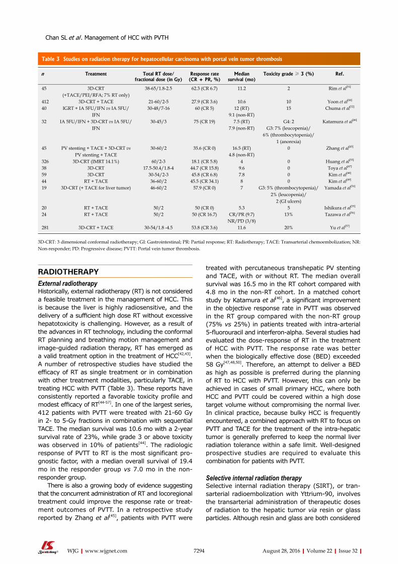
RADIOTHERAPYExternal radiotherapy Historically, external radiotherapy (RT) is not considered a feasible treatment in the management of HCC. This is because the liver is highly radiosensitive, and the delivery of a sufficient high dose RT without excessive hepatotoxicity is challenging. However, as a result of the advances in RT technology, including the conformal RT planning and breathing motion management and image-guided radiation therapy, RT has emerged as a valid treatment option in the treatment of HCC[42,43]. A number of retrospective studies have studied the efficacy of RT as single treatment or in combination with other treatment modalities, particularly TACE, in treating HCC with PVTT (Table 3). These reports have consistently reported a favorable toxicity profile and modest efficacy of RT[44-57]. In one of the largest series, 412 patients with PVTT were treated with 21-60 Gy in 2- to 5-Gy fractions in combination with sequential TACE. The median survival was 10.6 mo with a 2-year survival rate of 23%, while grade 3 or above toxicity was observed in 10% of patients[44]. The radiologic response of PVTT to RT is the most significant pro-gnostic factor, with a median overall survival of 19.4 mo in the responder group vs 7.0 mo in the non-responder group.
There is also a growing body of evidence suggesting that the concurrent administration of RT and locoregional treatment could improve the response rate or treat-ment outcomes of PVTT. In a retrospective study reported by Zhang et al[45], patients with PVTT were
treated with percutaneous transhepatic PV stenting and TACE, with or without RT. The median overall survival was 16.5 mo in the RT cohort compared with 4.8 mo in the non-RT cohort. In a matched cohort study by Katamura et al[46], a significant improvement in the objective response rate in PVTT was observed in the RT group compared with the non-RT group (75% vs 25%) in patients treated with intra-arterial 5-fluorouracil and interferon-alpha. Several studies had evaluated the dose-response of RT in the treatment of HCC with PVTT. The response rate was better when the biologically effective dose (BED) exceeded 58 Gy[47,48,50]. Therefore, an attempt to deliver a BED as high as possible is preferred during the planning of RT to HCC with PVTT. However, this can only be achieved in cases of small primary HCC, where both HCC and PVTT could be covered within a high dose target volume without compromising the normal liver. In clinical practice, because bulky HCC is frequently encountered, a combined approach with RT to focus on PVTT and TACE for the treatment of the intra-hepatic tumor is generally preferred to keep the normal liver radiation tolerance within a safe limit. Well-designed prospective studies are required to evaluate this combination for patients with PVTT.
Selective internal radiation therapy Selective internal radiation therapy (SIRT), or tran-sarterial radioembolization with Yttrium-90, involves the transarterial administration of therapeutic doses of radiation to the hepatic tumor via resin or glass particles. Although resin and glass are both considered
7294 August 28, 2016|Volume 22|Issue 32|WJG|www.wjgnet.com
Table 3 Studies on radiation therapy for hepatocellular carcinoma with portal vein tumor thrombosis
n Treatment Total RT dose/fractional dose (in Gy)
Response rate (CR + PR, %)
Median survival (mo)
Toxicity grade ≥ 3 (%) Ref.
45 3D-CRT (+TACE/PEI/RFA; 7% RT only)
38-65/1.8-2.5 62.3 (CR 6.7) 11.2 2 Rim et al[51]
412 3D-CRT + TACE 21-60/2-5 27.9 (CR 3.6) 10.6 10 Yoon et al[44]
40 IGRT + IA 5FU/IFN vs IA 5FU/IFN
30-48/7-16 60 (CR 5) 12 (RT) 15 Chuma et al[52]
9.1 (non-RT)32 IA 5FU/IFN + 3D-CRT vs IA 5FU/
IFN30-45/3 75 (CR 19) 7.5 (RT) G4: 2 Katamura et al[46]
7.9 (non-RT) G3: 7% (leucopenia)/6% (thrombocytopenia)/
1 (anorexia)45 PV stenting + TACE + 3D-CRT vs
PV stenting + TACE30-60/2 35.6 (CR 0) 16.5 (RT) 0 Zhang et al[45]
4.8 (non-RT)326 3D-CRT (IMRT 14.1%) 60/2-3 18.1 (CR 5.8) 4 0 Huang et al[53]
38 3D-CRT 17.5-50.4/1.8-4 44.7 (CR 15.8) 9.6 0 Toya et al[47]
59 3D-CRT 30-54/2-3 45.8 (CR 6.8) 7.8 0 Kim et al[48]
44 RT + TACE 36-60/2 45.5 (CR 34.1) 8 0 Kim et al[49]
19 3D-CRT (+ TACE for liver tumor) 46-60/2 57.9 (CR 0) 7 G3: 5% (thrombocytopenia)/2% (leucopenia)/
2 (GI ulcers)
Yamada et al[54]
20 RT + TACE 50/2 50 (CR 0) 5.3 5 Ishikura et al[55]
24 RT + TACE 50/2 50 (CR 16.7) CR/PR (9.7) 13% Tazawa et al[56]
NR/PD (3/8)281 3D-CRT + TACE 30-54/1.8 -4.5 53.8 (CR 3.6) 11.6 20% Yu et al[57]
3D-CRT: 3 dimensional conformal radiotherapy; GI: Gastrointestinal; PR: Partial response; RT: Radiotherapy; TACE: Transarterial chemoembolization; NR: Non-responder; PD: Progressive disease; PVTT: Portal vein tumor thrombosis.
Chan SL et al . Management of HCC with PVTH

permanent embolic agents, their embolic effects are less than those of a TACE procedure due to their small size, as is their effect on hepatic vascular dynamics[58]. During radioembolization, a radioactive microsphere is selectively injected into the hepatic artery or its branch, delivering intense local tumor radiation. SIRT provides adequate radiation to the tumor with little radiation to the rest of the liver and to the patient’s body. Because it does not cause ischemia, SIRT can be performed in patients with portal vein thrombosis, making SIRT a feasible choice for HCC with PVTT. Common complications of SIRT include fatigue and deranged liver function[59]. Serious complications such as radiation pneumonitis, radiation cholecystitis, liver abscess and radiation-induced liver disease occur in less than 1% of patients. In general, technetium-labeled macroaggregated albumin scanning is performed prior to SIRT to quantify the fraction of lung shunting and/or the tumor/normal uptake ratio. The accepted safe radiation dose to the lung is less than 30 Gy in a single procedure and less than 50 Gy in total over multiple procedures[59,60].
Studies on SIRT focusing on patients with HCC and PVTT are limited. However, numerous retrospective series have reported the safety and efficacy of SIRT in patients with HCC[61-64]. Subgroup analyses from the three largest series of HCC patients who underwent SIRT involving more than 200 patients with PVTT demonstrated the overall survival to be approximately 10 mo. Ozkan et al[65] reported a better median overall survival of 17 mo among 29 HCC patients treated with SIRT, with and without PVTT. There was no significant difference in survival between patients with and without PVT. A report from the largest group of PVT patients showed concordant results[63], in which it was also found that patients with Child-Pugh A cirrhosis, with or without PVT, benefited the most from SIRT. In a recent prospective phase Ⅱ trial of 35 patients with PVT treated with SIRT, the overall survival of Child-Pugh A and B patients were 16 mo and 6 mo, respectively.
The efficacy of SIRT in unresectable HCC was compared with sorafenib in a recent study. Edeline et al[66] retrospectively reviewed the records of 151 HCC patients with PVTT. The overall survival of 34 patients treated with SIRT was compared with 117 patients treated with sorafenib only. SIRT was associated with a higher median overall survival compared to sorafenib (18.8 mo vs 6.5 mo, P < 0.001). A prospective randomized study comparing SIRT and sorafenib has already completed accrual with preliminary results expected to be available in late 2016 (NCT01135056). At present, there have been no randomized controlled trials directly comparing SIRT to TACE in patients with HCC and PVTT. Several small-scale studies have suggested that the efficacy of SIRT is comparable to TACE in unresectable HCC. Of note, She et al[67] performed survival analysis of 16 patients who underwent SIRT and compared it with another
16 patients in a matched cohort treated with TACE. Half of the patients in each group had major vascular invasion, and those treated with SIRT had an overall survival of 12.0 mo compared to 8.0 mo in the TACE cohort. These data provide preliminary clues that SIRT might be more effective than TACE in the setting of HCC with PVT, but this hypothesis requires further validation in prospective randomized clinical trials.
SYSTEMIC THERAPY Compared to other cancer types, the progress in the development of systemic therapy is slower for HCC. Conventional cytotoxic chemotherapy, such as doxorubicin or doxorubicin-based combinations, could lead to significant toxicity, which limits the potential benefits in cirrhotic populations[68]. Other more “novel” regimens of systemic chemotherapy have also been evaluated. In particular, the combination of oxaliplatin and 5-fluorouracil and leucovorin, known as the FOLFOX4 regimen, has been compared to doxorubicin in patients with advanced HCC in an Asian population with HCC[69]. The overall study fails to demonstrate a statistically significant difference between the two regimens, but FOLFOX4 was found to have small survival benefits in the subgroup population of Chinese patients[70,71]. There have been a handful of case reports or series showing that cytotoxic chemotherapy could improve the severity of PVTT, but there are no dedicated prospective studies to validate such efficacy of systemic chemotherapy in the treatment of PVTT. Further, the toxicity of systemic chemotherapy has been shown to be higher in the presence of impaired liver function and portal hypertension, which frequently occur in patients with PVTT.
Sorafenib is a multi-targeted small molecule with specific activity against vascular endothelial growth factor receptor. Its use in the setting of BCLC stage C HCC, including patients with PVTT, extra-hepatic metastases, and ECOG performance status of 1 or higher, is known to modestly prolong median overall survival by approximately 2 mo, compared to a placebo[72,73]. In both Phase Ⅲ clinical trials, namely the SHARP[72] and the Asian pacific study[73], there are no detailed analyses on the efficacy of sorafenib according to the different severities of PVTT. Nevertheless, the efficacy of sorafenib in PVTT may still be indirectly reflected by the subgroup analyses in patients with macrovascular invasion. In both studies, the proportion of macrovascular invasion in the sorafenib arm was 36%, and subgroup analyses unanimously indicated similar survival benefits between patients with and without macrovascular invasion[72,73]. Regarding the specific treatment for patients with Vp3 and Vp4 PVTT, a retrospective review was published by a Korean center, which analyzed the outcome of sorafenib in 6 patients with Vp3 and 24 patients with Vp4 PVTT[74]. It was shown that 10% (3 patients) had a partial response in the PVTT arm with revascularization, and
7295 August 28, 2016|Volume 22|Issue 32|WJG|www.wjgnet.com
Chan SL et al . Management of HCC with PVTH

the median overall survival was 3.1 mo. The above dataset suggested that sorafenib demonstrated a modest efficacy in the treatment of PVTT, but given the low response rate of likely lower than 10%, the treatment is mainly reserved as a palliative treatment for patients with Vp3 or Vp4 who are not amenable to more aggressive treatment.
Recently, the direction of the development of systemic treatment for HCC has shifted from targets along signaling pathways to immunotherapy and, in particular, to the check-point inhibitors. There have been one phase I clinical trial of a cytotoxic T-lymphocyte-associated protein 4 (CTLA-4) inhibitor, tremelimumab, and another ongoing trial on a programmed cell death protein-1 (PD-1) inhibitor, nivolumab, in patients with advanced HCC[75,76]. Both studies have suggested that check-point inhibitors are tolerable and potentially efficacious in HCC. In particular, some remarkable and durable radiological responses have been observed. Given the more mature and robust data of the immune check-point inhibitor in the treatment of HCC, its use in the treatment of PVTT can be better elucidated. Another direction is biomarker-driven drug development, which aims to improve the response via patient selection with predictive biomarkers in the tissue or plasma[77].
PORTAL VEIN STENTINGTheoretically, the placement of an endovascular stent into the portal vein could increase the portal blood flow into the liver parenchyma in HCC complicated with PVTT. This may, in turn, relieve the portal hypertension-related complications, especially variceal bleeding, and expand treatment options for the tumor. Most of the studies on portal vein stenting in the treatment of PVTT of HCC are case reports or small-scale series. Two large-scale case series suggest that portal vein stenting is feasible in select patients. One series come from a Japanese group that reported the percutaneous placement of stents in the portal vein following the administration of TACE or trans-arterial infusion of chemotherapy in 21 patients[78]. With a 100% success rate of placement, it was demonstrated that the portal pressure decreased by approximately 3 mmHg. A short course of an anticoagulant was administered in 14 patients, but it was not suitable for the others due to coagulopathy. The patency rate was 53.6% at 1 year with a mean patency period of 12.4 mo. Procedure-related complications appeared to be uncommon, with only a case of pseudoaneurysm reported. Another large case series from a French group reviewed the records of 54 patients with PVTT who underwent portal stenting under general anesthesia by a percutaneous approach or an open approach via catheterization of the superior mesenteric vein after exteriorization of the terminal ileum[79]. Twenty percent of patients developed complications after portal stenting, mostly in the form
of hepatic failure, and 7% died within 30 d of stenting. Bilirubin levels > 30 μmol/L and an open surgical approach were predictors of short-term mortality. TACE was administered in 48% of patients after stenting, and the 12-mo survival rate was 44%. Both of these series indicate that the placement of portal stents is a feasible procedure with potential efficacy. However, because of the concern of bleeding complications during the anticoagulation period and the potential risk of tumor dissemination during the stenting procedures, in addition to the complicated procedures involved, portal vein stenting has not gained wide acceptance among HCC experts.
FUTURE PERSPECTIVESThe need for high-level evidence Conventionally, HCC with PVTT is considered to be an advanced disease with clinical management similar to HCC with extra-hepatic metastases. There are no dedicated clinical trials to study the treatment of this population. As a result, the efficacy and safety of different treatment modalities on PVTT are mainly generated from retrospective studies or extrapolated from subgroup analyses of prospective clinical trials. Currently, it is evident that the prognoses and disease behavior of patients with PVTT differ from those of patients without PVTT. In addition, patients with PVTT are more prone to develop complications due to portal hypertension or hepatic failure. Therefore, to define a better treatment strategy for patients with HCC with PVTT, dedicated clinical trials in this population are warranted. Because the prognoses differ between patients with different severities and degrees of PVTT, it is important to accurately gauge the outcome of patients during the design of clinical trials for HCC with PVTT. To achieve this goal, a uniform classification of PVTT is required for the stratification of risk groups during randomization and to facilitate the comparison of results between different studies. At present, at least three classifications have been developed for PVTT in HCC. It is crucial to reach consensus on the classification of PVTT amongst the HCC experts.
Multi-disciplinary approach Management of HCC with PVTT is a clinical dilemma with challenges. On one hand, despite the recom-mendation of sorafenib as the standard treatment for HCC with PVTT by the BCLC guidelines, emerging evidence clearly shows that select patients could benefit from more aggressive treatment approaches. On the other hand, not all patients may uniformly benefit from aggressive treatment. PVTT represents an adverse prognostic factor with an underlying more rapid disease course. Aggressive treatment may not lead to better outcomes in some patients; for example, in the setting of poor performance or with impaired hepatic function. Therefore, when facing patients with HCC with PVTT,
7296 August 28, 2016|Volume 22|Issue 32|WJG|www.wjgnet.com
Chan SL et al . Management of HCC with PVTH

clinicians have to balance the potential benefits and toxicity of different treatments in the individual patients. A multi-disciplinary tumor board is required to determine the most appropriate management of patients with PVTT. Furthermore, the cumulative evidence indicates that a single treatment modality is unlikely to achieve a remarkable effect in PVTT, and the combination of different treatment modalities, such as RT to PVTT and TACE or the addition of SIRT to systemic agents, may be more efficacious in select patients. Future research should focus on combinations of different established treatment modalities for PVTT in HCC.
CONCLUSIONThe management of HCC with PVTT is evolving. The treatment modality for HCC with PVTT includes surgical resection, TACE, radiation therapy including external RT or SIRT of the liver lesions, and systemic agents, while portal vein stent treatment remains investigational. Dedicated clinical studies on HCC complicated with PVTT are inadequate. The decision of the optimal treatment for individual patients requires multi-disciplinary input. Future research should be geared towards the generation of high-level evidence of novel treatments and combinations of established treatments for this population.
REFERENCES1 Roayaie S, Blume IN, Thung SN, Guido M, Fiel MI, Hiotis S,
Labow DM, Llovet JM, Schwartz ME. A system of classifying microvascular invasion to predict outcome after resection in patients with hepatocellular carcinoma. Gastroenterology 2009; 137: 850-855 [PMID: 19524573 DOI: 10.1053/j.gastro.2009.06.003]
2 Quirk M, Kim YH, Saab S, Lee EW. Management of hepatocellular carcinoma with portal vein thrombosis. World J Gastroenterol 2015; 21: 3462-3471 [PMID: 25834310 DOI: 10.3748/wjg.v21.i12.3462]
3 Pirisi M, Avellini C, Fabris C, Scott C, Bardus P, Soardo G, Beltrami CA, Bartoli E. Portal vein thrombosis in hepatocellular carcinoma: age and sex distribution in an autopsy study. J Cancer Res Clin Oncol 1998; 124: 397-400 [PMID: 9719503]
4 Chan SL, Mo FK, Wong CS, Chan CM, Leung LK, Hui EP, Ma BB, Chan AT, Mok TS, Yeo W. A study of circulating interleukin 10 in prognostication of unresectable hepatocellular carcinoma. Cancer 2012; 118: 3984-3992 [PMID: 22180222 DOI: 10.1002/cncr.26726]
5 Llovet JM, Bustamante J, Castells A, Vilana R, Ayuso Mdel C, Sala M, Brú C, Rodés J, Bruix J. Natural history of untreated nonsurgical hepatocellular carcinoma: rationale for the design and evaluation of therapeutic trials. Hepatology 1999; 29: 62-67 [PMID: 9862851]
6 Chan SL, Mo FK, Johnson PJ, Liem GS, Chan TC, Poon MC, Ma BB, Leung TW, Lai PB, Chan AT, Mok TS, Yeo W. Prospective validation of the Chinese University Prognostic Index and comparison with other staging systems for hepatocellular carcinoma in an Asian population. J Gastroenterol Hepatol 2011; 26: 340-347 [PMID: 21261725 DOI: 10.1111/j.1440-1746.2010.06329.x]
7 Connolly GC, Chen R, Hyrien O, Mantry P, Bozorgzadeh A, Abt P, Khorana AA. Incidence, risk factors and consequences of portal vein and systemic thromboses in hepatocellular carcinoma. Thromb Res 2008; 122: 299-306 [PMID: 18045666]
8 Huang J, Deng Q, Wang Q, Li KY, Dai JH, Li N, Zhu ZD, Zhou
B, Liu XY, Liu RF, Fei QL, Chen H, Cai B, Zhou B, Xiao HS, Qin LX, Han ZG. Exome sequencing of hepatitis B virus-associated hepatocellular carcinoma. Nat Genet 2012; 44: 1117-1121 [PMID: 22922871 DOI: 10.1038/ng.2391]
9 Tarantino L, Francica G, Sordelli I, Esposito F, Giorgio A, Sorrentino P, de Stefano G, Di Sarno A, Ferraioli G, Sperlongano P. Diagnosis of benign and malignant portal vein thrombosis in cirrhotic patients with hepatocellular carcinoma: color Doppler US, contrast-enhanced US, and fine-needle biopsy. Abdom Imaging 2006; 31: 537-544 [PMID: 16865315 DOI: 10.1007/s00261-005-0150-x]
10 Piscaglia F, Gianstefani A, Ravaioli M, Golfieri R, Cappelli A, Giampalma E, Sagrini E, Imbriaco G, Pinna AD, Bolondi L. Criteria for diagnosing benign portal vein thrombosis in the assessment of patients with cirrhosis and hepatocellular carcinoma for liver transplantation. Liver Transpl 2010; 16: 658-667 [PMID: 20440775 DOI: 10.1002/lt.22044]
11 Rammohan A, Jeswanth S, Sukumar R, Anand L, Kumar PS, Srinivasan UP, Ravi R, Ravichandran P. Percutaneous ultrasound-guided fine-needle aspiration of portal vein thrombi as a diagnostic and staging technique for hepatocellular carcinoma. Abdom Imaging 2013; 38: 1057-1060 [PMID: 23579929 DOI: 10.1007/s00261-013-9997-4]
12 Rossi S, Ghittoni G, Ravetta V, Torello Viera F, Rosa L, Serassi M, Scabini M, Vercelli A, Tinelli C, Dal Bello B, Burns PN, Calliada F. Contrast-enhanced ultrasonography and spiral computed tomography in the detection and characterization of portal vein thrombosis complicating hepatocellular carcinoma. Eur Radiol 2008; 18: 1749-1756 [PMID: 18369630 DOI: 10.1007/s00330-008-0931-z]
13 Kim JH, Lee JM, Yoon JH, Lee DH, Lee KB, Han JK, Choi BI. Portal Vein Thrombosis in Patients with Hepatocellular Carcinoma: Diagnostic Accuracy of Gadoxetic Acid-enhanced MR Imaging. Radiology 2016; 279: 773-783 [PMID: 26780538 DOI: 10.1148/radiol.2015150124]
14 Llovet JM, Brú C, Bruix J. Prognosis of hepatocellular carcinoma: the BCLC staging classification. Semin Liver Dis 1999; 19: 329-338 [PMID: 10518312]
15 Kudo M, Kitano M, Sakurai T, Nishida N. General Rules for the Clinical and Pathological Study of Primary Liver Cancer, Nationwide Follow-Up Survey and Clinical Practice Guidelines: The Outstanding Achievements of the Liver Cancer Study Group of Japan. Dig Dis 2015; 33: 765-770 [PMID: 26488173 DOI: 10.1159/000439101]
16 Kokudo N, Hasegawa K, Akahane M, Igaki H, Izumi N, Ichida T, Uemoto S, Kaneko S, Kawasaki S, Ku Y, Kudo M, Kubo S, Takayama T, Tateishi R, Fukuda T, Matsui O, Matsuyama Y, Murakami T, Arii S, Okazaki M, Makuuchi M. Evidence-based Clinical Practice Guidelines for Hepatocellular Carcinoma: The Japan Society of Hepatology 2013 update (3rd JSH-HCC Guidelines). Hepatol Res 2015; 45 [PMID: 25625806 DOI: 10.1111/hepr.12464]
17 Shi J, Lai EC, Li N, Guo WX, Xue J, Lau WY, Wu MC, Cheng SQ. A new classification for hepatocellular carcinoma with portal vein tumor thrombus. J Hepatobiliary Pancreat Sci 2011; 18: 74-80 [PMID: 20686792 DOI: 10.1007/s00534-010-0314-0]
18 Xu JF, Liu XY, Wang S, Wen HX. Surgical treatment for hepatocellular carcinoma with portal vein tumor thrombus: a novel classification. World J Surg Oncol 2015; 13: 86 [PMID: 25889711 DOI: 10.1186/s12957-015-0493-x]
19 Kumada K, Ozawa K, Okamoto R, Takayasu T, Yamaguchi M, Yamamoto Y, Higashiyama H, Morikawa S, Sasaki H, Shimahara Y. Hepatic resection for advanced hepatocellular carcinoma with removal of portal vein tumor thrombi. Surgery 1990; 108: 821-827 [PMID: 2173162]
20 Tanaka A, Morimoto T, Yamaoka Y. Implications of surgical treatment for advanced hepatocellular carcinoma with tumor thrombi in the portal vein. Hepatogastroenterology 1996; 43: 637-643 [PMID: 8799408]
21 Chok KS, Cheung TT, Chan SC, Poon RT, Fan ST, Lo CM.
7297 August 28, 2016|Volume 22|Issue 32|WJG|www.wjgnet.com
Chan SL et al . Management of HCC with PVTH

Surgical outcomes in hepatocellular carcinoma patients with portal vein tumor thrombosis. World J Surg 2014; 38: 490-496 [PMID: 24132826 DOI: 10.1007/s00268-013-2290-4]
22 Wu CC, Hsieh SR, Chen JT, Ho WL, Lin MC, Yeh DC, Liu TJ, P’eng FK. An appraisal of liver and portal vein resection for hepatocellular carcinoma with tumor thrombi extending to portal bifurcation. Arch Surg 2000; 135: 1273-1279 [PMID: 11074879]
23 Ban D, Shimada K, Yamamoto Y, Nara S, Esaki M, Sakamoto Y, Kosuge T. Efficacy of a hepatectomy and a tumor thrombectomy for hepatocellular carcinoma with tumor thrombus extending to the main portal vein. J Gastrointest Surg 2009; 13: 1921-1928 [PMID: 19727969 DOI: 10.1007/s11605-009-0998-0]
24 Chan SC, Liu CL, Lo CM, Lam CM, Poon RT, Yuen WK, Fan ST, Wong J. Value of live donor liver transplantation experience in major hepatectomy for hepatocellular carcinoma. Arch Surg 2003; 138: 265-271 [PMID: 12611572]
25 Peng ZW, Guo RP, Zhang YJ, Lin XJ, Chen MS, Lau WY. Hepatic resection versus transcatheter arterial chemoembolization for the treatment of hepatocellular carcinoma with portal vein tumor thrombus. Cancer 2012; 118: 4725-4736 [PMID: 22359112 DOI: 10.1002/cncr.26561]
26 Ohkubo T, Yamamoto J, Sugawara Y, Shimada K, Yamasaki S, Makuuchi M, Kosuge T. Surgical results for hepatocellular carcinoma with macroscopic portal vein tumor thrombosis. J Am Coll Surg 2000; 191: 657-660 [PMID: 11129815]
27 Konishi M, Ryu M, Kinoshita T, Inoue K. Surgical treatment of hepatocellular carcinoma with direct removal of the tumor thrombus in the main portal vein. Hepatogastroenterology 2001; 48: 1421-1424 [PMID: 11677978]
28 Pawlik TM, Poon RT, Abdalla EK, Ikai I, Nagorney DM, Belghiti J, Kianmanesh R, Ng IO, Curley SA, Yamaoka Y, Lauwers GY, Vauthey JN. Hepatectomy for hepatocellular carcinoma with major portal or hepatic vein invasion: results of a multicenter study. Surgery 2005; 137: 403-410 [PMID: 15800485]
29 Ikai I, Hatano E, Hasegawa S, Fujii H, Taura K, Uyama N, Shimahara Y. Prognostic index for patients with hepatocellular carcinoma combined with tumor thrombosis in the major portal vein. J Am Coll Surg 2006; 202: 431-438 [PMID: 16500247]
30 Le Treut YP, Hardwigsen J, Ananian P, Saïsse J, Grégoire E, Richa H, Campan P. Resection of hepatocellular carcinoma with tumor thrombus in the major vasculature. A European case-control series. J Gastrointest Surg 2006; 10: 855-862 [PMID: 16769542]
31 Liu CL, Fan ST. Nonresectional therapies for hepatocellular carcinoma. Am J Surg 1997; 173: 358-365 [PMID: 9136797]
32 Ngan H, Lai CL, Fan ST, Lai EC, Yuen WK, Tso WK. Treatment of inoperable hepatocellular carcinoma by transcatheter arterial chemoembolization using an emulsion of cisplatin in iodized oil and gelfoam. Clin Radiol 1993; 47: 315-320 [PMID: 8389682 DOI: 10.1016/S0009-9260(05)81446-1]
33 O’Suilleabhain CB, Poon RT, Yong JL, Ooi GC, Tso WK, Fan ST. Factors predictive of 5-year survival after transarterial chemoembolization for inoperable hepatocellular carcinoma. Br J Surg 2003; 90: 325-331 [PMID: 12594668]
34 Yu SC, Hui JW, Hui EP, Chan SL, Lee KF, Mo F, Wong J, Ma B, Lai P, Mok T, Yeo W. Unresectable hepatocellular carcinoma: randomized controlled trial of transarterial ethanol ablation versus transcatheter arterial chemoembolization. Radiology 2014; 270: 607-620 [PMID: 24126369 DOI: 10.1148/radiol.13130498]
35 Llovet JM, Bruix J. Systematic review of randomized trials for unresectable hepatocellular carcinoma: Chemoembolization improves survival. Hepatology 2003; 37: 429-442 [PMID: 12540794 DOI: 10.1053/jhep.2003.50047]
36 Bruix J, Sala M, Llovet JM. Chemoembolization for hepatocellular carcinoma. Gastroenterology 2004; 127: S179-S188 [PMID: 15508083]
37 Lo CM, Ngan H, Tso WK, Liu CL, Lam CM, Poon RT, Fan ST, Wong J. Randomized controlled trial of transarterial lipiodol chemoembolization for unresectable hepatocellular carcinoma. Hepatology 2002; 35: 1164-1171 [PMID: 11981766]
38 Minagawa M, Makuuchi M. Treatment of hepatocellular
carcinoma accompanied by portal vein tumor thrombus. World J Gastroenterol 2006; 12: 7561-7567 [PMID: 17171782 DOI: 10.3748/WJG.v12.i47.7561]
39 Yamada R, Sato M, Kawabata M, Nakatsuka H, Nakamura K, Takashima S. Hepatic artery embolization in 120 patients with unresectable hepatoma. Radiology 1983; 148: 397-401 [PMID: 6306721]
40 Leng JJ, Xu YZ, Dong JH. Efficacy of transarterial chemoem-bolization for hepatocellular carcinoma with portal vein thrombosis: a meta-analysis. ANZ J Surg 2014; Epub ahead of print [PMID: 25088384 DOI: 10.1111/ans.12803]
41 Xue TC , Xie XY, Zhang L, Yin X, Zhang BH, Ren ZG. Transarterial chemoembolization for hepatocellular carcinoma with portal vein tumor thrombus: a meta-analysis. BMC Gastroenterol 2013; 13: 60 [PMID: 23566041 DOI: 10.1186/1471-230x-13-60]
42 Klein J, Dawson LA. Hepatocellular carcinoma radiation therapy: review of evidence and future opportunities. Int J Radiat Oncol Biol Phys 2013; 87: 22-32 [PMID: 23219567 DOI: 10.1016/j.ijrobp.2012.08.043]
43 Dawson LA. Overview: Where does radiation therapy fit in the spectrum of liver cancer local-regional therapies? Semin Radiat Oncol 2011; 21: 241-246 [PMID: 21939852 DOI: 10.1016/j.semradonc.2011.05.009]
44 Yoon SM, Lim YS, Won HJ, Kim JH, Kim KM, Lee HC, Chung YH, Lee YS, Lee SG, Park JH, Suh DJ. Radiotherapy plus transarterial chemoembolization for hepatocellular carcinoma invading the portal vein: long-term patient outcomes. Int J Radiat Oncol Biol Phys 2012; 82: 2004-2011 [PMID: 21621346 DOI: 10.1016/j.ijrobp.2011.03.019]
45 Zhang XB , Wang JH, Yan ZP, Qian S, Du SS, Zeng ZC. Hepatocellular carcinoma with main portal vein tumor thrombus: treatment with 3-dimensional conformal radiotherapy after portal vein stenting and transarterial chemoembolization. Cancer 2009; 115: 1245-1252 [PMID: 19156918 DOI: 10.1002/cncr.24139]
46 Katamura Y, Aikata H, Takaki S, Azakami T, Kawaoka T, Waki K, Hiramatsu A, Kawakami Y, Takahashi S, Kenjo M, Toyota N, Ito K, Chayama K. Intra-arterial 5-fluorouracil/interferon combination therapy for advanced hepatocellular carcinoma with or without three-dimensional conformal radiotherapy for portal vein tumor thrombosis. J Gastroenterol 2009; 44: 492-502 [PMID: 19330281 DOI: 10.1007/s00535-009-0033-y]
47 Toya R, Murakami R, Baba Y, Nishimura R, Morishita S, Ikeda O, Kawanaka K, Beppu T, Sugiyama S, Sakamoto T, Yamashita Y, Oya N. Conformal radiation therapy for portal vein tumor thrombosis of hepatocellular carcinoma. Radiother Oncol 2007; 84: 266-271 [PMID: 17716760 DOI: 10.1016/j.radonc.2007.07.005]
48 Kim DY, Park W, Lim DH, Lee JH, Yoo BC, Paik SW, Kho KC, Kim TH, Ahn YC, Huh SJ. Three-dimensional conformal radiotherapy for portal vein thrombosis of hepatocellular carcinoma. Cancer 2005; 103: 2419-2426 [PMID: 15822130 DOI: 10.1002/cncr.21043]
49 Kim TH, Kim DY, Park JW, Kim YI, Kim SH, Park HS, Lee WJ, Park SJ, Hong EK, Kim CM. Three-dimensional conformal radiotherapy of unresectable hepatocellular carcinoma patients for whom transcatheter arterial chemoembolization was ineffective or unsuitable. Am J Clin Oncol 2006; 29: 568-575 [PMID: 17148993 DOI: 10.1097/01.coc.0000239147.60196.11]
50 Lee JH , Kim DH, Ki YK, Nam JH, Heo J, Woo HY, Kim DW, Kim WT. Three-dimensional conformal radiotherapy for portal vein tumor thrombosis alone in advanced hepatocellular carcinoma. Radiat Oncol J 2014; 32: 170-178 [PMID: 25324989 DOI: 10.3857/roj.2014.32.3.170]
51 Rim CH, Yang DS, Park YJ, Yoon WS, Lee JA, Kim CY. Effecti-veness of high-dose three-dimensional conformal radiotherapy in hepatocellular carcinoma with portal vein thrombosis. Jpn J Clin Oncol 2012; 42: 721-729 [PMID: 22689916 DOI: 10.1093/jjco/hys082]
52 Chuma M, Taguchi H, Yamamoto Y, Shimizu S, Nakanishi M, Ogawa K, Sho T, Horimoto H, Kobayashi T, Nakai M, Terashita K, Sakuhara Y, Abo D, Tsukuda Y, Tsunematsu S,
7298 August 28, 2016|Volume 22|Issue 32|WJG|www.wjgnet.com
Chan SL et al . Management of HCC with PVTH

Hige S, Kato M, Shirato H, Asaka M. Efficacy of therapy for advanced hepatocellular carcinoma: intra-arterial 5-fluorouracil and subcutaneous interferon with image-guided radiation. J Gastroenterol Hepatol 2011; 26: 1123-1132 [PMID: 21501224 DOI: 10.1111/j.1440-1746.2011.06745.x]
53 Huang YJ, Hsu HC, Wang CY, Wang CJ, Chen HC, Huang EY, Fang FM, Lu SN. The treatment responses in cases of radiation therapy to portal vein thrombosis in advanced hepatocellular carcinoma. Int J Radiat Oncol Biol Phys 2009; 73: 1155-1163 [PMID: 18760547 DOI: 10.1016/j.ijrobp.2008.06.1486]
54 Yamada K, Izaki K, Sugimoto K, Mayahara H, Morita Y, Yoden E, Matsumoto S, Soejima T, Sugimura K. Prospective trial of combined transcatheter arterial chemoembolization and three-dimensional conformal radiotherapy for portal vein tumor thrombus in patients with unresectable hepatocellular carcinoma. Int J Radiat Oncol Biol Phys 2003; 57: 113-119 [PMID: 12909223]
55 Ishikura S, Ogino T, Furuse J, Satake M, Baba S, Kawashima M, Nihei K, Ito Y, Maru Y, Ikeda H. Radiotherapy after transcatheter arterial chemoembolization for patients with hepatocellular carcinoma and portal vein tumor thrombus. Am J Clin Oncol 2002; 25: 189-193 [PMID: 11943901]
56 Tazawa J , Maeda M, Sakai Y, Yamane M, Ohbayashi H, Kakinuma S, Miyasaka Y, Nagayama K, Enomoto N, Sato C. Radiation therapy in combination with transcatheter arterial chemoembolization for hepatocellular carcinoma with extensive portal vein involvement. J Gastroenterol Hepatol 2001; 16: 660-665 [PMID: 11422619]
57 Yu JI, Park HC, Lim DH, Park W, Yoo BC, Paik SW, Koh KC, Lee JH. Prognostic index for portal vein tumor thrombosis in patients with hepatocellular carcinoma treated with radiation therapy. J Korean Med Sci 2011; 26: 1014-1022 [PMID: 21860551 DOI: 10.3346/jkms.2011.26.8.1014]
58 Sato K, Lewandowski RJ, Bui JT, Omary R, Hunter RD, Kulik L, Mulcahy M, Liu D, Chrisman H, Resnick S, Nemcek AA, Vogelzang R, Salem R. Treatment of unresectable primary and metastatic liver cancer with yttrium-90 microspheres (TheraSphere): assessment of hepatic arterial embolization. Cardiovasc Intervent Radiol 2006; 29: 522-529 [PMID: 16729228 DOI: 10.1007/s00270-005-0171-4]
59 Coldwell D , Sangro B, Wasan H, Salem R, Kennedy A. General selection criteria of patients for radioembolization of liver tumors: an international working group report. Am J Clin Oncol 2011; 34: 337-341 [PMID: 20921882 DOI: 10.1097/COC.0b013e3181ec61bb]
60 Kennedy A, Nag S, Salem R, Murthy R, McEwan AJ, Nutting C, Benson A, Espat J, Bilbao JI, Sharma RA, Thomas JP, Coldwell D. Recommendations for radioembolization of hepatic malignancies using yttrium-90 microsphere brachytherapy: a consensus panel report from the radioembolization brachytherapy oncology consortium. Int J Radiat Oncol Biol Phys 2007; 68: 13-23 [PMID: 17448867 DOI: 10.1016/j.ijrobp.2006.11.060]
61 D’Avola D, Lñarrairaegui M, Bilbao JI, Martinez-Cuesta A, Alegre F, Herrero JI, Quiroga J, Prieto J, Sangro B. A retrospective comparative analysis of the effect of Y90-radioembolization on the survival of patients with unresectable hepatocellular carcinoma. Hepatogastroenterology 2009; 56: 1683-1688 [PMID: 20214218]
62 Hilgard P, Hamami M, Fouly AE, Scherag A, Müller S, Ertle J, Heusner T, Cicinnati VR, Paul A, Bockisch A, Gerken G, Antoch G. Radioembolization with yttrium-90 glass microspheres in hepatocellular carcinoma: European experience on safety and long-term survival. Hepatology 2010; 52: 1741-1749 [PMID: 21038413 DOI: 10.1002/hep.23944]
63 Salem R, Lewandowski RJ, Mulcahy MF, Riaz A, Ryu RK, Ibrahim S, Atassi B, Baker T, Gates V, Miller FH, Sato KT, Wang E, Gupta R, Benson AB, Newman SB, Omary RA, Abecassis M, Kulik L. Radioembolization for hepatocellular carcinoma using Yttrium-90 microspheres: a comprehensive report of long-term outcomes. Gastroenterology 2010; 138: 52-64 [PMID: 19766639 DOI: 10.1053/j.gastro.2009.09.006]
64 Sangro B, Carpanese L, Cianni R, Golfieri R, Gasparini D,
Ezziddin S, Paprottka PM, Fiore F, Van Buskirk M, Bilbao JI, Ettorre GM, Salvatori R, Giampalma E, Geatti O, Wilhelm K, Hoffmann RT, Izzo F, Iñarrairaegui M, Maini CL, Urigo C, Cappelli A, Vit A, Ahmadzadehfar H, Jakobs TF, Lastoria S. Survival after yttrium-90 resin microsphere radioembolization of hepatocellular carcinoma across Barcelona clinic liver cancer stages: a European evaluation. Hepatology 2011; 54: 868-878 [PMID: 21618574 DOI: 10.1002/hep.24451]
65 Ozkan ZG, Poyanli A, Ucar A, Kuyumcu S, Akyuz F, Keskin S, Saglam S, Yilmaz E, Karaca C, Turkmen C. Favorable survival time provided with radioembolization in hepatocellular carcinoma patients with and without portal vein thrombosis. Cancer Biother Radiopharm 2015; 30: 132-138 [PMID: 25760644 DOI: 10.1089/cbr.2014.1748]
66 Edeline J, Crouzet L, Campillo-Gimenez B, Rolland Y, Pracht M, Guillygomarc’h A, Boudjema K, Lenoir L, Adhoute X, Rohou T, Boucher E, Clément B, Blanc JF, Garin E. Selective internal radiation therapy compared with sorafenib for hepatocellular carcinoma with portal vein thrombosis. Eur J Nucl Med Mol Imaging 2016; 43: 635-643 [PMID: 26455499 DOI: 10.1007/s00259-015-3210-7]
67 She WH, Cheung TT, Yau TC, Chan AC, Chok KS, Chu FS, Liu RK, Poon RT, Chan SC, Fan ST, Lo CM. Survival analysis of transarterial radioembolization with yttrium-90 for hepatocellular carcinoma patients with HBV infection. Hepatobiliary Surg Nutr 2014; 3: 185-193 [PMID: 25202695 DOI: 10.3978/j.issn.2304-3881.2014.07.09]
68 Chan SL, Yeo W. Targeted therapy of hepatocellular carcinoma: present and future. J Gastroenterol Hepatol 2012; 27: 862-872 [PMID: 22369685 DOI: 10.1111/j.1440-1746.2012.07096.x]
69 Qin S, Bai Y, Lim HY, Thongprasert S, Chao Y, Fan J, Yang TS, Bhudhisawasdi V, Kang WK, Zhou Y, Lee JH, Sun Y. Randomized, multicenter, open-label study of oxaliplatin plus fluorouracil/leucovorin versus doxorubicin as palliative chemotherapy in patients with advanced hepatocellular carcinoma from Asia. J Clin Oncol 2013; 31: 3501-3508 [PMID: 23980077 DOI: 10.1200/JCO.2012.44.5643]
70 Qin S, Cheng Y, Liang J, Shen L, Bai Y, Li J, Fan J, Liang L, Zhang Y, Wu G, Rau KM, Yang TS, Jian Z, Liang H, Sun Y. Efficacy and safety of the FOLFOX4 regimen versus doxorubicin in Chinese patients with advanced hepatocellular carcinoma: a subgroup analysis of the EACH study. Oncologist 2014; 19: 1169-1178 [PMID: 25223462 DOI: 10.1634/theoncologist.2014-0190]
71 Chan SL. Drug development for hepatocellular carcinoma: knowing the past helps to understand the future. Oncologist 2014; 19: 1115-1117 [PMID: 25223461 DOI: 10.1634/theoncologist.2014-0304]
72 Llovet JM, Ricci S, Mazzaferro V, Hilgard P, Gane E, Blanc JF, de Oliveira AC, Santoro A, Raoul JL, Forner A, Schwartz M, Porta C, Zeuzem S, Bolondi L, Greten TF, Galle PR, Seitz JF, Borbath I, Häussinger D, Giannaris T, Shan M, Moscovici M, Voliotis D, Bruix J. Sorafenib in advanced hepatocellular carcinoma. N Engl J Med 2008; 359: 378-390 [PMID: 18650514]
73 Cheng AL, Kang YK, Chen Z, Tsao CJ, Qin S, Kim JS, Luo R, Feng J, Ye S, Yang TS, Xu J, Sun Y, Liang H, Liu J, Wang J, Tak WY, Pan H, Burock K, Zou J, Voliotis D, Guan Z. Efficacy and safety of sorafenib in patients in the Asia-Pacific region with advanced hepatocellular carcinoma: a phase III randomised, double-blind, placebo-controlled trial. Lancet Oncol 2009; 10: 25-34 [PMID: 19095497 DOI: 10.1016/S1470-2045(08)70285-7]
74 Jeong SW, Jang JY, Shim KY, Lee SH, Kim SG, Cha SW, Kim YS, Cho YD, Kim HS, Kim BS, Kim KH, Kim JH. Practical effect of sorafenib monotherapy on advanced hepatocellular carcinoma and portal vein tumor thrombosis. Gut Liver 2013; 7: 696-703 [PMID: 24312711 DOI: 10.5009/gnl.2013.7.6.696]
75 Sangro B, Gomez-Martin C, de la Mata M, Iñarrairaegui M, Garralda E, Barrera P, Riezu-Boj JI, Larrea E, Alfaro C, Sarobe P, Lasarte JJ, Pérez-Gracia JL, Melero I, Prieto J. A clinical trial of CTLA-4 blockade with tremelimumab in patients with hepatocellular carcinoma and chronic hepatitis C. J Hepatol 2013; 59: 81-88 [PMID: 23466307 DOI: 10.1016/j.jhep.2013.02.022]
7299 August 28, 2016|Volume 22|Issue 32|WJG|www.wjgnet.com
Chan SL et al . Management of HCC with PVTH

76 El-Khoueiry AB, Melero I, Crocenzi TS, Welling TH, Yau TC, Yeo W, Chopra A, Grosso J, Lang L, Anderson J, Dela Cruz CM, Sangro B. Phase I/II safety and antitumor activity of nivolumab in patients with advanced hepatocellular carcinoma (HCC): CA209-040. J Clin Oncol 2015; 33 Suppl: abstr LBA101
77 Chan SL, Wong AM, Lee K, Wong N, Chan AK. Personalized therapy for hepatocellular carcinoma: Where are we now? Cancer Treat Rev 2016; 45: 77-86 [PMID: 26995632 DOI: 10.1016/j.ctrv.2016.02.008]
78 Yamakado K, Tanaka N, Nakatsuka A, Matsumura K, Takase K, Takeda K. Clinical efficacy of portal vein stent placement in patients with hepatocellular carcinoma invading the main portal vein. J Hepatol 1999; 30: 660-668 [PMID: 10207808]
79 Vibert E, Azoulay D, Cunha AS, Adam R, Samuel D, Castaing D. Portal stenting for hepatocellular carcinoma extending into the portal vein in cirrhotic patients. J Surg Oncol 2013; 107: 696-701 [PMID: 23280593 DOI: 10.1002/jso.23306]
80 Ikai I, Kudo M, Arii S, Omata M, Kojiro M, Sakamoto M, Takayasu K, Hayashi N, Makuuchi M, Matsuyama Y, Monden
M. Report of the 18th follow-up survey of primary liver cancer in Japan. Hepatol Res 2010; 40: 1043-1059 [DOI: 10.1111/j.1872-034X.2010.00731.x]
81 Luo J, Guo RP, Lai EC, Zhang YJ, Lau WY, Chen MS, Shi M. Transarterial chemoembolization for unresectable hepatocellular carcinoma with portal vein tumor thrombosis: a prospective comparative study. Ann Surg Oncol 2011; 18: 413-420 [PMID: 20839057 DOI: 10.1245/s10434-010-1321-8]
82 Niu ZJ, Ma YL, Kang P, Ou SQ, Meng ZB, Li ZK, Qi F, Zhao C. Transarterial chemoembolization compared with conservative treatment for advanced hepatocellular carcinoma with portal vein tumor thrombus: using a new classification. Med Oncol 2012; 29: 2992-2997 [PMID: 22200992 DOI: 10.1007/s12032-011-0145-0]
83 Kim JH, Yoon HK, Kim SY, Kim KM, Ko GY, Gwon DI, Sung KB. Transcatheter arterial chemoembolization vs. chemoinfusion for unresectable hepatocellular carcinoma in patients with major portal vein thrombosis. Aliment Pharmacol Ther 2009; 29: 1291-1298 [PMID: 19392861 DOI: 10.1111/j.1365-2036.2009.04016.x]
P- Reviewer: Cardinale V, Tchilikidi KY, Wei W, Zhang Q S- Editor: Yu J L- Editor: A E- Editor: Ma S
7300 August 28, 2016|Volume 22|Issue 32|WJG|www.wjgnet.com
Chan SL et al . Management of HCC with PVTH

Pancreatic cancer: open or minimally invasive surgery?
Yu-Hua Zhang, Cheng-Wu Zhang, Zhi-Ming Hu, De-Fei Hong
Yu-Hua Zhang, Cheng-Wu Zhang, Zhi-Ming Hu, De-Fei Hong, Division of Hepatobiliary and Pancreatic Surgery and Minimally Invasive Surgery, Zhejiang Provincial People’s Hospital, Hangzhou 310014, Zhejiang Province, China
Author contributions: Zhang YH and Hong DF designed the study; Zhang YH, Zhang CW, Hu ZM and Hong DF reviewed and analyzed the literature, and drafted and revised the manuscript; All authors approved the final version of the manuscript to be published.
Supported by the Natural Science Foundation of Zhejiang Province, China, No. LY15H160054; and the Key Project of the Science and Technology Department of Zhejiang Province, No. 2013C03046.
Conflict-of-interest statement: The authors declare that there is no potential conflict of interest related to this study.
Open-Access: This article is an open-access article which was selected by an in-house editor and fully peer-reviewed by external reviewers. It is distributed in accordance with the Creative Commons Attribution Non Commercial (CC BY-NC 4.0) license, which permits others to distribute, remix, adapt, build upon this work non-commercially, and license their derivative works on different terms, provided the original work is properly cited and the use is non-commercial. See: http://creativecommons.org/licenses/by-nc/4.0/
Manuscript source: Invited manuscript
Correspondence to: De-Fei Hong, Professor, Division of Hepatobiliary and Pancreatic Surgery and Minimally Invasive Surgery, Zhejiang Provincial People’s Hospital, 158 Shangtang Road, Hangzhou 310014, Zhejiang Province, China. [email protected]: +86-571-85893396
Received: March 25, 2016 Peer-review started: March 26, 2016First decision: April 14, 2016Revised: May 4, 2016 Accepted: May 23, 2016 Article in press: May 23, 2016Published online: August 28, 2016
REVIEW
Submit a Manuscript: http://www.wjgnet.com/esps/Help Desk: http://www.wjgnet.com/esps/helpdesk.aspxDOI: 10.3748/wjg.v22.i32.7301
World J Gastroenterol 2016 August 28; 22(32): 7301-7310 ISSN 1007-9327 (print) ISSN 2219-2840 (online)
© 2016 Baishideng Publishing Group Inc. All rights reserved.
7301 August 28, 2016|Volume 22|Issue 32|WJG|www.wjgnet.com
AbstractPancreatic duct adenocarcinoma is one of the most fatal malignancies, with R0 resection remaining the most important part of treatment of this malignancy. However, pancreatectomy is believed to be one of the most challenging procedures and R0 resection remains the only chance for patients with pancreatic cancer to have a good prognosis. Some surgeons have tried minimally invasive pancreatic surgery, but the short- and long-term outcomes of pancreatic malignancy remain controversial between open and minimally invasive procedures. We collected comparative data about minimally invasive and open pancreatic surgery. The available evidence suggests that minimally invasive pancreaticoduodenectomy (MIPD) is as safe and feasible as open PD (OPD), and shows some benefit, such as less intraoperative blood loss and shorter postoperative hospital stay. Despite the limited evidence for MIPD in pancreatic cancer, most of the available data show that the short-term oncological adequacy is similar between MIPD and OPD. Some surgical techniques, including superior mesenteric artery-first approach and laparoscopic pancreatoduodenectomy with major vein resection, are believed to improve the rate of R0 resection. Laparoscopic distal pancrea-tectomy is less technically demanding and is accepted in more pancreatic centers. It is technically safe and feasible and has similar short-term oncological prognosis compared with open distal pancreatectomy.
Key words: Laparoscopic; Minimally invasive; Robotic; Pancreaticoduodenectomy; Distal pancreatectomy; Pancreatic cancer
© The Author(s) 2016. Published by Baishideng Publishing Group Inc. All rights reserved.
Core tip: Minimally invasive pancreaticoduodenec-tomy is as safe and feasible as open pancreaticoduo-denectomy (OPD) and shows some superiority. The

results, the authors concluded that the minimally invasive approach was not advocated because there was no apparent advantage over traditional open approaches. After that, surgeons spent a decade improving their laparoscopic skills, until a large LPD cohort was reported in France in 2005[12] and then in India in 2009[15]. During 1994-2009, several surgeons tried to apply hybrid, laparoscopic-open approaches to avoid the complexity of a purely laparoscopic pro-cedure[13,19]. Although these approaches may overcome some of the limitations, they may reduce the potential benefits of purely laparoscopic approaches, including less pain, improved postoperative recovery and shorter hospital stay. After Palanivelu et al[15] reported 75 cases of LPD in 2009, large cohorts of LPD have been reported in the United States[11,20,21], South Korea[16], China[22], Italy[23] and France[24]. LPD eventually gained momentum, following its 30 years’ development, and it has emerged as a well-established procedure with acceptable morbidity and mortality rates in some specialized high-volume pancreatic centers[12,15,16,20,22,23]. Although LPD has been accepted in many specialized minimally invasive pancreatic centers, the short- and long-term results remain controversial. We collected clinical reports with comparative data between mini-mally invasive PD (MIPD) and open PD (OPD) (Table 1).
Safety and feasibility of LPD PD is a complex procedure because of the dissection around important vessels and three complex recon-structions. Moreover, it is a procedure with high morbidity[25]. Although LPD has been accepted in some specialized centers, it is still a challenging operation for most pancreatic surgeons; still, there has been a rapid increase in the number of LPDs performed in different centers. Some large-volume centers have published their comparative studies between LPD and OPD[11,15,16,20,21,24,26-31], demonstrating the safety of LPD; although, long-term oncological benefits of this approach remain debatable.
Croome et al[20] reviewed their data for patients with pancreatic ductal adenocarcinoma (PDAC) undergoing LPD (n = 108) and OPD (n = 214). A significantly reduced blood loss and blood transfusion requirement and a shorter postoperative stay (6 d vs 9 d) were observed in the LPD group compared with the OPD group.
A case match study was performed by Dokmak et al[24] comparing 46 LPD and OPD procedures. Patients were matched for demographic data, associated comor-bidity and underlying disease. The results suggested that a high rate of severe morbidity due to severe pancreatic fistula was detected in patients with a high risk of pancreatic fistula. In a subgroup of patients with a low risk of pancreatic fistula, the outcome of the two approaches was similar. The result of this study suggested that, in a subgroup of patients with a high risk of pancreatic fistula, LPD was associated with high
Zhang YH et al . Minimally invasive pancreatectomy
7302 August 28, 2016|Volume 22|Issue 32|WJG|www.wjgnet.com
short-term oncological results are similar between laparoscopic pancreaticoduodenectomy (LPD) and OPD. However, in some experienced hands, better prognosis is detected in the LPD group because the patients can receive adjuvant therapy faster because of the benefits of minimal invasiveness. Minimally invasive distal pancreatectomy is a well-established procedure and widely accepted. It is safe, feasible, and has similar short-term oncological results compared with open distal pancreatectomy.
Zhang YH, Zhang CW, Hu ZM, Hong DF. Pancreatic cancer: Open or minimally invasive surgery? World J Gastroenterol 2016; 22(32): 7301-7310 Available from: URL: http://www.wjgnet.com/1007-9327/full/v22/i32/7301.htm DOI: http://dx.doi.org/10.3748/wjg.v22.i32.7301
INTRODUCTIONPancreatic cancer ranks as the 4th highest cause of cancer-related death in the United States and the 5-year survival is about 6%[1]. Surgical R0 resection is the best chance for a cure and remains the cornerstone of treatment of pancreatic malignancy[2,3]. However, pancreatic surgery is believed to be one of the most challenging procedures because of the high risks of postoperative morbidity and mortality associated with intraoperative bleeding and postoperative compli-cations including pancreatic fistula[2,4,5]. Another key point for surgical treatment of pancreatic malignancy is oncological adequacy. R0 resection is the best chance for patients to have a good prognosis[3,6].
Minimally invasive techniques, including laparoscopic and robotic approaches, have rapidly evolved and include a variety of abdominal surgical procedures[7-10]. They provide the patients with better short-term outcomes, including smaller incisions, shorter hospital stay and less blood loss. Some surgeons in large-volume pancreatic centers have tried minimally invasive pancreatic surgery[11-16]. However, the short- and long-term outcomes of pancreatic malignancy remain controversial, especially for oncological prognosis.
Many pancreatic surgeons doubt the safety and oncological adequacy of minimally invasive pancreatic surgery. Here, we collected and analyzed the published data about minimally invasive pancreatic surgery.
LAPAROSCOPIC PANCREATICODUODENECTOMYBackground of laparoscopic pancreaticoduodenectomyFollowing the first report of laparoscopic pancreati-coduodenectomy (LPD) in 1994[17], Gagner and Pomp[18] subsequently published a series of 10 patients in 1997. In Gagner’s series, the conversion rate was 40% and the operating time was 8.5 h. Dependent upon these

mor
bidi
ty.
Thus
, it
shou
ld b
e co
nsid
ered
onl
y in
pat
ient
s w
ith a
dila
ted
panc
reat
ic d
uct
and
a ha
rd p
ancr
eas
text
ure,
who
are
bel
ieve
d to
hav
e a
low
ris
k of
pan
crea
tic
fistu
la.
Ada
m e
t al
[32] r
evie
wed
pat
ient
s un
derg
oing
PD
fro
m t
he N
atio
nal Can
cer
Dat
abas
e be
twee
n 20
10 a
nd 2
011,
inc
ludi
ng 9
83 M
IPD
s an
d 60
78 L
PDs.
The
ir r
esul
ts
sugg
este
d th
at, fo
r pa
tient
s w
ith P
DAC, no
diff
eren
ce w
as d
etec
ted
in n
umbe
r of
lym
ph n
odes
(LN
s) r
emov
ed, ra
te o
f R0
rese
ctio
n, le
ngth
of ho
spita
l sta
y or
rea
dmission
. How
ever
, th
e 30
-d m
orta
lity
was
low
er in
the
OPD
gro
up tha
n in
the
MIP
D g
roup
. Th
e au
thor
s su
gges
ted
that
the
wid
espr
ead
adop
tion
of the
tec
hniq
ue s
houl
d be
pau
sed.
M
IPD
is a
com
plex
pro
cedu
re tha
t ne
eds
com
preh
ensive
pro
toco
ls o
utlin
ing
crite
ria for
impl
emen
tatio
n.Asb
un a
nd S
tauf
fer[1
1] p
rese
nted
ret
rosp
ectiv
e cl
inic
al d
ata
from
May
o Clin
ic o
f 21
5 O
PD a
nd 5
3 LP
D p
atie
nts.
The
y al
so s
how
ed s
igni
fican
tly b
ette
r re
sults
in
LPD
7303 August 28, 2016|Volume 22|Issue 32|WJG|www.wjgnet.com
Tab
le 1
Sa
fety
and
fea
sibi
lity
of l
apar
osco
pic
panc
reat
icod
uode
nect
omy:
Clin
ical
coh
orts
of
min
imal
ly i
nvas
ive
panc
reat
icod
uode
nect
omy
and
open
pan
crea
tico
duod
enec
tom
y in
clud
ing
com
para
tive
res
ults
Ref
.Yea
rC
ount
ryTe
chni
que
Cas
esO
pera
ting
tim
e, m
inEB
L, m
LLH
S, D
CD
≥ Ⅲ
PFD
GE
Rea
dmission
rat
eM
orta
lity
Shar
p et
al[2
1]20
15U
nite
d St
ates
LPD
3
84N
RN
RN
RN
RN
RN
R5.
0%5.
2% (3
0 D
)O
PD 4
037
NR
NR
NR
NR
NR
NR
9.0%
3.7%
(30
D)
Song
et a
l[16]
2015
Sout
hKor
eaLP
PPD
93 4
82.5
± 1
17.6
609
± 37
514
.3 ±
7.8
7.5
%6
(6.5
) 3
.2%
5 (5
.4)
NR
OPP
PD
93
347.
9 ±
87.2
570
± 44
819
.2 ±
8.8
5.4
%6
(6.5
) 7
.5%
3 (3
.2)
NR
Che
n et
al[3
4]20
15C
hina
RPD
60 4
10 ±
103
400
(200
-600
)
20 ±
7.4
11.7
%13
.3%
8.3
%N
R1.
7%O
PD
120
323
± 80
500
(350
-800
)
25
± 11
.213
.3%
24.9
%15
.0%
NR
2.5%
Dok
mak
et a
l[24]
2015
Fran
ceLP
D
46
342
(240
-540
)36
8 (5
0-12
00)
25
(6-1
04)
28.0
%48
.0%
17.0
% 9
.0%
2.0%
OPD
4626
4 (1
20-4
00)
293
(50-
1200
) 2
3 (7
-115
)20
.0%
41.0
%15
.0%
9.0
%0
Bake
r et a
l[31]
2015
Uni
ted
Stat
esRP
D
22
454
(294
-529
)42
5 (5
0-22
00)
7 (4
-25)
13.6
% 4
.6%
13.6
%22
.7%
0O
PD
49
364
(213
-948
) 6
50 (1
50-6
100)
9 (5
-48)
20.4
%12
.2%
30.6
%29
.8%
4.1%
Tran
et a
l[74]
2015
Uni
ted
Stat
esLP
D
681
NR
NR
12 (9
-20)
NR
NR
NR
NR
3.8%
OPD
1489
3N
RN
R11
(8-1
6)N
RN
RN
RN
R5.
0%Ta
n et
al[7
5]20
15C
hina
LPD
3051
3.17
± 5
6.13
NR
9.9
7 ±
3.74
NR
10/3
02/
30N
R0
OPD
3037
1.67
± 8
5.53
NR
11.8
7 ±
4.72
NR
6/3
03/
30N
R1/
30A
dam
et a
l[32]
2015
Uni
ted
Stat
esM
IPD
9
83N
RN
RN
RN
RN
RN
R 4
.8%
NR
OPD
607
8N
RN
RN
RN
RN
RN
R 3
.7%
NR
Cha
likon
da et
al[3
3]20
14U
nite
d St
ates
HPD
3047
6.00
485
9.7
930
.0%
NR
NR
NR
4.0%
OPD
3036
6.48
775
13.2
643
.0%
NR
NR
NR
0Ba
o et
al[7
6]20
14U
nite
d St
ates
RPD
2843
1 (3
40-6
28)
100
(50-
300)
7.4
(5.5
-17.
1)N
R29
.0%
NR
25.0
%7.
0% (9
0 D
)O
PD
28
410
(190
-621
)30
0 (1
00-8
00)
8.1
(6.5
-15.
3)N
R29
.0%
NR
25.0
%7.
0% (9
0 D
)C
room
e et
al[2
0]20
14U
nite
d St
ates
LPD
1
0837
9.4
± 93
.549
2.4
± 51
9.3
6 (4
-118
) 5
.6%
(≥ Ⅲ
b)11
% (B
/C)
9% (B
/C)
NR
1.0%
(I H
)O
PD
214
387.
6 ±
91.8
866.
7 ±
733.
79
(5-7
3)13
.6%
(≥ Ⅲ
b)12
% (B
/C)
18%
(B/C
)N
R2.
0% (I
H)
Spei
cher
et a
l[77]
2014
Uni
ted
Stat
esLP
D
25
381
(342
-465
)20
0 (1
00-4
25)
8.5
(7-1
1.2)
NR
16%
(B/C
)N
R30
.4%
0 (3
0 D
)H
PD
31
442
(386
.5-4
86.5
)60
0 (3
12.5
-700
)
12
(8.5
-18.
5)N
R35
.5%
NR
35.5
%3.
2%(3
0 D
)O
PD
84
425.
5 (3
45.8
-478
.8)
425
(300
-700
)10
(8-1
4)N
R22
.6%
NR
39.3
%1.
2% (3
0 D
)A
sbun
et a
l[11]
2012
Uni
ted
Stat
esLP
D
53
541
± 88
195
± 13
6 8
.0 ±
3.2
NR
16.7
%11
.3%
NR
5.7%
(100
D)
OPD
2
15 4
01 ±
108
1032
± 1
151
12.4
± 8
.5N
R17
.3%
15.3
%N
R8.
8% (1
00 D
)La
i et a
l[28]
2012
Chi
naRP
D
20
491.
5 ±
94.0
247
(50-
889)
13.7
± 6
.1N
R35
.0%
5%N
R0%
OPD
6726
4.9
± 63
.777
4.8
(50-
8000
) 2
5.8
± 23
.1N
R17
.9%
11.9
%N
R3%
CD
: Cla
vien
-Din
do; D
GE:
Del
ayed
gas
tric
em
ptyi
ng; E
BL: E
stim
ated
blo
od lo
ss; H
PD: H
ybri
d pa
ncre
atic
oduo
dene
ctom
y; L
HS:
Len
gth
of h
ospi
tal s
tay;
LPD
: Lap
aros
copi
c pa
ncre
atic
oduo
dene
ctom
y; L
PPPD
: Lap
aros
copi
c py
loru
s-pr
eser
ving
pan
crea
ticod
uode
nect
omy;
MIP
D: M
inim
ally
inva
sive
pan
crea
ticod
uode
nect
omy;
NR:
Not
rep
orte
d; O
PD: O
pen
panc
reat
icod
uode
nect
omy;
OPP
PD: O
pen
pylo
rus-
pres
ervi
ng p
ancr
eatic
oduo
dene
ctom
y; P
F:
Panc
reat
ic fi
stul
a; R
PD: R
obot
ic p
ancr
eatic
oduo
dene
ctom
y.
Zhang YH et al . Minimally invasive pancreatectomy

resection is required for a good prognosis. Many clinical studies have reported LPD; however, most of those studies have included a variety of diseases requiring LPD. To the best of our knowledge, few studies have compared the oncological prognosis of PDAC treated with LPD or OPD (Table 2).
Song et al[16] compared the oncological results of pancreatic cancer treated with OPPPD (n = 261) and LPPPD (n = 11). TMN stage, R0 resection rate, in-hospital stay and the overall survival were similar between the two groups. In a case-control study from France[24], the results for LPD (n = 15) in patients with PDAC were similar to those with OPD (n = 14) with regard to tumor size, number of LNs harvested and rate of R0 resection. Croome et al[20] reported a large single center study of pancreatic carcinoma treated with LPD. Clinical data of 108 cases of LPD were reviewed retrospectively and compared with 214 cases of OPD performed in the same period at their center. The short-term oncological results, including tumor size, LN positivity, R0 resection and overall survival, were similar between the two groups, and significantly longer progression-free survival was found in the LPD group. The authors thought that this difference might have been because the patients who underwent LPD had the advantage of minimal invasiveness and recovered faster from the operation. This allowed the patients to receive adjuvant therapy in a timely manner and probably led to better prognosis.
A large LPD cohort study[21] from the National Cancer Data Base involved 384 LPDs and 4039 OPDs. The results showed no difference between the LPD and OPD groups with regard to length of stay, margin-positive resection, LN count and readmission rate.
Chen et al[34] compared the oncological results of pancreatic cancer treated with RPD (n = 19) and OPD (n = 38). There was no difference in the R0 resection rate, number of LNs resected, cancer stage, overall survival and disease-free survival between the two groups.
All the results above show that in most of the experienced minimally invasive pancreatic centers, LPD has similar short-term oncological results as OPD. However, Croome et al[20] reported the long-term prognostic benefit in the LPD group because of the advantages of minimal invasiveness. Furthermore, Croome et al[20] presented the largest cohort with pancreatic cancer treated with LPD, thus, we can probably form the hypothesis that, if surgeons acquire enough experience of LPD, LPD can yield the benefits of minimal invasiveness as well as long-term oncological benefit, compared with OPD. To obtain oncological adequacy, some technical tips are suggested for application during the operation.
Surgical technique to improve rate of R0 resection Superior mesenteric artery-first approach: To improve the long-term prognosis of patients with
groups, such as less blood loss (P < 0.001) and blood transfusion requirements (P < 0.001), and a shorter postoperative hospital stay (P < 0.001). While a significantly longer operating time was observed in LPD (P < 0.001), the LPD patients had a greater number of LNs removed than the OPD patients (P = 0.007). This series also demonstrated that LPD is safe and feasible and showed some benefits for patients.
Results from another cohort with PDAC in the United States treated with LPD were presented at the Western Surgical Association 122nd Scientific Session[21]. The researchers compared 4037 OPDs with 384 LPDs and showed significant differences favoring LPD for length of hospital stay and unplanned readmission. A lower risk of 30-d mortality was found in high-volume centers and in centers with experience of performing more than 10 LPDs; moreover, the 30-d mortality for LPD was similar to that for OPD. Finally, the researchers demonstrated that there is a learning curve for LPD.
Song et al[16] compared 137 laparoscopic pylorus-preserving PDs (LPPPDs) with 2055 open PPPDs (OPPPDs) in South Korea. They found that operating time was longer for LPPPD than for OPPPD, and the perioperative complications were similar in both groups. Fewer analgesic injections were administrated in the LPPPD group (P < 0.001). The oncological results were similar between the two groups, including number of LNs removed and long-term survival.
In addition to LPD, a few studies have compared robotic PD (RPD) with OPD. Chalikonda et al[33] from the Cleveland Clinic reviewed the results of 30 matched laparoscopic RPD (LRPD) and OPD procedures. LRPD and OPD groups were matched with demographics. A similar estimated blood loss and rate of reoperation were found in the two groups. However, there was a significant increase in operating time and shorter hospital stay for the LRPD group.
We found that most of the clinical studies showed that LPD is as safe and feasible as OPD technically, and has some of the superiority associated with minimally invasive surgery, such as less estimated blood loss and shorter hospital stay. However, some authors have suggested that MIPD should be advocated in a subgroup of patients with lower risk of pancreatic fistula. In our opinion, LPD is as safe as OPD. However, due to the complexity of LPD, it is a technically demanding procedure with a learning curve. In small clinical cohorts of LPD at the beginning of the learning curve, there might be higher morbidity and mortality for LPD than for OPD. The problem now is how to reduce the risks of LPD at the beginning of the learning curve. Apart from technical feasibility, the major arguments against LPD are oncological adequacy, especially for patients with PDAC.
Oncological adequacy of LPD for pancreatic malignancyPancreatic cancer still has a high fatality rate. Radical
7304 August 28, 2016|Volume 22|Issue 32|WJG|www.wjgnet.com
Zhang YH et al . Minimally invasive pancreatectomy

PDAC, curative (R0) resection is required initially. Many reports have discussed the value of R0 resection in prognosis of PDAC. The consensus among pancreatic surgeons is that positive surgical margins are asso-ciated with poor survival[35-38]. The primary site of positive margins is from the right side of the superior mesenteric artery (SMA) (N14) to the right side of the celiac trunk (N9), including the mesopancreas[39]. To improve R0 resection, the SMA-first approach was advocated in OPD. The artery-first approach has been proven as effective in reducing the risk of bleeding and improving the rate of R0 resection in pancreatic cancer.
However, few publications have reported the SMA-first approach in LPD. To the best of our knowledge, only two publications have described laparoscopic SMA-first approaches[40,41]. Pittau et al[40] reported the right posterior approach. The authors performed this procedure exactly like the Pessaux procedure in OPD[42], and they dissected the SMA after complete kocherization, including mobilization of the right colon. Cho et al[41] described the left posterior SMA-first approach. They dissected the SMA at the ligament of
Treitz without mobilization of the duodenum or right colon[43]. In our center, we perform the right posterior SMA-first approach, as described by Pittau et al[40]. We expose the SMA from the right side after complete kocherization (Figure 1). After exposure of the SMA, it would be easy to decide the resectability of the tumor. Another benefit is that this approach makes resection of the uncinate process from the SMA easier, and warrants complete removal of the neuro laminar tissue at the right side of the SMA up to the celiac axis (Figure 2).
Major vein resection: Involvement of the portal vein in locally advanced tumor is no longer a contradiction for surgical resection of pancreatic malignancy using traditional open procedures. A lot of data from larger pancreatic centers have provided evidence indicating that en bloc resection of tumor with involved vessels is safe and feasible, and can improve the rate of R0 resection[44-51]. Patients who have en bloc resection with the involved vein have similar long-term oncolo-gical prognosis compared with patients who do not have vascular involvement[44,45,48-50].
7305 August 28, 2016|Volume 22|Issue 32|WJG|www.wjgnet.com
Table 2 Oncological results of pancreatic cancer in minimally invasive pancreaticoduodenectomy and open pancreaticoduodenectomy: Clinical trials including comparative results of pancreatic ductal adenocarcinoma between minimally invasive pancreaticoduodenectomy and open pancreaticoduodenectomy
Ref. Year Country Technique No. of PDAC cases Rate of R0 resection No. of LN Positive LN Tumor size, cm
Sharp et al[21] 2015 United States LPD 384 80.0% 18 ± 9.7 NR 3.2 ± 1.3OPD 4037 74.0% 16 ± 9.6 NR 3.3 ± 2.4
Song et al[16] 2015 South Korea LPPPD 11 72.7% 15 ± 10 0.8 ± 1.2 2.8 ± 0.6OPPPD 261 81.0% 16.2 ± 9.6 1.5 ± 2.2 3.0 ± 1.2
Dokmak et al[24] 2015 France LPD 15 60.0% 20 (8-59) 4.7 (0-32) 2.4 (1.5-4)OPD 14 50.0% 25 (8-47) 2.2 (0-12) 2.8 (2.5-4)
Chen et al[34] 2015 China RPD 19 94.7% 18.1 ± 6.6 NR 3.0 ± 0.9OPD 38 92.1% 17.8 ± 7.1 NR 3.1 ± 1.0
Croome et al[20] 2014 United States LPD 108 77.8% 21.4 ± 8.1 73.1% 3.3 ± 1.0OPD 214 76.6% 20.1 ± 7.5 72.0% 3.3 ± 1.3
LN: Lymph node; LPD: Laparoscopic pancreaticoduodenectomy; LPPPD: Laparoscopic pylorus-preserving pancreaticoduodenectomy; MIPD: Minimally invasive pancreaticoduodenectomy; OPD: Open pancreaticoduodenectomy; OPPPD: Open pylorus-preserving pancreaticoduodenectomy; PDAC: Pancreatic duct adenocarcinoma; NR: Not reported.
PUP
SMA
LRV
IVC D
IVCSMA
SMV
SPV
PV
CT
AORTA
Figure 1 Superior mesentery artery was exposed from the right posterior side after complete kocherization. D: Duodenum; IVC: Inferior vena cava; LRV: Left renal vein; PUP: Pancreatic uncinate process; SMA: Superior mesentery artery.
Figure 2 Local vision after removal of the specimen. CT: Celiac trunk; IVC: Inferior vena cava; PV: Portal vein; SMA: Superior mesenteric artery; SMV: Superior mesenteric vein; SPV: Splenic vein.
Zhang YH et al . Minimally invasive pancreatectomy

Kendrick et al[52] reported the first example of LPD with vein resection. Later in that same year, Giulianotti et al[53] published data of RPD with major vein resection. Kendrick et al[52] reported 11 patients who underwent laparoscopic pancreatectomy with major venous resection. In their series, one segmental resection and 10 tangential venous resections were described. Giulianotti et al[53] described three cases of robot-assisted distal pancreatectomy (DP) with vascular resection (two cases of celiac truck resection and one of portal vein resection) and two cases of RPD with portal vein resection. These initial results show that, for surgeons with considerable experience of minimally invasive pancreatic surgery, major vein resection during pancreatectomy is a safe and feasible adjunctive procedure. Kendrick et al[52] consequently reported a series of LPD with major vein resection. Thirty-one patients who underwent LPD with vascular resection were compared with 58 patients who underwent OPD with major vessel resection. The LPD group had decreased blood loss and shorter length of hospital stay, but there was no difference between LPD and OPD with regard to severe complications, mortality or overall survival. The authors concluded that LPD with vein resection is safe and feasible, and can achieve similar outcomes compared to patients undergoing OPD with vein resection. Most of the minimally invasive pancreatic centers considered LPD with vein resection a contraindication. However, for further application of LPD in patients with pancreatic malignancy with vein involvement, it is necessary for surgeons to master the minimally invasive technique of vein resection and reconstruction.
In our center, we have performed five MIPD proce-dures with major vein resection: four patients under-went LPD with tangential venous resection, and one underwent robotic vein segment resection (Figure 3). In our limited experience of LPD with major vein resection, tangential venous resections can be performed safely laparoscopically. However, for segmentectomy of the major vein, a robotic system is advocated. The first RPD was performed by Giulianotti et al[54] in 2001.
RPD has since been proven to be feasible and safe, with the minimally invasive advantages compared with open procedures[30,31,33,34,55,56]. It is believed that the robotic surgical system provides surgeons with enhanced dexterity, superior magnified high-resolution 3D visualization, and greater precision and ergonomic comfort. This approach enables surgeons to control the surgical instruments with accuracy, flexibility and a wide range of motion, which is suggested for procedures that require complicated resection and reconstruction, such as prostatectomy, coronary surgery and PD. In our opinion, the application of robotic systems in PD with major vein resection can improve the quality of vein reconstruction, and we advocate them if possible.
LAPAROSCOPIC DPBackground DP is widely accepted as an option for PDAC located in the distal pancreas. However, in past decades, laparoscopic DP (LDP) has been accepted increasingly with evidence of minimally invasive benefits. Com-pared with LPD, LDP is less technically demanding because there is limited dissection around the vessels and no reconstruction is required[57,58]. So, more surgeons accept LDP than LPD.
Safety and feasibility of LDPA recently published meta-analysis[59-63] indicated that LDP was a safe and feasible option in terms of operating time and postoperative mortality and morbidities, such as postoperative bleeding and pancreatic fistula. Moreover, minimally invasive superiority was found in LDP, including significantly decreased estimated blood loss, time to first oral intake and length of hospital stay[59-63]. These results clearly show that LDP is as safe and feasible as ODP.
Short-term oncological resultsMicroscopically, R0 resection is the most important part of treatment of resectable pancreatic cancer. Some non-comparative cohorts have shown that R0 resection of pancreatic cancer can be achieved by laparoscopic resection[64,65]. Most of the comparative studies have shown that there is no difference in the rate of R0 resection in the final pathological results between LDP and ODP[58,66,67]. To the best of our knowledge, only DiNorcia et al[68] have reported a decrease in R1 resection in the laparoscopic group; however, their series had mixed pathology, including neuroendocrine tumor and pancreatic adenocarcinoma. Another important short-term oncological marker is LN retrieval. A minimum of 12 LNs is required for resection of pancreatic adenocarcinoma[69,70]. N0 patients with > 12 LNs have better survival than N0 patients with < 12 LNs (P < 0.001)[70]. Most studies have found that the number of LNs harvested in laparoscopic and open procedures is similar[58,66-68,71].
7306 August 28, 2016|Volume 22|Issue 32|WJG|www.wjgnet.com
SMV
PV
Figure 3 Vein reconstruction via robotic system. PV: Portal vein; SMV: Superior mesenteric vein.
Zhang YH et al . Minimally invasive pancreatectomy

The data here demonstrate that most of the minimally invasive pancreatic surgeons have a consensus that LDP has the same short-term oncological results as ODP.
Long-term oncological outcomes of LDP Only a few studies have described long-term prognosis after LDP, and few comparative data are reported. Mabrut et al[64] reported 16 patients with pancreatic malignancy, 4 of whom had pancreatic adenocarcinoma, and 23% of these patients had recurrence during 15 mo. Fernández-Cruz et al[72] reported 10 cases of laparoscopic radical antegrade modular pancreatosple-nectomy (RAMPS), with 3 having died within a year and a median survival period of 14 mo. Rehman et al[67] found a similar 3-year overall survival between 8 LDP and 14 ODP procedures for PDAC. Kooby et al[58] reported similar median survival (16 mo) after LDP and ODP in a matched study. Kim et al[73] reported 11 LDPs with diagnosis of malignancy in their postoperative pathological results, including 5 cases of PDAC; only 1 patient died of cancer during the follow-up period (3-60 mo). The results to date suggest that the long-term prognosis of LDP for adenocarcinoma is similar to that for open procedures. It was also found that there was no difference in short-term oncological markers, including tumor size, radiological stage, margin-negative resection, power of LN retrieval and LN metastasis between the two groups. The authors concluded that LDP is acceptable for patients with pancreatic malignancy. However, further larger studies are required to give solid evidence of the long-term oncological benefit of LDP.
CONCLUSIONAfter initial reports of LPD and LDP in the 1990s, laparoscopic pancreatectomy finally became a well-established procedure following 30 years’ development of laparoscopic skills and equipment. The data here suggest that minimally invasive pancreatectomy is safe and feasible and has adequate evidence of good short-term outcome. However, randomized controlled trials and long-term oncological results are still lacking. The long-term oncological results should be further addressed by randomized controlled trials. Another problem now is how to generalize this procedure from experienced hands to other centers.
ACKNOWLEDGMENTSWe wish to thank Tang Wei and Song Peipei from Tokyo University for their kind help and critical review of the manuscript.
REFERENCES1 Siegel RL, Miller KD, Jemal A. Cancer statistics, 2016. CA
Cancer J Clin 2016; 66: 7-30 [PMID: 26742998 DOI: 10.3322/
caac.21332]2 Gao JJ, Song PP, Tamura S, Hasegawa K, Sugawara Y, Kokudo
N, Uchida K, Orii R, Qi FH, Dong JH, Tang W. Standardization of perioperative management on hepato-biliary-pancreatic surgery. Drug Discov Ther 2012; 6: 108-111 [PMID: 22622021]
3 Hidalgo M . Pancreatic cancer. N Engl J Med 2010; 362: 1605-1617 [PMID: 20427809 DOI: 10.1056/NEJMra0901557]
4 Shimoda M. Upon completing the 7th Sino-Japanese Symposium on Hepato-Pancreato-Biliary Disease. Biosci Trends 2008; 2: 96 [PMID: 20103909]
5 Yamashita S, Sakamoto Y, Kaneko J, Tamura S, Aoki T, Sugawara Y, Hasegawa K, Kokudo N. Resection of the second portion of the duodenum sacrificing the minor papilla but preserving the pancreas for a recurrent duodenal adenocarcinoma: report of a case. Biosci Trends 2012; 6: 44-47 [PMID: 22426103 DOI: 10.5582/bst.2012.v6.1.44]
6 Flattet Y, Yamaguchi T, Andrejevic-Blant S, Halkic N. Pancreatic adenocarcinoma: the impact of preneoplastic lesion pattern on survival. Biosci Trends 2015; 9: 402-406 [PMID: 26781798 DOI: 10.5582/bst.2015.01163]
7 Hong D, Cheng J, Wang Z, Shen G, Xie Z, Wu W, Zhang Y, Zhang Y, Liu X. Comparison of two laparoscopic splenectomy plus pericardial devascularization techniques for management of portal hypertension and hypersplenism. Surg Endosc 2015; 29: 3819-3826 [PMID: 25783835 DOI: 10.1007/s00464-015-4147-4]
8 Hong D, Liu Y, Peng S, Sun X, Wang Z, Cheng J, Shen G, Zhang Y, Huang D. Binding pancreaticogastrostomy in laparoscopic central pancreatectomy: a novel technique in laparoscopic pancreatic surgery. Surg Endosc 2016; 30: 715-720 [PMID: 26123326 DOI: 10.1007/s00464-015-4265-z]
9 Hong DF, Gao M, Bryner U, Cai XJ, Mou YP. Intraoperative endoscopic sphincterotomy for common bile duct stones during laparoscopic cholecystectomy. World J Gastroenterol 2000; 6: 448-450 [PMID: 11819626]
10 Hong DF, Xin Y, Chen DW. Comparison of laparoscopic cholecystectomy combined with intraoperative endoscopic sphincterotomy and laparoscopic exploration of the common bile duct for cholecystocholedocholithiasis. Surg Endosc 2006; 20: 424-427 [PMID: 16395539 DOI: 10.1007/s00464-004-8248-8]
11 Asbun HJ, Stauffer JA. Laparoscopic vs open pancreati-coduodenectomy: overall outcomes and severity of complications using the Accordion Severity Grading System. J Am Coll Surg 2012; 215: 810-819 [PMID: 22999327 DOI: 10.1016/j.jamcollsurg.2012.08.006]
12 Dulucq JL , Win t r i nge r P, Maha jna A . Lapa roscop ic pancreaticoduodenectomy for benign and malignant diseases. Surg Endosc 2006; 20: 1045-1050 [PMID: 16736311 DOI: 10.1007/s00464-005-0474-1]
13 Kimura Y, Hirata K, Mukaiya M, Mizuguchi T, Koito K, Katsuramaki T. Hand-assisted laparoscopic pylorus-preserving pancreaticoduodenectomy for pancreas head disease. Am J Surg 2005; 189: 734-737 [PMID: 15910728 DOI: 10.1016/j.amjsurg.2005.03.017]
14 Lee SH, Kang CM, Hwang HK, Choi SH, Lee WJ, Chi HS. Minimally invasive RAMPS in well-selected left-sided pancreatic cancer within Yonsei criteria: long-term (>median 3 years) oncologic outcomes. Surg Endosc 2014; 28: 2848-2855 [PMID: 24853839 DOI: 10.1007/s00464-014-3537-3]
15 Palanivelu C, Rajan PS, Rangarajan M, Vaithiswaran V, Senthilnathan P, Parthasarathi R, Praveen Raj P. Evolution in techniques of laparoscopic pancreaticoduodenectomy: a decade long experience from a tertiary center. J Hepatobiliary Pancreat Surg 2009; 16: 731-740 [PMID: 19652900 DOI: 10.1007/s00534-009-0157-8]
16 Song KB, Kim SC, Hwang DW, Lee JH, Lee DJ, Lee JW, Park KM, Lee YJ. Matched Case-Control Analysis Comparing Laparo-scopic and Open Pylorus-preserving Pancreaticoduodenectomy in Patients With Periampullary Tumors. Ann Surg 2015; 262: 146-155 [PMID: 25563866 DOI: 10.1097/SLA.0000000000001079]
17 Gagner M, Pomp A. Laparoscopic pylorus-preserving pancreato-
7307 August 28, 2016|Volume 22|Issue 32|WJG|www.wjgnet.com
Zhang YH et al . Minimally invasive pancreatectomy

duodenectomy. Surg Endosc 1994; 8: 408-410 [PMID: 7915434]18 Gagner M, Pomp A. Laparoscopic pancreatic resection: Is it
worthwhile? J Gastrointest Surg 1997; 1: 20-25; discussion 25-26 [PMID: 9834326]
19 Ammori BJ. Laparoscopic hand-assisted pancreaticoduo-denectomy: initial UK experience. Surg Endosc 2004; 18: 717-718 [PMID: 15214369]
20 Croome KP , Farnell MB, Que FG, Reid-Lombardo KM, Truty MJ, Nagorney DM, Kendrick ML. Total laparoscopic pancreaticoduodenectomy for pancreatic ductal adenocarcinoma: oncologic advantages over open approaches? Ann Surg 2014; 260: 633-638; discussion 638-640 [PMID: 25203880 DOI: 10.1097/SLA.0000000000000937]
21 Sharpe SM, Talamonti MS, Wang CE, Prinz RA, Roggin KK, Bentrem DJ, Winchester DJ, Marsh RD, Stocker SJ, Baker MS. Early National Experience with Laparoscopic Pancreatico-duodenectomy for Ductal Adenocarcinoma: A Comparison of Laparoscopic Pancreaticoduodenectomy and Open Pancreatico-duodenectomy from the National Cancer Data Base. J Am Coll Surg 2015; 221: 175-184 [PMID: 26095569 DOI: 10.1016/j.jamcollsurg.2015.04.021]
22 Wang M, Zhang H, Wu Z, Zhang Z, Peng B. Laparoscopic pancreaticoduodenectomy: single-surgeon experience. Surg Endosc 2015; 29: 3783-3794 [PMID: 25783837 DOI: 10.1007/s00464-015-4154-5]
23 Corcione F, Pirozzi F, Cuccurullo D, Piccolboni D, Caracino V, Galante F, Cusano D, Sciuto A. Laparoscopic pancreatico-duodenectomy: experience of 22 cases. Surg Endosc 2013; 27: 2131-2136 [PMID: 23355144 DOI: 10.1007/s00464-012-2728-z]
24 Dokmak S, Ftériche FS, Aussilhou B, Bensafta Y, Lévy P, Ruszniewski P, Belghiti J, Sauvanet A. Laparoscopic pancreatico-duodenectomy should not be routine for resection of periampullary tumors. J Am Coll Surg 2015; 220: 831-838 [PMID: 25840531 DOI: 10.1016/j.jamcollsurg.2014.12.052]
25 Cameron JL, Riall TS, Coleman J, Belcher KA. One thousand consecutive pancreaticoduodenectomies. Ann Surg 2006; 244: 10-15 [PMID: 16794383 DOI: 10.1097/01.sla.0000217673.04165.ea]
26 Wei H, Wei B, Zheng Z, Huang Y, Huang J, Fang J. Comparative study of outcomes after laparoscopic versus open pancreatico-duodenectomy. Zhonghua Weichang Waike Zazhi 2014; 17: 465-468 [PMID: 24859956]
27 Langan RC, Graham JA, Chin AB, Rubinstein AJ, Oza K, Nusbaum JA, Smirniotopoulos J, Kayser R, Jha R, Haddad N, Al-Kawas F, Carroll J, Hanna J, Parker A, Al-Refaie WB, Johnson LB. Laparoscopic-assisted versus open pancreaticoduodenectomy: early favorable physical quality-of-life measures. Surgery 2014; 156: 379-384 [PMID: 24680859 DOI: 10.1016/j.surg.2014.03.018]
28 Lai EC, Yang GP, Tang CN. Robot-assisted laparoscopic pancreaticoduodenectomy versus open pancreaticoduodenectomy--a comparative study. Int J Surg 2012; 10: 475-479 [PMID: 22732431 DOI: 10.1016/j.ijsu.2012.06.003]
29 Hakeem AR, Verbeke CS, Cairns A, Aldouri A, Smith AM, Menon KV. A matched-pair analysis of laparoscopic versus open pancreaticoduodenectomy: oncological outcomes using Leeds Pathology Protocol. Hepatobiliary Pancreat Dis Int 2014; 13: 435-441 [PMID: 25100130]
30 Buchs NC, Addeo P, Bianco FM, Ayloo S, Benedetti E, Giulianotti PC. Robotic versus open pancreaticoduodenectomy: a comparative study at a single institution. World J Surg 2011; 35: 2739-2746 [PMID: 21947494 DOI: 10.1007/s00268-011-1276-3]
31 Baker EH , Ross SW, Seshadri R, Swan RZ, Iannitti DA, Vrochides D, Martinie JB. Robotic pancreaticoduodenectomy: comparison of complications and cost to the open approach. Int J Med Robot 2015; Epub ahead of print [PMID: 26202591 DOI: 10.1002/rcs.1688]
32 Adam MA, Choudhury K, Dinan MA, Reed SD, Scheri RP, Blazer DG, Roman SA, Sosa JA. Minimally Invasive Versus Open Pancreaticoduodenectomy for Cancer: Practice Patterns and Short-term Outcomes Among 7061 Patients. Ann Surg 2015; 262: 372-377
[PMID: 26158612 DOI: 10.1097/SLA.0000000000001055]33 Chalikonda S, Aguilar-Saavedra JR, Walsh RM. Laparoscopic
robotic-assisted pancreaticoduodenectomy: a case-matched comparison with open resection. Surg Endosc 2012; 26: 2397-2402 [PMID: 22437947 DOI: 10.1007/s00464-012-2207-6]
34 Chen S, Chen JZ, Zhan Q, Deng XX, Shen BY, Peng CH, Li HW. Robot-assisted laparoscopic versus open pancreaticoduodenectomy: a prospective, matched, mid-term follow-up study. Surg Endosc 2015; 29: 3698-3711 [PMID: 25761559 DOI: 10.1007/s00464-015-4140-y]
35 Konstantinidis IT, Warshaw AL, Allen JN, Blaszkowsky LS, Castillo CF, Deshpande V, Hong TS, Kwak EL, Lauwers GY, Ryan DP, Wargo JA, Lillemoe KD, Ferrone CR. Pancreatic ductal adenocarcinoma: is there a survival difference for R1 resections versus locally advanced unresectable tumors? What is a “true” R0 resection? Ann Surg 2013; 257: 731-736 [PMID: 22968073 DOI: 10.1097/SLA.0b013e318263da2f]
36 Neoptolemos JP, Stocken DD, Dunn JA, Almond J, Beger HG, Pederzoli P, Bassi C, Dervenis C, Fernandez-Cruz L, Lacaine F, Buckels J, Deakin M, Adab FA, Sutton R, Imrie C, Ihse I, Tihanyi T, Olah A, Pedrazzoli S, Spooner D, Kerr DJ, Friess H, Büchler MW. Influence of resection margins on survival for patients with pancreatic cancer treated by adjuvant chemoradiation and/or chemotherapy in the ESPAC-1 randomized controlled trial. Ann Surg 2001; 234: 758-768 [PMID: 11729382]
37 Winter JM, Cameron JL, Campbell KA, Arnold MA, Chang DC, Coleman J, Hodgin MB, Sauter PK, Hruban RH, Riall TS, Schulick RD, Choti MA, Lillemoe KD, Yeo CJ. 1423 pancreaticoduodenectomies for pancreatic cancer: A single-institution experience. J Gastrointest Surg 2006; 10: 1199-1210; discussion 1210-1211 [PMID: 17114007 DOI: 10.1016/j.gassur.2006.08.018]
38 Howard TJ, Krug JE, Yu J, Zyromski NJ, Schmidt CM, Jacobson LE, Madura JA, Wiebke EA, Lillemoe KD. A margin-negative R0 resection accomplished with minimal postoperative complications is the surgeon’s contribution to long-term survival in pancreatic cancer. J Gastrointest Surg 2006; 10: 1338-1345; discussion 1345-1346 [PMID: 17175452 DOI: 10.1016/j.gassur.2006.09.008]
39 Gaedcke J, Gunawan B, Grade M, Szöke R, Liersch T, Becker H, Ghadimi BM. The mesopancreas is the primary site for R1 resection in pancreatic head cancer: relevance for clinical trials. Langenbecks Arch Surg 2010; 395: 451-458 [PMID: 19418067 DOI: 10.1007/s00423-009-0494-8]
40 Pittau G, Sànchez-Cabùs S, Laurenzi A, Gelli M, Cunha AS. Laparoscopic Pancreaticoduodenectomy: Right Posterior Superior Mesenteric Artery “First” Approach. Ann Surg Oncol 2015; 22 Suppl 3: S345-S348 [PMID: 26471492 DOI: 10.1245/s10434-015-4913-5]
41 Cho A, Yamamoto H, Kainuma O. Tips of laparoscopic pancrea-ticoduodenectomy: superior mesenteric artery first approach (with video). J Hepatobiliary Pancreat Sci 2014; 21: E19-E21 [PMID: 24307512 DOI: 10.1002/jhbp.54]
42 Pessaux P, Varma D, Arnaud JP. Pancreaticoduodenectomy: superior mesenteric artery first approach. J Gastrointest Surg 2006; 10: 607-611 [PMID: 16627229 DOI: 10.1016/j.gassur.2005.05.001]
43 Kurosaki I, Minagawa M, Takano K, Takizawa K, Hatakeyama K. Left posterior approach to the superior mesenteric vascular pedicle in pancreaticoduodenectomy for cancer of the pancreatic head. JOP 2011; 12: 220-229 [PMID: 21546696]
44 Tseng JF, Raut CP, Lee JE, Pisters PW, Vauthey JN, Abdalla EK, Gomez HF, Sun CC, Crane CH, Wolff RA, Evans DB. Pancreati-coduodenectomy with vascular resection: margin status and survival duration. J Gastrointest Surg 2004; 8: 935-49; discussion 949-50 [PMID: 15585381 DOI: 10.1016/j.gassur.2004.09.046]
45 Takahashi S, Ogata Y, Tsuzuki T. Combined resection of the pancreas and portal vein for pancreatic cancer. Br J Surg 1994; 81: 1190-1193 [PMID: 7953357]
46 Shibata C, Kobari M, Tsuchiya T, Arai K, Anzai R, Takahashi M, Uzuki M, Sawai T, Yamazaki T. Pancreatectomy combined with superior mesenteric-portal vein resection for adenocarcinoma in
7308 August 28, 2016|Volume 22|Issue 32|WJG|www.wjgnet.com
Zhang YH et al . Minimally invasive pancreatectomy

pancreas. World J Surg 2001; 25: 1002-1005 [PMID: 11571964]47 Nakao A, Takeda S, Sakai M, Kaneko T, Inoue S, Sugimoto H,
Kanazumi N. Extended radical resection versus standard resection for pancreatic cancer: the rationale for extended radical resection. Pancreas 2004; 28: 289-292 [PMID: 15084973]
48 Müller SA, Hartel M, Mehrabi A, Welsch T, Martin DJ, Hinz U, Schmied BM, Büchler MW. Vascular resection in pancreatic cancer surgery: survival determinants. J Gastrointest Surg 2009; 13: 784-792 [PMID: 19137380 DOI: 10.1007/s11605-008-0791-5]
49 Martin RC, Scoggins CR, Egnatashvili V, Staley CA, McMasters KM, Kooby DA. Arterial and venous resection for pancreatic adenocarcinoma: operative and long-term outcomes. Arch Surg 2009; 144: 154-159 [PMID: 19221327 DOI: 10.1001/archsurg.2008.547]
50 Kaneoka Y, Yamaguchi A, Isogai M. Portal or superior mesenteric vein resection for pancreatic head adenocarcinoma: prognostic value of the length of venous resection. Surgery 2009; 145: 417-425 [PMID: 19303991 DOI: 10.1016/j.surg.2008.12.009]
51 Harrison LE, Klimstra DS, Brennan MF. Isolated portal vein involvement in pancreatic adenocarcinoma. A contraindication for resection? Ann Surg 1996; 224: 342-347; discussion 347-349 [PMID: 8813262]
52 Kendrick ML, Sclabas GM. Major venous resection during total laparoscopic pancreaticoduodenectomy. HPB (Oxford) 2011; 13: 454-458 [PMID: 21689228 DOI: 10.1111/j.1477-2574.2011.00323.x]
53 Giulianotti PC, Addeo P, Buchs NC, Ayloo SM, Bianco FM. Robotic extended pancreatectomy with vascular resection for locally advanced pancreatic tumors. Pancreas 2011; 40: 1264-1270 [PMID: 21785385 DOI: 10.1097/MPA.0b013e318220e3a4]
54 Giulianotti PC, Coratti A, Angelini M, Sbrana F, Cecconi S, Balestracci T, Caravaglios G. Robotics in general surgery: personal experience in a large community hospital. Arch Surg 2003; 138: 777-784 [PMID: 12860761 DOI: 10.1001/archsurg.138.7.777]
55 Narula VK, Mikami DJ, Melvin WS. Robotic and laparoscopic pancreaticoduodenectomy: a hybrid approach. Pancreas 2010; 39: 160-164 [PMID: 19910835 DOI: 10.1097/MPA.0b013e3181bd604e]
56 Giulianotti PC, Sbrana F, Bianco FM, Elli EF, Shah G, Addeo P, Caravaglios G, Coratti A. Robot-assisted laparoscopic pancreatic surgery: single-surgeon experience. Surg Endosc 2010; 24: 1646-1657 [PMID: 20063016 DOI: 10.1007/s00464-009-0825-4]
57 Vijan SS, Ahmed KA, Harmsen WS, Que FG, Reid-Lombardo KM, Nagorney DM, Donohue JH, Farnell MB, Kendrick ML. Laparoscopic vs open distal pancreatectomy: a single-institution comparative study. Arch Surg 2010; 145: 616-621 [PMID: 20644122 DOI: 10.1001/archsurg.2010.120]
58 Kooby DA, Hawkins WG, Schmidt CM, Weber SM, Bentrem DJ, Gillespie TW, Sellers JB, Merchant NB, Scoggins CR, Martin RC, Kim HJ, Ahmad S, Cho CS, Parikh AA, Chu CK, Hamilton NA, Doyle CJ, Pinchot S, Hayman A, McClaine R, Nakeeb A, Staley CA, McMasters KM, Lillemoe KD. A multicenter analysis of distal pancreatectomy for adenocarcinoma: is laparoscopic resection appropriate? J Am Coll Surg 2010; 210: 779-785, 786-787 [PMID: 20421049 DOI: 10.1016/j.jamcollsurg.2009.12.033]
59 Jin T, Altaf K, Xiong JJ, Huang W, Javed MA, Mai G, Liu XB, Hu WM, Xia Q. A systematic review and meta-analysis of studies comparing laparoscopic and open distal pancreatectomy. HPB (Oxford) 2012; 14: 711-724 [PMID: 23043660 DOI: 10.1111/j.1477-2574.2012.00531.x]
60 Mehrabi A, Hafezi M, Arvin J, Esmaeilzadeh M, Garoussi C, Emami G, Kössler-Ebs J, Müller-Stich BP, Büchler MW, Hackert T, Diener MK. A systematic review and meta-analysis of laparoscopic versus open distal pancreatectomy for benign and malignant lesions of the pancreas: it’s time to randomize. Surgery 2015; 157: 45-55 [PMID: 25482464 DOI: 10.1016/j.surg.2014.06.081]
61 Pericleous S, Middleton N, McKay SC, Bowers KA, Hutchins RR. Systematic review and meta-analysis of case-matched studies comparing open and laparoscopic distal pancreatectomy: is it a safe procedure? Pancreas 2012; 41: 993-1000 [PMID: 22836858 DOI: 10.1097/MPA.0b013e31824f3669]
62 Ricci C, Casadei R, Taffurelli G, Toscano F, Pacilio CA, Bogoni S, D’Ambra M, Pagano N, Di Marco MC, Minni F. Laparoscopic versus open distal pancreatectomy for ductal adenocarcinoma: a systematic review and meta-analysis. J Gastrointest Surg 2015; 19: 770-781 [PMID: 25560180 DOI: 10.1007/s11605-014-2721-z]
63 Venkat R, Edil BH, Schulick RD, Lidor AO, Makary MA, Wolfgang CL. Laparoscopic distal pancreatectomy is associated with significantly less overall morbidity compared to the open technique: a systematic review and meta-analysis. Ann Surg 2012; 255: 1048-1059 [PMID: 22511003 DOI: 10.1097/SLA.0b013e318251ee09]
64 Mabrut JY, Fernandez-Cruz L, Azagra JS, Bassi C, Delvaux G, Weerts J, Fabre JM, Boulez J, Baulieux J, Peix JL, Gigot JF; Hepatobiliary and Pancreatic Section (HBPS) of the Royal Belgian Society of Surgery; Belgian Group for Endoscopic Surgery (BGES); Club Coelio. Laparoscopic pancreatic resection: results of a multicenter European study of 127 patients. Surgery 2005; 137: 597-605 [PMID: 15962401]
65 Marangos IP, Buanes T, Røsok BI, Kazaryan AM, Rosseland AR, Grzyb K, Villanger O, Mathisen Ø, Gladhaug IP, Edwin B. Laparoscopic resection of exocrine carcinoma in central and distal pancreas results in a high rate of radical resections and long postoperative survival. Surgery 2012; 151: 717-723 [PMID: 22284762 DOI: 10.1016/j.surg.2011.12.016]
66 Jayaraman S, Gonen M, Brennan MF, D’Angelica MI, DeMatteo RP, Fong Y, Jarnagin WR, Allen PJ. Laparoscopic distal pancreatectomy: evolution of a technique at a single institution. J Am Coll Surg 2010; 211: 503-509 [PMID: 20868976 DOI: 10.1016/j.jamcollsurg.2010.06.010]
67 Rehman S, John SK, Lochan R, Jaques BC, Manas DM, Charnley RM, French JJ, White SA. Oncological feasibility of laparoscopic distal pancreatectomy for adenocarcinoma: a single-institution comparative study. World J Surg 2014; 38: 476-483 [PMID: 24081543 DOI: 10.1007/s00268-013-2268-2]
68 DiNorcia J, Schrope BA, Lee MK, Reavey PL, Rosen SJ, Lee JA, Chabot JA, Allendorf JD. Laparoscopic distal pancreatectomy offers shorter hospital stays with fewer complications. J Gastrointest Surg 2010; 14: 1804-1812 [PMID: 20589446 DOI: 10.1007/s11605-010-1264-1]
69 House MG, Gönen M, Jarnagin WR, D’Angelica M, DeMatteo RP, Fong Y, Brennan MF, Allen PJ. Prognostic significance of pathologic nodal status in patients with resected pancreatic cancer. J Gastrointest Surg 2007; 11: 1549-1555 [PMID: 17786531 DOI: 10.1007/s11605-007-0243-7]
70 Slidell MB, Chang DC, Cameron JL, Wolfgang C, Herman JM, Schulick RD, Choti MA, Pawlik TM. Impact of total lymph node count and lymph node ratio on staging and survival after pancreatectomy for pancreatic adenocarcinoma: a large, population-based analysis. Ann Surg Oncol 2008; 15: 165-174 [PMID: 17896141 DOI: 10.1245/s10434-007-9587-1]
71 Mehta SS, Doumane G, Mura T, Nocca D, Fabre JM. Laparoscopic versus open distal pancreatectomy: a single-institution case-control study. Surg Endosc 2012; 26: 402-407 [PMID: 21909859 DOI: 10.1007/s00464-011-1887-7]
72 Fernández-Cruz L, Cosa R, Blanco L, Levi S, López-Boado MA, Navarro S. Curative laparoscopic resection for pancreatic neoplasms: a critical analysis from a single institution. J Gastrointest Surg 2007; 11: 1607-1621; discussion 1621-1622 [PMID: 17896167 DOI: 10.1007/s11605-007-0266-0]
73 Kim J, Han HS, Yoon YS, Cho JY, Ahn KS, Kwon Y. Outcomes of the patients who were postoperatively diagnosed as malignancy after laparoscopic distal pancreatectomy. Surg Laparosc Endosc Percutan Tech 2012; 22: 467-470 [PMID: 23047395 DOI: 10.1097/SLE.0b013e3182632833]
74 Tran TB, Dua MM, Worhunsky DJ, Poultsides GA, Norton JA, Visser BC. The First Decade of Laparoscopic Pancreatico-duodenectomy in the United States: Costs and Outcomes Using the Nationwide Inpatient Sample. Surg Endosc 2016; 30: 1778-1783 [PMID: 26275542 DOI: 10.1007/s00464-015-4444-y]
75 Tan CL, Zhang H, Peng B, Li KZ. Outcome and costs of laparoscopic pancreaticoduodenectomy during the initial learning
7309 August 28, 2016|Volume 22|Issue 32|WJG|www.wjgnet.com
Zhang YH et al . Minimally invasive pancreatectomy

curve vs laparotomy. World J Gastroenterol 2015; 21: 5311-5319 [PMID: 25954105 DOI: 10.3748/wjg.v21.i17.5311]
76 Bao PQ, Mazirka PO, Watkins KT. Retrospective comparison of robot-assisted minimally invasive versus open pancreatico-duodenectomy for periampullary neoplasms. J Gastrointest Surg 2014; 18: 682-689 [PMID: 24234245 DOI: 10.1007/
s11605-013-2410-3]77 Speicher PJ, Nussbaum DP, White RR, Zani S, Mosca PJ,
Blazer DG, Clary BM, Pappas TN, Tyler DS, Perez A. Defining the learning curve for team-based laparoscopic pancreatico-duodenectomy. Ann Surg Oncol 2014; 21: 4014-4019 [PMID: 24923222 DOI: 10.1245/s10434-014-3839-7]
P- Reviewer: Kleeff J S- Editor: Ma YJ L- Editor: Filipodia E- Editor: Ma S
7310 August 28, 2016|Volume 22|Issue 32|WJG|www.wjgnet.com
Zhang YH et al . Minimally invasive pancreatectomy

Technical advances in external radiotherapy for hepatocellular carcinoma
Shin-Hyung Park, Jae-Chul Kim, Min Kyu Kang
Shin-Hyung Park, Department of Radiation Oncology, Kyungpook National University Hospital, Daegu 41944, South Korea
Jae-Chul Kim, Min Kyu Kang, Department of Radiation Oncology, Kyungpook National University School of Medicine, Daegu 41944, South Korea
Author contributions: Park SH and Kang MK analyzed the literature and wrote the paper; Kim JC contributed to the critical revision of the manuscript for important intellectual content.
Conflict-of-interest statement: No conflict of interest.
Open-Access: This article is an open-access article which was selected by an in-house editor and fully peer-reviewed by external reviewers. It is distributed in accordance with the Creative Commons Attribution Non Commercial (CC BY-NC 4.0) license, which permits others to distribute, remix, adapt, build upon this work non-commercially, and license their derivative works on different terms, provided the original work is properly cited and the use is non-commercial. See: http://creativecommons.org/licenses/by-nc/4.0/
Manuscript source: Invited manuscript
Correspondence to: Min Kyu Kang, MD, Assistant Professor, Department of Radiation Oncology, Kyungpook National University Medical Center, 807 Hoguk-ro, Buk-gu, Daegu 41404, South Korea. [email protected]: +82-53-2002653 Fax: +82-53-2002029
Received: March 29, 2016Peer-review started: April 2, 2016First decision: May 30, 2016Revised: June 21, 2016Accepted: July 21, 2016Article in press: July 21, 2016Published online: August 28, 2016
AbstractRadiotherapy techniques have substantially improved
MINIREVIEWS
Submit a Manuscript: http://www.wjgnet.com/esps/Help Desk: http://www.wjgnet.com/esps/helpdesk.aspxDOI: 10.3748/wjg.v22.i32.7311
World J Gastroenterol 2016 August 28; 22(32): 7311-7321 ISSN 1007-9327 (print) ISSN 2219-2840 (online)
© 2016 Baishideng Publishing Group Inc. All rights reserved.
7311 August 28, 2016|Volume 22|Issue 32|WJG|www.wjgnet.com
in the last two decades. After the introduction of 3-dimensional conformal radiotherapy, radiotherapy has been increasingly used for the treatment of hepatocellular carcinoma (HCC). Currently, more advanced techniques, including intensity-modulated radiotherapy (IMRT), stereotactic ablative body radiotherapy (SABR), and charged particle therapy, are used for the treatment of HCC. IMRT can escalate the tumor dose while sparing the normal tissue even though the tumor is large or located near critical organs. SABR can deliver a very high radiation dose to small HCCs in a few fractions, leading to high local control rates of 84%-100%. Various advanced imaging modalities are used for radiotherapy planning and delivery to improve the precision of radiotherapy. These advanced techniques enable the delivery of high dose radiotherapy for early to advanced HCCs without increasing the radiation-induced toxicities. However, as there have been no effective tools for the prediction of the response to radiotherapy or recurrences within or outside the radiation field, future studies should focus on selecting the patients who will benefit from radiotherapy.
Key words: Hepatocellular carcinoma; Radiotherapy; 3D-conformal radiotherapy; Intensity-modulated radiotherapy; stereotactic ablative body radiotherapy; Charged particle therapy; Image-guided radiotherapy
© The Author(s) 2016. Published by Baishideng Publishing Group Inc. All rights reserved.
Core tip: Radiotherapy techniques have greatly improved in the last two decades. After the introduction of 3-dimensional conformal radiotherapy, the use of radiotherapy for hepatocellular carcinoma (HCC) has increased substantially. Currently, more advanced techniques including intensity-modulated radiotherapy, stereotactic ablative body radiotherapy, charged particle therapy, and image-guided radiotherapy are increasingly used for the treatment of HCCs. These techniques

RT planning and a computerized treatment planning system, the tumor and surrounding normal liver can be delineated accurately; the delivered dose and irradiated volume of the tumor and normal liver can be precisely evaluated. As a result, experience in the response of the tumor and normal liver to certain dose levels shapes the current decision making process for the RT regimen.
A high 92% response rate (80% complete response and 12% partial response) was achieved in a French phase 2 trial conducted in 27 patients having Child -Pugh class A or B liver function with a single tumor sized ≤ 5 cm or 2 tumors sized ≤ 3 cm after 66 Gy of 3D-CRT delivered in 33 fractions[6]. A Korean multicenter retrospective patterns of care study conducted in 398 HCC patients showed that a biologic effective dose of ≥ 53.1 Gy10 was associated with an improved 2-year overall survival[3]. Seong et al[7] treated 158 HCC patients with a dose of 25.2-60 Gy (1.8 Gy per fraction). In their study, the RT dose was identified by multivariate analysis as the only significant factor for survival. The median survival times in patients who received < 40 Gy, 40-50 Gy, and > 50 Gy were 6, 8, and 13 mo, respectively. Other studies also showed that a total RT dose of > 40-50 Gy achieved higher response or survival rates[8-10].
The Korean Practice Guidelines for the Management of Hepatocellular Carcinoma recommend RT for HCC patients as follows: (1) RT can be performed in HCC patients if liver functions indicate Child-Pugh class A or superb B and the irradiated total liver volume receiving ≥ 30 Gy is ≤ 60% (evidence level B1); (2) RT can be considered for HCC patients ineligible for surgical resection, liver transplantation, RFA, percu-taneous ethanol injection, or TACE (C1); (3) RT can be considered for HCC patients who show incomplete response to TACE when the dose-volume criteria in Recommendation 1 are met (B2); (4) RT can be considered for HCC patients with portal vein invasion when the dose-volume criteria in Recommendation 1 are met (C1); and (5) RT is performed to alleviate symptoms caused by primary HCC or its metastases (B1)[11]. In the meta-analysis of 5 randomized and 12 non-randomized trials, TACE combined with RT achieved a better tumor response and survival than TACE alone[12]. Patients with portal vein thrombosis (PVT) responded to RT in about 45% of the cases[13,14].
However, the tolerance dose of the normal liver often limits the use of higher dose RT for HCC despite the availability of the modern 3D-CRT technique. Many factors including poor liver function with a Child-Pugh B or C score, prior TACE, PVT, and hepatitis B carrier status are known to be associated with a higher risk of RILD[15,16]. Nonetheless, these factors are unavoidable when RT is indicated. Radiation dose modification is recommended according to the liver function, the relative size of the tumor to the whole liver, and the normal liver dose[17,18]. Therefore, more advanced RT techniques are warranted to overcome
Park SH et al . Radiotherapy techniques for HCC
7312 August 28, 2016|Volume 22|Issue 32|WJG|www.wjgnet.com
facilitate the delivery of higher dose radiotherapy for early to advanced HCCs, while minimizing radiation-induced toxicities. This review will cover the technical aspects of modern radiotherapy techniques along with their clinical applications.
Park SH, Kim JC, Kang MK. Technical advances in external radiotherapy for hepatocellular carcinoma. World J Gastroenterol 2016; 22(32): 7311-7321 Available from: URL: http://www.wjgnet.com/1007-9327/full/v22/i32/7311.htm DOI: http://dx.doi.org/10.3748/wjg.v22.i32.7311
INTRODUCTIONIn the past, radiotherapy (RT) had a limited role in the treatment of hepatocellular carcinoma (HCC) due to the poor tolerance of the normal liver and the poor RT technique. As a result, the well-known Barcelona Clinic Liver Cancer guidelines for the treatment of HCC did not recommend RT as a treatment option for all stages of HCC[1]. This guideline recommends surgical treatments or local ablative therapies such as percutaneous ethanol injection or radiofrequency ablation (RFA) for the treatment of early small tumor(s) of stage 0 or A. Transarterial chemoembolization (TACE) is recommended for stage B large or multifocal HCCs and new agents like sorafenib are recommended for advanced stage C HCCs, which includes portal vein invasion or lymph node metastases. However, as many patients are not candidates for curative treatment or are not effectively treated with TACE or sorafenib, the use of other effective local modalities are warranted.
With the advancement of RT technologies, including 3-dimensional conformal radiotherapy (3D-CRT), intensity-modulated radiotherapy (IMRT), stereotactic ablative body radiotherapy (SABR), charged particle therapy, and image-guided radiotherapy (IGRT), delivering a higher radiation dose to the tumor in a safer way than before has become possible. To date, many institutions have reported good clinical outcomes for HCC patients receiving high dose radiation[2]. Moreover, increased understanding of the dose-response rela-tionship and radiation-induced liver disease (RILD) facilitates the use of RT for patients with early to advanced HCCs[3-5]. In this topic highlight, we focused on the technical aspects of modern RT techniques for HCC along with their clinical applications.
3D-CRTIn contrast to the conventional 2D-RT technique, which usually uses opposing anterior and posterior radiation fields, 3D-CRT uses multiple coplanar or non-coplanar fields in order to reduce the high-dose exposure of normal tissues including the liver and bowels and to increase the tumor dose coverage (Figure 1). With the use of computed tomography (CT) images for

these unavoidable obstacles and improve the clinical outcomes in terms of tumor control and normal tissue toxicity.
IMRTIMRT is an advanced form of conformal RT that facilitates the delivery of a higher radiation dose compared to 3D-CRT. A computer-aided automated optimization process, known as inverse treatment planning, modulates the intensity of each beam to gain the desired target coverage while minimizing the dose to the normal organs (Figure 1). At present, various forms of IMRT, including volumetric-modulated arc therapy (VMAT) and helical tomotherapy (HT), are available. VMAT delivers intensity-modulated beams during gantry rotation, and HT delivers the radiation dose in slices with the help of a rotating gantry similar to a helical CT scanner. Furthermore, IMRT can deliver different doses to different targets at the same time, which is called simultaneous integrated boost-IMRT. Using this technique, a higher dose can be delivered to the gross tumor volume concurrently with a lower dose to areas of subclinical disease. Even though radiation oncologists have been reluctant in using IMRT for moving tumors due to the dosimetric and radiobiological uncertainties related to respiratory movement, recent experimental and clinical studies have rationalized its use for the treatment of HCC.
The distortion of calculated dose distribution of the IMRT plan on the static CT images is inevitable if the target moves during the IMRT beam delivery. The difference between the calculated and measured doses of a single IMRT field or a single fraction with doses of multiple IMRT fields was unacceptably high; however, repeated irradiation negated the effect of motion[19-21]. After the delivery of 30 fractions, the mean dose to a moving tumor differed slightly (< 2%-3%) from that of a static tumor[19]. Volumetric dose measurements by Duan et al[21] revealed that the 5-fraction isodose line for the moving phantom was fairly well matched with that of the stationary phantom, and the diffe-rence between the tumor control probabilities of the stationary and moving tumors for ≥ 2 fractions was small (< 2.3%). Kuo et al[20] reported that this difference was larger at higher amplitudes of tumor motion and higher dose rates of irradiation (500 MU/min vs 300 MU/min); however, it did not differ between the IMRT delivery modes (sliding window vs step-and-shoot).
The dosimetric advantages of IMRT over 3D-CRT and the importance of the IMRT techniques were previously reported. Early dosimetric studies comparing IMRT to 3D-CRT suggested that IMRT enabled dose escalation without the risk of increased liver toxicity and potentially reduced the normal tissue complication probability in HCC patients previously diagnosed with RILD after 3D-CRT[16,22]. Chen et al[23] compared the techniques of
7313 August 28, 2016|Volume 22|Issue 32|WJG|www.wjgnet.com
Figure 1 The radiotherapy plans of different radiotherapy techniques for hepatocellular carcinoma. A: 2-dimensional radiotherapy (2D-RT); B: 3-dimensional conformal radiotherapy (3D-CRT); C: Intensity-modulated radiotherapy (IMRT); D: dose volume histogram of the tumor (blue) and normal liver (brown). IMRT plan shows the best liver sparing while 2D-RT shows the worst liver sparing.
Isodose 100%
95%
90%
80%
50%
0 20 40 60 80 100 Relative dose (%)
100
80
60
40
20
0
Volu
me
(%)
Dose (cGy)0 1000 2000 3000 4000 5000
A B
C D
2D-RT3D-CRTIMRT
Park SH et al . Radiotherapy techniques for HCC

When the tumor was located within < 1 cm of the gastrointestinal structures, 55 Gy and 44 Gy in 22 fractions to the tumor and the surrounding area with subclinical disease, respectively, was delivered.
The results discussed thus far indicated that IMRT has the potential of dose escalation for HCC without an increased risk of RILD, which signals the potential for improved survival and quality of life in patients with HCC. However, because there is no standard technique for IMRT delivery and because the IMRT plan is not always better than the 3D-CRT plan, it is important to individualize the treatment plan for every patient.
SABR SABR is generally defined as a treatment method for delivering a high dose of radiation to the target in a few fractions (typically 1-5 fractions) with a high degree of precision. SABR with a common linear accelerator usually utilizes multiple coplanar or noncoplanar static beams or multiple arc beams (Figure 2). The CyberKnife system (Accuray, Inc., Sunnyvale, CA, United States) and the VERO system (BrainlLab AG, Feldkirchen, Germany) are specialized machines for SABR, and HT is also used for SABR. To irradiate the tumor more accurately and to increase the sparing of the normal organs, SABR is performed in combination with at least one kind of IGRT technique integrated into the treatment machine; the different IGRT techniques are described in the subsequent section. During the last decade, the use of SABR for HCC has increased substantially and the practice guidelines from the National Cancer Center and Korean Liver Cancer Study Group recommend SABR as an alternative to the ablation/embolization techniques, or when these therapies have failed or are contraindicated[11,33].
Since the reporting of the first clinical experience with SABR by Blomgren et al[34] in 1995, many prospec-tive and retrospective studies have been conducted (Table 1). Generally, SABR was used for the treatment of a few, small HCCs (< 5-6 cm) in patients with Child-Pugh class A or B disease. Local control rates at 2-3 years were 84%-100%, excluding two studies in which a relatively low dose was used[35] or large tumors were treated[36]. However, the overall survival and the incidence of severe hepatic toxicities varied due to the heterogeneity of patient and tumor characteristics such as liver function, tumor location, and tumor size. The most recent study by Wahl et al[37] showed comparable results between SABR (249 tumors in 161 patients) and RFA (83 tumors in 63 patients) in 224 patients with inoperable, nonmetastatic HCC. The rates of freedom from local progression at 1 and 2 years were 83.6% and 80.2% for RFA vs 97.4% and 83.8% for SABR. Notably, increase in tumor size was a predictor of local progression in patients who underwent RFA, but not in patients who underwent SABR.
Although there have been no prospective trials comparing SABR to other ablative modalities, recent
3D-CRT, fixed-angle IMRT, and VMAT in small to large HCCs and suggested that VMAT might carry the lowest risk of RILD with the lowest V20 and V30 compared to 3D-CRT or IMRT for right lobe tumors. However, the results of comparisons between different RT techniques (3D-CRT vs fixed-angle IMRT vs VMAT vs HT) have been variable. Although some studies reported that the mean liver dose was higher for fixed-angle IMRT or VMAT plan compared to 3D-CRT[16,23], these results could be caused by suboptimal IMRT beam configuration or the routine application of constraints to IMRT planning as a planning study. HT has been reported to provide a better uniformity for the target coverage than fixed-angle IMRT; however, the low dose volume of the normal liver that is related to the risk of RILD was higher for HT compared to that of fixed-angle IMRT[24,25]. Park et al[26] reported that the dose-volumetric parameters of VMAT vs fixed-angle IMRT differed according to the target location within the liver; central tumors showed higher mean liver dose and lower liver volume receiving 30 Gy for VMAT than for IMRT; however, peripheral tumors showed no difference. When using fixed-angle coplanar IMRT, using fields entering the body near the tumor might be better at reducing the normal liver dose by decreasing the length of beam path through the normal liver compared to the equidistant beam array[27].
The clinical outcomes of IMRT have been reported recently. Yoon et al[28] reported that IMRT could deliver higher doses (median, 50 Gy in 20 fractions) and achieved higher 3-year overall survival and progression-free survival than 3D-CRT without the increased risk of RILD in stage Ⅲ or ⅣA HCC patients. Hou et al[29] reported similar results in advanced HCC patients with portal vein and/or inferior vena cava tumor thrombi with IMRT of a median total dose of 60 Gy with a fraction size of 2.5-4.0 Gy. Several authors reported that delivering a high dose IMRT was feasible in patients with small to large HCCs without a high incidence of RILD. Wang et al[30] delivered 45, 60, or 66 Gy in 1.8 or 2.0 Gy per fraction depending on tumor stage, target location, and the sizes of small to large HCCs ineligible for surgery or ablative treatments. The mean normal liver dose was 19.4 ± 6.3 Gy, and nonconventional RILD was observed in 13% of patients. Kang et al[31] delivered a median dose of 50.4 Gy to advanced HCCs with an equivalent sphere size of 11.4 ± 2.6 cm. There was no grade ≥ 3 RILD in patients treated with IMRT without combination with TACE or intra-arterial chemotherapy. McIntosh et al[4] conducted an accelerated IMRT with concurrent capecitabine in 20 patients with unresectable HCC with a mean tumor size of 9 cm (range, 1.3-17.4 cm). The prescribed dose was 50 Gy in 20 fractions and there were no grade > 2 acute or late toxicities. Kim et al[32] reported that an accelerated RT with simultaneous integrated boost-IMRT was feasible and safe for patients with inoperable HCC. The tumor and the surrounding area with subclinical disease received 66 Gy and 55 Gy in 22 fractions, respectively.
7314 August 28, 2016|Volume 22|Issue 32|WJG|www.wjgnet.com
Park SH et al . Radiotherapy techniques for HCC

data supports the use of SABR as an alternative ablative treatment option for the treatment of inope-rable HCC. However, because SABR cannot be repeated unlike the other treatment modalities and because RILD occurs more frequently in patients with poor liver function, the decision on the best ablative modality
should be made using a multidisciplinary approach.
CHARGED PARTICLE THERAPYCharged particle therapy such as proton and carbon ion therapy offers distinct physical properties. The
7315 August 28, 2016|Volume 22|Issue 32|WJG|www.wjgnet.com
Figure 2 The stereotactic ablative body radiotherapy plan using the VERO system.
Table 1 The summary of the trials conducted using stereotactic ablative body radiotherapy for hepatocellular carcinoma
Study Year n Dose/fraction (Gy/Fr) Local control Overall survival Severe hepatic toxicity
Cárdenes et al[65] 2010 17 36–48 Gy/3 or 5 Fr 2-yr, 100% 2-yr, 60% 27% in CPC-BKwon et al[35] 2010 42 30–39 Gy/3 Fr 3-yr, 68% 3-yr, 59% Gr ≥ 3, 2%Andolino et al[5] 2011 60 40–48 Gy/3 or 5 Fr 2-yr, 90% 2-yr, 67% Gr ≥ 3, 0%Bujold et al[36] 2013 102 24–54 Gy/6 Fr 2-yr, 74% 2-yr, 34% Gr ≥ 3, 17%Kang et al[66] 2012 47 42–60 Gy/3 Fr 2-yr, 95% 2-yr, 69% Gr ≥ 3, 19%Yoon et al[67] 2013 93 30–60 Gy/3-4 Fr 3-yr, 92% 3-yr, 54% Gr ≥ 3, 7%Sanuki et al[68] 2014 185 35–40 Gy/5 Fr 3-yr, 91% 3-yr, 70% Gr 5, 7% in CPC-BKimura et al[69] 2015 65 48 Gy/4 Fr 2-yr, 100% 2-yr, 76% Gr ≥ 3, 23%Wahl et al[37] 2016 63 27–60 Gy/3 or 5 Fr 2-yr, 84% 2-yr, 46% Gr ≥ 3, 2%
CPC-B: Child-Pugh class B; Gr: Grade.
Park SH et al . Radiotherapy techniques for HCC

absorbed dose rapidly increases and suddenly rises to a peak before the proton is ultimately stopped, called the “Bragg peak effect”. This facilitates increased sparing of normal tissues surrounding the tumor compared to conventional photon beam therapy, and thus, dose escalation for HCC can be achieved.
Some retrospective[38-40] and prospective[41-43] studies have reported encouraging outcomes with proton or carbon beam therapy in patients with HCC (Table 2). Local control rates were 88%-98% at 2-5 years with a very low incidence of severe toxicity. Hata et al[39] reported that patients with Child-Pugh C cirrhosis also showed no therapy-related toxicity of grade ≥ 3. Bush et al[41] reported that 6 patients showed pathologic complete response and 7 patients showed microscopic residual disease in 18 patients who underwent liver transplantation after proton beam therapy. Recently, the interim analysis of a randomized trial comparing proton beam therapy to TACE for HCC was reported[43]. At the time of analysis, 36 patients in the TACE group and 33 patients in the proton group were available for analysis. Pathologic complete response was achieved in 10% of the 10 patients from the TACE group and 25% of the 12 patients from the proton group, who underwent liver transplantation after treatment. There was a trend toward improved 2-year local control (88% vs 45%, P = 0.06) and progression-free survival (48% vs 31%, P = 0.06) favoring proton beam therapy.
Charged particle therapy generally showed better local control and survival rates than the photon-based RT series, although a direct comparison is impossible due to the differences in patient characteristics. Moreover, a recent interim analysis of a randomized trial comparing TACE and proton beam therapy favored the proton beam therapy. Although the facilities for charged particle therapy have been limited thus far, it is anticipated that the use of charged particle therapy will increase in the near future.
IGRTIGRT is defined as RT that employs imaging to maxi-mize accuracy and precision throughout the whole process, which includes target and normal tissue delineation, radiation delivery, and adaptation of
therapy to anatomic and biological changes over time in individual patients[44]. Of these, accurate target delineation, target relocalization to allow proper patient repositioning, and respiratory motion management have been the most challenging in patients with HCC.
Target delineationThe initial step of IGRT is precise tumor delineation. The specific enhancement pattern of HCC (enhance-ment in arterial phase and washout in portal venous or late delayed phase) can help radiation oncologists delineate gross tumor volume. A radiologic-pathologic correlation study showed that microscopic invasion from HCC was observed up to 4 mm from the gross tumor, and the distance was correlated with the alpha-feto protein level, tumor size, PVT, and TNM stage[45]. This study suggested that a margin of < 5 mm from the gross tumor volume is required for the clinical target volume. The planning target volume (PTV) is defined as the volume that is used for the RT planning to ensure the tumor dose in the presence of breathing motion and set-up uncertainties. The PTV margin from the clinical target volume ranges 5-10 mm or more, depending on the methods of simulation and in-room IGRT.
For the tumor delineation of HCC, 4D-CT images, which are synchronized with the patient’s respiratory cycle, are usually acquired to capture the whole trajectory of the moving tumor. Brock[46] recommended the acquisition of contrast breath-hold CT scans followed by 4D-CT in the HCC patients to capture both the early enhancement and washout phases. However, 4D-CT cannot acquire the same quality achieved with the diagnostic scans[47] and have many artifacts preventing accurate tumor delineation[48]. Therefore, diagnostic CT or magnetic resonance imaging (MRI), which shows the extent of HCC better, should be used for tumor delineation. Rigid or deformable registration between the diagnostic and RT planning images can be used. Based on our experience of rigid registration, it is important to match the fiducial markers (e.g., lipiodol) or anatomical landmarks (e.g., liver contour and vessels) near the tumor, instead of the whole liver. Although a difference in target size by a few millimeters was observed after the deformable registration between MRI and CT images[49], deformable registration
7316 August 28, 2016|Volume 22|Issue 32|WJG|www.wjgnet.com
Table 2 The summary of trials conducted using charged particle therapy for hepatocellular carcinoma
Study Year Particle n Dose/fraction (Gy/fr) Local control Overall survival Grade ≥ 3 Liver toxicity
Nakayama et al[38], retrospective 2009 Proton 318 55–79.2 CGE/10–22 Fr NA 5-yr, 45% NoneKomatsu et al[40], retrospective 2011 Proton 242 52.8–84 CGE/4–38 Fr 5-yr, 90% 5-yr, 38% 1%
Carbon 101 52.8–76 CGE/4–20 Fr 5-yr, 93% 5-yr, 36% 3%Hata et al[39], retrospective 2006 Proton 19 50–84 Gy/10–24 Fr 1 failure 2-yr, 42% NoneBush et al[70], phase 2 2011 Proton 76 63 Gy/15 Fr NA 3-yr, 60%1 NoneHong et al[42], phase 2 2016 Proton 44 58.05–67.5 CGE/15 Fr 2-yr, 95% 2-yr, 63% 2%Bush et al[43], randomized 2016 Proton 33 70.2 Gy/15 Fr 2-yr, 88% 2-yr, 48%1 None
1Progression-free survival. CGE: Cobalt gray equivalent; NA: Not available.
Park SH et al . Radiotherapy techniques for HCC

between diagnostic MRI and RT planning images could be helpful for target delineation; however, it is still at the investigational phase.
Every effort should be taken to delineate the target precisely using the currently available imaging moda-lities, and further research is required for the combi-nation of these modalities in order to make the whole trajectory of the tumor more clearly visible on the RT planning system.
Target relocalization and tumor surrogatesBefore the radiation beam is turned on, bony land-marks are usually used to position the tumor to its original location at the time of simulation. However, as HCC moves during the respiratory cycle and is often invisible on in-room images, surrogates for the tumor are required for the application of IGRT. High-density materials (e.g., inserted fiducial markers, packed lipiodol, surgical clips), the diaphragm, large vessels, and the entire liver can be used as surrogates. With the help of these surrogates, PTV margins can be reduced and normal tissue doses can be further spared.
Various techniques involving 2D or 3D volumetric image guidance are now available to verify and reposition the location of surrogates[46]. Kilovoltage (kV) or megavoltage (MV) radiography can help visualize the location of diaphragm or fiducial markers, which is subsequently compared to their location on the planning CT image at the specific phase of respiration (e.g., breath-hold or gated). The kV fluoroscopic imaging can show the tumor motion during respiration or breath-hold. Using volumetric imaging by a CT scanner in the treatment room, soft tissues, including the liver, adjacent structures, or fiducial markers, can be used for image guidance. Because the long acquisition time for CT images can lead to image blurring, breath-hold or respiration sorting techniques can be used as well. The specific technique for the target relocalization can be chosen according to the RT delivery technique (free-breathing, breath-hold, gated, or tracking). Recently, non-invasive MRI has been used for IGRT[50,51].
Gold fiducial markers are preferred over other surro-gates because they provide better visibility on a standard MV imaging device as well as on kV X-ray images. They can be used for real-time tumor tracking (for gated or tracking treatment) as well as for confirming PTV margins in 2D or 3D images. Interestingly, Wahl et al[37] reported that the local failure rate was higher in SABR-treated patients without fiducial markers compared to those with fiducial markers (10% vs 0%, respectively, P = 0.15), which highlights the importance of using an accurate IGRT technique.
The management of respiratory motionAnother issue in RT for HCC is the control of the respira-tory motion, because the liver moves in a significant
range during respiration[52]. The ways to treat a moving tumor can be classified into motion-encompassing, forced shallow breathing with abdominal compression, respiratory gating, and real-time tumor tracking[53]. The motion-encompassing method refers to the covering of all possible positions of the moving tumor through the whole breathing cycle and subsequently a large volume of normal tissue may be irradiated. Although breath-hold and forced shallow breathing can reduce the respiratory motion for liver tumors, this might result in significant patient discomfort or inconvenience during treatment. Presently, respiratory gating and real-time tumor tracking are the most advanced techniques. Figure 3 shows the dose distribution for the three techniques of motion management.
The respiratory gating method involves turning on the radiation beam when the tumor is at a given location, which leads to a smaller PTV volume. The current commercially available Real-time Position Management system (Varian Medical system, Palo Alto, CA) detects the respiratory signal via the movement of the surrogate on the abdominal surface, which can be correlated with the respiratory movement of the tumor inside the body. The position and width of the gate within a patient’s respiratory cycle are determined by monitoring the tumor’s respiratory motion that was captured on 4D-CT images. This gating method using an external breathing signal is easy, noninvasive, and radiation-free; however, a potential error might be that the signal does not accurately correlate with the internal target position[54-56]. For this reason, the Hokkaido group developed the real-time tumor tracking radiation therapy system that combines both the external breathing signals and the internal tumor motion signals via implanted fiducial markers[57]. Kubo et al[58] reported the feasibility of gated IMRT as well. A disadvantage of the gating techniques is the reduced efficiency of radiation delivery, resulting in a prolonged treatment time (or reduced duty cycle). For SABR, where a larger dose is delivered at each treatment, this prolonged treatment time could decrease the patient compliance.
An alternative strategy is to reposition the radiation beam while tracking the tumor’s changing position dynamically. Ideally, this method can eliminate the need to compensate for the movement of the tumor and achieve a 100% duty cycle for dose delivery. Iizuka et al[59] showed that the tracking technique could reduce the PTV volume by 35% in 11 liver cases, compared to the motion-encompassing method. Currently, there have been two treatment machines capable of tumor tracking: the CyberKnife system and the VERO system. The clinical feasibility of the CyberKnife system has been shown in several studies[60-63]. The CyberKnife system consists of a pair of fluoroscopes in the ceiling coupled to a small X-band linear accelerator mounted on a robotic arm, which can move according to the movement of the inserted fiducial markers. The VERO
7317 August 28, 2016|Volume 22|Issue 32|WJG|www.wjgnet.com
Park SH et al . Radiotherapy techniques for HCC

system uses a pair of fluoroscopes mounted in the machine to monitor the movement of inserted fiducial markers and allows the treatment head, gimbal, to pivot in two dimensions according to the movement of the fiducial markers[64].
CONCLUSIONRecent advances in the RT techniques facilitate dose escalation for small to large tumors with the hope of improved local tumor control without increasing normal tissue toxicity. However, local failure is still problematic, especially in advanced HCCs, and intrahepatic or distant metastases often develop, which could offset the impact of increased local control and render the given treatments meaningless. Unfortunately, reliable methods that can predict the tumor response to RT or recurrences within or outside the RT field have not been developed. Therefore, future research should focus on the prediction of the outcomes after treatment to determine the patients who will benefit from RT as well as the novel biologic agents that can prevent recurrences outside the RT field.
ACKNOWLEDGMENTSWe thank Jonathan Lawler for the English editing and audio recording.
REFERENCES1 Llovet JM. Updated treatment approach to hepatocellular
carcinoma. J Gastroenterol 2005; 40: 225-235 [PMID: 15830281 DOI: 10.1007/s00535-005-1566-3]
2 Feng M, Ben-Josef E. Radiation therapy for hepatocellular carcinoma. Semin Radiat Oncol 2011; 21: 271-277 [PMID: 21939856 DOI: 10.1016/j.semradonc.2011.05.002]
3 Seong J, Lee IJ, Shim SJ, Lim DH, Kim TH, Kim JH, Jang HS, Kim MS, Chie EK, Kim JH, Nam TK, Lee HS, Han CJ. A multicenter retrospective cohort study of practice patterns and clinical outcome on radiotherapy for hepatocellular carcinoma in Korea. Liver Int 2009; 29: 147-152 [PMID: 18795897 DOI: 10.1111/j.1478-3231.2008.01873.x]
4 McIntosh A, Hagspiel KD, Al-Osaimi AM, Northup P, Caldwell S, Berg C, Angle JF, Argo C, Weiss G, Rich TA. Accelerated treatment using intensity-modulated radiation therapy plus concurrent capecitabine for unresectable hepatocellular carcinoma. Cancer 2009; 115: 5117-5125 [PMID: 19642177 DOI: 10.1002/cncr.24552]
5 Andolino DL, Johnson CS, Maluccio M, Kwo P, Tector AJ, Zook
7318 August 28, 2016|Volume 22|Issue 32|WJG|www.wjgnet.com
Static Gating Tracking
Figure 3 The comparison of dose distributions in 3 stereotactic ablative body radiotherapy plans for a representative case. The static plan using the motion encompassing technique is shown in the left column. The gating and tracking plans are shown in the middle and right column, respectively.
Park SH et al . Radiotherapy techniques for HCC

J, Johnstone PA, Cardenes HR. Stereotactic body radiotherapy for primary hepatocellular carcinoma. Int J Radiat Oncol Biol Phys 2011; 81: e447-e453 [PMID: 21645977 DOI: 10.1016/j.ijrobp.2011.04.011]
6 Mornex F, Girard N, Beziat C, Kubas A, Khodri M, Trepo C, Merle P. Feasibility and efficacy of high-dose three-dimensional-conformal radiotherapy in cirrhotic patients with small-size hepatocellular carcinoma non-eligible for curative therapies--mature results of the French Phase II RTF-1 trial. Int J Radiat Oncol Biol Phys 2006; 66: 1152-1158 [PMID: 17145534]
7 Seong J, Park HC, Han KH, Chon CY. Clinical results and prognostic factors in radiotherapy for unresectable hepatocellular carcinoma: a retrospective study of 158 patients. Int J Radiat Oncol Biol Phys 2003; 55: 329-336 [PMID: 12527045 DOI: 10.1016/S0360-3016(02)03929-9]
8 Liu MT, Li SH, Chu TC, Hsieh CY, Wang AY, Chang TH, Pi CP, Huang CC, Lin JP. Three-dimensional conformal radiation therapy for unresectable hepatocellular carcinoma patients who had failed with or were unsuited for transcatheter arterial chemoembolization. Jpn J Clin Oncol 2004; 34: 532-539 [PMID: 15466827 DOI: 10.1093/jjco/hyh089]
9 Park HC, Seong J, Han KH, Chon CY, Moon YM, Suh CO. Dose-response relationship in local radiotherapy for hepatocellular carcinoma. Int J Radiat Oncol Biol Phys 2002; 54: 150-155 [PMID: 12182985 DOI: 10.1016/S0360-3016(02)02864-X]
10 Park W, Lim DH, Paik SW, Koh KC, Choi MS, Park CK, Yoo BC, Lee JE, Kang MK, Park YJ, Nam HR, Ahn YC, Huh SJ. Local radiotherapy for patients with unresectable hepatocellular carcinoma. Int J Radiat Oncol Biol Phys 2005; 61: 1143-1150 [PMID: 15752895 DOI: 10.1016/j.ijrobp.2004.08.028]
11 Korean Liver Cancer Study Group (KLCSG); National Cancer Center, Korea (NCC). 2014 KLCSG-NCC Korea Practice Guideline for the Management of Hepatocellular Carcinoma. Gut Liver 2015; 9: 267-317 [PMID: 25918260 DOI: 10.5009/gnl14460]
12 Meng MB, Cui YL, Lu Y, She B, Chen Y, Guan YS, Zhang RM. Transcatheter arterial chemoembolization in combination with radiotherapy for unresectable hepatocellular carcinoma: a systematic review and meta-analysis. Radiother Oncol 2009; 92: 184-194 [PMID: 19042048 DOI: 10.1016/j.radonc.2008.11.002]
13 Zeng ZC, Fan J, Tang ZY, Zhou J, Qin LX, Wang JH, Sun HC, Wang BL, Zhang JY, Jiang GL, Wang YQ. A comparison of treatment combinations with and without radiotherapy for hepatocellular carcinoma with portal vein and/or inferior vena cava tumor thrombus. Int J Radiat Oncol Biol Phys 2005; 61: 432-443 [PMID: 15667964 DOI: 10.1016/j.ijrobp.2004.05.025]
14 Kim DY, Park W, Lim DH, Lee JH, Yoo BC, Paik SW, Kho KC, Kim TH, Ahn YC, Huh SJ. Three-dimensional conformal radiotherapy for portal vein thrombosis of hepatocellular carcinoma. Cancer 2005; 103: 2419-2426 [PMID: 15822130 DOI: 10.1002/cncr.21043]
15 Pan CC, Kavanagh BD, Dawson LA, Li XA, Das SK, Miften M, Ten Haken RK. Radiation-associated liver injury. Int J Radiat Oncol Biol Phys 2010; 76: S94-100 [PMID: 20171524 DOI: 10.1016/j.ijrobp.2009.06.092]
16 Cheng JC, Wu JK, Huang CM, Liu HS, Huang DY, Tsai SY, Cheng SH, Jian JJ, Huang AT. Dosimetric analysis and comparison of three-dimensional conformal radiotherapy and intensity-modulated radiation therapy for patients with hepatocellular carcinoma and radiation-induced liver disease. Int J Radiat Oncol Biol Phys 2003; 56: 229-234 [PMID: 12694843 DOI: 10.1016/S0360-3016(03)00091-9]
17 Seong J. Challenge and hope in radiotherapy of hepatocellular carcinoma. Yonsei Med J 2009; 50: 601-612 [PMID: 19881961 DOI: 10.3349/ymj.2009.50.5.601]
18 Cheng JC , Chuang VP, Cheng SH, Huang AT, Lin YM, Cheng TI, Yang PS, You DL, Jian JJ, Tsai SY, Sung JL, Horng CF. Local radiotherapy with or without transcatheter arterial chemoembolization for patients with unresectable hepatocellular carcinoma. Int J Radiat Oncol Biol Phys 2000; 47: 435-442 [PMID: 10802371 DOI: 10.1016/S0360-3016(00)00462-4]
19 Jiang SB, Pope C, Al Jarrah KM, Kung JH, Bortfeld T, Chen GT. An experimental investigation on intra-fractional organ motion effects in lung IMRT treatments. Phys Med Biol 2003; 48: 1773-1784 [PMID: 12870582 DOI: 10.1088/0031-9155/48/12/307]
20 Kuo HC, Chuang KS, Liu WS, Wu A, Lalonde R. Analysis of organ motion effects on the effective fluences for liver IMRT. Phys Med Biol 2007; 52: 4227-4244 [PMID: 17664605 DOI: 10.1088/0031-9155/52/14/014]
21 Duan J , Shen S, Fiveash JB, Popple RA, Brezovich IA. Dosimetric and radiobiological impact of dose fractionation on respiratory motion induced IMRT delivery errors: a volumetric dose measurement study. Med Phys 2006; 33: 1380-1387 [PMID: 16752574 DOI: 10.1118/1.2192908]
22 Eccles CL, Bissonnette JP, Craig T, Taremi M, Wu X, Dawson LA. Treatment planning study to determine potential benefit of intensity-modulated radiotherapy versus conformal radiotherapy for unresectable hepatic malignancies. Int J Radiat Oncol Biol Phys 2008; 72: 582-588 [PMID: 18793961 DOI: 10.1016/j.ijrobp.2008.06.1496]
23 Chen D, Wang R, Meng X, Liu T, Yan H, Feng R, Liu S, Jiang S, Xu X, Zhu K, Dou X. A comparison of liver protection among 3-D conformal radiotherapy, intensity-modulated radiotherapy and RapidArc for hepatocellular carcinoma. Radiat Oncol 2014; 9: 48 [PMID: 24502643 DOI: 10.1186/1748-717X-9-48]
24 Hsieh CH, Liu CY, Shueng PW, Chong NS, Chen CJ, Chen MJ, Lin CC, Wang TE, Lin SC, Tai HC, Tien HJ, Chen KH, Wang LY, Hsieh YP, Huang DY, Chen YJ. Comparison of coplanar and noncoplanar intensity-modulated radiation therapy and helical tomotherapy for hepatocellular carcinoma. Radiat Oncol 2010; 5: 40 [PMID: 20492727 DOI: 10.1186/1748-717X-5-40]
25 Song JH, Son SH, Kay CS, Jang HS. Identification of Biologically Effective Dose-Volumetric Parameters That Predict Radiation-Induced Hepatic Toxicity in Patients Treated With Helical Tomotherapy for Unresectable Locally Advanced Hepatocellular Carcinoma. Medicine (Baltimore) 2015; 94: e1904 [PMID: 26512611 DOI: 10.1097/MD.0000000000001904]
26 Park JM, Kim K, Chie EK, Choi CH, Ye SJ, Ha SW. RapidArc vs intensity-modulated radiation therapy for hepatocellular carcinoma: a comparative planning study. Br J Radiol 2012; 85: e323-e329 [PMID: 22745211 DOI: 10.1259/bjr/19088580]
27 Kim SH, Kang MK, Yea JW, Kim SK, Choi JH, Oh SA. The impact of beam angle configuration of intensity-modulated radiotherapy in the hepatocellular carcinoma. Radiat Oncol J 2012; 30: 146-151 [PMID: 23170294 DOI: 10.3857/roj.2012.30.3.146]
28 Yoon HI, Lee IJ, Han KH, Seong J. Improved oncologic outcomes with image-guided intensity-modulated radiation therapy using helical tomotherapy in locally advanced hepatocellular carcinoma. J Cancer Res Clin Oncol 2014; 140: 1595-1605 [PMID: 24809934 DOI: 10.1007/s00432-014-1697-0]
29 Hou JZ, Zeng ZC, Wang BL, Yang P, Zhang JY, Mo HF. High dose radiotherapy with image-guided hypo-IMRT for hepatocellular carcinoma with portal vein and/or inferior vena cava tumor thrombi is more feasible and efficacious than conventional 3D-CRT. Jpn J Clin Oncol 2016; 46: 357-362 [PMID: 26802166 DOI: 10.1093/jjco/hyv205]
30 Wang PM, Hsu WC, Chung NN, Chang FL, Fogliata A, Cozzi L. Radiotherapy with volumetric modulated arc therapy for hepatocellular carcinoma patients ineligible for surgery or ablative treatments. Strahlenther Onkol 2013; 189: 301-307 [PMID: 23420547 DOI: 10.1007/s00066-012-0298-6]
31 Kang MK, Kim MS, Kim SK, Ye GW, Lee HJ, Kim TN, Eun JR. High-dose radiotherapy with intensity-modulated radiation therapy for advanced hepatocellular carcinoma. Tumori 2011; 97: 724-731 [PMID: 22322838 DOI: 10.1700/1018.11088]
32 Kim TH, Park JW, Kim YJ, Kim BH, Woo SM, Moon SH, Kim SS, Lee WJ, Kim DY, Kim CM. Simultaneous integrated boost-intensity modulated radiation therapy for inoperable hepatocellular carcinoma. Strahlenther Onkol 2014; 190: 882-890 [PMID: 24638270 DOI: 10.1007/s00066-014-0643-z]
33 NCCN Clinical Practice Guidelines in Oncology, Hepatobiliary
7319 August 28, 2016|Volume 22|Issue 32|WJG|www.wjgnet.com
Park SH et al . Radiotherapy techniques for HCC

Cancers. 2016, version 1. Available from: URL: https://www.nccn.org/professionals/physician_gls/pdf/hepatobiliary.pdf
34 Blomgren H, Lax I, Näslund I, Svanström R. Stereotactic high dose fraction radiation therapy of extracranial tumors using an accelerator. Clinical experience of the first thirty-one patients. Acta Oncol 1995; 34: 861-870 [PMID: 7576756 DOI: 10.3109/02841869509127197]
35 Kwon JH, Bae SH, Kim JY, Choi BO, Jang HS, Jang JW, Choi JY, Yoon SK, Chung KW. Long-term effect of stereotactic body radiation therapy for primary hepatocellular carcinoma ineligible for local ablation therapy or surgical resection. Stereotactic radiotherapy for liver cancer. BMC Cancer 2010; 10: 475 [PMID: 20813065 DOI: 10.1186/1471-2407-10-475]
36 Bujold A, Massey CA, Kim JJ, Brierley J, Cho C, Wong RK, Dinniwell RE, Kassam Z, Ringash J, Cummings B, Sykes J, Sherman M, Knox JJ, Dawson LA. Sequential phase I and II trials of stereotactic body radiotherapy for locally advanced hepatocellular carcinoma. J Clin Oncol 2013; 31: 1631-1639 [PMID: 23547075 DOI: 10.1200/JCO.2012.44.1659]
37 Wahl DR, Stenmark MH, Tao Y, Pollom EL, Caoili EM, Lawrence TS, Schipper MJ, Feng M. Outcomes After Stereotactic Body Radiotherapy or Radiofrequency Ablation for Hepatocellular Carcinoma. J Clin Oncol 2016; 34: 452-459 [PMID: 26628466 DOI: 10.1200/JCO.2015.61.4925]
38 Nakayama H, Sugahara S, Tokita M, Fukuda K, Mizumoto M, Abei M, Shoda J, Sakurai H, Tsuboi K, Tokuuye K. Proton beam therapy for hepatocellular carcinoma: the University of Tsukuba experience. Cancer 2009; 115: 5499-5506 [PMID: 19645024 DOI: 10.1002/cncr.24619]
39 Hata M, Tokuuye K, Sugahara S, Fukumitsu N, Hashimoto T, Ohnishi K, Nemoto K, Ohara K, Matsuzaki Y, Akine Y. Proton beam therapy for hepatocellular carcinoma patients with severe cirrhosis. Strahlenther Onkol 2006; 182: 713-720 [PMID: 17149578 DOI: 10.1007/s00066-006-1564-2]
40 Komatsu S, Fukumoto T, Demizu Y, Miyawaki D, Terashima K, Sasaki R, Hori Y, Hishikawa Y, Ku Y, Murakami M. Clinical results and risk factors of proton and carbon ion therapy for hepatocellular carcinoma. Cancer 2011; 117: 4890-4904 [PMID: 21495022 DOI: 10.1002/cncr.26134]
41 Bush DA, Hillebrand DJ, Slater JM, Slater JD. High-dose proton beam radiotherapy of hepatocellular carcinoma: preliminary results of a phase II trial. Gastroenterology 2004; 127: S189-S193 [PMID: 15508084 DOI: 10.1053/j.gastro.2004.09.033]
42 Hong TS , Wo JY, Yeap BY, Ben-Josef E, McDonnell EI, Blaszkowsky LS, Kwak EL, Allen JN, Clark JW, Goyal L, Murphy JE, Javle MM, Wolfgang JA, Drapek LC, Arellano RS, Mamon HJ, Mullen JT, Yoon SS, Tanabe KK, Ferrone CR, Ryan DP, DeLaney TF, Crane CH, Zhu AX. Multi-Institutional Phase II Study of High-Dose Hypofractionated Proton Beam Therapy in Patients With Localized, Unresectable Hepatocellular Carcinoma and Intrahepatic Cholangiocarcinoma. J Clin Oncol 2016; 34: 460-468 [PMID: 26668346 DOI: 10.1200/JCO.2015.64.2710]
43 Bush DA , Smith JC, Sla ter JD, Volk ML, Reeves ME, Cheng J, Grove R, de Vera ME. Randomized Clinical Trial Comparing Proton Beam Radiation Therapy with Transarterial Chemoembolization for Hepatocellular Carcinoma: Results of an Interim Analysis. Int J Radiat Oncol Biol Phys 2016; 95: 477-482 [PMID: 27084661 DOI: 10.1016/j.ijrobp.2016.02.027]
44 American College of Radiology. ACR-ASTRO Practice Parameter for Image-Guided Radiation Therapy (IGRT). 2014. Available from: URL: http://www.acr.org/~/media/69e10ccca478445-2b9ffa174529d0843.pdf
45 Wang MH, Ji Y, Zeng ZC, Tang ZY, Fan J, Zhou J, Zeng MS, Bi AH, Tan YS. Impact factors for microinvasion in patients with hepatocellular carcinoma: possible application to the definition of clinical tumor volume. Int J Radiat Oncol Biol Phys 2010; 76: 467-476 [PMID: 19406586 DOI: 10.1016/j.ijrobp.2009.01.057]
46 Brock KK. Imaging and image-guided radiation therapy in liver cancer. Semin Radiat Oncol 2011; 21: 247-255 [PMID: 21939853 DOI: 10.1016/j.semradonc.2011.05.001]
47 Beddar AS, Briere TM, Balter P, Pan T, Tolani N, Ng C, Szklaruk J, Krishnan S. 4D-CT imaging with synchronized intravenous contrast injection to improve delineation of liver tumors for treatment planning. Radiother Oncol 2008; 87: 445-448 [PMID: 18194819 DOI: 10.1016/j.radonc.2007.12.009]
48 Yamamoto T, Langner U, Loo BW, Shen J, Keall PJ. Retro-spective analysis of artifacts in four-dimensional CT images of 50 abdominal and thoracic radiotherapy patients. Int J Radiat Oncol Biol Phys 2008; 72: 1250-1258 [PMID: 18823717 DOI: 10.1016/j.ijrobp.2008.06.1937]
49 Voroney JP, Brock KK, Eccles C, Haider M, Dawson LA. Prospective comparison of computed tomography and magnetic resonance imaging for liver cancer delineation using deformable image registration. Int J Radiat Oncol Biol Phys 2006; 66: 780-791 [PMID: 17011453 DOI: 10.1016/j.ijrobp.2006.05.035]
50 Mutic S, Dempsey JF. The ViewRay system: magnetic resonance-guided and controlled radiotherapy. Semin Radiat Oncol 2014; 24: 196-199 [PMID: 24931092 DOI: 10.1016/j.semradonc.2014.02.008]
51 Fallone BG. The rotating biplanar linac-magnetic resonance imaging system. Semin Radiat Oncol 2014; 24: 200-202 [PMID: 24931093 DOI: 10.1016/j.semradonc.2014.02.011]
52 Nishioka T, Nishioka S, Kawahara M, Tanaka S, Shirato H, Nishi K, Hiromura T. Synchronous monitoring of external/internal respiratory motion: validity of respiration-gated radiotherapy for liver tumors. Jpn J Radiol 2009; 27: 285-289 [PMID: 19714438 DOI: 10.1007/s11604-009-0332-5]
53 Keall PJ, Mageras GS, Balter JM, Emery RS, Forster KM, Jiang SB, Kapatoes JM, Low DA, Murphy MJ, Murray BR, Ramsey CR, Van Herk MB, Vedam SS, Wong JW, Yorke E. The management of respiratory motion in radiation oncology report of AAPM Task Group 76. Med Phys 2006; 33: 3874-3900 [PMID: 17089851 DOI: 10.1118/1.2349696]
54 Berbeco RI, Nishioka S, Shirato H, Chen GT, Jiang SB. Residual motion of lung tumours in gated radiotherapy with external respiratory surrogates. Phys Med Biol 2005; 50: 3655-3667 [PMID: 16077219 DOI: 10.1088/0031-9155/50/16/001]
55 Seppenwoolde Y, Berbeco RI, Nishioka S, Shirato H, Heijmen B. Accuracy of tumor motion compensation algorithm from a robotic respiratory tracking system: a simulation study. Med Phys 2007; 34: 2774-2784 [PMID: 17821984 DOI: 10.1118/1.2739811]
56 Wu H, Zhao Q, Berbeco RI, Nishioka S, Shirato H, Jiang SB. Gating based on internal/external signals with dynamic correlation updates. Phys Med Biol 2008; 53: 7137-7150 [PMID: 19033643 DOI: 10.1088/0031-9155/53/24/009]
57 Shirato H, Shimizu S, Kitamura K, Nishioka T, Kagei K, Hashimoto S, Aoyama H, Kunieda T, Shinohara N, Dosaka-Akita H, Miyasaka K. Four-dimensional treatment planning and fluoroscopic real-time tumor tracking radiotherapy for moving tumor. Int J Radiat Oncol Biol Phys 2000; 48: 435-442 [PMID: 10974459 DOI: 10.1016/S0360-3016(00)00625-8]
58 Kubo HD, Wang L. Compatibility of Varian 2100C gated operations with enhanced dynamic wedge and IMRT dose delivery. Med Phys 2000; 27: 1732-1738 [PMID: 10984218 DOI: 10.1118/1.1287110]
59 Iizuka Y, Matsuo Y, Ishihara Y, Akimoto M, Tanabe H, Takayama K, Ueki N, Yokota K, Mizowaki T, Kokubo M, Hiraoka M. Dynamic tumor-tracking radiotherapy with real-time monitoring for liver tumors using a gimbal mounted linac. Radiother Oncol 2015; 117: 496-500 [PMID: 26362722 DOI: 10.1016/j.radonc.2015.08.033]
60 van der Voort van Zyp NC, Prévost JB, Hoogeman MS, Praag J, van der Holt B, Levendag PC, van Klaveren RJ, Pattynama P, Nuyttens JJ. Stereotactic radiotherapy with real-time tumor tracking for non-small cell lung cancer: clinical outcome. Radiother Oncol 2009; 91: 296-300 [PMID: 19297048 DOI: 10.1016/j.radonc.2009.02.011]
61 Brown WT, Wu X, Fayad F, Fowler JF, García S, Monterroso MI, de la Zerda A, Schwade JG. Application of robotic stereotactic radiotherapy to peripheral stage I non-small cell lung cancer with curative intent. Clin Oncol (R Coll Radiol) 2009; 21: 623-631 [PMID: 19682875 DOI: 10.1016/j.clon.2009.06.006]
62 Collins BT, Erickson K, Reichner CA, Collins SP, Gagnon GJ,
7320 August 28, 2016|Volume 22|Issue 32|WJG|www.wjgnet.com
Park SH et al . Radiotherapy techniques for HCC

Dieterich S, McRae DA, Zhang Y, Yousefi S, Levy E, Chang T, Jamis-Dow C, Banovac F, Anderson ED. Radical stereotactic radiosurgery with real-time tumor motion tracking in the treatment of small peripheral lung tumors. Radiat Oncol 2007; 2: 39 [PMID: 17953752 DOI: 10.1186/1748-717X-2-39]
63 Nuyttens JJ, Prévost JB, Praag J, Hoogeman M, Van Klaveren RJ, Levendag PC, Pattynama PM. Lung tumor tracking during stereotactic radiotherapy treatment with the CyberKnife: Marker placement and early results. Acta Oncol 2006; 45: 961-965 [PMID: 16982564 DOI: 10.1080/02841860600902205]
64 Depuydt T, Poels K, Verellen D, Engels B, Collen C, Haverbeke C, Gevaert T, Buls N, Van Gompel G, Reynders T, Duchateau M, Tournel K, Boussaer M, Steenbeke F, Vandenbroucke F, De Ridder M. Initial assessment of tumor tracking with a gimbaled linac system in clinical circumstances: a patient simulation study. Radiother Oncol 2013; 106: 236-240 [PMID: 23398905 DOI: 10.1016/j.radonc.2012.12.015]
65 Cárdenes HR, Price TR, Perkins SM, Maluccio M, Kwo P, Breen TE, Henderson MA, Schefter TE, Tudor K, Deluca J, Johnstone PA. Phase I feasibility trial of stereotactic body radiation therapy for primary hepatocellular carcinoma. Clin Transl Oncol 2010; 12: 218-225 [PMID: 20231127 DOI: 10.1007/s12094-010-0492-x]
66 Kang JK, Kim MS, Cho CK, Yang KM, Yoo HJ, Kim JH, Bae SH, Jung DH, Kim KB, Lee DH, Han CJ, Kim J, Park SC, Kim YH.
Stereotactic body radiation therapy for inoperable hepatocellular carcinoma as a local salvage treatment after incomplete transarterial chemoembolization. Cancer 2012; 118: 5424-5431 [PMID: 22570179 DOI: 10.1002/cncr.27533]
67 Yoon SM, Lim YS, Park MJ, Kim SY, Cho B, Shim JH, Kim KM, Lee HC, Chung YH, Lee YS, Lee SG, Lee YS, Park JH, Kim JH. Stereotactic body radiation therapy as an alternative treatment for small hepatocellular carcinoma. PLoS One 2013; 8: e79854 [PMID: 24255719 DOI: 10.1371/journal.pone.0079854]
68 Sanuki N, Takeda A, Oku Y, Mizuno T, Aoki Y, Eriguchi T, Iwabuchi S, Kunieda E. Stereotactic body radiotherapy for small hepatocellular carcinoma: a retrospective outcome analysis in 185 patients. Acta Oncol 2014; 53: 399-404 [PMID: 23962244 DOI: 10.3109/0284186X.2013.820342]
69 Kimura T, Aikata H, Takahashi S, Takahashi I, Nishibuchi I, Doi Y, Kenjo M, Murakami Y, Honda Y, Kakizawa H, Awai K, Chayama K, Nagata Y. Stereotactic body radiotherapy for patients with small hepatocellular carcinoma ineligible for resection or ablation therapies. Hepatol Res 2015; 45: 378-386 [PMID: 24849379 DOI: 10.1111/hepr.12359]
70 Bush DA, Kayali Z, Grove R, Slater JD. The safety and efficacy of high-dose proton beam radiotherapy for hepatocellular carcinoma: a phase 2 prospective trial. Cancer 2011; 117: 3053-3059 [PMID: 21264826 DOI: 10.1002/cncr.25809]
P- Reviewer: Lau WY, Wang YG S- Editor: Yu J L- Editor: A E- Editor: Ma S
7321 August 28, 2016|Volume 22|Issue 32|WJG|www.wjgnet.com
Park SH et al . Radiotherapy techniques for HCC

Yu-Lin Sun, Fei Liu, Fang Liu, Xiao-Hang Zhao, State Key Laboratory of Molecular Oncology, National Cancer Center/Cancer Hospital, Chinese Academy of Medical Sciences and Peking Union Medical College, Beijing 100021, China
Author contributions: Sun YL and Zhao XH designed the study; Sun YL, Liu F and Liu F performed the research; Sun YL analyzed the data and wrote the manuscript; Zhao XH supervised this work.
Supported by the Natural Science Foundation of China, No. 81572840, No. 81372591 and No. 81572365; the State Key Project for Basic Research, No. 2014CBA02002; and National High-tech R and D Program, No. 2012AA020206.
Institutional review board statement: All routine clinic biopsy specimens and blood samples from the patients were taken after informed consent and ethical permission was obtained for participation in the study.
Conflict-of-interest statement: To the best of our knowledge, no conflict of interest exists.
Data sharing statement: Technical appendix and dataset available from the corresponding author. Participants gave informed consent for data sharing. No additional data are available.
Open-Access: This article is an open-access article which was selected by an in-house editor and fully peer-reviewed by external reviewers. It is distributed in accordance with the Creative Commons Attribution Non Commercial (CC BY-NC 4.0) license, which permits others to distribute, remix, adapt, build upon this work non-commercially, and license their derivative works on different terms, provided the original work is properly cited and the use is non-commercial. See: http://creativecommons.org/licenses/by-nc/4.0/
Manuscript source: Unsolicited manuscript
Correspondence to: Xiao-Hang Zhao, MD, PhD, Professor, State Key Laboratory of Molecular Oncology, National Cancer Center/Cancer Hospital, Chinese Academy of Medical Sciences
and Peking Union Medical College, Panjiayuan Nanli 17, Chaoyang District, Beijing 100021, China. [email protected]: +86-10-67709015Fax: +86-10-87778360
Received: March 26, 2016Peer-review started: March 28, 2016First decision: May 12, 2016Revised: May 16, 2016Accepted: June 2, 2016Article in press: June 2, 2016Published online: August 28, 2016
AbstractAIMTo investigate the expression characteristics of hete-rogeneous nuclear ribonucleoprotein H1 (HNRNPH1) mRNA and protein in cell lines and tissues of esophageal squamous cell carcinoma (ESCC).
METHODSWestern blotting was used to assess the expression of HNRNPH1 protein in seven ESCC cell lines and 30 paired fresh tissue specimens. The subcellular localization of HNRNPH1 was determined by immunofluorescence in ESCC cells. The RNA sequencing data from 87 patients with ESCC were obtained from the cancer genome atlas (TCGA), and the expression and clinical characteristics analysis of different transcript variants of HNRNPH1 were evaluated in this dataset. In addition, immunohistochemistry was carried out to detect the expression of HNRNPH1 protein in 125 patients.
RESULTSThe expression of HNRNPH1 protein varied across different ESCC cell lines. It was exclusively restricted to the nucleus of the ESCC cells. There are two tran-
Submit a Manuscript: http://www.wjgnet.com/esps/Help Desk: http://www.wjgnet.com/esps/helpdesk.aspxDOI: 10.3748/wjg.v22.i32.7322
7322 August 28, 2016|Volume 22|Issue 32|WJG|www.wjgnet.com
World J Gastroenterol 2016 August 28; 22(32): 7322-7331 ISSN 1007-9327 (print) ISSN 2219-2840 (online)
© 2016 Baishideng Publishing Group Inc. All rights reserved.
ORIGINAL ARTICLE
Protein and gene expression characteristics of heterogeneous nuclear ribonucleoprotein H1 in esophageal squamous cell carcinoma
Basic Study
Yu-Lin Sun, Fei Liu, Fang Liu, Xiao-Hang Zhao

script variants of the HNRNPH1 gene. Variant 1 was constitutively expressed, and its expression did not change during tumorigenesis. In contrast, levels of variant 2 were low in non-tumorous tissues and were dramatically increased in ESCC (P = 0.0026). The high levels of variant 2 were associated with poorer differentiated tumors (P = 0.0287). Furthermore, in paired fresh tissue specimens, HNRNPH1 protein was overexpressed in 73.3% (22/30) of neoplastic tissues. HNRNPH1 was significantly upregulated in ESCC, with strong staining in 43.2% (54/125) of tumor tissues and 22.4% (28/125) of matched non-cancerous tissues (P = 0.0005). Positive HNRNPH1 expression was significantly associated with poor tumor differentiation degree (P = 0.0337).
CONCLUSIONThe different alternative transcript variants of HNRNPH1 exhibited different expression changes during tumo-rigenesis. Its mRNA and protein were overexpressed in ESCC and associated with poorer differentiation of tumor cells. These findings highlight the potential of HNRNPH1 in the therapy and diagnosis of ESCC.
Key words: Heterogeneous nuclear ribonucleoprotein H1; Esophageal squamous cell carcinoma; Alternative transcript variants; Biomarker
© The Author(s) 2016. Published by Baishideng Publishing Group Inc. All rights reserved.
Core tip: Heterogeneous nuclear ribonucleoprotein H1 (HNRNPH1) is an evolutionarily conserved splicing factor. It is involved in alternative splicing, polya-denylation, mRNA export, and translation. This study investigated the expression, localization, and clinical significance of HNRNPH1 in esophageal squamous cell carcinoma (ESCC). We found that this gene posse-sses two alternative transcript variants; one was constitutively expressed, while the other was regulated and dramatically increased in ESCC. HNRNPH1 protein was overexpressed in ESCC tissues. Strong HNRNPH1 levels were associated with poorer tumor differentiation and alternative splicing of apoptosis-related genes. These findings suggest that HNRNPH1 is a potential diagnostic biomarker and therapeutic target for ESCC.
Sun YL, Liu F, Liu F, Zhao XH. Protein and gene expression characteristics of heterogeneous nuclear ribonucleoprotein H1 in esophageal squamous cell carcinoma. World J Gastroenterol 2016; 22(32): 7322-7331 Available from: URL: http://www.wjgnet.com/1007-9327/full/v22/i32/7322.htm DOI: http://dx.doi.org/10.3748/wjg.v22.i32.7322
INTRODUCTIONEsophageal cancer is the third most commonly diagnosed cancer and the fourth leading cause of
cancer death in China[1]. It was estimated that 477900 new cases and 375000 deaths occurred in 2015, accounting for about 13.3% of all cancer deaths. The predominant histological subtype in China is esophageal squamous cell carcinoma (ESCC).
Under various environmental exposures and genetic factors, ESCC develops through progression from normal esophageal epithelium to dysplasia, early stage and then advanced stage esophageal carcinoma with an accumulation of numerous genetic and epigenetic abnormalities[2]. Currently, alternations in thousands of genes have been found in ESCC. Nascent transcripts that are produced by RNA polymerase II undergo precursor mRNA splicing to generate mature mRNAs. Heterogeneous nuclear ribonucleoproteins (hnRNPs) play critical roles in this process. In addition, hnRNPs act as transfactors to regulate alternative splicing, gene expression, mRNA export, localization, translation, and stability[3]. In humans, the hnRNPs family consists of at least 20 abundant, major hnRNP proteins and other less abundant, minor hnRNP proteins[3]. Most of the major hnRNP proteins have been shown to be overexpressed in ESCC tissues by proteomic analysis[47]. Immunohistochemical staining showed that HNRNPB1 was a potential diagnostic marker for squamous cell carcinoma of various organs, including ESCC[8].
Our previous proteomic study found that a major hnRNP protein, HNRNPH1, was upregulated approximately 8.4fold in ESCC. Its overexpression was also observed in another proteomic study in ESCC[5]. HNRNPH1 was first purified in 1994 as an abundant component of hnRNP complexes[9]. It is ubiquitously expressed in various human tissues and binds only to poly (rG) sequence[9,10]. HNRNPH1 was demonstrated to stimulate premRNA cleavage and polyadenylation, and it is an important determinant of alternative splicing[1117]. At present, the relevant reports about HNRNPH1 and cancer are still very limited. Upregulation of HNRNPH1 was found in pancreatic adenocarcinoma, hepatocellular carcinoma, gastric carcinoma, head and neck carcinomas, and colon cancer[18,19]. However, the aberrant expression of HNRNPH1 has not been verified and evaluated in largescale clinical samples. In this study, we investigated the expression and clinical significance of HNRNPH1 mRNA and protein in ESCC using an RNA sequencing dataset from the cancer genome atlas (TCGA), western blotting, and immunohistochemical staining assays.
MATERIALS AND METHODSCell lines and cell culturesThe human ESCC cell lines KYSE30, KYSE140, KYSE170, KYSE180, KYSE410, and KYSE510 were the gifts from Dr. Y. Shimada at Hyogo College of Medicine. EC0156 was established by our laboratory[20]. EC0156 was cultured in Dulbecco’s modified Eagle’s medium
7323 August 28, 2016|Volume 22|Issue 32|WJG|www.wjgnet.com
Sun YL et al . Overexpression of HNRNPH1 in ESCC

supplemented with 10% fetal calf serum, 100 U/ml penicillin, and 100 μg/ml streptomycin (Gibco, NY, United States). The other cell lines were cultured in Roswell Park Memorial Institute (RPMI)1640 medium. All the cells were incubated at 37 ℃ in a humidified atmosphere of 5% CO2.
Clinical specimen collection and preparationSurgical tissues from ESCC patients were collected after obtaining informed consent and approval from the Institutional Review Board of the Cancer Hospital of Chinese Academy of Medical Sciences (CAMS, Beijing, China). A total of 30 fresh tumor and paired adjacent nontumor esophagus tissue samples were collected from patients (25 male, five female; median age, 58 ± 10 SD; range 32-72 years) undergoing resection from January 2005 to January 2009. All patients were diagnosed by two senior pathologists without chemo/radiotherapy before surgical operation. The tissue samples were collected and washed right after surgical resection. They were then snapfrozen in liquid nitrogen immediately and stored at 80 ℃.
For immunohistochemical staining, 50 formalinfixed, paraffinembedded tissues specimens were collected from surgically resected ESCC in Cancer Hospital of CAMS from January 1999 to 2009. In addition, a tissue microarray that contained 75 ESCC cases were purchased from Shanghai Outdo Biotech Co., ltd (Shanghai, China).
Protein extractionCells in the exponential phase of growth were harvested using a protein lysis buffer (pH 7.4) containing 50 mmol/l TrisHCl, 150 mmol/l NaCl, 1% nonidet P40 (NP40), 0.1% sodium dodecyl sulfate (SDS), and protease inhibitor cocktail (Roche Diagnostics, Mannheim, Germany). In addition, subcellular protein extraction was performed using ProteoExtractTM Subcellular Proteome Extraction Kit (Calbiochem, Billerica, MA, United States) according to the manufacturer’s guidelines. Fresh tissue samples were homogenized, and the proteins were extracted using the protein lysis buffer described above. The protein content was determined by Coomassie Plus Protein Assay (Pierce, Rockford, Il, Untied States).
Western blot analysisApproximately 15 μg of total proteins or subcellular proteins were diluted in laemmli buffer containing 10% βmercaptoethanol and boiled at 95 ℃ for 10 min. Samples were subjected to SDSpolyacrylamide gel electrophoresis (SDSPAGE) and then transferred to polyvinylidene difluoride membranes. After blocking, the membranes were incubated with antiHNRNPH1 (ab10374, Abcam, Cambridge, United Kingdom), antilamin B, antiAIF (Santa Cruz Biotech., Dallas,
TX, Untied States), and antiβactin (SigmaAldrich, St. louis, MO, United States) antibodies. Following intensive washing, the membranes were developed with horseradish peroxidase conjugated second antibodies (Jackson Immunoresearch lab., West Grove, PA, United States) and visualized using an enhanced chemiluminescence system (Santa Cruz Biotech.). The upregulation or downregulation of HNRNPH1 was defined as a change in relative band intensity in tumors compared with their paired adjacent normal tissues.
Immunofluorescence stainingEC0156 cells were grown in 0.01% polyllysine coated slices for 24 h. After being fixed with 4% paraformal-dehyde for 30 min at room temperature and washed three times with phosphate buffered saline (PBS) (pH 7.4), the cells were blocked with 1% bovine serum albumin (BSA) and 0.1% Triton X100 for 30 min at room temperature. Washed cells were incubated for 30 min with rabbit antiHNRNPH1 antibody. The cells were then incubated in the dark for 60 min with Alexa Fluor 488conjugated goat antirabbit secondary IgG (life Technologies, Carlsbad, CA, United States). The fluorescence signals were captured under a Nikon E400 fluorescence microscope (Nikon Instech Co., Tokyo, Japan).
ImmunohistochemistryThe tissue array of paraffin-embedded ESCC and their matched adjacent normal tissues were incubated with HNRNPH1 or control antibodies. After washing with 1 × PBS, slices were reacted with the biotinlabeled second antibody and then visualized using an ultrasensitive streptavidinperoxidases system (Maxim Biotech, Fuzhou, China). Semiquantitative analysis of the HNRNPH1 immunoreaction was quantified as described previously[21]. A staining index was used in which 0 was considered negative, 14 was weak, and > 4 was considered strong expression.
TCGA RNA sequencing data mining and statistical analysisThe ESCC transcriptome dataset was obtained from TCGA. The normalized transcripts (isoforms) sequencing data from 11 nontumor tissues and 87 tumor tissues were available. The expression of two HNRNPH1 transcripts and their clinical significance were analyzed. The MannWhitney U test was used to compare the RPKM (Reads per kilobase of transcript per million reads mapped) between the two groups. Spearman rank correlation analysis was used to calculate the correlation coefficient of the two transcripts. P values < 0.05 were considered significant. All analyses were performed using GraphPad prism 6.0 (GraphPad Software Inc., la Jolla, CA, United States).
7324 August 28, 2016|Volume 22|Issue 32|WJG|www.wjgnet.com
Sun YL et al . Overexpression of HNRNPH1 in ESCC

7325 August 28, 2016|Volume 22|Issue 32|WJG|www.wjgnet.com
(n = 87), we compared the abundance of these two variants between tumor and nontumor tissues. In the nontumorous tissues (n = 11), variant 1 was constitutively expressed, whereas most of the samples barely expressed variant 2. However, in the tumor tissues, the expression of variant 1 was not altered compared to control (P = 0.3211), whereas variant 2 was significantly upregulated (P = 0.0026, Figure 2B). Because the samples in TCGA were comprised of different races, we compared the differences of variant 1 and 2 in Asians and Caucasians. Caucasians had slightly higher levels of HNRNPH1 than Asians, but there was no significant difference between the two races (Figure 2B). In addition, the expression of variant 1 was not correlated with that of variant 2 in tumor tissues (P = 0.1201, R = -0.1679; Figure 2C), suggesting that the two variants of HNRNPH1 are regulated by different mechanisms and display different expression characteristics.
Furthermore, we investigated the clinicopathological significance of variant 1 and 2 mRNA levels in Asians. No correlation between variant 1 and clinical features was observed (Figure 2D), whereas the levels of variant 2 were higher in poorly differentiated tumors (P = 0.0287; Figure 2E). Moreover, all of the cases were dichotomized into two groups, a high level group and a low level group, based on the median RPKM values in the tumor tissues. There was no significant relationship between variant 2 expression and overall survival of ESCC patients (Figure 3). Overall, it seems that the variant 1 of HNRNPH1 is constitutively
RESULTSexpression and localization of HNRNPH1 protein in eSCC cell linesFirst, we observed the levels of HNRNPH1 protein in several ESCC cell lines. As shown in Figure 1A, HNRNPH1 expression varied across the different ESCC cells, with KYSE30, KYSE140, KYSE410, KYSE170, and EC0156 showing relatively high expression, whereas KYSKE180 and KYSE510 showing relatively low expression levels. Many members in the HNRNP family shuttle rapidly between the nucleus and cytoplasm. The shuttling capacity of HNRNPH1, however, remains unknown. Therefore, we next investigated the subcellular localization of HNRNPH1 via two methods. Immunofluorescence staining showed that it was localized in the nucleus but not the nucleolus (Figure 1B). Furthermore, western blotting analysis of subcellular protein showed that HNRNPH1 was strictly nuclear (Figure 1C). Thus, HNRNPH1 protein is ubiquitously expressed and exclusively sequestered to the nucleus in the ESCC cells.
HNRNPH1 mRNAs are up-regulated in ESCC tissuesBased on the NCBI RNA reference sequences collection (RefSeq) database (hg19), the HNRNPH1 gene has two transcript variants, NM_001257293 (variant 1) and NM_005520 (variant 2). They are different in the 5’ untranslated region (UTR) region but encode the same protein (Figure 2A). Using the TCGA RNA sequencing gene isoforms data from ESCC patients
HNRNPH1
β-actin
KYSE KYSE30 140 170 180 410 510 MW (kDa)EC
0156
HNRNPH1 DAPI Merge
HNRNPH1
AIF
Lamin B
51
67
66
Cy Mem Nu MW (kDa)A
B
C
Figure 1 Expression and localization of heterogeneous nuclear ribonucleoprotein H1 in esophageal squamous cell carcinoma cells. A: Protein levels of HNRNPH1 were assessed by Western blots in seven ESCC cell lines. The β-actin protein was used as a loading control; B: Immunofluorescent visualization of HNRNPH1 in EC0156 cells; C: Subcellular protein levels of HNRNPH1 in EC0156 were assessed by western blots. Lamin B is a specific marker for nuclear proteins. AIF is a marker for intracellular membrane and nuclear proteins. Coomassie blue-stained SDS-PAGE gel was used as the loading control. AIF: Apoptosis inducing factor; HNRNPH1: Heterogeneous nuclear ribonucleoprotein H1; ESCC: Esophageal squamous cell carcinoma; SDS-PAGE: Sodium dodecyl sulfate polyacrylamide gel electrophoresis.
51
43
Sun YL et al . Overexpression of HNRNPH1 in ESCC

7326 August 28, 2016|Volume 22|Issue 32|WJG|www.wjgnet.com
expressed, whereas the expression of variant 2 is modulated. Variant 2 expression was more associated with tumorigenesis in ESCC than variant 1 expression.
HNRNPH1 protein is overexpressed in eSCC tissuesTo confirm the observation at the mRNA level, the protein expression of HNRNPH1 in ESCC patients was analyzed using western blotting and immunohistochemical staining assays. First, western blotting results revealed that HNRNPH1 was overexpressed in 73.3% (22/30) of neoplastic tissues compared to non
tumorous esophageal mucosal tissues (Figure 4A and B). Immunohistochemistry in 125 paired ESCC and
nonneoplastic esophageal mucosa was used to evaluate the expression of HNRNPH1 in more detail. The staining of HNRNPH1 was confined to the nuclei in both tumor and nontumor cells (Figure 5). HNRNPH1 was strongly stained in 43.2% (54/125) of tumor tissues and in 22.4% (28/125) of matched noncancerous tissues. In addition, HNRNPH1 expression was weak in 17.6% (22/125) of tumors, while its expression was weak in 35.2% (44/125) of normal
Figure 2 Expression and clinicopathological characteristics of heterogeneous nuclear ribonucleoprotein H1 mRNA presented in the cancer genome atlas RNA sequencing dataset. A: Transcript models for HNRNPH1 in hg19 visualized in the NCBI RefSeq. Human HNRNPH1 was encoded on the reverse strand. Two transcript variants were transcribed with different 5’UTR region. The light grey represents the untranslated region, while dark grey represents the coding region; B: These two transcripts had different expression patterns in ESCC. NT, non-tumor tissues (n = 11); T, tumor tissues (n = 87); C: The correlation analysis between variant 1 and 2 in the tumor tissues of ESCC (n = 87). Pearson correlation coefficients were calculated between their mRNA levels; D and E: The clinicopathological characteristics analysis of variant 1 (D) and variant 2 (E) expression in the Asian ESCC cases (n = 40). T staging, tumor invasive depth; N staging, lymph node metastasis; AJCC staging, the 7th edition cancer staging of American Joint Committee on Cancer. HNRNPH1: Heterogeneous nuclear ribonucleoprotein H1; ESCC:Esophageal squamous cell carcinoma; UTR: Untranslated region.
NM_001257293NM_005520
Variant 1Variant 2
Variant 1P = 0.3211
Variant 2P = 0.0026
Variant 1P = 0.6875
Variant 2P = 0.5819 R = -0.1679
P = 0.1201
8000
6000
4000
2000
0NT T NT T Asian White Asian White
RPK
M
8000
6000
4000
2000
00 2000 4000 6000 Variant 1
Varia
nt 2
AgeP = 0.2132
Tumor differentiationP = 0.7740
T stagingP = 0.39845000
4000
3000
2000
1000
0< 60 ≥ 60 G1 G2 G3 T2 T3 N0 N1-2 Ⅱ Ⅲ + Ⅳ
RPK
M
N stagingP = 0.8419
AJCC stagingP = 0.9362
AgeP = 0.2234
Tumor differentiationP = 0.0287
T stagingP = 0.24858000
6000
4000
2000
0< 60 ≥ 60 G1 G2 G3 T2 T3 N0 N1-2 Ⅱ Ⅲ + Ⅳ
RPK
M
N stagingP = 0.9425
AJCC stagingP = 0.9279
A
B C
D
E
Sun YL et al . Overexpression of HNRNPH1 in ESCC

7327 August 28, 2016|Volume 22|Issue 32|WJG|www.wjgnet.com
esophageal epithelia. The upregulation of HNRNPH1 in tumor tissues was statistically significant (P = 0.0005). The correlations between the clinicopathologic characteristics of ESCC patients and the expression of HNRNPH1 in their tumors are summarized in Table 1. Its high expression in ESCC correlated significantly with the poorer tumor differentiation degree (P = 0.0337). However, no correlation was found between its expression and age, gender, and lymph node metastasis (P > 0.05). These trends were consistent with the results demonstrated at the mRNA level. Taken together, these findings suggest that HNRNPH1 is overexpressed in ESCC, and its high expression is associated with worse biological behaviors of tumors.
HNRNPH1 regulates cell proliferation and apoptosis-related genes in ESCCPreviously, it was shown that HNRNPH1 modulates the alternative splicing of apoptotic mediators Bclx and Araf[18]. To clarify further the biological significance of HNRNPH1 in ESCC, we performed a correlation analysis between variant 2 of HNRNPH1 and the alternative transcripts of these genes. In addition, the cell proliferation marker MKI67, which encodes Ki67 protein, was included. As shown in Figure 6A and B, variant 2 was positively correlated with the expression of MKI67 (Rho = 0.3101, P = 0.0035) and the prominent transcript of Araf (Rho = 0.2787, P = 0.0090). Moreover, variant 2 was inversely correlated with the proapoptotic transcript BclX [BclX (S), Rho = 0.2349, P = 0.0285; Figure 6C]. Therefore, overexpression of variant 2 of HNRNPH1 contributes to cell growth and antiapoptosis in ESCC.
DISCUSSIONThe human HNRNPH1 gene is mapped to the reverse
strand at 5q35.3. It encodes two transcript variants based on the annotation of NCBI RefSeq hg19. Variant 1 possesses 14 exons, whereas variant 2 has 13 exons (Figure 2A). They are differed at the 5’UTR region but encode the same protein. Until now, there were no studies describing the expression characteristics of these two variants. Our results showed that variant 1 is constitutively expressed, and it is not regulated by the disordered signal transduction networks during carcinogenesis. Meanwhile, variant 2 is barely expressed in normal tissues. These results are consistent with previous findings that have shown that the expression of HNRNPH1 was unaffected by treatment with two second messengers and seven cytokines in normal human keratinocytes[10].In addition, we found that variant 2 is significantly overexpressed in ESCC and correlated with poorer tumor differentiation. These results suggest that the transcript variant 1 of HNRNPH1 is responsible for maintaining its invariable intracellular levels, whereas variant 2 is regulated and responds to tumorigenesis. We have identified a new mechanism for the gene expression regulation of HNRNPH1.
HNRNPH1 protein has three RNA binding domains, a glycinetyrosineargininerich (GYR) domain and a Cterminus glycinerich domain. The central GYR domain is responsible for its nuclear localization[22]. HNRNPH1 can shuttle between the nucleus and
100
80
60
40
20
00 6 12 18 24 30 36 Survival times (mo)
Cum
mul
ativ
e su
rviv
al
P = 0.2400
Low levels (n = 40)
High levels (n = 39)
Figure 3 Kaplan-Meier curves of esophageal squamous cell carcinoma patients with low and high levels of variant 2 transcript of heterogeneous nuclear ribonucleoprotein H1 (n = 79). All of the cases were dichotomized into two groups, high level group and low level group, by the median RPKM values in tumor tissues. Log-rank test was used to compare the survival curves (P = 0.2400). HNRNPH1: Heterogeneous nuclear ribonucleoprotein H1; ESCC:Esophageal squamous cell carcinoma.
Table 1 Expression of heterogeneous nuclear ribonucleoprotein H1protein and its clinical significance in 125 esophageal squamous cell carcinomaspecimens n (%)
Characteristics All cases (n )
HNRNPH1 P value1
Negative Weak Strong
Tissues Normal 125 53 (42.4) 44 (35.2) 28 (22.4) 0.0005 Cancer 125 49 (39.2) 22 (17.6) 54 (43.2)Age (yr) 0.5422 ≥ 60 71 26 (36.6) 16 (22.5) 29 (40.8) < 60 54 23 (42.6) 6 (11.1) 25 (46.3)Gender 0.9615 Male 90 34 (37.8) 17 (18.9) 39 (43.3) Female 35 15 (42.9) 5 (14.2) 15 (42.9)Tumor differentiation
0.0652
Well 29 15 (51.7) 5 (17.2) 9 (31.1) Moderately 72 26 (36.1) 16 (22.2) 30 (41.7) Poorly 24 8 (33.3) 1 (4.2) 15 (62.5)Tumor differentiation
0.0337
Well + moderately
101 41 (40.6) 21 (20.8) 39 (38.6)
Poorly 24 8 (33.3) 1 (4.2) 15 (62.5)Lymph Node Metastasis2
0.1839
Present 42 14 (33.3) 9 (21.4) 19 (45.2) Not present 36 18 (50.0) 7 (19.4) 11 (30.6)
1The strong expression of HNRNPH1 was compared with negative and weak expression; 2Only 78 cases had information on lymph node metastasis. HNRNPH1: Heterogeneous nuclear ribonucleoprotein H1; ESCC: Esophageal squamous cell carcinoma.
Sun YL et al . Overexpression of HNRNPH1 in ESCC

7328 August 28, 2016|Volume 22|Issue 32|WJG|www.wjgnet.com
2.5
2.0
1.5
1.0
0.5
0.01 2 3 4 5 6 7 8 9 10 11 12 13 14 15 16 17 18 19 20 21 22 23 24 25 26 27 28 29 30
Rela
tive
expr
essi
on le
vel
*
*
* *
*
** *
**
*
*
*
*
**
*
*
*
*
**
*
*
HNRNPH1
β-actin
51
43
1 2 3 4 5 6 7 8 9 10 11
N C N C N C N C N C N C N C N C N C N C N C MW (kDa)
HNRNPH1
β-actin
51
43
12 13 14 15 16 17 18 19 20 21 22
N C N C N C N C N C N C N C N C N C N C N C MW (kDa)
HNRNPH1
β-actin
51
43
23 24 25 26 27 28 29 30
N C N C N C N C N C N C N C N C MW (kDa)
A
B
Figure 4 Western blotting analysis of heterogeneous nuclear ribonucleoprotein H1 in paired esophageal squamous cell carcinoma specimens. A: Representative western blotting images of tumor (C) and matched adjacent non-tumor esophageal mucosal tissues (N) from 30 patients with ESCC. β-actin protein levels are shown as a loading control. The patients were coded from 1 to 30. B: Densitometric analysis of 30 ESCC cases. The gray and black bars represent the relative band intensity of HNRNPH1 in non-tumor (N) or tumor (C) tissues. Each data point represents the mean ± SD derived from three independent experiments. The asterisks mark the cases that overexpressed HNRNPH1 in tumor tissues. HNRNPH1: Heterogeneous nuclear ribonucleoprotein H1.
Figure 5 Representative immunohistochemistry staining of heterogeneous nuclear ribonucleoprotein H1 in the tumor and non-tumor tissues from a esophageal squamous cell carcinoma patient. HNRNPH1 was mainly localized to the nuclei. The images are shown at high magnification (× 400), and the lower right panels are magnification × 40. HNRNPH1: Heterogeneous nuclear ribonucleoprotein H1; ESCC: Esophageal squamous cell carcinoma.
Tumor Non-tumor
Sun YL et al . Overexpression of HNRNPH1 in ESCC

7329 August 28, 2016|Volume 22|Issue 32|WJG|www.wjgnet.com
cytoplasm[22], and strong cytoplasmic staining was observed in some cases of pancreatic, rectal, liver, gastric, and lung cancer[19]. In head and neck cancer, however, HNRNPH1 was only overexpressed in the nuclei[18]. Our results showed that HNRNPH1 was restricted to the nucleus in ESCC. Therefore, the intracellular localization of HNRNPH1 may be tissue or cell typedependent.
HNRNPH1 is an evolutionarily conserved splicing factor, and it plays a dual role in the activation and inhibition of premRNA processing, polyadenylation, mRNA export, and translation. It can act as a component in the intronic splicing enhancer complex to stimulate gene splicing, including csrc, MAP kinase activating death domain (MADD), and macrophage stimulating 1 receptor (MST1R)[13,17]. In addition, it can be recruited to the exonic splicing silencer to regulate
alternative splicing of tropomyosin, collagenlike tail subunit (COlQ), muscle nicotinic acetylcholine receptor alpha subunit, and fibroblast growth factor receptor 2[11,12,16,23,24]. HNRNPH1 can cooperate with other hnRNP proteins to stimulate polyadenylation through a direct interaction with poly (A) polymerase[25]. Moreover, it has been shown to bind some mRNAs to inhibit their nuclear export[26]. In amyotrophic lateral sclerosis (AlS) and frontotemporal dementia (FTD), the RNA foci that result from GGGGCC (G4C2) intronic repeat expansion within C9ORF72 sequester HNRNPH1 and other RNA binding proteins, leading to neurotoxicity[27]. In view of these functions, aberrant expression of HNRNPH1 will alter cell phenotypes. Apoptosis was significantly activated when HNRNPH1 was depleted. Experiments showed that inhibition of HNRNPH1 induced apoptosis by activation of caspase3,
Rho = 0.3101P = 0.0035
15000
1000
5000
00 2000 4000 6000 8000 Variant 2
MKI
67 (
uc00
1lke
.2)
ARho = 0.2057P = 0.0560
15000
1000
5000
00 2000 4000 6000 8000 Variant 2
MKI
67 (
uc00
1lkf
.2)
Rho = 0.2787P = 0.0090
1500
100
500
00 2000 4000 6000 8000 Variant 2
A-ra
f (u
c001
1mlp
.1)
BRho = -0.2012
P = 0.0617
400
300
200
100
00 2000 4000 6000 8000 Variant 2
A-ra
f (u
c004
dic.
1)
Rho = 0.1739P = 0.1072
4000
3000
2000
1000
00 2000 4000 6000 8000 Variant 2
Bcl-X
(L)
(uc0
02w
wl.2
)
CRho = -0.0920
P = 0.3965
5000
4000
3000
2000
1000
00 2000 4000 6000 8000 Variant 2
Bcl-X
(L)
(uc0
02w
wn.
2)
Rho = -0.2349P = 0.0285
800
600
400
200
00 2000 4000 6000 8000 Variant 2
Bcl-X
(S)
(uc0
02w
wm
.2)
Figure 6 Expression levels of transcript variant 2 of heterogeneous nuclear ribonucleoprotein H1 were associated with the alternative splicing of proliferation- and apoptosis-related genes in esophageal squamous cell carcinoma. A: The correlation analysis between variant 2 and the two transcripts of MKI67 gene; B: The correlation analysis between variant 2 and the A-raf gene; C: The correlation analysis between variant 2 and the Bcl-X gene. HNRNPH1: Heterogeneous nuclear ribonucleoprotein H1; ESCC: Esophageal squamous cell carcinoma.
Sun YL et al . Overexpression of HNRNPH1 in ESCC

7330 August 28, 2016|Volume 22|Issue 32|WJG|www.wjgnet.com
poly(ADPribose)polymerase (PARP) cleavage, and 10fold increases in DNA fragmentation, whereas its transient overexpression was protective against etoposideinduced apoptosis[18]. The antiapoptotic role of HNRNPH1 may result from its substrates, Araf and BclX[18,28]. BclX encodes two isoforms with different functions. The longer isoform BclX (l), which is translated from transcripts uc002wwl.2 and uc002wwn.2, acts as an apoptotic inhibitor, whereas the shorter form BclX (S), which is translated from transcript uc002wwm.2, acts as an apoptotic activator. It also maintains p53 premRNA 3’end processing to contribute to p53mediated apoptosis[15]. In this study, we observed that the mRNAs and proteins of HNRNPH1 are all increased in ESCC. These increases in HNRNPH1 levels promote cell proliferation and inhibit apoptosis partially via upregulating the proproliferative and antiapoptotic transcripts of MKI67 and Araf and restraining the proapoptotic transcripts of Bcl-X. These findings indicate that it may enhance the chemoresistance of ESCC cells.
In conclusion, our results demonstrated that there are two transcript variants of HNRNPH1. One is constitutively expressed, and the other is regulated. The regulated mRNA variant led to the overexpression of HNRNPH1 protein in ESCC. Its expression was restricted to the nucleus and was associated with poorer differentiation of tumor cells. Our study is the first to investigate the aberrant expression of HNRNPH1 in ESCC and highlights the potential of HNRNPH1 in the therapy and diagnosis of ESCC.
COMMENTSBackgroundEsophageal squamous cell carcinoma (ESCC) is the third most commonly diagnosed cancer and the fourth leading cause of cancer death in China.Currently, thousands of gene alternations have been found in ESCC. One of these genes encodes heterogeneous nuclear ribonucleoprotein H1 (HNRNPH1), an evolutionarily conserved splicing factor that is involved in alternative splicing, polyadenylation, mRNA export, and translation.
Research frontiersPrevious proteomic studies have found that HNRNPH1 was upregulated approximately 8.4-fold in ESCC. However, data regarding the association between HNRNPH1 and cancer are still very limited.
Innovations and breakthroughsThis study is the first to investigate the expression, localization, and clinical significance of HNRNPH1 in cell lines and clinical specimens of ESCC using RNA sequencing dataset, western blotting, immunofluorescence, and immunohistochemistry staining.
Applications There are two alternative transcript variants of HNRNPH1; one was constitutively expressed, while the other was regulated and dramatically increased in ESCC. HNRNPH1 protein was also overexpressed in ESCC tissues. Strong HNRNPH1 levels were significantly associated with poorer tumor differentiation, suggesting that it may be a potential diagnostic biomarker and therapeutic target in ESCC.
Terminology Heterogeneous nuclear ribonucleoproteins (hnRNPs) play critical roles in precursor mRNA splicing, alternative splicing, gene expression, mRNA export, localization, translation, and stability regulation. In humans, the hnRNP family consists of at least 20 abundant, major hnRNP proteins and other less abundant, minor hnRNP proteins. HNRNPH1 is an ubiquitously expressed major hnRNP protein thatbinds only to the poly (rG) sequence.
Peer-reviewThis manuscript clearly demonstrated that HNRNPH1 is overexpressed in human ESCC and is associated with poor differentiation. Overexpression of HNRNPH1 was associated with alternative splicing of some proliferation and apoptosis related genes. Overall, the manuscript is well organized, and the data are convincing. However, the functional consequences of HNRNPH1 overexpression in human ESCC still need further exploration.
REFERENCES1 Chen W, Zheng R, Baade PD, Zhang S, Zeng H, Bray F, Jemal A,
Yu XQ, He J. Cancer statistics in China, 2015. CA Cancer J Clin 2016; 66: 115-132 [PMID: 26808342 DOI: 10.3322/caac.21338]
2 Li SQ, Li F, Xiao Y, Wang CM, Tuo L, Hu J, Yang XB, Wang JS, Shi WH, Li X, Cao XF. Comparison of long non-coding RNAs, microRNAs and messenger RNAs involved in initiation and progression of esophageal squamous cell carcinoma. Mol Med Rep 2014; 10: 652-662 [PMID: 24888564 DOI: 10.3892/mmr.2014.2287]
3 Chaudhury A, Chander P, Howe PH. Heterogeneous nuclear ribonucleoproteins (hnRNPs) in cellular processes: Focus on hnRNP E1’s multifunctional regulatory roles. RNA 2010; 16: 1449-1462 [PMID: 20584894 DOI: 10.1261/rna.2254110]
4 Fan NJ, Gao CF, Wang CS, Zhao G, Lv JJ, Wang XL, Chu GH, Yin J, Li DH, Chen X, Yuan XT, Meng NL. Identification of the up-regulation of TP-alpha, collagen alpha-1(VI) chain, and S100A9 in esophageal squamous cell carcinoma by a proteomic method. J Proteomics 2012; 75: 3977-3986 [PMID: 22583932 DOI: 10.1016/j.jprot.2012.05.008]
5 Pawar H, Kashyap MK, Sahasrabuddhe NA, Renuse S, Harsha HC, Kumar P, Sharma J, Kandasamy K, Marimuthu A, Nair B, Rajagopalan S, Maharudraiah J, Premalatha CS, Kumar KV, Vijayakumar M, Chaerkady R, Prasad TS, Kumar RV, Kumar RV, Pandey A. Quantitative tissue proteomics of esophageal squamous cell carcinoma for novel biomarker discovery. Cancer Biol Ther 2011; 12: 510-522 [PMID: 21743296 DOI: 10.4161/cbt.12.6.16833]
6 Du XL, Hu H, Lin DC, Xia SH, Shen XM, Zhang Y, Luo ML, Feng YB, Cai Y, Xu X, Han YL, Zhan QM, Wang MR. Proteomic profiling of proteins dysregulted in Chinese esophageal squamous cell carcinoma. J Mol Med (Berl) 2007; 85: 863-875 [PMID: 17318615 DOI: 10.1007/s00109-007-0159-4]
7 Kashyap MK, Harsha HC, Renuse S, Pawar H, Sahasrabuddhe NA, Kim MS, Marimuthu A, Keerthikumar S, Muthusamy B, Kandasamy K, Subbannayya Y, Prasad TS, Mahmood R, Chaerkady R, Meltzer SJ, Kumar RV, Rustgi AK, Pandey A. SILAC-based quantitative proteomic approach to identify potential biomarkers from the esophageal squamous cell carcinoma secretome. Cancer Biol Ther 2010; 10: 796-810 [PMID: 20686364]
8 Sueoka E, Sueoka N, Goto Y, Matsuyama S, Nishimura H, Sato M, Fujimura S, Chiba H, Fujiki H. Heterogeneous nuclear ribonucleoprotein B1 as early cancer biomarker for occult cancer of human lungs and bronchial dysplasia. Cancer Res 2001; 61: 1896-1902 [PMID: 11280744]
9 Matunis MJ, Xing J, Dreyfuss G. The hnRNP F protein: unique primary structure, nucleic acid-binding properties, and subcellular localization. Nucleic Acids Res 1994; 22: 1059-1067 [PMID: 7512260]
10 Honoré B, Rasmussen HH, Vorum H, Dejgaard K, Liu X, Gromov P, Madsen P, Gesser B, Tommerup N, Celis JE. Heterogeneous nuclear ribonucleoproteins H, H’, and F are members of a ubiquitously expressed subfamily of related but distinct proteins encoded by
COMMENTS
Sun YL et al . Overexpression of HNRNPH1 in ESCC

7331 August 28, 2016|Volume 22|Issue 32|WJG|www.wjgnet.com
genes mapping to different chromosomes. J Biol Chem 1995; 270: 28780-28789 [PMID: 7499401]
11 Masuda A, Shen XM, Ito M, Matsuura T, Engel AG, Ohno K. hnRNP H enhances skipping of a nonfunctional exon P3A in CHRNA1 and a mutation disrupting its binding causes congenital myasthenic syndrome. Hum Mol Genet 2008; 17: 4022-4035 [PMID: 18806275 DOI: 10.1093/hmg/ddn305]
12 Chen CD, Kobayashi R, Helfman DM. Binding of hnRNP H to an exonic splicing silencer is involved in the regulation of alternative splicing of the rat beta-tropomyosin gene. Genes Dev 1999; 13: 593-606 [PMID: 10072387]
13 Chou MY, Rooke N, Turck CW, Black DL. hnRNP H is a component of a splicing enhancer complex that activates a c-src alternative exon in neuronal cells. Mol Cell Biol 1999; 19: 69-77 [PMID: 9858532]
14 Bagga PS, Arhin GK, Wilusz J. DSEF-1 is a member of the hnRNP H family of RNA-binding proteins and stimulates pre-mRNA cleavage and polyadenylation in vitro. Nucleic Acids Res 1998; 26: 5343-5350 [PMID: 9826757]
15 Decorsière A, Cayrel A, Vagner S, Millevoi S. Essential role for the interaction between hnRNP H/F and a G quadruplex in maintaining p53 pre-mRNA 3’-end processing and function during DNA damage. Genes Dev 2011; 25: 220-225 [PMID: 21289067 DOI: 10.1101/gad.607011]
16 Rahman MA, Azuma Y, Nasrin F, Takeda J, Nazim M, Bin Ahsan K, Masuda A, Engel AG, Ohno K. SRSF1 and hnRNP H antagonistically regulate splicing of COLQ exon 16 in a congenital myasthenic syndrome. Sci Rep 2015; 5: 13208 [PMID: 26282582 DOI: 10.1038/srep13208]
17 Lefave CV, Squatrito M, Vorlova S, Rocco GL, Brennan CW, Holland EC, Pan YX, Cartegni L. Splicing factor hnRNPH drives an oncogenic splicing switch in gliomas. EMBO J 2011; 30: 4084-4097 [PMID: 21915099 DOI: 10.1038/emboj.2011.259]
18 Rauch J, O’Neill E, Mack B, Matthias C, Munz M, Kolch W, Gires O. Heterogeneous nuclear ribonucleoprotein H blocks MST2-mediated apoptosis in cancer cells by regulating A-Raf transcription. Cancer Res 2010; 70: 1679-1688 [PMID: 20145135 DOI: 10.1158/0008-5472.CAN-09-2740]
19 Honoré B, Baandrup U, Vorum H. Heterogeneous nuclear ribonucleoproteins F and H/H’ show differential expression in normal and selected cancer tissues. Exp Cell Res 2004; 294: 199-209 [PMID: 14980514 DOI: 10.1016/j.yexcr.2003.11.011]
20 Xu Y, Zhou L, Huang J, Liu F, Yu J, Zhan Q, Zhang L, Zhao X. Role of Smac in determining the chemotherapeutic response of esophageal squamous cell carcinoma. Clin Cancer Res 2011; 17: 5412-5422 [PMID: 21676925 DOI: 10.1158/1078-0432.CCR-11-0426]
21 Liu F, Sun YL, Xu Y, Liu F, Wang LS, Zhao XH. Expression and phosphorylation of stathmin correlate with cell migration in esophageal squamous cell carcinoma. Oncol Rep 2013; 29: 419-424 [PMID: 23229199 DOI: 10.3892/or.2012.2157]
22 Van Dusen CM, Yee L, McNally LM, McNally MT. A glycine-rich domain of hnRNP H/F promotes nucleocytoplasmic shuttling and nuclear import through an interaction with transportin 1. Mol Cell Biol 2010; 30: 2552-2562 [PMID: 20308327 DOI: 10.1128/MCB.00230-09]
23 Sohail M, Cao W, Mahmood N, Myschyshyn M, Hong SP, Xie J. Evolutionarily emerged G tracts between the polypyrimidine tract and 3’ AG are splicing silencers enriched in genes involved in cancer. BMC Genomics 2014; 15: 1143 [PMID: 25523808 DOI: 10.1186/1471-2164-15-1143]
24 Mauger DM, Lin C, Garcia-Blanco MA. hnRNP H and hnRNP F complex with Fox2 to silence fibroblast growth factor receptor 2 exon IIIc. Mol Cell Biol 2008; 28: 5403-5419 [PMID: 18573884 DOI: 10.1128/MCB.00739-08]
25 Millevoi S, Decorsière A, Loulergue C, Iacovoni J, Bernat S, Antoniou M, Vagner S. A physical and functional link between splicing factors promotes pre-mRNA 3’ end processing. Nucleic Acids Res 2009; 37: 4672-4683 [PMID: 19506027 DOI: 10.1093/nar/gkp470]
26 Kim DH, Langlois MA, Lee KB, Riggs AD, Puymirat J, Rossi JJ. HnRNP H inhibits nuclear export of mRNA containing expanded CUG repeats and a distal branch point sequence. Nucleic Acids Res 2005; 33: 3866-3874 [PMID: 16027111 DOI: 10.1093/nar/gki698]
27 Lee YB, Chen HJ, Peres JN, Gomez-Deza J, Attig J, Stalekar M, Troakes C, Nishimura AL, Scotter EL, Vance C, Adachi Y, Sardone V, Miller JW, Smith BN, Gallo JM, Ule J, Hirth F, Rogelj B, Houart C, Shaw CE. Hexanucleotide repeats in ALS/FTD form length-dependent RNA foci, sequester RNA binding proteins, and are neurotoxic. Cell Rep 2013; 5: 1178-1186 [PMID: 24290757 DOI: 10.1016/j.celrep.2013.10.049]
28 Garneau D, Revil T, Fisette JF, Chabot B. Heterogeneous nuclear ribonucleoprotein F/H proteins modulate the alternative splicing of the apoptotic mediator Bcl-x. J Biol Chem 2005; 280: 22641-22650 [PMID: 15837790 DOI: 10.1074/jbc.M501070200]
P- Reviewer: Chen XL S- Editor: Ma YJ L- Editor: Filipodia E- Editor: Wang CH
Sun YL et al . Overexpression of HNRNPH1 in ESCC

Ming-Wei Zhong, Shao-Zhuang Liu, Guang-Yong Zhang, Xiang Zhang, San-Yuan Hu
Ming-Wei Zhong, Shao-Zhuang Liu, Guang-Yong Zhang, Xiang Zhang, San-Yuan Hu, Department of General Surgery, Qilu Hospital of Shandong University, Jinan 250012, Shandong Province, China
Author contributions: Zhong MW performed the majority of experiments, analyzed the data and prepared the manuscript; Liu SZ, Zhang GY and Hu SY designed the experiments and revised the manuscript; Zhang X prepared figures and revised the manuscript; all authors have approved the final version to be published.
Supported by the National Natural Science Foundation of China, No. 81471019 (to Hu SY), No. 81300286 (to Liu SZ), and No. 81370496 (to Zhang GY); and the Taishan Scholar Foundation (to Hu SY).
Institutional review board statement: The study was reviewed and approved by the Institutional Review Board of Qilu Hospital of Shandong University, Jinan, China.
Institutional animal care and use committee statement: All procedures involving animals were reviewed and approved by the Ethics Committee on Animal Experiment of Shandong University Qilu Hospital (IACUC protocol number: DWLL-2015-014).
Conflict-of-interest statement: All authors declared no conflict of interest related to this study.
Data sharing statement: No additional data are available.
Open-Access: This article is an open-access article which was selected by an in-house editor and fully peer-reviewed by external reviewers. It is distributed in accordance with the Creative Commons Attribution Non Commercial (CC BY-NC 4.0) license, which permits others to distribute, remix, adapt, build upon this work non-commercially, and license their derivative works on different terms, provided the original work is properly cited and the use is non-commercial. See: http://creativecommons.org/licenses/by-nc/4.0/
Manuscript source: Unsolicited manuscript
Correspondence to: Dr. San-Yuan Hu, Professor, Department
of General Surgery, Qilu Hospital of Shandong University, No. 107, Wenhua Xi Road, Jinan 250012, Shandong Province, China. [email protected] Telephone: +86-531-82166351Fax: +86-531-82166351
Received: June 17, 2016Peer-review started: June 17, 2016First decision: July 12, 2016Revised: July 26, 2016Accepted: August 8, 2016Article in press: August 8, 2016Published online: August 28, 2016
AbstractAIMTo explore the effect of sleeve gastrectomy (SG) with jejuno-jejunal or jejuno-ileal loop on glycolipid metabolism in diabetic rats.
METHODSDiabetic rats, which were induced by high-fat diet (HFD), nicotinamide and low-dose streptozotocin, underwent sham operations, SG, SG with jejuno-ileal loop (SG-JI) and SG with jejuno-jejunal loop (SG-JJ) followed by postoperative HFD. Then, at the time points of baseline and 2, 12 and 24 wk postoperatively, we determined and compared several variables, including the area under the curve for the results of oral glucose tolerance test (AUCOGTT), serum levels of triglyceride, cholesterol and ghrelin in fasting state, homeostasis model assessment of insulin resistance (HOMA-IR), body weight, calorie intake, glucagon-like peptide (GLP)-1 and insulin secretions after glucose gavage at dose of 1 g/kg.
RESULTSAt 2 wk postoperatively, rats that underwent SG, SG-JJ and SG-JI, compared with sham-operated (SHAM)
ORIGINAL ARTICLE
Submit a Manuscript: http://www.wjgnet.com/esps/Help Desk: http://www.wjgnet.com/esps/helpdesk.aspxDOI: 10.3748/wjg.v22.i32.7332
World J Gastroenterol 2016 August 28; 22(32): 7332-7341 ISSN 1007-9327 (print) ISSN 2219-2840 (online)
© 2016 Baishideng Publishing Group Inc. All rights reserved.
7332 August 28, 2016|Volume 22|Issue 32|WJG|www.wjgnet.com
Basic Study
Effects of sleeve gastrectomy with jejuno-jejunal or jejuno-ileal loop on glycolipid metabolism in diabetic rats

as well as those with class Ⅰ obesity (BMI 30.0-34.9 kg/m2) should consider bariatric/metabolic surgery, if T2DM is inadequately controlled despite optimal treatment with oral or injectable medication. During the past decade, bariatric surgery, especially sleeve gastrectomy (SG), has been increasingly performed in patients. Currently, the most commonly performed procedure in patients of the United States, Canada and the Asia/Pacific region is SG, which outnumbers Roux-en-Y gastric bypass (RYGB)[2]. As a novel metabolic surgery, SG is characterized by a lower complication rate, faster operation, fewer technical requirements, and fewer postoperative nutritional problems[3], and some researchers have reported that SG and RYGB have equal efficacy for diabetes[4,5]. However, long-term randomized controlled comparison of the effects of SG and RYGB in patients with T2DM is surprisingly limited. Other researchers doubt the long-term effect of SG on diabetes patients[3]. In addition, SG has been reported as an independent predictor for the recurrence of diabetes[6].
Surgeons have designed many additional surgical procedures for SG to improve excessive body weight loss and diabetes control, such as SG with duodeno-jejunal bypass[7], loop gastroileostomy[8] jejuno-ileal bypass[9] and duodeno-ileal bypass[10]. Although these surgical procedures showed some improvement with regard to excessive body weight loss and diabetes control, they were performed with resection of different segments of small intestine or different intestinal loops. The procedures were not compared in randomized controlled trials (RCTs) and were limited to clinical retrospective studies. In the present study, we conducted a RCT in a rat model to compare the effect of SG with different intestinal loops on diabetes control.
Several factors of the diabetes postoperative recu-rrence have been confirmed, including preoperative BMI, patient age, T2DM course and severity, excessive weight loss, body weight regain, and postoperative diet and lifestyle[6,11,12]. Dietary control seems more important for patients with SG[13], and in our previous study, we demonstrated that the improvement in glucose metabolism after metabolic surgery can be reversed by postoperative high-fat diet (HFD) in rats[14]. In this present study, we used rats with postoperative HFD to simulate patients with an undesirable diet. Diabetic rats were treated with sham operation (SHAM), SG, SG with jejuno-jejunal loop (SG-JJ) or SG with jejuno-ileal loop (SG-JI). At the stated time points, we determined and compared the glucose and lipid metabolic profiles, serum parameters including insulin, glucagon-like peptide (GLP)-1 and ghrelin.
MATERIALS AND METHODSAnimalsUnder the conditions of constant temperature at 24 to 26 ℃, humidity at 50% to 60% and light/dark
Zhong MW et al . Sleeve gastrectomy with different intestinal loops
7333 August 28, 2016|Volume 22|Issue 32|WJG|www.wjgnet.com
rats, demonstrated lower body weight, calorie intake and ghrelin (P < 0.05 vs SHAM), enhanced secretion of insulin and GLP-1 after glucose gavage (P < 0.05 vs SHAM), improved AUCOGTT, HOMA-IR, fasting serum triglyceride and cholesterol (AUCOGTT: 1616.9 ± 83.2, 837.4 ± 83.7, 874.9 ± 97.2 and 812.6 ± 81.9, P < 0.05 vs SHAM; HOMA-IR: 4.31 ± 0.54, 2.94 ± 0.22, 3.17 ± 0.37 and 3.41 ± 0.22, P < 0.05 vs SHAM; Triglyceride: 2.35 ± 0.17, 1.87 ± 0.23, 1.98 ± 0.30 and 2.04 ± 0.21 mmol/L, P < 0.05 vs SHAM; Cholesterol: 1.84 ± 0.21, 1.53 ± 0.20, 1.52 ± 0.20 and 1.46 ± 0.23 mmol/L). At 12 wk postoperatively, rats receiving SG-JJ and SG-JI had lower body weight, reduced levels of triglyceride and cholesterol and elevated level of GLP-1 compared to those receiving SG (P < 0.05 vs SG). At 24 wk after surgery, compared with SG, the advantage of SG-JJ and SG-JI for glucolipid metabolism was still evident (P < 0.05 vs SG). SG-JI had a better performance in lipid metabolism and GLP-1 secretion of rats than did SG-JJ.
CONCLUSIONSG combined with intestinal loop induces better glycolipid metabolism than simple SG, with the lipid metabolism being more improved with SG-JI compared to SG-JJ.
Key words: Sleeve gastrectomy; Jejuno-jejunal loop; Jejuno-ileal loop; Diabetes; Glucolipid metabolism
© The Author(s) 2016. Published by Baishideng Publishing Group Inc. All rights reserved.
Core tip: To improve the effect of sleeve gastrectomy (SG), surgeons sporadically use different intestinal loops; however, these innovative surgical procedures lack a theoretical foundation. We explored the effect of SG with jejuno-jejunal loop (SG-JJ) or jejuno-ileal loop (SG-JI) on glycolipid metabolism in diabetic rats. We discovered that SG-JJ and SG-JI were superior to SG in improving glycolipid metabolism. Compared with SG-JJ, the improvement in lipid metabolism after SG-JI was more apparent. These findings might help surgeons select procedures for individual patients.
Zhong MW, Liu SZ, Zhang GY, Zhang X, Hu SY. Effects of sleeve gastrectomy with jejuno-jejunal or jejuno-ileal loop on glycolipid metabolism in diabetic rats. World J Gastroenterol 2016; 22(32): 7332-7341 Available from: URL: http://www.wjgnet.com/1007-9327/full/v22/i32/7332.htm DOI: http://dx.doi.org/10.3748/wjg.v22.i32.7332
INTRODUCTIONRecently, international diabetes organizations made a joint statement in Diabetes Care[1]. This statement indicated that patients with type 2 diabetes mellitus (T2DM) with class Ⅲ [body mass index (BMI) ≥ 40 kg/m2] and class Ⅱ (BMI 35.0-39.9 kg/m2) obesity

alternation at 12 h interval, 70 8-wk-old Wistar rats, provided by Laboratory Animal Center of Shandong University, were separately housed in independently ventilated cages. All the rats received 1-wk adaptive feeding, followed by 4-wk HFD (Huafukang Biotech Company, China), which contains 40% fat as calories, to induce insulin resistance. A 12-h fasting period was succeeded by a single intraperitoneal injection with nicotinamide at the dose of 170 mg/kg. After 15 min, streptozotocin (Sigma-Aldrich, St Louis, MO, United States), at the dose of 65 mg/kg, was administrated to the rats by injection, so that they could reach a diabetic state[15]. Two weeks later, 39 rats met the criteria for the diabetic state, which included fasting blood glucose level of more than or equal to 7.1 mmol/L or the 2-h blood glucose level of more than or equal to 11.1 mmol/L during oral glucose tolerance test (OGTT). We excluded the rats with extreme hyperglycemia (blood glucose level of more than 16.7 mmol/L)[16]. All animal experimental procedures involved in our study had been approved by the Animal Care and Utilization Committee of Qilu Hospital of Shandong University, Jinan, China.
Experimental scheduleWe fed the diabetic rats with low-residue diet for 48 h, then performed SHAM (SHAM group, n = 10), SG (SG group, n = 10), SG-JJ (SG-JJ group, n = 10) and SG-JI (SG-JI group, n = 9) on them under anesthesia with 10% chloral hydrate solution. All rats were continuously provided with HFD, followed by 72 h low-residue diet feeding post-operation.
At the time points of baseline, 2, 12 and 24 wk postoperatively, we measured several variables including the results of OGTT, homeostasis model assessment of insulin resistance (HOMA-IR), levels of triglyceride, cholesterol and ghrelin in fasting serum, body weight, calorie intake and secretion of GLP-1 and insulin after gavage (1 g/kg).
Surgical procedures The surgical procedures were performed as described in our and other previous studies[17,18] (Figure 1).
SG surgery: We made a 4-cm vertical midline abdo-minal incision along with abdominal midline from the xiphoid process. After locating the greater curvature, we freed it from the gastric cardium to the pylorus with ligating and transecting relative gastric vessels. The glandular stomach and most of the gastric body were removed (70% of total stomach). Residual stomach was closed using 7-0 silk sutures, and re-placed in the abdominal cavity. Then, a 5-0 silk suture (Ningbo Medical Needle) was applied for the closure of the incision.
SG-JJ surgery: On the basis of SG surgery, we located jejunum 15 cm and 35 cm distal to the ligament of Treitz respectively and connected them to form a jejuno-jejunal side-by-side anastomosis.
SG-JI surgery: On the basis of SG surgery, we divided jejunum 15 cm distal to the ligament of Treitz, and connected it to the distal ileum 20 cm proximal to the ileocecal valve to form a jejuno-ileal side-by-side anastomosis.
SHAM surgery: The incision and procedure were similar to the performance of SG except that we kept the glandular stomach and most of the gastric body. Same durations of operation were maintained to ensure similar stress from the surgery and anesthesia.
OGTTUpon completion of 8-h fasting, all rats received 1 g/kg glucose by oral gavage. Then, we estimated the levels of blood glucose at five time points (baseline, 10, 30, 60 and 120 min after gavage) respectively. The glucometer used was the Roche One Touch Ultra
7334 August 28, 2016|Volume 22|Issue 32|WJG|www.wjgnet.com
A B C
SG SG-JJ SG-JI
Figure 1 Sleeve gastrectomy (A), sleeve gastrectomy with jejuno-jejunal loop (B) and sleeve gastrectomy with jejuno-ileal loop (C). Sleeve gastrectomy (SG): The lesser curvature of the stomach is preserved and 70%-80% of the stomach is removed from the greater curvature; SG with jejuno-jejunal loop (SG-JJ): Based on SG surgery, a jejuno-jejunal side-by-side anastomosis was made between the jejunum 15 cm distal to the ligament of Treitz and 35 cm distal to the ligament of Treitz; SG with jejuno-ileal loop (SG-JI): Based on SG surgery, a jejuno-ileal side-by-side anastomosis was made between the jejunum 15 cm distal to the ligament of Treitz and the distal ileum 20 cm proximal to the ileocecal valve.
Zhong MW et al . Sleeve gastrectomy with different intestinal loops

RESULTSTen, nine, eight and nine rats survived in the SHAM, SG, SG-JJ and SG-JI groups, respectively. Three rats died of residual stomach leakage.
OGTTAs shown in Figure 2A, in the early postoperative period (2 wk after surgery), rats in the SG (837.4 ± 83.7), SG-JJ (874.9 ± 97.2) and SG-JI (812.6 ± 81.9) groups showed lower AUCOGTT than the SHAM group (1616.9 ± 83.2, P < 0.05), and the lower AUCOGTT was sustained until the end of the study (SG: 1696.6 ± 155.5; SG-JJ: 1343.9 ± 217.3; SG-JI: 1275.9 ± 194.3; SHAM: 1965.9 ± 81.6; P < 0.05). At 24 wk after surgery, the AUCOGTT in the SG-JJ and SG-JI groups was lower than that in the SG group (P < 0.05), and no difference in AUCOGTT was observed between the SG-JJ and SG-JI groups at any time point during this study.
HOMA-IRRats that underwent SHAM had higher HOMA-IR than rats of other metabolic surgery types at every postoperative time point (P < 0.05). At 24 wk postope-ratively, the HOMA-IR in the SG-JJ (3.72 ± 0.54) and SG-JI (3.73 ± 0.79) groups was comparable, and it was lower than that in the SG group (4.86 ± 0.62, P < 0.05) (Figure 2B).
Lipid profilesLipid profiles showed a similar trend to AUCOGTT. At 12 wk postoperatively, rats of the SG group had significantly higher fasting triglyceride levels than those of the SG-JJ (2.27 ± 0.21 mmol/L vs 1.59 ± 0.17 mmol/L) and SG-JI (2.27 ± 0.21 mmol/L vs 1.49 ± 0.25 mmol/L, P < 0.05) groups (Figure 3A). The same
(Lifescan, Milpitas, CA, United States).
Serum parametersWith rats under anesthesia by diethyl ether, we respectively gathered blood samples from retrobulbar venous plexus at time points of baseline, 15, 30, 60 and 120 min after gavage with glucose, similar to OGTT. Serum was collected by centrifugation (1006 × g, 4 ℃, 15 min) and stored at -80 ℃ for further measurement. Levels of triglyceride and cholesterol in fasting serum were detected by the Hitachi automatic biochemical analyzer (Japan). Concentrations of insulin, GLP-1 and ghrelin in serum were tested by enzyme-linked immunosorbent assay (ELISA) kits (insulin: Millipore, Billerica, MA, United States; GLP-1 and ghrelin: Uscn Life Science, Wuhan, China).
HOMA-IRWe conducted the calculations of HOMA-IR, according to the levels of insulin and blood glucose in fasting serum, by the following formula: HOMA-IR = fasting insulin (mIU/L) × fasting blood glucose (mmol/L)/22.5[19].
Statistical analysisAll quantitative data are presented as mean ± SD. By the use of trapezoidal integration, we calculated the area under the curves for OGTT (AUCOGTT). We analyzed data of different groups, including the AUCOGTT, HOMA-IR values, triglyceride, cholesterol and ghrelin serum levels, by means of one-way analysis of variance (ANOVA) followed by Bonferroni post hoc comparison. A mixed model ANOVA followed by Bonferroni post hoc comparison analysis was used in insulin and GLP-1 secretions in glucose-gavage rats. All statistical calculations were processed with the use of SPSS version 19.0 (IBM, Armonk, NY, United States), at an alpha level of 0.05.
7335 August 28, 2016|Volume 22|Issue 32|WJG|www.wjgnet.com
Figure 2 Area under the curve for the results of oral glucose tolerance test (A) and homeostasis model assessment of insulin resistance (B). Area under the curve for the results of oral glucose tolerance test (AUCOGTT) and homeostasis model assessment of insulin resistance (HOMA-IR) were determined at baseline, and 2, 12 and 24 wk after surgery, and were analyzed by one-way ANOVA followed by Bonferroni post hoc comparison (aP < 0.05 vs SHAM; bP < 0.05 vs SG). SHAM: Sham-operated; SG: Sleeve gastrectomy; SG-JJ: SG with jejuno-jejunal loop; SG-JI: SG with jejuno-ileal loop.
Zhong MW et al . Sleeve gastrectomy with different intestinal loops
a a a
a
a,ba,b
8
6
4
2
00 2 12 24
Weeks after operation
HO
MA-
IR
aa a
SHAM
SG
SG-JJ
SG-JI
B
a a a
a
a,ba,b
2500
2000
1500
1000
500
00 2 12 24
Weeks after operation
AUCO
GTT
A SHAM
SG
SG-JJ
SG-JI
a aa

trend was also observed in fasting cholesterol levels among the SG (1.98 ± 0.22 mmol/L), SG-JJ (1.70 ± 0.20 mmol/L) and SG-JI (1.58 ± 0.18 mmol/L, P < 0.05) groups (Figure 3B). Fasting triglyceride levels of the SG-JI group, at 24 wk postoperatively, were lower than in the SG-JJ group (1.68 ± 0.24 mmol/L vs 2.22 ± 0.25 mmol/L) (Figure 3A), which showed the same trend in fasting cholesterol levels of the SG-JI group compared with the SG-JJ group (1.81 ± 0.19 mmol/L vs 2.14 ± 0.17 mmol/L, P < 0.05) (Figure 3B).
Body weight and calorie intakeAt every postoperative time point, compared with the SHAM group, the rats that underwent SG, SG-JJ and SG-JI had lower body weight (Figure 3C) and calorie intake (Figure 3D) (P < 0.05). The differences of 12-wk postoperative body weight were statistically significant in the SG group, compared with the SG-JJ group (403.8 ± 31.0 g vs 366.1 ± 14.2 g, P < 0.05) and with the SG-JI group (403.8 ± 31.0 g vs 326.2 ± 24.8 g, P < 0.05), and the differences were also observed at 24 wk after surgery. No differences, however, were seen for calorie intake among the SG, SG-JJ and SG-JI groups.
Serum insulinInsulin secretion after gavage at baseline, and 2, 12 and 24 wk after surgery is described in Figure 4A-D, respectively. Secretion of insulin after gavage in the SG, SG-JJ and SG-JI groups was higher than that in the SHAM group at any time postoperatively (P < 0.05). At 24 wk after surgery, compared with the SG group, rats had higher insulin secretion in the SG-JJ (P < 0.05) and SG-JI groups (P < 0.05). No statistical difference was seen for insulin secretion between the SG-JJ and SG-JI groups.
GLP-1Serum GLP-1 at baseline, and 2, 12 and 24 wk after surgery is shown in Figure 5A-D, respectively. There was no difference in GLP-1 secretion at baseline among the four groups. At baseline, rats that underwent different surgeries had similar GLP-1 secretions, which were higher in the SG (P < 0.05), SG-JJ (P < 0.05) and SG-JI (P < 0.05) groups compared with the SHAM group at 2 wk postoperatively. Then, at 12 wk postoperatively, rats in the SG group secreted less GLP-1 than those in the SG-JJ (P < 0.05) and SG-JI groups (P < 0.05). Rats in the SG-JJ group had lower GLP-1 secretion than those
7336 August 28, 2016|Volume 22|Issue 32|WJG|www.wjgnet.com
Figure 3 Fasting serum triglyceride (A), cholesterol (B), body weight (C) and calorie intake (D). Analysis was carried out by one-way ANOVA followed by Bonferroni post hoc comparison at baseline, and 2, 12 and 24 wk after surgery (aP < 0.05 vs SHAM; bP < 0.05 vs SG; cP < 0.05 vs SG-JJ). SHAM: Sham-operated; SG: Sleeve gastrectomy; SG-JJ: SG with jejuno-jejunal loop; SG-JI: SG with jejuno-ileal loop.
aa,b
a,b,c
a
a,ba,b,c
800
600
400
200
00 2 12 24
Weeks after operation
Body
wei
ght
(g)
a a a
a
a,ba,b
a
a,b
a,b,c
5
4
3
2
1
00 2 12 24
Weeks after operation
Trig
lyce
rides
(m
mol
/L)
aa a
A SHAM
SG
SG-JJ
SG-JI
aa,b
a,b
a
a,ba,b,c
4
3
2
1
00 2 12 24
Weeks after operation
Chol
este
rol (
mm
ol/L
)
a a a
B
C
a a a
aa
a
200
150
100
50
00 2 12 24
Weeks after operation
Calo
rie in
take
(Kc
al)
a a a
D SHAM
SG
SG-JJ
SG-JI
SHAM
SG
SG-JJ
SG-JI
SHAM
SG
SG-JJ
SG-JI
Zhong MW et al . Sleeve gastrectomy with different intestinal loops

in the SG-JI group (P < 0.05) at 24 wk postoperatively.
Fasting serum ghrelinNo significant differences were seen for fasting serum ghrelin at baseline in any of the rats (Figure 6) or for any other time points for the SG, SG-JJ and SG-JI groups. The SG, SG-JJ and SG-JI groups showed lower fasting serum ghrelin than those in the SHAM group at all postoperative times (P < 0.05).
DISCUSSIONDiabetes commonly threatens human health, with an estimated 422 million cases worldwide, according to the data reported by the World Health Organization in 2014; moreover, the prevalence of diabetes shows an increasing growth rate in countries with low-middle incomes[20].
Metabolic surgery has become increasingly common worldwide because of its efficacy in improving mana-gement of diabetes, especially for cases inadequately controlled by optimal treatment[1]. Metabolic surgery is classified as restrictive, malabsorptive and mixed operation. SG used to be considered as a pure re-strictive operation, but in present thought is more than that. Compared with other restrictive techniques (e.g., adjustable gastric banding), performance of SG
can achieve more excellent efficacy, producing more efficient gastric emptying and intestinal transit as well as higher GLP-1 levels and lower ghrelin levels, similar to RYGB[21]. Hence, SG is the most popular method of metabolic surgery in the United States/Canada and Asia/Pacific regions, in addition to its advantages of faster operation, fewer technical requirements, lower complication rate, and fewer postoperative nutritional problems[2,3]. SG, as a novel surgery approach, has unclear long-term effect on diabetes, with a reported inadequate remission rate compared with RYGB[6]. In previous clinical study, single-anastomosis duodeno-jejunal bypass with SG[7], gastroileostomy loop with SG[8], jejuno-ileal bypass with SG[9] and duodeno-ileal bypass with SG[10] were performed, with the aim to achieve improvement in controlling diabetes of SG. These studies demonstrated that SG with different intestinal bypass showed excellent antidiabetic effects. However, there have been limited prospective RCTs. In this study, we performed a RCT to compare the effect of SG, SG-JJ and SG-JI on diabetes control, and showed that all of the three surgeries have excellent antidiabetic effects shortly after surgery. What’s more, this study demonstrated that SG-JJ and SG-JI could provide better glycolipid metabolism than SG, despite a maintained HFD chow administered until 24 wk after surgery.
7337 August 28, 2016|Volume 22|Issue 32|WJG|www.wjgnet.com
Figure 4 Insulin secretion curves after gavage at baseline (A), and 2 wk (B), 12 wk (C) and 24 wk (D) after surgery. Analysis was carried out by mixed model ANOVA followed by Bonferroni post hoc comparison. The P value is shown in the rectangular frame. P < 0.05 was considered statistically significant. SHAM: Sham-operated; SG: Sleeve gastrectomy; SG-JJ: SG with jejuno-jejunal loop; SG-JI: SG with jejuno-ileal loop.
Zhong MW et al . Sleeve gastrectomy with different intestinal loops
2.0
1.5
1.0
0.5
0.00 15 30 60 120
Time after glucose gavage (min)
Seru
m in
sulin
(ng
/mL)
BaselineSHAM
SG-JJ
SG-JI
SG
2.0
1.5
1.0
0.5
0.00 15 30 60 120
Time after glucose gavage (min)
Seru
m in
sulin
(ng
/mL)
2 wk after operation
SHAM vs SG, P < 0.05SHAM vs SG-JJ, P < 0.05SHAM vs SG-JI, P < 0.05
2.0
1.5
1.0
0.5
0.00 15 30 60 120
Time after glucose gavage (min)
Seru
m in
sulin
(ng
/mL)
12 wk after operation2.0
1.5
1.0
0.5
0.00 15 30 60 120
Time after glucose gavage (min)
Seru
m in
sulin
(ng
/mL)
24 wk after operation
SHAM vs SG, P < 0.05SHAM vs SG-JJ, P < 0.05SHAM vs SG-JI, P < 0.05SG vs SG-JJ, P < 0.05SG vs SG-JI, P < 0.05
SHAM vs SG, P < 0.05SHAM vs SG-JJ, P < 0.05SHAM vs SG-JI, P < 0.05
A B
C D
SHAM
SG-JJ
SG-JI
SG
SHAM
SG-JJ
SG-JI
SG
SHAM
SG-JJ
SG-JI
SG

Excellent postoperative management contributes much to the antidiabetic effect of metabolic surgery, suggesting that good postoperative diet control is necessary for SG[22]. In this study, we used post-operative HFD to simulate undesirable dietary control.
Although antidiabetic activity decreased over time, the rats with SG still manifested better diabetes control than the SHAM rats. Compared with SG, SG-JJ and SG-JI did not show an improved antidiabetic effect at 12 wk after surgery. Nevertheless, an improved antidiabetic effect of SG-JJ and SG-JI was observed at 24 wk after surgery. That finding demonstrates that SG-JJ and SG-JI can enhance improvement in glucose metabolism after SG. We did not observe any difference in AUCOGTT between the SG-JJ and SG-JI groups.
Lipid metabolism demonstrated a similar trend to glucose metabolism. SG-JI and SG-JJ rats showed lower fasting triglyceride and cholesterol level than SG rats. This may partly be because some chyme entered the distal intestine through the jejuno-jejunal or jejuno-ileal anastomotic stoma. At 24 wk after surgery, the rats with SG-JI demonstrated lower fasting triglyceride and cholesterol level than the SG-JJ group. We suggest that SG can improve glucose and lipid metabolism, SG-JI and SG-JJ can enhance these improvements, and SG-JI improves lipid metabolism more effectively than SG-JJ does.
At 12 wk postoperatively, SG-JI and SG-JJ were observed to achieve better weight control. Calorie intake, however, was not statistically different among the three groups. Similarly, since 12 wk after surgery,
7338 August 28, 2016|Volume 22|Issue 32|WJG|www.wjgnet.com
Figure 6 Fasting serum ghrelin at baseline, and 2, 12 and 24 wk after surgery. Analysis was carried out by one-way ANOVA followed by Bonferroni post hoc comparison. Sleeve gastrectomy (SG), SG with jejuno-jejunal loop (SG-JJ) and SG with jejuno-ileal loop (SG-JI) groups showed lower fasting serum ghrelin at all postoperative time points, and no difference in fasting serum ghrelin among the three groups was discovered during this study (aP < 0.05 vs SHAM). SHAM: Sham-operated group.
Figure 5 glucagon-like peptide-1 secretion curves after gavage at baseline (A), 2 wk (B), 12 wk (C) and 24 wk (D) after surgery. Analysis was carried out by mixed model ANOVA followed by Bonferroni post hoc comparison. The P value is shown in the rectangular frame. P < 0.05 was considered statistically significant. GLP-1: Glucagon-like peptide-1; SHAM: Sham-operated; SG: Sleeve gastrectomy; SG-JJ: SG with jejuno-jejunal loop; SG-JI: SG with jejuno-ileal loop.
a aa
a aa
2000
1500
1000
500
00 2 12 24
Weeks after operation
Ghr
elin
(pg
/mL)
a aa
SHAM
SG
SG-JJ
SG-JI
Zhong MW et al . Sleeve gastrectomy with different intestinal loops
2.0
1.5
1.0
0.5
0.00 15 30 60 120
Time after glucose gavage (min)
Seru
m t
otal
GLP
-1 (
ng/m
L)
2 wk after operation
SHAM vs SG, P < 0.05SHAM vs SG-JJ, P < 0.05SHAM vs SG-JI, P < 0.05
B60
50
40
30
20
10
00 15 30 60 120
Time after glucose gavage (min)
Seru
m t
otal
GLP
-1 (
ng/m
L)
BaselineA
60
50
40
30
20
10
00 15 30 60 120
Time after glucose gavage (min)
Seru
m t
otal
GLP
-1 (
ng/m
L)
12 wk after operation2.0
1.5
1.0
0.5
0.00 15 30 60 120
Time after glucose gavage (min)
Seru
m t
otal
GLP
-1 (
ng/m
L)
24 wk after operation
SHAM vs SG, P < 0.05SHAM vs SG-JJ, P < 0.05SHAM vs SG-JI, P < 0.05SG vs SG-JJ, P < 0.05SG vs SG-JI, P < 0.05SG-JJ vs SG-JI, P < 0.05
SHAM vs SG, P < 0.05SHAM vs SG-JJ, P < 0.05SHAM vs SG-JI, P < 0.05SG vs SG-JJ, P < 0.05SG vs SG-JI, P < 0.05
C D
SHAM
SG-JJ
SG-JI
SG
SHAM
SG-JJ
SG-JI
SG
SHAM
SG-JJ
SG-JI
SG
SHAM
SG-JJ
SG-JI
SG
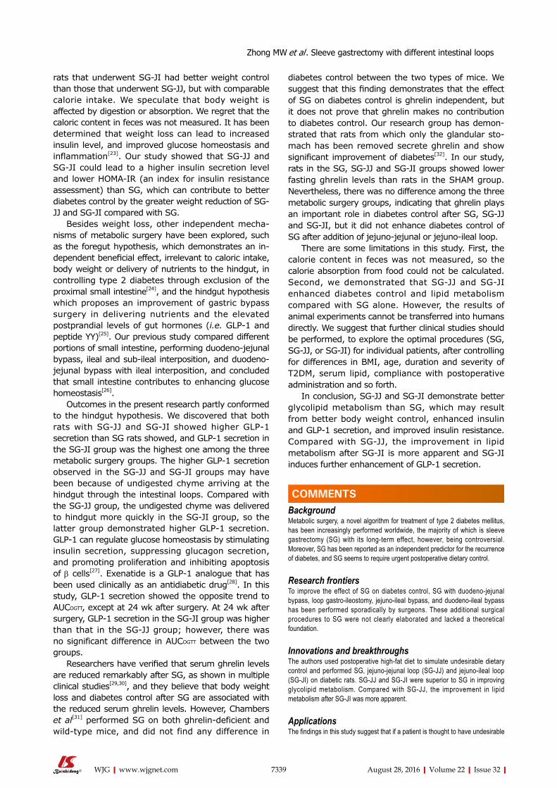
rats that underwent SG-JI had better weight control than those that underwent SG-JJ, but with comparable calorie intake. We speculate that body weight is affected by digestion or absorption. We regret that the caloric content in feces was not measured. It has been determined that weight loss can lead to increased insulin level, and improved glucose homeostasis and inflammation[23]. Our study showed that SG-JJ and SG-JI could lead to a higher insulin secretion level and lower HOMA-IR (an index for insulin resistance assessment) than SG, which can contribute to better diabetes control by the greater weight reduction of SG-JJ and SG-JI compared with SG.
Besides weight loss, other independent mecha-nisms of metabolic surgery have been explored, such as the foregut hypothesis, which demonstrates an in-dependent beneficial effect, irrelevant to caloric intake, body weight or delivery of nutrients to the hindgut, in controlling type 2 diabetes through exclusion of the proximal small intestine[24], and the hindgut hypothesis which proposes an improvement of gastric bypass surgery in delivering nutrients and the elevated postprandial levels of gut hormones (i.e. GLP-1 and peptide YY)[25]. Our previous study compared different portions of small intestine, performing duodeno-jejunal bypass, ileal and sub-ileal interposition, and duodeno-jejunal bypass with ileal interposition, and concluded that small intestine contributes to enhancing glucose homeostasis[26].
Outcomes in the present research partly conformed to the hindgut hypothesis. We discovered that both rats with SG-JJ and SG-JI showed higher GLP-1 secretion than SG rats showed, and GLP-1 secretion in the SG-JI group was the highest one among the three metabolic surgery groups. The higher GLP-1 secretion observed in the SG-JJ and SG-JI groups may have been because of undigested chyme arriving at the hindgut through the intestinal loops. Compared with the SG-JJ group, the undigested chyme was delivered to hindgut more quickly in the SG-JI group, so the latter group demonstrated higher GLP-1 secretion. GLP-1 can regulate glucose homeostasis by stimulating insulin secretion, suppressing glucagon secretion, and promoting proliferation and inhibiting apoptosis of β cells[27]. Exenatide is a GLP-1 analogue that has been used clinically as an antidiabetic drug[28]. In this study, GLP-1 secretion showed the opposite trend to AUCOGTT, except at 24 wk after surgery. At 24 wk after surgery, GLP-1 secretion in the SG-JI group was higher than that in the SG-JJ group; however, there was no significant difference in AUCOGTT between the two groups.
Researchers have verified that serum ghrelin levels are reduced remarkably after SG, as shown in multiple clinical studies[29,30], and they believe that body weight loss and diabetes control after SG are associated with the reduced serum ghrelin levels. However, Chambers et al[31] performed SG on both ghrelin-deficient and wild-type mice, and did not find any difference in
diabetes control between the two types of mice. We suggest that this finding demonstrates that the effect of SG on diabetes control is ghrelin independent, but it does not prove that ghrelin makes no contribution to diabetes control. Our research group has demon-strated that rats from which only the glandular sto-mach has been removed secrete ghrelin and show significant improvement of diabetes[32]. In our study, rats in the SG, SG-JJ and SG-JI groups showed lower fasting ghrelin levels than rats in the SHAM group. Nevertheless, there was no difference among the three metabolic surgery groups, indicating that ghrelin plays an important role in diabetes control after SG, SG-JJ and SG-JI, but it did not enhance diabetes control of SG after addition of jejuno-jejunal or jejuno-ileal loop.
There are some limitations in this study. First, the calorie content in feces was not measured, so the calorie absorption from food could not be calculated. Second, we demonstrated that SG-JJ and SG-JI enhanced diabetes control and lipid metabolism compared with SG alone. However, the results of animal experiments cannot be transferred into humans directly. We suggest that further clinical studies should be performed, to explore the optimal procedures (SG, SG-JJ, or SG-JI) for individual patients, after controlling for differences in BMI, age, duration and severity of T2DM, serum lipid, compliance with postoperative administration and so forth.
In conclusion, SG-JJ and SG-JI demonstrate better glycolipid metabolism than SG, which may result from better body weight control, enhanced insulin and GLP-1 secretion, and improved insulin resistance. Compared with SG-JJ, the improvement in lipid metabolism after SG-JI is more apparent and SG-JI induces further enhancement of GLP-1 secretion.
COMMENTSBackgroundMetabolic surgery, a novel algorithm for treatment of type 2 diabetes mellitus, has been increasingly performed worldwide, the majority of which is sleeve gastrectomy (SG) with its long-term effect, however, being controversial. Moreover, SG has been reported as an independent predictor for the recurrence of diabetes, and SG seems to require urgent postoperative dietary control.
Research frontiersTo improve the effect of SG on diabetes control, SG with duodeno-jejunal bypass, loop gastro-ileostomy, jejuno-ileal bypass, and duodeno-ileal bypass has been performed sporadically by surgeons. These additional surgical procedures to SG were not clearly elaborated and lacked a theoretical foundation.
Innovations and breakthroughsThe authors used postoperative high-fat diet to simulate undesirable dietary control and performed SG, jejuno-jejunal loop (SG-JJ) and jejuno-ileal loop (SG-JI) on diabetic rats. SG-JJ and SG-JI were superior to SG in improving glycolipid metabolism. Compared with SG-JJ, the improvement in lipid metabolism after SG-JI was more apparent.
ApplicationsThe findings in this study suggest that if a patient is thought to have undesirable
7339 August 28, 2016|Volume 22|Issue 32|WJG|www.wjgnet.com
COMMENTS
Zhong MW et al . Sleeve gastrectomy with different intestinal loops

postoperative dietary control, SG-JJ and SG-JI may be considered as preferential surgical procedures. SG-JI may be selected for patients with hyperlipidemia. These discoveries might help surgeons to select surgical procedures for individual patients.
TerminologySG, a novel procedure of metabolic surgery to remove 70%-80% of the stomach from the greater curvature, has demonstrated short- and medium-term roles in improving glycolipid metabolism in obese patients with/without type 2 diabetes mellitus, but its long-term beneficial effect remains controversial. In SG-JJ and SG-JI surgery, a jejuno-jejunal or jejuno-ileal side-by-side anastomosis is made based on SG surgery.
Peer-reviewThe study is very interesting. Regarding new guidelines, there is a need for the surgical methods dedicated for diabetic patients. The authors explored the effect of SG with jejuno-jejunal or jejuno-ileal loop on glycolipid metabolism in diabetic rats.
REFERENCES1 Rubino F, Nathan DM, Eckel RH, Schauer PR, Alberti KG,
Zimmet PZ, Del Prato S, Ji L, Sadikot SM, Herman WH, Amiel SA, Kaplan LM, Taroncher-Oldenburg G, Cummings DE. Metabolic Surgery in the Treatment Algorithm for Type 2 Diabetes: A Joint Statement by International Diabetes Organizations. Diabetes Care 2016; 39: 861-877 [PMID: 27222544 DOI: 10.2337/dc16-0236]
2 Angrisani L, Santonicola A, Iovino P, Formisano G, Buchwald H, Scopinaro N. Bariatric Surgery Worldwide 2013. Obes Surg 2015; 25: 1822-1832 [PMID: 25835983 DOI: 10.1007/s11695-015-1657-z]
3 Gagner M. Effect of sleeve gastrectomy on type 2 diabetes as an alternative to Roux-en-Y gastric bypass: a better long-term strategy. Surg Obes Relat Dis 2015; 11: 1280-1281 [PMID: 26048520 DOI: 10.1016/j.soard.2015.03.009]
4 Cutolo PP, Nosso G, Vitolo G, Brancato V, Capaldo B, Angrisani L. Clinical efficacy of laparoscopic sleeve gastrectomy vs laparoscopic gastric bypass in obese type 2 diabetic patients: a retrospective comparison. Obes Surg 2012; 22: 1535-1539 [PMID: 22960950 DOI: 10.1007/s11695-012-0657-5]
5 Cho JM, Kim HJ, Lo Menzo E, Park S, Szomstein S, Rosenthal RJ. Effect of sleeve gastrectomy on type 2 diabetes as an alternative treatment modality to Roux-en-Y gastric bypass: systemic review and meta-analysis. Surg Obes Relat Dis 2015; 11: 1273-1280 [PMID: 26071847 DOI: 10.1016/j.soard.2015.03.001]
6 Jiménez A, Casamitjana R, Flores L, Viaplana J, Corcelles R, Lacy A, Vidal J. Long-term effects of sleeve gastrectomy and Roux-en-Y gastric bypass surgery on type 2 diabetes mellitus in morbidly obese subjects. Ann Surg 2012; 256: 1023-1029 [PMID: 22968072 DOI: 10.1097/SLA.0b013e318262ee6b]
7 Lee WJ, Lee KT, Kasama K, Seiki Y, Ser KH, Chun SC, Chen JC, Lee YC. Laparoscopic single-anastomosis duodenal-jejunal bypass with sleeve gastrectomy (SADJB-SG): short-term result and comparison with gastric bypass. Obes Surg 2014; 24: 109-113 [PMID: 23990452 DOI: 10.1007/s11695-013-1067-z]
8 Mui WL, Lee DW, Lam KK. Laparoscopic sleeve gastrectomy with loop bipartition: A novel metabolic operation in treating obese type II diabetes mellitus. Int J Surg Case Rep 2014; 5: 56-58 [PMID: 24441436 DOI: 10.1016/j.ijscr.2013.12.002]
9 Hassn A, Luhmann A, Rahmani S, Morris-Stiff G. Medium-Term Results of Combined Laparoscopic Sleeve Gastrectomy and Modified Jejuno-Ileal Bypass in Bariatric Surgery. Obes Surg 2016; Epub ahead of print [PMID: 26922187 DOI: 10.1007/s11695-016-2098-z]
10 Sánchez-Pernaute A, Herrera MA, Pérez-Aguirre ME, Talavera P, Cabrerizo L, Matía P, Díez-Valladares L, Barabash A, Martín-Antona E, García-Botella A, Garcia-Almenta EM, Torres A. Single anastomosis duodeno-ileal bypass with sleeve gastrectomy
(SADI-S). One to three-year follow-up. Obes Surg 2010; 20: 1720-1726 [PMID: 20798995 DOI: 10.1007/s11695-010-0247-3]
11 Chikunguwo SM, Wolfe LG, Dodson P, Meador JG, Baugh N, Clore JN, Kellum JM, Maher JW. Analysis of factors associated with durable remission of diabetes after Roux-en-Y gastric bypass. Surg Obes Relat Dis 2010; 6: 254-259 [PMID: 20303324 DOI: 10.1016/j.soard.2009.11.003]
12 Kashyap SR, Schauer P. Clinical considerations for the manage-ment of residual diabetes following bariatric surgery. Diabetes Obes Metab 2012; 14: 773-779 [PMID: 22288474 DOI: 10.1111/j.1463-1326.2012.01577.x]
13 Kahn SE, Hull RL, Utzschneider KM. Mechanisms linking obesity to insulin resistance and type 2 diabetes. Nature 2006; 444: 840-846 [PMID: 17167471 DOI: 10.1038/nature05482]
14 Liu SZ, Sun D, Zhang GY, Wang L, Liu T, Sun Y, Li MX, Hu SY. A high-fat diet reverses improvement in glucose tolerance induced by duodenal-jejunal bypass in type 2 diabetic rats. Chin Med J (Engl) 2012; 125: 912-919 [PMID: 22490596]
15 Samuel VT, Beddow SA, Iwasaki T, Zhang XM, Chu X, Still CD, Gerhard GS, Shulman GI. Fasting hyperglycemia is not associated with increased expression of PEPCK or G6Pc in patients with Type 2 Diabetes. Proc Natl Acad Sci USA 2009; 106: 12121-12126 [PMID: 19587243 DOI: 10.1073/pnas.0812547106]
16 Duca FA, Côté CD, Rasmussen BA, Zadeh-Tahmasebi M, Rutter GA, Filippi BM, Lam TK. Metformin activates a duodenal Ampk-dependent pathway to lower hepatic glucose production in rats. Nat Med 2015; 21: 506-511 [PMID: 25849133 DOI: 10.1038/nm.3787]
17 Sun D, Liu S, Zhang G, Chen W, Yan Z, Hu S. Type 2 diabetes control in a nonobese rat model using sleeve gastrectomy with duodenal-jejunal bypass (SGDJB). Obes Surg 2012; 22: 1865-1873 [PMID: 22911149 DOI: 10.1007/s11695-012-0744-7]
18 Bruinsma BG, Uygun K, Yarmush ML, Saeidi N. Surgical models of Roux-en-Y gastric bypass surgery and sleeve gastrectomy in rats and mice. Nat Protoc 2015; 10: 495-507 [PMID: 25719268 DOI: 10.1038/nprot.2015.027]
19 Matthews DR, Hosker JP, Rudenski AS, Naylor BA, Treacher DF, Turner RC. Homeostasis model assessment: insulin resistance and beta-cell function from fasting plasma glucose and insulin concentrations in man. Diabetologia 1985; 28: 412-419 [PMID: 3899825]
20 World Health Organization. Global report on diabetes. Geneva: World Health Organization, 2016
21 Benaiges D, Más-Lorenzo A, Goday A, Ramon JM, Chillarón JJ, Pedro-Botet J, Flores-Le Roux JA. Laparoscopic sleeve gastrectomy: More than a restrictive bariatric surgery procedure? World J Gastroenterol 2015; 21: 11804-11814 [PMID: 26557004 DOI: 10.3748/wjg.v21.i41.11804]
22 Weiner RA, Theodoridou S, Weiner S. Failure of laparoscopic sleeve gastrectomy--further procedure? Obes Facts 2011; 4 Suppl 1: 42-46 [PMID: 22027290 DOI: 10.1159/000327343]
23 Tvarijonaviciute A, Ceron JJ, Holden SL, Morris PJ, Biourge V, German AJ. Effects of weight loss in obese cats on biochemical analytes related to inflammation and glucose homeostasis. Domest Anim Endocrinol 2012; 42: 129-141 [PMID: 22177629 DOI: 10.1016/j.domaniend.2011.10.003]
24 Rubino F, Forgione A, Cummings DE, Vix M, Gnuli D, Mingrone G, Castagneto M, Marescaux J. The mechanism of diabetes control after gastrointestinal bypass surgery reveals a role of the proximal small intestine in the pathophysiology of type 2 diabetes. Ann Surg 2006; 244: 741-749 [PMID: 17060767 DOI: 10.1097/01.sla.0000224726.61448.1b]
25 Dirksen C, Jørgensen NB, Bojsen-Møller KN, Jacobsen SH, Hansen DL, Worm D, Holst JJ, Madsbad S. Mechanisms of improved glycaemic control after Roux-en-Y gastric bypass. Diabetologia 2012; 55: 1890-1901 [PMID: 22538359 DOI: 10.1007/s00125-012-2556-7]
26 Liu S, Zhang G, Wang L, Sun D, Chen W, Yan Z, Sun Y, Hu S. The entire small intestine mediates the changes in glucose homeostasis after intestinal surgery in Goto-Kakizaki rats. Ann Surg 2012; 256: 1049-1058 [PMID: 23001083 DOI: 10.1097/
7340 August 28, 2016|Volume 22|Issue 32|WJG|www.wjgnet.com
Zhong MW et al . Sleeve gastrectomy with different intestinal loops

SLA.0b013e31826c3866]27 Drucker DJ. The role of gut hormones in glucose homeostasis.
J Clin Invest 2007; 117: 24-32 [PMID: 17200703 DOI: 10.1172/JCI30076]
28 Syed YY, McCormack PL. Exenatide Extended-Release: An Updated Review of Its Use in Type 2 Diabetes Mellitus. Drugs 2015; 75: 1141-1152 [PMID: 26071140 DOI: 10.1007/s40265-015-0420-z]
29 Vigneshwaran B, Wahal A, Aggarwal S, Priyadarshini P, Bhattacharjee H, Khadgawat R, Yadav R. Impact of Sleeve Gastrectomy on Type 2 Diabetes Mellitus, Gastric Emptying Time, Glucagon-Like Peptide 1 (GLP-1), Ghrelin and Leptin in Non-morbidly Obese Subjects with BMI 30-35.0 kg/m(2): a Prospective Study. Obes Surg 2016; Epub ahead of print [PMID: 27185177 DOI: 10.1007/s11695-016-2226-9]
30 Nosso G, Griffo E, Cotugno M, Saldalamacchia G, Lupoli R, Pacini G, Riccardi G, Angrisani L, Capaldo B. Comparative Effects of Roux-en-Y Gastric Bypass and Sleeve Gastrectomy on Glucose Homeostasis and Incretin Hormones in Obese Type 2 Diabetic Patients: A One-Year Prospective Study. Horm Metab Res 2016; 48: 312-317 [PMID: 26788926 DOI: 10.1055/s-0041-111505]
31 Chambers AP, Kirchner H, Wilson-Perez HE, Willency JA, Hale JE, Gaylinn BD, Thorner MO, Pfluger PT, Gutierrez JA, Tschöp MH, Sandoval DA, Seeley RJ. The effects of vertical sleeve gastrectomy in rodents are ghrelin independent. Gastroenterology 2013; 144: 50-52.e5 [PMID: 22995675 DOI: 10.1053/j.gastro.2012.09.009]
32 Sun D, Liu S, Zhang G, Colonne P, Hu C, Han H, Li M, Hu S. Sub-sleeve gastrectomy achieves good diabetes control without weight loss in a non-obese diabetic rat model. Surg Endosc 2014; 28: 1010-1018 [PMID: 24190081 DOI: 10.1007/s00464-013-3272-1]
P- Reviewer: Janik MR, Matsuda T, Rege RV, Yamaoka Y S- Editor: Gong ZM L- Editor: Filipodia E- Editor: Ma S
7341 August 28, 2016|Volume 22|Issue 32|WJG|www.wjgnet.com
Zhong MW et al . Sleeve gastrectomy with different intestinal loops

Qi Xie, Min-Yi Wu, Ding-Xuan Zhang, Yi-Ming Yang, Bao-Shuai Wang, Jing Zhang, Jin Xu, Wei-De Zhong, Jia-ni Hu
Qi Xie, Min-Yi Wu,Ding-Xuan Zhang, Yi-Ming Yang, Bao-Shuai Wang, Medical Imaging Department, Nansha Central Hospital, Guangzhou First People’s Hospital, Guangzhou Medical University, Guangzhou 511457, Guangdong Province, China
Jing Zhang, Department of Pathology, Cancer Center, SunYat-sen University, Guangzhou 510080, Guangdong Province, China
Jin Xu, Department of Pathology, Guangzhou First People’s Hospital, Guangzhou Medical University, Guangzhou 510180, Guangdong Province, China
Wei-De Zhong, Clinical Molecular Medicine and Molecular Diagnostics Laboratory, Guangzhou First People’s Hospital, Guangzhou Medical University, Guangzhou 510180, Guangdong Province, China
Jia-ni Hu, Karmanos Cancer Institute, Wayne State University, Detroit, MI 48201, United States
Author contributions: Xie Q designed the study; Wu MY, Zhang DX and Yang YM performed the research; Zhang J and Xu J collected and analyzed the data; Wang BS performed the MRI; Zhong WD conducted the statistical analysis; Xie Q and Zhang DX wrote the manuscript;Hu JN revised the manuscript.
Supported by the Natural Science Foundation of Guangdong, No. 2015A030313732.
Institutional review board statement: The study was reviewed and approved by the Experimental Animal Center of Sun Yat-sen University and Guangzhou Medical University respectively.
Institutional animal care and use committee statement: All procedures involving animals were reviewed and approved by the Institutional Animal Care and Use Committee of Sun Yat-sen University and Guangzhou Medical University.
Conflict-of-interest statement: To the best of our knowledge, no conflict of interest exists.
Data sharing statement: Technical appendix, statistical code, and dataset available from the corresponding author at [email protected].
Open-Access: This article is an open-access article which was selected by an in-house editor and fully peer-reviewed by external reviewers. It is distributed in accordance with the Creative Commons Attribution Non Commercial (CC BY-NC 4.0) license, which permits others to distribute, remix, adapt, build upon this work non-commercially, and license their derivative works on different terms, provided the original work is properly cited and the use is non-commercial. See: http://creativecommons.org/licenses/by-nc/4.0/
Manuscript source: Unsolicited manuscript
Correspondence to: Qi Xie, MD, Medical Imaging Department, Nansha Central Hospital, Guangzhou First People’s Hospital, Guangzhou Medical University, Guangzhou 511457, Guangdong Province, China. [email protected]: +86-20-22903628Fax: +86-20-22903699
Received: April 23, 2016Peer-review started: April 23, 2016First decision: May 27, 2016Revised: June 11, 2016Accepted: July 6, 2016Article in press: July 6, 2016Published online: August 28, 2016
AbstractAIMTo investigate the anticancer effect of a recombinant adenovirus-mediated p53 (rAd-p53) combined with 5-fluorouracil (5-FU) in human colon cancer resistant to 5-FU in vivo and the mechanism of rAd-p53 in reversal
ORIGINAL ARTICLE
Submit a Manuscript: http://www.wjgnet.com/esps/Help Desk: http://www.wjgnet.com/esps/helpdesk.aspxDOI: 10.3748/wjg.v22.i32.7342
World J Gastroenterol 2016 August 28; 22(32): 7342-7352 ISSN 1007-9327 (print) ISSN 2219-2840 (online)
© 2016 Baishideng Publishing Group Inc. All rights reserved.
7342 August 28, 2016|Volume 22|Issue 32|WJG|www.wjgnet.com
Basic Study
Synergistic anticancer effect of exogenous wild-type p53 gene combined with 5-FU in human colon cancer resistant to 5-FU in vivo

Xie Q et al . Anticancer effect of 5-FU and rAd-p53
7343 August 28, 2016|Volume 22|Issue 32|WJG|www.wjgnet.com
of 5-FU resistance.
METHODSnude mice bearing human colon cancer SW480/5-FU (5-FU resistant) were randomly assigned to four groups (n = 25 each): control group, 5-FU group, rAd-p53 group, and rAd-p53 + 5-FU group. At 24 h, 48 h, 72 h, 120 h and 168 h after treatment, 5 mice were randomly selected from each group and sacrificed using an overdose of anesthetics. The tumors were removed and the protein expressions of p53, protein kinase C (PKC), permeability-glycoprotein (P-gp) and multidrug resistance-associated protein 1 (MRP1) (Western blot) and apoptosis (TUNEL) were determined.
RESULTSThe area ratios of tumor cell apoptosis were larger in the rAd/p53 + 5-FU group than that in the control, 5-FU and rAd/p53 groups (P < 0.05), and were larger in the rAd/p53 group than that of the control group (P < 0.05) and the 5-FU group at more than 48 h (P < 0.05). The p53 expression was higher in the rAd/p53 and the rAd/p53 + 5-FU groups than that of the control and 5-FU groups (P < 0.05), and were higher in the rAd/p53 + 5-FU group than that of the rAd/p53 group (P < 0.05). Overexpression of PKC, P-gp and MRP1 was observed in the 5-FU and control groups. In the rAd/p53 + 5-FU group, the expression of P-gp and MRP1 was lower that of the control and 5-FU groups (P < 0.05), and the expression of PKC was lower than that of the control, 5-FU and rAd/p53 groups at more than 48 h (P < 0.05). In the rAd/p53 group, the expression of P-gp and MRP1 was lower that of the control and 5-FU groups at more than 48 h (P < 0.05), and the expression of PKC was lower than that of the control and 5-FU groups at more than 120 h (P < 0.05).
CONCLUSION5-FU combined with rAd-p53 has a synergistic anti-cancer effect in SW480/5-FU (5-FU resistance), which contributes to reversal of 5-FU resistance.
Key words: Human colon cancer; Multidrug resistance; 5-Fluorouracil; Recombinant adenovirus-mediated p53; Xenografts in nude mice
© The Author(s) 2016. Published by Baishideng Publishing Group Inc. All rights reserved.
Core tip: To observe anticancer action of a recombinant adenovirus-mediated p53 (rAd-p53) combined with 5-fluorouracil (5-FU) in human colon cancer with resistance to 5-FU in vivo to investigate the potential and mechanism of rAd-p53 in the reversal of resistance to 5-FU in human colon cancer. Our previous results revealed that exogenous wild-type p53 gene from rAd-p53 can decrease expression of PKC, P-gp and MRP1 in SW480/5-FU (5-FU resistance) and promote apoptosis of tumor cell, which contributes to reversing 5-FU resistance in vivo . 5-FU can increase the expression of exogenous wild-type p53, so 5-FU combined with rAd-p53 has a
synergistic anticancer effect for colon cancer of 5-FU resistance in vivo.
Xie Q, Wu MY, Zhang DX, Yang YM, Wang BS, Zhang J, Xu J, Zhong WD, Hu JN. Synergistic anticancer effect of exogenous wild-type p53 gene combined with 5-FU in human colon cancer resistant to 5-FU in vivo. World J Gastroenterol 2016; 22(32): 7342-7352 Available from: URL: http://www.wjgnet.com/1007-9327/full/v22/i32/7342.htm DOI: http://dx.doi.org/10.3748/wjg.v22.i32.7342
INTRODUCTIONColorectal cancer (CRC) is one of the most common gastrointestinal cancers. In 2013, there were 1.6 million incident cases of CRC worldwide, with 56% occurring in developing countries and 44% in developed countries, which caused 771000 deaths[1]. Most patients are usually at an advanced stage at the time of diagnosis.
To date, 5fluorouracil (5FU) remains a widely used chemotherapeutic drug in the treatment of advanced CRC; however, response rates are only 10% to 15%, due to severe side effects and resistance[2]. The anticancer efficacy of 5-FU is thought to be partly attributed to its ability to induce p53-dependent cell growth arrest and apoptosis; consequently, mutations or deletions of p53 can cause cells to become resistant to 5FU[36]. Therefore, overcoming 5FU resistance caused by mutations or deletions of p53 will be a key issue in the design of more effective individualized therapeutic strategies.
Gene replacement therapy for a mutated p53 gene using a recombinant adenovirus-mediated p53 (rAdp53) gene reportedly increases apoptosis after administration[712]. Our previous results revealed that exogenous wildtype p53 (wtp53) from rAdp53 increased tumor necrosis in human colon cancer SW480 (5-FU responsive) harboring mutant p53, and 5-FU combined with rAd-p53 had a synergistic anticancer effect in vivo[13]. Therefore, rAdp53 may contribute to the reversal of resistance to 5-FU in colon cancer.
The present study determined the early therapeutic effectiveness of rAd-p53 alone or in combination with 5FU for the treatment of human colon cancer SW480/5FU (5FU resistant) in a nude mouse model. The potential and mechanism of rAdp53 in the reversal of resistance to 5FU in human colon cancer in vivo was also investigated.
MATERIALS AND METHODSThe present study strictly complied with the recommendations of the Guide for the Care and Use of Laboratory Animals of the National Institutes of Health.

The animal use protocol was reviewed and approved by the Institutional Animal Care and Use Committee (IACUC) of Sun Yatsen University (20110702) and Guangzhou Medical University, Guangzhou, China.
Cell cultureThe human colon cancer cell line SW480 was purchased from the Cell Bank of Sun Yat-sen University. The cells were cultured in RPMI 1640 with 10% fetal calf serum, 100 U/mL penicillin and 100 µg/mL streptomycin, and grown at 37 ℃ in a 5% CO2 humidified atmosphere. 5-FU resistant SW480 (SW480/5FU) cells were generated by continuous exposure to increasing concentrations of 5-FU for more than 5 mo. SW480/5-FU cells were able to survive in 6 µg/mL of 5-FU. The IC50 of 5-FU, based on the results of a [3(4,5dimethylthiazol2yl)2,5 diphenyltetrazolium bromide (MTT)] assay, was 23.593 µg/mL for parental cells (SW480) and 140.642 µg/mL for resistant cells (SW480/5FU). The resistance index (RI) was 5.93. The IC50 of 5-FU in SW480 and SW480/5FU cells was assayed with MTT.
Animal modelBALB/c nude mice were purchased from the Animal Center of Sun Yat-sen University. A total of 100 4-wk-old BALB/c nude mice, weighing 1618 g regardless of sex, were subcutaneously implanted with SW480/5-FU tissues in the rear flank to generate xenograft models as described previously[13,14]. All surgical procedures were performed under anesthesia induced by chloral hydrate (4.5% chloral hydrate, 2 mL/100 g body weight, intraperitoneal injection), and all efforts were made to minimize suffering. Mice were fed in a specific pathogen-free (SPF) laboratory. One month after implantation, the mice were randomly assigned to four groups (25 per group): control group (medicalgrade saline), 5FU group, rAdp53 group (Gendicine, SibionoGeneTech Co., Ltd, Shenzhen, China), and rAd-p53 + 5-FU group. The above-mentioned thera-peutic agents were administered by intratumoral injection. The dose of rAd-p53 administered was 1 × 10⁷ VIP/mm3 tumor for each group. The dose of 5FU was 25 mg for tumors 0.5-0.9 cm, 50 mg for tumors 1.01.4 cm, and 75 mg for tumors more than 1.5 cm[13].
Assessment of tumor responseAt 24, 48, 72, 120 and 168 h after treatment, 5 mice were randomly selected from each group and euthanized with an overdose of anesthetics. The tumors were removed and divided into equal halves. One half was immediately frozen at 30 ℃ for Western blot analysis. The other half was fixed in phosphate-buffered saline (pH 7.3) containing 4% formaldehyde and 0.2% glutaraldehyde, embedded in paraffin, and sectioned for TUNEL assay.
Measurement of apoptosisPathological sections were stained using the TUNEL
apoptosis in situ detection reagent kit (Keygen, Nanjing, China) according to the manufacturer’s instruc-tions. The area ratio of tumor cell apoptosis was calculated as the percentage of positively stained cell nuclei (dark brown) at magnification × 100. The average of the evaluations by the pathologists (Zhang & Xu) was used for analysis.
Western blot analysisWestern blot analysis was used to detect the protein expression of p53, protein kinase C (PKC), perme-ability-glycoprotein (P-gp) and multidrug resistance-associated protein 1 (MRP1) as described in the instruction manual (Phototope®-HRP Western blot kit; Cell Signaling Technology, United States). Cell extracts were obtained from frozen tumor tissues (-30 ℃). Immunoblot analysis was performed using anti-p53 monoclonal antibodies (Santa Cruz Biotech, United States), anti-PKC and multidrug resistance protein 1 (MDR1) and MRP1 monoclonal antibodies (Santa Cruz Biotech). Subsequent protein detection was performed using an enhanced chemiluminescence (ECL) detection system (Hitachi, Japan).
The band intensities (IOD) of protein expression stated above were scanned into the computer and analyzed with ImagePro Plus 6.0 software. The relative IOD (RIOD) of protein expression was calculated as the IOD of the protein in the control group and therapeutic groups at each time point divided by the corresponding IOD of GAPDH (internal control).
Statistical analysisThe SPSS 13.1 statistical package (SPSS Inc., United States) was used for all calculations. The tumor responses of the entire sample set (ANOVA repeated data) and between groups (SNK test) were compared, and the correlations between parameters were evaluated with Pearson’s correlation at a significance level of 0.05. Data are presented as mean ± SD of a representative of at least three independently performed experiments.
RESULTSTumor cell IC50The IC50 of 5-FU based on the results of the MTT assay was 23.593 µg/mL for parental cells (SW480) and 140.642 µg/mL for resistant cells (SW480/5FU). The resistance index (RI) was 5.93.
Tumor cell apoptosisTumor cell apoptosis was detected in sections from the 5-FU group and the control group at the observed time points (Figure 1); however, there were no significant differences in tumor cell apoptosis between the two groups (SNK test, P > 0.05; Table 1).
The area ratio of tumor cell apoptosis in the rAdp53 + 5FU group was significantly larger than
7344 August 28, 2016|Volume 22|Issue 32|WJG|www.wjgnet.com
Xie Q et al . Anticancer effect of 5-FU and rAd-p53

Protein expressionP53 expression: P53 protein showed weak expres-sion in the 5FU group and the control group at the observed time points (Figure 2A). There were no significant differences in the RIOD of p53 expression between the two groups (SNK test, P > 0.05; Table 2). P53 expression level in the rAdp53 and the rAdp53 + 5FU group was higher than that in the control and 5-FU groups (SNK test, P < 0.05) and increased in a time-dependent manner (ANOVA, P < 0.05; Table 2), with peak expression at 120 h. P53 expression in the
that in the control group, 5FU group and the rAdp53 group (SNK test, P < 0.05; Table 1). The area ratio of tumor cell apoptosis in the rAdp53 group was significantly larger than that in the control group (SNK test, P < 0.05). After > 48 h of treatment, the area ratio of tumor cell apoptosis in the rAdp53 group was significantly larger than that in the 5-FU group (SNK test, P < 0.05).
The area ratio of tumor cell apoptosis in the rAdp53 group and the rAdp53 + 5FU group tended to increase with time (ANOVA, P <0.05).
7345 August 28, 2016|Volume 22|Issue 32|WJG|www.wjgnet.com
A B
C D
Figure 1 Tumor cell apoptosis. Tumor cell apoptosis at 72 h (magnification × 400) in the control group (A), 5-FU group (B), rAd-p53 group (C) and rAd-p53 + 5-FU group (D).
Table 1 Tumor apoptosis ratios in experimental groups and control group (mean ± SD)
Group 24 h 48 h 72 h 120 h 168 h
Control 0.25 ± 0.02 0.28 ± 0.01 0.27 ± 0.02 0.29 ± 0.02 0.24 ± 0.045-FU 0.27 ± 0.03 0.31 ± 0.03 0.31 ± 0.05 0.30 ± 0.04 0.31 ± 0.02rAd-p53 0.29 ± 0.02 0.35 ± 0.03 0.43 ± 0.08 0.42 ± 0.06 0.46 ± 0.065-FU + rAd-p53 0.33 ± 0.03 0.44 ± 0.08 0.58 ± 0.07 0.59 ± 0.05 0.62 ± 0.07F 13.235 41.487 53.812 61.676 80.755P value 0 0 0 0 0P value > 0.05 > 0.05 > 0.05 > 0.05 > 0.055-FU vs ControlP value < 0.05 < 0.05 < 0.05 < 0.05 < 0.05rAd-p53 vs ControlP value < 0.05 < 0.05 < 0.05 < 0.05 < 0.05rAd-p53 + 5-FU vs ControlP value < 0.05 < 0.05 < 0.05 < 0.05 < 0.05rAd-p53 + 5-FU vs rAd-p53P value < 0.05 < 0.05 < 0.05 < 0.05 < 0.05rAd-p53 + 5-FU vs 5-FUP value >0.05 < 0.05 < 0.05 < 0.05 < 0.05rAd-p53 vs 5-FU
Xie Q et al . Anticancer effect of 5-FU and rAd-p53

7346 August 28, 2016|Volume 22|Issue 32|WJG|www.wjgnet.com
5-FU rAd-p53 5-FU + rAd-p53
0.9
0.8
0.7
0.6
0.5
0.4
0.3
0.2
0.1
0.0
The
RIO
D o
f p5
3 ex
pres
sion
Control
24 h
48 h
72 h
120 h
168 h
A
5-FU rAd-p53 5-FU + rAd-p53
1.8
1.6
1.4
1.2
1.0
0.8
0.6
0.4
0.2
0.0
The
RIO
D o
f PK
C ex
pres
sion
Control
24 h
48 h
72 h
120 h
168 h
5-FU rAd-p53 5-FU + rAd-p53
2.0
1.8
1.6
1.4
1.2
1.0
0.8
0.6
0.4
0.2
0.0
The
RIO
D o
f P-
gp e
xpre
ssio
n
Control
24 h
48 h
72 h
120 h
168 h
5-FU rAd-p53 5-FU + rAd-p53
2.0
1.8
1.6
1.4
1.2
1.0
0.8
0.6
0.4
0.2
0.0
The
RIO
D o
f M
RP1
exp
ress
ion
Control
24 h
48 h
72 h
120 h
168 h
Figure 2 Relative band intensities of p53 expression (A), protein kinase C expression (B), permeability-glycoprotein expression (C) and MRP1 expression (D) in the three experimental groups.
B
C
D
Xie Q et al . Anticancer effect of 5-FU and rAd-p53

rAdp53 + 5FU group was significantly higher than that in the rAd-p53 group (SNK test, P < 0.05; Table 2).
PKC, P-gp and MRP1 expression: Overexpression of PKC, P-gp and MRP1 was observed in the 5-FU group and the control group (Figure 2BD). There were no significant differences in the RIOD of the expression of these proteins between the two groups (SNK test, P > 0.05; Tables 3-5).
At the observed time points, the expression of PKC, Pgp and MRP1 in the rAd/p53 + 5FU group gradually decreased in a time-dependent manner (ANOVA, P < 0.05; Tables 3-5). The expression of P-gp and MRP1 was significantly lower than that in the control group and the 5-FU group (SNK test, P < 0.05). More than 48 h after treatment, the expression of PKC in the rAdp53 + 5FU group was significantly lower than that in the control, 5FU and rAdp53 groups.
In the rAdp53 group, the expression of Pgp and MRP1 was significantly lower than that in the control group and the 5FU group at > 48 h of treatment (SNK test, P < 0.05). The expression of PKC was significantly lower than that in the control group and the 5-FU group at > 120 h of treatment (SNK test, P < 0.05).
Pearson’s correlation testThe RIOD of p53 expression was positively correlated with the area ratio of tumor cell apoptosis (the correlation coefficient and P value were 0.545 and 0.000, respectively).
The RIOD of PKC, P-gp and MRP1 expression was negatively correlated with the area ratio of tumor cell apoptosis (correlation coefficients were -0.322, 0.012 and 0.335 and P values were 0.009, -0.541 and 0.000, respectively).
7347 August 28, 2016|Volume 22|Issue 32|WJG|www.wjgnet.com
Table 2 Relative IOD of p53 expression in experimental groups and control group (mean ± SD)
Group 24 h 48 h 72 h 120 h 168 h
Control 0.099 ± 0.001 0.105 ± 0.003 0.101 ± 0.007 0.105 ± 0.015 0.103 ± 0.0015-FU 0.108 ± 0.002 0.098 ± 0.028 0.096 ± 0.004 0.097 ± 0.047 0.096 ± 0.033rAd-p53 0.187 ± 0.003 0.209 ± 0.014 0.627 ± 0.005 0.743 ± 0.012 0.713 ± 0.0055-FU + rAd-p53 0.393 ± 0.004 0.461 ± 0.002 0.691 ± 0.018 0.822 ± 0.009 0.808 ± 0.027F 221.565 270.392 3983.173 9639.595 3867.703P value 0 0 0 0 0P value > 0.05 > 0.05 > 0.05 > 0.05 > 0.055-FU vs ControlP value < 0.05 < 0.05 < 0.05 < 0.05 < 0.05rAd-p53 vs ControlP value < 0.05 < 0.05 < 0.05 < 0.05 < 0.05rAd-p53 + 5-FU vs ControlP value < 0.05 < 0.05 < 0.05 < 0.05 < 0.05rAd-p53 + 5-FU vs rAd-p53P value < 0.05 < 0.05 < 0.05 < 0.05 < 0.05rAd-p53 + 5-FU vs 5-FUP value < 0.05 < 0.05 < 0.05 < 0.05 < 0.05rAd-p53 vs 5-FU
Table 3 Relative IOD of protein kinase C expression in experimental groups and control group (mean ± SD)
Group 24 h 48 h 72 h 120 h 168 h
Control 1.13 ± 0.08 1.21 ± 0.06 0.99 ± 0.13 1.26 ± 0.42 1.08 ± 0.205-FU 0.94 ± 0.21 1.12 ± 0.14 0.93 ± 0.14 1.08 ± 0.52 1.04 ± 0.26rAd-p53 0.93 ± 0.42 0.91 ± 0.23 1.07 ± 0.13 0.57 ± 0.18 0.36 ± 0.095-FU + rAd-p53 1.23 ± 0.24 0.53 ± 0.09 0.45 ± 0.10 0.26 ± 0.06 0.08 ± 0.01F 1.036 33.972 14.348 24.072 71.559P value 0.427 0 0.01 0 0P value > 0.05 > 0.05 > 0.05 > 0.05 > 0.055-FU vs ControlP value > 0.05 < 0.05 > 0.05 < 0.05 < 0.05rAd-p53 vs ControlP value > 0.05 < 0.05 < 0.05 < 0.05 < 0.05rAd/p53 + 5- FU vs ControlP value > 0.05 < 0.05 < 0.05 < 0.05 < 0.05rAd-p53 + 5-FU vs rAd-p53P value > 0.05 < 0.05 < 0.05 < 0.05 < 0.05rAd-p53 + 5-FU vs 5-FUP value > 0.05 < 0.05 > 0.05 < 0.05 < 0.05rAd-p53 vs 5-FU
Xie Q et al . Anticancer effect of 5-FU and rAd-p53

The RIOD of p53 expression and the RIOD of PKC, P-gp and MRP1 expression showed a negative correlation (correlation coefficients were -0.366, 0.004 and 0.406 and P values were 0.001, 0.488 and 0.000, respectively).
DISCUSSION5-FU is still widely used as a major anticancer drug in the treatment of colon cancer[3]. However, a major impediment to the success of colon cancer chemotherapy is the development of cancer variants exhibiting multidrug resistance (MDR)[26,15,16].
MDR usually presents as crossresistance to multiple chemotherapeutic drugs with different structures[16,17]. Anti-cancer drug resistance in colon cancer cells can be caused by various factors, including alterations in drug influx and efflux, enhancement of drug inactivation
and mutation of the drug target induced by various proteins[1821]. To date, multiple factors have been reported to lead to resistance to chemotherapeutic drugs[1623]. P-gp, PKC and the multidrug resistance-associated proteins (MRPs) contribute to chemotherapy resistance[1723]. In our previous studies, overexpression of P-gp, PKC and MRP1 was observed in human colon cancer SW480/5-FU cells (5-FU resistant) and weak expression of these proteins was seen in parental human colon cancer SW480 cells (5FU responsive)[14].
Various mechanisms contribute to MDR, including the overexpression of drug efflux pumps (pump resistance) and the upregulation of cellular antiapoptotic defense systems (nonpump resistance)[23,24]. P-gp encoded by the MDR1 gene and MRPs belong to the ATP-binding cassette (ABC) superfamily. These transporter proteins (responsible for pump resistance) mediate the efflux of drugs in the MDR spectrum, such as anthracyclines, out
7348 August 28, 2016|Volume 22|Issue 32|WJG|www.wjgnet.com
Table 4 Relative IOD of P-gp expression in experimental groups and control group (mean ± SD)
Group 24 h 48 h 72 h 120 h 168 h
Control 1.28 ± 0.20 1.32 ± 0.31 1.36 ± 0.40 1.29 ± 0.43 1.30 ± 0.565-FU 1.17 ± 0.37 1.13 ± 0.41 1.17 ± 0.39 1.27 ± 0.47 1.29 ± 0.56rAd-p53 1.12 ± 0.31 0.69 ± 0.11 0.62 ± 0.02 0.36 ± 0.15 0.26 ± 0.065-FU + rAd-p53 0.86 ± 0.17 0.65 ± 0.21 0.59 ± 0.25 0.31 ± 0.01 0.10 ± 0.03F 5.670 7.301 14.645 9.844 7.971P value 0.022 0.011 0.001 0.005 0.009P value > 0.05 > 0.05 > 0.05 > 0.05 > 0.055-FU vs ControlP value > 0.05 < 0.05 < 0.05 < 0.05 < 0.05rAd-p53 vs ControlP value < 0.05 < 0.05 < 0.05 < 0.05 < 0.05rAd-p53 + 5-FU vs ControlP value < 0.05 > 0.05 > 0.05 > 0.05 > 0.05rAd-p53 + 5-FU vs rAd-p53P value < 0.05 < 0.05 < 0.05 < 0.05 < 0.05rAd-p53 + 5-FU vs 5-FUP value > 0.05 < 0.05 < 0.05 < 0.05 < 0.05rAd-p53 vs 5-FU
Table 5 Relative IOD of MRP1 expression in experimental groups and control group (mean ± SD)
Group 24 h 48 h 72 h 120 h 168 h
Control 1.31 ± 0.36 1.33 ± 0.08 1.09 ± 0.24 1.22 ± 0.35 1.07 ± 0.225-FU 1.27 ± 0.10 1.31 ± 0.11 1.14 ± 0.16 1.20 ± 0.31 1.13 ± 0.14rAd-p53 0.98 ± 0.09 0.74 ± 0.11 0.58 ± 0.18 0.87 ± 0.31 0.31 ± 0.085-FU + rAd-p53 0.71 ± 0.17 0.62 ± 0.02 0.51 ± 0.10 0.42 ± 0.08 0.13 ± 0.10F 6.438 123.754 20.567 15.512 36.073P value 0.016 0 0 0.001 0P value > 0.05 > 0.05 > 0.05 > 0.05 > 0.055-FU vs ControlP value > 0.05 < 0.05 < 0.05 < 0.05 < 0.05rAd-p53 vs ControlP value < 0.05 < 0.05 < 0.05 < 0.05 < 0.05rAd-p53 + 5-FU vs ControlP value > 0.05 < 0.05 > 0.05 < 0.05 > 0.05rAd-p53 + 5-FU vs rAd-p53P value < 0.05 < 0.05 < 0.05 < 0.05 < 0.05rAd-p53 + 5-FU vs 5-FUP value > 0.05 < 0.05 < 0.05 < 0.05 < 0.05rAd-p53 vs 5-FU
Xie Q et al . Anticancer effect of 5-FU and rAd-p53

of cells, thus reducing drug efficacy[24]. Generally, there are two approaches used to reverse ABC superfamilymediated MDR: blocking its drug-pump function and inhibiting its expression[2527].
PKC is one of the signaling enzymes that is positively regulated by reactive oxygen species (ROS) and plays a crucial role in a variety of pathophysiological states, including tumor progression. PKC contains multiple cysteine residues that can be activated by ROS oxidatively[28,29]. PKC represents a family of serine/threonine kinases that are involved in the regulation of cell growth, cell death and stress responsiveness[30]. Generally, the PKCs are classified into three subfamilies based on their structural and activation characteristics: the conventional or classic (α, βI, βⅡ, and γ), the novel or nonclassic (δ, ε, η and θ), and the atypical PKC isoenzymes (ζ, ι and λ)[30]. Different PKC isoenzymes may exert similar or opposite cellular effects by differential coupling of signaling pathways[31]. Cancer cells survive by evading apoptosis or promoting proliferation, invasion and metastasis. PKC may act as a downstream effector of the signaling protein phosphatidylinositol 3-kinase (PI3K). The PI3K-mediated signaling cascade regulates cell proliferation, cell survival, differentiation and apoptosis[3234]. Phosphorylation of the regulatory subunit p85a is linked to increased survival of cancer cells. The p85 subunit regulates the catalytic subunit p110 by stabilization and inactivation of its kinase activity in the basal state as well as by recruitment of PI3K to phospho-tyrosine residues of the activated receptors[34,35].
PKCα and PKCβ may promote ABCB1 function by phosphorylation[18,33,34]. Notably, the effects of PKC signaling on ABCB1 phosphorylation and function appear to be cell type-dependent. In ovarian carcinoma cells, antisense oligomers directed against PKCα and PKCβ reversed ABCB1mediated drug resistance[36]. In contrast, PKCβ was not detectable in some reports, and siRNAs targeting PKCα interfered with PKC signaling, but not with ABCB1 function[18]. Moreover, p53 was shown to suppress PKCαmediated ABCB1 activation in leiomyosarcoma, fibrosarcoma, and osteosarcoma cells[18,34].
Mutations or deletions of suppressor gene p53 are the most common genetic abnormalities that occur during cancerogenesis in the majority of human neoplasms. The p53 gene, localized on the short arm of chromosome 17 (17p13), encodes nucleic phosphoproteins, and affects several cell functions (induction of many genes, regulation of the cell cycle and apoptosis control)[37]. Under the condition of p53 gene mutation, cancer cells remain intact and survive[38].
Evasion from chemotherapy-induced apoptosis due to p53 loss strongly contributes to drug resis-tance[36,16,38]. Wildtype p53 is a key tumor suppressor in preventing tumorigenesis and cancer progression; however, mutant p53, detected in over 50% of all human tumors and in approximately 70% of colo
rectal cancers[39-43], promotes tumor progression and resistance to therapies[36,38,44], and such mutants have become the most common prognostic indicators for both tumor recurrence and cancer death[40,43,45,46]. Prevention of p53 mutation to restore wildtype p53 activity is an attractive anticancer therapy to reverse 5FU resistance in colon cancer.
Infection with Adp53 can significantly downregulate MDR1 transcription and Pgp expression in breast cancer cell lines and reverse resistance to adriamycin[47]. Treatment with rAdp53 alone, oxaliplatin alone or combined treatment led to a decrease in Bcl-2 expression and an increase in Bax expression in gastric cancer cells, and induced apoptosis of gastric cancer cells, which was accompanied by increased expression of caspase3[12]. Therefore, rAdp53 may enhance the sensitivity of gastric cancer cells to chemotherapy by promoting apoptosis. In the present study, exogenous wildtype p53 gene from rAdp53 decreased the expression of PKC, P-gp and MRP1 and promoted apoptosis of colon carcinoma cells in nude mice implanted with human colon carcinoma SW480/5FU (5-FU resistant), which contributed to the reversal of 5FU resistance.
The antimetabolite, 5-FU, is an analogue of uracil with a fluorine atom at the C5 position of the pyrimidine ring. 5FU is converted in cells to different active metabolites, including fluorodeoxyuridine monophosphate (FdUMP), fluorodeoxyuridine triphosphate (FdUTP) and fluorouridine triphosphate (FUTP). These metabolites have been implicated in both global RNA metabolism due to incorporation of the ribonucleotide FUMP into RNA, and DNA metabolism due to thymidylate synthase (TS) inhibition or direct incorporation of FdUMP into DNA, leading to a wide range of biological effects which can act as triggers for apoptotic cell death[3,4,15,16]. Therefore, 5-FU can be regarded as a genotoxic agent.
P53 protein in the production of wildtype p53 expression plays a key role in cell cycle regulation and in the cellular response to cytotoxic stress and DNA damage[4851]. P53 protein is maintained at low levels by MDM2, an E3-ligase that binds p53 and promotes its degradation[5254]. DNA damage and other stresses, including gamma and UV irradiation, chemotherapeutic agents, hypoxia, heat or alterations in intracellular nucleotide pools, disrupt p53-MDM2 binding, causing p53 levels to increase[49-51,55]. Wildtype p53 is induced in response to a host of genotoxic and environmental stresses, a host of target genes are then transcriptionally activated, including p21, GADD45, Bax and Bcl2. Induction of p21, in turn, leads to cell cycle arrest at both G1 and G2 checkpoints. This function is thought to be essential in preserving the integrity of the cellular genome in response to treatment with cytotoxic agents. In addition to mediating cell cycle arrest, p53 is a potent inducer of apoptosis and programmed cell death[49-51].
Increased p53 protein in response to genotoxic stress also occurs in cancer cells[56-59]. Treatment of
7349 August 28, 2016|Volume 22|Issue 32|WJG|www.wjgnet.com
Xie Q et al . Anticancer effect of 5-FU and rAd-p53

human colon cancer RKO cells and tetraploid cancer cells with 5FU resulted in a significant increase in the levels of the endogenous p53 protein family in vitro and enhanced tumor suppression[59]. The p53 protein family forms an interacting network of proteins[60]. Cancer cell responses to 5FU treatment are determined by the total activity of the entire p53 family rather than p53 alone[60]. Suppressor p53 is one of the molecular targets of 5FU. With regard to 5FU, translational regulation is an important process for controlling endogenous p53 expression[59-61]. Our previous study demonstrated that the expression of exogenous wildtype p53 gene in colon cancer cells in nude mice bearing human colon carcinoma SW480 (5FU responsive) treated with rAdp53 + 5FU was significantly higher than that with rAdp53 alone, and tumor necrosis was positively correlated with p53 expression in vivo[13]. 5FU also increased the anticancer effect of rAd/p53 in vivo.
In the present study, p53 expression in colon cancer SW480/5FU in the rAd/p53 group and rAdp53 + 5FU group was higher than in the control and 5FU groups and increased in a timedependent manner. P53 expression in the rAdp53 + 5FU group was significantly higher than in the rAd-p53 group. These results suggest that 5FU increased the expression of exogenous wildtype p53 gene in colon cancer (resistant to 5FU) in vivo. Exogenous wild-type p53 is also induced in response to genotoxic stress in chemotherapyresistant cancer cells.
In summary, exogenous wildtype p53 gene from rAd-p53 can decrease the expression of PKC, P-gp and MRP1 in SW480/5FU (5FU resistant) and promote apoptosis of tumor cells, which contributes to the reversal of 5FU resistance in vivo. 5FU can increase the expression of exogenous wildtype p53, thus 5FU combined with rAd-p53 has a synergistic anticancer effect in colon cancer resistant to 5FU in vivo. Therefore, the DNA-damaging agent 5-FU combined with exogenous wildtype p53 provides a potential therapeutic strategy and can enhance the sensitivity and reduce the toxicity of chemotherapy and improve the clinical efficacy of colon cancer chemotherapy.
ACKNOWLEDGMENTSA statistical review of the study was performed by Gui-Qin Wang (a biomedical statistician at Nansha Central Hospital, Guangzhou First People’s Hospital, Guangzhou Medical University, Guangzhou, China).
COMMENTSBackground5-fluorouracil (5-FU) is a widely used chemotherapeutic drug in the treatment of advanced colorectal cancer (CRC); however, response rates are only 10% to 15%, due to severe side effects and resistance. Mutations or deletions of p53 can cause cells to become resistant to 5-FU. Therefore, gene replacement therapy for a mutated p53 gene using recombinant adenovirus-mediated p53
(rAd-p53) may overcome 5-FU resistance caused by mutations or deletions of p53. Therefore, rAd-p53 may contribute to the reversal of 5-FU resistance in colon cancer.
Research frontiersExogenous wild-type p53 from rAd-p53 increased tumor necrosis in human colon cancer SW480 (5-FU responsive) harboring mutant p53, and 5-FU combined with rAd-p53 had a synergistic anticancer effect in vivo.
Innovations and breakthroughsThis is the first study to evaluate 5-FU combined with rAd-p53 in the treatment of colon cancer SW480/5-FU (5-FU resistant) compared with controls in vivo.
ApplicationsThis study first showed that exogenous wild-type p53 gene from rAd-p53 can decrease the expression of PKC, P-gp and MRP1 in colon cancer SW480/5-FU (5-FU resistant) and promote apoptosis of tumor cells, which contributes to the reversal of 5-FU resistance in vivo. 5-FU can increase the expression of exogenous wild-type p53; thus, 5-FU combined with rAd-p53 had a synergistic anticancer effect in 5-FU resistant colon cancer in vivo.
Terminology5-FU increased the expression of exogenous wild-type p53 gene in colon cancer (5-FU resistant) in vivo. Exogenous wild-type p53 is also induced in response to genotoxic stress in chemotherapy-resistant cancer cells.
Peer-reviewThe authors demonstrated that 5-FU increased the expression of exogenous wild-type p53 gene, and exogenous wild-type p53 is induced in response to genotoxic stress by 5-FU in colon cancer (resistant to 5-FU) in vivo. These results are interesting. Previous studies have established that increased p53 protein in response to genotoxic stress occurs in cancer cells in vitro. In this study, the authors showed for the first time that 5-FU combined with rAd-p53 has a synergistic anticancer effect for colon cancer resistant to 5-FU in vivo.
REFERENCES1 Fitzmaurice C, Dicker D, Pain A, Hamavid H, Moradi-Lakeh
M, MacIntyre MF, Allen C, Hansen G, Woodbrook R, Wolfe C, Hamadeh RR, Moore A, Werdecker A, Gessner BD, Te Ao B, McMahon B, Karimkhani C, Yu C, Cooke GS, Schwebel DC, Carpenter DO, Pereira DM, Nash D, Kazi DS, De Leo D, Plass D, Ukwaja KN, Thurston GD, Yun Jin K, Simard EP, Mills E, Park EK, Catalá-López F, deVeber G, Gotay C, Khan G, Hosgood HD, Santos IS, Leasher JL, Singh J, Leigh J, Jonas JB, Sanabria J, Beardsley J, Jacobsen KH, Takahashi K, Franklin RC, Ronfani L, Montico M, Naldi L, Tonelli M, Geleijnse J, Petzold M, Shrime MG, Younis M, Yonemoto N, Breitborde N, Yip P, Pourmalek F, Lotufo PA, Esteghamati A, Hankey GJ, Ali R, Lunevicius R, Malekzadeh R, Dellavalle R, Weintraub R, Lucas R, Hay R, Rojas-Rueda D, Westerman R, Sepanlou SG, Nolte S, Patten S, Weichenthal S, Abera SF, Fereshtehnejad SM, Shiue I, Driscoll T, Vasankari T, Alsharif U, Rahimi-Movaghar V, Vlassov VV, Marcenes WS, Mekonnen W, Melaku YA, Yano Y, Artaman A, Campos I, MacLachlan J, Mueller U, Kim D, Trillini M, Eshrati B, Williams HC, Shibuya K, Dandona R, Murthy K, Cowie B, Amare AT, Antonio CA, Castañeda-Orjuela C, van Gool CH, Violante F, Oh IH, Deribe K, Soreide K, Knibbs L, Kereselidze M, Green M, Cardenas R, Roy N, Tillmann T, Li Y, Krueger H, Monasta L, Dey S, Sheikhbahaei S, Hafezi-Nejad N, Kumar GA, Sreeramareddy CT, Dandona L, Wang H, Vollset SE, Mokdad A, Salomon JA, Lozano R, Vos T, Forouzanfar M, Lopez A, Murray C, Naghavi M. The Global Burden of Cancer 2013. JAMA Oncol 2015; 1: 505-527 [PMID: 26181261 DOI: 10.1001/jamaoncol.2015.0735]
2 Pardini B, Kumar R, Naccarati A, Novotny J, Prasad RB, Forsti A, Hemminki K, Vodicka P, Lorenzo Bermejo J. 5-Fluorouracil-based chemotherapy for colorectal cancer and MTHFR/MTRR
7350 August 28, 2016|Volume 22|Issue 32|WJG|www.wjgnet.com
COMMENTS
Xie Q et al . Anticancer effect of 5-FU and rAd-p53

genotypes. Br J Clin Pharmacol 2011; 72: 162-163 [PMID: 21204909 DOI: 10.1111/j.1365-2125.2010.03892.x]
3 Sui X, Kong N, Wang X, Fang Y, Hu X, Xu Y, Chen W, Wang K, Li D, Jin W, Lou F, Zheng Y, Hu H, Gong L, Zhou X, Pan H, Han W. JNK confers 5-fluorouracil resistance in p53-deficient and mutant p53-expressing colon cancer cells by inducing survival autophagy. Sci Rep 2014; 4: 4694 [PMID: 24733045 DOI: 10.1038/srep04694]
4 Sun XX, Dai MS, Lu H. 5-fluorouracil activation of p53 involves an MDM2-ribosomal protein interaction. J Biol Chem 2007; 282: 8052-8059 [PMID: 17242401]
5 Subbarayan PR, Sarkar M, Nelson G, Benitez E, Singhal S, Ardalan B. Chronic exposure of colorectal cancer cells in culture to fluoropyrimidine analogs induces thymidylate synthase and suppresses p53. A molecular explanation for the mechanism of 5-FU resistance. Anticancer Res 2010; 30: 1149-1156 [PMID: 20530421]
6 Huang C, Zhang XM, Tavaluc RT, Hart LS, Dicker DT, Wang W, El-Deiry WS. The combination of 5-fluorouracil plus p53 pathway restoration is associated with depletion of p53-deficient or mutant p53-expressing putative colon cancer stem cells. Cancer Biol Ther 2009; 8: 2186-2193 [PMID: 19923910]
7 Baek JH, Agarwal ML, Tubbs RR, Vladisavljevic A, Tomita H, Bukowski RM, Milsom JW, Kim JM, Kwak JY. In vivo recombinant adenovirus-mediated p53 gene therapy in a syngeneic rat model for colorectal cancer. J Korean Med Sci 2004; 19: 834-841 [PMID: 15608394]
8 Guan YS, La Z, Yang L, He Q, Li P. p53 gene in treatment of hepatic carcinoma: status quo. World J Gastroenterol 2007; 13: 985-992 [PMID: 17373730 DOI: 10.3748/WJG.v13.i7.985]
9 Inoue H, Shiraki K, Murata K, Sugimoto K, Kawakita T, Yamaguchi Y, Saitou Y, Enokimura N, Yamamoto N, Yamanaka Y, Nakano T. Adenoviral-mediated transfer of p53 gene enhances TRAIL-induced apoptosis in human hepatocellular carcinoma cells. Int J Mol Med 2004; 14: 271-275 [PMID: 15254777]
10 Roth JA. Adenovirus p53 gene therapy. Expert Opin Biol Ther 2006; 6: 55-61 [PMID: 16370914]
11 Shimada H , Shimizu T, Ochiai T, Liu TL, Sashiyama H, Nakamura A, Matsubara H, Gunji Y, Kobayashi S, Tagawa M, Sakiyama S, Hiwasa T. Preclinical study of adenoviral p53 gene therapy for esophageal cancer. Surg Today 2001; 31: 597-604 [PMID: 11495154]
12 Chen GX, Zheng LH, Liu SY, He XH. rAd-p53 enhances the sensitivity of human gastric cancer cells to chemotherapy. World J Gastroenterol 2011; 17: 4289-4297 [PMID: 22090785 DOI: 10.3748/wjg.v17.i38.4289]
13 Xie Q, Liang BL, Wu YH, Zhang J, Chen MW, Liu HY, Gu XF, Xu J. Synergistic anticancer effect of rAd/P53 combined with 5-fluorouracil or iodized oil in the early therapeutic response of human colon cancer in vivo. Gene 2012; 499: 303-308 [PMID: 22441128 DOI: 10.1016/j.gene.2012.02.007]
14 Xie Q, Yang YM, Wu MYi, Zhang DX, Lei ZX, Zhang J, Xu J, Zhong WD. Detection of the drug resistance of human colon cancer in mice with magnetic resonance diffusion imaging. ZhongguoLinchuangYixueYingxiangZazhi 2016; 17: 355-358, 366
15 Ahn JY, Lee JS, Min HY, Lee HY. Acquired resistance to 5-fluorouracil via HSP90/Src-mediated increase in thymidylate synthase expression in colon cancer. Oncotarget 2015; 6: 32622-32633 [PMID: 26416450 DOI: 10.18632/oncotarget.5327]
16 Zhang N, Yin Y, Xu SJ, Chen WS. 5-Fluorouracil: mechanisms of resistance and reversal strategies. Molecules 2008; 13: 1551-1569 [PMID: 18794772]
17 Kathawala RJ, Gupta P, Ashby CR, Chen ZS. The modulation of ABC transporter-mediated multidrug resistance in cancer: a review of the past decade. Drug Resist Updat 2015; 18: 1-17 [PMID: 25554624 DOI: 10.1016/j.drup.2014.11.002]
18 Michaelis M, Rothweiler F, Löschmann N, Sharifi M, Ghafourian T, Cinatl J. Enzastaurin inhibits ABCB1-mediated drug efflux independently of effects on protein kinase C signalling and the cellular p53 status. Oncotarget 2015; 6: 17605-17620 [PMID: 25749379]
19 Balcerczak E, Panczyk M, Piaskowski S, Pasz-Walczak G, Sałagacka A, Mirowski M. ABCB1/MDR1 gene polymorphisms as a prognostic factor in colorectal cancer. Int J Colorectal Dis 2010; 25: 1167-1176 [PMID: 20533057 DOI: 10.1007/s00384-010-0961-2]
20 He T, Mo A, Zhang K, Liu L. ABCB1/MDR1 gene polymorphism and colorectal cancer risk: a meta-analysis of case-control studies. Colorectal Dis 2013; 15: 12-18 [PMID: 23279665 DOI: 10.1111/j.1463-1318.2012.02919.x]
21 Wang X, Wang C, Qin YW, Yan SK, Gao YR. Simultaneous suppression of multidrug resistance and antiapoptotic cellular defense induces apoptosis in chemoresistant human acute myeloid leukemia cells. Leuk Res 2007; 31: 989-994 [PMID: 17067671]
22 O’Brian CA, Ward NE, Stewart JR, Chu F. Prospects for targeting protein kinase C isozymes in the therapy of drug-resistant cancer--an evolving story. Cancer Metastasis Rev 2001; 20: 95-100 [PMID: 11831653]
23 Zhan M, Yu D, Liu J, Glazer RI, Hannay J, Pollock RE. Transcrip-tional repression of protein kinase Calpha via Sp1 by wild type p53 is involved in inhibition of multidrug resistance 1 P-glycoprotein phosphorylation. J Biol Chem 2005; 280: 4825-4833 [PMID: 15563462]
24 Lo YL, Liu Y. Reversing multidrug resistance in Caco-2 by silencing MDR1, MRP1, MRP2, and BCL-2/BCL-xL using liposomal antisense oligonucleotides. PLoS One 2014; 9: e90180 [PMID: 24637737 DOI: 10.1371/journal.pone.0090180]
25 Xue C, Wang C, Liu Q, Meng Q, Sun H, Huo X, Ma X, Liu Z, Ma X, Peng J, Liu K. Targeting P-glycoprotein expression and cancer cell energy metabolism: combination of metformin and 2-deoxyglucose reverses the multidrug resistance of K562/Dox cells to doxorubicin. Tumour Biol 2016; Epub ahead of print [PMID: 26733176]
26 Abdallah HM, Al-Abd AM, El-Dine RS, El-Halawany AM. P-glycoprotein inhibitors of natural origin as potential tumor chemo-sensitizers: A review. J Adv Res 2015; 6: 45-62 [PMID: 25685543 DOI: 10.1016/j.jare.2014.11.008]
27 Kobori T, Harada S, Nakamoto K, Tokuyama S. Mechanisms of P-glycoprotein alteration during anticancer treatment: role in the pharmacokinetic and pharmacological effects of various substrate drugs. J Pharmacol Sci 2014; 125: 242-254 [PMID: 24989947]
28 Gopalakrishna R, Gundimeda U. Antioxidant regulation of protein kinase C in cancer prevention. J Nutr 2002; 132: 3819S-3823S [PMID: 12468631]
29 Paulsen CE, Carroll KS. Orchestrating redox signaling networks through regulatory cysteine switches. ACS Chem Biol 2010; 5: 47-62 [PMID: 19957967 DOI: 10.1021/cb900258z]
30 Giorgi C, Agnoletto C, Baldini C, Bononi A, Bonora M, Marchi S, Missiroli S, Patergnani S, Poletti F, Rimessi A, Zavan B, Pinton P. Redox control of protein kinase C: cell- and disease-specific aspects. Antioxid Redox Signal 2010; 13: 1051-1085 [PMID: 20136499 DOI: 10.1089/ars.2009.28]
31 Caino MC, Meshki J, Kazanietz MG. Hallmarks for senescence in carcinogenesis: novel signaling players. Apoptosis 2009; 14: 392-408 [PMID: 19169823]
32 Maurya AK, Vinayak M. Quercetin regresses Dalton’s lymphoma growth via suppression of PI3K/AKT signaling leading to upregulation of p53 and decrease in energy metabolism. Nutr Cancer 2015; 67: 354-363 [PMID: 25658812 DOI: 10.1080/01635581.2015.990574]
33 Vanhaesebroeck B, Guillermet-Guibert J, Graupera M, Bilanges B. The emerging mechanisms of isoform-specific PI3K signalling. Nat Rev Mol Cell Biol 2010; 11: 329-341 [PMID: 20379207 DOI: 10.1038/nrm2882]
34 Maurya AK, Vinayak M. Anticarcinogenic action of quercetin by downregulation of phosphatidylinositol 3-kinase (PI3K) and protein kinase C (PKC) via induction of p53 in hepatocellular carcinoma (HepG2) cell line. Mol Biol Rep 2015; 42: 1419-1429 [PMID: 26311153]
35 Geering B, Cutillas PR, Nock G, Gharbi SI, Vanhaesebroeck B. Class IA phosphoinositide 3-kinases are obligate p85-p110 heterodimers. Proc Natl Acad Sci USA 2007; 104: 7809-7814
7351 August 28, 2016|Volume 22|Issue 32|WJG|www.wjgnet.com
Xie Q et al . Anticancer effect of 5-FU and rAd-p53

[PMID: 17470792]36 Masanek U, Stammler G, Volm M. Modulation of multidrug
resistance in human ovarian cancer cell lines by inhibition of P-glycoprotein 170 and PKC isoenzymes with antisense oligonu-cleotides. J Exp Ther Oncol 2002; 2: 37-41 [PMID: 12415618]
37 Carson DA, Lois A. Cancer progression and p53. Lancet 1995; 346: 1009-1011 [PMID: 7475551]
38 Hodorová I, Rybárová S, Vecanová J, Solár P, Plank L, Mihalik J. Relation between expression pattern of wild-type p53 and multidrug resistance proteins in human nephroblastomas. Acta Histochem 2013; 115: 273-278 [PMID: 22925562 DOI: 10.1016/j.acthis.2012.08.001]
39 Bookstein R, Demers W, Gregory R, Maneval D, Park J, Wills K. p53 gene therapy in vivo of herpatocellular and liver metastatic colorectal cancer. Semin Oncol 1996; 23: 66-77 [PMID: 8607033]
40 Hollstein M, Sidransky D, Vogelstein B, Harris CC. p53 mutations in human cancers. Science 1991; 253: 49-53 [PMID: 1905840]
41 Levine AJ, Momand J, Finlay CA. The p53 tumour suppressor gene. Nature 1991; 351: 453-456 [PMID: 2046748]
42 Spitz FR, Nguyen D, Skibber JM, Cusack J, Roth JA, Cristiano RJ. In vivo adenovirus-mediated p53 tumor suppressor gene therapy for colorectal cancer. Anticancer Res 1996; 16: 3415-3422 [PMID: 9042200]
43 Liu YY, Hill RA, Li YT. Ceramide glycosylation catalyzed by glucosylceramide synthase and cancer drug resistance. Adv Cancer Res 2013; 117: 59-89 [PMID: 23290777 DOI: 10.1016/B978-0-12-394274-6.00003-0]
44 Grassilli E, Narloch R, Federzoni E, Ianzano L, Pisano F, Giovannoni R, Romano G, Masiero L, Leone BE, Bonin S, Donada M, Stanta G, Helin K, Lavitrano M. Inhibition of GSK3B bypass drug resistance of p53-null colon carcinomas by enabling necroptosis in response to chemotherapy. Clin Cancer Res 2013; 19: 3820-3831 [PMID: 23729362 DOI: 10.1158/1078-0432.CCR-12-3289]
45 Brosh R, Rotter V. When mutants gain new powers: news from the mutant p53 field. Nat Rev Cancer 2009; 9: 701-713 [PMID: 19693097 DOI: 10.1038/nrc2693]
46 Olivier M, Langerød A, Carrieri P, Bergh J, Klaar S, Eyfjord J, Theillet C, Rodriguez C, Lidereau R, Bièche I, Varley J, Bignon Y, Uhrhammer N, Winqvist R, Jukkola-Vuorinen A, Niederacher D, Kato S, Ishioka C, Hainaut P, Børresen-Dale AL. The clinical value of somatic TP53 gene mutations in 1,794 patients with breast cancer. Clin Cancer Res 2006; 12: 1157-1167 [PMID: 16489069]
47 Qi X, Chang Z, Song J, Gao G, Shen Z. Adenovirus-mediated p53 gene therapy reverses resistance of breast cancer cells to adriamycin. Anticancer Drugs 2011; 22: 556-562 [PMID: 21637162
DOI: 10.1097/CAD.0b013e328345b4e7]48 García MA, Carrasco E, Aguilera M, Alvarez P, Rivas C, Campos
JM, Prados JC, Calleja MA, Esteban M, Marchal JA, Aránega A. The chemotherapeutic drug 5-fluorouracil promotes PKR-mediated apoptosis in a p53-independent manner in colon and breast cancer cells. PLoS One 2011; 6: e23887 [PMID: 21887339 DOI: 10.1371/journal.pone.0023887]
49 Levine AJ. p53, the cellular gatekeeper for growth and division. Cell 1997; 88: 323-331 [PMID: 9039259]
50 Ko LJ, Prives C. p53: puzzle and paradigm. Genes Dev 1996; 10: 1054-1072 [PMID: 8654922]
51 Vogelstein B, Kinzler KW. p53 function and dysfunction. Cell 1992; 70: 523-526 [PMID: 1505019]
52 Haupt Y, Maya R, Kazaz A, Oren M. Mdm2 promotes the rapid degradation of p53. Nature 1997; 387: 296-299 [PMID: 9153395]
53 Kubbutat MH, Jones SN, Vousden KH. Regulation of p53 stability by Mdm2. Nature 1997; 387: 299-303 [PMID: 9153396]
54 Davaadelger B, Shen H, Maki CG. Novel roles for p53 in the genesis and targeting of tetraploid cancer cells. PLoS One 2014; 9: e110844 [PMID: 25380055 DOI: 10.1371/journal.pone.0110844]
55 Linke SP, Clarkin KC, Di Leonardo A, Tsou A, Wahl GM. A reversible, p53-dependent G0/G1 cell cycle arrest induced by ribonucleotide depletion in the absence of detectable DNA damage. Genes Dev 1996; 10: 934-947 [PMID: 8608941]
56 Mowat MR. p53 in tumor progression: life, death, and everything. Adv Cancer Res 1998; 74: 25-48 [PMID: 9561266]
57 Shaw P, Bovey R, Tardy S, Sahli R, Sordat B, Costa J. Induction of apoptosis by wild-type p53 in a human colon tumor-derived cell line. Proc Natl Acad Sci USA 1992; 89: 4495-4499 [PMID: 1584781]
58 Yonish-Rouach E, Resnitzky D, Lotem J, Sachs L, Kimchi A, Oren M. Wild-type p53 induces apoptosis of myeloid leukaemic cells that is inhibited by interleukin-6. Nature 1991; 352: 345-347 [PMID: 1852210]
59 Ju J, Schmitz JC, Song B, Kudo K, Chu E. Regulation of p53 expression in response to 5-fluorouracil in human cancer RKO cells. Clin Cancer Res 2007; 13: 4245-4251 [PMID: 17634554]
60 Das R, Bhattacharya K, Sarkar S, Samanta SK, Pal BC, Mandal C. Mahanine synergistically enhances cytotoxicity of 5-fluorouracil through ROS-mediated activation of PTEN and p53/p73 in colon carcinoma. Apoptosis 2014; 19: 149-164 [PMID: 24052409 DOI: 10.1007/s10495-013-0907-6]
61 Vilgelm AE, Washington MK, Wei J, Chen H, Prassolov VS, Zaika AI. Interactions of the p53 protein family in cellular stress response in gastrointestinal tumors. Mol Cancer Ther 2010; 9: 693-705 [PMID: 20197393 DOI: 10.1158/1535-7163.MCT-09-0912]
P- Reviewer: Sam MR S- Editor: Ma YJ L- Editor: Filipodia E- Editor: Ma S
7352 August 28, 2016|Volume 22|Issue 32|WJG|www.wjgnet.com
Xie Q et al . Anticancer effect of 5-FU and rAd-p53

Jian-Ping Liu, Wen-Li Zou, Shui-Jiao Chen, Hong-Yun Wei, Ya-Ni Yin, Yi-You Zou, Fang-Gen Lu
Jian-Ping Liu, Hong-Yun Wei, Fang-Gen Lu, Department of Gastroenterology, Second Xiangya Hospital of Central South University, Changsha 410011, Hunan Province, China
Wen-Li Zou, Department of Nephrology, Zhejiang Provincial People’s Hospital, Hangzhou 310014, Zhejiang Province, China
Shui-Jiao Chen, Ya-Ni Yin, Yi-You Zou, Department of Gastroenterology, Xiangya Hospital of Central South University, Changsha 410008, Hunan Province, China
Author contributions: Yin YN, Zou YY and Lu FG designed the research; Liu JP, Zou WL and Chen SJ performed the research; Liu JP and Zou WL analyzed and interpreted the data; Liu JP, Wei HY and Yin YN wrote the paper.
Supported by National Natural Science Foundation of China, No. 81300270.
Institutional animal care and use committee statement: All procedures involving animals were reviewed and approved by the Institutional Animal Care and Use Committee of Central South University, Changsha, China.
Conflict-of-interest statement: The authors declare that there is no conflict of interest related to this study.
Data sharing statement: Raw sequencing data are available from the corresponding author at [email protected]. Participants gave informed consent for data sharing.
Open-Access: This article is an open-access article which was selected by an in-house editor and fully peer-reviewed by external reviewers. It is distributed in accordance with the Creative Commons Attribution Non Commercial (CC BY-NC 4.0) license, which permits others to distribute, remix, adapt, build upon this work non-commercially, and license their derivative works on different terms, provided the original work is properly cited and the use is non-commercial. See: http://creativecommons.org/licenses/by-nc/4.0/
Manuscript source: Unsolicited manuscript
Correspondence to: Ya-Ni Yin, MD, PhD, Department of
Gastroenterology, Xiangya Hospital of Central South University, No. 87, Xiangya Road, Changsha 410008, Hunan Province, China. [email protected]: +86-731-84327106 Fax: +86-731-84327332
Received: April 7, 2016Peer-review started: April 8, 2016First decision: May 12, 2016Revised: June 9, 2016Accepted: July 21, 2016Article in press: July 21, 2016Published online: August 28, 2016
AbstractAIMTo study the effects of different diets on intestinal microbiota and nonalcoholic fatty liver disease (NAFLD) development at the same caloric intake.
METHODSThirty male Sprague-Dawley rats were randomized into five groups (six rats each). The control diet (CON) group and free high-fat diet (FFAT) group were allowed ad libitum access to a normal chow diet and a high-fat diet, respectively. The restrictive high-fat diet (RFAT) group, restrictive high-sugar diet (RSUG) group, and high-protein diet (PRO) group were fed a high-fat diet, a high-sugar diet, and a high-protein diet, respectively, in an isocaloric way. All rats were killed at 12 wk. Body weight, visceral fat index (visceral fat/body weight), liver index (liver/body weight), insulin resistance, portal lipopolysaccharide (LPS), serum alanine aminotransferase (ALT), serum aspartate aminotransferase (AST), and liver triglycerides were measured. The intestinal microbiota in the different groups of rats was sequenced using high-throughput sequencing technology.
ORIGINAL ARTICLE
Submit a Manuscript: http://www.wjgnet.com/esps/Help Desk: http://www.wjgnet.com/esps/helpdesk.aspxDOI: 10.3748/wjg.v22.i32.7353
World J Gastroenterol 2016 August 28; 22(32): 7353-7364 ISSN 1007-9327 (print) ISSN 2219-2840 (online)
© 2016 Baishideng Publishing Group Inc. All rights reserved.
7353 August 28, 2016|Volume 22|Issue 32|WJG|www.wjgnet.com
Basic Study
Effects of different diets on intestinal microbiota and nonalcoholic fatty liver disease development

Liu JP et al . Effects of diets on NAFLD development
7354 August 28, 2016|Volume 22|Issue 32|WJG|www.wjgnet.com
RESULTSThe FFAT group had higher body weight, visceral fat index, liver index, peripheral insulin resistance, portal LPS, serum ALT, serum AST, and liver triglycerides compared with all other groups (P < 0.05). Taking the same calories, the RFAT and RSUG groups demonstrated increased body weight, visceral fat index, peripheral insulin resistance and liver triglycerides compared with the PRO group (P < 0.05). The RFAT group also showed increased portal LPS compared with the PRO group (P < 0.05). Unweighted UniFrac principal coordinates analysis of the sequencing data revealed that the intestinal microbiota structures of the CON, FFAT, RSUG and PRO groups were roughly separated away from each other. Taxon-based analysis showed that, compared with the CON group, the FFAT group had an increased abundance of Firmicutes , Roseburia and Oscillospira bacteria, a higher ratio of Firmicutes to Bacteroidetes , and a decreased abundance of Bacteroidetes , Bacteroides and Parabacteroides bacteria (P < 0.05). The RFAT group showed an increased abundance of Firmicutes and decreased abundance of Parabacteroides bacteria (P < 0.05). The RSUG group showed an increased abundance of Bacteroidetes and Sutterella bacteria, higher ratio of Bacteroidetes to Firmicutes , and a decreased abundance of Firmicutes (P < 0.05). The PRO group showed an increased abundance of Bacteroidetes , Prevotella , Oscillospira and Sutterella bacteria, and a decreased abundance of Firmicutes (P < 0.05). Compared with the FFAT group, the RFAT group had an increased abundance of Bacteroidetes , higher ratio of Bacteroidetes to Firmicutes , and decreased abundance of Firmicutes and Oscillospira bacteria (P < 0.05).
CONCLUSIONCompared with the high-protein diet, the NAFLD-inducing effects of high-fat and high-sugar diets are independent from calories, and may be associated with changed intestinal microbiota.
Key words: Nonalcoholic fatty liver disease; High-fat diet; Restrictive high-fat diet; Restrictive high-sugar diet; High-protein diet; Intestinal microbiota
© The Author(s) 2016. Published by Baishideng Publishing Group Inc. All rights reserved.
Core tip: Diet plays an important role in development of nonalcoholic fatty liver disease (NAFLD), and can shape intestinal microbiota, which is closely linked to NAFLD. We studied the effects of high-fat, high-sugar and high-protein diets on intestinal microbiota and NAFLD development in an isocaloric way. NAFLD-inducing effects of high-fat and high-sugar diets, compared with high-protein diet, are independent from calories, and these diets can alter intestinal microbiota independently from calories. The effects of these diets on NAFLD development at the same caloric intake may be associated with changes in intestinal microbiota. These findings are meaningful for appropriate dietary
therapy for NAFLD.
Liu JP, Zou WL, Chen SJ, Wei HY, Yin YN, Zou YY, Lu FG. Effects of different diets on intestinal microbiota and nonalcoholic fatty liver disease development. World J Gastroenterol 2016; 22(32): 7353-7364 Available from: URL: http://www.wjgnet.com/1007-9327/full/v22/i32/7353.htm DOI: http://dx.doi.org/10.3748/wjg.v22.i32.7353
INTRODUCTIONNonalcoholic fatty liver disease (NAFLD) is a chronic liver disease in which triglycerides accumulate within the hepatocytes of patients with minimal or no alcohol intake and without any other known cause. It comprises a wide spectrum of liver damage ranging from steatosis to nonalcoholic steatohepatitis (NASH), advanced fibrosis and cirrhosis[1,2]. With the increasing incidence of obesity and diabetes mellitus, NAFLD has been recognized as a burgeoning health burden which affects 10%-24% of the general population and 70% of obese patients[3,4]. Approximately 30%-40% of individuals with simple steatosis progress to NASH, and NASH can progress to cirrhosis, which is a major risk factor for hepatocellular carcinoma[5,6]. Studies also have reported that NAFLD is a strong independent risk factor for cardiovascular disease[7].
Although many genetic and environmental factors contribute to the development of NAFLD, diet is an important environmental factor that can affect the development of NAFLD. High-fat diet is a widely-stu-died diet that can induce NAFLD[8] and is often used to induce animal models of the disease[9]. Recently, high-sugar diet, mainly as high-fructose diet, has been found to play an important role in the development of NAFLD[10-12]. However, unlike high-fat and high-sugar diets, high-protein diet can ameliorate NAFLD[13]. The results of these former studies suggest that different types of diets may have different effects on the development of NAFLD, and high-fat and high-sugar diets are NAFLD-inducing diets.
To understand the effects of diet on the develop-ment of NAFLD, the intestinal microbiota must be considered because it is the interface between diet and the liver. Germ-free mice are resistant to NAFLD induced by high-fat diet[14]. However, when the intestinal microbiota was introduced into germ-free mice, the mice showed a rapid increase in body fat content and liver triglycerides. When a high-fat diet induces NAFLD, it also causes dysbiosis of the intestinal microbiota[15]. Moreover, small bowel bacterial overgrowth is associated with NAFLD, and patients with NASH have a lower percentage of Bacteroidetes[16,17]. These studies show a close link between the intestinal microbiota and NAFLD. As diet can affect the development of NAFLD through

the intestinal microbiota, it may be useful for us to understand the relationship between diet, intestinal microbiota, and NAFLD in order to prevent or treat this disease.
High-fat and high-sugar diets are associated with hyperphagia; however, high-protein diet can reduce caloric intake[18-20]. In order to understand the effects of different diets on the development of NAFLD more clearly, we restricted the caloric intake of rats in the high-fat and high-sugar diet groups to the same levels as rats in the high-protein diet group, to exclude caloric intake as a confounder. Given the important role that the intestinal microbiota plays in the pathogenesis of NAFLD, and that the intestinal microbiota is greatly influenced by diet, we examined the dietary effects on the intestinal microbiota.
MATERIALS AND METHODSAnimals, diets and experimental designThirty 5-wk-old male Sprague-Dawley rats (Hunan SJA Laboratory Animal Co. Ltd., China) were housed at a regulated temperature (21 ± 1.6 ℃), humidity (45% ± 10%), and an alternating 12-h light and dark cycle. After 1 wk of acclimation, rats were randomized into five groups (six rats each). The control diet (CON) group was allowed ad libitum access to a normal chow diet and the free high-fat diet (FFAT) group was allowed ad libitum access to a high-fat diet. The restrictive high-fat diet (RFAT) group, restrictive high-sugar diet (RSUG) group and high-protein diet (PRO) group had access to a high-fat diet, a high-sugar diet and a high-protein diet, respectively. The caloric intake of the RFAT and RSUG groups was calibrated to the same caloric intake as the PRO group. The energy density and percentage of energy derived from protein, sugar and fat in the different diets are shown in Table 1. Dietary foods were irradiated by Co60 and stored at 4 ℃, protected from air. Fresh diets were administered daily and any remaining food was weighed and discarded. Food intake was measured every day. The study was approved by the Animal Ethics Committee of Central South University and all efforts were made to minimize animal suffering.
Oral glucose tolerance testTwo days before the rats were sacrificed, Oral glucose tolerance test (OGTT) was performed. Briefly, 12-h
fasted rats were administered 2 g/kg glucose orally. Blood samples were taken from the tail to measure blood glucose levels before and 30, 60, 90 and 120 min after glucose administration by using an OneTouch-UltraSmart Blood Glucose Monitoring System (LifeScan, Milpitas, United States).
Liver index and visceral fat index assayAfter the rats were killed, the livers were isolated and weighed. Liver index was calculated as the ratio of liver to body weight. Mesenteric, retroperitoneal, and epididymal fat was isolated and weighed as visceral fat mass. The visceral fat index was calculated as the ratio of visceral fat to body weight.
Biochemical analysisAt the end of the experiment, all the animals were killed after a 12-h overnight fast. Blood samples were collected via cardiac puncture and centrifuged to obtain serum. The concentrations of serum alanine aminotransferase (ALT) and aspartate aminotrans-ferase (AST) were measured by standard procedures. The level of serum insulin was assayed with an ELISA kit (Mercodia, Uppsala, Sweden). Insulin resistance (IR) was determined by the homeostasis model assessment (HOMA) formula: [HOMA-IR = fasting insulin (µU/mL) × plasma glucose (mmol/L)/22.5)][21]. One milliliter of portal blood was collected into an apyrogenic tube for the lipopolysaccharide (LPS) assay. The level of portal LPS was measured with a chromogenic limulus amoebocyte lysate test kit (Bokang, Zhanjiang, China).
Liver lipid measurementLiver lipids were extracted using a modified chloroform/methanol method[22,23]. A small fragment of snap-frozen liver tissue was pulverized to a fine powder in liquid nitrogen. Approximately 50 mg of liver tissue was extracted twice in 1 mL of 2:1 (v/v) chloroform: methanol solution. The organic extracts were air dried and resuspended in 1 mL of 2% Triton X-100 solution. Liver triglycerides were measured colorimetrically using a Triglyceride Reagent Kit (Dongou, Wenzhou, China). Frozen sections stained with Oil Red O were also used for liver lipid detection.
High-throughput sequencing of bacterial 16S rRNA gene and bioinformatic analysisBefore the rats were killed, fecal samples were collected and stored at -80 ℃. 16S rRNA genes of V4 hypervariable region were amplified using specific primers (515F: 5’-GTGCCAGCMGCCGCGGTAA-3’, 806R: 5’-GGACTACHVGGGTWTCTAAT-3’) with the barcode. All polymerase chain reactions (PCRs) were carried out with Phusion High-Fidelity PCR Master Mix (New England Biolabs, Ipswich, United States). PCR products were mixed in equidensity ratios. The mixture of PCR products was purified using a Qiagen Gel Extraction Kit (Qiagen, Hilden, Germany). Sequencing
7355 August 28, 2016|Volume 22|Issue 32|WJG|www.wjgnet.com
Table 1 Percentage of energy and energy density of the four diets
Normal High protein High sugar High fat
Protein (casein) 19.2% 60.0% 19.2% 19.2%Sugar (fructose) 10.5% 10.5% 60.0% 10.5%Fat (lard) 16.5% 16.5% 16.5% 60.0%Energy density (kcal/kg)
3810 3810 3810 5179
Liu JP et al . Effects of diets on NAFLD development

these two groups were higher than those in the PRO group (P < 0.05, Figure 1C and D). Compared with the CON group, the PRO group showed a decrease in body weight (P < 0.05) but not visceral fat index (P > 0.05). There were no significant differences in liver index among the other groups (P > 0.05, Figure 1E).
Fasting serum insulin, IR, and OGTT There were no significant differences in fasting blood glucose levels among the different groups (data not shown). However, compared with other groups, the FFAT group exhibited elevated fasting serum insulin and HOMA-IR values (P < 0.05; Figure 2A and B). Although the RFAT, RSUG and PRO groups showed no significant differences in fasting serum insulin and HOMA-IR values compared with the CON group (P > 0.05), the PRO group showed decreased fasting serum insulin and HOMA-IR values compared with the RFAT and RSUG groups (P < 0.05; Figure 2A and B). The OGTT confirmed the impaired glucose tolerance of the FFAT group (Figure 2C and D). The area under the curve (AUC) of the OGTT showed that the PRO group had decreased AUCOGTT when compared with the RFAT and RSUG groups (P < 0.05), but these three groups showed no significant differences in AUCOGTT values when compared with the CON group (P > 0.05; Figure 2D).
Portal LPS and liver function At the end of the trial, portal LPS was measured. The FFAT group had higher levels of portal LPS compared with the other groups (P < 0.05; Figure 3A). Taking the same calories, the RFAT group showed higher levels of portal LPS than the PRO group (P < 0.05; Figure 3A). Serum ALT and AST are biomarkers of liver function, which can reflect liver injury. Only the FFAT group had higher serum ALT and AST than the other groups (P < 0.05), and other groups had the same level of serum ALT and AST (Figure 3B and C).
Liver triglycerides The results of oil red O staining showed that the FFAT group had obvious accumulation of triglycerides in the liver (Figure 4A). However, the RFAT and RSUG groups had significantly less triglyceride accumulation in the liver than the FFAT group. The CON and PRO groups had no detectable triglyceride accumulation in the liver by oil red O staining. Colorimetric measure-ment confirmed the oil red O staining findings. Using colorimetric measurement, the FFAT group also showed increased liver triglyceride levels compared with the other groups (P < 0.05; Figure 4B). Among the three isocaloric groups, the level of liver triglycerides was higher in the RFAT and RSUG groups than in the PRO group (P < 0.05); however, when compared with the CON group, no significant differences were found (P > 0.05).
libraries were generated using the TruSeq DNA PCR-Free Sample Preparation Kit (Illumina, San Diego, United States). Lastly, the library was sequenced on an Illumina HiSeq2500 platform and 250 bp paired-end reads were generated.
QIIME pipeline (1.9.1) was used to process and analyze the 16S raw data for all samples[24]. Paired-end reads were merged using join_paired_ends.py script, and then the raw reads were demultiplexed and quality filtered (at Phred ≥ Q20) using split_libraries_fastq.py script. The high quality reads were clustered into operational taxonomic units (OTUs) at 97% similarity using pick_open_reference_otus.py script. This script also picked a representative sequence for each OTU, and all representative sequences were taxonomically classified using the Greengene database gg_13_8. Sequence alignment was conducted using the PyNAST method and a phylogenetic tree was constructed. The phylogenetic tree was then used for Unweighted UniFrac principal coordinates analysis (PCoA). The chimera sequences were removed with pick_open_reference_otus.py script. To remove sampling depth heterogeneity, OTU abundance information was normalized using a standard sequence number corresponding to the sample with the least sequences. Subsequent analyses were all performed based on these normalized data.
Statistical analysisData are presented as mean ± SE. The differences in quantitative data were statistically analyzed using one-way analysis of variance. When differences were significant, post hoc comparisons were made using a Bonferroni multiple-comparison test. The relative abundance of different phyla and genera was compared between groups using the Mann-Whitney test. SPSS version 20.0 software was used for statistical analyses of the data. The results were considered significant at P < 0.05.
RESULTSCaloric intake, body weight, and liver index Figure 1A shows the mean caloric intake of the diffe-rent groups of rats. The FFAT group, which was fed a diet containing 60% of calories from fat, consumed more calories than the other groups. The RFAT, RSUG and PRO groups, which were restricted to the same caloric intake, consumed fewer calories than the CON group. Figure 1B shows the body weight of each group of rats over time. At the end of the experiment, the FFAT group had the highest body weight, visceral fat index, and liver index among all the groups (P < 0.05, Figure 1C-E). The RFAT and RSUG groups displayed no significant differences in body weight and visceral fat index compared with the CON group (P > 0.05); however, the body weight and visceral fat index of
7356 August 28, 2016|Volume 22|Issue 32|WJG|www.wjgnet.com
Liu JP et al . Effects of diets on NAFLD development

Intestinal bacterial composition A total of 1705045 high-quality reads (average of 56 835 sequences per sample) were delineated into OTUs at the 97% similarity level. Rarefaction curves revealed that all the samples had a similar pattern, which indicates that some new OTU would be expected with additional sequencing (Figure 5A). However, the good-coverage index, an indicator of sequencing depth, of all samples ranged from 0.968 to 0.977. Combining the results of the rarefaction curves and the good-coverage index, we demonstrated that the sequencing depth in the present study was sufficient to reflect the bacterial composition of different group samples.
Unweighted UniFrac PCoA revealed that despite inter-individual variation, the intestinal microbiota structures of the CON, FFAT, RSUG and PRO groups were roughly separated away from each other (Figure 5B). However, the intestinal microbiota structure of the RFAT group was not well separated.
For taxon-based analysis, a total of 15 phyla were detected and the most abundant phyla included Firmicutes, Bacteroidetes, Proteobacteria and Tenericutes (Figure 5C). Compared with the CON group samples, the FFAT group samples showed an increased abundance of Firmicutes, decreased abundance of Bacteroidetes, and lower ratio of Bacteroidetes to
7357 August 28, 2016|Volume 22|Issue 32|WJG|www.wjgnet.com
Mea
n ca
loric
inta
ke (
kcal
/gro
up)
6000
5000
4000
3000
2000
1000
0
t /wk0 1 2 3 4 5 6 7 8 9 10 11 12
CONFFATFRAT; RSUG; PRO
CON FFAT FRAT RSUG PRO
a
c c
c,e,g
0.06
0.04
0.02
0.00
Visc
eral
fat
inde
xD
CON FFAT FRAT RSUG PRO
a
c c
a,c,e,g
700
600
500
400
300
200
100
0
Body
wei
ght
(g)
C
CON FFAT FRAT RSUG PRO
a
c c c
0.04
0.03
0.02
0.01
0.00
Live
r in
dex
E
A
Body
wei
ght
(g)
700
600
500
400
300
200
100
t /wk0 1 2 3 4 5 6 7 8 9 10 11 12
CONFFATFRATRSUGPRO
B
Figure 1 Caloric intake, body weight, visceral fat index, and liver index in rats. A: Mean caloric intake; B: Body weight at different experimental periods; C: Body weight at 12 wk; D: Visceral fat index, calculated as visceral fat weight/body weight; E: Liver index, calculated as liver weight/body weight. Differences were denoted as follows: aP < 0.05 vs CON group rats, cP < 0.05 vs FFAT group rats, eP < 0.05 vs RFAT group rats, gP < 0.05 vs restrictive high-sugar diet (RSUG) group rats.
Liu JP et al . Effects of diets on NAFLD development

7358 August 28, 2016|Volume 22|Issue 32|WJG|www.wjgnet.com
CON FFAT FRAT RSUG PRO
a
cc
c,e,g
10
8
6
4
2
0
Fast
ing
seru
m in
sulin
(µU
/mL)
A
CON FFAT FRAT RSUG PRO
a
c c
c,e,g
2.5
2.0
1.5
1.0
0.5
0.0
HO
MA-
IR in
dex
B
CON FFAT FRAT RSUG PRO
a
c c
c,e,g
1400
1200
1000
800
600
400
200
0
AUC
D
0 30 60 90 120
12
10
8
6
4
2
0
Bloo
d gl
ucos
e (m
mol
/L)
C
t /min
CONFFATFRATRSUGPRO
Figure 2 Fasting serum insulin, insulin resistance, and oral glucose tolerance test in rats. A: Fasting serum insulin; B: HOMA-IR, calculated according to the formula fasting insulin (µU/mL) × fasting glucose (mmol/L)/22.5; C: Oral glucose tolerance test (OGTT); D: Area under the curve (AUC) of OGTT. Differences were denoted as follows: aP < 0.05 vs CON group rats, cP < 0.05 vs free high-fat diet (FFAT) group rats, eP < 0.05 vs RFAT group rats, gP < 0.05 vs restrictive high-sugar diet (RSUG) group rats.
CON FFAT FRAT RSUG PRO
a
c
c
c,e
0.20
0.15
0.10
0.05
0.00
Prot
al L
PS (
EU/m
L)
A
CON FFAT FRAT RSUG PRO
a
c cc
60
50
40
30
20
10
0
Seru
m A
LT (
U/L
)
B
CON FFAT FRAT RSUG PRO
a
c c c
150
125
100
75
50
25
0
Seru
m A
ST (
U/L
)
C
Figure 3 Portal lipopolysaccharide and liver function in rats. A: Portal LPS; B: Serum ALT; C: Serum AST. Values are expressed as mean ± SE. Differences were denoted as follows: aP < 0.05 vs CON group rats, cP < 0.05 vs FFAT group rats, eP < 0.05 vs RFAT group rats. LPS: Lipopolysaccharide; ALT: Alanine aminotransferase; AST: Aspartate aminotransferase.
Liu JP et al . Effects of diets on NAFLD development

Firmicutes (P < 0.05; Figure 6A). The RFAT group samples showed only an increased abundance of Firmicutes (P < 0.05; Figure 6A). The RSUG group samples showed an increased abundance of Bacteroidetes, higher ratio of Bacteroidetes to Firmicutes, and decreased abundance of Firmicutes (P < 0.05; Figure 6A). The PRO group samples showed an increased abundance of Bacteroidetes and decreased abundance of Firmicutes (P < 0.05; Figure 6A). The FFAT and RFAT groups had the same high-fat diet, and the only difference between these two groups was the total caloric intake. However, compared with the FFAT group samples, the RFAT group samples showed an increased abundance of Bacteroidetes, higher ratio of Bacteroidetes to Firmicutes, and decreased abundance of Firmicutes (P
< 0.05; Figure 6A).At the genus level, a total of 109 genera were
found, and we only selected the genera with a relative abundance > 1% for analysis. Compared with the CON group samples, the FFAT group samples showed an increased abundance of Roseburia and Oscillospira, and a decreased abundance of Bacteroides and Parabacterioides (P < 0.05; Figure 6B). The RFAT group samples showed a decreased abundance of Parabacterioides (P < 0.05; Figure 6B); the RSUG group samples showed an increased abundance of Sutterella (P < 0.05; Figure 6B); the PRO group samples showed an increased abundance of Prevotella, Oscillospira, and Sutterella (P < 0.05; Figure 6B). Compared with the FFAT group samples, the RFAT group samples showed a decreased
7359 August 28, 2016|Volume 22|Issue 32|WJG|www.wjgnet.com
A CON FFAT RFAT
RSUG PRO
CON FFAT FRAT RSUG PRO
a
c c
c,e,g
100
80
60
40
20
0
Live
r tr
igly
cerid
es (
mg/
g)
B
Figure 4 Liver triglycerides in rats. A: Liver sections stained with oil red O; representative photomicrographs were captured at magnification × 200; B: Liver triglycerides. Differences were denoted as follows: aP < 0.05 vs CON group rats, cP < 0.05 vs FFAT group rats, eP < 0.05 vs RFAT group rats, gP < 0.05 vs RSUG group rats. CON: Control diet; FFAT: Free high-fat diet; RSUG: Restrictive high-sugar diet; RFAT: Restrictive high-fat diet.
Liu JP et al . Effects of diets on NAFLD development

abundance of Oscillospira (P < 0.05; Figure 6B).
DISCUSSIONWe investigated the effects of different diets on the development of NAFLD, excluding caloric intake as a confounder. We demonstrated that high-fat and high-sugar diets in rats increased body weight, IR, and liver triglycerides than a high-protein diet, and that these effects were independent of caloric intake. We also found that different diets changed the intestinal microbiota composition independently from caloric intake. The different effects of these diets on NAFLD development, at the same caloric intake, may be associated with changes in the intestinal microbiota.
Obesity and IR are recognized as risk factors for NAFLD[25,26]. In most cases, NAFLD is a complication of obesity[4]. Peripheral IR may cause accumulation of liver triglycerides via three main mechanisms: increased peripheral lipolysis (which increases free fatty
acids in the liver), hyperinsulinemia (which stimulates activity of the key lipogenic transcription factors) and hyperglycemia (which increases glucose concentration in the liver and provides substrates for lipogenesis)[27]. Free availability of a high-fat or high-sugar diet can cause rodent obesity, IR, and accumulation of liver triglycerides[28,29]. However, free availability of a high-protein diet has the opposite effect[14]. In the present study, we restricted the caloric intake of these three diets to the same level, and found that high-fat and high-sugar diets caused experimental rats to have greater body weight, peripheral IR, and accumulation of liver triglycerides than a high-protein diet. This suggests that, compared with a high-protein diet, the NAFLD-inducing effects of high-fat and high-sugar diets are independent from caloric intake. The results of the present study showed no significant difference in body weight, peripheral IR, and accumulation of liver triglycerides between the RFAT and CON groups. However, de Meijer et al[30] found that mice with a
7360 August 28, 2016|Volume 22|Issue 32|WJG|www.wjgnet.com
CON1CON2CON3CON4CON5CON6FFAT1FFAT2FFAT3FFAT4FFAT5FFAT6RFAT1RFAT2RFAT3RFAT4RFAT5RFAT6RSUG1RSUG2RSUG3RSUG4RSUG5RSUG6PRO1PRO2PRO3PRO4PRO5PRO6
3000
2500
2000
1500
1000
500
0
0 10000 20000 30000 40000 50000 Number of sequences sampled
Num
ber
of O
TUs
PC2
CONFFATFRATRSUGPRO
0.10
0.05
0.00
-0.05
-0.10
-0.10 -0.05 0.00 0.05 0.10 PC1
A B
CON
1
CON
2
CON
3
CON
4
CON
5
CON
6
FFAT
1
FFAT
2
FFAT
3
FFAT
4
FFAT
5
FFAT
6
RFA
T1
RFA
T2
RFA
T3
RFA
T4
RFA
T5
RFA
T6
RSU
G1
RSU
G2
RSU
G3
RSU
G4
RSU
G5
RSU
G6
PRO
1
PRO
2
PRO
3
PRO
4
PRO
5
PRO
6
1.0
0.8
0.6
0.4
0.2
0.0
Rela
tive
abun
danc
es
FirmicutesBacteroidetesProteobacteria
TenericutesCyanobacteriaDeferribacteres
ElusimicrobiaAcidobacteriaGemmatimonadetes
LentisphaeraePlanctomycetesActinobacteria
SpirochaetesVerrucomicrobiaEuryarchaeota
Others
Figure 5 Bacterial composition of different group samples. A: Rarefaction curves (at a 97% similarity level); B: Unweighted UniFrac PCoA; C: Intestinal microbiota composition at the phylum level. FFAT: Free high-fat diet; RSUG: Restrictive high-sugar diet; RFAT: Restrictive high-fat diet; CON: Control diet.
C
Liu JP et al . Effects of diets on NAFLD development

restrictive high-fat diet showed higher body weight, peripheral IR, and accumulation of liver triglycerides than the control diet mice. The main reason for this discrepancy may be the different restrictive levels that we chose. The study by de Meijer et al[30] restricted the caloric intake of the high-fat diet group to the same level as the control diet group; however, we restricted the caloric intake of the high-fat diet group to the same level as the high-protein diet group, which consumed fewer calories than the control diet group. This discrepancy may reflect the role that calories play in the development of NAFLD. Compared with the FFAT group, the RFAT group showed decreased body weight, peripheral IR, and accumulation of liver triglycerides. This confirms the role of calories in the development of NAFLD. We conclude that dietary composition and caloric intake are two independent factors that can affect the development of NAFLD.
LPS is a constituent of Gram-negative bacteria and can trigger the secretion of proinflammatory cytokines, such as tumor necrosis factor-α, when it binds to Toll-like receptor (TLR) 4 on the membrane surface of monocytes, macrophages, adipocytes and hepatocytes.
After continuous subcutaneous infusion of LPS for 4 wk, wild-type mice exhibited obesity, adipose tissue weight gain, steatosis, and fasting hyperglycemia[31]. However, mice mutant for CD14 (a key multifunctional receptor that mediates the combination of LPS and TLR4) resisted most of the LPS-induced features of metabolic diseases. Therefore, LPS is considered a triggering factor in the development of metabolic disorders. In NAFLD patients, elevated LPS levels have been observed[32]. However, the relationship between different diets and LPS is unclear. In the present study, with the same caloric intake, rats fed a high-fat diet showed higher portal LPS levels than those fed a high-protein diet, suggesting that compared with a high-protein diet, a high-fat diet can elevate portal LPS level independently from caloric intake. Unexpectedly, although the RFAT group showed increased portal LPS levels, serum ALT and AST levels in the RFAT and PRO groups did not differ significantly. A possible explanatory hypothesis is that there is a threshold level of LPS concentration in the liver that induces obvious injury, and the liver can handle the elevated LPS which does not reach the threshold level. The higher levels of
7361 August 28, 2016|Volume 22|Issue 32|WJG|www.wjgnet.com
a
g_Bacteroides
CONFFATFRATRSUGPRO
0.25
0.20
0.15
0.10
0.05
0.00
Rela
tive
abun
danc
es
a
Parabacteroides
a
g_Prevotella
a
a
g_Paraprevotella
a a
g_Roseburia
a
g_Oscillospira
ca
g_Sutterella
aaa
ac
a a
Firmicutes
ac
a
Bacteroidetes
a a
Proteobacteria
a
c
a
B/F
CONFFATFRATRSUGPRO
1.5
1.0
0.5
0.0
Rela
tive
abun
danc
es
A
B
Figure 6 Comparison of relative abundance at the phylum and genus levels. A: Comparison of relative abundance at the phylum level; B: Comparison of relative abundance at the genus level. aP < 0.05 vs CON group rats; cP < 0.05, FFAT group rats vs RFAT group rats. B/F: Ratio of Bacteroidetes to Firmicutes; FFAT: Free high-fat diet; RSUG: Restrictive high-sugar diet; RFAT: Restrictive high-fat diet; CON: Control diet.
Liu JP et al . Effects of diets on NAFLD development

portal LPS and serum ALT and AST in the FFAT group compared with the RFAT group partly confirm this hypothesis.
The human intestine harbors > 2000 species of commensal bacteria that define the intestinal micro-biota. This community consists of 10 times more bacteria than human cells and encodes 100-200-fold more genes than our own genome[33,34]. The intestinal microbiota provides the host with enhanced metabolic capabilities (fermentation of nondigestible dietary residue, production of vitamin K, and absorption of ions), protection against pathogens, education of the immune system, and modulation of gastrointestinal development. So the intestinal microbiota can be viewed as a “metabolic organ”. This organ can affect host metabolic processes[35,36]. Although the composition of the intestinal microbiota is influenced by multiple factors, diet plays an important role in shaping it[37,38]. In the present study, Unweighted UniFrac PCoA results showed that the intestinal microbiota structures of the CON, FFAT, RSUG and PRO groups were roughly separated away from each other; however, the intestinal microbiota of the RFAT group was not well separated. However, taxon-based analysis found that intestinal microbiota composition of the RFAT, RSUG and PRO groups differed from that of the CON group. These results suggest that different diets can affect the composition of the intestinal microbiota independently from caloric intake. This important finding may be helpful in better understanding the effect of diet on the composition of the intestinal microbiota. In the present study, the RSUG group showed an increased abundance of Bacteroidetes. Some species of bacteria in this phylum, such as Bacteroides thetaiotaomicron, can encode adequate carbohydrate active enzymes for carbohydrate metabolism of food[39]. This enables the host to extract more energy from the diet, which will be deposited in the liver in the form of triglycerides. Unlike the RSUG group, the RFAT group showed an increased abundance of Firmicutes; however, how the bacteria in this phylum affect NAFLD development is not clear. The effect of a high-protein diet on the composition of the intestinal microbiota is not well studied. We found that the PRO group had an increased abundance of Bacteroidetes and Sutterella bacteria, and decreased abundance of Firmicutes. This change in intestinal microbiota was similar to that in the RSUG group; however, the PRO group also had an increased abundance of Prevotella and Oscillospira. Kovatcheva-Datchary et al[40] found that Prevotella is associated with improvement in glucose metabolism. Oscillospira has never been cultured, so it is an enigmatic bacterial genus and little is known of it role in the intestinal tract. However, recent studies found that Oscillospira is positively associated with leanness, and is reduced in pediatric NASH[41,42]. We conclude that the beneficial effects of high-protein diet on NAFLD may be closely associated with Prevotella and Oscillospira, which requires further study. Changes in intestinal microbiota
are one mechanism by which diet affects NAFLD development, so we can conclude that the different effects of these diets on NAFLD development, at the same caloric intake, may be associated with changes in intestinal microbiota. However, the exact role that these different bacteria play in the development of NAFLD remains unclear and needs further study. Previous studies have found that high-fat diet re-duces Bacteroides and increases Firmicutes and Proteobacteria. The ratio of Bacteroidetes to Firmicutes was also decreased by a high-fat diet[43,44]. The results of the present study confirmed these findings. Moreover, we found that the high-fat diet increased Roseburia and Oscillospira spp. However, Neyrinck et al[45] have reported that the number of Roseburia spp. was decreased when mice were fed a high-fat diet. As host genotype is also an important factor that can affect the intestinal microbiota, the reason for this contrary result may be mainly due to the different animals that were used[21]. Taxon-based analysis also found that intestinal microbiota composition of the RFAT group was different from that of the FFAT group, suggesting that, except for different dietary composition, caloric intake is an independent factor that can shape the intestinal microbiota.
Because of the limitations of the bacterial 16S rRNA gene sequencing technique that we adopted, we could not classify sequences at the species level, and the relationship between intestinal bacteria and portal LPS levels was undefined. It should be pointed out that the results of the present study were obtained from rats and host genotype also can affect intestinal microbiota composition, so it may not be appropriate to apply our results directly to humans.
In conclusion, our present study found that compared with the high-protein diet, the NAFLD-inducing effect of the high-fat and high-sugar diets is independent from caloric intake. This helps in understanding the effects of diet on the development of NAFLD. In addition, the effects of diet on the intestinal microbiota in the present study extend our knowledge of the relationship between diet and the intestinal microbiota. In the future, we can manipulate the intestinal microbiota by diet to prevent or treat NAFLD. Overall, our findings shed some light on the desirability of dietary therapy for NAFLD.
COMMENTSBackgroundAlthough several studies have elaborated the relationship between different diets and nonalcoholic fatty liver disease (NAFLD) development, those studies had the confounder of caloric intake. The intestinal microbiota is the interface between diet and the liver; however, the relationship between diet, intestinal microbiota and NAFLD is unclear.
Research frontiersPrevious studies have shown that high-fat and high-sugar diets can induce NAFLD; however, a high-protein diet can ameliorate it. Diet is also an important factor that can shape the intestinal microbiota, and NAFLD patients are always
7362 August 28, 2016|Volume 22|Issue 32|WJG|www.wjgnet.com
Liu JP et al . Effects of diets on NAFLD development
COMMENTS

associated with changes in intestinal microbiota.
Innovations and breakthroughsTo the best of the authors’ knowledge, this is the first study to evaluate the effects of different diets on the intestinal microbiota and NAFLD development at the same caloric intake. This study suggests that compared with a high-protein diet, the NAFLD-inducing effect of high-fat and high-sugar diets is independent from caloric intake. The different effects of these diets on NAFLD development, at the same caloric intake, may be associated with changes in the intestinal microbiota.
ApplicationsThis study extends the authors’ knowledge of the relationship between diet, intestinal microbiota, and NAFLD. It sheds some light on the desirability of dietary therapy for NAFLD.
TerminologyUnweighted UniFrac principal coordinates analysis is a method used to discriminate the microbiota composition of the different groups based on evolutionary distance.
Peer-reviewIn this paper, the authors examined the effects of different diets on NAFLD and intestinal microiota. They found that high-fat diet and high-sugar diet but not high-protein diet induced NAFLD independently from calories. Also diet affects the microbiota.
REFERENCES1 Angulo P. Nonalcoholic fatty liver disease. N Engl J Med 2002;
346: 1221-1231 [PMID: 11961152 DOI: 10.1056/NEJMra011775]2 Wang JT, Liu YL. Non-alcoholic fatty liver disease: the problems
we are facing. Hepatobiliary Pancreat Dis Int 2003; 2: 334-337 [PMID: 14599934]
3 Angulo P. GI epidemiology: nonalcoholic fatty liver disease. Aliment Pharmacol Ther 2007; 25: 883-889 [PMID: 17402991 DOI: 10.1111/j.1365-2036.2007.03246.x]
4 Clark JM. The epidemiology of nonalcoholic fatty liver disease in adults. J Clin Gastroenterol 2006; 40 Suppl 1: S5-10 [PMID: 16540768 DOI: 10.1097/01.mcg.0000168638.84840.ff]
5 McCullough AJ. Pathophysiology of nonalcoholic steatohepatitis. J Clin Gastroenterol 2006; 40 Suppl 1: S17-S29 [PMID: 16540762 DOI: 10.1097/01.mcg.0000168645.86658.22]
6 Cohen JC, Horton JD, Hobbs HH. Human fatty liver disease: old questions and new insights. Science 2011; 332: 1519-1523 [PMID: 21700865 DOI: 10.1126/science.1204265]
7 Kim D, Choi SY, Park EH, Lee W, Kang JH, Kim W, Kim YJ, Yoon JH, Jeong SH, Lee DH, Lee HS, Larson J, Therneau TM, Kim WR. Nonalcoholic fatty liver disease is associated with coronary artery calcification. Hepatology 2012; 56: 605-613 [PMID: 22271511 DOI: 10.1002/hep.25593]
8 Vilar L, Oliveira CP, Faintuch J, Mello ES, Nogueira MA, Santos TE, Alves VA, Carrilho FJ. High-fat diet: a trigger of non-alcoholic steatohepatitis? Preliminary findings in obese subjects. Nutrition 2008; 24: 1097-1102 [PMID: 18640006 DOI: 10.1016/j.nut.2008.05.017]
9 Lieber CS, Leo MA, Mak KM, Xu Y, Cao Q, Ren C, Ponomarenko A, DeCarli LM. Model of nonalcoholic steatohepatitis. Am J Clin Nutr 2004; 79: 502-509 [PMID: 14985228]
10 Ouyang X, Cirillo P, Sautin Y, McCall S, Bruchette JL, Diehl AM, Johnson RJ, Abdelmalek MF. Fructose consumption as a risk factor for non-alcoholic fatty liver disease. J Hepatol 2008; 48: 993-999 [PMID: 18395287 DOI: 10.1016/j.jhep.2008.02.011]
11 Faeh D, Minehira K, Schwarz JM, Periasamy R, Park S, Tappy L. Effect of fructose overfeeding and fish oil administration on hepatic de novo lipogenesis and insulin sensitivity in healthy men. Diabetes 2005; 54: 1907-1913 [PMID: 15983189 DOI: 10.2337/
diabetes.54.7.1907]12 Roglans N, Vilà L, Farré M, Alegret M, Sánchez RM, Vázquez-
Carrera M, Laguna JC. Impairment of hepatic Stat-3 activation and reduction of PPARalpha activity in fructose-fed rats. Hepatology 2007; 45: 778-788 [PMID: 17326204 DOI: 10.1002/hep.21499]
13 Yang HY, Tzeng YH, Chai CY, Hsieh AT, Chen JR, Chang LS, Yang SS. Soy protein retards the progression of non-alcoholic steatohepatitis via improvement of insulin resistance and steatosis. Nutrition 2011; 27: 943-948 [PMID: 21333494 DOI: 10.1016/j.nut.2010.09.004]
14 Bäckhed F, Ding H, Wang T, Hooper LV, Koh GY, Nagy A, Semenkovich CF, Gordon JI. The gut microbiota as an environmental factor that regulates fat storage. Proc Natl Acad Sci USA 2004; 101: 15718-15723 [PMID: 15505215 DOI: 10.1073/pnas.0407076101]
15 Yin X, Peng J, Zhao L, Yu Y, Zhang X, Liu P, Feng Q, Hu Y, Pang X. Structural changes of gut microbiota in a rat non-alcoholic fatty liver disease model treated with a Chinese herbal formula. Syst Appl Microbiol 2013; 36: 188-196 [PMID: 23453736 DOI: 10.1016/j.syapm.2012.12.009]
16 Wigg AJ, Roberts-Thomson IC, Dymock RB, McCarthy PJ, Grose RH, Cummins AG. The role of small intestinal bacterial overgrowth, intestinal permeability, endotoxaemia, and tumour necrosis factor alpha in the pathogenesis of non-alcoholic steatohepatitis. Gut 2001; 48: 206-211 [PMID: 11156641 DOI: 10.1136/gut.48.2.206]
17 Mouzaki M, Comelli EM, Arendt BM, Bonengel J, Fung SK, Fischer SE, McGilvray ID, Allard JP. Intestinal microbiota in patients with nonalcoholic fatty liver disease. Hepatology 2013; 58: 120-127 [PMID: 23401313 DOI: 10.1002/hep.26319]
18 Savastano DM, Covasa M. Adaptation to a high-fat diet leads to hyperphagia and diminished sensitivity to cholecystokinin in rats. J Nutr 2005; 135: 1953-1959 [PMID: 16046722]
19 Sclafani A. Carbohydrate-induced hyperphagia and obesity in the rat: effects of saccharide type, form, and taste. Neurosci Biobehav Rev 1987; 11: 155-162 [PMID: 3302791 DOI: 10.1016/S0149-7634(87)80020-9]
20 Weigle DS, Breen PA, Matthys CC, Callahan HS, Meeuws KE, Burden VR, Purnell JQ. A high-protein diet induces sustained reductions in appetite, ad libitum caloric intake, and body weight despite compensatory changes in diurnal plasma leptin and ghrelin concentrations. Am J Clin Nutr 2005; 82: 41-48 [PMID: 16002798]
21 Matthews DR, Hosker JP, Rudenski AS, Naylor BA, Treacher DF, Turner RC. Homeostasis model assessment: insulin resistance and beta-cell function from fasting plasma glucose and insulin concentrations in man. Diabetologia 1985; 28: 412-419 [PMID: 3899825 DOI: 10.1007/BF00280883]
22 Folch J, Lees M, Sloane Stanley GH. A simple method for the isolation and purification of total lipides from animal tissues. J Biol Chem 1957; 226: 497-509 [PMID: 13428781]
23 Ables GP, Perrone CE, Orentreich D, Orentreich N. Methionine-restricted C57BL/6J mice are resistant to diet-induced obesity and insulin resistance but have low bone density. PLoS One 2012; 7: e51357 [PMID: 23236485 DOI: 10.1371/journal.pone.0051357]
24 Caporaso JG, Kuczynski J, Stombaugh J, Bittinger K, Bushman FD, Costello EK, Fierer N, Peña AG, Goodrich JK, Gordon JI, Huttley GA, Kelley ST, Knights D, Koenig JE, Ley RE, Lozupone CA, McDonald D, Muegge BD, Pirrung M, Reeder J, Sevinsky JR, Turnbaugh PJ, Walters WA, Widmann J, Yatsunenko T, Zaneveld J, Knight R. QIIME allows analysis of high-throughput community sequencing data. Nat Methods 2010; 7: 335-336 [PMID: 20383131 DOI: 10.1038/nmeth.f.303]
25 Bellentani S, Saccoccio G, Masutti F, Crocè LS, Brandi G, Sasso F, Cristanini G, Tiribelli C. Prevalence of and risk factors for hepatic steatosis in Northern Italy. Ann Intern Med 2000; 132: 112-117 [PMID: 10644271 DOI: 10.7326/0003-4819-132-2-200001180-00029]
26 Choudhury J, Sanyal AJ. Insulin resistance and the pathogenesis of nonalcoholic fatty liver disease. Clin Liver Dis 2004; 8: 575-94, ix [PMID: 15331065 DOI: 10.1016/j.cld.2004.04.006]
27 Lockman KA, Nyirenda MJ. Interrelationships between hepatic fat and insulin resistance in non-alcoholic fatty liver disease. Curr
7363 August 28, 2016|Volume 22|Issue 32|WJG|www.wjgnet.com
Liu JP et al . Effects of diets on NAFLD development

Diabetes Rev 2010; 6: 341-347 [PMID: 20701585 DOI: 10.2174/157339910793360879]
28 Meli R, Mattace Raso G, Irace C, Simeoli R, Di Pascale A, Paciello O, Pagano TB, Calignano A, Colonna A, Santamaria R. High Fat Diet Induces Liver Steatosis and Early Dysregulation of Iron Metabolism in Rats. PLoS One 2013; 8: e66570 [PMID: 23805238 DOI: 10.1371/journal.pone.0066570]
29 Park DY, Ahn YT, Huh CS, McGregor RA, Choi MS. Dual probiotic strains suppress high fructose-induced metabolic syndrome. World J Gastroenterol 2013; 19: 274-283 [PMID: 23345951 DOI: 10.3748/wjg.v19.i2.274]
30 de Meijer VE, Le HD, Meisel JA, Akhavan Sharif MR, Pan A, Nosé V, Puder M. Dietary fat intake promotes the development of hepatic steatosis independently from excess caloric consumption in a murine model. Metabolism 2010; 59: 1092-1105 [PMID: 20060143 DOI: 10.1016/j.metabol.2009.11.006]
31 Cani PD, Amar J, Iglesias MA, Poggi M, Knauf C, Bastelica D, Neyrinck AM, Fava F, Tuohy KM, Chabo C, Waget A, Delmée E, Cousin B, Sulpice T, Chamontin B, Ferrières J, Tanti JF, Gibson GR, Casteilla L, Delzenne NM, Alessi MC, Burcelin R. Metabolic endotoxemia initiates obesity and insulin resistance. Diabetes 2007; 56: 1761-1772 [PMID: 17456850 DOI: 10.2337/db06-1491]
32 Harte AL, da Silva NF, Creely SJ, McGee KC, Billyard T, Youssef-Elabd EM, Tripathi G, Ashour E, Abdalla MS, Sharada HM, Amin AI, Burt AD, Kumar S, Day CP, McTernan PG. Elevated endotoxin levels in non-alcoholic fatty liver disease. J Inflamm (Lond) 2010; 7: 15 [PMID: 20353583 DOI: 10.1186/1476-9255-7-15]
33 Neish AS. Microbes in gastrointestinal health and disease. Gastroenterology 2009; 136: 65-80 [PMID: 19026645 DOI: 10.1053/j.gastro.2008.10.080]
34 Ashida H, Ogawa M, Kim M, Mimuro H, Sasakawa C. Bacteria and host interactions in the gut epithelial barrier. Nat Chem Biol 2012; 8: 36-45 [PMID: 22173358 DOI: 10.1038/nchembio.741]
35 Bäckhed F, Ley RE, Sonnenburg JL, Peterson DA, Gordon JI. Host-bacterial mutualism in the human intestine. Science 2005; 307: 1915-1920 [PMID: 15790844 DOI: 10.1126/science.1104816]
36 Guarner F, Malagelada JR. Gut flora in health and disease. Lancet 2003; 361: 512-519 [PMID: 12583961 DOI: 10.1016/S0140-6736(03)12489-0]
37 Turnbaugh PJ, Ridaura VK, Faith JJ, Rey FE, Knight R, Gordon JI. The effect of diet on the human gut microbiome: a metagenomic analysis in humanized gnotobiotic mice. Sci Transl Med 2009; 1:
6ra14 [PMID: 20368178 DOI: 10.1126/scitranslmed.3000322]38 Zhang C, Zhang M, Wang S, Han R, Cao Y, Hua W, Mao Y, Zhang
X, Pang X, Wei C, Zhao G, Chen Y, Zhao L. Interactions between gut microbiota, host genetics and diet relevant to development of metabolic syndromes in mice. ISME J 2010; 4: 232-241 [PMID: 19865183 DOI: 10.1038/ismej.2009.112]
39 Martens EC, Koropatkin NM, Smith TJ, Gordon JI. Complex glycan catabolism by the human gut microbiota: the Bacteroidetes Sus-like paradigm. J Biol Chem 2009; 284: 24673-24677 [PMID: 19553672 DOI: 10.1074/jbc.R109.022848]
40 Kovatcheva-Datchary P, Nilsson A, Akrami R, Lee YS, De Vadder F, Arora T, Hallen A, Martens E, Björck I, Bäckhed F. Dietary Fiber-Induced Improvement in Glucose Metabolism Is Associated with Increased Abundance of Prevotella. Cell Metab 2015; 22: 971-982 [PMID: 26552345 DOI: 10.1016/j.cmet.2015.10.001]
41 Verdam FJ, Fuentes S, de Jonge C, Zoetendal EG, Erbil R, Greve JW, Buurman WA, de Vos WM, Rensen SS. Human intestinal microbiota composition is associated with local and systemic inflammation in obesity. Obesity (Silver Spring) 2013; 21: E607-E615 [PMID: 23526699 DOI: 10.1002/oby.20466]
42 Zhu L, Baker SS, Gill C, Liu W, Alkhouri R, Baker RD, Gill SR. Characterization of gut microbiomes in nonalcoholic steatohepatitis (NASH) patients: a connection between endogenous alcohol and NASH. Hepatology 2013; 57: 601-609 [PMID: 23055155 DOI: 10.1002/hep.26093]
43 Cani PD, Neyrinck AM, Fava F, Knauf C, Burcelin RG, Tuohy KM, Gibson GR, Delzenne NM. Selective increases of bifidobacteria in gut microflora improve high-fat-diet-induced diabetes in mice through a mechanism associated with endotoxaemia. Diabetologia 2007; 50: 2374-2383 [PMID: 17823788 DOI: 10.1007/s00125-007-0791-0]
44 Murphy EF, Cotter PD, Healy S, Marques TM, O’Sullivan O, Fouhy F, Clarke SF, O’Toole PW, Quigley EM, Stanton C, Ross PR, O’Doherty RM, Shanahan F. Composition and energy harvesting capacity of the gut microbiota: relationship to diet, obesity and time in mouse models. Gut 2010; 59: 1635-1642 [PMID: 20926643 DOI: 10.1136/gut.2010.215665]
45 Neyrinck AM, Possemiers S, Druart C, Van de Wiele T, De Backer F, Cani PD, Larondelle Y, Delzenne NM. Prebiotic effects of wheat arabinoxylan related to the increase in bifidobacteria, Roseburia and Bacteroides/Prevotella in diet-induced obese mice. PLoS One 2011; 6: e20944 [PMID: 21695273 DOI: 10.1371/journal.pone.0020944]
P- Reviewer: Hirasawa N, Mulholland MW, Sugimura H, Skrypnyk I S- Editor: Qi Y L- Editor: Wang TQ E- Editor: Ma S
7364 August 28, 2016|Volume 22|Issue 32|WJG|www.wjgnet.com
Liu JP et al . Effects of diets on NAFLD development

Ari Leshno, Menachem Moshkowitz, Maayan David, Lior Galazan, Alfred I Neugut, Nadir Arber, Erwin Santo
Ari Leshno, Menachem Moshkowitz, Maayan David, Lior Galazan, Nadir Arber, Erwin Santo, Integrated Cancer Prevention Center, Tel-Aviv Sourasky Medical Center, Tel-Aviv University, Tel-Aviv 6997801, Israel
Menachem Moshkowitz, Nadir Arber, Erwin Santo, Department of Gastroenterology, Tel-Aviv Sourasky Medical Center, Affiliated to the Sackler Faculty of Medicine, Tel-Aviv University, Tel-Aviv 6997801, Israel
Alfred I Neugut, Department of Epidemiology, Mailman School of Public Health, Columbia University, New York, NY 10032, United States
Alfred I Neugut, Herbert Irving Comprehensive Cancer Center, Columbia University, New York, NY 10032, United States
Alfred I Neugut, Department of Medicine, College of Physicians and Surgeons, Columbia University, New York, NY 10032, United States
Author contributions: Leshno A and Moshkowitz M contributed equally to this work; Santo I and Arber N designed the study; David M and Galazan L participated in the acquisition and initial analysis of the data; Leshno A and Moshkowitz M analyzed and interpreted the data, and drafted the initial manuscript; Neugut AI, Arber N and Santo E revised the article critically for important intellectual content.
Institutional review board statement: The study was reviewed and approved by the Tel-Aviv Sourasky Medical Center Helsinki Committee.
Informed consent statement: All study participants, or their legal guardian, provided informed written consent prior to study enrollment.
Conflict-of-interest statement: There are no conflicts of interest to report.
Data sharing statement: No additional data are available.
Open-Access: This article is an open-access article which was selected by an in-house editor and fully peer-reviewed by external
reviewers. It is distributed in accordance with the Creative Commons Attribution Non Commercial (CC BY-NC 4.0) license, which permits others to distribute, remix, adapt, build upon this work non-commercially, and license their derivative works on different terms, provided the original work is properly cited and the use is non-commercial. See: http://creativecommons.org/licenses/by-nc/4.0/
Manuscript source: Unsolicited manuscript
Correspondence to: Nadir Arber, MD, MHA, MSc, professor, Head, Integrated Cancer Prevention Center, Tel-Aviv Sourasky Medical Center, Tel-Aviv University, 6 Weizmann Street, Tel Aviv 6423906, Israel. [email protected]: +972-3-6974968Fax: +972-3-6974867
Received: March 15, 2016Peer-review started: March 18, 2016First decision: April 14, 2016Revised: May 11, 2016Accepted: June 15, 2016Article in press: June 15, 2016published online: August 28, 2016
AbstractAIMTo determine the prevalence of colorectal neoplasia in average risk persons 40-59 years of age in Israel and to compare the results with other populations.
METHODSWe reviewed the results of asymptomatic average-risk subjects, aged 40 to 59 years, undergoing their first screening colonoscopy between April 1994 and January 2014. The detection rates of adenoma, advanced adenoma (AA) and colorectal cancer (CRC) were determined in the 40’s and 50’s age groups by gender. The prevalence of lesions was compared between age groups. After meticulous review of the literature, these
ORIGINAL ARTICLE
Submit a Manuscript: http://www.wjgnet.com/esps/Help Desk: http://www.wjgnet.com/esps/helpdesk.aspxDOI: 10.3748/wjg.v22.i32.7365
World J Gastroenterol 2016 August 28; 22(32): 7365-7372 ISSN 1007-9327 (print) ISSN 2219-2840 (online)
© 2016 Baishideng Publishing Group Inc. All rights reserved.
7365 August 28, 2016|Volume 22|Issue 32|WJG|www.wjgnet.com
Observational Study
prevalence of colorectal neoplasms in young, average risk individuals: A turning tide between East and West

of patients who develop CRC are considered “average risk”[3]. The incidence and mortality rates of CRC increase after 50 years of age, the time that screening should be initiated. Only a small number of studies focusing on the prevalence of adenomas among persons younger than 50 years have been published and revealed variable results.
The aim of this study was to determine the prevalence of colorectal neoplasia in average risk persons in the fifth and sixth decades and to compare it to other populations from various geographical areas.
MATERIALS AND METHODSStudy designBetween April 1994 and June 2014, we have been prospectively collecting and documenting the results of screening colonoscopies carried out by several senior gastroenterologists from the Division of Gastroenterology at the TelAviv Medical Center (TASMC). In addition to endoscopic reports, all participants were interviewed by the physician regarding symptoms, details of recent laboratory tests, and any personal or family history. The collected data were entered into a computerized database. Consecutive first screening colonoscopy of asymptomatic averagerisk (i.e., no personal or family history of colorectal CRC or CRC related syndrome) subjects aged 4059 years old at the time of colonoscopy were included.
Individuals were excluded if they reported a personal history of colorectal adenoma or carcinoma at any time; if they had a family history of colorectal adenoma or carcinoma (one first-degree relative aged < 70, or two or more family relatives at any age); if they reported symptoms suggestive of neoplasia (rectal bleeding, change in bowel habit, abdominal pain, or unexplained weight loss during the previous 6 mo); or if they had a positive fecal occult blood test (FOBT), or laboratory abnormalities, such as irondeficiency anemia. Other exclusion criteria were inflammatory bowel disease (IBD) or severe comorbidity (e.g., malignancy, significant cardiopulmonary, renal or hepatic diseases). Mild complaints (such as chronic constipation without a recent change in bowel habit, or minor anal or abdominal discomfort, such as flatulence or bloating) were not regarded as neoplasiarelated symptoms and were not considered to be exclusion criteria. Written informed consent was obtained from all participants. The study was approved by the TASMC Helsinki Committee. Colonoscopy was performed according to the usual protocol in our institute, as previously described[4].
Literature reviewEnglish language medical literature searches for human studies were performed through June 2015 using “screening”, “colonoscopy”, and “average risk” as keywords. Articles describing the prevalence of
Leshno A et al . Colorectral neoplasms in average-risk individuals
7366 August 28, 2016|Volume 22|Issue 32|WJG|www.wjgnet.com
results were compared to published studies addressing the prevalence of colorectal neoplasia in similar patient groups, in a variety of geographical locations.
RESULTSWe included first screening colonoscopy results of 1750 individuals. The prevalence of adenomas, AA and CRC was 8.3%, 1.0% and 0.2% in the 40-49 age group and 13.7%, 2.4% and 0.2% in the 50-59 age group, respectively. Age-dependent differences in adenoma and AA rates were significant only among men (p < 0.005). Literature review disclosed 17 relevant studies. As expected, in both Asian and Western populations, the risks for overall adenoma and advanced adenoma was significantly higher in the 50's age group as compared to the 40's age group in a similar fashion. The result of the current study were similar to previous studies on Western populations. A substantially higher rate of adenoma, was observed in studies conducted among Asian populations in both age groups.
CONCLUSIONThe higher rate of colorectal neoplasia in Asian popu-lations requires further investigation and reconsi-deration as to the starting age of screening in that population.
Key words: Colonoscopy; Adenoma; Colorectal cancer; Average risk; Young; Asian; Western
© The Author(s) 2016. published by Baishideng publishing Group Inc. All rights reserved.
Core tip: This research focuses on evaluating detection rates of colorectal neoplasia among average risk individuals aged 40-59 years and comparing the detection rates between the fifth and sixth decades. In this prospective study of first screening colonoscopy from 1750 consecutive average risk subjects aged 40-59, we found that the prevalence of colorectal neoplasia is age and gender dependent. In addition we did an extensive search of the literature that revealed a markedly higher adenoma detection rate among Asians, and in particular Koreans compares to Western populations.
Leshno A, Moshkowitz M, David M, Galazan L, Neugut AI, Arber N, Santo E. Prevalence of colorectal neoplasms in young, average risk individuals: A turning tide between East and West. World J Gastroenterol 2016; 22(32): 7365-7372 Available from: URL: http://www.wjgnet.com/1007-9327/full/v22/i32/7365.htm DOI: http://dx.doi.org/10.3748/wjg.v22.i32.7365
INTRODUCTIONColorectal cancer (CRC) is the third leading cause of cancer mortality in the Western world[1,2], with an estimated lifetime risk of 5%-6%. Approximately 70%

colorectal neoplasia during screening colonoscopy among asymptomatic persons in their fifth and sixth decades were used to compare the current report.
Statistical analysisStatistical analyses were performed using IBM SPSS 21.0 software. The statistical significance level was set to 0.05. The prevalence of lesions was presented as proportions with 95%CI. Age was considered as a categorical variable, divided into two groups (4049, 5059 years). Pearson's χ 2 tests and student's ttest were used for comparison of lesion prevalence according to sex and age group.
RESULTSData were collected from a total of 7921 subjects who underwent colonoscopy of which 3653 were primary screening procedures. After exclusion of subjects aged above 59 or below 40 years at the time of the procedure, there were 1750 eligible subjects for final analysis (Figure 1).
The mean age of the subjects was 51.9 ± 5.1 years and men comprised 52.3% of the study population. There were 505 subjects aged 4049 (mean age 45.3 ± 2.8 years) of whom 278 (55.0%) were men. In the 5059 age group (mean age 51.9 ± 5.09), there were 1245 screenees, of whom 916 (51.2%) were men. Demographic features are shown in Table 1.
All lesions were sampled and sent for pathological examination. If more than one lesion was detected, the colonoscopic findings were classified according to the
most advanced lesion, e.g., adenocarcinoma, adenoma, hyperplastic or inflammatory polyp. Advanced neoplasia was defined as any adenoma measuring more than 10 mm, or with villous or tubulovillous histology, adenoma with highgrade dysplasia, or cancer[5,6]. Of the 1750 study subjects, 212 (12.1%) were found to have one or more colorectal neoplasia (nonadvanced adenoma 10.4%; advanced 2.0%; invasive cancer 0.2%). The prevalence of colorectal neoplasia in the two age brackets according to gender is illustrated in Table 2. Among the 4049 year age group, 43 (8.5%) individuals had adenomas (7.5% nonadvanced, 1% advanced). Among the 5059 year age group, 174 (13.7%) individuals had adenomas (11.6% non-advanced, 2.4% advanced, p = 0.01 and 0.06 respectively). A total of four (0.2%) cases of adenomas with highgrade dysplasia were detected all of which were in the 50's age group. Four (0.2%) CRC cases were detected (three in the 40's and one in the 50's year groups). The increased prevalence of neoplastic findings among the 50's age group was significant only among men for both adenomatous polyps and advanced adenoma (p = 0.001 and 0.005, respectively). No differences were observed for women (p = 0.435 and 0.744, respectively). The difference in CRC prevalence rate between the age and gender groups didn't reach statistical significance most probably due to the small sample size.
Literature review disclosed 17 relevant studies (Table 3). The adenoma detection rate (ADR) and advanced lesion in each study is depicted in Figures 2 and 3, respectively. As expected, in both Asian and Western populations, the risks for overall adenoma and advanced adenoma was significantly higher in the 50’s age group as compared to the 40’s age group in a similar fashion (Table 4).
The most prominent observation is the markedly higher ADR among Asians, and in particular Koreans for both age groups (OR = 2.56, 95%CI: 2.04-2.51; and OR = 2.64, 95%CI: 2.09-2.23, in the 40's and 50's age groups, respectively, p < 0.0001).This difference between Western and Asian studies is significantly diminished in both age groups with regard to advanced adenoma and CRC rates. The prevalence of advanced lesions had a narrower range between studies (range between 1.1% to 3.7% in the 40's group and between 1.5% to 7.5% in the 50's age group), and in contrast to ADR, the detection rate of advanced lesions was significantly lower in the Asian
7367 August 28, 2016|Volume 22|Issue 32|WJG|www.wjgnet.com
Total subjects with colonoscopies(n = 7921)
Early detection(n = 3653)
Other indication(n = 4268)
Average riskNo family history or
CRC related syndrome(n = 3297)
High riskFamily history or CRC
related syndrome(n = 356)
Aged 40-49 yr(n = 505)
Aged 50-59 yr(n = 1245)
Age at screening< 40 or > 59(n = 1547)
Age at screening = 40-59 yr(n = 1750)
Figure 1 Flow chart for selection of study population.
Table 1 Group size, mean age and male percentage according to age study group
Age study group 40-49 yr 50-59 yr Total
Number of subjects 505 1245 1750Mean age (yr) at colonoscopy (SD)
45.30 (2.81) 54.64 (2.83) 51.94 (5.09)
Male percentage (%) 55 51.2 52.3
Leshno A et al . Colorectral neoplasms in average-risk individuals

DISCUSSIONOverall, the 50 year age group had higher rate of colorectal neoplasia as compared to the 40 year age group, similar to all studies across the world. There is a significant increased risk for colorectal neoplasia, in both age groups, between Western and Asian origins.
The differences between the two age brackets are statistically significant among men only. It is consistent with previous studies which found that women have a lower risk for colorectal neoplasia across all age groups[7]. Regula et al[8] and Ferlitsch et al[9] observed that men tend to develop advanced adenomas a decade earlier than women. The current study is limited to subjects younger than 60 years which can explain the lack of difference among women.
The findings in an Israeli population are comparable with observations from previous studies conducted in the West, indicating that age and male gender are the most important risk factors for the development of colorectal adenomas. A review of studies from Western
population (OR = 0.79, 95%CI: 0.72-0.86, p < 0.0001; and OR = 0.78, 95%CI: 0.63-0.97, p = 0.027). The rate of CRC ranged from 0% to 0.7% in the 40’s age group and 0.1% to 0.9% in the 50’s age group. The differences between Asian and Western populations in CRC rate were not statistically significant.
7368 August 28, 2016|Volume 22|Issue 32|WJG|www.wjgnet.com
Table 2 Comparison of detection rates of neoplastic findings on primary screening between subjects aged 40-49 and subjects aged 50-59 years, according to gender n (%)
Gender finding type 40-49 yr 50-59 yr Total P value1 OR (95%CI)
All Total colonoscopies 505 1245 1750 Adenomatous polyps 42 (8.3) 170 (13.7) 212 (12.1) 0.002 1.74 (1.22-2.49) Advanced adenoma 5 (1.0) 30 (2.4) 35 (2.0) 0.060 2.47 (0.95-6.40) Cancer 1 (0.2) 3 (0.2) 4 (0.2) 1.000 1.22 (0.13-11.73)Men Total colonoscopies 278 638 916 Adenomatous polyps 23 (8.3) 106 (16.6) 129 (14.1) 0.001 2.21 (1.37-3.55) Advanced adenoma 1 (0.4) 22 (3.4) 23 (2.5) 0.005 9.89 (1.33-73.76) Cancer 1 (0.4) 2 (0.3) 3 (0.3) 1.000 0.87 (0.08-9.65)Women Total colonoscopies 227 607 834 Adenomatous polyps 19 (8.4) 64 (10.5) 83 (10.0) 0.435 1.29 (0.76-2.21) Advanced adenoma 4 (1.8) 8 (1.3) 12 (1.4) 0.744 0.75 (0.22-2.50) Cancer 0 (0.0) 1 (0.2) 1 (0.1) 1.000 1
1χ 2 statistical comparison of distribution of selected findings.
Table 3 Studies reporting detection rates of adenoma and advanced lesion among average risk individuals aged 40-49 and 50-59 years
Ref. Country of origin
Year published
Number of subjects in age group
40-49 yr 50-59 yr
Western originde Jong et al[17] the
Netherlands2005 90 36
Regula et al[8] Poland 2006 2392 37313Boursi et al[18] Israel 2009 262 672Current study Israel - 505 1245Strul et al[4] Israel 2006 183 409Imperiale et al[6] United States 2002 906 1533Thoma et al[19] United States 2010 247 747Rundle et al[20] United States 2008 553 352Studies of Western origin (8)
5138 40774
Asian originHemmasi et al[21] Iran 2014 333 407Chiu et al[22] Taiwan
(China)2005 654 592
Chiu et al[23] Taiwan (China)
2008 4161 4211
Liu et al[24] Taiwan (China)
2005 2656 1903
Choe et al[25] South Korea 2006 1875 1587Hong et al[26] South Korea 2010 1049 712Kim et al[27] South Korea 2013 4550 4436Chung et al[28] South Korea 2009 1930 2716Park et al[29] South Korea 2009 1057 1207Ko et al[30] South Korea 2012 1200 1038Studies of Asian origin (9)
19465 18807
All studies (17) 24603 59581
Table 4 Odds ratio for colorectal neoplasia detection among individuals aged 50-59 years compared to those aged 40-49 according to country of origin
Finding OR (95%CI) P value1
Studies of Western originOverall adenomas 1.7 (1.53-1.88) < 0.0001Advanced adenoma 2.34 (1.95-2.81) < 0.0001Cancer 2.33 (1.45-3.75) < 0.0001Studies of Asian originOverall adenomas 1.99 (1.89-2.09) < 0.0001Advanced adenoma 2.36 (2.05-2.72) < 0.0001Cancer 3.88 (2.41-6.23) < 0.0001TotalOverall adenomas 1.25 (1.20-1.30) < 0.0001Advanced adenoma 2.65 (2.38-2.94) < 0.0001Cancer 3.39 (2.45-4.68) < 0.0001
1χ 2 statistical comparison of distribution of selected findings.
Leshno A et al . Colorectral neoplasms in average-risk individuals

countries, including Israel, revealed heterogeneity in the prevalence rates of adenomas and advanced adenomas. ADR and the detection of advanced adenomas were slightly lower in Israel than in other countries. Various factors might explain these discrepancies, mainly differences in genetic and environmental backgrounds. The use of different study design is another possible explanation.
The current study included real average population as they were all asymptomatic subjects with no family history of CRC. Other studies, like the landmark study of Regula et al[8], included patients with a family history of CRC.
Herein, it can be confirmed that CRC screening in the West should begin earlier than the age of 50.
During our review of the literature we detected a secondary important finding regarding a significantly
increased ADR among Asians, and in particular from the Far East, as compared to those from Western countries. This observation is consistent with reports showing an increase in CRC in eastern countries[10] which is considered the result of rapid economic development and life style modification. Shin et al[11] reported a rapid increase in CRC incidence in Korea between 19992009 in both men and women. Although in part attributed to the introduction of colorectal cancer screening, they concluded that transition in risk factors (e.g., alcohol consumption, obesity and increased meat consumption) also has a significant role in the increase of CRC incidence.
Interestingly, despite the increased overall adenoma rate in Asian countries, the prevalence of advanced adenomas did not increase in similar proportions, and were even lower than in the West. This observation
7369 August 28, 2016|Volume 22|Issue 32|WJG|www.wjgnet.com
Figure 2 Overall adenoma detection rate in 40's (A) and 50's (B) age groups.
- Western studies; - Western studies summary; - Asian studies; - Asian studies summary; - All studies summary
de Jong et al
Regula et al
Boursi et al
Current study
Strul et al
Imperiale et al
Thoma et al
Rundle et al
West
Hemmasi et al
Chiu et al
Chiu et al
Liu et al
Choe et al
Hong et al
Kim et al
Chung et al
park et al
Ko et al
East
Total
Adenoma detection rate
0% 5% 10% 15% 20% 25% 30%
4.4%
6.0%
7.6%
8.3%
9.8%
11.0%
12.1%
14.3%
8.5%
10.5%
10.2%
10.4%
10.9%
16.7%
17.3%
21.7%
22.2%
28.1%
28.3%
17.3%
15.5%
de Jong et al
Regula et al
Boursi et al
Current study
Strul et al
Thoma et al
Rundle et al
West
Hemmasi et al
Chiu et al
Chiu et al
Liu et al
Choe et al
Hong et al
Kim et al
Chung et al
park et al
Ko et al
East
Total
Adenoma detection rate
0% 5% 10% 15% 20% 25% 30% 35% 40% 45%
25.0%
13.4%
14.0%
13.7%
14.9%
22.6%
15.9%
13.6%
13.6%
14.5%
25.8%
22.3%
26.7%
22.2%
33.8%
32.8%
38.1%
42.4%
29.4%
18.6%
A B
Leshno A et al . Colorectral neoplasms in average-risk individuals

suggests a slower rate of adenoma progression in Eastern Asia which may be due to genetic and/or environmental factors.
The high rates of colorectal neoplasia in East Asia population, is of great significance regarding immigrants from these countries to the West and specifically the United States. Ladabaum and colleagues[12] calculated CRC incidence rates in foreignborn Asian ethnic subgroups in California and found significant impacts of nativity and residence in an ethnic enclave on CRC incidence, suggesting a substantial effect of acquired environmental factors. In contrast to other minorities, such as African Americans, studies regarding the rate of polyps among East Asian immigrants aged 4059 in the United States is limited, while there is a lot of information about the poor adherence of
this population to CRC screening programs. In fact, Asian Americans have been described as one of the underrepresented and hardtoreach populations[13]. Several studies have found that Asian Americans have lower CRC screening rates than nonHispanic white Americans[14,15]. Moreover, Oh et al[16] described lower rates of CRC screening knowledge and uptake among Korean Americans compared to Asian Americans as a whole.
In conclusion, the detection rates of colorectal neoplasms among asymptomatic averagerisk subjects in this study were compatible with previous results of Western origin, confirming the current guidelines to begin screening for CRC at the age of 50. The substantial differences between Western and East Asian populations in terms of overall adenoma and advanced adenoma
7370 August 28, 2016|Volume 22|Issue 32|WJG|www.wjgnet.com
Figure 3 Advanced adenoma detection rate in 40's (A) and 50's (B) age groups.
- Western studies; - Western studies summary; - Asian studies; - Asian studies summary; - All studies summary
de Jong et al
Regula et al
Boursi et al
Current study
Strul et al
Imperiale et al
Thoma et al
Rundle et al
West
Hemmasi et al
Chiu et al
Chiu et al
Liu et al
Choe et al
Hong et al
Chung et al
park et al
Ko et al
East
Total
Adenoma detection rate
0.0% 1.0% 2.0% 3.0% 4.0% 5.0%
2.2%
2.5%
2.7%
1.0%
1.1%
3.5%
2.0%
2.0%
2.4%
1.2%
1.4%
1.5%
1.3%
1.3%
2.5%
2.7%
2.4%
3.7%
1.9%
2.0%
de Jong et al
Regula et al
Boursi et al
Current study
Strul et al
Imperiale et al
Thoma et al
Rundle et al
West
Hemmasi et al
Chiu et al
Chiu et al
Liu et al
Choe et al
Hong et al
Chung et al
park et al
Ko et al
East
Total
Adenoma detection rate
0.0% 1.0% 2.0% 3.0% 4.0% 5.0%
8.3%
5.6%
5.4%
2.4%
2.4%
4.1%
5.0%
3.7%
5.4%
3.0%
3.5%
5.7%
3.2%
3.1%
4.1%
4.1%
1.8%
7.4%
4.3%
5.2%
A B
Leshno A et al . Colorectral neoplasms in average-risk individuals

detection rates mandate further investigation and imply the need for specific screening recommendation for this population.
COMMENTSBackgroundColorectal cancer (CRC) is the third leading cause of cancer mortality in the Western world and approximately 70% of patients who develop CRC are considered “average risk”. According to previous studies, the incidence and mortality rates of CRC increase after 50 years of age, and therefore screening should be initiated at this age. However, only few studies focused on the prevalence of adenomas among persons younger than 50 years and those that have been published revealed variable results.
Research frontiersAs the rate of CRC increases over time, it is important to re-evaluate and examine the detection rates of screening colonoscopy among average-risk individuals to better determine the the recommended age to begin screening. It is of no less importance to search for CRC risk factors including gender and origin.
Innovations and breakthroughsThe present study confirms the current guidelines to begin screening for CRC at the age of 50. Environmental factors such as diet and lifestyle are known to have a significant effect on risk for CRC. The substantial differences between Western and East Asian populations in terms of overall adenoma and advanced adenoma detection rates observed in this study is a clear proof that such factors should be taken into consideration.
ApplicationsThe results of this study mandate further investigation and imply the need for specific screening recommendation for the Asian population especially among immigrants to western countries.
TerminologyCRC is the third leading cause of cancer mortality in the Western world. As in most cancers the best treatment is detection and removal of premalignant lesions by colonoscopy. Currently the recommended age for screening is 50 years in which these lesion start to develop.
Peer-reviewThis is an interesting and well-designed study, addressing the issue of CRC screening in two year groups, 40-50 and 50-59, in an Israeli population. The results are comparable with studies conducted in the West, but interestingly with a slightly lower prevalence rate of adenomas and advanced adenomas in Israel.
REFERENCES1 Siegel RL, Miller KD, Jemal A. Cancer statistics, 2015. CA Cancer
J Clin 2015; 65: 5-29 [PMID: 25559415 DOI: 10.3322/caac.21254]2 Peeters M, Haller DG. Therapy for early-stage colorectal cancer.
Oncology (Williston Park) 1999; 13: 307-15; discussion 315-7, 320-1 [PMID: 10204153]
3 Lieberman D. Screening for colorectal cancer in average-risk populations. Am J Med 2006; 119: 728-735 [PMID: 16945604 DOI: 10.1016/j.amjmed.2006.03.037]
4 Strul H, Kariv R, Leshno M, Halak A, Jakubowicz M, Santo M, Umansky M, Shirin H, Degani Y, Revivo M, Halpern Z, Arber N. The prevalence rate and anatomic location of colorectal adenoma and cancer detected by colonoscopy in average-risk individuals aged 40-80 years. Am J Gastroenterol 2006; 101: 255-262 [PMID: 16454827 DOI: 10.1111/j.1572-0241.2006.00430.x]
5 Lieberman DA, Weiss DG, Bond JH, Ahnen DJ, Garewal H, Chejfec G. Use of colonoscopy to screen asymptomatic adults
for colorectal cancer. Veterans Affairs Cooperative Study Group 380. N Engl J Med 2000; 343: 162-168 [PMID: 10900274 DOI: 10.1056/NEJM200010193431620]
6 Imperiale TF, Wagner DR, Lin CY, Larkin GN, Rogge JD, Ransohoff DF. Results of screening colonoscopy among persons 40 to 49 years of age. N Engl J Med 2002; 346: 1781-1785 [PMID: 12050337 DOI: 10.1097/00132579-200205000-00010]
7 Lieberman DA, Williams JL, Holub JL, Morris CD, Logan JR, Eisen GM, Carney P. Race, ethnicity, and sex affect risk for polyps >9 mm in average-risk individuals. Gastroenterology 2014; 147: 351-38; quiz e14-5 [PMID: 24786894 DOI: 10.1053/j.gastro.2014.04.037]
8 Regula J, Rupinski M, Kraszewska E, Polkowski M, Pachlewski J, Orlowska J, Nowacki MP, Butruk E. Colonoscopy in colorectal-cancer screening for detection of advanced neoplasia. N Engl J Med 2006; 355: 1863-1872 [PMID: 17079760 DOI: 10.1056/NEJMc063405]
9 Ferlitsch M, Heinze G, Salzl P, Britto-Arias M, Waldmann E, Reinhart K, Bannert C, Fasching E, Knoflach P, Weiss W, Trauner M, Ferlitsch A. Sex is a stronger predictor of colorectal adenoma and advanced adenoma than fecal occult blood test. Med Oncol 2014; 31: 151 [PMID: 25115743 DOI: 10.1007/s12032-014-0151-0]
10 Ferlay J, Shin HR, Bray F, Forman D, Mathers C, Parkin DM. Estimates of worldwide burden of cancer in 2008: GLOBOCAN 2008. Int J Cancer 2010; 127: 2893-2917 [PMID: 21351269 DOI: 10.1002/ijc.25516]
11 Shin A, Kim KZ, Jung KW, Park S, Won YJ, Kim J, Kim DY, Oh JH. Increasing trend of colorectal cancer incidence in Korea, 1999-2009. Cancer Res Treat 2012; 44: 219-226 [PMID: 23341785 DOI: 10.4143/crt.2012.44.4.219]
12 Ladabaum U, Clarke CA, Press DJ, Mannalithara A, Myer PA, Cheng I, Gomez SL. Colorectal cancer incidence in Asian populations in California: effect of nativity and neighborhood-level factors. Am J Gastroenterol 2014; 109: 579-588 [PMID: 24492754 DOI: 10.1038/ajg.2013.488]
13 Howerton MW, Gibbons MC, Baffi CR, Gary TL, Lai GY, Bolen S, Tilburt J, Tanpitukpongse TP, Wilson RF, Powe NR, Bass EB, Ford JG. Provider roles in the recruitment of underrepresented populations to cancer clinical trials. Cancer 2007; 109: 465-476 [PMID: 17200964 DOI: 10.1002/cncr.22436]
14 Homayoon B, Shahidi NC, Cheung WY. Impact of asian ethnicity on colorectal cancer screening: a population-based analysis. Am J Clin Oncol 2013; 36: 167-173 [PMID: 22441340 DOI: 10.1097/COC.0b013e3182439068]
15 Centers for Disease Control and Prevention (CDC). Cancer screening - United States, 2010. MMWR Morb Mortal Wkly Rep 2012; 61: 41-45 [PMID: 22278157]
16 Oh KM, Jacobsen KH. Colorectal cancer screening among Korean Americans: a systematic review. J Community Health 2014; 39: 193-200 [PMID: 23982772 DOI: 10.1007/s10900-013-9758-x]
17 de Jong AE, Morreau H, Nagengast FM, Mathus-Vliegen EM, Kleibeuker JH, Griffioen G, Cats A, Vasen HF. Prevalence of adenomas among young individuals at average risk for colorectal cancer. Am J Gastroenterol 2005; 100: 139-143 [PMID: 15654793 DOI: 10.1111/j.1572-0241.2005.41000.x]
18 Boursi B, Halak A, Umansky M, Galzan L, Guzner-Gur H, Arber N. Colonoscopic screening of an average-risk population for colorectal neoplasia. Endoscopy 2009; 41: 516-521 [PMID: 19533557 DOI: 10.1055/s-0029-1214757]
19 Thoma MN, Castro F, Golawala M, Chen R. Detection of colorectal neoplasia by colonoscopy in average-risk patients age 40-49 versus 50-59 years. Dig Dis Sci 2011; 56: 1503-1508 [PMID: 21286937 DOI: 10.1007/s10620-011-1565-6]
20 Rundle AG , Lebwohl B, Vogel R, Levine S, Neugut AI. Colonoscopic screening in average-risk individuals ages 40 to 49 vs 50 to 59 years. Gastroenterology 2008; 134: 1311-1315 [PMID: 18471508 DOI: 10.1053/j.gastro.2008.02.032]
21 Hemmasi G, Sohrabi M, Zamani F, Ajdarkosh H, Rakhshani N, Khoonsari M, Ameli M, Hatami K. Prevalence of colorectal adenoma in an average-risk population aged 40-50 versus 50-60 years. Eur J Cancer Prev 2015; 24: 386-390 [PMID: 25380192
7371 August 28, 2016|Volume 22|Issue 32|WJG|www.wjgnet.com
COMMENTS
Leshno A et al . Colorectral neoplasms in average-risk individuals

DOI: 10.1097/CEJ.0000000000000097]22 Chiu HM, Wang HP, Lee YC, Huang SP, Lai YP, Shun CT, Chen
MF, Wu MS, Lin JT. A prospective study of the frequency and the topographical distribution of colon neoplasia in asymptomatic average-risk Chinese adults as determined by colonoscopic screening. Gastrointest Endosc 2005; 61: 547-553 [PMID: 15812407 DOI: 10.1016/S0016-5107(05)00121-5]
23 Chiu HM, Lin JT, Chen CC, Lee YC, Liao WC, Liang JT, Shun CT, Wang HP, Wu MS. Prevalence and characteristics of nonpolypoid colorectal neoplasm in an asymptomatic and average-risk Chinese population. Clin Gastroenterol Hepatol 2009; 7: 463-470 [PMID: 19264577 DOI: 10.1016/j.cgh.2008.11.026]
24 Liu HH, Wu MC, Peng Y, Wu MS. Prevalence of advanced colonic polyps in asymptomatic Chinese. World J Gastroenterol 2005; 11: 4731-4734 [PMID: 16094719 DOI: 10.3748/wjg.v11.i30.4731]
25 Choe JW, Chang HS, Yang SK, Myung SJ, Byeon JS, Lee D, Song HK, Lee HJ, Chung EJ, Kim SY, Jung HY, Lee GH, Hong WS, Kim JH, Min YI. Screening colonoscopy in asymptomatic average-risk Koreans: analysis in relation to age and sex. J Gastroenterol Hepatol 2007; 22: 1003-1008 [PMID: 17608845 DOI: 10.1111/j.1440-1746.2006.04774.x]
26 Hong SN, Kim JH, Choe WH, Han HS, Sung IK, Park HS, Shim CS.
Prevalence and risk of colorectal neoplasms in asymptomatic, average-risk screenees 40 to 49 years of age. Gastrointest Endosc 2010; 72: 480-489 [PMID: 20638061 DOI: 10.1016/j.gie.2010.06.022]
27 Kim HS, Baik SJ, Kim KH, Oh CR, Lee SI. [Prevalence and risk factors of colorectal adenoma in 14,932 koreans undergoing screening colonoscopy]. Korean J Gastroenterol 2013; 62: 104-110 [PMID: 23981944 DOI: 10.4166/kjg.2013.62.2.104]
28 Chung SJ, Kim YS, Yang SY, Song JH, Park MJ, Kim JS, Jung HC, Song IS. Prevalence and risk of colorectal adenoma in asymptomatic Koreans aged 40-49 years undergoing screening colonoscopy. J Gastroenterol Hepatol 2010; 25: 519-525 [PMID: 20370730 DOI: 10.1111/j.1440-1746.2009.006147.x]
29 Park HW, Byeon JS, Yang SK, Kim HS, Kim WH, Kim TI, Park DI, Kim YH, Kim HJ, Lee MS, Chung IK, Jung SA, Jeen YT, Choi JH, Choi H, Choi KY, Han DS, Song JS. Colorectal Neoplasm in Asymptomatic Average-risk Koreans: The KASID Prospective Multicenter Colonoscopy Survey. Gut Liver 2009; 3: 35-40 [PMID: 20479899 DOI: 10.5009/gnl.2009.3.1.35]
30 Ko HJ, Youn CH. Determination of the beginning age for colonoscopic screening among colonoscopy-naïve individuals. Clin Res Hepatol Gastroenterol 2012; 36: 384-390 [PMID: 22440052 DOI: 10.1016/j.clinre.2012.01.009]
P- Reviewer: Cremers MI S- Editor: Gong ZM L- Editor: A E- Editor: Ma S
7372 August 28, 2016|Volume 22|Issue 32|WJG|www.wjgnet.com
Leshno A et al . Colorectral neoplasms in average-risk individuals

Yuki Maeda, Dai Hirasawa, Naotaka Fujita, Tetsuya Ohira, Yoshihiro Harada, Taku Yamagata, Yoshiki Koike, Kenjirou Suzuki
Yuki Maeda, Dai Hirasawa, Tetsuya Ohira, Yoshihiro Harada, Taku Yamagata, Yoshiki Koike, Kenjirou Suzuki, Department of Gastroenterology, Sendai City Medical Center, Sendai 983-0824, Japan
Naotaka Fujita, Miyagi Medical Check-up Plaza, Sendai 984-0015, Japan
Author contributions: Maeda Y and Hirasawa D conceived of and designed this study; Maeda Y, Hirasawa D, Ohira T, Harada Y, Yamagata T, Suzuki K and Koike Y acquired the data; Maeda Y and Hirasawa D performed analysis and interpretation of the data; Maeda Y drafted the manuscript and performed statistical analysis; Hirasawa D and Fujita N provided administrative, technical and material support and supervised the study.
Institutional review board statement: This study was approved by the institutional review board of Sendai City Medical Center.
Clinical trial registration statement: The trial was registered with the UMIN Clinical Trials Registry (No. UMIN000006441).
Informed consent statement: All participants provided written informed consent prior to enrollment in the study.
Conflict-of-interest statement: None of the authors have any conflicts of interest concerning this clinical research.
Data sharing statement: No additional data are available.
Open-Access: This article is an open-access article which was selected by an in-house editor and fully peer-reviewed by external reviewers. It is distributed in accordance with the Creative Commons Attribution Non Commercial (CC BY-NC 4.0) license, which permits others to distribute, remix, adapt, build upon this work non-commercially, and license their derivative works on different terms, provided the original work is properly cited and the use is non-commercial. See: http://creativecommons.org/licenses/by-nc/4.0/
Manuscript source: Invited manuscript
Correspondence to: Yuki Maeda, MD, Department of Gastroenterology, Sendai City Medical Center, 5-22-1, Tsurugaya, Miyagino-ku, Sendai 983-0824, Japan. [email protected]: +81-22-2521111Fax: +81-22-2529431
Received: March 9, 2016Peer-review started: March 9, 2016First decision: March 31, 2016Revised: April 27, 2016Accepted: May 23, 2016Article in press: May 23, 2016Published online: August 28, 2016
AbstractAIMTo assess the efficacy of CO2 insufflation for reduction of mediastinal emphysema (ME) immediately after endoscopic submucosal dissection (ESD).
METHODSA total of 46 patients who were to undergo esophageal ESD were randomly assigned to receive either CO2 insufflation (CO2 group, n = 24) or air insufflation (Air group, n = 22). Computed tomography (CT) was carried out immediately after ESD and the next morning. Pain and abdominal distention were chrono-logically recorded using a 100-mm visual analogue scale (VAS). The volume of residual gas in the digestive tract was measured using CT imaging.
RESULTSThe incidence of ME immediately after ESD in the CO2 group was significantly lower than that in the Air group (17% vs 55%, P = 0.012). The incidence of ME the next morning was 8.3% vs 32% respectively (P
ORIGINAL ARTICLE
Submit a Manuscript: http://www.wjgnet.com/esps/Help Desk: http://www.wjgnet.com/esps/helpdesk.aspxDOI: 10.3748/wjg.v22.i32.7373
World J Gastroenterol 2016 August 28; 22(32): 7373-7382 ISSN 1007-9327 (print) ISSN 2219-2840 (online)
© 2016 Baishideng Publishing Group Inc. All rights reserved.
7373 August 28, 2016|Volume 22|Issue 32|WJG|www.wjgnet.com
Randomized Controlled Trial
Carbon dioxide insufflation in esophageal endoscopic submucosal dissection reduces mediastinal emphysema: A randomized, double-blind, controlled trial

after esophageal ESD with CO2 insufflation[8]. To further assess the efficacy of CO2 insufflation for reduction of post-ESD ME, we conducted a prospective, double-blind, randomized controlled trial, the results of which are reported herein.
MATERIALS AND METHODSStudy designThis study was a single-center, randomized, double-blind, controlled trial in Japan. This study was approved by the institutional review board of Sendai City Medical Center and met all criteria of the Declaration of Helsinki. The trial was registered with the UMIN Clinical Trials Registry (No. UMIN000006441).
ParticipantsBetween February 2011 and May 2012, all consecutive patients undergoing esophageal ESD at the center were screened for recruitment. The inclusion criterion was all consecutive patients undergoing esophageal ESD. The following patients were excluded: those who had severe chronic obstructive pulmonary disease (COPD) resulting in less than 50% of the predicted values of the forced expiratory volume in 1 s (FEV1.0) or less than 70% of FEV1.0/FVC (forced vital capacity)[12], those who had experienced CO2 retention, those who had multiple synchronous esophageal lesions treated at one time, those who were to undergo esophageal ESD under general anesthesia with positive pressure ventilation, and those who refused to participate. All participants provided written informed consent prior to enrollment in the study.
Randomization and blindingParticipants were randomly assigned to either the CO2 insufflation group (CO2 group) or the air insufflation group (Air group). Randomization took place imme-diately before the ESD procedure. Individual randomi-zation to the two treatment groups (1:1) was perfor-med by using computer-generated random numbers. A sequentially numbered, opaque, sealed envelope containing a random number was opened sequentially by an endoscopy nurse after participant details were written on the envelope. When the number was even, the patient was allocated to the CO2 group and administration of CO2 was started. When the number was odd, the endoscopy nurse pretended to start administration of CO2. Both the CO2 regulator and the air inlet button on the processor were concealed from the endoscopists, so that the patients and the endoscopists were all blind with regard to the type of gas used. The endoscopy nurse was responsible for switching the CO2 device on and off. The CO2 delivery system was set in the endoscopy room and attached to the endoscopic air-water auxiliary system throughout the study period, regardless of its use.
Maeda Y et al . CO2 insufflation reduces mediastinal emphysema after esophageal ESD
7374 August 28, 2016|Volume 22|Issue 32|WJG|www.wjgnet.com
= 0.066). There were no differences in pain scores or distention scores at any post-procedure time points. The volume of residual gas in the digestive tract immediately after ESD was significantly smaller in the CO2 group than that in the Air group (808 mL vs 1173 mL, P = 0.013).
CONCLUSIONCO2 insufflation during esophageal ESD significantly reduced postprocedural ME. CO2 insufflation also reduced the volume of residual gas in the digestive tract immediately after ESD, but not the VAS scores of pain and distention.
Key words: Endoscopic submucosal dissection; Carbon dioxide insufflation; Mediastinal emphysema; Superficial esophageal cancer; Complication
© The Author(s) 2016. Published by Baishideng Publishing Group Inc. All rights reserved.
Core tip: This randomized, double-blind, controlled trial assessed the efficacy of CO2 insufflation for reduction of mediastinal emphysema immediately after endoscopic submucosal dissection (ESD). This study showed that CO2 insufflation during esophageal ESD significantly reduced postprocedural mediastinal emphysema. CO2 insufflation also reduced the volume of residual gas in the digestive tract immediately after ESD, but not the visual analogue scale scores of pain and distention.
Maeda Y, Hirasawa D, Fujita N, Ohira T, Harada Y, Yamagata T, Koike Y, Suzuki K. Carbon dioxide insufflation in esophageal endoscopic submucosal dissection reduces mediastinal emphysema: A randomized, double-blind, controlled trial. World J Gastroenterol 2016; 22(32): 7373-7382 Available from: URL: http://www.wjgnet.com/1007-9327/full/v22/i32/7373.htm DOI: http://dx.doi.org/10.3748/wjg.v22.i32.7373
INTRODUCTIONCarbon dioxide (CO2) is rapidly cleared from the gastro-intestinal (GI) tract by passive absorption and subse-quently exhaled from the lungs. In several studies, CO2 insufflation during diagnostic or therapeutic endoscopy has been shown to be safe and effective in reducing procedure-related pain and discomfort[1-5].
The safety of CO2 insufflation for endoscopic sub-mucosal dissection (ESD) has also been shown in several studies[6-8]. As for esophageal ESD, it is known that mediastinal emphysema (ME) can develop even if no perforation is recognized during or at the end of the procedure because the esophagus has no serosa[9-11]. CO2 insufflation during esophageal ESD is expected to reduce the incidence of ME. We have previously reported the results of a pilot study concerning ME

Procedure of ESDESD was performed as described by Oyama et al[10], using a HookKnife, GIF-Q260J Gastroscope (Olympus Medical System Corp., Tokyo, Japan) and an electro-cautery unit (ICC200; ERBE, Tübingen, Germany). The modes of electric power used were the 50 W auto-cut mode and the 50 W spray-coagulation mode[8,9]. Ten percent glycerin with 0.007% epinephrine was used for local injection into the submucosal layer. The ESD procedures in this study were performed by three endoscopists who had at least 5 years’ experience in endoscopy and experience in more than 20 cases of gastric ESD. The procedures were performed on an inpatient basis.
Intraoperative managementIn the CO2 group, CO2 was administered by using a commercially available CO2 regulation unit (OLYMPUS UCR; Olympus), which was connected to a CO2 bottle. A CO2 nasal sampling set with O2 tubing (CapnoLine H O2; ORIDON MEDICAL 1987 Ltd., Israel) was used to monitor end-tidal CO2 pressure (EtCO2). Standard monitors including electrocardiography, an oscillometric blood pressure cuff and a pulse oximeter were employed.
The sedation technique was standardized for all patients. No premedication was given. Propofol was administered slowly as a drip infusion approximately 10 mg/kg per hour initially, with monitoring of the patient’s level of consciousness and movement. The level of sedation was evaluated following the American Society of Anesthesiologists classification and maintained at a moderate to deep level[13,14]. After achieving a suitable sedation level for ESD, drip infusion (1-5 mg/kg per hour) of propofol using a syringe pump was conti-nued and adjusted to maintain an adequate depth of sedation. An analgesic (pentazocine, 7.5-15 mg) was given intravenously at the beginning of sedation and further injection was performed depending on the patient’s condition. When the combination of propofol and pentazocine could not achieve or maintain an adequate level of sedation, droperidol was added[15].
Periprocedural patient managementOn the day of ESD and the day after, the patient was kept fasting. An antibiotic (Cefamezin 1 g × 2/d) was administered intravenously for 3 d after the procedure. When a patient suffered from a fever of over 38 ℃ and/or from post-sternal pain, fasting and administration of antibiotics were prolonged until symptoms improved.
Outcome measurement The primary outcome was the incidence of ME eva-luated on computed tomography (CT) immediately after ESD. The secondary outcome measurements were as follows: incidence of ME the next morning, severity of pain and bowel distention, volume of residual gas in the GI tract, amount of sedative drugs,
procedure time, EtCO2 pressure, oxygen saturation, rate of en-bloc resection and R0-resection, and clinical course.
Low-dose plain CT was carried out immediately after ESD and the next morning. A 64-detector row helical CT (Aquilion 64 TSX-101A; Toshiba Medical Systems Co., Tochigi, Japan) with automatic exposure control (AEC) (Volume EC; Toshiba Medical Systems Co.), which adjusts tube current automatically to achieve consistent image quality and to reduce the radiation dose, was employed[16-18]. To further reduce the radiation dose, targeted SD of CT values in the setting of CT-AEC in this study was set at 30 as a low-dose protocol, which is much higher than that of 7.5 in the standard protocol. All other parameters were the same as those of the standard protocol of CT scanning with a constant voltage of 120 kV. For a CT scan with a scanned length of approximately 400 mm, an effective dose based on the effective weighted CT dose index was expected to be approximately 1.9 mGy in the low-dose technique used in this study, which is much lower than that of 30 mGy in the standard protocol.
Four grades of ME were employed: Grade-0, no ME; Grade-Ⅰ, bubbles around the esophagus; Grade-Ⅱ, ME around the thoracic aorta; Grade-Ⅲ, ME extending around the heart and/or beyond the mediastinum into the neck; and Grade-Ⅳ, ME with pneumothorax and/or subcutaneous emphysema (Figure 1)[9].
The CT data were transferred to a workstation running a software program (Ziosstation; Ziosoft Inc. Tokyo, Japan) for volume rendering. The volume of residual gas was calculated from the volume-rendering image of the GI tract. Figure 2 shows a rendering image of the residual gas in the GI tract after completion of ESD with CO2 insufflation immediately after ESD (Figure 2A) and the next morning (Figure 2B), the volume of residual gas being 517 mL and 217 mL respectively. The case shown in Figure 3 received air insufflation during ESD, the volume of residual gas being 1638 mL immediately after ESD (Figure 3A) and 224 mL the next morning (Figure 3B).
The degrees of pain and bowel distention were recorded using a 100-mm visual analogue scale (VAS) immediately after the procedure, 1 and 3 h after the procedure and the next morning. The amount of sedative drugs (propofol, pentazocine and droperidol), procedure time, EtCO2 pressure, oxygen saturation, rate of en bloc resection and R0-resection, and clinical courses were recorded.
Statistical analysisSample size was determined by power calculation using Fisher’s exact test. Based on the results of a pilot study[8], the incidence of ME with air insufflation was 63% and that of CO2 was 30%. To detect this difference with a power of 0.7 and alpha of 0.05, 22 patients per group would be required. Assuming dropout, we set our recruitment goal as 46 patients total.
7375 August 28, 2016|Volume 22|Issue 32|WJG|www.wjgnet.com
Maeda Y et al . CO2 insufflation reduces mediastinal emphysema after esophageal ESD

of ME) were compared by using the χ 2 test (or Fisher’s exact test, when appropriate). A two-sided P value of < 0.05 was considered statistically significant for all tests.
Analyses were performed on an intention-to-treat basis for patients who underwent the treatment. Con-tinuous variables (e.g., VAS) were compared by using the t-test, and categorical variables (e.g., incidence
7376 August 28, 2016|Volume 22|Issue 32|WJG|www.wjgnet.com
Grade-0 None No ME
Grade-I Bubble Bubbles around the esophagus
Grade-Ⅱ Localized ME around the thoracic aorta
Grade-Ⅲ Diffuse ME extending around the heart or beyond the mediastinum
Grade-Ⅳ Extensive ME extending to pneumothorax or subcutaneous emphysema
Figure 1 Grade of mediastinal emphysema on computed tomography[9].
Maeda Y et al . CO2 insufflation reduces mediastinal emphysema after esophageal ESD

RESULTSDetails of subjectsBetween February 2011 and May 2012, 53 patients underwent esophageal ESD in our department. Figure 4 shows the flow of these patients. After exclusion of those who were to undergo ESD under general anesthesia with positive pressure ventilation (n = 2) and those who refused to participate (n = 5), a total of 46 patients consented to take part in the trial and were randomized: 24 to receive CO2 insufflation (CO2 group) and 22 to receive air insufflation (Air group).
The demographic data of patients are shown in Table 1; the two groups did not differ at baseline. The mean procedure time was 69.2 min in the CO2 group and 65.0 min in the Air group, with no statistically significant difference (NS).
Although most patients in both groups had squamous cell carcinoma, 1 patient in the CO2 group and 3 patients in the Air group had Barrett’s adenocarcinoma.
The average size of the resected specimen was 40.0 mm vs 42.3 mm, respectively (NS). The rate of R0 resection was 92% vs 95%, respectively (NS).
Incidence and severity of MEIn the CO2 group, the incidence of ME immediately after ESD was significantly less compared with that in the Air group (17% vs 55%, P = 0.012) (Figure 5A). As for the grade of ME immediately after ESD, Grade-I was 13% in the CO2 group vs 36% in the Air group. Grade-Ⅱ was 4.2% vs 18%, and Grade-Ⅲ and Grade Ⅳ were 0% in both groups. The CO2 group tended to have a lower grade of ME (P = 0.065) (Figure 5A). The
7377 August 28, 2016|Volume 22|Issue 32|WJG|www.wjgnet.com
Immediately after ESD Next day
Figure 2 Volume-rendering image of bowel gas immediately after endoscopic submucosal dissection with CO2 insufflation (A) and that of the next day (B).
A B
A B
Figure 3 Volume-rendering image of bowel gas immediately after endoscopic submucosal dissection with air insufflation (A) and that of next day (B).
Immediately after ESD Next day
Assessed for eligibility (n = 53)
Randomized (n = 46)
Allocated to CO2 insufflation (n = 24)
Excluded (n = 7) Not meeting inclusion criteria (n = 2) Refused to participate (n = 5)
Allocated to air insufflation (n = 22)
Figure 4 Patient flow chart.
Table 1 Patient characteristics
CO2 group Air group P value
Total No. of patients 24 22Sex, M/F 21/3 19/3 0.7460Age (yr, mean ± SD) 67.5 ± 5.8 72.0 ± 7.2 0.7718Location1 Cervical esophagus (Ce) 0 0 0.6015 Upper thoracic esophagus (Ut) 2 4 Middle thoracic esophagus (Mt) 17 11 Lower thoracic esophagus (Lt) 4 4 Abdominal esophagus (Ae) 1 3Histology1
Squamous cell carcinoma 23 19 0.3364 Barrett’s adenocarcinoma 1 3Histological depth1
EP 5 6 0.1734 LPM 11 9 MM 4 6 SM1 0 1 SM2 4 0Tumor size (mm, mean ± SD) 26.6 ± 14.4 27.4 ± 22.9 0.8955Resection size (mm, mean ± SD) 40.0 ± 14.1 42.3 ± 21.2 0.6620En-bloc resection 24 22 -R0 resection 22 20 0.9378 HM+ 1 1 0.5087 VM+ 1 0 0.9649 Ly+ 0 0 - V+ 1 3 0.3364Procedure time (min, mean ± SD) 69.2 ± 28.1 65.0 ± 39.2 0.6847
1Based on the Japanese Classification of Esophageal Cancer[19]. EP: Carcinoma in situ; LPM: Tumor invades lamina propria mucosa; MM: Tumor invades muscularis mucosa; SM1: Tumor invades upper third of the submucosal layer; SM2: Tumor invades middle third of the submucosal layer or deeper.
Maeda Y et al . CO2 insufflation reduces mediastinal emphysema after esophageal ESD

incidence of ME the next morning tended to be lower in the CO2 group compared with that in the Air group (8.3% vs 32%, P = 0.066) (Figure 5B). About half of Grade-I ME observed immediately after ESD had disappeared by the next morning (Figure 5).
Pain and distentionThe mean severity of pain on a 100-mm VAS in the
CO2 group compared that in the Air group was 9.6 mm vs 11.1 mm immediately after ESD, 22.4 vs 13.9 at 1-h after the procedure, 16.7 vs 14.3 at 3-h after the procedure, and 10.9 vs 18.8 the next morning, showing no difference between the groups (Figure 6A). There were no differences in the mean severity of abdominal distension at any post-procedure point of time either (Figure 6B).
7378 August 28, 2016|Volume 22|Issue 32|WJG|www.wjgnet.com
A
B
Grade I
Ⅱ
Positive
Negative
Grade 0
CO2
Grade I
Grade Ⅱ
Positive
Negative
Grade 0
Air
P = 0.0124
Grade 0
Grade I
Positive
Negative
CO2
P = 0.0660
Grade I
Positive
Negative
Grade 0
Air
Grade Ⅱ
Figure 5 Incidence and degree of mediastinal emphysema immediately after endoscopic submucosal dissection (A) and on the day after endoscopic submucosal dissection (B). P value for the incidence of ME. Grade-0 means negative for ME. ME: Mediastinal emphysema.
Before ESD Immediately after ESD
1 h post-ESD 3 h post-ESD Next day
100
90
80
70
60
50
40
30
20
10
0
CO2 group
Air group
(mm)A
Before ESD Immediately after ESD
1 h post-ESD 3 h post-ESD Next day
100
90
80
70
60
50
40
30
20
10
0
CO2 group
Air group
(mm)B
Figure 6 Mean pain score (A) and distension score (B) on the 100-mm visual analogue scale at different time points before and after endoscopic submucosal dissection in the CO2 and Air groups. ESD: Endoscopic submucosal dissection.
Maeda Y et al . CO2 insufflation reduces mediastinal emphysema after esophageal ESD

Residual gasThe mean volume of residual gas in the GI tract immediately after ESD was significantly smaller in the CO2 group than that in the Air group (803 mL vs 1173 mL, P = 0.013) (Table 2). On the day after the procedure, gas volume in the GI tract was reduced in both groups without a significant difference between the groups (P = 0.945).
Respiratory depressionConcerning maximum EtCO2 pressure levels during ESD, there was no difference between the CO2 and the Air groups (45.9 mmHg vs 44.3 mmHg). Minimum oxygen saturation levels by pulse oximeter (SpO2) did not differ by group either (93.7% vs 93.9%) (Table 2).
Sedative useThe impact of CO2 insufflation on the dosages of sedative drugs administered during the procedures was assessed. The mean dosage of propofol used was 537 mg in the CO2 group and 610 mg in the Air group, with no statistically significant difference (Table 2). The mean dosage of pentazocine did not differ either. The number of the cases using droperidol was 5 in the CO2 group and 2 in the Air group. The mean dosage of droperidol in those cases was 3.0 mg in the CO2 group and 2.5 mg in the Air group (Table 2).
Clinical courseNo perforation or postprocedural bleeding was encountered in either of the groups. The incidence of fever of over 38 ℃ was infrequent and similar in both groups (8.3% vs 9.1%, NS). The mean duration of fever over 38 ℃ was 1.5 d vs 2.0 d, respectively (NS) (Table 3). The mean duration of fasting did not differ by group. The incidence of adverse events was infrequent and did not differ between the two groups. All cases recovered with conservative treatment such
as prolonged fasting and administration of antibiotics (Table 3).
DISCUSSIONCO2 is rapidly absorbed from the GI tract into the bloodstream and subsequently excreted through expiration. The usefulness and safety of CO2 as an alternative to air in patients who undergo diagnostic or therapeutic endoscopy under conscious or intravenously sedated conditions have been demonstrated in several randomized controlled studies[1-6]. No pulmonary com-plications or CO2 retention have reportedly occurred from CO2 insufflation in patients without some type of pulmonary dysfunction, and no adverse event related to CO2 insufflation developed in the present study either. Neither elevation of EtCO2 levels nor depression of SpO2 levels occurred due to CO2 insufflation, compared with air insufflation. These results indicate that CO2 insufflation is safe to use during esophageal ESD.
ME can develop after esophageal ESD even without perforation because the esophagus has no serosa. In contrast, no free air without perforation after gastric ESD was observed in a previously reported randomized controlled study[6]. During ESD, it is mandatory to maintain an adequate endoscopic view with insufflation of gas to achieve a safe procedure. In cases with exposure of the muscular layer, leakage of the insufflated gas into the mediastinum via the gap of the muscle fibers is considered to be a mechanism for the development of ME during ESD. However, ME can develop even in cases without exposure of the muscular layer[9], indicating that preservation of the submucosa is not a perfect barrier against leakage of insufflated gas. This randomized controlled study demonstrated that CO2 insufflation during esophageal ESD can significantly reduce postprocedural ME as compared with air insufflation. CO2 insufflation may restrain the increase in the inner pressure of the esophagus as a result of rapid absorption into the bloodstream. ME itself may also rapidly disappear because leaking CO2 in the mediastinum is also more quickly absorbed into the bloodstream than air. ME detected by X-ray is not so common though CT immediately after ESD revealed a certain prevalence of post-ESD ME[9-11]. Patients
7379 August 28, 2016|Volume 22|Issue 32|WJG|www.wjgnet.com
Table 2 Effect of CO2 insufflation vs air insufflation (mean ± SD)
CO2 group Air group P value
Gas volume in the GI tractImmediately after ESD (mL) 803 ± 371 1173 ± 580 0.0128Next day (mL) 300 ± 136 304 ± 215 0.9449End-tidal carbon dioxide partial pressure (EtCO2) measurements Baseline EtCO2 level (mmHg)
38.2 ± 3.6 38.1 ± 4.1 0.7543
Maximum EtCO2 level (mmHg)
45.9 ± 4.1 44.3 ± 6.7 0.8562
Oxygen saturation (SpO2) measurements Baseline SpO2 level (%) 98.9 ± 1.3 98.4 ± 1.0 0.2762 Minimum SpO2 level (%) 93.7 ± 3.4 93.9 ± 2.3 0.8198Sedative drugs Propofol dose (mg) 537 ± 258 610 ± 533 0.5655 Pentazocine hydrochloride dose (mg)
27.2 ± 4.5 27.1 ± 5.8 0.9658
Droperidol used, No. of patients
5 2 0.4859
Droperidol dose (mg) 3.0 ± 1.1 2.5 ± 0.0 0.5761
Table 3 Clinical course
CO2 group Air group P value
Fever ≥ 38 ℃ 8.3% 9.1% 0.6652Duration of fever ≥ 38 ℃ (d) 1.5 ± 0.7 2.0 ± 1.4 0.6984Duration of fasting, d 2.4 ± 0.8 2.1 ± 0.2 0.1639Complications Perforation 0% 0% - Post-procedure hemorrhage 0% 0% - Esophageal stricture with dysphagia
8.3% 9.1% 0.6652
Pneumonia 0% 0% - Death 0% 0% -
Maeda Y et al . CO2 insufflation reduces mediastinal emphysema after esophageal ESD
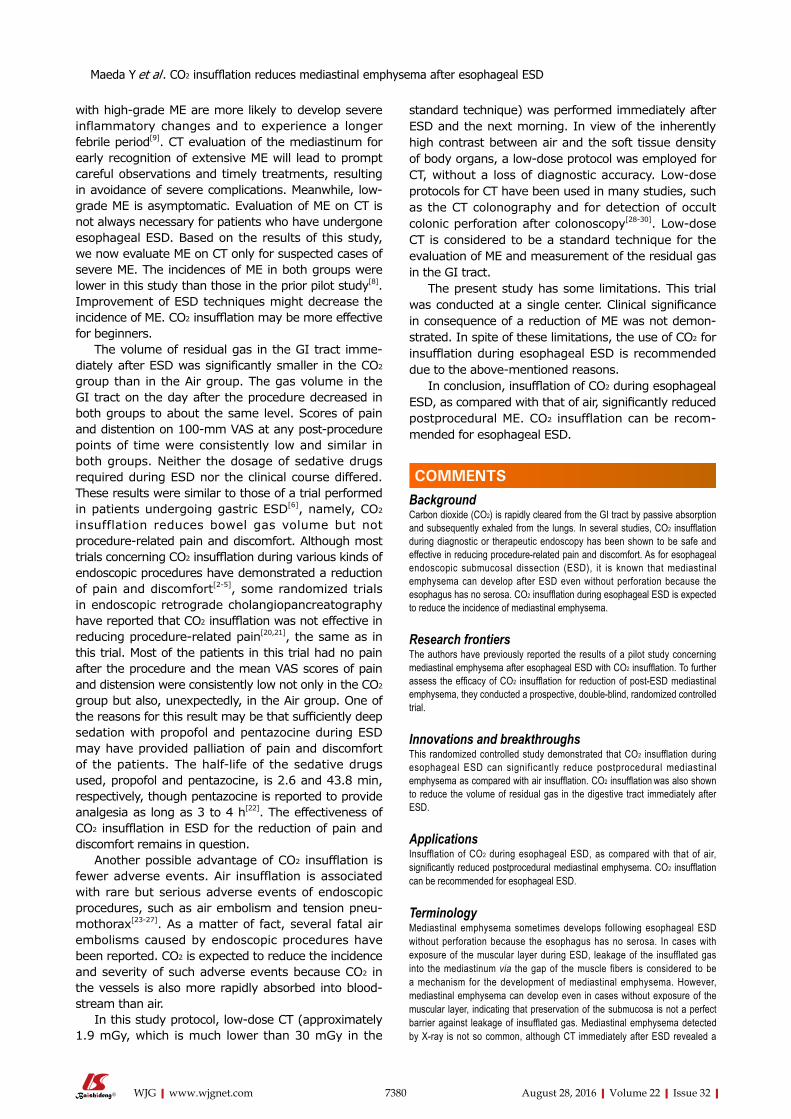
with high-grade ME are more likely to develop severe inflammatory changes and to experience a longer febrile period[9]. CT evaluation of the mediastinum for early recognition of extensive ME will lead to prompt careful observations and timely treatments, resulting in avoidance of severe complications. Meanwhile, low-grade ME is asymptomatic. Evaluation of ME on CT is not always necessary for patients who have undergone esophageal ESD. Based on the results of this study, we now evaluate ME on CT only for suspected cases of severe ME. The incidences of ME in both groups were lower in this study than those in the prior pilot study[8]. Improvement of ESD techniques might decrease the incidence of ME. CO2 insufflation may be more effective for beginners.
The volume of residual gas in the GI tract imme-diately after ESD was significantly smaller in the CO2 group than in the Air group. The gas volume in the GI tract on the day after the procedure decreased in both groups to about the same level. Scores of pain and distention on 100-mm VAS at any post-procedure points of time were consistently low and similar in both groups. Neither the dosage of sedative drugs required during ESD nor the clinical course differed. These results were similar to those of a trial performed in patients undergoing gastric ESD[6], namely, CO2 insufflation reduces bowel gas volume but not procedure-related pain and discomfort. Although most trials concerning CO2 insufflation during various kinds of endoscopic procedures have demonstrated a reduction of pain and discomfort[2-5], some randomized trials in endoscopic retrograde cholangiopancreatography have reported that CO2 insufflation was not effective in reducing procedure-related pain[20,21], the same as in this trial. Most of the patients in this trial had no pain after the procedure and the mean VAS scores of pain and distension were consistently low not only in the CO2 group but also, unexpectedly, in the Air group. One of the reasons for this result may be that sufficiently deep sedation with propofol and pentazocine during ESD may have provided palliation of pain and discomfort of the patients. The half-life of the sedative drugs used, propofol and pentazocine, is 2.6 and 43.8 min, respectively, though pentazocine is reported to provide analgesia as long as 3 to 4 h[22]. The effectiveness of CO2 insufflation in ESD for the reduction of pain and discomfort remains in question.
Another possible advantage of CO2 insufflation is fewer adverse events. Air insufflation is associated with rare but serious adverse events of endoscopic procedures, such as air embolism and tension pneu-mothorax[23-27]. As a matter of fact, several fatal air embolisms caused by endoscopic procedures have been reported. CO2 is expected to reduce the incidence and severity of such adverse events because CO2 in the vessels is also more rapidly absorbed into blood-stream than air.
In this study protocol, low-dose CT (approximately 1.9 mGy, which is much lower than 30 mGy in the
standard technique) was performed immediately after ESD and the next morning. In view of the inherently high contrast between air and the soft tissue density of body organs, a low-dose protocol was employed for CT, without a loss of diagnostic accuracy. Low-dose protocols for CT have been used in many studies, such as the CT colonography and for detection of occult colonic perforation after colonoscopy[28-30]. Low-dose CT is considered to be a standard technique for the evaluation of ME and measurement of the residual gas in the GI tract.
The present study has some limitations. This trial was conducted at a single center. Clinical significance in consequence of a reduction of ME was not demon-strated. In spite of these limitations, the use of CO2 for insufflation during esophageal ESD is recommended due to the above-mentioned reasons.
In conclusion, insufflation of CO2 during esophageal ESD, as compared with that of air, significantly reduced postprocedural ME. CO2 insufflation can be recom-mended for esophageal ESD.
COMMENTSBackgroundCarbon dioxide (CO2) is rapidly cleared from the GI tract by passive absorption and subsequently exhaled from the lungs. In several studies, CO2 insufflation during diagnostic or therapeutic endoscopy has been shown to be safe and effective in reducing procedure-related pain and discomfort. As for esophageal endoscopic submucosal dissection (ESD), it is known that mediastinal emphysema can develop after ESD even without perforation because the esophagus has no serosa. CO2 insufflation during esophageal ESD is expected to reduce the incidence of mediastinal emphysema.
Research frontiersThe authors have previously reported the results of a pilot study concerning mediastinal emphysema after esophageal ESD with CO2 insufflation. To further assess the efficacy of CO2 insufflation for reduction of post-ESD mediastinal emphysema, they conducted a prospective, double-blind, randomized controlled trial.
Innovations and breakthroughsThis randomized controlled study demonstrated that CO2 insufflation during esophageal ESD can significantly reduce postprocedural mediastinal emphysema as compared with air insufflation. CO2 insufflation was also shown to reduce the volume of residual gas in the digestive tract immediately after ESD.
ApplicationsInsufflation of CO2 during esophageal ESD, as compared with that of air, significantly reduced postprocedural mediastinal emphysema. CO2 insufflation can be recommended for esophageal ESD.
TerminologyMediastinal emphysema sometimes develops following esophageal ESD without perforation because the esophagus has no serosa. In cases with exposure of the muscular layer during ESD, leakage of the insufflated gas into the mediastinum via the gap of the muscle fibers is considered to be a mechanism for the development of mediastinal emphysema. However, mediastinal emphysema can develop even in cases without exposure of the muscular layer, indicating that preservation of the submucosa is not a perfect barrier against leakage of insufflated gas. Mediastinal emphysema detected by X-ray is not so common, although CT immediately after ESD revealed a
7380 August 28, 2016|Volume 22|Issue 32|WJG|www.wjgnet.com
COMMENTS
Maeda Y et al . CO2 insufflation reduces mediastinal emphysema after esophageal ESD

certain prevalence of post-ESD mediastinal emphysema. Patients with high-grade mediastinal emphysema are more likely to develop severe inflammatory changes and to experience a longer febrile period. Endoscopists should strive to avoid mediastinal emphysema in esophageal ESD.
Peer-reviewThe work is well-done, well-written, documented and structured. The information included is interesting and the number of cases presented is very valuable. This study provides interesting results on the efficacy of CO2 insufflation for reduction of ME immediately after ESD.
REFERENCES1 Dellon ES, Hawk JS, Grimm IS, Shaheen NJ. The use of carbon
dioxide for insufflation during GI endoscopy: a systematic review. Gastrointest Endosc 2009; 69: 843-849 [PMID: 19152906 DOI: 10.1016/j.gie.2008.05.067]
2 Sumanac K, Zealley I, Fox BM, Rawlinson J, Salena B, Marshall JK, Stevenson GW, Hunt RH. Minimizing postcolonoscopy abdominal pain by using CO(2) insufflation: a prospective, randomized, double blind, controlled trial evaluating a new commercially available CO(2) delivery system. Gastrointest Endosc 2002; 56: 190-194 [PMID: 12145595]
3 Bretthauer M, Thiis-Evensen E, Huppertz-Hauss G, Gisselsson L, Grotmol T, Skovlund E, Hoff G. NORCCAP (Norwegian colorectal cancer prevention): a randomised trial to assess the safety and efficacy of carbon dioxide versus air insufflation in colonoscopy. Gut 2002; 50: 604-607 [PMID: 11950803]
4 Maple JT, Keswani RN, Hovis RM, Saddedin EZ, Jonnalagadda S, Azar RR, Hagen C, Thompson DM, Waldbaum L, Edmundowicz SA. Carbon dioxide insufflation during ERCP for reduction of postprocedure pain: a randomized, double-blind, controlled trial. Gastrointest Endosc 2009; 70: 278-283 [PMID: 19523621 DOI: 10.1016/j.gie.2008.12.050]
5 Domagk D, Bretthauer M, Lenz P, Aabakken L, Ullerich H, Maaser C, Domschke W, Kucharzik T. Carbon dioxide insufflation improves intubation depth in double-balloon enteroscopy: a randomized, controlled, double-blind trial. Endoscopy 2007; 39: 1064-1067 [PMID: 18072057]
6 Maeda Y, Hirasawa D, Fujita N, Obana T, Sugawara T, Ohira T, Harada Y, Yamagata T, Suzuki K, Koike Y, Kusaka J, Tanaka M, Noda Y. A prospective, randomized, double-blind, controlled trial on the efficacy of carbon dioxide insufflation in gastric endoscopic submucosal dissection. Endoscopy 2013; 45: 335-341 [PMID: 23468193 DOI: 10.1055/s-0032-1326199]
7 Saito Y, Uraoka T, Matsuda T, Emura F, Ikehara H, Mashimo Y, Kikuchi T, Kozu T, Saito D. A pilot study to assess the safety and efficacy of carbon dioxide insufflation during colorectal endoscopic submucosal dissection with the patient under conscious sedation. Gastrointest Endosc 2007; 65: 537-542 [PMID: 17321264]
8 Maeda Y, Hirasawa D, Fujita N, Obana T, Sugawara T, Ohira T, Harada Y, Yamagata T, Suzuki K, Koike Y, Yamamoto Y, Kusaka Z, Noda Y. A pilot study to assess mediastinal emphysema after esophageal endoscopic submucosal dissection with carbon dioxide insufflation. Endoscopy 2012; 44: 565-571 [PMID: 22407383 DOI: 10.1055/s-0031-1291664]
9 Maeda Y, Hirasawa D, Fujita N, Suzuki T, Obana T, Sugawara T, Ohira T, Harada Y, Noda Y. Mediastinal emphysema after esophageal endoscopic submucosal dissection: its prevalence and clinical significance. Dig Endosc 2011; 23: 221-226 [PMID: 21699565 DOI: 10.1111/j.1443-1661.2010.01085.x]
10 Oyama T, Tomori A, Hotta K, Morita S, Kominato K, Tanaka M, Miyata Y. Endoscopic submucosal dissection of early esophageal cancer. Clin Gastroenterol Hepatol 2005; 3: S67-S70 [PMID: 16013002]
11 Tamiya Y, Nakahara K, Kominato K, Serikawa O, Watanabe Y, Tateishi H, Takedatsu H, Toyonaga A, Sata M. Pneumomediastinum is a frequent but minor complication during esophageal endoscopic submucosal dissection. Endoscopy 2010; 42: 8-14 [PMID:
19899032 DOI: 10.1055/s-0029-1215215]12 Committee for the Third Edition of the COPD Guidelines of The
Japanese Respiratory Society. Guidelines for the Diagnosis and Treatment of COPD (Chronic Obstructive Pulmonary Disease). 3rd ed. 2009
13 Faigel DO, Baron TH, Goldstein JL, Hirota WK, Jacobson BC, Johanson JF, Leighton JA, Mallery JS, Peterson KA, Waring JP, Fanelli RD, Wheeler-Harbaugh J. Guidelines for the use of deep sedation and anesthesia for GI endoscopy. Gastrointest Endosc 2002; 56: 613-617 [PMID: 12397263]
14 Training Committee. American Society for Gastrointestinal Endo-scopy. Training guideline for use of propofol in gastrointestinal endoscopy. Gastrointest Endosc 2004; 60: 167-172 [PMID: 15278039]
15 Yamagata T, Hirasawa D, Fujita N, Suzuki T, Obana T, Sugawara T, Ohira T, Harada Y, Maeda Y, Koike Y, Suzuki K, Noda Y. Efficacy of propofol sedation for endoscopic submucosal dissection (ESD): assessment with prospective data collection. Intern Med 2011; 50: 1455-1460 [PMID: 21757829]
16 Lee TY, Chhem RK. Impact of new technologies on dose reduction in CT. Eur J Radiol 2010; 76: 28-35 [PMID: 20643522 DOI: 10.1016/j.ejrad.2010.06.036]
17 Kalra MK, Naz N, Rizzo SM, Blake MA. Computed tomography radiation dose optimization: scanning protocols and clinical applications of automatic exposure control. Curr Probl Diagn Radiol 2005; 34: 171-181 [PMID: 16129235]
18 Matsumoto K, Ohno Y, Koyama H, Kono A, Inokawa H, Onishi Y, Nogami M, Takenaka D, Araki T, Sugimura K. 3D automatic exposure control for 64-detector row CT: radiation dose reduction in chest phantom study. Eur J Radiol 2011; 77: 522-527 [PMID: 19836179 DOI: 10.1016/j.ejrad.2009.09.012]
19 The Japan Esophageal Society. Japanese Classification of Esophageal Cacer[article in Japanese with English abstract in part].10th ed. Tokyo: Kanehara Shuppan, 2008
20 Dellon ES, Velayudham A, Clarke BW, Isaacs KL, Gangarosa LM, Galanko JA, Grimm IS. A randomized, controlled, double-blind trial of air insufflation versus carbon dioxide insufflation during ERCP. Gastrointest Endosc 2010; 72: 68-77 [PMID: 20493485 DOI: 10.1016/j.gie.2010.01.041]
21 Kuwatani M, Kawakami H, Hayashi T, Ishiwatari H, Kudo T, Yamato H, Ehira N, Haba S, Eto K, Kato M, Asaka M. Carbon dioxide insufflation during endoscopic retrograde cholangiopancreatography reduces bowel gas volume but does not affect visual analogue scale scores of suffering: a prospective, double-blind, randomized, controlled trial. Surg Endosc 2011; 25: 3784-3790 [PMID: 21656068 DOI: 10.1007/s00464-011-1789-8]
22 Sadove MS, Balagot RC. Pentazocine--A New Nonaddicting Analgesic. A Double-Blind Evaluation in Postoperative Pain. JAMA 1965; 193: 887-892 [PMID: 14341131]
23 Katzgraber F, Glenewinkel F, Fischler S, Rittner C. Mechanism of fatal air embolism after gastrointestinal endoscopy. Int J Legal Med 1998; 111: 154-156 [PMID: 9587799]
24 Bisceglia M, Simeone A, Forlano R, Andriulli A, Pilotto A. Fatal systemic venous air embolism during endoscopic retrograde cholangiopancreatography. Adv Anat Pathol 2009; 16: 255-262 [PMID: 19546613 DOI: 10.1097/PAP.0b013e3181aab793]
25 Green BT, Tendler DA. Cerebral air embolism during upper endoscopy: case report and review. Gastrointest Endosc 2005; 61: 620-623 [PMID: 15812425]
26 Morley AP, Lau JY, Young RJ. Tension pneumothorax complicating a perforation of a duodenal ulcer during ERCP with endoscopic sphincterotomy. Endoscopy 1997; 29: 332 [PMID: 9255546]
27 Rai A, Iftikhar S. Tension pneumothorax complicating diagnostic upper endoscopy: a case report. Am J Gastroenterol 1999; 94: 845-847 [PMID: 10086678]
28 van Gelder RE, Venema HW, Florie J, Nio CY, Serlie IW, Schutter MP, van Rijn JC, Vos FM, Glas AS, Bossuyt PM, Bartelsman JF, Laméris JS, Stoker J. CT colonography: feasibility of substantial dose reduction--comparison of medium to very low doses in identical patients. Radiology 2004; 232: 611-620 [PMID: 15215541]
7381 August 28, 2016|Volume 22|Issue 32|WJG|www.wjgnet.com
Maeda Y et al . CO2 insufflation reduces mediastinal emphysema after esophageal ESD

29 Florie J, van Gelder RE, Schutter MP, van Randen A, Venema HW, de Jager S, van der Hulst VP, Prent A, Bipat S, Bossuyt PM, Baak LC, Stoker J. Feasibility study of computed tomography colonography using limited bowel preparation at normal and low-dose levels study. Eur Radiol 2007; 17: 3112-3122 [PMID: 17549490]
30 Hough DM, Kuntz MA, Fidler JL, Johnson CD, Petersen BT, Kofler JM, Fletcher JG. Detection of occult colonic perforation before CT colonography after incomplete colonoscopy: perforation rate and use of a low-dose diagnostic scan before CO2 insufflation. AJR Am J Roentgenol 2008; 191: 1077-1081 [PMID: 18806146 DOI: 10.2214/AJR.07.2746]
P- Reviewer: Gonzalez ES, Tan YF S- Editor: Qi Y L- Editor: Filipodia E- Editor: Ma S
7382 August 28, 2016|Volume 22|Issue 32|WJG|www.wjgnet.com
Maeda Y et al . CO2 insufflation reduces mediastinal emphysema after esophageal ESD

Does massive intraabdominal free gas require surgical intervention?
Tadashi Furihata, Makoto Furihata, Kunibumi Ishikawa, Masato Kosaka, Naoki Satoh, Keiichi Kubota
Tadashi Furihata, Makoto Furihata, Kunibumi Ishikawa, Masato Kosaka, Naoki Satoh, Department of General Surgery, Kyouwa Chuo Hospital, Chikusei-shi, Ibaraki 309-1195, Japan
Tadashi Furihata, Keiichi Kubota, Department of Gastro-enterological Surgery, Dokkyo Medical University, Shimotsuga-gun, Tochigi 321-0293, Japan
Author contributions: Furihata T served as an attending doctor for the patient, designed the report, and wrote the manuscript; Furihata M contributed equally to this manuscript; Ishikawa K, Kosaka M and Satoh N clinically managed the present case; and Kubota K comprehensively revised the manuscript.
Institutional review board statement: This manuscript was reviewed and approved by the institutional review board of Kyouwa Chuo Hospital.
Informed consent statement: Written informed consent was obtained from the patient for publication of the case report and the accompanying images.
Conflict-of-interest statement: The authors declare no conflicts of interest.
Open-Access: This article is an open-access article which was selected by an in-house editor and fully peer-reviewed by external reviewers. It is distributed in accordance with the Creative Commons Attribution Non Commercial (CC BY-NC 4.0) license, which permits others to distribute, remix, adapt, build upon this work non-commercially, and license their derivative works on different terms, provided the original work is properly cited and the use is non-commercial. See: http://creativecommons.org/licenses/by-nc/4.0/
Manuscript source: Unsolicited manuscript
Correspondence to: Tadashi Furihata, MD, PhD, Department of General Surgery, Kyouwa Chuo Hospital, 1676-1 Kadoi, Chikusei-shi, Ibaraki 309-1195, Japan. [email protected]: +81-296-576131Fax: 81-296-574676
Received: March 26, 2016 Peer-review started: March 27, 2016
CASE REPORT
Submit a Manuscript: http://www.wjgnet.com/esps/Help Desk: http://www.wjgnet.com/esps/helpdesk.aspxDOI: 10.3748/wjg.v22.i32.7383
World J Gastroenterol 2016 August 28; 22(32): 7383-7388 ISSN 1007-9327 (print) ISSN 2219-2840 (online)
© 2016 Baishideng Publishing Group Inc. All rights reserved.
7383 August 28, 2016|Volume 22|Issue 32|WJG|www.wjgnet.com
First decision: May 12, 2016Revised: May 24, 2016 Accepted: June 15, 2016 Article in press: June 15, 2016Published online: August 28, 2016
AbstractWe describe a rare case of an 81-year-old man who presented with severe epigastralgia. A chest radio-graph showed massive free gas bilaterally in the diaphragmatic spaces. Computed tomography (CT) scan also showed massive free gas in the peritoneal cavity with portal venous gas. We used a wait-and-see approach and carefully considered surgery again when the time was appropriate. The patient received conservative therapy with fasting, an intravenous infusion of antibiotics, and nasogastric intubation. The patient soon recovered and was able to start eating meals 4 d after treatment; thus, surgical intervention was avoided. Thereafter, colonoscopy examination showed pneumatosis cystoides intestinalis in the ascending colon. On retrospective review, CT scan demonstrated sporadic air-filled cysts in the ascending colon. The present case taught us a lesson: the presence of massive intraabdominal free gas with portal venous gas does not necessarily require surgical intervention. Pneumatosis cystoides intestinalis should be considered as a potential causative factor of free gas with portal venous gas when making the differential diagnosis.
Key words: Case reports; Portal vein; Pneumatosis cystoides intestinalis; Colonoscopy; General surgery
© The Author(s) 2016. Published by Baishideng Publishing Group Inc. All rights reserved.
Core tip: We describe a rare case of an 81-year-old man with pneumatosis cystoides intestinalis (PCI).

CASE REPORTAn 81-year-old man who presented at our emergency department with acute severe epigastralgia, perium-bilical pain, and progressive abdominal distention was admitted to the Department of General Surgery at our institution. The patient visited our institution for an old myocardial and cerebral infarction, hypertension, and chronic obstipation, and he received a variety of medications. He had a mildly increased body temperature (37.2 ℃). His cramping but persistent pain started abruptly. Physical examination showed severe epigastric tenderness and distension without any peritoneal signs. His facial appearance expressed agony, and he presented with excessive sweating. His blood pressure slightly decreased to 12.7/8.65 kPa, which was his usual hypertensive state, and his pulse rate was 61 beats/min. Regarding the hematological parameters, the white blood cell count and C-reactive protein level were mildly increased (10, 500/µL and 0.91 mg/dL, respectively). Serum levels of blood urea nitrogen and creatinine were also increased (26.3 mg/dL and 1.29 mg/dL, respectively), and the levels were slightly different compared to those of his blood samples collected during his normal state (18.5 mg/dL and 0.89 mg/dL, respectively). On radiologic examination, a chest and abdominal plain radiograph showed massive free gas bilaterally in the subdiaphragmatic spaces (Figure 1). Computed tomography (CT) scan also showed that massive free gas was widespread bilaterally under the diaphragm. Moreover, PVG was identified, but ascites was not (Figure 2). Consequently, we performed arterial blood gas analysis, which showed no abnormality (pH = 7.46). First, we considered perforation of the digestive tract. Then, we assumed that the pathological digestive tract would have been accompanied by irreversible ischemia; however, we could not establish a definitive preoperative diagnosis. We used a wait-and-see approach, because the phy-
Furihata T et al . PCI with massive intraabdominal free gas
7384 August 28, 2016|Volume 22|Issue 32|WJG|www.wjgnet.com
Figure 1 Plain radiograph demonstrating massive intraabdominal free gas bilaterally in the subdiaphragmatic spaces (white arrows). Grape-like clusters or honeycomb-shaped shadows, which are characteristic of pneumatosis cystoides intestinalis, were not observed.
A BPCI is characterized by free gas in the submucosal or subserosal layer of the gastrointestinal tract, and its etiology is unknown. The patient presented with massive intraabdominal free gas and portal venous gas (PVG) due to PCI, and he was successfully treated without surgical intervention. When clinicians encounter free gas with PVG, PCI should be considered.
Furihata T, Furihata M, Ishikawa K, Kosaka M, Satoh N, Kubota K. Does massive intraabdominal free gas require surgical intervention? World J Gastroenterol 2016; 22(32): 7383-7388 Available from: URL: http://www.wjgnet.com/1007-9327/full/v22/i32/7383.htm DOI: http://dx.doi.org/10.3748/wjg.v22.i32.7383
INTRODUCTIONPneumatosis cystoides intestinalis (PCI) is a rare clinical entity with an unknown etiology, and it is characterized by free gas in the submucosal or subserosal layer of the gastrointestinal tract[1-3]. PCI has been associated with various underlying etiologies to explain the pathogenic mechanisms causally involved in the accumulation of intramural gas. There are four main theories: (1) the mechanical theory; (2) pulmonary theory; (3) bacterial theory; and (4) chemical theory or nutrition deficiency theory. Currently, chemotherapy, hormonal therapy, steroids, immunosuppression, and connective tissue disease have been reported as causative factors of PCI[2,4-8]. Yet, the exact theory has not been determined. Symptoms usually stem from a secondary underlying disease, including abdominal discomfort, diarrhea, constipation, rectal bleeding, tenesmus, or weight loss. About 3% of patients with PCI develop complications, including tension pneumoperitoneum, intestinal volvulus, obstruction, hemorrhage, and intestinal perforation[9]. One study reported a higher rate of complications with PCI (16.3%), including intestinal obstruction (51.3%), intestinal perforation (35.9%), atypical hyperplasia and canceration (10.2%), and intussusceptions and intestinal necrosis (2.6%)[2]. The most important issue for physicians is recognizing the entity of PCI so that they do not misdiagnose or mismanage it as another disease such as a malignancy or polyposis. It is also important to differentiate the harmless type from the life-threatening type, for which immediate surgery is required[8]. Thus, the conditions of patients with PCI can be confusing, and the decision to treat PCI should be carefully made in consideration of the risks. We herein emphasize how all physicians in the field of gastroenterology can recognize PCI by describing a patient who presented with massive intraabdominal free gas and portal venous gas (PVG) who was successfully treated without surgical intervention.
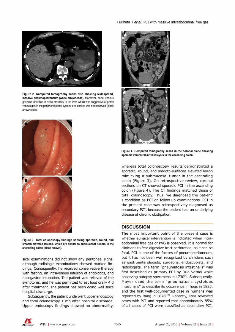
sical examinations did not show any peritoneal signs, although radiologic examinations showed marked findings. Consequently, he received conservative therapy with fasting, an intravenous infusion of antibiotics, and nasogastric intubation. The patient was relieved of the symptoms, and he was permitted to eat food orally 4 d after treatment. The patient has been doing well since hospital discharge.
Subsequently, the patient underwent upper endoscopy and total colonoscopy 1 mo after hospital discharge. Upper endoscopy findings showed no abnormality,
whereas total colonoscopy results demonstrated a sporadic, round, and smooth-surfaced elevated lesion mimicking a submucosal tumor in the ascending colon (Figure 3). On retrospective review, coronal sections on CT showed sporadic PCI in the ascending colon (Figure 4). The CT findings matched those of total colonoscopy. Thus, we diagnosed the patient’s condition as PCI on follow-up examinations. PCI in the present case was retrospectively diagnosed as secondary PCI, because the patient had an underlying disease of chronic obstipation.
DISCUSSIONThe most important point of the present case is whether surgical intervention is indicated when intra-abdominal free gas or PVG is observed. It is normal for clinicians to fear digestive tract perforation, as it can be fatal. PCI is one of the factors of pneumoperitoneum, but it has not been well recognized by clinicians such as gastroenterologists, surgeons, endoscopists, and radiologists. The term “pneumatosis intestinalis” was first described as primary PCI by Duo Vernoi while observing autopsy specimens in 1730[1]. Subsequently, Mayer used the term “pneumatosis cystoides intestinalis” to describe its occurrence in hogs in 1825, and the first well-documented case in humans was reported by Bang in 1876[10]. Recently, Koss reviewed cases with PCI and reported that approximately 85% of all cases of PCI were classified as secondary PCI,
7385 August 28, 2016|Volume 22|Issue 32|WJG|www.wjgnet.com
Figure 2 Computed tomography scans also showing widespread, massive pneumoperitoneum (white arrowheads). Moreover, portal venous gas was identified in close proximity to the liver, which was suggestive of portal venous gas in the peripheral portal system, and ascites was not observed (black arrowheads).
A
B
Figure 3 Total colonoscopy findings showing sporadic, round, and smooth elevated lesions, which are similar to submucosal tumors in the ascending colon (black arrows).
A
B
Figure 4 Computed tomography scans in the coronal plane showing sporadic intramural air-filled cysts in the ascending colon.
Furihata T et al . PCI with massive intraabdominal free gas

88 patients with PCI, 19 were diagnosed as having benign PCI, including 6 patients with both PI and PVG[14]. Considering these cases, PCI may be one of the factors that induce PVG, which can also be fatal. Conversely, PCI and PVG are associated with each other, and they can occur together in various non-ischemic conditions that are not associated with an unfavorable outcome[13,14,18-20]. Even if life-threatening conditions of intraabdominal free gas, PCI, and PVG occur simultaneously, patients’ conditions may not necessarily be serious. Thus, deepening the knowledge of these conditions is essential for clinicians (Table 1). In the present case, the mechanisms of these life-threatening conditions were connected, and the following hypotheses have been made. First, secondary PCI probably occurred due to mucosal disruption caused by chronic obstipation. Second, increased gas in the serosal or subserosal layer is partially transported via the mesenteric drainage veins to the hepatic portal veins. Sequentially, ruptures of serosal or subserosal cysts cause pneumoperitoneum. Therefore, physicians must determine whether it is best for patients to undergo surgical intervention. Considering these life-threatening conditions, the most important diagnostic information is patients’ vital signs and complaints, and physical examination of the abdomen.
In general, surgical indications of PCI include cases suggestive of inconvertible intestinal obstruction or perforation, or patients with precancerous con-ditions[13,21]. Wu et al[2] mentioned that these compli-cations associated with PCI occur in approximately 16.3% of cases. Hence, radiologic investigations are important for diagnosing PCI. Particularly, multidetector
which results from other underlying diseases[11]. In the remaining 15% of cases, PCI was idiopathic or primary. Morris et al[12] reported that the incidence of PCI was 46% in the colon, followed by 27% in the small intestine, 7% in the colon and small intestine combined, 5% in the stomach, and 15% in other parts of the gastrointestinal tract. The most common localization of gas was in the submucosa (69.9%)[2]. On the basis of autopsy studies, its incidence in the general population has been estimated as three per 10000 individuals[13,14]. Its recognition has not been widespread in terms of frequency.
PVG is also a radiological sign of serious underlying gastrointestinal pathology. The mortality for PVG ranges between 75% and 90%. The most common cause of PVG is acute mesenteric ischemia. Although hepatic PVG helps distinguish between benign and life-threatening PCI, it may also occur with or without PCI due to nonischemic conditions. Mesenteric abscess formation, portomesenteric thrombophlebitis, sepsis, abdominal trauma, severe enteritis, cholangitis, chronic cholecystitis, pancreatitis, inflammatory bowel disease, diverticulitis, and gastrointestinal surgery or liver transplantation are considered causative factors of PVG[14,15]. The association between PVG and PCI seems to have been noted first in the article by Wolfe and Evans in 1955[15,16]. They reported gas in the portal veins of six living infants, three of whom had associated pneumatosis of the bowel. Khalil et al[13] reported that the combination of PCI and PVG is associated with bowel ischemia in about 70% of cases[13,17,18]. Sooby et al[15] classified 88 cases of PCI with PVG into three distinct subgroups: mechanical, ischemic, and benign. They also reported that of
7386 August 28, 2016|Volume 22|Issue 32|WJG|www.wjgnet.com
Table 1 Causative factors of intraabdominal free gas
Pulmonary disease Drugs Infectious disease Asthma Alpha-glucosidase inhibitor AIDS Chronic bronchitis Corticosteroids Whipple’ disease Chronic obstructive pulmonary disease Lactulose Candida albicans Positive end-expiratory pressure Sorbitol Clostridia Pulmonary fibrosis Chemotherapy agents Escherichia coliGastrointestinal disorders Gefitinib, Sorafenib, Cetuximab, Mycobacterium tuberculosis Pyloric stenosis Sunitinib, imatinib, 5-FU, etc. Intestinal parasites Pyloric ulcer disease Choral hydrate Virus Peptic ulcers Caustic agents Cytomegalovirus, Rotavirus, Bowel obstruction Mechanical causes Adenovirus, Varicella zoster virus Rupture of jejunal diverticula Endoscopy Autoimmune disease Inflammatory bowel disease Billiary stent perforation, Sclerotherapy Scleroderma Crohn’s disease, ulcerative colitis Barium enema Lupus variants Adynamic ileus Operation Polymyositis Appendicitis Jejunoileal bypass, Jejunostomy tubes, Dermatomyositis Toxic megacolon Post-surgical anastomosis Sarcoidosis Volvulus Organ transplantation Polyarteritis nodosa Carcinoma Cardiac, Bone marrow, Kidney, Liver, Vascular disease Hirschsprung disease Lung, Graft versus host disease Mesenteric vascular disease Celiac sprue Trauma Intestinal infarction and ischemia Enteritis and colitis Intestinal ischemia Diverticulitis Intestinal strangulation Emphysematous gastritis Collagen vascular disease Bowel necrosis
Furihata T et al . PCI with massive intraabdominal free gas

CT is clearly beneficial, because it enables clinicians to make a more confident diagnosis of PCI by reformations in the coronal, sagittal, and axial planes[3,14]. However, radiologic or endoscopic examination does not nece-ssarily show typical findings, which means that patients with PCI cannot be diagnosed definitively, as shown in the present case. We reached the definite diagnosis retrospectively by reformation of CT in the coronal plane.
We were able to avoid surgical intervention in the present case because of the following important factors: (1) the patient did not show any abdominal peritoneal signs; (2) arterial blood gas measurement did not show metabolic acidosis; (3) radiological examinations demonstrated that there was a consi-derable amount of free gas bilaterally in the subdia-phragmatic spaces together with PVG, but no ascites was observed; and (4) despite mild renal dysfunction, urine output was obtained, and the patient had stable vital signs. It was a bold decision to avoid surgical intervention, because the patient would have required prompt action if perforation or ischemia of the digestive tract perforation had occurred. Interestingly, nine of 27 patients with PCI died for an overall mortality rate of 33%. Eleven patients observed without surgery had a mortality rate of 18%, whereas those who underwent surgery had a mortality rate of 44%. The remaining nine patients who improved without surgery did not manifest any clinical signs that would have prompted surgery[22]. Therefore, it seems that some patients with PCI can be successfully managed with conservative treatment. However, 37% to 75% of patients with PVG have bowel infarction, and 10 of 12 patients with both PCI and PVG died within 48 h[8]. The long-term prognosis of PCI is unknown, and a long-term follow-up study is required to evaluate this further.
Thus, complementary evaluations such as blood gas analysis can be helpful. Knechtle et al[22] advocated the classification of clinical and laboratory values, including the assessment of arterial blood gas (pH, HCO3-), so that they could predict the occurrence of ischemic bowel and the patient’s outcome. Khalil et al[13] propounded a decision-making algorithm after PCI is diagnosed. The proposal makes sense, because the checklists include a variety of essential criteria for diagnosing PCI, including physical conditions, physical examinations, and medical history intake. Obtaining the patient’s medical history of comorbidities such as pulmonary disease, gastrointestinal disease, autoimmune disease, infectious disease, and vascular disease, as well as possible iatrogenic causes and the use of drugs is very important for cases of secondary PCI. The lack of knowledge may lead to misdiagnosis, which causes the patient to undergo unnecessary operations. As previously described, the fact that CT did not show ascites was favorable, because ascites often indicate ischemia or perforation of the gastrointestinal tract. Briefly, if CT demonstrates
intraabdominal free gas with PVG in the absence of ascites, PCI is a possible diagnosis.
In conclusion, massive intraabdominal free gas with PVG does not always require surgical intervention. Therefore, the decision to perform surgery should be made on the basis of the knowledge of PCI. When clinicians encounter free gas with PVG in the abdomen, PCI should be considered when making the differential diagnosis.
ACKNOWLEDGMENTSWe thank Yuki Matsuhashi, Department of Endocri-nology and Metabolism, and Hirosaki University Graduate School of Medicine for their assistance in making the tables and figures.
COMMENTSCase characteristicsAn 81-year-old man presented with severe epigastralgia.
Clinical diagnosisPhysical examination showed severe epigastric tenderness and distension without any peritoneal signs.
Laboratory diagnosisRegarding the hematological parameters, the white blood cell count and C-reactive protein level were mildly increased (10.500/µL and 0.91 mg/dL, respectively). Serum levels of blood urea nitrogen and creatinine were also increased (26.3 mg/dL and 1.29 mg/dL, respectively). Arterial blood gas analysis showed no abnormality (PH = 7.46).
Imaging diagnosisComputed tomography scan showed that massive free gas was widespread bilaterally under the diaphragm. Moreover, portal venous gas was identified, but ascites was not observed.
Pathological diagnosisThere was no specimen to make a pathological diagnosis.
TreatmentThe authors used a wait-and-see approach, because the physical examinations did not show any peritoneal signs, although radiologic examinations showed marked findings. Consequently, he received conservative therapy with fasting, an intravenous infusion of antibiotics, and nasogastric intubation. We were able to avoid surgical intervention.
Related reportsPneumatosis cystoides intestinalis (PCI) is a rare entity. In addition, there are few reports of patients with PCI successfully treated without surgical intervention.
Term explanationPCI is a rare clinical entity with an unknown etiology, and it is characterized by free gas in the submucosal or subserosal layer of the gastrointestinal tract.
Experience and lessonsThe present case taught us the following lesson: the presence of intraabdominal free gas with portal venous gas (PVG) does not necessarily require surgical intervention. When gastroenterologists encounter free gas with PVG, PCI should be considered.
7387 August 28, 2016|Volume 22|Issue 32|WJG|www.wjgnet.com
COMMENTS
Furihata T et al . PCI with massive intraabdominal free gas

Peer-reviewIt is a case report of a patient with abdominal pain that was proved to be caused by PCI, and was managed conservatively. It is very interesting.
REFERENCES1 Du Vernoy JG. Aer intestinorum tam sub extima quam intima
tunica inclusus: observationes anatomicae: comment. Acad Scient Petropol 1730; 5: 213-225
2 Wu LL, Yang YS, Dou Y, Liu QS. A systematic analysis of pneumatosis cystoids intestinalis. World J Gastroenterol 2013; 19: 4973-4978 [PMID: 23946603 DOI: 10.3748/wjg.v19.i30.4973]
3 Ivanović A, Kovač J, Mašulović D, Stefanović A, Jakšić E, Saranović D. Education and imaging. Gastrointestinal: the role of multidetector computer tomography in diagnosis of pneumatosis cystoides intestinalis. J Gastroenterol Hepatol 2012; 27: 182 [PMID: 22188029 DOI: 10.1111/j.1440-1746.2011.06952.x]
4 Groninger E, Hulscher JB, Timmer B, Tamminga RY, Broens PM. Free air intraperitoneally during chemotherapy for acute lymphoblastic leukemia: consider pneumatosis cystoides intestinalis. J Pediatr Hematol Oncol 2010; 32: 141-143 [PMID: 20147849 DOI: 10.1097/MPH.0b013e3181ced397]
5 Vendryes C, Hunter CJ, Harlan SR, Ford HR, Stein J, Pierce JR. Pneumatosis intestinalis after laparoscopic appendectomy: case report and review of the literature. J Pediatr Surg 2011; 46: e21-e24 [PMID: 22075367 DOI: 10.1016/j.jpedsurg.2011.07.022]
6 Balbir-Gurman A, Brook OR, Chermesh I, Braun-Moscovici Y. Pneumatosis cystoides intestinalis in scleroderma-related conditions. Intern Med J 2012; 42: 323-329 [PMID: 22432985 DOI: 10.1111/j.1445-5994.2011.02557.x]
7 Korhonen K, Lovvorn HN, Koyama T, Koehler E, Calder C, Manes B, Evans M, Bruce K, Ho RH, Domm J, Frangoul H. Incidence, risk factors, and outcome of pneumatosis intestinalis in pediatric stem cell transplant recipients. Pediatr Blood Cancer 2012; 58: 616-620 [PMID: 21721114 DOI: 10.1002/pbc.23242]
8 Heng Y, Schuffler MD, Haggitt RC, Rohrmann CA. Pneumatosis intestinalis: a review. Am J Gastroenterol 1995; 90: 1747-1758 [PMID: 7572888]
9 Arikanoglu Z, Aygen E, Camci C, Akbulut S, Basbug M, Dogru O, Cetinkaya Z, Kirkil C. Pneumatosis cystoides intestinalis: a single center experience. World J Gastroenterol 2012; 18: 453-457 [PMID: 22346251 DOI: 10.3748/wjg.v18.i5.453]
10 Pear BL. Pneumatosis intestinalis: a review. Radiology 1998; 207:
13-19 [PMID: 9530294]11 KOSS LG . Abdominal gas cysts (pneumatosis cystoides
intestinorum hominis); an analysis with a report of a case and a critical review of the literature. AMA Arch Pathol 1952; 53: 523-549 [PMID: 14923068]
12 Morris MS, Gee AC, Cho SD, Limbaugh K, Underwood S, Ham B, Schreiber MA. Management and outcome of pneumatosis intestinalis. Am J Surg 2008; 195: 679-682; discussion 682-683 [PMID: 18424288 DOI: 10.1016/j.amjsurg.2008.01.011]
13 Khalil PN, Huber-Wagner S, Ladurner R, Kleespies A, Siebeck M, Mutschler W, Hallfeldt K, Kanz KG. Natural history, clinical pattern, and surgical considerations of pneumatosis intestinalis. Eur J Med Res 2009; 14: 231-239 [PMID: 19541582]
14 Ho LM, Paulson EK, Thompson WM. Pneumatosis intestinalis in the adult: benign to life-threatening causes. AJR Am J Roentgenol 2007; 188: 1604-1613 [PMID: 17515383]
15 Sooby P, Harshen R, Joarder R. An unusual triad of pneumatosis intestinalis, portal venous gas and pneumoperitoneum in an asymptomatic patient. J Surg Case Rep 2015; 2015: pii rjv035 [PMID: 25858266 DOI: 10.1093/jscr/rjv035]
16 Wolfe JN, Evans WA. Gas in the portal veins of the liver in infants; a roentgenographic demonstration with postmortem anatomical correlation. Am J Roentgenol Radium Ther Nucl Med 1955; 74: 486-488 [PMID: 13249015]
17 Paran H, Epstein T, Gutman M, Shapiro Feinberg M, Zissin R. Mesenteric and portal vein gas: computerized tomography findings and clinical significance. Dig Surg 2003; 20: 127-132 [PMID: 12686780]
18 Wiesner W, Mortelé KJ, Glickman JN, Ji H, Ros PR. Portal-venous gas unrelated to mesenteric ischemia. Eur Radiol 2002; 12: 1432-1437 [PMID: 12042950]
19 Wiesner W, Mortelé KJ, Glickman JN, Ji H, Ros PR. Pneumatosis intestinalis and portomesenteric venous gas in intestinal ischemia: correlation of CT findings with severity of ischemia and clinical outcome. AJR Am J Roentgenol 2001; 177: 1319-1323 [PMID: 11717075]
20 Griffiths DM, Gough MH. Gas in the hepatic portal veins. Br J Surg 1986; 73: 172-176 [PMID: 3512030]
21 Zhang H, Jun SL, Brennan TV. Pneumatosis intestinalis: not always a surgical indication. Case Rep Surg 2012; 2012: 719713 [PMID: 23198249 DOI: 10.1155/2012/719713]
22 Knechtle SJ, Davidoff AM, Rice RP. Pneumatosis intestinalis. Surgical management and clinical outcome. Ann Surg 1990; 212: 160-165 [PMID: 2375647]
P- Reviewer: Misiakos EP S- Editor: Ma YJ L- Editor: A E- Editor: Ma S
7388 August 28, 2016|Volume 22|Issue 32|WJG|www.wjgnet.com
Furihata T et al . PCI with massive intraabdominal free gas

© 2016 Baishideng Publishing Group Inc. All rights reserved.
Published by Baishideng Publishing Group Inc8226 Regency Drive, Pleasanton, CA 94588, USA
Telephone: +1-925-223-8242Fax: +1-925-223-8243
E-mail: [email protected] Desk: http://www.wjgnet.com/esps/helpdesk.aspx
http://www.wjgnet.com
I S S N 1 0 0 7 - 9 3 2 7
9 7 7 1 0 07 9 3 2 0 45
3 2