World Journal of Hepatology - NET
98
World Journal of Hepatology World J Hepatol 2014 July 27; 6(7): 453-537 Published by Baishideng Publishing Group Inc ISSN 1948-5182 (online)
-
Upload
khangminh22 -
Category
Documents
-
view
0 -
download
0
Transcript of World Journal of Hepatology - NET

World Journal of HepatologyWorld J Hepatol 2014 July 27; 6(7): 453-537
Published by Baishideng Publishing Group Inc
ISSN 1948-5182 (online)

EDITORS-IN-CHIEFClara Balsano, RomeWan-Long Chuang, Kaohsiung
GUEST EDITORIAL BOARD MEMBERSKing-Wah Chiu, KaohsiungTai-An Chiang, TainanChi-Tan Hu, HualienSen-Yung Hsieh, TaoyuanWenya Huang, TainanLiang-Yi Hung, TainanJih RU Hwu, HsinchuJing-Yi Lee, TaipeiMei-Hsuan Lee, TaipeiChih-Wen Lin, KaohsiungChun-Che Lin, TaichungWan-Yu Lin, TaichungTai-Long Pan, Tao-YuanSuh-Ching Yang, TaipeiChun-Yan Yeung, Taipei
MEMBERS OF THE EDITORIAL BOARD
Algeria
Samir Rouabhia, Batna
Argentina
Fernando O Bessone, RosarioMaria C Carrillo, RosarioMelisa M Dirchwolf, Buenos AiresBernardo Frider, Buenos Aires
Jorge Quarleri, Buenos AiresAdriana M Torres, Rosario
Armenia
Narina Sargsyants, Yerevan
Australia
Mark D Gorrell, Sydney
Austria
Harald Hofer, ViennaGustav Paumgartner, ViennaMatthias Pinter, ViennaThomas Reiberger, Vienna
Bangladesh
Shahinul Alam, DhakaMamun Al Mahtab, Dhaka
Belgium
Nicolas Lanthier, BrusselsPhilip Meuleman, GhentLuisa Vonghia, Antwerp
Botswana
Francesca Cainelli, Gaborone
Sandro Vento, Gaborone
Brazil
Edson Abdala, Sao PauloIlka FSF Boin, CampinasNiels OS Camara, Sao PauloAna Carolina FN Cardoso, Rio de JaneiroRoberto J Carvalho-Filho, Sao PauloJulio CU Coelho, CuritibaFlavio Henrique Ferreira Galvao, São PauloJanaina L Narciso-Schiavon, FlorianopolisSílvia HC Sales-Peres, BauruLeonardo L Schiavon, FlorianópolisLuciana D Silva, Belo HorizonteVanessa Souza-Mello, Rio de JaneiroJaques Waisberg, Santo André
Bulgaria
Mariana P Penkova-Radicheva, Stara ZagoraMarieta Simonova, Sofia
Canada
Runjan Chetty, TorontoMichele Molinari, HalifaxGiada Sebastiani, Montreal
Chile
Luis A Videla, Santiago
I
Editorial Board2014-2017
The World Journal of Hepatology Editorial Board consists of 468 members, representing a team of worldwide experts in hepatology. They are from 53 countries, including Algeria (1), Argentina (6), Armenia (1), Australia (1), Austria (4), Bangladesh (2), Belgium (3), Botswana (2), Brazil (13), Bulgaria (2), Canada (3), Chile (1), China (98), Czech Repoublic (1), Denmark (2), Egypt (12), France (6), Germany (19), Greece (11), Hungary (5), India (15), Indonesia (2), Iran (4), Israel (1), Italy (51), Japan (35), Jordan (1), Malaysia (2), Mexico (3), Moldova (1), Netherlands (3), Nigeria (1), Pakistan (1), Philippines (2), Poland (1), Portugal (2), Qatar (1), Romania (6), Russia (2), Saudi Arabia (4), Singapore (1), South Korea (11), Spain (20), Sri Lanka (1), Sudan (1), Sweden (1), Switzerland (1), Thailand (4), Turkey (21), Ukraine (3), United Kingdom (17), and United States (56).
January 27, 2014WJH|www.wjgnet.com
World Journal of HepatologyW J H

ChinaGuang-Wen Cao, ShanghaiEn-Qiang Chen, ChengduGong-Ying Chen, HangzhouJin-lian Chen, ShanghaiJun Chen, ChangshaAlfred Cheng, Hong KongChun-Ping Cui, BeijingShuang-Suo Dang, Xi’an Ming-Xing Ding, JinhuaZhi-Jun Duang, DalianHe-Bin Fan, WuhanXiao-Ming Fan, ShanghaiJames Yan Yue Fung, Hong Kong Yi Gao, GuangzhouZuo-Jiong Gong, WuhanZhi-Yong Guo, GuangzhouShao-Liang Han, WenzhouTao Han, TianjinJin-Yang He, GuangzhouMing-Liang He, Hong KongCan-Hua Huang, ChengduBo Jin, BeijingShan Jin, Hohhot Hui-Qing Jiang, ShijiazhuangWan-Yee Joseph Lau, Hong KongGuo-Lin Li, ChangshaJin-Jun Li, ShanghaiQiang Li, JinanSheng Li, JinanZong-Fang Li, Xi'anXu Li, Guangzhou Xue-Song Liang, Shanghai En-Qi Liu, Xi‘anPei Liu, ShenyangZhong-Hui Liu, ChangchunGuang-Hua Luo, ChangzhouYi Lv, Xi'anGuang-Dong Pan, LiuzhouWen-Sheng Pan, HangzhouJian-Min Qin, Shanghai Wai-Kay Seto, Hong KongHong Shen, ChangshaXiao Su, ShanghaiLi-Ping Sun, BeijingWei-Hao Sun, NanjingXue-Ying Sun, HarbinHua Tang, TianjinLing Tian, ShanghaiEric Tse, Hong KongGuo-Ying Wang, ChangzhouYue Wang, BeijingShu-Qiang Wang, ChengduMary MY Waye, Hong KongHong-Shan Wei, BeijingDanny Ka-Ho Wong, Hong KongGrace Lai-Hung Wong, Hong KongBang-Fu Wu, DongguanFeng Wu, ChongqingXiong-Zhi Wu, Tianjin Chun-Fang Xu, SuzhouRui-An Xu, QuanzhouRui-Yun Xu, GuangzhouWei-Li Xu, ShijiazhuangShi-Ying Xuan, Qingdao Ming-Xian Yan, JinanLv-Nan Yan, ChengduJin Yang, HangzhouJi-Hong Yao, DalianWinnie Yeo, Hong Kong
Zheng Zeng, BeijingQi Zhang, HangzhouShi-Jun Zhang, GuangzhouXiao-Lan Zhang, ShijiazhuangXiao-Yong Zhang, GuangzhouXin-Chen Zhang, HarbinYong Zhang, Xi'anHong-Chuan Zhao, HefeiMing-Hua Zheng, WenzhouYu-Bao Zheng, GuangzhouRen-Qian Zhong, ShanghaiFan Zhu, WuhanXiao Zhu, Dongguan
Czech Repoublic
Kamil Vyslouzil, Olomouc
Denmark
Henning Gronbaek, AarhusChristian Mortensen, Hvidovre
Egypt
Ihab T Abdel-Raheem, DamanhourNGB G Bader EL Din, CairoHatem Elalfy, MansouraMahmoud M El-Bendary, MansouraMona El SH El-Raziky, CairoMohammad El-Sayed, CairoYasser M Fouad, MiniaMohamed AA Metwally, BenhaHany Shehab, CairoMostafa M Sira, Shebin El-koomAshraf Taye, MiniaMA Ali Wahab, Mansoura
France
Laurent Alric, ToulouseSophie Conchon, NantesDaniel J Felmlee, StrasbourgHerve Lerat, CreteilDominique Salmon, ParisJean-Pierre Vartanian, Paris
Germany
Laura E Buitrago-Molina, HannoverEnrico N De Toni, MunichOliver Ebert, MuenchenRolf Gebhardt, LeipzigJanine V Hartl, RegensburgSebastian Hinz, KielBenjamin Juntermanns, EssenRoland Kaufmann, JenaViola Knop, FrankfurtVeronika Lukacs-Kornek, HomburgBenjamin Maasoumy, HannoverJochen Mattner, ErlangenNadja M Meindl-Beinker, MannheimUlf P Neumann, AachenMargarete Odenthal, CologneYoshiaki Sunami, Munich
Christoph Roderburg, AachenFrank Tacke, AachenYuchen Xia, Munich
Greece
Alex P Betrosian, AthensGeorge N Dalekos, LarissaIoanna K Delladetsima, AthensNikolaos K Gatselis, LarissaStavros Gourgiotis, AthensChristos G Savopoulos, ThessalonikiTania Siahanidou, AthensEmmanouil Sinakos, ThessalonikiNikolaos G Symeonidi, ThessalonikiKonstantinos C Thomopoulos, LarissaKonstantinos Tziomalos, Thessaloniki
Hungary
Gabor Banhegyi, BudapestPeter L Lakatos, BudapestMaria Papp, DebrecenFerenc Sipos, BudapestZsolt J Tulassay, Budapest
India
Deepak N Amarapurkar, Mumbai Girish M Bhopale, PuneSibnarayan Datta, TezpurNutan D Desai, MumbaiSorabh Kapoor, MumbaiJaswinder S Maras, New DelhiNabeen C Nayak, New DelhiC Ganesh Pai, ManipalAmit Pal, ChandigarhK Rajeshwari, New DelhiAnup Ramachandran, VelloreD Nageshwar Reddy, HyderabadShivaram P Singh, CuttackAjith TA, ThrissurBalasubramaniyan Vairappan, Pondicherry
Indonesia
Cosmas RA Lesmana, JakartaNeneng Ratnasari, Yogyakarta
Iran
Seyed M Jazayeri, TehranSedigheh Kafi-Abad, TehranIradj Maleki, SariFakhraddin Naghibalhossaini, Shiraz
Israel
Stephen DH Malnick, Rehovot
Italy
Francesco Angelico, Rome
II January 27, 2014WJH|www.wjgnet.com
III January 27, 2014WJH|www.wjgnet.com
Alfonso W Avolio, RomeFrancesco Bellanti, FoggiaMarcello Bianchini, ModenaGuglielmo Borgia, NaplesMauro Borzio, MilanoEnrico Brunetti, PaviaValeria Cento, RomaBeatrice Conti, RomeFrancesco D'Amico, PadovaSamuele De Minicis, FermoFabrizio De Ponti, BolognaGiovan Giuseppe Di Costanzo, NapoliLuca Fabris, PadovaGiovanna Ferraioli, PaviaAndrea Galli, FlorenceeMatteo Garcovich, RomeEdoardo G Giannini, GenovaRossano Girometti, UdineAlessandro Granito, BolognaAlberto Grassi, RiminiAlessandro Grasso, SavonaFrancesca Guerrieri, RomeQuirino Lai, AquilaAndrea Lisotti, BolognaMarcello F Maida, PalermoLucia Malaguarnera, CataniaAndrea Mancuso, PalermoLuca Maroni, AnconaFrancesco Marotta, MilanoPierluigi Marzuillo, NaplesSara Montagnese, PadovaGiuseppe Nigri, RomeClaudia Piccoli, FoggiaCamillo Porta, PaviaChiara Raggi, Rozzano (MI)Maria Rendina, BariMaria Ripoli, San Giovanni RotondoKryssia I Rodriguez-Castro, PaduaRaffaella Romeo, MilanAmedeo Sciarra, MilanoAntonio Solinas, SassariAurelio Sonzogni, BergamoGiovanni Squadrito, MessinaSalvatore Sutti, NovaraValentina Svicher, RomeLuca Toti, RomeElvira Verduci, MilanUmberto Vespasiani-Gentilucci, RomeMaria A Zocco, Rome
Japan
Yasuhiro Asahina, TokyoNabil AS Eid, TakatsukiKenichi Ikejima, TokyoShoji Ikuo, KobeYoshihiro Ikura, TakatsukiShinichi Ikuta, NishinomiyaKazuaki Inoue, YokohamaToshiya Kamiyama, SapporoTakanobu Kato, TokyoSaiho Ko, NaraHaruki Komatsu, SakuraMasanori Matsuda, Chuo-city Yasunobu Matsuda, NiigataYoshifumi Nakayama, KitakyushuTaichiro Nishikawa, Kyoto
Satoshi Oeda, SagaKenji Okumura, UrayasuMichitaka Ozaki, SapporoTakahiro Sato, SapporoJunichi Shindoh, TokyoRyo Sudo, YokohamaAtsushi Suetsugu, GifuHaruhiko Sugimura, HamamatsuReiji Sugita, SendaiKoichi Takaguchi, TakamatsuShinji Takai, TakatsukiAkinobu Takaki, OkayamaYasuhito Tanaka, NagoyaTakuji Tanaka, Gifu CityAtsunori Tsuchiya, NiigataKoichi Watashi, TokyoHiroshi Yagi, TokyoTaro Yamashita, KanazawaShuhei Yoshida, ChibaHitoshi Yoshiji, Kashihara
Jordan
Kamal E Bani-Hani, Zarqa
Malaysia
Peng Soon Koh, Kuala LumpurYeong Yeh Lee, Kota Bahru
Mexico
Francisco J Bosques-Padilla, MonterreyMaría de F Higuera-de la Tijera, Mexico CityJosé A Morales-Gonzalez, México City
Moldova
Angela Peltec, Chishinev
Netherlands
Wybrich R Cnossen, NijmegenFrank G Schaap, MaastrichtFareeba Sheedfar, Groningen
Nigeria
CA Asabamaka Onyekwere, Lagos
Pakistan
Bikha Ram Devrajani, Jamshoro
Philippines
Janus P Ong, PasigJD Decena Sollano, Manila
Poland
Jacek Zielinski, Gdansk
Portugal
Rui T Marinho, LisboaJoao B Soares, Braga
Qatar
Reem Al Olaby, Doha
Romania
Bogdan Dorobantu, BucharestLiana Gheorghe, BucharestGeorge S Gherlan, BucharestRomeo G Mihaila, SibiuBogdan Procopet, Cluj-NapocaStreba T Streba, Craiova
Russia
Anisa Gumerova, KazanPavel G Tarazov, St.Petersburg
Saudi Arabia
Abdulrahman A Aljumah, RiyadhIhab MH Mahmoud, RiyadhIbrahim Masoodi, RiyadhMhoammad K Parvez, Riyadh
Singapore
Ser Yee Lee, Singapore
South Korea
Young-Hwa Chung, SeoulDae-Won Jun, SeoulBum-Joon Kim, SeoulDo Young Kim, SeoulJi Won Kim, SeoulMoon Young Kim, WonuMi-Kyung Lee, SuncheonKwan-Kyu Park, DaeguYoung Nyun Park, SeoulJae-Hong Ryoo, SeoulJong Won Yun, Kyungsan
Spain
Ivan G Marina, MadridJuan G Acevedo, BarcelonaJavier Ampuero, SevillaJaime Arias, MadridAndres Cardenas, BarcelonaAgustin Castiella, MendaroIsrael Fernandez-Pineda, SevillaRocio Gallego-Duran, SevillaRita Garcia-Martinez, Barcelona

IV January 27, 2014WJH|www.wjgnet.com
José M González-Navajas, AlicanteJuan C Laguna, BarcelonaElba Llop, MadridLaura Ochoa-Callejero, La Rioja Albert Pares, BarcelonaSonia Ramos, MadridFrancisco Rodriguez-Frias, CórdobaManuel L Rodriguez-Peralvarez, CórdobaMarta R Romero, Salamanca Carlos J Romero, Madrid Maria Trapero-Marugan, Madrid
Sri Lanka
Niranga M Devanarayana, Ragama
Sudan
Hatim MY Mudawi, Khartoum
Sweden
Evangelos Kalaitzakis, Lund
Switzerland
Christoph A Maurer, Liestal
Thailand
Taned Chitapanarux, Chiang maiTemduang Limpaiboon, Khon KaenSith Phongkitkarun, BangkokYong Poovorawan, Bangkok
Turkey
Osman Abbasoglu, AnkaraMesut Akarsu, IzmirUmit Akyuz, IstanbulHakan Alagozlu, SivasYasemin H Balaban, IstanbulBulent Baran, VanMehmet Celikbilek, Yozgat
Levent Doganay, IstanbulFatih Eren, IstanbulAbdurrahman Kadayifci, GaziantepAhmet Karaman, KayseriMuhsin Kaya, DiyarbakirOzgur Kemik, VanSerdar Moralioglu, UskudarA Melih Ozel, Gebze - KocaeliSeren Ozenirler, AnkaraAli Sazci, KocaeliGoktug Sirin, KocaeliMustafa Sunbul, SamsunNazan Tuna, SakaryaOzlem Yonem, Sivas
Ukraine
Rostyslav V Bubnov, KyivNazarii K Kobyliak, KyivIgor N Skrypnyk, Poltava
United Kingdom
Safa Al-Shamma, BournemouthJayantha Arnold, SouthallMarco Carbone, CambridgeRajeev Desai, BirminghamAshwin Dhanda, BristolMatthew Hoare, CambridgeStefan G Hubscher, BirminghamNikolaos Karidis, LondonLemonica J Koumbi, LondonPatricia Lalor, BirminghamJi-Liang Li, OxfordEvaggelia Liaskou, BirminghamRodrigo Liberal, LondonWei-Yu Lu, EdinburghRichie G Madden, TruroChristian P Selinger, LeedsEsther Una Cidon, Bournemouth
United States
Naim Alkhouri, Cleveland Robert A Anders, BaltimoreMohammed Sawkat Anwer, North GraftonKalyan Ram Bhamidimarri, Miami
Brian B Borg, JacksonRonald W Busuttil, Los AngelesAndres F Carrion, MiamiSaurabh Chatterjee, ColumbiaDisaya Chavalitdhamrong, GainesvilleMark J Czaja, BronxJonathan M Fenkel, PhiladelphiaCatherine Frenette, La JollaLorenzo Gallon, ChicagoKalpana Ghoshal, ColumbusGrigoriy E Gurvits, New YorkHie-Won L Hann, PhiladelphiaShuang-Teng He, Kansas CityWendong Huang, DuarteRachel Hudacko, SuffernLu-Yu Hwang, HoustonIjaz S Jamall, SacramentoNeil L Julie, BethesdaHetal Karsan, AtlantaAhmed O Kaseb, HoustonZeid Kayali, PasadenaKusum K Kharbanda, OmahaTimothy R Koch, WashingtonGursimran S Kochhar, ClevelandSteven J Kovacs, East HanoverMary C Kuhns, Abbott ParkJiang Liu, Silver SpringLi Ma, StanfordFrancisco Igor Macedo, SouthfieldSandeep Mukherjee, OmahaNatalia A Osna, OmahaJen-Jung Pan, HoustonChristine Pocha, MinneapolisYury Popov, BostonDavide Povero, La JollaPhillip Ruiz, MiamiTakao Sakai, ClevelandNicola Santoro, New HavenEva Schmelzer, PittsburghZhongjie Shi, PhiladelphiaNathan J Shores, New OrleansSiddharth Singh, RochesterVeysel Tahan, Iowa CityMehlika Toy, BostonHani M Wadei, JacksonvilleGulam Waris, North ChicagoRuliang Xu, New YorkJun Xu, Los AngelesMatthew M Yeh, SeattleXuchen Zhang, West HavenLixin Zhu, BuffaloSasa Zivkovic, Pittsburgh

Contents Monthly Volume 6 Number 7 July 27, 2014
July 27, 2014|Volume 6|Issue 7|WJH|www.wjgnet.com I
453 Colorectal hepatic metastasis: Evolving therapies
Macedo FI, Makarawo T
464 Juvenile autoimmune hepatitis: Spectrum of the disease
Maggiore G, Nastasio S, Sciveres M
477 Focal liver lesions detection and characterization: The advantages of
gadoxetic acid-enhanced liver MRI
Palmucci S
486 Differential diagnosis and management of liver tumors in infants
Fernandez-Pineda I, Cabello-Laureano R
496 CEUS and Fibroscan in non-alcoholic fatty liver disease and non-alcoholic
steatohepatitis
Cocciolillo S, Parruti G, Marzio L
504 Clinical outcomes of compensated and decompensated cirrhosis: A long term
study
Samonakis DN, Koulentaki M, Coucoutsi C, Augoustaki A, Baritaki C, Digenakis E,
Papiamonis N, Fragaki M, Matrella E, Tzardi M, Kouroumalis EA
513 Patients with multiple synchronous colonic cancer hepatic metastases benefit
from enrolment in a “liver first” approach protocol
Kardassis D, Ntinas A, Miliaras D, Kofokotsios A, Papazisis K, Vrochides D
520 Pegylated interferon alfa-2b plus ribavirin for treatment of chronic hepatitis C
Rao PN, Koshy A, Philip J, Premaletha N, Varghese J, Narayanasamy K, Mohindra S, Pai
NV, Agarwal MK, Konar A, Vora HB
527 Acute fatty liver of pregnancy associated with severe acute pancreatitis: A
case report
de Oliveira CV, Moreira A, Baima JP, Franzoni LC, Lima TB, Yamashiro FS, Coelho KYR,
Sassaki LY, Caramori CA, Romeiro FG, Silva GF
532 Portal vein thrombosis with protein C-S deficiency in a non-cirrhotic patient
Rodríguez-Leal GA, Morán S, Corona-Cedillo R, Brom-Valladares R
REVIEW
RETROSPECTIVESTUDY
ORIGINAL ARTICLE
CLINICAL TRIALSSTUDY
CASE REPORT

ContentsWorld Journal of Hepatology
Volume 6 Number 7 July 27, 2014
FLYLEAF
EDITORS FOR THIS ISSUE
Responsible Assistant Editor: Xiang Li Responsible Science Editor: Xiu-Xia SongResponsible Electronic Editor: Su-Qing Liu Proofing Editorial Office Director: Jin-Lei WangProofing Editor-in-Chief: Lian-Sheng Ma
NAME OF JOURNAL World Journal of Hepatology
ISSNISSN 1948-5182 (online)
LAUNCH DATEOctober 31, 2009
FREQUENCY Monthly
EDITORS-IN-CHIEFClara Balsano, PhD, Professor, Departement of Biomedicine, Institute of Molecular Biology and Pathology, Rome 00161, Italy
Wan-Long Chuang, MD, PhD, Doctor, Professor, Hepatobiliary Division, Department of Internal Medicine, Kaohsiung Medical University Hospital, Kaohsiung Medical University, Kaohsiung 807, Taiwan
EDITORIAL OFFICEJin-Lei Wang, Director
Xiu-Xia Song, Vice DirectorWorld Journal of HepatologyRoom 903, Building D, Ocean International Center, No. 62 Dongsihuan Zhonglu, Chaoyang District, Beijing 100025, ChinaTelephone: +86-10-59080039Fax: +86-10-85381893E-mail: [email protected] Desk: http://www.wjgnet.com/esps/helpdesk.aspxhttp://www.wjgnet.com
PUBLISHERBaishideng Publishing Group Inc8226 Regency Drive, Pleasanton, CA 94588, USATelephone: +1-925-223-8242Fax: +1-925-223-8243E-mail: [email protected] Desk: http://www.wjgnet.com/esps/helpdesk.aspxhttp://www.wjgnet.com
PUBLICATION DATEJuly 27, 2014
COPYRIGHT© 2014 Baishideng Publishing Group Inc. Articles pub-lished by this Open Access journal are distributed under the terms of the Creative Commons Attribution Non-commercial License, which permits use, distribution, and reproduction in any medium, provided the original work is properly cited, the use is non commercial and is otherwise in compliance with the license.
SPECIAL STATEMENTAll articles published in journals owned by the Baishideng Publishing Group (BPG) represent the views and opinions of their authors, and not the views, opinions or policies of the BPG, except where other-wise explicitly indicated.
INSTRUCTIONS TO AUTHORSFull instructions are available online at http://www.wjgnet.com/1948-5182/g_info_20100316080002.htm
ONLINE SUBMISSION http://www.wjgnet.com/esps/
July 27, 2014|Volume 6|Issue 7|WJH|www.wjgnet.com II
APPENDIX
ABOUT COVER
AIM AND SCOPE
I-V Instructions to authors
Editorial Board Member of World Journal of Hepatology, Grace Lai-Hung Wong, MD, Associate Professor, Department of Medicine and Therapeutics, The Chinese Univer-sity of Hong Kong, Hong Kong, China
World Journal of Hepatology (World J Hepatol, WJH, online ISSN 1948-5182, DOI: 10.4254), is a peer-reviewed open access academic journal that aims to guide clinical practice and improve diagnostic and therapeutic skills of clinicians.
WJH covers topics concerning liver biology/pathology, cirrhosis and its complications, liver fibrosis, liver failure, portal hypertension, hepatitis B and C and inflammatory disorders, steatohepatitis and metabolic liver disease, hepatocellular carcinoma, biliary tract disease, autoimmune disease, cholestatic and biliary disease, transplantation, genetics, epidemiology, microbiology, molecular and cell biology, nutrition, geriatric and pediatric hepatology, diagnosis and screening, endoscopy, imaging, and advanced technology. Priority publication will be given to articles concerning diagnosis and treatment of hepatology diseases. The following aspects are covered: Clinical diagnosis, laboratory diagnosis, differential diagnosis, imaging tests, pathological diagnosis, molecular biological diagnosis, immunological diagnosis, genetic diagnosis, functional diagnostics, and physical diagnosis; and comprehensive therapy, drug therapy, surgical therapy, interventional treatment, minimally invasive therapy, and robot-assisted therapy.
We encourage authors to submit their manuscripts to WJH. We will give priority to manuscripts that are supported by major national and international foundations and those that are of great basic and clinical significance.
World Journal of Hepatology is now indexed in PubMed Central, PubMed, Digital Object Identifier, Directory of Open Access Journals, and Scopus.
I-IV Editorial Board
INDEXING/ABSTRACTING
Francisco Igor B Macedo, Tafadzwa Makarawo
Francisco Igor B Macedo, Tafadzwa Makarawo, Department of Surgery, Providence Hospital and Medical Centers, Southfield, MI 48075, United States Author contributions: Macedo FI and Makarawo T contributed equally in all steps of this paper.Correspondence to: Francisco Igor B Macedo, MD, Depart-ment of Surgery, Providence Hospital and Medical Centers, 16001 W Nine Mile Rd, Southfield, MI 48075, United States. [email protected]: +1-786-9994754 Fax: +1-786-9994754Received: November 28, 2013 Revised: February 23, 2014 Accepted: May 31, 2014Published online: July 27, 2014
AbstractThe approach for colorectal hepatic metastasis has ad-vanced tremendously over the past decade. Multidrug chemotherapy regimens have been successfully intro-duced with improved outcomes. Concurrently, adjunct multimodal therapies have improved survival rates, and increased the number of patients eligible for curative liver resection. Herein, we described major advance-ments of surgical and oncologic management of such lesions, thereby discussing modern chemotherapeutic regimens, adjunct therapies and surgical aspects of liver resection.
© 2014 Baishideng Publishing Group Inc. All rights reserved.
Key words: Colorectal cancer; Hepatic metastasis; Hepatectomy; Survival; Chemotherapy; 5-fluorouracil leucovorin and oxaliplatin
Core tip: The management of colorectal hepatic metas-tasis is complex, and should involve a multidisciplinary tumor board involving specialized medical and surgical oncologists. Although liver resection still remains as the key step in the management of liver metastasis, the introduction of new chemotherapeutic regimens and recent adjunct therapies, including radiofrequency ablation, cryotherapy and radioembolization improved patient care, and prolonged survival in patients with
unresectable disease.
Macedo FI, Makarawo T. Colorectal hepatic metastasis: Evolving therapies. World J Hepatol 2014; 6(7): 453-463 Available from: URL: http://www.wjgnet.com/1948-5182/full/v6/i7/453.htm DOI: http://dx.doi.org/10.4254/wjh.v6.i7.453
INTRODUCTIONColon cancer is the third most common malignancy in the United States, and comprising around 10% of all cancer-related mortality[1]. Most disease-related mortality is associated with metastatic disease. Approximately 25% of patients is diagnosed with metastases at initial presen-tation, and around 50% will present metastases during the clinical management of the disease[2,3]. The survival for untreated colorectal hepatic metastasis (CHM) are dismal with medial survival estimated in only 6 to 9 mo[4].
Although liver resection still remains as the most im-portant modality in the treatment of CHM, the introduc-tion of recent adjunct therapies, including radiofrequency ablation (RFA), cryotherapy and radioembolization im-proved patient care, and prolonged survival in patients with unresectable disease. Concurrently, the evolution of chemotherapy with the introduction of multidrug thera-py optimized response rates, and expanded the number of surgical candidates for curative liver resection. Herein, we describe the current management of CHM, thereby discussing major advancements in chemotherapeutic regimens, adjunct therapies and surgical technique, and describe paradigm changes in resectability and outcomes.
DETERMINATION OF STRATEGYThe management of CHM is complex, and should in-volve a multidisciplinary tumor board including oncolo-gists, radiologists, colorectal and hepatobiliary surgeons. Clinical and laboratory suspicion of metastasis should be routinely confirmed by radiological imaging. Options
REVIEW
Submit a Manuscript: http://www.wjgnet.com/esps/Help Desk: http://www.wjgnet.com/esps/helpdesk.aspxDOI: 10.4254/wjh.v6.i7.453
453 July 27, 2014|Volume 6|Issue 7|WJH|www.wjgnet.com
World J Hepatol 2014 July 27; 6(7): 453-463ISSN 1948-5182 (online)
© 2014 Baishideng Publishing Group Inc. All rights reserved.
Colorectal hepatic metastasis: Evolving therapies

available include computed tomography (CT), ultrasound, fluorodeoxyglucose-positron emission tomography (PET), and magnetic resonance imaging (MRI). Multi-detector CT is widely available, and is routinely used for detection of CHM[5]. MRI is being used more commonly, and pro-vides better visualization of liver lesions as compared to CT by some experts[6]. PET scan is usually associated with CT (PET-CT), and is superior to CT or MRI for identi-fication of equivocal lesions, metastases, and local recur-rence, prior to resection of metastatic disease[7-10].
Several prognostic factors should be considered dur-ing definition of therapeutic strategy, including: staging of the primary tumor, interval diagnosis between the primary and metastatic lesions, number and size of me-tastases, presence of surgical margins and extrahepatic recurrence, and elevated biochemical markers such as carcinoembrionic antigen, alkaline phosphatase, and albu-min[11-15]. The most important decision for definition of the therapeutic plan is defined based on resectability of metastatic disease. Patients should be stratified as suitable for resection, potentially resectable after chemotherapy and/or adjunct therapies, and those with unresectable disease.
MANAGEMENT OF RESECTABLE DISEASELiver resection continues to be the most crucial step in the management of CHM, potentially offering definitive treatment to a subset of patients. The use of chemother-apy is used as an adjunct therapy, thereby enhancing the 5-year survival at approximately 37%-58%[16,17]. Assess-ment of resectability is based on the volume of future remnant liver with adequate vascular inflow and outflow and biliary drainage[18]. For patients with normal liver function, 20% of remnant tissue is required, whereas in the presence of steatosis and cirrhosis, 30% and 40% of residual liver is necessary, respectively. Negative margins of 1-cm is associated with improved outcomes, and is currently recommended by most experts[19,20]. Contrain-dications to resection include uncontrollable extrahepatic disease, extensive lymph node involvement, including retroperitoneal or mediastinal nodes, bone or central ner-vous system metastases[21]. Local predictors of unresect-ability are determined by hepatic vascular involvement, and bilaterally, that would leave an inadequate functional liver remnant. Perioperative combination with chemo-therapy with 5-fluorouracil, leucovorin, and oxaliplatin (FOLFOX) regimen given 3 mo prior and 3 mo follow-ing resection of metastases enhances survival by 8% at 3 years[22]. Neoadjuvant chemotherapy for patients with resectable liver metastases is still under investigation, and currently, remains controversial. Another topic of major debate is regarding the timing of the colectomy relative to the hepatectomy in cases of synchronous CHM. Typi-cally, the primary colorectal cancer (CRC) is resected first, however in select cases where the liver disease is margin-ally resectable and primary CRC is small, the liver resec-
tion may be considered as initial approach to avoid pro-gression of CHM. Combined resections are associated with shorter hospital stay and less morbidity, with similar 5-year survival and technically more challenging[23].
MANAGEMENT OF POTENTIALLY RESECTABLE DISEASEInitially unresectable liver metastases can become resect-able after being downsized by neoadjuvant chemotherapy, and, in such cases, resection may be advocated. Bismuth et al[24] reported the first experience with downstaging of unresectable lesions to resectable. They found similar out-comes to those patients with initially resectable lesions[25]. Nuzzo et al[26] found similar operative complications, and 3-year overall survival between initially resectable patients and those with initially unresectable but downstaged le-sions. Subsequent reports showed conversion rates be-tween 30%-50% with the combination of hepatic artery infusional fluoxuridine with systemic chemotherapy[27,28].
In these patients, response to initial chemotherapy ap-pears to be a predictor of outcome[29].
Initial experience with addition of a vascular endothe-lial growth factor (VEGF) or epidermal growth factor re-ceptor (EGFR) target agent (bevacizumab or cetuximab, respectively) is associated with higher resection rates in patients with initially unresectable disease. Resection is usually performed 5-8 wk after the last chemotherapy cycle with cetuximab or bevacizumab, respectively. The decision for resectability in these patients is often chal-lenging, and involves a multidisciplinary team, depending on the experience of hepatobiliary surgeon and assess-ment for sufficient remnant liver. Many surgeons and on-cologists would offer resection as soon as the lesion has become resectable, whereas others usually continue che-motherapy for 4 to 9 mo regardless of the response[30].
Several techniques have been recently introduced aiming at downsizing metastatic disease and improving resectability, including radioembolization, intra-arterial chemotherapy, and local ablation techniques, especially radiofrequency ablation. These adjunct modalities will be discussed separately.
MANAGEMENT OF UNRESECTABLE DISEASEThe majority of patients with CRC and concurrent me-tastasis has unresectable disease. However, due advances in systemic therapy, the survival of these patients is pro-gressively improving[31]. The median survival is improved, estimated in up to 24 mo.
The approach for unresectable metastatic disease with synchronous CRC is still controversial. Resection of the bowel cancer initially is associated with precise defini-tion of nodal and peritoneal status, prevention of local complications, the theoretical advantage of reduced total-body tumor load as well as psychological benefits for the patient[11]. However, the chemotherapy-first approach
454 July 27, 2014|Volume 6|Issue 7|WJH|www.wjgnet.com
Macedo FI et al . Colorectal liver metastasis: A review

is considered better by other experts due to the avoid-ance of postoperative morbidity and mortality, potential downstaging of unresectable CHM to resectability, and data showing equivalent survival benefits[11].
Monoclonal therapy against VEGF and EGFR should be considered especially in refractory cases, and will be further discussed in this review. For non-curative therapy of CHM, in addition to using the standard FOLFOX or FOLFIRI chemotherapy regimens, single agent strategies have been used with survival benefits as evidenced by the MRC FOCUS (using 5-FU-LV) and CAIRO (using capecitabine) trials[32-34].
TREATMENT MODALITIESResectionSurgery is the key step in the management of patients with CHM and represents the only chance for cure. Re-section of CHM is considered a relatively safe operation with an operative mortality less than 5% by most recent series[30,35,36]. In high volume centers, median hospital stay ranges between 5 and 10 d for minor and major resec-tions[36,37]. With increased outcomes, hepatectomies are now safely performed in elderly patients[38].
In cases of multiple, bilateral CHM, surgical options include: parenchyma-sparing approaches, and two-stage hepatectomy. In a two-stage operation, a portion of the liver disease is removed, and the contralateral portal vein is occluded, followed by 1 to 3 mo interval to allow for hypertrophy of the remaining liver and a curative-intent, second-stage hepatectomy. In such cases, the portal vein is occluded intraoperatively or subsequently by percutane-ous embolization. Most experts perform minor segment resection first followed by resection of major liver. The minor-first approach spares the patient with progressive disease to undergo a major hepatectomy.
Within 2 years, most patients developed a recur-rence[11,39]. Approximately 40% of them are eligible to undergo reoperation. The 5-year survival after first and second hepatectomies was 47% and 32%, respectively[40].
The experience with laparoscopic resection of CHM is yet minimal. Buell et al[41] and Mala et al[42] demonstrated tumor clearance, feasibility and safety of laparoscopic liver resection in 31 and 42 patients with CHM, respec-tively[41,42]. Long-term outcomes compared to open ap-proach remains unknown.
CHEMOTHERAPYAlthough chemotherapy plays a vital role in managing re-sectable and unresectable CHM, the timing of delivery is still controversial. For resectable disease, delivery of che-motherapy may be offered before colon resection (pre-operative), after colon resection but before liver resection (peri-operative) or after both resections (post-operative).
Pre- and peri-operative chemotherapy for resectable diseaseFor patients with potentially resectable CHM, response
to chemotherapy has become an important adjunct in deciding whether to proceed with surgery. Typically, most tumors either reduce in size or remain unchanged follow-ing chemotherapy[22,43-46].
The recommended approach of delivering neoad-juvant chemotherapy to patients with resectable CHM consists of a 2-3 mo course of FOLFOX in order to limit chemotherapy-induced liver injury[46]. Chemotherapy application is considered safe to be used in patients with intact colorectal tumors[47]. In order to avoid difficulties locating both colorectal tumors and CHM that respond well to systemic chemotherapy, it would be prudent to mark the lesions before initiation of therapy, typically done using India ink tattoo or metallic coils placed by interventional radiology[48]. The disadvantages of pre-operative chemotherapy application include the develop-ment of new extrahepatic lesions[49] as well as a possible increased incidence of post-operative sequelae[22].
Adjuvant chemotherapyThe application of 5-FU-based chemotherapy post-CHM resection is established in most clinical practice despite prospective data limited to only two studies[50-52]. Pooled analysis of these two trials demonstrated a trend towards longer disease-free survival but no difference in median progression-free survival or overall survival. At present, there is no role for irinotecan-containing chemotherapy regimen (FOLFIRI) following hepatic resection with no benefit demonstrated when compared to 5-FU based regimens[53].
The application of systemic chemotherapy for CHM is associated with hepatotoxicity, a sequelae that has been recognized to increase the risk of peri- and post-operative mortality for CHM resection candidates. Amongst these hepatotoxic sequelae are hepatic steatosis seen in 30%-47% of patients on 5-FU[17], non-alcoholic steatohepatitis (NASH) in 12%-25% of patients on iri-notecan[18] and sinusoidal dilation in 78% of patient on oxaliplatin[37]. The impact of these hepatotoxic effects is somewhat varied, although it is clear that the irinotecan-associated NASH appears to be the most significant with established evidence of increased post-operative mortality due to liver failure. Although previously the recognition of these adverse reactions was the domain of oncologists, the significant impact on post-operative out-comes has made it imperative for surgeons to be mindful of them too before considering operative intervention.
MOLECULAR TARGETED THERAPIESMonoclonal antibodies against VEGF and EGFR have added an additional therapeutic option for treatment in select patients when used in combination with chemo-therapy. Evidence of the therapeutic benefit of this treat-ment modality was initially found using the anti-VEGF monoclonal antibody, bevacizumab, with findings of im-proved survival when used in combination with therapy of IFL (irinotecan, 5-FU and leucovorin)[54]. Additional studies have demonstrated similar benefits in response
455 July 27, 2014|Volume 6|Issue 7|WJH|www.wjgnet.com
Macedo FI et al . Colorectal liver metastasis: A review
456 July 27, 2014|Volume 6|Issue 7|WJH|www.wjgnet.com
post-operative week provided the patient has recovered well. In the United States, the chemotherapeutic agent most commonly used for HAI is fluoxuridine (FUDR) due to its high uptake by the liver limiting systemic toxic effects[63], although the low toxicity benefit may be lost by concomitant systemic chemotherapy use[64]. Dexa-methasone has been delivered in conjunction with FUDR HAI-therapy, reducing biliary sclerosis, increasing tumor response rate and patient survival[65]. In Europe, 5-FU based HAI chemotherapy has also been used with some success.
HAI in unresectable diseaseThe role of HAI chemotherapy in unresectable disease is yet to be defined. This is largely due to inconclusive evidence from trials regarding patient outcomes[66]. On one hand, HAI has been found to produce higher tumor response rates than systemic therapy alone, but on the other no significant survival advantage has been found via the numerous randomized trials performed so far[67]. The application of combination therapy of HAI and systemic chemotherapy as second-line therapy following failed conventional chemotherapy[68] or to downstage initially unresectable CHM[28] have been suggested roles for HAI.
HAI as adjuvant therapyThe evidence supporting adjuvant HAI-therapy is even less established. To date, there has only been evidence from a single RCT that demonstrated a significant surviv-al advantage applying HAI chemotherapy over systemic chemotherapy in the adjuvant setting[69]. This subject is therefore under ongoing scrutiny in current studies as-sessing HAI chemotherapy vs modern chemotherapy regimens.
RadioembolizationRadioembolization (or selective internal radiation therapy; SIRT) delivers high-energy beta-emitting radiation lo-cally to CHM, delivering its effects specifically on tumor vasculature and minimizing collateral hepatic damage[70]. At present, this modality is delivered via two forms; Yt-trium-90 (90Y)-labeled resin microspheres (SIR-Spheres®; Sirtex Medical, Sydney, Australia) and 90Y-labeled glass mi-crospheres (Therasphere®; MDS Nordion, Ottawa, Cana-da). Radioembolization therapy is performed by injecting radioactive microspheres designed to embolize into small vessels around the metastases via branches of the hepatic artery, usually using a percutaneous femoral approach and fluoroscopic monitoring[71].
The current benefits with radioembolization using 90Y microspheres have been reduced tumor load of un-resectable CHM particularly if refractory to conventional chemotherapy. Indeed, combing radioembolization with chemotherapy has produced longer tumor suppression compared to chemotherapy alone[72]. The results of the recently ended SIRFLOX trial evaluating the efficacy of first-line therapy of FOLFOX6 combined with SIR-Spheres® vs FOLFOX6 alone will hopefully provide ad-
rate, disease-free progression and overall survival of us-ing bevacizumab in combination with 5-FU/LV alone and FOLFOX in the first-line and second-line settings re-spectively[29]. Bevacizumab has, however, been associated with a number of complications, most notably gastroin-testinal perforation, risk of bleeding and wound healing problems. As a result, the use of this modality requires careful monitoring, with treatment withheld for 6-8 wk prior to resection[55,56].
Panitumumab and cetuximab are EGFR inhibitors that have also demonstrated benefits in treating patients with metastatic CRC. Benefits have particularly been found using cetuximab in chemorefractory patients, im-proving survival compared to standard therapies[57]. In-deed, similar to bevacizumab, cetuximab appears to have superior effects when used in combination with[29]. It also appears that EGFR inhibitors are most effective for non-mutated (wild-type) K-ras colorectal tumors[55]. The side-effect profile for anti-EGFR antibodies is less extensive, limited to acneiform rash and hypomagnesemia and al-lergic reactions with cetuximab only[29] and no significant hepatotoxic effects seen thus far.
ADJUNCT THERAPIESWith the role of surgical resection for CHM widely ac-cepted, the roles of non-operative liver directed thera-pies continue to evolve. With numerous new adjunctive therapies coming to the fore in recent years producing encouraging outcomes (including downstaging of CHM and increasing survival), the decision to integrate these options into current practice is challenging. Broadly speaking, there are three non-operative, liver directed therapies in use; intra-arterial therapies, ablative thera-pies, and radiotherapies.
INTRA-ARTERIAL THERAPIESThe role of intra-arterial therapies continues to evolve. The delivery of intra-arterial therapies uses the principal that hepatic metastases deriving their blood supply from hepatic arteries[58,59]. Therefore, intra-arterial therapy en-hances drug delivery to hepatic tumors, maximizing local tumor therapy and limiting systemic therapy with its side-effects.
Hepatic arterial infusion chemotherapyThe hepatic arterial infusion (HAI) modality delivers che-motherapy directly to the liver via intra-abdominal cath-eters or infusion pumps cannulating the gastroduodenal artery[60,61]. An intimate understanding of hepatobiliary anatomy by surgeons is required to avoid placement of these catheters within aberrant anatomy leading to organ underperfusion with associated peptic ulceration, pancre-atitis or biliary sclerosis[62]. The complex technical skills for correct placement of these infusion pumps requires experience often attainable at high volume centers. The delivery of HAI may be initiated as soon as the first
Macedo FI et al . Colorectal liver metastasis: A review

457 July 27, 2014|Volume 6|Issue 7|WJH|www.wjgnet.com
ditional evidence in favor of this treatment strategy in patients with unresectable CHM[73,74].
Further, trials have also demonstrated similar benefits of 90Y microspheres used in combination with other treatment modalities like HAI therapy, demonstrating a superior time to progression compared to HAI alone[73,74].
The evidence supporting the use of 90Y glass micro-spheres in CHM is less extensive with limited research demonstrating CHM tumor regression in upto 88% of patients with chemo-refractory tumors treated with 90Y glass microspheres[75]. The further assessment of 90Y-glass microspheres as salvage therapy continues to be evaluated with an ongoing phase Ⅲ multicenter randomized trial (EPOCH trial) which will hopefully provide corrobora-tive evidence in support of this modality[17]. The long-term toxicity effects of radioembolization techniques are yet to elucidated.
ChemoembolizationChemoembolization [or transcatheter arterial chemoem-bolization (TACE)] is a form of transarterial therapy that also utilizes the principal of liver tumors’ predominantly arterial supply, allowing for regional therapy to the tu-mors. Similar to HAI, TACE is delivered using selective angiographic techniques by injection of chemothera-peutic drug combined with embolic material resulting in selective ischemic and chemotherapeutic effects on the CHM[76].
At present, there is no standard approach to deliver-ing TACE therapy, although the application of a newer approach, drug-eluting beads composed of irinotecan (DEBIRI®; Biocompatibles United Kingdom Ltd, Farn-ham, United Kingdom) is gaining wider acceptance through ongoing clinical trials[77-79]. Irinotecan is preferen-tially used in this modality due to its properties allowing for application to the beads. Administration of DEBIRI® oc-curs via a selective arterial catheter, depositing the beads adjacent to the CHM tumors. This allows for slow release of irinotecan locally to the tumors.
Although DEBIRI® is presently not approved by the United States Food and Drug Administration, there are promising early results on its efficacy and safety. Available clinical trials suggest that DEBIRI® treatment may be as-sociated with a median survival time of 15-25 mo, which is broadly equivalent to the outcomes achieved for unre-sectable CHM with the use of best-practice systemic che-motherapy[76]. In addition, the majority of patients that had responded to TACE treatment had failed first-line chemotherapy regimens[76]. Further evidence from addi-tional trials[78,80-82] have also found successful downstaging of unresectable CHM to resectable status with most trials describing minimal toxicity effects[76].
It must be mentioned that the majority of the avail-able trials to date have methodological flaws, and their conclusions must be interpreted with caution. To address the lack of high-quality randomized comparative trials as-sessing DEBIRI® use, there is ongoing research to evalu-ate its benefits when used in combination with systemic
chemotherapy.
ABLATION TECHNIQUESAblation techniques aim to induce local destruction of the CHM. At present, the exact role of ablative tech-niques in the treatment of CHM is unclear, although there have been suggestions that its roles may include to reduce tumor size minimizing the extent of liver resec-tion required, adjunctive therapy for patients either unfit for surgery or with unresectable disease. Ablative ap-proaches can be subdivided into cryoablation, RFA and microwave ablation.
CryoablationCryoablation was the first thermal ablative modality at-tempted to treat unresectable hepatic malignancies[83]. Cryoablation (or cryosurgery) is induced by local delivery of liquid nitrogen or argon on a probe tip to the CHM, resulting in tumor destruction by intracellular ice crystals that form from the rapid cooling. The “iceball” that forms around the tip of the probe can be measured by real-time intraoperative ultrasound although there has been some suggestion that the tissue furthest away from the tip may not be cooled sufficiently to cause tissue destruction[17].
Cryotherapy applicationsCryoablation application appears to vary between institu-tions. In general, its primary use has been for the ablation of unresectable CHM. Despite initial thoughts that cryo-ablation could be used in patients with resectable CHM, high tumor recurrence following cryosurgery has tem-pered this enthusiasm. So far, previous research has dem-onstrated a modest 5 year survival of 26% but also low mortality rates of less than 5% following cryotherapy for CHM[84]. Cryoablation used in combination with surgery has also been shown to produce similar survival benefits to surgery alone in patients with initially unresectable CHM[85].
The application of cryotherapy to the remnant liver resection margins (edge cryotherapy) remains undecided. Although some authors have reported the decreased application of edge cryotherapy due to report higher complication rates than hepatic resection alone[17], other institutions have reported positive outcomes with this ap-proach, finding potential cure of up to 13% of advanced unresectable CHM compared with resection alone.
Additional benefits of cryosurgery include its facility in treating bilobar CHM or recurrent hepatic tumors follow-ing resection in addition to evidence from animal models that shows decreased secretion of factors that stimulate growth of occult micrometastases following cryotherapy compared to post-surgical resection[86]. One of the short-comings of cryoablation is its poor ability to destroy tu-mors next to larger blood vessels due to the “heat-sinking” effect[87], resulting in recurrence rates as high as 44%. Another disadvantage of this modality is that for unclear physiologic reasons, patients may suffer from a systematic
Macedo FI et al . Colorectal liver metastasis: A review
458 July 27, 2014|Volume 6|Issue 7|WJH|www.wjgnet.com
inflammatory response (cryoshock phenomenon)[82,88,89] associated with periprocedural deaths[88,89].
RFABy far, the most extensively evaluated ablative approach is RFA. RFA is the most widely applied ablative modality due to ease and safety of application and inexpense of equipment[17]. This modality is applied by placing needles within and adjacent to CHM through which alternating electrical current is delivered at radiofrequency range gen-erating heat to desiccate the tumors[90,91].
ApplicationAlthough RFA is in widespread use across many institu-tions internationally, a paucity of randomized controlled trials up to now has prevented the development of a con-sistent approach to its use. Indeed, to date, there are no RCTs comparing surgical resection with RFA in resect-able CHM, a study that at present seems inconceivable and unethical considering established survival data from surgical resection. At present, most evidence from the retrospective studies available comparing RFA and resec-tion has demonstrated the inferiority of RFA compared to surgical resection with increased local recurrence rates (16%-60% vs 0%-24%) and worse long-term survival[91,92].
At present, RFA is being used to treat unresectable CHM only, with no extrahepatic metastatic disease[93].
Tumors amenable to successful treatment with RFA have typically been solitary CHM or a few which are not close to large hepatic vessels[93]. Tumor size in particular has been limited to 3-cm due to the circumferential rim of ablation currently delivered by ablation probes being ap-proximately 4-cm in diameter, a limitation that may be addressed with advancement of the technology. Overlap-ping ablations can be used to treat larger tumors although this has been associated with less successful complete ab-lation[94]. The presence of large blood vessels limits RFA efficacy because their high blood flow acts a “heat sink”, protecting adjacent cells from thermal ablation[17].
RFA is delivered via open, laparoscopic or percutane-ous approaches[93]. The application of ultrasound, CT and MRI are particularly important to guide the needle in the percutaneous approach while intraoperative ultra-sound is an additional adjunct used to directly visualize the tumor in the operative approaches. It appears at pres-ent that RFA via laparotomy is associated with the lowest recurrence rate followed by laparoscopy, and finally by percutaneous approach. The trade-off of using the least invasive percutaneous approach must be weighed up against poor tumor visualization increasing the potential for recurrence. The surgical approaches are typically ap-plied at the time of primary or hepatic metastasis tumor resection.
In addition to the aforementioned advantages of RFA, it has a relatively lower morbidity profile of < 10% independent of the approach used for delivery being sur-gical or percutaneous[95]. Amongst the complications that have been seen, thermal injury (bowel and biliary injury),
mechanical (biliary and vessel injury) and septic (abscess and peritonitis) have been the most widely reported. A more infrequent presentation of post-ablative syndrome where patients suffer from self-limiting constitutional upset including malaise, febrile episodes, myalgia, nausea and vomiting has also been reported[93].
Microwave ablationMicrowave ablation (MWA) is a more recently developed technique used for CHM. MWA is applied via a micro-wave probe delivered into the tumor via image-guided per-cutaneous, or ultrasound guided surgical approaches. Via these probes, microwave radiation between 900 MHz and 2.4 GHz is delivered that causes polarized water molecules within the tissue to oscillate generating friction that pro-duces heat that destroys tissue by coagulative necrosis[96].
MWA applicationAs this modality is relatively new, the evidence of its ef-ficacy is limited and has included too many different liver tumor types particularly hepatocellular carcinoma. The exact application of MWA for CHM is therefore still un-clear. Although reported local recurrence rates have been extremely variable ranging from 3% to 50%, encouraging evidence from the largest series reported rates as low as 3% and 6%[97,98]. Further research would therefore pro-vide the evidence to define its role as an ablative therapy in CHM management.
The purported advantages of MWA have been the more extensive nature of tissue destruction created by the heating mechanism generated by this technique. This mechanism also appears to be less prone to the “heat-sink” effect seen with RFA therapy[99]. There has also been suggestion that intra-operative hepatic inflow oc-clusion (Pringle maneuver) increases the size of ablated lesions[100]. Further, there appears to be reduced occur-rence of charring using MWA and it creates larger abla-tion zones up to 6 cm away more rapidly than RFA[96]. Interestingly, there is now growing interest over a further method of cell death induced by microwaves character-ized by normal-looking but non-viable cells. If indeed this is correct, this would have important implications in the post-procedure observation of the ablated tumors, requiring likely routine histopathology to differentiate seemingly viable tumor from completely ablated ones.
The complication rates from MWA range from 6% to 30%, most often associated with cases where laparotomy and additional procedures had been performed[90,97,98].There are at present concerns of potential inadvertent injury to surrounding organs due to the higher energy generated by this modality.
STEREOTACTIC BODY RADIOTHERAPYStereotactic body radiotherapy (SBRT) is another newer technology that has generated growing interest for use in ablating CHM[101]. Unlike external beam radiation therapy (EBRT) which had previously been abandoned for use
Macedo FI et al . Colorectal liver metastasis: A review

459 July 27, 2014|Volume 6|Issue 7|WJH|www.wjgnet.com
in liver tumors due to the narrow therapeutic window between tumoricidial and hepatoxic effects, SBRT uses more modern technology that allows for safe treatment delivery in lung and liver with hypofractionation[101].
ApplicationSBRT is based on techniques used in stereotactic radio-surgery for brain tumors[101]. In this modality, the tumor location is identified using four-dimensional imaging that maps the target area accounting for patient movements during breathing. Gold seeds called fiducials are then placed within the tumor, which guide treatment. Using the predetermined tumor coordinates, high-dose radia-tion is delivered over a relatively shorter duration com-pared to conventional EBRT.
Although encouraging evidence of tumor local con-trol rates as high as > 90% have been demonstrated in lung tumors using SBRT[102,103], its application in liver tumors specifically CHM is still under scrutiny with few well-designed studies presently available in current literature. The optimum radiation dosage is also undeter-mined, although it appears that a higher dose of up to 60 Gy is most effective, eliminating high local progression rates seen at lower doses[104], maximizing tumor response rate (up to 90%) and 2-year local control rate of 100%.
Although the treatment is focused, it does not elimi-nate surrounding toxicity. Specifically, acute gastrointes-tinal and liver toxicity in addition to chest wall pain have been reported side effects of the therapy. In addition, and more importantly, although there is some early evidence of local tumor control with SBRT, it is not yet been dem-onstrated to significantly impact survival.
However, the encouraging early results have lead to the assertion that SBRT be considered as an option in patients not offered surgery after chemotherapy to locally ablate their CHM[101].
CONCLUSIONThe management of CHM is complex, and should involve a multidisciplinary tumor board involving specialized medical and surgical oncologists. Although overall survival has increased tremendously over the last 5 years with the introduction of adjunct therapies, more efficient chemo-therapeutic regimens still need to be discovered. Concur-rently, the criteria for resection is much more liberal and should be based on functional remnant liver volume. Even in situations where multiple, bilobar liver metastases are present, resection may be a considered option. Both basic studies and prospective trials are necessary to further understand the molecular aspects of colorectal hepatic metastasis, and therefore improve outcomes.
REFERENCES1 Parkin DM, Bray F, Ferlay J, Pisani P. Global cancer sta-
tistics, 2002. Cancer J Clin 2005; 55: 74-108 [PMID: 15761078 DOI: 10.3322/canjclin.55.2.74]
2 Steele G, Ravikumar TS. Resection of hepatic metastases from colorectal cancer. Biologic perspective. Ann Surg 1989; 210: 127-138 [PMID: 2667471 DOI: 10.1097/00000658-198908000-00001]
3 Mohammad WM, Balaa FK. Surgical management of colorec-tal liver metastases. Clin Colon Rectal Surg 2009; 22: 225-232 [PMID: 21037813 DOI: 10.1055/s-0029-1242462]
4 Scheele J, Stangl R, Altendorf-Hofmann A. Hepatic metas-tases from colorectal carcinoma: impact of surgical resection on the natural history. Br J Surg 1990; 77: 1241-1246 [PMID: 2253003 DOI: 10.1002/bjs.1800771115]
5 Kamel IR, Fishman EK. Recent advances in CT imaging of liver metastases. Cancer J 2004; 10: 104-120 [PMID: 15130270 DOI: 10.1097/00130404-200403000-00006]
6 Braga L, Guller U, Semelka RC. Modern hepatic imaging. Surg Clin North Am 2004; 84: 375-400 [PMID: 15062651 DOI: 10.1016/S0039-6109(03)00227-5]
7 Adams RB, Aloia TA, Loyer E, Pawlik TM, Taouli B, Vau-they JN. Selection for hepatic resection of colorectal liver metastases: expert consensus statement. HPB (Oxford) 2013; 15: 91-103 [PMID: 23297719 DOI: 10.1111/j.1477-2574.2012.00557.x]
8 Chen LB, Tong JL, Song HZ, Zhu H, Wang YC. (18)F-DG PET/CT in detection of recurrence and metastasis of colorec-tal cancer. World J Gastroenterol 2007; 13: 5025-5029 [PMID: 17854148]
9 Schmidt GP, Baur-Melnyk A, Haug A, Utzschneider S, Becker CR, Tiling R, Reiser MF, Hermann KA. Whole-body MRI at 1.5 T and 3 T compared with FDG-PET-CT for the detection of tumour recurrence in patients with colorectal cancer. Eur Radiol 2009; 19: 1366-1378 [PMID: 19190917 DOI: 10.1007/s00330-008-1289-y]
10 Truant S, Huglo D, Hebbar M, Ernst O, Steinling M, Pruvot FR. Prospective evaluation of the impact of [18F]fluoro-2-de-oxy-D-glucose positron emission tomography of resectable colorectal liver metastases. Br J Surg 2005; 92: 362-369 [PMID: 15672427 DOI: 10.1002/bjs.4843]
11 Haddad AJ, Bani Hani M, Pawlik TM, Cunningham SC. Colorectal liver metastases. Int J Surg Oncol 2011; 2011: 285840 [PMID: 22312501 DOI: 10.1155/2011/285840]
12 Fong Y, Fortner J, Sun RL, Brennan MF, Blumgart LH. Clini-cal score for predicting recurrence after hepatic resection for metastatic colorectal cancer: analysis of 1001 consecutive cases. Ann Surg 1999; 230: 309-318; discussion 318-321 [PMID: 10493478 DOI: 10.1097/00000658-199909000-00004]
13 Nordlinger B, Guiguet M, Vaillant JC, Balladur P, Boudjema K, Bachellier P, Jaeck D. Surgical resection of colorectal carci-noma metastases to the liver. A prognostic scoring system to improve case selection, based on 1568 patients. Association Française de Chirurgie. Cancer 1996; 77: 1254-1262 [PMID: 8608500]
14 Schindl M, Wigmore SJ, Currie EJ, Laengle F, Garden OJ. Prognostic scoring in colorectal cancer liver metastases: development and validation. Arch Surg 2005; 140: 183-189 [PMID: 15724001 DOI: 10.1001/archsurg.140.2.183]
15 Iwatsuki S, Dvorchik I, Madariaga JR, Marsh JW, Dodson F, Bonham AC, Geller DA, Gayowski TJ, Fung JJ, Starzl TE. Hepatic resection for metastatic colorectal adenocarcinoma: a proposal of a prognostic scoring system. J Am Coll Surg 1999; 189: 291-299 [PMID: 10472930 DOI: 10.1016/S1072-7515(99)00089-7]
16 Choti MA, Sitzmann JV, Tiburi MF, Sumetchotimetha W, Rangsin R, Schulick RD, Lillemoe KD, Yeo CJ, Cameron JL. Trends in long-term survival following liver resection for hepatic colorectal metastases. Ann Surg 2002; 235: 759-766 [PMID: 12035031 DOI: 10.1097/00000658-200206000-00002]
17 Abdalla EK, Vauthey JN, Ellis LM, Ellis V, Pollock R, Broglio KR, Hess K, Curley SA. Recurrence and outcomes following hepatic resection, radiofrequency ablation, and combined resection/ablation for colorectal liver metastases. Ann Surg
Macedo FI et al . Colorectal liver metastasis: A review

460 July 27, 2014|Volume 6|Issue 7|WJH|www.wjgnet.com
2004; 239: 818-825; discussion 825-827 [PMID: 15166961 DOI: 10.1097/01.sla.0000128305.90650.71]
18 Charnsangavej C, Clary B, Fong Y, Grothey A, Pawlik TM, Choti MA. Selection of patients for resection of hepatic colorectal metastases: expert consensus statement. Ann Surg Oncol 2006; 13: 1261-1268 [PMID: 16947009 DOI: 10.1245/s10434-006-9023-y]
19 Cady B, Jenkins RL, Steele GD, Lewis WD, Stone MD, Mc-Dermott WV, Jessup JM, Bothe A, Lalor P, Lovett EJ, Lavin P, Linehan DC. Surgical margin in hepatic resection for colorectal metastasis: a critical and improvable determinant of outcome. Ann Surg 1998; 227: 566-571 [PMID: 9563547 DOI: 10.1097/00000658-199804000-00019]
20 Scheele J, Stang R, Altendorf-Hofmann A, Paul M. Resection of colorectal liver metastases. World J Surg 1995; 19: 59-71 [PMID: 7740812 DOI: 10.1007/BF00316981]
21 Garden OJ, Rees M, Poston GJ, Mirza D, Saunders M, Leder-mann J, Primrose JN, Parks RW. Guidelines for resection of colorectal cancer liver metastases. Gut 2006; 55 Suppl 3: iii1-iii8 [PMID: 16835351 DOI: 10.1136/gut.2006.098053]
22 Nordlinger B, Sorbye H, Glimelius B, Poston GJ, Schlag PM, Rougier P, Bechstein WO, Primrose JN, Walpole ET, Finch-Jones M, Jaeck D, Mirza D, Parks RW, Collette L, Praet M, Bethe U, Van Cutsem E, Scheithauer W, Gruenberger T. Peri-operative chemotherapy with FOLFOX4 and surgery versus surgery alone for resectable liver metastases from colorectal cancer (EORTC Intergroup trial 40983): a randomised con-trolled trial. Lancet 2008; 371: 1007-1016 [PMID: 18358928 DOI: 10.1016/S0140-6736(08)60455-9]
23 Hillingsø JG, Wille-Jørgensen P. Staged or simultaneous resection of synchronous liver metastases from colorectal cancer--a systematic review. Colorectal Dis 2009; 11: 3-10 [PMID: 18637099 DOI: 10.1111/j.1463-1318.2008.01625.x]
24 Bismuth H, Adam R, Lévi F, Farabos C, Waechter F, Casta-ing D, Majno P, Engerran L. Resection of nonresectable liver metastases from colorectal cancer after neoadjuvant che-motherapy. Ann Surg 1996; 224: 509-520; discussion 520-522 [PMID: 8857855 DOI: 10.1097/00000658-199610000-00009]
25 Adam R, Delvart V, Pascal G, Valeanu A, Castaing D, Azou-lay D, Giacchetti S, Paule B, Kunstlinger F, Ghémard O, Levi F, Bismuth H. Rescue surgery for unresectable colorectal liver metastases downstaged by chemotherapy: a model to predict long-term survival. Ann Surg 2004; 240: 644-657; dis-cussion 657-658 [PMID: 15383792]
26 Nuzzo G, Giuliante F, Ardito F, Vellone M, Pozzo C, Cas-sano A, Giovannini I, Barone C. Liver resection for primarily unresectable colorectal metastases downsized by chemother-apy. J Gastrointest Surg 2007; 11: 318-324 [PMID: 17458605 DOI: 10.1007/s11605-006-0070-2]
27 Clavien PA, Selzner N, Morse M, Selzner M, Paulson E. Do-wnstaging of hepatocellular carcinoma and liver metastases from colorectal cancer by selective intra-arterial chemothera-py. Surgery 2002; 131: 433-442 [PMID: 11935134 DOI: 10.1067/msy.2002.122374]
28 Kemeny NE, Melendez FD, Capanu M, Paty PB, Fong Y, Schwartz LH, Jarnagin WR, Patel D, D’Angelica M. Conver-sion to resectability using hepatic artery infusion plus sys-temic chemotherapy for the treatment of unresectable liver metastases from colorectal carcinoma. J Clin Oncol 2009; 27: 3465-3471 [PMID: 19470932 DOI: 10.1200/JCO.2008.20.1301]
29 Van Cutsem E, Nordlinger B, Cervantes A. Advanced co-lorectal cancer: ESMO Clinical Practice Guidelines for treat-ment. Ann Oncol 2010; 21 Suppl 5: v93-v97 [PMID: 20555112 DOI: 10.1093/annonc/mdq222]
30 Ito K, Govindarajan A, Ito H, Fong Y. Surgical treatment of hepatic colorectal metastasis: evolving role in the setting of improving systemic therapies and ablative treatments in the 21st century. Cancer J 2010; 16: 103-110 [PMID: 20404606 DOI: 10.1097/PPO.0b013e3181d7e8e5]
31 Schwarz RE, Berlin JD, Lenz HJ, Nordlinger B, Rubbia-
Brandt L, Choti MA. Systemic cytotoxic and biological thera-pies of colorectal liver metastases: expert consensus state-ment. HPB (Oxford) 2013; 15: 106-115 [PMID: 23297721 DOI: 10.1111/j.1477-2574.2012.00558.x]
32 Falcone A, Ricci S, Brunetti I, Pfanner E, Allegrini G, Barbara C, Crinò L, Benedetti G, Evangelista W, Fanchini L, Cortesi E, Picone V, Vitello S, Chiara S, Granetto C, Porcile G, Fioretto L, Orlandini C, Andreuccetti M, Masi G. Phase III trial of in-fusional fluorouracil, leucovorin, oxaliplatin, and irinotecan (FOLFOXIRI) compared with infusional fluorouracil, leu-covorin, and irinotecan (FOLFIRI) as first-line treatment for metastatic colorectal cancer: the Gruppo Oncologico Nord Ovest. J Clin Oncol 2007; 25: 1670-1676 [PMID: 17470860 DOI: 10.1200/JCO.2006.09.0928]
33 Seymour MT, Maughan TS, Ledermann JA, Topham C, James R, Gwyther SJ, Smith DB, Shepherd S, Maraveyas A, Ferry DR, Meade AM, Thompson L, Griffiths GO, Parmar MK, Stephens RJ. Different strategies of sequential and combination chemotherapy for patients with poor prognosis advanced colorectal cancer (MRC FOCUS): a randomised controlled trial. Lancet 2007; 370: 143-152 [PMID: 17630037 DOI: 10.1016/S0140-6736(07)61087-3]
34 Koopman M, Antonini NF, Douma J, Wals J, Honkoop AH, Erdkamp FL, de Jong RS, Rodenburg CJ, Vreugdenhil G, Loosveld OJ, van Bochove A, Sinnige HA, Creemers GJ, Tesselaar ME, Slee PH, Werter MJ, Mol L, Dalesio O, Punt CJ. Sequential versus combination chemotherapy with capecitabine, irinotecan, and oxaliplatin in advanced colorec-tal cancer (CAIRO): a phase III randomised controlled trial. Lancet 2007; 370: 135-142 [PMID: 17630036 DOI: 10.1016/S0140-6736(07)61086-1]
35 Fernandez FG, Drebin JA, Linehan DC, Dehdashti F, Siegel BA, Strasberg SM. Five-year survival after resection of hepat-ic metastases from colorectal cancer in patients screened by positron emission tomography with F-18 fluorodeoxyglucose (FDG-PET). Ann Surg 2004; 240: 438-447; discussion 447-450 [PMID: 15319715 DOI: 10.1097/01.sla.0000138076.72547.b1]
36 Kato T, Yasui K, Hirai T, Kanemitsu Y, Mori T, Sugihara K, Mochizuki H, Yamamoto J. Therapeutic results for hepatic metastasis of colorectal cancer with special reference to ef-fectiveness of hepatectomy: analysis of prognostic factors for 763 cases recorded at 18 institutions. Dis Colon Rectum 2003; 46: S22-S31 [PMID: 14530655]
37 Minagawa M, Makuuchi M, Torzilli G, Takayama T, Kawa-saki S, Kosuge T, Yamamoto J, Imamura H. Extension of the frontiers of surgical indications in the treatment of liver me-tastases from colorectal cancer: long-term results. Ann Surg 2000; 231: 487-499 [PMID: 10749608 DOI: 10.1097/00000658-200004000-00006]
38 Fong Y, Blumgart LH, Fortner JG, Brennan MF. Pancreatic or liver resection for malignancy is safe and effective for the el-derly. Ann Surg 1995; 222: 426-434; discussion 434-437 [PMID: 7574924]
39 de Jong MC, Pulitano C, Ribero D, Strub J, Mentha G, Schul-ick RD, Choti MA, Aldrighetti L, Capussotti L, Pawlik TM. Rates and patterns of recurrence following curative intent surgery for colorectal liver metastasis: an international multi-institutional analysis of 1669 patients. Ann Surg 2009; 250: 440-448 [PMID: 19730175]
40 de Jong MC, Mayo SC, Pulitano C, Lanella S, Ribero D, Strub J, Hubert C, Gigot JF, Schulick RD, Choti MA, Aldrighetti L, Mentha G, Capussotti L, Pawlik TM. Repeat curative intent liver surgery is safe and effective for recurrent colorectal liver metastasis: results from an international multi-institu-tional analysis. J Gastrointest Surg 2009; 13: 2141-2151 [PMID: 19795176 DOI: 10.1007/s11605-009-1050-0]
41 Buell JF, Thomas MT, Rudich S, Marvin M, Nagubandi R, Ravindra KV, Brock G, McMasters KM. Experience with more than 500 minimally invasive hepatic procedures. Ann Surg 2008; 248: 475-486 [PMID: 18791368]
Macedo FI et al . Colorectal liver metastasis: A review

461 July 27, 2014|Volume 6|Issue 7|WJH|www.wjgnet.com
42 Mala T, Edwin B, Rosseland AR, Gladhaug I, Fosse E, Mathi-sen O. Laparoscopic liver resection: experience of 53 proce-dures at a single center. J Hepatobiliary Pancreat Surg 2005; 12: 298-303 [PMID: 16133696 DOI: 10.1007/s00534-005-0974-3]
43 Chua TC, Saxena A, Liauw W, Kokandi A, Morris DL. Sys-tematic review of randomized and nonrandomized trials of the clinical response and outcomes of neoadjuvant systemic chemotherapy for resectable colorectal liver metastases. Ann Surg Oncol 2010; 17: 492-501 [PMID: 19856028 DOI: 10.1245/s10434-009-0781-1]
44 Adam R, Pascal G, Castaing D, Azoulay D, Delvart V, Paule B, Levi F, Bismuth H. Tumor progression while on chemotherapy: a contraindication to liver resection for multiple colorectal me-tastases? Ann Surg 2004; 240: 1052-1061; discussion 1061-1064 [PMID: 15570210 DOI: 10.1097/01.sla.0000145964.08365.01]
45 Gallagher DJ, Zheng J, Capanu M, Haviland D, Paty P, De-matteo RP, D’Angelica M, Fong Y, Jarnagin WR, Allen PJ, Kemeny N. Response to neoadjuvant chemotherapy does not predict overall survival for patients with synchronous colorectal hepatic metastases. Ann Surg Oncol 2009; 16: 1844-1851 [PMID: 19224284 DOI: 10.1245/s10434-009-0348-1]
46 Karoui M, Penna C, Amin-Hashem M, Mitry E, Benoist S, Franc B, Rougier P, Nordlinger B. Influence of preoperative chemotherapy on the risk of major hepatectomy for colorec-tal liver metastases. Ann Surg 2006; 243: 1-7 [PMID: 16371728 DOI: 10.1097/01.sla.0000193603.26265.c3]
47 Abdalla EK, Bauer TW, Chun YS, D’Angelica M, Kooby DA, Jarnagin WR. Locoregional surgical and interventional thera-pies for advanced colorectal cancer liver metastases: expert consensus statements. HPB (Oxford) 2013; 15: 119-130 [PMID: 23297723 DOI: 10.1111/j.1477-2574.2012.00597.x]
48 Zalinski S, Abdalla EK, Mahvash A, Vauthey JN. A marking technique for intraoperative localization of small liver me-tastases before systemic chemotherapy. Ann Surg Oncol 2009; 16: 1208-1211 [PMID: 19214636 DOI: 10.1245/s10434-009-0328-5]
49 Mitry E, Fields AL, Bleiberg H, Labianca R, Portier G, Tu D, Nitti D, Torri V, Elias D, O’Callaghan C, Langer B, Marti-gnoni G, Bouché O, Lazorthes F, Van Cutsem E, Bedenne L, Moore MJ, Rougier P. Adjuvant chemotherapy after poten-tially curative resection of metastases from colorectal cancer: a pooled analysis of two randomized trials. J Clin Oncol 2008; 26: 4906-4911 [PMID: 18794541 DOI: 10.1200/JCO.2008.17.3781]
50 Langer B, Bleiberg H, Labianca R, Shepherd L, Nitti D, Mar-soni S. Fluorouracil (FU) plus l-leucovorin (l-LV) versus ob-servation after potentially curative resection of liver or lung metastases from colorectal cancer (CRC): results of the ENG (EORTC/NCIC CTG/GIVIO) randomized trial. Proc Am Soc Clin Oncol 2002; 21: 592
51 Portier G, Elias D, Bouche O, Rougier P, Bosset JF, Saric J, Belghiti J, Piedbois P, Guimbaud R, Nordlinger B, Bugat R, Lazorthes F, Bedenne L. Multicenter randomized trial of ad-juvant fluorouracil and folinic acid compared with surgery alone after resection of colorectal liver metastases: FFCD ACHBTH AURC 9002 trial. J Clin Oncol 2006; 24: 4976-4982 [PMID: 17075115 DOI: 10.1200/JCO.2006.06.8353]
52 Reddy SK, Zorzi D, Lum YW, Barbas AS, Pawlik TM, Ribero D, Abdalla EK, Choti MA, Kemp C, Vauthey JN, Morse MA, White RR, Clary BM. Timing of multimodality therapy for resectable synchronous colorectal liver metastases: a retro-spective multi-institutional analysis. Ann Surg Oncol 2009; 16: 1809-1819 [PMID: 18979139 DOI: 10.1245/s10434-008-0181-y]
53 Ychou M, Hohenberger W, Thezenas S, Navarro M, Maurel J, Bokemeyer C, Shacham-Shmueli E, Rivera F, Kwok-Keung Choi C, Santoro A. A randomized phase III study compar-ing adjuvant 5-fluorouracil/folinic acid with FOLFIRI in patients following complete resection of liver metastases from colorectal cancer. Ann Oncol 2009; 20: 1964-1970 [PMID:
19567451 DOI: 10.1093/annonc/mdp236]54 Hurwitz H, Fehrenbacher L, Novotny W, Cartwright T,
Hainsworth J, Heim W, Berlin J, Baron A, Griffing S, Hol-mgren E, Ferrara N, Fyfe G, Rogers B, Ross R, Kabbinavar F. Bevacizumab plus irinotecan, fluorouracil, and leucovo-rin for metastatic colorectal cancer. N Engl J Med 2004; 350: 2335-2342 [PMID: 15175435 DOI: 10.1056/NEJMoa032691]
55 Normanno N, Tejpar S, Morgillo F, De Luca A, Van Cutsem E, Ciardiello F. Implications for KRAS status and EGFR-tar-geted therapies in metastatic CRC. Nat Rev Clin Oncol 2009; 6: 519-527 [PMID: 19636327]
56 Kabbinavar F, Hurwitz HI, Fehrenbacher L, Meropol NJ, Novotny WF, Lieberman G, Griffing S, Bergsland E. Phase II, randomized trial comparing bevacizumab plus fluoro-uracil (FU)/leucovorin (LV) with FU/LV alone in patients with metastatic colorectal cancer. J Clin Oncol 2003; 21: 60-65 [PMID: 12506171]
57 Jonker DJ, O’Callaghan CJ, Karapetis CS, Zalcberg JR, Tu D, Au HJ, Berry SR, Krahn M, Price T, Simes RJ, Tebbutt NC, van Hazel G, Wierzbicki R, Langer C, Moore MJ. Cetuximab for the treatment of colorectal cancer. N Engl J Med 2007; 357: 2040-2048 [PMID: 18003960 DOI: 10.1056/NEJMoa071834]
58 Lucke B, Breedis C, Woo ZP, Berwick L, Nowell P. Differ-ential growth of blood-borne metastatic tumors in liver and lung (experiments with rabbit V-2 carcinoma). Am J Pathol 1951; 27: 729-730 [PMID: 14846956]
59 Ackerman NB. The blood supply of experimental liver me-tastases. IV. Changes in vascularity with increasing tumor growth. Surgery 1974; 75: 589-596 [PMID: 4840805]
60 Ensminger WD. Intrahepatic arterial infusion of chemother-apy: pharmacologic principles. Semin Oncol 2002; 29: 119-125 [PMID: 11951209 DOI: 10.1053/sonc.2002.31679]
61 Allen PJ, Stojadinovic A, Ben-Porat L, Gonen M, Kooby D, Blumgart L, Paty P, Fong Y. The management of variant ar-terial anatomy during hepatic arterial infusion pump place-ment. Ann Surg Oncol 2002; 9: 875-880 [PMID: 12417509 DOI: 10.1007/BF02557524]
62 Stratmann SL. Hepatic artery chemotherapy in the manage-ment of colorectal metastases. Proc (Bayl Univ Med Cent) 2002; 15: 376-379 [PMID: 16333468]
63 Sigurdson ER, Ridge JA, Kemeny N, Daly JM. Tumor and liver drug uptake following hepatic artery and portal vein infusion. J Clin Oncol 1987; 5: 1836-1840 [PMID: 3681370]
64 Kemeny N, Capanu M, D’Angelica M, Jarnagin W, Haviland D, Dematteo R, Fong Y. Phase I trial of adjuvant hepatic arte-rial infusion (HAI) with floxuridine (FUDR) and dexametha-sone plus systemic oxaliplatin, 5-fluorouracil and leucovorin in patients with resected liver metastases from colorectal cancer. Ann Oncol 2009; 20: 1236-1241 [PMID: 19233901 DOI: 10.1093/annonc/mdn769]
65 Kemeny N, Seiter K, Conti JA, Cohen A, Bertino JR, Sig-urdson ER, Botet J, Chapman D, Mazumdar M, Budd AJ. Hepatic arterial floxuridine and leucovorin for unresectable liver metastases from colorectal carcinoma. New dose sched-ules and survival update. Cancer 1994; 73: 1134-1142 [PMID: 8313315 DOI: 10.1002/1097-0142(19940215)73:4<1134::AID-CNCR2820730403>3.0.CO;2-V]
66 Mocellin S, Pilati P, Lise M, Nitti D. Meta-analysis of he-patic arterial infusion for unresectable liver metastases from colorectal cancer: the end of an era? J Clin Oncol 2007; 25: 5649-5654 [PMID: 18065736 DOI: 10.1200/JCO.2007.12.1764]
67 Kemeny NE, Niedzwiecki D, Hollis DR, Lenz HJ, Warren RS, Naughton MJ, Weeks JC, Sigurdson ER, Herndon JE, Zhang C, Mayer RJ. Hepatic arterial infusion versus systemic therapy for hepatic metastases from colorectal cancer: a ran-domized trial of efficacy, quality of life, and molecular mark-ers (CALGB 9481). J Clin Oncol 2006; 24: 1395-1403 [PMID: 16505413 DOI: 10.1200/JCO.2005.03.8166]
68 Kemeny N, Gonen M, Sullivan D, Schwartz L, Benedetti F, Saltz L, Stockman J, Fong Y, Jarnagin W, Bertino J, Tong W,
Macedo FI et al . Colorectal liver metastasis: A review

462 July 27, 2014|Volume 6|Issue 7|WJH|www.wjgnet.com
Paty P. Phase I study of hepatic arterial infusion of floxuri-dine and dexamethasone with systemic irinotecan for un-resectable hepatic metastases from colorectal cancer. J Clin Oncol 2001; 19: 2687-2695 [PMID: 11352961]
69 Kemeny N, Huang Y, Cohen AM, Shi W, Conti JA, Bren-nan MF, Bertino JR, Turnbull AD, Sullivan D, Stockman J, Blumgart LH, Fong Y. Hepatic arterial infusion of chemo-therapy after resection of hepatic metastases from colorectal cancer. N Engl J Med 1999; 341: 2039-2048 [PMID: 10615075 DOI: 10.1056/NEJM199912303412702]
70 Kennedy AS, Nutting C, Coldwell D, Gaiser J, Drachenberg C. Pathologic response and microdosimetry of (90)Y micro-spheres in man: review of four explanted whole livers. Int J Radiat Oncol Biol Phys 2004; 60: 1552-1563 [PMID: 15590187 DOI: 10.1016/j.ijrobp.2004.09.004]
71 Gulec SA, Pennington K, Wheeler J, Barot TC, Suthar RR, Hall M, Schwartzentruber D. Yttrium-90 microsphere-selec-tive internal radiation therapy with chemotherapy (chemo-SIRT) for colorectal cancer liver metastases: an in vivo double-arm-controlled phase II trial. Am J Clin Oncol 2013; 36: 455-460 [PMID: 22643569 DOI: 10.1097/COC.0b013e3182546c50]
72 Wasan H, Kennedy A, Coldwell D, Sangro B, Salem R. Inte-grating radioembolization with chemotherapy in the treat-ment paradigm for unresectable colorectal liver metastases. Am J Clin Oncol 2012; 35: 293-301 [PMID: 21278562 DOI: 10.1097/COC.0b013e3182005747]
73 Gray B, Van Hazel G, Hope M, Burton M, Moroz P, An-derson J, Gebski V. Randomised trial of SIR-Spheres plus chemotherapy vs. chemotherapy alone for treating patients with liver metastases from primary large bowel cancer. Ann Oncol 2001; 12: 1711-1720 [PMID: 11843249 DOI: 10.1023/A:1013569329846]
74 Cosimelli M, Golfieri R, Cagol PP, Carpanese L, Sciuto R, Maini CL, Mancini R, Sperduti I, Pizzi G, Diodoro MG, Per-rone M, Giampalma E, Angelelli B, Fiore F, Lastoria S, Bac-chetti S, Gasperini D, Geatti O, Izzo F. Multi-centre phase II clinical trial of yttrium-90 resin microspheres alone in unresectable, chemotherapy refractory colorectal liver me-tastases. Br J Cancer 2010; 103: 324-331 [PMID: 20628388 DOI: 10.1038/sj.bjc.6605770]
75 Lewandowski RJ, Thurston KG, Goin JE, Wong CY, Gates VL, Van Buskirk M, Geschwind JF, Salem R. 90Y microsphere (TheraSphere) treatment for unresectable colorectal cancer metastases of the liver: response to treatment at targeted doses of 135-150 Gy as measured by [18F]fluorodeoxyglucose positron emission tomography and computed tomographic imaging. J Vasc Interv Radiol 2005; 16: 1641-1651 [PMID: 16371530 DOI: 10.1097/01.RVI.0000179815.44868.66]
76 Richardson AJ, Laurence JM, Lam VW. Transarterial che-moembolization with irinotecan beads in the treatment of colorectal liver metastases: systematic review. J Vasc Interv Radiol 2013; 24: 1209-1217 [PMID: 23885916 DOI: 10.1016/j.jvir.2013.05.055]
77 Fiorentini G, Aliberti C, Tilli M, Mulazzani L, Graziano F, Giordani P, Mambrini A, Montagnani F, Alessandroni P, Catalano V, Coschiera P. Intra-arterial infusion of irinotecan-loaded drug-eluting beads (DEBIRI) versus intravenous therapy (FOLFIRI) for hepatic metastases from colorectal cancer: final results of a phase III study. Anticancer Res 2012; 32: 1387-1395 [PMID: 22493375]
78 Martin RC, Scoggins CR, Tomalty D, Schreeder M, Metzger T, Tatum C, Sharma V. Irinotecan drug-eluting beads in the treatment of chemo-naive unresectable colorectal liver metastasis with concomitant systemic fluorouracil and oxaliplatin: results of pharmacokinetics and phase I trial. J Gastrointest Surg 2012; 16: 1531-1538 [PMID: 22528576 DOI: 10.1007/s11605-012-1892-8]
79 Fiorentini G, Poddie DB, Cantore M, Giovanis P, Guadagni S, De Giorgi U, Cariello A, Dazzi C, Turci D. Locoregional
therapy for liver metastases from colorectal cancer: the pos-sibilities of intraarterial chemotherapy, and new hepatic-directed modalities. Hepatogastroenterology 2001; 48: 305-312 [PMID: 11379296]
80 Bower M, Metzger T, Robbins K, Tomalty D, Válek V, Boudný J, Andrasina T, Tatum C, Martin RC. Surgical down-staging and neo-adjuvant therapy in metastatic colorectal carcinoma with irinotecan drug-eluting beads: a multi-institutional study. HPB (Oxford) 2010; 12: 31-36 [PMID: 20495642 DOI: 10.1111/j.1477-2574.2009.00117.x]
81 Thirion P, Michiels S, Pignon JP, Buyse M, Braud AC, Carl-son RW, O’Connell M, Sargent P, Piedbois P. Modulation of fluorouracil by leucovorin in patients with advanced colorec-tal cancer: an updated meta-analysis. J Clin Oncol 2004; 22: 3766-3775 [PMID: 15365073 DOI: 10.1200/JCO.2004.03.104]
82 Seifert JK, Junginger T, Morris DL. A collective review of the world literature on hepatic cryotherapy. J R Coll Surg Ed-inb 1998; 43: 141-154 [PMID: 9654872]
83 Ng KM, Chua TC, Saxena A, Zhao J, Chu F, Morris DL. Two decades of experience with hepatic cryotherapy for advanced colorectal metastases. Ann Surg Oncol 2012; 19: 1276-1283 [PMID: 21913018 DOI: 10.1245/s10434-011-2025-4]
84 Seifert JK, Springer A, Baier P, Junginger T. Liver resection or cryotherapy for colorectal liver metastases: a prospective case control study. Int J Colorectal Dis 2005; 20: 507-520 [PMID: 15973545 DOI: 10.1007/s00384-004-0723-0]
85 Rivoire M, De Cian F, Meeus P, Négrier S, Sebban H, Kaem-merlen P. Combination of neoadjuvant chemotherapy with cryotherapy and surgical resection for the treatment of un-resectable liver metastases from colorectal carcinoma. Cancer 2002; 95: 2283-2292 [PMID: 12436433 DOI: 10.1002/cncr.10973]
86 Allen PJ, D’Angelica M, Hodyl C, Lee J, You YJ, Fong Y. The effects of hepatic cryosurgery on tumor growth in the liver. J Surg Res 1998; 77: 132-136 [PMID: 9733599 DOI: 10.1006/jsre.1998.5365]
87 Bhardwaj N, Strickland AD, Ahmad F, Atanesyan L, West K, Lloyd DM. A comparative histological evaluation of the ablations produced by microwave, cryotherapy and radio-frequency in the liver. Pathology 2009; 41: 168-172 [PMID: 19152189 DOI: 10.1080/00313020802579292]
88 Seifert JK, Morris DL. World survey on the complications of hepatic and prostate cryotherapy. World J Surg 1999; 23: 109-113; discussion 113-114 [PMID: 9880417 DOI: 10.1007/PL00013173]
89 Primrose JN. Treatment of colorectal metastases: surgery, cryotherapy, or radiofrequency ablation. Gut 2002; 50: 1-5 [PMID: 11772955 DOI: 10.1136/gut.50.1.1]
90 Rocha FG, D’Angelica M. Treatment of liver colorectal metastases: role of laparoscopy, radiofrequency ablation, and microwave coagulation. J Surg Oncol 2010; 102: 968-974 [PMID: 21166000 DOI: 10.1002/jso.21720]
91 Hompes D, Prevoo W, Ruers T. Radiofrequency ablation as a treatment tool for liver metastases of colorectal origin. Can-cer Imaging 2011; 11: 23-30 [PMID: 21435988]
92 Stang A, Fischbach R, Teichmann W, Bokemeyer C, Brau-mann D. A systematic review on the clinical benefit and role of radiofrequency ablation as treatment of colorectal liver metastases. Eur J Cancer 2009; 45: 1748-1756 [PMID: 19356924 DOI: 10.1016/j.ejca.2009.03.012]
93 Wong SL, Mangu PB, Choti MA, Crocenzi TS, Dodd GD, Dorfman GS, Eng C, Fong Y, Giusti AF, Lu D, Marsland TA, Michelson R, Poston GJ, Schrag D, Seidenfeld J, Benson AB. American Society of Clinical Oncology 2009 clinical evidence review on radiofrequency ablation of hepatic metastases from colorectal cancer. J Clin Oncol 2010; 28: 493-508 [PMID: 19841322 DOI: 10.1200/JCO.2009.23.4450]
94 Jiang HC, Liu LX, Piao DX, Xu J, Zheng M, Zhu AL, Qi SY, Zhang WH, Wu LF. Clinical short-term results of radiofre-quency ablation in liver cancers. World J Gastroenterol 2002; 8: 624-630 [PMID: 12174368]
Macedo FI et al . Colorectal liver metastasis: A review

463 July 27, 2014|Volume 6|Issue 7|WJH|www.wjgnet.com
95 Mulier S, Mulier P, Ni Y, Miao Y, Dupas B, Marchal G, De Wever I, Michel L. Complications of radiofrequency coagu-lation of liver tumours. Br J Surg 2002; 89: 1206-1222 [PMID: 12296886 DOI: 10.1046/j.1365-2168.2002.02168.x]
96 Pathak S, Jones R, Tang JM, Parmar C, Fenwick S, Malik H, Poston G. Ablative therapies for colorectal liver metastases: a systematic review. Colorectal Dis 2011; 13: e252-e265 [PMID: 21689362 DOI: 10.1111/j.1463-1318.2011.02695.x]
97 Iannitti DA, Martin RC, Simon CJ, Hope WW, Newcomb WL, McMasters KM, Dupuy D. Hepatic tumor ablation with clustered microwave antennae: the US Phase II trial. HPB (Oxford) 2007; 9: 120-124 [PMID: 18333126 DOI: 10.1080/13651820701222677]
98 Martin RC, Scoggins CR, McMasters KM. Safety and efficacy of microwave ablation of hepatic tumors: a prospective re-view of a 5-year experience. Ann Surg Oncol 2010; 17: 171-178 [PMID: 19707829 DOI: 10.1245/s10434-009-0686-z]
99 Wright AS, Sampson LA, Warner TF, Mahvi DM, Lee FT. Radiofrequency versus microwave ablation in a hepatic porcine model. Radiology 2005; 236: 132-139 [PMID: 15987969 DOI: 10.1148/radiol.2361031249]
100 Shibata T, Niinobu T, Ogata N. Comparison of the effects of in-vivo thermal ablation of pig liver by microwave and ra-diofrequency coagulation. J Hepatobiliary Pancreat Surg 2000;
7: 592-598 [PMID: 11180892 DOI: 10.1007/s005340070009]101 Chang DT, Swaminath A, Kozak M, Weintraub J, Koong
AC, Kim J, Dinniwell R, Brierley J, Kavanagh BD, Dawson LA, Schefter TE. Stereotactic body radiotherapy for colorec-tal liver metastases: a pooled analysis. Cancer 2011; 117: 4060-4069 [PMID: 21432842 DOI: 10.1002/cncr.25997]
102 Baumann P, Nyman J, Hoyer M, Wennberg B, Gagliardi G, Lax I, Drugge N, Ekberg L, Friesland S, Johansson KA, Lund JA, Morhed E, Nilsson K, Levin N, Paludan M, Sederholm C, Traberg A, Wittgren L, Lewensohn R. Outcome in a prospec-tive phase II trial of medically inoperable stage I non-small-cell lung cancer patients treated with stereotactic body ra-diotherapy. J Clin Oncol 2009; 27: 3290-3296 [PMID: 19414667 DOI: 10.1200/JCO.2008.21.5681]
103 Timmerman R, Paulus R, Galvin J, Michalski J, Straube W, Bradley J, Fakiris A, Bezjak A, Videtic G, Johnstone D, Fowler J, Gore E, Choy H. Stereotactic body radiation ther-apy for inoperable early stage lung cancer. JAMA 2010; 303: 1070-1076 [PMID: 20233825 DOI: 10.1001/jama.2010.261]
104 van der Pool AE, Méndez Romero A, Wunderink W, Heij-men BJ, Levendag PC, Verhoef C, Ijzermans JN. Stereotactic body radiation therapy for colorectal liver metastases. Br J Surg 2010; 97: 377-382 [PMID: 20095016 DOI: 10.1002/bjs.6895]
P- Reviewer: Higuera-de la Tijera MF, Ogura T, Tan CH S- Editor: Ji FF L- Editor: A E- Editor: Liu SQ
Macedo FI et al . Colorectal liver metastasis: A review

largely on our own personal database and on a review of current literature.
© 2014 Baishideng Publishing Group Inc. All rights reserved.
Key words: Juvenile autoimmune hepatitis; Autoim-mune hepatitis; Autoantibodies; Autoimmune liver dis-ease; Chronic hepatitis; Acute liver failure
Core tip: Juvenile autoimmune hepatitis is an inflam-matory liver disease affecting mainly young girls from infancy to late adolescence, characterized by active liver damage, elevated immunoglobulin G levels, high titers of serum non organ-specific and organ-specific autoantibodies, and interface hepatitis on liver biopsy. Two types are identified according to the autoantibody panel, with differences in the epidemiological distribu-tion, genetic markers and clinical presentation. The most effective therapy for autoimmune hepatitis is pharmacological suppression of the immune response. Treatment should be started as soon as the diagnosis is made to avoid severe liver damage and progression of fibrosis.
Maggiore G, Nastasio S, Sciveres M. Juvenile autoimmune hepati-tis: Spectrum of the disease. World J Hepatol 2014; 6(7): 464-476 Available from: URL: http://www.wjgnet.com/1948-5182/full/v6/i7/464.htm DOI: http://dx.doi.org/10.4254/wjh.v6.i7.464
INTRODUCTIONAutoimmune hepatitis (AIH) results from an autoim-mune attack of the liver parenchyma. The term “autoim-mune hepatitis” was first employed by Mackay et al[1] in 1965 to describe “a persistent liver disease, with highly elevated levels of serum transaminase, sometimes over 1000 units, elevated serum gamma globulins, up to 6.0 g per 100 mL, piecemeal necrosis on liver biopsy with diffuse lymphoid infiltration and fibrosis, progressing to cirrhosis, various autoantibodies reactions in the serum,
Giuseppe Maggiore, Silvia Nastasio, Marco Sciveres
Giuseppe Maggiore, Silvia Nastasio, Department of Clinical and Experimental Medicine, Pediatric Gastroenterology, Univer-sity Hospital of Pisa, 56127 Pisa, ItalyMarco Sciveres, Pediatric Hepatology and Pediatric Liver Trans-plantation, UPMC Ismett, 90100 Palermo, ItalyAuthor contributions: All authors equally contributed to this paper.Correspondence to: Giuseppe Maggiore, MD, Department of Clinical and Experimental Medicine, Pediatric Gastroenterology, University Hospital of Pisa, Via Roma 67, 56127 Pisa, Italy. [email protected]: + 39-50-992639 Fax: + 39-50-993044Received: November 7, 2013 Revised: May 19, 2014 Accepted: May 29, 2014Published online: July 27, 2014
AbstractJuvenile autoimmune hepatitis (JAIH) is a progressive inflammatory liver disease, affecting mainly young girls, from infancy to late adolescence, characterized by active liver damage, as shown by high serum activ-ity of aminotransferases, by elevated immunoglobulin G levels, high titers of serum non organ-specific and organ-specific autoantibodies, and by interface hepa-titis on liver biopsy. It is a multifactorial disease of un-known etiology in which environmental factors act as a trigger in genetically predisposed individuals. Two types of JAIH are identified according to the autoan-tibody panel detected at diagnosis: AIH-1, character-ized by the presence of anti-smooth muscle antibody and/or antinuclear antibody and AIH-2, by anti-liver-kidney microsomal antibody type 1 and/or by the presence of anti-liver cytosol type 1 antibody. Epide-miological distribution, genetic markers, clinical pre-sentation and pattern of serum cytokines differentiate the two types of AIH suggesting possible pathogenetic mechanisms. The most effective therapy for AIH is pharmacological suppression of the immune response. Treatment should be started as soon as the diagnosis is made to avoid severe liver damage and progres-sion of fibrosis. The aim of this review is to outline the most significant and peculiar features of JAIH, based
REVIEW
Submit a Manuscript: http://www.wjgnet.com/esps/Help Desk: http://www.wjgnet.com/esps/helpdesk.aspxDOI: 10.4254/wjh.v6.i7.464
464 July 27, 2014|Volume 6|Issue 7|WJH|www.wjgnet.com
World J Hepatol 2014 July 27; 6(7): 464-476ISSN 1948-5182 (online)
© 2014 Baishideng Publishing Group Inc. All rights reserved.
Juvenile autoimmune hepatitis: Spectrum of the disease

improving with immunosuppressive drugs”. The pattern of the serum autoantibodies that characterize patients with AIH led to a classification of this disease[2,3].
In children, after anecdotal reports of cases of chron-ic hepatitis and hypergammaglobulinaemia in the sixties[4], it was clearly shown that about half of the children with the histological features of chronic active hepatitis had hypergammaglobulinemia and high titers of serum au-toantibodies[5] and that these patients responded in most cases to prednisone and azathioprine treatment, therefore suggesting an autoimmune mechanism[6,7]. In the same period a peculiar form of autoimmune hepatitis, now called AIH-2 was described as a distinct entity in chil-dren[8] and later confirmed in adults[9].
The aim of this review is to outline the most signifi-cant and peculiar features of juvenile AIH (JAIH), based largely from our own personal database; additionally we searched PubMed with the term of “juvenile autoim-mune hepatitis”, “autoimmune hepatitis”, “epidemiol-ogy”, “pathogenesis” and “treatment”, filtered for age “birth-18 years”.
DEFINITIONAIH is a liver disease of unknown origin, pathogeneti-cally characterized by an inflammatory liver disease, as shown by high serum activity of aminotransferases, by elevated immunoglobulin G levels, high titers of serum non organ-specific and organ-specific autoantibodies, and by interface hepatitis on liver biopsy[10]. It affects mainly young girls and spontaneously progresses to severe liver damage. Immunosuppressive therapy, which should be started as soon as diagnosis is made, induces clinical and biochemical remission in most treated patients. If un-treated, cirrhosis and terminal liver failure may rapidly occur[6].
EPIDEMIOLOGYAIH can be diagnosed at any age in both sexes. Mean an-nual prevalence in European adults ranges from 11.6 per 100 individuals to 17 per 100[11,12] with point prevalence in homogeneous populations, such as Alaskan natives, of 42.9 per 100 individuals[13]. Epidemiological data on JAIH are largely incomplete. There is only one recent report on the incidence and prevalence of JAIH, which was con-ducted in Utah, United States. In this study, the incidence and prevalence of JAIH was reported to be 0.4 and 3.0 cases per 100000 children, respectively[14]. AIH-1 is the more common type of AIH, which also affects adults and often presents at puberty, while AIH-2 is typical of pediatric age, presenting at a younger age, and even dur-ing infancy[8,15].
PATHOGENESISAIH is a multifactorial disease of unknown etiology. En-vironmental factors act as a trigger with self-perpetuating
liver inflammation in predisposed individuals who carry a complex genetic background. Moreover, a defective immunoregulatory function, possibly genetically related, fails to control autoreactive clones and let the disease become clinically evident. The histological picture of interface hepatitis, in which a mononuclear and plasma cell infiltrate, which originates in the portal tracts, and disrupts the parenchymal limiting plate, morphologically illustrates this process. Among the inflammatory cells, activated T lymphocytes, positive for the CD4+ helper/inducer phenotype, predominate. These cells are believed to recognize self-antigens on the hepatocyte surface and to trigger the autoimmune liver damage[16].
Genetics Main susceptibility HLA alleles for AIH-1 in Europe and North America were found to be DRB1*0401 and DRB1*0301. The presence of these alleles confers an increased risk of developing AIH-1 and influences some features of the disease[17]. Geographic variation of the genetic predisposition to AIH-1 exists: in some countries such as Japan, Mexico and Argentina, DR3 haplotype is poorly represented in the general population, and the principal susceptibility alleles for AIH-1 are DRB1*0404 and DRB1*0405[18-20]. European children display the typi-cal pattern for AIH-1 of Caucasian patients with a sig-nificant prevalence of DRB1*0301 and DRB3*0101[21].
Knowledge of the genetic background of AIH-2 is limited. In Europe, DRB1*03 and DQB1*02 alleles may have an important role, whereas other studies reported an increased frequency of DRB1*07, DRB4*01 and DQB1*06. In a pediatric population from Brazil, a sig-nificant increase of DRB1*07, DRB4 and DQB1*02 was observed. Moreover, HLA-DRB1*07 allele was found significantly associated with the presence of anti-liver-kidney microsomal antibody type 1 (LKM-1) alone and HLA-DRB1*03 allele with anti-liver cytosol type 1 anti-body (LC-1)[22-24].
A partial deficiency of HLA class Ⅲ complement component C4, genetically determined, has been associ-ated with JAIH[25].
Environmental factorsA number of drugs may cause unpredictable, dose-inde-pendent, immune-mediated liver damage. Autoimmune hepatitis related to halothane, tienilic acid, dihydralazine and minocycline are typically associated with LKM auto-antibodies even though the molecular targets are different from AIH-2 (i.e., CYP2E1 for halothane and CYP2C9 for tienilic acid)[26].
Several viruses have been proposed as triggering fac-tors for AIH such as HAV, measles, EBV or HSV, based on clinical or epidemiological criteria[27]. CYP2D6, the specific target of LKM-1 antibodies, shows epitopes that cross-react with homologous region of HCV, CMV and HSV[28]. Although definite evidence supporting this mechanism is lacking, it is conceivable that, infections with otherwise common viruses might lead, within a per-
465 July 27, 2014|Volume 6|Issue 7|WJH|www.wjgnet.com
Maggiore G et al . Juvenile autoimmune hepatitis: A review

missive genetic background, to break tolerance to self-antigens like CYP2D6, which could also be expressed, under particular conditions, on the hepatocyte surface[29].
Autoimmune reaction as a defect of regulatory functionA defect in a subpopulation of T lymphocytes regulating the immune response to liver- antigens expressed on the hepatocyte membrane has been reported in patients with AIH-1[30]. This T-cell subpopulation, bearing the interleu-kin 2 receptor α-chain (CD25+) and known as functional regulatory T-cells (T-regs), has been extensively studied as the putative main subset of regulatory cells for immune tolerance maintenance. In AIH patients, T-regs lympho-cytes were found to be defective in number[16]. Moreover, functional studies have suggested that these cells are defective in promoting secretion of regulatory cyto-kines by their targets and in regulating CD4+ and CD8+ T-cell proliferation and interferon-gamma production and that they are unable to restrain monocyte activation and function[16]. However, using different markers, such as FOXP3, to identify T-regs lymphocytes, their role, in AIH, as the main immunoregulatory cells was recently challenged[31,32].
In AIH-2, the principal autoantigen (CYP2D6) is known, and the dominant epitopes target of the B and T-cell immune responses are also well characterized. On this basis, generation and expansion of HLA-restricted specific T-reg lymphocytes has been attempted and their immunomodulatory properties have been described in vi-tro[33]. Targeted immunotherapy with autologous infusion of ex-vivo expanded T-regs was demonstrated to induce remission of experimental AIH of mice[34].
Animal modelsAdvances in understanding the pathogenesis of AIH has been limited by the lack of accurate animal models. Mu-rine models have been generated through DNA immuni-zation with a chimeric fusion protein containing human CYP2D6 and human forminotransferase cyclodeaminase, the two self antigens of type 2 AIH, together with the extracellular region of mouse, cytotoxic T-lymphocyte antigen 4, as an immunological modulator[34,35]. Another model for AIH-2 uses CYP2D6 transgenic mice and tolerance mechanisms are overrun with the use of an adenovirus-CYP2D6 vector[36]. Immunized or infected mice developed chronic histological changes in the liver close to interface hepatitis, resembling those of AIH, with the development of a specific immune response with the production of anti-LKM1 and anti-LC-1 anti-bodies. A third animal model was created without the use of active immunization against xenopeptides, but using a transgenic mouse expressing chicken ovalbumin on the hepatocyte surface[37].
CLINICAL FEATURESA specific autoantibody panel identifies two types of AIH: the presence of anti-smooth muscle antibody (SMA)
and/or antinuclear antibody (ANA), in AIH-1[21,38], and LKM-1 and/or LC-1, in AIH-2[8,39]. Epidemiological distribution, genetic markers, clinical presentation and pattern of serum cytokines differentiate the two types of AIH suggesting possible pathogenetic mechanisms[40]. AIH-1 presents at any age, from infancy to the elderly, and in both sexes, while AIH-2 presents almost exclusive-ly in childhood, with a very high incidence in females[8,39]. Patients with AIH-2 present at younger age that AIH-1, and are at higher risk to develop an acute liver failure[41]. Hypergammaglobulinemia is common in AIH-1, but it can be absent in AIH-2[8,38]. Moreover, AIH-2 is almost never associated with evidence of bile duct lesions while bile duct lesion is commonly observed in AIH-1[38]. Extra hepatic diseases of autoimmune mechanism are frequent-ly observed in patients with both types of AIH. Autoim-mune thyroid diseases (Grave’s and Hashimoto diseases) and autoimmune skin diseases such as vitiligo or alopecia are more frequently observed in AIH-2[8,38].
Three patterns of clinical onset characterize JAIH: (1) Acute onset with anorexia, nausea, vomiting and ab-dominal pain followed by jaundice, eventually suggesting an acute viral hepatitis, is the most frequent. In particular, patients, with AIH-2, are at higher risk than AIH-1 to de-velop acute liver failure with encephalopathy; (2) Insidi-ous onset with progressive fatigue, anorexia, and inter-mittent jaundice lasting for several months/years before diagnosis, can be observed in about a third of patients. All these patients have clinical evidence of chronic liver disease and/or of cirrhosis at diagnosis; and (3) About 10% of patients may be asymptomatic when the liver disease is serendipitously discovered by the finding of clinical signs of chronic liver disease or by an increase of aminotransferase activity.
In a few patients, JAIH may reveal itself with symp-tomatic portal hypertension or with symptoms related to an extrahepatic autoimmune disease such as autoimmune thrombocytopenia, autoimmune haemolytic anemia, dia-betes type 1, autoimmune thyroiditis, vitiligo, cutaneous vasculitis, uveitis, glomerulonephritis, juvenile chronic ar-thritis, systemic lupus erythematosus, Sjögren’s syndrome, celiac disease and inflammatory bowel disease[8,15,38].
LABORATORY FEATURESAt diagnosis, the “activity” of the liver disease can be documented by the presence of an almost constant in-crease of liver enzyme, in particular of serum transami-nase activity that may increase up to 50 times or more the upper normal limit, while gamma glutamyltransferase (GGT) activity may be normal or only slightly elevated. An increase of GGT should suggest bile duct damage as in the case of autoimmune hepatitis/cholangitis overlap syndrome. Serum gamma globulins and immunoglobu-lins G are usually elevated, sometimes markedly, up to 6-8 g/L. Serum albumin may be normal in absence of liver function impairment and ascites. Serum immunoglobulin A deficiency and/or genetically determined low levels of
466 July 27, 2014|Volume 6|Issue 7|WJH|www.wjgnet.com
Maggiore G et al . Juvenile autoimmune hepatitis: A review

467 July 27, 2014|Volume 6|Issue 7|WJH|www.wjgnet.com
and SMA can, in fact, be detected in other liver diseases (viral or drug induced hepatitis, steatohepatitis and hepa-tocellular carcinoma) and also in non-hepatic disorders, however at lower titers.
Various patterns of ANA staining can be observed: homogeneous (60%) and speckled (15%-25%) are the most frequent, however they are not considered of clini-cal importance and they may vary in the same patient, during treatment. Several nuclear antigens have been identified, as a target of ANA reactivity: single and dou-ble-stranded DNA, histones, chromatin, ribonucleopro-tein complexes, cyclin A and centromere, but no single AIH specific antigen has been detected so far. In AIH-1, ANA can be detected either alone or in conjunction with SMA. In children, ANA is considered positive when the titer is ≥ 1:40, however since ANA reactivity at low titer can be frequently found in children we suggest raising the positivity cut-off to at least to 1:100. Moreover, Anti dsDNA antibodies can be detected in 25% of ANA-reactive AIH-1 patients[46].
When using rat stomach as substrate for SMA: uni-form IIF stain of the muscolaris mucosa, blood vessels walls (V) and parietal cell occurs. With rat kidney tissue, staining of the mesangial area of glomeruli (G) and of proximal renal tubular cells (T) also occurs. “VG” and “VGT” staining patterns are the most frequent IIF pat-terns encountered in AIH. SMA reactivity usually stains structural components of the cytoskeleton such as des-min and troponin. In AIH SMA reactivity is directed against filamentous (F) actin. Anti-F-actin can be detect-ed using cultured human fibroblast or HEp2 cells. Anti-F-actin specificity is higher than SMA but anti-F-actin an-tibodies may be found also in viral infection, connective tissue disease and celiac disease.
Anti-LKM-1 serum reactivity defines the AIH-2, the most common type of JAIH occurring in infancy and childhood[8]. LKM-1 are present in 30%-70% of sera of patients with AIH-2 with anti-LC-1 antibody. Occasional patients with both ANA and LKM-1 have been defined as AIH-2.
LKM-1 stains hepatocytes and the proximal renal tubular cells (P3 portion) of liver and kidney sections in mice. Occasional staining of the distal renal tubules usually generates confusion with anti-mitochondrial auto-antibody (AMA). AMA positivity in children is rare and Primary Biliary Cirrhosis is exceptional in pediatric age.
Anti-LC-1 is an organ-specific antibody, which homo-geneously stains, in IIF, the cytoplasm of the hepatocytes, sparing the perilobular layer of central veins and without staining of the proximal renal tubules[48]. LC-1 can also be detected with both immunodiffusion and immunob-lotting. LC-1 antibody reacts with forminotransferase cyclodeaminase, a 58-62 Kd liver specific antigen[49] and together with LKM-1, characterizes AIH-2. In fact LC1 reactivity can be found associated with LKM-1 in about 50% of AIH-2, but LC-1 may characterize on its own, as a sole autoantibody children with AIH-2[39].
The simultaneous presence of LKM-1 may obscure
C4 can be observed in AIH-2[8,25]. Prolonged prothrom-bin time suggests severe liver function impairment.
Autoantigens and autoantibodiesRecognition of pathogenetic autoantigens in AIH might be one of the key factors to develop an etiologic-based therapy. Unfortunately most of the antigens recognized by autoantibodies detected in AIH are either non organ-specific or intracellular molecules, unlikely involved in triggering autoimmune reaction. The most studied can-didate autoantigens are the asialoglycoproteins receptor (ASGP-R) for type AIH-1 and the cytochrome P4502D6 (CYP2D6) for AIH-2.
The ASGP-R is an organ-specific antigen expressed in the hepatocyte membrane. Even if several experimen-tal studies had been published, its role in pathogenesis of AIH is still controversial[42]. Both peripheral and in-filtrating lymphocytes collected from adult and pediatric patients with AIH show a proliferative response to hu-man ASGP-R[43], and a lack in T-suppressing function of CD4+ T-cells specific for ASGP-R and corrigible by immunosuppressive therapy, has been described both in patients and in their healthy relatives.
Seven isoforms of cytochrome P450 are expressed in human liver and all of these isoforms are targets of LKM reactivity in different types of autoimmune, viral or drug induced liver disease. CYP2D6 is an intracellular enzyme active in detoxification of several drugs and is the molecular target of AIH-2[44]. By effect of some cyto-kines, CYP2D6 can be expressed on hepatocytes surface becoming a potential target for autoreactive T-cells[29].
Detection of serum non-organ-specific autoantibod-ies (ANA, SMA and LKM-1) known to be associated with autoimmune liver diseases is a critical component of diagnostic criteria developed by International Auto-immune Hepatitis Group[2,3]. Their assessment should preferably be performed by indirect immunofluorescence (IIF) on frozen section of rat liver, kidney and stomach, and the presence of ANA/SMA and LKM-1 is virtually mutually exclusive. Sera screened positive for ANA/SMA should be further examined to assess the pattern of nuclear staining by the use of HEp2 cell monolayers, or to define the target of the SMA reaction[45].
The autoantibody profile does not markedly vary in the course of AIH with the exception of ANA reactivity that can be detected “de novo” in both subgroups of AIH. Autoantibodies titers varies during the course of the disease usually reducing in titer in case of remission, but also independently[46,47].
Autoantibody titers are not predictive of biochemical or histological remission. High titers at onset do not sug-gest a more aggressive disease and their disappearance from serum is not predictive of a better disease control during treatment or of a sustained remission in case of discontinuation of treatment.
Antinuclear and anti smooth muscle antibody, the serological hallmark of AIH-1, are usually present at high (≥ 1:100) titer, but they are not specific of AIH. ANA
Maggiore G et al . Juvenile autoimmune hepatitis: A review

468 July 27, 2014|Volume 6|Issue 7|WJH|www.wjgnet.com
LC-1 IIF reactivity. In these cases it is necessary to use another method to detect the presence of LC-1 such as immunodiffusion, ELISA, Western blot or dot blot.
Anti-SLA is a non organ-specific antibody which target antigen is likely to be a 50 Kd protein identified as O-phosphoseryl-tRNA: selenocysteinyl-tRNA synthase. Anti-SLA is considered a specific marker for AIH-1 being present in 6% to 58% of adults and children with AIH-1, alone or in combination with SMA and/or ANA[50]. Its detection could be particularly useful in patients who are negative for conventional markers of the disease (ANA, SMA), but its diagnostic role in JAIH is not relevant.
Anti-human ASGPR is a species-specific, liver-specific autoantibody that can be detected in sera of patients with various inflammatory liver diseases, but predominantly in AIH. The absence of a commercialized assay restricts its use to few laboratories.
Anti-neutrophil cytoplasmic antibodies (ANCA) con-stitute a heterogeneous group of autoantibodies directed against various subcellular components of neutrophils or myeloid cells and their presence has been proven to be a reliable diagnostic tool in systemic vasculitis. They are routinely detected by IIF on ethanol fixed human neutro-phils and commonly classified in cytoplasmic (cANCA), perinuclear (pANCA) and atypical (pANNA). Atypical pANCA are characterized by non-homogeneous labeling of the nuclear periphery together with multiple intra-nuclear fluorescent staining and have been reported in patients with autoimmune liver disease including scleros-ing cholangitis associated with inflammatory colitis and in AIH-1.
LIVER HISTOLOGYThe International Autoimmune Hepatitis Group has affirmed the role of liver biopsy for the diagnosis of AIH[2,3]; liver biopsy is thus recommended in all patients suspected AIH unless there is a significant contraindica-tion[51]. The histological hallmark of AIH is “interface hepatitis” (formerly called piecemeal necrosis) (Figure 1). A considerable amount of eosinophilic granulocytes can be observed within the portal infiltrate, especially in such
cases associated to celiac disease[52].In patients with AIH presenting as an acute liver dis-
ease, liver histology allows for differentiation between spontaneous exacerbation of a chronic liver disease (“acute-on-chronic”) and a newly developed disease. In the latter case, centrilobular zone 3 necrosis is the most typical pattern[53]. Subsequent transition to the classic fea-tures of “interface hepatitis” usually occurs. Other pos-sible liver biopsy findings in AIH include the presence of giant multinucleated hepatocytes[54]. Moreover, diffuse giant cell transformation characterizes a distinct form of AIH in infants, associated with autoimmune hemolytic anemia[55] (Figure 2).
Massive liver cell necrosis may be present in AIH with acute severe/fulminant onset and may be associated with bridging necrosis and/or with multilobular or panlobular necrosis. These histological findings support but do not constitute firm evidence for the diagnosis of AIH.
Biliary ducts are usually not affected in AIH and the presence of lymphocytic cholangitis, or that of a mixed inflammatory infiltrate surrounding and infiltrating the bile ducts, has received a negative diagnostic rating on the IAIHG diagnostic score[3]. However, the incidental presence of bile duct inflammatory changes has been recognized in patients with AIH responding to immuno-suppressive treatment[56]. In children, bile duct inflamma-tory changes are common in AIH-1, but are very rare or absent in AIH-2.
Liver biopsy also provides information on prognosis, identifying the presence of cirrhosis. Cirrhosis may be present at diagnosis or rapidly develop in JAIH[57]. Cir-rhosis is more frequent at diagnosis in AIH-1 than in AIH-2[58], however, concerning the diagnosis of cirrhosis, “blind” percutaneous liver biopsy has been demonstrated to be of low diagnostic sensitivity, since up to 50% of patients may not be correctly diagnosed[57].
DIAGNOSISJAIH has variable clinical manifestations and should be considered in the diagnostic work-up of any patient with
Figure 1 Interface hepatitis with piecemeal necrosis and lymphocyte spillover across the limiting plate.
Figure 2 Liver biopsy of a 6 mo old infant with autoimmune hemolytic anemia showing diffuse giant cell transformation and moderate inflamma-tory portal infiltrate.
Maggiore G et al . Juvenile autoimmune hepatitis: A review

469 July 27, 2014|Volume 6|Issue 7|WJH|www.wjgnet.com
a cryptogenic liver disease. Diagnosis of JAIH basically relies on the exclusion of other possible known causes of the hepatic disease, such as chronic viral infections and Wilson’s disease and by clinical biochemical and histologi-cal “positive” criteria. Diagnosis is not challenging when all the major clinical and biochemical elements of the disease are present, such as the occurrence of an autoim-mune disease in the same patient, a biochemically “active” liver disease, an elevation of serum gamma globulins, presence in serum of autoantibodies known to charac-terize JAIH, and compatible histopathological features on liver biopsy. However, sometimes the diagnosis may become difficult and for this reason, in 1993, an interna-tional board of physicians published a set of criteria to identify patients as having either “definite” or “probable” autoimmune hepatitis[2]. Once used primarily for scientific and research purposes, this scoring system is now widely used in clinical practice after being reviewed in 1999[3].
A simplified scoring system has since been proposed based solely on four parameters (autoantibodies, IgG levels, liver histology and exclusion of viral hepatitis), and has been validated in adults with 88% sensitivity and 97% specificity[59]. When comparing both scoring system in adults, we see that the revised original scoring system has shown greater sensitivity for the diagnosis than the simplified scoring system (100% vs 95%), while the simplified score had greater specificity (90% vs 73%) and predictability (92% vs 82%) for AIH than the revised original system[60]. The original scoring system was as-sessed in children using the GGT/aminotransferase ratio instead of the alkaline phosphatase/aminotransferase ratio to improve its specificity[61]. When tested in children the simplified scoring system was not proved to be ef-fective mainly because of its low sensitivity[62]. In conclu-sion, no validated scoring system for the diagnosis exists for JAIH[63]. In challenging cases, once Wilson’s disease is excluded, and in absence of liver function failure, an immunosuppressive treatment should be attempted for at least 6 wk. A positive response to this treatment would suggest AIH. Moreover, relapse after immunosuppres-sive drug withdrawal is positively weighted in the IAIHG diagnostic criteria[2,3].
MANAGEMENTThe most effective therapy for JAIH is pharmacological suppression of the immune response. Treatment should be started as soon as the diagnosis is made to avoid se-vere liver damage and progression of fibrosis. Standard therapy includes a combination of prednisone and aza-thioprine[6] or occasionally prednisone as a monothera-py[6,7,21]. Prednisone or prednisolone is used at a higher dose that used in adults (2 mg/kg per day, up to a maxi-mal daily dose of 60 mg/d in the adolescent) and aza-thioprine is administered starting from 1 mg/kg per day up to a maximum of 2.5 mg/kg per day. First line combi-nation therapy including prednisone and azathioprine can be more effective than prednisone alone[64]. Moreover, the “steroid-sparing” effect of the azathioprine allows
reducing more rapidly the steroid dose, thus tapering side effects related to the prolonged use of steroids at high dose.
The goal of the treatment is to obtain clinical and biochemical remission of the liver disease clinical signs with normalization of the “activity” of the disease (trans-aminase, gamma globulins) and of the liver function (prothrombin activity; INR). The definition of treatment-induced remission in JAIH should be stricter than that used in adult disease: the serum activity of aminotrans-ferase should be maintained within the upper limit of normal, serum immunoglobulin G levels within the normal range for age, and serum autoantibodies absent or at very low titer[6,15,21]. Even clinical and biochemical remission do not always reflect histological resolution of inflammation, the proof of histological remission is not required. The rapidity and degree of response to treat-ment depends on the disease severity at onset. In JAIH, treatment is associated, in over 90% of cases, with a mea-surable clinical and laboratory response within 4 to 8 wk. Complete normalization of biochemical parameters may, however, take several months. On histopathological eval-uation, the immunosuppressive treatment improves the fibrosis score, with an arrest in its progression into cir-rhosis. Fibrosis control is mainly associated with regres-sion of necroinflammatory activity[65]. Once remission is obtained, it must be maintained in the long term on the lowest possible dose of medication. Different therapeutic schedules of treatment discontinuation exist and should be tailored on individual patients. Prednisone is usually first decreased; the shift to alternate-day use of steroids may be suitable because of the lower incidence of side effect[66]. In cases of severe liver function impairment at diagnosis, liver function may further deteriorate despite an appropriate therapy. In these patients immunosuppres-sive therapy should be modified with the introduction of a third drug such as cyclosporine. In case of further non-response, the possibility of a liver transplant should be considered.
When complete remission is achieved, the goal of the immunosuppressive treatment is to maintain remis-sion and to prevent relapse of the disease. Prednisone should be further reduced to the lowest dose that allows a biochemical remission. Alternate-day doses of pred-nisone associated to azathioprine are usually effective in maintaining remission. A relapse may occur at any time, the most frequent cause of a relapse is patient’s non-compliance. It is questionable that a histological remis-sion has to be demonstrated through a liver biopsy in patients with clinical and biochemical remission, since the presence of histological remission has not been shown to be sufficiently indicative of an absence of possibility of relapse in the case of further reduction of the immuno-suppression[6]. Liver fibrosis rarely progresses in patients who maintain a persistent biochemical remission and it can even diminish during treatment. Duration of the immunosuppressive treatment before attempting discon-tinuation is unknown; stopping treatment within the first two years is usually followed by a relapse[6]. We suggest
Maggiore G et al . Juvenile autoimmune hepatitis: A review

470 July 27, 2014|Volume 6|Issue 7|WJH|www.wjgnet.com
that sustained remission should be maintained for at least five years, thereafter, in case of combined treatment of prednisone and azathioprine, prednisone is stopped and the patient is maintained on azathioprine monotherapy. Azathioprine monotherapy had been demonstrated to maintain remission in most patients with AIH[67]. Unde-tectable serum autoantibodies do not exclude the risk of relapse, but an increase of the titer of autoantibodies suggests caution in modifying the dose of immunosup-pressive therapy.
Particular variant forms of AIH, as celiac disease-associated AIH on gluten free diet, might have a lower risk to relapse after treatment discontinuation[68]. In such cases a discontinuation attempt after less than 5 years of treatment could be justified.
Steroids mostly cause side effects of immunosuppres-sive therapy, including increase of food intake leading to moderate and reversible weight increase and a reduction of height growth. Severe side effects include obesity, growth failure, severe cosmetic changes, cutaneous striae, vertebral collapse, hyperglycemia, and cataracts, causing both visual impairment and, potentially, psychosis. Aza-thioprine is usually a safe drug, and cytopenia necessitat-ing a dosage reduction is a rare event. Teratogenicity and oncogenicity issues resulting from azathioprine use in humans have not been conclusively demonstrated. Preg-nancy should however be excluded in adolescent girls before starting treatment with azathioprine. However, if prolonged azathioprine treatment is needed, pregnancy has been demonstrated to be safe, in the long term, in young females with AIH[69,70]. During pregnancy, higher doses of prednisone may be an alternative option for those young women who prefer azathioprine withdrawal. However, vigilance is required at all times, and patients need careful monitoring, especially in the postpartum pe-riod, because of the possibility of relapses.
In case of non-response to conventional treatment or in the presence of severe side effects of corticosteroids the use of cyclosporine A is indicated. Cyclosporine A at a median dose of 5 mg/kg per day induces remission in children and adolescents with AIH with a initial tar-get concentration in serum of cyclosporine of 200-250 ng/mL[71,72]. Cyclosporine treatment side effects including mild gingival hyperplasia and reversible irsutism in some patients, are usually well tolerated and disappear after reduction of the dose[73]. Normality of renal function should be verified before starting this drug. In the follow-up, once remission is obtained, the dose of cyclosporine can be reduced with a target concentration of 100 ng/mL or the patient may be shifted to conventional treatment.
In children who either did not tolerate azathioprine or did not respond to conventional treatment, mycophe-nolate-mofetil (MFM, 20 mg/kg per day) in addition to steroids therapy has been shown to induce and maintain remission[74]. Side effects of MFM include headache, diar-rhea, dizziness, hair loss and neutropenia.
Budesonide, a steroid that is rapidly metabolized with low systemic exposure, in combination with azathioprine, has been recently shown in a trial including patients with
JAIH to induce and maintain remission with fewer side effects than prednisone[75]. However, the low proportion of remission observed in this study compared to that reported in others pediatric studies using prednisone and azathioprine schedules, do not support its use as first-line treatment of JAIH[76]. Recently Rituximab, a monoclonal antibody against CD20, a B-lymphocyte surface antigen, has been successfully used in selected cases as a rescue therapy[77].
Liver transplantation should be considered as a thera-peutic option for children and adolescents with JAIH and chronic end stage liver disease or in patients with acute liver failure at onset not responding to rescue immuno-suppression. Five-year post-transplant survival in JAIH patients is scored at 86%[78].
LONG-TERM OUTCOMEImmunosuppressive treatment has convincingly altered the outcome of most patients with AIH[6,21,79-82]. Indeed according to previous prognostic studies on adults, 40% of patients with severe disease without treatment die within six months of diagnosis and cirrhosis eventually develops in at least 40% of untreated survivors[83,84].
On the other hand the 10-year survival rates among treated adults is 60% for those with cirrhosis on the ini-tial liver biopsy[83,85] and more than 80% for those patients without cirrhosis at presentation[86].
The long-term outcome of JAIH still remains scarce-ly known, however, in case of full and prompt response to immunosuppressive therapy, the prognosis is usually satisfactory and most patients survive in the long-term with excellent quality of life and, in the majority of cases, on low dose immunosuppression.
In the five largest published series of children with AIH, overall survival rate in long-term treated patients exceeded 80% with a 5-year survival with native liver ranging between 67% and 87%; follow-up ranged from 4, 8 to 10 years[21,38,87-89].
The presence of cirrhosis on initial liver biopsy did not seem to impact long-term survival in children with AIH[87,88] while elevated total bilirubin and prolonged INR are independent risk factors of death and/or need of liver transplantation[21]. Immunosuppressive treat-ment requires to be prolonged in the long term in the majority of patients; however, sustained remission after treatment discontinuation has been reported in 13% to 20% of patients[21,87].
End-stage liver disease leading to liver transplantation has been reported to develop up to 14 years after diag-nosis in 8% to 16% of children with JAIH compliant to immunosuppressive therapy and in absence of an evident biochemical relapse[21,87].
VARIANT FORMS OF JAIHGiant cell hepatitis with autoimmune hemolytic anemiaGiant cell hepatitis with autoimmune hemolytic anemia is a rare entity described by Bernard et al[55] in 1981, pre-
Maggiore G et al . Juvenile autoimmune hepatitis: A review

471 July 27, 2014|Volume 6|Issue 7|WJH|www.wjgnet.com
senting in early childhood with severe progressive liver disease in combination with Coombs positive hemolytic anemia. The clinical course is usually aggressive leading to hepatic failure and death.
The mechanism of liver disease is not known, but an autoimmune process is believed to be responsible for this component of the disease as well. A study by Whitington et al[90] has recently provided evidence that systemic B cell autoimmunity might play a pathogenetic role even if au-toantibodies are usually absent, histological features char-acteristic of auto-immune hepatitis are missing, and the disease is highly refractory to therapy that would usually be effective in AIH. Conventional immunosuppressive treatments (steroids, azathioprine, cyclophosphamide, cyclosporine, mycophenolate) are associated with high toxicity and often produce only partial or short-lasting remission[91]. Liver transplantation is associated with high rate of disease recurrence. In more recent studies the use of anti-CD20 monoclonal antibody (Rituximab) has been reported to be effective in patients with refractory hepatitis[92]. Intravenous immunoglobulins have been also reported to be efficacious in case of severe liver func-tion impairment at onset or during a relapse in patients treated by multiple immunosuppression although their efficacy seems to be only temporary[93].
Autoantibody-negative autoimmune hepatitisCryptogenic hepatitis with autoimmune features in ab-sence of detectable serum autoantibodies is described as “autoantibody-negative AIH”[94]. It affects a small pro-portion of adult patients presenting with a cryptogenic liver disease with acute or chronic presentation with the clinical, biochemical and histopathological features of AIH and responsive to immunosuppressive treat-ment[94-96]. The comprehensive international scoring sys-tem can support, but never override the clinical diagnosis pre-treatment, and non-standard serological markers should be sought in order to enhance diagnostic confi-dence[3,60,97]. A 3-mo treatment trial with corticosteroids should be considered in all candidates for the diagnosis, regardless of the serological findings[98,99]. This entity has been reported in children only in small series or in single case report[100].
Celiac disease associated-AIHCeliac disease (CD) is common in patients with AIH, especially in children, as it has been shown in a previous Italian multicenter survey[52] and in more recent small pediatric series[68,101-103]. These studies reported a preva-lence of CD in up to 19% of children with JAIH. The pathogenetic role of gluten in triggering AIH is uncer-tain, however, both types of AIH have been described in association with CD as well as autoantibody-negative AIH[52,100]. Liver damage, as evidenced by elevated ami-notransferase activity, has been reported as being pres-ent from the first observation of celiac patients in some cases while in other, CD was diagnosed by a serological screening in patients with a known AIH[101,102]. Therefore
all patients with AIH should be serological screened for CD and moreover all CD patients with clinical and/or biochemical signs of liver damage should be closely followed-up to exclude an AIH, especially in the case of persistent elevation of liver enzymes on a gluten free diet.
Children with co-existent CD seem to have an appar-ently more favorable response to treatment, suggesting a positive effect of gluten withdrawal on AIH co-existent with CD. Gluten withdrawal might potentiate the immu-nosuppressive effect of the immunosuppressive drugs, maintaining remission even when the treatment has been withdrawn[68,103].
Autoimmune polyendocrinopathy-candidiasis-ectodermal dystrophy-associated JAIHAutoimmune polyendocrinopathy-candidiasis-ectodermal dystrophy (APECED) is a rare autosomal recessive dis-order caused by mutations in autoimmune regulator gene (AIRE) inducing a loss in central immune tolerance, fail-ure to eliminate autoreactive T cells in the thymus, and their escape to the periphery. APECED is characterized by an extremely variable pattern of destructive autoim-mune reaction, mainly mediated by specific autoantibod-ies toward different endocrine and non-endocrine organs. Virtually, all tissues and organs may represent the target of the autoimmune attacks, thus leading to a wide spec-trum of clinical features. The three main components of APECED are chronic mucocutaneous candidiasis, chron-ic hypoparathyroidism and Addison’s disease. Generally chronic mucocutaneous candidiasis develops first and it is often followed by chronic hypoparathyroidism, before the age of 10 years, and later on by adrenal insufficiency. In addition to the main components, the spectrum of minor manifestations may include ectodermal dystrophy, other endocrinopathies, such as hypergonadotropic hy-pogonadism, insulin-dependent diabetes, autoimmune thyroiditis, and pituitary dysfunction. Moreover, skin diseases (vitiligo and alopecia) and gastrointestinal disor-ders (chronic atrophic gastritis, pernicious anemia) and particularly AIH, may be present. AIH shows a clinical phenotype akin to AIH-2 and it is present in 15%-20% of cases with CYP1A2 and CYP2A6 as a specific target antigens[104]. Heterozygous mutations of AIRE gene have been reported in children with AIH-1 suggesting a pos-sible predisposition role[105].
De novo autoimmune hepatitis De novo autoimmune hepatitis, after liver transplantation, was first described in 1998 by the group of King’s Col-lege Hospital in London[106]. It is a form of late graft dys-function characterized by abnormal liver function tests, high serum concentration of immunoglobulin, presence of autoantibodies, and histological features of interface hepatitis coupled with a rich plasma cell infiltrate[107]. This recently recognized entity affects patients transplanted for disorders other than AIH and usually of non-autoimmune nature. Since its first description several authors reported the occurrence of de novo AIH in children and adults
Maggiore G et al . Juvenile autoimmune hepatitis: A review

472 July 27, 2014|Volume 6|Issue 7|WJH|www.wjgnet.com
transplanted for non-autoimmune conditions[108-115].The pathogenesis of de novo AIH is not yet defined
and there are a variety of potential mechanisms leading to autoimmune liver disease post-transplant. Possible pathogenetic mechanism include the release of autoanti-gens from damaged tissue, as well as molecular mimicry, whereby exposure to viruses sharing amino acid sequenc-es with autoantigens leads to cross-reactive immunity[106]. De novo AIH responds to treatment with corticosteroids and azathioprine allowing excellent graft and patient sur-vival. Early recognition and appropriate management are therefore essential to avoid graft loss[107,116].
CONCLUSIONJuvenile AIH is a severe liver disease of childhood and adolescence progressing rapidly toward cirrhosis and severe liver function impairment unless immunosup-pressive treatment is promptly started. Its clinical spec-trum is broad: from asymptomatic liver damage to acute symptomatic and even severe hepatitis. Early diagnosis is mandatory but no scoring system of sufficient sensitiv-ity exists. Serum autoantibodies are a relevant tool, but not essential for diagnosis and liver histology has distinct but non-pathognomonic features. The vast majority of treated patients responds to the immunosuppressive treatment, but relapses are frequent and mostly related to defective compliance to treatment. Long-term outcome studies on JAIH concerning the possibility of safely stop-ping the immunosuppressive treatment are needed for appropriate counseling to families and patients.
REFERENCES1 Mackay IR, Weiden S, Hasker J. Autoimmune hepatitis.
Ann N Y Acad Sci 1965; 124: 767-780 [PMID: 5214838 DOI: 10.1111/j.1749-6632.1965.tb19000.x]
2 Johnson PJ, McFarlane IG. Meeting report: International Autoimmune Hepatitis Group. Hepatology 1993; 18: 998-1005 [PMID: 8406375 DOI: 10.1002/hep.1840180435]
3 Alvarez F, Berg PA, Bianchi FB, Bianchi L, Burroughs AK, Cancado EL, Chapman RW, Cooksley WG, Czaja AJ, Desmet VJ, Donaldson PT, Eddleston AL, Fainboim L, Heathcote J, Homberg JC, Hoofnagle JH, Kakumu S, Krawitt EL, Mackay IR, MacSween RN, Maddrey WC, Manns MP, McFarlane IG, Meyer zum Büschenfelde KH, Zeniya M. International Au-toimmune Hepatitis Group Report: review of criteria for di-agnosis of autoimmune hepatitis. J Hepatol 1999; 31: 929-938 [PMID: 10580593 DOI: 10.1016/S0168-8278(99)80297-9]
4 Grossman A, Rosenthal IM, Szanto PB. Chronic hepatitis with hypergammaglobulinemia in childhood. Report of eight cases. Pediatrics 1962; 29: 933-947 [PMID: 13902289]
5 Odièvre M, Maggiore G, Homberg JC, Saadoun F, Couroucé AM, Yvart J, Hadchouel M, Alagille D. Seroimmunologic clas-sification of chronic hepatitis in 57 children. Hepatology 1983; 3: 407-409 [PMID: 6840686 DOI: 10.1002/hep.1840030320]
6 Maggiore G, Bernard O, Hadchouel M, Hadchouel P, Odi-evre M, Alagille D. Treatment of autoimmune chronic active hepatitis in childhood. J Pediatr 1984; 104: 839-844 [PMID: 6726513 DOI: 10.1016/S0022-3476(84)80477-1]
7 Vegnente A, Larcher VF, Mowat AP, Portmann B, Williams R. Duration of chronic active hepatitis and the development of cirrhosis. Arch Dis Child 1984; 59: 330-335 [PMID: 6721559
DOI: 10.1136/adc.59.4.330]8 Maggiore G, Bernard O, Homberg JC, Hadchouel M, Al-
varez F, Hadchouel P, Odièvre M, Alagille D. Liver disease associated with anti-liver-kidney microsome antibody in children. J Pediatr 1986; 108: 399-404 [PMID: 3950819 DOI: 10.1016/S0022-3476(86)80880-0]
9 Homberg JC, Abuaf N, Bernard O, Islam S, Alvarez F, Khalil SH, Poupon R, Darnis F, Lévy VG, Grippon P. Chronic ac-tive hepatitis associated with antiliver/kidney microsome antibody type 1: a second type of “autoimmune” hepatitis. Hepatology 1987; 7: 1333-1339 [PMID: 3679093 DOI: 10.1002/hep.1840070626]
10 Maggiore G, Sciveres M. Autoimmune hepatitis: a child-hood disease. Curr Pediatr Rev 2005; 1: 73-90 [DOI: 10.2174/1573396052953499]
11 Boberg KM. Prevalence and epidemiology of autoimmune hepatitis. Clin Liver Dis 2002; 6: 635-647 [PMID: 12362572 DOI: 10.1016/S1089-3261(02)00021-1]
12 Primo J, Merino C, Fernández J, Molés JR, Llorca P, Hinojosa J. [Incidence and prevalence of autoimmune hepatitis in the area of the Hospital de Sagunto (Spain)]. Gastroenterol Hepa-tol 2004; 27: 239-243 [PMID: 15056409 DOI: 10.1016/S0210-5705(03)70452-X]
13 Hurlburt KJ, McMahon BJ, Deubner H, Hsu-Trawinski B, Williams JL, Kowdley KV. Prevalence of autoimmune liver disease in Alaska Natives. Am J Gastroenterol 2002; 97: 2402-2407 [PMID: 12358264 DOI: 10.1111/j.1572-0241.2002.06019.x]
14 Deneau M, Jensen MK, Holmen J, Williams MS, Book LS, Gu-thery SL. Primary sclerosing cholangitis, autoimmune hepa-titis, and overlap in Utah children: epidemiology and natural history. Hepatology 2013; 58: 1392-1400 [PMID: 23686586 DOI: 10.1002/hep.26454]
15 Mieli-Vergani G, Vergani D. Autoimmune liver diseases in children - what is different from adulthood? Best Pract Res Clin Gastroenterol 2011; 25: 783-795 [PMID: 22117642 DOI: 10.1016/j.bpg.2011.10.007]
16 Longhi MS, Ma Y, Mieli-Vergani G, Vergani D. Aetiopatho-genesis of autoimmune hepatitis. J Autoimmun 2010; 34: 7-14 [PMID: 19766456 DOI: 10.1016/j.jaut.2009.08.010]
17 Donaldson PT, Czaja AJ. Genetic effects on susceptibility, clinical expression, and treatment outcome of type 1 au-toimmune hepatitis. Clin Liver Dis 2002; 6: 707-725 [PMID: 12362576 DOI: 10.1016/S1089-3261(02)00023-5]
18 Yoshizawa K, Ota M, Katsuyama Y, Ichijo T, Matsumoto A, Tanaka E, Kiyosawa K. Genetic analysis of the HLA region of Japanese patients with type 1 autoimmune hepatitis. J Hepatol 2005; 42: 578-584 [PMID: 15763345 DOI: 10.1016/j.jhep.2004.12.019]
19 Pando M, Larriba J, Fernandez GC, Fainboim H, Ciocca M, Ramonet M, Badia I, Daruich J, Findor J, Tanno H, Cañero-Velasco C, Fainboim L. Pediatric and adult forms of type I autoimmune hepatitis in Argentina: evidence for differential genetic predisposition. Hepatology 1999; 30: 1374-1380 [PMID: 10573514 DOI: 10.1002/hep.510300611]
20 Vázquez-García MN, Aláez C, Olivo A, Debaz H, Pérez-Luque E, Burguete A, Cano S, de la Rosa G, Bautista N, Hernández A, Bandera J, Torres LF, Kershenobich D, Alva-rez F, Gorodezky C. MHC class II sequences of susceptibility and protection in Mexicans with autoimmune hepatitis. J Hepatol 1998; 28: 985-990 [PMID: 9672174 DOI: 10.1016/S0168-8278(98)80347-4]
21 Gregorio GV, Portmann B, Reid F, Donaldson PT, Doherty DG, McCartney M, Mowat AP, Vergani D, Mieli-Vergani G. Autoimmune hepatitis in childhood: a 20-year experience. Hepatology 1997; 25: 541-547 [PMID: 9049195 DOI: 10.1002/hep.510250308]
22 Ma Y, Bogdanos DP, Hussain MJ, Underhill J, Bansal S, Longhi MS, Cheeseman P, Mieli-Vergani G, Vergani D. Polyclonal T-cell responses to cytochrome P450IID6 are as-
Maggiore G et al . Juvenile autoimmune hepatitis: A review

473 July 27, 2014|Volume 6|Issue 7|WJH|www.wjgnet.com
sociated with disease activity in autoimmune hepatitis type 2. Gastroenterology 2006; 130: 868-882 [PMID: 16530525 DOI: 10.1053/j.gastro.2005.12.020]
23 Djilali-Saiah I, Fakhfakh A, Louafi H, Caillat-Zucman S, Debray D, Alvarez F. HLA class II influences humoral au-toimmunity in patients with type 2 autoimmune hepatitis. J Hepatol 2006; 45: 844-850 [PMID: 17050030 DOI: 10.1016/j.jhep.2006.07.034]
24 Cassinotti A, Birindelli S, Clerici M, Trabattoni D, Lazzaroni M, Ardizzone S, Colombo R, Rossi E, Porro GB. HLA and autoimmune digestive disease: a clinically oriented review for gastroenterologists. Am J Gastroenterol 2009; 104: 195-217; quiz 194, 218 [PMID: 19098870 DOI: 10.1038/ajg.2008.10]
25 Vergani D, Wells L, Larcher VF, Nasaruddin BA, Davies ET, Mieli-Vergani G, Mowat AP. Genetically determined low C4: a predisposing factor to autoimmune chronic active hepati-tis. Lancet 1985; 2: 294-298 [PMID: 2862466]
26 Mizutani T, Shinoda M, Tanaka Y, Kuno T, Hattori A, Usui T, Kuno N, Osaka T. Autoantibodies against CYP2D6 and other drug-metabolizing enzymes in autoimmune hepatitis type 2. Drug Metab Rev 2005; 37: 235-252 [PMID: 15747502 DOI: 10.1081/DMR-28798]
27 Vento S, Cainelli F. Is there a role for viruses in triggering autoimmune hepatitis? Autoimmun Rev 2004; 3: 61-69 [PMID: 14871651 DOI: 10.1016/S1568-9972(03)00053-3]
28 Bogdanos DP, Choudhuri K, Vergani D. Molecular mimicry and autoimmune liver disease: virtuous intentions, malign consequences. Liver 2001; 21: 225-232 [PMID: 11454184 DOI: 10.1034/j.1600-0676.2001.021004225.x]
29 Muratori L, Parola M, Ripalti A, Robino G, Muratori P, Bel-lomo G, Carini R, Lenzi M, Landini MP, Albano E, Bianchi FB. Liver/kidney microsomal antibody type 1 targets CY-P2D6 on hepatocyte plasma membrane. Gut 2000; 46: 553-561 [PMID: 10716687 DOI: 10.1136/gut.46.4.553]
30 Vento S, Hegarty JE, Bottazzo G, Macchia E, Williams R, Ed-dleston AL. Antigen specific suppressor cell function in au-toimmune chronic active hepatitis. Lancet 1984; 1: 1200-1204 [PMID: 6202994 DOI: 10.1016/S0140-6736(84)91691-X]
31 Ferri S, Longhi MS, De Molo C, Lalanne C, Muratori P, Granito A, Hussain MJ, Ma Y, Lenzi M, Mieli-Vergani G, Bianchi FB, Vergani D, Muratori L. A multifaceted imbal-ance of T cells with regulatory function characterizes type 1 autoimmune hepatitis. Hepatology 2010; 52: 999-1007 [PMID: 20683931 DOI: 10.1002/hep.23792]
32 Peiseler M, Sebode M, Franke B, Wortmann F, Schwinge D, Quaas A, Baron U, Olek S, Wiegard C, Lohse AW, Weiler-Normann C, Schramm C, Herkel J. FOXP3+ regulatory T cells in autoimmune hepatitis are fully functional and not reduced in frequency. J Hepatol 2012; 57: 125-132 [PMID: 22425700 DOI: 10.1016/j.jhep.2012.02.029]
33 Longhi MS, Hussain MJ, Kwok WW, Mieli-Vergani G, Ma Y, Vergani D. Autoantigen-specific regulatory T cells, a po-tential tool for immune-tolerance reconstitution in type-2 autoimmune hepatitis. Hepatology 2011; 53: 536-547 [PMID: 21274874 DOI: 10.1002/hep.24039]
34 Lapierre P, Béland K, Yang R, Alvarez F. Adoptive transfer of ex vivo expanded regulatory T cells in an autoimmune hepa-titis murine model restores peripheral tolerance. Hepatology 2013; 57: 217-227 [PMID: 22911361 DOI: 10.1002/hep.26023]
35 Lapierre P, Djilali-Saiah I, Vitozzi S, Alvarez F. A murine model of type 2 autoimmune hepatitis: Xenoimmunization with human antigens. Hepatology 2004; 39: 1066-1074 [PMID: 15057911 DOI: 10.1002/hep.20109]
36 Holdener M, Hintermann E, Bayer M, Rhode A, Rodrigo E, Hintereder G, Johnson EF, Gonzalez FJ, Pfeilschifter J, Manns MP, Herrath Mv, Christen U. Breaking tolerance to the natural human liver autoantigen cytochrome P450 2D6 by virus infection. J Exp Med 2008; 205: 1409-1422 [PMID: 18474629 DOI: 10.1084/jem.20071859]
37 Buxbaum J, Qian P, Allen PM, Peters MG. Hepatitis result-
ing from liver-specific expression and recognition of self-antigen. J Autoimmun 2008; 31: 208-215 [PMID: 18513923 DOI: 10.1016/j.jaut.2008.04.015]
38 Maggiore G, Veber F, Bernard O, Hadchouel M, Homberg JC, Alvarez F, Hadchouel P, Alagille D. Autoimmune hepati-tis associated with anti-actin antibodies in children and ado-lescents. J Pediatr Gastroenterol Nutr 1993; 17: 376-381 [PMID: 8145091 DOI: 10.1097/00005176-199311000-00007]
39 Bridoux-Henno L, Maggiore G, Johanet C, Fabre M, Vajro P, Dommergues JP, Reinert P, Bernard O. Features and outcome of autoimmune hepatitis type 2 presenting with isolated positivity for anti-liver cytosol antibody. Clin Gas-troenterol Hepatol 2004; 2: 825-830 [PMID: 15354284 DOI: 10.1016/S1542-3565(04)00354-4]
40 Maggiore G, De Benedetti F, Massa M, Pignatti P, Martini A. Circulating levels of interleukin-6, interleukin-8, and tumor necrosis factor-alpha in children with autoimmune hepatitis. J Pediatr Gastroenterol Nutr 1995; 20: 23-27 [PMID: 7884614]
41 Maggiore G, Porta G, Bernard O, Hadchouel M, Alvarez F, Homberg JC, Alagille D. Autoimmune hepatitis with initial presentation as acute hepatic failure in young children. J Pediatr 1990; 116: 280-282 [PMID: 2299503 DOI: 10.1016/S0022-3476(05)82892-6]
42 Rigopoulou EI, Roggenbuck D, Smyk DS, Liaskos C, Myt-ilinaiou MG, Feist E, Conrad K, Bogdanos DP. Asialoglyco-protein receptor (ASGPR) as target autoantigen in liver auto-immunity: lost and found. Autoimmun Rev 2012; 12: 260-269 [PMID: 22571878 DOI: 10.1016/j.autrev.2012.04.005]
43 Löhr H, Treichel U, Poralla T, Manns M, Meyer zum Büschenfelde KH. Liver-infiltrating T helper cells in autoim-mune chronic active hepatitis stimulate the production of autoantibodies against the human asialoglycoprotein recep-tor in vitro. Clin Exp Immunol 1992; 88: 45-49 [PMID: 1532926 DOI: 10.1111/j.1365-2249.1992.tb03037.x]
44 Gueguen M, Meunier-Rotival M, Bernard O, Alvarez F. An-ti-liver kidney microsome antibody recognizes a cytochrome P450 from the IID subfamily. J Exp Med 1988; 168: 801-806 [PMID: 2842431 DOI: 10.1084/jem.168.2.801]
45 Vergani D, Alvarez F, Bianchi FB, Cançado EL, Mackay IR, Manns MP, Nishioka M, Penner E. Liver autoimmune serol-ogy: a consensus statement from the committee for autoim-mune serology of the International Autoimmune Hepatitis Group. J Hepatol 2004; 41: 677-683 [PMID: 15464251 DOI: 10.1016/j.jhep.2004.08.002]
46 Johanet C, Beleoken E, Ballot E. Autoantibodies in autoim-mune hepatitis: antinuclear antibodies (ANA). Clin Res Hepatol Gastroenterol 2012; 36: 394-396 [PMID: 22481089 DOI: 10.1016/j.clinre.2012.02.005]
47 Couto CA, Bittencourt PL, Porta G, Abrantes-Lemos CP, Carrilho FJ, Guardia BD, Cançado EL. Antismooth muscle and antiactin antibodies are indirect markers of histological and biochemical activity of autoimmune hepatitis. Hepatology 2014; 59: 592-600 [PMID: 23929663 DOI: 10.1002/hep.26666]
48 Martini E, Abuaf N, Cavalli F, Durand V, Johanet C, Hom-berg JC. Antibody to liver cytosol (anti-LC1) in patients with autoimmune chronic active hepatitis type 2. Hepatology 1988; 8: 1662-1666 [PMID: 3192182 DOI: 10.1002/hep.1840080632]
49 Lapierre P, Hajoui O, Homberg JC, Alvarez F. Formimino-transferase cyclodeaminase is an organ-specific autoantigen recognized by sera of patients with autoimmune hepatitis. Gastroenterology 1999; 116: 643-649 [PMID: 10029623 DOI: 10.1016/S0016-5085(99)70186-1]
50 Johanet C, Ballot E. Auto-antibodies in autoimmune hepa-titis: anti-soluble liver antigen (SLA). Clin Res Hepatol Gas-troenterol 2012; 36: 244-246 [PMID: 22306052 DOI: 10.1016/j.clinre.2011.10.013]
51 Gleeson D, Heneghan MA. British Society of Gastroenterol-ogy (BSG) guidelines for management of autoimmune hepa-titis. Gut 2011; 60: 1611-1629 [PMID: 21757447 DOI: 10.1136/gut.2010.235259]
Maggiore G et al . Juvenile autoimmune hepatitis: A review
474 July 27, 2014|Volume 6|Issue 7|WJH|www.wjgnet.com
52 Caprai S, Vajro P, Ventura A, Sciveres M, Maggiore G. Auto-immune liver disease associated with celiac disease in child-hood: a multicenter study. Clin Gastroenterol Hepatol 2008; 6: 803-806 [PMID: 18258488 DOI: 10.1016/j.cgh.2007.12.002]
53 Hofer H, Oesterreicher C, Wrba F, Ferenci P, Penner E. Centri-lobular necrosis in autoimmune hepatitis: a histological feature associated with acute clinical presentation. J Clin Pathol 2006; 59: 246-249 [PMID: 16505273 DOI: 10.1136/jcp.2005.029348]
54 Devaney K, Goodman ZD, Ishak KG. Postinfantile giant-cell transformation in hepatitis. Hepatology 1992; 16: 327-333 [PMID: 1639341 DOI: 10.1002/hep.1840160208]
55 Bernard O, Hadchouel M, Scotto J, Odièvre M, Alagille D. Severe giant cell hepatitis with autoimmune hemolytic ane-mia in early childhood. J Pediatr 1981; 99: 704-711 [PMID: 7299542 DOI: 10.1016/S0022-3476(81)80388-5]
56 Czaja AJ, Carpenter HA. Autoimmune hepatitis with inci-dental histologic features of bile duct injury. Hepatology 2001; 34: 659-665 [PMID: 11584360 DOI: 10.1053/jhep.2001.27562]
57 Vajro P, Hadchouel P, Hadchouel M, Bernard O, Alagille D. Incidence of cirrhosis in children with chronic hepatitis. J Pediatr 1990; 117: 392-396 [PMID: 2391593 DOI: 10.1016/S0022-3476(05)81078-9]
58 Floreani A, Liberal R, Vergani D, Mieli-Vergani G. Autoim-mune hepatitis: Contrasts and comparisons in children and adults - a comprehensive review. J Autoimmun 2013; 46: 7-16 [PMID: 24035197 DOI: 10.1016/j.jaut.2013.08.004]
59 Hennes EM, Zeniya M, Czaja AJ, Parés A, Dalekos GN, Krawitt EL, Bittencourt PL, Porta G, Boberg KM, Hofer H, Bianchi FB, Shibata M, Schramm C, Eisenmann de Torres B, Galle PR, McFarlane I, Dienes HP, Lohse AW. Simplified cri-teria for the diagnosis of autoimmune hepatitis. Hepatology 2008; 48: 169-176 [PMID: 18537184 DOI: 10.1002/hep.22322]
60 Czaja AJ. Performance parameters of the diagnostic scor-ing systems for autoimmune hepatitis. Hepatology 2008; 48: 1540-1548 [PMID: 18924244 DOI: 10.1002/hep.22513]
61 Ebbeson RL, Schreiber RA. Diagnosing autoimmune hepa-titis in children: is the International Autoimmune Hepatitis Group scoring system useful? Clin Gastroenterol Hepatol 2004; 2: 935-940 [PMID: 15476158 DOI: 10.1016/S1542-3565(04)00396-9]
62 Hiejima E, Komatsu H, Sogo T, Inui A, Fujisawa T. Utility of simplified criteria for the diagnosis of autoimmune hepa-titis in children. J Pediatr Gastroenterol Nutr 2011; 52: 470-473 [PMID: 21407112 DOI: 10.1097/MPG.0b013e3181fc1e0b]
63 Ferri PM, Ferreira AR, Miranda DM, Simões E Silva AC. Diagnostic criteria for autoimmune hepatitis in children: a challenge for pediatric hepatologists. World J Gastroenterol 2012; 18: 4470-4473 [PMID: 22969217 DOI: 10.3748/wjg.v18.i33.4470]
64 Vitfell-Pedersen J, Jørgensen MH, Müller K, Heilmann C. Autoimmune hepatitis in children in Eastern Denmark. J Pediatr Gastroenterol Nutr 2012; 55: 376-379 [PMID: 22644464 DOI: 10.1097/MPG.0b013e3182602b20]
65 Ferreira AR, Roquete ML, Toppa NH, de Castro LP, Fa-gundes ED, Penna FJ. Effect of treatment of hepatic histo-pathology in children and adolescents with autoimmune hepatitis. J Pediatr Gastroenterol Nutr 2008; 46: 65-70 [PMID: 18162836 DOI: 10.1097/01.mpg.0000304456.84552.13]
66 Clark JH, Fitzgerald JF. Effect of exogenous corticosteroid therapy on growth in children with HBsAg-negative chronic aggressive hepatitis. J Pediatr Gastroenterol Nutr 1984; 3: 72-76 [PMID: 6694051 DOI: 10.1097/00005176-198401000-00016]
67 Johnson PJ, McFarlane IG, Williams R. Azathioprine for long-term maintenance of remission in autoimmune hepa-titis. N Engl J Med 1995; 333: 958-963 [PMID: 7666914 DOI: 10.1056/NEJM199510123331502]
68 Nastasio S, Sciveres M, Riva S, Filippeschi IP, Vajro P, Mag-giore G. Celiac disease-associated autoimmune hepatitis in childhood: long-term response to treatment. J Pediatr Gastro-enterol Nutr 2013; 56: 671-674 [PMID: 23403438 DOI: 10.1097/
MPG.0b013e31828b1dfa]69 Terrabuio DR, Abrantes-Lemos CP, Carrilho FJ, Cançado
EL. Follow-up of pregnant women with autoimmune hepa-titis: the disease behavior along with maternal and fetal out-comes. J Clin Gastroenterol 2009; 43: 350-356 [PMID: 19077726 DOI: 10.1097/MCG.0b013e318176b8c5]
70 Heneghan MA, Norris SM, O’Grady JG, Harrison PM, McFarlane IG. Management and outcome of pregnancy in autoimmune hepatitis. Gut 2001; 48: 97-102 [PMID: 11115829 DOI: 10.1136/gut.48.1.97]
71 Alvarez F, Ciocca M, Cañero-Velasco C, Ramonet M, de Da-vila MT, Cuarterolo M, Gonzalez T, Jara-Vega P, Camarena C, Brochu P, Drut R, Alvarez E. Short-term cyclosporine induces a remission of autoimmune hepatitis in children. J Hepatol 1999; 30: 222-227 [PMID: 10068099 DOI: 10.1016/S0168-8278(99)80065-8]
72 Debray D, Maggiore G, Girardet JP, Mallet E, Bernard O. Ef-ficacy of cyclosporin A in children with type 2 autoimmune hepatitis. J Pediatr 1999; 135: 111-114 [PMID: 10393616 DOI: 10.1016/S0022-3476(99)70339-2]
73 Sciveres M, Caprai S, Palla G, Ughi C, Maggiore G. Effec-tiveness and safety of ciclosporin as therapy for autoimmune diseases of the liver in children and adolescents. Aliment Pharmacol Ther 2004; 19: 209-217 [PMID: 14723612 DOI: 10.1046/j.1365-2036.2003.01754.x]
74 Aw MM, Dhawan A, Samyn M, Bargiota A, Mieli-Vergani G. Mycophenolate mofetil as rescue treatment for autoimmune liver disease in children: a 5-year follow-up. J Hepatol 2009; 51: 156-160 [PMID: 19446911 DOI: 10.1016/j.jhep.2009.02.024]
75 Woynarowski M, Nemeth A, Baruch Y, Koletzko S, Melter M, Rodeck B, Strassburg CP, Pröls M, Woźniak M, Manns MP. Budesonide versus prednisone with azathioprine for the treatment of autoimmune hepatitis in children and adoles-cents. J Pediatr 2013; 163: 1347-1353.e1 [PMID: 23810723 DOI: 10.1016/j.jpeds.2013.05.042]
76 Mieli-Vergani G, Vergani D. Budesonide for juvenile au-toimmune hepatitis? Not yet. J Pediatr 2013; 163: 1246-1248 [PMID: 23932214 DOI: 10.1016/j.jpeds.2013.06.064]
77 D’Agostino D, Costaguta A, Álvarez F. Successful treatment of refractory autoimmune hepatitis with rituximab. Pedi-atrics 2013; 132: e526-e530 [PMID: 23821693 DOI: 10.1542/peds.2011-1900]
78 Martin SR, Alvarez F, Anand R, Song C, Yin W. Outcomes in children who underwent transplantation for autoimmune hepatitis. Liver Transpl 2011; 17: 393-401 [PMID: 21445922 DOI: 10.1002/lt.22244]
79 Murray-Lyon IM, Stern RB, Williams R. Controlled trial of prednisone and azathioprine in active chronic hepatitis. Lan-cet 1973; 1: 735-737 [PMID: 4121073 DOI: 10.1016/S0140-6736(73)92125-9]
80 Stellon AJ, Hegarty JE, Portmann B, Williams R. Randomised controlled trial of azathioprine withdrawal in autoimmune chronic active hepatitis. Lancet 1985; 1: 668-670 [PMID: 2858619 DOI: 10.1016/S0140-6736(85)91329-7]
81 Cook GC, Mulligan R, Sherlock S. Controlled prospective trial of corticosteroid therapy in active chronic hepatitis. Q J Med 1971; 40: 159-185 [PMID: 4933363]
82 Maggiore G, Bernard O, Hadchouel M, Alagille D. Life-saving immunosuppressive treatment in severe autoimmune chronic active hepatitis. J Pediatr Gastroenterol Nutr 1985; 4: 655-658 [PMID: 4032180 DOI: 10.1097/00005176-198508000-00028]
83 Soloway RD, Summerskill WH, Baggenstoss AH, Geall MG, Gitnićk GL, Elveback IR, Schoenfield LJ. Clinical, biochemi-cal, and histological remission of severe chronic active liver disease: a controlled study of treatments and early progno-sis. Gastroenterology 1972; 63: 820-833 [PMID: 4538724]
84 Mistilis SP, Skyring AP, Blackburn CR. Natural history of active chronic hepatitis. I. Clinical features, course, diagnos-tic criteria, morbidity, mortality and survival. Australas Ann
Maggiore G et al . Juvenile autoimmune hepatitis: A review

475 July 27, 2014|Volume 6|Issue 7|WJH|www.wjgnet.com
Med 1968; 17: 214-223 [PMID: 5684607]85 Krawitt EL. Autoimmune hepatitis. N Engl J Med 2006; 354:
54-66 [PMID: 16394302 DOI: 10.1056/NEJMra050408]86 Feld JJ, Dinh H, Arenovich T, Marcus VA, Wanless IR,
Heathcote EJ. Autoimmune hepatitis: effect of symptoms and cirrhosis on natural history and outcome. Hepatology 2005; 42: 53-62 [PMID: 15954109 DOI: 10.1002/hep.20732]
87 Saadah OI, Smith AL, Hardikar W. Long-term outcome of autoimmune hepatitis in children. J Gastroenterol Hepatol 2001; 16: 1297-1302 [PMID: 11903750 DOI: 10.1046/j.1440-1746.2001.02615.x]
88 Radhakrishnan KR, Alkhouri N, Worley S, Arrigain S, Hupertz V, Kay M, Yerian L, Wyllie R, Feldstein AE. Au-toimmune hepatitis in children--impact of cirrhosis at pre-sentation on natural history and long-term outcome. Dig Liver Dis 2010; 42: 724-728 [PMID: 20163994 DOI: 10.1016/j.dld.2010.01.002]
89 Deneau M, Book LS, Guthery SL, Jensen MK. Outcome after Discontinuation of Immunosuppression in Children with Autoimmune Hepatitis: A Population-Based Study. J Pediatr 2014; 164: 714-719.e2 [DOI: 10.1016/j.jpeds.2013.12.008]
90 Whitington PF, Vos MB, Bass LM, Melin-Aldana H, Romero R, Roy CC, Alvarez F. Humoral immune mechanism of liver injury in giant cell hepatitis with autoimmune hemolytic anemia. J Pediatr Gastroenterol Nutr 2014; 58: 74-80 [PMID: 23969541 DOI: 10.1097/MPG.0b013e3182a98dbe]
91 Maggiore G, Sciveres M, Fabre M, Gori L, Pacifico L, Resti M, Choulot JJ, Jacquemin E, Bernard O. Giant cell hepatitis with autoimmune hemolytic anemia in early childhood: long-term outcome in 16 children. J Pediatr 2011; 159: 127-132.e1 [PMID: 21349541 DOI: 10.1016/j.jpeds.2010.12.050]
92 Shores D, Kobak G, Pegram LD, Whitington PF, Shneider BL. Giant cell hepatitis and immune thrombocytopenic pur-pura: reversal of liver failure with rituximab therapy. J Pe-diatr Gastroenterol Nutr 2012; 55: e128-e130 [PMID: 21921813 DOI: 10.1097/MPG.0b013e3182359002]
93 Lega S, Maschio M, Taddio A, Maggiore G, Ventura A. Gi-ant cell hepatitis with Coombs-positive haemolytic anaemia: steroid sparing with high-dose intravenous immunoglobulin and cyclosporine. Acta Paediatr 2013; 102: e137-e139 [PMID: 23205764 DOI: 10.1111/apa.12114]
94 Czaja AJ. Autoantibody-negative autoimmune hepatitis. Dig Dis Sci 2012; 57: 610-624 [PMID: 22187100 DOI: 10.1007/s10620-011-2017-z]
95 Gassert DJ, Garcia H, Tanaka K, Reinus JF. Corticosteroid-responsive cryptogenic chronic hepatitis: evidence for seronegative autoimmune hepatitis. Dig Dis Sci 2007; 52: 2433-2437 [PMID: 17429719 DOI: 10.1007/s10620-006-9665-4]
96 Czaja AJ, Carpenter HA, Santrach PJ, Moore SB, Homburger HA. The nature and prognosis of severe cryptogenic chronic active hepatitis. Gastroenterology 1993; 104: 1755-1761 [PMID: 8500735]
97 Czaja AJ. Comparability of probable and definite autoim-mune hepatitis by international diagnostic scoring criteria. Gastroenterology 2011; 140: 1472-1480 [PMID: 21324319 DOI: 10.1053/j.gastro.2011.02.010]
98 Czaja AJ, Rakela J, Ludwig J. Features reflective of early prognosis in corticosteroid-treated severe autoimmune chronic active hepatitis. Gastroenterology 1988; 95: 448-453 [PMID: 3391372]
99 Czaja AJ. Corticosteroids or not in severe acute or fulminant autoimmune hepatitis: therapeutic brinksmanship and the point beyond salvation. Liver Transpl 2007; 13: 953-955 [PMID: 17600348 DOI: 10.1002/lt.21088]
100 Quail MA, Russell RK, Bellamy C, Mieli-Vergani G, Gillett PM. Seronegative autoimmune hepatitis presenting after diagnosis of coeliac disease: a case report. Eur J Gastroenterol Hepatol 2009; 21: 576-579 [PMID: 19318969 DOI: 10.1097/MEG.0b013e3282fa1400]
101 Tosun MS, Ertekin V, Selimoğlu MA. Autoimmune hepatitis
associated with celiac disease in childhood. Eur J Gastroenter-ol Hepatol 2010; 22: 898-899 [PMID: 20535074 DOI: 10.1097/MEG.0b013e32832faf09]
102 El-Shabrawi M, El-Karaksy H, Mohsen N, Isa M, Al-Biltagi M, El-Ansari M. Celiac disease in children and adolescents with autoimmune hepatitis: a single-centre experience. J Trop Pediatr 2011; 57: 104-108 [PMID: 20571152 DOI: 10.1093/tropej/fmq057]
103 Di Biase AR, Colecchia A, Scaioli E, Berri R, Viola L, Vestito A, Balli F, Festi D. Autoimmune liver diseases in a paediat-ric population with coeliac disease - a 10-year single-centre experience. Aliment Pharmacol Ther 2010; 31: 253-260 [PMID: 19878151 DOI: 10.1111/j.1365-2036.2009.04186.x]
104 Clemente MG, Meloni A, Obermayer-Straub P, Frau F, Manns MP, De Virgiliis S. Two cytochromes P450 are major hepatocellular autoantigens in autoimmune polyglandular syndrome type 1. Gastroenterology 1998; 114: 324-328 [PMID: 9453493 DOI: 10.1016/S0016-5085(98)70484-6]
105 Lankisch TO, Mourier O, Sokal EM, Habes D, Lacaille F, Bridoux-Henno L, Hermeziu B, Lenaerts C, Strassburg CP, Jacquemin E. AIRE gene analysis in children with autoim-mune hepatitis type I or II. J Pediatr Gastroenterol Nutr 2009; 48: 498-500 [PMID: 19322061 DOI: 10.1097/MPG.0b013e31818550de]
106 Kerkar N, Hadzić N, Davies ET, Portmann B, Donaldson PT, Rela M, Heaton ND, Vergani D, Mieli-Vergani G. De-novo autoimmune hepatitis after liver transplantation. Lancet 1998; 351: 409-413 [PMID: 9482295 DOI: 10.1016/S0140-6736(97)06478-7]
107 Mieli-Vergani G, Vergani D. De novo autoimmune hepati-tis after liver transplantation. J Hepatol 2004; 40: 3-7 [PMID: 14672607 DOI: 10.1016/j.jhep.2003.10.022]
108 Andries S, Casamayou L, Sempoux C, Burlet M, Reding R, Bernard Otte J, Buts JP, Sokal E. Posttransplant immune hepatitis in pediatric liver transplant recipients: incidence and maintenance therapy with azathioprine. Transplantation 2001; 72: 267-272 [PMID: 11477351 DOI: 10.1097/00007890-200107270-00018]
109 Hernandez HM, Kovarik P, Whitington PF, Alonso EM. Au-toimmune hepatitis as a late complication of liver transplan-tation. J Pediatr Gastroenterol Nutr 2001; 32: 131-136 [PMID: 11321380 DOI: 10.1097/00005176-200102000-00007]
110 Heneghan MA, Portmann BC, Norris SM, Williams R, Muiesan P, Rela M, Heaton ND, O’Grady JG. Graft dys-function mimicking autoimmune hepatitis following liver transplantation in adults. Hepatology 2001; 34: 464-470 [PMID: 11526530 DOI: 10.1053/jhep.2001.26756]
111 Gupta P, Hart J, Millis JM, Cronin D, Brady L. De novo hep-atitis with autoimmune antibodies and atypical histology: a rare cause of late graft dysfunction after pediatric liver trans-plantation. Transplantation 2001; 71: 664-668 [PMID: 11292299 DOI: 10.1097/00007890-200103150-00016]
112 Spada M, Bertani A, Sonzogni A, Petz W, Riva S, Torre G, Melzi ML, Alberti D, Colledan M, Segalin A, Lucianetti A, Gridelli B. A cause of late graft dysfunction after liver transplantation in children: de-novo autoimmune hepatitis. Transplant Proc 2001; 33: 1747-1748 [PMID: 11267495 DOI: 10.1016/S0041-1345(00)02826-8]
113 Salcedo M, Vaquero J, Bañares R, Rodríguez-Mahou M, Al-varez E, Vicario JL, Hernández-Albújar A, Tíscar JL, Rincón D, Alonso S, De Diego A, Clemente G. Response to steroids in de novo autoimmune hepatitis after liver transplantation. Hepatology 2002; 35: 349-356 [PMID: 11826408 DOI: 10.1053/jhep.2002.31167]
114 Clemente MG, Vajro P, Musu MP, Cicotto L, Ciccimarra E, Mandato C, De Virgiliis S. Autoimmune manifestations in children transplanted for non- autoimmune liver diseases. J Hepatol 2001; 34: 45 (abstract) [DOI: 10.1016/S0168-8278 (01)81023-0]
115 Petz W, Sonzogni A, Bertani A, Spada M, Lucianetti A,
Maggiore G et al . Juvenile autoimmune hepatitis: A review

476 July 27, 2014|Volume 6|Issue 7|WJH|www.wjgnet.com
Colledan M, Gridelli B. A cause of late graft dysfunction af-ter pediatric liver transplantation: de novo autoimmune hep-atitis. Transplant Proc 2002; 34: 1958-1959 [PMID: 12176643 DOI: 10.1016/S0041-1345(02)03137-8]
116 Gibelli NE, Tannuri U, Mello ES, Cançado ER, Santos MM,
Ayoub AA, Maksoud-Filho JG, Velhote MC, Silva MM, Pin-ho-Apezzato ML, Maksoud JG. Successful treatment of de novo autoimmune hepatitis and cirrhosis after pediatric liver transplantation. Pediatr Transplant 2006; 10: 371-376 [PMID: 16677364 DOI: 10.1111/j.1399-3046.2005.00470.x]
P- Reviewer: Tannuri U S- Editor: Song XX L- Editor: A E- Editor: Liu SQ
Maggiore G et al . Juvenile autoimmune hepatitis: A review

Stefano Palmucci
Stefano Palmucci, Radiodiagnostic and Radiotherapy Unit, Uni-versity Hospital “Policlinico-Vittorio Emanuele”, 95123 Catania, ItalyAuthor contributions: Palmucci S designed and wrote the manuscript.Correspondence to: Stefano Palmucci, MD, Radiodiagnostic and Radiotherapy Unit, Universiy Hospital “Policlinico-Vittorio Emanuele”, Via Santa Sofia 78, 95123 Catania, Italy. [email protected]: +39-95-3781769 Fax: +39-95-3782368Received: March 28, 2014 Revised: May 14, 2014 Accepted: June 10, 2014Published online: July 27, 2014
AbstractSince its clinical introduction, several studies in litera-ture have investigated gadolinium ethoxybenzhyl di-ethylenetriaminepentaacetic acid or gadoxetic acid (Gd-EOB-DTPA) properties. Following contrast injection, it provides dynamic vascular phases (arterial, portal and equilibrium phases) and hepatobiliary phase, the lat-ter due to its uptake by functional hepatocytes. The main advantages of Gd-EOB-DTPA of focal liver lesion detection and characterization are discussed in this paper. Namely, we focus on the possibility of distin-guishing focal nodular hyperplasia (FNH) from hepatic adenoma (HA), the identification of early hepatocellular carcinoma (HCC) and the pre-operative assessment of metastasis in liver parenchyma. Regarding the differ-entiation between FNH and HA, adenoma typically ap-pears hypointense in hepatobiliary phase, whereas FNH is isointense or hyperintense to the surrounding hepatic parenchyma. As for the identification of early HCCs, many papers recently published in literature have em-phasized the contribution of hepatobiliary phase in the characterization of nodules without a typical hallmark of HCC. Atypical nodules (no hypervascularizaton ob-served on arterial phase and/or no hypovascular ap-pearance on portal phase) with low signal intensity in the hepatobiliary phase, have a high probability of
malignancy. Finally, regarding the evaluation of focal hepatic metastases, magnetic resonance pre-operative assessment using gadoxetic acid allows for more accu-rate diagnosis.
© 2014 Baishideng Publishing Group Inc. All rights reserved.
Key words: Magnetic resonance imaging; Liver; Image enhancement; Gadolinium diethylenetriaminepentaace-tic acid; Carcinoma; Hepatocellular
Core tip: This study highlights the added value of ga-doxetic acid-enhanced liver magnetic resonance imag-ing (MRI) in the detection and characterization of focal liver lesions. Three main topics are summarized: the role of gadoxetic acid in the evaluation of solid benign hepatic lesions, represented by hepatocellular adenoma and focal nodular hyperplasia; the diagnostic capability of hepatobiliary phase of gadoxetic acid-enhanced liver magnetic resonance imaging in the early identification of small hepatocellular carcinoma; the high diagnostic accuracy powered by gadoxetic enhanced-liver MRI in the detection of hepatic metastasis.
Palmucci S. Focal liver lesions detection and characterization: The advantages of gadoxetic acid-enhanced liver MRI. World J Hepatol 2014; 6(7): 477-485 Available from: URL: http://www.wjgnet.com/1948-5182/full/v6/i7/477.htm DOI: http://dx.doi.org/10.4254/wjh.v6.i7.477
INTRODUCTIONSince the first studies were reported in literature in 1991-1992, several authors have investigated the potenti-alities of gadolinium ethoxybenzhyl diethylenetriamine-pentaacetic acid or gadoxetic acid (Gd-EOB-DTPA) enhanced magnetic resonance imaging (MRI) liver[1-5]. In a previous article published by Mühler et al[5], spin-echo
REVIEW
Submit a Manuscript: http://www.wjgnet.com/esps/Help Desk: http://www.wjgnet.com/esps/helpdesk.aspxDOI: 10.4254/wjh.v6.i7.477
477 July 27, 2014|Volume 6|Issue 7|WJH|www.wjgnet.com
World J Hepatol 2014 July 27; 6(7): 477-485ISSN 1948-5182 (online)
© 2014 Baishideng Publishing Group Inc. All rights reserved.
Focal liver lesions detection and characterization: The advantages of gadoxetic acid-enhanced liver MRI

(SE) sequences and short tau inversion recovery (STIR) sequences were compared in the detection of experimen-tal liver metastases[5]. Relative enhancement and lesion-to-liver contrast were also analysed in the mentioned study. After contrast administration, the authors reported lesion-to-liver contrast increased by approximately 500% with both SE and STIR sequences. Therefore, we can see that the role of Gd-EOB-DTPA in focal liver lesion (FLLs) detection has been studied from the beginning.
Subsequently, the usefulness of hepatospecific con-trast in liver MRI has been confirmed by other studies. In fact, detection and characterization of focal liver tumours have been compared in the same patient using Gd-EOB-DTPA and Gd-DTPA enhanced MRI[6]. In the assess-ment of FLLs, Gd-EOB-DTPA has also been compared with intra-operative findings in a multicenter analysis[7].
Although research on focal lesions is the most com-mon, some authors have observed that, because of its properties, Gd-EOB-DTPA could be potentially used as a tracer of liver functionality[8-10].
The mechanisms of contrast uptake and excretion have been documented[11-14]. The uptake of Gd-Eob-DT-PA is achieved by functional hepatocytes, which have the cloned organic anion transporting polypeptides (OATPs). In humans the contrast is introduced through OATP1 and OATP3 transporters, located at the apical membrane of hepatocytes[15]. Then, the contrast has urinary and biliary excretion rates (the latter up to 50%, much higher than other hepatospecific contrasts). Regarding biliary excretion, the contrast is excreted through Multidrug Resistance-associated Proteins (MRPs) to bile canaliculi (MRP2 = apical transporter) or sinusoidal spaces (MRP3, MRP4 = basolateral transporters)[11-15].
Thus, in normal liver parenchyma starting during dy-namic vascular phases, hepatocytes increase the uptake of gadoxetic acid. The uptake process is gradually followed by contrast discharging through the bile canaliculi. Gen-erally, the hepatobiliary phase, where hepatocytes reach maximum signal intensity, is obtained 20 min after con-trast administration. The variable contrast uptake by FLLs represents an additional diagnostic tool in liver imaging.
The aim of this topic highlight is to discuss the advan-tages of gadoxetic acid-enhanced liver MRI in the study of FLLs, focusing on: (1) Evaluation of hepatic adenoma and focal nodular hyperplasia; (2) Identification of early hepatocellular carcinoma (HCC); and (3) Detection of hepatic metastases detection in oncology patients. Typi-cal and atypical behaviours of FLLs using gadoxetic acid-enhanced MRI are summarized in Table 1, which shows imaging features observed also in the hepatobiliary phase.
EVALUATION OF HEPATIC ADENOMA AND FOCAL NODULAR HYPERPLASIAThe use of Gd-Eob-DTPA allows for characterization of hepatic adenoma (HA) and focal nodular hyperplasia (FNH). In some cases, diagnosis between these solid le-sions cannot be reliably achieved using only dynamic vascular phases, and hepatobiliary contrast agents are very useful in their differentiation. In fact, in a previous study, although using gadobenate dimeglumine-a different liver specific contrast from gadoxetic acid-Grazioli et al[16] re-ported an overall accuracy of 98.3% in the differentiation of FNH from HA and liver adenomatosis, with positive predictive value of 100% and negative predictive value of 96.4%.
FNH was described for the first time by Edmondson in 1956[17]. The lesion is considered a non-neoplastic and hyperplastic response of the liver parenchyma to “a pre-existing local arterial spiderlike malformation”[18]. It oc-curs in asymptomatic women. The relationship between FNH and contraceptives is still unclear as several authors have demonstrated that contraceptives may favour FNH progression[19]. The lesion is generally represented by a solid circumscribed mass, sometimes with lobulated con-tour (Figure 1), with a central scar surrounded by nodules of hyperplastic hepatocytes and small bile ductuli[20]. FNHs may show a certain degree of histological het-erogeneity, due to the variable degree of intra-lesional inflammation, fibrosis or fat content (the latter has been described as steatotic FNH).
478 July 27, 2014|Volume 6|Issue 7|WJH|www.wjgnet.com
Palmucci S. Focal liver lesions evaluation using Gd-EOB-DTPA
Table 1 Imaging features of focal liver lesions in the dynamic vascular phases (after contrast administration) and in the hepatobiliary phase
Phases
Arterial Portal Delayed Hepato-biliaryFNH Hyperintense Isointense Isointense Hyperintense/isointense
(hypointense1)Adenoma Hyperintense Isotense/slightly
hypointenseIsotense/slightly hypointense Hypointense (hyperintense
or mixed hypo/hyperintense1)Typical HCC Hyperintense Hypointense Hypointense HypointensePre/early HCC (decreased portal supply)
Isointense Hypointense Hypointense Hypointense
Pre/early HCC (increased arterial supply)
Hyperintense Isointense Isointense Hypointense
Metastasis (hypovascular) Irregularly hypointense Irregularly hypointense Irregularly hypointense HypointenseMetastasis (hypervascular) Irregularly hyperintense Isointense or hypointense Inhomogeneously hypointense Hypointense
1Atypical behaviours of focal liver lesions. FNH: Focal nodular hyperplasia; HCC: Hepatocellular carcinoma.

Hepatic adenoma is a rare monoclonal benign liver tumour, predominantly found in young females and as-sociated with the use of contraceptives[21]. It generally appears as an uncapsulated mass, formed by large plats or cord cells very similar to hepatocytes. In a work by Grazi-oli et al[22], they are defined as “these plats are separated by sinusoids, which consist of small capillaries perfused through the arterial pressure”. This histological architec-ture explains the morphological behaviour of adenomas during the dynamic phases after contrast administration. In fact, lesions often appear hypervascular in the arterial phase, and are generally isointense or hypointense to the surrounding liver in the portal phase. The vascular sup-ply in the portal phase is not observed because of the adenomas lack of a portal vascularization[22]. Adenomas have a poor number of Kupfer cells, and this histological feature could explain the absence of technetium (Tc)-99m sulfur colloid uptake. In addition, HAs do not have bile canaliculi[23,24].
The significant capability of Gd-Eob-DTPA in distin-guishing FNH from adenomas depends on histological features and cellular expression of molecular transport-ers. Bile ductuli are present in FNHs, whereas they are missing in HAs. The molecular transporter Organic Anion Transporting Polypeptide 8 (OATP 8) is usually absent or minimally expressed in cellular adenomas. This transporter is instead expressed in FNH, explaining the uptake of Gd-Eob-DTPA[25].
Thus, typically HAs appear hypointense, whereas FNHs are isointense or hyperintense to the surrounding hepatic parenchyma (Figures 1 and 2, Table 1). Several studies have described the mentioned imaging features.
In a work published in 2001, all three adenomas stud-ied in the hepatobiliary phase by Grazioli et al[22] showed hypointense appearance following liver contrast agent administration. Zech et al[25] reported enhancement in the hepatobiliary phase in the 90% of FNH examined in their series where only a minority of lesions showed no enhancement or peripheral enhancement. The presence of biliary canaliculi, even if not functioning, leads to a “slower excretion in comparison to the surrounding pa-renchyma”, and this gadoxetic acid retention explains the hyperintense appearance of FNH[26] (Figures 1 and 2).
Nevertheless, atypical lesions are very difficult to diagnose, even using Gd-EOB-DTPA. In fact, the het-erogeneity of FNH could also explain the atypical imag-ing presentation that has recently been well described in many articles[27,28]. In another case series published in literature, Grazioli et al[29] found that 62 out of 68 FNHs (91.2%) were hyperintense or isointense to the surround-ing liver, with only 6 lesions showing an atypical pat-tern[29]. One atypical enhancement pattern explanation was the presence of a large central scar. These lesions ap-peared hypointense in hepatobiliary phase, showing only a little marginal enhancement. Two atypical lesions, in the series reported by Grazioli et al[29], were hypointense for
479 July 27, 2014|Volume 6|Issue 7|WJH|www.wjgnet.com
Figure 1 Typical imaging features of focal nodular hyperplasia in a 29-year-old woman. Gadoxetic acid-enhanced magnetic resonance imaging; axial images (A-D) were obtained in dynamic phases and hepatobiliary phase. A shows a solid circumscribed mass (white arrow), lobulated in contour, with a central scar (black arrow); the lesion is hyperintense on the arterial phase (A) and persists slightly hyperintense in the portal and venous phases (B and C respectively). In hepatobiliary phase (D) the mass is slightly hyperintense or isointense to the surrounding liver. The presence of biliary canaliculi, even if not functioning, leads to retention of gadox-etic acid in comparison to the surrounding parenchyma.
A B
C D
Palmucci S. Focal liver lesions evaluation using Gd-EOB-DTPA

480 July 27, 2014|Volume 6|Issue 7|WJH|www.wjgnet.com
In 2012 the European Association for the Study of the Liver (EASL) and European Organization for Re-search and Treatment of Cancer (EORTC) provided common guidelines for the management of the liver[36]. The joint committee established that non-invasive assess-ment for HCC could be made only by applying a 4-phase multidetector computed tomography (CT) scan or dy-namic contrast-enhanced MRI. In addition, the guidelines postulated that diagnosis is based on a typical morpho-logical hallmark of HCC (Figure 3), with hypervascular pattern in the arterial phase and wash-out in the portal venous or delayed phases[36]. It has to be remarked that while only one technique is required for nodules greater than 1 cm in diameter (evidence 2D, recommendation 2B), a more conservative approach using 2 techniques is recommended in suboptimal settings[36].
Similarly, in 2010 an update of The American As-sociation for the Study of Liver Disease (AASLD) rec-ommended that nodules greater than 1 cm should be investigated with either 4-phase multidetector CT scan or dynamic contrast enhanced MRI[37]. In case of atypical nodules, a second contrast methodical is required (level Ⅱ), or alternatively a biopsy.
Nevertheless, the characterization of a nodule, based on these approaches, is not possible if both mentioned imaging features, “wash-in” and “wash-out”, are not observed. Nodules may have hypervascular appearance in arterial phase, without evident wash-out in the portal or equilibrium phase (Figure 4). They could also have the same attenuation or signal intensity to the surround-ing liver parenchyma during the dynamic arterial phase on CT and MRI images respectively, and may manifest a wash-out only in the portal phase. In this case the diagno-sis is difficult and so, a further analysis is usually required in order to evaluate other important features such as a change in size or a tumour marker. A more invasive ap-proach could be also adopted by choosing a biopsy.
In addition, small nodules (< 2 cm) very often lack the typical behaviour of HCC. Arterial neovascularization or reduced portal supply cannot be identified on imaging techniques, probably because these vascular changes are not significant. Adopting only hypervascularity criteria in
the presence of large fibrous components and abundant fat contents (steatotic FNH).
On the other hand, atypical HAs may not appear hypointense in the hepatospecific phase. Atypical be-haviours, appearing as hyperintense lesions, have been reported in literature[15]. In fact, inflammatory adenomas could enhance in the hepatospecific phase. Hyperintense HAs in the hepatobiliary phase have been observed in the series by Denecke et al[15]. They reported one hepatic ad-enoma homogeneously hyperintense and two HAs with a mixed pattern (hypo-/hyperintense). In the subgroup of fatty hepatic adenomas, 14 adenomas were hypointense and 1 was mixed hyper-/hypointense. Also, Huppertz et al[30] describe in their FLLs series two out of three ad-enomas with hyperintense appearance in comparison to the surrounding liver. However, based on a quantitative analysis, all HAs, with hypointense signal to the sur-rounding liver on hepatobiliary phase, showed a certain degree of increase in signal intensity[15]. This could prob-ably be explained by contrast retention in the interstitium or fibrotic tissue.
In addition, in the series reported by Denecke et al[15], the proportion between hyperintense and hypointense adenomas in hepatobiliary phase was approximately equal both in the non-steatotic group and in the steatotic of fatty adenomas[29]. The mechanism of Gd-EOB-DTPA uptake in these minority HAs is still unclear and further studies with histological correlation are needed.
IDENTIFICATION OF EARLY HCCThe progressive differentiation of a regenerative nodule to a dysplastic nodule, and then to an early-HCC has been well investigated[31-34]. In this differentiation, the nodule increases its arteriolar supply progressively and re-duces the portal vascularization[32,33]. This vascular change is a crucial step in the carcinogenesis. In view of this consideration, HCC diagnosis with imaging techniques is based on a “vascular analysis” of enhancing pattern, with an increased signal intensity or “wash-in” during the arterial phase and a “wash-out” pattern in the portal or equilibrium phase[35] (Table 1).
Figure 2 Magnetic resonance imaging of a small focal nodular hyperplasia. Arterial, venous and hepatobiliary phases (A, B and C), acquired in a 44-year-old woman shows the typical enhancement of a small focal nodular hyperplasia (white arrows). The lesion is located in the fourth liver segment, between medium and left sovrahepatic vein. In hepatobiliary phase (C) the lesion is slightly hyperintense to the surrounding liver parenchyma, due to uptake of hepatospecific contrast.
A B C
Palmucci S. Focal liver lesions evaluation using Gd-EOB-DTPA

481 July 27, 2014|Volume 6|Issue 7|WJH|www.wjgnet.com
the diagnosis of HCC, MR sensitivity for nodules < 20 mm is about 63%[38,39].
An important diagnostic tool for the evaluation of lesions in the hepatospecific phase has now been added. In fact, papers have recently emphasized the contribu-tion of hepatobiliary phase in the characterization of nodules without a typical hallmark of HCC. In a recent paper by Iannicelli et al[40], a total of 120 nodules were retrospectively evaluated using gadoxetic acid-enhanced liver MRI. In this study, 92 out of 120 nodules (76.6%) reported typical vascular behaviour of HCC, with hyper-vascularization appearance in the arterial phase. In the hepatobiliary phase, 90/92 nodules showed low signal intensity, whereas two nodules were hyperintense. The other 28 cases, with non-hypervascular behaviour in the arterial phase, were hypointense in hepatobiliary phase. Among these non-hypervascular nodules, only 15 cases had hypointense signal in the equilibrium phase. In the follow-up study, 50% of non-hypervascular nodules with low signal intensity in the hepatobiliary phase acquired the typical vascular behaviour of HCC.
The high accuracy in the identification of early HCCs will probably change the diagnostic algorithm in
hepatocellular carcinoma[41]. It facilitates the diagnosis of hypervascular advanced HCC and the differentiation of early HCC and dysplastic nodules from pseudovascular lesions.
The hypointense appearance in hepatobiliary phase will probably be considered a “radiological marker of nodule differentiation”. In the study by Golfieri et al[42], 62 out of 215 nodules were atypical for radiological behaviour. Their histological analysis showed 20 high-grade dysplastic nodules (HGDN)/early HCC, 21 low-grade nodules dysplasia, 17 regenerative nodules and 4 nodular regenerative hyperplasia. Nineteen out of 20 HGDN/early HCC nodules were hypointense in hepato-biliary phase. In another work, Kogita et al[43] found that low or absence of Gd-EOB-DTPA uptake precedes the decrease of portal vascularization in malignant differen-tiation (Figure 4).
In conclusion, gadoxetic acid-enhanced liver MRI could be very helpful in the early identification of HCC. However, differentiation between HCC and dysplastic nodule remain very difficult. Atypical nodules require better investigation, studying their behaviour in the hepa-tospecific phase.
Figure 3 Imaging features of a typical hepatocellular carcinoma. Axial magnetic resonance images show a hypervascular lesion in the arterial phase (A, white arrow), located in the top of the liver, with wash-out clearly in the portal venous phase (B, white arrow). This enhancement pattern represents the typical morphological hallmark of hepatocellular carcinoma. The nodule has an increased arteriolar supply and reduced portal vascularization. In hepatobiliary phase, the lesion appears hypointense to the surrounding liver parenchyma.
A B C
Figure 4 Imaging features of a small hepatocellular carcinoma. The lesion (white arrow), located in the fifth segment of right hepatic lobe, is detectable in the ar-terial and hepatobiliary phase. It has hypervascular appearance in arterial phase (A), without evident wash-out in the portal phase (B). The lesion is hypointense in the hepatobiliary phase (C). As reported in literature, the low or absence of gadolinium ethoxybenzhyl diethylenetriaminepentaacetic acid or gadoxetic acid uptake could precede the decrease of portal vascularization in malignant differentiation.
A B C
Palmucci S. Focal liver lesions evaluation using Gd-EOB-DTPA

482 July 27, 2014|Volume 6|Issue 7|WJH|www.wjgnet.com
LIVER METASTASES DETECTION IN ONCOLOGY PATIENTSDetection of liver metastases in oncology patients is es-sential in order to choose the best possible management and treatment. In this regard, many studies have demon-strated the high diagnostic accuracy of liver MRI[44]. Nev-ertheless, routine liver MRI is generally not performed for the staging of extra-hepatic oncology diseases. For example, the American College of Radiology Appropri-ateness Criteria for pre-treatment staging of colorectal cancer recommended CT of the chest, abdomen and pelvis for the initial evaluation of disease[45]. In the major-ity of the cases, staging liver MRI is required to evaluate doubtful FLLs.
The identification of liver involvement by metastases disease is essential because surgical resection has im-proved patient survival, especially in cases of colorectal cancer[46,47].
Gadoxetic acid-enhanced liver MRI allows for a vas-cular dynamic study of the hepatic parenchyma and adds hepatospecific phase for characterization of FLLs[46,48-50]. Lee et al[46] evaluated Gd-EOB-DTPA liver MRI and tri-ple-phase multidetector computed tomography (MDCT) in the detection of suspected hepatic metastases, report-ing that dynamic MR images with or without hepato-specific phase show better diagnostic performance than MDCT images. The sensitivity increased significantly with the addition of hepatobiliary phase in gadoxetic acid-enhanced MRI (P < 0.0001). In particular, the diag-nostic accuracy was greater for small lesions (< 1 cm)[46]. Gadoxetic acid-enhanced liver MRI showed higher capa-bility than enhanced MDCT in detection liver metastases from pancreatic carcinoma. In fact, in a recent work by Motosugi et al[48], higher values of sensitivity for detec-tion of metastases were reported, with values of 85% for MRI and 69% for MDCT.
Acquisition of hepatospecific phase takes some time in a liver MRI protocol because it is generally performed 20 min after contrast administration. Less time would be important, in order to reduce the length of a liver MRI protocol. Diagnostic accuracy for metastases detection and lesion conspicuity was evaluated in hepatospecific images obtained 10 min and 20 min after gadoxetic acid administration[51]. In the study performed by Jeong et al[51], the hepatobiliary phase images obtained at 10 and 20 min after Gd-EOB-DTPA administration improve detection of metastases in comparison with pre-contrast images and dynamic acquisitions only. It has been demonstrated that sensitivity in the detection of metastases does not differ significantly using delay images acquired at 10 min and 20 min after contrast injection. However, in our opinion, the interval time between dynamic acquisitions and 10-min hepatobiliary phase, and between the 10-min and 20-min hepatobiliary phases, could be maintained in a standard liver MRI protocol. In fact, these intervals offer the possibility to acquire other sequences, thus ac-quiring a more complete liver MRI protocol. Diffusion weighted imaging (DWI) using multiple b values could
require more time for its acquisition. In line with what has previously been reported in literature[52], morphologi-cal T2-weighted sequences, including axial breath-hold steady-state free-precession, axial breath-hold single shot spin-echo and axial breath-hold fast spin-echo sequences are acquired after dynamic imaging in our protocol. After these T2-weighted sequences, radiologists may acquire the first hepatospecific phase (10 min after contrast ad-ministration). Then, between 10-min and 20-min hepato-biliary phases, DWI could be placed without any consid-erable influence on imaging quality[52].
Recently in the field of FLL detection and character-ization, it has been evaluated whether diagnostic perfor-mance of gadoxetic acid-enhanced liver MRI could be enriched by DWI. The contribution of DWI has been widely applied in different radiology fields[53-58]. In detec-tion and characterization of FLLs, diffusion imaging reported higher scores in comparison with conventional T2-weighted sequences. In view of these results, sev-eral studies have compared the diagnostic capability of DWI and gadoxetic acid-enhanced liver MRI in detec-tion FLLs. Donati et al[59] found that adding DWI to Gd-EOB-DTPA did not significantly increase diagnostic accuracy compared to Gd-EOB-DTPA imaging alone. Considering the detection of small metastases, Shimada et al[60] reported higher diagnostic accuracy of Gd-EOB-DTPA in comparison to DWI. Probably, both imaging modalities represent very important diagnostic tools in the evaluation of FLLs, as recently described in a study by Macera et al[61]. They found that the combination of DWI with Gd-EOB-DTPA-enhanced MRI imaging sig-nificantly increases the diagnostic accuracy sensitivity in patients with colorectal liver metastases treated with pre-operative chemotherapy[61].
CONCLUSIONThe topics discussed clearly demonstrate the importance of gadoxetic acid-enhanced liver MRI in the evaluation of FLLs. In fact, it significantly increases diagnostic ac-curacy in the detection and characterization of FLLs. Furthermore, it allows for the diagnosis of benign solid hepatic lesions such as FNH and HA, thanks to the dif-ferent contrast uptake observed in hepatobiliary phase.
Some atypical nodules in vascular behaviours could be diagnosed as HCC if they lack Gd-EOB-DTPA retention in the hepatobiliary phase. The HCC guidelines need to underline the recent use of a liver hepatospecific agent. Finally, MR pre-operative assessment using gadoxetic acid allows for higher diagnostic accuracy in the detection of hepatic metastases.
REFERENCES1 Weinmann HJ, Schuhmann-Giampieri G, Schmitt-Willich H,
Vogler H, Frenzel T, Gries H. A new lipophilic gadolinium chelate as a tissue-specific contrast medium for MRI. Magn Reson Med 1991; 22: 233-237; discussion 242 [PMID: 1812351]
2 Schuhmann-Giampieri G, Schmitt-Willich H, Press WR, Negishi C, Weinmann HJ, Speck U. Preclinical evaluation
Palmucci S. Focal liver lesions evaluation using Gd-EOB-DTPA

483 July 27, 2014|Volume 6|Issue 7|WJH|www.wjgnet.com
of Gd-EOB-DTPA as a contrast agent in MR imaging of the hepatobiliary system. Radiology 1992; 183: 59-64 [PMID: 1549695 DOI: 10.1148/radiology.183.1.1549695]
3 Clément O, Mühler A, Vexler V, Berthezène Y, Brasch RC. Gadolinium-ethoxybenzyl-DTPA, a new liver-specific mag-netic resonance contrast agent. Kinetic and enhancement patterns in normal and cholestatic rats. Invest Radiol 1992; 27: 612-619 [PMID: 1428739]
4 Clément O, Mühler A, Vexler VS, Rosenau W, Berthezène Y, Kuwatsuru R, Brasch RC. Evaluation of radiation-induced liv-er injury with MR imaging: comparison of hepatocellular and reticuloendothelial contrast agents. Radiology 1992; 185: 163-168 [PMID: 1523301 DOI: 10.1148/radiology.185.1.1523301]
5 Mühler A, Clément O, Vexler V, Berthezène Y, Rosenau W, Brasch RC. Hepatobiliary enhancement with Gd-EOB-DT-PA: comparison of spin-echo and STIR imaging for detection of experimental liver metastases. Radiology 1992; 184: 207-213 [PMID: 1609081 DOI: 10.1148/radiology.184.1.1609081]
6 Hammerstingl R, Zangos S, Schwarz W, Rosen T, Bechstein WO, Balzer T, Vogl TJ. Contrast-enhanced MRI of focal liver tumors using a hepatobiliary MR contrast agent: detection and differential diagnosis using Gd-EOB-DTPA-enhanced versus Gd-DTPA-enhanced MRI in the same patient. Acad Radiol 2002; 9 Suppl 1: S119-S120 [PMID: 12019845]
7 Huppertz A, Balzer T, Blakeborough A, Breuer J, Giovagno-ni A, Heinz-Peer G, Laniado M, Manfredi RM, Mathieu DG, Mueller D, Reimer P, Robinson PJ, Strotzer M, Taupitz M, Vogl TJ. Improved detection of focal liver lesions at MR im-aging: multicenter comparison of gadoxetic acid-enhanced MR images with intraoperative findings. Radiology 2004; 230: 266-275 [PMID: 14695400 DOI: 10.1148/radiol.2301020269]
8 Ryeom HK, Kim SH, Kim JY, Kim HJ, Lee JM, Chang YM, Kim YS, Kang DS. Quantitative evaluation of liver function with MRI Using Gd-EOB-DTPA. Korean J Radiol 2004; 5: 231-239 [PMID: 15637473]
9 Tamada T, Ito K, Higaki A, Yoshida K, Kanki A, Sato T, Higashi H, Sone T. Gd-EOB-DTPA-enhanced MR imaging: evaluation of hepatic enhancement effects in normal and cir-rhotic livers. Eur J Radiol 2011; 80: e311-e316 [PMID: 21315529 DOI: 10.1016/j.ejrad.2011.01.020]
10 Tajima T, Takao H, Akai H, Kiryu S, Imamura H, Watanabe Y, Shibahara J, Kokudo N, Akahane M, Ohtomo K. Rela-tionship between liver function and liver signal intensity in hepatobiliary phase of gadolinium ethoxybenzyl diethyl-enetriamine pentaacetic acid-enhanced magnetic resonance imaging. J Comput Assist Tomogr 2010; 34: 362-366 [PMID: 20498536 DOI: 10.1097/RCT.0b013e3181cd3304]
11 Jeong WK, Kim YK, Song KD, Choi D, Lim HK. The MR imaging diagnosis of liver diseases using gadoxetic acid: emphasis on hepatobiliary phase. Clin Mol Hepatol 2013; 19: 360-366 [PMID: 24459639 DOI: 10.3350/cmh.2013.19.4.360]
12 Kim SH, Jeong WK, Kim Y, Kim MY, Kim J, Pyo JY, Oh YH. Hepatocellular carcinoma composed of two different histo-logic types: imaging features on gadoxetic acid-enhanced liver MRI. Clin Mol Hepatol 2013; 19: 92-96 [PMID: 23593616 DOI: 10.3350/cmh.2013.19.1.92]
13 Pastor CM, Planchamp C, Pochon S, Lorusso V, Montet X, Mayer J, Terrier F, Vallee JP. Kinetics of gadobenate dimeglu-mine in isolated perfused rat liver: MR imaging evaluation. Radiology 2003; 229: 119-125 [PMID: 12944603 DOI: 10.1148/radiol.2291020726]
14 Tsuboyama T, Onishi H, Kim T, Akita H, Hori M, Tatsumi M, Nakamoto A, Nagano H, Matsuura N, Wakasa K, Tomoda K. Hepatocellular carcinoma: hepatocyte-selective enhance-ment at gadoxetic acid-enhanced MR imaging--correlation with expression of sinusoidal and canalicular transporters and bile accumulation. Radiology 2010; 255: 824-833 [PMID: 20501720 DOI: 10.1148/radiol.10091557]
15 Denecke T, Steffen IG, Agarwal S, Seehofer D, Kröncke T, Hänninen EL, Kramme IB, Neuhaus P, Saini S, Hamm B,
Grieser C. Appearance of hepatocellular adenomas on ga-doxetic acid-enhanced MRI. Eur Radiol 2012; 22: 1769-1775 [PMID: 22437921 DOI: 10.1007/s00330-012-2422-5]
16 Grazioli L, Morana G, Kirchin MA, Schneider G. Accurate differentiation of focal nodular hyperplasia from hepatic adenoma at gadobenate dimeglumine-enhanced MR imag-ing: prospective study. Radiology 2005; 236: 166-177 [PMID: 15955857 DOI: 10.1148/radiol.2361040338]
17 Edmondson HA. Tumors of the liver and intrahepatic bile ducts. Washington: Armed Forces Institute of Pathology, 1958
18 Wanless IR, Mawdsley C, Adams R. On the pathogenesis of focal nodular hyperplasia of the liver. Hepatology 1985; 5: 1194-1200 [PMID: 4065824]
19 Shen YH, Fan J, Wu ZQ, Ma ZC, Zhou XD, Zhou J, Qiu SJ, Qin LX, Ye QH, Sun HC, Huang XW, Tang ZY. Focal nodu-lar hyperplasia of the liver in 86 patients. Hepatobiliary Pan-creat Dis Int 2007; 6: 52-57 [PMID: 17287167]
20 Bartolozzi C, Cioni D, Donati F, Lencioni R. Focal liver le-sions: MR imaging-pathologic correlation. Eur Radiol 2001; 11: 1374-1388 [PMID: 11519546]
21 Maillette de Buy Wenniger L, Terpstra V, Beuers U. Focal nodular hyperplasia and hepatic adenoma: epidemiology and pathology. Dig Surg 2010; 27: 24-31 [PMID: 20357448 DOI: 10.1159/000268404]
22 Grazioli L, Federle MP, Brancatelli G, Ichikawa T, Olivetti L, Blachar A. Hepatic adenomas: imaging and pathologic findings. Radiographics 2001; 21: 877-892; discussion 892-894 [PMID: 11452062 DOI: 10.1148/radiographics.21.4.g01jl04877]
23 Rubin RA, Lichtenstein GR. Hepatic scintigraphy in the ev-aluation of solitary solid liver masses. J Nucl Med 1993; 34: 697-705 [PMID: 8384256]
24 Boulahdour H, Cherqui D, Charlotte F, Rahmouni A, Dhu-meaux D, Zafrani ES, Meignan M. The hot spot hepatobili-ary scan in focal nodular hyperplasia. J Nucl Med 1993; 34: 2105-2110 [PMID: 8254396]
25 Zech CJ, Grazioli L, Breuer J, Reiser MF, Schoenberg SO. Diagnostic performance and description of morphological features of focal nodular hyperplasia in Gd-EOB-DTPA-en-hanced liver magnetic resonance imaging: results of a mul-ticenter trial. Invest Radiol 2008; 43: 504-511 [PMID: 18580333 DOI: 10.1097/RLI.0b013e3181705cd1]
26 Mohajer K, Frydrychowicz A, Robbins JB, Loeffler AG, Reed TD, Reeder SB. Characterization of hepatic adenoma and focal nodular hyperplasia with gadoxetic acid. J Magn Reson Imaging 2012; 36: 686-696 [PMID: 22674623 DOI: 10.1002/jmri.23701]
27 Baranes L, Chiaradia M, Pigneur F, Decaens T, Djabbari M, Zegaï B, Costentin C, Laurent A, Calderaro J, Rahmouni A, Luciani A. Imaging benign hepatocellular tumors: atypical forms and diagnostic traps. Diagn Interv Imaging 2013; 94: 677-695 [PMID: 23830777 DOI: 10.1016/j.diii.2013.05.002]
28 Ronot M, Paradis V, Duran R, Kerbaol A, Vullierme MP, Belghiti J, Valla DC, Vilgrain V. MR findings of steatotic fo-cal nodular hyperplasia and comparison with other fatty tumours. Eur Radiol 2013; 23: 914-923 [PMID: 23064717 DOI: 10.1007/s00330-012-2676-y]
29 Grazioli L, Bondioni MP, Haradome H, Motosugi U, Tinti R, Frittoli B, Gambarini S, Donato F, Colagrande S. Hepatocel-lular adenoma and focal nodular hyperplasia: value of ga-doxetic acid-enhanced MR imaging in differential diagnosis. Radiology 2012; 262: 520-529 [PMID: 22282184 DOI: 10.1148/radiol.11101742]
30 Huppertz A, Haraida S, Kraus A, Zech CJ, Scheidler J, Breuer J, Helmberger TK, Reiser MF. Enhancement of focal liver le-sions at gadoxetic acid-enhanced MR imaging: correlation with histopathologic findings and spiral CT--initial observa-tions. Radiology 2005; 234: 468-478 [PMID: 15591431]
31 Lee JM, Zech CJ, Bolondi L, Jonas E, Kim MJ, Matsui O, Merkle EM, Sakamoto M, Choi BI. Consensus report of the
Palmucci S. Focal liver lesions evaluation using Gd-EOB-DTPA

484 July 27, 2014|Volume 6|Issue 7|WJH|www.wjgnet.com
4th International Forum for Gadolinium-Ethoxybenzyl-Diethylenetriamine Pentaacetic Acid Magnetic Resonance Imaging. Korean J Radiol 2011; 12: 403-415 [PMID: 21852900 DOI: 10.3348/kjr.2011.12.4.403]
32 Haradome H, Grazioli L, Tinti R, Morone M, Motosugi U, Sano K, Ichikawa T, Kwee TC, Colagrande S. Additional value of gadoxetic acid-DTPA-enhanced hepatobiliary phase MR imaging in the diagnosis of early-stage hepatocellular carcinoma: comparison with dynamic triple-phase multi-detector CT imaging. J Magn Reson Imaging 2011; 34: 69-78 [PMID: 21598343 DOI: 10.1002/jmri.22588]
33 Kudo M. Multistep human hepatocarcinogenesis: correlation of imaging with pathology. J Gastroenterol 2009; 44 Suppl 19: 112-118 [PMID: 19148804 DOI: 10.1007/s00535-008-2274-6]
34 Chou CT, Chou JM, Chang TA, Huang SF, Chen CB, Chen YL, Chen RC. Differentiation between dysplastic nodule and early-stage hepatocellular carcinoma: the utility of conven-tional MR imaging. World J Gastroenterol 2013; 19: 7433-7439 [PMID: 24259975 DOI: 10.3748/wjg.v19.i42.7433]
35 Joo I, Choi BI. New paradigm for management of hepato-cellular carcinoma by imaging. Liver Cancer 2012; 1: 94-109 [PMID: 24159577 DOI: 10.1159/000342404]
36 European Association For The Study Of The Liver1; Euro-pean Organisation For Research And Treatment Of Cancer. EASL-EORTC clinical practice guidelines: management of hepatocellular carcinoma. J Hepatol 2012; 56: 908-943 [PMID: 22424438 DOI: 10.1016/j.jhep.2011.12.001]
37 Bruix J, Sherman M. Management of hepatocellular carci-noma. Hepatology 2005; 42: 1208-1236 [PMID: 16250051 DOI: 10.1002/hep.20933]
38 Forner A, Vilana R, Ayuso C, Bianchi L, Solé M, Ayuso JR, Boix L, Sala M, Varela M, Llovet JM, Brú C, Bruix J. Diag-nosis of hepatic nodules 20 mm or smaller in cirrhosis: Pro-spective validation of the noninvasive diagnostic criteria for hepatocellular carcinoma. Hepatology 2008; 47: 97-104 [PMID: 18069697 DOI: 10.1002/hep.21966]
39 International Consensus Group for Hepatocellular Neo-plasiaThe International Consensus Group for Hepatocel-lular Neoplasia. Pathologic diagnosis of early hepatocellular carcinoma: a report of the international consensus group for hepatocellular neoplasia. Hepatology 2009; 49: 658-664 [PMID: 19177576 DOI: 10.1002/hep.22709]
40 Iannicelli E, Di Pietropaolo M, Marignani M, Briani C, Fed-erici GF, Delle Fave G, David V. Gadoxetic acid-enhanced MRI for hepatocellular carcinoma and hypointense nodule observed in the hepatobiliary phase. Radiol Med 2014; 119: 367-376 [PMID: 24297598 DOI: 10.1007/s11547-013-0364-x]
41 Kudo M. Will Gd-EOB-MRI change the diagnostic algorithm in hepatocellular carcinoma? Oncology 2010; 78 Suppl 1: 87-93 [PMID: 20616589 DOI: 10.1159/000315235]
42 Golfieri R, Renzulli M, Lucidi V, Corcioni B, Trevisani F, Bolondi L. Contribution of the hepatobiliary phase of Gd-EOB-DTPA-enhanced MRI to Dynamic MRI in the detection of hypovascular small (≤ 2 cm) HCC in cirrhosis. Eur Radiol 2011; 21: 1233-1242 [PMID: 21293864 DOI: 10.1007/s00330-010-2030-1]
43 Kogita S, Imai Y, Okada M, Kim T, Onishi H, Takamura M, Fukuda K, Igura T, Sawai Y, Morimoto O, Hori M, Nagano H, Wakasa K, Hayashi N, Murakami T. Gd-EOB-DTPA-enhanced magnetic resonance images of hepatocellular carcinoma: correlation with histological grading and portal blood flow. Eur Radiol 2010; 20: 2405-2413 [PMID: 20490505 DOI: 10.1007/s00330-010-1812-9]
44 Imam K, Bluemke DA. MR imaging in the evaluation of hepatic metastases. Magn Reson Imaging Clin N Am 2000; 8: 741-756 [PMID: 11149677]
45 Dewhurst C, Rosen MP, Blake MA, Baker ME, Cash BD, Fidler JL, Greene FL, Hindman NM, Jones B, Katz DS, Lalani T, Miller FH, Small WC, Sudakoff GS, Tulchinsky M, Yagh-mai V, Yee J. ACR Appropriateness Criteria pretreatment
staging of colorectal cancer. J Am Coll Radiol 2012; 9: 775-781 [PMID: 23122343 DOI: 10.1016/j.jacr.2012.07.025]
46 Lee KH, Lee JM, Park JH, Kim JH, Park HS, Yu MH, Yoon JH, Han JK, Choi BI. MR imaging in patients with suspected liver metastases: value of liver-specific contrast agent gadox-etic acid. Korean J Radiol 2013; 14: 894-904 [PMID: 24265564 DOI: 10.3348/kjr.2013.14.6.894]
47 Baker ME, Pelley R. Hepatic metastases: basic principles and implications for radiologists. Radiology 1995; 197: 329-337 [PMID: 7480672 DOI: 10.1148/radiology.197.2.7480672]
48 Motosugi U, Ichikawa T, Morisaka H, Sou H, Muhi A, Kimura K, Sano K, Araki T. Detection of pancreatic carcino-ma and liver metastases with gadoxetic acid-enhanced MR imaging: comparison with contrast-enhanced multi-detector row CT. Radiology 2011; 260: 446-453 [PMID: 21693662 DOI: 10.1148/radiol.11103548]
49 Böttcher J, Hansch A, Pfeil A, Schmidt P, Malich A, Schnee-weiss A, Maurer MH, Streitparth F, Teichgräber UK, Renz DM. Detection and classification of different liver lesions: comparison of Gd-EOB-DTPA-enhanced MRI versus multi-phasic spiral CT in a clinical single centre investigation. Eur J Radiol 2013; 82: 1860-1869 [PMID: 23932636 DOI: 10.1016/j.ejrad.2013.06.013]
50 Muhi A, Ichikawa T, Motosugi U, Sou H, Nakajima H, Sano K, Sano M, Kato S, Kitamura T, Fatima Z, Fukushima K, Iino H, Mori Y, Fujii H, Araki T. Diagnosis of colorectal hepatic metastases: comparison of contrast-enhanced CT, contrast-enhanced US, superparamagnetic iron oxide-enhanced MRI, and gadoxetic acid-enhanced MRI. J Magn Reson Imaging 2011; 34: 326-335 [PMID: 21780227 DOI: 10.1002/jmri.22613]
51 Jeong HT, Kim MJ, Park MS, Choi JY, Choi JS, Kim KS, Choi GH, Shin SJ. Detection of liver metastases using gadoxetic-enhanced dynamic and 10- and 20-minute delayed phase MR imaging. J Magn Reson Imaging 2012; 35: 635-643 [PMID: 22095933 DOI: 10.1002/jmri.22880]
52 Saito K, Araki Y, Park J, Metoki R, Katsuyama H, Nishio R, Kakizaki D, Moriyasu F, Tokuuye K. Effect of Gd-EOB-DT-PA on T2-weighted and diffusion-weighted images for the diagnosis of hepatocellular carcinoma. J Magn Reson Imaging 2010; 32: 229-234 [PMID: 20578029 DOI: 10.1002/jmri.22219]
53 Morani AC, Elsayes KM, Liu PS, Weadock WJ, Szklaruk J, Dillman JR, Khan A, Chenevert TL, Hussain HK. Abdomi-nal applications of diffusion-weighted magnetic resonance imaging: Where do we stand. World J Radiol 2013; 5: 68-80 [PMID: 23671743 DOI: 10.4329/wjr.v5.i3.68]
54 Lee NK, Kim S, Kim GH, Kim DU, Seo HI, Kim TU, Kang DH, Jang HJ. Diffusion-weighted imaging of biliopancreatic disorders: correlation with conventional magnetic resonance imaging. World J Gastroenterol 2012; 18: 4102-4117 [PMID: 22919242 DOI: 10.3748/wjg.v18.i31.4102]
55 Palmucci S, Mauro LA, Messina M, Russo B, Failla G, Milone P, Berretta M, Ettorre GC. Diffusion-weighted MRI in a liver protocol: its role in focal lesion detection. World J Radiol 2012; 4: 302-310 [PMID: 22900131 DOI: 10.4329/wjr.v4.i7.302]
56 Ichikawa T, Haradome H, Hachiya J, Nitatori T, Araki T. Diffusion-weighted MR imaging with a single-shot echo-planar sequence: detection and characterization of focal he-patic lesions. AJR Am J Roentgenol 1998; 170: 397-402 [PMID: 9456953 DOI: 10.2214/ajr.170.2.9456953]
57 Bruegel M, Rummeny EJ. Hepatic metastases: use of diffu-sion-weighted echo-planar imaging. Abdom Imaging 2010; 35: 454-461 [PMID: 19471997 DOI: 10.1007/s00261-009-9541-8]
58 Palmucci S, Mauro LA, Failla G, Foti PV, Milone P, Sinagra N, Zerbo D, Veroux P, Ettorre GC, Veroux M. Magnetic resonance with diffusion-weighted imaging in the evalua-tion of transplanted kidneys: updating results in 35 patients. Transplant Proc 2012; 44: 1884-1888 [PMID: 22974862 DOI: 10.1016/j.transproceed.2012.06.045]
59 Donati OF, Fischer MA, Chuck N, Hunziker R, Weishaupt D, Reiner CS. Accuracy and confidence of Gd-EOB-DTPA
Palmucci S. Focal liver lesions evaluation using Gd-EOB-DTPA
485 July 27, 2014|Volume 6|Issue 7|WJH|www.wjgnet.com
enhanced MRI and diffusion-weighted imaging alone and in combination for the diagnosis of liver metastases. Eur J Radiol 2013; 82: 822-828 [PMID: 23287713 DOI: 10.1016/j.ejrad.2012.12.005]
60 Shimada K, Isoda H, Hirokawa Y, Arizono S, Shibata T, To-gashi K. Comparison of gadolinium-EOB-DTPA-enhanced and diffusion-weighted liver MRI for detection of small hepatic metastases. Eur Radiol 2010; 20: 2690-2698 [PMID:
20563726 DOI: 10.1007/s00330-010-1842-3]61 Macera A, Lario C, Petracchini M, Gallo T, Regge D, Flori-
ani I, Ribero D, Capussotti L, Cirillo S. Staging of colorectal liver metastases after preoperative chemotherapy. Diffusion-weighted imaging in combination with Gd-EOB-DTPA MRI sequences increases sensitivity and diagnostic accuracy. Eur Radiol 2013; 23: 739-747 [PMID: 22976920 DOI: 10.1007/s00330-012-2658-0]
P- Reviewer: Borzio M, Penkova-Radicheva MP S- Editor: Ji FF L- Editor: A E- Editor: Liu SQ
Palmucci S. Focal liver lesions evaluation using Gd-EOB-DTPA

Israel Fernandez-Pineda, Rosa Cabello-Laureano
Israel Fernandez-Pineda, Rosa Cabello-Laureano, Depart-ment of Pediatric Surgery, Virgen del Rocio Children’s Hospital, Sevilla 41013, SpainAuthor contributions: Fernandez-Pineda I and Cabello-Laure-ano R designed the review article and wrote the manuscript.Correspondence to: Israel Fernandez-Pineda, MD, Depart-ment of Pediatric Surgery, Virgen del Rocio Children’s Hospital, Av. Manuel Siurot s/n, t. 0034 955012924, Sevilla 41013, Spain. [email protected] Telephone: +34-955-012924 Fax: +34-955-012805Received: January 21, 2014 Revised: March 21, 2014Accepted: May 31, 2014Published online: July 27, 2014
AbstractDuring the first year of life, most of the liver neoplasms are benign in origin, but some of these histologically benign lesions may be challenging in their manage-ment. Although most hepatic hemangiomas can be safely observed until involution is documented, some patients will need treatment due to progressive hepa-tomegaly, hypothyroidism and/or cardiac failure. Large mesenchymal hamartomas may require extensive hepatic resection and an appropriate surgical plan is critical to obtain good results. For malignant neoplasms such as hepatoblastoma, complete surgical resection is the mainstay of curative therapy. The decision about whether to perform an upfront or delayed resection of a primary liver malignant tumor is based on many considerations, including the ease of resection, surgi-cal expertise, tumor histology and stage, and the likely chemosensitivity of the tumor. This article reviews the initial management of the more common hepatic tu-mors of infancy, focusing on the differential diagnosis and treatment options.
© 2014 Baishideng Publishing Group Inc. All rights reserved.
Key words: Hepatoblastoma; Hepatic hemangioma; Mesenchymal hamartoma; Undifferentiated embryonal
sarcoma of the liver; Focal nodular hyperplasia
Core tip: Management of liver neoplasms during the first year of life may be challenging. Some of these tumors may be observed but others require extensive surgical resection and adjuvant therapies. Differential diagnosis and treatment options are discussed in our article.
Fernandez-Pineda I, Cabello-Laureano R. Differential diagno-sis and management of liver tumors in infants. World J Hepa-tol 2014; 6(7): 486-495 Available from: URL: http://www.wjgnet.com/1948-5182/full/v6/i7/486.htm DOI: http://dx.doi.org/10.4254/wjh.v6.i7.486
INTRODUCTIONThe management of infants with liver tumors may be challenging and it may require a complete work-up be-cause of symptoms or concern about malignancy. Initial evaluation should be focused on patient history, preg-nancy evaluation, gestational age at birth, weight and findings on physical exam. Diagnostic imaging modalities may facilitate the identification of benign and malignant liver tumors, however biopsy or resection for histological diagnosis sometimes becomes necessary. Some of these infantile hepatic neoplasms are highly vascularized and surgical interventions are at high risk of bleeding. Certain tumor markers may be helpful in the initial work-up and evaluation of response to therapy. Alpha-fetoprotein (AFP) level may be elevated in children with malignant lesions such as hepatoblastoma and hepatocellular car-cinoma, but cautious interpretation is warranted as AFP level is frequently elevated in infants up to 6 mo of age and may be slightly elevated with benign tumors and with hepatic insult or regeneration. Therapy must be tailored according to the nature of the lesion. Observation is recommended for asymptomatic hepatic hemangioma,
REVIEW
Submit a Manuscript: http://www.wjgnet.com/esps/Help Desk: http://www.wjgnet.com/esps/helpdesk.aspxDOI: 10.4254/wjh.v6.i7.486
486 July 27, 2014|Volume 6|Issue 7|WJH|www.wjgnet.com
World J Hepatol 2014 July 27; 6(7): 486-495ISSN 1948-5182 (online)
© 2014 Baishideng Publishing Group Inc. All rights reserved.
Differential diagnosis and management of liver tumors in infants

whereas complete surgical resection in the mainstay of treatment in hepatoblastoma. Benign primary liver tumors described in infants include hemangioma, fo-cal nodular hyperplasia and mesenchymal hamartomas. Hepatic adenoma is almost exclusively a disease of older children. Malignant lesions include hepatoblastoma, bili-ary tract rhabdomyosarcoma, angiosarcoma, rhabdoid tumor, undifferentiated embryonal sarcoma and meta-static neuroblastoma (Table 1). The aim of this article is to review the clinical features and management of infants diagnosed with a liver tumor.
BENIGN LIVER TUMORS IN INFANTSHepatic hemangiomaHepatic hemangioma (HH) is the most common benign liver tumor of infancy and it must be differentiated from misnamed hepatic hemangiomas seen in adults, which
correspond actually to hepatic venous malformations[1,2]. These adult cases are histologically described as cavern-ous hemangiomas with large, dilated, blood-filled vessels lined by flattened endothelium, whereas HH are true vascular tumors composed of proliferating endothelial cells. A great variety of pediatric vascular lesions is in-correctly referred to as “hemangiomas” in the medical literature and a significant number of patients receive ineffective and potentially harmful treatment based on misclassification. In 2007, Christison-Lagay et al[3] from Vascular Anomalies Center in Boston Children’s Hospital postulated three principal categories of HH (focal, multi-focal, and diffuse) and a clinical practice algorithm. These lesions share the same patterns of growth, histological findings and involution as their cutaneous counterparts, the infantile hemangioma (IH) and the Rapidly Involuting Congenital Hemangioma (RICH)[4-6]. Focal hemangioma seems to correspond with a RICH, a vascular tumor com-pleted formed at birth with no postnatal growth in which involution is normally observed in the first 12-18 mo af-ter birth. Multifocal and diffuse HH correspond with IH, the most common vascular tumor in children that shows a rapid postnatal growth (0-12 mo) followed by slow in-volution (1-5 years). It is probable that most HH remain undiagnosed since they are asymptomatic self-limiting le-sions, although they often come to clinical attention while screening for visceral hemangioma based on the presence of multiple cutaneous IH (Figure 1), since the liver is the most commonly involved organ[3,7,8]. Some patients may develop a congestive heart failure associated with high-volume vascular shunting and treatment is warranted. Unresponsive patients to therapy may develop a severe cardiac failure with hypothyroidism (IH express type 3
487 July 27, 2014|Volume 6|Issue 7|WJH|www.wjgnet.com
Fernandez-Pineda I et al . Liver tumors in infants
Figure 1 Cutaneous hemangiomatosis.
Table 1 Hepatic tumors characterisctics
Clinical findings Laboratory findings Biopsy findings Therapy Outcome
Hepatic hemangioma Cutaneous hemangiomas
Decreased T3, T4 Glut-1 positive/negative ObservationPropranolol
Embolization
Favourable
Focal nodular hyperplasia BleedingTorsion
- Glutamine synthetase ObservationSurgery
Favourable
Mesenchymal hamartoma Hepatomegaly - Vimentin, desmin, a-1 antitrypsin, actin, cytokeratins
Surgery Favourable
Hepatoblastoma Hepatomegaly Elevated AFP Small cellsEmbryonal epithelial cells
ChemotherapySurgery
EFS 30%-90%
Billiary tract rhabdomyosarcoma
JaundiceHilum of the liver
Cholestasis Embryonal or botryoid subtype Chemotherapy Radiation therapy
Surgery
EFS 60%-90%
Angiosarcoma Metastatic disease - Glut-1 negative ChemotherapyRadiation therapy
Surgery
Unfavourable
Malignant rhabdoid tumor Metastatic disease - INI1/BAF 47 ChemotherapySurgery
Unfavourable
Undifferentiated embryonal sarcoma
Right lobe of the liver
- SMA, a-ACT, desmin, vimentin ChemotherapySurgery
Unfavourable
Metastatic hepatic disease from NB
Hepatomegaly Elevated catecholamines
MYC-N ChemotherapyRadiation therapy
Surgery
EFS 50%-90%
AFP: Alpha-fetoprotein; MYC-N: MYC-N proto-oncogene protein; EFS: Event free survival; SMA: Smooth muscle actin; ACT: Actin; INI1/BAF: INI1/BAF protein; NB: Neuroblastoma.

iodothyronine deiodinase that converts thyroid hormone to its inactive form, resulting in an acquired hypothyroid-ism), abdominal compartment syndrome, and death[9-12].
Differential diagnosis with malignant liver tumors should be performed and AFP should be included in the initial lab work. Focal HH (Figure 2) shows centripetal enhancement and central sparing because of thrombosis, necrosis, or intralesional hemorrhage on computed to-mography (CT) or gadolinium magnetic resonance imag-ing (MRI). Multifocal HH shows multiple well-defined, spherical lesions with intervening areas of normal hepatic parenchyma, whereas diffuse lesions (Figure 3) nearly totally replace the liver. On CT, lesions are hypodense relative to liver without contrast but enhance centripetally with contrast. Central sparing, thrombosis, or necrosis is not seen in multifocal and diffuse HH. Radiologists who are very specialized in looking at vascular lesions feel comfortable in many cases saying that something is an hemangioma vs another tumor based upon its radio-graphic presentation. Hepatoblastomas tend to be het-erogeneous on T2-weighted imaging and angiosarcomas seem to have central enhancement rather than centrifugal enhancement, but if there is any question about the diag-nosis, a biopsy is recommended, although this procedure is at high risk of bleeding[13-16].
Most of the diagnosed HH may be observed closely
with serial abdominal ultrasonography until involution is documented. If the lesions become symptomatic (he-modynamically significant shunting), medical therapy is firstly recommended. Recently, propranolol has been introduced as an effective treatment for cutaneous IH and several recent cases have been reported showing ex-cellent response of diffuse HH to propranolol, even in patients with associated hypothyroidism. Corticosteroids have been first line treatment of infantile hemangioma, but the use of propranolol is emerging as the treatment of choice for high-risk infantile hemangiomas[17,18]. Other therapeutic options include arterial embolization, hepatic artery ligation, resection, or liver transplantation[3].
Focal nodular hyperplasiaFocal nodular hyperplasia (FNH) of the liver is a rare benign lesion, usually seen in older children rather than infants. Girls are more affected than boys. An asymptom-atic incidental finding on a diagnostic study is commonly observed[19,20]. A cumulative incidence is reported in oncologic pediatric patients after completion of therapy and differential diagnosis to other focal hepatic lesions, such as metastasis, is often challenging. Infants with neu-roblastoma and metastatic hepatic disease seem to be a specific risk-group for FNH development, especially if they underwent chemotherapy and/or radiation therapy to the liver during treatment[21-23]. Gutweiler et al[24] re-ported a hepatoblastoma case presenting with FNH after treatment of neuroblastoma. FNH should be considered in patients with persistent late imaging changes. Classical CT-contrast picture is a lesion enhanced when compared with normal liver and a central scar that becomes hyper-intense owing to concentration of the contrast. Currently, liver ultrasound (US) and MRI are the recommended diagnostic imaging tools for characterizing the lesion and subsequent follow-up. Glutamine synthetase is a nitrogen metabolism enzyme with a distribution in the human liver characterized by its strict pericentrolobular localiza-tion[25]. It has emerged as a good marker for identification of resected FNH and for differentiating FNH from all other types of hepatocellular nodules developed on nor-mal liver[26]. Acute abdominal pain may develop owing to torsion or rupture of the lesion with bleeding. Although FNH is a benign lesion that is typically managed conser-vatively in adults, most children with FNH undergo bi-opsy or resection because of increasing size, concerning symptoms or inability to rule out malignancy, especially in pediatric cancer survivors[27].
Mesenchymal hamartomaAfter hemangiomas, mesenchymal hamartoma of the liv-er (MHL) is the second commonest benign hepatic tumor in childhood, but these tumors are relatively rare. Most MHLs are large benign multicystic masses that present in the first 2 years of life[28]. Prenatal diagnosis of MHL has been reported, most often in the last trimester of preg-nancy and it may be a cause of severe hydrops. An early prenatal diagnosis and a subsequent follow-up could help to establish the best time for delivery. Fetal intervention
488 July 27, 2014|Volume 6|Issue 7|WJH|www.wjgnet.com
Figure 2 Abdominal computed tomography-contrast. Focal hepatic hem-angioma that shows centripetal enhancement and central sparing because of thrombosis, necrosis and/or intralesional hemorrhage.
Figure 3 Abdominal magnetic resonance imaging-contrast. Diffuse hepatic hemangioma that nearly totally replaces the liver.
Fernandez-Pineda I et al . Liver tumors in infants

489 July 27, 2014|Volume 6|Issue 7|WJH|www.wjgnet.com
to exclude. MHL has been considered a focal tumor, but small satellite lesions at the tumor margin have been described, which could explain tumor recurrence after apparent complete resection. Clinical and histological evidence suggest that UESL can develop within a preex-isting MHL[28,30]. Both tumors share similar features on gross pathology (cystic and solid components, sometimes pedunculated), histology (mesenchymal elements with benign bile duct epithelial structures), and immunohisto-chemistry (positive staining for vimentin, desmin, a-1-antitrypsin, actin, cytokeratins). Flow cytometry studies have shown that although most MHLs are diploid, some are aneuploid and cytogenetic studies have demonstrated a balanced translocation involving the same breakpoint on chromosome 19 (band 19q13.4) and chromosome 11. These abnormalities have been found in both, UESL and MHL[28].
The management of MHL remains still controversial. MHL has the potential to involute spontaneously, espe-cially for those tumors with a prominent angiomatous component. Nonoperative management may be appro-priate in selected cases (e.g., infants with a biopsy-proven MHL and a prominent vascular component). Percutane-ous aspiration or drainage of larger cysts may temporarily control tumor size in life-threatening lesions and it may helpful for the definitive surgical resection. The standard of care is complete resection with the goal of achieving negative margins to avoid the risks of local recurrence and long-term malignant transformation. Enucleation may be adequate in case of very large tumors that replace most of the liver parenchyma. Liver infiltration by MHL is rarely seen and a surgical plain is normally found for resection (Figure 5). Pedunculated lesions are amenable to laparoscopic resection. Marsupialization or partial resection are suboptimal because of the risk of tumor recurrence. Liver transplantation can be considered for unresectable tumors[28,30].
MALIGNANT LIVER TUMORS IN INFANTSHepatoblastomaHepatoblastoma (HB) is the most common malignant liver tumor in infancy and early childhood, accounting for over 65% of all liver cancer diagnosed in children under 15 years of age. Recent publications indicate that the inci-dence rates for HB have increased in the last decades[34,35]. Maternal smoking, parental occupation and genetic sus-ceptibility (gene MPO, NQ01, SULT, IGF-2 and so on) have been associated with HB and recent studies provide support for an increased risk of HB in low (1500-2500 g) and very low (< 1500 g), birth weight infants, in which HB is diagnosed at older ages and in more advanced stages than HB cases of normal birth weight. Neonatal therapies including supplemental oxygen, phototherapy, administration of numerous drugs, total parenteral nu-trition and blood transfusions may play a role in the development of HB[36]. An infant with HB usually pres-ents with an abdominal mass often detected by a parent.
may be beneficial in selected cases. If the fetus is becom-ing hydropic, early delivery or fetal treatment (particularly if the tumor is composed of a few large cysts) should be considered. Most affected fetuses have been successfully delivered vaginally[29].
Postnatal presentation is more common with abdomi-nal distension and/or an upper abdominal mass. Liver function tests are usually normal. AFP is occasionally elevated though not to the degree that occurs in hepa-toblastoma. About 75% of MHL occur in the right lobe of the liver. In the newborn, the tumor may expand rapidly and cause life-threatening abdominal distension with respiratory distress[30]. Diagnostic imaging studies demonstrate a multiloculated cystic tumor with a variable amount of solid tissue[31]. This may be seen in undifferen-tiated embryonal sarcoma of the liver (UESL), but rarely in hepatoblastoma. Intratumor calcification, which can be frequently detected in hepatoblastoma or hepatic heman-gioma, has been reported very rarely for a MHL.
Ultrasound demonstrates the presence of thin mobile septate and/or round hyperechoic parietal nodules within the cysts, but rarely containing debris. The hepatic archi-tecture is normal beyond the outer rim of compressed liver. On CT-contrast the solid component, septate, and the peripheral rim may enhance. On MRI, MHL has a low signal intensity on T1-weighted magnetic resonance sequences and a variable signal intensity on T2-weighted sequences (Figure 4)[32]. In most patients, the diagnosis of MHL is suggested by imaging and confirmed by histolog-ical examination of the resected specimen. If radiological diagnosis is not clear, a percutaneous or open tumor bi-opsy can be performed[33].
Although a laparoscopic or open surgical biopsy is considered by some authors, SIOPEL (International Childhood Liver Tumor Study Group of the Interna-tional Society of Paediatric Oncology) currently recom-mends image-guided coaxial plugged needle biopsy for liver tumors (obtaining numerous cores)[34]. Fine needle aspiration cytology is of limited value because hepato-blastoma or a malignant mesenchymal tumor is difficult
Figure 4 Abdominal computed tomography and magnetic resonance imaging. A: Abdominal computed tomography-contrast shows enhancement of the solid component, septate, and the peripheral rim; B: Abdominal magnetic resonance imaging-contrast shows a high signal intensity on T2-weighted mag-netic resonance sequences.
A B
Fernandez-Pineda I et al . Liver tumors in infants

490 July 27, 2014|Volume 6|Issue 7|WJH|www.wjgnet.com
Other frequent presenting findings include anorexia, fail-ure to thrive, abdominal pain, and abdominal distension. Jaundice is rarely seen since the liver function is otherwise normal. The presence of jaundice in a pediatric patient with a liver mass is most commonly seen in biliary rhab-domyosarcoma and undifferentiated sarcoma of the liver. Marked thrombocytosis is a typical finding in the labora-tory work of a HB patient due to a paraneoplastic effect related to the tumor production of interleukin-6, a potent growth factor for megakaryocytes. The measurement of the serum AFP level is an useful test in infants with a liver mass and is elevated in at least 70% of children with HB. Moreover, patients with low AFP level at diagnosis (< 100 ng/mL) tend to have a more aggressive biological tumor behaviour and ultimately an unfavourable clinical outcome[37]. AFP is also an extremely useful marker of the tumor response to therapy and in the early detection of tumor recurrence. Attention must be paid to the cor-rection of residual fetal.
AFP in infants under 6 mo of age. Elevation of AFP may also be seen in infants with yolk-sac tumors, sarco-mas and hamartomas.
Abdominal ultrasonography-Doppler should be the first imaging modality in an infant with suspicion of a liver tumor and provides information about the origin of the mass, the extention of the lesion, and discerns wheth-
er the lesion is solid or cystic and whether it is a solitary or a multifocal tumor. It represents a valuable tool of resectability assessment as it allows to investigate the rela-tion between the tumor and the hepatic vessels (Figure 6). It can be also used intraoperatively. On CT, HB shows heterogenous, low attenuation mass which enhance dur-ing arterial phase and hypoattenuates during portal phase. MRI shows HB as hypointense in comparison to normal liver in T1-weighted sequences and hyperintense in T2-weighted sequences, while dynamic imaging with gado-linium shows early enhancement with rapid washout. Chest CT should be performed to investigate pulmonary metastatic disease[34].
The pre-treatment evaluation system of tumor exten-sion was developed by the SIOPEL and aims to define tumor extension before any therapeutic intervention. This system divides the liver into four sectors, an anterior and a posterior sector on the right and a medial and a lat-eral sector on the left (Table 2). The Children’s Oncology Group (COG) adopted a different system based mostly on surgical findings[38-40].
In SIOPEL protocol, a tumor biopsy is required to confirm diagnosis before starting chemotherapy and this does not upstage a patient if a subsequent complete resection is performed. Biopsy can be done with open or laparoscopic surgical technique, but a percutaneous approach ultrasound-guided is preferred. Tumor seeding should be prevented by advancing the needle through a short depth of normal liver tissue (a portion that will be resected at future surgery)[41,42]. COG protocol allows
Figure 5 Surgical resection of mesenchymal hamartoma of the liver.
Figure 6 Doppler-US of the liver allows to investigate the relation between the tumor and the hepatic vessels. 1: Aorta; 2: Inferior vena cava; 3: Hepatic portal vein; 4: Left portal vein; 5: Right portal vein; 6 Tumor.
12
3
45
6
Table 2 Pre-treatment evaluation system of tumor extension staging system
PRETEXT Ⅰ One section is involved and three adjoining sections are free
PRETEXT Ⅱ One or two sections are involved, but two adjoining sections are free
PRETEXT Ⅲ Two or three sections are involved, and no two adjoin-ing sections are free
PRETEXT Ⅳ All four sectors involved, any involvement of caudate lobe indicates a minimum of PRETEXT Ⅱ
PRETEXT: Pre-treatment evaluation system of tumor extension.
Fernandez-Pineda I et al . Liver tumors in infants

491 July 27, 2014|Volume 6|Issue 7|WJH|www.wjgnet.com
a primary tumor resection without a biopsy if it seems feasible. Patients with negative margin and microscopic positive margin will receive a less intensive chemotherapy regimen compared with those patients with gross residual disease or initial biopsy only. Patients with negative mar-gin and pure fetal histology are observed and will receive no adjuvant chemotherapy in the COG protocol[43].
For both protocols, surgical resection is the mainstay of curative therapy, but only one-third to one-half of newly diagnosed patients with HB will have resectable disease at diagnosis. The combination of cisplatin-based chemotherapy and surgery has improved survival in pa-tients with unresectable HB by increasing the number of patients whose tumors can be resected. Patients whose tu-mor may not be resectable even after neoadjuvant chemo-therapy should be referred to a liver transplant center[44-48].
Biliary tract rhabdomyosarcomaRhabdomyosarcoma (RMS) of the biliary tree is a rare mesenchymal neoplasm that arises as an intraluminal bili-ary mass or cluster of grape-like masses and it typically presents with features of obstructive jaundice[49]. Median age at presentation is 3 years, but it should be included in the differential diagnosis of an infant presenting with jaundice and a mass in the porta hepatis. Other diagnos-tic possibilities include undifferentiated sarcoma of the liver, pancreateoblastoma, papillary cystic tumor of the pancreas, metastatic lesions and more rarely, hepatoblas-toma[50,51]. Additionally, a RMS in this location can mimic the radiological appearance of a choledochal cyst because of its combined cystic and solid component[52]. Once the radiological diagnosis is performed by US, CT or MRI, an endoscopic retrograde cholangio-pancreatography can be performed to relieve biliary obstruction, visualize the biliary tree and obtain a biopsy[53,54]. This procedure may be challenging in an infant and an open, laparoscopic or needle biopsy may be required to confirm the diagnosis. Outcome in RMS appears to have improved over the last several decades secondary to the tumor chemosensitiv-ity. Multiagent neoadjuvant chemotherapy following the biopsy may avoid important complications associated to a massive primary resection. Even after chemotherapy, gross total resection is rarely possible but outcome is good despite residual disease after second-look surgery[48,51].
AngiosarcomaMost of the vascular liver neoplasms in infants are be-nign and correspond to infantile hemangioma (multifocal and diffuse hepatic hemangioma) and rapidly involuting congentinal hemangioma (focal hepatic hemangioma). If an hepatic hemangioma shows an unusual progression, malignancy should be suspected and a tumor biopsy is warranted. Hepatic angiosarcoma is a rare and high-grade malignant neoplasm that accounts for 2% of liver tumors in children[55-57]. Early metastatic disease to the lungs is commonly seen. Diagnosis may be challeng-ing and an open wedge biopsy may be a good choice to avoid potential bleeding and obtain an accurate histolog-
ical diagnosis. Prognosis is poor, even after multiagent chemotherapy, surgical resection, radiation, and liver transplantation[58,59].
Malignant rhabdoid tumorMalignant rhabdoid tumor of the liver (MRTL) is a rare and aggressive neoplasm that share clinical features with HB such as male predominance, thrombocytosis, anemia, and only moderate derangement of overall liver function at presentation[60,61]. However, patients at diagnosis are younger compared with HB patients and LDH is typi-cally elevated. Accurate diagnosis of MRTL may be chal-lenging due to extensive tumor necrosis and immuno-histochemistry studies for INI1/BAF 47 protein (which is abnormally lost in all rhabdoid tumors) has emerged as an useful tool for diagnosis[62]. For infants with liver tumors and normal AFP level at diagnosis, detailed cyto-genetic, immunohistochemical and/ or molecular analysis of INI1/BAF 47 protein may be helpful in distinguishing MRTL from HB[63]. Hepatoblastomas of small cell undif-ferentiated histology can mimic MRTL but do not have INI1 mutations[63,64]. Outcomes for patients with MRTL are very poor. Multiagent chemotherapy including vin-cristine, doxorubicin, cyclophosphamide, ifosfamide and etoposide in combination with complete surgical resec-tion is the mainstay of treatment[65].
Undifferentiated embryonal sarcomaUndifferentiated embryonal sarcoma of the liver (UESL) is an uncommon malignant hepatic neoplasm that oc-curs more frequently in older children but has also been described in infancy[66,67]. Malignant transformation from mesenchymal hamartoma or a solitary liver cyst to UESL has been reported. It is generally considered to be a highly invasive malignant tumor with lung, peritoneum and pleura as the typical sites for distant metastasis. The diagnosis may be challenging and relies on postoperative pathology and immunostaining analysis (positive expres-sion of SMA, a-ACT, desmin, vimentin). If the tumor is not suitable to primary resection, a biopsy should be obtained followed by chemoradiation. Survival rates have significantly improved in the last decades and long-term survival cases have been reported[68,69]. In an Italian-German soft tissue sarcoma study[70], 12 of 17 children with UESL achieved remission following treatment with chemoradiation and surgery. Patients whose tumor is not able to be resected or who have postoperative local recur-rence of the tumor without distant metastasis may be candidates for liver transplantation.
Metastatic neuroblastomaNeuroblastoma (NB) is the most common extracranial solid tumor in the pediatric population, accounting for 6%-10% of all childhood cancers and 15% of all cancer related mortalities in children. Common sites for metas-tasis are bone marrow, bone and liver. Stage 4S or MS represents 5% of NB cases and it is defined as disease with a localized primary adrenal or extra-adrenal tumor
Fernandez-Pineda I et al . Liver tumors in infants
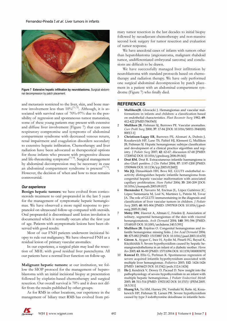
492 July 27, 2014|Volume 6|Issue 7|WJH|www.wjgnet.com
and metastasis restricted to the liver, skin, and bone mar-row involvement less than 10%[71,72]. Although, it is as-sociated with survival rates of 70%-97% due to the pos-sibility of regression and spontaneous tumor maturation, some of these young patients may present with extensive and diffuse liver involvement (Figure 7) that can cause respiratory compromise and symptoms of abdominal compartment syndrome with decreased venous return, renal impairment and coagulation disorders secondary to extensive hepatic infiltration. Chemotherapy and liver radiation have been advocated as therapeutical options for those infants who present with progressive disease and life-threatening symptoms[73,74]. Surgical management by abdominal decompression may be necessary in case an abdominal compartment syndrome is present[75,76]. However, the decision of when and how to treat remains controversial.
Our experienceBenign hepatic tumors: we have evolved from cortico-steroids treatment to oral propranolol in the last 5 years for the management of symptomatic hepatic hemangio-mas. We have observed a more rapid response to pro-pranolol on ultrasound follow-up compared with steroids. Oral propranolol is discontinued until lesion involution is documented which it normally occurs after the first year of age. Patients with asymptomatic lesions have been ob-served with good results.
Most of our FNH patients underwent incisional bi-opsy to rule out malignancy. We have observed FNH as a residual lesion of primary vascular anomalies.
In our experience, a surgical plain may lead the resec-tion of MHL with good residual liver parenchyma. All our patients have a normal liver function on follow-up.
Malignant hepatic tumors: at our institution, we fol-low the SIOP protocol for the management of hepato-blastoma with an initial incisional biopsy at presentation followed by cisplatin-based chemotherapy and surgical resection. Our overall survival is 70% and it does not dif-fer from the results published by other groups.
As for RMS in other locations, our experience in the management of biliary tract RMS has evolved from pri-
mary tumor resection in the last decades to initial biopsy followed by neoadjuvant chemotherapy and non-massive second look surgery for tumor resection and evaluation of tumor response.
We have anecdotal cases of infants with tumors other than hepatoblastoma (angiosarcoma, malignant rhabdoid tumor, undifferentiated embryonal sarcoma) and conclu-sions are difficult to be drawn.
We have successfully managed liver infiltration by neuroblastoma with standard protocols based on chemo-therapy and radiation therapy. We have only performed one surgical abdominal decompression by patch place-ment in a patient with an abdominal compartment syn-drome (Figure 7) who finally died.
REFERENCES1 MullikenJB, Glowacki J. Hemangiomas and vascular mal-
formations in infants and children: a classification based on endothelial characteristics. Plast Reconstr Surg 1982; 69: 412-422 [PMID:7063565]
2 Mulliken JB, Fishman SJ, Burrows PE. Vascular anomalies. Curr Probl Surg 2000; 37: 17-84 [DOI: 10.1016/S0011-3840(00)80013-1]
3 Christison-Lagay ER, Burrows PE, Alomari A, Dubois J, Kozakewich HP, Lane TS, Paltiel HJ, Klement G, Mulliken JB, Fishman SJ. Hepatic hemangiomas: subtype classification and development of a clinical practice algorithm and reg-istry. J Pediatr Surg 2007; 42: 62-67; discussion 67-68 [PMID: 17208542 DOI: 10.1016/j.jpedsurg.2006.09.041]
4 Drut RM, Drut R. Extracutaneous infantile haemangioma is also Glut1 positive. J Clin Pathol 2004; 57: 1197-1200 [PMID: 15509684 DOI: 10.1136/jcp.2003.012682]
5 Mo JQ, Dimashkieh HH, Bove KE. GLUT1 endothelial re-activity distinguishes hepatic infantile hemangioma from congenital hepatic vascular malformation with associated capillary proliferation. Hum Pathol 2004; 35: 200-209 [DOI: 10.1016/j.humpath.2003.09.017]
6 Hernández F, Navarro M, Encinas JL, López Gutiérrez JC, López Santamaría M, Leal N, Martínez L, Patrón M, Tovar JA. The role of GLUT1 immunostaining in the diagnosis and classification of liver vascular tumors in children. J Pediatr Surg 2005; 40: 801-804 [PMID: 15937818 DOI: 10.1016/j.jped-surg.2005.01.046]
7 Metry DW, Hawrot A, Altman C, Frieden IJ. Association of solitary, segmental hemangiomas of the skin with visceral hemangiomatosis. Arch Dermatol 2004; 140: 591-596 [PMID: 15148105 DOI: 10.1001/archderm.140.5.591]
8 Mulliken JB, Enjolras O. Congenital hemangiomas and in-fantile hemangioma: missing links. J Am Acad Dermatol 2004; 50: 875-882 [PMID: 15153887 DOI: 10.1016/j.jaad.2003.10.670]
9 Güven A, Aygun C, Ince H, Aydin M, Pinarli FG, Baysal K, Küçüködük S. Severe hypothyroidism caused by hepatic he-mangioendothelioma in an infant of a diabetic mother. Horm Res 2005; 63: 86-89 [PMID: 15711094 DOI: 10.1159/000083879]
10 Konrad D, Ellis G, Perlman K. Spontaneous regression of severe acquired infantile hypothyroidism associated with multiple liver hemangiomas. Pediatrics 2003; 112: 1424-1426 [PMID: 14654623 DOI: 10.1542/peds.112.6.1424]
11 Ho J, Kendrick V, Dewey D, Pacaud D. New insight into the pathophysiology of severe hypothyroidism in an infant with multiple hepatic hemangiomas. J Pediatr Endocrinol Metab 2005; 18: 511-514 [PMID: 15921182 DOI: 10.1515/-JPEM.2005.18.5.511]
12 Huang SA, Tu HM, Harney JW, Venihaki M, Butte AJ, Koza-kewich HP, Fishman SJ, Larsen PR. Severe hypothyroidism caused by type 3 iodothyronine deiodinase in infantile hem-
Figure 7 Extensive hepatic infiltration by neuroblastoma. Surgical abdomi-nal decompression by patch placement.
Fernandez-Pineda I et al . Liver tumors in infants

493 July 27, 2014|Volume 6|Issue 7|WJH|www.wjgnet.com
angiomas. N Engl J Med 2000; 343: 185-189 [PMID: 10900278 DOI: 10.1056/NEJM200007203430305]
13 Kassarjian A, Dubois J, Burrows PE. Angiographic classi-fication of hepatic hemangiomas in infants. Radiology 2002; 222: 693-698 [PMID: 11867787]
14 Kassarjian A, Zurakowski D, Dubois J, Paltiel HJ, Fishman SJ, Burrows PE. Infantile hepatic hemangiomas: clinical and imaging findings and their correlation with therapy. AJR Am J Roentgenol 2004; 182: 785-795 [PMID: 14975986 DOI: 10.2214/ajr.182.3.1820785]
15 Regier TS, Ramji FG. Pediatric hepatic hemangioma. Radio-graphics 2004; 24: 1719-1724 [PMID: 15537980 DOI: 10.1148/rg.246035188]
16 Hughes JA, Hill V, Patel K, Syed S, Harper J, De Bruyn R. Cutaneous haemangioma: prevalence and sonographic char-acteristics of associated hepatic haemangioma. Clin Radiol 2004; 59: 273-80 [DOI: 10.1016/S0009-9260(03)00267-8]
17 Léauté-Labrèze C, Dumas de la Roque E, Hubiche T, Bora-levi F, Thambo JB, Taïeb A. Propranolol for severe heman-giomas of infancy. N Engl J Med 2008; 358: 2649-2651 [PMID: 18550886 DOI: 10.1056/NEJMc0708819]
18 Marsciani A, Pericoli R, Alaggio R, Brisigotti M, Vergine G. Massive response of severe infantile hepatic hemangioma to propanolol. Pediatr Blood Cancer 2010; 54: 176 [PMID: 19743301 DOI: 10.1002/pbc.22262]
19 Stocker JT, Ishak KG. Focal nodular hyperplasia of the liver: A study of 21 pediatric cases. Cancer 1981; 48: 336
20 Whelan TJ, Baugh JH, Chandor S. Focal nodular hyperpla-sia of the liver. Ann Surg 1973; 177: 150-158 [PMID: 4698535 DOI: 10.1097/00000658-197302000-00004]
21 Sugito K, Uekusa S, Kawashima H, Furuya T, Ohashi K, Inoue M, Ikeda T, Koshinaga T, Tomita R, Mugishima H, Maebayashi T. The clinical course in pediatric solid tumor patients with focal nodular hyperplasia of the liver. Int J Clin Oncol 2011; 16: 482-487 [PMID: 21455626 DOI: 10.1007/s10147-011-0210-x]
22 French AE, Irwin MS, Navarro OM, Greenberg M, Nathan PC. Long-term hepatic outcomes in survivors of stage 4S and 4 neuroblastoma in infancy. Pediatr Blood Cancer 2012; 58: 283-288 [PMID: 21370436 DOI: 10.1002/pbc.23077]
23 Benz-Bohm G, Hero B, Gossmann A, Simon T, Körber F, Berthold F. Focal nodular hyperplasia of the liver in long-term survivors of neuroblastoma: how much diagnostic imaging is necessary? Eur J Radiol 2010; 74: e1-e5 [PMID: 19369017 DOI: 10.1016/j.ejrad.2009.05.002]
24 Gutweiler JR, Yu DC, Kim HB, Kozakewich HP, Marcus KJ, Shamberger RC, Weldon CB. Hepatoblastoma presenting with focal nodular hyperplasia after treatment of neuroblas-toma. J Pediatr Surg 2008; 43: 2297-2300 [PMID: 19040959 DOI: 10.1016/j.jpedsurg.2008.08.069]
25 Bioulac-Sage P, Cubel G, Taouji S, Scoazec JY, Leteurtre E, Paradis V, Sturm N, Nhieu JT, Wendum D, Bancel B, Ramos J, Paraf F, Saint Paul MC, Michalak S, Fabre M, Guettier C, Le Bail B, Zucman-Rossi J, Balabaud C. Immunohistochemical markers on needle biopsies are helpful for the diagnosis of focal nodular hyperplasia and hepatocellular adenoma sub-types. Am J Surg Pathol 2012; 36: 1691-1699 [PMID: 23060349 DOI: 10.1097/PAS.0b013e3182653ece]
26 Shafizadeh N, Kakar S. Diagnosis of well-differentiated he-patocellular lesions: role of immunohistochemistry and other ancillary techniques. Adv Anat Pathol 2011; 18: 438-445 [PMID: 21993269 DOI: 10.1097/PAP.0b013e318234abb4]
27 Meyers RL, Scaife ER. Benign liver and biliary tract masses in infants and toddlers. Semin Pediatr Surg 2000; 9: 146-155 [PMID: 10949425 DOI: 10.1053/spsu.2000.7562]
28 Stringer MD, Alizai NK. Mesenchymal hamartoma of the liver: a systematic review. J Pediatr Surg 2005; 40: 1681-1690 [PMID: 16291152 DOI: 10.1016/j.jpedsurg.2005.07.052]
29 Cignini P, Coco C, Giorlandino M, Bagolan P, Morini F, Giorlandino C. Fetal hepatic mesenchymal hamartoma: a
case report. J Prenat Med 2007; 1: 45-46 [PMID: 22470828]30 Thompson PA, Chintagumpala M. Renal and hepatic tu-
mors in the neonatal period. Semin Fetal Neonatal Med 2012; 17: 216-221 [PMID: 22595862 DOI: 10.1016/j.siny.2012.04.002]
31 Kim SH, Kim WS, Cheon JE, Yoon HK, Kang GH, Kim IO, Yeon KM. Radiological spectrum of hepatic mesenchymal hamartoma in children. Korean J Radiol 2007; 8: 498-505 [PMID: 18071280 DOI: 10.3348/kjr.2007.8.6.498]
32 Anil G, Fortier M, Low Y. Cystic hepatic mesenchymal ham-artoma: the role of radiology in diagnosis and perioperative management. Br J Radiol 2011; 84: e91-e94 [PMID: 21511744 DOI: 10.1259/bjr/41579091]
33 Millard J, Fraser N, Stewart RJ. Mesenchymal hamartoma of the liver: is biopsy always necessary? Pediatr Surg Int 2006; 22: 622-625 [PMID: 16807720 DOI: 10.1007/s00383-006-1702-z]
34 Roebuck DJ. Assessment of malignant liver tumors in chil-dren. Cancer Imaging 2009; 9: S98-S103 [PMID: 19965302 DOI: 10.1102/1470-7330.2009.9041]
35 Siegel MJ. Pediatric liver imaging. Semin Liver Dis 2001; 21: 251-269 [PMID: 11436576 DOI: 10.1055/s-2001-15339]
36 Mann JR, Kasthuri N, Raafat F, Pincott JR, Parkes SE, Muir KR, Ingram LC, Cameron AH. Malignant hepatic tumours in children: incidence, clinical features and aetiology. Pae-diatr Perinat Epidemiol 1990; 4: 276-289 [PMID: 2374747 DOI: 10.1111/j.1365-3016.1990.tb00651.x]
37 Maruyama K, Ikeda H, Koizumi T, Tsuchida Y. Prenatal and postnatal histories of very low birthweight infants who developed hepatoblastoma. Pediatr Int 1999; 41: 82-89 [PMID: 10200142]
38 De Ioris M, Brugieres L, Zimmermann A, Keeling J, Brock P, Maibach R, Pritchard J, Shafford L, Zsiros J, Czaudzerna P, Perilongo G. Hepatoblastoma with a low serum alpha-feto-protein level at diagnosis: the SIOPEL group experience. Eur J Cancer 2008; 44: 545-550 [PMID: 18166449 DOI: 10.1016/j.ejca.2007.11.022]
39 Aronson DC, Schnater JM, Staalman CR, Weverling GJ, Plaschkes J, Perilongo G, Brown J, Phillips A, Otte JB, Czaud-erna P, MacKinlay G, Vos A. Predictive value of the pretreat-ment extent of disease system in hepatoblastoma: results from the International Society of Pediatric Oncology Liver Tumor Study Group SIOPEL-1 study. J Clin Oncol 2005; 23: 1245-1252 [PMID: 15718322 DOI: 10.1200/JCO.2005.07.145]
40 Roebuck DJ, Aronson D, Clapuyt P, Czauderna P, de Ville de Goyet J, Gauthier F, Mackinlay G, Maibach R, McHugh K, Olsen OE, Otte JB, Pariente D, Plaschkes J, Childs M, Perilongo G. 2005 PRETEXT: a revised staging system for primary malignant liver tumours of childhood developed by the SIOPEL group. Pediatr Radiol 2007; 37: 123-132; quiz 249-250 [PMID: 17186233 DOI: 10.1007/s00247-006-0361-5]
41 Czauderna P, Otte JB, Aronson DC, Gauthier F, Mackinlay G, Roebuck D, Plaschkes J, Perilongo G. Guidelines for surgical treatment of hepatoblastoma in the modern era--recommen-dations from the Childhood Liver Tumour Strategy Group of the International Society of Paediatric Oncology (SIO-PEL). Eur J Cancer 2005; 41: 1031-1036 [PMID: 15862752 DOI: 10.1016/j.ejca.2005.02.004]
42 Hoffer FA. Liver biopsy methods for pediatric oncology pa-tients. Pediatr Radiol 2000; 30: 481-488 [PMID: 10929368 DOI: 10.1007/s002470000244]
43 Maturen KE, Nghiem HV, Marrero JA, Hussain HK, Higgins EG, Fox GA, Francis IR. Lack of tumor seeding after percuta-neous biopsy of hepatocellular carcinoma using coaxial cut-ting needle technique. AJR Am J Roentgenol 2005; 184: 37
44 Davidoff AM, Fernandez-Pineda I, Santana VM, Shochat SJ. The role of neoadjuvant chemotherapy in children with malignant solid tumors. Semin Pediatr Surg 2012; 21: 88-99 [PMID: 22248974 DOI: 10.1053/j.sempedsurg.2011.10.010]
45 Brown J, Perilongo G, Shafford E, Keeling J, Pritchard J, Brock P, Dicks-Mireaux C, Philips A, Vos A, Plaschkes J.
Fernandez-Pineda I et al . Liver tumors in infants

494 July 27, 2014|Volume 6|Issue 7|WJH|www.wjgnet.com
Pretreatment prognostic factors for children with hepato-blastoma - results from the International Society of Paediat-ric Oncology (SIOP) study SIOPEL 1. Eur J Cancer 2000; 36: 1418-1425 [DOI: 10.1016/S0959-8049(00)00074-5]
46 Perilongo G, Shafford E, Maibach R, Aronson D, Brugières L, Brock P, Childs M, Czauderna P, MacKinlay G, Otte JB, Pritchard J, Rondelli R, Scopinaro M, Staalman C, Plaschkes J. Risk-adapted treatment for childhood hepatoblastoma. final report of the second study of the International Society of Paediatric Oncology--SIOPEL 2. Eur J Cancer 2004; 40: 411-421 [PMID: 14746860 DOI: 10.1016/j.ejca.2003.06.003]
47 Katzenstein HM, London WB, Douglass EC, Reynolds M, Plaschkes J, Finegold MJ, Bowman LC. Treatment of unre-sectable and metastatic hepatoblastoma: a pediatric oncology group phase II study. J Clin Oncol 2002; 20: 3438-3444 [PMID: 12177104 DOI: 10.1200/JCO.2002.07.400]
48 Pritchard J, Brown J, Shafford E, Perilongo G, Brock P, Dicks-Mireaux C, Keeling J, Phillips A, Vos A, Plaschkes J. Cisplatin, doxorubicin, and delayed surgery for childhood hepatoblastoma: a successful approach--results of the first prospective study of the International Society of Pediatric Oncology. J Clin Oncol 2000; 18: 3819-3828 [PMID: 11078495]
49 Spunt S, Lobe T, Pappo A, Parham DS, Wharam MD Jr, Arndt C, Andersen JR, Crist WM, Paidas C, Wiener E, An-drassy RJ, Schwartz CL. Aggressive surgery is unwarranted for biliary tract rhabdomyosarcoma. J Pediatr Surg 2000; 35: 309-316 [DOI: 10.1016/S0022-3468(00)90030-7]
50 Zampieri N, Camoglio F, Corroppolo M, Cecchetto M, Ornis S, Ottolenghi A. Botryoid rhabdomyosarcoma of the biliary tract in children: a unique case report. Eur J Cancer Care (Engl) 2006; 15: 463-466 [PMID: 17177904 DOI: 10.1111/j.1365-2354.2006.00683.x]
51 Perera MT, McKiernan PJ, Brundler MA, Hobin DA, Mayer DA, Mirza DF, Sharif K. Embryonal rhabdomyosarcoma of the ampulla of Vater in early childhood: report of a case and review of literature. J Pediatr Surg 2009; 44: e9-e11 [PMID: 19231522 DOI: 10.1016/j.jpedsurg.2008.10.113]
52 Pollono DG, Tomarchio S, Berghoff R, Drut R, Urrutia A, Cedola J. Rhabdomyosarcoma of extrahepatic biliary tree: initial treatment with chemotherapy and conservative sur-gery. Med Pediatr Oncol 1998; 30: 290-293
53 Tireli GA, Sander S, Dervisoglu S, Demirali O, Unal M. Em-bryonal rhabdomyosarcoma of the common bile duct mim-icking choledochal cyst. J Hepatobiliary Pancreat Surg 2005; 12: 263-265 [PMID: 15995817 DOI: 10.1007/s00534-004-0959-7]
54 Himes RW, Raijman I, Finegold MJ, Russell HV, Fishman DS. Diagnostic and therapeutic role of endoscopic retrograde cholangiopancreatography in biliary rhabdomyosarcoma. World J Gastroenterol 2008; 14: 4823-4825 [PMID: 18720547 DOI: 10.3748/wjg.14.4823]
55 Awan S, Davenport M, Portmann B, Howard ER. Angiosar-coma of liver in children. J Pediatr Surg 1996; 31: 1729-32 [DOI: 10.1016/S0022-3468(96)90065-2]
56 Noronha R, Gonzalez-Crussi F. Hepatic angiosarcoma in childhood. A case report and review of the literature. Am J Surg Pathol 1984; 8: 863-871 [PMID: 6542321 DOI: 10.1097/00000478-198411000-00007]
57 Dimashkieh HH, Mo JQ, Wyatt-Ashmead J, Collins MH. Pediatric hepatic angiosarcoma: case report and review of the literature. Pediatr Dev Pathol 2004; 7: 527-532 [PMID: 15547777 DOI: 10.1007/s10024-004-4041-x]
58 Ferrari A, Casanova M, Bisogno G, Cecchetto G, Meazza C, Gandola L, Garaventa A, Mattke A, Treuner J, Carli M. Malignant vascular tumors in children and adolescents: a report from the Italian and German Soft Tissue Sarcoma Co-operative Group. Med Pediatr Oncol 2002; 39: 109-114 [PMID: 12116058 DOI: 10.1002/mpo.10078]
59 Lezama-del Valle P, Gerald WL, Tsai J, Meyers P, La Qua-glia MP. Malignant vascular tumors in young patients. Can-cer 1998; 83: 1634-1639
60 Trobaugh-Lotrario AD, Finegold MJ, Feusner JH. Rhabdoid tumors of the liver: rare, aggressive, and poorly responsive to standard cytotoxic chemotherapy. Pediatr Blood Cancer 2011; 57: 423-428 [PMID: 21744471 DOI: 10.1002/pbc.22857]
61 Wagner LM, Garrett JK, Ballard ET, Hill DA, Perry A, Biegel JA, Collins MH. Malignant rhabdoid tumor mimick-ing hepatoblastoma: a case report and literature review. Pediatr Dev Pathol 2007; 10: 409-415 [PMID: 17929989 DOI: 10.2350/06-08-0155.1]
62 Perry A, Fuller CE, Judkins AR, Dehner LP, Biegel JA. INI1 expression is retained in composite rhabdoid tumors, includ-ing rhabdoid meningiomas. Mod Pathol 2005; 18: 951-958 [PMID: 15761491 DOI: 10.1038/modpathol.3800375]
63 Al Nassan A, Sughayer M, Matalka I, Ghandour K, Masar-weh M, Zimmermann A, Sultan I. INI1 (BAF 47) immunohis-tochemistry is an essential diagnostic tool for children with hepatic tumors and low alpha fetoprotein. J Pediatr Hematol Oncol 2010; 32: e79-e81 [PMID: 20048688 DOI: 10.1097/MPH.0b013e3181b79613]
64 Trobaugh-Lotrario AD, Tomlinson GE, Finegold MJ, Gore L, Feusner JH. Small cell undifferentiated variant of hepato-blastoma: adverse molecular and clinical features similar to rhabdoid tumors. Pediatric Blood and Cancer 2009; 52: 328-334
65 Jayaram A, Finegold MJ, Parham DM, Jasty R. Successful management of rhabdoid tumor of the liver. J Pediatr Hematol Oncol 2007; 29: 406-408 [PMID: 17551403 DOI: 10.1097/MPH.0b013e3180601011]
66 Ismail H, Dembowska-Bagińska B, Broniszczak D, Kaliciński P, Maruszewski P, Kluge P, Swięszkowska E, Kościesza A, Lembas A, Perek D. Treatment of undifferentiated embryo-nal sarcoma of the liver in children--single center experience. J Pediatr Surg 2013; 48: 2202-2206 [DOI: 10.1016/j.jped-surg.2013.05.020]
67 Pachera S, Nishio H, Takahashi Y, Yokoyama Y, Oda K, Ebata T, Igami T, Nagino M. Undifferentiated embryonal sarcoma of the liver: case report and literature survey. J Hepa-tobiliary Pancreat Surg 2008; 15: 536-544 [PMID: 18836810 DOI: 10.1007/s00534-007-1265-y]
68 Webber EM, Morrison KB, Pritchard SL, Sorensen PH. Undif-ferentiated embryonal sarcoma of the liver: results of clinical management in one center. J Pediatr Surg 1999; 34: 1641-1644 [DOI: 10.1016/S0022-3468(99)90634-6]
69 Gao J, Fei L, Li S, Cui K, Zhang J, Yu F, Zhang B. Undiffer-entiated embryonal sarcoma of the liver in a child: A case report and review of the literature. Oncol Lett 2013; 5: 739-742 [PMID: 23426588]
70 Bisogno G, Pilz T, Perilongo G, Ferrari A, Harms D, Ninfo V, Treuner J, Carli M. Undifferentiated sarcoma of the liver in childhood: a curable disease. Cancer 2002; 94: 252-257 [PMID: 11815984 DOI: 10.1002/cncr.10191]
71 Stephenson SR, Cook BA, Mease AD, Ruymann FV. The prognostic significance of age and patterns of metastasis in stage IV-S neuroblastoma. Cancer 1986; 58: 372-375
72 Evans AE, Chatten J, D’Angio GJ, Gerson JM, Robinson J, Schnaufer L. A review of 17 IV-S neuroblastoma patients at the Children’s Hospital of Philadelphia. Cancer 1980; 45: 833-839
73 Bishop MW, Yin H, Shimada H, Towbin AJ, Miethke A, Weiss B. Management of stage 4S composite neuroblastoma with a MYCN-amplified nodule. J Pediatr Hematol Oncol 2014; 36: e31-e35 [PMID: 23528904 DOI: 10.1097/MPH.0b013e3182847376]
74 Steele M, Jones NL, Ng V, Kamath B, Avitzur Y, Chami R, Cutz E, Fecteau A, Baruchel S. Successful liver trans-plantation in an infant with stage 4S(M) neuroblastoma. Pediatr Blood Cancer 2013; 60: 515-517 [PMID: 23152322 DOI: 10.1002/pbc.24391]
75 Nuchtern JG, London WB, Barnewolt CE, Naranjo A, Mc-Grady PW, Geiger JD, Diller L, Schmidt ML, Maris JM, Cohn SL, Shamberger RC. A prospective study of expectant
Fernandez-Pineda I et al . Liver tumors in infants

495 July 27, 2014|Volume 6|Issue 7|WJH|www.wjgnet.com
observation as primary therapy for neuroblastoma in young infants: a Children’s Oncology Group study. Ann Surg 2012; 256: 573-580 [PMID: 22964741]
76 Mabrut JY, Dubois R, Pelizzo G, Floret D, Frappaz D, Chap-
puis JP. Abdominal expansion using a polytetrafluoroethyl-ene prosthesis in the treatment of Pepper syndrome. Pediatr Surg Int 2000; 16: 219-221 [PMID: 10786988 DOI: 10.1007/s003830050729]
P- Reviewer: Wakiyama S S- Editor: Ji FF L- Editor: A E- Editor: Liu SQ
Fernandez-Pineda I et al . Liver tumors in infants

(TTP, s), regional blood volume (RBV, cm3), regional blood flow (RBF, cm3/s) and mean transit time (MTT, s). At 24-48 h post-injection, liver stiffness was evaluated using Fibroscan and measured in kPa. The statistical evaluation was performed using Student’s t test.
RESULTS: In the PV, the Peak%, RBV and RBF were significantly reduced in the NAFLD and NASH patients compared with the controls (Peak%: NAFLD 26.3 ± 6.6, NASH 28.1 ± 7.3 vs controls 55.8 ± 9.9, P < 0.001; RBV: NAFLD 4202.3 ± 3519.7, NASH 3929.8 ± 1941.3 vs controls 7473 ± 3281, P < 0.01; RBF: NAFLD 32.5 ± 10.8, NASH 32.7 ± 12.1 vs controls 73.1 ± 13.9, P < 0.001). The TTP in the PV was longer in both patient groups but reached statistical significance only in the NASH patients compared with the controls (NASH 79.5 ± 37.8 vs controls 43.2 ± 30, P < 0.01). In the LP, the Peak%, RBV and RBF were significantly reduced in the NAFLD and NASH patients compared with the controls (Peak%: NAFLD 43.2 ± 7.3, NASH 41.7 ± 7.7 vs controls 56.6 ± 6.3, P < 0.001; RBV: NAFLD 4851.5 ± 2009, NASH 5069.4 ± 2292.5 vs controls 6922.9 ± 2461.5, P < 0.05; RBF: NAFLD 55.7 ± 10.1, NASH 54.5 ± 12.1 vs controls 75.9 ± 10.5, P < 0.001). The TTP was longer in both patient groups but did not reach statistical significance. The MTT in both the PV and LP in the NAFLD and NASH patients was not different from that in the controls. Liver stiffness was significantly increased relative to the controls only in the NASH pa-tients (NASH: 6.4 ± 2.2 vs controls 4.6 ± 1.5, P < 0.05).
CONCLUSION: Blood flow derangement within the liver present not only in NASH but also in NAFLD sug-gests that a vascular flow alteration precedes liver fi-brosis development.
© 2014 Baishideng Publishing Group Inc. All rights reserved.
Key words: Non-alcoholic fatty liver disease; Non-alco-holic steatohepatitis; Contrast-enhanced ultrasound; Fibroscan; Hepatic blood flow; Liver stiffness
Sila Cocciolillo, Giustino Parruti, Leonardo Marzio
Sila Cocciolillo, Leonardo Marzio, Digestive Physiopathology Unit, Gabriele d’Annunzio University, Pescara Civic Hospital, 65124 Pescara, ItalyGiustino Parruti, Infectious and Tropical Diseases Unit, Pescara Civic Hospital, 65124 Pescara, ItalyAuthor contributions: Cocciolillo S contributed to the recruit-ment of patients, acquisition of data, design the study, analysis and interpretation of the data and writing of the draft manuscript; Parruti G supervised and analyzed data from fibroscan; Marzio L contributed to the conceptual design, provided administrative support and overall supervision of the study, analyzed the data and critically revised the manuscript.Correspondence to: Sila Cocciolillo, MD, Digestive Physio-pathology Unit, Gabriele d’Annunzio University, Pescara Civic Hospital, Via Fonte Romana 8, 65124 Pescara, Italy. [email protected]: +39-329-2066508 Fax: +39-85-4295547Received: April 18, 2014 Revised: June 12, 2014 Accepted: June 27, 2014Published online: July 27, 2014
AbstractAIM: To determine intra-hepatic blood flow and liver stiffness in patients with non-alcoholic fatty liver dis-ease (NAFLD) and non-alcoholic steatohepatitis (NASH) using contrast-enhanced ultrasound and fibroscan.
METHODS: This prospective study included 15 patients with NAFLD, 17 patients with NASH and 16 healthy con-trols. In each patient, real-time ultrasound was used to locate the portal vein (PV) and the right liver lobe, and 5 mL of SonoVue® was then injected intravenous in a peripheral vein of the left arm over a 4-s span. Digital recording was performed for 3 min thereafter. The re-cording was subsequently retrieved to identify an area of interest in the PV area and in the right liver paren-chyma (LP) to assess the blood flow by processing the data using dedicated software (Qontrast®, Bracco, Ita-ly). The following parameters were evaluated: percent-age of maximal contrast activity (Peak%), time to peak
ORIGINAL ARTICLE
Submit a Manuscript: http://www.wjgnet.com/esps/Help Desk: http://www.wjgnet.com/esps/helpdesk.aspxDOI: 10.4254/wjh.v6.i7.496
496 July 27, 2014|Volume 6|Issue 7|WJH|www.wjgnet.com
World J Hepatol 2014 July 27; 6(7): 496-503ISSN 1948-5182 (online)
© 2014 Baishideng Publishing Group Inc. All rights reserved.
CEUS and Fibroscan in non-alcoholic fatty liver disease and non-alcoholic steatohepatitis

Core tip: The use of contrast-enhanced ultrasound (CEUS) assisted by dedicated software (Qontrast®) in combination with Fibroscan examination could provide a non-invasive tool to evaluate the level of fatty-liver dis-ease. In this study, we found that there were reductions in portal and intra-parenchymal blood flow in patients affected by non-alcoholic fatty liver disease and non-alcoholic steatohepatitis (NASH), whereas liver stiffness was increased only in NASH patients. Qontrast®-assisted CEUS could be used to quantify early changes in intra-parenchymal liver flow before the onset of fibrosis.
Cocciolillo S, Parruti G, Marzio L. CEUS and Fibroscan in non-alcoholic fatty liver disease and non-alcoholic steatohepatitis. World J Hepatol 2014; 6(7): 496-503 Available from: URL: http://www.wjgnet.com/1948-5182/full/v6/i7/496.htm DOI: http://dx.doi.org/10.4254/wjh.v6.i7.496
INTRODUCTIONNon-alcoholic fatty liver disease (NAFLD) is one of the most common causes of chronic liver disease world-wide[1,2]. Liver biopsy, which is the gold standard for di-agnosing NAFLD is an invasive procedure with potential adverse effects and large inter- and intra-observer vari-ability[3]. NAFLD cannot be diagnosed reliably without clear imaging or biopsy evidence of hepatic steatosis and without excluding excessive alcohol consumption, viral hepatitis and medications. NAFLD is further divided into non-alcoholic fatty liver (NAFL) and non-alcoholic steatohepatitis (NASH). NAFL is simple steatosis with no evidence of hepatocellular injury, whereas NASH is steatosis with inflammation, hepatocellular injury and possible fibrosis. NASH can lead to cirrhosis and hepa-tocellular carcinoma, whereas NAFLD has a very slow, if any, progression to NASH. NAFL and NASH, therefore, can be considered different steps in the same histological disease spectrum[3,4]. The pathogenesis of NAFLD is not completely known[5,6]. The fat accumulation occurring in NAFLD is key to the onset of vascular impairment[7]. The fat accumulation is responsible for liver structural and functional changes, leading to increased hepatic vas-cular resistance and finally to portal hypertension.
To study blood flow in the liver, pulsed continuous Doppler ultrasound (US) is used as the first-line imaging investigation. Doppler US can evaluate the blood flow in large and small vessels but fails to analyze the flow in the capillaries or sinusoids, where the velocity of the red blood cells is too slow to produce a Doppler signal[8]. Hence, changes in the hepatic microcirculation may be assessed using contrast-enhanced ultrasonography (CEUS) that consists of an intravenously administered suspension of gas-filled microbubbles that remain entire-ly within the intravascular space, thus acting as a blood pool tracer[9,10]. The obtained data can be processed using a post-processing computational tool (Qontrast®, Esaote, Florence, Italy) that includes a suite of software applica-
tions for image analysis designed to use alternative repre-sentations to extract and present brightness information that is already present in the image.
Liver fibrosis directly affects the mechanical properties of the liver parenchyma, such as stiffness, which indicates tissue resistance to deformation under mechanical stress. A greater stiffness corresponds to a higher tissue resistance to deformation. Liver stiffness can be studied using three physical measurements: two measures based on sono-graphic techniques, such as Fibroscan[11] and acoustic radia-tion force impulse[12,13], and one that is MR-based, such as magnetic resonance elastography[14]. Regardless of the spe-cific technique, the measured parameter is correlated with the histological fibrosis stage, and the results can be used to accurately predict moderate to severe fibrosis[10,11,15].
In NASH and NAFLD, it remains unclear whether early changes in intrahepatic blood flow are associated with an early production of fibrous tissue. Therefore, the aim of this study was to evaluate the liver blood flow in the large and small intra-parenchymal vessels and fibrosis using CEUS and Fibroscan in patients with NAFLD and NASH compared with healthy controls.
MATERIALS AND METHODSPopulationsThe study population was enrolled from August 2010 to December 2013. All of the participants were Caucasian and underwent physical examinations, laboratory tests for liver function, upper and lower abdominal real-time ultra-sonography (RUS) and computed tomography (CT) scan when necessary.
Sixteen healthy controls and 32 patients with US-documented steatosis were recruited (Table 1). Fifteen patients affected by NAFLD as defined according the lat-est guidelines established by the American Association for the study of liver diseases[3] and 17 patients with NASH defined as having fatty liver on abdominal ultrasound ex-amination and either aspartate aminotransferase or alanine aminotransferase more than 1.5 times the upper normal limit on two occasions during the six months before en-
497 July 27, 2014|Volume 6|Issue 7|WJH|www.wjgnet.com
Cocciolillo S et al . CEUS and fibroscan in NAFLD and NASH
Table 1 Study populations
Population characteristics Controls NAFLD NASH
Number 16 15 17Male/female 8/8 12/3 16/1Mean age (range) 37 yr
(26-69 yr)48 yr
(26-75 yr)45 yr
(20-74 yr)AST (mean ± SD) 20.6 ± 4.5 19.3 ± 5b 45.2 ± 22.1d
ALT (mean ± SD) 24.4 ± 7.0 27.4 ± 8.1b 86.4 ± 55.7d
GGT (mean ± SD) 18.3 ± 10.1 25.6 ± 20b 73.1 ± 43d
ALP (mean ± SD) 144.4 ± 45.4 154.1 ± 38.3 176.5 ± 57.4
bP < 0.001, NAFLD vs NASH; dP < 0.001, NASH vs controls. AST: Aspar-tate aminotransferase: reference range 0-37 (IU/L); ALT: Alanine amino-transferase: reference range 0-40 (IU/L); GGT: Gamma-glutamyl trans-ferase: reference range 7-50 (IU/L); ALP: Alkaline phosphatase: reference range 98-279 (IU/L); NASH: Non-alcoholic steatohepatitis; NAFLD: Non-alcoholic fatty liver disease.

rollment were included. Exclusion criteria were laboratory data and image studies as assessed with ultrasound or CT scan when necessary, compatible with hepatitis B and C, autoimmune hepatitis, sclerosing cholangitis, Wilson’s disease, alpha-1 anti-trypsin deficiency, hemochromatosis and hepatic cirrhosis[16]. Additional exclusion criteria were patients with medical histories of malignancy, previous abdominal or thoracic surgery and history of heart and pulmonary disease that may impair the flow of the con-trast bubbles to the liver as well as severe concomitant dis-eases. Finally, patients with pregnancy and breastfeeding as well as pediatric patients were also excluded.
Contrast-enhanced ultrasound and dedicated softwareThe patients were examined after fasting for 8 h and after having obtained written informed consent. The CEUS examination was always performed by the same expert operator using RUS with a 3.5-MHz convex probe (MyLab70 XVision, Esaote, Ansaldo, Italy) through a longitudinal intercostal scan, in which the portal vein (PV) and right liver parenchyma (LP) could be easily identified while keeping the patient or subject in the supine posi-tion. The US contrast medium (SonoVue®, Bracco Spa, Milan, Italy) consisted of 2.5 μm sulfur hexafluoride-filled microbubbles (hence, they are smaller than red blood cells, which have a diameter of 7 μm) stabilized by a lipid monolayer membrane[9]. The microbubbles can generate a nonlinear harmonic response to a low mechanical index (MI), thus permitting continuous real-time imaging. In our study, we used a signal-processing algorithm installed on the ultrasonographic machine (Contrast-Tuned Imaging™, CnTI™, Esaote, Genoa, Italy) that automatically sets a low MI of 0.06 and holds this value constant during the entire CEUS procedure. These features allow the contrast medium microbubbles to travel through the smallest blood vessels without bursting. SonoVue, a blood-pool contrast agent, has no cellular uptake; thus, it enhances only the US image gen-erated by the blood vessels[9]. In CEUS studies, there are 3 overlapping vascular phases: the arterial phase, which
starts within 20 s after the injection and lasts 30-45 s; the portal venous phase, which usually lasts until 2 min after the injection; and the late phase, which corresponds to the clearance of the US contrast agent from the circula-tion. The CEUS screen, because of the low gain, shows signals only from intensely reflective structures, which limits the ability to identify the proper scan area. To over-come this problem, a split-screen display was used on the ultrasound machine to show the conventional B-mode image beside the CEUS image (Figure 1). Using contrast-processed data, the blood flow through the small capil-laries of the liver interstitial tissue could be measured in terms of the volume and flow.
The procedure started with a 5-mL contrast medium injection (always performed by the same expert nurse) using a 20-gauge (G) needle cannula over a 4-s span into the antecubital vein of the left arm with the patient in the supine position. The line was then flushed with a 5-mL bolus of saline solution, also injected over a 4-s span. During the contrast medium injection, digital re-cording was started and performed for 3 min; during this operation, the patients were asked to breathe slowly to minimize respiration-related movements. The video recordings were then analyzed by the same trained opera-tor using Qontrast® software (Esaote, Florence, Italy), which performs a parametric analysis of perfusion within a selected set of higher signal intensity frames in the re-gion of interest (ROI). In each patient, we evaluated two ROIs: one in the PV and one in the right LP. To correct for translational movements in the ROI, a Gamma variate (bolus)-corrected parametric curve model was selected. The Qontrast® software was then allowed to process the perfusion in each of the previously determined ROIs, calculate the parameters automatically and plot the mea-sured and calculated curves. The following parameters were generated (Figure 2): Peak%, the maximum signal intensity (SI) reached during SonoVue® bolus transit at time T, where T was the time to peak (TTP, s), the time to reach the maximum SI; regional blood volume (RBV, cm3), the blood volume in the ROI, proportional to the area under the time intensity curve; mean transit time (MTT, s), the contrast medium mean transit time in the ROI; and regional blood flow (RBF, cm3/s), the RBV to MTT ratio. The reproducibility of the data obtained by Qontrast® analysis of CEUS was tested according to the method of Ridolfi et al[17].
Transient elastographyTransient elastography was performed 24-48 h after CEUS using a Fibroscan device (Echosens, Paris, France). Fi-broscan consists of a 5-MHz US transducer probe in-stalled on the axis of a vibrator that generates a 50-Hz vibration (completely painless to the patient) that causes an elastic shear wave to propagate through the skin and subcutaneous tissue and finally to the LP, the stiffness of which is directly related to the velocity of the wave. Fibroscan measures the stiffness of a cylindrical volume 1 cm in diameter, 4 cm in length and 25 to 45 cm from
498 July 27, 2014|Volume 6|Issue 7|WJH|www.wjgnet.com
Figure 1 Example in a healthy control of the split-screen display during the contrast-enhanced ultrasound procedure upon the injection of Son-oVue® using a low mechanical index. Left: B-mode frame; Right: Contrast-enhanced ultrasound frame. PV: Portal vein; LP: Liver parenchyma. The B-mode frame shows more detail because of the higher gain.
B mode
LP
PV
CEUS
Cocciolillo S et al . CEUS and fibroscan in NAFLD and NASH

499 July 27, 2014|Volume 6|Issue 7|WJH|www.wjgnet.com
MTT in both the PV and LP in the NAFLD and NASH patients was similar to that in the controls (Figure 5).
FibroscanThe values of liver stiffness measured in kPa were found to be significantly greater in the NASH patients com-pared with the control group (Figure 6).
Adverse effectsCEUS studies were performed successfully in all of the patients and were well tolerated, with no side or adverse effects reported.
DISCUSSIONOur study showed that blood flow, as assessed by Qon-trast®-assisted CEUS analysis of the PV and LP, was de-creased in patients affected by NAFLD and NASH. We also found that liver stiffness, as assessed by Fibroscan, was increased only in NASH patients.
Based on the data obtained by the Qontrast® analysis of ROIs in the PV and LP, significant reductions in the Peak%, RBV and RBF were found in both groups of pa-tients, whereas a delayed TTP was found only in the PV of the NASH group. Our results suggest the hypothesis that in patients with NAFLD, there is a reduced vascular compliance in the liver due to augmented hepatic vascular resistance to portal blood flow and an increased hepatic vascular tone that starts before the onset of fibrosis. This change was previously demonstrated by Francque et al[18]
in an experimental animal model; in their study, Wistar rats fed with a methionine- and choline-deficient diet for four weeks developed severe steatosis associated with a significant increase in intrahepatic resistance before the onset of fibrosis and inflammation. These changes involved functional (liver endothelial dysfunction and va-soconstrictor overproduction) and structural (sinusoidal altered microvascular architecture) factors. Another study by Pasarín et al[19], performed on rats fed with a cafeteria diet for one month, showed that the impaired response
the skin. Acquisition was performed by the same expert operator through an intercostal scan, in which the probe was placed perpendicular to an area free of large vascular structures. During acquisition, the patient lay in the su-pine position with the right arm in abduction. Liver stiff-ness was determined by computing the median value of 10 successful acquisitions in kPa.
Statistical analysisContinuous variables (laboratory values, SonoVue® data processed by Qontrast® software and elastosonography data) are expressed as group means ± SDs. Age was ana-lyzed as a mean. Comparisons of all gathered data among the groups were tested by a Welch-corrected unpaired t test. P values were two-tailed, and all P values less than 0.05 were considered statistically significant. All statistical relationships were assessed using correlation analysis. The statistical analyses were performed using the GraphPad Prism software, version 3.00 (GraphPad Software, San Diego, California, Unites States).
RESULTSQontrastWe could not analyze two PV ROIs in NAFLD patients and three in NASH patients because of poor video re-cording due to liver steatosis that interfered with the re-turning echoes to the US probe.
The PV analysis showed a significantly shorter Peak% (Figure 3A) and decreased RBV and RBF (Figure 3C and D) in both the NASH and NAFLD patients compared with the controls. The TTP in the PV was longer in both patient groups but reached significance only in the NASH patients (Figure 3B).
The LP analysis yielded similar results, with Peak% (Figure 4A), RBV and RBF (Figure 4C and D) signifi-cantly reduced in both the NASH and NAFLD groups compared with the normal controls. The TTP was longer in both NASH and NAFLD patients compared with the controls but did not reach significance (Figure 4B). The
Figure 2 Example of region of interest selection in the portal vein and Qontrast®-assisted contrast-enhanced ultrasound analysis of portal vein param-eters in healthy control. A: Region of interest (ROI) drawn in a region of the portal vein (PV) in a selected set of higher signal intensity frames 1 min and 10 s after SonoVue® injection; B: A gamma variate (bolus)-corrected parametric curve for the translational movement caused by breathing activity. SI (%): Signal intensity; PEAK (%): Maximum signal intensity reached during SonoVue® bolus injection; TTP (s): Time to peak; RBV (cm3): Regional blood volume; RBF (cm3/s): Regional blood flow; MTT (s): Mean transit time.
s
AB mode
ROIPV
CEUS
SI (
%)
Peak (%) = 60.5TTP (s) = 44.659 RBV = 5843.024RBF = 78.457MTT (s) = 74.47
14.10028.200
42.30056.400
70.50084.600
98.700
112.800
100
80
60
40
20
0
B
SI Peak (%)
MTT (s)
TTP (s)
Cocciolillo S et al . CEUS and fibroscan in NAFLD and NASH

500 July 27, 2014|Volume 6|Issue 7|WJH|www.wjgnet.com
to endothelial-dependent vasodilation caused endothelial dysfunction, leading to augmented intrahepatic resistance and reduced portal flow. Even in this study, the functional features of intrahepatic vascular changes preceded the
onset of fibrosis and inflammation[19]. Another interesting observation is that our data ob-
tained by Qontrast®-assisted CEUS were similar to those from other studies that analyzed liver blood flow with
Figure 3 Contrast-enhanced ultrasound of the portal vein in controls, non-alcoholic fatty liver disease and non-alcoholic steatohepatitis. A: Peak (%); B: TTP (s); C: RBV (cm3); D: RBF (cm3/s); Peak (%): Maximum signal intensity (SI) reached during SonoVue® bolus injection; TTP (s): Time to peak; RBV (cm3): Re-gional blood volume; RBF (cm3/s): Regional blood flow; NAFLD: Non-alcoholic fatty liver disease; NASH: Non-alcoholic steatohepatitis.
75
50
25
0Controls NAFLD NASH
Peak (%) PV
bP < 0.001 vs controls55.8 ± 9.9
b26.3 ± 6.6
b28.1 ± 7.3
Peak
(%
)
A 150
100
50
0Controls NAFLD NASH
TTP (s) PVaP < 0.01 vs controls
43.3 ± 30
71.9 ± 39.1
a79.5 ± 37.8
TTP
(s)
B
15000
10000
5000
0Controls NAFLD NASH
RBV (cm3) PV
aP < 0.01 vs controls7473 ± 3281
a4202.3 ± 3519.7 a
3929.8 ± 1941.3
RBV
(cm
3 )
C
RBF
(cm
3 /s)
100
75
50
25
0Controls NAFLD NASH
RBF (cm3/s) PV
dP < 0.001 vs controls73.1 ± 13.9
d32.5 ± 10.8
d32.7 ± 12.1
D
Figure 4 Contrast-enhanced ultrasound of liver parenchyma in controls, non-alcoholic fatty liver disease and non-alcoholic steatohepatitis. A: Peak (%); B: TTP (s); C: RBV (cm3); D: RBF (cm3/s); Peak (%): Maximum signal intensity (SI) reached during SonoVue® bolus injection; TTP (s): Time to peak; RBV (cm3): Re-gional blood volume; RBF (cm3/s): Regional blood flow; NAFLD: Non-alcoholic fatty liver disease; NASH: Non-alcoholic steatohepatitis.
75
50
25
0Controls NAFLD NASH
Peak (%) LPbP < 0.001 vs controls56.6 ± 6.3
b43.2 ± 7.3
b41.7 ± 7.7
Peak
(%
)
A 75
50
25
0Controls NAFLD NASH
TTP (s) LP
38.2 ± 1142.9 ± 12.9
45.1 ± 14.7
TTP
(s)
B
10000
7500
5000
2500
0Controls NAFLD NASH
RBV (cm3) LPaP < 0.05 vs controls6922.9 ± 2461.5
a4851.5 ± 2009
a5069.4 ± 2292.5
RBV
(cm
3 )
C
RBF
(cm
3 /s)
100
75
50
25
0Controls NAFLD NASH
RBF (cm3/s) LPdP < 0.001 vs Controls
75.9 ± 10.5
d55.7 ± 10.1
d54.5 ± 12.1
D
Cocciolillo S et al . CEUS and fibroscan in NAFLD and NASH

501 July 27, 2014|Volume 6|Issue 7|WJH|www.wjgnet.com
CEUS using SonoVue® as the contrast medium in cir-rhotic patients. Lin et al[20] studied the flow in the right PV by means of color Doppler and CEUS, and they found that the arrival time of SonoVue® in the right PV was prolonged, whereas the velocity and flow volume were decreased. Similar results were found in another study by Ridolfi et al[17], who evaluated liver blood flow in the PV and in the parenchyma by means of CEUS and subsequent analysis by Qontrast® in cirrhotic patients and healthy subjects. They found a reduced Peak% and prolonged TTP and MTT in cirrhotic patients compared with controls. These data suggest that NASH and, more interestingly, NAFLD might be considered precursors of liver cirrhosis due to the presence of similar hemody-namic changes in liver blood flow.
In our patients affected by NASH, the delay in reach-ing the maximum signal intensity (TTP), only present in the PV, together with the reduction in blood flow in either the PV or LP, could be the consequence of not only intra-parenchymal microcirculation variations but also increased liver stiffness. Liver stiffness data in our patients with NAFLD and NASH could be included among those with no fibrosis (NAFLD) or mild fibrosis (NASH) as classi-fied by Wong et al[21]. These authors studied a large cohort of patients with hepatic steatosis using Fibroscan and liver histology. They found that patients with no fibrosis or mild fibrosis showed liver stiffness values (kPa) that were consistent with those of our patients with NAFLD (no
fibrosis) and NASH (mild fibrosis), respectively. They also identified some patients with steatosis with liver stiffness values that were much higher than those found in this present study. These patients were classified at histology as having a fibrosis pattern compatible with early cirrhosis, which was an exclusion criterion in our study.
The major limitation of this present study was the small number of patients that were examined; further studies in a much larger population are required to draw definitive conclusions regarding the value of the digital data generated by Qontrast®. However, the differences between the control, NAFLD and NASH groups for the main measure of the analysis (Peak% in the PV and LP) were so large that the statistical power of the study could be considered satisfactory. The absence of liver biopsy data in our study was another limitation, although other authors have found a significant correlation between US and histopathologic data in the evaluation of steatosis[22-24]. The CEUS procedure may be incorrectly applied when the US machine does not meet the criteria of good sen-sitivity, good tissue suppression and good temporal and spatial resolution as reported in the Guidelines of Euro-pean Federation of Societies for Ultrasound in Medicine and Biology[25]. In our study, we obtained a good tissue suppression by means of CnTI™, which maintains a low MI throughout the study and avoids microbubble burst-ing as well as bioeffects in the target organs.
In conclusion, CEUS evaluated by Qontrast® might be able to quantify functional vascular liver changes not otherwise detectable with any other non-invasive proce-dure and before the development of fibrosis. The com-bined use of Fibroscan and Qontrast®-assisted CEUS could be helpful in assessing the level of disease and could be potentially useful for monitoring the effects of therapeutic interventions.
COMMENTSBackgroundNon-alcoholic fatty liver disease (NAFLD) is among the most common cause of chronic liver disease worldwide. NAFLD is further subdivided into non-alcoholic fatty liver (NAFL) and non-alcoholic steatohepatitis (NASH). Whereas NASH is represented by steatosis with inflammation, hepatocellular injury and possible fibrosis, NAFL is a simple steatosis with no evidence of hepatocellular injury.
Figure 5 Contrast-enhanced ultrasound of the portal vein (A) and liver parenchyma (B) in controls, non-alcoholic fatty liver disease and non-alcoholic steatohepatitis: Mean transit time (s). NAFLD: Non-alcoholic fatty liver disease; NASH: Non-alcoholic steatohepatitis.
200
100
0Controls NAFLD NASH
MTT (s) PV
102.5 ± 44
122.1 ± 69
119.3 ± 43.3
MTT
(s)
A150
100
50
0Controls NAFLD NASH
MTT (s) LP
89.7 ± 24.485.1 ± 22.5
91.2 ± 29.9
MTT
(s)
B
Stiff
ness
(kP
a)
10.0
7.5
5.0
2.5
0.0Controls NAFLD NASH
FibroscanaP < 0.05 vs controls
4.6 ± 1.5
5.7 ± 1.7
a6.4 ± 2.2
Figure 6 Fibroscan in controls, non-alcoholic fatty liver disease and non-alcoholic steatohepatitis. NAFLD: Non-alcoholic fatty liver disease; NASH: Non-alcoholic steatohepatitis.
COMMENTS
Cocciolillo S et al . CEUS and fibroscan in NAFLD and NASH

502 July 27, 2014|Volume 6|Issue 7|WJH|www.wjgnet.com
Fat accumulation within the hepatocytes leads to narrowing and distortion of the sinusoidal lumen, leading to increased hepatic vascular resistance and finally to portal hypertension and fibrosis that are among the stigmata of hepatic cirrho-sis. Whether these vascular hemodynamic changes are present in NAFLD and NASH remains unclear. Pulsed continuous Doppler ultrasound (US) is the first-line imaging tool for studying blood flow in the liver and allows for the evaluation of flow in the great hepatic vessels but fails to analyze the flow in the capillaries or sinusoids, where the velocity of the red blood cells is too slow to produce a Doppler signal. Hence, to assess changes in hepatic microcirculation, the authors used US to analyze a Doppler signal generated by an intravenously ad-ministered suspension of gas-filled microbubbles (each bubble is one-third the diameter of a red blood cell) stabilized by a lipid monolayer membrane; these features allow these bubbles to remain entirely within the intravascular space, thus acting as a blood pool tracer. The obtained data can be processed with a post-processing computational tool (Qontrast®, Esaote, Firenze, Italy), which al-lowed them to extrapolate objective and quantitative parameters of microvascu-lar damage in the liver. Liver fibrosis directly affects the mechanical properties of the liver parenchyma and may also contribute to portal hypertension. Liver stiffness can be studied with Fibroscan that consists of measuring the resis-tance of the liver tissue to the propagation of a US beam within the tissue. Research frontiersConsidering the increasing prevalence of NAFLD with potentially severe outcomes and the limitations of the actual gold standard (liver biopsy) as a diagnostic procedure, the development of a non-invasive technique that allows for an early assessment of liver damage in terms of the derangement of intra-hepatic microcirculation and the development of fibrosis appears to be a stimu-lating research field. This approach also has therapeutic implications in terms of the development of new drugs and monitoring of their therapeutic effects. Innovations and breakthroughsThe US contrast medium (SonoVue®, Bracco Spa, Milan, Italy) consisted of 2.5 μm sulfur hexafluoride-filled microbubbles, which are smaller than red blood cells. The microbubbles have no cellular uptake, unlike the contrast media used for computed tomography scan or magnetic resonance, and can travel through the smallest liver blood vessels without bursting. A Doppler signal not otherwise detectable with the standard US machine is therefore generated, and the flow in microvessels can be measured. In addition, the use of a computer program that analyzes the signal intensity within the US image allows for the standardization of data in term of the blood flow and volume. By means of contrast-enhanced ultrasound (CEUS) and computer-assisted determination of flow and volume, it has been possible for the first time to detect a derangement in the microcircula-tion within the liver parenchyma not only in NASH but also in NAFLD. Fibrosis otherwise appears to be limited only to NASH.Applications To non-invasively monitor the development of liver disease and to study the ef-fect of drugs on hepatic micro-circulation and fibrosis.Terminology CEUS: contrast-enhanced ultrasound, SonoVue®: microbubbles of 2.5 μm in diameter filled with sulfur hexafluoride that are stabilized by a lipid mono-layer membrane. QONTRAST® is a suite of software applications for image analysis designed to extract and present, in alternative representation, bright-ness information that is already contained within the images. FibroScan® is a sonography-based non-invasive and rapid bedside method for the diagnosis and quantification of hepatic fibrosis (by measuring liver stiffness).Peer reviewThis is a clinical study which evaluated the findings of contrast-enhanced US and Fibroscan in patients with NAFLD and control. The authors found some dif-ferences of the hepatic hemodynamics and liver stiffness among control NAFL and NASH.
REFERENCES1 Bellentani S, Scaglioni F, Marino M, Bedogni G. Epidemi-
ology of non-alcoholic fatty liver disease. Dig Dis 2010; 28: 155-161 [PMID: 20460905 DOI: 10.1159/000282080]
2 Torres DM, Harrison SA. Diagnosis and therapy of nonal-coholic steatohepatitis. Gastroenterology 2008; 134: 1682-1698 [PMID: 18471547 DOI: 10.1053/j.gastro.2008.02.077]
3 Chalasani N, Younossi Z, Lavine JE, Diehl AM, Brunt EM,
Cusi K, Charlton M, Sanyal AJ. The diagnosis and manage-ment of non-alcoholic fatty liver disease: practice Guideline by the American Association for the Study of Liver Diseases, American College of Gastroenterology, and the Ameri-can Gastroenterological Association. Hepatology 2012; 55: 2005-2023 [PMID: 22488764 DOI: 10.1002/hep.25762]
4 Yilmaz Y. Review article: is non-alcoholic fatty liver disease a spectrum, or are steatosis and non-alcoholic steatohepatitis distinct conditions? Aliment Pharmacol Ther 2012; 36: 815-823 [PMID: 22966992]
5 Friedman SL. Mechanisms of hepatic fibrogenesis. Gastroen-terology 2008; 134: 1655-1669 [PMID: 18471545 DOI: 10.1053/j.gastro.2008.03.003]
6 Vernon G, Baranova A, Younossi ZM. Systematic review: the epidemiology and natural history of non-alcoholic fatty liver disease and non-alcoholic steatohepatitis in adults. Ali-ment Pharmacol Ther 2011; 34: 274-285 [PMID: 21623852 DOI: 10.1111/j.1365-2036.2011.04724.x]
7 Farrell GC, Teoh NC, McCuskey RS. Hepatic microcircula-tion in fatty liver disease. Anat Rec (Hoboken) 2008; 291: 684-692 [PMID: 18484615 DOI: 10.1002/ar.20715]
8 Bernatik T, Strobel D, Hahn EG, Becker D. Doppler mea-surements: a surrogate marker of liver fibrosis? Eur J Gas-troenterol Hepatol 2002; 14: 383-387 [PMID: 11943950 DOI: 10.1097/00042737-200204000-00008]
9 Greis C. Technology overview: SonoVue (Bracco, Milan). Eur Radiol 2004; 14 Suppl 8: P11-P15 [PMID: 15700328 DOI: 10.1007/s10406-004-0076-3]
10 Quaia E. Assessment of tissue perfusion by contrast-en-hanced ultrasound. Eur Radiol 2011; 21: 604-615 [PMID: 20927527 DOI: 10.1007/s00330-010-1965-6]
11 Yoneda M, Yoneda M, Mawatari H, Fujita K, Endo H, Iida H, Nozaki Y, Yonemitsu K, Higurashi T, Takahashi H, Ko-bayashi N, Kirikoshi H, Abe Y, Inamori M, Kubota K, Saito S, Tamano M, Hiraishi H, Maeyama S, Yamaguchi N, Togo S, Nakajima A. Noninvasive assessment of liver fibrosis by measurement of stiffness in patients with nonalcoholic fatty liver disease (NAFLD). Dig Liver Dis 2008; 40: 371-378 [PMID: 18083083 DOI: 10.1016/j.dld.2007.10.019]
12 Friedrich-Rust M, Nierhoff J, Lupsor M, Sporea I, Fierbinte-anu-Braticevici C, Strobel D, Takahashi H, Yoneda M, Suda T, Zeuzem S, Herrmann E. Performance of Acoustic Radia-tion Force Impulse imaging for the staging of liver fibrosis: a pooled meta-analysis. J Viral Hepat 2012; 19: e212-e219 [PMID: 22239521 DOI: 10.1111/j.1365-2893.2011.01537.x]
13 Yoneda M, Suzuki K, Kato S, Fujita K, Nozaki Y, Hosono K, Saito S, Nakajima A. Nonalcoholic fatty liver disease: US-based acoustic radiation force impulse elastography. Radiology 2010; 256: 640-647 [PMID: 20529989 DOI: 10.1148/radiol.10091662]
14 Yin M, Talwalkar JA, Glaser KJ, Manduca A, Grimm RC, Rossman PJ, Fidler JL, Ehman RL. Assessment of hepatic fibrosis with magnetic resonance elastography. Clin Gastro-enterol Hepatol 2007; 5: 1207-1213.e2 [PMID: 17916548 DOI: 10.1016/j.cgh.2007.06.012]
15 Mori M, Fujii H, Ogawa T, Kobayashi S, Iwai S, Morikawa H, Enomoto M, Tamori A, Sawada A, Takeda S, Kawada N. Close correlation of liver stiffness with collagen deposition and presence of myofibroblasts in non-alcoholic fatty liver disease. Hepatol Res 2011; 41: 897-903 [PMID: 21682831 DOI: 10.1111/j.1872-034X.2011.00842.x]
16 Barzin G, Merat S, Nokhbeh-Zaeem H, Saniee P, Pedramnia S, Mostashfi Habibabadi A, Nasseri-Moghaddam S. Oral Nitrate Reductase Activity Is Not Associated with Develop-ment of Non-Alcoholic Fatty Liver Disease (NAFLD) and Non-Alcoholic Steatohepatitis (NASH): A Pilot Study. Middle East J Dig Dis 2014; 6: 23-27 [PMID: 24829701]
17 Ridolfi F, Abbattista T, Busilacchi P, Brunelli E. Contrast-enhanced ultrasound evaluation of hepatic microvascular
Cocciolillo S et al . CEUS and fibroscan in NAFLD and NASH

503 July 27, 2014|Volume 6|Issue 7|WJH|www.wjgnet.com
changes in liver diseases. World J Gastroenterol 2012; 18: 5225-5230 [PMID: 23066317 DOI: 10.3748/wjg.v18.i37.5225]
18 Francque S, Laleman W, Verbeke L, Van Steenkiste C, Casteleyn C, Kwanten W, Van Dyck C, D’Hondt M, Ramon A, Vermeulen W, De Winter B, Van Marck E, Van Marck V, Pelckmans P, Michielsen P. Increased intrahepatic resistance in severe steatosis: endothelial dysfunction, vasoconstrictor overproduction and altered microvascular architecture. Lab Invest 2012; 92: 1428-1439 [PMID: 22890552 DOI: 10.1038/la-binvest.2012.103]
19 Pasarín M, La Mura V, Gracia-Sancho J, García-Calderó H, Rodríguez-Vilarrupla A, García-Pagán JC, Bosch J, Abraldes JG. Sinusoidal endothelial dysfunction precedes inflamma-tion and fibrosis in a model of NAFLD. PLoS One 2012; 7: e32785 [PMID: 22509248 DOI: 10.1371/journal.pone.0032785]
20 Lin LW, Duan XJ, Wang XY, Xue ES, He YM, Gao SD, Yu LY. Color Doppler velocity profile and contrast-enhanced ul-trasonography in assessment of liver cirrhosis. Hepatobiliary Pancreat Dis Int 2008; 7: 34-39 [PMID: 18234636]
21 Wong VW, Vergniol J, Wong GL, Foucher J, Chan HL, Le Bail B, Choi PC, Kowo M, Chan AW, Merrouche W, Sung JJ, de Lédinghen V. Diagnosis of fibrosis and cirrhosis us-ing liver stiffness measurement in nonalcoholic fatty liver disease. Hepatology 2010; 51: 454-462 [PMID: 20101745 DOI: 10.1002/hep.23312]
22 Joseph AE, Saverymuttu SH, al-Sam S, Cook MG, Maxwell JD. Comparison of liver histology with ultrasonography in assessing diffuse parenchymal liver disease. Clin Radiol 1991; 43: 26-31 [PMID: 1999069 DOI: 10.1016/S0009-9260(05)80350-2]
23 Fishbein M, Castro F, Cheruku S, Jain S, Webb B, Gleason T, Stevens WR. Hepatic MRI for fat quantitation: its relation-ship to fat morphology, diagnosis, and ultrasound. J Clin Gastroenterol 2005; 39: 619-625 [PMID: 16000931 DOI: 10.1097/00004836-200508000-00012]
24 Hernaez R, Lazo M, Bonekamp S, Kamel I, Brancati FL, Guallar E, Clark JM. Diagnostic accuracy and reliability of ultrasonography for the detection of fatty liver: a meta-anal-ysis. Hepatology 2011; 54: 1082-1090 [PMID: 21618575 DOI: 10.1002/hep.24452]
25 Piscaglia F, Nolsøe C, Dietrich CF, Cosgrove DO, Gilja OH, Bachmann Nielsen M, Albrecht T, Barozzi L, Bertolotto M, Catalano O, Claudon M, Clevert DA, Correas JM, D’Onofrio M, Drudi FM, Eyding J, Giovannini M, Hocke M, Ignee A, Jung EM, Klauser AS, Lassau N, Leen E, Mathis G, Saftoiu A, Seidel G, Sidhu PS, ter Haar G, Timmerman D, Weskott HP. The EFSUMB Guidelines and Recommendations on the Clin-ical Practice of Contrast Enhanced Ultrasound (CEUS): up-date 2011 on non-hepatic applications. Ultraschall Med 2012; 33: 33-59 [PMID: 21874631 DOI: 10.1055/s-0031-1281676]
P- Reviewer: Maruyama H, Nagarajan P, Videla LA S- Editor: Ji FF L- Editor: A E- Editor: Liu SQ
Cocciolillo S et al . CEUS and fibroscan in NAFLD and NASH

Dimitrios N Samonakis, Mairi Koulentaki, Constantina Coucoutsi, Aikaterini Augoustaki, Chryssavgi Baritaki, Emmanuel Digenakis, Nikolaos Papiamonis, Maria Fragaki, Erminia Matrella, Maria Tzardi, Elias A Kouroumalis
Dimitrios N Samonakis, Mairi Koulentaki, Constantina Cou-coutsi, Aikaterini Augoustaki, Chryssavgi Baritaki, Emman-uel Digenakis, Nikolaos Papiamonis, Maria Fragaki, Erminia Matrella, Elias A Kouroumalis, Department of Gastroenterology and Hepatology, University Hospital of Heraklion, Heraklion 71110, Crete, Greece Maria Tzardi, Department of Histopathology, University Hospi-tal of Heraklion, Heraklion 71110, Crete, GreeceAuthor contributions: Samonakis DN wrote the manuscript, did the statistics and was responsible for patient management; Kou-lentaki M contributed to data base design, patient management and paper corrections; Coucoutsi C, Augustaki A and Baritaki C kept data base and patient management; Digenakis E, Papiamonis N and Fragaki M contributed to patient management and data base update, patient survival follow up; Matrella E contributed to long term patient management and outpatient clinic details; Tzardi M diagnosed all liver biopsies; Kouroumalis EA contrib-uted to the study design, patient managment, contributed to the manuscript, had overall responsibility for the study. Correspondence to: Dr. Dimitrios N Samonakis, Depart-ment of Gastroenterology and Hepatology, University Hospital of Heraklion, Buidling A-4th Floor, Voutes, Herklion 71110, Crete, Greece. [email protected]: +30-2810-392356 Fax: +30-2810-542085Received: December 27, 2013 Revised: May 16, 2014Accepted: June 10, 2014Published online: July 27, 2014
AbstractAIM: To study these characteristics and prognostic patterns in a Greek patient population.
METHODS: We analyzed a large cohort of cirrhotic patients referred to the department of Gastroenterol-ogy and Hepatology and the outpatient clinics of this tertiary hospital, between 1991 and 2008. We included patients with established cirrhosis, either compensated or decompensated, and further decompensation epi-sodes were registered. A data base was maintained and updated prospectively throughout the study period.
We analyzed differences in cirrhosis aetiology, time to and mode of decompensation, hepatocellular carcinoma (HCC) occurrence and ultimately patient survival.
RESULTS: Five hundreds and twenty-two patients with median age 67 (range, 29-91) years and average follow up 9 years-10 mo (range, 1-206 mo) were stud-ied. Commonest aetiology was hepatitis C virus (HCV, 41%) followed by alcohol (31%). The median survival time in compensated cirrhotics was 115 mo (95%CI: 95-133), whereas in decompensated patients was 55 mo (95%CI: 36-75). HCV patients survived longer while HBV patients had over twice the risk of death of HCV patients. The median time to decompensation was 65 mo (95%CI: 51-79), with alcoholics having the highest risk (RR = 2.1 vs HCV patients). Hepatitis B virus (HBV) patients had the highest risk of HCC, alcoholics the low-est. Leading causes of death: liver failure, hepatorenal syndrome, sepsis and HCC progression.
CONCLUSION: Cirrhosis aetiology and decompensa-tion at presentation were predictors of survival. Alco-holics had the highest decompensation risk, HBV cir-rhotics the highest risk of HCC and HCV cirrhotics the highest decompensation-free time.
© 2014 Baishideng Publishing Group Inc. All rights reserved.
Key words: Survival; Decompensation; Hepatocellular carcinoma; Bleeding; Ascites
Core tip: Hepatitis C was the most common cause in our cirrhotics and many hepatitis C virus patients were aged and demonstrated a long, mild course. Alcoholic and non alcoholic steatohepatitis cirrhosis is becoming a significant problem. Ascites was the commonest type of decompensation. Survival in compensated cirrhotics was at least double that of decompensated patients. Variceal bleeding was more frequent in alcoholics; nev-ertheless it was unexpectedly related to better survival
RETROSPECTIVE STUDY
Submit a Manuscript: http://www.wjgnet.com/esps/Help Desk: http://www.wjgnet.com/esps/helpdesk.aspxDOI: 10.4254/wjh.v6.i7.504
504 July 27, 2014|Volume 6|Issue 7|WJH|www.wjgnet.com
World J Hepatol 2014 July 27; 6(7): 504-512ISSN 1948-5182 (online)
© 2014 Baishideng Publishing Group Inc. All rights reserved.
Clinical outcomes of compensated and decompensated cirrhosis: A long term study

than decompensation with ascites or encephalopathy. This was attributed to the improvements in the man-agement of variceal bleeding together with the impor-tance of abstinence from alcohol after the episode was successfully treated. Hepatocellular carcinoma patients with a history of hepatitis B virus had the highest risk of mortality.
Samonakis DN, Koulentaki M, Coucoutsi C, Augoustaki A, Baritaki C, Digenakis E, Papiamonis N, Fragaki M, Matrella E, Tzardi M, Kouroumalis EA. Clinical outcomes of compen-sated and decompensated cirrhosis: A long term study. World J Hepatol 2014; 6(7): 504-512 Available from: URL: http://www.wjgnet.com/1948-5182/full/v6/i7/504.htm DOI: http://dx.doi.org/10.4254/wjh.v6.i7.504
INTRODUCTION Cirrhosis and its complications represent the end in the spectrum of chronic liver diseases, irrespective of aetiol-ogy. The natural history of cirrhosis is classically charac-terised by an asymptomatic phase termed compensated cirrhosis, followed by the development of complications from portal hypertension and/or liver dysfunction, termed decompensated cirrhosis. The transition has been estimated to occur at a rate of 5%-7% per year. In recent years this process has been proposed as a series of critical steps that if unchecked, culminate in hepatic decompensation[1].
Chronic infection with hepatitis B virus (HBV) and hepatitis C virus (HCV), represents the commonest cause of cirrhosis worldwide[2]. Nevertheless, hepatitis C often has indolent course for a long period of time[3,4] with a median time from infection to cirrhosis of 30 years; sev-eral confounding factors have been associated with dis-ease progression[4]. Despite higher risk for decompensa-tion in HCV infection, cirrhosis presents earlier in HBV patients[5]. Longitudinal studies of patients with chronic hepatitis B have shown a 5 year cumulative incidence of developing cirrhosis 8%-20% with a 5 year survival in compensated cirrhosis 85% and 15%-35% in decompen-sated cirrhosis[6]. Various factors are related with progres-sion in HBV infection, but clearly its course is modified by antiviral therapy and HBV DNA suppression[7,8]. Sus-tained response to anti-HCV treatment also significantly determines patients’ outcome regarding decompensation, liver failure, death or orthotopic liver transplantation and decreases does not completely eliminate the HCC risk[9].
Hepatocellular carcinoma (HCC) is a major compli-cation of viral cirrhosis, both compensated and decom-pensated, and a major cause of death[2]. HCC incidence appears to be increasing worldwide[10] and several clinico-pathological variables have been identified as predictors for outcome[11,12]. The annual incidence of HBV related HCC ranges from 2%-5%[6]. Two other very common causes of chronic liver disease and subsequent complica-tions are non alcoholic steatohepatitis (NASH) and alco-
holic liver disease, which especially in western countries are becoming significant public health issues[13,14].
The purpose of this study was to evaluate-in a region of southern Greece-a cohort of patients with either compensated or decompensated cirrhosis at presentation; to identify the time to and mode of decompensation, investigate the occurrence of HCC and assess the impact of all the aforementioned on patient survival.
MATERIALS AND METHODSStudy designThe study was performed in a tertiary hospital which is the reference centre for the island of Crete, in the south of Greece. The population of 800000 is largely homoge-nous. The few non-Greek patients are mostly from East-ern Europe and the Balkans. The study conformed to the principles of declaration of Helsinki and was approved by the ethics committee of the University Hospital of Heraklion. The participants in the study provided verbal consent; for those patients with hepatic encephalopathy, consent was given by relatives. Written informed consent was provided from those undergoing interventional pro-cedures (i.e., liver biopsies, endoscopies and abdominal paracentesis).
We included patients with a diagnosis of cirrhosis who were seen as outpatients in the liver clinic or were hospitalized, mostly for chronic liver disease complica-tions. Starting date of the study was January 1991 and their data were registered in a data base until June 2008. During the long period of this study, many patients that fulfilled the criteria were included and were therefore fol-lowed up prospectively. In that sense the study is both prospective and retrospective.
All patients with established cirrhosis were included. Diagnosis was based on liver biopsy (all patients with compensated cirrhosis) and/or clinical evidence of de-compensation combined with endoscopic and radiological findings. We excluded from the study (1) paediatric liver disease; (2) patients with primary biliary cirrhosis (PBC); (3) autoimmune hepatitis (AIH) cirrhosis; and (4) 12 pa-tients who did not wish to participate in the study. PBC patients have a discrete clinical course and our experience has been reported elsewhere[15]. AIH patients are few in Crete (less than 20 patients have been diagnosed during the study period) and all but one run a good course under treatment. Thus AIH as a separate group for cirrhosis ae-tiology was excluded due to small numbers.
The diagnosis of decompensated cirrhosis was based on the presence of any of the following: ascites, vari-ceal bleeding or encephalopathy. The classification as compensated cirrhosis precluded any past history of the above criteria. The diagnosis of liver failure was made when one or more of the following were observed in decompensated cirrhotics: Hepato-renal syndrome type 2/type 1, progressively worsening liver biochemistry with prolongation of international normalized ratio and/or deepening jaundice (frequently due to sepsis), severely worsening encephalopathy, or liver failure in the context
505 July 27, 2014|Volume 6|Issue 7|WJH|www.wjgnet.com
Samonakis DN et al . Cirrhosis: A clinical real life study

of massive infiltration from tumour. All patients with HBV related cirrhosis had received
standard antivirals, initially lamivudine/adefovir and later either entecavir or tenofovir. Antiviral treatment started at the time of initial diagnosis of chronic HBV infection and continued after the diagnosis of cirrhosis until death or end of follow up.
No patient with decompensated hepatitis C related cirrhosis received antiviral treatment with the standard regimen. The number of compensated HCV cirrhotics in this population on treatment with interferon and ribavirin was too small to draw conclusions. HCV decompensated cirrhotics received only supportive treatment as indicated (diuretics, antibiotics, varices ligation, repeated paracen-tesis and terlipressin). Approximately half of HCV cir-rhotics had no antiviral treatment prior to their cirrhosis diagnosis due to either old age, side effects or unavailabil-ity of treatment. In any case antivirals were discontinued on diagnosis of cirrhosis according to the guidelines at a certain time period. A 30% of alcoholics discontinued alcohol consumption.
A careful evaluation was performed to document any episode of decompensation at scheduled outpatient Hep-atology clinic visits or at hospitalization for any reason. For patients who had not attended the outpatient clinic for three months after their previous visit, information regarding the outcome was obtained by telephone inter-views with patients or relatives.
Liver biopsies were taken using ultrasound guidance and were initially performed with Menghini needles, later substituted by Tru-cut needles; few biopsies were done intraoperatively or transjugularly. All patients with bleed-ing were scoped within 24 h to diagnose and treat portal hypertensive bleeding. Ascites and encephalopathy were diagnosed according to standard criteria; all patients with ascites on presentation and according to clinical suspicion underwent abdominal paracentesis to check for sponta-neous bacterial peritonitis ever since this was internation-ally accepted practice. Screening for HCC was performed every 6 mo with ultrasound (US) and a-fetoprotein, and during the last 3 years of the study contrast-enhanced US was used. HCC was diagnosed either histologically or according to European association of the study of the liver/American association of the study if the liver crite-ria ever since these were published[16,17].
Viral hepatitis markers were detected by Abbott Elisa immunoassays and viral load was measured quantitatively using polymerase chain reaction test wherever appropri-ate since the method was available. Alcohol misuse (de-fined as exceeding 40 g of ethanol daily in male-20 g daily in female patients) was identified after interviewing the patient during hospitalisation or in the outpatient alco-holic clinic, as well from information provided by social services. The study included patients with a diagnosis of alcoholic cirrhosis who were either active drinkers or were abstainers at evaluation. Three distinct end points were considered: decompensation, death (or liver trans-plantation) and HCC. Few patients received a transplant
due to the late development of liver transplantation ser-vices in the country. NASH related cirrhosis and crypto-genic cirrhosis were evaluated as a single group.
Statistical analysisUnivariate comparisons of patient characteristics be-tween the aetiological groups were undertaken using the chi-squared test and one-way ANOVA according to the type of characteristic. Bonferroni post-hoc comparisons were made when the ANOVA comparison was found to be statistically significant.
The median follow-up time was calculated using the reverse Kaplan Meier estimator[18]. Kaplan-Meier estimates of survival curves were constructed for both overall survival and decompensation-free survival. Me-dian survival times were compared using the log-rank test. Both univariate and multivariate Cox’s proportional hazards models were used to estimate hazard ratios (rela-tive risks). A significance level of 5% was chosen for all hypothesis tests. SPSS version 17 was used throughout.
RESULTSA total of 522 patients were included in the study. The majority of these patients had compensated cirrhosis on presentation (n = 360, 69%). One hundred and eighty five patients developed decompensation during follow up (35.4% of the entire cohort and 51.3% of the initially compensated cirrhotics) and there were 231 deaths (44%) over the follow-up period. Median follow-up was 9 years 10 mo, and ranged from 1 mo to just over 17 years. There were 183 patients with a minimum follow up of 5 years in the entire cohort.
Seventy eight patients (15%) were lost to follow up. The distribution of cirrhosis causes in those lost to fol-low up was found to be similar to those remaining in the study (n = 444). Leading causes of death were: liver failure which resulted in 55 deaths (23.8%) hepatorenal syndrome (n = 50, 21.6%), sepsis (n = 25, 10.8%), mas-sive portal hypertensive bleeding (n = 15, 6.5%). These were followed by HCC progression, extrahepatic cancer, cardiovascular events, and other causes. In 21 patients (9%) who died the cause was not verified.
Characteristics of the patient cohort are presented in Table 1. Mean patient age was 67 (range 29 to 91) years. The most common cause of cirrhosis was hepatitis C (41%, 215 patients), followed by alcoholic liver disease (31%, 162 patients). The age distribution within each etiologic group is summarized in Figure 1.
The mean age of the alcoholic liver disease (ALD) pa-tients was 62 (SD ± 12) years, of HBV patients 67 (SD ± 10) years, of HBV + ALD patients 56 (SD ± 15) years, of HCV patients 71 (SD ± 9) years, HCV + ALD patients 65 (SD ± 11) years, of NASH/cryptogenic patients 70 (SD ± 13) years. Average patient age differed to a statistically significant extent between groups (P < 0.0001). Post-hoc pairwise comparisons indicated that HCV and NASH/cryptogenic patients were older on average than ALD or
506 July 27, 2014|Volume 6|Issue 7|WJH|www.wjgnet.com
Samonakis DN et al . Cirrhosis: A clinical real life study

507 July 27, 2014|Volume 6|Issue 7|WJH|www.wjgnet.com
The median time to decompensation was 65 mo (95%CI: 51-79 mo) and varied according to aetiol-ogy. Kaplan-Meier curves are presented by etiologic group in Figure 4 (P = 0.003). The highest median decompensation-free time was seen in the HCV patient group (median 105, 95%CI: 60-150 mo), the lowest in
HBV/ALD patients (P < 0.0001 in all cases). HBV/ALD patients were also younger, on average, than patients with HBV alone (P = 0.035). There have been only 5 patients with HDV co-infection, three of them with HBV/HDV/HCV with a history of intravenous drug use. Six patients had co infection HBV/HCV. Due to the small number no separate analysis for the viral co infections was performed.
The median survival time of those presenting with compensated cirrhosis was 115 (95%CI: 95-135) mo whereas decompensated patients had a median survival of 55 (95%CI: 36-75) mo. Kaplan-Meier survival curves also indicated a worse overall prognosis for patients pre-senting with decompensated cirrhosis (Figure 2) (P < 0.0001).
Survival was also strongly influenced by cirrhosis aetiology: Kaplan-Meier survival curves are presented according to etiologic group in Figure 3, in which HBV patients (90% e-antigen negative) appear to have the worst overall survival (P = 0.004). Using univariate Cox regression analysis, HBV patients were found to have just over twice the risk of death of HCV patients (RR = 2.1, P < 0.0001) whilst the NASH/cryptogenic group had a RR of 1.6 (P = 0.042) compared to the HCV group.
At presentation, both cirrhosis aetiology and decom-pensation remained significant predictors of survival (P values 0.007 and < 0.0001 respectively) after adjusting for age (P = 0.633) and sex (P = 0.505) in a multivariable model. The RR was 2.6 for patients that were decompen-sated at diagnosis (95%CI: 1.9-3.6) compared to compen-sated patients. Patients with HBV had RR = 1.8 (95%CI: 1.2-2.7, P = 0.005) compared to HCV patients but none of the other groups had a statistically significantly elevat-ed risk compared to HCV patients (all P values > 0.1).
Table 1 Patients' cohort characteristics
n (%)
Number of patients 522Male 342 (66)Female 180 (34)Cirrhosis aetiology HCV 180 (34) HCV/ALD 35 (7) HBV 67 (13) HBV/ALD 15 (3) ALD 162 (31) NASH/other 63 (12)HCC 88 (17)Patients alive 213 (41)Patients died 231 (44)Lost to follow up 78 (15)Initially compensated 358 (69)Initially decompensated 164 (31)Decompensated during Follow Up 185 (35)Initial episode of decompensation Ascites 256 (73) Variceal bleed 37 (11) Encephalopathy 10 (3) More than 1 22 (6) Not known 24 (7)
HCC: Hepatocellular carcinoma; HCV: Hepatitis C virus; HBV: Hepatitis B virus; ALD: Alcoholic liver disease; NASH: Non alcoholic steatohepatitis.
Age
(yr)
100
80
60
40
20
0
HCV
HBV
Alco
hol
HCV
+
alco
hol
HBV
+
alco
hol
Oth
er
Figure 1 Patients’ age in the different etiologies of cirrhosis. HBV: Hepati-tis B virus; HCV: Hepatitis C virus.
Surv
ival
pro
babi
lity
Survival time (mo)0 24 48 72 96 120 144 168 192 216
1.0
0.8
0.6
0.4
0.2
0.0
Initial cirrhosis stateCompensatedDecompensated
Figure 2 Survival curves in compensated and decompensated cirrhosis.
Surv
ival
pro
babi
lity
t /mo0 24 48 72 96 120 144 168 192 216
1.0
0.8
0.6
0.4
0.2
0.0
HCVHBVHCV + alcoholHBV + alcoholAlcoholOther
Aetiology
Figure 3 Survival curves according to the etiology of cirrhosis. HBV: Hepatitis B virus; HCV: Hepatitis C virus.
Samonakis DN et al . Cirrhosis: A clinical real life study

508 July 27, 2014|Volume 6|Issue 7|WJH|www.wjgnet.com
the NASH/cryptogenic (median 58, 95%CI: 48-68) and HBV groups (median 57, 95%CI: 35-79 mo). Aetiol-ogy remained a statistically significant predictor of risk of decompensation in a multivariable Cox model (P = 0.026), adjusting for age (P = 0.611) and sex (P = 0.878). Patients with alcoholic aetiology had the highest risk of decompensation compared to those with HCV (RR = 2.1, 95%CI: 1.3-3.2).
The most frequent type of decompensation was pre-sentation of ascites (73%, 256 patients) while 6% (22 patients) had more than one complication on the same date, 15 having both ascites and encephalopathy. The lat-ter group of patient had high mortality (64%, 14 patients out of 22, died during follow-up). Variceal bleeding was diagnosed in 37 patients. The leading aetiology in patients with variceal bleeding was ALD (51%, 19 patients), fol-lowed by NASH/crypto (27%, 10 patients) and then HCV (19%, 7 patients).
From the Kaplan-Meier curve it appears that patients who decompensated with variceal bleeding had the best overall survival, followed by those decompensating with ascites whilst the worst outcomes were evident in the group presenting with more than one complication (Fig-ure 5). The corresponding log rank test indicated, how-ever that the differences in survival were not statistically significant (P = 0.354).
HCC was diagnosed in 10 patients at the time of first presentation, whilst 78 patients developed HCC over the follow-up period. The mean time to the development of HCC in the entire cohort was 164 mo (95%CI: 156-172 mo). The incidence of HCC during the follow-up period was associated with cirrhosis aetiology (P = 0.003), even after adjusting for age and sex in a multivariable model (P = 0.027); the only pairwise statistically significant com-parison was ALD compared to HBV, with ALD patients having an HCC risk of 0.3 times that of those with HBV aetiology (95%CI: 0.15-0.60, P = 0.001). In addition, fe-male cirrhotics had an HCC risk 0.38 times that of men (95%CI: 0.20-0.71). Age was not statistically significant (P
= 0.205). Cirrhosis aetiology was a borderline statistically significant predictor of survival after the diagnosis of HCC (P = 0.064) after adjusting for age (P = 0.494) and sex (P = 0.159).
DISCUSSIONIn this homogenous cohort of patients with extended follow up from a single centre we studied the clinical course of cirrhosis and analyzed it according to the aeti-ology. The most common aetiology in this southern re-gion of Greece was hepatitis C, in keeping with previous publications from the island and mainland[19,20]. In our cohort alcoholic was the second most common cause of cirrhosis and this cause has displayed an increasing trend over recent years.
Hepatitis C patients were, on average, older followed by the NASH/cryptogenic group, with HBV cirrhotics tending to be diagnosed at a younger age in this study. The older age in the HCV cohort could be expected as HCV infection is asymptomatic in the majority of pa-tients with slow progression over decades, until cirrhosis is established[5]. Alcohol abuse in HCV infected patients is a well recognised negative prognostic factor and ex-plains the younger average age of this group compared to the HCV group. Even in the younger HBV cirrhotics, however, alcohol abuse significantly lowered the age of cirrhosis diagnosis. In our study, age appeared to influ-ence the survival after decompensation while comorbidi-ties like cardiovascular diseases or diabetes had significant effect in those with NASH related cirrhosis.
NASH is an increasingly recognised cause of cirrho-sis, delineating a significant percentage of cryptogenic cases[13]. Although the adult obesity epidemic has not been yet evident in Greece, most of the NASH cirrhot-ics were identified recently and many of these cases were linked to diabetes mellitus. Their relatively small number in our study is obviously due to the usually long period until cirrhosis develops. This picture is expected to change over the next decade. In our department in a sur-
Prob
abili
ty o
f re
mai
ning
com
pens
ated
t /mo
1.0
0.8
0.6
0.4
0.2
0.0
HCVHBVHCV + alcoholHBV + alcoholAlcoholOther
Aetiology
0 24 48 72 96 120
144
168
192
216
240
Figure 5 Survival in relation to the type of decompensation.Figure 4 Risk for decompensation according to the etiology of cirrhosis. HBV: Hepatitis B virus; HCV: Hepatitis C virus.
AscitesVariceal bleedEncephalopathyMore than oneNot known
Type of decompensation
t /mo
Surv
ival
pro
babi
lity
0 24 48 72 96 120 144 168 192
1.0
0.8
0.6
0.4
0.2
0.0
Samonakis DN et al . Cirrhosis: A clinical real life study

509 July 27, 2014|Volume 6|Issue 7|WJH|www.wjgnet.com
vey of 2000 liver biopsies, NAFLD/NASH comprised 22.5% of the biopsies between the years 2003-2006, as compared to 5% between the years 1990-1995[21].
The majority of patients were diagnosed with com-pensated cirrhosis, but a considerable number decom-pensated during follow up. Patients who presented with compensated cirrhosis had a significantly better survival than those presenting with decompensated cirrhosis. A recent interesting study from the United Kingdom showed that survival of cirrhosis is significantly higher in patients diagnosed and followed in an ambulatory setting than those with first diagnosis in the occasion of a hospi-tal admission[22]. In this study aetiology affected prognosis in young patients to a greater extent than in older ones.
In our cohort, those with HCV aetiology remained compensated for a longer period of time on average, with alcoholics having the highest risk for decompensa-tion. These data are similar to those of other studies of Greek cirrhotic patients (as reported by Giannousis et al[20]) as well as to those from a cohort of 4537 cirrhotics from a general practice data base in the United Kingdom. In the later study, alcoholic aetiology had higher rate of decompensation compared to others during the first year after diagnosis; nevertheless this difference was not evi-dent following the first year[23].
Ascites was the most common type of presentation in decompensated cirrhosis while patients with mul-tiple presentations (i.e., combination of ascites, variceal bleeding, and encephalopathy) had the worst prognosis. In a study on acute-on-chronic liver failure (ACLF) by Moreau et al[24], ascites was a risk factor for development ACLF because it is an independent predictor of kidney failure following bacterial infections. Benvegnù et al[2] reported (using a large cohort of viral cirrhosis, mainly HCV related, cirrhosis patients) that the most frequent complication was HCC, followed by ascites which is also the experience published by Sangiovanni et al[25], in an ele-gant natural history study of 214 HCV patients. A recent paper[26] showed that the HCC incidence was significantly higher among HCV patients with varices compared to those without.
In the present study, alcoholics had significantly more episodes of variceal bleeding. Unexpectedly patients who decompensated with variceal bleeding displayed a better survival compared to other presentations of decompen-sation. This may be attributed to the large number of alcoholics in this group of decompensated patients, in whom abstinence may have effectively influenced the prognosis. Moreover the established approach in variceal bleeding which includes a combination of pharmacologic and early endoscopic therapy may also be responsible for improved survival displayed in these patients[27]. Primary and secondary prophylaxis might also account for the de-creased incidence of variceal bleeding observed in the re-cent years as compared to episodes seen in the first years of the study.
Our cohort’s average survival was almost similar in compensated cirrhotics (10 years) and slightly better in
decompensated (4.5 years) to the survival reported in the seminal natural history paper by D’Amico et al[28]. The somewhat better survival in our decompensated group could be due to our study being more recent (with docu-mented improvements in the medical and endoscopic management of these patients), and also due to the de-velopment of alcohol services in our department and the course of HCV patients with the longest survival. Fattovich et al[3] in a previous classical study also reported a long survival in a cohort of 384 HCV cirrhotics[25,29]. It should be stressed that survival in HCV cirrhosis was better compared to HBV cirrhotics despite the fact that HCV cirrhotics received only symptomatic and support-ive treatment while practically the vast majority HBV patients received antiviral treatment.
The lowest survival rates were found in the HBV group. This might be related to the increased incidence of HCC in this group and to the fact that more than 90% of our HBV patients had HBeAg negative chronic hepa-titis. Indeed the incidence of cirrhosis and its subsequent complications are much more frequent in HBeAg nega-tive than in HBeAg positive HBV infected patients, both in Europe and in Asia[7]. Moreover, we have included patients from the first era of the antiviral therapy when treatment for HBV aetiology was not as effective as treat-ments now available. This, together with the development of lamivudine resistance in a percentage of the HBV patients (data under preparation) as well as with the cor-relation with HCC all contributed to the uneven outcome of these patients. The poor outcomes for the combined aetiology, HBV plus alcohol group, is no surprise as alco-hol can worsen the natural course of viral hepatitis at any time[4,7].
Alcoholic cirrhotics despite their higher decompensa-tion risk had a relatively high overall survival rates and this can again be explained by the fact that a proportion of these patients successfully discontinued or reduced their alcohol intake. Thirty percent of the whole cohort of patients with alcoholic aetiology became abstinent, mostly by attending the alcohol services at the hospital. Similar to our findings, the study by Toshikuni et al[30] reported that survival of HCV cirrhotics was similar to survival of alcoholic cirrhotics, with the same risk for de-compensation and mortality. A study in Danish patients[31] showed that alcoholic cirrhotics had high prevalence of complications at the time of diagnosis and these were predictors of 1-year mortality. In this series ascites was also the most frequent type of decompensation, while there was also high risk of variceal bleeding or encepha-lopathy. As in our series, more than one complication was associated with worse prognosis.
HCC development was observed mostly in HCV and HBV cirrhosis, and NASH had the smallest incidence. The risk was highest in HBV cirrhosis and lowest in those with alcoholic aetiology. Similarly, Fattovich et al[11] reported that in the absence of HBV or HCV infection, HCC incidence is lower in alcoholic cirrhotics and these data were confirmed by a retrospective study from Japan.
Samonakis DN et al . Cirrhosis: A clinical real life study

510 July 27, 2014|Volume 6|Issue 7|WJH|www.wjgnet.com
However recent data confirm that heavy alcohol con-sumption significantly increases the risk of HCC in HBV-related cirrhotics[32].
Survival after HCC development was marginally re-lated to the aetiology in our group of patients in keeping with the data by Trevisani et al[12]. However, the develop-ment of HCC was a catastrophic event in the natural course of the disease[2,25]. The poor survival of the HCC group was also influenced by the fact that many of these patients were referred from district hospitals after the diagnosis of large tumours, not amenable to radical treat-ments (resection or transplantation). This, together with a heterogeneous approach to HCC screening amongst the referring hospitals obviously affected both the actual incidence and the outcome. Treatment of these patients has been reported in the randomized trial with sc octreo-tide[33] or im long acting somatostatin analogues[34]. The remaining few patients underwent chemoembolization and have been reported elsewhere (Samonakis et al[34] submitted). Only 3 patients were transplanted due to the recent development of transplant services in the country, where even today there is only one liver transplant centre with a rather limited activity.
The causes of death in this cohort of cirrhotic pa-tients were mostly related to complications of liver disease and/or HCC rather than the presence of comorbidities. This in keeping with most published experience in natural history studies[35]. An exception to this was the NASH-cryptogenic group where death from cardiovascular com-plications was frequent (data not shown). It is increasingly recognised that cardiovascular diseases may seriously con-tribute to the mortality of cirrhosis, contrary to previously thoughts that liver cirrhosis is protective for coronary artery disease[36].
This study has several limitations. Due to the origi-nal design it has a retrospective and a prospective arm. Moreover some patients were lost to follow up after a successful management of an acute episode, so data on survival or cause of death are missing for this population. We could not provide an analysis in relation to the model end-stage liver disease (MELD) score as it was introduced after 2002. A recent publication[37] showed that aetiology of cirrhosis has an impact on 1-year survival predicted by the MELD score. The study findings are further limited by a long accrual period. Standard survival analysis meth-ods, such as those applied in the present study, are valid under the assumption that the probabilities of death are stable with respect to absolute time.
In conclusion, in this cohort of patients with a long follow up we found that cirrhosis aetiology and decom-pensation were predictors of survival at presentation. Alcoholics had the highest risk of decompensation and HBV cirrhotics were at the highest risk of develop-ing HCC. On average HCV cirrhotics had the highest decompensation-free time. The improvement in the man-agement of cirrhosis complications, recent advances in the treatment of viral hepatitis and the development of specialized services for alcoholic liver disease could affect
the development of complications and ultimately patient survival.
ACKNOWLEDGMENTSWe are grateful to Dr. Moschandreas for her advice regarding data analysis and for English language correc-tions. We are also grateful to Dr. Economaki, Dr. Leonti-dis, Dr. Chatzicostas for their contribution in data selec-tion and information for patient update.
COMMENTSBackgroundCirrhosis and its complications represent the end in the spectrum of chronic liver diseases, irrespective of aetiology. Its natural history is typically character-ised by an asymptomatic phase termed compensated cirrhosis, followed by the development of complications from portal hypertension and/or liver dysfunction, termed decompensated cirrhosis. Cirrhotics have diverse presentation and prognosis according to stage, aetiology and geographic region. Research frontiersThe research objective was to study disease progression in the authors’ cohort of compensated and decompensated cirrhotics; to identify the time to and mode of decompensation, to assess the occurrence of hepatocellular carcinoma and the ultimate impact of cirrhosis on patient survival. Innovations and breakthroughsPrevious studies from various parts of the world have presented local experi-ence. This is the largest study in their country, both with respect to the study population and the follow up period. It reflects various critical issues on the epidemiology, natural history and survival of this large cohort of cirrhotics.ApplicationsThe study provides insight on the natural course of common causes of liver cirrhosis, denotes the increasing problem of alcoholic liver disease, whereas provides useful information on the importance of aetiology in prognosis. TerminologyCirrhosis arises as a result of different mechanisms of liver injury and repre-sents a common denominator to various aetiologies; it represents an increasing cause of liver morbidity and mortality. Chronic infection with hepatitis B and C virus, represent the commonest cause of cirrhosis. Hepatocellular carcinoma is a primary neoplasm that frequently develops on a cirrhotic liver.Peer reviewThe article of Samonakis et al entitled “Natural history study of compensated and decompensated cirrhosis: a long term single centre study” is a retrospec-tive study aimed to reporting the characteristics and evolution of a wide cohort of cirrhotic patients in Greece. The article is of interest in clinical practice, the methodology is appropriate and the size of the population is big enough to sup-port the conclusions that authors have drawn in this comprehensive work.
REFERENCES1 Garcia-Tsao G, Friedman S, Iredale J, Pinzani M. Now there
are many (stages) where before there was one: In search of a pathophysiological classification of cirrhosis. Hepatology 2010; 51: 1445-1449 [PMID: 20077563 DOI: 10.1002/hep.23478]
2 Benvegnù L, Gios M, Boccato S, Alberti A. Natural history of compensated viral cirrhosis: a prospective study on the incidence and hierarchy of major complications. Gut 2004; 53: 744-749 [PMID: 15082595 DOI: 10.1136/gut.2003.020263]
3 Fattovich G, Giustina G, Degos F, Tremolada F, Diodati G, Almasio P, Nevens F, Solinas A, Mura D, Brouwer JT, Thom-as H, Njapoum C, Casarin C, Bonetti P, Fuschi P, Basho J, Tocco A, Bhalla A, Galassini R, Noventa F, Schalm SW, Realdi G. Morbidity and mortality in compensated cirrho-sis type C: a retrospective follow-up study of 384 patients. Gastroenterology 1997; 112: 463-472 [PMID: 9024300 DOI: 10.1053/gast.1997.v112.pm9024300]
COMMENTS
Samonakis DN et al . Cirrhosis: A clinical real life study

511 July 27, 2014|Volume 6|Issue 7|WJH|www.wjgnet.com
4 Massard J, Ratziu V, Thabut D, Moussalli J, Lebray P, Ben-hamou Y, Poynard T. Natural history and predictors of disease severity in chronic hepatitis C. J Hepatol 2006; 44: S19-S24 [PMID: 16356583 DOI: 10.1016/j.jhep.2005.11.009]
5 Fattovich G, Pantalena M, Zagni I, Realdi G, Schalm SW, Christensen E. Effect of hepatitis B and C virus infections on the natural history of compensated cirrhosis: a cohort study of 297 patients. Am J Gastroenterol 2002; 97: 2886-2895 [PMID: 12425564 DOI: 10.1111/j.1572-0241.2002.07057.x]
6 McMahon BJ. The natural history of chronic hepatitis B vi-rus infection. Hepatology 2009; 49: S45-S55 [PMID: 19399792 DOI: 10.1002/hep.22898]
7 Fattovich G, Bortolotti F, Donato F. Natural history of ch-ronic hepatitis B: special emphasis on disease progression and prognostic factors. J Hepatol 2008; 48: 335-352 [PMID: 18096267 DOI: 10.1016/j.jhep.2007.11.011]
8 Manno M, Cammà C, Schepis F, Bassi F, Gelmini R, Gianni-ni F, Miselli F, Grottola A, Ferretti I, Vecchi C, De Palma M, Villa E. Natural history of chronic HBV carriers in northern Italy: morbidity and mortality after 30 years. Gastroenterol-ogy 2004; 127: 756-763 [PMID: 15362032 DOI: 10.1053/j.gas-tro.2004.06.021]
9 Manesis EK, Papatheodoridis GV, Touloumi G, Karafouli-dou A, Ketikoglou J, Kitis GE, Antoniou A, Kanatakis S, Koutsounas SJ, Vafiadis I. Natural course of treated and untreated chronic HCV infection: results of the nationwide Hepnet.Greece cohort study. Aliment Pharmacol Ther 2009; 29: 1121-1130 [PMID: 19222410 DOI: 10.1111/j.1365-2036.2009.03974.x]
10 Shariff MI, Cox IJ, Gomaa AI, Khan SA, Gedroyc W, Taylor-Robinson SD. Hepatocellular carcinoma: current trends in worldwide epidemiology, risk factors, diagnosis and therapeutics. Expert Rev Gastroenterol Hepatol 2009; 3: 353-367 [PMID: 19673623 DOI: 10.1586/egh.09.35]
11 Fattovich G, Stroffolini T, Zagni I, Donato F. Hepatocellular carcinoma in cirrhosis: incidence and risk factors. Gastroen-terology 2004; 127: S35-S50 [PMID: 15508101 DOI: 10.1053/j.gastro.2004.09.014]
12 Trevisani F, Magini G, Santi V, Morselli-Labate AM, Canta-rini MC, Di Nolfo MA, Del Poggio P, Benvegnù L, Rapaccini G, Farinati F, Zoli M, Borzio F, Giannini EG, Caturelli E, Bernardi M. Impact of etiology of cirrhosis on the survival of patients diagnosed with hepatocellular carcinoma during surveillance. Am J Gastroenterol 2007; 102: 1022-1031 [PMID: 17313497 DOI: 10.1111/j.1572-0241.2007.01100.x]
13 Liou I, Kowdley KV. Natural history of nonalcoholic steato-hepatitis. J Clin Gastroenterol 2006; 40 Suppl 1: S11-S16 [PMID: 16540761]
14 Tome S, Lucey MR. Review article: current management of al-coholic liver disease. Aliment Pharmacol Ther 2004; 19: 707-714 [PMID: 15043511 DOI: 10.1111/j.1365-2036.2004.01881.x]
15 Koulentaki M, Moscandrea J, Dimoulios P, Chatzicostas C, Kouroumalis EA. Survival of anti-mitochondrial an-tibody-positive and -negative primary biliary cirrhosis patients on ursodeoxycholic acid treatment. Dig Dis Sci 2004; 49: 1190-1195 [PMID: 15387345 DOI: 10.1023/B:DD-AS.0000037811.48575.da]
16 Bruix J, Sherman M, Llovet JM, Beaugrand M, Lencioni R, Burroughs AK, Christensen E, Pagliaro L, Colombo M, Rodes J. Clinical management of hepatocellular carcinoma. Conclusions of the Barcelona-2000 EASL conference. J Hepa-tol 2001; 35: 421-430 [DOI: 10.1016/S0168-8278(01)00130-1]
17 Bruix J, Sherman M. Management of hepatocellular carci-noma. Hepatology 2005; 42: 1208-1236 [PMID: 16250051 DOI: 10.1002/hep.20933]
18 Schemper M, Smith TL. A note on quantifying follow-up in studies of failure time. Control Clin Trials 1996; 17: 343-346 [DOI: 10.1016/0197-2456(96)00075-X]
19 Kouroumalis EA, Skordilis PG, Moschandrea J, Alexandra-kis G, Charoulakis N, Tzardi M, Manousos ON. Natural his-
tory of advanced hepatocellular carcinoma in Crete. Associa-tion with hepatitis C virus. Eur J Gastroenterol Hepatol 1997; 9: 981-988 [PMID: 9391788 DOI: 10.1097/00042737-199710000-00011]
20 Giannousis IP, Papatheodoridis GV, Deutsch MJ, Manola-kopoulos SG, Manesis EK, Koskinas JS, Archimandritis AJ. The burden and recent epidemiological changes of the main chronic liver diseases in a Greek referral tertiary centre. Eur J Gastroenterol Hepatol 2010; 22: 172-179 [PMID: 19738477]
21 Avgerinos A, Koulentaki M, Tzardi M, Samonakis D, Ma-trella E, Kouroumalis EA. Increased incidence of steato-hepatitis during the last sixteen years in Crete. EASL special conference on NASH/NAFLD. Bologna, Italy, 2009
22 Ratib S, Fleming KM, Crooks CJ, Aithal GP, West J. 1 and 5 year survival estimates for people with cirrhosis of the liver in England, 1998-2009: a large population study. J Hepatol 2014; 60: 282-289 [PMID: 24128415]
23 Fleming KM, Aithal GP, Card TR, West J. The rate of de-compensation and clinical progression of disease in people with cirrhosis: a cohort study. Aliment Pharmacol Ther 2010; 32: 1343-1350 [PMID: 21050236 DOI: 10.1111/j.1365-2036.2010.04473.x]
24 Moreau R, Jalan R, Gines P, Pavesi M, Angeli P, Cordoba J, Durand F, Gustot T, Saliba F, Domenicali M, Gerbes A, Wen-don J, Alessandria C, Laleman W, Zeuzem S, Trebicka J, Ber-nardi M, Arroyo V. Acute-on-chronic liver failure is a distinct syndrome that develops in patients with acute decompensa-tion of cirrhosis. Gastroenterology 2013; 144: 1426-1437, 1437.e1-e9 [PMID: 23474284 DOI: 10.1053/j.gastro.2013.02.042]
25 Sangiovanni A, Prati GM, Fasani P, Ronchi G, Romeo R, Manini M, Del Ninno E, Morabito A, Colombo M. The natu-ral history of compensated cirrhosis due to hepatitis C virus: A 17-year cohort study of 214 patients. Hepatology 2006; 43: 1303-1310 [PMID: 16729298 DOI: 10.1002/hep.21176]
26 Gomez EV, Rodriguez YS, Bertot LC, Gonzalez AT, Perez YM, Soler EA, Garcia AY, Blanco LP. The natural history of compensated HCV-related cirrhosis: a prospective long-term study. J Hepatol 2013; 58: 434-444 [PMID: 23111008 DOI: 10.1016/j.jhep.2012.10.023]
27 Samonakis DN, Triantos CK, Thalheimer U, Patch DW, Bur-roughs AK. Management of portal hypertension. Postgrad Med J 2004; 80: 634-641 [PMID: 15537846 DOI: 10.1136/pgmj.2004.020446]
28 D’Amico G, Garcia-Tsao G, Pagliaro L. Natural history and prognostic indicators of survival in cirrhosis: a systematic review of 118 studies. J Hepatol 2006; 44: 217-231 [PMID: 16298014 DOI: 10.1016/j.jhep.2005.10.013]
29 Poynard T, Bedossa P, Opolon P. Natural history of liver fibrosis progression in patients with chronic hepatitis C. The OBSVIRC, METAVIR, CLINIVIR, and DOSVIRC groups. Lancet 1997; 349: 825-832 [DOI: 10.1016/S0140-6736(96)07642-8]
30 Toshikuni N, Izumi A, Nishino K, Inada N, Sakanoue R, Ya-mato R, Suehiro M, Kawanaka M, Yamada G. Comparison of outcomes between patients with alcoholic cirrhosis and those with hepatitis C virus-related cirrhosis. J Gastroenterol Hepatol 2009; 24: 1276-1283 [PMID: 19486451 DOI: 10.1111/j.1440-1746.2009.05851.x]
31 Jepsen P, Ott P, Andersen PK, Sørensen HT, Vilstrup H. Clinical course of alcoholic liver cirrhosis: a Danish popu-lation-based cohort study. Hepatology 2010; 51: 1675-1682 [PMID: 20186844]
32 Lin CW, Lin CC, Mo LR, Chang CY, Perng DS, Hsu CC, Lo GH, Chen YS, Yen YC, Hu JT, Yu ML, Lee PH, Lin JT, Yang SS. Heavy alcohol consumption increases the incidence of hepatocellular carcinoma in hepatitis B virus-related cir-rhosis. J Hepatol 2013; 58: 730-735 [PMID: 23220252 DOI: 10.1016/j.jhep.2012.11.045]
33 Kouroumalis E, Skordilis P, Thermos K, Vasilaki A, Mosch-andrea J, Manousos ON. Treatment of hepatocellular carci-
Samonakis DN et al . Cirrhosis: A clinical real life study
512 July 27, 2014|Volume 6|Issue 7|WJH|www.wjgnet.com
noma with octreotide: a randomised controlled study. Gut 1998; 42: 442-447 [PMID: 9577356 DOI: 10.1136/gut.42.3.442]
34 Samonakis DN, Moschandreas J, Arnaoutis T, Skordilis P, Leontidis C, Vafiades I, Kouroumalis E. Treatment of hepato-cellular carcinoma with long acting somatostatin analogues. Oncol Rep 2002; 9: 903-907 [PMID: 12066229]
35 Asrani SK, Kamath PS. Natural history of cirrhosis. Curr Gas-troenterol Rep 2013; 15: 308 [PMID: 23314828 DOI: 10.1007/s11894-012-0308-y]
36 Kalaitzakis E, Rosengren A, Skommevik T, Björnsson E. Coronary artery disease in patients with liver cirrhosis. Dig Dis Sci 2010; 55: 467-475 [PMID: 19242795]
37 Angermayr B, Luca A, König F, Bertolini G, Ploner M, Gr-idelli B, Ulbrich G, Reiberger T, Bosch J, Peck-Radosavljevic M. Aetiology of cirrhosis of the liver has an impact on sur-vival predicted by the Model of End-stage Liver Disease score. Eur J Clin Invest 2009; 39: 65-71 [PMID: 19087131 DOI: 10.1111/j.1365-2362.2008.02063.x]
P- Reviewer: Castiella A, Koch TR, Romero MR, Squadrito G S- Editor: Song XX L- Editor: A E- Editor: Liu SQ
Samonakis DN et al . Cirrhosis: A clinical real life study

Dimitrios Kardassis, Achilleas Ntinas, Dimosthenis Miliaras, Alexandros Kofokotsios, Konstantinos Papazisis, Dionisios Vrochides
Dimitrios Kardassis, Achilleas Ntinas, Dionisios Vrochides, Centre for Hepato-Pancreato-Biliary Surgery, “Euromedica Ge-niki Kliniki” General Hospital, 54645 Thessaloniki, GreeceDimosthenis Miliaras, Department of Pathology, “Euromedica Geniki Kliniki” General Hospital, 54645 Thessaloniki, GreeceAlexandros Kofokotsios, Department of Interventional Gas-troenterology, “Euromedica Geniki Kliniki” General Hospital, 54645 Thessaloniki, GreeceKonstantinos Papazisis, Department of Medical Oncology, “Eu-romedica Geniki Kliniki” General Hospital, 54645 Thessaloniki, GreeceAuthor contributions: Kardassis D, Ntinas A and Vrochides D conceived and designed the study, performed surgical procedures, analysed and interpreted the data, and wrote the manuscript; Kofokotsios A, Papazisis K and Miliaras D provided multi-disci-plinary treatment of the enlisted patients based on their respective specialties and were also involved in editing the manuscript; all authors approved the manuscript.Correspondence to: Dimitrios Kardassis, MD, Centre for Hepato-Pancreato-Biliary Surgery, “Euromedica Geniki Kliniki” General Hospital, R.: 701, 11 Maria Callas Street, 54645 Thes-saloniki, Greece. [email protected]: +30-231-0895469 Fax: +30-23-10895196Received: November 4, 2013 Revised: May 15, 2014Accepted: May 28, 2014Published online: July 27, 2014
AbstractAIM: To assess a protocol for treating patients with multiple synchronous colonic cancer liver metastases, which are unresectable in one stage.
METHODS: Patients enrolled in the “liver first” proto-col presented with colon-only (not rectal) cancer and multiple synchronous hepatic metastases (type Ⅱ or Ⅲ). All patients showed good performance status (ECOG PS 0-1) and were treated with curative intent. Complete oncologic staging including positron emission tomogra-
phy-computed tomography was performed in order to rule out extrahepatic disease. If bowel obstruction was imminent, an intraluminal colonic stent was placed en-doscopically. Subsequently, all patients received stan-dardised neo-adjuvant chemotherapy, that is, FOLFOX or XELOX regimens combined with an antiangiogenic agent (bevacizumab or cetuximab). Provided that a response to chemotherapy was observed, patients un-derwent either one or two hepatectomies with or with-out portal vein embolization followed by the indicated colectomy. Further chemotherapy was administered after each procedure. Re-staging was performed after each chemotherapeutic treatment. Disease progression at any stage resulted in discontinuation of the protocol and conversion to palliative disease management.
RESULTS: Prospectively recorded data from 11 con-secutive patients (8 men) were analysed for this study. Their mean age at the time of their first assessment was 65.7 (SD ± 15.3) years. Six (54.6%) patients presented with type Ⅲ metastatic disease. The mini-mum and maximum follow-up periods were 7.3 and 39.6 mo, respectively. The mean overall survival of all patients was 16.5 (95%CI: 10.0-23.2) mo. A colonic stent had to be placed in 5 (45.5%) patients due to the onset of an intraluminal obstruction. Four (36.4%) patients succeeded in completing all planned surgi-cal operations. Their mean overall survival was 27.2 (95%CI: 15.1-39.3) mo and the mean disease-free survival was 7.7 (95%CI: 3.0-12.5) mo. Patients, who were obliged to shift to palliative treatment due to dis-ease progression, had a mean overall survival of 10.5 (95%CI: 8.6-12.4) mo. None of these patients under-went palliative colectomy. No postoperative mortality was recorded.
CONCLUSION: The implementation of a structured “liver first” approach protocol for the treatment of pa-
CLINICAL TRIALS STUDY
Submit a Manuscript: http://www.wjgnet.com/esps/Help Desk: http://www.wjgnet.com/esps/helpdesk.aspxDOI: 10.4254/wjh.v6.i7.513
513 July 27, 2014|Volume 6|Issue 7|WJH|www.wjgnet.com
World J Hepatol 2014 July 27; 6(7): 513-519ISSN 1948-5182 (online)
© 2014 Baishideng Publishing Group Inc. All rights reserved.
Patients with multiple synchronous colonic cancer hepatic metastases benefit from enrolment in a “liver first” approach protocol

tients with extensive, liver-limited colon cancer meta-static disease may be beneficial.
© 2014 Baishideng Publishing Group Inc. All rights reserved.
Key words: Clinical protocols; Colectomy; Colon cancer; Hepatectomy; Liver neoplasm
Core tip: Complete tumour burden resection remains the only possible curative therapy for liver-limited colon cancer metastatic disease. However, there are different approaches regarding treatment of the primary tumour and its hepatic metastases, if the latter are synchro-nous and unresectable with one surgical procedure. For this subgroup of patients, a “liver first” approach protocol is introduced in order to assess standardised treatment as well as to prevent overtreatment in cases of undetected extra-hepatic metastatic dissemination or disease progression.
Kardassis D, Ntinas A, Miliaras D, Kofokotsios A, Papazisis K, Vrochides D. Patients with multiple synchronous colonic cancer hepatic metastases benefit from enrolment in a “liver first” ap-proach protocol. World J Hepatol 2014; 6(7): 513-519 Available from: URL: http://www.wjgnet.com/1948-5182/full/v6/i7/513.htm DOI: http://dx.doi.org/10.4254/wjh.v6.i7.513
INTRODUCTIONApproximately every second patient who suffers from colorectal cancer (CRC) will at some point be diagnosed with either synchronous or metachronous metastatic disease[1,2]. Liver is the most frequently affected organ. Resection of the complete tumour load has long been accepted as the only therapeutic option that results in improved long-term survival or even cure[3]. During the past decade a significant prolongation of overall survival and an increase in survival rates has been reported. This development is based on the improvement of systemic chemotherapy and introduction of antiangiogenic agents, but also on the utilisation of advanced surgical strategies and equipment[4-6].
Whereas in metachronous resectable disease, the tim-ing of necessary operative procedures seems obvious, various approaches are currently being implemented if resectable (or potentially resectable) hepatic metastases, with no evidence of extrahepatic disease, are detected at the time of the primary tumour diagnosis[7]. The “classic” approach consists of targeting the primary tumour first, followed by chemotherapy and resection of the hepatic metastases[8]. This strategy remains essential, if diagnosis of the disease coincides with an existing acute lower gas-trointestinal bleeding or significant bowel obstruction. The “simultaneous” approach includes resection of the primary tumour as well as any hepatic metastases in one stage. This option is often preferred, especially in experi-
enced centres, when a minor hepatectomy is sufficient in clearing the existing tumour load[9]. Finally, the “reverse” strategy has been introduced in recent years[10,11]. In this approach, liver specific procedures such as portal vein embolization and hepatectomies come first, followed by colectomy. All operative procedures take place either after chemotherapy alone or after combination with radiotherapy, when the diagnosis is rectal cancer. The ra-tionale behind this strategy is that patients with multiple hepatic metastases are more likely to become incurable by not timely confronting the extensive liver metastatic disease.
Important criteria for choosing the appropriate thera-peutic plan are patient’s performance status, primary tumour location, disease extent, available diagnostic and therapeutic tools and methods, as well as the centre’s medical and surgical team experience. Due to the com-plexity of the disease, the patient population is hetero-genic. In addition, conclusions regarding best possible management are based on retrospective series of patients suffering from CRC and liver metastases[12]. Therefore, treatment of those patients is routinely based on patient and centre specific (“individually tailored”) approaches rather than generally accepted guidelines.
For this study, a subgroup of CRC patients was defined, that is, patients who had been diagnosed with stage Ⅳ colonic (not rectal) cancer and presented with multiple, bilobar, synchronous, liver-only metastases, that were either potentially resectable after more than one procedure (type Ⅱ) or initially unresectable, but possibly resectable after tumour downsizing (type Ⅲ)[13,14]. These patients were enrolled in a prospective “liver first” ap-proach protocol which included staging, certain oncolog-ic therapy and surgical therapeutic steps. The aim of the study was to assess the implementation of this algorithm, especially in terms of applicability and safety.
MATERIALS AND METHODSEthicsThis study was conducted in a tertiary care private hos-pital according to the guidelines of the Declaration of Helsinki of the World Medical Association[15]. The hos-pital’s ethics committee approved the study protocol. Written informed consent was obtained from all patients. Their enrolment was discussed during and approved by the hospital’s weekly tumour board. All patients were treated with curative intent.
DefinitionsNomenclature regarding the extent of hepatic resections is that endorsed by the International Hepato-Pancreato-Biliary Association[16]. Decisions on resectability were taken by the hepato-pancreato-biliary surgeons of our centre based on the recommendations made on the Con-sensus Conferences on the Multidisciplinary Treatment of Colorectal Cancer Metastases[17,18]. Postoperative com-plications are reported according to the Dindo-Clavien
514 July 27, 2014|Volume 6|Issue 7|WJH|www.wjgnet.com
Kardassis D et al . Hepatectomy and “liver first” approach

classification[19].
PatientsInclusion criteria for patients enrolled in the “liver first” protocol included the diagnosis of colon-only (not rectal) cancer and synchronous, multiple, bilobar, liver metasta-ses (type Ⅱ or Ⅲ), age ≥ 18 years, no previous disease-specific therapeutic management and Eastern Coopera-tive Oncology Group (ECOG) performance status grade 0 or 1. Patients who were diagnosed with extrahepatic disease were excluded.
Study protocolThe protocol was performed within the scope of an intent-to-treat study. Initially, a complete oncologic stag-ing, that is clinical examination, blood tests, liver function tests, tumour marker determination, coloscopy, primary tumour histology, abdominal and thoracic cross-sectional imaging, positron emission tomography-computed to-mography (PET-CT), was performed. In the case of an imminent bowel obstruction, an intraluminal colonic stent was placed by endoscopy (Figure 1). All patients then received standardised neo-adjuvant chemotherapy including an antiangiogenic agent. In the case of post-chemotherapy disease response, patients underwent either portal vein embolization, in order to achieve an increase in the future liver remnant, or/and one or two hepatectomies. If indicated, radiofrequency ablation or microwave ablation was performed intraoperatively. In between, (sandwich) chemotherapy was administered. This particular protocol phase was called “liver mold-ing”. If the disease remained stable, a PET-CT scan was performed in order to assess the neoplasm’s response to chemotherapy. Following the “liver molding” phase, che-motherapy and re-staging was repeated. Only in the case of absence of extrahepatic disease at this stage, patients underwent the indicated colectomy. Adjuvant chemo-
therapy regimens were administered. On the other hand, disease progression at any stage of the protocol resulted in its discontinuation and conversion to palliative disease management.
ChemotherapyFirst-line chemotherapy comprised of 5-fluorouracil, leu-covorin, and oxaliplatin (FOLFOX4), or capecitabine and oxaliplatin (XELOX) combined with a vascular endo-thelial growth factor inhibitor (bevacizumab). In second-line chemotherapy, oxaliplatin was replaced by irinotecan and/or bevacizumab was replaced by an epidermal growth factor receptor inhibitor (panitumumab), the lat-ter was administered if patients had non-mutated disease (KRAS wild-type).
Statistical analysisContinuous and categorical variables were recorded and analysed with descriptive statistics. Survival analysis was performed by the use of Kaplan-Meier curves. Statisti-cal analysis was performed by means of the IBM SPSS Statistics Package, version 19.9 (SPSS Inc., Chicago, IL, United States).
RESULTSFor this study, prospectively collected data were analysed. Between July, 2010 and October, 2011 eleven consecu-tive patients (eight men) who met the inclusion criteria were enrolled in the “liver first” protocol. Demographic and clinical characteristics at the time of their first as-sessment are displayed in Table 1. Patients’ mean age was 65.7 (SD ± 15.3) years. Seven patients (63.6%) presented with the primary tumour located in the sigmoid colon. Five patients (45.5%) presented with type Ⅱ metastatic disease. Six patients (54.6%) presented with type Ⅲ metastatic disease. The number of hepatic metastases
515 July 27, 2014|Volume 6|Issue 7|WJH|www.wjgnet.com
"Sandwichchemotherapy"
Imminent Bowel Obstruction
Re-staging(CT)
Colectomy
Chemo-therapy
Re-staging(CT)
Liver"molding"
Hepatectomy(ies) ± PVE ± RFA or MWA
Chemotherapybetween interventions
Chemo-therapy
Chemo-therapy
Chemo-therapy
Response
Progression
Yes
No
Intraluminal Stent Placement
Staging(PET-CT)
Re-staging(PET-CT)
Chemo-therapy
ResponseⅡ → Ⅱ or Ⅱ Ⅰ → Ⅱ
Stable or response
StableⅡ → Ⅱ
Progression ⅠⅠ → Ⅲ or Ⅲ → Ⅲ
Progression
Figure 1 Algorithm of the “liver first” protocol. PET-CT: Positron emission tomography–computed tomography; PVE: Portal vein embolization; RFA: Radiofre-quency ablation; MWA: Microwave ablation.
Kardassis D et al . Hepatectomy and “liver first” approach

516 July 27, 2014|Volume 6|Issue 7|WJH|www.wjgnet.com
DISCUSSIONPatients presenting with metastatic CRC represent a large, but significantly heterogeneous population as dis-tinctions can be made based on primary tumour location, extension of metastatic spread and diagnosis time point of metastases (synchronous vs metachronous). Currently, complete neoplasm resection is regarded as the only cu-rative therapeutic option for those patients[20]. Despite broadening resectability criteria in recent years, only a selected group (20%-30%) will be candidates for curative resection[21]. Historically, the first step of implementing therapeutic treatment was to resect the primary colorectal tumour and subsequently target hepatic metastases (“clas-sic” approach). Due to improvements in both chemother-apy and surgical techniques, simultaneous resection of primary and liver-limited secondary disease (“combined” approach) or the prioritised resection of liver metastases (“reverse” approach) are being performed in experienced centres[22,23].
For this study, we selected a patient cohort as ho-mogenous as possible. To be more specific, we included patients with synchronous liver-only metastatic disease that was diagnosed at the same time as the primary tu-mour and was either resectable in more than one stage or potentially resectable after successful downsizing. We excluded patients with rectal cancer because of the “interference” of radiotherapy treatment phases with the specific protocol steps. We also excluded patients who had to be treated with the “classic” approach, for example patients with ileus secondary to complete bowel obstruction. In addition, patients who could be treated with the “combined” approach, for example due to the presence of a solitary liver metastasis, were also excluded. Finally, we excluded patients with potentially resectable extrahepatic neoplasm dissemination.
In theory, the proposed “liver first” protocol may take advantage of the fact that neo-adjuvant chemotherapy in CRC patients provides an assessment of tumour biolo-gy[24]. Its effectiveness influences future therapeutic strat-egies because it may downsize the existing tumour load, so that initially unresectable metastases may become re-sectable[25]. Adding biological agents reportedly increases oncologic response and resectability rate[26]. On the other hand, this approach helps to avoid unnecessary operative procedures, and thus potential complications and delay in chemotherapy administration in patients whose neo-plasm’s biology is not favourable.
Upfront colectomy in the treatment of CRC with synchronous hepatic metastases in the context of the cu-rative or even palliative setting became controversial the last few years. Even though some authors conclude that upfront colectomy is beneficial in terms of overall surviv-al, this standpoint has been challenged because the rate of primary-related complications seems low, even when using modern antiangiogenic therapy[27-30]. In our small cohort of patients, we did not encounter any primary-related complications. Whenever a bowel obstruction
ranged between seven and more than thirty, while their size ranged between 2 cm and 16 cm. A colonic stent was placed in five patients (45.5%) before the start of neo-adjuvant therapy due to an imminent intraluminal obstruction. Four patients (36.4%), all presenting with type Ⅱ metastatic disease at the time of first assessment, completed all scheduled surgical procedures and cor-respondingly the entire protocol. They underwent two or three operations (mean: 2.75), including the indicated colectomy as the last operative step. Pathology con-firmed negative margins (R0) of all resected specimens. One out of five “type Ⅱ” patients (20.0%) suffered dis-ease progression before reaching the time point of the planned hepatectomy. In only one out of six “type Ⅲ” patients (16.7%) the neoplasm was able to be converted to “type Ⅱ” following neo-adjuvant chemotherapy. No palliative colectomy was necessary for the seven patients who had to be allocated to palliative therapy due to dis-ease progression (Table 2).
The minimum and maximum follow-up periods were 7.3 mo and 39.6 mo, respectively. The mean overall surviv-al of all patients was 16.5 (95%CI: 10.0-23.2) mo. Patients who were able to complete the “liver first” protocol had a mean disease-free survival of 7.7 (95%CI: 3.0-12.5) mo and a mean overall survival of 27.2 (95%CI: 15.1-39.3) mo. On the contrary, patients, who were obliged to shift to palliative treatment due to disease progression during the period of their enrolment did not became free of dis-ease at any time point and had a mean overall survival of 10.5 (95%CI: 8.6-12.4) mo (Table 2).
With regard to severe complications associated with chemotherapy, one patient suffered from upper gastro-intestinal bleeding after receiving the FOLFOX and be-vacizumab regimen. Two severe postoperative complica-tions (Grade Ⅲ) were documented. One patient suffered an anastomotic site bleeding following sigmoidectomy, which was confirmed and treated by endoscopy and blood transfusions, and one patient suffered a bile leak-age following hepatectomy, requiring percutaneous drain-age. Furthermore, no postoperative (90-d) mortality was recorded.
Table 1 Patients’ first assessment demographic and clinical characteristics
Patients Gender Age Primary Metastatic Colonic (yr) colonic type obstruction >
tumour location (liver-limited) stent placement1 Male 67 Sigmoid Ⅱ -2 Male 75 Sigmoid Ⅱ -3 Female 37 Sigmoid Ⅲ -4 Male 79 Sigmoid Ⅲ √5 Male 79 Descending Ⅲ √6 Male 40 Sigmoid Ⅱ √7 Female 75 Sigmoid Ⅱ -8 Male 59 Descending Ⅲ -9 Female 78 Descending Ⅲ √10 Male 59 Sigmoid Ⅲ √11 Male 75 Ascending Ⅱ -
Kardassis D et al . Hepatectomy and “liver first” approach

517 July 27, 2014|Volume 6|Issue 7|WJH|www.wjgnet.com
was imminent, a stent placement prevented acute surgery and enabled the protocol enrolment for each patient. In fact, one of five patients who received a colonic stent completed all planned operations and thus, the stent was resected with the colectomy specimen.
In spite of meticulous and repeated staging, three out of four patients (75.0%), who completed the “liver first” protocol and became disease-free, were finally diag-nosed with recurrence (mean disease-free survival of 7.7 mo). This trend coincides with several large retrospec-tive series[31,32]. A recent study suggests that pathologic characteristics of the primary colorectal tumour are more prognostic than relevant metastatic features[33].
A significant limitation of this study is the absence of a control group with matched diagnosis for comparing the “reverse” with the “classic” approach. Another im-portant limitation is that the number of patients enrolled in the applied protocol is small.
The main goal of this work was to examine the fea-sibility and safety of realising a prospective “liver first” approach protocol-to our knowledge, it is the first one - for patients with liver-limited metastatic colon cancer. It focuses on a specific subgroup, namely patients with syn-chronous, multiple, bilobar hepatic metastases that are re-sectable after several interventions or disease downsizing. Treatment for these patients is usually “individually tai-lored” since the criterion of metastatic load resectability and the availability of therapeutic options may differ sig-nificantly among medical teams. Even though the num-ber of patients is low, a noticeable trend can be observed, that is, patients who showed disease progression during the various steps of this algorithm had a worse outcome than those patients who succeeded in completing the protocol and became disease free, even for a short period of time. Furthermore, patients with disease progression avoided at least one operation (colectomy) without devel-oping primary-related complications that needed surgical intervention.
In conclusion, the implementation of a structured “liver first” approach protocol for the treatment of pa-tients with extensive, liver-limited colon cancer metastatic disease is feasible, safe, and may be beneficial. The appli-
cation of such a protocol requires strict multidisciplinary decision-making process and therapeutic management.
COMMENTSBackgroundLiver-limited colon cancer metastatic disease is a common entity in oncological and surgical practice. Complete tumour burden resection combined with sys-temic chemotherapy currently constitutes the only possible curative therapy. Research frontiersNo consensus has yet been reached concerning both the timing and the se-quence of primary tumour and synchronous, multiple hepatic metastases resec-tion in case this cannot be achieved in one stage (“simultaneous” approach). Depending on the patient’s clinical situation and the existing medical expertise, the primary tumour is either targeted upfront (“classic” approach) or subsequent to one or more liver resections (“reverse” or “liver first” approach).Research frontiersFor this subgroup of patients, a structured “liver first” approach protocol has been introduced and implemented in order to assess standardised treatment as well as to prevent overtreatment in cases of undetected extra-hepatic meta-static dissemination or disease progression.ApplicationsThis study suggests that, regarding the treatment of patients with multiple synchronous colonic cancer liver metastases, which are unresectable in one stage, the application of a “liver first” approach protocol, which is based on a strict multidisciplinary decision-making process and therapeutic management is feasible, safe and potentially beneficial.TerminologyA synchronous colorectal cancer metastasis is usually defined as metastatic neoplasmatic tissue that is detected either concurrently with diagnosis of the primary tumour or three to twelve months after the diagnosis. With respect to the described treatment protocol, a synchronous colorectal cancer metastasis was defined as metastatic neoplasmatic tissue which was diagnosed at the same time as the primary tumour. In contrast, metachronous metastases were identified at a later stage.Peer reviewThe present manuscript deals with a novel and very interesting approach proto-col to treat patients with colon cancer and hepatic metastasis.
REFERENCES1 Abdalla EK, Adam R, Bilchik AJ, Jaeck D, Vauthey JN, Mah-
vi D. Improving resectability of hepatic colorectal metasta-ses: expert consensus statement. Ann Surg Oncol 2006; 13: 1271-1280 [PMID: 16955381 DOI: 10.1245/s10434-006-9045-5]
2 Bova R, Kamphues C, Neuhaus P, Puhl G. [Impact of time of occurrence of liver metastases (synchronous vs. metachro-nous) on early postoperative outcome and long-term sur-
Table 2 Patients’ operative treatment and oncologic characteristics
Patients Metastatic type Hepatectomy 1 Hepatectomy 2 Colectomy Disease-free Overall
(liver-limited) Survival period (mo)1 Ⅱ RE and wedge and RFA-MWA - √ 8.23 39.572 Ⅱ Right Left lateral and RFA-MWA √ 2.20 14.173 Ⅲ - - - - 13.974 Ⅲ - - - - 7.335 Ⅲ - - - - 13.376 Ⅱ Laparoscopic left lateral Right √ 15.27 39.177 Ⅱ Left lateral Right √ 5.27 15.578 Ⅲ - - - - 9.439 Ⅲ Laparoscopic left lateral - - - 7.8010 Ⅲ - - - - 11.511 Ⅱ - - - - 10.1
RE: Right extended; RFA: Radiofrequency ablation; MWA: Microwave ablation.
COMMENTS
Kardassis D et al . Hepatectomy and “liver first” approach

518 July 27, 2014|Volume 6|Issue 7|WJH|www.wjgnet.com
vival of colorectal cancer patients]. Zentralbl Chir 2014; 139: 220-225 [PMID: 23846535]
3 Scheele J, Stangl R, Altendorf-Hofmann A. Hepatic metas-tases from colorectal carcinoma: impact of surgical resection on the natural history. Br J Surg 1990; 77: 1241-1246 [PMID: 2253003 DOI: 10.1002/bjs.1800771115]
4 Wang CC, Li J. An update on chemotherapy of colorectal liver metastases. World J Gastroenterol 2012; 18: 25-33 [PMID: 22228967 DOI: 10.3748/wjg.v18.i1.25]
5 Padman S, Padbury R, Beeke C, Karapetis CS, Bishnoi S, Townsend AR, Maddern G, Price TJ. Liver only metastatic disease in patients with metastatic colorectal cancer: impact of surgery and chemotherapy. Acta Oncol 2013; 52: 1699-1706 [PMID: 24102180 DOI: 10.3109/0284186X.2013.831473]
6 Donati M, Stavrou GA, Oldhafer KJ. Current position of ALPPS in the surgical landscape of CRLM treatment propos-als. World J Gastroenterol 2013; 19: 6548-6554 [PMID: 24151380 DOI: 10.3748/wjg.v19.i39.6548]
7 Tzeng CW, Aloia TA. Colorectal liver metastases. J Gastroin-test Surg 2013; 17: 195-201; quiz p.201-202 [PMID: 23054896 DOI: 10.1007/s11605-012-2022-3]
8 Gennari L, Doci R, Bignami P, Bozzetti F. Surgical treatment of hepatic metastases from colorectal cancer. Ann Surg 1986; 203: 49-54 [PMID: 3942421 DOI: 10.1097/00000658-198601000-00009]
9 Mayo SC, Pulitano C, Marques H, Lamelas J, Wolfgang CL, de Saussure W, Choti MA, Gindrat I, Aldrighetti L, Barrosso E, Mentha G, Pawlik TM. Surgical management of patients with synchronous colorectal liver metastasis: a multicenter international analysis. J Am Coll Surg 2013; 216: 707-716; dis-cussion 716-718 [PMID: 23433970 DOI: 10.1016/j.jamcollsurg.2012.12.029]
10 Mentha G, Majno PE, Andres A, Rubbia-Brandt L, Morel P, Roth AD. Neoadjuvant chemotherapy and resection of advanced synchronous liver metastases before treatment of the colorectal primary. Br J Surg 2006; 93: 872-878 [PMID: 16671066 DOI: 10.1002/bjs.5346]
11 Mentha G, Roth AD, Terraz S, Giostra E, Gervaz P, Andres A, Morel P, Rubbia-Brandt L, Majno PE. ‘Liver first’ approach in the treatment of colorectal cancer with synchronous liver metastases. Dig Surg 2008; 25: 430-435 [PMID: 19212115 DOI: 10.1159/000184734]
12 Andres A, Toso C, Adam R, Barroso E, Hubert C, Capus-sotti L, Gerstel E, Roth A, Majno PE, Mentha G. A survival analysis of the liver-first reversed management of advanced simultaneous colorectal liver metastases: a LiverMetSurvey-based study. Ann Surg 2012; 256: 772-778; discussion 778-779 [PMID: 23095621 DOI: 10.1097/SLA.0b013e3182734423]
13 Adam R, Delvart V, Pascal G, Valeanu A, Castaing D, Azou-lay D, Giacchetti S, Paule B, Kunstlinger F, Ghémard O, Levi F, Bismuth H. Rescue surgery for unresectable colorectal liver metastases downstaged by chemotherapy: a model to predict long-term survival. Ann Surg 2004; 240: 644-657; dis-cussion 657-658 [PMID: 15383792]
14 Choti MA. Defining resectable metastatic CRC: indications, outcomes, and controversies. In: Marshall JA, Choti MA. Managing CRC: the resectable and potentially resectable patient-A multidisciplinary approach. New Jersey: CMP-Medica-United Business Media, 2008: 9-15
15 Adopted by the 18th World Medical Association General Assembly (Helsinki, Finland, June 1964) and amended by the 64th World Medical Association General Assembly. For-taleza, Brazil, 2013
16 The Brisbane 2000 terminology of liver anatomy and resec-tions. HPB 2000; 2: 333-339. HPB (Oxford) 2002; 4: 99-100 [DOI: 10.1080/136518202760378489]
17 Charnsangavej C, Clary B, Fong Y, Grothey A, Pawlik TM, Choti MA. Selection of patients for resection of hepatic colorectal metastases: expert consensus statement. Ann Surg Oncol 2006; 13: 1261-1268 [PMID: 16947009 DOI: 10.1245/
s10434-006-9023-y]18 Adams RB, Aloia TA, Loyer E, Pawlik TM, Taouli B, Vau-
they JN. Selection for hepatic resection of colorectal liver metastases: expert consensus statement. HPB (Oxford) 2013; 15: 91-103 [PMID: 23297719 DOI: 10.1111/j.1477-2574.2012.00557.x]
19 Dindo D, Demartines N, Clavien PA. Classification of surgical complications: a new proposal with evaluation in a cohort of 6336 patients and results of a survey. Ann Surg 2004; 240: 205-213 [PMID: 15273542 DOI: 10.1097/01.sla.0000133083.54934.ae]
20 Reuter NP, Woodall CE, Scoggins CR, McMasters KM, Mar-tin RC. Radiofrequency ablation vs. resection for hepatic colorectal metastasis: therapeutically equivalent? J Gastroin-test Surg 2009; 13: 486-491 [PMID: 18972167 DOI: 10.1007/s11605-008-0727-0]
21 Poultsides GA, Paty PB. Reassessing the need for primary tumor surgery in unresectable metastatic colorectal cancer: overview and perspective. Ther Adv Med Oncol 2011; 3: 35-42 [PMID: 21789154 DOI: 10.1177/1758834010386283]
22 Brouquet A, Mortenson MM, Vauthey JN, Rodriguez-Bigas MA, Overman MJ, Chang GJ, Kopetz S, Garrett C, Curley SA, Abdalla EK. Surgical strategies for synchronous colorec-tal liver metastases in 156 consecutive patients: classic, com-bined or reverse strategy? J Am Coll Surg 2010; 210: 934-941 [PMID: 20510802 DOI: 10.1016/j.jamcollsurg.2010.02.039]
23 Jamal MH, Hassanain M, Chaudhury P, Tran TT, Wong S, Yousef Y, Jozaghi Y, Salman A, Jabbour S, Simoneau E, Al-Abbad S, Al-Jiffry M, Arena G, Kavan P, Metrakos P. Staged hepatectomy for bilobar colorectal hepatic metastases. HPB (Oxford) 2012; 14: 782-789 [PMID: 23043668 DOI: 10.1111/j.1477-2574.2012.00543.x]
24 Mayo SC, Pawlik TM. Colorectal hepatic metastasis - current therapeutic approach. EGHR 2011; 7: 54-60
25 Adam R, Avisar E, Ariche A, Giachetti S, Azoulay D, Casta-ing D, Kunstlinger F, Levi F, Bismuth F. Five-year survival following hepatic resection after neoadjuvant therapy for nonresectable colorectal. Ann Surg Oncol 2001; 8: 347-353 [PMID: 11352309]
26 Nordlinger B, Adam R, Arnold D, Zalcberg JR, Gruenberger T. The role of biological agents in the resection of colorectal liver metastases. Clin Oncol (R Coll Radiol) 2012; 24: 432-442 [PMID: 22794325 DOI: 10.1016/j.clon.2012.01.002]
27 Ferrand F, Malka D, Bourredjem A, Allonier C, Bouché O, Louafi S, Boige V, Mousseau M, Raoul JL, Bedenne L, Leduc B, Deguiral P, Faron M, Pignon JP, Ducreux M. Impact of primary tumour resection on survival of patients with colorectal cancer and synchronous metastases treated by chemotherapy: results from the multicenter, randomised tri-al Fédération Francophone de Cancérologie Digestive 9601. Eur J Cancer 2013; 49: 90-97 [PMID: 22926014 DOI: 10.1016/j.ejca.2012.07.006]
28 Scheer MG, Sloots CE, van der Wilt GJ, Ruers TJ. Manage-ment of patients with asymptomatic colorectal cancer and synchronous irresectable metastases. Ann Oncol 2008; 19: 1829-1835 [PMID: 18662955 DOI: 10.1093/annonc/mdn398]
29 Poultsides GA, Servais EL, Saltz LB, Patil S, Kemeny NE, Guillem JG, Weiser M, Temple LK, Wong WD, Paty PB. Out-come of primary tumor in patients with synchronous stage IV colorectal cancer receiving combination chemotherapy without surgery as initial treatment. J Clin Oncol 2009; 27: 3379-3384 [PMID: 19487380 DOI: 10.1200/JCO.2008.20.9817]
30 Cirocchi R, Trastulli S, Abraha I, Vettoretto N, Boselli C, Montedori A, Parisi A, Noya G, Platell C. Non-resection versus resection for an asymptomatic primary tumour in pa-tients with unresectable stage IV colorectal cancer. Cochrane Database Syst Rev 2012; 8: CD008997 [PMID: 22895981 DOI: 10.1002/14651858.CD008997.pub2]
31 Topal B, Kaufman L, Aerts R, Penninckx F. Patterns of fail-ure following curative resection of colorectal liver metasta-
Kardassis D et al . Hepatectomy and “liver first” approach

519 July 27, 2014|Volume 6|Issue 7|WJH|www.wjgnet.com
ses. Eur J Surg Oncol 2003; 29: 248-253 [PMID: 12657235]32 Tan MC, Butte JM, Gonen M, Kemeny N, Fong Y, Allen PJ,
Kingham TP, Dematteo RP, Jarnagin WR, D’Angelica MI. Prognostic significance of early recurrence: a conditional survival analysis in patients with resected colorectal liver metastasis. HPB (Oxford) 2013; 15: 803-813 [PMID: 23782400 DOI: 10.1111/hpb.12136]
33 Cardona K, Mastrodomenico P, D’Amico F, Shia J, Gönen M, Weiser MR, Paty PB, Kingham TP, Allen PJ, De Matteo RP, Fong Y, Jarnagin WR, D’Angelica MI. Detailed pathologic characteristics of the primary colorectal tumor independent-ly predict outcome after hepatectomy for metastases. Ann Surg Oncol 2013; 20: 148-154 [PMID: 22847127 DOI: 10.1245/s10434-012-2540-y]
P- Reviewer: Galvao FHF, Ramos S S- Editor: Song XX L- Editor: Webster JR E- Editor: Liu SQ
Kardassis D et al . Hepatectomy and “liver first” approach

PN Rao, Abraham Koshy, Jacob Philip, Narayanan Premaletha, Joy Varghese, Krishnasamy Narayanasamy, Samir Mohindra, Nitin Vikas Pai, Manoj Kumar Agarwal, Ashoknanda Konar, Hasmukh B Vora
PN Rao, Department of Gastroenterology, Asian Institute of Gas-troenterology, Hyderabad 500082, IndiaAbraham Koshy, Department of Hepatology, Lakeshore Hospi-tal, Kochi 682040, IndiaJacob Philip, Department of Gastroenterology, Cosmopolitan Hospital, Thiruvananthapuram 695004, IndiaNarayanan Premaletha, Department of Gastroenterology, Med-ical College, Thiruvananthapuram 695011, IndiaJoy Varghese, Department of Hepatology, Global Hospitals and Health City, Chennai 600100, IndiaKrishnasamy Narayanasamy, Department of Hepatology, GGH and MMC, Chennai 600003, IndiaSamir Mohindra, Department of Gastroenterology, SGPGIMS, Lucknow 226014, IndiaNitin Vikas Pai, Department of Gastroenterology, Pai Clinic and Diagnostic Center, Pune 411005, IndiaManoj Kumar Agarwal, Department of Gastroenterology, Belle Vue Clinic, Kolkata 700017, IndiaAshoknanda Konar, Department of Gastroenterology, Peerless Hospital and B.K. Roy Research Center, Kolkata 700094, IndiaHasmukh B Vora, Department of Gastroenterology, Alka Hospi-tal, Ahmedabad 380019, IndiaAuthor contributions: All eleven authors of the manuscript car-ried out the field research for the study in addition to contribut-ing to the conception and design of the study; Rao PN was also instrumental in writing the paper.Supported by Virchow Biotech Private Limited, Hyderabad, IndiaCorrespondence to: PN Rao, MD, DM, Department of Gastro-enterology, Asian Institute of Gastroenterology, 6-3-661, Soma-jiguda, Hyderabad 500082, India. [email protected] Telephone: +91-40-23378888 Fax: +91-40-23324255Received: December 19, 2013 Revised: April 11, 2014Accepted: May 28, 2014Published online: July 27, 2014
AbstractAIM: To study the safety and efficacy of pegylated in-terferon alfa-2b, indigenously developed in India, plus ribavirin in treatment of hepatitis C virus (HCV).
METHODS: One-hundred HCV patients were enrolled
in an open-label, multicenter trial. Patients were treated with pegylated interferon alfa-2b 1.5 μg/kg per week subcutaneously plus oral ribavirin 800 mg/d for patients with genotypes 2 and 3 for 24 wk. The same dose of peginterferon plus weight-based ribavirin (800 mg/d for ≤ 65 kg; 1000 mg/d for > 65-85 kg; 1200 mg/d for > 85-105 kg; 1400 mg/d for > 105 kg body weight) was administered for 48 wk for patients with genotypes 1 and 4. Serological and biochemical responses of pa-tients were assessed.
RESULTS: Eighty-two patients (35 in genotypes 1 and 4 and 47 in 2 and 3), completed the study. In genotype 1, 25.9% of patients achieved rapid virologic response (RVR): while the figures were 74.1% for early virologic response (EVR) and 44.4% for sustained virologic re-sponse (SVR). For genotypes 2 and 3, all patients bar one belonged to genotype 3, and of those, 71.4%, 87.5%, and 64.3% achieved RVR, EVR, and SVR, re-spectively. In genotype 4, 58.8%, 88.2%, and 52.9% of patients achieved RVR, EVR, and SVR, respectively. The majority of patients attained normal levels of ala-nine aminotransferase by 4-12 wk of therapy. Most patients showed a good tolerance for the treatment, although mild-to-moderate adverse events were exhib-ited; only two patients discontinued the study medica-tion due to serious adverse events (SAEs). Eleven SAEs were observed in nine patients; however, only four SAEs were related to study medication.
CONCLUSION: Peginterferon alfa-2b, which was de-veloped in India, in combination with ribavirin, is a safe and effective drug in the treatment of HCV.
© 2014 Baishideng Publishing Group Inc. All rights reserved.
Key words: Hepatitis C virus; Genotype; Peginterferon alfa-2b; Ribavirin; Treatment
Core tip: In a multicenter study, the safety and efficacy of pegylated interferon alfa-2b, indigenously developed
CLINICAL TRIALS STUDY
Submit a Manuscript: http://www.wjgnet.com/esps/Help Desk: http://www.wjgnet.com/esps/helpdesk.aspxDOI: 10.4254/wjh.v6.i7.520
520 July 27, 2014|Volume 6|Issue 7|WJH|www.wjgnet.com
World J Hepatol 2014 July 27; 6(7): 520-526ISSN 1948-5182 (online)
© 2014 Baishideng Publishing Group Inc. All rights reserved.
Pegylated interferon alfa-2b plus ribavirin for treatment of chronic hepatitis C

in India, plus ribavirin was evaluated on 100 hepatitis C virus (HCV) patients with genotypes 1, 2, 3, and 4. Eighty-two patients completed the study. Most patients had mild-to-moderate adverse events, although 11 serious adverse events were reported in 9 patients. However, only 4 of these were related to study medica-tion. The percentage of serologic response (rapid viro-logic response, early virologic response, and sustained virologic response rates) of patients was similar to that reported in published studies. In conclusion, peginter-feron alfa-2b, developed in India, is a safe and cost-effective drug in the treatment of Indian patients with HCV infection.
Rao PN, Koshy A, Philip J, Premaletha N, Varghese J, Narayana-samy K, Mohindra S, Pai NV, Agarwal MK, Konar A, Vora HB. Pegylated interferon alfa-2b plus ribavirin for treatment of chron-ic hepatitis C. World J Hepatol 2014; 6(7): 520-526 Available from: URL: http://www.wjgnet.com/1948-5182/full/v6/i7/520.htm DOI: http://dx.doi.org/10.4254/wjh.v6.i7.520
INTRODUCTIONAccording to the World Health Organization’s estimates, over 170 million people (3% of the world’s population) are infected with chronic hepatitis C virus (HCV) world-wide[1]. Each year, about five million people are newly infected, and more than 350000 people, despite availabil-ity of treatment, die from HCV-related complications[2]. Hepatitis is an emerging infection in India, with a paucity of large scale prevalence studies on hepatitis C in the general population. The reported prevalence rates also vary widely (range 0.09% to 7.89%)[3]. However, regard-less of prevalence rates, the burden of HCV infection in India is expected to be high with a population over 1.2 billion; as a result, its treatment modalities, as well as suc-cess rates, demand attention.
The HCV genotype plays a significant role in thera-peutic guidelines, since HCV genotypes 1 and 4 are more resistant to treatment compared to HCV genotypes 2 and 3. Yet, irrespective of genotype, pegylated interferon, in combination with ribavirin, is considered the gold stan-dard in the treatment of chronic HCV infection[4-7]. Cur-rently, both pegylated interferon alfa-2a and pegylated alfa-2b are available in India. These drugs are exorbitantly priced and are not easily accessible to the majority of In-dian patients. In view of this, Virchow Biotech developed pegylated interferon alfa-2b from Escherichia coli by using recombinant DNA technology, and priced it competitive-ly. The aim of the present study is to evaluate the safety and efficacy of pegylated interferon alfa-2b in chronic hepatitis C patients.
MATERIALS AND METHODSPatient selectionMale and female patients aged 18-65 years-old (both
years inclusive) that attended the outpatient department of 12 hospitals were screened. 100 consecutive patients were enrolled if they had chronic hepatitis C infection as per the following criteria: presence of HCV RNA and persistent elevation of serum alanine aminotransferase (ALT) levels 1.5 times greater than normal (N < 40 IU/L); compensated liver disease at the time of baseline visit as defined by Child-Pugh class A; hemoglobin ≥ 9 g/dL (females), ≥ 10 g/dL (males); platelet count ≥ 75 × 109/L; neutrophil count ≥ 1.5 × 10⁹/L; and thyroid stimulat-ing hormone within normal limits (0.35-5.50 mIU/mL). Only treatment näive patients were included in the study. Patients were excluded if they had evidence of other liver diseases such as hepatitis A virus, hepatitis B virus, alfa-2 antitrypsin deficiency, Wilson’s disease, primary biliary cirrhosis, autoimmune liver disease, or hemochromatosis. Other criteria for exclusion were: chronic alcoholism; history of drug abuse; immune suppression associated with organ transplantation; history of hypersensitivity to interferon or its diluents; significant psychiatric dis-ease, especially depression; severe cardiovascular disease; patients with co-infection of human immunodeficiency virus infection; and pregnant and lactating women. Study procedures were explained to each participant and writ-ten informed consent was obtained before enrolment into the study.
Study designThis is an open-label, multicenter study that was con-ducted, with the approval of the Drugs Controller General of India, at 12 centers across eight Indian cit-ies between March 2010 and March 2013. The study, conducted in accordance with principles under the 1964 Declaration of Helsinki and later revisions, was initi-ated after obtaining approval of the study protocol from the institutional ethical committee at respective centers. This trial was registered in Clinical Trial Registry India (CTRI/2011/000028).
Treatment regimenTreatment consisted of the administration of peginter-feron alfa-2b (manufactured by Virchow Biotech Private Ltd, Hyderabad, India) 1.5 μg/kg per week subcutane-ously, in combination with ribavirin 800 mg/d orally, for patients with genotypes 2 and 3 for 24 wk. The same dose of peginterferon was administered in combination with weight-based ribavirin (800 mg/d for ≤ 65 kg; 1000 mg/d for > 65-85 kg; 1200 mg/d for > 85-105 kg; 1400 mg/d for > 105 kg body weight) for 48 wk for patients with genotypes 1 and 4.
Dose modification/discontinuation Ribavirin dose was reduced to half if hemoglobin level was < 10 g/dL; treatment was discontinued if hemoglo-bin level was < 8.5 g/dL. Peginterferon dose was reduced to half in patients with white blood cells (WBC) < 1.5 × 10⁹/L, neutrophils < 0.75 × 10⁹/L, or platelet count < 50 × 109/L. Peginterferon treatment was discontinued in
521 July 27, 2014|Volume 6|Issue 7|WJH|www.wjgnet.com
Rao PN et al . Treatment of chronic hepatitis C patients

patients with WBC < 1.0 × 10⁹/L, neutrophils < 0.5 × 10⁹/L, or platelet count < 25 × 109/L.
Assessment of efficacyThe primary efficacy endpoint was the percentage of pa-tients with sustained virologic response (SVR), defined as undetectable serum HCV RNA 24 wk after cessation of therapy. Secondary efficacy endpoints were: rapid virolog-ic response (RVR), defined as undetectable serum HCV RNA at week 4; early virologic response (EVR), defined as undetectable serum HCV RNA or 2-log10 reduction in HCV RNA from the baseline at week 12; end of treat-ment virologic response (ETVR), defined as undetectable serum HCV RNA at weeks 24 and 481; with normaliza-tion of ALT at weeks 12, 24, 481, and 24 after cessation of therapy (1only for patients with genotypes 1 and 4). Data on non-responders, relapse, and breakthrough were also collected[4]. Non-responders were defined as those who failed to clear HCV RNA from serum after 24 wk of therapy. Relapse was defined as undetectable HCV RNA at the end of treatment, followed by the reappear-ance of HCV RNA during follow-up. Breakthrough was defined as undetectable HCV RNA during treatment, followed by the appearance of HCV RNA, despite con-tinued treatment.
Blood samples were obtained for serologic tests for quantitative HCV RNA by polymerase chain reaction (PCR) at baseline and at weeks 4, 12, 24, and 48 for geno-types 2 and 3; while for genotypes 1 and 4 this was at baseline and at weeks 4, 12, 24, 48, and 72. Cobas Taqman HCV test (Roche), using the real-time PCR method with a lower detection limit of < 25 IU/mL, was employed for quantification of HCV RNA in serum. A linear array de-tection kit from Roche was used in HCV genotyping.
Assessment of safetyVitals (respiratory rate, pulse rate, body temperature, and blood pressure), hematology (complete blood picture, hemoglobin, and platelet count), and ALT levels were measured at each visit. Biochemical parameters (serum
lactate dehydrogenase, creatinine, potassium, and phos-phorus) were also measured at specified screening visits; weeks 4, 12, 24, and 48 for genotypes 2 and 3, and weeks 4, 12, 24, 48, and 72 for genotypes 1 and 4. Patients were monitored for adverse events (AE) and medication com-pliance throughout the duration of study. Adverse events were graded as mild, moderate, or severe. Treatment was suspended or modified according to the severity of adverse events. The dosage of peginterferon alfa-2b, riba-virin, or combination of the two was again increased to the original level after the resolution of adverse events. Serious adverse events (SAEs) were documented and communicated to the institutional ethics committee and Drugs Controller General of India.
Sample sizeVarious trials conducted on patients with genotypes 1 and 4 or 2 and 3 have reported around 40%-80% SVR, which reflects the efficacy of peginterferon alfa-2b in the treat-ment of HCV[6,7]. In our earlier pilot study conducted on 25 patients with HCV infection, a SVR of 60% was observed. Considering the 60% efficacy, 95%CI, 80% power, and 15% error with a 15% dropout rate with two tailed t-test, the calculated sample size was 100 patients.
Statistical analysisValues were expressed as mean (SD). Since an open-label study design was adopted, efficacy assessment basically relied upon descriptive statistics rather than inferential analysis. Intention-to-treat (ITT) analysis was carried out on the population that included all patients who met the eligibility criteria and had received at least one dose of the investigational drug during the study period. Per protocol analysis was also carried out, which included pa-tients who completed the stipulated study period.
Safety parameters, such as vital signs and laboratory findings including hematology and biochemical param-eters, were analyzed by repeated measure analysis of variance. Two-sided P-values were reported, with those less than 0.05 being considered statistically significant. All analyses were performed using IBM SPSS version 19.0 for Windows.
RESULTSPatients’ characteristicsA total of 100 consecutive patients with chronic hepatitis C who met the inclusion/exclusion criteria were enrolled into the study. Among them, 27 pertained to genotype 1, 17 for genotype 4, only one for genotype 2, and 55 for genotype 3. Since there was only one patient with genotype 2, the results presented on genotypes 2 and 3 basically represent only those of genotype 3. The demo-graphic and baseline characteristics of the 100 enrolled patients are presented in Table 1. At baseline, values of hematological and biochemical investigations were within normal limits except for liver function tests such as serum ALT, aspartate aminotransferase, and alkaline
522 July 27, 2014|Volume 6|Issue 7|WJH|www.wjgnet.com
Table 1 Baseline characteristics of patients with genotypes 1, 3 and 4
Parameter Genotype 1 Genotype 3 Genotype 4
(n = 27) (n = 56)1 (n = 17)Age (yr) 41.9 ± 13.2 41.7 ± 10.9 46.3 ± 9.3Weight (kg) 60.5 ± 12.0 63.3 ± 11.5 63.7 ± 10.8Male number (%)2 19 (70.3%) 11 (19.7%) 11 (64.7%)Hemoglobin (g/dL) 14.1 ± 1.6 13.8 ± 1.9 14.2 ± 1.2White blood cell count (109/L)
6682 ± 1682 7086 ± 1886 7201 ± 1886
Neutrophils (%) 58.4 ± 8.4 56.0 ± 11.8 53.3 ± 8.0Platelet count (103/L) 200 ± 80 199 ± 78 170 ± 50Alanine Aminotransferase(U/L)
88.1 ± 41 127.7 ± 87.4 104.9 ± 61.1
HCV RNA log10 IU/mL 5.5 ± 1.2 5.4 ± 1.1 5.5 ± 0.9
1Includes one patient with genotype 2; 2Value in percentage. HCV: Hepati-tis C virus.
Rao PN et al . Treatment of chronic hepatitis C patients
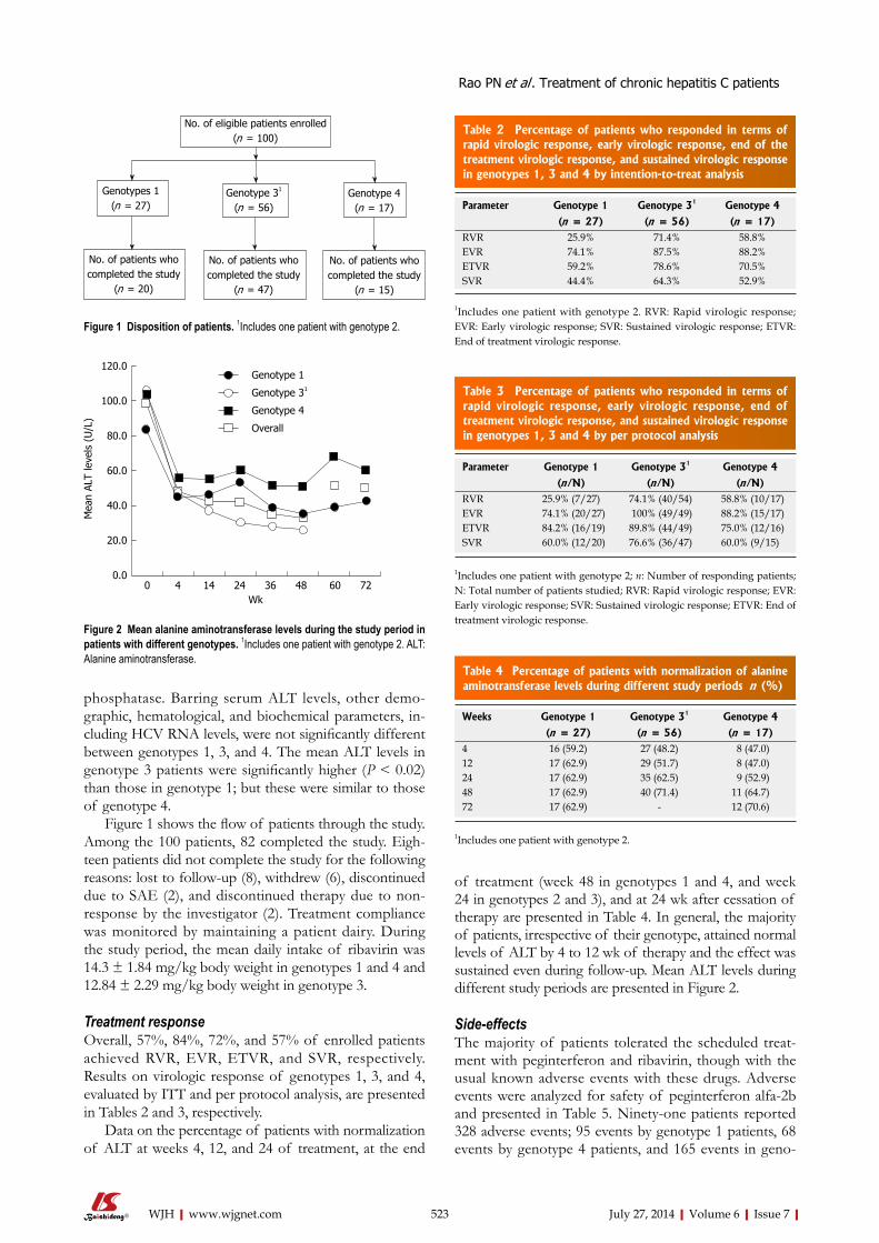
523 July 27, 2014|Volume 6|Issue 7|WJH|www.wjgnet.com
of treatment (week 48 in genotypes 1 and 4, and week 24 in genotypes 2 and 3), and at 24 wk after cessation of therapy are presented in Table 4. In general, the majority of patients, irrespective of their genotype, attained normal levels of ALT by 4 to 12 wk of therapy and the effect was sustained even during follow-up. Mean ALT levels during different study periods are presented in Figure 2.
Side-effectsThe majority of patients tolerated the scheduled treat-ment with peginterferon and ribavirin, though with the usual known adverse events with these drugs. Adverse events were analyzed for safety of peginterferon alfa-2b and presented in Table 5. Ninety-one patients reported 328 adverse events; 95 events by genotype 1 patients, 68 events by genotype 4 patients, and 165 events in geno-
phosphatase. Barring serum ALT levels, other demo-graphic, hematological, and biochemical parameters, in-cluding HCV RNA levels, were not significantly different between genotypes 1, 3, and 4. The mean ALT levels in genotype 3 patients were significantly higher (P < 0.02) than those in genotype 1; but these were similar to those of genotype 4.
Figure 1 shows the flow of patients through the study. Among the 100 patients, 82 completed the study. Eigh-teen patients did not complete the study for the following reasons: lost to follow-up (8), withdrew (6), discontinued due to SAE (2), and discontinued therapy due to non-response by the investigator (2). Treatment compliance was monitored by maintaining a patient dairy. During the study period, the mean daily intake of ribavirin was 14.3 ± 1.84 mg/kg body weight in genotypes 1 and 4 and 12.84 ± 2.29 mg/kg body weight in genotype 3.
Treatment response Overall, 57%, 84%, 72%, and 57% of enrolled patients achieved RVR, EVR, ETVR, and SVR, respectively. Results on virologic response of genotypes 1, 3, and 4, evaluated by ITT and per protocol analysis, are presented in Tables 2 and 3, respectively.
Data on the percentage of patients with normalization of ALT at weeks 4, 12, and 24 of treatment, at the end
No. of eligible patients enrolled(n = 100)
No. of patients who completed the study
(n = 15)
No. of patients who completed the study
(n = 47)
No. of patients who completed the study
(n = 20)
Genotypes 1(n = 27)
Genotype 31
(n = 56)Genotype 4
(n = 17)
Figure 1 Disposition of patients. 1Includes one patient with genotype 2.
Mea
n AL
T le
vels
(U
/L)
Wk0 4 14 24 36 48 60 72
120.0
100.0
80.0
60.0
40.0
20.0
0.0
Genotype 1
Genotype 31
Genotype 4
Overall
Figure 2 Mean alanine aminotransferase levels during the study period in patients with different genotypes. 1Includes one patient with genotype 2. ALT: Alanine aminotransferase.
Table 2 Percentage of patients who responded in terms of rapid virologic response, early virologic response, end of the treatment virologic response, and sustained virologic response in genotypes 1, 3 and 4 by intention-to-treat analysis
Parameter Genotype 1 Genotype 31 Genotype 4
(n = 27) (n = 56) (n = 17)RVR 25.9% 71.4% 58.8%EVR 74.1% 87.5% 88.2%ETVR 59.2% 78.6% 70.5%SVR 44.4% 64.3% 52.9%
1Includes one patient with genotype 2. RVR: Rapid virologic response; EVR: Early virologic response; SVR: Sustained virologic response; ETVR: End of treatment virologic response.
Table 3 Percentage of patients who responded in terms of rapid virologic response, early virologic response, end of treatment virologic response, and sustained virologic response in genotypes 1, 3 and 4 by per protocol analysis
Parameter Genotype 1 Genotype 31 Genotype 4
(n/N) (n/N) (n/N)RVR 25.9% (7/27) 74.1% (40/54) 58.8% (10/17)EVR 74.1% (20/27) 100% (49/49) 88.2% (15/17)ETVR 84.2% (16/19) 89.8% (44/49) 75.0% (12/16)SVR 60.0% (12/20) 76.6% (36/47) 60.0% (9/15)
1Includes one patient with genotype 2; n: Number of responding patients; N: Total number of patients studied; RVR: Rapid virologic response; EVR: Early virologic response; SVR: Sustained virologic response; ETVR: End of treatment virologic response.
Table 4 Percentage of patients with normalization of alanine aminotransferase levels during different study periods n (%)
Weeks Genotype 1 Genotype 31 Genotype 4
(n = 27) (n = 56) (n = 17)4 16 (59.2) 27 (48.2) 8 (47.0)12 17 (62.9) 29 (51.7) 8 (47.0)24 17 (62.9) 35 (62.5) 9 (52.9)48 17 (62.9) 40 (71.4) 11 (64.7)72 17 (62.9) - 12 (70.6)
1Includes one patient with genotype 2.
Rao PN et al . Treatment of chronic hepatitis C patients

524 July 27, 2014|Volume 6|Issue 7|WJH|www.wjgnet.com
type 3 patients. Administration of peginterferon alfa-2b resulted in common mild-to-moderate AEs, which included flu-like symptoms, nausea, and loss of appetite. None of the patients permanently stopped treatment due to adverse events, with the exception of two patients who discontinued due to SAEs. Ribavirin was temporarily discontinued due to anemia in ten patients. Twenty-four patients required ribavirin dose reduction, four needed peginterferon alfa-2b dose reduction, and four required both ribavirin and peginterferon alfa-2b dose reduction for management of anemia and thrombocytopenia. Nine patients reported 11 SAEs, which were all relieved with relevant therapy aside from one patient who died. Among the 11 SAEs, four were related to the study medication and the remaining seven, including the case of death, were unrelated to it.
DISCUSSION Infection with HCV is one of the most important medi-
cal and public health problems worldwide in view of its life-threatening complications, including hepatocellular carcinoma, cirrhosis, and liver failure[8-10]. The goal of therapy in chronic HCV infection is to achieve SVR and thereby prevent long-term complications. Despite the promising role of new antiviral therapies[11], the use of pegylated-interferon alfa combined with ribavirin contin-ues, to date, to be the standard care of treatment in HCV infection.
Since genotype constitutes one of the important de-terminants of the course and outcome of therapy, 24 or 48 wk combination therapy with peginterferon alfa and ribavirin has been recommended for genotypes 2 and 3 and genotypes 1 and 4 patients, respectively[4-7]. The pres-ent open-label, multicenter study using standard-of-care therapy was undertaken to establish that the safety and efficacy of peginterferon alfa-2b is comparable to the results of historical controls in the treatment of chronic HCV infection.
One-hundred eligible patients with chronic HCV infection were enrolled, with the majority (55%) having HCV genotype 3 which is in accordance with the pub-lished prevalence studies conducted in India[12,13]. There was only one patient with genotype 2, which is rare among Indians, and thus it should be noted that the re-ported combined results of patients with genotypes 2 and 3, in fact, only reflects those of genotype 3. Anticipating 15% attrition, 100 patients were enrolled. However, there was instead an 18% dropout, and as a result, 82 patients completed the specified study period of therapy.
Since the dose and duration of therapy were differ-ent, the data on outcome measurements were analyzed separately for genotypes 1 and 4 and for genotypes 2 and 3. The SVR (44.4%) observed in the present study for genotype 1 is comparable with those of reported stud-ies[14-16]. In genotypes 2 and 3, 64.3% of patients achieved SVR, which fits with the conformity figure results report-ed by Manns et al[17]. The rates of SVR in treatment naïve genotype 2 patients were reported to be 86.5%[18], which is higher than that of genotype 3. Since our genotype 2 and 3 patients, except for one, belonged to genotype 3, a lower SVR (64.3%) was observed in the present study. In genotype 4, 52.9% patients achieved SVR, which is com-parable with values from published studies[19,20]. Apart from genotype, baseline viral load has been shown to be one of the determinants of SVR[21]. However, perhaps due to the small number of patients covered in the pres-ent study, our stratified statistical analysis showed that baseline viral load had no impact on SVR.
In view of the cost factor and incidence of adverse events with peginterferon use during long-duration treat-ment, individualized treatment, based on the results of RVR and EVR, has been emphasized. In this respect, the presence of RVR is highly predictive of ultimate SVR with a full treatment course of 48 wk in genotype 1 pa-tients[22]. In the current study, all genotype 1 patients (n = 7) who achieved RVR also attained SVR; while a study reported a SVR rate of 86.8% in patients with RVR[15]. In genotype 4 patients, 80% with RVR attained SVR,
Table 5 Patients with adverse events
Adverse event n (%) of patientsGenotype 1 Genotype 31 Genotype 4 (n = 27) (n = 56) (n = 17)
Injection-site reactions 9 (33.3) 16 (28.6) 7 (41.2)Flu-like symptoms 24 (88.8) 49 (87.5) 14 (82.3)Tiredness 4 (14.8) 5 (8.9) 2 (11.7)Weight loss 1 (3.7) 3 (5.4) 1 (5.8)Chest discomfort 1 (3.7) 2 (3.6) 2 (11.7)Arthralgia 3 (11.1) 0 (0) 1 (5.8)Alopecia 2 (7.4) 10 (17.9) 3 (17.6)Anorexia 2 (7.4) 7 (12.5) 3 (17.6)Nausea 3 (11.1) 8 (14.3) 3 (17.6)Vomiting 2 (7.4) 3 (5.4) 0Dyspepsia 1 (3.7) 3 (5.4) 2 (11.7)Gastritis 1 (3.7) 0 (0) 0Mucous stool 1 (3.7) 0 (0) 0Diarrhea 1 (3.7) 4 (7.1) 1 (5.8)Melena 1 (3.7) 6 (10.7) 0Ascites 1 (3.7) 0 (0) 0Thrombocytopenia 2 (7.4) 5 (8.9) 1 (5.8)Anemia 9 (33.3) 13 (23.2) 6 (35.3)Neutropenia 12 (44.4) 15 (26.8) 8 (47.0)Anxiety 1 (3.7) 3 (5.4) 1 (5.8)Depression 2 (7.4) 4 (7.1) 3 (17.6)Insomnia 1 (3.7) 1 (1.8) 2 (11.7)Hypothyroidism 2 (7.4) 2 (3.6) 2 (11.7)Giddiness 1 (3.7) 1 (1.8) 2 (11.7)Dry throat 1 (3.7) 1 (1.8) 0 (0)Cough 2 (7.4) 2 (3.6) 2 (11.7)Sinusitis 1 (3.7) 0 (0) 0 (0)Bleeding gums 2 (7.4) 1 (1.8) 0 (0)Palpitation 1 (3.7) 0 (0) 0 (0)Pruritus 0 (0) 0 (0) 1 (5.8)Yellow-colored sputum 0 (0) 0 (0) 1 (5.8)Urinary tract infection 1 (3.7) 0 (0) 0 (0)Death 0 (0) 1 (1.8) 0 (0)No. of patients reporting AEs 24 (88.8) 49 (87.5) 14 (82.3)Discontinued due to SAEs 0 (0) 2 (3.6) 0 (0)Temporary discontinuation of therapy
4 (14.8) 3 (5.4) 3 (17.6)
Temporary dose reduction 11 (40.7) 16 (29.6) 5 (29.4)
1Includes one patient with genotype 2. SAE: Serious adverse event.
Rao PN et al . Treatment of chronic hepatitis C patients

525 July 27, 2014|Volume 6|Issue 7|WJH|www.wjgnet.com
whereas the published study reported 86%[23]. Similarly, among the genotype 3 patients who had RVR, 83.3% at-tained SVR, which is similar (83.7%) to that reported in the literature[24]. This further confirms the utility of RVR in predicting SVR.
Among the patients who attained EVR, 10 (76.9%) in genotype 1 and 9 (75%) in genotype 4 achieved SVR. In patients with genotypes 2 and 3, the percentage with EVR attaining SVR was 100%. This is in line with the literature[25], which shows that patients with genotype 3 who fail to achieve EVR also fail to achieve SVR. Since the duration of treatment for genotypes 2 and 3 is only 24 wk, it has been reported that EVR testing is not cost-effective in these patients[25]. This indicates that utility of RVR is higher than EVR in the prediction of SVR.
Overall, 16 patients had relapse; 5 (31.2%) patients in genotype 1, 8 (18.2%) patients in genotypes 2 and 3, and 3 (25%) patients in genotype 4. Among the 100 patients, 5 were non-responders to the study treatment; 1 (3.7%) patient in genotype 1; 2 (3.6%) patients in genotypes 2 and 3, and 2 (11.7%) in genotype 4. In addition 4 patients had breakthrough during the treatment; 2 (7.4%) patients in genotype 1; 1 (1.8%) patient in genotype 3, and 1 (5.8%) patient in genotype 4.
Biochemical response of peginterferon alfa-2b was assessed by the percentage of patients attaining normal-ization of ALT levels. Overall, the majority of patients (51%) had normalization of ALT levels as early as week 4. This denotes that peginterferon is very effective in pro-ducing a biochemical response in patients with chronic hepatitis C.
The treatment was well-tolerated in the majority of patients, though with the common side-effects usually at-tributed with interferon or ribavirin. In 32% of patients, temporary dose modifications in peginterferon (4%), ribavirin (24%), or both (4%), and temporary discon-tinuation of therapy in 10% of patients, were required. Though 11 SAEs were observed in 9 patients, only 4 were related to study medication, with such SAEs also being reported in earlier studies[15,17,26,27].
The limitations of the study are that it is a single arm study and the results on the outcome measures were compared with those of historical controls. Earlier stud-ies on Indian patients with HCV infection were conduct-ed using peginterferon alfa-2b in two studies-one study was carried out on 103 patients, but only on genotype 3 patients[25]; the other study, despite covering all four geno-types, had only 16 patients[28]. We are not aware of any study conducted with an adequately powered sample of Indian patients with HCV infection following the global guidelines on peginterferon plus ribavirin[4-7,29].
Therefore, despite the limitation of a lack of com-parator, our results on serological responses such as RVR, EVR, ETVR, and SVR provide valuable information on the safety and efficacy of peginterferon alfa-2b, in combi-nation with ribavirin, in the treatment of Indian patients with chronic HCV infection. Currently, Virchow Biotech-developed peginterferon alfa-2b is marketed in India and other emerging countries at a very competitive rate. In
view of the relatively low incidence of the adverse events and improved virologic and biochemical response, the results of the study show that peginterferon alfa-2b, in combination with ribavirin, is a safe and cost-effective drug in the treatment of chronic hepatitis C.
COMMENTSBackgroundHepatitis C virus (HCV) infection is a major cause of chronic liver disease in In-dia, with a high morbidity and mortality due to its complications. Pegylated inter-feron, in combination with ribavirin, is the standard recommended treatment for chronic hepatitis C. One of the reasons for this could be due to its cost factor, with another being that studies evaluating the safety and efficacy of these drugs in India are limited. Therefore, an attempt is being made to evaluate the efficacy of peginterferon alfa-2b, a drug locally developed in India, in combination with ribavirin.Research frontiersThis prospective study presents results on the efficacy, in terms of virologic response, of indigenously-developed peginterferon alfa-2b plus ribavirin in Indian patients with different genotypes of chronic hepatitis C. Adverse events observed with this combination are also reported. Innovations and breakthroughsThere have been a few prior studies on Indian patients with HCV infection us-ing peginterferon alfa-2b. However, these were limited to a small number of patients or confined to one genotype. Applications This study demonstrates that virologic response of peginterferon alfa-2b and ribavirin, when given as per global guidelines in Indian patients with different types of chronic hepatitis C, is similar to that of historical controls.TerminologySuccess rate of treatment is assessed based on sustained virologic response, which is defined as undetectable HCV RNA in blood 24 wk after cessation of therapy. Peer reviewThis is a straightforward clinical control study.
REFERENCES1 Global surveillance and control of hepatitis C. Report of a
WHO Consultation organized in collaboration with the Viral Hepatitis Prevention Board, Antwerp, Belgium. J Viral Hepat 1999; 6: 35-47 [PMID: 10847128 DOI: 10.1046/j.1365-2893.1999.6120139.x]
2 WHO. Hepatitis C. WHO Fact sheet No 164. Available from: URL: http: //www.who.int/mediacentre/factsheets/fs164/en/
3 Mukhopadhyaya A. Hepatitis C in India. J Biosci 2008; 33: 465-473 [PMID: 19208972 DOI: 10.4103/1947-2714.103325]
4 Ghany MG, Strader DB, Thomas DL, Seeff LB. Diagnosis, management, and treatment of hepatitis C: an update. Hepa-tology 2009; 49: 1335-1374 [PMID: 19330875 DOI: 10.1002/hep.22759]
5 European Association for the Study of the Liver. EASL Cli-nical Practice Guidelines: management of hepatitis C virus infection. J Hepatol 2011; 55: 245-264 [PMID: 21371579 DOI: 10.1016/j.jhep.2011.02.023]
6 Omata M, Kanda T, Yu ML, Yokosuka O, Lim SG, Jafri W, Tateishi R, Hamid SS, Chuang WL, Chutaputti A, Wei L, Sol-lano J, Sarin SK, Kao JH, McCaughan GW. APASL consensus statements and management algorithms for hepatitis C virus infection. Hepatol Int 2012; 6: 409-435 [DOI: 10.1007/s12072-012-9342-y]
7 Ghany MG, Nelson DR, Strader DB, Thomas DL, Seeff LB. An update on treatment of genotype 1 chronic hepatitis C virus infection: 2011 practice guideline by the American As-sociation for the Study of Liver Diseases. Hepatology 2011; 54: 1433-1444 [PMID: 21898493 DOI: 10.1002/hep.24641]
COMMENTS
Rao PN et al . Treatment of chronic hepatitis C patients

526 July 27, 2014|Volume 6|Issue 7|WJH|www.wjgnet.com
8 Kenny-Walsh E. Clinical outcomes after hepatitis C infection from contaminated anti-D immune globulin. Irish Hepatolo-gy Research Group. N Engl J Med 1999; 340: 1228-1233 [PMID: 10210705 DOI: 10.1056/NEJM199904223401602]
9 Di Bisceglie AM. Natural history of hepatitis C: its impact on clinical management. Hepatology 2000; 31: 1014-1018 [PMID: 10733560 DOI: 10/S0270913900298170]
10 Barrera JM, Bruguera M, Ercilla MG, Gil C, Celis R, Gil MP, del Valle Onorato M, Rodés J, Ordinas A. Persistent hepatitis C viremia after acute self-limiting posttransfusion hepatitis C. Hepatology 1995; 21: 639-644 [PMID: 7533121]
11 Kanda T, Imazeki F, Yokosuka O. New antiviral therapies for chronic hepatitis C. Hepatol Int 2010; 4: 548-561 [PMID: 21063477 DOI: 10.1007/s12072-010-9193-3]
12 Hissar SS, Goyal A, Kumar M, Pandey C, Suneetha PV, Sood A, Midha V, Sakhuja P, Malhotra V, Sarin SK. Hepa-titis C virus genotype 3 predominates in North and Central India and is associated with significant histopathologic liver disease. J Med Virol 2006; 78: 452-458 [PMID: 16482560 DOI: 10.1002/jmv.20561]
13 Narahari S, Juwle A, Basak S, Saranath D. Prevalence and geographic distribution of Hepatitis C Virus genotypes in Indian patient cohort. Infect Genet Evol 2009; 9: 643-645 [PMID: 19460332 DOI: 10.1016/j.meegid.2009.04.001]
14 Manns MP, McHutchison JG, Gordon SC, Rustgi VK, Shiff-man M, Reindollar R, Goodman ZD, Koury K, Ling M, Al-brecht JK. Peginterferon alfa-2b plus ribavirin compared with interferon alfa-2b plus ribavirin for initial treatment of chron-ic hepatitis C: a randomised trial. Lancet 2001; 358: 958-965 [PMID: 11583749 DOI: 10.1016/S0140-6736(01)06102-5]
15 Mangia A, Minerva N, Bacca D, Cozzolongo R, Ricci GL, Carretta V, Vinelli F, Scotto G, Montalto G, Romano M, Cristofaro G, Mottola L, Spirito F, Andriulli A. Individual-ized treatment duration for hepatitis C genotype 1 patients: A randomized controlled trial. Hepatology 2008; 47: 43-50 [PMID: 18069698 DOI: 10.1002/hep.22061]
16 Kainuma M, Furusyo N, Kajiwara E, Takahashi K, Nomura H, Tanabe Y, Satoh T, Maruyama T, Nakamuta M, Kotoh K, Azuma K, Shimono J, Shimoda S, Hayashi J. Pegylated interferon α-2b plus ribavirin for older patients with chronic hepatitis C. World J Gastroenterol 2010; 16: 4400-4409 [PMID: 20845506 DOI: 10.3748/wjg.v16.i35.4400]
17 Manns M, Zeuzem S, Sood A, Lurie Y, Cornberg M, Klinker H, Buggisch P, Rössle M, Hinrichsen H, Merican I, Ilan Y, Mauss S, Abu-Mouch S, Horban A, Müller TH, Welsch C, Chen R, Faruqi R, Pedicone LD, Wedemeyer H. Reduced dose and duration of peginterferon alfa-2b and weight-based ribavirin in patients with genotype 2 and 3 chronic hepatitis C. J Hepatol 2011; 55: 554-563 [PMID: 21237227 DOI: 10.1016/j.jhep.2010.12.024]
18 Kanda T, Imazeki F, Azemoto R, Yonemitsu Y, Mikami S, Kita K, Takashi M, Sunaga M, Wu S, Nakamoto S, Tawada A, Arai M, Kato K, Yoshida Y, Koma Y, Fujiwara K, Fukai K, Suzuki N, Yokosuka O. Response to peginterferon-alfa 2b and ribavirin in Japanese patients with chronic hepatitis C genotype 2. Dig Dis Sci 2011; 56: 3335-3342 [PMID: 21604145 DOI: 10.1007/s10620-011-1750-7]
19 Al-Ali J, Siddique I, Varghese R, Hasan F. Pegylated interfer-on-alpha2b plus ribavirin for the treatment of chronic hepa-titis C virus genotype 4 infection in patients with normal serum ALT. Ann Hepatol 2012; 11: 186-193 [PMID: 22345335]
20 Khuroo MS, Khuroo MS, Dahab ST. Meta-analysis: a ran-domized trial of peginterferon plus ribavirin for the initial treatment of chronic hepatitis C genotype 4. Aliment Phar-macol Ther 2004; 20: 931-938 [PMID: 15521839 DOI: 10.1111/j.1365-2036.2004.02208.x]
21 Hadziyannis SJ, Sette H, Morgan TR, Balan V, Diago M, Marcellin P, Ramadori G, Bodenheimer H, Bernstein D, Rizzetto M, Zeuzem S, Pockros PJ, Lin A, Ackrill AM. Peginterferon-alpha2a and ribavirin combination therapy in chronic hepatitis C: a randomized study of treatment dura-tion and ribavirin dose. Ann Intern Med 2004; 140: 346-355 [PMID: 14996676]
22 Poordad F, Landaverde C. Rapid virological response to peginterferon alfa and ribavirin treatment of chronic hepati-tis C predicts sustained virological response and relapse in genotype 1 patients. Therap Adv Gastroenterol 2009; 2: 91-97 [PMID: 21180537 DOI: 10.1177/1756283X08101217]
23 Kamal SM, El Kamary SS, Shardell MD, Hashem M, Ahmed IN, Muhammadi M, Sayed K, Moustafa A, Hakem SA, Ibra-hiem A, Moniem M, Mansour H, Abdelaziz M. Pegylated interferon alpha-2b plus ribavirin in patients with genotype 4 chronic hepatitis C: The role of rapid and early virologic response. Hepatology 2007; 46: 1732-1740 [PMID: 17943989]
24 Poordad FF. Review article: the role of rapid virological response in determining treatment duration for chronic hepatitis C. Aliment Pharmacol Ther 2010; 31: 1251-1267 [PMID: 20236258 DOI: 10.1111/j.1365-2036.2010.04300.x]
25 Gralewicz S, Eckersdorf B, Gołebiewski H. Hippocampal rh-ythmic slow activity (RSA) in the cat after intraseptal injections of muscarinic cholinolytics. Acta Neurobiol Exp (Wars) 1992; 52: 211-221 [PMID: 1293959 DOI: 10.1053/jhep.2003.50364]
26 Jacobson IM, Brown RS, Freilich B, Afdhal N, Kwo PY, San-toro J, Becker S, Wakil AE, Pound D, Godofsky E, Strauss R, Bernstein D, Flamm S, Pauly MP, Mukhopadhyay P, Griffel LH, Brass CA. Peginterferon alfa-2b and weight-based or flat-dose ribavirin in chronic hepatitis C patients: a random-ized trial. Hepatology 2007; 46: 971-981 [PMID: 17894303 DOI: 10.1002/hep.21932]
27 Sood A, Midha V, Hissar S, Kumar M, Suneetha PV, Bansal M, Sood N, Sakhuja P, Sarin SK. Comparison of low-dose pegylated interferon versus standard high-dose pegylated interferon in combination with ribavirin in patients with chronic hepatitis C with genotype 3: an Indian experience. J Gastroenterol Hepatol 2008; 23: 203-207 [PMID: 17645472 DOI: 10.1111/j.1440-1746.2007.05057.x]
28 Ray G, Pal S, Nayyar I, Dey S. Efficacy and tolerability of pe-gylated interferon alpha 2b and ribavirin in chronic hepatitis C--a report from eastern India. Trop Gastroenterol 2007; 28: 109-112 [PMID: 18383998]
29 WHO. Guidelines for the screening, care and treatment of persons with hepatitis c infection. Available from: URL: http: //apps.who.int/iris/bitstream/10665/111747/1/9789-241548755_eng.pdf?ua=1
P- Reviewer: Ford N, Kanda T, Liu CJ, Shi Z S- Editor: Ji FF L- Editor: Rutherford A E- Editor: Liu SQ
Rao PN et al . Treatment of chronic hepatitis C patients

Cássio Vieira de Oliveira, Alecsandro Moreira, Julio P Baima, Leticia de C Franzoni, Talles B Lima, Fabio da S Yamashiro, Kunie Yabuki Rabelo Coelho, Ligia Y Sassaki, Carlos Antonio Caramori, Fernando G Romeiro, Giovanni F Silva
Cássio Vieira de Oliveira, Alecsandro Moreira, Julio P Baima, Leticia de C Franzoni, Talles B Lima, Fabio da S Yamashiro, Ligia Y Sassaki, Carlos Antonio Caramori, Fernando G Ro-meiro, Giovanni F Silva, Department of Internal Medicine, Gastroenterology Division, Botucatu Medical School, São Paulo State University-UNESP, Botucatu, São Paulo 01049-010, Brazil Kunie Yabuki Rabelo Coelho, Department of Pathology, Botu-catu Medical School, São Paulo State University-UNESP, Botu-catu, São Paulo 01049-010, BrazilAuthor contributions: de Oliveira CV conceived and coordi-nated the study and participated in the data acquisition and manu-script writing; Lima TB, Baima J, Coelho KYR, Sassaki LY, Caramori CA, Moreira A and Silva GF participated in the data acquisition and manuscript writing; Romeiro FG coordinated the study and participated in the data acquisition and manuscript writing; Coelho KYR coordinated the histopathological analysis.Supported by The Department of Internal Medicine, Gastro-enterology Division, Botucatu Medical School, São Paulo State University-UNESP, Botucatu/SP, BrazilCorrespondence to: Cássio Vieira de Oliveira, PhD, Depart-ment of Internal Medicine, Gastroenterology Division, Botucatu Medical School, São Paulo State University-UNESP, Rua Quiri-no de Andrade, 215, Botucatu, São Paulo 01049-010, Brazil. [email protected]: +55-14-997624920 Fax: +55-14-38116213Received: March 12, 2014 Revised: May 20, 2014Accepted: June 10, 2014Published online: July 27, 2014
AbstractAcute fatty liver of pregnancy is a rare disease that affects women in the third trimester of pregnancy. Although infrequent, the disease can cause maternal mortality. The diagnosis is not always clear until the pregnancy is terminated, and significant complica-tions, such as acute pancreatitis, can occur. Pancreatic involvement typically only occurs in severe cases after the development of hepatic and renal impairment. To date, little knowledge is available regarding how the
disease causes pancreatitis. Treatment involves sup-portive measures and pregnancy interruption. In this report, we describe a case of a previously healthy 26-year-old woman at a gestational age of 27 wk and 6 d who was admitted with severe abdominal pain and vomiting. This case illustrates the clinical and labora-tory overlap between acute fatty liver of pregnancy and pancreatitis, highlighting the difficulties in differentiat-ing each disease. Furthermore, the hypothesis for this overlapping is presented, and the therapeutic options are discussed.
© 2014 Baishideng Publishing Group Inc. All rights reserved.
Key words: Acute fatty liver of pregnancy; Severe acute pancreatitis; Fulminant hepatic failure; Liver disease in pregnancy
Core tip: A previously healthy 26-year-old woman at 27 wk and 6 d of pregnancy was referred for investiga-tion of abdominal pain. She presented with complaints of diffuse abdominal pain with nausea and vomiting associated with hepatic and renal dysfunction. Acute fatty liver of pregnancy and severe acute pancreatitis were diagnosed. Acute fatty liver of pregnancy is rarely associated with severe acute pancreatitis, which can complicate the diagnosis. The possible mechanisms in-volved in this association and the current therapies are discussed, focusing on the relevant aspects to improve the management of similar cases.
de Oliveira CV, Moreira A, Baima JP, Franzoni LC, Lima TB, Yamashiro FS, Coelho KYR, Sassaki LY, Caramori CA, Ro-meiro FG, Silva GF. Acute fatty liver of pregnancy associated with severe acute pancreatitis: A case report. World J Hepa-tol 2014; 6(7): 527-531 Available from: URL: http://www.wjgnet.com/1948-5182/full/v6/i7/527.htm DOI: http://dx.doi.org/10.4254/wjh.v6.i7.527
CASE REPORT
Submit a Manuscript: http://www.wjgnet.com/esps/Help Desk: http://www.wjgnet.com/esps/helpdesk.aspxDOI: 10.4254/wjh.v6.i7.527
527 July 27, 2014|Volume 6|Issue 7|WJH|www.wjgnet.com
World J Hepatol 2014 July 27; 6(7): 527-531ISSN 1948-5182 (online)
© 2014 Baishideng Publishing Group Inc. All rights reserved.
Acute fatty liver of pregnancy associated with severe acute pancreatitis: A case report

INTRODUCTION Acute fatty liver of pregnancy (AFLP) is a disorder unique to pregnancy that is characterized by microve-sicular fatty infiltration of hepatocytes[1]. AFLP was first described in 1940 and was initially considered fatal[2]. However, early diagnosis has dramatically improved the prognosis and maternal mortality; therefore, maternal mortality is currently the exception rather than the rule[1]. AFLP typically occurs in the third quarter of pregnancy, but it is not always diagnosed prior to delivery, as was the case described herein.
The most common initial symptoms are anorexia, nausea, vomiting, abdominal pain, and jaundice. A condi-tion that must be excluded is hemolytic anemia elevated liver function and low platelet count syndrome (HELLP) syndrome, which is characterized by hemolysis, elevated liver enzymes, and low platelet count. AFLP and HELLP syndrome can occur together in some overlapping cases, making the diagnosis more difficult. However, the signs of liver failure, such as hypoglycemia and hepatic enceph-alopathy, are suggestive of AFLP. Additionally, HELLP syndrome is likely to occur in patients with hypertension, whereas AFLP often occurs in the absence of hyperten-sion. The differential diagnosis of these two diseases was evaluated in a recent study, which indicated that the incorporation of antithrombin activity less than 65% into the diagnostic criteria for AFLP may facilitate prompt diagnosis of this disease[3].
CASE REPORTA previously healthy 26-year-old woman at a gestational age of 27 wk and 6 d was referred to our hospital due to a diagnostic hypothesis of acute appendicitis. She was com-plaining of diffuse abdominal pain, nausea, and vomiting during the week. During her physical exam, she was pale and prostrated with mild tachycardia (108 beats/min) and normal blood pressure (110/70 mmHg). No signs of acute appendicitis were noted, but she displayed a potent and diffuse abdominal pain. Cardiotocography revealed signs of fetal distress, so an emergency cesarean section was performed. During the surgery, the possibility of ap-pendicitis was eliminated. Because the newborn displayed bradycardia and an absence of heartbeats at delivery, he was submitted to initial resuscitation protocols and sent to the intensive care unit (ICU). Laboratory tests on the mother revealed leukocytosis, anemia, and hepatic and re-nal impairment, but no significant proteinuria was found (Table 1).
Abdominal ultrasonography revealed only pancreatic edema without signs of biliary obstruction. After the delivery, abdominal computed tomography (CT), upper gastrointestinal endoscopy, and biochemical tests were performed. The endoscopy was performed exclusively to investigate the possibility of peptic ulcer or other gas-troduodenal diseases, but no pathological findings were found. The CT showed only pancreatic edema without peripancreatic collections (Figure 1). Given that the amy-
lase increase was greater than sixfold higher than the nor-mal upper limit and that pancreatic edema was confirmed by ultrasonography and CT exams, the presence of pan-creatitis was conclusive. According to the Ranson criteria, the patient had a severe disease that achieved 4 points at admission based on the leukocyte count, aspartate ami-notransferase, glycemia, and lactate dehydrogenase values (Table 1). Additionally, she had acute renal failure and achieved 14 points according to the APACHE Ⅱ criteria, which corresponds to an estimated 18.6% risk of hospi-tal death.
The patient developed somnolence and exhibited a progressive decrease in her level of consciousness. Tra-cheal intubation and mechanical ventilation were needed, so she was transferred to the ICU. At this time, the blood glucose remained normal, but she had abdominal disten-sion and decreased bowel sounds. Then, the diagnostic hypotheses changed to acute liver failure, severe acute pancreatitis, and renal failure. Suddenly, she presented re-current episodes of hypoglycemia, even with continuous
528 July 27, 2014|Volume 6|Issue 7|WJH|www.wjgnet.com
de Oliveira CV et al . Pancreatitis and pregnancy fatty liver
Table 1 Main laboratory tests demonstrating the development of liver and pancreatic
Blood tests Admission 48 h after Hospital 1 yr after
admission discharge dischargeHemoglobin (g/dL) 9.8 11.2 - -Leukocyte count (mm3) 24000 21500 - -Glucose (mg/dL) 586 71 91 76Alkaline phosphatase (U/L) 532 392 248 -g-GTP (U/L) 282 325 234 -Calcium (mg/dL) 8.4 7.7 - -Amylase (U/L) 460 642 - 59LDH (U/L) 938 977 - -ALT (U/L) 202 112 34 15AST (U/L) 343 179 38 17TB (mg/dL) 4.0 4.9 0.6 0.5Creatinine (mg/dL) 2.6 3.3 0.7 0.8Urea (mg/dL) 78 85 20 28INR 2.13 2.78 1.16 0.98Proteinuria 0.06 g/
24 h0.03 g/
24 h- -
ALT: Alanine aminotransferase; AST: Aspartate aminotransferase; TB: Total bilirubin; LDH: Lactate dehydrogenase; INR: International normalized ratio; g-GTP: Gamma-glutamyl transpeptidase.
Figure 1 Abdominal computed tomography scan showing diffuse pancre-atic edema.

dextrose infusion and parenteral nutrition. In response to these new symptoms, AFLP became the major diagnostic hypothesis. Serum factor V was normal, so a percutane-ous liver biopsy was performed.
The liver biopsy analysis showed centrilobular micro-goticular steatosis, ballooning degeneration, and reticular collapse. The Masson staining showed areas of reticular thickening and intralobular collapse. The “red oil” stain was positive in focal areas, yielding the diagnosis of AFLP (Figure 2).
Seven days following the delivery, she exhibited a clear improvement in consciousness level and liver function tests. She was discharged on postoperative day 24 and returned to the hospital 4 mo later without neurological sequelae. Additionally, laboratory tests and abdominal CT were normal. Despite the problems during the birth, her child exhibited normal development.
DISCUSSIONAFLP is a rare condition that affects approximately 1 in 7000 to 1 in 20000 births[4-8]. AFLP is more common in women with multiple pregnancies and, possibly, in under-weight women. However, this case of AFLP occurred in a primiparous, normal-weight woman but not delivering twins.
Approximately half of AFLP patients display signs of preeclampsia at the beginning of or at some time dur-ing the course of the disease[9]. Extrahepatic complica-tions may occur, which can be life-threatening[10,11]. The patients rarely develop pancreatitis, which can be severe. Similar to the case described herein, pancreatitis is typi-cally noticed only after the development of hepatic and renal dysfunction[12]. In this case, the patient had AFLP with severe acute pancreatitis, an association that is rarely documented in the literature. The acute renal failure was a complication of the pancreatitis, so it was treated only by supportive measurements and the delivery, thereby confirming that it was a consequence of the underlying disease. No renal replacement therapy was needed. The
liver function tests demonstrated severe hepatic impair-ment, which was the cause of the jaundice. Therefore, even in the presence of severe pancreatitis, liver disease remained the major disease.
Women with AFLP have impaired liver function with increased bilirubin and transaminase levels and leukocyte counts, which are typically higher than those observed in a normal pregnancy. The platelet count can be reduced with or without additional signs of disseminated intravas-cular coagulation in association with a significant reduc-tion of antithrombin Ⅲ[13]. Severely affected patients also have elevated serum ammonia, prolonged prothrombin times, and hypoglycemia caused by liver failure. Acute renal failure and hyperuricemia are often present[14]. How-ever, in the case presented, the recurrent episodes of hypoglycemia, even with appropriate correction, were the main diagnostic clue.
The association between AFLP and inherited defects in the mitochondrial beta-oxidation of fatty acids, espe-cially the impairment of long-chain 3-hydroxyacyl-CoA dehydrogenase (LCHAD), suggests that some affected women and fetuses have an inherited enzyme deficiency in beta-oxidation that predisposes the mother to this dis-order[15-17]. LCHAD catalyzes the third step of the beta-oxidation of fatty acids in the mitochondrion (the forma-tion of 3-ketoacyl-CoA from 3-hydroxyacyl-CoA). The accumulation of long-chain metabolites of 3-hydroxyacyl produced by the fetus or placenta is toxic to the liver and can serve as the cause of the liver disease. The role of the pathogenesis of LCHAD in AFLP has been illustrated in various studies[18-20].
The mechanism by which pancreatitis may develop as a complication of fatty liver of pregnancy is not well understood because this association is rare. Our hypoth-esis is that the accumulation of long-chain metabolites of 3-hydroxyacyl is toxic to the liver and the pancreatic tissue. Thus, the pancreas could be affected when an in-creased concentration of these metabolites is present, as occurs in cases of severe hepatic disease. This hypothesis serves as a reasonable explanation for the pancreatic im-pairment displayed in this case of hepatic failure.
The diagnosis of LCHAD deficiency in newborns can save lives; therefore, all women with AFLP and their children should be administered a molecular test for LCHAD, which should at least evaluate the most com-mon mutation, namely, G1528C[21,22]. In the present case, it was not possible to perform this type of test because it was not available.
The clinical diagnosis of AFLP is typically performed according to the definition, presentation, and laboratory-compatible image results. The liver imaging is primarily used to exclude other diagnoses, such as hepatic infarc-tion and hematoma[23]. Various authors reported steatosis on ultrasound or CT, but these tests are only useful for performing comparative analyses[24,25]. The AFLP diagno-sis can only be made through a liver biopsy showing mi-crovesicular fatty infiltration in hepatocytes. The fat drop-lets are centrally distributed around the cellular nuclei,
529 July 27, 2014|Volume 6|Issue 7|WJH|www.wjgnet.com
Figure 2 Histopathological analysis of the liver biopsy according to three stains. A: Hematoxylin-eosin staining reveals canalicular cholestasis, hepato-cellular ballooning, and microgoticular steatosis; B: Masson staining demon-strates microgoticular steatosis and perivenular and pericellular fibrosis; C: Oil red staining shows the microdroplets of fat clearly stained in red.
A B C
de Oliveira CV et al . Pancreatitis and pregnancy fatty liver

530 July 27, 2014|Volume 6|Issue 7|WJH|www.wjgnet.com
COMMENTSCase characteristicsA previously healthy 26-year-old woman with a gestational age of 27 wk and 6 d presented with diffuse abdominal pain associated with hepatic and renal failure.Clinical diagnosisDiffuse abdominal pain and vomiting.Differential diagnosisHemolytic anemia elevated liver function and low platelet count syndrome syn-drome.Laboratory diagnosisAmylase: 642 U/L; glucose: 586 mg/dL; alanine aminotransferase: 202 U/L; aspartate aminotransferase: 343 U/L; total bilirubin: 4.0 mg/dL; creatinine: 2.6 mg/dL; urea: 78 mg/dL; international normalized ratio: 2.13.Imaging diagnosisPancreatic edema was confirmed by ultrasonography and computed tomography.Pathological diagnosisThe liver biopsy results were compatible with acute fatty liver of pregnancy (AFLP).TreatmentPregnancy interruption and supportive measures.Related reportsAFLP rarely presents with severe acute pancreatitis.Experiences and lessonsBecause the early symptoms of AFLP can be uncharacteristic, the disease can be misdiagnosed. It is important to be vigilant to make the correct diagnosis of AFLP and identify complications, such as pancreatitis.Peer reviewThis article describes a rare case with acute fatty liver disease complicated with acute pancreatitis. This is an interesting case report.
REFERENCES1 Bacq Y, Riely CA. Acute fatty liver of pregnancy: the hepa-
tologist’s view. Gastroenterologist 1993; 1: 257-264 [PMID: 8055222]
2 Sheehan H. The pathology of acute yellow atrophy and de-layed chloroform poisoning. J Obstet Gynaecol 1940; 47: 49-53 [DOI: 10.1111/j.1471-0528.1940.tb14731.x]
3 Minakami H, Morikawa M, Yamada T, Yamada T, Akaishi R, Nishida R. Differentiation of acute fatty liver of pregnancy from syndrome of hemolysis, elevated liver enzymes and low platelet counts. J Obstet Gynaecol Res 2014; 40: 641-649 [PMID: 24428400 DOI: 10.1111/jog.12282]
4 Usta IM, Barton JR, Amon EA, Gonzalez A, Sibai BM. Acute fatty liver of pregnancy: an experience in the diagnosis and management of fourteen cases. Am J Obstet Gynecol 1994; 171: 1342-1347 [PMID: 7977544 DOI: 10.1016/0002-9378(94)90158-9]
5 Pockros PJ, Peters RL, Reynolds TB. Idiopathic fatty liver of pregnancy: findings in ten cases. Medicine (Baltimore) 1984; 63: 1-11 [PMID: 6690883 DOI: 10.1097/00005792-198401000-00001]
6 Reyes H, Sandoval L, Wainstein A, Ribalta J, Donoso S, Smok G, Rosenberg H, Meneses M. Acute fatty liver of pregnancy: a clinical study of 12 episodes in 11 patients. Gut 1994; 35: 101-106 [PMID: 8307428 DOI: 10.1136/gut.35.1.101]
7 Castro MA, Fassett MJ, Reynolds TB, Shaw KJ, Goodwin TM. Reversible peripartum liver failure: a new perspective on the diagnosis, treatment, and cause of acute fatty liver of pregnancy, based on 28 consecutive cases. Am J Obstet Gyne-col 1999; 181: 389-395 [PMID: 10454689 DOI: 10.1016/S0002-9378(99)70567-3]
8 Knight M, Nelson-Piercy C, Kurinczuk JJ, Spark P, Brockle-hurst P. A prospective national study of acute fatty liver of pregnancy in the UK. Gut 2008; 57: 951-956 [PMID: 18332072 DOI: 10.1136/gut.2008.148676]
giving the cytoplasm a foamy aspect and typically sparing the cells around the portal tract[26]. A special staining (oil red) must be used to confirm the diagnosis in patients without obvious vacuolation[27,28]. As the liver biopsy is invasive, it is not always performed. Liver biopsy should be performed with caution during pregnancy and be re-served for cases where the diagnosis remains unclear.
A specific treatment is not available for AFLP. The primary treatment is delivery, which typically occurs on an emergency basis after maternal stabilization via the in-fusion of glucose and reversal of the coagulation distur-bances. Because hypoglycemia is common and dangerous, glucose levels should be monitored until liver function normalizes[7]. It is typically necessary to treat hypoglyce-mia with the continuous infusion of a 10% glucose solu-tion. Some patients with severe hypoglycemia may require additional bolus administrations of 50% glucose[7].
The liver function tests typically begin to normalize after delivery; however, after the first few days, a tran-sient worsening of renal and hepatic function may be observed, followed by a definite improvement. In severe cases, particularly when the diagnosis is delayed, more days of illness can be observed, requiring supportive treatment in an ICU. Mechanical ventilation, dialysis, parenteral nutrition, or surgery to treat bleeding during cesarean delivery may be needed. Even the most severely ill patients can recover without liver disease sequelae[7]. However, substantial morbidity and even maternal mor-tality may occur[8]. Some reports have also described pa-tients who required liver transplantation, which is rarely needed when a diagnosis and pregnancy termination were achieved in sufficient time[29,30]. A case series of five patients indicated good recovery after plasma exchange and renal replacement therapy, showing that the accumu-lation of long-chain metabolites of 3-hydroxyacyl is most likely the major cause of the liver disease. The authors suggested that these therapies are safe and effective; thus, they should be used immediately at the onset of hepatic encephalopathy and/or renal failure in AFLP patients[31]. None of the patients treated by plasma exchange and re-nal replacement therapy developed pancreatitis, indicating that our pancreatic toxicity hypothesis may be correct.
Of note, AFLP can recur in subsequent pregnan-cies despite negative LCHAD mutation screening re-sults[6,15,18,32-35]. However, the exact risk of recurrence is unknown. Affected women should be advised of this possibility, and a maternal-fetal medicine specialist should closely monitor subsequent pregnancies. Given that the initial symptoms of AFLP can be atypical, the diagnosis can be easily misidentified, leading to multiple organ dys-functions. Once the diagnosis is confirmed, it is important to be vigilant to avoid serious complications, such as pan-creatitis, renal failure, and hypoglycemia, as described here.
ACKNOWLEDGMENTSWe acknowledge Professor Maria Rita Piloto for the ar-ticle corrections.
COMMENTS
de Oliveira CV et al . Pancreatitis and pregnancy fatty liver

531 July 27, 2014|Volume 6|Issue 7|WJH|www.wjgnet.com
9 Riely CA. Acute fatty liver of pregnancy. Semin Liver Dis 1987; 7: 47-54 [PMID: 3296215 DOI: 10.1055/s-2008-1040563]
10 Pereira SP, O’Donohue J, Wendon J, Williams R. Maternal and perinatal outcome in severe pregnancy-related liver disease. Hepatology 1997; 26: 1258-1262 [PMID: 9362370 DOI: 10.1002/hep.510260525]
11 Kennedy S, Hall PM, Seymour AE, Hague WM. Transient diabetes insipidus and acute fatty liver of pregnancy. Br J Obstet Gynaecol 1994; 101: 387-391 [PMID: 8018608 DOI: 10.1111/j.1471-0528.1994.tb11909.x]
12 Moldenhauer JS, O’brien JM, Barton JR, Sibai B. Acute fatty liver of pregnancy associated with pancreatitis: a life-threat-ening complication. Am J Obstet Gynecol 2004; 190: 502-505 [PMID: 14981397 DOI: 10.1016/j.ajog.2003.09.022]
13 Castro MA, Goodwin TM, Shaw KJ, Ouzounian JG, McGe-hee WG. Disseminated intravascular coagulation and anti-thrombin III depression in acute fatty liver of pregnancy. Am J Obstet Gynecol 1996; 174: 211-216 [PMID: 8572009]
14 Grünfeld JP, Pertuiset N. Acute renal failure in pregnancy: 1987. Am J Kidney Dis 1987; 9: 359-362 [PMID: 3555010 DOI: 10.1016/S0272-6386(87)80137-3]
15 Schoeman MN, Batey RG, Wilcken B. Recurrent acute fatty liver of pregnancy associated with a fatty-acid oxidation defect in the offspring. Gastroenterology 1991; 100: 544-548 [PMID: 1985050]
16 Wilcken B, Leung KC, Hammond J, Kamath R, Leonard JV. Pregnancy and fetal long-chain 3-hydroxyacyl coenzyme A dehydrogenase deficiency. Lancet 1993; 341: 407-408 [PMID: 8094173]
17 Treem WR, Rinaldo P, Hale DE, Stanley CA, Millington DS, Hyams JS, Jackson S, Turnbull DM. Acute fatty liver of pregnancy and long-chain 3-hydroxyacyl-coenzyme A de-hydrogenase deficiency. Hepatology 1994; 19: 339-345 [PMID: 8294091 DOI: 10.1002/hep.1840190211]
18 Treem WR, Shoup ME, Hale DE, Bennett MJ, Rinaldo P, Millington DS, Stanley CA, Riely CA, Hyams JS. Acute fatty liver of pregnancy, hemolysis, elevated liver enzymes, and low platelets syndrome, and long chain 3-hydroxyacyl-coen-zyme A dehydrogenase deficiency. Am J Gastroenterol 1996; 91: 2293-2300 [PMID: 8931405]
19 Sims HF, Brackett JC, Powell CK, Treem WR, Hale DE, Ben-nett MJ, Gibson B, Shapiro S, Strauss AW. The molecular basis of pediatric long chain 3-hydroxyacyl-CoA dehydroge-nase deficiency associated with maternal acute fatty liver of pregnancy. Proc Natl Acad Sci USA 1995; 92: 841-845 [PMID: 7846063 DOI: 10.1073/pnas.92.3.841]
20 Yang Z, Zhao Y, Bennett MJ, Strauss AW, Ibdah JA. Fetal genotypes and pregnancy outcomes in 35 families with mitochondrial trifunctional protein mutations. Am J Obstet Gynecol 2002; 187: 715-720 [PMID: 12237653 DOI: 10.1067/mob.2002.125893]
21 Rajasri AG, Srestha R, Mitchell J. Acute fatty liver of preg-nancy (AFLP)--an overview. J Obstet Gynaecol 2007; 27: 237-240 [PMID: 17464801 DOI: 10.1080/01443610701194705]
22 Ibdah JA. Acute fatty liver of pregnancy: an update on
pathogenesis and clinical implications. World J Gastroenterol 2006; 12: 7397-7404 [PMID: 17167825 DOI: 10.3748/wjg.v12.i46.7397]
23 Strategies for cadaveric organ procurement. Mandated choice and presumed consent. Council on Ethical and Judi-cial Affairs, American Medical Association. JAMA 1994; 272: 809-812 [PMID: 8078146]
24 Castro MA, Ouzounian JG, Colletti PM, Shaw KJ, Stein SM, Goodwin TM. Radiologic studies in acute fatty liver of preg-nancy. A review of the literature and 19 new cases. J Reprod Med 1996; 41: 839-843 [PMID: 8951135]
25 Campillo B, Bernuau J, Witz MO, Lorphelin JM, Degott C, Rueff B, Benhamou JP. Ultrasonography in acute fatty liver of pregnancy. Ann Intern Med 1986; 105: 383-384 [PMID: 3527013 DOI: 10.7326/0003-4819-105-3-383]
26 Bacq Y. Acute fatty liver of pregnancy. Semin Perinatol 1998; 22: 134-140 [PMID: 9638907 DOI: 10.1016/S0146-0005(98)80045-1]
27 Riely CA, Latham PS, Romero R, Duffy TP. Acute fatty liver of pregnancy. A reassessment based on observations in nine patients. Ann Intern Med 1987; 106: 703-706 [PMID: 3565968 DOI: 10.7326/0003-4819-106-5-703]
28 Rolfes DB, Ishak KG. Acute fatty liver of pregnancy: a clini-copathologic study of 35 cases. Hepatology 1985; 5: 1149-1158 [PMID: 2415437 DOI: 10.1002/hep.1840050615]
29 Ockner SA, Brunt EM, Cohn SM, Krul ES, Hanto DW, Peters MG. Fulminant hepatic failure caused by acute fatty liver of pregnancy treated by orthotopic liver transplantation. Hepatology 1990; 11: 59-64 [PMID: 2403963 DOI: 10.1002/hep.1840110112]
30 Amon E, Allen SR, Petrie RH, Belew JE. Acute fatty liver of pregnancy associated with preeclampsia: management of hepatic failure with postpartum liver transplantation. Am J Perinatol 1991; 8: 278-279 [PMID: 1741873 DOI: 10.1055/s-2007-999396]
31 Yu CB, Chen JJ, Du WB, Chen P, Huang JR, Chen YM, Cao HC, Li LJ. Effects of plasma exchange combined with con-tinuous renal replacement therapy on acute fatty liver of pregnancy. Hepatobiliary Pancreat Dis Int 2014; 13: 179-183 [PMID: 24686545 DOI: 10.1016/S1499-3872(14)60028-X]
32 Bacq Y, Assor P, Gendrot C, Perrotin F, Scotto B, Andres C. [Recurrent acute fatty liver of pregnancy]. Gastroenterol Clin Biol 2007; 31: 1135-1138 [PMID: 18176373 DOI: 10.1016/S0399-8320(07)78351-3]
33 MacLean MA, Cameron AD, Cumming GP, Murphy K, Mills P, Hilan KJ. Recurrence of acute fatty liver of preg-nancy. Br J Obstet Gynaecol 1994; 101: 453-454 [PMID: 8018624 DOI: 10.1111/j.1471-0528.1994.tb11926.x]
34 Barton JR, Sibai BM, Mabie WC, Shanklin DR. Recurrent acute fatty liver of pregnancy. Am J Obstet Gynecol 1990; 163: 534-538 [PMID: 2386140]
35 Visconti M, Manes G, Giannattasio F, Uomo G. Recurrence of acute fatty liver of pregnancy. J Clin Gastroenterol 1995; 21: 243-245 [PMID: 8648062 DOI: 10.1097/00004836-199510000-00016]
P- Reviewer: Alcazar JL, Cichoz-Lach H, Shimizu Y, Tsikouras P, Wang CC
S- Editor: Ji FF L- Editor: A E- Editor: Liu SQ
de Oliveira CV et al . Pancreatitis and pregnancy fatty liver

Gustavo A Rodríguez-Leal, Segundo Morán, Roberto Corona-Cedillo, Rocío Brom-Valladares
Gustavo A Rodríguez-Leal, Segundo Morán, Laboratory of Gastrohepatology Research, Hospital de Pediatría, CMN Siglo XXI, Mexican Institute of Social Security, Delegación Cuauhtémoc, CP 06720, México Gustavo A Rodríguez-Leal, Gastroenterology Department, Médica Sur Clinic and Foundation, Ciudad de México, DF 14050, MéxicoRoberto Corona-Cedillo, Rocío Brom-Valladares, Radiology Department, Médica Sur Clinic and Foundation, Ciudad de México, DF 14050, MéxicoAuthor contributions: RodríguezLeal GA drafted the manuscript, provided patient details and made the suggested revisions; Morán S assisted in drafting the manuscript, reviewed the manuscript and made suggestions on revisions, and assisted with journal submission; CoronaCedillo R and BromValladares R reviewed the manuscript and made suggestions on revisions, and provided the radiologic material and comments.Correspondence to: Segundo Morán, MD, Laboratory of Gastrohepatology Research, Hospital de Pediatría, CMN Siglo XXI, Mexican Institute of Social Security, Av Cuauhtémoc 330, Colonia Doctores, Delegación Cuauhtémoc, CP 06720, México. [email protected]: +525556276900 Fax: +525557610952 Received: December 4, 2013 Revised: March 11, 2014Accepted: May 28, 2014Published online: July 27, 2014
AbstractThere are several conditions that can lead to portal vein thrombosis (PVT), including including infection, malig-nancies, and coagulation disorders. Anew condition of interest is protein C and S deficiencies, associated with hypercoagulation and recurrent venous thromboem-bolism. We report the case of a non-cirrhotic 63-year-old male diagnosed with acute superior mesenteric vein thrombosis and PVT and combined deficiencies in pro-teins C and S, recanalized by short-term low molecular heparin plus oral warfarin therapy.
© 2014 Baishideng Publishing Group Inc. All rights reserved.
Key words: Portal vein thrombosis; Mesenteric venous thrombosis; Protein C and S deficiency; Anticoagulant therapy; Transient elastography
Core tip: Abdominal pain, diarrhea, rectal bleeding, ab-dominal distention, ascites, anorexia, fever, lactacido-sis, sepsis, and splenomegaly are common features of acute portal vein thrombosis (PVT). Etiological factors in non-cirrhotic PVT patients are prothrombotic states and local factors, although more than one factor is of-ten identified. Our patient, a 63-year-old man, without personal or familial history of venous thromboembolism developed portal and mesenteric vein thrombosis after an acute gastrointestinal infection by Escherichia coli . Clinicians need to be aware of this potential complica-tion in patients with persistent abdominal pain and as-cites after abdominal infections.
RodríguezLeal GA, Morán S, CoronaCedillo R, BromValladares R. Portal vein thrombosis with protein CS deficiency in a noncirrhotic patient. World J Hepatol 2014; 6(7): 532537 Available from: URL: http://www.wjgnet.com/19485182/full/v6/i7/532.htm DOI: http://dx.doi.org/10.4254/wjh.v6.i7.532
INTRODUCTIONPortal vein thrombosis (PVT) is defined as complete or partial obstruction of blood flow in the portal vein, associated with a thrombus in the vasal lumen[1]. The first case of PVT was reported in 1868 by Balfour and Stewart, in a patient showing splenomegaly, ascites, and variceal dilatation[2]. PVT is rare in the general popula-tion having been reported with mean age-standarized incidence and prevalence rates of 0.7 and 3.7 per 100000 inhabitants, respectively[3]. However among patients with cirrhosis, these rates jump to between 4.4%-15%, and cause about 5%-10% of overall cases of portal hyper-tension[4]. Some 22%-70% of patients without cirrhosis
CASE REPORT
Submit a Manuscript: http://www.wjgnet.com/esps/Help Desk: http://www.wjgnet.com/esps/helpdesk.aspxDOI: 10.4254/wjh.v6.i7.532
532 July 27, 2014|Volume 6|Issue 7|WJH|www.wjgnet.com
World J Hepatol 2014 July 27; 6(7): 532-537ISSN 1948-5182 (online)
© 2014 Baishideng Publishing Group Inc. All rights reserved.
Portal vein thrombosis with protein C-S deficiency in a non-cirrhotic patient

demonstrate prothrombotic states and local factors are present in 10%-50%[3-5], although more than one factor is often identified[6]. PVT also shows different clinical pre-sentations in acute vs chronic onset patients and collateral circulation, both its development and extent. Intestinal congestion and ischemia, with abdominal pain, diarrhea, rectal bleeding, abdominal distention, nausea, vomiting, anorexia, fever, lactacidosis, sepsis, and splenomegaly are common in acute PVT. More difficult to diagnose, chronic PVT can be completely asymptomatic, or present splenomegaly, pancytopenia, varices, and, on rare occa-sion, ascites[2].
PVT is classified into four categories: (1) thrombosis confined to the portal vein beyond the confluence of the splenic and superior mesenteric vein (SMV); (2) extension of thrombus into the SMV, but with patent mesenteric vessels; (3) diffuse thrombosis of splanchnic venous sys-tem, but with large collaterals; and (4) extensive splanch-nic venous thrombosis, but with only fine collaterals. Currently this anatomical classification is mainly used to determine operability, but it may also have etiological and prognostic relevance, since patients with thrombus interference with mesenteric vasculature risk bowel in-farction and have a lower risk of variceal bleeding than those with isolated PVT. In all cases, patients with PVT should be tested for an underlying thrombophilic condi-tion[6]. Hereditary thrombophilias known to predispose for PVT include mutations of the prothrombin, or factor V, genes, and deficiency of one of the natural antico-agulant proteins C, S, or antithrombin. Fisher et al[7] in a study with twenty-nine adult patients with portal hyper-tension caused by PVT, found that 18 patients (62%) had deficiencies in one or more of the natural anticoagulant proteins, and six had combined deficiency of all three proteins. Of these, eight cases (28%) had combined C and S protein deficiency, nine (31%) had C protein and antithrombin deficiency, seven (24%) showed protein S and antithrombin deficiency, and six cases (21%),as men-tioned, had combined deficiency of all three proteins. Due to increased use and improvement of non-invasive imaging techniques in diagnostic evaluation of abdominal pain, acute portomesenteric venous obstruction is an in-creasingly recognized disorder[1,2,4,5].
CASE REPORTThe patient was a 63-year-old man with glaucoma treated with timolol and latanoprost. He had undergone a resec-tion of thyrogloid cyst 50 years previously. There was no personal history of venous thromboembolism and familial history was unrevealed. No abdominal trauma was reported. The patient had developed an acute gastro-intestinal infection by Escherichia coli three months before admission, and received treatment with ciprofloxacin. Since that infection, he had felt intermittent mesogastric abdominal pain after meals, nausea and diarrhea, that increased in frequency 2 wk before admission, when he also noted increased abdominal girdle and peripheral ede-
ma. He did not note mucus or blood in feces. On admis-sion, the patient had a fever 39 ℃ and blood pressure of 100/70 mmHg. He was alert and oriented without signs of encephalopathy. His bowel sounds were hypoactive and minimal epimesogastric tenderness was present with no rebound tenderness. He had non-tense ascites and edema in the lower extremities. Heart, lungs, throat and skin were unremarkable. Laboratory studies showed a hematocrit of 42.2%, mean corpuscular volume of 87 fl, and a sedimentation rate of 51%, white cell count of 6.8/mm3, neutrophils 65.6%, lymphocytes 19.0%, mono-cytes 15.1%, eosinophils 0.3%, platelet count 271/mm3, prothrombin time 10.6 s, 97.6%, international normal-ized ratio (INR) 0.96. Serum chemistry values and urine test were normal. Liver function test showed: albumin 3.3 g/dl; total bilirubin 1.94 mg/dl; alanine aminotransfer-ase 51 U/l; aspartate aminotransferase 40 U/l; alkaline phosphatase was 96 (32-91 U/L); lactic dehydrogenase was 251 U/L (98-192 U/L); g-glutamyl transpeptidase was 139 U/L (7-50 U/L). Amylase 44 U/L, Lipase 23 U/L. Viral B and C antibodies were negative. Tumoral markers CA-19-9, ACE, alkaline phosphatae (AFP) were negative. His antiphospholipid antibodies and cardiolipin antibodies were negative. A thrombophilia workup, not including screening for JAK2V617F mutation, revealed normal homocysteine blood levels; C-reactive protein levels was 216.5 (0-7.4 mg/L); D-dimer was 5770 (0-199 ng/mL); fibrinogen levels was 443 (177-410 mg/dL); low levels and little activity of the protein C antigen [protein C antigen level, 39%; protein C activity, 54% (normal 70%-140%)] and protein S antigen [protein S antigen level, 59%; protein C activity, 30% (normal 65%-140%)] were found; antithrombin Ⅲ levels were 89% (normal 75%-125%). Factor V Leiden mutation was homozygote. His father was dead and his mother and sister neglected screening. Hematological, urine, ascites fluid and pharyn-geal cultures were negative. Upper endoscopy revealed mild portal hypertensive gastropathy without gastric and esophageal varices. Ultrasonography of the abdomen showed that the portal vein could not be identified in the porta hepatis, which was occupied by several abnormal tubular structures suggestive of cavernous transforma-tion (Figure 1A). The computed tomography scan of the abdomen showed cavernous transformation following PVT. The portal venous thrombus extended from the superior mesenteric vein (Figure 2). A transient elastog-raphy (TE) (Fibroscan) was abnormal with stiffness 7.4 kPa. We treated the patient with low molecular weight heparin (enoxaparine, 1 mg/kg) during the first week and chronic anticoagulation therapy (warfarin 2.5 mg/d, INR 2-3) to date. A new Doppler ultrasound, five months af-ter admission, improved his portal flow with complete re-canalization and without ascites (Figure 1B). The patient is asymptomatic three years after hospital discharge.
DISCUSSIONIn recent years, PVT has increasingly been diagnosed by
533 July 27, 2014|Volume 6|Issue 7|WJH|www.wjgnet.com
Rodríguez-Leal GA et al . PVT in a non-cirrhotic patient

wide use of ultrasound-Doppler equipment. When cir-rhosis is not present, the lifetime risk of getting PVT in the general population is reported to be 1%[8,9]. Currently recognized etiologies can be divided into 2 categories: thrombophilic disorders and thromboses thought to be caused from local factors (Table 1).
Protein C is a thrombin-dependent anticoagulant en-zyme known to deactivate coagulation cofactors V and VIa and to stimulate fibrinolysis[10]. Protein C deficiency, often inherited as an autosomal dominant trait, is a risk factor for venous thrombosis.
The prevalence of protein C deficiency, as indicated solely by plasma level, is 1 in 200-500 persons in the gen-
eral population. However, this number is unreliable as many affected individuals remain asymptomatic through-out their lives. However, protein C deficiency is present in approximately 2%-5% patients presenting VTE. Severe homozygous or compound heterozygous protein C defi-ciency is found in 1 in 500000-750000 live births. Protein S deficiency occurs in 1.35% of the patients with venous thrombosis.
There is evidence to suggest that thrombosis in unusual sites, such as cerebral sinus venous thrombosis, mesenteric vein thrombosis, PVT, and suprahepatic vein thrombosis (Budd-Chiari syndrome), in young individuals is associated with inherited thrombophilia.
liver function impairment, which can be a result of PVT, cannot account for the low C and S protein levels in our patient, as the levels of other function tests and indirect markers of liver fibrosis (TE) were abnormal.
It is not known whether the unexplained bout of ab-dominal pain and diarrhea which occurred three months before our patient, was due to thrombosis, to a resolutive episode of intestinal ischaemia secondary to mesenteric vein thrombosis, or to an unrelated illness, although ab-dominal pain, diarrhea, abdominal distention, nausea, anorexia, and fever are common in acute PVT[4].
In Mexico, Majluf-Cruz et al[11], studied 36 patients who had thrombosis-related portal hypertension and found an incidence of 30% of protein C deficiency, whereas 9% had protein S deficiency in patients with primary thrombophilia[12]. Similarly in Mexican patients with non-cirrhotic PVT, 31% had protein C deficiency[13].
534 July 27, 2014|Volume 6|Issue 7|WJH|www.wjgnet.com
Figure 1 Doppler ultrasound. A: Liver Doppler ultrasound. The image shows the thrombus in the portal vein; B: Doppler ultrasound, performed 4 mo after discharge, revealed that the portal vein thrombi had disappeared and a smooth bloodstream was observed in the portal vein.
A B
Table 1 Hypercoagulable etiologies
Thrombophilic disorders Local factors
Inherited disorders Acquired disorders Inflammatory Related to surgeryFactor V Leyden mutation Myeloproliferative disorders Cirrhosis Post liver transplantProthrombin mutation Malignancy Sepsis SplenectomyAntithrombin Ⅲ Antiphospholipid syndrome Pancreatitis/cholecystitis ColectomyProtein C deficiency Anticardiolipin antibody Diverticulitis Umbilical vein catheterizationProtein S deficiency Paroxysmal nocturnal hemoglobinuria Appendicitis Portocaval shunting
Hyperhomocystein-emia Peptic ulcer diseaseOral contraception pills Inflammatory bowel diseasePregnancy/post-partum Blunt abdominal trauma
Figure 2 Coronal reconstruction of contrast-enhanced computed tomogra-phy image with arrows indicating portal venous thrombosis and evidence of cavernous transformation.
Rodríguez-Leal GA et al . PVT in a non-cirrhotic patient

535 July 27, 2014|Volume 6|Issue 7|WJH|www.wjgnet.com
to imaging studies. An abdominal magnetic resonance imaging may prove more useful than Doppler ultra-sound in identifying venous collateral development and cavernoma[21]. An important step in PVT is to disclose malignancy. We only performed some tumoral markers (CA-19-9, ACE, AFP), but screening for JAK2V617F in order to discard myeloproliferative neoplasms and posi-tron emission tomography-scan were not performed. TE is a non-invasive technique to assess liver fibrosis, which assesses liver fibrosis by calculating the velocity of a low-frequency transient shear wave produced by a mechanical probe that is placed directly on the skin of the patient. Liver stiffness is expressed in kPa. The method is easy to learn (the procedure can be performed by a technical as-sistant), and results are immediately available. One meta-analysis evaluating the predictive performance of TE in patients with chronic liver disease suggests the optimal cut-off value for the diagnosis of significant fibrosis is 7.65 kPa and for cirrhosis 13.01 kPa[22]. In our patient, stiffness of 7.4 kPa was highly predictive for significant fibrosis (F ≥ 2). There is no data on the use of TE in PVT, but this method may be useful to determine liver fibrosis in these patients. Complications during follow-up frequently include: esophageal and gastric varices, portal hypertensive gastropathy and bleeding. Portal hy-pertensive gastropathy is reported to be 44% in patients without cancer and cirrhosis, as was the case with our patient[23]. Therefore, it would be wise to screen all PVT patients endoscopically. Although spontaneous resolu-tion of PVT has been reported in the literature, a spe-cific therapeutic management strategy is necessary. The goal of treatment is similar in acute and chronic PVT, and includes correction of causal factors, prevention of thrombosis extension and achievement of portal vein pa-tency. Currently, anticoagulant therapy is the best way to obtain portal vein recanalization; however, its application is not universally accepted. No controlled trial has been performed on the use of anticoagulants in acute PVT[24]. After 6 mo of therapy, complete recanalization has been reported in about 50% of patients, with good outcomes in mesenteric vein involvement, and very few complica-tions. What is certain is that, in acute PVT onset, the sooner the treatment is given, the better the prognosis; the rate of recanalization is about 69% if anticoagulation is begun within the first week after diagnosis, while it falls to 25% when begun in the second week[25]. Thrombolytic therapy may also be effective, but efficacy is significantly lower and mortality increases compared to conserva-tive treatment[26]. Surgical thrombectomy is usually not recommended. Other approaches, such as transyugular intrahepatic portosystemic shunt, should be reserved for liver transplant patients developing acute PVT or as an alternative when anticoagulation fails[4]. In non-cirrhotic and non-neoplastic patients, PVT has shown promising results with overall survival at 1 year and 5 years of 92% and 76% respectively[3,23,27,28].
In conclusion, our case shows that PVT can be pro-voked by C and S protein deficiency and that the PVT
However, a French study has found a high number of patients with non-cirrhotic PVT showed Protein S defi-ciency[14] and in a study from United Kingdom, protein S deficiency was found in 38% of patients with PVT[15]. Other cases have also reported C and S protein deficien-cies in patients with idiopathic portal hypertension ac-companied by PVT[16,17]. Valla et al[14], argue that C and S protein deficiencies do not explain the majority of idiopathic portal thrombosis. Nevertheless, we agree with others that measurements of C and S proteins should be performed in patients with portal thrombosis when no overt cause is located. However, since a low number of cases of PVT may be due to underlying hereditary anti-coagulant protein deficiency, this can only be confirmed by careful investigation of background of family mem-bers, preferably including both parents. When studies of the parents is not feasible, another possibility might be screening siblings, which could be used for both di-agnostic and counseling purposes. Lastly, the recent use of gene sequencing in the elucidation of anticoagulant protein gene mutations may now allow determination of whether such anticoagulant deficiencies in PVT are truly primary or not[18]. Some possible mechanisms for reduc-tion in concentrations of procoagulant and anticoagulant proteins in patients with PVT are shown in Table 2.
Visualization of abnormalities associated with PVT is crucial to diagnosis and appropriate intervention. Cavern-ous transformation of the portal vein occurs in one-third of patients after PVT. An ultrasonographically diagnostic triad would consist of: (1) failure of visualization of the extra-hepatic portal vein; (2) demonstration of high-level echoes in the region of the porta hepatis (the “diamond sign”); and (3) visualization of multiple serpiginous vascu-lar channels around the portal vein[19]. Dynamic contrast-enhanced computed tomography (CT) is the best means of diagnosing PVT and evaluating possible causative diseases. The findings of PVT in a dynamic CT include: filling defect partially or totally occluding the vessel lu-men and rim enhancement of the vessel wall[20]. Signs and symptoms of PVT may be subtle or nonspecific and are secondary to the underlying illness. On the other hand, presence of a well-developed cavernoma usually indicates an old thrombosis. A previous PVT, however, can be as-sociated with a recently superimposed thrombus, which is then responsible for the acute manifestations which lead
Table 2 Proposed mechanism for reduction in concentrations of procoagulant and anticoagulant proteins in patients with portal vein thrombosis
Hereditary or acquired thrombophiliaReduced hepatic blood flowReduced synthesisPortal hypertensionPortosystemic shuntingClearance or consumptionPortal pyaemia or other local inflammatory diseasePortal vein thrombosisReduced levels of procoagulant and anticoagulant proteins
Rodríguez-Leal GA et al . PVT in a non-cirrhotic patient

536 July 27, 2014|Volume 6|Issue 7|WJH|www.wjgnet.com
can be recanalized by short-term low molecular heparin plus oral warfarin therapy. Although the evidence is not definitive, existing literature supports the idea that the risk-benefit ratio favors anticoagulation in chronic non-cirrhotic PVT.
COMMENTSCase characteristicsUpon admission the patient felt intermittent colicky abdominal pain and non-bloody diarrhea after meals with increased abdominal girdle and peripheral edema at physical examination.Clinical diagnosisThe patient presented with non-tense ascites and imaging evidence of portal vein thrombosis (PVT) on a background of non-liver disease.Differential diagnosisDifferential diagnosis was performed between inherited vs acquired disorders of coagulation in PVT using ultrasound Doppler, dynamic computed tomography (CT) and specific laboratory tests.Laboratory diagnosisA thrombophilia workup, not including screening for JAK2V617F mutation, revealed normal homocysteine blood levels; C-reactive protein levels were 216.5 (0-7.4 mg/L); D-dimer was 5770 (0-199 ng/mL); fibrinogen levels was 443 (177-410 mg/dL); low levels and little activity of the protein C antigen [protein C antigen level, 39%; protein C activity, 54% (normal 70%-140%)] and protein S antigen [protein S antigen level, 59%; protein C activity, 30% (normal 65%-140%)] and homozygote factor V Leiden mutation was found; abnormal liver function tests (albumin 3.3 g/dL; total bilirubin 1.94 mg/dL; alanine ami-notransferase 51 U/L (31-45 U/L); alkaline phosphatase 96 (32-91 U/L); lactic dehydrogenase 251 U/L (98-192 U/L); g-glutamyl transpeptidase 139 U/L (7-50 U/L) were found; antithrombin Ⅲ levels, viral B and C antibodies ,CA-19-9, ACE, alkaline phosphatae, antiphospholipid antibodies and cardiolipin antibod-ies were normal or negative. Imaging diagnosisLiver Doppler ultrasound showed a thrombus in the portal vein that was cor-roborated by CT image indicating portal venous thrombosis and evidence of cavernous transformation.Pathologic diagnosisHistologic examination was not indicated.TreatmentThe patient was treated with low molecular weight heparin (enoxaparine, 1 mg/kg) during the first week and chronic anticoagulation therapy (warfarin 2.5 mg/d, INR 2-3) to date. Experiences and lessonsEven if Doppler ultrasound or abdominal CT play a key role in the diagnosis of PVT, the protocol to find the etiology of the thrombosis may be complex.Peer reviewThis manuscript is interesting and presents a remarkable presentation about diagnosis and management of PVT associated with C and S protein deficiency in a non-cirrhotic patient.
REFERENCES1 Kocher G, Himmelmann A. Portal vein thrombosis (PVT):
a study of 20 non-cirrhotic cases. Swiss Med Wkly 2005; 135: 372-376 [PMID: 16106327]
2 Wang JT, Zhao HY, Liu YL. Portal vein thrombosis. Hepato-biliary Pancreat Dis Int 2005; 4: 515-518 [PMID: 16286254]
3 Plessier A, Darwish-Murad S, Hernandez-Guerra M, Con-signy Y, Fabris F, Trebicka J, Heller J, Morard I, Lasser L, Langlet P, Denninger MH, Vidaud D, Condat B, Hadengue A, Primignani M, Garcia-Pagan JC, Janssen HL, Valla D. Acute portal vein thrombosis unrelated to cirrhosis: a prospective multicenter follow-up study. Hepatology 2010; 51: 210-218 [PMID: 19821530 DOI: 10.1002/hep.23259]
4 Ponziani FR, Zocco MA, Campanale C, Rinninella E, Tor-tora A, Di Maurizio L, Bombardieri G, De Cristofaro R, De Gaetano AM, Landolfi R, Gasbarrini A. Portal vein thrombo-sis: insight into physiopathology, diagnosis, and treatment. World J Gastroenterol 2010; 16: 143-155 [PMID: 20066733 DOI: 10.3748/wjg.v16.i2.143]
5 Janssen HL, Wijnhoud A, Haagsma EB, van Uum SH, van Nieuwkerk CM, Adang RP, Chamuleau RA, van Hattum J, Vleggaar FP, Hansen BE, Rosendaal FR, van Hoek B. Extra-hepatic portal vein thrombosis: aetiology and determinants of survival. Gut 2001; 49: 720-724 [PMID: 11600478 DOI: 10.1136/gut.49.5.720]
6 Denninger MH, Chaït Y, Casadevall N, Hillaire S, Guillin MC, Bezeaud A, Erlinger S, Briere J, Valla D. Cause of portal or hepatic venous thrombosis in adults: the role of multiple concurrent factors. Hepatology 2000; 31: 587-591 [PMID: 10706547 DOI: 10.1002/hep.510310307]
7 Fisher NC, Wilde JT, Roper J, Elias E. Deficiency of natural anticoagulant proteins C, S, and antithrombin in portal vein thrombosis: a secondary phenomenon? Gut 2000; 46: 534-539 [PMID: 10716684 DOI: 10.1136/gut.46.4.534]
8 Webster GJ, Burroughs AK, Riordan SM. Review article: portal vein thrombosis -- new insights into aetiology and management. Aliment Pharmacol Ther 2005; 21: 1-9 [PMID: 15644039 DOI: 10.1111/j.1365-2036.2004.02301.x]
9 Ogren M, Bergqvist D, Björck M, Acosta S, Eriksson H, Sternby NH. Portal vein thrombosis: prevalence, patient characteristics and lifetime risk: a population study based on 23,796 consecutive autopsies. World J Gastroenterol 2006; 12: 2115-2119 [PMID: 16610067]
10 Clouse LH, Comp PC. The regulation of hemostasis: the protein C system. N Engl J Med 1986; 314: 1298-1304 [PMID: 3010107 DOI: 10.1056/NEJM198605153142006]
11 Majluf-Cruz A, Hurtado Monroy R, Sansores García L, Labardini-Méndez J. The incidence of protein C deficiency in thrombosis-related portal hypertension. Am J Gastroenterol 1996; 91: 976-980 [PMID: 8633591]
12 Ruiz-Argüelles GJ, López-Martínez B, Valdés-Tapia P, Gómez-Rangel JD, Reyes-Núñez V, Garcés-Eisele J. Primary thrombophilia in Mexico. V. A comprehensive prospective study indicates that most cases are multifactorial. Am J Hema-tol 2005; 78: 21-26 [PMID: 15609280 DOI: 10.1002/ajh.20233]
13 Orozco H, Guraieb E, Takahashi T, Garcia-Tsao G, Hurtado R, Anaya R, Ruiz-Arguelles G, Hernandez-Ortiz J, Casillas MA, Guevara L. Deficiency of protein C in patients with portal vein thrombosis. Hepatology 1988; 8: 1110-1111 [PMID: 3262079 DOI: 10.1002/hep.1840080522]
14 Valla D, Casadevall N, Huisse MG, Tulliez M, Grange JD, Muller O, Binda T, Varet B, Rueff B, Benhamou JP. Etiology of portal vein thrombosis in adults. A prospective evaluation of primary myeloproliferative disorders. Gastroenterology 1988; 94: 1063-1069 [PMID: 3345875]
15 Aiach M, Borgel D, Gaussem P, Emmerich J, Alhenc-Gelas M, Gandrille S. Protein C and protein S deficiencies. Semin Hematol 1997; 34: 205-216 [PMID: 9241706]
16 Hwang S, Kim do Y, Kim M, Chon YE, Lee HJ, Park YN, Park JY, Ahn SH, Han KH, Chon CY. [Deficiencies in pro-teins C and S in a patient with idiopathic portal hypertension accompanied by portal vein thrombosis]. Korean J Hepatol 2010; 16: 176-181 [PMID: 20606502 DOI: 10.3350/kjhep.2010.16.2.176]
17 Das SK, Ray A, Jana CK, Banerjee N, Khaskil S. Chronic por-tal vein thrombosis due to combined deficiency of protein C and protein S. J Indian Med Assoc 2011; 109: 753-754 [PMID: 22482325]
18 Johnson NV, Khor B, Van Cott EM. Advances in laboratory testing for thrombophilia. Am J Hematol 2012; 87 Suppl 1: S108-S112 [PMID: 22473489 DOI: 10.1002/ajh.23186]
19 Ueno N, Sasaki A, Tomiyama T, Tano S, Kimura K. Color Doppler ultrasonography in the diagnosis of cavernous transformation of the portal vein. J Clin Ultrasound 1997; 25:
COMMENTS
Rodríguez-Leal GA et al . PVT in a non-cirrhotic patient
537 July 27, 2014|Volume 6|Issue 7|WJH|www.wjgnet.com
227-233 [PMID: 9314103 DOI: 10.1002/(SICI)1097-0096(199706)25:5<227::AID-JCU2>3.0.CO;2-F]
20 Lee HK, Park SJ, Yi BH, Yeon EK, Kim JH, Hong HS. Portal vein thrombosis: CT features. Abdom Imaging 2008; 33: 72-79 [PMID: 17694406 DOI: 10.1007/s00261-007-9200-x]
21 Smith CS, Sheehy N, McEniff N, Keogan MT. Magnetic res-onance portal venography: use of fast-acquisition true FISP imaging in the detection of portal vein thrombosis. Clin Radiol 2007; 62: 1180-1188 [PMID: 17981166 DOI: 10.1016/j.crad.2007.06.007]
22 Friedrich-Rust M, Ong MF, Martens S, Sarrazin C, Bojunga J, Zeuzem S, Herrmann E. Performance of transient elas-tography for the staging of liver fibrosis: a meta-analysis. Gastroenterology 2008; 134: 960-974 [PMID: 18395077 DOI: 10.1053/j.gastro.2008.01.034]
23 Sogaard KK, Astrup LB, Vilstrup H, Gronbaek H. Portal vein thrombosis; risk factors, clinical presentation and treat-ment. BMC Gastroenterol 2007; 7: 34 [PMID: 17697371 DOI: 10.1186/1471-230X-7-34]
24 DeLeve LD, Valla DC, Garcia-Tsao G. Vascular disorders of the liver. Hepatology 2009; 49: 1729-1764 [PMID: 19399912
DOI: 10.1002/hep.22772]25 Turnes J, García-Pagán JC, González M, Aracil C, Calleja JL,
Ripoll C, Abraldes JG, Bañares R, Villanueva C, Albillos A, Ayuso JR, Gilabert R, Bosch J. Portal hypertension-related complications after acute portal vein thrombosis: impact of early anticoagulation. Clin Gastroenterol Hepatol 2008; 6: 1412-1417 [PMID: 19081529 DOI: 10.1016/j.cgh.2008.07.031]
26 Malkowski P, Pawlak J, Michalowicz B, Szczerban J, Wro-blewski T, Leowska E, Krawczyk M. Thrombolytic treat-ment of portal thrombosis. Hepatogastroenterology 2003; 50: 2098-2100 [PMID: 14696472]
27 Hall TC, Garcea G, Metcalfe M, Bilku D, Dennison AR. Man-agement of acute non-cirrhotic and non-malignant portal vein thrombosis: a systematic review. World J Surg 2011; 35: 2510-2520 [PMID: 21882035 DOI: 10.1007/s00268-011-1198-0]
28 Amitrano L, Guardascione MA, Scaglione M, Pezzullo L, Sangiuliano N, Armellino MF, Manguso F, Margaglione M, Ames PR, Iannaccone L, Grandone E, Romano L, Balzano A. Prognostic factors in noncirrhotic patients with splanchnic vein thromboses. Am J Gastroenterol 2007; 102: 2464-2470 [PMID: 17958760 DOI: 10.1111/j.1572-0241.2007.01477.x]
P- Reviewer: Kobyliak NK, Wong GLH, Wong GLH S- Editor: Ma YJ L- Editor: A E- Editor: Liu SQ
Rodríguez-Leal GA et al . PVT in a non-cirrhotic patient

GENERAL INFORMATIONWorld Journal of Hepatology (World J Hepatol, WJH, online ISSN 1948-5182, DOI: 10.4254) is a peer-reviewed open access (OA) academic journal that aims to guide clinical practice and improve diagnostic and therapeutic skills of clinicians.
Aim and scopeWJH covers topics concerning arrhythmia, heart failure, vascular disease, stroke, hypertension, prevention and epidemiology, dyslipi-demia and metabolic disorders, cardiac imaging, pediatrics, nursing, and health promotion. Priority publication will be given to articles concerning diagnosis and treatment of hepatology diseases. The fol-lowing aspects are covered: Clinical diagnosis, laboratory diagnosis, differential diagnosis, imaging tests, pathological diagnosis, molecular biological diagnosis, immunological diagnosis, genetic diagnosis, functional diagnostics, and physical diagnosis; and comprehensive therapy, drug therapy, surgical therapy, interventional treatment, min-imally invasive therapy, and robot-assisted therapy. We encourage authors to submit their manuscripts to WJH. We will give priority to manuscripts that are supported by major national and international foundations and those that are of great basic and clinical significance. WJH is edited and published by Baishideng Publishing Group (BPG). BPG has a strong professional editorial team composed of science editors, language editors and electronic editors. BPG current-ly publishes 43 OA clinical medical journals, including 42 in English, has a total of 15471 editorial board members or peer reviewers, and is a world first-class publisher.
ColumnsThe columns in the issues of WJH will include: (1) Editorial: The editorial board members are invited to make comments on an im-portant topic in their field in terms of its current research status and future directions to lead the development of this discipline; (2) Frontier: The editorial board members are invited to select a highly cited cutting-edge original paper of his/her own to summarize ma-jor findings, the problems that have been resolved and remain to be resolved, and future research directions to help readers understand his/her important academic point of view and future research direc-tions in the field; (3) Diagnostic Advances: The editorial board mem-bers are invited to write high-quality diagnostic advances in their field to improve the diagnostic skills of readers. The topic covers general clinical diagnosis, differential diagnosis, pathological diagnosis, lab-oratory diagnosis, imaging diagnosis, endoscopic diagnosis, biotech-nological diagnosis, functional diagnosis, and physical diagnosis; (4) Therapeutics Advances: The editorial board members are invited to write high-quality therapeutic advances in their field to help improve the therapeutic skills of readers. The topic covers medication therapy, psychotherapy, physical therapy, replacement therapy, interventional therapy, minimally invasive therapy, endoscopic therapy, transplanta-tion therapy, and surgical therapy; (5) Field of Vision: The editorial board members are invited to write commentaries on classic articles, hot topic articles, or latest articles to keep readers at the forefront of research and increase their levels of clinical research. Classic articles refer to papers that are included in Web of Knowledge and have re-ceived a large number of citations (ranking in the top 1%) after being published for more than years, reflecting the quality and impact of papers. Hot topic articles refer to papers that are included in Web of Knowledge and have received a large number of citations after being
published for no more than 2 years, reflecting cutting-edge trends in scientific research. Latest articles refer to the latest published high-quality papers that are included in PubMed, reflecting the latest re-search trends. These commentary articles should focus on the status quo of research, the most important research topics, the problems that have now been resolved and remain to be resolved, and future research directions. Basic information about the article to be com-mented (including authors, article title, journal name, year, volume, and inclusive page numbers; (6) Minireviews: The editorial board members are invited to write short reviews on recent advances and trends in research of molecular biology, genomics, and related cut-ting-edge technologies to provide readers with the latest knowledge and help improve their diagnostic and therapeutic skills; (7) Review: To make a systematic review to focus on the status quo of research, the most important research topics, the problems that have now been resolved and remain to be resolved, and future research directions; (8) Topic Highlight: The editorial board members are invited to write a series of articles (7-10 articles) to comment and discuss a hot topic to help improve the diagnostic and therapeutic skills of readers; (9) Medical Ethics: The editorial board members are invited to write arti-cles about medical ethics to increase readers’ knowledge of medical ethics. The topic covers international ethics guidelines, animal studies, clinical trials, organ transplantation, etc.; (10) Clinical Case Conference or Clinicopathological Conference: The editorial board members are invited to contribute high-quality clinical case conference; (11) Ori-ginal Articles: To report innovative and original findings in hepatol-ogy; (12) Research Report: To briefly report the novel and innovative findings in hepatology; (13) Meta-Analysis: Covers the systematic review, mixedtreatment comparison, meta-regression, and over-view of reviews, in order to summarize a given quantitative effect, e.g., the clinical effectiveness and safety of clinical treatments by combining data from two or more randomized controlled trials, thereby providing more precise and externally valid estimates than those which would stem from each individual dataset if analyzed separately from the others; (14) Case Report: To report a rare or typical case; (15) Letters to the Editor: To discuss and make reply to the contributions published in WJH, or to introduce and comment on a controversial issue of general interest; (16) Book Reviews: To introduce and comment on quality monographs of hepatology; and (17) Autobiography: The editorial board members are invited to write their autobiography to provide readers with stories of success or failure in their scientific research career. The topic covers their basic personal information and information about when they started do-ing research work, where and how they did research work, what they have achieved, and their lessons from success or failure.
Name of journalWorld Journal of Hepatology
ISSNISSN 1948-5182 (online)
Launch dateOctober 31, 2009
FrequencyMonthly
Editors-in-ChiefClara Balsano, PhD, Professor, Departement of Biomedicine,
Submit a Manuscript: http://www.wjgnet.com/esps/Help Desk: http://www.wjgnet.com/esps/helpdesk.aspxwww.wjgnet.com
IWJH|www.wjgnet.com July 27, 2014|Volume 6|Issue 7|
World J Hepatol 2014 July 27; 6(7): I-VISSN 1948-5182 (online)
© 2014 Baishideng Publishing Group Inc. All rights reserved.
INSTRUCTIONS TO AUTHORS

Instructions to authors
Institute of Molecular Biology and Pathology, Rome 00161, Italy
Wan-Long Chuang, MD, PhD, Doctor, Professor, Hepatobiliary Division, Department of Internal Medicine, Kaohsiung Medical University Hospital, Kaohsiung Medical University, Kaohsiung 807, Taiwan
Editorial officeJin-Lei Wang, DirectorXiu-Xia Song, Vice DirectorWorld Journal of HepatologyRoom 903, Building D, Ocean International Center,No. 62 Dongsihuan Zhonglu, Chaoyang District,Beijing 100025, ChinaTelephone: +86-10-59080039Fax: +86-10-85381893E-mail: [email protected] Desk: http://www.wjgnet.com/esps/helpdesk.aspxhttp://www.wjgnet.com
PublisherBaishideng Publishing Group Inc8226 Regency Drive, Pleasanton, CA 94588, USATelephone: +1-925-223-8242Fax: +1-925-223-8243E-mail: [email protected] Desk: http://www.wjgnet.com/esps/helpdesk.aspxhttp://www.wjgnet.com
Instructions to authorsFull instructions are available online at http://www.wjgnet.com/1948-5182/g_info_20100316080002.htm.
Indexed and Abstracted inPubMed Central, PubMed, Digital Object Identifier, Directory of Open Access Journals, and Scopus.
SPECIAL STATEMENTAll articles published in journals owned by the BPG represent the views and opinions of their authors, and not the views, opinions or policies of the BPG, except where otherwise explicitly indicated.
Biostatistical editingStatistical review is performed after peer review. We invite an ex-pert in Biomedical Statistics to evaluate the statistical method used in the paper, including t-test (group or paired comparisons), chi-squared test, Ridit, probit, logit, regression (linear, curvilinear, or stepwise), correlation, analysis of variance, analysis of covariance, etc. The reviewing points include: (1) Statistical methods should be described when they are used to verify the results; (2) Whether the statistical techniques are suitable or correct; (3) Only homogeneous data can be averaged. Standard deviations are preferred to standard errors. Give the number of observations and subjects (n). Losses in observations, such as drop-outs from the study should be re-ported; (4) Values such as ED50, LD50, IC50 should have their 95% confidence limits calculated and compared by weighted probit analysis (Bliss and Finney); and (5) The word ‘significantly’ should be replaced by its synonyms (if it indicates extent) or the P value (if it indicates statistical significance).
Conflict-of-interest statementIn the interests of transparency and to help reviewers assess any po-tential bias, WJH requires authors of all papers to declare any compet-ing commercial, personal, political, intellectual, or religious interests in relation to the submitted work. Referees are also asked to indi-cate any potential conflict they might have reviewing a particular paper. Before submitting, authors are suggested to read “Uniform Requirements for Manuscripts Submitted to Biomedical Journals: Ethical Considerations in the Conduct and Reporting of Research: Conflicts of Interest” from International Committee of Medical
Journal Editors (ICMJE), which is available at: http://www.icmje.org/ethical_4conflicts.html.
Sample wording: [Name of individual] has received fees for serv-ing as a speaker, a consultant and an advisory board member for [names of organizations], and has received research funding from [names of organization]. [Name of individual] is an employee of [name of or-ganization]. [Name of individual] owns stocks and shares in [name of organization]. [Name of individual] owns patent [patent identification and brief description].
Statement of informed consentManuscripts should contain a statement to the effect that all human studies have been reviewed by the appropriate ethics committee or it should be stated clearly in the text that all persons gave their informed consent prior to their inclusion in the study. Details that might disclose the identity of the subjects under study should be omitted. Authors should also draw attention to the Code of Ethics of the World Med-ical Association (Declaration of Helsinki, 1964, as revised in 2004).
Statement of human and animal rightsWhen reporting the results from experiments, authors should follow the highest standards and the trial should conform to Good Clinical Practice (for example, US Food and Drug Administration Good Clinical Practice in FDA-Regulated Clinical Trials; UK Medicines Research Council Guidelines for Good Clinical Practice in Clinical Trials) and/or the World Medical Association Declaration of Hel-sinki. Generally, we suggest authors follow the lead investigator’s na-tional standard. If doubt exists whether the research was conducted in accordance with the above standards, the authors must explain the rationale for their approach and demonstrate that the institutional review body explicitly approved the doubtful aspects of the study.
Before submitting, authors should make their study approved by the relevant research ethics committee or institutional review board. If human participants were involved, manuscripts must be accom-panied by a statement that the experiments were undertaken with the understanding and appropriate informed consent of each. Any per-sonal item or information will not be published without explicit con-sents from the involved patients. If experimental animals were used, the materials and methods (experimental procedures) section must clearly indicate that appropriate measures were taken to minimize pain or discomfort, and details of animal care should be provided.
SUBMISSION OF MANUSCRIPTSManuscripts should be typed in 1.5 line spacing and 12 pt. Book Antiqua with ample margins. Number all pages consecutively, and start each of the following sections on a new page: Title Page, Abstract, Introduction, Materials and Methods, Results, Discus-sion, Acknowledgements, References, Tables, Figures, and Figure Legends. Neither the editors nor the publisher are responsible for the opinions expressed by contributors. Manuscripts formally ac-cepted for publication become the permanent property of BPG, and may not be reproduced by any means, in whole or in part, with-out the written permission of both the authors and the publisher. We reserve the right to copy-edit and put onto our website accepted manuscripts. Authors should follow the relevant guidelines for the care and use of laboratory animals of their institution or national animal welfare committee. For the sake of transparency in regard to the performance and reporting of clinical trials, we endorse the policy of the ICMJE to refuse to publish papers on clinical trial results if the trial was not recorded in a publicly-accessible registry at its outset. The only register now available, to our knowledge, is http://www.clinicaltrials.gov sponsored by the United States National Library of Medicine and we encourage all potential con-tributors to register with it. However, in the case that other registers become available you will be duly notified. A letter of recommenda-tion from each author’s organization should be provided with the contributed article to ensure the privacy and secrecy of research is protected.
Authors should retain one copy of the text, tables, photo-graphs and illustrations because rejected manuscripts will not be returned to the author(s) and the editors will not be responsible
IIWJH|www.wjgnet.com July 27, 2014|Volume 6|Issue 7|

Instructions to authors
for loss or damage to photographs and illustrations sustained dur-ing mailing.
Online submissionsManuscripts should be submitted through the Online Submission System at: http://www.wjgnet.com/esps/. Authors are highly recom-mended to consult the ONLINE INSTRUCTIONS TO AUTH-ORS (http://www.wjgnet.com/1948-5182/g_info_20100316080002.htm) before attempting to submit online. For assistance, authors encountering problems with the Online Submission System may send an email describing the problem to [email protected], or by telephone: +86-10-85381892. If you submit your manuscript online, do not make a postal contribution. Repeated online submission for the same manuscript is strictly prohibited.
MANUSCRIPT PREPARATIONAll contributions should be written in English. All articles must be submitted using word-processing software. All submissions must be typed in 1.5 line spacing and 12 pt. Book Antiqua with ample mar-gins. Style should conform to our house format. Required informa-tion for each of the manuscript sections is as follows:
Title pageTitle: Title should be less than 12 words.
Running title: A short running title of less than 6 words should be provided.
Authorship: Authorship credit should be in accordance with the standard proposed by ICMJE, based on (1) substantial contribu-tions to conception and design, acquisition of data, or analysis and interpretation of data; (2) drafting the article or revising it critically for important intellectual content; and (3) final approval of the ver-sion to be published. Authors should meet conditions 1, 2, and 3.
Institution: Author names should be given first, then the complete name of institution, city, province and postcode. For example, Xu-Chen Zhang, Li-Xin Mei, Department of Pathology, Chengde Med-ical College, Chengde 067000, Hebei Province, China. One author may be represented from two institutions, for example, George Sgourakis, Department of General, Visceral, and Transplantation Surgery, Essen 45122, Germany; George Sgourakis, 2nd Surgical Department, Korgialenio-Benakio Red Cross Hospital, Athens 15451, Greece
Author contributions: The format of this section should be: Author contributions: Wang CL and Liang L contributed equally to this work; Wang CL, Liang L, Fu JF, Zou CC, Hong F and Wu XM designed the research; Wang CL, Zou CC, Hong F and Wu XM performed the research; Xue JZ and Lu JR contributed new reagents/analytic tools; Wang CL, Liang L and Fu JF analyzed the data; and Wang CL, Liang L and Fu JF wrote the paper.
Supportive foundations: The complete name and number of sup-portive foundations should be provided, e.g. Supported by National Natural Science Foundation of China, No. 30224801
Correspondence to: Only one corresponding address should be provided. Author names should be given first, then author title, af-filiation, the complete name of institution, city, postcode, province, country, and email. All the letters in the email should be in lower case. A space interval should be inserted between country name and email address. For example, Montgomery Bissell, MD, Professor of Medicine, Chief, Liver Center, Gastroenterology Division, Uni-versity of California, Box 0538, San Francisco, CA 94143, United States. [email protected]
Telephone and fax: Telephone and fax should consist of +, coun-try number, district number and telephone or fax number, e.g. Tele-phone: +86-10-85381892 Fax: +86-10-85381893
Peer reviewers: All articles received are subject to peer review. Normally, three experts are invited for each article. Decision on acceptance is made only when at least two experts recommend publication of an article. All peer-reviewers are acknowledged on Express Submission and Peer-review System website.
AbstractThere are unstructured abstracts (no less than 200 words) and struc-tured abstracts. The specific requirements for structured abstracts are as follows:
An informative, structured abstract should accompany each manuscript. Abstracts of original contributions should be struc-tured into the following sections: AIM (no more than 20 words; Only the purpose of the study should be included. Please write the Aim in the form of “To investigate/study/…”), METHODS (no less than 140 words for Original Articles; and no less than 80 words for Brief Articles), RESULTS (no less than 150 words for Original Articles and no less than 120 words for Brief Articles; You should present P values where appropriate and must provide relevant data to illustrate how they were obtained, e.g., 6.92 ± 3.86 vs 3.61 ± 1.67, P < 0.001), and CONCLUSION (no more than 26 words).
Key wordsPlease list 5-10 key words, selected mainly from Index Medicus, which reflect the content of the study.
Core tip Please write a summary of less than 100 words to outline the most innovative and important arguments and core contents in your paper to attract readers.
TextFor articles of these sections, original articles and brief articles, the main text should be structured into the following sections: INTRO-DUCTION, MATERIALS AND METHODS, RESULTS and DISCUSSION, and should include appropriate Figures and Tables. Data should be presented in the main text or in Figures and Tables, but not in both.
IllustrationsFigures should be numbered as 1, 2, 3, etc., and mentioned clearly in the main text. Provide a brief title for each figure on a separate page. Detailed legends should not be provided under the figures. This part should be added into the text where the figures are applicable. Keep-ing all elements compiled is necessary in line-art image. Scale bars should be used rather than magnification factors, with the length of the bar defined in the legend rather than on the bar itself. File names should identify the figure and panel. Avoid layering type directly over shaded or textured areas. Please use uniform legends for the same subjects. For example: Figure 1 Pathological changes in atrophic gas-tritis after treatment. A: ...; B: ...; C: ...; D: ...; E: ...; F: ...; G: …etc. It is our principle to publish high resolution-figures for the E-versions.
TablesThree-line tables should be numbered 1, 2, 3, etc., and mentioned clearly in the main text. Provide a brief title for each table. Detailed legends should not be included under tables, but rather added into the text where applicable. The information should complement, but not duplicate the text. Use one horizontal line under the title, a second under column heads, and a third below the Table, above any footnotes. Vertical and italic lines should be omitted.
Notes in tables and illustrationsData that are not statistically significant should not be noted. aP < 0.05, bP < 0.01 should be noted (P > 0.05 should not be noted). If there are other series of P values, cP < 0.05 and dP < 0.01 are used. A third series of P values can be expressed as eP < 0.05 and fP < 0.01. Other notes in tables or under illustrations should be expressed as 1F, 2F, 3F; or sometimes as other symbols with a superscript (Arabic numer-als) in the upper left corner. In a multi-curve illustration, each curve
IIIWJH|www.wjgnet.com July 27, 2014|Volume 6|Issue 7|

should be labeled with ●, ○, ■, □, ▲, △, etc., in a certain sequence.
AcknowledgmentsBrief acknowledgments of persons who have made genuine con-tributions to the manuscript and who endorse the data and conclu-sions should be included. Authors are responsible for obtaining written permission to use any copyrighted text and/or illustrations.
REFERENCESCoding systemThe author should number the references in Arabic numerals ac-cording to the citation order in the text. Put reference numbers in square brackets in superscript at the end of citation content or after the cited author’s name. For citation content which is part of the narration, the coding number and square brackets should be typeset normally. For example, “Crohn’s disease (CD) is associated with increased intestinal permeability[1,2]”. If references are cited directly in the text, they should be put together within the text, for example, “From references[19,22-24], we know that...”
When the authors write the references, please ensure that the order in text is the same as in the references section, and also ensure the spelling accuracy of the first author’s name. Do not list the same citation twice.
PMID and DOIPleased provide PubMed citation numbers to the reference list, e.g. PMID and DOI, which can be found at http://www.ncbi.nlm.nih.gov/sites/entrez?db=pubmed and http://www.crossref.org/Sim-pleTextQuery/, respectively. The numbers will be used in E-version of this journal.
Style for journal referencesAuthors: the name of the first author should be typed in bold-faced letters. The family name of all authors should be typed with the in-itial letter capitalized, followed by their abbreviated first and middle initials. (For example, Lian-Sheng Ma is abbreviated as Ma LS, Bo-Rong Pan as Pan BR). The title of the cited article and italicized journal title (journal title should be in its abbreviated form as shown in PubMed), publication date, volume number (in black), start page, and end page [PMID: 11819634 DOI: 10.3748/wjg.13.5396].
Style for book referencesAuthors: the name of the first author should be typed in bold-faced letters. The surname of all authors should be typed with the initial letter capitalized, followed by their abbreviated middle and first initials. (For example, Lian-Sheng Ma is abbreviated as Ma LS, Bo-Rong Pan as Pan BR) Book title. Publication number. Publication place: Publication press, Year: start page and end page.
FormatJournals English journal article (list all authors and include the PMID where applicable)1 Jung EM, Clevert DA, Schreyer AG, Schmitt S, Rennert J,
Kubale R, Feuerbach S, Jung F. Evaluation of quantitative con-trast harmonic imaging to assess malignancy of liver tumors: A prospective controlled two-center study. World J Gastroenterol 2007; 13: 6356-6364 [PMID: 18081224 DOI: 10.3748/wjg.13. 6356]
Chinese journal article (list all authors and include the PMID where applicable)2 Lin GZ, Wang XZ, Wang P, Lin J, Yang FD. Immunologic
effect of Jianpi Yishen decoction in treatment of Pixu-diar-rhoea. Shijie Huaren Xiaohua Zazhi 1999; 7: 285-287
In press3 Tian D, Araki H, Stahl E, Bergelson J, Kreitman M. Signature
of balancing selection in Arabidopsis. Proc Natl Acad Sci USA 2006; In press
Organization as author4 Diabetes Prevention Program Research Group. Hyperten-
sion, insulin, and proinsulin in participants with impaired glu-
cose tolerance. Hypertension 2002; 40: 679-686 [PMID: 12411462 PMCID:2516377 DOI:10.1161/01.HYP.0000035706.28494. 09]
Both personal authors and an organization as author 5 Vallancien G, Emberton M, Harving N, van Moorselaar RJ;
Alf-One Study Group. Sexual dysfunction in 1, 274 European men suffering from lower urinary tract symptoms. J Urol 2003; 169: 2257-2261 [PMID: 12771764 DOI:10.1097/01.ju. 0000067940.76090.73]
No author given6 21st century heart solution may have a sting in the tail. BMJ
2002; 325: 184 [PMID: 12142303 DOI:10.1136/bmj.325. 7357.184]
Volume with supplement7 Geraud G, Spierings EL, Keywood C. Tolerability and safety
of frovatriptan with short- and long-term use for treatment of migraine and in comparison with sumatriptan. Headache 2002; 42 Suppl 2: S93-99 [PMID: 12028325 DOI:10.1046/j.1526-4610.42.s2.7.x]
Issue with no volume8 Banit DM, Kaufer H, Hartford JM. Intraoperative frozen
section analysis in revision total joint arthroplasty. Clin Orthop Relat Res 2002; (401): 230-238 [PMID: 12151900 DOI:10.1097/00003086-200208000-00026]
No volume or issue9 Outreach: Bringing HIV-positive individuals into care. HRSA
Careaction 2002; 1-6 [PMID: 12154804]
BooksPersonal author(s)10 Sherlock S, Dooley J. Diseases of the liver and billiary system.
9th ed. Oxford: Blackwell Sci Pub, 1993: 258-296Chapter in a book (list all authors)11 Lam SK. Academic investigator’s perspectives of medical
treatment for peptic ulcer. In: Swabb EA, Azabo S. Ulcer disease: investigation and basis for therapy. New York: Marcel Dekker, 1991: 431-450
Author(s) and editor(s)12 Breedlove GK, Schorfheide AM. Adolescent pregnancy.
2nd ed. Wieczorek RR, editor. White Plains (NY): March of Dimes Education Services, 2001: 20-34
Conference proceedings13 Harnden P, Joffe JK, Jones WG, editors. Germ cell tumours V.
Proceedings of the 5th Germ cell tumours Conference; 2001 Sep 13-15; Leeds, UK. New York: Springer, 2002: 30-56
Conference paper14 Christensen S, Oppacher F. An analysis of Koza's computa-
tional effort statistic for genetic programming. In: Foster JA, Lutton E, Miller J, Ryan C, Tettamanzi AG, editors. Genetic programming. EuroGP 2002: Proceedings of the 5th Euro-pean Conference on Genetic Programming; 2002 Apr 3-5; Kinsdale, Ireland. Berlin: Springer, 2002: 182-191
Electronic journal (list all authors)15 Morse SS. Factors in the emergence of infectious diseases.
Emerg Infect Dis serial online, 1995-01-03, cited 1996-06-05; 1(1): 24 screens. Available from: URL: http://www.cdc.gov/ncidod/eid/index.htm
Patent (list all authors)16 Pagedas AC, inventor; Ancel Surgical R&D Inc., assignee.
Flexible endoscopic grasping and cutting device and position-ing tool assembly. United States patent US 20020103498. 2002 Aug 1
Statistical dataWrite as mean ± SD or mean ± SE.
Statistical expressionExpress t test as t (in italics), F test as F (in italics), chi square test as χ2 (in Greek), related coefficient as r (in italics), degree of freedom as υ (in Greek), sample number as n (in italics), and probability as P (in italics).
IVWJH|www.wjgnet.com
Instructions to authors
July 27, 2014|Volume 6|Issue 7|

VWJH|www.wjgnet.com
Instructions to authors
July 27, 2014|Volume 6|Issue 7|
UnitsUse SI units. For example: body mass, m (B) = 78 kg; blood pres-sure, p (B) = 16.2/12.3 kPa; incubation time, t (incubation) = 96 h, blood glucose concentration, c (glucose) 6.4 ± 2.1 mmol/L; blood CEA mass concentration, p (CEA) = 8.6 24.5 mg/L; CO2 volume fraction, 50 mL/L CO2, not 5% CO2; likewise for 40 g/L formal-dehyde, not 10% formalin; and mass fraction, 8 ng/g, etc. Arabic numerals such as 23, 243, 641 should be read 23243641.
The format for how to accurately write common units and quantums can be found at: http://www.wjgnet.com/1948-5182/g_info_20100107115140.htm.
AbbreviationsStandard abbreviations should be defined in the abstract and on first mention in the text. In general, terms should not be abbrevi-ated unless they are used repeatedly and the abbreviation is helpful to the reader. Permissible abbreviations are listed in Units, Symbols and Abbreviations: A Guide for Biological and Medical Editors and Authors (Ed. Baron DN, 1988) published by The Royal Society of Medicine, London. Certain commonly used abbreviations, such as DNA, RNA, HIV, LD50, PCR, HBV, ECG, WBC, RBC, CT, ESR, CSF, IgG, ELISA, PBS, ATP, EDTA, mAb, can be used directly without further explanation.
ItalicsQuantities: t time or temperature, c concentration, A area, l length, m mass, V volume.Genotypes: gyrA, arg 1, c myc, c fos, etc.Restriction enzymes: EcoRI, HindI, BamHI, Kbo I, Kpn I, etc.Biology: H. pylori, E coli, etc.
Examples for paper writingAll types of articles’ writing style and requirement will be found in the link: http://www.wjgnet.com/esps/NavigationInfo.aspx?id=15
RESUBMISSION OF THE REVISEDMANUSCRIPTSAuthors must revise their manuscript carefully according to the revision policies of BPG. The revised version, along with the signed copyright transfer agreement, responses to the reviewers, and English language Grade A certificate (for non-native speakers of English), should be submitted to the online system via the link
contained in the e-mail sent by the editor. If you have any questions about the revision, please send e-mail to [email protected].
Language evaluation The language of a manuscript will be graded before it is sent for re-vision. (1) Grade A: priority publishing; (2) Grade B: minor language polishing; (3) Grade C: a great deal of language polishing needed; and (4) Grade D: rejected. Revised articles should reach Grade A.
Copyright assignment formPlease download a Copyright assignment form from http://www.wjgnet.com/1948-5182/g_info_20100107114726.htm.
Responses to reviewersPlease revise your article according to the comments/suggestions provided by the reviewers. The format for responses to the reviewers’ comments can be found at: http://www.wjgnet.com/1948-5182/g_info_20100107114601.htm.
Proof of financial supportFor papers supported by a foundation, authors should provide a copy of the approval document and serial number of the foundation.
STATEMENT ABOUT ANONYMOUS PUBLICA-TION OF THE PEER REVIEWERS’ COMMENTSIn order to increase the quality of peer review, push authors to carefully revise their manuscripts based on the peer reviewers' comments, and promote academic interactions among peer re-viewers, authors and readers, we decide to anonymously publish the reviewers’ comments and author’s responses at the same time the manuscript is published online.
PUBLICATION FEEWJH is an international, peer-reviewed, OA online journal. Articles published by this journal are distributed under the terms of the Cre-ative Commons Attribution Non-commercial License, which per-mits use, distribution, and reproduction in any medium and format, provided the original work is properly cited. The use is non-com-mercial and is otherwise in compliance with the license. Authors of accepted articles must pay a publication fee. Publication fee: 698 USD per article. All invited articles are published free of charge.

© 2014 Baishideng Publishing Group Inc. All rights reserved.
Published by Baishideng Publishing Group Inc8226 Regency Drive, Pleasanton, CA 94588, USA
Telephone: +1-925-223-8242Fax: +1-925-223-8243
E-mail: [email protected] Desk: http://www.wjgnet.com/esps/helpdesk.aspx
http://www.wjgnet.com