World Journal of Critical Care Medicine - NET
107
World Journal of Critical Care Medicine World J Crit Care Med 2018 February 4; 7(1): 1-30 Published by Baishideng Publishing Group Inc ISSN 2220-3141 (online)
-
Upload
khangminh22 -
Category
Documents
-
view
1 -
download
0
Transcript of World Journal of Critical Care Medicine - NET

World Journal of Critical Care MedicineWorld J Crit Care Med 2018 February 4; 7(1): 1-30
Published by Baishideng Publishing Group Inc
ISSN 2220-3141 (online)

World Journal ofCritical Care MedicineW J C C M
Contents
IWJCCM|www.wjgnet.com February 4, 2018|Volume 7|Issue 1|
Quarterly Volume 7 Number 1 February 4, 2018
ORIGINAL ARTICLE Basic Study
1 Effectsofmineralocorticoidreceptorantagonistsonresponsestohemorrhagicshockinrats
Yamamoto K, Yamamoto T, Takamura M, Usui S, Murai H, Kaneko S, Taniguchi T
Retrospective Study
9 Adverseeventsincriticalcare:SearchandactivedetectionthroughtheTriggerTool
Molina FJ, Rivera PT, Cardona A, Restrepo DC, Monroy O, Rodas D, Barrientos JG
Prospective Study
16 Spectrumofcardiacmanifestationsanditsrelationshiptooutcomesinpatientsadmittedwithscrubtyphus
infection
Karthik G, Sudarsan TI, Peter JV, Sudarsanam T, Varghese GM, Kundavaram P, Sathyendra S, Iyyadurai R, Pichamuthu K
SYSTEMATIC REVIEWS24 Respiratorymechanics,ventilator-associatedpneumoniaandoutcomesinintensivecareunit
Kock KS, Maurici R
REVIEW
ContentsWorld Journal of Critical Care Medicine
Volume 7 Number 1 February 4, 2018
EDITORS FOR THIS ISSUE
Responsible Assistant Editor: Xiang Li Responsible Science Editor: Li-Jun CuiResponsible Electronic Editor: Rui-Fang Li Proofing Editorial Office Director: Xiu-Xia SongProofing Editor-in-Chief: Lian-Sheng Ma
EDITORIALOFFICEXiu-Xia Song, DirectorWorld Journal of Critical Care MedicineBaishideng Publishing Group Inc7901 Stoneridge Drive, Suite 501, Pleasanton, CA 94588, USATelephone: +1-925-2238242Fax: +1-925-2238243E-mail: [email protected] Desk: http://www.f6publishing.com/helpdeskhttp://www.wjgnet.com
PUBLISHERBaishideng Publishing Group Inc7901 Stoneridge Drive, Suite 501, Pleasanton, CA 94588, USATelephone: +1-925-223-8242Fax: +1-925-223-8243E-mail: [email protected] Desk: http://www.f6publishing.com/helpdeskhttp://www.wjgnet.com
PUBLICATIONDATEFebruary 4, 2018
COPYRIGHT© 2018 Baishideng Publishing Group Inc. Articles published by this Open-Access journal are distributed under the terms of the Creative Commons Attribution Non-commercial License, which permits use, distribution, and reproduction in any medium, provided the original work is properly cited, the use is non commercial and is otherwise in compliance with the license.
SPECIALSTATEMENTAll articles published in journals owned by the Baishi-deng Publishing Group (BPG) represent the views and opinions of their authors, and not the views, opinions or policies of the BPG, except where otherwise expli-citly indicated.
INSTRUCTIONSTOAUTHORShttp://www.wjgnet.com/bpg/gerinfo/204
ONLINESUBMISSIONhttp://www.f6publishing.com
IIWJCCM|www.wjgnet.com
ABOUT COVER
AIM AND SCOPE
INDExING/ABSTRACTING
February 4, 2018|Volume 7|Issue 1|
NAMEOFJOURNALWorld Journal of Critical Care Medicine
ISSNISSN 2220-3141 (online)
LAUNCHDATEFebruary 4, 2012
FREQUENCYQuarterly
EDITOR-IN-CHIEFBart Van Rompaey, BSc, MSc, PhD, Associate Professor, Nurse, Faculty of Medicine and Health Sciences, Department of Nursing and midwifery, Cen-tre for Research and Innovation in Care, University of Antwerp, Wilrijk 2610, Antwerp, Belgium
EDITORIALBOARDMEMBERSAll editorial board members resources online at http://www.wjgnet.com/2220-3141/editorialboard.htm
EditorialBoardMemberofWorldJournalofCriticalCareMedicine ,AymanAYousef,MD,AssistantProfessor,DepartmentofAnesthesia,TantaUniversityHospitals,Tanta31527,Egypt
World Journal of Critical Care Medicine (World J Crit Care Med, WJCCM, online ISSN 2220-3141, DOI: 10.5492) is a peer-reviewed open access academic journal that aims to guide clinical practice and improve diagnostic and therapeutic skills of clinicians.
WJCCM covers topics concerning severe infection, shock and multiple organ dysfunc-tion syndrome, infection and anti-infection treatment, acute respiratory distress syndrome and mechanical ventilation, acute kidney failure, continuous renal replacement therapy, rational nutrition and immunomodulation in critically ill patients, sedation and analgesia, cardiopulmonary cerebral resuscitation, fluid resuscitation and tissue perfusion, coagulant dysfunction, hemodynamic monitoring and circulatory support, ICU management and treatment control, and application of bronchofiberscopy in critically ill patients.
We encourage authors to submit their manuscripts to WJCCM. We will give priority to manuscripts that are supported by major national and international foundations and those that are of great clinical significance.
World Journal of Critical Care Medicine is now indexed in PubMed, PubMed Central.
KanakoYamamoto,TakashiYamamoto,MasayukiTakamura,SoichiroUsui,HisayoshiMurai,ShuichiKaneko,TakumiTaniguchi
ORIGINAL ARTICLE
1 February 4, 2018|Volume 7|Issue 1|WJCCM|www.wjgnet.com
Effects of mineralocorticoid receptor antagonists on responses to hemorrhagic shock in rats
KanakoYamamoto,MasayukiTakamura,SoichiroUsui,HisayoshiMurai,ShuichiKaneko, Department of System Biology, Kanazawa University Graduate School of Advanced Preventive Medical Sciences, Kanazawa University, Kanazawa 920-8641, Japan
Takashi Yamamoto, TakumiTaniguchi, Department of Anesthesiology and Intensive Care Medicine, Kanazawa University Graduate School of Medical Sciences, Kanazawa University, Kanazawa 920-8641, Japan
ORCIDnumber: Kanako Yamamoto (0000-0003-3169-2823); Takashi Yamamoto (0000-0001-9406-6665); Masayuki Takamura (0000-0002-1540-4417); Soichiro Usui (0000-0003-1752-8082); Hisayoshi Murai (0000-0002-0050-4618); Shuichi Kaneko (0000-0003-1104-4853); Takumi Taniguchi (0000-0001-7489- 4519).
Authorcontributions: All authors contributed to this manu-script.
Institutional reviewboard statement: The protocol was approved by the ethical committee of Kanazawa University (AP-153421).
Institutionalanimalcareandusecommitteestatement:All animal protocol was performed according to the Guide for the Care and Use of Laboratory Animals in Kanazawa University, which strictly conforms to the Guide for the Care and Use of Laboratory Animals, published by the US National Institutes of Health (NIH, Bethesda, MD). The protocol was approved by the ethical committee of Kanazawa University (AP-153421).
Conflict-of-intereststatement: All authors have no conflicts of interest to declare.
Data sharing statement: All data are available from the corresponding author at [email protected].
Open-Access: This article is an open-access article which was selected by an in-house editor and fully peer-reviewed by external reviewers. It is distributed in accordance with the Creative Commons Attribution Non Commercial (CC BY-NC 4.0) license,
which permits others to distribute, remix, adapt, build upon this work non-commercially, and license their derivative works on different terms, provided the original work is properly cited and the use is non-commercial. See: http://creativecommons.org/licenses/by-nc/4.0/
Manuscriptsource:Unsolicited manuscript
Correspondenceto:MasayukiTakamura,MD,PhD,Doctor,SeniorLecturer, Department of System Biology, Kanazawa University Graduate School of Advanced Preventive Medical Sciences, Kanazawa University, 13-1, Takara-machi, Kanazawa 920-8641, Japan. [email protected]: +81-76-2652233 Fax: +81-76-2344250
Received: September 16, 2017 Peer-reviewstarted: September 17, 2017Firstdecision: November 27, 2017 Revised: December 3, 2017Accepted: December 14, 2017Articleinpress: December 14, 2017Publishedonline: February 4, 2018
AbstractAIMToevaluate theeffectsofmineralocorticoid receptor(MR) antagonists on mortality and inflammatoryresponsesafterhemorrhagicshock(HS)inrats.
METHODSOnehundred and twomale Sprague–Dawley ratswererandomlyassignedtooneofthefollowingthreegroups:Control,spironolactone(SPL),andeplerenone(EP)groups.HSwasinducedbytheremovalofblood.Onehalfofratswereevaluatedtodeterminemortality,hemodynamics,plasma tumornecrosis factor-alpha(TNF-α)concentrations,andarterialbloodgasat8hafter
World Journal ofCritical Care MedicineW J C C M
Submit a Manuscript: http://www.f6publishing.com
DOI: 10.5492/wjccm.v7.i1.1
World J Crit Care Med 2018 February 4; 7(1): 1-8
ISSN 2220-3141 (online)
Basic Study

2 February 4, 2018|Volume 7|Issue 1|WJCCM|www.wjgnet.com
YamamotoKetal .Mineralocorticoidreceptorantagonistsinhemorrhagicshock
HSrecovery.Intheremainderofrats, theexpressionlevelsofgenesencodingcytokineswereevaluated inlivertissuesamplesat1hafterHSrecovery.
RESULTSThesurvival rates8hafterHSrecoverywere71%,94%,and82% in thecontrol,SPL,andEPgroups,respectively.Therewerenosignificantdifferences insurvival ratesamong the threegroups (P =0.219).Furthermore, therewerenosignificantdifferences ingeneexpression levels in the liverorplasmaTNF-αconcentrationsamongthethreegroups(P =0.888).
CONCLUSIONPretreatmentwithMRantagonistsdidnot improvemortalityorcytokine responses in the liverafterHSrecoveryinrats.
Key words:Hemorrhagicshock;Mortality;Inflammatoryresponse;Mineralocorticoidreceptorantagonist;Cytokine
© The Author(s) 2018.PublishedbyBaishidengPublishingGroupInc.Allrightsreserved.
Core tip:Mineralocorticoidreceptor(MR)antagonistshaveanti-inflammatoryeffects inmodelsof ischemicand reperfusion injury, suggestingpotential clinicalvalueinpatientswithhemorrhagicshock.However,ourfindingsindicatethatpretreatmentwithMRantagonistsdoesnotimprovemortalityratesorcytokineresponsesin the liverafterrecovery fromhemorrhagicshock inrats.
Yamamoto K, Yamamoto T, Takamura M, Usui S, Murai H, Kaneko S, Taniguchi T. Effects of mineralocorticoid receptor antagonists on responses to hemorrhagic shock in rats. World J Crit Care Med 2018; 7(1): 1-8 Available from: URL: http://www.wjgnet.com/2220-3141/full/v7/i1/1.htm DOI: http://dx.doi.org/10.5492/wjccm.v7.i1.1
INTRODUCTIONHemorrhagic shock (HS), a frequent and dangerous complication of trauma and massive intraoperative bleeding, is associated with high mortality and morbidity. HS causes ischemic injury in vital organs and tissues, and resuscitation for HS causes reperfusion injury in these tissues. Ischemic and reperfusion injury (IRI) causes the release of numerous pro-inflammatory mediators, such as cytokines and nitric oxide, and results in multiple organ dysfunction (MOD), a leading cause of death in HS patients[1-5]. Mineralocorticoid receptor (MR) antagonists, such as spironolactone (SPL) and eplerenone (EP), have anti-inflammatory effects in vitro[6-8]. In particular, SPL inhibits inflammatory responses, such as the attenuation of cytokine and NF-kappa B responses, in vitro[6-8]. Moreover, in several animal models, MR antagonists
protect against IRI in various organs, including the kidney, liver, intestine, heart, and brain[9-13]. These observations suggest that MR antagonists have beneficial effects during HS and after recovery from HS. In clinical settings, MR antagonists are often administered to hypertensive patients to control blood pressure (BP)[14,15]. However, it is not clear whether MR antagonists have beneficial effects when administered before reaching the HS state caused by trauma and massive intraoperative bleeding. We hypothesized that pretreatment with MR antagonists has beneficial effects on MOD after HS. Accordingly, we evaluated the effects of pretreatment with SPL and EP on mortality and inflammatory re-sponses after HS in rats.
MATERIALS AND METHODSAll procedures involving animals were reviewed and approved by the Committee on Animal Experimentation of Kanazawa University (AP-153421).
Experimental protocol Effect of MR antagonists on mortality and inflammatory responses in HS rats: Fifty-one male Sprague–Dawley (SD) rats (body weight, 350–400 g) were randomly divided into the following 3 groups (n = 17 per group): Control (no medication), SPL (oral administration of SPL at 10 mg/kg per day for 5 d), and EP (oral administration of EP at 100 mg/kg per day for 5 d). The rats received SPL and EP with food. Rats in the SPL and EP groups received powder medicine with powder feed for 5 d. The doses of EP and SPL were selected based on previous studies in rats[16,17] .
After medication for 5 d, all rats were anesthetized with pentobarbital sodium (intraperitoneal injection of 50 mg/kg)[18]. Rats underwent tracheostomy, and a cannula was inserted into the trachea. The tracheal cannula was attached to a respirator after a cannula was inserted into the carotid artery. Ventilation was performed by administering oxygen (100%, 1 L/min) at a frequency of 32 breaths/min with an inspiratory/expiratory ratio of 1:1 using a small animal respirator. Then, the femoral artery and vein were cannulated. After the operation, lactate Ringer’s solution containing muscle relaxant (rocuronium bromide 0.75 mg/mL) and an anesthetic (pentobarbital sodium 0.98 mg/mL) were continuously infused through the cannula of the femoral vein at 10 mL/kg per hour. The femoral artery catheter was connected to a pressure transducer to monitor the arterial blood pressure and heart rate (HR). Rats were placed on a warming pad and maintained at 36-38 ℃, as measured using a rectal thermometer.
After the stabilization of rats for 30 min, their blood was drawn via the carotid artery cannula to induce HS. Systolic arterial pressure (SAP) was maintained at less than 40 mmHg for 40 min. Removal blood volume were 13 ± 0.4 mL, 13 ± 0.5 mL, and 13 ± 0.4 mL in the control group, SPL group, and EP group, respectively. The

3 February 4, 2018|Volume 7|Issue 1|WJCCM|www.wjgnet.com
removed blood was diluted two-fold in lactate Ringer's solution and an equal volume was returned through the femoral vein cannula. The methods for this experiment were described in a previous study[19].
The survival rate, SAP, and HR were observed for up to 8 h after HS recovery. The arterial blood sample (0.25 mL) was obtained before HS and at 0, 1, 2, 3, 4, and 5 h after HS recovery. And then PH, Lactate, BE and Hb were measured immediately by The ABL800 FLEX blood gas analyzer. Furthermore, arterial blood samples (1.5 mL) were obtained before HS and at 2, 4, and 5 h after HS recovery to measure plasma tumor necrosis factor (TNF)-α. The TNF-α concentrations were measured using enzyme-linked immunosorbent assay (ELISA) kits (Boster Biological Technology, Pleasanton, CA, United States).
Effects of MR antagonists on gene expression in the liver after recovery from hemorrhagic shock: An additional 51 male SD rats were randomly divided into the following 3 groups (n = 17 per group): Control group (no medication), SPL group (oral administration of SPL at 10 mg/kg per day for 5 d), and EP group (oral administration of EP at 100 mg/kg per day for 5 d).
As described above, all animals were anesthetized and HS was induced. At 1 h after recovery from HS, the organization of the liver was examined in the three groups. Liver tissue samples were obtained at 1 h after recovery from HS; the time was established based on previous studies[20]. Each sample was placed in a container, frozen in liquid nitrogen, and stored at -80 ℃.
Total RNA was isolated from liver tissues using the High Pure RNA Tissue Kit (Roche Diagnostics, Mannheim, Germany). The quality and quantity of RNA was deter-mined using a NanoDrop (NanoDrop Technologies, Wilmington, DE, United States). The RNA was reverse-transcribed to cDNA using the High-Capacity cDNA Reverse Transcription Kit (Applied Biosystems, Foster City, CA, United States). Quantitative real-time detection polymerase chain reaction (RTD-PCR) was performed using the ABI PRISM 7900HT Sequence Detection System (Applied Biosystems). The following primers and TaqMan probes (Applied Biosystems) were used: interleukin (IL)-6 (Rn01410330_m1), TNF (Rn01525859_g1), IL-1β (Rn00580432_m1), intercellular adhesion molecule (ICAM) 1 (Rn00564227_m1), and 18S rRNA (18S,4319413E). The following standard protocol was followed for all reactions: 30 s at 95 ℃ (initial denaturation), 40 cycles of 5 s at 95 ℃ and 30 s at 60 ℃. mRNA levels were standardized against 18S rRNA expression levels[21] .
Animal care and use statementThe protocol was designed to minimize pain or discomfort. Before operation all rats were anesthetized with pentobar-bital sodium (intraperitoneal injection of 50 mg/kg). Ventilation was performed by administering oxygen (100%, 1 L/min) using a small animal respirator during experiment. Lactate Ringer’s solution containing muscle
relaxant (rocuronium bromide 0.75 mg/mL) and an anesthetic (pentobarbital sodium 0.98 mg/mL) were continuously infused through the cannula of the femoral vein at 10 mL/kg per hour during experiment. All rats were euthanized by intravenous injection KCl under general anesthesia for tissue collection.
Statistical analysis At a sample size analysis, one-sided Fisher’s exact tests with a significance level of 5% and a power of 85% showed that a minimum of 17 rats per group was needed to detect a difference in the survival ratio of at least 40% between control and treatment groups. It was based on the result of the preliminary experiment. All data are expressed as means ± standard error (SE). At survival rate analysis, the death was defined as an event. The observation period was defined as eight hours since HS recovery. If an event did not occur at the time of the end during an observation period, it was censored. Survival rates for the three groups were compared using the Kaplan–Meier and Log Rank (Mantel–Cox) tests. Significance was defined as P < 0.01. Differences between groups in hemodynamic properties, including blood gas analysis (BGA) and plasma TNF-α, were analyzed using two-way repeated measure ANOVA, followed by post-hoc tests (Bonferroni’s method). Differences in gene expression levels between groups were analyzed by one-way ANOVA. Significance was defined as P < 0.01. Data analyses were implemented in SPSS v.23 (SPSS Inc., Chicago, IL, United States).
RESULTSMortality rate and hemodynamics The survival rates 8 h after recovery from HS were 71%, 94%, and 82% in the control group, SPL group, and EP group, respectively. There were no significant differences in survival rate among the three groups (Figure 1).
SAPs gradually decreased after HS recovery in all groups. SAPs of the control group decreased more in comparison with SAPs of the MR antagonists treatment group. SAPs of the EP treatment group did not decrease very much in comparison with the SAP of the control group. There were significant differences among three groups in SAP (P < 0.01). There were significant differences between EP group and control group in SAP at 5-8 h after the HS recovery (EP vs control; P < 0.01, Figure 2).
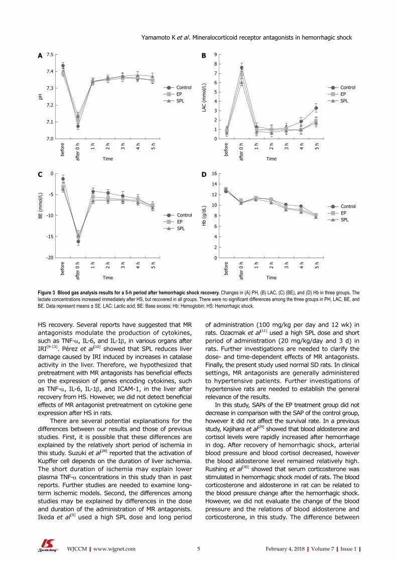
Inflammatory responsesThe lactate concentrations increased immediately after HS, but recovered in all groups. There were no significant differences among the three groups in lactate concentration, Hb, PH, and BE (Figure 3).
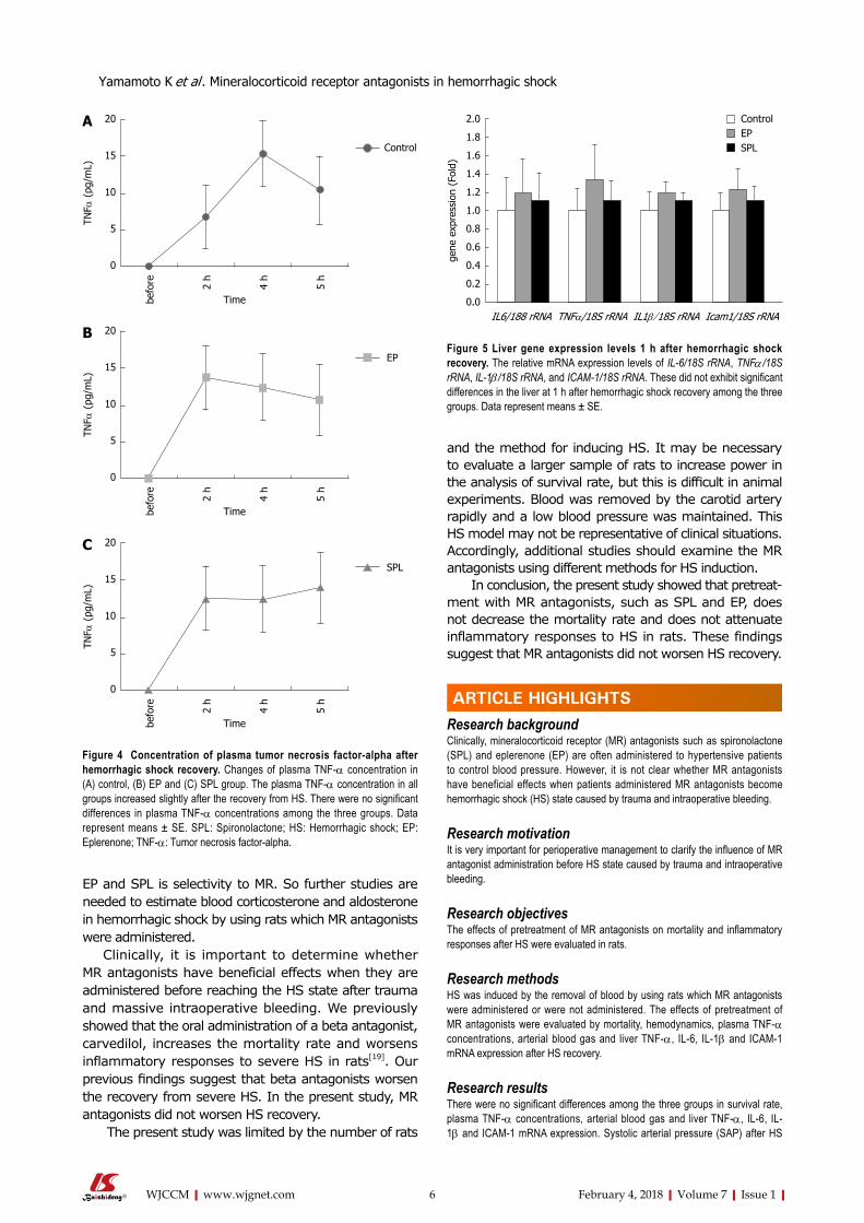
The plasma TNF-α concentration in all groups increased slightly after the recovery from HS. There were no significant differences in plasma TNF-α concentrations among the three groups (Figure 4).
YamamotoKetal .Mineralocorticoidreceptorantagonistsinhemorrhagicshock

4 February 4, 2018|Volume 7|Issue 1|WJCCM|www.wjgnet.com
Gene expression in the liver at 1 h after recovery from hemorrhagic shock The mRNA expression levels of TNF-α, IL-6, IL-1β, and ICAM-1 did not exhibit significant differences in the liver at 1 h after HS recovery among the three groups (Figure 5).
DISCUSSIONHS induced by partial exsanguination in rats caused metabolic acidosis and increased TNF-α concentrations immediately after the return of blood. Metabolic acidosis deteriorated immediately after the recovery from HS, but was gradually re-aggravated. As a result, the survival rate was low. HS with the oral administration of SPL and EP, MR antagonists, also resulted in similar to HS of the control. These were no significant differences between the control and MR antagonist-treated groups in the responses after HS recovery. Cytokine gene expression in the liver tissue 1 h after HS recovery did not differ among groups. Thus, our results showed that the oral administration of MR antagonists does not affect the mortality rate and inflammatory responses after HS recovery in rats based on comparisons with control rats after HS recovery.
MR antagonists are often used to treat hypertensive patients and have beneficial effects in patients with chronic heart failure and ischemic heart diseases[14,15,22,23]. Many recent investigations have demonstrated the beneficial effects of MR antagonists with respect to inflammatory responses and IRI in vitro and in vivo[6-13]. Kato et al[6] showed that SPL inhibits the production of pro-inflammatory mediators, such as TNF-α and nitric oxide, in response to lipopolysaccharides in vitro. Ozacmak et al[11] showed that pretreatment with SPL reduces intestinal injury induced by IR, including the inhibition of inflammatory responses, in rats. These studies suggest that the early and continuous administration of MR antagonists has beneficial effects
in critical patients with MOD after HS. However, the effects of pretreatment with MR antagonists on MOD after HS are unclear. Therefore, we evaluated the effects of the pretreatment of MR antagonists on mortality and inflammatory responses after HS in rats. Contrary to our expectations, pretreatment with MR antagonists did not have beneficial effects after HS in rats.
Many investigations have demonstrated that activated Kupffer cells release inflammatory cytokines, such as TNF-α or IL-1, soon after liver IRI. TNF-α increases the expression of ICAM-1 and promotes the adhesion of neutrophils[24-26]. Liver failure is recognized after liver IRI, and many other organs (e.g., the myocardium, pancreas, small intestine, kidney, adrenal gland, and lungs) seem to be damaged by inflammatory reactions and oxidation[27]. Therefore, it is important to evaluate cytokine production in the liver at an early stage after
1.0
0.9
0.8
0.7
0.6
0.5
0.4
0.3
0.2
0.1
0.0
Surv
ival
pro
babi
lity
02468
Timeafterhemorrhagicshockrecovery(h)
ControlEPSPL
Figure 1 Kaplan–Meier survival analysis. The survival rates 8 h after recovery from HS were 71%, 94%, and 82% in the control group, SPL group, and EP group, respectively. There were no significant differences in survival rate among the three groups (P = 0.219). HS: Hemorrhagic shock; EP: Eplerenone; SPL: Spironolactone.
Figure 2 Changes in systolic arterial pressure and heart rate over an 8-h period after hemorrhagic shock recovery. Changes in (A) SAP and (B) HR in three groups. SAPs gradually decreased after HS recovery in all groups. There were significant difference among three groups in a change of the SAP (P < 0.01). There were significant difference between EP group and control group in SAP at 5-8 h after the HS recovery. EP vs control; P < 0.01(5-8 h). Data represent means ± SE. bP < 0.01 compared with controls. SAP: Systolic arterial pressure; HR: Heart rate; HS: Hemorrhagic shock; EP: Eplerenone.
500
450
400
350
300
250
200
HR
(bpm
)
Time
befo
re
shoc
k10
min
20m
in
30m
in
40m
in
afte
r0
h
1h
2h
3h
4h
5h
6h
7h
8h
B
ControlEPSPL
180
160
140
120
100
90
80
70
60
40
20
0
SAP
(mm
Hg)
Time
befo
re
shoc
k10
min
20m
in
30m
in
40m
in
afte
r0
h
1h
2h
3h
4h
5h
6h
7h
8h
ControlEPSPL
Ab
b
bb
YamamotoKetal .Mineralocorticoidreceptorantagonistsinhemorrhagicshock
5 February 4, 2018|Volume 7|Issue 1|WJCCM|www.wjgnet.com
HS recovery. Several reports have suggested that MR antagonists modulate the production of cytokines, such as TNF-α, IL-6, and IL-1β, in various organs after IRI[9-13]. Pérez et al[10] showed that SPL reduces liver damage caused by IRI induced by increases in catalase activity in the liver. Therefore, we hypothesized that pretreatment with MR antagonists has beneficial effects on the expression of genes encoding cytokines, such as TNF-α, IL-6, IL-1β, and ICAM-1, in the liver after recovery from HS. However, we did not detect beneficial effects of MR antagonist pretreatment on cytokine gene expression after HS in rats.
There are several potential explanations for the differences between our results and those of previous studies. First, it is possible that these differences are explained by the relatively short period of ischemia in this study. Suzuki et al[28] reported that the activation of Kupffer cell depends on the duration of liver ischemia. The short duration of ischemia may explain lower plasma TNF-α concentrations in this study than in past reports. Further studies are needed to examine long-term ischemic models. Second, the differences among studies may be explained by differences in the dose and duration of the administration of MR antagonists. Ikeda et al[9] used a high SPL dose and long period
of administration (100 mg/kg per day and 12 wk) in rats. Ozacmak et al[11] used a high SPL dose and short period of administration (20 mg/kg/day and 3 d) in rats. Further investigations are needed to clarify the dose- and time-dependent effects of MR antagonists. Finally, the present study used normal SD rats. In clinical settings, MR antagonists are generally administered to hypertensive patients. Further investigations of hypertensive rats are needed to establish the general relevance of the results.
In this study, SAPs of the EP treatment group did not decrease in comparison with the SAP of the control group, however it did not affect the survival rate. In a previous study, Kajihara et al[29] showed that blood aldosterone and cortisol levels were rapidly increased after hemorrhage in dog. After recovery of hemorrhagic shock, arterial blood pressure and blood cortisol decreased, however the blood aldosterone level remained relatively high.
Rushing et al[30] showed that serum corticosterone was stimulated in hemorrhagic shock model of rats. The blood corticosterone and aldosterone in rat can be related to the blood pressure change after the hemorrhagic shock. However, we did not evaluate the change of the blood pressure and the relations of blood aldosterone and corticosterone, in this study. The difference between
ControlEPSPL
7.5
7.4
7.3
7.2
7.1
7.0
pH
Time
befo
re
afte
r0
h
1h
2h
3h
4h
5h
A
ControlEPSPL
9
8
7
6
5
4
3
2
1
0
LAC
(mm
ol/L
)
Time
befo
re
afte
r0
h
1h
2h
3h
4h
5h
B
0
-5
-10
-15
-20
BE(
mm
ol/L
)
Time
befo
re
afte
r0
h
1h
2h
3h
4h
5h
C
ControlEPSPL
ControlEPSPL
16
14
12
10
8
6
4
2
0
Hb
(g/d
L)
Time
befo
re
afte
r0
h
1h
2h
3h
4h
5h
D
Figure 3 Blood gas analysis results for a 5-h period after hemorrhagic shock recovery. Changes in (A) PH, (B) LAC, (C) (BE), and (D) Hb in three groups. The lactate concentrations increased immediately after HS, but recovered in all groups. There were no significant differences among the three groups in PH, LAC, BE, and BE. Data represent means ± SE. LAC: Lactic acid; BE: Base excess; Hb: Hemoglobin; HS: Hemorrhagic shock.
YamamotoKetal .Mineralocorticoidreceptorantagonistsinhemorrhagicshock
6 February 4, 2018|Volume 7|Issue 1|WJCCM|www.wjgnet.com
EP and SPL is selectivity to MR. So further studies are needed to estimate blood corticosterone and aldosterone in hemorrhagic shock by using rats which MR antagonists were administered.
Clinically, it is important to determine whether MR antagonists have beneficial effects when they are administered before reaching the HS state after trauma and massive intraoperative bleeding. We previously showed that the oral administration of a beta antagonist, carvedilol, increases the mortality rate and worsens inflammatory responses to severe HS in rats[19]. Our previous findings suggest that beta antagonists worsen the recovery from severe HS. In the present study, MR antagonists did not worsen HS recovery.
The present study was limited by the number of rats
and the method for inducing HS. It may be necessary to evaluate a larger sample of rats to increase power in the analysis of survival rate, but this is difficult in animal experiments. Blood was removed by the carotid artery rapidly and a low blood pressure was maintained. This HS model may not be representative of clinical situations. Accordingly, additional studies should examine the MR antagonists using different methods for HS induction.
In conclusion, the present study showed that pretreat-ment with MR antagonists, such as SPL and EP, does not decrease the mortality rate and does not attenuate inflammatory responses to HS in rats. These findings suggest that MR antagonists did not worsen HS recovery.
ARTICLE HIGHLIGHTSResearch backgroundClinically, mineralocorticoid receptor (MR) antagonists such as spironolactone (SPL) and eplerenone (EP) are often administered to hypertensive patients to control blood pressure. However, it is not clear whether MR antagonists have beneficial effects when patients administered MR antagonists become hemorrhagic shock (HS) state caused by trauma and intraoperative bleeding.
Research motivationIt is very important for perioperative management to clarify the influence of MR antagonist administration before HS state caused by trauma and intraoperative bleeding.
Research objectivesThe effects of pretreatment of MR antagonists on mortality and inflammatory responses after HS were evaluated in rats.
Research methodsHS was induced by the removal of blood by using rats which MR antagonists were administered or were not administered. The effects of pretreatment of MR antagonists were evaluated by mortality, hemodynamics, plasma TNF-α concentrations, arterial blood gas and liver TNF-α, IL-6, IL-1β and ICAM-1 mRNA expression after HS recovery.
Research resultsThere were no significant differences among the three groups in survival rate, plasma TNF-α concentrations, arterial blood gas and liver TNF-α, IL-6, IL-1β and ICAM-1 mRNA expression. Systolic arterial pressure (SAP) after HS
Control
20
15
10
5
0
TNF α
(pg
/mL)
befo
re 2h
4h
5h
A
Time
20
15
10
5
0
TNF α
(pg
/mL)
befo
re 2h
4h
5h
B
Time
20
15
10
5
0
TNF α
(pg
/mL)
befo
re 2h
4h
5h
C
Time
EP
SPL
Figure 4 Concentration of plasma tumor necrosis factor-alpha after hemorrhagic shock recovery. Changes of plasma TNF-α concentration in (A) control, (B) EP and (C) SPL group. The plasma TNF-α concentration in all groups increased slightly after the recovery from HS. There were no significant differences in plasma TNF-α concentrations among the three groups. Data represent means ± SE. SPL: Spironolactone; HS: Hemorrhagic shock; EP: Eplerenone; TNF-α: Tumor necrosis factor-alpha.
2.0
1.8
1.6
1.4
1.2
1.0
0.8
0.6
0.4
0.2
0.0
gene
exp
ress
ion
(Fol
d)
IL6/188rRNATNFα/18SrRNAIL1β/18SrRNAIcam1/18SrRNA
ControlEPSPL
Figure 5 Liver gene expression levels 1 h after hemorrhagic shock recovery. The relative mRNA expression levels of IL-6/18S rRNA, TNFα /18S rRNA, IL-1β /18S rRNA, and ICAM-1/18S rRNA. These did not exhibit significant differences in the liver at 1 h after hemorrhagic shock recovery among the three groups. Data represent means ± SE.
ARTICLE HIGHLIGHTS
YamamotoKetal .Mineralocorticoidreceptorantagonistsinhemorrhagicshock

7 February 4, 2018|Volume 7|Issue 1|WJCCM|www.wjgnet.com
recovery did not decrease in rats of EP group in comparison with control groups. After HS recovery, the reason why blood pressure was maintained in rats of EP group is the problems that remain to be solved, in this research.
Research conclusionsPretreatment with MR antagonists did not improve mortality or cytokine responses in the liver after HS recovery in rats. The HS model in the present study was made during general anesthesia after pretreatment of MR antagonists. This model is similar to the clinical situation when patients administered MR antagonists become HS state during operation. The present study suggested that MR antagonists may not be worsen the recovery of HS state and may not need to be withdrawn before the operations.
Research perspectivesThe present study used normal SD rats. In clinical settings, MR antagonists are generally administered to hypertensive patients. Further investigations by using hypertensive rats which MR antagonists were administered will be needed. The present study, SAPs of the EP treatment group did not decrease in comparison with the SAP of the control group, so further studies are needed to evaluate relations of blood corticosterone or aldosterone and blood pressure in hemorrhagic shock by using rats which MR antagonists were administered.
ACKNOWLEDGEMENTSWe thank Hisayo Masaki and Ayano Nomura for support during the experiment. We would like to thank Editage (www.editage.jp) for English language editing.
REFERENCES1 Hardaway RM. Traumatic shock alias posttrauma critical illness.
Am Surg 2000; 66: 284-290 [PMID: 10759201]2 Dewar D, Moore FA, Moore EE, Balogh Z. Postinjury multiple
organ failure. Injury 2009; 40: 912-918 [PMID: 19541301 DOI: 10.1016/j.injury.2009.05.024]
3 Yao YM, Redl H, Bahrami S, Schlag G. The inflammatory basis of trauma/shock-associated multiple organ failure. Inflamm Res 1998; 47: 201-210 [PMID: 9657252 DOI: 10.1007/s000110050318]
4 Hierholzer C, Kalff JC, Billiar TR, Bauer AJ, Tweardy DJ, Harbrecht BG. Induced nitric oxide promotes intestinal inflammation following hemorrhagic shock. Am J Physiol Gastrointest Liver Physiol 2004; 286: G225-G233 [PMID: 14715517 DOI: 10.1152/ajpgi.00447.2002]
5 Botha AJ, Moore FA, Moore EE, Kim FJ, Banerjee A, Peterson VM. Postinjury neutrophil priming and activation: an early vulnerable window. Surgery 1995; 118: 358-364; discussion 364-365 [PMID: 7638753 DOI: 10.1016/S0039-6060(05)80345-9]
6 Kato Y, Kamiya H, Koide N, Odkhuu E, Komatsu T, Dagvadorj J, Watarai A, Kondo M, Kato K, Nakamura J, Yokochi T. Spironolactone inhibits production of proinflammatory mediators in response to lipopolysaccharide via inactivation of nuclear factor-κB. Immunopharmacol Immunotoxicol 2014; 36: 237-241 [PMID: 24852317 DOI: 10.3109/08923973.2014.921690]
7 Sønder SU, Woetmann A, Odum N, Bendtzen K. Spironolactone induces apoptosis and inhibits NF-kappaB independent of the mineralocorticoid receptor. Apoptosis 2006; 11: 2159-2165 [PMID: 17051331 DOI: 10.1007/s10495-006-0286-3]
8 Miura R, Nakamura K, Miura D, Miura A, Hisamatsu K, Kajiya M, Nagase S, Morita H, Fukushima Kusano K, Ohe T, Ishihara K. Anti-inflammatory effect of spironolactone on human peripheral blood mononuclear cells. J Pharmacol Sci 2006; 101: 256-259 [PMID: 16837769 DOI: 10.1254/jphs.SC0060049]
9 Ikeda H, Tsuruya K, Toyonaga J, Masutani K, Hayashida H, Hirakata H, Iida M. Spironolactone suppresses inflammation and prevents L-NAME-induced renal injury in rats. Kidney Int 2009; 75: 147-155 [PMID: 18923385 DOI: 10.1038/ki.2008.507]
10 Pérez JC, Ramírez AC, González LT, Espinosa LE, Quintana
MM, Galván GA, Chavira HZ, de la Garza FJ, Lemarroy CR, Garza NE, Rodríguez EP, Pérez PC. Spironolactone Effect in Hepatic Ischemia/Reperfusion Injury in Wistar Rats. Oxid Med Cell Longev 2016; 2016: 3196431 [PMID: 26798418 DOI: 10.1155/2016/3196431]
11 Ozacmak HS, Ozacmak VH, Barut F, Araslı M, Ucan BH. Pretreatment with mineralocorticoid receptor blocker reduces intestinal injury induced by ischemia and reperfusion: involvement of inhibition of inflammatory response, oxidative stress, nuclear factor κB, and inducible nitric oxide synthase. J Surg Res 2014; 191: 350-361 [PMID: 24862878 DOI: 10.1016/j.jss.2014.04.040]
12 Kang YM, Zhang ZH, Johnson RF, Yu Y, Beltz T, Johnson AK, Weiss RM, Felder RB. Novel effect of mineralocorticoid receptor antagonism to reduce proinflammatory cytokines and hypothalamic activation in rats with ischemia-induced heart failure. Circ Res 2006; 99: 758-766 [PMID: 16960100 DOI: 10.1161/01.RES.0000244092.95152.86]
13 Dorrance AM, Osborn HL, Grekin R, Webb RC. Spironolactone reduces cerebral infarct size and EGF-receptor mRNA in stroke-prone rats. Am J Physiol Regul Integr Comp Physiol 2001; 281: R944-R950 [PMID: 11507012 DOI: 10.1152/ajpregu.2001.281.3.R944]
14 Jansen PM, Frenkel WJ, van den Born BJ, de Bruijne EL, Deinum J, Kerstens MN, Arnoldus JH, Woittiez AJ, Wijbenga JA, Zietse R, Danser AH, van den Meiracker AH. Determinants of blood pressure reduction by eplerenone in uncontrolled hypertension. J Hypertens 2013; 31: 404-413 [PMID: 23249826 DOI: 10.1097/HJH.0b013e32835b71d6]
15 Guo H, Xiao Q. Clinical efficacy of spironolactone for resistant hypertension: a meta analysis from randomized controlled clinical trials. Int J Clin Exp Med 2015; 8: 7270-7278 [PMID: 26221266]
16 Rocha R, Rudolph AE, Frierdich GE, Nachowiak DA, Kekec BK, Blomme EA, McMahon EG, Delyani JA. Aldosterone induces a vascular inflammatory phenotype in the rat heart. Am J Physiol Heart Circ Physiol 2002; 283: H1802-H1810 [PMID: 12384457 DOI: 10.1152/ajpheart.01096.2001]
17 Tanaka H, Watanabe K, Harima M, Thanikachalam PV, Yamaguchi K, Tachikawa H, Kodama M, Aizawa Y. Effects of various diuretics on cardiac function in rats with heart failure. Yakugaku Zasshi 2009; 129: 871-879 [PMID: 19571523 DOI: 10.1248/yakushi.129.871]
18 Yamashita M, Taniyama M, Tamai M. Cellular localization of tumor necrosis factor-alpha mRNA and interleukin-6 mRNA in the rat liver after hemorrhagic shock. Surg Today 2002; 32: 701-706 [PMID: 12181720 DOI: 10.1007/s005950200130]
19 Taniguchi T, Kurita A, Yamamoto K, Inaba H. Effects of carvedilol on mortality and inflammatory responses to severe hemorrhagic shock in rats. Shock 2009; 32: 272-275 [PMID: 19295485 DOI: 10.1097/SHK.0b013e3181a24cb3]
20 Liu LM, Dubick MA. Hemorrhagic shock-induced vascular hyporeactivity in the rat: relationship to gene expression of nitric oxide synthase, endothelin-1, and select cytokines in corresponding organs. J Surg Res 2005; 125: 128-136 [PMID: 15854664 DOI: 10.1016/j.jss.2004.12.008]
21 Kitano K, Usui S, Ootsuji H, Takashima S, Kobayashi D, Murai H, Furusho H, Nomura A, Kaneko S, Takamura M. Rho-kinase activation in leukocytes plays a pivotal role in myocardial ischemia/reperfusion injury. PLoS One 2014; 9: e92242 [PMID: 24638037 DOI: 10.1371/journal.pone.0092242]
22 Pitt B, Zannad F, Remme WJ, Cody R, Castaigne A, Perez A, Palensky J, Wittes J. The effect of spironolactone on morbidity and mortality in patients with severe heart failure. Randomized Aldactone Evaluation Study Investigators. N Engl J Med 1999; 341: 709-717 [PMID: 10471456 DOI: 10.1056/NEJM199909023411001]
23 Pitt B, Remme W, Zannad F, Neaton J, Martinez F, Roniker B, Bittman R, Hurley S, Kleiman J, Gatlin M; Eplerenone Post-Acute Myocardial Infarction Heart Failure Efficacy and Survival Study Investigators. Eplerenone, a selective aldosterone blocker, in patients with left ventricular dysfunction after myocardial
YamamotoKetal .Mineralocorticoidreceptorantagonistsinhemorrhagicshock

8 February 4, 2018|Volume 7|Issue 1|WJCCM|www.wjgnet.com
infarction. N Engl J Med 2003; 348: 1309-1321 [PMID: 12668699 DOI: 10.1056/NEJMoa030207]
24 Wanner GA, Ertel W, Müller P, Höfer Y, Leiderer R, Menger MD, Messmer K. Liver ischemia and reperfusion induces a systemic inflammatory response through Kupffer cell activation. Shock 1996; 5: 34-40 [PMID: 8821101 DOI: 10.1097/00024382-199601000-00008]
25 Mendes-Braz M, Elias-Miró M, Jiménez-Castro MB, Casillas-Ramírez A, Ramalho FS, Peralta C. The current state of knowledge of hepatic ischemia-reperfusion injury based on its study in experimental models. J Biomed Biotechnol 2012; 2012: 298657 [PMID: 22649277 DOI: 10.1155/2012/298657]
26 Farhood A, McGuire GM, Manning AM, Miyasaka M, Smith CW, Jaeschke H. Intercellular adhesion molecule 1 (ICAM-1) expression and its role in neutrophil-induced ischemia-reperfusion injury in rat liver. J Leukoc Biol 1995; 57: 368-374 [PMID: 7884306 DOI: 10.1002/jlb.57.3.368]
27 Nastos C, Kalimeris K, Papoutsidakis N, Tasoulis MK, Lykoudis
PM, Theodoraki K, Nastou D, Smyrniotis V, Arkadopoulos N. Global consequences of liver ischemia/reperfusion injury. Oxid Med Cell Longev 2014; 2014: 906965 [PMID: 24799983 DOI: 10.1155/2014/906965]
28 Suzuki S, Nakamura S, Sakaguchi T, Ochiai H, Konno H, Baba S, Baba S. Alteration of reticuloendothelial phagocytic function and tumor necrosis factor-alpha production after total hepatic ischemia. Transplantation 1997; 64: 821-827 [PMID: 9326405 DOI: 10.1097/00007890-199709270-00006]
29 Kajihara H, Malliwah JA, Matsumura M, Taguchi K, Iijima S. Changes in blood cortisol and aldosterone levels and ultrastructure of the adrenal cortex during hemorrhagic shock. Pathol Res Pract 1983; 176: 324-340 [PMID: 6856521 DOI: 10.1016/S0344-0338(83)80022-3]
30 Rushing GD, Britt RC, Britt LD. Effects of hemorrhagic shock on adrenal response in a rat model. Ann Surg 2006; 243: 652-654; discussion 654-656 [PMID: 16633000 DOI: 10.1097/01.sla.0000216759.36819.1b]
P- Reviewer: Beltowski J, Chello M, Spasojevi SD S- Editor: Kong JX L- Editor: A E- Editor: Li RF
YamamotoKetal .Mineralocorticoidreceptorantagonistsinhemorrhagicshock

FranciscoJMolina,PaulaTRivera,AlejandroCardona,DianaCRestrepo,OraliaMonroy,DanielRodas,JuanGBarrientos
ORIGINAL ARTICLE
9 February 4, 2018|Volume 7|Issue 1|WJCCM|www.wjgnet.com
Adverse events in critical care: Search and active detection through the Trigger Tool
FranciscoJMolina,DanielRodas,JuanGBarrientos, Clínica Universitaria Bolivariana, School of Medicine, Universidad Pontificia Bolivariana, Medellín 050034, Colombia
PaulaTRivera, Faculty of Nursing, Universidad de Caldas, Manizales 170004, Colombia
AlejandroCardona,DianaCRestrepo, School of Medicine, Universidad Pontificia Bolivariana, Medellín 050034, Colombia
OraliaMonroy, Clínica Universitaria Bolivariana, Medellín 050034, Colombia
ORCIDnumber: Francisco J Molina (0000-0003-0705-6579); Paula T Rivera (0000-0002-7600-9736); Alejandro Cardona (0000-0001-5503-5487); Diana C Restrepo (0000-0002-8149-5214); Oralia Monroy (0000-0002-3118-0310); Daniel Rodas (0000-0001-6022-0572); Juan G Barrientos (0000-0001-5135-5168).
Authorcontributions: Molina FJ, Restrepo DC and Barrientos JG designed the research; all authors performed the study; Molina FJ, Cardona A, Restrepo DC and Barrientos JG directed use of the analytical tools; all authors analyzed the data; Molina FJ, Cardona A and Barrientos JG wrote the paper.
Institutionalreviewboardstatement: The study was reviewed and approved for publication by the research directorate of Universidad Pontificia Bolivariana.
Informedconsentstatement: All study participants or their legal guardian provided informed written consent about personal and medical data collection prior to study enrolment.
Conflict-of-interest statement: All the Authors declare no conflict of interest related to the manuscript.
Open-Access: This article is an open-access article which was selected by an in-house editor and fully peer-reviewed by external reviewers. It is distributed in accordance with the Creative Commons Attribution Non Commercial (CC BY-NC 4.0) license, which permits others to distribute, remix, adapt, build upon this work non-commercially, and license their derivative works on different terms, provided the original work is properly cited and
the use is non-commercial. See: http://creativecommons.org/licenses/by-nc/4.0/
Manuscriptsource: Invited manuscript
Correspondenceto:FranciscoJMolina,MSc,Intensivist,Clínica Universitaria Bolivariana, School of Medicine, Universidad Pontificia Bolivariana, Carrera 72a No. 78b-50, Medellín 050034, Colombia. [email protected]: +57-313-7452815
Received: August 11, 2017Peer-reviewstarted: September 16, 2017Firstdecision: November 7, 2017Revised: November 20, 2017 Accepted: December 1, 2017Articleinpress: December 1, 2017Publishedonline: February 4, 2018
AbstractAIMToinvestigatetheincidenceofdisadvantageouseventsbyusingtheGlobalTriggerTool inan intensivecareunit(ICU).
METHODSAretrospectivedescriptivestudywasperformed ina12-beduniversityICUinthecityofMedellin,Colombia.Clinicalchartsofhospitalizedpatientswerereviewed,between January 1 andDecember 31, 2016,withthe following inclusion criteria: subjects agedover18years,withat least24hofhospitalizationandwhohada completemedical history that couldbeaccessed.Interventions: Trainedreviewersconducteda retrospective examination of medical chartssearching forclueevents thatelicit investigation, inordertodetectanunfavorableevent.Measurements:Informationwasprocessed throughSPSS software
World Journal ofCritical Care MedicineW J C C M
Submit a Manuscript: http://www.f6publishing.com
DOI: 10.5492/wjccm.v7.i1.9
World J Crit Care Med 2018 February 4; 7(1): 9-15
ISSN 2220-3141 (online)
Retrospective Study

10 February 4, 2018|Volume 7|Issue 1|WJCCM|www.wjgnet.com
MolinaFJetal .TriggerToolforintensivecareevents
version21; fornumerical variables, themeanwasreportedwithstandarddeviation(SD).Percentageswerecalculatedforqualitativevariables.
RESULTSTwohundredand forty-four triggersoccurred,with82.4%of subjects havingpresentedwith at leastoneandanaverageof3.37(SD3.47).Atotalof178adverseevents (AEs) tookplace in48 individuals,withan incidenceof52.1%.Onaverage, foureventsperpatientwererecorded,and foreachunfortunateevent, 1.98 triggers were presented. The mostfrequent displeasing issueswere: pressure ulcers(17.6%), followedby complicationsor reactions tomedicaldevices(4.3%),andlacerationsorskindefects(3.7%); the least frequentwasdelayeddiagnosisortreatment(0.56%).Thirty-eightpoint fourpercentofmishapeventscausedtemporarydamagethatrequiredintervention, and48.9%ofAEswerepreventable.ComparisonbetweenAEsandadmissiondiagnosesfound that hypertension and sepsiswere theonlydiagnosesthathadstatisticalsignificance(P =0.042and0.022,respectively).
CONCLUSIONAlmosthalfof theunfavorable issueswereclassifiedasavoidable,whichleavesaverywidefieldofworkintermsofpreventativeactivities.
Key words: Adverseevents;Criticalcare;TriggerTool;Complications;Security
© The Author(s) 2018.PublishedbyBaishidengPublishingGroupInc.Allrightsreserved.
Core tip: TheGlobalTriggerTool isa typeofactivedetectionofadverseevents(AEs).Threestudiescarriedout in intensivecareunits(ICUs),whichincludedonlypatientswhodiedinthefollowing96hor7dpriortoICUadmission.The importanceofourstudy isthat itwasperformedduring theentirehospitalstay in theICU.The incidenceofAEswas52.1%,and48.9%of thesewerepreventable.Themost frequentwerepressureulcers(17.6%)andcomplicationsrelatedtomedicaldevices(4.3%).Thethreemaintriggerswereskindefects,excitationordrowsiness,andunscheduledwithdrawalofsurgicalcatheter,probes,ordrains.
Molina FJ, Rivera PT, Cardona A, Restrepo DC, Monroy O, Rodas D, Barrientos JG. Adverse events in critical care: Search and active detection through the Trigger Tool. World J Crit Care Med 2018; 7(1): 9-15 Available from: URL: http://www.wjgnet.com/2220-3141/full/v7/i1/9.htm DOI: http://dx.doi.org/10.5492/wjccm.v7.i1.9
INTRODUCTIONIn 2000, the publication of “To Err is Human: Building a Safer Health System” from the United States Institute
of Medicine marked a before and after in the awareness of this issue and has made security research become a fundamental pillar[1]. In 2004, the World Health Organization (WHO) created the Global Alliance for Patient Safety, in order to coordinate, disseminate and accelerate improvements in patient safety worldwide.
Patient safety is defined as the absence of un-necessary or potential harm associated with health care. This damage is represented as a functional, structural or any detrimental effect derived from medical care. Adverse events (AEs) can be classified as preventable or nonpreventable. The causality model raises many factors that influence avoidable unfavorable event sequence. The system produces errors when several weaknesses occur momentarily, allowing the opportunity for acci-dent. Risk management is a discipline the objective of which is study of unfavorable issues derived from assistance through its detection and analysis, with the ultimate goal of designing strategies for its prevention. Risk is defined as the combination of the probability of occurrence of an event and its consequences. In the United Kingdom, an organizational model of causation of errors and AEs, known as the London Protocol, was developed.
In a study by Resar et al[2] conducted between 2001 and 2004 in 62 intensive care units (ICUs) of 54 hospitals, the authors described an incidence of 11.3 AEs. Rothschild et al[3], for a year under direct observation, found 120 AEs in 79 subjects (20.2%), including 66 (55%) not avoidable and 54 (45%) avoidable; the rate per 1000 patient-days was 80.5. Forster et al[4], in an academic ICU with 207 individuals being monitored daily, found AEs in 40 patients (19%), being preventable in 21 subjects (10%); these AEs were associated with an increase in hospital stay.
There are two types of AE detection: passive, where events are voluntarily reported; and active, where retrospectively or prospectively, a comprehensive assessment is performed to actively detect issues. The passives do not reach the absolute detection of the events, compared to the active review[5]. As described previously, only between 10% to 30% of AEs are voluntarily reported[6]. In one study, nurses were able to create a nonpunitive atmosphere which increased the spontaneous and voluntary reporting 10 to 20 times more[2]. Another survey assessing different methods of notification in Hospital Monte Naraco, revealed that 30% of the events were reported by voluntary means[6].
In the active methodology, there is a tool known as Global Trigger, which is based on a retrospective revision of the clinical chart performed by trained reviewers which seeks hints that will serve as indications for the evaluators to investigate the records in depth. This tool enables data acquisition and subsequent analysis and management through time of the causes of AEs[7,8].
This tool has facilitated the detection of, at least, 10 times more events than those reported by passive search methods, such as voluntary reports[2]. It has been reported that only between 10% to 20% of errors

11 February 4, 2018|Volume 7|Issue 1|WJCCM|www.wjgnet.com
are reported; and, of those, 90% to 95% do not cause harm to patients[9]. We intend to establish the incidence of AEs by using the Global Trigger Tool (GTT) in a high-complexity academic ICU.
MATERIALS AND METHODSA retrospective descriptive study was conducted in a 12-bed ICU, belonging to a university center in the city of Medellin, Colombia. This service is attended by intensivists, with a ratio of 6 patients per doctor at daytime and 12 patients per doctor at night. Nursing staff keeps a ratio of 6 patients per nurse during 24 h, and there is 1 nursing assistant for every 2 patients. There is an available respiratory therapist 24 h a day. Clinical charts of hospitalized patients were taken, between January 1 and December 31, 2016, with the following inclusion criteria: subjects aged over 18 years, with at least 24 h of hospitalization who had a complete medical history that could be accessed. This study was approved by the ethics committee of the Universidad Pontificia Bolivariana.
Techniques and data collection instrumentsAfter the ethical and institutional endorsement, we proceeded to train the team of reviewers, constituted by nurses with expertise and experience in Quality of Health Services, and medical specialists in intensive care who were standardized in review(ing) criteria, established(ing) times and process(ing) order(s). Each team analyzed the medical records in the event of a trigger; the chart was sent to one of two intensive care specialists to define the presence of this AE. Sixteen triggers were used to detect AEs (Table 1). These triggers were initially extracted from the literature, and then corroborated by each of the service intensivists, and subsequently, a consensus was obtained at a group meeting.
In case of an AE, a consensus was reached between the two intensivists. If this was proven positive, the specialist analyzed the preventability and severity of the AE, which was carried out with the classification of the The National Coordinating Council for Medication Error Reporting and Prevention (NCC MERP), which stipulates the following criteria on a scale from A to I. Criteria from A to D are considered incidents. From the E and on, they are considered events, as follows: E: Temporary harm requiring intervention; F: Temporary harm requiring prolonged hospitalization; G: Permanent harm; H: Injury that demands intervention to sustain life; Y: Harm that contributes to death.
Statistical analysisData processing was done using SPSS version 21. Quantitative variables were analyzed by grouping (mean and median) and dispersion measures [standard deviation and interquartile range (IQR), according to their distribution type]. Categorical variables were analyzed as proportions. Bivariate analysis was performed to search for association between AE and admission diagnoses. χ2 hypothesis tests were used for categorical variables and Student’s t-test for continuous variables with normal distribution or Mann-Whitney U-test for those variables with a different distribution[10]. A significant association was considered if a P value of less than 0.05 was obtained.
RESULTSData were collected from 134 patients. Forty patients were excluded for the following reasons: 12 subjects aged under 18 years and 28 patients remained less than 24 h in hospitalization; finally, 94 clinical charts were analyzed. General characteristics of the patients were: the mean age of the individuals was 56.77 years (standard deviation: 20.72; minimum: 10 and
Table 1 Triggers
Trigger n %
Skin defects or lacerations 36 14.75Excitation or drowsiness of the patient 34 13.93Unscheduled withdrawal of surgical catheter, probes, drains or other devices 34 13.93Hypotension 33 13.52Initiation of antibiotics after 48 h of admission 28 11.48Abrupt fall in hemoglobin or hematocrit by more than 25% 24 9.84Hypoglycemia 19 7.79Pneumonia 9 3.69Reintubation in less than 48 h 6 2.46Unscheduled surgical reintervention 5 2.05Chest tube insertion during ICU hospitalization 4 1.64Initiation of dialysis during ICU hospitalization 4 1.64Accidental extubation 3 1.23Adverse drug reaction events 3 1.23Cardiac arrest 1 0.41Protamine use 1 0.41Total 244 100.00
ICU: Intensive care unit.
MolinaFJetal .TriggerToolforintensivecareevents

12 February 4, 2018|Volume 7|Issue 1|WJCCM|www.wjgnet.com
maximum: 87). Sixty-two point eight percent (n = 59) of patients were female, and 37.2% (n = 35) were male. Of the subjects who suffered AEs, 62.8% were women and 37.2% were men. APACHE Ⅱ was 18 (IQR 14-24), and 12 of the 94 patients died (12.5%), with only one death related to an AE. The average patient stay was 8.05 d, with a standard deviation of 11.8 d (with a minimum of 1 d and a maximum of 66 d). The reasons for admission were: 22 (23.4%) patients were postsurgical, 18 (19.14%) came from the obstetric service, 14 (14.9%) from emergencies, 28 (29.78%) from hospitalization and 12 (12.76%) came from other institutions. Of the assessed patients, 43/94 (45.7%) had at least one comorbidity on admittance to the ICU; among the main ones were acute myocardial infarction (84%), sepsis (15%), cranioencephalic trauma (5%), pneumonia (5%) and cerebrovascular accident (4%). Other causes of lower frequency included: Urinary tract infection, heart failure and rheumatologic disease with 4%, 3% and 2%, respectively. Eighty-eight point three percent of the individuals had health system affiliation due to their job, and 11.7% were subsidized by the State.
Table 1 shows the triggers, totaling 248, concen-trated in 69 subjects; the most frequent of which were: skin defects or lacerations (14.75%), excitation or somnolence of the patient through the RASS scale (+3 or -3) (13.93%) and hypotension (13.52%). The least frequent was the use of protamine (0.41%).
The Triggers found elicited further investigation into the medical records in order to look for unfavorable issues. This search yielded a total of 178 AEs in 49
subjects, with an incidence of 52.1%; on average, 3.6 events per patient were recorded, and 1.98 triggers for each AE.
Table 2 shows the AEs detected; the most predomi-nant were pressure ulcers (17.6%), followed by com-plications or reactions to medical devices (4.3%), lacerations or skin defects (3.7%). The least presented was delayed diagnosis or treatment (0.56%).
One part of the analysis of displeasing events is prevention; almost half of the AEs were preventable (48.9%), 28% were incidents, 1.2% were nonpreven-table and 21.9% were a complication of the underlying disease.
The 38.4% of severity of AEs were classified in category E (temporary harm that required intervention), 10.8% classified in category H (harm that required an intervention to sustain life), 0.9% were rated in category F (temporary harm demanding prolonged hospitali-zation), and finally, categories Y (harm that contributed to death) and G (permanent harm), accounted for 0.3%.
Another comparison between AEs and admission diagnoses found that hypertension and sepsis were the only diagnoses that had statistical significance (P = 0.042 and 0.022, respectively).
When reviewing the patient’s age and preventability, the most striking findings indicated that 172 patients had preventable AEs, who were at least 17 years of age and had a maximum age of 87 years, with a median of 69 years and a 75th percentile of 77 years. On the contrary, 6 patients developed nonpreventable AEs, with a minimum age of 64 and a maximum of 87 years; the median was 69 and the 75th percentile was 75 years.
Table 2 Adverse events
Adverse event n (%)
Pressure ulcers 62 17.6Complications or reactions to medical devices 15 4.3Lacerations 13 3.7Drug-induced hypotension 10 2.8Poor glycemic control 9 2.6Nosocomial pneumonia 9 2.6Injury during procedure 8 2.3Phlebitis 7 2.0Hemorrhage or hematoma related to surgery or procedure 7 2.0Acute lung disease or respiratory failure 5 1.4Operative site infection 5 1.4Another event 5 1.4Drug-induced neurological disorders 4 1.1Sepsis and septic shock 4 1.1Burns, erosion, bruises and fractures 3 0.9Pneumothorax 2 0.6Pruritus, rash or dermal lesions, reactive to drugs or dressings 2 0.6Adhesion and functional alterations after surgical intervention 1 0.3Bacteremia associated with device 1 0.3Error in medication delivery 1 0.3Events attributable to internal failures in timeliness or continuity of evaluation 1 0.3Failures attributed to quality 1 0.3Opportunistic infection by immunosuppressive treatment 1 0.3Nosocomial urinary tract infection 1 0.3Delay in diagnosis or treatment 1 0.3Total 178 100
MolinaFJetal .TriggerToolforintensivecareevents

13 February 4, 2018|Volume 7|Issue 1|WJCCM|www.wjgnet.com
The statistical significance was a Kruskal-Wallis P value of 0.012.
DISCUSSIONThe main finding of our study is that the incidence of AEs in the ICU is 52.1%. The most frequent triggers were skin defects or lacerations (14.7%), excitation or somnolence of the patient according to the RASS scale (+3 or -3) (13.9%), and hypotension (13.5%). The most predominant AEs were pressure ulcers (17.6%), followed by complications or reactions to medical devices (4.3%), and lacerations or skin defects (3.7%). On average, 3.6 events per patient were recorded, and 1.98 triggers for each AE.
The largest study to identify the occurrence of displeasing issues was conducted by the Institute for Healthcare Improvement in 62 ICUs from 54 hospitals, between 2001-2004. The prevalence of AEs observed in 12.074 admissions in the ICU was 11.3 AEs/100 patient d; in a subgroup of 1.294 charts of 13 ICUs, which were reviewed in detail, 1.450 unpleasant events were identified, with a prevalence of 16.4 events/100 ICU d[2]. The Institute used, for the first time as a method of detecting AEs in ICU, records related to medications linked to pharmacy, finding 120 AEs in 79 patients (20.2%), with a rate of 8.05 AEs/100 patient d. This incidence is lower than ours, but with two differences: the Institute did not use the Tool, and it was prospective for a year, through continuous direct observation. Forster et al[4] also monitored patients daily by a multidisciplinary team; they evaluated 207 critical patients, with AEs in 40 patients (19%).
In a systematic review of the GTT by the end of 2016, in the different specialties, only three studies carried out in ICUs were found. Apart from the afore-mentioned[11], which shares similarity to ours, investi-gating triggers in subjects during hospital stay, the other two studies differ in their admission criteria. The first, Nilsson et al[12], included only patients who upon admission to the hospital’s ICU or in the following 96 h died. The second, the PREVENT trial[13], reviewed clinical charts 7 d prior to ICU admission. Table 3 shows the methodological characteristics of these surveys, including our own. Table 4 shows the most frequent triggers and AEs in the different studies performed with GTT in ICU.
Of the findings of these investigations, we can highlight the following. Firstly, that AEs are preventable
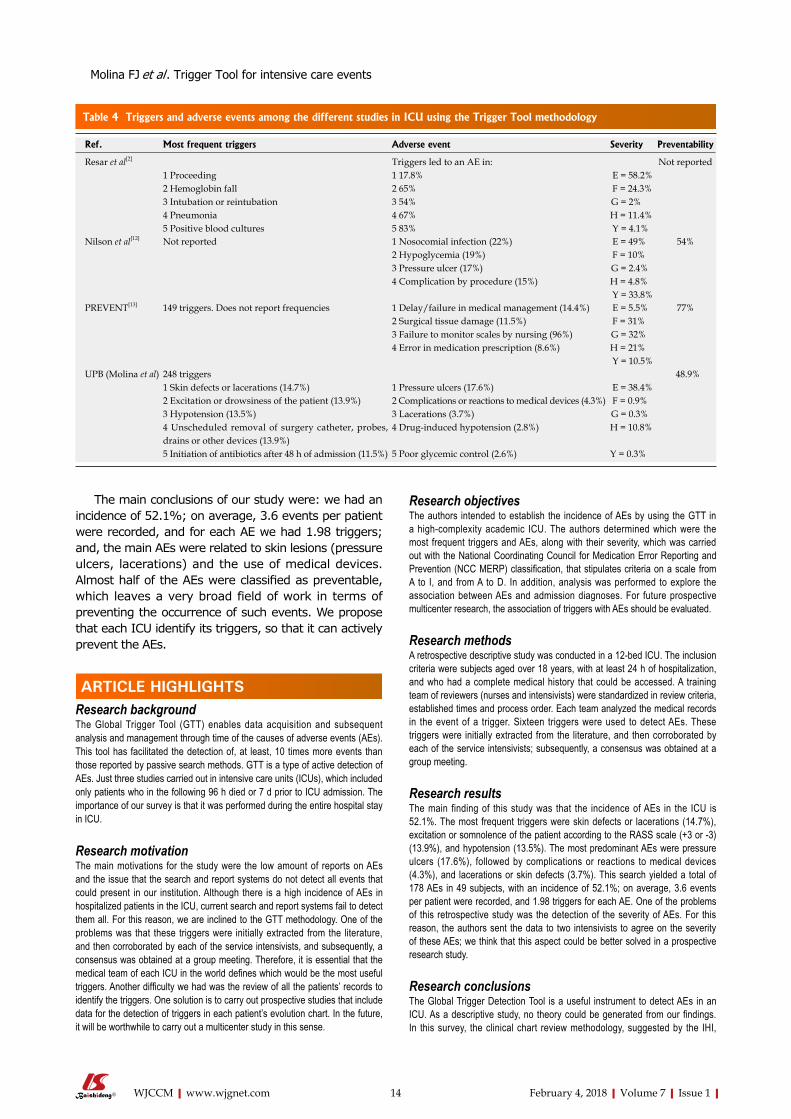
in a high proportion (between 48.9% and 77% of cases). Secondly, in all studies, except PREVENT, AEs in their severity were more temporal (E or F). Thirdly, in spite of using the GGT methodology, only Resar et al[2] and ours describe the most conventional triggers. And, lastly, the most common AEs in the different studies are distinct, perhaps they do resemble in that they are related to skin care.
In our study, the most extensive trigger was skin laceration, which is consistent with the most prevalent AE: pressure ulcer; this event is consistent with other studies, such as IBEAS[14] in hospitalization, which considers it as the most common in Latin America. This event is largely associated with the presence of patients’ comorbidities, such as physical dependence, poor nutritional status, high hospital stay and the need to be in bed, distinctive of subjects hospitalized in an ICU.
In terms of severity and age, it was evident that as the patient was older, the likelihood of developing an AE increased, a fact that is consistent with a study conducted in Spain, where it was observed that age over 65 years was associated with the presence of AE[15]. Our knowledge indicates a higher frequency of unpleasant issues in females, in contrast to a survey performed in an ICU in Sao Paulo, which revealed a higher incidence of AE in males 52.3%. Sex differences could be attributed to the fact that the institution included in this research serves primarily maternal patients[16].
This study had several limitations. First, it was performed in a single center. Second, since it is retro-spective, there may be bias in the lack of information from medical and nursing records. The third limitation was the inclusion of the unit’s own intensivists within the research team; however, this fact strengthened their competencies in the use of the methodology and facilitated that they self-evaluated the AEs presented. The fourth limitation was the difficulty that existed in our environment for the unification in the administrative criteria of hospitalization in intensive care; that is to say, there may be special care patients. The fifth limitation was selection bias for interobserver variability, despite treatment and use of the same tool.
In the future, it will be worthwhile to carry out a multicenter study, given the shortage of these, and with the clearance of the most frequent triggers found in this study and Resar et al[2]. In addition, a prospective cohort study, after identifying the triggers, can be done to see how many AEs are prevented.
Table 3 Comparison between the different studies in ICU using the Trigger Tool methodology
Ref. Patients No. of ICUs Sample Incidence or prevalence of AEs
Resar et al[2] During ICU stay 62 12074 11.3/100 patient dNilsson et al[12] Those who die in less than 96 h of ICU admission 1 128 32/100 ICU admissions 19.5%PREVENT[13] Within 7 d prior to ICU admission 5 280 27.1% (80% related to reason for admission)UPB (Molina et al) During ICU stay 1 94 52.1% 3.6 AEs per patient
AEs: Adverse events; ICU: Intensive care unit.
MolinaFJetal .TriggerToolforintensivecareevents
The main conclusions of our study were: we had an incidence of 52.1%; on average, 3.6 events per patient were recorded, and for each AE we had 1.98 triggers; and, the main AEs were related to skin lesions (pressure ulcers, lacerations) and the use of medical devices. Almost half of the AEs were classified as preventable, which leaves a very broad field of work in terms of preventing the occurrence of such events. We propose that each ICU identify its triggers, so that it can actively prevent the AEs.
ARTICLE HIGHLIGHTSResearch backgroundThe Global Trigger Tool (GTT) enables data acquisition and subsequent analysis and management through time of the causes of adverse events (AEs). This tool has facilitated the detection of, at least, 10 times more events than those reported by passive search methods. GTT is a type of active detection of AEs. Just three studies carried out in intensive care units (ICUs), which included only patients who in the following 96 h died or 7 d prior to ICU admission. The importance of our survey is that it was performed during the entire hospital stay in ICU.
Research motivationThe main motivations for the study were the low amount of reports on AEs and the issue that the search and report systems do not detect all events that could present in our institution. Although there is a high incidence of AEs in hospitalized patients in the ICU, current search and report systems fail to detect them all. For this reason, we are inclined to the GTT methodology. One of the problems was that these triggers were initially extracted from the literature, and then corroborated by each of the service intensivists, and subsequently, a consensus was obtained at a group meeting. Therefore, it is essential that the medical team of each ICU in the world defines which would be the most useful triggers. Another difficulty we had was the review of all the patients’ records to identify the triggers. One solution is to carry out prospective studies that include data for the detection of triggers in each patient’s evolution chart. In the future, it will be worthwhile to carry out a multicenter study in this sense.
14 February 4, 2018|Volume 7|Issue 1|WJCCM|www.wjgnet.com
ARTICLE HIGHLIGHTS
Research objectivesThe authors intended to establish the incidence of AEs by using the GTT in a high-complexity academic ICU. The authors determined which were the most frequent triggers and AEs, along with their severity, which was carried out with the National Coordinating Council for Medication Error Reporting and Prevention (NCC MERP) classification, that stipulates criteria on a scale from A to I, and from A to D. In addition, analysis was performed to explore the association between AEs and admission diagnoses. For future prospective multicenter research, the association of triggers with AEs should be evaluated.
Research methodsA retrospective descriptive study was conducted in a 12-bed ICU. The inclusion criteria were subjects aged over 18 years, with at least 24 h of hospitalization, and who had a complete medical history that could be accessed. A training team of reviewers (nurses and intensivists) were standardized in review criteria, established times and process order. Each team analyzed the medical records in the event of a trigger. Sixteen triggers were used to detect AEs. These triggers were initially extracted from the literature, and then corroborated by each of the service intensivists; subsequently, a consensus was obtained at a group meeting.
Research resultsThe main finding of this study was that the incidence of AEs in the ICU is 52.1%. The most frequent triggers were skin defects or lacerations (14.7%), excitation or somnolence of the patient according to the RASS scale (+3 or -3) (13.9%), and hypotension (13.5%). The most predominant AEs were pressure ulcers (17.6%), followed by complications or reactions to medical devices (4.3%), and lacerations or skin defects (3.7%). This search yielded a total of 178 AEs in 49 subjects, with an incidence of 52.1%; on average, 3.6 events per patient were recorded, and 1.98 triggers for each AE. One of the problems of this retrospective study was the detection of the severity of AEs. For this reason, the authors sent the data to two intensivists to agree on the severity of these AEs; we think that this aspect could be better solved in a prospective research study.
Research conclusionsThe Global Trigger Detection Tool is a useful instrument to detect AEs in an ICU. As a descriptive study, no theory could be generated from our findings. In this survey, the clinical chart review methodology, suggested by the IHI,
Table 4 Triggers and adverse events among the different studies in ICU using the Trigger Tool methodology
Ref. Most frequent triggers Adverse event Severity Preventability
Resar et al[2]
1 Proceeding2 Hemoglobin fall3 Intubation or reintubation4 Pneumonia5 Positive blood cultures
Triggers led to an AE in:1 17.8%2 65%3 54%4 67%5 83%
E = 58.2% F = 24.3% G = 2%H = 11.4% Y = 4.1%
Not reported
Nilson et al[12] Not reported 1 Nosocomial infection (22%)2 Hypoglycemia (19%)3 Pressure ulcer (17%)4 Complication by procedure (15%)
E = 49% F = 10% G = 2.4%H = 4.8% Y = 33.8%
54%
PREVENT[13] 149 triggers. Does not report frequencies 1 Delay/failure in medical management (14.4%)2 Surgical tissue damage (11.5%)3 Failure to monitor scales by nursing (96%)4 Error in medication prescription (8.6%)
E = 5.5% F = 31% G = 32%H = 21% Y = 10.5%
77%
UPB (Molina et al) 248 triggers1 Skin defects or lacerations (14.7%)2 Excitation or drowsiness of the patient (13.9%)3 Hypotension (13.5%)4 Unscheduled removal of surgery catheter, probes, drains or other devices (13.9%)5 Initiation of antibiotics after 48 h of admission (11.5%)
1 Pressure ulcers (17.6%)2 Complications or reactions to medical devices (4.3%)3 Lacerations (3.7%)4 Drug-induced hypotension (2.8%)
5 Poor glycemic control (2.6%)
E = 38.4% F = 0.9% G = 0.3%H = 10.8% Y = 0.3%
48.9%
MolinaFJetal .TriggerToolforintensivecareevents

15 February 4, 2018|Volume 7|Issue 1|WJCCM|www.wjgnet.com
was taken as a reference, although the research team made variations in the manner of selecting patients (systematic randomized sampling), along with the review time of clinical records (review all charts), which allowed the detection of more triggers and AEs that could be useful for future investigations. Including GTT methodology to the study implies an increase in the frequency of AEs, and thus adopts measures that reduce their incidence in the future.
Research perspectivesThe authors suggest the adoption of the methodology in the institution with a trained team in this tool. In future investigations, it is recommended to determine the effectiveness of the tool through analytical studies (cases and controls) that show statistically significant differences between passive and active methods of AE detection. The authors suggest prospective projects that validate the methodology to verify that they could anticipate the presentation of AEs.
ACKNOWLEDGMENTSClínica Universitaria Bolivariana, School of Medicine, Universidad Pontificia Bolivariana, Medellín, Colombia.
REFERENCES1 Kohn LT, Corrigan JM, Donaldson MS (Eds): Institute of
Medicine (US) Committee on Quality of Health Care in America; To Err is Human: Building a Safer Health System. Washington (DC): National Academies Press (US), 2000
2 Resar RK, Rozich JD, Simmonds T, Haraden CR. A trigger tool to identify adverse events in the intensive care unit. Jt Comm J Qual Patient Saf 2006; 32: 585-590 [PMID: 17066996 DOI: 10.1016/S1553-7250(06)32076-4]
3 Rothschild JM, Landrigan CP, Cronin JW, Kaushal R, Lockley SW, Burdick E, Stone PH, Lilly CM, Katz JT, Czeisler CA, Bates DW. The Critical Care Safety Study: The incidence and nature of adverse events and serious medical errors in intensive care. Crit Care Med 2005; 33: 1694-1700 [PMID: 16096443 DOI: 10.1097/01.CCM.0000171609.91035.BD]
4 Forster AJ, Kyeremanteng K, Hooper J, Shojania KG, van Walraven C. The impact of adverse events in the intensive care unit on hospital mortality and length of stay. BMC Health Serv Res 2008; 8: 259 [PMID: 19091089 DOI: 10.1186/1472-6963-8-259]
5 Misson JC. A review of clinical risk management. J Qual Clin Pract 2001; 21: 131-134 [PMID: 11856410 DOI: 10.1046/j.1440-1762.2001.00421.x]
6 Leape LL. A systems analysis approach to medical error. J Eval
Clin Pract 1997; 3: 213-222 [PMID: 9406109 DOI: 10.1046/j.1365-2753.1997.00006.x]
7 Rozich JD, Haraden CR, Resar RK. Adverse drug event trigger tool: a practical methodology for measuring medication related harm. Qual Saf Health Care 2003; 12: 194-200 [PMID: 12792009 DOI: 10.1093/intqhc/mzw019]
8 Suresh G, Horbar JD, Plsek P, Gray J, Edwards WH, Shiono PH, Ursprung R, Nickerson J, Lucey JF, Goldmann D. Voluntary anonymous reporting of medical errors for neonatal intensive care. Pediatrics 2004; 113: 1609-1618 [PMID: 15173481 DOI: 10.1542/peds.113.6.1609]
9 Wu AW, Pronovost P, Morlock L. ICU incident reporting systems. J Crit Care 2002; 17: 86-94 [PMID: 12096371 DOI: 10.1053/jcrc.2002.35100]
10 Zhang Z. Univariate description and bivariate statistical inference: the first step delving into data. Ann Transl Med 2016; 4: 91 [PMID: 27047950 DOI: 10.21037/atm.2016.02.11]
11 Hibbert PD, Molloy CJ, Hooper TD, Wiles LK, Runciman WB, Lachman P, Muething SE, Braithwaite J. The application of the Global Trigger Tool: a systematic review. Int J Qual Health Care 2016; 28: 640-649 [PMID: 27664822 DOI: 10.1093/intqhc/mzw115]
12 Nilsson L, Pihl A, Tågsjö M, Ericsson E. Adverse events are common on the intensive care unit: results from a structured record review. Acta Anaesthesiol Scand 2012; 56: 959-965 [PMID: 22571769 DOI: 10.1111/j.1399-6576.2012.02711.x]
13 Garry DA, McKechnie SR, Culliford DJ, Ezra M, Garry PS, Loveland RC, Sharma VV, Walden AP, Keating LM; PREVENT group. A prospective multicentre observational study of adverse iatrogenic events and substandard care preceding intensive care unit admission (PREVENT). Anaesthesia 2014; 69: 137-142 [PMID: 24443852 DOI: 10.1111/anae.12535]
14 Aranaz-Andrés JM, Aibar-Remón C, Limón-Ramírez R, Amarilla A, Restrepo FR, Urroz O, Sarabia O, García-Corcuera LV, Terol-García E, Agra-Varela Y, Gonseth-García J, Bates DW, Larizgoitia I; IBEAS team. Prevalence of adverse events in the hospitals of five Latin American countries: results of the ‘Iberoamerican Study of Adverse Events’ (IBEAS). BMJ Qual Saf 2011; 20: 1043-1051 [PMID: 21712370 DOI: 10.1136/bmjqs.2011.051284]
15 Morales IJ, Peters SG, Afessa B. Hospital mortality rate and length of stay in patients admitted at night to the intensive care unit. Crit Care Med 2003; 31: 858-863 [PMID: 12626997 DOI: 10.1097/01.CCM.0000055378.31408.26]
16 Lehmann LS, Puopolo AL, Shaykevich S, Brennan TA. Iatrogenic events resulting in intensive care admission: frequency, cause, and disclosure to patients and institutions. Am J Med 2005; 118: 409-413 [PMID: 15808139 DOI: 10.1016/j.amjmed.2005.01.012]
P- Reviewer: Inchauspe AA, Willms DC, Zhang Z S- Editor: Ji FF L- Editor: Filipodia E- Editor: Li RF
MolinaFJetal .TriggerToolforintensivecareevents

GunasekaranKarthik,ThomasIsaiahSudarsan,JohnVictorPeter,ThambuSudarsanam,GeorgeMVarghese,PaulKundavaram,SowmyaSathyendra,RamyaIyyadurai,KishorePichamuthu
ORIGINAL ARTICLE
16 February 4, 2018|Volume 7|Issue 1|WJCCM|www.wjgnet.com
Spectrum of cardiac manifestations and its relationship to outcomes in patients admitted with scrub typhus infection
Gunasekaran Karthik, Thambu Sudarsanam, PaulKundavaram,SowmyaSathyendra,Ramya Iyyadurai, Department of Medicine, Christian Medical College, Vellore 632004, India
Thomas Isaiah Sudarsan, JohnVictor Peter, KishorePichamuthu,Medical Intensive Care Unit, Christian Medical College, Vellore 632004, India
GeorgeMVarghese, Department of Infectious Diseases, Christian Medical College, Vellore 632004, India
ORCIDnumber: Gunasekaran Karthik (0000-0002-8615-5753); Thomas Isaiah Sudarsan (0000-0002-0175-6630); John Victor Peter (0000-0002-3423-1830); Thambu Sudarsanam (0000-0001-7283-7836); George M Varghese (0000-0002- 4040-5649); Paul Kundavaram (0000-0002-2382-4411); Sowmya Sathyendra (0000-0002-9443-0022); Ramya Iyyadurai (0000-0001-8453-6205); Kishore Pichamuthu (0000-0002- 6977-1183).
Author contributions: Sudarsan TI acted as guarantor and corresponded with the journal for this paper; Karthik G, Peter JV, Sudarsanam T, Varghese GM, Kundavaram P, Sathyendra S, Iyyadurai R and Pichamuthu K contributed to acquisition of the data and revision of this manuscript.
Institutionalreviewboardstatement: This study was approved by the local Institutional Review Board (No. 8104).
Informedconsentstatement:All study participants, or their legal guardian, provided informed written consent prior to study enrollment.
Conflict-of-intereststatement: There are no financial implications or conflict of interest to declare for any of the authors.
Datasharingstatement: No additional data is available.
Open-Access: This article is an open-access article which was selected by an in-house editor and fully peer-reviewed by external reviewers. It is distributed in accordance with the Creative
Commons Attribution Non Commercial (CC BY-NC 4.0) license, which permits others to distribute, remix, adapt, build upon this work non-commercially, and license their derivative works on different terms, provided the original work is properly cited and the use is non-commercial. See: http://creativecommons.org/licenses/by-nc/4.0/
Manuscriptsource: Invited manuscript
Correspondenceto:Dr.ThomasIsaiahSudarsan,MD,IDCC,AssistantProfessor, Medical Intensive Care Unit, Christian Medical College, Ida Scudder Road, Vellore 632004, India. [email protected]: +91-98-4017089
Received: October 5, 2017Peer-reviewstarted: October 29, 2017Firstdecision: November 30, 2017Revised: December 8, 2017 Accepted: December 28, 2017Articleinpress: December 28, 2017Publishedonline: February 4, 2018
AbstractAIMTostudythespectrumofcardiacmanifestationsinscrubtyphusinfectionandassessitsrelationshiptooutcomes.
METHODSDemographicdata,electrocardiographic(ECG)changes,left ventricular (LV) systolic anddiastolic function,myocardial injury(definedastroponinT>14pg/mL),andpericardialeffusionweredocumented.Myocarditiswasdiagnosedwhenmyocardial injurywasassociatedwithglobalLVsystolicdysfunction.The relationshipbetweenmyocarditisandoutcomeswasassessedusinglogisticregressionanalysisandexpressedasoddsratio(OR)with95%CI.
World Journal ofCritical Care MedicineW J C C M
Submit a Manuscript: http://www.f6publishing.com
DOI: 10.5492/wjccm.v7.i1.16
World J Crit Care Med 2018 February 4; 7(1): 16-23
ISSN 2220-3141 (online)
Prospective Study
17 February 4, 2018|Volume 7|Issue 1|WJCCM|www.wjgnet.com
KarthikGetal .Cardiacmanifestationsinscrubtyphus
RESULTS Thecohort(n =81;35males)aged49.4±16.1years(mean, SD)presented8.1±3.1d after symptomonset.TheAPACHE-Ⅱ scorewas15.7±7.0.Forty-eight (59%)patientswereventilated,and46(56%)requiredvasoactiveagents.Mortalitywas9.9%.ECGchangeswerenon-specific;sinustachycardiawasthemostcommonfinding.Myocardial injurywasevidentin 61.7%of patients and LV systolic dysfunctionin30.9%.Adiagnosisofmyocarditiswasmade in12.3%.Inaddition,sevenpatientswithregionalwallmotionabnormalitieshadLVsystolicdysfunctionandelevatedcardiacenzymes.Milddiastolicdysfunctionwasobserved in18(22%)patients.Mildtomoderatepericardialeffusionwasseen in51%.Onmultivariatelogistic regressionanalysis,patientswithmyocarditistended tobeolder (OR=1.04,95%CI:0.99-1.09),hadshorter symptomduration (OR=0.69,95%CI:0.49-0.98), and tended to stay longer in hospital(OR=1.17,95%CI:0.98-1.40).Myocarditiswasnotassociatedwithincreasedmortality.
CONCLUSION Inscrub typhus infection,cardiacmanifestationsarefrequentandassociatedwith increasedmorbiditybutnotmortality.
Key words:Scrubtyphus;Myocarditis;Outcome
© The Author(s) 2018.PublishedbyBaishidengPublishingGroupInc.Allrightsreserved.
Core tip: Thisstudycharacterizesthecardiacmanifesta-tions inscrub typhususingacombinationofclinicalparameters,biomarkers,andechocardiography.Inthisprospectivecohort study,81patientsadmittedwithscrubtyphus infectionwereenrolled.Awiderangeofcardiacmanifestationswereobservedfromnon-specificelectrocardiographicchangestopericarditis,myocarditis,andcirculatoryshock.Myocarditisoccurredin12.3%ofthepatients.Patientswithmyocarditishadsignificantlyshortersymptomduration(OR=0.69,95%CI:0.49-0.98)andtendedtostaylongerinhospital(OR=1.17,95%CI0.98-1.40).Aneffectonmortalitywasnotdemonstrable.
Karthik G, Sudarsan TI, Peter JV, Sudarsanam T, Varghese GM, Kundavaram P, Sathyendra S, Iyyadurai R, Pichamuthu K. Spectrum of cardiac manifestations and its relationship to outcomes in patients admitted with scrub typhus infection. World J Crit Care Med 2018; 7(1): 16-23 Available from: URL: http://www.wjgnet.com/2220-3141/full/v7/i1/16.htm DOI: http://dx.doi.org/10.5492/wjccm.v7.i1.16
INTRODUCTIONScrub typhus, caused by Orientia tsutsugamushi, is endemic in the “tsutsugamushi triangle”. It accounts for nearly 50% of the cases of acute, undifferentiated
febrile illness in some settings[1], and is associated with significant morbidity. Although the overall case fatality rate of patients admitted with scrub typhus infection is reported to be 9%[2], in those presenting with severe illness and requiring intensive care unit (ICU) admission, mortality may be as high as 24%[3]. There are reports that scrub typhus can cause myocarditis and myocardial dysfunction[4-7]. However, the magnitude of this problem and its impact on outcome are unclear.
Myocarditis is postulated to occur as a result of disseminated endothelial infection of the small vessels or secondary immune mediated mononuclear inflamma-tion[5]. In a study of 31 patients who died of scrub typhus infection during World War Ⅱ, post-mortem findings showed varying degrees of myocardial inflammation in 25 cases[6]. In a recent study of 35 children with scrub typhus infection[5], myocarditis with cardiogenic shock was reported in 34%. In a systematic review of 76 articles on scrub typhus involving 19644 patients, only four studies reported data on myocarditis[8]. Pooled analysis of these four studies[8] suggested a strong association between myocarditis and mortality (24% vs 4%; P < 0.001). There is a paucity of prospective studies that have systematically characterized cardiac manifestations in scrub typhus. This study detailed the spectrum of cardiac manifestations in scrub typhus infection and assessed its relationship to outcomes.
MATERIALS AND METHODSPatients and settingAdult patients admitted with an acute febrile illness (AFI) between June 2012 and January 2014 to the medical ward, medical ICU, or high dependency unit (HDU) of a tertiary care teaching hospital in India were considered for inclusion. Since a majority of patients with scrub typhus infection manifest thrombocytopenia, only patients with AFI and thrombocytopenia (platelet count < 150000/cmm) were screened. This was done because the study protocol required an echocardiographic assessment within 48 h of admission as well as additional serological tests. Given that the serological tests are batched for analysis and are not done daily, and since the institution handles a large number of patients with AFI, the additional screening criteria ensured that patients with a low probability of scrub typhus infection were not subject to additional tests and echocardiographic evaluation.
A diagnosis of scrub typhus was made when a patient with an AFI had a positive IgM enzyme-linked immunosorbent assay (ELISA) for scrub typhus, with or without an eschar and other causes of fever were excluded[5,8]. Patients with an alternative diagnosis and those unwilling to participate in the study were excluded.
Ethical approval and fundingThe study was approved by the local Institutional Review
18 February 4, 2018|Volume 7|Issue 1|WJCCM|www.wjgnet.com
Board (No. 8104) and supported by an institutional research grant (Fluid Research Fund). Written informed consent was obtained from all the patients or from their next of kin if the patient was critically ill and unable to consent.
Diagnostic testsAll patients underwent routine tests that included complete blood count, liver function test, renal function test, electrolyte test, chest radiograph, and appropriate cultures and serological tests. Scrub Typhus detect IgM ELISA (InBios International Inc., Seattle, United States) was used for serological diagnosis. An optical density ≥ 0.5 was diagnostic of scrub typhus.
Cardiac enzymes, creatinine kinase muscle brain isoenzyme (CK-MB), and troponin T were determined for all the patients. CK-MB was measured using COBAS e411 from ROCHE diagnostics. The reference range for CK-MB in our laboratory is < 6.7 pg/mL for males and < 3.8 pg/mL for females. Troponin T was measured by electrochemiluminescence immunoassay using COBAS e-411 modular analytics (ROCHE). The reference range for troponin T is < 14 pg/mL. As troponin T is a sensitive marker of myocyte injury[9], this test was used for screening and diagnosis of myocarditis; troponin T > 14 pg/mL was considered as evidence of myocardial injury.
All patients had an electrocardiogram (ECG) at recruit-ment; heart rate, ST-T changes, and rhythm abnormalities were documented. A transthoracic echocardiography was performed within 48 h of admission, using a Sonosite Micromax unit with a 1 to 5 MHz phased array transducer probe. Left ventricular (LV) systolic and diastolic function and presence of pericardial effusion were assessed. LV systolic function was assessed by ejection fraction (EF), cardiac index (CI), and stroke volume index (SVI). LV ejection fraction (LVEF) < 50% determined by M mode in the parasternal long axis view was considered as myocardial dysfunction. The CI and SVI were determined using the measured LV outflow diameter from the parasternal long-axis view and LV outflow tract velocity time integral in apical 5 chamber view. A diagnosis of myocarditis was made when global LV systolic dysfunction (LVEF < 50%) was associated with cardiac enzyme elevation (Troponin T > 14 pg/mL).
LV diastolic dysfunction was assessed using the mitral inflow wave pattern measured on pulsed wave Doppler (PWD) ultrasound. The ratio between heights of early and late diastolic flow velocity peaks E and A was obtained. The early diastolic peak velocity of the medial mitral annulus was also measured by tissue Doppler imaging and designated as e’. Diastolic dysfunction was diagnosed when the E/A ratio was < 1 and / or E/e’ was > 15[10-12].
Shock was classified as cardiogenic, septic, or mixed, based on echocardiograhic parameters, where measures of preload [central venous pressure (CVP) and inferior vena cava (IVC) variability], cardiac contractility (cardiac
index), and afterload [vasular resistance-systemic vascular resistance index (SVRI)] were used. Since CVP is not a reliable measure of preload[13], non-invasive measurements of the IVC diameter and IVC variability (calculated by measuring the difference between the maximum and minimum diameters on the M-mode and dividing it by the mean of the two) were used for assessing preload. IVC variability was present when it was > 18% for ventilated patients with no spontaneous breaths and was > 50% in a spontaneously breathing patient. Patients with a low CI and high SVRI were categorized as having cardiogenic shock, while those with a high CI and low SVRI were categorized as the ones with septic shock. Patients with a clinical picture of septic shock but a low CI were considered with mixed shock.
Severity of illnessSeverity of illness was assessed using the Acute Physiology and Chronic Health Evaluation Ⅱ (APACHE-Ⅱ) score[14]. Organ dysfunction was evaluated using the Sequential Organ Failure Assessment (SOFA) score[15]. Organ dysfunction was present when the organ specific SOFA score was ≥ 1 and organ failure when SOFA score was ≥ 3[16].
Patient management and outcome parametersAll patients were managed with appropriate antibiotics and supportive therapy (hemodynamic support, ventilation, and renal replacement therapy) as indicated. In less sick patients, oral doxycycline was administered at 100 mg twice daily. In critically ill patients, particularly those in shock, intravenous azithromycin 500 mg once daily was used either alone or in combination with enteral doxycycline since intravenous doxycycline was not available at the time of study. In pregnant patients, only azithromycin was administered.
The primary outcome that was assessed was the incidence of myocarditis. Other outcomes that were evaluated included mortality, need for ventilation and dialysis, type of ventilatory support (invasive or non-invasive), and duration of ICU and hospital stay.
ValidationThree investigators (KP, TIS, and KG), trained in critical care sonology, did the echocardiographic assessments. Inter-observer variability validation studies were done at the start of the study on 20 randomly chosen patients. The agreement among the investigators was measured using kappa and interclass correlation co-efficient. The kappa for the categorical variable regional wall motion abnormality was 0.57, and the interclass correlation coefficients for non-categorical variables such as LVEF, E/a, left ventricular outflow tract (LVOT) diameter, IVC diameter, and E/e’ were 0.76, 0.86, 0.87, 0.76, and 0.88, respectively. During the study, echocardiogram images of all the recruited patients were stored and reviewed by a certified sonologist (KP).
KarthikGetal .Cardiacmanifestationsinscrubtyphus

19 February 4, 2018|Volume 7|Issue 1|WJCCM|www.wjgnet.com
Statistical analysisAssuming an incidence of myocarditis in scrub typhus as 30%, with 10% precision, the sample size was calculated to be 84. Descriptive statistics were obtained for all variables in the study. Categorical and continuous variables were compared for outcome using the Fisher’s exact test and Student’s t-test, respectively. All continuous data are expressed as the mean with standard deviation (SD) unless the data are not normally distributed. A P-value < 0.05 was considered statistically significant. The presence of myocarditis was correlated with mortality and expressed as odds ratio (OR) with 95% confidence interval (CI). A bivariate logistic regression analysis was performed to identify factors associated with myocarditis. Clinically relevant factors with a P-value < 0.2 on bivariate logistic regression analysis were incorporated into a multivariate logistic regression analysis. Statistical analyses were done using SPSS version 15 and Stata 11 statistical package (Statacorp, College Station, Texas, United States).
RESULTSBaseline characteristicsDuring the study period, 122 patients with suspected scrub typhus were screened for myocarditis; 103 patients were diagnosed with scrub typhus based on
presentation as AFI, a positive IgM ELISA for scrub typhus, and exclusion of other diagnoses. Nineteen patients were excluded due to poor echo window and three patients were excluded because they did not consent for the study. Therefore, the study cohort comprised of 81 patients (35 males) with a mean (± SD) age of 49.4 ± 16.1 years, who presented 8.1 ± 3.1 d after symptom onset. All patients presented with fever; dyspnea was present in 52 (76.5%) patients. Diabetes mellitus was the most common co-morbid condition (23.5%). Baseline characteristics are summarized in Table 1.
On examination, an eschar was present in 62 (77%) patients. Involvement of two or more organ systems was present in 79 (97.5%), and 7 (8.6%) had involvement of all six organ systems. Respiratory and cardiovascular system involvements were present in 90.1% and 62.9%, respectively. Forty-eight (59%) patients required mechanical ventilatory support and three (3.7%) patients required dialysis.
Cardiac parametersCardiac parameters are summarized in Table 2. Sinus tachycardia was the most common ECG finding (46.9%). Fourteen patients had two abnormal findings in the ECG and 18 (22.2%) had a normal ECG. Twenty-five (30.9%) patients had myocardial dysfunction, and 50
Table 1 Demographics, symptoms, co-morbidities, and laboratory parameters in patients with and without myocarditis
Characteristic With myocarditis (n = 10) No myocarditis (n = 71) P value
Demographic Age, mean (SD), yr 56.5 (13.7) 48.4 (16.2) 0.14 Gender (female:male) 41:30:00 5:05 0.64 Illness duration, mean (SD), d 6.8 (3.1) 8.3 (3.0) 0.16Symptom, n (%) Fever 10 (100) 71 (100) 1.00 Cough 2 (10) 27 (38) 0.27 Breathlessness 8 (80) 54 (76) 0.78 Altered mental status 3 (30) 12 (16.9) 0.32 Myalgia 5 (50) 25 (35.2) 0.36 Vomiting 3 (30) 19 (27.8) 0.83Co-morbidity, n (%) Diabetes 0 16 (22.5) 0.09 Hypertension 1 (10) 14 (19.7) 0.46 Pregnancy 0 5 (7) 0.39 Coronary artery disease 0 2 (2.8) 0.59 Smoking 1 (10) 2 (2.8) 0.26Laboratory parameter, mean (SD) Hemoglobin, g % 12.9 (3.6) 11.3 (2.2) 0.06 Total white cell count, cmm 11360 (5484) 10649.3 (4879) 0.67 Platelets, cmm 47400 (36039) 65376 (62668) 0.38 Creatinine, mg % 1.6 (1.1) 1.7 (1.5) 0.76 Total bilirubin, mg % 1.7 (1.1) 2.1 (2.0) 0.49 Direct bilirubin, mg % 1.3 (1.0) 1.6 (1.7) 0.55 Serum albumin, mg % 2.3 (0.7) 2.6 (0.5) 0.20 AST, U/L 173 (143) 142 (92) 0.37 ALT, U/L 69 (53) 71.7 (51) 0.88 Alkaline phosphatase, U/L 196 (105) 203 (108) 0.84 SOFA score 10.6 (2.8) 8.7 (4.0) 0.16 APACHE Ⅱ score 17.1 (8.3) 15.5 (6.8) 0.50
n: Number of patients; SD: Standard deviation; AST: Serum aspartate aminotransferase; ALT: Serum alanine transaminase; SOFA: Sequential Organ Failure Assessment; APACHE: Acute Physiology and Chronic Health Evaluation; ICU: Intensive care unit.
KarthikGetal .Cardiacmanifestationsinscrubtyphus

20 February 4, 2018|Volume 7|Issue 1|WJCCM|www.wjgnet.com
(61.7%) patients had myocardial injury. Myocarditis was diagnosed in ten (12.3%) patients. In addition, 12 patients had regional wall motion abnormality (RWMA) with seven patients manifesting myocardial injury and an LVEF of < 50%. Eighteen (22%) patients had evidence of diastolic dysfunction. Mild to moderate pericardial effusion was seen in 41 (51%) patients.
Sixty-seven patients had hypotension at recruitment; however, only 46 (56.8%) patients required vasoactive agents. In these 67 patients, echocardiographic parameters of cardiac index, SVRI and CVP calculated from IVC variability were used to categorize shock. In patients with myocarditis (n = 10), the clinical picture was cardiogenic shock in four patients, septic (vasoplegic) shock in two patients, and mixed shock in four patients. In the seven patients with RWMA with myocardial dysfunction, four had features of cardiogenic shock, two had features of septic shock, and and one had mixed shock. Of the remaining patients, 22 had features of cardiogenic shock and six had features of septic shock while mixed type of shock was seen in 22.
OutcomesForty-nine (60.5%) patients required intensive care; 48 were mechanically ventilated. Thirty-nine patients required invasive ventilation, 15 required non-invasive ventilation, and seven were treated with both invasive and non-invasive ventilation. The mean (± SD) dura-tions of ICU and hospital stay were 4.2 ± 4.4 and 9.2 ± 4.7 d, respectively. The overall hospital mortality, which included two patients discharged at request, was 9.9%. Myocarditis was associated with a significantly higher (P = 0.024) number of patients requiring vasoactive agents. The need and duration of ventilation, length of stay, and mortality were not significantly different
between those who developed myocarditis and those who did not (Table 3).
Factors associated with myocarditisOn bivariate logistic regression analysis (Table 4), patients with myocarditis tended to have shorter duration of symptoms prior to presentation to hospital (P = 0.16), higher organ dysfunction scores as assessed by SOFA (P = 0.16), and a longer stay in hospital (P = 0.10) when compared with those who did not develop myocarditis. Myocarditis was not associated with mortality (P = 0.99).
On multivariate logistic regression analysis incorpo-rating age, duration of symptoms, SOFA score, and hospital length of stay, myocarditis was independently associated with shorter duration of symptoms (OR = 0.69, 95%CI: 0.49 to 0.98, P = 0.04). However, age and longer hospital stay only tended to be associated with myocarditis (Table 4). The “goodness of fit statistic” (Hosmer Lemeshow test, P = 0.238) was used to validate the regression analysis.
DISCUSSIONScrub typhus is a common cause of AFI with multi-organ dysfunction in India. There is a paucity of literature on the spectrum of cardiac manifestations and its impact on outcome in scrub typhus. In this prospective study of 81 patients admitted with scrub typhus infection, cardiac manifestations ranged from non-specific ECG changes to pericarditis, myocarditis, and circulatory shock.
We observed, in our cohort, that ECG changes were non-specific. The most common ECG finding was sinus tachycardia (46.9%). Three patients had atrial fibrillation, while five had sinus bradycardia and one
Table 2 Comparison of cardiac parameters in patients with and without myocarditis
Parameter With myocarditis (n = 10) No myocarditis (n = 71) P value
Electrocardiography finding, n (%) Sinus tachycardia 3 (30) 35 (49.3) 0.25 ST-T changes 2 (20) 8 (11.3) 0.43 T wave inversion 3 (30) 5 (7) 0.0231
QRS morphology changes 5 (50) 6 (8.5) < 0.0011
Supraventricular tachycardia 0 1 (1.4) 0.70 Atrial fibrillation 0 3 (4.2) 0.51 Wide QRS tachycardia 0 1 (1.4) 0.71 Sinus bradyarrhythmia 1 (10) 4 (5.6) 0.59 Cardiac biomarker, mean (SD) CK-MB (ng/mL) 14.1 (18.1) 5.9 (7.1) 0.0091
Troponin T (pg/mL) 235.9 (475.2) 61.6 (136.5) 0.0141
Echocardiography finding, mean (SD) LVEF 41.6 (8.8) 59.8 (13.3) < 0.0011
Cardiac index 2.7 (1.1) 2.6 (0.9) 0.79 Systemic vascular resistance 2417 (1280) 2354 (891) 0.85 E/A 0.9 (0.2) 1.2 (0.4) 0.06 E/e’ 9.9 (4.0) 9.8 (3.4) 0.94 CVP, cm 13.5 (5.3) 10.9±5.2 0.15
1Significant value. SD: Standard deviation; CK-MB: Creatinine kinase muscle brain iso-enzyme; LVEF: Left ventricular ejection fraction; E/A: The ratio between heights of early and late diastolic flow velocity peaks; E/e’: The ratio between early mitral inflow velocity and mitral annular early diastolic velocity; CVP: Central venous pressure.
KarthikGetal .Cardiacmanifestationsinscrubtyphus

21 February 4, 2018|Volume 7|Issue 1|WJCCM|www.wjgnet.com
patient had a wide QRS tachycardia. ST-T and QRS morphology changes were observed in 12.3% and 13.6%, respectively. The ECG findings in our cohort are similar to the study by Watt et al[17], where findings were predominantly minor and non-specific.
Troponin T was used as the biomarker to indicate myocyte injury, since it is a more sensitive marker in patients clinically suspected to have myocarditis[18]. Myocardial injury was diagnosed in 50 (61.7%) patients. Since troponin can be elevated in several conditions other than myocardial injury in critically ill patients[19], the incidence of myocardial injury may have been overestimated in our cohort, particularly in patients with renal failure (n = 16). However, myocarditis was presumed only if myocardial injury coexisted with global myocardial dysfunction. Thus, it is unlikely that the prevalence of myocarditis in our study (12.3%) was affected by a possible overestimation of myocardial injury. It is also interesting to note that troponin T was elevated in 17 patients without any evidence of myocardial dysfunction or renal failure.
Echocardiogram is a key non-invasive tool in detect-ing impaired LV function in suspected myocarditis, even when subclinical[7,18]. Depressed LVEF was observed in 25 (30.9%) patients. RWMA was seen in 12 patients, of whom seven had a LVEF of < 50%. Although the presence of new RWMA with myocardial injury would suggest myocardial infarction, it is possible that in this
clinical setting, it could also indicate focal myocarditis in these seven (8.6%) patients. However, this was not explored further.
LV diastolic dysfunction has not been previously des-cribed in scrub typhus. Eighteen patients had evidence of diastolic dysfunction; however, only two had E/e’ > 15. Pericardial involvement has been reported in autopsy studies, case reports[7,20], and case series[21]. We observed mild to moderate pericardial effusion in 41 (51%) patients. However, none developed cardiac tamponade requiring intervention.
Forty-nine (60.5%) patients required intensive care admission. The proportion of patients presenting in shock in our study (56.8%) was higher than those of the earlier reports by Varghese et al[2] and Chrispal et al[1] (23.1% and 13.8%, respectively). This is likely due to selection bias of possibly excluding less sick patients, as outlined in the methods. However, it is interesting to note that the mortality in our cohort (9.9%) is similar to an earlier study (9%)[2] and much lower than that of a study that included only critically ill patients (24.1%)[3].
The frequency of occurrence of myocarditis in our adult cohort of patients with scrub typhus (12.4%) is lower than the 34% reported in the pediatric subset[5]. The high frequency of occurrence in the pediatric study[5] was attributed to the late presentation. In our study, myocarditis was associated with a trend to shorter duration of symptoms and higher SOFA score (Table 4). However,
Table 3 Outcomes in patients with and without myocarditis in scrub typhus infection
Characteristic With myocarditis (n = 10) No myocarditis (n = 71) P value
Outcome variable, n (%)Needing ventilation 7 (70) 41 (57.7) 0.46Non-invasive ventilation 3 (30) 12 (16.9) 0.32Invasive ventilation 5 (50) 34 (47.9) 0.90Duration of ICU stay, mean (SD), d 5.0 (4.8) 4.0 (4.4) 0.53Length of hospital stay, mean (SD), d 11.6 (5.7) 8.9 (4.5) 0.10Needing dialysis 1 (10) 2 (2.8) 0.26Needing vasoactive agent 9 (90) 37 (52.1) 0.0241
Mortality 1 (10) 7 (9.9) 0.99
1Significant value. SD: Standard deviation; n: Number of patients; ICU: Intensive care unit.
Table 4 Bivariate and multivariate logistic regression analyses of factors associated with myocarditis in scrub typhus infection
Variable Bivariate analysis Multivariate analysis
OR (95%CI) P value OR (95%CI) P value
Age 1.03 (0.99-1.08) 0.14 1.04 (0.99-1.09) 0.08Gender 0.73 (0.19-2.76) 0.64Symptom duration 0.84 (0.65-1.07) 0.16 0.69 (0.49-0.98) 0.04APACHE-Ⅱ score 1.03 (0.94-1.13) 0.49SOFA score 1.13 (0.95-1.34) 0.16 1.12 (0.89-1.41) 0.32Need for ventilation 1.71 (0.41-7.15) 0.46Hospital length of stay 1.11 (0.98-1.27) 0.10 1.17 (0.98-1.40) 0.09Mortality 1.02 (0.11-9.25) 0.99
Variables with a P-value < 0.2 were incorporated into the multivariate logistic regression analysis; on multivariate logistic regression analysis, age and hospital length of stay tended to be associated with myocarditis while symptom duration was associated with myocarditis. Hosmer-Lemeshow goodness-of-fit test, P = 0.238. APACHE: Acute Physiology and Chronic Health Evaluation; SOFA: Sequential organ failure assessment.
KarthikGetal .Cardiacmanifestationsinscrubtyphus

22 February 4, 2018|Volume 7|Issue 1|WJCCM|www.wjgnet.com
APACHE-Ⅱ score was not associated with myocarditis. There was no gender predilection to the development of myocarditis. On the other hand, there was a trend of increasing occurrence of myocarditis with increasing age. Myocarditis was not associated with an increased need for mechanical ventilatory support but tended to be associated with longer period of hospital stay. Further, myocarditis was not associated with increased mortality. On multivariate logistic regression analysis, myocarditis was associated with shorter duration of symptoms prior to presentation and prolonged hospitalization. Generally, myocarditis is thought to be a rare but serious complication of scrub typhus resulting in high mortality and presenting later[1,2,8]. The shorter duration of symptoms in those with myocarditis in our study may indicate earlier presentation in those who develop more severe symptoms.
The lack of association between myocarditis and mortality in our study contradicts the observations of Taylor et al[8], who in a systematic review pooling data of four studies showed a significant association between myocarditis and mortality. The low mortality in our relatively small cohort (n = 81) may explain this difference. Further, as described in that review[8], the diagnosis of myocarditis was made in the majority of studies based on clinical parameters and was not based on echocardiographic evaluation and cardiac enzyme elevation. This could have resulted in misclassification and a higher incidence of myocarditis. It is also likely that our study was not powered adequately to detect an association between myocarditis and mortality, since the sample size was calculated assuming an incidence of myocarditis of 30%. The smaller sample size also limited our ability to incorporate all clinical symptoms in the multivariate logistic regression analysis.
In scrub typhus infection, cardiac manifestations are frequent and are associated with increased morbidity but not mortality. Early recognition and focused management of cardiac complications may help reduce morbidity and mortality associated with scrub typhus infection. Histopathological studies may further clarify the understanding of the pathophysiology of cardiac manifestations in scrub typhus infection.
ARTICLE HIGHLIGHTSResearch background Scrub typhus, a rickettsial infection caused by Orientia tsutsugamushi, is endemic in India, with mortality rates of up to 24% in critically ill cohorts. Although cardiac involvement has been described in scrub typhus infection, the literature is devoid of prospective studies on the nature and extent of cardiac involvement and its impact on outcomes. This study characterizes the cardiac manifestations in scrub typhus infection. Myocarditis was associated with shorter symptom duration but not with mortality.
Research motivationScrub typhus is endemic in the southern states of India, with a high case burden next only to malaria and dengue in the list of tropical infections. Disease occurrence is seasonal with peak during monsoons. It is associated with significant morbidity and mortality. Organ dysfunction is common and
necessitates intensive care admission. This subject being tropical was considered for study.
Research objectivesThe primary objectives were to study the spectrum of cardiac manifestations in scrub typhus infection and to estimate the incidence of myocarditis. The authors planned to compare the outcomes in scrub typhus patients with and without myocarditis and further to identify the factors contributing to the occurrence of myocarditis. This would help understand the disease process better and enable more focused research and treatment.
Research methodsThis was a prospective observational study where all patients suspected to have scrub typhus were considered. Those patients with proven scrub typhus were enrolled and clinical characteristics, cardiac biomarkers, and electrocardiographic and echocardiographic findings were noted. Standard definitions were used for the diagnosis of myocardial injury, left ventricular dysfunction, and myocarditis. Myocarditis was correlated with outcomes.
Research resultsMyocardial injury was evident in 61.7% of patients and LV systolic dysfunction in 30.9%. A diagnosis of myocarditis was made in 12.3%. On multivariate logistic regression analysis, patients with myocarditis tended to be older (OR = 1.04, 95%CI: 0.99-1.09), had shorter symptom duration (OR = 0.69, 95%CI: 0.49-0.98), and tended to stay longer in hospital (OR = 1.17, 95%CI: 0.98-1.40). Myocarditis was not associated with increased mortality.
Research conclusionsIn scrub typhus infection, cardiac manifestations are frequent and are associated with increased morbidity but not mortality.
Research perspectivesMyocarditis was diagnosed clinically in many of the previous reported studies. The use of biomarkers and echocardiography might improve the robustness of the definitions. Scrub typhus patients with myocarditis presented early to the hospital and tended to stay longer. Further insight into pathogenesis needs to be addressed by histopathological studies.
REFERENCES1 Chrispal A, Boorugu H, Gopinath KG, Prakash JA, Chandy
S, Abraham OC, Abraham AM, Thomas K. Scrub typhus: an unrecognized threat in South India - clinical profile and predictors of mortality. Trop Doct 2010; 40: 129-133 [PMID: 20360426 DOI: 10.1258/td.2010.090452]
2 Varghese GM, Trowbridge P, Janardhanan J, Thomas K, Peter JV, Mathews P, Abraham OC, Kavitha ML. Clinical profile and improving mortality trend of scrub typhus in South India. Int J Infect Dis 2014; 23: 39-43 [PMID: 24661931 DOI: 10.1016/j.ijid.2014.02.009]
3 Griffith M, Peter JV, Karthik G, Ramakrishna K, Prakash JA, Kalki RC, Varghese GM, Chrispal A, Pichamuthu K, Iyyadurai R, Abraham OC. Profile of organ dysfunction and predictors of mortality in severe scrub typhus infection requiring intensive care admission. Indian J Crit Care Med 2014; 18: 497-502 [PMID: 25136187 DOI: 10.4103/0972-5229.138145]
4 Sittiwangkul R, Pongprot Y, Silviliarat S, Oberdorfer P, Jittamala P, Sirisanthana V. Acute fulminant myocarditis in scrub typhus. Ann Trop Paediatr 2008; 28: 149-154 [PMID: 18510826 DOI: 10.1179/146532808X302189]
5 Kumar M, Krishnamurthy S, Delhikumar CG, Narayanan P, Biswal N, Srinivasan S. Scrub typhus in children at a tertiary hospital in southern India: clinical profile and complications. J Infect Public Health 2012; 5: 82-88 [PMID: 22341847 DOI: 10.1016/j.jiph.2011.11.001]
6 Levine HD. Pathologic study of thirty-one cases of scrub typhus fever with especial reference to the cardiovascular system. Am
ARTICLE HIGHLIGHTS
KarthikGetal .Cardiacmanifestationsinscrubtyphus

23 February 4, 2018|Volume 7|Issue 1|WJCCM|www.wjgnet.com
Heart J 1946; 31: 314-328 [PMID: 21018737 DOI: 10.1016/0002-8703(46)90313-4]
7 Chang JH, Ju MS, Chang JE, Park YS, Han WS, Kim IS, Chang WH. Pericarditis due to Tsutsugamushi disease. Scand J Infect Dis 2000; 32: 101-102 [PMID: 10716090 DOI: 10.4103/0970-2113.184923]
8 Taylor AJ, Paris DH, Newton PN. A Systematic Review of Mortality from Untreated Scrub Typhus (Orientia tsutsugamushi). PLoS Negl Trop Dis 2015; 9: e0003971 [PMID: 26274584 DOI: 10.1371/journal.pntd.0003971]
9 Lauer B, Niederau C, Kühl U, Schannwell M, Pauschinger M, Strauer BE, Schultheiss HP. Cardiac troponin T in patients with clinically suspected myocarditis. J Am Coll Cardiol 1997; 30: 1354-1359 [PMID: 9350939 DOI: 10.1016/S0735-1097(97)00317-3]
10 Ommen SR, Nishimura RA, Appleton CP, Miller FA, Oh JK, Redfield MM, Tajik AJ. Clinical utility of Doppler echocardiography and tissue Doppler imaging in the estimation of left ventricular filling pressures: A comparative simultaneous Doppler-catheterization study. Circulation 2000; 102: 1788-1794 [PMID: 11023933 DOI: 10.1161/01.CIR.102.15.1788]
11 Asrar Ul Haq M, Mutha V, Rudd N, Hare DL, Wong C. Heart failure with preserved ejection fraction - unwinding the diagnosis mystique. Am J Cardiovasc Dis 2014; 4: 100-113 [PMID: 25360388]
12 Nagueh SF, Sun H, Kopelen HA, Middleton KJ, Khoury DS. Hemodynamic determinants of the mitral annulus diastolic velocities by tissue Doppler. J Am Coll Cardiol 2001; 37: 278-285 [PMID: 11153752 DOI: 10.1016/S0735-1097(00)01056-1]
13 Pinsky MR. Cardiovascular issues in respiratory care. Chest 2005; 128: 592S-597S [PMID: 16306058 DOI: 10.1378/chest.128.5_suppl_2.592S]
14 Knaus WA, Draper EA, Wagner DP, Zimmerman JE. APACHE II: a severity of disease classification system. Crit Care Med 1985; 13: 818-829 [PMID: 3928249 DOI: 10.1097/00003246-198510000
-00009]15 Vincent JL, Moreno R, Takala J, Willatts S, De Mendonça
A, Bruining H, Reinhart CK, Suter PM, Thijs LG. The SOFA (Sepsis-related Organ Failure Assessment) score to describe organ dysfunction/failure. On behalf of the Working Group on Sepsis-Related Problems of the European Society of Intensive Care Medicine. Intensive Care Med 1996; 22: 707-710 [PMID: 8844239 DOI: 10.1007/BF01709751]
16 Moreno R, Vincent JL, Matos R, Mendonça A, Cantraine F, Thijs L, Takala J, Sprung C, Antonelli M, Bruining H, Willatts S. The use of maximum SOFA score to quantify organ dysfunction/failure in intensive care. Results of a prospective, multicentre study. Working Group on Sepsis related Problems of the ESICM. Intensive Care Med 1999; 25: 686-696 [PMID: 10470572 DOI: 10.1007/s001340050931]
17 Watt G, Kantipong P, Jirajarus K. Acute scrub typhus in Northern Thailand: EKG changes. Southeast Asian J Trop Med Public Health 2002; 33: 312-313 [PMID: 12236430 DOI: 10.4269/ajtmh.16-0088]
18 Lauer B, Niederau C, Kühl U, Schannwell M, Pauschinger M, Strauer BE, Schultheiss HP. [Cardiac troponin T in the diagnosis and follow up of suspected myocarditis]. Dtsch Med Wochenschr 1998; 123: 409-417 [PMID: 9581167 DOI: 10.1055/s-2007-1023979]
19 Korff S, Katus HA, Giannitsis E. Differential diagnosis of elevated troponins. Heart 2006; 92: 987-993 [PMID: 16775113 DOI: 10.1136/hrt.2005.071282]
20 Kalra A, Gajera MJ, Shah N, Fraimow H. Cardiac tamponade as manifestation of rickettsial infection. Chest 2010; 138 (4_MeetingAbstracts): 37A-37A [DOI: 10.1378/chest.9979]
21 Lee CS, Hwang JH, Lee HB, Kwon KS. Risk factors leading to fatal outcome in scrub typhus patients. Am J Trop Med Hyg 2009; 81: 484-488 [PMID: 19706919]
P- Reviewer: Chello M, Inchauspe AA, Said SAM S- Editor: Ji FF L- Editor: Wang TQ E- Editor: Li RF
KarthikGetal .Cardiacmanifestationsinscrubtyphus

KelserdeSouzaKock,RosemeriMaurici
SYSTEMATIC REVIEWS
24 February 4, 2018|Volume 7|Issue 1|WJCCM|www.wjgnet.com
Respiratory mechanics, ventilator-associated pneumonia and outcomes in intensive care unit
KelserdeSouzaKock, Department of Physiotherapy, University of South of Santa Catarina, Tubarão, SC 88704-001, Brazil
RosemeriMaurici, Graduate Program in Medical Sciences, Federal University of Santa Catarina, Florianópolis, SC 88700-000, Brazil
ORCIDnumber: Kelser de Souza Kock (0000-0002-0117-6142); Rosemeri Maurici (0000-0001-9627-2112).
Authorcontributions: Kock KS performed the data collect, statistical analysis and wrote the mansucript; Maurici R performed the revision and editing the manuscript.
Conflict-of-intereststatement: There is no conflict-of-interest.
Datasharingstatement: No additional data is available.
Open-Access: This article is an open-access article which was selected by an in-house editor and fully peer-reviewed by external reviewers. It is distributed in accordance with the Creative Commons Attribution Non Commercial (CC BY-NC 4.0) license, which permits others to distribute, remix, adapt, build upon this work non-commercially, and license their derivative works on different terms, provided the original work is properly cited and the use is non-commercial. See: http://creativecommons.org/licenses/by-nc/4.0/
Manuscriptsource: Unsolicited manuscript
Correspondenceto:KelserdeSouzaKock,MSc, Department of Physiotherapy, University of South of Santa Catarina, José Acácio Moreira street, number 787, Tubarão, SC 88704-001, Brazil. [email protected]: +55-48-999969811
Received: August 28, 2017Peer-reviewstarted: August 29, 2017Firstdecision: September 25, 2017Revised: November 5, 2017Accepted: December 3, 2017 Articleinpress: December 3, 2017 Publishedonline: February 4, 2018
AbstractAIMToevaluate thepredictive capabilityof respiratorymechanicsforthedevelopmentofventilator-associatedpneumonia(VAP)andmortality in the intensivecareunit(ICU)ofahospitalinsouthernBrazil.
METHODSAcohort studywasconductedbetween, involvingasampleof120 individuals. Staticmeasurementsofcomplianceand resistanceof the respiratorysysteminpressure-controlledventilation(PCV)andvolume-controlledventilation(VCV)modesinthe1stand5thdaysofhospitalizationwereperformedtomonitorrespiratorymechanics.Theseverityof thepatients’ illnesswasquantifiedbytheAcutePhysiologyandChronicHealthEvaluationⅡ(APACHEⅡ).ThediagnosisofVAPwasmadebasedonclinical, radiological and laboratoryparameters.
RESULTSThesignificantassociationsfoundforthedevelopmentofVAPwereAPACHEⅡscoresabovetheaverage(P =0.016),durationofMV(P =0.001)andICUlengthofstayabovetheaverage(P =0.003),malegender(P =0.004),andworseningofrespiratoryresistanceinPCVmode(P =0.010).Ageabovetheaverage(P <0.001),lowlevelofoxygenationonday1(P =0.003)andday5(P =0.004)andlowlungcomplianceduringVCVonday1(P =0.032)wereassociatedwithdeathastheoutcome.
CONCLUSIONTheworseningofairwayresistance inPCVmode in-dicatedthepossibilityofearlydiagnosisofVAP.LowlungcomplianceduringVCVandlowoxygenationindexweredeath-relatedprognosticindicators.
World Journal ofCritical Care MedicineW J C C M
Submit a Manuscript: http://www.f6publishing.com
DOI: 10.5492/wjccm.v7.i1.24
World J Crit Care Med 2018 February 4; 7(1): 24-30
ISSN 2220-3141 (online)

25 February 4, 2018|Volume 7|Issue 1|WJCCM|www.wjgnet.com
KockKSetal .Respiratorymechanics,VAPandoutcomesinICU
Key words:Respiratorymechanics;Respiratory tractinfection;Ventilator-associatedpneumonia
© The Author(s) 2018.PublishedbyBaishidengPublishingGroupInc.Allrightsreserved.
Core tip: Theresultsshowthattherespiratoryfunctionisaprognosticmeasure,and is stronglyassociatedwithmortality.Lowoxygenand low lungcomplianceduring volume-controlled ventilation demonstratethis fact.Worseningof respiratorysystemresistanceduringpressure-controlledventilation,associatedwiththedevelopmentofventilator-associatedpneumonia,indicates thepossibilityofearlydiagnosis.Basedonthisassumption, thisprocedureshouldbeperformedroutinelyintheintensivecareunitenvironment,providingtheintensivecarephysicianandthephysiotherapistwithadditionalprognosisanddiagnosisvariables, inadditiontotheclinical,laboratoryandradiologicaldata.
Kock KS, Maurici R. Respiratory mechanics, ventilator-associated pneumonia and outcomes in intensive care unit. World J Crit Care Med 2018; 7(1): 24-30 Available from: URL: http://www.wjgnet.com/2220-3141/full/v7/i1/24.htm DOI: http://dx.doi.org/10.5492/wjccm.v7.i1.24
INTRODUCTIONFactors influencing the outcomes in intensive care unit (ICU) enable behaviors that can benefit the patient and reduce hospital costs[1,2]. Monitoring of respiratory mechanics in the admission of patients may provide an additional parameter for the monitoring of cases with possible epidemiological implications[3]. Invasive ventilatory support is a resource frequently used in extremely critical care, either to rescue breathing in patients unable to maintain the ventilatory demand, or as a strategy for energy saving in seriously ill patients[4]. Knowledge about respiratory mechanics may facilitate the detection of changes in the respiratory status of the patient and enable appropriate adjustment in ventilatory parameters, as well as support an appropriate therapeutic intervention to improve his or her clinical condition[5,6].
Few studies use these variables as prognostic measures in ICU[7]. The main applications of monitoring respiratory mechanics are performed on well-established cases, such as in patients with obstructive lung disease[8] and in patients with acute respiratory distress syndrome (ARDS)[9]. Some studies discuss the importance of these measures in patients with pulmonary fibrosis[10] or with the human immunodeficiency virus (HIV) infection and pneumonia[11].
The measurements of respiratory mechanics most frequently used are compliance and resistance of the respiratory system. Compliance is associated with distensibility of the respiratory system, which is resulting from the tidal volume variation divided by the
peak inspiratory pressure. Resistance is related to the conduction of air, obtained mathematically from the variation between the peak and plateau pressures divided by the inspiratory airflow[12].
Ventilator-associated pneumonia (VAP) is the most common infection in ICU. This pulmonary condition may change the respiratory mechanics. Beyond the importance of the bundles of care for the prevention of VAP[13], information of compliance and resistance of respiratory system can provide additional data for an early diagnosis. The aim of this study was to assess the risk of changes in respiratory mechanics for determination of outcomes: development of VAP, and mortality in ICU.
MATERIALS AND METHODSA cohort study was performed on adults in the intensive care unit of the of the Hospital Nossa Senhora da Conceição, located in Tubarão, State of Santa Catarina, Brazil. Individuals hospitalized between February and September 2013 who required invasive ventilatory support and whose family signed the informed consent were selected. The study was approved by the Human Research Ethics Committee of the University of Southern Santa Catarina (number 12.460.4.08.Ⅲ).
As this is a study of diagnostic and prognostic accuracy, the sample size was dimensioned for a prevalence of mortality[14] and VAP[15] of 20% (P = 0.2), with a 12% error (e = 0.12) in the 95% confidence interval (Zα/2 = 1.96). Sensitivity was defined 90% (Sens = 0.9). The equation[16] used is described below:
The following subjects were excluded from the study: Patients who were hospitalized in the ICU for cardiac surgery, those who developed pneumonia, died, or were extubated within 48 h of the onset of mechanical ventilation, those who were reintubated and those whose cause of orotracheal intubation was respiratory infection. The patients who were transferred to another ICU were excluded as well.
The following procedures were performed for the data collection: Day 1 (D1) - First 24 h of mechanical ventilation. APACHE Ⅱ[17] scoring, assessment of oxygenation index obtained from the PaO2/FiO2 ratio[18], and assessment of the respiratory system compliance and resistance were performed. The patients should score 5-6 in the Ramsay sedation scale[19] for measuring airflow compliance and resistance. Respiratory mechanic was measured in volume-controlled ventilation (VCV) and pressure-controlled ventilation (PCV)[20]. The equations for calculating respiratory mechanics were these:
nsens = (Zα/2)2 Sens.(1-Sens)
≈ 120 e2.P
Volume Control Ventilation (VCV)(20)
Pressure Control Ventilation (PCV)16
RRS = PPeak - PPlat
FCRS = VT
PPlat - PEEP

26 February 4, 2018|Volume 7|Issue 1|WJCCM|www.wjgnet.com
CRS: Respiratory system compliance (mL/cmH2O); RRS: Respiratory system resistance (cmH2O/L/s); PEEP: Positive end-expiratory pressure (cmH2O); PInsp: Inspiratory pressure (cmH2O); Ppeak: Peak inspiratory pressure (cmH2O); Pplat: Plateau pressure (cmH2O); VT: Tidal volume (mL); F: Forced inspiratory flow (L/s); Fmax: Peak inspiratory flow (L/s).
Day 5 (D5) - Assessment of the respiratory system compliance and resistance was performed as described above. If the patient were in the ventilatory weaning process, this measure would not be collected.
Patients were monitored until their discharge from the ICU or death. The duration of mechanical ventilation and length of ICU stay were taken into account, until
the emergence of at least one of those outcomes.VAP was diagnosed by the emergence of new or
progressive pulmonary infiltrate on the chest X-Ray, associated with signs and laboratory alterations, such as fever (> 38 ℃), leukocytosis (> 10000/mm3) or leukopenia (< 4000/mm3), and purulent tracheal secre-tions[21].
Early VAP was performed when it was diagnosed within the first 5 d. Late VAP was considered when the diagnosis occurred after the sixth day[15].
Statistical analysisData were stored in a database using a Microsoft Excell® software, which was exported to SPSS® Statistics 20.0. They were presented using absolute numbers and percen-tages, and measures of central tendency and dispersion. The cutoff point for normal respiratory compliance and resistance was defined as the means obtained from the results.
The analysis of numerical data was performed primarily by the Kolmogorov-Smirnov test for normality. The results with normal distribution were compared using Student’s t-test, and the non-normal distribution results by using the Mann-Whitney test[22]. The Chi-square test was used for categorical data analysis. Variable comparisons were made in relation to the VAP outcomes, mortality, ICU stay and duration of mechanical ventilation. The relative risk was estimated, by univariate analysis, for variables with statistical association. The confidence interval was set at 95% and a P < 0.05 was considered statistically significant.
Measurements of the oxygenation index and respira-tory mechanics were performed by analyzing the worsen-ing or improvement in these variables between D1 and D5.
A higher airflow resistance in D1 than in D5 was considered a better state. Reversely, a lower airflow resistance, a worse result.
RESULTSA total of 184 patients who were hospitalized in the ICU of the Hospital Nossa Senhora da Conceição (Tubarão, State of Santa Catarina, Brazil) between February and September 2013 were consecutively monitored (Figure 1).
According to the selection criteria, 120 patients were allocated to participate in the study. Table 1 describes the general characteristics of the sample.
Oxygenation index and respiratory mechanics on days 1 and 5 are shown in Table 2. On the 5th day, only 77 of the 120 patients were monitored, because they were either extubated, weaned from mechanical ventilation, or had died by then.
The incidence of VAP was 31.8% (38 cases), with an infection density of 24/1000 d. The 38 cases of VAP, 19 (50%) were of early 19 (50%) were late. The overall mortality rate was 62 cases (51.7%). The mean length of stay in ICU was 15.2 ± 11.1 d and mean duration of mechanical ventilation was 13.1 ± 10.6 d.
49individualsHospitalizationforpneumonia
12individualsLessthan48hhospitalization
3individualsTransfertoanotherICU
Excluded64individuals
184individuals
Included120individuals
Figure 1 Flow chart of sample characterization.
Table 1 Characteristics of the sample
Sample characteristics (n = 120) Results
Age (yr) Mean ± SD 58.5 ± 19.4 Minimum-maximum 15-91Gender n (%) Male 69 (57.5) Female 51 (42.5)APACHE Ⅱ (score) Mean ± SD 27.4 ± 6.7 Minimum–maximum 8-47Risk of mortality (%)-APACHE Ⅱ Mean ± SD 61.8 ± 7.3Admission diagnoses n (%) Diseases of the circulatory system 43 (35.8) Diseases of the respiratory system 15 (12.5) Lesions, poisoning and other consequences of external causes 14 (11.7) Gastrointestinal diseases 8 (6.7) Unclassified signs and symptoms 8 (6.7) Diseases of the genitourinary system 7 (5.8) Neoplasia 4 (3.3) Nervous system disorders 4 (3.3) Infectious and parasitic diseases 3 (2.5) Endocrine, nutritional and metabolic diseases 3 (2.5) Other 11 (9.2)Mechanical ventilators n (%) 1Servo S 89 (74.2) 1Servo 900 203 (16.7) 1Dixtal 11 (9.2)
1Source: Prepared by the author, 2013.
RRS = PInsp - PEEP
F 1,75CRS = VT
PInsp - PEEP max
KockKSetal .Respiratorymechanics,VAPandoutcomesinICU

27 February 4, 2018|Volume 7|Issue 1|WJCCM|www.wjgnet.com
Tables 3 and 4 shows the numeric variables com-pared with VAP rates and outcomes: Mortality.
Table 5 presents the relative risk estimate for the variables that demonstrated statistical association with VAP and the outcomes.
DISCUSSIONThe general characteristics of the sample were similar to those of other studies on ICU, i.e., most individuals were male and the mean age exceeded middle age[14,23,24].
Among the surveyed patients, the severity of disease classified by the APACHE Ⅱ was considered relatively high. A research carried out by Wunsch et al[14] analyzed the clinical and epidemiological characteristics of over 170000 patients from 160 ICUs in England and 137 ICUs in the United States. In the United States, the mean APACHE Ⅱ score was 15.3 ± 8, and for mechanically ventilated subjects, the score was 20.1 ± 8.9. In England, these scores were significantly higher, reaching 20.5 ± 8.5 in the APACHE Ⅱ score and 22.3 ± 8.2 for individuals undergoing artificial respiration.
A study conducted by Matic et al[25] assessed the influence of the APACHE Ⅱ score on the selection of the mechanical invasive or non-invasive ventilatory support. The median APACHE Ⅱ score was 24 in the group that received non-invasive mechanical ventilation, and 26 in the group that required invasive support. These data corroborate the findings of the present study, despite the fact that higher APACHE Ⅱ scores indicate a more severe clinical condition in patients requiring invasive mechanical ventilatory support. However, the disease severity is related to the characteristics of each ICU, and comorbidities may influence the score, and consequently, the outcomes[26].
With respect to the most common causes of hos-pitalization in the ICU, the results of this study are in line with the research carried out by Wunsch et al[14], in which the main reasons were of cardiac origin (44.6% in the United States and 27.1% in England), followed
by respiratory (20.2% in the United States and 26.3% in England), neurological (19, 1% in the United States. and 24.1% in England) and gastrointestinal (9.5% in the United States and 10.1% in England) causes. These results may differ according to the characteristics of each ICU[27,28].
The length of stay in the ICU and duration of MV were relatively high. According to a review study by Elliott[29], the length of stay for all patient profiles can vary from 2 to 13 d, according to the ICU and the severity of cases. A study by Esteban et al[30] that analyzed the characteris-tics and outcomes of adult patients requiring mechanical ventilation indicated an average length of stay in the ICU and duration of MV of 13.7 and 7.2 d, respectively. A study by Matic et al[25], also in mechanically ventilated patients, found an average duration in MV of 7 d, and length of stay in ICU of 8.5 d. A Brazilian multicenter study sample consisting of 775 adult patients from 45 ICUs showed that the average length of stay in ICU among subjects requiring only non-invasive ventilation was 7 d. Those who required invasive ventilatory support stayed for 13 d on average[31].
The data regarding the incidence of VAP in the present study were similar to those found in the litera-ture. A review conducted by Joseph et al[15] demon-strated that the incidence can vary from 6% to 52%. The density of VAP infection described in the systematic review by Arabi et al[32] may vary from 10 episodes per 1000 ventilator days, such as in Thailand and Columbia, to 41.7 episodes per 1000 ventilator days in a cancer ICU in Brazil.
By comparing the significant associations of the variables for the development of VAP, it was observed that the APACHE Ⅱ was a predictor, indicating that
Table 2 Respiratory mechanics and oxygenation index on the 1st and 5th day
Oxygenation index Mean ± SD Minimum-maximum
Respiratory mechanics
1st day (n = 120) PaO2/FiO2 (mmHg) 236.0 ± 97.6 47.0-465.7 Compliance-VCV (mL/cm H2O) 40.9 ± 12.8 15.0-88.0 Resistance-VCV (cm H2O/L/s) 13.2 ± 4.9 4.1-28.6 Compliance-PCV (mL/cm H2O) 35.0 ± 10.0 15.0-62.0 Resistance-PCV (cm H2O/L/s) 27.3 ± 16.2 9.1-131.15th day (n = 77) PaO2/FiO2 (mmHg) 241.7 ± 88.7 58.0-445.0 Compliance-VCV (mL/cm H2O) 39.7 ± 13.2 18.0-83.0 Resistance-VCV (cm H2O/L/s) 13.8 ± 6.0 5.3-43.0 Compliance-PCV (mL/cm H2O) 32.9 ± 9.3 13.5-52.5 Resistance-PCV (cm H2O/L/s) 26.4 ± 11.8 6.2-73.5
Source: Prepared by the author, 2013. VCV: Volume-controlled ventilation; PCV: Pressure-controlled ventilation.
Table 3 Numeric variables and ventilator-associated pneumonia
Variables VAP P value
Yes No
APACHE Ⅱ1 29.2 ± 5.6 26.5 ± 7.1 0.026Age (yr) 57.1 ± 19.1 59.2 ± 19.6 0.5651st day (n = 120) PaO2/FiO2 (mmHg)1 232.3 ± 79.9 237.8 ± 105.2 0.756 Compliance-VCV (mL/cmH2O) 43.1 ± 14.9 39.8 ± 11.6 0.365 Resistance-VCV (cmH2O/L/s) 13.7 ± 5.0 13.0 ± 4.8 0.594 Compliance-PCV (mL/cmH2O)1 34.8 ± 10.4 35.1 ± 9.8 0.879 Resistance-PCV (cmH2O/L/s) 23.6 ± 10.3 29.0 ± 18.1 0.1145th day (n = 77) PaO2/FiO2 (mmHg)1 244.1 ± 94.1 240.2 ± 86.1 0.850 Compliance-VCV (mL/cmH2O) 43.3 ± 14.0 37.6 ± 12.3 0.092 Resistance-VCV (cmH2O/L/s) 13.9 ± 6.8 13.8 ± 5.5 0.996 Compliance-PCV (mL/cmH2O)1 33.6 ± 8.9 32.5 ± 9.6 0.606 Resistance-PCV (cmH2O/L/s) 27.1 ± 11.7 25.9 ± 12.0 0.777(n = 120) Duration of MV (d) 18.4 ± 14.9 10.7 ± 6.8 0.001 Length of stay in ICU (d) 20.4 ± 15.3 12.8 ± 7.6 0.003
Mann-Whitney U-test; 1Student’s t-test. Source: Prepared by the author, 2013. VAP: Ventilator-associated pneumonia; VCV: Volume-controlled ventilation; PCV: Pressure-controlled ventilation; MV: Mechanical ventilation; ICU: Intensive care unit.
KockKSetal .Respiratorymechanics,VAPandoutcomesinICU
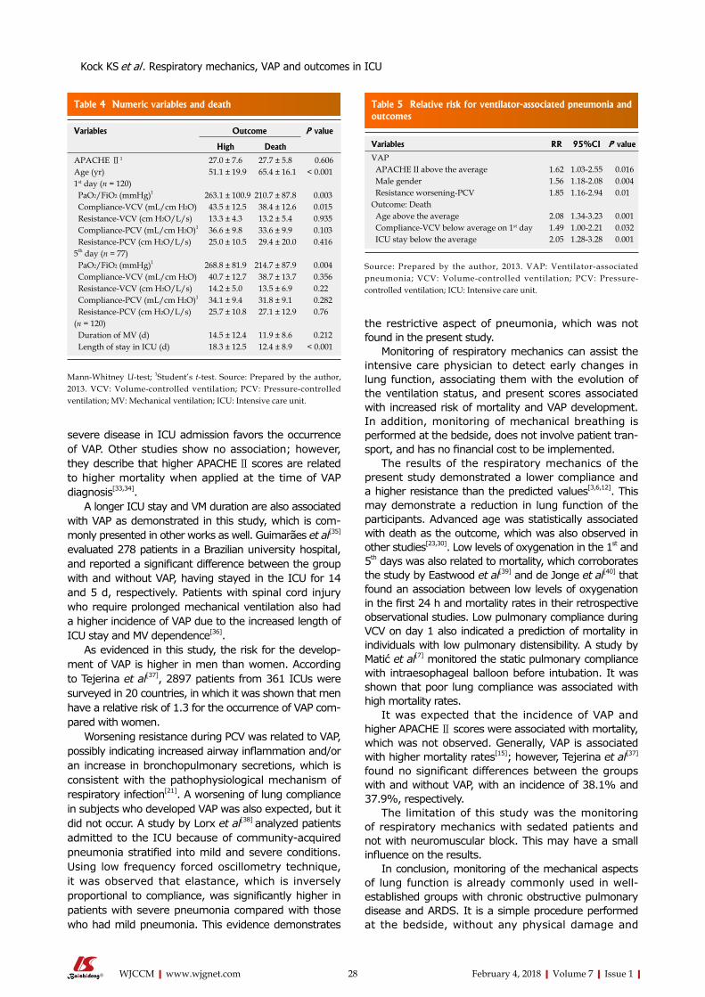
28 February 4, 2018|Volume 7|Issue 1|WJCCM|www.wjgnet.com
severe disease in ICU admission favors the occurrence of VAP. Other studies show no association; however, they describe that higher APACHE Ⅱ scores are related to higher mortality when applied at the time of VAP diagnosis[33,34].
A longer ICU stay and VM duration are also associated with VAP as demonstrated in this study, which is com-monly presented in other works as well. Guimarães et al[35] evaluated 278 patients in a Brazilian university hospital, and reported a significant difference between the group with and without VAP, having stayed in the ICU for 14 and 5 d, respectively. Patients with spinal cord injury who require prolonged mechanical ventilation also had a higher incidence of VAP due to the increased length of ICU stay and MV dependence[36].
As evidenced in this study, the risk for the develop-ment of VAP is higher in men than women. According to Tejerina et al[37], 2897 patients from 361 ICUs were surveyed in 20 countries, in which it was shown that men have a relative risk of 1.3 for the occurrence of VAP com-pared with women.
Worsening resistance during PCV was related to VAP, possibly indicating increased airway inflammation and/or an increase in bronchopulmonary secretions, which is consistent with the pathophysiological mechanism of respiratory infection[21]. A worsening of lung compliance in subjects who developed VAP was also expected, but it did not occur. A study by Lorx et al[38] analyzed patients admitted to the ICU because of community-acquired pneumonia stratified into mild and severe conditions. Using low frequency forced oscillometry technique, it was observed that elastance, which is inversely proportional to compliance, was significantly higher in patients with severe pneumonia compared with those who had mild pneumonia. This evidence demonstrates
the restrictive aspect of pneumonia, which was not found in the present study.
Monitoring of respiratory mechanics can assist the intensive care physician to detect early changes in lung function, associating them with the evolution of the ventilation status, and present scores associated with increased risk of mortality and VAP development. In addition, monitoring of mechanical breathing is performed at the bedside, does not involve patient tran-sport, and has no financial cost to be implemented.
The results of the respiratory mechanics of the present study demonstrated a lower compliance and a higher resistance than the predicted values[3,6,12]. This may demonstrate a reduction in lung function of the participants. Advanced age was statistically associated with death as the outcome, which was also observed in other studies[23,30]. Low levels of oxygenation in the 1st and 5th days was also related to mortality, which corroborates the study by Eastwood et al[39] and de Jonge et al[40] that found an association between low levels of oxygenation in the first 24 h and mortality rates in their retrospective observational studies. Low pulmonary compliance during VCV on day 1 also indicated a prediction of mortality in individuals with low pulmonary distensibility. A study by Matić et al[7] monitored the static pulmonary compliance with intraesophageal balloon before intubation. It was shown that poor lung compliance was associated with high mortality rates.
It was expected that the incidence of VAP and higher APACHE Ⅱ scores were associated with mortality, which was not observed. Generally, VAP is associated with higher mortality rates[15]; however, Tejerina et al[37] found no significant differences between the groups with and without VAP, with an incidence of 38.1% and 37.9%, respectively.
The limitation of this study was the monitoring of respiratory mechanics with sedated patients and not with neuromuscular block. This may have a small influence on the results.
In conclusion, monitoring of the mechanical aspects of lung function is already commonly used in well-established groups with chronic obstructive pulmonary disease and ARDS. It is a simple procedure performed at the bedside, without any physical damage and
Table 4 Numeric variables and death
Variables Outcome P value
High Death
APACHE Ⅱ1 27.0 ± 7.6 27.7 ± 5.8 0.606Age (yr) 51.1 ± 19.9 65.4 ± 16.1 < 0.0011st day (n = 120) PaO2/FiO2 (mmHg)1 263.1 ± 100.9 210.7 ± 87.8 0.003 Compliance-VCV (mL/cm H2O) 43.5 ± 12.5 38.4 ± 12.6 0.015 Resistance-VCV (cm H2O/L/s) 13.3 ± 4.3 13.2 ± 5.4 0.935 Compliance-PCV (mL/cm H2O)1 36.6 ± 9.8 33.6 ± 9.9 0.103 Resistance-PCV (cm H2O/L/s) 25.0 ± 10.5 29.4 ± 20.0 0.4165th day (n = 77) PaO2/FiO2 (mmHg)1 268.8 ± 81.9 214.7 ± 87.9 0.004 Compliance-VCV (mL/cm H2O) 40.7 ± 12.7 38.7 ± 13.7 0.356 Resistance-VCV (cm H2O/L/s) 14.2 ± 5.0 13.5 ± 6.9 0.22 Compliance-PCV (mL/cm H2O)1 34.1 ± 9.4 31.8 ± 9.1 0.282 Resistance-PCV (cm H2O/L/s) 25.7 ± 10.8 27.1 ± 12.9 0.76(n = 120) Duration of MV (d) 14.5 ± 12.4 11.9 ± 8.6 0.212 Length of stay in ICU (d) 18.3 ± 12.5 12.4 ± 8.9 < 0.001
Mann-Whitney U-test; 1Student’s t-test. Source: Prepared by the author, 2013. VCV: Volume-controlled ventilation; PCV: Pressure-controlled ventilation; MV: Mechanical ventilation; ICU: Intensive care unit.
Table 5 Relative risk for ventilator-associated pneumonia and outcomes
Variables RR 95%CI P value
VAP APACHE II above the average 1.62 1.03-2.55 0.016 Male gender 1.56 1.18-2.08 0.004 Resistance worsening-PCV 1.85 1.16-2.94 0.01Outcome: Death Age above the average 2.08 1.34-3.23 0.001 Compliance-VCV below average on 1st day 1.49 1.00-2.21 0.032 ICU stay below the average 2.05 1.28-3.28 0.001
Source: Prepared by the author, 2013. VAP: Ventilator-associated pneumonia; VCV: Volume-controlled ventilation; PCV: Pressure-controlled ventilation; ICU: Intensive care unit.
KockKSetal .Respiratorymechanics,VAPandoutcomesinICU

29 February 4, 2018|Volume 7|Issue 1|WJCCM|www.wjgnet.com
no additional cost. Based on this assumption, this procedure should be performed routinely in the ICU environment, providing the intensive care physician and the physiotherapist with additional prognosis and diagnosis variables, in addition to the clinical, laboratory and radiological data.
The results show that the respiratory function is a prognostic measure, and is strongly associated with mortality. Low oxygen and low lung compliance during VCV demonstrate this fact. Worsening of respiratory system resistance during PCV, associated with the development of VAP, indicates the possibility of early diagnosis.
ARTICLE HIGHLIGHTS Research backgroundThe measurements of respiratory mechanics most frequently used are compliance and resistance of the respiratory system. Compliance is associated with distensibility of the respiratory system, which is resulting from the tidal volume variation divided by the peak inspiratory pressure. Resistance is related to the conduction of air, obtained mathematically from the variation between the peak and plateau pressures divided by the inspiratory airflow.
Research motivationThe aim is evaluate the predictive capability of respiratory mechanics for the development of VAP and mortality in the intensive care unit (ICU) of a hospital in southern Brazil.
Research objectives Respiratory mechanics, ventilator-associated pneumonia.
Research methodsA cohort study was conducted between, involving a sample of 120 individuals. Static measurements of compliance and resistance of the respiratory system in pressure-controlled ventilation (PCV) and volume-controlled ventilation (VCV) modes in the 1st and 5th days of hospitalization were performed to monitor respiratory mechanics. The severity of the patients’ illness was quantified by the Acute Physiology and Chronic Health Evaluation Ⅱ (APACHE Ⅱ). The diagnosis of VAP was made based on clinical, radiological and laboratory parameters.
Research resultsThe significant associations found for the development of VAP were APACHE Ⅱ scores above the average (P = 0.016), duration of MV (P = 0.001) and ICU length of stay above the average (P = 0.003), male gender (P = 0.004), and worsening of respiratory resistance in PCV mode (P = 0.010). Age above the average (P < 0.001), low level of oxygenation on day 1 (P = 0.003) and day 5 (P = 0.004) and low lung compliance during VCV on day 1 (P = 0.032) were associated with death as the outcome.
Research conclusionsThe worsening of airway resistance in PCV mode indicated the possibility of early diagnosis of VAP. Low lung compliance during VCV and low oxygenation index were death-related prognostic indicators.
Research perspectivesThe results show that the respiratory function is a prognostic measure, and is strongly associated with mortality.
REFERENCES1 Nguyen YL, Wunsch H, Angus DC. Critical care: the impact
of organization and management on outcomes. Curr Opin Crit Care 2010; 16: 487-492 [PMID: 20689418 DOI: 10.1097/MCC.0b013e32833d9180]
2 Rubenfeld GD, Angus DC, Pinsky MR, Curtis JR, Connors AF Jr, Bernard GR. Outcomes research in critical care: results of the American Thoracic Society Critical Care Assembly Workshop on Outcomes Research. The Members of the Outcomes Research Workshop. Am J Respir Crit Care Med 1999; 160: 358-367 [PMID: 10390426 DOI: 10.1164/ajrccm.160.1.9807118]
3 Henderson WR, Sheel AW. Pulmonary mechanics during mechanical ventilation. Respir Physiol Neurobiol 2012; 180: 162-172 [PMID: 22154694 DOI: 10.1016/j.resp.2011.11.014]
4 Hamed HMF, Ibrahim HG, Khater YH, Aziz ES. Ventilation and ventilators in the ICU: What very intensivist must know. Curr Anaesth Crit Care 2006; 17: 77-83
5 Polak AG. Analysis of multiple linear regression algorithms used for respiratory mechanics monitoring during artificial ventilation. Comput Methods Programs Biomed 2011; 101: 126-134 [PMID: 20822825 DOI: 10.1016/j.cmpb.2010.08.001]
6 Lucangelo U, Bernabé F, Blanch L. Respiratory mechanics derived from signals in the ventilator circuit. Respir Care 2005; 50: 55-65; discussion 65-67 [PMID: 15636645]
7 Matić I, Pavicić F, Sakić-Zdravcević K, Danić D, Jurjević M. Pulmonary compliance values provide prognosis in mechanically ventilated patients--a randomized prospective study. Coll Antropol 2007; 31: 829-836 [PMID: 18041396]
8 Dhand R. Ventilator graphics and respiratory mechanics in the patient with obstructive lung disease. Respir Care 2005; 50: 246-261; discussion 259-261 [PMID: 15691394]
9 Koutsoukou A , Perraki H, Orfanos SE, Koulouris NG, Tromaropoulos A, Sotiropoulou C, Roussos C. History of mechanical ventilation may affect respiratory mechanics evolution in acute respiratory distress syndrome. J Crit Care 2009; 24: 626.e1-626.e6 [PMID: 19427758 DOI: 10.1016/j.jcrc.2009.02.003]
10 Nava S, Rubini F. Lung and chest wall mechanics in ventilated patients with end stage idiopathic pulmonary fibrosis. Thorax 1999; 54: 390-395 [PMID: 10212101 DOI: 10.1136/thx.54.5.390]
11 D’Angelo E, Calderini E, Robatto FM, Puccio P, Milic-Emili J. Lung and chest wall mechanics in patients with acquired immunodeficiency syndrome and severe Pneumocystis carinii pneumonia. Eur Respir J 1997; 10: 2343-2350 [PMID: 9387963 DOI: 10.1183/09031936.97.10102343]
12 Jubran A. Monitoring patient mechanics during mechanical ventilation. Crit Care Clin 1998; 14: 629-653 [PMID: 9891631 DOI: 10.1016/S0749-0704(05)70024-5]
13 Hellyer TP, Ewan V, Wilson P, Simpson AJ. The Intensive Care Society recommended bundle of interventions for the prevention of ventilator-associated pneumonia. J Intensive Care Soc 2016; 17: 238-243
14 Wunsch H, Angus DC, Harrison DA, Linde-Zwirble WT, Rowan KM. Comparison of medical admissions to intensive care units in the United States and United Kingdom. Am J Respir Crit Care Med 2011; 183: 1666-1673 [PMID: 21471089 DOI: 10.1164/rccm.201012-1961OC]
15 Joseph NM, Sistla S, Dutta TK, Badhe AS, Parija SC. Ventilator-associated pneumonia: a review. Eur J Intern Med 2010; 21: 360-368 [PMID: 20816584 DOI: 10.1016/j.ejim.2010.07.006]
16 Hajian-Tilaki K. Sample size estimation in diagnostic test studies of biomedical informatics. J Biomed Inform 2014; 48: 193-204 [PMID: 24582925 DOI: 10.1016/j.jbi.2014.02.013]
17 Knaus WA, Draper EA, Wagner DP, Zimmerman JE. APACHE II: a severity of disease classification system. Crit Care Med 1985; 13: 818-829 [PMID: 3928249 DOI: 10.1097/00003246-198510000-00009]
18 Sánchez Casado M, Quintana Díaz M, Palacios D, Hortigüela V, Marco Schulke C, García J, Canabal A, Pérez Pedrero MJ, Velasco Ramos A, Arrese MA. Relationship between the alveolar-arterial oxygen gradient and PaO2/FiO2-introducing PEEP into the model. Med Intensiva 2012; 36: 329-334 [PMID: 22154281 DOI: 10.1016/j.medin.2011.10.007]
ARTICLE HIGHLIGHTS
KockKSetal .Respiratorymechanics,VAPandoutcomesinICU

30 February 4, 2018|Volume 7|Issue 1|WJCCM|www.wjgnet.com
19 Ramsay MA, Savege TM, Simpson BR, Goodwin R. Controlled sedation with alphaxalone-alphadolone. Br Med J 1974; 2: 656-659 [PMID: 4835444 DOI: 10.1136/bmj.2.5920.656]
20 Nassar BS, Collett ND, Schmidt GA. The flow-time waveform predicts respiratory system resistance and compliance. J Crit Care 2012; 27: 418.e7-418.14 [PMID: 22226421 DOI: 10.1016/j.jcrc.2011.10.012]
21 Sociedade Brasileira de Pneumologia e Tisiologia. Diretrizes brasileiras para tratamento das pneumonias adquiridas no hospital e das associadas à ventilação mecânica. J Bras Pneumol 2007; 33 Suppl 1: 1-30
22 Zhang Z. Univariate description and bivariate statistical inference: the first step delving into data. Ann Transl Med 2016; 4: 91 [PMID: 27047950 DOI: 10.21037/atm.2016.02.11]
23 Acuña K, Costa E, Grover A, Camelo A, Santos Júnior R. Clinical-epidemiological characteristics of adults and aged interned in an intensive care unity of the Amazon (Rio Branco, Acre). Rev Bras Ter Intensiva 2007; 19: 304-309 [PMID: 25310063 DOI: 10.1590/S0103-507X2007000300006]
24 Rocha MS, Caetano JÁ, Soares E, Medeiros FL. Caracterização da população atendida em unidade de terapia intensiva: subsídio para a assistência. Rev enferm UERJ 2007; 15: 411-416
25 Matic I, Titlic M, Dikanovic M, Jurjevic M, Jukic I, Tonkic A. Effects of APACHE II score on mechanical ventilation; prediction and outcome. Acta Anaesthesiol Belg 2007; 58: 177-183 [PMID: 18018838]
26 Norena M, Wong H, Thompson WD, Keenan SP, Dodek PM. Adjustment of intensive care unit outcomes for severity of illness and comorbidity scores. J Crit Care 2006; 21: 142-150 [PMID: 16769457 DOI: 10.1016/j.jcrc.2005.11.011]
27 Doak MW, Nixon AC, Lupton DJ, Waring WS. Self-poisoning in older adults: patterns of drug ingestion and clinical outcomes. Age Ageing 2009; 38: 407-411 [PMID: 19383772 DOI: 10.1093/ageing/afp046]
28 Sudarsanam TD, Jeyaseelan L, Thomas K, John G. Predictors of mortality in mechanically ventilated patients. Postgrad Med J 2005; 81: 780-783 [PMID: 16344303 DOI: 10.1136/pgmj.2005.033076]
29 Elliott D. Measuring the health outcomes of general ICU patients: a systematic review of methods and findings. Aust Crit Care 1999; 12: 132-140 [PMID: 11271027 DOI: 10.1016/S1036-7314(99)70598-9]
30 Esteban A, Anzueto A, Frutos F, Alía I, Brochard L, Stewart TE, Benito S, Epstein SK, Apezteguía C, Nightingale P, Arroliga AC, Tobin MJ; Mechanical Ventilation International Study Group. Characteristics and outcomes in adult patients receiving mechanical ventilation: a 28-day international study. JAMA 2002; 287: 345-355 [PMID: 11790214 DOI: 10.1001/jama.287.3.345]
31 Azevedo LC, Park M, Salluh JI, Rea-Neto A, Souza-Dantas VC,
Varaschin P, Oliveira MC, Tierno PF, dal-Pizzol F, Silva UV, Knibel M, Nassar AP Jr, Alves RA, Ferreira JC, Teixeira C, Rezende V, Martinez A, Luciano PM, Schettino G, Soares M; ERICC (Epidemiology of Respiratory Insufficiency in Critical Care) investigators. Clinical outcomes of patients requiring ventilatory support in Brazilian intensive care units: a multicenter, prospective, cohort study. Crit Care 2013; 17: R63 [PMID: 23557378 DOI: 10.1186/cc12594]
32 Arabi Y, Al-Shirawi N, Memish Z, Anzueto A. Ventilator-associated pneumonia in adults in developing countries: a systematic review. Int J Infect Dis 2008; 12: 505-512 [PMID: 18502674 DOI: 10.1016/j.ijid.2008.02.010]
33 Huang KT, Tseng CC, Fang WF, Lin MC. An early predictor of the outcome of patients with ventilator-associated pneumonia. Chang Gung Med J 2010; 33: 274-282 [PMID: 20584505]
34 Mirsaeidi M, Peyrani P, Ramirez JA; Improving Medicine through Pathway Assessment of Critical Therapy of Hospital-Acquired Pneumonia (IMPACT-HAP) Investigators. Predicting mortality in patients with ventilator-associated pneumonia: The APACHE II score versus the new IBMP-10 score. Clin Infect Dis 2009; 49: 72-77 [PMID: 19480582 DOI: 10.1086/599349]
35 Guimarães MMQ, Rocco JR. Prevalência e prognóstico dos pacientes com pneumonia associada à ventilação mecânica em um hospital universitário. Bras Pneumol 2006; 32: 339-346
36 García-Leoni ME, Moreno S, García-Garrote F, Cercenado E. Ventilator-associated pneumonia in long-term ventilator-assisted individuals. Spinal Cord 2010; 48: 876-880 [PMID: 20404831 DOI: 10.1038/sc.2010.43]
37 Tejerina E, Frutos-Vivar F, Restrepo MI, Anzueto A, Abroug F, Palizas F, González M, D’Empaire G, Apezteguía C, Esteban A; Internacional Mechanical Ventilation Study Group. Incidence, risk factors, and outcome of ventilator-associated pneumonia. J Crit Care 2006; 21: 56-65 [PMID: 16616625 DOI: 10.1016/j.jcrc.2005.08.005]
38 Lorx A, Suki B, Hercsuth M, Szabó B, Pénzes I, Boda K, Hantos Z. Airway and tissue mechanics in ventilated patients with pneumonia. Respir Physiol Neurobiol 2010; 171: 101-109 [PMID: 20215004 DOI: 10.1016/j.resp.2010.03.004]
39 Eastwood G, Bellomo R, Bailey M, Taori G, Pilcher D, Young P, Beasley R. Arterial oxygen tension and mortality in mechanically ventilated patients. Intensive Care Med 2012; 38: 91-98 [PMID: 22127482 DOI: 10.1007/s00134-011-2419-6]
40 de Jonge E, Peelen L, Keijzers PJ, Joore H, de Lange D, van der Voort PH, Bosman RJ, de Waal RA, Wesselink R, de Keizer NF. Association between administered oxygen, arterial partial oxygen pressure and mortality in mechanically ventilated intensive care unit patients. Crit Care 2008; 12: R156 [PMID: 19077208 DOI: 10.1186/cc7150]
P- Reviewer: Jeong KJ, Zhang ZH S- Editor: Cui LJ L- Editor: A E- Editor: Li RF
KockKSetal .Respiratorymechanics,VAPandoutcomesinICU

© 2018 Baishideng Publishing Group Inc. All rights reserved.
Published by Baishideng Publishing Group Inc7901 Stoneridge Drive, Suite 501, Pleasanton, CA 94588, USA
Telephone: +1-925-223-8242Fax: +1-925-223-8243
E-mail: [email protected] Desk: http://www.f6publishing.com/helpdesk
http://www.wjgnet.com

World Journal of Critical Care MedicineWorld J Crit Care Med 2018 May 4; 7(2): 31-38
Published by Baishideng Publishing Group Inc
ISSN 2220-3141 (online)

World Journal ofCritical Care MedicineW J C C M
Contents
IWJCCM|www.wjgnet.com May 4, 2018|Volume 7|Issue 2|
Quarterly Volume 7 Number 2 May 4, 2018
ORIGINAL ARTICLE
Clinical Practice Study31 Confidencelevelofpediatrictraineesinmanagementofshockstates
Morparia K, Berg J, Basu SREVIEW

ContentsWorld Journal of Critical Care Medicine
Volume 7 Number 2 May 4, 2018
EDITORS FOR THIS ISSUE
Responsible Assistant Editor: Xiang Li Responsible Science Editor: Fang-Fang JiResponsible Electronic Editor: Wen-Wen Tan Proofing Editorial Office Director: Ya-Juan MaProofing Editor-in-Chief: Lian-Sheng Ma
EDITORIALOFFICEXiu-Xia Song, DirectorWorld Journal of Critical Care MedicineBaishideng Publishing Group Inc7901 Stoneridge Drive, Suite 501, Pleasanton, CA 94588, USATelephone: +1-925-2238242Fax: +1-925-2238243E-mail: [email protected] Desk: http://www.f6publishing.com/helpdeskhttp://www.wjgnet.com
PUBLISHERBaishideng Publishing Group Inc7901 Stoneridge Drive, Suite 501, Pleasanton, CA 94588, USATelephone: +1-925-223-8242Fax: +1-925-223-8243E-mail: [email protected] Desk: http://www.f6publishing.com/helpdeskhttp://www.wjgnet.com
PUBLICATIONDATEMay 4, 2018
COPYRIGHT© 2018 Baishideng Publishing Group Inc. Articles published by this Open-Access journal are distributed under the terms of the Creative Commons Attribution Non-commercial License, which permits use, distribution, and reproduction in any medium, provided the original work is properly cited, the use is non commercial and is otherwise in compliance with the license.
SPECIALSTATEMENTAll articles published in journals owned by the Baishi-deng Publishing Group (BPG) represent the views and opinions of their authors, and not the views, opinions or policies of the BPG, except where otherwise expli-citly indicated.
INSTRUCTIONSTOAUTHORShttp://www.wjgnet.com/bpg/gerinfo/204
ONLINESUBMISSIONhttp://www.f6publishing.com
IIWJCCM|www.wjgnet.com
ABOUT COVER
AIM AND SCOPE
INDExING/ABSTRACTING
May 4, 2018|Volume 7|Issue 2|
NAMEOFJOURNALWorld Journal of Critical Care Medicine
ISSNISSN 2220-3141 (online)
LAUNCHDATEFebruary 4, 2012
FREQUENCYQuarterly
EDITOR-IN-CHIEFBart Van Rompaey, BSc, MSc, PhD, Associate Professor, Nurse, Faculty of Medicine and Health Sciences, Department of Nursing and midwifery, Cen-tre for Research and Innovation in Care, University of Antwerp, Wilrijk 2610, Antwerp, Belgium
EDITORIALBOARDMEMBERSAll editorial board members resources online at http://www.wjgnet.com/2220-3141/editorialboard.htm
EditorialBoardMemberofWorld JournalofCriticalCareMedicine ,ZsoltJBalogh,MD,PhD,Professor,DivisionofSurgery, JohnHunterHospital,
Newcastle,NSW2310,Australia
World Journal of Critical Care Medicine (World J Crit Care Med, WJCCM, online ISSN 2220-3141, DOI: 10.5492) is a peer-reviewed open access academic journal that aims to guide clinical practice and improve diagnostic and therapeutic skills of clinicians.
WJCCM covers topics concerning severe infection, shock and multiple organ dysfunc-tion syndrome, infection and anti-infection treatment, acute respiratory distress syndrome and mechanical ventilation, acute kidney failure, continuous renal replacement therapy, rational nutrition and immunomodulation in critically ill patients, sedation and analgesia, cardiopulmonary cerebral resuscitation, fluid resuscitation and tissue perfusion, coagulant dysfunction, hemodynamic monitoring and circulatory support, ICU management and treatment control, and application of bronchofiberscopy in critically ill patients.
We encourage authors to submit their manuscripts to WJCCM. We will give priority to manuscripts that are supported by major national and international foundations and those that are of great clinical significance.
World Journal of Critical Care Medicine is now indexed in PubMed, PubMed Central.

Kavita Morparia, Julie Berg, Sonali Basu
ORIGINAL ARTICLE
31 May 4, 2018|Volume 7|Issue 2|WJCCM|www.wjgnet.com
Confidence level of pediatric trainees in management of shock states
Kavita Morparia, Department of Pediatric Critical Care, Children’s Hospital of New Jersey, Newark Beth Israel Medical Center, Newark, NJ 07112, United States
Julie Berg, Department of Emergency Medicine, Children’s National Health System, Washington, DC 20010, United States
Sonali Basu, Department of Critical Care Medicine, George Washington University, Children’s National Health System, Washington, DC 20010, United States
ORCID number: Kavita Morparia (0000-0002-6170-3995); Julie Berg (0000-0002-2675-893); Sonali Basu (0000-0002-6066-3231).
Author contributions: Morparia K and Basu S designed the research methodology and survey; Morparia K and Berg J were responsible for distribution of survey and data collection; Morparia K, Berg J and Basu S contributed to writing the ma-nuscript; Morparia K was responsible for subsequent revisions.
Institutional review board statement: The study was reviewed by the Institutional Review Board at the Children’s National Medical Center Institutional Review Board and certified as “exempt” since it was an anonymous survey sent to trainees and did not involve real patients.
Informed consent statement: The study was exempt from informed consent as it did not involve any patients.
Conflict-of-interest statement: No potential conflicts of interest relevant to this article were reported.
Open-Access: This article is an open-access article which was selected by an in-house editor and fully peer-reviewed by external reviewers. It is distributed in accordance with the Creative Commons Attribution Non Commercial (CC BY-NC 4.0) license, which permits others to distribute, remix, adapt, build upon this work non-commercially, and license their derivative works on different terms, provided the original work is properly cited and the use is non-commercial. See: http://creativecommons.org/licenses/by-nc/4.0/
Manuscript source: Unsolicited manuscript
Correspondence to: Kavita Morparia, MD, Attending Physician, Department of Pediatric Critical Care, Children’s Hospital of New Jersey, Newark Beth Israel Medical Center, 201 Lyons Avenue, Newark, NJ 07112, United States. [email protected]: +1-973-9268081Fax: +1-973-9266452
Received: February 6, 2018Peer-review started: February 7, 2018First decision: March 7, 2018Revised: March 12, 2018Accepted: April 22, 2018Article in press: April 22, 2018Published online: May 4, 2018
AbstractAIMTo assess overall confidence level of trainees in as-sessing and treating shock, we sought to improve awareness of recurrent biases in clinical decision-making to help address appropriate educational interventions.
METHODSPediatric trainees on a national listserv were offered the opportunity to complete an electronic survey anon-ymously. Four commonly occurring clinical scenarios were presented, and respondents were asked to choose whether or not they would give fluid, rank factors utilized in decision-making, and comment on confidence level in their decision.
RESULTSPediatric trainees have a very low confidence level for assessment and treatment of shock. Highest confidence level is for initial assessment and treatment of shock involving American College of Critical Care Medicine/Pe-diatric Advanced Life Support recommendations. Children with preexisting cardiac comorbidities are at high risk of
World Journal ofCritical Care MedicineW J C C M
Submit a Manuscript: http://www.f6publishing.com
DOI: 10.5492/wjccm.v7.i2.31
World J Crit Care Med 2018 May 4; 7(2): 31-38
ISSN 2220-3141 (online)
Clinical Practice Study

under-resuscitation.
CONCLUSIONPediatric trainees nationwide have low confidence in managing various shock states, and would benefit from guidance and teaching around certain common clinical situations.
Key words: Fluid bolus; Shock; Medical education; Central venous pressure; Decision-making; Pediatric advanced life support guidelines
© The Author(s) 2018. Published by Baishideng Publishing Group Inc. All rights reserved.
Core tip: Pediatric trainees at all levels of training across the United States express a low degree of confidence in management of various types of shock. Children with cardiac comorbidity are at very high risk of under-resuscitation when presenting with shock. Central venous pressure is often used in isolation for decision-making regarding fluid administration and supersedes other subjective and objective measures of intravascular fluid status and shock state.
Morparia K, Berg J, Basu S. Confidence level of pediatric trainees in management of shock states. World J Crit Care Med 2018; 7(2): 31-38 Available from: URL: http://www.wjgnet.com/2220-3141/full/v7/i2/31.htm DOI: http://dx.doi.org/10.5492/wjccm.v7.i2.31
INTRODUCTIONFluid resuscitation of hypovolemic shock has been hailed as acute medicine’s great triumph for children[1]. A fluid bolus is one of the most rapid ways to increase cardiac output and is central in the management of many shock states. Recognition and timely resuscitation of shock with fluid is one of the most important aspects of training in pediatrics, and one that all trainees must be empowered to feel confident managing. It has been well established that appropriate early treatment of shock is associated with improved outcomes[2,3]. Pediatric resident and fellow physicians are often the first responders at the bedside of an acutely ill child. They may be assessing the patient for the first time in the emergency room, or might be called upon to assess a patient for a change in vital signs. We were interested in exploring the grey zone in decision-making - everyone would elect to continue fluid resuscitation in the face of obvious hypotension - however, in practice, the blood pressure drops very late in the evolution of shock in children, and we were interested in finding out how trainees navigate the period before profound shock develops.
Though there is some literature on level of adherence to Pediatric Advanced Life Support (PALS) guidelines
32 May 4, 2018|Volume 7|Issue 2|WJCCM|www.wjgnet.com
Morparia K et al . How pediatric trainees manage shock?
and outcomes[4,5], there is a paucity of literature on the ability of pediatric trainees to correctly assess and treat shock states of varying etiologies. Variable heart rates and blood pressure thresholds for varying ages in children make it more challenging to recognize deviations from normal, often confounded by factors such as fever and beta agonist administration. While comorbid cardiomyopathy engenders a more cautious approach to fluid bolus administration, fear of fluid overload might hamper adequate resuscitation. The American College of Critical Care Medicine (ACCM) guidelines[6] recommend resuscitation end-points based on the difference between mean arterial pressure (MAP) and central venous pressure (CVP), mixed venous saturation (ScVO2) and hemoglobin level, along with clinical exam findings. CVP has long been shown to have no utility as a marker of fluid responsiveness, yet continues to be considered as a factor in decision-making. We framed these potential stumbling blocks as clinical situations that may be widely prevalent in clinical practice as a survey for a nationwide sample of pediatric trainees. We sought to assess the confidence level of residents in the assessment and treatment of shock.
Our aim in performing this research was to assess overall confidence level of trainees in assessing and treating shock. We sought to improve awareness of recurrent biases in clinical decision-making to help address appropriate educational interventions. There is extensive literature on cognitive biases affecting decision-making in medicine[7,8], however we are not aware of any studies dealing with decision-making aspects in the management of children in the emergency room or critical care environment. There are situations where closer supervision and clinical guidance may improve earlier detection of shock in the critically ill child.
MATERIALS AND METHODSWe designed a survey tool using REDCap[9], an online electronic survey tool. We obtained approval from the Institutional Review Board at the Children’s National Health System and by the American Academy of Pediatrics (AAP) Section on Medical Students, Residents and Fellowship Trainees (SOMSRFT). The survey was then distributed via the AAP SOMSRFT listserv to all members currently having valid email addresses and registered with this section of the AAP, with an additional reminder email after a few weeks. Participation in the survey was voluntary and anonymous.
We collected demographic information including current role (medical student, resident or fellow), level of training based on postgraduate year (PGY-1, 2, 3, etc.) and area of specialty (for fellows). This was followed by four hypothetical case scenarios based on the recognition and management of shock. Table 1 details the clinical case presented and expected outcomes. The clinical description was followed by a set of questions including (1) Course of action the trainee would choose: fluid/

33 May 4, 2018|Volume 7|Issue 2|WJCCM|www.wjgnet.com
vasoactive/neither/other; (2) clinical factors taken into consideration - ranked on a Likert scale for importance; (3) level of confidence in decision ranked on a Likert scale; and (4) comments/reasoning if any. We per-formed additional analysis after stratification of the respondents into two groups - experts and non-experts. We classified fellows in pediatric critical care, pediatric emergency medicine and pediatric hospital medicine as experts in fluid resuscitation. All other respondents were non-experts.
Statistical analysisData were directly available on REDCap, and descriptive statistics were generated using this tool. Additional statistical functions were performed using SPSS v 21.0 (IBM). We used the χ 2 test to detect differences in categorical variables among groups and the Mann Whitney U test to detect differences between the distributions among groups.
RESULTSThere were a total of 539 respondents, with demo-graphic characteristics shown in Table 2. For the purpose of this study, we excluded responses from medical students and attending physicians, thus analyzing a total of 490 responses. There was an even distribution of residents and fellows across all levels of training. Of the fellows, pediatric critical care medicine and pediatric emergency medicine fellows accounted for the majority of respondents. Trainees’ clinical decisions in each of the four hypothetical scenarios are detailed in Table 3. Table 4 illustrates different choices selected by experts compared to non-experts for all the four scenarios.
In scenario 1, 85% of respondents chose to continue resuscitation of shock, with an even split of respondents choosing fluid (38%) or vasoactive (47%). Most people cited the ACCM/PALS guideline cutoff of 60 mL/kg for fluid administration as the rationale for choosing vasoactives over fluid. Of the remaining 15%, 9% of respondents required additional data before determining course of action - including mental status, urine output, signs of fluid overload and ultrasound measures of fluid status. Six percent of respondents chose to halt resuscitation. The most common reason cited for stopping resuscitation was to give antipyretic and evaluate heart rate response as tachycardia could be due to presence of fever. Other reasons were “normal blood pressure”, giving Ⅳ fluids at higher than maintenance rate to treat shock and avoidance of pulmonary edema. Thirty-six percent of respondents said they had only slight confidence in their decision. There was no correlation of low confidence level with primary decision taken.
For scenario 2, 80% of respondents chose to continue resuscitation of shock, with 20% choosing fluid and 60% choosing vasoactives. The most common reason cited for not continuing resuscitation was interpretation of CVP
as normal. Other reasons included awaiting chest X-ray to evaluate for pulmonary edema, echocardiogram for assessing cardiac function, and assessing adequacy of urine output. In this case, 65% of respondents were only slightly confident of their decision. Of respondents who chose to halt resuscitation or obtain additional data first, 76% (70 out of 92) expressed low confidence, compared to those who chose to give either fluid or vasoactive, where 60% expressed low confidence.
For scenario 3, 39% of total respondents choose to give additional fluid, 15% would give a vasoactive, and 46% elect to not continue resuscitation. The most common reason by far for not continuing resuscitation is the known cardiac comorbidity, with several trainees requiring an echocardiogram to evaluate for cardiac function first. The next most common factor cited is the presence of fever, and several respondents want to reevaluate the degree of tachycardia after fever has subsided. Other reasons for slowing or halting resuscitation include obtaining a chest X-ray, cultures and antibiotics, and transfer to the pediatric intensive care unit. Sixty-four percent of all respondents express poor confidence in decision. In this case, prevalence of low confidence level is the same - at 64% in all respondents regardless of whether or not they pursue to continue resuscitation or delay/halt resuscitation.
For the patient in scenario 4, 43% of respondents elected to not perform any additional intervention, 29% chose to give a fluid bolus and 20% elected to increase vasoactive support. By far the most common reason for administration of fluid or increasing vasoactive dose was the CVP value. A majority of respondents (61%) expressed low confidence in their decision.
DISCUSSIONEarly identification and rapid reversal of shock has been well documented to improve outcomes. We surveyed pediatric residents and fellows to assess how they made decisions regarding treatment of shock, and uncovered some commonly prevalent biases and errors in management.
The first scenario describes a 5-year old who is clearly presenting in shock, with end-organ dysfunction manifested by altered mental status. She has already received the initial 60 mL/kg of fluid. Per the ACCM guidelines[6], fluid resuscitation should be continued until signs of pulmonary overload occur or shock is reversed, defined by achieving threshold heart rate (HR) and MAP-CVP values, which were not yet achieved in this girl. In our experience, the initial 60 mL/kg is given automatically, and then there is some complacency while the patient awaits transfer to the intensive care unit (ICU). This can be thought of in terms of “premature closure” and “representativeness”, where a diagnostic category is assigned to the patient and clinical response after 60 mL/kg of fluid is taken for granted, as this amount would suffice for most patients. Ideally, central
Morparia K et al . How pediatric trainees manage shock?

34 May 4, 2018|Volume 7|Issue 2|WJCCM|www.wjgnet.com
Table 1 Clinical case scenarios and outcomes studied
Case Scenario description Clinical questions Key clinical features Expected interventions Outcomes studied
1 A 5 yr-old girl is brought into the emergency department with fever, diarrhea and vomiting. She is drowsy and does not answer questions appropriately. Her vitals are as follows: Temp
38.4 ℃, HR 168/min, RR 36/min, BP 90/45 (MAP 60) mmHg. She seems dehydrated with dry mucous membranes, is warm, flushed and has flash capillary refill. You diagnose septic shock and after getting two good Ⅳ lines for access, begin rapid fluid administration. You
give her 60 mL/kg crystalloids over a period of 60 min, and one dose of antibiotics. At the end of this time, her vitals are as follows: Temp 38.4 ℃, HR 165/min, RR 32/min, BP 96/40 (MAP 59) mmHg, capillary refill unchanged. What would
be your next plan of action?
What would be your next plan of action
(1) Do nothing at this point;
(2) give an additional 20 mL/kg fluid bolus; (3) start a vasoactive
medication; (4) need additional
data to decide- please specify;
(5) Other What factors were used
for decision making? HR/BP/Cap refill/Response to fluid/
other (specify) How confident are you in decision on a scale of 0 (not confident at all) to 4 (sure of decision)
Severe shock with altered mental status Ongoing shock with
HR and MAP not at threshold levels per ACCM-PALS
guidelines
Continue resuscitation with either fluid
or vasoactive administration
evaluate lactate, mixed venous saturations
Rationale in choosing fluid vs vasoactive, or
vice versa Rationale for withholding resuscitation
Confidence level in decision-making
2 You are caring for a 4 yr old in the PICU with severe septic shock due to lobar pneumonia. His
first night of admission he received 60 mL/kg of crystalloids and 20 mL/kg of 5% albumin. When you see him this morning, he is barely arousable and has a cap refill time of 5 s. You
insert an internal jugular line and a radial arterial line. His vitals at this time are as follows:
Temp 37.5 ℃, HR 152/min, RR 35/min, BP 100/45 (MAP 63) mmHg. His CVP is 8 mmHg. You obtain a blood gas from the A-line, and his lactate is 4.5 mmol/L. You decide to intubate to reduce oxygen consumption related to work of
breathing. Despite adequate sedation, he persists to have tachycardia; vitals after intubation are as follows: Temp 37.5 ℃, HR 168/min, BP 110/40
(MAP 63) mmHg, CVP 10 mmHg, cap refill time 5 s. You immediately have to escalate to very high ventilator settings with pressure control
of 34 and PEEP of 10 to achieve acceptable oxygenation and ventilation. What would your
next intervention be?
What would be your next plan of action
(1) Do nothing at this point;
(2) give an additional 20 mL/kg fluid bolus; (3) start a vasoactive
medication; (4) need additional
data to decide- please specify;
(5) Other What factors were used
for decision making? HR/BP/CVP/Cap refill/Response to
fluid/other (specify) How confident are you in decision on a scale of 0 (not confident at all) to 4 (sure of decision)
Worsening hemodynamics after initiation of positive pressure ventilation
Resuscitate shock with fluid or vasoactives
Recognition of decrease in preload caused by initiating of positive pressure ventilation
Interpretation of CVP in conjunction with higher intrathoracic
pressures
3 While rotating through the Hematology Oncology unit, you are called to the bedside of a 12-yr old receiving maintenance chemotherapy for AML. She has developed a temperature of 39.3 ℃. On exam she has a HR of 160/min, RR 32/min, BP 110/40 (MAP 63) mmHg, and cap
refill of 3 s. She is known to have anthracycline-induced cardiomyopathy. You palpate her
abdomen and notice that her liver is 3-4 cm, similar to earlier, she has no murmur or gallop, and a CVP transduced through her broviac is 6 mmHg. In the last 4 h, her urine output has
reduced from 1.5 mL/kg·h to 0.3 mL/kg·h. You decide to cautiously give her a 10 mL/kg fluid bolus over half an hour to see the response. At
the end of the bolus, her HR is now 154/min, BP is 106/46 (MAP 66) mmHg, CVP has increased
to 8 mmHg. What would your next step be?
What would be your next plan of action
(1) Do nothing at this point;
(2) give an additional fluid bolus;
(3) start a vasoactive medication;
(4) need additional data to decide- please
specify; (5) Other
What factors were used for decision making? HR/BP/Cap refill/Response to fluid/
other (specify) How confident are you in decision on a scale of 0 (not confident at all) to 4 (sure of decision)
Known cardiomyopathy with
onset of shock Low CVP and absence
of hepatomegaly Good response to fluid
bolus
Continue fluid resuscitation
Rationale for halting or slowing resuscitation
Morparia K et al . How pediatric trainees manage shock?

35 May 4, 2018|Volume 7|Issue 2|WJCCM|www.wjgnet.com
venous access should be obtained and vasoactive me-dications made available to be started immediately if needed, while assessment of shock should proceed with the same urgency as at initial presentation, especially in the presence of ongoing losses. We found that most respondents were appropriately aggressive, opting to continue either fluid or vasoactive. While most respondents explicitly cited that they were basing their decision on ACCM guidelines, most also interpreted the blood pressure as normal, although the MAP was below
the threshold recommended in the ACCM guidelines. It would be interesting to study how prevalent is this departure from the guidelines while treating shock, and whether the thresholds recommended by the ACCM are actually achieved in practice.
Scenario 2 dealt with a more complex situation where positive pressure ventilation had just been initiated. While advanced cardiopulmonary interactions are beyond the scope of general pediatric training, the ACCM guidelines mention that fluid loading might be necessary with the initiation of positive pressure ventilation due to a resultant reduction in the preload. The scenario describes florid shock with altered mental status, tachycardia despite adequate sedation, a heart rate of 168 and a diastolic blood pressure of 40 mmHg in a 4-year old child. The CVP of 10 mmHg in the face of very high intrathoracic pressures does not reflect in any way on intravascular volume status. The most common reason for inadequate resuscitation was the CVP value. Interestingly, a low confidence level was correlated with inaction in this scenario. This can be seen as a form of “omission bias”-one of the commonest biases in clinical medicine, where events occurring through the natural progression of a disease are more acceptable than those that may be directly attributed to the action of the physician.
In the third scenario, though the patient is known to have chemotherapy-induced cardiomyopathy, her liver size is not enlarged from baseline, CVP is low, and heart rate improves with an initial 10 mL/kg bolus. The probability that fluid administration will be harmful given these clinical attributes is inflated, and the far more likely possibility that there is ongoing shock in an immunocompromised host is minimized. This is a form of “base rate neglect”-clinicians conflate probabilities to rule out the worst case scenario.
4 You are caring for a 6 yr old girl admitted to the PICU after anaphylactic shock from a bee sting.
She has required a lot of fluid in the 4 h since admission-a total of 60 mL/kg, and is currently on dopamine at 7 mg/kg per minute. She has a urine output of 1 mL/kg per hour. She is on 2 L NC, and her vitals are as follows: Temp 36.8 ℃, HR 115/min, RR 16/min, BP 95/65 (MAP 75)
mmHg, with flushed extremities and cap refill of 2 s. You insert an internal jugular line and when
you transduce it, you get a CVP of 3 mmHg. What would you do next?
What would be your next plan of action
(1) Do nothing- continue maintenance
fluids; (2) give an additional 20 mL/kg fluid bolus; (3) increase vasoactive
medication; (4) need additional
data to decide- please specify; (5) other
What factors were used for decision making? HR/BP/Cap refill/Response to fluid/
other (specify) How confident are you in decision on a scale of 0 (not confident at all) to 4 (sure of decision)
Vital signs not suggestive of shock
Incidentally transduced CVP level
of 3 mmHg
Do not continue any further resuscitation
Percentage continuing fluid resuscitation
based on isolated CVP value
ACCM: American college of critical care medicine; PALS: Pediatric advanced life support; MAP: Mean arterial pressure; CVP: Central venous pressure.
Demographics of respondents n (%)
Total respondents 539 (100)Medical students 37 (7)Residents 393 (73)Fellows 97 (18)Attending physicians 12 (2)Level of training- residents 3671 (100) PGY-1 120 (33) PGY-2 110 (30) PGY-3 121 (33) PGY-4 16 (4) No response 26Level of training- fellows 901 (100) PGY-4 25 (28) PGY-5 32 (35) PGY-6 29 (32) PGY-7 4 (4) No response 7Pediatric fellows’ specialty (97) Pediatric critical care medicine 25 (26) Pediatric emergency medicine 22 (23) Neonatology 14 (14) Pediatric hospital medicine 5 (5) Pediatric cardiology 4 (4) Other 27 (28)
1Total number of respondents to this question from all survey takers.
Table 2 Demographics of respondents
Morparia K et al . How pediatric trainees manage shock?

36 May 4, 2018|Volume 7|Issue 2|WJCCM|www.wjgnet.com
In scenario 4 - the patient has required a fair amount of fluid and vasopressor, but has an adequate urine output and normal vitals. Again in this case, CVP of 3 mmHg takes precedence in decision-making and half of all respondents elect to give additional fluid or increase vasoactives.
We uncovered some common themes across the scenarios. The presence of fever can act as a confoun-ding factor in the attribution of tachycardia to the shock state. This leads to delay in treatment for shock while treating with antipyretics and awaiting fever to subside before assessing degree of tachycardia. It is important to emphasize to trainees that while awaiting antipyretics to take effect, it is vital to continue resuscitative measures for shock. Although several clinical studies have widely disproven the utility of CVP both as a marker of intravascular volume state as well as an indicator of fluid responsiveness[10,11], CVP is still widely used in decision-making regarding fluid status, often taking primacy over other signs. This approach is not only not useful, it can be potentially harmful as we have shown, where the contribution of factors such as intrathoracic pressure and diastolic function to CVP is not considered, and a normal CVP value leads to a premature halt to resuscitation. Conversely, a CVP of 3 mmHg is normal in a spontaneously breathing child with normal cardiac function and does not indicate hypovolemia[12]. Interpretation of a given CVP value should take into account these aforementioned factors, with recent expert opinion suggesting that extreme values of CVP may still be useful to guide resuscitation[13].
Though some respondents mentioned using ultrasound measures to ascertain for fluid status, markers of fluid responsiveness such as respiratory variation in peak aortic velocity require the absence of spontaneous breathing in a mechanically ventilated patient, as well as some expertise to obtain[14]. Furthermore, although echocardiography might give a general idea as to cardiac function, it does not yield any information on systemic vascular resistance or intravascular volume. Myocardial depression often coexists with septic shock[15], yet these children need to be resuscitated with either fluid or vasopressors, and clinical exam for response to fluid, hepatomegaly and rales is usually the only tool available at bedside. As such, it is not appropriate to await results of echocardiographic imaging to make decisions on fluid resuscitation. Experts were more likely than non-experts to prefer vasoactive medication to fluid in all instances except for scenario 4, and were uniformly more confident.
Our study has several limitations. Answering questions on a survey does not replicate the experience of examining a patient and assessing the evolution of disease in real time. In practice, each patient is immeasurably complex and there are multiple sources of clinical input - both conscious and subconscious. For sake of keeping the question stem at a reasonable length, we had to handpick what we considered the most relevant information. There is also the inevitable introduction of the Hawthorne effect here-respondents are aware that they are completing a survey on fluid management practices and this knowledge might further add to the
Table 3 Responses to clinical scenarios
Scenario Fluid bolus Vasoactive No further intervention/ need more data/other
Factors cited as important for decision-making
Percentage with low confidence
1-septic shock s/p 60 mL/kg fluid 38% 47% 15% Capillary refill, response to fluid
36%
2-hemodynamic instability s/p initiation of positive pressure ventilation
20% 60% 20% CVP, lactate 65%
3-shock with comorbid cardiomyopathy
39% 15% 46% HR, BP 64%
4-anaphylaxis with resolved shock 29% 20% 51% CVP 61%
s/p: Status post; CVP: Central venous pressure; HR: Heart rate; BP: Blood pressure.
Table 4 Preferences of non-experts vs experts
Scenario Role Bolus (% of total) Vasoactive (% of total) Other (% of total) P value
1-septic shock with ongoing losses
Non-experts1 (n = 438) Experts1 (n = 52)
40 47 13 0.0225 50 25
2-worsening hemodynamics after intubation
Non-experts (n = 438) Experts (n = 52)
21 59 20 0.0811 73 16
3-shock in a child known to have cardiomyopathy
Non-experts (n = 438) Experts (n = 52)
39 15 46 0.7742 17 41
4-recovering anaphylactic shock with low CVP
Non-experts (n = 438)Experts (n = 52)
29 21 50 0.0125 8 67
1Experts: Fellows in pediatric critical care, pediatric emergency medicine, and pediatric hospital medicine; Non-experts: All other respondents.
Morparia K et al . How pediatric trainees manage shock?

37 May 4, 2018|Volume 7|Issue 2|WJCCM|www.wjgnet.com
inaccuracy of survey responses compared to actual decisions made. Thus the survey-based methodology is far less rigorous than conducting a prospective study and debriefing pediatric trainees in real time.
To conclude, while pediatric trainees are most confident when following ACCM guidelines to treat shock, they face a high degree of decisional conflict and lack of confidence when encountering alternative etiologies and comorbidities. Educational interventions targeting the biases outlined in our study could be of benefit.
ARTICLE HIGHLIGHTSResearch backgroundPediatric trainees are often the first responders at the bedside for evaluation and ongoing management of children presenting with various shock states, yet there is little data on how they navigate through these decisions or how confident they feel in making these decisions. We conducted a survey of pediatric trainees all over the United States. Our study is the first study to survey in the literature studying fluid administration practices of trainees.
Research motivationThe motivation for performing this research was to uncover common situations where pediatric trainees faced a significant decisional conflict when treating shock. We also aimed to uncover some common situations where under-resuscitation was common and to highlight cognitive biases and fallacies of trainees while assessing and treating children with shock.
Research objectives One of the study objectives was to assess level of adherence and confidence level with American College of Critical Care Medicine (ACCM) guidelines which are universally followed in the United States for treatment of septic shock. Additionally, we wanted to assess degree of reliance on central venous pressure, resuscitation in children with ongoing cardiac comorbidity. We also sought to discover if there were significant differences in treatment practices of more advanced level trainees such as pediatric critical care, hospital medicine and emergency medicine fellows. All these objectives were realized, and can help in training and supporting pediatric residents for management of shock.
Research methodsWe conducted a nationwide survey of all pediatric trainees in the United States. This included residents at all levels of training, and fellows training in one of the subspecialties of pediatrics. The survey was voluntary and anonymous. Statistics were primarily descriptive, and SPSS was used for performing additional statistical testing.
Research resultsWe found that pediatric trainees across all levels of training faced a high degree of uncertainty and lack of confidence while they were making decisions regarding fluid administration in children presenting with shock. ACCM guidelines are frequently cited, yet blood pressure goals cited in the ACCM guidelines are often not met, nor is a suboptimal blood pressure recognized. Children with coexisting cardiac comorbidities may be prone to severe under-resuscitation for fear of cardiac failure causing pulmonary edema. Fever is an important confounding factor often delaying recognition of shock. This study sheds light on these important observations, and further prospective observational studies are warranted which study decision-making of trainees.
Research conclusionsThis study is the first study on how trainees in pediatrics make decisions for treatment of shock. It is vital that shock be recognized and treated rapidly, yet there are no studies looking at how confident trainees feel in their judgment. This study points to a very low level of confidence when treating shock, and some common situations which should be highlighted to trainees while caring for patients or in simulated scenarios.
Research perspectivesPediatric trainees should be supported adequately and provided focused teaching related to treatment of shock states in children. Children with malignancy and cardiac comorbidity who present with septic shock are a uniquely vulnerable population prone to under-resuscitation and should be managed by expert physicians. Central venous pressure should be interpreted with caution and not used in isolation without entire clinical picture.
REFERENCES1 Carcillo JA, Tasker RC. Fluid resuscitation of hypovolemic
shock: acute medicine’s great triumph for children. Intensive Care Med 2006; 32: 958-961 [PMID: 16791656 DOI: 10.1007/s00134-006-0189-3]
2 Carcillo JA, Davis AL, Zaritsky A. Role of early fluid resuscitation in pediatric septic shock. JAMA 1991; 266: 1242-1245 [PMID: 1870250 DOI: 10.1001/jama.1991.03470090076035]
3 Han YY, Carcillo JA, Dragotta MA, Bills DM, Watson RS, Westerman ME, Orr RA. Early reversal of pediatric-neonatal septic shock by community physicians is associated with improved outcome. Pediatrics 2003; 112: 793-799 [PMID: 14523168 DOI: 10.1542/peds.112.4.793]
4 Oliveira CF, Nogueira de Sá FR, Oliveira DS, Gottschald AF, Moura JD, Shibata AR, Troster EJ, Vaz FA, Carcillo JA. Time- and fluid-sensitive resuscitation for hemodynamic support of children in septic shock: barriers to the implementation of the American College of Critical Care Medicine/Pediatric Advanced Life Support Guidelines in a pediatric intensive care unit in a developing world. Pediatr Emerg Care 2008; 24: 810-815 [PMID: 19050666 DOI: 10.1097/PEC.0b013e31818e9f3a]
5 Paul R, Neuman MI, Monuteaux MC, Melendez E. Adherence to PALS Sepsis Guidelines and Hospital Length of Stay. Pediatrics 2012; 130: e273-e280 [PMID: 22753559 DOI: 10.1542/peds.2012-0094]
6 Brierley J, Carcillo JA, Choong K, Cornell T, Decaen A, Deymann A, Doctor A, Davis A, Duff J, Dugas MA, Duncan A, Evans B, Feldman J, Felmet K, Fisher G, Frankel L, Jeffries H, Greenwald B, Gutierrez J, Hall M, Han YY, Hanson J, Hazelzet J, Hernan L, Kiff J, Kissoon N, Kon A, Irazuzta J, Lin J, Lorts A, Mariscalco M, Mehta R, Nadel S, Nguyen T, Nicholson C, Peters M, Okhuysen-Cawley R, Poulton T, Relves M, Rodriguez A, Rozenfeld R, Schnitzler E, Shanley T, Kache S, Skippen P, Torres A, von Dessauer B, Weingarten J, Yeh T, Zaritsky A, Stojadinovic B, Zimmerman J, Zuckerberg A. Clinical practice parameters for hemodynamic support of pediatric and neonatal septic shock: 2007 update from the American College of Critical Care Medicine. Crit Care Med 2009; 37: 666-688 [PMID: 19325359 DOI: 10.1097/CCM.0b013e31819323c6]
7 Blumenthal-Barby JS, Krieger H. Cognitive biases and heuristics in medical decision making: a critical review using a systematic search strategy. Med Decis Making 2015; 35: 539-557 [PMID: 25145577 DOI: 10.1177/0272989X14547740]
8 Crowley RS, Legowski E, Medvedeva O, Reitmeyer K, Tseytlin E, Castine M, Jukic D, Mello-Thoms C. Automated detection of heuristics and biases among pathologists in a computer-based system. Adv Health Sci Educ Theory Pract 2013; 18: 343-363 [PMID: 22618855 DOI: 10.1007/s10459-012-9374-z]
9 Harris PA, Taylor R, Thielke R, Payne J, Gonzalez N, Conde JG. Research electronic data capture (REDCap)--a metadata-driven methodology and workflow process for providing translational research informatics support. J Biomed Inform 2009; 42: 377-381 [PMID: 18929686 DOI: 10.1016/j.jbi.2008.08.010]
10 Shippy CR, Appel PL, Shoemaker WC. Reliability of clinical monitoring to assess blood volume in critically ill patients. Crit Care Med 1984; 12: 107-112 [PMID: 6697726]
11 Marik PE, Baram M, Vahid B. Does central venous pressure predict fluid responsiveness? A systematic review of the literature and the tale of seven mares. Chest 2008; 134: 172-178 [PMID: 18628220 DOI: 10.1378/chest.07-2331]
12 Arikan AA, Zappitelli M, Goldstein SL, Naipaul A, Jefferson
Morparia K et al . How pediatric trainees manage shock?
ARTICLE HIGHLIGHTS

38 May 4, 2018|Volume 7|Issue 2|WJCCM|www.wjgnet.com
LS, Loftis LL. Fluid overload is associated with impaired oxygenation and morbidity in critically ill children. Pediatr Crit Care Med 2012; 13: 253-258 [PMID: 21760565 DOI: 10.1097/PCC.0b013e31822882a3]
13 De Backer D, Vincent JL. Should we measure the central venous pressure to guide fluid management? Ten answers to 10 questions. Crit Care 2018; 22: 43 [PMID: 29471884 DOI: 10.1186/s13054-018-1959-3]
14 Morparia KG, Reddy SK, Olivieri LJ, Spaeder MC, Schuette JJ. Respiratory variation in peak aortic velocity accurately predicts fluid responsiveness in children undergoing neurosurgery under general anesthesia. J Clin Monit Comput 2018; 32: 221-226 [PMID: 28299589 DOI: 10.1007/s10877-017-0013-3]
15 Raj S, Killinger JS, Gonzalez JA, Lopez L. Myocardial dysfunction in pediatric septic shock. J Pediatr 2014; 164: 72-77.e2 [PMID: 24144393 DOI: 10.1016/j.jpeds.2013.09.027]
P- Reviewer: He HW, Schuurman HJ S- Editor: Cui LJ L- Editor: A E- Editor: Huang Y
Morparia K et al . How pediatric trainees manage shock?

© 2018 Baishideng Publishing Group Inc. All rights reserved.
Published by Baishideng Publishing Group Inc7901 Stoneridge Drive, Suite 501, Pleasanton, CA 94588, USA
Telephone: +1-925-223-8242Fax: +1-925-223-8243
E-mail: [email protected] Desk: http://www.f6publishing.com/helpdesk
http://www.wjgnet.com
World Journal of Critical Care MedicineWorld J Crit Care Med 2018 August 4; 7(3): 39-45
Published by Baishideng Publishing Group Inc
ISSN 2220-3141 (online)

World Journal ofCritical Care MedicineW J C C M
Contents
IWJCCM|www.wjgnet.com August 4, 2018|Volume 7|Issue 3|
Volume 7 Number 3 August 4, 2018
EVIDENCE-BASED MEDICINE39 ValidationoftheVitalPACEarlyWarningScoreattheIntermediateCareUnit
Plate JDJ, Peelen LM, Leenen LPH, Hietbrink F
REVIEW

ContentsWorld Journal of Critical Care Medicine
Volume 7 Number 3 August 4, 2018
EDITORS FOR THIS ISSUE
Responsible Assistant Editor: Xiang Li Responsible Science Editor: Fang-Fang JiResponsible Electronic Editor: Wen-Wen Tan Proofing Editorial Office Director: Jin-Lei WangProofing Editor-in-Chief: Lian-Sheng Ma
Fax: +1-925-2238243E-mail: [email protected] Desk: http://www.f6publishing.com/helpdeskhttp://www.wjgnet.com
PUBLISHERBaishideng Publishing Group Inc7901 Stoneridge Drive, Suite 501, Pleasanton, CA 94588, USATelephone: +1-925-223-8242Fax: +1-925-223-8243E-mail: [email protected] Desk: http://www.f6publishing.com/helpdeskhttp://www.wjgnet.com
PUBLICATIONDATEAugust 4, 2018
COPYRIGHT© 2018 Baishideng Publishing Group Inc. Articles
published by this Open-Access journal are distributed under the terms of the Creative Commons Attribution Non-commercial License, which permits use, distribution, and reproduction in any medium, provided the original work is properly cited, the use is non commercial and is otherwise in compliance with the license.
SPECIALSTATEMENTAll articles published in journals owned by the Baishi-deng Publishing Group (BPG) represent the views and opinions of their authors, and not the views, opinions or policies of the BPG, except where otherwise expli-citly indicated.
INSTRUCTIONSTOAUTHORShttp://www.wjgnet.com/bpg/gerinfo/204
ONLINESUBMISSIONhttp://www.f6publishing.com
IIWJCCM|www.wjgnet.com
ABOUT COVER
AIM AND SCOPE
INDExINg/ABSTRACTINg
August 4, 2018|Volume 7|Issue 3|
NAMEOFJOURNALWorld Journal of Critical Care Medicine
ISSNISSN 2220-3141 (online)
LAUNCHDATEFebruary 4, 2012
EDITORIALBOARDMEMBERSAll editorial board members resources online at http://www.wjgnet.com/2220-3141/editorialboard.htm
EDITORIALOFFICEJin-Lei Wang, DirectorWorld Journal of Critical Care MedicineBaishideng Publishing Group Inc7901 Stoneridge Drive, Suite 501, Pleasanton, CA 94588, USATelephone: +1-925-2238242
EditorialBoardMemberofWorld JournalofCriticalCareMedicine ,Marc
Oliver Maybauer, MD, PhD, Professor, Manchester Royal Infirmary, Manchester
AcademicHealthScienceCentreandUniversityofManchester,ManchesterM13
9WL,UnitedKingdom
World Journal of Critical Care Medicine (World J Crit Care Med, WJCCM, online ISSN 2220-3141, DOI: 10.5492) is a peer-reviewed open access academic journal that aims to guide clinical practice and improve diagnostic and therapeutic skills of clinicians.
WJCCM covers topics concerning severe infection, shock and multiple organ dysfunc-tion syndrome, infection and anti-infection treatment, acute respiratory distress syndrome and mechanical ventilation, acute kidney failure, continuous renal replacement therapy, rational nutrition and immunomodulation in critically ill patients, sedation and analgesia, cardiopulmonary cerebral resuscitation, fluid resuscitation and tissue perfusion, coagulant dysfunction, hemodynamic monitoring and circulatory support, ICU management and treatment control, and application of bronchofiberscopy in critically ill patients.
We encourage authors to submit their manuscripts to WJCCM. We will give priority to manuscripts that are supported by major national and international foundations and those that are of great clinical significance.
World Journal of Critical Care Medicine (WJCCM) is now indexed in PubMed, PubMed Cen-
tral, China National Knowledge Infrastructure (CNKI), and Superstar Journals Database.

Joost DJ Plate, Linda M Peelen, Luke PH Leenen, Falco Hietbrink
EVIDENCE-BASED MEDICINE
39 August 4, 2018|Volume 7|Issue 3|WJCCM|www.wjgnet.com
Validation of the VitalPAC Early Warning Score at the Intermediate Care Unit
Joost DJ Plate, Luke PH Leenen, Falco Hietbrink, Division of Surgery, University Medical Centre Utrecht, Utrecht University, Utrecht 3584 CX, The Netherlands
Linda M Peelen, Julius Center for Health Sciences, University Medical Center Utrecht, Utrecht University, Utrecht 3584 CX, The Netherlands
Linda M Peelen, Departments of Anaesthesiology and Intensive Care Medicine, University Medical Center Utrecht, Utrecht University, Utrecht 3584 CX, The Netherlands
ORCID number: Joost DJ Plate (0000-0003-4928-414X); Linda M Peelen (0000-0003-0269-0095); Luke PH Leenen (0000-0001-8385-1801); Falco Hietbrink (0000-0003-1462-6840).
Author contributions: All authors contributed to this manuscript.
Conflict-of-interest statement: There are no conflicts of interest arising from this work.
Open-Access: This article is an open-access article, which was selected by an in-house editor and fully peer-reviewed by external reviewers. It is distributed in accordance with the Creative Commons Attribution Non Commercial (CC BY-NC 4.0) license, which permits others to distribute, remix, adapt, build upon this work non-commercially, and license their derivative works on different terms, provided the original work is properly cited and the use is non-commercial. See: http://creativecommons.org/licenses/by-nc/4.0/
Manuscript source: Unsolicited manuscript
Correspondence to: Joost DJ Plate, MD, MSc, Research Scientist, Division of Surgery, University Medical Centre Utrecht, Utrecht University, Heidelberglaan 100, Utrecht 3584 CX, The Netherlands. [email protected]: +31-88-7555555Fax: +31-88-7555438
Received: March 20, 2018Peer-review started: March 20, 2018First decision: April 23, 2018Revised: June 19, 2018
Accepted: June 26, 2018Article in press: June 27, 2018Published online: August 4, 2018
AbstractAIMTo assess the performance and clinical relevance of the Early Warning Scoring (EWS) system at the Intermediate Care Unit (IMCU).
METHODSThis cohort study used all the VitalPAC EWS (ViEWS) scores collected during each nursing shift from 2014 through 2016 at the mixed surgical IMCU of an academic teaching hospital. Clinical deterioration defined as transfer to the Intensive Care Unit (ICU) or mortality within 24 h was the primary outcome of interest.
RESULTSA total of 9113 aggregated ViEWS scores were obtained from 2113 admissions. The incidence of the combined outcome was 272 (3.0%). The area under the curve of the ViEWS was 0.72 (CI: 0.69-0.75). Using a threshold value of six, the sensitivity was 68% with a positive predictive value of 5% and a number needed to trigger (e.g. , false alarms) of 19%.
CONCLUSIONThe ViEWS at the IMCU has a discriminative performance that is considerably lower than at the hospital ward. The number of false alarms is high, which may result in alarm fatigue. Therefore, use of the ViEWS in its current form at the IMCU should be reconsidered.
Key words: Intermediate Care Unit; High-dependency unit; Clinical deterioration; Vital signs; Early Warning
World Journal ofCritical Care MedicineW J C C M
Submit a Manuscript: http://www.f6publishing.com
DOI: 10.5492/wjccm.v7.i3.39
World J Crit Care Med 2018 August 4; 7(3): 39-45
ISSN 2220-3141 (online)

40 August 4, 2018|Volume 7|Issue 3|WJCCM|www.wjgnet.com
Plate JDJ et al . ViEWS at the IMCU
Scoring
© The Author(s) 2018. Published by Baishideng Publishing Group Inc. All rights reserved.
Core tip: This study used all the routinely collected Early Warning Scores (EWS) in every nursing shift from 2014 to 2016 (n = 9113) at the standalone Intermediate Care Unit to assess the performance and clinical relevance of the EWS to detect clinical deterioration amongst patients admitted in this critical care facility. It follows that although the discriminative performance was acceptable (AUC 0.72), the clinical relevance is limited as 19 false alarms were needed to detect one event. As this may result in alarm fatigue, its use in this setting should be reconsidered.
Plate JDJ, Peelen LM, Leenen LPH, Hietbrink F. Validation of the VitalPAC Early Warning Score at the Intermediate Care Unit. World J Crit Care Med 2018; 7(3): 39-45 Available from: URL: http://www.wjgnet.com/2220-3141/full/v7/i3/39.htm DOI: http://dx.doi.org/10.5492/wjccm.v7.i3.39
INTRODUCTIONIn the past decade, increasing attention has been raised for the concept of Early Warning Scoring (EWS) systems, in order to timely detect the clinically deteriorating patient in need of intervention[1]. Frequently the EWS is used, combined with structured communication systems to allow adequate information transfer[2]. EWS systems were initially developed and validated at general hospital wards, but are currently also integrated in other settings, such as the emergency department and the prehospital setting[1,3-6]. It is however unclear if the performance of the EWS obtained in the general ward is representative for such settings, as differences in case-mix between settings are likely present[7].
One setting where EWS is used, but to our knowledge not validated, is the Intermediate Care Unit (IMCU). Although the exact extent to which EWS systems are applied at IMCUs has not been investigated, EWS systems are widely applied in the hospital setting, for example within a broad national safety program in The Netherlands[8], and the National Health Service in the United Kingdom[9]. As within the continuum of care the IMCU is positioned between the hospital ward and the intensive care unit[10,11], it has a remarkably different case-mix, which is likely to influence the performance of the EWS. Also, the haemodynamic and respiratory support provided at IMCUs directly influences the vital parameters used in (most) EWSs, and may affect its performance[10].
This validation study aims to assess the performance and clinical relevance of the EWS at the IMCU. Additionally, it investigates the potential of several IMCU-specific clinical
parameters to improve the performance of EWS within this setting.
MATERIALS AND METHODSData sources and participantsThis cohort study used prospectively collected data of all admissions at the surgical IMCU of the University Medical Centre in Utrecht, a tertiary referral hospital in the Netherlands from the period of January 1st, 2014 through December 31st, 2016. This stand-alone, mixed surgical IMCU admits patients from all surgical disciplin-es (except cardiothoracic surgery and neurosurgery), providing haemodynamic monitoring and cardiovascular- and respiratory-support, including inotropic use and supplementary (high flow) oxygen administration. Multiple vasoactive medications, haemodialysis, and mechanical ventilation are not performed at this IMCU. According to the Institutional Review Board the study was not subject to the Medical Research Involving Human Subjects Act and therefore the necessity of informed consent was waived (protocol No. 17-326/C).
Clinical parametersBecause the VitalPAC-Early Warning Score (ViEWS) has the highest discriminative ability compared to 34 other EWS[1], we chose to validate this EWS. The ViEWS is based on the heart frequency, respiratory rate, temperature, systolic blood pressure, oxygen saturation, inspired oxygen (yes/no) and consciousness (supplementary material). As a threshold, it often uses an aggregated score of 5[4]. In our hospital, a slightly modified score was used in routine care, to which we will refer to as the University Medical Center Utrecht-EWS (UMC-EWS). This UMC-EWS used almost similar parameters as the ViEWS. Therefore, the performance of this score (with the currently used threshold of 3) was also assessed. Additionally, possible clinically important IMCU-specific predictors were added to the ViEWS.
Data collectionThe parameters required for the UMC-EWS were collected in the Electronic Health Record in every nursing shift (every 8 h), as required by the Dutch authorities. The blood pressure and saturation were additionally required to calculate the ViEWS. These were collected from nursing measurements within 2 h before the UMC-EWS measurements.
Additional clinical predictors collected were nora-drenalin (norepinephrine) use (µg/kg per minute), oxygen use (L/min), nurse worry (yes/no), urine output (≤ 75 mL over the last 4 h) and the change in ViEWS, which was the ViEWS minus the previous ViEWS at the IMCU (within two nursing shifts, defined as within 18 h). Noradrenalin (µg/kg per minute) and oxygen (L/min) were collected from nursing measurements within 2

41 August 4, 2018|Volume 7|Issue 3|WJCCM|www.wjgnet.com
h before the UMC-EWS measurements. Nurse worry (yes/no) and urine output (≤ 75 mL over the last 4 h) were part of the UMC-EWS and hence, did not require additional data collection.
OutcomeThe primary outcome was clinically relevant deterioration, defined as transfer to the Intensive Care Unit (ICU) or death within 24 h of the EWS measurement. Secondary outcome was transfer to the ICU or death within 12 h.
Statistical analysisDescriptive statistics presented are the mean and 95% CI for normally distributed continuous variables, and the median and interquartile range (IQR) for non-normally distributed continuous variables[12]. Categorical variables are described as proportions. The aggregated ViEWS was calculated from the separately collected parameters from the UMC-EWS and the nursing measurements. To assess the discriminative performance of the ViEWS score, i.e., the extent to which deteriorating patients can be distinguished from patients who are not deteriorating, we calculated the area under the recei-ver operating characteristics curve (AUROC) with its 95% CI. Subsequently, the optimal threshold value was deducted from the ROC curve by the Q-point method (i.e., the threshold value with the minimum distance to the upper left corner). For this threshold value, the parameters sensitivity, specificity, positive predictive value (PPV), negative predictive value (NPV), and number needed to trigger (NNT) were calculated. The latter was of interest to assess the possibility of alarm fatigue[13]. Furthermore, to analyse if the discriminative performance could be improved, other possible clinical predictors were, first separately and then jointly, added to the ViEWS in logistic regression analyses. To this end, the ViEWS was included as an offset term. For these models, again discriminative performance measures using the optimal threshold were calculated. As the amount of missing data was small, complete case analyses were performed. To determine whether the occurrence of multiple measurements per admission affected the discriminative performance, an additional analysis was performed using only the first observation per IMCU admission. As the UMC-EWS was not always performed at 8 h intervals, another additional analysis was performed to determine the effect of the measurements at irregular time intervals. To this end, only regular time interval measurements (within 6-10 h after the previous measurements, thus in the consecutive nursing shift) were taken into account for this analysis. All statistical analyses were performed using R software for statistical computing version 3.3.2[14]. Where applicable, the reporting of this article follows the Transparent Reporting of a multivariable prediction model for Individual Prognosis or Diagnosis (TRIPOD) statement[15].
RESULTSParticipantsDuring the study period, there were 2113 admissions (1782 different patients) at the IMCU with a total of 9113 aggregated EWS. The mean age of the patients was 61.2 (SD 17.0) years. In total, 1150 (64.5%) patients were male.
The median ViEWS was 5 (IQR 3-6). Most common were ViEWS of 3 (n = 1754, 19.3%), 4 (n = 1500, 16.5%), 5 (n = 1398, 15.4%) and 6 (n = 1324, 14.5%). The median interval between subsequent EWS was 8.6 h (IQR 6.8-13.4). This was 8.1 h (IQR 6.4-11.5) for observations followed by ICU transfer within 24 h, as compared to 8.6 h (IQR 6.8-13.5) for observations that were not followed by ICU transfer within 24 h.
Figure 1 shows the percentages of observations for the separate ViEWS parameters. It should be noted that during almost all of the observations (n = 7530, 82.63%) patients received oxygen, hence nearly always three points were received for this item. There were 6602 (72.4%) observations during which patients received oxygen through a nasal cannula, 748 (8.2%) observations during which patients received oxygen through a non-rebreathing mask and 180 (2.0%) observations during which patients received oxygen through a high-flow nasal cannula.
There were 687 (7.5%) observations during which patients received noradrenalin (251 admissions). Nurses indicated to be worried in 895 (9.8%) of observations, and reported a decreased urine output (≤ 75 mL over the last 4 h) in 304 (3.3%) observations. The mean change in ViEWS was -0.15 (SD 2.29) per subsequent measurement, using 6115 (67.10%) measurements.
The number of observations that were followed by clinical deterioration (ICU transfer or mortality within 24 h) was 272 (3.0%). Of these, 248 (90.1%) were ICU transfers within 24 h and 27 (8.8%) were deaths within 24 h. In three (1.1%) observations, both outcomes occurred within 24 h after the observation.
Figure 2 shows the percentage and number of observations per ViEWS and the event rate within 24 h. Two observations with a ViEWS of 16 were not followed by an event. During these observations, one admission was a patient who had sepsis due to pneumonia who stabilized, until secondary deterioration (respiratory insufficiency) and ICU transfer 3 d later. The other admission was a patient who suffered from respiratory insufficiency due to cardiac failure and pneumonia with a restricted policy of no ICU admission.
Discriminative performance The AUROC of the ViEWS for the prediction of clinical deterioration within 24 h was 0.72 (0.69-0.75). For a threshold of five (as is recommended for hospital wards[9]), the sensitivity was 79%, PPV 4%, NPV 99%, and NNT 23. For the optimal threshold value at the IMCU of 6, the sensitivity was 68%, with a PPV
Plate JDJ et al . ViEWS at the IMCU
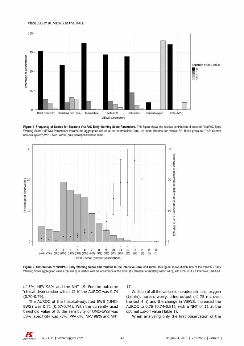
42 August 4, 2018|Volume 7|Issue 3|WJCCM|www.wjgnet.com
of 5%, NPV 98% and the NNT 19. For the outcome ‘clinical deterioration within 12 h’ the AUROC was 0.74 (0.70-0.79).
The AUROC of the hospital-adjusted EWS (UMC-EWS) was 0.71 (0.67-0.74). With the currently used threshold value of 3, the sensitivity of UMC-EWS was 58%, specificity was 73%, PPV 6%, NPV 98% and NNT
17.Addition of all the variables noradrenalin use, oxygen
(L/min), nurse’s worry, urine output (≤ 75 mL over the last 4 h) and the change in ViEWS, increased the AUROC to 0.78 (0.74-0.81), with a NNT of 11 at the optimal cut-off value (Table 1).
When analysing only the first observation of the
Perc
enta
ge o
f ob
serv
atio
ns
Heart frequency Breathing rate (bpm) Temperature
ViEWS parameters
Systolic BP Saturation Inspired oxygen CNS (AVPU)
Separate ViEWS value0
100
75
50
25
0
123
Figure 1 Frequency of Scores for Separate VitalPAC Early Warning Score Parameters. This figure shows the relative contribution of separate VitalPAC Early Warning Score (ViEWS) Parameters towards the aggregated scores at the Intermediate Care Unit. bpm: Breaths per minute; BP: Blood pressure; CNS: Central nervous system; AVPU: Alert, verbal, pain, unresponsiveness scale.
Figure 2 Distribution of VitalPAC Early Warning Score and transfer to the Intensive Care Unit rates. This figure shows distribution of the VitalPAC Early Warning Score aggregated values (bar chart) in relation with the occurrence of the event (ICU transfer or mortality within 24 h), with 95%CIs. ICU: Intensive Care Unit.
Percentage of observations followed by an event <
24 h (95%CI)
30
20
10
0
0 1 2 3 4 5 6 7 8 9 10 11 12 13 14 15 16 (448) (351) (291) (1754) (1500) (1398) (1324) (829) (538) (323) (173) (105) (52) (19) (5) (1) (2)
ViEWS score (number observations)
Perc
enta
ge o
f ob
serv
atio
ns
30
20
10
0
Plate JDJ et al . ViEWS at the IMCU

43 August 4, 2018|Volume 7|Issue 3|WJCCM|www.wjgnet.com
ViEWS per IMCU admission (n = 2113), the AUROC was 0.72 (0.65-0.79) using the optimal threshold. When including only the observations that were part of a regular measurement interval (n = 5255), the AUROC was 0.70 (0.66-0.74) for the optimal threshold.
CalibrationThe calibration of the ViEWS and the ViEWS with additional parameters (noradrenalin, nurse worry score, oxygen (continuous) and change in ViEWS) is shown in Figure 3. From this, it follows that the ViEWS underpredicts clinical deterioration for Probability < 0.05, while it overpredicts clinical deterioration for Probability
> 0.05.
DISCUSSIONThis study shows that the discriminative performance of the (Vi)EWS at the IMCU is moderate. However, its clinical relevance is debatable as its use carries the risk of alarm fatigue (NNT = 19).
In comparison, considerably higher AUROCS have been described for the ViEWS at the hospital ward. One study using the same composite outcome found an AUROC of 0.87 (0.87-0.87)[4]. At external validation studies AUROCs of 0.87 (no confidence interval) and 0.89 (0.83-0.95) for 24 h mortality were observed in, respectively, first world and lower-income countries[3,16]. In the pre-hospital setting an AUROC of 0.815 (0.730-0.990) was reported in external validation for the composite outcome of ICU transfer or mortality within 48 h[5]. A possible explanation for the lower discriminative performance of the (Vi)EWS at the IMCU is the considerably different case-mix when compared to the hospital ward: IMCU patients are already closely monitored and often receive haemodynamic and respiratory support. Hence, many patients achieve a relatively high ViEWS score, which diminishes the discriminative potential of the score. Furthermore, the supervising clinicians at the IMCU are trained and experienced in supporting the deteriorating patient, and thus deterioration (as measured by the EWS) may not directly lead to ICU transfer and mortality, as a large part of the resuscitation can be provided at the IMCU.
Subsequently, this study shows that the addition of IMCU-specific clinical parameters increases the discriminative performance (AUROC 0.78, CI: 0.74-0.81) and decreases the false alarm rate (NNT = 11). We did however not intend to develop and present a revised IMCU-specific EWS. First further external validation
Predictors AUROC Sensitivity (%) Specificity (%) PPV (%) NPV (%) NNT
ViEWS, with regular threshold (5)0.72
(0.69-0.75)79 49 4 99 23
ViEWS, with optimal threshold (6)0.72
(0.69-0.75)68 64 5 98 19
ViEWS and noradrenalin0.72
(0.69–0.75)68 64 5 98 19
ViEWS and oxygen (continuous)0.74
(0.71-0.77)61 76 7 98 14
ViEWS and nurse worry score0.73
(0.70-0.76)70 63 5 99 21
ViEWS and change in ViEWS0.74
(0.70-0.78)65 70 7 99 16
ViEWS and noradrenalin, nurse worry score, oxygen (continuous) and change in ViEWS
0.78 (0.74-0.81)
62 78 10 99 11
Table 1 Predictive Performance of the regular VitalPAC Early Warning Score and updated models at the Intermediate Care Unit
The numbers presented indicate the area under the curve (AUROC), sensitivity, specificity, positive predictive value (PPV), negative predictive value (NPV) and number needed to trigger (NNT). This is shown for the original VitalPAC early warning score model (ViEWS) with both the regularly used and optimal threshold, and for the updated models containing additional parameters, all with their optimal thresholds as determined by the Q-point method. ViEWS: VitalPac Early Warning Score; NNT: Number needed to trigger.
Figure 3 Calibration plots of the VitalPac Early Warning Score and the VitalPac Early Warning Score with additional parameters at the Intermediate Care Unit. This figure shows the calibration plot of the ViEWS (lower plot) and the ViEWS with the additional parameters noradrenalin, nurse worry score, oxygen (continuous) and change in ViEWS (upper plot). ViEWS: VitalPac Early Warning Score.
0.0
0.2
0.4
0.6
0.8
1.0
Obs
erve
d pr
obab
ility
0.0 0.2 0.4 0.6 0.8 1.0Predicted probability
Plate JDJ et al . ViEWS at the IMCU

44 August 4, 2018|Volume 7|Issue 3|WJCCM|www.wjgnet.com
would be necessary, and second, it would require collection of extra parameters, which would make the EWS less easy to use. It should be noted that the moderate performance of the EWS was not specific for the ViEWS, as the observed performance of the UMC-EWS was lower than the ViEWS (AUROC of 0.71, CI: 0.67-0.74) in the IMCU.
Clinically, we do not consider a prediction model with a NNT of 19, i.e. only 1 out of 19 triggers, does indeed indicate clinical deterioration, to be feasible. This carries a high risk of alarm fatigue and therefore has the potential to compromise patient safety while increasing the (administrative) burden at the IMCU. In its current design, we therefore believe that it should not be used. We would regard a NNT of 4 or less as ideal (with a sensitivity of ≥ 50%), as this would indicate that at least 25% of the alarms are justified. It should be noted that the addition of the other IMCU-specific clinical parameters increased the amount of data to be collected, still resulting in a higher NNT; therefore, more detailed modelling, likely connected with automated data collection to reduce administrative burden, would be necessary to achieve this goal. Furthermore, as these additional IMCU-specific clinical parameters are used to fit a model in our own cohort, there is a risk of overfitting and thus the real performance of this updated model is likely even less.
We recognize that this clinical relevance highly depends on the organisation of the IMCU and, in particular, on the extent to which the IMCU is inde-pendent. IMCUs that are an extension of the hospital ward perhaps do benefit from the ViEWS, whereas independent (stand-alone) IMCUs with extensive monitoring and supportive options probably do not. The clinical relevance of the ViEWS at the IMCU also depends on the hospital organization, as “transfer to the ICU” is a process measure which is potentially susceptible to logistical factors (the occupancy rates at both units). Although the in-hospital mortality rate is preferably an outcome measure[1], its use as single outcome parameter was not considered feasible due to its rare occurrence (0.30%) and susceptibility to treatment restrictions. Additionally, as necessity for ICU transfer often is due to mechanical ventilation[10], its use as an outcome measure for deterioration at IMCUs can likely be generalized.
This study has multiple strengths. First, it is the first to investigate the validity and clinical relevance of the ViEWS at the IMCU. Second, it analyses a large dataset, using a standardized data collection protocol in which the EWS is consistently collected on a regular basis.
Its limitations are that physicians have likely acted upon EWS values to prevent further deterioration. Although we have partly taken this into account by using ICU transfer as an outcome, we did not include other treatment changes, such as increasing the amount of noradrenalin. Therefore, we were unable to assess the relevance of the ViEWS for the prediction of these
treatment changes. Another limitation is, as indicated before, that we cannot generalize our findings to IMCUs with a case-mix which is more comparable to the hospital ward, in particular as our IMCU may be unique in its possibility to administer vasopressors.
Further research to detect the deteriorating patient at the IMCU should probably not solely focus on cross-sectional measurements of vital signs (as is done in the EWS). Rather, focus should shift towards using (automated) repeated measurements of these vital signs to adequately and timely detect the deteriorating patient at the IMCU. By optimal support through ICT and electronic medical record systems, this preferably results in a real-time trend analysis and score visualization with an easy read-out of clinical deterioration and adjustable thresholds per patient. Especially in an environment with constant monitoring, this could possibly lead to earlier interventions and improved patient outcomes.
The ViEWS at the IMCU has a discriminative per-formance that is considerably lower than at the hospital ward. As the number of false alarms is high, it carries the risk of alarm fatigue. Therefore, its application in this setting should be reconsidered. As the addition of IMCU-specific clinical predictors does not sufficiently improve the performance, further research should focus on more complex models incorporating repeated measurements of vital functions.
ARTICLE HIGHLIGHTSResearch backgroundEarly Warning Scoring (EWS) systems to recognize the clinically deteriorating patient are widely used in the clinical setting, including in Intermediate Care Units (IMCUs). However, they have been developed and validated for the general hospital ward population and hence their applicability within the IMCU population is unclear.
Research motivationThe application of prediction models (EWS) at a different setting than the setting at which they were developed (IMCU instead of hospital ward), could lead to an inefficient use of scarce resources and may compromise patient safety. To justly consider the (ongoing) use of the EWS at the IMCU, its discriminative performance and applicability need to be investigated.
Research objectivesThis validation study aims to assess the performance and clinical relevance of the VitalPAC-EWS (ViEWS) at the IMCU. Further, it aims to improve the EWS for its use at the IMCU.
Research methodsElectronically collected data from 2014 to 2016 at the IMCU were used to obtain the area under the receiver operating curve (AUC) and the number needed to trigger (false alarm rate) at the current and the optimal threshold.
Research results The AUC of the ViEWS was 0.72 (CI: 0.69-0.75). The number needed to trigger was 19 per one event. Although the discriminative performance is acceptable, the clinical relevance is limited as 19 false alarms are needed per one event. This carries the risk of alarm fatigue. Therefore, this study contributes to this research field that the use of the EWS at the stand-alone IMCU should be reconsidered. The main problem that remains to be solved are that an
ARTICLE HIGHLIGHTS
Plate JDJ et al . ViEWS at the IMCU

45 August 4, 2018|Volume 7|Issue 3|WJCCM|www.wjgnet.com
alternative system needs to be developed to timely detect clinical deterioration at the IMCU.
Research conclusionsThe new findings of this study are that the use of the ViEWS at the IMCU should be reconsidered. It proposes that this is due to remarkable case-mix differences between the hospital ward and the IMCU. This study proposes to use new methods to detect clinical deterioration at the IMCU, using automated data collection and perhaps more sophisticated statistical methods. The implication for clinical practice is that the EWS in its current form at the IMCU should perhaps not be used.
Research perspectivesGeneral experiences and lessons that can be learned from this study are that prediction models should not be used in different settings without prior validation. Further research should focus on alternative methods to detect the clinically deteriorating patient at the IMCU, through the modelling of repeated measurements in prediction models. Also, further research should focus on the use of the EWS in differently formatted IMCUs, such as the IMCU that is integrated into the ICU.
ACKNOWLEDGEMENTS We would like to thank Melvin van Maurik for his efforts in extracting the required data.
REFERENCES1 Prytherch DR, Smith GB, Schmidt PE, Featherstone PI. ViEWS--
Towards a national early warning score for detecting adult inpatient deterioration. Resuscitation 2010; 81: 932-937 [PMID: 20637974 DOI: 10.1016/j.resuscitation.2010.04.014]
2 Beckett CD, Kipnis G. Collaborative communication: inte-grating SBAR to improve quality/patient safety outcomes. J Healthc Qual 2009; 31: 19-28 [PMID: 19813557 DOI: 10.1111/j.1945-1474.2009.00043.x]
3 Opio MO, Nansubuga G, Kellett J. Validation of the VitalPAC™ Early Warning Score (ViEWS) in acutely ill medical patients attending a resource-poor hospital in sub-Saharan Africa. Resuscitation 2013; 84: 743-746 [PMID: 23438452 DOI: 10.1016/j.resuscitation.2013.02.007]
4 Smith GB, Prytherch DR, Meredith P, Schmidt PE, Featherstone PI. The ability of the National Early Warning Score (NEWS) to discriminate patients at risk of early cardiac arrest, unanticipated intensive care unit admission, and death. Resuscitation 2013; 84:
465-470 [PMID: 23295778 DOI: 10.1016/j.resuscitation.2012.12.016]
5 Silcock DJ, Corfield AR, Gowens PA, Rooney KD. Validation of the National Early Warning Score in the prehospital setting. Resuscitation 2015; 89: 31-35 [PMID: 25583148 DOI: 10.1016/j.resuscitation.2014.12.029]
6 Williams TA, Tohira H, Finn J, Perkins GD, Ho KM. The ability of early warning scores (EWS) to detect critical illness in the prehospital setting: A systematic review. Resuscitation 2016; 102: 35-43 [PMID: 26905389 DOI: 10.1016/j.resuscitation.2016.02.011]
7 Royston P, Moons KG, Altman DG, Vergouwe Y. Prognosis and prognostic research: Developing a prognostic model. BMJ 2009; 338: b604 [PMID: 19336487 DOI: 10.1136/bmj.b604]
8 Veiligheids Agenda. Vroege herkenning en behandeling van de vitaal bedreigde patiënt. 2008. Available from: URL: https://www.vmszorg.nl/vms-veiligheidsprogramma/10-themas/vroege-herkenning-en-behandeling-vitaal-bedreigde-patient/
9 Royal College of Physicians. National Early Warning Score (NEWS) 2. Available from: URL: https://www.rcplondon.ac.uk/projects/outputs/national-early-warning-score-news-2
10 Plate JDJ, Leenen LPH, Houwert M, Hietbrink F. Utilisation of Intermediate Care Units: A Systematic Review. Crit Care Res Pract 2017; 2017: 8038460 [PMID: 28775898 DOI: 10.1155/2017/8038460]
11 Sjoding MW, Valley TS, Prescott HC, Wunsch H, Iwashyna TJ, Cooke CR. Rising Billing for Intermediate Intensive Care among Hospitalized Medicare Beneficiaries between 1996 and 2010. Am J Respir Crit Care Med 2016; 193: 163-170 [PMID: 26372779 DOI: 10.1164/rccm.201506-1252OC]
12 Zhang Z. Univariate description and bivariate statistical inference: the first step delving into data. Ann Transl Med 2016; 4: 91 [PMID: 27047950 DOI: 10.21037/atm.2016.02.11]
13 Johnson KR, Hagadorn JI, Sink DW. Alarm Safety and Alarm Fatigue. Clin Perinatol 2017; 44: 713-728 [PMID: 28802348 DOI: 10.1016/j.clp.2017.05.005]
14 R Core Team. The R Project for Statistical Computing. Available from: URL: https://www.r-project.org/
15 Moons KG, Altman DG, Reitsma JB, Ioannidis JP, Macaskill P, Steyerberg EW, Vickers AJ, Ransohoff DF, Collins GS. Transparent Reporting of a multivariable prediction model for Individual Prognosis or Diagnosis (TRIPOD): explanation and elaboration. Ann Intern Med 2015; 162: W1-73 [PMID: 25560730 DOI: 10.7326/M14-0698]
16 Bleyer AJ, Vidya S, Russell GB, Jones CM, Sujata L, Daeihagh P, Hire D. Longitudinal analysis of one million vital signs in patients in an academic medical center. Resuscitation 2011; 82: 1387-1392 [PMID: 21756971 DOI: 10.1016/j.resuscitation.2011.06.033]
P- Reviewer: Surani S, Zhang Z S- Editor: Ji FF L- Editor: Filipodia E- Editor: Tan WW
Plate JDJ et al . ViEWS at the IMCU

© 2018 Baishideng Publishing Group Inc. All rights reserved.
Published by Baishideng Publishing Group Inc7901 Stoneridge Drive, Suite 501, Pleasanton, CA 94588, USA
Telephone: +1-925-223-8242Fax: +1-925-223-8243
E-mail: [email protected] Desk: http://www.f6publishing.com/helpdesk
http://www.wjgnet.com

World Journal of Critical Care MedicineWorld J Crit Care Med 2018 September 7; 7(4): 46-51
Published by Baishideng Publishing Group Inc
ISSN 2220-3141 (online)

World Journal ofCritical Care MedicineW J C C M
Contents
IWJCCM|www.wjgnet.com
Volume 7 Number 4 September 7, 2018
ORIGINAL ARTICLE
Retrospective Cohort Study46 Clinicalcharacteristicsandoutcomesassociatedwithnasal intermittentmandatoryventilation inacute
pediatricrespiratoryfailure
Wang BC, Pei T, Lin CB, Guo R, Elashoff D, Lin JA, Pineda CREVIEW
September 7, 2018|Volume 7|Issue 4|

ContentsWorld Journal of Critical Care Medicine
Volume 7 Number 4 September 7, 2018
EDITORS FOR THIS ISSUE
Responsible Assistant Editor: Xiang Li Responsible Science Editor: Fang-Fang JiResponsible Electronic Editor: Yun-Xiao Jian Wu Proofing Editorial Office Director: Jin-Lei WangProofing Editor-in-Chief: Lian-Sheng Ma
Fax: +1-925-2238243E-mail: [email protected] Desk: http://www.f6publishing.com/helpdeskhttp://www.wjgnet.com
PUBLISHERBaishideng Publishing Group Inc7901 Stoneridge Drive, Suite 501, Pleasanton, CA 94588, USATelephone: +1-925-223-8242Fax: +1-925-223-8243E-mail: [email protected] Desk: http://www.f6publishing.com/helpdeskhttp://www.wjgnet.com
PUBLICATIONDATESeptember 7, 2018
COPYRIGHT© 2018 Baishideng Publishing Group Inc. Articles
published by this Open-Access journal are distributed under the terms of the Creative Commons Attribution Non-commercial License, which permits use, distribution, and reproduction in any medium, provided the original work is properly cited, the use is non commercial and is otherwise in compliance with the license.
SPECIALSTATEMENTAll articles published in journals owned by the Baishi-deng Publishing Group (BPG) represent the views and opinions of their authors, and not the views, opinions or policies of the BPG, except where otherwise expli-citly indicated.
INSTRUCTIONSTOAUTHORShttp://www.wjgnet.com/bpg/gerinfo/204
ONLINESUBMISSIONhttp://www.f6publishing.com
IIWJCCM|www.wjgnet.com
ABOUT COVER
AIM AND SCOPE
INDExING/ABSTRACTING
September 7, 2018|Volume 7|Issue 4|
NAMEOFJOURNALWorld Journal of Critical Care Medicine
ISSNISSN 2220-3141 (online)
LAUNCHDATEFebruary 4, 2012
EDITORIALBOARDMEMBERSAll editorial board members resources online at http://www.wjgnet.com/2220-3141/editorialboard.htm
EDITORIALOFFICEJin-Lei Wang, DirectorWorld Journal of Critical Care MedicineBaishideng Publishing Group Inc7901 Stoneridge Drive, Suite 501, Pleasanton, CA 94588, USATelephone: +1-925-2238242
EditorialBoardMemberofWorldJournalofCriticalCareMedicine ,RameshNatarajan,PhD,Professor,DivisionofPulmonaryDiseaseandCriticalCareMedicine,DepartmentofInternalMedicine,MedicalCollegeofVirginiaCampus,VirginiaCommonwealthUniversity,Richmond,VA23298-0050,UnitedStates
World Journal of Critical Care Medicine (World J Crit Care Med, WJCCM, online ISSN 2220-3141, DOI: 10.5492) is a peer-reviewed open access academic journal that aims to guide clinical practice and improve diagnostic and therapeutic skills of clinicians.
WJCCM covers topics concerning severe infection, shock and multiple organ dysfunc-tion syndrome, infection and anti-infection treatment, acute respiratory distress syndrome and mechanical ventilation, acute kidney failure, continuous renal replacement therapy, rational nutrition and immunomodulation in critically ill patients, sedation and analgesia, cardiopulmonary cerebral resuscitation, fluid resuscitation and tissue perfusion, coagulant dysfunction, hemodynamic monitoring and circulatory support, ICU management and treatment control, and application of bronchofiberscopy in critically ill patients.
We encourage authors to submit their manuscripts to WJCCM. We will give priority to manuscripts that are supported by major national and international foundations and those that are of great clinical significance.
World Journal of Critical Care Medicine (WJCCM) is now indexed in PubMed, PubMed Cen-
tral, China National Knowledge Infrastructure (CNKI), and Superstar Journals Database.

Billy C Wang, Theodore Pei, Cheryl B Lin, Rong Guo, David Elashoff, James A Lin, Carol Pineda
ORIGINAL ARTICLE
46 September 7, 2018|Volume 7|Issue 4|WJCCM|www.wjgnet.com
Clinical characteristics and outcomes associated with nasal intermittent mandatory ventilation in acute pediatric respiratory failure
Billy C Wang, Department of Pediatrics, Division of Critical Care Medicine, Loma Linda University Children’s Hospital, Loma Linda, CA 92354, United States
Theodore Pei, Cheryl B Lin, Carol Pineda, Department of Pediatrics, Division of Pediatric Critical Care, Floating Hospital for Children at Tufts, Boston, MA 02111, United States
Rong Guo, David Elashoff, Department of Medicine, Biostatistics Core, UCLA David Geffen School of Medicine, Los Angeles, CA 90024, United States
James A Lin, Department of Pediatrics, Mattel Children’s Hospital at UCLA, Los Angeles, CA 90095, United States
ORCID numbers: Billy C Wang (0000-0002-0733-0892); Theodore Pei (0000-0001-7980-1318); Cheryl B Lin (0000-0001-8516-1660); Rong Guo (0000-0001-7518-6426) ; David Elashoff (0000-0002-5865-8580); James A Lin (0000-0001-5818-156X); Carol Pineda (0000-0002-2934-1488).
Author contributions: Wang BC, Pei T, Pineda C and Lin JA designed the study, collected data, and participated in writing and revision of the manuscript; Lin CB collected data and reviewed the manuscript; Guo R and Elashoff D provided statistical analysis and reviewed the manuscript
supported by NIH National Center for Advancing Translational Science, No. UL1TR001881.
Institutional review board statement: This study was approved by the UCLA Institutional Review Board.
Conflict-of-interest statement: All authors have no conflicts of interest to report.
sTROBE statement: The STROBE Statement have been adopted.
Open-Access: This article is an open-access article which was selected by an in-house editor and fully peer-reviewed by external reviewers. It is distributed in accordance with the Creative Commons Attribution Non Commercial (CC BY-NC 4.0) license, which
permits others to distribute, remix, adapt, build upon this work non-commercially, and license their derivative works on different terms, provided the original work is properly cited and the use is non-commercial. See: http://creativecommons.org/licenses/by-nc/4.0/
Manuscript source: Invited manuscript
Correspondence to: Billy C Wang, MD, Fellow, Department of Pediatrics, Division of Critical Care Medicine, Loma Linda University Children’s Hospital, 11234 Anderson Street Room CH5763, Loma Linda, CA 92354, United States. [email protected]: +1-909-5584250Fax: +1-909-5580303
Received: June 2, 2018 Peer-review started: June 2, 2018First decision: July 9, 2018Revised: July 25, 2018Accepted: August 4, 2018Article in press: August 5, 2018Published online: September 7, 2018
AbstractAIMTo characterize the clinical course and outcomes of nasal intermittent mandatory ventilation (NIMV) use in acute pediatric respiratory failure.
METHODSWe identified all patients treated with NIMV in the pediatric intensive care unit (PICU) or inpatient general pediatrics between January 2013 and December 2015 at two academic centers. Patients who utilized NIMV with other modes of noninvasive ventilation during the same admission were included. Data included demographics, vital signs on admission and prior to initiation of NIMV, pediatric risk of mortality Ⅲ (PRIsM-Ⅲ) scores,
World Journal ofCritical Care MedicineW J C C M
Submit a Manuscript: http://www.f6publishing.com
DOI: 10.5492/wjccm.v7.i4.46
World J Crit Care Med 2018 September 7; 7(4): 46-51
ISSN 2220-3141 (online)
Retrospective Cohort Study

complications, respiratory support characteristics, PICU and hospital length of stays, duration of respiratory support, and complications. Patients who did not require escalation to mechanical ventilation were defined as NIMV responders; those who required escalation to mechanical ventilation (MV) were defined as NIMV non-responders. NIMV responders were compared to NIMV non-responders.
RESULTS Forty-two patients met study criteria. six (14%) failed treatment and required MV. The majority of the patients (74%) had a primary diagnosis of bronchiolitis. The median age of these 42 patients was 4 mo (range 0.5-28.1 mo, IQR 7, P = 0.69). No significant difference was measured in other baseline demographics and vitals on initiation of NIMV; these included age, temperature, respiratory rate, O2 saturation, heart rate, systolic blood pressure, diastolic blood pressure, and PRIsM-Ⅲ scores. The duration of NIMV was shorter in the NIMV non-responder vs NIMV responder group (6.5 h vs 65 h, P < 0.0005). Otherwise, NIMV failure was not associated with significant differences in PICU length of stay (LOs), hospital LOs, or total duration of respiratory support. No patients had aspiration pneumonia, pneumothorax, or skin breakdown.
CONCLUSIONMost of our patients responded to NIMV. NIMV failure is not associated with differences in hospital LOs, PICU LOs, or duration of respiratory support.
Key words: Continuous positive airway pressure; Pediatric; Noninvasive positive pressure ventilation; Nasal intermittent mandatory ventilation; High flow nasal cannula; Acute respiratory failure; Bilevel positive airway pressure
© The Author(s) 2018. Published by Baishideng Publishing Group Inc. All rights reserved.
Core tip: In our cohort of patients between 0.5 and 28.1 mo of age with acute respiratory failure, the majority of patients were successfully supported with nasal intermittent mandatory ventilation (NIMV) alone or NIMV in conjunction with other modes of noninvasive ventilation (NIV). Use of NIMV with or without NIV was not associated with significant differences in hospital length of stay (LOs), pediatric intensive care unit LOs, or duration of respiratory support. Failure of NIMV with or without NIV was recognized in a median of 6.5 h.
Wang BC, Pei T, Lin CB, Guo R, Elashoff D, Lin JA, Pineda C. Clinical characteristics and outcomes associated with nasal intermittent mandatory ventilation in acute pediatric respiratory failure. World J Crit Care Med 2018; 7(4): 46-51 Available from: URL: http://www.wjgnet.com/2220-3141/full/v7/i4/46.htm DOI: http://dx.doi.org/10.5492/wjccm.v7.i4.46
INTRODUCTIONAcute respiratory failure accounts for 46% to 59% of
47WJCCM|www.wjgnet.com
Wang BC et al . Pediatric NIMV case series
unplanned admissions to the pediatric intensive care unit (PICU) with 68% of these patients requiring advanced respiratory support[1-3]. While endotracheal intubation with mechanical ventilation (MV) is the classic management of respiratory failure, noninvasive ventilation (NIV) is rapidly gaining acceptance as a first line intervention. Continuous positive airway pressure (CPAP), bilevel positive airway pressure (BIPAP), high flow nasal cannula (HFNC), and nasal intermittent mandatory ventilation (NIMV) are examples of NIV.
NIMV is a time-cycled, time-triggered pressure control mode of non-invasive ventilation typically administered through nasal prongs or nasal mask via mechanical ventilator. Unlike BIPAP or endotracheal mechanical ventilation, mandatory breaths on NIMV are often not synchronized to patient breaths, though newer ventilators may synchronize to diaphragmatic stimulation (e.g., Noninvasive neurally adjusted ventilatory assist) or when a negative inspiratory pressure threshold is reached.
NIV modalities have been shown to prevent intubation and reintubation in the adult and neonatal population[4,5]. Relatively few studies have assessed its efficacy in the pediatric critical care setting[6-10]. NIMV in particular is infrequently used outside of the neonatal intensive care unit (NICU) and, to our knowledge, has not been studied in the setting of acute respiratory failure in the PICU. In the absence of data from robust studies, we reviewed our experience with NIMV in critically ill pediatric patients and describe the clinical characteristics and outcomes of pediatric patients with acute respiratory failure who were treated with NIMV.
MaTeRIals aND MeThODsInclusion and exclusion criteriaWe performed a retrospective chart review of children between 1 d to 28 mo of age in acute respiratory failure admitted to the PICU or intermediate-level pediatric unit of two academic medical centers between January 2013 and December 2015. Institutional review board approval was obtained at each site.
Patients were identified through ICD codes associated with acute respiratory failure and CPT codes for endotracheal intubation or NIV. NIV modalities reviewed include NIMV, HFNC, CPAP, and BIPAP. All patients treated with NIMV were individually reviewed. Due to the paucity of patients utilizing NIMV alone, patients treated with other modes of NIV in series with NIMV during a single admission were included. Exclusion criteria were patients who did not utilize NIMV during hospitalization, managed in the NICU, chronic CPAP or BIPAP dependence, tracheostomy dependence, and post-extubation NIV.
MaterialsNIV and NIMV settings were initiated and titrated at clinician discretion based on the patient’s clinical response. NIMV was administered via RAM cannula (Neotech Products, Valencia, CA) nasal prongs sized according to the child’s age and weight and connected to a humidified
September 7, 2018|Volume 7|Issue 4|
48WJCCM|www.wjgnet.com
Avea ventilator (CareFusion, Franklin Lakes, NJ) in NIMV mode. This mode of ventilation was asynchronous to patient breaths at both our institutions. The decision to escalate to invasive mechanical ventilation was made at the discretion of the physician.
Data Data included (1) demographics--age in days, gender, admission, discharge weight and discharge diagnosis; (2) vital signs on initiation of and with any changes in NIV; (3) characteristics of respiratory support--modality, length of time on each modality, maximum settings (delta P, FIO2, PEEP, and mandatory rate); (4) complications--aspiration pneumonia, pneumothorax, skin breakdown; and (5) outcome data-MV, mortality, pediatric risk of mortality Ⅲ(PRISM-Ⅲ) scores, and hospital and PICU length of stay. Respiratory support characteristics were recorded hourly in PICU site A and every 4 h in the pediatric units of site A and PICU site B.
Statistical analysisSuccessful NIMV treatment was defined as use of NIMV without the use of invasive mechanical ventilation. Patients successfully treated with NIMV (NIMV respon-ders) were compared to those unsuccessfully treated with NIMV, which we defined as escalation to MV (NIMV non-responders).
Standard descriptive statistics were reported. Medians (min-max, IQR) were analyzed for numerical variables
and frequency count (%) for categorical variables. Fisher’s Exact tests were used for the comparison of categorical characteristics and Wilcoxon rank sum tests were used for the comparison of numerical characteristics. P values less than 0.05 were considered statistically significant. All statistical analysis was performed by a biomedical statistician who utilized SAS v9.4 (SAS Institute Inc., Cary, NC).
ResUlTsDuring our study period, 1164 charts were initially reviewed and 1122 were removed based on exclusion criteria. Forty-two patients used NIMV during their hospital admission. Figures 1 and 2 illustrate the selection process and the sequence of escalation of respiratory support, respectively.
Comparison of NIMV responders and NIMV non-respondersIn our 42 patients who received NIMV, 36 (86%) were successfully supported without further escalation, while 6 (14%) subsequently required endotracheal intubation and MV after trial of NIMV with or without other modes of NIV. Eight (19%) of the 42 patients used NIMV exclusively; of these 8 patients, 4 (50%) failed and required mechanical ventilation. Except for the patients requiring MV, NIMV was used as the final mode of noninvasive respiratory support in patients treated with more than one NIV modality (Figure 2).
The median age of these 42 patients was 4 mo (range 0.5-28.1 mo, IQR 7). Half of the patients were males. The leading discharge diagnosis was bronchiolitis (n = 31, 74%). Demographics (weight, age, gender), PRISM-III scores on admission, and vitals prior to initiation of NIMV (heart rate, respiratory rate, blood pressure, oxygen saturation, temperature) between these two subgroups were similar (Table 1). The distribution of diagnoses was similar between NIMV responders and NIMV non-responders (Table 2).
We observed no significant difference in maximum NIMV settings (delta P, FIO2, PEEP, and rate), time to escalation to maximum settings, hospital and PICU length of stay, or in total duration of all respiratory support between NIMV responders and non-responders, or those that required MV (Table 3). However, NIMV responders remained on this mode of support for a greater length of time than those who failed (65 h vs 6.5 h, P < 0.0005).
DIsCUssIONIn our cohort of pediatric patients with acute respiratory failure treated with NIMV with or without other NIV modalities, 86% did not require MV. This rate is similar to data on heterogenous modes of NIV modalities in the PICU described in separate studies conducted by Yaman et al[7], Milési et al[8], and Wolfler et al[11] in the PICU. To our knowledge this is the first study that characterizes the patients, pathologies, and clinical outcomes of NIMV for
Total assessedn = 1164
site An = 492
site Bn = 672
Met exclusion criterian = 464
Met exclusion criterian = 658
NIMVn = 28
NIMVn = 14
Figure 1 Patient selection by institution.NIMV: Nasal intermittent mandatory ventilation.
36 NIMV responders
6 NIMV non-responders
HFNC -> BIPAP -> NIMV (1)HFNC -> CPAP -> NIMV (7)HFNC -> NIMV (21)CPAP -> NIMV (3)NIMV (4)
HFNC -> CPAP -> NIMV -> MV (2)NIMV -> MV (4)
Figure 2 Escalation of respiratory support modalities1. 1Progression of respiratory support indicated by arrows and number of patients (in parentheses) following each pathway. HFNC: High flow nasal cannula; BIPAP: Bilevel positive airway pressure; NIMV: Nasal intermittent mandatory ventilation; CPAP: Continuous positive airway pressure; MV: Mechanical ventilation.
September 7, 2018|Volume 7|Issue 4|
Wang BC et al . Pediatric NIMV case series

49WJCCM|www.wjgnet.com
acute respiratory failure in pediatric patients outside of the NICU. The strongest evidence for NIMV in the pediatric population to date are limited to pathologies encountered in the NICU; for example, it shows significant clinical benefit over other modes of NIV in neonatal respiratory distress syndrome, apnea of prematurity, and the prevention of post-extubation failure[4,12,13]. In discordance with the NICU literature, a recent prospective study of NIV for post-extubation support in the PICU showed no difference in respiratory effort when compared between NIMV, HFNC, and CPAP, though NIMV in this study was synchronized to approximately 50%[14].
An ongoing concern of NIV is delay in endotracheal intubation and MV that may lead to worsening physiologic status at time of intubation and thus worse clinical
outcomes[15]. Our data do not support this hypothesis. We revealed substantially less time on NIMV in the cohort that proceeded to MV compared to those that responded successfully to NIMV. Our observed median time to intubation of 6.5 h in NIMV nonresponders was similar to treatment failure observed by another study utilizing mask BIPAP in adults[11]. Additionally, similarities in clinical outcomes of hospital length of stay, PICU length of stay, and complications suggest that recovery time may be independent of the mode of respiratory support. There was a single mortality due to an uncorrectable congenital lung pathology.
MV is associated with a number of challenges and complications such as sedation, paralysis, polyneuro-pathies, iatrogenic pneumonia, chemical pneumonitis,
Table 1 Comparison of nasal intermittent mandatory ventilation responders to nasal intermittent mandatory ventilation non-responders - baseline characteristics1
Baseline characteristic NIMV responders (n = 36) NIMV non-responders (n = 6) Total (n = 42) P value
Age (mo) 4 (0.5-20, IQR = 6.4) 5.2 (0.5-28.1, IQR = 12.6) 4 (0.5-28.1, IQR = 7) 0.69Admission weight (kg) 4.9 (2.3-12, IQR = 4.3) 7.1 (2.6-10.9, IQR = 4.8) 5.1 (2.3-12, IQR = 4.4) 0.39T Max in Celsius (range) 38 (36.6-40, IQR = 1.6) 38.9 (37.4-39.7, IQR = 1.7) 38.1 (36.6-40, IQR = 1.6) 0.2Respiratory rate (in breaths per minute) 60 (24-97, IQR = 19) 56 (42-77, IQR = 32) 60 (24-97, IQR = 22) 0.76O2 saturation (%) 98 (68-100, IQR = 5.5) 99 (98-100, IQR = 2) 98.5 (68-100, IQR = 5) 0.31Heart rate (beats per minute) 152.5 (95-205, IQR = 34.5) 158.5 (127-210, IQR = 24) 153.5 (95-210, IQR = 33) 0.99Systolic blood pressure (mmHg) 100 (71-129, IQR = 21) 112 (81-120, IQR = 25) 103 (71-129, IQR = 20) 0.3Diastolic blood pressure (mmHg) 51 (37-84, IQR = 20) 59.5 (43-81, IQR = 25) 51 (37-84, IQR = 22) 0.41PRISM-Ⅲ score 0 (0-11, IQR = 0) 1 (0-7, IQR = 3) 0 (0-11, IQR = 0) 0.08
1Vital signs were the last recorded values before initiation of NIMV. Values are presented as median (range, IQR). NIMV: Nasal intermittent mandatory ventilation; PRISM-Ⅲ: Pediatric risk of mortality Ⅲ.
Table 2 Diagnoses causing acute respiratory failure in nasal intermittent mandatory ventilation responders vs nasal intermittent mandatory ventilation non-responders1
Diagnosis NIMV responders n (%) NIMV non-responders n (%) Total (%) P value
Asthma exacerbation 1 (3) 2 (33) 3 (7) 0.11Bronchiolitis 28 (78) 3 (50) 31 (74)Heart failure 1 (3) 0 (0) 1 (2)Pneumonia 3 (8) 1 (17) 4 (10)Viral syndrome 3 (8) 0 (0) 3 (7)
1Values expressed as number (percent). NIMV: Nasal intermittent mandatory ventilation.
Table 3 Comparison of nasal intermittent mandatory ventilation responders to nasal intermittent mandatory ventilation non-responders - maximum support and clinical outcomes1
Support or outcome variable NIMV responders (n = 36) NIMV non-responders (n = 6) Total (n = 42) P value
Max delta P of NIMV (cmH2O) 16 (6-26, IQR = 6.5) 17.5 (10-30, IQR = 6) 16.5 (6-30, IQR = 6) 0.68Max FIO2 of NIMV (%) 40 (25-100, IQR = 22.5) 42.5 (30-70, IQR = 10) 40 (25-100, IQR = 15) 0.84Max PEEP of NIMV (cmH2O) 6 (5-8, IQR = 2) 5 (5-8, IQR = 2) 6 (5-8, IQR = 2) 0.37Max rate of NIMV (breaths per minute) 30 (20-70, IQR = 3.5) 30 (20-60, IQR = 5) 30 (20-70, IQR = 2) 0.32Time to max FIO2 (h) 0 (0-119, IQR = 1.5) 0 (0-29.5, IQR = 6) 0 (0-119, IQR = 2) 0.75Time to max setting (h) 1 (0-48, IQR = 9) 3 (0-29.5, IQR = 20) 1 (0-48, IQR = 10) 0.8Hospital length of stay (d) 7 (3-30, IQR = 3.5) 9 (4-14, IQR = 7) 7.5 (3-30, IQR = 4) 0.73PICU length of stay (d) 6 (3-30, IQR = 3) 6.5 (3-14, IQR = 5) 6 (3-30, IQR = 3) 0.64Total duration of NIMV (h) 65 (5-240, IQR = 47.5) 6.5 (0.5-30, IQR = 24) 59.5 (0.5-240, IQR = 53) 0.001Total duration of all respiratory support (h) 94.5 (28-254, IQR = 60) 115 (65-230, IQR = 31.5) 95.8 (28-254, IQR = 57) 0.3
1Values are presented as median (range, IQR). NIMV: Nasal intermittent mandatory ventilation; PICU: Pediatric intensive care unit.
September 7, 2018|Volume 7|Issue 4|
Wang BC et al . Pediatric NIMV case series
50WJCCM|www.wjgnet.com
soft tissue trauma, pneumothorax, and other lung injuries[16]. Unlike MV, NIV modalities have three to fivefold fewer rates of these complications, particularly ventilator associated pneumonia and barotrauma[17,18]. Non-invasive ventilation also reduces complications such as mortality and nosocomial infections[7,17,19]. None of our patients had aspiration pneumonia, pneumothorax, barotrauma, or soft tissue injury associated with NIMV use. Additionally, only one patient required mild sedation with an oral benzodiazepine during NIMV support.
The clinical application of NIMV or any NIV is in the hope of avoiding MV. The general practice in our two divisions is to apply NIMV as a last-resort modality prior to MV in young infants. We confirmed this practice pattern in our observation that NIMV, when applied, was used as the final means of noninvasive respiratory support (Figure 2).
Our study is limited in that this was a non-randomized, non-protocolized, retrospective review of chart data with a limited sample size, thus rendering the power of our study low. Limitations in NIMV experience in our PICUs precluded protocols for its application. There is no head-to-head randomization and comparison between other NIV modalities. Treatment with several non-invasive modalities limits the ability to extrapolate the contribution of each mode to successful support or failure. This aspect also limits our data on NIMV alone. Lastly, the criteria to initiate NIV, modify mode of NIV, and decision to intubate was based on clinician judgment and not protocolized.
Despite these limitations, our study provides the first retrospective analysis of outcomes associated with NIMV use in pediatric acute respiratory failure at two academic institutions that are widely disparate geographically. Future goals include verification of this data with a larger cohort and protocolized escalation of respiratory support. Larger and multicenter prospective studies may identify useful clinical parameters that may assist in the identification of patients who may benefit from NIMV. Future goals may include randomization of patients to NIMV alone vs other modes of NIV.
NIMV successfully supported 86% of pediatric patients with acute respiratory failure. The remaining patients who failed NIMV did not have a longer PICU, hospital LOS, or total duration of respiratory support when compared to those successfully supported with NIMV. NIMV failure was recognized within a median of 6.5 h, therefore the use of NIMV did not delay escalation to endotracheal intubation
aRTICle hIghlIghTsResearch backgroundNasal intermittent mandatory ventilation (NIMV) is a mode of noninvasive ventilation (NIV) seldomly utilized outside of the neonatal intensive care unit (NICU). To our knowledge NIMV has not been studied in the pediatric intensive care unit (PICU) population.
Research motivationAcute respiratory failure requiring advanced respiratory support accounts for a large proportion of PICU admissions. NIV is rapidly gaining acceptance as the first mode of oxygenation and ventilatory support for many of these patients.
The potential use of NIMV adds to the arsenal of respiratory support strategies. Its success could obviate the need for mechanical ventilation in some patients.
Research objectiveOur primary objectives were to review our experience with NIMV-both alone and in conjunction with other modes of NIV-and describe our patient outcome data and compare with existing literature. In particular our interests were intubation rate, PICU length of stay, hospital length of stay, duration of respiratory support, and complications.
Research methods During our study period, we identified all patients who utilized NIMV with or without other modes of NIV at two academic institutions. We excluded patients in the NICU, those dependent on chronic continuous positive airway pressure (CPAP) or bilevel positive airway pressure or tracheostomy, and post-extubation NIV. Data included demographics, vitals, characteristics of respiratory support, diagnoses, complications, and outcome data. Patients who did not require escalation to mechanical ventilation (MV) were defined as NIMV responders; those who required escalation to MV were defined as NIMV non-responders. NIMV responders were compared to NIMV non-responders. Standard descriptive statistics are used. All statistical analyses were run by a certified biostatistician using SAS v9.4.
Research results We identified 42 patients during our three-year study period. Median age of these patients was 4 mo. The majority of patients had a primary diagnosis of bronchiolitis. Six failed NIMV. Baseline demographics, vitals, diagnoses, and pediatric risk of mortality III scores were similar between NIMV responders and NIMV non-responders. However, NIMV non-responders were on this mode of ventilation for a significantly shorter period of time. Outcome data including hospital length of stay, PICU length of stay, and duration of respiratory support were similar between the two groups. No patients had aspiration pneumonia, pneumothorax, or skin breakdown associated with NIMV. There was a single mortality due to an uncorrectable and fatal lung pathology.
Research conclusions NIMV was utilized in pediatric patients with acute respiratory failure and successfully supported the majority of our patients. Failure of NIMV was quickly identified in a median of 6.5 h. Patients who required intubation did not have a longer PICU length of stay, hospital length of stay, or total duration of respiratory support when compared to those successfully supported with NIMV.
Research perspectivesBased on our data, NIMV appears to be a promising mode of noninvasive respiratory support. Future goals include prospective, and randomized studies to describe and evaluate the efficacy of NIMV.
RefeReNCes1 Krmpotic K, Lobos AT. Clinical profile of children requiring early
unplanned admission to the PICU. Hosp Pediatr 2013; 3: 212-218 [PMID: 24313089 DOI: 10.1542/hpeds.2012-0081]
2 Mukhija G, Chandra S. Clinical profile of patients admitted to the PICU of a tertiary care teaching hospital. Int J Pediatr Res 2017; 4: 127-129
3 Society of Critical Care Medicine. Critical Care Statistics. Available from: URL: http://www.sccm.org/Communications/Pages/CriticalCareStats.aspx
4 Lemyre B, Davis PG, De Paoli AG, Kirpalani H. Nasal intermittent positive pressure ventilation (NIPPV) versus nasal continuous positive airway pressure (NCPAP) for preterm neonates after extubation. Cochrane Database Syst Rev 2014; 9: CD003212 [PMID: 25188554 DOI: 10.1002/14651858.CD003212.pub2]
5 Patel BK, Wolfe KS, Pohlman AS, Hall JB, Kress JP. Effect of Noninvasive Ventilation Delivered by Helmet vs Face Mask on the Rate of Endotracheal Intubation in Patients With Acute Respiratory Distress Syndrome: A Randomized Clinical Trial. JAMA 2016; 315: 2435-2441 [PMID: 27179847 DOI: 10.1001/jama.2016.6338]
September 7, 2018|Volume 7|Issue 4|
aRTICle hIghlIghTs
Wang BC et al . Pediatric NIMV case series

51WJCCM|www.wjgnet.com
6 Morris JV, Ramnarayan P, Parslow RC, Fleming SJ. Outcomes for Children Receiving Noninvasive Ventilation as the First-Line Mode of Mechanical Ventilation at Intensive Care Admission: A Propensity Score-Matched Cohort Study. Crit Care Med 2017; 45: 1045-1053 [PMID: 28328654 DOI: 10.1097/CCM.0000000000002369]
7 Yaman A, Kendirli T, Ödek Ç, Ateş C, Taşyapar N, Güneş M, İnce E. Efficacy of noninvasive mechanical ventilation in prevention of intubation and reintubation in the pediatric intensive care unit. J Crit Care 2016; 32: 175-181 [PMID: 26795440 DOI: 10.1016/j.jcrc.2015.12.013]
8 Milési C, Essouri S, Pouyau R, Liet JM, Afanetti M, Portefaix A, Baleine J, Durand S, Combes C, Douillard A, Cambonie G; Groupe Francophone de Réanimation et d’Urgences Pédiatriques (GFRUP). High flow nasal cannula (HFNC) versus nasal continuous positive airway pressure (nCPAP) for the initial respiratory management of acute viral bronchiolitis in young infants: a multicenter randomized controlled trial (TRAMONTANE study). Intensive Care Med 2017; 43: 209-216 [PMID: 28124736 DOI: 10.1007/s00134-016-4617-8]
9 Mayordomo-Colunga J, Medina A, Rey C, Díaz JJ, Concha A, Los Arcos M, Menéndez S. Predictive factors of non invasive ventilation failure in critically ill children: a prospective epidemiological study. Intensive Care Med 2009; 35: 527-536 [PMID: 18982307 DOI: 10.1007/s00134-008-1346-7]
10 Bernet V, Hug MI, Frey B. Predictive factors for the success of noninvasive mask ventilation in infants and children with acute respiratory failure. Pediatr Crit Care Med 2005; 6: 660-664 [PMID: 16276332 DOI: 10.1097/01.PCC.0000170612.16938.F6]
11 Wolfler A, Calderini E, Iannella E, Conti G, Biban P, Dolcini A, Pirozzi N, Racca F, Pettenazzo A, Salvo I; Network of Pediatric Intensive Care Unit Study Group. Evolution of Noninvasive Mechanical Ventilation Use: A Cohort Study Among Italian PICUs. Pediatr Crit Care Med 2015; 16: 418-427 [PMID: 25828780 DOI: 10.1097/PCC.0000000000000387]
12 Khalaf MN, Brodsky N, Hurley J, Bhandari V. A prospective
randomized, controlled trial comparing synchronized nasal intermittent positive pressure ventilation versus nasal continuous positive airway pressure as modes of extubation. Pediatrics 2001; 108: 13-17 [PMID: 11433048 DOI: 10.1542/peds.108.1.13]
13 Lemyre B, Davis PG, de Paoli AG. Nasal intermittent positive pressure ventilation (NIPPV) versus nasal continuous positive airway pressure (NCPAP) for apnea of prematurity. Cochrane Database Syst Rev 2002; 1: CD002272 [PMID: 11869635 DOI: 10.1002/14651858.CD002272]
14 Kamerkar A, Hotz J, Morzov R, Newth CJL, Ross PA, Khemani RG. Comparison of Effort of Breathing for Infants on Nasal Modes of Respiratory Support. J Pediatr 2017; 185: 26-32.e3 [PMID: 28366356 DOI: 10.1016/j.jpeds.2017.02.060]
15 Teague WG. Noninvasive ventilation in the pediatric intensive care unit for children with acute respiratory failure. Pediatr Pulmonol 2003; 35: 418-426 [PMID: 12746937 DOI: 10.1002/ppul.10281]
16 Principi T, Fraser DD, Morrison GC, Farsi SA, Carrelas JF, Maurice EA, Kornecki A. Complications of mechanical ventilation in the pediatric population. Pediatr Pulmonol 2011; 46: 452-457 [PMID: 21194139 DOI: 10.1002/ppul.21389]
17 Nourdine K, Combes P, Carton MJ, Beuret P, Cannamela A, Ducreux JC. Does noninvasive ventilation reduce the ICU nosocomial infection risk? A prospective clinical survey. Intensive Care Med 1999; 25: 567-573 [PMID: 10416907 DOI: 10.1007/s001340050904]
18 Carron M, Freo U, BaHammam AS, Dellweg D, Guarracino F, Cosentini R, Feltracco P, Vianello A, Ori C, Esquinas A. Complications of non-invasive ventilation techniques: a comprehensive qualitative review of randomized trials. Br J Anaesth 2013; 110: 896-914 [PMID: 23562934 DOI: 10.1093/bja/aet070]
19 McCurdy BR. Noninvasive positive pressure ventilation for acute respiratory failure patients with chronic obstructive pulmonary disease (COPD): an evidence-based analysis. Ont Health Technol Assess Ser 2012; 12: 1-102 [PMID: 23074436]
P- Reviewer: Adrish M, Inchauspe AA S- Editor: Ma YJ L- Editor: A E- Editor: Wu YXJ
September 7, 2018|Volume 7|Issue 4|
Wang BC et al . Pediatric NIMV case series

© 2018 Baishideng Publishing Group Inc. All rights reserved.
Published by Baishideng Publishing Group Inc7901 Stoneridge Drive, Suite 501, Pleasanton, CA 94588, USA
Telephone: +1-925-223-8242Fax: +1-925-223-8243
E-mail: [email protected] Desk: http://www.f6publishing.com/helpdesk
http://www.wjgnet.com
World Journal of Critical Care MedicineWorld J Crit Care Med 2018 October 16; 7(5): 52-72
Published by Baishideng Publishing Group Inc
ISSN 2220-3141 (online)
World Journal ofCritical Care MedicineW J C C M
Contents
IWJCCM|www.wjgnet.com
Volume 7 Number 5 October 16, 2018
Review52 VitaminCinthecriticallyill-indicationsandcontroversies
Nabzdyk CS, Bittner EA
62 Respiratoryfailureinthehematopoieticstemcelltransplantrecipient
Wieruszewski PM, Herasevich S, Gajic O, Yadav H
REVIEW
October 16, 2018|Volume 7|Issue 5|
ContentsWorld Journal of Critical Care Medicine
Volume 7 Number 5 October 16, 2018
EDITORS FOR THIS ISSUE
Responsible Assistant Editor: Xiang Li Responsible Science Editor: Fang-Fang JiResponsible Electronic Editor: Shu-Yu Yin Proofing Editorial Office Director: Jin-Lei WangProofing Editor-in-Chief: Lian-Sheng Ma
Fax: +1-925-2238243E-mail: [email protected] Desk: http://www.f6publishing.com/helpdeskhttp://www.wjgnet.com
PUBLISHERBaishideng Publishing Group Inc7901 Stoneridge Drive, Suite 501, Pleasanton, CA 94588, USATelephone: +1-925-223-8242Fax: +1-925-223-8243E-mail: [email protected] Desk: http://www.f6publishing.com/helpdeskhttp://www.wjgnet.com
PUBLICATIONDATEOctober 16, 2018
COPYRIGHT© 2018 Baishideng Publishing Group Inc. Articles
published by this Open-Access journal are distributed under the terms of the Creative Commons Attribution Non-commercial License, which permits use, distribution, and reproduction in any medium, provided the original work is properly cited, the use is non commercial and is otherwise in compliance with the license.
SPECIALSTATEMENTAll articles published in journals owned by the Baishi-deng Publishing Group (BPG) represent the views and opinions of their authors, and not the views, opinions or policies of the BPG, except where otherwise expli-citly indicated.
INSTRUCTIONSTOAUTHORShttp://www.wjgnet.com/bpg/gerinfo/204
ONLINESUBMISSIONhttp://www.f6publishing.com
IIWJCCM|www.wjgnet.com
ABOUT COVER
AIM AND SCOPE
INDExINg/ABSTRACTINg
October 16, 2018|Volume 7|Issue 5|
NAMEOFJOURNALWorld Journal of Critical Care Medicine
ISSNISSN 2220-3141 (online)
LAUNCHDATEFebruary 4, 2012
EDITORIALBOARDMEMBERSAll editorial board members resources online at http://www.wjgnet.com/2220-3141/editorialboard.htm
EDITORIALOFFICEJin-Lei Wang, DirectorWorld Journal of Critical Care MedicineBaishideng Publishing Group Inc7901 Stoneridge Drive, Suite 501, Pleasanton, CA 94588, USATelephone: +1-925-2238242
EditorialBoardMemberofWorldJournalofCriticalCareMedicine ,DavidC
Willms,MD,Doctor,DepartmentofCriticalCare,SharpMemorialHospital,San
Diego,CA92123,UnitedStates
World Journal of Critical Care Medicine (World J Crit Care Med, WJCCM, online ISSN 2220-3141, DOI: 10.5492) is a peer-reviewed open access academic journal that aims to guide clinical practice and improve diagnostic and therapeutic skills of clinicians.
WJCCM covers topics concerning severe infection, shock and multiple organ dysfunc-tion syndrome, infection and anti-infection treatment, acute respiratory distress syndrome and mechanical ventilation, acute kidney failure, continuous renal replacement therapy, rational nutrition and immunomodulation in critically ill patients, sedation and analgesia, cardiopulmonary cerebral resuscitation, fluid resuscitation and tissue perfusion, coagulant dysfunction, hemodynamic monitoring and circulatory support, ICU management and treatment control, and application of bronchofiberscopy in critically ill patients.
We encourage authors to submit their manuscripts to WJCCM. We will give priority to manuscripts that are supported by major national and international foundations and those that are of great clinical significance.
World Journal of Critical Care Medicine (WJCCM) is now indexed in PubMed, PubMed Cen-
tral, China National Knowledge Infrastructure (CNKI), and Superstar Journals Database.

Christoph S Nabzdyk, Edward A Bittner
REVIEW
52 October 16, 2018|Volume 7|Issue 5|WJCCM|www.wjgnet.com
Vitamin C in the critically ill - indications and controversies
Christoph S Nabzdyk, Edward A Bittner, Department of Anesthesia, Critical Care and Pain Medicine, Massachusetts General Hospital, Harvard Medical School, Boston, MA 02114, United States
ORCID number: Christoph S Nabzdyk (0000-0002-9849-6221); Edward A Bittner (0000-0002-0159-2373).
Author contributions: Both authors equally contributed to this paper with conception, literature review, drafting and critical revision and editing, and final approval of the final version.
Conflict-of-interest statement: No potential conflicts of interest. No financial support.
Open-Access: This article is an open-access article which was selected by an in-house editor and fully peer-reviewed by external reviewers. It is distributed in accordance with the Creative Commons Attribution Non Commercial (CC BY-NC 4.0) license, which permits others to distribute, remix, adapt, build upon this work non-commercially, and license their derivative works on different terms, provided the original work is properly cited and the use is non-commercial. See: http://creativecommons.org/licenses/by-nc/4.0/
Manuscript source: Invited manuscript
Correspondence to: Edward A Bittner, MD, PhD, MSEd, Associate Professor, Department of Anesthesia, Critical Care and Pain Medicine, Massachusetts General Hospital, Harvard Medical School, 55 Fruit St, Boston, MA 02114, United States. [email protected]: +1-617-7263030Fax: +1-617-7267536
Received: June 24, 2018 Peer-review started: June 25, 2018First decision: July 9, 2018Revised: August 4, 2018 Accepted: August 21, 2018Article in press: August 21, 2018Published online: October 16, 2018
AbstractAscorbic acid (vitamin C) elicits pleiotropic effects in the
body. Among its functions, it serves as a potent anti-oxidant, a co-factor in collagen and catecholamine syn-thesis, and a modulator of immune cell biology. Further-more, an increasing body of evidence suggests that high-dose vitamin C administration improves hemodynamics, end-organ function, and may improve survival in critically ill patients. This article reviews studies that evaluate vitamin C in pre-clinical models and clinical trials with re-spect to its therapeutic potential.
Key words: Ascorbic acid; vitamin C; Sepsis; Shock; Critical care medicine; Vasopressors; Cardiovascular
© The Author(s) 2018. Published by Baishideng Publishing Group Inc. All rights reserved.
Core tip: An increasing body of evidence suggests that high-dose vitamin C administration improves hemo-dynamics, end-organ function, and may improve survival in critically ill patients. This article reviews studies that evaluate vitamin C in pre-clinical models and clinical trials with respect to its therapeutic potential.
Nabzdyk CS, Bittner EA. Vitamin C in the critically ill - indications and controversies. World J Crit Care Med 2018; 7(5): 52-61 Available from: URL: http://www.wjgnet.com/2220-3141/full/v7/i5/52.htm DOI: http://dx.doi.org/10.5492/wjccm.v7.i5.52
INTRODUCTIONVitamin C is one of the most well-known essential nu-trients and is believed by many to confer a litany of health benefits (Figure 1). The Nobel Prize Winner Linus Pauling may have been the foremost ambassador to date who suggested that vitamin C would enhance cardiovascular health, improve the body’s immune function to overcome infections, and even help abate cancer[1-4]. These health claims created significant controversies that lasted for decades. While many of Pauling’s “more is better” claims have not been supported by rigorous scientific
World Journal ofCritical Care MedicineW J C C M
Submit a Manuscript: http://www.f6publishing.com
DOI: 10.5492/wjccm.v7.i5.52
World J Crit Care Med 2018 October 16; 7(5): 52-61
ISSN 2220-3141 (online)

investigation, a growing number of benefits of vitamin C administration have been identified for medical treat-ment, including in the field of critical care. This mini-review will examine the evidence in support of vitamin C administration for critically ill patients and provide general recommendations for use by intensive care unit practitioners.
VITAMIN C LEVELS IN THE CRITICALLY ILLVitamin C is water-soluble and circulates in the plasma. It is freely filtered by the glomerulus and reabsorbed in the proximal tubule via the first sodium-dependent vitamin C transporter (SVCT1). In the setting of hypovitaminosis C, its urinary excretion is minimal[5]. While SVCT1 regulates whole-body homeostasis of vitamin C, a high-affinity, low-capacity sodium-dependent vitamin C transporter SVCT2 protects metabolically-active cells against oxidative stress, which facilitates vitamin C accumulation where it is needed[6]. The recommended daily oral dose of vitamin C is 75 mg (adult female)/90 mg (adult male), and only ten mg of daily oral vitamin C is necessary to prevent scurvy (plasma level < 0.1 mg/dL; normal range 0.8-1.6 mg/dL). Despite meeting these recommended daily intakes, many critically ill patients exhibit decreased vitamin C plasma levels. Carr et al[7] reported hypovitaminosis C in 44 critically ill patients receiving standard intensive care unit nutrition, of which one-third had vitamin C deficiency. The degree of vitamin C deficiency was more pronounced
53 October 16, 2018|Volume 7|Issue 5|WJCCM|www.wjgnet.com
Nabzdyk CS et al . Vitamin C in critical care
in the septic population as compared to the non-septic critically ill. Continuous renal replacement is commonly utilized in critically ill patients and is believed to lead to a depletion of water-soluble vitamins[8-10]. A retrospective study of critically ill patients receiving continuous renal replacement revealed that 87% (13 out of 15) had vitamin C deficiencies[9].
BIOLOGICAL EFFECTS OF VITAMIN CAmong vitamin C’s pleiotropic functions that are of re-levance to critical illness are its immune-enhancing effects, anti-oxidant properties, and potential anti-mutagenic effects[11,12]. Vitamin C has been shown to enhance neutrophil chemotaxis, phagocytosis, and thus microbial clearance[13,14]. In addition, vitamin C promotes T cell and natural killer cell proliferation and modulates their functions[13,15]. Studies on vitamin C’s effects on B cells have revealed conflicting data with regard to proliferation and differentiation[13,15]. Vitamin C appears to induce antibody production in human lymphocytes and those of guinea pigs[16,17]. In a mouse model of abdominal sepsis induced by cecal-puncture ligation, parenteral vitamin C administration improved sepsis outcomes through reversal of regulatory T cell inhibitory function[18]. Hypovitaminosis C in a sepsis model using guinea pigs was also associated with fewer macrophages in the peritoneal cavity and impaired macrophage migration[19,20]. Interestingly, the adverse effects of vitamin C deficiency were more pro-nounced in elderly guinea pigs[19].
In cell culture and rodent experiments, vitamin C has been shown to decrease lipid peroxidation, pre-vent occludin dephosphorylation, and thus diminish the loosening of tight junctions[5,21-23]. Vitamin C also improves microcirculatory flow impairment by inhibiting tumor-necrosis-factor (TNF)-induced intercellular adhesion molecule 1 expression, thereby decreasing leukocyte adhe-siveness[5,24,25]. In smokers, a single bolus administration of vitamin C (3 g Ⅳ) was found to increase coronary flow reserve, which is an integrated parameter of endothelial function and vascular smooth muscle relaxation. This effect was not seen in healthy control patients[26].
Vitamin C is a cofactor in collagen synthesis, a mitogen for fibroblasts, and is believed to positively modulate proinflammatory signaling and inflammation resolution that occur in wound beds[27,28]. Vitamin C supplementation in deficient mice promotes wound healing through enhanced matrix deposition and fibroblast proliferation[27]. In addition, topical vitamin C increases dermal collagen biosynthesis in healthy volunteers[29,30]. However, vitamin C supplementation does not consistently improve pressure ulcer healing in nursing homes and hospitalized patients, and recent systematic reviews have concluded that vitamin C (often administered in conjunction with zinc and other nutrients) is ineffective in treatment for this condition[31-35].
Vitamin C is a cofactor in carnitine synthesis, a mole-cule that facilitates fatty acid shuttling into mitochondria,
Effects of vitamin C
Antioxidant Radical oxygen scavenger protecting cells from oxidative stress
Steroid- and catecholamine synthesis Cofactor in catecholamine, vasopressin and steroid synthesis Improves hemodynamics; may accelerate resolution of shock
Immune cell function Increases neutrophil phagocytosis and chemotaxis Affects macrophage migration Enhances T and NK cell proliferation, modulates their function May increase antibody formation
Endothelial cell function Decreases endothelial ICAM expression and leukocyte adhesion Improves endothelial barrier function Decreases fluid requirements in burn patients Improves microcirculation
Carnitine production Modulates fatty acid metabolism May improve microcirculation and cardiac function
Wound healing Cofactor of collagen production Mitogen for fibroblasts
Figure 1 Biological functions of vitamin C. NK: Natural killer cells; ICAM: Intercellular adhesion molecule.

54 October 16, 2018|Volume 7|Issue 5|WJCCM|www.wjgnet.com
reduces oxidative stress, and promotes endothelial sprouting[36,37]. Its deficiency has been linked to cardio-myopathy and neurometabolic disease[38,39]. Despite car-nitine’s essential metabolic roles, clinical data to date have not yielded convincing evidence that supplementation in critically ill patients will improve outcomes[40-42].
Vitamin C is also a cofactor in catecholamine syn-thesis and adrenal steroidogenesis[43,44]. Vitamin C contributes to the conversion of dopamine to norepine-phrine by dopamine beta-hydroxylase[45]. Vitamin C enhances norepinephrine synthesis both by recycling tetrahydrobiopterin, a critical cofactor in catecholamine synthesis, and increasing tyrosine hydroxylase expres-sion[46]. Furthermore, vitamin C is a cofactor for the peptidylglycine α-amidating monooxygenase that is required for the endogenous synthesis of vasopressin[47]. One study in cardiac surgical patients has suggested that pre-operative administration of vitamin C mitigates etomidate-induced adrenal suppression[48]. Thus, there has been significant interest in utilizing vitamin C for the management of hemodynamically-unstable patients[49].
VITAMIN C IN CARDIOVASCULAR PATIENTSWhile a recent review concluded that there is insufficient evidence to support the use of vitamin C to reduce cardiovascular disease risk or mortality in the general population, increasing evidence suggests that it may have a beneficial role in patients with acute coronary syndromes or undergoing cardiac surgical procedures[50]. Cardiac surgery, extracorporeal membrane oxygenation and hemodialysis produce oxidative stress, which ne-gatively impacts morbidity and mortality[51]. Vitamin C’s ability to scavenge reactive oxygen species and increase nitric oxide production through induction of endothelial nitric oxide synthase have made it a focus of interest as a cardiovascular therapy adjunct[52]. In one study of cardiac surgical patients undergoing cardiopulmonary bypass, statistically significant reductions in plasma levels of vitamin C were found intraoperatively compared to preoperative levels, even prior to initiation of cardio-pulmonary bypass (Δ16.3% compared to baseline). This decrease in vitamin C plasma levels continued after cardiopulmonary bypass and lasted for at least six days[53].
Perioperative vitamin C administration has also been shown to prevent post-operative atrial fibrillation in the majority of the studies[54-59]. Its effects appear to result in reductions in the duration of hospital and intensive care unit patient stay following cardiac surgery[54-57].
Other studies examining the effects of vitamin C administration on patients with acute myocardial infarction and undergoing coronary revascularization procedures have reported improved left ventricular ejection fraction, microcirculation, and limited infarct size in patients with acute myocardial infarction[60-62]. One recent randomized multicenter clinical trial on patients with myocardial infarction undergoing percutaneous coronary angioplasty
did not show a significant improvement in infarct size or ejection fraction at the time of the intervention with vitamin C administration. However, a decline in the LVEF between 7-15 d and 2-3 mo noted in the control group was not seen in the vitamin C group[63]. The authors of this study suggested that vitamin C may have ameliorated myocardial reperfusion injury[63].
In addition to potential beneficial effects on micro-perfusion and myocardial protection, a growing body of evidence suggests that vitamin C administration may positively affect hemodynamic parameters and hasten freedom from vasopressors in critically ill patients[64-67]. In-terestingly, some evidence suggests that vitamin C’s effects on hemodynamics may have a ceiling effect. A recently reported pharmacokinetic study by de Grooth et al[68] only found a minimal reduction in heart rate among critically ill patients randomized to receive 2 g/d vs 10 g/d of vitamin C. However, only the treatment group that received the 2 g/d of vitamin C, but not the 10 g/d treatment regimen, had a clinically-relevant decrease in norepinephrine requirements over 48 h[68].
VITAMIN C IN BURN-INJURED PATIENTSIncreased capillary leakage is a clinical hallmark of burn injury. It is associated with significant fluid and protein extravasation. The term “fluid creep” was coined to de-scribe the phenomenon that burn patients often receive significantly more resuscitation fluid than anticipated based on Parkland formula calculations[69]. This excess fluid resuscitation can be associated with edema-related complications[70]. Endothelial damage leading to increased permeability in patients with burn injury may partly be mediated by reactive oxygen species-induced lipid peroxidation. As an antioxidant, vitamin C has been evaluated as a therapy to decrease fluid resuscitation requirements[71,72]. In a rodent model of burn injury, high-dose vitamin C appeared to improve microvascular barrier dysfunction, without affecting leukocyte activation[73]. In a study of guinea pigs with 70% third-degree burns given high dose vitamin C (170, 340 and 680 mg/kg per day), fluid requirements were significantly reduced while stable cardiac outputs were maintained[74]. In a study of dogs with burn injuries, vitamin C administration (14 mg/kg per hour) decreased lipid peroxidation and microvascular protein and fluid leakage[75]. A burn study in sheep pro-vided additional evidence that high-dose vitamin C (250 mg/kg bolus plus 15 mg/kg per hour) could reduce fluid requirements and lipid peroxidation, as well as improve antioxidant status[76]. Preliminary studies in humans have also been promising. In a study of 37 patients with > 30% total body surface area burns, vitamin C administration (66 mg/kg per hour) reduced fluid requirements, wound edema, and increased the ratio of PaO2 to a fraction of inspired oxygen[66]. In a retrospective review of 40 patients with > 20% total body surface area, vitamin C (66 mg/kg per hour) was associated with increased urine output and decreased fluid requirements, but no change in outcomes or incidence of acute kidney injury[77]. In another small
Nabzdyk CS et al . Vitamin C in critical care
55 October 16, 2018|Volume 7|Issue 5|WJCCM|www.wjgnet.com
study (n = 30) of patients with second degree burns, topical vitamin C accelerated formation of granulation tissue[78].
VITAMIN C IN SEPTIC PATIENTSThere has recently been a surge of interest in the use of vitamin C as an adjuvant treatment for sepsis. This interest was stimulated by the findings of a cohort study by Marik et al[64] that administered a cocktail of vitamin C (1.5 g Ⅳ every 6 h), hydrocortisone (50 mg Ⅳ every 6 h) and thiamine (200 mg Ⅳ every 12 h) to 47 septic patients and found a significant reduction in SOFA scores, dependence on vasopressors, and most importantly in hospital mortality to 8.5% in the treatment arm vs 40.4% in a historic control group. These findings were consistent with small phase Ⅰ double-blinded placebo-controlled trials suggesting the beneficial effects of vitamin C in patients with sepsis[67]. This trial, which randomized 24 septic patients with documented hypovitaminosis C to receive placebo, low-dose (50 mg/kg per day) or high-dose (200 mg/kg per day) parental vitamin C for four days, found significant reductions in SOFA scores and CRP plasma levels in the vitamin C-treated groups[67]. In another small trial of critically ill surgical patients, Zabet et al[65] reported a significant reduction in 28 d mortality in 14 patients with septic shock who were randomized to receive 25 mg/kg per day of ascorbic acid every 6 h for 72 h, when compared to 14 patients with septic shock who received placebo. Despite these promising findings, there are potential safety concerns worthy of consideration with vitamin C administration in the critically ill population. A recent study by De Grooth et al[68] evaluated four paren-teral vitamin C repletion regimens (2 g/d vs 10 g/d; bolus vs continuous infusion) administered for 48 h to critically ill patients with multiple organ dysfunction. The patients receiving 10 g vitamin C per day had supraphysiologic vitamin C levels and hyperoxaluria, oxalate being a metabolite of vitamin C. These findings raise concern for an increased risk of oxalate nephropathy, as has been reported with high-dose vitamin C administration and more prolonged administration in the noncritically ill population[68,79,80]. This theoretical risk of oxalate nephro-pathy stands in contrast with the mostly reassuring data about the safety of short-term high-dose vitamin C admini-stration[64,65,67].
At present, multiple ongoing randomized controlled trials, including the VICTAS, ACTS, and HYVCTTSSS trials, are aimed at confirming the beneficial effects of vitamin C and adjuncts in critically ill patients with sepsis[81-83].
VITAMIN C IN HEMORRHAGIC SHOCKTrauma and hemorrhagic shock can lead to significant coagulopathy and inflammation, and both are associated with increased mortality and morbidity. Given its anti-oxidant effects, vitamin C has long been evaluated as a protective agent to mitigate effects on proinflammatory and procoagulant pathways caused by trauma and hemor-
rhagic shock[84-88]. In a swine model of acute hemorrhagic shock, animals
were randomized to receive either intravenous normal saline, low-dose Vitamin C (50 mg/kg), or high-dose Vitamin C (200 mg/kg). The group of animals receiving normal saline (control) showed significantly greater histo-logical end-organ damage, including elevated acute lung injury scores and increased mRNA levels of interleukin (IL)-1β, IL-8, TNF-α, plasminogen activation inhibitor-1 and tissue factor compared with the groups receiving vitamin C. Furthermore, only a modest correction of co-agulopathy was observed in the vitamin C group when compared to the normal saline group[88]. Similarly, in a rat model of hemorrhagic shock, vitamin C administration (low 100 mg/kg or high 500 mg/kg) was shown to attenuate renal injury, possibly via a SIRT1-mediated mechanism. Levels of serum creatinine, BUN, TNF-α, and IL-1β were lower in the vitamin C group when compared to a sham group. Conversely, levels of hemeoxygenase-1 (HO-1), a stress-response protein believed to play key roles in mediating protection against oxidant-mediated lung injury, were higher in kidneys treated with vitamin C. This effect appeared to occur irrespective of the vitamin C dose administered[89]. Another study of the effects of vitamin C administration (100 mg/kg) on renal function found a decrease in expression of the induced dendritic cell-specific intercellular adhesion molecule 3-grabbing nonintegrin protein in the tubular epithelial cells of rat kidneys. Levels of this protein are believed to correlate with the occur-rence of kidney injury. Vitamin C administration prior to resuscitation was also found to decrease proinflammatory cytokine production, which mitigated renal injury[90]. Another rat model of hemorrhagic shock found that vita-min C treatment induced HO-1 expression in a variety of tissues, including kidney, lung and liver, with decreased organ injury and proinflammatory responses[91]. Likewise, vitamin C pretreatment in the setting of hemorrhagic shock appears to protect the intestinal epithelium by decreased proinflammatory cytokine expression and neutrophil infiltration. This effect was also believed to be mediated by HO-1 and was abrogated by pharmacological HO-1 inhibition[92]. Prior studies have suggested that pre-treatment of rats with vitamin C (1 mg/100 g or 5 mg/100 g) decreases gastric mucosal bleeding after induction of hemorrhagic shock and retransfusion[93]. Lastly, the combination of vitamin C administration (50 mg/kg per day for 3 d) prior to inducing hemorrhage together with intravenous infusion vitamin C (50 mg/kg) following hemorrhage improved cardiovascular parameters, such as blood pressure and LV dp/dt, and decreased free radical production in a rat model of hemorrhagic hypotension[94].
These beneficial effects of vitamin C stand in contrast with those obtained in a rat model of liver injury and hemorrhagic shock, in which vitamin C preconditioning (10 mg/kg) did not improve the recovery of animals after resuscitation[95]. Likewise, a survival study in rats with hemorrhagic shock did not show a difference when lactated Ringer’s solution plus vitamin C (50 mg/kg) was administered for resuscitation, compared with lactated
Nabzdyk CS et al . Vitamin C in critical care

56 October 16, 2018|Volume 7|Issue 5|WJCCM|www.wjgnet.com
Ringer’s solution alone[96]. These preclinical studies point out multiple mecha-
nisms by which vitamin C may serve as an antioxidant in hemorrhagic shock and thus could provide organ protection. However, evidence suggesting a vitamin C-mediated survival benefit is missing. To our knowledge, there is thus far no human trial data available that demon-strate a clinical benefit of vitamin C administration as an adjunct for the treatment of trauma and hemorrhagic shock.
VITAMIN C AND PAINPain is a common problem in critically ill patients, either due to injuries secondary to infection, inflammation, trauma, surgery, cancer, or in the setting of the reac-tivation of herpes zoster. Evidence suggests that vitamin C acts as a cofactor for the biosynthesis of opioid peptides and as a potent anti-inflammatory agent[97,98].
Several case reports and a cohort study have re-ported clinical improvement in relief for patients with acute herpes zoster exacerbation who were admi-nistered vitamin C [99-101]. While a recent randomized controlled trial of high dose intravenous vitamin C (5 g iv bolus per day on day 1, 3 and 5) failed to find a reduction in acute herpes zoster pain, there was a decrease in the incidence of post-herpetic neuropathy[102]. A similarly designed study found lower plasma concentrations of vitamin C in patients with post-herpetic neuropathy than in healthy volunteers, and a reduction in spontaneous post-herpetic neuropathy pain after high-dose vitamin C treatment[103].
Several trials have found reductions in the development of complex regional pain syndrome after wrist and ankle surgery with vitamin C[104-107]. A study of patients with osteoarthritis-related hip or knee joint pain found that vitamin C that was administered enterally for 14 d provided modest pain relief, equivalent to approximately half the effect of nonsteroidal anti-inflammatory drugs[108]. In a randomized controlled trial of vitamin C in patients undergoing single-level posterior lumbar interbody fusion, there was no difference in postoperative pain intensity between the two groups, but vitamin C administration was associated with improved functional status[109].
A majority of the prospective and case studies of vitamin C administration for cancer-related pain have reported improvements in quality-of-life indicators such as pain, fatigue, insomnia, nausea and vomiting[110-115]. However, clinical trial data regarding vitamin C-related opioid-sparing effects in cancer patients have yielded mixed results[116-119].
VITAMIN C IN CANCER PATIENTSPerhaps more widely investigated than any other vitamin C-related claim is the assertion of benefit for patients with cancer. In fact, a quick PubMed search of “ascorbic acid + cancer” yielded 4,376 items, 247 of which were clinical trials (as of May 2018).
Cancer patients have been recognized to have low vitamin C levels compared with healthy controls[120]. In a large randomized, placebo-controlled trial, daily intake of antioxidants, vitamins and minerals, a combination of vitamin C (120 mg/d), vitamin E, zinc, beta carotene and selenium lowered total cancer incidence and all-cause mortality in men but not women at 7.5 years[121]. A similar regimen of vitamin C and E supplementation with beta carotene did not, however, prevent the formation of colon adenomas in a randomized trial of 864 patients[122]. Another study of vitamin C and E supplementation for cancer prevention did not identify immediate or long-term effects on the risk of total cancers, prostate cancer, or other site-specific cancers[123].
A randomized clinical trial examining different doses of vitamin C (1, 2 or 4 g/d) failed to find a dose-response relationship or an association between serum ascorbic acid levels and mutagen sensitivity, which has been described as a risk factor for tobacco-related epithelial cancers[124]. Despite these clinical findings, basic science data suggest that vitamin C may have a beneficial role in cancer progression through several different mecha-nisms. Vitamin C was recently found to restore Tet me-thylcytosine dioxygenase 2 function, one of the most frequently mutated genes in hematopoietic malignancies. Through this mechanism, vitamin C may block aberrant self-renewal and leukemia progression[125]. Vitamin C also facilitates DNA oxidation in leukemia cells, rendering them more sensitive to poly ADP ribose polymerase inhibitors[125].
In cholangiocarcinoma, SVCT2 expression levels have been shown to correlate with susceptibility to vitamin C-induced cancer cell death in vitro and in vivo[126]. In separate experiments, Vitamin C has been shown to increase methotrexate-mediated hepatocellular carci-noma cell death[127]. Furthermore, vitamin C enhances the effectiveness of radiation therapy for glioblastoma and gemcitabine/epigallocatechin-3-gallate treatment for mesothelioma[128,129]. These findings are in contrast to data showing that vitamin C interferes with chemotherapy drugs such as doxorubicin, methotrexate, and cispla-tin[128-131]. Moreover, vitamin C may enhance the growth of some cancers. For example, plasmocytoma cell growth is dependent on the presence of vitamin C[132]. Vitamin C exposure showed differential effects in an in vitro model of colony-forming bone marrow cell growth in patients with myelodysplastic syndrome. In this model, vitamin C responsiveness (both growth enhancement or inhibition) was associated with shorter survival when compared to patients with no response to vitamin C[133]. Adding to this complex picture is data derived from in vitro work that examined the response of HL-60 cells from an acute myeloid leukemia cell line to vitamin C. Vitamin C administration decreased oxidative stress and thus protected HL-60 cells from H2O2-induced cell death[134].
Curiously, high-dose vitamin C (0.5-5 mmol/L) has also been shown to increase the procoagulant properties of freshly isolated red blood cells via externalization of phophatidylserine, a mechanism known to lead to throm-
Nabzdyk CS et al . Vitamin C in critical care

57 October 16, 2018|Volume 7|Issue 5|WJCCM|www.wjgnet.com
bus formation. Interestingly, this effect was more pro-nounced in red blood cells from cancer patients and could be confirmed in a rat model of thrombus formation[135].
In one study in terminal cancer patients, vitamin C was associated with increased quality-of-life and survival[116]. In contrast, in two double-blinded randomized controlled trials that included patients with advanced cancers (sto-mach, colon, pancreas, lung, breast and others), vitamin C (10 g/d) did not improve survival[136,137].
Given the complexities of cancer biology and vitamin C, the risks and benefits of initiating high-dose vitamin C therapy in critically ill oncology patients should be carefully weighed and discussed with the oncology consultant.
CONCLUSIONVitamin C is once again a focus of intense interest with respect to its role in the treatment of critically ill patients. Evidence suggests that vitamin C administration may have a variety of beneficial effects in patients undergoing cardiac surgical procedures, during resuscitation with acute burn injury, for the treatment of sepsis, in reducing pain, and in the treatment of cancer. While many questions have yet to be answered, there is little data to suggest that short-term high-dose vitamin C would elicit major harm, except for the risk of oxalate nephropathy. In fact, evidence suggests that short-term high-dose vitamin C in selected patients may improve hemodynamic parameters, decrease fluid resuscitation requirements, reduce the incidence of perioperative atrial fibrillation, improve pain and potentially reduce sepsis-associated mortality. We eagerly await additions to the growing body of evidence that examine the role of vitamin C administration for improving outcomes for our sickest patients.
REFERENCES1 Pauling L. Vitamin C therapy of advanced cancer. N Engl
J Med 1980; 302: 694-695 [PMID: 7354772 DOI: 10.1056/NEJM198003203021219]
2 Pauling L. Diet, nutrition, and cancer. Am J Clin Nutr 1977; 30: 661-663 [PMID: 324260 DOI: 10.1093/ajcn/30.5.661]
3 Cameron E, Pauling L. Ascorbic acid and the glycosaminoglycans. An orthomolecular approach to cancer and other diseases. Oncology 1973; 27: 181-192 [PMID: 4267127 DOI: 10.1159/000224733]
4 Pauling L. Vitamin C and common cold. JAMA 1971; 216: 332 [PMID: 5107925 DOI: 10.1001/jama.1971.03180280086025]
5 Berger MM, Oudemans-van Straaten HM. Vitamin C supplementation in the critically ill patient. Curr Opin Clin Nutr Metab Care 2015; 18: 193-201 [PMID: 25635594 DOI: 10.1097/MCO.0000000000000148]
6 Savini I, Rossi A, Pierro C, Avigliano L, Catani MV. SVCT1 and SVCT2: key proteins for vitamin C uptake. Amino Acids 2008; 34: 347-355 [PMID: 17541511 DOI: 10.1007/s00726-007-0555-7]
7 Carr AC, Rosengrave PC, Bayer S, Chambers S, Mehrtens J, Shaw GM. Hypovitaminosis C and vitamin C deficiency in critically ill patients despite recommended enteral and parenteral intakes. Crit Care 2017; 21: 300 [PMID: 29228951 DOI: 10.1186/s13054-017-1891-y]
8 Honoré PM, De Waele E, Jacobs R, Mattens S, Rose T, Joannes-Boyau O, De Regt J, Verfaillie L, Van Gorp V, Boer W, Collin V, Spapen HD. Nutritional and metabolic alterations during continuous renal replacement therapy. Blood Purif 2013; 35: 279-284 [PMID: 23689499 DOI: 10.1159/000350610]
9 Kamel AY, Dave NJ, Zhao VM, Griffith DP, Connor MJ Jr, Ziegler TR. Micronutrient Alterations During Continuous Renal Replacement
Therapy in Critically Ill Adults: A Retrospective Study. Nutr Clin Pract 2018; 33: 439-446 [PMID: 28727945 DOI: 10.1177/0884533617716618]
10 Story DA, Ronco C, Bellomo R. Trace element and vitamin concentrations and losses in critically ill patients treated with continuous venovenous hemofiltration. Crit Care Med 1999; 27: 220-223 [PMID: 9934919 DOI: 10.1097/00003246-199901000-00057]
11 Frei B, Stocker R, England L, Ames BN. Ascorbate: the most effective antioxidant in human blood plasma. Adv Exp Med Biol 1990; 264: 155-163 [PMID: 2244489 DOI: 10.1007/978-1-4684-5730-8_24]
12 Dennis JM, Witting PK. Protective Role for Antioxidants in Acute Kidney Disease. Nutrients 2017; 9: E718 [PMID: 28686196 DOI: 10.3390/nu9070718]
13 Carr AC, Maggini S. Vitamin C and Immune Function. Nutrients 2017; 9: E1211 [PMID: 29099763 DOI: 10.3390/nu9111211]
14 Ames AM, Nungester WJ. The relationship between ascorbic acid and phagocytic activity. J Bacteriol 1947; 54: 53 [PMID: 20255149]
15 van Gorkom GNY, Klein Wolterink RGJ, Van Elssen CHMJ, Wieten L, Germeraad WTV, Bos GMJ. Influence of Vitamin C on Lymphocytes: An Overview. Antioxidants (Basel) 2018; 7: E41 [PMID: 29534432 DOI: 10.3390/antiox7030041]
16 Tanaka M, Muto N, Gohda E, Yamamoto I. Enhancement by ascorbic acid 2-glucoside or repeated additions of ascorbate of mitogen-induced IgM and IgG productions by human peripheral blood lymphocytes. Jpn J Pharmacol 1994; 66: 451-456 [PMID: 7723222 DOI: 10.1254/jjp.66.451]
17 Feigen GA, Smith BH, Dix CE, Flynn CJ, Peterson NS, Rosenberg LT, Pavlović S, Leibovitz B. Enhancement of antibody production and protection against systemic anaphylaxis by large doses of vitamin C. Res Commun Chem Pathol Pharmacol 1982; 38: 313-333 [PMID: 7163630 DOI: 10.1016/S0022-5347(17)52586-0]
18 Gao YL, Lu B, Zhai JH, Liu YC, Qi HX, Yao Y, Chai YF, Shou ST. The Parenteral Vitamin C Improves Sepsis and Sepsis-Induced Multiple Organ Dysfunction Syndrome via Preventing Cellular Immunosuppression. Mediators Inflamm 2017; 2017: 4024672 [PMID: 28210072 DOI: 10.1155/2017/4024672]
19 Ganguly R, Waldman RH. Macrophage functions in aging: effects of vitamin C deficiency. Allerg Immunol (Leipz) 1985; 31: 37-43 [PMID: 2986438]
20 Ganguly R, Durieux MF, Waldman RH. Macrophage function in vitamin C-deficient guinea pigs. Am J Clin Nutr 1976; 29: 762-765 [PMID: 937230 DOI: 10.1093/ajcn/29.7.762]
21 Han M, Pendem S, Teh SL, Sukumaran DK, Wu F, Wilson JX. Ascorbate protects endothelial barrier function during septic insult: Role of protein phosphatase type 2A. Free Radic Biol Med 2010; 48: 128-135 [PMID: 19840845 DOI: 10.1016/j.freeradbiomed.2009.10.034]
22 Fisher BJ, Kraskauskas D, Martin EJ, Farkas D, Wegelin JA, Brophy D, Ward KR, Voelkel NF, Fowler AA 3rd, Natarajan R. Mechanisms of attenuation of abdominal sepsis induced acute lung injury by ascorbic acid. Am J Physiol Lung Cell Mol Physiol 2012; 303: L20-L32 [PMID: 22523283 DOI: 10.1152/ajplung.00300.2011]
23 Al-Shmgani HS, Moate RM, Macnaughton PD, Sneyd JR, Moody AJ. Effects of hyperoxia on the permeability of 16HBE14o- cell monolayers--the protective role of antioxidant vitamins E and C. FEBS J 2013; 280: 4512-4521 [PMID: 23809212 DOI: 10.1111/febs.12413]
24 Mo SJ, Son EW, Rhee DK, Pyo S. Modulation of TNF-alpha-induced ICAM-1 expression, NO and H2O2 production by alginate, allicin and ascorbic acid in human endothelial cells. Arch Pharm Res 2003; 26: 244-251 [PMID: 12723939 DOI: 10.1007/BF02976837]
25 Scioli MG, Bielli A, Agostinelli S, Tarquini C, Arcuri G, Ferlosio A, Costanza G, Doldo E, Orlandi A. Antioxidant treatment prevents serum deprivation- and TNF-α-induced endothelial dysfunction through the inhibition of NADPH oxidase 4 and the restoration of β-oxidation. J Vasc Res 2014; 51: 327-337 [PMID: 25401479 DOI: 10.1159/000365926]
26 Kaufmann PA, Gnecchi-Ruscone T, di Terlizzi M, Schäfers KP, Lüscher TF, Camici PG. Coronary heart disease in smokers: vitamin C restores coronary microcirculatory function. Circulation 2000; 102:
Nabzdyk CS et al . Vitamin C in critical care

58 October 16, 2018|Volume 7|Issue 5|WJCCM|www.wjgnet.com
1233-1238 [PMID: 10982536 DOI: 10.1161/01.CIR.102.11.1233]27 Mohammed BM, Fisher BJ, Kraskauskas D, Ward S, Wayne JS,
Brophy DF, Fowler AA 3rd, Yager DR, Natarajan R. Vitamin C promotes wound healing through novel pleiotropic mechanisms. Int Wound J 2016; 13: 572-584 [PMID: 26290474 DOI: 10.1111/iwj.12484]
28 Duarte TL, Cooke MS, Jones GD. Gene expression profiling reveals new protective roles for vitamin C in human skin cells. Free Radic Biol Med 2009; 46: 78-87 [PMID: 18973801 DOI: 10.1016/j.freeradbiomed.2008.09.028]
29 Nusgens BV, Humbert P, Rougier A, Richard A, Lapière CM. Stimulation of collagen biosynthesis by topically applied vitamin C. Eur J Dermatol 2002; 12: XXXII-XXXIV [PMID: 12120619 DOI: 10.1016/j.jaad.2006.10.175]
30 Fitzpatrick RE, Rostan EF. Double-blind, half-face study comparing topical vitamin C and vehicle for rejuvenation of photodamage. Dermatol Surg 2002; 28: 231-236 [PMID: 11896774 DOI: 10.1046/j.1524-4725.2002.01129.x]
31 ter Riet G, Kessels AG, Knipschild PG. Randomized clinical trial of ascorbic acid in the treatment of pressure ulcers. J Clin Epidemiol 1995; 48: 1453-1460 [PMID: 8543959 DOI: 10.1016/0895-4356(95)00053-4]
32 Taylor TV, Rimmer S, Day B, Butcher J, Dymock IW. Ascorbic acid supplementation in the treatment of pressure-sores. Lancet 1974; 2: 544-546 [PMID: 4140267 DOI: 10.1016/S0140-6736(74)91874-1]
33 Desneves KJ, Todorovic BE, Cassar A, Crowe TC. Treatment with supplementary arginine, vitamin C and zinc in patients with pressure ulcers: a randomised controlled trial. Clin Nutr 2005; 24: 979-987 [PMID: 16297506 DOI: 10.1016/j.clnu.2005.06.011]
34 Ubbink DT, Santema TB, Stoekenbroek RM. Systemic wound care: a meta-review of cochrane systematic reviews. Surg Technol Int 2014; 24: 99-111 [PMID: 24700218]
35 Langer G, Fink A. Nutritional interventions for preventing and treating pressure ulcers. Cochrane Database Syst Rev 2014; (6): CD003216 [PMID: 24919719 DOI: 10.1002/14651858.CD003216.pub2]
36 Pekala J, Patkowska-Sokoła B, Bodkowski R, Jamroz D, Nowakowski P, Lochyński S, Librowski T. L-carnitine--metabolic functions and meaning in humans life. Curr Drug Metab 2011; 12: 667-678 [PMID: 21561431 DOI: 10.2174/138920011796504536]
37 Teuwen LA, Draoui N, Dubois C, Carmeliet P. Endothelial cell metabolism: an update anno 2017. Curr Opin Hematol 2017; 24: 240-247 [PMID: 28212191 DOI: 10.1097/MOH.0000000000000335]
38 Wang ZY, Liu YY, Liu GH, Lu HB, Mao CY. l-Carnitine and heart disease. Life Sci 2018; 194: 88-97 [PMID: 29241711 DOI: 10.1016/j.lfs.2017.12.015]
39 Ribas GS, Vargas CR, Wajner M. L-carnitine supplementation as a potential antioxidant therapy for inherited neurometabolic disorders. Gene 2014; 533: 469-476 [PMID: 24148561 DOI: 10.1016/j.gene.2013.10.017]
40 Puskarich MA, Finkel MA, Karnovsky A, Jones AE, Trexel J, Harris BN, Stringer KA. Pharmacometabolomics of l-carnitine treatment response phenotypes in patients with septic shock. Ann Am Thorac Soc 2015; 12: 46-56 [PMID: 25496487 DOI: 10.1513/AnnalsATS.201409-415OC]
41 Puskarich MA, Kline JA, Krabill V, Claremont H, Jones AE. Preliminary safety and efficacy of L-carnitine infusion for the treatment of vasopressor-dependent septic shock: a randomized control trial. JPEN J Parenter Enteral Nutr 2014; 38: 736-743 [PMID: 23851424 DOI: 10.1177/0148607113495414]
42 Hatamkhani S, Karimzadeh I, Elyasi S, Farsaie S, Khalili H. Carnitine and sepsis: a review of an old clinical dilemma. J Pharm Pharm Sci 2013; 16: 414-423 [PMID: 24021290 DOI: 10.18433/J3JS4C]
43 Patak P, Willenberg HS, Bornstein SR. Vitamin C is an important cofactor for both adrenal cortex and adrenal medulla. Endocr Res 2004; 30: 871-875 [PMID: 15666839 DOI: 10.1081/ERC-200044126]
44 Stone KJ, Townsley BH. The effect of L-ascorbate on catecholamine biosynthesis. Biochem J 1973; 131: 611-613 [PMID: 4146453 DOI: 10.1042/bj1310611]
45 Bornstein SR, Yoshida-Hiroi M, Sotiriou S, Levine M, Hartwig HG, Nussbaum RL, Eisenhofer G. Impaired adrenal catecholamine system function in mice with deficiency of the ascorbic acid transporter (SVCT2). FASEB J 2003; 17: 1928-1930 [PMID: 12897061 DOI: 10.1096/fj.02-1167fje]
46 May JM, Qu ZC, Meredith ME. Mechanisms of ascorbic acid stimulation of norepinephrine synthesis in neuronal cells. Biochem Biophys Res Commun 2012; 426: 148-152 [PMID: 22925890 DOI: 10.1016/j.bbrc.2012.08.054]
47 Prigge ST, Mains RE, Eipper BA, Amzel LM. New insights into copper monooxygenases and peptide amidation: structure, mechanism and function. Cell Mol Life Sci 2000; 57: 1236-1259 [PMID: 11028916 DOI: 10.1007/PL00000763]
48 Das D, Sen C, Goswami A. Effect of Vitamin C on adrenal suppression by etomidate induction in patients undergoing cardiac surgery: A randomized controlled trial. Ann Card Anaesth 2016; 19: 410-417 [PMID: 27397444 DOI: 10.4103/0971-9784.185522]
49 Carr AC, Shaw GM, Fowler AA, Natarajan R. Ascorbate-dependent vasopressor synthesis: a rationale for vitamin C administration in severe sepsis and septic shock? Crit Care 2015; 19: 418 [PMID: 26612352 DOI: 10.1186/s13054-015-1131-2]
50 Moser MA, Chun OK. Vitamin C and Heart Health: A Review Based on Findings from Epidemiologic Studies. Int J Mol Sci 2016; 17: E1328 [PMID: 27529239 DOI: 10.3390/ijms17081328]
51 McDonald CI, Fraser JF, Coombes JS, Fung YL. Oxidative stress during extracorporeal circulation. Eur J Cardiothorac Surg 2014; 46: 937-943 [PMID: 24482384 DOI: 10.1093/ejcts/ezt637]
52 Wilson JX. Mechanism of action of vitamin C in sepsis: ascorbate modulates redox signaling in endothelium. Biofactors 2009; 35: 5-13 [PMID: 19319840 DOI: 10.1002/biof.7]
53 Rodemeister S, Duquesne M, Adolph M, Nohr D, Biesalski HK, Unertl K. Massive and long-lasting decrease in vitamin C plasma levels as a consequence of extracorporeal circulation. Nutrition 2014; 30: 673-678 [PMID: 24631388 DOI: 10.1016/j.nut.2013.10.026]
54 Hu X, Yuan L, Wang H, Li C, Cai J, Hu Y, Ma C. Efficacy and safety of vitamin C for atrial fibrillation after cardiac surgery: A meta-analysis with trial sequential analysis of randomized controlled trials. Int J Surg 2017; 37: 58-64 [PMID: 27956113 DOI: 10.1016/j.ijsu.2016.12.009]
55 Hemilä H, Suonsyrjä T. Vitamin C for preventing atrial fibrillation in high risk patients: a systematic review and meta-analysis. BMC Cardiovasc Disord 2017; 17: 49 [PMID: 28143406 DOI: 10.1186/s12872-017-0478-5]
56 Antonic M, Lipovec R, Gregorcic F, Juric P, Kosir G. Perioperative ascorbic acid supplementation does not reduce the incidence of postoperative atrial fibrillation in on-pump coronary artery bypass graft patients. J Cardiol 2017; 69: 98-102 [PMID: 26917198 DOI: 10.1016/j.jjcc.2016.01.010]
57 Baker WL, Coleman CI. Meta-analysis of ascorbic acid for prevention of postoperative atrial fibrillation after cardiac surgery. Am J Health Syst Pharm 2016; 73: 2056-2066 [PMID: 27806938 DOI: 10.2146/ajhp160066]
58 Dehghani MR, Majidi N, Rahmani A, Asgari B, Rezaei Y. Effect of oral vitamin C on atrial fibrillation development after isolated coronary artery bypass grafting surgery: A prospective randomized clinical trial. Cardiol J 2014; 21: 492-499 [PMID: 24293167 DOI: 10.5603/CJ.a2013.0154]
59 Carnes CA, Chung MK, Nakayama T, Nakayama H, Baliga RS, Piao S, Kanderian A, Pavia S, Hamlin RL, McCarthy PM, Bauer JA, Van Wagoner DR. Ascorbate attenuates atrial pacing-induced peroxynitrite formation and electrical remodeling and decreases the incidence of postoperative atrial fibrillation. Circ Res 2001; 89: E32-E38 [PMID: 11557745 DOI: 10.1161/hh1801.097644]
60 Valls N, Gormaz JG, Aguayo R, González J, Brito R, Hasson D, Libuy M, Ramos C, Carrasco R, Prieto JC, Dussaillant G, Puentes Á, Noriega V, Rodrigo R. Amelioration of persistent left ventricular function impairment through increased plasma ascorbate levels following myocardial infarction. Redox Rep 2016; 21: 75-83 [PMID: 26066587 DOI: 10.1179/1351000215Y.0000000018]
61 Basili S, Tanzilli G, Mangieri E, Raparelli V, Di Santo S, Pignatelli P, Violi F. Intravenous ascorbic acid infusion improves myocardial
Nabzdyk CS et al . Vitamin C in critical care
59 October 16, 2018|Volume 7|Issue 5|WJCCM|www.wjgnet.com
perfusion grade during elective percutaneous coronary intervention: relationship with oxidative stress markers. JACC Cardiovasc Interv 2010; 3: 221-229 [PMID: 20170881 DOI: 10.1016/j.jcin.2009.10.025]
62 Wang ZJ, Hu WK, Liu YY, Shi DM, Cheng WJ, Guo YH, Yang Q, Zhao YX, Zhou YJ. The effect of intravenous vitamin C infusion on periprocedural myocardial injury for patients undergoing elective percutaneous coronary intervention. Can J Cardiol 2014; 30: 96-101 [PMID: 24365194 DOI: 10.1016/j.cjca.2013.08.018]
63 Ramos C, Brito R, González-Montero J, Valls N, Gormaz JG, Prieto JC, Aguayo R, Puentes Á, Noriega V, Pereira G, Palavecino T, Rodrigo R. Effects of a novel ascorbate-based protocol on infarct size and ventricle function in acute myocardial infarction patients undergoing percutaneous coronary angioplasty. Arch Med Sci 2017; 13: 558-567 [PMID: 28507569 DOI: 10.5114/aoms.2016.59713]
64 Marik PE, Khangoora V, Rivera R, Hooper MH, Catravas J. Hydrocortisone, Vitamin C, and Thiamine for the Treatment of Severe Sepsis and Septic Shock: A Retrospective Before-After Study. Chest 2017; 151: 1229-1238 [PMID: 27940189 DOI: 10.1016/j.chest.2016.11.036]
65 Zabet MH, Mohammadi M, Ramezani M, Khalili H. Effect of high-dose Ascorbic acid on vasopressor’s requirement in septic shock. J Res Pharm Pract 2016; 5: 94-100 [PMID: 27162802 DOI: 10.4103/2279-042X.179569]
66 Tanaka H, Matsuda T, Miyagantani Y, Yukioka T, Matsuda H, Shimazaki S. Reduction of resuscitation fluid volumes in severely burned patients using ascorbic acid administration: a randomized, prospective study. Arch Surg 2000; 135: 326-331 [PMID: 10722036 DOI: 10.1001/archsurg.135.3.326]
67 Fowler AA, Syed AA, Knowlson S, Sculthorpe R, Farthing D, DeWilde C, Farthing CA, Larus TL, Martin E, Brophy DF, Gupta S; Medical Respiratory Intensive Care Unit Nursing, Fisher BJ, Natarajan R. Phase I safety trial of intravenous ascorbic acid in patients with severe sepsis. J Transl Med 2014; 12: 32 [PMID: 24484547 DOI: 10.1186/1479-5876-12-32]
68 de Grooth HJ, Manubulu-Choo WP, Zandvliet AS, Spoelstra-de Man AME, Girbes AR, Swart EL, Oudemans-van Straaten HM. Vitamin C Pharmacokinetics in Critically Ill Patients: A Randomized Trial of Four Ⅳ Regimens. Chest 2018; 153: 1368-1377 [PMID: 29522710 DOI: 10.1016/j.chest.2018.02.025]
69 Saffle JI. The phenomenon of “fluid creep” in acute burn resuscitation. J Burn Care Res 2007; 28: 382-395 [PMID: 17438489 DOI: 10.1097/BCR.0B013E318053D3A1]
70 Saffle JR. Fluid Creep and Over-resuscitation. Crit Care Clin 2016; 32: 587-598 [PMID: 27600130 DOI: 10.1016/j.ccc.2016.06.007]
71 Rizzo JA, Rowan MP, Driscoll IR, Chung KK, Friedman BC. Vitamin C in Burn Resuscitation. Crit Care Clin 2016; 32: 539-546 [PMID: 27600125 DOI: 10.1016/j.ccc.2016.06.003]
72 Cartotto R, Greenhalgh DG, Cancio C. Burn State of the Science: Fluid Resuscitation. J Burn Care Res 2017; 38: e596-e604 [PMID: 28328669 DOI: 10.1097/BCR.0000000000000541]
73 Kremer T, Harenberg P, Hernekamp F, Riedel K, Gebhardt MM, Germann G, Heitmann C, Walther A. High-dose vitamin C treatment reduces capillary leakage after burn plasma transfer in rats. J Burn Care Res 2010; 31: 470-479 [PMID: 20354446 DOI: 10.1097/BCR.0b013e3181db5199]
74 Matsuda T, Tanaka H, Williams S, Hanumadass M, Abcarian H, Reyes H. Reduced fluid volume requirement for resuscitation of third-degree burns with high-dose vitamin C. J Burn Care Rehabil 1991; 12: 525-532 [PMID: 1779006 DOI: 10.1097/00004630-199111000-00007]
75 Matsuda T, Tanaka H, Yuasa H, Forrest R, Matsuda H, Hanumadass M, Reyes H. The effects of high-dose vitamin C therapy on postburn lipid peroxidation. J Burn Care Rehabil 1993; 14: 624-629 [PMID: 8300697 DOI: 10.1097/00004630-199311000-00007]
76 Dubick MA, Williams C, Elgjo GI, Kramer GC. High-dose vitamin C infusion reduces fluid requirements in the resuscitation of burn-injured sheep. Shock 2005; 24: 139-144 [PMID: 16044084 DOI: 10.1097/01.shk.0000170355.26060.e6]
77 Kahn SA, Beers RJ, Lentz CW. Resuscitation after severe burn injury using high-dose ascorbic acid: a retrospective review. J Burn
Care Res 2011; 32: 110-117 [PMID: 21131846 DOI: 10.1097/BCR.0b013e318204b336]
78 Hrsfmmvy T. The Effects of Topical Vitamin C Solution on Burn Wounds Granulation: A Randomized Clinical Trial. J Biomed Health 2016; 1: 1-5 [DOI: 10.5812/JMB.8301]
79 Lamarche J, Nair R, Peguero A, Courville C. Vitamin C-induced oxalate nephropathy. Int J Nephrol 2011; 2011: 146927 [PMID: 21603151 DOI: 10.4061/2011/146927]
80 Rathi S, Kern W, Lau K. Vitamin C-induced hyperoxaluria causing reversible tubulointerstitial nephritis and chronic renal failure: a case report. J Med Case Rep 2007; 1: 155 [PMID: 18042297 DOI: 10.1186/1752-1947-1-155]
81 Donnino M. Ascorbic Acid, Corticosteroids, and Thiamine in Sepsis (ACTS) Trial. [accessed 2018 Aug 8]. In: ClinicalTrials.gov [Internet]. Bethesda (MD): U.S. National Library of Medicine. Available from: URL: https://clinicaltrials.gov/ct2/show/NCT03389555 ClinicalTrials.gov Identifier: NCT01750697
82 Stefanovic S. The Effect of Vitamin C, Thiamine and Hydrocortisone on Clinical Course and Outcome in Patients With Severe Sepsis and Septic Shock. [accessed 2018 Aug 8]. In: ClinicalTrials.gov [Internet]. Bethesda (MD): U.S. National Library of Medicine. Available from: URL: https://clinicaltrials.gov/ct2/show/NCT03335124 ClinicalTrials.gov Identifier: NCT03335124
83 Zhujiang Hospital. Hydrocortisone, Vitamin C, and Thiamine for the Treatment of Sepsis and Septic Shock (HYVCTTSSS). [accessed 2018 Aug 8]. In: ClinicalTrials.gov [Internet]. Bethesda (MD): U.S. National Library of Medicine. Available from: URL: https://clinicaltrials.gov/ct2/show/NCT03258684 ClinicalTrials.gov Identifier: NCT03258684
84 De Pasqualini CD. The effect of ascorbic acid on hemorrhagic shock in the guinea pig. Am J Physiol 1946; 147: 598-601 [PMID: 21002954 DOI: 10.1152/ajplegacy.1946.147.3.598]
85 Strawitz JG, Temple RL, Hift H. The effect of methylene blue and ascorbic acid in hemorrhagic shock. Surg Forum 1958; 9: 54-58 [PMID: 13635308]
86 Gomez OA, Santome JA. ASCORBIC Acid And Hemorrhagic Shock. Ii. Changes In The Whole Adrenal Gland And In The Adrenal Cortex. Acta Physiol Lat Am 1963; 13: 155-158 [PMID: 14098044]
87 Santome JA, Gomez OA. Ascorbic Acid And Hemorrhagic Shock. I. Changes In Plasma And In Whole Blood. Acta Physiol Lat Am 1963; 13: 150-154 [PMID: 14098043]
88 Reynolds PS, Fisher BJ, McCarter J, Sweeney C, Martin EJ, Middleton P, Ellenberg M, Fowler E, Brophy DF, Fowler AA 3rd, Spiess BD, Natarajan R. Interventional vitamin C: A strategy for attenuation of coagulopathy and inflammation in a swine multiple injuries model. J Trauma Acute Care Surg 2018; 85: S57-S67 [PMID: 29538225 DOI: 10.1097/TA.0000000000001844]
89 Qi MZ, Yao Y, Xie RL, Sun SL, Sun WW, Wang JL, Chen Y, Zhao B, Chen EZ, Mao EQ. Intravenous Vitamin C attenuates hemorrhagic shock-related renal injury through the induction of SIRT1 in rats. Biochem Biophys Res Commun 2018; 501: 358-364 [PMID: 29673592 DOI: 10.1016/j.bbrc.2018.04.111]
90 Ma L, Fei J, Chen Y, Zhao B, Yang ZT, Wang L, Sheng HQ, Chen EZ, Mao EQ. Vitamin C Attenuates Hemorrhagic Shock-induced Dendritic Cell-specific Intercellular Adhesion Molecule 3-grabbing Nonintegrin Expression in Tubular Epithelial Cells and Renal Injury in Rats. Chin Med J (Engl) 2016; 129: 1731-1736 [PMID: 27411463 DOI: 10.4103/0366-6999.185868]
91 Zhao B, Fei J, Chen Y, Ying YL, Ma L, Song XQ, Huang J, Chen EZ, Mao EQ. Vitamin C treatment attenuates hemorrhagic shock related multi-organ injuries through the induction of heme oxygenase-1. BMC Complement Altern Med 2014; 14: 442 [PMID: 25387896 DOI: 10.1186/1472-6882-14-442]
92 Zhao B, Fei J, Chen Y, Ying YL, Ma L, Song XQ, Wang L, Chen EZ, Mao EQ. Pharmacological preconditioning with vitamin C attenuates intestinal injury via the induction of heme oxygenase-1 after hemorrhagic shock in rats. PLoS One 2014; 9: e99134 [PMID: 24927128 DOI: 10.1371/journal.pone.0099134]
93 Ekman T, Risberg B, Bagge U. Ascorbate reduces gastric bleeding after hemorrhagic shock and retransfusion in rats. Eur Surg Res 1994;
Nabzdyk CS et al . Vitamin C in critical care

60 October 16, 2018|Volume 7|Issue 5|WJCCM|www.wjgnet.com
26: 187-193 [PMID: 8005179 DOI: 10.1159/000129335]94 Bhandari B, Kohli SK, Lal V. Protective role of ascorbic acid in
hemorrhage-induced cardiovascular depression. Indian J Physiol Pharmacol 2014; 58: 371-375 [PMID: 26215003]
95 Minor T, Niessen F, Klauke H, Isselhard W. No evidence for a protective effect of ascorbic acid on free radical generation and liver injury after hemorrhagic shock in rats. Shock 1996; 5: 280-283 [PMID: 8721388 DOI: 10.1097/00024382-199604000-00008]
96 Daughters K, Waxman K, Gassel A, Zommer S. Anti-oxidant treatment for shock: vitamin E but not vitamin C improves survival. Am Surg 1996; 62: 789-792 [PMID: 8813156]
97 Carr AC, McCall C. The role of vitamin C in the treatment of pain: new insights. J Transl Med 2017; 15: 77 [PMID: 28410599 DOI: 10.1186/s12967-017-1179-7]
98 Mikirova N, Casciari J, Rogers A, Taylor P. Effect of high-dose intravenous vitamin C on inflammation in cancer patients. J Transl Med 2012; 10: 189 [PMID: 22963460 DOI: 10.1186/1479-5876-10-189]
99 Schencking M, Sandholzer H, Frese T. Intravenous administration of vitamin C in the treatment of herpetic neuralgia: two case reports. Med Sci Monit 2010; 16: CS58-CS61 [PMID: 20424557]
100 Byun SH, Jeon Y. Administration of Vitamin C in a Patient with Herpes Zoster - A case report -. Korean J Pain 2011; 24: 108-111 [PMID: 21716609 DOI: 10.3344/kjp.2011.24.2.108]
101 Schencking M, Vollbracht C, Weiss G, Lebert J, Biller A, Goyvaerts B, Kraft K. Intravenous vitamin C in the treatment of shingles: results of a multicenter prospective cohort study. Med Sci Monit 2012; 18: CR215-CR224 [PMID: 22460093 DOI: 10.12659/MSM.882621]
102 Kim MS, Kim DJ, Na CH, Shin BS. A Study of Intravenous Administration of Vitamin C in the Treatment of Acute Herpetic Pain and Postherpetic Neuralgia. Ann Dermatol 2016; 28: 677-683 [PMID: 27904265 DOI: 10.5021/ad.2016.28.6.677]
103 Chen JY, Chang CY, Feng PH, Chu CC, So EC, Hu ML. Plasma vitamin C is lower in postherpetic neuralgia patients and administration of vitamin C reduces spontaneous pain but not brush-evoked pain. Clin J Pain 2009; 25: 562-569 [PMID: 19692796 DOI: 10.1097/AJP.0b013e318193cf32]
104 Besse JL, Gadeyne S, Galand-Desmé S, Lerat JL, Moyen B. Effect of vitamin C on prevention of complex regional pain syndrome type I in foot and ankle surgery. Foot Ankle Surg 2009; 15: 179-182 [PMID: 19840748 DOI: 10.1016/j.fas.2009.02.002]
105 Zollinger PE, Tuinebreijer WE, Kreis RW, Breederveld RS. Effect of vitamin C on frequency of reflex sympathetic dystrophy in wrist fractures: a randomised trial. Lancet 1999; 354: 2025-2028 [PMID: 10636366 DOI: 10.1016/S0140-6736(99)03059-7]
106 Cazeneuve JF, Leborgne JM, Kermad K, Hassan Y. [Vitamin C and prevention of reflex sympathetic dystrophy following surgical management of distal radius fractures]. Acta Orthop Belg 2002; 68: 481-484 [PMID: 12584978]
107 Zollinger PE, Tuinebreijer WE, Breederveld RS, Kreis RW. Can vitamin C prevent complex regional pain syndrome in patients with wrist fractures? A randomized, controlled, multicenter dose-response study. J Bone Joint Surg Am 2007; 89: 1424-1431 [PMID: 17606778 DOI: 10.2106/JBJS.F.01147]
108 Jensen NH. [Reduced pain from osteoarthritis in hip joint or knee joint during treatment with calcium ascorbate. A randomized, placebo-controlled cross-over trial in general practice]. Ugeskr Laeger 2003; 165: 2563-2566 [PMID: 12854267]
109 Lee GW, Yang HS, Yeom JS, Ahn MW. The Efficacy of Vitamin C on Postoperative Outcomes after Posterior Lumbar Interbody Fusion: A Randomized, Placebo-Controlled Trial. Clin Orthop Surg 2017; 9: 317-324 [PMID: 28861199 DOI: 10.4055/cios.2017.9.3.317]
110 Carr AC, Vissers MC, Cook J. Relief from cancer chemotherapy side effects with pharmacologic vitamin C. N Z Med J 2014; 127: 66-70 [PMID: 24481389]
111 Carr AC, Vissers MC, Cook J. Parenteral vitamin C for palliative care of terminal cancer patients. N Z Med J 2014; 127: 84-86 [PMID: 24997468]
112 Ma Y, Chapman J, Levine M, Polireddy K, Drisko J, Chen Q. High-dose parenteral ascorbate enhanced chemosensitivity of ovarian
cancer and reduced toxicity of chemotherapy. Sci Transl Med 2014; 6: 222ra18 [PMID: 24500406 DOI: 10.1126/scitranslmed.3007154]
113 Hoffer LJ, Robitaille L, Zakarian R, Melnychuk D, Kavan P, Agulnik J, Cohen V, Small D, Miller WH Jr. High-dose intravenous vitamin C combined with cytotoxic chemotherapy in patients with advanced cancer: a phase I-II clinical trial. PLoS One 2015; 10: e0120228 [PMID: 25848948 DOI: 10.1371/journal.pone.0120228]
114 Stephenson CM, Levin RD, Spector T, Lis CG. Phase I clinical trial to evaluate the safety, tolerability, and pharmacokinetics of high-dose intravenous ascorbic acid in patients with advanced cancer. Cancer Chemother Pharmacol 2013; 72: 139-146 [PMID: 23670640 DOI: 10.1007/s00280-013-2179-9]
115 Ragnhammar P, Hafström L, Nygren P, Glimelius B; SBU-group. Swedish Council of Technology Assessment in Health Care. A systematic overview of chemotherapy effects in colorectal cancer. Acta Oncol 2001; 40: 282-308 [PMID: 11441937 DOI: 10.1080/02841860151116367]
116 Murata A, Morishige F, Yamaguchi H. Prolongation of survival times of terminal cancer patients by administration of large doses of ascorbate. Int J Vitam Nutr Res Suppl 1982; 23: 103-113 [PMID: 6811475]
117 Pinkerton E, Good P, Gibbons K, Hardy J. An open-label pilot study of oral vitamin C as an opioid-sparing agent in patients with chronic pain secondary to cancer. Support Care Cancer 2017; 25: 341-343 [PMID: 27815713 DOI: 10.1007/s00520-016-3472-z]
118 Cameron E, Campbell A. The orthomolecular treatment of cancer. II. Clinical trial of high-dose ascorbic acid supplements in advanced human cancer. Chem Biol Interact 1974; 9: 285-315 [PMID: 4430016 DOI: 10.1016/0009-2797(74)90019-2]
119 Günes-Bayir A, Kiziltan HS. Palliative Vitamin C Application in Patients with Radiotherapy-Resistant Bone Metastases: A Retrospective Study. Nutr Cancer 2015; 67: 921-925 [PMID: 26168394 DOI: 10.1080/01635581.2015.1055366]
120 Mayland CR, Bennett MI, Allan K. Vitamin C deficiency in cancer patients. Palliat Med 2005; 19: 17-20 [PMID: 15690864 DOI: 10.1191/0269216305pm970oa]
121 Hercberg S, Galan P, Preziosi P, Bertrais S, Mennen L, Malvy D, Roussel AM, Favier A, Briançon S. The SU.VI.MAX Study: a randomized, placebo-controlled trial of the health effects of antioxidant vitamins and minerals. Arch Intern Med 2004; 164: 2335-2342 [PMID: 15557412 DOI: 10.1001/archinte.164.21.2335]
122 Greenberg ER, Baron JA, Tosteson TD, Freeman DH Jr, Beck GJ, Bond JH, Colacchio TA, Coller JA, Frankl HD, Haile RW. A clinical trial of antioxidant vitamins to prevent colorectal adenoma. Polyp Prevention Study Group. N Engl J Med 1994; 331: 141-147 [PMID: 8008027 DOI: 10.1056/NEJM199407213310301]
123 Wang L, Sesso HD, Glynn RJ, Christen WG, Bubes V, Manson JE, Buring JE, Gaziano JM. Vitamin E and C supplementation and risk of cancer in men: posttrial follow-up in the Physicians’ Health Study II randomized trial. Am J Clin Nutr 2014; 100: 915-923 [PMID: 25008853 DOI: 10.3945/ajcn.114.085480]
124 King TM, Trizna Z, Wu X, Amos CI, Fueger RH, Fueger JJ, Fritsche HA, Hsu TC, Winn R, Spitz MR. A clinical trial to evaluate the effect of vitamin C supplementation on in vitro mutagen sensitivity. The University of Texas M. D. Anderson Clinical Community Oncology Program Network. Cancer Epidemiol Biomarkers Prev 1997; 6: 537-542 [PMID: 9232342]
125 Cimmino L, Dolgalev I, Wang Y, Yoshimi A, Martin GH, Wang J, Ng V, Xia B, Witkowski MT, Mitchell-Flack M, Grillo I, Bakogianni S, Ndiaye-Lobry D, Martín MT, Guillamot M, Banh RS, Xu M, Figueroa ME, Dickins RA, Abdel-Wahab O, Park CY, Tsirigos A, Neel BG, Aifantis I. Restoration of TET2 Function Blocks Aberrant Self-Renewal and Leukemia Progression. Cell 2017; 170: 1079-1095.e20 [PMID: 28823558 DOI: 10.1016/j.cell.2017.07.032]
126 Wang C, Lv H, Yang W, Li T, Fang T, Lv G, Han Q, Dong L, Jiang T, Jiang B, Yang G, Wang H. SVCT-2 determines the sensitivity to ascorbate-induced cell death in cholangiocarcinoma cell lines and patient derived xenografts. Cancer Lett 2017; 398: 1-11 [PMID: 28385602 DOI: 10.1016/j.canlet.2017.03.039]
127 Yiang GT, Chou PL, Hung YT, Chen JN, Chang WJ, Yu YL, Wei
Nabzdyk CS et al . Vitamin C in critical care

61 October 16, 2018|Volume 7|Issue 5|WJCCM|www.wjgnet.com
CW. Vitamin C enhances anticancer activity in methotrexate-treated Hep3B hepatocellular carcinoma cells. Oncol Rep 2014; 32: 1057-1063 [PMID: 24969544 DOI: 10.3892/or.2014.3289]
128 Martinotti S, Ranzato E, Burlando B. In vitro screening of synergistic ascorbate-drug combinations for the treatment of malignant mesothelioma. Toxicol In Vitro 2011; 25: 1568-1574 [PMID: 21645609 DOI: 10.1016/j.tiv.2011.05.023]
129 Herst PM, Broadley KW, Harper JL, McConnell MJ. Pharmacological concentrations of ascorbate radiosensitize glioblastoma multiforme primary cells by increasing oxidative DNA damage and inhibiting G2/M arrest. Free Radic Biol Med 2012; 52: 1486-1493 [PMID: 22342518 DOI: 10.1016/j.freeradbiomed.2012.01.021]
130 Ong PS, Chan SY, Ho PC. Differential augmentative effects of buthionine sulfoximine and ascorbic acid in As2O3-induced ovarian cancer cell death: oxidative stress-independent and -dependent cytotoxic potentiation. Int J Oncol 2011; 38: 1731-1739 [PMID: 21455570 DOI: 10.3892/ijo.2011.986]
131 Heaney ML, Gardner JR, Karasavvas N, Golde DW, Scheinberg DA, Smith EA, O’Connor OA. Vitamin C antagonizes the cytotoxic effects of antineoplastic drugs. Cancer Res 2008; 68: 8031-8038 [PMID: 18829561 DOI: 10.1158/0008-5472.CAN-08-1490]
132 Park CH, Bergsagel DE, McCulloch EA. Ascorbic acid: a culture requirement for colony formation by mouse plasmacytoma cells. Science 1971; 174: 720-722 [PMID: 5123422 DOI: 10.1126/
science.174.4010.720]133 Park CH, Kimler BF, Bodensteiner D, Lynch SR, Hassanein RS.
In vitro growth modulation by L-ascorbic acid of colony-forming cells from bone marrow of patients with myelodysplastic syndromes. Cancer Res 1992; 52: 4458-4466 [PMID: 1643638]
134 Guaiquil VH, Vera JC, Golde DW. Mechanism of vitamin C inhibition of cell death induced by oxidative stress in glutathione-depleted HL-60 cells. J Biol Chem 2001; 276: 40955-40961 [PMID: 11533037 DOI: 10.1074/jbc.M106878200]
135 Kim K, Bae ON, Koh SH, Kang S, Lim KM, Noh JY, Shin S, Kim I, Chung JH. High-Dose Vitamin C Injection to Cancer Patients May Promote Thrombosis Through Procoagulant Activation of Erythrocytes. Toxicol Sci 2015; 147: 350-359 [PMID: 26139164 DOI: 10.1093/toxsci/kfv133]
136 Creagan ET, Moertel CG, O’Fallon JR, Schutt AJ, O’Connell MJ, Rubin J, Frytak S. Failure of high-dose vitamin C (ascorbic acid) therapy to benefit patients with advanced cancer. A controlled trial. N Engl J Med 1979; 301: 687-690 [PMID: 384241 DOI: 10.1056/NEJM197909273011303]
137 Moertel CG, Fleming TR, Creagan ET, Rubin J, O’Connell MJ, Ames MM. High-dose vitamin C vs placebo in the treatment of patients with advanced cancer who have had no prior chemotherapy. A randomized double-blind comparison. N Engl J Med 1985; 312: 137-141 [PMID: 3880867 DOI: 10.1056/NEJM198501173120301]
P- Reviewer: Lin JA, Llompart-Pou JA, Kumar N, Willms DC S- Editor: Ma YJ L- Editor: Filipodia E- Editor: Yin SY
Nabzdyk CS et al . Vitamin C in critical care

Patrick M Wieruszewski, Svetlana Herasevich, Ognjen Gajic, Hemang Yadav
REVIEW
62 October 16, 2018|Volume 7|Issue 5|WJCCM|www.wjgnet.com
Respiratory failure in the hematopoietic stem cell transplant recipient
Patrick M Wieruszewski, Department of Pharmacy, Mayo Clinic, Rochester, MN 55905, United States
Patrick M Wieruszewski, Svetlana Herasevich, Ognjen Gajic, Hemang Yadav, Multidisciplinary Epidemiology and Translational Research in Intensive Care Group, Mayo Clinic, Rochester, MN 55905, United States
Svetlana Herasevich, Department of Anesthesiology, Mayo Clinic, Rochester, MN 55905, United States
Ognjen Gajic, Hemang Yadav, Division of Pulmonary and Critical Care Medicine, Mayo Clinic, Rochester, MN 55905, United States
ORCID number: Patrick M Wieruszewski (0000-0002-5871-5186); Svetlana Herasevich (0000-0003-0525-9364); Ognjen Gajic (0000-0003-4218-0890); Hemang Yadav (0000-0002-1889-2524).
Author contributions: Wieruszewski PM generated the figures and wrote the manuscript; Herasevich S, Gajic O and Yadav H contributed to writing of the manuscript, provided intellectual contribution to the content, and made critical revisions; all authors reviewed and approved the final version of the manuscript.
Conflict-of-interest statement: Authors declare no conflict of interests for this article.
Open-Access: This article is an open-access article which was selected by an in-house editor and fully peer-reviewed by external reviewers. It is distributed in accordance with the Creative Commons Attribution Non Commercial (CC BY-NC 4.0) license, which permits others to distribute, remix, adapt, build upon this work non-commercially, and license their derivative works on different terms, provided the original work is properly cited and the use is non-commercial. See: http://creativecommons.org/licenses/by-nc/4.0/
Manuscript source: Invited manuscript
Correspondence to: Hemang Yadav, MBBS, MSc, Assistant Professor, Division of Pulmonary and Critical Care Medicine, Mayo Clinic, 200 First Street SW, Rochester, MN 55905, United States. [email protected]
Telephone: +1-507-2663958Fax: +1-507-2664372
Received: August 2, 2018 Peer-review started: August 3, 2018First decision: August 24, 2018Revised: September 4, 2018 Accepted: October 10, 2018Article in press: October 10, 2018Published online: October 16, 2018
AbstractThe number of patients receiving hematopoietic stem cell transplantation (HSCT) is rapidly rising worldwide. Despite substantial improvements in peri-transplant care, pulmonary complications resulting in respiratory failure remain a major contributor to morbidity and mortality in the post-transplant period, and represent a major barrier to the overall success of HSCT. Infectious com-plications include pneumonia due to bacteria, viruses, and fungi, and most commonly occur during neutropenia in the early post-transplant period. Non-infectious com-plications include idiopathic pneumonia syndrome, peri-engraftment respiratory distress syndrome, diffuse alveolar hemorrhage, pulmonary veno-occlusive disease, delayed pulmonary toxicity syndrome, cryptogenic orga-nizing pneumonia, bronchiolitis obliterans syndrome, and post-transplant lymphoproliferative disorder. These complications have distinct clinical features and risk fac-tors, occur at differing times following transplant, and con-tribute to morbidity and mortality.
Key words: Respiratory failure; Pulmonary complications; Hematopoietic stem cell transplantation; Stem cell transplant; Immunocompromised host
© The Author(s) 2018. Published by Baishideng Publishing Group Inc. All rights reserved.
World Journal ofCritical Care MedicineW J C C M
Submit a Manuscript: http://www.f6publishing.com
DOI: 10.5492/wjccm.v7.i5.62
World J Crit Care Med 2018 October 16; 7(5): 62-72
ISSN 2220-3141 (online)

Core tip: Respiratory failure in the hematopoietic stem cell transplant recipient is common and is a major contributor of morbidity, mortality, and healthcare utilization. Etiology may be infectious or non-infectious in nature, and in some cases these may coexist. While identification remains challenging, infectious and non-infectious syndromes have distinct clinical features and risks.
Wieruszewski PM, Herasevich S, Gajic O, Yadav H. Respiratory failure in the hematopoietic stem cell transplant recipient. World J Crit Care Med 2018; 7(5): 62-72 Available from: URL: http://www.wjgnet.com/2220-3141/full/v7/i5/62.htm DOI: http://dx.doi.org/10.5492/wjccm.v7.i5.62
INTRODUCTIONHematopoietic stem cell transplantation (HSCT) is increasingly utilized worldwide for definitive treatment of hematologic malignancy and other conditions, with over 50000 transplants performed annually[1]. During HSCT, patients undergo high dose conditioning chemotherapy and/or radiation therapy with a view to eradicate their immune system along with any residual malignant cells. Stem cells are collected beforehand and are administered after conditioning is complete to reconstitute the immune system. HSCT may be autologous (where the donor stem cells are the patient’s own) or allogeneic (where the donor stem cells are from an appropriately matched donor).
The posttransplantation period is temporally separated into three phases and represents a dynamic, individualized spectrum of risk (Figure 1). The first phase is the pancytopenic phase immediately following transplantation, typically lasting 1021 d following HSCT. Autologous transplant recipients typically engraft before allogeneic, and several peritransplant factors such as peripheral stem cell harvest and the use of granulocyte stimulating factors in the posttransplant period promote earlier marrow recovery. The second phase occurs after neutrophil engraftment, once the absolute neutrophil count consistently exceeds 500 cells per mm3. The second phase typically lasts for the first 100 or so days following transplantation. The third phase can be considered “late” complications of transplantation, occurring more often in allogeneic transplantation where graftversushost effects have pulmonary manifestations. Pulmonary complications and respiratory failure are common, occurring in up to twothirds of HSCT recipients, and are associated with significant morbidity and mortality[24]. These pulmonary complications can be characterized by the phase of the posttransplant period when they are most likely to occur (Figure 1). The purpose of this minireview is to highlight the infectious and noninfectious sources of respiratory failure in the HSCT recipient.
INITIAL APPROACH IN THE ACUTELY ILL PATIENTRespiratory failure following HSCT presents on a spectrum
63 October 16, 2018|Volume 7|Issue 5|WJCCM|www.wjgnet.com
Wieruszewski PM et al . Respiratory failure in HSCT
of severity. Several aspects of the clinical presentation provide clues about possible etiologies: acute versus subacute, early postHSCT or late postHSCT, diffuse versus focal. A substantial number of patients on the more severe end of this spectrum present with acute hypoxemic respiratory failure and diffuse pulmonary infiltrates, meeting criteria for the acute respiratory distress syndrome (ARDS)[2]. While the underlying etiology is often not known at the time of presentation, the principles of ARDS management and prevention are equally valid in this population. Specifically, this includes lungprotective mechanical ventilation with low tidal volume strategies, appropriate recruitment, and use of neuromuscular blockade where appropriate[57]. In addition, there should be a focus on preventing iatrogenic “secondhits” through judicious fluid and blood product administration, aspiration precautions, and early focus on mobilization and ventilator liberation[710]. These lung injury prevention guidelines have been conceptualized into the Checklist for Lung Injury Prevention, which was recently implemented as part of an ARDS prevention clinical trial[7,11]. Patients with preexisting pulmonary disease are more susceptible to pulmonary complications, particularly those receiving high dose radiation to the lungs as part of their conditioning program[12,13]. Concurrently, patients should be evaluated for possible etiologies for their presentation. These can be divided broadly into infectious and noninfectious causes.
INFECTIOUS RESPIRATORY FAILUREInfectious pulmonary complications are most common in the immediate posttransplant period during neutropenia. Recipients of allogeneic HSCT are typically more prone to infectious pulmonary complications due to a longer period of neutropenia and the need for immunosuppressant medication administration to prevent graftversushost disease[14]. Routine infectious prophylaxis during neutropenia has dramatically reduced the burden of infectious complications. However, breakthrough infections can occur from a variety of causative organisms and vary dependent on patient and transplant characteristics, and time elapsed following transplant (Figure 1)[3].
Bacterial Bacterial pneumonias most commonly occur in the early transplant period[15]. Risk for bacterial pneumonias in allotransplants is greater if myeloablative (as opposed to nonmyeloablative or reduced intensity) conditioning is used, the patient has graftversushost disease, there is delayed engraftment and a prolonged period of neutropenia, or if there are indwelling devices[1618]. In the early posttransplant period, gramnegative organisms such as Pseudomonas aeruginosa and Klebsiella pneumoniae should be suspected, whereas encapsulated organisms are a concern late after HSCT[19]. When patients develop hypoxemic respiratory failure and new pulmonary infiltrates following HSCT, infection is typically presumed. This approach is reasonable given the substantial mortality associated with delayed antimicrobial therapy in immunocompromised patients. Ideally, microbiological sampling from bronchoalveolar lavage (BAL) is preferred,

64 October 16, 2018|Volume 7|Issue 5|WJCCM|www.wjgnet.com
although the risk and benefits of invasive sampling need to be individually assessed. If patients are on antibacterial infectious prophylaxis when pneumonia is suspected, antibacterial agents should be broadened to cover nosocomial pathogens[20,21].
Certain infectious syndromes are worthy of additional discussion. Encapsulated bacteria, particularly Streptococcus pneumoniae, should be suspected later following HSCT, most commonly after 6 mo[22]. Invasive pneumococcal disease has been reported to be 30 times more prevalent in HSCT recipients compared to the general population[15], and up to 88% of cases have bacteremia[23]. Nocardia pneumonia can occur in the late posttransplant period, usually after 6 mo[24]. While nocardial infection is uncommon after HSCT, it should be suspected in nonresponders to initial antimicrobial therapy. Sulfamethoxazoletrimethoprim is the treatment of choice and response to therapy is typically robust[24,25]. Routine use of sulfamethoxazoletrimethoprim for Pneumocystis prophylaxis does not adequately protect against nocardiosis. Mycobacterial pneumonia is rare, but can occur in the late posttransplant period, and typically presents one year after HSCT[26,27]. Incidence of Mycobacteria tuberculosis among HSCT recipients is higher in endemic areas and those receiving allogeneic grafts[27]. Presentation and management of these infections and nontuberculous Mycobacteria are similar to that of the general population[27,28].
ViralHerpes simplex virus (HSV) infection is relatively uncom
mon following HSCT due to routine infectious prophylaxis with acyclovir[29]. HSV pneumonia typically occurs in the early posttransplant period and is a result of latent reactivation (Figure 1). Allotransplants receiving grafts from seropositive donors and those with graftversushost disease are at increased risk of HSV[29,30]. Diagnosis of HSV pneumonia can be challenging since lowgrade HSV reactivation and viral shedding is not uncommon in critical illness, and qualitative polymerase chain reaction (PCR) on BAL samples is exquisitely sensitive.
Cytomegalovirus (CMV) pneumonia occurs in up to 30% of allotransplants and typically presents after engraftment until around 4 mo (Figure 1)[31,32]. It occurs most commonly when a seropositive allograft recipient receives a seronegative transplant. Pulmonary imaging findings are nonspecific, typically bilateral and diffuse, with both alveolar and nodular opacities[33]. BAL fluid should be analyzed to confirm the presence of CMV by PCR (most common), shell assay, or viral culture. Again, low grade CMV shedding is not uncommon in critical illness and doesn’t necessarily indicate pneumonitis. Definitive diagnosis requires demonstration of tissue involvement on lung biopsy[34], but this is rarely performed. In the presence of CMV in BAL and a compatible clinical/radiographic picture, supportive evidence of widespread CMV reactivation is usually needed before initiation of treatment. Elevated and escalating quantitative serum PCR, or evidence of CMV involvement in other organs (e.g. gut, CNS) all support systemic CMV infection. Ganciclovir is the treatment of choice for invasive CMV disease, though treatment can be limited by leukopenia,
Early
Gram positive/negative
Infe
ctio
usN
on-
infe
ctio
us
Late
0 30 100Day
Encapsulated
NocardiaBacterial
Candiada
CMV
CARV
HSV
Pneumocystis
Aspergillus Aspergillus
DAH COP
PERDS DPTS BOS
PVOD
IPS
PTLD
Viral
Fungal
Neutropenia Immune reconstitution
Figure 1 Time-course of pulmonary complications following hematopoietic stem cell transplantation. BOS: Bronchiolitis obliterans syndrome; CARV: Community-acquired respiratory viruses; CMV: Cytomegalovirus; COP: Cryptogenic organizing pneumonia; DAH: Diffuse alveolar hemorrhage; DPTS: Delayed pulmonary toxicity syndrome; HSV: Herpes simplex virus; IPS: Idiopathic pneumonia syndrome; PERDS: Peri-engraftment respiratory distress syndrome; PTLD: Post-transplant lymphoproliferative disorder; PVOD: Pulmonary veno-occlusive disease.
Wieruszewski PM et al . Respiratory failure in HSCT

65 October 16, 2018|Volume 7|Issue 5|WJCCM|www.wjgnet.com
particularly problematic among the HSCT population[35]. The epidemiology of postHSCT CMV pneumonitis may change if novel CMV prophylactic agents are routinely administered[36].
The communityacquired respiratory viruses (CARV) including influenza virus, parainfluenza virus, respiratory syncytial virus (RSV), adenovirus, rhinovirus, enterovirus, and coronavirus, can occur during the entire posttransplant period (Figure 1)[37]. Diagnosis occurs most commonly by nasal PCRamplification assays, or with BAL. RSV is the most commonly isolated CARV, and is estimated to be recovered in up to a third of patients undergoing HSCT in the first three years[3739]. In addition to hypoxia, patients typically present with fever, productive cough, and dyspnea[37,40]. Chest imaging findings include diffuse patchy alveolar opacities[40]. RSV in the HSCT population is highly morbid and has mortality rates reported up to 80%. Beyond supportive care no specific therapy has shown consistent benefit. Given the high mortality rates in HSCT recipients, high RSV titer immune globulin or aerosolized ribavirin may be considered[41].
FungalPulmonary aspergillosis effects up to twothirds of HSCT recipients, although incidence is declining with routine antiAspergillus prophylaxis during neutropenia and more effective treatment of graftversushost disease[4244]. Pulmonary aspergillosis has been reported in upwards of 30% of HSCT recipients[3,42]. Risk factors include allogeneic transplant, unrelated donors, prolonged neutropenia, immunosuppressant use for graftversushost disease, and CMV infection[4547]. Most common findings radiologically include pulmonary nodules with or without halo sign, ground glass opacities, and an air crescent sign from necrotic tissue in advanced cases[4749]. Hemoptysis can be present and is typically associated with poor prognosis[5052]. Diagnosis is confirmed by Aspergillusspecific PCR or Aspergillus sp. antigen in BAL[53,54]. Monotherapy with isavuconazole or voriconazole is the preferred firstline treatment and therapeutic drug monitoring should be utilized to ensure adequacy of dosing[55]. Severe cases refractory to medical therapy or recurrent hemoptysis may be considered for surgical evaluation, though lung resection is highly morbid and associated with significant mortality in this population[56].
Incidence of Pneumocystis jirovecii pneumonia (PCP) has marginally declined in recent years as the use of prophylaxis has increased[57,58]. However, there is limited guidance and no consensus on which patients outside of HIVpositive individuals should receive prophylaxis, and therefore PCP remains highly relevant in HSCT recipients. Our institution routinely implements prophylaxis from engraftment until the first 100 d (or longer if patients are immunosuppressed for graftversus host disease). PCP occurs late after HSCT and presents with acute onset severe respiratory failure[5860]. Diagnosis is confirmed by the identification of Pneumocystis organisms in respiratory samples by PCR or fungal smear[58,61]. Sulfamethoxazoletrimethoprim is the treatment of choice and is highly
effective in killing Pneumocystis sp[58]. Patients with PCP typically die due to refractory hypoxemia from severe respiratory failure, and corticosteroids have failed to demonstrate benefit outside of the HIV population[62,63]. Nonetheless, adjunctive corticosteroids are typically administered in individuals with HSCT who develop PCP.
NON-INFECTIOUS RESPIRATORY FAILURENoninfectious respiratory failure syndromes are common throughout the entire postHSCT period, and our understanding of them remains incomplete. The risks of these syndromes vary based on transplant type, and a variety of modifiable and nonmodifiable transplant and patient characteristics. In addition to key distinguishing clinical criteria, noninfectious complications are categorized by when they occur temporally following HSCT (Figure 1). Often infection cannot be ruled out at the time of initial presentation and should be concurrently treated given the substantial mortality associated with delayed antimicrobial administration.
Peri-engraftment respiratory distress syndromeThe periengraftment respiratory distress syndrome (PERDS) is a pulmonary subset of the engraftment syndrome, a systemic capillary leak disorder that develops around the time of immune system reconstitution early after autologous HSCT (Figure 1)[64]. PERDS is defined as hypoxemic respiratory failure and bilateral pulmonary infiltrates that occur in the 5 d surrounding neutrophil engraftment, not fully explained by cardiac dysfunction or infection.
Focused studies of PERDS patients found an incidence of nearly 5% in autotransplants[65,66]. Casefatality rates in excess of 20% nearly two decades ago have substantially reduced to 6% in the current era[65,66]. Risk factors include female gender, blood product administration, rapid engraftment, and HSCT for the POEMS syndrome. We recently found radiographic changes consistent with lung injury precede neutrophil engraftment and may aid in early identification of the syndrome[66]. Treatment consists of short courses of high dose corticosteroids, most commonly 1 to 2 mg/kg methylprednisolone twice daily for 3 d, followed by a rapid taper[65,67]. Response is typically prompt with improvements in oxygenation in most within 24 h of steroid initiation.
Diffuse alveolar hemorrhageDiffuse alveolar hemorrhage (DAH) is a syndrome characterized by diffuse, bilateral pulmonary infiltrates, progressively bloody return during BAL, and presence of > 20% hemosiderinladen macrophages in alveolar lavage fluid[64]. While hemoptysis can be seen, it is often absent[68]. DAH mainly occurs during the early posttransplant period (Figure 1).
DAH occurs in 5%12% of HSCT recipients and is highly morbid with reported mortality rates as high as
Wieruszewski PM et al . Respiratory failure in HSCT

66 October 16, 2018|Volume 7|Issue 5|WJCCM|www.wjgnet.com
60% to 100%[6872]. Risk factors include age over 40 years, higher intensity conditioning therapies, total body irradiation, and HSCT for acute leukemia and myelodysplastic syndrome[69,70,73]. Our understanding of DAH following HSCT is limited. While some cases of alveolar hemorrhage occur during the thrombocytopenic period following transplant, many cases occur after platelet counts are adequate. Also, while DAH may occur in the setting of ARDS or pneumonia, some DAH cases occur in the absence of both.
Treatment of DAH consists of highdose corticosteroids, most commonly 500 to 1000 mg methylprednisolone per day for 5 d[70,72,7476]. While one study showed improved survival in 8 patients treated with antifibrinolytic aminocaproic acid[70], a subsequent larger study failed to show benefit[75]. Further, even in the presence of thrombocytopenia, platelet transfusion did not affect morbidity or mortality in DAH[68].
Idiopathic pneumonia syndromeIdiopathic pneumonia syndrome (IPS) is an umbrella term for widespread alveolar injury occurring in the absence of cardiac or renal dysfunction, iatrogenicinduced circulatory overload, and infection[64]. Symptoms are consistent with ARDS and pulmonary imaging typically reveals diffuse, bilateral pulmonary infiltrates. There are many similarities and overlap in the clinical presentation of IPS and other noninfectious complications discussed in this review. Those conditions have key distinguishing features and are therefore discussed separately.
IPS effects up to 10% of HSCT recipients, more so allotransplants, and typically occurs during the early posttransplant period (Figure 1)[64]. Mortality is as high as 80% and even greater in those requiring respiratory support with the mechanical ventilator[45,64]. Risk factors include higher intensity conditioning therapies, radiation administration, allogeneic transplant, age, and the presence of graftversushost disease.
Treatment of IPS is controversial, and no therapy has shown favorable outcome. Corticosteroids may be administered, though while some studies have shown benefit[45,77], others have not[78,79]. When given, higher doses (4 mg/kg per day, prednisolone equivalent) have been shown to be no better than lower doses (2 mg/kg per day or less, prednisolone equivalent), but have the potential to carry greater risk of adverse effects[45]. There has been an ongoing interest in tumor necrosis factor (TNF)α inhibition due to the observation that patients with IPS have cytokinerich BAL fluid[64]. Preliminary retrospective studies have shown promise with increased response rates and improved overall survival when TNFα inhibitor, etanercept, was added to corticosteroid therapy[80,81], though these findings were not replicated when a randomized controlled trial design as applied[82]. Further studies are needed to better phenotype what IPS truly represents, and whether any therapies can be effective.
Pulmonary veno-occlusive diseasePulmonary venoocclusive disease (PVOD) is a rare com
plication of HSCT with high associated mortality, typically occurring late after HSCT (Figure 1)[8385]. PVOD should be suspected in those who are progressively dyspneic, have evidence of pulmonary hypertension in the absence of left heart failure, and imaging suggestive of pulmonary edema[64,83,85]. PVOD may occur in the absence of these and therefore, diagnosis must be confirmed by the presence of fibrous intimal proliferation of the pulmonary venules on open surgical lung biopsy[64,86].
Due to the low incidence of PVOD following HSCT and inability to study large numbers of cases, risk factors are extrapolated from the nonHSCT population. These include viral infections, genetic predisposition, autoimmune disorders, and toxic insult to endothelia[86]. In the context of HSCT, these insults include conditioning chemotherapies bleomycin, mitomycin, and carmustine, and irradiation[8689]. Despite their use in primary pulmonary hypertension, pulmonary vasodilators may be detrimental in PVOD and should be avoided. Dilating the pulmonary arterial vasculature in the setting of fixed venous resistance may precipitate pulmonary edema and worsen respiratory status[86]. Corticosteroids may be administered, though data is sparse[83,86]. Overall, prognosis is poor and patients may consider evaluation for lung transplantation if eligible.
Delayed pulmonary toxicity syndromeThe delayed pulmonary toxicity syndrome (DPTS) is a constellation of interstitial pneumonitis and fibrosis occurring in the late transplant period, and can present years after HSCT[64]. Characteristically, DPTS appears to be confined to patients receiving highdose chemotherapy followed by autologous stem cell rescue for breast cancer[9093]. Accordingly, the incidence of DPTS in this specific population is reported to be as high as 72%[91]. Symptoms are nonspecific and include dyspnea, fevers, and nonproductive cough[64]. Similarly, chest imaging reveals bilateral interstitial infiltrates and ground glass opacities. DPTS occurs late following HSCT and can present several years following transplant (Figure 1)[9093]. The syndrome is highly responsive to corticosteroids and typically associated with favorable outcomes[91,92].
Cryptogenic organizing pneumoniaCryptogenic organizing pneumonia (COP) is an interstitial and airspace disease with symptoms mimicking classic pneumonia. Imaging findings include nodular lesions, ground glass attenuation, and patchy peribronchovascular, peripheral, bandlike consolidative distributions[64,94]. Biopsy reveals chronic alveolar inflammation and extensive granulation of the alveolar ducts and small airways[94]. Bronchoscopy is useful to distinguish COP from infectious pneumonia, and analysis of lavage fluid reveals a predominant lymphocytosis[95]. Previously referred to as bronchiolitis obliteransorganizing pneumonia, COP is a distinct entity from the bronchiolitis obliterans syndrome (BOS), which is discussed separately and should not be confused.
COP occurs in up to 10% of HSCT recipients and typically presents late following transplant (Figure 1)[94,96].
Wieruszewski PM et al . Respiratory failure in HSCT

67 October 16, 2018|Volume 7|Issue 5|WJCCM|www.wjgnet.com
Risk factors include cyclophosphamide conditioning, total body irradiation, male allotransplants with a female cell donor, presence of graftversushost disease, and HSCT for leukemia[94,95,97]. Generally, COP is responsive to corticosteroid therapy and typical regimens include 1 mg/kg prednisone daily with an extended taper up to 6 mo[94]. Case fatality rates are reported up to 20%, and are usually due to respiratory failure in the setting of relapsed, steroidrefractory disease[97,98].
BOS BOS is a slow progression of small airway obstruction believed to be a consequence of graftversushost disease[99]. While BOS classically manifests over months to years, abrupt decompensation and severe respiratory failure is not uncommon[100102]. Histology will reveal intraluminal fibrosis, however yield on transbronchial biopsy is highly dependent on disease presence in the area sampled and open surgical biopsy is very high risk in this population[64,103]. Therefore in the acute setting, diagnosis is established on the basis of reduced expiratory flow with obstructive airflow and radiologic findings include hyperinflation, air trapping, and a mosaic pattern of attenuation[64,95,103].
The incidence of BOS is estimated to be up to 20% and more likely associated with the presence of chronic graftversushost disease[99,104,105]. Other risk factors include elder age, reduced expiratory capacity pretransplantation, unrelated graft donor, irradiation, and viral infection postHSCT[99,105,106]. Highdose corticosteroids administered for weeks to months are the mainstay of treatment, though response rates are poor as BOS is irreversible, and mortality rates can be as high as 40%[4,95,99,103]. Despite extensive extrapolated use from solid organ transplant patients, macrolides have shown to worsen airflow declinefree survival in HSCT recipients[107]. Other therapies with inconclusive utility include inhaled corticosteroids, intravenous immune globulin, TNFα inhibitors, cyclosporine, and tacrolimus[4]. Extracorporeal photophoresis is a promising therapy with increasing evidence suggesting its potential benefit[108,109]. Lung transplantation for advanced BOS has been reported[110113].
Post-transplant lymphoproliferative disorderPosttransplant lymphoproliferative disorder (PTLD) is a rare form of malignancy secondary to Epstein Barr virus (EBV)infected B lymphocytes occurring in the first six months following allotransplant (Figure 1)[64,114,115]. Risk factors include Tcell depleted donors, HLA donor mismatch, Tcell depleting therapies including antithymocyte globulin and antiCD3 antibodies, and CMV antigens[114,115]. In addition to hypoxia, symptoms are consistent with viral illness, and chest imaging reveals diffuse basal and subpleural infiltrates[64,114]. Definitive diagnosis is established when EBVassociated lymphoid proliferation is demonstrated on biopsy[64,116]. Treatment includes modulation of Tcell depleting immunosuppression and administration of rituximab, an antiB cell antibody[117,118].
Preliminary reports demonstrate promise of infusion of EBVspecific Tcells as a therapeutic for PTLD, though others have demonstrated resistance to such therapy[119].
CONCLUSIONRespiratory failure due to infectious and noninfectious complications is common following HSCT and is associated with significant mortality, especially in those necessitating mechanical ventilation. Pulmonary complications are differentiated by key distinguishing features and their timecourse following transplantation. In acutely ill patients meeting ARDS criteria, routine use of bestpractice lungprotective strategies is recommended even once the underlying explanation for the respiratory failure is identified.
REFERENCES1 Niederwieser D, Baldomero H, Szer J, Gratwohl M, Aljurf M,
Atsuta Y, Bouzas LF, Confer D, Greinix H, Horowitz M, Iida M, Lipton J, Mohty M, Novitzky N, Nunez J, Passweg J, Pasquini MC, Kodera Y, Apperley J, Seber A, Gratwohl A. Hematopoietic stem cell transplantation activity worldwide in 2012 and a SWOT analysis of the Worldwide Network for Blood and Marrow Transplantation Group including the global survey. Bone Marrow Transplant 2016; 51: 778-785 [PMID: 26901703 DOI: 10.1038/bmt.2016.18]
2 Yadav H, Nolan ME, Bohman JK, Cartin-Ceba R, Peters SG, Hogan WJ, Gajic O, Kor DJ. Epidemiology of Acute Respiratory Distress Syndrome Following Hematopoietic Stem Cell Transplantation. Crit Care Med 2016; 44: 1082-1090 [PMID: 26807683 DOI: 10.1097/CCM.0000000000001617]
3 Chi AK, Soubani AO, White AC, Miller KB. An update on pulmonary complications of hematopoietic stem cell transplantation. Chest 2013; 144: 1913-1922 [PMID: 24297123 DOI: 10.1378/chest.12-1708]
4 Soubani AO, Pandya CM. The spectrum of noninfectious pulmonary complications following hematopoietic stem cell transplantation. Hematol Oncol Stem Cell Ther 2010; 3: 143-157 [PMID: 20890072 DOI: 10.1016/S1658-3876(10)50025-6]
5 Acute Respiratory Distress Syndrome Network, Brower RG, Matthay MA, Morris A, Schoenfeld D, Thompson BT, Wheeler A. Ventilation with lower tidal volumes as compared with traditional tidal volumes for acute lung injury and the acute respiratory distress syndrome. N Engl J Med 2000; 342: 1301-1308 [PMID: 10793162 DOI: 10.1056/NEJM200005043421801]
6 Amato MB, Meade MO, Slutsky AS, Brochard L, Costa EL, Schoenfeld DA, Stewart TE, Briel M, Talmor D, Mercat A, Richard JC, Carvalho CR, Brower RG. Driving pressure and survival in the acute respiratory distress syndrome. N Engl J Med 2015; 372: 747-755 [PMID: 25693014 DOI: 10.1056/NEJMsa1410639]
7 Lee MJ, Gergshengorn HB, Dinkels M, Hou P, Talmor DS, Gajic O, Gong MN, Group LIPS. Checklist for lung injury prevention (CLIP): A pilot study on implementation across multiple hospitals and multiple clinical areas. Am J Respir Crit Care Med 2012; 185: A6567 [DOI: 10.1164/ajrccm-conference.2012.185.1_MeetingAbstracts.A6567]
8 Yadav H, Thompson BT, Gajic O. Fifty Years of Research in ARDS. Is Acute Respiratory Distress Syndrome a Preventable Disease? Am J Respir Crit Care Med 2017; 195: 725-736 [PMID: 28040987 DOI: 10.1164/rccm.201609-1767CI]
9 Gong MN, Schenk L, Gajic O, Mirhaji P, Sloan J, Dong Y, Festic E, Herasevich V. Early intervention of patients at risk for acute respiratory failure and prolonged mechanical ventilation with a checklist aimed at the prevention of organ failure: protocol for a pragmatic stepped-wedged cluster trial of PROOFCheck. BMJ Open 2016; 6: e011347 [PMID: 27288382 DOI: 10.1136/bmjopen-2016-011347]
10 National Heart, Lung, and Blood Institute Acute Respiratory
Wieruszewski PM et al . Respiratory failure in HSCT

68 October 16, 2018|Volume 7|Issue 5|WJCCM|www.wjgnet.com
Distress Syndrome (ARDS) Clinical Trials Network, Wiedemann HP, Wheeler AP, Bernard GR, Thompson BT, Hayden D, deBoisblanc B, Connors AF Jr, Hite RD, Harabin AL. Comparison of two fluid-management strategies in acute lung injury. N Engl J Med 2006; 354: 2564-2575 [PMID: 16714767 DOI: 10.1056/NEJMoa062200]
11 Kor DJ, Carter RE, Park PK, Festic E, Banner-Goodspeed VM, Hinds R, Talmor D, Gajic O, Ware LB, Gong MN; US Critical Illness and Injury Trials Group: Lung Injury Prevention with Aspirin Study Group (USCIITG: LIPS-A). Effect of Aspirin on Development of ARDS in At-Risk Patients Presenting to the Emergency Department: The LIPS-A Randomized Clinical Trial. JAMA 2016; 315: 2406-2414 [PMID: 27179988 DOI: 10.1001/jama.2016.6330]
12 Singh AK, Karimpour SE, Savani BN, Guion P, Hope AJ, Mansueti JR, Ning H, Altemus RM, Wu CO, Barrett AJ. Pretransplant pulmonary function tests predict risk of mortality following fractionated total body irradiation and allogeneic peripheral blood stem cell transplant. Int J Radiat Oncol Biol Phys 2006; 66: 520-527 [PMID: 16965994 DOI: 10.1016/j.ijrobp.2006.05.023]
13 Parimon T, Madtes DK, Au DH, Clark JG, Chien JW. Pretransplant lung function, respiratory failure, and mortality after stem cell transplantation. Am J Respir Crit Care Med 2005; 172: 384-390 [PMID: 15894602 DOI: 10.1164/rccm.200502-212OC]
14 Coomes SM, Hubbard LL, Moore BB. Impaired pulmonary immunity post-bone marrow transplant. Immunol Res 2011; 50: 78-86 [PMID: 21170739 DOI: 10.1007/s12026-010-8200-z]
15 Kumar D, Humar A, Plevneshi A, Siegal D, Franke N, Green K, McGeer A; Toronto Invasive Bacterial Diseases Network. Invasive pneumococcal disease in adult hematopoietic stem cell transplant recipients: a decade of prospective population-based surveillance. Bone Marrow Transplant 2008; 41: 743-747 [PMID: 18176614 DOI: 10.1038/sj.bmt.1705964]
16 Soubani AO, Miller KB, Hassoun PM. Pulmonary complications of bone marrow transplantation. Chest 1996; 109: 1066-1077 [PMID: 8635332 DOI: 10.1378/chest.109.4.1066]
17 Poutsiaka DD, Price LL, Ucuzian A, Chan GW, Miller KB, Snydman DR. Blood stream infection after hematopoietic stem cell transplantation is associated with increased mortality. Bone Marrow Transplant 2007; 40: 63-70 [PMID: 17468772 DOI: 10.1038/sj.bmt.1705690]
18 Sirithanakul K, Salloum A, Klein JL, Soubani AO. Pulmonary complications following hematopoietic stem cell transplantation: diagnostic approaches. Am J Hematol 2005; 80: 137-146 [PMID: 16184594 DOI: 10.1002/ajh.20437]
19 Lossos IS, Breuer R, Or R, Strauss N, Elishoov H, Naparstek E, Aker M, Nagler A, Moses AE, Shapiro M. Bacterial pneumonia in recipients of bone marrow transplantation. A five-year prospective study. Transplantation 1995; 60: 672-678 [PMID: 7570975 DOI: 10.1097/00007890-199510150-00010]
20 Freifeld AG, Bow EJ, Sepkowitz KA, Boeckh MJ, Ito JI, Mullen CA, Raad II, Rolston KV, Young JA, Wingard JR; Infectious Diseases Society of America. Clinical practice guideline for the use of antimicrobial agents in neutropenic patients with cancer: 2010 update by the infectious diseases society of america. Clin Infect Dis 2011; 52: e56-e93 [PMID: 21258094 DOI: 10.1093/cid/cir073]
21 Klastersky J, de Naurois J, Rolston K, Rapoport B, Maschmeyer G, Aapro M, Herrstedt J; ESMO Guidelines Committee. Management of febrile neutropaenia: ESMO Clinical Practice Guidelines. Ann Oncol 2016; 27: v111-v118 [PMID: 27664247 DOI: 10.1093/annonc/mdw325]
22 Olarte L, Lin PL, Barson WJ, Romero JR, Tan TQ, Givner LB, Hoffman JA, Bradley JS, Hultén KG, Mason EO, Kaplan SL. Invasive pneumococcal infections in children following transplantation in the pneumococcal conjugate vaccine era. Transpl Infect Dis 2017; 19: e12630 [PMID: 27862712 DOI: 10.1111/tid.12630]
23 Torda A, Chong Q, Lee A, Chen S, Dodds A, Greenwood M, Larsen S, Gilroy N. Invasive pneumococcal disease following adult allogeneic hematopoietic stem cell transplantation. Transpl Infect Dis 2014; 16: 751-759 [PMID: 25040633 DOI: 10.1111/tid.12268]
24 van Burik JA, Hackman RC, Nadeem SQ, Hiemenz JW, White
MH, Flowers ME, Bowden RA. Nocardiosis after bone marrow transplantation: a retrospective study. Clin Infect Dis 1997; 24: 1154-1160 [PMID: 9195074 DOI: 10.1086/513654]
25 Daly AS, McGeer A, Lipton JH. Systemic nocardiosis following allogeneic bone marrow transplantation. Transpl Infect Dis 2003; 5: 16-20 [PMID: 12791070 DOI: 10.1034/j.1399-3062.2003.00007.x]
26 Akan H, Arslan O, Akan OA. Tuberculosis in stem cell transplant patients. J Hosp Infect 2006; 62: 421-426 [PMID: 16413085 DOI: 10.1016/j.jhin.2005.09.020]
27 Russo RL, Dulley FL, Suganuma L, França IL, Yasuda MA, Costa SF. Tuberculosis in hematopoietic stem cell transplant patients: case report and review of the literature. Int J Infect Dis 2010; 14 Suppl 3: e187-e191 [PMID: 19819176 DOI: 10.1016/j.ijid.2009.08.001]
28 Al-Anazi KA, Al-Jasser AM, Al-Anazi WK. Infections caused by non-tuberculous mycobacteria in recipients of hematopoietic stem cell transplantation. Front Oncol 2014; 4: 311 [PMID: 25426446 DOI: 10.3389/fonc.2014.00311]
29 Styczynski J, Reusser P, Einsele H, de la Camara R, Cordonnier C, Ward KN, Ljungman P, Engelhard D; Second European Conference on Infections in Leukemia. Management of HSV, VZV and EBV infections in patients with hematological malignancies and after SCT: guidelines from the Second European Conference on Infections in Leukemia. Bone Marrow Transplant 2009; 43: 757-770 [PMID: 19043458 DOI: 10.1038/bmt.2008.386]
30 Przybylski M, Majewska A, Dzieciatkowski T, Rusicka P, Basak GW, Nasilowska-Adamska B, Bilinski J, Jedrzejczak WW, Wroblewska M, Halaburda K, Mlynarczyk G, Tomaszewska A. Infections due to alphaherpesviruses in early post-transplant period after allogeneic haematopoietic stem cell transplantation: Results of a 5-year survey. J Clin Virol 2017; 87: 67-72 [PMID: 28033514 DOI: 10.1016/j.jcv.2016.12.008]
31 Pergam SA, Xie H, Sandhu R, Pollack M, Smith J, Stevens-Ayers T, Ilieva V, Kimball LE, Huang ML, Hayes TS, Corey L, Boeckh MJ. Efficiency and risk factors for CMV transmission in seronegative hematopoietic stem cell recipients. Biol Blood Marrow Transplant 2012; 18: 1391-1400 [PMID: 22387334 DOI: 10.1016/j.bbmt.2012.02.008]
32 Konoplev S, Champlin RE, Giralt S, Ueno NT, Khouri I, Raad I, Rolston K, Jacobson K, Tarrand J, Luna M, Nguyen Q, Whimbey E. Cytomegalovirus pneumonia in adult autologous blood and marrow transplant recipients. Bone Marrow Transplant 2001; 27: 877-881 [PMID: 11477447 DOI: 10.1038/sj.bmt.1702877]
33 Leung AN, Gosselin MV, Napper CH, Braun SG, Hu WW, Wong RM, Gasman J. Pulmonary infections after bone marrow transplantation: clinical and radiographic findings. Radiology 1999; 210: 699-710 [PMID: 10207470 DOI: 10.1148/radiology.210.3.r99mr39699]
34 Ljungman P, Griffiths P, Paya C. Definitions of cytomegalovirus infection and disease in transplant recipients. Clin Infect Dis 2002; 34: 1094-1097 [PMID: 11914998 DOI: 10.1086/339329]
35 Yoshikawa T. Betaherpesvirus Complications and Management During Hematopoietic Stem Cell Transplantation. Adv Exp Med Biol 2018; 1045: 251-270 [PMID: 29896671 DOI: 10.1007/978-981-10-7230-7_12]
36 Chemaly RF, Ullmann AJ, Stoelben S, Richard MP, Bornhäuser M, Groth C, Einsele H, Silverman M, Mullane KM, Brown J, Nowak H, Kölling K, Stobernack HP, Lischka P, Zimmermann H, Rübsamen-Schaeff H, Champlin RE, Ehninger G; AIC246 Study Team. Letermovir for cytomegalovirus prophylaxis in hematopoietic-cell transplantation. N Engl J Med 2014; 370: 1781-1789 [PMID: 24806159 DOI: 10.1056/NEJMoa1309533]
37 Community-acquired respiratory viruses. Am J Transplant 2004; 4 Suppl 10: 105-109 [PMID: 15504224 DOI: 10.1111/j.1600-6135.2004.00734.x]
38 Lavergne V, Ghannoum M, Weiss K, Roy J, Béliveau C. Successful prevention of respiratory syncytial virus nosocomial transmission following an enhanced seasonal infection control program. Bone Marrow Transplant 2011; 46: 137-142 [PMID: 20383207 DOI: 10.1038/bmt.2010.67]
39 Chatzis O, Darbre S, Pasquier J, Meylan P, Manuel O, Aubert JD,
Wieruszewski PM et al . Respiratory failure in HSCT

69 October 16, 2018|Volume 7|Issue 5|WJCCM|www.wjgnet.com
Beck-Popovic M, Masouridi-Levrat S, Ansari M, Kaiser L, Posfay-Barbe KM, Asner SA. Burden of severe RSV disease among immunocompromised children and adults: a 10 year retrospective study. BMC Infect Dis 2018; 18: 111 [PMID: 29510663 DOI: 10.1186/s12879-018-3002-3]
40 Ebbert JO, Limper AH. Respiratory syncytial virus pneumonitis in immunocompromised adults: clinical features and outcome. Respiration 2005; 72: 263-269 [PMID: 15942295 DOI: 10.1159/000085367]
41 Ghosh S, Champlin RE, Englund J, Giralt SA, Rolston K, Raad I, Jacobson K, Neumann J, Ippoliti C, Mallik S, Whimbey E. Respiratory syncytial virus upper respiratory tract illnesses in adult blood and marrow transplant recipients: combination therapy with aerosolized ribavirin and intravenous immunoglobulin. Bone Marrow Transplant 2000; 25: 751-755 [PMID: 10745261 DOI: 10.1038/sj.bmt.1702228]
42 Kontoyiannis DP, Marr KA, Park BJ, Alexander BD, Anaissie EJ, Walsh TJ, Ito J, Andes DR, Baddley JW, Brown JM, Brumble LM, Freifeld AG, Hadley S, Herwaldt LA, Kauffman CA, Knapp K, Lyon GM, Morrison VA, Papanicolaou G, Patterson TF, Perl TM, Schuster MG, Walker R, Wannemuehler KA, Wingard JR, Chiller TM, Pappas PG. Prospective surveillance for invasive fungal infections in hematopoietic stem cell transplant recipients, 2001-2006: overview of the Transplant-Associated Infection Surveillance Network (TRANSNET) Database. Clin Infect Dis 2010; 50: 1091-1100 [PMID: 20218877 DOI: 10.1086/651263]
43 De Pauw B, Walsh TJ, Donnelly JP, Stevens DA, Edwards JE, Calandra T, Pappas PG, Maertens J, Lortholary O, Kauffman CA, Denning DW, Patterson TF, Maschmeyer G, Bille J, Dismukes WE, Herbrecht R, Hope WW, Kibbler CC, Kullberg BJ, Marr KA, Muñoz P, Odds FC, Perfect JR, Restrepo A, Ruhnke M, Segal BH, Sobel JD, Sorrell TC, Viscoli C, Wingard JR, Zaoutis T, Bennett JE; European Organization for Research and Treatment of Cancer/Invasive Fungal Infections Cooperative Group; National Institute of Allergy and Infectious Diseases Mycoses Study Group (EORTC/MSG) Consensus Group. Revised definitions of invasive fungal disease from the European Organization for Research and Treatment of Cancer/Invasive Fungal Infections Cooperative Group and the National Institute of Allergy and Infectious Diseases Mycoses Study Group (EORTC/MSG) Consensus Group. Clin Infect Dis 2008; 46: 1813-1821 [PMID: 18462102 DOI: 10.1086/588660]
44 Neofytos D, Horn D, Anaissie E, Steinbach W, Olyaei A, Fishman J, Pfaller M, Chang C, Webster K, Marr K. Epidemiology and outcome of invasive fungal infection in adult hematopoietic stem cell transplant recipients: analysis of Multicenter Prospective Antifungal Therapy (PATH) Alliance registry. Clin Infect Dis 2009; 48: 265-273 [PMID: 19115967 DOI: 10.1086/595846]
45 Fukuda T, Hackman RC, Guthrie KA, Sandmaier BM, Boeckh M, Maris MB, Maloney DG, Deeg HJ, Martin PJ, Storb RF, Madtes DK. Risks and outcomes of idiopathic pneumonia syndrome after nonmyeloablative and conventional conditioning regimens for allogeneic hematopoietic stem cell transplantation. Blood 2003; 102: 2777-2785 [PMID: 12855568 DOI: 10.1182/blood-2003-05-1597]
46 Marr KA, Carter RA, Boeckh M, Martin P, Corey L. Invasive aspergillosis in allogeneic stem cell transplant recipients: changes in epidemiology and risk factors. Blood 2002; 100: 4358-4366 [PMID: 12393425 DOI: 10.1182/blood-2002-05-1496]
47 Salman N, Törün SH, Budan B, Somer A. Invasive aspergillosis in hematopoietic stem cell and solid organ transplantation. Expert Rev Anti Infect Ther 2011; 9: 307-315 [PMID: 21417870 DOI: 10.1586/eri.11.13]
48 Carvalho-Dias VM, Sola CB, Cunha CA, Shimakura SE, Pasquini R, Queiroz-Telles Fd. Invasive aspergillosis in hematopoietic stem cell transplant recipients: a retrospective analysis. Braz J Infect Dis 2008; 12: 385-389 [PMID: 19219277 DOI: 10.1590/S1413-86702008000500008]
49 Greene RE, Schlamm HT, Oestmann JW, Stark P, Durand C, Lortholary O, Wingard JR, Herbrecht R, Ribaud P, Patterson TF, Troke PF, Denning DW, Bennett JE, de Pauw BE, Rubin RH. Imaging findings in acute invasive pulmonary aspergillosis: clinical
significance of the halo sign. Clin Infect Dis 2007; 44: 373-379 [PMID: 17205443 DOI: 10.1086/509917]
50 Cao Y, Shao C, Song Y. Analysis of the clinical features of invasive bronchopulmonary aspergillosis. Clin Respir J 2018; 12: 1635-1643 [PMID: 29052351 DOI: 10.1111/crj.12722]
51 Herbrecht R, Natarajan-Amé S, Letscher-Bru V, Canuet M. Invasive pulmonary aspergillosis. Semin Respir Crit Care Med 2004; 25: 191-202 [PMID: 16088462 DOI: 10.1055/s-2004-824903]
52 Jewkes J, Kay PH, Paneth M, Citron KM. Pulmonary aspergilloma: analysis of prognosis in relation to haemoptysis and survey of treatment. Thorax 1983; 38: 572-578 [PMID: 6612647 DOI: 10.1136/thx.38.8.572]
53 Lehrnbecher T, Robinson PD, Fisher BT, Castagnola E, Groll AH, Steinbach WJ, Zaoutis TE, Negeri ZF, Beyene J, Phillips B, Sung L. Galactomannan, β-D-Glucan, and Polymerase Chain Reaction-Based Assays for the Diagnosis of Invasive Fungal Disease in Pediatric Cancer and Hematopoietic Stem Cell Transplantation: A Systematic Review and Meta-Analysis. Clin Infect Dis 2016; 63: 1340-1348 [PMID: 27567122 DOI: 10.1093/cid/ciw592]
54 Sakata KK, Klassen CL, Bollin KB, Grys TE, Slack JL, Wesselius LJ, Vikram HR. Microbiologic yield of bronchoalveolar lavage specimens from stem cell transplant recipients. Transpl Infect Dis 2017; 19: e12684 [PMID: 28218980 DOI: 10.1111/tid.12684]
55 Ullmann AJ, Aguado JM, Arikan-Akdagli S, Denning DW, Groll AH, Lagrou K, Lass-Flörl C, Lewis RE, Munoz P, Verweij PE, Warris A, Ader F, Akova M, Arendrup MC, Barnes RA, Beigelman-Aubry C, Blot S, Bouza E, Brüggemann RJM, Buchheidt D, Cadranel J, Castagnola E, Chakrabarti A, Cuenca-Estrella M, Dimopoulos G, Fortun J, Gangneux JP, Garbino J, Heinz WJ, Herbrecht R, Heussel CP, Kibbler CC, Klimko N, Kullberg BJ, Lange C, Lehrnbecher T, Löffler J, Lortholary O, Maertens J, Marchetti O, Meis JF, Pagano L, Ribaud P, Richardson M, Roilides E, Ruhnke M, Sanguinetti M, Sheppard DC, Sinkó J, Skiada A, Vehreschild MJGT, Viscoli C, Cornely OA. Diagnosis and management of Aspergillus diseases: executive summary of the 2017 ESCMID-ECMM-ERS guideline. Clin Microbiol Infect 2018; 24 Suppl 1: e1-e38 [PMID: 29544767 DOI: 10.1016/j.cmi.2018.01.002]
56 Wu GX, Khojabekyan M, Wang J, Tegtmeier BR, O’Donnell MR, Kim JY, Grannis FW, Raz DJ. Survival following lung resection in immunocompromised patients with pulmonary invasive fungal infection. Eur J Cardiothorac Surg 2016; 49: 314-320 [PMID: 25732975 DOI: 10.1093/ejcts/ezv026]
57 Festic E, Gajic O, Limper AH, Aksamit TR. Acute respiratory failure due to pneumocystis pneumonia in patients without human immunodeficiency virus infection: outcome and associated features. Chest 2005; 128: 573-579 [PMID: 16100140 DOI: 10.1378/chest.128.2.573]
58 Carmona EM, Limper AH. Update on the diagnosis and treatment of Pneumocystis pneumonia. Ther Adv Respir Dis 2011; 5: 41-59 [PMID: 20736243 DOI: 10.1177/1753465810380102]
59 Yale SH, Limper AH. Pneumocystis carinii pneumonia in patients without acquired immunodeficiency syndrome: associated illness and prior corticosteroid therapy. Mayo Clin Proc 1996; 71: 5-13 [PMID: 8538233 DOI: 10.4065/71.1.5]
60 De Castro N, Neuville S, Sarfati C, Ribaud P, Derouin F, Gluckman E, Socié G, Molina JM. Occurrence of Pneumocystis jiroveci pneumonia after allogeneic stem cell transplantation: a 6-year retrospective study. Bone Marrow Transplant 2005; 36: 879-883 [PMID: 16151423 DOI: 10.1038/sj.bmt.1705149]
61 Thomas CF Jr, Limper AH. Current insights into the biology and pathogenesis of Pneumocystis pneumonia. Nat Rev Microbiol 2007; 5: 298-308 [PMID: 17363968 DOI: 10.1038/nrmicro1621]
62 Injean P, Eells SJ, Wu H, McElroy I, Gregson AL, McKinnell JA. A Systematic Review and Meta-Analysis of the Data Behind Current Recommendations for Corticosteroids in Non-HIV-Related PCP: Knowing When You Are on Shaky Foundations. Transplant Direct 2017; 3: e137 [PMID: 28361121 DOI: 10.1097/TXD.0000000000000642]
63 Wieruszewski PM, Barreto JN, Frazee E, Daniels CE, Tosh PK, Dierkhising RA, Mara KC, Limper AH. Early Corticosteroids for
Wieruszewski PM et al . Respiratory failure in HSCT

70 October 16, 2018|Volume 7|Issue 5|WJCCM|www.wjgnet.com
Pneumocystis Pneumonia in Adults Without HIV Are Not Associated With Better Outcome. Chest 2018; pii: S0012-3692(18)30648-2 [PMID: 29705221 DOI: 10.1016/j.chest.2018.04.026]
64 Panoskaltsis-Mortari A, Griese M, Madtes DK, Belperio JA, Haddad IY, Folz RJ, Cooke KR; American Thoracic Society Committee on Idiopathic Pneumonia Syndrome. An official American Thoracic Society research statement: noninfectious lung injury after hematopoietic stem cell transplantation: idiopathic pneumonia syndrome. Am J Respir Crit Care Med 2011; 183: 1262-1279 [PMID: 21531955 DOI: 10.1164/rccm.2007-413ST]
65 Capizzi SA, Kumar S, Huneke NE, Gertz MA, Inwards DJ, Litzow MR, Lacy MQ, Gastineau DA, Prakash UB, Tefferi A. Peri-engraftment respiratory distress syndrome during autologous hematopoietic stem cell transplantation. Bone Marrow Transplant 2001; 27: 1299-1303 [PMID: 11548849 DOI: 10.1038/sj.bmt.1703075]
66 Wieruszewski PM , Personett HA, Peters SG, Gajic O, Hogan WJ, Dierkhising RA, Alkhateeb H, Yadav H. The Peri-Engraftment Respiratory Distress Syndrome Following Autologous Hematopoietic Cell Transplant. Am J Respir Crit Care Med 2018; 197: A5161
67 Carreras E, Fernández-Avilés F, Silva L, Guerrero M, Fernández de Larrea C, Martínez C, Rosiñol L, Lozano M, Marín P, Rovira M. Engraftment syndrome after auto-SCT: analysis of diagnostic criteria and risk factors in a large series from a single center. Bone Marrow Transplant 2010; 45: 1417-1422 [PMID: 20062097 DOI: 10.1038/bmt.2009.363]
68 Robbins RA, Linder J, Stahl MG, Thompson AB 3rd, Haire W, Kessinger A, Armitage JO, Arneson M, Woods G, Vaughan WP. Diffuse alveolar hemorrhage in autologous bone marrow transplant recipients. Am J Med 1989; 87: 511-518 [PMID: 2816966 DOI: 10.1016/S0002-9343(89)80606-0]
69 Lewis ID, DeFor T, Weisdorf DJ. Increasing incidence of diffuse alveolar hemorrhage following allogeneic bone marrow transplantation: cryptic etiology and uncertain therapy. Bone Marrow Transplant 2000; 26: 539-543 [PMID: 11019844 DOI: 10.1038/sj.bmt.1702546]
70 Wanko SO, Broadwater G, Folz RJ, Chao NJ. Diffuse alveolar hemorrhage: retrospective review of clinical outcome in allogeneic transplant recipients treated with aminocaproic acid. Biol Blood Marrow Transplant 2006; 12: 949-953 [PMID: 16920561 DOI: 10.1016/j.bbmt.2006.05.012]
71 Majhail NS, Parks K, Defor TE, Weisdorf DJ. Diffuse alveolar hemorrhage and infection-associated alveolar hemorrhage following hematopoietic stem cell transplantation: related and high-risk clinical syndromes. Biol Blood Marrow Transplant 2006; 12: 1038-1046 [PMID: 17067910 DOI: 10.1016/j.bbmt.2006.06.002]
72 Afessa B, Tefferi A, Litzow MR, Peters SG. Outcome of diffuse alveolar hemorrhage in hematopoietic stem cell transplant recipients. Am J Respir Crit Care Med 2002; 166: 1364-1368 [PMID: 12406834 DOI: 10.1164/rccm.200208-792OC]
73 Afessa B, Tefferi A, Litzow MR, Krowka MJ, Wylam ME, Peters SG. Diffuse alveolar hemorrhage in hematopoietic stem cell transplant recipients. Am J Respir Crit Care Med 2002; 166: 641-645 [PMID: 12204858 DOI: 10.1164/rccm.200112-141CC]
74 Metcalf JP, Rennard SI, Reed EC, Haire WD, Sisson JH, Walter T, Robbins RA. Corticosteroids as adjunctive therapy for diffuse alveolar hemorrhage associated with bone marrow transplantation. University of Nebraska Medical Center Bone Marrow Transplant Group. Am J Med 1994; 96: 327-334 [PMID: 8166151 DOI: 10.1016/0002-9343(94)90062-0]
75 Rathi NK, Tanner AR, Dinh A, Dong W, Feng L, Ensor J, Wallace SK, Haque SA, Rondon G, Price KJ, Popat U, Nates JL. Low-, medium- and high-dose steroids with or without aminocaproic acid in adult hematopoietic SCT patients with diffuse alveolar hemorrhage. Bone Marrow Transplant 2015; 50: 420-426 [PMID: 25531284 DOI: 10.1038/bmt.2014.287]
76 Raptis A, Mavroudis D, Suffredini A, Molldrem J, Rhee FV, Childs R, Phang S, Barrett A. High-dose corticosteroid therapy for diffuse alveolar hemorrhage in allogeneic bone marrow stem cell transplant
recipients. Bone Marrow Transplant 1999; 24: 879-883 [PMID: 10516700 DOI: 10.1038/sj.bmt.1701995]
77 Griese M, Rampf U, Hofmann D, Führer M, Reinhardt D, Bender-Götze C. Pulmonary complications after bone marrow transplantation in children: twenty-four years of experience in a single pediatric center. Pediatr Pulmonol 2000; 30: 393-401 [PMID: 11064430 DOI: 10.1002/1099-0496(200011)30:5<393::AID-PPUL5>3.0.CO;2-W]
78 Crawford SW, Hackman RC. Clinical course of idiopathic pneumonia after bone marrow transplantation. Am Rev Respir Dis 1993; 147: 1393-1400 [PMID: 8503550 DOI: 10.1164/ajrccm/147.6_Pt_1.1393]
79 Kantrow SP, Hackman RC, Boeckh M, Myerson D, Crawford SW. Idiopathic pneumonia syndrome: changing spectrum of lung injury after marrow transplantation. Transplantation 1997; 63: 1079-1086 [PMID: 9133468 DOI: 10.1097/00007890-199704270-00006]
80 Tizon R, Frey N, Heitjan DF, Tan KS, Goldstein SC, Hexner EO, Loren A, Luger SM, Reshef R, Tsai D, Vogl D, Davis J, Vozniak M, Fuchs B, Stadtmauer EA, Porter DL. High-dose corticosteroids with or without etanercept for the treatment of idiopathic pneumonia syndrome after allo-SCT. Bone Marrow Transplant 2012; 47: 1332-1337 [PMID: 22307018 DOI: 10.1038/bmt.2011.260]
81 Thompson J, Yin Z, D’Souza A, Fenske T, Hamadani M, Hari P, Rizzo JD, Pasquini M, Saber W, Shah N, Shaw BE, Shahir K, Banerjee A, Drobyski WR. Etanercept and Corticosteroid Therapy for the Treatment of Late-Onset Idiopathic Pneumonia Syndrome. Biol Blood Marrow Transplant 2017; 23: 1955-1960 [PMID: 28757436 DOI: 10.1016/j.bbmt.2017.07.019]
82 Yanik GA, Horowitz MM, Weisdorf DJ, Logan BR, Ho VT, Soiffer RJ, Carter SL, Wu J, Wingard JR, Difronzo NL, Ferrara JL, Giralt S, Madtes DK, Drexler R, White ES, Cooke KR. Randomized, double-blind, placebo-controlled trial of soluble tumor necrosis factor receptor: enbrel (etanercept) for the treatment of idiopathic pneumonia syndrome after allogeneic stem cell transplantation: blood and marrow transplant clinical trials network protocol. Biol Blood Marrow Transplant 2014; 20: 858-864 [PMID: 24607553 DOI: 10.1016/j.bbmt.2014.02.026]
83 Hackman RC, Madtes DK, Petersen FB, Clark JG. Pulmonary venoocclusive disease following bone marrow transplantation. Transplantation 1989; 47: 989-992 [PMID: 2660361 DOI: 10.1097/00007890-198906000-00014]
84 Williams LM, Fussell S, Veith RW, Nelson S, Mason CM. Pulmonary veno-occlusive disease in an adult following bone marrow transplantation. Case report and review of the literature. Chest 1996; 109: 1388-1391 [PMID: 8625695 DOI: 10.1378/chest.109.5.1388]
85 Troussard X, Bernaudin JF, Cordonnier C, Fleury J, Payen D, Briere J, Vernant JP. Pulmonary veno-occlusive disease after bone marrow transplantation. Thorax 1984; 39: 956-957 [PMID: 6393419 DOI: 10.1136/thx.39.12.956]
86 Mandel J, Mark EJ, Hales CA. Pulmonary veno-occlusive disease. Am J Respir Crit Care Med 2000; 162: 1964-1973 [PMID: 11069841 DOI: 10.1164/ajrccm.162.5.9912045]
87 Knight BK, Rose AG. Pulmonary veno-occlusive disease after chemotherapy. Thorax 1985; 40: 874-875 [PMID: 2416075 DOI: 10.1136/thx.40.11.874]
88 Joselson R, Warnock M. Pulmonary veno-occlusive disease after chemotherapy. Hum Pathol 1983; 14: 88-91 [PMID: 6187654 DOI: 10.1016/S0046-8177(83)80052-5]
89 Doll DC, Yarbro JW. Vascular toxicity associated with antineoplastic agents. Semin Oncol 1992; 19: 580-596 [PMID: 1411655]
90 Cao TM, Negrin RS, Stockerl-Goldstein KE, Johnston LJ, Shizuru JA, Taylor TL, Rizk NW, Wong RM, Blume KG, Hu WW. Pulmonary toxicity syndrome in breast cancer patients undergoing BCNU-containing high-dose chemotherapy and autologous hematopoietic cell transplantation. Biol Blood Marrow Transplant 2000; 6: 387-394 [PMID: 10917574 DOI: 10.1016/S1083-8791(00)70015-2]
91 Bhalla KS, Wilczynski SW, Abushamaa AM, Petros WP, McDonald CS, Loftis JS, Chao NJ, Vredenburgh JJ, Folz RJ. Pulmonary toxicity of induction chemotherapy prior to standard or high-dose
Wieruszewski PM et al . Respiratory failure in HSCT

71 October 16, 2018|Volume 7|Issue 5|WJCCM|www.wjgnet.com
chemotherapy with autologous hematopoietic support. Am J Respir Crit Care Med 2000; 161: 17-25 [PMID: 10619792 DOI: 10.1164/ajrccm.161.1.9903059]
92 Wilczynski SW, Erasmus JJ, Petros WP, Vredenburgh JJ, Folz RJ. Delayed pulmonary toxicity syndrome following high-dose chemotherapy and bone marrow transplantation for breast cancer. Am J Respir Crit Care Med 1998; 157: 565-573 [PMID: 9476874 DOI: 10.1164/ajrccm.157.2.9705072]
93 Todd NW, Peters WP, Ost AH, Roggli VL, Piantadosi CA. Pulmonary drug toxicity in patients with primary breast cancer treated with high-dose combination chemotherapy and autologous bone marrow transplantation. Am Rev Respir Dis 1993; 147: 1264-1270 [PMID: 8484641 DOI: 10.1164/ajrccm/147.5.1264]
94 Freudenberger TD, Madtes DK, Curtis JR, Cummings P, Storer BE, Hackman RC. Association between acute and chronic graft-versus-host disease and bronchiolitis obliterans organizing pneumonia in recipients of hematopoietic stem cell transplants. Blood 2003; 102: 3822-3828 [PMID: 12869516 DOI: 10.1182/blood-2002-06-1813]
95 Yoshihara S, Yanik G, Cooke KR, Mineishi S. Bronchiolitis obliterans syndrome (BOS), bronchiolitis obliterans organizing pneumonia (BOOP), and other late-onset noninfectious pulmonary complications following allogeneic hematopoietic stem cell transplantation. Biol Blood Marrow Transplant 2007; 13: 749-759 [PMID: 17580252 DOI: 10.1016/j.bbmt.2007.05.001]
96 Patriarca F, Skert C, Sperotto A, Damiani D, Cerno M, Geromin A, Zaja F, Stocchi R, Prosdocimo S, Fili’ C, Fanin R. Incidence, outcome, and risk factors of late-onset noninfectious pulmonary complications after unrelated donor stem cell transplantation. Bone Marrow Transplant 2004; 33: 751-758 [PMID: 14755316 DOI: 10.1038/sj.bmt.1704426]
97 Nakasone H, Onizuka M, Suzuki N, Fujii N, Taniguchi S, Kakihana K, Ogawa H, Miyamura K, Eto T, Sakamaki H, Yabe H, Morishima Y, Kato K, Suzuki R, Fukuda T. Pre-transplant risk factors for cryptogenic organizing pneumonia/bronchiolitis obliterans organizing pneumonia after hematopoietic cell transplantation. Bone Marrow Transplant 2013; 48: 1317-1323 [PMID: 23933758 DOI: 10.1038/bmt.2013.116]
98 Afessa B, Peters SG. Chronic lung disease after hematopoietic stem cell transplantation. Clin Chest Med 2005; 26: 571-586, vi [PMID: 16263397 DOI: 10.1016/j.ccm.2005.06.012]
99 Au BK, Au MA, Chien JW. Bronchiolitis obliterans syndrome epidemiology after allogeneic hematopoietic cell transplantation. Biol Blood Marrow Transplant 2011; 17: 1072-1078 [PMID: 21126596 DOI: 10.1016/j.bbmt.2010.11.018]
100 Chan CK, Hyland RH, Hutcheon MA, Minden MD, Alexander MA, Kossakowska AE, Urbanski SJ, Fyles GM, Fraser IM, Curtis JE. Small-airways disease in recipients of allogeneic bone marrow transplants. An analysis of 11 cases and a review of the literature. Medicine (Baltimore) 1987; 66: 327-340 [PMID: 3306259 DOI: 10.1097/00005792-198709000-00001]
101 Palmas A, Tefferi A, Myers JL, Scott JP, Swensen SJ, Chen MG, Gastineau DA, Gertz MA, Inwards DJ, Lacy MQ, Litzow MR. Late-onset noninfectious pulmonary complications after allogeneic bone marrow transplantation. Br J Haematol 1998; 100: 680-687 [PMID: 9531334 DOI: 10.1046/j.1365-2141.1998.00617.x]
102 Clark JG, Crawford SW, Madtes DK, Sullivan KM. Obstructive lung disease after allogeneic marrow transplantation. Clinical presentation and course. Ann Intern Med 1989; 111: 368-376 [PMID: 2669592 DOI: 10.7326/0003-4819-111-5-368]
103 Afessa B, Litzow MR, Tefferi A. Bronchiolitis obliterans and other late onset non-infectious pulmonary complications in hematopoietic stem cell transplantation. Bone Marrow Transplant 2001; 28: 425-434 [PMID: 11593314 DOI: 10.1038/sj.bmt.1703142]
104 Curtis DJ, Smale A, Thien F, Schwarer AP, Szer J. Chronic airflow obstruction in long-term survivors of allogeneic bone marrow transplantation. Bone Marrow Transplant 1995; 16: 169-173 [PMID: 7581118]
105 Yoshihara S, Tateishi U, Ando T, Kunitoh H, Suyama H, Onishi Y, Tanosaki R, Mineishi S. Lower incidence of Bronchiolitis obliterans in allogeneic hematopoietic stem cell transplantation with reduced-
intensity conditioning compared with myeloablative conditioning. Bone Marrow Transplant 2005; 35: 1195-1200 [PMID: 15852024 DOI: 10.1038/sj.bmt.1704985]
106 Chien JW, Martin PJ, Flowers ME, Nichols WG, Clark JG. Implications of early airflow decline after myeloablative allogeneic stem cell transplantation. Bone Marrow Transplant 2004; 33: 759-764 [PMID: 14968136 DOI: 10.1038/sj.bmt.1704422]
107 Bergeron A, Chevret S, Granata A, Chevallier P, Vincent L, Huynh A, Tabrizi R, Labussiere-Wallet H, Bernard M, Chantepie S, Bay JO, Thiebaut-Bertrand A, Thepot S, Contentin N, Fornecker LM, Maillard N, Risso K, Berceanu A, Blaise D, Peffault de La Tour R, Chien JW, Coiteux V, Socié G; ALLOZITHRO Study Investigators. Effect of Azithromycin on Airflow Decline-Free Survival After Allogeneic Hematopoietic Stem Cell Transplant: The ALLOZITHRO Randomized Clinical Trial. JAMA 2017; 318: 557-566 [PMID: 28787506 DOI: 10.1001/jama.2017.9938]
108 Del Fante C, Galasso T, Bernasconi P, Scudeller L, Ripamonti F, Perotti C, Meloni F. Extracorporeal photopheresis as a new supportive therapy for bronchiolitis obliterans syndrome after allogeneic stem cell transplantation. Bone Marrow Transplant 2016; 51: 728-731 [PMID: 26726939 DOI: 10.1038/bmt.2015.324]
109 Hefazi M, Langer KJ, Khera N, Adamski J, Roy V, Winters JL, Gastineau DA, Jacob EK, Kreuter JD, Gandhi MJ, Hogan WJ, Litzow MR, Hashmi SK, Yadav H, Iyer VN, Scott JP, Wylam ME, Cartin-Ceba R, Patnaik MM. Extracorporeal Photopheresis Improves Survival in Hematopoietic Cell Transplant Patients with Bronchiolitis Obliterans Syndrome without Significantly Impacting Measured Pulmonary Functions. Biol Blood Marrow Transplant 2018; pii: S1083-8791(18)30193-9 [PMID: 29679771 DOI: 10.1016/j.bbmt.2018.04.012]
110 Redel-Montero J, Bujalance-Cabrera C, Vaquero-Barrios JM, Santos-Luna F, Arenas-De Larriva M, Moreno-Casado P, Espinosa-Jiménez D. Lung transplantation for bronchiolitis obliterans after allogenic bone marrow transplantation. Transplant Proc 2010; 42: 3023-3025 [PMID: 20970599 DOI: 10.1016/j.transproceed.2010.07.086]
111 Cheng GS, Edelman JD, Madtes DK, Martin PJ, Flowers ME. Outcomes of lung transplantation after allogeneic hematopoietic stem cell transplantation. Biol Blood Marrow Transplant 2014; 20: 1169-1175 [PMID: 24727334 DOI: 10.1016/j.bbmt.2014.04.008]
112 Soubani AO, Kingah P, Alshabani K, Muma G, Haq A. Lung transplantation following hematopoietic stem cell transplantation: report of two cases and systematic review of literature. Clin Transplant 2014; 28: 776-782 [PMID: 24754643 DOI: 10.1111/ctr.12378]
113 Rabitsch W, Deviatko E, Keil F, Herold C, Dekan G, Greinix HT, Lechner K, Klepetko W, Kalhs P. Successful lung transplantation for bronchiolitis obliterans after allogeneic marrow transplantation. Transplantation 2001; 71: 1341-1343 [PMID: 11397974 DOI: 10.1097/00007890-200105150-00028]
114 Hou HA, Yao M, Tang JL, Chen YK, Ko BS, Huang SY, Tien HF, Chang HH, Lu MY, Lin TT, Lin KH, Hsiao CH, Lin CW, Chen YC. Poor outcome in post transplant lymphoproliferative disorder with pulmonary involvement after allogeneic hematopoietic SCT: 13 years’ experience in a single institute. Bone Marrow Transplant 2009; 43: 315-321 [PMID: 18836488 DOI: 10.1038/bmt.2008.325]
115 Curtis RE, Travis LB, Rowlings PA, Socié G, Kingma DW, Banks PM, Jaffe ES, Sale GE, Horowitz MM, Witherspoon RP, Shriner DA, Weisdorf DJ, Kolb HJ, Sullivan KM, Sobocinski KA, Gale RP, Hoover RN, Fraumeni JF Jr, Deeg HJ. Risk of lymphoproliferative disorders after bone marrow transplantation: a multi-institutional study. Blood 1999; 94: 2208-2216 [PMID: 10498590]
116 Stevens SJ, Verschuuren EA, Verkuujlen SA, Van Den Brule AJ, Meijer CJ, Middeldorp JM. Role of Epstein-Barr virus DNA load monitoring in prevention and early detection of post-transplant lymphoproliferative disease. Leuk Lymphoma 2002; 43: 831-840 [PMID: 12153173 DOI: 10.1080/10428190290016971]
117 Kunitomi A, Arima N, Ishikawa T. Epstein-Barr virus-associated post-transplant lymphoproliferative disorders presented as interstitial pneumonia; successful recovery with rituximab. Haematologica
Wieruszewski PM et al . Respiratory failure in HSCT

72 October 16, 2018|Volume 7|Issue 5|WJCCM|www.wjgnet.com
2007; 92: e49-e52 [PMID: 17562592 DOI: 10.3324/haematol.11142]118 Benkerrou M, Jais JP, Leblond V, Durandy A, Sutton L, Bordigoni
P, Garnier JL, Le Bidois J, Le Deist F, Blanche S, Fischer A. Anti-B-cell monoclonal antibody treatment of severe posttransplant B-lymphoproliferative disorder: prognostic factors and long-term
outcome. Blood 1998; 92: 3137-3147 [PMID: 9787149]119 McLaughlin LP, Bollard CM, Keller MD. Adoptive T Cell Therapy
for Epstein-Barr Virus Complications in Patients With Primary Immunodeficiency Disorders. Front Immunol 2018; 9: 556 [PMID: 29616044 DOI: 10.3389/fimmu.2018.00556]
P- Reviewer: Cascella M, Grawish ME, Lin JA, Zhao L S- Editor: Ji FF L- Editor: A E- Editor: Yin SY
Wieruszewski PM et al . Respiratory failure in HSCT

© 2018 Baishideng Publishing Group Inc. All rights reserved.
Published by Baishideng Publishing Group Inc7901 Stoneridge Drive, Suite 501, Pleasanton, CA 94588, USA
Telephone: +1-925-223-8242Fax: +1-925-223-8243
E-mail: [email protected] Desk: http://www.f6publishing.com/helpdesk
http://www.wjgnet.com

World Journal ofCritical Care Medicine
World J Crit Care Med 2018 November 30; 7(6): 73-83
ISSN 2220-3141 (online)
Published by Baishideng Publishing Group Inc

W J C C MWorld Journal ofCritical CareMedicine
Contents Continuous Volume 7 Number 6 November 30, 2018
ORIGINAL ARTICLE
Retrospective Study
73 Intensive care unit complications and outcomes of adult patients with hemophagocytic lymphohistiocytosis:
A retrospective study of 16 casesKapoor S, Morgan CK, Siddique MA, Guntupalli KK
WJCCM https://www.wjgnet.com November 30, 2018 Volume 7 Issue 6I

ContentsWorld Journal of Critical Care Medicine
Volume 7 Number 6 November 30, 2018
ABOUT COVER Editorial Board Member of World Journal of Critical Care Medicine, MalbrainMalbrain, MD, PhD, Chief Doctor, Professor, Department of Intensive CareMedicine, University Hospital Brussels (UZB), Vrije Universiteit Brussel(VUB), Brussels 1090, Brussels, Belgium
AIMS AND SCOPE World Journal of Critical Care Medicine ( World J Crit Care Med , WJCCM ,online ISSN 2220-3141, DOI: 10.5492) is a peer-reviewed open accessacademic journal that aims to guide clinical practice and improve diagnosticand therapeutic skills of clinicians. WJCCM covers topics concerning severe infection, shock and multipleorgan dysfunction syndrome, infection and anti-infection treatment, acuterespiratory distress syndrome and mechanical ventilation, acute kidneyfailure, continuous renal replacement therapy, rational nutrition andimmunomodulation in critically ill patients, etc. We encourage authors to submit their manuscripts to WJCCM. We willgive priority to manuscripts that are supported by major national andinternational foundations and those that are of great clinical significance.
INDEXING/ABSTRACTING World Journal of Critical Care Medicine (WJCCM) is now indexed in PubMed, PubMed
Central, China National Knowledge Infrastructure (CNKI), and Superstar Journals
Database.
EDITORS FORTHIS ISSUE
Responsible Assistant Editor: Xiang Li Responsible Science Editor: Ying Dou
Responsible Electronic Editor: Ying-Na Bian Proofing Editorial Office Director: Jin-Lei Wang
Proofing Editor-in-Chief: Lian-Sheng Ma
NAME OF JOURNALWorld Journal of Critical Care Medicine
ISSNISSN 2220-3141 (online)
LAUNCH DATEFebruary 4, 2012
FREQUENCYContinuous
EDITORS-IN-CHIEFKam Lun Ellis Hon, MBBS, MD, Professor
EDITORIAL BOARD MEMBERShttps://www.wjgnet.com/2220-3141/editorialboard.htm
EDITORIAL OFFICEJin-Lei Wang, Director
PUBLICATION DATENovember 30, 2018
COPYRIGHT© 2018 Baishideng Publishing Group Inc
INSTRUCTIONS TO AUTHORShttps://www.wjgnet.com/bpg/gerinfo/204
GUIDELINES FOR ETHICS DOCUMENTShttps://www.wjgnet.com/bpg/GerInfo/287
GUIDELINES FOR NON-NATIVE SPEAKERS OF ENGLISHhttps://www.wjgnet.com/bpg/gerinfo/240
PUBLICATION MISCONDUCThttps://www.wjgnet.com/bpg/gerinfo/208
ARTICLE PROCESSING CHARGEhttps://www.wjgnet.com/bpg/gerinfo/242
STEPS FOR SUBMITTING MANUSCRIPTShttps://www.wjgnet.com/bpg/GerInfo/239
ONLINE SUBMISSIONhttps://www.f6publishing.com
© 2018 Baishideng Publishing Group Inc. All rights reserved. 7901 Stoneridge Drive, Suite 501, Pleasanton, CA 94588, USA
E-mail: [email protected] https://www.wjgnet.com
WJCCM https://www.wjgnet.com November 30, 2018 Volume 7 Issue 6II
W J C C MWorld Journal ofCritical CareMedicine
Submit a Manuscript: https://www.f6publishing.com World J Crit Care Med 2018 November 30; 7(6): 73-83
DOI: 10.5492/wjccm.v7.i6.73 ISSN 2220-3141 (online)
ORIGINAL ARTICLE
Retrospective Study
Intensive care unit complications and outcomes of adult patientswith hemophagocytic lymphohistiocytosis: A retrospective study of16 cases
Sumit Kapoor, Christopher K Morgan, Muhammad Asim Siddique, Kalpalatha K Guntupalli
ORCID number: Sumit Kapoor(0000-0002-5683-3445); ChristopherK Morgan (0000-0002-1505-9246);Muhammad Asim Siddique(0000-0002-7098-467X); KalpalathaK Guntupalli(0000-0003-0033-4097).
Author contributions: Kapoor S,Morgan CK designed the study,collected data and participated inwriting and revising themanuscript; Siddique MA collecteddata, did data analysis andreviewed the manuscript;Guntupalli KK reviewed andrevised the manuscript.
Institutional review boardstatement: This study wasapproved by the Institutionalreview board of Baylor College ofMedicine with IRB No. H-41092.
Conflict-of-interest statement: Allauthors have no conflicts ofinterest to disclose.
Open-Access: This article is anopen-access article which wasselected by an in-house editor andfully peer-reviewed by externalreviewers. It is distributed inaccordance with the CreativeCommons Attribution NonCommercial (CC BY-NC 4.0)license, which permits others todistribute, remix, adapt, buildupon this work non-commercially,and license their derivative workson different terms, provided theoriginal work is properly cited andthe use is non-commercial. See:http://creativecommons.org/licen
Sumit Kapoor, Department of Critical Care Medicine, Montefiore Medical Center, Bronx, NY10467, United States
Christopher K Morgan, Muhammad Asim Siddique, Kalpalatha K Guntupalli, Department ofPulmonary, Critical Care and Sleep, Baylor College of Medicine, Houston, TX 77030, UnitedStates
AbstractAIMTo study the management, complications and outcomes of adult patientsadmitted with hemophagocytic lymphohistiocytosis (HLH) in the intensive careunit (ICU).
METHODSWe performed a retrospective observational study of adult patients with thediagnosis of “HLH” admitted to the two academic medical ICUs of BaylorCollege of Medicine between 01/01/2013 to 06/30/2017. HLH was diagnosedusing the HLH-2004 criteria proposed by the Histiocyte Society.
RESULTSSixteen adult cases of HLH were admitted to the medical ICUs over 4 years.Median age of presentation was 49 years and 10 (63%) were males. MedianSequential Organ Failure Assessment (SOFA) score at the time of ICU admissionwas 10. Median ICU length of stay (LOS) was 11.5 d and median hospital LOSwas 29 d. Septic shock and acute respiratory failure accounted for majority ofdiagnoses necessitating ICU admission. Septic shock was the most common ICUcomplication seen in (88%) patients, followed by acute kidney injury (81%) andacute respiratory failure requiring mechanical ventilation (75%). Nine patients(56%) developed disseminated intravascular coagulation and eight (50%) hadacute liver failure. 10 episodes of clinically significant bleeding were observed.Multi system organ failure was the most common cause of death seen in 12 (75%)patients. The 30 d mortality was 37% (6 cases) and 90 d mortality was 81% (13cases). There was no difference in mortality based on age (above or less than 50years), SOFA score on ICU admission (more than or less than 10),immunosuppression, time to diagnose HLH or direct ICU admission versus floortransfer.
WJCCM https://www.wjgnet.com November 30, 2018 Volume 7 Issue 673

ses/by-nc/4.0/
Manuscript source: Unsolicitedmanuscript
Corresponding author to: SumitKapoor, MD, FCCP, AssistantProfessor, Department of CriticalCare Medicine, Montefiore MedicalCenter, 111 East 210th Street,Bronx, NY 10467, United [email protected]: +1-714-3303466
Received: August 6, 2018Peer-review started: August 7, 2018First decision: October 8, 2018Revised: October 21, 2018Accepted: November 7, 2018Article in press: November 7, 2018Published online: November 30,2018
CONCLUSIONHLH is a devastating disease associated with poor outcomes in ICU. Intensivistsneed to have a high degree of clinical suspicion for HLH in patients with septicshock/multi system organ failure and progressive bi/pancytopenia who are notresponding to standard management in ICU.
Key words: Lymphohistiocytosis; Cytopenia; Hypercytokinemia; Hemophagocytosis;Shock
©The Author(s) 2018. Published by Baishideng Publishing Group Inc. All rights reserved.
Core tip: Hemophagocytic lymphohistiocytosis is a serious disorder in intensive careunit (ICU) with high morbidity and mortality. Septic shock, acute kidney injury andrespiratory failure are the most common manifestations in ICU. We observed highincidence of bleeding complications and bloodstream infections. High index of suspicionis necessary for ICU patients with severe septic shock and multi organ failure who do notrespond to standard treatment.
Kapoor S, Morgan CK, Siddique MA, Guntupalli KK. Intensive care unitcomplications and outcomes of adult patients with hemophagocyticlymphohistiocytosis: A retrospective study of 16 cases. World J Crit Care Med 2018;7(6): 73-83URL: https://www.wjgnet.com/2220-3141/full/v7/i6/73.htmDOI: https://dx.doi.org/10.5492/wjccm.v7.i6.73
INTRODUCTION“Hemophagocytic lymphohistiocytosis” or HLH, first described in 1952 by Farquharand Claireux, is a rare fatal disorder of dysregulated immune activation of naturalkiller cells and cytotoxic T cells, leading to hypercytokinemia, hemophagocytosis,multiple organ dysfunction and failure[1]. Clinically, the syndrome presents as aconstellation of fever, cytopenias (anemia, leukopenia, thrombocytopenia),hepatosplenomegaly, high ferritin and triglyceride levels, low fibrinogen levels andhistologic evidence of hemophagocytosis in various organs[2-6]. HLH is classified intotwo types, primary (familial) and secondary (reactive). Primary or familial HLH is agenetic disorder seen in pediatric population, usually fatal within 2 mo if leftuntreated. Secondary or reactive HLH is more often diagnosed in adults and usuallydue to underlying immunosuppression from lymphoid malignancy, infection,connective tissue disorder or idiopathic causes[2-6].
There has been an improved awareness in recent years amongst physicians withregard to HLH presentation, diagnosis and management. Due to the nature of diseaseto cause multiple organ dysfunctions, the number of intensive care unit (ICU)admissions and management by Intensivists in collaboration with hematologists andimmunologists has increased over the last few years[7]. Management of HLH in ICU isvery challenging for an intensivist for a variety of reasons. First, the patients arecritically ill with multiple organ involvement and failure, putting them at highest riskof mortality. Previous studies have investigated predictors of poor outcomes andreported hospital mortality in the range of 20%-75% and ICU mortality between 50%-80%[7-27]. The most common causes of death include multiple system organ failure(MSOF), bleeding and sepsis[23]. Second, the disease is rare in occurrence and the trueincidence, course, complications and risk factors for poor outcomes in ICU are still notclear. Third, the disease is often under recognized in ICU as suggested by the autopsystudies[28]. The reasons include non-specific presentation of disease, lack of specificbiomarker and absence of a validated HLH diagnostic criteria in ICU. HLH has manyclinical and laboratory features in common with septic shock which inevitably leadsto delayed diagnosis and treatment[25,29]. A recent paper by Halacli et al[30] emphasizedto keep HLH in the differential diagnosis of patients with severe sepsis/septic shockwho develop bicytopenia and are resistant to treatment. Hemophagocytosis andmacrophage activation, a histologic feature of HLH, is also seen in septic shock, withthe incidence of 0.8% and 4% respectively, making it sensitive but less specific for thediagnosis of HLH[29,31-33]. HLH-2004 criteria proposed by Histiocyte Society and H-
WJCCM https://www.wjgnet.com November 30, 2018 Volume 7 Issue 6
Kapoor S et al. ICU complications and outcomes of HLH patients
74
score have been used to diagnose HLH in ICU but are not well validated in ICUsetting[7,8,10,34-37]. Also, there is no specific biomarker for this disease. Ferritin, an acutephase reactant, can be elevated in other infections and acute and chronicinflammatory conditions in ICU and is not specific for HLH[38-43].
Although much research in recent years has focused on hospital complications andoutcomes of HLH patients, few researchers have addressed the problem inICU[1,6-10,25-27]. Buyse et al[8] published the first retrospective review on HLH patients inICU in 2010. Since then, only two other studies (with sample sizes of more than 10patients) have described ICU course and outcomes of HLH patients in the critical caresetting[7,10]. More evidence to guide Intensivists manage these complex patients withrare, fatal and under diagnosed syndrome in ICU is warranted. The present paperexamines the ICU course, complications, management and outcomes of HLH patientsadmitted to the academic medical ICUs of a tertiary medical center over the last 4years.
MATERIALS AND METHODSThe present study is a retrospective observational study of patients with the diagnosisof “HLH” admitted to the two academic medical ICUs of Baylor College of Medicinebetween 01/01/2013 to 06/30/2017. The research project was approved by theInstitutional Review Board of the Baylor College of Medicine and need for writteninformed consent was waived. All adult patients above 18 years of age admitted tothe two medical ICUs with the diagnosis of HLH were included in the study. HLHwas diagnosed using the HLH-2004 criteria proposed by the Histiocyte society.Patients were identified using ICD-9 and ICD-10 codes for HLH and data wasextracted from EPIC electronic health record with the help of clinical analytics team atBaylor College of Medicine. Data regarding patient demographics [age, sex,Sequential Organ Failure Assessment (SOFA) score on ICU admission, underlyingimmunodeficiency, precipitating factors etc.], most extreme pertinent laboratoryvalues and ICU complications [30 d and 90 d mortality, septic shock, need formechanical ventilation, acute kidney injury (AKI) etc.] was collected. Data analysiswas performed using STATA 15 software and descriptive statistics were expressed asmedian and interquartile range except where mentioned otherwise. For the subgroupanalysis in table 4, Fisher’s exact test was used to compare categorical variables andcalculate “P” value since the sample size was less than 20.
RESULTS
DemographicsSixteen adult patients were admitted to our medical ICUs over the study period withthe diagnosis of HLH, 3 of them had a prior history of HLH. Four patients hadestablished diagnosis of HLH before ICU admission and rest were diagnosed duringICU stay. The baseline characteristics of patients are presented in Table 1. The medianage of presentation was 49 years and ten (63%) were males. Median SOFA score onICU admission was ten. Median time from hospital to ICU admission was half daywith interquartile range of zero to seven days. Eleven patients (69%) had a knownimmunodeficiency in the form of underlying malignancy, autoimmune disease orhuman immunodeficiency virus (HIV) infection. Precipitating factors for HLHincluded infectious causes in five (31%), malignancy related in two (13%), bothinfection and malignancy related in five (31%) and idiopathic in four (25%) cases.
Clinical presentationSevere sepsis/shock and acute respiratory failure accounted for up to 80% diagnosesfor ICU admission. Median ICU length of stay (LOS) was 11.5 d and median hospitalLOS was 29 d. Table 2 lists relevant laboratory parameters observed at the time of ICUadmission and most extreme values. Thirteen bone marrow biopsies were performedof which, ten (77%) showed evidence of hemophagocytosis. Septic shock was the mostcommon ICU complication seen in fourteen (88%) patients, followed by AKI (81%)and acute respiratory failure requiring mechanical ventilation (75%). Majority of thepatients with acute renal failure required continuous renal replacement therapy(CRRT). Nine patients (56%) developed features of disseminated intravascularcoagulation (DIC) and eight (50%) had acute liver failure. Of note, ten episodes ofclinically significant bleeding requiring intervention were observed in our series.Because most of our patients (94%) were initiated on eight week regimen of standardchemotherapy with dexamethasone and etoposide per HLH-94 protocol they were
WJCCM https://www.wjgnet.com November 30, 2018 Volume 7 Issue 6
Kapoor S et al. ICU complications and outcomes of HLH patients
75

Table 1 Descriptive statistics of 16 intensive care unit patients with hemophagocyticLymphohistiocytosis [expressed as median (IQR)] n (%)
Parameter Value
Age (yr) 49 (26-61)
Sex
Male 10 (63)
Female 6 (37)
SOFA score on ICU admission 10 (7-15)
Time from hospital to ICU admission 0.5 (0-7)
Time to diagnose HLH (d) 3 (1-7)
Immunodeficiency 11 (69)
HIV infection 2
Malignancy 7
Autoimmune (SLE, autoimmune hemolytic anemia) 2
Precipitating factors
Infection only 5 (31)
Pneumonia 2
EBV 2
Typhoid fever 1
Malignancy only 2 (13)
Diffuse large B cell lymphoma 1
T cell lymphoma 1
Infection and malignancy combined 5 (31)
EBV and plasmablastic lymphoma 1
EBV and Hodgkin lymphoma 1
EBV and diffuse large B cell lymphoma 1
EBV and NK cell leukemia 1
EBV associated lymphoproliferative disorder 1
Idiopathic 4 (25)
Number of HLH criteria met 5 (5-6)
Principal reason for ICU admission
Severe sepsis/septic shock 9 (56)
Acute respiratory failure 4 (25)
GI Bleed 2 (13)
Acute encephalopathy 1 (6)
ICU length of stay (d) 11.5 (5-29)
Hospital length of stay (d) 29 (17-40)
Prior history of HLH 3 (19)
Biopsies performed 23
Bone marrow 13
Liver 4
Lymph node 3
Skin 2
Lung 1
Bi/pancytopenia on ICU admission 10 (63)
Hemophagocytosis seen on bone marrow biopsy (out of total) 10 (77)
Chemotherapy received 15 (94)
HLH: Hemophagocytic lymphohistiocytosis; ICU: Intensive care unit; SOFA: Sequential organ failureassessment; HIV: Human immunodeficiency virus; SLE: Systemic lupus erythematosus; EBV: Epstein-Barrvirus; NK: Natural killer.
prone to infectious complications. Six cases of pneumonia (including 3 fungal) and tencases of blood stream infections (bacteremia in nine and fungemia in one) wereobserved. One patient developed intra-abdominal abscess and required interventionalradiology guided drainage procedure. Of note, the ICU complications occurred both
WJCCM https://www.wjgnet.com November 30, 2018 Volume 7 Issue 6
Kapoor S et al. ICU complications and outcomes of HLH patients
76

before and after starting chemotherapy. Table 3 lists the ICU complications.
OutcomesMulti system organ failure was the most common cause of death seen in twelve (75%)patients. The 30 d mortality was 37% (six cases) and 90 d mortality was 81% (thirteencases).
We also performed subgroup analysis comparing categorical variables listed inTable 4 and found no difference in mortality based on age of presentation (above orless than 50 years), SOFA score on ICU admission (above or less than 10), presence ofunderlying immunosuppression at the time of HLH diagnosis, time to diagnose HLH( more than or less than 3 d) and whether patients were directly admitted to the ICUversus from the floor.
DISCUSSIONHLH is a rare disease, annual incidence being one per 800000 people and one to tenper 1 million children in Italy, Sweden, and the United States[2]. Secondary or reactiveHLH usually affects adults who may have underlying immunosuppression frommalignancy, HIV infection or autoimmune disorders[2-6]. Usually, the disease isprecipitated by factors like infection, malignancy or idiopathic causes. Malignancyassociated HLH usually has a poorer prognosis compared to infectious or idiopathiccauses[44].
Multiple studies have investigated morbidity, mortality and predictors of pooroutcomes in patients with HLH but very few have been conducted in an ICUsetting[7-27]. Since HLH is a rare and under diagnosed syndrome in ICU, there are onlythree retrospective studies reported in literature on ICU patients with sample sizes of10 or more (ranging from 10 to 71 patients)[7,8,10]. HLH in ICU affects all age groupsincluding young adults, median age varying from 25 to 57 years[7,8,10]. The disease isassociated with massive systemic inflammatory response due to cytokine storm,leading to multiple organ failure and circulatory shock[45]. Shock and acute respiratoryfailure with need for mechanical ventilation are the major reasons for ICU admissionin literature, accounting for up to 80% of principal diagnoses in our series[1,7,8,10,25,27].Other reasons include acute encephalopathy, acute kidney failure, acute liver failure,bleeding complications or MSOF[46]. Distributive shock (septic shock) due to intensevasodilation is the most common form of shock in ICU with reported incidence inliterature from 50%-80%[1,7,8,10,25,27]. In our case series, 88% patients developed septicshock and one had concomitant cardiogenic shock requiring mechanical circulatorysupport with intra-aortic balloon pump.
Patients can develop acute hypoxemic respiratory failure requiring mechanicalventilatory support, incidence of which varies from 58% to 100%[1,7,8,10,25,27]. Variousetiologies include pneumonia (infectious/aspiration), acute respiratory distresssyndrome, sepsis related respiratory failure, cardiogenic pulmonary edema,atelectasis etc[47].
A retrospective study by Aulagnon et al[48] reported a high incidence of AKI in HLHpatients (62%) and majority of them (59%) needed renal replacement therapy. Mainetiologies of AKI in their study included acute tubular necrosis, hypo perfusion,tumor lysis syndrome and HLH related glomerulopathies[48]. Many patients in ICUwith AKI require CRRT due to hemodynamic instability with incidence from 30% to54%.
Patients with HLH in ICU are prone to develop infectious/nosocomialcomplications like pneumonias, blood stream infections as they are usuallyimmunocompromised. The largest retrospective study in ICU by Barba et al[7] with 71confirmed cases of HLH found out that the incidence of invasive aspergillosis was25%, another study reported 10% incidence of invasive mucormycosis[10]. We saw 3cases of invasive fungal infections with 2 due to aspergillosis and 1 frommucormycosis. All three patients died from invasive Pneumonia. There was a highincidence of bacteremias in our patients with incidence of 62%, none with Catheterrelated bloodstream infections (CRBSI). Only one previous ICU study reported 10%incidence of CRBSI[10].
Severe coagulopathy and DIC add to the morbidity in HLH patients. None of theprevious ICU studies have reported any clinically significant bleeding complicationsin ICU. We observed 10 serious bleeding complications in our case series. In a reportby Valade et al [49], thrombocytopenia was seen in all patients, coagulationabnormalities in 68% and DIC in 50% patients. 22% of their patients developed severebleeding complications and 5 of them died from hemorrhagic shock. Low fibrinogen <2 g/L and elevated PT value were associated with higher mortality[49].
Multi system organ failure was the most common cause of death in our patients,
WJCCM https://www.wjgnet.com November 30, 2018 Volume 7 Issue 6
Kapoor S et al. ICU complications and outcomes of HLH patients
77

Table 2 Laboratory tests in patients with hemophagocytic lymphohistiocytosis [expressed asmedian (IQR)]
Laboratory test Value
Hemoglobin on ICU Admission (gm/dL) 9.3 (8.4-11.35)
Lowest Hemoglobin during ICU stay (gm/dL) 6 (4.4-6.7)
Platelet count on ICU admission (K/cubic mm) 89 (37-159)
Lowest platelet count (K/cubic mm) 7.5 (4-22)
White blood cell count on ICU admission (K/microL) 3.75 (1.75-5.4)
Lowest White blood cell count (K/microL) 0.1 (0.1-0.7)
Fibrinogen on ICU admission (mg/dL) 263 (86-312)
Lowest Fibrinogen (mg/dL) 70 (50-176)
First Ferritin level in ICU on clinical suspicion of HLH (microgram/L) 17728 (7689-37981)
Highest ferritin (microgram/L) 40000 (16500-55000)
Triglycerides (mg/dL) 350 (269-556)
ICU: Intensive care unit.
incidence being 75% which is similar to previous ICU studies (56% to 70%)[7,8,10].None of the diagnostic criteria including HLH-2004 (proposed by Histiocyte
society), H-score proposed by Fardet et al[36] or Delphi study criteria have beenvalidated in an ICU setting[34-37]. Of the three major retrospective studies in ICU, twoused HLH-2004 criterion and one used H score ≥ 169 to diagnose HLH in ICU[7,8,10]. H-score might have higher specificity compared to HLH-2004 criteria and HLH-2004criteria suffer from some intrinsic problems[40]. Serum ferritin, an acute phase reactant,has been utilized as a diagnostic and prognostic marker for HLH[38-43]. Higher cutoffvalue of ferritin level above > 10000 μg/L may increase sensitivity to 90% andspecificity to 96% in ICU but still can be positive in many conditions other than HLH,thereby lowering specificity[38-43,50]. The incidence of hypertriglyceridemia in HLH isestimated to be between 60%-70%[51]. Improvement of triglyceride level withchemotherapy might be an important predictor of response to treatment[51].Intensivists should have a high index of suspicion for HLH in patients with septicshock/multi system organ failure and progressive bi/pancytopenia[52]. There shouldbe a low threshold to obtain simple tests like serum ferritin, triglyceride andfibrinogen in patients with suspected HLH. Hemophagocytosis, a pathologic markerfor HLH, is a fairly sensitive but not specific criterion for diagnosis. It can be observedin other conditions like sepsis, hemolytic anemias and malignancy[29,31-33]. Its incidencein ICU varies between 70%-80% in pathologic specimens with our case series showingincidence of 77%[32,33]. The results can be false negative based on the experience of thepathologist and is usually seen in the advanced stage of the disease.
In addition to the treatment of the underlying trigger, early diagnosis andtreatment with chemotherapy per HLH-94 protocol with 8 wk course of etoposide anddexamethasone is associated with good outcomes[53-57]. Optimal duration of treatmentin ICU is unclear and not being studied as patients become prone to infectiouscomplications due to immunosuppression by prolonged chemotherapy. ICU mortalityfor HLH patients varies between 38% to 70%[1,7,8,10,25,27]. Buyse et al[8] reported ICUmortality of 39% and hospital mortality of 52%. Factors associated with highermortality included shock on ICU admission and presence of thrombocytopenia(platelet count less than 30 gm/L). The largest retrospective study by Barba et al[7]
reported 28 d ICU mortality of 38% and hospital mortality of 68%. SOFA score onadmission, advanced age and lymphoma related HLH were factors associated withhigher mortality. Our case series had 30 d ICU mortality of 37% and 81% 90 dmortality. We did not see any mortality difference based on SOFA scores (above orless than 10), age (above or less than 50 years), presence or absence ofimmunosuppression at the time of HLH diagnosis, time to diagnose HLH (more thanor less than 3 d) and direct ICU admission versus transfer from floors. This might beattributable to the overall sickness of our population and the aggressive nature of thedisease per se. The median SOFA score at the time of ICU admission in our series was10 while previous ICU studies reported median SOFA score in the range of 6-8. Largermulti center studies targeting HLH population in ICU and need to create HLHregistry are essential in future to improve our understanding of this syndrome.
The major limitations of our study are single center population, retrospectivedesign and relatively small sample size. Our retrospective study reported data over 4
WJCCM https://www.wjgnet.com November 30, 2018 Volume 7 Issue 6
Kapoor S et al. ICU complications and outcomes of HLH patients
78
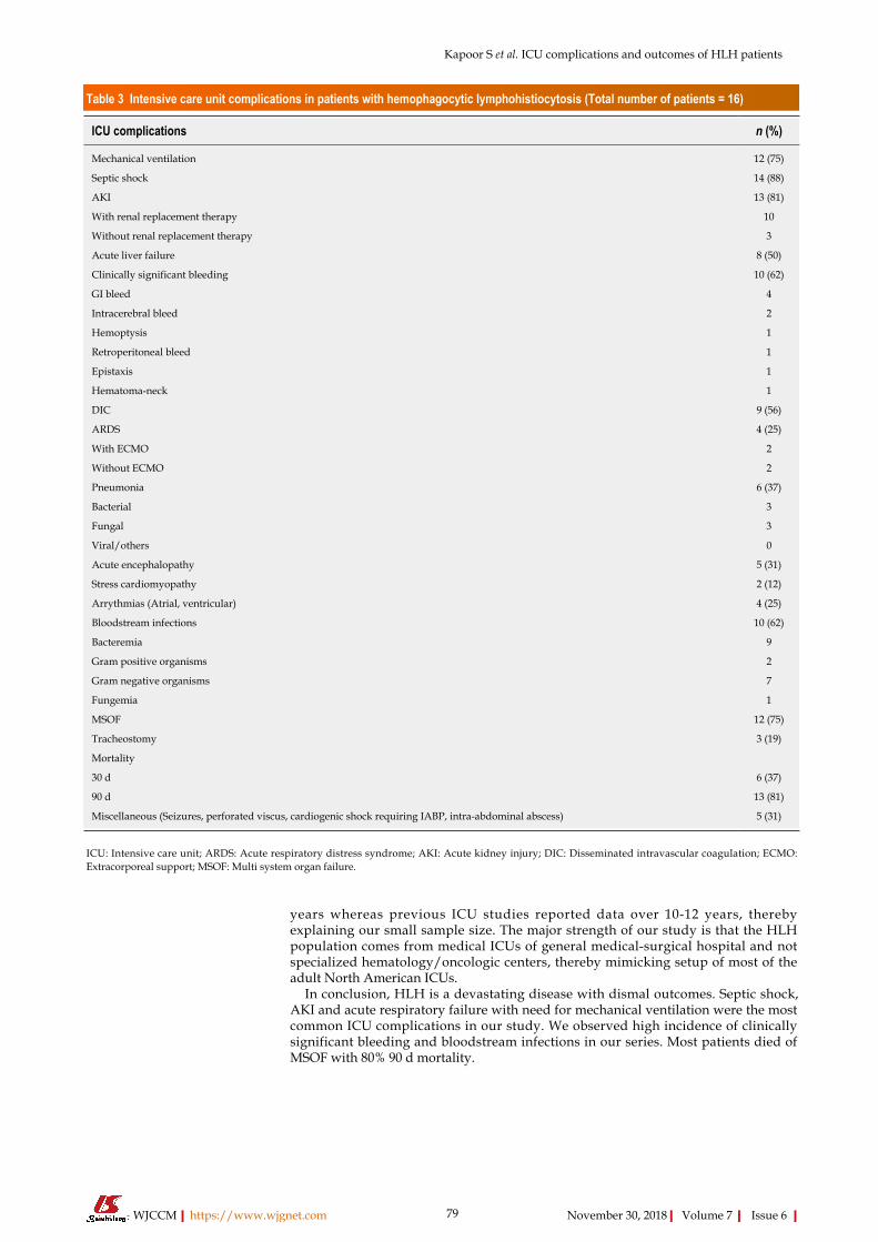
Table 3 Intensive care unit complications in patients with hemophagocytic lymphohistiocytosis (Total number of patients = 16)
ICU complications n (%)
Mechanical ventilation 12 (75)
Septic shock 14 (88)
AKI 13 (81)
With renal replacement therapy 10
Without renal replacement therapy 3
Acute liver failure 8 (50)
Clinically significant bleeding 10 (62)
GI bleed 4
Intracerebral bleed 2
Hemoptysis 1
Retroperitoneal bleed 1
Epistaxis 1
Hematoma-neck 1
DIC 9 (56)
ARDS 4 (25)
With ECMO 2
Without ECMO 2
Pneumonia 6 (37)
Bacterial 3
Fungal 3
Viral/others 0
Acute encephalopathy 5 (31)
Stress cardiomyopathy 2 (12)
Arrythmias (Atrial, ventricular) 4 (25)
Bloodstream infections 10 (62)
Bacteremia 9
Gram positive organisms 2
Gram negative organisms 7
Fungemia 1
MSOF 12 (75)
Tracheostomy 3 (19)
Mortality
30 d 6 (37)
90 d 13 (81)
Miscellaneous (Seizures, perforated viscus, cardiogenic shock requiring IABP, intra-abdominal abscess) 5 (31)
ICU: Intensive care unit; ARDS: Acute respiratory distress syndrome; AKI: Acute kidney injury; DIC: Disseminated intravascular coagulation; ECMO:Extracorporeal support; MSOF: Multi system organ failure.
years whereas previous ICU studies reported data over 10-12 years, therebyexplaining our small sample size. The major strength of our study is that the HLHpopulation comes from medical ICUs of general medical-surgical hospital and notspecialized hematology/oncologic centers, thereby mimicking setup of most of theadult North American ICUs.
In conclusion, HLH is a devastating disease with dismal outcomes. Septic shock,AKI and acute respiratory failure with need for mechanical ventilation were the mostcommon ICU complications in our study. We observed high incidence of clinicallysignificant bleeding and bloodstream infections in our series. Most patients died ofMSOF with 80% 90 d mortality.
WJCCM https://www.wjgnet.com November 30, 2018 Volume 7 Issue 6
Kapoor S et al. ICU complications and outcomes of HLH patients
79

Table 4 Differences in mortality based on various categorical variables
Categorical variable Categorical variable P value
Age above 50 yr (8 patients) Age less than or equal to 50 yr (8 patients) 0.5
SOFA score on ICU admission above 10 (7 patients) SOFA score on ICU admission less than or equal to 10 (9 patients) 0.6
Underlying immunosuppression (11 patients) No underlying immunosuppression (5 patients) 0.7
Direct admission to ICU (8 patients) Transfer from floor to ICU (8 patients) 0.5
Time to diagnose HLH more than 3 d (7 patients) Time to diagnose HLH less than or equal to 3 d (9 patients) 0.4
HLH: Hemophagocytic lymphohistiocytosis; ICU: Intensive care unit; SOFA: Sequential organ failure assessment.
ARTICLE HIGHLIGHTSResearch backgroundHemophagocytic lymphohistiocytosis (HLH) is a rare, fatal syndrome increasingly beingrecognized in intensive care unit (ICU) now. Not many studies have been conducted in an ICUsetting to study the complications and outcomes of this patient population.
Research motivationThere is an urgent need for more evidence in literature to help guide Intensivists identify andmanage these sick and complicated patients in ICU. This will help to improve their outcomesand decrease complications.
Research objectivesThe objective of our research is to study the ICU course, complications and outcomes of adultpatients admitted with HLH over the period of 4 years.
Research methodsIt is a retrospective observational study of adult patients with HLH admitted to the twoacademic medical ICUs from 1/1/2013 to 6/30/2017. The diagnosis of HLH was establishedusing HLH-2004 criteria. Data was collected using ICD 9 and 10 codes. Statistical analysis wasperformed using STATA 15 software.
Research resultsSixteen adult patients were admitted to ICUs over 4 years with HLH with median age of 49years. Median ICU LOS was 11.5 d and median hospital LOS was 29 d. Septic shock, acutekidney injury (AKI) and acute respiratory failure were the most common ICU complications.Multi system organ failure was the most common cause of death with high mortality of 80% over90 d. Age (above or below 50 years), Sequential Organ Failure Assessment score on ICUadmission, time to diagnose HLH and immune status of patient did not predict mortality.
Research conclusionsOur study showed that HLH in ICU is associated with mortality of 80% over 90 d periods. Mostcommon complications include septic shock, respiratory failure and AKI. Multi system organfailure is the most common cause of death. Clinically significant bleeding and bloodstreaminfections were also observed in our case series.
Research perspectivesPresentation of HLH in ICU mimics severe sepsis/septic shock. High index of suspicion for HLHis warranted in patients with septic shock and bi/pan cytopenia, not responding to standardtreatment. Tests like serum ferritin, fibrinogen, triglycerides, bone marrow/lymph node biopsieshelp in diagnosis of HLH. Early diagnosis and treatment with chemotherapy is crucial forimproved outcomes.
REFERENCES1 Okabe T, Shah G, Mendoza V, Hirani A, Baram M, Marik P. What intensivists need to know
about hemophagocytic syndrome: an underrecognized cause of death in adult intensive careunits. J Intensive Care Med 2012; 27: 58-64 [PMID: 21257627 DOI: 10.1177/0885066610393462]
2 Ramos-Casals M, Brito-Zerón P, López-Guillermo A, Khamashta MA, Bosch X. Adulthaemophagocytic syndrome. Lancet 2014; 383: 1503-1516 [PMID: 24290661 DOI:10.1016/S0140-6736(13)61048-X]
3 Janka G. Hemophagocytic lymphohistiocytosis: when the immune system runs amok. KlinPadiatr 2009; 221: 278-285 [PMID: 19707989 DOI: 10.1055/s-0029-1237386]
4 Campo M, Berliner N. Hemophagocytic Lymphohistiocytosis in Adults. Hematol Oncol ClinNorth Am 2015; 29: 915-925 [PMID: 26461151 DOI: 10.1016/j.hoc.2015.06.009]
5 Cesarine J, Filippone LM, Filippone EJ. Hemophagocytic lymphohistiocytosis in the ED. Am JEmerg Med 2016; 34: 2057.e5-2057.e8 [PMID: 27066745 DOI: 10.1016/j.ajem.2016.03.034]
6 Tothova Z, Berliner N. Hemophagocytic Syndrome and Critical Illness: New Insights into
WJCCM https://www.wjgnet.com November 30, 2018 Volume 7 Issue 6
Kapoor S et al. ICU complications and outcomes of HLH patients
80

Diagnosis and Management. J Intensive Care Med 2015; 30: 401-412 [PMID: 24407034 DOI:10.1177/0885066613517076]
7 Barba T, Maucort-Boulch D, Iwaz J, Bohé J, Ninet J, Hot A, Lega JC, Guérin C, Argaud L,Broussolle C. Hemophagocytic Lymphohistiocytosis in Intensive Care Unit: A 71-Case Strobe-Compliant Retrospective Study. Medicine (Baltimore) 2015; 94: e2318 [PMID: 26705219 DOI:10.1097/MD.0000000000002318]
8 Buyse S, Teixeira L, Galicier L, Mariotte E, Lemiale V, Seguin A, Bertheau P, Canet E, deLabarthe A, Darmon M. Critical care management of patients with hemophagocyticlymphohistiocytosis. Intensive Care Med 2010; 36: 1695-1702 [PMID: 20532477 DOI:10.1007/s00134-010-1936-z]
9 Machowicz R, Janka G, Wiktor-Jedrzejczak W. Your critical care patient may have HLH(hemophagocytic lymphohistiocytosis). Crit Care 2016; 20: 215 [PMID: 27389585 DOI:10.1186/s13054-016-1369-3]
10 Rajagopala S, Singh N, Agarwal R, Gupta D, Das R. Severe hemophagocyticlymphohistiocytosis in adults-experience from an intensive care unit from North India. Indian JCrit Care Med 2012; 16: 198-203 [PMID: 23559726 DOI: 10.4103/0972-5229.106501]
11 Guo Y, Bai Y, Gu L. Clinical features and prognostic factors of adult secondary hemophagocyticsyndrome: Analysis of 47 cases. Medicine (Baltimore) 2017; 96: e6935 [PMID: 28562543 DOI:10.1097/MD.0000000000006935]
12 Arca M, Fardet L, Galicier L, Rivière S, Marzac C, Aumont C, Lambotte O, Coppo P. Prognosticfactors of early death in a cohort of 162 adult haemophagocytic syndrome: impact of triggeringdisease and early treatment with etoposide. Br J Haematol 2015; 168: 63-68 [PMID: 25157895 DOI:10.1111/bjh.13102]
13 Akenroye AT, Madan N, Mohammadi F, Leider J. Hemophagocytic Lymphohistiocytosis mimicsmany common conditions: case series and review of literature. Eur Ann Allergy Clin Immunol2017; 49: 31-41 [PMID: 28120605]
14 Karlsson T. Secondary haemophagocytic lymphohistiocytosis: Experience from the UppsalaUniversity Hospital. Ups J Med Sci 2015; 120: 257-262 [PMID: 26212358 DOI:10.3109/03009734.2015.1064500]
15 Li J, Wang Q, Zheng W, Ma J, Zhang W, Wang W, Tian X. Hemophagocytic lymphohistiocytosis:clinical analysis of 103 adult patients. Medicine (Baltimore) 2014; 93: 100-105 [PMID: 24646466DOI: 10.1097/MD.0000000000000022]
16 Otrock ZK, Eby CS. Clinical characteristics, prognostic factors, and outcomes of adult patientswith hemophagocytic lymphohistiocytosis. Am J Hematol 2015; 90: 220-224 [PMID: 25469675 DOI:10.1002/ajh.23911]
17 Parikh SA, Kapoor P, Letendre L, Kumar S, Wolanskyj AP. Prognostic factors and outcomes ofadults with hemophagocytic lymphohistiocytosis. Mayo Clin Proc 2014; 89: 484-492 [PMID:24581757 DOI: 10.1016/j.mayocp.2013.12.012]
18 Park HS, Kim DY, Lee JH, Lee JH, Kim SD, Park YH, Lee JS, Kim BY, Jeon M, Kang YA. Clinicalfeatures of adult patients with secondary hemophagocytic lymphohistiocytosis from causesother than lymphoma: an analysis of treatment outcome and prognostic factors. Ann Hematol2012; 91: 897-904 [PMID: 22147006 DOI: 10.1007/s00277-011-1380-3]
19 Reddy S, Rangappa P, Kasaragod A, Kumar AS, Rao K. Haemophagocytic lymphohistiocytosis(HLH): case series in tertiary referral hospital over three years. J Assoc Physicians India 2013; 61:850-852 [PMID: 24974508]
20 Rivière S, Galicier L, Coppo P, Marzac C, Aumont C, Lambotte O, Fardet L. Reactivehemophagocytic syndrome in adults: a retrospective analysis of 162 patients. Am J Med 2014; 127:1118-1125 [PMID: 24835040 DOI: 10.1016/j.amjmed.2014.04.034]
21 Schram AM, Comstock P, Campo M, Gorovets D, Mullally A, Bodio K, Arnason J, Berliner N.Haemophagocytic lymphohistiocytosis in adults: a multicentre case series over 7 years. Br JHaematol 2016; 172: 412-419 [PMID: 26537747 DOI: 10.1111/bjh.13837]
22 Shabbir M, Lucas J, Lazarchick J, Shirai K. Secondary hemophagocytic syndrome in adults: acase series of 18 patients in a single institution and a review of literature. Hematol Oncol 2011; 29:100-106 [PMID: 20809477 DOI: 10.1002/hon.960]
23 Kaito K, Kobayashi M, Katayama T, Otsubo H, Ogasawara Y, Sekita T, Saeki A, Sakamoto M,Nishiwaki K, Masuoka H. Prognostic factors of hemophagocytic syndrome in adults: analysis of34 cases. Eur J Haematol 1997; 59: 247-253 [PMID: 9338623 DOI:10.1111/j.1600-0609.1997.tb00984.x]
24 Hanoun M, Dührsen U. The Maze of Diagnosing Hemophagocytic Lymphohistiocytosis: Single-Center Experience of a Series of 6 Clinical Cases. Oncology 2017; 92: 173-178 [PMID: 28052298DOI: 10.1159/000454733]
25 Raschke RA, Garcia-Orr R. Hemophagocytic lymphohistiocytosis: a potentially underrecognizedassociation with systemic inflammatory response syndrome, severe sepsis, and septic shock inadults. Chest 2011; 140: 933-938 [PMID: 21737492 DOI: 10.1378/chest.11-0619]
26 Lachmann G, La Rosée P, Schenk T, Brunkhorst FM, Spies C. [Hemophagocyticlymphohistiocytosis : A diagnostic challenge on the ICU]. Anaesthesist 2016; 65: 776-786 [PMID:27612865 DOI: 10.1007/s00101-016-0216-x]
27 Kleinert MM, Garate G, Osatnik J, Cicco J, Hunter B, Soria EJ. [Reactive hemophagocyticsyndrome in critical care patients. Report of 4 cases]. Medicina (B Aires) 2007; 67: 49-52 [PMID:17408021]
28 Strauss R, Neureiter D, Westenburger B, Wehler M, Kirchner T, Hahn EG. Multifactorial riskanalysis of bone marrow histiocytic hyperplasia with hemophagocytosis in critically ill medicalpatients--a postmortem clinicopathologic analysis. Crit Care Med 2004; 32: 1316-1321 [PMID:15187513 DOI: 10.1097/01.CCM.0000127779.24232.15]
29 Stéphan F, Thiolière B, Verdy E, Tulliez M. Role of hemophagocytic histiocytosis in the etiologyof thrombocytopenia in patients with sepsis syndrome or septic shock. Clin Infect Dis 1997; 25:1159-1164 [PMID: 9402376 DOI: 10.1086/516086]
30 Halacli B, Unver N, Halacli SO, Canpinar H, Ersoy EO, Ocal S, Guc D, Buyukasik Y, Topeli A.Investigation of hemophagocytic lymphohistiocytosis in severe sepsis patients. J Crit Care 2016;35: 185-190 [PMID: 27481757 DOI: 10.1016/j.jcrc.2016.04.034]
31 Grom AA. Macrophage activation syndrome and reactive hemophagocytic lymphohistiocytosis:
WJCCM https://www.wjgnet.com November 30, 2018 Volume 7 Issue 6
Kapoor S et al. ICU complications and outcomes of HLH patients
81

the same entities? Curr Opin Rheumatol 2003; 15: 587-590 [PMID: 12960485 DOI:10.1097/00002281-200309000-00011]
32 Lao K, Sharma N, Gajra A, Vajpayee N. Hemophagocytic Lymphohistiocytosis and BoneMarrow Hemophagocytosis: A 5-Year Institutional Experience at a Tertiary Care Hospital. SouthMed J 2016; 109: 655-660 [PMID: 27706506 DOI: 10.14423/SMJ.0000000000000546]
33 Rosado FG, Rinker EB, Plummer WD, Dupont WD, Spradlin NM, Reichard KK, Kim AS. Thediagnosis of adult-onset haemophagocytic lymphohistiocytosis: lessons learned from a review of29 cases of bone marrow haemophagocytosis in two large academic institutions. J Clin Pathol2016; 69: 805-809 [PMID: 26896491 DOI: 10.1136/jclinpath-2015-203577]
34 Janka GE. Hemophagocytic lymphohistiocytosis. Hematology 2015; 10 Suppl 1: 104-107 [DOI:10.1080/10245330512331390087]
35 Henter JI, Horne A, Aricó M, Egeler RM, Filipovich AH, Imashuku S, Ladisch S, McClain K,Webb D, Winiarski J. HLH-2004: Diagnostic and therapeutic guidelines for hemophagocyticlymphohistiocytosis. Pediatr Blood Cancer 2007; 48: 124-131 [PMID: 16937360 DOI:10.1002/pbc.21039]
36 Fardet L, Galicier L, Lambotte O, Marzac C, Aumont C, Chahwan D, Coppo P, Hejblum G.Development and validation of the HScore, a score for the diagnosis of reactive hemophagocyticsyndrome. Arthritis Rheumatol 2014; 66: 2613-2620 [PMID: 24782338 DOI: 10.1002/art.38690]
37 Debaugnies F, Mahadeb B, Ferster A, Meuleman N, Rozen L, Demulder A, Corazza F.Performances of the H-Score for Diagnosis of Hemophagocytic Lymphohistiocytosis in Adultand Pediatric Patients. Am J Clin Pathol 2016; 145: 862-870 [PMID: 27298397 DOI:10.1093/ajcp/aqw076]
38 Grangé S, Buchonnet G, Besnier E, Artaud-Macari E, Beduneau G, Carpentier D, Dehay J,Girault C, Marchalot A, Guerrot D. The Use of Ferritin to Identify Critically Ill Patients WithSecondary Hemophagocytic Lymphohistiocytosis. Crit Care Med 2016; 44: e1045-e1053 [PMID:27441901 DOI: 10.1097/CCM.0000000000001878]
39 Wormsbecker AJ, Sweet DD, Mann SL, Wang SY, Pudek MR, Chen LY. Conditions associatedwith extreme hyperferritinaemia (>3000 μg/L) in adults. Intern Med J 2015; 45: 828-833 [PMID:25851400 DOI: 10.1111/imj.12768]
40 Schweizer M, Goede JS, Briner V. Patients with an extraordinarily elevated serum ferritin: thinkof haemophagocytic lymphohistiocytosis. Swiss Med Wkly 2015; 145: w14152 [PMID: 26098856DOI: 10.4414/smw.2015.14152]
41 Schram AM, Campigotto F, Mullally A, Fogerty A, Massarotti E, Neuberg D, Berliner N. Markedhyperferritinemia does not predict for HLH in the adult population. Blood 2015; 125: 1548-1552[PMID: 25573993 DOI: 10.1182/blood-2014-10-602607]
42 Saeed H, Woods RR, Lester J, Herzig R, Gul Z, Monohan G. Evaluating the optimal serumferritin level to identify hemophagocytic lymphohistiocytosis in the critical care setting. Int JHematol 2015; 102: 195-199 [PMID: 25997871 DOI: 10.1007/s12185-015-1813-1]
43 Schaffner M, Rosenstein L, Ballas Z, Suneja M. Significance of Hyperferritinemia in HospitalizedAdults. Am J Med Sci 2017; 354: 152-158 [PMID: 28864373 DOI: 10.1016/j.amjms.2017.04.016]
44 Tamamyan GN, Kantarjian HM, Ning J, Jain P, Sasaki K, McClain KL, Allen CE, Pierce SA,Cortes JE, Ravandi F. Malignancy-associated hemophagocytic lymphohistiocytosis in adults:Relation to hemophagocytosis, characteristics, and outcomes. Cancer 2016; 122: 2857-2866 [PMID:27244347 DOI: 10.1002/cncr.30084]
45 Szyper-Kravitz M. The hemophagocytic syndrome/macrophage activation syndrome: a finalcommon pathway of a cytokine storm. Isr Med Assoc J 2009; 11: 633-634 [PMID: 20077953]
46 Cai G, Wang Y, Liu X, Han Y, Wang Z. Central nervous system involvement in adults withhaemophagocytic lymphohistiocytosis: a single-center study. Ann Hematol 2017; 96: 1279-1285[PMID: 28589450 DOI: 10.1007/s00277-017-3035-5]
47 Seguin A, Galicier L, Boutboul D, Lemiale V, Azoulay E. Pulmonary Involvement in PatientsWith Hemophagocytic Lymphohistiocytosis. Chest 2016; 149: 1294-1301 [PMID: 26836913 DOI:10.1016/j.chest.2015.11.004]
48 Aulagnon F, Lapidus N, Canet E, Galicier L, Boutboul D, Peraldi MN, Reuter D, Bernard R,Schlemmer B, Azoulay E. Acute kidney injury in adults with hemophagocyticlymphohistiocytosis. Am J Kidney Dis 2015; 65: 851-859 [PMID: 25480521 DOI:10.1053/j.ajkd.2014.10.012]
49 Valade S, Azoulay E, Galicier L, Boutboul D, Zafrani L, Stepanian A, Canet E, Lemiale V, VenotM, Veyradier A. Coagulation Disorders and Bleedings in Critically Ill Patients WithHemophagocytic Lymphohistiocytosis. Medicine (Baltimore) 2015; 94: e1692 [PMID: 26448017DOI: 10.1097/MD.0000000000001692]
50 Allen CE, Yu X, Kozinetz CA, McClain KL. Highly elevated ferritin levels and the diagnosis ofhemophagocytic lymphohistiocytosis. Pediatr Blood Cancer 2008; 50: 1227-1235 [PMID: 18085676DOI: 10.1002/pbc.21423]
51 Okamoto M, Yamaguchi H, Isobe Y, Yokose N, Mizuki T, Tajika K, Gomi S, Hamaguchi H,Inokuchi K, Oshimi K. Analysis of triglyceride value in the diagnosis and treatment response ofsecondary hemophagocytic syndrome. Intern Med 2009; 48: 775-781 [PMID: 19443971 DOI:10.2169/internalmedicine.48.1677]
52 Esmaili H, Rahmani O, Fouladi RF. Hemophagocytic syndrome in patients with unexplainedcytopenia: report of 15 cases. Turk Patoloji Derg 2013; 29: 15-18 [PMID: 23354791 DOI:10.5146/tjpath.2013.01142]
53 Kleynberg RL, Schiller GJ. Secondary hemophagocytic lymphohistiocytosis in adults: an updateon diagnosis and therapy. Clin Adv Hematol Oncol 2012; 10: 726-732 [PMID: 23271259]
54 La Rosée P. Treatment of hemophagocytic lymphohistiocytosis in adults. Hematology Am SocHematol Educ Program 2015; 2015: 190-196 [PMID: 26637720 DOI:10.1182/asheducation-2015.1.190]
55 Nikiforow S, Berliner N. The unique aspects of presentation and diagnosis of hemophagocyticlymphohistiocytosis in adults. Hematology Am Soc Hematol Educ Program 2015; 2015: 183-189[PMID: 26637719 DOI: 10.1182/asheducation-2015.1.183]
56 Porter R, Berliner N. Diagnosis and treatment of HLH in adults. Rinsho Ketsueki 2016; 57: 2059-2063 [PMID: 27795515 DOI: 10.11406/rinketsu.57.2059]
57 Schram AM, Berliner N. How I treat hemophagocytic lymphohistiocytosis in the adult patient.
WJCCM https://www.wjgnet.com November 30, 2018 Volume 7 Issue 6
Kapoor S et al. ICU complications and outcomes of HLH patients
82

Blood 2015; 125: 2908-2914 [PMID: 25758828 DOI: 10.1182/blood-2015-01-551622]
P- Reviewer: Gajic O, Santomauro M, Zhao L, Lin JAS- Editor: Dou Y L- Editor: A E- Editor: Bian YN
WJCCM https://www.wjgnet.com November 30, 2018 Volume 7 Issue 6
Kapoor S et al. ICU complications and outcomes of HLH patients
83

Published By Baishideng Publishing Group Inc
7901 Stoneridge Drive, Suite 501, Pleasanton, CA 94588, USA
Telephone: +1-925-2238242
Fax: +1-925-2238243
E-mail: [email protected]
Help Desk: https://www.f6publishing.com/helpdesk
https://www.wjgnet.com
© 2018 Baishideng Publishing Group Inc. All rights reserved.