World Journal of Hepatology - BPG Management System
86
World Journal of Hepatology World J Hepatol 2015 September 18; 7(20): 2237-2314 Published by Baishideng Publishing Group Inc ISSN 1948-5182 (online)
-
Upload
khangminh22 -
Category
Documents
-
view
0 -
download
0
Transcript of World Journal of Hepatology - BPG Management System
World Journal of HepatologyWorld J Hepatol 2015 September 18; 7(20): 2237-2314
Published by Baishideng Publishing Group Inc
ISSN 1948-5182 (online)
EDITORS-IN-CHIEFClara Balsano, RomeWan-Long Chuang, Kaohsiung
GUEST EDITORIAL BOARD MEMBERSKing-Wah Chiu, KaohsiungTai-An Chiang, TainanChi-Tan Hu, HualienSen-Yung Hsieh, TaoyuanWenya Huang, TainanLiang-Yi Hung, TainanJih RU Hwu, HsinchuJing-Yi Lee, TaipeiMei-Hsuan Lee, TaipeiChih-Wen Lin, KaohsiungChun-Che Lin, TaichungWan-Yu Lin, TaichungTai-Long Pan, Tao-YuanSuh-Ching Yang, TaipeiChun-Yan Yeung, Taipei
MEMBERS OF THE EDITORIAL BOARD
Algeria
Samir Rouabhia, Batna
Argentina
Fernando O Bessone, RosarioMaria C Carrillo, RosarioMelisa M Dirchwolf, Buenos AiresBernardo Frider, Buenos Aires
Jorge Quarleri, Buenos AiresAdriana M Torres, Rosario
Armenia
Narina Sargsyants, Yerevan
Australia
Mark D Gorrell, Sydney
Austria
Harald Hofer, ViennaGustav Paumgartner, ViennaMatthias Pinter, ViennaThomas Reiberger, Vienna
Bangladesh
Shahinul Alam, DhakaMamun Al Mahtab, Dhaka
Belgium
Nicolas Lanthier, BrusselsPhilip Meuleman, GhentLuisa Vonghia, Antwerp
Botswana
Francesca Cainelli, Gaborone
Sandro Vento, Gaborone
Brazil
Edson Abdala, Sao PauloIlka FSF Boin, CampinasNiels OS Camara, Sao PauloAna Carolina FN Cardoso, Rio de JaneiroRoberto J Carvalho-Filho, Sao PauloJulio CU Coelho, CuritibaFlavio Henrique Ferreira Galvao, São PauloJanaina L Narciso-Schiavon, FlorianopolisSílvia HC Sales-Peres, BauruLeonardo L Schiavon, FlorianópolisLuciana D Silva, Belo HorizonteVanessa Souza-Mello, Rio de JaneiroJaques Waisberg, Santo André
Bulgaria
Mariana P Penkova-Radicheva, Stara ZagoraMarieta Simonova, Sofia
Canada
Runjan Chetty, TorontoMichele Molinari, HalifaxGiada Sebastiani, Montreal
Chile
Luis A Videla, Santiago
I
Editorial Board2014-2017
The World Journal of Hepatology Editorial Board consists of 469 members, representing a team of worldwide experts in hepatology. They are from 53 countries, including Algeria (1), Argentina (6), Armenia (1), Australia (1), Austria (4), Bangladesh (2), Belgium (3), Botswana (2), Brazil (13), Bulgaria (2), Canada (3), Chile (1), China (98), Czech Repoublic (1), Denmark (2), Egypt (12), France (6), Germany (19), Greece (11), Hungary (5), India (15), Indonesia (2), Iran (4), Israel (1), Italy (52), Japan (35), Jordan (1), Malaysia (2), Mexico (3), Moldova (1), Netherlands (3), Nigeria (1), Pakistan (1), Philippines (2), Poland (1), Portugal (2), Qatar (1), Romania (6), Russia (2), Saudi Arabia (4), Singapore (1), South Korea (11), Spain (20), Sri Lanka (1), Sudan (1), Sweden (1), Switzerland (1), Thailand (4), Turkey (21), Ukraine (3), United Kingdom (17), and United States (56).
January 27, 2014WJH|www.wjgnet.com
World Journal of HepatologyW J H
ChinaGuang-Wen Cao, ShanghaiEn-Qiang Chen, ChengduGong-Ying Chen, HangzhouJin-lian Chen, ShanghaiJun Chen, ChangshaAlfred Cheng, Hong KongChun-Ping Cui, BeijingShuang-Suo Dang, Xi’an Ming-Xing Ding, JinhuaZhi-Jun Duang, DalianHe-Bin Fan, WuhanXiao-Ming Fan, ShanghaiJames Yan Yue Fung, Hong Kong Yi Gao, GuangzhouZuo-Jiong Gong, WuhanZhi-Yong Guo, GuangzhouShao-Liang Han, WenzhouTao Han, TianjinJin-Yang He, GuangzhouMing-Liang He, Hong KongCan-Hua Huang, ChengduBo Jin, BeijingShan Jin, Hohhot Hui-Qing Jiang, ShijiazhuangWan-Yee Joseph Lau, Hong KongGuo-Lin Li, ChangshaJin-Jun Li, ShanghaiQiang Li, JinanSheng Li, JinanZong-Fang Li, Xi'anXu Li, Guangzhou Xue-Song Liang, Shanghai En-Qi Liu, Xi‘anPei Liu, ShenyangZhong-Hui Liu, ChangchunGuang-Hua Luo, ChangzhouYi Lv, Xi'anGuang-Dong Pan, LiuzhouWen-Sheng Pan, HangzhouJian-Min Qin, Shanghai Wai-Kay Seto, Hong KongHong Shen, ChangshaXiao Su, ShanghaiLi-Ping Sun, BeijingWei-Hao Sun, NanjingXue-Ying Sun, HarbinHua Tang, TianjinLing Tian, ShanghaiEric Tse, Hong KongGuo-Ying Wang, ChangzhouYue Wang, BeijingShu-Qiang Wang, ChengduMary MY Waye, Hong KongHong-Shan Wei, BeijingDanny Ka-Ho Wong, Hong KongGrace Lai-Hung Wong, Hong KongBang-Fu Wu, DongguanFeng Wu, ChongqingXiong-Zhi Wu, Tianjin Chun-Fang Xu, SuzhouRui-An Xu, QuanzhouRui-Yun Xu, GuangzhouWei-Li Xu, ShijiazhuangShi-Ying Xuan, Qingdao Ming-Xian Yan, JinanLv-Nan Yan, ChengduJin Yang, HangzhouJi-Hong Yao, DalianWinnie Yeo, Hong Kong
Zheng Zeng, BeijingQi Zhang, HangzhouShi-Jun Zhang, GuangzhouXiao-Lan Zhang, ShijiazhuangXiao-Yong Zhang, GuangzhouXin-Chen Zhang, HarbinYong Zhang, Xi'anHong-Chuan Zhao, HefeiMing-Hua Zheng, WenzhouYu-Bao Zheng, GuangzhouRen-Qian Zhong, ShanghaiFan Zhu, WuhanXiao Zhu, Dongguan
Czech Repoublic
Kamil Vyslouzil, Olomouc
Denmark
Henning Gronbaek, AarhusChristian Mortensen, Hvidovre
Egypt
Ihab T Abdel-Raheem, DamanhourNGB G Bader EL Din, CairoHatem Elalfy, MansouraMahmoud M El-Bendary, MansouraMona El SH El-Raziky, CairoMohammad El-Sayed, CairoYasser M Fouad, MiniaMohamed AA Metwally, BenhaHany Shehab, CairoMostafa M Sira, Shebin El-koomAshraf Taye, MiniaMA Ali Wahab, Mansoura
France
Laurent Alric, ToulouseSophie Conchon, NantesDaniel J Felmlee, StrasbourgHerve Lerat, CreteilDominique Salmon, ParisJean-Pierre Vartanian, Paris
Germany
Laura E Buitrago-Molina, HannoverEnrico N De Toni, MunichOliver Ebert, MuenchenRolf Gebhardt, LeipzigJanine V Hartl, RegensburgSebastian Hinz, KielBenjamin Juntermanns, EssenRoland Kaufmann, JenaViola Knop, FrankfurtVeronika Lukacs-Kornek, HomburgBenjamin Maasoumy, HannoverJochen Mattner, ErlangenNadja M Meindl-Beinker, MannheimUlf P Neumann, AachenMargarete Odenthal, CologneYoshiaki Sunami, Munich
Christoph Roderburg, AachenFrank Tacke, AachenYuchen Xia, Munich
Greece
Alex P Betrosian, AthensGeorge N Dalekos, LarissaIoanna K Delladetsima, AthensNikolaos K Gatselis, LarissaStavros Gourgiotis, AthensChristos G Savopoulos, ThessalonikiTania Siahanidou, AthensEmmanouil Sinakos, ThessalonikiNikolaos G Symeonidi, ThessalonikiKonstantinos C Thomopoulos, LarissaKonstantinos Tziomalos, Thessaloniki
Hungary
Gabor Banhegyi, BudapestPeter L Lakatos, BudapestMaria Papp, DebrecenFerenc Sipos, BudapestZsolt J Tulassay, Budapest
India
Deepak N Amarapurkar, Mumbai Girish M Bhopale, PuneSibnarayan Datta, TezpurNutan D Desai, MumbaiSorabh Kapoor, MumbaiJaswinder S Maras, New DelhiNabeen C Nayak, New DelhiC Ganesh Pai, ManipalAmit Pal, ChandigarhK Rajeshwari, New DelhiAnup Ramachandran, VelloreD Nageshwar Reddy, HyderabadShivaram P Singh, CuttackAjith TA, ThrissurBalasubramaniyan Vairappan, Pondicherry
Indonesia
Cosmas RA Lesmana, JakartaNeneng Ratnasari, Yogyakarta
Iran
Seyed M Jazayeri, TehranSedigheh Kafi-Abad, TehranIradj Maleki, SariFakhraddin Naghibalhossaini, Shiraz
Israel
Stephen DH Malnick, Rehovot
Italy
Francesco Angelico, Rome
II January 27, 2014WJH|www.wjgnet.com
III January 27, 2014WJH|www.wjgnet.com
Alfonso W Avolio, RomeFrancesco Bellanti, FoggiaMarcello Bianchini, ModenaGuglielmo Borgia, NaplesMauro Borzio, MilanoEnrico Brunetti, PaviaValeria Cento, RomaBeatrice Conti, RomeFrancesco D'Amico, PadovaSamuele De Minicis, FermoFabrizio De Ponti, BolognaGiovan Giuseppe Di Costanzo, NapoliLuca Fabris, PadovaGiovanna Ferraioli, PaviaAndrea Galli, FlorenceeMatteo Garcovich, RomeEdoardo G Giannini, GenovaRossano Girometti, UdineAlessandro Granito, BolognaAlberto Grassi, RiminiAlessandro Grasso, SavonaSalvatore Gruttadauria, PalermoFrancesca Guerrieri, RomeQuirino Lai, AquilaAndrea Lisotti, BolognaMarcello F Maida, PalermoLucia Malaguarnera, CataniaAndrea Mancuso, PalermoLuca Maroni, AnconaFrancesco Marotta, MilanoPierluigi Marzuillo, NaplesSara Montagnese, PadovaGiuseppe Nigri, RomeClaudia Piccoli, FoggiaCamillo Porta, PaviaChiara Raggi, Rozzano (MI)Maria Rendina, BariMaria Ripoli, San Giovanni RotondoKryssia I Rodriguez-Castro, PaduaRaffaella Romeo, MilanAmedeo Sciarra, MilanoAntonio Solinas, SassariAurelio Sonzogni, BergamoGiovanni Squadrito, MessinaSalvatore Sutti, NovaraValentina Svicher, RomeLuca Toti, RomeElvira Verduci, MilanUmberto Vespasiani-Gentilucci, RomeMaria A Zocco, Rome
Japan
Yasuhiro Asahina, TokyoNabil AS Eid, TakatsukiKenichi Ikejima, TokyoShoji Ikuo, KobeYoshihiro Ikura, TakatsukiShinichi Ikuta, NishinomiyaKazuaki Inoue, YokohamaToshiya Kamiyama, SapporoTakanobu Kato, TokyoSaiho Ko, NaraHaruki Komatsu, SakuraMasanori Matsuda, Chuo-city Yasunobu Matsuda, NiigataYoshifumi Nakayama, KitakyushuTaichiro Nishikawa, Kyoto
Satoshi Oeda, SagaKenji Okumura, UrayasuMichitaka Ozaki, SapporoTakahiro Sato, SapporoJunichi Shindoh, TokyoRyo Sudo, YokohamaAtsushi Suetsugu, GifuHaruhiko Sugimura, HamamatsuReiji Sugita, SendaiKoichi Takaguchi, TakamatsuShinji Takai, TakatsukiAkinobu Takaki, OkayamaYasuhito Tanaka, NagoyaTakuji Tanaka, Gifu CityAtsunori Tsuchiya, NiigataKoichi Watashi, TokyoHiroshi Yagi, TokyoTaro Yamashita, KanazawaShuhei Yoshida, ChibaHitoshi Yoshiji, Kashihara
Jordan
Kamal E Bani-Hani, Zarqa
Malaysia
Peng Soon Koh, Kuala LumpurYeong Yeh Lee, Kota Bahru
Mexico
Francisco J Bosques-Padilla, MonterreyMaría de F Higuera-de la Tijera, Mexico CityJosé A Morales-Gonzalez, México City
Moldova
Angela Peltec, Chishinev
Netherlands
Wybrich R Cnossen, NijmegenFrank G Schaap, MaastrichtFareeba Sheedfar, Groningen
Nigeria
CA Asabamaka Onyekwere, Lagos
Pakistan
Bikha Ram Devrajani, Jamshoro
Philippines
Janus P Ong, PasigJD Decena Sollano, Manila
Poland
Jacek Zielinski, Gdansk
Portugal
Rui T Marinho, LisboaJoao B Soares, Braga
Qatar
Reem Al Olaby, Doha
Romania
Bogdan Dorobantu, BucharestLiana Gheorghe, BucharestGeorge S Gherlan, BucharestRomeo G Mihaila, SibiuBogdan Procopet, Cluj-NapocaStreba T Streba, Craiova
Russia
Anisa Gumerova, KazanPavel G Tarazov, St.Petersburg
Saudi Arabia
Abdulrahman A Aljumah, RiyadhIhab MH Mahmoud, RiyadhIbrahim Masoodi, RiyadhMhoammad K Parvez, Riyadh
Singapore
Ser Yee Lee, Singapore
South Korea
Young-Hwa Chung, SeoulDae-Won Jun, SeoulBum-Joon Kim, SeoulDo Young Kim, SeoulJi Won Kim, SeoulMoon Young Kim, WonuMi-Kyung Lee, SuncheonKwan-Kyu Park, DaeguYoung Nyun Park, SeoulJae-Hong Ryoo, SeoulJong Won Yun, Kyungsan
Spain
Ivan G Marina, MadridJuan G Acevedo, BarcelonaJavier Ampuero, SevillaJaime Arias, MadridAndres Cardenas, BarcelonaAgustin Castiella, MendaroIsrael Fernandez-Pineda, SevillaRocio Gallego-Duran, SevillaRita Garcia-Martinez, Barcelona

IV January 27, 2014WJH|www.wjgnet.com
José M González-Navajas, AlicanteJuan C Laguna, BarcelonaElba Llop, MadridLaura Ochoa-Callejero, La Rioja Albert Pares, BarcelonaSonia Ramos, MadridFrancisco Rodriguez-Frias, CórdobaManuel L Rodriguez-Peralvarez, CórdobaMarta R Romero, Salamanca Carlos J Romero, Madrid Maria Trapero-Marugan, Madrid
Sri Lanka
Niranga M Devanarayana, Ragama
Sudan
Hatim MY Mudawi, Khartoum
Sweden
Evangelos Kalaitzakis, Lund
Switzerland
Christoph A Maurer, Liestal
Thailand
Taned Chitapanarux, Chiang maiTemduang Limpaiboon, Khon KaenSith Phongkitkarun, BangkokYong Poovorawan, Bangkok
Turkey
Osman Abbasoglu, AnkaraMesut Akarsu, IzmirUmit Akyuz, IstanbulHakan Alagozlu, SivasYasemin H Balaban, IstanbulBulent Baran, VanMehmet Celikbilek, Yozgat
Levent Doganay, IstanbulFatih Eren, IstanbulAbdurrahman Kadayifci, GaziantepAhmet Karaman, KayseriMuhsin Kaya, DiyarbakirOzgur Kemik, VanSerdar Moralioglu, UskudarA Melih Ozel, Gebze - KocaeliSeren Ozenirler, AnkaraAli Sazci, KocaeliGoktug Sirin, KocaeliMustafa Sunbul, SamsunNazan Tuna, SakaryaOzlem Yonem, Sivas
Ukraine
Rostyslav V Bubnov, KyivNazarii K Kobyliak, KyivIgor N Skrypnyk, Poltava
United Kingdom
Safa Al-Shamma, BournemouthJayantha Arnold, SouthallMarco Carbone, CambridgeRajeev Desai, BirminghamAshwin Dhanda, BristolMatthew Hoare, CambridgeStefan G Hubscher, BirminghamNikolaos Karidis, LondonLemonica J Koumbi, LondonPatricia Lalor, BirminghamJi-Liang Li, OxfordEvaggelia Liaskou, BirminghamRodrigo Liberal, LondonWei-Yu Lu, EdinburghRichie G Madden, TruroChristian P Selinger, LeedsEsther Una Cidon, Bournemouth
United States
Naim Alkhouri, Cleveland Robert A Anders, BaltimoreMohammed Sawkat Anwer, North GraftonKalyan Ram Bhamidimarri, Miami
Brian B Borg, JacksonRonald W Busuttil, Los AngelesAndres F Carrion, MiamiSaurabh Chatterjee, ColumbiaDisaya Chavalitdhamrong, GainesvilleMark J Czaja, BronxJonathan M Fenkel, PhiladelphiaCatherine Frenette, La JollaLorenzo Gallon, ChicagoKalpana Ghoshal, ColumbusGrigoriy E Gurvits, New YorkHie-Won L Hann, PhiladelphiaShuang-Teng He, Kansas CityWendong Huang, DuarteRachel Hudacko, SuffernLu-Yu Hwang, HoustonIjaz S Jamall, SacramentoNeil L Julie, BethesdaHetal Karsan, AtlantaAhmed O Kaseb, HoustonZeid Kayali, PasadenaKusum K Kharbanda, OmahaTimothy R Koch, WashingtonGursimran S Kochhar, ClevelandSteven J Kovacs, East HanoverMary C Kuhns, Abbott ParkJiang Liu, Silver SpringLi Ma, StanfordFrancisco Igor Macedo, SouthfieldSandeep Mukherjee, OmahaNatalia A Osna, OmahaJen-Jung Pan, HoustonChristine Pocha, MinneapolisYury Popov, BostonDavide Povero, La JollaPhillip Ruiz, MiamiTakao Sakai, ClevelandNicola Santoro, New HavenEva Schmelzer, PittsburghZhongjie Shi, PhiladelphiaNathan J Shores, New OrleansSiddharth Singh, RochesterVeysel Tahan, Iowa CityMehlika Toy, BostonHani M Wadei, JacksonvilleGulam Waris, North ChicagoRuliang Xu, New YorkJun Xu, Los AngelesMatthew M Yeh, SeattleXuchen Zhang, West HavenLixin Zhu, BuffaloSasa Zivkovic, Pittsburgh
Contents Three issues per month Volume 7 Number 20 September 18, 2015
September 18, 2015|Volume 7|Issue 20|WJH|www.wjgnet.com I
EDITORIAL2237 Roleofliverresectioninthemanagementofmultinodularhepatocellularcarcinoma
Abbasoglu O
2241 CompartmentalizationofhepatitisBvirus:Lookingbeyondtheliver
Datta S
REVIEW2245 Evaluationofantiangiogenicefficacyinadvancedhepatocellularcarcinoma:Biomarkersandfunctional
imaging
Bouattour M, Payancé A, Wassermann J
2264 Markersofbacterialtranslocationinend-stageliverdisease
Koutsounas I, Kaltsa G, Siakavellas SI, Bamias G
2274 Hepatocellularcarcinoma:Fromclinicalpracticetoevidence-basedtreatmentprotocols
Galun D, Basaric D, Zuvela M, Bulajic P, Bogdanovic A, Bidzic N, Milicevic M
2292 Managementbeforehepatectomyforhepatocellularcarcinomawithcirrhosis
Nakayama H, Takayama T
MINIREVIEWS2303 Fasttrackanesthesiaforlivertransplantation:Reviewofthecurrentpractice
Aniskevich S, Pai SL
2309 Progressinthetreatmentofpulmonarymetastasesafterlivertransplantationforhepatocellularcarcinoma
Xiang ZW, Sun L, Li GH, Maharjan R, Huang JH, Li CX

ContentsWorld Journal of Hepatology
Volume 7 Number 20 September 18, 2015
FLYLEAF
EDITORS FOR THIS ISSUE
Responsible Assistant Editor: Xiang Li Responsible Science Editor: Fang-Fang JiResponsible Electronic Editor: Su-Qing Liu Proofing Editorial Office Director: Xiu-Xia SongProofing Editor-in-Chief: Lian-Sheng Ma
NAMEOFJOURNALWorld Journal of Hepatology
ISSNISSN 1948-5182 (online)
LAUNCHDATEOctober 31, 2009
FREQUENCY36 Issues/Year (8th, 18th, and 28th of each month)
EDITORS-IN-CHIEFClara Balsano, PhD, Professor, Departement of Biomedicine, Institute of Molecular Biology and Pathology, Rome 00161, Italy
Wan-Long Chuang, MD, PhD, Doctor, Professor, Hepatobiliary Division, Department of Internal Medicine, Kaohsiung Medical University Hospital, Kaohsiung Medical University, Kaohsiung 807, Taiwan
EDITORIALOFFICEJin-Lei Wang, Director
Xiu-Xia Song, Vice DirectorWorld Journal of HepatologyRoom 903, Building D, Ocean International Center, No. 62 Dongsihuan Zhonglu, Chaoyang District, Beijing 100025, ChinaTelephone: +86-10-59080039Fax: +86-10-85381893E-mail: [email protected] Desk: http://www.wjgnet.com/esps/helpdesk.aspxhttp://www.wjgnet.com
PUBLISHERBaishideng Publishing Group Inc8226 Regency Drive, Pleasanton, CA 94588, USATelephone: +1-925-223-8242Fax: +1-925-223-8243E-mail: [email protected] Desk: http://www.wjgnet.com/esps/helpdesk.aspxhttp://www.wjgnet.com
PUBLICATIONDATESeptember 18, 2015
COPYRIGHT© 2015 Baishideng Publishing Group Inc. Articles pub-lished by this Open Access journal are distributed under the terms of the Creative Commons Attribution Non-commercial License, which permits use, distribution, and reproduction in any medium, provided the original work is properly cited, the use is non commercial and is otherwise in compliance with the license.
SPECIALSTATEMENTAll articles published in journals owned by the Baishideng Publishing Group (BPG) represent the views and opinions of their authors, and not the views, opinions or policies of the BPG, except where other-wise explicitly indicated.
INSTRUCTIONSTOAUTHORSFull instructions are available online at http://www.wjgnet.com/1948-5182/g_info_20100316080002.htm
ONLINESUBMISSIONhttp://www.wjgnet.com/esps/
September 18, 2015|Volume 7|Issue 20|WJH|www.wjgnet.com II
ABOUT COVER
AIM AND SCOPE
EditorialBoardMemberofWorldJournalofHepatology ,Guo-YingWang,MD,PhD,AssociateProfessor,Surgeon,DepartmentofHepaticSurgeryandLiverTransplantation,theThirdAffiliatedHospitalofSunYat-SenUniversity,Guangzhou510630,GuangdongProvince,China
World Journal of Hepatology (World J Hepatol, WJH, online ISSN 1948-5182, DOI: 10.4254), is a peer-reviewed open access academic journal that aims to guide clinical practice and improve diagnostic and therapeutic skills of clinicians.
WJH covers topics concerning liver biology/pathology, cirrhosis and its complications, liver fibrosis, liver failure, portal hypertension, hepatitis B and C and inflammatory disorders, steatohepatitis and metabolic liver disease, hepatocellular carcinoma, biliary tract disease, autoimmune disease, cholestatic and biliary disease, transplantation, genetics, epidemiology, microbiology, molecular and cell biology, nutrition, geriatric and pediatric hepatology, diagnosis and screening, endoscopy, imaging, and advanced technology. Priority publication will be given to articles concerning diagnosis and treatment of hepatology diseases. The following aspects are covered: Clinical diagnosis, laboratory diagnosis, differential diagnosis, imaging tests, pathological diagnosis, molecular biological diagnosis, immunological diagnosis, genetic diagnosis, functional diagnostics, and physical diagnosis; and comprehensive therapy, drug therapy, surgical therapy, interventional treatment, minimally invasive therapy, and robot-assisted therapy.
We encourage authors to submit their manuscripts to WJH. We will give priority to manuscripts that are supported by major national and international foundations and those that are of great basic and clinical significance.
World Journal of Hepatology is now indexed in PubMed Central, PubMed, Digital Object Identifier, Directory of Open Access Journals, and Scopus.
I-IV EditorialBoard
INDEXING/ABSTRACTING
cause of cancer related deaths worldwide. Various treatment modalities have been applied to HCC depending on the tumor load, functional capacity of the liver and the general condition of the patient. According to Barcelona Clinic Liver Cancer staging strategy and The American Association for the Study of Liver Disease guidelines, surgical resection is not advocated in the tretment of multinodular HCC. Despite this, many recent clinical studies show that, resection can achieve good results in patients with multinodular HCC and 5year survival rate around 40% can be reached. If resection or transplantation is not performed, these patients are usually managed with palliative procedures such as transarterial chemoembolization, radioembolization and cytotoxic chemotherapy and 5year survival of this group of patients will be extremely low. Although survival rates are lower and complications may be increased in this group of patients, liver resection can safely be performed in selected patients in experienced centers for the management of multinodular HCC.
Key words: Hepatocellular carcinoma; Hepatoma; Liver resection; Transarterial chemoembolization; Liver cancer; Liver transplantation
© The Author(s) 2015. Published by Baishideng Publishing Group Inc. All rights reserved.
Core tip: Liver resection is underutilized in the management of multinodular hepatocellular carcinoma. The presence of multiple nodules should not be considered as a contraindication for surgical resection. Acceptable 5year survival rates can be achieved in selected patients.
Abbasoglu O. Role of liver resection in the management of multinodular hepatocellular carcinoma. World J Hepatol 2015; 7(20): 2237-2240 Available from: URL: http://www.wjgnet.com/1948-5182/full/v7/i20/2237.htm DOI: http://dx.doi.org/10.4254/wjh.v7.i20.2237
Osman Abbasoglu
Osman Abbasoglu, Department of Surgery, Faculty of Medicine, Hacettepe University, Ankara 06100, Turkey
Author contributions: Abbasoglu O performed the literature review and prepared the manuscript; Abbasoglu O solely con-tributed to this paper.
Conflictofinterest statement: The author whose name is listed immediately below certify that they have no affiliations with or involvement in any organization or entity with any financial interest (such as honoraria; educational grants; participation in speakers’ bureaus; membership, employment, consultancies, stock ownership, or other equity interest; and expert testimony or patent-licensing arrangements), or non-financial interest (such as personal or professional relationships, affiliations, knowledge or beliefs) in the subject matter or materials discussed in this manuscript.
OpenAccess: This article is an open-access article which was selected by an in-house editor and fully peer-reviewed by external reviewers. It is distributed in accordance with the Creative Commons Attribution Non Commercial (CC BY-NC 4.0) license, which permits others to distribute, remix, adapt, build upon this work non-commercially, and license their derivative works on different terms, provided the original work is properly cited and the use is non-commercial. See: http://creativecommons.org/licenses/by-nc/4.0/
Correspondence to: Dr. Osman Abbasoglu, Department of Surgery, Faculty of Medicine, Hacettepe University, Sihhiye, Ankara 06100, Turkey. [email protected]: +90-532-3649039Fax: +90-312-4262421
Received: May 14, 2015 Peerreview started: May 14, 2015 First decision: June 24, 2015Revised: August 2, 2015 Accepted: September 1, 2015Article in press: September 2, 2015Published online: September 18, 2015
AbstractHepatocellular carcinoma (HCC) is the third leading
EDITORIAL
Submit a Manuscript: http://www.wjgnet.com/esps/Help Desk: http://www.wjgnet.com/esps/helpdesk.aspxDOI: 10.4254/wjh.v7.i20.2237
2237 September 18, 2015|Volume 7|Issue 20|WJH|www.wjgnet.com
World J Hepatol 2015 September 18; 7(20): 2237-2240ISSN 1948-5182 (online)
© 2015 Baishideng Publishing Group Inc. All rights reserved.
Role of liver resection in the management of multinodular hepatocellular carcinoma
TEXTHepatocellular carcinoma (HCC) is a major health problem and is the sixth most common malignancy in the world[1]. HCC usually develops in the presence of underlying chronic liver disease, most commonly due to chronic hepatitis B in underdeveloped countries and due to hepatitis C in developed countries. Although resection remains the cornerstone treatment for HCC, the role of resection in multinodular HCC is controversial. The early literature suggests that transplantation is the only treatment of choice that can offer long-term survival in patients with multinodular disease[2,3].
Liver transplantation eliminates not only the tumor but also the underlying liver disease and excellent outcomes with 5-year survival rates exceeding 60% can be achieved but, many patients are not candidates for transplantation because of tumor size, advanced age, high costs, and finally organ shortage[4]. Clearly, in patients with advanced underlying liver disease and portal hypertension, transplantation is the only option with a chance of cure. Tumors meeting Milan criteria defined as a single tumor smaller than 5 cm or up to three nodules smaller than 3 cm each are good candidates for liver transplantation.
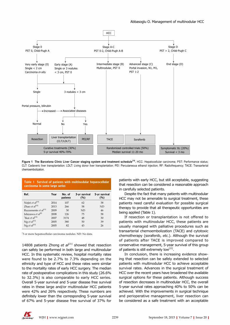
Many staging systems and management algorithms have been proposed for HCC and the Barcelona Clinic Liver Cancer (BCLC) staging and treatment strategy is the most commonly used system. In the BCLC treatment algorithm, surgical resection is limited to patients with early stage single tumors. Patients with up to three nodules may be offered liver transplantation. Patients with more than three nodules are considered for palliative treatments including chemoembolization. BCLC algorithm was first introduced by Llovet et al[5] in 1999 in Seminars in liver disease. Since this original publication, some modifications has been emerged, but the suggestions in the management of multinodular HCC has remained unchanged[6] (Figure 1). The American Association for the Study of Liver Disease guidelines also advocates liver resection only for solitary HCC smaller than 3 cm with well-preserved liver function[7].
On the other hand, the resectability for HCC mainly depends on the volume and functional capacity of the remnant liver, but not to the resected tumor amount. The severity of underlying liver dysfunction is critical in the evaluation of patients preoperatively. During daily practice, surgeons, gastroenterogists and oncologists may see many HCC patients with good liver reserve, good general condition but with multinodular HCC. Multinodular HCC has generally been regarded as a contraindication to resection, but recently there is evolving evidence for the resection of these tumors[8-14].
Anatomic vs nonanatomic resection in patients with HCC is an ongoing discussion[15]. The surgeons performing resection in the treatment of multinodular HCC have to face the difficulty of achieving a curative intervention and at the same time preventing postoperative liver failure as a result of removal of too much liver. Proponents of
anatomic resection claim that, eradication of intrahepatic metastasis along the portal venous system can only be achieved by systematic removal based on segmental liver anatomy[16]. On the contrary, some surgeons prefer non-anatomic resections, aiming to preserve enough tumor free margin to maximize the volume of the remnant liver. In the presence of multiple HCC nodules, anatomical resections may be difficult to perform as postoperative liver failure may be increased due to insufficient remnant liver. In an analysis of 434 patients from Japan, anatomic resections could be performed in only 36% of patients with multinodular HCC while 71% of patients had undergone anatomical resections in the presence of a single tumor[7]. Although the risk of intrahepatic disseminations is considered trivial in small tumors under 2 cm, it may be challenging to justify resection in the treatment of multinodular HCC for the anatomic resection advocates[17].
Nathan et al[18] analyzed the factors predictive of receipt of surgical treatment for early HCC that is, those patients with non-metastatic tumors 5 cm or smaller and without evidence of lymph node metastasis, extra-hepatic tumor growth, or major vascular invasion. Of the 1745 patients meeting the selection criteria, a total of 820 patients (47%) did not receive any type of surgical intervention. Seventy-six percent of those (n = 622) had found to have no documentation of any treatment modality in their medical records. The authors examined the factors associated with receipt of surgical therapy in a bivariate analysis. With respect to tumor characteristics, patients who receive surgical therapy generally had solitary tumors (68% vs 60%, P < 0.001) and less often had bilobar disease (14% vs 22%, P < 0.001). Surprisingly surgical treatment was not offered to many patients without any apparent reason. This Johns Hopkins University data suggest that surgical treatment options are not offered to many patients with HCC, and their opportunity of achieving better survival may be hindered.
There is not enough data about the maximum number of nodules that can be resected safely. In a recent study of 399 patients, Nojiri et al[19] showed that even if patients have four or more nodules without portal vein invasion and with well-preserved liver function, resection for HCC may be the treatment of choice. The 3- and 5-year overall survival rates of patients with multinodular HCC were 62% and 38% respectively. Ishizawa et al[8] reported 5-year survival of 58% in 126 patients with multinodular HCC and in this series 22 patients (17%) had four or more HCC nodules. In this group of patients, existence of multiple tumors was not found to be a predictor of overall survival but independently increased the risk of recurrence (relative risk 1.64)[8]. With the development of radiofrequency ablation, the combination of this modality with resection may increase the resectability rates and surgical treatment can be performed in more advanced multi-nodular HCC[20].
In a recent systematic review of 50 studies involving
2238 September 18, 2015|Volume 7|Issue 20|WJH|www.wjgnet.com
Abbasoglu O. Management of multinodular HCC
14808 patients Zhong et al[21] showed that resection can safely be performed in both large and multinodular HCC. In this systematic review, hospital mortality rates were found to be 2.7% to 7.3% depending on the ethnicity and type of HCC and these rates were similar to the mortality rates of early HCC surgery. The median rate of postoperative complications in this study (26.6% to 32.3%) is also comparable to early HCC series. Overall 5-year survival and 5-year disease free survival rates in these large and/or multinodular HCC patients were 42% and 26% respectively. These numbers are definitely lower than the corresponding 5-year survival of 67% and 5-year disease free survival of 37% for
patients with early HCC, but still acceptable, suggesting that resection can be considered a reasonable approach in carefully selected patients.
Despite the fact that many patients with multinodular HCC may not be amenable to surgical treatment, these patients need careful evaluation for possible surgical therapy to provide that all therapeutic opportunities are being applied (Table 1).
If resection or transplantation is not offered to patients with multinodular HCC, these patients are usually managed with palliative procedures such as transarterial chemoembolization (TACE) and cytotoxic chemotherapy (sorafenib, etc.). Although the survival of patients after TACE is improved compared to conservative management, 5-year survival of this group of patients is still extremely low[22].
In conclusion, there is increasing evidence show-ing that resection can be safely extended to selected patients with multinodular HCC to achieve acceptable survival rates. Advances in the surgical treatment of HCC over the recent years have broadened the available surgical options for these patients. Although success of resection decreases in multinodular HCC, the overall 5-year survival rates approaching 40% to 50% can be achieved. With the improvements in surgical technique and perioperative management, liver resection can be considered as a safe treatment with an acceptable
2239 September 18, 2015|Volume 7|Issue 20|WJH|www.wjgnet.com
Randomized controlled trials (50%)Median survival 1120 mo
Sorafenib
Stage 0PST 0, ChildPugh A
Stage ACPST 02, ChildPugh AB
Stage DPST > 2, ChildPugh C
Very early stage (0)Single < 2 cmCarcinoma insitu
Early stage (A)Single or 3 nodules < 3 cm, PST 0
Intermediate stage (B)Multinodular, PST 0
Advanced stage (C) Portal invasion, N1, M1, PST 12
End stage (D)
Single 3 nodules < 3 cm
Portal pressure, bilirubin
Increased Associated diseases
Normal No Yes
Symptomatic ttc (20%)Survival < 3 mo
HCC
ResectionLiver transplantation
(CLT/LDLT)PEI/RF
Curative treatments (30%)5yr survival 40%70%
TACE
Figure 1 The Barcelona Clinic Liver Cancer staging system and treatment schedule[19]. HCC: Hepatocellular carcinoma; PST: Performance status; CLT: Cadaveric liver transplantation; LDLT: Living donor liver transplantation; PEI: Percutaneous ethanol injection; RF: Radiofrequency; TACE: Transarterial chemoembolization.
Table 1 Survival of patients with multinodular hepatocellular carcinoma in some large series
Ref. Year No. of patients
3-yr survival (%)
5-yr survival (%)
Nojiri et al[19] 2014 107 62 38Zhao et al[10] 2013 266 58 NDRuzzenente et al[11] 2009 30 ND 46Ishizawa et al[8] 2008 126 75 581Ikai et al[14] 2007 3174 48 30Ng et al[12] 2005 380 50 39Ng et al[12] 2005 82 43 26
13 or more hepatocellular carcinoma nodules. ND: No data.
Abbasoglu O. Management of multinodular HCC
2240 September 18, 2015|Volume 7|Issue 20|WJH|www.wjgnet.com
resection justified in advanced hepatocellular carcinoma? Results of an observational study in 464 patients. J Gastrointest Surg 2009; 13: 1313-1320 [PMID: 19418103 DOI: 10.1007/s11605-009-0903-x]
12 Ng KK, Vauthey JN, Pawlik TM, Lauwers GY, Regimbeau JM, Belghiti J, Ikai I, Yamaoka Y, Curley SA, Nagorney DM, Ng IO, Fan ST, Poon RT. Is hepatic resection for large or multinodular hepatocellular carcinoma justified? Results from a multi-insti-tutional database. Ann Surg Oncol 2005; 12: 364-373 [PMID: 15915370 DOI: 10.1245/ASO.2005.06.004]
13 Wu CC, Cheng SB, Ho WM, Chen JT, Liu TJ, P’eng FK. Liver resec-tion for hepatocellular carcinoma in patients with cirrhosis. Br J Surg 2005; 92: 348-355 [PMID: 15672423 DOI: 10.1002/bjs.4838]
14 Ikai I, Arii S, Okazaki M, Okita K, Omata M, Kojiro M, Takayasu K, Nakanuma Y, Makuuchi M, Matsuyama Y, Monden M, Kudo M. Report of the 17th Nationwide Follow-up Survey of Primary Liver Cancer in Japan. Hepatol Res 2007; 37: 676-691 [PMID: 17617112 DOI: 10.1111/j.1872-034X.2007.00119.x]
15 Cucchetti A, Qiao GL, Cescon M, Li J, Xia Y, Ercolani G, Shen F, Pinna AD. Anatomic versus nonanatomic resection in cirrhotic patients with early hepatocellular carcinoma. Surgery 2014; 155: 512-521 [PMID: 24439747 DOI: 10.1016/j.surg.2013.10.009]
16 Zhou Y, Xu D, Wu L, Li B. Meta-analysis of anatomic resection versus nonanatomic resection for hepatocellular carcinoma. Langenbecks Arch Surg 2011; 396: 1109-1117 [PMID: 21476060 DOI: 10.1007/s00423-011-0784-9]
17 Eguchi S, Kanematsu T, Arii S, Okazaki M, Okita K, Omata M, Ikai I, Kudo M, Kojiro M, Makuuchi M, Monden M, Matsuyama Y, Nakanuma Y, Takayasu K. Comparison of the outcomes between an anatomical subsegmentectomy and a non-anatomical minor hepatectomy for single hepatocellular carcinomas based on a Japanese nationwide survey. Surgery 2008; 143: 469-475 [PMID: 18374043 DOI: 10.1016/j.surg.2007.12.003]
18 Nathan H, Hyder O, Mayo SC, Hirose K, Wolfgang CL, Choti MA, Pawlik TM. Surgical therapy for early hepatocellular carcinoma in the modern era: a 10-year SEER-medicare analysis. Ann Surg 2013; 258: 1022-1027 [PMID: 23299519 DOI: 10.1097/SLA.0b013e31827da749]
19 Nojiri K, Tanaka K, Takeda K, Ueda M, Matsuyama R, Taniguchi K, Kumamoto T, Mori R, Endo I. The efficacy of liver resection for multinodular hepatocellular carcinoma. Anticancer Res 2014; 34: 2421-2426 [PMID: 24778054]
20 Wong TC, Lo CM. Resection strategies for hepatocellular carcinoma. Semin Liver Dis 2013; 33: 273-281 [PMID: 23943107 DOI: 10.1055/s-0033-1351782]
21 Zhong JH, Rodríguez AC, Ke Y, Wang YY, Wang L, Li LQ. Hepatic resection as a safe and effective treatment for hepato-cellular carcinoma involving a single large tumor, multiple tumors, or macrovascular invasion. Medicine (Baltimore) 2015; 94: e396 [PMID: 25621684 DOI: 10.1097/MD.0000000000000396]
22 Lencioni R. Chemoembolization for hepatocellular carcinoma. Semin Oncol 2012; 39: 503-509 [PMID: 22846867]
P- Reviewer: Kapoor S, Wang GY S- Editor: Ji FF L- Editor: A E- Editor: Liu SQ
mortality and morbidity rates in the treatment of multi-nodular HCC in experienced centers.
REFERENCES1 Ferlay J, Shin HR, Bray F, Forman D, Mathers C, Parkin DM.
Estimates of worldwide burden of cancer in 2008: GLOBOCAN 2008. Int J Cancer 2010; 127: 2893-2917 [PMID: 21351269 DOI: 10.1002/ijc.25516]
2 Ochiai T, Sonoyama T, Ichikawa D, Fujiwara H, Okamoto K, Sakakura C, Ueda Y, Otsuji E, Itoi H, Hagiwara A, Yamagishi H. Poor prognostic factors of hepatectomy in patients with resectable small hepatocellular carcinoma and cirrhosis. J Cancer Res Clin Oncol 2004; 130: 197-202 [PMID: 14770307]
3 Mazzaferro V, Regalia E, Doci R, Andreola S, Pulvirenti A, Bozzetti F, Montalto F, Ammatuna M, Morabito A, Gennari L. Liver transplantation for the treatment of small hepatocellular carcinomas in patients with cirrhosis. N Engl J Med 1996; 334: 693-699 [PMID: 8594428 DOI: 10.1056/NEJM199603143341104]
4 Mazzaferro V, Bhoori S, Sposito C, Bongini M, Langer M, Miceli R, Mariani L. Milan criteria in liver transplantation for hepatocellular carcinoma: an evidence-based analysis of 15 years of experience. Liver Transpl 2011; 17 Suppl 2: S44-S57 [PMID: 21695773 DOI: 10.1002/lt.22365]
5 Llovet JM, Brú C, Bruix J. Prognosis of hepatocellular carcinoma: the BCLC staging classification. Semin Liver Dis 1999; 19: 329-338 [PMID: 10518312 DOI: 10.1055/s-2007-1007122]
6 Llovet JM, Di Bisceglie AM, Bruix J, Kramer BS, Lencioni R, Zhu AX, Sherman M, Schwartz M, Lotze M, Talwalkar J, Gores GJ. Design and endpoints of clinical trials in hepatocellular carcinoma. J Natl Cancer Inst 2008; 100: 698-711 [PMID: 18477802 DOI: 10.1093/jnci/djn134]
7 Bruix J, Sherman M. Management of hepatocellular carcinoma: an update. Hepatology 2011; 53: 1020-1022 [PMID: 21374666 DOI: 10.1002/hep.24199]
8 Ishizawa T, Hasegawa K, Aoki T, Takahashi M, Inoue Y, Sano K, Imamura H, Sugawara Y, Kokudo N, Makuuchi M. Neither multiple tumors nor portal hypertension are surgical contraindications for hepatocellular carcinoma. Gastroenterology 2008; 134: 1908-1916 [PMID: 18549877 DOI: 10.1053/j.gastro.2008.02.091]
9 Forner A, Reig ME, de Lope CR, Bruix J. Current strategy for staging and treatment: the BCLC update and future prospects. Semin Liver Dis 2010; 30: 61-74 [PMID: 20175034 DOI: 10.1055/s-0030-1247133]
10 Zhao WC, Fan LF, Yang N, Zhang HB, Chen BD, Yang GS. Preoperative predictors of microvascular invasion in multinodular hepatocellular carcinoma. Eur J Surg Oncol 2013; 39: 858-864 [PMID: 23669199 DOI: 10.1016/j.ejso.2013.04.003]
11 Ruzzenente A, Capra F, Pachera S, Iacono C, Piccirillo G, Lunardi M, Pistoso S, Valdegamberi A, D’Onofrio M, Guglielmi A. Is liver
Abbasoglu O. Management of multinodular HCC
acids and proteins have long been reported in a variety of extra-hepatic tissues. Of these, HBV has been studied in details in the peripheral blood mononuclear cells (PBMCs), due to its accessibility. From these studies, it is now well established that PBMCs are permis-sive to HBV infection, replication, transcription and production of infective virions. Furthermore, molecular evolutionary studies have provided definite evidences towards evolution of HBV genome in PBMCs, which is independent of evolution occurring in the liver, leading to the emergence and selection of compartment specific escape variants or drug resistant strains. These variants/resistant strains of HBV remain restricted within the PBMCs and are rarely detected in the serum/plasma. In addition, HBV infected PBMCs have been reported to be directly transmitted through intrauterine modes, and this infection does not correlate significantly with serum HBV surface antigen or HBV DNA markers. This editorial briefly reviews the current knowledge on this topic, emphasizes and delineates the gaps that are required to be filled to properly understand the biological and clinical relevance of extrahepatic tropism of HBV.
Key words: Lymphotropism; Compartmentalization; Hepatitis B virus; Peripheral blood mononuclear cell; Genotype
© The Author(s) 2015. Published by Baishideng Publishing Group Inc. All rights reserved.
Core tip: This editorial discusses the phenomenon of compartmentalization of hepatitis B virus (HBV) in the peripheral blood mononuclear cells, their clinical relevance in emergence of escape mutants/drug resistant strains and also in transmission of infection through intrauterine routes. Referring to findings reported in some of the recently published articles on this topic, possible implications of compartmentalization is discussed with a focus on knowledge gaps that need to be filled to better understand HBV biology and pathology.
Sibnarayan Datta
Sibnarayan Datta, Molecular Virology Laboratory, Defence Research Laboratory (DRDO), Assam 784001, India
Author contributions: Datta S envisaged the topic and wrote the article.
Supported by The Defence Research and Development Organization (DRDO), Ministry of Defence, Government of India.
Conflict-of-interest statement: The author declares no conflict of interest related to the submitted manuscript.
Open-Access: This article is an openaccess article which was selected by an inhouse editor and fully peerreviewed by external reviewers. It is distributed in accordance with the Creative Commons Attribution Non Commercial (CC BYNC 4.0) license, which permits others to distribute, remix, adapt, build upon this work noncommercially, and license their derivative works on different terms, provided the original work is properly cited and the use is noncommercial. See: http://creativecommons.org/licenses/bync/4.0/
Correspondence to: Sibnarayan Datta, PhD, DRDS, Molecular Virology Laboratory, Defence Research Laboratory (DRDO), Post bag No. 2, Tezpur, Assam 784001, India. [email protected] Telephone: +913712258508Fax: +913712258534
Received: May 28, 2015Peer-review started: May 30, 2015First decision: June 18, 2015Revised: July 8, 2015Accepted: July 29, 2015Article in press: August 3, 2015Published online: September 18, 2015
AbstractHepatitis B virus (HBV) is classically considered to be hepatotropic, but accumulating evidences strongly support its extra-hepatotropic nature too. HBV nucleic
EDITORIAL
Submit a Manuscript: http://www.wjgnet.com/esps/Help Desk: http://www.wjgnet.com/esps/helpdesk.aspxDOI: 10.4254/wjh.v7.i20.2241
2241 September 18, 2015|Volume 7|Issue 20|WJH|www.wjgnet.com
World J Hepatol 2015 September 18; 7(20): 2241-2244ISSN 1948-5182 (online)
© 2015 Baishideng Publishing Group Inc. All rights reserved.
Compartmentalization of hepatitis B virus: Looking beyond the liver
Datta S. Compartmentalization of hepatitis B virus: Looking beyond the liver. World J Hepatol 2015; 7(20): 22412244 Available from: URL: http://www.wjgnet.com/19485182/full/v7/i20/2241.htm DOI: http://dx.doi.org/10.4254/wjh.v7.i20.2241
TEXTHepatitis B virus (HBV) belongs to the family Hepadnaviridae of enveloped, partially doublestranded DNA viruses and is classically considered to be a hepatotropic virus[1]. However, HBV proteins and nucleic acids (both DNA, RNA) have been documented in a variety of extrahepatic sites, including peripheral blood mononuclear cells (PBMCs), lymph nodes, spleen, bone marrow, brain, cerebrospinal fluid[27]. As compared to other tissues, extrahepatic tropism of HBV has been studied in considerable details in the PBMCs, due to their easy access. These cells have been reported to be permissive to HBV infection, replication, production of replicative intermediates and biologically competent virion particles[810] strongly supporting the lymphotropic nature of HBV. HBV DNA has also been found to infect bone marrow cells in vitro, express HBV antigens, produce virion like particles containing HBV genome attesting to the fact that progenitor cells are also potential targets for HBV infection[1113]. Despite the insufficiency of evidences to prove histopathological changes due to extrahepatic HBV infection[3,14], the significance of such tropism is enormous from the perspective of long persistence and parallel evolution of the viral genome and its transmission. Two previous case studies among liver transplant patients clearly suggested the restricted persistence of immune escape variants of HBV in PBMCs that acted as a source of reinfection[15,16].
Systematic studies on woodchuck hepatitis virus (WHV, an animal model of hepadnaviral infection), have revealed a number of unique and important facets of lymphotropism of Hepadnaviruses[1719]. These studies have clearly demonstrated that Hepadnaviruses are strongly lymphotropic in nature and that lymphoid cells serve as an important nonhepatic reservoir for occult persistence of the virus[17,18]. Furthermore, challenge experiments with low doses of WHV was shown to induce primary occult infection, restricted within the lymphatic system, that rarely engaged the liver[18]. Such lymphoid cell restricted infection was transmissible to virus naive hosts as an asymptomatic, occult infection specifically within the lymphoid cells[18]. Interestingly, it was also demonstrated that woodchuck mothers with lymphoid cell restricted occult hepadnaviral infection transmit infection to their offspring, inducing an occult infection, that too remain restricted within the lymphatic system of the offsprings[19]. These evidences indicate a fascinating biology of lymphoid restricted Hepadnaviruses, that is distinct from hepatic infections.
Subsequently, findings resembling the WHV animal
model, were found in human HBV occult infections too. A previous study from our research group reported asymptomatic, persistent occult HBV infection, specifically in the peripheral blood leukocytes (PBL), and its possible transmission within members of a family, that lacked HBV DNA in serum, clearly signifying the involvement of lymphatic cells in occult HBV infection[20]. Based on the analysis of HBV sequences isolated from the PBLs, it was observed that despite the presence of two different subtypes of HBV, namely “ayw” and “adw” (genotypes D and A, respectively) in the family, only subtype adw with an immune escape mutation of HBV surface antigen (HBsAg) (G145R) was present in the PBLs of all the family members, that possible acquired HBV by nonsexual intrafamilial modes[20]. The results of this study also suggested the different modes of transmission of HBV subtypes, i.e., possible sexual transmission for “ayw” and restricted persistence of “adw” with G145R within the PBL and its transmission through nonsexual modes. Later, we demonstrated the PBL specific persistence of HBV subgenotype Ae/A2 with G145R even in unrelated individuals within our study population[8]. Using multiple clonal analyses of HBV DNA from serum and PBL from the same individuals, we detected diverse HBV subgenotypes (D1, D2, D3, D5, Cs/C1 and Aa/A1) in the serum, but could not detect subgenotype Ae/A2 sequences in any of the serum samples analyzed. On the other hand HBV subgenotype Ae/A2 with G145R was exclusively present in the PBL of majority of the subjects, signifying the compartmentalization of a typical HBV type with immune escape variants across a population of unrelated individuals, as previously reported for other viruses too[2123]. It has long been recognized that HBV interacts with cell receptors present on the hepatocytes and lymphocytes through its preS envelope protein, and amino acid residues 2147 are crucial for this interaction[24]. Interestingly, from the analysis of HBV multiple amino acid sequences, it has been observed that the length of the preS region vary among HBV genotypes, and also that the preS region is remarkably conserved within genotypes in relation to its marked intergenotype variability[25]. These facts suggest the HBV genotype specific differences in attachment efficiency to cellular receptors present on diverse cell types, and might be responsible for genotype specific compartmentalization of HBV. Despite being discovered much later than HBV, in sharp contrast to HBV, compartmentalization have been well studied for many other DNA and RNA viruses, including human immunodeficiency virus (HIV), HCV, epstein barr virus[2629].
In the recent years, studies on genetic variability of HBV in PBMCs and in paired liver/plasma from different groups of HBV infected individuals, have provided strong evidences in support of compartmentalized evolution of HBV within the PBMCs[30,31]. In these studies, researchers have investigated the HBV genetic variability, drug resistance and immune escape mutation patterns in plasma and PBMCs from patients in different phases of the chronic hepatitis B (CHB). Interestingly, in one
2242 September 18, 2015|Volume 7|Issue 20|WJH|www.wjgnet.com
Datta S. HBV compartmentalization
study on 22 patients, only 3 patients had identical HBV genotype profiles in plasma and PBMCs[7]. Moreover, the occurrence of immune escape mutations was also found to be mostly compartment specific, being frequently detected in the PBMCs of immuneactive CHB patients[7]. Similarly, in another recent study on HIVHBV coinfected individuals, researchers documented compartmentspecific evolution of HBV, as evident by distinct resistance mutation profiles in the plasma and cerebrospinal fluid, signifying independent evolution of HBV in the central nervous system[32]. Infection of immunologically privileged sites by different viruses, evolution of escape variants is known to be a well recognized immune evasion or immune modulation strategy, well recognized in case of other viruses such as HCV[33] and HIV[3437].
Apart from providing a privileged site for viruses to persist and evolve, PBMCs also play an important role in virus transmission, through trafficking of maternal PBMCs to the fetal blood[38]. More specifically, recent studies have demonstrated in utero transmission of HBV (including vaccine escape mutants) via PBMCs, crossing the placental barrier[39,40]. In a recent study on PBMC HBV DNA positive subjects, the authors observed that HBV infected PBMCs from the mothers are able to cross the placental membrane, and infect the fetus[40]. Very recently, a similar study, reported mothertoinfant PBMC trafficking activity in 63% of the study subjects and intrauterine transmission of HBV through this trafficking of infected PBMCs was evident in 71.4% of the neonates[41]. The intrauterine infection rate was much higher in neonates born to PBMC HBV DNApositive mothers, as compared to PBMC HBV DNAnegative mothers. The results of this study clearly demonstrated that mother to fetal PBMC traffic significantly increased the risk of PBMC HBV infection in newborns. However, surprisingly, no noteworthy association was found between mother to fetal HBV positive PBMC transfer and detection of serum HBsAg and/or HBV DNA positivity in the newborns[41] signifying that mother to fetus transfer of HBV positive PBMCs is not frequently reflected in serum. Additionally, the response to therapeutic approaches has been shown to be different in PBMC restricted HBV, as compared to HBV persisting in the liver[30,31]. Thus, there remains a serious concerns regarding the use of therapeutic approaches for prevention of vertical transmission of HBV, since serum marker based evaluation studies might not represent the actual incidences of transmission of HBV infected PBMCs or the efficacy of therapeutic interventions in containing such transmissions.
From the accumulating data, it is gradually becoming apparent that infection of lymphocytes is an inevitable phenomenon in a number of viral infection, including HBV. Interestingly, this also raise a serious question, if HBV is really a classical hepatotropic virus. Perhaps, further studies in this direction might lead to the answer in future. Nevertheless, persistence of HBV in PBMCs have important implications in long term persistence, emergence of immune escape/drug resistant variants
and also in transmission. It is thus extremely essential to study the phenomenon in details to properly understand the mechanisms involved. Further deliberations might be necessary to recommend testing of PBMCs for routine diagnosis of HBV infection, particularly in studies related to monitoring of transmission or therapeutic efficacy. Taking into account the significance of PBMCs in transfusion, transplantation, therapeutics, vaccination, and intrauterine transmission, a comprehensive understanding of HBV infection in these cells is imperative for designing effective strategies to reduce the burden of HBV and other viral infections.
REFERENCES1 Wieland SF, Chisari FV. Stealth and cunning: hepatitis B and
hepatitis C viruses. J Virol 2005; 79: 93699380 [PMID: 16014900 DOI: 10.1128/JVI.79.15.93699380.2005]
2 Mei SD, Yatsuhashi H, Parquet MC, Hamada R, Fujino T, Matsumoto T, Inoue O, Koga M, Yano M. Detection of HBV RNA in peripheral blood mononuclear cells in patients with and without HBsAg by reverse transcription polymerase chain reaction. Hepatol Res 2000; 18: 1928 [PMID: 10838033 DOI: 10.1016/S13866346(99)000819]
3 Yoffe B, Burns DK, Bhatt HS, Combes B. Extrahepatic hepatitis B virus DNA sequences in patients with acute hepatitis B infection. Hepatology 1990; 12: 187192 [PMID: 2391061]
4 Leung NW, Tam JS, Lau GT, Leung TW, Lau WY, Li AK. Hepatitis B virus DNA in peripheral blood leukocytes. A comparison between hepatocellular carcinoma and other hepatitis B virusrelated chronic liver diseases. Cancer 1994; 73: 11431148 [PMID: 8313316]
5 Mason A, Wick M, White H, Perrillo R. Hepatitis B virus replication in diverse cell types during chronic hepatitis B virus infection. Hepatology 1993; 18: 781789 [PMID: 8406351]
6 Zeldis JB, Mugishima H, Steinberg HN, Nir E, Gale RP. In vitro hepatitis B virus infection of human bone marrow cells. J Clin Invest 1986; 78: 411417 [PMID: 3090103 DOI: 10.1172/JCI112591]
7 Coffin CS, Osiowy C, Gao S, Nishikawa S, van der Meer F, van Marle G. Hepatitis B virus (HBV) variants fluctuate in paired plasma and peripheral blood mononuclear cells among patient cohorts during different chronic hepatitis B (CHB) disease phases. J Viral Hepat 2015; 22: 416426 [PMID: 25203736 DOI: 10.1111/jvh.12308]
8 Datta S, Panigrahi R, Biswas A, Chandra PK, Banerjee A, Mahapatra PK, Panda CK, Chakrabarti S, Bhattacharya SK, Biswas K, Chakravarty R. Genetic characterization of hepatitis B virus in peripheral blood leukocytes: evidence for selection and compartmentalization of viral variants with the immune escape G145R mutation. J Virol 2009; 83: 99839992 [PMID: 19420079 DOI: 10.1128/JVI.0190508]
9 Murakami Y, Minami M, Daimon Y, Okanoue T. Hepatitis B virus DNA in liver, serum, and peripheral blood mononuclear cells after the clearance of serum hepatitis B virus surface antigen. J Med Virol 2004; 72: 203214 [PMID: 14695661 DOI: 10.1002/jmv.10547]
10 Rong Q, Huang J, Su E, Li J, Li J, Zhang L, Cao K. Infection of hepatitis B virus in extrahepatic endothelial tissues mediated by endothelial progenitor cells. Virol J 2007; 4: 36 [PMID: 17407553 DOI: 10.1186/1743422X436]
11 Elfassi E, RometLemonne JL, Essex M, FrancesMcLane M, Haseltine WA. Evidence of extrachromosomal forms of hepatitis B viral DNA in a bone marrow culture obtained from a patient recently infected with hepatitis B virus. Proc Natl Acad Sci USA 1984; 81: 35263528 [PMID: 6587366]
12 Romet-Lemonne JL, McLane MF, Elfassi E, Haseltine WA, Azocar J, Essex M. Hepatitis B virus infection in cultured human
2243 September 18, 2015|Volume 7|Issue 20|WJH|www.wjgnet.com
Datta S. HBV compartmentalization
2244 September 18, 2015|Volume 7|Issue 20|WJH|www.wjgnet.com
dementia. J Virol 2005; 79: 1083010834 [PMID: 16051875]28 Roque-Afonso AM, Ducoulombier D, Di Liberto G, Kara R,
Gigou M, Dussaix E, Samuel D, Féray C. Compartmentalization of hepatitis C virus genotypes between plasma and peripheral blood mononuclear cells. J Virol 2005; 79: 63496357 [PMID: 15858018]
29 Sitki-Green D, Covington M, RaabTraub N. Compartmentalization and transmission of multiple epsteinbarr virus strains in asymptomatic carriers. J Virol 2003; 77: 18401847 [PMID: 12525618 DOI: 10.1128/JVI.77.3.18401847.2003]
30 Coffin CS, MulrooneyCousins PM, van Marle G, Roberts JP, Michalak TI, Terrault NA. Hepatitis B virus quasispecies in hepatic and extrahepatic viral reservoirs in liver transplant recipients on prophylactic therapy. Liver Transpl 2011; 17: 955962 [PMID: 21462295 DOI: 10.1002/lt.22312]
31 Coffin CS, MulrooneyCousins PM, Peters MG, van Marle G, Roberts JP, Michalak TI, Terrault NA. Molecular characterization of intrahepatic and extrahepatic hepatitis B virus (HBV) reservoirs in patients on suppressive antiviral therapy. J Viral Hepat 2011; 18: 415423 [PMID: 20626626 DOI: 10.1111/j.13652893.2010.01321.x]
32 Ene L, Duiculescu D, Tardei G, Ruta S, Smith DM, Mehta S, Letendre S, Achim CL. Hepatitis B virus compartmentalization in the cerebrospinal fluid of HIVinfected patients. Clin Microbiol Infect 2015; 21: 387.e5387.e8 [PMID: 25658525 DOI: 10.1016/j.cmi.2014.11.012]
33 Zehender G, De Maddalena C, Bernini F, Ebranati E, Monti G, Pioltelli P, Galli M. Compartmentalization of hepatitis C virus quasispecies in blood mononuclear cells of patients with mixed cryoglobulinemic syndrome. J Virol 2005; 79: 91459156 [PMID: 15994809 DOI: 10.1128/JVI.79.14.91459156.2005]
34 van Marle G, Gill MJ, Kolodka D, McManus L, Grant T, Church DL. Compartmentalization of the gut viral reservoir in HIV1 infected patients. Retrovirology 2007; 4: 87 [PMID: 18053211 DOI: 10.1186/17424690487]
35 van Marle G, Church DL, Nunweiler KD, Cannon K, Wainberg MA, Gill MJ. Higher levels of Zidovudine resistant HIV in the colon compared to blood and other gastrointestinal compartments in HIV infection. Retrovirology 2010; 7: 74 [PMID: 20836880 DOI: 10.1186/17424690774]
36 Potter SJ, Dwyer DE, Saksena NK. Differential cellular distribution of HIV1 drug resistance in vivo: evidence for infection of CD8+ T cells during HAART. Virology 2003; 305: 339352 [PMID: 12573579 DOI: 10.1006/viro.2002.1703]
37 Blackard JT, Ma G, Sengupta S, Martin CM, Powell EA, Shata MT, Sherman KE. Evidence of distinct populations of hepatitis C virus in the liver and plasma of patients coinfected with HIV and HCV. J Med Virol 2014; 86: 13321341 [PMID: 24788693 DOI: 10.1002/jmv.23968]
38 Lo YM, Lo ES, Watson N, Noakes L, Sargent IL, Thilaganathan B, Wainscoat JS. Twoway cell traffic between mother and fetus: biologic and clinical implications. Blood 1996; 88: 43904395 [PMID: 8943877]
39 Shao Q , Zhao X, Yao Li MD. Role of peripheral blood mononuclear cell transportation from mother to baby in HBV intrauterine infection. Arch Gynecol Obstet 2013; 288: 12571261 [PMID: 23708388 DOI: 10.1007/s004040132893x]
40 Bai GQ, Li SH, Yue YF, Shi L. The study on role of peripheral blood mononuclear cell in HBV intrauterine infection. Arch Gynecol Obstet 2011; 283: 317321 [PMID: 20107823 DOI: 10.1007/s0040401013668]
41 Xu YY, Liu HH, Zhong YW, Liu C, Wang Y, Jia LL, Qiao F, Li XX, Zhang CF, Li SL, Li P, Song HB, Li Q. Peripheral blood mononuclear cell traffic plays a crucial role in mothertoinfant transmission of hepatitis B virus. Int J Biol Sci 2015; 11: 266273 [PMID: 25678845 DOI: 10.7150/ijbs.10813]
P- Reviewer: Bell TG, Chuang WL, Gong ZJ S- Editor: Song XX L- Editor: A E- Editor: Liu SQ
lymphoblastoid cells. Science 1983; 221: 667669 [PMID: 6867736 DOI: 10.1126/science.6867736]
13 Laure F, Zagury D, Saimot AG, Gallo RC, Hahn BH, Brechot C. Hepatitis B virus DNA sequences in lymphoid cells from patients with AIDS and AIDSrelated complex. Science 1985; 229: 561563 [PMID: 2410981 DOI: 10.1126/science.2410981]
14 Cacoub P, Saadoun D, Bourlière M, Khiri H, Martineau A, Benhamou Y, Varastet M, Pol S, Thibault V, Rotily M, Halfon P. Hepatitis B virus genotypes and extrahepatic manifestations. J Hepatol 2005; 43: 764770 [PMID: 16087273 DOI: 10.1016/j.jhep.2005.05.029]
15 Brind A, Jiang J, Samuel D, Gigou M, Feray C, Bréchot C, Kremsdorf D. Evidence for selection of hepatitis B mutants after liver transplantation through peripheral blood mononuclear cell infection. J Hepatol 1997; 26: 228235 [PMID: 9059940 DOI: 10.1016/S01688278(97)800359]
16 Tai DI, Chung ZJ, Chen CL, Eng HL. Reappearance of HBsAg with compartmentalized different HBV strains in allograft versus PBMC of the recipient. J Gastroenterol 2001; 36: 200205 [PMID: 11291885]
17 Michalak TI, Pardoe IU, Coffin CS, Churchill ND, Freake DS, Smith P, Trelegan CL. Occult lifelong persistence of infectious hepadnavirus and residual liver inflammation in woodchucks convalescent from acute viral hepatitis. Hepatology 1999; 29: 928938 [PMID: 10051500 DOI: 10.1002/hep.510290329]
18 Michalak TI, Mulrooney PM, Coffin CS. Low doses of hepadnavirus induce infection of the lymphatic system that does not engage the liver. J Virol 2004; 78: 17301738 [PMID: 14747538 DOI: 10.1128/JVI.78.4.17301738.2004]
19 Coffin CS, Michalak TI. Persistence of infectious hepadnavirus in the offspring of woodchuck mothers recovered from viral hepatitis. J Clin Invest 1999; 104: 203212 [PMID: 10411550 DOI: 10.1172/JCI5048]
20 Chakravarty R, Neogi M, Roychowdhury S, Panda CK. Presence of hepatitis B surface antigen mutant G145R DNA in the peripheral blood leukocytes of the family members of an asymptomatic carrier and evidence of its horizontal transmission. Virus Res 2002; 90: 133141 [PMID: 12457969 DOI: 10.1016/S01681702(02)001478]
21 Di Liberto G, RoqueAfonso AM, Kara R, Ducoulombier D, Fallot G, Samuel D, Feray C. Clinical and therapeutic implications of hepatitis C virus compartmentalization. Gastroenterology 2006; 131: 7684 [PMID: 16831592 DOI: 10.1053/j.gastro.2006.04.016]
22 Pillai SK, Pond SL, Liu Y, Good BM, Strain MC, Ellis RJ, Letendre S, Smith DM, Günthard HF, Grant I, Marcotte TD, McCutchan JA, Richman DD, Wong JK. Genetic attributes of cerebrospinal fluidderived HIV1 env. Brain 2006; 129: 18721883 [PMID: 16735456 DOI: 10.1093/brain/awl136]
23 Pond SL, Frost SD, Grossman Z, Gravenor MB, Richman DD, Brown AJ. Adaptation to different human populations by HIV1 revealed by codonbased analyses. PLoS Comput Biol 2006; 2: e62 [PMID: 16789820 DOI: 10.1371/journal.pcbi.0020062]
24 Neurath AR, Strick N, Sproul P. Search for hepatitis B virus cell receptors reveals binding sites for interleukin 6 on the virus envelope protein. J Exp Med 1992; 175: 461469 [PMID: 1732412]
25 Kidd-Ljunggren K, Miyakawa Y, Kidd AH. Genetic variability in hepatitis B viruses. J Gen Virol 2002; 83: 12671280 [PMID: 12029141]
26 Navas S, Martín J, Quiroga JA, Castillo I, Carreño V. Genetic diversity and tissue compartmentalization of the hepatitis C virus genome in blood mononuclear cells, liver, and serum from chronic hepatitis C patients. J Virol 1998; 72: 16401646 [PMID: 9445070]
27 Ritola K, Robertson K, Fiscus SA, Hall C, Swanstrom R. Increased human immunodeficiency virus type 1 (HIV1) env compartmentalization in the presence of HIV1associated
Datta S. HBV compartmentalization
Published online: September 18, 2015
AbstractMany years after therapeutic wilderness, sorafenib finally showed a clinical benefit in patients with advanced hepatocellular carcinoma. After the primary general enthusiasm worldwide, some disappointments emerged particularly since no new treatment could exceed or at least match sorafenib in this setting. Without these new drugs, research focused on optimizing care of patients treated with sorafenib. One challenging research approach deals with identifying prognostic and predictive biomarkers of sorafenib in this population. The task still seems difficult; however appropriate investigations could resolve this dilemma, as observed for some malignancies where other drugs were used.
Key words: Hepatocellular carcinoma; Antiangiogenic therapies; Sorafenib; Predictive biomarkers; Prognosis biomarkers; Functional imaging
© The Author(s) 2015. Published by Baishideng Publishing Group Inc. All rights reserved.
Core tip: The approval of sorafenib in advanced hepatocellular carcinoma is based on the positive results of two large randomized phase Ⅲ clinical trials. The inter and intraindividual variability regarding tumor response and clinical outcome highlighted the unmet need of effective biomarkers of response. These biomarkers could be useful for monitoring treatment activity, detecting early resistance to treatment and identifying patients who would more likely benefit from treatment. An overview of prognostic/predictive biomarkers of sorafenib in hepatocellular carcinoma is discussed in this review.
Mohamed Bouattour, Audrey Payancé, Johanna Wassermann
Mohamed Bouattour, Audrey Payancé, Department of Hepatology, Beaujon University Hospital (AP-HP - Paris 7 Diderot), 92110 Clichy, France
Johanna Wassermann, Department of Medical Oncology, La Pitié-Salpêtrière University Hospital (AP-HP - Paris 6 Pierre et Marie Curie), 75013 Paris, France
Author contributions: All the authors contributed equally to this review; Bouattour M participated in the conception and the design of the manuscript; Bouattour M, Payancé A and Wassermann J collected and analyzed data and reviewed literature data; Bouattour M wrote the manuscript; all the authors revised the manuscript and approved the final version.
Conflictofinterest statement: Mohamed Bouattour has received occasional honoraria for lectures and travel grants from Bayer Pharma; Audrey Payancé and Johanna Wassermann: no conflict of interest.
OpenAccess: This article is an open-access article which was selected by an in-house editor and fully peer-reviewed by external reviewers. It is distributed in accordance with the Creative Commons Attribution Non Commercial (CC BY-NC 4.0) license, which permits others to distribute, remix, adapt, build upon this work non-commercially, and license their derivative works on different terms, provided the original work is properly cited and the use is non-commercial. See: http://creativecommons.org/licenses/by-nc/4.0/
Correspondence to: Dr. Mohamed Bouattour, Department of Hepatology, Beaujon University Hospital (AP-HP - Paris 7 Diderot), 100 Boulevard du Général Leclerc, 92110 Clichy, France. [email protected]: +33-1-40875525Fax: +33-1-40875487
Received: December 10, 2014Peerreview started: December 10, 2014First decision: February 7, 2015Revised: May 16, 2015Accepted: August 30, 2015Article in press: August 31, 2015
REVIEW
Submit a Manuscript: http://www.wjgnet.com/esps/Help Desk: http://www.wjgnet.com/esps/helpdesk.aspxDOI: 10.4254/wjh.v7.i20.2245
2245 September 18, 2015|Volume 7|Issue 20|WJH|www.wjgnet.com
World J Hepatol 2015 September 18; 7(20): 2245-2263ISSN 1948-5182 (online)
© 2015 Baishideng Publishing Group Inc. All rights reserved.
Evaluation of antiangiogenic efficacy in advanced hepatocellular carcinoma: Biomarkers and functional imaging
Bouattour M, Payancé A, Wassermann J. Evaluation of antiangiogenic efficacy in advanced hepatocellular carcinoma: Biomarkers and functional imaging. World J Hepatol 2015; 7(20): 2245-2263 Available from: URL: http://www.wjgnet.com/1948-5182/full/v7/i20/2245.htm DOI: http://dx.doi.org/10.4254/wjh.v7.i20.2245
INTRODUCTIONHepatocellular carcinoma (HCC) is the fifth most common cancer and the third leading cause of cancer-related deaths worldwide[1,2]. The incidence of HCC is steadily increasing with about 625000 new cases per year and the disease results in around 600000 deaths yearly over the world[1,2]. Less than 30% of patients diagnosed with HCC are eligible for curative treatment[3] and during the course of the natural evolution of HCC; a significant proportion of patients are candidates for systemic therapies. In recent years, considerable progress has been made in furthering the knowledge of molecular biology of HCC, including better understanding of the role of signaling pathways and angiogenesis[4-8]. These advances have led to the development of targeted therapies in HCC[9-11]. Nevertheless, only sorafenib, a multikinase inhibitor, remains till date the sole approved drug in advanced HCC, based on the clinical benefit observed in properly selected patients enrolled in clinical trials[12,13]. With only three months of survival gain compared to placebo, many practitioners and country health authorities consider the cost-efficacy ratio of sorafenib somewhat insufficient[14-16]. In some emerging countries, the drug is not even approved for patients with advanced HCC. Otherwise, published data and clinical practice highlight a great inter-individual and even intra-individual variation regarding clinical benefit and toxicity[17-22]. For clinicians, there is an unmet need to identify patients more likely to benefit from treatment. Thus, to dispose of predictive markers of response and to support the decision to continue treatment when better outcome has been detected early. Thus, to improve patient management, avoid side effects when sorafenib has proved ineffective, and control health expenses and clinical research. Numerous clinical, plasma and tumor-derived biomarkers have already been studied. Some of them have been proposed as predictive surrogate markers of activity of sorafenib and other antiangiogenic agents. Furthermore, Response Evaluation Criteria in Solid Tumors (RECIST) criteria[23,24] were proposed to evaluate tumor size changes during treatment in patients with cancer. Novel imaging techniques and radiological methods were suggested to strengthen the standard RECIST criteria in HCC to evaluate, directly in patients, the effects of drugs on tumor angiogenesis.
Herein, we review the current knowledge about prognostic/predictive and pharmacodynamics biomar-kers for sorafenib and other antiangiogenic agents in advanced HCC and their potential integration into
clinical practice. We also discuss the place of functional imaging to evaluate tumor response in advanced HCC. The Tables 1-3 give an overview of different studies of biomarkers in advanced HCC referred to in this review.
BIOMARKERSDefinitions, why biomarkers? The national institute of health defined “biological marker (biomarker): a characteristic that is objectively measured and evaluated as an indicator of normal biological processes, pathogenic processes, or pharmacological responses to a therapeutic intervention”[25]. Additionally, Ludwig et al[26] defined biomarkers as molecular, cellular or functional quantifiable or quantitative parameters indicative of particular genetic, epigenetic histological or cytological tumor abnormality. Initially, biomarkers were used for risk assessment and screening in cancers and later, to enhance cancer staging, to refine prognosis and to evaluate the response to biological therapy[27]. Biomarkers could then be clinical, biological, molecular or imaging parameters. Identifying prognostic and predictive biomarkers to antiangiogenic therapies is a crucial issue in HCC to be integrated into clinical care in the future. Previously, some predictive biomarkers of anticancer therapy response were identified in the field of oncology. Indeed, the efficacy of anti-epidermal growth factor receptors, such as cetuximab and pani-tumumab, in metastatic colorectal cancer is limited to proto-oncogene proteins p21(ras) (KRAS) wild-type cancer[28-30]. Other predictive biomarkers are used in clinical practice. For instance, the human epidermal growth factor receptor 2 expression in gastric and breast cancers to predict response to trastuzumab[31-33] and pertuzumab[34]. Moreover, gefitinib and erlotinib showed significant efficacy in patients with specific endothelial growth factor receptor (EGFR) mutations[35,36]. Recently, proto-oncogene proteins B-raf (BRAF) V600 E mutation in patients with metastatic melanoma was proved to be predictive of response to vemurafenib[37]. Regarding HCC, biomarkers should ideally meet at least the following criteria[26,38]: (1) to be easily measurable through mini-mally invasive procedures, ideally using blood tests; (2) to have a prognostic value in relation to the natural history and the outcome of HCC; (3) to have a predictive value wherein its presence correlates with the clinical response to sorafenib therapy; and (4) preferably not to be detectable in premalignant diseases (e.g., cirrhosis).
Clinical biomarkersPositive impact of drug-related cutaneous adverse events on clinical outcome was initially reported in patients treated with epidermal growth factor receptor inhibitors for advanced colorectal cancers[29,39], non-small-cell lung cancers[40] and pancreatic cancers[41]. Some retrospective studies have shown in patients with advanced HCC treated with sorafenib a positive association with early skin drug-related toxicities and clinical benefit[42-44] and
2246 September 18, 2015|Volume 7|Issue 20|WJH|www.wjgnet.com
Bouattour M et al . Biomarkers and sorafenib for HCC
disease control[44,45] (Table 4). Recently, the Barcelonan group reported the results of a prospective single-arm, monocentric study that assessed the link between early sorafenib-related skin toxicities and outcome in patients with advanced HCC[46]. Added to baseline performance status and barcelona-clinic-liver-cancer staging system[47], early sorafenib-induced skin reactions were an independent predictor of overall survival (OS). Patients who experienced skin adverse events have a better outcome compared to patients without any cutaneous reactions. The time to progression (TTP) was significantly longer in the first group (8.1 mo, 95%CI: 1.6-14.5, vs 3.9 mo, 95%CI: 2.08-5.7; P = 0.016) as well as OS (18.2 mo, 95%CI: 11.9-24.4, vs 10.1 mo, 95%CI: 10.1-13.0; P = 0.009)[46]. Accordingly, early skin reactions during sorafenib treatment may indicate antitumor effect and clinical benefit in patients with advanced HCC. These findings support the need to maintain treatment provided that these side effects are well managed.
Arterial hypertension is a frequent side effect ob-served in patients treated with antiangiogenic agents. The incidence of arterial hypertension in patients treated with sorafenib for advanced cancers was estimated at 23.1%[48]. Previous studies showed a positive link between arterial hypertension due to bevacizumab and outcome in patients with advanced colorectal cancer[49,50]
and renal cell cancer[51] or related to axitinib in pancreatic cancer[52]. However, a recent systematic review of all placebo-controlled phase Ⅲ trials with bevacizumab failed to demonstrate any positive impact of drug-related arterial hypertension and clinical benefit [progression-free survival (PFS) and OS] in patients with advanced cancers[53]. Sorafenib-induced arterial hypertension was reported to be predictive of clinical benefit in patients with metastatic renal cell cancer[54]. Estfan et al[55] found in a small cohort of patients with advanced HCC that arterial hypertension related to sorafenib correlated with better OS[55]. These results were not reproduced in other retrospective[42] and prospective[46] studies. Thus, no robust data is available to prove the link between an increase in blood pressure during sorafenib treatment and clinical benefit or antitumor activity for HCC (Table 4). In summary, no clinical biomarkers of response to sorafenib were validated in clinical practice. Based on the Barcelonan prospective study, cutaneous adverse events seem to be the best track to explore in patients treated with sorafenib for advanced HCC. These results should be interpreted with caution since no untreated control arm was evaluated in this study.
Circulating biomarkersAlpha-fetoprotein: Serum alpha-fetoprotein (AFP) is the only biomarker that passed all five phases of
2247 September 18, 2015|Volume 7|Issue 20|WJH|www.wjgnet.com
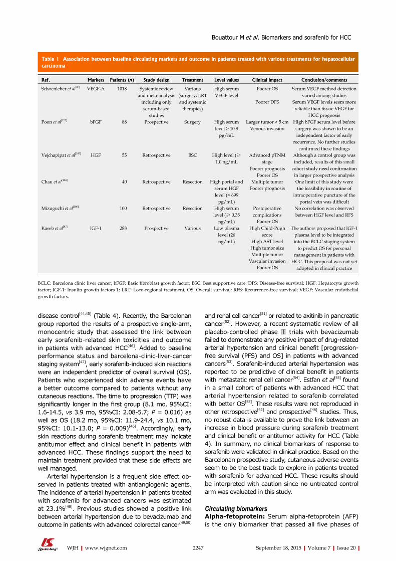
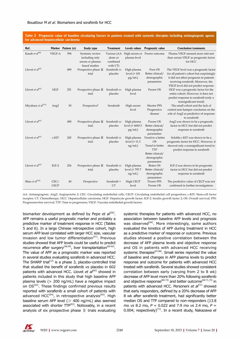
Table 1 Association between baseline circulating markers and outcome in patients treated with various treatments for hepatocellular carcinoma
Ref. Markers Patients (n ) Study design Treatment Level values Clinical impact Conclusion/comments
Schoenleber et al[85] VEGF-A 1018 Systemic review and meta-analysis
including only serum-based
studies
Various (surgery, LRT and systemic
therapies)
High serum VEGF level
Poorer OS Serum VEGF method detection varied among studies
Poorer DFS Serum VEGF levels seem more reliable than tissue VEGF for
HCC prognosisPoon et al[115] bFGF 88 Prospective Surgery High serum
level > 10.8 pg/mL
Larger tumor > 5 cm High bFGF serum level before surgery was shown to be an independent factor of early
recurrence. No further studies confirmed these findings
Venous invasion
Vejchapipat et al[105] HGF 55 Retrospective BSC High level (≥ 1.0 ng/mL
Advanced pTNM stage
Poorer prognosisPoorer OS
Although a control group was included, results of this small
cohort study need confirmation in larger prospective analysis
Chau et al[104] 40 Retrospective Resection High portal and serum HGF level (> 699
pg/mL)
Multiple tumor One limit of this study were the feasibility in routine of
intraoperative puncture of the portal vein was difficult
Poorer prognosis
Mizuguchi et al[106] 100 Retrospective Resection
High serum level (≥ 0.35
ng/mL)
Postoperative complications
No correlation was observed between HGF level and RFS
Poorer OSKaseb et al[87] IGF-1 288 Prospective Various Low plasma
level (26 ng/mL)
High Child-Pugh score
The authors proposed that IGF-1 plasma level to be integrated into the BCLC staging system
to predict OS for personal management in patients with
HCC. This proposal was not yet adopted in clinical practice
High AST levelHigh tumor sizeMultiple tumor
Vascular invasionPoorer OS
BCLC: Barcelona clinic liver cancer; bFGF: Basic fibroblast growth factor; BSC: Best supportive care; DFS: Disease-free survival; HGF: Hepatocyte growth factor; IGF-1: Insulin growth factors 1; LRT: Loco-regional treatment; OS: Overall survival; RFS: Recurrence-free survival; VEGF: Vascular endothelial growth factors.
Bouattour M et al . Biomarkers and sorafenib for HCC
2248 September 18, 2015|Volume 7|Issue 20|WJH|www.wjgnet.com
systemic therapies for patients with advanced HCC, no association between baseline AFP levels and prognosis was observed[66]. More interestingly, some authors evaluated the kinetics of AFP during treatment in HCC as a predictive marker of response or outcome. Previous studies showed a positive correlation between the decrease of AFP plasma levels and objective response and OS in patients with advanced HCC receiving systemic therapies[67,68]. Small series reported the value of baseline and changes in AFP plasma levels to predict response and outcome for patients with advanced HCC treated with sorafenib. Several studies showed consistent correlation between early (varying from 2 to 8 wk) decrease of AFP level more than 20% following sorafenib and objective response[69-73] and better outcome[69-71,73] in patients with advanced HCC. Personeni et al[71] showed that early responders, defined by a 20% decrease of AFP 8 wk after sorafenib treatment, had significantly better median OS and TTP compared to non-responders (13.8 mo vs 8.2 mo, P = 0.022 and 7.9 mo vs 2.4 mo, P = 0.004; respectively)[71]. In a recent study, Nakazawa et
biomarker development as defined by Pepe et al[56]. AFP remains a useful prognostic marker and probably a predictive marker of treatment response in HCC (Tables 5 and 6). In a large Chinese retrospective cohort, high serum AFP level correlated with larger HCC size, vascular invasion and low tumor differentiation[57]. Previous studies showed that AFP levels could be useful to predict recurrence after surgery[58,59], liver transplantation[60-62]. The value of AFP as a prognostic marker was reported in several studies evaluating sorafenib in advanced HCC. The SHARP trial[12] is a phase 3, placebo-controlled trial that studied the benefit of sorafenib vs placebo in 602 patients with advanced HCC. Llovet et al[63] showed in patients included in this study that high baseline AFP plasma levels (> 200 ng/mL) have a negative impact on OS[63]. These findings confirmed previous results reported with sorafenib a small cohort of patients with advanced HCC[64], in retrospective analysis[65]. High baseline serum AFP level (≥ 400 ng/mL) also seemed associated with shorter TTP[63]. Noticeably, in a recent analysis of six prospective phase Ⅱ trials evaluating
Table 2 Prognostic value of baseline circulating factors in patients treated with systemic therapies including antiangiogenic agents for advanced hepatocellular carcinoma
Ref. Marker Patient (n ) Study type Treatment Levels values Prognostic value Conclusion/comments
Kaseb et al[86] VEGF-A 394 Systemic review including only
serum or plasma-based studies
Various (AA alone or
combined with CT)
High serum or plasma level
Poorer outcome Plasma VEGF seemed more relevant than serum VEGF as prognostic factor
for HCC
Llovet et al[63] 490 Prospective phase Ⅲ trial
Sorafenib vs placebo
High plasma level (> 101
pg/mL)
Poor OS The VEGF level was a prognostic factor for all patient's cohort but surprisingly it did not affect prognosis in patients
receiving sorafenib. Moreover, the VEGF level did not predict response
Better clinical/ demographic parameters
Llovet et al[63] HGF 251 Prospective phase Ⅲ trial
Sorafenib vs placebo
High plasma level
Poorer OS HGF was a prognostic factor for the entire cohort. However, it does not
predict response to sorafenib (only a nonsignificant trend)
Miyahara et al[112] Ang2 30 Prospective? Sorafenib High serum level
Shorter PFSProgressive
disease
The small cohort and the lack of control arm hamper conclusion on the role of Ang2 as predictive of response
to sorafenibLlovet et al[63] 490 Prospective phase Ⅲ
trialSorafenib vs
placeboHigh plasma
level (> 6043.5 pg/mL)
Poorer OSBetter clinical/demographic parameters
Ang2 was shown to be a prognostic factor in HCC but did not predict
response to sorafenib
Llovet et al[63] c-KIT 245 Prospective phase Ⅲ trial
Sorafenib vs placebo
High plasma level (> 11.3
ng/mL)
Trend to a better OS
Soluble c-KIT was shown to be a prognostic factor for HCC. However, it showed only a nonsignificant trend to
predict response to sorafenibTrend to better
TTPBetter clinical/demographic parameters
Llovet et al[63] IGF-2 254 Prospective phase Ⅲ trial
Sorafenib vs placebo
High plasma level (> 797.7
ng/mL)
Better OS IGF-2 was shown to be prognostic factor in HCC but did not predict
response to sorafenibBetter clinical/demographic parameters
Shao et al[126] CEC/CECP
40 Prospective Sorafenib + CT
High CECP level
Poorer PFSPoorer OS
The predictive value of CECP was not confirmed in further investigations
AA: Antiangiogenic; Ang2: Angiopoietin 2; CEC: Circulating endothelial cells; CECP: Circulating endothelial cell progenitors; c-KIT: Stem-cell factor receptor; CT: Chemotherapy; HCC: Hepatocellular carcinoma; HGF: Hepatocyte growth factor; IGF-2: Insulin growth factor 2; OS: Overall survival; PFS: Progression-free survival; TTP: Time to progression; VEGF: Vascular endothelial growth factors.
Bouattour M et al . Biomarkers and sorafenib for HCC
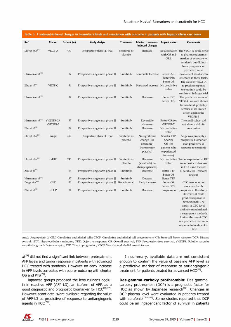
2249 September 18, 2015|Volume 7|Issue 20|WJH|www.wjgnet.com
al[74] did not find a significant link between pretreatment AFP levels and tumor response in patients with advanced HCC treated with sorafenib. However, an early increase in AFP levels correlates with poorer outcome with shorter OS and PFS[74].
Japanese groups proposed the lens culinaris agglu-tinin reactive AFP (AFP-L3), an isoform of AFP, as a good diagnostic and prognostic biomarker for HCC[75-77]. However, scant data is/are available regarding the value of AFP-L3 as predictive of response to antiangiogenic agents in HCC[78].
In summary, available data are not consistent enough to confirm the value of baseline AFP level as a predictive marker of response to antiangiogenic treatment for patients treated for advanced HCC[79].
Des-gamma-carboxy prothrombin: Des-gamma-carboxy prothrombin (DCP) is a prognostic factor for HCC as shown by Japanese research[80]. Changes in DCP plasma level were evaluated in patients treated with sorafenib[73,81,82]. Some studies reported that DCP could be an independent factor of survival in patients
Table 3 Treatment-induced changes in biomarkers levels and association with outcome in patients with hepatocellular carcinoma
Ref. Marker Patient (n ) Study design Treatment Marker treatment-induced changes
Impact value Comments
Llovet et al[63] VEGF-A 490 Prospective phase Ⅲ trial Sorafenib vs placebo
Increase No association with OS and
ORR
The VEGF-A could serve as pharmacodynamic marker of exposure to sorafenib but did not
have prognostic or predictive value
Harmon et al[93] 37 Prospective single arm phase Ⅱ Sunitinib Reversible Increase Better DCR Inconsistent results were observed in these trials. The value of VEGF-A
to predict response to sunitinib could be
confirmed in larger trial
Better PFSBetter OS
Zhu et al[91] VEGF-C 34 Prospective single arm phase Ⅱ Sunitinib
Sustained increase No predictive value
Harmon et al[93] 37 Prospective single arm phase Ⅱ Sunitinib Decrease Better DC The predictive value of VEGF-C was not shown for sorafenib probably because of its limited
action against the VEGFR-3
Better ORR
Harmon et al[93] sVEGFR-2/ sVEGFR-3
37 Prospective single arm phase Ⅱ Sunitinib Reversible decrease
Better OS (for sVEGFR-2)
The small cohort did not allow a definite
conclusion Zhu et al[91] 34 Prospective single arm phase Ⅱ Sunitinib Decrease No predictive value
Llovet et al[63] Ang2 490 Prospective phase Ⅲ trial Sorafenib vs placebo
No significant change (for sorafenib)
Increase (for placebo)
Shorter TTPShorter OS (for
patients who experienced
increase)
Ang2 was probably a prognostic biomarker
than predictive of response to sorafenib
Llovet et al[63] c-KIT 245 Prospective single arm phase Ⅱ Sorafenib vs placebo
Decrease (sorafenib) no
change (placebo)
No predictive value
Tumor expression of KIT was considered as low in HCC, and the role
of soluble KIT remains unclear
Zhu et al[91] 34 Prospective single arm phase Ⅱ Sunitinib Decrease Better TTPBetter OS
Harmon et al[93] 37 Prospective single arm phase Ⅱ Sunitinib Decease Better TTPBoige et al[98] CEC 36 Prospective single arm phase Ⅱ Bevacizumab Early increase Better OR CEC level was not
associated with prognosis in this study.
However, it could predict response to bevacizumab. The rarity of CEC level
and non-standardized measurement methods limited the use of CEC
as a predictive marker of response to treatment in
HCC
Better DCR Zhu et al[91] CECP 34 Prospective single arm phase Ⅱ Sunitinib Decrease Progression
Ang2: Angiopoietin 2; CEC: Circulating endothelial cells; CECP: Circulating endothelial cell progenitors; c-KIT: Stem-cell factor receptor; DCR: Disease control; HCC: Hepatocellular carcinoma; ORR: Objective response; OS: Overall survival; PFS: Progression-free survival; sVEGFR: Soluble vascular endothelial growth factors receptor; TTP: Time to progression; VEGF: Vascular endothelial growth factors.
Bouattour M et al . Biomarkers and sorafenib for HCC
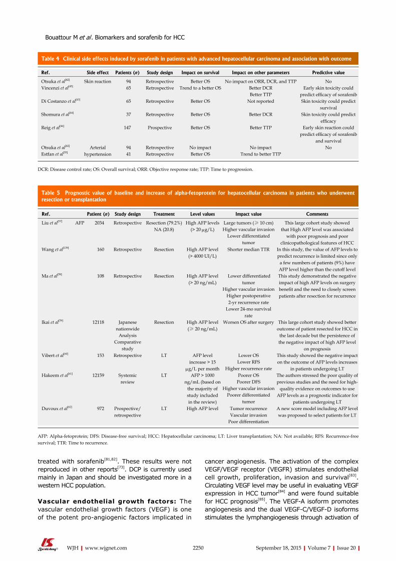
2250 September 18, 2015|Volume 7|Issue 20|WJH|www.wjgnet.com
treated with sorafenib[81,82]. These results were not reproduced in other reports[73]. DCP is currently used mainly in Japan and should be investigated more in a western HCC population.
Vascular endothelial growth factors: The vascular endothelial growth factors (VEGF) is one of the potent pro-angiogenic factors implicated in
cancer angiogenesis. The activation of the complex VEGF/VEGF receptor (VEGFR) stimulates endothelial cell growth, proliferation, invasion and survival[83]. Circulating VEGF level may be useful in evaluating VEGF expression in HCC tumor[84] and were found suitable for HCC prognosis[85]. The VEGF-A isoform promotes angiogenesis and the dual VEGF-C/VEGF-D isoforms stimulates the lymphangiogenesis through activation of
Table 4 Clinical side effects induced by sorafenib in patients with advanced hepatocellular carcinoma and association with outcome
Ref. Side effect Patients (n ) Study design Impact on survival Impact on other parameters Predictive value
Otsuka et al[42] Skin reaction 94 Retrospective Better OS No impact on ORR, DCR, and TTP NoVincenzi et al[45] 65 Retrospective Trend to a better OS Better DCR Early skin toxicity could
predict efficacy of sorafenibBetter TTPDi Costanzo et al[43] 65 Retrospective Better OS Not reported Skin toxicity could predict
survivalShomura et al[44] 37 Retrospective Better OS Better DCR Skin toxicity could predict
efficacyReig et al[46] 147 Prospective Better OS Better TTP Early skin reaction could
predict efficacy of sorafenib and survival
Otsuka et al[42] Arterial hypertension
94 Retrospective No impact No impact NoEstfan et al[55] 41 Retrospective Better OS Trend to better TTP
DCR: Disease control rate; OS: Overall survival; ORR: Objective response rate; TTP: Time to progression.
Table 5 Prognostic value of baseline and increase of alpha-fetoprotein for hepatocellular carcinoma in patients who underwent resection or transplantation
Ref. Patient (n ) Study design Treatment Level values Impact value Comments
Liu et al[57] AFP 2034 Retrospective Resection (79.2%) NA (20.8)
High AFP levels (> 20 µg/L)
Large tumors (≥ 10 cm) This large cohort study showed that High AFP level was associated
with poor prognosis and poor clinicopathological features of HCC
Higher vascular invasionLower differentiated
tumorWang et al[139] 160 Retrospective Resection
High AFP level (> 4000 UI/L)
Shorter median TTR In this study, the value of AFP levels to predict recurrence is limited since only
a few numbers of patients (9%) have AFP level higher than the cutoff level
Ma et al[58] 108 Retrospective Resection High AFP level (> 20 ng/mL)
Lower differentiated tumor
This study demonstrated the negative impact of high AFP levels on surgery benefit and the need to closely screen patients after resection for recurrence
Higher vascular invasionHigher postoperative 2-yr recurrence rate
Lower 24-mo survival rate
Ikai et al[59] 12118 Japanese nationwide
Resection High AFP level (≥ 20 ng/mL)
Worsen OS after surgery This large cohort study showed better outcome of patient resected for HCC in
the last decade but the persistence of the negative impact of high AFP level
on prognosis
AnalysisComparative
studyVibert et al[60] 153 Retrospective LT AFP level
increase > 15 µg/L per month
Lower OS This study showed the negative impact on the outcome of AFP levels increases
in patients undergoing LTLower RFS
Higher recurrence rateHakeem et al[61] 12159 Systemic
reviewLT AFP > 1000
ng/mL (based on the majority of study included in the review)
Poorer OS The authors stressed the poor quality of previous studies and the need for high-
quality evidence on outcomes to use AFP levels as a prognostic indicator for
patients undergoing LT
Poorer DFS Higher vascular invasion
Poorer differentiated tumor
Duvoux et al[62] 972 Prospective/retrospective
LT High AFP level Tumor recurrence A new score model including AFP level was proposed to select patients for LTVascular invasion
Poor differentiation
AFP: Alpha-fetoprotein; DFS: Disease-free survival; HCC: Hepatocellular carcinoma; LT: Liver transplantation; NA: Not available; RFS: Recurrence-free survival; TTR: Time to recurrence.
Bouattour M et al . Biomarkers and sorafenib for HCC
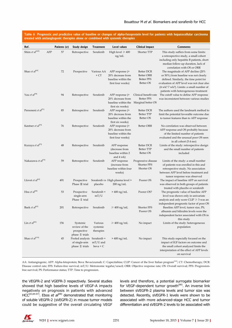
2251 September 18, 2015|Volume 7|Issue 20|WJH|www.wjgnet.com
the VEGFR-2 and VGEFR-3 respectively. Several studies showed that high baseline levels of VEGF-A impacts negatively on prognosis in patients with advanced HCC[63,85-87]. Ebos et al[88] demonstrated that monitoring of soluble VEGFR-2 (sVEGFR-2) in mouse tumor models could be suggestive of the overall circulating VEGF
levels and therefore, a potential surrogate biomarker for VEGF-dependent tumor growth[88]. An inverse link between sVEGFR-2 plasma levels and tumor size was detected. Recently, sVEGFR-1 levels were shown to be associated with more advanced-stage HCC and tumor differentiation and sVEGFR-2 levels to be associated with
Table 6 Prognostic and predictive value of baseline or changes of alpha-fetoprotein level for patients with hepatocellular carcinoma treated with antiangiogenic therapies alone or combined with systemic therapies
Ref. Patients (n ) Study design Treatment Level values Clinical impact Comments
Shim et al[160] AFP 57 Retrospective Sorafenib High level ≥ 400 ng/mL
Shorter TTP This study suffers from some limits: a retrospective study, a small cohort
including only hepatitis B patients, short median follow-up duration, lack of
correlation with OS or ORRShao et al[69] 72 Prospective Various AA
+ CTAFP response (>
20% decrease from baseline within the
first four weeks)
Better DCR The magnitude of AFP decline (20% or 50%) from baseline was not clearly defined. Similarly, the time point for
evaluation of AFP level was not clear also (4 wk? 7 wk?). Limits: a small number of patients with heterogeneous treatment
Better ORRBetter PFSBetter OS
Yau et al[70] 94 Retrospective Sorafenib AFP response (> 20% decrease from baseline within the
first six weeks)
Clinical benefit rate The cutoff value to define AFP response was inconsistent between various studies Better PFS
Marginal better OS
Personeni et al[71] 85 Retrospective Sorafenib AFP response (> 20% decrease from baseline within the
first six weeks)
Better DCR The authors used the landmark method to limit the potential favorable outcome due to tumor features than to AFP response
Better TTPBetter OS
Køstner et al[72] 76 Retrospective Sorafenib AFP response (> 20% decrease from baseline within the
first four weeks)
Better ORR No correlation was observed between AFP response and OS probably because
of the limited number of patients evaluated and the unusual poor OS seen
in all cohort (5.4 mo) Kuzuya et al[73] 48 Retrospective Sorafenib AFP response
(decrease from baseline within 2
and 4 wk)
Better DCR Limits of the study: retrospective design and the small number of patients
includedBetter TTPBetter OS
Nakazawa et al[74] 59 Retrospective Sorafenib AFP response (increase from
baseline within four weeks)
Progressive disease Limits of the study: a small number of patients was enrolled in this and retrospective study. No association
between AFP level before treatment and tumor response was observed
Shorter PFSShorter OS
Llovet et al[63] 491 Prospective Phase Ⅲ trial
Sorafenib vs placebo
High plasma level > 200 ng/mL
Poorer OS The impact of baseline AFP on survival was observed in both groups of patients
treated with placebo or sorafenib Hsu et al[64] 53 Prospective
single-arm Phase Ⅱ trial
Sorafenib + mT/U
> 400 ng/mL Poorer OS? The prognostic value of baseline AFP level was shown only in univariate
analysis and only score CLIP ≥ 3 was an independent prognostic factor of poor OS
Baek et al[65] 201 Retrospective Sorafenib ≥ 400 ng/mL Shorter FFS Baseline AFP level, tumor size, PS, albumin and bilirubin levels were the
independent factor associated with OS in this study
Poorer OS
Lin et al[66] 156 Systemic review of the prospective
phase Ⅱ trials
Various systemic therapies
≥ 400 ng/mL No impact Limits of the study: heterogeneous population
Shao et al[119] 45 Pooled analysis of single-arm phase Ⅱ trials
Sorafenib + mT/U and beva + C
> 400 ng/mL No impact This study especially focused on the impact of IGF factors on outcome and the small cohort analyzed limits the
interpretation of the effect of AFP levels on survival
AA: Antiangiogenic; AFP: Alpha-fetoprotein; Beva: Bevacizumab; C: Capecitabine; CLIP: Cancer of the liver Italian program[161]; CT: Chemotherapy; DCR: Disease control rate; FFS: Failure-free survival; mT/U: Metronomic tegafur/uracil; ORR: Objective response rate; OS: Overall survival; PFS: Progression-free survival; PS: Performance status; TTP: Time to progression.
Bouattour M et al . Biomarkers and sorafenib for HCC
2252 September 18, 2015|Volume 7|Issue 20|WJH|www.wjgnet.com
poorly differentiated tumor[89]. Llovet et al[63] reported changes of plasma VEGF level in patients treated for HCC enrolled in the SHARP study. Compared to baseline level, a significant increase in plasma level of VEGF was observed in the sorafenib group (P = 0.010) and a significant decrease in plasma level of sVEGFR-2 and sVEGFR-3 was seen in the placebo group (P < 0.0001)[63]. The increase of VEGF plasma level found after sorafenib treatment was somewhat surprising since sorafenib showed OS improvement. However, similar findings were observed in patients treated with sorafenib for renal cell carcinoma[90], with sunitinib for advanced HCC[91-93] or renal cell carcinoma[94-96]. Increase of VEGF plasma level could be subsequent to hypoxia induced by the antiangiogenic agents[94]. Noticeably, a reversible increase in the VEGF level induced with sunitinib was also observed in non-tumor-bearing mice suggesting a systemic response that possibly masks tumor-specific changes or any difference in responding patients. Therefore, the increase in VEGF in response to treatment could also occur independently of tumor[97] and might explain the absence of correlation between this change and the outcome in HCC patients treated with antiangiogenic agents[63]. In the SHARP trial, the increase of VEGF-A plasma concentration during sorafenib treatment observed in patients with advanced HCC did not predict OS or tumor response[63]. Similarly, no association between VEGF-A plasma level changes and outcome was observed in patients treated with bevacizumab for advanced HCC[98]. Accordingly, the VEGF-A could serve as a pharmacodynamic marker of exposure to antiangiogenic agents but did not have prognostic or predictive value[85]. Sunitinib induced in patients with HCC, a reduction of VEGF-C (the ligand of VEGFR-3) plasma level that was associated with disease control and tumor response according to the RECIST criteria[23] and Choi criteria[99,100] respectively[93]. Likewise, sunitinib-induced decrease of sVEGFR-3 plasma levels in patients with renal cell cancer and breast cancer correlated with a better outcome[95,101]. Baseline level of VEGF-C may be regarded as a potential predictive biomarker of sunitinib efficacy in patients with advanced HCC[92,93]. However, as sorafenib has limited action against the VEGFR-3[102], the value of this biomarker to predict response in HCC patients could be anecdotal.
In summary, further robust studies are warranted to demonstrate the predictive value of circulating VEGF in patients treated with sorafenib or other antiangiogenic agents for advanced HCC. The plasma VEGF should be assessed more than serum VEGF because it was more reproducible and consistent in estimating the activity of VEGF[86].
Hepatocyte growth factor: The hepatocyte growth factor (HGF) is a strong promoter of hepatocarcino-genesis through the activation of the HGF axis and its receptor MET[103]. Previous studies showed that high serum levels of HGF in patients with HCC negatively associated with OS and outcome[104-106]. In the recent
SHARP study biomarkers analysis, patients treated with sorafenib experienced a decrease in a mean plasma level of HGF although; patients treated with placebo have mean HGF concentration increase[63]. Added to circulating stem-cell factor receptor (c-KIT) and angiopoietin 2 (Ang2) concentrations, HGF level was shown to be an independent factor of survival in patients with advanced HCC[63]. Low baseline HGF plasma level trends toward better OS (12.4 mo vs 6.3 mo, P = 0.073) and TTP in patients treated with sorafenib for HCC[63]. Noticeably, in contrast to plasma levels, tissue HGF expression carries low prognostic information[107]. Further investigations are needed to identify the role of HGF as a predictor of response to sorafenib in patients with advanced HCC.
Ang2: Ang2, one of the families of angiopoietins, is an angiogenic factor implicated in tumor angiogenesis stimulation and progression in human HCC[108]. Tumor overexpression of Ang2 was associated with vascular invasion, tumor size microvessel density level, poorly prognosis HCC[108,109] and poor differentiated tumor[110]. Preoperative presence of Ang2 in the hepatic vein was also associated with portal invasion and poor outcome in HCC resected patients[111]. In a small uncontrolled cohort of patients treated with sorafenib for advanced HCC, the authors reported that Ang2 could predict the outcome[112]. High Ang2 serum baseline level was associated with PFS but not with OS in HCC patients treated with sorafenib[112]. Llovet et al[63] confirmed the negative impact on prognosis of baseline high plasma level of Ang2 in HCC. In patients treated with sorafenib or placebo, median OS was significantly shorter in those with high baseline Ang2 plasma levels compared to those with low baseline concentrations (6.3 mo vs 14.1 mo, HR = 2.407; 95%CI: 1.9-3.03; P < 0.001). In the group of patients treated with sorafenib, no significant changes in median Ang2 plasma levels were observed during the treatment. However, concentration increase was reported in the group of patients treated with placebo[63]. Both patient groups treated with sorafenib or placebo that experienced an increase of Ang2 plasma levels during follow-up had shorter OS and TTP[63]. Ang2 seems, therefore, a prognostic factor of HCC aggressiveness but not an adequate predictive factor of sorafenib efficacy. Llovet et al[63] suggested that dosing Ang2 plasma levels during treatment with sorafenib could be an attractive option to monitor patients with advanced HCC.
Basic fibroblast growth factor: The basic fibroblast growth factor (bFGF) is one of the identified angiogenic factors with a potent stimulus for HCC growth[113]. Tumor overexpression of bFGF seems mainly implicated in HCC invasiveness than tumor neovascularization[114]. Moreover, a significant correlation between high preo-perative serum bFGF level and larger tumor, venous invasion, advanced tumor staging and early recurrence was reported in resected HCC[115]. In the SHARP study,
Bouattour M et al . Biomarkers and sorafenib for HCC
2253 September 18, 2015|Volume 7|Issue 20|WJH|www.wjgnet.com
no difference was observed concerning changes in mean bFGF plasma concentration between sorafenib and placebo in patients with advanced HCC[63].
Stem-cell factor receptor - KIT: The role of stem-cell factor receptor and its soluble forms has not been entirely elucidated in HCC. Soluble forms of KIT were fundamentally implicated in tumor-cell survival and proliferation[93]. Llovet et al[63] reported a trend to a positive impact of high baseline soluble c-KIT level on OS and TTP in patients treated with sorafenib. Sorafenib induced a significant decrease in mean plasma levels of soluble c-KIT, unlike the placebo that resulted in no changes in c-KIT concentration[63]. Likewise, following exposure to sunitinib, plasma levels of soluble c-KIT decreased significantly in patients with renal cell carcinoma[95], breast cancer[101] and HCC[91-93]. SHARP biomarker analysis showed a nonsignificant trend of soluble c-KIT in predicting sorafenib response in patients with advanced HCC. In the sorafenib cohort, patients with high baseline soluble c-KIT level showed better median OS and TTP compared to those with low soluble c-KIT level but without reaching significance (10.4 mo vs 9.4 mo, P = 0.081 and 6.7 mo vs 4.1 mo, P = 0.052; respectively)[63]. In a phase Ⅱ study, Zhu et al[91] reported that soluble KIT plasma levels decrease following 14 d of sunitinib treatment in patients with advanced HCC and correlated with better PFS and OS. Similarly, improvement of TTP and trend towards better OS were reported when soluble KIT plasma level decreased from baseline following sunitinib in patients with HCC, metastatic breast cancer and neuroendocrine tumor[93,95,101]. Nowadays, the role of soluble c-KIT in HCC pathogenesis remains unclear since the expression of this protein kinase in HCC tissue appears to be anecdotal[116].
Insulin growth factors: The insulin growth factors (IGF) signaling pathway, including its ligand, IGF-1, and IGF-2, plays a crucial role in carcinogenesis of various tumors[117,118]. In patients with HCC, independently to the tumor stage, low baseline IGF-1 plasma level correlated with poorer OS[87]. In a small cohort of patients with advanced HCC receiving first-line antiangiogenic treatment associated with metronomic chemotherapy, serum levels of IGF-1 could predict treatment efficacy in this population. Indeed, high baseline IGF-1 serum levels before treatment correlate with better OS, PFS and disease control rate[87]. Moreover, high baseline IGF-2 plasma levels associated with a better OS in the placebo group enrolled in the SHARP trial[63]. In this large phase Ⅲ controlled trial, the IGF-2 failed to predict response to sorafenib in patients with advanced HCC[63] confirming previous results observed with other antiangiogenic agents[119].
Circulating endothelial cells and circulating endothelial cell progenitors: In preclinical models, levels of circulating endothelial cells (CEC) and bone-
marrow-derived CEC progenitors (CECP) were shown to be potential surrogate markers of angiogenesis[120,121]. High circulating level of CECP in patients with HCC correlates with advanced disease[122]. Previous studies reported levels of CEC and CECP decrease and return to normal values following antiangiogenic therapy in cases of complete remission[123]. Willett et al[124] showed that high doses of bevacizumab induce an increase of viable CEC and CECP percentage in a small cohort of patient with rectal cancer. Bevacizumab treatment induced in patients with advanced HCC, an early increase of viable CEC levels that correlated with objective response[98]. In patients with imatinib-resistant gastrointestinal stro-mal tumor, sunitinib induced early, but not subsequent increase of CEC blood levels that seemed to be corre-lating with clinical benefit[125]. Otherwise, sunitinib was shown to cause a decrease of CECP level in patients with advanced HCC[91]. Shao et al[126] showed that high baseline CECP level, but not CEC level, was associated with poor OS in patients treated with sorafenib com-bined with metronomic chemotherapy. The value of CEC and CECP levels as biomarkers of angiogenesis and antiangiogenic therapies in HCC needs further prospective analysis. In fact, methods and techniques of measurement were inconsistent, and unreliable results were reported depending on the type of study (clinical or preclinical studies), cancer types, and antiangiogenic agents[98,115,116,121].
In summary, none of the above biomarkers is vali-dated to predict response to sorafenib in patient with advanced HCC. Except the SHARP biomarkers analysis study, the majority of available data was reported from no control arm retrospective studies. Validation through further large, controlled randomized trials are required to confirm the predictive value of such pre-dictive biomarkers so to be integrated with clinical use. Moreover, techniques used to assess drug-induced variation in circulating factors should be standardized for reliable interpretation. An important issue should also be questioned of whether the presence or change in circulating biomarkers could discriminate between treatment benefit and tumor resistance or escape.
Tissue biomarkersIn addition to tissue prognosis markers obtained from tumor samples, some studies tried to identify predictive factors of response and outcome following anticancer agents. Table 7 summarizes studies evaluating tissue biomarkers used as prognostic and predictive of HCC. Abou-Alfa et al[127] evaluated the impact of tumor expression of phosphorylated extracellular signal-regulated kinase (pERK) and outcome in patients treated with sorafenib for advanced HCC. A high pretreatment tumor level of pERK correlated with TTP, but the survival impact was not analyzed. Tumor-cell expression and staining levels of pERK using immunohistochemistry analysis were performed in 33 patients. Patients with high pretreatment tumor-cell pERK expression had better TTP compared to those low staining intensity. The
Bouattour M et al . Biomarkers and sorafenib for HCC
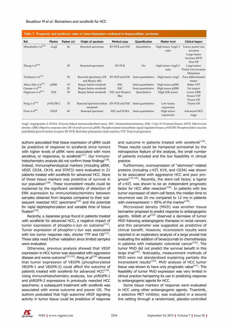
2254 September 18, 2015|Volume 7|Issue 20|WJH|www.wjgnet.com
authors speculated that tissue expression of pERK could be predictive of response to sorafenib since tumors with higher levels of pERK were associated with more sensitive, or responsive, to sorafenib[127]. Our immuno-histochemistry analysis did not confirm these findings[128]. Indeed, immunophenotypical markers (including pERK, VEGF, CD34, CK19, and STAT3) were evaluated in 21 patients treated with sorafenib for advanced HCC. None of these tissue markers was predictive of survival in our population[128]. These inconsistent results could be explained by the significant variability of detection of ERK expression by immunohistochemistry between samples obtained from biopsies compared to their sub-sequent resected HCC specimens[129] and the potential for rapid dephosphorylation and variable time of tissue fixation[130].
Recently, a Japanese group found in patients treated with sorafenib for advanced HCC, a negative impact of tumor expression of phospho-c-Jun on outcome[131]. Tumor expression of phosphor-c-Jun was associated with low tumor response rate, shorter TTP and OS[131]. These data need further validation since limited samples were evaluated.
Otherwise, previous analysis showed that VEGF expression in HCC tumor was associated with aggressive disease and worse outcome[132,133]. Peng et al[134] showed that tumor expression of VEGFR (phosphorylated VEGFR-1 and VEGFR-2) could affect the outcome of patients treated with sorafenib for advanced HCC[134]. Using immunohistochemistry analyzes, low pVEGFR-1 and pVEGFR-2 expressions in previously resected HCC specimens; a subsequent treatment with sorafenib was associated with worse outcome and poorer OS. The authors postulated that high autocrine VEGF signaling activity in tumor tissue could be predictive of response
and outcome in patients treated with sorafenib[134]. These results could be hampered somewhat by the retrospective feature of the analysis, the small number of patients included and the low feasibility in clinical practice.
Furthermore, overexpression of “stemness”-related proteins (including c-KIT, K19, and CD34) was shown to be associated with aggressive HCC and poor pro-gnosis[135-138]. Recently, the stem-cell factor, a ligand of c-KIT, was shown to be an independent prognostic factor for HCC after resection[139]. In patients with low tumor expression of stem-cell factor, the median time to recurrence was 24 mo compared to 12 mo in patients with overexpression > 85% of the marker[139].
Microvessel density (MVD) was another tissue biomarker proposed to predict response to antiangiogenic agents. Willett et al[124] observed a decrease of tumor MVD following antiangiogenic therapies in rectal cancers and this parameter was suggested as predictive of clinical benefit. However, inconsistent results were reported in an exploratory analysis of a large pivotal trial evaluating the addition of bevacizumab to chemotherapy in patients with metastatic colorectal cancer[140]. The tumor MVD did not predict the survival benefit in this large trial[140]. Noticeably, measurement methods of MVD were not standardized explaining partially the inconsistent results[140]. MVD analysis of HCC tumor tissue was shown to have only prognostic value[141]. The feasibility of tumor MVD expression was very limited in clinical practice hampering its use in predicting response to antiangiogenic agents for HCC.
Some tissue markers of response were evaluated in HCC using other antiangiogenic agents. Tivantinib, a selective MET inhibitor, was evaluated in a second line setting through a randomized, placebo-controlled
Table 7 Prognostic and predictive value of tissue biomarkers evaluated in hepatocellular carcinoma
Ref. Marker Patient (n ) Origin of specimen Method assay Quantification Marker level Clinical impact
Mitsuhashi et al[108] Ang2 46 Resected specimens RT-PCR and IHC Quantitative High tumor Ang2/1 ratio
Tumor portal vein invasion
Large tumorIncrease MVD
Poor OSZhang et al[109] 38 Resected specimens RT-PCR No High tumor Ang2/1
ratioLarge tumor
Portal vein invasionMetastasis
Torimura et al[110] 59 Resected specimens (19) and Biopsy (40)
RT-PCR and IHC Semi-quantitative High tumor Ang2 Poor differentiated tumor
Abou-Alfa et al[127] pERK 33 Biopsy before sorafenib IHC Semi-quantitative High tumor pERK Better TTPOzenne et al[128] 20 Biopsy before sorafenib IHC Semi-quantitative High tumor pERK No impactHagiwara et al[131] JNK 39 Biopsy before sorafenib IHC and Western
BlotQuantitative High JNK tumor Lower ORR
Poorer TTPPoorer OS
Peng et al[134] pVEGFR-2 35 Resected specimen before sorafenib
RT-PCR and IHC Semi-quantitative Low tumor expression
Poorer OS
Poon et al[84] VEGF 60 Resected specimen IHC and ELISA Semi-quantitative High tumor expression
Advanced HCC stage
Ang2: Angiopoietin 2; ELISA: Enzyme-linked immunoadsorbent assay; IHC: Immunohistochemistry; JNK: C-Jun N-Terminal Kinase; MVD: Microvessel density; ORR: Objective response rate; OS: Overall survival; pERK: Phosphorylated extracellular signal regulated kinase; pVEGFR: Phosphorylated vascular endothelial growth factors receptor; RT-PCR: Real-time polymerase chain reaction; TTP: Time to progression.
Bouattour M et al . Biomarkers and sorafenib for HCC
2255 September 18, 2015|Volume 7|Issue 20|WJH|www.wjgnet.com
phase Ⅱ trial in patients with advanced HCC[142]. In this study, tumor expression of MET influenced treatment benefit. Patients with tumor overexpression of MET clearly benefit from tivantinib treatment. High-MET tumor expression was associated with longer TTP on tivantinib compared to placebo (2.7 mo vs 1.4 mo, HR = 0.43, 95%CI: 0.19-0.97; P = 0.03) and OS (7.2 mo vs 3.8 mo, HR = 0.38, 95%CI: 0.18-0.81; P = 0.01). Interestingly, tivantinib did not show any benefit when tumor expression of MET was low[142].
Nowadays, no tissues biomarkers can identify patients who might respond to sorafenib. Tumor analy-sis data were/was unavailable in large clinical trials, probably because of lack of tumor samples biopsies since HCC diagnosis was frequently made according to imaging features[143,144].
IMAGING FEATURES AND FUNCTIONAL IMAGING The clinical benefit of sorafenib with OS gain in patients with advanced HCC contrasted largely with a low objective response rate noted in this population. The low response rates could be considered as a sign of lack of antitumor activity in early phases of clinical trials but were favorably balanced by sustained tumor stabilization and small numbers of tumor progression in the waterfall plot activity. Fortunately, the decision to proceed with phase Ⅲ trials was not hampered by the apparent lack of tumor response.
Which response criteria to apply?The conventional RECIST criteria[23,24] usually used for tumor response evaluation of conventional chemo-therapy appear clearly inappropriate to evaluate the response to sorafenib in patients with advanced HCC. Major features were reported following antiangiogenic agents consisting of decreased tumor vascularization[145] and density[146] on computer tomography (CT) scans. The modified RECIST (mRECIST) criteria are a new assessment method proposed by Lencioni and Llovet[145] to overcome the limitations of RECIST criteria. They include vascularization and tumor arterial enhancement changes of the target lesion on CT. Other new criteria including European Association for the Study of the Liver (EASL) criteria and Choi criteria, that evaluated tumor density changes, were also proposed to evaluate tumor response to sorafenib in patients with HCC[100,146-148]. A representative case of discrepancies between these criteria is shown in Figure 1. Several studies used CT-scan evaluation to predict early response to sorafenib and to adjust treatment strategy according to the potential clinical benefit[100,147,149].
Edeline et al[147] showed in patients treated with sorafenib for advanced HCC that overall response rate was higher when mRECIST criteria were applied compared to RECIST criteria (22.7% vs 1.9%). Interestingly, tumor response assessment according
to mRECIST criteria, reclassified 22.6% of patients as responders while they were initially categorized as having stable disease by RECIST criteria[147]. Our group found consistent results when alternative radiological criteria to RECIST were applied[100]. We evaluated early tumor response in 64 patients with advanced HCC treated with sorafenib using RECIST, mRECIST, Choi and EASL criteria[100]. These new criteria identified a higher objective response rate compared to the conventional RECIST criteria (varying from 51% for Choi to 28% and 28% for mRECIST and EASL respectively; compared to only 3% for RECIST criteria). Responder patients according to Choi criteria at the first tumor assessment had better OS compared to non-responders (22.4 mo vs 10.6 mo, 95%CI: 0.15-086; P = 0.097)[100].
Further evaluations of these new criteria in com-parison to RECIST criteria are needed in prospective clinical trials evaluating sorafenib or other antiangiogenic agents for advanced HCC.
In summary, we believe that, combining early reduction of AFP levels following sorafenib initiation with new radiological criteria could be helpful in detect-ing patients who might benefit from antiangiogenic treatment and to propose better tailor-made strategy management.
Functional imagingVarious functional imaging tools [including contrast-enhanced ultrasound, dynamic contrast-enhanced magnetic resonance imaging (MRI) and dynamic contrast-enhanced CT and positron emission tomography (PET)] were proposed to evaluate the antiangiogenic effects[150] (Table 8). Functional imaging approaches consist of infusion of intravenous contrast agent that enhances vascular and tumor structures and the acquisition of sequential images before, during, and after injection.
Dynamic contrast-enhanced ultrasound Some small cohort studies evaluated the useful of dynamic contrast-enhanced ultrasound (DCE-US) to predict early tumor response to sorafenib in patients with advanced HCC[151-153]. In a Japanese prospective monocentric study, a total of 37 patients with advanced HCC treated with sorafenib were evaluated using DCE-US, before treatment and on days 7, 14 and 28 of treatment[152]. Significant changes in different US perfusion parameters between responders and non-responders (according to RECIST and mRECIST criteria) were observed at the prescheduled time of the follow-up. Correlation between reduction in tumor blood volume 7 d after treatment initiation and better PFS and OS was found. The authors suggest that DCE-US performed earlier could be useful to identify patients with advanced HCC, who may benefit from sorafenib[152]. Consistent results were obtained in an Italian prospective study that enrolled 28 patients treated with sorafenib and monitored with DCE-US at baseline, days 7, 15 and 30 of treatment[154]. Early decrease of tumor vascularity
Bouattour M et al . Biomarkers and sorafenib for HCC
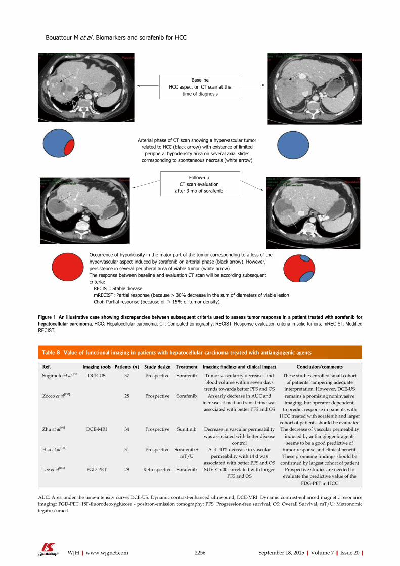
2256 September 18, 2015|Volume 7|Issue 20|WJH|www.wjgnet.com
BaselineHCC aspect on CT scan at the
time of diagnosis
Arterial phase of CT scan showing a hypervascular tumor related to HCC (black arrow) with existence of limited
peripheral hypodensity area on several axial slides corresponding to spontaneous necrosis (white arrow)
FollowupCT scan evaluation
after 3 mo of sorafenib
Occurrence of hypodensity in the major part of the tumor corresponding to a loss of the hypervascular aspect induced by sorafenib on arterial phase (black arrow). However, persistence in several peripheral area of viable tumor (white arrow)The response between baseline and evaluation CT scan will be according subsequent criteria: RECIST: Stable disease mRECIST: Partial response (because > 30% decrease in the sum of diameters of viable lesion Choi: Partial response (because of ≥ 15% of tumor density)
Figure 1 An illustrative case showing discrepancies between subsequent criteria used to assess tumor response in a patient treated with sorafenib for hepatocellular carcinoma. HCC: Hepatocellular carcinoma; CT: Computed tomography; RECIST: Response evaluation criteria in solid tumors; mRECIST: Modified RECIST.
Table 8 Value of functional imaging in patients with hepatocellular carcinoma treated with antiangiogenic agents
Ref. Imaging tools Patients (n ) Study design Treatment Imaging findings and clinical impact Conclusion/comments
Sugimoto et al[152] DCE-US 37 Prospective Sorafenib Tumor vascularity decreases and blood volume within seven days
trends towards better PFS and OS
These studies enrolled small cohort of patients hampering adequate
interpretation. However, DCE-US remains a promising noninvasive imaging, but operator dependent,
to predict response in patients with HCC treated with sorafenib and larger cohort of patients should be evaluated
Zocco et al[153] 28 Prospective Sorafenib An early decrease in AUC and increase of median transit time was associated with better PFS and OS
Zhu et al[91] DCE-MRI 34 Prospective Sunitinib Decrease in vascular permeability was associated with better disease
control
The decrease of vascular permeability induced by antiangiogenic agents seems to be a good predictive of
tumor response and clinical benefit. These promising findings should be
confirmed by largest cohort of patient
Hsu et al[156] 31 Prospective Sorafenib + mT/U
A ≥ 40% decrease in vascular permeability with 14 d was
associated with better PFS and OSLee et al[159] FGD-PET 29 Retrospective Sorafenib SUV < 5.00 correlated with longer
PFS and OS Prospective studies are needed to
evaluate the predictive value of the FDG-PET in HCC
AUC: Area under the time-intensity curve; DCE-US: Dynamic contrast-enhanced ultrasound; DCE-MRI: Dynamic contrast-enhanced magnetic resonance imaging; FGD-PET: 18F-fluorodeoxyglucose - positron-emission tomography; PFS: Progression-free survival; OS: Overall Survival; mT/U: Metronomic tegafur/uracil.
Bouattour M et al . Biomarkers and sorafenib for HCC
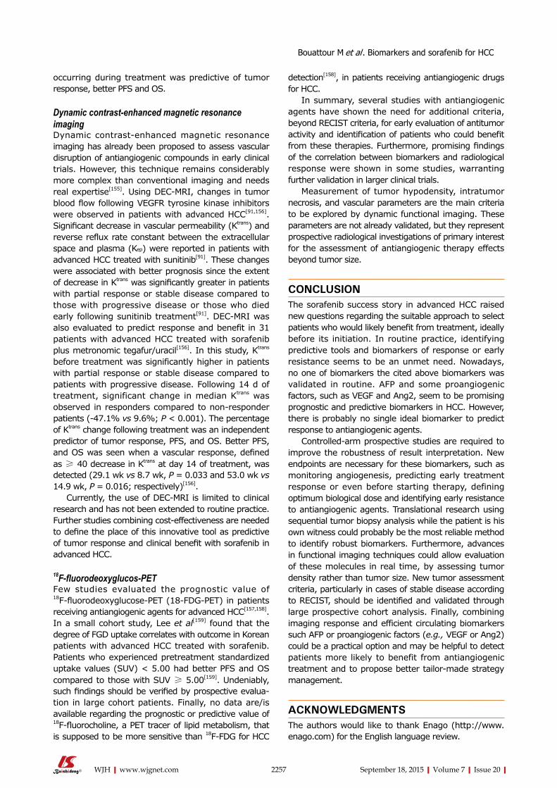
2257 September 18, 2015|Volume 7|Issue 20|WJH|www.wjgnet.com
occurring during treatment was predictive of tumor response, better PFS and OS.
Dynamic contrast-enhanced magnetic resonance imagingDynamic contrast-enhanced magnetic resonance imaging has already been proposed to assess vascular disruption of antiangiogenic compounds in early clinical trials. However, this technique remains considerably more complex than conventional imaging and needs real expertise[155]. Using DEC-MRI, changes in tumor blood flow following VEGFR tyrosine kinase inhibitors were observed in patients with advanced HCC[91,156]. Significant decrease in vascular permeability (Ktrans) and reverse reflux rate constant between the extracellular space and plasma (Kep) were reported in patients with advanced HCC treated with sunitinib[91]. These changes were associated with better prognosis since the extent of decrease in Ktrans was significantly greater in patients with partial response or stable disease compared to those with progressive disease or those who died early following sunitinib treatment[91]. DEC-MRI was also evaluated to predict response and benefit in 31 patients with advanced HCC treated with sorafenib plus metronomic tegafur/uracil[156]. In this study, Ktrans before treatment was significantly higher in patients with partial response or stable disease compared to patients with progressive disease. Following 14 d of treatment, significant change in median Ktrans was observed in responders compared to non-responder patients (-47.1% vs 9.6%; P < 0.001). The percentage of Ktrans change following treatment was an independent predictor of tumor response, PFS, and OS. Better PFS, and OS was seen when a vascular response, defined as ≥ 40 decrease in Ktrans at day 14 of treatment, was detected (29.1 wk vs 8.7 wk, P = 0.033 and 53.0 wk vs 14.9 wk, P = 0.016; respectively)[156].
Currently, the use of DEC-MRI is limited to clinical research and has not been extended to routine practice. Further studies combining cost-effectiveness are needed to define the place of this innovative tool as predictive of tumor response and clinical benefit with sorafenib in advanced HCC.
18F-fluorodeoxyglucos-PETFew studies evaluated the prognostic value of 18F-fluorodeoxyglucose-PET (18-FDG-PET) in patients receiving antiangiogenic agents for advanced HCC[157,158]. In a small cohort study, Lee et al[159] found that the degree of FGD uptake correlates with outcome in Korean patients with advanced HCC treated with sorafenib. Patients who experienced pretreatment standardized uptake values (SUV) < 5.00 had better PFS and OS compared to those with SUV ≥ 5.00[159]. Undeniably, such findings should be verified by prospective evalua-tion in large cohort patients. Finally, no data are/is available regarding the prognostic or predictive value of 18F-fluorocholine, a PET tracer of lipid metabolism, that is supposed to be more sensitive than 18F-FDG for HCC
detection[158], in patients receiving antiangiogenic drugs for HCC.
In summary, several studies with antiangiogenic agents have shown the need for additional criteria, beyond RECIST criteria, for early evaluation of antitumor activity and identification of patients who could benefit from these therapies. Furthermore, promising findings of the correlation between biomarkers and radiological response were shown in some studies, warranting further validation in larger clinical trials.
Measurement of tumor hypodensity, intratumor necrosis, and vascular parameters are the main criteria to be explored by dynamic functional imaging. These parameters are not already validated, but they represent prospective radiological investigations of primary interest for the assessment of antiangiogenic therapy effects beyond tumor size.
CONCLUSIONThe sorafenib success story in advanced HCC raised new questions regarding the suitable approach to select patients who would likely benefit from treatment, ideally before its initiation. In routine practice, identifying predictive tools and biomarkers of response or early resistance seems to be an unmet need. Nowadays, no one of biomarkers the cited above biomarkers was validated in routine. AFP and some proangiogenic factors, such as VEGF and Ang2, seem to be promising prognostic and predictive biomarkers in HCC. However, there is probably no single ideal biomarker to predict response to antiangiogenic agents.
Controlled-arm prospective studies are required to improve the robustness of result interpretation. New endpoints are necessary for these biomarkers, such as monitoring angiogenesis, predicting early treatment response or even before starting therapy, defining optimum biological dose and identifying early resistance to antiangiogenic agents. Translational research using sequential tumor biopsy analysis while the patient is his own witness could probably be the most reliable method to identify robust biomarkers. Furthermore, advances in functional imaging techniques could allow evaluation of these molecules in real time, by assessing tumor density rather than tumor size. New tumor assessment criteria, particularly in cases of stable disease according to RECIST, should be identified and validated through large prospective cohort analysis. Finally, combining imaging response and efficient circulating biomarkers such AFP or proangiogenic factors (e.g., VEGF or Ang2) could be a practical option and may be helpful to detect patients more likely to benefit from antiangiogenic treatment and to propose better tailor-made strategy management.
ACKNOWLEDGMENTSThe authors would like to thank Enago (http://www.enago.com) for the English language review.
Bouattour M et al . Biomarkers and sorafenib for HCC
2258 September 18, 2015|Volume 7|Issue 20|WJH|www.wjgnet.com
REFERENCES1 Kamangar F, Dores GM, Anderson WF. Patterns of cancer inci-
dence, mortality, and prevalence across five continents: defining priorities to reduce cancer disparities in different geographic regions of the world. J Clin Oncol 2006; 24: 2137-2150 [PMID: 16682732 DOI: 10.1200/JCO.2005.05.2308]
2 Ferlay J, Shin HR, Bray F, Forman D, Mathers C, Parkin DM. Estimates of worldwide burden of cancer in 2008: GLOBOCAN 2008. Int J Cancer 2010; 127: 2893-2917 [PMID: 21351269 DOI: 10.1002/ijc.25516]
3 Llovet JM, Burroughs A, Bruix J. Hepatocellular carcinoma. Lancet 2003; 362: 1907-1917 [PMID: 14667750 DOI: 10.1016/S0140-6736(03)14964-1]
4 Thomas MB, Abbruzzese JL. Opportunities for targeted therapies in hepatocellular carcinoma. J Clin Oncol 2005; 23: 8093-8108 [PMID: 16258107 DOI: 10.1200/JCO.2004.00.1537]
5 Pang RW, Poon RT. From molecular biology to targeted therapies for hepatocellular carcinoma: the future is now. Oncology 2007; 72 Suppl 1: 30-44 [PMID: 18087180 DOI: 10.1159/000111705]
6 Llovet JM, Bruix J. Molecular targeted therapies in hepatocellular carcinoma. Hepatology 2008; 48: 1312-1327 [PMID: 18821591 DOI: 10.1002/hep.22506]
7 Psyrri A, Arkadopoulos N, Vassilakopoulou M, Smyrniotis V, Dimitriadis G. Pathways and targets in hepatocellular carcinoma. Expert Rev Anticancer Ther 2012; 12: 1347-1357 [PMID: 23176622 DOI: 10.1586/era.12.113]
8 Kudo M. Signaling pathway/molecular targets and new targeted agents under development in hepatocellular carcinoma. World J Gastroenterol 2012; 18: 6005-6017 [PMID: 23155330 DOI: 10.3748/wjg.v18.i42.6005]
9 Faivre S, Bouattour M, Raymond E. Novel molecular therapies in hepatocellular carcinoma. Liver Int 2011; 31 Suppl 1: 151-160 [PMID: 21205154 DOI: 10.1111/j.1478-3231.2010.02395.x]
10 Shen YC, Lin ZZ, Hsu CH, Hsu C, Shao YY, Cheng AL. Clinical trials in hepatocellular carcinoma: an update. Liver Cancer 2013; 2: 345-364 [PMID: 24400222 DOI: 10.1159/000343850]
11 Bouattour M, Wassermann J. Molecular targeted drugs under investigation in hepatocellular carcinoma. 4th ed. Frontiers in Anti-Cancer Drug Discovery. Bentham Science, 2014: 39-87. Available from: URL: http://ebooks.benthamscience.com/book/9781608059225/chapter/124572/
12 Llovet JM, Ricci S, Mazzaferro V, Hilgard P, Gane E, Blanc JF, de Oliveira AC, Santoro A, Raoul JL, Forner A, Schwartz M, Porta C, Zeuzem S, Bolondi L, Greten TF, Galle PR, Seitz JF, Borbath I, Häussinger D, Giannaris T, Shan M, Moscovici M, Voliotis D, Bruix J. Sorafenib in advanced hepatocellular carcinoma. N Engl J Med 2008; 359: 378-390 [PMID: 18650514 DOI: 10.1056/NEJMoa0708857]
13 Cheng AL, Kang YK, Chen Z, Tsao CJ, Qin S, Kim JS, Luo R, Feng J, Ye S, Yang TS, Xu J, Sun Y, Liang H, Liu J, Wang J, Tak WY, Pan H, Burock K, Zou J, Voliotis D, Guan Z. Efficacy and safety of sorafenib in patients in the Asia-Pacific region with advanced hepatocellular carcinoma: a phase III randomised, double-blind, placebo-controlled trial. Lancet Oncol 2009; 10: 25-34 [PMID: 19095497 DOI: 10.1016/S1470-2045(08)70285-7]
14 Connock M, Round J, Bayliss S, Tubeuf S, Greenheld W, Moore D. Sorafenib for the treatment of advanced hepatocellular carcinoma. Health Technol Assess 2010; 14 Suppl 1: 17-21 [PMID: 20507799 DOI: 10.3310/hta14suppl1/03]
15 Cammà C, Cabibbo G, Petta S, Enea M, Iavarone M, Grieco A, Gasbarrini A, Villa E, Zavaglia C, Bruno R, Colombo M, Craxì A. Cost-effectiveness of sorafenib treatment in field practice for patients with hepatocellular carcinoma. Hepatology 2013; 57: 1046-1054 [PMID: 23299720 DOI: 10.1002/hep.26221]
16 Palmer DH, Hussain SA, Smith AJ, Hargreaves S, Ma YT, Hull D, Johnson PJ, Ross PJ. Sorafenib for advanced hepatocellular carcinoma (HCC): impact of rationing in the United Kingdom. Br J Cancer 2013; 109: 888-890 [PMID: 23880824 DOI: 10.1038/bjc.2013.410]
17 Iavarone M, Cabibbo G, Piscaglia F, Zavaglia C, Grieco A, Villa E, Cammà C, Colombo M; SOFIA (SOraFenib Italian Assessment) study group. Field-practice study of sorafenib therapy for hepatocellular carcinoma: a prospective multicenter study in Italy. Hepatology 2011; 54: 2055-2063 [PMID: 21898496 DOI: 10.1002/hep.24644]
18 Tod M, Mir O, Bancelin N, Coriat R, Thomas-Schoemann A, Taieb F, Boudou-Rouquette P, Ropert S, Michels J, Abbas H, Durand JP, Dauphin A, Vidal M, Goldwasser F, Blanchet B. Functional and clinical evidence of the influence of sorafenib binding to albumin on sorafenib disposition in adult cancer patients. Pharm Res 2011; 28: 3199-3207 [PMID: 21691893 DOI: 10.1007/s11095-011-0499-1]
19 Arrondeau J, Mir O, Boudou-Rouquette P, Coriat R, Ropert S, Dumas G, Rodrigues MJ, Rousseau B, Blanchet B, Goldwasser F. Sorafenib exposure decreases over time in patients with hepatocellular carcinoma. Invest New Drugs 2012; 30: 2046-2049 [PMID: 22038662 DOI: 10.1007/s10637-011-9764-8]
20 Mir O, Coriat R, Blanchet B, Durand JP, Boudou-Rouquette P, Michels J, Ropert S, Vidal M, Pol S, Chaussade S, Goldwasser F. Sarcopenia predicts early dose-limiting toxicities and pharma-cokinetics of sorafenib in patients with hepatocellular carcinoma. PLoS One 2012; 7: e37563 [PMID: 22666367 DOI: 10.1371/journal.pone.0037563]
21 Lee JH, Chung YH, Kim JA, Shim JH, Lee D, Lee HC, Shin ES, Yoon JH, Kim BI, Bae SH, Koh KC, Park NH. Genetic predisposition of hand-foot skin reaction after sorafenib therapy in patients with hepatocellular carcinoma. Cancer 2013; 119: 136-142 [PMID: 22736425 DOI: 10.1002/cncr.27705]
22 Widmer N, Bardin C, Chatelut E, Paci A, Beijnen J, Levêque D, Veal G, Astier A. Review of therapeutic drug monitoring of anticancer drugs part two--targeted therapies. Eur J Cancer 2014; 50: 2020-2036 [PMID: 24928190 DOI: 10.1016/j.ejca.2014.04.015]
23 Therasse P, Arbuck SG, Eisenhauer EA, Wanders J, Kaplan RS, Rubinstein L, Verweij J, Van Glabbeke M, van Oosterom AT, Christian MC, Gwyther SG. New guidelines to evaluate the response to treatment in solid tumors. European Organization for Research and Treatment of Cancer, National Cancer Institute of the United States, National Cancer Institute of Canada. J Natl Cancer Inst 2000; 92: 205-216 [PMID: 10655437]
24 Eisenhauer EA, Therasse P, Bogaerts J, Schwartz LH, Sargent D, Ford R, Dancey J, Arbuck S, Gwyther S, Mooney M, Rubinstein L, Shankar L, Dodd L, Kaplan R, Lacombe D, Verweij J. New response evaluation criteria in solid tumours: revised RECIST guideline (version 1.1). Eur J Cancer 2009; 45: 228-247 [PMID: 19097774 DOI: 10.1016/j.ejca.2008.10.026]
25 Biomarkers Definitions Working Group. Biomarkers and surrogate endpoints: preferred definitions and conceptual framework. Clin Pharmacol Ther 2001; 69: 89-95 [PMID: 11240971 DOI: 10.1067/mcp.2001.113989]
26 Ludwig JA, Weinstein JN. Biomarkers in cancer staging, prognosis and treatment selection. Nat Rev Cancer 2005; 5: 845-856 [PMID: 16239904 DOI: 10.1038/nrc1739]
27 Sessa C, Guibal A, Del Conte G, Rüegg C. Biomarkers of angiogenesis for the development of antiangiogenic therapies in oncology: tools or decorations? Nat Clin Pract Oncol 2008; 5: 378-391 [PMID: 18560389 DOI: 10.1038/ncponc1150]
28 Lièvre A, Bachet JB, Le Corre D, Boige V, Landi B, Emile JF, Côté JF, Tomasic G, Penna C, Ducreux M, Rougier P, Penault-Llorca F, Laurent-Puig P. KRAS mutation status is predictive of response to cetuximab therapy in colorectal cancer. Cancer Res 2006; 66: 3992-3995 [PMID: 16618717]
29 Van Cutsem E, Köhne CH, Hitre E, Zaluski J, Chang Chien CR, Makhson A, D’Haens G, Pintér T, Lim R, Bodoky G, Roh JK, Folprecht G, Ruff P, Stroh C, Tejpar S, Schlichting M, Nippgen J, Rougier P. Cetuximab and chemotherapy as initial treatment for metastatic colorectal cancer. N Engl J Med 2009; 360: 1408-1417 [PMID: 19339720 DOI: 10.1056/NEJMoa0805019]
30 Douillard JY, Siena S, Cassidy J, Tabernero J, Burkes R, Barugel M, Humblet Y, Bodoky G, Cunningham D, Jassem J, Rivera F, Kocákova I, Ruff P, Błasińska-Morawiec M, Šmakal M, Canon JL, Rother M, Oliner KS, Wolf M, Gansert J. Randomized, phase
Bouattour M et al . Biomarkers and sorafenib for HCC
2259 September 18, 2015|Volume 7|Issue 20|WJH|www.wjgnet.com
III trial of panitumumab with infusional fluorouracil, leucovorin, and oxaliplatin (FOLFOX4) versus FOLFOX4 alone as first-line treatment in patients with previously untreated metastatic colorectal cancer: the PRIME study. J Clin Oncol 2010; 28: 4697-4705 [PMID: 20921465 DOI: 10.1200/JCO.2009.27.4860]
31 Piccart-Gebhart MJ, Procter M, Leyland-Jones B, Goldhirsch A, Untch M, Smith I, Gianni L, Baselga J, Bell R, Jackisch C, Cameron D, Dowsett M, Barrios CH, Steger G, Huang CS, Andersson M, Inbar M, Lichinitser M, Láng I, Nitz U, Iwata H, Thomssen C, Lohrisch C, Suter TM, Rüschoff J, Suto T, Greatorex V, Ward C, Straehle C, McFadden E, Dolci MS, Gelber RD. Trastuzumab after adjuvant chemotherapy in HER2-positive breast cancer. N Engl J Med 2005; 353: 1659-1672 [PMID: 16236737 DOI: 10.1056/NEJMoa052306]
32 Romond EH, Perez EA, Bryant J, Suman VJ, Geyer CE, Davidson NE, Tan-Chiu E, Martino S, Paik S, Kaufman PA, Swain SM, Pisansky TM, Fehrenbacher L, Kutteh LA, Vogel VG, Visscher DW, Yothers G, Jenkins RB, Brown AM, Dakhil SR, Mamounas EP, Lingle WL, Klein PM, Ingle JN, Wolmark N. Trastuzumab plus adjuvant chemotherapy for operable HER2-positive breast cancer. N Engl J Med 2005; 353: 1673-1684 [PMID: 16236738]
33 Bang YJ, Van Cutsem E, Feyereislova A, Chung HC, Shen L, Sawaki A, Lordick F, Ohtsu A, Omuro Y, Satoh T, Aprile G, Kulikov E, Hill J, Lehle M, Rüschoff J, Kang YK. Trastuzumab in combination with chemotherapy versus chemotherapy alone for treatment of HER2-positive advanced gastric or gastro-oesophageal junction cancer (ToGA): a phase 3, open-label, randomised controlled trial. Lancet 2010; 376: 687-697 [PMID: 20728210 DOI: 10.1016/S0140-6736(10)61121-X]
34 Baselga J, Cortés J, Im SA, Clark E, Ross G, Kiermaier A, Swain SM. Biomarker analyses in CLEOPATRA: a phase III, placebo-controlled study of pertuzumab in human epidermal growth factor receptor 2-positive, first-line metastatic breast cancer. J Clin Oncol 2014; 32: 3753-3761 [PMID: 25332247 DOI: 10.1200/JCO.2013.54.5384]
35 Mok TS, Wu YL, Thongprasert S, Yang CH, Chu DT, Saijo N, Sunpaweravong P, Han B, Margono B, Ichinose Y, Nishiwaki Y, Ohe Y, Yang JJ, Chewaskulyong B, Jiang H, Duffield EL, Watkins CL, Armour AA, Fukuoka M. Gefitinib or carboplatin-paclitaxel in pulmonary adenocarcinoma. N Engl J Med 2009; 361: 947-957 [PMID: 19692680 DOI: 10.1056/NEJMoa0810699]
36 Zhou C, Wu YL, Chen G, Feng J, Liu XQ, Wang C, Zhang S, Wang J, Zhou S, Ren S, Lu S, Zhang L, Hu C, Hu C, Luo Y, Chen L, Ye M, Huang J, Zhi X, Zhang Y, Xiu Q, Ma J, Zhang L, You C. Erlotinib versus chemotherapy as first-line treatment for patients with advanced EGFR mutation-positive non-small-cell lung cancer (OPTIMAL, CTONG-0802): a multicentre, open-label, randomised, phase 3 study. Lancet Oncol 2011; 12: 735-742 [PMID: 21783417 DOI: 10.1016/S1470-2045(11)70184-X]
37 Chapman PB, Hauschild A, Robert C, Haanen JB, Ascierto P, Larkin J, Dummer R, Garbe C, Testori A, Maio M, Hogg D, Lorigan P, Lebbe C, Jouary T, Schadendorf D, Ribas A, O’Day SJ, Sosman JA, Kirkwood JM, Eggermont AM, Dreno B, Nolop K, Li J, Nelson B, Hou J, Lee RJ, Flaherty KT, McArthur GA. Improved survival with vemurafenib in melanoma with BRAF V600E mutation. N Engl J Med 2011; 364: 2507-2516 [PMID: 21639808]
38 Brunetto AT, Kristeleit RS, de Bono JS. Early oncology clinical trial design in the era of molecular-targeted agents. Future Oncol 2010; 6: 1339-1352 [PMID: 20799878 DOI: 10.2217/fon.10.92]
39 Cunningham D, Humblet Y, Siena S, Khayat D, Bleiberg H, Santoro A, Bets D, Mueser M, Harstrick A, Verslype C, Chau I, Van Cutsem E. Cetuximab monotherapy and cetuximab plus irinotecan in irinotecan-refractory metastatic colorectal cancer. N Engl J Med 2004; 351: 337-345 [PMID: 15269313]
40 Pérez-Soler R, Chachoua A, Hammond LA, Rowinsky EK, Huberman M, Karp D, Rigas J, Clark GM, Santabárbara P, Bonomi P. Determinants of tumor response and survival with erlotinib in patients with non--small-cell lung cancer. J Clin Oncol 2004; 22: 3238-3247 [PMID: 15310767 DOI: 10.1200/JCO.2004.11.057]
41 Xiong HQ, Rosenberg A, LoBuglio A, Schmidt W, Wolff RA, Deutsch J, Needle M, Abbruzzese JL. Cetuximab, a monoclonal
antibody targeting the epidermal growth factor receptor, in combination with gemcitabine for advanced pancreatic cancer: a multicenter phase II Trial. J Clin Oncol 2004; 22: 2610-2616 [PMID: 15226328 DOI: 10.1200/JCO.2004.12.040]
42 Otsuka T, Eguchi Y, Kawazoe S, Yanagita K, Ario K, Kitahara K, Kawasoe H, Kato H, Mizuta T; Saga Liver Cancer Study Group. Skin toxicities and survival in advanced hepatocellular carcinoma patients treated with sorafenib. Hepatol Res 2012; 42: 879-886 [PMID: 22469363 DOI: 10.1111/j.1872-034X.2012.00991.x]
43 Di Costanzo GG, Tortora R, Iodice L, Lanza AG, Lampasi F, Tartaglione MT, Picciotto FP, Mattera S, De Luca M. Safety and effectiveness of sorafenib in patients with hepatocellular carcinoma in clinical practice. Dig Liver Dis 2012; 44: 788-792 [PMID: 22579445 DOI: 10.1016/j.dld.2012.04.001]
44 Shomura M, Kagawa T, Shiraishi K, Hirose S, Arase Y, Koizumi J, Mine T. Skin toxicity predicts efficacy to sorafenib in patients with advanced hepatocellular carcinoma. World J Hepatol 2014; 6: 670-676 [PMID: 25276283 DOI: 10.4254/wjh.v6.i9.670]
45 Vincenzi B, Santini D, Russo A, Addeo R, Giuliani F, Montella L, Rizzo S, Venditti O, Frezza AM, Caraglia M, Colucci G, Del Prete S, Tonini G. Early skin toxicity as a predictive factor for tumor control in hepatocellular carcinoma patients treated with sorafenib. Oncologist 2010; 15: 85-92 [PMID: 20051477 DOI: 10.1634/theoncologist.2009-0143]
46 Reig M, Torres F, Rodriguez-Lope C, Forner A, LLarch N, Rimola J, Darnell A, Ríos J, Ayuso C, Bruix J. Early dermatologic adverse events predict better outcome in HCC patients treated with sorafenib. J Hepatol 2014; 61: 318-324 [PMID: 24703956 DOI: 10.1016/j.jhep.2014.03.030]
47 Llovet JM, Brú C, Bruix J. Prognosis of hepatocellular carcinoma: the BCLC staging classification. Semin Liver Dis 1999; 19: 329-338 [PMID: 10518312 DOI: 10.1055/s-2007-1007122]
48 Funakoshi T, Latif A, Galsky MD. Risk of hypertension in cancer patients treated with sorafenib: an updated systematic review and meta-analysis. J Hum Hypertens 2013; 27: 601-611 [PMID: 23636006 DOI: 10.1038/jhh.2013.30]
49 Österlund P, Soveri LM, Isoniemi H, Poussa T, Alanko T, Bono P. Hypertension and overall survival in metastatic colorectal cancer patients treated with bevacizumab-containing chemotherapy. Br J Cancer 2011; 104: 599-604 [PMID: 21304526 DOI: 10.1038/bjc.2011.2]
50 Scartozzi M, Galizia E, Chiorrini S, Giampieri R, Berardi R, Pierantoni C, Cascinu S. Arterial hypertension correlates with clinical outcome in colorectal cancer patients treated with first-line bevacizumab. Ann Oncol 2009; 20: 227-230 [PMID: 18842611 DOI: 10.1093/annonc/mdn637]
51 Bono P, Elfving H, Utriainen T, Osterlund P, Saarto T, Alanko T, Joensuu H. Hypertension and clinical benefit of bevacizumab in the treatment of advanced renal cell carcinoma. Ann Oncol 2009; 20: 393-394 [PMID: 19211503 DOI: 10.1093/annonc/mdn729]
52 Spano JP, Chodkiewicz C, Maurel J, Wong R, Wasan H, Barone C, Létourneau R, Bajetta E, Pithavala Y, Bycott P, Trask P, Liau K, Ricart AD, Kim S, Rixe O. Efficacy of gemcitabine plus axitinib compared with gemcitabine alone in patients with advanced pancreatic cancer: an open-label randomised phase II study. Lancet 2008; 371: 2101-2108 [PMID: 18514303 DOI: 10.1016/S0140-6736(08)60661-3]
53 Hurwitz HI, Douglas PS, Middleton JP, Sledge GW, Johnson DH, Reardon DA, Chen D, Rosen O. Analysis of early hypertension and clinical outcome with bevacizumab: results from seven phase III studies. Oncologist 2013; 18: 273-280 [PMID: 23485622 DOI: 10.1634/theoncologist.2012-0339]
54 Ravaud A, Sire M. Arterial hypertension and clinical benefit of sunitinib, sorafenib and bevacizumab in first and second-line treatment of metastatic renal cell cancer. Ann Oncol 2009; 20: 966-967; author reply 967 [PMID: 19403939 DOI: 10.1093/annonc/mdp201]
55 Estfan B, Byrne M, Kim R. Sorafenib in advanced hepatocellular carcinoma: hypertension as a potential surrogate marker for efficacy. Am J Clin Oncol 2013; 36: 319-324 [PMID: 22547010 DOI: 10.1097/COC.0b013e3182468039]
Bouattour M et al . Biomarkers and sorafenib for HCC
2260 September 18, 2015|Volume 7|Issue 20|WJH|www.wjgnet.com
56 Pepe MS, Etzioni R, Feng Z, Potter JD, Thompson ML, Thornquist M, Winget M, Yasui Y. Phases of biomarker development for early detection of cancer. J Natl Cancer Inst 2001; 93: 1054-1061 [PMID: 11459866]
57 Liu C, Xiao GQ, Yan LN, Li B, Jiang L, Wen TF, Wang WT, Xu MQ, Yang JY. Value of α-fetoprotein in association with clini-copathological features of hepatocellular carcinoma. World J Gastroenterol 2013; 19: 1811-1819 [PMID: 23555170 DOI: 10.3748/wjg.v19.i11.1811]
58 Ma WJ, Wang HY, Teng LS. Correlation analysis of preoperative serum alpha-fetoprotein (AFP) level and prognosis of hepatocellular carcinoma (HCC) after hepatectomy. World J Surg Oncol 2013; 11: 212 [PMID: 23981851 DOI: 10.1186/1477-7819-11-212]
59 Ikai I, Arii S, Kojiro M, Ichida T, Makuuchi M, Matsuyama Y, Nakanuma Y, Okita K, Omata M, Takayasu K, Yamaoka Y. Reevaluation of prognostic factors for survival after liver resection in patients with hepatocellular carcinoma in a Japanese nationwide survey. Cancer 2004; 101: 796-802 [PMID: 15305412 DOI: 10.1002/cncr.20426]
60 Vibert E, Azoulay D, Hoti E, Iacopinelli S, Samuel D, Salloum C, Lemoine A, Bismuth H, Castaing D, Adam R. Progression of alphafetoprotein before liver transplantation for hepatocellular carcinoma in cirrhotic patients: a critical factor. Am J Transplant 2010; 10: 129-137 [PMID: 20070666 DOI: 10.1111/j.1600-6143.2009.02750.x]
61 Hakeem AR, Young RS, Marangoni G, Lodge JP, Prasad KR. Systematic review: the prognostic role of alpha-fetoprotein following liver transplantation for hepatocellular carcinoma. Aliment Pharmacol Ther 2012; 35: 987-999 [PMID: 22429190 DOI: 10.1111/j.1365-2036.2012.05060.x]
62 Duvoux C, Roudot-Thoraval F, Decaens T, Pessione F, Badran H, Piardi T, Francoz C, Compagnon P, Vanlemmens C, Dumortier J, Dharancy S, Gugenheim J, Bernard PH, Adam R, Radenne S, Muscari F, Conti F, Hardwigsen J, Pageaux GP, Chazouillères O, Salame E, Hilleret MN, Lebray P, Abergel A, Debette-Gratien M, Kluger MD, Mallat A, Azoulay D, Cherqui D. Liver transplantation for hepatocellular carcinoma: a model including α-fetoprotein improves the performance of Milan criteria. Gastroenterology 2012; 143: 986-994.e3; quiz e14-15 [PMID: 22750200 DOI: 10.1053/j.gastro.2012.05.052]
63 Llovet JM, Peña CE, Lathia CD, Shan M, Meinhardt G, Bruix J; SHARP Investigators Study Group. Plasma biomarkers as predictors of outcome in patients with advanced hepatocellular carcinoma. Clin Cancer Res 2012; 18: 2290-2300 [PMID: 22374331 DOI: 10.1158/1078-0432.CCR-11-2175]
64 Hsu CH, Shen YC, Lin ZZ, Chen PJ, Shao YY, Ding YH, Hsu C, Cheng AL. Phase II study of combining sorafenib with metronomic tegafur/uracil for advanced hepatocellular carcinoma. J Hepatol 2010; 53: 126-131 [PMID: 20416968 DOI: 10.1016/j.jhep.2010.01.035]
65 Baek KK, Kim JH, Uhm JE, Park SH, Lee J, Park JO, Park YS, Kang WK, Lim HY. Prognostic factors in patients with advanced hepatocellular carcinoma treated with sorafenib: a retrospective comparison with previously known prognostic models. Oncology 2011; 80: 167-174 [PMID: 21701230 DOI: 10.1159/000327591]
66 Lin ZZ, Hsu C, Hu FC, Shao YY, Chang DY, Yang CH, Hong RL, Hsu CH, Cheng AL. Factors impacting prognosis prediction in BCLC stage C and Child-Pugh class A hepatocellular carcinoma patients in prospective clinical trials of systemic therapy. Oncologist 2012; 17: 970-977 [PMID: 22673633 DOI: 10.1634/theoncologist.2011-0411]
67 Chan SL, Mo FK, Johnson PJ, Hui EP, Ma BB, Ho WM, Lam KC, Chan AT, Mok TS, Yeo W. New utility of an old marker: serial alpha-fetoprotein measurement in predicting radiologic response and survival of patients with hepatocellular carcinoma undergoing systemic chemotherapy. J Clin Oncol 2009; 27: 446-452 [PMID: 19064965 DOI: 10.1200/JCO.2008.18.8151]
68 Vora SR, Zheng H, Stadler ZK, Fuchs CS, Zhu AX. Serum alpha-fetoprotein response as a surrogate for clinical outcome in patients receiving systemic therapy for advanced hepatocellular carcinoma.
Oncologist 2009; 14: 717-725 [PMID: 19581525 DOI: 10.1634/theoncologist.2009-0038]
69 Shao YY, Lin ZZ, Hsu C, Shen YC, Hsu CH, Cheng AL. Early alpha-fetoprotein response predicts treatment efficacy of antiangiogenic systemic therapy in patients with advanced hepatocellular carcinoma. Cancer 2010; 116: 4590-4596 [PMID: 20572033 DOI: 10.1002/cncr.25257]
70 Yau T, Yao TJ, Chan P, Wong H, Pang R, Fan ST, Poon RT. The significance of early alpha-fetoprotein level changes in predicting clinical and survival benefits in advanced hepatocellular carcinoma patients receiving sorafenib. Oncologist 2011; 16: 1270-1279 [PMID: 21885876 DOI: 10.1634/theoncologist.2011-0105]
71 Personeni N, Bozzarelli S, Pressiani T, Rimassa L, Tronconi MC, Sclafani F, Carnaghi C, Pedicini V, Giordano L, Santoro A. Usefulness of alpha-fetoprotein response in patients treated with sorafenib for advanced hepatocellular carcinoma. J Hepatol 2012; 57: 101-107 [PMID: 22414760 DOI: 10.1016/j.jhep.2012.02.016]
72 Køstner AH, Sorensen M, Olesen RK, Grønbæk H, Lassen U, Ladekarl M. Sorafenib in advanced hepatocellular carcinoma: a nationwide retrospective study of efficacy and tolerability. ScientificWorldJournal 2013; 2013: 931972 [PMID: 23431262 DOI: 10.1155/2013/931972]
73 Kuzuya T, Asahina Y, Tsuchiya K, Tanaka K, Suzuki Y, Hoshioka T, Tamaki S, Kato T, Yasui Y, Hosokawa T, Ueda K, Nakanishi H, Itakura J, Takahashi Y, Kurosaki M, Izumi N. Early decrease in α-fetoprotein, but not des-γ-carboxy prothrombin, predicts sorafenib efficacy in patients with advanced hepatocellular carcinoma. Oncology 2011; 81: 251-258 [PMID: 22116493 DOI: 10.1159/000334454]
74 Nakazawa T, Hidaka H, Takada J, Okuwaki Y, Tanaka Y, Watanabe M, Shibuya A, Minamino T, Kokubu S, Koizumi W. Early increase in α-fetoprotein for predicting unfavorable clinical outcomes in patients with advanced hepatocellular carcinoma treated with sorafenib. Eur J Gastroenterol Hepatol 2013; 25: 683-689 [PMID: 23395995 DOI: 10.1097/MEG.0b013e32835d913b]
75 Tamura Y, Igarashi M, Kawai H, Suda T, Satomura S, Aoyagi Y. Clinical advantage of highly sensitive on-chip immunoassay for fucosylated fraction of alpha-fetoprotein in patients with hepatocellular carcinoma. Dig Dis Sci 2010; 55: 3576-3583 [PMID: 20407827 DOI: 10.1007/s10620-010-1222-5]
76 Toyoda H, Kumada T, Tada T. Highly sensitive Lens culinaris agglutinin-reactive α-fetoprotein: a new tool for the management of hepatocellular carcinoma. Oncology 2011; 81 Suppl 1: 61-65 [PMID: 22212938 DOI: 10.1159/000333263]
77 Nakao K, Ichikawa T. Recent topics on α-fetoprotein. Hepatol Res 2013; 43: 820-825 [PMID: 23347387 DOI: 10.1111/hepr.12052]
78 Kudo M, Ueshima K. Positioning of a molecular-targeted agent, sorafenib, in the treatment algorithm for hepatocellular carcinoma and implication of many complete remission cases in Japan. Oncology 2010; 78 Suppl 1: 154-166 [PMID: 20616599 DOI: 10.1159/000315245]
79 Shao YY, Hsu CH, Cheng AL. Predictive biomarkers of antiangiogenic therapy for advanced hepatocellular carcinoma: where are we? Liver Cancer 2013; 2: 93-107 [PMID: 24159601 DOI: 10.1159/000343845]
80 Nagaoka S, Yatsuhashi H, Hamada H, Yano K, Matsumoto T, Daikoku M, Arisawa K, Ishibashi H, Koga M, Sata M, Yano M. The des-gamma-carboxy prothrombin index is a new prognostic indicator for hepatocellular carcinoma. Cancer 2003; 98: 2671-2677 [PMID: 14669288 DOI: 10.1002/cncr.11839]
81 Ueshima K, Kudo M, Takita M, Nagai T, Tatsumi C, Ueda T, Kitai S, Ishikawa E, Yada N, Inoue T, Hagiwara S, Minami Y, Chung H, Sakurai T. Des-γ-carboxyprothrombin may be a promising biomarker to determine the therapeutic efficacy of sorafenib for hepatocellular carcinoma. Dig Dis 2011; 29: 321-325 [PMID: 21829024 DOI: 10.1159/000327570]
82 Kawaoka T, Aikata H, Kan H, Fujino H, Fukuhara T, Kobayashi T, Naeshiro N, Miyaki D, Hiramatsu A, Imamura M, Kawakami Y, Hyogo H, Chayama K. Clinical outcome and prognostic factors of patients with hepatocellular carcinoma and extrahepatic metastasis treated with sorafenib. Hepatol Res 2014; 44: 1320-1328 [PMID:
Bouattour M et al . Biomarkers and sorafenib for HCC
2261 September 18, 2015|Volume 7|Issue 20|WJH|www.wjgnet.com
24506236 DOI: 10.1111/hepr.12307]83 Hicklin DJ, Ellis LM. Role of the vascular endothelial growth
factor pathway in tumor growth and angiogenesis. J Clin Oncol 2005; 23: 1011-1027 [PMID: 15585754 DOI: 10.1200/JCO.2005.06.081]
84 Poon RT, Lau CP, Cheung ST, Yu WC, Fan ST. Quantitative correlation of serum levels and tumor expression of vascular endothelial growth factor in patients with hepatocellular carcinoma. Cancer Res 2003; 63: 3121-3126 [PMID: 12810638]
85 Schoenleber SJ, Kurtz DM, Talwalkar JA, Roberts LR, Gores GJ. Prognostic role of vascular endothelial growth factor in hepatocellular carcinoma: systematic review and meta-analysis. Br J Cancer 2009; 100: 1385-1392 [PMID: 19401698 DOI: 10.1038/sj.bjc.6605017]
86 Kaseb AO, Hanbali A, Cotant M, Hassan MM, Wollner I, Philip PA. Vascular endothelial growth factor in the management of hepatocellular carcinoma: a review of literature. Cancer 2009; 115: 4895-4906 [PMID: 19637355 DOI: 10.1002/cncr.24537]
87 Kaseb AO, Morris JS, Hassan MM, Siddiqui AM, Lin E, Xiao L, Abdalla EK, Vauthey JN, Aloia TA, Krishnan S, Abbruzzese JL. Clinical and prognostic implications of plasma insulin-like growth factor-1 and vascular endothelial growth factor in patients with hepatocellular carcinoma. J Clin Oncol 2011; 29: 3892-3899 [PMID: 21911725 DOI: 10.1200/JCO.2011.36.0636]
88 Ebos JM, Lee CR, Bogdanovic E, Alami J, Van Slyke P, Francia G, Xu P, Mutsaers AJ, Dumont DJ, Kerbel RS. Vascular endothelial growth factor-mediated decrease in plasma soluble vascular endothelial growth factor receptor-2 levels as a surrogate biomarker for tumor growth. Cancer Res 2008; 68: 521-529 [PMID: 18199548 DOI: 10.1158/0008-5472.CAN-07-3217]
89 Kemik O, Sumer A, Kemik SA, Purisa S, Tuzun S. Circulating levels of VEGF family and their receptors in hepatocellular carcinoma. Bratisl Lek Listy 2010; 111: 485-488 [PMID: 21180261]
90 Peña C, Lathia C, Shan M, Escudier B, Bukowski RM. Bio-markers predicting outcome in patients with advanced renal cell carcinoma: Results from sorafenib phase III Treatment Approaches in Renal Cancer Global Evaluation Trial. Clin Cancer Res 2010; 16: 4853-4863 [PMID: 20651059 DOI: 10.1158/1078-0432.CCR-09-3343]
91 Zhu AX, Sahani DV, Duda DG, di Tomaso E, Ancukiewicz M, Catalano OA, Sindhwani V, Blaszkowsky LS, Yoon SS, Lahdenranta J, Bhargava P, Meyerhardt J, Clark JW, Kwak EL, Hezel AF, Miksad R, Abrams TA, Enzinger PC, Fuchs CS, Ryan DP, Jain RK. Efficacy, safety, and potential biomarkers of sunitinib monotherapy in advanced hepatocellular carcinoma: a phase II study. J Clin Oncol 2009; 27: 3027-3035 [PMID: 19470923 DOI: 10.1200/JCO.2008.20.9908]
92 Faivre SJ, Bouattour M, Dreyer C, Raymond E. Sunitinib in hepatocellular carcinoma: redefining appropriate dosing, schedule, and activity end points. J Clin Oncol 2009; 27: e248-e250; author reply e251-e252 [PMID: 19901099 DOI: 10.1200/JCO.2009.25.0670]
93 Harmon CS, DePrimo SE, Raymond E, Cheng AL, Boucher E, Douillard JY, Lim HY, Kim JS, Lechuga MJ, Lanzalone S, Lin X, Faivre S. Mechanism-related circulating proteins as biomarkers for clinical outcome in patients with unresectable hepatocellular carcinoma receiving sunitinib. J Transl Med 2011; 9: 120 [PMID: 21787417 DOI: 10.1186/1479-5876-9-120]
94 Motzer RJ, Michaelson MD, Redman BG, Hudes GR, Wilding G, Figlin RA, Ginsberg MS, Kim ST, Baum CM, DePrimo SE, Li JZ, Bello CL, Theuer CP, George DJ, Rini BI. Activity of SU11248, a multitargeted inhibitor of vascular endothelial growth factor receptor and platelet-derived growth factor receptor, in patients with metastatic renal cell carcinoma. J Clin Oncol 2006; 24: 16-24 [PMID: 16330672 DOI: 10.1200/JCO.2005.02.2574]
95 Deprimo SE, Bello CL, Smeraglia J, Baum CM, Spinella D, Rini BI, Michaelson MD, Motzer RJ. Circulating protein biomarkers of pharmacodynamic activity of sunitinib in patients with metastatic renal cell carcinoma: modulation of VEGF and VEGF-related proteins. J Transl Med 2007; 5: 32 [PMID: 17605814 DOI:
10.1186/1479-5876-5-32]96 Rini BI, Michaelson MD, Rosenberg JE, Bukowski RM, Sosman
JA, Stadler WM, Hutson TE, Margolin K, Harmon CS, DePrimo SE, Kim ST, Chen I, George DJ. Antitumor activity and biomarker analysis of sunitinib in patients with bevacizumab-refractory metastatic renal cell carcinoma. J Clin Oncol 2008; 26: 3743-3748 [PMID: 18669461 DOI: 10.1200/JCO.2007.15.5416]
97 Ebos JM, Lee CR, Christensen JG, Mutsaers AJ, Kerbel RS. Multiple circulating proangiogenic factors induced by sunitinib malate are tumor-independent and correlate with antitumor efficacy. Proc Natl Acad Sci USA 2007; 104: 17069-17074 [PMID: 17942672 DOI: 10.1073/pnas.0708148104]
98 Boige V, Malka D, Bourredjem A, Dromain C, Baey C, Jacques N, Pignon JP, Vimond N, Bouvet-Forteau N, De Baere T, Ducreux M, Farace F. Efficacy, safety, and biomarkers of single-agent bevacizumab therapy in patients with advanced hepatocellular carcinoma. Oncologist 2012; 17: 1063-1072 [PMID: 22707516 DOI: 10.1634/theoncologist.2011-0465]
99 Choi H, Charnsangavej C, Faria SC, Macapinlac HA, Burgess MA, Patel SR, Chen LL, Podoloff DA, Benjamin RS. Correlation of computed tomography and positron emission tomography in patients with metastatic gastrointestinal stromal tumor treated at a single institution with imatinib mesylate: proposal of new computed tomography response criteria. J Clin Oncol 2007; 25: 1753-1759 [PMID: 17470865 DOI: 10.1200/JCO.2006.07.3049]
100 Ronot M, Bouattour M, Wassermann J, Bruno O, Dreyer C, Larroque B, Castera L, Vilgrain V, Belghiti J, Raymond E, Faivre S. Alternative Response Criteria (Choi, European association for the study of the liver, and modified Response Evaluation Criteria in Solid Tumors [RECIST]) Versus RECIST 1.1 in patients with advanced hepatocellular carcinoma treated with sorafenib. Oncologist 2014; 19: 394-402 [PMID: 24652387 DOI: 10.1634/theoncologist.2013-0114]
101 Keyvanjah K, DePrimo SE, Harmon CS, Huang X, Kern KA, Carley W. Soluble KIT correlates with clinical outcome in patients with metastatic breast cancer treated with sunitinib. J Transl Med 2012; 10: 165 [PMID: 22897944 DOI: 10.1186/1479-5876-10-165]
102 Wilhelm S, Carter C, Lynch M, Lowinger T, Dumas J, Smith RA, Schwartz B, Simantov R, Kelley S. Discovery and development of sorafenib: a multikinase inhibitor for treating cancer. Nat Rev Drug Discov 2006; 5: 835-844 [PMID: 17016424 DOI: 10.1038/nrd2130]
103 Breuhahn K, Longerich T, Schirmacher P. Dysregulation of growth factor signaling in human hepatocellular carcinoma. Oncogene 2006; 25: 3787-3800 [PMID: 16799620 DOI: 10.1038/sj.onc.1209556]
104 Chau GY, Lui WY, Chi CW, Chau YP, Li AF, Kao HL, Wu CW. Significance of serum hepatocyte growth factor levels in patients with hepatocellular carcinoma undergoing hepatic resection. Eur J Surg Oncol 2008; 34: 333-338 [PMID: 17218078 DOI: 10.1016/j.ejso.2006.12.007]
105 Vejchapipat P, Tangkijvanich P, Theamboonlers A, Chongsrisawat V, Chittmittrapap S, Poovorawan Y. Association between serum hepatocyte growth factor and survival in untreated hepatocellular carcinoma. J Gastroenterol 2004; 39: 1182-1188 [PMID: 15622483 DOI: 10.1007/s00535-004-1469-8]
106 Mizuguchi T, Nagayama M, Meguro M, Shibata T, Kaji S, Nobuoka T, Kimura Y, Furuhata T, Hirata K. Prognostic impact of surgical complications and preoperative serum hepatocyte growth factor in hepatocellular carcinoma patients after initial hepatectomy. J Gastrointest Surg 2009; 13: 325-333 [PMID: 18846405 DOI: 10.1007/s11605-008-0711-8]
107 Goyal L, Muzumdar MD, Zhu AX. Targeting the HGF/c-MET pathway in hepatocellular carcinoma. Clin Cancer Res 2013; 19: 2310-2318 [PMID: 23388504 DOI: 10.1158/1078-0432.CCR-12-2791]
108 Mitsuhashi N, Shimizu H, Ohtsuka M, Wakabayashi Y, Ito H, Kimura F, Yoshidome H, Kato A, Nukui Y, Miyazaki M. Angio-poietins and Tie-2 expression in angiogenesis and proliferation of human hepatocellular carcinoma. Hepatology 2003; 37: 1105-1113 [PMID: 12717391 DOI: 10.1053/jhep.2003.50204]
Bouattour M et al . Biomarkers and sorafenib for HCC
2262 September 18, 2015|Volume 7|Issue 20|WJH|www.wjgnet.com
109 Zhang ZL, Liu ZS, Sun Q. Expression of angiopoietins, Tie2 and vascular endothelial growth factor in angiogenesis and progression of hepatocellular carcinoma. World J Gastroenterol 2006; 12: 4241-4245 [PMID: 16830384 DOI: 10.3748/wjg.v12.i26.4241]
110 Torimura T, Ueno T, Kin M, Harada R, Taniguchi E, Nakamura T, Sakata R, Hashimoto O, Sakamoto M, Kumashiro R, Sata M, Nakashima O, Yano H, Kojiro M. Overexpression of angiopoietin-1 and angiopoietin-2 in hepatocellular carcinoma. J Hepatol 2004; 40: 799-807 [PMID: 15094228 DOI: 10.1016/j.jhep.2004.01.027]
111 Kuboki S, Shimizu H, Mitsuhashi N, Kusashio K, Kimura F, Yoshidome H, Ohtsuka M, Kato A, Yoshitomi H, Miyazaki M. Angiopoietin-2 levels in the hepatic vein as a useful predictor of tumor invasiveness and prognosis in human hepatocellular carcinoma. J Gastroenterol Hepatol 2008; 23: e157-e164 [PMID: 17931370 DOI: 10.1111/j.1440-1746.2007.05175.x]
112 Miyahara K, Nouso K, Tomoda T, Kobayashi S, Hagihara H, Kuwaki K, Toshimori J, Onishi H, Ikeda F, Miyake Y, Nakamura S, Shiraha H, Takaki A, Yamamoto K. Predicting the treatment effect of sorafenib using serum angiogenesis markers in patients with hepatocellular carcinoma. J Gastroenterol Hepatol 2011; 26: 1604-1611 [PMID: 22011296 DOI: 10.1111/j.1440-1746.2011.06887.x]
113 Wang J, Yang J, Yuan D, Wang J, Zhao J, Wang L. Effects of basic fibroblast growth factor on angiogenin expression and cell proliferation in H7402 human hepatoma cells. J Genet Genomics 2009; 36: 399-407 [PMID: 19631914 DOI: 10.1016/S1673-8527(08)60129-0]
114 Mise M, Arii S, Higashituji H, Furutani M, Niwano M, Harada T, Ishigami S, Toda Y, Nakayama H, Fukumoto M, Fujita J, Imamura M. Clinical significance of vascular endothelial growth factor and basic fibroblast growth factor gene expression in liver tumor. Hepatology 1996; 23: 455-464 [PMID: 8617424 DOI: 10.1002/hep.510230309]
115 Poon RT, Ng IO, Lau C, Yu WC, Fan ST, Wong J. Correlation of serum basic fibroblast growth factor levels with clinicopathologic features and postoperative recurrence in hepatocellular carcinoma. Am J Surg 2001; 182: 298-304 [PMID: 11587697]
116 Becker G, Schmitt-Graeff A, Ertelt V, Blum HE, Allgaier HP. CD117 (c-kit) expression in human hepatocellular carcinoma. Clin Oncol (R Coll Radiol) 2007; 19: 204-208 [PMID: 17359908 DOI: 10.1016/j.clon.2006.12.009]
117 Pollak M. Insulin and insulin-like growth factor signalling in neoplasia. Nat Rev Cancer 2008; 8: 915-928 [PMID: 19029956 DOI: 10.1038/nrc2536]
118 LeRoith D, Roberts CT. The insulin-like growth factor system and cancer. Cancer Lett 2003; 195: 127-137 [PMID: 12767520 DOI: 10.1016/S0304-3835(03)00159-9]
119 Shao YY, Huang CC, Lin SD, Hsu CH, Cheng AL. Serum insulin-like growth factor-1 levels predict outcomes of patients with advanced hepatocellular carcinoma receiving antiangiogenic therapy. Clin Cancer Res 2012; 18: 3992-3997 [PMID: 22623732 DOI: 10.1158/1078-0432.CCR-11-2853]
120 Capillo M, Mancuso P, Gobbi A, Monestiroli S, Pruneri G, Dell’Agnola C, Martinelli G, Shultz L, Bertolini F. Continuous infusion of endostatin inhibits differentiation, mobilization, and clonogenic potential of endothelial cell progenitors. Clin Cancer Res 2003; 9: 377-382 [PMID: 12538491]
121 Goon PK, Lip GY, Boos CJ, Stonelake PS, Blann AD. Circulating endothelial cells, endothelial progenitor cells, and endothelial microparticles in cancer. Neoplasia 2006; 8: 79-88 [PMID: 16611400 DOI: 10.1593/neo.05592]
122 Ho JW, Pang RW, Lau C, Sun CK, Yu WC, Fan ST, Poon RT. Significance of circulating endothelial progenitor cells in hepatocellular carcinoma. Hepatology 2006; 44: 836-843 [PMID: 17006919 DOI: 10.1002/hep.21353]
123 Bertolini F, Shaked Y, Mancuso P, Kerbel RS. The multifaceted circulating endothelial cell in cancer: towards marker and target identification. Nat Rev Cancer 2006; 6: 835-845 [PMID: 17036040 DOI: 10.1038/nrc1971]
124 Willett CG, Boucher Y, di Tomaso E, Duda DG, Munn LL, Tong RT, Chung DC, Sahani DV, Kalva SP, Kozin SV, Mino M, Cohen
KS, Scadden DT, Hartford AC, Fischman AJ, Clark JW, Ryan DP, Zhu AX, Blaszkowsky LS, Chen HX, Shellito PC, Lauwers GY, Jain RK. Direct evidence that the VEGF-specific antibody bevacizumab has antivascular effects in human rectal cancer. Nat Med 2004; 10: 145-147 [PMID: 14745444 DOI: 10.1038/nm988]
125 Norden-Zfoni A, Desai J, Manola J, Beaudry P, Force J, Maki R, Folkman J, Bello C, Baum C, DePrimo SE, Shalinsky DR, Demetri GD, Heymach JV. Blood-based biomarkers of SU11248 activity and clinical outcome in patients with metastatic imatinib-resistant gastrointestinal stromal tumor. Clin Cancer Res 2007; 13: 2643-2650 [PMID: 17473195 DOI: 10.1158/1078-0432.CCR-06-0919]
126 Shao YY, Lin ZZ, Chen TJ, Hsu C, Shen YC, Hsu CH, Cheng AL. High circulating endothelial progenitor levels associated with poor survival of advanced hepatocellular carcinoma patients receiving sorafenib combined with metronomic chemotherapy. Oncology 2011; 81: 98-103 [PMID: 21986371 DOI: 10.1159/000331684]
127 Abou-Alfa GK, Schwartz L, Ricci S, Amadori D, Santoro A, Figer A, De Greve J, Douillard JY, Lathia C, Schwartz B, Taylor I, Moscovici M, Saltz LB. Phase II study of sorafenib in patients with advanced hepatocellular carcinoma. J Clin Oncol 2006; 24: 4293-4300 [PMID: 16908937 DOI: 10.1200/JCO.2005.01.3441]
128 Ozenne V, Paradis V, Pernot S, Castelnau C, Vullierme MP, Bouattour M, Valla D, Farges O, Degos F. Tolerance and outcome of patients with unresectable hepatocellular carcinoma treated with sorafenib. Eur J Gastroenterol Hepatol 2010; 22: 1106-1110 [PMID: 20300004 DOI: 10.1097/MEG.0b013e3283386053]
129 Shao YY, Chen CL, Ho MC, Huang CC, Tu HC, Hsu CH, Cheng AL. Dissimilar immunohistochemical expression of ERK and AKT between paired biopsy and hepatectomy tissues of hepatocellular carcinoma. Anticancer Res 2012; 32: 4865-4870 [PMID: 23155253]
130 Baker AF, Dragovich T, Ihle NT, Williams R, Fenoglio-Preiser C, Powis G. Stability of phosphoprotein as a biological marker of tumor signaling. Clin Cancer Res 2005; 11: 4338-4340 [PMID: 15958615]
131 Hagiwara S, Kudo M, Nagai T, Inoue T, Ueshima K, Nishida N, Watanabe T, Sakurai T. Activation of JNK and high expression level of CD133 predict a poor response to sorafenib in hepa-tocellular carcinoma. Br J Cancer 2012; 106: 1997-2003 [PMID: 22596232 DOI: 10.1038/bjc.2012.145]
132 Park YN, Kim YB, Yang KM, Park C. Increased expression of vascular endothelial growth factor and angiogenesis in the early stage of multistep hepatocarcinogenesis. Arch Pathol Lab Med 2000; 124: 1061-1065 [PMID: 10888784]
133 Poon RT, Lau CP, Ho JW, Yu WC, Fan ST, Wong J. Tissue factor expression correlates with tumor angiogenesis and invasiveness in human hepatocellular carcinoma. Clin Cancer Res 2003; 9: 5339-5345 [PMID: 14614019]
134 Peng S, Wang Y, Peng H, Chen D, Shen S, Peng B, Chen M, Lencioni R, Kuang M. Autocrine vascular endothelial growth factor signaling promotes cell proliferation and modulates sorafenib treatment efficacy in hepatocellular carcinoma. Hepatology 2014; 60: 1264-1277 [PMID: 24849467 DOI: 10.1002/hep.27236]
135 Yang XR, Xu Y, Yu B, Zhou J, Qiu SJ, Shi GM, Zhang BH, Wu WZ, Shi YH, Wu B, Yang GH, Ji Y, Fan J. High expression levels of putative hepatic stem/progenitor cell biomarkers related to tumour angiogenesis and poor prognosis of hepatocellular carcinoma. Gut 2010; 59: 953-962 [PMID: 20442200 DOI: 10.1136/gut.2008.176271]
136 Kim H, Choi GH, Na DC, Ahn EY, Kim GI, Lee JE, Cho JY, Yoo JE, Choi JS, Park YN. Human hepatocellular carcinomas with “Stemness”-related marker expression: keratin 19 expression and a poor prognosis. Hepatology 2011; 54: 1707-1717 [PMID: 22045674]
137 Kim H, Yoo JE, Cho JY, Oh BK, Yoon YS, Han HS, Lee HS, Jang JJ, Jeong SH, Kim JW, Park YN. Telomere length, TERT and shelterin complex proteins in hepatocellular carcinomas expressing “stemness”-related markers. J Hepatol 2013; 59: 746-752 [PMID: 23685049 DOI: 10.1016/j.jhep.2013.05.011]
138 Kim H, Park YN. Hepatocellular carcinomas expressing ‘stemness’-
Bouattour M et al . Biomarkers and sorafenib for HCC
2263 September 18, 2015|Volume 7|Issue 20|WJH|www.wjgnet.com
related markers: clinicopathological characteristics. Dig Dis 2014; 32: 778-785 [PMID: 25376296 DOI: 10.1159/000368021]
139 Wang X, Ren H, Zhao T, Chen J, Sun W, Sun Y, Ma W, Wang J, Gao C, Gao S, Lang M, Jia L, Hao J. Stem cell factor is a novel independent prognostic biomarker for hepatocellular carcinoma after curative resection. Carcinogenesis 2014; 35: 2283-2290 [PMID: 25086759 DOI: 10.1093/carcin/bgu162]
140 Jubb AM, Hurwitz HI, Bai W, Holmgren EB, Tobin P, Guerrero AS, Kabbinavar F, Holden SN, Novotny WF, Frantz GD, Hillan KJ, Koeppen H. Impact of vascular endothelial growth factor-A expression, thrombospondin-2 expression, and microvessel density on the treatment effect of bevacizumab in metastatic colorectal cancer. J Clin Oncol 2006; 24: 217-227 [PMID: 16365183 DOI: 10.1200/JCO.2005.01.5388]
141 Li Y, Ma X, Zhang J, Liu X, Liu L. Prognostic value of micro-vessel density in hepatocellular carcinoma patients: a meta-analysis. Int J Biol Markers 2014; 29: e279-e287 [PMID: 24803279 DOI: 10.5301/jbm.5000087]
142 Santoro A, Rimassa L, Borbath I, Daniele B, Salvagni S, Van Laethem JL, Van Vlierberghe H, Trojan J, Kolligs FT, Weiss A, Miles S, Gasbarrini A, Lencioni M, Cicalese L, Sherman M, Gridelli C, Buggisch P, Gerken G, Schmid RM, Boni C, Personeni N, Hassoun Z, Abbadessa G, Schwartz B, Von Roemeling R, Lamar ME, Chen Y, Porta C. Tivantinib for second-line treatment of advanced hepatocellular carcinoma: a randomised, placebo-controlled phase 2 study. Lancet Oncol 2013; 14: 55-63 [PMID: 23182627 DOI: 10.1016/S1470-2045(12)70490-4]
143 Bruix J, Sherman M. Management of hepatocellular carcinoma. Hepatology 2005; 42: 1208-1236 [PMID: 16250051 DOI: 10.1002/hep.20933]
144 European Association For The Study Of The Liver; European Organisation For Research And Treatment Of Cancer. EASL-EORTC clinical practice guidelines: management of hepatocellular carcinoma. J Hepatol 2012; 56: 908-943 [PMID: 22424438 DOI: 10.1016/j.jhep.2011.12.001]
145 Lencioni R, Llovet JM. Modified RECIST (mRECIST) assessment for hepatocellular carcinoma. Semin Liver Dis 2010; 30: 52-60 [PMID: 20175033 DOI: 10.1055/s-0030-1247132]
146 Faivre S, Zappa M, Vilgrain V, Boucher E, Douillard JY, Lim HY, Kim JS, Im SA, Kang YK, Bouattour M, Dokmak S, Dreyer C, Sablin MP, Serrate C, Cheng AL, Lanzalone S, Lin X, Lechuga MJ, Raymond E. Changes in tumor density in patients with advanced hepatocellular carcinoma treated with sunitinib. Clin Cancer Res 2011; 17: 4504-4512 [PMID: 21531821 DOI: 10.1158/1078-0432.CCR-10-1708]
147 Edeline J, Boucher E, Rolland Y, Vauléon E, Pracht M, Perrin C, Le Roux C, Raoul JL. Comparison of tumor response by Response Evaluation Criteria in Solid Tumors (RECIST) and modified RECIST in patients treated with sorafenib for hepatocellular carcinoma. Cancer 2012; 118: 147-156 [PMID: 21713764 DOI: 10.1002/cncr.26255]
148 Liu L, Wang W, Chen H, Zhao Y, Bai W, Yin Z, He C, Jia J, Yang M, Xia J, Fan D, Han G. EASL- and mRECIST-evaluated responses to combination therapy of sorafenib with transarterial chemoembolization predict survival in patients with hepatocellular carcinoma. Clin Cancer Res 2014; 20: 1623-1631 [PMID: 24493832 DOI: 10.1158/1078-0432.CCR-13-1716]
149 Salvaggio G, Furlan A, Agnello F, Cabibbo G, Marin D, Giannitrapani L, Genco C, Midiri M, Lagalla R, Brancatelli G. Hepatocellular carcinoma enhancement on contrast-enhanced CT and MR imaging: response assessment after treatment with
sorafenib: preliminary results. Radiol Med 2014; 119: 215-221 [PMID: 24297581 DOI: 10.1007/s11547-013-0332-5]
150 Provenzale JM. Imaging of angiogenesis: clinical techniques and novel imaging methods. AJR Am J Roentgenol 2007; 188: 11-23 [PMID: 17179341 DOI: 10.2214/AJR.06.0280]
151 Shiozawa K, Watanabe M, Kikuchi Y, Kudo T, Maruyama K, Sumino Y. Evaluation of sorafenib for hepatocellular carcinoma by contrast-enhanced ultrasonography: a pilot study. World J Gastroenterol 2012; 18: 5753-5758 [PMID: 23155317 DOI: 10.3748/wjg.v18.i40.5753]
152 Sugimoto K, Moriyasu F, Saito K, Rognin N, Kamiyama N, Furuichi Y, Imai Y. Hepatocellular carcinoma treated with sora-fenib: early detection of treatment response and major adverse events by contrast-enhanced US. Liver Int 2013; 33: 605-615 [PMID: 23305331 DOI: 10.1111/liv.12098]
153 Zocco MA, Garcovich M, Lupascu A, Di Stasio E, Roccarina D, Annicchiarico BE, Riccardi L, Ainora ME, Ponziani F, Caracciolo G, Rapaccini GL, Landolfi R, Siciliano M, Pompili M, Gasbarrini A. Early prediction of response to sorafenib in patients with advanced hepatocellular carcinoma: the role of dynamic contrast enhanced ultrasound. J Hepatol 2013; 59: 1014-1021 [PMID: 23811306 DOI: 10.1016/j.jhep.2013.06.011]
154 Piscaglia F, Cucchetti A, Dietrich CF, Salvatore V. Towards new tools for refined management of patients with advanced hepatocellular carcinoma under systemic therapy: some enthusiasm with a word of caution. J Hepatol 2013; 59: 924-925 [PMID: 23928406 DOI: 10.1016/j.jhep.2013.07.028]
155 O’Connor JP, Jackson A, Parker GJ, Jayson GC. DCE-MRI bio-markers in the clinical evaluation of antiangiogenic and vascular disrupting agents. Br J Cancer 2007; 96: 189-195 [PMID: 17211479 DOI: 10.1038/sj.bjc.6603515]
156 Hsu CY, Shen YC, Yu CW, Hsu C, Hu FC, Hsu CH, Chen BB, Wei SY, Cheng AL, Shih TT. Dynamic contrast-enhanced magnetic resonance imaging biomarkers predict survival and response in hepatocellular carcinoma patients treated with sorafenib and metronomic tegafur/uracil. J Hepatol 2011; 55: 858-865 [PMID: 21338641 DOI: 10.1016/j.jhep.2011.01.032]
157 Siemerink EJ, Mulder NH, Brouwers AH, Hospers GA. 18F-Fluorodeoxyglucose positron emission tomography for monitoring response to sorafenib treatment in patients with hepatocellular carcinoma. Oncologist 2008; 13: 734-735; author reply 736-737 [PMID: 18586929 DOI: 10.1634/theoncologist.2008-0063]
158 Fartoux L, Decaens T. Contribution of biomarkers and imaging in the management of hepatocellular carcinoma. Clin Res Hepatol Gastroenterol 2011; 35 Suppl 1: S21-S30 [PMID: 21742297 DOI: 10.1016/S2210-7401(11)70004-3]
159 Lee JH, Park JY, Kim do Y, Ahn SH, Han KH, Seo HJ, Lee JD, Choi HJ. Prognostic value of 18F-FDG PET for hepatocellular carcinoma patients treated with sorafenib. Liver Int 2011; 31: 1144-1149 [PMID: 21745288 DOI: 10.1111/j.1478-3231.2011.02541.x]
160 Shim JH, Park JW, Choi JI, Park BJ, Kim CM. Practical efficacy of sorafenib monotherapy for advanced hepatocellular carcinoma patients in a Hepatitis B virus-endemic area. J Cancer Res Clin Oncol 2009; 135: 617-625 [PMID: 18846384 DOI: 10.1007/s00432-008-0496-x]
161 A new prognostic system for hepatocellular carcinoma: a retrospective study of 435 patients: the Cancer of the Liver Italian Program (CLIP) investigators. Hepatology 1998; 28: 751-755 [PMID: 9731568]
P- Reviewer: Penkova-Radicheva MP, Vradelis S, Wang JY S- Editor: Gong XM L- Editor: A E- Editor: Liu SQ
Bouattour M et al . Biomarkers and sorafenib for HCC
Ioannis Koutsounas, Garyfallia Kaltsa, Spyros I Siakavellas, Giorgos Bamias
Ioannis Koutsounas, Garyfallia Kaltsa, Spyros I Siakavellas, Giorgos Bamias, Academic Department of Gastroenterology, Ethnikon and Kapodistriakon University, School of Medical Sciences, Laikon General Hospital, 11527 Athens, Greece
Author contributions: All authors contributed to this manuscript.
Conflict-of-interest statement: The authors have no conflict of interest to declare.
Open-Access: This article is an open-access article which was selected by an in-house editor and fully peer-reviewed by external reviewers. It is distributed in accordance with the Creative Commons Attribution Non Commercial (CC BY-NC 4.0) license, which permits others to distribute, remix, adapt, build upon this work non-commercially, and license their derivative works on different terms, provided the original work is properly cited and the use is non-commercial. See: http://creativecommons.org/licenses/by-nc/4.0/
Correspondence to: Ioannis Koutsounas, MD, Academic Department of Gastroenterology, Ethnikon and Kapodistriakon University, School of Medical Sciences, Laikon General Hospital, 17 Agiou Thoma Street, 11527 Athens, Greece. [email protected] Telephone: +30-210-7456504Fax: +30-210-7791839
Received: July 2, 2015Peer-review started: July 7, 2015First decision: July 27, 2015Revised: August 13, 2015Accepted: August 30, 2015Article in press: August 31, 2015Published online: September 18, 2015
AbstractBacterial translocation (BT) refers to the passage of viable bacteria or bacterial products from the intestinal lumen, through the intestinal epithelium, into the systemic circulation and extraintestinal locations. The three principal mechanisms that are thought to be involved in BT include bacterial overgrowth, disruption of the gut mucosal barrier and an impaired host defence.
BT is commonly observed in liver cirrhosis and has been shown to play an important role in the pathogenesis of the complications of end stage liver disease, including infections as well as hepatic encephalopathy and hepatorenal syndrome. Due to the importance of BT in the natural history of cirrhosis, there is intense interest for the discovery of biomarkers of BT. To date, several such candidates have been proposed, which include bacterial DNA, soluble CD14, lipopolysaccharides endotoxin, lipopolysaccharide-binding protein, calpro-tectin and procalcitonin. Studies on the association of these markers with BT have demonstrated not only promising data but, oftentimes, contradictory results. As a consequence, currently, there is no optimal marker that may be used in clinical practice as a surrogate for the presence of BT.
Key words: End stage liver disease; Cirrhosis; Soluble CD14; Bacterial DNA; Lipopolysaccharides endotoxin; Procalcitonin; Bacterial translocation; Calprotectin; Biomarkers; Lipopolysaccharide-binding protein
© The Author(s) 2015. Published by Baishideng Publishing Group Inc. All rights reserved.
Core tip: The exact mechanism behind bacterial translocation in patients with cirrhosis has not been fully elucidated. The discovery of reliable biomarkers for this phenomenon would be of significant clinical importance, as bacterial translocation is closely associated with the development of severe complications. Various molecules have been identified as candidates for serving as markers of bacterial translocation in this patient population. This mini-review attempts to summarize the most recent available data regarding the potential use of such markers as clinical and prognostic tools in the management of end-stage liver disease.
Koutsounas I, Kaltsa G, Siakavellas SI, Bamias G. Markers of bacterial translocation in end-stage liver disease. World J Hepatol 2015; 7(20): 2264-2273 Available from: URL: http://www.wjgnet.com/1948-5182/full/v7/i20/2264.htm DOI: http://dx.doi.org/10.4254/wjh.v7.i20.2264
REVIEW
Submit a Manuscript: http://www.wjgnet.com/esps/Help Desk: http://www.wjgnet.com/esps/helpdesk.aspxDOI: 10.4254/wjh.v7.i20.2264
2264 September 18, 2015|Volume 7|Issue 20|WJH|www.wjgnet.com
World J Hepatol 2015 September 18; 7(20): 2264-2273ISSN 1948-5182 (online)
© 2015 Baishideng Publishing Group Inc. All rights reserved.
Markers of bacterial translocation in end-stage liver disease
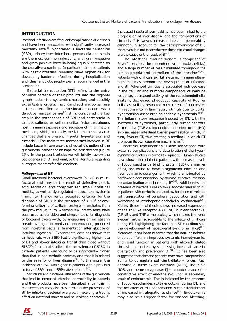
INTRODUCTIONBacterial infections are frequent complications of cirrhosis and have been associated with significantly increased mortality rate[1]. Spontaneous bacterial peritonitis (SBP), urinary tract infections, pneumonia and sepsis are the most common infections, with gram-negative and gram-positive bacteria being equally detected as the causative organisms. In particular, cirrhotic patients with gastrointestinal bleeding have higher risk for developing bacterial infections during hospitalization and, thus, antibiotic prophylaxis is recommended in this scenario[2,3].
Bacterial translocation (BT) refers to the entry of viable bacteria or their products into the regional lymph nodes, the systemic circulation, and possibly extraintestinal organs. The origin of such microorganisms is the enteric flora and translocation occurs via a defective mucosal barrier[4]. BT is considered the key step in the pathogenesis of SBP and bacteremia in cirrhotic patients, as well as a critical factor that triggers host immune responses and secretion of inflammatory mediators, which, ultimately, mediate the hemodynamic changes that are present in portal hypertension and cirrhosis[5]. The main three mechanisms involved in BT include bacterial overgrowth, physical disruption of the gut mucosal barrier and an impaired host defence (Figure 1)[6]. In the present article, we will briefly review the pathogenesis of BT and analyze the literature regarding surrogate markers for this condition.
Pathogenesis of BTSmall intestinal bacterial overgrowth (SIBO) is multi-factorial and may be the result of defective gastric acid secretion and compromised small intestinal motility, as well as dysregulated mucosal and systemic immunity. The currently accepted criterion for the diagnosis of SIBO is the presence of > 105 colony-forming units/mL of coliform bacteria in aspirates from the proximal jejunum. Alternatively, breath tests have been used as sensitive and simpler tools for diagnosis of bacterial overgrowth, by measuring an increase in breath hydrogen or methane concentration, produced from intestinal bacterial fermentation after glucose or lactulose ingestion[7]. Experimental data has shown that cirrhotic rats with SIBO had a significantly higher rate of BT and slower intestinal transit than those without SIBO[8]. In clinical studies, the prevalence of SIBO in cirrhotic patients was found to be significantly higher than that in non-cirrhotic controls, and that it is related to the severity of liver disease[9]. Furthermore, the incidence of SIBO was higher in patients with a previous history of SBP than in SBP-naïve patients[10].
Structural and functional alterations of the gut mucosa that lead to increased intestinal permeability to bacteria and their products have been described in cirrhosis[11]. Bile secretions may also play a role in the prevention of BT by inhibiting bacterial overgrowth, exerting a trophic effect on intestinal mucosa and neutralizing endotoxin[12].
Increased intestinal permeability has been linked to the progression of liver disease and the complications of cirrhosis[13]. However, increased intestinal permeability cannot fully account for the pathophysiology of BT; moreover, it is not clear whether these structural changes are the cause or the result of BT[5].
The intestinal immune system is comprised of Peyer’s patches, the mesenteric lymph nodes (MLNs) and a large number of cells distributed throughout the lamina propria and epithelium of the intestine[14,15]. Patients with cirrhosis exhibit systemic immune altera-tions that may promote the development of infections and BT. Advanced cirrhosis is associated with decrease in the cellular and humoral components of immune response, decreased activity of the reticuloendothelial system, decreased phagocytic capacity of Kupffer cells, as well as restricted recruitment of leucocytes in response to inflammatory stimuli due to portal hypertension-associated splanchnic hyperaemia[16-18]. The inflammatory response induced by BT, with the synthesis of cytokines, particularly tumor necrosis factor-alpha (TNF-α), interleukins and nitric oxide (NO) also increases intestinal barrier permeability, which, in turn, favours BT, thus creating a feedback in which BT promotes its own causative mechanisms[5].
Bacterial translocation is also associated with systemic complications and deterioration of the hyper-dynamic circulation in cirrhosis (Figure 1). Human studies have shown that cirrhotic patients with increased levels of lipopolysaccharide binding protein (LBP), a marker of BT, are found to have a significant immune and haemodynamic derangement, which is ameliorated by norfloxacin administration, by causing selective intestinal decontamination and inhibiting BT[19]. Additionally, the presence of bacterial DNA (bDNA), another marker of BT, in patients with cirrhosis and ascites, has been correlated with aggravation of peripheral vasodilation and with worsening of intrahepatic endothelial dysfunction[20]. Kidney tissue in cirrhosis shows increased expression of the toll-like receptor 4 (TLR4), nuclear factor κB (NF-κB), and TNF-α molecules, which makes the renal system further susceptible to the effects of cirrhosis during BT, highlighting the fact that BT contributes to the development of hepatorenal syndrome (HRS)[21]. Moreover, it has been reported that the non- absorbable antibiotic rifaximin improves systemic hemodynamics and renal function in patients with alcohol-related cirrhosis and ascites, by suppressing intestinal bacterial overgrowth and preventing BT[22]. It has been also suggested that cirrhotic patients may have compromised ability to upregulate sufficient dilatory forces [i.e., endothelial nitric oxide synthase (NOS), inducible NOS, and heme oxygenase-1] to counterbalance the constrictive effect of endothelin-1 upon a secondary insult of endotoxemia. This is indicated by the presence of lipopolysaccharides (LPS) endotoxin during BT, and the net effect of this phenomenon is the establishment of increased intrahepatic resistance[23]. Endotoxemia may also be a trigger factor for variceal bleeding,
2265 September 18, 2015|Volume 7|Issue 20|WJH|www.wjgnet.com
Koutsounas I et al . Markers of bacterial translocation in end-stage liver disease
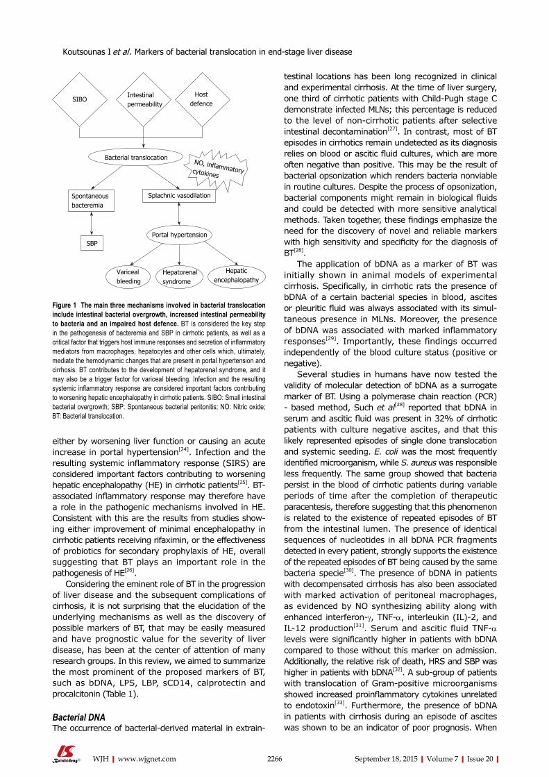
either by worsening liver function or causing an acute increase in portal hypertension[24]. Infection and the resulting systemic inflammatory response (SIRS) are considered important factors contributing to worsening hepatic encephalopathy (HE) in cirrhotic patients[25]. BT-associated inflammatory response may therefore have a role in the pathogenic mechanisms involved in HE. Consistent with this are the results from studies show-ing either improvement of minimal encephalopathy in cirrhotic patients receiving rifaximin, or the effectiveness of probiotics for secondary prophylaxis of HE, overall suggesting that BT plays an important role in the pathogenesis of HE[26].
Considering the eminent role of BT in the progression of liver disease and the subsequent complications of cirrhosis, it is not surprising that the elucidation of the underlying mechanisms as well as the discovery of possible markers of BT, that may be easily measured and have prognostic value for the severity of liver disease, has been at the center of attention of many research groups. In this review, we aimed to summarize the most prominent of the proposed markers of BT, such as bDNA, LPS, LBP, sCD14, calprotectin and procalcitonin (Table 1).
Bacterial DNAThe occurrence of bacterial-derived material in extrain-
testinal locations has been long recognized in clinical and experimental cirrhosis. At the time of liver surgery, one third of cirrhotic patients with Child-Pugh stage C demonstrate infected MLNs; this percentage is reduced to the level of non-cirrhotic patients after selective intestinal decontamination[27]. In contrast, most of BT episodes in cirrhotics remain undetected as its diagnosis relies on blood or ascitic fluid cultures, which are more often negative than positive. This may be the result of bacterial opsonization which renders bacteria nonviable in routine cultures. Despite the process of opsonization, bacterial components might remain in biological fluids and could be detected with more sensitive analytical methods. Taken together, these findings emphasize the need for the discovery of novel and reliable markers with high sensitivity and specificity for the diagnosis of BT[28].
The application of bDNA as a marker of BT was initially shown in animal models of experimental cirrhosis. Specifically, in cirrhotic rats the presence of bDNA of a certain bacterial species in blood, ascites or pleuritic fluid was always associated with its simultaneous presence in MLNs. Moreover, the presence of bDNA was associated with marked inflammatory responses[29]. Importantly, these findings occurred independently of the blood culture status (positive or negative).
Several studies in humans have now tested the validity of molecular detection of bDNA as a surrogate marker of BT. Using a polymerase chain reaction (PCR) - based method, Such et al[28] reported that bDNA in serum and ascitic fluid was present in 32% of cirrhotic patients with culture negative ascites, and that this likely represented episodes of single clone translocation and systemic seeding. E. coli was the most frequently identified microorganism, while S. aureus was responsible less frequently. The same group showed that bacteria persist in the blood of cirrhotic patients during variable periods of time after the completion of therapeutic paracentesis, therefore suggesting that this phenomenon is related to the existence of repeated episodes of BT from the intestinal lumen. The presence of identical sequences of nucleotides in all bDNA PCR fragments detected in every patient, strongly supports the existence of the repeated episodes of BT being caused by the same bacteria specie[30]. The presence of bDNA in patients with decompensated cirrhosis has also been associated with marked activation of peritoneal macrophages, as evidenced by NO synthesizing ability along with enhanced interferon-γ, TNF-α, interleukin (IL)-2, and IL-12 production[31]. Serum and ascitic fluid TNF-α levels were significantly higher in patients with bDNA compared to those without this marker on admission. Additionally, the relative risk of death, HRS and SBP was higher in patients with bDNA[32]. A sub-group of patients with translocation of Gram-positive microorganisms showed increased proinflammatory cytokines unrelated to endotoxin[33]. Furthermore, the presence of bDNA in patients with cirrhosis during an episode of ascites was shown to be an indicator of poor prognosis. When
2266 September 18, 2015|Volume 7|Issue 20|WJH|www.wjgnet.com
Intestinalpermeability
Hostdefence
SIBO
Bacterial translocation
Splachnic vasodilation
Portal hypertension
Varicealbleeding
Hepatorenalsyndrome
Hepaticencephalopathy
NO, inflammatorycytokines
SBP
Spontaneousbacteremia
Figure 1 The main three mechanisms involved in bacterial translocation include intestinal bacterial overgrowth, increased intestinal permeability to bacteria and an impaired host defence. BT is considered the key step in the pathogenesis of bacteremia and SBP in cirrhotic patients, as well as a critical factor that triggers host immune responses and secretion of inflammatory mediators from macrophages, hepatocytes and other cells which, ultimately, mediate the hemodynamic changes that are present in portal hypertension and cirrhosis. BT contributes to the development of hepatorenal syndrome, and it may also be a trigger factor for variceal bleeding. Infection and the resulting systemic inflammatory response are considered important factors contributing to worsening hepatic encephalopathy in cirrhotic patients. SIBO: Small intestinal bacterial overgrowth; SBP: Spontaneous bacterial peritonitis; NO: Nitric oxide; BT: Bacterial translocation.
Koutsounas I et al . Markers of bacterial translocation in end-stage liver disease
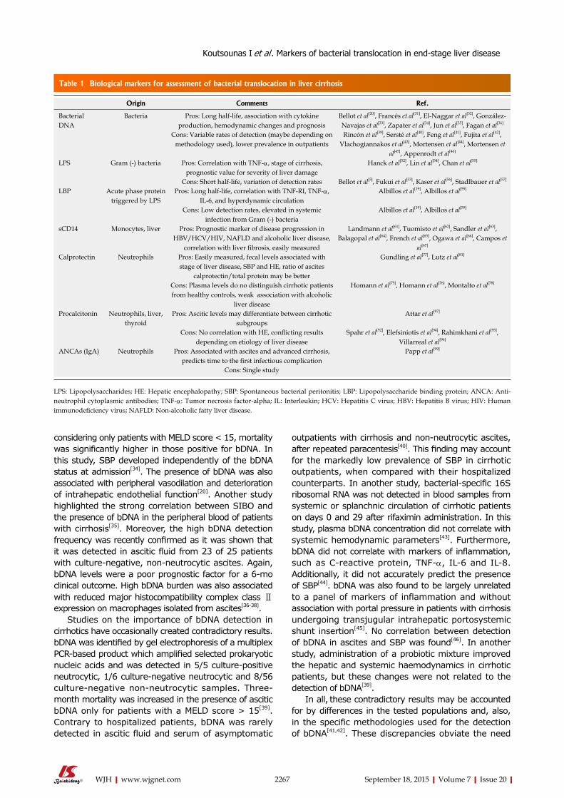
2267 September 18, 2015|Volume 7|Issue 20|WJH|www.wjgnet.com
outpatients with cirrhosis and non-neutrocytic ascites, after repeated paracentesis[40]. This finding may account for the markedly low prevalence of SBP in cirrhotic outpatients, when compared with their hospitalized counterparts. In another study, bacterial-specific 16S ribosomal RNA was not detected in blood samples from systemic or splanchnic circulation of cirrhotic patients on days 0 and 29 after rifaximin administration. In this study, plasma bDNA concentration did not correlate with systemic hemodynamic parameters[43]. Furthermore, bDNA did not correlate with markers of inflammation, such as C-reactive protein, TNF-α, IL-6 and IL-8. Additionally, it did not accurately predict the presence of SBP[44]. bDNA was also found to be largely unrelated to a panel of markers of inflammation and without association with portal pressure in patients with cirrhosis undergoing transjugular intrahepatic portosystemic shunt insertion[45]. No correlation between detection of bDNA in ascites and SBP was found[46]. In another study, administration of a probiotic mixture improved the hepatic and systemic haemodynamics in cirrhotic patients, but these changes were not related to the detection of bDNA[39].
In all, these contradictory results may be accounted for by differences in the tested populations and, also, in the specific methodologies used for the detection of bDNA[41,42]. These discrepancies obviate the need
considering only patients with MELD score < 15, mortality was significantly higher in those positive for bDNA. In this study, SBP developed independently of the bDNA status at admission[34]. The presence of bDNA was also associated with peripheral vasodilation and deterioration of intrahepatic endothelial function[20]. Another study highlighted the strong correlation between SIBO and the presence of bDNA in the peripheral blood of patients with cirrhosis[35]. Moreover, the high bDNA detection frequency was recently confirmed as it was shown that it was detected in ascitic fluid from 23 of 25 patients with culture-negative, non-neutrocytic ascites. Again, bDNA levels were a poor prognostic factor for a 6-mo clinical outcome. High bDNA burden was also associated with reduced major histocompatibility complex class Ⅱ expression on macrophages isolated from ascites[36-38].
Studies on the importance of bDNA detection in cirrhotics have occasionally created contradictory results. bDNA was identified by gel electrophoresis of a multiplex PCRbased product which amplified selected prokaryotic nucleic acids and was detected in 5/5 culture-positive neutrocytic, 1/6 culture-negative neutrocytic and 8/56 culture-negative non-neutrocytic samples. Three-month mortality was increased in the presence of ascitic bDNA only for patients with a MELD score > 15[39]. Contrary to hospitalized patients, bDNA was rarely detected in ascitic fluid and serum of asymptomatic
Table 1 Biological markers for assessment of bacterial translocation in liver cirrhosis
Origin Comments Ref.
Bacterial DNA
Bacteria Pros: Long half-life, association with cytokine production, hemodynamic changes and prognosis
Bellot et al[20], Francés et al[31], El-Naggar et al[32], González-Navajas et al[33], Zapater et al[34], Jun et al[35], Fagan et al[36]
Cons: Variable rates of detection (maybe depending on methodology used), lower prevalence in outpatients
Rincón et al[39], Sersté et al[40], Feng et al[41], Fujita et al[42], Vlachogiannakos et al[43], Mortensen et al[44], Mortensen et
al[45], Appenrodt et al[46]
LPS Gram (-) bacteria Pros: Correlation with TNF-α, stage of cirrhosis, prognostic value for severity of liver damage
Hanck et al[52], Lin et al[54], Chan et al[55]
Cons: Short half-life, variation of detection rates Bellot et al[5], Fukui et al[53], Kaser et al[56], Stadlbauer et al[57]
LBP Acute phase protein triggered by LPS
Pros: Long half-life, correlation with TNF-RI, TNF-α, IL-6, and hyperdynamic circulation
Albillos et al[19], Albillos et al[59]
Cons: Low detection rates, elevated in systemic infection from Gram (-) bacteria
Albillos et al[19], Albillos et al[59]
sCD14 Monocytes, liver Pros: Prognostic marker of disease progression in HBV/HCV/HIV, NAFLD and alcoholic liver disease,
correlation with liver fibrosis, easily measured
Landmann et al[61], Tuomisto et al[62], Sandler et al[63], Balagopal et al[64], French et al[65], Ogawa et al[66], Campos et
al[67]
Calprotectin Neutrophils Pros: Easily measured, fecal levels associated with stage of liver disease, SBP and HE, ratio of ascites
calprotectin/total protein may be better
Gundling et al[77], Lutz et al[81]
Cons: Plasma levels do no distinguish cirrhotic patients from healthy controls, weak association with alcoholic
liver disease
Homann et al[75], Homann et al[76], Montalto et al[78]
Procalcitonin Neutrophils, liver, thyroid
Pros: Ascitic levels may differentiate between cirrhotic subgroups
Attar et al[97]
Cons: No correlation with HE, conflicting results depending on etiology of liver disease
Spahr et al[92], Elefsiniotis et al[94], Rahimkhani et al[95], Villarreal et al[96]
ANCAs (IgA) Neutrophils Pros: Associated with ascites and advanced cirrhosis, predicts time to the first infectious complication
Papp et al[99]
Cons: Single study
LPS: Lipopolysaccharides; HE: Hepatic encephalopathy; SBP: Spontaneous bacterial peritonitis; LBP: Lipopolysaccharide binding protein; ANCA: Anti-neutrophil cytoplasmic antibodies; TNF-α: Tumor necrosis factor-alpha; IL: Interleukin; HCV: Hepatitis C virus; HBV: Hepatitis B virus; HIV: Human immunodeficiency virus; NAFLD: Non-alcoholic fatty liver disease.
Koutsounas I et al . Markers of bacterial translocation in end-stage liver disease
2268 September 18, 2015|Volume 7|Issue 20|WJH|www.wjgnet.com
for larger studies which will include well-characterized patient subpopulations, including chronic hepatitis, non-alcoholic fatty liver disease (NAFLD), as well as compensated and decompensated cirrhosis. Moreover, standardisation of methodology is required in order to determine the applicability of bDNA usage in clinical practice as a surrogate marker for BT. Finally, testing of larger cohorts of patients may define the significance of detectable bDNA as a prognostic tool for systemic responses and survival in cirrhosis.
LPS-LBP-sCD14LPS or endotoxin is a lipopolysaccharide, which is part of the outer membrane of Gram-negative bacteria. In circulation, it is recognized by LBP. The LPS-LBP complex binds to membrane CD14 (mCD14) on myeloid cells or to circulating CD14 (soluble CD14, sCD14)[47,48] and promotes a cascade of inflammatory responses via myeloid differentiation-2/TLR4 activation of NF-κB[49,50]. Kupffer cells, which are specialized macrophages located in the liver, are activated when exposed to LPS and can produce a spectrum of cytokines and reactive oxygen intermediates, including the proinflammatory cytokine TNF-α. Thus, LPS is a potent stimulator of Kupffer cell TNF-α production, and this pathway has been implicated in the pathogenesis of many types of liver injury. It has also been proposed that through this mechanism hepatic stellate cells are activated towards the production of inflammatory and adhesion molecules, thus inflicting liver damage[51].
Due to its aforementioned properties, LPS was one of the first molecules that was proposed as a marker of BT. The working hypothesis is that its presence in the sera of cirrhotic patients directly indicates endotoxemia from Gram negative bacteria. In alcoholic cirrhosis, LPS has been found to correlate with TNF-α levels and with the stage of liver disease (Child-Pugh score); thus, it has been suggested that LPS may be a key player in the progression of alcoholic liver disease[52]. Levels of LPS in plasma of patients with alcoholic cirrhosis were higher than in non-alcoholics, pointing to a critical role for alcohol consumption in the development of endotoxemia and liver damage[53]. Taking into consideration that LPS plasma levels of cirrhotic patients are elevated compared to chronic hepatitis and the reported correlation with the stage of cirrhosis, it is probable that LPS may serve as a prognostic factor of disease severity[54] and short-term survival of cirrhotic patients[55]. It should be noted, however, that not all studies have demonstrated a positive correlation between serum LPS and stage of cirrhosis[53,56]. Therefore, LPS has not been clearly established as a reliable marker of BT[57]. This may be due to the short half-life of LPS or to the interference of various factors with the detection of LPS[5].
LBP is a 65 kDa, acute phase protein that is predominantly produced in the liver by hepatocytes. Bacterial LPS triggers the production of LBP and the peripheral levels of LBP are significantly elevated in the setting of bacterial presence. LBP is known to specifically
bind and transfer bacterial LPS, and the LPS-LBP complex binds mCD14 on myeloid cells or to sCD14[58]. Consequently, several studies have tested the validity of the expression of LBP as an indicator of a systemic response to LPS, and, thus, as an indirect marker of BT. Albillos et al[19] demonstrated that BT, expressed by elevated plasma levels of LBP, leads to derangement of patients with decompensated cirrhosis. Cirrhotic patients with high LBP levels had enhanced expression of sCD14 and sTNF-RI as well as elevated circulating levels of TNF-α and IL-6. Furthermore, an intense hyperdynamic circulatory state was described in this population. Interestingly, LPS was detectable only in one third of the patients with high LBP. The latter observation strengthens the suggestion that LPS cannot easily serve as a marker of BT, due to its short half-life, so transient episodes of bacteremia may remain undiagnosed. On the other hand, the subsequent expression of LBP after endotoxemia is detectable for a much longer period. Moreover, a study performed by the same group suggested that patients with high LBP circulating levels have increased susceptibility to infections[59]. Different studies have proposed that increased levels of LBP in cirrhosis are the result of chronic endotoxemia and that also no difference occurs in the expression of LBP between alcoholic and non-alcoholic cirrhosis. Kaser et al[56] reported a strong correlation between the two binding proteins of LPS, LBP and sCD14. Although its longer half-life makes LBP a more attractive marker of BT than LPS, its use has certain drawbacks. First, systemic LBP elevation exists only in response to Gram-negative bacteria, and, second, it may not only be present in BT, but also in systemic infection resulting in SIRS (systemic inflammatory response syndrome)[60].
Recently, the target molecule of the complex LPS-LBP, CD14 was also proposed as a surrogate marker of BT. mCD14 is expressed mainly by macrophages and at a lesser extent by neutrophils and dendritic cells. sCD14 is secreted by hepatocytes and monocytes. The expression of sCD14 is upregulated as a result of the presence of LPS. CD14 acts as a co-receptor along with TLR-4 for the detection of bacterial LPS[61]. LPS induces the CD14/TLR4 complex endocytosis in human monocytes and macrophages and the consequent NF-κB activation. Tuomisto et al[62] showed a significant correlation between the levels of bDNA in the liver and the local expression of CD14 in alcoholic liver disease. These findings emphasize the role of CD14 as a marker of BT. Sandler et al[63] proposed that sCD14 may not only be a marker of BT, but also a prognostic marker of disease progression in HBV and HCV infection. Specifically, sCD14 in patients with severe fibrosis was highly elevated not only in peripheral blood, but also within the hepatic parenchyma, measured by CD14 (+) hepatic cells. Regardless of the etiology of cirrhosis, microbial translocation as identified by the presence of sCD14, is believed to play a key role in the progression of liver disease[56]. Studies concerning cirrhosis in HIV-infected patients support this hypothesis[64]. French et al[65] provided evidence
Koutsounas I et al . Markers of bacterial translocation in end-stage liver disease
2269 September 18, 2015|Volume 7|Issue 20|WJH|www.wjgnet.com
that in HIV/hepatitis C virus (HCV)-coinfection, levels of sCD14 and IL-6 were mostly elevated in patient with disease progression than in non-progressors. Studies in patients with NAFLD reached similar results, suggesting a positive correlation between serum sCD14, hepatic CD14 expression and liver inflammation[66]. Attention has also been drawn towards polymorphisms of the promoter region of the CD14 gene, shedding light to the role of CD14/-159TT genotype in the progression of liver injury in alcoholic liver disease[67]. Taking into account the aforementioned studies, it appears that sCD14 may represent a promising marker of BT as it can easily be measured in circulation and correlates with progression of liver disease.
Calprotectin - procalcitonin - anti-neutrophil cytoplasmic antibodiesCalprotectin is a calcium and zinc binding protein with a molecular weight of 36 kDa[68]. It has been estimated that it may account for more than 60% of the soluble cytosolic proteins in human neutrophil granulocytes[69]. Moreover, it was shown that calprotectin measurements in fecal samples not only correlate with the degree of neutrophil migration in the gastrointestinal mucosa[70], but also serve as reliable surrogate marker of intestinal inflammation[71]. As a result, fecal calprotectin has been studied in depth in gastrointestinal disorders, and has assumed an important role in monitoring the activity and response to treatment in patients with inflammatory bowel disease[72,73].
The pathogenesis of BT in patients with cirrhosis has been associated with alterations in gut mucosal immune responses and intestinal permeability. In addition, neutrophil infiltrates are detected in the gastrointestinal mucosa of cirrhotics. Consequently, calprotectin has been investigated as a possible diagnostic marker for the existence and natural history of SBP and HE. Initial studies conducted by a Danish research group, focused on the possible prognostic significance of calprotectin levels in the plasma and ascitic fluid samples from patients with end-stage liver disease. The authors did not find a significant difference between healthy controls and patients with cirrhosis (irrespectively if liver disease was compensated or decompensated), a finding that was confirmed in additional studies[74,75]. On the other hand, they reported that high plasma calprotectin levels were an indicator of poor survival in alcohol-related cirrhosis[74]. The most important finding, however, regarding the role of calprotectin in relation to BT, was that during follow up of the patients higher calprotectin levels were an independent predictor of recurrent bacterial infections[76].
Arguably the most important study regarding the role of fecal calprotectin in the diagnosis of BT complications, is the one conducted by Gundling et al[77]. They investigated the relationship between fecal calprotectin levels and the onset and course of SBP and HE. They confirmed that patients with cirrhosis had significantly elevated fecal calprotectin levels
when compared to healthy controls. Moreover, this increase correlated with the severity of liver disease (assessed by Child-Pugh and MELD scores). Even more significantly, higher calprotectin values were associated with advanced stages of HE, the presence of SBP, as well as extraintestinal infections. Finally, calprotectin strongly correlated with serum ammonia levels[77]. The aforementioned observations reinforced the hypothesis that fecal calprotectin may be a reliable surrogate marker for BT and provide important assistance in the diagnosis and clinical management of patients with decompensated liver disease. It should be noted that the finding of a possible selective calprotectin upregulation in alcoholic liver disease that was reported in initial studies, was not confirmed in a longitudinal study of active alcoholics, where alcoholics and controls had similar fecal calprotectin measurements[78].
It was recently reported that ascitic calprotectin may be utilized (with the help of a point-of-care assay) to reliably predict an elevated polymorphonuclear count (> 250) in ascitic fluid, allowing for faster diagnosis of SBP[79]. Another study by Alempijević et al[80] focusing exclusively on HE confirmed that fecal calprotectin levels were positively correlated with HE grading according to the West-Haven grouping criteria, although it did not show a correlation with serum ammonia levels as Gundling et al[77] did. Finally, the ratio of ascites calprotectin to total protein was proposed as a better marker than ascitic fluid calprotectin alone for use in the diagnosis and prognosis of SBP. The authors report satisfactory sensitivity and specificity for this new marker, as well as a statistically significant correlation of higher values with poor 30-d survival[81].
In all, calprotectin remains a promising surrogate marker for BT in cirrhosis. It demonstrates many advantages, especially in its fecal measurement, as it is a non-invasive, quick and relatively easy to perform assay, with proven clinical value in other disease states[82].
Procalcitonin (PCT) is a 116 amino acid propeptide of calcitonin. It has been established as a valuable biomarker for the diagnosis and monitoring of bacterial infections to the point that is being used as a guide for antibiotic use[83]. Concerning advanced liver disease, PCT has been studied for the past 15 years regarding its potential for the diagnosis of SBP in decompensated patients and subsequently for its utility as an indirect marker of BT. As the liver is believed to be a key source of PCT, there were initially concerns that hepatic impairment may result in downregulated serum PCT levels. Although, this was not proven to be the case[84], and early reports were encouraging about the use of PCT in the diagnosis of SBP, its potential role remains currently unclear as several studies provided conflicting results[85-93]. A solution has been proposed with the use of an ultra-sensitive PCT assay[87], and possible explanations for the discrepancies noted between studies include higher baseline levels in patient with alcoholic[94] or specific viral-related[95] causes of cirrhosis and the
Koutsounas I et al . Markers of bacterial translocation in end-stage liver disease
2270 September 18, 2015|Volume 7|Issue 20|WJH|www.wjgnet.com
presence of other bacterial infections[96]. Furthermore, it was reported that PCT levels in the ascitic fluid, but not in serum may differentiate between different cirrhotic subgroups, reflecting a possible localized role in the interplay between ascites and BT[97]. Interestingly, in contrast to calprotectin, PCT has not been found to correlate with the presence of HE, another possible important component of the BT phenotype in cirrhosis. It should be also noted, that in several of the PCT-related studies the role of CRP was assessed as well and it was usually found to have an inferior potential as a SBP marker when compared to PCT[91,98].
Finally, as part of the potential of established auto-immune and inflammatory markers of BT, a recent Hungarian study is worth mentioning. Therein, it was reported that the prevalence of anti-neutrophil cyto-plasmic antibodies (ANCA) of the IgA subtype was higher in patients with cirrhosis in comparison to patients with non-cirrhotic chronic liver disease or healthy controls. Moreover, the presence of IgA ANCA was associated with the presence of ascites and advanced cirrhosis. Even more significant was the finding that, during follow up, patients with IgA ANCA had not only a higher complication risk but also IgA ANCA positivity correlated with a shorter time to the first infectious complication[99]. As IgA antibodies are linked to the gut immune system, these results may reflect a component in the pathogenesis of BT involving alterations in the local immune system and the intestinal barrier. In spite of the promise shown by these observations, further research is required to elucidate if IgA ANCA may serve as a possible marker of BT.
CONCLUSIONBT plays an important role in the pathogenesis of end stage liver disease complications. Therefore, its early and reliable detection would be significant for accurately identifying a particular subset of patients with potentially adverse prognosis, who may benefit from increased vigilance and aggressive management. Several biological molecules (bacterial DNA, soluble CD14, LPS/endotoxin, lipopolysaccharide-binding protein, calprotectin, pro-calcitonin) have been tested as potential biomarkers for BT, each with its own merits and flaws (Table 1). The literature available on the subject is intriguing and expanding. In all, while no single marker has emerged as optimal for the identification of BT, there is an obvious need for better designed and more focused research. These studies will not only enable us to understand the underlying mechanisms of BT, but may also allow for implementing a timely intervention in patients with liver cirrhosis. This may, in turn, alter the natural history of this ominous disease and improve its currently unfavorable prognosis.
ACKNOWLEDGMENTSThis research has been co-financed by the European
Union (European Social Fund) and Greek national funds through the Operational Program “Education and Lifelong Learning” of the National Strategic Reference Framework - Research Funding Program: Heracleitus Ⅱ. Investing in knowledge society through the European Social Fund, of which Dr. Kaltsa was a recipient.
REFERENCES1 Arvaniti V, D’Amico G, Fede G, Manousou P, Tsochatzis E,
Pleguezuelo M, Burroughs AK. Infections in patients with cirrhosis increase mortality four-fold and should be used in determining prognosis. Gastroenterology 2010; 139: 1246-1256, 1256.e1-5 [PMID: 20558165 DOI: 10.1053/j.gastro.2010.06.019]
2 Deschênes M, Villeneuve JP. Risk factors for the development of bacterial infections in hospitalized patients with cirrhosis. Am J Gastroenterol 1999; 94: 2193-2197 [PMID: 10445549 DOI: 10.1111/j.1572-0241.1999.01293.x]
3 Fernández J, Navasa M, Gómez J, Colmenero J, Vila J, Arroyo V, Rodés J. Bacterial infections in cirrhosis: epidemiological changes with invasive procedures and norfloxacin prophylaxis. Hepatology 2002; 35: 140-148 [PMID: 11786970 DOI: 10.1053/jhep.2002.30082]
4 Berg RD, Garlington AW. Translocation of certain indigenous bacteria from the gastrointestinal tract to the mesenteric lymph nodes and other organs in a gnotobiotic mouse model. Infect Immun 1979; 23: 403-411 [PMID: 154474]
5 Bellot P, Francés R, Such J. Pathological bacterial translocation in cirrhosis: pathophysiology, diagnosis and clinical implications. Liver Int 2013; 33: 31-39 [PMID: 23121656 DOI: 10.1111/liv.12021]
6 Wiest R, Rath HC. Gastrointestinal disorders of the critically ill. Bacterial translocation in the gut. Best Pract Res Clin Gastroenterol 2003; 17: 397-425 [PMID: 12763504 DOI: 10.1016/S1521-6918(03)00024-6]
7 Gasbarrini A, Lauritano EC, Gabrielli M, Scarpellini E, Lupascu A, Ojetti V, Gasbarrini G. Small intestinal bacterial overgrowth: diagnosis and treatment. Dig Dis 2007; 25: 237-240 [PMID: 17827947 DOI: 10.1159/000103892]
8 Pérez-Paramo M, Muñoz J, Albillos A, Freile I, Portero F, Santos M, Ortiz-Berrocal J. Effect of propranolol on the factors promoting bacterial translocation in cirrhotic rats with ascites. Hepatology 2000; 31: 43-48 [PMID: 10613726 DOI: 10.1002/hep.510310109]
9 Yang CY, Chang CS, Chen GH. Small-intestinal bacterial overgrowth in patients with liver cirrhosis, diagnosed with glucose H2 or CH4 breath tests. Scand J Gastroenterol 1998; 33: 867-871 [PMID: 9754736 DOI: 10.1080/00365529850171549]
10 Chang CS, Chen GH, Lien HC, Yeh HZ. Small intestine dysmotility and bacterial overgrowth in cirrhotic patients with spontaneous bacterial peritonitis. Hepatology 1998; 28: 1187-1190 [PMID: 9794900 DOI: 10.1002/hep.510280504]
11 Such J, Guardiola JV, de Juan J, Casellas JA, Pascual S, Aparicio JR, Solá-Vera J, Pérez-Mateo M. Ultrastructural characteristics of distal duodenum mucosa in patients with cirrhosis. Eur J Gastroenterol Hepatol 2002; 14: 371-376 [PMID: 11943948 DOI: 10.1097/00042737-200204000-00006]
12 Bertók L. Bile acids in physico-chemical host defence. Pathophysiology 2004; 11: 139-145 [PMID: 15561510 DOI: 10.1016/j.pathophys.2004.09.002]
13 Pascual S, Such J, Esteban A, Zapater P, Casellas JA, Aparicio JR, Girona E, Gutiérrez A, Carnices F, Palazón JM, Sola-Vera J, Pérez-Mateo M. Intestinal permeability is increased in patients with advanced cirrhosis. Hepatogastroenterology 2003; 50: 1482-1486 [PMID: 14571769]
14 MacDonald TT, Pettersson S. Bacterial regulation of intestinal immune responses. Inflamm Bowel Dis 2000; 6: 116-122 [PMID: 10833071 DOI: 10.1097/00054725-200005000-00008]
15 Heel KA, McCauley RD, Papadimitriou JM, Hall JC. Review: Peyer’s patches. J Gastroenterol Hepatol 1997; 12: 122-136 [PMID: 9083913 DOI: 10.1111/j.1440-1746.1997.tb00395.x]
Koutsounas I et al . Markers of bacterial translocation in end-stage liver disease
2271 September 18, 2015|Volume 7|Issue 20|WJH|www.wjgnet.com
16 Rajkovic IA, Williams R. Abnormalities of neutrophil phagocytosis, intracellular killing and metabolic activity in alcoholic cirrhosis and hepatitis. Hepatology 1989; 6: 252-262 [PMID: 3007318 DOI: 10.1002/hep.1840060217]
17 Bolognesi M, Merkel C, Bianco S, Angeli P, Sacerdoti D, Amodio P, Gatta A. Clinical significance of the evaluation of hepatic reticuloendothelial removal capacity in patients with cirrhosis. Hepatology 1994; 19: 628-634 [PMID: 8119687 DOI: 10.1002/hep.1840190313]
18 Panés J, Pérez-del-Pulgar S, Casadevall M, Salas A, Pizcueta P, Bosch J, Anderson DC, Granger DN, Piqué JM. Impaired mesenteric leukocyte recruitment in experimental portal hyper-tension in the rat. Hepatology 1999; 30: 445-453 [PMID: 10421653 DOI: 10.1002/hep.510300214]
19 Albillos A, de la Hera A, González M, Moya JL, Calleja JL, Monserrat J, Ruiz-del-Arbol L, Alvarez-Mon M. Increased lipopolysaccharide binding protein in cirrhotic patients with marked immune and hemodynamic derangement. Hepatology 2003; 37: 208-217 [PMID: 12500206 DOI: 10.1053/jhep.2003.50038]
20 Bellot P, García-Pagán JC, Francés R, Abraldes JG, Navasa M, Pérez-Mateo M, Such J, Bosch J. Bacterial DNA translocation is associated with systemic circulatory abnormalities and intrahepatic endothelial dysfunction in patients with cirrhosis. Hepatology 2010; 52: 2044-2052 [PMID: 20979050 DOI: 10.1002/hep.23918]
21 Shah N, Dhar D, El Zahraa Mohammed F, Habtesion A, Davies NA, Jover-Cobos M, Macnaughtan J, Sharma V, Olde Damink SW, Mookerjee RP, Jalan R. Prevention of acute kidney injury in a rodent model of cirrhosis following selective gut decontamination is associated with reduced renal TLR4 expression. J Hepatol 2012; 56: 1047-1053 [PMID: 22266601 DOI: 10.1016/j.jhep.2011.11.024]
22 Kalambokis GN, Mouzaki A, Rodi M, Pappas K, Fotopoulos A, Xourgia X, Tsianos EV. Rifaximin improves systemic hemodynamics and renal function in patients with alcohol-related cirrhosis and ascites. Clin Gastroenterol Hepatol 2012; 10: 815-818 [PMID: 22391344 DOI: 10.1016/j.cgh.2012.02.025]
23 Baveja R, Keller S, Yokoyama Y, Sonin N, Clemens MG, Zhang JX. LPS-induced imbalanced expression of hepatic vascular stress genes in cirrhosis: possible mechanism of increased susceptibility to endotoxemia. Shock 2002; 17: 316-321 [PMID: 11954834 DOI: 10.1097/00024382-200204000-00014]
24 Thalheimer U, Triantos CK, Samonakis DN, Patch D, Burroughs AK. Infection, coagulation, and variceal bleeding in cirrhosis. Gut 2005; 54: 556-563 [PMID: 15753544 DOI: 10.1136/gut.2004.048181]
25 Vaquero J, Polson J, Chung C, Helenowski I, Schiodt FV, Reisch J, Lee WM, Blei AT. Infection and the progression of hepatic encephalopathy in acute liver failure. Gastroenterology 2003; 125: 755-764 [PMID: 12949721 DOI: 10.1016/S0016-5085(03)01051-5]
26 Sidhu SS, Goyal O, Mishra BP, Sood A, Chhina RS, Soni RK. Rifaximin improves psychometric performance and health-related quality of life in patients with minimal hepatic encephalopathy (the RIME Trial). Am J Gastroenterol 2011; 106: 307-316 [PMID: 21157444 DOI: 10.1038/ajg.2010.455]
27 Cirera I, Bauer TM, Navasa M, Vila J, Grande L, Taurá P, Fuster J, García-Valdecasas JC, Lacy A, Suárez MJ, Rimola A, Rodés J. Bacterial translocation of enteric organisms in patients with cirrhosis. J Hepatol 2001; 34: 32-37 [PMID: 11211904 DOI: 10.1016/S0168-8278(00)00013-1]
28 Such J, Francés R, Muñoz C, Zapater P, Casellas JA, Cifuentes A, Rodríguez-Valera F, Pascual S, Sola-Vera J, Carnicer F, Uceda F, Palazón JM, Pérez-Mateo M. Detection and identification of bacterial DNA in patients with cirrhosis and culture-negative, nonneutrocytic ascites. Hepatology 2002; 36: 135-141 [PMID: 12085357 DOI: 10.1053/jhep.2002.33715]
29 Guarner C, González-Navajas JM, Sánchez E, Soriando G, Francés R, Chiva M, Zapater P, Benlloch S, Muñoz C, Pascual S, Balanzó J, Pérez-Mateo M, Such J. The detection of bacterial DNA in blood of rats with CCl4-induced cirrhosis with ascites represents episodes of bacterial translocation. Hepatology 2006; 44: 633-639 [PMID: 16941689 DOI: 10.1002/hep.21286]
30 Francés R, Benlloch S, Zapater P, González JM, Lozano B, Muñoz C, Pascual S, Casellas JA, Uceda F, Palazón JM, Carnicer F, Pérez-
Mateo M, Such J. A sequential study of serum bacterial DNA in patients with advanced cirrhosis and ascites. Hepatology 2004; 39: 484-491 [PMID: 14768002 DOI: 10.1002/hep.20055]
31 Francés R, Muñoz C, Zapater P, Uceda F, Gascón I, Pascual S, Pérez-Mateo M, Such J. Bacterial DNA activates cell mediated immune response and nitric oxide overproduction in peritoneal macrophages from patients with cirrhosis and ascites. Gut 2004; 53: 860-864 [PMID: 15138214 DOI: 10.1136/gut.2003.027425]
32 El-Naggar MM, Khalil el-SA, El-Daker MA, Salama MF. Bacterial DNA and its consequences in patients with cirrhosis and culture-negative, non-neutrocytic ascites. J Med Microbiol 2008; 57: 1533-1538 [PMID: 19018026 DOI: 10.1099/jmm.0.2008/001867-0]
33 González-Navajas JM, Bellot P, Francés R, Zapater P, Muñoz C, García-Pagán JC, Pascual S, Pérez-Mateo M, Bosch J, Such J. Presence of bacterial-DNA in cirrhosis identifies a subgroup of patients with marked inflammatory response not related to endotoxin. J Hepatol 2008; 48: 61-67 [PMID: 17998145 DOI: 10.1016/j.jhep.2007.08.012]
34 Zapater P, Francés R, González-Navajas JM, de la Hoz MA, Moreu R, Pascual S, Monfort D, Montoliu S, Vila C, Escudero A, Torras X, Cirera I, Llanos L, Guarner-Argente C, Palazón JM, Carnicer F, Bellot P, Guarner C, Planas R, Solá R, Serra MA, Muñoz C, Pérez-Mateo M, Such J. Serum and ascitic fluid bacterial DNA: a new independent prognostic factor in noninfected patients with cirrhosis. Hepatology 2008; 48: 1924-1931 [PMID: 19003911 DOI: 10.1002/hep.22564]
35 Jun DW, Kim KT, Lee OY, Chae JD, Son BK, Kim SH, Jo YJ, Park YS. Association between small intestinal bacterial overgrowth and peripheral bacterial DNA in cirrhotic patients. Dig Dis Sci 2010; 55: 1465-1471 [PMID: 19517230 DOI: 10.1007/s10620-009-0870-9]
36 Fagan KJ, Rogers GB, Melino M, Arthur DM, Costello ME, Morrison M, Powell EE, Irvine KM. Ascites bacterial burden and immune cell profile are associated with poor clinical outcomes in the absence of overt infection. PLoS One 2015; 10: e0120642 [PMID: 25781164 DOI: 10.1371/journal.pone.0120642]
37 Francés R, Zapater P, González-Navajas JM, Muñoz C, Caño R, Moreu R, Pascual S, Bellot P, Pérez-Mateo M, Such J. Bacterial DNA in patients with cirrhosis and noninfected ascites mimics the soluble immune response established in patients with spontaneous bacterial peritonitis. Hepatology 2008; 47: 978-985 [PMID: 18306221 DOI: 10.1002/hep.22083]
38 Mostafa MS, El-Seidi EA, Kassem AM, Shemis MA, Saber M, Michael MN. Detection of ascitic fluid infections in patients with liver cirrhosis and ascites. Arab J Gastroenterol 2011; 12: 20-24 [PMID: 21429450 DOI: 10.1016/j.ajg.2011.01.004]
39 Rincón D, Vaquero J, Hernando A, Galindo E, Ripoll C, Puerto M, Salcedo M, Francés R, Matilla A, Catalina MV, Clemente G, Such J, Bañares R. Oral probiotic VSL#3 attenuates the circulatory disturbances of patients with cirrhosis and ascites. Liver Int 2014; 34: 1504-1512 [PMID: 24661740 DOI: 10.1111/liv.12539]
40 Sersté T, Bert F, Leflon-Guibout V, Chauvet C, Marcon E, Asselah T, Francoz C, Durand F, Lebrec D, Valla D, Moreau R, Nicolas-Chanoine MH. Detection of bacterial DNA in serum and ascitic fluid of asymptomatic outpatients with cirrhosis and non-neutrocytic ascites. Liver Int 2011; 31: 494-498 [PMID: 21382159 DOI: 10.1111/j.1478-3231.2011.02448.x]
41 Feng Y, Chen CL, Chen TH, Liang YH, Chen HL, Lin CY, Chiu CH. Application of next-generation sequencing to study ascitic microbiome in cirrhotic patients with or without spontaneous bacterial peritonitis. J Microbiol Immunol Infect 2014; Epub ahead of print [PMID: 25305707 DOI: 10.1016/j.jmii.2014.07.005]
42 Fujita T. Is bacterial DNA a surrogate marker of bacterial translocation in cirrhosis? J Hepatol 2008; 49: 145-146; author reply 146-147 [PMID: 18466995 DOI: 10.1016/j.jhep.2008.04.001]
43 Vlachogiannakos J, Daikos G, Thalheimer U, Burroughs AK, Ladas SD. Is bacterial DNA a better marker than endotoxin of bacterial translocation in decompensated cirrhosis? Hepatology 2011; 53: 2140-2141; author reply 41-42 [PMID: 21425309 DOI: 10.1002/hep.24303]
Koutsounas I et al . Markers of bacterial translocation in end-stage liver disease
2272 September 18, 2015|Volume 7|Issue 20|WJH|www.wjgnet.com
44 Mortensen C, Jensen JS, Hobolth L, Dam-Larsen S, Madsen BS, Andersen O, Møller S, Bendtsen F. Association of markers of bacterial translocation with immune activation in decompensated cirrhosis. Eur J Gastroenterol Hepatol 2014; 26: 1360-1366 [PMID: 25357217 DOI: 10.1097/MEG.0000000000000217]
45 Mortensen C, Karlsen S, Grønbæk H, Nielsen DT, Frevert S, Clemmesen JO, Møller S, Jensen JS, Bendtsen F. No difference in portal and hepatic venous bacterial DNA in patients with cirrhosis undergoing transjugular intrahepatic portosystemic shunt insertion. Liver Int 2013; 33: 1309-1315 [PMID: 23763259 DOI: 10.1111/liv.12205]
46 Appenrodt B, Lehmann LE, Thyssen L, Gentemann M, Rabe C, Molitor E, Trebicka J, Stüber F, Sauerbruch T. Is detection of bacterial DNA in ascitic fluid of clinical relevance? Eur J Gastroenterol Hepatol 2010; 22: 1487-1494 [PMID: 21048463 DOI: 10.1097/MEG.0b013e328340c43a]
47 Frey EA, Miller DS, Jahr TG, Sundan A, Bazil V, Espevik T, Finlay BB, Wright SD. Soluble CD14 participates in the response of cells to lipopolysaccharide. J Exp Med 1992; 176: 1665-1671 [PMID: 1281215]
48 Pugin J, Schürer-Maly CC, Leturcq D, Moriarty A, Ulevitch RJ, Tobias PS. Lipopolysaccharide activation of human endothelial and epithelial cells is mediated by lipopolysaccharide-binding protein and soluble CD14. Proc Natl Acad Sci USA 1993; 90: 2744-2748 [PMID: 7681988 DOI: 10.1073/pnas.90.7.2744]
49 Heumann D, Roger T. Initial responses to endotoxins and Gram-negative bacteria. Clin Chim Acta 2002; 323: 59-72 [PMID: 12135807 DOI: 10.1016/S0009-8981(02)00180-8]
50 Le Roy D, Di Padova F, Adachi Y, Glauser MP, Calandra T, Heumann D. Critical role of lipopolysaccharide-binding protein and CD14 in immune responses against gram-negative bacteria. J Immunol 2001; 167: 2759-2765 [PMID: 11509620 DOI: 10.4049/jimmunol.167.5.2759]
51 Paik YH, Schwabe RF, Bataller R, Russo MP, Jobin C, Brenner DA. Toll-like receptor 4 mediates inflammatory signaling by bacterial lipopolysaccharide in human hepatic stellate cells. Hepatology 2003; 37: 1043-1055 [PMID: 12717385 DOI: 10.1053/jhep.2003.50182]
52 Hanck C, Rossol S, Böcker U, Tokus M, Singer MV. Presence of plasma endotoxin is correlated with tumour necrosis factor receptor levels and disease activity in alcoholic cirrhosis. Alcohol Alcohol 1998; 33: 606-608 [PMID: 9872348 DOI: 10.1093/alcalc/33.6.606]
53 Fukui H, Brauner B, Bode JC, Bode C. Plasma endotoxin concentrations in patients with alcoholic and non-alcoholic liver disease: reevaluation with an improved chromogenic assay. J Hepatol 1991; 12: 162-169 [PMID: 2050995 DOI: 10.1016/0168-8278(91)90933-3]
54 Lin RS, Lee FY, Lee SD, Tsai YT, Lin HC, Lu RH, Hsu WC, Huang CC, Wang SS, Lo KJ. Endotoxemia in patients with chronic liver diseases: relationship to severity of liver diseases, presence of esophageal varices, and hyperdynamic circulation. J Hepatol 1995; 22: 165-172 [PMID: 7790704 DOI: 10.1016/0168-8278(95)80424-2]
55 Chan CC, Hwang SJ, Lee FY, Wang SS, Chang FY, Li CP, Chu CJ, Lu RH, Lee SD. Prognostic value of plasma endotoxin levels in patients with cirrhosis. Scand J Gastroenterol 1997; 32: 942-946 [PMID: 9299675 DOI: 10.3109/00365529709011206]
56 Kaser A, Ludwiczek O, Waldenberger P, Jaschke W, Vogel W, Tilg H. Endotoxin and its binding proteins in chronic liver disease: the effect of transjugular intrahepatic portosystemic shunting. Liver 2002; 22: 380-387 [PMID: 12390473 DOI: 10.1034/j.1600-0676.2002.01666.x]
57 Stadlbauer V, Davies NA, Wright G, Jalan R. Endotoxin measures in patients’ sample: how valid are the results? J Hepatol 2007; 47: 726-727; author reply 727-723 [PMID: 17850915 DOI: 10.1016/j.jhep.2007.08.001]
58 Schumann RR, Kirschning CJ, Unbehaun A, Aberle HP, Knope HP, Lamping N, Ulevitch RJ, Herrmann F. The lipopolysaccharide-binding protein is a secretory class 1 acute-phase protein whose gene is transcriptionally activated by APRF/STAT/3 and other cytokine-inducible nuclear proteins. Mol Cell Biol 1996; 16: 3490-3503 [PMID: 8668165]
59 Albillos A, de-la-Hera A, Alvarez-Mon M. Serum lipopo-lysaccharide-binding protein prediction of severe bacterial infection in cirrhotic patients with ascites. Lancet 2004; 363: 1608-1610 [PMID: 15145636 DOI: 10.1016/S0140-6736(04)16206-5]
60 Myc A, Buck J, Gonin J, Reynolds B, Hammerling U, Emanuel D. The level of lipopolysaccharide-binding protein is significantly increased in plasma in patients with the systemic inflammatory response syndrome. Clin Diagn Lab Immunol 1997; 4: 113-116 [PMID: 9067641]
61 Landmann R, Knopf HP, Link S, Sansano S, Schumann R, Zimmerli W. Human monocyte CD14 is upregulated by lipo-polysaccharide. Infect Immun 1996; 64: 1762-1769 [PMID: 8613389]
62 Tuomisto S, Pessi T, Collin P, Vuento R, Aittoniemi J, Karhunen PJ. Changes in gut bacterial populations and their translocation into liver and ascites in alcoholic liver cirrhotics. BMC Gastroenterol 2014; 14: 40 [PMID: 24564202 DOI: 10.1186/1471-230X-14-40]
63 Sandler NG, Koh C, Roque A, Eccleston JL, Siegel RB, Demino M, Kleiner DE, Deeks SG, Liang TJ, Heller T, Douek DC. Host response to translocated microbial products predicts outcomes of patients with HBV or HCV infection. Gastroenterology 2011; 141: 1220-1230, 1230.e1-3 [PMID: 21726511 DOI: 10.1053/j.gastro.2011.06.063]
64 Balagopal A, Philp FH, Astemborski J, Block TM, Mehta A, Long R, Kirk GD, Mehta SH, Cox AL, Thomas DL, Ray SC. Human immunodeficiency virus-related microbial translocation and progression of hepatitis C. Gastroenterology 2008; 135: 226-233 [PMID: 18457674 DOI: 10.1053/j.gastro.2008.03.022]
65 French AL, Evans CT, Agniel DM, Cohen MH, Peters M, Landay AL, Desai SN. Microbial translocation and liver disease progression in women coinfected with HIV and hepatitis C virus. J Infect Dis 2013; 208: 679-689 [PMID: 23687224 DOI: 10.1093/infdis/jit225]
66 Ogawa Y, Imajo K, Yoneda M, Kessoku T, Tomeno W, Shinohara Y, Kato S, Mawatari H, Nozaki Y, Fujita K, Kirikoshi H, Maeda S, Saito S, Wada K, Nakajima A. Soluble CD14 levels reflect liver inflammation in patients with nonalcoholic steatohepatitis. PLoS One 2013; 8: e65211 [PMID: 23762319 DOI: 10.1371/journal.pone.0065211]
67 Campos J, Gonzalez-Quintela A, Quinteiro C, Gude F, Perez LF, Torre JA, Vidal C. The -159C/T polymorphism in the promoter region of the CD14 gene is associated with advanced liver disease and higher serum levels of acute-phase proteins in heavy drinkers. Alcohol Clin Exp Res 2005; 29: 1206-1213 [PMID: 16046876 DOI: 10.1097/01.ALC.0000171977.25531.7A]
68 Fagerhol MK. Calprotectin, a faecal marker of organic gastroi-ntestinal abnormality. Lancet 2000; 356: 1783-1784 [PMID: 11117904 DOI: 10.1016/S0140-6736(00)03224-4]
69 Johne B, Fagerhol MK, Lyberg T, Prydz H, Brandtzaeg P, Naess-Andresen CF, Dale I. Functional and clinical aspects of the myelomonocyte protein calprotectin. Mol Pathol 1997; 50: 113-123 [PMID: 9292145 DOI: 10.1136/mp.50.3.113]
70 Vermeire S, Van Assche G, Rutgeerts P. Laboratory markers in IBD: useful, magic, or unnecessary toys? Gut 2006; 55: 426-431 [PMID: 16474109 DOI: 10.1136/gut.2005.069476]
71 Konikoff MR, Denson LA. Role of fecal calprotectin as a biomarker of intestinal inflammation in inflammatory bowel disease. Inflamm Bowel Dis 2006; 12: 524-534 [PMID: 16775498 DOI: 10.1097/00054725-200606000-00013]
72 Burri E, Beglinger C. The use of fecal calprotectin as a biomarker in gastrointestinal disease. Expert Rev Gastroenterol Hepatol 2014; 8: 197-210 [PMID: 24345070 DOI: 10.1586/17474124.2014.869476]
73 Sipponen T. Diagnostics and prognostics of inflammatory bowel disease with fecal neutrophil-derived biomarkers calprotectin and lactoferrin. Dig Dis 2013; 31: 336-344 [PMID: 24246984 DOI: 10.1159/000354689]
74 Homann C, Garred P, Graudal N, Hasselqvist P, Christiansen M, Fagerhol MK, Thomsen AC. Plasma calprotectin: a new prognostic marker of survival in alcohol-induced cirrhosis. Hepatology 1995; 21: 979-985 [PMID: 7705809 DOI: 10.1002/hep.1840210415]
Koutsounas I et al . Markers of bacterial translocation in end-stage liver disease
2273 September 18, 2015|Volume 7|Issue 20|WJH|www.wjgnet.com
75 Homann C, Christensen E, Schlichting P, Philipsen EK, Graudal NA, Garred P. Ascites fluid and plasma calprotectin concentrations in liver disease. Scand J Gastroenterol 2003; 38: 415-420 [PMID: 12739714 DOI: 10.1080/00365520310000870]
76 Homann C, Garred P, Graudal NA, Hasselqvist P, Christiansen M, Fagerhol MK, Thomsen AC. [Plasma calprotectin. A new prognostic marker of survival in alcoholic liver cirrhosis]. Ugeskr Laeger 1996; 158: 2980-2984 [PMID: 8686035]
77 Gundling F, Schmidtler F, Hapfelmeier A, Schulte B, Schmidt T, Pehl C, Schepp W, Seidl H. Fecal calprotectin is a useful screening parameter for hepatic encephalopathy and spontaneous bacterial peritonitis in cirrhosis. Liver Int 2011; 31: 1406-1415 [PMID: 22093455 DOI: 10.1111/j.1478-3231.2011.02577.x]
78 Montalto M, Gallo A, Ferrulli A, Visca D, Campobasso E, Cardone S, D’Onofrio F, Santoro L, Covino M, Mirijello A, Leggio L, Gasbarrini G, Addolorato G. Fecal calprotectin concentrations in alcoholic patients: a longitudinal study. Eur J Gastroenterol Hepatol 2011; 23: 76-80 [PMID: 21030869 DOI: 10.1097/MEG.0b013e32834101f9]
79 Burri E, Schulte F, Muser J, Meier R, Beglinger C. Measure-ment of calprotectin in ascitic fluid to identify elevated polymor-phonuclear cell count. World J Gastroenterol 2013; 19: 2028-2036 [PMID: 23599621 DOI: 10.3748/wjg.v19.i13.2028]
80 Alempijević T, Štulić M, Popovic D, Culafic D, Dragasevic S, Milosavljevic T. The role of fecal calprotectin in assessment of hepatic encephalopathy in patients with liver cirrhosis. Acta Gastroenterol Belg 2014; 77: 302-305 [PMID: 25509200]
81 Lutz P, Pfarr K, Nischalke HD, Krämer B, Goeser F, Glässner A, Wolter F, Kokordelis P, Nattermann J, Sauerbruch T, Hoerauf A, Strassburg CP, Spengler U. The ratio of calprotectin to total protein as a diagnostic and prognostic marker for spontaneous bacterial peritonitis in patients with liver cirrhosis and ascites. Clin Chem Lab Med 2015; Epub ahead of print [PMID: 26035112 DOI: 10.1515/cclm-2015-0284]
82 Montalto M, Gallo A, Santoro L, D’Onofrio F, Landolfi R, Gas-barrini A. Role of fecal calprotectin in gastrointestinal disorders. Eur Rev Med Pharmacol Sci 2013; 17: 1569-1582 [PMID: 23832721]
83 Assink-de Jong E, de Lange DW, van Oers JA, Nijsten MW, Twisk JW, Beishuizen A. Stop Antibiotics on guidance of Procalcitonin Study (SAPS): a randomised prospective multicenter investigator-initiated trial to analyse whether daily measurements of procalcitonin versus a standard-of-care approach can safely shorten antibiotic duration in intensive care unit patients--calculated sample size: 1816 patients. BMC Infect Dis 2013; 13: 178 [PMID: 23590389 DOI: 10.1186/1471-2334-13-178]
84 Bota DP, Van Nuffelen M, Zakariah AN, Vincent JL. Serum levels of C-reactive protein and procalcitonin in critically ill patients with cirrhosis of the liver. J Lab Clin Med 2005; 146: 347-351 [PMID: 16310518 DOI: 10.1016/j.lab.2005.08.005]
85 Connert S, Stremmel W, Elsing C. Procalcitonin is a valid marker of infection in decompensated cirrhosis. Z Gastroenterol 2003; 41: 165-170 [PMID: 12592597 DOI: 10.1055/s-2003-37314]
86 Cai ZH, Fan CL, Zheng JF, Zhang X, Zhao WM, Li B, Li L, Dong PL, Ding HG. Measurement of serum procalcitonin levels for the early diagnosis of spontaneous bacterial peritonitis in patients with decompensated liver cirrhosis. BMC Infect Dis 2015; 15: 55 [PMID: 25887691 DOI: 10.1186/s12879-015-0776-4]
87 Marciano S, Haddad L, Martínez AP, Posadas ML, Piñero F, Mora GJ, Guerrero LN, Ridruejo E, Mandó OG, Giunta DH, Gadano AC. Ultra-sensitive procalcitonin may help rule out bacterial infections in patients with cirrhosis. Ann Hepatol 2014; 13: 541-547 [PMID:
25152987]88 Lesińska M, Hartleb M, Gutkowski K, Nowakowska-Duława E.
Procalcitonin and macrophage inflammatory protein-1 beta (MIP-1β) in serum and peritoneal fluid of patients with decompensated cirrhosis and spontaneous bacterial peritonitis. Adv Med Sci 2014; 59: 52-56 [PMID: 24797975 DOI: 10.1016/j.advms.2013.07.006]
89 Cekin Y, Cekin AH, Duman A, Yilmaz U, Yesil B, Yolcular BO. The role of serum procalcitonin levels in predicting ascitic fluid infection in hospitalized cirrhotic and non-cirrhotic patients. Int J Med Sci 2013; 10: 1367-1374 [PMID: 23983598 DOI: 10.7150/ijms.6014]
90 Su DH, Zhuo C, Liao K, Cheng WB, Cheng H, Zhao XF. Value of serum procalcitonin levels in predicting spontaneous bacterial peritonitis. Hepatogastroenterology 2013; 60: 641-646 [PMID: 23159389 DOI: 10.5754/hge12645]
91 Li CH, Yang RB, Pang JH, Chang SS, Lin CC, Chen CH, Chen HY, Chiu TF. Procalcitonin as a biomarker for bacterial infections in patients with liver cirrhosis in the emergency department. Acad Emerg Med 2011; 18: 121-126 [PMID: 21276124 DOI: 10.1111/j.1553-2712.2010.00991.x]
92 Spahr L, Morard I, Hadengue A, Vadas L, Pugin J. Procalcitonin is not an accurate marker of spontaneous bacterial peritonitis in patients with cirrhosis. Hepatogastroenterology 2001; 48: 502-505 [PMID: 11379342]
93 Viallon A, Zeni F, Pouzet V, Lambert C, Quenet S, Aubert G, Guyomarch S, Tardy B, Bertrand JC. Serum and ascitic procalcitonin levels in cirrhotic patients with spontaneous bacterial peritonitis: diagnostic value and relationship to pro-inflammatory cytokines. Intensive Care Med 2000; 26: 1082-1088 [PMID: 11030164 DOI: 10.1007/s001340051321]
94 Elefsiniotis IS, Skounakis M, Vezali E, Pantazis KD, Petrocheilou A, Pirounaki M, Papatsibas G, Kontou-Kastellanou C, Moulakakis A. Clinical significance of serum procalcitonin levels in patients with acute or chronic liver disease. Eur J Gastroenterol Hepatol 2006; 18: 525-530 [PMID: 16607149 DOI: 10.1097/00042737-200605000-00012]
95 Rahimkhani M, Einollahi N, Khavari Daneshvar H, Dashti N. Survey of serum procalcitonin in cirrhotic patients. Acta Med Iran 2013; 51: 153-156 [PMID: 23605598]
96 Villarreal E, Vacacela K, Gordon M, Calabuig C, Alonso R, Ruiz J, Kot P, Babiloni D, Ramírez P. Usefulness of procalcitonin for diagnosing infection in critically ill patients with liver cirrhosis. Med Intensiva 2015; Epub ahead of print [PMID: 25843699 DOI: 10.1016/j.medin.2015.02.006]
97 Attar BM, Moore CM, George M, Ion-Nedelcu N, Turbay R, Zachariah A, Ramadori G, Fareed J, Van Thiel DH. Procalcitonin, and cytokines document a dynamic inflammatory state in non-infected cirrhotic patients with ascites. World J Gastroenterol 2014; 20: 2374-2382 [PMID: 24605035 DOI: 10.3748/wjg.v20.i9.2374]
98 Lazzarotto C, Ronsoni MF, Fayad L, Nogueira CL, Bazzo ML, Narciso-Schiavon JL, de Lucca Schiavon L, Dantas-Corrêa EB. Acute phase proteins for the diagnosis of bacterial infection and prediction of mortality in acute complications of cirrhosis. Ann Hepatol 2013; 12: 599-607 [PMID: 23813138]
99 Papp M, Sipeki N, Vitalis Z, Tornai T, Altorjay I, Tornai I, Udvardy M, Fechner K, Jacobsen S, Teegen B, Sumegi A, Veres G, Lakatos PL, Kappelmayer J, Antal-Szalmas P. High prevalence of IgA class anti-neutrophil cytoplasmic antibodies (ANCA) is associated with increased risk of bacterial infection in patients with cirrhosis. J Hepatol 2013; 59: 457-466 [PMID: 23639483 DOI: 10.1016/j.jhep.2013.04.018]
P- Reviewer: Breitkopf-Heinlein K, Dambrauskas Z, Dolle L, Mortensen C
S- Editor: Qiu S L- Editor: A E- Editor: Liu SQ
Koutsounas I et al . Markers of bacterial translocation in end-stage liver disease
Danijel Galun, Dragan Basaric, Marinko Zuvela, Predrag Bulajic, Aleksandar Bogdanovic, Nemanja Bidzic, Miroslav Milicevic
Danijel Galun, Dragan Basaric, Marinko Zuvela, Predrag Bulajic, Aleksandar Bogdanovic, Nemanja Bidzic, Miroslav Milicevic, Clinic of Digestive Surgery, University Clinical Center of Serbia, 11000 Belgrade, Serbia
Danijel Galun, Marinko Zuvela, Miroslav Milicevic, Medical School, University of Belgrade, 11000 Belgrade, Serbia
Author contributions: Galun D and Milicevic M designed research; Galun D, Basaric D and Zuvela M performed research; Bulajic P, Bogdanovic A and Bidzic N analyzed data; Galun D and Milicevic M wrote the paper.
Conflict-of-interest statement: The authors state no conflict of interest.
Open-Access: This article is an open-access article which was selected by an in-house editor and fully peer-reviewed by external reviewers. It is distributed in accordance with the Creative Commons Attribution Non Commercial (CC BY-NC 4.0) license, which permits others to distribute, remix, adapt, build upon this work non-commercially, and license their derivative works on different terms, provided the original work is properly cited and the use is non-commercial. See: http://creativecommons.org/licenses/by-nc/4.0/
Correspondence to: Danijel Galun, Associate Professor, Clinic of Digestive Surgery, University Clinical Center of Serbia, Koste Todorovica 6, 11000 Belgrade, Serbia. [email protected]: +381-11-3615652Fax: +381-11-3615655
Received: August 29, 2014Peer-review started: August 30, 2014First decision: November 14, 2014Revised: July 6, 2015Accepted: August 30, 2015Article in press: August 31, 2015Published online: September 18, 2015
AbstractHepatocellular carcinoma (HCC) is one of the major malignant diseases in many healthcare systems. The growing number of new cases diagnosed each year is nearly equal to the number of deaths from this cancer. Worldwide, HCC is a leading cause of cancer-related deaths, as it is the fifth most common cancer and the third most important cause of cancer related death in men. Among various risk factors the two are prevailing: viral hepatitis, namely chronic hepatitis C virus is a well-established risk factor contributing to the rising incidence of HCC. The epidemic of obesity and the metabolic syndrome, not only in the United States but also in Asia, tend to become the leading cause of the long-term rise in the HCC incidence. Today, the diagnosis of HCC is established within the national surveillance programs in developed countries while the diagnosis of symptomatic, advanced stage disease still remains the characteristic of underdeveloped countries. Although many different staging systems have been developed and evaluated the Barcelona-Clinic Liver Cancer staging system has emerged as the most useful to guide HCC treatment. Treatment allocation should be decided by a multidisciplinary board involving hepatologists, pathologists, radiologists, liver surgeons and oncologists guided by personalized -based medicine. This approach is important not only to balance between different oncologic treatments strategies but also due to the complexity of the disease (chronic liver disease and the cancer) and due to the large number of potentially efficient therapies. Careful patient selection and a tailored treatment modality for every patient, either potentially curative (surgical treatment and tumor ablation) or palliative (transarterial therapy, radioembolization and medical treatment, i.e. , sorafenib) is mandatory to achieve the best treatment outcome.
REVIEW
Submit a Manuscript: http://www.wjgnet.com/esps/Help Desk: http://www.wjgnet.com/esps/helpdesk.aspxDOI: 10.4254/wjh.v7.i20.2274
2274 September 18, 2015|Volume 7|Issue 20|WJH|www.wjgnet.com
World J Hepatol 2015 September 18; 7(20): 2274-2291ISSN 1948-5182 (online)
© 2015 Baishideng Publishing Group Inc. All rights reserved.
Hepatocellular carcinoma: From clinical practice to evidence-based treatment protocols
Key words: Hepatocellular carcinoma; Evidence-based; Management; Clinical practice; Treatment allocation
© The Author(s) 2015. Published by Baishideng Publishing Group Inc. All rights reserved.
Core tip: In response to the hepatocellular carcinoma (HCC) burden marked differences between countries are reflected in providing disparate quality of healthcare considering screening and surveillance programs; available treatment modalities and drugs; reimbur-sement of specific treatment options by the state-funded health insurance. Since the number of new HCC cases being diagnosed each year is nearly equal to the number of deaths from this cancer it is clear that the international scientific community and healthcare systems worldwide have no efficient answer to this problem. International consensus on the use of any given staging model is lacking. High-quality trials with better patients’ stratification are mandatory. This review article reflects the perspective of liver surgeons working in a developing country.
Galun D, Basaric D, Zuvela M, Bulajic P, Bogdanovic A, Bidzic N, Milicevic M. Hepatocellular carcinoma: From clinical practice to evidence-based treatment protocols. World J Hepatol 2015; 7(20): 2274-2291 Available from: URL: http://www.wjgnet.com/1948-5182/full/v7/i20/2274.htm DOI: http://dx.doi.org/10.4254/wjh.v7.i20.2274
INTRODUCTIONAccording to Bailar et al[1] cancer mortality rates have not been significantly reduced in industrialized countries except for testicular cancer, leukemia and lymphoma in spite of an evident progress in developing innovative approaches for cancer treatment. Hepatocellular carcinoma (HCC) is a frustrating example for general disappointment with the results of cancer treatment having in mind that the growing number of new cases being diagnosed each year is nearly equal to the number of deaths from this disease[2-4].
Hepatocellular cancer is characterized by high and increasing incidence, late diagnosis when curative intent treatments are not feasible, low resectability rate, high recurrence after a curative intent surgery, poor response to medical treatments, and finally grave prognosis. These characteristics define HCC as one of the major malignant diseases in many healthcare systems worldwide. Today, HCC is one of the leading causes of cancer-related deaths, as it is the fifth most common cancer and the third most important cause of cancer related deaths in men[2-4].
A growing incidence of HCC was found in North America increasing annually by 5.4% between 2002 and 2006 being one of only four malignancies demonstrating
a growing number of new cases[3,5]. Hispanics and blacks are found to have the greatest increase in incidence in the United States[6]. The overall 5-year survival less than 12% and 3-fold increase in incidence of HCC from 1975-2007 in both sexes made HCC the fastest rising cause of cancer related death in United States[7].
More than 748000 new cases are diagnosed each year, accounting for 9.2% of all new cancer cases world-wide (7.9% in men; 3.7% in women)[8-10]. Moreover, the number of new cases of HCC increases continuously[11].
Furthermore, HCC is a major burden for healthcare systems in underdeveloped countries with 84% of the world HCC population having the highest annual fatality ratio of any human tumor (0.96)[8,9,11]. Underdeveloped regions may even have a 100-fold greater incidence of HCC compared to developed countries. This is one of the greatest differences recorded among cancers[10]. Sub-Saharan Africa and Eastern Asia are regions with the greatest incidence of HCC demonstrating incidence rates of over 20 per 100000 individuals[7]. This figure is most probably even larger when considering that many HCC cases remain under-diagnosed or under-reported[11]. In these regions the most common cause for HCC is HBV transmission at birth and the diagnosis is established about one decade earlier compared to the developed countries characterized by HCV acquired later in life as a dominant cause for HCC[7].
Mediterranean countries have intermediate incidence rates of 10-20 per 100000 individuals, while North and South America have a relatively low incidence despite of the reported increase in the number of HCC cases (< 5 per 100000 individuals)[7].
In developed countries, HCC dominantly occurs in patients over 60 years old while in underdeveloped regions the HCC diagnosis is already established in many patients in their 30 s[9-11]. In all regions, there is a predominance of the male over the female gender (3/4:1) in the Asia-Pacific region, sub-Saharan Africa and medium-risk countries, compared to 2:1 in regions with a low incidence of HCC[8-11].
The majority of HCC cases occur in cirrhotic livers[10,
12,13]; therefore the competing mortality risks from the tumor and the cirrhosis should be considered when deciding for a specific treatment modality.
In the majority of countries worldwide the diagnosis is established late when only limited treatment options are available resulting in poor treatment outcome. Only in Japan the strict adherence to the national surveillance program led to improved treatment results. This is mainly because approximately 20% of HCC cases are diagnosed in an early stage when curative treatment modalities can be applied[14,15].
In response to the HCC burden marked differences between countries worldwide are reflected in providing disparate quality of healthcare considering screening and surveillance programs; available treatment modalities and drugs; reimbursement of specific treatment options
2275 September 18, 2015|Volume 7|Issue 20|WJH|www.wjgnet.com
Galun D et al . Management of HCC
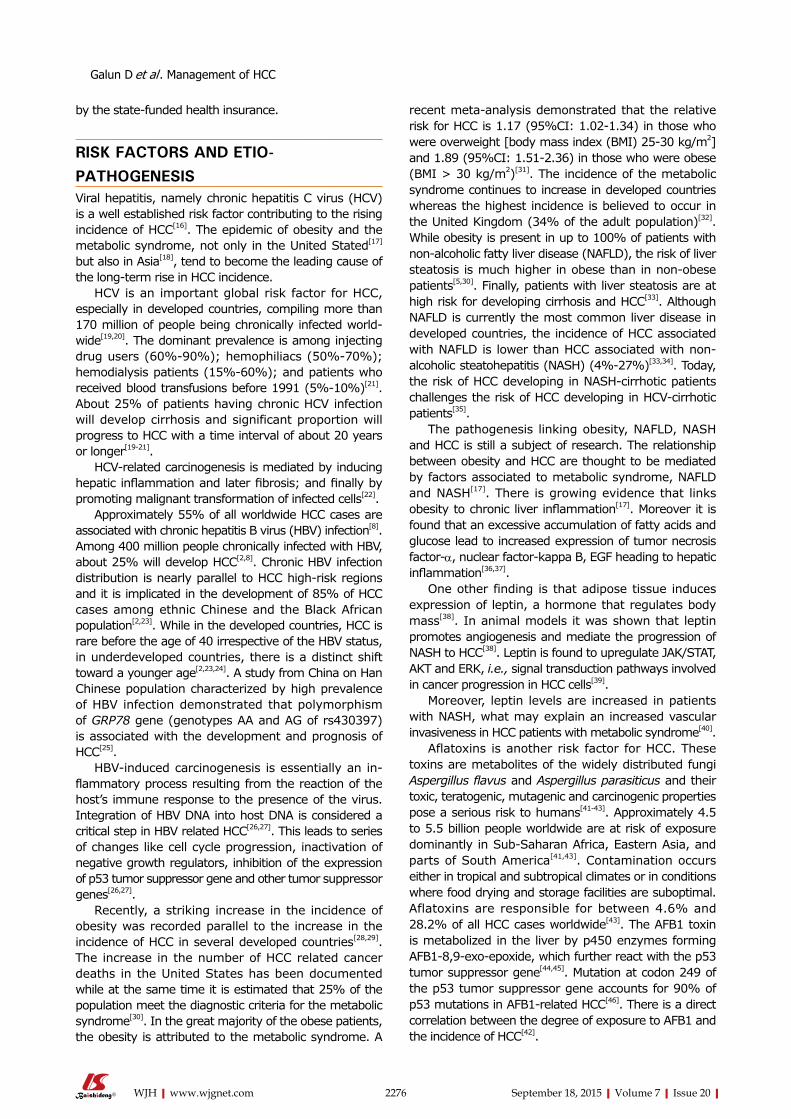
by the state-funded health insurance.
RISK FACTORS AND ETIO-PATHOGENESISViral hepatitis, namely chronic hepatitis C virus (HCV) is a well established risk factor contributing to the rising incidence of HCC[16]. The epidemic of obesity and the metabolic syndrome, not only in the United Stated[17] but also in Asia[18], tend to become the leading cause of the long-term rise in HCC incidence.
HCV is an important global risk factor for HCC, especially in developed countries, compiling more than 170 million of people being chronically infected world-wide[19,20]. The dominant prevalence is among injecting drug users (60%-90%); hemophiliacs (50%-70%); hemodialysis patients (15%-60%); and patients who received blood transfusions before 1991 (5%-10%)[21]. About 25% of patients having chronic HCV infection will develop cirrhosis and significant proportion will progress to HCC with a time interval of about 20 years or longer[19-21].
HCV-related carcinogenesis is mediated by inducing hepatic inflammation and later fibrosis; and finally by promoting malignant transformation of infected cells[22].
Approximately 55% of all worldwide HCC cases are associated with chronic hepatitis B virus (HBV) infection[8]. Among 400 million people chronically infected with HBV, about 25% will develop HCC[2,8]. Chronic HBV infection distribution is nearly parallel to HCC high-risk regions and it is implicated in the development of 85% of HCC cases among ethnic Chinese and the Black African population[2,23]. While in the developed countries, HCC is rare before the age of 40 irrespective of the HBV status, in underdeveloped countries, there is a distinct shift toward a younger age[2,23,24]. A study from China on Han Chinese population characterized by high prevalence of HBV infection demonstrated that polymorphism of GRP78 gene (genotypes AA and AG of rs430397) is associated with the development and prognosis of HCC[25].
HBV-induced carcinogenesis is essentially an in-flammatory process resulting from the reaction of the host’s immune response to the presence of the virus. Integration of HBV DNA into host DNA is considered a critical step in HBV related HCC[26,27]. This leads to series of changes like cell cycle progression, inactivation of negative growth regulators, inhibition of the expression of p53 tumor suppressor gene and other tumor suppressor genes[26,27].
Recently, a striking increase in the incidence of obesity was recorded parallel to the increase in the incidence of HCC in several developed countries[28,29]. The increase in the number of HCC related cancer deaths in the United States has been documented while at the same time it is estimated that 25% of the population meet the diagnostic criteria for the metabolic syndrome[30]. In the great majority of the obese patients, the obesity is attributed to the metabolic syndrome. A
recent meta-analysis demonstrated that the relative risk for HCC is 1.17 (95%CI: 1.02-1.34) in those who were overweight [body mass index (BMI) 25-30 kg/m2] and 1.89 (95%CI: 1.51-2.36) in those who were obese (BMI > 30 kg/m2)[31]. The incidence of the metabolic syndrome continues to increase in developed countries whereas the highest incidence is believed to occur in the United Kingdom (34% of the adult population)[32]. While obesity is present in up to 100% of patients with non-alcoholic fatty liver disease (NAFLD), the risk of liver steatosis is much higher in obese than in non-obese patients[5,30]. Finally, patients with liver steatosis are at high risk for developing cirrhosis and HCC[33]. Although NAFLD is currently the most common liver disease in developed countries, the incidence of HCC associated with NAFLD is lower than HCC associated with non-alcoholic steatohepatitis (NASH) (4%-27%)[33,34]. Today, the risk of HCC developing in NASH-cirrhotic patients challenges the risk of HCC developing in HCV-cirrhotic patients[35].
The pathogenesis linking obesity, NAFLD, NASH and HCC is still a subject of research. The relationship between obesity and HCC are thought to be mediated by factors associated to metabolic syndrome, NAFLD and NASH[17]. There is growing evidence that links obesity to chronic liver inflammation[17]. Moreover it is found that an excessive accumulation of fatty acids and glucose lead to increased expression of tumor necrosis factor-α, nuclear factor-kappa B, EGF heading to hepatic inflammation[36,37].
One other finding is that adipose tissue induces expression of leptin, a hormone that regulates body mass[38]. In animal models it was shown that leptin promotes angiogenesis and mediate the progression of NASH to HCC[38]. Leptin is found to upregulate JAK/STAT, AKT and ERK, i.e., signal transduction pathways involved in cancer progression in HCC cells[39].
Moreover, leptin levels are increased in patients with NASH, what may explain an increased vascular invasiveness in HCC patients with metabolic syndrome[40].
Aflatoxins is another risk factor for HCC. These toxins are metabolites of the widely distributed fungi Aspergillus flavus and Aspergillus parasiticus and their toxic, teratogenic, mutagenic and carcinogenic properties pose a serious risk to humans[41-43]. Approximately 4.5 to 5.5 billion people worldwide are at risk of exposure dominantly in Sub-Saharan Africa, Eastern Asia, and parts of South America[41,43]. Contamination occurs either in tropical and subtropical climates or in conditions where food drying and storage facilities are suboptimal. Aflatoxins are responsible for between 4.6% and 28.2% of all HCC cases worldwide[43]. The AFB1 toxin is metabolized in the liver by p450 enzymes forming AFB1-8,9-exo-epoxide, which further react with the p53 tumor suppressor gene[44,45]. Mutation at codon 249 of the p53 tumor suppressor gene accounts for 90% of p53 mutations in AFB1-related HCC[46]. There is a direct correlation between the degree of exposure to AFB1 and the incidence of HCC[42].
2276 September 18, 2015|Volume 7|Issue 20|WJH|www.wjgnet.com
Galun D et al . Management of HCC
2277 September 18, 2015|Volume 7|Issue 20|WJH|www.wjgnet.com
rate can be greater than 10% for small lesions[64]. The AASLD guideline has been prospectively validated for focal lesions 0.5 to 2.0 cm in size using MRI and contrast-enhanced ultrasound, and demonstrated a low sensi-tivity (33%) but a very high specificity (100%) for the diagnosis of HCC[65].
STAGINGSince 1984 nine different staging systems have been developed and evaluated. The Barcelona-Clinic Liver Cancer (BCLC) staging system has emerged as the most useful to guide treatment decisions (Figure 1). BCLC is based on the analysis of independent studies in different clinical settings. It includes prognostic variables related to tumor status, liver functional status, and health performance status, together with treatment-dependent variables obtained from cohort studies and randomized clinical trials. The system links tumor stage with the treatment strategy allowing an estimation of life expectancy associated to specific HCC management[66].
BCLC demonstrated the best independent predic-tive power in many trials[67-71] when the entire patient population was included [not limited to patient population treated by surgery, radiofrequency ablation (RFA) or transarterial chemoembolization (TACE) only]. The BCLC staging system was externally validated[67,68,71,72] and has practically become an universal staging and treat-ment system. Moreover it was endorsed by European Association for the study of the liver (EASL) and AASLD as standard for patients with HCC[56,66].
However, other trials have demonstrated conflicting results thus favoring other staging systems[73-77]. Graf et al[78] have shown many limitations for the BCLC staging system (Table 1). Furthermore, as indicated by Maida et al[79] the BCLC staging system was not derived from a cohort of HCC patients by a multivariate analysis, and therefore it is not a prognostic model able to predict the mortality of HCC patients. Moreover, the intermediate stage (BCLC B) includes an extremely heterogeneous population in terms of both liver function and tumor characteristics and the main limitation of the BCLC is represented by its rigidity when it is acting as a treat-ment algorithm[79].
Importantly, treatment allocation should be decided by a multidisciplinary board based on individualized rather than on a guideline-based approach[80].
Although BCLC is the most comprehensive staging system, as it integrates tumor status, liver function and the performance status neither BCLC nor any other of the staging systems has been universally accepted, as pointed out by the AASLD guidelines[56], meaning that international consensus on the use of any given model is lacking.
TREATMENTTreatment allocation should be decided by a multidi-sciplinary board involving hepatologists, pathologists,
The study from Yu et al[47] found a synergistic effect of AFB1 and HBV in causing HCC since population with HBV who lived in the region of high exposure to AFB1 were associated to a mortality rate ten times higher than that of population with HBV living in the region of low exposure to the toxin.
Alcohol abuse, lasting more than 10 years, increases the chance for HCC development approximately five fold[48]. It is most common in the Americas (32% of HCC cases in the United States)[48] and Western Europe (45% of the cases in Italy[49]) and the incidence is increasing in Asia[7,9]. In principle, patients who develop the tumor have alcohol-induced cirrhosis[50].
Other less frequent risk factors include iron overload[51], hereditary hemochromatosis[52], tobacco smoking[53,54] and membranous obstruction of inferior vena cava[55].
DIAGNOSISToday, the diagnosis of HCC is established within the national surveillance programs in developed countries while the diagnosis of symptomatic, advanced stage, disease still remains the characteristic of underdeveloped countries. According to the American Association for the Study of Liver Diseases (AASLD) screening for HCC is recommended according to existing guidelines in all cirrhotic patients using ultrasound every six months[56]. Screening for chronic HBV carriers is recommended as well[57].
When a nodule is detected in a cirrhotic liver, a contrast-enhanced diagnostic procedure is strongly recommended. It is important to search for the typical signs of HCC (arterial phase enhancement and portal venous phase washout)[56]. The updated guidelines of AASLD consider that a non-invasive diagnosis of HCC can be established if a lesion > 10 mm has a typical vascular enhancement pattern in 4-phase multi-detector row CT (MDCT) or dynamic contrast enhanced magnetic resonance imaging (DCE-MRI)[56]. These guidelines were also accepted by the European societies[58].
Although MDCT is currently the most common imaging modality for detecting HCC, it is suboptimal for nodule characterization. DCE-MRI, with liver-specific contrast agents, has emerged as the preferred diagnostic modality for the investigation of HCC as it facilitates liver cancer characterization[59-61]. A recent meta-analysis[62] estimated the accuracy of MRI with liver-specific contrast agents compared to MDCT for the detection and characterization of HCC and demonstrated the superiority of MRI for the detection of HCC lesions < 20 mm.
For nodules smaller than 1 cm, a repeated ultra-sound examination in three months intervals is recom-mended[56]. A biopsy is required only if imaging is inconclusive for lesions smaller than 2 cm, or it is atypical for lesions larger than 2 cm when the AFP level is not elevated[56]. However, biopsy carries an approximately 2% risk of tumor seeding[63] and the false-negative
Galun D et al . Management of HCC
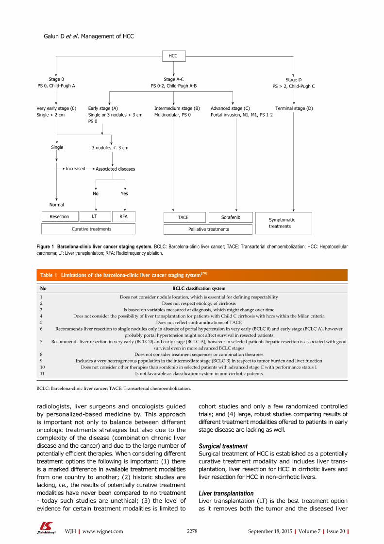
2278 September 18, 2015|Volume 7|Issue 20|WJH|www.wjgnet.com
radiologists, liver surgeons and oncologists guided by personalized-based medicine by. This approach is important not only to balance between different oncologic treatments strategies but also due to the complexity of the disease (combination chronic liver disease and the cancer) and due to the large number of potentially efficient therapies. When considering different treatment options the following is important: (1) there is a marked difference in available treatment modalities from one country to another; (2) historic studies are lacking, i.e., the results of potentially curative treatment modalities have never been compared to no treatment - today such studies are unethical; (3) the level of evidence for certain treatment modalities is limited to
cohort studies and only a few randomized controlled trials; and (4) large, robust studies comparing results of different treatment modalities offered to patients in early stage disease are lacking as well.
Surgical treatmentSurgical treatment of HCC is established as a potentially curative treatment modality and includes liver trans-plantation, liver resection for HCC in cirrhotic livers and liver resection for HCC in non-cirrhotic livers. Liver transplantationLiver transplantation (LT) is the best treatment option as it removes both the tumor and the diseased liver
Table 1 Limitations of the barcelona-clinic liver cancer staging system[78]
No BCLC classification system
1 Does not consider nodule location, which is essential for defining respectability2 Does not respect etiology of cirrhosis3 Is based on variables measured at diagnosis, which might change over time4 Does not consider the possibility of liver transplantation for patients with Child C cirrhosis with hccs within the Milan criteria5 Does not reflect contraindications of TACE6 Recommends liver resection to single nodules only in absence of portal hypertension in very early (BCLC 0) and early stage (BCLC A), however
probably portal hypertension might not affect survival in resected patients7 Recommends liver resection in very early (BCLC 0) and early stage (BCLC A), however in selected patients hepatic resection is associated with good
survival even in more advanced BCLC stages8 Does not consider treatment sequences or combination therapies9 Includes a very heterogeneous population in the intermediate stage (BCLC B) in respect to tumor burden and liver function10 Does not consider other therapies than sorafenib in selected patients with advanced stage C with performance status 111 Is not favorable as classification system in non-cirrhotic patients
BCLC: Barcelona-clinic liver cancer; TACE: Transarterial chemoembolization.
Stage 0PS 0, Child-Pugh A
Stage A-CPS 0-2, Child-Pugh A-B
Stage DPS > 2, Child-Pugh C
Very early stage (0)Single < 2 cm
Early stage (A)Single or 3 nodules < 3 cm, PS 0
Intermedium stage (B)Multinodular, PS 0
Advanced stage (C)Portal invasion, N1, M1, PS 1-2
Terminal stage (D)
Single 3 nodules ≤ 3 cm
Increased Associated diseases
Normal
No Yes
Resection LT RFA
Curative treatments
TACE Sorafenib
Palliative treatments
Symptomatictreatments
HCC
Figure 1 Barcelona-clinic liver cancer staging system. BCLC: Barcelona-clinic liver cancer; TACE: Transarterial chemoembolization; HCC: Hepatocellular carcinoma; LT: Liver transplantation; RFA: Radiofrequency ablation.
Galun D et al . Management of HCC
2279 September 18, 2015|Volume 7|Issue 20|WJH|www.wjgnet.com
parenchyma[81,82]. This is primarily important for patients with a Child-Pugh (CP) score C as it is the treatment of liver failure. The patient’s age (typically younger than 70 years), co-morbidities (e.g., cardiopulmonary disease, smoking, diabetes or renal disease), nutritional state (e.g., poor nutrition or morbidly obese), and social factors (e.g., adequate support, compliance, abstinence from alcohol and completion of an appropriate rehabi-litation program) are all factors determining the patients’ eligibility for LT[81].
The most appropriate candidates for LT are patients that fit into the Milan criteria (a single tumor < 5 cm or up to 3 tumors of < 3 cm) achieving a 5-year survival rate of 70%-80%. In these patients the recurrence rates are approximately 10%[83,84].
The Milan criteria can be expanded to include more patients primarily by liberalizing the restrictions on tumor size. Yao et al[85] demonstrated that using the University of California San Francisco criteria (single nodule < 6.5 cm or ≤ 3 nodules each ≤ 4.5 cm, with total combined tumor diameter ≤ 8 cm), a 75% 5-year survival rate is achievable. Kaido et al[86] reported that using the Kyoto criteria (a combination of tumor number ≤ 10, maximal diameter of each tumor ≤ 5 cm, and serum des-γ-carboxy prothrombin levels ≤ 400 mAU/mL), the 5-year survival rate after living donor LT is 82%.
Mazzaferro et al[87] have proposed the “Metro ticket price” concept - the further one goes in expanding the criteria for LT, the more one “pays”, i.e., the more you deviate from the Milan criteria, the survival rate decreases and recurrence rate increases.
Due to the limited number of donors and the scarcity of sufficient available data, current guidelines do not recommend LT for HCC patients outside the Milan criteria[58,88]. Patients with a compromised liver function (CP - B or C) should be listed for LT while allocation of this treatment modality to CP class A patients instead of surgical resection is still an area of debate. The Barcelona Clinic has analyzed their results for surgical resection and LT in an intent-to-treat manner, although the patients were never compared directly in a randomized trial[89]. The five-year survival rates for resection and LT were nearly identical if patients for resection were carefully selected (CP class A, normal bilirubin levels and no portal hypertension).
Waiting time for LT is a serious obstacle in many national transplant programs worldwide. When the waiting list for LT is longer than 12 mo the drop-out rates can reach 25% of HCC patients listed for LT[90,91]. Clearly, if patients with more advanced tumors are included as a result of expanded listing criteria the dropout rate will be higher and this will lead to poor survival figures. In that regard the potential benefit of TACE, TARE, RFA and others, applied in the neoadjuvant setting include “bridging” or “down-staging” strategies to increase the number of HCC patients qualifying for LT[92].
Furthermore another important concept of LT is salvage LT that saves the donor pool and can effectively
be performed for patients with recurrence or liver function deterioration following resection for HCC. This does not increase the perioperative mortality and has similar long-term survival compared to primary LT[93].
Liver transplantation can also be offered to patients with non-resectable HCC in normal livers providing 5-year survival rates of 59%[94]. In contrast to LT for HCC in cirrhosis the tumor size is not a predictor of post-transplant survival[94].
Finally, many controversies related to LT were confronted during an international consensus conference held in 2010, in Switzerland that resulted in 37 statements and recommendations[95]. These recom-mendations reflect the current state of scientific evidence regarding the LT and reflect differences in clinical practice of LT between continents, countries and institutions. In each controversial topic the strength of recommendation was conditioned by the level of evidence that was in the majority of instances 2 or less reflecting the quality of evidence that is currently available. Among the 37 recommendations only 17 are strong (presented in Table 2) while the others are week or their strength could not be established due to insufficient data[95].
The highest level of evidence and the strength of recommendation is related to the assessment of candidates for LT and in defining criteria for listing candidates with HCC in cirrhotic livers for deceased donor LT. In regard to HCC patients in non-cirrhotic liver LT this procedure may be considered as salvage transplantation for patients with intrahepatic recurrence following liver resection and no evidence of lymph node or macrovascular invasion[95].
The role of down-staging was evaluated in per-spective of different loco-regional treatment options that are presented in the literature (TACE, TARE, RFA). Although the largest experience is linked to TACE and RFA, based on existing evidence, no recommendation can be made for selecting a specific loco-regional therapy for down-staging[95].
Living donor LT is an important alternative to deceased donor liver transplantation in the present circu-mstances of increasing number of HCC patients listed for LT. It is conducted in a limited number of centers worldwide. Although it facilitates access to LT, recent meta analysis demonstrated that living donor LT is associated with a higher rate of surgical complications following transplantation[96].
In that sense an important recommendation is derived from the consensus conference, i.e., that living donor LT must be restricted to centers of excellence in liver surgery and liver transplantation to minimize donor risk and maximize recipient outcome[95].
Liver resectionDuring the past decade a tremendous improvement in the understanding of liver anatomy, advances in technology, anesthesiology and postoperative intensive care and the application of intraoperative ultrasono-
Galun D et al . Management of HCC
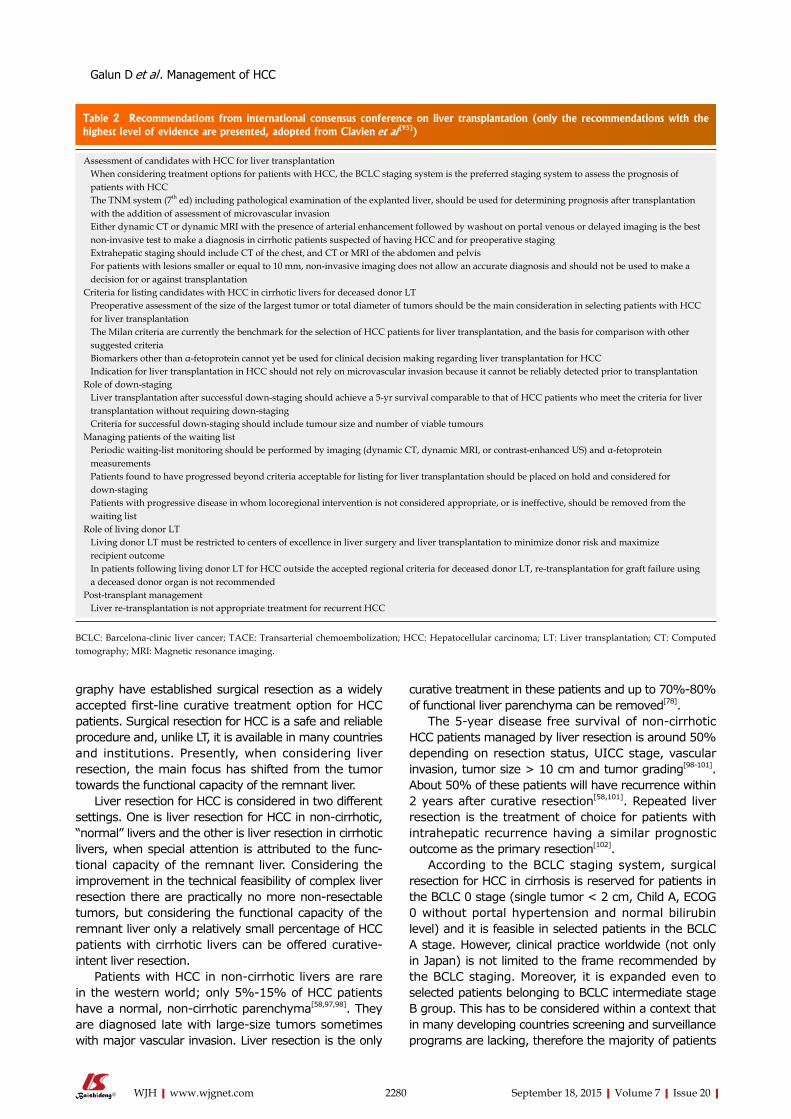
2280 September 18, 2015|Volume 7|Issue 20|WJH|www.wjgnet.com
graphy have established surgical resection as a widely accepted first-line curative treatment option for HCC patients. Surgical resection for HCC is a safe and reliable procedure and, unlike LT, it is available in many countries and institutions. Presently, when considering liver resection, the main focus has shifted from the tumor towards the functional capacity of the remnant liver.
Liver resection for HCC is considered in two different settings. One is liver resection for HCC in non-cirrhotic, “normal” livers and the other is liver resection in cirrhotic livers, when special attention is attributed to the func-tional capacity of the remnant liver. Considering the improvement in the technical feasibility of complex liver resection there are practically no more non-resectable tumors, but considering the functional capacity of the remnant liver only a relatively small percentage of HCC patients with cirrhotic livers can be offered curative-intent liver resection.
Patients with HCC in non-cirrhotic livers are rare in the western world; only 5%-15% of HCC patients have a normal, non-cirrhotic parenchyma[58,97,98]. They are diagnosed late with large-size tumors sometimes with major vascular invasion. Liver resection is the only
curative treatment in these patients and up to 70%-80% of functional liver parenchyma can be removed[78].
The 5-year disease free survival of non-cirrhotic HCC patients managed by liver resection is around 50% depending on resection status, UICC stage, vascular invasion, tumor size > 10 cm and tumor grading[98-101]. About 50% of these patients will have recurrence within 2 years after curative resection[58,101]. Repeated liver resection is the treatment of choice for patients with intrahepatic recurrence having a similar prognostic outcome as the primary resection[102].
According to the BCLC staging system, surgical resection for HCC in cirrhosis is reserved for patients in the BCLC 0 stage (single tumor < 2 cm, Child A, ECOG 0 without portal hypertension and normal bilirubin level) and it is feasible in selected patients in the BCLC A stage. However, clinical practice worldwide (not only in Japan) is not limited to the frame recommended by the BCLC staging. Moreover, it is expanded even to selected patients belonging to BCLC intermediate stage B group. This has to be considered within a context that in many developing countries screening and surveillance programs are lacking, therefore the majority of patients
Table 2 Recommendations from international consensus conference on liver transplantation (only the recommendations with the highest level of evidence are presented, adopted from Clavien et al [95])
Assessment of candidates with HCC for liver transplantation When considering treatment options for patients with HCC, the BCLC staging system is the preferred staging system to assess the prognosis of patients with HCC The TNM system (7th ed) including pathological examination of the explanted liver, should be used for determining prognosis after transplantation with the addition of assessment of microvascular invasion Either dynamic CT or dynamic MRI with the presence of arterial enhancement followed by washout on portal venous or delayed imaging is the best non-invasive test to make a diagnosis in cirrhotic patients suspected of having HCC and for preoperative staging Extrahepatic staging should include CT of the chest, and CT or MRI of the abdomen and pelvis For patients with lesions smaller or equal to 10 mm, non-invasive imaging does not allow an accurate diagnosis and should not be used to make a decision for or against transplantationCriteria for listing candidates with HCC in cirrhotic livers for deceased donor LT Preoperative assessment of the size of the largest tumor or total diameter of tumors should be the main consideration in selecting patients with HCC for liver transplantation The Milan criteria are currently the benchmark for the selection of HCC patients for liver transplantation, and the basis for comparison with other suggested criteria Biomarkers other than α-fetoprotein cannot yet be used for clinical decision making regarding liver transplantation for HCC Indication for liver transplantation in HCC should not rely on microvascular invasion because it cannot be reliably detected prior to transplantationRole of down-staging Liver transplantation after successful down-staging should achieve a 5-yr survival comparable to that of HCC patients who meet the criteria for liver transplantation without requiring down-staging Criteria for successful down-staging should include tumour size and number of viable tumours Managing patients of the waiting list Periodic waiting-list monitoring should be performed by imaging (dynamic CT, dynamic MRI, or contrast-enhanced US) and α-fetoprotein measurements Patients found to have progressed beyond criteria acceptable for listing for liver transplantation should be placed on hold and considered for down-staging Patients with progressive disease in whom locoregional intervention is not considered appropriate, or is ineffective, should be removed from the waiting listRole of living donor LT Living donor LT must be restricted to centers of excellence in liver surgery and liver transplantation to minimize donor risk and maximize recipient outcome In patients following living donor LT for HCC outside the accepted regional criteria for deceased donor LT, re-transplantation for graft failure using a deceased donor organ is not recommendedPost-transplant management Liver re-transplantation is not appropriate treatment for recurrent HCC
BCLC: Barcelona-clinic liver cancer; TACE: Transarterial chemoembolization; HCC: Hepatocellular carcinoma; LT: Liver transplantation; CT: Computed tomography; MRI: Magnetic resonance imaging.
Galun D et al . Management of HCC
2281 September 18, 2015|Volume 7|Issue 20|WJH|www.wjgnet.com
are diagnosed in an advanced stage of the disease when surgical resection is still feasible[103,104]. Strict adherence to the BCLC staging system would direct the majority of patients to palliative treatment only.
Despite of recent advances in surgical techniques and perioperative care, liver resection is challenged by the poor functional reserve of the cirrhotic liver, the impaired regeneration capacity, elevated portal venous pressure, and other co-morbidities of the HCC patients[105,106]. Although reserved for high-volume centers, liver resection is justified even for patiens with large and multinodular HCC[107-109].
A study from Ishizawa et al[110] has demonstrated that neither multiple tumors nor portal hypertension are surgical contraindications for HCC. Two other studies have verified that liver resection is feasible even in Child B patients and in selected patients a major liver resection is feasible as well[111,112]. According to Ho et al[113] liver resection is associated with better overall survival comparing to TACE (37.9 mo vs 17.3 mo) even for patients with multinodular HCC. In patients with large tumors, TACE is associated with low response rate and a modest 3 years survival rate[108,109].
Several studies confirmed that blood loss has a negative impact on the perioperative morbidity, mortality and long-term outcome[114,115] therefore a control of bleeding is mandatory when performing liver resection. Vascular occlusion techniques[116] are effective in reducing blood loss, but it was found that they compromise hepatic functional reserve in conditions of a preexistent liver disease[117,118]. Fu et al[119] found an earlier recovery of the postoperative liver function after hemihepatic vascular inflow occlusion compared with the Pringle maneuver, however it is technically more demanding and potentially associated with more bleeding in cirrhotic livers.
Prediction of the future, functional remnant liver volume (FLR) is crucial for postoperative morbidity and mortality. A remnant volume of at least 40% should remain following resection of cirrhotic livers in order to preserve adequate liver function[120]. Three dimensional measurements of liver volumes based on MDCT or magnetic resonance imaging (MRI) and more important post-processing software are important for predicting the FLR after liver resection.
Portal vein embolization (PVE) has an important role as an effective tool in inducing hypertrophy of the non-embolized hepatic segments. An increase of the FLR volume of 20%-46% can be achieved after 2-8 wk[121,122]. When the FLR volume is insufficient PVE is considered an important therapeutic step before extended resection. Recently, one other approach has been described for increasing the FLR volume in a two-stage procedure for patients undergoing extended liver resection. In situ liver transection combined with portal vein ligation emerged as a procedure associated with rapid growth of the FLR[123,124] and was tested in the settings of HCC in cirrhotic livers[125] even in conditions of major vascular invasion[126]. The median FLR volume
increase was 18.7% within one week after the first step and 38.6% after the second step[125]. More studies are needed before the real merits of ALPPS can be evaluated.
The use of metabolic tests, namely the indocyanine green test is another tool to assess the liver functional capacity in order to avoid postoperative liver failure[127]. As indicated in two surveys[120,128] it is widely used in Asia and the indocyanine green retention rate at 15 min (ICGR-15) is integrated into the decision tree for deciding the safe limit of hepatectomy[127]. In the western world the ICGR-15 test is used in a limited number of centers and in selective cases only[120]. In HCC patients with cirrhotic livers characterized by normal bilirubin level and absence of ascites the ICGR-15 is the main determinant for performing a liver resection[127].
The anatomic liver resection should be associated with improved outcome as HCC tumors have a tendency for local portal vein invasion with possible extension toward the main portal vein. However conflicting results are present in the literature. Two studies[129,130], demonstrated that anatomic resection is an indepen-dent predictor of improved recurrence-free survival and it significantly improves the disease-free survival rates. Anatomic resection is recommended in the EASL guidelines as the preferred approach if sufficient remnant liver volume can be preserved[56]. The use of dye widely practiced in Japan may aid delineation of tumor bearing segments and facilitate complete anatomical resection[131,132].
Laparoscopic liver resection for HCC in cirrhotic livers is an established and safe procedure performed in many centers worldwide[128]. There are no randomized controlled trials that has compared laparoscopic vs open liver resection in HCC patients. Four meta-analyses[133-136] of nonrandomized studies found that laparoscopic resection was associated with significantly less blood loss, lower transfusion requirements, lower overall morbidity, and shorter length of hospital stay without a significant difference in length of operation, surgical margin status, or tumor recurrence rates.
Ablative proceduresTumor ablation can be achieved by chemical (ethanol, acetic acid) or thermal [radiofrequency ablation-RFA, microwave ablation (MWA)] ablation and it is the treatment of choice in patients with single, small tumors who are not candidates for surgery. According to the BCLC staging and treatment algorithm these patients are classified as BCLC A patients[58]. BCLC 0 patients may also be managed by this treatment modality, although the algorithm primarily allocates resection to this group of patients[58]. When procedure limitations are strictly respected (tumor size, tumor location, duration of the treatment, maintaining the required temperature in the tumor zone, etc.) tumor ablation is a curative treatment option for the management of carefully selected HCC patients.
Historically, tumor ablation started as chemical
Galun D et al . Management of HCC
2282 September 18, 2015|Volume 7|Issue 20|WJH|www.wjgnet.com
ablation using percutaneous ethanol injection (PEI) for the management of nodular-type HCC. There is con-siderable experience with PEI since it is an established technique. PEI induces coagulation necrosis of the tumor as a result of cellular dehydration, protein denaturation, and chemical occlusion of small tumor vessels[137]. Several studies confirm that tumors < 2 cm can be successfully treated by PEI achieving equivalent results to thermal ablation techniques[137-139]. For larger tumors PEI is inferior to thermal ablation and therefore should not be performed[138-142]. However, PEI should not be neglected and can be used in underdeveloped regions as a very useful treatment modality.
Thermal ablation has now largely replaced PEI, initially with RFA and recently with MWA[137,139]. Although it is an interventional procedure performed percutaneously by interventional radiologists or jointly by an interventional radiologist and liver surgeon, a multidisciplinary approach which provides important advantages, as described by Poon et al[143]. Thermal ablation can also be done via an open or laparoscopic surgical approach.
The main advantage of thermal ablation is related to its low major morbidity (2.2%-3.1%) and mortality (0.1%-0.5%) rates[144,145]. Major complications include intraperitoneal hemorrhage, hepatic abscess, bile duct injury, and liver decompensation[56,144,145]. Tumor seeding along the needle track has been reported as a rare (0.5%) late complication of RFA[146].
The most important observations resulted from explants studies following LT and demonstrated com-plete tumor necrosis in explanted liver specimens in 83% of tumors > 3 cm and in 88% of tumors in non-perivascular locations[54,56,144,145]. Clearly the efficacy of RFA is reduced with increasing tumor size and the presence of large vessels[147]. RFA should be applied for tumors less than 3 cm in size, bearing in mind that success is related to the total volume of the tumor tissue that has to be ablated.
Lencioni et al[145,148] have demonstrated 61% 5-year survival in patients with Child A cirrhosis and solitary HCC, compared with 51% in patients with Child A cirrhosis and multiple tumors and 31% in patients with Child B cirrhosis. Livraghi et al[149] has reported complete tumor response in 97% of tumors ≤ 2 cm, with 5-year survival in patients with preserved hepatic function of 68%, challenging resection as the first-line approach in such cases.
Hasegawa et al[150] concluded that resection was associated with a higher overall survival and lower recurrence rate than RFA or PEI in the treatment of HCC ≤ 3 cm.
A challenging question is whether emerging alter-native, MWA, will replace RFA. Compared to RFA, MWA is less-susceptible to the heat sink effect of nearby blood vessels and produces a larger zone of necrosis[151].
In a non-randomized study published in 2013[152] that investigated the therapeutic efficacy of per-cutaneous RFA and MWA for HCC < 5 cm no significant differences were found between the two procedures
in the percentage of complete ablation local tumor progression, distant recurrence and overall survival. Clearly, more studies are needed to compare the two ablation techniques.
Transarterial therapyAccording to the BCLC staging and treatment algorithm TACE is indicated for patients classified as BCLC B stage, that is an intermediate stage composed of a very heterogeneous patient population[56,153,154]. A Cochrane review[154] clearly confirmed the survival benefit of this treatment modality. However, TACE is not standardized in regard to: (1) the procedure technique; (2) the choice of embolic agent; (3) the choice of applied medications; and (4) the schedule (on demand or at fixed intervals). In clinical practice TACE is performed by injection of chemotherapy with or without lipiodol, followed by the injection of embolic particles. This procedure is considered as conventional TACE. Innovative step forward was the development of drug-eluting beads (DC Bead) used to increase tumor drug delivery. However, the PRECISION V study[155] designed to compare the two TACE procedures failed to demonstrate a clear superiority of DC Bead-TACE (one-sided P = 0.11). The difference between the two TACE procedures was found in the complete response, objective response, and disease control favoring DC Bead group (27% vs 22%, 52% vs 44%, and 63% vs 52%, respectively)[155].
Complications of TACE include non-target emboliza-tion, the post embolization syndrome (fever, abdominal pain, ileus), liver failure, cholecystitis and acute portal vein thrombosis[154]. The procedure-related mortality is less than 5% which defines TACE as a safe pro-cedure[154]. Main portal vein thrombosis, poor liver function, and extrahepatic spread have been shown to be predictors of poor outcome and are considered con-traindications for chemoembolization[154].
Several aspects of TACE treatment require special consideration. In clinical practice an attempt should be made to achieve the supraselective approach (STACE) using micro-catheters in order to deliver chemotherapy as close as possible to the tumor site. Unfortunately this aspect was not much elaborated in clinical trials. Only one trial[156] on 60 patients who were candidates for LT, found STACE to be associated with complete tumor necrosis in a larger proportion of patients (30.8% vs 6.9%, P = 0.02) compared to selective TACE group. Still, a 5-year disease-free survival was similar in both groups (76.8% vs 74.8%)[156]. In conclusion, there is no clear relationship between the therapy-induced complete necrosis and long-term survival.
The combination of TACE and RFA is another challenging treatment option practiced in many centers worldwide. Recent meta-analyses[157] showed that the combination of RFA and TACE was associated with a significantly higher overall survival rates (OR 1 year = 2.39, 95%CI: 1.35-4.21, P = 0.003; OR 3 years = 1.85, 95%CI: 1.26-2.71, P = 0.002), and recurrence-free survival rate (OR 1 year = 2.00, 95%CI: 1.26-3.18, P
Galun D et al . Management of HCC
2283 September 18, 2015|Volume 7|Issue 20|WJH|www.wjgnet.com
= 0.003; OR 3 years = 2.13, 95%CI: 1.41-3.20, P < 0.001) compared with RFA alone[157]. The quality of the evidence was high for the 1- and 3-year survival[157].
Two recent meta-analysis[158,159] elaborated the combination of TACE and sorafenib and found evident improvement in the objective response, time to pro-gression and overall survival although the sorafenib associated AEs were more frequent in the combination therapy group[158,159].
Another important meta-analysis[160] examined the efficacy of TACE for HCC patients with portal vein thrombosis (PVTT) and found that TACE improves the 1-year survival of patients with HCC and PVTT. As current treatment algorithms contraindicate TACE in patients with main trunk PVTT more trials are required to confirm these findings[54,56,66].
As already indicated the BCLC B stage (an inter-mediate stage) is composed of a very heterogeneous patient population. Several studies confirmed limited treatment efficacy of TACE for HCC patients with large multinodular tumors[161,162]. The objective response to TACE treatment is found in 52% of patients (PRECISION V trial[155]) leaving large proportion of patients without an effective treatment. It is important to note that TACE is contraindicated in patients with main trunk PVTT. These findings led to development of new therapies for optimal management of these subcategories of HCC patients belonging to BCLC B stage[163].
Radio-embolization via hepatic artery using micro-spheres impregnated with yttrium-90 (TARE) is a new emerging treatment option that is available in a limited number of centers worldwide[162,163]. TARE uses the same concept as TACE in regards the technical aspect of the procedure. The difference is reflected in the mode of action. In TARE the embolic particles (microspheres) are 3-10 times smaller than those used in TACE (25-35 micron in diameter)[164]. Yttrium-90 (beta emitter with a short half-life) microspheres are used to produce tumor necrosis by internal delivery of tumoricidal dose of radiation directly to the tumor with nearly no embolic effect on the vessels[165]. The safety and efficacy of TARE is well established in many trials[164,166-168] and post-embolization syndrome is found in 20%-55% of cases[164,166]. TARE was found to be a safe procedure in HCC patients with PVTT[169,170]. Initially, TARE was indicated in HCC patients who progressed or relapsed after the TACE treatment or in HCC patients not amenable to TACE (large multinodular tumors or presence of PVTT)[164]. Although Sangro et al[162] found survival benefit for TARE comparing to TACE as a first-line treatment other studies reported no significant difference in survival[168,171,172]. Potential advantage of TARE over TACE can be attributed to early-stage HCC patients listed for LT who are candidates for bridging or down-staging therapy[173,174].
MEDICAL TREATMENTAfter years of disappointment with the results of
numerous trials testing the efficacy of different drugs in the medical management of HCC, two milestone studies[175,176] have established sorafenib as a treatment of choice for BCLC C patients according to the EASL-EORTC guidelines[56].
Sorafenib is a molecular inhibitor of several tyrosine protein kinases (VEGFR and PDGFR); Raf kinases (C-Raf than B-Raf)[177,178] and intracellular serine/threonine kinases (C-Raf, wild-type B-Raf and mutant B-Raf)[179] Sorafenib treatment induces autophagy[180], which suppresses tumor growth.
Although sorafenib was introduced as well-tolerat-ed drug a subanalysis of the two leading[175,176] and other[181,182] studies have shown that the tolerability of sorafenib was suboptimal[183]; it was down-dosed in more than 50% and interrupted in 45% of patients due to severe adverse events or compromised liver function[183].
Therefore the most important side effects are gastrointestinal[184] (diarrhea 43%, increased lipase 41%, increased amylase 30%, nausea 23%, anorexia 16%, vomiting 16%, and constipation 15%), dermatologic[185] (rash/desquamation 40%, hand-foot skin reaction 30%, alopecia 27%, pruritus 19%, and dry skin 11%), cardiovascular[186]. (Hypertension 17%, angioedema, and congestive heart failure), hematologic[187]. (Hypo-albuminemia 49%, hemorrhage 15%, anemia and thrombocytopenia) and nervous system side effects[186] (neuropathy 13% and headache 10%).
One of the two milestone studies (SHARP/phase Ⅲ)[175] conducted in the western world have shown that sorafenib prolonged median survival from 7.9 mo (placebo group) to 10.7 mo (sorafenib group) (HR = 0.69; 95%CI: 0.55-0.87; P = 0.00058). Sorafenib also improved the time to progression (from 2.8 mo to 5.5 mo). Another milestone study conducted in Asia confirmed the outcomes of the SHARP trial, i.e., a phase Ⅲ Asia-Pacific trial[176] have shown a median overall survival of 6.5 mo (treatment group) comparing to 4.2 mo (placebo group) (HR = 0.68; 95%CI: 0.50-0.93; P = 0.014).
Another important study was a phase Ⅳ, GIDEON trial[188], conducted with the aim to evaluate the safety of sorafenib treatment in HCC patients in real-life conditions. In 2011 the second interim analysis showed a median survival of 10.3 mo for Child A patients and 4.8 mo for Child B patients. The amount of adverse events was comparable to the two milestone studies.
The use of sorafenib in adjuvant settings was addressed in the STORM trial. In mid 2014 major phar-maceutical companies Bayer and Onyx announced that the STORM trial did not meet its primary endpoint. During the ASCO annual meeting in 2014 Bruix et al[189] reported that both primary and secondary endpoints were not met. The trial enrolled the largest cohort of patients with HCC treated in this setting. Overall, 1114 patients were equitably randomized to take either sorafenib or placebo. The study did not met its primary and secondary endpoints since no differences were observed regarding recurrence-free survival (33.4 mo
Galun D et al . Management of HCC
2284 September 18, 2015|Volume 7|Issue 20|WJH|www.wjgnet.com
vs 33.8 mo; HR = 0.94; 95%CI: 0.78-1.13, P = 0.26), time to recurrence (38.6 mo vs 35.8 mo; HR = 0.89, 95%CI: 0.73-1.08) and overall survival (not reached vs not reached, HR = 0.99, 95%CI: 0.76-1.30, P = 0.48). In this trial, a higher rate of sorafenib discontinuation due to drug-adverse events was observed compared to placebo (24% vs 7%)[190].
Additional studies have evaluated other targeted agents either in combination with sorafenib, or designed as head-to-head compared to sorafenib, or as second-line treatments following disease progression or inability to tolerate sorafenib; however, all these trials failed to demonstrate an improvement in overall survival[190].
CONCLUSION AND FUTURE DIRECTIONSHCC is a difficult to treat and extremely complex malignant disease. Epidemiological data confirms an increasing number of new cases each year and this rise will persist due to the burden linked to HCV and obesity. Since the number of new HCC cases being diagnosed each year is nearly equal to the number of deaths from this cancer it is clear that the international scientific community and healthcare systems worldwide have no efficient answer to HCC. There are marked differences between countries in providing disparate quality of healthcare considering screening and surveillance programs; available treatment modalities and drugs; reimbursement of specific treatment options by the state-funded health insurance. In many countries world-wide liver transplantation is not a therapeutic option. In countries with national LT program the donor pool is a serious obstacle for treating more patients. Surveillance programs, so essential for the diagnosing an early stage HCC, are lacking in many countries. The experience from Japan clearly confirms importance of a successful surveillance program. Liver resection and TACE are the two treatment modalities offered to HCC patients even in underdeveloped countries. Since treatment allocation should be decided by a multidisciplinary board involving hepatologists, pathologists, radiologists, liver surgeons and oncologists guided by individualized-based medicine, HCC patients should be managed in high-volume, tertiary, university centers. This approach is important to achieve the best possible outcome from a variety of potentially useful therapies and for research purposes.
Different combination therapies tested in various studies failed to demonstrate a real benefit in terms of overall survival. This is mainly due to the complexity of the disease and due to the extremely heterogeneous patient populations included in clinical trials. The con-sensus conference on LT for HCC has shown that many controversies remained unanswered due to the lack of evidence. Therefore, high-quality randomized trials with better patient stratification are mandatory in the future to find patient populations that can benefit from certain treatment modalities. Basic research in HCC carcinogenesis is equally important. Combinations of different treatment modalities should be more exploited
in order to improve survival and the quality of life of HCC patients.
In the management of HCC patients, several recom-mendations are important: (1) to establish a national surveillance program in as many countries as possible; (2) to further improve treatment modalities for patients on the waiting list for LT; (3) to improve the safety of liver resection and to reduce the recurrence rates following resection; (4) to investigate further and to upgrade results of the TACE treatment modality; (5) to continue research on novel molecular therapies; and (6) to continue research on novel molecular markers for better patient selection for various treatment modalities.
REFERENCES1 Bailar JC, Gornik HL. Cancer undefeated. N Engl J Med 1997;
336: 1569-1574 [PMID: 9164814 DOI: 10.1056/NEJM199705293362206]
2 Parkin DM, Bray F, Ferlay J, Pisani P. Global cancer statistics, 2002. CA Cancer J Clin 2002; 55: 74-108 [PMID: 15761078 DOI: 10.3322/canjclin.55.2.74]
3 National Cancer Institute. Cancer Trends Progress Report - 2009/2010 Update. [Accessed 2015 Mar]. Available from: URL: http://progressreport.cancer.gov/
4 Mortality Database. WHO Statistical Information System. Geneva: World Health Organization, 2008. Available from: URL: http://www.who.int/whosis
5 Altekruse SF, McGlynn KA, Reichman ME. Hepatocellular carcinoma incidence, mortality, and survival trends in the United States from 1975 to 2005. J Clin Oncol 2009; 27: 1485-1491 [PMID: 19224838 DOI: 10.1200/JCO.2008.20.7753]
6 El-Serag HB, Lau M, Eschbach K, Davila J, Goodwin J. Epidemiology of hepatocellular carcinoma in Hispanics in the United States. Arch Intern Med 2007; 167: 1983-1989 [PMID: 17923599 DOI: 10.1001/archinte.167.18.1983]
7 Mittal S, El-Serag HB. Epidemiology of hepatocellular carcinoma: consider the population. J Clin Gastroenterol 2013; 47 Suppl: S2-S6 [PMID: 23632345 DOI: 10.1097/MCG.0b013e3182872f29]
8 Ferlay J, Shin HR, Bray F, Forman D, Mathers C, Parkin DM. Estimates of worldwide burden of cancer in 2008: GLOBOCAN 2008. Int J Cancer 2010; 127: 2893-2917 [PMID: 21351269 DOI: 10.1002/ijc.25516]
9 Jemal A, Bray F, Center MM, Ferlay J, Ward E, Forman D. Global cancer statistics. CA Cancer J Clin 2011; 61: 69-90 [PMID: 21296855 DOI: 10.3322/caac.20107]
10 GLOBOCAN 2012: Estimated Cancer Incidence, Mortality And Prevalence Worldwide In 2012. Lyon: International Agency for Research on Cancer (IARC). [Accessed 2012]. Available from: URL: http://www-dep.iarc.fr
11 Kew MC. Hepatocellular carcinoma: epidemiology and risk factors. J Hepatocellular Carcinoma 2014; 1: 115-125 [DOI: 10.2147/JHC.S44381]
12 El-Serag HB. Epidemiology of hepatocellular carcinoma in USA. Hepatol Res 2007; 37 Suppl 2: S88-S94 [PMID: 17877502 DOI: 10.1111/j.1872-034X.2007.00168.x]
13 Bruix J, Sherman M. Management of hepatocellular carcinoma. Hepatology 2005; 42: 1208-1236 [PMID: 16250051 DOI: 10.1002/hep.20933]
14 Osaki Y, Nishikawa H. Treatment for hepatocellular carcinoma in Japan over the last three decades: Our experience and published work review. Hepatol Res 2015; 45: 59-74 [PMID: 24965914 DOI: 10.1111/hepr.12378]
15 van Meer S, de Man RA, Siersema PD, van Erpecum KJ. Surveillance for hepatocellular carcinoma in chronic liver disease: evidence and controversies. World J Gastroenterol 2013; 19: 6744-6756 [PMID: 24187450 DOI: 10.3748/wjg.v19.i40.6744]
Galun D et al . Management of HCC
2285 September 18, 2015|Volume 7|Issue 20|WJH|www.wjgnet.com
16 Harnois DM. Hepatitis C virus infection and the rising incidence of hepatocellular carcinoma. Mayo Clin Proc 2012; 87: 7-8 [PMID: 22212962 DOI: 10.1016/j.mayocp.2011.11.004]
17 Siegel AB, Zhu AX. Metabolic syndrome and hepatocellular carcinoma: two growing epidemics with a potential link. Cancer 2009; 115: 5651-5661 [PMID: 19834957 DOI: 10.1002/cncr.24687]
18 Asia Pacific Cohort Studies Collaboration. The burden of overweight and obesity in the Asia-Pacific region. Obes Rev 2007; 8: 191-196 [PMID: 17444961 DOI: 10.1111/j.1467-789X.2006.00292.x]
19 Kew M, François G, Lavanchy D, Margolis H, Van Damme P, Grob P, Hallauer J, Shouval D, Leroux-Roels G, Meheus A. Prevention of hepatitis C virus infection. J Viral Hepat 2004; 11: 198-205 [PMID: 15117321 DOI: 10.1111/j.1365-2893.2004.00492.x]
20 Zampino R, Marrone A, Restivo L, Guerrera B, Sellitto A, Rinaldi L, Romano C, Adinolfi LE. Chronic HCV infection and inflammation: Clinical impact on hepatic and extra-hepatic manifestations. World J Hepatol 2013; 5: 528-540 [PMID: 24179612 DOI: 10.4254/wjh.v5.i10.528]
21 Hutin Y, Kitler ME, Dore G. Global burden of disease for hepatitis C. J Clin Pharmacol 2003; 43: 20-29
22 Lemon SM, McGivern DR. Is hepatitis C virus carcinogenic? Gastroenterology 2012; 142: 1274-1278 [PMID: 22537433 DOI: 10.1053/j.gastro.2012.01.045]
23 Kew MC. Epidemiology of chronic hepatitis B virus infection, hepatocellular carcinoma, and hepatitis B virus-induced hepatocellular carcinoma. Pathol Biol (Paris) 2010; 58: 273-277 [PMID: 20378277 DOI: 10.1016/j.patbio.2010.01.005]
24 Nguyen VT, Amin J, Law MG, Dore GJ. Predictors and survival in hepatitis B-related hepatocellular carcinoma in New South Wales, Australia. J Gastroenterol Hepatol 2009; 24: 436-442 [PMID: 19175834 DOI: 10.1111/j.1440-1746.2008.05577.x]
25 Zhu X, Chen MS, Tian LW, Li DP, Xu PL, Lin MC, Xie D, Kung HF. Single nucleotide polymorphism of rs430397 in the fifth intron of GRP78 gene and clinical relevance of primary hepatocellular carcinoma in Han Chinese: risk and prognosis. Int J Cancer 2009; 125: 1352-1357 [PMID: 19533686 DOI: 10.1002/ijc.24487]
26 Shafritz DA, Shouval D, Sherman HI, Hadziyannis SJ, Kew MC. Integration of hepatitis B virus DNA into the genome of liver cells in chronic liver disease and hepatocellular carcinoma. Studies in percutaneous liver biopsies and post-mortem tissue specimens. N Engl J Med 1981; 305: 1067-1073 [PMID: 6268980 DOI: 10.1056/NEJM198110293051807]
27 Kew MC. Hepatitis B virus x protein in the pathogenesis of hepatitis B virus-induced hepatocellular carcinoma. J Gastroenterol Hepatol 2011; 26 Suppl 1: 144-152 [PMID: 21199526 DOI: 10.1111/j.1440-1746.2010.06546.x]
28 Calle EE, Rodriguez C, Walt-Thurmond K. Over-weight, obesity, and cancer risk. Lancet Oncol 2002; 348: 625-1638
29 Bianchini F, Kaaks R, Vainio H. Overweight, obesity, and cancer risk. Lancet Oncol 2002; 3: 565-574 [PMID: 12217794 DOI: 10.1016/S1470-2045(02)00849-5]
30 Borena W, Strohmaier S, Lukanova A, Bjørge T, Lindkvist B, Hallmans G, Edlinger M, Stocks T, Nagel G, Manjer J, Engeland A, Selmer R, Häggström C, Tretli S, Concin H, Jonsson H, Stattin P, Ulmer H. Metabolic risk factors and primary liver cancer in a prospective study of 578,700 adults. Int J Cancer 2012; 131: 193-200 [PMID: 21805476 DOI: 10.1002/ijc.26338]
31 Welzel TM, Graubard BI, Zeuzem S, El-Serag HB, Davila JA, McGlynn KA. Metabolic syndrome increases the risk of primary liver cancer in the United States: a study in the SEER-Medicare database. Hepatology 2011; 54: 463-471 [PMID: 21538440 DOI: 10.1002/hep.24397]
32 Adams LA, Lymp JF, St Sauver J, Sanderson SO, Lindor KD, Feldstein A, Angulo P. The natural history of nonalcoholic fatty liver disease: a population-based cohort study. Gastroenterology 2005; 129: 113-121 [PMID: 16012941 DOI: 10.1053/j.gastro.2005.04.014]
33 Lazo M, Clark JM. The epidemiology of nonalcoholic fatty liver
disease: a global perspective. Semin Liver Dis 2008; 28: 339-350 [PMID: 18956290 DOI: 10.1055/s-0028-1091978]
34 Nobili V, Alisi A, Grimaldi C, Liccardo D, Francalanci P, Monti L, Castellano A, de Ville de Goyet J. Non-alcoholic fatty liver disease and hepatocellular carcinoma in a 7-year-old obese boy: coincidence or comorbidity? Pediatr Obes 2014; 9: e99-e102 [PMID: 24302697 DOI: 10.1111/j.2047-6310.2013.00209.x]
35 Schlesinger S, Aleksandrova K, Pischon T, Fedirko V, Jenab M, Trepo E, Boffetta P, Dahm CC, Overvad K, Tjønneland A, Halkjær J, Fagherazzi G, Boutron-Ruault MC, Carbonnel F, Kaaks R, Lukanova A, Boeing H, Trichopoulou A, Bamia C, Lagiou P, Palli D, Grioni S, Panico S, Tumino R, Vineis P, Bueno-de-Mesquita HB, van den Berg S, Peeters PH, Braaten T, Weiderpass E, Quirós JR, Travier N, Sánchez MJ, Navarro C, Barricarte A, Dorronsoro M, Lindkvist B, Regner S, Werner M, Sund M, Khaw KT, Wareham N, Travis RC, Norat T, Wark PA, Riboli E, Nöthlings U. Abdominal obesity, weight gain during adulthood and risk of liver and biliary tract cancer in a European cohort. Int J Cancer 2013; 132: 645-657 [PMID: 22618881 DOI: 10.1002/ijc.27645]
36 Nathan C. Epidemic inflammation: pondering obesity. Mol Med 2008; 14: 485-492 [PMID: 18431463 DOI: 10.2119/2008-00038.Nathan]
37 Balkwill F, Mantovani A. Inflammation and cancer: back to Virchow? Lancet 2001; 357: 539-545 [PMID: 11229684 DOI: 10.1016/S0140-6736(00)04046-0]
38 Ikejima K, Takei Y, Honda H, Hirose M, Yoshikawa M, Zhang YJ, Lang T, Fukuda T, Yamashina S, Kitamura T, Sato N. Leptin receptor-mediated signaling regulates hepatic fibrogenesis and remodeling of extracellular matrix in the rat. Gastroenterology 2002; 122: 1399-1410 [PMID: 11984526 DOI: 10.1053/gast.2002.32995]
39 Saxena NK, Sharma D, Ding X, Lin S, Marra F, Merlin D, Anania FA. Concomitant activation of the JAK/STAT, PI3K/AKT, and ERK signaling is involved in leptin-mediated promotion of invasion and migration of hepatocellular carcinoma cells. Cancer Res 2007; 67: 2497-2507 [PMID: 17363567 DOI: 10.1158/0008-5472.CAN-06-3075]
40 Chitturi S, Farrell G, Frost L, Kriketos A, Lin R, Fung C, Liddle C, Samarasinghe D, George J. Serum leptin in NASH correlates with hepatic steatosis but not fibrosis: a manifestation of lipotoxicity? Hepatology 2002; 36: 403-409 [PMID: 12143049 DOI: 10.1053/jhep.2002.34738]
41 Wild CP, Pionneau FA, Montesano R, Mutiro CF, Chetsanga CJ. Aflatoxin detected in human breast milk by immunoassay. Int J Cancer 1987; 40: 328-333 [PMID: 3623716]
42 Strosnider H, Azziz-Baumgartner E, Banziger M, Bhat RV, Breiman R, Brune MN, DeCock K, Dilley A, Groopman J, Hell K, Henry SH, Jeffers D, Jolly C, Jolly P, Kibata GN, Lewis L, Liu X, Luber G, McCoy L, Mensah P, Miraglia M, Misore A, Njapau H, Ong CN, Onsongo MT, Page SW, Park D, Patel M, Phillips T, Pineiro M, Pronczuk J, Rogers HS, Rubin C, Sabino M, Schaafsma A, Shephard G, Stroka J, Wild C, Williams JT, Wilson D. Workgroup report: public health strategies for reducing aflatoxin exposure in developing countries. Environ Health Perspect 2006; 114: 1898-1903 [PMID: 17185282]
43 Liu Y, Chang CC, Marsh GM, Wu F. Population attributable risk of aflatoxin-related liver cancer: systematic review and meta-analysis. Eur J Cancer 2012; 48: 2125-2136 [PMID: 22405700 DOI: 10.1016/j.ejca.2012.02.009]
44 Hsu IC, Metcalf RA, Sun T, Welsh JA, Wang NJ, Harris CC. Mutational hotspot in the p53 gene in human hepatocellular carcinomas. Nature 1991; 350: 427-428 [PMID: 1849234 DOI: 10.1038/350427a0]
45 Kirk GD, Camus-Randon AM, Mendy M, Goedert JJ, Merle P, Trépo C, Bréchot C, Hainaut P, Montesano R. Ser-249 p53 mutations in plasma DNA of patients with hepatocellular carcinoma from The Gambia. J Natl Cancer Inst 2000; 92: 148-153 [PMID: 10639517 DOI: 10.1093/jnci/92.2.148]
46 Gouas D, Shi H, Hainaut P. The aflatoxin-induced TP53 mutation at codon 249 (R249S): biomarker of exposure, early detection and target for therapy. Cancer Lett 2009; 286: 29-37 [PMID: 19376640
Galun D et al . Management of HCC
2286 September 18, 2015|Volume 7|Issue 20|WJH|www.wjgnet.com
DOI: 10.1016/j.canlet.2009.02.057]47 Yu MW, Lien JP, Chiu YH, Santella RM, Liaw YF, Chen CJ.
Effect of aflatoxin metabolism and DNA adduct formation on hepatocellular carcinoma among chronic hepatitis B carriers in Taiwan. J Hepatol 1997; 27: 320-330 [PMID: 9288607 DOI: 10.1016/S0168-8278(97)80178-X]
48 Morgan TR, Mandayam S, Jamal MM. Alcohol and hepatocellular carcinoma. Gastroenterology 2004; 127: S87-S96 [PMID: 15508108 DOI: 10.1053/j.gastro.2004.09.020]
49 Chiesa R, Donato F, Tagger A, Favret M, Ribero ML, Nardi G, Gelatti U, Bucella E, Tomasi E, Portolani N, Bonetti M, Bettini L, Pelizzari G, Salmi A, Savio A, Garatti M, Callea F. Etiology of hepatocellular carcinoma in Italian patients with and without cirrhosis. Cancer Epidemiol Biomarkers Prev 2000; 9: 213-216 [PMID: 10698484]
50 Nzeako UC, Goodman ZD, Ishak KG. Hepatocellular carcinoma in cirrhotic and noncirrhotic livers. A clinico-histopathologic study of 804 North American patients. Am J Clin Pathol 1996; 105: 65-75 [PMID: 8561091]
51 Nahon P, Ganne-Carrié N, Trinchet JC, Beaugrand M. Hepatic iron overload and risk of hepatocellular carcinoma in cirrhosis. Gastroenterol Clin Biol 2010; 34: 1-7 [PMID: 19762191 DOI: 10.1016/j.gcb.2009.07.032]
52 Fracanzani AL, Conte D, Fraquelli M, Taioli E, Mattioli M, Losco A, Fargion S. Increased cancer risk in a cohort of 230 patients with hereditary hemochromatosis in comparison to matched control patients with non-iron-related chronic liver disease. Hepatology 2001; 33: 647-651 [PMID: 11230745 DOI: 10.1053/jhep.2001.22506]
53 Chuang SC, La Vecchia C, Boffetta P. Liver cancer: descriptive epidemiology and risk factors other than HBV and HCV infection. Cancer Lett 2009; 286: 9-14 [PMID: 19091458 DOI: 10.1016/j.canlet.2008.10.040]
54 International Agency for Research on Cancer. Tobacco Smoke and Involuntary Smoking. IARC Monographs on the Evaluation of the Carcinogenic Risks to Humans. Lyon: IARC, 2004: 1-1438
55 Gwon D, Ko GY, Yoon HK, Sung KB, Kim JH, Lee SS, Lee JM, Ohm JY, Shin JH, Song HY. Hepatocellular carcinoma associated with membranous obstruction of the inferior vena cava: incidence, characteristics, and risk factors and clinical efficacy of TACE. Radiology 2010; 254: 617-626 [PMID: 20093533 DOI: 10.1148/radiol.09090738]
56 Bruix J, Sherman M. Management of hepatocellular carcinoma: an update. Hepatology 2011; 53: 1020-1022 [PMID: 21374666 DOI: 10.1002/hep.24199]
57 Yang HI, Sherman M, Su J, Chen PJ, Liaw YF, Iloeje UH, Chen CJ. Nomograms for risk of hepatocellular carcinoma in patients with chronic hepatitis B virus infection. J Clin Oncol 2010; 28: 2437-2444 [PMID: 20368541 DOI: 10.1200/JCO.2009.27.4456]
58 European Association for Study of Liver; European Organisation for Research and Treatment of Cancer. EASL-EORTC clinical practice guidelines: management of hepatocellular carcinoma. Eur J Cancer 2012; 48: 599-641 [PMID: 22424278 DOI: 10.1016/j.ejca.2011.12.021]
59 Ba-Ssalamah A, Uffmann M, Saini S, Bastati N, Herold C, Schima W. Clinical value of MRI liver-specific contrast agents: a tailored examination for a confident non-invasive diagnosis of focal liver lesions. Eur Radiol 2009; 19: 342-357 [PMID: 18810454 DOI: 10.1007/s00330-008-1172-x]
60 Colli A, Fraquelli M, Casazza G, Massironi S, Colucci A, Conte D, Duca P. Accuracy of ultrasonography, spiral CT, magnetic resonance, and alpha-fetoprotein in diagnosing hepatocellular carcinoma: a systematic review. Am J Gastroenterol 2006; 101: 513-523 [PMID: 16542288 DOI: 10.1111/j.1572-0241.2006.00467.x]
61 Floriani I, D’Onofrio M, Rulli E, Chen MH, Li R, Musicco L. Performance of imaging modalities in the diagnosis of hepatocellular carcinoma: a systematic review and meta-analysis. Ultraschall Med 2013; 34: 454-462 [PMID: 23238800 DOI: 10.1055/s-0032-1330358]
62 Chen L, Zhang L, Bao J, Zhang J, Li C, Xia Y, Huang X, Wang
J. Comparison of MRI with liver-specific contrast agents and multidetector row CT for the detection of hepatocellular carcinoma: a meta-analysis of 15 direct comparative studies. Gut 2013; 62: 1520-1521 [PMID: 23929696 DOI: 10.1136/gutjnl-2013-305231]
63 Stigliano R, Marelli L, Yu D, Davies N, Patch D, Burroughs AK. Seeding following percutaneous diagnostic and therapeutic approaches for hepatocellular carcinoma. What is the risk and the outcome? Seeding risk for percutaneous approach of HCC. Cancer Treat Rev 2007; 33: 437-447 [PMID: 17512669 DOI: 10.1016/j.ctrv.2007.04.001]
64 Caturelli E, Solmi L, Anti M, Fusilli S, Roselli P, Andriulli A, Fornari F, Del Vecchio Blanco C, de Sio I. Ultrasound guided fine needle biopsy of early hepatocellular carcinoma complicating liver cirrhosis: a multicentre study. Gut 2004; 53: 1356-1362 [PMID: 15306600 DOI: 10.1136/gut.2003.032359]
65 Forner A, Vilana R, Ayuso C, Bianchi L, Solé M, Ayuso JR, Boix L, Sala M, Varela M, Llovet JM, Brú C, Bruix J. Diagnosis of hepatic nodules 20 mm or smaller in cirrhosis: Prospective validation of the noninvasive diagnostic criteria for hepatocellular carcinoma. Hepatology 2008; 47: 97-104 [PMID: 18069697 DOI: 10.1002/hep.21966]
66 European Association For The Study Of The Liver; European Organisation For Research And Treatment Of Cancer. EASL-EORTC clinical practice guidelines: management of hepatocellular carcinoma. J Hepatol 2012; 56: 908-943 [PMID: 22424438 DOI: 10.1016/j.jhep.2011.12.001]
67 Marrero JA, Fontana RJ, Barrat A, Askari F, Conjeevaram HS, Su GL, Lok AS. Prognosis of hepatocellular carcinoma: comparison of 7 staging systems in an American cohort. Hepatology 2005; 41: 707-716 [PMID: 15795889 DOI: 10.1002/hep.20636]
68 Kim BK, Kim SU, Park JY, Kim do Y, Ahn SH, Park MS, Kim EH, Seong J, Lee do Y, Han KH. Applicability of BCLC stage for prognostic stratification in comparison with other staging systems: single centre experience from long-term clinical outcomes of 1717 treatment-naïve patients with hepatocellular carcinoma. Liver Int 2012; 32: 1120-1127 [PMID: 22524688 DOI: 10.1111/j.1478-3231.2012.02811.x]
69 Cillo U, Bassanello M, Vitale A, Grigoletto FA, Burra P, Fagiuoli S, D’Amico F, Ciarleglio FA, Boccagni P, Brolese A, Zanus G, D’Amico DF. The critical issue of hepatocellular carcinoma prognostic classification: which is the best tool available? J Hepatol 2004; 40: 124-131 [PMID: 14672623 DOI: 10.1016/j.jhep.2003.09.027]
70 Pascual S, Zapater P, Such J, García-Herola A, Sempere L, Irurzun J, Palazón JM, Carnicer F, Pérez-Mateo M. Comparison of staging systems to predict survival in hepatocellular carcinoma. Liver Int 2006; 26: 673-679 [PMID: 16842323 DOI: 10.1111/j.1478-3231.2006.01282.x]
71 Cillo U, Vitale A, Grigoletto F, Farinati F, Brolese A, Zanus G, Neri D, Boccagni P, Srsen N, D’Amico F, Ciarleglio FA, Bridda A, D’Amico DF. Prospective validation of the Barcelona Clinic Liver Cancer staging system. J Hepatol 2006; 44: 723-731 [PMID: 16488051 DOI: 10.1016/j.jhep.2005.12.015]
72 Santambrogio R, Salceda J, Costa M, Kluger MD, Barabino M, Laurent A, Opocher E, Azoulay D, Cherqui D. External validation of a simplified BCLC staging system for early hepatocellular carcinoma. Eur J Surg Oncol 2013; 39: 850-857 [PMID: 23726257 DOI: 10.1016/j.ejso.2013.05.001]
73 Hsu CY, Hsia CY, Huang YH, Su CW, Lin HC, Lee PC, Loong CC, Chiang JH, Huo TI, Lee SD. Selecting an optimal staging system for hepatocellular carcinoma: comparison of 5 currently used prognostic models. Cancer 2010; 116: 3006-3014 [PMID: 20564406 DOI: 10.1002/cncr.25044]
74 Cammà C, Di Marco V, Cabibbo G, Latteri F, Sandonato L, Parisi P, Enea M, Attanasio M, Galia M, Alessi N, Licata A, Latteri MA, Craxì A. Survival of patients with hepatocellular carcinoma in cirrhosis: a comparison of BCLC, CLIP and GRETCH staging systems. Aliment Pharmacol Ther 2008; 28: 62-75 [PMID: 18373636 DOI: 10.1111/j.1365-2036.2008.03692.x]
75 Lin CY, Kee KM, Wang JH, Lee CM, Chen CL, Changchien CS, Hu TH, Cheng YF, Hsu HC, Wang CC, Chen TY, Lu SN. Is the
Galun D et al . Management of HCC
2287 September 18, 2015|Volume 7|Issue 20|WJH|www.wjgnet.com
Cancer of the Liver Italian Program system an adequate weighting for survival of hepatocellular carcinoma? Evaluation of intrascore prognostic value among 36 subgroups. Liver Int 2009; 29: 74-81 [PMID: 18331238 DOI: 10.1111/j.1478-3231.2008.01702.x]
76 Huo TI, Lin HC, Hsia CY, Wu JC, Lee PC, Chi CW, Lee SD. The model for end-stage liver disease based cancer staging systems are better prognostic models for hepatocellular carcinoma: a prospective sequential survey. Am J Gastroenterol 2007; 102: 1920-1930 [PMID: 17573792 DOI: 10.1111/j.1572-0241.2007.01370.x]
77 Chung H, Kudo M, Takahashi S, Hagiwara S, Sakaguchi Y, Inoue T, Minami Y, Ueshima K, Fukunaga T, Matsunaga T. Comparison of three current staging systems for hepatocellular carcinoma: Japan integrated staging score, new Barcelona Clinic Liver Cancer staging classification, and Tokyo score. J Gastroenterol Hepatol 2008; 23: 445-452 [PMID: 17683486 DOI: 10.1111/j.1440-1746.2007.05075.x]
78 Graf D, Vallböhmer D, Knoefel WT, Kröpil P, Antoch G, Sagir A, Häussinger D. Multimodal treatment of hepatocellular carcinoma. Eur J Intern Med 2014; 25: 430-437 [PMID: 24666568 DOI: 10.1016/j.ejim.2014.03.001]
79 Maida M, Orlando E, Cammà C, Cabibbo G. Staging systems of hepatocellular carcinoma: a review of literature. World J Gastroenterol 2014; 20: 4141-4150 [PMID: 24764652 DOI: 10.3748/wjg.v20.i15.4141]
80 Goldberger JJ, Buxton AE. Personalized medicine vs guideline-based medicine. JAMA 2013; 309: 2559-2560 [PMID: 23712449 DOI: 10.1001/jama.2013.6629]
81 Llovet JM. Updated treatment approach to hepatocellular carcinoma. J Gastroenterol 2005; 40: 225-235 [PMID: 15830281 DOI: 10.1007/s00535-005-1566-3]
82 El-Serag HB. Hepatocellular carcinoma. N Engl J Med 2011; 365: 1118-1127 [PMID: 21992124 DOI: 10.1056/NEJMra1001683]
83 Mazzaferro V, Regalia E, Doci R, Andreola S, Pulvirenti A, Bozzetti F, Montalto F, Ammatuna M, Morabito A, Gennari L. Liver transplantation for the treatment of small hepatocellular carcinomas in patients with cirrhosis. N Engl J Med 1996; 334: 693-699 [PMID: 8594428 DOI: 10.1056/NEJM199603143341104]
84 Chan SC. Liver transplantation for hepatocellular carcinoma. Liver Cancer 2013; 2: 338-344 [PMID: 24400221 DOI: 10.1159/000343849]
85 Yao FY, Ferrell L, Bass NM, Bacchetti P, Ascher NL, Roberts JP. Liver transplantation for hepatocellular carcinoma: comparison of the proposed UCSF criteria with the Milan criteria and the Pittsburgh modified TNM criteria. Liver Transpl 2002; 8: 765-774 [PMID: 12200775 DOI: 10.1053/jlts.2002.34892]
86 Kaido T, Ogawa K, Mori A, Fujimoto Y, Ito T, Tomiyama K, Takada Y, Uemoto S. Usefulness of the Kyoto criteria as expanded selection criteria for liver transplantation for hepatocellular carcinoma. Surgery 2013; 154: 1053-1060 [PMID: 24074704 DOI: 10.1016/j.surg.2013.04.056]
87 Mazzaferro V, Llovet JM, Miceli R, Bhoori S, Schiavo M, Mariani L, Camerini T, Roayaie S, Schwartz ME, Grazi GL, Adam R, Neuhaus P, Salizzoni M, Bruix J, Forner A, De Carlis L, Cillo U, Burroughs AK, Troisi R, Rossi M, Gerunda GE, Lerut J, Belghiti J, Boin I, Gugenheim J, Rochling F, Van Hoek B, Majno P. Predicting survival after liver transplantation in patients with hepatocellular carcinoma beyond the Milan criteria: a retrospective, exploratory analysis. Lancet Oncol 2009; 10: 35-43 [PMID: 19058754 DOI: 10.1016/S1470-2045(08)70284-5]
88 Kudo M, Izumi N, Kokudo N, Matsui O, Sakamoto M, Nakashima O, Kojiro M, Makuuchi M. Management of hepatocellular carcinoma in Japan: Consensus-Based Clinical Practice Guidelines proposed by the Japan Society of Hepatology (JSH) 2010 updated version. Dig Dis 2011; 29: 339-364 [PMID: 21829027 DOI: 10.1159/000327577]
89 Llovet JM, Fuster J, Bruix J. Intention-to-treat analysis of surgical treatment for early hepatocellular carcinoma: resection versus transplantation. Hepatology 1999; 30: 1434-1440 [PMID: 10573522 DOI: 10.1002/hep.510300629]
90 Yao FY, Bass NM, Nikolai B, Davern TJ, Kerlan R, Wu V, Ascher
NL, Roberts JP. Liver transplantation for hepatocellular carcinoma: analysis of survival according to the intention-to-treat principle and dropout from the waiting list. Liver Transpl 2002; 8: 873-883 [PMID: 12360427 DOI: 10.1053/jlts.2002.34923]
91 Freeman RB, Mithoefer A, Ruthazer R, Nguyen K, Schore A, Harper A, Edwards E. Optimizing staging for hepatocellular carcinoma before liver transplantation: A retrospective analysis of the UNOS/OPTN database. Liver Transpl 2006; 12: 1504-1511 [PMID: 16952174 DOI: 10.1002/lt.20847]
92 Fujiki M, Aucejo F, Choi M, Kim R. Neo-adjuvant therapy for hepatocellular carcinoma before liver transplantation: where do we stand? World J Gastroenterol 2014; 20: 5308-5319 [PMID: 24833861 DOI: 10.3748/wjg.v20.i18.5308]
93 Li HY, Wei YG, Yan LN, Li B. Salvage liver transplantation in the treatment of hepatocellular carcinoma: a meta-analysis. World J Gastroenterol 2012; 18: 2415-2422 [PMID: 22654435 DOI: 10.3748/wjg.v18.i19.2415]
94 Mergental H, Adam R, Ericzon BG, Kalicinski P, Mühlbacher F, Höckerstedt K, Klempnauer JL, Friman S, Broelsch CE, Mantion G, Fernandez-Sellez C, van Hoek B, Fangmann J, Pirenne J, Muiesan P, Königsrainer A, Mirza DF, Lerut J, Detry O, Le Treut YP, Mazzaferro V, Löhe F, Berenguer M, Clavien PA, Rogiers X, Belghiti J, Kóbori L, Burra P, Wolf P, Schareck W, Pisarski P, Foss A, Filipponi F, Krawczyk M, Wolff M, Langrehr JM, Rolles K, Jamieson N, Hop WC, Porte RJ. Liver transplantation for unresectable hepatocellular carcinoma in normal livers. J Hepatol 2012; 57: 297-305 [PMID: 22521348 DOI: 10.1016/j.jhep.2012.03.022]
95 Clavien PA, Lesurtel M, Bossuyt PM, Gores GJ, Langer B, Perrier A. Recommendations for liver transplantation for hepatocellular carcinoma: an international consensus conference report. Lancet Oncol 2012; 13: e11-e22 [PMID: 22047762 DOI: 10.1016/S1470-2045(11)70175-9]
96 Wan P, Yu X, Xia Q. Operative outcomes of adult living donor liver transplantation and deceased donor liver transplantation: a systematic review and meta-analysis. Liver Transpl 2014; 20: 425-436 [PMID: 24478109 DOI: 10.1002/lt.23836]
97 Bralet MP, Régimbeau JM, Pineau P, Dubois S, Loas G, Degos F, Valla D, Belghiti J, Degott C, Terris B. Hepatocellular carcinoma occurring in nonfibrotic liver: epidemiologic and histopathologic analysis of 80 French cases. Hepatology 2000; 32: 200-204 [PMID: 10915724 DOI: 10.1053/jhep.2000.9033]
98 Lubrano J, Huet E, Tsilividis B, François A, Goria O, Riachi G, Scotté M. Long-term outcome of liver resection for hepatocellular carcinoma in noncirrhotic nonfibrotic liver with no viral hepatitis or alcohol abuse. World J Surg 2008; 32: 104-109 [PMID: 18026787 DOI: 10.1007/s00268-007-9291-0]
99 Lang H, Sotiropoulos GC, Brokalaki EI, Schmitz KJ, Bertona C, Meyer G, Frilling A, Paul A, Malagó M, Broelsch CE. Survival and recurrence rates after resection for hepatocellular carcinoma in noncirrhotic livers. J Am Coll Surg 2007; 205: 27-36 [PMID: 17617329 DOI: 10.1016/j.jamcollsurg.2007.03.002]
100 Smoot RL, Nagorney DM, Chandan VS, Que FG, Schleck CD, Harmsen WS, Kendrick ML. Resection of hepatocellular carcinoma in patients without cirrhosis. Br J Surg 2011; 98: 697-703 [PMID: 21280030 DOI: 10.1002/bjs.7401]
101 Faber W, Sharafi S, Stockmann M, Denecke T, Sinn B, Puhl G, Bahra M, Malinowski MB, Neuhaus P, Seehofer D. Long-term results of liver resection for hepatocellular carcinoma in noncirrhotic liver. Surgery 2013; 153: 510-517 [PMID: 23122930 DOI: 10.1016/j.surg.2012.09.015]
102 Cauchy F, Fuks D, Belghiti J. HCC: current surgical treatment concepts. Langenbecks Arch Surg 2012; 397: 681-695 [PMID: 22290218 DOI: 10.1007/s00423-012-0911-2]
103 Ferenci P, Fried M, Labrecque D, Bruix J, Sherman M, Omata M, Heathcote J, Piratsivuth T, Kew M, Otegbayo JA, Zheng SS, Sarin S, Hamid SS, Modawi SB, Fleig W, Fedail S, Thomson A, Khan A, Malfertheiner P, Lau G, Carillo FJ, Krabshuis J, Le Mair A. Hepatocellular carcinoma (HCC): a global perspective. J Clin Gastroenterol 2010; 44: 239-245 [PMID: 20216082 DOI: 10.1097/
Galun D et al . Management of HCC
2288 September 18, 2015|Volume 7|Issue 20|WJH|www.wjgnet.com
MCG.0b013e3181d46ef2]104 Tan CK, Law NM, Ng HS, Machin D. Simple clinical prognostic
model for hepatocellular carcinoma in developing countries and its validation. J Clin Oncol 2003; 21: 2294-2298 [PMID: 12805329 DOI: 10.1200/JCO.2003.03.151]
105 Poon RT, Fan ST, Lo CM, Ng IO, Liu CL, Lam CM, Wong J. Improving survival results after resection of hepatocellular carcinoma: a prospective study of 377 patients over 10 years. Ann Surg 2001; 234: 63-70 [PMID: 11420484 DOI: 10.1097/00000658-200107000-00010]
106 Moser MA, Kneteman NM, Minuk GY. Research toward safer resection of the cirrhotic liver. HPB Surg 2000; 11: 285-297 [PMID: 10674743 DOI: 10.1155/2000/31945]
107 Abdalla EK, Denys A, Hasegawa K, Leung TW, Makuuchi M, Murthy R, Ribero D, Zorzi D, Vauthey JN, Torzilli G. Treatment of large and advanced hepatocellular carcinoma. Ann Surg Oncol 2008; 15: 979-985 [PMID: 18236115 DOI: 10.1245/s10434-007-9727-7]
108 Ruzzenente A, Capra F, Pachera S, Iacono C, Piccirillo G, Lunardi M, Pistoso S, Valdegamberi A, D’Onofrio M, Guglielmi A. Is liver resection justified in advanced hepatocellular carcinoma? Results of an observational study in 464 patients. J Gastrointest Surg 2009; 13: 1313-1320 [PMID: 19418103 DOI: 10.1007/s11605-009-0903-x]
109 Ng KK, Vauthey JN, Pawlik TM, Lauwers GY, Regimbeau JM, Belghiti J, Ikai I, Yamaoka Y, Curley SA, Nagorney DM, Ng IO, Fan ST, Poon RT. Is hepatic resection for large or multinodular hepatocellular carcinoma justified? Results from a multi-institutional database. Ann Surg Oncol 2005; 12: 364-373 [PMID: 15915370 DOI: 10.1245/ASO.2005.06.004]
110 Ishizawa T, Hasegawa K, Aoki T, Takahashi M, Inoue Y, Sano K, Imamura H, Sugawara Y, Kokudo N, Makuuchi M. Neither multiple tumors nor portal hypertension are surgical contraindications for hepatocellular carcinoma. Gastroenterology 2008; 134: 1908-1916 [PMID: 18549877 DOI: 10.1053/j.gastro.2008.02.091]
111 Kuroda S, Tashiro H, Kobayashi T, Oshita A, Amano H, Ohdan H. Selection criteria for hepatectomy in patients with hepatocellular carcinoma classified as Child-Pugh class B. World J Surg 2011; 35: 834-841 [PMID: 21190110 DOI: 10.1007/s00268-010-0929-y]
112 Galun DA, Bulajic P, Zuvela M, Basaric D, Ille T, Milicevic MN. Is there any benefit from expanding the criteria for the resection of hepatocellular carcinoma in cirrhotic liver? Experience from a developing country. World J Surg 2012; 36: 1657-1665 [PMID: 22395347 DOI: 10.1007/s00268-012-1544-x]
113 Ho MC, Huang GT, Tsang YM, Lee PH, Chen DS, Sheu JC, Chen CH. Liver resection improves the survival of patients with multiple hepatocellular carcinomas. Ann Surg Oncol 2009; 16: 848-855 [PMID: 19159983 DOI: 10.1245/s10434-008-0282-7]
114 Poon RT, Fan ST. Hepatectomy for hepatocellular carcinoma: patient selection and postoperative outcome. Liver Transpl 2004; 10: S39-S45 [PMID: 14762838 DOI: 10.1002/lt.20040]
115 Katz SC, Shia J, Liau KH, Gonen M, Ruo L, Jarnagin WR, Fong Y, D’Angelica MI, Blumgart LH, Dematteo RP. Operative blood loss independently predicts recurrence and survival after resection of hepatocellular carcinoma. Ann Surg 2009; 249: 617-623 [PMID: 19300227 DOI: 10.1097/SLA.0b013e31819ed22f]
116 van der Bilt JD, Livestro DP, Borren A, van Hillegersberg R, Borel Rinkes IH. European survey on the application of vascular clamping in liver surgery. Dig Surg 2007; 24: 423-435 [PMID: 17855781 DOI: 10.1159/000108325]
117 van Gulik TM, de Graaf W, Dinant S, Busch OR, Gouma DJ. Vascular occlusion techniques during liver resection. Dig Surg 2007; 24: 274-281 [PMID: 17657152 DOI: 10.1159/000103658]
118 Sugiyama Y, Ishizaki Y, Imamura H, Sugo H, Yoshimoto J, Kawasaki S. Effects of intermittent Pringle’s manoeuvre on cirrhotic compared with normal liver. Br J Surg 2010; 97: 1062-1069 [PMID: 20632273 DOI: 10.1002/bjs.7039]
119 Fu SY, Lau WY, Li GG, Tang QH, Li AJ, Pan ZY, Huang G, Yin L, Wu MC, Lai EC, Zhou WP. A prospective randomized controlled trial to compare Pringle maneuver, hemihepatic vascular
inflow occlusion, and main portal vein inflow occlusion in partial hepatectomy. Am J Surg 2011; 201: 62-69 [PMID: 20409520 DOI: 10.1016/j.amjsurg.2009.09.029]
120 Breitenstein S, Apestegui C, Petrowsky H, Clavien PA. “State of the art” in liver resection and living donor liver transplantation: a worldwide survey of 100 liver centers. World J Surg 2009; 33: 797-803 [PMID: 19172348 DOI: 10.1007/s00268-008-9878-0]
121 Hemming AW, Reed AI, Howard RJ, Fujita S, Hochwald SN, Caridi JG, Hawkins IF, Vauthey JN. Preoperative portal vein embolization for extended hepatectomy. Ann Surg 2003; 237: 686-691; discussion 691-693 [PMID: 12724635 DOI: 10.1097/01.SLA.0000065265.16728.C0]
122 Nagino M, Kamiya J, Nishio H, Ebata T, Arai T, Nimura Y. Two hundred forty consecutive portal vein embolizations before extended hepatectomy for biliary cancer: surgical outcome and long-term follow-up. Ann Surg 2006; 243: 364-372 [PMID: 16495702 DOI: 10.1097/01.sla.0000201482.11876.14]
123 Knoefel WT, Gabor I, Rehders A, Alexander A, Krausch M, Schulte am Esch J, Fürst G, Topp SA. In situ liver transection with portal vein ligation for rapid growth of the future liver remnant in two-stage liver resection. Br J Surg 2013; 100: 388-394 [PMID: 23124776 DOI: 10.1002/bjs.8955]
124 Schnitzbauer AA, Lang SA, Goessmann H, Nadalin S, Baumgart J, Farkas SA, Fichtner-Feigl S, Lorf T, Goralcyk A, Hörbelt R, Kroemer A, Loss M, Rümmele P, Scherer MN, Padberg W, Königsrainer A, Lang H, Obed A, Schlitt HJ. Right portal vein ligation combined with in situ splitting induces rapid left lateral liver lobe hypertrophy enabling 2-staged extended right hepatic resection in small-for-size settings. Ann Surg 2012; 255: 405-414 [PMID: 22330038 DOI: 10.1097/SLA.0b013e31824856f5]
125 Vennarecci G, Laurenzi A, Levi Sandri GB, Busi Rizzi E, Cristofaro M, Montalbano M, Piselli P, Andreoli A, D’Offizi G, Ettorre GM. The ALPPS procedure for hepatocellular carcinoma. Eur J Surg Oncol 2014; 40: 982-988 [PMID: 24767805 DOI: 10.1016/j.ejso.2014.04.002]
126 Vennarecci G, Laurenzi A, Santoro R, Colasanti M, Lepiane P, Ettorre GM. The ALPPS procedure: a surgical option for hepatocellular carcinoma with major vascular invasion. World J Surg 2014; 38: 1498-1503 [PMID: 24146197 DOI: 10.1007/s00268-013-2296-y]
127 Imamura H, Sano K, Sugawara Y, Kokudo N, Makuuchi M. Assessment of hepatic reserve for indication of hepatic resection: decision tree incorporating indocyanine green test. J Hepatobiliary Pancreat Surg 2005; 12: 16-22 [PMID: 15754094 DOI: 10.1007/s00534-004-0965-9]
128 Mise Y, Sakamoto Y, Ishizawa T, Kaneko J, Aoki T, Hasegawa K, Sugawara Y, Kokudo N. A worldwide survey of the current daily practice in liver surgery. Liver Cancer 2013; 2: 55-66 [PMID: 24159597 DOI: 10.1159/000346225]
129 Arii S, Tanaka S, Mitsunori Y, Nakamura N, Kudo A, Noguchi N, Irie T. Surgical strategies for hepatocellular carcinoma with special reference to anatomical hepatic resection and intraoperative contrast-enhanced ultrasonography. Oncology 2010; 78 Suppl 1: 125-130 [PMID: 20616594 DOI: 10.1159/000315240]
130 Regimbeau JM, Kianmanesh R, Farges O, Dondero F, Sauvanet A, Belghiti J. Extent of liver resection influences the outcome in patients with cirrhosis and small hepatocellular carcinoma. Surgery 2002; 131: 311-317 [PMID: 11894036 DOI: 10.1067/msy.2002.121892]
131 Shindoh J, Mise Y, Satou S, Sugawara Y, Kokudo N. The inter-segmental plane of the liver is not always flat--tricks for anatomical liver resection. Ann Surg 2010; 251: 917-922 [PMID: 20395853 DOI: 10.1097/SLA.0b013e3181d773ae]
132 Ahn KS, Kang KJ, Park TJ, Kim YH, Lim TJ, Kwon JH. Benefit of systematic segmentectomy of the hepatocellular carcinoma: revisiting the dye injection method for various portal vein branches. Ann Surg 2013; 258: 1014-1021 [PMID: 23478518 DOI: 10.1097/SLA.0b013e318281eda3]
133 Zhou YM, Shao WY, Zhao YF, Xu DH, Li B. Meta-analysis of laparoscopic versus open resection for hepatocellular carcinoma. Dig Dis Sci 2011; 56: 1937-1943 [PMID: 21259071 DOI: 10.1007/
Galun D et al . Management of HCC
2289 September 18, 2015|Volume 7|Issue 20|WJH|www.wjgnet.com
s10620-011-1572-7]134 Fancellu A, Rosman AS, Sanna V, Nigri GR, Zorcolo L, Pisano
M, Melis M. Meta-analysis of trials comparing minimally-invasive and open liver resections for hepatocellular carcinoma. J Surg Res 2011; 171: e33-e45 [PMID: 21920552 DOI: 10.1016/j.jss.2011.07.008]
135 Li N, Wu YR, Wu B, Lu MQ. Surgical and oncologic outcomes following laparoscopic versus open liver resection for hepato-cellular carcinoma: A meta-analysis. Hepatol Res 2012; 42: 51-59 [PMID: 21988222 DOI: 10.1111/j.1872-034X.2011.00890.x]
136 Yin Z, Fan X, Ye H, Yin D, Wang J. Short- and long-term outcomes after laparoscopic and open hepatectomy for hepatocellular carcinoma: a global systematic review and meta-analysis. Ann Surg Oncol 2013; 20: 1203-1215 [PMID: 23099728 DOI: 10.1245/s10434-012-2705-8]
137 Lencioni R. Loco-regional treatment of hepatocellular carcinoma. Hepatology 2010; 52: 762-773 [PMID: 20564355 DOI: 10.1002/hep.23725]
138 Shiina S, Teratani T, Obi S, Sato S, Tateishi R, Fujishima T, Ishikawa T, Koike Y, Yoshida H, Kawabe T, Omata M. A randomized controlled trial of radiofrequency ablation with ethanol injection for small hepatocellular carcinoma. Gastroenterology 2005; 129: 122-130 [PMID: 16012942 DOI: 10.1053/j.gastro.2005.04.009]
139 Lencioni R, Crocetti L. A critical appraisal of the literature on local ablative therapies for hepatocellular carcinoma. Clin Liver Dis 2005; 9: 301-314, viii [PMID: 15831275 DOI: 10.1016/j.cld.2004.12.002]
140 Lencioni RA, Allgaier HP, Cioni D, Olschewski M, Deibert P, Crocetti L, Frings H, Laubenberger J, Zuber I, Blum HE, Bartolozzi C. Small hepatocellular carcinoma in cirrhosis: randomized comparison of radio-frequency thermal ablation versus percutaneous ethanol injection. Radiology 2003; 228: 235-240 [PMID: 12759473 DOI: 10.1148/radiol.2281020718]
141 Lin SM, Lin CJ, Lin CC, Hsu CW, Chen YC. Radiofrequency ablation improves prognosis compared with ethanol injection for hepatocellular carcinoma < or =4 cm. Gastroenterology 2004; 127: 1714-1723 [PMID: 15578509 DOI: 10.1053/j.gastro.2004.09.003]
142 Lin SM, Lin CJ, Lin CC, Hsu CW, Chen YC. Randomised controlled trial comparing percutaneous radiofrequency thermal ablation, percutaneous ethanol injection, and percutaneous acetic acid injection to treat hepatocellular carcinoma of 3 cm or less. Gut 2005; 54: 1151-1156 [PMID: 16009687 DOI: 10.1136/gut.2004.045203]
143 Poon RT, Ng KK, Lam CM, Ai V, Yuen J, Fan ST, Wong J. Learning curve for radiofrequency ablation of liver tumors: prospective analysis of initial 100 patients in a tertiary institution. Ann Surg 2004; 239: 441-449 [PMID: 15024304 DOI: 10.1097/01.sla.0000118565.21298.0a]
144 Rhim H. Complications of radiofrequency ablation in hepa-tocellular carcinoma. Abdom Imaging 2005; 30: 409-418 [PMID: 15688113 DOI: 10.1007/s00261-004-0255-7]
145 Lencioni R, Crocetti L. Image-guided thermal ablation of hepatocellular carcinoma. Crit Rev Oncol Hematol 2008; 66: 200-207 [PMID: 18304832 DOI: 10.1016/j.critrevonc.2008.01.003]
146 Llovet JM, Vilana R, Brú C, Bianchi L, Salmeron JM, Boix L, Ganau S, Sala M, Pagès M, Ayuso C, Solé M, Rodés J, Bruix J. Increased risk of tumor seeding after percutaneous radiofrequency ablation for single hepatocellular carcinoma. Hepatology 2001; 33: 1124-1129 [PMID: 11343240 DOI: 10.1053/jhep.2001.24233]
147 Lu DS, Yu NC, Raman SS, Limanond P, Lassman C, Murray K, Tong MJ, Amado RG, Busuttil RW. Radiofrequency ablation of hepatocellular carcinoma: treatment success as defined by histologic examination of the explanted liver. Radiology 2005; 234: 954-960 [PMID: 15681691 DOI: 10.1148/radiol.2343040153]
148 Lencioni R, Cioni D, Crocetti L, Franchini C, Pina CD, Lera J, Bartolozzi C. Early-stage hepatocellular carcinoma in patients with cirrhosis: long-term results of percutaneous image-guided radiofrequency ablation. Radiology 2005; 234: 961-967 [PMID: 15665226 DOI: 10.1148/radiol.2343040350]
149 Livraghi T, Meloni F, Di Stasi M, Rolle E, Solbiati L, Tinelli C,
Rossi S. Sustained complete response and complications rates after radiofrequency ablation of very early hepatocellular carcinoma in cirrhosis: Is resection still the treatment of choice? Hepatology 2008; 47: 82-89 [PMID: 18008357 DOI: 10.1002/hep.21933]
150 Hasegawa K, Kokudo N, Makuuchi M, Izumi N, Ichida T, Kudo M, Ku Y, Sakamoto M, Nakashima O, Matsui O, Matsuyama Y. Comparison of resection and ablation for hepatocellular carcinoma: a cohort study based on a Japanese nationwide survey. J Hepatol 2013; 58: 724-729 [PMID: 23178708 DOI: 10.1016/j.jhep.2012.11.009]
151 Liu Y, Zheng Y, Li S, Li B, Zhang Y, Yuan Y. Percutaneous microwave ablation of larger hepatocellular carcinoma. Clin Radiol 2013; 68: 21-26 [PMID: 22766484 DOI: 10.1016/j.crad.2012.05.007]
152 Zhang L, Wang N, Shen Q, Cheng W, Qian GJ. Therapeutic efficacy of percutaneous radiofrequency ablation versus microwave ablation for hepatocellular carcinoma. PLoS One 2013; 8: e76119 [PMID: 24146824 DOI: 10.1371/journal.pone.0076119]
153 Llovet JM, Real MI, Montaña X, Planas R, Coll S, Aponte J, Ayuso C, Sala M, Muchart J, Solà R, Rodés J, Bruix J. Arterial embolisation or chemoembolisation versus symptomatic treatment in patients with unresectable hepatocellular carcinoma: a randomised controlled trial. Lancet 2002; 359: 1734-1739 [PMID: 12049862 DOI: 10.1016/S0140-6736(02)08649-X]
154 Oliveri RS, Wetterslev J, Gluud C. Transarterial (chemo)embol-isation for unresectable hepatocellular carcinoma. Cochrane Database Syst Rev 2011; (3): CD004787 [PMID: 21412886 DOI: 10.1002/14651858.CD004787.pub2]
155 Lammer J, Malagari K, Vogl T, Pilleul F, Denys A, Watkinson A, Pitton M, Sergent G, Pfammatter T, Terraz S, Benhamou Y, Avajon Y, Gruenberger T, Pomoni M, Langenberger H, Schuchmann M, Dumortier J, Mueller C, Chevallier P, Lencioni R. Prospective randomized study of doxorubicin-eluting-bead embolization in the treatment of hepatocellular carcinoma: results of the PRECISION V study. Cardiovasc Intervent Radiol 2010; 33: 41-52 [PMID: 19908093 DOI: 10.1007/s00270-009-9711-7]
156 Dharancy S, Boitard J, Decaens T, Sergent G, Boleslawski E, Duvoux C, Vanlemmens C, Meyer C, Gugenheim J, Durand F, Boillot O, Declerck N, Louvet A, Canva V, Romano O, Ernst O, Mathurin P, Pruvot FR. Comparison of two techniques of transarterial chemoembolization before liver transplantation for hepatocellular carcinoma: a case-control study. Liver Transpl 2007; 13: 665-671 [PMID: 17427172 DOI: 10.1002/lt.21109]
157 Liu Z, Gao F, Yang G, Singh S, Lu M, Zhang T, Zhong Z, Zhang F, Tang R. Combination of radiofrequency ablation with transarterial chemoembolization for hepatocellular carcinoma: an up-to-date meta-analysis. Tumour Biol 2014; 35: 7407-7413 [PMID: 24777334]
158 Yang M, Yuan JQ, Bai M, Han GH. Transarterial chemoem-bolization combined with sorafenib for unresectable hepatocellular carcinoma: a systematic review and meta-analysis. Mol Biol Rep 2014; 41: 6575-6582 [PMID: 25091939]
159 Zhang L, Hu P, Chen X, Bie P. Transarterial chemoembolization (TACE) plus sorafenib versus TACE for intermediate or advanced stage hepatocellular carcinoma: a meta-analysis. PLoS One 2014; 9: e100305 [PMID: 24945380 DOI: 10.1371/journal.pone.0100305]
160 Leng JJ, Xu YZ, Dong JH. Efficacy of transarterial chemoe-mbolization for hepatocellular carcinoma with portal vein thrombosis: a meta-analysis. ANZ J Surg 2014; Epub ahead of print [PMID: 25088384 DOI: 10.1111/ans.12803]
161 Kim DY, Ryu HJ, Choi JY, Park JY, Lee DY, Kim BK, Kim SU, Ahn SH, Chon CY, Han KH. Radiological response predicts survival following transarterial chemoembolisation in patients with unresectable hepatocellular carcinoma. Aliment Pharmacol Ther 2012; 35: 1343-1350 [PMID: 22486716 DOI: 10.1111/j.1365-2036.2012.05089.x]
162 Sangro B, Carpanese L, Cianni R, Golfieri R, Gasparini D, Ezziddin S, Paprottka PM, Fiore F, Van Buskirk M, Bilbao JI, Ettorre GM, Salvatori R, Giampalma E, Geatti O, Wilhelm K, Hoffmann RT, Izzo F, Iñarrairaegui M, Maini CL, Urigo C, Cappelli A, Vit A, Ahmadzadehfar H, Jakobs TF, Lastoria S.
Galun D et al . Management of HCC
2290 September 18, 2015|Volume 7|Issue 20|WJH|www.wjgnet.com
Survival after yttrium-90 resin microsphere radioembolization of hepatocellular carcinoma across Barcelona clinic liver cancer stages: a European evaluation. Hepatology 2011; 54: 868-878 [PMID: 21618574 DOI: 10.1002/hep.24451]
163 Cappelli A, Pettinato C, Golfieri R. Transarterial radioembolization using yttrium-90 microspheres in the treatment of hepatocellular carcinoma: a review on clinical utility and developments. J Hepatocellular Carcinoma 2014; 1: 163-182
164 Sangro B, Bilbao JI, Boan J, Martinez-Cuesta A, Benito A, Rodriguez J, Panizo A, Gil B, Inarrairaegui M, Herrero I, Quiroga J, Prieto J. Radioembolization using 90Y-resin microspheres for patients with advanced hepatocellular carcinoma. Int J Radiat Oncol Biol Phys 2006; 66: 792-800 [PMID: 16904840 DOI: 10.1016/j.ijrobp.2006.05.065]
165 Bilbao JI, de Martino A, de Luis E, Díaz-Dorronsoro L, Alonso-Burgos A, Martínez de la Cuesta A, Sangro B, García de Jalón JA. Biocompatibility, inflammatory response, and recannalization characteristics of nonradioactive resin microspheres: histological findings. Cardiovasc Intervent Radiol 2009; 32: 727-736 [PMID: 19449060 DOI: 10.1007/s00270-009-9592-9]
166 Kulik LM, Carr BI, Mulcahy MF, Lewandowski RJ, Atassi B, Ryu RK, Sato KT, Benson A, Nemcek AA, Gates VL, Abecassis M, Omary RA, Salem R. Safety and efficacy of 90Y radiotherapy for hepatocellular carcinoma with and without portal vein thrombosis. Hepatology 2008; 47: 71-81 [PMID: 18027884 DOI: 10.1002/hep.21980]
167 Hilgard P, Hamami M, Fouly AE, Scherag A, Müller S, Ertle J, Heusner T, Cicinnati VR, Paul A, Bockisch A, Gerken G, Antoch G. Radioembolization with yttrium-90 glass microspheres in hepatocellular carcinoma: European experience on safety and long-term survival. Hepatology 2010; 52: 1741-1749 [PMID: 21038413 DOI: 10.1002/hep.23944]
168 Salem R, Lewandowski RJ, Mulcahy MF, Riaz A, Ryu RK, Ibrahim S, Atassi B, Baker T, Gates V, Miller FH, Sato KT, Wang E, Gupta R, Benson AB, Newman SB, Omary RA, Abecassis M, Kulik L. Radioembolization for hepatocellular carcinoma using Yttrium-90 microspheres: a comprehensive report of long-term outcomes. Gastroenterology 2010; 138: 52-64 [PMID: 19766639 DOI: 10.1053/j.gastro.2009.09.006]
169 Iñarrairaegui M, Thurston KG, Bilbao JI, D’Avola D, Rodriguez M, Arbizu J, Martinez-Cuesta A, Sangro B. Radioembolization with use of yttrium-90 resin microspheres in patients with hepatocellular carcinoma and portal vein thrombosis. J Vasc Interv Radiol 2010; 21: 1205-1212 [PMID: 20598574 DOI: 10.1016/j.jvir.2010.04.012]
170 Lau WY, Sangro B, Chen PJ, Cheng SQ, Chow P, Lee RC, Leung T, Han KH, Poon RT. Treatment for hepatocellular carcinoma with portal vein tumor thrombosis: the emerging role for radioembolization using yttrium-90. Oncology 2013; 84: 311-318 [PMID: 23615394 DOI: 10.1159/000348325]
171 Lewandowski RJ, Kulik LM, Riaz A, Senthilnathan S, Mulcahy MF, Ryu RK, Ibrahim SM, Sato KT, Baker T, Miller FH, Omary R, Abecassis M, Salem R. A comparative analysis of transarterial downstaging for hepatocellular carcinoma: chemoembolization versus radioembolization. Am J Transplant 2009; 9: 1920-1928 [PMID: 19552767 DOI: 10.1111/j.1600-6143.2009.02695.x]
172 Lance C, McLennan G, Obuchowski N, Cheah G, Levitin A, Sands M, Spain J, Srinivas S, Shrikanthan S, Aucejo FN, Kim R, Menon KV. Comparative analysis of the safety and efficacy of transcatheter arterial chemoembolization and yttrium-90 radioembolization in patients with unresectable hepatocellular carcinoma. J Vasc Interv Radiol 2011; 22: 1697-1705 [PMID: 21983055 DOI: 10.1016/j.jvir.2011.08.013]
173 Golfieri R, Bilbao JI, Carpanese L, Cianni R, Gasparini D, Ezziddin S, Paprottka PM, Fiore F, Cappelli A, Rodriguez M, Ettorre GM, Saltarelli A, Geatti O, Ahmadzadehfar H, Haug AR, Izzo F, Giampalma E, Sangro B, Pizzi G, Notarianni E, Vit A, Wilhelm K, Jakobs TF, Lastoria S. Comparison of the survival and tolerability of radioembolization in elderly vs. younger patients with unresectable hepatocellular carcinoma. J Hepatol 2013; 59:
753-761 [PMID: 23707371 DOI: 10.1016/j.jhep.2013.05.025]174 Ettorre GM, Santoro R, Puoti C, Sciuto R, Carpanese L, Antonini
M, Antonucci G, Maini CL, Miglioresi L, Vennarecci G. Short-term follow-up of radioembolization with yttrium-90 microspheres before liver transplantation: new perspectives in advanced hepatocellular carcinoma. Transplantation 2010; 90: 930-931 [PMID: 20962610 DOI: 10.1097/TP.0b013e3181f10f04]
175 Llovet JM, Ricci S, Mazzaferro V, Hilgard P, Gane E, Blanc JF, de Oliveira AC, Santoro A, Raoul JL, Forner A, Schwartz M, Porta C, Zeuzem S, Bolondi L, Greten TF, Galle PR, Seitz JF, Borbath I, Häussinger D, Giannaris T, Shan M, Moscovici M, Voliotis D, Bruix J. Sorafenib in advanced hepatocellular carcinoma. N Engl J Med 2008; 359: 378-390 [PMID: 18650514 DOI: 10.1056/NEJMoa0708857]
176 Cheng AL, Kang YK, Chen Z, Tsao CJ, Qin S, Kim JS, Luo R, Feng J, Ye S, Yang TS, Xu J, Sun Y, Liang H, Liu J, Wang J, Tak WY, Pan H, Burock K, Zou J, Voliotis D, Guan Z. Efficacy and safety of sorafenib in patients in the Asia-Pacific region with advanced hepatocellular carcinoma: a phase III randomised, double-blind, placebo-controlled trial. Lancet Oncol 2009; 10: 25-34 [PMID: 19095497 DOI: 10.1016/S1470-2045(08)70285-7]
177 Smalley KS, Xiao M, Villanueva J, Nguyen TK, Flaherty KT, Letrero R, Van Belle P, Elder DE, Wang Y, Nathanson KL, Herlyn M. CRAF inhibition induces apoptosis in melanoma cells with non-V600E BRAF mutations. Oncogene 2009; 28: 85-94 [PMID: 18794803 DOI: 10.1038/onc.2008.362]
178 Wilhelm SM, Adnane L, Newell P, Villanueva A, Llovet JM, Lynch M. Preclinical overview of sorafenib, a multikinase inhibitor that targets both Raf and VEGF and PDGF receptor tyrosine kinase signaling. Mol Cancer Ther 2008; 7: 3129-3140 [PMID: 18852116 DOI: 10.1158/1535-7163.MCT-08-0013]
179 Keating GM, Santoro A. Sorafenib: a review of its use in advanced hepatocellular carcinoma. Drugs 2009; 69: 223-240 [PMID: 19228077 DOI: 10.2165/00003495-200969020-00006]
180 Zhang Y, Xue D, Wang X, Lu M, Gao B, Qiao X. Screening of kinase inhibitors targeting BRAF for regulating autophagy based on kinase pathways. Mol Med Rep 2014; 9: 83-90 [PMID: 24213221 DOI: 10.3892/mmr.2013.1781]
181 Kudo M. Treatment of advanced hepatocellular carcinoma with emphasis on hepatic arterial infusion chemotherapy and molecular targeted therapy. Liver Cancer 2012; 1: 62-70 [PMID: 24159574 DOI: 10.1159/000342402]
182 Choi GH, Shim JH, Kim MJ, Ryu MH, Ryoo BY, Kang YK, Shin YM, Kim KM, Lim YS, Lee HC. Sorafenib alone versus sorafenib combined with transarterial chemoembolization for advanced-stage hepatocellular carcinoma: results of propensity score analyses. Radiology 2013; 269: 603-611 [PMID: 23864102 DOI: 10.1148/radiol.13130150]
183 Iavarone M, Cabibbo G, Piscaglia F, Zavaglia C, Grieco A, Villa E, Cammà C, Colombo M. Field-practice study of sorafenib therapy for hepatocellular carcinoma: a prospective multicenter study in Italy. Hepatology 2011; 54: 2055-2063 [PMID: 21898496 DOI: 10.1002/hep.24644]
184 Amar S, Wu KJ, Tan WW. Sorafenib-induced pancreatitis. Mayo Clin Proc 2007; 82: 521 [PMID: 17418082 DOI: 10.4065/82.4.521]
185 Autier J, Escudier B, Wechsler J, Spatz A, Robert C. Prospective study of the cutaneous adverse effects of sorafenib, a novel multikinase inhibitor. Arch Dermatol 2008; 144: 886-892 [PMID: 18645140 DOI: 10.1001/archderm.144.7.886]
186 Product Information. Nexavar (sorafenib). West Haven, CT: Bayer Pharmaceutical Inc. Available from: URL: http://www.univgraph.com/bayer/inserts/NexavarNAV_PI.pdf
187 Alexandrescu DT, McClure R, Farzanmehr H, Dasanu CA. Secondary erythrocytosis produced by the tyrosine kinase inhibitors sunitinib and sorafenib. J Clin Oncol 2008; 26: 4047-4048 [PMID: 18711201 DOI: 10.1200/JCO.2008.18.3525]
188 Lencioni R, Kudo M, Ye SL, Bronowicki JP, Chen XP, Dagher L, Furuse J, Geschwind JF, Ladrón de Guevara L, Papandreou C, Sanyal AJ, Takayama T, Yoon SK, Nakajima K, Cihon F, Heldner S, Marrero JA. First interim analysis of the GIDEON (Global
Galun D et al . Management of HCC
2291 September 18, 2015|Volume 7|Issue 20|WJH|www.wjgnet.com
Investigation of therapeutic decisions in hepatocellular carcinoma and of its treatment with sorafeNib) non-interventional study. Int J Clin Pract 2012; 66: 675-683 [PMID: 22698419 DOI: 10.1111/j.1742-1241.2012.02940.x]
189 Bruix J, Takayama T, Mazzaferro V, Chau GY, Yang J, Kudo M, Cai J, Poon RTP, Han KH, Tak WY, Lee HC, Song T, Roayaie S, Bolondi L, Lee KS, Makuuchi M, Souza F, Berre MAL, Meinhardt
G, Llovet JM. STORM: A phase III randomized, double-blind, placebo-controlled trial of adjuvant sorafenib after resection or ablation to prevent recurrence of hepatocellular carcinoma (HCC). J Clin Oncol 2014; 32: 5s
190 Tabrizian P, Roayaie S, Schwartz ME. Current management of hepatocellular carcinoma. World J Gastroenterol 2014; 20: 10223-10237 [PMID: 25132740 DOI: 10.3748/wjg.v20.i30.10223]
P- Reviewer: Baran B, Lai S, Zhu X S- Editor: Tian YL L- Editor: A E- Editor: Liu SQ
Galun D et al . Management of HCC
Hisashi Nakayama, Tadatoshi Takayama
Hisashi Nakayama, Tadatoshi Takayama, Department of Digestive Surgery, Nihon University School of Medicine, Itabashi-ku, Tokyo 173-8610, Japan
Author contributions: Both authors contributed to this work.
Supported by A Grant-in-Aid for Scientific Research (C) 25350856 from the Ministry of Education, Culture, Sports, Science and Technology (MEXT), Japan.
Conflict-of-interest statement: The authors declare no conflict of interest.
Open-Access: This article is an open-access article which was selected by an in-house editor and fully peer-reviewed by external reviewers. It is distributed in accordance with the Creative Commons Attribution Non Commercial (CC BY-NC 4.0) license, which permits others to distribute, remix, adapt, build upon this work non-commercially, and license their derivative works on different terms, provided the original work is properly cited and the use is non-commercial. See: http://creativecommons.org/licenses/by-nc/4.0/
Correspondence to: Hisashi Nakayama, MD, PhD, Depart-ment of Digestive Surgery, Nihon University School of Medicine, 30-1, Oyaguchikami-machi, Itabashi-ku, Tokyo 173-8610, Japan. [email protected]: +81-3-35542345 Fax: +81-3-39578299
Received: May 18, 2015Peer-review started: May 19, 2015First decision: July 6, 2015Revised: August 4, 2015Accepted: August 30, 2015 Article in press: August 31, 2015Published online: September 18, 2015
Abstract The global distribution of hepatocellular carcinoma (HCC) varies markedly among regions, and patients in East Asia and Central Africa account for about 80%
of all cases. The risk factors are hepatitis B, hepatitis C, alcohol, and etc. The risk of carcinogenesis further increases with progression to hepatic cirrhosis in all liver disorders. Radical treatment of HCC by liver resection without causing liver failure has been established as a safe approach through selection of an appropriate range of resection of the damaged liver. This background indicates that both evaluation of hepatic functional reserve and measures against concomitant diseases such as thrombocytopenia accompanying portal hypertension, prevention of rupture of esophageal varices, reliable control of ascites, and improvement of hypoalbuminemia are important issues in liver resection in patients with hepatic cirrhosis. We review the latest information on perioperative management of liver resection in HCC patients with hepatic cirrhosis.
Key words: Hepatocellular carcinoma; Liver resection; Liver cirrhosis; Portal hypertension
© The Author(s) 2015. Published by Baishideng Publishing Group Inc. All rights reserved.
Core tip: Radical treatment of hepatocellular carcinoma (HCC) by liver resection without causing liver failure has been established as a safe approach through selection of an appropriate range of resection of the damaged liver. This background indicates that both evaluation of hepatic functional reserve and measures against concomitant diseases such as thrombocytopenia accompanying portal hypertension, prevention of rupture of esophageal varices, reliable control of ascites, and improvement of hypoalbuminemia are important issues in liver resection in patients with hepatic cirrhosis. The latest information on perioperative management of liver resection in HCC patients with hepatic cirrhosis was reviewed.
Nakayama H, Takayama T. Management before hepatectomy for hepatocellular carcinoma with cirrhosis. World J Hepatol 2015; 7(20): 2292-2302 Available from: URL: http://www.
REVIEW
Submit a Manuscript: http://www.wjgnet.com/esps/Help Desk: http://www.wjgnet.com/esps/helpdesk.aspxDOI: 10.4254/wjh.v7.i20.2292
2292 September 18, 2015|Volume 7|Issue 20|WJH|www.wjgnet.com
World J Hepatol 2015 September 18; 7(20): 2292-2302ISSN 1948-5182 (online)
© 2015 Baishideng Publishing Group Inc. All rights reserved.
Management before hepatectomy for hepatocellular carcinoma with cirrhosis
wjgnet.com/1948-5182/full/v7/i20/2292.htm DOI: http://dx.doi.org/10.4254/wjh.v7.i20.2292
INTRODUCTIONThe global distribution of hepatocellular carcinoma (HCC) varies markedly among regions, and patients in East Asia and Central Africa account for about 80% of all cases[1,2]. The risk factors are hepatitis B and aflatoxin in these regions[3], whereas hepatitis C and alcohol are risk factors in North America, Europe and Japan[4,5]. The risk of carcinogenesis further increases with progression to hepatic cirrhosis in all liver disorders. Hepatic cirrhosis is an irreversible pathological change and inhibition of disease progression has previously been considered difficult. However, advances in antiviral therapy now permit eradication or inhibition of replication of viruses[6].
Radical treatment of HCC by liver resection without causing liver failure has been established as a safe approach through selection of an appropriate range of resection of the damaged liver[7,8]. In the HCC practice guidelines of the Barcelona Clinic Liver Cancer (BCLC) staging system, liver resection is not recommended for patients with portal hypertension[9], and radiofrequency ablation (RFA) and transcatheter arterial chemoembolization are selected in many countries. In Japan, liver resection using appropriate preoperative management has been found to be safe and to improve the prognosis for patients with portal hypertension[10].
This background indicates that both evaluation of hepatic functional reserve[11] and measures against concomitant diseases such as thrombocytopenia accompanying portal hypertension, prevention of rupture of esophageal varices, reliable control of ascites, and improvement of hypoalbuminemia are important issues in liver resection in patients with hepatic cirrhosis[12]. In this report, we review the latest information on perioperative management of liver resection in HCC patients with hepatic cirrhosis.
DEFINITION OF HEPATIC CIRRHOSISHepatic cirrhosis is the terminal stage of chronic liver disease, in which fibrous tissue accumulation due to necrotizing inflammatory reactions makes the liver surface rough and irregular[12]. Histologically, lobular structure remodeling and pseudolobule formation are observed; i.e., hepatic cirrhosis is a morphologically defined disease[13].
ClassificationHepatic cirrhosis is classified based on: (1) cause; (2) function and clinical stage; and (3) node sizebased morphology (World Health Organization classification). In (2), hepatic cirrhosis is classified into compensated and decompensated phases, and by the ChildPugh classification, as described below. In (3), hepatic
cirrhosis is classified into three types: micro-nodular type, with nodes < 3 mm, macronodular type, with nodes ≥ 3 mm, and mixed nodular type, in which both nodules are mixed.
CausePersistent hepatitis virus B and C infections and excessive alcohol intake are the causes in many patients. The specific types are primary biliary hepatic cirrhosis; autoimmune hepatitis; nonalcoholic steatohepatitis; and metabolic (Wilson disease, hemochromatosis), congestive (BuddChiari syndrome), parasitic, and druginduced types[12].
DiagnosisHepatic cirrhosis is definitively diagnosed by histological confirmation of lobular structure remodeling and pseu-dolobule formation on liver biopsy. However, liver biopsy is not optimal because performance of this procedure before liver resection has a risk of complications. Thus, it is desirable to evaluate the presence of hepatic cirrhosis based on blood chemistry and diagnostic imaging. Several formulas for this purpose using blood tests have been reported[1417] (Table 1). The aspartate aminotransferase (AST) to platelet ratio index (APRI index) is based on the AST level and platelet count. The diagnostic performance for hepatic cirrhosis C using a cutoff of 1.0 is about 77% sensitivity and 75% specificity[16].
In imaging diagnosis, transient elastography (FibroScan™) can be used for noninvasive measurement of liver stiffness (stiffness), in which liver elasticity is determined by measuring the velocity of transmission in the liver of a single shear wave emitted from a specific probe of an ultrasonic diagnostic device[18,19]. A strong correlation between liver elasticity and fibrosis stage has been reported[20].
StagingThe most common hepatic cirrhosis classification is the Child-Pugh classification, in which 5 factors are scored: encephalopathy, ascites, serum bilirubin level, serum albumin level, and prothrombin activity[21,22]. However, diagnoses of encephalopathy and ascites are subjective, and evaluation of liver function is determined specifically at the time of the test, which are disadvantages in evaluation of hepatic functional reserve for liver resection. In planning for liver resection, the liver damage classification is more appropriate, particularly for HCC[23]. This classification uses the indocyanine green retention rate at 15 min (ICGR15), instead of encephalopathy in the Child-Pugh classification, and stricter measurements of serum albumin and prothrombin levels. This classification is particularly useful for preoperative selection of patients with favorable hepatic functional reserve[24].
The prognosis of HCC depends on the hepatic functional reserve and tumor stage. These variables are integrated in staging systems including the model for
2293 September 18, 2015|Volume 7|Issue 20|WJH|www.wjgnet.com
Nakayama H et al . Perioperative management for HCC with LC
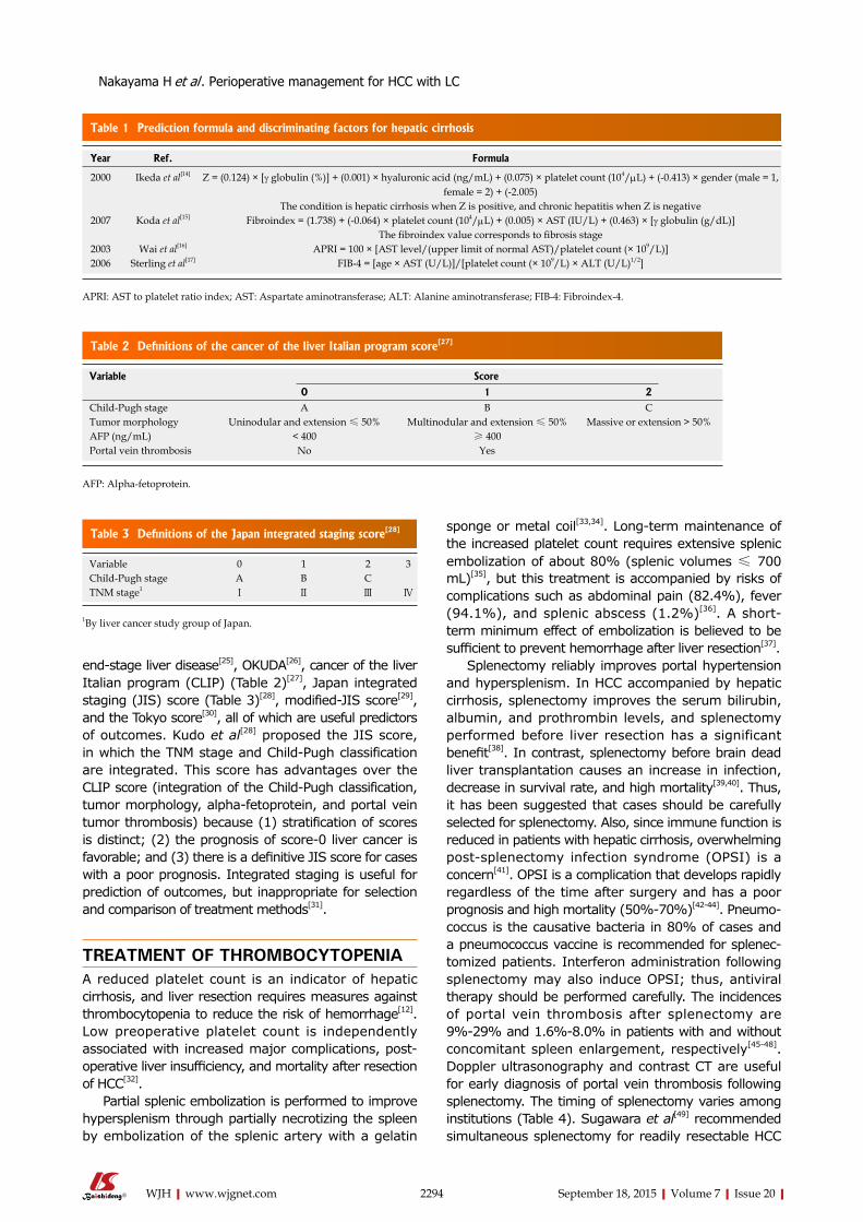
endstage liver disease[25], OKUDA[26], cancer of the liver Italian program (CLIP) (Table 2)[27], Japan integrated staging (JIS) score (Table 3)[28], modifiedJIS score[29], and the Tokyo score[30], all of which are useful predictors of outcomes. Kudo et al[28] proposed the JIS score, in which the TNM stage and ChildPugh classification are integrated. This score has advantages over the CLIP score (integration of the ChildPugh classification, tumor morphology, alphafetoprotein, and portal vein tumor thrombosis) because (1) stratification of scores is distinct; (2) the prognosis of score0 liver cancer is favorable; and (3) there is a definitive JIS score for cases with a poor prognosis. Integrated staging is useful for prediction of outcomes, but inappropriate for selection and comparison of treatment methods[31].
TREATMENT OF THROMBOCYTOPENIAA reduced platelet count is an indicator of hepatic cirrhosis, and liver resection requires measures against thrombocytopenia to reduce the risk of hemorrhage[12]. Low preoperative platelet count is independently associated with increased major complications, postoperative liver insufficiency, and mortality after resection of HCC[32].
Partial splenic embolization is performed to improve hypersplenism through partially necrotizing the spleen by embolization of the splenic artery with a gelatin
sponge or metal coil[33,34]. Longterm maintenance of the increased platelet count requires extensive splenic embolization of about 80% (splenic volumes ≤ 700 mL)[35], but this treatment is accompanied by risks of complications such as abdominal pain (82.4%), fever (94.1%), and splenic abscess (1.2%)[36]. A shortterm minimum effect of embolization is believed to be sufficient to prevent hemorrhage after liver resection[37].
Splenectomy reliably improves portal hypertension and hypersplenism. In HCC accompanied by hepatic cirrhosis, splenectomy improves the serum bilirubin, albumin, and prothrombin levels, and splenectomy performed before liver resection has a significant benefit[38]. In contrast, splenectomy before brain dead liver transplantation causes an increase in infection, decrease in survival rate, and high mortality[39,40]. Thus, it has been suggested that cases should be carefully selected for splenectomy. Also, since immune function is reduced in patients with hepatic cirrhosis, overwhelming postsplenectomy infection syndrome (OPSI) is a concern[41]. OPSI is a complication that develops rapidly regardless of the time after surgery and has a poor prognosis and high mortality (50%70%)[4244]. Pneumococcus is the causative bacteria in 80% of cases and a pneumococcus vaccine is recommended for splenectomized patients. Interferon administration following splenectomy may also induce OPSI; thus, antiviral therapy should be performed carefully. The incidences of portal vein thrombosis after splenectomy are 9%29% and 1.6%8.0% in patients with and without concomitant spleen enlargement, respectively[4548]. Doppler ultrasonography and contrast CT are useful for early diagnosis of portal vein thrombosis following splenectomy. The timing of splenectomy varies among institutions (Table 4). Sugawara et al[49] recommended simultaneous splenectomy for readily resectable HCC
2294 September 18, 2015|Volume 7|Issue 20|WJH|www.wjgnet.com
Table 1 Prediction formula and discriminating factors for hepatic cirrhosis
Year Ref. Formula
2000 Ikeda et al[14] Z = (0.124) × [γ globulin (%)] + (0.001) × hyaluronic acid (ng/mL) + (0.075) × platelet count (104/μL) + (-0.413) × gender (male = 1, female = 2) + (-2.005)
The condition is hepatic cirrhosis when Z is positive, and chronic hepatitis when Z is negative 2007 Koda et al[15] Fibroindex = (1.738) + (-0.064) × platelet count (104/μL) + (0.005) × AST (IU/L) + (0.463) × [γ globulin (g/dL)]
The fibroindex value corresponds to fibrosis stage 2003 Wai et al[16] APRI = 100 × [AST level/(upper limit of normal AST)/platelet count (× 109/L)]2006 Sterling et al[17] FIB-4 = [age × AST (U/L)]/[platelet count (× 109/L) × ALT (U/L)1/2]
APRI: AST to platelet ratio index; AST: Aspartate aminotransferase; ALT: Alanine aminotransferase; FIB-4: Fibroindex-4.
Table 2 Definitions of the cancer of the liver Italian program score[27]
Variable Score
0 1 2Child-Pugh stage A B CTumor morphology Uninodular and extension ≤ 50% Multinodular and extension ≤ 50% Massive or extension > 50%AFP (ng/mL) < 400 ≥ 400Portal vein thrombosis No Yes
Table 3 Definitions of the Japan integrated staging score[28]
Variable 0 1 2 3Child-Pugh stage A B CTNM stage1 Ⅰ Ⅱ Ⅲ Ⅳ
1By liver cancer study group of Japan.
Nakayama H et al . Perioperative management for HCC with LC
AFP: Alpha-fetoprotein.
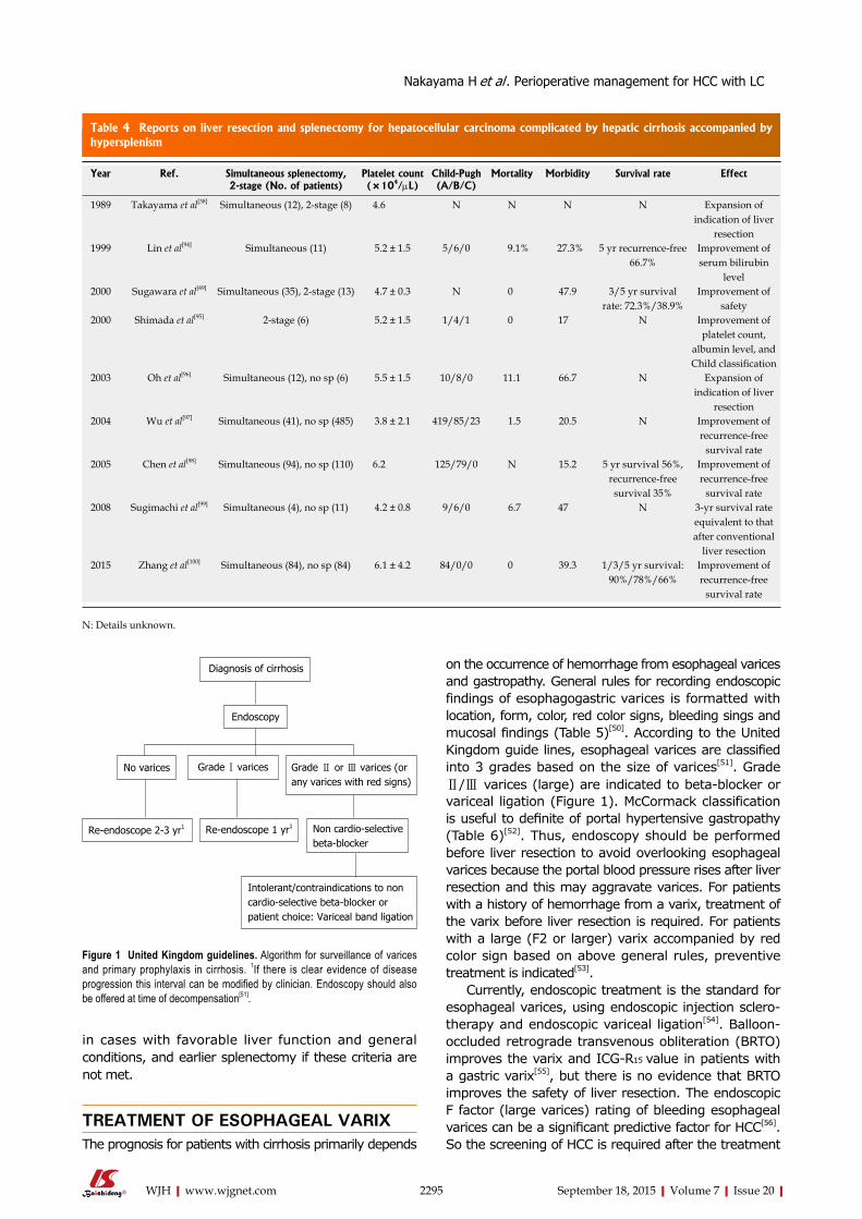
2295 September 18, 2015|Volume 7|Issue 20|WJH|www.wjgnet.com
on the occurrence of hemorrhage from esophageal varices and gastropathy. General rules for recording endoscopic findings of esophagogastric varices is formatted with location, form, color, red color signs, bleeding sings and mucosal findings (Table 5)[50]. According to the United Kingdom guide lines, esophageal varices are classified into 3 grades based on the size of varices[51]. Grade Ⅱ/Ⅲ varices (large) are indicated to betablocker or variceal ligation (Figure 1). McCormack classification is useful to definite of portal hypertensive gastropathy (Table 6)[52]. Thus, endoscopy should be performed before liver resection to avoid overlooking esophageal varices because the portal blood pressure rises after liver resection and this may aggravate varices. For patients with a history of hemorrhage from a varix, treatment of the varix before liver resection is required. For patients with a large (F2 or larger) varix accompanied by red color sign based on above general rules, preventive treatment is indicated[53].
Currently, endoscopic treatment is the standard for esophageal varices, using endoscopic injection sclerotherapy and endoscopic variceal ligation[54]. Balloonoccluded retrograde transvenous obliteration (BRTO) improves the varix and ICG-R15 value in patients with a gastric varix[55], but there is no evidence that BRTO improves the safety of liver resection. The endoscopic F factor (large varices) rating of bleeding esophageal varices can be a significant predictive factor for HCC[56]. So the screening of HCC is required after the treatment
in cases with favorable liver function and general conditions, and earlier splenectomy if these criteria are not met.
TREATMENT OF ESOPHAGEAL VARIXThe prognosis for patients with cirrhosis primarily depends
Table 1 Prediction formula and discriminating factors for hepatic cirrhosis Table 4 Reports on liver resection and splenectomy for hepatocellular carcinoma complicated by hepatic cirrhosis accompanied by hypersplenism
Year Ref. Simultaneous splenectomy, 2-stage (No. of patients)
Platelet count (×104/μL)
Child-Pugh (A/B/C)
Mortality Morbidity Survival rate Effect
1989 Takayama et al[38] Simultaneous (12), 2-stage (8) 4.6 N N N N Expansion of indication of liver
resection1999 Lin et al[94] Simultaneous (11) 5.2 ± 1.5 5/6/0 9.1% 27.3% 5 yr recurrence-free
66.7%Improvement of serum bilirubin
level2000 Sugawara et al[49] Simultaneous (35), 2-stage (13) 4.7 ± 0.3 N 0 47.9 3/5 yr survival
rate: 72.3%/38.9%Improvement of
safety2000 Shimada et al[95] 2-stage (6) 5.2 ± 1.5 1/4/1 0 17 N Improvement of
platelet count, albumin level, and Child classification
2003 Oh et al[96] Simultaneous (12), no sp (6) 5.5 ± 1.5 10/8/0 11.1 66.7 N Expansion of indication of liver
resection2004 Wu et al[97] Simultaneous (41), no sp (485) 3.8 ± 2.1 419/85/23 1.5 20.5 N Improvement of
recurrence-free survival rate
2005 Chen et al[98] Simultaneous (94), no sp (110) 6.2 125/79/0 N 15.2 5 yr survival 56%, recurrence-free survival 35%
Improvement of recurrence-free
survival rate2008 Sugimachi et al[99] Simultaneous (4), no sp (11) 4.2 ± 0.8 9/6/0 6.7 47 N 3-yr survival rate
equivalent to that after conventional
liver resection2015 Zhang et al[100] Simultaneous (84), no sp (84) 6.1 ± 4.2 84/0/0 0 39.3 1/3/5 yr survival:
90%/78%/66%Improvement of recurrence-free
survival rate
N: Details unknown.
Diagnosis of cirrhosis
Endoscopy
Grade Ⅰ varicesNo varices Grade Ⅱ or Ⅲ varices (or any varices with red signs)
Non cardio-selective beta-blocker
Re-endoscope 1 yr1Re-endoscope 2-3 yr1
Intolerant/contraindications to non cardio-selective beta-blocker or patient choice: Variceal band ligation
Figure 1 United Kingdom guidelines. Algorithm for surveillance of varices and primary prophylaxis in cirrhosis. 1If there is clear evidence of disease progression this interval can be modified by clinician. Endoscopy should also be offered at time of decompensation[51].
Nakayama H et al . Perioperative management for HCC with LC
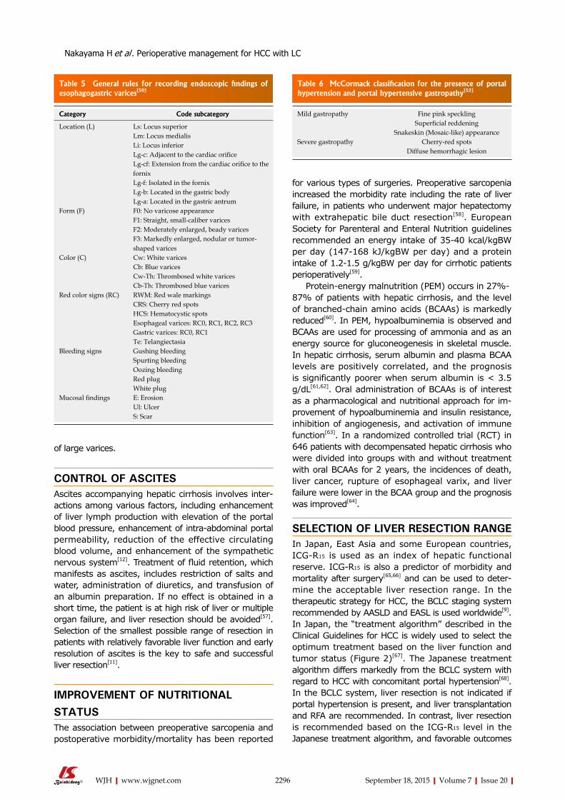
2296 September 18, 2015|Volume 7|Issue 20|WJH|www.wjgnet.com
of large varices.
CONTROL OF ASCITESAscites accompanying hepatic cirrhosis involves interactions among various factors, including enhancement of liver lymph production with elevation of the portal blood pressure, enhancement of intraabdominal portal permeability, reduction of the effective circulating blood volume, and enhancement of the sympathetic nervous system[12]. Treatment of fluid retention, which manifests as ascites, includes restriction of salts and water, administration of diuretics, and transfusion of an albumin preparation. If no effect is obtained in a short time, the patient is at high risk of liver or multiple organ failure, and liver resection should be avoided[57]. Selection of the smallest possible range of resection in patients with relatively favorable liver function and early resolution of ascites is the key to safe and successful liver resection[11].
IMPROVEMENT OF NUTRITIONAL STATUSThe association between preoperative sarcopenia and postoperative morbidity/mortality has been reported
for various types of surgeries. Preoperative sarcopenia increased the morbidity rate including the rate of liver failure, in patients who underwent major hepatectomy with extrahepatic bile duct resection[58]. European Society for Parenteral and Enteral Nutrition guidelines recommended an energy intake of 3540 kcal/kgBW per day (147168 kJ/kgBW per day) and a protein intake of 1.21.5 g/kgBW per day for cirrhotic patients perioperatively[59].
Proteinenergy malnutrition (PEM) occurs in 27%87% of patients with hepatic cirrhosis, and the level of branchedchain amino acids (BCAAs) is markedly reduced[60]. In PEM, hypoalbuminemia is observed and BCAAs are used for processing of ammonia and as an energy source for gluconeogenesis in skeletal muscle. In hepatic cirrhosis, serum albumin and plasma BCAA levels are positively correlated, and the prognosis is significantly poorer when serum albumin is < 3.5 g/dL[61,62]. Oral administration of BCAAs is of interest as a pharmacological and nutritional approach for improvement of hypoalbuminemia and insulin resistance, inhibition of angiogenesis, and activation of immune function[63]. In a randomized controlled trial (RCT) in 646 patients with decompensated hepatic cirrhosis who were divided into groups with and without treatment with oral BCAAs for 2 years, the incidences of death, liver cancer, rupture of esophageal varix, and liver failure were lower in the BCAA group and the prognosis was improved[64].
SELECTION OF LIVER RESECTION RANGEIn Japan, East Asia and some European countries, ICGR15 is used as an index of hepatic functional reserve. ICGR15 is also a predictor of morbidity and mortality after surgery[65,66] and can be used to determine the acceptable liver resection range. In the therapeutic strategy for HCC, the BCLC staging system recommended by AASLD and EASL is used worldwide[9]. In Japan, the “treatment algorithm” described in the Clinical Guidelines for HCC is widely used to select the optimum treatment based on the liver function and tumor status (Figure 2)[67]. The Japanese treatment algorithm differs markedly from the BCLC system with regard to HCC with concomitant portal hypertension[68]. In the BCLC system, liver resection is not indicated if portal hypertension is present, and liver transplantation and RFA are recommended. In contrast, liver resection is recommended based on the ICGR15 level in the Japanese treatment algorithm, and favorable outcomes
Table 5 General rules for recording endoscopic findings of esophagogastric varices[50]
Category Code subcategory
Location (L) Ls: Locus superiorLm: Locus medialisLi: Locus inferiorLg-c: Adjacent to the cardiac orificeLg-cf: Extension from the cardiac orifice to the fornixLg-f: Isolated in the fornixLg-b: Located in the gastric bodyLg-a: Located in the gastric antrum
Form (F) F0: No varicose appearanceF1: Straight, small-caliber varicesF2: Moderately enlarged, beady varicesF3: Markedly enlarged, nodular or tumor-shaped varices
Color (C) Cw: White varicesCb: Blue varicesCw-Th: Thrombosed white varicesCb-Th: Thrombosed blue varices
Red color signs (RC) RWM: Red wale markingsCRS: Cherry red spotsHCS: Hematocystic spotsEsophageal varices: RC0, RC1, RC2, RC3Gastric varices: RC0, RC1Te: Telangiectasia
Bleeding signs Gushing bleedingSpurting bleedingOozing bleedingRed plugWhite plug
Mucosal findings E: ErosionUl: UlcerS: Scar
Table 6 McCormack classification for the presence of portal hypertension and portal hypertensive gastropathy[52]
Mild gastropathy Fine pink specklingSuperficial reddening
Snakeskin (Mosaic-like) appearanceSevere gastropathy Cherry-red spots
Diffuse hemorrhagic lesion
Nakayama H et al . Perioperative management for HCC with LC
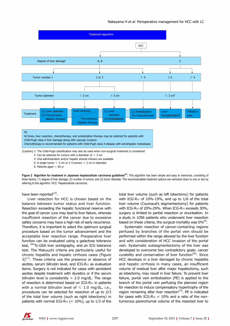
2297 September 18, 2015|Volume 7|Issue 20|WJH|www.wjgnet.com
have been reported[10].Liver resection for HCC is chosen based on the
balance between tumor status and liver function. Resection exceeding the hepatic functional reserve with the goal of cancer cure may lead to liver failure, whereas insufficient resection of the cancer due to excessive safety concerns may have a high risk of early recurrence. Therefore, it is important to select the optimum surgical procedure based on the tumor advancement and the acceptable liver resection range. Preoperative liver function can be evaluated using a galactose tolerance test, 99mTcGSA liver scintigraphy, and an ICG tolerance test. The Makuuchi criteria are particularly useful for chronic hepatitis and hepatic cirrhosis cases (Figure 3)[11]. These criteria use the presence or absence of ascites, serum bilirubin level, and ICGR15 as evaluation items. Surgery is not indicated for cases with persistent ascites despite treatment with diuretics or if the serum bilirubin level is consistently > 2.0 mg/dL. The range of resection is determined based on ICGR15 in patients with a normal bilirubin level of ≤ 1.0 mg/dL, i.e., procedures can be selected for resection of up to 2/3 of the total liver volume (such as right lobectomy) in patients with normal ICGR15 (< 10%), up to 1/3 of the
total liver volume (such as left lobectomy) for patients with ICGR15 of 10%19%, and up to 1/6 of the total liver volume (Couinaud’s segmentectomy) for patients with ICGR15 of 20%29%. When ICGR15 exceeds 30%, surgery is limited to partial resection or enucleation. In a study in 1056 patients who underwent liver resection based on these criteria, the surgical mortality was 0%[8].
Systematic resection of cancercontaining regions perfused by branches of the portal vein should be performed within the range allowed by the liver function and with consideration of HCC invasion of the portal vein. Systematic subsegmentectomy of the liver was developed to overcome two contradictory goals: cancer curability and conservation of liver function[69]. Since HCC develops in a liver damaged by chronic hepatitis and hepatic cirrhosis in many cases, an insufficient volume of residual liver after major hepatectomy, such as lobectomy, may result in liver failure. To prevent liver failure, portal vein embolization (PE) is applied to the branch of the portal vein perfusing the planned region for resection to induce compensatory hypertrophy of the region remaining after liver resection[70]. PE is indicated for cases with ICGR15 < 10% and a ratio of the nontumorous parenchymal volume of the resected liver to
Treatment algorithm
PSAt times, liver resection, chemotherapy, and embolization therapy may be selected for patients with Child-Pugh class A liver damage along with vascular invasionChemotherapy is recommended for patients with Child-Pugh class A disease with extrahepatic metastases
(Caution) 1: The Child-Pugh classification may also be used when non-surgical treatment is considered 2: Can be selected for tumors with a diameter of ≤ 3 cm 3: Oral administration and/or hepatic arterial infusion are available 4: A single tumor ≤ 5 cm or 2-3 tumors ≤ 3 cm in diameter 5: Patients aged ≤ 65 yr
Tumor diameter ≤ 3 cm > 3 cm ≤ 3 cm4
Treatment(1) Liver resection(2) Percutaneous ablation therapy2
(1) Liver resection(2) Embolization
(1) Embolization(2) Chemotherapy3
Liver transplantation5
Liver resection Percutaneous ablation therapy
Palliative care
Tumor number 1 2 or 3 ≥ 4 1-3 ≥ 4
Degree of liver damage1 A, B C
HCC
Figure 2 Algorithm for treatment in Japanese hepatocellular carcinoma guidelines[67]. This algorithm has been simple and easy to memorize, consisting of three factors: (1) degree of liver damage; (2) number of tumors; and (3) tumor diameter. The recommendable treatment options are narrowed down to one or two by referring to this algorithm. HCC: Hepatocellular carcinoma.
Nakayama H et al . Perioperative management for HCC with LC
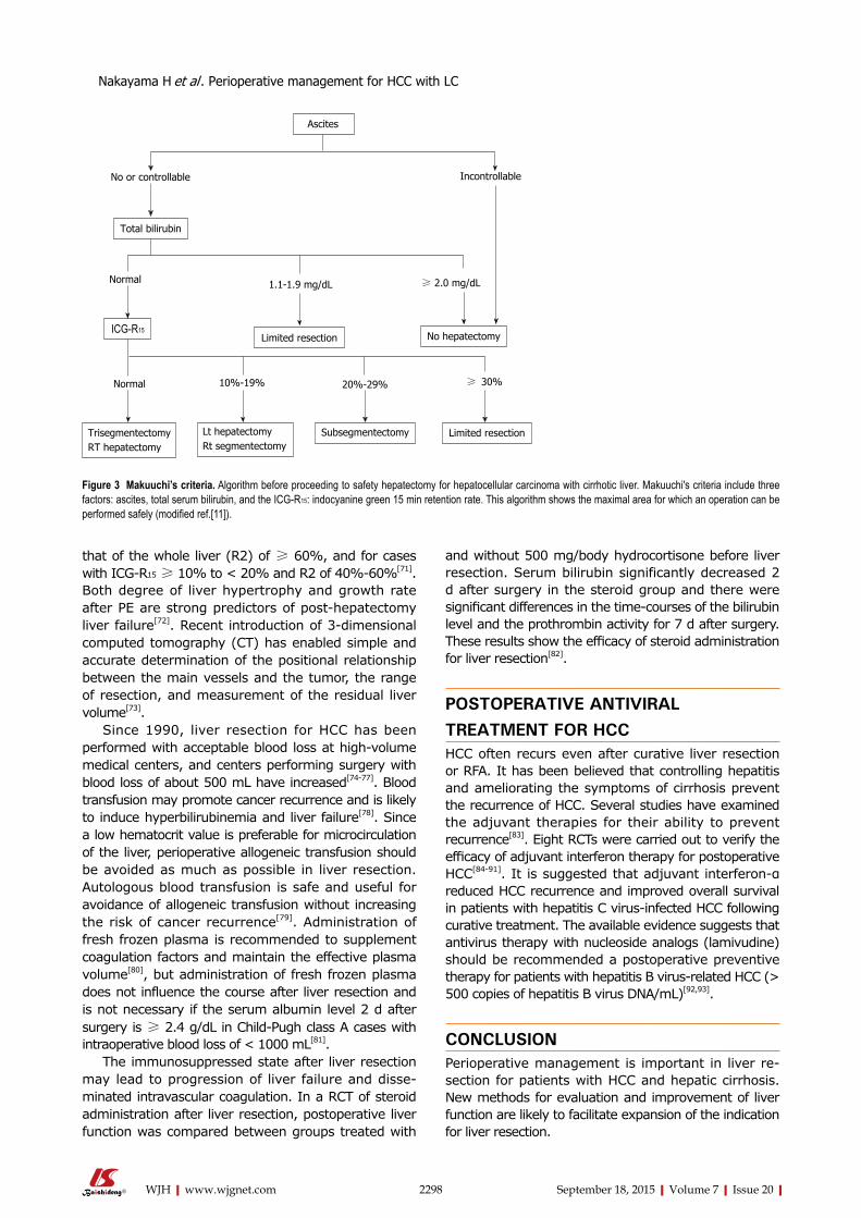
2298 September 18, 2015|Volume 7|Issue 20|WJH|www.wjgnet.com
that of the whole liver (R2) of ≥ 60%, and for cases with ICGR15 ≥ 10% to < 20% and R2 of 40%60%[71]. Both degree of liver hypertrophy and growth rate after PE are strong predictors of posthepatectomy liver failure[72]. Recent introduction of 3dimensional computed tomography (CT) has enabled simple and accurate determination of the positional relationship between the main vessels and the tumor, the range of resection, and measurement of the residual liver volume[73].
Since 1990, liver resection for HCC has been performed with acceptable blood loss at highvolume medical centers, and centers performing surgery with blood loss of about 500 mL have increased[7477]. Blood transfusion may promote cancer recurrence and is likely to induce hyperbilirubinemia and liver failure[78]. Since a low hematocrit value is preferable for microcirculation of the liver, perioperative allogeneic transfusion should be avoided as much as possible in liver resection. Autologous blood transfusion is safe and useful for avoidance of allogeneic transfusion without increasing the risk of cancer recurrence[79]. Administration of fresh frozen plasma is recommended to supplement coagulation factors and maintain the effective plasma volume[80], but administration of fresh frozen plasma does not influence the course after liver resection and is not necessary if the serum albumin level 2 d after surgery is ≥ 2.4 g/dL in ChildPugh class A cases with intraoperative blood loss of < 1000 mL[81].
The immunosuppressed state after liver resection may lead to progression of liver failure and disseminated intravascular coagulation. In a RCT of steroid administration after liver resection, postoperative liver function was compared between groups treated with
and without 500 mg/body hydrocortisone before liver resection. Serum bilirubin significantly decreased 2 d after surgery in the steroid group and there were significant differences in the time-courses of the bilirubin level and the prothrombin activity for 7 d after surgery. These results show the efficacy of steroid administration for liver resection[82].
POSTOPERATIVE ANTIVIRAL TREATMENT FOR HCCHCC often recurs even after curative liver resection or RFA. It has been believed that controlling hepatitis and ameliorating the symptoms of cirrhosis prevent the recurrence of HCC. Several studies have examined the adjuvant therapies for their ability to prevent recurrence[83]. Eight RCTs were carried out to verify the efficacy of adjuvant interferon therapy for postoperative HCC[8491]. It is suggested that adjuvant interferon-α reduced HCC recurrence and improved overall survival in patients with hepatitis C virusinfected HCC following curative treatment. The available evidence suggests that antivirus therapy with nucleoside analogs (lamivudine) should be recommended a postoperative preventive therapy for patients with hepatitis B virusrelated HCC (> 500 copies of hepatitis B virus DNA/mL)[92,93].
CONCLUSIONPerioperative management is important in liver resection for patients with HCC and hepatic cirrhosis. New methods for evaluation and improvement of liver function are likely to facilitate expansion of the indication for liver resection.
No or controllable Incontrollable
Normal 1.1-1.9 mg/dL ≥ 2.0 mg/dL
Normal 10%-19% 20%-29% ≥ 30%
Ascites
Total bilirubin
ICG-R15Limited resection No hepatectomy
Subsegmentectomy Limited resectionLt hepatectomyRt segmentectomy
TrisegmentectomyRT hepatectomy
Figure 3 Makuuchi’s criteria. Algorithm before proceeding to safety hepatectomy for hepatocellular carcinoma with cirrhotic liver. Makuuchi's criteria include three factors: ascites, total serum bilirubin, and the ICG-R15: indocyanine green 15 min retention rate. This algorithm shows the maximal area for which an operation can be performed safely (modified ref.[11]).
Nakayama H et al . Perioperative management for HCC with LC
2299 September 18, 2015|Volume 7|Issue 20|WJH|www.wjgnet.com
REFERENCES1 Tejeda-Maldonado J, García-Juárez I, Aguirre-Valadez J,
González-Aguirre A, Vilatobá-Chapa M, Armengol-Alonso A, Escobar-Penagos F, Torre A, Sánchez-Ávila JF, Carrillo-Pérez DL. Diagnosis and treatment of hepatocellular carcinoma: An update. World J Hepatol 2015; 7: 362-376 [PMID: 25848464 DOI: 10.4254/wjh.v7.i3.362]
2 Forner A, Llovet JM, Bruix J. Hepatocellular carcinoma. Lancet 2012; 379: 1245-1255 [PMID: 22353262 DOI: 10.1016/s0140-6736(11)61347-0]
3 Trépo C, Chan HL, Lok A. Hepatitis B virus infection. Lancet 2014; 384: 2053-2063 [PMID: 24954675 DOI: 10.1016/s0140-6736(14)60220-8]
4 Webster DP, Klenerman P, Dusheiko GM. Hepatitis C. Lancet 2015; 385: 1124-1135 [PMID: 25687730 DOI: 10.1016/s0140-6736(14)62401-6]
5 Echeverría N, Moratorio G, Cristina J, Moreno P. Hepatitis C virus genetic variability and evolution. World J Hepatol 2015; 7: 831-845 [PMID: 25937861 DOI: 10.4254/wjh.v7.i6.831]
6 Muir AJ, Poordad F, Lalezari J, Everson G, Dore GJ, Herring R, Sheikh A, Kwo P, Hézode C, Pockros PJ, Tran A, Yozviak J, Reau N, Ramji A, Stuart K, Thompson AJ, Vierling J, Freilich B, Cooper J, Ghesquiere W, Yang R, McPhee F, Hughes EA, Swenson ES, Yin PD. Daclatasvir in combination with asunaprevir and beclabuvir for hepatitis C virus genotype 1 infection with compensated cirrhosis. JAMA 2015; 313: 1736-1744 [PMID: 25942724 DOI: 10.1001/jama.2015.3868]
7 Zhou Y, Lei X, Wu L, Wu X, Xu D, Li B. Outcomes of hepate-ctomy for noncirrhotic hepatocellular carcinoma: a systematic review. Surg Oncol 2014; 23: 236-242 [PMID: 25465529 DOI: 10.1016/j.suronc.2014.11.001]
8 Imamura H, Seyama Y, Kokudo N, Maema A, Sugawara Y, Sano K, Takayama T, Makuuchi M. One thousand fifty-six hepatectomies without mortality in 8 years. Arch Surg 2003; 138: 1198-1206; discussion 1206 [PMID: 14609867 DOI: 10.1001/archsurg.138.11.1198]
9 Bruix J, Sherman M. Management of hepatocellular carcinoma: an update. Hepatology 2011; 53: 1020-1022 [PMID: 21374666 DOI: 10.1002/hep.24199]
10 Ishizawa T, Hasegawa K, Aoki T, Takahashi M, Inoue Y, Sano K, Imamura H, Sugawara Y, Kokudo N, Makuuchi M. Neither multiple tumors nor portal hypertension are surgical contraindications for hepatocellular carcinoma. Gastroenterology 2008; 134: 1908-1916 [PMID: 18549877 DOI: 10.1053/j.gastro.2008.02.091]
11 Makuuchi M, Kosuge T, Takayama T, Yamazaki S, Kakazu T, Miyagawa S, Kawasaki S. Surgery for small liver cancers. Semin Surg Oncol 1993; 9: 298-304 [PMID: 8210909]
12 Tsochatzis EA, Bosch J, Burroughs AK. Liver cirrhosis. Lancet 2014; 383: 1749-1761 [PMID: 24480518 DOI: 10.1016/s0140-6736(14)60121-5]
13 Hytiroglou P, Snover DC, Alves V, Balabaud C, Bhathal PS, Bioulac-Sage P, Crawford JM, Dhillon AP, Ferrell L, Guido M, Nakanuma Y, Paradis V, Quaglia A, Theise ND, Thung SN, Tsui WM, van Leeuwen DJ. Beyond “cirrhosis”: a proposal from the International Liver Pathology Study Group. Am J Clin Pathol 2012; 137: 5-9 [PMID: 22180471 DOI: 10.1309/ajcp2t2ohtapbtmp]
14 Ikeda K, Saitoh S, Kobayashi M, Suzuki Y, Tsubota A, Suzuki F, Arase Y, Murashima N, Chayama K, Kumada H. Distinction between chronic hepatitis and liver cirrhosis in patients with hepatitis C virus infection. Practical discriminant function using common laboratory data. Hepatol Res 2000; 18: 252-266 [PMID: 11058829]
15 Koda M, Matunaga Y, Kawakami M, Kishimoto Y, Suou T, Murawaki Y. FibroIndex, a practical index for predicting significant fibrosis in patients with chronic hepatitis C. Hepatology 2007; 45: 297-306 [PMID: 17256741 DOI: 10.1002/hep.21520]
16 Wai CT, Greenson JK, Fontana RJ, Kalbfleisch JD, Marrero JA, Conjeevaram HS, Lok AS. A simple noninvasive index can predict
both significant fibrosis and cirrhosis in patients with chronic hepatitis C. Hepatology 2003; 38: 518-526 [PMID: 12883497 DOI: 10.1053/jhep.2003.50346]
17 Sterling RK, Lissen E, Clumeck N, Sola R, Correa MC, Montaner J, S Sulkowski M, Torriani FJ, Dieterich DT, Thomas DL, Messinger D, Nelson M. Development of a simple noninvasive index to predict significant fibrosis in patients with HIV/HCV coinfection. Hepatology 2006; 43: 1317-1325 [PMID: 16729309 DOI: 10.1002/hep.21178]
18 Sandrin L, Fourquet B, Hasquenoph JM, Yon S, Fournier C, Mal F, Christidis C, Ziol M, Poulet B, Kazemi F, Beaugrand M, Palau R. Transient elastography: a new noninvasive method for assessment of hepatic fibrosis. Ultrasound Med Biol 2003; 29: 1705-1713 [PMID: 14698338]
19 Lucidarme D, Foucher J, Le Bail B, Vergniol J, Castera L, Duburque C, Forzy G, Filoche B, Couzigou P, de Lédinghen V. Factors of accuracy of transient elastography (fibroscan) for the diagnosis of liver fibrosis in chronic hepatitis C. Hepatology 2009; 49: 1083-1089 [PMID: 19140221 DOI: 10.1002/hep.22748]
20 Castéra L, Vergniol J, Foucher J, Le Bail B, Chanteloup E, Haaser M, Darriet M, Couzigou P, De Lédinghen V. Prospective comparison of transient elastography, Fibrotest, APRI, and liver biopsy for the assessment of fibrosis in chronic hepatitis C. Gastroenterology 2005; 128: 343-350 [PMID: 15685546]
21 Child CG. The liver and portal hypertension. 3rd ed. Philadelphia: WB Saunders, 1964
22 Pugh RN, Murray-Lyon IM, Dawson JL, Pietroni MC, Williams R. Transection of the oesophagus for bleeding oesophageal varices. Br J Surg 1973; 60: 646-649 [PMID: 4541913]
23 The general rules for the clinical and pathological study of primary liver cancer. Liver Cancer Study Group of Japan. Jpn J Surg 1989; 19: 98-129 [PMID: 2659865]
24 Minagawa M, Ikai I, Matsuyama Y, Yamaoka Y, Makuuchi M. Staging of hepatocellular carcinoma: assessment of the Japanese TNM and AJCC/UICC TNM systems in a cohort of 13,772 patients in Japan. Ann Surg 2007; 245: 909-922 [PMID: 17522517 DOI: 10.1097/01.sla.0000254368.65878.da]
25 Kamath PS, Wiesner RH, Malinchoc M, Kremers W, Therneau TM, Kosberg CL, D’Amico G, Dickson ER, Kim WR. A model to predict survival in patients with end-stage liver disease. Hepatology 2001; 33: 464-470 [PMID: 11172350 DOI: 10.1053/jhep.2001.22172]
26 Okuda K, Ohtsuki T, Obata H, Tomimatsu M, Okazaki N, Hasegawa H, Nakajima Y, Ohnishi K. Natural history of hepato-cellular carcinoma and prognosis in relation to treatment. Study of 850 patients. Cancer 1985; 56: 918-928 [PMID: 2990661]
27 A new prognostic system for hepatocellular carcinoma: a retrospective study of 435 patients: the Cancer of the Liver Italian Program (CLIP) investigators. Hepatology 1998; 28: 751-755 [PMID: 9731568 DOI: 10.1002/hep.510280322]
28 Kudo M, Chung H, Haji S, Osaki Y, Oka H, Seki T, Kasugai H, Sasaki Y, Matsunaga T. Validation of a new prognostic staging system for hepatocellular carcinoma: the JIS score compared with the CLIP score. Hepatology 2004; 40: 1396-1405 [PMID: 15565571 DOI: 10.1002/hep.20486]
29 Ikai I, Takayasu K, Omata M, Okita K, Nakanuma Y, Matsuyama Y, Makuuchi M, Kojiro M, Ichida T, Arii S, Yamaoka Y. A modified Japan Integrated Stage score for prognostic assessment in patients with hepatocellular carcinoma. J Gastroenterol 2006; 41: 884-892 [PMID: 17048053 DOI: 10.1007/s00535-006-1878-y]
30 Tateishi R, Yoshida H, Shiina S, Imamura H, Hasegawa K, Teratani T, Obi S, Sato S, Koike Y, Fujishima T, Makuuchi M, Omata M. Proposal of a new prognostic model for hepatocellular carcinoma: an analysis of 403 patients. Gut 2005; 54: 419-425 [PMID: 15710994 DOI: 10.1136/gut.2003.035055]
31 Duseja A. Staging of hepatocellular carcinoma. J Clin Exp Hepatol 2014; 4: S74-S79 [PMID: 25755615 DOI: 10.1016/j.jceh.2014.03.045]
32 Maithel SK, Kneuertz PJ, Kooby DA, Scoggins CR, Weber SM, Martin RC, McMasters KM, Cho CS, Winslow ER, Wood
Nakayama H et al . Perioperative management for HCC with LC
2300 September 18, 2015|Volume 7|Issue 20|WJH|www.wjgnet.com
WC, Staley CA. Importance of low preoperative platelet count in selecting patients for resection of hepatocellular carcinoma: a multi-institutional analysis. J Am Coll Surg 2011; 212: 638-648; discussion 648-650 [PMID: 21463803 DOI: 10.1016/j.jamcollsurg.2011.01.004]
33 Yoshida H, Mamada Y, Taniai N, Tajiri T. Partial splenic embolization. Hepatol Res 2008; 38: 225-233 [PMID: 18034810 DOI: 10.1111/j.1872-034X.2007.00302.x]
34 Hayashi H, Beppu T, Shirabe K, Maehara Y, Baba H. Management of thrombocytopenia due to liver cirrhosis: a review. World J Gastroenterol 2014; 20: 2595-2605 [PMID: 24627595 DOI: 10.3748/wjg.v20.i10.2595]
35 Hayashi H, Beppu T, Okabe K, Masuda T, Okabe H, Ishiko T, Baba H. Therapeutic factors considered according to the preoperative splenic volume for a prolonged increase in platelet count after partial splenic embolization for liver cirrhosis. J Gastroenterol 2010; 45: 554-559 [PMID: 20047119 DOI: 10.1007/s00535-009-0185-9]
36 Sakai T, Shiraki K, Inoue H, Sugimoto K, Ohmori S, Murata K, Takase K, Nakano T. Complications of partial splenic embolization in cirrhotic patients. Dig Dis Sci 2002; 47: 388-391 [PMID: 11855556]
37 Hadduck TA, McWilliams JP. Partial splenic artery embolization in cirrhotic patients. World J Radiol 2014; 6: 160-168 [PMID: 24876920 DOI: 10.4329/wjr.v6.i5.160]
38 Takayama T, Makuuchi M, Yamazaki S, Hasegawa H. [The role of splenectomy in patients with hepatocellular carcinoma and hypersplenism as an aid to hepatectomy]. Nihon Geka Gakkai Zasshi 1989; 90: 1043-1048 [PMID: 2552282]
39 Lüsebrink R, Blumhardt G, Lohmann R, Bachmann S, Knoop M, Lemmens HP, Neuhaus P. Does concommitant splenectomy raise the mortality of liver transplant recipients? Transpl Int 1994; 7 Suppl 1: S634-S636 [PMID: 11271326]
40 Samimi F, Irish WD, Eghtesad B, Demetris AJ, Starzl TE, Fung JJ. Role of splenectomy in human liver transplantation under modern-day immunosuppression. Dig Dis Sci 1998; 43: 1931-1937 [PMID: 9753254]
41 Okabayashi T, Hanazaki K. Overwhelming postsplenectomy infection syndrome in adults - a clinically preventable disease. World J Gastroenterol 2008; 14: 176-179 [PMID: 18186551]
42 Waghorn DJ, Mayon-White RT. A study of 42 episodes of overwhelming post-splenectomy infection: is current guidance for asplenic individuals being followed? J Infect 1997; 35: 289-294 [PMID: 9459404]
43 Waghorn DJ. Overwhelming infection in asplenic patients: current best practice preventive measures are not being followed. J Clin Pathol 2001; 54: 214-218 [PMID: 11253134]
44 Sinwar PD. Overwhelming post splenectomy infection syndrome - review study. Int J Surg 2014; 12: 1314-1316 [PMID: 25463041 DOI: 10.1016/j.ijsu.2014.11.005]
45 Kercher KW, Carbonell AM, Heniford BT, Matthews BD, Cunningham DM, Reindollar RW. Laparoscopic splenectomy reverses thrombocytopenia in patients with hepatitis C cirrhosis and portal hypertension. J Gastrointest Surg 2004; 8: 120-126 [PMID: 14746844]
46 Yoshida M, Watanabe Y, Horiuchi A, Yamamoto Y, Sugishita H, Kawachi K. Portal and splenic venous thrombosis after splenectomy in patients with hypersplenism. Hepatogastroenterology 2009; 56: 538-541 [PMID: 19579638]
47 Stamou KM, Toutouzas KG, Kekis PB, Nakos S, Gafou A, Manouras A, Krespis E, Katsaragakis S, Bramis J. Prospective study of the incidence and risk factors of postsplenectomy thrombosis of the portal, mesenteric, and splenic veins. Arch Surg 2006; 141: 663-669 [PMID: 16847237 DOI: 10.1001/archsurg.141.7.663]
48 Winslow ER, Brunt LM, Drebin JA, Soper NJ, Klingensmith ME. Portal vein thrombosis after splenectomy. Am J Surg 2002; 184: 631-635; discussion 635-636 [PMID: 12488196]
49 Sugawara Y, Yamamoto J, Shimada K, Yamasaki S, Kosuge T, Takayama T, Makuuchi M. Splenectomy in patients with hepatocellular carcinoma and hypersplenism. J Am Coll Surg 2000;
190: 446-450 [PMID: 10757382]50 Tajiri T, Yoshida H, Obara K, Onji M, Kage M, Kitano S, Kokudo
N, Kokubu S, Sakaida I, Sata M, Tajiri H, Tsukada K, Nonami T, Hashizume M, Hirota S, Murashima N, Moriyasu F, Saigenji K, Makuuchi H, Oho K, Yoshida T, Suzuki H, Hasumi A, Okita K, Futagawa S, Idezuki Y. General rules for recording endoscopic findings of esophagogastric varices (2nd edition). Dig Endosc 2010; 22: 1-9 [PMID: 20078657 DOI: 10.1111/j.1443-1661.2009.00929.x]
51 Tripathi D, Stanley AJ, Hayes PC, Patch D, Millson C, Mehrzad H, Austin A, Ferguson JW, Olliff SP, Hudson M, Christie JM. UK guidelines on the management of variceal haemorrhage in cirrhotic patients. Gut 2015; Epub ahead of print [PMID: 25887380 DOI: 10.1136/gutjnl-2015-309262]
52 McCormack TT, Sims J, Eyre-Brook I, Kennedy H, Goepel J, Johnson AG, Triger DR. Gastric lesions in portal hypertension: inflammatory gastritis or congestive gastropathy? Gut 1985; 26: 1226-1232 [PMID: 3877665]
53 Liu HT, Cheng SB, Wu CC, Yeh HZ, Chang CS, Wang J. Impact of severe oesophagogastric varices on liver resection for hepatocellular carcinoma in cirrhotic patients. World J Surg 2015; 39: 461-468 [PMID: 25338186 DOI: 10.1007/s00268-014-2811-9]
54 Sarin SK, Govil A, Jain AK, Guptan RC, Issar SK, Jain M, Murthy NS. Prospective randomized trial of endoscopic sclerotherapy versus variceal band ligation for esophageal varices: influence on gastropathy, gastric varices and variceal recurrence. J Hepatol 1997; 26: 826-832 [PMID: 9126795]
55 Akahane T, Iwasaki T, Kobayashi N, Tanabe N, Takahashi N, Gama H, Ishii M, Toyota T. Changes in liver function parameters after occlusion of gastrorenal shunts with balloon-occluded retrograde transvenous obliteration. Am J Gastroenterol 1997; 92: 1026-1030 [PMID: 9177524]
56 Nakayama H, Masuda H, Miyake H, Takayama T, Yokoyama E. Endoscopic prediction of hepatocellular carcinoma by evaluation of bleeding esophageal varices. Digestion 2004; 70: 233-239 [PMID: 15627772 DOI: 10.1159/000082895]
57 Donadon M, Costa G, Cimino M, Procopio F, Fabbro DD, Palmisano A, Torzilli G. Safe hepatectomy selection criteria for hepatocellular carcinoma patients: a validation of 336 consecutive hepatectomies. The BILCHE score. World J Surg 2015; 39: 237-243 [PMID: 25217112 DOI: 10.1007/s00268-014-2786-6]
58 Otsuji H, Yokoyama Y, Ebata T, Igami T, Sugawara G, Mizuno T, Nagino M. Preoperative sarcopenia negatively impacts postoperative outcomes following major hepatectomy with extrahepatic bile duct resection. World J Surg 2015; 39: 1494-1500 [PMID: 25651963 DOI: 10.1007/s00268-015-2988-6]
59 Plauth M, Cabré E, Riggio O, Assis-Camilo M, Pirlich M, Kondrup J, Ferenci P, Holm E, Vom Dahl S, Müller MJ, Nolte W. ESPEN Guidelines on Enteral Nutrition: Liver disease. Clin Nutr 2006; 25: 285-294 [PMID: 16707194 DOI: 10.1016/j.clnu.2006.01.018]
60 McCullough AJ, Bugianesi E. Protein-calorie malnutrition and the etiology of cirrhosis. Am J Gastroenterol 1997; 92: 734-738 [PMID: 9149179]
61 Plauth M, Cabré E, Campillo B, Kondrup J, Marchesini G, Schütz T, Shenkin A, Wendon J. ESPEN Guidelines on Parenteral Nutrition: hepatology. Clin Nutr 2009; 28: 436-444 [PMID: 19520466 DOI: 10.1016/j.clnu.2009.04.019]
62 Nishiguchi S, Habu D. Effect of oral supplementation with branched-chain amino acid granules in the early stage of cirrhosis. Hepatol Res 2004; 30S: 36-41 [PMID: 15607137 DOI: 10.1016/j.hepres.2004.08.009]
63 Suzuki K, Endo R, Kohgo Y, Ohtake T, Ueno Y, Kato A, Suzuki K, Shiraki R, Moriwaki H, Habu D, Saito M, Nishiguchi S, Katayama K, Sakaida I. Guidelines on nutritional management in Japanese patients with liver cirrhosis from the perspective of preventing hepatocellular carcinoma. Hepatol Res 2012; 42: 621-626 [PMID: 22686857 DOI: 10.1111/j.1872-034X.2012.00990.x]
64 Muto Y, Sato S, Watanabe A, Moriwaki H, Suzuki K, Kato A, Kato M, Nakamura T, Higuchi K, Nishiguchi S, Kumada H. Effects of oral branched-chain amino acid granules on event-free
Nakayama H et al . Perioperative management for HCC with LC
2301 September 18, 2015|Volume 7|Issue 20|WJH|www.wjgnet.com
survival in patients with liver cirrhosis. Clin Gastroenterol Hepatol 2005; 3: 705-713 [PMID: 16206505]
65 Lau H, Man K, Fan ST, Yu WC, Lo CM, Wong J. Evaluation of preoperative hepatic function in patients with hepatocellular carcinoma undergoing hepatectomy. Br J Surg 1997; 84: 1255-1259 [PMID: 9313707]
66 Nakayama H, Takayama T, Okubo T, Higaki T, Midorikawa Y, Moriguchi M, Aramaki O, Yamazaki S. Subcutaneous drainage to prevent wound infection in liver resection: a randomized controlled trial. J Hepatobiliary Pancreat Sci 2014; 21: 509-517 [PMID: 24519844 DOI: 10.1002/jhbp.93]
67 Kokudo N, Hasegawa K, Akahane M, Igaki H, Izumi N, Ichida T, Uemoto S, Kaneko S, Kawasaki S, Ku Y, Kudo M, Kubo S, Takayama T, Tateishi R, Fukuda T, Matsui O, Matsuyama Y, Murakami T, Arii S, Okazaki M, Makuuchi M. Evidence-based Clinical Practice Guidelines for Hepatocellular Carcinoma: The Japan Society of Hepatology 2013 update (3rd JSH-HCC Guidelines). Hepatol Res 2015; 45 [PMID: 25625806 DOI: 10.1111/hepr.12464]
68 Nakayama H, Takayama T. Role of surgical resection for hepatocellular carcinoma based on Japanese clinical guidelines for hepatocellular carcinoma. World J Hepatol 2015; 7: 261-269 [PMID: 25729481 DOI: 10.4254/wjh.v7.i2.261]
69 Makuuchi M, Hasegawa H, Yamazaki S. Ultrasonically guided subsegmentectomy. Surg Gynecol Obstet 1985; 161: 346-350 [PMID: 2996162]
70 Makuuchi M, Thai BL, Takayasu K, Takayama T, Kosuge T, Gunvén P, Yamazaki S, Hasegawa H, Ozaki H. Preoperative portal embolization to increase safety of major hepatectomy for hilar bile duct carcinoma: a preliminary report. Surgery 1990; 107: 521-527 [PMID: 2333592]
71 Kubota K, Makuuchi M, Kusaka K, Kobayashi T, Miki K, Hasegawa K, Harihara Y, Takayama T. Measurement of liver volume and hepatic functional reserve as a guide to decision-making in resectional surgery for hepatic tumors. Hepatology 1997; 26: 1176-1181 [PMID: 9362359 DOI: 10.1053/jhep.1997.v26.pm0009362359]
72 Leung U, Simpson AL, Araujo RL, Gönen M, McAuliffe C, Miga MI, Parada EP, Allen PJ, D’Angelica MI, Kingham TP, DeMatteo RP, Fong Y, Jarnagin WR. Remnant growth rate after portal vein embolization is a good early predictor of post-hepatectomy liver failure. J Am Coll Surg 2014; 219: 620-630 [PMID: 25158914 DOI: 10.1016/j.jamcollsurg.2014.04.022]
73 Saito S, Yamanaka J, Miura K, Nakao N, Nagao T, Sugimoto T, Hirano T, Kuroda N, Iimuro Y, Fujimoto J. A novel 3D hepatectomy simulation based on liver circulation: application to liver resection and transplantation. Hepatology 2005; 41: 1297-1304 [PMID: 15846773 DOI: 10.1002/hep.20684]
74 Grazi GL, Ercolani G, Pierangeli F, Del Gaudio M, Cescon M, Cavallari A, Mazziotti A. Improved results of liver resection for hepatocellular carcinoma on cirrhosis give the procedure added value. Ann Surg 2001; 234: 71-78 [PMID: 11420485]
75 Jarnagin WR, Schwartz LH, Gultekin DH, Gönen M, Haviland D, Shia J, D’Angelica M, Fong Y, Dematteo R, Tse A, Blumgart LH, Kemeny N. Regional chemotherapy for unresectable primary liver cancer: results of a phase II clinical trial and assessment of DCE-MRI as a biomarker of survival. Ann Oncol 2009; 20: 1589-1595 [PMID: 19491285 DOI: 10.1093/annonc/mdp029]
76 Poon RT, Fan ST, Lo CM, Liu CL, Lam CM, Yuen WK, Yeung C, Wong J. Improving perioperative outcome expands the role of hepatectomy in management of benign and malignant hepatobiliary diseases: analysis of 1222 consecutive patients from a prospective database. Ann Surg 2004; 240: 698-708; discussion 708-710 [PMID: 15383797]
77 Aramaki O, Takayama T, Higaki T, Nakayama H, Ohkubo T, Midorikawa Y, Moriguchi M, Matsuyama Y. Decreased blood loss reduces postoperative complications in resection for hepatocellular carcinoma. J Hepatobiliary Pancreat Sci 2014; 21: 585-591 [PMID: 24638988 DOI: 10.1002/jhbp.101]
78 Katz SC, Shia J, Liau KH, Gonen M, Ruo L, Jarnagin WR, Fong Y, D’Angelica MI, Blumgart LH, Dematteo RP. Operative blood
loss independently predicts recurrence and survival after resection of hepatocellular carcinoma. Ann Surg 2009; 249: 617-623 [PMID: 19300227 DOI: 10.1097/SLA.0b013e31819ed22f]
79 Ishizawa T, Hasegawa K, Tsuno NH, Tanaka M, Mise Y, Aoki T, Imamura H, Beck Y, Sugawara Y, Makuuchi M, Takahashi K, Kokudo N. Predeposit autologous plasma donation in liver resection for hepatocellular carcinoma: toward allogenic blood-free operations. J Am Coll Surg 2009; 209: 206-214 [PMID: 19632597 DOI: 10.1016/j.jamcollsurg.2009.03.004]
80 Makuuchi M, Takayama T, Gunvén P, Kosuge T, Yamazaki S, Hasegawa H. Restrictive versus liberal blood transfusion policy for hepatectomies in cirrhotic patients. World J Surg 1989; 13: 644-648 [PMID: 2554598]
81 Yamazaki S, Takayama T, Kimura Y, Moriguchi M, Higaki T, Nakayama H, Fujii M, Makuuchi M. Transfusion criteria for fresh frozen plasma in liver resection: a 3 + 3 cohort expansion study. Arch Surg 2011; 146: 1293-1299 [PMID: 22106322 DOI: 10.1001/archsurg.2011.293]
82 Hayashi Y, Takayama T, Yamazaki S, Moriguchi M, Ohkubo T, Nakayama H, Higaki T. Validation of perioperative steroids administration in liver resection: a randomized controlled trial. Ann Surg 2011; 253: 50-55 [PMID: 21233606 DOI: 10.1097/SLA.0b013e318204b6bb]
83 Zhong JH, Ma L, Li LQ. Postoperative therapy options for hepatocellular carcinoma. Scand J Gastroenterol 2014; 49: 649-661 [PMID: 24716523 DOI: 10.3109/00365521.2014.905626]
84 Chen LT, Chen MF, Li LA, Lee PH, Jeng LB, Lin DY, Wu CC, Mok KT, Chen CL, Lee WC, Chau GY, Chen YS, Lui WY, Hsiao CF, Whang-Peng J, Chen PJ. Long-term results of a randomized, observation-controlled, phase III trial of adjuvant interferon Alfa-2b in hepatocellular carcinoma after curative resection. Ann Surg 2012; 255: 8-17 [PMID: 22104564 DOI: 10.1097/SLA.0b013e3182363ff9]
85 Ikeda K, Arase Y, Saitoh S, Kobayashi M, Suzuki Y, Suzuki F, Tsubota A, Chayama K, Murashima N, Kumada H. Interferon beta prevents recurrence of hepatocellular carcinoma after complete resection or ablation of the primary tumor-A prospective randomized study of hepatitis C virus-related liver cancer. Hepatology 2000; 32: 228-232 [PMID: 10915728 DOI: 10.1053/jhep.2000.9409]
86 Kubo S, Nishiguchi S, Hirohashi K, Tanaka H, Shuto T, Kinoshita H. Randomized clinical trial of long-term outcome after resection of hepatitis C virus-related hepatocellular carcinoma by postoperative interferon therapy. Br J Surg 2002; 89: 418-422 [PMID: 11952580 DOI: 10.1046/j.0007-1323.2001.02054.x]
87 Lin SM, Lin CJ, Hsu CW, Tai DI, Sheen IS, Lin DY, Liaw YF. Prospective randomized controlled study of interferon-alpha in preventing hepatocellular carcinoma recurrence after medical ablation therapy for primary tumors. Cancer 2004; 100: 376-382 [PMID: 14716774 DOI: 10.1002/cncr.20004]
88 Lo CM, Liu CL, Chan SC, Lam CM, Poon RT, Ng IO, Fan ST, Wong J. A randomized, controlled trial of postoperative adjuvant interferon therapy after resection of hepatocellular carcinoma. Ann Surg 2007; 245: 831-842 [PMID: 17522506 DOI: 10.1097/01.sla.0000245829.00977.45]
89 Shiratori Y, Shiina S, Teratani T, Imamura M, Obi S, Sato S, Koike Y, Yoshida H, Omata M. Interferon therapy after tumor ablation improves prognosis in patients with hepatocellular carcinoma associated with hepatitis C virus. Ann Intern Med 2003; 138: 299-306 [PMID: 12585827]
90 Sun HC, Tang ZY, Wang L, Qin LX, Ma ZC, Ye QH, Zhang BH, Qian YB, Wu ZQ, Fan J, Zhou XD, Zhou J, Qiu SJ, Shen YF. Postoperative interferon alpha treatment postponed recurrence and improved overall survival in patients after curative resection of HBV-related hepatocellular carcinoma: a randomized clinical trial. J Cancer Res Clin Oncol 2006; 132: 458-465 [PMID: 16557381 DOI: 10.1007/s00432-006-0091-y]
91 Mazzaferro V, Romito R, Schiavo M, Mariani L, Camerini T, Bhoori S, Capussotti L, Calise F, Pellicci R, Belli G, Tagger A, Colombo M, Bonino F, Majno P, Llovet JM. Prevention of hepatocellular carcinoma recurrence with alpha-interferon after
Nakayama H et al . Perioperative management for HCC with LC
2302 September 18, 2015|Volume 7|Issue 20|WJH|www.wjgnet.com
liver resection in HCV cirrhosis. Hepatology 2006; 44: 1543-1554 [PMID: 17133492 DOI: 10.1002/hep.21415]
92 Yin J, Li N, Han Y, Xue J, Deng Y, Shi J, Guo W, Zhang H, Wang H, Cheng S, Cao G. Effect of antiviral treatment with nucleotide/nucleoside analogs on postoperative prognosis of hepatitis B virus-related hepatocellular carcinoma: a two-stage longitudinal clinical study. J Clin Oncol 2013; 31: 3647-3655 [PMID: 24002499 DOI: 10.1200/jco.2012.48.5896]
93 Wong JS, Wong GL, Tsoi KK, Wong VW, Cheung SY, Chong CN, Wong J, Lee KF, Lai PB, Chan HL. Meta-analysis: the efficacy of anti-viral therapy in prevention of recurrence after curative treatment of chronic hepatitis B-related hepatocellular carcinoma. Aliment Pharmacol Ther 2011; 33: 1104-1112 [PMID: 21488914 DOI: 10.1111/j.1365-2036.2011.04634.x]
94 Lin MC, Wu CC, Ho WL, Yeh DC, Liu TJ, P’eng FK. Concomitant splenectomy for hypersplenic thrombocytopenia in hepatic resection for hepatocellular carcinoma. Hepatogastroenterology 1999; 46: 630-634 [PMID: 10370587]
95 Shimada M, Hashizume M, Shirabe K, Takenaka K, Sugimachi K. A new surgical strategy for cirrhotic patients with hepatocellular carcinoma and hypersplenism. Performing a hepatectomy after a laparoscopic splenectomy. Surg Endosc 2000; 14: 127-130 [PMID: 10656943]
96 Oh JW, Ahn SM, Kim KS, Choi JS, Lee WJ, Kim BR. The role of splenectomy in patients with hepatocellular carcinoma and secondary hypersplenism. Yonsei Med J 2003; 44: 1053-1058 [PMID: 14703616]
97 Wu CC, Cheng SB, Ho WM, Chen JT, Yeh DC, Liu TJ, P’eng FK. Appraisal of concomitant splenectomy in liver resection for hepatocellular carcinoma in cirrhotic patients with hypersplenic thrombocytopenia. Surgery 2004; 136: 660-668 [PMID: 15349116 DOI: 10.1016/j.surg.2004.01.010]
98 Chen XP, Wu ZD, Huang ZY, Qiu FZ. Use of hepatectomy and splenectomy to treat hepatocellular carcinoma with cirrhotic hypersplenism. Br J Surg 2005; 92: 334-339 [PMID: 15672441 DOI: 10.1002/bjs.4776]
99 Sugimachi K, Ikeda Y, Tomikawa M, Taketomi A, Tsukamoto S, Kawasaki K, Yamamura S, Korenaga D, Maehara Y, Takenaka K. Appraisal of hepatic resection in the treatment of hepatocellular carcinoma with severe thrombocytopenia. World J Surg 2008; 32: 1077-1081 [PMID: 18338210 DOI: 10.1007/s00268-007-9442-3]
100 Zhang XY, Li C, Wen TF, Yan LN, Li B, Yang JY, Wang WT, Jiang L. Synchronous splenectomy and hepatectomy for patients with hepatocellular carcinoma and hypersplenism: A case-control study. World J Gastroenterol 2015; 21: 2358-2366 [PMID: 25741142 DOI: 10.3748/wjg.v21.i8.2358]
P- Reviewer: Wang K S- Editor: Song XX L- Editor: A E- Editor: Liu SQ
Nakayama H et al . Perioperative management for HCC with LC
Stephen Aniskevich, Sher-Lu Pai
Stephen Aniskevich, Sher-Lu Pai, Department of Anesthesiology, Division of Hepatobiliary and Abdominal Organ Transplant, Mayo Clinic Florida, Jacksonville, FL 32224, United States
Author contributions: Aniskevich S and Pai SL contributed to the writing and editing of this manuscript.
Conflict-of-interest statement: The authors have no relevant conflicts of interest to disclose.
Open-Access: This article is an openaccess article which was selected by an inhouse editor and fully peerreviewed by external reviewers. It is distributed in accordance with the Creative Commons Attribution Non Commercial (CC BYNC 4.0) license, which permits others to distribute, remix, adapt, build upon this work noncommercially, and license their derivative works on different terms, provided the original work is properly cited and the use is noncommercial. See: http://creativecommons.org/licenses/bync/4.0/
Correspondence to: Stephen Aniskevich, MD, Department of Anesthesiology, Division of Hepatobiliary and Abdominal Organ Transplant, Mayo Clinic Florida, 4500 San Pablo Rd, Jacksonville, FL 32224, United States. [email protected]: +19049533328Fax: +19049563332
Received: February 12, 2015Peer-review started: February 12, 2015First decision: June 2, 2015Revised: June 26, 2015Accepted: August 28, 2015 Article in press: September 7, 2015Published online: September 18, 2015
AbstractHistorically, patients undergoing liver transplantation were left intubated and extubated in the intensive care unit (ICU) after a period of recovery. Proponents of this practice argued that these patients were critically ill and
need time to be properly optimized from a physiological and pain standpoint prior to extubation. Recently, there has been a growing movement toward early extubation in transplant centers worldwide. Initially fueled by research into early extubation following cardiac surgery, extubation in the operating room or soon after arrival to the ICU, has been shown to be safe with proper patient selection. Additionally, as experience at determining appropriate candidates has improved, some institutions have developed systems to allow select patients to bypass the ICU entirely and be admitted to the surgical ward after transplant. We discuss the history of early extubation and the arguments in favor and against fast track anesthesia. We also described our practice of fast track anesthesia at Mayo Clinic Florida, in which, we extubate approximately 60% of our patients in the operating room and send them to the surgical ward after a period of time in the post anesthesia recovery unit.
Key words: Liver transplant; Fast track anesthesia; Early extubation; Intensive care; Liver failure
© The Author(s) 2015. Published by Baishideng Publishing Group Inc. All rights reserved.
Core tip: With proper patient selection, early extubation and bypassing of the intensive care unit is possible for patients undergoing liver transplantation. This needs a multidisciplinary approach and institutional support to be effective and can improve patient outcomes, as well as, improving resource utilization.
Aniskevich S, Pai SL. Fast track anesthesia for liver transplantation: Review of the current practice. World J Hepatol 2015; 7(20): 23032308 Available from: URL: http://www.wjgnet.com/19485182/full/v7/i20/2303.htm DOI: http://dx.doi.org/10.4254/wjh.v7.i20.2303
MINIREVIEWS
Submit a Manuscript: http://www.wjgnet.com/esps/Help Desk: http://www.wjgnet.com/esps/helpdesk.aspxDOI: 10.4254/wjh.v7.i20.2303
2303 September 18, 2015|Volume 7|Issue 20|WJH|www.wjgnet.com
World J Hepatol 2015 September 18; 7(20): 2303-2308ISSN 1948-5182 (online)
© 2015 Baishideng Publishing Group Inc. All rights reserved.
Fast track anesthesia for liver transplantation: Review of the current practice
INTRODUCTIONHistorically, patients undergoing liver transplantation have been recovered in intensive care units (ICUs) following surgery to ensure a smooth transition through the recovery period. Advocates of this practice argue that these surgeries are associated with large fluid shifts and patients often have multiple significant comorbidities necessitating a slow, controlled emergence with close supervision[13]. However, as surgical and anesthetic techniques have improved, a growing number of centers worldwide have begun the practice of early extubation following transplantation. Some centers have even developed processes allowing select patients to completely bypass the ICU and be directly admitted to the surgical ward after transplantation. This article aims to discuss the history and techniques involved in fast track liver anesthesia.
LITERATURE SEARCHWe performed a PubMed and MEDLINE search using the terms fast track anesthesia, early extubation, liver transplant, abdominal transplantation, ICU, transplant anesthesia. Articles included original studies and review articles were reviewed for significance and additional articles were identified from the reviewed articles.
HISTORYThe idea of early extubation following major surgery was first described in cardiac anesthesia by Prakash et al[4] in 1977. In his study, Prakash found that 123 of 142 adult patients could spontaneously breath either immediately after or within 3 h following openheart surgery. Furthermore, the group realized that the careful pre and intraoperative assessment of potential candidates was needed to ensure success with this approach. More investigators followed, and as the literature supporting and refining the process of early extubation in cardiac anesthesia grew, anesthesiologists began exploring its application in liver transplantation (Table 1). Similar to the cardiac anesthesia experience, liver transplant teams began the arduous task of trying to determine which patients were most likely to succeed with this new approach. Rossaint et al[5], in 1990, suggested that patients who have been given minimal fluids may be good candidates. In their study, fluid was administered only when there was a fall in the cardiac index and ventricular filling pressures. This resulted in 5/36 patients being extubated immediately after surgery and an average time to extubation of 6 h for the remainder. Mandell and her team were successful at defining criteria for early extubation by examining patients that were successfully extubated within 8 h after surgery[6]. After deriving a list of extubation criteria and retrospectively comparing their success rates to another university program, they found that patients with good donor liver function, hemodynamic
stability, an alveolararterial gradient of < 150 mmHg, and no encephalopathy tended to do well with immediate extubation. Criteria that did not significantly affect extubation were age > 50, United Network for Organ Sharing status 24, intraoperative transfusion requirements, and coexisting diseases. That same year, Neelakanta et al[7] published a paper describing the immediate extubation of 18 patients after liver transplantation followed by ICU admission. There were no incidents of reintubation. When the extubation group was retrospectively compared to matched controls, no differences in outcomes were discovered. Plevak et al[8] reported that an integrated plan encompassing all levels of care for the first 48 h after liver transplant reduced the time to extubation and shortened ICU stays without the need to change personnel and intraoperative protocols.
Over the next several years, researchers continued to refine which criteria best predicted success with early extubation. As the popularity with early extubation grew, transplant programs developed internal protocols to streamline the perioperative process and the percentages of patients given the opportunity for early extubation increased. Experience bred confidence, which in turn lead to more early extubations, better outcomes and improved patient selection. Biancofiore showed an increase in the extubation rate from an initial rate of 19% to 82% over 5 years as the anesthesia team became more confident with the process[1]. Similarly, the overall trend had been an increase in rates reported in the published studies from the same time era. Starting with success rates of 18.7% in 2001, multiple studies now describe rates closer to 90% as of 2010[912]. Today many centers around the world participate in early extubation of liver transplant patients, some have even progressed to bypassing the ICU altogether[2,13]. For the purpose of this paper, the process of bypassing the ICU and going directly from the postoperative care unit to the surgical ward is termed “fast tracking”.
ARGUMENTS FOR AND AGAINST FAST TRACK ANESTHESIAProponents and advocates of fast track anesthesia have raised several points to support their arguments (Table 2). Early investigators believed that prolonged intubation following complex surgeries, such as liver transplant, allowed the patients to adequately “recovery” from the stress of surgery. Additionally, this period theoretically allowed the physicians caring for the patient in the postoperative phase to adequately optimize hemodynamic and pulmonary parameters prior to extubation and ideally improve outcomes. Advocates for early extubation have argued that it may be beneficial for the new graft to limit the exposure to mechanical ventilation. Kaisers et al[14] reported the deleterious effects of positive end expiratory pressure (PEEP) on liver graft hemodynamics. They found that a PEEP of
2304 September 18, 2015|Volume 7|Issue 20|WJH|www.wjgnet.com
Aniskevich S et al . Fast track anesthesia for liver transplantation
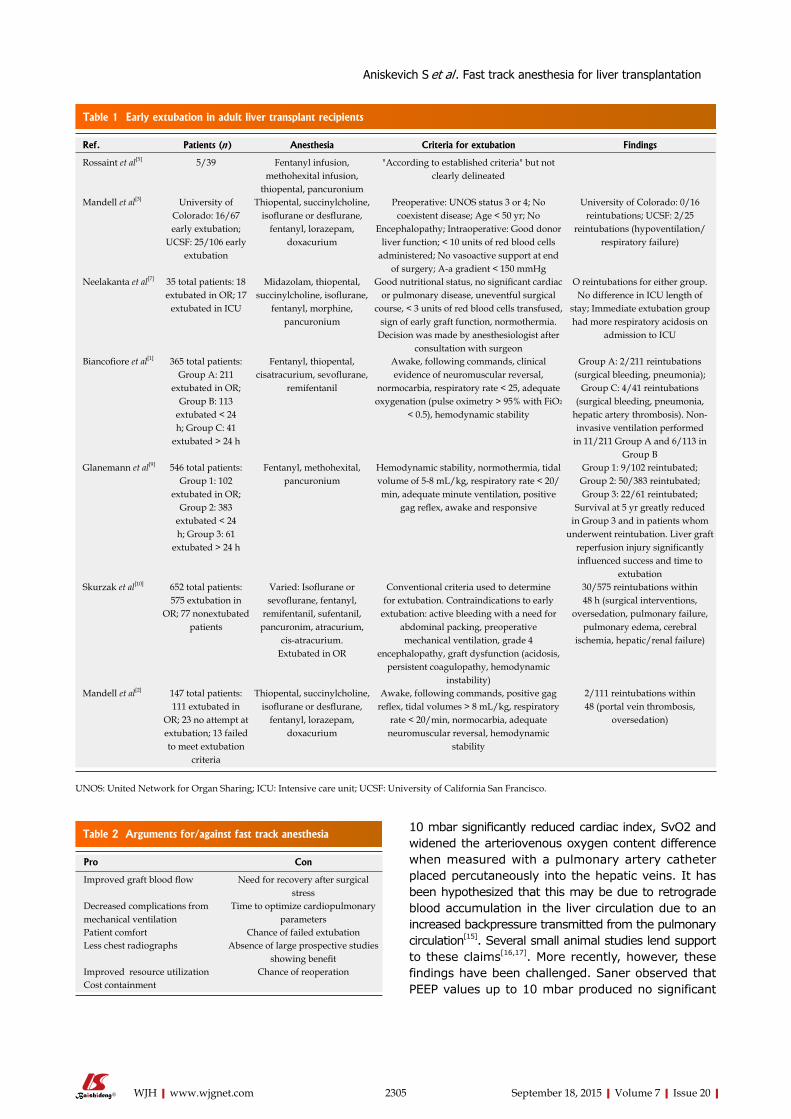
10 mbar significantly reduced cardiac index, SvO2 and widened the arteriovenous oxygen content difference when measured with a pulmonary artery catheter placed percutaneously into the hepatic veins. It has been hypothesized that this may be due to retrograde blood accumulation in the liver circulation due to an increased backpressure transmitted from the pulmonary circulation[15]. Several small animal studies lend support to these claims[16,17]. More recently, however, these findings have been challenged. Saner observed that PEEP values up to 10 mbar produced no significant
2305 September 18, 2015|Volume 7|Issue 20|WJH|www.wjgnet.com
Table 1 Early extubation in adult liver transplant recipients
Ref. Patients (n ) Anesthesia Criteria for extubation Findings
Rossaint et al[5] 5/39 Fentanyl infusion, methohexital infusion,
thiopental, pancuronium
"According to established criteria" but not clearly delineated
Mandell et al[3] University of Colorado: 16/67 early extubation;
UCSF: 25/106 early extubation
Thiopental, succinylcholine, isoflurane or desflurane,
fentanyl, lorazepam, doxacurium
Preoperative: UNOS status 3 or 4; No coexistent disease; Age < 50 yr; No
Encephalopathy; Intraoperative: Good donor liver function; < 10 units of red blood cells
administered; No vasoactive support at end of surgery; A-a gradient < 150 mmHg
University of Colorado: 0/16 reintubations; UCSF: 2/25
reintubations (hypoventilation/respiratory failure)
Neelakanta et al[7] 35 total patients: 18 extubated in OR; 17
extubated in ICU
Midazolam, thiopental, succinylcholine, isoflurane,
fentanyl, morphine, pancuronium
Good nutritional status, no significant cardiac or pulmonary disease, uneventful surgical
course, < 3 units of red blood cells transfused, sign of early graft function, normothermia.
Decision was made by anesthesiologist after consultation with surgeon
O reintubations for either group. No difference in ICU length of
stay; Immediate extubation group had more respiratory acidosis on
admission to ICU
Biancofiore et al[1] 365 total patients: Group A: 211
extubated in OR; Group B: 113
extubated < 24 h; Group C: 41
extubated > 24 h
Fentanyl, thiopental, cisatracurium, sevoflurane,
remifentanil
Awake, following commands, clinical evidence of neuromuscular reversal,
normocarbia, respiratory rate < 25, adequate oxygenation (pulse oximetry > 95% with FiO2
< 0.5), hemodynamic stability
Group A: 2/211 reintubations (surgical bleeding, pneumonia);
Group C: 4/41 reintubations (surgical bleeding, pneumonia,
hepatic artery thrombosis). Non-invasive ventilation performed
in 11/211 Group A and 6/113 in Group B
Glanemann et al[9] 546 total patients: Group 1: 102
extubated in OR; Group 2: 383
extubated < 24 h; Group 3: 61
extubated > 24 h
Fentanyl, methohexital, pancuronium
Hemodynamic stability, normothermia, tidal volume of 5-8 mL/kg, respiratory rate < 20/min, adequate minute ventilation, positive
gag reflex, awake and responsive
Group 1: 9/102 reintubated; Group 2: 50/383 reintubated; Group 3: 22/61 reintubated;
Survival at 5 yr greatly reduced in Group 3 and in patients whom
underwent reintubation. Liver graft reperfusion injury significantly influenced success and time to
extubationSkurzak et al[10] 652 total patients:
575 extubation in OR; 77 nonextubated
patients
Varied: Isoflurane or sevoflurane, fentanyl,
remifentanil, sufentanil, pancuronim, atracurium,
cis-atracurium.Extubated in OR
Conventional criteria used to determine for extubation. Contraindications to early
extubation: active bleeding with a need for abdominal packing, preoperative mechanical ventilation, grade 4
encephalopathy, graft dysfunction (acidosis, persistent coagulopathy, hemodynamic
instability)
30/575 reintubations within 48 h (surgical interventions,
oversedation, pulmonary failure, pulmonary edema, cerebral
ischemia, hepatic/renal failure)
Mandell et al[2] 147 total patients: 111 extubated in
OR; 23 no attempt at extubation; 13 failed to meet extubation
criteria
Thiopental, succinylcholine, isoflurane or desflurane,
fentanyl, lorazepam, doxacurium
Awake, following commands, positive gag reflex, tidal volumes > 8 mL/kg, respiratory
rate < 20/min, normocarbia, adequate neuromuscular reversal, hemodynamic
stability
2/111 reintubations within 48 (portal vein thrombosis,
oversedation)
UNOS: United Network for Organ Sharing; ICU: Intensive care unit; UCSF: University of California San Francisco.
Table 2 Arguments for/against fast track anesthesia
Pro Con
Improved graft blood flow Need for recovery after surgical stress
Decreased complications from mechanical ventilation
Time to optimize cardiopulmonary parameters
Patient comfort Chance of failed extubationLess chest radiographs Absence of large prospective studies
showing benefitImproved resource utilization Chance of reoperationCost containment
Aniskevich S et al . Fast track anesthesia for liver transplantation
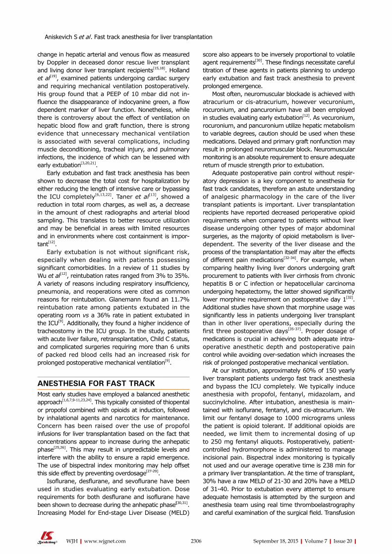
2306 September 18, 2015|Volume 7|Issue 20|WJH|www.wjgnet.com
score also appears to be inversely proportional to volatile agent requirements[30]. These findings necessitate careful titration of these agents in patients planning to undergo early extubation and fast track anesthesia to prevent prolonged emergence.
Most often, neuromuscular blockade is achieved with atracurium or cisatracurium, however vecuronium, rocuronium, and pancuronium have all been employed in studies evaluating early extubation[12]. As vecuronium, rocuronium, and pancuronium utilize hepatic metabolism to variable degrees, caution should be used when these medications. Delayed and primary graft nonfunction may result in prolonged neuromuscular block. Neuromuscular monitoring is an absolute requirement to ensure adequate return of muscle strength prior to extubation.
Adequate postoperative pain control without respiratory depression is a key component to anesthesia for fast track candidates, therefore an astute understanding of analgesic pharmacology in the care of the liver transplant patients is important. Liver transplantation recipients have reported decreased perioperative opioid requirements when compared to patients without liver disease undergoing other types of major abdominal surgeries, as the majority of opioid metabolism is liverdependent. The severity of the liver disease and the process of the transplantation itself may alter the effects of different pain medications[3234]. For example, when comparing healthy living liver donors undergoing graft procurement to patients with liver cirrhosis from chronic hepatitis B or C infection or hepatocellular carcinoma undergoing hepatectomy, the latter showed significantly lower morphine requirement on postoperative day 1[32]. Additional studies have shown that morphine usage was significantly less in patients undergoing liver transplant than in other liver operations, especially during the first three postoperative days[3537]. Proper dosage of medications is crucial in achieving both adequate intraoperative anesthetic depth and postoperative pain control while avoiding oversedation which increases the risk of prolonged postoperative mechanical ventilation.
At our institution, approximately 60% of 150 yearly liver transplant patients undergo fast track anesthesia and bypass the ICU completely. We typically induce anesthesia with propofol, fentanyl, midazolam, and succinylcholine. After intubation, anesthesia is maintained with isoflurane, fentanyl, and cis-atracurium. We limit our fentanyl dosage to 1000 micrograms unless the patient is opioid tolerant. If additional opioids are needed, we limit them to incremental dosing of up to 250 mg fentanyl aliquots. Postoperatively, patientcontrolled hydromorphone is administered to manage incisional pain. Bispectral index monitoring is typically not used and our average operative time is 238 min for a primary liver transplantation. At the time of transplant, 30% have a raw MELD of 2130 and 20% have a MELD of 3140. Prior to extubation every attempt to ensure adequate hemostasis is attempted by the surgeon and anesthesia team using real time thromboelastrography and careful examination of the surgical field. Transfusion
change in hepatic arterial and venous flow as measured by Doppler in deceased donor rescue liver transplant and living donor liver transplant recipients[15,18]. Holland et al[19], examined patients undergoing cardiac surgery and requiring mechanical ventilation postoperatively. His group found that a PEEP of 10 mbar did not influence the disappearance of indocyanine green, a flow dependent marker of liver function. Nonetheless, while there is controversy about the effect of ventilation on hepatic blood flow and graft function, there is strong evidence that unnecessary mechanical ventilation is associated with several complications, including muscle deconditioning, tracheal injury, and pulmonary infections, the incidence of which can be lessened with early extubation[3,20,21].
Early extubation and fast track anesthesia has been shown to decrease the total cost for hospitalization by either reducing the length of intensive care or bypassing the ICU completely[6,13,22]. Taner et al[13], showed a reduction in total room charges, as well as, a decrease in the amount of chest radiographs and arterial blood sampling. This translates to better resource utilization and may be beneficial in areas with limited resources and in environments where cost containment is important[12].
Early extubation is not without significant risk, especially when dealing with patients possessing significant comorbidities. In a review of 11 studies by Wu et al[12], reintubation rates ranged from 3% to 35%. A variety of reasons including respiratory insufficiency, pneumonia, and reoperations were cited as common reasons for reintubation. Glanemann found an 11.7% reintubation rate among patients extubated in the operating room vs a 36% rate in patient extubated in the ICU[9]. Additionally, they found a higher incidence of tracheostomy in the ICU group. In the study, patients with acute liver failure, retransplantation, Child C status, and complicated surgeries requiring more than 6 units of packed red blood cells had an increased risk for prolonged postoperative mechanical ventilation[9].
ANESTHESIA FOR FAST TRACKMost early studies have employed a balanced anesthetic approach[1,6,7,911,23,24]. This typically consisted of thiopental or propofol combined with opioids at induction, followed by inhalational agents and narcotics for maintenance. Concern has been raised over the use of propofol infusions for liver transplantation based on the fact that concentrations appear to increase during the anhepatic phase[25,26]. This may result in unpredictable levels and interfere with the ability to ensure a rapid emergence. The use of bispectral index monitoring may help offset this side effect by preventing overdosage[2729].
Isoflurane, desflurane, and sevoflurane have been used in studies evaluating early extubation. Dose requirements for both desflurane and isoflurane have been shown to decrease during the anhepatic phase[30,31]. Increasing Model for Endstage Liver Disease (MELD)
Aniskevich S et al . Fast track anesthesia for liver transplantation
2307 September 18, 2015|Volume 7|Issue 20|WJH|www.wjgnet.com
goals are a stable hemoglobin of 810 mg/dL, an international normalization ratio of 1.52, a fibrinogen level greater that 170, and a platelet count of approximately 100.
We do not utilize set fast track criteria per se; rather all of the anesthesiologists base our determination on clinical experience after consultation with the operating surgeon. Typically redo transplants and patients requiring vasopressors or postoperative dialysis are admitted to the ICU after surgery. High volume transfusions are not an indication for ICU admission unless there is significant concomitant coagulopathy and a high likelihood of needing to transfuse more than 2 units of blood products per hour. Likewise, MELD score itself is not an indication for intensive care, although higher MELDs are more likely to be associated with significant comorbidities. We usually make our determination after graft reperfusion to give either the ICU or surgical ward time to prepare for the admission. On admission to the postoperative care unit, the anesthesia team evaluates relevant blood labs, an electrocardiogram, and a chest radiograph. After clearance from the anesthesia team, the patient is transferred to the surgical ward where the patient initially receives 1:1 nursing care for the first 24 h and further evaluation from the transplant hepatology team[13].
CONCLUSIONIn summary, early extubation for large surgical cases started in cardiac surgery and is gaining popularity within the liver transplant anesthesia community. The practice of fast track anesthesia may decrease the incidence of pulmonary complications and improve graft function, and result in better resource management. As experience grows within our field, transplant teams have become better able to determine patients that can benefit from this practice[24]. Careful coordination and communication between the surgeons, anesthesiologists, and ward teams needs to be in place to ensure safe delivery of care.
REFERENCES1 Biancofiore G, Bindi ML, Romanelli AM, Boldrini A, Bisà M,
Esposito M, Urbani L, Catalano G, Mosca F, Filipponi F. Fast track in liver transplantation: 5 years’ experience. Eur J Anaesthesiol 2005; 22: 584590 [PMID: 16119594]
2 Mandell MS, Lezotte D, Kam I, Zamudio S. Reduced use of intensive care after liver transplantation: influence of early extubation. Liver Transpl 2002; 8: 676681 [PMID: 12149759]
3 Mandell MS, Campsen J, Zimmerman M, Biancofiore G, Tsou MY. The clinical value of early extubation. Curr Opin Organ Transplant 2009; 14: 297302 [PMID: 19417660]
4 Prakash O, Jonson B, Meij S, Bos E, Hugenholtz PG, Nauta J, Hekman W. Criteria for early extubation after intracardiac surgery in adults. Anesth Analg 1977; 56: 703708 [PMID: 562097]
5 Rossaint R, Slama K, Jaeger M, Konrad M, Pappert D, Bechstein W, Blumhardt G, Neuhaus P, Falke KJ. Fluid restriction and early extubation for successful liver transplantation. Transplant Proc 1990; 22: 15331534 [PMID: 2389392]
6 Mandell MS, Lockrem J, Kelley SD. Immediate tracheal extu
bation after liver transplantation: experience of two transplant centers. Anesth Analg 1997; 84: 249253 [PMID: 9024010]
7 Neelakanta G, Sopher M, Chan S, Pregler J, Steadman R, Braunfeld M, Csete M. Early tracheal extubation after liver transplantation. J Cardiothorac Vasc Anesth 1997; 11: 165167 [PMID: 9105986]
8 Plevak DJ, Torsher LC. Fast tracking in liver transplantation. Liver Transpl Surg 1997; 3: 447448 [PMID: 9346779]
9 Glanemann M, Langrehr J, Kaisers U, Schenk R, Müller A, Stange B, Neumann U, Bechstein WO, Falke K, Neuhaus P. Postoperative tracheal extubation after orthotopic liver transplantation. Acta Anaesthesiol Scand 2001; 45: 333339 [PMID: 11207470]
10 Skurzak S, Stratta C, Schellino MM, Fop F, Andruetto P, Gallo M, Rampa P, Crucitti M, Zabatta D, Panio A, Cerutti E. Extubation score in the operating room after liver transplantation. Acta Anaesthesiol Scand 2010; 54: 970978 [PMID: 20626358]
11 Trzebicki J, Nicinska B, Blaszczyk B, Jureczko L, Kolacz M, Pacholczyk M, Chmura A, Lagiewska B, Adadynski L, Malkowski P, Wasiak D, Lisik W, Kwiatkowski A, Lazowski T. Thoracic epidural analgesia in anaesthesia for liver transplantation: the 10year experience of a single centre. Ann Transplant 2010; 15: 3539 [PMID: 20657517]
12 Wu J, Rastogi V, Zheng SS. Clinical practice of early extubation after liver transplantation. Hepatobiliary Pancreat Dis Int 2012; 11: 577585 [PMID: 23232628]
13 Taner CB, Willingham DL, Bulatao IG, Shine TS, Peiris P, Torp KD, Canabal J, Nguyen JH, Kramer DJ. Is a mandatory intensive care unit stay needed after liver transplantation? Feasibility of fasttracking to the surgical ward after liver transplantation. Liver Transpl 2012; 18: 361369 [PMID: 22140001]
14 Kaisers U, Langrehr JM, Haack M, Mohnhaupt A, Neuhaus P, Rossaint R. Hepatic venous catheterization in patients undergoing positive endexpiratory pressure ventilation after OLT: technique and clinical impact. Clin Transplant 1995; 9: 301306 [PMID: 7579737]
15 Saner FH, Olde Damink SW, Pavlaković G, Sotiropoulos GC, Radtke A, Treckmann J, Beckebaum S, Cicinnati V, Paul A. Is positive endexpiratory pressure suitable for liver recipients with a rescue organ offer? J Crit Care 2010; 25: 477482 [PMID: 19942400]
16 Brienza N, Revelly JP, Ayuse T, Robotham JL. Effects of PEEP on liver arterial and venous blood flows. Am J Respir Crit Care Med 1995; 152: 504510 [PMID: 7633699]
17 Bredenberg CE, Paskanik AM. Relation of portal hemodynamics to cardiac output during mechanical ventilation with PEEP. Ann Surg 1983; 198: 218222 [PMID: 6347103]
18 Saner FH, Olde Damink SW, Pavlaković G, van den Broek MA, Sotiropoulos GC, Radtke A, Nadalin S, Malagó M, Paul A. Positive endexpiratory pressure induces liver congestion in living donor liver transplant patients: myth or fact. Transplantation 2008; 85: 18631866 [PMID: 18580483]
19 Holland A, Thuemer O, Schelenz C, van Hout N, Sakka SG. Positive endexpiratory pressure does not affect indocyanine green plasma disappearance rate or gastric mucosal perfusion after cardiac surgery. Eur J Anaesthesiol 2007; 24: 141147 [PMID: 16938155]
20 Razonable RR, Findlay JY, O’Riordan A, Burroughs SG, Ghobrial RM, Agarwal B, Davenport A, Gropper M. Critical care issues in patients after liver transplantation. Liver Transpl 2011; 17: 511527 [PMID: 21384524]
21 Papadimos TJ, Hensley SJ, Duggan JM, Khuder SA, Borst MJ, Fath JJ, Oakes LR, Buchman D. Implementation of the “FASTHUG” concept decreases the incidence of ventilatorassociated pneumonia in a surgical intensive care unit. Patient Saf Surg 2008; 2: 3 [PMID: 18271952]
22 Quiroga M, Rodríguez MG, Montalván C, Abarca J, Viñuela M, Cavallieri S, Hepp J, Ríos H, Suárez L, Innocenti F, Humeres R, Palacios JM, Sandoval R, Cruz J, Rius M. Trends in mechanical ventilation and immediate extubation after liver transplantation in a single center in Chile. Transplant Proc 2004; 36: 16831684 [PMID: 15350452]
Aniskevich S et al . Fast track anesthesia for liver transplantation
2308 September 18, 2015|Volume 7|Issue 20|WJH|www.wjgnet.com
23 Zeyneloglu P, Pirat A, Guner M, Torgay A, Karakayali H, Arslan G. Predictors of immediate tracheal extubation in the operating room after liver transplantation. Transplant Proc 2007; 39: 11871189 [PMID: 17524928]
24 Alper I, Ulukaya S. Anesthetic management in pediatric liver transplantation: a comparison of deceased or live donor liver transplantations. J Anesth 2010; 24: 399406 [PMID: 20339881]
25 Takizawa D, Hiraoka H, Nakamura K, Yamamoto K, Horiuchi R. Propofol concentrations during the anhepatic phase of livingrelated donor liver transplantation. Clin Pharmacol Ther 2004; 76: 648649 [PMID: 15592336]
26 Wu J, Zhu SM, He HL, Weng XC, Huang SQ, Chen YZ. Plasma propofol concentrations during orthotopic liver transplantation. Acta Anaesthesiol Scand 2005; 49: 804810 [PMID: 15954963]
27 Cammu G, Decruyenaere J, Troisi R, de Hemptinne B, Colardyn F, Mortier E. Criteria for immediate postoperative extubation in adult recipients following livingrelated liver transplantation with total intravenous anesthesia. J Clin Anesth 2003; 15: 515519 [PMID: 14698363]
28 Gan TJ, Glass PS, Windsor A, Payne F, Rosow C, Sebel P, Manberg P. Bispectral index monitoring allows faster emergence and improved recovery from propofol, alfentanil, and nitrous oxide anesthesia. BIS Utility Study Group. Anesthesiology 1997; 87: 808815 [PMID: 9357882]
29 Tremelot L, Restoux A, PaugamBurtz C, Dahmani S, Massias L, Peuch C, Belghiti J, Mantz J. Interest of BIS monitoring to guide propofol infusion during the anhepatic phase of orthotopic liver transplantation. Ann Fr Anesth Reanim 2008; 27: 975978 [PMID: 19028068]
30 Kang JG, Ko JS, Kim GS, Gwak MS, Kim YR, Lee SK. The relationship between inhalational anesthetic requirements and the severity of liver disease in liver transplant recipients according to three phases of liver transplantation. Transplant Proc 2010; 42:
854857 [PMID: 20430189]31 Toprak Hİ, Sener A, Gedik E, Uçar M, Karahan K, Aydogan
MS, Ersoy MÖ. Bispectral index monitoring to guide endtidal isoflurane concentration at three phases of operation in patients with endstage liver disease undergoing orthotopic liver transplantation. Transplant Proc 2011; 43: 892895 [PMID: 21486622]
32 Crotty B, Watson KJ, Desmond PV, Mashford ML, Wood LJ, Colman J, Dudley FJ. Hepatic extraction of morphine is impaired in cirrhosis. Eur J Clin Pharmacol 1989; 36: 501506 [PMID: 2753069]
33 Jin SJ, Jung JY, Noh MH, Lee SH, Lee EK, Choi BM, Song MH, Noh GJ. The population pharmacokinetics of fentanyl in patients undergoing livingdonor liver transplantation. Clin Pharmacol Ther 2011; 90: 423431 [PMID: 21814196]
34 Hasselström J, Eriksson S, Persson A, Rane A, Svensson JO, Säwe J. The metabolism and bioavailability of morphine in patients with severe liver cirrhosis. Br J Clin Pharmacol 1990; 29: 289297 [PMID: 2310653]
35 Kotb HI, ElKady SA, Emara SE, Fouad EA, ElKabsh MY. Pharmacokinetics of controlled release morphine (MST) in patients with liver carcinoma. Br J Anaesth 2005; 94: 9599 [PMID: 15516353]
36 Chen JP, Jawan B, Chen CL, Wang CH, Cheng KW, Wang CC, Concejero AM, Villagomeza E, Huang CJ. Comparison of postoperative morphine requirements in healthy living liver donors, patients with hepatocellular carcinoma undergoing partial hepatectomy, and liver transplant recipients. Transplant Proc 2010; 42: 701702 [PMID: 20430150]
37 Eisenach JC, Plevak DJ, Van Dyke RA, Southorn PA, Danielson DR, Krom RA, Nagorney DM, Rettke SR. Comparison of analgesic requirements after liver transplantation and cholecystectomy. Mayo Clin Proc 1989; 64: 356359 [PMID: 2649750]
P- Reviewer: Staikou C S- Editor: Yu J L- Editor: A E- Editor: Liu SQ
Aniskevich S et al . Fast track anesthesia for liver transplantation
Zhan-Wang Xiang, Lin Sun, Guo-Hong Li, Rakesh Maharjan, Jin-Hua Huang, Chuan-Xing Li
Zhan-Wang Xiang, Lin Sun, Guo-Hong Li, Rakesh Maharjan, Jin-Hua Huang, Chuan-Xing Li, Sun Yat-Sen University Cancer Center, State Key Laboratory of Oncology in South China, Collaborative Innovation Center for Cancer Medicine, Guangzhou 510060, Guangdong Province, China
Author contributions: Xiang ZW and Sun L contributed equally to this work; Li CX and Huang JH designed research; Xiang ZW, Sun L, Li GH and Li CX performed research; Xiang ZW, Sun L and Maharjan R analyzed data; Xiang ZW and Sun L wrote the paper; all authors had read and approved the final manuscript to be published.
Supported by National Natural Science Foundation of China, No. 81371652 (to Huang JH).
Conflict-of-interest statement: The authors declare there is no conflict of interest in this study.
Open-Access: This article is an open-access article which was selected by an in-house editor and fully peer-reviewed by external reviewers. It is distributed in accordance with the Creative Commons Attribution Non Commercial (CC BY-NC 4.0) license, which permits others to distribute, remix, adapt, build upon this work non-commercially, and license their derivative works on different terms, provided the original work is properly cited and the use is non-commercial. See: http://creativecommons.org/licenses/by-nc/4.0/
Correspondence to: Chuan-Xing Li, MD, Sun Yat-Sen University Cancer Center, State Key Laboratory of Oncology in South China, Collaborative Innovation Center for Cancer Medicine, No. 651 Dongfeng East Road, Guangzhou 510060, Guangdong Province, China. [email protected]: +86-20-87343901 Fax: +86-20-87343901
Received: April 16, 2015Peer-review started: April 19, 2015First decision: June 2, 2015Revised: June 19, 2015Accepted: September 2, 2015Article in press: September 7, 2015Published online: September 18, 2015
AbstractHepatocellular carcinoma (HCC) is the fifth most common cancer in the world, and is the third leading cause of cancer-related death. Liver transplantation (LT) has become a curative treatment for patients with HCC. However, recurrence and metastasis after LT are the main factors reducing long-term survival in patients, and the lung is the most common site of metastasis after LT for HCC, although metastasis to liver, para-aortic lymph nodes and renal periphery are observed. Thus, the treatment of pulmonary metastases after LT for HCC has become a hot research topic, the successful treatment of pulmonary metastases can significantly prolong the survival of LT patients. Although single conventional treatment (chemotherapy, surgery and external beam radiation therapy), immunosuppression, image-guided minimally invasive therapy (radiofrequency ablation, microwave ablation, cryoablation, and brachytherapy) and molecular targeted drugs have had a significant effect, patients do not have durable remission and the long-term survival rate is disappointing. Therefore, improving existing treatments and identifying a more effective combination therapy are important research issues in the prevention and treatment of pulmonary metastases after LT for HCC. The paper reviewed single conventional treatments, new treatments, and combination therapy, to provide a basis for the best treatment of these patients.
Key words: Liver transplantation; Progress; Treatment; Pulmonary metastases; Hepatocellular carcinoma
© The Author(s) 2015. Published by Baishideng Publishing Group Inc. All rights reserved.
Core tip: The treatment of pulmonary metastases after liver transplantation for hepatocellular carcinoma has become a hot research topic. Although single conventional treatment (chemotherapy, surgery and external beam radiation therapy), image-guided
MINIREVIEWS
Submit a Manuscript: http://www.wjgnet.com/esps/Help Desk: http://www.wjgnet.com/esps/helpdesk.aspxDOI: 10.4254/wjh.v7.i20.2309
2309 September 18, 2015|Volume 7|Issue 20|WJH|www.wjgnet.com
World J Hepatol 2015 September 18; 7(20): 2309-2314ISSN 1948-5182 (online)
© 2015 Baishideng Publishing Group Inc. All rights reserved.
Progress in the treatment of pulmonary metastases after liver transplantation for hepatocellular carcinoma
minimally invasive therapy (radiofrequency ablation, microwave ablation, cryoablation, and brachytherapy) and molecular targeted drugs have had a significant effect, patients do not have durable remission and the long-term survival rate is disappointing. Therefore, we reviewed single conventional treatments, new treat-ments, and combination therapy, to provide a basis for the best treatment of these patients.
Xiang ZW, Sun L, Li GH, Maharjan R, Huang JH, Li CX. Progress in the treatment of pulmonary metastases after liver transplantation for hepatocellular carcinoma. World J Hepatol 2015; 7(20): 2309-2314 Available from: URL: http://www.wjgnet.com/1948-5182/full/v7/i20/2309.htm DOI: http://dx.doi.org/10.4254/wjh.v7.i20.2309
INTRODUCTIONHepatocellular carcinoma (HCC) is the fifth most common cancer in the world, is the third leading cause of cancer-related death, and the incidence is high and increasing[1,2]. Liver transplantation (LT) is carried out to treat HCC, especially in the early stage[3]. However, due to extrahepatic organ micrometastases, which cannot be found by imaging and cancer cells present in the blood circulation before LT, the charac-teristics of liver cancer (microvascular invasion, low differentiation, allelic imbalance, genetic diversity), the stage (super Milan criteria) and the administration of immunosuppressive agents during and after LT[4], result in a high incidence of postoperative recurrence and metastasis (60%-100%)[5]. Tumor recurrence and metastasis obviously decrease the chance of long-term survival after LT for HCC[6,7], thus recurrence and metastasis are the greatest obstacles to successful HCC treatment.
Lung is the most frequent site of metastasis after LT, although metastasis to liver, para-aortic lymph nodes and renal periphery are observed[8]. The successful treatment of pulmonary metastases can significantly prolong the survival of LT patients[9]. Therefore, improv-ing existing treatments and identifying a more effective combination therapy have become important issues in pulmonary metastases after LT for HCC. The purpose of this paper is to provide an overview of therapies for pulmonary metastases after LT for HCC. Therefore, we reviewed single conventional treatments, new treatments, and combination therapy, to provide a basis for the best treatment of these patients.
CONVENTIONAL TREATMENT METHODSChemotherapy and external beam radiotherapy The administration of chemotherapy for lung meta-stases after LT is controversial. The United Network for Organ Sharing data indicate that 48% of HCC patients received adjuvant chemotherapy after LT[10]. However,
the results of therapy have been disappointing, and pulmonary metastases response rates after single or multiple agent chemotherapy regimens were low, with the 1-year survival rate between 0% and 30%[11]. Lee et al[12] reported that the most commonly used chemo-therapeutic regimens were administered to patients following LT for HCC, and the median time to pro-gression was 7.0 wk (95%CI: 5.8-8.2) and the median overall survival was 16.6 wk (95%CI: 10.1-23.1). Roxburgh et al[13] also found that HCC was generally chemoresistant and results using systemic therapy were disappointing. In addition, patients after LT required long-term oral immunosuppressive drugs, as the vast majority of patients could not tolerate chemotherapy toxicity, which was also a problem when administering chemotherapy[14].
There has been some progress in the use of external beam radiotherapy (EBRT), particularly the use of three-dimensional conformal radiotherapy and the tomotherapy system, which have significantly increased the partial or complete remission rate of lung metastases[15]. Jang et al[16] reported that 30-57.6 Gy of radiation for lung metastases resulted in a complete response rate of 26.3%, and the overall survival rate at 1 year was 50.1%. Matsui et al[17] also showed that stereotactic radiotherapy, performed at the site of tumor location two years after radiotherapy, resulted in survival of the patient without recurrence. However, at the time of diagnosis, most patients have multiple pulmonary or systemic metastases[18]. Therefore, the future trend in radiotherapy for pulmonary metastases after LT for HCC is to increase the tumor area dose by screening patients cautiously and using advanced radiotherapy positioning technology, while reducing the scope of normal tissue irradiated and the incidence of radiation toxicity. EBRT has significant value for lung metastases after LT for HCC.
Surgery Surgery has been accepted as the first treatment for pulmonary metastases after LT for HCC for some time. Studies have confirmed that surgery is effective, and survival is reported to be between 24% and 78% at 3 years, with median survival ranging from 21 mo to 29 mo[19,20]. However, surgical resection of isolated metastasis following LT for HCC is limited to a few studies or case reports worldwide[21,22]. Bates et al[23] reported that five patients who had pulmonary resection of metastatic HCC after LT, had an average survival period of 44 mo after transplantation and 28 mo after pulmonary resection, these survival times were similar to those of patients following metastasectomy after liver resection for HCC. A year later, Zhang et al[18] also reported five patients who underwent standard lobectomy or wedge resection, and their survival ranged from 3 to 53 mo, with an average survival period of 18 mo. Similarly, Togashi et al[24] described two cases with long-term survival following pulmonary metastasectomy for HCC recurrence several months after living donor
2310 September 18, 2015|Volume 7|Issue 20|WJH|www.wjgnet.com
Xiang ZW et al . Treatment of pulmonary metastases after liver transplantation
liver transplantation, with no signs of further recurrence 2 years and 4 years 5 mo after resection of the lung metastasis. These studies show that resection of pulmonary metastases is an effective treatment method. However, growth of metastatic tumors in the alveoli causes no or mild respiratory symptoms in the early stages of disease. At the time of diagnosis, lung metastases have usually already developed into multiple metastatic lesions, and most patients are denied the chance of surgical treatment[25]. Furthermore, due to immunosuppression and surgical trauma, lung lesions may recur after resection at any time, and possibly metastases in other organs. The rate of recurrence and metastasis in patients receiving immunosuppressive therapy is significantly higher than in those who do not receive immunosuppressive therapy, indicating that immunosuppressive therapy plays a major role in tumor recurrence and metastasis after LT[26]. Therefore, the value of surgery in the treatment of lung metastasis after LT for HCC should be confirmed by further prospective multi-center clinical studies.
NEW TREATMENT METHODSImmunosuppressionMore and more studies have confirmed that immuno-suppressants [e.g., mammalian target of rapamycin inhibitors (m-TORi)] have anti-transplant rejection and multiple anti-tumor effects after LT for HCC[27,28]. Kawahara et al[29] found that m-TORi can decrease the risk of recurrence after LT for HCC and have lower drug toxicities. Cholongitas et al[30] also showed that patients on m-TORi had significantly lower recurrence rates following LT for HCC, thus m-TORi may represent an alternative immunosuppressive regimen with antine-oplastic effects. Moreover, the early use of m-TORi can significantly prolong survival time and delay tumor progression after LT[31]. Klintmalm et al[32] indicated that m-TORi may have benefits in the oncology setting and in relation to HCV-related allograft fibrosis, metabolic syndrome, neurotoxicity, and survival time. However, clinical studies have demonstrated that immunosu-ppressive agents can cause serious adverse reactions in patients such as pneumonia and thrombocytopenia[33]. In patients with pulmonary metastases after LT for HCC, most were in poor physical condition and were unable to tolerate further treatment. Therefore, further research on reducing the side effects of m-TORi and controlling further progression with combination therapy for pulmonary metastases after LT for HCC, will have significant clinical value.
Minimally invasive therapyIn recent years, due to the development of medical imaging, many patients with pulmonary metastases may also be treated with minimally invasive treatments, such as interstitial laser coagulation, cryotherapy, microwave ablation, percutaneous ethanol injection, radiofrequency ablation (RFA), and 125I brachytherapy[34,35]. RFA uses
thermal and non-thermal effects which are generated by RFA electromagnetic waves in a biological medium to solidify cancer tissue, and the local temperature can be higher than 90 ℃, which kills tumor cells quickly and effectively. RFA has been demonstrated to be a safe and valuable treatment option and is accepted as the best therapeutic choice for patients with unresectable HCC pulmonary metastases. Lencioni et al[36] reported that RFA results in a high proportion of sustained complete responses in properly selected patients with pulmonary malignancies, and is associated with acceptable mor-bidity. Hiraki et al[37] found that RFA for 83 pulmonary metastases resulting from HCC was effective and safe in selected patients, where the effectiveness rate was 92% and survival rate was significantly improved. Therefore, RFA improves survival in patients with limited metastatic lung disease[38]. In addition, 125I brachytherapy has also been used for pulmonary malignant tumors with good efficacy. Zhang et al[39] reported that computed tomography-guided 125I seed implantation was safe and well tolerated for treating lung tumors, with few complications, and the 1-, 3-year, and median overall survival were 68.7%, 20.8% and 17.4 mo, respectively. While the minimally invasive treatment of malignant lung tumors has significantly progressed, there are few reports on pulmonary metastases after LT for HCC. With further clinical studies, coupled with the advantages of minimally invasive treatment, such as safety, ease of use, few complications and minimal trauma, this will become a new treatment for pulmonary metastases after LT for HCC.
Targeted drugsSorafenib is a small molecular multikinase inhibitor of vascular endothelial growth factor (VEGFR)-2, VEGFR-3, platelet-derived growth factor-b, raf, Flt-3 and c-KIT. Studies have demonstrated the ability of sorafenib to inhibit tumor proliferation via the RAS-/RAF-signaling pathway and angiogenesis[40,41]. Sorafenib has been approved for the treatment of advanced HCC[42]. Kudo et al[43] reported 15 cases with complete remission following treatment with sorafenib in patients with advanced metastatic HCC, including multiple liver lesions, lymph node metastases, adrenal metastases, lung metastases and vascular invasion, which were completely absent after treatment, and three tumor markers (AFP, PIVKA-Ⅱ and AFP-L3) returned to normal values. In addition, sorafenib has also made a breakthrough in the treatment of HCC recurrence after LT. Sposito et al[44] found that sorafenib was associated with an acceptable safety profile and had a survival benefit in HCC patients suffering recurrence after LT, the recurrence time was 38.1 mo, living conditions were significantly improved after cancer recurrence (the median survival from recurrence was 21.3 mo), and the only factor associated with survival after HCC recurrence in multivariate analysis was treatment with sorafenib (HR = 4.0; P = 0.0325). Yeganeh et al[45] in a large single-center retrospective study found that
2311 September 18, 2015|Volume 7|Issue 20|WJH|www.wjgnet.com
Xiang ZW et al . Treatment of pulmonary metastases after liver transplantation
2312 September 18, 2015|Volume 7|Issue 20|WJH|www.wjgnet.com
success and have the potential to delay progression and prolong survival, however, this does not apply to all patients. Therefore, improving the existing treatments, using individual and combination therapy, is the future direction for the prevention and treatment of pulmonary metastases after LT for HCC.
REFERENCES1 Iakova P, Timchenko L, Timchenko NA. Intracellular signaling
and hepatocellular carcinoma. Semin Cancer Biol 2011; 21: 28-34 [PMID: 20850540 DOI: 10.1016/j.semcancer.2010.09.001]
2 McClune AC, Tong MJ. Chronic hepatitis B and hepatocellular carcinoma. Clin Liver Dis 2010; 14: 461-476 [PMID: 20638025]
3 Zheng SS, Xu X, Liang TB, Wang WL, Shen Y, Zhang M, Wu J, Lu AW, Shao ZX. [Liver transplantation for hepatocellular carcinoma: prognostic analysis of 89 cases]. Zhonghua Waike Zazhi 2005; 43: 450-454 [PMID: 15854372]
4 Welker MW, Bechstein WO, Zeuzem S, Trojan J. Recurrent hepatocellular carcinoma after liver transplantation - an emerging clinical challenge. Transpl Int 2013; 26: 109-118 [PMID: 22994652 DOI: 10.1111/j.1432-2277.2012.01562.x]
5 Poon RT, Fan ST, Lo CM, Liu CL, Wong J. Long-term survival and pattern of recurrence after resection of small hepatocellular carcinoma in patients with preserved liver function: implications for a strategy of salvage transplantation. Ann Surg 2002; 235: 373-382 [PMID: 11882759]
6 Kornberg A, Küpper B, Tannapfel A, Katenkamp K, Thrum K, Habrecht O, Wilberg J. Long-term survival after recurrent hepatocellular carcinoma in liver transplant patients: clinical patterns and outcome variables. Eur J Surg Oncol 2010; 36: 275-280 [PMID: 19857941 DOI: 10.1016/j.ejso.2009.10.001]
7 Regalia E, Fassati LR, Valente U, Pulvirenti A, Damilano I, Dardano G, Montalto F, Coppa J, Mazzaferro V. Pattern and management of recurrent hepatocellular carcinoma after liver trans-plantation. J Hepatobiliary Pancreat Surg 1998; 5: 29-34 [PMID: 9683751 DOI: 10.1007/PL00009947]
8 Katyal S, Oliver JH, Peterson MS, Ferris JV, Carr BS, Baron RL. Extrahepatic metastases of hepatocellular carcinoma. Radiology 2000; 216: 698-703 [PMID: 10966697]
9 Cruz RJ, Ranganathan S, Mazariegos G, Soltys K, Nayyar N, Sun Q, Bond G, Shaw PH, Haberman K, Krishnamurti L, Marsh JW, Humar A, Sindhi R. Analysis of national and single-center incidence and survival after liver transplantation for hepato-blastoma: new trends and future opportunities. Surgery 2013; 153: 150-159 [PMID: 23331862]
10 Little SA, Fong Y. Hepatocellular carcinoma: current surgical management. Semin Oncol 2001; 28: 474-486 [PMID: 11685740]
11 Pokorny H, Gnant M, Rasoul-Rockenschaub S, Gollackner B, Steiner B, Steger G, Steininger R, Mühlbacher F. Does additional doxorubicin chemotherapy improve outcome in patients with hepatocellular carcinoma treated by liver transplantation? Am J Transplant 2005; 5: 788-794 [PMID: 15760403 DOI: 10.1111/j.1600-6143.2005.00780.x]
12 Lee JO, Kim DY, Lim JH, Seo MD, Yi HG, Oh DY, Im SA, Kim TY, Bang YJ. Palliative chemotherapy for patients with recurrent hepatocellular carcinoma after liver transplantation. J Gastroenterol Hepatol 2009; 24: 800-805 [PMID: 19175825]
13 Roxburgh P, Evans TR. Systemic therapy of hepatocellular car-cinoma: are we making progress? Adv Ther 2008; 25: 1089-1104 [PMID: 18972075]
14 Waidmann O, Hofmann WP, Zeuzem S, Trojan J. mTOR inhibitors and sorafenib for recurrent heptocellular carcinoma after orthotopic liver transplantation. J Hepatol 2011; 54: 396-398 [PMID: 21111506 DOI: 10.1016/j.jhep.2010.08.038]
15 Zhao JD, Xu ZY, Zhu J, Qiu JJ, Hu WG, Cheng LF, Zhang XJ, Jiang GL. Application of active breathing control in 3-dimensional conformal radiation therapy for hepatocellular carcinoma: the feasibility and benefit. Radiother Oncol 2008; 87: 439-444 [PMID:
sorafenib was safe and well tolerated in patients with recurrent HCC following LT and may be associated with a modest survival benefit, the rate of survival at 3, 6, 9, and 12 mo was 100%, 80%, 71% and 62%, respectively. However, studies on the therapeutic value of sorafenib in pulmonary metastases after LT for HCC are rare, and only a few case reports were identified in the present review. Furthermore, serious adverse reactions have been observed, and some patients could not tolerate conventional doses of sorafenib and required dose reduction, which led to progression and deterioration of the disease or death[14,46]. Therefore, further investigations are needed to confirm the efficacy of sorafenib in the prevention and treatment of HCC after LT in international and multi-center randomized studies with a large sample size. Nevertheless, sorafenib is still a safe treatment and has a place in the treatment of pulmonary metastases after LT for HCC.
Comprehensive treatmentsIn clinical practice, it is difficult to achieve the desired results in patients with HCC lung metastases using a single treatment, therefore it is necessary to combine two or even more treatment methods. At present, reports on the combination therapy of pulmonary meta-stases after LT for HCC are rare. Sakamoto et al[47] reported one patient with HCC, multiple lung metastases and peritoneal metastasis. The patient was treated with a variety of methods including injection of ethanol, chemotherapy, and surgical resection, and although the patient died from cerebral infarction caused by tumor thrombus, the patient’s quality of life for five years was significantly improved. Matsui et al[17] described one patient with HCC who underwent right hepatic lobectomy and was found to have right lung metastases two years later, and lobectomy after chemotherapy was ineffective. The left lung was then found to have metastasis, the patient received directional EBRT, and quality of life within two years was good with no further recurrences. Li et al[48] reported 8 patients with HCC who underwent LT and then were found to have lung metastases. All patients received brachytherapy combined with sorafenib therapy. The local control rates of multiple lung metastases after LT for HCC after 4, 6, 12, 18 and 24 mo were 92.2%, 82.4%, 76.2%, 73.3% and 72.2%, respectively, and the overall 1-, 2- and 3-year survival rates were 100%, 50% and 12.5%, respectively. These studies suggest that treatment of patients with HCC metastases after LT should be individualized, with comprehensive therapies, or combined methods with a variety of treatments according to the condition of the patient, to improve their quality of life and survival.
CONCLUSIONRecurrence and metastasis are major problems restric-ting long-term survival after LT, while single treatments have had some success, they still need further study. New treatment methods have also had remarkable
Xiang ZW et al . Treatment of pulmonary metastases after liver transplantation

2313 September 18, 2015|Volume 7|Issue 20|WJH|www.wjgnet.com
18334274]16 Jang JW, Kay CS, You CR, Kim CW, Bae SH, Choi JY, Yoon SK,
Han CW, Jung HS, Choi IB. Simultaneous multitarget irradiation using helical tomotherapy for advanced hepatocellular carcinoma with multiple extrahepatic metastases. Int J Radiat Oncol Biol Phys 2009; 74: 412-418 [PMID: 18963538 DOI: 10.1016/j.ijrobp. 2008. 08.034]
17 Matsui Y, Ishibashi H, Ota S, Hirose M, Oba N, Tanio N, Nakajima N, Muro H. [Bilateral pulmonary metastases from hepatocellular carcinoma successfully treated by surgical resection and stereotactic radiotherapy; report of a case]. Kyobu Geka 2011; 64: 944-946 [PMID: 21899135]
18 Zhang C, Rao J, Tu Z, Ni Y. Surgical resection of resectable thoracic metastatic hepatocellular carcinoma after liver transplantation. J Thorac Cardiovasc Surg 2009; 138: 240-241 [PMID: 19577090 DOI: 10.1016/j.jtcvs.2008.05.014]
19 Tomimaru Y, Sasaki Y, Yamada T, Eguchi H, Takami K, Ohigashi H, Higashiyama M, Ishikawa O, Kodama K, Imaoka S. The significance of surgical resection for pulmonary metastasis from hepatocellular carcinoma. Am J Surg 2006; 192: 46-51 [PMID: 16769274]
20 Nakajima J, Tanaka M, Matsumoto J, Takeuchi E, Fukami T, Takamoto S. Appraisal of surgical treatment for pulmonary metastasis from hepatocellular carcinoma. World J Surg 2005; 29: 715-718 [PMID: 16078127]
21 Bazan HA, McMurtry KA, Waters PF, Thung SN. Surgical resection of pulmonary metastases after orthotopic liver trans-plantation for hepatocellular carcinoma. Transplantation 2002; 73: 1007-1008 [PMID: 11923711]
22 Viola C, Asselah T, Samuel D, Durand F, Boudjema H, Valla D, Marcellin P. Solitary pulmonary metastasis arising thirteen years after liver transplantation for HBV-related hepatocellular carcinoma. World J Gastroenterol 2006; 12: 4911-4913 [PMID: 16937481]
23 Bates MJ, Farkas E, Taylor D, McFadden PM. Pulmonary resection of metastatic hepatocellular carcinoma after liver transplantation. Ann Thorac Surg 2008; 85: 412-415 [PMID: 18222234 DOI: 10.1016/j.athoracsur.2007.10.065]
24 Togashi J, Sugawara Y, Aoki T, Tamura S, Kaneko J, Nakajima J, Sano A, Kokudo N. Resection of lung metastases from hepatocellular carcinoma after living donor liver transplantation: report of two cases. Surg Today 2011; 41: 1294-1297 [PMID: 21874434 DOI: 10.1007/s00595-010-4487-6]
25 Lam CM, Lo CM, Yuen WK, Liu CL, Fan ST. Prolonged survival in selected patients following surgical resection for pulmonary metastasis from hepatocellular carcinoma. Br J Surg 1998; 85: 1198-1200 [PMID: 9752858]
26 Yokoyama I, Carr B, Saitsu H, Iwatsuki S, Starzl TE. Accelerated growth rates of recurrent hepatocellular carcinoma after liver transplantation. Cancer 1991; 68: 2095-2100 [PMID: 1655200]
27 Vivarelli M, Dazzi A, Zanello M, Cucchetti A, Cescon M, Ravaioli M, Del Gaudio M, Lauro A, Grazi GL, Pinna AD. Effect of different immunosuppressive schedules on recurrence-free survival after liver transplantation for hepatocellular carcinoma. Transplantation 2010; 89: 227-231 [PMID: 20098287]
28 Liang W, Wang D, Ling X, Kao AA, Kong Y, Shang Y, Guo Z, He X. Sirolimus-based immunosuppression in liver transplantation for hepatocellular carcinoma: a meta-analysis. Liver Transpl 2012; 18: 62-69 [PMID: 21964956]
29 Kawahara T, Asthana S, Kneteman NM. m-TOR inhibitors: what role in liver transplantation? J Hepatol 2011; 55: 1441-1451 [PMID: 21781947 DOI: 10.1016/j.jhep.2011.06.015]
30 Cholongitas E, Mamou C, Rodríguez-Castro KI, Burra P. Mammalian target of rapamycin inhibitors are associated with lower rates of hepatocellular carcinoma recurrence after liver trans-plantation: a systematic review. Transpl Int 2014; 27: 1039-1049 [PMID: 24943720]
31 Menon KV, Hakeem AR, Heaton ND. Meta-analysis: recurrence and survival following the use of sirolimus in liver transplantation for hepatocellular carcinoma. Aliment Pharmacol Ther 2013; 37: 411-419 [PMID: 23278125 DOI: 10.1111/apt.12185]
32 Klintmalm GB, Nashan B. The Role of mTOR Inhibitors in Liver Transplantation: Reviewing the Evidence. J Transplant 2014; 2014: 845438 [PMID: 24719752 DOI: 10.1155/2014/845438]
33 Harper SJ, Gelson W, Harper IG, Alexander GJ, Gibbs P. Swit-ching to sirolimus-based immune suppression after liver trans-plantation is safe and effective: a single-center experience. Transplantation 2011; 91: 128-132 [PMID: 21452417]
34 Smith SL, Jennings PE. Lung radiofrequency and microwave ablation: a review of indications, techniques and post-procedural imaging appearances. Br J Radiol 2015; 88: 20140598 [PMID: 25465192 DOI: 10.1259/bjr.20140598]
35 Gillams A. Tumour ablation: current role in the liver, kidney, lung and bone. Cancer Imaging 2008; 8 Spec No A: S1-S5 [PMID: 18852074 DOI: 10.1102/1470-7330.2008.9001]
36 Lencioni R, Crocetti L, Cioni R, Suh R, Glenn D, Regge D, Helmberger T, Gillams AR, Frilling A, Ambrogi M, Bartolozzi C, Mussi A. Response to radiofrequency ablation of pulmonary tumours: a prospective, intention-to-treat, multicentre clinical trial (the RAPTURE study). Lancet Oncol 2008; 9: 621-628 [PMID: 18565793 DOI: 10.1016/S1470-2045(08)70155-4]
37 Hiraki T, Yamakado K, Ikeda O, Matsuoka T, Kaminou T, Yamagami T, Gobara H, Mimura H, Kawanaka K, Takeda K, Yamashita Y, Inoue Y, Ogawa T, Nishimura T, Kanazawa S. Percut-aneous radiofrequency ablation for pulmonary metastases from hepatocellular carcinoma: results of a multicenter study in Japan. J Vasc Interv Radiol 2011; 22: 741-748 [PMID: 21531575 DOI: 10.1016/j.jvir.2011.02.030]
38 Crocetti L, Lencioni R. Radiofrequency ablation of pulmonary tumors. Eur J Radiol 2010; 75: 23-27 [PMID: 20452739 DOI: 10.1016/j.ejrad.2010.04.011]
39 Zhang T, Lu M, Peng S, Zhang W, Yang G, Liu Z, Singh S, Yang Y, Zhang F, Gao F. CT-guided implantation of radioactive 125I seed in advanced non-small-cell lung cancer after failure of first-line chemotherapy. J Cancer Res Clin Oncol 2014; 140: 1383-1390 [PMID: 24723151]
40 Chang YS, Adnane J, Trail PA, Levy J, Henderson A, Xue D, Bortolon E, Ichetovkin M, Chen C, McNabola A, Wilkie D, Carter CA, Taylor IC, Lynch M, Wilhelm S. Sorafenib (BAY 43-9006) inhibits tumor growth and vascularization and induces tumor apoptosis and hypoxia in RCC xenograft models. Cancer Chemother Pharmacol 2007; 59: 561-574 [PMID: 17160391]
41 Wilhelm SM, Carter C, Tang L, Wilkie D, McNabola A, Rong H, Chen C, Zhang X, Vincent P, McHugh M, Cao Y, Shujath J, Gawlak S, Eveleigh D, Rowley B, Liu L, Adnane L, Lynch M, Auclair D, Taylor I, Gedrich R, Voznesensky A, Riedl B, Post LE, Bollag G, Trail PA. BAY 43-9006 exhibits broad spectrum oral antitumor activity and targets the RAF/MEK/ERK pathway and receptor tyrosine kinases involved in tumor progression and angiogenesis. Cancer Res 2004; 64: 7099-7109 [PMID: 15466206]
42 Abou-Alfa GK, Schwartz L, Ricci S, Amadori D, Santoro A, Figer A, De Greve J, Douillard JY, Lathia C, Schwartz B, Taylor I, Moscovici M, Saltz LB. Phase II study of sorafenib in patients with advanced hepatocellular carcinoma. J Clin Oncol 2006; 24: 4293-4300 [PMID: 16908937]
43 Kudo M, Ueshima K. Positioning of a molecular-targeted agent, sorafenib, in the treatment algorithm for hepatocellular carcinoma and implication of many complete remission cases in Japan. Oncology 2010; 78 Suppl 1: 154-166 [PMID: 20616599 DOI: 10.1159/000315245]
44 Sposito C, Mariani L, Germini A, Flores Reyes M, Bongini M, Grossi G, Bhoori S, Mazzaferro V. Comparative efficacy of sorafenib versus best supportive care in recurrent hepatocellular carcinoma after liver transplantation: a case-control study. J Hepatol 2013; 59: 59-66 [PMID: 23500153 DOI: 10.1016/j.jhep.2013.02.026]
45 Yeganeh M, Finn RS, Saab S. Apparent remission of a solitary metastatic pulmonary lesion in a liver transplant recipient treated with sorafenib. Am J Transplant 2009; 9: 2851-2854 [PMID: 20021481 DOI: 10.1111/j.1600-6143.2009.02860.x]
46 Sotiropoulos GC, Nowak KW, Fouzas I, Vernadakis S, Kykalos S, Klein CG, Paul A. Sorafenib treatment for recurrent hepatocellular carcinoma after liver transplantation. Transplant Proc 2012; 44:
Xiang ZW et al . Treatment of pulmonary metastases after liver transplantation
2314 September 18, 2015|Volume 7|Issue 20|WJH|www.wjgnet.com
2754-2756 [PMID: 23146514 DOI: 10.1016/j.transproceed.2012.09.022]
47 Sakamoto H, Amikura K, Tanaka Y, Nishimura Y, Kawashima Y, Yatsuoka T, Yamagata Y, Yamaura T, Yokoyama Y, Akiyama H, Nakajima T. [Successful treatment of multiple pulmonary and peritoneal recurrence of hepatocellular carcinoma with bronchial artery infusion therapy and PEIT followed by surgery--a case
report]. Gan To Kagaku Ryoho 2010; 37: 2687-2689 [PMID: 21224680]
48 Li C, Zhang F, Zhang W, Zhang L, Huang Z, Wu P. Feasibility of (125)I brachytherapy combined with sorafenib treatment in patients with multiple lung metastases after liver transplantation for hepatocellular carcinoma. J Cancer Res Clin Oncol 2010; 136: 1633-1640 [PMID: 20865361 DOI: 10.1007/s00432-010-0821-z]
P- Reviewer: Juntermanns B, Ozden I S- Editor: Yu J L- Editor: A E- Editor: Liu SQ
Xiang ZW et al . Treatment of pulmonary metastases after liver transplantation

© 2015 Baishideng Publishing Group Inc. All rights reserved.
Published by Baishideng Publishing Group Inc8226 Regency Drive, Pleasanton, CA 94588, USA
Telephone: +1-925-223-8242Fax: +1-925-223-8243
E-mail: [email protected] Desk: http://www.wjgnet.com/esps/helpdesk.aspx
http://www.wjgnet.com