
U593007.pdf - UCL Discovery - University College London
244
REFERENCE ONLY UNIVERSITY OF LONDON THESIS Degree fV\*> Year Name of Author & COPYRIGHT This is a thesis accepted for a Higher Degree of the University of London. It is an unpublished typescript and the copyright is held by the author. All persons consulting the thesis must read and abide by the Copyright Declaration below. COPYRIGHT DECLARATION I recognise that the copyright of the above-described thesis rests with the author and that no quotation from it or information derived from it may be published without the prior written consent of the author. LOANS Theses may not be lent to individuals, but the Senate House Library may lend a copy to approved libraries within the United Kingdom, for consultation solely on the premises of those libraries. Application should be made to: Inter-Library Loans, Senate House Library, Senate House, Malet Street, London WC1E 7HU. REPRODUCTION University of London theses may not be reproduced without explicit written permission from the Senate House Library. Enquiries should be addressed to the Theses Section of the Library. Regulations concerning reproduction vary according to the date of acceptance of the thesis and are listed below as guidelines. A. Before 1962. Permission granted only upon the prior written consent of the author. (The Senate House Library will provide addresses where possible). B. 1962- 1974. In many cases the author has agreed to permit copying upon completion of a Copyright Declaration. C. 1975 - 1988. Most theses may be copied upon completion of a Copyright Declaration. D. 1989 onwards. Most theses may be copied. This thesis comes within category D. G -P ^T his copy has been deposited in the Library of ^ ^ ^ □ This copy has been deposited in the Senate House Library, Senate House, Malet Street, London WC1E 7HU. By C:\Documents and Settings\lproctor\Loca! Settings\Temporary internet Files\OLK8\Copyright - thesis (2).doc ^ ders 3965
-
Upload
khangminh22 -
Category
Documents
-
view
0 -
download
0
Transcript of U593007.pdf - UCL Discovery - University College London

REFERENCE ONLY
UNIVERSITY OF LONDON THESIS
D egree fV \*> Y ear N am e of Author &
COPYRIGHTThis is a thesis a c c e p te d for a Higher D egree of the University of London. It is an unpublished typescrip t an d the copyright is held by the author. All p e rso n s consulting the thesis m ust read an d ab ide by the Copyright Declaration below.
COPYRIGHT DECLARATIONI recogn ise tha t the copyright of the above-described thesis res ts with the author and that no quotation from it or information derived from it m ay be published without the prior written c o n s e n t of th e author.
LOANST h e s e s m ay not be lent to individuals, but the S e n a te H ouse Library m ay lend a copy to approved libraries within the United Kingdom, for consultation solely on the p rem ises of th o s e libraries. Application should be m a d e to: Inter-Library Loans, S e n a te H ouse Library, S e n a te House, Malet Street, London W C1E 7HU.
REPRODUCTIONUniversity of London th e s e s may not be reproduced without explicit written permission from the S e n a te H ouse Library. Enquiries should b e a d d re s s e d to the T h e s e s Section of th e Library. Regulations concerning reproduction vary according to the d a te of a c c e p ta n c e of the thesis and are listed below a s guidelines.
A. Before 1962. Perm ission granted only upon the prior written consen t of theauthor. (The S e n a te H ouse Library will provide a d d r e s s e s w h ere possible).
B. 1 9 6 2 - 1974. In m any c a s e s the author h a s ag ree d to permit copying uponcompletion of a Copyright Declaration.
C. 1975 - 1988. Most th e se s m ay be copied upon completion of a CopyrightDeclaration.
D. 1989 onw ards . Most th e se s m ay be copied.
This thesis comes within category D.
G - P ^ T h i s copy h a s b e e n deposited in the Library of ^ ^ ^
□ This copy h a s b e e n deposited in the S e n a te H ouse Library, S e n a te House, Malet S treet, London W C1E 7HU.
By
C:\Documents and Settings\lproctor\Loca! Settings\Temporary internet Files\OLK8\Copyright - thesis (2).doc ^ ders3965


Characterisation of a post-entry restriction to HIV in human
cells
David Marchant
Submitted to the University o f London for the degree o f Doctor o f Philosophy
November 2005
Wohl Virion Centre
Department o f Immunology and Molecular Pathology
Division o f Infection and Immunity
Windeyer Institute o f Medical Sciences
University College London

UMI Number: U593007
All rights reserved
INFORMATION TO ALL USERS The quality of this reproduction is dependent upon the quality of the copy submitted.
In the unlikely event that the author did not send a complete manuscript and there are missing pages, these will be noted. Also, if material had to be removed,
a note will indicate the deletion.
Dissertation Publishing
UMI U593007Published by ProQuest LLC 2013. Copyright in the Dissertation held by the Author.
Microform Edition © ProQuest LLC.All rights reserved. This work is protected against
unauthorized copying under Title 17, United States Code.
ProQuest LLC 789 East Eisenhower Parkway
P.O. Box 1346 Ann Arbor, Ml 48106-1346

Abstract
HIV-2 infected monocyte derived macrophages (MDM) at much lower efficiency
compared to HIV-1 or to infection of PBMC. After a brief initial burst of replication,
HIV-2 viral production was terminated. HIV-1 however rebounded cyclically in culture
over a period of 20 days. Early entry events of HIV-2 in MDM were accommodated
efficiently and replication could be restimulated with LPS indicating that HIV-2 enters a
latent phase in MDM. The amino acid charge of the HIV-1 V3 loop is negatively
correlated with macrophage tropism (Zhong et al., 1995), and I demonstrate that the
HIV-2 V3 loop charge is also negatively correlated with MDM tropism, albeit weakly.
This led me to investigate other determinants of HIV cellular tropism. An HIV-2
primary isolate that is unable to replicate in MDM was molecularly cloned (MCR) and
the restriction (termed Lv2) mapped to virus env and gag (Schmitz et al., 2004). I show
that a variety of primary HIV-1 and HIV-2 viruses are susceptible to Lv2 restriction.
Lv2 is a post-entry, post-reverse transcription restriction to HIV infection. To
incorporate a role for Env, which acts at the cell surface, a model where the Env
delivers a susceptible Gag into a restrictive cellular compartment was developed
(Schmitz et al., 2004). The compartmentalisation model of Lv2 was tested using
compounds that affect endocytic pathways (hypertonic sucrose) and lipid rafts (Methyl-
p-cyclodextrin) in restrictive cells. With these methods I show that restricted virus can
be rescued from Lv2 if a lipid raft-dependent endocytic pathway is inhibited. Fusion of
restricted virus into HeLa/CD4 cells containing a tailless CD4 that located outside lipid
rafts was fully permissive. The restrictive pathway was further defined using dominant
negative mutants that specifically inhibit defined endocytic pathways; clathrin, caveolae
and non-clathrin non-caveolae mediated. A role for an Arf6 non-clathrin non-caveolae
mediated endocytic pathway in Lv2 restriction was demonstrated. Lastly, env swapped
viruses demonstrate that delivery to the restriction pathway is Env dependent. In
keeping with the Lv2 model, the unrestricted virus was unaffected by any of these
treatments. Thus the route of entry, determined by the viral Env, can influence cellular
tropism by avoiding intracellular blocks to infection.
2

The saturable nature of Lv2 restriction was investigated. HIV-2 Env pseudotypes of El
and N-tropic MLV pseudotypes saturated Lv2. This observation led me to investigate if
human tripartite motif protein (TRIM) 5a and members of the human TRIM family of
proteins, shown to restrict MLV, mediated Lv2.
The TRIM family of proteins have been implicated in retroviral restrictions and even so
far as to have broad anti-viral activity (Nisole et al., 2005). By choosing TRIM proteins
with SPRY(B30.2) domains, and an RNA interference (RNAi) screening approach,
TRIMs 1,18 and 34 were identified as having Lv2 restriction activity. These proteins
are expressed in restrictive HeLa/CD4 cells but not in permissive ones, thus acting as
intracellular determinants of retroviral cell-tropism.
3

AcknowledgementsI would first like to thank my supervisors Aine McKnight and Robin Weiss for their
support, advice and patience during my PhD. They have both been exceptional with
regard to support. Additional mention is owing to Aine, who over the past five years
has provided me with the encouragement that one only gets from a good friend.
The majority of the work in this thesis was performed under the advisement and
assistance of Stuart Neil. I learned a great deal about, not only, laboratory practice, but
virology and cell biology from Stuart. Discussions that ranged from virology to politics
while sipping afternoon tea, will be missed. Cheers Stu.
Thanks go to Gemma, Ian and Mark Marsh for reading this thesis and for their advice
about grammar, format, virology, printing, which printer, hard bound or soft bound, etc
etc...
Thanks to the ‘folks’ in the office: Suzy, Marlen, Anna, Elaina, Willy, and Ed, for
enjoyable discussion and company. Thanks to Suzy and Marlen for their support,
advice and objectivity on matters that were not always science related. No
acknowledgement page would be complete without Keith, thank you.
Nicola and Liz have provided me with much help and advice during my time here at the
Wohl. Advice and support from Liz have been pivotal to my running the containment
level 3 laboratory while doing a PhD.
Nigel was my music advisor while writing this thesis. He provided me with a constant
supply of hard industrial, hard minimal techno and EBM that made writing in a crowded
office feasible, thanks Nige! Thanks to Skinny Puppy for their hard industrial beats and
lead singer Ogres growling scream that kept me awake and writing into the wee hours
of the night.
Everyone in the Wohl deserves thanks for making the working environment a very
pleasant one. Teresa, Lyuba and Giada, Luciano, and Rino for the wonderful
4

company...and the coffee. Greg, Ari and Yasu provided me with much advice and
encouragement during my time at the Wohl.
I would like to thank my parents, sisters, and friends from ‘across the pond’ for their
support. Pier, Hugh, Adam, Dave (Boy), Dari, Jeff, James, Christa, and Mo must be
mentioned because they helped make ‘home’ feel not-so-far-away.
Finally, a PhD would not have been possible without Sara, whose contributions were
too great to mention here. Therefore, this thesis is for Sara.
5
Table of ContentsTitle page............................................................................................................................1
Abstract..............................................................................................................................2
Acknowledgements...........................................................................................................4
Table of Contents..............................................................................................................6
Figures.............................................................................................................................. 14
Tables............................................................................................................................... 18
Abbreviations.................................................................................................................. 19
Chapter 1Introduction.................................................................................................................... 25
1.1 Acquired Immune Deficiency Syndrome............................................................. 25
1.2 The discovery of the Human Immunodeficiency Viruses type 1 and type 2 ..... 25
1.3 Epidemiology of HIV-1 and HIV-2......................................................................26
1.4 Disease course and mechanism of T helper cell loss.......................................... 27
1.4.1 Disease course.................................................................................................... 27
1.4.2 Mechanism ofT cell loss................................................................................... 27
1.5 AIDS associated illnesses......................................................................................28
1.5.1 Kaposi’s sarcoma............................................................................................... 28
1.5.2 Candida................................................................................................................29
1.5.3 Pneumocystis camii pneumonia........................................................................ 29
1.5.4 Typical and atypical Mycobacteriosis...............................................................29
1.6 The immune response to HIV infection...............................................................30
1.6.1 Interferon response............................................................................................. 30
1.6.2 y8T cells.............................................................................................................. 30
1.6.3 Natural Killer cells.............................................................................................30
1.6.4 CD8+ cytotoxic T cells.......................................................................................31
1.6.5 Human leukocyte antigen genotype associations to prognosis....................... 31
1.6.6 T helper cells.......................................................................................................32
1.6.7 Humoral immunity............................................................................................. 33
1.7 Highly Active Anti-Retroviral Therapy: triple therapy........................................33
1.7.1 Nucleoside reverse transcriptase inhibitors.................................................... 34
6

1.7.2 Non-nucleoside reverse transcriptase inhibitors.............................................. 34
1.7.3 Protease inhibitors.............................................................................................. 34
1.7.4 Fusion inhibitor.................................................................................................. 35
1.7.5 Integrase inhibitors............................................................................................. 35
1.7.6 Small molecule receptor-binding inhibitors.....................................................35
1.7.7 Drug resistance....................................................................................................35
1.8 The retroviruses and their phytogeny...................................................................36
1.8.1 Complex versus simple.......................................................................................37
1.8.2 Genome structure............................................................................................... 38
1.8.3 Epidemiology and classification of the primate lenti viruses.......................... 40
1.8.4 The origins of HIV-1 and HIV-2...................................................................... 41
1.9 Cellular tropism and receptors of HIV.................................................................42
1.9.1 The HIV receptor............................................................................................... 43
1.9.2 Coreceptors.........................................................................................................43
1.9.3 Attachment receptors..........................................................................................45
1.9.4 Cells infected: transmission through to onset of AIDS................................... 46
1.10 HIV Vaccines and microbicides......................................................................... 48
1.10.1 Vaccines............................................................................................................ 48
1.10.2 Microbicides.................................................................................................... 48
1.11 The HIV infection cycle......................................................................................49
1.11.1 The infectious retrovirus particle.................................................................... 50
1.11.2 Receptor engagement.......................................................................................52
1.11.3 Fusion................................................................................................................52
1.11.4 Fusion mechanism............................................................................................52
1.11.5 Entry..................................................................................................................53
1.11.6 Early entry and reverse transcription events...................................................54
1.11.7 Reverse transcription........................................................................................55
1.11.8 Retroviral restriction and virus entry...............................................................57
1.12 Into the nucleus................................................................................................... 58
1.12.1 Nuclear import of the pre-integration complex.............................................. 58
1.12.2 Cellular genes involved in HIV nuclear import............................................. 59
1.12.3 Integration......................................................................................................... 60
7

1.12.4 Integration site selection.................................................................................62
1.12.5 HIV transcription.............................................................................................63
1.12.6 Mechanism of Tat transactivation..................................................................64
1.12.7 Viral RNA species...........................................................................................66
1.13 ...andout (of the nucleus).................................................................................68
1.13.1 Export of viral RNA from the nucleus.......................................................... 68
1.13.2 Particle formation and budding......................................................................69
1.13.3 RNA encapsidation......................................................................................... 70
1.13.4 Virus budding and the endosomal sorting complex required for transport 1
machinery.................................................................................................................... 71
1.13.5 Down regulation of the CD4 receptor........................................................... 73
1.13.6 Immune evasion.............................................................................................. 74
1.13.7 Apolipoprotein B mRNA-editing enzyme catalytic polypeptide restriction of
HIV and M LV ............................................................................................................ 74
1.13.8 Particle maturation..........................................................................................75
1.14 Scope of this thesis............................................................................................ 76
Chapter 2Materials and methods................................................................................................. 78
2.1 Buffers and solutions...........................................................................................78
2.2 Eukaryotic cell culture.........................................................................................78
2.2.1 Passaging cells.................................................................................................. 79
2.2.2 Freezing cells.................................................................................................... 79
2.2.3 Thawing cells.................................................................................................... 79
2.2.4 Preparation of peripheral blood mononuclear cells ....................................... 79
2.2.5 Preparation of monocyte derived macrophages.............................................. 80
2.2.6 Transfection of DNA into Eukaryotic cells.....................................................80
2.2.7 RNA interference..............................................................................................82
2.2.8 Transfection of short interfering RNA............................................................ 82
2.2.9 Production of tripartite motif protein expressing cell lines........................... 83
2.3 HIV viruses and viral vectors..............................................................................83
2.3.1 Viruses and preparation of virus stocks...........................................................83
2.3.2 Virus production by transfection of 293T cells..............................................86
8

2.3.3 N-tropic, B-tropic and Moloney MLV virion pseudotyping.......................... 86
2.3.4 HIV-1 vector pseudotypes................................................................................. 86
2.4 Infectivity assays................................................................................................... 87
2.4.1 Tissue culture infectious dose 50...................................................................... 87
2.4.2 Monocyte derived macrophage time courses of infection...............................89
2.4.3 Cholesterol depletion..........................................................................................89
2.4.4 Inhibition of endocytosis with hypertonic sucrose..........................................89
2.4.5 Transfection of dominant-negative mutants.....................................................90
2.4.6 Abrogation assays............................................................................................ 91
2.4.6.1 Refl saturation................................................................................................ 91
2.4.6.2 Refl saturation with an, Lv2 restricted, HIV-2 challenge dose...................93
2.4.6.3 Lv2 saturation with HIV-2 envelope pseudotypes of M LV........................ 95
2.5 Detection o f infectivity...........................................................................................96
2.5.1 Immunostaining of HIV-2- and HIV-1-infected cells and calculation of
restriction...................................................................................................................... 96
2.5.2 Flow cytometry.................................................................................................. 97
2.5.3 Reverse transcriptase enzyme linked immunosorbent assay.......................... 97
2.5.4 Detection of infectivity by gag-LTR polymerase chain reaction (see
polymerase chain reaction)..........................................................................................97
2.6 Polymerase chain reaction....................................................................................98
2.6.1 Polymerase chain reaction of HIV-2 sequences from HIV-2 infected
macrophages................................................................................................................ 98
2.6.2 gag-LTR polymerase chain reaction.................................................................98
2.6.3 GAPDH polymerase chain reaction..................................................................99
2.6.4 Reverse transcriptase polymerase chain reaction.............................................99
2.6.5 Quantitative polymerase chain reaction..........................................................100
2.7 Cloning of the HIV-2 envelope third variable loop region...............................100
2.7.1 Cloning and sequencing of the HIV-2 third variable loop............................ 101
2.7.2 Sequencing sample preparation for Beckman CEQ-2000........................... 101
2.8 Maxi-prep of plasmid stocks................................................................................102
2.8.1 Transformation of bacteria.............................................................................. 102
2.8.2 Restriction enzyme digest of plasmid stocks..................................................102
9

2.8.3 Hi-speed plasmid maxi kit............................................................................. 103
2.9 Subcellular fractionation....................................................................................103
2.9.1 Preparation of cell lysates................................................................................ 103
2.9.2 Isolation of lipid rafts by sucrose gradient fractionation............................... 103
2.9.3 Sodium dodecyl sulphate polyacrylamide gel electrophoresis..................... 104
2.9.4 Sodium dodecyl sulphate polyacrylamide gel electrophoresis of tripartite
motif proteins..............................................................................................................104
2.9.5 Western blot...................................................................................................... 105
2.9.6 Slot blot of fractions derived from sucrose gradient centrifugation 105
2.10 Microscopy.........................................................................................................106
2.10.1 Intracellular antibody staining for major histocompatibility complex class I
protein....................................................................................................................... 106
2.10.2 Immunofluorescence microscopy................................................................. 106
2.11 Antibodies...........................................................................................................107
2.12 Statistical analysis............................................................................................. 107
Chapter 3HIV macrophage tropism and a post-entry restriction to HIV infection in human
cells................................................................................................................................. 109
3.1 Introduction:......................................................................................................109
3.2 Results:................................................................................................................. I l l
3.2.1 HIV-2 primary isolates, compared to HIV-1, exhibit poor replication in
monocyte-derived macrophages................................................................................111
3.2.2 Like HIV-1, the third variable loop charge on the HIV-2 envelope may be
associated with HIV-2 macrophage tropism............................................................ 114
3.2.3 HIV-2 reverse transcription in monocyte derived macrophages is transient.
..................................................................................................................................... 117
3.2.4 HIV-2, but not HIV-1, infected monocyte derived macrophages exhibit an
early burst in viral replication....................................................................................119
3.2.5 The lack of HIV-2 spread in monocyte derived macrophage culture is not
because of defective assembly...................................................................................120
3.2.6 Latent virus production can be stimulated in HIV-2 infected monocyte
derived macrophages..................................................................................................122
10

3.2.7 There is a loss of HIV-2 gag-LTR RNA production with HIV-2 gag-LTR
DNA loss..................................................................................................................... 122
3.2.8 The HIV-2 isolate, prCBL23, which displayed no replication on monocyte
derived macrophages demonstrates a post-entry restriction in HeLa/CD4 cells.. 124
3.2.9 Lv2 is a restriction to HIV infection in some human cell types.................... 125
3.2.10 The viral gag and envelope genes act in concert to mediate Lv2 restriction.
126
3.2.11 Lv2 restriction of HIV infection is common among HIV-1 and HIV-2
isolates......................................................................................................................... 127
3.3 Discussion..........................................................................................................128
Chapter 4An endocytic route of delivery to Lv2 restriction..................................................... 131
4.1 Introduction:........................................................................................................131
4.2 Results:................................................................................................................. 134
4.2.1 MCR is not restricted when fusion is forced at the cell surface.................... 134
4.2.2 Lv2 restriction does not require acidified endosomes to inhibit virus entry.
..................................................................................................................................... 136
4.2.3 Hypertonic media inhibit endocytosis of transferrin and cholera toxin binding
subunit in U87/CD4/CXCR4 and HeLa/CD4 cells..................................................137
4.2.4 Lv2 restriction depends on an endocytic pathway of entry that is directed by
the viral envelope....................................................................................................... 138
4.2.5 Infectivity of HIV-1 retroviral vectors pseudotyped with MCR and MCN
envelopes in HeLa/CD4 cells can be improved by inhibition of endocytosis 140
4.2.6 Depletion of membrane cholesterol with methyl-P-cyclodextrin inhibits
uptake of cholera toxin binding subunit: membrane trafficking differences between
restrictive and permissive cells..................................................................................141
4.2.8 Directing CD4 out of lipid rafts rescues restricted virus infection................143
4.2.9 Endocytic pathways are specifically inhibited with dominant negative
mutants........................................................................................................................ 145
4.2.10 Dynamin K44A and Arf6 T27N dominant negative mutants recover
infection by Lv2 restricted HIV-1 89.6 and HIV-2 MCR in HeLa/CD4 cells...... 149
11
4.2.11 HeLa/CD4 cells expressing Arf6 T27N and dynamin K44A dominant
negative mutants rescued Lv2 restricted infection by MCR gag and envelope gene
swap molecular clones............................................................................................... 150
4.3 Discussion:........................................................................................................ 152
Chapter 5Lv2 is a saturable restriction.......................................................................................157
5.1 Introduction:........................................................................................................157
5.2 Results:................................................................................................................. 159
5.2.1 Lv2 is distinct from Refl in human cells........................................................ 159
5.2.2 MCR envelope confers Lv2-like restrictions to N-tropic, B-tropic, and
Moloney MLV virus-like particles............................................................................162
5.2.3 Restriction of MLV occurs pre-reverse transcription whereas restriction of
HIV-2 occurs post-reverse transcription................................................................... 163
5.2.4 B-tropic MLV pseudotype with an HIV-2 envelope can abrogate Lv2 and
rescue restricted HIV-2 MCR and HIV-1 89.6 infection..........................................166
5.2.5 Refl (human tripartite motif protein 5a) reduces the potency of HIV-2
envelope pseudotypes of N-tropic MLV to saturate out the factor responsible for
Lv2 restriction.............................................................................................................167
5.2.6 Infection by HIV-2 envelope pseudotypes of B-tropic MLV can be enhanced
in Lv2 restrictive HeLa/CD4 and permissive H399 cells with abrogating doses of
HIV-2: evidence for other restriction pathways....................................................... 169
5.3 Discussion:...........................................................................................................170
Chapter 6Characterisation of the tripartite motif proteins responsible for Lv2 restriction 173
6.1 Introduction:........................................................................................................173
6.2 Results:................................................................................................................. 175
6.2.1 Tripartite motif protein 5a is not responsible for Lv2 restriction in
U87/CD4/CXCR4 and HeLa/CD4 cells................................................................... 175
6.2.2 Members of the tripartite motif family of proteins can be knocked down
specifically.................................................................................................................. 177
12

6.2.3 Knockdown of tripartite motif proteins 1, 18 and 34 by RNA interference
rescues restricted HIV-1 and HIV-2 infection in Lv2 restrictive HeLa/CD4 cells.
.....................................................................................................................................179
6.2.4 Tripartite motif proteins 1,18 and 34 are expressed to higher endogenous
levels in restrictive HeLa/CD4 than in permissive U87/CD4/CXCR4 or
NP2/CD4/CXCR4 cells..............................................................................................180
6.2.5 Permissive cells expressing human tripartite motif proteins 1, 18 or 34
become restrictive.................................................................................................... 182
6.2.6 The variable expression of tripartite motif proteins in permissive cells may
explain the different restriction activities of the over-expressed tripartite motif
proteins........................................................................................................................183
6.2.7 The tripartite motif protein ip -SPRY (butyrophilin 30.2) domain possesses
the majority of restriction activity of tripartite motif protein lp ............................ 185
6.2.8 Infection by HIV-2 envelope pseudotypes of MLV can be recovered in
HeLa/CD4 cells when the expression of tripartite motif proteins 1,18 and 34 is
knocked down.............................................................................................................187
6.3 Discussion:.........................................................................................................189
Chapter 7Summary and future directions 191
7.1 Summary 191
7.2 Future directions.........................
Publications resulting from this thesis
References..............................................
191
192
193
13

Figures
Chapter 1Figure 1.1. Disease course: CD4 count versus viraemia.................................................. 27
Figure 1.2. Unrooted phylogenetic tree of the Retroviridae.............................................37
Figure 1.3. Comparison of the genome structure of complex retroviruses HIV-1 and
HIV-2, and the simple retrovirus MLV.............................................................................. 39
Figure 1.4. HIV replication cycle.......................................................................................50
Figure 1.5. Composition of the HIV-1 virion (authors rendition)................................... 51
Figure 1.6. The process of reverse transcription............................................................... 56
Figure 1.7. HIV integration................................................................................................ 62
Figure 1.8. Promoter and primer binding sites and key RNA structural features of the
HIV-1 LTR............................................................................................................................64
Figure 1.9. HIV transcription: the initiation complex.......................................................65
Figure 1.10. HIV-1 messenger RNA expression..............................................................67
Figure 1.11. HIV-1 p55 and MLV p65 Gag polyprotein schematic............................... 72
Chapter 2Figure 2.1. Production of retroviral vector by triple transfection.................................... 87
Figure 2.2. Example tissue culture infectious dose 50 calculation.................................. 88
Figure 2.3. Transfection efficiency of dominant negative mutant constructs.................90
Figure 2.4. Abrogation of restriction factors in restrictive versus permissive cells: Refl.
............................................................................................................................................... 92
Figure 2.5. Determination of relationship between Refl and Lv2.................................. 94
Figure 2.6. Abrogation experiment design for preventing receptor interference by
saturating doses of HIV-2 envelope pseudotypes of MLV............................................... 96
Figure 2.7. Spearman rank coefficient for HIV-2 third variable loop charge versus
macrophage tropism........................................................................................................... 108
Chapter 3Figure 3.1. HIV-2 is less efficient at infection of monocyte derived macrophages than
HIV-1................................................................................................................................... 113
Figure 3.2. Macrophage tropism versus the third variable loop charge of HIV-2 116
Figure 3.3. Limiting dilution gag-LTR polymerase chain reaction and ERV-3
polymerase chain reaction analysis of HIV-2 time courses on monocyte derived
macrophages and peripheral blood mononuclear cells.................................................... 118
14

Figure 3.4. HIV-2 replication in monocyte derived macrophages is early and transient.
120
Figure 3.5. HIV-2 ALI produces infectious particles from infected monocyte derived
macrophages........................................................................................................................121
Figure 3.6. HIV-2 infection of monocyte derived macrophages is transient and can be
restimulated with lipopolysaccharide treatment............................................................... 123
Figure 3.7. HIV-2 PrCBL 23 is restricted in HeLa/CD4 cells........................................124
Figure 3.8. Lv2 is a restriction to HIV infection in some human cell types................. 125
Figure 3.9. HIV-2 gag and envelope act in concert to mediate the restriction of MCR in
HeLa/CD4........................................................................................................................... 126
Figure 3.10. HIV-1 and HIV-2 isolates are susceptible to Lv2 restriction....................127
Chapter 4Figure 4.1. MCR pseudotyped with vesicular stomatitis virus envelope is rescued from
Lv2 restriction in HeLa/CD4 cells: MCR is not restricted in HeLa/CD4 cells when
forced to fuse at the cell surface........................................................................................ 135
Figure 4.2. Neutralisation of intracellular pH with bafilomycin Al doesn’t affect MCR
restriction in HeLa/CD4 cells...........................................................................................136
Figure 4.3. Sucrose inhibits transferrin and cholera toxin-B uptake............................. 137
Figure 4.4. Treatment of Lv2 restrictive HeLa/CD4 cells with hypertonic sucrose
recovers restricted MCR infection.....................................................................................138
Figure 4.5. Recovery of MCR infection with hypertonic sucrose is mediated by both
gag and envelope................................................................................................................ 139
Figure 4.6. HIV-1 vector pseudotypes with MCR and MCN envelopes are rescued by
treatment with hypertonic sucrose in restrictive HeLa/CD4 cells...................................140
Figure 4.7. Methyl-|3-cyclodextrin differentially inhibits cholera toxin-B uptake in Lv2
restricted and permissive cells........................................................................................... 142
Figure 4.8. MCR infection in restrictive HeLa/CD4 cells is rescued by treatment with
methyl-P-cyclodextrin: MCR gag and envelope are both required for recovery of
infection by cholesterol depletion......................................................................................143
Figure 4.9. CD4 is directed out of lipid rafts by truncation of its cytoplasmic tail 144
Figure 4.10. Restricted MCR infection is rescued in HeLa/CD4H399 cells....................145
Figure 4.11. Dominant negative mutants targeted at key endocytic proteins block
specific endocytic pathways.............................................................................................. 148
15
Figure 4.12. Arf6 and dynamin dominant negative mutants recover Lv2 restricted HIV-
1 and HIV-2 infection in HeLa/CD4 cells........................................................................ 150
Figure 4.13. Arf6 and dynamin dominant negative mutants recover gag and envelope
gene swap virus infection in HeLa/CD4 cells.................................................................151
Figure 4.14. Cotransfection of Arf6 and dynamin dominant negative mutants has an
additive effect on restriction recovery of HIV-1 89.6 in HeLa/CD4 cells..................... 152
Figure 4.15. Dynamin and Arf6 mediated pathways of restricted HIV entry...............155
Figure 4.16. Methyl-P-cyclodextrin and sucrose may act on different stages of the
restriction pathway............................................................................................................. 156
Chapter 5Figure 5.1. Refl can be saturated in HeLa/CD4 cells by pre-treatment with vesicular
stomatitis virus envelope pseudotyped N-tropic MLV particles.....................................160
Figure 5.2. Permissive U87/CD4/CXCR4 cells have more Refl activity than Lv2
restricted HeLa/CD4 cells..................................................................................................161
Figure 5.3. The infectious titre of Lv2 restricted MCR is not affected by saturation of
Refl with vesicular stomatitis virus envelope pseudotypes of N-tropic MLV 161
Figure 5.4. MCR envelope confers Lv2 restriction to N-tropic, B-tropic and Moloney
MLV Gag-Pol cores........................................................................................................... 163
Figure 5.5. Lv2 restricted MLV is blocked pre-reverse transcription whereas Lv2
restriction of MCR occurs post-reverse transcription...................................................... 165
Figure 5.6. B-tropic MLV pseudotypes with HIV-2 envelopes abrogate Lv2 restriction
and recover restricted HIV-1 and HIV-2 virus infection................................................. 167
Figure 5.7. N-tropic MLV Gag-Pol core can abrogate Lv2 restriction if Refl /TRIM 5a
is ‘knocked down’ prior to challenge................................................................................ 168
Figure 5.8. Reciprocal abrogation: infection by HIV-2 envelope pseudotypes of B-
tropic MLV is rescued on both Lv2 restrictive and permissive cell types upon
abrogation with HIV-2....................................................................................................... 170
Chapter 6Figure 6.1. Knockdown of the tripartite motif protein 5a/Refl restriction factor has no
effect on Lv2 in restrictive HeLa/CD4 cells.....................................................................177
Figure 6.2. RNA interference knocks down the expression of tripartite motif proteins 1,
18 and 34 specifically and exclusively..............................................................................178
16

Figure 6.3. Knockdown of tripartite motif proteins 1,18 and 34 with RNA interference
rescues Lv2 restricted MCR and 89.6 infection in HeLa/CD4 cells............................. 180
Figure 6.4. Tripartite motif proteins 1, 18 and 34, are expressed to higher levels in
restrictive HeLa/CD4 than in permissive U87/CD4/CXCR4 and NP2/CD4/CXCR4
cells...................................................................................................................................... 181
Figure 6.5. Permissive NP2/CD4/CXCR4 cells can be made restrictive to HIV-1 89.6
and HIV-2 MCR infection by expressing tripartite motif proteins 1(3, 18(3 and 34a ... 185
Figure 6.6. Tripartite motif protein lp without a SPRY (butyrophilin 30.2) domain
(tripartite motif protein ip ASPRY) has limited restriction activity against HIV-1 and
HIV-2 compared to full-length tripartite motif protein lp ...............................................186
Figure 6.7. MCR and MCN envelope pseudotypes of N and B-tropic MLV are rescued
on restrictive HeLa/CD4 cells knocked down for tripartite motif proteins 1,18, and 34.
188
17
Tables
Chapter 1Table 1.1: The six groups of primate lentiviruses.............................................................40
Table 1.2: Coreceptors used by HIV-1, HIV-2, and SIV................................................. 44
Chapter 2Table 2.1. Buffers and solutions.........................................................................................78
Table 2.2. Cell lines used....................................................................................................78
Table 2.3. Transfection volumes of dominant negative mutants..................................... 81
Table 2.4. Plasmids............................................................................................................. 81
Table 2.5. RNA interference transfection volumes...........................................................83
Table 2.6. HIV isolates....................................................................................................... 85
Table 2.7. Antibodies........................................................................................................ 107
Chapter 3Table 3.1. CCR5 and CXCR4 coreceptor use of HIV-1 and HIV-2 primary isolates. 114
Table 3.2. HIV third variable loop sequences, charge and coreceptor usage............... 115
18

Abbreviations
Ab Antibody
Ad Adenovirus
ADCC Antibody dependent cell cytotoxicity
ADE Antibody dependent enhancement
Ag Antigen
AGM African green monkey
AIDS Acquired immune deficiency syndrome
AP Alkaline phosphatase
APC Antigen presenting cell
APOBEC Apolipoprotein B mRNA-editing enzyme catalytic polypeptide
Arf-6 Adenosine ribosylation factor 6
ARM Rev arginine-rich RNA binding motif
ARP AIDS reagent program
AZT 3'-azido-3'-deoxythymidine
BAF barrier to auto-integration factor
BrdU Bromo-deoxy-Uridine
CA Capsid
CAF CD8 T cell antiviral factor
Cav-1 Caveolin-1
CCR CC chemokine receptor
CD Cluster of differentiation
CDK9 Cyclin dependent kinase 9
CMV Cytomegalovirus
CNS Central nervous system
Col Colobus monkey
cPPT Central polypurine tract
CPZ Chimpanzee
CRF Circulating recombinant forms
CTL Cytotoxic T cell
CTS Central termination sequence
19

CTxB Cholera toxin binding subunit
CXCR CXC chemokine receptor
DC Dendritic cell
DC-SIGN Dendritic cell-specific ICAM-3 grabbing non-integrin
DMEM Dulbeccos Modified Eagles medium
DMSO Dimethyl sulphoxide
Dyn Dynamin 2
EDTA Ethylenediaminetetraacetic acid
EEA1 Early endosome antigen 1
EED Embryonic ectoderm development protein
eGFP Emerald green fluorescent protein
EIAV Equine infectious anaemia virus
ELISA Enzyme linked immunosorbent assay
ENV Envelope
ER Endoplasmic reticulum
ESCRT Endosomal sorting complex required for transport
FCS Fetal calf serum
FDC Follicular dendritic cell
FFU Focus forming units
FFU/ml Focus forming units per ml
FITC Fluoroscene isothiocyanate
Fvl Friend virus restriction 1
Fv4 Friend virus restriction 4
GAG Group specific antigen
GAP GTPase activating proteins
GEF Guanosine nucleotide exchange factor
HAART Highly active anti-retroviral therapy
hAPOBEC Human APOBEC
HFV Human foamy viruses
HHV8 Human herpes virus 8
HIV Human immunodeficiency virus
HLA Human leukocyte antigen
20

HMGA1 High mobility group protein Al
hr Hour
HS Human serum
HSP 60 Heat shock protein 60
HSPG Heparin sulphate proteoglycans
HTLV Human T cell lymphotropic virus
huTRIM Human tripartite motif
IDU Injecting drug users
IFN Interferon
IgG Immunoglobulin G
IKB Inhibitor of kappa B
IL Interleukin
IN Integrase
Ini 1 Integrase interactor protein 1
KS Kaposi's sarcoma
LC Langerhans cell
LEDGF Lens epidermal growth factor
LTR Long terminal repeat
Lvl Lentivirus restriction 1
Lv2 Lentivirus restriction 2
MA Matrix
mAb Monoclonal antibody
Mac Macaque
MCN Molecular clone not restricted
MCR Molecular clone restricted
MDM Monocyte derived macrophages
MDTF Mus Dunni tail fibroblasts
MHC Major histocompatibility complex
ml Millilitre
MLV Murine leukaemia virus
MLV-A Amphotropic MLV (envelope/tropism)
MLV-B B-tropic MLV
21

MLV-Mo Moloney MLV
MLV-N N-tropic MLV
M 0 Macrophage
MOI Multiplicity of infection
MPV Murine papillomavirus
MTOC Microtubule organising centre
MVB Multivesicular body
MpCD Methyl-beta-cyclodextrin
Nab Neutralising antibodies
NC Nucleocapsid
Neo Neomycin
NF-KB Nuclear factor kappa B
NK Natural killer cell
NLS Nuclear localisation signal
NNRTI Non-nucleotide reverse transcription inhibitor
NPC Nuclear pore complex
NRTI Nucleotide reverse transcription inhibitor
pAb Polyclonal antibody
PBMC Peripheral blood mononuclear cells
PBS Phosphate buffered saline
PCR Polymerase chain reaction
PHA Phytohaemagglutinin
PIC Preintegration complex
PM Plasma membrane
PML Promyelocytic leukaemia protein (TRIM 19)
PM PA (R)-9-(2-phosphonylmethoxypropyl)
POL Polymerase
PR Protease
P-TEFb Positive transcription elongation factor B
QPCR Quantitative PCR
R5 CC chemokine receptor 5 using HIV
Rab Rat brain
22

Rad 18 Radiation sensitivity protein 18
RANTES Regulated on activation, normal T cell expressed and secreted
Refl Rebecca Femley factor 1
REV Regulator of expression of viral proteins
rhTRIM Rhesus macaque tripartite motif
RNase H Ribonuclease H
RPMI Roswell park memorial institute
RRE Rev response element
RT Reverse transcriptase
RT-PCR Reverse transcriptase polymerase chain reaction
s Seconds
SD Splice donor
SDM Standard deviation of the mean
SDS-PAGE Sodium dodecyl sulphate polyacrylamide gel electrophoresis
siRNA Short interfering ribonucleic acid
SIV Simian immunodeficiency virus
SMM Sooty mangabey
SP1 Spacer peptide 1
SP2 Spacer peptide 2
SSSV Salmon swim bladder sarcoma virus
SU Surface subunit of viral Env
SV 40 Simian virus 40
SYK Sykes monkey
TAE T ris-acetate EDT A
TAR Transactivation response element
TAT T ransactivator of T AR
TBS Tris buffered saline
TCID5o Tissue culture infectious dose 50
TCLA T cell line adapted
TDF Tenofovir disoproxl fumarate
Th T helper cell
TM Transmembrane domain of viral Env
23

Trf Transferrin
TRIM Tripartite motif
TSG 101 Tumour suppressor gene 101
U Units
v Volts
V3 Variable loop 3
VIF Viral infectivity factor
VPR Viral protein R
VPU Viral protein U
VPX Viral protein X
VSV Vesicular stomatitis virus
X4 CXC chemokine receptor 4 using HIV
Zeo Zeomycin
24

C hap ter 1
Introduction
1.1 Acquired Immune Deficiency Syndrome
In 1981, several patients were identified who had an insidious immune deficiency . The
patients presented with opportunistic infections not seen before in the immune-
competent, such as: oral Candidiasis, Kaposis Sarcoma (KS), Toxoplasmosis,
Pneumocystis carnii pneumonia (PCP), and Cytomegalovirus (CMV) retinitis (Holland
et al., 1982; Horowitz et al., 1983). After two years of infection, the case-fatality rate
was approximately 90% and the cause of death in these immune compromised patients
was often PCP, normally only seen in immunocompromised patients (Gottlieb et al.,
1983; Groopman and Gottlieb, 1983; Murray et al., 1984). Immunologically, the
majority of infected individuals appeared normal except for a reduction in their helper T
cell subset. The percentages of B cells, circulating immunoglobulin levels, and natural
killer (NK) and antibody-dependent cell-mediated cytotoxic T cell (ADCC) functions
were normal, however there was a reduction in the patient CD4+/CD8+ T cell ratio.
Therefore the cause of the immune deficiency appeared to be a loss of CD4+ T cells
(Gottlieb et al., 1981; Schroff et al., 1983).
1.2 The discovery o f the Human Immunodeficiency Viruses type 1 and type 2
In 1983 a T-Lymphotropic virus was isolated from a patient suffering from
lymphadenopathy (Barre-Sinoussi et al., 1983). Before this discovery there were
several hypotheses put forward for the cause of the growing number of
lymphadenopathy cases, these included drug use, spermatozoa exposure, and even the
use of amyl-nitrates, or 'poppers' that enhance sexual prowess (Gottlieb et al., 1981;
Quagliarello, 1982). However, an overwhelming amount of evidence suggested an
infectious aetiology, in particular, a blood transmitted infectious agent. It became
apparent that not only homosexuals were susceptible to this new acquired
immunodeficiency, but cases of infected intravenous drug users, haemophiliacs and
25

Chapter 1 Introduction
heterosexuals were becoming increasingly apparent (Evatt et al., 1984; Feorino et al., 1984; Griffin, 1983). Since a characteristic of the immune deficiency was the loss of the T-helper cell subset it was suggested that a T-Lymphotropic virus was responsible for the onset of AIDS, possibly even a member of the HTLV retrovirus family (Gallo et
al., 1983). After sequencing and genotyping it was found that the AIDS virus resembled a member of the lentivirus family rather than the HTLV family of delta
retroviruses and was termed the Human Immunodeficiency Virus type 1 (HIV-1) (Alizon and Montagnier, 1986; Coffin, 1986).
The Human Immunodeficiency Virus type 2 (HIV-2) was isolated in 1986 from two AIDS patients in West Africa. Then named Lymphadenopathy virus 2 (LAV-2), the envelope (Env) of HIV-2 reacted with serum from a macaque infected with STLV-
Illmac (now known as SlVmac) but not with serum from patients with HIV-1 (Barin et al., 198S; Chen et al., 1997; Clavel et al., 1986a; Clavel et al., 1986b; Clavel et al.,
1987; Hahn et al., 2000). The sero cross reactivity of HIV-1 and HIV-2 was restricted
to the core protein p24 (Barin et al., 1983; Clavel et al., 1986a; Clavel et al., 1987; Hahn et al., 2000). This suggested that the new virus, HIV-2, was more closely related to the simian virus than to HIV-1 [reviewed in (Hahn et al., 2000)]. The subsequent molecular cloning of HTV-2 suggested that it was distinct from both SIV and HIV-1 and more closely related to SIV (Clavel et al., 1986a).
1.3 Epidemiology o f HIV-1 and HIV-2
By the end of 2004, approximately 58 million people had died of AIDS or were living with HIV infection (UNAIDS, 2004). The region with the greatest number of people
infected with HIV is sub-Saharan Africa. The number of those currently infected in this
region (December, 2004) stands at approximately 25 million, with Botswana (37.3% adult population as of end 2003), Lesotho (28.9% adult population as of end 2003),
Swaziland (38.8% adult population as of end 2003), and South Africa (21.5% adult population as of end 2003) having the highest percentage of the population infected
(UNAIDS, 2004).
26

Chapter 1____________________________________________________________Introduction
1.4 Disease course and mechanism o f T helper cell loss
1.4.1 Disease course
The first few weeks o f HIV infection are typified by flu-like symptoms (“acute” in fig
1.1) that are coincident with a high plasma viral load. An adaptive immune response
controls acute viraemia and restores CD4+ T cell levels, but does not eradicate the virus.
High virus turnover depletes CD4+ T cell levels gradually over the course of a few
years. When the CD4 count falls below 500 CD4+ T cells/pl opportunistic infections
become more frequent, (mean of approximately 10 years) and an individual is
considered to have AIDS when their CD4 count falls below 200 cells/pl (fig 1.1)
(Castro et al., 1992; Janeway et al., 2001).
Increasing viral diversity
Acute Chronic AIDS
Weeks
O O
Viral set-pctntPrimaryinfection
— Plasma viraemia— CD4+ T lymphocytes
Years
Figure 1.1. Disease course: CD4 count versus viraemia.
After the acute viraemia stage the CD4 count stabilises with the viral set point. The protracted chronic
phase is clinically “silent” however there is very high virus turnover followed by depleted CD4 counts
and a high level o f viraemia with the onset o f AIDS. From (Simon and Ho, 2003).
1.4.2 Mechanism of T cell loss
HIV preferentially infects HIV specific (Douek et al., 2002), and non-specific Th cells
(Badley et al., 2000). Autologous T cell death due to toxicity of direct infection is
27
Chapter 1 Introduction
thought to be just one of many mechanisms of T cell depletion [reviewed in (Simon and Ho, 2003)]. HIV induced enhancement of Fas ligand, TRAIL receptor, and even ligation of CD4 by gpl20 have been shown to be a cause of bystander cell death [reviewed in (Badley et al., 2000; Simon and Ho, 2003)]. All of the above are thought
to lead to CD4 counts less than 200 cells/pl that is the hallmark of AIDS (fig 1.1).
1.5 AIDS associated illnesses
Th cell loss is associated with the onset of opportunistic infection. When circulating CD4+ T cell levels fall to less than 500 cells/pl HIV infected individuals may start to present with opportunistic infections (Castro et al., 1992). The combination of declining CD4+ T cells less than 200 cells/ pi with or without presentation of opportunistic infections is a characteristic of AIDS (Castro et al., 1992).
The first indications of an emerging HIV epidemic were the appearance of opportunistic infections in previously healthy homosexual men (Gerstoft et al., 1982; Gottlieb et al., 1981). The spectrum of infections presented by AIDS patients varies from the
cosmetically undesirable KS lesions and oral candidiasis to the more malignant and debilitating PCP and atypical mycobacteriosis (Lemer and Tapper, 1984). The infections/pathogens involved in AIDS are too numerous to describe here so the following is an introduction to four of the more common illnesses associated with HIV
associated immunodeficiency.
1.5.1 Kaposi’s sarcomaKS lesions are often the first manifestation of AIDS and appear when the level of
immunosuppression is still mild (Beral, 1991). Originally described by Moritz Kaposi in 1872 (Kaposi, 1872), KS is 20,000 times more common in those with AIDS than the general population (USA) and 300 times more common than in other immunosuppressed groups (Beral et al., 1990). There is also a correlation between KS
and HIV infection in South Africa (Sitas et al., 1999). KS lesions are a tumour of vascular origin that tend to be brownish-purple and favour the extremities (Ruszczak et al., 1987; Samaniego et al., 1995), and are caused by human herpes virus 8 (Boshoff et
28
Chapter 1 Introduction
al., 1995; Chang et al., 1994; Dupin et al., 1999; Sirianni et al., 1997; Talbot and Crawford, 2004).
1.5.2 Candida
Opportunistic infection patterns vary from region to region, fungal infections were the most common opportunistic pathogen in those suffering from AIDS in the USA in 1981 (Gottlieb et al., 1983; Gottlieb et al., 1981). Primarily the Candida species, these infections can become disseminated and are becoming increasingly drug resistant (Coleman et al., 1998; Walsh and Groll, 1999). Oropharyngeal candidiasis caused by Candida albicans is an early indicator of AIDS that can become systemic if left
untreated (McCullough et al., 1996). Indeed, the study of fungal infections of AIDS patients has led to die discovery of new pathogenic Candida species (Coleman et al., 1998; Sullivan et al., 1995).
1.53 Pneumocystis carnii pneumonia
Deaths of young homosexual men and injecting drug users (IDUs) in the early 1980s by
PCP were some of the first indications that an epidemic of acquired immunodeficiency was emerging (Masur et al., 1981; Masur et al., 1982). Pneumocystis carinii, once
thought to be a protozoan is now considered a fungus and causes acute “aggressive” pneumonia in immunodeficient humans, and Pneumocystis spp have been reported to
cause disease in immunodeficient animals (Bartlett and Smith, 1991; Stringer, 1996). It is a major cause of “life-threatening” pneumonia and occurs in up to 80% of AIDS patients in the USA (Bartlett and Smith, 1991), making it the most common opportunistic infection in these patients [reviewed in (Sepkowitz, 2002)]. There is
however a regional discrepancy; the incidence of PCP is only about 7% in African AIDS patients (Bartlett and Smith, 1991).
1.5.4 Typical and atypical Mycobacteriosis
A major cause of death in AIDS patients is the atypical Mycobacterial species. Disseminated Mycobacterium avium and CMV infections have been reported to be a
leading cause of death in AIDS patients in the USA (Lemer and Tapper, 1984).
29

Chapter 1 Introduction
“Without HIV, the (Mycobacterium) tuberculosis epidemic would be in decline” (Nunn et al., 200S). Tuberculosis is the second leading cause of death world-wide and the leading cause of death of HIV patients in Africa (Frothingham et al., 2005; Nunn et al., 2005).
1.6 The immune response to H IV infection
1.6.1 Iaterferon response
A type 1 (cell mediated) immune response has been associated with suppression of viral
load and long-term non-progression, with high IL-2 and IFNy levels as strong correlates of maintenance of healthy CD4 cell counts (Clerici et al., 1996a; Salvaggio et al., 1996). Indeed, high IL-10 levels, indicating a switch to a predominantly humoral response
(type 2), have been associated with a poor prognosis (Clerici et al., 1996b; Clerici et al., 1994).
1.6.2 y8T cells
The VyV82+ T cells recognise small organic phosphate antigens (Ag) and alkyl amines from a variety of bacterial and protozoal parasites without the need for MHC
presentation or Ag processing (Janeway et al., 2001). There is no evidence that HIV encodes peptide Ags that are recognised by VyV62+ T cells (Chen and Letvin, 2003).
However, the VyV82+ T cells can inhibit HIV replication in vitro because P-chemokines (MIPl-o, MEPl-p, and RANTES), shown to block entry of CCR5 (R5) using HIV viruses, are produced by phospho-Ag stimulated VyV82+ T cells (Poccia et al., 1999).
1.63 Natural Killer cells
Natural killer (NK) cells comprise about 15% of the peripheral blood lymphocytes,
display a CD56+CD3‘ phenotype and do not express specific Ag receptors like B and T cells. They are considered the “first line of defense”, secreting cytokines and
chemokines upon activation, modulating other immune cells (Jacobs et al., 2005). Low
30

Chapter 1 Introduction
NK cell numbers in the blood of HTV-infected individuals are associated with a more rapid progression to disease that has been suggested to indicate the importance of NK cell functions in controlling HIV infection (Jacobs et al., 2005). HIV-1 accessory genes Vpu and Nef down-regulate MHC-I from the cell surface of HTV infected cells,
hindering immune recognition and clearance by cytotoxic T-lymphocytes (CTL). However NK cells are able to recognise these MHC-corrupted cells and lyse them (Martin et al., 2002b).
1.6.4 CD8+ cytotoxic T celb
CTL (CD8+ VaVp T cells) detect virally infected cells by recognition of foreign peptides associated with MHC-I. Detection of foreign peptide leads to lysis and apoptosis of the infected cell by CTL secreted perforin and granzyme, respectively. Professional antigen presenting cells (APCs) are needed for optimal activation and expansion of CTL (Collins, 2004). The importance of CTL in controlling HIV viral
load is underlined by the coincident appearance of anti-HIV CTL and control of initial acute HIV viraemia (Borrow et al., 1994). Also, the breadth of HIV Ag recognition by
CTLs may determine the chronic viraemia set point after the acute viraemia stage of infection (Jones et al., 2004).
1.6.5 Human leukocyte antigen genotype associations to prognosis
The human major histocompatibility complex (MHC) is composed of genes located on
the short arm of chromosome 6 [reviewed in (Janeway et al., 2001)]. The MHC class I proteins encoded in this complex are expressed on all nucleated cells and the class II proteins are expressed constitutively on APCs, T cells and endothelial cells when
activated [reviewed in (Janeway et al., 2001)]. The MHC proteins express foreign and ‘self peptides that are recognised during infection and immune surveillance to discern
between healthy and infected host cells. The most polymorphic region of the human genome is the MHC [reviewed in (Horton et al., 2004)], so some individuals may express HIV Ag via MHC class II more efficiently and control viral load better than others. Human leukocyte antigen (HLA) haplotypes DQ2-DR3-B8-Cw7-Al and DQ1- DR1-B35-Cw4-Al 1 have been associated with rapid progression to AIDS (Just, 1995), whereas HLA-Bw4, B44 and B57 have been associated with long-term non-progression
31

Chapter 1 Introduction
to AIDS (Flores-Villanueva et al., 2001; Migueles et al., 2000), however the mechanism(s) that contribute^) to the haplotype specific prognosis is still not known.
Homozygosity for any 2 Bw4 alleles is a marker for delayed AIDS progression. This
raises the possibility that NK cells are involved in regulating AIDS progression since HLA-B molecules with the Bw4 motif serve as ligands for the natural killer cell
receptor, killer immunoglobulin receptor (KIR) 3DL1 (Carrington and Bontrop, 2002). KIRs expressed on NK cells regulate inhibition and activation of NK cell responses through recognition of HLA class I molecules on the cell surface (Janeway et al., 2001).
1.6.6 T helper cells
There are two subsets of T helper cells (Th cells): Thl and Th2. Thl cells stimulate the CD8+ CTL, and macrophage (M0) cell-mediated immune response through secreted IFN-y and IL12 [reviewed in (Janeway et al., 2001)]. Th2 cells stimulate (antibody) Ab production by B cells through secretion of IL4, IL6, and IL10 (the humoral response) [reviewed in (Janeway et al., 2001)]. The activation of a naive Th cell by a professional
APC is via MHC class II interaction with the Th cell receptor and CD4 [reviewed in (Janeway et al., 2001)]. Whether the Th cell becomes type 1 or type 2 depends on the costimulation it receives at the point of Ag presentation [reviewed in (Janeway et al.,
2001)].
Strong cell-mediated immunity is associated with low HIV viral loads (Clerici et al., 1996a; Clerici et al., 1996b; Clerici et al., 1993; Clerici et al., 1994; Clerici and Shearer, 1996; Kalams and Walker, 1998), and a strong Thl cell response is required to drive this response. Indeed, HIV-1-specific CD4 Thl cells expressing IFN-y, combined with IgG2
antibodies are “predictors” of long-term non-progression to AIDS (Clerici et al., 1996a; Clerici et al., 1996b; Clerici et al., 1993; Clerici et al., 1994; Martinez et al., 2005).
The Thl and Th2 response in HIV infection is skewed toward a Th2 response in those individuals that progress to AIDS (Clerici et al., 1996b; Clerici et al., 1994; Clerici and
Shearer, 1996). Cytokines of a type 2 profile (IL-4, 6,10) are associated with the onset of disease, in addition to the inability of peripheral blood mononuclear cells (PBMC) taken from these patients to be unreactive to in vitro Influenza and HIV Env Ag
32

Chapter 1 Introduction
stimulation as measured by IL-2 production (Clerici et al., 1996b; Clerici et al., 1993; Clerici et al., 1994).
1.6.7 Humoral immunity
B cells produce Abs in response to stimulation by Th2 cells [reviewed in (Janeway et al.,2001)]. If an Ab inhibits virus infection then it is considered neutralising: “the loss of infectivity which ensues when Ab molecule(s) bind to a virus particle, and usually occurs without the involvement of any other agency” (Dimmock, 1993). Neutralising
antibodies (NAbs) probably work in concert with cell-mediated immunity to help control viral infection (Burton, 2002).
NAbs to HIV cannot be detected until after the initial peak viraemia, sometimes they do not appear until 17 months after acute infection (Aasa-Chapman et al., 2004), so they may play a role in control of the later stages of infection (Ferrantelli and Ruprecht,2002). However, studies have suggested that complement (a cascade of proteins that act to form pores in and opsonize pathogens) in conjunction with non-NAbs directed
against virus Env may help suppress initial peak HTV viraemia (Aasa-Chapman et al.,
2005).
Seroconversion is the point at which anti-HIV-1 antibodies can be detected in the blood. HIV-1 infection is diagnosed by detecting antibodies specific to the virus which show-
up 6-8 weeks after infection (Gurtler, 1996). This period is called “serological latency”
and may be shorter than 6 weeks or several weeks longer, however inability to detect antibodies 3 months after infection is rare (Gurtler, 1996). Antibodies to Env, the immunodominant epitope of gp41 and Gag (p24 and p i7) are detectable first. Following are antibodies to reverse transcriptase RT (p51 and p66) and integrase (IN) (p32). Antibodies to all of these HIV proteins persist for life (Gurtler, 1996).
1.7 Highly Active Anti-Retroviral Therapy: triple therapy.
The treatment of HTV infection with anti-retroviral drugs involves blocking viral
replication [reviewed in (Dalgleish and Weiss, 1999)]. The purpose of highly active anti-retroviral therapy (HAART) is to lower plasma viraemia to restore circulating
33

Chapter 1 Introduction
CD4+ T cell levels, thus the patient is able to fight opportunistic infection and live
longer [reviewed in (Back et al., 2002)]. The first of these drugs to be used was Zidovudine [Azidothymidine(AZT)], which was licensed for symptomatic disease in 1987 [(Brook, 1987; Fischl et al., 1987; Parks et al., 1988; Yarchoan et al., 1986; Yarchoan et al., 1988) reviewed in (Dalgleish and Weiss, 1999)].
1.7.1 Nucleoside reverse transcriptase inhibitors
NRTIs like AZT, lamivudine (3TC), didanosine (ddl), zalcitabine (ddC) and stavudine
(d4T) are converted to their active triphosphate forms within cells [reviewed in (Back et al., 2002)]. The mode of anti-retroviral action is to act as viral DNA chain terminators
as the proviral genome is reverse transcribed from the viral RNA [reviewed in (Hightower and Kallas, 2003)].
1.7.2 Non-nucleoside reverse transcriptase inhibitors
The non-nucleoside reverse transcriptase inhibitors (NNRTls), Nevirapine and Efavirenz have an aromatic structure and bind to a hydrophobic pocket near the
polymerase active site [reviewed in (Hightower and Kallas, 2003)]. Unlike HTV-1, these drugs do not affect the in vitro replication of HTV-2 [reviewed in (Hightower and
Kallas, 2003)]. It is thought that this natural resistance is conferred by amino acid Leu- 188 that is in close proximity to one of the two hydrophobic pockets of RT [reviewed in (Hightower and Kallas, 2003)].
1.73 Protease inhibitors
The HTV-1, HTV-2, and SIV proteases belong to the aspartyl-protease family. They are
required to post-translationally cleave polyproteins into structural proteins and functional enzymes required for infectivity [reviewed in (Hightower and Kallas, 2003)]. Protease inhibitors such as Indinavir, Saquinavir, Nelfinavir, and Ritonavir compete for
the protease active site and have been shown to bind HTV-1 protease with 10 to 100 times greater affinity than the HTV-2 protease (Tomasselli et al., 1990), and are therefore much more effective against HIV-1 than HTV-2 infection.
34

Chapter 1 Introduction
1.7.4 Fusion inhibitor
Enfuvirtide is a 36 amino acid peptide specific for a portion of the gp41 HR2 region of the HTV-1 laboratory strain LAI but is ineffective at inhibiting HIV-2 or STV replication [reviewed in (Greenberg et al., 2004)], it is also effective at inhibiting HTV-1 group O Replication (Poveda et al., 200S). Enfuvirtide binds to the six-helix bundle conformation, of the gp41 portion of the Env protein, which forms during the fusion
process [reviewed in (Greenberg et al., 2004)]. Fusion is therefore blocked mid process and the virion remains tethered to the cell via the fusion peptide.
1.7.5 Integrase inhibitors
The p-diketo acids have been described as potent inhibitors of HIV IN activity (Hazuda
et al., 2000). Their specific action is to inhibit strand transfer (3* end-joining step) during the integration reaction (fig 1.7)(Marchand et al., 2002). The p-diketo acids are specific for the strand transfer reaction in the nanomolar range, do not affect 3’
processing in treated cells, so they were ‘proof of concept’ that inhibition of strand
transfer activity equates to antiviral activity [reviewed in (Pommier et al., 2005)]. A diketo acid is in phase I clinical trials as of March 2005 [reviewed in (Pommier et al.,
2005)].
1.7.6 Small molecule receptor-binding inhibitors
A small molecule HTV replication inhibitor has been reported that blocks the binding of
HTV to its primary CD4 receptor and is a promising candidate for clinical trials (Lin et
al., 2003; McKnight and Weiss, 2003). However, coreceptor and receptor blocking drugs are currently unavailable for treatment of HIV infection [reviewed in (Pommier et
al., 2005)].
1.7.7 Drug resistance
The list of amino acids associated with HTV-1 drug resistance is too long to discuss here but there are some interesting comparisons between HIV-1 and HIV-2. Some HTV-1 resistance mutations occur naturally in HIV-2, such as Leu-188, as described earlier in
‘Non-nucleoside reverse transcriptase inhibitors’ section. The RT error rate, diploid
35

Chapter 1 Introduction
RNA genome recombination, and the high rate of HIV replication all contribute to the generation of resistance mutations to anti-retroviral drugs (Mansky and Temin, 1995; Rezende and Prasad, 2004). Drug resistance has been mapped to numerous mutations in the HTV genome for each of the categories of antiretroviral drugs currently in use [reviewed in (Back et al., 2002)], even for the IN inhibitors that have yet to see widespread use [reviewed in (Pommier et al., 2005)].
1.8 The retroviruses and their phytogeny
Retroviruses are defined by the reverse transcription of their RNA genome into DNA for integration into the host genome. They carry a diploid RNA genome in the virion
that is of positive polarity. The spumaviruses, however, carry a partially and completely reverse transcribed DNA genome in the particle that was formed in the
producer cell (Delelis et al., 2003; Moebes et al., 1997; Yu et al., 1999).
HIV-1 and HIV-2 are “complex” retroviruses in the family Retroviridae of the genus lentivirus. MLV is a “simple” retrovirus in the gamma retrovirus genus. In the
Retroviridae, the simple retroviruses are divided into the alpha, beta, and gamma retroviruses (fig 1.2). The delta and epsilon retroviruses, lentiviruses and spumaviruses make up the complex retroviruses (fig 1.2) [reviewed in (Coffin et al., 1997)]. There are four human retroviruses, and they all cause disease in humans: HTLV-1, HTLV-2,
HTV-1, HTV-2; and the simian foamy viruses (SFVs). HTLV-1 infection is associated with tropical spastic paraperesis (TSP) or HTLV-1 associated myelopathy (HAM), and
adult T cell leukaemia (ATL) (Gallo et al., 1981; Gessain et al., 1985; Hinuma et al., 1981; Miyoshi et al., 1981; Poiesz et al., 1980; Roman et al., 1987). The pathogenic role of HTLV-2 remains unclear except in the presence of rare cases of CD8 lympho-
proliferative disorders and neuromyelopathies (Fouchard et al., 1995; Hall et al., 1996). HIV-1 and HTV-2 both cause AIDS in infected individuals. The SFVs don’t cause any
known disease because animal keepers who seroconvert to SFV as a result of being
bitten or scratched by a range of Old World primates, and a laboratory worker accidentally infected with SFV, have all remained healthy for over a decade (Heneine et al., 1998). However the first human foamy virus (HFV) isolate to be described was
36

Chapter 1 Introduction
isolated from nasopharyngeal carcinoma tissue from a Kenyan patient (Achong et al., 1971; Epstein et al., 1974).
DeltaSpuma b fv
SnRV /
HTLV-1
BLV HFVsssv F«LV
MLVSIV-QALVHIV-1 GammaWD8V
Epsilon K H V
WEHV-2RSV WEHV-1
Alpha
Beta
Figure 1.2. Unrooted phylogenetic tree of the Retroviridae.SlV-agni - simian immunodeficiency virus isolated from African green monkeys, M W - Maedi-Visna
virus, FIV - feline immunodeficiency virus, EIAV * equine infectious anaemia virus, RSV * Rous sarcoma virus, MMTV = mouse mammary tumour virus, MPMV = Mason-Pfizer monkey virus,
WEHV-I and 2 * walleye epidermal hyperplasia viruses 1 and 2, PEHV = Perch epidermal hypoplasia
virus, WDSV - walleye dermal sarcoma virus, GALV - gibbon ape leukaonia virus, MLV = murine
leukaemia virus, FeLV * feline leukaemia virus, HFV » human foamy virus, BFV = bovine foamy virus, SaRV « snakehead retrovirus, SSSV - salmon swim bladder sarcoma virus, BLV =* bovine leukaemia
virus, HTLV-1 and 2 - human T cell leukaonia virus 1 and 2. Adapted from (Knipe et al., 2001).
1.8.1 Complex versos simple
The complex retroviruses, unlike the simple retroviruses, have accessory genes. The simple retroviruses encode only the Gag, Pro, Pol, and Env gene products, with some exceptions (see below). The complex retroviruses encode all of these products in addition to accessory genes that have a range of functions as described for HTV in The HIV life cycle section [reviewed in (Knipe et al., 2001)]. The genome structures of
complex retroviruses HIV-1 and HIV-2 along-side the simple retrovirus MLV are
shown in fig 1.3.
37

Chapter 1 Introduction
1.8.2 Genome structure
The alpha, beta and gamma retroviruses have “simple lifestyles,” as introduced above (Coffin et al., 1997). Simple retroviruses do not encode additional proteins that directly and specifically affect viral RNA synthesis or processing. This simple pattern of RNA regulation is correlated with the simple pattern of mRNA splicing. Most of the simple
retroviruses make only one spliced env mRNA, none of the simple retroviruses make multiply spliced mRNAs as is the case with MLV in fig 1.3, below.
MMTV, a member of the beta retrovirus genus (fig 1.2), encodes a superantigen (sag)
that overlaps U3 of the LTR and this adds another spliced mRNA in addition to the
spliced env transcript Though several members of this genus are associated with neoplastic disease none encode a known oncogene.
Complex retroviruses like the delta retroviruses, lentiviruses and spumaviruses have multiple splice donor sites in their genomes which give rise to complex patterns of mRNA (fig 1.10), a greater variety of gene products and the encoding of viral proteins in all three reading frames, like HTV-1 and HTV-2 in fig 1.3.
The epsilon retroviruses have singly spliced genomes to accommodate translation of env
but are complex in that they encode from one to three open reading frames (ORF).
ORFa, present in all three walleye retroviruses is a viral homologue of cyclin D.
38
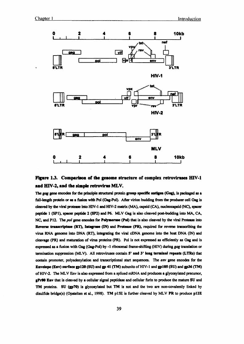
Chapter 1 Introduction
0 2 4 6 8 10kbnof
rov£ S f l L
JtfiL onv
5’LTR 3'LTR
HIV-1
tat,vpxcas •nv
RfiL 3’LTRvpr rov
HIV-2
1 flag 1 DOl 3*1 IRonv
MLV0 2 4 6 8 10kb 1 i I I__________I__________I__________I__________I
Figure U . Comparison of the genome structure of complex retroviruses HIV-1 and HTV-2, and the simple retrovirus MLV.The gag gene encodes for die principle structural protein group specific antigen (Gag), is packaged as a full-length protein or as a fusion with Pol (Gag-Pol). After virion budding from the producer cell Gag is
cleaved by the viral protease into HIV-1 and HIV-2 matrix (MA), capsid (CA), nucleocapsid (NC), spacer
peptide 1 (SP1), spacer peptide 2 (SP2) and P6. MLV Gag is also cleaved post-budding into MA. CA, NC, and P12. The pot gene encodes for Polymerase (Pol) that is also cleaved by the viral Protease into Reverse transcriptase (RT), Integrase (IN) and Protease (PR), required for reverse transcribing the virus RNA genome into DNA (RT), integrating die viral cDNA genome into the host DNA (IN) and cleavage (PR) and maturation of virus proteins (PR). Pol is not expressed as efficiently as Gag and is expressed as a fusion with Gag (Gag-Pol) by -1 ribosomal frame-shifting (HIV) during gag translation or termination suppression (MLV). All retroviruses contain 5* and 3* long terminal repeats (LTRs) that
contain promoter, polyadenylation and transcriptional start sequences. The env gene encodes for the Envelope (Env) surface gpl20 (SU) and gp 41 (TM) subunits of HIV-1 and gpl05 (SU) and gp36 (TM) of HIV-2. The MLV Env is also expressed from a spliced mRNA and produces a glycosylated precursor, gPr80 Env that is cleaved by a cellular signal peptidase and cellular furin to produce the mature SU and TM proteins. SU (gp70) is glycosylated but TM is not and the two are non-covalently linked by
disulfide bridge(s) (Opstelten et al., 1998). TM pl5E is further cleaved by MLV PR to produce pl2E
39

Chapter 1 Introduction
and p2E (the R peptide) (Green et al., 1981). This processing is required for fusion of the virion membrane and the host-cell plasma membrane, mediated by Env, during infection. Viral infecthity
fhctor (Vif), viral protein R (Vpr), viral protein X (Vpx), viral protein U (Vpn), negative inactivity
iK tor(N ef), transactivator of transcription (Tat), regulator of expression of viral proteins (Rev).
a
I A 3 Epideariology and classification of the primate lentivinises
The HIVs and SIVs are currently classified into six groups of primate lentivinises (table 1.1) (Barlow et al., 2003; Hahn et al., 2000), and additional groups will probably emerge as more full-length sequences are elucidated from different primate species (Peelers et al., 2002). There are at least 18 distinct lentivinises that naturally infect African primate species (Hahn et al., 2000) and over 40 different SIVs have been described from their natural or heterologous species (Apetrei et al., 2004).
Table 1.1: The dz groups of primate lentivinises.
Group Virus Host species
1 HIV-1 (Barre-Sinoussi et al., 1983) Homo sapiens
HumanSIVcpz (Peeters et al., 1992; Peeters et al., 1989) Pan troglodytes
Chimpanzee2 HTV-2(Clavel et al., 1986a), Homo sapien
HumanSIVsmm (Mutphey-Coib et al., 1986), Cercocebus atys
Sooty mangabey,SIVmac (Daniel et al., 1985) M acacaspp
Macaques3 SlVagm (Ohta et al., 1988) Cercopithecus spp
African green monkeys4 SIVmnd (Tsujimoto et al., 1988) Papio sphinx
Mandrill5 SIVsyk (Emau et al., 1991) Cercopithecus cdbogaris
Sykes monkey6 SIVcol (Courgnaud et al., 2001) Colobus Guerza
Colobus monkeys
40

Chapter 1 Introduction
HIV-1 consists of groups M (main), N (new), and O (outlier). Group M is responsible for the current pandemic and has been further subdivided into 11 clades, A through K. Subtype C predominates globally with 47% of new infections in the year 2000, clade A accounted for 30% (including AE and AG), circulating recombinant forms (CRFs) accounted for 18%, and clade B accounted for 12% of all new infections in 2000 (Julg and Goebel, 2005). Clade B is found in North America, South America, Europe and
Southeast Asia. Clade C predominates in sub Saharan Africa and Southeast Asia with BF, AE, BC, AG, and AB CRFs in South America, southeast Asia, China, Eastern
Europe and central Africa, respectively[http://www.iavireport.org/issues/0803/images/HrV-clademap.pdf, accessed October, 2005; (Julg and Goebel, 2005)].
HIV-2 is categorised into 7 subtypes, A through G. Subtypes A and B cause AIDS and have been isolated from individuals as infectious virus whereas only partial nucleotide sequences have been obtained for subtypes C through G (Gao et al., 1994). HIV-2 is found primarily in West Africa; subtype A is the most prevalent and found in The
Democratic Republic of the Congo, Mali, Gambia, Cape Verde Islands, Guinea Bissau, Ghana, Senegal, and Liberia whereas subtype B is found almost exclusively in the Ivory Coast and Ghana (Ellenberger et al., 1999; Ishikawa et al., 2001; Peeters et al., 2003; Pieniazek et al., 1999). In contrast to HTV-1, the HIV-2 epidemic is waning with a gradual decrease in the trend of HTV-2 seroprevalence (Bouckenooghe and Shandera,
1999).
1.8.4 The origins of HIV-1 and HTV-2
Geographical location and sequence data suggest that HIV-2 was derived from SIV of Sooty Mangabeys, STVsmm- The presence of HIV-2 was confirmed through molecular
characterisation (Clavel et al., 1986a), approximately one year after serological screening suggested it’s presence (Barin et al., 1985). HIV-2 and STVsmm share identical genome structure (Hirsch et al., 1989), they do not separate into distinct phylogenetic lineages based on their species of origin (table 1.1) (Hahn et al., 2000), and there is “geographic coincidence” between the natural habitat of the Sooty
41

Chapter 1 Introduction
Mangabey and where HIV-2 is endemic (Chen et al., 1996b; Hahn et al., 2000). Each of the seven subtypes of HIV-2 are as divergent in sequence as the SIVsmm sequences are from each other, in the same geographic location, which supports the hypothesis that
each HIV-2 subtype is the result of an independent zoonotic event of Sooty Mangabey to Human (Apetrei and Marx, 2004; Apetrei et al., 2004; Chen et al., 1997; Chen et al., 1996b; Gao et al., 1994; Gao et al., 1992; Hahn et al., 2000).
HTV-1 is similar to SIV from chimpanzees. However, the origin of HIV-1 was more difficult to elucidate than that of HIV-2 (table 1) (Hahn et al., 2000). At one point, there
were only two full-length sequences of STVcpz: STVcpz-gabi, SIV cpz-ant and a partial Pol sequence named SIVcpz-gab2 (Vanden Haesevelde et al., 1996), which did not provide a clear epidemiological STVcpz to HIV-1 link. A clearer association between HTV-1 and STVcpz was not made until additional SIVcpz sequences were obtained [(Bibollet-Ruche et al., 2004; Gao et al., 1999; Rambaut et al., 2001; Switzer et al., 2005; Worobey et al., 2004) reviewed in (Hahn et al., 2000)]. The phylogenetic
starburst of group M HIV-1 sequences and the interspersed STVcpz sequences between HTV-1 groups M, N and O suggest that these three HTV-1 groups are derived from three
independent zoonotic transfers of virus from chimpanzee to human (Bibollet-Ruche et al., 2004; Gao et al., 1999; Hahn et al., 2000; Rambaut et al., 2001; Worobey et al., 2004), so all of the circulating subtypes of group M were derived from one zoonotic
event. The circulating subtypes of HTV-2 however were each thought to have been derived from separate zoonotic events [reviewed in (Hahn et al., 2000)]. It is deduced that the current (December, 2004) 39 million HIV-1 group M infections worldwide are
the result of 1 zoonotic transfer of STVcpz to a human.
1.9 Cellular tropism and receptors o f H IV
The cell tropism of HTV is best explained in the context of receptor and, most importantly, coreceptor usage. Therefore the receptor and coreceptors of HTV will be
discussed before the cellular tropism of HIV.
42

Chapter 1 Introduction
1.9.1 The HTV receptor
It has been shown that CD4 is the primary receptor for HIV-1 (Clapham PR, 2001;
Clapham and McKnight, 2002; Dalgleish et al., 1984; Klatzmann et al., 1984). Anti- CD4 mabs and soluble recombinant CD4 (sCD4) can block infection of HIV-1, HTV-2
and SIV viruses (Clapham et al., 1989; Sattentau et al., 1988; Weiss et al., 1988), suggesting that CD4 is the main receptor for all of the primate lentivinises.
1.9.2 Coreceptors
It was observed two years after the discovery of the CD4 receptor that additional cofactors were required for HIV entry and infection. It was shown that HTV-1 could bind murine cells and non-permissive human cells expressing CD4, but could not fuse
and enter (Harrington and Geballe, 1993; Maddon et al., 1986). However, HeLa/non-
permissive cell fusions were permissive to infection suggesting the presence of an entry cofactor (Harrington and Geballe, 1993; Maddon et al., 1986). Fusin (CXCR4) was the
first identified coreceptor required for HIV infection of SI isolates (Feng et al., 1996), and discovery of the R5 coreceptor (Dragic et al., 1996), required for M 0 tropism
(Alkhatib et al., 1996), followed (table 1.2). Also, the discovery of dual coreceptor and
broad coreceptor using isolates helped to explain dual M 0 and T cell tropic isolates
(table 1.2) (Berson et al., 1996; Doranz et al., 1996).
All of the HTV coreceptors are members of die 7-TM spanning receptor superfamily.
These receptor proteins signal through the heterotrimeric G-proteins upon chemokine ligation.
HIV-2 has been shown to use a broader range of coreceptors than HTV-1 in vitro
(Guillon et al., 1998; McKnight et al., 1998; Momer et al., 1999; Momer et al., 2002; Owen et al., 1998), but this does not correlate with HTV-2 pathogenicity in vivo (Blaak et al., 2005; Momer et al., 1999; Momer et al., 2002; van Der Ende et al., 2000). A comprehensive list of HIV-1 and HIV-2 coreceptors is listed in table 1.2. The HIV coreceptors and associations with cell tropism are discussed in more detail in chapter 3.
43

Chapter 1 Introduction
Table 1.2: Coreceptors used by HIV-1, HTV-2, and SIV.
Coreceptor Virus Cell/tissue type ReferenceCCR1 HIV-2 Activated T cells,
mono, DC(Guillon et al., 1998; McKnight et al.,
* 1998)CCR2b HIV-1,
SIVHIV-2, Mono, T cells (Chen et al., 1998;
Doranz et al., 1996)CCR3 HIV-1, HIV-2 Eosinophils, (Choe et al., 1996;
microglia, Th2 Sol et al., 1997)CCR4 HIV-2 Th2, (McKnight et al.,
1998; Owen et al., 1998)
CCRS (R5) HIV-1, HIV-2, Activated T cells, (Deng et al., 1996;SIV mono, DC Sol et al., 1997)
CCR8 HIV-1,SIV
HIV-2, Th2, brain, thymocytes
(Rucker et al., 1997)
CCR9 HIV-1 Lymphocytes, brain, placenta
(Choe et al., 1998)
CXCR2 HIV-2 Neutrophils, brain (Bron et al., 1997)CXCR4 (X4) HIV-1, HIV-2, Lymphocytes, (Feng et al., 1996;
SIV, FIV memo, brain, Schols and Deprogenitor cells Clercq, 1998; Willett
etal., 1997)CXCR5 HIV-2 B cells (Kanbe et al., 1999)CX3CRI HIV-1, HIV-2 NK cells, CD8 T (Reeves et al., 1997;
cells, brain Rucker etal., 1997)GPR1 HIV-1,
SIVHIV-2, M0, brain (Farzan et al., 1997;
Shimizu et al., 1999)GPR15 (BOB) HIV-1,
SIVHIV-2, T cells, colon (Deng et al., 1997;
Farzan et al., 1997)STRL33 (Bonzo) HIV-1, HIV-2, T cells, mono, (Deng et al., 1997;
SIV placenta Liao etal., 1997)APJ HIV-1, HIV-2, CNS (Choe et al., 1998;
44

Chapter 1 Introduction
SIV Edinger et al., 1998)ChemR23 HIV-1, SIV M0, DC (Samson et al., 1998)RDC1 HIV-1, HIV-2, Lymphocytes, brain (Shimizu et al., 2000)
SIVLdikotriene B4 HIV-1 Leukocytes (Owman et al., 1998)receptor (BLTR)
U S 2 8 cmv HIV-1, HIV-2 CMV-infected cells (Pleskoff et al., 1997)D6 HIV-1, HTV-2 M0,PBMC, brain (Neil etal., 2005)
1.93 Attachment receptors
Depending on the route of transmission, time are several factors that affect virion-cell landing, such as extracellular fluid (Maher et al., 2005), and host cell surface attachment proteins and proteoglycans (Ugolini et al., 1999).
Abs directed against the cell-cell attachment protein, ICAM-3 have been shown to inhibit HIV infection (Sommerfelt and Asjo, 1995) and it was once thought that LFA-1 was required for HIV fusion because mabs to LFA-1 could block syncitium formation (cytopathic cell fusion effect of HIV in culture) (Hildreth and Orentas, 1989; Ugolini et al., 1999). Intercellular attachment proteins already present on the surface of producer cells may bud off with virions so incorporation of these cellular proteins on the virion may be a non-selective/random process (Tremblay et al., 1998). ICAMs on the surface of virions may aid virion-target cell attachment by interaction with the associated ICAM receptor and enhance infectivity (Fortin et al., 1997), and can even make the virus refractory to Ab neutralisation (Fortin et al., 2000).
Heparin sulphate proteoglycans (HSPG) are expressed to high levels on epithelial and endothelial cells attracting engagement of HTV virion with their net anionic charge (Ugolini et al., 1999), thus supporting virus absorption to the cell surface. The preferred binding of X4 viruses with HSPG is associated with the higher V3 loop charge compared to R5 viruses (HTV V3 loops are explained in greater detail in chapter 3) (Ohshiro et al., 1996; Ugolini et al., 1999).

Chapter 1 Introduction
The binding of virus by dendritic cells (DCs) at sights of virus exposure (mucosal tissue) may lead to unwitting presentation of infectious virus to target T cells in lymph nodes (van Kooyk and Geijtenbeek, 2003). The DC C-type lectin, specific intercellular adhesion molecule-grabbing nonintegrin (DC-SIGN) (Geijtenbeek et al., 2000), and the endothelial cell expressed DC-SIGNR (Pohlmann et al., 2001), have been shown to bind HIV (or any particles with high mannose sugars on glycoproteins) and promote
internalisation/protection of virus by DCs, until presentation via the immunological synapse to the unsuspecting T cell. The DC-SIGNs are internalisation receptors that internalise with bound ligand (van Kooyk and Geijtenbeek, 2003) and therefore promote internalisation and protection of virus by DCs, allowing virion to remain infectious for prolonged periods (van Kooyk and Geijtenbeek, 2003).
Galactosyl ceramide and its derivatives (sulphated) have been implicated as alternative receptors for HIV entry (Bhat et al., 1991; Harouse et al., 1991), although at much
lower efficiency than via preferred receptor and coreceptor. The bond between the galactose sugar residue and the ceramide-lipid backbone is required for binding to HTV gpl20 Env protein (Bhat et al., 1991). This glycolipid may be used by HTV to infect CD4( ) colon epithelial cells in the gastrointestinal tract (Yahi et al., 1992), and may be of particular importance because anti-CD4 Ab doesn’t inhibit HTV infection of human colon epithelial cells (Fantini et al., 1991). However given how inefficient these
processes are in vitro it is difficult to envisage a major in-vivo role.
1.9.4 Cells infected: transmission through to onset of AIDS
HIV preferentially infects haematopoietic cells that express CD4 and the relevant 7-TM
coreceptor(s), namely; DCs, M 0 and T cells (Clapham and McKnight, 2002). During
transmission of HIV, immune cells of the mucosae are infected and those cells that express the pertinent receptor and coreceptor are the targets of attachment and infection (Clapham and McKnight, 2002; de Roda Husman and Schuitemaker, 1998; Moore et
al., 2004). The most relevant cells infected, during transmission, in-vivo are the R5 expressing cells such as resident M0S, T cells and DCs (Clapham and McKnight, 2002; Davis and Dorns, 2004; de Roda Husman and Schuitemaker, 1998; Miller and Shattock,
2003; Moore et al., 2004; Schuitemaker, 1994; Wilflingseder et al., 2005).
46

C hapter 1 Introduction
A model of transmission that has gained acceptance is the one where DCs [e.g. Langerhans cells (LCs)] and M0s present in the mucosae become productively infected (via CD4 receptor and R5 coreceptor) or transport virus particles via C-type lectins (e.g.
DC-SIGN). These HIV containing ‘Trojan horses” then migrate to draining lymph nodes, producing/presenting HIV for subsequent infection of susceptible T cells (Clapham and McKnight, 2002; Davis and Dorns, 2004; Moore et al., 2004; Wilflingseder et al., 2005). Activated CD4+ T cells are present in ample numbers in inflamed mucosae during ulcerative genital diseases so these cells are also a target for HIV infection during transmission (Ho et al., 1995).
Follicular dendritic cells (FDCs) are not infected by HIV but enhance cell-free virus infection by promoting die upregulation of X4 on surrounding T cells In vitro (Estes et al., 2002; Taruishi et al., 2004). FDCs, present in the lymph nodes, may create a more suitable microenvironment for virus transmission in the host.
X4 strains emerge in 40-50% of subtype B infections (Connor et al., 1997), and X4 viruses in symptomatic individuals demonstrate a broader tropism for different T cell subsets (Clapham and McKnight, 2002). A nomenclature system has been derived for HIV viruses where those that use CCR5 as a coreceptor for entry are termed R5, those that use CXCR4 are termed X4 and those that can use both coreceptors equivalently for
entry are R5X4 viruses (Berger et al., 1998).
In T cells, R5 expression is found primarily on memory T cells whereas X4 is more widespread and is expressed to higher levels on naive T cells (Bleul et al., 1997). Accordingly, in the blood of individuals carrying R5 viruses the CD4+CD45RO+ memory T cells carry most of the proviral load, with a minority of CD4+CD45RA+ naive T cells infected, and when X4 viruses emerge a greater proportion of CD4+CD45RA+ naive T cells become infected (Blaak et al., 2000; Clapham and
McKnight, 2002; Ostrowski et al., 1999). Also, with regards to cells in general, X4 is more widely expressed than R5, which would lend a broader cell tropism to X4 using HIV viruses than R5 viruses (Moore et al., 2004). The infection of M0s by HIV and the role that these cells play in infection is discussed in more detail in chapter 3.
47

Chanter 1 Introduction
1.10 H IV Vaccines and microbicides
There is currently no prospect for an efficacious vaccine against HIV-1 infection (Desrosiers, 2004; Letvin, 2005). Nothing tried thus far has provided therapeutic assistance to help fight infection let alone sterilising immunity (Desrosiers, 2004; Letvin, 2005; McMichael and Hanke, 2003). Therefore there is more research than ever on topical microbicides and chemical prophylaxis.
1.10.1 Vaccines
The results of the first phase 3 HIV-1 vaccine trials were released in 2003. The Vaxgen gpl20 trials were considered a “failure” because they did not provide protection against
infection and did not lower viral loads (Desrosiers, 2004). It is becoming increasingly apparent that traditional vaccine approaches will not work to vaccinate against HIV and more novel strategies are starting to be adopted (Desrosiers, 2004; Letvin, 2005;
McMichael and Hanke, 2003). As of March 2004, there were 13 different products at various stages of clinical testing in more than 20 individual human trials (Desrosiers,
2004).
1.10.2 Microbicides
Microbicides are topical formulations designed to block HIV-1 infection when applied
to genital mucosal surfaces before sexual intercourse.
A surfactant called nonoxynol-9, known to non-specifically disrupt enveloped viral particles, was shown in clinical trials to destroy membranes of epithelial cells lining mucosal surfaces (Shattock and Moore, 2003). As a result, nonoxynol-9 did not protect against HIV infection but increased the chances of becoming infected (Shattock and
Moore, 2003). Therefore any potential microbicide must not reduce natural defences against HIV infection.
The most appealing microbicide candidates will specifically target the HIV virus, such as: antibodies, proteins and small molecule inhibitors that bind the viral Env glycoproteins gp 120 and gp 41 or glycan residues present on the surface of the viral
48

Chapter 1 Introduction
Env. These will ideally inactivate the virus before it has the chance to infect (Shattock and Moore, 2003).
Chemical approaches are also being pursued in light of promising pre-clinical results.
(R)-9-(2-phosphonylmethoxypropyl) adenine (PMPA) has proven effective at preventing SIV infection of macaques when delivered subcutaneously, once daily, as pre- and post-exposure prophylaxis (Tsai et al., 199S). An RT inhibitor, with few side effects, tenofovir disoproxl fumarate (TDF) is currently in clinical trials to determine its efficacy as preexposure prophylaxis (Grant et al., 2005).
1.11 The H IV infecHon cycle
‘The HTV infection cycle* section of this thesis describes the viral and cellular proteins when their action becomes relevant to the retroviral replication cycle. The pertinent viral and cellular proteins are discussed in detail in the subsection where they have been highlighted in bold. Therefore there is not a specific section in this thesis that lists the HTV gene products and their multitude of functions.
49
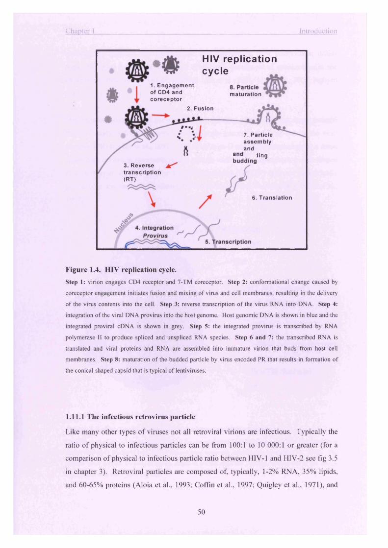
Chapter 1 Introduction
1. Engagement of CD4 and coreceptor
2. Fusion
3. Reverse ft* *transcription(RT)
HIV replication o!p cycle
1. Engagementmaturation
7. Particleassemblyandbudding
6. Translation
Figure 1.4. HIV replication cycle.
Step 1: virion engages CD4 receptor and 7-TM coreceptor. Step 2: conformational change caused by
coreceptor engagement initiates fusion and mixing of virus and cell membranes, resulting in the delivery
of the virus contents into the cell. Step 3: reverse transcription o f the virus RNA into DNA. Step 4:
integration o f the viral DNA provirus into the host genome. Host genomic DNA is shown in blue and the
integrated proviral cDNA is shown in grey. Step 5: the integrated provirus is transcribed by RNA
polymerase II to produce spliced and unspliced RNA species. Step 6 and 7: the transcribed RNA is
translated and viral proteins and RNA are assembled into immature virion that buds from host cell
membranes. Step 8: maturation of the budded particle by virus encoded PR that results in formation of
the conical shaped capsid that is typical o f lentiviruses.
1.11.1 The infectious retrovirus particle
Like many other types of viruses not all retroviral virions are infectious. Typically the
ratio of physical to infectious particles can be from 100:1 to 10 000:1 or greater (for a
comparison of physical to infectious particle ratio between HIV-1 and HIV-2 see fig 3.5
in chapter 3). Retroviral particles are composed of, typically, 1-2% RNA, 35% lipids,
and 60-65% proteins (Aloia et al., 1993; Coffin et al., 1997; Quigley et al., 1971), and
50

Chapter 1
although the particles are derived from host membranes their composition differs
somewhat from the plasma membrane (PM), with enrichment of sphingomyelin and
cholesterol (lipid rafts) (Aloia et al., 1993; Liao et al., 2001; Liao et al., 2003; Nguyen
et al., 2003).
Mature infectious retroviral particles have a condensed core of electron dense material
that is thought to form as a result of proteolytic processing of Gag-Pol by the virus
protease (PR) (Coffin et al., 1997). The MLV type-C retrovirus morphology is a dense
round core centred in the middle of the particle while Lentiviruses have a cone shaped
core (Coffin et al., 1997). The type-B retroviruses (MMTV) have an eccentrically
placed core and the type-D viruses (M-PMV) have a rod shaped one (Coffin et al.,
1997).
Env SU (blue)TM (fuchsia) Lipid bilayer
p17,MA (yellow)p9, NC (green)p11, PR~1 p63, RT r grey p32, IN J
RNA genome (brown)
p24, CA (orange)Env TM (fuchsia)
Vpr
Figure 1.5. Composition of the HIV-1 virion (authors rendition).
Conformation of V1-V3 loops within trimeric Env was obtained from (Chen et al., 2005). The interaction
o f Env TM gp41 (fuchsia) with MA is derived from molecular modelling in (Forster et al., 2000). The
dimeric RNA genome (brown) is shown coated in nucleocapsid (NC, green), spilling out o f the orange
capsid core, with the 5’ ends exposed.
51

Chapter 1 Introduction
1.11.2 Receptor engagement
Initial HIV-receptor interaction during successful virus entry occurs when CD4 binds the CD4 binding site of the Env gpl20 (SU) subunit (figs 1.4 and 1.5). CD4 receptor binding induces a conformational change in Env that allows a positively charged region near the bridging sheet that lies at the base of the V1V2 and V3 loops, to interact with the coreceptor (Doms, 2000; Rizzuto and Sodroski, 2000; Rizzuto et al., 1998). Successful coreceptor binding results in conformational rearrangement of gp41 (TM)
(fig 1.5), and fusion (fig 1.4).
1.11.3 Fusion
The fusion process is mediated by the gp41 subunit of Env. The purpose of fusion is to
1, force a fusion pore that makes the viral Env continuous with the host cell membrane and 2, allow mixing of the two membranes to enable delivery of all of the virions contents into the cell (Doms, 2000). At least six R5 molecules are required for the successful formation of a fusion pore. This number is based on a number of scientific
assumptions but is compatible with what is known about influenza virus hemagglutinin (Doms, 2000; Ellens et al., 1990). However, a more recent study on HIV-1 Env trimers
themselves has suggested that just one Env trimer on the virion is required to effect virus-host fusion (Yang et al., 2005).
1.11.4 Fusion mechanism
1, Prior to fusion each gp41 molecule is in a high energy, tightly packed state with the N-terminal fusion peptide folded back towards the viral membrane. 2, Attachment of gpl20 to the CD4 receptor exposes a coreceptor-binding region on gpl20. 3, Coreceptor engagement causes a conformational change and release of gp41 from its high-energy state. 4, The fusion peptide extends out towards, and inserts into, the target cell membrane. 5, The HR1 and HR2 regions of gp41 are in consecutive alignment at
this stage and they then Told in' to meet each other, which results in the formation of the six-helix bundle. The purpose of the formation of the six-helix bundle is to bring
52

Chapter 1 Introduction
the host cell and virus membranes into 6, close apposition to promote virus-cell membrane fusion and formation of the fusion pore.
1.11.5 Entry
The term ‘entry’ is used here to describe the topographical region of the cell where
fusion takes place. This is especially relevant to chapter 4 of this thesis, which deals with endocytosis of virus particles and the resulting restriction of HIV-1 and HIV-2.
Several endocytic pathways have been implicated in the productive entry of viruses into the host cell (Marsh and Pelchen-Matthews, 2000; Sieczkarski and Whittaker, 2002a).
One of the first viruses shown to utilise the clathrin endocytic route was Semliki forest virus (Helenius et al., 1980), also demonstrated to require low pH to initiate fusion (Helenius et al., 1982; Marsh et al., 1982). The SV40 polyoma virus uses caveolae and the more recently described non-clathrin non-caveolae route of entry. Amphotropic
MLV (MLV-A) has recently been shown to prefer entry via caveolae endocytosis as well (Beer et al., 2005). Influenza virus can enter by both clathrin and the non-clathrin
pathways but has a specific requirement for entry via a specific Rab 5 early endosome. Semliki forest and influenza viruses are pH-dependent and require entry via endocytosis but SV40 and MLV-A are pH-independent viruses so despite no dependence on delivery into an acidified vesicle to trigger fusion, these pH-independent viruses prefer endocytic entry. HIV is also a pH-independent virus that may, in some circumstances, use endocytosis for productive entry (Daecke et al., 2005; McClure et
al., 1988).
The site of fusion for a pH-independent retrovirus can be the inside of an endosome or the cell surface given that there is a sufficient level of receptor/coreceptor at both sites. A virus can bind the cell surface and be ‘taken up’ by the cell coincident to a ligand triggered or constitutive endocytic process. It is also conceivable that a cell-free virus
could be taken up with the fluid phase by macropinocytosis as has been postulated as a route of productive entry for HIV in M0s and microvascular endothelial cells (binding and fusion would then occur within the macropinocytic vesicle) (Liu et al., 2002;
Marechal, 2001).
53

Chapter 1 Introduction
The various endocytic pathways used by HTV, and other viruses, to enter cells is discussed in more detail in chapter 4.
1.11.6 Early entry and reverse transcription events
The viral CA enters the cell and uncoats after fusion takes place. The contents of the capsid empty into the cell: reverse transcription complex (RTC) which includes Integrase (IN), reverse transcriptase (RT), viral protein R (Vpr), and matrix (MA),
not to mention the two copies of the RNA genome (Bukrinsky et al., 1993b; Peterlin
and Trono, 2003). Also released from the virion are negative infectivity factor (Nef), P6, Vpr, and viral infectivity factor (Vif), and the capsid (CA) protein of the now
uncoated capsid (fig 1.4) [reviewed in (Cullen, 2001; Peterlin and Trono, 2003)].
The RT starts to reverse transcribe the RNA genome into a double-stranded complementary viral DNA for integration into the host genome, after cell entry but before nuclear translocation (Peterlin and Trono, 2003). For this to begin, the cortical actin cytoskeleton has been implicated as a scaffold for the formation of the RTC and
initiation of reverse transcription (Bukrinskaya et al., 1998). Initial strong stop DNA has been reported to be reverse transcribed in the virion itself, before cell entry (Zhang et al., 1995), but the majority of viral DNA synthesis is thought to occur in the cytoplasm of the host cell (Bukrinsky et al., 1993b; Famet and Haseltine, 1991; Karageorgos et al., 1993; Kim et al., 1989).
It has been shown that HTV uses the microtubule network for transport during entry (McDonald et al., 2002). More specifically, it has been suggested that a minus-end
directed microtubule motor, dynein, might be used by HIV to shuttle the RTC towards the MTOC, which is adjacent to the nucleus (McDonald et al., 2002). This data on HIV is concurrent with what is known about the transport of adenoviruses and herpesviruses because Ad-2 and HSV-1 have been shown to use dynein for transport along
microtubules to the nucleus (Dohner et al., 2002; Martin et al., 2002a; Sodeik, 2000; Sodeik et al., 1997; Suomalainen et al., 1999; Yedowitz et al., 2005).
Nef is packaged into the virion (Kotov et al., 1999; Welker et al., 1996), and this form of Nef has been shown to boost infectivity 10-fold over ANef virions (Kotov et al.,
54

Chapter 1 Introduction
1999). It has been postulated that since an endocytosis directing VSV Env relieves HTV from its Nef dependence (Aiken, 1997), with an HTV Env, Nef may help direct the RTC to an endocytic location that is more conducive to reverse transcription and productive entry (Kotov et al., 1999).
1.11.7 Reverse transcription
Retroviral reverse transcription is highly error prone allowing a high rate of mutation and adaptation. The presence of two RNA molecules in the virion allows for crossover
events during reverse transcription and can be especially advantageous if a virion is produced with two heterologous RNA strands from a superinfected cell. The RT enzyme is highly recombinogenic helping to facilitate this action. There is no proof reading mechanism in the RT enzyme or in the host RNA polymerase II that transcribes the integrated retroviral genome, resulting in an estimated 1 nucleotide mutation per 104 nucleotides per round of replication. The onward transmission of recombinant HIV viruses with mixed genomes from different clades is referred to as CRFs.
The Vif protein acts indirectly at this stage by preventing the packaging of apolipoprotein B mRNA-editing enzyme catalytic polypeptide (APOBEC) 3G, F and B into the viral particle at the stage of assembly (see also virus budding section) (Kao et al., 2003; Mariani et al., 2003; Marin et al., 2003; Mehle et al., 2004; Navarro and Landau, 2004; Sheehy et al., 2002; Sheehy et al., 2003). APOBECs 3G, F and B packaged with the virus particle cause cytosine to uracil deamination of the negative sense viral DNA during reverse transcription (Bishop et al., 2004a; Bishop et al., 2004b; Harris and Liddament, 2004; Mangeat et al., 2003; Newman et al., 2005;
Sheehy et al., 2002; Stopak et al., 2003; Zheng et al., 2004). APOBECs packaged into the virion of A vif viruses cause cytosine to uracil deamination resulting in nonsense mutation of the viral genome, and abortive replication (Harris and Liddament, 2004; Navarro and Landau, 2004).
RT reverse transcribes the packaged viral RNA into cDNA for integration into the host genome that is illustrated in fig 1.6.
55
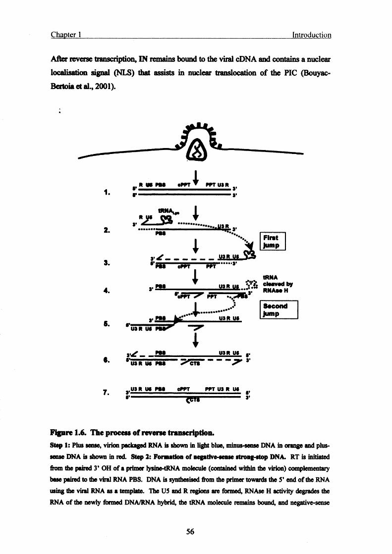
Chapter 1 Introduction
After reverse transcription, IN remains bound to the viral cDNA and contains a nuclear
localisation signal (NLS) that assists in nuclear translocation of the PIC (Bouyac-
Bertoia et al., 2001).
&1.
2.
3.
4.
S.
R US P M cPPT
tRNA*./z j a ..
v 4?--------8’ p m — Sp F T
£SL
P M
US R US pS P "
PPT USR
msjiL &
FirstJump
PPT
tRNAU 1 « U «
PPT - v
JIL RNAaaH
U SR US
8scondJump
s / - sUSR US
U SR US P M CTS
7 r U SR US P M CPPT PPT US R US
•----------------------JCTJ--------------- *’
Figure 1.6. The process of reverse transcription.Step I: Phis sense, virion packaged RNA is shown in light blue, minus-sense DNA in orange and plus- sense DNA is shown in red. Step 2: Formation of negative-sense strong-stop DNA. RT is initiated from the paired 3' OH of a primer lysine-tRNA molecule (contained within the virion) complementary base paired to the viral RNA PBS. DNA is synthesised from the primer towards the 5’ end of the RNA using the viral RNA as a template. The US and R regions are formed, RNAse H activity degrades the RNA of the newly formed DNA/RNA hybrid, the tRNA molecule remains bound, and negative-sense
56
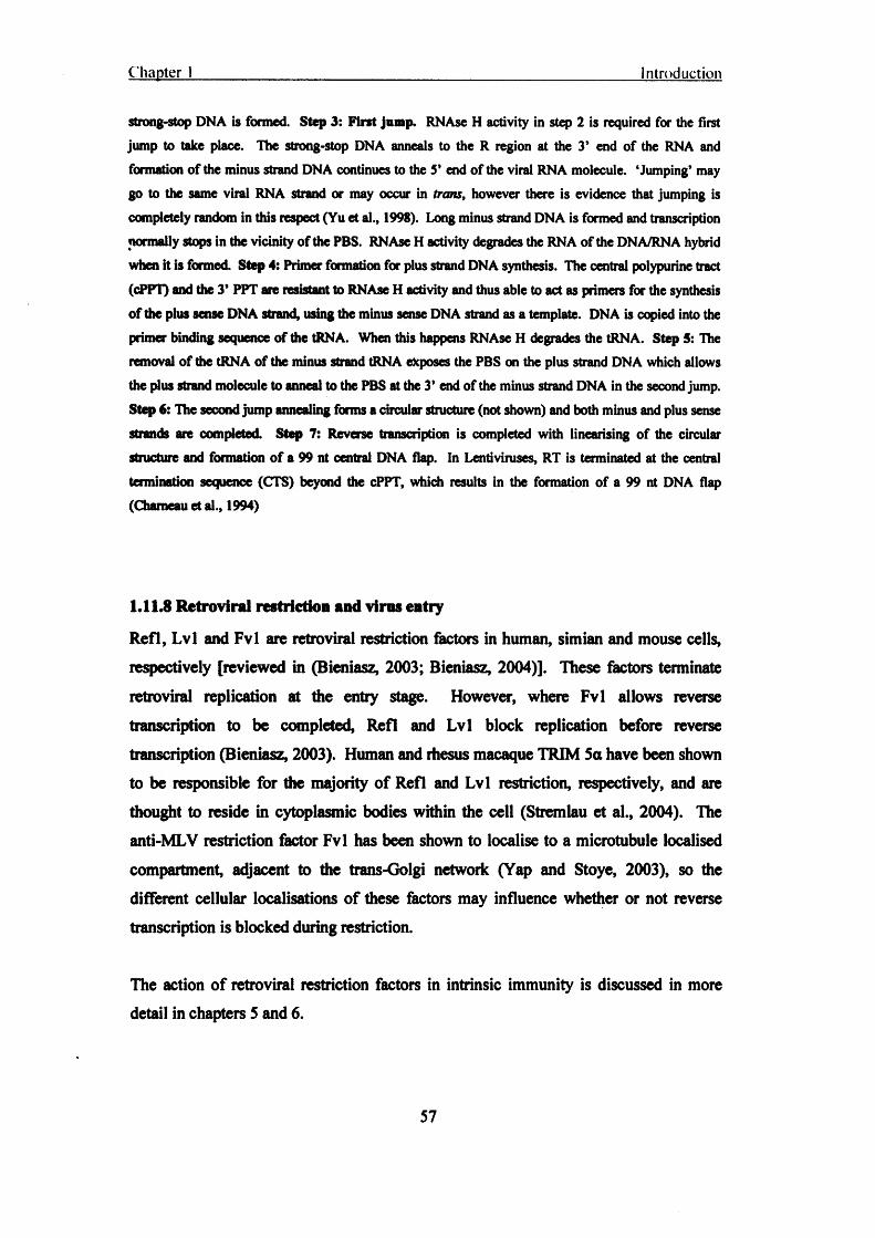
Chapter 1 Introduction
strong-stop DNA is formed. Step 3: First jump. RNAse H activity in step 2 is required for the first jump to take place. The strong-stop DNA anneals to the R region at the 3* end of the RNA and formation of the minus strand DNA continues to the 5’ end of the viral RNA molecule. ‘Jumping’ may go to the same viral RNA strand or may occur in trans, however there is evidence that jumping is completely random in this respect (Yu et al., 1998). Long minus strand DNA is formed and transcription normally stops in the vicinity of the PBS. RNAse H activity degrades the RNA of the DNA/RNA hybrid when it is formed. Step 4: Primer formation for plus strand DNA synthesis. The central polypurine tract
(cPPT) and the 3’ PPT are resistant to RNAse H activity and thus able to act as primers for the synthesis of the plus sense DNA strand, using the minus sense DNA strand as a template. DNA is copied into the primer binding sequence of the tRNA. When this happens RNAse H degrades the tRNA. Step 5: The removal of the tRNA of the minus strand tRNA exposes the PBS on the plus strand DNA which allows the plus strand molecule to anneal to the PBS at the 3’ end of the minus strand DNA in the second jump. Step 6: The second jump annealing forms a circular structure (not shown) and both minus and plus sense strands are completed. Step 7: Reverse transcription is completed with linearising of the circular structure and formation of a 99 nt central DNA flap. In Lentiviruses, RT is terminated at the central
termination sequence (CTS) beyond the cPPT, which results in the formation of a 99 nt DNA flap (Chameau et al., 1994)
1.11.8 Retroviral restriction and virus entry
Refl, Lvl and Fvl are retroviral restriction factors in human, simian and mouse cells,
respectively [reviewed in (Bieniasz, 2003; Bieniasz, 2004)]. These factors terminate
retroviral replication at the entry stage. However, where Fvl allows reverse transcription to be completed, Refl and Lvl block replication before reverse transcription (Bieniasz, 2003). Human and rhesus macaque TRIM 5a have been shown to be responsible for the majority of Refl and Lvl restriction, respectively, and are thought to reside in cytoplasmic bodies within the cell (Stremlau et al., 2004). The anti-MLV restriction factor Fvl has been shown to localise to a microtubule localised compartment, adjacent to the trans-Golgi network (Yap and Stoye, 2003), so the
different cellular localisations of these factors may influence whether or not reverse transcription is blocked during restriction.
The action of retroviral restriction factors in intrinsic immunity is discussed in more detail in chapters 5 and 6.
57

Chapter 1 Introduction
1.12 Into the nucleus...
1.12.1 Nuclear import of the pre-integration complex
At the end of reverse transcription there is an apparent change in the protein composition of the RTC. The sub-viral unit, at the completion of reverse transcription, is referred to as the pre-integration complex (PIC), and this may shed the RT and MA
that were associated with the RTC and remain associated only to IN (Famet and Haseltine, 1991; Karageorgos et al., 1993). So it has been argued that the PIC may only require the NLS of IN for translocation across the nuclear envelope in nondividing cells (Famet and Haseltine, 1991; Karageorgos et al., 1993). The size and the proteins that associate with the HTV PIC are, however, controversial as will be discussed later.
As entry progresses, smaller RTC species appear and the smallest species localise to the nucleus at late post-entry time points, indicated by decreasing sedimentation (‘S’) values (Fassati and Goff, 2001; Karageorgos et al., 1993), this alteration in size may assist the PIC to traverse the small 25 nm pore size of the NPC. Reports of the PIC nucleoprotein complex size vary from 56 nm (Sherman and Greene, 2002), to sizes of 400 to 700 nm (McDonald et al., 2002) and 200nm (Nermut and Fassati, 2003). Nevertheless, the PIC is complex and probably requires the action of several NLSs to effect nuclear entry (Bukrinsky and Haffar, 1999) including those of cellular proteins that have been recruited to die viral PIC (Maertens et al., 2004). In addition, nuclear
import/export is a redundant system and PIC nuclear translocation may involve numerous nuclear import pathways (Jenkins et al., 1998; Kuersten et al., 2001).
Vpr is packaged into virus particles through its interaction with the C-terminal p6 domain of the p55 Gag polyprotein (fig 1.5 and 1.11), at roughly 14 molecules per virion (Sherman and Greene, 2002). Vpr has been shown to induce “disruptions” in the nuclear envelope, which may facilitate trafficking of the 56-700 nm PIC across the nuclear membrane where nuclear pores are normally only 25nm across (de Noronha et
al., 2001). Vpr may also utilise its two novel NLSs for PIC nuclear import, one of which results in the direct association of Vpr with the nuclear pore complex (NPC) (Jenkins et al., 1998).
58

Chapter 1 Introduction
Beside its role for providing structure to the virus particle, MA, like Vpr, displays nucleocytoplasmic shuttling and is thought to contribute to the nuclear localisation of the PIC (Jenkins et al., 1998). MA is a part of the PIC and contains a classic NLS that
is thought to contribute to nuclear localisation (Jenkins et al., 1998; Peterlin and Trono,2003). It has been shown that lentiviral nuclear localisation in non-dividing cells requires the NLS of MA (Bukrinsky et al., 1993a; Bukrinsky et al., 1993b; Dubrovsky et al., 1995), however there are other reports that claim MA is not required for PIC nuclear translocation (Famet and Haseltine, 1991; Fouchier et al., 1997).
A 99 nt DNA flap, formed by strand displacement at the cPPT just before RT termination at the CTS (fig 1.6; step 7) (Zennou et al., 2000), has been shown to enhance nuclear localisation of lentiviral vectors (Dardalhon et al., 2001; Sirven et al.,2000). Later studies have shown that the 99 nt flap may be repositioned at a wide range of locations within the viral cDNA for enhancement of nuclear localisation (De Rijck et
al., 2005).
Viral protein x (Vpx) is an accessory gene found in viruses of the HIV- 2/SIVsmm/SIVmac group of primate lentiviruses, but not HIV-1 (fig 1.3). Vpx is proposed to have arisen as the result of a gene duplication of Vpr since the two genes
share sequence homology (Tristem et al., 1990). The gene redundancy of Vpx and Vpr does not equate to a functional redundancy since the roles of Vpr in HIV-1 have been split-up in HIV-2 Vpx and Vpr (Fletcher et al., 1996): the PIC nuclear localisation and G2-M cell cycle arrest activities of HIV-1 Vpr are delegated to HIV-2 Vpx and Vpr,
respectively (Fletcher et al., 1996), and the Vpx NLS has been shown to be necessary and sufficient for nuclear localisation of the HIV-2 PIC into die nucleus in non-dividing
cells (Belshan and Ratner, 2003).
1.12.2 Cellular genes involved in HTV nuclear import
The importins mediate the majority of traffic into and out of the nucleus by interacting with the RanGTP/GDP system which is governed by the RanGTP/GDP concentration gradient between the nucleus and cytoplasm; low levels of RanGTP in the cytoplasm allow the binding of importins to their cargoes, whereas high RanGTP levels in the
59
Chapter 1 Introduction
nucleus induce their dissociation [reviewed in (Gorlich and Kutay, 1999)]. It is therefore conceivable that one of these proteins may mediate the nuclear translocation of HIV-1 PICs. Indeed, Importin 7, a member of the importin p superfamily, has been implicated in the nuclear import of HIV-1 PICs into the nucleus (Fassati et al., 2003).
Lens epidermal growth factor (LEDGF) is a cellular protein that has been shown to
associate with and promote the nuclear localisation of the PIC (Maertens et al., 2004), and chromosome tethering of IN (Emiliani et al., 2005).
1.123 Integration
Integration is the process whereby the viral cDNA genome is inserted into the host DNA genome. IN remains associated with the PIC after translocation into the nucleus and performs the integration reaction.
Following reverse transcription, the viral cDNA is “primed” for integration in the cytoplasm by IN mediated 3’ trimming of the DNA ends, termed *3* processing* (fig
1.7) [reviewed in (Pommier et al., 2005)]. Trimming is a 3* endonucleolytic cleavage of 2 bases exactly 3* of a CA motif that creates 3* CA ends on the viral DNA (Pommier et al., 2005). The 3* CA-OH ends are 3* reactive intermediates required for strand transfer to occur (Pommier et al., 2005).
Following entry into the nucleus, IN catalyses the insertion of the viral DNA into a host
chromosome. IN remains bound to the viral cDNA as a multimeric complex bridging the viral DNA ends. The 3’-OH ends of the PIC are joined to the 5* ends of a host
chromosome, catalysed by IN (fig 1.7). This joining event leads to a canonical five base stagger. Completion of integration requires trimming of two nucleotides from the 5* proviral ends and gap filling from the 3’-OH chromosomal DNA ends (fig 1.7). It is likely that cellular enzymes are involved in 5* processing however their identity
remains uncertain (Daniel et al., 2004; Yoder and Bushman, 2000).
Cellular proteins have been shown to have IN binding and catalysis of integration activities (Pommier et al., 2005). Inil (integrase interactor protein 1) was the first discovered cellular IN binding protein that promotes integration (Turelli et al., 2001;
60

Chapter 1 Introduction
Turlure et al., 2004). Since then, heat shock protein 60 (Hsp60) has been shown to bind IN and stimulate in vitro processing and end-joining (Parissi et al., 2001), LEDGF is an IN to chromatin tethering factor that is not absolutely necessary for integration but may play a chromosomal DNA targeting role (Cherepanov et al., 2003; Llano et al., 2004), embryonic ectoderm development protein (EED) has been suggested to help traffic or “convoy” the PIC to the nucleus, during entry (Violot et al., 2003), and radiation sensitivity protein 18 (Rad 18) may be involved in the post-integration “translesion repair” that must take place to seal the gap between the viral 5* and chromosomal 3* DNA ends (fig 1.7c) [(Mulder et al., 2002) reviewed in (Turlure et al.,2004).
Cellular proteins barrier to auto-integration factor (BAF) (Lee and Craigie, 1998;
Lin and Engelman, 2003), and high mobility group protein Al (HMGA1) bind DNA directly and regulate integration of HIV and MLV [(Li et al., 1998; Li et al., 2000) reviewed in Pommier et al, 2005]. HMGA1 stimulates IN activity either by condensation of reactant DNA or the binding of HMGA1 monomers to multiple DNA sites may fold reactant DNA into conformations that favour IN catalysis and assembly (Famet and Bushman, 1997; Li et al., 1998; Li et al., 2000). BAF stimulates
intermolecular integration by binding the double stranded DNA of the PIC (rather than intramolecular integration resulting in 2 LTR circles) (Harris and Engelman, 2000; Lee
and Craigie, 1998).
PML (TRIM 19) has been shown to associate with HIV-1 RTCs in the target cell (in the cytoplasm) and interfere with integration (Turelli et al., 2001).
61

Chapter Introduction
B-diketoacids
3’
5’
Figure 1.7. HIV integration.
(A) 3’ processing: viral cDNA genome is shown in black and the host genomic DNA is grey, 3’ ends are
denoted by blue circles and the 5’ ends are denoted by red circles. IN catalyses an endonucleolytic
cleavage o f two bases from the 3’ CA ends. HIV-2 is unique among the lentiviruses, and indeed the
Retroviridae, in that an asymmetric 3 bases are removed from the U5 LTR and 2 bases from the U3 LTR
(Whitcomb and Hughes, 1991). (B) Strand transfer is catalysed by IN mediated ligation o f the viral 3 ’-
OH ends (created in the 3’ processing reaction) to 5’ phosphate groups o f the host DNA. (C) This
joining event leads to a canonical five base stagger. Completion of integration by gap filling from the 3 ’-
OH chromosomal DNA ends (Pommier et al., 2005).
1.12.4 Integration site selection
In vitro integration systems have shown that IN prefers to integrate the viral genome
into severely kinked DNA regions within the nucleosome (Pruss et al., 1994a). The
curvature induced by the nucleosome may widen the DNA major groove and thus
promote the interaction of host DNA with IN (Pruss et al., 1994b). Human genome-
wide screens have shown that HIV-1 integration favours active genes (Schroder et al.,
2002; Wu et al., 2003) whereas MLV favours integration in transcription start regions
(Wu et al., 2003).
62

Chapter 1 Introduction
1.12.5 HTV transcription
Once integrated in the host genome, the provirus behaves like a human gene with
transcription initiated at the 5* end and terminating at the 3* end (Jones and Peterlin,1994).
Both HIV-1 and HTV-2 have very similar binding sites and mechanisms of LTR driven transcription. Their LTRs share 40% sequence identity (Guyader et al., 1987) and their promoter binding sites share 50% sequence identity (Tong-Starksen et al., 1990). However the HTV-1 LTR responds better to T cell activation signals than HIV-2 (Hannibal et al., 1993); the HTV-2 LTR contains only one NF-kB binding site whereas HIV-1 has two (Tong-Starksen et al., 1990), and this has been proposed to be a factor contributing to the longer latency of HTV-2 (Hannibal et al., 1993; Tong-Starksen et al., 1990). NF-kB is activated in the cytoplasm by proteolytic cleavage of inhibitor of kB (IkB) during T cell activation leading to localisation to the nucleus, LTR binding and
up-regulation.
The majority of the HTV promoter is contained within the U3 region, with reported SP1 transcription factor binding sites in the HTV-1 leader region (Logan et al., 2004). The LTR contains enhancer and promoter regions with binding sites for several transcription factors and a polyadenylation signal (fig 1.8) (Marciniak et al., 1990; Southgate and Green, 1991). Moving upstream from the transcription start site, the initiator (Inr), the TATA box, and 3 SP-1 sites are found (Berkhout and Jeang, 1992). These sites position RNA polymerase II at the correct site for initiating transcription (Berkhout and Jeang, 1992). The NF-AT and NF-kB transcription factor binding sites allow maximum levels of viral transcription in activated T cells and M0s (fig 1.8) (Griffin et al., 1989; Griffin et al., 1991; Jones and Peterlin, 1994).
63
Chapter 1 Introduction
TAR
* W NF-AT IMF lto-1 S l l - twBk>1 SIM |P 3 | LM OATA4 N F-K B- ^ TATAI I 1 I T T ! I 1 I II ■ ! I I
E 4 u A PJC (NF-AT)NF-kB AF-1 AF-1 AP-1 HMO DBF1 (CT)*U . I I I I I . I L_
U3 US 8 0
Transcription initiation s its
+
L sadsr region
♦1 ♦100 ♦200 ♦300
Figure 1.8. Promoter and primer binding sites and key RNA structural features of the HIV-1 LTRKey promoter binding sites are shown in addition to the polyadenylation site (pA), the position of the
RNA TAR loop (TAR), transcription initiator (Inr), primer binding site (PBS), dimer initiation site (DIS), and die y packaging signal. SD “ gag splice donor site. The LTR is not drawn to scale (Logan et al.,2004).
Early events of HIV transcription involve transactivator of transcription (Tat) and regulator of expression of viral proteins (Rev). HIV remains in a transcriptionally latent state after integration with only the appearance of short TAR transcripts in the nucleus (Adams et al., 1994; Feinberg et al., 1991). Cellular activation signals improve the rate of transcription and full-length, fully spliced 2 kb mRNA transcripts appear (fig 1.10) (Jones and Peterlin, 1994). These 2 kb mRNA species encode for Tat, Rev and Nef (fig 1.10) (Kim et al., 1989). Tat and Rev both contain NLSs so after translation they are transported back into the nucleus and dramatically improve the processivity and fidelity of HIV transcription (Malim et al., 1989a; Perkins et al., 1989; Ruben et al., 1989). Tat then interacts with TAR and greatly improves the quantity of
polyadenylated genome length transcripts in die nucleus (fig 1.9)(Adams et al., 1994;
Feinberg et al., 1991).
1.12.6 Mechanism of Tat transactivation
The TAR element, which forms an RNA stem loop, is the target RNA sequence for Tat transactivation (Feng and Holland, 1988; Muesing et al., 1987; Peterlin et al., 1986). Tat in conjunction with positive transcription elongation factor B (p-TEFb), binds TAR with high affinity that allows elongation of viral RNA transcripts through activation of
64

Chapter 1 Introduction
RNA polymerase II (fig 1.9a) (Kao et al., 1987; Wei et al., 1998). The p-TEFb
complex consists of cyclin T1 and CDK9 (fig 1.9b) (Wei et al., 1998), and once
recruited to the nascent RNA complex, CDK9 phosphorylates the C-terminal domain of
RNA polymerase II and negative transcription elongation factor (N-TEF) (Yamaguchi
et al., 1999), leading to high fidelity and processive transcription (fig 1.9c) (Peterlin and
Trono, 2003; Price, 2000).
Figure 1.9. HIV transcription: the initiation complex.
(A) The RNA polymerase II holoenzyme complex (RNA Pol II) binds to upstream basal transcription
promoters in U3 o f the LTR (fig 1.8) and, without Tat, transcription will begin but elongation is
inefficient. The nascent TAR RNA folds into the TAR stem loop structure and recruits cyclin T1 and
Tat. (B) Tat binds the 5’ bulge and Cyclin T1 binds the central loop of TAR. CDK9 o f the p-TEFb
complex phosphorylates the C-terminal domain (CTD) of RNA Pol II (C), which results in the
displacement of TAR from the polymerase, and efficient transcription/elongation of the viral genome by
RNA Pol II (Jones and Peterlin, 1994; Kam, 1999).
A
Tat
Initiation
RNA Pol II holoenzyme complex
CDK9
Elongation
p-TEFb
65

Chapter 1 Introduction
Tat functions poorly in mouse cells, however mouse human chromosome 12 somatic hybrids have been shown to recover Tat transactivation (Hart et al., 1989; Hart et al., 199S; Newstein et al., 1990). An 83 kDa protein expressed from human chromosome |2 was identified as human Cyclin Tl, the cofactor required for efficient Tat transactivation (Hart et al., 199S). A Cyclin Tl homologue is encoded by mice but it does not support Tat and TAR interactions like its human homologue (Bieniasz et al., 1998; Wei etal., 1998).
1.12.7 Viral RNA species
HIV RNA species are capped at their 5’ end and polyadenylated at their 3* end much like cellular mRNA species (Bohnlein et al., 1989; Chiu et al., 2001; Chiu et al., 2002; Das et al., 1999; Zhou et al., 2003). The genome length viral RNA contains approximately 9 different primary splice sites that produce more than 30 different RNA species [(Bohne et al., 2005) reviewed in (Coffin et al., 1997)], which can be detected in the cell. Most of these species are three sizes; 2 kb, 4 kb and 9 kb species (fig 1.10):
the 2 kb fully spliced class encode for Tat, Rev, and Nef (Robert-Guroff et al., 1990; Schwartz et al., 1990), die 4 kb species encode for Vpu, Vif Vpr, and Env (Arrigo et al., 1990). The complete and unspliced species provide the RNA coding sequence for Gag and Pol expression (fig 1.10) (Arrigo et al., 1990; Kim et al., 1989; Neumann et
al., 1994; Schwartz et al., 1990).
\
66

Chapter 1
neftatTARrev
v|fl
env
3’LTR5’LTR
I I I Vi ral RNA, G ag , Pol
9 k b • ---------------------------------------^ ------------------------------------------ a a a a
Env, V if, V pu , V p r
4 k b • ^ ^ . . . ^ --------------------- a a a a
Tat, Rev, N ef2 k b » . •. / ------------------a a a a
HIV-1
Figure 1.10. HIV-1 messenger RNA expression.
The three mRNA species present during active infection are shown in red. TAR is present in all o f the
species (solid red dot) and the Rev response element (RRE) is shown as a red diamond in the 4 and 9 kb
RNAs. The splice donor sites are the arrows pointing down and the splice acceptor sites are the arrows
pointing up. The 2 kb ‘early’ transcripts produced first do not require the action o f Rev and the RRE
because they are fully spliced. The full-length and partially spliced late transcripts are produced by the
assistance of Tat and Rev. Positions are approximate. Derived from (Arrigo et al., 1990; Dalgleish and
Weiss, 1999).
Sites of HIV gene transcription repression have been mapped and one study has
revealed that repression of HIV transcription is associated with gene deserts,
centromeric heterochromatin and very highly expressed cellular genes (Lewinski et al.,
2005). Even though HIV integration has been shown to prefer transcriptional units
integration into very highly expressed genes can silence provirus transcription
[(Lewinski et al., 2005; Schroder et al., 2002; Wu et al., 2003) reviewed in (Bushman et
al., 2005)]. Read-through transcription of cellular genes into provirus can dislodge HIV
transcription factors, or “train wrecking”, the colliding of converging RNA polymerase
complexes (Lewinski et al., 2005). Gene deserts however may be enriched with
67

Chapter 1 Introduction
binding sites for transcriptional silencing proteins (Lewinski et al., 2005), and centromeric heterochromatin adopt a condensed structure that inhibits access to transcriptional machinery. Thus integration into these areas represses retroviral transcription (Jordan et al., 2003; Jordan et al., 2001; Lewinski et al., 2005).
HIV latency is covered in more detail in chapter 3.
1.13 ...and out (o f the nucleus)
1.13.1 Export of viral RNA from the nucleus
Rev mediates the export of unspliced viral RNAs from the nucleus. Cellular commitment factors localised in die nucleus recognise intron splice sites and monitor mRNA to inhibit the export of unspliced pre-mRNA (Cullen, 2003b; Legrain and Rosbash, 1989). HIV depends on the action of Rev to bypass these commitment factors and export the unspliced 4 and 9 kb late viral RNA species from the nucleus (Malim et
al., 1989a; Malim et al., 1989b). Rev usurps control over nuclear export inhibition of unspliced RNA species by binding the RRE that lies within the env gene, thus promoting the export of unspliced viral RNA (Fischer et al., 1995; Malim et al., 1989b). The RRE is a 240 bp long stretch of complex secondary RNA structure that interacts with the Rev arginine-rich RNA binding motif (ARM) (Malim et al., 1990; Zapp and Green, 1989). Export of viral mRNA is driven by interaction with the cellular CRM1 protein known to export host U snRNA and rRNAs (Bogerd et al., 1998; Fomerod et al., 1997; Neville et al., 1997; Stade et al., 1997), this is intriguing since the CRM1
pathway isn’t thought to be an export pathway for cellular mRNAs (Cullen, 2003b).
Step 1: in order for CRM 1 to bind the viral export cargo (Rev: RRE-RNA) it must first bind the Ran-GTP nuclear export factor (Askjaer et al., 1998; Fomerod et al.,
1997). Multiple Rev molecules assemble onto the RRE-RNA using the ARM sequence and recruit several CRM 1 Ran-GTP complexes to the RRE (Askjaer et al., 1998; Fischer et al., 1995; Fomerod et al., 1997; Henderson and Percipalle, 1997; Malim et al., 1989a; Malim et al., 1989b; Malim et al., 1990; Olsen et al., 1990).
68

Chapter 1 Introduction
Step 2: CRM 1 targets the viral ribonucleoprotein complex to the nuclear pore (Cullen, 2003a; Fomerod et al., 1997; Neville et al., 1997), where the CRM 1-Ran-GTP-Rev: RRE-RNA complex traverses the nuclear envelope to the cytoplasm.
$tep3: in the cytoplasm, Ran BP1 and Ran GAP induce the hydrolysis of Ran-GTP to GDP, triggering the release of CRM 1 from the Rev NES, exposing the Rev NLS and
as a result, Rev in addition to CRM 1 are transported back into the nucleus (Askjaer et al., 1998; Cullen, 2003a; Fischer et al., 1999; Henderson and Percipalle, 1997). The incompletely spliced viral RNA, now in the cytoplasm, is available for translation by ribosomes or packaging into virions.
1.13.2 Particle formation and budding
Once the HIV viral proteins have been translated they must then be assembled in the cytoplasm, and trafficked to the site of budding. This requires targeting of viral proteins so that all constituents, required to make an infectious particle, are assembled properly and in the same place. From the point of view of a handful of different viral proteins, the inside of the cell is a very large and viscous place (300mg protein/ml) (Sodeik, 2000). Molecules are therefore incapable of 1, diffusing freely through the cytoplasm to reach the point of assembly and 2, randomly assembling into a highly organised viral particle. This section will discuss the recruitment of cellular protein expression and budding machinery by HTV to transport viral particle constituents and assemble them into full-infectious viral particles. The mechanism of viral protein assembly will also be discussed.
Full-length/uncleaved Gag-Pol and p55-Gag (fig 1.11) precursors play major roles during assembly and form the bulk of virion structure. It has been estimated that the Gag shell of the immature retrovirus particle is built up of 1500-1800 Gag monomers (Adamson and Jones, 2004). However the retroviruses only require about 1 Pol for every 10-20 Gag proteins so Gag-Pol production must be less frequent or efficient than Gag production [reviewed in (Coffin et al., 1997)]. HIV therefore prevents Pol read- through by a retroviral mechanism called “frameshift suppression”, where the ribosome must slip back -1 nucleotides in order to complete the Gag-Pol precursor protein (Jacks et al., 1988; Wilson et al., 1988). The gag and pol genes of HIV are out of translational
69

Chapter 1 Introduction
phase and -1 frameshifting puts them back into the same reading frame, essentially increasing the size of the gag ORF to include pol. The 0 to -1 frameshift occurs at a specific codon of the RNA, 5’ of the gag-pol overlap region (fig 1.3) (Jacks et al., 1988; Wilson et al., 1988). MLV uses a read-through suppression mechanism called ^termination suppression” whereby the ribosome reads through the gag termination codon as though it were a sense codon. Therefore MLV gag and pol are encoded in the same reading frame (fig 1.3). The efficient packaging of Pol (Pr, RT and IN) by orthoretroviruses depends upon fusion with Gag since the targeting sequence for budding is within Gag and not Pol. In fact, expression of Gag and Gag-Pol alone will drive the formation of retrovirus-like particles that are indistinguishable from full infectious particles (Coffin et al., 1997).
1.133 RNA encapsidatkm
Encapsidation of genomic length HIV-1 and HTV-2 RNA depends upon the binding of the RNA packaging signal f to dual zinc fingers of NC within p55-Gag [(Aldovini and Young, 1990; Dannull et al., 1994; Dorman and Lever, 2000; South and Summers,
1993; Tsukahara et al., 1996) reviewed in (Coffin et al., 1997)]. Only genome length viral RNAs of HIV-1 contain the Y signal because ¥ exists in the leader region of the genome, downstream of the gag SD site. However, all RNA transcripts of HTV-2 contain the Y signal since it lies upstream of the gag SD site (Griffin et al., 2001; Kaye and Lever, 1998; Kaye and Lever, 1999; McCann and Lever, 1997). In this instance,
HTV-2 uses a novel mechanism to select for genome length viral RNAs: cotranslational packaging of the RNA by the nascent NC and competition for limiting concentrations of Gag polyprotein (Griffin et al., 2001), so translation of gag and packaging of RNA occur cotranslationally. This is in contrast to simple retroviruses, such as MLV, where the RNA that is translated is compartmentally separate from the RNA that is encapsidated (Levin and Rosenak, 1976).
During budding, Gag egress requires the binding of RNA via Y for transport along actin microfilaments (Mouland et al., 2001; Poole et al., 2005; Rey et al., 1996), suggesting an intracellular targeting role for the RNA, whereas microtubules have been implicated in trafficking of RTCs during viral entry (McDonald et al., 2002).
70

Chapter 1 Introduction
The MA region of p55-Gag forms the final contacts with the inside of the virion. It does this via the M domain that is myristoylated in order to target Gag to the PM and or membrane surfaces within the cell (figs 1.4 and 1.11) (Adamson and Jones, 2004).
1.13.4 Virus budding and the endosomal sorting complex required for transport 1 machinery
The type-C retroviruses, which include the alpha and gamma retroviruses and Lentiviruses, assemble their cores at the PM (fig 1.4, step 7) [reviewed in (Demirov and Freed, 2004)]. The type-B, D and spuma retroviruses assemble their cores in the cytoplasm. The fully assembled cores, known as intracytoplasmic A-type cores, are then trafficked to the PM for budding and particle release (Coffin et al., 1997).
HIV and type-C retroviruses like MLV depend upon interactions with cellular endosomal sorting complex required for transport (ESCRT) machinery to effect budding from cell membranes. The p6 and pl2 proteins of Gag are required for particle budding in HIV and MLV, respectively (Gottlinger et al., 1991; Huang et al.,1995). The PTAP and PPPY motifs in p6 of HTV and pl2 of MLV, respectively, have been shown to be absolute requirements for particle release and have been aptly named the Mate domains' (fig 1.11) (Gottlinger et al., 1991; Huang et al., 1995; Wills and Craven, 1991; Yuan et al., 2000; Yuan et al., 1999). The requirement of the late domain for particle release is illustrated by the incomplete budding and 'tethering' of virus particles to the surface of the producer cell ("stalk mutants”) when the PTAP motif of HTV-1 is mutated (Gottlinger et al., 1991). The host cell protein TSG101 interacts with the PTAP motif in the L domain of p6 in HIV-1 Gag (fig 1.11) (Garrus et al., 2001; VerPlank et al., 2001), which couples virion formation to the cellular ESCRT trafficking machinery (Katzmann et al., 2001; Pomillos et al., 2002; Vogt, 2000). Depletion of TSG 101 does not inhibit budding by MLV but experiments inhibiting VPS 4 activity block both HTV and MLV budding (Garrus et al., 2001).
71

Chapter 1 Introduction
M
/ myr
MLV
PPPY
M _______ L
I "A
myr
Figure 1.11. HIV-1 p55 and MLV p65 Gag polyprotein schematic.
Uncleaved Gag polyprotein showing regions important for virus budding. M = membrane association
domain of Gag that is myristoylated (myr) at the N-terminus. MHR = major homology region, present in
the capsid o f all retroviruses is important for Gag-Gag interactions during budding, however it is absent
in the spumaviruses (Maurer et al., 1988; Renne et al., 1992). I = interaction domains important for Gag-
Gag and Gag-i|f RNA interactions during particle assembly. Basic residues are important for Gag-Gag
interaction and the Zn 2 fingers formed in these domains are important for interaction with RNA (Lee
and Linial, 2004). L = late domain shown to bind TSG 101 (HIV-1) for particle budding via the ESCRT
1 machinery. Spl and Sp2 = spacer peptide 1 and 2.
The PTAP motif o f HIV-1 p6 Gag binds the N-terminal region of TSG 101. The C-
terminal portion of TSG 101 binds VPS 28 such that prevention of VPS 28 to TSG 101
interaction abolishes the ability of TSG 101 to mediate particle budding (Martin-
Serrano et al., 2003). An ATPase called VPS 4 has also been implicated as an
additional factor required for budding because ATPase defective VPS 4 mutants inhibit
HIV-1 and MLV budding (Garrus et al., 2001), which is consistent with an earlier
finding that depletion of ATP potently inhibits budding (Tritel and Resh, 2001). TSG
101 and VPS 28 are normally associated within the cytoplasm for regulation of traffic
to and from the multi vesicular body (MVB) and late endosomes, but their interaction
with HIV-1 Gag results in the budding of particles from the PM (Martin-Serrano et al.,
2003). Moreover, consistent with the normal localisation of TSG 101 and VPS 28,
MHR
12 CA NC
HIV-1
MHR
pTAp
I I L
MA CA NC p6
Sp1 Sp2
72

Chapter 1 Introduction
budding of HIV-1 in M0s has been reported to occur in late endosomes and not the PM (Pelchen-Matthews et al., 2003).
1.13.5 Down regulation of the CD4 receptor
An excess of CD4 on the surface of a producer cell greatly diminishes the infectivity of virus, either by inhibition of Env incorporation into the particle or even inhibition of particle release (Lama et al., 1999; Ross et al., 1999), so HIV has incorporated mechanisms to down regulate cell receptor expression during budding. The three viral genes responsible for CD4 down regulation by HIV-1 are env, vpu and nef Most viruses achieve cell receptor down modulation by Env sequestration of receptor in the ER [reviewed in (Dorns and Trono, 2000)]. However HIV has adapted two additional genes, vpu and nef, to help effect this task (Aiken et al., 1994; Bour et al., 1995; Chen et al., 1996a; Margottin et al., 1998; Rhee and Marsh, 1994).
Nef carries out its functions by establishing connections between proteins. It has no enzymatic activity and is essentially a conglomerate of protein-protein interaction domains (Dorns and Trono, 2000; Peterlin and Trono, 2003). Nef down regulates cell surface CD4. During CD4 down modulation, Nef acts as a connector to the cytoplasmic tail of CD4 and clathrin adaptor proteins, thereby directing CD4 into a clathrin-mediated degradative pathway (Aiken et al., 1994; Bresnahan et al., 1998; Foti et al., 1997; Margottin et al., 1998; Schwartz et al., 1995). Considerably less is known about the function of HIV-2 Nef, but this protein has also been shown to down modulate the expression of CD4 and MHC-I (Munch et al., 2005).
Vpu acts in a very similar way to Nef in that it too is an adaptor for directing CD4 into
a degradative pathway. Vpu bridges the cytoplasmic tail of Env trapped CD4 receptors, in the ER, to proteolysis machinery. Vpu bound to CD4 binds a protein called 0TrCP, which in turn links this protein complex to the proteasome (Bour et al., 1995; Margottin et al., 1998; Schubert et al., 1998), and Env is then free to make its way to the cell
surface (Bour and Strebel, 2003; Willey et al., 1992).
Vpu is also thought to improve particle release via a poorly defined, CD4 and Env independent mechanism (Paul et al., 1998; Schubert et al., 1996; Schubert and Strebel,
73

Chapter 1 Introduction
1994). It has been suggested that Vpu enhances particle production by counteracting a cell-type dependent virus assembly restriction (Varthakavi et al., 2003).
HTV-2 does not encode for Vpu (fig 1.3), however enhancement of HIV-2 virus particle please, has been attributed to the cytoplasmic tail of HTV-2 Env (Bour et al., 1996; Bour and Strebel, 1996; Ritter et al., 1996). A membrane proximal GYXX motif in the
cytoplasmic domain has been reported as necessary for this activity (Abada et al.,2005).
1.13.6 Immune evasion
Nef decreases the expression of MHC-I molecules from the cell surface, helping virus infected cells to avoid recognition and killing by CTLs (Peterlin and Trono, 2003). Since Nef is an early expressed protein (fig 1.10) it is a well-chosen candidate to prevent immune recognition by MHC-I. The rest of the HTV replication cycle may proceed uninterrupted because the bulk of virus protein production can then proceed without the pressure of virus protein being presented by MHC-I to cytotoxic T cells at the cell surface. Nef appears however, to be selective in its down regulation of only HLA-A and B, but not HLA-C or HLA-E, perhaps to avoid detection from a lack of MHC expression and subsequent killing by NK cells (Cohen et al., 1999; Kasper and Collins, 2003; Williams et al., 2005; Williams et al., 2002).
1.13.7 Apolipoprotein B mRNA-editing enzyme catalytic polypeptide restriction of HIV and MLV
Vif acts at the point of assembly by promoting the degradation of hAPOBEC 3G proteins that would otherwise be packaged into the virion (Sheehy et al., 2002; Sheehy et al., 2003). Cytidine deamination of the RT minus strand DNA by hAPOBEC 3G during RT then occurs upon entry of the RTC, inhibiting virus replication (see also reverse transcription section) (Bishop et al., 2004a; Bishop et al., 2004b; Mangeat et al., 2003; Newman et al., 2005). In this regard Vif acts directly at the point of assembly but indirectly at entry and RT.
74

Chapter 1 Introduction
Vif and hAPOBEC 3G co-immunoprecipitate suggesting that they bind one another (Mariani et al., 2003; Marin et al., 2003; Sheehy et al., 2003; Stopak et al., 2003). One report has shown that Vif may inhibit hAPOBEC 3G translation from mRNA in infected cells (Stopak et al., 2003). Proteasome inhibitors can partially reverse the effect of Vif mediated decrease on hAPOBEC 3G steady state levels and Vif-APOBEC complexes can then be seen packaged into virions (Kao et al., 2003; Marin et al., 2003;
Mehle et al., 2004; Sheehy et al., 2003; Stopak et al., 2003). In addition, the Vif- APOBEC complexes were shown to maintain the antiviral activity of APOBEC (Mehle et al., 2004).
Human APOBEC (hAPOBEC) 3G, 3F and 3B inhibit HIV replication and hAPOBECs 3B and 3C can inhibit infection by some SIV isolates (Yu et al., 2004).
Murine APOBEC 3 and rat APOBEC I can both inhibit HTV infection when expressed in human cells (Bishop et al., 2004a; Bishop et al., 2004b; Mariani et al., 2003).
Like HTV, MLV packages hAPOBEC 3G which inhibits replication of this retrovirus in human cells (Bishop et al., 2004a; Harris et al., 2003; Mangeat et al., 2003). However, murine APOBEC 3 does not inhibit MLV replication (Bishop et al., 2004a). Unlike HIV, MLV is not A-T rich with regard to nucleic acid content so it may not be under ‘ A’ pressure as is the case with HTV (Navarro and Landau, 2004).
1.13.8 Particle maturation
Newly budded virions, whether they are from the cell surface or intracellular vesicles, are immature. The activity of PR is required to mature the particle by cleaving p55 Gag into MA, CA, spacer peptide 1 (Spl), NC, spacer peptide 2 (Sp2) and p6. Pol must also be cleaved into RT, IN and PR for reverse transcription and integration to proceed efficiently in the next round of infection (fig 1.4). Mature, infectious particles of the lentivirus family have cone shaped capsids (figs 1.4 and 1.5) whereas MLV and
the other C-type retroviruses have spherical capsid cores. Once matured, the infectious virion may infect the next target cell and the virus life cycle begins anew (fig 1.4, step
8).
75

Chapter 1 Introduction
1.14 Scope o f this thesis
HIV-1 and HTV-2 both infect M0s and T cells. Monocyte derived macrophages (MDM) and PBMC are used to compare the relative infectivity and the replication kinetics of HIV-1 and HTV-2 in these two key infectable cell types. HTV-2 infects PBMC as efficiently as HIV-1, however HIV-2 replicates to much lower levels in MDM. A brief and early burst of replication followed by an inability to spread through MDM culture and an apparent latency of remaining provirus is observed for HTV-2. The HIV-2 V3' loop charge was found to be associated with coreceptor usage but there is only a weak correlation to M0 tropism (chapter 3), so other factors affecting HTV
tropism were investigated.
The primary isolate, piCBL 23 was remarkable as being particularly refractory to replication in MDM, and has been shown to be restricted post-entry and post-reverse transcription in these cells (McKnight A, 2001). PrCBL 23 was further shown to be permissive for some cell lines such as U87/CD4/CXCR4 and restricted in HeLa/CD4 cells. This restriction was termed Lv2 (Schmitz et al., 2004). I show that about one- third of HIV-1 and HIV-2 viruses are susceptible to Lv2 restriction (chapter 3)
(Marchant et al., 2005).
MCR is a molecular clone of prCBL 23 and the env and gag genes are shown to act in concert to mediate Lv2 restriction in HeLa/CD4 cells. To explain the involvement of Env in a post-entry event, a compartmentalisation model was developed (Schmitz et al.,2004). I tested this model with hypertonic sucrose inhibition of endocytosis and
interruption of lipid rafts to implicate an endocytic lipid raft pathway of restriction for restricted HIV-2 MCR. Dominant negative endocytosis mutants, a more specific tool for studying endocytosis, implicate an Arf6 and dynamin dependent pathway of entry (chapter 4).
Chapter 5 demonstrates that Lv2 is indeed a saturable restriction factor, like Refl, Fvl and Lvl, but unlike such restrictions, requires an HIV-2 Env to deliver saturable MLV doses into the restrictive compartment. This chapter compares the saturable nature of Lv2 and other restriction factors with N and B-tropic MLV Gag-Pol cores, expressing various Envs. By classic saturation assays and the development of a variation of this
76

Chapter 1 Introduction
assay, Lv2 is saturated by both N and B-tropic MLV vector pseudotypes when pseudotyped with HIV-2 Envs.
Similarities between Lv2, Refl and Lvl suggest that tripartite motif protein (TRIM) 5a
<?r a related protein is responsible for Lv2 restriction. RNA interference (RNAi) is employed in chapter 6 to screen multiple TRIMs for Lv2 activity. TRIMs 1,18 and 34 are identified as candidates and investigated further. The 3 TRIMs were cloned and over-expressed in permissive cells, rendering these cells restrictive. The TRIM If) SPRY/B30.2 domain is implicated as the domain partially responsible for TRIM If) restriction activity. Moreover, RNAi knockdown of TRIMs 1, 18 and 34 in HeLa/CD4 cells and challenge with the HTV-2 Env-MLV pseudotypes suggest that other TRIMs, or TRIM like factors, may by Lv2 restrictive (chapter 6).
77

C hapter 2
M aterials and m ethods
2.1 Buffers and solutions
Table 2.1. Buffers and solutions.40mM dNTP 10mM dATP; 10mM dCTP; 10mM dGTP;
10mM dTTP.
HEPES-h 10mM HEPES; 1.5mM MgCI2; 10mM KCI;
0.5mM dithiothreitol.
PBS-A 137mM NaCI; 3mM KCI; 10mM Na2 HP0 4 ;
2mM KH2 PO4 (pH7.4)
TAE 40mM Tris-HCI pH 7.8; 20mM sodium
acetate; 1mM EDTA
TBS 20mM Tris-HCI, pH 7.6, 120mM NaCI
TE 10mM TRIS-HCI, pH 7.4; 1mM EDTA
2.2 Eukaryotic cell culture
Table 2.2. Cell lines used.Cell line Original cell type Reference
293T Human embryonic fibroblasts (Graham et al., 1977)
GHOST Human osteosarcoma (Cecilia et al., 1998)
H399 Human cervical carcinoma (Pitcher et al., 1999)
HeLa/CD4 Human cervical carcinoma (Scherer et al., 1953)
U87/CD4/CCR5 Human glioma (Deng e ta l., 1997)
U87/CD4/CXCR4 Human glioma (Deng et al., 1997)
Mus Dunni tail fibroblasts (M DTF) Mouse fibroblast (Lander and
Chattopadhyay, 1984)
NP2/CD4/CCR5 Human glioma (Soda et al., 1999)
NP2/CD4/CXCR4 Human glioma (Soda et al., 1999)
78

C hapter 2 M aterials and methods
2.2.1 Passaging cells.
Cells were passaged twice weekly. Adherent cells were detached from tissue culture flasks with 0.5% Trypsin in 0.02% versene then replated in new flasks at a dilution of 1/3 to 1/20 in Dulbecco’s modified Eagle's medium (DMEM, Gibco™), 5% fetal calf serum (FCS, Biowest™). Cells expressing CD4 were supplemented with 1 mg/ml G418 (Gibco™) and those expressing CXCR4 or CCR5 were supplemented with 1 pg/ml puromycin.
2.2.2 Freezing cells.
Cells were pelleted at 300g for 5 min and resuspended in DMEM, 20% FCS on ice at approximately 1.0 x 10⁶ cells/ml, diluted 1:1 with a solution of DMEM, 20% FCS, 20%
DMSO, 1 ml aliquots were pipetted into cryotubes, placed in a polystyrene container and cooled in a -80°C freezer to freeze slowly overnight (approximately 1°C per min). Frozen cells were transferred to vapour phase liquid nitrogen for long-term storage.
2.23 Thawing cells.
Frozen aliquots of cells were thawed from liquid nitrogen storage in a 37°C water bath and added dropwise to DMEM, 10% FCS. Once optimal growth was achieved medium was replaced with normal growth medium and selection antibiotics if applicable (DMEM, 5% FCS).
2.2.4 Preparation of peripheral blood mononuclear cells.
For most studies PBMC were prepared from the same donor as the MDM. Cells were resuspended to 1.0 x 10⁶ cells/ml in RPMI 1640, 10% FCS and 0.5 pg/ml phytohemagglutinin (PHA). Two days later the PBMC were washed twice in serum free RPMI 1640 and then suspended in RPMI 1640, 10% FCS, 20 units (U) /ml of interleukin-2 (IL-2) for two days so that T cells would be selectively propagated.
79

C hapter 2 M aterials and m ethods
2.2.5 Preparation of monocyte derived macrophages.
MDM were isolated from peripheral blood monocytes by adherence to plastic as described previously (Simmons et al., 1995), except, washes were performed with serum free RPMI 1640 Penicillin (100 U/ml) and Streptomycin (100 pg/ml). Briefly, buffy coats (Brentwood Blood Transfusion Centre, UK) were diluted 1:1 with PBS. The blood-PBS mixture was then layered onto Lymphoprep™ and centrifuged for 30 min at 700g. The white blood cell layer was harvested, washed twice in PBS, and then suspended in RPMI 1640. Cells were counted, and then plated at 1.0 x 10⁸ cells per 140 mm bacterial dish in 5% heat inactivated human serum (HS) RPMI 1640. After 2 hr, the plates were washed three times in RPMI 1640 and then left to incubate at 37°C
overnight. The cells were incubated overnight before washing three times and left to differentiate into MDM for 7 days. After 7 days the MDM were washed 3 times in RPMI 1640, incubated in PBS, 0.01% EDTA at 37°C, and removed by scraping gently with a cell scraper. Harvested cells were pelleted and washed twice, counted, and replated at 1 x 10⁶, 2 x 10s, 1 x 10s, and 1 x 10⁴ cells per 6, 24, 48, or 96 well tray, respectively.
2.2.6 Transfection of DNA into Eukaryotic cells.
FuGene®-6 was used to transfect DNA into cells as per manufacturers instructions. This reagent provided high transfection efficiency (70 to 90%) for both single and
multiple plasmid transfections. Briefly, 16-18 pi of FuGene®-6 per tissue culture tray was added to 500 pi of Optimem® (Gibco™). The Fugene®-6 mixture was added to 3 pg of DNA (per tissue culture plate), pipetted up and down and incubated for 15 min at room temperature. Fresh media (DMEM, 5% FCS) was added to the cells and 10, 20,
42, 80, and 500 pi of the FuGene®-6-DNA mixture was added to each well of a 48,24, 12, 6 well, and 10 cm plate, respectively. The transfection efficiency was best when transfection complexes were added per well, opposed to batch transfection with replating the following day. This was particularly the case with transfection of the
dominant negative endocytosis mutants (table 2.3).
80

C hapter 2 M aterials and methods
Table 2.3. Transfection volumes of dominant negative mutants.Volume FuGene- 6/plate
DNA Culture dish Fresh media (per well)
Transfection mix (per well)
18 Ml 3 MS
48 well 10 0 m> 10 Ml
24 well 200 pi 20 Ml
12 well 400 Ml 40 Ml
6 well 800 Ml 80 M<10 cm dish 8 ml 500 Ml
Table 2.4. Plasmids
Construct Plasm ids Encode Reference
Env pMDGpMP11-MCRenvpMP11-MCNenv
VSV-G (VSV Env) MCR Env MCN Env
(Yea at al.. 1994) (Schmitz at a t. 2004) (Schmitz et al., 2004)
Gag-Pol pCK33-MLV-NpCIG3-MLV-BpHIT-60-MLV-Mop8.2-HIV-1
MLV-N Gag-Pol MLV-B Gag-Pol MLV-Mo Gag-Pol HIV-1 Gag-Pol
(Bock at al., 2000) (Bock e ta l , 2000) (Soneoka at al., 1995) (Zufferey et al., 1997)
Transfer vectors pCNCGpCNCZpC(*TRM)CZpCSGW
NaoR, oGFP NeoR, ZeoR TRIM 1,18, 34ZeoR eGFP
(Soneoka at al., 1995) Keith Aubin Keith Aubin (Demaison at al.,2002)
Cloning vectors pGEM-T easy-(HIV-2 env V3 loops)
TA cloning vector for cloning and sequencing HIV-2 V3 loops.
Promega Madison, Wl, USA
Dominantnegativeendocytosiemutants
pEGFP-C1-DPF pEGFP-dnCavl pSRa-ArfS(T27N) pSRa-Dyn(K44A)
eGFP-Eps15-HA (KanR) eGFP-dnCav 1 (KanR) Arf8T27N-HA (Amp1*) Dynamin K44A-HA (AmpR)
(Benmerah at al.,1998) (Pelkmans at al.,2001) (Paters at a l, 1995) (van dar Bliek 1993)
Infectious molecular clonee of HIV
HIV-1 89.6 HIV-1 NL4.3 HIV-2 MCR HIV-2 MCNHIV-2 MCR, MCN-gsg+env swaps
HIV-1 89.6 HIV-1 NL4.3 HIV-2 MCR HIV-2 MCNHIV-2 MCR. MCN-gag+env swaps
(Collman etal., 1992) (Adachi at al., 1986) (Schmitz at a I., 2004) (Schmitz e ta l , 2004) (Schmitz at al., 2004)
ZeoR ■ Zeomycin resistance, NeoR ■ Nsomycin resistance, dnCavl ■ dominant negative caveolin 1, HA ■ haemagglutinin tag
81

C hapter 2 M aterials and m ethods
2.2.7 RNA interference.
To down regulate the endogenous mRNA transcripts of TRIMs 1, 5, 6, 18, 34 and D6 (control) the following short interfering RNAs (siRNAs) were used: TRIM 1 (NM_052817) 5’ -aagcgcaacagcgaacuagaa, TRIM 5a 5 ’ -gcucagggaggucaaguuguu,
TRIM 6 (NM_001003818) 5’-aaccggagacaagugagguuu, TRIM 18 (NM_010797) 5’- aacggcucuaucgccuuuuau, TRIM 34 (NM_030684) 5’ -aaguggacguguccaagaaaa and £>6 5 * -aaggcugccucucugcaaagu.
SiRNAs directed against TRIMs 1,6,18,34, and £>6 control were designed by inputting the nucleotide sequence accession number from the NCBI website into the Qiagen siRNA design tool (http*7/wwwl.qiagen.com/ Products/ GeneSilencing/ CustomSiRna/ SiRnaDesigner.aspx). The sequence with the highest score but the least similarity to sequences in other human genes was used.
The huTRIM 5a siRNA sequence was copied from [(Stremlau et al., 2004), sequence
3].
2.2.8 Transfection of short interfering RNA.
Cells were transfected with siRNA oligonucleotides using Oligofectamine™ (Invitrogen™) as per manufacturers instructions. Briefly, cells were plated in 6 well and 10 cm dishes such that they were approximately 30-50% confluent on the day of transfection. Oligonucleotide (20 pM stock) was diluted in Optimem® and mixed
gently (table 2.5). Oligofectamine was diluted in Optimem®, mixed gently and incubated at room temperature for 10 min. The diluted oligonucleotide and Oligofectamine™ were mixed and incubated for a further 20 min. Fresh growth media (DMEM, 5% FCS) was added to the cells and oligonucleotide-Oligofectamine™ complexes were added to each well (table 2.5). Cells were washed and normal growth media was added the following day or replated into 48 and 24 well trays at 4.0 x 10⁴ and 8.0 x 10⁴ cells per well respectively. Cells were challenged with HTV, MLV vector pseudotypes or assayed for gene knockdown the next day (48 hr post-transfection).
82

Chapter 2 M aterials and m ethods
Table 2.5. RNA interference transfection volumes.Culture dish Oligonucleotide
(Ml of 20 pM stock) +
Optimem9 (per
well)
Oligofectamine™ ♦ Optimem9 (per
well)
Vol. of medium per well (fresh
growth medium
added before
complexes).
Transfection vol. (complexes
added to each
well)
Swell 5 pi ♦ 80 pi 2 pi + 5 pi 800 pi 93 pi
10 cm dish 30 pi + 525 pi 10 pi + 35 pi 5ml 600 pi
2.2.9 Production of tripartite motif protein expressing cell lines.
NP2/CD4/CXCR4 and U87/CD4/CXCR4 cells stably expressing TRIM proteins 1, 18 and 34 were made by transduction with an MLV derived retroviral vector (table 2.4, fig2.1). TRIMs amplified from cDNA were subcloned into the pC(TRIM)CZ MLV vector by Keith Aubin (table 2.4). Vector pseudotypes were made with VSV Env as described in section 2.4.2 ‘virus production by transfection of 293T’ (fig 2.1). Cells were transduced with the vector at a multiplicity of infection (MOI) of approximately 1, and Zeomycin containing selection medium was added 48 hr post-transduction. Stable transductants were infected with HTV after stable growth in Zeomycin selection media
for about 3 weeks.
2.3 H IV viruses and viral vectors
2.3.1 Viruses and preparation of virus stocks.
All work with primary isolates and molecular clones of HIV-1 and HIV-2 was performed in a containment level 3 laboratory. Primary HTV-2 and HIV-1 strains used in this thesis were prepared in PHA and IL-2 stimulated PBMC from the peripheral blood of infected individuals as described previously (table 2.6) (Reeves et al., 1999; Simmons G, 1996). Briefly, stocks of HIV primary isolates were prepared from the lowest possible passage stock. PBMC from 2 donors were mixed and 5.0 x 10⁶ cells were incubated with 1 ml of virus stock at 37°C for 3 hr. Cell-virus mixture was mixed periodically during incubation. Cells were reconstituted in 5 to 10 ml RPMI 1640,10%
83

Chapter 2 M aterials and methods
FCS, supplemented with IL-2. To boost virus stock volume 3 to 5 times the original number of PBMC were added to the infected cells at the peak of infectious virus production, spun at 300g for 5 min and incubated again at 37°C for 3 hr. At the peak of virus production infected cells were spun-down at 500g for 10 min to clarify cell free virus. Aliquots of 0.S to 1.0 ml were snap frozen in liquid nitrogen and stored in vapour phase liquid nitrogen.
84
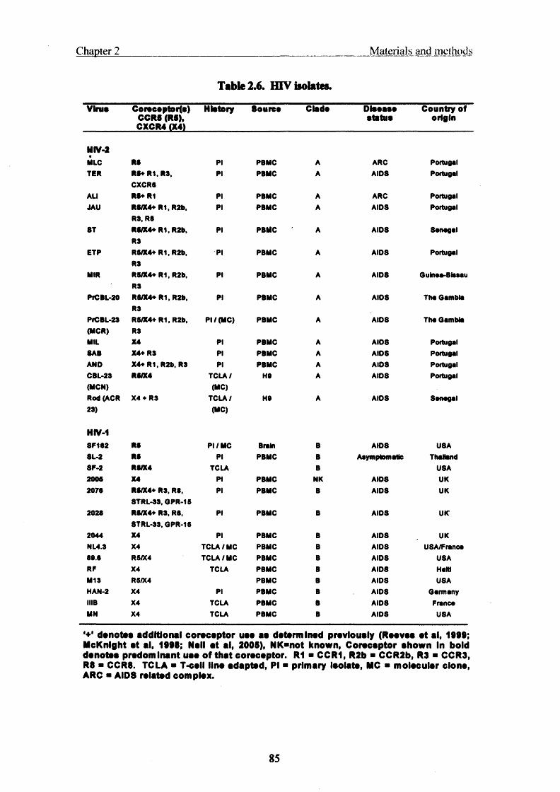
Chapter 2 M ateria ls and m ethods
Table 2.6. HIV isolates.
Virus Corscoptor(s) CCR8 (R5), CXCR4 (X4)
History Source Clade Diseasestatus
Country of origin
NIV-2MLC R8 pi PBMC A ARC PortugalTER R8+ R1. R3.
CXCR8pi PBMC A AID8 Portugal
AU R8+R1 Pi PBMC A ARC PortugalJAU R5/X4+ R1, R2b,
R3. R8pi PBMC A AIDS Portugal
8T R8/X4+ R1, R2b, R3
pi PBMC A AID8 Sanogal
ETP R8/X4+ R1, R2b, R3
Pi PBMC A AIDS Portugal
MIR R5/X4+R1, R2b, R3
pi PBMC A AIDS Guinoa-Blssau
PrCBL-20 R8/X4+ R1, R2b, R3
pi PBMC A AIDS Tho Gambia
PfCBL-23 R5/X4+ R1, R2b, PI / (MC) PBMC A AIDS Tho Gambia(MCR) R3MIL X4 PI PBMC A AIDS Portugal8AB X4+R3 PI PBMC A AID8 PortugalAND X4+ R1, R2b, R3 PI PBMC A AIDS PortugalCBL-23 R8/X4 TCLA/ H9 A AIDS Portugal(MCN) (MC)Rod (ACR X4 ♦ R3 TCLA / HO A AIDS Sanagal23) (MC)
HIV-18P1S2 R8 PI / MC Brain B AIDS U8A8L-2 RS PI PBMC B Asymptomatic Thailand8F-2 R8/X4 TCLA B USA2005 X4 PI PBMC NK AIDS UK2070 R5/X4+ R3, R8,
STRL-33, OPR-15PI PBMC B AID8 UK
2028 R8/X4+ R3, R8, STRL-33, GPR-15
PI PBMC B AID8 UK
2044 X4 PI PBMC B AIDS UKNL4.3 X4 TCLA/MC PBMC B AID8 USA/Franca80.3 R5/X4 TCLA/MC PBMC B AID8 U8ARF X4 TCLA PBMC B AID8 HaWM13 R5/X4 PBMC B AIDS USAHAN-2 X4 PI PBMC B AIDS GarmanyIIIB X4 TCLA PBMC B AID8 FrancaMN X4 TCLA PBMC B AID8 USA
*+’ denotes additional corscsptor uaa as datarmlnad previously (Raavaa at al, 1999; McKnlght at al, 1998; Nall at al, 2005), NK*not known, Corscsptor shown In bold denotes predominant uaa of that corscsptor. R1 * CCR1, R2b ■ CCR2b, R3 ■ CCR3, R8 ■ CCR8. TCLA ■ T-csll line adapted, PI ■ primary Isolate, MC ■ molecular clone, ARC ■ AID8 related complex.
85

C hapter 2 M aterials and methods
23.2 Virus production by transfection of 293T cells.
HTV molecular clone stocks were prepared in 293T cells in 10 cm culture dish by transfection with FuGene®-6 (table 2.4), as described in Transfection of DNA into eukaryotic cells' except for some changes: the day after transfection, 8 ml of transfection media was replaced with 14 ml of normal growth media in a 10 cm dish to boost stock volume. This was replaced the following day with a further 14 ml and the virus stocks were then harvested and aliquoted into 0.S ml 72 hr post-transfection. Harvests of equal infectivity could be taken at 48, 72 and 96 hr post-transfection. The 293T cells were cultured in GlutaMAX™ supplemented DMEM (Gibco™) and 10% FCS in a 37°C and 10% CO2 incubator.
2.33 N-tropic, B-tropic and Moloney MLV virion pseudotyping.
VSV-enveloped, MLV-N/B-Gag-Pol particles were produced from a combination of three plasmids as described previously (fig 2.1, table 2.4) (Towers et al., 2000). MLV- N, B and Moloney (Mo) Gag-Pol cores (packaging constructs) were expressed from pCIG3-N, pCIG3-B and pHIT60 respectively, and pseudotyped with VSV-G expressed from pMDG, or MCR and MCN Env from pSVffl (fig 2.1, table 2.4). The proviral reporter genome pCNCG encoding eGFP was packaged into the pseudovirions (fig 2.1, table 2.4). Three pg pCNCG, 2 pg pCIG3-N or B, and 1 pg pMDG were mixed and a confluent layer of 293T cells in a 10 cm dish was transfected in the presence of 18ul FuGene®-6 (see ‘transfection of DNA into eukaryotic cells'). Pseudotype particles were harvested 48, 72 and 96 hr post-transfection and viral titres were determined on MDTF
and by RT-ELISA.
2.3.4 HIV-1 vector pseudotypes.
HIV-1-based vectors were derived from the packaging construct p8.91 (Zufferey et al., 1997), and the eGFP encoding vector genome pCSGW (table 2.4) (Demaison et al., 2002), in conjunction with pMPl 1, encoding either MCN or MCR Env (Schmitz et al., 2004). Molecular clones, pseudotyped viruses, and vectors were produced by transient transfection of 293T cells, as described above in ‘N and B-tropic and Moloney MLV virion pseudotyping' and fig 2.1.
86

Chapter 2_____________ Materials and methods
A Transfervector I c m v
(pCNCG )
MLV 5 ’ LTR
(R/U5)
MLV 3 ' LTR
(U3/R/U5)
r CMV X Q ^ neo } CMV egfp | T I
G ag-Polexpression i CMV (pCIG3)
Env k
expression 1 CMV H(pM DG ) P -g lo b in in tro n P-g lob in p(A) s ig n a l
293T producer cell
C S ingle round retroviral vector
Figure 2.1. Production of retroviral vector by triple transfection.
(A) Transfer vector, Gag-Pol and Env expression plasmids were mixed and transfected into (B) 293T
producer cells. (C) The only construct to contain the packaging signal (y) is the transfer vector (devoid
of genes required for replication) so it is preferentially packaged and the virions that are produced are
single round only.
2.4 Injectivity assays
2.4.1 Tissue culture infectious dose §0.
Tissue culture infectious dose 50s (TCID50) of HIV-1 and HIV-2 were estimated on
both MDM and PBMC. MDM and PBMC were plated on 96 well trays at 104 and 105
cells per well, respectively. Virus was serially diluted in half logs and 50 pi dilutions
were incubated with either PBMC or MDMs. After overnight incubation with virus
inoculum cells were washed 3 times in growth media which was changed every 7 days.
87

Chapter 2 M aterials and methods
Supernatant was harvested at day 14 for PBMC and day 21 for MDM. Virus production was detected by RT-ELISA (Caviditech) and TCIDsos were calculated by the Karber method, described previously (Aine McKnight, 1995). Samples were subject to RT- ELISA using the 'detection of RT activity1 protocol outlined in the manufacturers instructions. The TCEDso is the dilution where cells become infected in 50% of wells: TCID50 = L-d(S-0.5). Where L is the highest dilution to give 100% of positives, while d is the dilution factor (for half log dilutions this is 0.5). S is the sum of the number of positive wells/total number of wells for all the dilutions to give any positives starting from L and going up to the highest dilution to contain positives. An example is given below (fig 2.2).
TCIDu-L - d (S - 0.5)
Total no. of walla
No. of walla with infectad cells
Log dilution aariaa— ►N -0.5 -1.0 -1.5 -2.0 -2.5
5 5 5 5 5 5
5 5 | 5 | 3 1 0
tL« -1
L * -1 d ■ 0.53 ■ 1 + 0.6 + 0.2 * 1.8
TCIDm- -1 -0.5 (1.8-0.5)- -1 - (0.65)■-1 .65- 45 TCIDm/SOmI
■ 8.9 x 10² TCIDS0/ml
Figure 2.2. Example tissue culture infectious dose 50 calculation.
88

Chapter 2 M aterials and m ethods
2.4.2 Monocyte derived macrophage time courses of infection.
MDM time-courses were done in 6-well trays. Cells were plated at 1.5 x 10⁶ cells per well. The following day, 1ml of 104 TCIDso/ml (as determined on PBMC) of virus diluted in RPMI 1640, 10% heat-inactivated HS was added to its corresponding well. After 3 hr of infection, the virus was removed and the cells were washed with serum free RPMI 1640. Fresh 10% HS in RPMI 1640 was added and the first aliquot, time 0, was taken. Every two days, an aliquot was removed and stored at -40°C for determination of RT activity and focus forming units (FFUs). After the removal of each time-point aliquot the MDM were washed once with 10% heat-inactivated HS RPMI 1640 to eliminate the possibility of measuring carry-over virus in the following time- point.
2.43 Cholesterol depletion.
To aid cell adherence during cholesterol depletion, tissue culture surfaces were treated
with 1 part 2% bovine gelatine (Sigma) to 1 part serum free DMEM for 1 hr at room temp. Cells were plated the night before they were needed on 48 well trays at approximately 5.0 x 10⁴ cells per well and in 75 cm2 medium flasks at 1.0 x 10⁷ cells/flask. The best results were obtained when four confluent-medium 75 cm2 flasks were split into eight gelatine-coated-medium flasks and fractionated 3 days later.
Prior to virus addition, cells were washed with serum free media then treated with 10
mM methyl-p-cyclodextrin (MpCD) for 20 min at 37°C. The cells were rinsed once
with serum free media, virus dilutions were added and allowed to bind to the cells on ice for 1 hr. After synchronisation on ice, virus inoculum was removed and serum free medium, with or without water-soluble cholesterol, was added to the cells, and transferred to a 37°C incubator to allow infection to proceed for 2 hr. Normal growth media was added to the cells, fixed and stained for HIV infection 3 days later.
2.4.4 Inhibition of endocytosis with hypertonic sucrose.
Cells were plated at subconfluence, as described for ‘cholesterol depletion’, the day before they were needed. The cells were washed once with growth media, virus dilutions were added, and placed on ice for 1 hr. Normal growth media (DMEM, 5%
89
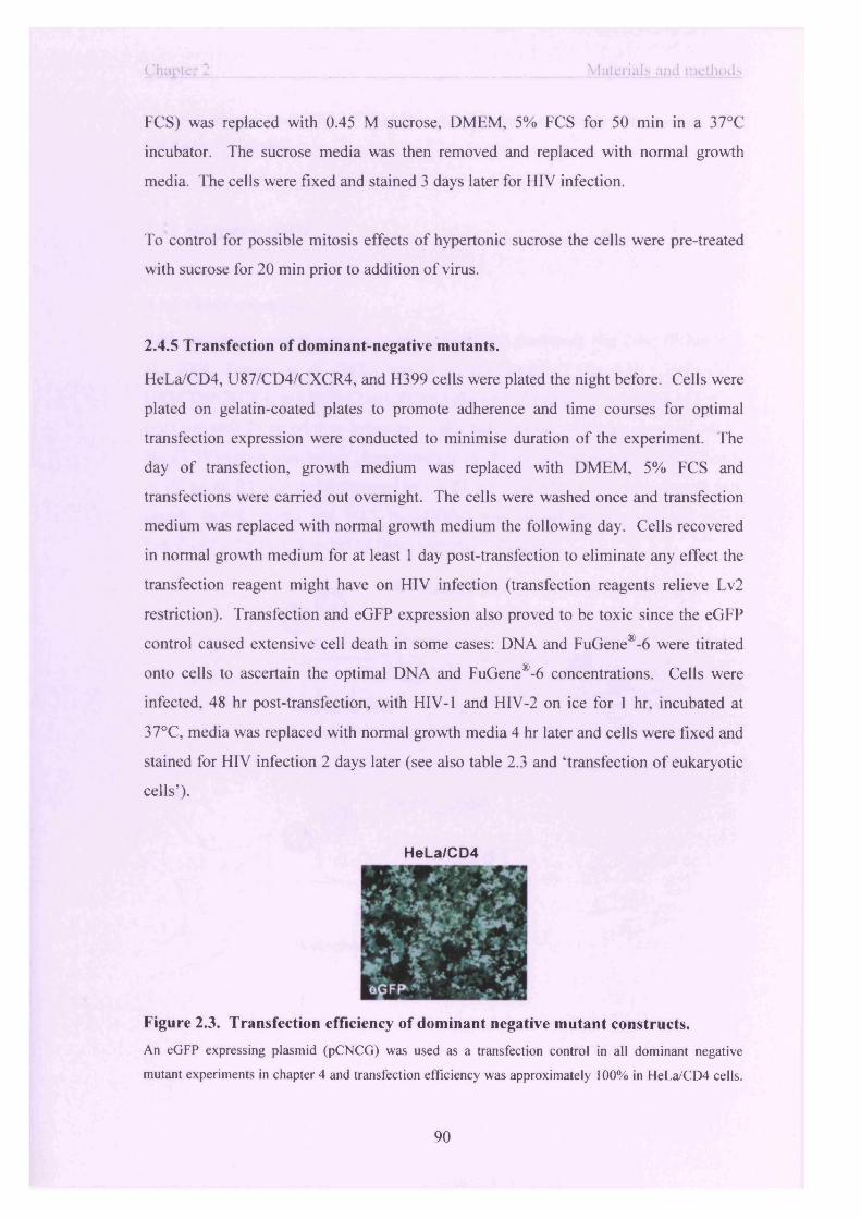
Chapter 2_________________________________________________ Materials and methods
FCS) was replaced with 0.45 M sucrose, DMEM, 5% FCS for 50 min in a 37°C
incubator. The sucrose media was then removed and replaced with normal growth
media. The cells were fixed and stained 3 days later for HIV infection.
To control for possible mitosis effects of hypertonic sucrose the cells were pre-treated
with sucrose for 20 min prior to addition of virus.
2.4.5 Transfection of dominant-negative mutants.
HeLa/CD4, U87/CD4/CXCR4, and H399 cells were plated the night before. Cells were
plated on gelatin-coated plates to promote adherence and time courses for optimal
transfection expression were conducted to minimise duration of the experiment. The
day of transfection, growth medium was replaced with DMEM, 5% FCS and
transfections were carried out overnight. The cells were washed once and transfection
medium was replaced with normal growth medium the following day. Cells recovered
in normal growth medium for at least 1 day post-transfection to eliminate any effect the
transfection reagent might have on HIV infection (transfection reagents relieve Lv2
restriction). Transfection and eGFP expression also proved to be toxic since the eGFP
control caused extensive cell death in some cases: DNA and FuGene®-6 were titrated
onto cells to ascertain the optimal DNA and FuGene®-6 concentrations. Cells were
infected, 48 hr post-transfection, with HIV-1 and HIV-2 on ice for 1 hr, incubated at
37°C, media was replaced with normal growth media 4 hr later and cells were fixed and
stained for HIV infection 2 days later (see also table 2.3 and ‘transfection of eukaryotic
cells’).
H e L a /C D 4
Figure 2.3. Transfection efficiency of dominant negative mutant constructs.
An eGFP expressing plasmid (pCNCG) was used as a transfection control in all dominant negative
mutant experiments in chapter 4 and transfection efficiency was approximately 100% in HeLa/CD4 cells.
90

Chapter 2 Materials and methods
Cells were transfected with FuGene® (Roche) as described above, and visualised by blue light before
staining for HIV infection.
2.4.6 Abrogation assays
2.4.6.1 Refl saturation.
Refl abrogation control was conducted as described previously (fig 2.4c) (Schmitz et
al., 2004; Towers et al., 2002) and in results chapter 5 (fig 5.1). HeLa/CD4,
U87/CD4/CXCR4, and HOS/CD4/CXCR4 cells were plated in 48 well trays at 5 x 10³
cells per well 24 hr prior to infection. Cells were treated with high doses of MLV-
Nzeo(VSV) vector pseudotype [approximately 105 IU/ml as determined on MDTF cells
or 104 pg/ml RT activity (determined by RT ELISA)] for 6 hr. Cells were washed with
normal growth media and MLV-NeGFp(VSV) was titrated on the cells and eGFP
transduced cells were detected by flow cytometry 3 days later.
A MLV-NeGFP(VSV)
Permissive cells
& j - i
9 - & ju
M LV -N ^V S V )
Restrictive cells
91

Chapter 2
C
__________ Materials and methods
M LV -N ^V S V ) “Saturating dose”
‘Temporarily permissive’ fU% “Indicator/challengedose”
Figure 2.4. Abrogation of restriction factors in restrictive versus permissive cells:
R efl.
(A) Permissive cells do not express restriction factors, all eGFP encoding virus vectors (shown with green
nucleic acids) are able to enter and establish productive infection. This scenario also occurs with
challenge o f restrictive cells with resistant/non-restricted viruses (no restriction). Infected cells express
eGFP [green cells in (A)]. However, (B) restrictive cells express restriction factors (red) that inhibit all
challenge virus, thus there is no productive infection in this hypothetical model. (C) Experimental model
o f Refl abrogation (chapter 5, fig 5.1). The purpose of abrogation experiments is to saturate restriction
factor with a decoy virus and observe the ‘rescue’ o f a similarly restricted virus with a different reporter
gene. The challenge and saturating viruses must be detectable by different reporter systems in order to
detect restriction-saturation and recovery of challenge virus infectivity [The saturating dose conferred
Zeomycin resistance (Zeo) and the challenge dose was an eGFP reporter in this case (fig 5.1)]. High
doses of saturating virus are normally required to saturate restriction that is followed by a titration of
challenge/reporter virus and the cells are temporarily rendered permissive. When the restrictive factor has
been pre-saturated there is an apparent increase in eGFP expression from challenge virus infection.
Abrogation does not require that the saturating dose and challenge dose viruses are the same, however
they must be targeted by the same restriction factor for rescue of restricted virus infectivity.
92
C hapter 2 M aterials and methods
2.4.6.2 Refl saturation with an, Lv2 restricted, HIV-2 challenge dose.
HeLa/CD4, U87/CD4/CXCR4, and HOS/CD4/CXCR4 cells were plated in 48 well trays at 5 x 10³ cells per well 24 hr prior to infection and an abrogation assay was conducted as described previously [fig 2.5a for experimental design; chapter 5, fig 5.3 for results (Schmitz et al., 2004)]. MLV-N and B(VSV) pseudotyped viruses were added to the cells at an MOI of approximately 10 (about 1 x 10s IU/ml as determined on MDTF cells or 104 pg/ml RT activity as determined by RT-ELISA). After 6 hr, the cells were washed 3 times with growth media (DMEM, 5% fetal calf serum), and the
HTV-2s, MCN and MCR, were titrated onto the cells. Two days post-infection the cells were screened for MLV vector transduction by visualisation of eGFP expression, to determine whether the dose applied to the cells was sufficient to overcome Refl restriction, and then immunostained 3 days later for MCN and MCR infection with human polyclonal HIV-2 serum. In this case the saturating dose read-out is eGFP (fig 2.5b) and the HIV-2 challenge virus reporter is p-Gal, blue spot staining. Monitoring
eGFP expression was a convenient control to determine whether the MLV-NeGFP dose delivered was a Refl saturating one (fig 2.5b).
93

Chapter 2 Materials and methods
A
B
GHOST
HeLa/CD4
/ "sSrO ■
MLV-N (VSV)
MUV-B (VSV)
Figure 2.5. Determination of relationship between Refl and Lv2.
(A) The experimental model for determining whether Lv2 (purple) and Refl (red) are the same restriction
factor, (i) If Lv2 and Refl are different restriction factors there would be no expected recovery of Lv2 by
Refl saturation, (ii) If Refl and Lv2 were the same restriction factor then there would be recovery o f Lv2
restricted HIV-2 MCR with the saturation of Refl with MLV-N(VSV) vector pseudotypes. (B) EGFP
expression by MLV-NeGFp(VSV) particles in (A) allowed for monitoring of Refl saturation during Lv2
M L V -N ^ V S V)“Saturating dose”
HIV-2 MCR“Challenge dose”
94

Chapter 2 M aterials and methods
saturation control experiment before staining for HIV infection by in-situ {3-Gal staining (blue spots, FFU). Saturation of Refl by MLV-N is indicated by eGFP expressing cells. The same dose of MLV- B«gfp(VSV) transduced 100% of cells challenged.
2.4.6.3 Lv2 saturation with HTV-2 envelope pseudotypes of MLV.
Abrogation assay set-up was modified to prevent receptor interference when MLV pseudotypes with HTV-2 Envs were used as the saturating doses with HIV-1 and HIV-2 challenge titrations (fig 2.6; results chapter 5, figs 5.6, 5.7, 5.8). The binding of gpl20 has been reported to result in down modulation of CD4 receptor at the stage of HIV entry (Cefai et al., 1992). The HIV indicator doses were plated on ice for 1 hr, later, equivalent saturating doses of MLV(HIV-2) (104 pg/ml RT activity per well) were added for a further 30 min. Abrogations were incubated at 37°C for 3 hr, media was replaced with normal growth media and fixed and stained for HIV infection 3 days later.
Reciprocal abrogations were conducted as above except indicator dose titrations of MLVeGFp(HIV-2) were plated for 1 hr and saturating doses of control and HIV-2 pseudotypes MCR(MCR), MCN(MCN) and MCR(VSV) (104 pg/ml RT activity per well) were plated for a further 30 min on ice (fig 2.6). The MCR and MCN backbones contained a stop codon before env to prevent further rounds of replication and cytopathic effect when saturating doses of HTV-2 were added to indicator cells. Abrogations were detected by flow cytometry 3 days later.
95

Chapter 2_________________________________________________ Materials and methods
“Saturating dose”
“Indicator/challenge do se”
HIV-2 MCR,HIV-1 89.6
Figure 2.6. Abrogation experiment design for preventing receptor interference by
saturating doses of HIV-2 envelope pseudotypes of MLV.
To prevent down regulation o f CD4 receptor by MLV vectors with HIV-2 Envs both the challenge HIV-1
and HIV-2, and the saturating MLV(HIV-2) virus doses were bound to the target cells on ice. The
challenge virus titrations were plated first to prevent occlusion o f available receptor by saturating
MLV(HIV-2) viruses. Dashed line separates the different outcomes (right) with and (left) without
MLV(HIV-2) saturation (chapter 5; fig 5.6, 5.7, 5.8). Lv2 restriction factors are shown in red.
2.5 Detection o f infectivity
2.5.1 Immunostaining of HIV-2- and HIV-l-infected cells and calculation of
restriction.
The immunostaining method has been described previously (Schmitz et al., 2004).
Briefly, methanol-acetone (l:l)-fixed cells infected with HIV-2 or HIV-1 were
immunostained with HIV-2 HS diluted 1/4,000 or anti-HIV-1 p24 monoclonal antibody
diluted 1/100 (1:1 mix of EVA 365 and 366 from the Medical Research Council AIDS
Reagent Program, Potters Bar, United Kingdom). Second-layer 6-galactosidase
96

Chapter 2 M aterials and methods
conjugates of goat anti-human immunoglobulin G (HIV-2) or goat anti-mouse immunoglobulin G (HIV-1) were used to detect first-layer antibodies at a dilution of 1:400 (Southern Biotechnology Associates, Inc., Birmingham, Ala.). Infected cells were stained blue with X-Gal (5-bromo-4-chloro-3-indoyl-6-galactopyranoside) in PBS Containing 3 mM potassium ferri-cyanide, 3 mM potassium ferro-cyanide, and 1 mM magnesium chloride. Foci of infection, which stained blue, were counted, and virus infectivity was estimated as FFU per millilitre (FFU/ml). The restriction (n-fold) is calculated as the ratio of infectivity of unrestricted to restricted cells (unrestricted + restricted).
2.5.2 Flow cytometry.
Flow cytometry was used to detect eGFP transduced into cells by retroviral vectors. Cells were plated on 24 or 48 well trays and challenged the following day with retroviral vector pseudotypes. Cells were washed with 200 pi 1:5000 versene (Gibco™) 3 to 5 days post-transduction, detached with a further 200 pi 1:5000 versene, and fixed with 200 pi 3.8% formol saline for 20 min at room temperature. At this stage cells were deemed safe to remove from the containment level 3 suite and detect eGFP on a Becton Dickinson FACScan flow cytometer at containment level 1.
2.53 Reverse transcriptase enzyme linked immunosorbent assay.
RT ELISA kits were purchased from Caviditech. ELISA plates (96 well) are prebound with poly-rA and an oligo-dT primer. The poly-rA serves as a template for the sample virus RT to build the new DNA strand with bromo-deoxyuridine-triphosphate (BrdU). An anti-BrdU mAb conjugated to alkaline phosphatase (AP) detects the nascent BrdU strand. This is a colorimetric assay that detects the AP cleavage of para-nitro-phenyl phosphate at 405 nm.
2.5.4 Detection of infectivity by gag-LTR polymerase chain reaction (see polymerase chain reaction).
97

C hapter 2 M aterials and methods
2.6 Polymerase chain reaction
2.6.1 Polymerase chain reaction of HTV-2 sequences from HIV-2 infected macrophages.
For gag-LTR and ERV-3 polymerase chain reaction (PCR), MDM were set-up as for MDM time-courses (described in ‘infectivity assays’) except the viruses were treated with 25 U DNase (Roche) per 0.5ml vial of virus before dilution and incubation with cells. At the appropriate time-point the infected or negative control uninfected MDM were washed once with versene 1:5000, incubated for 10 min in versene 1:5000 at 37°C, and harvested by scraping. The mixture was centrifuged at 4000g for 5 min, the supernatant was removed, and the cell pellet was stored at -20°C.
DNA was prepared from harvested MDM with the Qia-amp Blood and Tissue Mini Kit (Qiagen°). Briefly, cells were spun and resuspended in 200pl of PBS. After addition of Qia-amp lysis buffer and proteinase K the cells were digested at 56°C for 3 hr. DNA was ethanol precipitated and washed twice on a Qia-amp column, and then eluted in lOOpl of distilled water.
2.6.2 gag-LTR polymerase chain reaction.
PCR reactions were run using 5 pi of DNA from a DNA extraction of 1.5 x 10⁶ MDM (approximately 250 ng of cellular DNA). The 5' gag-LTR primer 2713 (5- TCTCTCCAGCACTAGCAGGTAGAG) is complementary to a sequence in 'R' of the HIV-2 LTR. The 3' gag-LTR, 2715 (5'-CAAGACGGAGTTTCTCGCGCCCAT) primer is complementary to a sequence within HTV-2 gag. The gag-LTR primers measure full-length transcripts of reverse transcription and integrated HTV-2 provirus (529-bp fragment). The 5' and 3' ERV-3 primers detect sequences within the env region
of the human endogenous retrovirus ERV-3 (ERV-3 forward 5'- GAGGCATAACTATAGGAGATTGG, ERV-3 reverse 5'-CCTTTCCAAGTCTGAACTG; 404bp fragment). There is one copy of ERV-3 per haploid human genome and therefore ERV-3 PCR acts as a cellular DNA input control (Cohen et al., 1985; O'Connell et al., 1984). The gag-LTR reactions were carried out with the Roche High-Fidelity PCR system. For a 50 pi reaction the concentrations of
98

Chapter 2 M aterials and m ethods
the reaction components were as follows: buffer 3 (undisclosed concentration of DMSO, Tween-20, and Nonidet-P40 to minimise secondary structure) containing 2.75 mM final concentration MgCh, 1 pmol of each primer, dNTPs were added to a final concentration of 4 mM, and 1.75 U of Taq polymerase per reaction. Roche buffer 3 was
Used to minimise template secondary structure and aid in the amplification of viral reverse transcription products. Conditions for gag-LTR PCR were as follows: 4 min at 94°C, followed by 35 cycles of 94°C for 1 min, 65°C for 1 min, 68°C for 1.5 min. Conditions for ERV-3 PCR were as follows: 4 min at 94°C, followed by 35 cycles of 94°C for 45 s, 60°C for 45 s, 68°C for 45 s. PCR products were run on a 2% agarose gel and viewed under UV. The gag-LTR PCR products ran at approximately 0.5 kbp and the ERV-3 PCR products ran at approximately 0.4 kbp. An elongation temperature of 68°C was chosen because this is the optimal temperature for the Taq Extender proofreading activity.
2.63 GAPDH polymerase chain reaction.
The forward (5 ’-TGGTGAAGACGCCAGTGG) and reverse (5’- ATGGGGAAGGTGAAGGTC) GAPDH primers were used to detect equal cDNA input for RT-PCR (below). The annealing conditions were as follows: 4 min at 94°C, followed by 35 cycles of 94°C for 30 s, 60°C for 30 s, 74°C for 30 s.
2.6.4 Reverse transcriptase polymerase chain reaction.
RT-PCR to measure gag-LTR RNA transcripts was conducted on MDM infected with HTV-2 JAU and HIV-1 SF162. MDM in 6-well trays were incubated with equivalent input (5.0 x 10³ FFU) of either JAU or SF162 for 3 hr, time points were harvested eveiy 24 hr up to and including 5 days post-infection. The infected MDM were harvested with Trizol® RNA extraction reagent and the cDNA was prepared. Cells for RT-PCR were washed once in PBS and dissolved in-situ with 1ml of Trizol® (Gibco-BRL) RNA extraction reagent. RNA was extracted as per manufacturers instructions. Briefly, extracted RNA was quantified spectrophotometrically by measuring the absorbance at 260 nm. One pg of total RNA was DNase treated for 30 min at 37°C. To remove RNA secondary structure and to inactivate the DNase, two fifths of the DNase treated RNA was heated at 75°C for 10 min in the presence of DNase stop solution and random
99

Chapter 2 M aterials and methods
primers, then put on ice. The RNA was added to a mixture that was either RT positive or negative (to control for contaminating DNA). RT reactions, plus or minus RT, were incubated at the RT reaction temperature of 42°C for 1 hr. Two pi of each RT reaction was put into a 50 pi GAPDH and gag-LTR PCR reaction and subject to the PCR conditions as mentioned in ‘polymerase chain reaction*.
2.6.5 Quantitative polymerase chain reaction.
U87/CD4/CXCR4, H399, and HeLa/CD4 cells were seeded in 24 well trays at 5 to 8 x 10⁴ cells per well and treated with 5.0 x 10³ FFU (determined on U87/CD4/CXCR4) of either MCN or MCR pie-treated with DNasel (Roche) for 1 h at 37°C. 100 ng of total DNA was used for qPCR with primers: For HTV-2 gag-LTR: 900 nM forward primer (S'-CGGCTGAGTGAAGGCAGTAAG-S1), 300 nM reverse primer (5’-TTGGCCTGCGCCTTTCTA-3'), 150 nM taqman probe (5'-6-carboxyfluorescein-5'- CAGGAACAAACCACGACGGAGTGCTC-3'-6-carboxytetramethyl-rhodamine-3').
For HIV-1 packaging signal OF), 300 nM forward primer
(TGGGCAAGCAGGGAGCTA), 300 nM reverse(TCCTGTCTGAAGGGATGGTTGT) and 150 nM Taqman probe (5'-6-
carboxyfluorescein-AACGATTCGCAGTTAATCCTGGCCTGTT-6- carboxytetramethyl-rhodamine-3').
First strand synthesis of the MLV pCNCG was monitored by amplification of the vector eGFP cDNA sequence: 300 nM forward primer (5’-CAACAGCCACAACGTCTATATCAT-3’), 300 nM reverse (5’- ATGTTGTGGCGGATCTTGAAG-3 ’) and 150 nM taqman probe (5'-6-carboxyfluorescein-CCGACAAGCAGAAGAACGGCATCAA-6-carboxytetramethyl-
rhodamine-3'). The PCR conditions consisted of one cycle of denaturation (95°C for 10 min) followed by 45 cycles of amplification (95°C for 15 s, 60°C for 1 min). The amplification, data acquisition, and analysis were performed using the ABI PRISM 7000 sequence detection system.
2.7 Cloning of the HJV-2 envelope third variable loop region
100

C hapter 2 M aterials and methods
2.7.1 Cloning and sequencing of the HIV-2 third variable loop.
3-5 x 10⁶ PBMC were infected with HTV-2 virus stocks. Three days after infection the infected cells were harvested and DNA extracted with the DNA Blood Mini Kit (Qiagen). V3 loops were amplified with primers FI 3418 (5-GGTTTGGCTTTAATGGCACTAGAG) and F13420 (5'-TTCTCCTCTGC AGTTAGTCC AC AT) in the HIV-2 V3 region (243-bp fragment). The amplified product was purified from ethidium bromide-containing 1% agarose gel with the Qiagen Agarose Gel Extraction kit. The V3 region was cloned into pGEM-T easy with the pGEM-T easy TA cloning kit (Promega, Madison, WI, USA). Clones were screened and sequenced with primers F13418 and F13420. Sequencing was carried out with the CEQ-DTCS Beckman Coulter Quick Start kit (Beckman-Coulter, Fullerton, CA, USA). The V3 loops from HIV-2 isolates were sequenced from at least 3 clones or directly from PCR product
Direct sequencing of PCR reactions of the HIV-2 isolates was used to confirm the cloned sequences. Briefly, the V3 region was amplified with primers FI3418 and FI3420, the product was analysed on an ethidium bromide-containing 1% agarose gel, extracted with the Qiagen PCR extraction kit and then sequenced (see 'Cloning and sequencing of V3 loops' above).
V3 loop sequences were aligned using the Clustal nucleic acid sequence alignment program.
The net charge of each of the V3 loops was estimated from the amino acid sequence by totalling the number of positively charged Arginine, Lysine, and Histidine residues and then subtracting the number of negatively charged Aspartate and Glutamate residues.
2.7.2 Sequencing sample preparation for Beckman CEQ-2000.
Mini-prep DNA was prepared for sequencing by the dye-labelled-dideoxy-nucleotide chain termination method. For each sequencing reaction 6 pi of premix was added to 5 pmol of primer (T7 and SP6 of pGEM-T easy or F13418 and F13420) and 1 pi of a 50 pi plasmid miniprep and made up to a total of 10 pi with water. Reactions were run on
101

Chapter 2 M aterials and methods
an MJ PCR thermocycler for 24 cycles at the following conditions: 96°C for 30 s, 50°C
for 15 s, 60°C for 4 min, and finished at 4°C.
Sequencing samples were cleaned-up after the labelling reaction. Fifteen pi of premix (2 pi 3 M cold NaOAc, 2 pi 100 mM EDTA, 1 pi glycogen, and 10 pi of water) was added to each sample and mixed. Sixty pi of -20°C 95% ethanol was added to each tube and vortexed. Samples were left on ice for 10 min then centrifuged at room temperature for 15 min to promote the sticking of precipitated DNA to the sample tube. Supernatant was removed and 200 pi -20°C 70% ethanol was added for 10 s to wash pellet Tubes were left open on bench for 30 min to dry. Pellets were dissolved in deionised formamide. Samples were loaded into sequencing plates and overlayed with a drop of oil.
2.8 Maxi-prep o f plasmid stocks
2.8.1 Transformation of bacteria.
HB 101 or TOP F10’ bacteria were mixed with plasmid DNA on ice and transformed by heat shock at 42°C for 30 s. Transformed bacteria were plated on LB agar plates supplemented with 100 pg/ml ampicillin for selection of transformants, or 50 pg/ml kanamycin for pEGFP-Cl/C2 (Clontech™) plasmids. Colonies were picked and minipreps (Qiagen) were performed on 5 ml overnight cultures of inoculated LB-broth
with 100 pg/ml ampicillin.
2.8.2 Restriction enzyme digest of plasmid stocks.
Restriction enzyme digests were performed on die above minipreps to confirm the identity of transformed plasmid DNA. Digests were performed on 1 pg of total DNA with 10 U of restriction enzyme per reaction, between 37 and 65°C (depending on the restriction enzyme) for 1 hr. Restriction products were run on a 1.5 % agarose gel for 2 hr at 60 V and the digestion pattern was compared to that of a positive control run in parallel (if available) or the predicted pattern.
102

Chapter 2 M aterials and methods
2.83 Hi-speed plasmid maxi kit.
Positive HB 101, TOP FI O’ or STBL 2 bacteria colonies were repicked and plasmid stocks were grown using LB culture broth at 37°C (or 30°C for STBL 2) overnight. All plasmid-transformed bacteria were grown-up in approximately 150 ml of LB broth, except 250 ml for low copy plasmids [pMDG (VSV-G)], with 100 pg/ml ampicillin or 50 pg/ml kanamycin. Maxi prep was performed as per manufacturers instructions for the Hi-speed plasmid maxi kit (Qiagen). Concentration and quality of plasmid DNA was determined by the absorbance at 260 nm and the 260/280 nm ratio, respectively, on an ND-1000 spectrophotometer (Nanodrop, Wilmington, DE, USA).
2.9 Subcellular fractionation
2.9.1 Preparation of cell lysates.
The preparation and fractionation of cell lysates were carried out on ice unless otherwise stated. Cells (1.0 x 10⁷ to 5.0 x 10⁷) were harvested with versene 1:5 000 (Gibco™) and then washed twice with PBS. The cell pellet was resuspended in 1 ml hypotonic HEPES-h buffer, Roche EDTA free complete anti-protease pellet, pH 7.9 at 4°C and centrifuged for 5 min at 5000 rpm (500g) and 4°C in a Fischer refrigerated microcentrifuge. 300 pi 1% Triton X-100 HEPES-h buffer was added to the cell pellet, left for 30 min on ice, then homogenised with 10 strokes of a Dounce homogeniser. The homogeniser was washed with 100 pi of 1% Triton X-100 HEPES-h buffer and added to the lysate. To remove nuclei, the lysate was spun at 2000 rpm (300 g) for 3 min and 4°C, then the supernatant containing cell membranes was transferred to a new tube. Nuclei were stored at -20°C for PCR and western blot. The resulting cell lysate was loaded onto a sucrose gradient.
2.9.2 Isolation of lipid rafts by sucrose gradient fractionation.
Four hundred pi of cell lysate was mixed with 800 pi 60% sucrose in HEPES-h buffer (w/v) to make a 40% sucrose/lysate solution. One hundred pi of the sucrose/lysate solution was stored at -20°C as a total membrane input fraction. Sucrose gradients consisted of 3 ml of 5%, 6 ml of 36% sucrose (w/v) in HEPES-h on top of 900 pi of
103

Chapter 2 M aterials and methods
sample lysate in 40% sucrose 1% Triton X-100 HEPES-h buffer. The gradients were loaded into 17ml Sorvall centrifuge tubes as follows: 3 ml of 5% sucrose was added to the bottom of the centrifuge tube first, 6 ml of the 36% sucrose was pipetted below, and the 1100 pi sample fraction was added last Centrifuge tubes were loaded onto a Sorvall §urespin 630 rotor and spun at 100 OOOg for 20 hr at 4°C in a Sorvall Discovery 100S centrifuge. One ml Fractions were collected from the top of the tube with a 'trimmed* PI000 Gilson pipette tip and stored at -20°C for western blot, slot blot, or PCR. The lipid raft fraction was isolated from fraction 3 to 5 that manifested as a flocculent (wispy) band at the 5%-36% sucrose interface.
2.93 Sodium dodecyl sulphate polyacrylamide gel electrophoresis.
Fractionations were screened for the presence of cell proteins by SDS polyacrylamide gel electrophoresis (SDS-PAGE) and Western blot. Samples were mixed with 6X loading buffer (0.25 M TRIS-C1 pH 6.8, 50% glycerol, 5% SDS, 0.2% bromophenol blue, 5% P-mercaptoethanol), heated at 90°C for 3-5 min, loaded onto a 3% stacking gel (8% acrylamide (40% acrylamide/bisacrylamide (37.5:1) Biorad), 0.1 M TRIS-C1 pH 6.8, 1.0% SDS, 0.2% TEMED, 0.2% APS) and resolved on a 15% resolving gel (37% acrylamide (40% acrylamide/bisacrylamide (37.5:1) Biorad), 0.4 M TRIS-C1 pH 8.8, 0.1% SDS, 0.1% TEMED, 0.05% APS) beside a Benchmark™ prestained protein ladder (Invitrogen™). Gels were run on a Hoefer Mighty Small II SE 250 (Hoefer Scientific Instruments©, Inc., California, USA) in protein electrophoresis buffer (250 mM TRIS, 2 M glycine, 0.1% SDS) at 25 mA per gel and 70 to 100 V until the sample front reached the bottom of the gel.
2.9.4 Sodium dodecyl sulphate polyacrylamide gel electrophoresis of tripartite motif proteins.
Trim proteins were resolved in polyacrylamide gels as above except for the following changes: samples were dissolved directly in 2 X loading buffer, sonicated for 10 min in a Decon F5 minor sonicating water bath (Decon ultrasonics, Hove Sussex, England), and stored at -20°C. When the samples were needed they were boiled for 10 min at 95°C, sonicated for a further 10 min and loaded onto a polyacrylamide gel.
104
Chapter 2 M aterials and methods
2.9.5 Western blot.Proteins were transferred to PVDF (poly-vinyl-difluoride) membrane in a Mini Trans- Blot Cell (Biorad Laboratories0) as per manufacturers instructions. The ( 8 x 6 cm) PVDF membrane was activated in methanol then soaked in protein blotting buffer (250 eiM TRIS, 2 M glycine, 10% ethanol) for 5-10 min. The running gel was separated from the stacking gel, placed on the activated PVDF membrane, and sandwiched between four pieces of pre-wet 3MM Whatman filter paper (10 x 7.5 cm). This assembly was placed in the blotting cell and blotted for 3 hr at 250 mA and 200 V or
overnight at 125 mA and 50 V at 4°C.
The membrane was removed from the blotting assembly and washed briefly in PBS then blocked in PBS, 0.1% TWEEN 20, 5% powdered milk (w/v) for 1 hr with shaking. The membrane was incubated with primary antibody diluted 1/500-1/2000 in PBS, 0.1% TWEEN 20, 5% powdered milk (w/v) for 45 min to 1 hr. The membrane was washed 3 x 10 min in PBS, 0.1% TWEEN 20. The horseradish peroxidase-conjugated secondary antibody was added at a dilution of 1/2000 in PBS, 0.1% TWEEN 20, 5% powdered milk (w/v) for 45 min to 1 hr. The final three washes were done in PBS, 0.1% TWEEN 20 for 10 min each. The last wash was done in PBS only. ECL™ detection substrate and Hyperfilm ECL™ (Amersham Biosciences0) were used for film detection and ECL-Plus™ substrate was used for detection of Western blots on a Storm 860™ (Molecular Dynamics).
2.9.6 Slot blot of fractions derived from sucrose gradient centrifugation.Dot blotting was used to screen sucrose gradient fractions for lipid raft associated proteins and lipids, as per manufacturers instructions. Briefly, a Bio-Dot SF microfiltration unit (Bio-Rad) was set-up using Bio-Dot SF filter paper and Amersham0
Hybond-ECL Nitrocellulose membrane as this yielded the best results. The blotting membrane and filter paper were pre-wet in TBS before mounting onto blotting unit.
The membrane was washed by drawing 200pl of TBS through each well under suction.
An aliquot of each sample was diluted 1/10 in 200pl TBS, drawn through, then washed
with 200|d TBS. The membrane was removed from the blotter, washed in TBS for 10
min, and antibody detection was carried out as for a western blot and detection and
105

Chapter 2 Materials and methods
densitometry were done on a Storm 860™ (Molecular Dynamics) and ImageQuant software.
2.10 Microscopy
2.10.1 Intracellular antibody staining for major histocompatibility complex class I protein.
HeLa cells were seeded on coverslips. The following day, cells were fixed in freshly prepared solution (3% paraformaldehyde, 60 mM sucrose, 0.1 M sodium phosphate pH7.2) for 30 min at room temperature. After three washes in PBS for 5 min, cells were washed with PBS 20 mM glycine for 5 min, permeabilised in PBS 20 mM glycine 0.1% saponin for 10 min at room temperature, washed with PBS 20 mM glycine for S min and blocked with PBS 20 mM glycine 1% bovine serum albumin (BSA) for 10 min at room temperature. Incubation with primary antibodies was done in blocking solution for 1 h at 37°C. After washing in PBS glycine for 5 min cells were incubated with secondary antibodies (donkey antimouse-FITC) in blocking solution for 45 min at 37°C. Finally, cells were washed for 10 min in PBS and mounted with mowiol (Calbiochem) and analyzed by confocal microscopy (MRC 1024 (BioRad, Hercules, CA, USA) equipped with a krypton-argon laser). Pictures were acquired using Kalman averaging and analysed with Lasersharp software (BioRad). .
2.10.2 Immunofluorescence microscopy.
Cells were visualised after treatment with either FITC-conjugated transferrin (Trf-FITC) or FITC-conjugated cholera toxin B subunit (CTxB-FITC). Slides were mounted with immunofluorescence mounting medium (Dako, Carpinteria, CA, USA) and observed by confocal microscopy [MRC 1024 (BioRad, Hercules, CA, USA) equipped with a krypton-argon laser]. Pictures were acquired using Kalman averaging and analysed with Lasersharp software (BioRad).
106

Chapter 2 Materials and methods
2.11 Antibodies
Table 2.7. Antibodies.Specificity/(target)
Source/ Ab type Assay Dilution Company Catalogueno.
Actin Rabbit pAb WB 1/2000 Sigma A-5060
CD4 (Q4120) Mouse mAb SB 1/2000 ARP ARP318
GM1 (CTxB) CTxB-Blotin (ligand)
CTxB-FITC
SB
IF
1/4000
1/1000Sigma
C9972
C1655
HIV-1 (p24CA) Mouse mAb IS 1/100 ARP EVA 365, 366
HIV-2 antiserum Human pAb IS 1/4000 HIV-2 patient N/A
huMHC-l Mouse mAb IF 1/200 Serotec MCA81
MID 1(TRIM 18) Rabbit pAb WB 1/1000 Orbigen PAB-11697
MID 2(TRIM 1) Goat pAb WB 1/1000 Imgenex IMX-3087
PIAP Mouse mAb WB/SB 1/2000 Sigma A2951
RNF 21 (TRIM 34) Goat pAb WB 1/1000 abeam ab4509
Transferrin (TrfR) Trf-FITC (ligand) IF 1/1000 Sigma T0288
HA tag Mouse mAb WB 1/3000 Sigma H9658
pAb ■ polyclonal antibody; mAb * monoclonal antibody; CTxB ■ Cholera toxin binding subunit; ARP ■ AIDS reagent program, NIBSC, Potters Bar, UK; huMHC-l ■ human MHC-I, N/A * not applicable; PIAP * placental alkaline phosphatase; TrfR * transferrin receptor; WB ■ western blot; SB * slot blot; IF ■ immunofluorescence.
2.12 Statistical analysis
The mean and the standard deviation of the mean (SDM) were calculated using Microsoft Excel.
The Spearman's rank coefficient was calculated for HTV-2 V3 loop charge versus MDM tropism as in fig 2.7. The Spearman’s rank coefficient tool was used to calculate Rs: http://faculty.vassar.edu/lowry/corr__rank.html.
107
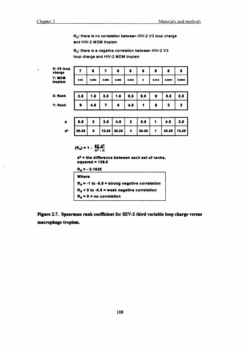
Chapter 2 Materials and methods
H0: there is no correlation between HIV-2 V3 loop charge and HIV-2 MDM tropism
Ha: there is a negative correlation between HIV-2 V3 loop charge and HIV-2 MDM tropism
X: V3 lo o p c h a r g e
Y: MDM tro p is m
7 6 7 6 8 8 9 8 8
0.03 9.043 9.009 0.004 0.003 0 0.010 0.0001 0.0004
X: R a n k 3.5 1.5 3.6 1.5 6.5 6.5 9 6.5 6.5
Y: R a n k 9 4.5 7 6 4.5 1 8 2 3
d 6.5 3 3.6 4.6 2 5.5 1 4.5 3.5
d> 30 .2 S • 12 .25 20 .26 4 3 0 .26 1 2 0 .26 12 .26
61 d2
d2 ■ the difference between each set of ranks, squared * 139.5R, ■ -0.1625WhereRs * -1 to -0.5 ■ strong negative correlation Rs < 0 to -0.5 * weak negative correlation Rs * 0 ■ no correlation
Figure 2.7. Spearman rank coefficient for HTV-2 third variable loop charge versus macrophage tropism.
108

Chapter 3
HTV macrophage tropism and a post-entry restriction to HIV infection
in human cells
3.1 Introduction:
HIV-1 HIV-2 both cause AIDS. However HIV-2 causes a slower mean progression to disease and is less transmissible than HIV-1 (Marlink et al., 1994). HTV-2 infection is found primarily in West Africa (Wilkins et al., 1993) (Pfutzner et al., 1992; Piedade et al., 2000). In contrast, HIV-1 infection is found worldwide with particularly high prevalence in sub Saharan Africa. UNAIDS report that at the end o f2004 there were 42 million people living with HIV-1 infection globally (UNAIDS, 2004).
Both viruses use CD4 and a 7-TM coreceptor to gain entry into cells but HTV-2 can use a broader spectrum of coreceptors than HTV-1 in vitroy including CCR1, CCR2b, CCR3, CCR5, and CXCR4 (Alkhatib et al., 1996; Clapham and McKnight, 2002; Deng et al., 1996; Maddon et al., 1986; Sattentau et al., 1988). It is likely that CCR5 and CXCR4 are the most important for infection of primary cells but other coreceptors may be used at a lower efficiency (McKnight et al., 1998; Momer et al., 2002).
The HTV-1 and HIV-2 Envs are made up of conserved Cl through to C5 and variable VI to V5 domains. The HTV-1 third variable loop (V3 loop) acts as a determinant of cell tropism and is the principle neutralising determinant in laboratory isolates (Hwang et al., 1991; Hwang et al., 1992; Javaherian et al., 1990; McKnight et al., 1995; Westervelt et al., 1992). HTV infection normally begins with slow replicating and low titre viruses termed slow/low. A poor prognosis and declining CD4 counts are later associated with the appearance of rapid replicating, high titre viruses termed rapid/high. There is evidence that the HIV-2 V3 loop is an important neutralisation domain as well and an increase in the net positive charge of the V3 loop has been associated with an increase in the rapid/high from a slow/low phenotype, of HIV-2 isolates (Albert et al., 1996; Bjorling et al., 1994)(McKnight, 1995). Isaka et al demonstrated that basic amino acid substitutions in the C-terminal half of the HIV-2 V3 loop would cause a coreceptor
109
Chapter 3 HIV macrophage tropism
switch from CCR5 to CXCR4 (Isaka et al., 1999). However, it is not known if HIV-2 coreceptor use or V3-loop charge is associated with cell tropism as it is with HIV-1.
HIV-1 infection of M0s has been well documented both in vitro and in vivo (reviewed hy (Martin and Bandres, 1999). The first HTV-1 viruses to emerge in the host are M0 tropic (M-tropic), CCR5 using, probably because during transmission, CCRS expressing M0s and DCs, resident in the mucosae, are the first likely targets. Although there are some exceptions, HTV-1 M0 tropism results from the ability to use CCR5 as a coreceptor. Like mucosal transmission, M0 tropic CCR5 using viruses isolates emerge in preference to CXCR4 using T cell tropic isolates, during intravenous transmission of HTV-1. In this case it is speculated that both T cell tropic and M0 tropic isolates are transmitted but the isolates that infect M0s flourish. It has been proposed that the transmitted M0 tropic virus pool facilitates CTL escape (Schutten et al., 2001), ((Schuitemaker, 1994). It has been demonstrated that HTV-2 can infect both MDMs and T cells in vitro but it is not known what role M0s play in HTV-2 infection (Castro et al., 1990; Valentin et al., 1994) and the relevance this has to HIV-2 transmission is unknown.
In about 50% of untreated HTV-1 subtype B infections there is a switch from M0 tropic, CCR5 using viruses to T cell tropic, CXCR4 using viruses. The change in cell tropism and coreceptor usage of HTV-1 is often accompanied by a poor prognosis and progression to AIDS (Nielsen et al., 1993). The picture for HTV-2 is less well explained but one study shows that HTV-2 isolates taken from AIDS patients use CXCR4 whereas isolates from asymptomatic patients use CCR5 and CCR3 (Blaak et al., 2005; Sol et al.,1997). Thus HTV-2 may follow the same coreceptor switch during infection. Whether the switch from CCR5 to CXCR4 usage in HTV-2 infection is associated with a switch from M-tropism to T-tropism is not known.
The elevation in HIV-1 or HTV-2 RNA plasma viral load is associated with the decline in the CD4 count that is the hallmark of the onset of illness (Schuitemaker et al., 1992) (Damond et al., 2002). It appears that the CD4 count of patients with HIV-2 remains higher than the CD4 count of patients with HIV-1 regardless of recent sero-conversion or development of AIDS (Andersson et al., 2000).
110

Chapter 3 HIV macrophage tropism
All of the Lentiviruses have a propensity to replicate in M0s and indeed there are Lentiviruses that mediate their infection and pathogenicity solely through these cells. The non-primate Lentiviruses Maedi-Visna Virus, Caprine-Arthritis Encephalitis Virus, and Equine Infectious Anaemia Virus (EIAV) all mediate their pathogenesis by infecting M0s. HIV uses M0s as a key infectable cell type and so the propensity of HTV-1 and HIV-2 to infect M0s may be a pathogenesis determinant.
In this chapter I characterise the replication of HTV-2 primary isolates in M0s and T cells from PBMC and compare it with HTV-1. Thus by comparing the M0 tropism of HTV-1 with the less pathogenic HIV-2 this study may provide insight in to why HIV-1 is significantly more pathogenic than HIV-2. MDM infected with HIV-2 primary isolates produced an early and brief burst of replication, followed by a latency, without rebound at later time points whereas HTV-1 primary isolates showed a delayed burst of replication in MDM but showed stable virus production over 21 days. The V3 loop amino acid charges of the HTV-2 isolates were calculated from the sequences and there appeared to be an association with coreceptor usage and a weak negative correlation with M0 tropism, similar to HTV-1.
3.2 Results:
3.2.1 HTV-2 primary isolates, compared to HIV-1, exhibit poor replication in monocyte-derived macrophages.
Infection of MDMs by both R5 and X4 HTV-1 primary isolates has been previously described (Clapham PR, 2001; Eckstein et al., 2001; Valentin et al., 1994; von Briesen et al., 1990). First I determined how well HTV-2 primary isolates infect MDMs. I directly compared the infection of MDM to PBMC by HTV-1 and HIV-2. The viruses used here represent a broad range (table 3.1); a spectrum of HIV-2 primary isolates was selected that use CCR5, CXCR4, or both coreceptors. The HIV-1 primary isolates chosen also have a broad spectrum of coreceptor use and M0 tropism (McKnight et al.,1998), (Reeves et al., 1999) and (Simmons G, 1998). Supernatants were harvested and RT activity was determined by RT-ELISA (Caviditech). TCID50 for each virus was
111

C hapter 3 HIV macrophage tropism
determined on both MDMs and PBMC at 21 and 14 days post-infection, respectively, as has been described for HIV-1 (fig 3.1a) (Simmons G, 1996).
The HTV-1 isolates efficiently infected MDM whereas the HIV-2 isolates did not, even though HIV-2 had higher titres on PBMC. HTV-2 MIL and SAB had TCID50S on PBMC of 5.6 x 10⁵ and 8 .6 x 105 respectively, compared to the HTV-ls 2028 and 2076 that had TCIDsos on PBMC of 2.2 x 10⁵. HTV- 2 was approximately 1 to 2 logs less infectious for MDMs than any HIV-1 isolate (fig 3.1a). HIV-2 TER shows the highest
HIV-2 TCID50 on MDM of 2 .8 x 102 and the lowest TCID50 is that of HTV-2 MLC, 3.6 x 10*. PrCBL 23 does not have a TCID50 that can be calculated from MDM infection since no infection could be detected. This is in contrast to HTV-1 where the lowest TCID50 on MDM is SL2,1.0 x 103, the highest is 7.4 x 10⁴ of2076.
To better compare HTV-2 MDM infection the ratio of M0 to PBMC tropism was
determined from the values in fig 3.1a. The value of the MDM TCID50 was divided by the PBMC TCID50 for each isolate and the relative M0 tropism values were plotted (fig 3.1b). The most ‘macrophage tropic’ HTV-2 was Ali, and then in order of descending M0 tropism were MIR, AND, ST, JAU, TER, ETP, MLC, SAB, and MIL. PrCBL 23 could not infect MDM even when titres of 104 TCID50 were reached on PBMC.
112

Chapter 3 HIV macrophage tropism
Coreceptor
CCR5 || X4/R5 iPEfl!?!
2 10 o 103
□ PBMC
MLC ALI TER ST ETP JAU MIR prCBL23AND MIL SAB
HIV-2
SF162 SL2 2028 2076 2044
HIV-1
B
tE</)Q.OL_■*->0O)ro.cCLo
2028
SF16220442076
SL2
■ 0.1
0.01
0.001
- 0.0001
- 0.00001
- 0.000001
HIV-1
• 0.1
ALI
MirAndSt
JauTer
MLC ! > •
Etp
Sab
Mil
0.01
0.001
0.0001
0.00001
- 0.000001PrCBL23*
HIV-2
*PrCBL23 does not register on a logarithmic chart since its relative macrophage tropism value is 0.
Figure 3.1. HIV-2 is less efficient at infection of monocyte derived macrophages
than HIV-1.(A) Tissue culture infectious dose 50s per ml (TCID5 0/ml) of HIV-2 and HIV-1 primary isolates on MDM
and PBMC, harvested at 21 days (MDM) and 14 days (PBMC) post-infection. Coreceptor use is
indicated (see table 3.1). HIV replication was detected by RT ELISA. (B) Tropism scatter plot. To
illustrate the difference between HIV-2 and HIV-1 M0 tropism a scatter plot was made from the PBMC
and MDM TCID50s from fig 3.1a. This plot was constructed by dividing the MDM TCID5 0 by the PBMC
TCIDso. As previously described (Simmons G, 1996).
113

Chapter 3 HIV macrophage tropism
Table 3.1. CCR5 and CXCR4 coreceptor use of HIV-1 and HIV-2 primaryisolates.
Isolate Corsoaptor(s) CCR5 (R5), CXCR4 (X4)
Preferredcoreceptor
Titre on NP2/CD4/CXCR4
(FFU/ml)
Titre on NP2/CD4/CCR5
(FFU/mO
HIV-2MLC R6 R5* 0 1.0 x10⁴TER R6+ R1.R9.CXdM 0 4.5 x 10⁴ALI RS+ r i 0 4.0 x 10⁴JAU R6/X4+ R1, R2t>,
RS.RS
5.0x10* 5.0x10*
ST R5/X4+ R1, R2b, RS NT NTETP R5/X4+ R1, R2S. RS 1.0x10* 5.0x102MIR R5/X4+ R1, R2b, RS 5.5 x10⁴ 5.0x10²PrCBL-23 R5/X4+ R1, R2b. RS 4.0x10* 5.0 x 10*MIL X4 2.3 x10⁴ 0SAB X4+ r s i r 4.0 x10⁴ 0AND X4+ R1.R2S.RS X4 1.0x10* 0
HIV-1SF162 R6 Ri »• 0 2.5 x 10*SL-2 R6 0 1.0x10*2076 R5/X4+ r s . r s .
8TRL-33, OPR-152.0 x10⁴ 1.8 x10⁴
2028 R6/X4+ RS. RS.
STRL-33, GPR-15 1 r7.5 x 104 7.3 x 10⁴
2044 X4 Xl 7.8 x10⁴ 0
'+’ denotes additional coreceptor use as determined previously (Reeves et al, 1999; McKnight et al, 1998; Neil et al, 2005), NT=not tested, Coreceptor shown in bold denotes predominant use of that coreceptor.
3J2J2 Like HIV-1, the third variable loop charge on the HTV-2 envelope may be associated with HIV-2 macrophage tropism.
I set out to determine if the V3 loop charge of HTV-2 is associated with M 0 tropism
because it has been shown that HIV-1 tropism, coreceptor usage, and V3 loop charge
are associated (Bieniasz et al., 1997; Kuiken et al., 1992; Shioda et al., 1992). A high
V3 loop charge on all of the HIV-2 isolates used in this study it may explain the lack of
114

M 0 tropism of HIV-2. DNA was harvested from HIV-2 infected PBMC 48 hr post
infection and the V3 loop region was sequenced from the HIV-2 proviral DNA. The
number of negatively charged amino acids were subtracted from the positively charged
amino acids, between the V3 loop cysteines, to obtain the net V3 loop charge. Table
3.2 illustrates how the coreceptor usage of the HIV-2 primary isolates studied is
associated with the amino acid charge, ranging from +6 to +10, on the Env V3 loop.
The charge increase is seen primarily in the C-terminal half of the loop and is associated
with increased CXCR4 use by HIV-2. ETP was the only HIV-2 isolate that exhibited a
negatively charged amino acid. There is no consistently high V3 loop charge pattern
but a spectrum of charges.
Table 3.2. HIV third variable loop sequences, charge and coreceptor usage.
HIV-2 Coreceptor* V3 Sequence*? V3 Charge
ALI R5 + cMLC R5 cST R5/(X4) + cJAU R5/X4 + cETP (R5)/X4 + c
PrCBL20 X4/(R5) + cPrCBL23 X4/(R5) + c
MIR X4/(R5) + cMIL X4 cSAB X4 + c
A-nd X4 + c
C^PGNBTWPITlMSGLIFE|SQPINiS-Bp(SQAWCSQPINN-
SQPLNT P30AWC30AWC
Q P IIS -
S9PVIN9Mpcnifevvp
HIV-1 Coreceptor V3 Sequencet V3 ChargeSF162 R5 CT gPNNNTj jS ITIG PG ji AFYATG H H H 3qa C +4
GUN-1 wt R5/X4 CT SPNNNT aSITIGSG.jAF 2i i g n i C +7SF2 X4 C l SPNNNT. I s is t g p g ; Al ITG 3 i i g 8 i t Sa 3!c +5IIIB X4 CT 2PNNNTj Ss i E iq EgpgE i
HEH
'Up ... +9
* (R5), (X4) denote at least 10 fold less efficient CCR5 or CXCR4 usage than main coreceptor (table 3.1).
t Sequences from National Centre for Biological Information (NCBI), NIH. Accession no: SF162 AY988107, GUN-1 wt D34598, IIIB M64769, SF2 K02007
Interestingly, when the coreceptor use of HIV-2 in table 3.1 is compared to the M0
tropism in fig 3.1b there is a general correlation between CCR5 use and M 0 tropism
(table 3.2), those that use CCR5 tend to be the most M 0 tropic as indicated by a slight
115

Chapter 3 HIV macrophage tropism
downward slope of the regression trend line in fig 3.2. However there are some
anomalies: MLC uses CCR5 preferentially to CXCR4 but has rated low on the M 0
tropism scale and MIR, a CXCR4 using isolate with the highest V3 loop charge is the
second most M 0 tropic HTV-2. Although, all of the HIV-2 isolates tested (except
prCBL 23) demonstrated some replication in MDM none could replicate to levels
comparable to HIV-1.
To determine whether HIV-2 V3 loop charge is negatively correlated with M 0 tropism
I used the Spearman’s rank correlation (materials and methods). Ho states that there is
no correlation between HIV-2 V3 loop charge and M 0 tropism, and H, states that there
is a negative correlation between the two. The Spearman’s rank correlation returns a
coefficient (Rs) of -0.1625. Where Rs=0 there is no correlation, and Rs between -0.5
and 0 is a weak negative, and Rs between -0.5 and -1.0 is a strong negative correlation.
Therefore I reject the null hypothesis and confirm that there is a weak negative
correlation between HIV-2 V3 loop charge and M 0 tropism (fig 3.2). When the V3
loop charges of two HIV-1 isolates SF2 and SF162 were compared to their M 0 tropism
the Spearman’s rank coefficient returned a value of -0.67, which confirms a strong
negative correlation between HTV-1 V3 loop charge and M 0 tropism, as described
previously (Hwang et al., 1991; Hwang et al., 1992; Westervelt et al., 1992).
1
IiIsi
• pC8L23*MLjcxcmi «xem (CXCR4/CCR6)
• ETP (CXCR4/CCR5)
• MjCICCW)
Macrophage tropism
Figure 3.2. Macrophage tropism versus the third variable loop charge of HIV-2.The M0 tropism from fig 3.1b was plotted against the net V3 loop amino acid charge in table 3.2. The net V3 loop amino acid charge was calculated by subtracting die total charge of die negatively charged amino acids from the total of the positively charged amino acids. The coreceptor usage, listed beside
116
Chapter 3 HIV macrophage tropism
each isolate, was taken from table 3.1. Hie coreceptor used with greatest efficiency is listed first in bold black type. Additional coreceptors are listed in grey. Hie regression trend line was plotted with
Microsoft Excel. A weak negative correlation between HIV-2 V3 loop charge and M0 tropism is
supported by a Spearman’s rank coefficient of -0.1625 (above).
3.2.3 HTV-2 reverse transcription in monocyte derived macrophages is transient.
Limiting dilution gag-LTR PCR was used to investigate the efficiency of the HIV-2s MIR, TER, MIL, and ETP to enter and reverse transcribe in MDM compared with PBMC and with HIV-1. The ERV-3 PCR was used as a DNA input control and to show that the host genomic DNA input is equivalent between time points, cell types and isolates. ERV-3 is present in one copy per haploid human genome (chromosome 7) that allows for camparison to the number of HIV genomes present (Cohen et al., 1985; O'Connell et al., 1984). Fig 3.3 shows that a similarly high level of HIV-2 MIR gag- LTR DNA was produced at 24hrs post-infection in both MDM and PBMC. At 72 hrs MIR gag-LTR continued to increase in PBMC but there was a decrease of approximately 2.0 logarithms of HIV-2 gag-LTR DNA in MDM. This same pattern of reverse transcription in MDM was seen with all HTV-2s tested. Thus the HIV-2s enter and reverse-transcribe in MDM regardless of the V3 loop charge and coreceptor usage.
PBMC and MDM infected with HIV-1 2044 were subject to limiting dilution PCR as with HIV-2 above to compare 24 hr and 72 hr time points. I wanted to determine if the gag-LTR product was lost in HIV-1 infected MDM similar to HIV-2 infected MDM. In contrast, the amount of gag-LTR product increases in PBMC at 72 hr compared to 24 hr (fig 3.3). There was approximately a 3-fold loss of gag-LTR product in MDM at 72 hr compared to 24 hr, however there was approximately 3-fold less input DNA (see ERV- 3 control) at this time point suggesting no relative loss in HIV-1 gag-LTR product. Therefore, there was less loss of reverse transcription product with HIV-1 infection of MDM compared to HIV-2.
117

Chapter 3 HIV macrophage tropism
MIR R5/X424 hr 72hr
M ac r o i : 1'. a iic-s
TER R524 hr 72hr
gagLTR |
M acrophageserv3 I
24 hr 72hr 24 hr 72hr
PBMCPBMC
MIL X4 ETP R5/X424 hr 72hr 24 hr 72hr1 - I gaSLTR| |M acrophages M acrophages
I I gagl-TRj ~lPBMC PBMC
M a c ro p h a g e s
PBM C
gagLTRerv-3
gagLTRerv-3
HIV-1 204424 hrs 72hrs
10310210110°
1 24 72H o u rs p o s t in fe c tio n
Figure 3.3. Limiting dilution gag-LTR polymerase chain reaction and ERV-3
polymerase chain reaction analysis of HIV-2 time courses on monocyte derived
macrophages and peripheral blood mononuclear cells.(A) To compare the extent of HIV-2 reverse transcription in PBMCs and MDM I measured the final
stages of reverse transcription by gag-LTR PCR. PBMCs and MDM (106 cells each) were infected with
103 TCID5 0 of HIV-2 virus [HIV-2 titres were determined in PBMC and coreceptor usage was determined
in NP2/CD4/CXCR4 and NP2/CD4/CCR5 cells (fig 3.1a and table 3.1)]. Cells were infected with HIV-2
inoculum for 3 hr and then washed twice with RPMI/10% HS. Serial dilutions were made of the infected
PBMC or MDM DNA in half logarithm dilutions starting with neat and ending with a dilution of 10‘3.
118
Chapter 3 HtV macrophage tropism
M0 tropic HIV-1 2044 is shown for comparison. (B) QPCR was conducted on DNA from MDM infected with HIV-2 MIR above, to verify serial dilution method in fig 3.3a.
5.2.4 HIV-2, bat aot HIV-1, infected monocyte derived macrophages exhibit an
early burst in viral replication.
To investigate M0 tropism further, I conducted a time-course of virus replication on HTV-2 and HTV-1 in MDM. MDMs were infected with equivalent amounts of either HTV-1 or HTV-2 (determined on PBMC). The input corresponds to approximately 104
TCIDso/ml as measured on PBMC for each HTV-2 isolate. The inputs for MIL and SAB were 5.0 x 10⁴ and 8.0 x 10⁴ TCIDso/ml respectively. At each time point, an aliquot of supernatant was taken from the cultures. RT activity of the supernatants was measured by RT-ELISA and a time course of RT activity was plotted. Fig 3.4a shows that there is a burst of RT activity at day 2 from HIV-2 infected MDM that was not detected at later time points. In general, the RT activity measured at the burst of replication for HIV-2 was higher than in HTV-1 infected MDM. However, HTV-1 MDM virus production rebounded at time points beyond the initial replication peak (fig 3.4b). HTV-2 ALI produced 150 pg/ml of RT activity at day 2 whereas the highest for HIV-1 is 2076 with 50 pg/ml RT at day 16 (fig 3.4a and b). As seen here, the infectivity of HTV-2 primary isolates may not be truly represented by calculating the TCID50 at 2 1 days after infection but by conducting a time-course of HIV-2 replication in MDM (fig 3.4a). The continuous and late replication of HTV-1 in MDM has been well established thus the measurement of HTV-1 TCID50S 21 days post-infection is appropriate here (Gorry PR,
2001; Simmons G, 1996).
119
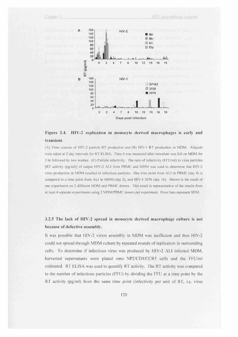
Chapter 3 HtV macrophaue tropism
160-1140120 -
100
HIV-2■ Mil a Mir
□ Ali 0 Etp
0 2 4 7 8 10 12 15 16 19E
160140120100
HIV-1
□ SF162□ 2028
80 ■ 207660
. ( I k
4020- r-n_ (-!-■ n— r J
00 2 4 7 8 10 12 15 16 19
Days post infection
Figure 3.4. HIV-2 replication in monocyte derived macrophages is early and
transient.(A) Time courses of HIV-2 particle RT production and (B) HIV-1 RT production in MDM. Aliquots
were taken at 2 day intervals for RT ELISA. Time 0 was measured after inoculum was left on MDM for
3 hr followed by two washes. (C) Particle infectivity. The ratio of infectivity (FFU/ml) to virus particles
[RT activity (pg/ml)] of output HIV-2 ALI from PBMC and MDM was used to determine that HIV-2
virus production in MDM resulted in infectious particles. One time point from ALI in PBMC (day 4) is
compared to a time point from ALI in MDM (day 2), and HIV-1 2076 (day 16). Shown is the result of
one experiment on 2 different MDM and PBMC donors. This result is representative of the results from
at least 4 separate experiments using 2 MDM/PBMC donors per experiment. Error bars represent SDM.
3.2.5 The lack of HIV-2 spread in monocyte derived macrophage culture is not
because of defective assembly.
It was possible that HIV-2 virion assembly in MDM was inefficient and thus HIV-2
could not spread through MDM culture by repeated rounds of replication in surrounding
cells. To determine if infectious virus was produced by HIV-2 ALI infected MDM,
harvested supernatants were plated onto NP2/CD4/CCR5 cells and the FFU/ml
estimated. RT ELISA was used to quantify RT activity. The RT activity was compared
to the number of infectious particles (FFU) by dividing the FFU at a time point by the
RT activity (pg/ml) from the same time point (infectivity per unit of RT, i.e. virus
120

Chapter 3 HIV macrophaue tropism
supernatants that have less infectious virus will have more RT compared to infectivity). HIV-2 ALI infected MDM had RT activity only at day 2, so this time point was compared to one time point, day 4, from ALI infected PBMC that exhibited rebounding virus replication. We can see that the ratio of FFU/RT from HIV-2 ALI infected MDM js equivalent to that seen at day 4 in ALI infected PBMC (fig 3.5b), thus the lack of spread and rebounding replication of HTV-2 in MDM is unlikely to be the inability to produce infectious virus alone. HTV-1 2076 demonstrates greater infectivity when produced from MDM than when it is produced from PBMC (fig 3.5b), which may explain why HTV-1 demonstrates spreading and rebounding replication in MDM culture.
A 10*
10*
101
B
a ■All 2076
RTactivity(pg/ml)
□ PBMC MDM
Ali 2076
FFU/ml
«£s1D
□ PBMC H MDM
Ali1
2076
Figure 3.5. HIV-2 ALI produces infectious particles from infected monocyte derived macrophages.The ratio of infectivity (FlFU/ml) to RT activity (pg/ml) of HIV-2 ALI harvested from infected PBMC and MDM was used to determine that HIV-2 virus production in MDM resulted in infectious particles comparable to PBMC (bottom panel). One time point from ALI infected PBMC (day 4) is compared to a time point from ALI infected MDM (day 2), and HIV-1 2076 (day 16) (top panel). Shown are the results from samples obtained from the experiment in fig 3.4 and are representative of the result from 2 independent experiments using 2 MDM and PBMC donors each.
121
Chapter 3 HIV macrophage tropism
3.2.6 Latent virus production can be stimulated in HIV-2 infected monocyte derived macrophages.
To determine whether latent integrated HTV-2 provirus was present in MDM culture, lipopolysaccharide (LPS) was added to a well of infected MDM 4 days post-infection and harvested the following day (day 5 post-infection). Stimulation of transcription of HTV-2 JAU RNA was evident by gag-LTR RT-PCR (fig 3.6a). The M0 tropic HTV-1 SF162, maintains gag-LTR RNA production throughout the time-course, indicating constant viral replication (fig 3.6b).
3.2.7 There is a loss of HTV-2 gag-LTR RNA production with HIV-2 gag-LTR DNA loss.I next determined whether viral RNA transcripts were being produced over a 5 day period because, as fig 3.4a indicates, there is a burst of RT production at early time points but not at late ones. I wanted to determine if HTV-2 JAU RNA transcription was associated with RT production. RT-PCR to measure gag-LTR RNA transcripts was conducted on MDM infected with HTV-2 JAU or HTV-1 SF162. MDM were incubated with equivalent input (5.0 x 10³ PBMC TCID50) of either JAU or SF162 for 3 hr, time points were harvested every 24 hr up to and including 5 days post-infection. The infected MDM were harvested with Trizol® RNA extraction reagent and the cDNA was prepared. HTV-2 RNA production by MDM peaks at 24 hr post-infection and decreases to almost undetectable levels by day 5 (fig 3.6a). Interestingly, JAU transcription could be restimulated by addition of LPS and titrated out 100-fold (fig 3.6a), however the amount of recovery is not as intense as the expression seen at 24 hr post-infection.
I determined whether the LPS stimulation of HIV-2 viral RNA production in fig 3.5a translated to production of virus particles. I detected virus particles in harvested supernatants by RT-ELISA and show that HTV-2 JAU can be stimulated to higher levels than HTV-1 SF162 at day 5 with LPS stimulation (fig 3.6c). SF162 was apparently unaffected by LPS treatment.
122

Chapter 3 HIV macrophage tropism
A HIV-2 JAU
GAPDH
24HR 48HR 72HR 96HR 5 Day 5 Day LPS
RT + - + - + - + - + - + -
gagLTR
LPS stimulated dilution
1/10 1/100 1/1000
HIV-1 SF162GAPDH
24HRS 48HRS 72HRS 96HRS 5 Day
gagLTR
AMtw&adhM
104 1 103 o> 102Q.H 101 * 100
10’1
JAU ■ SF162 □
I t24hr 72hr 5 Day 5 Day +
LPS
Time post-infection
Figure 3.6. HIV-2 infection of monocyte derived macrophages is transient and can
be restimulated with lipopolysaccharide treatment.RT-PCR of (A) HIV-2 JAU and (B) HIV-1 SF162 infected MDM harvested at indicated time points post
infection. LPS (50ng/ml) was added to the HIV-2 JAU culture 4 days post-infection and harvested on
day 5. RNA was extracted at the time points indicated and cDNA was made for GAPDH and garg-LTR
PCR. (C) RT activity from the experiment in fig 3.6a and b above. Shown are the results of one
experiment and they are representative of the results from at least 3 independent experiments. Error bars
represent SDM.
123

Chapter 3 HIV macronhage tropism
3.2.8 The HTV-2 isolate, prCBL23, which displayed no replication on monocyte derived macrophages demonstrates a post-entry restriction in HeLa/CD4 cells.
Until this point of the chapter I have described a general post-entry restriction of HIV-2 isolates in MDM compared to PBMC and HIV-1. In fig 3.1a and b I noted that prCBL 23 was not able to infect MDM whereas all of the other HIV-2s tested were able to replicate, to some degree. At this point I focus on a post-entry restriction of HIV-2 prCBL 23 (MCR) in HeLa/CD4 cells. It has been reported previously that prCBL 23 demonstrates a post-entry block to infection in MDM and HeLa/CD4 cells that occurs post-reverse transcription (McKnight et al, 2001).
The T cell line adapted HIV-2 strain CBL 23 and the primary isolate prCBL 23 are restricted in HeLa/CD4 cells, as described previously (McKnight et al, 2001). To study this post-entry restriction I concentrated on infection of HeLa/CD4 cells as they are relatively easy to culture. Fig 3.7 shows that CBL 23 has veiy similar infectivity for U87/CD4/CXCR4 and HeLa/CD4 cells. PrCBL 23 infection of U87/CD4/CXCR4 cells is comparable to CBL 23, however there is a drop in infectivity from a mean of 2.3 x 10⁴ FFU on permissive U87/CD4/CXCR4 cells to 5.0 x 10² FFU on restrictive HeLa/CD4 cells (fig 3.7). The mean titre of prCBL 23 on U87/CD4/CXCR4 divided by the titre on HeLa/CD4 cells reveals a restriction of 45.6-fold. Thus, the restriction of prCBL 23 in MDM compared to PBMC is similar to the restriction, seen here, in HeLa/CD4 cells compared to U87/CD4/CXCR4 cells
Figure 3.7. HTV-2 PrCBL 23 is restricted in HeLa/CD4 cells.CBL 23 and prCBL 23 viruses were titrated onto U87/CD4/CXCR4 and HeLa/CD4 cells, fixed and stained for infection 3 days lata*. Foci of infection were counted and FFU/ml calculated.
□ U87/CD4/CXCR4 ■ HeLa/CD4
CBL23 prCBL23

Chapter 3 HfV macrophage tropism
3.2.9 Lv2 is a restriction to HIV infection in some human cell types.
Molecular clones and gene swaps were made from prCBL 23 and CBL 23 by my colleague, Christian Schmitz, to study the restriction of prCBL 23 in MDM and HeLa/CD4 cells. These molecular clones were named Molecular Clone Restricted (MCR) and Molecular Clone Not-restricted (MCN) after prCBL 23 and CBL 23, respectively (Schmitz et al., 2004). Restrictive cells are resistant to infection by restricted viruses, MCR and prCBL 23 on HeLa/CD4 cells. This pattern of HIV restriction in human cells has been named Lentivirus susceptibility factor 2 (Lv2) (Schmitz et al., 2004), after the Lentivirus susceptibility factor 1 (Lvl) pattern of retroviral restriction in non-human primate cells (Cowan et al., 2002). Figs 3.8a and b show infection of a standardised dose (equivalent input determined on unrestrictive U87/CD4/CXCR4) of MCR and MCN on the permissive U87/CD4/CXCR4 and the Lv2 restricted HeLa/CD4 cells. As expected MCN is not restricted on either of the U87/CD4/CXCR4 or HeLa/CD4 cells but there is 42.0-fold less infection of MCR on the HeLa/CD4 than the U87/CD4/CXCR4 cells.
10®
I 1043u.u. 10®
10*
□ U87/CD4/CXCR4 ■ HeLa/CD4
MCR MCN
B S 50
MCN
Figure 3.8. Lv2 is a restriction to HIV infection in some human cell types.(A) The same infectious dose (standardised on permissive U87/CD4/CXCR4 cells) of MCR and MCN were plated on permissive U87/CD4/CXCR4 and restrictive HeLa/CD4 cells and fixed and stained for HIV-2 infection 3 days later. Infectivity is reported as FFU/ml. (B) The fold restriction was calculated by dividing the FFU/ml on permissive U87/CD4/CXCR4 cells by the FFU/ml on restrictive HeLa/CD4 cells for each virus in (A). Results shown represent at least 10 independent experiments. Error bars represent SDM.
40302010 I
MCR
125
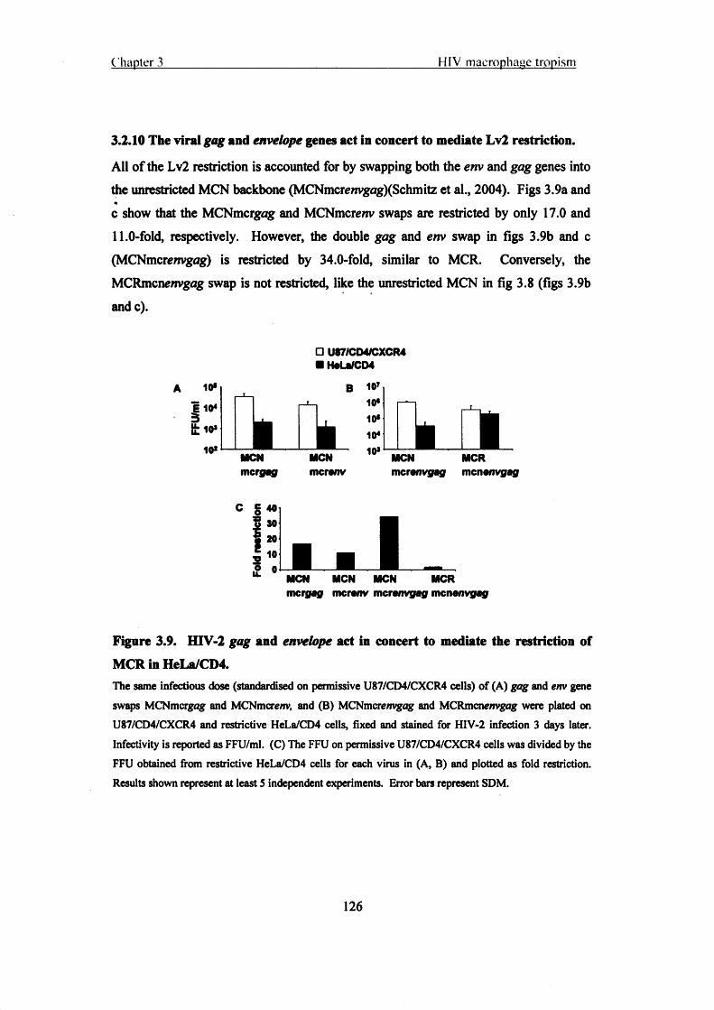
Chapter 3________________________________________________ HIV macrophage tropism
3.2.10 The viral gag and envelope genes act in concert to mediate Lv2 restriction.
All of the Lv2 restriction is accounted for by swapping both the env and gag genes into the unrestricted MCN backbone (MCNmcremgag)(Schmitz et al., 2004). Figs 3.9a and c show that the MCNmcrgag and MCNmcre/iv swaps are restricted by only 17.0 and 11.0-fold, respectively. However, the double gag and env swap in figs 3.9b and c (MCNmcrewvgag) is restricted by 34.0-fold, similar to MCR. Conversely, the MCRmcnenvgag swap is not restricted, like the unrestricted MCN in fig 3.8 (figs 3.9b andc).
10*1104 1 10*
10*
□ U87/CD4/CXCR4 ■ HeLa/CD4
_IkMCNmcrgag
MCNmcrenv
B 107 10*
10*
10*
10* MCN MCRmcranvgag men envgog
C § 40
i 30 • 20
i 1:II V i ■ IMCN MCN MCN MCR mcrgag mcrenv merenvgag mcnenvgag
Figure 3.9. HIV-2 gag and envelope act in concert to mediate the restriction of MCR in HeLa/CD4.The same infectious dose (standardised on permissive U87/CD4/CXCR4 cells) of (A) gag and env gene swaps MCNmcrgag and MCNmcrenv, and (B) MCNmcrenvgog and MCRmcnenvgog were plated on U87/CD4/CXCR4 and restrictive HeLa/CD4 cells, fixed and stained for HIV-2 infection 3 days later. Infectivity is reported as FFU/ml. (C) The FFU on permissive U87/CD4/CXCR4 cells was divided by the FFU obtained from restrictive HeLa/CD4 cells for each virus in (A, B) and plotted as fold restriction. Results shown represent at least 5 independent experiments. Error bars represent SDM.
126

Chapter 3 HIV macrophage tropism
3.2.11 Lv2 restriction of HIV infection is common among HIV-1 and HIV-2
isolates.
HIV-1 (NL4.3, RF, IIIB, MN, SF2, 2044, 2005, 2028, 2076, M13, HAN2, and 89.6) and
HIV-2 (MCR, MCN, MIL, ETP, JAU, ACR23, MIR, AND, prCBL20 and SAB)
laboratory adapted and primary isolates were tested for infection on U87/CD4/CXCR4,
HeLa/CD4 and H399 cells to determine the prevalence of Lv2 restriction (fig 3.10a).
The fold restriction of infection on HeLa/CD4 and H399 cells was determined and half
of all of the HIV-2 isolates (MCR, ETP, AND, prCBL20, and SAB) and about one third
of the HIV-1 isolates (2076, Ml 3, HAN2, and 89.6) were restricted in an Lv2 manner.
□ U87/CD4/CXCR4■ HeLa/CD4■ H399
A 106 |10 s
104 103
1 102^ MCR MCN MIL ETP JAU ACR23 MIR AND prCBL20 SABLLLL
B 1Q7
fh \ k M i a106105104103102
i ll l l . 1 11 . JNL4.3 RF lllb MN SF-2 2044 2005 2028 2076 M13 HAN2 89.6
c 403020
co 10o'k_ 0Bi
Dk_■D120O | |LL 80
40
0
U87/HeLa
LU U87/H399
B -. m-\ —r-. —i i i B~i K-iMCR MCN MIL ETP JAU ACR23 MIR AND prCBL20 SAB
LNL4.3 RF lllb MN SF-2 2044 2005 2028 2076 M13 HAN2 89.6
Figure 3.10. HIV-1 and HIV-2 isolates are susceptible to Lv2 restriction.
127

Chapter 3 HIV macrophage tropism
(A) An array of HIV-2 and (B) HIV-1 isolates was titrated on permissive U87/CD4/CXCR4, H399*, and restrictive HeLa/CD4 cells and the FFU/ml calculated. Fold restriction was calculated for (C) HIV-2 and (D) HIV-1 by dividing the titre on permissive U87/CD4/CXCR4 cells by the titre on restrictive HeLa/CD4 or permissive H399 cells. *H399 cells, described in greater detail in chapter 4, are a HeLa/CD4 cell line that expresses a CD4 molecule with a C-terminal tail truncation at the membrane proximal amino acid position 399. This cell line that is not Lv2 restrictive, unlike the related HeLa/CD4, is used in chapter 4 to further characterise the cellular location of Lv2 restriction.
3.3 Discussion
The data presented here show that apart from a single burst of viral replication HIV-2 continuous replication in MDM is largely absent. However, latent HIV-2 provirus could be stimulated with LPS. In contrast, HIV-1 showed delayed and rebounding replication in MDM with no evidence of a latent phase. A molecular clone, MCR was made from HTV-2 prCBL 23, which demonstrated a complete block to replication in MDM. The models used to study HTV post-entry restrictions in human cells, Lv2, was found to be a common pattern of restriction to HIV-1 and HIV-2 infection in human cells.
Sequencing of the HTV-2 V3 loop revealed that a high net positive charge of the V3 loop was associated with CXCR4 use on NP2/CD4/CXCR4 cells. None of the HTV-2 isolates infected MDM very well when compared to PBMC infection or to HIV-1 on
MDM, but the HTV-2 isolates that exhibit the highest relative MDM infection are those that use CCR5 and have less positive V3 loop charges. However, HTV-2 MLC, an isolate that uses CCR5 and has the lowest V3 loop charge demonstrates poor infectivity on MDM. The V3 loop charge is only weakly correlated with M0 tropism, so overall, the V3 loop fails to determine the lack of replication of HTV-2 in MDM compared to PBMC and HTV-1. If the V3 loops were somehow responsible for the lack of MDM infection we would expect to see a consistently high charge on all of the HIV-2 V3 loops and a stronger ability by some of the HIV-2 isolates to infect MDM. A recent study has suggested that the env genetic determinants dictating cell tropism of dual tropic HIV-1 viruses are “complex”; they include regions of the V1/V2 and V5 hyper variable regions, and charged and uncharged amino acids in the V3 loop (Ghaffari et al., 2005), so too, the HTV-2 env determinants of tropism may be complex as well.
128

Chapter 3 HIV macrophage tropism
Low positive and high positive HIV-2 V3 loop charges have been reported to be associated with slow/low and fast/high viral phenotypes, respectively (Albert et al., 1996). It has also previously been reported that shift in the coreceptor usage from £CR5 to CXCR4 is associated with an increase in the net positive charge in the V3 region of HTV-2 molecular clones (Isaka et al., 1999). Here I confirm that an increasing net positive charge of the V3 loop correlates with CXCR4 usage of HIV-2 primary isolates and that there is a weak association between CCR5 using HTV-2 isolates and M0 tropism. There was a broad range of V3 loop charges among the HIV-2 isolates so it does not appear as though V3 loops alone are responsible for the lower HTV-2 TCID50S in MDM compared to HIV-1.
The lower infectivity of HTV-2 compared with HIV-1 in MDM can be attributed to at least 2 factors: HIV-1 is more infectious when produced from MDM than PBMC and HIV-2 reverts to a latent state in MDM. HIV-2 primary isolates enter MDM and reverse transcribe to the same levels as seen in PBMC. However, when later time points were analysed they showed significantly less gog-LTR product that suggests there is less viral spread in culture or that this is just ‘left-over’ gag-LTR product from what could be seen at earlier time points post-infection.
Latency has been demonstrated for HTV-1, Jordan et al. show that the inducible replication of latent integrated HTV-1 in the T cell line Jurkat (Jordan et al., 2003). They show that a small proportion of HIV-1 integrates into or near alphoid repeats in heterochromatin and the natural suppression of transcription in this region of the genome inhibits integrated HTV-1 transcription (Jordan et al., 2001). I have shown stimulation of HTV-2 production in MDM with LPS. The levels of stimulation of latently HTV-1 infected T cell lines has been demonstrated to vary with different agonists, particularly those that best stimulate NF-kB, like the phorbol ester, TPA (Jordan et al., 2003). Strong NF-kB agonists (LPS) stimulate HIV-2 replication in MDM to the same levels that were demonstrated at day 2 in HTV-2 infected MDM.
M0s have been demonstrated to be a stable source of HTV-1 replication due to their greater ability to survive virus replication whereas HIV-1 infected PBMC have been shown to readily undergo apoptosis and necrosis, several mechanisms have been
129
Chapter 3 HIV macrophage tropism
proposed (Meylan et al., 1998; Wang et al., 2001; Zhang et al., 2001) reviewed in (Alimonti et al., 2003). Gartner et al. first demonstrated that ex-vivo M0s can be infected with HIV-1, and may continue producing virus for more than 40 days (Gartner et al., 1986). Certain factors have been shown to promote the survival of M0s infected tyith HTV-1. Garaci et al. have shown that Nerve Growth Factor (NGF) is an autocrine factor that inhibits apoptosis and is up regulated by HTV-1 infected M0s (Garaci et al.,1999).
The in vitro experiments conducted here in ex-vivo MDM may have implications for disease progression in the host. HTV-2 RNA production can be induced in MDM by lipopolysaccharide five days post-infection suggesting that MDM remain infected and that provirus remains integrated at later time points. Eitner et al. have demonstrated in HTV-2 infected pig-tailed macaques that there is a highly disseminated viral phase followed by exclusive localisation of infection to the FDC network with few infected cells (Eitner et al., 2000). These findings may reflect the burst of viral RNA that I see in HTV-2 infected MDM followed by latent/loss of HIV-2 replication. Perhaps the initial burst of replication helps the virus to establish itself in the host and the subsequent latency may allow HIV-2 to avoid the immune system.
Individuals infected with HTV-2 have lower plasma viral loads than those infected with HTV-1, which reflects the lesser pathogenicity of HTV-2 (Gottlieb et al., 2002; Popper et al., 2000; Popper et al., 1999; Shanmugam et al., 2000). However the lower viral load does not equate to a lower proviral load than HTV-1 (Ariyoshi et al., 1996; Berry et al., 1994; Sarr et al., 1999), but to less viral production from the proviral template (Popper et al., 2000).
Lv2 restriction of HTV infection was introduced in this chapter. In the next chapter I demonstrate that an endocytic pathway delivers HIV to Lv2 restriction in HeLa/CD4 cells.
130

Chapter 4
An endocytic route of delivery to Lv2 restriction
4.1 Introduction:
In the last section, chapter 3, the restricted replication of HIV-2 isolates in MDM was identified. In particular, prCBL 23 demonstrated no replication in MDM. The molecular clone, MCR, derived from prCBL 23 virus swarm was made by my colleague Cristian Schmitz, and a restriction model using HeLa/CD4 cells as the restrictive cell type was developed (Schmitz et al., 2004). This chapter investigates the route of entry taken by Lv2 restricted and unrestricted viruses.
There have been many reports on the involvement of endocytosis in HIV infection (Fredericksen et al., 2002; Marsh and Pelchen-Matthews, 2000; Vidricaire et al., 2003; Wei et al., 2005), but so far none that have linked retroviral restriction to a particular endocytic pathway. Studying the pathways of restriction in the host cell can help to understand the pathway of infection and the preferred route of entiy. It may not be the entry pathway that is restricting viral entry, per-se, but an anti-viral protein/ saturable restriction factor (Stremlau et al., 2004; Towers et al., 2000).
Clathrin mediated endocytosis was the first mechanism of receptor-mediated cellular internalisation to be discovered as a means of yolk protein uptake in the mosquito oocyte (Roth and Porter, 1964). The “archetypical” cargo to be trafficked by this pathway is the Trf receptor that internalises iron-binding protein, Trf (Harding et al., 1983; Pearse, 1982), and is commonly used as a marker of clathrin activity (Le Roy and
v
Wrana, 2005). Sometimes called the “classical route” of endocytosis clathrin trafficking is thought to be the most active route for receptor-mediated internalisation. Once the clathrin coated pit has budded from the PM it fuses with an early endosome that can become an acidified late-endosome of pH 5.0 to 6.5.
Virus Envs can cause host-virus membrane fusion by pH-dependent and independent mechanisms. The best understood mechanism of pH-dependent fusion is that of the
131

Chapter 4 An endocvtic route o f delivery to L.v2 restriction
influenza Haemagglutinin protein (HA), a type I fusion protein. The HA protein is folded into a high-energy state that is released upon exposure of the virion to a low pH (5.0-6.5) endosomal compartment, exposing the N-terminal fusion peptide (Wiley and Skehel, 1987). A chain-direction reversal occurs at the amino terminal end of the central coiled coil resulting in the extension of the fusion peptide towards the target membrane (Colman and Lawrence, 2003). A second chain-direction reversal occurs in the central coiled-coil region, this time projecting the carboxyl terminus back towards the amino terminus. This folding of the fusion domain back to the membrane anchor acts to bring the viral and target membranes into close apposition for fusion.
Most retroviruses and paramyxoviruses fuse with host target membranes at neutral pH and so it is the receptor binding interaction that triggers the fusion mechanism (above).
Clathrin endocytosis has been reported as an entry route for pH-dependent and. independent viruses alike. Since the clathrin endocytic route can lead to an acidified compartment it is the preferred route of entry by many pH-dependent viruses: Semliki, an alphavirus, was the first confirmed pH-dependent virus to use clathrin as its primary route of entry (Helenius et al., 1980; Helenius et al., 1982; Marsh et al., 1982; White et al., 1980). Vesicular Stomatitis Virus (VSV) has also been shown to use clathrin endocytosis as its route of entry by inhibition with amantadine (Schlegel et al., 1982), and has been confirmed recently with dominant negative mutants (Sun et al., 2005). Ph- dependent viruses such as Influenza, Sindbis, and human rhinovirus 14 (DeTulleo and Kirchhausen, 1998) also use this pathway of entry. HIV-1, a pH-independent virus has been shown to require clathrin, at least in part, as a route of entry (Daecke et al., 2005; Stein et al., 1987).
Caveolae were first described as “omega-like” structures in early morphology studies of the PM (Palade, 1961) and have since been shown to require the scaffolding protein caveolin to support their shape and function (Rothberg et al., 1992). Caveolin binds cholesterol and it is therefore thought that caveolae mediate cholesterol storage and homeostasis (Johannes and Lamaze, 2002; Rothberg et al., 1992), and numerous signalling and trafficking events (Anderson, 1998; Simons and Toomre, 2000). They are expressed on fibroblasts, smooth muscle cells, endothelial, and epithelial cells but not T cells (Scherer et al., 1997).
132

Chapter 4 An endoevtic route o f delivery to Lv2 restriction
One of the few and best characterised intracellular parasites that enter via caveolae is the SV40 polyoma virus. Fewer viruses have been found that use this internalisation mechanism as a portal of entry compared to clathrin endocytosis (Sieczkarski and Whittaker, 2002a), perhaps because it is a slowly internalising, non-constitutive pathway and is therefore less active than clathrin endocytosis (Thomsen et al., 2002). This may be why SV40 requires ligand triggering of caveolae to induce actin rearrangements and uptake (Pelkmans et al., 2002). The study of SV40 internalisation has also revealed a novel organelle called the caveosome (Pelkmans et al., 2001), used as a trafficking intermediate on its way to the ER and nucleus.
Apart from clathrin-mediated and caveolae endocytosis, internalisation of cel 1-surface markers independently of clathrin-coated pits has lead to the definition of a new type of endocytic mechanism, clathrin independent endocytosis (Nichols and Lippincott- Schwartz, 2001). Internalising lipid (Puri et al., 2001) and protein (Nichols et al., 2001) markers have been visualised outside of clathrin pits suggesting that clathrin independent routes exist, but also in different compartments suggesting the existence of numerous non-classical routes of endocytosis (Nichols and Lippincott-Schwartz, 2001). The membrane organisation of vesicles in these pathways is thought to be composed, primarily of lipid rafts (Simons and Ikonen, 1997), and the organisation of some lipid raft markers post-internalisation, such as GPI-linked proteins, is thought to occur in discreet compartments independent of the classical early endosome antigen 1 (EEA1) endosome (Sabharanjak et al., 2002). Some lipid raft markers such as the M2 muscarinic acetylcholine receptors internalise independently of clathrin but traffic into a classical clathrin/Rab5 positive early endosome post-internalisation (Delaney et al., 2002).
Virus entry by clathrin/caveolae independent pathways is highly variable and obligate in some cases. Murine polyomavirus (MPV) requires actin depolymerisation and stable microtubules for infection and a clathrin/caveolae independent pathway for productive entry (Gilbert and Benjamin, 2000; Gilbert et al., 2003). Similar to MPV, SV40 can use at least one caveolae/clathrin independent route of entry in addition to caveolae (Damm7et al., 2005). Clathrin independent uptake via “smooth surfaced pits and vesicles” was implicated in early studies of influenza virus entry (Matlin et al., 1981). The use of
133

Chapter 4 An endocvtic route o f delivers to Lv2 restriction
dominant negative mutants has confirmed influenza virus entry by both clathrin and clathrin/caveolae independent endocytosis in the same cell (Lakadamyali et al., 2004; Rust et al., 2004; Sieczkarski and Whittaker, 2002b). Even though influenza does not require clathrin for initial entry into the cell it does require the classical Rab 5 and Rab 7 acidified, late endosomal compartments for productive infection (Sieczkarski and Whittaker, 2003).
Depletion of membrane cholesterol (using cyclodextrins) has shown that PM lipid rafts are necessary for HTV-1 binding and entry (Liao et al., 2001), and may be required for clustering of CD4 receptor and coreceptors to sites of virus binding (Manes et al.,2000). However, truncation of the CD4 cytoplasmic domain suggests that CD4 does not need to be localised to lipid rafts for efficient HTV-1 entry and infection (Bedinger et al., 1988; Popik and Alee, 2004). The involvement of lipid rafts in HTV entry may occur post-CD4 binding by destabilisation of lipid raft domains (Kozak et al., 2002). It has even been suggested that virion associated lipid rafts are required for virus infectivity (Graham et al., 2003; Liao et al., 2003).
4.2 Results:
4.2.1 MCR is not restricted when fusion is forced at the ceil surface.
MCR can be rescued in HeLa/CD4 cells when it is pseudotyped with VSV-G (fig 4.1a)(Schmitz et al., 2004). VSV-G is a pH-dependent Env that requires delivery into an acidified endosome compartment to trigger fusion with the host cell (Blumenlhal et
al., 1987). Since MCR avoids restriction by delivery into an endosomal pathway with a VSV Env, I determined if MCR restriction could be overcome if MCR delivery was forced, outside of an endosomal compartment, at the cell surface with a VSV Env. The fusion of VSV-G at the cell surface by transient acid-shock with acidified (pH 5.5) media has been described previously (Marsh and Bron, 1997). The buffering of growth media to a pH of 5.0-5.5 emulates the pH of late endosomes and acts to fuse pH- dependent Envs at the cell surface when added transiently to pH-dependent-virus and cell mixtures (Marsh and Bron, 1997; White et al., 1980).
134

Chapter 4 An endocvtie route o f delivery to Lv2 restriction
Fig 4.1a and b show a comparison of MCR and MCR(VSV). Infection of MCR is rescued if restrictive cores are pseudotyped with VSV Env (fig 4.1a). MCR infection in HeLa/CD4 cells is not affected by monensin neutralisation of endosomal pH (control) or by the transient acid treatment at pH 5.5 compared to normal infection. However infection is recovered more than 10-fold when MCR(VSV) is forced to fuse at the cell surface by transient acid shock (‘pH 5.5 + monensin’) (fig 4.1b). When VSV-G mediated fusion is prevented by monensin it fails to rescue MCR [MCR (VSV) ‘pH 6.8 + monensin’, fig 4.1b].
A
B
pH 6.8 pH 6.8 ♦ pH 5.5 +monsnain monsnain
Figure 4.1. MCR pseudotyped with vesicular stomatitis virus envelope is rescued from Lv2 restriction in HeLa/CD4 cells: MCR is not restricted in HeLa/CD4 cells when forced to fuse at the cell surface.(A) MCR and MCR(VSV) were titrated on U87/CD4/CXCR4 and HeLa/CD4 cells and fixed and stained fa- HIV-2 infection 3 days later. (B) Surface fusion of MCR(VSV) by transient acid shock. MCR and MCR(VSV) were titrated on HeLa/CD4 cells and treated with low pH media for 2 min to induce fusion (pH 5.5 + monensin) or controls with physiological pH media with or without monensin (‘pH 6.8 + monensin’ and ‘pH 6.8’, respectively), low pH media was replaced with pH 6.8 buffered growth media and cells were fixed and stained for HIV-2 infection 3 days later (materials and methods). The results shown are representative of at least 4 independent experiments. Error bars represent the SDM.
10«10410*
10*
101_ r i
MCR MCR(VSV)
□ U87/CD4/CXCR4 ■ HeLa/CD4
10*1
10«
104 T ■ ■■ MCR □ MCR(VSV)
Normalinfaction
Control VSV fusion at call surfacs
135

Chapter 4 An endocvtic route o f delivery to Lv2 restriction
4.2.2 Lv2 restriction does not require acidified endosomes to inhibit virus entry.
Bafilomycin Al is a lysosomal tropic agent that neutralises the pH of late endosomes and lysosomes, effectively inhibiting the fusion and entry of pH-dependent viruses (Drose et al., 1993; Helenius et al., 1982; Marsh et al., 1982; Perez and Carrasco, 1993). Previous reports have demonstrated that HTV-1 does not require a low pH for fusion (McClure et al., 1988; Stein et al., 1987). I determined the effect of pH on MCR restriction in HeLa/CD4 cells. Fig 4.2 shows that treatment of HeLa/CD4 cells with bafilomycin Al can inhibit infection by a pH-dependent VSV Env [MCR(VSV)] by more than 99%. Bafilomycin does not alter the infectivity of MCR on restrictive HeLa/CD4 cells (fig 4.2). Interestingly, MCR infectivity decreased (by approximately 3-fold) from a mean of 145 to 45 FFU on permissive U87/CD4/CXCR4 cells with bafilomycin treatment Though there was no effect on infection in HeLa/CD4 cells there was a decrease in restriction from 11-fold to 3-fold, upon bafilomycin treatment. This was due entirely to a drop in infection of MCR on permissive U87/CD4/CXCR4 cells and not any change in infection on restrictive HeLa/CD4 cells (fig 4.2).
Figure 4.2. Neutralisation of intracellular pH with bafilomycin Al doesn’t affect MCR restriction in HeLa/CD4 cells.Viruses and pseudotypes were plated on U87/CD4/CXCR4 and HeLa/CD4 cells in the presence of bafilomycin Al. Cells were washed with normal growth media and infection was allowed to proceed in the absence of bafilomycin Al. Cells were fixed and stained for HIV-2 infection 3 days later. Results shown are representative of 3 independent experiments. Error bars represent SDM.
□ U87/CD4/CXCR4 ■ HeLa/CD4
MCR MCN MCR(VSV) MCR MCN MCR(VSV)
Bafilomycin A1
136

Chapter 4__________________________ An endocytic route o f delivery to Lv2 restriction
4.2.3 Hypertonic media inhibit endocytosis of transferrin and cholera toxin
binding subunit in U87/CD4/CXCR4 and HeLa/CD4 cells.
I determined the effect of endocytosis inhibition on restriction in HeLa/CD4 cells,
without affecting the pH of endosomal compartments. Hypertonic sucrose inhibits
receptor-mediated endocytosis without affecting pH (Daukas and Zigmond, 1985;
Heuser and Anderson, 1989). As a positive control for inhibition of endocytosis I used
FITC labelled Trf (Trf-FITC)(fig 4.3a-d), which is internalised by clathrin coated pits
(Gruenberg, 2001), and FITC labelled CTxB (CTxB-FITC)(fig 4.3e-h) which is
internalised by a lipid raft dependent pathway (Wolf et al., 2002). Hypertonic sucrose
inhibited Trf-FITC and CTxB-FITC uptake in both the restrictive HeLa/CD4 and
permissive U87/CD4/CXCR4 cells, signified by capping of FITC at the cell surface (fig
4.3b, d, f, h).
Transferrin-FITC
U87/CD4/CXCR4
HeLa/CD4
Cholera Toxin-FITC
U87/CD4/CXCR4
HeLa/CD4
Control 0.45M sucrose
Figure 4.3. Sucrose inhibits transferrin and cholera toxin-B uptake.
Transferrin-polylysine-FITC (a-d) or cholera toxin binding-subunit-FITC (e-h) were bound to the
permissive U87/CD4/CXCR4 or restrictive HeLa/CD4 cells with or without 0.45M sucrose and examined
by confocal microscopy. Inhibition is shown by Trf-FITC or CTxB-FITC capping at the cell surface with
137

Chapter 4 An endocvtic route o f delivery to Lv2 restriction
simultaneous uptake into perinuclear (endosomal) regions of the cell. White scale bar indicates 10pm magnification. Results are representative of at least 4 independent experiments.
4.2.4 Lv2 restriction depends on an endocytic pathway of entry that is directed by the viral envelope.Fig 4.4 shows the effect of hypertonic sucrose on restricted MCR infection. Unrestricted MCN and restricted MCR were plated on sucrose treated HeLa/CD4 and U87/CD4/CXCR4 cells. The infectivity of MCR on HeLa CD4 cells increased 6.5-fold from a mean of 16 to a mean of 105 FFU with sucrose treatment (fig 4.4a). Fig 4.4b shows the resultant fold restriction decrease from 12.0-fold to 1.5-fold. There was no effect of sucrose treatment on MCR infection of permissive U87/CD4/CXCR4 cells or on MCN infection of either HeLa/CD4 or U87/CD4/CXCR4 cells (fig 4.4a).
10*
10*
101
10°
□ U87/CD4/CXCR4 ■ HeLa/CD4
i l n ^ iMCR MCR 4- MCN MCN +
sucrose sucrose
B j M
| 10
I .MCR
□ Control ■ 0.45M Sucrose
MCN
Figure 4.4. Treatment of Lv2 restrictive HeLa/CD4 cells with hypertonic sucrose recovers restricted MCR infection.Cells were plated with a U87/CD4/CXCR4 standardised dose of (A) MCR and MCN and treated with hypertonic sucrose for 1 hr. Cells were fixed and stained for HIV-2 infection 3 days later. (B) The FFU from permissive U87/CD4/CXCR4 infection were divided by the infection on restrictive HeLa/CD4 cells to obtain the fold restriction, for control and 0.45M sucrose treatments. Results are representative of 5 independent experiments. Error bars represent SDM.
138

Chapter 4____________________________ An endocvtic route o f delivery to Lv2 restriction
Lv2 restriction maps to both the viral env and gag genes (Schmitz et al., 2004). I determined whether the rescue of restriction with hypertonic sucrose was due to env or gag or both. Fig 4.5b shows the restriction of MCN containing a restricted Env (MCNmcrenv) is rescued from 21.0-fold to 1.0-fold, in this experiment, with hypertonic sucrose treatment The MCN molecular clone with a restricted Gag (MCNmcrgng) was less restricted but was completely rescued from 4.0-fold to 1.0-fold of restriction (fig 4.5b). Therefore both the env and gag mediated restrictions of Lv2 are rescued by inhibition of endocytosis.
A 10*
3 & 101
10“
□ U87/CD4/CXCR4 ■ HeLa/CD4
L
i i n i nMCN MCN MCN MCN mcrgag mcrgag+ mcrenv mcrenv+
sucrose sucrose
B § 40$| 20
3 0
□ Control ■ 0.45M Sucrose
MCNmcfffsg
MCNmcrenv
Figure 4.5. Recovery of MCR infection with hypertonic sucrose is mediated by both gag and envelope,(A) Cells were titrated with MCNmcrgag and MCNmcrenv viruses and treated with hypertonic sucrose. Cells were fixed and stained for HIV-2 infection 3 days later. (B) The permissive U87/CD4/CXCR4 infection was divided by the FFU on restrictive HeLa/CD4 cells to obtain the fold restriction. Results are representative of 3 independent experiments. Error bars represent SDM.
139

Chapter 4 An endocvtie route o f delivery to Lv2 restriction
4.2.5 Infectivity of HIV-1 retroviral vectors pseudotyped with MCR and MCN
envelopes in HeLa/CD4 cells can be improved by inhibition of endocytosis.
HIV-1 retroviral p8.91 particles pseudotyped with MCR and MCN Envs were plated on
HeLa/CD4 and U87/CD4/CXCR4 cells treated with hypertonic sucrose. Fig 4.6a shows
that the infectivity of both the MCR and MCN vector pseudotypes was rescued in
sucrose treated HeLa/CD4 cells by 8.5 and 5.0-fold, respectively. Sucrose treatment
had little effect on the infectivity of either of the HIV-2 Env pseudotypes on
U87/CD4/CXCR4 cells (fig 4.6b).
HeLa/CD4
1000
8.91(MCR)
8.91(MCN)
0 .68% 5 79% (8.5x)
HBSr’ '
7.61% 37.85% (5 .0x )
10° 101 10* 10* 104 10° 101 10* 10* 104
B U87/CD4/CXCR4
8.91(MCR)
8.91(MCN)
10° 101 10* 10* 104 10« 101 10* 10* 104
Control 0.45M sucrose
Figure 4.6. HIV-1 vector pseudotypes with MCR and MCN envelopes are rescued
by treatment with hypertonic sucrose in restrictive HeLa/CD4 cells.(A) HeLa/CD4 and (B) U87/CD4/CXCR4 cells were plated with a U87/CD4/CXCR4 standardised dose
of HIV-1 vector pseudotypes HIV-lg9 )(MCR) and HIV-lg9 ](MCN) and treated with hypertonic sucrose
for 1 hr. EGFP positive cells were determined by FACS three days post-infection. Results shown are
representative of 2 independent experiments.
140

Chapter 4 Ail endocytic route o f delivery to Lv2 restriction
4.2.6 Depletion of membrane cholesterol with methyl-p-cyclodextrin inhibits
uptake of cholera toxin binding subunit: membrane trafficking differences
between restrictive and permissive cells.
Membrane cholesterol is required for the endocytic trafficking of lipid rafts (Sandvig et
al., 2004). I determined the effect of membrane cholesterol depletion on Lv2 restriction
using MpCD. As controls, the disruption of lipid rafts by MpCD was followed in
HeLa/CD4 and U87/CD4/CXCR4 cells with CTxB-FITC and Trf-FITC. Cholera toxin
trafficking is via a lipid raft dependent endocytic pathway that is disrupted by depletion
of PM cholesterol (Fujinaga et al., 2003; Nichols, 2002; Sandvig et al., 2004; Sandvig
and Van Deurs, 2002). Fig 4.7d, e, f show that internalisation of CTxB in HeLa/CD4
cells is inhibited by MpCD and can be reconstituted with water-soluble cholesterol.
However, the internalisation of CTxB-FITC in U87/CD4/CXCR4 cells was not
inhibited by treatment with MpCD (fig 4.7a, b, c). As expected internalisation of
clathrin dependent Trf-FITC was not inhibited by depletion of membrane cholesterol
with MpCD (fig 4.7g-j).
U87/CD4/CXCR4 HeLa/C D4
Control
10mM MpCD
1 0mM MpCD + 0.2 (iM Cholesterol
CTxB-FITC
U87/CD4/CXCR4
Control
HeLa/CD4

Chapter 4 An endocvtic route o f delivery to Lv2 restriction
Figure 4.7. Methyl-p-cyclodextrin differentially inhibits cholera toxin-B uptake in
Lv2 restricted and permissive cells.Permissive U87/CD4/CXCR4 or restrictive HeLa/CD4 cells were treated with M(1CD. CTxB-FITC (a-f) and Trf-FITC (g-j) were bound to treated or untreated cells for lhr with or without water-soluble cholesterol and examined by confocal microscopy. Capping of CTxB-FITC at the cell surface shows inhibition. White scale bar indicates lOpm magnification. Results are representative of at least 4 independent experiments.
MCR, MCN, MCNmcrgag, and MCNmcrenv viruses were plated on MpCD treated HeLa/CD4 and U87/CD4/CXCR4 cells. The titre of all viruses was slightly decreased on U87/CD4/CXCR4 cells (fig 4.8b). Fig 4.8a shows that MCR titre on restricted HeLa/CD4 ceils increased 8.0-fold with M0CD treatment from a mean of 15 to a mean of 120 FFU and restriction could be partially reconstituted to a mean of 25 FFU by adding back water-soluble cholesterol. The unrestricted MCN was not affected by any of these treatments on HeLa/CD4 cells. The titres of MCNmcreov and MCNmcrgag on HeLa/CD4 cells were not affected by M0CD treatment (data not shown). M(3CD had no effect on the infectivity of a VSV Env pseudotype in fig 4.8c, supporting the evidence that the pathway delivered to by VSV-G is different from Lv2.
142

Chapter 4 An endocvtic route o f delivery to Lv2 restriction
B
10* i HeLa/CD4
10*
101
10°r h n
3It
MCR MCN □ Control MM0CD M MPCD +
10»! U87/CD4/CXCR4 choleaterol
10*
101
10°i i
MCR MCN
10*
? 102 | _ 1MCR(VSV)
.. ■ ■ ■Control M0CD MPCD +
cholesterol
Figure 4.8. MCR infection in restrictive HeLa/CD4 cells is rescued by treatment with methyl-p-cyclodextrin: MCR gag and envelope are both required for recovery of infection by cholesterol depletion.(A) HeLa/CD4 and (B) U87/CD4/CXCR4 cells were treated with lOmM M0CD for 30 min at 37°C, fixed doses (standardised on U87/CD4/CXCR4) of viruses were added on ice for 1 hr, and cells were incubated in serum free media +/- 0.20pM cholesterol at 37°C for 2 hr. Growth media was added and cells were fixed and stained for HIV-2 infection 3 days later. (C) Cholesterol depletion does not affect entry of VSV Env on HeLa/CD4 cells. The results shown are representative of at least 6 independent experiments. Error bars represent the SDM. The titres of MCR and MCN pn HeLa/CD4 cells are similar in (A) because input doses were equalised for ease of comparison.
4.2.8 Directing CD4 out of lipid rafts rescues restricted virus infection.
CD4 localises primarily within lipid rafts on the PM (Popik et al., 2002). I used a HeLa cell line expressing a mutant CD4 with a stop codon at amino acid position 399 (H399) in the cytoplasmic tail (fig 4.9a). The locations of wild-type CD4 compared to CD4h399 within lipid rafts were verified. Lipid rafts were prepared from U87/CD4/CXCR4,
143

Chapter 4___________________________An endocytic route o f delivery to Lv2 restriction
HeLa/CD4, and H399 cells on a sucrose gradient and densitometry was performed on
the fractions to determine the precise quantity of CD4 in lipid rafts. The lipid raft
marker placental alkaline phosphatase (PLAP) was tested in parallel for each fraction.
Fig 4.9b shows that the CD4 of U87/CD4/CXCR4 and HeLa/CD4 cells is found
predominantly in the lipid raft fractions 3,4, and 5 as is the lipid raft marker, PLAP in
fig 4.9c. In contrast, the CD4 of H399 cells is found primarily in fractions 9 and 10 and
was thus excluded from lipid rafts. C-terminally truncated CD4 localising outside of
lipid rafts has been verified (Popik and Alee, 2004). This truncation is upstream of
position 419-427, the poly arginine tract, shown to direct CD4 into lipid rafts (Popik
and Alee, 2004).
Lip id ra f t
H eLa/C D 4 H eLa/C D 4 H399
B S 50O «o 30(0 20o *-*-> 0
Lipid raftHlk ^Jfl C D 4
8 9 10
0 .< 50-1 40Q. 30
75 20
o 10*-» 0
55
□ U 87 /C D 4/C X C R 4■ H eLa/C D 4■ H399
H ■ h . . . 1PLAP
5 6 7 8 9 10
Top ---------------------------------------- ► pelletFraction N um ber
Figure 4.9. CD4 is directed out of lipid rafts by truncation of its cytoplasmic tail.
(A) Truncation o f CD4 cytoplasmic tail at membrane proximal histidine 399 (H399). Lipid rafts were
prepared from membrane lysates o f U87/CD4/CXCR4, HeLa/CD4, and H399 and analysed for (B) CD4,
and (C) placental alkaline phosphatase (PLAP) localisation as a lipid raft marker. Results are
representative o f 4 independent fractionations.
144

Chapter 4____________________________ An endocvtic route o f delivery to Lv2 restriction
Fig 4.10b shows that the restriction of MCR in HeLa/CD4 cells is recovered in H399
cells from 42.0-fold to 1.4-fold. The restrictions of MCNmcrenv and MCNmcrgag
were much lower than MCR, as expected (6.0 and 5.0-fold respectively). Interestingly,
the clones that expressed the MCN Env demonstrated a drop in infection on H399 cells
and those that expressed the MCR Env showed an enhancement of infection on H399
cells compared to HeLa/CD4 cells in all 5 independent experiments.
tMCNmcrgag
_ ■MCN mcrgag
Figure 4.10. Restricted MCR infection is rescued in HeLa/CD4H399 cells.(A) An equivalent input of 104 FFU (standardised on U87/CD4/CXCR4) of MCR, MCN, MCNmcrewv,
and MCNmcrgag were plated on U87/CD4/CXCR4, HeLa/CD4, and H399 cells and the FFU/ml
calculated. (B) The FFU from permissive U87/CD4/CXCR4 infection were divided by the infection on
restrictive HeLa/CD4 cells (black bars) or permissive H399 (grey bars) to obtain fold restriction. Results
are representative of 5 independent experiments. Error bars represent SDM.
4.2.9 Endocytic pathways are specifically inhibited with dominant negative
mutants.
Inhibition of endocytosis with MpCD is cell-type dependent and hypertonic sucrose
blocks numerous pathways (fig 4.5). In some cell types, MpCD will also inhibit
clathrin-mediated endocytosis (Rodal et al., 1999). Dominant negative mutants are used
here to target the three major pathways of endocytosis specifically: clathrin
B
106
i 105D 104 U.U - 1 0 3
102
□ U87/CD4/CXCR4■ HeLa/CD4■ H399
1 iti iViMCR MCN MCN
mcrenv
£ 20
MCR MCN MCNmcrenv
145

Chapter 4 An endocvtic route o f delivery to Lv2 restriction
(Epsl5XBenmerah et al., 1999; Benmerah et al., 1998), caveolae (dncaveolin-1) (Pelkmans et al., 2001), and non-clathrin non-caveolae (Arf6-T27N) mediated endocytosis (Fig 4.1 la)(Radhakrishna and Donaldson, 1997). Dynamin is a ‘pinchase’ that is used by both clathrin and caveolae endocytosis, thus the dynamin dominant negative mutant K44A will inhibit both of these pathways (fig 4.1 la) (van der Bliek et al., 1993), whereas the recycling endosome pathway mediated by Adenosine ribosylation factor 6 (Arf6) has been reported to be dynamin independent (Delaney et al., 2002).
Arf6 regulates a novel membrane recycling system at the cell periphery, between a tubular endosome network and the PM (Donaldson, 2003; Radhakrishna and Donaldson, 1997). There are six members of the corf gene family and Arf6 is the only member of the family to localise to the PM early endosomal network (Cavenagh et al., 1996). Dominant negative mutants of Arf6 have been designed to study the consequences of inhibiting GTP binding (T27N) and GTP hydrolysis (Q67L) (Peters et al., 1995). Q67L blocks the movement of PM into the endosome and T27N blocks the return of the membrane from the endosome to the PM (Donaldson, 2003). The Arf6 recycling endosomal network is thought to be a member of the non-clathrin non- caveolae group of internalisation pathways that are cholesterol dependent (Naslavsky et al., 2004).
Thus far, the Arf6 pathway has not been implicated in the entry of any virus. Although Arf6 has been implicated in the HTV-1 Nef mediated down regulation of MHC class I molecules from the cell surface (Blagoveshchenskaya et al., 2002), however the physiological relevance of this work remains disputed (Larsen et al., 2004).
The Arf6 tubular endosome recycles MHC-I from the cell surface and is involved in the trafficking of the IL-2 receptor alpha subunit (Tac). When trafficking is inhibited with the Arf6 T27N dominant negative mutant or RNAi MHC-I is directed down a degradative clathrin mediated pathway and results in less expression at the cell surface (fig 4.1 lb) (Naslavsky et al., 2003; Naslavsky et al., 2004). The dynamin K44A mutant and the clathrin Epsl5 mutant inhibit entry of VSV into an acidified endosomal pathway for fusion and entry (Sun et al., 2005). Fig 4.lid shows that expression of these mutants knocked down VSV infection by 70% in HeLa/CD4 cells. Arf6 has been
146

C hapter 4 An endocvtic route o f delivery to Lv2 restriction
reported to direct some endosomal traffic into low pH compartments (Delaney et al.,2002), thus there is a limited (28%) inhibition of VSV infection in the Arf6 T27N knockdown cells (fig 4.1 Id).
Caveolae have been implicated as an entry pathway for SV40 and (Pelkmans et al.,2001), more recently, amphotropic-MLV (MLV-A) (Beer et al., 2005). The MLV-A receptor, PiT-2 colocalises with caveolin-1, directing MLV-A into this pathhway (Beer et al., 2005). N-terminally eGFP-tagged caveolin-1 (dnCav) behaves in a dominant negative fashion when transfected into cells that endogenously express caveolin-1, whereas C-terminally eGFP-tagged caveolin-1 acts in a wild-type fashion (Pelkmans et al., 2001). The dynamin K44A and dnCavl mutants inhibited HTV(MLV-A) infection by 93 and 95%, respectively, compared to the transfection control in fig 4.11c. However, HTV-1 NL4.3 was not affected by any of these treatments (fig 4.1 lc).
147
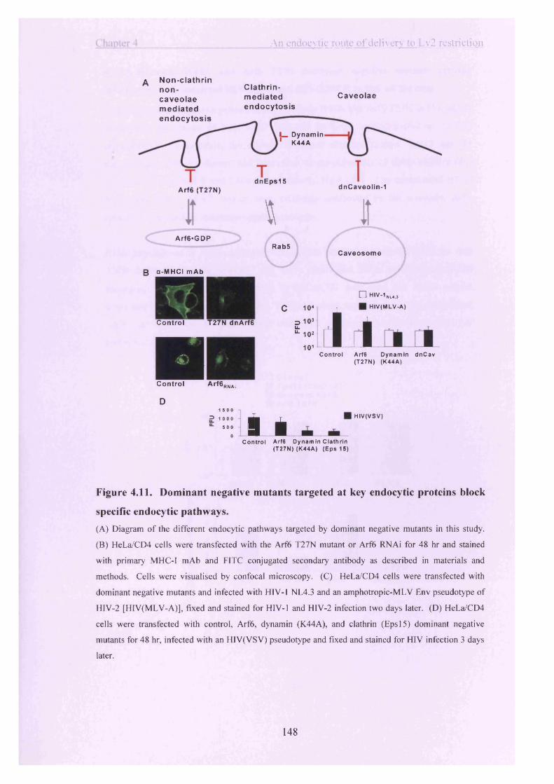
An endocytic route o f delivery to Lv2 restriction
a Non-clathrin non- caveolae mediated
Arf6*GDP
B a-MHCI mAb
Clathrin-mediatedendocytosis
Caveolae
endocytosis
l_ Dynamin 1 K44A
dnEps15dnCaveolin-1Arf6 (T27N)
\ JtRab5
Caveosome
c
Control T27N dnArf6
DControl A r f 6 R NAt
D1 5 0 0
=> 1 0 0 0 ^
“ ■ 5 0 0 1
0 ®L i
10 *
3 10J E “-10*
101 i d□ HIV-1ML4.,
HIV(MLV-A)
C on tro l Arf6 D ynam in d n C av (T27N) (K44A)
HIV(VSV)
C ontro l Arf6 D ynam in Cla th rln (T27N)(K44A) (Eps 15)
Figure 4.11. Dominant negative mutants targeted at key endocytic proteins block
specific endocytic pathways.
(A) Diagram of the different endocytic pathways targeted by dominant negative mutants in this study.
(B) HeLa/CD4 cells were transfected with the Arf6 T27N mutant or Arf6 RNAi for 48 hr and stained
with primary MHC-I mAb and FITC conjugated secondary antibody as described in materials and
methods. Cells were visualised by confocal microscopy. (C) HeLa/CD4 cells were transfected with
dominant negative mutants and infected with HIV-1 NL4.3 and an amphotropic-MLV Env pseudotype of
HIV-2 [HIV(MLV-A)], fixed and stained for HIV-1 and HIV-2 infection two days later. (D) HeLa/CD4
cells were transfected with control, Arf6 , dynamin (K44A), and clathrin (Eps 15) dominant negative
mutants for 48 hr, infected with an HIV(VSV) pseudotype and fixed and stained for HIV infection 3 days
later.
148

Chapter 4 An endocvtic route o f delivery to Lv2 restriction
4.2.10 Dynamin K44A and Arf6 T27N dominant negative mutants recover infection by Lv2 restricted HIV-1 89.6 and HIV-2 MCR in HeLa/CD4 cells.
Expression of dominant negative mutants dynamin K44A and Arf6 T27N in HeLa/CD4 resulted in the rescue of both MCR (5.0-fold for both) and 89.6 (6.0 to 7.0-fold respectively). In contrast, the clathrin dominant negative mutant, Eps 15 and the dncaveolin-1 (data not shown) had little effect on the infectivity of either MCR or 89.6 in HeLa/CD4 cells (1.0 and 2.0-fold respectively, fig 4.12b). The unrestricted HTV-2 MCN and HIV-1 NL4.3 viruses were relatively unaffected by the dynamin, Arf6, caveolin-1, or clathrin dominant negative mutants.
RNAi knockdown of Arf6 expression was used to confirm the specificity of the Arf6 T27N dominant negative mutant. Fig 4.12c shows that RNAi knockdown of Arf6 recovered 3.0-fold of MCR infection in HeLa/CD4 cells over the RNAi control, compared to 3.7-fold when the Arf6 T27N construct was used, in this experiment. MCN was unaffected by any of these treatments, confirming a role for Arf6 in Lv2 restriction.
□ C o n tro l■ E p s 1 5 (c la th r in )■ D y n a m in K44A■ A rf6 T27N
MCR
MCR
149

Chapter 4 An endocvtic route o f delivery to Lv2 restriction
Control Arf6 s/Control s/Arf6 (T27N)
Figure 4.12. Arf6 and dynamin dominant negative mutants recover Lv2 restricted HIV-1 and HTV-2 infection in HeLa/CD4 cells.(A) HeLa/CD4 cells were transfected with specific endocytosis knockdown mutants or (C) siRNA oligomers directed against control and Arf6, and infected two days later with titrations of restricted (A) HIV-1 89.6, (A,C) HIV-2 MCR and unrestricted (A) HIV-1 NL4.3 and (A,C) HIV-2 MCN. Control represents infection of cells transfected with an eGFP repressing construct or siRNA directed against the D6 blood group antigen. (B) Infection on cells transfected with each dominant negative mutant in (A) was divided by the infection from the control in (A) to obtain fold recovery of infection. Results are representative of 7 independent experiments. Error bars represent SDM.
4.2.11 HeLa/CD4 cells expressing Arffi T27N and dynamin K44A dominant negative mutants rescued Lv2 restricted infection by MCR gag and envelope gene swap molecular clones.
Similar to the results in fig 4.12, the recovery of MCR with dynamin K44A and Arf6 T27N was 4.5 and 5.0-fold, respectively (fig 4.13b). The gene swap molecular clone viruses each showed about half the recoveiy of MCR with the Arf6 and dynamin dominant negative mutants: MCNmcrgag and MCNmcrewv were rescued between 2.0 and 3.0-fold with dynamin K44A, or Arf6 T27N mutant on HeLa/CD4 cells (Fig 4.13b).
150

hapter 4 An endocytic route o f de l iv ery to Lv2 restriction
103
102
101
□ Control□ Dynamin K44A ■ Arf6 T27N
1 , ■usH j h
c■i 6
£ 40^ 25
1 0 £"O
MCR
I
MCN MCN mcrgag mcrenv
I IMCR MCN MCN
mcrgag mcr env
Figure 4.13. Arf6 and dynamin dominant negative mutants recover gag and
envelope gene swap virus infection in HeLa/CD4 cells.HeLa/CD4 cells were transfected with endocytosis knockout mutants and infected 2 days later with (A)
MCR gag and env swaps, MCNmcrgag and MCNmcrewv, and fixed and stained for HIV-2 infection three
days later. (B) Infection on cells transfected with each dominant negative mutant was divided by the
infection from the control to obtain fold recovery of infection. Results shown are representative of 3
independent experiments. Error bars represent SDM.
The restriction of HIV-2 MCR and HIV-1 89.6 is recovered by inhibiting Arf6 or
dynamin activity so it is interesting to see what effect knocking out both of these
pathways, at the same time, has on restricted virus infection. Fig 4.14e shows that the
double knockdown of Arf6 and dynamin doesn’t increase recovery of infection of MCR
over that seen with the single knockdowns but there is an additive effect of double
Arf6/dynamin knockdown on recovery of 89.6 infection. 89.6 was recovered 2.2-fold
and 4.3-fold with the dynamin and Arf6 mutants, respectively, but the recovery
increased to 10.5-fold with the dynamin/Arf6 double knockout (fig 4.14b).
151

Chapter 4 An endocytic route o f delivery to Lv2 restriction
□ C ontro l□ Dynam in K44A□ Arf6 T27N ■ Dyn/Arf6
B
103
£ 102
101
coso0) 12
.E 10o<D>oo(1)
\ < w m a
M C R
M C R
MCN 89.6
1MCN 89.6
Figure 4.14. Cotransfection of Arf6 and dynamin dominant negative mutants has
an additive effect on restriction recovery of HIV-1 89.6 in HeLa/CD4 cells.(A) Arf6 T27N and dynamin K44A mutants were cotransfected into HeLa/CD4 cells (Dyn/Arf) or
individually, infected with restricted HIV-2 MCR, HIV-1 89.6, and unrestricted MCN. (B) The FFU
obtained from infection on cells transfected with each dominant negative mutant was divided by the
infection from the control to obtain fold recovery of infection. Results are representative of 3
independent experiments. Error bars represent SDM.
4.3 Discussion:
This chapter demonstrates that a lipid raft dependent endocytic pathway mediates Lv2
restriction in HeLa/CD4 cells. Restricted virus Env directs the viral core into this
restrictive Lv2 compartment, where trafficking of the retrovirus to the nucleus is
terminated. Specific pathways of endocytosis were blocked with dominant negative
mutants and it was demonstrated that direction into the restrictive compartment is via an
Arf6 and dynamin dependent pathway, and at least partly explains Lv2. Furthermore,
the Lv2 compartment is pH-independent and the classical clathrin and caveolae
endocytic pathways are not involved. These data support the model that Lv2 restriction
is dependent on a specific pathway of entry (Schmitz et al., 2004).
152
C hapter 4 An endocvtic route o f delivery to L.v2 restriction
The role of endocytosis and PM trafficking of CD4 in HIV infection has been reported previously, and cells that do not actively internalise CD4 are more infectable than cells that rapidly endocytose CD4 (Pelchen-Matthews et al., 1995). In keeping with this, Lv2 restricted virus infection is recovered by inhibiting endocytosis with hypertonic sucrose, by depletion of lipid rafts and by directing CD4 outside of lipid rafts, which supports the notion of an endocytic route of restricted virus internalisation.
CD4 is internalised by HeLa/CD4 cells in a clathrin dependent manner (Pelchen- Matthews et al., 1992; Pelchen-Matthews et al., 1995) and hypertonic media inhibit the formation of the clathrin triskelion on the PM (Heuser and Anderson, 1989). Cholesterol depletion has been suggested to inhibit clathrin dependent endocytosis as well as lipid raft dependent pathways in a Chinese hamster ovary cell line (Subtil et al.,1999), but there was no inhibition of Trf-FITC uptake in either HeLa/CD4 or U87/CD4/CXCR4 cells. Indirect evidence that Lv2 acts independently of clathrin is that restricted virus pseudotyped with a clathrin dependent VSV Env is rescued from Lv2 restriction (McKnight A, 2001; Schmitz et al., 2004; Sun et al., 2005). Lastly, the clathrin dominant negative mutant, Eps 15 did not have an effect on restricted virus infection in restrictive cells confirming that a clathrin route of entry has little or no role in Lv2 restriction.
Recovery of HTV infection by treatment of target cells with pH neutralising reagents has been described previously and is dependent on the HTV isolate used (Fredericksen et al., 2002; Wei et al., 2005). However the pH-independence of Lv2 restriction shown by monensin and bafilomycin Al treatment of restrictive cells suggest that acidified endosomes are not involved in Lv2 restriction. Furthermore, pseudotyping MCR with a pH-dependent VSV Env rescues Lv2 restricted virus infection (McKnight A, 2001; Schmitz et al., 2004), which further supports the model that Lv2 restriction occurs in a pH-independent compartment.
A more precise identification of the specific endocytic route(s) leading to Lv2 was determined using dominant negative mutants. Restricted virus infection is recovered when the Arf6 T27N dominant negative mutant is over-expressed in restrictive cells.
153

Chapter 4 An endocvtic route o f delivery to Lv2 restriction
Additionally, infection is recovered when the dynamin K44A dominant negative mutant is over-expressed in HeLa/CD4 cells.
Double Arf6 and dynamin knock down experiments indicated that there was no additive effect on MCR suggesting that these mechanisms may be interconnected. The iiftemalisation of M2 muscarinic acetylcholine receptor in HeLa cells is Arf6 dependent but dynamin independent (Delaney et al., 2002). In MDCK cells, Arf6 endocytic recycling requires the action of dynamin (Altschuler et al., 1999). Therefore, dynamin involvement in Arf6 trafficking may be cell-type dependent. But dynamin is involved in Lv2 restriction in HeLa/CD4 cells and is a part of the Arf6 component of restriction. HIV-2 MCR infection is not recovered more with the double Arf6/dynamin knockdowns compared to Arf6 or dynamin single knockdowns suggesting that these mechanisms are not mutually exclusive. HTV-1 89.6 on the other hand shows additive infection recovery when both Arf6 and dynamin are knocked down. This may be because, in this case, some restricted 89.6 is internalised by an Arf6-independent dynamin dependent pathway that eventually traffics 89.6 to the Arf6 early endosome. Indeed, the dominant negative clathrin Eps 15 mutant recovers some restricted 89.6 but not MCR infection. The majority of 89.6 restricted virus may enter the Arf6 endosome by the Arf6/dynamin route, like MCR (fig 4.15). The transfer of proteins in HeLa cells between the Arf6 early endosome and the clathrin Rab5 early endosome via an intermediate compartment has been described (fig 4.15) (Delaney et al., 2002). This model is certainly favoured by the additive recovery of 89.6 infection in the double dynamin/Arf6 dominant negative knockout cells but not by die MCR infection recovery. To conclude, MCR and 89.6 may enter the restricting Arf6 compartment by different pathways.
154

C hapter 4 An endocvtic route o f delivery to Lv2 restriction
MCR, 89.6 19.6
Dynamm (v la clathrin?)tamin
<^ArfB+(Lv2 r—trtctton]
Figure 4.15. Dynamin and Arf6 mediated pathways of restricted HIV entry.
The viral env and gag genes act in concert to mediate Lv2 restriction (Schmitz et al., 2004), and mediate rescue of infection upon hypertonic sucrose treatment (Marchant et al., 2005). This suggests that the restricted virus Env targets the susceptible capsid cargo into the Lv2 compartment where the virus is restricted. The recovery of HIV infection in HeLa/CD4 cells with lysosomotropic agents has been reported (Fredericksen et al., 2002; Pelchen-Matthews et al., 1995) and like Lv2, the viral Env mediates the recoveiy of infection in this system (Wei et al., 2005). Interestingly, an MCN Env pseudotype of HTV-1 was rescued from an apparent restriction in HeLa/CD4 cells with sucrose treatment This suggests that MCN Env directs HTV-1 into the Lv2 compartment (or Lv2-like) for restriction, suggesting that in the wild-type unrestricted MCN virus, the Gag and Env cooperate their activities to avoid Lv2 restriction. In other words, MCN Gag core has evolved to infect HeLa/CD4 cells via the Lv2 restriction route lending support to the theory that the viral env and gag must co-evolve to avoid restriction (Schmitz et al., 2004).
MpCD had little effect on the infectivity of the gene swap, MCNmcrewv and MCNmcrgag, and only wild-type MCR infection was recovered on HeLa/CD4 cells with this treatment. This is in contrast to the recovery of MCR and MCNmcrewv infection in HeLa/CD4 cells with sucrose treatment. However, if the sucrose treatment and MpCD were acting at different stages along the restriction pathway this result may not be so surprising (fig 4.16). As reported previously, hypertonic media inhibit early events in endocytic uptake (Heuser and Anderson, 1989), HIV Env may direct the
155

Chapter 4 An endocvtic route o f delivery to Lv2 restriction
virion into these sites hence the Env mediated recovery of restriction with hypertonic
sucrose. Only wild-type MCR, with both MCR Env and Gag together, was rescued
with MpCD treatment. This may suggest that by the time the virus reaches the MpCD
compartment, Env has already performed its, restriction-targeting, role.
MCR and MCN env
Hypertonicsucrose
MCR gag (Lv2 restriction)
gagNucleus
Figure 4.16. Methyl-p-cyclodextrin and sucrose may act on different stages of the
restriction pathway.
In this chapter I show that both an (1) endocytic lipid raft pathway, elucidated with
sucrose treatment and MpCD, and (2) a dynamin dependent Arf6 pathway, elucidated
with dominant negative mutants, are involved in Lv2 restriction. However it has not
been shown whether these two mechanisms of entry are the same or different pathways.
Experiments where endocytosis was inhibited with MpCD or sucrose in cells expressing
dominant negative endocytosis mutants proved too toxic for accurate analysis of results.
Regardless, there are interesting similarities between the cholesterol requirements of the
restriction and the Arf6 pathway; both the Arf6 pathway and Lv2 require lipid rafts and
cholesterol (Hiroyama and Exton, 2005; Kirkham et al., 2005; Naslavsky et al., 2004).
It is therefore possible that the Arf6 dominant negative characterisation o f the restriction
and the MpCD and hypertonic sucrose studies represent the same pathway of restriction.
156

C hapter 5
Lv2 is a saturable restriction
5.1 Introduction:
Numerous mechanisms of innate immunity against retroviral infection have been
described previously (reviewed in (Bieniasz, 2003; Goff, 2003; Goff, 2004)). While
some of these mechanisms act prior to virus entry into cells the recently described
‘restriction factors’ act post-entry. The first of these to be discovered and investigated
in detail is the Friend virus susceptibility-1 restriction (Fvl) (Lilly, 1967). B-tropic and
N-tropic Murine Leukaemia Viruses (MLV) readily infect cells from BALB/c and NIH
Swiss mice, respectively, however BALB/c mouse cells restrict N-tropic MLV infection
by approximately 1000-fold and NIH cells restrict B-tropic virus by 100-fold. The aptly
named NB-tropic MLV can infect both cell types equally as well (Hartley et al., 1970).
When the genetic locus of the Fvl restriction was mapped it was discovered that the
Fvln and Fvlb restriction phenotypes are alleles of the same jv l gene (Pincus et al.,
1971).
Fvl has been cloned and shares sequence similarity to a mouse endogenous retrovirus
gag that is unrelated to MLV. Localised to mouse chromosome 4, Fvl has its own
retroviral major homology region (MHR) that it requires for restriction activity (Best et
al., 1996). Regions at the N and C-terminal ends are also required to restrict N or B-
tropic MLV infection (Bishop et al., 2001).
MLV can infect human cells if the MLV ecotropic Env is replaced with a pantropic
VSV Env. These mixed MLV (VSV) pseudotypes were used to show that N-tropic
MLV is restricted in human cells (Towers et al., 2000). The N-MLV restriction in
human cells, termed ‘R efl’, was later shown to also target EIAV (Hatziioannou et al.,
2003). The pattern of Refl restriction, whereby N-tropic MLV but not B-tropic MLV is
restricted, is also present in cells from a wide variety of mammals (Towers et al., 2000).
157
Chapter 5 Lv2 is a saturable restriction
The target of Fvl and Refl restrictions is the MLV capsid protein (CA). When residue 110 of the MLV CA is an acidic amino acid it is sufficient to confer B-tropism to MLV, whereas if a basic amino acid is substituted at this site the tropism switches to N-tropic (Kozak and Chakraborti, 1996). Refl was discovered using the N-tropic and B-tropic MLV Gag-Pol packaging constructs that differ at amino acid position 110 of capsid. Thus, it was apparent that, like Fvl, Refl was a CA directed restriction (Towers et al.,2000). However, unlike Fvl, Refl restriction was shown to occur before reverse transcription (Jolicoeur and Rassart, 1980; Towers et al., 2000; Yang et al., 1980). It has even been proposed that Fvl requires the action of huTRIM 5a to effect viral restriction when Fvl is expressed in human cells (Keckesova et al., 2004).
Lvl is a post-entry restriction to VSV-G pseudotyped HTV, SIV, and MLV in nonhuman primate cells (Cowan et al., 2002). However its activity in different primate cells varies slightly. Generally, Lvl is more restrictive towards HIV-1 than S IV m ac and MLV in Old World monkey cells (Hofmann et al., 1999). However, S IV m ac is more restricted than HTV-1 in New World monkey cells (Hofmann et al., 1999). Examination of primary cells from various tissues suggests that the species and not the tissue of origin determine this restriction (Bieniasz, 2003; Hofmann et al., 1999). Lvl in African green monkey cells has broader activity than Lvl in other non-human primates or Refl in human cells because it can restrict N-tropic MLV, STVmac, HTV-1 and 2, and EIAV, (Besnier et al., 2002; Bieniasz, 2003; Cowan et al., 2002; Hatziioannou et al., 2003). Lvl is known to restrict HTV-1 in Rhesus macaque, and even rabbit and pig cells (Hatziioannou et al., 2003; Hofmann et al., 1999). Like Refl, Lvl activity is directed against the CA of the incoming retrovirus and occurs pre-reverse transcription (Owens et al., 2004; Owens et al., 2003).
A characteristic of the Fvl, Refl, and Lvl restrictions is that they can be “abrogated”v
with saturating doses of ‘decoy’ virus-like particles (VLPs). The in-vitro saturation of restriction factors by pre-treatment with genome-defective particles was first described for Fvl (Boone et al., 1990; Duran-Troise et al., 1977). Restriction abrogation experiments are performed by saturating cellular restriction factors with concentrated doses of VSV Env pseudotypes of MLV, HIV, and STV Gag cores, as has been described for Refl and Lvl [(Towers et al., 2002) and (Besnier et al., 2002)
158

Chapter 5 Lv2 is a saturable restriction
respectively], prior to challenge with sensitive virus resulting in loss of restriction (apparent increase in sensitive/reporter virus infection).
Lv2 is a restriction to HIV infection in human cells that is mediated by both the HIV Env and CA (Schmitz et al., 2004). In chapter 3 I introduced the model of Lv2 restriction of the molecular clones MCR and MCN in HeLa/CD4 cells and the prevalence of Lv2 among HTV-1 and HTV-2 viruses. Chapter 4 investigated how restricted viruses are directed into Lv2 restriction via an endocytic pathway (Arf6 and dynamin dependent) (Marchant et al., 200S), and reinforced the model that HTV Gag and Env act in concert to mediate Lv2 restriction.
In this chapter I set out to examine whether, similar to Refl, Lvl and Fvl, Lv2 is saturable. N and B-tropic MLV (MLV-N/B) Gag-Pol cores with MCR and MCN Env could saturate the Lv2 restriction factor and rescue restricted virus infection, however MLV-N/B with VSV Env had no effect. In contrast to Refl, which restricts N but not B tropic MLV, I show that Lv2 can restrict both N and B tropic cores and that MLV-B has the most potent ability to absorb and abrogate Lv2 restriction factors. Thus in this way I was able to investigate the relationship between Refl and Lv2.
5.2 Results:
5.2.1 Lv2 is distinct from Refl in human cells.
Viruses that are susceptible to Refl restriction in human cells and Lvl restriction in non-human primate cells are rescued by pre-absorption of the Refl restriction factors with saturating doses of decoy virus particles (Besnier et al., 2002). I sought to determine whether the pre-absorption of Refl by susceptible virus like particles (VLPs) on Lv2 restrictive cells could rescue restricted MCR. As a positive control, Refl saturating doses of VSV-G pseudotyped MLV-N and B VLPs with a zeomycin resistance transfer vector were used to rescue VSV Env pseudotypes of MLV-N Gag- Pol cores with an eGFP reporter vector from Refl restriction. Fig 5.1 shows that infection by MLV-B(VSV) pseudotypes are not enhanced by absorption with the decoy VLPs. The MLV-N(VSV) pseudotype infections were rescued in all of the cells tested
159

C hanter 5 Lv2 is a saturable restriction
when absorption was carried-out with MLV-N(VSV) but not MLV-B(VSV) saturating VLPs (data not shown). MLV-N(VSV) titre increased by 7.5-fold in HeLa/CD4 cells. These results are consistent with those previously described for Refl restriction (Hatziioannou et al., 2003).
1000
MLV-N (VSV)
o 1000
MLV-B (VSV)
o10* 101 10> 10s 10« 10* 101 1Q1 10* 10*Control *efl
saturation
[MLV-Nzeol(VSV) J
Figure 5.1. Refl can be saturated in HeLa/CD4 cells by pre-treatment with vesicular stomatitis virus envelope pseudotyped N-tropic MLV particles.To confirm that Refl could be saturated with N-tropic MLV, stocks of MLV-N(VSV) were added to HeLa/CD4 cells at an MOl of 10. Four hr later, MLV-N(VSV) with a GFP reporter transgene was titrated onto the cells and GFP was detected 3 days later by flow cytometry. The results shown are representative of 2 independent experiments.
Fig 5.2 shows the titration of MLV-N(VSV) and MLV-B(VSV) on the Fvl-null cell line, mus-dwmi tail fibroblasts (MDTF), a permissive cell, to determine the actual infectious units per ml of pseudotype stocks in the absence of an Fvl restriction phenotype (Lander and Chattopadhyay, 1984). The infectious titre of MLV-N(VSV) and MLV-B(VSV) in MDTF cells are 3.5 x 10⁵ and 4.0 x 10⁵ infectious units per ml, respectively, as shown in fig 5.2. The titre of MLV-N(VSV) on HeLa/CD4 and U87/CD4/CXCR4 cells is 5.2 x 10³ and 20 infectious units per ml respectively, indicating that both Lv2 restrictive and permissive cells are Refl positive.
40.6%
160

Chapter 5 Lv2 is a saturable restriction
□ MLV N(VSV) ■ MLV B(VSV)
I 101U87/CD4/CXCR4 HeLa/CD4 MDTF
Figure 5.2. Permissive U87/CD4/CXCR4 cells have more Refl activity than Lv2
restricted HeLa/CD4 cells.MLV-N(VSV) and MLV-B(VSV) are equivalent titre in Fvl null MDTF cells. MLV-N(VSV) and MLV-
B(VSV) vector pseudotypes were titrated in Refl positive U87/CD4/CXCR4, HeLa/CD4, and on Fvl
null MDTF cells. Infectious units were determined by flow cytometry. Results shown are representative
of 3 independent experiments. Error bars represent the SDM.
To determine whether MLV-N(VSV) vector pseudotype could saturate Lv2 I performed
a saturation experiment. Lv2 sensitive MCR was titrated on restrictive HeLa/CD4,
GHOST, and permissive U87/CD4/CXCR4 cells (McKnight A, 2001; Schmitz et al.,
2004), that had been pretreated with Refl-saturating doses of MLV-N(VSV) (fig 5.3).
The titre of MCR increased in the MLV-N(VSV) treated HeLa/CD4, GHOST, and
U87/CD4/CXCR4 cells by factors of 5.0, 2.0, and 2.5-fold, respectively, over the mock
transfection control. Therefore, pre-treatment of all cells with MLV-N(VSV),
regardless of whether they were Lv2 susceptible or not, resulted in increases in MCR
titres. Thus there was no abrogation of Lv2 restriction with MLV-N or B(VSV).
Figure 5.3. The infectious titre of Lv2 restricted MCR is not affected by saturation
of Refl with vesicular stomatitis virus envelope pseudotypes of N-tropic MLV.
□ U87/CD4/CXCR4 ■ HeLa/CD4 II GHOST/CXCR4
MLV-N MLV-B Transfection Control(VSV) (VSV) control
161

C hapter 5 Lv2 is a saturable restriction
MCR was titrated on U87/CD4/CXCR4, HeLa/CD4, and GHOST cells that had been pre-treated with Refl-saturating doses of MLV-N(VSV). MLV-B(VSV) was added as a Refl negative control.
5.2.2 MCR envelope confers Lv2-Iike restrictions to N-tropic, B-tropic, and%
Moloney MLV virus-like particles.It has been reported that a VSV Env rescues MCR in restrictive cell types. Furthermore MCR Env will confer restriction to resistant HIV-2 MCN and HTV-1 NL4.3 (Schmitz et al., 2004). The effect that MCR, MCN, and VSV Envs have on infection of MLV-N, B, and Mo Gag-Pol cores in HeLa/CD4, H399 and U87/CD4/CXCR4 cells was determined. The results of the flow cytometric analysis are illustrated in figs 5.4a, c, and e. The fold restriction (U87/CD4/CXCR4 divided by either the restricted HeLa/CD4 titre or permissive H399 titre) is shown in fig 5.4b, d, f.
The titres of VSV-G pseudotyped MLV-N, B and Mo Gag-Pol cores are equivalent on U87/CD4/CXCR4, HeLa/CD4, and H399 cells. The 55.0-fold lower titre of MLV- N(VSV) compared to MLV-B(VSV) is expected, due to Refl. However when the MCR Env was used to pseudotype MLV cores this conferred fold restrictions in HeLa/CD4 cells of 35.7, 57.0-fold and 65.0-fold to MLV-B, MLV-N and MLV-Mo cores, respectively (figs 5.4b, d, and f). Interestingly, the ‘unrestricted’ MCN Env conferred a 14.9-fold restriction to the MLV-B core and a small but noteworthy 8.3-fold restriction to the MLV-Mo core. No restriction by MCN Env to MLV-N was detected in HeLa/CD4 cells. Where MCR and MCN Env conferred an Lv2-like restriction to the MLV cores in HeLa/CD4 cells there was almost complete recovery of restriction by H399 cells, as demonstrated for Lv2 restricted HIV-2 MCR in chapter 4.
162

Chanter 5_________________________________ Lv2 is a saturable restriction
10s
104
102
□ U87/CD4/CXCR4 ■ HeLa/CD4□ H399
h u m(VSV) (MCR)
6040200
■ U87/HeLa □ U87/H399
(MCN) (VSV) MLV-B
I .(MCR) (MCN)
10®104102
6040200LJ
(VSV) (MCR) (MCN) (VSV) (MCR) (MCN) MLV-N
10®
104102
60 hIrr rmm - L L(VSV) (MCR) (MCN) (VSV) (MCR) (MCN)
MLV-Mo
Infectious units/ml Fold restriction
Figure 5.4. MCR envelope confers Lv2 restriction to N-tropic, B-tropic and
Moloney MLV Gag-Pol cores.(A) MLV-B (C) MLV-N (E) and MLV-Mo pseudotypes with VSV, MCR, and MCN Envs were titrated
on U87/CD4/CXCR4, HeLa/CD4, and H399 cells. Three days later, eGFP positive cells were determined
by flow cytometry and the infectious units per ml were calculated. Fold restriction of the (B) MLV-B (D)
MLV-N and (F) MLV-Mo pseudotypes were calculated by dividing the infectious titre on
U87/CD4/CXCR4 by the titre from HeLa/CD4 or H399.
5.2.3 Restriction of MLV occurs pre-reverse transcription whereas restriction of
HIV-2 occurs post-reverse transcription.
Lv2 restriction occurs post-reverse transcription but Refl occurs pre-reverse
transcription (McKnight A, 2001; Towers et al., 2002). I therefore wanted to determine
the point at which MLV-N and B are restricted in these human cells.
163

Chapter 5 Lv2 is a saturable restriction
U87/CD4/CXCR4, HeLa/CD4, and H399 cells were challenged with MLV-N and MLV-B MLV Gag-Pol cores pseudotyped with VSV, MCR, and MCN Envs and analysed by quantitative PCR (qPCR) (to first strand synthesised proviral DNA) 10 min, 3 and 18 hr post-infection (fig 5.5a). QPCR of gag-LTR was also performed to detect full-length reverse transcribed provirus of HIV-2s MCR and MCN as a comparison to the qPCR of the MLV vector pseudotypes in fig 5.5a (fig 5.5b).
Fig 5.5a shows that MLV-B(VSV) is synthesising the first strand of the MLV vector genome to equivalent levels in U87/CD4/CXCR4, HeLa/CD4, and H399 cells. As expected, MLV-N(VSV) synthesised 111.2, i 1.0 and 76.5-fold fewer first strand reverse transcripts (18 hr) in U87/CD4/CXCR4, HeLa/CD4, and H399 cells, respectively, than MLV-B(VSV). The MLV vector pseudotypes that have an MCR Env demonstrated less first strand synthesis on HeLa/CD4 than U87/CD4/CXCR4 cells. MLV-N(MCR) synthesised 14.0 eGFP copies in U87/CD4/CXCR4 compared to 2.0 copies in HeLa/CD4 cells and MLV-B(MCR) synthesised 307.0 copies compared to 6.0 in U87/CD4/CXCR4 and HeLa/CD4 cells, respectively. The 48.0-fold difference in MLV-B(MCR) first strand synthesis in U87/CD4/CXCR4 compared to HeLa/CD4 cells is reminiscent of the 35.7-fold restriction of this pseudotype in fig 5.4b. The 7.0-fold drop in first strand copies of MLV-N(MCR) from 14.0 to 2.0 copies in U87/CD4/CXCR4 and HeLa/CD4 cells, respectively, is considerably less than the 57.0- fold restriction of this pseudotype in fig 5.4d. This number of qPCR copies is however at the lower limit of detection and therefore less accurate. The MLV-N(MCN) pseudotype was also at the lower limit of qPCR detection and the number of copies in U87/CD4/CXCR4 and HeLa/CD4 cells was an equivalent 9.0 and 7.0 copies but this is reminiscent of the lack of restriction of MLV-N(MCN) in U87/CD4/CXCR4 and HeLa/CD4 cells in fig 5.4d. Since the synthesis of the first strand qPCR in fig 5.5a matches the drop in titre of these vector pseudotypes during restriction in fig 5.4 the restriction of MCR and MCN pseudotyped MLV cores is therefore occurring prereverse transcription.
The above data is in contrast to that seen for Lv2 restriction. The restriction of HIV-2 MCR in HeLa/CD4 cells occurs post-reverse transcription as shown in fig 5.5b, and described previously (McKnight A, 2001; Schmitz et al., 2004). At 18 hr postinfection, HIV-2s MCR and MCN demonstrate 20.0 and 40.0 copies, respectively, of
164

Chanter 5 Lv2 is a saturable restriction
full-length gag-LTR provirus. This 2.0-fold difference does not account for the 40.0-
fold restriction between MCR and MCN in HeLa/CD4 cells in fig 3.8 of chapter 3.
A 104 □ 10 m inJ 103 □ 3 hr? 1 0 - 1 ■ 18 hr JLi io ’c r f l r 1 1 r f l CL r f l r 1 n i to>o>coo«0)Q.104“ 1CIlo 1 o2
2 i o1o
U87 HeLa H399 , U87 HeLa H399, U87 HeLa H399,
(VSV) (MCR)M L V -N
(MCN)
fl rl l l 1 „ rl rl 4 4U87 HeLa H399,. U87 HeLa H399,. U87 HeLa H399,
(MCN)
<ZQo>o 1 O3
10 2Q.» 1 0 '
'§• io»0o: 10-1 HO)<001
(VSV)
U87/CD4/CXCR4
(MCR)M L V -B
HeLa/CD4 H399
MCR □ MCNj n ii r jiin fi ji i\ r
18 1 18 1 18
H r p o s t - in fe c t io n
Figure 5.5. Lv2 restricted MLV is blocked pre-reverse transcription whereas Lv2
restriction of MCR occurs post-reverse transcription.(A) qPCR of first-strand cDNA synthesis by MLV-N and B Gag-Pol pseudotypes. Infections were
synchronised on ice then incubated at 37°C for 10 min, 3 hr, and 18 hr time points and eGFP qPCR was
performed on the cellular DNA. Envs used to pseudotype MLV-N and B Gag-Pol are indicated in
brackets: (MCR), (MCN) and (VSV). (B) QPCR for full-length reverse transcripts of MCR and MCN 1,
2, 6, 18 hr post-infection in U87/CD4/CXCR4, HeLa/CD4, and H399 cells. The results shown are
representative of 4 independent experiments in (A) and 3 independent experiments in (B). Error bars
represent the standard error of the mean.
165

Chapter 5 Lv2 is a saturable restriction
5.2.4 B-tropic MLV pseudotype with an HTV-2 envelope can abrogate Lv2 and rescue restricted HTV-2 MCR and HTV-1 89.6 infection.
As shown in fig 5.3, MLV-N or B(VSV) vector pseudotypes do not rescue Lv2
restriction. But as previously reported (Schmitz et al., 2004), VSV-G overcomes Lv2
and saturation may require delivery by a different viral Env. I sought to determine if
MLV Gag-Pol cores could saturate-out Lv2 restriction when targeted using an HTV-2
Env rather than VSV-G. Since MCR Env was the most potent at restricting MLV-N
and B these pseudotyped viruses were added at high MOI (approximately 104 pg RT
ELISA activity) after challenge with sensitive HIV-2 MCR, HIV-1 89.6, and resistant
HIV-2 MCN, and HIV-1 NL4.3. To prevent receptor interference, binding of the
indicator and abrogating/saturating viruses was done on ice and uptake into the cell was
triggered at 37°C (materials and methods).
Fig 5.6 shows the results of this abrogation assay in U87/CD4/CXCR4 and HeLa/CD4
cells. Interestingly, both the MLV-B(MCN) and MLV-B(MCR) pseudotypes were able
to saturate Lv2 restriction in HeLa/CD4 cells (fig 5.6c and d). Suggesting that MCN
Env delivers to a restrictive compartment. Fig 5.6d shows that MCR infection on
HeLa/CD4 cells was recovered 3.5 and 3.0-fold, by saturation with MLV-B(MCR) and
MLV-B(MCN), respectively. MCN was unaffected by any of the treatments in
HeLa/CD4 cells and neither MCR nor MCN were affected by abrogating doses in
U87/CD4/CXCR4.
Infection by HTV-1 89.6, the Lv2 restricted HTV-1, in HeLa/CD4 cells increased from
18 to 120 FFU upon abrogation with the MLV-B(MCR) pseudotype and 72.5 FFU with
MLV-B(MCN) (fig 5.6c). This corresponds to recovery of 89.6 infection of 6.7-fold
and 4.0-fold for the MLV-B(MCR) and MLV-B(MCN) pseudotypes, respectively (fig
5.6d). HIV-1 NL4.3 infection in U87/CD4/CXCR4 and HeLa/CD4 cells was relatively
unaffected by these treatments.
166

Chanter 5 Lv2 is a saturable restriction
300250200150100500
□ C o n t r o l□ MLV-B (MCR) ■ MLV-B (MCN)
U 8 7 /C D 4/C X C R 4
B
f i f r a1^10> 88 6 <D«- 4
3 2MCR MCN 89.6 NL4.3
H eL a/C D 4
D
li. o
150
100
MCR MCN 89.6 NL4.3
*10 o 8 O 6
8 42 2 £ 0
MCR MCN 89.6 NL4.3
- Q L d HMCR MCN 89.6 NL4.3
Figure 5.6. B-tropic MLV pseudotypes with HIV-2 envelopes abrogate Lv2
restriction and recover restricted HIV-1 and HIV-2 virus infection.Fixed doses of HIV-2s MCR, MCN and HIV-Is 89.6 and NL4.3 were plated on (A) U87/CD4/CXCR4,
and (C) HeLa/CD4 cells on ice. Neat pseudotype supernatant was plated over the HIV-2 and HIV-1
challenge virus on ice for a further 30 min, incubated at 37°C, then fixed and stained for HIV-2 infection
3 days later. (B,D) The FFU from the MLV(HIV-2) saturations were divided by the FFU from the
control to obtain fold recovery. Results shown are representative of 12 independent experiments. Error
bars represent the SDM.
5.2.5 Refl (human tripartite motif protein 5a) reduces the potency of HIV-2
envelope pseudotypes of N-tropic MLV to saturate out the factor responsible for
Lv2 restriction.
Figs 5.6 and 5.7a show that MLV-B(HIV-2) pseudotyped particles can saturate Lv2
restriction and partially rescue some restricted HIV-1 and 2 infection. Since MLV-
N(VSV) is known to be Refl restricted by huTRIM 5a, I wanted to determine if the
potency of MLV-N(HIV-2) particles to saturate Lv2 restriction could be improved by
knocking down TRIM 5a. I noted that MLV-N pseudotypes with HIV-2 Envs were
unable to saturate Lv2 and recover MCR infection as in the experiment in fig 5.7a.
HeLa/CD4 cells were treated with TRIM 5a RNAi before carrying-out an Lv2
167

Chapter 5 _________________________ Lv2 is a saturable restriction
saturation experiment as in fig 5.6. Fig 5.7b and c show that MLV-N(HIV-2) particles
are as potent as MLV-B(HIV-2) particles at rescuing Lv2 restricted MCR when TRIM
5a is first knocked down. Suggesting that Refl can also restrict HIV-2 Env
pseudotyped cores. Interestingly, the MLV-N(MCN) was as efficient as the MLV-
N(MCR) at recovering MCR infection by 5.3-fold. This also suggests, as in fig 5.6, that
MCN Env also delivers into a restrictive compartment.
120100
80604020
0
I
■ MCR □ MCN
C ontro l (VSV) (M CR ) (M C N) (VSV) (M CR ) (M C N )
B -M LV— v — N -M LV
j
B■ MCR □ MCN
Control MLV-N MLV-B MLV-N MLV-B
(MCR) (MCN)
4)>Oo<Dk.2o
8
i i l lMLV-N MLV-B MLV-N MLV-B
(MCR) (MCN)
Saturatingpseudotype
Saturatingpseudotype
Figure 5.7. N-tropic MLV Gag-Pol core can abrogate Lv2 restriction if
Refl/TRIM 5a is 'knocked down’ p rio r to challenge.
168

HeLa/CD4 cells were (B) treated with RNAi oligomers directed against TRIM 5a or (A) without and
abrogation experiments were performed as in fig 5.6. (C) The fold recovery (shown below) was obtained
by dividing the FFU from the MLV(HIV-2) pseudotype treated infection by the FFU from the control
infection in (B). Results are representative of 3 independent experiments. Error bars represent the SDM.
5.2.6 Infection by HIV-2 envelope pseudotypes of B-tropic MLV can be enhanced
in Lv2 restrictive HeLa/CD4 and permissive H399 cells with abrogating doses of
HIV-2: evidence for other restriction pathways.
HIV-2 Env pseudotypes of MLV-N and -B can saturate Lv2 and partially recover HIV-
2 and HIV-1 infection on HeLa/CD4 cells (figs 5.6 and 5.7). To determine whether the
same pattern of recovery would occur with saturation of Lv2 with high MOI of HIV-2
virus followed by challenge with MLV-B(MCR) and MLV-B(MCN). Dilutions of
MLV-B(MCR) and MLV-B(MCN) vector pseudotypes were bound to cells on ice
together with high MOI doses of HIV-2. In HeLa/CD4, the titre of MLV-B(MCR) was
not rescued to a large extent by pre-treatment with MCN, MCR, and MCR(VSV) (fig
5.8a). Infectivity of MLV-B(MCN) on HeLa/CD4 was enhanced more than 5-fold by
pre-treatment with MCN, MCR, and MCR(VSV) respectively (fig 5.8b). Rescue of
both MLV-B(MCR) and MLV-B(MCN) on H399 cells was more pronounced with 7 to
10-fold recovery with MCN, MCR, and MCR(VSV) generally seen, in figs 5.8c and d.
The results suggest alternative mechanisms of Lv2 restriction are operating and that
saturation experiments need to be interpreted with caution.
A B(MCR) B0 .01% 0.03% (3.0 X)
1000
B(MCN)
0.28% 1.55% (5.5 X)1000
0
Hi .
1000
Control MCR(MCR)
o.02% (2.0 X) 0.02% (2.0 X)1000
Control MCR(MCR)
1 50% (5.4 X) 149% (5.3 X)
10° 101 102 103 104 10° 101 102 1 03 1 04
MCN(MCN) MCR(VSV)
o *10° 101 102 103 1 04 1 0° 101 102 103 1 04
MCN(MCN) MCR(VSV)
HeLa/CD4
169

C B(MCR) D
1.61% 12.22% (7.6 X)1000 1000
B(MCN)
0.70% 6 .49% (9.3 X)
Control MCR(MCR) Control MCR(MCR)
1000-1 j | f i f 100(L
' :'V
' ' '
10° 101 102 103 104 10° 101 102 103 104
MCN(MCN)
10° 101 102 103 104 10° 101 102 1 03 1 04
MCR(VSV) MCN(MCN) MCR(VSV)
H399
Figure 5.8. Reciprocal abrogation: infection by HIV-2 envelope pseudotypes of B-
tropic MLV is rescued on both Lv2 restrictive and permissive cell types upon
abrogation with HIV-2.RT ELISA standardised inputs of MLV pseudotypes were added to (A, B) HeLa/CD4 and (C, D) H399
cells on ice. Neat supernatant (approx 104 pg RT U) of MCR, MCN, and MCR(VSV) were used to pre
treat cells. Cells were harvested two days later and eGFP positive cells were determined by flow
cytometry. (# x) denotes the fold recovery over control. The results shown are representative of 3
independent experiments.
5.3 Discussion:
The results shown here demonstrate that MLV-N and -B pseudotyped with VSV-G are
unable to saturate Lv2, resulting in no recovery of HIV-2 MCR infection. Substitution
with either an MCR or MCN Env results in abrogation of Lv2 and recovery of restricted
HIV-1 89.6 and HIV-2 MCR. These results support the model that Env plays a
significant role in the susceptibility of virus to restriction factors. Thus MLV-B Gag-
Pol particles with either HIV-2 MCR or MCN Envs can saturate-out Lv2 restriction and
rescue restricted HIV-1 and HIV-2 virus infection. Initial experiments indicated,
however, that MLV-N could not saturate in an Lv2 manner. Later experiments
demonstrated that MLV-N could saturate Lv2 if the TRIM 5a activity of Refl was first
170
knocked down. Thus Lv2 is independent from Refl restriction, Lv2 is able to recognise
MLV-N, B, and Mo cores and is dependent on the Env route of entry.
Lv2 is an independent restriction directed by the viral env-receptor interaction. VSV
Env overcomes Lv2 (Schmitz et al., 2004), indicating localisation of the Lv2 restriction
factor in a distinct compartment.
This chapter further supports the model that Refl and Lv2 are distinct factors in human
cells because; MLV-N Gag-Pol VLPs have lower titre on human cells than MLV-B
VLPs, regardless of their Env, which is probably due to Refl restriction, however all
MLV Gag-Pols tested became susceptible to Lv2 restriction in HeLa/CD4 cells when
pseudotyped with MCR Env; MLV-B(MCR) is restricted by Lv2 but not by Refl; the
discreet compartmentalisation requires that Lv2 is only saturable by MLV Gag-Pol
cores with HIV-2 Envs and finally, MLV-N(HIV-2) can saturate Lv2 restriction if
TRIM 5a is knocked down. This suggests that Lv2 and Refl compete for incoming
retroviral cores and that Refl may occur before Lv2 in the host cell. Alternatively, Refl
could be dominant over Lv2 activity when competing for incoming retroviral capsids.
Lv2, like refl, can be abrogated by saturation with MLV Gag ‘decoy’ VLPs. However,
unlike Refl restriction, both MLV-B and MLV-N cores are able to saturate Lv2
restriction. Surprisingly, MLV-B VLPs with both restricted MCR and unrestricted
MCN Envs could saturate Lv2 restriction and recover HIV-1 89.6 and HIV-2 MCR
infection on HeLa/CD4 cells. Suggesting that MCN Env can deliver to both a
restrictive and non-restrictive pathway.
A number of experiments presented in this chapter suggest that saturation experiments
need to be interpreted with caution. 1) The MLV-N and Mo Gag may be targeted by
additional restriction factors other than those specific to Lv2 thus decreasing their
potency to knockout Lv2 restriction and rescue MCR infection; TRIM 5a knockdown
by RNAi improved the potency of MLV-N to saturate Lv2. 2) The ability of one
species of virus core to saturate a restriction and rescue infectivity of another species of
virus core cannot always be reciprocated
171

In the ‘reciprocal abrogation’ experiments MLV-B(HIV-2) vectors were rescued with
saturating doses of HIV-2. Recovery of MLV-B(HIV-2) was in a non-Lv2 manner;
there was recovery of MLV-B(HIV-2) in Lv2 permissive cells. Therefore, Lv2
permissive cells may bypass Lv2 restrictive compartments but direct viruses into
previously undescribed restrictive routes.
This chapter has shown that, like Refl, Lv2 is a saturable restriction factor but, unlike
Refl, Lv2 is compartment specific. Since Lv2 is saturable it suggests that a specific
protein is responsible for the restriction of HIV-1 and HIV-2 in human cells. Chapter 6
describes the identification and characterisation of TRIM proteins that mediate Lv2 and
these TRIMs can be bypassed with a VSV Env, further confirming that Lv2 and Refl
are distinct restrictions.
172

C hapter 6
C haracterisation of the tripartite m otif proteins responsible for Lv2
restriction
6.1 Introduction:
In chapter 4 I partially characterised a specific compartment/pathway that leads to Lv2
restriction, and in chapter 5 I demonstrated the saturable nature of Lv2. This chapter
characterises the restriction more specifically; identifying the cellular TRIM proteins
involved in restriction of HIV-1 89.6 and HIV-2 MCR, and their cell-type specific
expression.
TRIMs have been implicated as antiviral molecules (Nisole et al., 2005; Reymond et al.,
2001). Indeed, TRIM 5a was cloned from humans and rhesus macaques and identified
as the Refl and Lvl restriction factors, respectively (Keckesova et al., 2004; Perron et
al., 2004; Stremlau et al., 2004; Yap et al., 2004). TRIM 5a (Keckesova et al., 2004;
Perron et al., 2004; Stremlau et al., 2004; Yap et al., 2004) and TRIM 1 (Yap et al.,
2004) are responsible for the Refl and Lvl restrictions and as outlined in the
introduction to the last chapter, Refl is the human model for resistance to infection of
MLV-N and EIAV (Hatziioannou et al., 2003; Towers et al., 2000). Lvl restriction is
the model for resistance of Rhesus macaque cells to HIV-1 and SIVagm infection
(Hatziioannou et al., 2003), and African green monkey cells to HIV-1 and SIVmac
infection (Besnier et al., 2002; Cowan et al., 2002; Hatziioannou et al., 2003).
Therefore, TRIM 5a and TRIM 1 are thought to be responsible for the cross-species
lentivirus restrictions in humans and simians (Hatziioannou et al., 2004; Keckesova et
al., 2004; Stremlau et al., 2004; Yap et al., 2004).
Because TRIM 1 and TRIM 5a from humans and African green monkeys were shown
to have Refl activity and TRIM 19 has been shown to have anti-viral activity against
VSV, it was suggested that the TRIM protein family has very broad anti-viral activity
(Yap et al., 2004).
173

<. 'lm pier 6 ...... ......... ( l i a rack-risaiioi) o f the T R I M pro te ins r e spo ns i b le for i \ 2 restr ict ion
TRIMs have also been associated with restrictions of viruses other than retroviruses.
The promyelocytic leukaemia protein (PML), TRIM 19, has been demonstrated to have
suppressive activity against numerous viruses including influenza and VSV (Chelbi-
Alix et al., 1998), however this activity was shown to be at the stage of virus expression
and not entry (Chelbi-Alix et al., 1998). PML has also been shown to inhibit
Arenavirus infection in mice and may have antiviral effects on Herpes Simplex virus in
human cells (Bonilla et al., 2002; Chee et al., 2003). With regards to HIV, there has
been one report of PML associating with the incoming HIV-1 PIC in human cells and
inhibiting HIV integration (Turelli et al., 2001). In this study, an enhancement of HIV-
1 infection was shown upon treatment of cells with arsenic, however this treatment has
been shown to suppress Refl restriction and recover HIV-1 infection in human cells by
a PML independent mechanism (Berthoux et al., 2003; Keckesova et al., 2004; Sayah
and Luban, 2004; Turelli et al., 2001).
Since the discovery of TRIM 5a there has been great interest in the regions of this
protein that are responsible for retroviral restriction. The SPRY (B30.2) domain was
cited as the principal TRIM 5 domain responsible for antiviral activity (Stremlau et al.,
2004; Stremlau et al., 2005). In particular, residues 320 to 345 have been proposed to
be responsible for the species-specific restriction activity of TRIM 5a (Nakayama et al.,
2005; Perez-Caballero et al., 2005; Sawyer et al., 2005; Yap et al., 2005). More
specifically, a proline to arginine alteration at aa position 332 in the SPRY domain has
been shown to confer anti-HIV-1 activity to human TRIM 5a, which does not normally
have activity against this virus (Yap et al., 2005). There are four splice variants of
TRIM5: a, 8, y and e [reviewed in (Nisole et al., 2005)]. However, of these, only the a
splice variant can effect Refl and Lvl activity (Keckesova et al., 2004; Stremlau et al.,
2004; Yap et al., 2004). In fact, antiviral activity of the SPRY domain was first
implicated by comparing the activities of TRIM 5a with TRIM 5y, which is identical to
TRIM 5 a except that TRIM 5y lacks most of the SPRY domain due to alternative
splicing (Stremlau et al., 2004). In addition to the SPRY domain, MLV-N restriction by
TRIM 5a requires regions in the coiled-coil domain (Perez-Caballero et al., 2005; Yap
et al., 2005).
Because Lv2 is a saturable restriction factor (shown in chapter 5), and CA dependent as
reported previously (Schmitz et al., 2004), I investigated whether the TRIM proteins are
174

i. ' i iaptcr 6 _________C h a ra c t e r i s a t i o n o f the TRIM pro te ins r e s p o n s ible lor I .v2 res t r i c t ion
responsible for Lv2 and if one of them is TRIM 5a, like for Refl. Since TRIM 1 and
TRIM 5a both have SPRY domains and TRIM 5a restricts via this domain the main
criteria for choosing candidate Lv2 TRIMs, from the 69 TRIM genes identified, was the
presence of a SPRY domain [reviewed in (Nisole et al., 2005)].
6.2 Results:
6.2.1 Tripartite motif protein 5a is not responsible for Lv2 restriction in U87/CD4/CXCR4 and HeLa/CD4 cells.
I used RNAi to determine whether TRIM 5a is responsible for Lv2 restriction. MLV
cores with VSV Env could not saturate Lv2 restriction [chapter 5, (Schmitz et al.,
2004)]. However, MCR pseudotyped with a VSV Env is not restricted. Thus it is still
possible that TRIMs in the appropriate compartment could act as Lv2.
I first confirmed that the siRNA designed could knockdown TRIM 5a activity in the
cells used in this study. U87/CD4/CXCR4, HeLa/CD4, and H399 cells were transfected
with TRIM 5a and control RNAi oligomers, then infected with MLV-N(VSV) and
MLV-B(VSV) VLPs (fig 6.1a). The titre of MLV-B(VSV) on the TRIM 5a
knockdown cells is not altered when compared to the control cells. MLV-N(VSV) titre
increased by 50.0, 24.1, and 35.5-fold on the TRIM 5a knockdown U87/CD4/CXCR4,
HeLa/CD4, and H399 cells, respectively, over the control cells (fig 6.1a). These
observations are consistent with those published previously (Keckesova et al., 2004).
The TRIM 5a RNAi knockdown cells were titrated with restricted MCR, HIV-1 89.6
and unrestricted MCN and HIV-1 NL4.3 (fig 6.1b). There was little or no effect on the
titre of HIV-2 MCR (5.3 x 10⁴ FFU/ml vs 2.6 x 10⁴ FFU/ml) or HIV-1 89.6 (1.5 x 10⁴
FFU/ml vs 1.5 x 10⁴ FFU/ml) on HeLa/CD4 cells, demonstrating no rescue of Lv2
restriction. 89.6 titre on control U87/CD4/CXCR4 cells was 1.53xl05 FFU/ml
increasing to 2.39x10⁵ FFU/ml on the TRIM 5a knockdown, increasing 89.6 restriction
from 10.2 to 15.4-fold upon TRIM 5a knockdown, however this does not appear to be
an Lv2 specific result. Thus knockdown of TRIM 5a/Refl activity did not alleviate Lv2
restriction.
175

( l i a rac t e r i sa uon o f the T RI M proteins r e s pons ib le for L v 2 restr ict ion
U 87/C D 4/C XC R 4 HeLa/CD 4 H3991.69% 3.69% 2.20%10000 ------------
s/C ontro l
10000
s/TR IM 5a
4.38%3.23% 4.52%
10° 10* 10* 10* 10* 10* 101 10* 10* 10* 10" 10* 10* 10* 10* G F P —►
M LV-B (VSV)
10000
s/C ontro l
s/TR IM 5a
1,50%(SO.O X) 2.17% (24.1 X)
10* 10* 10* 10* 10* 10* 10* 10* 10* 10* 10° 10* 10* 10* 10* GFP —►
M LV-N (VSV)
B 106 , MCR
10#
104
103U87/CD4/CXCR4 HeLa/CD4 H399
10‘ MCN10#
I r y -
104
g 10s3u.“* 10#
10#
104
10*
89.6
U87/CD4/CXCR4 HeLa/CD4 H399
1 n mU87/CD4/CXCR4 HeLa/CD4 H399
□ ControlRNAI -TRIMSa^
10710#
10#
10410*
NL4.3n ri nU87/CD4/CXCR4 HeLa/CD4 H399
176
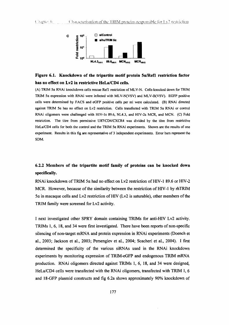
1 'apU" :•> _ ( i ia iav ic r i sa i ion o f the 1 K 1 \ 1 p ro te ins r e s pon s i b le for I .v? rest r i ct ion
C c 102 oSS018 io12o
10°
□ siControl ■ sihuTRIM 5a
j u l ■ . iNL4.3HIV1 89.6h,v1 MCNHlV2 MCRHIV2
Figure 6.1. Knockdown of the tripartite motif protein 5a/RefL restriction factor has no effect on Lv2 in restrictive HeLa/CD4 cells.(A) TRIM 5a RNAi knockdown cells rescue Refl restriction of MLV-N. Cells knocked down for TRIM
TRIM 5a expression with RNAi were infected with MLV-N(VSV) and MLV-B(VSV). EGFP positive
cells were determined by FACS and eGFP positive cells per ml were calculated. (B) RNAi directed
against TRIM 5a has no effect on Lv2 restriction. Cells transfected with TRIM 5a RNAi or control
RNAi oligomers were challenged with HIV-ls 89.6, NL4.3, and HIV-2s MCR, and MCN. (C) Fold
restriction. The titre from permissive U87/CD4/CXCR4 was divided by the titre from restrictive
HeLa/CD4 cells for both the control and the TRIM 5a RNAi experiments. Shown are the results of one
experiment. Results in this fig are representative of 3 independent experiments. Error bars represent the
SDM.
6.2.2 Members of the tripartite motif family of proteins can be knocked down specifically.
RNAi knockdown of TRIM 5a had no effect on Lv2 restriction of HIV-1 89.6 or HIV-2
MCR. However, because of the similarity between the restriction of HIV-1 by rhTRIM
5a in macaque cells and Lv2 restriction of HIV (Lv2 is saturable), other members of the
TRIM family were screened for Lv2 activity.
I next investigated other SPRY domain containing TRIMs for anti-HIV Lv2 activity.
TRIMs 1,6, 18, and 34 were first investigated. There have been reports of non-specific
silencing of non-target mRNA and protein expression in RNAi experiments (Doench et
al., 2003; Jackson et al., 2003; Persengiev et al., 2004; Scacheri et al., 2004). I first
determined the specificity of the various siRNAs used in the RNAi knockdown
experiments by monitoring expression of TRIM-eGFP and endogenous TRIM mRNA
production. RNAi oligomers directed against TRIMs 1, 6, 18, and 34 were designed,
HeLa/CD4 cells were transfected with the RNAi oligomers, transfected with TRIM 1, 6
and 18-GFP plasmid constructs and fig 6.2a shows approximately 90% knockdown of
177

Chapter 6 _______ Characterisation o f the TRIM proteins responsible for Lv2 restriction
eGFP expression. TRIM RNAi did not inhibit expression of heterologous TRIM-eGFP
constructs (data not shown).
To semi-quantify the amount of RNAi inhibition of endogenous TRIM mRNA
knockdown, 5.0-fold serial dilutions were made of cDNA from the RT reaction of RNA
extracted from siTRIM treated HeLa/CD4 cells (this work was done by Keith Aubin).
Fig 6.2b shows that RNAi for TRIMs 1, 18 and 34 could knockdown their respective
targets, however TRIM RNAi didn’t knockdown the mRNA of a heterologous TRIM.
TRIM knockdown is indicated by 5.0-fold less cDNA/RNA expression than a
heterologous TRIM or the p-actin control (fig 6.2b).
C ontrol
s/R N A
&
B
s/Control
< s/TRIM 1 Z £
s/TRIM 18
s/TRIM 34
TRIM 1PCR prim ers
TRIM 18 TRIM 34 p-actin
Figure 6.2. RNA interference knocks down the expression of tripartite motif
proteins 1, 18 and 34 specifically and exclusively.
(A) The expression o f eGFP-tagged TRIMs 1, 6, and 18 transfected into HeLa/CD4 cells is knocked
down specifically by siRNA as visualised by epifluorescence. (B) TRIM 1, 18 and 34 endogenous
mRNA transcript levels are down regulated by specific siRNAs. Five pg total RNA from siRNA treated
HeLa/CD4 cells was reverse transcribed and PCR was performed on 5.0-fold serial dilutions o f cDNA.
TRIM specific primers did not down modulate expression o f other TRIMs or P-actin loading control. For
178
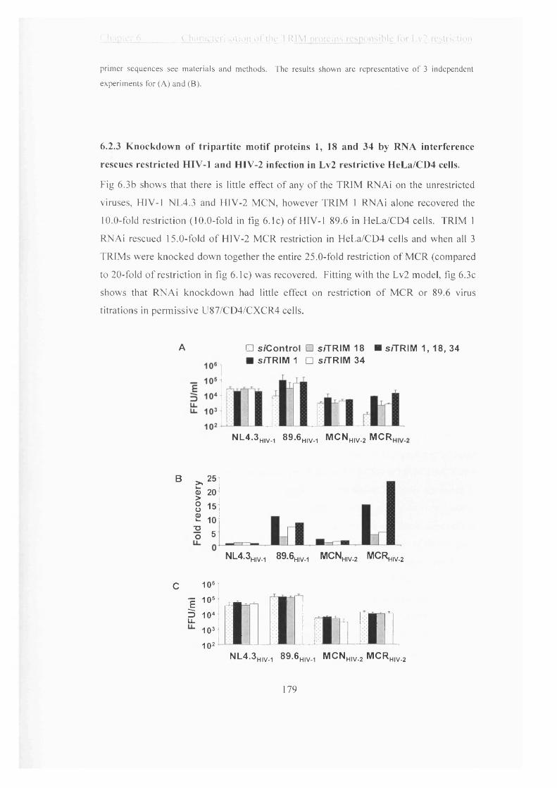
( ha pier 6_________C haracterisation o f the TRIM proteins responsible for Lv2 restriction
primer sequences see materials and methods. The results shown are representative of 3 independent
experiments for (A) and (B).
6.2.3 Knockdown of tripartite motif proteins 1, 18 and 34 by RNA interference
rescues restricted HIV-1 and HIV-2 infection in Lv2 restrictive HeLa/CD4 cells.
Fig 6.3b shows that there is little effect of any of the TRIM RNAi on the unrestricted
viruses, HIV-1 NL4.3 and HIV-2 MCN, however TRIM 1 RNAi alone recovered the
10.0-fold restriction (10.0-fold in fig 6.1c) of HIV-1 89.6 in HeLa/CD4 cells. TRIM 1
RNAi rescued 15.0-fold of HIV-2 MCR restriction in HeLa/CD4 cells and when all 3
TRIMs were knocked down together the entire 25.0-fold restriction of MCR (compared
to 20-fold of restriction in fig 6.1c) was recovered. Fitting with the Lv2 model, fig 6.3c
shows that RNAi knockdown had little effect on restriction of MCR or 89.6 virus
titrations in permissive U87/CD4/CXCR4 cells.
A □ s / C o n t r o l □ s /TRIM 18 ■ s /TRIM 1, 18, 34■ s/TRIM 1 □ s /TRIM 34
N L 4 .3 mv_1 8 9 .6 ^ . . , M C N hiv_2 M CR
B >.25 | 20 8 15 2 101 5
c 106
■g 105
g 10« 103 102
NL4.3H|V.1 89.6H)NM MCNh,v_2 MCR^,,^
M l m r n iN L 4 -3 HIV-1 8 9 .6 H|V_i M C N hiv.2 M C R hiv_2
179

<■ Jk i (. i iara c i c r isa l ion o i ' t h c T R I M pro teins r e spo ns i b le lor Lv2 res t r i c t ion
Figure 6.3. Knockdown of tripartite motif proteins 1, 18 and 34 with RNA interference rescues Lv2 restricted MCR and 89.6 infection in HeLa/CD4 cells.(A) HeLa/CD4 and (C) U87/CD4/CXCR4 cells were transfected with RNAi oligomers directed against
TRIM or irrelevant cellular protein and infected with HIV 2 days later, and fixed and stained for HIV
infection 3 days later. (B) Fold recovery of HIV infection on HeLa/CD4 cells. The virus titre from
infection on TRIM RNAi treated HeLa/CD4s was divided by the titre from the control RNAi HeLa/CD4
cells to obtain fold recovery. The results shown in (A) represent the mean of the results pooled from 7
independent experiments. The error bars in (A) represent the SDM of the pooled results from 7
independent experiments. The results shown in (B) are the product of 1 experiment but represent the
results of 2 independent experiments and error bars represent the SDM.
6.2.4 Tripartite motif proteins 1, 18 and 34 are expressed to higher endogenous levels in restrictive HeLa/CD4 than in permissive U87/CD4/CXCR4 or NP2/CD4/CXCR4 cells.
To determine whether TRIMs 1, 18 and 34 were expressed endogenously in the
permissive U87/CD4/CXCR4 and NP2/CD4/CXCR4 cell types I used specific
polyclonal Abs to detect these TRIMs in HeLa/CD4, U87/CD4/CXCR4 and
NP2/CD4/CXCR4 cells.
Figs 6.4a and b show the detection of TRIMs 1, 18 and 34 by western blot. Despite
almost identical actin input, TRIMs 1, 18 and 34 are expressed to significantly higher
levels in restrictive HeLa/CD4 than permissive U87/CD4/CXCR4 or NP2/CD4/CXCR4
cells. There are higher levels of TRIM la expression in HeLa/CD4 cells compared to
permissive NP2/CD4/CXCR4 and U87/CD4/CXCR4 cells (fig 6.4a, top panel).
Difference in expression of TRIM 18a and p between restrictive and permissive cells is
the most apparent in that there is no detectable expression of either of these splice
variants in NP2/CD4/CXCR4 or U87/CD4/CXCR4 cells despite strong expression in
HeLa/CD4 cells (fig 6.4a, middle panel). Detection of the TRIM 34 splice variants
yielded the a and ‘short’ isoforms in addition to an as yet uncharacterised 80 kDa band
(fig 6.4a, bottom panel). The difference in expression of the a variant between
HeLa/CD4 and permissive NP2/CD4/CXCR4 and U87/CD4/CXCR4 cells was most
apparent. There is less expression of the short variant in permissive NP2/CD4/CXCR4
180
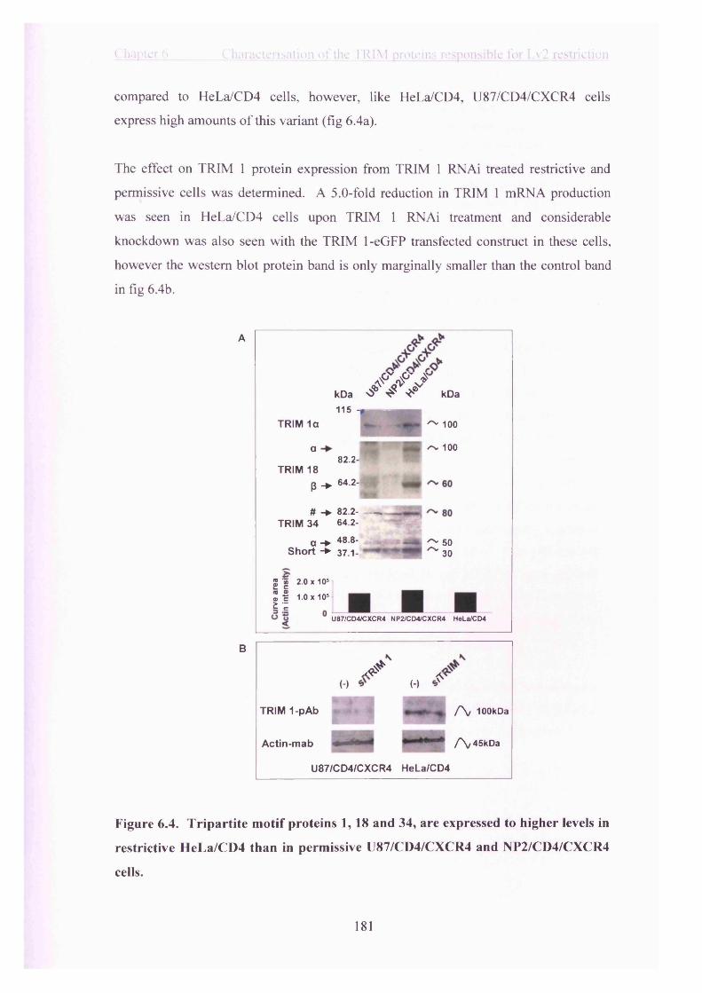
Chapter 6________ Characterisation of the TRIM proteins responsible for Lv2 restriction
compared to HeLa/CD4 cells, however, like HeLa/CD4, U87/CD4/CXCR4 cells
express high amounts o f this variant (fig 6.4a).
The effect on TRIM 1 protein expression from TRIM 1 RNAi treated restrictive and
permissive cells was determined. A 5.0-fold reduction in TRIM 1 mRNA production
was seen in HeLa/CD4 cells upon TRIM 1 RNAi treatment and considerable
knockdown was also seen with the TRIM 1-eGFP transfected construct in these cells,
however the western blot protein band is only marginally smaller than the control band
in fig 6.4b.
A
B
- / / / . . . 115
TRIM 1a
a ■8 2 . 2 -
^ 100
~ 100
TRIM 18B -► 64-2'B P# -► 82.2- —.
TRIM 34 64.2-a -► 48.8-
Short -► 37 .1-^ 50 ~ 3 0
S I 2.0 x 10s
2 H i.o x io5 £ ^
U87/CD4/CXCR4 NP2/CD4/CXCR4 HeLa/CD4
TRIM 1-pAb
*(-) (-) .<s
f \ 10OkDa
Actin-mab /N^skDa
U87/CD4/CXCR4 HeLa/CD4
Figure 6.4. Tripartite m otif proteins 1, 18 and 34, are expressed to higher levels in
restrictive HeLa/CD4 than in permissive U87/CD4/CXCR4 and NP2/CD4/CXCR4
cells.
181

\ . ' u i ;!or f> ..............( iiarac:crisiUi(»ii o f the T R I M p r o te ins r e s pon s i b le lo r Lv2 res t r iction
(A) Cell lysates were western blotted and probed with anti-TRIM 1, 18 and 34 polyclonal antibodies.
Detection of actin with an actin mAh was used as an input control and band intensity was measured by
densitometry, below (see materials and methods). Arrows denote position of detectable splice variants on
western blots. The a, P and ‘Short’ isoforms are indicated and the long uncharacterised 80kDa TRIM 34
isoform is denoted as #. (B) Cell lysates from control and TRIM 1 RNAi treated cells were western
blotted and probed with anti-TRIM 1 polyclonal antibody and anti-actin mAb as an input control. The
figs shown are the product of one experiment but represent the results from at least 7 different western
blots of each of the TRIM proteins.
6.2.5 Permissive cells expressing human tripartite motif proteins 1, 18 or 34 become restrictive.
The RNAi knockdown of TRIM expression (controls in fig 6.3) resulted in recovery of
restricted HIV infection in restrictive HeLa/CD4 cells. If TRIMs 1, 18 and 34 are
indeed responsible for Lv2 then it is important to recapitulate the restriction by
expressing these candidate TRIMs in permissive cells and demonstrating restriction of
Lv2 susceptible HIV. Fig 6.5c shows that HIV-1 89.6 infection in TRIM lp expressing
NP2/CD4/CXCR4 cells is restricted 6.0-fold compared to infection on the parental
NP2/CD4/CXCR4 cells. TRIM 18p also restricted 89.6 by about 6.0-fold as well, and
TRIM 34a displayed weaker restriction activity at only 2.0-fold less infection than the
NP2/CD4/CXCR4 parental cells. So it would appear that individually expressed
TRIMs 1 and 18 effect the majority of restriction on HIV-1 89.6. Trim 18p and 34a
expressing NP2/CD4/CXCR4 cells each restricted HIV-2 MCR by about 6.0-fold
whereas TRIM 1 p expressing NP2/CD4/CXCR4 cells restricted MCR by 11.0-fold (fig
6.5c). HIV-1 NL4.3 and HIV-2 MCN were relatively unaffected by any of the
NP2/CD4/CXCR4 cells expressing any of the TRIM constructs (fig 6.5c).
MCR(VSV) was titrated onto TRIM lp expressing NP2/CD4/CXCR4 cells alongside
MCR. Fig 6.5g shows that TRIM ip restriction is bypassed in NP2/CD4/CXCR4 cells
by a VSV Env, supporting the implication of TRIM ip in the Lv2 model of restriction.
182
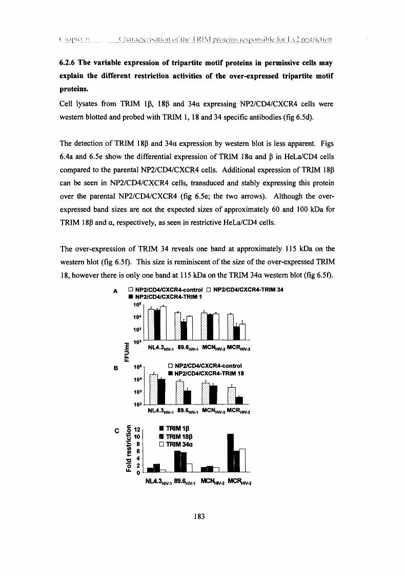
( h a r a c t c r i sa i io n ,uI'Jhv T R I M p roteins r e s p o n s ib le lo r I..v2 r es tr ict ion
6.2.6 The variable expression of tripartite motif proteins in permissive cells may explain the different restriction activities of the over-expressed tripartite motif proteins.
Cell lysates from TRIM 1(3, 18J3 and 34a expressing NP2/CD4/CXCR4 cells were
western blotted and probed with TRIM 1, 18 and 34 specific antibodies (fig 6.5d).
The detection of TRIM 18p and 34a expression by western blot is less apparent. Figs
6.4a and 6.5e show the differential expression of TRIM 18a and (3 in HeLa/CD4 cells
compared to the parental NP2/CD4/CXCR4 cells. Additional expression of TRIM 18|3
can be seen in NP2/CD4/CXCR4 cells, transduced and stably expressing this protein
over the parental NP2/CD4/CXCR4 (fig 6.5e; the two arrows). Although the over
expressed band sizes are not the expected sizes of approximately 60 and 100 kDa for
TRIM 18P and a, respectively, as seen in restrictive HeLa/CD4 cells.
The over-expression of TRIM 34 reveals one band at approximately 115 kDa on the
western blot (fig 6.5f). This size is reminiscent of the size of the over-expressed TRIM
18, however there is only one band at 115 kDa on the TRIM 34a western blot (fig 6.5f).
A □ NP2/CD4/CXCR4-control □ NP2/CD4/CXCR4-TRIM 34 ■ NP2/CD4/CXCR4-TRIM 1
E NL4.3HnM 89.6H(V.., MCNH(V.2 MCRhiv.2
B 10s □ N P2/CD4/CXCR4-control ■ NP2/CD4/CXCR4-TRIM 18
NL4.3H)V.1 89.6hi>m M C IW , MCRH1V.2
c | i 2l 1TRIM1P
U I KIM 34a
■ TRIM1P
U I KIM 34a
NL4.3Hiv-i 89.6h(v.i MCNmv-2 M^^hiv-2
183
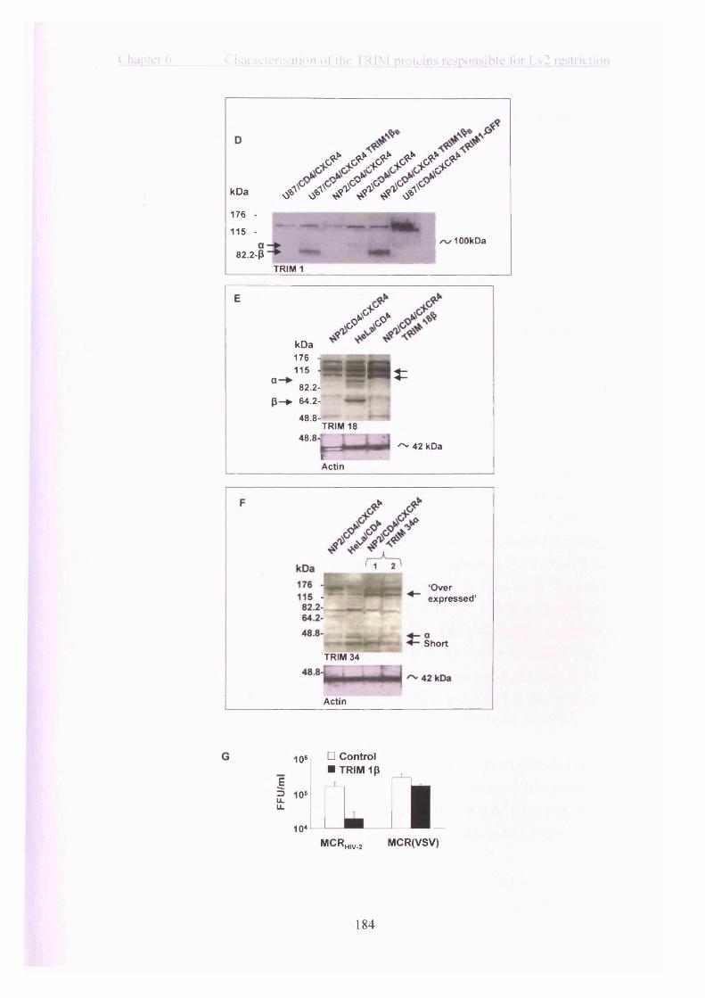
Chapter 6 Characterisation o f the TRIM proteins responsible for Lv2 restriction
, 0^ / / / / /
KDa / / / / / /
176 -
115 -
82.2-P/%✓ 100kDa
TR IM 1
E
k D a4 7 c
G ° V
11 b - 115 -
° 82 .2 - p - ^ 64 .2-
48 .8-
■ 5 * *
f"
| *
48 .8-TR IM 18
^ 42 kDaftActin
‘O verexpressed ’
Short
TRIM 34
Actin
10e □ Control■ TRIM 1p
EI 10‘LL
104MCR HIV-2
aMCR(VSV)
184

C hapter 6 C haracterisation o f the TRIM proteins responsible for Lv2 restriction
Figure 6.5. Permissive NP2/CD4/CXCR4 cells can be made restrictive to HIV-189.6 and HIV-2 MCR infection by expressing tripartite motif proteins 10,180 and 34a.(A) TRIMs 10, 34a and (B) 18p were cloned from HeLa/CD4 cells, sub cloned into an MLV retroviral vector, then transduced into permissive NP2/CD4/CXCR4 cells. Cells were infected with HIV or (G) Hiy(VSV) pseudotype, fixed and stained for infection 3 days later. Error bars represent the SDM. (C) Fold restriction was calculated by dividing the infection on control NP2/CD4/CXCR4 cells by the TRIM transduced cells. All three TRIMs are shown together. (D-F) Lysates from U87/CD4/CXCR4 and NP2/CD4/CXCR4 cells stably expressing (D) TRIM ip, (E) TRIM 18p or (F) TRIM 34a were western blotted and probed with anti-TRIM 1, TRIM 18, or TRIM 34 pAbs, respectively. All of the cell lysates in (D), (E) and (F) were western blotted and probed with an actin mAb as an input control. The upper arrow in (D) indicates the position of TRIM la and the lower arrow indicates the position of TRIM ip. The figs shown are the product of one experiment but results represent the results of at least 4 independent experiments.
6.2.7 The tripartite motif protein 10 -SPRY (butyrophilin 30.2) domain possesses the majority of restriction activity of tripartite motif protein 10.
A research assistant in the lab, Keith Aubin cloned TRIM 10 without a SPRY domain (TRIM 10 ASPRY). The antiviral activity of TRIM 5a has been reported to reside, primarily, in the SPRY (B30.2) domain at the C-terminal end of the protein (Stremlau et al., 2004; Stremlau et al., 2005). In TRIM 10 this corresponds to aa position 397 to 685, the last 288 amino acids. NP2/CD4/CXCR4 cells stably expressing TRIM 10-ASPRY lose the majority of their restriction activity towards HIV-1 89.6 and MCR. Fig 6.6b shows that restriction of 89.6 goes from 6.0-fold in the TRIM 10 expressing NP2/CD4/CXCR4 cells to 2.0-fold restriction in the TRIM 10-ASPRY expressing NP2/CD4/CXCR4 cells. The restriction of MCR in the TRIM 10-ASPRY cells decreases to 4.0-fold from 11.0-fold in the TRIM 10 expressing NP2/CD4/CXCR4 cells. As was seen with TRIM 10 in fig 6.5c, the TRIM 10-ASPRY had little effect on the infection of HIV-1 NL4.3 or HIV-2 MCN in NP2/CD4/CXCR4 cells (fig 6.6b)
Fig 6.6c shows the level of expression of TRIM 10-ASPRY in NP2/CD4/CXCR4 cells with an anti-HA mAb. TRIM 10-ASPRY was cloned with an HA tag at the C-terminus because the TRIM 10 antibody used to detect TRIM 10 over-expression in fig 6.5d recognises a motif in the SPRY region of TRIM 1. Therefore, in this system, direct
185

"hapter 6 Characterisation of the TRIM proteins responsible for Lv2 restriction
comparisons between western blots o f over-expressed TRIM 1 (3 and TRIM 10-ASPRY
are difficult to make. The western blots in figs 6.5d and 6.6c show that TRIM lp and
TRIM ip- ASPRY proteins, respectively, are over-expressed in the parental
NP2/CD4/CXCR4 cells to roughly similar levels.
□ NP2/CD4/CXCR4-control NP2/CD4/CXCR4-ASPRY/B30.2
B
105
E 1045u_U - 103
102
c 12o 1°o•C 80) 6£ 4
T3 2ou_ 0
i i i .NL4.3h.v_, 8 9 . 6 ^ MCNhiv_2 M C R ^
■ TRIM 1□ TRIM 1 ASPRY (B30.2)
I INL4.3h.v_, 89.6h.v_t M CN h,v_2 MCRhiv_2
xc?
kDa (-) 25a 25b kDa
64.2- sn, 60
TRIM 1p ASPRY48.8-
4 2
Actin
Figure 6.6. Tripartite m otif protein ip without a SPRY (butyrophilin 30.2)
domain (tripartite m otif protein lp ASPRY) has limited restriction activity against
HIV-1 and HIV-2 compared to full-length tripartite motif protein ip .
(A) TRIM lp ASPRY was stably expressed in NP2/CD4/CXCR4 cells. Cells were challenged with HIV
and fixed and stained for infection 3 days later. (B) The fold restriction o f HIV infection on TRIM 1
NP2/CD4/CXCR4 cells calculated from (A) compared to infection on TRIM 10 ASPRY
NP2/CD4/CXCR4 cells. Fold restriction was obtained by dividing the infectious titre on control
NP2/CD4/CXCR4 cells by the titre on TRIM lp ASPRY NP2/CD4/CXCR4 cells in (A). (C) Cell lysates
186

C hapter 6 C haracterisation o f the TRIM proteins responsible for Lv2 restriction
from NP2/CD4/CXCR4 cells stably expressing TRIM 1[J ASPRY were western blotted and probed with an anti-HA-tag mAb or an anti-actin mAb as an input control.
6.2.8 Infection by HIV-2 envelope pseudotypes of MLV can be recovered in
HeLa/CD4 cells when the expression of tripartite motif proteins 1, 18 and 34 is
knocked down.
TRIMs 1,18, and 34 mediate Lv2 restriction of HTV-1 and HIV-2 as described above. I determined what effect these TRIMs would have on the replication of HTV-2 Env pseudotypes of MLV in Lv2 restrictive cells. MLV vector pseudotypes were used as a tool in Chapter 5 to demonstrate the saturable nature of Lv2 restriction. Saturable doses of MLV-N and B vector pseudotypes with HIV-2 Envs could recover Lv2 restricted HIV-1 and HIV-2 infection. Identifying the TRIMs being saturated by incoming MLV- N and B (that have HTV-2 Envs) will provide insight into which TRIMs are responsible for Lv2 restriction of HIV-1 and HTV-2 in HeLa/CD4 cells. HeLa/CD4 cells were transfected with RNAi oligomers directed against TRIMs 1, 18, and 34, infected with HTV-2 Env-MLV pseudotypes, and infection was determined by flow cytometry of the MLV-eGFP reporter vector. As expected, since VSV overcomes Lv2 restriction, MLV- B(VSV) infection of HeLa/CD4 cells was unaffected by the TRIM 1, 18 or 34 knockdowns (fig 6.7f). Infection of HeLa/CD4 cells by MLV-N(VSV) was rescued by4.0-fold by the TRIM 1 knockdown in fig 6.7f as has been reported previously (Yap et al., 2004). The 8.0-fold recovery of MLV-N(VSV) infection in TRIM 34 knockdown HeLa/CD4 cells has not been previously reported (fig 6.7f).
In chapter 5 (titration of MLV vector pseudotypes) MCR Env confers restriction to MLV cores (N and B-tropic) in HeLa/CD4 cells. MCN Env also confers restriction to MLV-B cores (but to a lesser extent) and no further restriction to MLV-N capsid cores. I postulated that the lack of MLV-N restriction was because it is already restricted by Refl in human cells. Figs 6.7a to d show that MLV-N(HIV-2) pseudotypes are more susceptible to recovery with RNAi TRIM knockdown in HeLa/CD4 cells than the
MLV-B (HIV-2) pseudotypes.
187
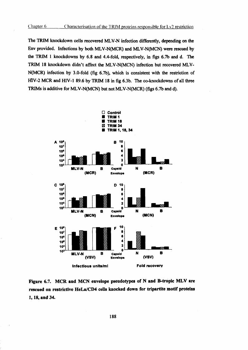
C hapter 6 C haracterisation o f the TRIM proteins responsible for Lv2 restriction
The TRIM knockdown cells recovered MLV-N infection differently, depending on the Env provided. Infections by both MLV-N(MCR) and MLV-N(MCN) were rescued by the TRIM 1 knockdowns by 6.8 and 4.4-fold, respectively, in figs 6.7b and d. The TRIM 18 knockdown didn’t affect the MLV-N(MCN) infection but recovered MLV- N(MCR) infection by 3.0-fold (fig 6.7b), which is consistent with the restriction of HIV-2 MCR and HIV-1 89.6 by TRIM 18 in fig 6.3b. The co-knockdowns of all three TRIMs is additive for MLV-N(MCN) but not MLV-N(MCR) (figs 6.7b and d).
□ Control■ TRIM 1■ TRIM 18 0 TRIM 34■ TRIM 1,18, 34
A 10*10710*
B 10 8 6
10*10*10s
42
- 0 CapaidMLV-N B
(MCR) EnvalopaB
(MCR)
C 10*
MLV-N(MCN)
D 10 8
I 64
2^ 0CapsidEnvalopa
LN B
(MCN)
Capsid(VSV) Envalopa (VSV)
Infectious units/m l Fold recovery
Figure 6.7. MCR and MCN envelope pseudotypes of N and B-tropic MLV are
rescued on restrictive HeLa/CD4 cells knocked down for tripartite motif proteins
1,18, and 34.
188

C hapter 6 C haracterisation o f the TRIM proteins responsible for Lv2 restriction
(A-F) HeLa/CD4 cells treated with control, TRIM 1, 18 or 34 RNAi and all three pooled together were infected with MLV-N and B pseudotypes with MCR, MCN, and VSV Envs. (A, C, E) EGFP positive cells were determined by flow cytometry, and infectious units per ml were calculated. (B, D, F) Fold recovery of pseudotype infection with TRIM RNAi knockdown. The TRIM RNAi infection was divided by the control RNAi infection to obtain fold recovery. Results are representative of 6 independent experiments. Error bars represent SDM.
6 . 3 D i s c u s s i o n :This chapter demonstrates that TRIMs 1, 18 and 34, mediate Lv2. By gene silencing in restrictive HeLa/CD4 and by rendering permissive NP2/CD4/CXCR4 cells restrictive I show that most Lv2 restriction activity lies in the activity of TRIM 1. Furthermore, die SPRY domain of TRIM 1 is implicated as an important determinant of antiviral activity of TRIM ip. This chapter also shows that the difference in restriction activity between Lv2 permissive and restrictive cells lies in the expression of monomeric TRIM proteins in restrictive cell types compared to permissive ones. Infection of TRIM knockdown cells with HIV-2 Env pseudotypes of MLV-N and B cores suggests that the MLV capsid cores are ‘hit’ by different TRIMs when delivered into one compartment by HIV-2 Envs than when delivered to another compartment by a VSV Env. Thus the compartmentalisation models of TRIM proteins (Reymond et al., 2001) and Lv2 retroviral restriction (Schmitz et al., 2004) are mutually supportive.
Some endogenous expression of TRIM 1 in permissive cells is detectable. The western blots show trace amounts of TRIM 1 protein expression, however significant levels of recovery of MLV-N(VSV) with TRIM 1 RNAi, in permissive cells, suggests that TRIM 1 is normally expressed in these cells as well as restrictive ones and that only small amounts are required to inhibit MLV-N.
PH-dependent VSV-G delivers its contents from an acidic compartment whereas pH- independent HTV can fuse outside acidic compartments. Acid-dependent VSV-G can only fuse in a low pH compartment (Mannen et al., 1982). TRIM 5a activity is associated with this pathway of entry because MLV-N(VSV) is restricted in a Refl- dependent manner. However, in chapter 5 the knockdown of TRIM 5a recovered the potency of MLV-N(HIV-2) vectors to saturate-out Lv2 restriction suggesting a more ubiquitous localisation of TRIM 5a in the cell than Lv2 activity.
189
Chapter 6 C haracterisation o f the TRIM proteins responsible for Lv2 restriction
I showed that individual TRIMs are responsible for restriction of HIV-1 89.6 and HTV-2 MCR. However, I have not shown what happens when all of these proteins are overexpressed simultaneously. The over-expression of these three proteins together may very well result in recapitulation of the full Lv2 restriction.
The ability to target different viruses (via the different viral entry pathways) may, in part, lie in the differing nuclear and cytoplasmic localisations of the 69 TRIM gene products (Nisole et al., 2005), that can form homo and hetero-oligomers in different cellular compartments (Reymond et al., 2001). This particular chapter is especially pertinent to the Lv2 model of restriction, because Lv2 restriction (Schmitz et al., 2004), like TRIM localisation (Reymond et al., 2001), is a model based on compartmentalisation. Depending on the virus and target cell type, virus Envs target different cellular entry pathways (Marchant et al., 2005).
190

Chapter 7
Summary and future directions
7.1 Summary
In chapter 3 I observed that although HIV-1 and HTV-2 both infect PBMC and MDM the replication kinetics of HTV-2 in MDM are different. HIV-2 primary isolates could enter MDM and reverse transcribe efficiently but entered a latent state after an initial burst of replication.
The V3 loop of Env is a MDM cell tropism determinant in HTV-1 and I show that this is also true for HIV-2, although weaker. The correlation suggests that there are other factors associated with HIV-2 infection of MDM. One HTV-2 isolate in particular, prCBL 23, demonstrated efficient infection of PBMC but lacked the ability to infect MDM. In chapter 4 I show that the role of Env in Lv2 restriction may be explained by its delivering susceptible cores into restrictive compartments via an endocytic pathway.
I demonstrate the saturable nature of the Lv2 restriction for HTV-1 and HTV-2 in chapter 5 and in chapter 6 1 demonstrate that TRIMs 1, 18, and 34 mediate Lv2.
7.2 Future directions
To better understand the role that restriction plays in viral infection it would be interesting to investigate the tissue specific expression of viral restrictions and which types of viruses are targeted by these factors. Work resulting from this thesis might also involve investigating the possible localisation of TRIMs in endocytic compartments. Arf6 was implicated as a restrictive compartment thus this may be a region of high TRIM density. Site directed mutagenesis would also be a useful tool to relocalise TRIMs to other cellular compartments and observe the effect on restriction to further investigate the compartmentalisation model of restriction.
191
Publications resulting from this thesis
Marchant D, Neil SJ, Aubin K, Carter G, McKnight A. Multiple cytoplasmic human tripartite motif proteins terminate human immunodeficiency virus replication. Nature. Submitted, in review.
Marchant D, Neil SJ, McKnight A. Latency of HTV-2 in Macrophages. Journal of General Virology. Accepted, October 2005.
Marchant D, Neil SJ, Aubin K, Schmitz C, McKnight A. The route of viral entry determines the susceptibility of HIV-1 and HIV-2 to Lv2 restriction. J Virol. 2005 Aug;79(15):9410-8.
Schmitz C, Marchant D, Neil SJ, Aubin K, Reuter S, Dittmar MT, McKnight A. Lv2, a novel post-entry restriction, is mediated by both capsid and envelope. J Virol. 2004;
78(4):2006-16.
192

References
Aasa-Chapman, M. M., Hayman, A., Newton, P., Comforth, D., Williams, I., Borrow,P., Balfe, P., and McKnight, A. (2004). Development of the antibody response in acute HIV-1 infection. Aids 18, 371-381.
Aasa-Chapman, M. M., Holuigue, S., Aubin, K., Wong, M., Jones, N. A., Comforth, D., Pellegrino, P., Newton, P., Williams, I., Borrow, P., and McKnight, A. (2005).Detection of antibody-dependent complement-mediated inactivation of both autologous and heterologous virus in primary human immunodeficiency virus type 1 infection. J Virol 79, 2823-2830.
Abada, P., Noble, B., and Cannon, P. M. (2005). Functional domains within the human immunodeficiency virus type 2 envelope protein required to enhance vims production. J Virol 79, 3627-3638.
Achong, B. G., Mansell, P. W., and Epstein, M. A. (1971). A new human virus in cultures from a nasopharyngeal carcinoma. J Pathol 103, P I8.
Adams, M., Sharmeen, L., Kimpton, J., Romeo, J. M., Garcia, J. V., Peterlin, B. M., Groudine, M., and Emerman, M. (1994). Cellular latency in human immunodeficiency virus-infected individuals with high CD4 levels can be detected by the presence of promoter-proximal transcripts. Proc Natl Acad Sci U S A 91, 3862-3866.
Adamson, C. S., and Jones, I. M. (2004). The molecular basis of HIV capsid assembly- five years of progress. Rev Med Virol 14, 107-121.
Aiken, C. (1997). Pseudotyping human immunodeficiency virus type 1 (HIV-1) by the glycoprotein of vesicular stomatitis virus targets HIV-1 entry to an endocytic pathway and suppresses both the requirement for Nef and the sensitivity to cyclosporin A. J Virol 71, 5871-5877.
Aiken, C., Konner, J., Landau, N. R., Lenburg, M. E., and Trono, D. (1994). Nef induces CD4 endocytosis: requirement for a critical dileucine motif in the membrane- proximal CD4 cytoplasmic domain. Cell 76, 853-864.
Aine McKnight, P. R. C., and Thomas F. Schulz (1995). Detection of HIV entry into cells. In HIV: Virology and Immunology, J.Kam, ed. (Cambridge, IRL PRESS), pp. 129-141.
Albert, J., Stalhandske, P., Marquina, S., Karis, J., Fouchier, R. A., Norrby, E., and Chiodi, F. (1996). Biological phenotype of HIV type 2 isolates correlates with V3 genotype. AIDS Res Hum Retroviruses 12, 821-828.
Aldovini, A., and Young, R. A. (1990). Mutations of RNA and protein sequences involved in human immunodeficiency virus type 1 packaging result in production of noninfectious virus. J Virol 64, 1920-1926.
193

Alimonti, J. B., Ball, T. B., and Fowke, K. R. (2003). Mechanisms of CD4+ T lymphocyte cell death in human immunodeficiency virus infection and AIDS. J Gen Virol 84, 1649-1661.
Alizon, M., and Montagnier, L. (1986). Lymphadenopathy/AIDS virus: genetic organization and relationship to animal lentiviruses. Anticancer Res 6, 403-411.
Alkhatib, G., Combadiere, C., Broder, C. C., Feng, Y., Kennedy, P. E., Murphy, P. M., and Berger, E. A. (1996). CC CKR5: a RANTES, MIP-1 alpha, MIP-lbeta receptor as a fusion cofactor for macrophage-tropic HIV-1. Science 272, 1955-1958.
Aloia, R. C., Tian, H., and Jensen, F. C. (1993). Lipid composition and fluidity of the human immunodeficiency virus envelope and host cell plasma membranes. Proc Natl Acad Sci U S A 90, 5181-5185.
Altschuler, Y., Liu, S., Katz, L., Tang, K., Hardy, S., Brodsky, F., Apodaca, G., and Mostov, K. (1999). ADP-ribosylation factor 6 and endocytosis at the apical surface of Madin-Darby canine kidney cells. J Cell Biol 147, 7-12.
Anderson, R. G. (1998). The caveolae membrane system. Annu Rev Biochem 67, 199- 225.
Andersson, S., Norrgren, H., da Silva, Z., Biague, A., Bamba, S., Kwok, S., Christopherson, C., Biberfeld, G., and Albert, J. (2000). Plasma viral load in HIV-1 and HIV-2 singly and dually infected individuals in Guinea-Bissau, West Africa: significantly lower plasma virus set point in HIV-2 infection than in HIV-1 infection. Arch Intern Med 160, 3286-3293.
Apetrei, C., and Marx, P. A. (2004). Simian retroviral infections in human beings. Lancet 364, 137-138; author reply 139-140.
Apetrei, C., Robertson, D. L., and Marx, P. A. (2004). The history of SIVS and AIDS: epidemiology, phylogeny and biology of isolates from naturally SIV infected nonhuman primates (NHP) in Africa. Front Biosci 9, 225-254.
Ariyoshi, K., Berry, N., Wilkins, A., Ricard, D., Aaby, P., Naucler, A., Ngom, P. T., Jobe, O., Jaffar, S., Dias, F., et a l (1996). A community-based study of human immunodeficiency virus type 2 provirus load in rural village in West Africa. J Infect Dis 773,245-248.
Arrigo, S. J., Weitsman, S., Zack, J. A., and Chen, I. S. (1990). Characterization and expression of novel singly spliced RNA species of human immunodeficiency virus type 1. J Virol 64, 4585-4588.
Askjaer, P., Jensen, T. H., Nilsson, J., Englmeier, L., and Kjems, J. (1998). The specificity of the CRM 1-Rev nuclear export signal interaction is mediated by RanGTP.J Biol Chem 273, 33414-33422.
Back, D., Gatti, G., Fletcher, C., Garaffo, R., Haubrich, R., Hoetelmans, R., Kurowski, M., Luber, A., Merry, C., and Pemo, C. F. (2002). Therapeutic drug monitoring in HIV infection: current status and future directions. Aids 16 Suppl 1, S5-37.
194

Badley, A. D., Pilon, A. A., Landay, A., and Lynch, D. H. (2000). Mechanisms of HIV- associated lymphocyte apoptosis. Blood 96, 2951-2964.
Barin, F., M'Boup, S., Denis, F., Kanki, P., Allan, J. S., Lee, T. H., and Essex, M.(1985). Serological evidence for virus related to simian T-lymphotropic retrovirus III in residents of west Africa. Lancet 2, 1387-1389.
Barlow, K. L., Ajao, A. O., and Clewley, J. P. (2003). Characterization of a novel simian immunodeficiency virus (SIVmonNGl) genome sequence from a mona monkey (Cercopithecus mona). J Virol 77, 6879-6888.
Barre-Sinoussi, F., Chermann, J. C., Rey, F., Nugeyre, M. T., Chamaret, S., Gruest, J., Dauguet, C., Axler-Blin, C., Vezinet-Brun, F., Rouzioux, C., et al. (1983). Isolation of a T-lymphotropic retrovirus from a patient at risk for acquired immune deficiency syndrome (AIDS). Science 220, 868-871.
Bartlett, M. S., and Smith, J. W. (1991). Pneumocystis carinii, an opportunist in immunocompromised patients. Clin Microbiol Rev 4, 137-149.
Bedinger, P., Moriarty, A., von Borstel, R. C., 2nd, Donovan, N. J., Steimer, K. S., and Littman, D. R. (1988). Internalization of the human immunodeficiency virus does not require the cytoplasmic domain of CD4. Nature 334, 162-165.
Beer, C., Andersen, D. S., Rojek, A., and Pedersen, L. (2005). Caveola-dependent endocytic entry o f amphotropic murine leukemia virus. J Virol 79, 10776-10787.
Belshan, M., and Ratner, L. (2003). Identification of the nuclear localization signal of human immunodeficiency virus type 2 Vpx. Virology 311, 7-15.
Benmerah, A., Bayrou, M., Cerf-Bensussan, N., and Dautry-Varsat, A. (1999). Inhibition of clathrin-coated pit assembly by an Epsl5 mutant. J Cell Sci 112 (P t 9), 1303-1311.
Benmerah, A., Lamaze, C., Begue, B., Schmid, S. L., Dautry-Varsat, A., and Cerf- Bensussan, N. (1998). AP-2/Epsl5 interaction is required for receptor-mediated endocytosis. J Cell Biol 140, 1055-1062.
Beral, V. (1991). Epidemiology of Kaposi's sarcoma. Cancer Surv 10, 5-22.
Beral, V., Peterman, T. A., Berkelman, R. L., and Jaffe, H. W. (1990). Kaposi's sarcoma among persons with AIDS: a sexually transmitted infection? Lancet 335, 123-128.
Berger, E. A., Dorns, R. W., Fenyo, E. M., Korber, B. T., Littman, D. R., Moore, J. P., Sattentau, Q. J., Schuitemaker, H., Sodroski, J., and Weiss, R. A. (1998). A new classification for HIV-1. Nature 391, 240.
Berkhout, B., and Jeang, K. T. (1992). Functional roles for the TATA promoter and enhancers in basal and Tat-induced expression of the human immunodeficiency virus type 1 long terminal repeat. J Virol 66, 139-149.
Berry, N., Ariyoshi, K., Jobe, O., Ngum, P. T., Corrah, T., Wilkins, A., Whittle, H., and Tedder, R. (1994). HIV type 2 proviral load measured by quantitative polymerase chain
195

reaction correlates with CD4+ lymphopenia in HIV type 2-infected individuals. AIDS Res Hum Retroviruses 10, 1031-1037.
Berson, J. F., Long, D., Doranz, B. J., Rucker, J., Jirik, F. R., and Dorns, R. W. (1996).A seven-transmembrane domain receptor involved in fusion and entry of T-cell-tropic human immunodeficiency virus type 1 strains. J Virol 70, 6288-6295.
Berthoux, L., Towers, G. J., Gurer, C., Salomoni, P., Pandolfi, P. P., and Luban, J. (2003). As(2)0(3) enhances retroviral reverse transcription and counteracts Refl antiviral activity. J Virol 77, 3167-3180.
Besnier, C., Takeuchi, Y., and Towers, G. (2002). Restriction of lentivirus in monkeys. Proc Natl Acad Sci U S A 99, 11920-11925.
Best, S., Le Tissier, P., Towers, G., and Stoye, J. P. (1996). Positional cloning of the mouse retrovirus restriction gene Fvl. Nature 382, 826-829.
Bhat, S., Spitalnik, S. L., Gonzalez-Scarano, F., and Silberberg, D. H. (1991).Galactosyl ceramide or a derivative is an essential component of the neural receptor for human immunodeficiency virus type 1 envelope glycoprotein gpl20. Proc Natl Acad Sci U S A 88, 7131-7134.
Bibollet-Ruche, F., Gao, F., Bailes, E., Saragosti, S., Delaporte, E., Peeters, M., Shaw, G. M., Hahn, B. H., and Sharp, P. M. (2004). Complete genome analysis of one of the earliest SIVcpzPtt strains from Gabon (SIVcpzGAB2). AIDS Res Hum Retroviruses 20, 1377-1381.
Bieniasz, P. D. (2003). Restriction factors: a defense against retroviral infection. Trends Microbiol 11, 286-291.
Bieniasz, P. D. (2004). Intrinsic immunity: a front-line defense against viral attack. Nat Immunol 5, 1109-1115.
Bieniasz, P. D., Fridell, R. A., Aramori, I., Ferguson, S. S., Caron, M. G., and Cullen, B. R. (1997). HIV-1-induced cell fusion is mediated by multiple regions within both the viral envelope and the CCR-5 co-receptor. Embo J 16, 2599-2609.
Bieniasz, P. D., Grdina, T. A., Bogerd, H. P., and Cullen, B. R. (1998). Recruitment of a protein complex containing Tat and cyclin T1 to TAR governs the species specificity of HIV-1 Tat. Embo J 77, 7056-7065.
Bishop, K. N., Bock, M., Towers, G., and Stoye, J. P. (2001). Identification of the regions of Fvl necessary for murine leukemia virus restriction. J Virol 75, 5182-5188.
Bishop, K. N., Holmes, R. K., Sheehy, A. M., Davidson, N. O., Cho, S. J., and Malim, M. H. (2004a). Cytidine deamination of retroviral DNA by diverse APOBEC proteins. CurrBiol 14, 1392-1396.
Bishop, K. N., Holmes, R. K., Sheehy, A. M., and Malim, M. H. (2004b). APOBEC- mediated editing of viral RNA. Science 305, 645.
196

Bjorling, E., Chiodi, F., Utter, G., and Norrby, E. (1994). Two neutralizing domains in the V3 region in the envelope glycoprotein gpl25 of HIV type 2. J Immunol 152, 1952- 1959.
Blaak, H., Boers, P. H., Gruters, R. A., Schuitemaker, H., van der Ende, M. E., and Osterhaus, A. D. (2005). CCR5, GPR15, and CXCR6 are major coreceptors of human immunodeficiency virus type 2 variants isolated from individuals with and without plasma viremia. J Virol 79, 1686-1700.
Blaak, H., van't Wout, A. B., Brouwer, M., Hooibrink, B., Hovenkamp, E., and Schuitemaker, H. (2000). In vivo HIV-1 infection of CD45RA(+)CD4(+) T cells is established primarily by syncytium-inducing variants and correlates with the rate of CD4(+) T cell decline. Proc Natl Acad Sci U S A 97, 1269-1274.
Blagoveshchenskaya, A. D., Thomas, L., Feliciangeli, S. F., Hung, C. H., and Thomas, G. (2002). HIV-1 Nef downregulates MHC-I by a PACS-1- and PI3K-regulated ARF6 endocytic pathway. Cell 111, 853-866.
Bleul, C. C., Wu, L., Hoxie, J. A., Springer, T. A., and Mackay, C. R. (1997). The HIV coreceptors CXCR4 and CCR5 are differentially expressed and regulated on human T lymphocytes. Proc Natl Acad Sci U S A 94, 1925-1930.
Blumenthal, R., Bali-Puri, A., Walter, A., Covell, D., and Eidelman, O. (1987). pH- dependent fusion of vesicular stomatitis virus with Vero cells. Measurement by dequenching of octadecyl rhodamine fluorescence. J Biol Chem 262, 13614-13619.
Bogerd, H. P., Echarri, A., Ross, T. M., and Cullen, B. R. (1998). Inhibition of human immunodeficiency virus Rev and human T-cell leukemia virus Rex function, but not Mason-Pfizer monkey virus constitutive transport element activity, by a mutant human nucleoporin targeted to Crml. J Virol 72, 8627-8635.
Bohne, J., Wodrich, H., and Krausslich, H. G. (2005). Splicing of human immunodeficiency virus RNA is position-dependent suggesting sequential removal of introns from the 5' end. Nucleic Acids Res 33, 825-837.
Bohnlein, S., Hauber, J., and Cullen, B. R. (1989). Identification of a U5-specific sequence required for efficient polyadenylation within the human immunodeficiency virus long terminal repeat. J Virol 63, 421-424.
Bonilla, W. V., Pinschewer, D. D., Klenerman, P., Rousson, V., Gaboli, M., Pandolfi, P. P., Zinkemagel, R. M., Salvato, M. S., and Hengartner, H. (2002). Effects of promyelocytic leukemia protein on virus-host balance. J Virol 76, 3810-3818.
Boone, L. R., Innes, C. L., and Heitman, C. K. (1990). Abrogation of Fv-1 restriction by genome-deficient virions produced by a retrovirus packaging cell line. J Virol 64, 3376- 3381.
Borrow, P., Lewicki, H., Hahn, B. H., Shaw, G. M., and Oldstone, M. B. (1994). Virus- specific CD8+ cytotoxic T-lymphocyte activity associated with control of viremia in primary human immunodeficiency virus type 1 infection. J Virol 68, 6103-6110.
197

Boshoff, C., Schulz, T. F., Kennedy, M. M., Graham, A. K., Fisher, C., Thomas, A., McGee, J. O., Weiss, R. A., and O'Leary, J. J. (1995). Kaposi's sarcoma-associated herpesvirus infects endothelial and spindle cells. Nat Med 1, 1274-1278.
Bouckenooghe, A., and Shandera, W. (1999). HIV trends in African blood donors. J Infect 39, 122-128.
Bour, S., Schubert, U., Peden, K., and Strebel, K. (1996). The envelope glycoprotein of human immunodeficiency virus type 2 enhances viral particle release: a Vpu-like factor? J Virol 70, 820-829.
Bour, S., Schubert, U., and Strebel, K. (1995). The human immunodeficiency virus type 1 Vpu protein specifically binds to the cytoplasmic domain of CD4: implications for the mechanism of degradation. J Virol 69, 1510-1520.
Bour, S., and Strebel, K. (1996). The human immunodeficiency virus (HIV) type 2 envelope protein is a functional complement to HIV type 1 Vpu that enhances particle release of heterologous retroviruses. J Virol 70, 8285-8300.
Bour, S., and Strebel, K. (2003). The HIV-1 Vpu protein: a multifunctional enhancer of viral particle release. Microbes Infect 5, 1029-1039.
Bouyac-Bertoia, M., Dvorin, J. D., Fouchier, R. A., Jenkins, Y., Meyer, B. E., Wu, L. I., Emerman, M., and Malim, M. H. (2001). HIV-1 infection requires a functional integrase NLS. Mol Cell 7, 1025-1035.
Bresnahan, P. A., Yonemoto, W., Ferrell, S., Williams-Herman, D., Geleziunas, R., and Greene, W. C. (1998). A dileucine motif in HIV-1 Nef acts as an internalization signal for CD4 downregulation and binds the AP-1 clathrin adaptor. Curr Biol 8, 1235-1238.
Bron, R., Klasse, P. J., Wilkinson, D., Clapham, P. R., Pelchen-Matthews, A., Power,C., Wells, T. N., Kim, J., Peiper, S. C., Hoxie, J. A., and Marsh, M. (1997).Promiscuous use of CC and CXC chemokine receptors in cell-to-cell fusion mediated by a human immunodeficiency virus type 2 envelope protein. J Virol 71, 8405-8415.
Brook, I. (1987). Approval of zidovudine (AZT) for acquired immunodeficiency syndrome. A challenge to the medical and pharmaceutical communities. Jama 258,1517.
Bukrinskaya, A., Brichacek, B., Mann, A., and Stevenson, M. (1998). Establishment of a functional human immunodeficiency virus type 1 (HIV-1) reverse transcription complex involves the cytoskeleton. J Exp Med 188, 2113-2125.
Bukrinsky, M. I., and Haffar, O. K. (1999). HIV-1 nuclear import: in search of a leader. Front Biosci 4, D772-781.
Bukrinsky, M. I., Haggerty, S., Dempsey, M. P., Sharova, N., Adzhubel, A., Spitz, L., Lewis, P., Goldfarb, D., Emerman, M., and Stevenson, M. (1993a). A nuclear localization signal within HIV-1 matrix protein that governs infection of non-dividing cells. Nature 365, 666-669.
198

Bukrinsky, M. I., Sharova, N., McDonald, T. L., Pushkarskaya, T., Tarpley, W. G., and Stevenson, M. (1993b). Association of integrase, matrix, and reverse transcriptase antigens of human immunodeficiency virus type 1 with viral nucleic acids following acute infection. Proc Natl Acad Sci U S A 90, 6125-6129.
Burton, D. R. (2002). Antibodies, viruses and vaccines. Nat Rev Immunol 2, 706-713.
Bushman, F., Lewinski, M., Ciuffi, A., Barr, S., Leipzig, J., Hannenhalli, S., and Hoffmann, C. (2005). Genome-wide analysis of retroviral DNA integration. Nat Rev Microbiol.
Carrington, M., and Bontrop, R. E. (2002). Effects of MHC class I on HIV/SIV disease in primates. Aids 16 Suppl 4, S I05-114.
Castro, B. A., Barnett, S. W., Evans, L. A., Moreau, J., Odehouri, K., and Levy, J. A. (1990). Biologic heterogeneity of human immunodeficiency virus type 2 (HIV-2) strains. Virology 178, 527-534.
Castro, K., Ward, J., Slutsker, L., Buehler, J., Jaffe, H., and Berkelman, R. (1992). 1993 revised classification system for HIV infection and expanded surveillance case definition for AIDS among adolescents and adults.
Cavenagh, M. M., Whitney, J. A., Carroll, K., Zhang, C., Boman, A. L., Rosenwald, A. G., Mellman, I., and Kahn, R. A. (1996). Intracellular distribution of Arf proteins in mammalian cells. Arf6 is uniquely localized to the plasma membrane. J Biol Chem 271, 21767-21774.
Cecilia, D., KewalRamani, V. N., O'Leary, J., Volsky, B., Nyambi, P., Burda, S., Xu,S., Littman, D. R., and Zolla-Pazner, S. (1998). Neutralization profiles of primary human immunodeficiency virus type 1 isolates in the context of coreceptor usage. J Virol 72, 6988-6996.
Cefai, D., Ferrer, M., Serpente, N., Idziorek, T., Dautry-Varsat, A., Debre, P., and Bismuth, G. (1992). Internalization of HIV glycoprotein gpl20 is associated with down- modulation of membrane CD4 and p561ck together with impairment of T cell activation. J Immunol 149, 285-294.
Chang, Y., Cesarman, E., Pessin, M. S., Lee, F., Culpepper, J., Knowles, D. M., and Moore, P. S. (1994). Identification of herpesvirus-like DNA sequences in AIDS- associated Kaposi's sarcoma. Science 266, 1865-1869.
Chameau, P., Mirambeau, G., Roux, P., Paulous, S., Buc, H., and Clavel, F. (1994). HIV-1 reverse transcription. A termination step at the center of the genome. J Mol Biol 247,651-662.
Chee, A. V., Lopez, P., Pandolfi, P. P., and Roizman, B. (2003). Promyelocytic leukemia protein mediates interferon-based anti-herpes simplex virus 1 effects. J Virol 77,7101-7105.
Chelbi-Alix, M. K., Quignon, F„ Pelicano, L., Koken, M. H., and de The, H. (1998). Resistance to virus infection conferred by the interferon-induced promyelocytic leukemia protein. J Virol 72, 1043-1051.
199

Chen, B., Vogan, E. M., Gong, H., Skehel, J. J., Wiley, D. C., and Harrison, S. C.(2005). Structure of an unliganded simian immunodeficiency virus gpl20 core. Nature 433, 834-841.
Chen, B. K., Gandhi, R. T., and Baltimore, D. (1996a). CD4 down-modulation during infection of human T cells with human immunodeficiency virus type 1 involves independent activities of vpu, env, and nef. J Virol 70, 6044-6053.
Chen, Z., Kwon, D., Jin, Z., Monard, S., Telfer, P., Jones, M. S., Lu, C. Y., Aguilar, R.F., Ho, D. D., and Marx, P. A. (1998). Natural infection of a homozygous delta24 CCR5 red-capped mangabey with an R2b-tropic simian immunodeficiency virus. J Exp Med 188, 2057-2065.
Chen, Z., Luckay, A., Sodora, D. L., Telfer, P., Reed, P., Gettie, A., Kanu, J. M., Sadek, R. F., Yee, J., Ho, D. D., et a l (1997). Human immunodeficiency virus type 2 (HIV-2) seroprevalence and characterization of a distinct HIV-2 genetic subtype from the natural range of simian immunodeficiency virus-infected sooty mangabeys. J Virol 71, 3953- 3960.
Chen, Z., Telfier, P., Gettie, A., Reed, P., Zhang, L., Ho, D. D., and Marx, P. A.(1996b). Genetic characterization of new West African simian immunodeficiency virus SIVsm: geographic clustering of household-derived SIV strains with human immunodeficiency virus type 2 subtypes and genetically diverse viruses from a single feral sooty mangabey troop. J Virol 70, 3617-3627.
Chen, Z. W., and Letvin, N. L. (2003). Vgamma2Vdelta2+ T cells and anti-microbial immune responses. Microbes Infect 5, 491-498.
Cherepanov, P., Maertens, G., Proost, P., Devreese, B., Van Beeumen, J., Engelborghs, Y., De Clercq, E., and Debyser, Z. (2003). HIV-1 integrase forms stable tetramers and associates with LEDGF/p75 protein in human cells. J Biol Chem 278, 372-381.
Chiu, Y. L., Coronel, E., Ho, C. K., Shuman, S., and Rana, T. M. (2001). HIV-1 Tat protein interacts with mammalian capping enzyme and stimulates capping of TAR RNA. J Biol Chem 276, 12959-12966.
Chiu, Y. L., Ho, C. K., Saha, N., Schwer, B., Shuman, S., and Rana, T. M. (2002). Tat stimulates cotranscriptional capping of HIV mRNA. Mol Cell 10, 585-597.
Choe, H., Farzan, M., Konkel, M., Martin, K., Sun, Y., Marcon, L., Cayabyab, M., Berman, M., Dorf, M. E., Gerard, N., et al (1998). The orphan seven-transmembrane receptor apj supports the entry of primary T-cell-line-tropic and dualtropic human immunodeficiency virus type 1. J Virol 72, 6113-6118.
Choe, H., Farzan, M., Sun, Y., Sullivan, N., Rollins, B., Ponath, P. D., Wu, L., Mackay, C. R., LaRosa, G., Newman, W., et al (1996). The beta-chemokine receptors CCR3 and CCR5 facilitate infection by primary HIV-1 isolates. Cell 85, 1135-1148.
Clapham PR, M. A. (2001). HIV-1 receptors and cell tropism. British Medical Bulletin 58, 43-59.
200
Clapham, P. R., and McKnight, A. (2002). Cell surface receptors, virus entry and tropism of primate lentiviruses. J Gen Virol 83, 1809-1829.
Clapham, P. R., Weber, J. N., Whitby, D., McIntosh, K., Dalgleish, A. G., Maddon, P.J., Deen, K. C., Sweet, R. W., and Weiss, R. A. (1989). Soluble CD4 blocks the infectivity of diverse strains of HIV and SIV for T cells and monocytes but not for brain and muscle cells. Nature 337, 368-370.
Clavel, F., Guetard, D., Brun-Vezinet, F., Chamaret, S., Rey, M. A., Santos-Ferreira, M.O., Laurent, A. G., Dauguet, C., Katlama, C., Rouzioux, C., and et al. (1986a). Isolation of a new human retrovirus from West African patients with AIDS. Science 233, 343- 346.
Clavel, F., Guyader, M., Guetard, D., Salle, M., Montagnier, L., and Alizon, M.(1986b). Molecular cloning and polymorphism of the human immune deficiency virus type 2. Nature 324, 691-695.
Clavel, F., Mansinho, K., Chamaret, S., Guetard, D., Favier, V., Nina, J., Santos- Ferreira, M. O., Champalimaud, J. L., and Montagnier, L. (1987). Human immunodeficiency virus type 2 infection associated with AIDS in West Africa. N Engl J Med 316, 1180-1185.
Clerici, M., Balotta, C., Meroni, L., Ferrario, E., Riva, C., Trabattoni, D., Ridolfo, A., Villa, M., Shearer, G. M., Moroni, M., and Galli, M. (1996a). Type 1 cytokine production and low prevalence of viral isolation correlate with long-term nonprogression in HIV infection. AIDS Res Hum Retroviruses 12, 1053-1061.
Clerici, M., Balotta, C., Salvaggio, A., Riva, C., Trabattoni, D., Papagno, L.,Berlusconi, A., Rusconi, S., Villa, M. L., Moroni, M., and Galli, M. (1996b). Human immunodeficiency virus (HIV) phenotype and interleukin-2/ interleukin-10 ratio are associated markers of protection and progression in HIV infection. Blood 88, 574-579.
Clerici, M., Lucey, D. R., Berzofsky, J. A., Pinto, L. A., Wynn, T. A., Blatt, S. P., Dolan, M. J., Hendrix, C. W., Wolf, S. F., and Shearer, G. M. (1993). Restoration of HIV-specific cell-mediated immune responses by interleukin-12 in vitro. Science 262, 1721-1724.
Clerici, M., Sarin, A., Coffman, R. L., Wynn, T. A., Blatt, S. P., Hendrix, C. W., Wolf, S. F., Shearer, G. M., and Henkart, P. A. (1994). Type 1/type 2 cytokine modulation of T-cell programmed cell death as a model for human immunodeficiency virus pathogenesis. Proc Natl Acad Sci U S A 91, 11811-11815.
Clerici, M., and Shearer, G. M. (1996). Correlates of protection in HIV infection and the progression of HIV infection to AIDS. Immunol Lett 51, 69-73.
Coffin, J. M. (1986). Genetic variation in AIDS viruses. Cell 46, 1-4.
Coffin, J. M., Hughes, S. H., Varmus, H. E., and Coffin, J. M. E. C. S. H. (1997). Retroviruses (Cold Spring Harbor, NY, Cold Spring Harbor press).
Cohen, G. B., Gandhi, R. T., Davis, D. M., Mandelboim, O., Chen, B. K., Strominger, J. L., and Baltimore, D. (1999). The selective downregulation of class I major
201

histocompatibility complex proteins by HIV-1 protects HIV-infected cells from NK cells. Immunity 10, 661-671.
Cohen, M., Powers, M., O'Connell, C., and Kato, N. (1985). The nucleotide sequence of the env gene from the human provirus ERV3 and isolation and characterization of an ERV3-specific cDNA. Virology 147, 449-458.
Coleman, D. C., Rinaldi, M. G., Haynes, K. A., Rex, J. H., Summerbell, R. C., Anaissie, E. J., Li, A., and Sullivan, D. J. (1998). Importance of Candida species other than Candida albicans as opportunistic pathogens. Med Mycol 36 Suppl 1, 156-165.
Collins, K. L. (2004). Resistance of HIV-infected cells to cytotoxic T lymphocytes. Microbes Infect 6, 494-500.
Colman, P. M., and Lawrence, M. C. (2003). The structural biology of type I viral membrane fusion. Nat Rev Mol Cell Biol 4, 309-319.
Connor, R. I., Sheridan, K. E., Ceradini, D., Choe, S., and Landau, N. R. (1997).Change in coreceptor use coreceptor use correlates with disease progression in HIV-1-- infected individuals. J Exp Med 185, 621-628.
Courgnaud, V., Pourrut, X., Bibollet-Ruche, F., Mpoudi-Ngole, E., Bourgeois, A., Delaporte, E., and Peeters, M. (2001). Characterization of a novel simian immunodeficiency virus from guereza colobus monkeys (Colobus guereza) in Cameroon: a new lineage in the nonhuman primate lentivirus family. J Virol 75, 857- 866 .
Cowan, S., Hatziioannou, T., Cunningham, T., Muesing, M. A., Gottlinger, H. G., and Bieniasz, P. D. (2002). Cellular inhibitors with Fvl-like activity restrict human and simian immunodeficiency virus tropism. Proc Natl Acad Sci U S A 99, 11914-11919.
Cullen, B. R. (2001). Journey to the center of the cell. Cell 105, 697-700.
Cullen, B. R. (2003a). Nuclear mRNA export: insights from virology. Trends Biochem Sci 28, 419-424.
Cullen, B. R. (2003b). Nuclear RNA export. J Cell Sci 116, 587-597.
Daecke, J., Fackler, O. T., Dittmar, M. T., and Krausslich, H. G. (2005). Involvement of clathrin-mediated endocytosis in human immunodeficiency virus type 1 entry. J Virol 79, 1581-1594.
Dalgleish, A., and Weiss, R. (1999). HIV and the new viruses, Second edition edn (London, Academic press).
Dalgleish, A. G., Beverley, P. C., Clapham, P. R., Crawford, D. H., Greaves, M. F., and Weiss, R. A. (1984). The CD4 (T4) antigen is an essential component of the receptor for the AIDS retrovirus. Nature 312, 763-767.
Damm, E. M., Pelkmans, L., Kartenbeck, J., Mezzacasa, A., Kurzchalia, T., and Helenius, A. (2005). Clathrin- and caveolin-1-independent endocytosis: entry of simian virus 40 into cells devoid of caveolae. J Cell Biol 168, 477-488.
202

Damond, F., Gueudin, M., Pueyo, S., Farfara, I., Robertson, D. L., Descamps, D.,Chene, G., Matheron, S., Campa, P., Brun-Vezinet, F., and Simon, F. (2002). Plasma RNA viral load in human immunodeficiency virus type 2 subtype A and subtype B infections. J Clin Microbiol 40, 3654-3659.
Daniel, M. D., Letvin, N. L., King, N. W., Kannagi, M , Sehgal, P. K., Hunt, R. D., Kanki, P. J., Essex, M., and Desrosiers, R. C. (1985). Isolation of T-cell tropic HTLV- III-like retrovirus from macaques. Science 228, 1201-1204.
Daniel, R., Greger, J. G., Katz, R. A., Taganov, K. D., Wu, X., Kappes, J. C., and Skalka, A. M. (2004). Evidence that stable retroviral transduction and cell survival following DNA integration depend on components of the nonhomologous end joining repair pathway. J Virol 78, 8573-8581.
Dannull, J., Surovoy, A., Jung, G., and Moelling, K. (1994). Specific binding of HIV-1 nucleocapsid protein to PSI RNA in vitro requires N-terminal zinc finger and flanking basic amino acid residues. Embo J 13, 1525-1533.
Dardalhon, V., Herpers, B., Noraz, N., Pflumio, F., Guetard, D., Leveau, C., Dubart- Kupperschmitt, A., Chameau, P., and Taylor, N. (2001). Lentivirus-mediated gene transfer in primary T cells is enhanced by a central DNA flap. Gene Ther 8, 190-198.
Das, A. T., Klaver, B., and Berkhout, B. (1999). A hairpin structure in the R region of the human immunodeficiency virus type 1 RNA genome is instrumental in polyadenylation site selection. J Virol 73, 81-91.
Daukas, G., and Zigmond, S. H. (1985). Inhibition of receptor-mediated but not fluid- phase endocytosis in polymorphonuclear leukocytes. J Cell Biol 101, 1673-1679.
Davis, C. W., and Dorns, R. W. (2004). HIV transmission: closing all the doors. J Exp Med 199, 1037-1040.
de Noronha, C. M., Sherman, M. P., Lin, H. W., Cavrois, M. V., Moir, R. D., Goldman, R. D., and Greene, W. C. (2001). Dynamic disruptions in nuclear envelope architecture and integrity induced by HIV-1 Vpr. Science 294, 1105-1108.
De Rijck, J., Van Maele, B., and Debyser, Z. (2005). Positional effects of the central DNA flap in HIV-1-derived lentiviral vectors. Biochem Biophys Res Commun 328, 987-994.
de Roda Husman, A. M., and Schuitemaker, H. (1998). Chemokine receptors and the clinical course of HIV-1 infection. Trends Microbiol 6, 244-249.
Delaney, K. A., Murph, M. M., Brown, L. M., and Radhakrishna, H. (2002). Transfer of M2 muscarinic acetylcholine receptors to clathrin-derived early endosomes following clathrin-independent endocytosis. J Biol Chem 277, 33439-33446.
Delelis, O., Saib, A., and Sonigo, P. (2003). Biphasic DNA synthesis in spumaviruses. J Virol 77, 8141-8146.
Demaison, C., Parsley, K., Brouns, G., Scherr, M., Battmer, K., Kinnon, C., Grez, M., and Thrasher, A. J. (2002). High-level transduction and gene expression in
203

hematopoietic repopulating cells using a human immunodeficiency [correction of imunodeficiency] virus type 1-based lentiviral vector containing an internal spleen focus forming virus promoter. Hum Gene Ther 13, 803-813.
Demirov, D. G., and Freed, E. O. (2004). Retrovirus budding. Virus Res 106, 87-102.
Deng, H., Liu, R., Ellmeier, W., Choe, S., Unutmaz, D., Burkhart, M., Di Marzio, P., Marmon, S., Sutton, R. E., Hill, C. M., et al (1996). Identification of a major coreceptor for primary isolates of HIV-1. Nature 381, 661-666.
Deng, H. K., Unutmaz, D., KewalRamani, V. N., and Littman, D. R. (1997). Expression cloning of new receptors used by simian and human immunodeficiency viruses. Nature 388, 296-300.
Desrosiers, R. C. (2004). Prospects for an AIDS vaccine. Nat Med 10, 221-223.
DeTulleo, L., and Kirchhausen, T. (1998). The clathrin endocytic pathway in viral infection. Embo J 17, 4585-4593.
Dimmock, N. J. (1993). Neutralization of animal viruses. Curr Top Microbiol Immunol 183, 1-149.
Doench, J. G., Petersen, C. P., and Sharp, P. A. (2003). siRNAs can function as miRNAs. Genes Dev 17, 438-442.
Dohner, K., Wolfstein, A., Prank, U., Echeverri, C., Dujardin, D., Vallee, R., and Sodeik, B. (2002). Function of dynein and dynactin in herpes simplex virus capsid transport. Mol Biol Cell 13, 2795-2809.
Dorns, R. W. (2000). Beyond receptor expression: the influence of receptor conformation, density, and affinity in HIV-1 infection. Virology 276, 229-237.
Dorns, R. W., and Trono, D. (2000). The plasma membrane as a combat zone in the HIV battlefield. Genes Dev 14, 2677-2688.
Donaldson, J. G. (2003). Multiple roles for Arf6: sorting, structuring, and signaling at the plasma membrane. J Biol Chem 278, 41573-41576.
Doranz, B. J., Rucker, J., Yi, Y., Smyth, R. J., Samson, M., Peiper, S. C., Parmentier, M., Collman, R. G., and Dorns, R. W. (1996). A dual-tropic primary HIV-1 isolate that uses fusin and the beta-chemokine receptors CKR-5, CKR-3, and CKR-2b as fusion cofactors. Cell 85, 1149-1158.
Dorman, N., and Lever, A. (2000). Comparison of viral genomic RNA sorting mechanisms in human immunodeficiency virus type 1 (HIV-1), HIV-2, and Moloney murine leukemia virus. J Virol 74, 11413-11417.
Douek, D. C., Brenchley, J. M., Betts, M. R., Ambrozak, D. R., Hill, B. J., Okamoto,Y., Casazza, J. P., Kuruppu, J., Kunstman, K., Wolinsky, S., et al (2002). HIV preferentially infects HIV-specific CD4+ T cells. Nature 417, 95-98.
204

Dragic, T., Litwin, V., Allaway, G. P., Martin, S. R., Huang, Y., Nagashima, K. A., Cayanan, C., Maddon, P. J., Koup, R. A., Moore, J. P., and Paxton, W. A. (1996). HIV- 1 entry into CD4+ cells is mediated by the chemokine receptor CC-CKR-5. Nature 381, 667-673.
Drose, S., Bindseil, K. U., Bowman, E. J., Siebers, A., Zeeck, A., and Altendorf, K. (1993). Inhibitory effect of modified bafilomycins and concanamycins on P- and V-type adenosinetriphosphatases. Biochemistry 32, 3902-3906.
Dubrovsky, L., Ulrich, P., Nuovo, G. J., Manogue, K. R., Cerami, A., and Bukrinsky,M. (1995). Nuclear localization signal of HIV-1 as a novel target for therapeutic intervention. Mol Med 1, 217-230.
Dupin, N., Fisher, C., Kellam, P., Ariad, S., Tulliez, M., Franck, N., van Marck, E., Salmon, D., Gorin, I., Escande, J. P., et al. (1999). Distribution of human herpesvirus-8 latently infected cells in Kaposi's sarcoma, multicentric Castleman's disease, and primary effusion lymphoma. Proc Natl Acad Sci U S A 96, 4546-4551.
Duran-Troise, G., Bassin, R. H., Rein, A., and Gerwin, B. I. (1977). Loss of Fv-1 restriction in Balb/3T3 cells following infection with a single N tropic murine leukemia virus particle. Cell 10, 479-488.
Eckstein, D. A., Sherman, M. P., Penn, M. L., Chin, P. S., De Noronha, C. M., Greene, W. C., and Goldsmith, M. A. (2001). HIV-1 Vpr Enhances Viral Burden by Facilitating Infection of Tissue Macrophages but Not Nondividing CD4(+) T Cells. J Exp Med 194, 1407-1419.
Edinger, A. L., Hoffman, T. L., Sharron, M., Lee, B., Yi, Y., Choe, W., Kolson, D. L., Mitrovic, B., Zhou, Y., Faulds, D., et al (1998). An orphan seven-transmembrane domain receptor expressed widely in the brain functions as a coreceptor for human immunodeficiency virus type 1 and simian immunodeficiency virus. J Virol 72, 7934- 7940.
Eitner, F., Cui, Y., Grouard-Vogel, G., Hudkins, K. L., Schmidt, A., Birkebak, T., Agy, M. B., Hu, S. L., Morton, W. R., Anderson, D. M., et al. (2000). Rapid shift from virally infected cells to germinal center-retained virus after HIV-2 infection of macaques. Am J Pathol 156, 1197-1207.
Ellenberger, D. L., Pieniazek, D., Nkengasong, J., Luo, C. C., Devare, S., Maurice, C., Janini, M., Ramos, A., Fridlund, C., Hu, D. J., et al. (1999). Genetic analysis of human immunodeficiency virus in Abidjan, Ivory Coast reveals predominance of HIV type 1 subtype A and introduction of subtype G. AIDS Res Hum Retroviruses 15, 3-9.
Ellens, H., Bentz, J., Mason, D., Zhang, F., and White, J. M. (1990). Fusion of influenza hemagglutinin-expressing fibroblasts with glycophorin-bearing liposomes: role of hemagglutinin surface density. Biochemistry 29, 9697-9707.
Emau, P., McClure, H. M., Isahakia, M., Else, J. G., and Fultz, P. N. (1991). Isolation from African Sykes' monkeys (Cercopithecus mitis) of a lentivirus related to human and simian immunodeficiency viruses. J Virol 65, 2135-2140.
205
Emiliani, S., Mousnier, A., Busschots, K., Maroun, M., Van Maele, B., Tempe, D., Vandekerckhove, L., Moisant, F., Ben-Slama, L., Witvrouw, M., et al. (2005). Integrase mutants defective for interaction with LEDGF/p75 are impaired in chromosome tethering and HIV-1 replication. J Biol Chem 280, 25517-25523.
Epstein, M. A., Achong, B. G., and Ball, G. (1974). Further observations on a human syncytial virus from a nasopharyngeal carcinoma. J Natl Cancer Inst 53, 681-688.
Estes, J. D., Keele, B. F., Tenner-Racz, K., Racz, P., Redd, M. A., Thacker, T. C., Jiang, Y., Lloyd, M. J., Gartner, S., and Burton, G. F. (2002). Follicular dendritic cell- mediated up-regulation of CXCR4 expression on CD4 T cells and HIV pathogenesis. J Immunol 169, 2313-2322.
Evatt, B. L., Ramsey, R. B., Lawrence, D. N., Zyla, L. D., and Curran, J. W. (1984).The acquired immunodeficiency syndrome in patients with hemophilia. Ann Intern Med 100, 499-504.
Fantini, J., Yahi, N., and Chermann, J. C. (1991). Human immunodeficiency virus can infect the apical and basolateral surfaces of human colonic epithelial cells. Proc Natl Acad Sci U S A 88, 9297-9301.
Famet, C. M., and Bushman, F. D. (1997). HIV-1 cDNA integration: requirement of HMG I(Y) protein for function of preintegration complexes in vitro. Cell 88, 483-492.
Famet, C. M., and Haseltine, W. A. (1991). Determination of viral proteins present in the human immunodeficiency virus type 1 preintegration complex. J Virol 65, 1910- 1915.
Farzan, M., Choe, H., Martin, K., Marcon, L., Hofmann, W., Karlsson, G., Sun, Y., Barrett, P., Marchand, N., Sullivan, N., et al. (1997). Two orphan seven-transmembrane segment receptors which are expressed in CD4-positive cells support simian immunodeficiency vims infection. J Exp Med 186, 405-411.
Fassati, A., and Goff, S. P. (2001). Characterization of intracellular reverse transcription complexes of human immunodeficiency vims type 1. J Virol 75, 3626-3635.
Fassati, A., Gorlich, D., Harrison, I., Zaytseva, L., and Mingot, J. M. (2003). Nuclear import of HIV-1 intracellular reverse transcription complexes is mediated by importin 7. EmboJ 22, 3675-3685.
Feinberg, M. B., Baltimore, D., and Frankel, A. D. (1991). The role of Tat in the human immunodeficiency virus life cycle indicates a primary effect on transcriptional elongation. Proc Natl Acad Sci U S A 88, 4045-4049.
Feng, S., and Holland, E. C. (1988). HIV-1 tat trans-activation requires the loop sequence within tar. Nature 334, 165-167.
Feng, Y., Broder, C. C., Kennedy, P. E., and Berger, E. A. (1996). HIV-1 entry cofactor: functional cDNA cloning of a seven-transmembrane, G protein-coupled receptor. Science 272, 872-877.
206

Feorino, P. M., Kalyanaraman, V. S., Haverkos, H. W., Cabradilla, C. D., Warfield, D. T., Jaffe, H. W., Harrison, A. K., Gottlieb, M. S., Goldfmger, D., Chermann, J. C., and et al. (1984). Lymphadenopathy associated virus infection of a blood donor-recipient pair with acquired immunodeficiency syndrome. Science 225, 69-72.
Ferrantelli, F., and Ruprecht, R. M. (2002). Neutralizing antibodies against HIV - back in the major leagues? Curr Opin Immunol 14, 495-502.
Fischer, U., Huber, J., Boelens, W. C., Mattaj, I. W., and Luhrmann, R. (1995). The HIV-1 Rev activation domain is a nuclear export signal that accesses an export pathway used by specific cellular RNAs. Cell 82, 475-483.
Fischer, U., Pollard, V. W., Luhrmann, R., Teufel, M., Michael, M. W., Dreyfuss, G., and Malim, M. H. (1999). Rev-mediated nuclear export of RNA is dominant over nuclear retention and is coupled to the Ran-GTPase cycle. Nucleic Acids Res 27, 4128- 4134.
Fischl, M. A., Richman, D. D., Grieco, M. H., Gottlieb, M. S., Volberding, P. A., Laskin, O. L., Leedom, J. M., Groopman, J. E., Mildvan, D., Schooley, R. T., and et al. (1987). The efficacy of azidothymidine (AZT) in the treatment of patients with AIDS and AIDS-related complex. A double-blind, placebo-controlled trial. N Engl J Med 317, 185-191.
Fletcher, T. M., 3rd, Brichacek, B., Sharova, N., Newman, M. A., Stivahtis, G., Sharp, P. M., Emerman, M., Hahn, B. H., and Stevenson, M. (1996). Nuclear import and cell cycle arrest functions of the HIV-1 Vpr protein are encoded by two separate genes in HIV-2/SIV(SM). Embo J 15, 6155-6165.
Flores-Villanueva, P. O., Yunis, E. J., Delgado, J. C., Vittinghoff, E., Buchbinder, S., Leung, J. Y., Uglialoro, A. M., Clavijo, O. P., Rosenberg, E. S., Kalams, S. A., et al (2001). Control of HIV-1 viremia and protection from AIDS are associated with HLA- Bw4 homozygosity. Proc Natl Acad Sci U S A 98, 5140-5145.
Fomerod, M., Ohno, M., Yoshida, M., and Mattaj, I. W. (1997). CRM1 is an export receptor for leucine-rich nuclear export signals. Cell 90, 1051-1060.
Forster, M. J., Mulloy, B., and Nermut, M. V. (2000). Molecular modelling study of HIV pl7gag (MA) protein shell utilising data from electron microscopy and X-ray crystallography. J Mol Biol 298, 841-857.
Fortin, J. F., Cantin, R., Bergeron, M. G., and Tremblay, M. J. (2000). Interaction between virion-bound host intercellular adhesion molecule-1 and the high-affmity state of lymphocyte function-associated antigen-1 on target cells renders R5 and X4 isolates of human immunodeficiency virus type 1 more refractory to neutralization. Virology 268, 493-503.
Fortin, J. F., Cantin, R., Lamontagne, G., and Tremblay, M. (1997). Host-derived ICAM-1 glycoproteins incorporated on human immunodeficiency virus type 1 are biologically active and enhance viral infectivity. J Virol 71, 3588-3596.
207

Foti, M., Mangasarian, A., Piguet, V., Lew, D. P., Krause, K. H., Trono, D., and Carpentier, J. L. (1997). Nef-mediated clathrin-coated pit formation. J Cell Biol 139, 37-47.
Fouchard, N., Flageul, B., Bagot, M., Avril, M. F., Hermine, O., Sigaux, F., Merle- Beral, H., Troussard, X., Delfraissy, J. F., de The, G., and et al. (1995). Lack of evidence of HTLV-I/II infection in T CD8 malignant or reactive lymphoproliferative disorders in France: a serological and/or molecular study of 169 cases. Leukemia 9, 2087-2092.
Fouchier, R. A., Meyer, B. E., Simon, J. H., Fischer, U., and Malim, M. H. (1997). HIV-1 infection of non-dividing cells: evidence that the amino-terminal basic region of the viral matrix protein is important for Gag processing but not for post-entry nuclear import. Embo J 16, 4531-4539.
Fredericksen, B. L., Wei, B. L., Yao, J., Luo, T., and Garcia, J. V. (2002). Inhibition of Endosomal/Lysosomal Degradation Increases the Infectivity of Human Immunodeficiency Virus. J Virol 76, 11440-11446.
Frothingham, R., Stout, J. E., and Hamilton, C. D. (2005). Current issues in global tuberculosis control. Int J Infect Dis 9, 297-311.
Fujinaga, Y., Wolf, A. A., Rodighiero, C., Wheeler, H., Tsai, B., Allen, L., Jobling, M.G., Rapoport, T., Holmes, R. K., and Lencer, W. I. (2003). Gangliosides that associate with lipid rafts mediate transport of cholera and related toxins from the plasma membrane to endoplasmic reticulm. Mol Biol Cell 14, 4783-4793.
Gallo, R. C., Poiesz, B. J., and Ruscetti, F. W. (1981). Regulation of human T-cell proliferation: T-cell growth factor and isolation of a new class of type-C retroviruses from human T-cells. Haematol Blood Transfus 26, 502-514.
Gallo, R. C., Sarin, P. S., Gelmann, E. P., Robert-Guroff, M., Richardson, E., Kalyanaraman, V. S., Mann, D., Sidhu, G. D., Stahl, R. E., Zolla-Pazner, S., et al. (1983). Isolation of human T-cell leukemia virus in acquired immune deficiency syndrome (AIDS). Science 220, 865-867.
Gao, F., Bailes, E., Robertson, D. L., Chen, Y., Rodenburg, C. M., Michael, S. F., Cummins, L. B., Arthur, L. O., Peeters, M., Shaw, G. M., et al. (1999). Origin of HIV-1 in the chimpanzee Pan troglodytes troglodytes. Nature 397, 436-441.
Gao, F., Yue, L., Robertson, D. L., Hill, S. C., Hui, H., Biggar, R. J., Neequaye, A. E., Whelan, T. M., Ho, D. D., Shaw, G. M., and et al. (1994). Genetic diversity of human immunodeficiency virus type 2: evidence for distinct sequence subtypes with differences in virus biology. J Virol 68, 7433-7447.
Gao, F., Yue, L., White, A. T., Pappas, P. G., Barchue, J., Hanson, A. P., Greene, B. M., Sharp, P. M., Shaw, G. M., and Hahn, B. H. (1992). Human infection by genetically diverse SIVSM-related HIV-2 in west Africa. Nature 358, 495-499.
Garaci, E., Caroleo, M. C., Aloe, L., Aquaro, S., Piacentini, M., Costa, N., Amendola, A., Micera, A., Calio, R., Pemo, C. F., and Levi-Montalcini, R. (1999). Nerve growth
208

factor is an autocrine factor essential for the survival of macrophages infected with HIV. Proc Natl Acad Sci U S A 96, 14013-14018.
Garrus, J. E., von Schwedler, U. K., Pomillos, O. W., Morham, S. G., Zavitz, K. H., Wang, H. E., Wettstein, D. A., Stray, K. M., Cote, M , Rich, R. L., et al. (2001). TsglOl and the vacuolar protein sorting pathway are essential for HIV-1 budding. Cell 107, 55- 65.
Gartner, S., Markovits, P., Markovitz, D. M., Kaplan, M. H., Gallo, R. C., and Popovic, M. (1986). The role of mononuclear phagocytes in HTLV-III/LAV infection. Science 233, 215-219.
Geijtenbeek, T. B., Kwon, D. S., Torensma, R., van Vliet, S. J., van Duijnhoven, G. C., Middel, J., Comelissen, I. L., Nottet, H. S., KewalRamani, V. N., Littman, D. R., et al (2000). DC-SIGN, a dendritic cell-specific HIV-1-binding protein that enhances transinfection of T cells. Cell 100, 587-597.
Gerstoft, J., Malchow-Moller, A., Bygbjerg, I., Dickmeiss, E., Enk, C., Halberg, P., Haahr, S., Jacobsen, M., Jensen, K., Mejer, J., et al. (1982). Severe acquired immunodeficiency in European homosexual men. Br Med J (Clin Res Ed) 285, 17-19.
Gessain, A., Barin, F., Vemant, J. C., Gout, O., Maurs, L., Calender, A., and de The, G. (1985). Antibodies to human T-lymphotropic virus type-I in patients with tropical spastic paraparesis. Lancet 2, 407-410.
Ghaffari, G., Tuttle, D. L., Briggs, D., Burkhardt, B. R., Bhatt, D., Andiman, W. A., Sleasman, J. W., and Goodenow, M. M. (2005). Complex determinants in human immunodeficiency virus type 1 envelope gpl20 mediate CXCR4-dependent infection of macrophages. J Virol 79, 13250-13261.
Gilbert, J. M., and Benjamin, T. L. (2000). Early steps of polyomavirus entry into cells.J Virol 74, 8582-8588.
Gilbert, J. M., Goldberg, I. G., and Benjamin, T. L. (2003). Cell penetration and trafficking of polyomavirus. J Virol 77, 2615-2622.
Goff, S. P. (2003). Death by deamination: a novel host restriction system for HIV-1.Cell 114, 281-283.
Goff, S. P. (2004). Genetic control of retrovirus susceptibility in mammalian cells.Annu Rev Genet 38, 61-85.
Gorlich, D., and Kutay, U. (1999). Transport between the cell nucleus and the cytoplasm. Annu Rev Cell Dev Biol 15, 607-660.
Gorry PR, G. B., Jerome A. Zack, Kimberly Ritola, Ronald Swanstrom, Chris J. Birch, Jeanne E. Bell, Norbert Bannert, Keith Crawford, Hui Wang, Dominique Schols, Erik De Clercq, Kevin Kunstman, Steven M. Wolinsky, and Dana Gabuzda (2001). Macrophage Tropism of Human Immunodeficiency Virus Type 1 Isolates from Brain and Lymphoid Tissues Predicts Neurotropism Independent of Coreceptor Specificity. The Journal of Virology 75, 10073-10089.
209

Gottlieb, G. S., Sow, P. S., Hawes, S. E., Ndoye, I., Redman, M., Coll-Seck, A. M., Faye-Niang, M. A., Diop, A., Kuypers, J. M., Critchlow, C. W., et al. (2002). Equal plasma viral loads predict a similar rate of CD4+ T cell decline in human immunodeficiency virus (HIV) type 1- and HIV-2-infected individuals from Senegal, West Africa. J Infect Dis 185, 905-914.
Gottlieb, M. S., Groopman, J. E., Weinstein, W. M., Fahey, J. L., and Detels, R. (1983). The acquired immunodeficiency syndrome. Ann Intern Med 99, 208-220.
Gottlieb, M. S., Schroff, R., Schanker, H. M., Weisman, J. D., Fan, P. T., Wolf, R. A., and Saxon, A. (1981). Pneumocystis carinii pneumonia and mucosal candidiasis in previously healthy homosexual men: evidence of a new acquired cellular immunodeficiency. N Engl J Med 305, 1425-1431.
Gottlinger, H. G., Dorfman, T., Sodroski, J. G., and Haseltine, W. A. (1991). Effect of mutations affecting the p6 gag protein on human immunodeficiency virus particle release. Proc Natl Acad Sci U S A 88, 3195-3199.
Graham, D. R., Chertova, E., Hilbum, J. M., Arthur, L. O., and Hildreth, J. E. (2003). Cholesterol depletion of human immunodeficiency virus type 1 and simian immunodeficiency virus with beta-cyclodextrin inactivates and permeabilizes the virions: evidence for virion-associated lipid rafts. J Virol 77, 8237-8248.
Graham, F. L., Smiley, J., Russell, W. C., and Naim, R. (1977). Characteristics of a human cell line transformed by DNA from human adenovirus type 5. J Gen Virol 36, 59-74.
Grant, R. M., Buchbinder, S., Cates, W., Jr., Clarke, E., Coates, T., Cohen, M. S., Delaney, M., Flores, G., Goicochea, P., Gonsalves, G., et al. (2005). AIDS. Promote HIV chemoprophylaxis research, don't prevent it. Science 309, 2170-2171.
Green, N., Shinnick, T. M., Witte, O., Ponticelli, A., Sutcliffe, J. G., and Lemer, R. A. (1981). Sequence-specific antibodies show that maturation of Moloney leukemia virus envelope polyprotein involves removal of a COOH-terminal peptide. Proc Natl Acad Sci U S A 78, 6023-6027.
Greenberg, M., Cammack, N., Salgo, M., and Smiley, L. (2004). HIV fusion and its inhibition in antiretroviral therapy. Rev Med Virol 14, 321-337.
Griffin, G. E., Leung, K., Folks, T. M., Kunkel, S., and Nabel, G. J. (1989). Activation of HIV gene expression during monocyte differentiation by induction of NF-kappa B. Nature 339, 70-73.
Griffin, G. E., Leung, K., Folks, T. M., Kunkel, S., and Nabel, G. J. (1991). Induction of NF-kappa B during monocyte differentiation is associated with activation of HIV-gene expression. Res Virol 142, 233-238.
Griffin, J. P. (1983). Acquired immune deficiency syndrome: a new epidemic. Home Healthc Nurse 1, 17-18, 44.
Griffin, S. D., Allen, J. F., and Lever, A. M. (2001). The major human immunodeficiency virus type 2 (HIV-2) packaging signal is present on all HIV-2 RNA
210

species: cotranslational RNA encapsidation and limitation of Gag protein confer specificity. J Virol 75, 12058-12069.
Groopman, J. E., and Gottlieb, M. S. (1983). Acquired immune deficiency syndrome. AIDS: the widening gyre. Nature 303, 575-576.
Gruenberg, J. (2001). The endocytic pathway: a mosaic of domains. Nat Rev Mol Cell Biol 2, 721-730.
Guillon, C., van der Ende, M. E., Boers, P. H., Gruters, R. A., Schutten, M., and Osterhaus, A. D. (1998). Coreceptor usage of human immunodeficiency virus type 2 primary isolates and biological clones is broad and does not correlate with their syncytium-inducing capacities. J Virol 72, 6260-6263.
Gurtler, L. (1996). Difficulties and strategies of HIV diagnosis. Lancet 348, 176-179.
Guyader, M., Emerman, M., Sonigo, P., Clavel, F., Montagnier, L., and Alizon, M.(1987). Genome organization and transactivation of the human immunodeficiency virus type 2. Nature 326, 662-669.
Hahn, B. H., Shaw, G. M., De Cock, K. M., and Sharp, P. M. (2000). AIDS as a zoonosis: scientific and public health implications. Science 287, 607-614.
Hall, W. W., Ishak, R., Zhu, S. W., Novoa, P., Eiraku, N., Takahashi, H., Ferreira Mda, C., Azevedo, V., Ishak, M. O., Ferreira Oda, C., et a l (1996). Human T lymphotropic virus type II (HTLV-II): epidemiology, molecular properties, and clinical features of infection. J Acquir Immune Defic Syndr Hum Retrovirol 13 Suppl 1, S204-214.
Hannibal, M. C., Markovitz, D. M., Clark, N., and Nabel, G. J. (1993). Differential activation of human immunodeficiency virus type 1 and 2 transcription by specific T- cell activation signals. J Virol 67, 5035-5040.
Harding, C., Heuser, J., and Stahl, P. (1983). Receptor-mediated endocytosis of transferrin and recycling of the transferrin receptor in rat reticulocytes. J Cell Biol 97, 329-339.
Harouse, J. M., Bhat, S., Spitalnik, S. L., Laughlin, M., Stefano, K., Silberberg, D. H., and Gonzalez-Scarano, F. (1991). Inhibition of entry of HIV-1 in neural cell lines by antibodies against galactosyl ceramide. Science 253, 320-323.
Harrington, R. D., and Geballe, A. P. (1993). Cofactor requirement for human immunodeficiency virus type 1 entry into a CD4-expressing human cell line. J Virol 67, 5939-5947.
Harris, D., and Engelman, A. (2000). Both the structure and DNA binding function of the barrier-to-autointegration factor contribute to reconstitution of HIV type 1 integration in vitro. J Biol Chem 275, 39671-39677.
Harris, R. S., Bishop, K. N., Sheehy, A. M., Craig, H. M., Petersen-Mahrt, S. K., Watt,I. N., Neuberger, M. S., and Malim, M. H. (2003). DNA deamination mediates innate immunity to retroviral infection. Cell 113, 803-809.
211

Harris, R. S., and Liddament, M. T. (2004). Retroviral restriction by APOBEC proteins. Nat Rev Immunol 4, 868-877.
Hart, C. E., Ou, C. Y., Galphin, J. C., Moore, J., Bacheler, L. T., Wasmuth, J. J., Petteway, S. R., Jr., and Schochetman, G. (1989). Human chromosome 12 is required for elevated HIV-1 expression in human-hamster hybrid cells. Science 246, 488-491.
Hart, C. E., Saltrelli, M. J., Galphin, J. C., and Schochetman, G. (1995). A human chromosome 12-associated 83-kilodalton cellular protein specifically binds to the loop region of human immunodeficiency virus type 1 trans-activation response element RNA. J Virol 69, 6593-6599.
Hartley, J. W., Rowe, W. P., and Huebner, R. J. (1970). Host-range restrictions of murine leukemia viruses in mouse embryo cell cultures. J Virol 5, 221-225.
Hatziioannou, T., Cowan, S., Goff, S. P., Bieniasz, P. D., and Towers, G. J. (2003). Restriction of multiple divergent retroviruses by Lvl and Refl. Embo J 22, 385-394.
Hatziioannou, T., Perez-Caballero, D., Yang, A., Cowan, S., and Bieniasz, P. D. (2004). Retrovirus resistance factors Refl and Lvl are species-specific variants of TRIM5alpha. Proc Natl Acad Sci U S A 101, 10774-10779.
Hazuda, D. J., Felock, P., Witmer, M., Wolfe, A., Stillmock, K., Grobler, J. A., Espeseth, A., Gabryelski, L., Schleif, W., Blau, C., and Miller, M. D. (2000). Inhibitors of strand transfer that prevent integration and inhibit HIV-1 replication in cells. Science 287, 646-650.
Helenius, A., Kartenbeck, J., Simons, K., and Fries, E. (1980). On the entry of Semliki forest virus into BHK-21 cells. J Cell Biol 84, 404-420.
Helenius, A., Marsh, M., and White, J. (1982). Inhibition of Semliki forest virus penetration by lysosomotropic weak bases. J Gen Virol 58 Pt 1, 47-61.
Henderson, B. R., and Percipalle, P. (1997). Interactions between HIV Rev and nuclear import and export factors: the Rev nuclear localisation signal mediates specific binding to human importin-beta. J Mol Biol 274, 693-707.
Heneine, W., Switzer, W. M., Sandstrom, P., Brown, J., Vedapuri, S., Schable, C. A., Khan, A. S., Lerche, N. W., Schweizer, M., Neumann-Haefelin, D., et al. (1998). Identification of a human population infected with simian foamy viruses. Nat Med 4, 403-407.
Heuser, J. E., and Anderson, R. G. (1989). Hypertonic media inhibit receptor-mediated endocytosis by blocking clathrin-coated pit formation. J Cell Biol 108, 389-400.
Hightower, M., and Kallas, E. G. (2003). Diagnosis, antiretroviral therapy, and emergence of resistance to antiretroviral agents in HIV-2 infection: a review. Braz J Infect Dis 7, 7-15.
Hildreth, J. E., and Orentas, R. J. (1989). Involvement of a leukocyte adhesion receptor (LFA-1) in HIV-induced syncytium formation. Science 244, 1075-1078.
212
Hinuma, Y N agata , K., Hanaoka, M., Nakai, M., Matsumoto, T., Kinoshita, K. I., Shirakawa, S., and Miyoshi, I. (1981). Adult T-cell leukemia: antigen in an ATL cell line and detection of antibodies to the antigen in human sera. Proc Natl Acad Sci U S A 78, 6476-6480.
Hiroyama, M., and Exton, J. H. (2005). Localization and regulation of phospholipase D2 by ARF6. J Cell Biochem 95, 149-164.
Hirsch, V. M., Olmsted, R. A., Murphey-Corb, M., Purcell, R. H., and Johnson, P. R. (1989). An African primate lentivirus (SIVsm) closely related to HIV-2. Nature 339, 389-392.
Ho, J. L., He, S., Hu, A., Geng, J., Basile, F. G., Almeida, M. G., Saito, A. Y.,Laurence, J., and Johnson, W. D., Jr. (1995). Neutrophils from human immunodeficiency virus (HlV)-seronegative donors induce HIV replication from HIV- infected patients' mononuclear cells and cell lines: an in vitro model of HIV transmission facilitated by Chlamydia trachomatis. J Exp Med 181, 1493-1505.
Hofmann, W., Schubert, D., LaBonte, J., Munson, L., Gibson, S., Scammell, J., Ferrigno, P., and Sodroski, J. (1999). Species-specific, postentry barriers to primate immunodeficiency virus infection. J Virol 73, 10020-10028.
Holland, G. N., Gottlieb, M. S., Yee, R. D., Schanker, H. M., and Pettit, T. H. (1982). Ocular disorders associated with a new severe acquired cellular immunodeficiency syndrome. Am J Ophthalmol 93, 393-402.
Horowitz, S. L., Bentson, J. R., Benson, F., Davos, I., Pressman, B., and Gottlieb, M. S. (1983). CNS toxoplasmosis in acquired immunodeficiency syndrome. Arch Neurol 40, 649-652.
Horton, R., Wilming, L., Rand, V., Lovering, R. C., Bruford, E. A., Khodiyar, V. K., Lush, M. J., Povey, S., Talbot, C. C., Jr., Wright, M. W., et al. (2004). Gene map of the extended human MHC. Nat Rev Genet 5, 889-899.
Huang, M., Orenstein, J. M., Martin, M. A., and Freed, E. O. (1995). p6Gag is required for particle production from full-length human immunodeficiency virus type 1 molecular clones expressing protease. J Virol 69, 6810-6818.
Hwang, S. S., Boyle, T. J., Lyerly, H. K., and Cullen, B. R. (1991). Identification of the envelope V3 loop as the primary determinant of cell tropism in HIV-1. Science 253, 71- 74.
Hwang, S. S., Boyle, T. J., Lyerly, H. K., and Cullen, B. R. (1992). Identification of envelope V3 loop as the major determinant of CD4 neutralization sensitivity of HIV-1. Science 257, 535-537.
Isaka, Y., Sato, A., Miki, S., Kawauchi, S., Sakaida, H., Hori, T., Uchiyama, T., Adachi,A., Hayami, M., Fujiwara, T., and Yoshie, O. (1999). Small amino acid changes in the V3 loop of human immunodeficiency virus type 2 determines the coreceptor usage for CXCR4 and CCR5. Virology 264, 237-243.
213

Ishikawa, K., Janssens, W., Banor, J. S., Shinno, T., Piedade, J., Sata, T., Ampofo, W.K., Brandful, J. A., Koyanagi, Y., Yamamoto, N., et al. (2001). Genetic analysis of HIV type 2 from Ghana and Guinea-Bissau, West Africa. AIDS Res Hum Retroviruses 17, 1661-1663.
Jacks, T., Power, M. D., Masiarz, F. R., Luciw, P. A., Barr, P. J., and Varmus, H. E.(1988). Characterization of ribosomal frameshifting in HIV-1 gag-pol expression.Nature 331, 280-283.
Jackson, A. L., Bartz, S. R., Schelter, J., Kobayashi, S. V., Burchard, J., Mao, M., Li,B., Cavet, G., and Linsley, P. S. (2003). Expression profiling reveals off-target gene regulation by RNAi. Nat Biotechnol 21, 635-637.
Jacobs, R., Heiken, H., and Schmidt, R. E. (2005). Mutual interference of HIV and natural killer cell-mediated immune response. Mol Immunol 42, 239-249.
Janeway, C., Travers, P., Walport, M., and Schlomchik, M. (2001). Immunobiology, 5th edition edn (New York and London, Garland publishing).
Javaherian, K., Langlois, A. J., LaRosa, G. J., Profy, A. T., Bolognesi, D. P., Herlihy,W. C., Putney, S. D., and Matthews, T. J. (1990). Broadly neutralizing antibodies elicited by the hypervariable neutralizing determinant of HIV-1. Science 250, 1590- 1593.
Jenkins, Y., McEntee, M., Weis, K., and Greene, W. C. (1998). Characterization of HIV-1 vpr nuclear import: analysis of signals and pathways. J Cell Biol 143, 875-885.
Johannes, L., and Lamaze, C. (2002). Clathrin-dependent or not: is it still the question? Traffic 3, 443-451.
Jolicoeur, P., and Rassart, E. (1980). Effect of Fv-1 gene product on synthesis of linear and supercoiled viral DNA in cells infected with murine leukemia virus. J Virol 33, 183-195.
Jones, K. A., and Peterlin, B. M. (1994). Control of RNA initiation and elongation at the HIV-1 promoter. Annu Rev Biochem 63, 717-743.
Jones, N. A., Wei, X., Flower, D. R., Wong, M., Michor, F., Saag, M. S., Hahn, B. H., Nowak, M. A., Shaw, G. M., and Borrow, P. (2004). Determinants of human immunodeficiency virus type 1 escape from the primary CD8+ cytotoxic T lymphocyte response. J Exp Med 200, 1243-1256.
Jordan, A., Bisgrove, D., and Verdin, E. (2003). HIV reproducibly establishes a latent infection after acute infection of T cells in vitro. Embo J 22, 1868-1877.
Jordan, A., Defechereux, P., and Verdin, E. (2001). The site of HIV-1 integration in the human genome determines basal transcriptional activity and response to Tat transactivation. Embo J 20, 1726-1738.
Julg, B., and Goebel, F. D. (2005). HIV Genetic Diversity: Any Implications for Drug Resistance? Infection 33, 299-301.
214

Just, J. J. (1995). Genetic predisposition to HIV-1 infection and acquired immune deficiency virus syndrome: a review of the literature examining associations with HLA [corrected]. Hum Immunol 44, 156-169.
Kalams, S. A., and Walker, B. D. (1998). The critical need for CD4 help in maintaining effective cytotoxic T lymphocyte responses. J Exp Med 188, 2199-2204.
Kanbe, K., Shimizu, N., Soda, Y., Takagishi, K., and Hoshino, H. (1999). A CXC chemokine receptor, CXCR5/BLR1, is a novel and specific coreceptor for human immunodeficiency virus type 2. Virology 265, 264-273.
Kao, S., Khan, M. A., Miyagi, E., Plishka, R., Buckler-White, A., and Strebel, K.(2003). The human immunodeficiency virus type 1 Vif protein reduces intracellular expression and inhibits packaging of APOBEC3G (CEM15), a cellular inhibitor of virus infectivity. J Virol 77, 11398-11407.
Kao, S. Y., Caiman, A. F., Luciw, P. A., and Peterlin, B. M. (1987). Anti-termination of transcription within the long terminal repeat of HIV-1 by tat gene product. Nature 330, 489-493.
Kaposi, M. (1872). Idiopathisches multiples Pigmentsarcom der Haut. ARCHIVES OF DERMATOLOGY AND SYPHILOLOGY 4, 265-273.
Karageorgos, L., Li, P., and Burrell, C. (1993). Characterization of HIV replication complexes early after cell-to-cell infection. AIDS Res Hum Retroviruses 9, 817-823.
Kam, J. (1999). Tackling Tat. J Mol Biol 293, 235-254.
Kasper, M. R., and Collins, K. L. (2003). Nef-mediated disruption of HLA-A2 transport to the cell surface in T cells. J Virol 77, 3041-3049.
Katzmann, D. J., Babst, M., and Emr, S. D. (2001). Ubiquitin-dependent sorting into the multivesicular body pathway requires the function of a conserved endosomal protein sorting complex, ESCRT-I. Cell 106, 145-155.
Kaye, J. F., and Lever, A. M. (1998). Nonreciprocal packaging of human immunodeficiency virus type 1 and type 2 RNA: a possible role for the p2 domain of Gag in RNA encapsidation. J Virol 72, 5877-5885.
Kaye, J. F., and Lever, A. M. (1999). Human immunodeficiency virus types 1 and 2 differ in the predominant mechanism used for selection of genomic RNA for encapsidation. J Virol 73, 3023-3031.
Keckesova, Z., Ylinen, L. M., and Towers, G. J. (2004). The human and African green monkey TRIM5alpha genes encode Refl and Lvl retroviral restriction factor activities. Proc Natl Acad Sci U S A 101, 10780-10785.
Kim, S. Y., Bym, R., Groopman, J., and Baltimore, D. (1989). Temporal aspects of DNA and RNA synthesis during human immunodeficiency virus infection: evidence for differential gene expression. J Virol 63, 3708-3713.
215

Kirkham, M., Fujita, A., Chadda, R., Nixon, S. J., Kurzchalia, T. V., Sharma, D. K., Pagano, R. E., Hancock, J. F., Mayor, S., and Parton, R. G. (2005). Ultrastructural identification of uncoated caveolin-independent early endocytic vehicles. J Cell Biol.
Klatzmann, D., Champagne, E., Chamaret, S., Gruest, J., Guetard, D., Hercend, T., Gluckman, J. C., and Montagnier, L. (1984). T-lymphocyte T4 molecule behaves as the receptor for human retrovirus LAV. Nature 312, 767-768.
Knipe, D., Howley, P., Griffin, D., Lamb, R., Martin, M., Roizman, B., and Straus, S., eds. (2001). Feilds Virology, Fourth Edition edn (LIPPINCOTT WILLIAMS & WILKINS).
Kotov, A., Zhou, J., Flicker, P., and Aiken, C. (1999). Association of Nef with the human immunodeficiency virus type 1 core. J Virol 75, 8824-8830.
Kozak, C. A., and Chakraborti, A. (1996). Single amino acid changes in the murine leukemia virus capsid protein gene define the target of Fvl resistance. Virology 225, 300-305.
Kozak, S. L., Heard, J. M., and Kabat, D. (2002). Segregation of CD4 and CXCR4 into distinct lipid microdomains in T lymphocytes suggests a mechanism for membrane destabilization by human immunodeficiency virus. J Virol 76, 1802-1815.
Kuersten, S., Ohno, M., and Mattaj, I. W. (2001). Nucleocytoplasmic transport: Ran, beta and beyond. Trends Cell Biol 11, 497-503.
Kuiken, C. L., de Jong, J. J., Baan, E., Keulen, W., Tersmette, M., and Goudsmit, J. (1992). Evolution of the V3 envelope domain in proviral sequences and isolates of human immunodeficiency virus type 1 during transition of the viral biological phenotype. J Virol 66, 4622-4627.
Lakadamyali, M., Rust, M. J., and Zhuang, X. (2004). Endocytosis of influenza viruses. Microbes Infect 6, 929-936.
Lama, J., Mangasarian, A., and Trono, D. (1999). Cell-surface expression of CD4 reduces HIV-1 infectivity by blocking Env incorporation in a Nef- and Vpu-inhibitable manner. Curr Biol 9, 622-631.
Lander, M. R., and Chattopadhyay, S. K. (1984). A Mus dunni cell line that lacks sequences closely related to endogenous murine leukemia viruses and can be infected by ectropic, amphotropic, xenotropic, and mink cell focus-forming viruses. J Virol 52, 695-698.
Larsen, J. E., Massol, R. H., Nieland, T. J., and Kirchhausen, T. (2004). HIV Nef- mediated major histocompatibility complex class I down-modulation is independent of Arf6 activity. Mol Biol Cell 15, 323-331.
Le Roy, C., and Wrana, J. L. (2005). Clathrin- and non-clathrin-mediated endocytic regulation of cell signalling. Nat Rev Mol Cell Biol 6, 112-126.
Lee, E. G., and Linial, M. L. (2004). Basic residues of the retroviral nucleocapsid play different roles in gag-gag and Gag-Psi RNA interactions. J Virol 78, 8486-8495.
216

Lee, M. S., and Craigie, R. (1998). A previously unidentified host protein protects retroviral DNA from autointegration. Proc Natl Acad Sci U S A 95, 1528-1533.
Legrain, P., and Rosbash, M. (1989). Some cis- and trans-acting mutants for splicing target pre-mRNA to the cytoplasm. Cell 57, 573-583.
Lemer, C. W., and Tapper, M. L. (1984). Opportunistic infection complicating acquired immune deficiency syndrome. Clinical features of 25 cases. Medicine (Baltimore) 63, 155-164.
Letvin, N. L. (2005). Progress toward an HIV vaccine. Annu Rev Med 56, 213-223.
Levin, J. G., and Rosenak, M. J. (1976). Synthesis of murine leukemia virus proteins associated with virions assembled in actinomycin D-treated cells: evidence for persistence of viral messenger RNA. Proc Natl Acad Sci U S A 73, 1154-1158.
Lewinski, M. K., Bisgrove, D., Shinn, P., Chen, H., Hoffmann, C., Hannenhalli, S., Verdin, E., Berry, C. C., Ecker, J. R., and Bushman, F. D. (2005). Genome-wide analysis of chromosomal features repressing human immunodeficiency virus transcription. J Virol 79, 6610-6619.
Li, L., Famet, C. M., Anderson, W. F., and Bushman, F. D. (1998). Modulation of activity of Moloney murine leukemia virus preintegration complexes by host factors in vitro. J Virol 72, 2125-2131.
Li, L., Yoder, K., Hansen, M. S., Olvera, J., Miller, M. D., and Bushman, F. D. (2000). Retroviral cDNA integration: stimulation by HMG I family proteins. J Virol 74, 10965- 10974.
Liao, F., Alkhatib, G., Peden, K. W., Sharma, G., Berger, E. A., and Farber, J. M.(1997). STRL33, A novel chemokine receptor-like protein, functions as a fusion cofactor for both macrophage-tropic and T cell line-tropic HIV-1. J Exp Med 185, 2015-2023.
Liao, Z., Cimakasky, L. M., Hampton, R., Nguyen, D. H., and Hildreth, J. E. (2001). Lipid rafts and HIV pathogenesis: host membrane cholesterol is required for infection by HIV type 1. AIDS Res Hum Retroviruses 17, 1009-1019.
Liao, Z., Graham, D. R., and Hildreth, J. E. (2003). Lipid rafts and HIV pathogenesis: virion-associated cholesterol is required for fusion and infection of susceptible cells. AIDS Res Hum Retroviruses 19, 675-687.
Lilly, F. (1967). Susceptibility to two strains of Friend leukemia virus in mice. Science 155, 461-462.
Lin, C. W., and Engelman, A. (2003). The barrier-to-autointegration factor is a component of functional human immunodeficiency virus type 1 preintegration complexes. J Virol 77, 5030-5036.
Lin, P. F., Blair, W., Wang, T., Spicer, T., Guo, Q., Zhou, N., Gong, Y. F., Wang, H.G., Rose, R., Yamanaka, G., et al (2003). A small molecule HIV-1 inhibitor that targets
217

the HIV-1 envelope and inhibits CD4 receptor binding. Proc Natl Acad Sci U S A 100, 11013-11018.
Liu, N. Q., Lossinsky, A. S., Popik, W., Li, X., Gujuluva, C., Kriederman, B., Roberts,J., Pushkarsky, T., Bukrinsky, M., Witte, M., et al. (2002). Human immunodeficiency virus type 1 enters brain microvascular endothelia by macropinocytosis dependent on lipid rafts and the mitogen-activated protein kinase signaling pathway. J Virol 76, 6689- 6700.
Llano, M., Vanegas, M., Fregoso, O., Saenz, D., Chung, S., Peretz, M., and Poeschla, E. M. (2004). LEDGF/p75 determines cellular trafficking of diverse lentiviral but not murine oncoretroviral integrase proteins and is a component of functional lentiviral preintegration complexes. J Virol 78, 9524-9537.
Logan, A. C., Haas, D. L., Kafri, T., and Kohn, D. B. (2004). Integrated self- inactivating lentiviral vectors produce full-length genomic transcripts competent for encapsidation and integration. J Virol 78, 8421-8436.
Maddon, P. J., Dalgleish, A. G., McDougal, J. S., Clapham, P. R., Weiss, R. A., and Axel, R. (1986). The T4 gene encodes the AIDS virus receptor and is expressed in the immune system and the brain. Cell 47, 333-348.
Maertens, G., Cherepanov, P., Debyser, Z., Engelborghs, Y., and Engelman, A. (2004). Identification and characterization of a functional nuclear localization signal in the HIV- 1 integrase interactor LEDGF/p75. J Biol Chem 279, 33421-33429.
Maher, D., Wu, X., Schacker, T., Horbul, J., and Southern, P. (2005). HIV binding, penetration, and primary infection in human cervicovaginal tissue. Proc Natl Acad Sci U S A 102, 11504-11509.
Malim, M. H., Bohnlein, S., Hauber, J., and Cullen, B. R. (1989a). Functional dissection of the HIV-1 Rev trans-activator-derivation of a trans-dominant repressor of Rev function. Cell 58, 205-214.
Malim, M. H., Hauber, J., Le, S. Y., Maizel, J. V., and Cullen, B. R. (1989b). The HIV- 1 rev trans-activator acts through a structured target sequence to activate nuclear export of unspliced viral mRNA. Nature 338, 254-257.
Malim, M. H., Tiley, L. S., McCam, D. F., Rusche, J. R., Hauber, J., and Cullen, B. R. (1990). HIV-1 structural gene expression requires binding of the Rev trans-activator to its RNA target sequence. Cell 60, 675-683.
Manes, S., del Real, G., Lacalle, R. A., Lucas, P., Gomez-Mouton, C., Sanchez- Palomino, S., Delgado, R., Alcami, J., Mira, E., and Martinez, A. C. (2000). Membrane raft microdomains mediate lateral assemblies required for HIV-1 infection. EMBO Rep 1, 190-196.
Mangeat, B., Turelli, P., Caron, G., Friedli, M., Perrin, L., and Trono, D. (2003). Broad antiretroviral defence by human APOBEC3G through lethal editing of nascent reverse transcripts. Nature 424, 99-103.
218

Mannen, K., Ohuchi, M., and Mifune, K. (1982). pH-dependent hemolysis and cell fusion of rhabdoviruses. Microbiol Immunol 26, 1035-1043.
Mansky, L. M., and Temin, H. M. (1995). Lower in vivo mutation rate of human immunodeficiency virus type 1 than that predicted from the fidelity of purified reverse transcriptase. J Virol 69, 5087-5094.
Marchand, C., Zhang, X., Pais, G. C., Cowansage, K., Neamati, N., Burke, T. R., Jr., and Pommier, Y. (2002). Structural determinants for HIV-1 integrase inhibition by beta- diketo acids. J Biol Chem 277, 12596-12603.
Marchant, D., Neil, S. J., Aubin, K., Schmitz, C., and McKnight, A. (2005). An Envelope-Determined, pH-Independent Endocytic Route of Viral Entry Determines the Susceptibility of Human Immunodeficiency Virus Type 1 (HIV-1) and HIV-2 to Lv2 Restriction. J Virol 79, 9410-9418.
Marciniak, R. A., Calnan, B. J., Frankel, A. D., and Sharp, P. A. (1990). HIV-1 Tat protein trans-activates transcription in vitro. Cell 63, 791-802.
Marechal, V., Prevost, M.C., Petit, C., Perret, E., Heard, J.M., and Schwartz, O. (2001). Human Immunodeficiency Virus Type 1 Entry into Macrophages Mediated by Macropinocytosis. Journal of Virology 75, 11166-11177.
Margottin, F., Bour, S. P., Durand, H., Selig, L., Benichou, S., Richard, V., Thomas, D., Strebel, K., and Benarous, R. (1998). A novel human WD protein, h-beta TrCp, that interacts with HIV-1 Vpu connects CD4 to the ER degradation pathway through an F- box motif. Mol Cell 1, 565-574.
Mariani, R., Chen, D., Schrofelbauer, B., Navarro, F., Konig, R., Bollman, B., Munk,C., Nymark-McMahon, H., and Landau, N. R. (2003). Species-specific exclusion of APOBEC3G from HIV-1 virions by Vif. Cell 114, 21-31.
Marin, M., Rose, K. M., Kozak, S. L., and Kabat, D. (2003). HIV-1 Vif protein binds the editing enzyme APOBEC3G and induces its degradation. Nat Med 9, 1398-1403.
Marlink, R., Kanki, P., Thior, I., Travers, K., Eisen, G., Siby, T., Traore, I., Hsieh, C.C., Dia, M. C., Gueye, E. H., and et al. (1994). Reduced rate of disease development after HIV-2 infection as compared to HIV-1. Science 265, 1587-1590.
Marsh, M., and Bron, R. (1997). SFV infection in CHO cells: cell-type specific restrictions to productive virus entry at the cell surface. J Cell Sci 110 ( Pt 1), 95-103.
Marsh, M., and Pelchen-Matthews, A. (2000). Endocytosis in viral replication. Traffic 1, 525-532.
Marsh, M., Wellsteed, J., Kern, H., Harms, E., and Helenius, A. (1982). Monensin inhibits Semliki Forest virus penetration into culture cells. Proc Natl Acad Sci U S A 79, 5297-5301.
Martin, A., O'Hare, P., McLauchlan, J., and Elliott, G. (2002a). Herpes simplex virus tegument protein VP22 contains overlapping domains for cytoplasmic localization, microtubule interaction, and chromatin binding. J Virol 76, 4961-4970.
219

Martin, J. C., and Bandres, J. C. (1999). Cells of the monocyte-macrophage lineage and pathogenesis of HIV-1 infection. J Acquir Immune Defic Syndr 22, 413-429.
Martin, M. P., Gao, X., Lee, J. H., Nelson, G. W., Detels, R., Goedert, J. J., Buchbinder,5., Hoots, K., Vlahov, D., Trowsdale, J., et al. (2002b). Epistatic interaction between KIR3DS1 and HLA-B delays the progression to AIDS. Nat Genet 31, 429-434.
Martinez, V., Costagliola, D., Bonduelle, O., N'go, N., Schnuriger, A., Theodorou, I., Clauvel, J. P., Sicard, D., Agut, H., Debre, P., et al. (2005). Combination of HIV-1- specific CD4 Thl cell responses and IgG2 antibodies is the best predictor for persistence of long-term nonprogression. J Infect Dis 191, 2053-2063.
Martin-Serrano, J., Zang, T., and Bieniasz, P. D. (2003). Role of ESCRT-I in retroviral budding. J Virol 77, 4794-4804.
Masur, H., Michelis, M. A., Greene, J. B., Onorato, I., Stouwe, R. A., Holzman, R. S., Wormser, G., Brettman, L., Lange, M., Murray, H. W., and Cunningham-Rundles, S. (1981). An outbreak of community-acquired Pneumocystis carinii pneumonia: initial manifestation of cellular immune dysfunction. N Engl J Med 305, 1431-1438.
Masur, H., Michelis, M. A., Wormser, G. P., Lewin, S., Gold, J., Tapper, M. L., Giron, J., Lemer, C. W., Armstrong, D., Setia, U., et al. (1982). Opportunistic infection in previously healthy women. Initial manifestations of a community-acquired cellular immunodeficiency. Ann Intern Med 97, 533-539.
Matlin, K. S., Reggio, H., Helenius, A., and Simons, K. (1981). Infectious entry pathway of influenza virus in a canine kidney cell line. J Cell Biol 91, 601-613.
Maurer, B., Bannert, H., Darai, G., and Flugel, R. M. (1988). Analysis of the primary structure of the long terminal repeat and the gag and pol genes of the human spumaretrovirus. J Virol 62, 1590-1597.
McCann, E. M., and Lever, A. M. (1997). Location of cis-acting signals important for RNA encapsidation in the leader sequence of human immunodeficiency virus type 2. J Virol 77,4133-4137.
McClure, M. O., Marsh, M., and Weiss, R. A. (1988). Human immunodeficiency virus infection of CD4-bearing cells occurs by a pH-independent mechanism. Embo J 7, 513- 518.
McCullough, M. J., Ross, B. C., and Reade, P. C. (1996). Candida albicans: a review of its history, taxonomy, epidemiology, virulence attributes, and methods of strain differentiation. Int J Oral Maxillofac Surg 25, 136-144.
McDonald, D., Vodicka, M. A., Lucero, G., Svitkina, T. M., Borisy, G. G., Emerman, M., and Hope, T. J. (2002). Visualization of the intracellular behavior of HIV in living cells. J Cell Biol 159, 441-452.
McKnight, A., Dittmar, M. T., Moniz-Periera, J., Ariyoshi, K., Reeves, J. D., Hibbitts,5., Whitby, D., Aarons, E., Proudfoot, A. E., Whittle, H., and Clapham, P. R. (1998). A broad range of chemokine receptors are used by primary isolates of human immunodeficiency virus type 2 as coreceptors with CD4. J Virol 72, 4065-4071.
220

McKnight A, G. D., Dittmar M, Clapham P, Thomas E. (2001). Characterization of a late entry event in the replication cycle of human immunodeficiency virus type 2.Journal of Virology 75, 6914-6922.
McKnight, A., and Weiss, R. A. (2003). Blocking the docking of HIV-1. Proc Natl Acad Sci U S A 100, 10581-10582.
McKnight, A., Weiss, R. A., Shotton, C., Takeuchi, Y., Hoshino, H., and Clapham, P.R. (1995). Change in tropism upon immune escape by human immunodeficiency virus.J Virol 69, 3167-3170.
McMichael, A. J., and Hanke, T. (2003). HIV vaccines 1983-2003. Nat Med 9, 874- 880.
Mehle, A., Strack, B., Ancuta, P., Zhang, C., McPike, M., and Gabuzda, D. (2004). Vif overcomes the innate antiviral activity of APOBEC3G by promoting its degradation in the ubiquitin-proteasome pathway. J Biol Chem 279, 7792-7798.
Meylan, P. R., Baumgartner, M., Ciuffi, A., Munoz, M., and Sahli, R. (1998). The nef gene controls syncytium formation in primary human lymphocytes and macrophages infected by HIV type 1. AIDS Res Hum Retroviruses 14, 1531-1542.
Migueles, S. A., Sabbaghian, M. S., Shupert, W. L., Bettinotti, M. P., Marincola, F. M., Martino, L., Hallahan, C. W., Selig, S. M., Schwartz, D., Sullivan, J., and Connors, M. (2000). HLA B*5701 is highly associated with restriction of virus replication in a subgroup of HIV-infected long term nonprogressors. Proc Natl Acad Sci U S A 97, 2709-2714.
Miller, C. J., and Shattock, R. J. (2003). Target cells in vaginal HIV transmission. Microbes Infect 5, 59-67.
Miyoshi, I., Kubonishi, I., Yoshimoto, S., Akagi, T., Ohtsuki, Y., Shiraishi, Y., Nagata, K., and Hinuma, Y. (1981). Type C virus particles in a cord T-cell line derived by co- cultivating normal human cord leukocytes and human leukaemic T cells. Nature 294, 770-771.
Moebes, A., Enssle, J., Bieniasz, P. D., Heinkelein, M., Lindemann, D., Bock, M., McClure, M. O., and Rethwilm, A. (1997). Human foamy virus reverse transcription that occurs late in the viral replication cycle. J Virol 71, 7305-7311.
Moore, J. P., Kitchen, S. G., Pugach, P., and Zack, J. A. (2004). The CCR5 and CXCR4 coreceptors—central to understanding the transmission and pathogenesis of human immunodeficiency virus type 1 infection. AIDS Res Hum Retroviruses 20, 111-126.
Momer, A., Bjomdal, A., Albert, J., Kewalramani, V. N., Littman, D. R., Inoue, R., Thorstensson, R., Fenyo, E. M., and Bjorling, E. (1999). Primary human immunodeficiency virus type 2 (HIV-2) isolates, like HIV-1 isolates, frequently use CCR5 but show promiscuity in coreceptor usage. J Virol 73, 2343-2349.
Morner, A., Bjorndal, A., Leandersson, A. C., Albert, J., Bjorling, E., and Jansson, M. (2002). CCR5 or CXCR4 is required for efficient infection of peripheral blood
221

mononuclear cells by promiscuous human immunodeficiency virus type 2 primary isolates. AIDS Res Hum Retroviruses 18, 193-200.
Mouland, A. J., Xu, H., Cui, H., Krueger, W., Munro, T. P., Prasol, M., Mercier, J., Rekosh, D., Smith, R., Barbarese, E., et al. (2001). RNA trafficking signals in human immunodeficiency virus type 1. Mol Cell Biol 21, 2133-2143.
Muesing, M. A., Smith, D. H., and Capon, D. J. (1987). Regulation of mRNA accumulation by a human immunodeficiency virus trans-activator protein. Cell 48, 691- 701.
Mulder, L. C., Chakrabarti, L. A., and Muesing, M. A. (2002). Interaction of HIV-1 integrase with DNA repair protein hRadl8. J Biol Chem 277, 27489-27493.
Munch, J., Schindler, M., Wildum, S., Rucker, E., Bailer, N., Knoop, V., Novembre, F. J., and Kirchhoff, F. (2005). Primary sooty mangabey simian immunodeficiency virus and human immunodeficiency virus type 2 nef alleles modulate cell surface expression of various human receptors and enhance viral infectivity and replication. J Virol 79, 10547-10560.
Murphey-Corb, M., Martin, L. N., Rangan, S. R., Baskin, G. B., Gormus, B. J., Wolf, R.H., Andes, W. A., West, M., and Montelaro, R. C. (1986). Isolation of an HTLV-III- related retrovirus from macaques with simian AIDS and its possible origin in asymptomatic mangabeys. Nature 321, 435-437.
Murray, J. F., Felton, C. P., Garay, S. M., Gottlieb, M. S., Hopewell, P. C., Stover, D.E., and Teirstein, A. S. (1984). Pulmonary complications of the acquired immunodeficiency syndrome. Report of a National Heart, Lung, and Blood Institute workshop. N Engl J Med 310, 1682-1688.
Nakayama, E. E., Miyoshi, H., Nagai, Y., and Shioda, T. (2005). A specific region of 37 amino acid residues in the SPRY (B30.2) domain of African green monkey TRIM5alpha determines species-specific restriction of simian immunodeficiency virus SIVmac infection. J Virol 79, 8870-8877.
Naslavsky, N., Weigert, R., and Donaldson, J. G. (2003). Convergence of non-clathrin- and clathrin-derived endosomes involves Arf6 inactivation and changes in phosphoinositides. Mol Biol Cell 14, 417-431.
Naslavsky, N., Weigert, R., and Donaldson, J. G. (2004). Characterization of a nonclathrin endocytic pathway: membrane cargo and lipid requirements. Mol Biol Cell 15, 3542-3552.
Navarro, F., and Landau, N. R. (2004). Recent insights into HIV-1 Vif. Curr Opin Immunol 16, 477-482.
Neil, S. J., Aasa-Chapman, M. M., Clapham, P. R., Nibbs, R. J., McKnight, A., and Weiss, R. A. (2005). The promiscuous CC chemokine receptor D6 is a functional coreceptor for primary isolates of human immunodeficiency virus type 1 (HIV-1) and HIV-2 on astrocytes. J Virol 79, 9618-9624.
222

Nermut, M. V., and Fassati, A. (2003). Structural analyses of purified human immunodeficiency virus type 1 intracellular reverse transcription complexes. J Virol 77, 8196-8206.
Neumann, M., Harrison, J., Saltarelli, M., Hadziyannis, E., Erfle, V., Felber, B. K., and Pavlakis, G. N. (1994). Splicing variability in HIV type 1 revealed by quantitative RNA polymerase chain reaction. AIDS Res Hum Retroviruses 10, 1531-1542.
Neville, M., Stutz, F., Lee, L., Davis, L. I., and Rosbash, M. (1997). The importin-beta family member Crmlp bridges the interaction between Rev and the nuclear pore complex during nuclear export. Curr Biol 7, 767-775.
Newman, E. N., Holmes, R. K., Craig, H. M., Klein, K. C., Lingappa, J. R., Malim, M.H., and Sheehy, A. M. (2005). Antiviral function of APOBEC3G can be dissociated from cytidine deaminase activity. Curr Biol 75, 166-170.
Newstein, M., Stanbridge, E. J., Casey, G., and Shank, P. R. (1990). Human chromosome 12 encodes a species-specific factor which increases human immunodeficiency virus type 1 tat-mediated trans activation in rodent cells. J Virol 64, 4565-4567.
Nguyen, D. G., Booth, A., Gould, S. J., and Hildreth, J. E. (2003). Evidence That HIV Budding in Primary Macrophages Occurs through the Exosome Release Pathway. J Biol Chem 278, 52347-52354.
Nichols, B. J. (2002). A distinct class of endosome mediates clathrin-independent endocytosis to the Golgi complex. Nat Cell Biol 4, 374-378.
Nichols, B. J., Kenworthy, A. K., Polishchuk, R. S., Lodge, R., Roberts, T. H., Hirschberg, K., Phair, R. D., and Lippincott-Schwartz, J. (2001). Rapid cycling of lipid raft markers between the cell surface and Golgi complex. J Cell Biol 153, 529-541.
Nichols, B. J., and Lippincott-Schwartz, J. (2001). Endocytosis without clathrin coats. Trends Cell Biol 11, 406-412.
Nielsen, C., Pedersen, C., Lundgren, J. D., and Gerstoft, J. (1993). Biological properties of HIV isolates in primary HIV infection: consequences for the subsequent course of infection. Aids 7, 1035-1040.
Nisole, S., Stoye, J. P., and Saib, A. (2005). TRIM family proteins: retroviral restriction and antiviral defence. Nat Rev Microbiol 3, 799-808.
Nunn, P., Williams, B., Floyd, K., Dye, C., Elzinga, G., and Raviglione, M. (2005). Tuberculosis control in the era of HIV. Nat Rev Immunol 5, 819-826.
O'Connell, C., O'Brien, S., Nash, W. G., and Cohen, M. (1984). ERV3, a full-length human endogenous provirus: chromosomal localization and evolutionary relationships. Virology 138, 225-235.
Ohshiro, Y., Murakami, T., Matsuda, K., Nishioka, K., Yoshida, K., and Yamamoto, N.(1996). Role of cell surface glycosaminoglycans of human T cells in human immunodeficiency virus type-1 (HIV-1) infection. Microbiol Immunol 40, 827-835.
223

Ohta, Y., Masuda, T., Tsujimoto, H., Ishikawa, K., Kodama, T., Morikawa, S., Nakai, M., Honjo, S., and Hayami, M. (1988). Isolation of simian immunodeficiency virus from African green monkeys and seroepidemiologic survey of the virus in various nonhuman primates. Int J Cancer 41, 115-122.
Olsen, H. S., Cochrane, A. W., Dillon, P. J., Nalin, C. M., and Rosen, C. A. (1990). Interaction of the human immunodeficiency virus type 1 Rev protein with a structured region in env mRNA is dependent on multimer formation mediated through a basic stretch of amino acids. Genes Dev 4, 1357-1364.
Opstelten, D. J., Wallin, M., and Garoff, H. (1998). Moloney murine leukemia virus envelope protein subunits, gp70 and Prl5E, form a stable disulfide-linked complex. J Virol 72, 6537-6545.
Ostrowski, M. A., Chun, T. W., Justement, S. J., Motola, I., Spinelli, M. A.,Adelsberger, J., Ehler, L. A., Mizell, S. B., Hallahan, C. W., and Fauci, A. S. (1999). Both memory and CD45RA+/CD62L+ naive CD4(+) T cells are infected in human immunodeficiency virus type 1-infected individuals. J Virol 73, 6430-6435.
Owen, S. M., Ellenberger, D., Rayfield, M., Wiktor, S., Michel, P., Grieco, M. H., Gao, F., Hahn, B. H., and Lai, R. B. (1998). Genetically divergent strains of human immunodeficiency virus type 2 use multiple coreceptors for viral entry. J Virol 72, 5425-5432.
Owens, C. M., Song, B., Perron, M. J., Yang, P. C., Stremlau, M., and Sodroski, J. (2004). Binding and susceptibility to postentry restriction factors in monkey cells are specified by distinct regions of the human immunodeficiency virus type 1 capsid. J Virol 78, 5423-5437.
Owens, C. M., Yang, P. C., Gottlinger, H., and Sodroski, J. (2003). Human and simian immunodeficiency virus capsid proteins are major viral determinants of early, postentry replication blocks in simian cells. J Virol 77, 726-731.
Owman, C., Garzino-Demo, A., Cocchi, F., Popovic, M., Sabirsh, A., and Gallo, R. C.(1998). The leukotriene B4 receptor functions as a novel type of coreceptor mediating entry of primary HIV-1 isolates into CD4-positive cells. Proc Natl Acad Sci U S A 95, 9530-9534.
Palade, G. E. (1961). Blood capillaries of the heart and other organs. Circulation 24, 368-388.
Parissi, V., Calmels, C., De Soultrait, V. R., Caumont, A., Fournier, M., Chaignepain,S., and Litvak, S. (2001). Functional interactions of human immunodeficiency virus type 1 integrase with human and yeast HSP60. J Virol 75, 11344-11353.
Parks, W. P., Parks, E. S., Fischl, M. A., Leuther, M. D., Allain, J. P., Nusinoff- Lehrman, S., Barry, D. W., and Makuch, R. W. (1988). HIV-1 inhibition by azidothymidine in a concurrently randomized placebo-controlled trail. J Acquir Immune Defic Syndr 1, 125-130.
Paul, M., Mazumder, S., Raja, N., and Jabbar, M. A. (1998). Mutational analysis of the human immunodeficiency virus type 1 Vpu transmembrane domain that promotes the
224
enhanced release of virus-like particles from the plasma membrane of mammalian cells.J Virol 72, 1270-1279.
Pearse, B. M. (1982). Coated vesicles from human placenta carry ferritin, transferrin, and immunoglobulin G. Proc Natl Acad Sci U S A 79, 451-455.
Peeters, M., Courgnaud, V., Abela, B., Auzel, P., Pourrut, X., Bibollet-Ruche, F., Loul,S., Liegeois, F., Butel, C., Koulagna, D., et al. (2002). Risk to human health from a plethora of simian immunodeficiency viruses in primate bushmeat. Emerg Infect Dis 8, 451-457.
Peeters, M., Fransen, K., Delaporte, E., Van den Haesevelde, M., Gershy-Damet, G. M., Kestens, L., van der Groen, G., and Piot, P. (1992). Isolation and characterization of a new chimpanzee lentivirus (simian immunodeficiency virus isolate cpz-ant) from a wild-captured chimpanzee. Aids 6, 447-451.
Peeters, M., Honore, C., Huet, T., Bedjabaga, L., Ossari, S., Bussi, P., Cooper, R. W., and Delaporte, E. (1989). Isolation and partial characterization of an HIV-related virus occurring naturally in chimpanzees in Gabon. Aids 3, 625-630.
Peeters, M., Toure-Kane, C., and Nkengasong, J. N. (2003). Genetic diversity of HIV in Africa: impact on diagnosis, treatment, vaccine development and trials. Aids 17, 2547- 2560.
Pelchen-Matthews, A., Boulet, I., Littman, D. R., Fagard, R., and Marsh, M. (1992).The protein tyrosine kinase p561ck inhibits CD4 endocytosis by preventing entry of CD4 into coated pits. J Cell Biol 117, 279-290.
Pelchen-Matthews, A., Clapham, P., and Marsh, M. (1995). Role of CD4 endocytosis in human immunodeficiency virus infection. J Virol 69, 8164-8168.
Pelchen-Matthews, A., Kramer, B., and Marsh, M. (2003). Infectious HIV-1 assembles in late endosomes in primary macrophages. J Cell Biol 162, 443-455.
Pelkmans, L., Kartenbeck, J., and Helenius, A. (2001). Caveolar endocytosis of simian virus 40 reveals a new two-step vesicular-transport pathway to the ER. Nat Cell Biol 3, 473-483.
Pelkmans, L., Puntener, D., and Helenius, A. (2002). Local actin polymerization and dynamin recruitment in SV40-induced internalization of caveolae. Science 296, 535- 539.
Perez, L., and Carrasco, L. (1993). Entry of poliovirus into cells does not require a low- pH step. J Virol 67, 4543-4548.
Perez-Caballero, D., Hatziioannou, T., Yang, A., Cowan, S., and Bieniasz, P. D. (2005). Human tripartite motif 5alpha domains responsible for retrovirus restriction activity and specificity. J Virol 79, 8969-8978.
Perkins, A., Cochrane, A. W., Ruben, S. M., and Rosen, C. A. (1989). Structural and functional characterization of the human immunodeficiency virus rev protein. J Acquir Immune Defic Syndr 2, 256-263.
225

Perron, M. J., Stremlau, M., Song, B., Ulm, W., Mulligan, R. C., and Sodroski, J.(2004). TRIM5alpha mediates the postentry block to N-tropic murine leukemia viruses in human cells. Proc Natl Acad Sci U S A 101, 11827-11832.
Persengiev, S. P., Zhu, X., and Green, M. R. (2004). Nonspecific, concentration- dependent stimulation and repression of mammalian gene expression by small interfering RNAs (siRNAs). Rna 10, 12-18.
Peterlin, B. M., Luciw, P. A., Barr, P. J., and Walker, M. D. (1986). Elevated levels of mRNA can account for the trans-activation of human immunodeficiency virus. Proc Natl Acad Sci U S A 83, 9734-9738.
Peterlin, B. M., and Trono, D. (2003). Hide, shield and strike back: how HIV-infected cells avoid immune eradication. Nat Rev Immunol 3, 97-107.
Peters, P. J., Hsu, V. W., Ooi, C. E., Finazzi, D., Teal, S. B., Oorschot, V., Donaldson,J. G., and Klausner, R. D. (1995). Overexpression of wild-type and mutant ARF1 and ARF6: distinct perturbations of nonoverlapping membrane compartments. J Cell Biol 128, 1003-1017.
Pfutzner, A., Dietrich, U., von Eichel, U., von Briesen, H., Brede, H. D., Maniar, J. K., and Rubsamen-Waigmann, H. (1992). HIV-1 and HIV-2 infections in a high-risk population in Bombay, India: evidence for the spread of HIV-2 and presence of a divergent HIV-1 subtype. J Acquir Immune Deflc Syndr 5, 972-977.
Piedade, J., Venenno, T., Prieto, E., Albuquerque, R., Esteves, A., Parreira, R., and Canas-Ferreira, W. F. (2000). Longstanding presence of HIV-2 infection in Guinea- Bissau (West Africa). Acta Trop 76, 119-124.
Pieniazek, D., Ellenberger, D., Janini, L. M., Ramos, A. C., Nkengasong, J., Sassan- Morokro, M., Hu, D. J., Coulibally, I. M., Ekpini, E., Bandea, C., et al. (1999). Predominance of human immunodeficiency virus type 2 subtype B in Abidjan, Ivory Coast. AIDS Res Hum Retroviruses 15, 603-608.
Pincus, T., Hartley, J. W., and Rowe, W. P. (1971). A major genetic locus affecting resistance to infection with murine leukemia viruses. I. Tissue culture studies of naturally occurring viruses. J Exp Med 133, 1219-1233.
Pitcher, C., Honing, S., Fingerhut, A., Bowers, K., and Marsh, M. (1999). Cluster of differentiation antigen 4 (CD4) endocytosis and adaptor complex binding require activation of the CD4 endocytosis signal by serine phosphorylation. Mol Biol Cell 10, 677-691.
Pleskoff, O., Treboute, C., Brelot, A., Heveker, N., Seman, M., and Alizon, M. (1997). Identification of a chemokine receptor encoded by human cytomegalovirus as a cofactor for HIV-1 entry. Science 276, 1874-1878.
Poccia, F., Battistini, L., Cipriani, B., Mancino, G., Martini, F., Gougeon, M. L., and Colizzi, V. (1999). Phosphoantigen-reactive Vgamma9Vdelta2 T lymphocytes suppress in vitro human immunodeficiency virus type 1 replication by cell-released antiviral factors including CC chemokines. J Infect Dis 180, 858-861.
226

Pohlmann, S., Soilleux, E. J., Baribaud, F., Leslie, G. J., Morris, L. S., Trowsdale, J., Lee, B., Coleman, N., and Doms, R. W. (2001). DC-SIGNR, a DC-SIGN homologue expressed in endothelial cells, binds to human and simian immunodeficiency viruses and activates infection in trans. Proc Natl Acad Sci U S A 98, 2670-2675.
Poiesz, B. J., Ruscetti, F. W., Mier, J. W., Woods, A. M., and Gallo, R. C. (1980). T- cell lines established from human T-lymphocytic neoplasias by direct response to T-cell growth factor. Proc Natl Acad Sci U S A 77, 6815-6819.
Pommier, Y., Johnson, A. A., and Marchand, C. (2005). Integrase inhibitors to treat HIV/AIDS. Nat Rev Drug Discov 4, 236-248.
Poole, E., Strappe, P., Mok, H. P., Hicks, R., and Lever, A. M. (2005). HIV-1 Gag- RNA Interaction Occurs at a Perinuclear/Centrosomal Site; Analysis by Confocal Microscopy and FRET. Traffic 6, 741-755.
Popik, W., and Alee, T. M. (2004). CD4 Receptor Localized to Non-raft Membrane Microdomains Supports HIV-1 Entry: IDENTIFICATION OF A NOVEL RAFT LOCALIZATION MARKER IN CD4. J Biol Chem 279, 704-712.
Popik, W., Alee, T. M., and Au, W. C. (2002). Human immunodeficiency virus type 1 uses lipid raft-colocalized CD4 and chemokine receptors for productive entry into CD4(+) T cells. J Virol 76, 4709-4722.
Popper, S. J., Sarr, A. D., Gueye-Ndiaye, A., Mboup, S., Essex, M. E., and Kanki, P. J. (2000). Low plasma human immunodeficiency virus type 2 viral load is independent of proviral load: low virus production in vivo. J Virol 74, 1554-1557.
Popper, S. J., Sarr, A. D., Travers, K. U., Gueye-Ndiaye, A., Mboup, S., Essex, M. E., and Kanki, P. J. (1999). Lower human immunodeficiency virus (HIV) type 2 viral load reflects the difference in pathogenicity of HIV-1 and HIV-2. J Infect Dis 180, 1116-1 1 2 1 .
Pomillos, O., Alam, S. L., Rich, R. L., Myszka, D. G., Davis, D. R., and Sundquist, W.I. (2002). Structure and functional interactions of the TsglOl UEV domain. Embo J 21, 2397-2406.
Poveda, E., Barreiro, P., Rodes, B., and Soriano, V. (2005). Enfuvirtide is active against HIV type 1 group O. AIDS Res Hum Retroviruses 21, 583-585.
Price, D. H. (2000). P-TEFb, a cyclin-dependent kinase controlling elongation by RNA polymerase II. Mol Cell Biol 20, 2629-2634.
Pruss, D., Bushman, F. D., and Wolffe, A. P. (1994a). Human immunodeficiency virus integrase directs integration to sites of severe DNA distortion within the nucleosome core. Proc Natl Acad Sci U S A 91, 5913-5917.
Pruss, D., Reeves, R., Bushman, F. D., and Wolffe, A. P. (1994b). The influence of DNA and nucleosome structure on integration events directed by HIV integrase. J Biol Chem 269, 25031-25041.
227
Puri, V., Watanabe, R., Singh, R. D., Dominguez, M., Brown, J. C., Wheatley, C. L., Marks, D. L., and Pagano, R. E. (2001). Clathrin-dependent and -independent internalization of plasma membrane sphingolipids initiates two Golgi targeting pathways. J Cell Biol 154, 535-547.
Quagliarello, V. (1982). The Acquired Immunodeficiency Syndrome: current status. Yale J Biol Med 55, 443-452.
Quigley, J. P., Rifkin, D. B., and Reich, E. (1971). Phospholipid composition of Rous sarcoma virus, host cell membranes and other enveloped RNA viruses. Virology 46, 106-116.
Radhakrishna, H., and Donaldson, J. G. (1997). ADP-ribosylation factor 6 regulates a novel plasma membrane recycling pathway. J Cell Biol 139, 49-61.
Rambaut, A., Robertson, D. L., Pybus, O. G., Peeters, M., and Holmes, E. C. (2001). Human immunodeficiency virus. Phylogeny and the origin of HIV-1. Nature 410, 1047- 1048.
Reeves, J. D., Hibbitts, S., Simmons, G., McKnight, A., Azevedo-Pereira, J. M., Moniz- Pereira, J., and Clapham, P. R. (1999). Primary human immunodeficiency virus type 2 (HIV-2) isolates infect CD4-negative cells via CCR5 and CXCR4: comparison with HIV-1 and simian immunodeficiency virus and relevance to cell tropism in vivo. J Virol 73, 7795-7804.
Reeves, J. D., McKnight, A., Potempa, S., Simmons, G., Gray, P. W., Power, C. A., Wells, T., Weiss, R. A., and Talbot, S. J. (1997). CD4-independent infection by HIV-2 (ROD/B): use of the 7-transmembrane receptors CXCR-4, CCR-3, and V28 for entry. Virology 231, 130-134.
Renne, R., Friedl, E., Schweizer, M., Fleps, U., Turek, R., and Neumann-Haefelin, D. (1992). Genomic organization and expression of simian foamy virus type 3 (SFV-3). Virology 186, 597-608.
Rey, O., Canon, J., and Krogstad, P. (1996). HIV-1 Gag protein associates with F-actin present in microfilaments. Virology 220, 530-534.
Reymond, A., Meroni, G., Fantozzi, A., Merla, G., Cairo, S., Luzi, L., Riganelli, D., Zanaria, E., Messali, S., Cainarca, S., etal. (2001). The tripartite motif family identifies cell compartments. Embo J 20, 2140-2151.
Rezende, L. F., and Prasad, V. R. (2004). Nucleoside-analog resistance mutations in HIV-1 reverse transcriptase and their influence on polymerase fidelity and viral mutation rates. Int J Biochem Cell Biol 36, 1716-1734.
Rhee, S. S., and Marsh, J. W. (1994). Human immunodeficiency virus type 1 Nef- induced down-modulation of CD4 is due to rapid internalization and degradation of surface CD4. J Virol 68, 5156-5163.
Ritter, G. D., Jr., Yamshchikov, G., Cohen, S. J., and Mulligan, M. J. (1996). Human immunodeficiency virus type 2 glycoprotein enhancement of particle budding: role of the cytoplasmic domain. J Virol 70, 2669-2673.
228

Rizzuto, C., and Sodroski, J. (2000). Fine definition of a conserved CCR5-binding region on the human immunodeficiency virus type 1 glycoprotein 120. AIDS Res Hum Retroviruses 16, 741-749.
Rizzuto, C. D., Wyatt, R., Hemandez-Ramos, N., Sun, Y., Kwong, P. D., Hendrickson, W. A., and Sodroski, J. (1998). A conserved HIV gpl20 glycoprotein structure involved in chemokine receptor binding. Science 280, 1949-1953.
Robert-Guroff, M., Popovic, M., Gartner, S., Markham, P., Gallo, R. C., and Reitz, M. S. (1990). Structure and expression of tat-, rev-, and nef-specific transcripts of human immunodeficiency virus type 1 in infected lymphocytes and macrophages. J Virol 64, 3391-3398.
Rodal, S. K., Skretting, G., Garred, O., Vilhardt, F., van Deurs, B., and Sandvig, K.(1999). Extraction of cholesterol with methyl-beta-cyclodextrin perturbs formation of clathrin-coated endocytic vesicles. Mol Biol Cell 10, 961-974.
Roman, G. C., Schoenberg, B. S., Madden, D. L., Sever, J. L., Hugon, J., Ludolph, A., and Spencer, P. S. (1987). Human T-lymphotropic virus type I antibodies in the serum of patients with tropical spastic paraparesis in the Seychelles. Arch Neurol 44, 605-607.
Ross, T. M., Oran, A. E., and Cullen, B. R. (1999). Inhibition of HIV-1 progeny virion release by cell-surface CD4 is relieved by expression of the viral Nef protein. Curr Biol 9,613-621.
Roth, T. F., and Porter, K. R. (1964). Yolk Protein Uptake in the Oocyte of the Mosquito Aedes Aegypti. L. J Cell Biol 20, 313-332.
Rothberg, K. G., Heuser, J. E., Donzell, W. C., Ying, Y. S., Glenney, J. R., and Anderson, R. G. (1992). Caveolin, a protein component of caveolae membrane coats. Cell 68, 673-682.
Ruben, S., Perkins, A., Purcell, R., Joung, K., Sia, R., Burghoff, R., Haseltine, W. A., and Rosen, C. A. (1989). Structural and functional characterization of human immunodeficiency virus tat protein. J Virol 63, 1-8.
Rucker, J., Edinger, A. L., Sharron, M., Samson, M., Lee, B., Berson, J. F., Yi, Y., Margulies, B., Collman, R. G., Doranz, B. J., et al. (1997). Utilization of chemokine receptors, orphan receptors, and herpesvirus-encoded receptors by diverse human and simian immunodeficiency viruses. J Virol 71, 8999-9007.
Rust, M. J., Lakadamyali, M., Zhang, F., and Zhuang, X. (2004). Assembly of endocytic machinery around individual influenza viruses during viral entry. Nat Struct Mol Biol 11, 567-573.
Ruszczak, Z., Mayer-Da Silva, A., and Orfanos, C. E. (1987). Kaposi's sarcoma in AIDS. Multicentric angioneoplasia in early skin lesions. Am J Dermatopathol 9, 388- 398.
Sabharanjak, S., Sharma, P., Parton, R. G., and Mayor, S. (2002). GPI-anchored proteins are delivered to recycling endosomes via a distinct cdc42-regulated, clathrin- independent pinocytic pathway. Dev Cell 2, 411-423.
229

Salvaggio, A., Balotta, C., Galli, M., and Clerici, M. (1996). CD4 count in HIV infection is positively correlated to interferon-gamma and negatively correlated to interleukin-10 in vitro production. Aids 10, 449-451.
Samaniego, F., Markham, P. D., Gallo, R. C., and Ensoli, B. (1995). Inflammatory cytokines induce AIDS-Kaposi's sarcoma-derived spindle cells to produce and release basic fibroblast growth factor and enhance Kaposi's sarcoma-like lesion formation in nude mice. J Immunol 154, 3582-3592.
Samson, M., Edinger, A. L., Stordeur, P., Rucker, J., Verhasselt, V., Sharron, M., Govaerts, C., Mollereau, C., Vassart, G., Dorns, R. W., and Parmentier, M. (1998). ChemR23, a putative chemoattractant receptor, is expressed in monocyte-derived dendritic cells and macrophages and is a coreceptor for SIV and some primary HIV-1 strains. Eur J Immunol 28, 1689-1700.
Sandvig, K., Spilsberg, B., Lauvrak, S. U., Torgersen, M. L., Iversen, T. G., and van Deurs, B. (2004). Pathways followed by protein toxins into cells. Int J Med Microbiol 293, 483-490.
Sandvig, K., and Van Deurs, B. (2002). Membrane traffic exploited by protein toxins. Annu Rev Cell Dev Biol 18, 1-24.
Sarr, A. D., Popper, S., Thior, I., Hamel, D. J., Sankale, J. L., Siby, T., Marlink, R., Essex, M., Mboup, S., and Kanki, P. (1999). Relation between HIV-2 proviral load and CD4+ lymphocyte count differs in monotypic and dual HIV infections. J Hum Virol 2, 45-51.
Sattentau, Q. J., Clapham, P. R., Weiss, R. A., Beverley, P. C., Montagnier, L.,Alhalabi, M. F., Gluckmann, J. C., and Klatzmann, D. (1988). The human and simian immunodeficiency viruses HIV-1, HIV-2 and SIV interact with similar epitopes on their cellular receptor, the CD4 molecule. Aids 2, 101-105.
Sawyer, S. L., Wu, L. I., Emerman, M., and Malik, H. S. (2005). Positive selection of primate TRIM5alpha identifies a critical species-specific retroviral restriction domain. Proc Natl Acad Sci U S A 102, 2832-2837.
Sayah, D. M., and Luban, J. (2004). Selection for loss of Refl activity in human cells releases human immunodeficiency virus type 1 from cyclophilin A dependence during infection. J Virol 78, 12066-12070.
Scacheri, P. C., Rozenblatt-Rosen, O., Caplen, N. J., Wolfsberg, T. G., Umayam, L., Lee, J. C., Hughes, C. M., Shanmugam, K. S., Bhattacharjee, A., Meyerson, M., and Collins, F. S. (2004). Short interfering RNAs can induce unexpected and divergent changes in the levels of untargeted proteins in mammalian cells. Proc Natl Acad Sci U S A 101, 1892-1897.
Scherer, P. E., Lewis, R. Y., Volonte, D., Engelman, J. A., Galbiati, F., Couet, J., Kohtz,D. S., van Donselaar, E., Peters, P., and Lisanti, M. P. (1997). Cell-type and tissue- specific expression of caveolin-2. Caveolins 1 and 2 co-localize and form a stable hetero-oligomeric complex in vivo. J Biol Chem 272, 29337-29346.
230

Scherer, W. F., Syverton, J. T., and Gey, G. O. (1953). Studies on the propagation in vitro of poliomyelitis viruses. IV. Viral multiplication in a stable strain of human malignant epithelial cells (strain HeLa) derived from an epidermoid carcinoma of the cervix. J Exp Med 97, 695-710.
Schlegel, R., Dickson, R. B., Willingham, M. C., and Pastan, I. H. (1982). Amantadine and dansylcadaverine inhibit vesicular stomatitis virus uptake and receptor-mediated endocytosis of alpha 2-macroglobulin. Proc Natl Acad Sci U S A 79, 2291-2295.
Schmitz, C., Marchant, D., Neil, S. J., Aubin, K., Reuter, S., Dittmar, M. T., and McKnight, A. (2004). Lv2, a novel postentry restriction, is mediated by both capsid and envelope. J Virol 78, 2006-2016.
Schols, D., and De Clercq, E. (1998). The simian immunodeficiency virus mnd(GB-l) strain uses CXCR4, not CCR5, as coreceptor for entry in human cells. J Gen Virol 79 ( Pt 9), 2203-2205.
Schroder, A. R., Shinn, P., Chen, H., Berry, C., Ecker, J. R., and Bushman, F. (2002). HIV-1 integration in the human genome favors active genes and local hotspots. Cell 110, 521-529.
Schroff, R. W., Gottlieb, M. S., Prince, H. E., Chai, L. L., and Fahey, J. L. (1983). Immunological studies of homosexual men with immunodeficiency and Kaposi's sarcoma. Clin Immunol Immunopathol 27, 300-314.
Schubert, U., Anton, L. C., Bacik, I., Cox, J. H., Bour, S., Bennink, J. R., Orlowski, M., Strebel, K., and Yewdell, J. W. (1998). CD4 glycoprotein degradation induced by human immunodeficiency virus type 1 Vpu protein requires the function of proteasomes and the ubiquitin-conjugating pathway. J Virol 72, 2280-2288.
Schubert, U., Bour, S., Ferrer-Montiel, A. V., Montal, M., Maldarell, F., and Strebel, K.(1996). The two biological activities of human immunodeficiency virus type 1 Vpu protein involve two separable structural domains. J Virol 70, 809-819.
Schubert, U., and Strebel, K. (1994). Differential activities of the human immunodeficiency virus type 1-encoded Vpu protein are regulated by phosphorylation and occur in different cellular compartments. J Virol 68, 2260-2271.
Schuitemaker, H. (1994). Macrophage-tropic HIV-1 variants: initiators of infection and AIDS pathogenesis? J Leukoc Biol 56, 218-224.
Schuitemaker, H., Koot, M., Kootstra, N. A., Dercksen, M. W., de Goede, R. E., van Steenwijk, R. P., Lange, J. M., Schattenkerk, J. K., Miedema, F., and Tersmette, M. (1992). Biological phenotype of human immunodeficiency virus type 1 clones at different stages of infection: progression of disease is associated with a shift from monocytotropic to T-cell-tropic virus population. J Virol 66, 1354-1360.
Schutten, M., van Baalen, C. A., Guillon, C., Huisman, R. C., Boers, P. H., Sintnicolaas, K., Gruters, R. A., and Osterhaus, A. D. (2001). Macrophage tropism of human immunodeficiency virus type 1 facilitates in vivo escape from cytotoxic T-lymphocyte pressure. J Virol 75, 2706-2709.
231

Schwartz, O., Dautry-Varsat, A., Goud, B., Marechal, V., Subtil, A., Heard, J. M., and Danos, O. (1995). Human immunodeficiency virus type 1 Nef induces accumulation of CD4 in early endosomes. J Virol 69, 528-533.
Schwartz, S., Felber, B. K., Benko, D. M., Fenyo, E. M., and Pavlakis, G. N. (1990). Cloning and functional analysis of multiply spliced mRNA species of human immunodeficiency virus type 1. J Virol 64, 2519-2529.
Sepkowitz, K. A. (2002). Opportunistic infections in patients with and patients without Acquired Immunodeficiency Syndrome. Clin Infect Dis 34, 1098-1107.
Shanmugam, V., Switzer, W. M., Nkengasong, J. N., Garcia-Lerma, G., Green, T. A., Ekpini, E., Sassan-Morokro, M., Antunes, F., Manshino, K., Soriano, V., et al. (2000). Lower HIV-2 plasma viral loads may explain differences between the natural histories of HIV-1 and HIV-2 infections. J Acquir Immune Defic Syndr 24, 257-263.
Shattock, R. J., and Moore, J. P. (2003). Inhibiting sexual transmission of HIV-1 infection. Nat Rev Microbiol 1, 25-34.
Sheehy, A. M., Gaddis, N. C., Choi, J. D., and Malim, M. H. (2002). Isolation of a human gene that inhibits HIV-1 infection and is suppressed by the viral Vif protein. Nature 418, 646-650.
Sheehy, A. M., Gaddis, N. C., and Malim, M. H. (2003). The antiretroviral enzyme APOBEC3G is degraded by the proteasome in response to HIV-1 Vif. Nat Med 9, 1404-1407.
Sherman, M. P., and Greene, W. C. (2002). Slipping through the door: HIV entry into the nucleus. Microbes Infect 4, 67-73.
Shimizu, N., Soda, Y., Kanbe, K., Liu, H. Y., Jinno, A., Kitamura, T., and Hoshino, H.(1999). An orphan G protein-coupled receptor, GPR1, acts as a coreceptor to allow replication of human immunodeficiency virus types 1 and 2 in brain-derived cells. J Virol 73, 5231-5239.
Shimizu, N., Soda, Y., Kanbe, K., Liu, H. Y., Mukai, R., Kitamura, T., and Hoshino, H.(2000). A putative G protein-coupled receptor, RDC1, is a novel coreceptor for human and simian immunodeficiency viruses. J Virol 74, 619-626.
Shioda, T., Levy, J. A., and Cheng-Mayer, C. (1992). Small amino acid changes in the V3 hypervariable region of gpl20 can affect the T-cel 1-line and macrophage tropism of human immunodeficiency virus type 1. Proc Natl Acad Sci U S A 89, 9434-9438.
Sieczkarski, S. B., and Whittaker, G. R. (2002a). Dissecting virus entry via endocytosis. J Gen Virol 83, 1535-1545.
Sieczkarski, S. B., and Whittaker, G. R. (2002b). Influenza virus can enter and infect cells in the absence of clathrin-mediated endocytosis. J Virol 76, 10455-10464.
Sieczkarski, S. B., and Whittaker, G. R. (2003). Differential requirements of Rab5 and Rab7 for endocytosis of influenza and other enveloped viruses. Traffic 4, 333-343.
232

Simmons, G., McKnight, A., Takeuchi, Y., Hoshino, H., and Clapham, P. R. (1995). Cell-to-cell fusion, but not virus entry in macrophages by T-cell line tropic HIV-1 strains: a V3 loop-determined restriction. Virology 209, 696-700.
Simmons G, R. J., McKnight A, Dejucq N, Hibbitts S, Power CA, Aarons E, Schols D, De Clercq E, Proudfoot AE, Clapham PR. (1998). CXCR4 as a functional coreceptor for human immunodeficiency virus type 1 infection of primary macrophages. Journal of Virology 72, 8453-8457.
Simmons G, W. D., Reeves JD, Dittmar MT, Beddows S, Weber J, Carnegie G, Desselberger U, Gray PW, Weiss RA, Clapham PR. (1996). Primary, syncytium- inducing human immunodeficiency virus type 1 isolates are dual-tropic and most can use either Lestr or CCR5 as coreceptors for virus entry. Journal of Virology 70, 8355- 8360.
Simon, V., and Ho, D. D. (2003). HIV-1 dynamics in vivo: implications for therapy.Nat Rev Microbiol 1, 181-190.
Simons, K., and Ikonen, E. (1997). Functional rafts in cell membranes. Nature 387, 569- 572.
Simons, K., and Toomre, D. (2000). Lipid rafts and signal transduction. Nat Rev Mol Cell Biol 7,31-39.
Sirianni, M. C., Uccini, S., Angeloni, A., Faggioni, A., Cottoni, F., and Ensoli, B. (1997). Circulating spindle cells: correlation with human herpesvirus-8 (HHV-8) infection and Kaposi's sarcoma. Lancet 349, 255.
Sirven, A., Pflumio, F., Zennou, V., Titeux, M., Vainchenker, W., Coulombel, L., Dubart-Kupperschmitt, A., and Chameau, P. (2000). The human immunodeficiency virus type-1 central DNA flap is a crucial determinant for lentiviral vector nuclear import and gene transduction of human hematopoietic stem cells. Blood 96, 4103-4110.
Sitas, F., Carrara, H., Beral, V., Newton, R., Reeves, G., Bull, D., Jentsch, U., Pacella- Norman, R., Bourboulia, D., Whitby, D., et al. (1999). Antibodies against human herpesvirus 8 in black South African patients with cancer. N Engl J Med 340, 1863- 1871.
Soda, Y., Shimizu, N., Jinno, A., Liu, H. Y., Kanbe, K., Kitamura, T., and Hoshino, H. (1999). Establishment of a new system for determination of coreceptor usages of HIV based on the human glioma NP-2 cell line. Biochem Biophys Res Commun 258, 313- 321.
Sodeik, B. (2000). Mechanisms of viral transport in the cytoplasm. Trends Microbiol 8, 465-472.
Sodeik, B., Ebersold, M. W., and Helenius, A. (1997). Microtubule-mediated transport of incoming herpes simplex virus 1 capsids to the nucleus. J Cell Biol 136, 1007-1021.
Sol, N., Ferchal, F., Braun, J., Pleskoff, O., Treboute, C., Ansart, I., and Alizon, M.(1997). Usage of the coreceptors CCR-5, CCR-3, and CXCR-4 by primary and cell line- adapted human immunodeficiency virus type 2. J Virol 71, 8237-8244.
233

Sommerfelt, M. A., and Asjo, B. (1995). Intercellular adhesion molecule 3, a candidate human immunodeficiency virus type 1 co-receptor on lymphoid and monocytoid cells. J Gen Virol 76 (P t 6), 1345-1352.
South, T. L., and Summers, M. F. (1993). Zinc- and sequence-dependent binding to nucleic acids by the N-terminal zinc finger of the HIV-1 nucleocapsid protein: NMR structure of the complex with the Psi-site analog, dACGCC. Protein Sci 2, 3-19.
Southgate, C. D., and Green, M. R. (1991). The HIV-1 Tat protein activates transcription from an upstream DNA-binding site: implications for Tat function. Genes Dev 5, 2496-2507.
Stade, K., Ford, C. S., Guthrie, C., and Weis, K. (1997). Exportin 1 (Crmlp) is an essential nuclear export factor. Cell 90, 1041-1050.
Stein, B. S., Gowda, S. D., Lifson, J. D., Penhallow, R. C., Bensch, K. G., and Engleman, E. G. (1987). pH-independent HIV entry into CD4-positive T cells via virus envelope fusion to the plasma membrane. Cell 49, 659-668.
Stopak, K., de Noronha, C., Yonemoto, W., and Greene, W. C. (2003). HIV-1 Vif blocks the antiviral activity of APOBEC3G by impairing both its translation and intracellular stability. Mol Cell 12, 591-601.
Stremlau, M., Owens, C. M., Perron, M. J., Kiessling, M., Autissier, P., and Sodroski, J.(2004). The cytoplasmic body component TRIM5alpha restricts HIV-1 infection in Old World monkeys. Nature 427, 848-853.
Stremlau, M., Perron, M., Welikala, S., and Sodroski, J. (2005). Species-specific variation in the B30.2(SPRY) domain of TRIM5alpha determines the potency of human immunodeficiency virus restriction. J Virol 79, 3139-3145.
Stringer, J. R. (1996). Pneumocystis carinii: what is it, exactly? Clin Microbiol Rev 9, 489-498.
Subtil, A., Gaidarov, I., Kobylarz, K., Lampson, M. A., Keen, J. H., and McGraw, T. E.(1999). Acute cholesterol depletion inhibits clathrin-coated pit budding. Proc Natl Acad Sci U S A 96, 6775-6780.
Sullivan, D. J., Westemeng, T. J., Haynes, K. A., Bennett, D. E., and Coleman, D. C. (1995). Candida dubliniensis sp. nov.: phenotypic and molecular characterization of a novel species associated with oral candidosis in HIV-infected individuals. Microbiology 141 ( Pt 7), 1507-1521.
Sun, X., Yau, V. K., Briggs, B. J., and Whittaker, G. R. (2005). Role of clathrin- mediated endocytosis during vesicular stomatitis virus entry into host cells. Virology.
Suomalainen, M., Nakano, M. Y., Keller, S., Boucke, K., Stidwill, R. P., and Greber, U.F. (1999). Microtubule-dependent plus- and minus end-directed motilities are competing processes for nuclear targeting of adenovirus. J Cell Biol 144, 657-672.
Switzer, W. M., Parekh, B., Shanmugam, V., Bhullar, V., Phillips, S., Ely, J. J., and Heneine, W. (2005). The epidemiology of simian immunodeficiency virus infection in a
234

large number of wild- and captive-born chimpanzees: evidence for a recent introduction following chimpanzee divergence. AIDS Res Hum Retroviruses 21, 335-342.
Talbot, S. J., and Crawford, D. H. (2004). Viruses and tumours—an update. Eur J Cancer 40, 1998-2005.
Taruishi, M., Terashima, K., Dewan, Z., Yamamoto, N., Ikeda, S., Kobayashi, D., Eishi, Y., Yamazaki, M., Furusaka, T., Sugimoto, M., et al. (2004). Role of follicular dendritic cells in the early HIV-1 infection: in vitro model without specific antibody. Microbiol Immunol 48, 693-702.
Thomsen, P., Roepstorff, K., Stahlhut, M., and van Deurs, B. (2002). Caveolae are highly immobile plasma membrane microdomains, which are not involved in constitutive endocytic trafficking. Mol Biol Cell 13, 238-250.
Tomasselli, A. G., Hui, J. O., Sawyer, T. K., Staples, D. J., Bannow, C., Reardon, I. M., Howe, W. J., DeCamp, D. L., Craik, C. S., and Heinrikson, R. L. (1990). Specificity and inhibition of proteases from human immunodeficiency viruses 1 and 2. J Biol Chem 265, 14675-14683.
Tong-Starksen, S., Welsh, T., and Peterlin, B. (1990). Differences in transcriptional enhancers of HIV-1 and HIV-2. Response to T cell activation signals. J Immunol 145, 4348-4354.
Towers, G., Bock, M., Martin, S., Takeuchi, Y., Stoye, J. P., and Danos, O. (2000). A conserved mechanism of retrovirus restriction in mammals. Proc Natl Acad Sci U S A 97, 12295-12299.
Towers, G., Collins, M., and Takeuchi, Y. (2002). Abrogation of Refl retrovirus restriction in human cells. J Virol 76, 2548-2550.
Tremblay, M. J., Fortin, J. F., and Cantin, R. (1998). The acquisition of host-encoded proteins by nascent HIV-1. Immunol Today 19, 346-351.
Tristem, M., Marshall, C., Karpas, A., Petrik, J., and Hill, F. (1990). Origin of vpx in lentiviruses. Nature 347, 341-342.
Tritel, M., and Resh, M. D. (2001). The late stage of human immunodeficiency virus type 1 assembly is an energy-dependent process. J Virol 75, 5473-5481.
Tsai, C. C., Follis, K. E., Sabo, A., Beck, T. W., Grant, R. F., Bischofberger, N., Benveniste, R. E., and Black, R. (1995). Prevention of SIV infection in macaques by (R)-9-(2-phosphonylmethoxypropyl)adenine. Science 270, 1197-1199.
Tsujimoto, H., Cooper, R. W., Kodama, T., Fukasawa, M., Miura, T., Ohta, Y., Ishikawa, K., Nakai, M., Frost, E., Roelants, G. E., and et al. (1988). Isolation and characterization of simian immunodeficiency virus from mandrills in Africa and its relationship to other human and simian immunodeficiency viruses. J Virol 62, 4044- 4050.
235

Tsukahara, T., Komatsu, H., Kubo, M., Obata, F., and Tozawa, H. (1996). Binding properties of human immunodeficiency virus type-2 (HIV-2) RNA corresponding to the packaging signal to its nucleocapsid protein. Biochem Mol Biol Int 40, 33-42.
Turelli, P., Doucas, V., Craig, E., Mangeat, B., Klages, N., Evans, R., Kalpana, G., and Trono, D. (2001). Cytoplasmic recruitment of INI 1 and PML on incoming HIV preintegration complexes: interference with early steps of viral replication. Mol Cell 7, 1245-1254.
Turlure, F., Devroe, E., Silver, P. A., and Engelman, A. (2004). Human cell proteins and human immunodeficiency virus DNA integration. Front Biosci 9, 3187-3208.
Ugolini, S., Mondor, I., and Sattentau, Q. J. (1999). HIV-1 attachment: another look. Trends Microbiol 7, 144-149.
UNAIDS (2004). Global summary of the HIV and AIDS epidemic in 2004. In epidemiology (Geneva, Switzerland, UNAIDS).
Valentin, A., Albert, J., Fenyo, E. M., and Asjo, B. (1994). Dual tropism for macrophages and lymphocytes is a common feature of primary human immunodeficiency virus type 1 and 2 isolates. J Virol 68, 6684-6689.
van der Bliek, A. M., Redelmeier, T. E., Damke, H., Tisdale, E. J., Meyerowitz, E. M., and Schmid, S. L. (1993). Mutations in human dynamin block an intermediate stage in coated vesicle formation. J Cell Biol 122, 553-563.
van Der Ende, M. E., Guillon, C., Boers, P. H., Gruters, R. A., Racz, P., Tenner-Racz, K., Osterhaus, A. D., and Schutten, M. (2000). Broadening of coreceptor usage by human immunodeficiency virus type 2 does not correlate with increased pathogenicity in an in vivo model. J Gen Virol 81 Pt 2, 507-513.
van Kooyk, Y., and Geijtenbeek, T. B. (2003). DC-SIGN: escape mechanism for pathogens. Nat Rev Immunol 3, 697-709.
Vanden Haesevelde, M. M., Peeters, M., Jannes, G., Janssens, W., van der Groen, G., Sharp, P. M., and Saman, E. (1996). Sequence analysis of a highly divergent HIV-1- related lentivirus isolated from a wild captured chimpanzee. Virology 221, 346-350.
Varthakavi, V., Smith, R. M., Bour, S. P., Strebel, K., and Spearman, P. (2003). Viral protein U counteracts a human host cell restriction that inhibits HIV-1 particle production. Proc Natl Acad Sci U S A 100, 15154-15159.
VerPlank, L., Bouamr, F., LaGrassa, T. J., Agresta, B., Kikonyogo, A., Leis, J., and Carter, C. A. (2001). TsglOl, a homologue of ubiquitin-conjugating (E2) enzymes, binds the L domain in HIV type 1 Pr55(Gag). Proc Natl Acad Sci U S A 98, 7724-7729.
Vidricaire, G., Tardif, M. R., and Tremblay, M. J. (2003). The low viral production in trophoblastic cells is due to a high endocytic internalization of the human immunodeficiency virus type 1 and can be overcome by the pro-inflammatory cytokines tumor necrosis factor-alpha and interleukin-1. J Biol Chem 278, 15832-15841.
236
Violot, S., Hong, S. S., Rakotobe, D., Petit, C., Gay, B., Moreau, K., Billaud, G., Priet,S., Sire, J., Schwartz, O., et al. (2003). The human polycomb group EED protein interacts with the integrase of human immunodeficiency virus type 1. J Virol 77, 12507- 12522.
Vogt, V. M. (2000). Ubiquitin in retrovirus assembly: actor or bystander? Proc Natl Acad Sci U S A 97, 12945-12947.
von Briesen, H., Andreesen, R., and Rubsamen-Waigmann, H. (1990). Systematic classification of HIV biological subtypes on lymphocytes and monocytes/macrophages. Virology 178, 597-602.
Walsh, T. J., and Groll, A. H. (1999). Emerging fungal pathogens: evolving challenges to immunocompromised patients for the twenty-first century. Transpl Infect Dis 1, 247- 261.
Wang, J., Guan, E., Roderiquez, G., and Norcross, M. A. (2001). Synergistic induction of apoptosis in primary CD4(+) T cells by macrophage-tropic HIV-1 and TGF-betal. J Immunol 167, 3360-3366.
Wei, B. L., Denton, P. W., O'Neill, E., Luo, T., Foster, J. L., and Garcia, J. V. (2005). Inhibition of lysosome and proteasome function enhances human immunodeficiency virus type 1 infection. J Virol 79, 5705-5712.
Wei, P., Garber, M. E., Fang, S. M., Fischer, W. H., and Jones, K. A. (1998). A novel CDK9-associated C-type cyclin interacts directly with HIV-1 Tat and mediates its high- affinity, loop-specific binding to TAR RNA. Cell 92, 451-462.
Weiss, R. A., Clapham, P. R., McClure, M. O., McKeating, J. A., McKnight, A., Dalgleish, A. G., Sattentau, Q. J., and Weber, J. N. (1988). Human immunodeficiency viruses: neutralization and receptors. J Acquir Immune Defic Syndr 1, 536-541.
Welker, R., Kottler, H., Kalbitzer, H. R., and Krausslich, H. G. (1996). Human immunodeficiency virus type 1 Nef protein is incorporated into virus particles and specifically cleaved by the viral proteinase. Virology 219, 228-236.
Westervelt, P., Trowbridge, D. B., Epstein, L. G., Blumberg, B. M., Li, Y., Hahn, B. H., Shaw, G. M., Price, R. W., and Ratner, L. (1992). Macrophage tropism determinants of human immunodeficiency virus type 1 in vivo. J Virol 66, 2577-2582.
Whitcomb, J. M., and Hughes, S. H. (1991). The sequence of human immunodeficiency virus type 2 circle junction suggests that integration protein cleaves the ends of linear DNA asymmetrically. J Virol 65, 3906-3910.
White, J., Kartenbeck, J., and Helenius, A. (1980). Fusion of Semliki forest virus with the plasma membrane can be induced by low pH. J Cell Biol 87, 264-272.
Wiley, D. C., and Skehel, J. J. (1987). The structure and function of the hemagglutinin membrane glycoprotein of influenza virus. Annu Rev Biochem 56, 365-394.
Wilflingseder, D., Banki, Z., Dierich, M. P., and Stoiber, H. (2005). Mechanisms promoting dendritic cell-mediated transmission of HIV. Mol Immunol 42, 229-237.
237

Wilkins, A., Ricard, D., Todd, J., Whittle, H., Dias, F., and Paulo Da Silva, A. (1993). The epidemiology of HIV infection in a rural area of Guinea-Bissau. Aids 7, 1119-1122.
Willett, B. J., Picard, L., Hosie, M. J., Turner, J. D., Adema, K., and Clapham, P. R.(1997). Shared usage of the chemokine receptor CXCR4 by the feline and human immunodeficiency viruses. J Virol 71, 6407-6415.
Willey, R. L., Maldarelli, F., Martin, M. A., and Strebel, K. (1992). Human immunodeficiency virus type 1 Vpu protein regulates the formation of intracellular gpl60-CD4 complexes. J Virol 66, 226-234.
Williams, M., Roeth, J. F., Kasper, M. R., Filzen, T. M., and Collins, K. L. (2005). Human immunodeficiency virus type 1 Nef domains required for disruption of major histocompatibility complex class I trafficking are also necessary for coprecipitation of Nef with HLA-A2. J Virol 79, 632-636.
Williams, M., Roeth, J. F., Kasper, M. R., Fleis, R. I., Przybycin, C. G., and Collins, K. L. (2002). Direct binding of human immunodeficiency virus type 1 Nef to the major histocompatibility complex class I (MHC-I) cytoplasmic tail disrupts MHC-I trafficking. J Virol 76, 12173-12184.
Wills, J. W., and Craven, R. C. (1991). Form, function, and use of retroviral gag proteins. Aids 5, 639-654.
Wilson, W., Braddock, M., Adams, S. E., Rathjen, P. D., Kingsman, S. M., and Kingsman, A. J. (1988). HIV expression strategies: ribosomal frameshifting is directed by a short sequence in both mammalian and yeast systems. Cell 55, 1159-1169.
Wolf, A. A., Fujinaga, Y., and Lencer, W. I. (2002). Uncoupling of the cholera toxin- G(M1) ganglioside receptor complex from endocytosis, retrograde Golgi trafficking, and downstream signal transduction by depletion of membrane cholesterol. J Biol Chem 277, 16249-16256.
Worobey, M., Santiago, M. L., Keele, B. F., Ndjango, J. B., Joy, J. B., Labama, B. L., Dhed, A. B., Rambaut, A., Sharp, P. M., Shaw, G. M., and Hahn, B. H. (2004). Origin of AIDS: contaminated polio vaccine theory refuted. Nature 428, 820.
Wu, X., Li, Y., Crise, B., and Burgess, S. M. (2003). Transcription start regions in the human genome are favored targets for MLV integration. Science 300, 1749-1751.
Yahi, N., Baghdiguian, S., Moreau, H., and Fantini, J. (1992). Galactosyl ceramide (or a closely related molecule) is the receptor for human immunodeficiency virus type 1 on human colon epithelial HT29 cells. J Virol 66, 4848-4854.
Yamaguchi, Y., Takagi, T., Wada, T., Yano, K., Furuya, A., Sugimoto, S., Hasegawa,J., and Handa, H. (1999). NELF, a multisubunit complex containing RD, cooperates with DSIF to repress RNA polymerase II elongation. Cell 97, 41-51.
Yang, W. K., Kiggans, J. O., Yang, D. M., Ou, C. Y., Tennant, R. W., Brown, A., and Bassin, R. H. (1980). Synthesis and circularization of N- and B-tropic retroviral DNA Fv-1 permissive and restrictive mouse cells. Proc Natl Acad Sci U S A 77, 2994-2998.
238
Yang, X., Kurteva, S., Ren, X., Lee, S., and Sodroski, J. (2005). Stoichiometry of envelope glycoprotein trimers in the entry of human immunodeficiency virus type 1. J Virol 79, 12132-12147.
Yap, M. W., Nisole, S., Lynch, C., and Stoye, J. P. (2004). Trim5alpha protein restricts both HIV-1 and murine leukemia virus. Proc Natl Acad Sci U S A 101, 10786-10791.
Yap, M. W., Nisole, S., and Stoye, J. P. (2005). A Single Amino Acid Change in the SPRY Domain of Human Trim5alpha Leads to HIV-1 Restriction. Curr Biol 15, 73-78.
Yap, M. W., and Stoye, J. P. (2003). Intracellular localisation of Fvl. Virology 307, 76- 89.
Yarchoan, R., Klecker, R. W., Weinhold, K. J., Markham, P. D., Lyerly, H. K., Durack,D. T., Gelmann, E., Lehrman, S. N., Blum, R. M., Barry, D. W., and et al. (1986). Administration of 3'-azido-3'-deoxythymidine, an inhibitor of HTLV-III/LAV replication, to patients with AIDS or AIDS-related complex. Lancet 1, 575-580.
Yarchoan, R., Thomas, R. V., Grafman, J., Wichman, A., Dalakas, M., McAtee, N., Berg, G., Fischl, M., Pemo, C. F., Klecker, R. W., and et al. (1988). Long-term administration of 3'-azido-2',3'-dideoxythymidine to patients with AIDS-related neurological disease. Ann Neurol 23 Suppl, S82-87.
Yedowitz, J. C., Kotsakis, A., Schlegel, E. F., and Blaho, J. A. (2005). Nuclear localizations of the herpes simplex virus type 1 tegument proteins VP13/14, vhs, and VP16 precede VP22-dependent microtubule reorganization and VP22 nuclear import. J Virol 79, 4730-4743.
Yoder, K. E., and Bushman, F. D. (2000). Repair of gaps in retroviral DNA integration intermediates. J Virol 74, 11191-11200.
Yu, H., Jetzt, A. E., Ron, Y., Preston, B. D., and Dougherty, J. P. (1998). The nature of human immunodeficiency virus type 1 strand transfers. J Biol Chem 273, 28384-28391.
Yu, Q., Chen, D., Konig, R., Mariani, R., Unutmaz, D., and Landau, N. R. (2004). APOBEC3B and APOBEC3C are potent inhibitors of simian immunodeficiency virus replication. J Biol Chem 279, 53379-53386.
Yu, S. F., Sullivan, M. D., and Linial, M. L. (1999). Evidence that the human foamy virus genome is DNA. J Virol 73, 1565-1572.
Yuan, B., Campbell, S., Bacharach, E., Rein, A., and Goff, S. P. (2000). Infectivity of Moloney murine leukemia virus defective in late assembly events is restored by late assembly domains of other retroviruses. J Virol 74, 7250-7260.
Yuan, B., Li, X., and Goff, S. P. (1999). Mutations altering the moloney murine leukemia virus pi 2 Gag protein affect virion production and early events of the virus life cycle. Embo J 18, 4700-4710.
Zapp, M. L., and Green, M. R. (1989). Sequence-specific RNA binding by the HIV-1 Rev protein. Nature 342, 714-716.
239

Zennou, V., Petit, C., Guetard, D., Nerhbass, U., Montagnier, L., and Chameau, P.(2000). HIV-1 genome nuclear import is mediated by a central DNA flap. Cell 101, 173-185.
Zhang, H., Zhang, Y., Spicer, T., Henrard, D., and Poiesz, B. J. (1995). Nascent human immunodeficiency virus type 1 reverse transcription occurs within an enveloped particle. J Virol 69, 3675-3682.
Zhang, M., Li, X., Pang, X., Ding, L., Wood, O., Clouse, K., Hewlett, I., and Dayton, A.I. (2001). Identification of a potential HIV-induced source of bystander-mediated apoptosis in T cells: upregulation of trail in primary human macrophages by HIV-1 tat.J Biomed Sci 8, 290-296.
Zheng, Y. H., Irwin, D., Kurosu, T., Tokunaga, K., Sata, T., and Peterlin, B. M. (2004). Human APOBEC3F is another host factor that blocks human immunodeficiency virus type 1 replication. J Virol 78, 6073-6076.
Zhong, P., Peeters, M., Janssens, W., Fransen, K., Heyndrickx, L., Vanham, G., Willems, B., Piot, P., and van der Groen, G. (1995). Correlation between genetic and biological properties of biologically cloned HIV type 1 viruses representing subtypes A, B, and D. AIDS Res Hum Retroviruses 11, 239-248.
Zhou, M., Deng, L., Kashanchi, F., Brady, J. N., Shatkin, A. J., and Kumar, A. (2003). The Tat/TAR-dependent phosphorylation of RNA polymerase II C-terminal domain stimulates cotranscriptional capping of HIV-1 mRNA. Proc Natl Acad Sci U S A 100, 12666-12671.
Zufferey, R., Nagy, D., Mandel, R. J., Naldini, L., and Trono, D. (1997). Multiply attenuated lentiviral vector achieves efficient gene delivery in vivo. Nat Biotechnol 15, 871-875.
240

241