Breakdown of the Endothelial Barrier Function in Tumor Cell Transmigration
Upload
independentCategory
view
4download
0
TNFα Signals via p66Shc to Induce E-Selectin, PromoteLeukocyte Transmigration and Enhance Permeability inHuman Endothelial CellsLuigi Laviola1☯, Maura Roberta Orlando1☯, Maria Angela Incalza1, Cristina Caccioppoli1, MariangelaMelchiorre1, Anna Leonardini1, Angelo Cignarelli1, Federica Tortosa1, Rossella Labarbuta1, SabinaMartemucci1, Consiglia Pacelli2, Tiziana Cocco2, Sebastio Perrini1, Annalisa Natalicchio1, FrancescoGiorgino1*
1 Department of Emergency and Organ Transplantation – Section of Internal Medicine, Endocrinology, Andrology and Metabolic Diseases, University of BariAldo Moro, Bari, Italy, 2 Department of Medical Biochemistry, Biology and Physics, University of Bari Aldo Moro, Bari, Italy
Abstract
Endothelial cells participate in inflammatory events leading to atherogenesis by regulating endothelial cellpermeability via the expression of VE-Cadherin and β-catenin and leukocyte recruitment via the expression of E-Selectins and other adhesion molecules. The protein p66Shc acts as a sensor/inducer of oxidative stress and maypromote vascular dysfunction. The objective of this study was to investigate the role of p66Shc in tumor necrosis factorTNFα-induced E-Selectin expression and function in human umbilical vein endothelial cells (HUVEC). Exposure ofHUVEC to 50 ng/ml TNFα resulted in increased leukocyte transmigration through the endothelial monolayer and E-Selectin expression, in association with augmented phosphorylation of both p66Shc on Ser36 and the stress kinase c-Jun NH2-terminal protein kinase (JNK)-1/2, and higher intracellular reactive oxygen species (ROS) levels.Overexpression of p66Shc in HUVEC resulted in enhanced p66Shc phosphorylation on Ser36, increased ROS and E-Selectin levels, and amplified endothelial cell permeability and leukocyte transmigration through the HUVECmonolayer. Conversely, overexpression of a phosphorylation-defective p66Shc protein, in which Ser36 was replaced byAla, did not augment ROS and E-Selectin levels, nor modify cell permeability or leukocyte transmigration beyondthose found in wild-type cells. Moreover, siRNA-mediated silencing of p66Shc resulted in marked reduction of E-Selectin expression and leukocyte transmigration. In conclusion, p66Shc acts as a novel intermediate in the TNFαpathway mediating endothelial dysfunction, and its action requires JNK-dependent phosphorylation of p66Shc onSer36.
Citation: Laviola L, Orlando MR, Incalza MA, Caccioppoli C, Melchiorre M, et al. (2013) TNFα Signals via p66Shc to Induce E-Selectin, Promote LeukocyteTransmigration and Enhance Permeability in Human Endothelial Cells. PLoS ONE 8(12): e81930. doi:10.1371/journal.pone.0081930
Editor: Masuko Ushio-Fukai, University of Illinois at Chicago, United States of America
Received March 17, 2013; Accepted October 29, 2013; Published December 2, 2013
Copyright: © 2013 Laviola et al. This is an open-access article distributed under the terms of the Creative Commons Attribution License, which permitsunrestricted use, distribution, and reproduction in any medium, provided the original author and source are credited.
Funding: This work was supported in part by grants from the Ministero dell’Università e Ricerca (MIUR, Italy) and the COST Action BM0602 of theEuropean Science Foundation to F. Giorgino. The funders had no role in study design, data collection and analysis, decision to publish, or preparation ofthe manuscript.
Competing interests: Francesco Giorgino is a PLOS ONE Editorial Board member. This does not alter the authors' adherence to all the PLOS ONEpolicies on sharing data and materials, as detailed online in the guide for authors.
* E-mail: [email protected]
☯ These authors contributed equally to this work.
Introduction
Endothelial dysfunction plays a major role in thepathogenesis of vascular damage [1]. Typically, the functionalimpairment of the endothelium induced by metabolic factorsand/or cytokines precedes the development of morphologicalalterations [1,2], and is documented at a biochemical level bythe abnormal expression of endothelial cell-specific genes [3].In vitro, exposure of human endothelial cells to the pro-inflammatory cytokine TNFα results in the activation of the NF-
κB [4] and MAP kinase signaling pathways [5], and expressionof adhesion molecules and other mediators [6,7], whichincrease endothelial cell permeability [8], locally recruitcirculating leukocytes and promote diapedesis through theendothelial layer, thus initiating the atherosclerotic cascade [1].
E-Selectin is a Ca2+-dependent cell surface glycoprotein thatrecruits leukocytes under proinflammatory conditions [8], and istranscriptionally induced in response to inflammatory cytokines,such as IL-1β and TNFα [9]. Elevated cellular levels of E-Selectin have been documented in various diseases
PLOS ONE | www.plosone.org 1 December 2013 | Volume 8 | Issue 12 | e81930

associated with a pro-inflammatory condition, includingdiabetes, atherosclerosis, rheumatoid arthritis, and cancer[10-12]. Conversely, therapeutic strategies, which reducevascular injury, result in significant down-regulation of E-Selectin gene expression [13-15].
Tight intercellular junctions connecting endothelial cells arerequired to prevent the vascular contents from leaking intosurrounding tissue space [16]. The interaction betweenvascular endothelial(VE)-cadherin [17], a type-II endothelial-restricted classical cadherin, and β-catenin [18] is critical forcell-cell adhesion and cytoskeleton anchoring [19]. Disruptionof the adherent junctions at the level of VE-cadherin and β-catenin is an important mechanism leading to microvascularhyperpermeability [20]. Under inflammatory conditions, theincreased permeability is associated with downregulation ofVE–cadherin [21]. Enhanced endothelial cell permeabilityassociated with increased β-catenin expression and disruptionof the VE-cadherin/β-catenin complexes has been observed inhuman endothelial cells exposed to a proinflammatory milieu[22]. Thus, defining the mechanisms regulating E-Selectinexpression, endothelial cell permeability and leukocytetransmigration may increase our understanding of thedevelopment of vascular damage and may potentially identifynew therapeutic targets for cardiovascular disease.
The mammalian Shc locus encodes for three different ShcAadaptor proteins with respective Mr of 46, 52, and 66 kDa.Phosphorylation of the 66-kDa isoform, p66Shc, on Ser36 hasbeen associated with negative regulation of the p46/52Shc
complex, activation of oxidative stress, and increased cellularapoptosis [23-25]. Genetic deletion of p66Shc in the mouseresults in reduced systemic and cellular stress and increasedlifespan [24]. In light of its pivotal role as a cellular stresssensor, several studies have investigated thepathophysiological contribution of p66Shc to vascular damageand cardiovascular diseases. p66Shc knockout mice areprotected from high fat diet-induced atherosclerosis due todecreased oxidative stress and formation of foam cells [26,27],as well as from diabetes-induced endothelial dysfunction anddiabetic glomerulopathy. Interestingly, p66Shc protein levelsappear to be increased in the aorta and renal cortex ofexperimental models of diabetes and in circulating leucocytesfrom diabetic patients [27,28]. However, the mechanisms bywhich p66Shc may promote atherogenesis are still largelyunknown and the role of p66Shc in cytokine-induced endothelialdysfunction has not been addressed.
In this study, we provide evidence that p66Shc, through itsSer36 phosphorylation, mediates TNFα-induced endothelial cellpermeability by disrupting the cadherin–catenin complex andincreases leukocyte transmigration through the HUVECmonolayer by increasing E-Selectin expression levels.
Materials and Methods
Cell culturesHUVEC and HL-60 cells were purchased from ATCC
(Manassas, VA). HUVEC were grown on six-well plates toconfluence at 37°C in a humidified incubator gassed with 5%CO2, in F12 Kaighn’s medium (GIBCO, Palo Alto, CA),
supplemented with 10% Foetal Bovine Serum (GIBCO, PaloAlto, CA), 100 IU/ml penicillin, 100 µg/ml streptomycin (LONZA,MD, Iquique, Chile), non-essential amino acids (GIBCOInvitrogen, Paisley, UK), 25 mg/ml Endothelial Cell GrowthSupplement (SIGMA-ALDRICH, St Louis, MO), and 0.1 mg/mlheparin (SIGMA-ALDRICH, St Louis, MO). HUVEC weretreated with the JNK inhibitors SP600125 (30 µM for 2 h)(SIGMA, St Louis MO) or JNKi peptide [29] (10 mg/ml for 2 h),the MEK inhibitor PD98059 (30 μM or 50 μM for 2 h)(Calbiochem, La Jolla, CA), the p38 MAPK inhibitor SB203580(15 μM or 30 μM for 1 h) (Calbiochem, La Jolla, CA), the PKC-β inhibitor LY333531 (200 nM for 1 h or 24 h) (R&D Systems,Abingdon, UK) or the antioxidant agent N-acetyl-cysteine(NAC, 10 mM for 2 h) (Sigma Aldrich, St. Louis, MO) beforetreatment with TNFα (R&D Systems, Abingdon, UK). TheNADPH oxidase (Nox)-inhibitor 3-benzyl-7-(2-benzoxazolyl)thio-1,2,3-triazolo[4,5-d]pyrimidine (VAS2810, 5µM), and the complex I (NADH dehydrogenase) inhibitorsrotenone (10 µM) and thenoyltrifluoroacetone (TTFA, 10 µM)(purchased from SIGMA-ALDRICH, St Louis, MO) werecoincubated with TNFα for 0.5 h. The JNKi peptide sequence,5(6)-FAM-GRKKRRQRRRPPRPKRPTTLNLFPQVPRSQDT-COOH, was synthetized by Primm (Milan, Italy) and linked to aFITC fluorochrome to visualize its cell entry and accumulation.The promyelocytic cell line HL-60 was cultured in Iscove’sModified Dulbecco’s Medium (ATCC, Manassas, VA) with 100IU/ml penicillin, 100 μg/ml streptomycin (LONZA, MD, Iquique,Chile), and 10% of FBS (GIBCO, Palo Alto, CA). Cell viabilitywas assessed by trypan blue dye (SIGMA-ALDRICH, St Louis,MO) exclusion. Cell number was calculated using ScepterTM
Handheld Automated Cell Counter (Millipore, Bedford, MA).
Adenoviral transfection studiesHUVEC expressing increased levels of the p66Shc protein
were obtained as described previously [30]. Briefly, the gene ofinterest was cloned into the shuttle vector pAdTrack-CMV, thenco-transformed into E. coli BJ5183 cells with the adenoviralbackbone plasmid pAdEasy-1, and transfected into theadenovirus packaging cell line QBI293A. Scalar doses ofadenovirus-containing culture medium were used to biologicallydefine the optimal HUVEC infection dose (>90% of infectedcells). To obtain HUVEC overexpressing a dominant-negativep66Shc mutant, cells were transfected with an adenoviralconstruct carrying a Ser36 to Ala36 mutation. An emptyadenovirus was used as control for the infection (mock).
siRNA transfection studiesTwo independent siRNAs were used to obtain a selective
reduction of p66Shc mRNA and protein expression: siRNA#1 (5’-AUGAGUCUCUGUCAUCGCUTT-3’; 5’-AGCGAUGACAGAGACUCAUTC-3’) [31], and siRNA#2 (5’-UGAGUCUCUGUCAUCGCUGTT-3’; 5’-CAGCGAUGACAGAGACUCATT-3’. Both siRNA weredesigned using the siRNA Target Finder software andsynthesized by Qiagen (Hilden, Germany). Two independentsiRNAs were used to obtain a selective reduction of E-Selectinexpression: siRNA#1 [32] was synthesized by Qiagen andsiRNA#2 [33] was purchased by Santa Cruz Biotechnology Inc.
p66Shc in TNFα-Induced Endothelial Dysfunction
PLOS ONE | www.plosone.org 2 December 2013 | Volume 8 | Issue 12 | e81930

(sc-29296, Santa Cruz, CA). Control, non-silencingfluorescently labeled siRNA was obtained from Qiagen(AllStars Negative Control siRNA, Hilden, Germany). Celltransfection was achieved using Lipofectamine™ (Invitrogen,Carlsbad, CA), according to the manufacturer’s instructions.Selective reduction of p66Shc gene and protein expression wasverified by real-time RT-PCR (qPCR) two and three daysfollowing transfection, respectively.
ImmunoblottingCells were lysed in 50 mM HEPES, pH 7.5, containing 150
mM NaCl, 1 mM MgCl2, 1 mM CaCl2, 4 mM EDTA, 1% TritonX-100, 10% glycerol, 10 mM NaF, and 10 mM NaPP,supplemented with 4% protease inhibitor cocktail (RocheDiagnostics, Indianapolis, IN), and cleared by centrifugation.Protein concentration was determined by the Bradford assay(Bio-Rad, Hercules, CA). Equal protein samples (30-80 µg)were separated on SDS-PAGE gels and electro-transferredonto Hybond-P polyvinylidene difluoride filters (Amersham LifeScience, Arlington Heights, IL). The filters were then probedwith the specific primary antibodies, and the immune-reactivebands visualized with horseradish peroxidase (HRP)-conjugated goat anti-mouse IgG, goat anti-rabbit IgG, ordonkey anti-sheep IgG (H+L) (Bio-Rad, Hercules, CA), asappropriate, using an ECL Plus Western Blotting DetectionSystem (Amersham Life Science, Arlington Heights, IL), andquantified by densitometric analysis using the Versadocimaging system (Bio-Rad, Hercules, CA).
AntibodiesPolyclonal antibody against Shc was purchased from
Transduction Laboratories (Lexington, KY); antibody againstp66Shc-phospho-Ser36 was from Calbiochem (La Jolla, CA);antibodies against MAP kinases (ERK-1/2) were from ZymedLaboratories (San Francisco, CA), polyclonal antibodiesagainst phospho-p42/p44 MAP kinases (Thr202/Tyr204),phospho-SAPK/JNK kinases (Thr183/Tyr185), SAPK/JNKkinases, c-Jun-phospho-Ser63, c-Jun and phospho-p38 kinases(Thr180/Tyr182) were purchased from Cell Signaling Technology(Beverly, MA); polyclonal antibody against phospho-IκBα(Ser32), GAPDH and β-actin were from Santa CruzBiotechnology Inc. (Santa Cruz, CA); polyclonal antibodyagainst E-Selectin was from R&D Systems (Abingdon, UnitedKingdom), polyclonal antibody against β-catenin was fromAbcam (Cambridge, United Kingdom), and polyclonal antibodyagainst VE-cadherin was from Millipore (Bedford, MA).
qRT-PCRTotal RNA was isolated from HUVEC using the RNeasy Mini
Kit (Qiagen, Hilden, Germany). Genomic DNA contaminationwas eliminated by DNase digestion (Qiagen, Hilden, Germany),and cDNA was obtained using the High Capacity cDNAReverse Transcription Kit (Applied Biosystems, Weiterstadt,Germany). Primers for E-Selectin, p66Shc, and β-actin weredesigned using Primer Express 3.0 (Applied Biosystems,Weiterstadt, Germany). Primer sequences were designed asfollows: E-Selectin_For: 5’-GAAGGATGGACGCTCAATGG-3’and E-Selectin_Rev: 5’-TGGACTCAGTGGGAGCTTCAC-3’;
p66Shc_For: 5’-CCCCCAAGCCCAAGTACAA-3’; p66Shc_Rev:5’-GACCCAGAAGCCCCTTCCT-3’; β-actin_For: 5’-TGGATCAGCAAGCAGGAGTATG-3’ and β-actin_Rev: 5’-GCATTTGCGGTGGACGAT-3’. The PCR reactions werecarried out in an ABI PRISM 7500 System (AppliedBiosystems, Weiterstadt, Germany). The PCR reactions werecarried out under the following conditions: 50°C for 2 min, 95°Cfor 10 min, 40 cycles at 95°C for 15 sec, and 60°C for 1 min.Relative gene expression levels were determined by analyzingthe changes in SYBR green fluorescence during qRT-PCRusing the ΔΔCt method. To confirm amplification of specifictranscripts, melting curve profiles were produced at the end ofeach reaction. The mRNA level of each gene was normalizedusing β-actin as internal control.
Immunofluorescence analysesWild-type HUVEC and HUVEC infected with the various
adenoviral constructs were grown on coverslips in completemedium under basal conditions or after stimulation with TNFα,and then treated for confocal analysis, as follows. HUVEC werefixed with 3.7% formaldehyde, permeabilized, and incubatedwith the NF-κB (1:50, Santa Cruz Biotechnology Inc, CA), β-catenin (1:2000, Abcam, Cambridge, United Kingdom), or VE-cadherin antibody (1:57, Millipore, Bedford, MA) for 90 min atroom temperature followed by the secondary Alexa546 Fluoranti-mouse or anti-rabbit antibody for 1 h at room temperature(1:300, Molecular Probes, Eugene, OR) in PBS containing 3%BSA, respectively. Coverslips were mounted on glass slideswith Vectashield (VECTOR laboratories, Burlingame, CA).Images were acquired on a Leica DM IRE2 & DM IRB confocalmicroscope (Leica Microsystems, Heerbrugg, Switzerland).Data were quantified using the Cell Profiler Software(www.cellprofiler.org).
ROS analysesROS production was detected through the evaluation of
dihydroethidium (DHE) oxidation using a Jasco FP6200spectrofluorimeter (Jasco, Easton, MD). Cells were incubatedwith 20 mM DHE for 0.5 h at 37°C in a serum-free medium inthe dark, then washed with PBS, collected by trypsinization andresuspended in assay buffer (100 mM potassium phosphate,pH 7.4, 2 mM MgCl2), using an aliquot for proteindetermination. The fluorescence increase (480 nm excitationand 567 nm emission wavelength) caused by the ROS-dependent oxidation of DHE was expressed as arbitrary unitsnormalized by cell protein content.
Leukocyte transmigration testColorimetric QCMTM Leukocyte Transendothelial Cell
Migration Test (Millipore, Bedford, MA) was used to investigatethe migration of HL60 through a HUVEC monolayer. HUVEC(105 cells) were seeded onto fibronectin pre-coated cell cultureinserts, incubated for 48 h with E-Selectin siRNA#1 (50 nM), E-Selectin siRNA#2 (100 nM), p66Shc siRNA#1 (100 nM), p66Shc
siRNA#2 (50 nM), Ad/p66Shc, Ad/p66ShcAla36, or Ad/ mock, andgrown to confluency for 72 h. HL-60 (2x10⁵ cells) were addedon the endothelial cell layer and left to migrate for 18 h at 37°C.Studies were carried out in the absence or presence of 50
p66Shc in TNFα-Induced Endothelial Dysfunction
PLOS ONE | www.plosone.org 3 December 2013 | Volume 8 | Issue 12 | e81930
ng/ml TNFα as positive control. Migrated cells were stainedand measured by fluorimetry at OD 450 nm after 4 h ofincubation at 37°C. The measurements were repeated threetimes every 15 min.
FITC-dextran permeability assayHUVEC were grown on transwell permeable supports (pore
size, 1 µm; Millipore, Bedford, MA). Control cells andadenovirus-infected cells, overexpressing wild-type or mutantp66Shc, were incubated with 50 ng/ml TNFα for 4 h or leftuntreated. FITC-dextran was added to the upper chamber for20 min. The amount of FITC-dextran diffused into the bottomchamber was determined by measuring the fluorescence at485 nm and 535 nm of excitation and emission, respectively,and expressed as arbitrary units.
Statistical analysisData are presented as mean ±SE of at least three
independent experiments, unless differently specified in theFigure legend, and are expressed as percentage of controlvalues, as appropriate. Normal distribution of data wasassessed by the Kolmogorov–Smirnov test (P>0.05). Statisticalanalysis was performed by Student’s t test or one-way ANOVAwith Tukey’s multiple comparison test, as appropriate, usingMinitab® 15.1. Significance was assumed at a P value of lessthan 0.05.
Results
TNFα action and signaling in HUVECLeukocyte transmigration through the endothelial monolayer,
which represents a marker of endothelial activation, wassignificantly increased after exposure of HUVEC to TNFα for 1h (P<0.05 vs. basal; Figure 1A). E-Selectin mRNA levels,measured by qRT-PCR, were also significantly increased byTNFα, reaching a peak at 4 h (P<0.05 vs. basal; Figure 1B). Inaddition, E-Selectin protein levels were time-dependentlyincreased by TNFα, and persisted elevated up to 24 h (P<0.05vs. basal; Figure 1C). Selective inhibition of E-Selectinexpression following transfection of a specific siRNA (FigureS1A) resulted in significantly decreased leukocytetransmigration (P<0.05 vs. control; Figure 1D), indicating amajor role of this adhesion molecule in endothelial activation.
Exposure to TNFα induced the phosphorylation of the stresskinases JNK-1 and JNK-2 in HUVEC, with a significantactivation peak at 0.5 h (P<0.05 vs. untreated cells; FigureS1B). Similarly, ERK-1/2 was also rapidly activated uponexposure to TNFα. ERK-1/2 phosphorylation increasedapproximately 2-fold both 0.5 h and 1 h following stimulationwith TNFα (P<0.05 vs. basal), and then gradually declined(Figure S1C). To investigate the role of JNK in TNFα-inducedE-Selectin expression, HUVEC were pretreated withSP600125, prior to challenge with TNFα. Under theseconditions, the TNFα-induced increase in E-Selectin mRNAlevels was reduced by 85% (P<0.05 vs. TNFα-treated cells;Figure 1E). By contrast, preincubation with the MEK inhibitorPD98059 (30 µM) did not alter the effect of TNFα on E-Selectin
mRNA (Figure 1E). Similar results were obtained also usinghigher concentrations of PD98059 (50 µM, data not shown).The TNFα-induced increase in E-Selectin protein levels wasalso inhibited by the JNK inhibitors SP600125 and the JNKipeptide and not by the MEK inhibitor, respectively (Figure S2,A-D). Altogether, these findings indicate that TNFα stimulatesthe increase of E-Selectin gene expression via JNK but notERK signaling in HUVEC.
Effects of TNFα on p66Shc activation in HUVECTo assess the potential involvement of p66Shc in TNFα
signaling, HUVEC were exposed to increasing doses of TNFα(0-50 ng/ml), and p66Shc phosphorylation on Ser36 wasevaluated. Exposure to TNFα induced a dose-dependentincrease in p66Shc Ser-phosphorylation, which was augmented2-fold with 50 ng/ml of TNFα (P<0.05 vs. untreated cells;Figure 2A). Time-course analysis of p66Shc phosphorylation inresponse to TNFα showed a 3-fold significant increase thatoccurred after 0.5 h (P<0.05 vs. basal; Figure 2B). Thecontribution of JNK and ERK activation to the TNFα-inducedSer-phosphorylation of p66Shc was then studied using theSP600125 and PD98059 inhibitors, respectively. When cellswere pretreated with SP600125, the TNFα effect on p66Shc Ser-phosphorylation was greatly reduced (P<0.05 vs. TNFα-treatedcells; Figure 2C). By contrast, treatment with PD98059 did notaffect the ability of TNFα to induce p66Shc phosphorylation onSer36 (Figure 2D). When cells were pretreated with the p38MAPK inhibitor SB203580 or the PKC-β inhibitor LY333531,respectively, the TNFα effect on p66Shc Ser-phosphorylationwas partially impaired (P<0.05 vs. controls; Figure S3A and B).Thus, these results suggests that JNK, and partially p38 andPKC-β, but not ERK, are involved in the ability of TNFα torapidly stimulate p66Shc phosphorylation on Ser36 in HUVEC.
Effects of p66Shc overexpression on TNFα action inHUVEC
To investigate the role of p66Shc in TNFα signaling andaction, HUVEC with adenovirus-mediated overexpression ofp66Shc (HUVEC/p66Shc) were obtained. HUVEC/p66Shc showednormal cellular morphology, similarly to control cells (Figure3A). Moreover, in the context of augmented p66Shc expressionand phosphorylation (Figure S4, A-C), the pattern of JNK andERK activation was similar in HUVEC/p66Shc and control cells,since JNK-1/2 phosphorylation peaked at 0.5 h (P<0.05 vs.untreated cells; Figure S5A), and ERK-1/2 phosphorylation wasincreased at 0.5 h and 1 h following exposure to TNFα (P<0.05vs. untreated cells; Figure S5B). Increased p66Shc expressionand phosphorylation also did not apparently affect othersignaling pathways, such as that involving NF-κB. In bothHUVEC/wt and HUVEC/mock, NF-κB was found to be localizedin the cytoplasm in the basal state and was translocated to thenucleus following TNFα stimulation (Figure S6, A and B), andsimilar results were obtained in HUVEC/p66Shc (Figure S6D). Inparallel, Ser32-IκBα phosphorylation was induced by TNFα, butnot affected by p66Shc overexpression (Figure S6E). Therefore,increased p66Shc expression and phosphorylation wasapparently not linked to or did not interfere with the ERK or NF-
p66Shc in TNFα-Induced Endothelial Dysfunction
PLOS ONE | www.plosone.org 4 December 2013 | Volume 8 | Issue 12 | e81930

Figure 1. Effects of TNFα on endothelial activation in HUVEC. Cells were incubated with 50 ng/ml TNFα for the indicated timesor left untreated. A. Leukocyte transmigration test. HL-60 cells were added to HUVEC/wt, stimulated with 50 ng/ml TNFα for 4 h,and left to migrate for 18 h at 37°C. Migrated cells were stained and measured by OD at 450 nm. Data represents the mean oftriplicates from two independent experiments, and are normalized to control. *P<0.05 vs. no TNFα. B. TNFα regulation of E-SelectinmRNA levels. E-Selectin mRNA levels were evaluated by qRT-PCR, and normalized using β-actin as internal control. C. TNFαregulation of E-Selectin protein levels. E-Selectin protein levels were evaluated by immunoblotting, using GAPDH as internalcontrol. D. Leukocyte transmigration test. HL-60 cells were added to control HUVEC (treated with negative siRNA) and HUVECtreated with E-Selectin siRNA#1 (50 nM) or siRNA#2 (100 nM) for 48 h, and left to migrate for 18 h at 37°C. Studies were carriedout under basal conditions. Migrated cells were stained and measured by OD at 450 nm. Data represents the mean of triplicates ofone experiment and are normalized to control. *P<0.05 vs. negative siRNA. E. Effects of SP600125 (left) and PD98059 (right) onTNFα-induced E-Selectin mRNA expression. Cells were pre-treated with 30 mM JNK or MEK inhibitor, respectively, for 2 h and thenexposed 50 ng/ml TNFα for the indicated times (untreated cells, black; inhibitor-treated cells, grey; DMSO-treated cells, light grey).E-Selectin mRNA levels were evaluated by qRT-PCR, using β-actin as internal control. *P<0.05 vs. basal; #P<0.05 vs. TNFα-stimulated cells.doi: 10.1371/journal.pone.0081930.g001
p66Shc in TNFα-Induced Endothelial Dysfunction
PLOS ONE | www.plosone.org 5 December 2013 | Volume 8 | Issue 12 | e81930

Figure 2. TNFα-induced phosphorylation of p66Shc on Ser36 in HUVEC. A. Dose-response studies. Cells were incubated withTNFα for 0.5 h at the indicated doses or left untreated. Representative immunoblots of p66Shc phosphorylation on Ser36 (top left) andShc protein content (bottom left), and ratio of phosphorylated to total p66Shc protein (right). Cell lysates were analyzed byimmunoblotting with specific antibodies. B. Time-course studies. Cells were incubated with 50 ng/ml TNFα for the indicated times orleft untreated. Representative immunoblots of p66Shc phosphorylation on Ser36 (top left) and Shc protein content (bottom left), andratio of phosphorylated to total p66Shc protein (right). C. Effects of the JNK inhibitor SP600125 on TNFα-induced phosphorylation ofp66Shc on Ser36. Cells were pre-treated with 30 mM SP600125 for 2 h and then exposed to 50 ng/ml TNFα for 0.5 h. Representativeimmunoblots of p66Shc phosphorylation on Ser36 (top left) and Shc protein content (middle left), and ratio of phosphorylated to totalp66Shc protein (right; untreated cells, black bars; inhibitor-treated cells, grey bars; DMSO-treated cells, white bars). D. Effects of theMEK inhibitor PD98059 on TNFα-induced phosphorylation of p66Shc on Ser36. Cells were pre-treated with 30 mM PD98059 for 2 hand then exposed to 50 ng/ml TNFα for 0.5 h. Representative immunoblots of p66Shc phosphorylation on Ser36 (top left) and Shcprotein content (middle left), and ratio of phosphorylated to total p66Shc protein (right; untreated cells, black bars; inhibitor-treatedcells, grey bars; DMSO-treated cells, white bars). GAPDH protein content was used as loading control. *P<0.05 vs. basal; #P<0.05vs. controls.doi: 10.1371/journal.pone.0081930.g002
p66Shc in TNFα-Induced Endothelial Dysfunction
PLOS ONE | www.plosone.org 6 December 2013 | Volume 8 | Issue 12 | e81930

κB signaling pathways, and appeared to be downstream ofJNK activation.
Importantly, though, p66Shc overexpression potentiated theability of TNFα to increase leukocyte transmigration and E-Selectin expression. Under basal conditions, leukocytetransmigration was significantly augmented (P<0.05 vs.controls; Figure 3B) and E-Selectin mRNA levels weremoderately higher than in control cells (P<0.05 vs. HUVEC/wtand HUVEC/mock; Figure 3C, inset). However, after treatmentwith 50 ng/ml TNFα for 1 h, both leukocyte transmigration andE-Selectin mRNA were induced several-fold (P<0.01 vs.untreated HUVEC/p66Shc; Figure 3, B and C), and markedlyhigher in HUVEC/p66Shc compared with control cells (P<0.05vs. HUVEC/wt and HUVEC/mock; Figure 3, B and C). E-Selectin protein was also found to be increased in HUVEC/p66Shc compared to control cells after exposure to TNFα(P<0.05 vs. HUVEC/mock after TNFα for 1 h; Figure S5C).Treatment of HUVEC/p66Shc with SP600125 tended to reducebasal E-Selectin gene expression (P=0.081 vs. untreatedHUVEC/p66Shc; Figure 3C, inset), and significantly reducedTNFα-stimulated E-Selectin mRNA (P<0.05 vs. HUVEC/p66Shc
cells treated with TNFα alone; Figure 3C), and this wasparalleled by decreased p66Shc Ser36 phosphorylation levelsboth under basal conditions and following TNFα stimulation(P<0.05 vs. controls, Figure 4A). In contrast, preincubation ofHUVEC/p66Shc with PD98059 had no effect on p66Shc
phosphorylation and did not modify basal or TNFα-induced E-Selectin mRNA levels (data not shown).
The effects of p66Shc overexpression on Vascular Endothelial(VE)-cadherin and β-catenin expression were investigatednext, to explore the potential involvement of p66Shc in regulatingvascular permeability. Cells with increased p66Shc expressionshowed downregulation of VE-cadherin (Figure 5, A and C)and increased expression of β-catenin (Figure 5, B, D and E)compared to controls (HUVEC/mock). Since the VE-cadherin/β-catenin complex modulates endothelial permeability, a FITC-dextran-based assay was used to evaluate the permeability ofHUVEC monolayers. Leakage of the FITC-dextran marker wasfound to be increased in HUVEC/p66Shc compared to controlHUVEC (P<0.05; Figure 5F). Exposure to 50 ng/ml TNFα for 4h resulted in reduced VE-cadherin and increased β-cateninexpression (Figure 5, C-E), and greater FITC-dextran leakagein control HUVEC and even more so in HUVEC/p66Shc (P<0.05vs. control HUVEC; Figure 5F). Hence, these data show thatp66Shc, in addition to regulating leukocyte transmigration,mediates also changes in endothelial cell permeability, throughmodifications in the VE-cadherin/β-catenin complex.
Effects of p66Shc silencing on TNFα-induced endothelialactivation
To confirm the involvement of p66Shc in the effects of TNFαon endothelial activation, p66Shc gene expression was silencedusing two independent siRNAs. Transfection of HUVEC withthese siRNAs resulted in 40% to 70% reductions, respectively,of p66Shc mRNA levels compared to control (P<0.05; FigureS7A). p66Shc protein levels and Ser36 phosphorylation were alsosignificantly reduced with both siRNAs (P<0.05; Figure 6A).
Knockdown of p66Shc resulted in a significant inhibition ofleukocyte transmigration across the HUVEC monolayer(P<0.05; Figure 6B), and this was associated with 80% and60% decreases of E-Selectin mRNA levels under basalconditions and following stimulation with TNFα for 1 h,respectively (P<0.05; Figure 6C). TNFα-induced E-Selectinprotein expression was regulated through the JNK, but notERK, pathway also in siRNA-treated HUVEC (Figure S7, B andC). These results indicate that p66Shc plays an important role inleukocyte transmigration mediated by changes in E-Selectinexpression in HUVEC.
Involvement of oxidative stress in p66Shc signalingTo investigate whether p66Shc regulates E-Selectin
expression by modulating oxidative stress, intracellular reactiveoxygen species (ROS) levels were measured in theexperimental cells. In HUVEC/p66Shc, ROS levels wereincreased 8-fold compared to controls (P<0.05; Figure 7A).Stimulation with TNFα induced a weak but significant increaseof ROS levels in control HUVEC and HUVEC/p66Shc (P<0.05vs. basal; Figure 7A), and pretreatment with SP600125 blockedthis response (P<0.05 vs. cells treated with TNFα alone; Figure7A). Furthermore, pretreatment with the antioxidant N-acetyl-cysteine (NAC) inhibited the increase in E-Selectin gene andprotein expression induced by TNFα (Figure 7C), but alsoreduced p66Shc phosphorylation on Ser36 (Figure 7B). Inaddition, coincubation of cells with TNFα and various ROSinhibitors, including the NADPH oxidase inhibitor VAS2870,and the complex I inhibitors rotenone andthenoyltrifluoroacetone (TTFA), respectively, resulted inreduced p66Shc Ser36 phosphorylation induced by TNFα (Figure7D). These results suggest that the involvement of p66Shc in theTNFα-induced increase in E-Selectin expression is bothdependent on and acting through an increase in intracellularROS levels.
Role of Ser36 phosphorylation of p66Shc on leukocytetransmigration and E-Selectin gene expression, andendothelial permeability
To investigate the specific contribution of p66Shc
phosphorylation on Ser36 to the effects of TNFα, a mutantp66Shc protein, in which Ser36 was replaced by Ala, wasoverexpressed in HUVEC (HUVEC/p66ShcAla36). Theadenovirus-mediated p66Shc gene transfer augmented theprotein levels of mutated p66Shc by approximately 2-3 fold(Figure 4A); nevertheless, as expected, basal and TNFα-stimulated p66Shc phosphorylation was similar in HUVEC/p66ShcAla36 and in control cells, and was reduced compared toHUVEC/p66Shc (Figure S4, A-C; Figure 4A). HUVECmorphology was not altered following overexpression of themutant p66Shc (Figure S4D). Importantly, HUVEC/p66ShcAla36
showed a minimal increase in leukocyte transmigrationcompared to HUVEC/mock, but a markedly lower responsecompared to HUVEC/p66Shc with total impairment of the TNFαeffect (Figure 4B). Furthermore, E-Selectin mRNA levels werelower in HUVEC/p66ShcAla36 than in HUVEC/p66Shc andcomparable to those found in HUVEC/wt and HUVEC/mock,both under basal conditions and after TNFα stimulation (Figure
p66Shc in TNFα-Induced Endothelial Dysfunction
PLOS ONE | www.plosone.org 7 December 2013 | Volume 8 | Issue 12 | e81930

Figure 3. Effects of p66Shc overexpression on endothelial activation in HUVEC. A. Representative images of HUVEC/p66Shc
and HUVEC/mock. Cells were infected with the adenoviral constructs and analyzed by fluorescent microscopy. The green stainingidentifies cells expressing the green fluorescent protein (GFP) encoded by the recombinant adenovirus. Cellular morphology wasevaluated by optical microscopy. B. Leukocyte transmigration test. HL-60 cells were added to HUVEC/wt and HUVEC transducedwith Ad/p66Shc or Ad/mock for 48 h, and left to migrate for 18 h at 37°C. Studies were carried out under basal conditions or followingstimulation with 50 ng/ml TNFα for 4 h. Migrated cells were stained and measured by OD at 450 nm. Data represents the mean oftriplicates from two independent experiments, and are normalized to basal HUVEC/wt. *P<0.05 vs. no TNFα; §P<0.05 vs. controls.C. Effects of p66Shc overexpression on E-Selectin mRNA levels in HUVEC. HUVEC/wt (filled bars), HUVEC/p66Shc (open bars), andHUVEC/mock (grey bars) were treated with 50 ng/ml TNFα for 1 h or left untreated. When indicated, cells were pre-treated with 30mM SP600125 or DMSO for 2 h. E-Selectin gene expression was evaluated using qRT-PCR. The inset (right) shows E-SelectinmRNA levels under basal conditions. *P<0.05, TNFα-stimulated cells vs. unstimulated cells; #P<0.05, HUVEC/p66Shc vs. controls;§P<0.05, SP600125-treated cells vs. controls.doi: 10.1371/journal.pone.0081930.g003
p66Shc in TNFα-Induced Endothelial Dysfunction
PLOS ONE | www.plosone.org 8 December 2013 | Volume 8 | Issue 12 | e81930

Figure 4. Role of Ser36 phosphorylation of p66Shc in endothelial activation. A. Basal and TNFα-stimulated phosphorylation ofp66Shc on Ser36 in HUVEC overexpressing the p66Shc Ala36 mutant (HUVEC/p66ShcAla36). Cells were stimulated with 50 ng/ml TNFαfor 0.5 h or left untreated. When indicated, cells were pre-incubated with 30 mM SP600125 or DMSO for 2 h prior to TNFαstimulation. Each pair of representative immunoblots shows p66Shc phosphorylation on Ser36 (top left) and Shc protein content(bottom left) in HUVEC/wt, HUVEC/p66Shc, HUVEC/p66ShcAla36, and HUVEC/mock, respectively. The ratio of phosphorylated to totalp66Shc protein in the four cell lines is also shown (right; HUVEC/wt, filled bars; HUVEC/p66Shc, open bars; HUVEC/p66ShcAla36, greybars; HUVEC/mock, light grey bars). *P<0.05, TNFα-stimulated HUVEC/p66Shc vs. HUVEC/p66ShcAla36; #P<0.05, SP600125-treatedHUVEC/p66Shc vs. untreated HUVEC/p66Shc. B. Leukocyte transmigration test. HL-60 cells were added to HUVEC/w.t. and HUVECtreated with Ad/p66Shc, Ad/p66ShcAla36, or Ad/mock for 48 h, and left to migrate for 18 h at 37°C. Studies were carried out underbasal conditions or following stimulation with 50 ng/ml TNFα for 4 h. Migrated cells were stained and measured by OD at 450 nm.Data represents the mean of triplicates from two independent experiments, and are normalized to basal HUVEC/wt. *P<0.05 vs. noTNFα; §P<0.05 vs. HUVEC/wt; #P<0.05 vs. HUVEC/p66ShcAla36. C. Effects of Ser/Ala36 mutation on E-Selectin mRNA levels inHUVEC. HUVEC/wt (filled bars), HUVEC/p66Shc (open bars), HUVEC/Ala36 (grey bars), and HUVEC/mock (light grey bars) weretreated with 50 ng/ml TNFα for 1 h or left untreated. E-Selectin gene expression was evaluated using qRT-PCR. *P<0.05, TNFα-stimulated cells vs. unstimulated cells; #P<0.05, HUVEC/p66Shc vs. controls. D. Cartoon illustrating the signaling pathway mediatingthe stimulatory effect of TNFα on E-Selectin gene expression in HUVEC. MKKs, MKK-7 and MKK-4; IKK, IκBα kinase; ROS,reactive oxygen species.doi: 10.1371/journal.pone.0081930.g004
p66Shc in TNFα-Induced Endothelial Dysfunction
PLOS ONE | www.plosone.org 9 December 2013 | Volume 8 | Issue 12 | e81930
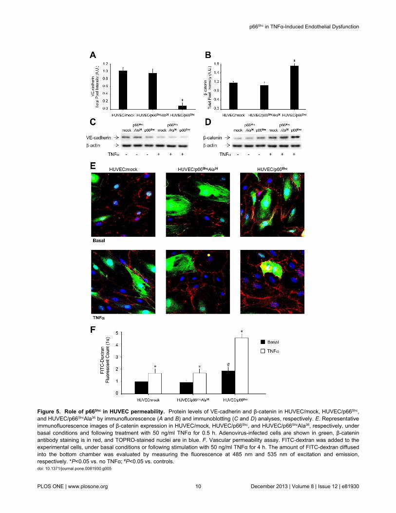
Figure 5. Role of p66Shc in HUVEC permeability. Protein levels of VE-cadherin and β-catenin in HUVEC/mock, HUVEC/p66Shc,and HUVEC/p66ShcAla36 by immunofluorescence (A and B) and immunoblotting (C and D) analyses, respectively. E. Representativeimmunofluorescence images of β-catenin expression in HUVEC/mock, HUVEC/p66Shc, and HUVEC/p66ShcAla36, respectively, underbasal conditions and following treatment with 50 ng/ml TNFα for 0.5 h. Adenovirus-infected cells are shown in green, β-cateninantibody staining is in red, and TOPRO-stained nuclei are in blue. F. Vascular permeability assay. FITC-dextran was added to theexperimental cells, under basal conditions or following stimulation with 50 ng/ml TNFα for 4 h. The amount of FITC-dextran diffusedinto the bottom chamber was evaluated by measuring the fluorescence at 485 nm and 535 nm of excitation and emission,respectively. *P<0.05 vs. no TNFα; #P<0.05 vs. controls.doi: 10.1371/journal.pone.0081930.g005
p66Shc in TNFα-Induced Endothelial Dysfunction
PLOS ONE | www.plosone.org 10 December 2013 | Volume 8 | Issue 12 | e81930

Figure 6. Effects of p66Shc silencing on endothelial activation in HUVEC. A. (top left) Representative immunoblot of p66Shc
Ser36 phosphorylation in HUVEC transfected with 50 nM or 100 nM of siRNA#1 or siRNA#2, and (right) quantification of multipleexperiments. Hatched bar, cells transfected with a negative control siRNA; black bars, cells treated with siRNA#1 and siRNA#2.(bottom left) Representative immunoblot of p66Shc protein content in HUVEC transfected with 50 nM or 100 nM of siRNA#1 orsiRNA#2, respectively, and (right) quantification of multiple experiments. Hatched bar, cells transfected with a negative controlsiRNA; grey bars, cells treated with siRNA#1; light grey bars, cells treated with siRNA#2. *P<0.05 vs. cells transfected with negativesiRNA. Neg., negative siRNA. B. Leukocyte transmigration test. HL-60 cells were added to control HUVEC (w.t., Lipofectamine™,and negative siRNA) and HUVEC treated with p66Shc siRNA#1 (100 nM) or p66Shc siRNA#2 (50 nM), and left to migrate for 18 h at37°C. Studies were carried out under basal conditions or following stimulation with 50 ng/ml TNFα for 4 h. Migrated cells werestained and measured by OD at 450 nm. Data represents the mean of triplicates from two independent experiments, and arenormalized to basal HUVEC/wt. *P<0.05 vs. no TNFα; §P<0.05 vs. HUVEC/wt. C. Effects of siRNA-mediated knockdown of p66Shc
on E-Selectin mRNA levels under basal conditions (left) and following TNFα stimulation (right). HUVEC were transfected with 100nM siRNA#1 (grey bars) or 50 nM siRNA#2 (light grey bars), and then left untreated or incubated with 50 ng/ml TNFα for 1 h.*P<0.05 vs. basal; #P<0.05 vs. TNFα-stimulated controls (wild-type, Lipofectamine™, and negative siRNA). Lysates were analyzedby qPCR 48 h following transfection, using β-actin as internal control. Lipo, Lipofectamine™.doi: 10.1371/journal.pone.0081930.g006
p66Shc in TNFα-Induced Endothelial Dysfunction
PLOS ONE | www.plosone.org 11 December 2013 | Volume 8 | Issue 12 | e81930
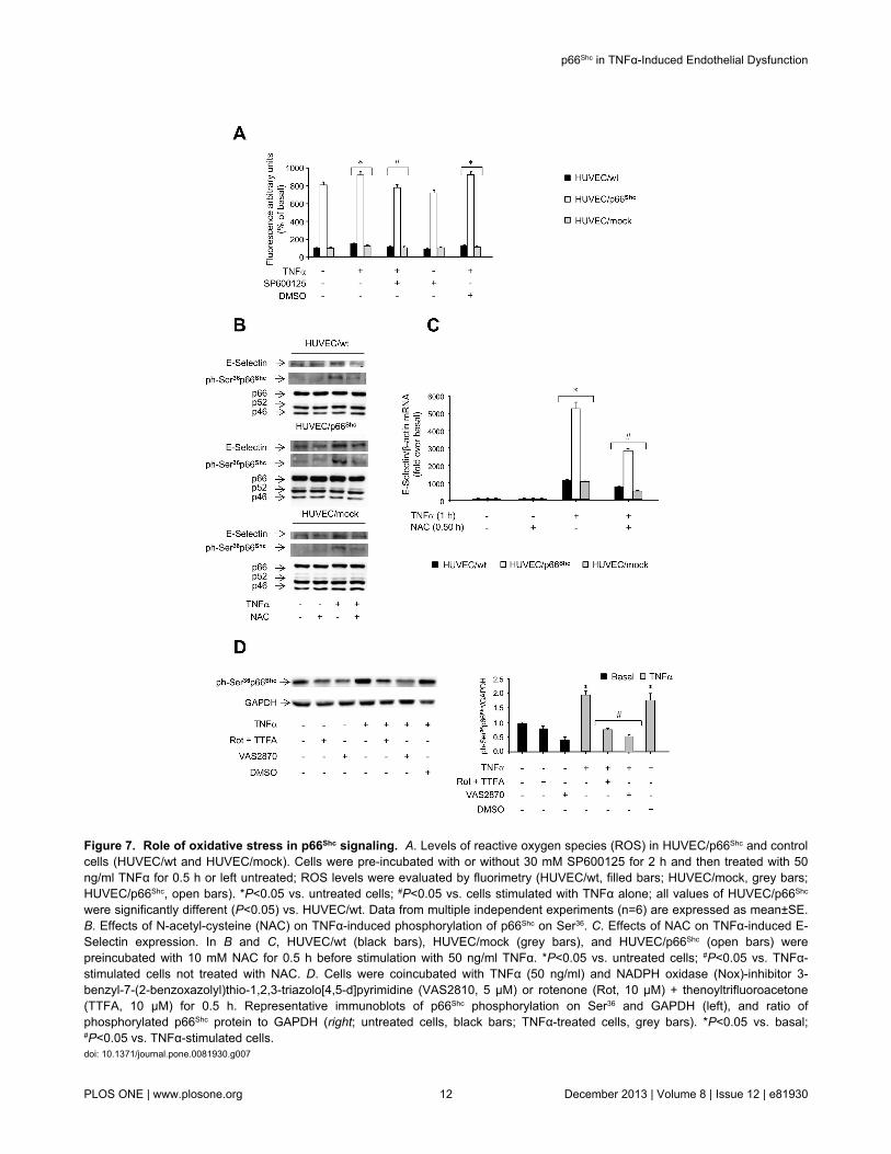
Figure 7. Role of oxidative stress in p66Shc signaling. A. Levels of reactive oxygen species (ROS) in HUVEC/p66Shc and controlcells (HUVEC/wt and HUVEC/mock). Cells were pre-incubated with or without 30 mM SP600125 for 2 h and then treated with 50ng/ml TNFα for 0.5 h or left untreated; ROS levels were evaluated by fluorimetry (HUVEC/wt, filled bars; HUVEC/mock, grey bars;HUVEC/p66Shc, open bars). *P<0.05 vs. untreated cells; #P<0.05 vs. cells stimulated with TNFα alone; all values of HUVEC/p66Shc
were significantly different (P<0.05) vs. HUVEC/wt. Data from multiple independent experiments (n=6) are expressed as mean±SE.B. Effects of N-acetyl-cysteine (NAC) on TNFα-induced phosphorylation of p66Shc on Ser36. C. Effects of NAC on TNFα-induced E-Selectin expression. In B and C, HUVEC/wt (black bars), HUVEC/mock (grey bars), and HUVEC/p66Shc (open bars) werepreincubated with 10 mM NAC for 0.5 h before stimulation with 50 ng/ml TNFα. *P<0.05 vs. untreated cells; #P<0.05 vs. TNFα-stimulated cells not treated with NAC. D. Cells were coincubated with TNFα (50 ng/ml) and NADPH oxidase (Nox)-inhibitor 3-benzyl-7-(2-benzoxazolyl)thio-1,2,3-triazolo[4,5-d]pyrimidine (VAS2810, 5 µM) or rotenone (Rot, 10 µM) + thenoyltrifluoroacetone(TTFA, 10 µM) for 0.5 h. Representative immunoblots of p66Shc phosphorylation on Ser36 and GAPDH (left), and ratio ofphosphorylated p66Shc protein to GAPDH (right; untreated cells, black bars; TNFα-treated cells, grey bars). *P<0.05 vs. basal;#P<0.05 vs. TNFα-stimulated cells.doi: 10.1371/journal.pone.0081930.g007
p66Shc in TNFα-Induced Endothelial Dysfunction
PLOS ONE | www.plosone.org 12 December 2013 | Volume 8 | Issue 12 | e81930

4C). Similarly, HUVEC permeability to dextran was found to belower in HUVEC/p66ShcAla36 than in HUVEC/p66Shc and notdifferent from HUVEC/mock, both under basal conditions andfollowing TNFα challenge (Figure 5). Therefore, p66Shc
phosphorylation on Ser36 appears to be important for the abilityof this protein to mediate the effects of TNFα on E-Selectingene expression and function in HUVEC, and this signalingevent is apparently dependent upon JNK activation and ROSgeneration (Figure 4D).
Discussion
Endothelial dysfunction is a systemic disorder and a keyvariable in the pathogenesis of atherosclerosis and itscomplications. Expression of adhesion molecules, leading toleukocyte rolling and diapedesis, represents one of the earliestevents in the atherogenic cascade. Hence, reverting thebiological processes leading to endothelial activation may bean attractive target in the effort to optimize individualizedtherapeutic strategies to reduce cardiovascular morbidity andmortality [34] . The p66Shc protein has been shown to controlcellular responses to oxidative stress, being involved inatherosclerosis in animal models [26,35,36]. Deletion of thep66Shc gene protects against age-related endothelialdysfunction [37] and reduces systemic and tissue oxidativestress, vascular cell apoptosis, and early atherogenesis in micefed a high-fat diet [26]. In this study, we provide further insightinto the cellular mechanisms mediating endothelial dysfunction,by showing that the stress-sensor protein p66Shc is implicated inTNFα action in HUVEC, a well-defined cellular model of thehuman endothelium.
Accumulating evidence suggests that the pro-inflammatorycytokine TNFα plays an important role in the disruption ofvascular responses [38]. TNFα stimulates the expression ofendothelial cell genes, including E-Selectin, which maypromote atherosclerosis via leukocyte recruitment [10]. In thisstudy, challenge of HUVEC with TNFα resulted in increased E-Selectin expression, in line with similar results obtained in otherendothelial cell systems [39]. This response occurred via theactivation of the stress-kinase JNK [40], whereas ERK wasapparently not involved.
Phosphorylation of p66Shc on Ser36 has been shown tomediate cellular aging, damage, and apoptosis, and to beinduced by oxidative stress [41]. We found that stimulation ofHUVEC with TNFα induced a 2- to 3-fold increase in p66Shc
phosphorylation on Ser36 (Figure 2, A and B), which was almostcompletely blocked by pretreatment of cells with the specificJNK inhibitor (Figure 2, C and D) and only partially reduced bypretreatment of cells with the p38 inhibitor (Figure S3A), in linewith previous results obtained in bovine aortic endothelial cellsexposed to H2O2 [41] and in HUVEC treated with the VascularEndothelial Growth Factor [42]. Specific isoforms of the proteinkinase C (PKC) family have also been proposed as mediatorsof ROS triggered stress signals. Indeed, oxidative stress hasbeen shown to activate the PKC-β isoform, leading to Ser36-phosphorylation and mitochondrial translocation of p66Shc, andincreased cell apoptosis [43]. In this study, the specific PKC-βinhibitor LY333531 reduced TNFα-induced phosphorylation of
p66Shc on Ser36 (Figure S3B). Interestingly, p66Shc inducesmitochondrial H2O2 production, resulting in furtheraugmentation of intracellular H2O2 levels and PKC-β activation,in a sort of self-triggered control loop [43]. In HUVEC, the PKC-β inhibitor LY333531 reduced AGEs-induced macrophageadhesion to endothelial cells, thus relieving the localinflammation [44]. It has also been previously demonstratedthat the inhibition of PKC-β prevents the activation of the JNK-p66Shc proatherogenic pathway in human aortic endothelialcells [45], in line with the results of our study.
The effect of TNFα on p66Shc phosphorylation on Ser36 wastightly associated with the ability of this cytokine to induce E-Selectin expression and leukocyte transmigration. Cells withselective overexpression of p66Shc and enhanced p66Shc
phosphorylation on Ser36 showed increased E-Selectinexpression and leukocyte transmigration, under basalconditions, and more so after stimulation with TNFα (Figure 3,B and C). A similar effect was observed when we studiedendothelial permeability, which appeared to be increased inp66Shc-overexpressing cells, and further increased in responseto TNFα (Figure 5F). This was associated with reduced VE-cadherin and increased β-catenin protein levels, respectively(Figure 5, A-E). Changes in VE-cadherin levels maysignificantly affect vascular permeability [16,22,46], anddisruption of the adherent junctions at the level of VE-cadherinand β-catenin is an important mechanism leading tomicrovascular hyperpermeability [20]. Indeed, the increaseddegradation of VE-cadherin may be due to the disruption ofcadherin–catenin complexes [47]. Interestingly, enhancedendothelial cell permeability associated with increased β-catenin expression and disruption of the VE-cadherin/β-catenincomplexes, as found in HUVEC overexpressing wild-typep66Shc (Figure 5) in this study, has been also observed inHUVEC treated with LPS and Ucn-1 [22].
Thus, p66Shc appears to enhance the cellular sensitivity to theeffects of TNFα, similarly to the increased susceptibility to toxicdamage and apoptosis observed in other cellular models ofp66Shc overexpression [48]. Conversely, deletion of p66Shc
conferred protection from oxidative injury and apoptosis in bothcellular systems in vitro and experimental animals in vivo[26,35]. In parallel with these observations, siRNA-mediatedp66Shc gene silencing resulted in marked reduction of E-Selectin expression and leukocyte transmigration both underbasal conditions and after TNFα stimulation (Figure 6, B andC). The role of p66Shc phosphorylation on Ser36 in this responsewas established by overexpressing a phosphorylation-defectivep66Shc mutant protein in HUVEC, which raised total p66Shc
protein levels to those found in the HUVEC overexpressing thewild-type p66Shc (Figure S7, A-C; Figure 4A), but failed toenhance E-Selectin expression or leukocyte transmigration asthat observed in HUVEC/p66Shc (Figure 4, B and C). In multipleexperimental models, p66Shc-dependent biological responsesappear to be largely inhibited when Ser36 is mutated, eventhough the extent of inhibition may vary depending on the celltype and specific amino acid substitution (i.e., Ser-to-Ala vs.Ser-to-Asp) [49]. Our results confirm previous observations onthe critical role of Ser36 phosphorylation of p66Shc for inductionof the apoptosis cascade in cells exposed to oxidative stress
p66Shc in TNFα-Induced Endothelial Dysfunction
PLOS ONE | www.plosone.org 13 December 2013 | Volume 8 | Issue 12 | e81930

[24]. Interestingly, ROS production was associated withactivation of the JNK-p66Shc pathway (Figure 7A), andpretreatment with the antioxidant NAC resulted in a significantdecrease in TNFα-induced p66Shc phosphorylation and E-Selectin expression (Figure 7B). The mitochondrial respiratorychain is considered the main intracellular source of ROS;indeed, the NADPH oxidase complex has been involved inendothelial dysfunction [50] and Ser36 phosphorylation of p66Shc
in normal fibroblasts [51]. It was previously demonstrated thatrotenone and TTFA (10 µM), which inhibit electron transfer toubiquinone, significantly decreased ROS generation in HUVEC[52]. A decrease in ROS production has been reported also inHUVEC treated with the flavo-enzymes inhibitor diphenyleneiodinium (10 µM) [52] and the NADPH oxidase inhibitorVAS2870 (5 µM) after incubation with TNFα and oxidized low-density lipoprotein, respectively [53,54]. In contrast, antimycinA (AA, 10 µM), a blocker of ubisemiquinone, increased ROSgeneration [52]. In this study, Ser36 phosphorylation of p66Shc
was significantly decreased when cells were exposed to theNADPH oxidase inhibitor VAS2810, and to the complex Iinhibitors rotenone and TTFA, suggesting a role for bothcytosolic and mitochondrial ROS in p66Shc activation inendothelial cells (Figure 7D). Hence, E-Selectin expressionappears to be, at least in part, under ROS control (Figure 7C),in line with previous reports [55]. In HUVEC/p66Shc,phosphorylation of p66Shc on Ser36 was found to be significantlyincreased also in the absence of TNFα stimulation (Figure 4A),and this was associated with some enhancement of basal E-Selectin mRNA levels (Figure 3C). This “constitutive” p66Shc
Ser phosphorylation was partly reduced when HUVEC/p66Shc
were preincubated with the specific JNK inhibitor (Figure 4A),even though we could not document changes in JNKphosphorylation by immunoblotting (Figure S5A). Thisphenomenon could potentially result from the effects ofaugmenting p66Shc protein levels in the presence of a low yeteffective level of basal JNK activity.
The downstream targets of p66Shc, potentially involved in theregulation of E-Selectin gene expression in HUVEC, are stillpoorly characterized. p66Shc has been shown to phosphorylateand inactivate FOXO3a, which is involved in the modulation ofantioxidant defenses in cardiomyocytes [56]. However, there isno evidence of FOXO3a expression in endothelial cells, or ofany FOXO3a-mediated transcriptional modulation of E-Selectin. p66Shc has also been shown to regulate thetranscriptional regulator Kruppel-like factor (KLF2), sinceknockdown of p66Shc in endothelial cells induced mRNAexpression of KLF2 and of its target gene thrombomodulin [57].In addition, overexpression of KLF2 potently inhibited theinduction of VCAM-1 and E-Selectin in response toproinflammatory cytokines, including TNFα [58], suggestingKLF2 as a likely candidate mediating the effects of p66Shc on E-Selectin. However, we found that selective overexpression orknockdown of p66Shc did not modify KLF2 mRNA levels inHUVEC (data not shown).
In animal models, the p66Shc protein has been shown to beinvolved in the cellular events leading to atherosclerosis.Overexpression of the p66Shc gene has been shown to reducenitric oxide generation in endothelial cells, whereas
downregulation or deletion of the p66Shc gene improved theimpaired endothelial cell-dependent vasodilation [36]. Inhumans, the expression of p66Shc has been evaluated incirculating monocytes, which are key contributors to thevascular damage. The levels of p66Shc in monocytes weresignificantly increased in patients with type 2 diabetescompared with controls, and were correlated to total plasma 8-isoprostane, a marker of oxidative stress [28]. More recently,p66Shc was found to be increased in monocytes from patientswith coronary artery disease, with a direct correlation betweenp66Shc levels and the number of diseased vessels [59]. In thisstudy, we provide evidence that p66Shc, through its regulatorySer36 phosphorylation, lies downstream of JNK in theTNFα/JNK signaling pathway that controls E-Selectin geneexpression in human endothelial cells (Figure 4D).Interestingly, modulation of p66Shc expression appears to affectthe ability of endothelial cells to induce leukocytetransmigration, a process which involves an E-Selectin-dependent mechanism [60] (Figure 1D), and Ser36
phosphorylation of p66Shc is again essential for thisphenomenon (Figure 4C). Thus, increased p66Shc expressionand activity may contribute to the endothelial dysfunctionobserved in individuals with high cardiovascular risk andpotentially represent a novel marker of vascular injury.
Supporting Information
Figure S1. A. E-Selectin mRNA levels in siRNA-transfectedHUVEC. HUVEC were transfected with 50 nM or 100 nM of twodifferent siRNAs specific for E-Selectin (siRNA#1 andsiRNA#2). Cells were lysed after 48 h, and E-Selectin mRNAlevels were measured by qPCR. siRNA Neg, cells transfectedwith a negative control siRNA. B. Time-course of TNFα-induced JNK-phosphorylation in HUVEC. Representativeimmunoblots of JNK phosphorylation (top left) and proteincontent (bottom left), and ratio of phosphorylated to total JNKproteins (right; JNK-1, grey line; JNK-2, black line). C. Time-course of TNFα-induced ERK phosphorylation. Representativeimmunoblots of ERK-1/2 phosphorylation (top left) and proteincontent (bottom left), and ratio of phosphorylated to totalERK-1/2 isoforms (right; ERK-1, black line, ERK-2, grey line).(TIF)
Figure S2. Effects of JNK and ERK inhibitors on TNFα-induced E-Selectin protein levels. HUVEC were pre-treatedwith 30 mM of the JNK inhibitor SP600125 (Panel A) or theERK inhibitor PD98059 (Panel B), respectively, for 2 h, andthen challenged with 50 ng/ml TNFα for the indicated times(untreated cells, black bars; inhibitor-treated cells, grey bars;DMSO-treated cells, light grey bars). E-Selectin protein levelswere evaluated by immunoblotting, using p66Shc protein contentas internal control. C. Representative images of HUVECtreated with the JNK inhibitor JNKi. Cells were treated with 10mg/ml JNKi peptide linked to a FITC fluorochrome for 0.5 h, 1 hor 2 h, and analyzed by fluorescence microscopy. The greenstaining identifies peptide accumulation inside the cells (left).Cellular morphology was evaluated by optical microscopy(right). Representative images after 2 h of exposure to the JNKi
p66Shc in TNFα-Induced Endothelial Dysfunction
PLOS ONE | www.plosone.org 14 December 2013 | Volume 8 | Issue 12 | e81930

are shown. D. Effects of JNKi on TNFα-induced c-Jun Ser63
phosphorylation and E-Selectin protein expression. HUVECwere preincubated with 10 mg/ml JNKi peptide for the indicatedtimes, and then exposed to 50 ng/ml TNFα for 1 h. E-Selectinprotein levels were evaluated by immunoblotting, using Shcprotein expression as loading control. c-Jun Ser63
phosphorylation was evaluated as a readout for the activity ofthe inhibitor, using c-Jun protein content as loading control.(TIF)
Figure S3. Effects of p38 MAPK and PKC-β inhibition onSer36 phosphorylation of p66Shc in HUVEC. A. Effects of thep38 MAPK inhibitor SB203580 on TNFα-inducedphosphorylation of p66Shc on Ser36. Cells were pre-treated with15 or 30 µM SB203580 for 1 h and then exposed to 50 ng/mlTNFα for 0.5 h. Representative immunoblots of p66Shc
phosphorylation on Ser36 (top left) and Shc protein content(middle left), and ratio of phosphorylated to total p66Shc protein(right; untreated cells, black bars; inhibitor-treated cells, greybars). B. Effects of the PKC-β inhibitor LY333531 on TNFα-induced phosphorylation of p66Shc on Ser36. Cells were pre-treated with 200 nM LY333531 for 1 h or 24 h and thenexposed to 50 ng/ml TNFα for 0.5 h. Representativeimmunoblots of p66Shc phosphorylation on Ser36 (top left) andShc protein content (middle left), and ratio of phosphorylated tototal p66Shc protein (right; untreated cells, black bars; inhibitor-treated cells, grey bars). β-actin protein content was used asloading control. *P<0.05 vs. basal; #P<0.05 vs. TNFα. SB,SB203580; LY, LY333531.(TIF)
Figure S4. Overexpression of p66Shc in HUVEC. A.Representative immunoblots of p66Shc phosphorylation on Ser36
and of Shc protein content in wild-type HUVEC (HUVEC/wt),HUVEC overexpressing p66Shc (HUVEC/p66Shc), and HUVECoverexpressing p66Shc with Ser36 to Ala mutation (HUVEC/p66ShcAla36). HUVEC were infected with different PFU/ml ofadenovirus, as indicated, and cell lysates were subjected toimmunoblotting with specific antibodies. B. Quantification ofp66Shc protein content in HUVEC/p66Shc (black line) andHUVEC/p66ShcAla36 (grey line) under basal conditions. Proteincontent of p66Shc is normalized to p52Shc protein content; thep66 Shc/p52Shc ratio in wild-type cells was considered thereference value (PFU=0 in the graph). C. Ratio of basal p66Shc
Ser36 phosphorylation to total p66Shc protein content inHUVEC/p66Shc (black line) and HUVEC/p66ShcAla36 (grey line),using the p66Shc phosphorylation/content ratio in HUVEC/wt asreference (PFU=0 in the graph). *P<0.05 vs. controls (wild-typeand mock). D. Representative images of HUVEC/wt,HUVEC/p66Shc, HUVEC/p66ShcAla36, and HUVEC/mock. Cellswere infected with the different adenoviral constructs andanalyzed by fluorescent microscopy. The green stainingidentifies cells expressing the green fluorescent protein (GFP)encoded by the recombinant adenovirus. Cellular morphologywas evaluated by optical microscopy.(TIF)
Figure S5. Phosphorylation of JNK and ERK and E-Selectin protein levels in HUVEC overexpressing p66Shc. A.Basal and TNFα-stimulated phosphorylation of JNK1/2 inHUVEC overexpressing p66Shc. HUVEC/w.t., HUVEC/p66Shc
and HUVEC/mock were incubated with 50 ng/ml TNFα for theindicated times or left untreated. Cell lysates were subjected toimmunoblotting with specific antibodies. Representativeimmunoblots of JNK1/2 phosphorylation and JNK1/2 proteincontent (top), and ratio of phosphorylated to total JNK proteins(bottom) (HUVEC/w.t. black bars, HUVEC/p66Shc, open bars;HUVEC/mock, grey bars). B. Basal and TNFα-stimulatedphosphorylation of ERK1/2 in HUVEC overexpressing p66Shc.HUVEC/w.t., HUVEC/p66Shc and HUVEC/mock were incubatedwith 50 ng/ml TNFα for the indicated times or left untreated.Cell lysates were subjected to immunoblotting with specificantibodies. Representative immunoblots of ERK-1/2phosphorylation and protein content (top), and ratio ofphosphorylated to total ERK-1/2 isoforms (bottom) (HUVEC/w.t. black bars, HUVEC/p66Shc, open bars; HUVEC/mock, greybars). *P<0.05 vs. basal. C. Basal and TNFα-stimulated E-Selectin protein levels in HUVEC overexpressing p66Shc.HUVEC/p66Shc and HUVEC/mock were incubated with 50 ng/mlTNFα for the indicated times or left untreated. Cell lysates weresubjected to immunoblotting with specific antibodies.Representative immunoblots of E-Selectin protein levels (left),and the ratio of E-Selectin to GAPDH (used as loading control)protein levels in multiple experiments (right) are shown(HUVEC/p66Shc, open bars; HUVEC/mock, grey bars). *P<0.05vs. basal; #P<0.05 vs. HUVEC/mock.(TIF)
Figure S6. Role of p66Shc in TNFα regulated NF-κBpathway. HUVEC/wt (A), HUVEC/mock (B), HUVEC/p66Shc
(C), and HUVEC/p66ShcAla36 (D) were left untreated orincubated with TNFα for 15 min, and then analyzed byimmunofluorescence. Adenovirus-infected cells are shown ingreen, NF-κB antibody staining is in red, and TOPRO-stainednuclei are in blue. The fuchsia staining results from the mergingof red and blue, indicating NF-κB localization in the cellnucleus. E. Cell lysates were subjected to immunoblotting withph-Ser32IκBα antibody using β-actin as loading control.Representative immunoblots (left) and the ratio of ph-Ser32IκBαto β-actin protein levels from multiple experiments (right) areshown (HUVEC/mock, grey bars, HUVEC/p66ShcAla36 blackbars, HUVEC/p66Shc, open bars). *P<0.05 vs. basal.(TIF)
Figure S7. siRNA-mediated knockdown of p66Shc. A. p66Shc
mRNA levels in siRNA-transfected HUVEC. HUVEC weretransfected with 50 nM or 100 nM of two different siRNAsspecific for p66Shc (siRNA#1 and siRNA#2). Cells were lysed 48h or 72 h later, and p66Shc mRNA levels were measured byqPCR. wt, wild-type cells; Lipo, cells treated withLipofectamine™ only; Negative siRNA, cells transfected with anegative control siRNA. B and C. Effects of siRNA-mediatedknockdown of p66Shc on E-Selectin protein expression underbasal conditions and following TNFα stimulation in HUVECpretreated with SP600125 (B) and PD98059 (C). HUVEC were
p66Shc in TNFα-Induced Endothelial Dysfunction
PLOS ONE | www.plosone.org 15 December 2013 | Volume 8 | Issue 12 | e81930
transfected for 72 h with 100 nM siRNA#1 and or negativecontrol siRNA, and preincubated with SP600125 or PD98059(30 mM, 2h), using DMSO as control, before treatment with 50ng/ml TNFα for 1 h. Lysates were analyzed by immnoblotting,using p52Shc and p46Shc as controls. Black bars, cellstransfected with a negative control siRNA; white bars, cellstransfected with 100 nM siRNA#1.(TIF)
Acknowledgements
We gratefully acknowledge Maria Pia Scavo for assistance withconfocal imaging.
Author Contributions
Conceived and designed the experiments: LL MRO CP TC SPAN FG. Performed the experiments: LL MRO MAI CC MM ALFT RL SM CP. Analyzed the data: LL MRO AC CP TC SP ANFG. Wrote the manuscript: LL MRO FG.
References
1. De Vriese AS, Verbeuren TJ, Van de Voorde J, Lameire NH, VanhouttePM (2000) Endothelial dysfunction in diabetes. Br J Pharmacol 130:963-974. doi:10.1038/sj.bjp.0703393. PubMed: 10882379.
2. Seifalian AM, Filippatos TD, Joshi J, Mikhailidis DP (2010) Obesity andarterial compliance alterations. Curr Vasc Pharmacol 8: 155-168. doi:10.2174/157016110790886956. PubMed: 20180777.
3. Rothlein R, Czajkowski M, O'Neill MM, Marlin SD, Mainolfi E et al.(1988) Induction of intercellular adhesion molecule 1 on primary andcontinuous cell lines by pro-inflammatory cytokines. Regulation bypharmacologic agents and neutralizing antibodies. J Immunol 141:1665-1669. PubMed: 3137261.
4. Viemann D, Goebeler M, Schmid S, Klimmek K, Sorg C et al. (2004)Transcriptional profiling of IKK2/NF-kappa B- and p38 MAP kinase-dependent gene expression in TNF-alpha-stimulated primary humanendothelial cells. Blood 103: 3365-3373. doi:10.1182/blood-2003-09-3296. PubMed: 14715628.
5. Hoefen RJ, Berk BC (2002) The role of MAP kinases in endothelialactivation. Vascul Pharmacol 38: 271-273. doi:10.1016/S1537-1891(02)00251-3. PubMed: 12487031.
6. Denk A, Goebeler M, Schmid S, Berberich I, Ritz O et al. (2001)Activation of NF-kappa B via the Ikappa B kinase complex is bothessential and sufficient for proinflammatory gene expression in primaryendothelial cells. J Biol Chem 276: 28451-28458. doi:10.1074/jbc.M102698200. PubMed: 11337506.
7. Gustin JA, Pincheira R, Mayo LD, Ozes ON, Kessler KM et al. (2004)Tumor necrosis factor activates CRE-binding protein through a p38MAPK/MSK1 signaling pathway in endothelial cells. Am J Physiol CellPhysiol 286: C547-C555. doi:10.1152/ajpcell.00332.2002. PubMed:14761884.
8. Nwariaku FE, Chang J, Zhu X, Liu Z, Duffy SL et al. (2002) The role ofp38 map kinase in tumor necrosis factor-induced redistribution ofvascular endothelial cadherin and increased endothelial permeability.Shock 18: 82-85. doi:10.1097/00024382-200207000-00015. PubMed:12095140.
9. Kelly M, Hwang JM, Kubes P (2007) Modulating leukocyte recruitmentin inflammation. J Allergy Clin Immunol 120: 3-10. doi:10.1016/S0091-6749(07)00978-5. PubMed: 17559914.
10. Kuldo JM, Westra J, Asgeirsdóttir SA, Kok RJ, Oosterhuis K et al.(2005) Differential effects of NF-{kappa}B and p38 MAPK inhibitors andcombinations thereof on TNF-{alpha}- and IL-1{beta}-inducedproinflammatory status of endothelial cells in vitro. Am J Physiol CellPhysiol 289: 1229-1239. doi:10.1152/ajpcell.00620.2004. PubMed:15972838.
11. Barthel SR, Gavino JD, Descheny L, Dimitroff CJ (2007) Targetingselectins and selectin ligands in inflammation and cancer. Expert OpinTher Targets 11: 1473-1491. doi:10.1517/14728222.11.11.1473.PubMed: 18028011.
12. Dong ZM, Chapman SM, Brown AA, Frenette PS, Hynes RO et al.(1998) The combined role of P- and E-selectins in atherosclerosis. JClin Invest 102: 145-152. doi:10.1172/JCI3001. PubMed: 9649568.
13. Tözeren A, Kleinman HK, Grant DS, Morales D, Mercurio AM et al.(1995) E-selectin-mediated dynamic interactions of breast- and colon-cancer cells with endothelial-cell monolayers. Int J Cancer 60: 426-431.PubMed: 7530236.
14. Skrha J, Stulc T, Hilgertová J, Weiserová H, Kvasnicka J et al. (2004)Effect of simvastatin and fenofibrate on endothelium in Type 2
diabetes. Eur J Pharmacol 493: 183-189. doi:10.1016/j.ejphar.2004.04.025. PubMed: 15189781.
15. Cominacini L, Fratta Pasini A, Garbin U, Campagnola M, Davoli A et al.(1997) E-selectin plasma concentration is influenced by glycaemiccontrol in NIDDM patients: possible role of oxidative stress.Diabetologia 40: 584-589. doi:10.1007/s001250050719. PubMed:9165228.
16. Rankin JA (2004) Biological mediators of acute inflammation. AACNClin Issues 15: 3–17. doi:10.1097/00044067-200401000-00002.PubMed: 14767362.
17. Dewi BE, Takasaki T, Kurane I (2008) Peripheral blood mononuclearcells increase the permeability of dengue virus-infected endothelialcells in association with downregulation of vascular endothelialcadherin. J Gen Virol 89: 642-652. doi:10.1099/vir.0.83356-0. PubMed:18272754.
18. Gooding JM, Yap KL, Ikura M (2004) The cadherin–catenin complex asa focal point of cell adhesion and signalling: New insights from three-dimensional structures. Bioessays 26: 497–511. doi:10.1002/bies.20033. PubMed: 15112230.
19. Cattelino A, Liebner S, Gallini R, Zanetti A, Balconi G et al. (2003) Theconditional inactivation of the beta-catenin gene in endothelial cellscauses a defective vascular pattern and increased vascular fragility. JCell Biol 162: 1111–1122. doi:10.1083/jcb.200212157. PubMed:12975353.
20. Fang D, Hawke D, Zheng Y, Xia Y, Meisenhelder J et al. (2007)Phosphorylation of beta-catenin by AKT promotes beta-catenintranscriptional activity. J Biol Chem 282: 11221-11229. doi:10.1074/jbc.M611871200. PubMed: 17287208.
21. Mochizuki N (2009) Vascular integrity mediated by vascular endothelialcadherin and regulated by sphingosine 1-phosphate andangiopoietin-1. Circ J 73: 2183–2191. doi:10.1253/circj.CJ-09-0666.PubMed: 19838001.
22. Wan R, Guo R, Chen C, Jin L, Zhu C et al. (2013) Urocortin increasedLPS-induced endothelial permeability by regulating the cadherin-catenin complex via corticotrophin-releasing hormone receptor 2. J CellPhysiol 228: 1295-1303. doi:10.1002/jcp.24286. PubMed: 23168683.
23. Le S, Connors TJ, Maroney AC (2001) c-Jun N-terminal kinasespecifically phosphorylates p66ShcA at serine 36 in response toultraviolet irradiation. J Biol Chem 276: 48332-48336. PubMed:11602589.
24. Migliaccio E, Giorgio M, Mele S, Pelicci G, Reboldi P et al. (1999) Thep66shc adaptor protein controls oxidative stress response and life spanin mammals. Nature 402: 309-313. doi:10.1038/46311. PubMed:10580504.
25. Nemoto S, Combs CA, French S, Ahn BH, Fergusson MM et al. (2006)The mammalian longevity-associated gene product p66shc regulatesmitochondrial metabolism. J Biol Chem 281: 10555-10560. doi:10.1074/jbc.M511626200. PubMed: 16481327.
26. Napoli C, Martin-Padura I, de Nigris F, Giorgio M, Mansueto G et al.(2003) Deletion of the p66Shc longevity gene reduces systemic andtissue oxidative stress, vascular cell apoptosis, and early atherogenesisin mice fed a high-fat diet. Proc Natl Acad Sci U S A 100: 2112-2116.doi:10.1073/pnas.0336359100. PubMed: 12571362.
27. Menini S, Amadio L, Oddi G, Ricci C, Pesce C et al. (2006) Deletion ofp66Shc longevity gene protects against experimental diabeticglomerulopathy by preventing diabetes-induced oxidative stress.Diabetes 55: 1642-1650. doi:10.2337/db05-1477. PubMed: 16731826.
p66Shc in TNFα-Induced Endothelial Dysfunction
PLOS ONE | www.plosone.org 16 December 2013 | Volume 8 | Issue 12 | e81930

28. Pagnin E, Fadini G, de Toni R, Tiengo A, Calò L et al. (2005) Diabetesinduces p66shc gene expression in human peripheral bloodmononuclear cells: relationship to oxidative stress. J Clin EndocrinolMetab 90: 1130-1136. PubMed: 15562031.
29. Bonny C, Oberson A, Negri S, Sauser C, Schorderet DF (2001) Cell-permeable peptide inhibitors of JNK: novel blockers of beta-cell death.Diabetes 50: 77–82. doi:10.2337/diabetes.50.1.77. PubMed: 11147798.
30. Natalicchio A, De Stefano F, Perrini S, Laviola L, Cignarelli A et al.(2009) Involvement of the p66Shc protein in glucose transportregulation in skeletal muscle myoblasts. Am J Physiol EndocrinolMetab 296: E228-E237. PubMed: 18957618.
31. Kisielow M, Kleiner S, Nagasawa M, Faisal A, Nagamine Y (2002)Isoform-specific knockdown and expression of adaptor protein ShcAusing small interfering. RNA - Biochem J 363: 1-5. doi:10.1042/0264-6021:3630001.
32. Walker T, Müller I, Raabe C, Nohe B, Zanke C et al. (2011) Effectivesilencing of adhesion molecules on venous endothelial cells forprotection of venous bypass grafts. Eur J Cardiothorac Surg 40:1241-1247. PubMed: 21429759.
33. Liu Z-J, Tian R, Li Y, An W, Zhuge Y et al. (2011) Inhibition of tumorangiogenesis and melanoma growth by targeting vascular E-Selectin.Ann Surg 254: 450-457. doi:10.1097/SLA.0b013e31822a72dc.PubMed: 21795970.
34. Bonetti PO, Lerman LO, Lerman A (2003) Endothelial Dysfunction. AMarker of Atherosclerotic. Risk - Arteriosclerosis, Thrombosis, andVascular Biology 23: 168-175. doi:10.1161/01.ATV.0000051384.43104.FC.
35. Martin-Padura I, de Nigris F, Migliaccio E, Mansueto G, Minardi S et al.(2008) p66Shc deletion confers vascular protection in advancedatherosclerosis in hypercholesterolemic apolipoprotein E knockoutmice. Endothelium 15: 276-287. doi:10.1080/10623320802487791.PubMed: 19065319.
36. Francia P, Cosentino F, Schiavoni M, Huang Y, Perna E et al. (2009)p66(Shc) protein, oxidative stress, and cardiovascular complications ofdiabetes: the missing link. J Mol Med (Berl) 87: 885-891. doi:10.1007/s00109-009-0499-3. PubMed: 19590843.
37. Francia P, Delli Gatti C, Bachschmid M, Martin-Padura I, Savoia C etal. (2004) Deletion of p66shc gene protects against age-relatedendothelial dysfunction. Circulation 110: 2889–2895. doi:10.1161/01.CIR.0000147731.24444.4D. PubMed: 15505103.
38. Zhang H, Park Y, Wu J, Chen XP, Lee S et al. (2009) Role of TNF-alpha in vascular dysfunction. Clin Sci (Lond) 116: 219-230. doi:10.1042/CS20080196.
39. Read MA, Whitley MZ, Gupta S, Pierce JV, Best J et al. (1997) Tumornecrosis factor alpha-induced E-selectin expression is activated by thenuclear factor-kappaB and c-JUN N-terminal kinase/p38 mitogen-activated protein kinase pathways. J Biol Chem 272: 2753-2761. doi:10.1074/jbc.272.5.2753. PubMed: 9006914.
40. Pober JS (1998) Activation and injury of endothelial cells by cytokines.Pathol. Biol (Paris) 46: 159-163.
41. Li M, Chiou KR, Kass DA (2007) Shear stress inhibition of H2O2-induced p66(Shc) phosphorylation by ASK1-JNK inactivation inendothelium. Heart Vessels 22: 423-427. doi:10.1007/s00380-007-0994-9. PubMed: 18044002.
42. Oshikawa J, Kim SJ, Furuta E, Caliceti C, Chen GF et al. (2012) Novelrole of p66Shc in ROS-dependent VEGF signaling and angiogenesis inendothelial cells. Am J Physiol Heart Circ Physiol 302: H724-H732. doi:10.1152/ajpheart.00739.2011. PubMed: 22101521.
43. Pinton P, Rizzuto R (2008) p66Shc, oxidative stress and aging:importing a lifespan determinant into mithocondria. Cell Cycle 7:304-308. doi:10.4161/cc.7.3.5360. PubMed: 18235239.
44. Xu Y, Wang S, Feng L, Zhu Q, Xiang P et al. (2010) Blockade of PCK-beta protects HUVEC from advanced glycation end products inducedinflammation. Int Immunopharmacology 10: 1552-1559. doi:10.1016/j.intimp.2010.09.006.
45. Shi Y, Cosentino F, Camici GG, Akhmedov A, Vanhoutte PM et al.(2011) Oxidized low-density lipoprotein activates p66Shc via lectin-like
oxidized low-density lipoprotein receptor-1, protein kinase C-beta, andc-Jun N-terminal kinase kinase in human endothelial cells. ArteriosclerThromb Vasc Biol 31: 2090-2097. doi:10.1161/ATVBAHA.111.229260.PubMed: 21817106.
46. Dejana E, Giampietro C (2012) Vascular endothelial-cadherin andvascular stability. Curr Opin Hematol 19: 218-223. doi:10.1097/MOH.0b013e3283523e1c. PubMed: 22395663.
47. Sawant DA, Tharakan B, Adekanbi A, Hunter FA, Smythe WR et al.(2011) Inhibition of VE-cadherin proteasomal degradation attenuatesmicrovascular hyperpermeability. Microcirculation 18: 46-55. doi:10.1111/j.1549-8719.2010.00067.x. PubMed: 21166925.
48. Koch OR, Fusco S, Ranieri SC, Maulucci G, Palozza P et al. (2008)Role of the life span determinant p66(shcA) in ethanol-induced liverdamage. Lab Invest 88: 750-760. doi:10.1038/labinvest.2008.44.PubMed: 18490896.
49. Arany I, Faisal A, Nagamine Y, Safirstein RL (2008) p66shc inhibitspro-survival epidermal growth factor receptor/ERK signaling duringsevere oxidative stress in mouse renal proximal tubule cells. J BiolChem 283: 6110-6117. doi:10.1074/jbc.M708799200. PubMed:18174162.
50. Nwariaku FE, Liu Z, Zhu X, Nahari D, Ingle C et al. (2004) NADPHoxidase mediates vascular endothelial cadherin phosphorylation andendothelial dysfunction. Blood 104: 3214-3220. doi:10.1182/blood-2004-05-1868. PubMed: 15271797.
51. Ammendola R, Ruocchio MR, Chirico G, Russo L, De Felice C et al.(2002) Inhibition of NADH/NADPH oxidase affects signal transductionby growth factor receptors in normal fibroblasts. Arch Biochem Biophys397: 253-257. doi:10.1006/abbi.2001.2641. PubMed: 11795879.
52. Corda S, Laplace C, Vicaut E, Duranteau J (2001) Rapid ReactiveOxygen Species Production by Mitochondria in Endothelail ClessExposed to Tumor Necrosis Factor-α is mediated by ceramide. Am JRespir Cell Mol Biol 24: 762-768. doi:10.1165/ajrcmb.24.6.4228.PubMed: 11415943.
53. Wind S, Beuerlein K, Eucker T, Müller H, Scheurer P et al. (2010)Comparative pharmacology of chemically distinct NADPH oxidaseinhibitors. Br J Pharmacol 161: 885-898. doi:10.1111/j.1476-5381.2010.00920.x. PubMed: 20860666.
54. Stielow C, Catar RA, Muller G, Wingler K, Scheurer P et al. (2006)Novel Nox inhibitor of oxLDL-induced reactive oxygen speciesformation in human endothelial cells. Biochem Biophys Res Commun26: 200-205. PubMed: 16603125.
55. Usui T, Yamawaki H, Kamibayashi M, Okada M, Hara Y (2010)CV-159, a unique dihydropyridine derivative, prevents TNF-inducedinflammatory responses in Human Umbilical Vein Endothelial Cells. JPharmacol Sci 113: 182-191. doi:10.1254/jphs.10033FP. PubMed:20484867.
56. Guo J, Gertsberg Z, Ozgen N, Steinberg SF (2009) p66Shc linksalpha1-adrenergic receptors to a reactive oxygen species-dependentAKT-FOXO3A phosphorylation pathway in cardiomyocytes. Circ Res104: 660-669. doi:10.1161/CIRCRESAHA.108.186288. PubMed:19168439.
57. Kumar A, Hoffman TA, Dericco J, Naqvi A, Jain MK et al. (2009)Transcriptional repression of Kruppel like factor-2 by the adaptorprotein p66shc. FASEB J 23: 4344-4352. doi:10.1096/fj.09-138743.PubMed: 19696221.
58. SenBanerjee S, Lin Z, Atkins GB, Greif DM, Rao RM et al. (2004) KLF2is a novel transcriptional regulator of endothelial proinflammatoryactivation. J Exp Med 199: 1305-1315. doi:10.1084/jem.20031132.PubMed: 15136591.
59. Noda Y, Yamagishi S, Matsui T, Ueda S, Ueda S et al. (2010) Thep66shc gene expression in peripheral blood monocytes is increased inpatients with coronary artery disease. Clin Cardiol 33: 548-552. doi:10.1002/clc.20761. PubMed: 20842738.
60. Yoshida M, Chien LJ, Yasukochi Y, Numano F (2000) Differentiation-induced transmigration of HL-60 cells across activated HUVECmonolayer involves E-selectin-dependent mechanism. Ann N Y AcadSci 902: 307-310. PubMed: 10865853
p66Shc in TNFα-Induced Endothelial Dysfunction
PLOS ONE | www.plosone.org 17 December 2013 | Volume 8 | Issue 12 | e81930
Copyright © 2022 FDOKUMEN