The Asia-Pacific in Asian American Transmigration: Lydia Minatoya’s The Strangeness of Beauty
Breakdown of the Endothelial Barrier Function in TumorCell Transmigration
Claudia Tanja Mierke,* Daniel Paranhos Zitterbart,* Philip Kollmannsberger,* Carina Raupach,*Ursula Schlotzer-Schrehardt,y Tamme Weyert Goecke,z Jurgen Behrens,§ and Ben Fabry**Biophysik, Zentrum fur medizinische Physik und Technik, yAugenklinik, zFrauenklinik, §Nikolaus-Fiebiger Zentrum fur Molekulare Medizin,Universitat Erlangen-Nurnberg, 91052 Erlangen, Germany
ABSTRACT The ability of tumor cells to metastasize is associated with a poor prognosis for cancer. During the process ofmetastasis, tumor cells circulating in the blood or lymph vessels can adhere to, and potentially transmigrate through, theendothelium and invade the connective tissue. We studied the effectiveness of the endothelium as a barrier against the invasion of51 tumor cell lines into a three-dimensional collagen matrix. Only nine tumor cell lines showed attenuated invasion in the presenceof an endothelial cell monolayer, whereas 17 cell lines became invasive or showed a significantly increased invasion. Endothelialcells cocultured with invasive tumor cells increased chemokine gene expression of IL-8 and Gro-b. Expression of the IL-8 and Gro-b receptor, CXCR2, was upregulated in invasive tumor cells. Addition of IL-8 or Gro-b increased tumor cell invasiveness by morethan twofold. Tumor cell variants selected for high CXCR2 expression were fourfold more invasive in the presence of an endothelialcell layer, whereas CXCR2 siRNA knock-down cells were fivefold less invasive. We demonstrate that Gro-b and IL-8 secreted byendothelial cells, together with CXCR2 receptor expression on invasive tumor cells, contribute to the breakdown of the endothelialbarrier by enhancing tumor cell force generation and cytoskeletal remodeling dynamics.
INTRODUCTION
Most cancer-related deaths are caused by metastasis forma-
tion, a process that starts with dissociation of tumor cells from
the primary tumor and is followed by tissue invasion, entrance
into blood or lymph vessels (intravasation), and transport to
remote sites. It is widely assumed that tumor cells can then
escape from the microvasculature (extravasation), invade the
target tissue, and form secondary tumors in distant organs
(1–3). A potentially rate-limiting step in metastasis formation,
therefore, would be the extravasation process that involves
adhesion of tumor cells to endothelial cells and their trans-
migration through the endothelial cell monolayer and base-
ment membrane (3–6). Certain tumor cell types have indeed
been shown, both in vitro and in vivo, to be able to overcome
the endothelial barrier (6–11). But extravasation need not be
the only mechanism for metastasis formation, as has recently
been pointed out by Al-Mehdi et al. (12), who reported that
tumor cells can adhere and grow onto the endothelial layer and
form a metastasis without ever leaving the blood or lymph
vessel. Either way, the role of the endothelial monolayer in
this process is thought to be crucial in that it can actively
modulate metastasis formation by either allowing or block-
ing the adhesion, and possibly transmigration, of tumor cells
(8–10). The details of endothelial cell functions in this pro-
cess, however, are poorly understood, and the extent to which
the endothelium impedes or promotes metastasis formation
is still unclear.
Transmigrating tumor cells are thought to be able to over-
come the endothelial barrier by inducing changes within
endothelial cells, including the upregulation of adhesion
molecule receptor expression (13), the reorganization of the
cytoskeleton (14), Src-mediated disruption of endothelial VE-
cadherin-b-catenin cell-cell adhesions (7), the formation of
‘‘holes’’ within the endothelial layer (15), and the induction of
apoptosis (16). Tumor cell invasion may bear a close resem-
blance to leukocyte trafficking for which the endothelium acts
as a barrier and greatly reduces invasion rates (17). For ex-
ample, the function of the endothelial cell barrier against both
leukocyte trafficking and tumor cell transmigration is reduced
in the presence of inflammatory cytokines such as tumor ne-
crosis factor-a and interleukin-1b (8,13,18,19). These cyto-
kines are known to trigger an upregulation of the adhesion
molecule E-selectin (13). The subsequent adhesion of tumor
cells to E-selectin leads to an upregulation of stress-activated
protein kinase-2 (SAPK2/p38) in endothelial cells (13) and
triggers actin polymerization and reorganization into stress
fibers (14).
Chemokines and their receptors are also important for
leukocyte trafficking (20,21) and tumor cell invasion (22).
Chemokines are a superfamily of small cytokine-like proteins
that induce cytoskeletal rearrangements in endothelial cells
and leukocytes, the firm adhesion of leukocytes to endothelial
cells, and the directional migration of leukocytes (20). The
involvement of chemokines in tumor-endothelial interactions
doi: 10.1529/biophysj.107.113613
Submitted June 11, 2007, and accepted for publication November 6, 2007.
Address reprint requests to Dr. Claudia Tanja Mierke, University of
Erlangen-Nuremberg, Center for Medical Physics and Technology, Biophys-
ics Group, Henkestrasse 91, 91052 Erlangen, Germany. Tel.: 49-9131-85-25607;
Fax: 49-9131-85-25601; E-mail: [email protected].
This is an Open Access article distributed under the terms of the Creative
Commons-Attribution Noncommercial License (http://creativecommons.
org/licenses/by-nc/2.0/), which permits unrestricted noncommercial use,
distribution, and reproduction in any medium, provided the original work is
properly cited.
Editor: Cristobal G. dos Remedios.
� 2008 by the Biophysical Society
0006-3495/08/04/2832/15 $2.00
2832 Biophysical Journal Volume 94 April 2008 2832–2846

and their effect on tumor cell mechanics during invasion are
considerably less well understood, however.
The aim of this study was to investigate the ability of the
endothelium to regulate the transmigration and invasion of
tumor cells into an extracellular matrix. We measured the
invasion of human tumor cell lines into a three-dimensional
collagen gel matrix that was covered with an endothelial cell
monolayer. In the presence of an endothelium, the invasion of
some tumor cell lines increased significantly. Gene expres-
sion analysis of endothelial cells cocultured with invasive
tumor cells revealed an upregulation of Gro-b and IL-8 che-
mokines compared with endothelial cells cocultured with
noninvasive tumor cells. Finally, we demonstrate that the
Gro-b and IL-8 receptor (CXCR2) expression on tumor cells
serves as a key mediator responsible for the breakdown of the
endothelial barrier function by enhancing tumor cell force
generation and cytoskeletal remodeling dynamics.
MATERIALS AND METHODS
Three-dimensional collagen assay
All chemicals were purchased from Sigma (Taufkirchen, Germany) unless
otherwise noted. Collagen R (Serva, Heidelberg, Germany) and G (Bio-
chrom, Berlin, Germany) were mixed at a ratio 1:1, 25 mM sodium bicar-
bonate and 10 vol % of 103 DME (Biochrom) was added. The solution was
neutralized with 1 N sodium hydroxide, and 1.2 ml collagen solution was
placed into each well of a six-well plate, polymerized, incubated with En-
dothelial Cell Growth Medium 2 (Promocell, Heidelberg, Germany) con-
taining 2% low-endotoxin FCS, and 600,000 endothelial cells (first passage)
were seeded onto the gel. Polymerized collagen gels were 474 mm 6 7 (mean 6
SE, n ¼ 12) thick. After 24 h, the cells had formed a closed monolayer, and
100,000 tumor cells were added per 3.5-cm dish. To study invasion in the
absence of endothelial cells, tumor cells were added directly to the gels.
Before seeding, tumor cells were stained with 5 mg/ml carboxyfluorescein
diacetate (Invitrogen, Karlsruhe, Germany) and 1 mg/ml Hoechst 33342 dye
to distinguish them from endothelial cells. Coculture times ranging from 8 h
to 5 d were tested. Staining was stable over the course of the experiment; the
Hoechst dye was found to exhibit only insignificant photobleaching, and the
carboxyfluorescein diacetate, although it was prone to photobleaching, was
adequate for confirming that endothelial cells did not invade the collagen
matrix. A coculture time of 72 h was found to be optimal because differences
in the invasiveness of tumor cell lines were clearly visible while the endo-
thelial monolayer was still confluent, with no signs of tube formation or
invasion. After fixation with 2.5% glutaraldehyde solution, the number of
invaded tumor cells and their invasion depth were determined in 12 randomly
selected fields of view. Invasion depth was determined by focusing the mi-
croscope on the center of the nucleus; the value was read from the motorized
z-drive of the microscope and was corrected for the refractive index of water
(1.33). The z-focus at the gel surface was taken as reference. Images were
taken using a CCD camera (ORCA ER, Hamamatsu, DMI6000 Leica mi-
croscope, Wetzlar, Germany, 403 HCX fluotar objective, NA 0.6, Wasabi
software).
Cell isolation and culture
Endothelial cells were isolated from the veins of human umbilical cords
(HUVECs) (23). The vein was washed with PBS buffer, and endothelial cells
were isolated using trypsin/EDTA solution (0.25%/0.2%) in PBS for 20 min
at 37�C. HUVECs were maintained in endothelial medium (see above).
HUVEC purity was determined by FACS analysis using VE-cadherin
(Coulter, Krefeld, Germany) and PECAM-1 (Biozol, Eching, Germany). Iso-
lations contained less than 0.3% contaminating cells. Human pulmonary en-
dothelial cells (HPMECs, Promocell) were used in passage 4–6 and cultured
in Endothelial Cell Growth Medium MV 2 (Promocell) containing 5% FCS.
Tumor cells (ATCC-LGC-Promochem, Wesel, Germany) were cultured
in DMEM (containing 1g/liter D-glucose, 10% low endotoxin FCS, 100 U/ml
penicillin, 100 mg/ml streptomycin) to 80% confluency and used in passages
5 to 30. All cells were cultured at 37�C, 95% humidity, and 5% CO2, har-
vested using Accutase (PAA, Linz, Austria) and tested for mycoplasma
contamination using a Mycoplasma-Detection-Kit (Roche, Penzberg, Germany).
Primary tumor cells were isolated from kidney clear cell carcinomas using
collagenase D and were used in passages 3–10. Primary tumor cells ex-
pressed E-cadherin (Coulter) and MUC-18 (Coulter) and did not express
PECAM-1 or VE-Cadherin.
Transmission EM
Cells were fixed in 4% paraformaldehyde/0.1% glutaraldehyde in 0.1 M
phosphate buffer, postfixed in 2% buffered osmium tetroxide, dehydrated
through a graded ethanol series and embedded in epoxy resin. The 1.0-mm
sections for orientation were stained with toluidine blue. Ultrathin sections
(70 nm) were stained with uranyl acetate and lead citrate and examined with
a transmission electron microscope (EM906E; Zeiss, Oberkochen, Germany).
Scanning EM
Fixed cells and gels were dehydrated through a graded ethanol series, washed
with hexadimethylsilazane reagent (Electron-Microscopy-Science, Hatfield,
PA), and air-dried. Cells were sputter-coated with gold and analyzed using a
scanning electron microscope (ISI-SX-40, International Scientific Instru-
ments, Milpitas, CA).
Cell sorting for gene expression analysis
Carboxyfluorescein-diacetate-stained tumor cells were cultured onto a
HUVEC monolayer for 16 h. Cells were harvested, stained with a mouse anti
PECAM-1 antibody and a secondary R-PE-labeled anti mouse IgG (F(ab)2
fragment) antibody (Dianova, Hamburg, Germany) to detect endothelial
cells, and separated using a cell sorter (Moflow, DakoCytomation). The purity
of sorted endothelial cells and tumor cells was better than 99%.
RNA isolation and DNA microarray hybridization
After cell sorting, endothelial cells from mono- or coculture with MDA-MB-
231, 786-O, MCF-7, and SW480 tumor cells were centrifuged (250 3 g,
5 min, 4�C). The pellet was resuspended in Trizol reagent (5 min, RT,
Invitrogen), and total RNA was isolated according to the manufacturer’s
instructions. RNA was digested with RNase-free DNase I and purified using
the RNAeasyKit (Qiagen, Hilden, Germany) according to the manufacturer’s
instructions. RNA quality was checked by gel electrophoresis and spectro-
photometric measurement of OD at 260/280 nm. cDNA synthesis and syn-
thesis of biotinylated cRNA were performed as described by Thomas et al.
(24). Human genome HG-U133A GeneChips (containing 22,283 open read-
ing frames/genes, Affymetrix, Santa Clara, CA) were hybridized, washed,
and scanned with the G2500A GeneArray scanner (Affymetrix) in cooper-
ation with Dr. Moroy and Dr. Klein-Hitpass, Institute of Cell Biology (Tumor
Research), University of Essen Medical School.
DNA-Microarray data analysis
Microarray data were analyzed using MicroarraySuite 5.1 and Data-
MiningTool 3.0 (Affymetrix). Gene expression levels of cocultured endo-
thelial cells were normalized by the expression levels of monocultured cells
Breakdown of the Endothelial Barrier 2833
Biophysical Journal 94(7) 2832–2846

(same isolation). Mono- and cocultured endothelial cells were stained and
sorted equally to eliminate bias. Statistical analyses were performed using
Student’s t-test. The expression of a HUVEC gene was considered to be
correlated with tumor invasiveness when the following criteria were met: the
expression level was higher than 500 Affymetrix units in at least one of the
culture conditions (the median expression level of all genes was 273 Affy-
metrix units); the expression level after coculture with invasive versus non-
invasive tumor cells was at least 1.8-fold different; and the expression levels
of that gene did not overlap between noninvasive and invasive coculture
conditions. Genes expressed in tumor cells at levels higher than 5000 Affy-
metrix units were disregarded to avoid contamination artifacts. We confirmed
the microarray data for IL-8, Gro-b, ICAM-1, and VCAM-1 using RT-PCR.
Flow cytometry
Tumor cells were harvested and resuspended in Hepes buffer (20 mM Hepes,
125 mM NaCl, 45 mM glucose, 5 mM KCl, 0.1% albumin, pH 7.4). Cells
were incubated with mouse antibodies directed against CXCR1, CXCR2,
CXCR3, CCR2 (all R&D systems, Minneapolis, MN), or CXCR4 (Dianova).
Appropriate isotype controls (mouse IgG1, IgG2a, and IgG2b) were used
(Invitrogen). After 30 min of incubation at 4�C, the cells were washed and
stained with a secondary R-PE-labeled anti-mouse IgG antibody. FACS
analysis was performed using a FACSCalibur system (Becton Dickinson,
Heidelberg, Germany).
Isolation of tumor cell variants
For the isolation of tumor cell variants expressing low and high amounts of
CXCR2, tumor cells were stained as described above under flow cytometry.
Low and high CXCR2-expressing tumor cell variants were separated using
a cell sorter. Cells were expanded in culture, and the isolation and sorting
procedures were repeated three times.
siRNA transfection
A quantity of 200,000 MDA-MB-231 cells were seeded into each six-well
plate. Ten minutes later, a transfection mixture containing 2.4 ml of a 20 mM
Alexafluor546-labeled CXCR2 RNAi solution (target-sequence AGGAT-
TTAAGTTTACCTCAAA) and 12 ml HiPerFect Reagent (Qiagen) in 100 ml
DMEM, was added and incubated at room temperature for 10 min. RNAi-
mediated CXCR2-knockdown and transfection efficiency were determined
by FACS-analysis using an anti-CXCR2 antibody and a Cy2-labeled anti-
mouse antibody (Dianova).
CXCR2 inhibition
CXCR2 inhibitor SB255002 (Calbiochem, San Diego, CA) was added to-
gether with tumor cells (100,000 per six-well plate) at concentrations ranging
from 2.2 nM to 28.4 mM. Invasiveness was determined after 3 days of co-
culture.
Cell mechanics
For creep measurements, a staircase-like sequence of step forces ranging
from 0.5 to 10 nN was applied to superparamagnetic 4.5-mm epoxylated,
fibronectin-coated beads (Invitrogen) using magnetic-tweezers as described
by Alenghat et al. (25) and Mierke et al. (26). After 30 min of bead incu-
bation, measurements were performed at 37�C on an inverted microscope
(DMI Leica) with 403 magnification using monocultured MDA-MB-231
wild type and CXCR2 siRNA knockdown cells. Bright-field images were
taken by a CCD camera (ORCA ER) at 40 frames/s. The bead positions were
tracked using an intensity-weighted center-of-mass algorithm (27). The creep
response J(t) of the cells followed a power law in time, J(t) ¼ a(t/t0)b, where
the prefactor a and the power-law exponent b were both force dependent, and
the reference time t0 was set to 1 s. The bead displacement in response to a
staircase-like force followed a superposition of power laws (28) (see Fig. 7
A), from which the power-law exponent b was determined by a least-squares
fit. In addition, the position of unforced beads was tracked over 5 min. These
beads moved spontaneously with a mean-square displacement (MSD) that
also followed a power law in time, MSD ¼ D*(Dt/t0)a 1 c using low and
high CXCR2-expressing tumor cell variants (see Fig. 7 B). The power-law
exponent a was determined by a least-squares fit (29,30). Cell tractions (see
Fig. 7, C and D) were computed from the deformation field of an elastic
fibronectin-coated (50 mm/ml) 6-kPa polyacrylamide gel during cell adhe-
sion using low and high CXCR2-expressing tumor cell variants (26,31).
Statistics
Data were expressed as mean values 6 SE if not indicated otherwise. Sta-
tistical analysis was performed using the two-tailed paired t-test. A p , 0.05
was considered to be statistically significant.
Online supplementary material
Table S1 shows all genes up- or downregulated in endothelial cells after
coculture with invasive compared with noninvasive tumor cells. Fig. S2 shows
the effect of CXCR2 (mean 6 SE) antagonist SB222005 on tumor cell
transmigration; *p , 0.05.
RESULTS
Overcoming the endothelial barrier
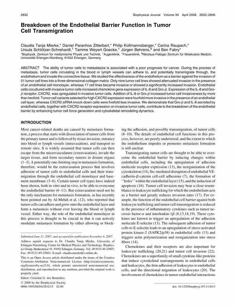
To study tumor-transendothelial migration and tissue inva-
sion, a three-dimensional collagen assay was developed (Fig.
1 A). Freshly isolated HUVECs were cultured onto a collagen
gel and formed a confluent monolayer within 24 h (Fig. 1 B).
Adjacent endothelial cells overlapped by ;2 mm (Fig. 1 Binset). Collagen fibers formed a mesh with an average pore
size of 0.6 6 0.2 mm (mean 6 SE); the gels had a shear
modulus of 58 Pa and a thickness of 474 6 7 mm (mean 6
SE) (Fig. 1 C).
With this assay, the transmigration and invasion behavior
was tested for 51 tumor cell lines derived from a broad
spectrum of tissues (Table 1). Before the tumor cells were
added to the assay, they were fluorescently labeled with car-
boxyfluorescein diacetate (to distinguish them from endo-
thelial cells, Fig. 1 F) and with Hoechst 33342 vital stain (for
cell counting and quantification of the invasion depth). All
tumor cells were able to attach to the endothelium or the
collagen gel surface, even those that are unable to attach
tightly to a pure plastic cell culture surface such as Colo201
and Colo205. After 3 days, the number of tumor cells that had
transmigrated through the endothelium was counted, and
their invasion depth was measured. All tumor cell lines that
invaded into the collagen gel assumed an elongated, spindle-
shaped morphology that was drastically different from the
fibroblast-like morphology seen in a two-dimensional plastic
cell culture flask (Fig. 1 D). Furthermore, tumor cells in the
gels formed long filopodia (Fig. 1 E).
2834 Mierke et al.
Biophysical Journal 94(7) 2832–2846
As introduced above, several pathways of tumor cell
transmigration are possible: disruption of cell-cell adhesion
sites, ‘‘hole’’ formation, and induction of apoptosis in en-
dothelial cells. To determine which pathway the tumor cells
chose, we analyzed TEM sections of collagen gels taken after
4, 10, 16, and 24 h of tumor-endothelial cell coculture with
noninvasive SW480 and MCF-7 tumor cells and with inva-
sive MDA-MB 231, A125, 786-O, and A375 tumor cells.
Fig. 1, H–M, illustrates the three steps of tumor cell extrav-
asation: tumor cell adhesion (Fig. 1, H and I), transmigration
(Fig. 1 J), and matrix invasion (Fig. 1 L and M). The adhesion
process was completed after 4 h of coculture, and transmi-
gration was detectable after 8 h. Among ;1000 analyzed
TEM sections in which tumor cells were present, 20 tumor
cells were in the process of transmigration, and ;100 cells
had invaded into the collagen gel. For all transmigrating tu-
mor cells, adjacent TEM sections were obtained to ensure
that endothelial cells were present on either side of the
transmigrating tumor cell. This finding indicates that the
tumor cells transmigrated not by ‘‘hole’’ formation but by
disrupting the endothelial cell-cell contacts. Moreover,
neighboring endothelial cells did not show morphological
signs indicative of apoptosis such as membrane blebbing, cell
shrinkage, or rounding. During tumor cell adhesion and
transmigration, the contact regions of tumor and endothelial
cells were decorated with multiple vacuoles and caveolae
(Fig. 1, I and K, arrows). In all TEM sections of invaded
tumor cells (Fig. 1, L and M), the endothelial monolayer
completely resealed and appeared intact (Fig. 1, L and M).
This was verified in multiple adjacent sections around each
invaded tumor cell. We repeatedly found stacks of invaded
tumor cells at different invasion depths at the same location
(Fig. 1, L and M), suggesting that these tumor cells had used
the same transmigration and invasion path.
FIGURE 1 Transendothelial migration and
collagen invasion of tumor cells. (A) Schematic
diagram of the tumor cell transmigration and
invasion assay. (B) TEM image of the endo-
thelial cell monolayer. (C) SEM image of the
three-dimensional collagen gel fiber network.
(D and E) MDA-MB-231 breast carcinoma
cells on plastic surface (D) showed an elon-
gated spindle-shaped morphology in a three-
dimensional collagen culture (E). (F) Same cell
stained with carboxyfluorescein-diacetate. (G)
The number of invaded MDA-MB-231 cells
and their invasion depth were increased in the
presence of an endothelium (dark gray) com-
pared with endothelium-free culture (light
gray). (H) Adhesion of an MDA-MB-231 cell
and (I) of an SW480 cell onto the endothelium.
Arrows indicate caveolae of tumor and endo-
thelial cells. (J) Transmigration of an MDA-
MB-231 cell through the endothelium. (K) An
enlarged detail of the close contact between
tumor and endothelial cells. (L) Invasion of an
MDA-MB-231 cell, and (M) stack of three
invaded MDA-MB-231 cells. Tumor cells
transmigrated without destroying or disrupting
the endothelium. Scale bars are 2 mm where
unspecified.
Breakdown of the Endothelial Barrier 2835
Biophysical Journal 94(7) 2832–2846
Classification of tumor cell invasiveness
All cell lines were classified into invasive and noninvasive
tumor cells according to their ability to invade the collagen
gel. The invasion depth for all the tumor cells was measured
in multiple randomly chosen fields of view. From the density
(number of invaded cells per square millimeter) plotted
against invasion depth, an invasion profile was obtained (Fig.
1 G). Invasiveness was quantified by an invasion score, de-
fined as cell density multiplied by the average invasion depth.
Tumor cell lines with an invasion score #0.1 mm�1 were
defined as noninvasive; a score .0.1 mm�1 was defined as
invasive. This threshold was chosen to avoid an erroneous
classification of noninvasive cells. Twenty-four of 51 tumor
cell lines were able to invade into the collagen gel when no
endothelial cells were present (Table 1). All cell lines derived
from skin (five lines), prostate (two), bladder (two), and kidney
(two) were invasive; among cell lines derived from breast (14
lines), cervix (two), colon (16 lines), lung (four), and pan-
creas (two) were both invasive and noninvasive lines (Table 1).
Endothelial cells enhance tumor cell invasion
A conspicuous question is to what degree does the endo-
thelial layer impede tumor cell invasion in a collagen matrix?
In the presence of an endothelial monolayer, invasiveness
was reduced in 9 of 24 invasive cell lines (Table 1), un-
changed in 9 cell lines, and, surprisingly, significantly in-
creased in 6 cell lines (Table 1). Eleven of 27 noninvasive
tumor cell lines became weakly invasive in the presence of an
endothelial layer (Table 1). We also studied primary tumor
cells isolated from four patients with kidney clear cell car-
cinomas. In contrast to Caki-1 and 786-O kidney tumor cell
lines, which did not alter their invasiveness, all four primary
kidney carcinoma cells showed increased matrix invasion in
the presence of an endothelium (data not shown).
The findings that the presence of the endothelium promoted
invasion of some of the tumor cells and even induced invasion
were unexpected and new. A possible interpretation of these
results is that the endothelium promotes tumor cell prolifer-
ation. However, this interpretation is ruled out by the finding
that the number of tumor cells after 16 h of monoculture
compared with coculture on an endothelium was equal.
The collagen invasion assay was repeated in MDA-MB-
231 tumor cells, but this time the endothelial layer was re-
placed by a closed monolayer of MCF-7 epithelial cells.
MDA-MB-231 cell invasion was fully blocked by MCF-7
cells, indicating that the modulation of tumor cell invasion
seen in our data was specific for the presence of endothelial
cells. Experiments on all tumor cell lines were repeated with
endothelial cells isolated from three to six different donors
(250 in total) and were performed over 3 years with more
than 10 batches of bovine and rat collagen. The standard
deviation of the invasion scores within a tumor cell line was
typically 30% of the mean, suggesting that effects of indi-
vidual HUVEC isolations or variations among collagen
batches were minimal.
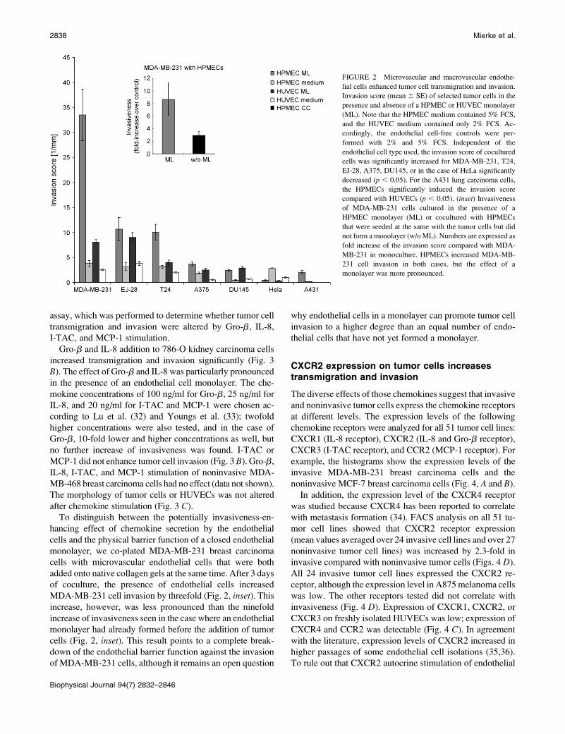
Both macrovascular and microvascularendothelial cells enhance tumor cell invasion
We replaced macrovascular HUVECs with primary HPMECs
isolated from lung resections and analyzed 11 different tumor
cell lines for their ability to overcome the endothelial cell
barrier and to invade the three-dimensional collagen gels. In
agreement with the data obtained with HUVECs, MCF-7,
CX-1, Caco-2, and MDA-MB-468 cells remained completely
noninvasive in the presence of a microvascular endothelial
cell layer, the invasion of HeLa cells was significantly im-
peded, and the invasion of MDA-MB-231, T24, EJ-28,
A375, and DU145 were significantly enhanced (Fig. 2). Note
that the control experiments without endothelial cells were
carried out in both HUVEC and HPMEC cell culture me-
dium, which differ in their serum and hydrocortisone content,
but the invasiveness of tumor cells was not markedly dif-
ferent. Interestingly, pulmonary microvascular ECs en-
hanced the invasion of MDA-MB-231 breast and T24 skin
carcinoma cell to a markedly larger extent than HUVECs, and
they induced the invasion of A431 lung carcinoma into the
collagen matrix (Fig. 2). Despite these important differences,
HUVECs provide an appropriate and convenient model system
to study breakdown of the endothelial barrier function against
tumor cell invasion.
Endothelial chemokines enhance transmigrationand invasion of some tumor cells
Gene expression analysis of endothelial cells was performed
to identify candidate genes responsible for the enhancement
of tumor cell transmigration and invasion. The expression
profile of endothelial cells in monoculture or after 16 h of
coculture with tumor cells was analyzed using DNA micro-
arrays. For coculture, two noninvasive cell lines (MCF-7 and
SW480) and two invasive cell lines (MDA-MB-231 and EJ-
28) were chosen. Our choice of MDA-MB-231 and EJ-28
cells was guided by their highly invasive behavior and their
pronounced increase in invasiveness in the presence of an
endothelium (Table 1).
Comparison of the gene expression profile under mono-
culture and coculture revealed 257 genes with expression
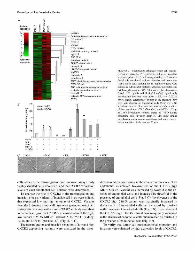
levels that correlated with invasiveness (Fig. 3 and Table S1).
Seventy-six genes were decreased, and 182 genes were in-
creased in endothelial cells when cocultured with invasive
tumor cells. Among the genes with increased expression
were the chemokines Gro-b, IL-8, and I-TAC (Fig. 3 A). The
expression of another chemokine, MCP-1, was increased
in endothelial cells (by 6.5-fold) only during coculture with
EJ-28 bladder carcinoma cells. Because MCP-1 has been
described as enhancer for PC-3 prostate carcinoma cell
invasiveness (32), it was included in a subsequent invasion
2836 Mierke et al.
Biophysical Journal 94(7) 2832–2846

TABLE 1 Classification of tumor cell invasiveness
Effect of HUVEC Cell line Tissue Invasion score w/o HUVEC Invasion score with HUVEC
Noninvasive Unchanged Colo205 Colon 0.05 6 0.02 0 6 0
BT-20 Breast 0 6 0 0 6 0
Caco-2 Colon 0 6 0 0 6 0
CX1 Colon 0 6 0 0 6 0
KS Breast 0 6 0 0 6 0
MDA-MB-453 Breast 0 6 0 0 6 0
MDA-MB-468 Breast 0 6 0 0 6 0
HCT116 Colon 0.01 6 0.01 0.01 6 0
MCF-7 Breast 0.08 6 0.02 0.08 6 0.02
CapanI Colon 0 6 0 0.01 6 0.01
SW948 Colon 0 6 0 0.01 6 0.01
HS578T Breast 0.04 6 0.01 0.06 6 0.01
A431 Lung 0 6 0 0.02 6 0.01
Colo201 Colon 0.03 6 0.01 0.05 6 0.01
A427 Lung 0 6 0 0.37 6 0.09
DLD-1 Colon 0.07 6 0.02 0.14 6 0.03
Induction of invasion SW48 Colon 0 6 0 0.05 6 0.01
CX-2 Colon 0 6 0 0.06 6 0.03
LX-1 Colon 0 6 0 0.13 6 0.03
T47D Breast 0 6 0 0.14 6 0.03
HT-29 Colon 0 6 0 0.15 6 0.02
A549 Lung 0 6 0 0.48 6 0.09
SW620 Colon 0.01 6 0 0.08 6 0.02
SW480 Colon 0.02 6 0.01 0.06 6 0.02
MS751 Cervix 0.02 6 0.01 0.07 6 0.01
A172 Brain 0.02 6 0.01 0.1 6 0.01
Mia-Paca-II Pancreas 0.04 6 0.01 0.16 6 0.03
Invasive Decreased invasion BT549 Breast 5.69 6 0.20 0.52 6 0.10
MDA-MB-436 Breast 5.05 6 0.60 0.96 6 0.19
Mewo Skin 3.04 6 0.32 0.02 6 0.01
PC-3 Prostate 6.13 6 0.66 3.86 6 0.4
SKBR3 Breast 3.02 6 0.44 1.30 6 0.13
Hacat Colon 0.98 6 0.18 0.04 6 0.01
Hela Skin 1.01 6 0.10 0.34 6 0.09
Me180 Cervix 1.38 6 0.49 0.76 6 0.20
DANG Pancreas 0.86 6 0.11 0.26 6 0.03
Unchanged C33A Colon 0.53 6 0.06 0.39 6 0.08
MDA-MB-361 Breast 0.24 6 0.03 0.18 6 0.02
A875 Skin 0.35 6 0.06 0.31 6 0.05
RT112 Bladder 0.32 6 0.06 0.34 6 0.07
CAKI-1 Kidney 0.11 6 0.03 0.21 6 0.04
MDA-MB-435 Breast 1.09 6 0.15 1.23 6 0.2
786-0 Kidney 0.29 6 0.04 0.59 6 0.14
FaDu Hypopharynx 0.65 6 0.13 1.39 6 0.28
A125 Lung 3.15 6 0.29 3.38 6 0.28
Increased invasion A375 Skin 0.55 6 0.09 2.49 6 0.34
DU145 Prostate 0.67 6 0.05 2.89 6 0.25
1205Lu Breast 0.82 6 0.20 1.52 6 0.36
T24 Skin 2.04 6 0.12 3.98 6 0.51
MDA-MB-231 Breast 2.52 6 0.12 8.05 6 0.55
EJ-28 Bladder 3.84 6 0.42 9.01 6 1.00
Invasion scores (mean 6 SE) of 51 tumor cell lines cultured for 3 days on a three-dimensional collagen gel (100,000 tumor cells per 35 cm2 dish) in the
absence and presence of endothelial cells. An invasion score ,0.1 was defined as noninvasive (n ¼ 27), and an invasion score of .0.1 was defined as
invasive (n ¼ 24). In some tumor cell lines, the endothelium decreased transmigration and invasion (n ¼ 9, for all p , 0.05), but in others, it significantly
enhanced or induced transmigration and invasion (for all p , 0.05 ).
Breakdown of the Endothelial Barrier 2837
Biophysical Journal 94(7) 2832–2846
assay, which was performed to determine whether tumor cell
transmigration and invasion were altered by Gro-b, IL-8,
I-TAC, and MCP-1 stimulation.
Gro-b and IL-8 addition to 786-O kidney carcinoma cells
increased transmigration and invasion significantly (Fig. 3
B). The effect of Gro-b and IL-8 was particularly pronounced
in the presence of an endothelial cell monolayer. The che-
mokine concentrations of 100 ng/ml for Gro-b, 25 ng/ml for
IL-8, and 20 ng/ml for I-TAC and MCP-1 were chosen ac-
cording to Lu et al. (32) and Youngs et al. (33); twofold
higher concentrations were also tested, and in the case of
Gro-b, 10-fold lower and higher concentrations as well, but
no further increase of invasiveness was found. I-TAC or
MCP-1 did not enhance tumor cell invasion (Fig. 3 B). Gro-b,
IL-8, I-TAC, and MCP-1 stimulation of noninvasive MDA-
MB-468 breast carcinoma cells had no effect (data not shown).
The morphology of tumor cells or HUVECs was not altered
after chemokine stimulation (Fig. 3 C).
To distinguish between the potentially invasiveness-en-
hancing effect of chemokine secretion by the endothelial
cells and the physical barrier function of a closed endothelial
monolayer, we co-plated MDA-MB-231 breast carcinoma
cells with microvascular endothelial cells that were both
added onto native collagen gels at the same time. After 3 days
of coculture, the presence of endothelial cells increased
MDA-MB-231 cell invasion by threefold (Fig. 2, inset). This
increase, however, was less pronounced than the ninefold
increase of invasiveness seen in the case where an endothelial
monolayer had already formed before the addition of tumor
cells (Fig. 2, inset). This result points to a complete break-
down of the endothelial barrier function against the invasion
of MDA-MB-231 cells, although it remains an open question
why endothelial cells in a monolayer can promote tumor cell
invasion to a higher degree than an equal number of endo-
thelial cells that have not yet formed a monolayer.
CXCR2 expression on tumor cells increasestransmigration and invasion
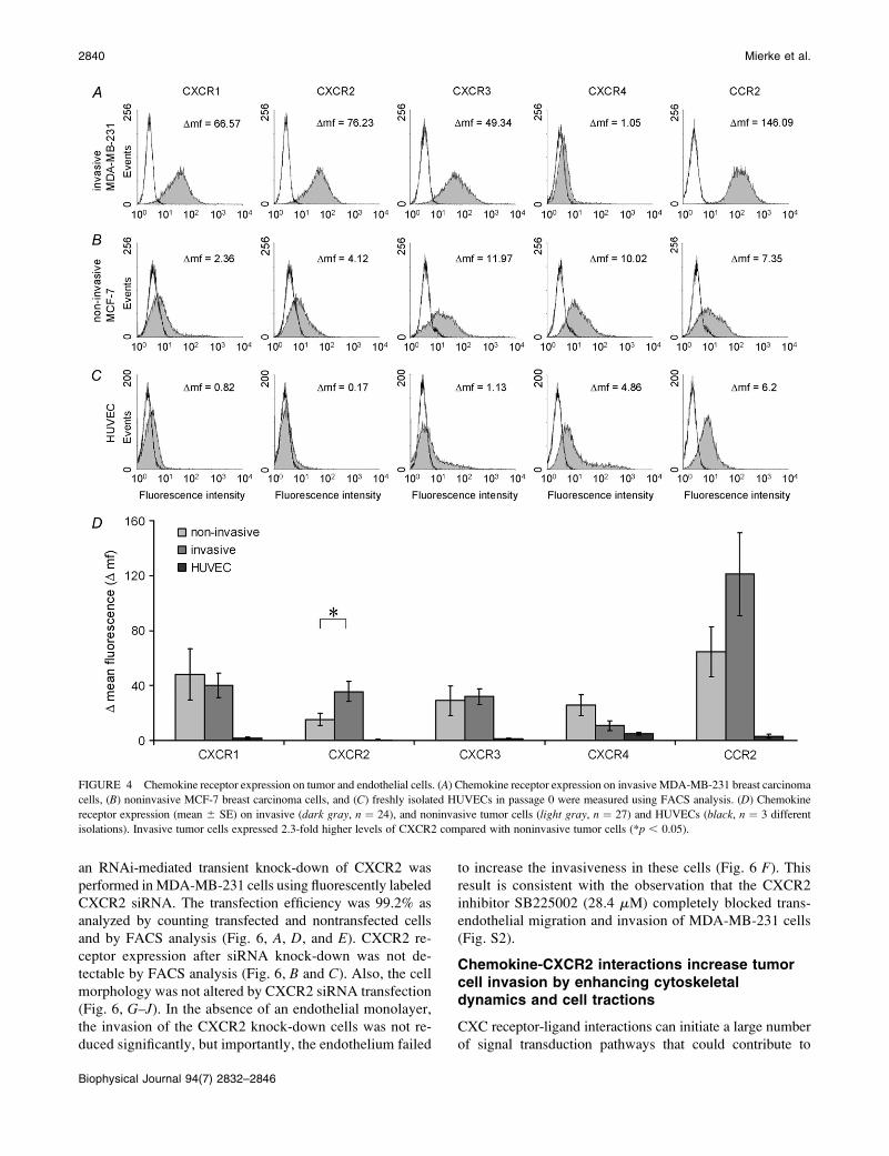
The diverse effects of those chemokines suggest that invasive
and noninvasive tumor cells express the chemokine receptors
at different levels. The expression levels of the following
chemokine receptors were analyzed for all 51 tumor cell lines:
CXCR1 (IL-8 receptor), CXCR2 (IL-8 and Gro-b receptor),
CXCR3 (I-TAC receptor), and CCR2 (MCP-1 receptor). For
example, the histograms show the expression levels of the
invasive MDA-MB-231 breast carcinoma cells and the
noninvasive MCF-7 breast carcinoma cells (Fig. 4, A and B).
In addition, the expression level of the CXCR4 receptor
was studied because CXCR4 has been reported to correlate
with metastasis formation (34). FACS analysis on all 51 tu-
mor cell lines showed that CXCR2 receptor expression
(mean values averaged over 24 invasive cell lines and over 27
noninvasive tumor cell lines) was increased by 2.3-fold in
invasive compared with noninvasive tumor cells (Figs. 4 D).
All 24 invasive tumor cell lines expressed the CXCR2 re-
ceptor, although the expression level in A875 melanoma cells
was low. The other receptors tested did not correlate with
invasiveness (Fig. 4 D). Expression of CXCR1, CXCR2, or
CXCR3 on freshly isolated HUVECs was low; expression of
CXCR4 and CCR2 was detectable (Fig. 4 C). In agreement
with the literature, expression levels of CXCR2 increased in
higher passages of some endothelial cell isolations (35,36).
To rule out that CXCR2 autocrine stimulation of endothelial
FIGURE 2 Microvascular and macrovascular endothe-
lial cells enhanced tumor cell transmigration and invasion.
Invasion score (mean 6 SE) of selected tumor cells in the
presence and absence of a HPMEC or HUVEC monolayer
(ML). Note that the HPMEC medium contained 5% FCS,
and the HUVEC medium contained only 2% FCS. Ac-
cordingly, the endothelial cell-free controls were per-
formed with 2% and 5% FCS. Independent of the
endothelial cell type used, the invasion score of cocultured
cells was significantly increased for MDA-MB-231, T24,
EJ-28, A375, DU145, or in the case of HeLa significantly
decreased (p , 0.05). For the A431 lung carcinoma cells,
the HPMECs significantly induced the invasion score
compared with HUVECs (p , 0.05). (inset) Invasiveness
of MDA-MB-231 cells cultured in the presence of a
HPMEC monolayer (ML) or cocultured with HPMECs
that were seeded at the same with the tumor cells but did
not form a monolayer (w/o ML). Numbers are expressed as
fold increase of the invasion score compared with MDA-
MB-231 in monoculture. HPMECs increased MDA-MB-
231 cell invasion in both cases, but the effect of a
monolayer was more pronounced.
2838 Mierke et al.
Biophysical Journal 94(7) 2832–2846
cells affected the transmigration and invasion assays, only
freshly isolated cells were used, and the CXCR2 expression
levels of each endothelial cell isolation were determined.
To analyze the role of CXCR2 in the transmigration and
invasion process, variants of invasive cell lines were isolated
that expressed low and high amounts of CXCR2. Variants
from the following tumor cell lines were generated using cell
sorting after staining with an anti-CXCR2 antibody (numbers
in parentheses give the CXCR2 expression ratio of the high/
low variant): MDA-MB-231 (breast, 5.3), 786-O (kidney,
12.3), and DU145 (prostate, 4.0) (Fig. 5, A–C).
The transmigration and invasion behaviors of low and high
CXCR2-expressing variants were analyzed in the three-
dimensional collagen assay in the absence or presence of an
endothelial monolayer. Invasiveness of the CXCR2-high
MDA-MB-231 variant was increased by twofold in the ab-
sence of endothelial cells, and increased by threefold in the
presence of endothelial cells (Fig. 5 G). Invasiveness of the
CXCR2-high 786-O variant was marginally increased in
the absence of endothelial cells but increased by fourfold
in the presence of endothelial cells (Fig. 5 H). Invasiveness of
the CXCR2-high DU145 variant was marginally increased
in the absence of endothelial cells but increased by fourfold in
the presence of endothelial cells (Fig. 5 I).To verify that tumor cell transendothelial migration and
invasion were enhanced by high expression levels of CXCR2,
FIGURE 3 Chemokines enhanced tumor cell transmi-
gration and invasion. (A) Expression profiles of genes that
were upregulated (red) or downregulated (green) in endo-
thelial cells cocultured with two invasive and two nonin-
vasive tumor cells. Among the 257 regulated genes were
numerous cytoskeletal proteins, adhesion molecules, and
cytokines/chemokines. (B) Addition of the chemokines
Gro-b (100 ng/ml) and IL-8 (25 ng/ml) significantly
increased the invasion score (mean 6 SE, *p , 0.05) of
786-O kidney carcinoma cells both in the presence (darkgray) and absence of endothelial cells (light gray). No
significant increase of invasiveness was seen after addition
of the chemokines I-TAC (20 ng/ml) and MCP-1 (20 ng/
ml). (C) Modulation contrast image of 786-O kidney
carcinoma cells (invasion depth 50 mm) show similar
morphology under control conditions and under chemo-
kine stimulation. Scale bars are 50 mm.
Breakdown of the Endothelial Barrier 2839
Biophysical Journal 94(7) 2832–2846
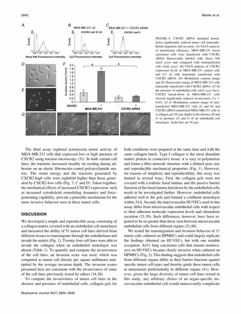
an RNAi-mediated transient knock-down of CXCR2 was
performed in MDA-MB-231 cells using fluorescently labeled
CXCR2 siRNA. The transfection efficiency was 99.2% as
analyzed by counting transfected and nontransfected cells
and by FACS analysis (Fig. 6, A, D, and E). CXCR2 re-
ceptor expression after siRNA knock-down was not de-
tectable by FACS analysis (Fig. 6, B and C). Also, the cell
morphology was not altered by CXCR2 siRNA transfection
(Fig. 6, G–J). In the absence of an endothelial monolayer,
the invasion of the CXCR2 knock-down cells was not re-
duced significantly, but importantly, the endothelium failed
to increase the invasiveness in these cells (Fig. 6 F). This
result is consistent with the observation that the CXCR2
inhibitor SB225002 (28.4 mM) completely blocked trans-
endothelial migration and invasion of MDA-MB-231 cells
(Fig. S2).
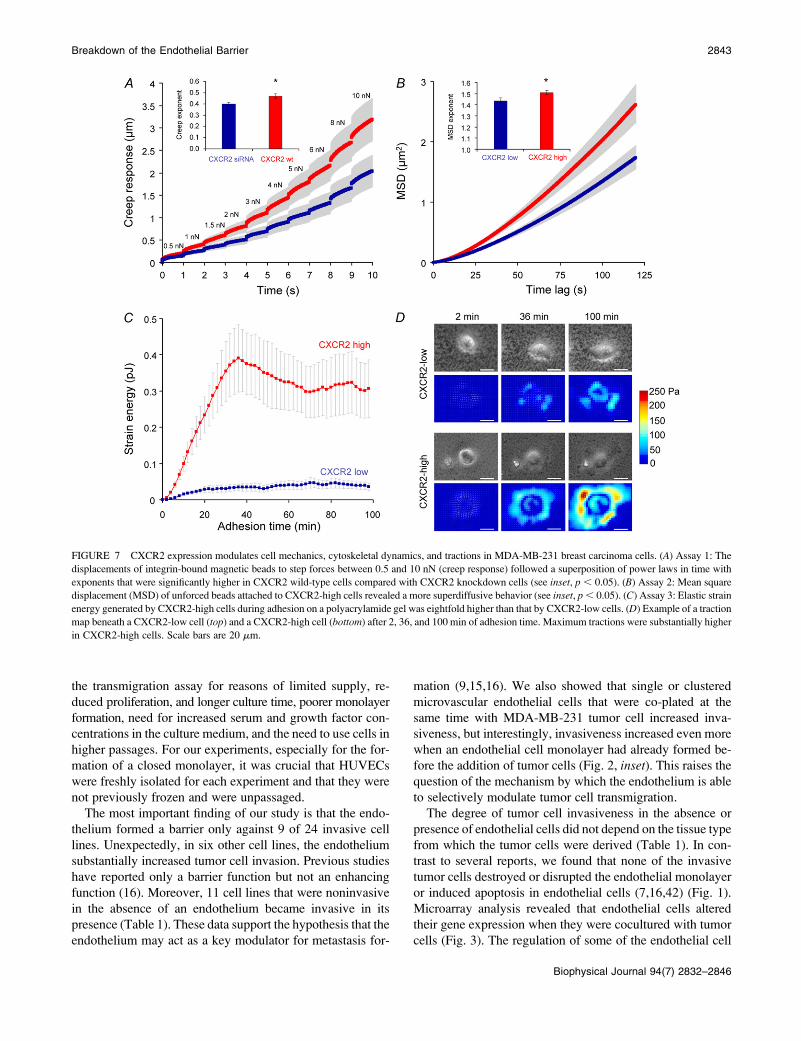
Chemokine-CXCR2 interactions increase tumorcell invasion by enhancing cytoskeletaldynamics and cell tractions
CXC receptor-ligand interactions can initiate a large number
of signal transduction pathways that could contribute to
FIGURE 4 Chemokine receptor expression on tumor and endothelial cells. (A) Chemokine receptor expression on invasive MDA-MB-231 breast carcinoma
cells, (B) noninvasive MCF-7 breast carcinoma cells, and (C) freshly isolated HUVECs in passage 0 were measured using FACS analysis. (D) Chemokine
receptor expression (mean 6 SE) on invasive (dark gray, n ¼ 24), and noninvasive tumor cells (light gray, n ¼ 27) and HUVECs (black, n ¼ 3 different
isolations). Invasive tumor cells expressed 2.3-fold higher levels of CXCR2 compared with noninvasive tumor cells (*p , 0.05).
2840 Mierke et al.
Biophysical Journal 94(7) 2832–2846

invasive and motile behavior of tumor cells by increasing
actomyosin motor activity, adhesion/de-adhesion, and cyto-
skeletal remodeling events (37). The role of CXCR2 was
explored using three cell-mechanical assays. The first was
measuring the creep response of integrin-bound fibronectin-
coated magnetic beads to step forces between 0.5 nN and 10
nN (Fig. 7 A). The creep response for all forces followed a
power law over time. The exponent of the power-law creep
response is a measure of bond stability. Exponents close to
zero indicate stable bonds that result in an elastic, solid-like
cell-mechanical behavior. Exponents close to unity indicate
unstable bonds with high cycling or turnover rates that result
in a viscous, fluid-like cell-mechanical behavior (27). In
MDA-MB-231 wild-type cells, the power-law creep expo-
nent was significantly higher, closer to a fluid-like behavior,
than in CXCR2 knock-down cells.
In the second assay, the random walk of unforced, spon-
taneously diffusing fibronectin-coated beads was analyzed
(Fig. 7 B). These beads cannot move unless the microstruc-
ture to which they are attached rearranges (29). ATP-driven
cytoskeletal rearrangements can be quantified by the super-
diffusive power-law exponent of the MSD of the bead
(29,30). The power-law exponent of the MSD in CXCR2-
high cell variants was significantly more superdiffusive (Fig.
7 B), indicative of a higher rate of ATP-driven cytoskeletal
rearrangements.
FIGURE 5 Analysis of CXCR2-low and CXCR2-high carcinoma cell variants. CXCR2 expression of CXCR2-low and CXCR2-high variants in (A) MDA-
MB-231 breast, (B) 786-O kidney, and (C) DU145 prostate carcinoma cells after four cycles of sorting and subsequent culturing. In each histogram, left curves
are isotype controls, and filled gray curves are CXCR2 expression on tumor cells. (D–I) Transmigration and invasion of MDA-MB-231 (left), 786-O (middle),
and DU145 (right) variants expressing low and high amounts of CXCR2. (D–F) Modulation contrast images of CXCR2-low variants (left) and CXCR2-high
variants (right) in a collagen gel (50–80 mm depths) in the absence (bottom) or presence (top) of an endothelial cell monolayer. Scale bars are 50 mm. (G–I)
Invasion scores (mean 6 SE) of CXCR2-low and -high variants in the presence (dark gray bars) or absence of an endothelial cell monolayer (light gray bars).
In the presence of a HUVEC monolayer, all CXCR2-high variants are significantly more invasive than CXCR2-low variants (*p , 0.05).
Breakdown of the Endothelial Barrier 2841
Biophysical Journal 94(7) 2832–2846
The third assay explored actomyosin motor activity of
MDA-MB-231 cells that expressed low or high amounts of
CXCR2 using traction microscopy (31). In both variant cell
lines, the tractions increased steadily on seeding during ad-
hesion on an elastic fibronectin-coated polyacrylamide ma-
trix. The strain energy and the tractions generated by
CXCR2-high cells were eightfold higher than those gener-
ated by CXCR2-low cells (Fig. 7, C and D). Taken together,
the mechanical effects of increased CXCR2 expression, such
as increased cytoskeletal remodeling dynamics and force-
generating capability, provide a plausible mechanism for the
more invasive behavior seen in these tumor cells.
DISCUSSION
We developed a simple and reproducible assay consisting of
a collagen matrix covered with an endothelial cell monolayer
and measured the ability of 51 tumor cell lines derived from
different tissues to transmigrate through the endothelium and
invade the matrix (Fig. 1). Twenty-four cell lines were able to
invade the collagen when an endothelial monolayer was
absent (Table 1). To quantify and compare the invasiveness
of the cell lines, an invasion score was used, which was
computed as tumor cell density per square millimeter mul-
tiplied by the average invasion depth. The invasion scores
presented here are consistent with the invasiveness of some
of the cell lines previously tested by others (34,38).
To compare the invasiveness of tumor cell lines in the
absence and presence of endothelial cells, collagen gels for
both conditions were prepared at the same time and with the
same collagen batch. Type I collagen is the most abundant
matrix protein in connective tissue: it is easy to polymerize
and forms a fiber network structure with a defined pore size
and reproducible mechanical properties (Fig. 1). However,
for reasons of simplicity and reproducibility, this assay was
limited in several ways. First, the collagen gels were not
covered with a realistic basal lamina, and the passive barrier
function of the basal lamina laid down by the endothelial cells
needs to be investigated further. However, endothelial cells
adhered well to the gels and formed a confluent monolayer
within 24 h. Second, the macrovascular HUVECs used in this
assay differ from microvascular endothelial cells with respect
to their adhesion molecule expression levels and chemokine
secretion (21,39). Such differences, however, have been re-
ported to be no greater than those seen between microvascular
endothelial cells from different organs (21,40).
We tested the transmigration and invasion behavior of 11
tumor cells cultured on HPMECs and could largely replicate
the findings obtained on HUVECs, but with one notable
exception: A431 lung carcinoma cells that remain noninva-
sive on HUVECs became clearly invasive when cultured on
HPMECs (Fig. 2). This finding suggests that endothelial cells
from different organs differ in their barrier function against
specific tumor cell types and thereby guide these tumor cells
to metastasize preferentially in different organs (41). How-
ever, given the large diversity of tumor cell lines tested in
this study, any arbitrary choice of an organ-specific mi-
crovascular endothelial cell would unnecessarily complicate
FIGURE 6 CXCR2 siRNA mediated knock-
down significantly reduced tumor cell transendo-
thelial migration and invasion. (A) FACS analysis
of transfection efficiency. MDA-MB-231 breast
carcinoma cells were transfected with CXCR2
siRNA fluorescently labeled with Alexa 546
(dark gray) and compared with nontransfected
cells (light gray). (B) FACS analysis of CXCR2
expression levels in MDA-MB-231 control cells
and (C) of cells transiently transfected with
CXCR2 siRNA. (D) Modulation contrast image
and (E) fluorescence image of MDA-MB-231 cells
transiently transfected with CXCR2 siRNA. (F) In
the presence of endothelial cells (dark gray bars),
CXCR2 knock-down in MDA-MB-231 cells
showed significantly reduced invasiveness (*p ,
0.05). (G–J) Modulation contrast image of non-
transfected MDA-MB-231 cells (G and H) and
CXCR2 siRNA-transfected MDA-MB-231 cells in
a collagen gel (50 mm depth) in the absence (H and
J) or presence (G and I) of an endothelial cell
monolayer. Scale bars are 50 mm.
2842 Mierke et al.
Biophysical Journal 94(7) 2832–2846
the transmigration assay for reasons of limited supply, re-
duced proliferation, and longer culture time, poorer monolayer
formation, need for increased serum and growth factor con-
centrations in the culture medium, and the need to use cells in
higher passages. For our experiments, especially for the for-
mation of a closed monolayer, it was crucial that HUVECs
were freshly isolated for each experiment and that they were
not previously frozen and were unpassaged.
The most important finding of our study is that the endo-
thelium formed a barrier only against 9 of 24 invasive cell
lines. Unexpectedly, in six other cell lines, the endothelium
substantially increased tumor cell invasion. Previous studies
have reported only a barrier function but not an enhancing
function (16). Moreover, 11 cell lines that were noninvasive
in the absence of an endothelium became invasive in its
presence (Table 1). These data support the hypothesis that the
endothelium may act as a key modulator for metastasis for-
mation (9,15,16). We also showed that single or clustered
microvascular endothelial cells that were co-plated at the
same time with MDA-MB-231 tumor cell increased inva-
siveness, but interestingly, invasiveness increased even more
when an endothelial cell monolayer had already formed be-
fore the addition of tumor cells (Fig. 2, inset). This raises the
question of the mechanism by which the endothelium is able
to selectively modulate tumor cell transmigration.
The degree of tumor cell invasiveness in the absence or
presence of endothelial cells did not depend on the tissue type
from which the tumor cells were derived (Table 1). In con-
trast to several reports, we found that none of the invasive
tumor cells destroyed or disrupted the endothelial monolayer
or induced apoptosis in endothelial cells (7,16,42) (Fig. 1).
Microarray analysis revealed that endothelial cells altered
their gene expression when they were cocultured with tumor
cells (Fig. 3). The regulation of some of the endothelial cell
FIGURE 7 CXCR2 expression modulates cell mechanics, cytoskeletal dynamics, and tractions in MDA-MB-231 breast carcinoma cells. (A) Assay 1: The
displacements of integrin-bound magnetic beads to step forces between 0.5 and 10 nN (creep response) followed a superposition of power laws in time with
exponents that were significantly higher in CXCR2 wild-type cells compared with CXCR2 knockdown cells (see inset, p , 0.05). (B) Assay 2: Mean square
displacement (MSD) of unforced beads attached to CXCR2-high cells revealed a more superdiffusive behavior (see inset, p , 0.05). (C) Assay 3: Elastic strain
energy generated by CXCR2-high cells during adhesion on a polyacrylamide gel was eightfold higher than that by CXCR2-low cells. (D) Example of a traction
map beneath a CXCR2-low cell (top) and a CXCR2-high cell (bottom) after 2, 36, and 100 min of adhesion time. Maximum tractions were substantially higher
in CXCR2-high cells. Scale bars are 20 mm.
Breakdown of the Endothelial Barrier 2843
Biophysical Journal 94(7) 2832–2846
genes depended on the ability of the tumor cells to transmi-
grate through the endothelium (Fig. 3). Endothelial cell genes
that were upregulated in the presence of invasive tumor cells
included cytoskeletal proteins, cell-cell adhesion molecules,
and the CXC chemokines Gro-b, IL-8, and I-TAC. This
study focused on chemokines and their receptors as modu-
lators for the interaction between tumor cells and endothelial
cells.
As expected from the microarray data (Fig. 3), the addition
of exogenous Gro-b and IL-8 significantly increased tumor
cell transmigration and invasion. Gro-a, Gro-b, and Gro-g
all bind to the same CXCR2 chemokine receptor and have
been reported to enhance melanoma tumor growth (43–45).
IL-8 also binds to the CXCR2 chemokine receptor and re-
portedly increases invasiveness of PC-3 prostate carcinoma
cells (22). FACS analysis of all 51 tumor cell lines revealed
that invasive tumor cells expressed significantly more
CXCR2 than noninvasive tumor cells (Fig. 5). This is con-
sistent with previous studies that have shown that malignant
PC-3 prostate carcinoma cells and prostate tumors at an ad-
vanced disease stage express increased levels of CXCR2
(22,46,47).
Interactions between the CXCR2 receptor on endothelial
cells and CXCR2 ligand secretion by tumor cells have been
reported to increase metastasis formation by enhancing an-
giogenesis (45,48). However, the HUVECs and HPMECs
used in our experiments did not express detectable amounts
of CXCR2 (Fig. 4). Therefore, in contrast to the previously
identified interaction pathway, we attribute the increased
tumor cell transmigration and invasion to an increased
CXCR2 expression on tumor cells that are being stimulated
by increased concentrations of Gro-b and IL-8 produced by
endothelial cells. Other chemokines and their receptors, in-
cluding the I-TAC receptor CXCR3, as well as the MCP-1
receptor CCR2, were expressed in noninvasive and invasive
tumor cells at similar levels (Fig. 4). This is consistent with
our finding that exogenous I-TAC and MCP-1 chemokines
did not enhance tumor cell invasion, and indeed, I-TAC even
reduced tumor cell invasion in the absence of an endothelium
(Fig. 3). Of all the tumor cells tested, only the interactions be-
tween CXCR2 and its chemokine ligands were conspicuous
and provide, therefore, a common mechanism for enhancing
tumor cell invasion in the presence of endothelial cells.
To further analyze the function of the CXCR2 chemokine
receptor, variants of MDA-MB-231, 786-O, and DU145
carcinoma cells were established that expressed high and low
levels of CXCR2 (Fig. 5). In the absence of an endothelial
monolayer, the invasion of CXCR2 high-expressing carci-
noma cells was only slightly increased, but in the presence of
an endothelial monolayer, the invasion was strongly in-
creased (Fig. 5). MDA-MB-231 cells treated with the CXCR2
inhibitor SB225002 showed nearly complete inhibition of
any invasion (Fig. S2). When CXCR2 was knocked down in
MDA-MB-231 cells by siRNA, the presence of an endothelial
cell layer failed to enhance tumor cell invasiveness (Fig. 6).
CXC receptor-ligand interactions are known to initiate a
large number of signal transduction pathways involving ac-
tivation of Rho, Rac, Cdc42, Erk, Akt, phospholipase C, and
inositol-1,4,5-trisphosphate (49,50). This raises the question
of possible downstream effects of pathways that may con-
tribute to a more invasive and motile behavior of tumor cells.
Recent studies have shown that the invasion speed of tumor
cells through a three-dimensional matrix is governed by a
dynamic equilibrium among traction forces, adhesion forces,
and matrix deformation forces (37). In this work, the dy-
namics and stability of the force transfer between adhesion
receptors and the cytoskeleton were investigated by analyz-
ing the motion of magnetically forced and unforced fibro-
nectin-coated microbeads. The beads were attached to
integrin receptors of tumor cell variants that expressed low
and high amounts of CXCR2 chemokine receptors (51).
Beads on the cells with high CXCR2 expression displayed
behavior that was indicative of an increased rate of adhesion/
de-adhesion and cytoskeletal remodeling events, both of
which promote cell invasion (37) (Fig. 7, A and B). At the
same time, high-CXCR2 cells generated substantially higher
contractile and adhesive forces and hence should be able to
propel themselves more forcefully through an extracellular
matrix (Fig. 7, C and D).
In summary, we found that, in the presence of invasive
tumor cells, Gro-b and IL-8 chemokines were upregulated in
endothelial cells and that CXCR2 receptors were highly ex-
pressed on invasive tumor cells, regardless of the tissue origin
of the tumor. The interactions between CXCR2 and Gro-b or
IL-8 lead to higher tractions and enhanced dynamics of cy-
toskeletal remodeling processes in tumor cells and represent a
generic mechanism for the breakdown of the endothelial
barrier function against tumor cell invasion.
SUPPLEMENTARY MATERIAL
To view all of the supplemental files associated with this
article, visit www.biophysj.org.
We thank Ludger Klein-Hitpass for help with the microarray analysis, Peter
Altevogt for the kind gift of A125 and KS cells, Barbara Reischl for
excellent technical assistance, Robert Schmiedl for help with scanning EM
imaging, Julia Resch and Joachim Kaschta for rheology measurements of
collagen gels, and Wolfgang H. Goldmann for critical comments. This
work was supported by Deutsche Krebshilfe (107384), DFG (MA 534/20-
4), and NIH (HL65960).
REFERENCES
1. Liotta, L. A., P. S. Steeg, and W. G. Stetler-Stevenson. 1991. Cancermetastasis and angiogenesis: an imbalance of positive and negativeregulation. Cell. 64:327–336.
2. Langley, R. R., and I. J. Fidler. 2007. Tumor cell-organ microenvi-ronment interactions in the pathogenesis of cancer metastasis. Endocr.Rev. 28:297–321.
3. Steeg, P. S. 2006. Tumor metastasis: mechanistic insights and clinicalchallenges. Nat. Med. 12:895–904.
2844 Mierke et al.
Biophysical Journal 94(7) 2832–2846

4. Nicolson, G. L. 1989. Metastatic tumor cell interactions with endothelium,basement membrane and tissue. Curr. Opin. Cell Biol. 1:1009–1019.
5. Stetler-Stevenson, W. G., S. Aznavoorian, and L. A. Liotta. 1993.Tumor cell interactions with the extracellular matrix during invasionand metastasis. Annu. Rev. Cell Biol. 9:541–573.
6. Luzzi, K. J., I. C. MacDonald, E. E. Schmidt, N. Kerkvliet, V. L.Morris, A. F. Chambers, and A. C. Groom. 1998. Multistep nature ofmetastatic inefficiency: dormancy of solitary cells after successfulextravasation and limited survival of early micrometastases. Am. J.Pathol. 153:865–873.
7. Weis, S., J. Cui, L. Barnes, and D. Cheresh. 2004. Endothelial barrierdisruption by VEGF-mediated Src activity potentiates tumor cellextravasation and metastasis. J. Cell Biol. 167:223–229.
8. Voura, E. B., R. A. Ramjeesingh, A. M. Montgomery, and C. H. Siu.2001. Involvement of integrin alpha(v)beta(3) and cell adhesion mol-ecule L1 in transendothelial migration of melanoma cells. Mol. Biol.Cell. 12:2699–2710.
9. Tremblay, P. L., F. A. Auger, and J. Huot. 2006. Regulation oftransendothelial migration of colon cancer cells by E-selectin-mediatedactivation of p38 and ERK MAP kinases. Oncogene. 25:6563–6573.
10. Sandig, M., E. B. Voura, V. I. Kalnins, and C. H. Siu. 1997. Role ofcadherins in the transendothelial migration of melanoma cells inculture. Cell Motil. Cytoskeleton. 38:351–364.
11. Fidler, I. J., and I. R. Hart. 1982. Biological diversity in metastaticneoplasms: origins and implications. Science. 217:998–1003.
12. Al-Mehdi, A. B., K. Tozawa, A. B. Fisher, L. Shientag, A. Lee, andR. J. Muschel. 2000. Intravascular origin of metastasis from theproliferation of endothelium-attached tumor cells: a new model formetastasis. Nat. Med. 6:100–102.
13. Laferriere, J., F. Houle, M. M. Taher, K. Valerie, and J. Huot. 2001.Transendothelial migration of colon carcinoma cells requires expres-sion of E-selectin by endothelial cells and activation of stress-activatedprotein kinase-2 (SAPK2/p38) in the tumor cells. J. Biol. Chem. 276:33762–33772.
14. Rousseau, S., F. Houle, J. Landry, and J. Huot. 1997. p38 MAP kinaseactivation by vascular endothelial growth factor mediates actin reor-ganization and cell migration in human endothelial cells. Oncogene.15:2169–2177.
15. Li, Y. H., and C. Zhu. 1999. A modified Boyden chamber assay fortumor cell transendothelial migration in vitro. Clin. Exp. Metastasis.17:423–429.
16. Heyder, C., E. Gloria-Maercker, F. Entschladen, W. Hatzmann, B.Niggemann, K. S. Zanker, and T. Dittmar. 2002. Realtime visualizationof tumor cell/endothelial cell interactions during transmigration acrossthe endothelial barrier. J. Cancer Res. Clin. Oncol. 128:533–538.
17. Wittchen, E. S., R. A. Worthylake, P. Kelly, P. J. Casey, L. A.Quilliam, and K. Burridge. 2005. Rap1 GTPase inhibits leukocytetransmigration by promoting endothelial barrier function. J. Biol.Chem. 280:11675–11682.
18. Chandrasekharan, U. M., M. Siemionow, M. Unsal, L. Yang, E. Poptic,J. Bohn, K. Ozer, Z. Zhou, P. H. Howe, M. Penn, and P. E. DiCorleto.2006. TNF-a receptor-II is required for TNF-a-induced leukocyte-endothelial interaction in vivo. Blood.
19. McGettrick, H. M., J. M. Lord, K. Q. Wang, G. E. Rainger, C. D.Buckley, and G. B. Nash. 2006. Chemokine- and adhesion-dependentsurvival of neutrophils after transmigration through cytokine-stimu-lated endothelium. J. Leukoc. Biol. 79:779–788.
20. Gallatin, W. M., I. L. Weissman, and E. C. Butcher. 1983. A cell-surface molecule involved in organ-specific homing of lymphocytes.Nature. 304:30–34.
21. Hillyer, P., E. Mordelet, G. Flynn, and D. Male. 2003. Chemokines,chemokine receptors and adhesion molecules on different humanendothelia: discriminating the tissue-specific functions that affectleucocyte migration. Clin. Exp. Immunol. 134:431–441.
22. Reiland, J., L. T. Furcht, and J. B. McCarthy. 1999. CXC-chemokinesstimulate invasion and chemotaxis in prostate carcinoma cells throughthe CXCR2 receptor. Prostate. 41:78–88.
23. Mierke, C. T., M. Ballmaier, U. Werner, M. P. Manns, K. Welte, andS. C. Bischoff. 2000. Human endothelial cells regulate survival andproliferation of human mast cells. J. Exp. Med. 192:801–811.
24. Thomas, H., S. Senkel, S. Erdmann, T. Arndt, G. Turan, L. Klein-Hitpass, and G. U. Ryffel. 2004. Pattern of genes influenced byconditional expression of the transcription factors HNF6, HNF4alphaand HNF1beta in a pancreatic beta-cell line. Nucleic Acids Res. 32:e150.
25. Alenghat, F. J., B. Fabry, K. Y. Tsai, W. H. Goldmann, and D. E.Ingber. 2000. Analysis of cell mechanics in single vinculin-deficientcells using a magnetic tweezer. Biochem. Biophys. Res. Commun. 277:93–99.
26. Mierke, C. T., P. Kollmannsberger, D. Paranhos-Zitterbart, J. Smith, B.Fabry, and W. H. Goldmann. 2008. Mechano-coupling and regulationof contractility by the vinculin tail domain. Biophys. J. 94:661–670.
27. Fabry, B., G. N. Maksym, J. P. Butler, M. Glogauer, D. Navajas, andJ. J. Fredberg. 2001. Scaling the microrheology of living cells. Phys.Rev. Lett. 87:148102.
28. Hildebrandt, J. 1969. Comparison of mathematical models for cat lungand viscoelastic balloon derived by Laplace transform methods frompressure-volume data. Bull. Math. Biophys. 31:651–667.
29. Bursac, P., G. Lenormand, B. Fabry, M. Oliver, D. A. Weitz, V.Viasnoff, J. P. Butler, and J. J. Fredberg. 2005. Cytoskeletal remod-elling and slow dynamics in the living cell. Nat. Mater. 4:557–561.
30. Raupach, C., D. P. Zitterbart, C. T. Mierke, C. Metzner, F. A. Muller, andB. Fabry. 2007. Stress fluctuations and motion of cytoskeletal-boundmarkers. Phys. Rev. E Stat. Nonlin. Soft Matter Phys. 76:011918.
31. Butler, J. P., I. M. Tolic-Norrelykke, B. Fabry, and J. J. Fredberg.2002. Traction fields, moments, and strain energy that cells exert ontheir surroundings. Am. J. Physiol. Cell Physiol. 282:C595–C605.
32. Lu, Y., Z. Cai, D. L. Galson, G. Xiao, Y. Liu, D. E. George, M. F.Melhem, Z. Yao, and J. Zhang. 2006. Monocyte chemotactic protein-1 (MCP-1) acts as a paracrine and autocrine factor for prostate cancergrowth and invasion. Prostate. 66:1311–1318.
33. Youngs, S. J., S. A. Ali, D. D. Taub, and R. C. Rees. 1997.Chemokines induce migrational responses in human breast carcinomacell lines. Int. J. Cancer. 71:257–266.
34. Muller, A., B. Homey, H. Soto, N. Ge, D. Catron, M. E. Buchanan, T.McClanahan, E. Murphy, W. Yuan, S. N. Wagner, J. L. Barrera, A.Mohar, E. Verastegui, and A. Zlotnik. 2001. Involvement of chemo-kine receptors in breast cancer metastasis. Nature. 410:50–56.
35. Salcedo, R., J. H. Resau, D. Halverson, E. A. Hudson, M. Dambach, D.Powell, K. Wasserman, and J. J. Oppenheim. 2000. Differentialexpression and responsiveness of chemokine receptors (CXCR1–3)by human microvascular endothelial cells and umbilical vein endothe-lial cells. FASEB J. 14:2055–2064.
36. Murdoch, C., P. N. Monk, and A. Finn. 1999. CXC chemokinereceptor expression on human endothelial cells. Cytokine. 11:704–712.
37. Zaman, M. H., L. M. Trapani, A. Siemeski, D. Mackellar, H. Gong,R. D. Kamm, A. Wells, D. A. Lauffenburger, and P. Matsudaira. 2006.Migration of tumor cells in 3D matrices is governed by matrix stiffnessalong with cell-matrix adhesion and proteolysis. Proc. Natl. Acad. Sci.USA. 103:10889–10894.
38. Lin, Y., R. Huang, L. Chen, S. Li, Q. Shi, C. Jordan, and R. P. Huang.2004. Identification of interleukin-8 as estrogen receptor-regulatedfactor involved in breast cancer invasion and angiogenesis by proteinarrays. Int. J. Cancer. 109:507–515.
39. Aird, W. C. 2007. Phenotypic heterogeneity of the endothelium: I.Structure, function, and mechanisms. Circ. Res. 100:158–173.
40. Aird, W. C. 2007. Phenotypic heterogeneity of the endothelium: II.Representative vascular beds. Circ. Res. 100:174–190.
41. Ruoslahti, E. 2004. Vascular zip codes in angiogenesis and metastasis.Biochem. Soc. Trans. 32:397–402.
42. Li, A., M. L. Varney, and R. K. Singh. 2001. Expression of interleukin8 and its receptors in human colon carcinoma cells with differentmetastatic potentials. Clin. Cancer Res. 7:3298–3304.
Breakdown of the Endothelial Barrier 2845
Biophysical Journal 94(7) 2832–2846
43. Owen, J. D., R. Strieter, M. Burdick, H. Haghnegahdar, L. Nanney, R.
Shattuck-Brandt, and A. Richmond. 1997. Enhanced tumor-forming
capacity for immortalized melanocytes expressing melanoma growth
stimulatory activity/growth-regulated cytokine beta and gamma pro-
teins. Int. J. Cancer. 73:94–103.
44. Haghnegahdar, H., J. Du, D. Wang, R. M. Strieter, M. D. Burdick,
L. B. Nanney, N. Cardwell, J. Luan, R. Shattuck-Brandt, and A.
Richmond. 2000. The tumorigenic and angiogenic effects of MGSA/
GRO proteins in melanoma. J. Leukoc. Biol. 67:53–62.
45. Loukinova, E., G. Dong, I. Enamorado-Ayalya, G. R. Thomas, Z. Chen,
H. Schreiber, and C. Van Waes. 2000. Growth regulated oncogene-alpha
expression by murine squamous cell carcinoma promotes tumor growth,
metastasis, leukocyte infiltration and angiogenesis by a host CXC
receptor-2 dependent mechanism. Oncogene. 19:3477–3486.
46. Varney, M. L., A. Li, B. J. Dave, C. D. Bucana, S. L. Johansson, and
R. K. Singh. 2003. Expression of CXCR1 and CXCR2 receptors in
malignant melanoma with different metastatic potential and their role in
interleukin-8 (CXCL-8)-mediated modulation of metastatic phenotype.
Clin. Exp. Metastasis. 20:723–731.
47. Murphy, C., M. McGurk, J. Pettigrew, A. Santinelli, R. Mazzucchelli,
P. G. Johnston, R. Montironi, and D. J. Waugh. 2005. Nonapical and
cytoplasmic expression of interleukin-8, CXCR1, and CXCR2 corre-
lates with cell proliferation and microvessel density in prostate cancer.
Clin. Cancer Res. 11:4117–4127.
48. Mestas, J., M. D. Burdick, K. Reckamp, A. Pantuck, R. A. Figlin, and
R. M. Strieter. 2005. The role of CXCR2/CXCR2 ligand biological
axis in renal cell carcinoma. J. Immunol. 175:5351–5357.
49. Schraufstatter, I. U., J. Chung, and M. Burger. 2001. IL-8 activates
endothelial cell CXCR1 and CXCR2 through Rho and Rac signal-
ing pathways. Am. J. Physiol. Lung Cell. Mol. Physiol. 280:L1094–
L1103.
50. Venkatakrishnan, G., R. Salgia, and J. E. Groopman. 2000. Chemokine
receptors CXCR-1/2 activate mitogen-activated protein kinase via the
epidermal growth factor receptor in ovarian cancer cells. J. Biol. Chem.275:6868–6875.
51. Choquet, D., D. P. Felsenfeld, and M. P. Sheetz. 1997. Extracellular
matrix rigidity causes strengthening of integrin- cytoskeleton linkages.
Cell. 88:39–48.
2846 Mierke et al.
Biophysical Journal 94(7) 2832–2846
Copyright © 2022 FDOKUMEN