Modulation of TNFα, IL10 and IL12p40 levels by a ceramide-1-phosphate analog, PCERA-1, in vivo and...
9
This article appeared in a journal published by Elsevier. The attached copy is furnished to the author for internal non-commercial research and education use, including for instruction at the authors institution and sharing with colleagues. Other uses, including reproduction and distribution, or selling or licensing copies, or posting to personal, institutional or third party websites are prohibited. In most cases authors are permitted to post their version of the article (e.g. in Word or Tex form) to their personal website or institutional repository. Authors requiring further information regarding Elsevier’s archiving and manuscript policies are encouraged to visit: http://www.elsevier.com/copyright
-
Upload
independent -
Category
Documents
-
view
0 -
download
0
Transcript of Modulation of TNFα, IL10 and IL12p40 levels by a ceramide-1-phosphate analog, PCERA-1, in vivo and...
This article appeared in a journal published by Elsevier. The attachedcopy is furnished to the author for internal non-commercial researchand education use, including for instruction at the authors institution
and sharing with colleagues.
Other uses, including reproduction and distribution, or selling orlicensing copies, or posting to personal, institutional or third party
websites are prohibited.
In most cases authors are permitted to post their version of thearticle (e.g. in Word or Tex form) to their personal website orinstitutional repository. Authors requiring further information
regarding Elsevier’s archiving and manuscript policies areencouraged to visit:
http://www.elsevier.com/copyright
Author's personal copy
Immunology Letters 123 (2009) 1–8
Contents lists available at ScienceDirect
Immunology Letters
journa l homepage: www.e lsev ier .com/ locate /
Modulation of TNF�, IL-10 and IL-12p40 levels by a ceramide-1-phosphateanalog, PCERA-1, in vivo and ex vivo in primary macrophages
Dorit Avnia,1, Meir Goldsmitha,1, Orna Ernsta, Roi Mashiachb, Tove Tuntlandc,Michael M. Meijlerb, Nathanael S. Grayd,2, Hugh Rosene, Tsaffrir Zora,∗
a Department of Biochemistry, Life Sciences Institute, Tel-Aviv University, Tel-Aviv 69978, Israelb Department of Chemistry, Ben-Gurion University of the Negev, Be’er-Sheva, Israelc Department of Pharmacology, Genomics Institute of the Novartis Research Foundation, La Jolla, CA, USAd Department of Biological Chemistry, Genomics Institute of the Novartis Research Foundation, La Jolla, CA, USAe Department of Immunology, The Scripps Research Institute, La Jolla, CA, USA
a r t i c l e i n f o
Article history:Received 27 August 2008Received in revised form 10 December 2008Accepted 23 December 2008Available online 29 January 2009
Keywords:TNF�Peritoneal macrophagesIL-12IL-10Anti-inflammatory drugs
a b s t r a c t
Phospho-ceramide analog-1 (PCERA-1) has been described as a potent in vivo suppressor of the pro-inflammatory cytokine tumor necrosis factor � (TNF�), and thus as a putative drug for the treatment ofinflammatory diseases. However, the in vivo cell target of PCERA-1 has not been identified, and its in vivoeffect on secretion of other relevant cytokines has not been reported. We have previously shown thatPCERA-1 suppresses lipopolysaccharide (LPS)-induced TNF� production in RAW264.7 macrophages invitro. We therefore hypothesized that PCERA-1 targets TNF� production by primary macrophages. In thisstudy we thus investigated the effect of PCERA-1 on LPS-induced release of TNF�, interleukin (IL)-10 andIL-12p40, in vivo, and ex vivo. We found that PCERA-1 suppressed production of the pro-inflammatorycytokines, TNF� and IL-12p40, and increased production of the anti-inflammatory cytokine, IL-10, in LPS-challenged mice, and in primary peritoneal macrophages as well as bone marrow-derived macrophages(BMDM) stimulated with LPS and interferon (IFN)-�. These activities of PCERA-1 were independent ofeach other. In contrast, PCREA-1 only slightly affected TNF� production in the whole blood assay, whereLPS-induced cytokines are mainly produced by monocytes. Moreover, isolated blood monocytes wereinert to PCERA-1, but acquired responsiveness to PCERA-1 upon macrophage colony stimulating factor(M-CSF)-induced differentiation into macrophages. Pharmacokinetic analysis in mice showed that whilethe volume of distribution of PCERA-1 is low, the drug was rapidly exchanged between the peritoneumand the systemic circulation. Together, these results suggest that sensitivity to PCERA-1 increases upondifferentiation of blood monocytes into tissue macrophages, and imply a mechanistic role for peritonealmacrophages in the in vivo anti-inflammatory activity of PCERA-1. Finally, we show that the mechanism ofactivity of PCERA-1 and prostaglandin E2 (PGE2) is distinct, and that PCERA-1 signaling is not mediated byEP2, a PGE2 receptor which is also activated by oxidized phospholipids. The independent and reciprocalmodulation of production of TNF� and IL-12p40, vs. IL-10, suggests that PCERA-1 may be a candidatedrug for the treatment of inflammation-linked diseases.
© 2009 Elsevier B.V. All rights reserved.
Abbreviations: PCERA-1, phospho-ceramide analog-1; CERA-1, ceramide analog-1; TLR, toll like receptor; LPS, lipopolysaccharide; TNF�, tumor necrosis factor �;IL, interleukin; IFN, interferon; PGE2, prostaglandin E2; GPCR, G-protein coupledreceptor; RA, rheumatoid arthritis; IBD, inflammatory bowel disease; IV, intra-venous; IP, intra-peritoneal; ED, effective dose; SEM, standard error mean; MAP,mitogen-activated protein; NF�B, nuclear factor �B; BMDM, bone marrow-derivedmacrophages; M-CSF, macrophage colony stimulating factor.
∗ Corresponding author. Tel.: +972 3 640 7192; fax: +972 3 640 7192.E-mail address: [email protected] (T. Zor).
1 These authors contributed equally to this work.2 Present address: Department of Biological Chemistry and Molecular Pharmacol-
ogy, Harvard Medical School, 250 Longwood Ave., Boston, MA 02115, USA.
1. Introduction
Cytokines are regulatory proteins that are produced and releasedby immune cells in response to tissue injury or infection, sensedby a toll like receptor (TLR) [1]. While cytokines are crucial inorchestrating an effective acute inflammatory response againstinvading pathogens, their activity may also bear negative conse-quences, as evident in chronic inflammatory autoimmune diseases[2].
Of particular significance, the pro-inflammatory cytokine tumornecrosis factor � (TNF�), secreted mainly by monocytes andmacrophages immediately after pathogen recognition, plays aninstrumental role in innate immunity, both directly and indirectly
0165-2478/$ – see front matter © 2009 Elsevier B.V. All rights reserved.doi:10.1016/j.imlet.2008.12.011
Author's personal copy
2 D. Avni et al. / Immunology Letters 123 (2009) 1–8
through augmentation of TLR-induced production of additionalpro-inflammatory cytokines such as interleukin (IL)-1�, IL-6, andIL-8 [3]. However, unregulated production of TNF� plays a keypathological role in development and progression of chronicinflammation and autoimmune diseases such as rheumatoid arthri-tis (RA), inflammatory bowel disease (IBD), and psoriasis [2].Accordingly, a successful strategy for improvement of clinical symp-toms is based on repeated administration of TNF� blockers, likethe monoclonal antibodies Infliximab and Adalimumab and thefusion protein Ethanercept [2]. Therapeutic application of theseapproved drugs is, however, hampered by the general disadvan-tages of protein drugs, including, nonexistence of oral application,immunogenic response, restricted distribution and high cost [3].Considering these limitations, a small molecular weight TNF� sup-pressor may have the benefits, while lacking the disadvantages, ofthe protein drugs.
The pro-inflammatory cytokine interleukin-12 (IL-12), com-posed of p35 and p40 subunits, is produced by macrophages andother immune cells as a late response to TLR activation, and playsan instrumental role in adaptive immunity, mainly by inducing Th1responses [4]. As such, it has also been implicated in the progres-sion of autoimmune diseases, including RA, IBD and psoriasis [5].Accordingly, IL-12 blockade is currently being clinically tested intreatments of some of the above inflammatory states, in particularwith patients that do not respond to TNF� blockers [6]. While thisalternative strategy is potentially promising, it should be kept inmind that IL-12 blockers and TNF� blockers share similar proteindrug-associated disadvantages.
The p40 subunit of IL-12 is also a component of another pro-inflammatory cytokine, IL-23 [7]. These two cytokines are essentialfor the maturation and proliferation of pro-inflammatory T cellssubsets, Th1 and the newly discovered Th17, respectively [8]. ThoseT-cell subsets have distinct roles in the pathogenesis of variousauto-immune diseases such as multiple sclerosis [9]. Moreover, theexact roles of IL-12 and Th1 in the progression of several inflam-matory diseases including RA [10], IBD [11], and psoriasis [12],are now re-evaluated, in light of the partial overlap in molecularcomposition and suggested cellular function, with IL-23 and Th17,respectively.
The inflammatory process is followed by an anti-inflammatoryresponse that prevents excessive damage to the host. Impairmentof this balance can lead to disproportionate pathology or immuno-suppression. TNF� and IL-10 are two key players in these processes,usually acting in opposition. IL-10 suppresses TNF� productionby macrophages [13], thereby contributing to resolution of theinflammation. In addition, IL-10 down-regulates Th1 responsesby blocking the production of several other pro-inflammatorycytokines, including IL-12 and IFN� [14]. The necessity of theseactivities for the inflammatory balance is evident in the IL-10 knock-out mouse which spontaneously acquires IBD [15], as well in thedevelopment of RA in human due to impaired IL-10 production [16].In light of these findings, IL-10 administration was considered as analternative therapy to anti-TNF� for the treatment of RA [17], IBD[18] and psoriasis [19].
A novel phospholipid-like drug was described by Matsui et al.as a potent in vivo suppressor of lipopolysaccharide (LPS)-inducedTNF� secretion, while the identity of the cells responding to thedrug has remained unknown [20,21]. Further research showedthat this putative anti-inflammatory drug, named by us phospho-ceramide analog-1 (PCERA-1, Fig. 1), when exogenously added toLPS-stimulated macrophages of the RAW264.7 cell line, inhibitsthe production of TNF� and increases the production of IL-10,at both the mRNA and protein levels, presumably via the cAMPpathway [22]. The main objectives of the research described herewas to determine the effect of PCERA-1 on in vivo productionof IL-12p40 and IL-10, in addition to TNF�, and to identify pri-
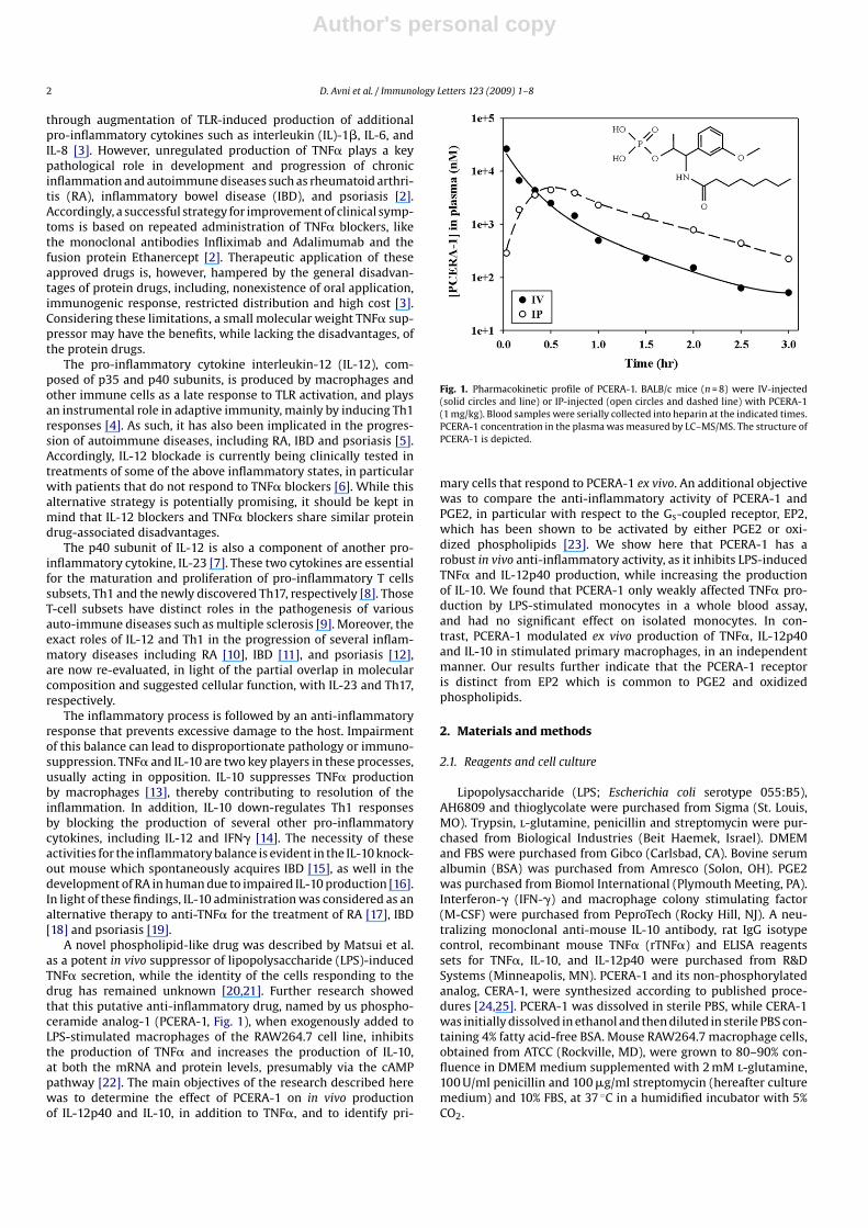
Fig. 1. Pharmacokinetic profile of PCERA-1. BALB/c mice (n = 8) were IV-injected(solid circles and line) or IP-injected (open circles and dashed line) with PCERA-1(1 mg/kg). Blood samples were serially collected into heparin at the indicated times.PCERA-1 concentration in the plasma was measured by LC–MS/MS. The structure ofPCERA-1 is depicted.
mary cells that respond to PCERA-1 ex vivo. An additional objectivewas to compare the anti-inflammatory activity of PCERA-1 andPGE2, in particular with respect to the Gs-coupled receptor, EP2,which has been shown to be activated by either PGE2 or oxi-dized phospholipids [23]. We show here that PCERA-1 has arobust in vivo anti-inflammatory activity, as it inhibits LPS-inducedTNF� and IL-12p40 production, while increasing the productionof IL-10. We found that PCERA-1 only weakly affected TNF� pro-duction by LPS-stimulated monocytes in a whole blood assay,and had no significant effect on isolated monocytes. In con-trast, PCERA-1 modulated ex vivo production of TNF�, IL-12p40and IL-10 in stimulated primary macrophages, in an independentmanner. Our results further indicate that the PCERA-1 receptoris distinct from EP2 which is common to PGE2 and oxidizedphospholipids.
2. Materials and methods
2.1. Reagents and cell culture
Lipopolysaccharide (LPS; Escherichia coli serotype 055:B5),AH6809 and thioglycolate were purchased from Sigma (St. Louis,MO). Trypsin, l-glutamine, penicillin and streptomycin were pur-chased from Biological Industries (Beit Haemek, Israel). DMEMand FBS were purchased from Gibco (Carlsbad, CA). Bovine serumalbumin (BSA) was purchased from Amresco (Solon, OH). PGE2was purchased from Biomol International (Plymouth Meeting, PA).Interferon-� (IFN-�) and macrophage colony stimulating factor(M-CSF) were purchased from PeproTech (Rocky Hill, NJ). A neu-tralizing monoclonal anti-mouse IL-10 antibody, rat IgG isotypecontrol, recombinant mouse TNF� (rTNF�) and ELISA reagentssets for TNF�, IL-10, and IL-12p40 were purchased from R&DSystems (Minneapolis, MN). PCERA-1 and its non-phosphorylatedanalog, CERA-1, were synthesized according to published proce-dures [24,25]. PCERA-1 was dissolved in sterile PBS, while CERA-1was initially dissolved in ethanol and then diluted in sterile PBS con-taining 4% fatty acid-free BSA. Mouse RAW264.7 macrophage cells,obtained from ATCC (Rockville, MD), were grown to 80–90% con-fluence in DMEM medium supplemented with 2 mM l-glutamine,100 U/ml penicillin and 100 �g/ml streptomycin (hereafter culturemedium) and 10% FBS, at 37 ◦C in a humidified incubator with 5%CO2.
Author's personal copy
D. Avni et al. / Immunology Letters 123 (2009) 1–8 3
2.2. RAW264.7 macrophages activation assay
RAW264.7 macrophages were maintained for 48 h prior to theexperiment in 96-well plates (0.2 ml/well), at 2 × 10⁵ cells/well, inculture medium supplemented with 5% FBS, at 37 ◦C in a humid-ified incubator with 5% CO2. The cells were stimulated with LPS(100 ng/ml) at 37 ◦C for 2 h in the presence of PCERA-1 (1 �M), PGE2(0.1 �M) or vehicle. TNF� secretion to the medium was measuredby ELISA.
2.3. Animal care
Male BALB/c mice (8–13 weeks, 23 ± 2 g), obtained from theanimal breeding centers of Tel-Aviv University (TAU), The ScrippsResearch Institute (TSRI), or Genomics institute of the NovartisResearch Foundation (GNF) were housed in a pathogen-free roomunder controlled temperature (22–23 ◦C), humidity, and lighting(12 h light–dark cycles), and were given access to food and waterad libitum. Animal care and experimentation was carried out inaccordance with TAU, TSRI and GNF guidelines.
2.4. Pharmacokinetic (PK) studies
PCERA-1, formulated as a 0.5 mg/ml solution in sterile PBS,was administered intravenously (IV) or intraperitoneally (IP) togroups of 8 mice at a dose of 1.0 mg/kg. Blood samples (50 �l)were serially drawn by retro-orbital bleeding at 2, 10, 20, 30and 45 min after dosing in the first satellite group of 4 mice;and at 1, 1.5, 2, 2.5 and 3 h in the second satellite group of4 mice. Blood samples were centrifuged to obtain plasma, andtotal (free and protein-bound) plasma concentrations of PCERA-1were quantified using a liquid chromatography/mass spectrometry(LC–MS/MS) assay. Pharmacokinetic parameters were calculated bynon-compartmental regression analysis of the mean (n = 4) plasmaconcentration data using Winnonlin 4.0 software (Pharsight, Moun-tain View, CA, USA).
2.5. In vivo cytokine production measurements
The mice received an IP injection (0.1 ml) of either PCERA-1 atdoses of up to 8 mg/kg, CERA-1 at 1 mg/kg, or saline as vehicle,followed after 40 min by an IP injection (0.1 ml) of LPS (5 mg/kg).Blood was obtained by cardiac puncture at 1.5 h (for TNF� and IL-10measurements), or at 4 h (for IL-12p40 measurement), and plasmacytokine levels were determined by ELISA. The data were expressedas the mean ± standard error mean (SEM) of 3–4 animals per group.
2.6. Ex vivo whole blood assay
Mouse blood was drawn in via the retro-orbital plexus underanesthesia. Whole blood was pooled together, mixed with RPMI(20% volume) containing 10 units/ml of heparin, and aliquoted300 �l per assay. LPS (1 �g/ml), and PCERA-1 (10 �M) or PGE2(1 �M) were added for 5 h at 37 ◦C. The plasma was then collectedby centrifugation (1500 × g for 3 min), and secreted TNF� was mea-sured by ELISA.
2.7. Isolation, culture, and ex vivo activation of primary mousemonocytes
Mouse blood was drawn in via the retro-orbital plexus underanesthesia into heparin (15 units/ml). Peripheral blood mononu-clear cells (PBMC) were isolated by density gradient centrifugation,using Histopaque 10831 (Sigma) according to the manufacturer’sinstructions, and washed twice with PBS. Monocytes were puri-fied by positive selection using mouse CD11b microbeads (Miltenyi
Biotec, Bergisch Gladbach, Germany) according to the manufac-turer’s instructions. Purity of monocytes (>98%) was verified by cellmorphology. The monocytes were washed, re-suspended in culturemedium supplemented with 10% FBS, seeded in a 96-well cultureplate (0.1 ml/well) at 5 × 10⁴ cells/well, and stimulated with LPS(1 �g/ml) in the presence of either PCERA-1 (10 �M), PGE2 (0.1 �M),or vehicle at 37 ◦C for 12 h. Cytokine levels in the culture mediumwere determined by ELISA.
2.8. Ex vivo differentiation of monocytes into macrophages, andactivation
Blood monocytes were isolated and purified as described above,re-suspended in culture medium supplemented with 10% FBS, andseeded in a 12-well plate at 0.3 × 10⁶ cells/well in a humidified incu-bator (37 ◦C, 5% CO2). After 24 h the medium was gently removedand replaced with fresh culture medium supplemented with 10%FBS and 50 ng/ml of macrophage colony stimulating factor (M-CSF).The cells were allowed to grow and differentiate for 11 days underthese conditions while the medium was replaced every 2 days. Atday 12, when >95% of the cells acquired macrophage morphology,the cells were removed by scraping and seeded in a 96-well cultureplate at 1 × 10⁵ cells/well under the same conditions. At day 14 thecells were treated with LPS (1 �g/ml) in the presence or absenceof PCERA-1 (1 �M) for 16 h. Cytokine levels in the culture mediumwere then determined by ELISA.
2.9. Isolation, culture, and ex vivo activation of mouse peritonealmacrophages
Mouse peritoneal macrophages were elicited by injection of2 ml 4% Brewer’s thioglycollate into the peritoneal cavities of theBALB/c mice, 5 days prior to harvest of peritoneal exudate cells.The cells were washed, re-suspended in culture medium sup-plemented with 10% FBS, and seeded in a 96-well culture plate(0.2 ml/well) at 5 × 10⁵ cells/well. Following incubation of 4 h at37 ◦C, non-adherent cells were removed by repeated washing withwarm culture medium and the adherent cells (∼98% homogenousby appearance) were further cultured overnight at 37 ◦C. Stimula-tion was then performed with IFN-� (10 ng/ml) and LPS (1 �g/ml)in the presence or absence of PCERA-1 (1 �M) at 37 ◦C for 12 h.Cytokine levels in the culture medium were determined by ELISA.All incubations were carried out in a humidified incubator with 5%CO2.
2.10. Isolation and ex vivo activation of mouse bonemarrow-derived macrophages (BMDM)
BALB/c mice were sacrificed and the femoral and tibial mar-row was flushed with sterile PBS using a 27-gage needle. Redblood cells were removed by osmotic shock. The cells were re-suspended in culture medium supplemented with 15% FBS and10 ng/ml M-CSF. The cells were seeded in Petri dishes at a densityof 2.9 × 10⁵ cells/cm2 and were incubated at 37 ◦C in a humid-ified incubator with 5% CO2. After 1 day, adherent cells werediscarded, while non-adherent cells were re-suspended in freshmedium and then allowed to further differentiate. On day 3,half of the supernatant was collected, centrifuged (1000 × g for5 min), and cells pellet was re-suspended in fresh medium andreturned to the culture dish. On day 6, the culture mediumwas replaced and non-adherent cells were discarded. On day7, the adherent cells (differentiated BMDM, ∼98% homoge-nous by appearance) were transferred to 96-well culture plates(0.2 ml/well), at 2 × 10⁵ cells/well, for 1 day. Stimulation was thenperformed with LPS (1 �g/ml) and IFN-� (10 ng/ml) in the presenceor absence of PCERA-1 (1 �M) at 37 ◦C for 4 h (for TNF� and IL-10
Author's personal copy
4 D. Avni et al. / Immunology Letters 123 (2009) 1–8
determinations) or for 12 h (for IL-12p40 determination). Cytokinelevels in the medium were determined by ELISA.
2.11. Enzyme-linked immunosorbent assay (ELISA)
Measurements of TNF�, IL-10 and IL-12p40 levels in miceplasma and in culture medium of peritoneal macrophages wereperformed with commercially available ELISA reagents sets, accord-ing to the manufacturer’s instructions, using a microplate reader(Bio-Tek, Winooski, VT). The samples were stored at −80 ◦C untilused.
2.12. Statistical analysis
All the data were analyzed using Student’s t-test wherever appli-cable. In all cases, differences of p < 0.05 were considered to besignificant. All experiments were repeated at least three times.
3. Results
3.1. Pharmacokinetic analysis of PCERA-1
To evaluate the pharmacokinetic (PK) properties of PCERA-1,the compound was injected, IV or IP, to BALB/c mice, and theblood plasma concentration of PCERA-1 was determined at var-ious time points by LC–MS/MS (Fig. 1). The PK parameters wereestimated by fitting the mean plasma concentration (n = 4) againsttime. PCREA-1 demonstrated low clearance (6.6 ml/(min*kg), 7%of hepatic blood flow) and a very small volume of distribution(0.12 l/kg, 50% higher than the blood volume), which resulted in ashort half-life of elimination of 0.6 h. The initial plasma concen-tration after administration of the IV dose was 22 �M, while at3 h the concentration had decreased to 50 nM. PCERA-1 was read-ily absorbed from the peritoneum to the systemic circulation, asnearly 90% of the IP-administered PCERA-1 shortly appeared inthe plasma (AUC = 5847 and 5228 h*nM, for IV and IP administra-tion, respectively), with maximal plasma concentration of 4.6 �Mobserved within 0.5 h of the IP injection. These results indicate thatPCERA-1 was rapidly exchanged between the peritoneum and thesystemic circulation; however, its distribution was largely limitedto the blood compartment.
3.2. In vivo activity of PCERA-1
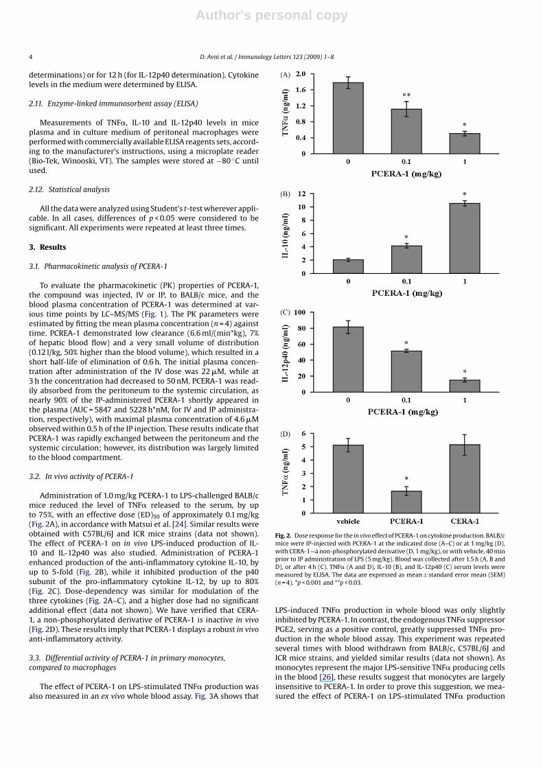
Administration of 1.0 mg/kg PCERA-1 to LPS-challenged BALB/cmice reduced the level of TNF� released to the serum, by upto 75%, with an effective dose (ED)50 of approximately 0.1 mg/kg(Fig. 2A), in accordance with Matsui et al. [24]. Similar results wereobtained with C57BL/6J and ICR mice strains (data not shown).The effect of PCERA-1 on in vivo LPS-induced production of IL-10 and IL-12p40 was also studied. Administration of PCERA-1enhanced production of the anti-inflammatory cytokine IL-10, byup to 5-fold (Fig. 2B), while it inhibited production of the p40subunit of the pro-inflammatory cytokine IL-12, by up to 80%(Fig. 2C). Dose-dependency was similar for modulation of thethree cytokines (Fig. 2A–C), and a higher dose had no significantadditional effect (data not shown). We have verified that CERA-1, a non-phosphorylated derivative of PCERA-1 is inactive in vivo(Fig. 2D). These results imply that PCERA-1 displays a robust in vivoanti-inflammatory activity.
3.3. Differential activity of PCERA-1 in primary monocytes,compared to macrophages
The effect of PCERA-1 on LPS-stimulated TNF� production wasalso measured in an ex vivo whole blood assay. Fig. 3A shows that
Fig. 2. Dose response for the in vivo effect of PCERA-1 on cytokine production. BALB/cmice were IP-injected with PCERA-1 at the indicated dose (A–C) or at 1 mg/kg (D),with CERA-1—a non-phosphorylated derivative (D, 1 mg/kg), or with vehicle, 40 minprior to IP administration of LPS (5 mg/kg). Blood was collected after 1.5 h (A, B andD), or after 4 h (C). TNF� (A and D), IL-10 (B), and IL-12p40 (C) serum levels weremeasured by ELISA. The data are expressed as mean ± standard error mean (SEM)(n = 4). *p < 0.001 and **p < 0.03.
LPS-induced TNF� production in whole blood was only slightlyinhibited by PCERA-1. In contrast, the endogenous TNF� suppressorPGE2, serving as a positive control, greatly suppressed TNF� pro-duction in the whole blood assay. This experiment was repeatedseveral times with blood withdrawn from BALB/c, C57BL/6J andICR mice strains, and yielded similar results (data not shown). Asmonocytes represent the major LPS-sensitive TNF� producing cellsin the blood [26], these results suggest that monocytes are largelyinsensitive to PCERA-1. In order to prove this suggestion, we mea-sured the effect of PCERA-1 on LPS-stimulated TNF� production
Author's personal copy
D. Avni et al. / Immunology Letters 123 (2009) 1–8 5
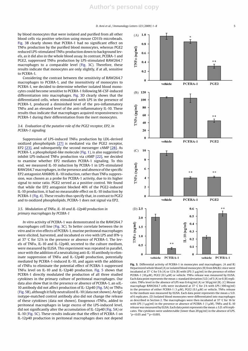
by blood monocytes that were isolated and purified from all otherblood cells via positive selection using mouse CD11b microbeads.Fig. 3B clearly shows that PCERA-1 had no significant effect onTNF� production by the purified blood monocytes, whereas PGE2reduced LPS-stimulated TNF� production down to background lev-els, as it did also in the whole blood assay. In contrast, PCERA-1 andPGE2, suppressed TNF� production by LPS-stimulated RAW264.7macrophages to a comparable level (Fig. 3C). Therefore, theseresults indicate that monocytes are only slightly, if at all, sensitiveto PCERA-1.
Considering the contrast between the sensitivity of RAW264.7macrophages to PCERA-1, and the insensitivity of monocytes toPCERA-1, we decided to determine whether isolated blood mono-cytes could become sensitive to PCERA-1 following M-CSF-induceddifferentiation into macrophages. Fig. 3D clearly shows that thedifferentiated cells, when stimulated with LPS in the presence ofPCERA-1, produced a diminished level of the pro-inflammatoryTNF� and an elevated level of the anti-inflammatory IL-10. Theseresults thus indicate that macrophages acquired responsiveness toPCERA-1 during their differentiation from the inert monocytes.
3.4. Evaluation of the putative role of the PGE2 receptor, EP2, inPCERA-1 signaling
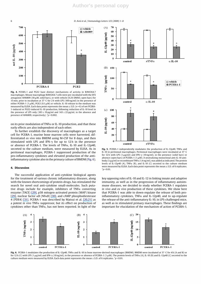
Suppression of LPS-induced TNF� production by LDL-derivedoxidized phospholipids [27] is mediated via the PGE2 receptor,EP2 [23], and subsequently the second messenger cAMP [28]. AsPCERA-1, a phospholipid-like molecule (Fig. 1), is also suggested toinhibit LPS-induced TNF� production via cAMP [22], we decidedto examine whether EP2 mediates PCERA-1 signaling. To thisend, we measured IL-10 induction by PCERA-1 in LPS-stimulatedRAW264.7 macrophages, in the presence and absence of the specificEP2 antagonist AH6809. IL-10 induction, rather than TNF� suppres-sion, was chosen as a probe for PCERA-1 activity, due to its highersignal to noise ratio. PGE2 served as a positive control. We foundthat while the EP2 antagonist blocked 40% of the PGE2-inducedIL-10 production, it had no measurable effect on IL-10 induction byPCERA-1 (Fig. 4). These results thus specify that, in contrast to PGE2and to oxidized phospholipids, PCERA-1 does not signal via EP2.
3.5. Modulation of TNF˛, IL-10 and IL-12p40 production inprimary macrophages by PCERA-1
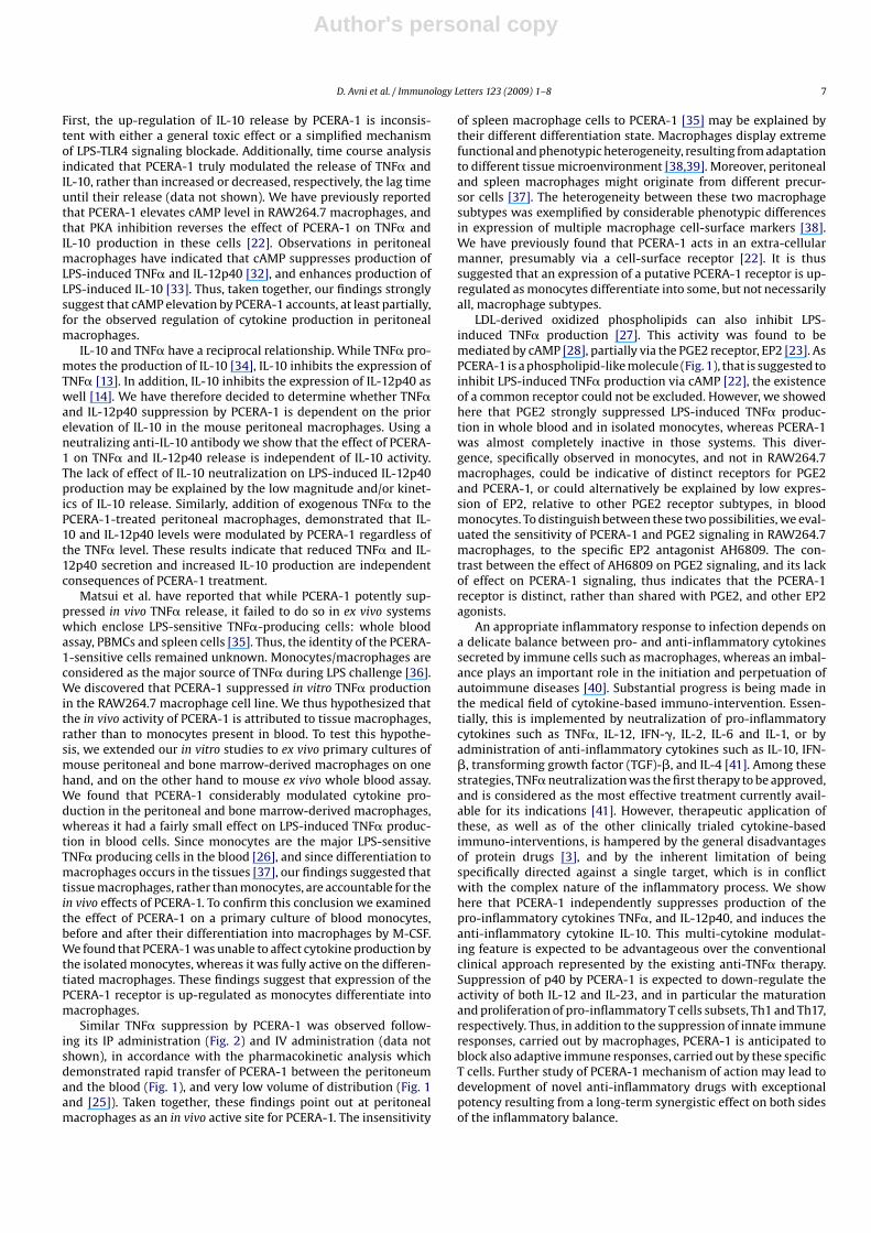
In vitro activity of PCERA-1 was demonstrated in the RAW264.7macrophages cell line (Fig. 3C). To better correlate between the invitro and in vivo effects of PCERA-1, murine peritoneal macrophageswere elicited, harvested, and incubated ex vivo with LPS and IFN-�at 37 ◦C for 12 h in the presence or absence of PCERA-1. The lev-els of TNF�, IL-10 and IL-12p40, secreted to the culture medium,were measured by ELISA. This experiment was repeated in parallel,once with the addition of a neutralizing anti-IL-10 antibody to elim-inate suppression of TNF� and IL-12p40 production, potentiallymediated by PCERA-1-induced IL-10, and again with the additionof rTNF� to eliminate the potential effect of PCERA-1-suppressedTNF� level on IL-10 and IL-12p40 production. Fig. 5 shows thatPCERA-1 directly modulated the production of all three studiedcytokines in the primary culture of peritoneal macrophages. Ourdata also show that in the presence or absence of PCERA-1, an �IL-10 antibody did not affect production of IL-12p40 (Fig. 5A) or TNF�(Fig. 5B), although it fully neutralized IL-10 (data not shown). An IgGisotype-matched control antibody also did not change the releaseof these cytokines (data not shown). Exogenous rTNF�, added toperitoneal macrophages in large excess of the LPS-induced level,did not significantly alter the accumulation of IL-12p40 (Fig. 5A) orIL-10 (Fig. 5C). These results indicate that the effect of PCERA-1 onIL-12p40 production in peritoneal macrophages does not depend
Fig. 3. Differential activity of PCERA-1 in monocytes and macrophages. (A and B)Heparinized whole blood (A) or isolated blood monocytes (B) from BALB/c mice wereincubated at 37 ◦C for 5 h (A) or 12 h (B) with LPS (1 �g/ml) in the presence of eitherPCERA-1 (10 �M), PGE2 (0.1 �M) or vehicle. TNF� release was measured by ELISA.Each data point represents the mean ± standard deviation (S.D.) of 5 (A) or 6 (B) repli-cates. TNF� level in the absence of LPS was 0.4 ng/ml (A) or 50 pg/ml (B). (C) Mousemacrophage RAW264.7 cells were incubated at 37 ◦C for 2 h with LPS (100 ng/ml)in the presence of either PCERA-1 (1 �M), PGE2 (0.1 �M) or vehicle. TNF� releaseto the medium was measured by ELISA. Each data point represents the mean ± S.D.of 6 replicates. (D) Isolated blood monocytes were differentiated into macrophagesas described in Section 2. The macrophages were then incubated at 37 ◦C for 16 hrwith LPS (1 �g/ml) in the presence or absence of PCERA-1 (1 �M). TNF� and IL-10release was measured by ELISA. Each data point represents the mean ± S.D. of 6 repli-cates. The cytokines were undetectable (lower than 20 pg/ml) in the absence of LPS.*p < 0.05 and **p < 0.001.
Author's personal copy
6 D. Avni et al. / Immunology Letters 123 (2009) 1–8
Fig. 4. PCERA-1 and PGE2 have distinct mechanisms of activity in RAW264.7macrophages. Mouse macrophage RAW264.7 cells were pre-incubated with the EP2antagonist AH6809 (30 �M, solid bars), or with vehicle (0.2% DMSO, open bars) for15 min, prior to incubation at 37 ◦C for 2 h with LPS (100 ng/ml) in the presence ofeither PCERA-1 (1 �M), PGE2 (0.1 �M) or vehicle. IL-10 release to the medium wasmeasured by ELISA. Each data point represents the mean ± S.D. (n = 6) of net PCERA-1-induced or PGE2-induced IL-10 production, following reduction of IL-10 level inthe presence of LPS only (183 ± 19 pg/ml and 143 ± 22 pg/ml, in the absence andpresence of AH6809, respectively). *p < 0.002.
on its prior modulation of TNF� or IL-10 production, and that theseearly effects are also independent of each other.
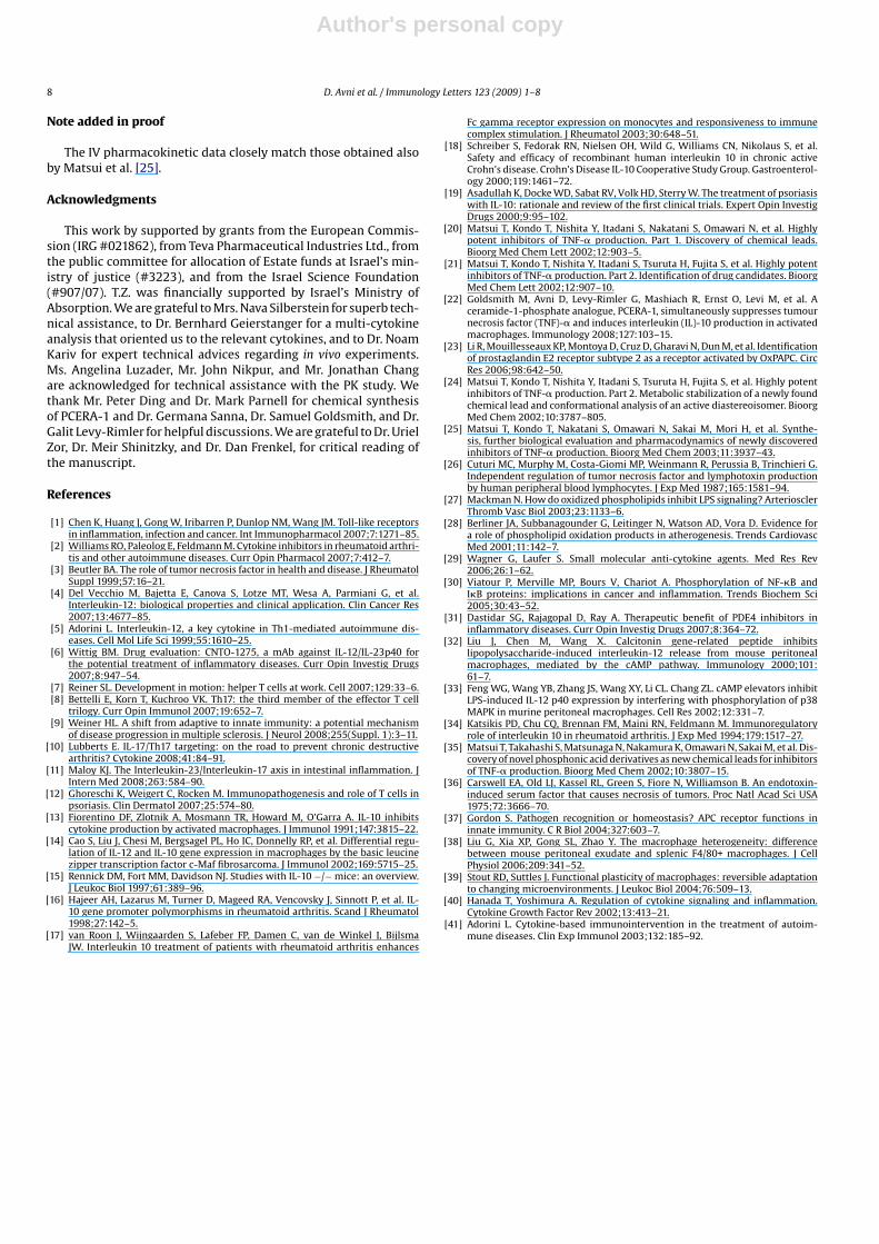
To further establish the discovery of macrophages as a targetcell for PCERA-1, murine bone marrow cells were harvested, dif-ferentiated ex vivo into BMDM using M-CSF for 8 days, and thenstimulated with LPS and IFN-� for up to 12 h in the presenceor absence of PCERA-1. The levels of TNF�, IL-10 and IL-12p40,secreted to the culture medium, were measured by ELISA. As inperitoneal macrophages, PCERA-1 suppressed production of thepro-inflammatory cytokines and elevated production of the anti-inflammatory cytokine also in the primary culture of BMDM (Fig. 6).
4. Discussion
The successful application of anti-cytokine biological agentsfor the treatment of various chronic inflammatory diseases, alongwith the known shortcomings of protein-drugs, has stimulated thesearch for novel oral anti-cytokine small-molecules. Such puta-tive drugs include for example, inhibitors of TNF� convertingenzyme (TACE) [29], p38 mitogen-activated protein (MAP) kinase[29], nuclear factor �B (NF�B) [30], and cAMP phosphodiesterase4 (PDE4) [31]. PCERA-1 was described by Matsui et al. [20,21] asa potent in vivo TNF� suppressor, but its effect on production ofcytokines other than TNF�, has not been reported. In light of the
Fig. 5. PCERA-1 independently modulates the production of IL-12p40, TNF� andIL-10 in peritoneal macrophages. Peritoneal macrophages were incubated at 37 ◦Cfor 12 h with LPS (1 �g/ml) and IFN-� (10 ng/ml), in the presence (solid bars) orabsence (open bars) of PCERA-1 (1 �M). A neutralizing monoclonal anti-IL-10 anti-body (3 �g/ml) or recombinant TNF� (1.4 ng/ml), was added as indicated. The proteinlevels of IL-12p40 (A), TNF� (B), and IL-10 (C) secreted to the culture mediumwere measured by ELISA. Each data point represents the mean ± S.D. of 6 replicates.*p < 0.01.
key opposing roles of IL-10 and IL-12 in linking innate and adaptiveimmunity, as well as in the progression of inflammatory autoim-mune diseases, we decided to study whether PCERA-1 regulatesin vivo and ex vivo production of these cytokines. We show herethat PCERA-1 was able to down-regulate the release of both pro-inflammatory cytokines, TNF� and IL-12p40, and to up-regulatethe release of the anti-inflammatory IL-10, in LPS-challenged mice,as well as in stimulated primary macrophages. These findings areimportant for elucidation of the mechanism of action of PCERA-1.
Fig. 6. PCERA-1 modulates the production of IL-12p40, TNF� and IL-10 in bone marrow-derived macrophages (BMDM). BMDM were incubated at 37 ◦C for 4 h (A and B) orfor 12 h (C) with LPS (1 �g/ml) and IFN-� (10 ng/ml), in the presence or absence of PCERA-1 (1 �M). The protein levels of TNF� (A), IL-10 (B) and IL-12p40 (C) secreted to theculture medium were measured by ELISA. Each data point represents the mean ± S.D. of 6 replicates. *p < 0.01.
Author's personal copy
D. Avni et al. / Immunology Letters 123 (2009) 1–8 7
First, the up-regulation of IL-10 release by PCERA-1 is inconsis-tent with either a general toxic effect or a simplified mechanismof LPS-TLR4 signaling blockade. Additionally, time course analysisindicated that PCERA-1 truly modulated the release of TNF� andIL-10, rather than increased or decreased, respectively, the lag timeuntil their release (data not shown). We have previously reportedthat PCERA-1 elevates cAMP level in RAW264.7 macrophages, andthat PKA inhibition reverses the effect of PCERA-1 on TNF� andIL-10 production in these cells [22]. Observations in peritonealmacrophages have indicated that cAMP suppresses production ofLPS-induced TNF� and IL-12p40 [32], and enhances production ofLPS-induced IL-10 [33]. Thus, taken together, our findings stronglysuggest that cAMP elevation by PCERA-1 accounts, at least partially,for the observed regulation of cytokine production in peritonealmacrophages.
IL-10 and TNF� have a reciprocal relationship. While TNF� pro-motes the production of IL-10 [34], IL-10 inhibits the expression ofTNF� [13]. In addition, IL-10 inhibits the expression of IL-12p40 aswell [14]. We have therefore decided to determine whether TNF�and IL-12p40 suppression by PCERA-1 is dependent on the priorelevation of IL-10 in the mouse peritoneal macrophages. Using aneutralizing anti-IL-10 antibody we show that the effect of PCERA-1 on TNF� and IL-12p40 release is independent of IL-10 activity.The lack of effect of IL-10 neutralization on LPS-induced IL-12p40production may be explained by the low magnitude and/or kinet-ics of IL-10 release. Similarly, addition of exogenous TNF� to thePCERA-1-treated peritoneal macrophages, demonstrated that IL-10 and IL-12p40 levels were modulated by PCERA-1 regardless ofthe TNF� level. These results indicate that reduced TNF� and IL-12p40 secretion and increased IL-10 production are independentconsequences of PCERA-1 treatment.
Matsui et al. have reported that while PCERA-1 potently sup-pressed in vivo TNF� release, it failed to do so in ex vivo systemswhich enclose LPS-sensitive TNF�-producing cells: whole bloodassay, PBMCs and spleen cells [35]. Thus, the identity of the PCERA-1-sensitive cells remained unknown. Monocytes/macrophages areconsidered as the major source of TNF� during LPS challenge [36].We discovered that PCERA-1 suppressed in vitro TNF� productionin the RAW264.7 macrophage cell line. We thus hypothesized thatthe in vivo activity of PCERA-1 is attributed to tissue macrophages,rather than to monocytes present in blood. To test this hypothe-sis, we extended our in vitro studies to ex vivo primary cultures ofmouse peritoneal and bone marrow-derived macrophages on onehand, and on the other hand to mouse ex vivo whole blood assay.We found that PCERA-1 considerably modulated cytokine pro-duction in the peritoneal and bone marrow-derived macrophages,whereas it had a fairly small effect on LPS-induced TNF� produc-tion in blood cells. Since monocytes are the major LPS-sensitiveTNF� producing cells in the blood [26], and since differentiation tomacrophages occurs in the tissues [37], our findings suggested thattissue macrophages, rather than monocytes, are accountable for thein vivo effects of PCERA-1. To confirm this conclusion we examinedthe effect of PCERA-1 on a primary culture of blood monocytes,before and after their differentiation into macrophages by M-CSF.We found that PCERA-1 was unable to affect cytokine production bythe isolated monocytes, whereas it was fully active on the differen-tiated macrophages. These findings suggest that expression of thePCERA-1 receptor is up-regulated as monocytes differentiate intomacrophages.
Similar TNF� suppression by PCERA-1 was observed follow-ing its IP administration (Fig. 2) and IV administration (data notshown), in accordance with the pharmacokinetic analysis whichdemonstrated rapid transfer of PCERA-1 between the peritoneumand the blood (Fig. 1), and very low volume of distribution (Fig. 1and [25]). Taken together, these findings point out at peritonealmacrophages as an in vivo active site for PCERA-1. The insensitivity
of spleen macrophage cells to PCERA-1 [35] may be explained bytheir different differentiation state. Macrophages display extremefunctional and phenotypic heterogeneity, resulting from adaptationto different tissue microenvironment [38,39]. Moreover, peritonealand spleen macrophages might originate from different precur-sor cells [37]. The heterogeneity between these two macrophagesubtypes was exemplified by considerable phenotypic differencesin expression of multiple macrophage cell-surface markers [38].We have previously found that PCERA-1 acts in an extra-cellularmanner, presumably via a cell-surface receptor [22]. It is thussuggested that an expression of a putative PCERA-1 receptor is up-regulated as monocytes differentiate into some, but not necessarilyall, macrophage subtypes.
LDL-derived oxidized phospholipids can also inhibit LPS-induced TNF� production [27]. This activity was found to bemediated by cAMP [28], partially via the PGE2 receptor, EP2 [23]. AsPCERA-1 is a phospholipid-like molecule (Fig. 1), that is suggested toinhibit LPS-induced TNF� production via cAMP [22], the existenceof a common receptor could not be excluded. However, we showedhere that PGE2 strongly suppressed LPS-induced TNF� produc-tion in whole blood and in isolated monocytes, whereas PCERA-1was almost completely inactive in those systems. This diver-gence, specifically observed in monocytes, and not in RAW264.7macrophages, could be indicative of distinct receptors for PGE2and PCERA-1, or could alternatively be explained by low expres-sion of EP2, relative to other PGE2 receptor subtypes, in bloodmonocytes. To distinguish between these two possibilities, we eval-uated the sensitivity of PCERA-1 and PGE2 signaling in RAW264.7macrophages, to the specific EP2 antagonist AH6809. The con-trast between the effect of AH6809 on PGE2 signaling, and its lackof effect on PCERA-1 signaling, thus indicates that the PCERA-1receptor is distinct, rather than shared with PGE2, and other EP2agonists.
An appropriate inflammatory response to infection depends ona delicate balance between pro- and anti-inflammatory cytokinessecreted by immune cells such as macrophages, whereas an imbal-ance plays an important role in the initiation and perpetuation ofautoimmune diseases [40]. Substantial progress is being made inthe medical field of cytokine-based immuno-intervention. Essen-tially, this is implemented by neutralization of pro-inflammatorycytokines such as TNF�, IL-12, IFN-�, IL-2, IL-6 and IL-1, or byadministration of anti-inflammatory cytokines such as IL-10, IFN-�, transforming growth factor (TGF)-�, and IL-4 [41]. Among thesestrategies, TNF� neutralization was the first therapy to be approved,and is considered as the most effective treatment currently avail-able for its indications [41]. However, therapeutic application ofthese, as well as of the other clinically trialed cytokine-basedimmuno-interventions, is hampered by the general disadvantagesof protein drugs [3], and by the inherent limitation of beingspecifically directed against a single target, which is in conflictwith the complex nature of the inflammatory process. We showhere that PCERA-1 independently suppresses production of thepro-inflammatory cytokines TNF�, and IL-12p40, and induces theanti-inflammatory cytokine IL-10. This multi-cytokine modulat-ing feature is expected to be advantageous over the conventionalclinical approach represented by the existing anti-TNF� therapy.Suppression of p40 by PCERA-1 is expected to down-regulate theactivity of both IL-12 and IL-23, and in particular the maturationand proliferation of pro-inflammatory T cells subsets, Th1 and Th17,respectively. Thus, in addition to the suppression of innate immuneresponses, carried out by macrophages, PCERA-1 is anticipated toblock also adaptive immune responses, carried out by these specificT cells. Further study of PCERA-1 mechanism of action may lead todevelopment of novel anti-inflammatory drugs with exceptionalpotency resulting from a long-term synergistic effect on both sidesof the inflammatory balance.
Author's personal copy
8 D. Avni et al. / Immunology Letters 123 (2009) 1–8
Note added in proof
The IV pharmacokinetic data closely match those obtained alsoby Matsui et al. [25].
Acknowledgments
This work by supported by grants from the European Commis-sion (IRG #021862), from Teva Pharmaceutical Industries Ltd., fromthe public committee for allocation of Estate funds at Israel’s min-istry of justice (#3223), and from the Israel Science Foundation(#907/07). T.Z. was financially supported by Israel’s Ministry ofAbsorption. We are grateful to Mrs. Nava Silberstein for superb tech-nical assistance, to Dr. Bernhard Geierstanger for a multi-cytokineanalysis that oriented us to the relevant cytokines, and to Dr. NoamKariv for expert technical advices regarding in vivo experiments.Ms. Angelina Luzader, Mr. John Nikpur, and Mr. Jonathan Changare acknowledged for technical assistance with the PK study. Wethank Mr. Peter Ding and Dr. Mark Parnell for chemical synthesisof PCERA-1 and Dr. Germana Sanna, Dr. Samuel Goldsmith, and Dr.Galit Levy-Rimler for helpful discussions. We are grateful to Dr. UrielZor, Dr. Meir Shinitzky, and Dr. Dan Frenkel, for critical reading ofthe manuscript.
References
[1] Chen K, Huang J, Gong W, Iribarren P, Dunlop NM, Wang JM. Toll-like receptorsin inflammation, infection and cancer. Int Immunopharmacol 2007;7:1271–85.
[2] Williams RO, Paleolog E, Feldmann M. Cytokine inhibitors in rheumatoid arthri-tis and other autoimmune diseases. Curr Opin Pharmacol 2007;7:412–7.
[3] Beutler BA. The role of tumor necrosis factor in health and disease. J RheumatolSuppl 1999;57:16–21.
[4] Del Vecchio M, Bajetta E, Canova S, Lotze MT, Wesa A, Parmiani G, et al.Interleukin-12: biological properties and clinical application. Clin Cancer Res2007;13:4677–85.
[5] Adorini L. Interleukin-12, a key cytokine in Th1-mediated autoimmune dis-eases. Cell Mol Life Sci 1999;55:1610–25.
[6] Wittig BM. Drug evaluation: CNTO-1275, a mAb against IL-12/IL-23p40 forthe potential treatment of inflammatory diseases. Curr Opin Investig Drugs2007;8:947–54.
[7] Reiner SL. Development in motion: helper T cells at work. Cell 2007;129:33–6.[8] Bettelli E, Korn T, Kuchroo VK. Th17: the third member of the effector T cell
trilogy. Curr Opin Immunol 2007;19:652–7.[9] Weiner HL. A shift from adaptive to innate immunity: a potential mechanism
of disease progression in multiple sclerosis. J Neurol 2008;255(Suppl. 1):3–11.[10] Lubberts E. IL-17/Th17 targeting: on the road to prevent chronic destructive
arthritis? Cytokine 2008;41:84–91.[11] Maloy KJ. The Interleukin-23/Interleukin-17 axis in intestinal inflammation. J
Intern Med 2008;263:584–90.[12] Ghoreschi K, Weigert C, Rocken M. Immunopathogenesis and role of T cells in
psoriasis. Clin Dermatol 2007;25:574–80.[13] Fiorentino DF, Zlotnik A, Mosmann TR, Howard M, O’Garra A. IL-10 inhibits
cytokine production by activated macrophages. J Immunol 1991;147:3815–22.[14] Cao S, Liu J, Chesi M, Bergsagel PL, Ho IC, Donnelly RP, et al. Differential regu-
lation of IL-12 and IL-10 gene expression in macrophages by the basic leucinezipper transcription factor c-Maf fibrosarcoma. J Immunol 2002;169:5715–25.
[15] Rennick DM, Fort MM, Davidson NJ. Studies with IL-10 −/− mice: an overview.J Leukoc Biol 1997;61:389–96.
[16] Hajeer AH, Lazarus M, Turner D, Mageed RA, Vencovsky J, Sinnott P, et al. IL-10 gene promoter polymorphisms in rheumatoid arthritis. Scand J Rheumatol1998;27:142–5.
[17] van Roon J, Wijngaarden S, Lafeber FP, Damen C, van de Winkel J, BijlsmaJW. Interleukin 10 treatment of patients with rheumatoid arthritis enhances
Fc gamma receptor expression on monocytes and responsiveness to immunecomplex stimulation. J Rheumatol 2003;30:648–51.
[18] Schreiber S, Fedorak RN, Nielsen OH, Wild G, Williams CN, Nikolaus S, et al.Safety and efficacy of recombinant human interleukin 10 in chronic activeCrohn’s disease. Crohn’s Disease IL-10 Cooperative Study Group. Gastroenterol-ogy 2000;119:1461–72.
[19] Asadullah K, Docke WD, Sabat RV, Volk HD, Sterry W. The treatment of psoriasiswith IL-10: rationale and review of the first clinical trials. Expert Opin InvestigDrugs 2000;9:95–102.
[20] Matsui T, Kondo T, Nishita Y, Itadani S, Nakatani S, Omawari N, et al. Highlypotent inhibitors of TNF-� production. Part 1. Discovery of chemical leads.Bioorg Med Chem Lett 2002;12:903–5.
[21] Matsui T, Kondo T, Nishita Y, Itadani S, Tsuruta H, Fujita S, et al. Highly potentinhibitors of TNF-� production. Part 2. Identification of drug candidates. BioorgMed Chem Lett 2002;12:907–10.
[22] Goldsmith M, Avni D, Levy-Rimler G, Mashiach R, Ernst O, Levi M, et al. Aceramide-1-phosphate analogue, PCERA-1, simultaneously suppresses tumournecrosis factor (TNF)-� and induces interleukin (IL)-10 production in activatedmacrophages. Immunology 2008;127:103–15.
[23] Li R, Mouillesseaux KP, Montoya D, Cruz D, Gharavi N, Dun M, et al. Identificationof prostaglandin E2 receptor subtype 2 as a receptor activated by OxPAPC. CircRes 2006;98:642–50.
[24] Matsui T, Kondo T, Nishita Y, Itadani S, Tsuruta H, Fujita S, et al. Highly potentinhibitors of TNF-� production. Part 2. Metabolic stabilization of a newly foundchemical lead and conformational analysis of an active diastereoisomer. BioorgMed Chem 2002;10:3787–805.
[25] Matsui T, Kondo T, Nakatani S, Omawari N, Sakai M, Mori H, et al. Synthe-sis, further biological evaluation and pharmacodynamics of newly discoveredinhibitors of TNF-� production. Bioorg Med Chem 2003;11:3937–43.
[26] Cuturi MC, Murphy M, Costa-Giomi MP, Weinmann R, Perussia B, Trinchieri G.Independent regulation of tumor necrosis factor and lymphotoxin productionby human peripheral blood lymphocytes. J Exp Med 1987;165:1581–94.
[27] Mackman N. How do oxidized phospholipids inhibit LPS signaling? ArteriosclerThromb Vasc Biol 2003;23:1133–6.
[28] Berliner JA, Subbanagounder G, Leitinger N, Watson AD, Vora D. Evidence fora role of phospholipid oxidation products in atherogenesis. Trends CardiovascMed 2001;11:142–7.
[29] Wagner G, Laufer S. Small molecular anti-cytokine agents. Med Res Rev2006;26:1–62.
[30] Viatour P, Merville MP, Bours V, Chariot A. Phosphorylation of NF-�B andI�B proteins: implications in cancer and inflammation. Trends Biochem Sci2005;30:43–52.
[31] Dastidar SG, Rajagopal D, Ray A. Therapeutic benefit of PDE4 inhibitors ininflammatory diseases. Curr Opin Investig Drugs 2007;8:364–72.
[32] Liu J, Chen M, Wang X. Calcitonin gene-related peptide inhibitslipopolysaccharide-induced interleukin-12 release from mouse peritonealmacrophages, mediated by the cAMP pathway. Immunology 2000;101:61–7.
[33] Feng WG, Wang YB, Zhang JS, Wang XY, Li CL. Chang ZL. cAMP elevators inhibitLPS-induced IL-12 p40 expression by interfering with phosphorylation of p38MAPK in murine peritoneal macrophages. Cell Res 2002;12:331–7.
[34] Katsikis PD, Chu CQ, Brennan FM, Maini RN, Feldmann M. Immunoregulatoryrole of interleukin 10 in rheumatoid arthritis. J Exp Med 1994;179:1517–27.
[35] Matsui T, Takahashi S, Matsunaga N, Nakamura K, Omawari N, Sakai M, et al. Dis-covery of novel phosphonic acid derivatives as new chemical leads for inhibitorsof TNF-� production. Bioorg Med Chem 2002;10:3807–15.
[36] Carswell EA, Old LJ, Kassel RL, Green S, Fiore N, Williamson B. An endotoxin-induced serum factor that causes necrosis of tumors. Proc Natl Acad Sci USA1975;72:3666–70.
[37] Gordon S. Pathogen recognition or homeostasis? APC receptor functions ininnate immunity. C R Biol 2004;327:603–7.
[38] Liu G, Xia XP, Gong SL, Zhao Y. The macrophage heterogeneity: differencebetween mouse peritoneal exudate and splenic F4/80+ macrophages. J CellPhysiol 2006;209:341–52.
[39] Stout RD, Suttles J. Functional plasticity of macrophages: reversible adaptationto changing microenvironments. J Leukoc Biol 2004;76:509–13.
[40] Hanada T, Yoshimura A. Regulation of cytokine signaling and inflammation.Cytokine Growth Factor Rev 2002;13:413–21.
[41] Adorini L. Cytokine-based immunointervention in the treatment of autoim-mune diseases. Clin Exp Immunol 2003;132:185–92.