Targeted Peptidecentric Proteomics Reveals Caspase7 as a Substrate of the Caspase1 Inflammasomes
14
Targeted Peptidecentric Proteomics Reveals Caspase-7 as a Substrate of the Caspase-1 Inflammasomes* □ S Mohamed Lamkanfi‡§¶, Thirumala-Devi Kanneganti‡§, Petra Van Damme**‡‡, Tom Vanden Berghe§§¶¶, Isabel Vanoverberghe§§¶¶, Joe ¨ l Vandekerckhove**‡‡, Peter Vandenabeele§§¶¶, Kris Gevaert**‡‡, and Gabriel Nu ´n ˜ ez‡ The aspartate-specific cysteine protease caspase-1 is ac- tivated by the inflammasomes and is responsible for the proteolytic maturation of the cytokines IL-1 and IL-18 during infection and inflammation. To discover new caspase-1 substrates, we made use of a proteome-wide gel-free differential peptide sorting methodology that al- lows unambiguous localization of the processing site in addition to identification of the substrate. Of the 1022 proteins that were identified, 20 were found to be specif- ically cleaved after Asp in the setup incubated with re- combinant caspase-1. Interestingly, caspase-7 emerged as one of the identified caspase-1 substrates. Moreover half of the other identified cleavage events occurred at sites closely resembling the consensus caspase-7 recog- nition sequence DEVD, suggesting caspase-1-mediated activation of endogenous caspase-7 in this setup. Con- sistently recombinant caspase-1 cleaved caspase-7 at the canonical activation sites Asp 23 and Asp 198 , and re- combinant caspase-7 processed a subset of the identified substrates. In vivo, caspase-7 activation was observed in conditions known to induce activation of caspase-1, in- cluding Salmonella infection and microbial stimuli com- bined with ATP. Interestingly Salmonella- and lipopo- lysaccharide ATP-induced activation of caspase-7 was abolished in macrophages deficient in caspase-1, the pat- tern recognition receptors Ipaf and Cryopyrin, and the inflammasome adaptor ASC, demonstrating an upstream role for the caspase-1 inflammasomes in caspase-7 acti- vation in vivo. In contrast, caspase-1 and the inflamma- somes were not required for caspase-3 activation. In con- clusion, we identified 20 new substrates activated downstream of caspase-1 and validated caspase-1-medi- ated caspase-7 activation in vitro and in knock-out mac- rophages. These results demonstrate for the first time the existence of a nucleotide binding and oligomeriza- tion domain-like receptor/caspase-1/caspase-7 cas- cade and the existence of distinct activation mecha- nisms for caspase-3 and -7 in response to microbial stimuli and bacterial infection. Molecular & Cellular Proteomics 7:2350 –2363, 2008. Cysteinyl aspartate-specific proteases (caspases) 1 have essential roles in apoptosis and inflammation (1). They are synthesized as zymogens with a prodomain of variable length followed by a large and a small catalytic subunit. In humans, the caspase family consists of 11 members, which are clas- sified into three phylogenetic groups correlating with their function (2). Caspase-1 is the prototypical member of the inflammatory caspases and mediates the proteolytic maturation of the re- lated cytokines IL-1 and IL-18 (3, 4) following its recruitment in large protein complexes termed “inflammasomes” (5–10). The molecular composition of the inflammasome depends on the identity of the nucleotide binding and oligomerization domain (NOD)-like receptor (NLR) family member serving as scaffold protein in the complex (6). The members of the cy- tosolic NLR family are believed to recognize conserved mi- crobial and viral components called pathogen-associated molecular patterns (PAMPs) in intracellular compartments. In humans, the NLR family is composed of 23 members that share remarkable structural similarity to a subset of plant disease resistance genes (R genes) (11). The amino-terminal sequence of NLRs generally contains homotypic interaction motifs such as the caspase recruitment domain (CARD) and From the ‡Department of Pathology and Comprehensive Cancer Center, University of Michigan Medical School, Ann Arbor, Michigan 48109, **Department of Medical Protein Research, Flanders Institute for Biotechnology, B-9000 Ghent, Belgium, ‡‡Department of Bio- chemistry, Ghent University, B-9000 Ghent, Belgium, §§Molecular Signalling and Cell Death Unit, Department for Molecular Biomedical Research, Flanders Institute for Biotechnology, B-9052 Zwijnaarde, Belgium, and ¶¶Molecular Signalling and Cell Death Unit, Department of Molecular Biology, Ghent University, B-9052 Zwijnaarde, Belgium Received, March 26, 2008, and in revised form, July 23, 2008 Published, MCP Papers in Press, July 30, 2008, DOI 10.1074/ mcp.M800132-MCP200 1 The abbreviations used are: caspase, cysteinyl aspartate-specific protease; CARD, caspase recruitment domain; ASC, apoptosis-asso- ciated specklike protein containing a CARD; ATP, adenosine triphos- phate; BMDM, bone marrow-derived macrophage; COFRADIC, com- bined fractional diagonal chromatography; DISC, death-inducing signaling complex; IL-1, interleukin-1; IL-18, interleukin-18; LPS, lipopolysaccharide; NLR, NOD-like receptor; PAMP, pathogen-asso- ciated molecular pattern; NOD, nucleotide binding and oligomeriza- tion domain; RP, reverse phase; TNBS, 2,4,6-trinitrobenzenesulfonic acid; cmk, chloromethyl ketone; fmk, fluoromethyl ketone. Research © 2008 by The American Society for Biochemistry and Molecular Biology, Inc. 2350 Molecular & Cellular Proteomics 7.12 This paper is available on line at http://www.mcponline.org
Transcript of Targeted Peptidecentric Proteomics Reveals Caspase7 as a Substrate of the Caspase1 Inflammasomes

Targeted Peptidecentric Proteomics RevealsCaspase-7 as a Substrate of the Caspase-1Inflammasomes*□S
Mohamed Lamkanfi‡§¶, Thirumala-Devi Kanneganti‡§�, Petra Van Damme**‡‡,Tom Vanden Berghe§§¶¶, Isabel Vanoverberghe§§¶¶, Joel Vandekerckhove**‡‡,Peter Vandenabeele§§¶¶, Kris Gevaert**‡‡, and Gabriel Nunez‡��
The aspartate-specific cysteine protease caspase-1 is ac-tivated by the inflammasomes and is responsible for theproteolytic maturation of the cytokines IL-1� and IL-18during infection and inflammation. To discover newcaspase-1 substrates, we made use of a proteome-widegel-free differential peptide sorting methodology that al-lows unambiguous localization of the processing site inaddition to identification of the substrate. Of the 1022proteins that were identified, 20 were found to be specif-ically cleaved after Asp in the setup incubated with re-combinant caspase-1. Interestingly, caspase-7 emergedas one of the identified caspase-1 substrates. Moreoverhalf of the other identified cleavage events occurred atsites closely resembling the consensus caspase-7 recog-nition sequence DEVD, suggesting caspase-1-mediatedactivation of endogenous caspase-7 in this setup. Con-sistently recombinant caspase-1 cleaved caspase-7 atthe canonical activation sites Asp23 and Asp198, and re-combinant caspase-7 processed a subset of the identifiedsubstrates. In vivo, caspase-7 activation was observed inconditions known to induce activation of caspase-1, in-cluding Salmonella infection and microbial stimuli com-bined with ATP. Interestingly Salmonella- and lipopo-lysaccharide � ATP-induced activation of caspase-7 wasabolished in macrophages deficient in caspase-1, the pat-tern recognition receptors Ipaf and Cryopyrin, and theinflammasome adaptor ASC, demonstrating an upstreamrole for the caspase-1 inflammasomes in caspase-7 acti-vation in vivo. In contrast, caspase-1 and the inflamma-somes were not required for caspase-3 activation. In con-clusion, we identified 20 new substrates activateddownstream of caspase-1 and validated caspase-1-medi-ated caspase-7 activation in vitro and in knock-out mac-
rophages. These results demonstrate for the first timethe existence of a nucleotide binding and oligomeriza-tion domain-like receptor/caspase-1/caspase-7 cas-cade and the existence of distinct activation mecha-nisms for caspase-3 and -7 in response to microbialstimuli and bacterial infection. Molecular & CellularProteomics 7:2350–2363, 2008.
Cysteinyl aspartate-specific proteases (caspases)1 haveessential roles in apoptosis and inflammation (1). They aresynthesized as zymogens with a prodomain of variable lengthfollowed by a large and a small catalytic subunit. In humans,the caspase family consists of 11 members, which are clas-sified into three phylogenetic groups correlating with theirfunction (2).
Caspase-1 is the prototypical member of the inflammatorycaspases and mediates the proteolytic maturation of the re-lated cytokines IL-1� and IL-18 (3, 4) following its recruitmentin large protein complexes termed “inflammasomes” (5–10).The molecular composition of the inflammasome depends onthe identity of the nucleotide binding and oligomerizationdomain (NOD)-like receptor (NLR) family member serving asscaffold protein in the complex (6). The members of the cy-tosolic NLR family are believed to recognize conserved mi-crobial and viral components called pathogen-associatedmolecular patterns (PAMPs) in intracellular compartments. Inhumans, the NLR family is composed of 23 members thatshare remarkable structural similarity to a subset of plantdisease resistance genes (R genes) (11). The amino-terminalsequence of NLRs generally contains homotypic interactionmotifs such as the caspase recruitment domain (CARD) and
From the ‡Department of Pathology and Comprehensive CancerCenter, University of Michigan Medical School, Ann Arbor, Michigan48109, **Department of Medical Protein Research, Flanders Institutefor Biotechnology, B-9000 Ghent, Belgium, ‡‡Department of Bio-chemistry, Ghent University, B-9000 Ghent, Belgium, §§MolecularSignalling and Cell Death Unit, Department for Molecular BiomedicalResearch, Flanders Institute for Biotechnology, B-9052 Zwijnaarde,Belgium, and ¶¶Molecular Signalling and Cell Death Unit, Departmentof Molecular Biology, Ghent University, B-9052 Zwijnaarde, Belgium
Received, March 26, 2008, and in revised form, July 23, 2008Published, MCP Papers in Press, July 30, 2008, DOI 10.1074/
mcp.M800132-MCP200
1 The abbreviations used are: caspase, cysteinyl aspartate-specificprotease; CARD, caspase recruitment domain; ASC, apoptosis-asso-ciated specklike protein containing a CARD; ATP, adenosine triphos-phate; BMDM, bone marrow-derived macrophage; COFRADIC, com-bined fractional diagonal chromatography; DISC, death-inducingsignaling complex; IL-1�, interleukin-1�; IL-18, interleukin-18; LPS,lipopolysaccharide; NLR, NOD-like receptor; PAMP, pathogen-asso-ciated molecular pattern; NOD, nucleotide binding and oligomeriza-tion domain; RP, reverse phase; TNBS, 2,4,6-trinitrobenzenesulfonicacid; cmk, chloromethyl ketone; fmk, fluoromethyl ketone.
Research
© 2008 by The American Society for Biochemistry and Molecular Biology, Inc.2350 Molecular & Cellular Proteomics 7.12This paper is available on line at http://www.mcponline.org

the pyrin domain. The central NOD is thought to be involvedin self-oligomerization and activation, whereas the carbox-yl-terminal leucine-rich repeat motifs sense specific PAMPsand autoregulate NLR activity. The bipartite adaptor proteinapoptosis-associated specklike protein containing a CARD(ASC) bridges the interaction between NLR proteins andinflammatory caspases through homotypic interactions withits own amino-terminal pyrin and carboxyl-terminal CARDdomains. As such, ASC plays a central role in the assemblyof the inflammasomes and the activation of caspase-1 inresponse to a broad range of PAMPs and intracellularpathogens (7, 12). Whereas the Cryopyrin inflammasome isessential for caspase-1 activation in response to LPS, lipidA, lipoteichoic acid, lipoprotein, and double-stranded RNAin the presence of millimolar concentrations of ATP (8, 9,13), intracellular pathogens such as Salmonella typhimurium(Salmonella) activate caspase-1 through the Ipaf inflamma-some (5, 8, 14). Recently Salmonella flagellin was identifiedas the bacterial ligand that is sensed by Ipaf, although themechanism remains obscure (5, 14). Interestingly Salmo-nella induces a rapid and specialized form of macrophagecell death, which is sometimes termed “pyroptosis” andrequires activation of caspase-1 (15) through the Ipaf in-flammasome (15).
The central roles of the executioner caspase-3 and -7 dur-ing apoptosis have been well established. Upon initiation ofthe cell death program, homotypic interaction motifs in thelarge prodomains of caspase-8 and -9 mediate their recruit-ment in the death-inducing signaling complex (DISC) and theapoptosome, respectively, where they undergo proximity-in-duced activation (16–18). Once activated, the initiatorcaspases induce an apoptotic caspase cascade by proteo-lytically removing the linker region between the large andsmall catalytic subunits of caspase-3 and -7, a step that isrequired for full proteolytic activity of these executionercaspases (19, 20). In turn, active caspase-3 and -7 cleave alarge set of substrates, ultimately resulting in the morpholog-ical and biochemical hallmarks of apoptosis such as DNAfragmentation and mitochondrial damage (21, 22). As defi-ciency in caspase-3 induced a compensatory activation ofcaspase-7, the mild apoptotic phenotype of caspase-3knock-out mice was suggested to be due to its functionalredundancy with caspase-7 (23, 24). Consistently, caspase-7knock-out mice have been recently reported to be born atnormal Mendelian ratios and to display no gross abnormali-ties, whereas caspase-3/-7 double knock-out mice sufferfrom early perinatal lethality (25). Furthermore, caspase-7-deficient cells from adult mice exhibit normal activation ofapoptosis in response to a wide variety of stimuli includingdeath receptor activation, etoposide, and UV irradiation (25).These results indicate that caspase-3 and -7 perform redun-dant roles in the regulation of apoptosis during embryonicdevelopment and in response to a wide variety of “classical”apoptotic triggers (25). However, the molecular mechanisms
that govern the activation of these executioner caspases dur-ing inflammation and infection remain unclear.
Here we identified caspase-7 as a caspase-1 substrate bya proteome-wide screen for caspase-1 targets using the ami-no-terminal combined fractional diagonal chromatography(COFRADIC) gel-free technology. By this technique, amino-terminal peptides, including those newly formed by proteaseprocessing, are isolated prior to LC-MS/MS analysis. BrieflyS-alkylated and N-acetylated proteins are digested with tryp-sin, and the resulting peptide mixture is passed over an strongcation exchange column to enrich for �-N-acetylated amino-terminal peptides in the non-binding fraction (26). This peptidemixture is then fractionated by RP-HPLC, and contaminating,internal peptides carrying a free �-amino group are incubatedwith 2,4,6-trinitrobenzenesulfonic acid (TNBS), which renderssuch peptides more hydrophobic and thus segregates themfrom TNBS-unaffected (because they are already blocked)amino-terminal peptides during a series of identical, second-ary RP-HPLC separations (27). Our data demonstrate thatrecombinant caspase-1 processes caspase-7 at the canoni-cal activation sites Asp23 and Asp198. In vivo, activation ofcaspase-7 by microbial stimuli, including infection with Sal-monella and stimulation with LPS, requires caspase-1 andcomponents, respectively, of the Ipaf and Cryopyrin inflam-masomes, whereas caspase-3 is activated independently ofthe caspase-1 inflammasomes. These results demonstratethe existence of a NOD-like receptor/caspase-1/caspase-7cascade activated in response to microbial stimuli and bac-terial infection and indicate for the first time the existence ofdifferential activation mechanisms for caspase-3 and -7 dur-ing inflammation and infection.
EXPERIMENTAL PROCEDURES
COFRADIC Isolation of Amino-terminal Peptides—108 Mf4/4 cellswere grown for 10 days in SILAC (stable isotope labeling by aminoacids in cell culture) RPMI 1640 medium containing [12C6] or [13C6]Arg(Invitrogen). Cells were collected, washed twice with cold PBS, andresuspended in 1 ml of ice-cold homogenization buffer (20 mM
HEPES-KOH, pH 7.5, 10 mM KCl, 1.5 mM MgCl2, 1 mM EDTA, 1⁄100 ofa Complete protease inhibitor mixture tablet (Roche Applied Science),and 1 mM DTT). After three rounds of freeze-thawing in liquid N2 andcold ethanol, samples were cleared by centrifuging at 20,000 � g for30 min at 4 °C, and the protein concentration was determined usingthe Bradford method (Bio-Rad). Subsequently 1 mg of protein extractwas incubated with 400 nM recombinant mouse caspase-1 (12C-labeled setup) or left untreated (13C-labeled setup) for 1 h at 37 °Cbefore 1 �M Ac-YVAD-cmk (Calbiochem) was added to stop thereaction. Subsequent steps for the sorting and identification of differ-entially generated amino-terminal peptides by COFRADIC were per-formed as described (full technical details are described elsewhere:see Ref. 26). Briefly the proteins in the lysates were reduced andS-alkylated by iodoacetamide. Subsequently free primary aminogroups were blocked by trideuteroacetylation. Following trypsin di-gestion, the peptide mixtures were mixed in a 1:1 ratio, and amino-terminal peptides were enriched after passing them over a strongcation exchange cartridge at pH 3 and then fractionated by RP-HPLC(primary run). Peptides in each fraction were incubated with TNBS toblock free �-amino termini of remaining internal (i.e. non-amino-
Proteomics Reveals Caspase-7 as a Target of Inflammasomes
Molecular & Cellular Proteomics 7.12 2351
terminal) peptides. Peptides carrying an amino-terminal protein part(i.e. amino-terminal peptides from either unprocessed proteins orprotein fragments generated by processing) are not affected by TNBSbecause their �-amino group was already (in vitro or in vivo) blocked.During a series of replicate secondary RP-HPLC runs, the TNBS-altered internal peptides shift to later elution times because of theirincreased hydrophobicity. In this way, a set of unaltered, amino-terminal peptides that do not shift during the consecutive runs werecollected for mass spectrometric analysis.
LC-MS/MS Analysis and Peptide Identification by Mascot—Thecollected peptide fractions were dried and redissolved in 20 �l ofwater/acetonitrile (98:2, v/v), and half of each analyte mixture wassampled by LC-MS/MS using a microfluidic interface (Agilent ChipCube) mounted on an Agilent XCT-Ultra ion trap mass spectrometerthat was operated as described previously (28). Mascot generic fileswere created as described previously (28) and used to search themouse subset of the UniProtKB/Swiss-Prot release 51.0 (October 31,2006 containing 241,242 sequence entries of which 11,897 entriesoriginate from Mus musculus) using a locally installed version ofMascot (29) (version 2.1.04). The following search parameters wereset. Both the peptide mass tolerance and the peptide fragment masstolerance were set to �0.5 Da, the “instrument setting” of Mascot wasset to “ESI-TRAP,” and peptide charge was set to 1�, 2�, and 3�.Fixed modifications were trideutero-amino-acetylation of lysine andS-carbamidomethylation of cysteine (for identifying heavy labeledpeptides; [13C6]arginine was also set as an additional fixed modifica-tion). Oxidation of methionine to its sulfoxide derivative, pyrogluta-mate formation (amino-terminal Gln), pyrocarbamidomethylcysteineformation (amino-terminal carbamidomethylated cysteines), acetyla-tion and trideuteroacetylation of the �-amino terminus, and deamida-tion (Gln and Asn) were considered as variable modifications. Endo-proteinase Arg-C/P was considered as the protease used with amaximum number of one missed cleavage.
Raw DAT result files of Mascot were further queried using in-housedeveloped software. Only MS/MS spectra receiving a score exceed-ing the corresponding Mascot identity threshold score at the 95%confidence level were kept. As a rule of thumb, all such identifiedpeptides holding six or fewer amino acids were discarded. In addition,spectra that received a low Mascot score (five or fewer points abovethe corresponding threshold) were further interrogated, and onlyspectra that contained a significant number of b and y peptide frag-ment ions typically covering a stretch of three consecutive aminoacids were considered identified (see supplemental data with identi-fied MS/MS spectra). Truncated peptide databases made by DBTool-kit (30) were searched in parallel to pick up protein processing eventsmore efficiently (e.g. see Refs. 30 and 31). Decoy databases (a shuf-fled version of the UniProtKB/Swiss-Prot database made by theDBToolkit algorithm (32)) were searched as suggested previously (33)to estimate the false discovery rate. At the Mascot identity thresholdscore (95%) used, the estimated false discovery rate was typicallyfound to be between 2 and 4% on the spectrum level (26). Whenneo-amino-terminal peptides generated by the (downstream) actionof caspase-1 were found to match multiple members of a proteinfamily (i.e. redundancies found in the searched database), the corre-sponding proteins are listed in Tables I and II.
In Vitro Caspase Cleavage Assays—cDNAs encoding wild typemurine caspase-7 or site-directed mutants harboring D23A, D198A,or D23A/D198A mutations were cloned in the pLT10 vector down-stream of the T7 promoter. pGEM11-proIL-1� was constructed byinserting murine pro-IL-1� cDNA into the unique HindII site of thepGEM11Zf(�) vector (Promega). pCMV-Sport6-Ligatin, pCMV-Sport6-Vps72, pCMV-Sport6-eIF4H, pCMV-Sport6-MCM3, pCMV-Sport6-Ascc2, pCMV-Sport6-Hsp60, pYX-Asc-TIF1�, and pYX-Asc-GIT2 plasmids were purchased from ATCC. Plasmids were used as a
template (250 ng each) for in vitro coupled transcription/translation ina rabbit reticulocyte lysate system according to the manufacturer’sinstructions (Promega). For detection of the translation products,[35S]methionine was added to the translation reactions. Translationreactions (2 �l each) were incubated with 30 nM purified recombinantcaspase-1 or caspase-7 in 23 �l of cell-free system buffer (10 mM
HEPES, pH 7.4, 220 mM mannitol, 68 mM sucrose, 2 mM NaCl, 2.5 mM
KH2PO4, 0.5 mM EGTA, 2 mM MgCl2, 5 mM sodium pyruvate, and 1mM DTT) for 1 h at 37 °C. In some experiments, recombinantcaspase-1 was preincubated with 1 �M YVAD-cmk (Calbiochem). Theresulting cleavage products were analyzed by SDS-PAGE andautoradiography.
Mice and Macrophages—Cryopyrin�/�, Ipaf�/�, ASC�/�, caspase-1�/�, and caspase-3�/� mice have been described previously (4, 5,10, 23). caspase-7�/� mice in a C57BL/6 background have beendescribed previously (25) and were purchased from The JacksonLaboratory. Mice were housed in a pathogen-free facility. Bone-marrow derived macrophages (BMDMs) were prepared as describedbefore (34). The animal studies were conducted under protocolsapproved by the University of Michigan Committee on Use and Careof Animals.
Bacteria and Ligands—Salmonella enterica serovar typhimuriumstrain SL1344 was kindly provided by D. Monack (Stanford Univer-sity). The fliB�/fljC� Salmonella strain was a generous gift of A.Aderem (University of Washington). Single colonies were inoculatedinto 3 ml of brain-heart infusion medium and grown overnight at 30 °Cwith shaking. All bacterial ligands and heat-killed bacteria were pur-chased from Invivogen and used at a concentration of 10 �g/ml. ATP(Roche Applied Science) was used at 5 mM throughout all experi-ments. Infection and stimulation of BMDMs with LPS and ATP wasperformed as described previously (34).
Western Blotting—Extracts were prepared, transferred to nitrocel-lulose membranes, and immunoblotted with primary antibodies, andthen proteins were detected by enhanced chemiluminescence asdescribed previously (6). Anti-caspase-1 antibody was described be-fore (35). Anti-caspase-3 and anti-caspase-7 were purchased fromCell Signaling Technology.
Precipitation of Biotin-bound Caspase-3 and -7—108 BMDMs wereleft untreated, stimulated with LPS � ATP, or infected with Salmo-nella. Cells were collected by centrifugation, and the cell pellet wasresuspended in 1 ml of Buffer A (1% Nonidet P-40, 200 mM NaCl, 20mM Tris-HCl, pH 7.4, and Roche Applied Science Complete proteaseinhibitor mixture tablet) containing 100 �M biotin-DEVD-fmk (KamiyaBiomedical Co.). Cells were incubated at room temperature for 15 minand lysed with three rounds of freeze-thawing in liquid N2. Superna-tants were collected by centrifugation (14,000 rpm at 4 °C) and incu-bated overnight with high capacity streptavidin-agarose beads(Thermo Scientific) or stored at �70 °C for direct immunoblotting ofcell lysates. Streptavidin-bound complexes were washed four timeswith cold Buffer A and analyzed by immunoblotting after elution inSDS-PAGE sample buffer.
Measurements of Cytokines—Mouse cytokines were measured inserum and culture supernatants with ELISA kits (R&D Systems).
RESULTS
Identification of Caspase-7 in a Proteome-wide Screen forin Vitro Caspase-1 Substrates—We performed a proteome-wide screen to identify novel caspase-1 substrates using thegel-free COFRADIC peptide sorting methodology used on adifferential setup (30). In addition to the identification of thesubstrate, this technology provides the exact position of theprocessing site by specifically identifying the neo-amino ter-minus generated by protein processing. In this study, we
Proteomics Reveals Caspase-7 as a Target of Inflammasomes
2352 Molecular & Cellular Proteomics 7.12
prepared proteins from untreated and caspase-1-treatedmacrophage extracts for differential analysis of amino-termi-nal peptides. Following LC-MS/MS analysis, 2663 peptideswere identified by Mascot (29), and these converged into 1022different proteins. On the basis of the differential 12C6/13C6
isotope labeling of L-arginine used (36), peptides that displayas couples of “light” and “heavy” forms spaced by 6 Da arepresent in both the control and caspase-1-treated samplesand thus do not hint at protein processing. Such peptideswere therefore not considered for further data interpretation.On the other hand, peptides that were (a) exclusively found inthe proteome digest of caspase-1-treated cell lysate (thusrepresented by a single isotopic peptide envelope) and (b)were generated by cleavage after Asp residues were hereregarded as candidate caspase-1 targets. Using this ap-proach, 20 proteins cleaved after Asp were identified uponincubation with recombinant caspase-1 (Tables I and II). Inaddition, 29 proteins were processed after residues other thanAsp (supplemental Table 1) and were hence not further con-sidered. Eleven of the 23 (48%) identified Asp-specific cleav-age sites resembled the caspase-1 cleavage sites in pro-IL-1� and pro-IL-18, suggesting that this subgroup ofsubstrates was directly processed by caspase-1 (Tables I andII, cluster I): next to the requirement for Asp in P1 that is typicalfor all caspases, this cluster was characterized by a uniquespecificity for the aliphatic residues Leu, Ile, Met, and Val inthe P4 position as evident from an analysis of the identifiedcleavage sites on the WebLogo (37) server (Fig. 1A, left).Notably the executioner caspase-7 was identified as a mem-ber of this substrate cluster with cleavage occurring at itscanonical activation site IQAD198 between the p20 and p10subunits (Tables I and II). This result suggested that recom-binant caspase-1 induced activation of endogenouscaspase-7 in the treated lysate. In line with this notion, theremaining 12 of 23 (52%) processing events occurred atcleavage sites that perfectly matched or closely resembledthe optimal caspase-7 recognition sequence DEVD (Fig. 1A,right) identified in combinatorial peptide library screenings(38).
Caspase-1 Processes Procaspase-7 at Canonical Asp23
and Asp198 Sites—To confirm that caspase-1 can cleavecaspase-7, in vitro translated 35S-labeled procaspase-7 wasincubated with 30 nM recombinant caspase-1 for 30 min, 1 h,or 2 h at 37 °C, and the resulting cleavage fragments wereanalyzed by SDS-PAGE and autoradiography. Similar tocaspase-1-mediated cleavage of pro-IL-1�, the 33-kDa full-length procaspase-7 band was completely processed in lessthan 30 min, giving rise to 19- and 11-kDa fragments that arecharacteristic for the p20 and p10 subunits of activecaspase-7, respectively (Fig. 1B). As expected, caspase-1-mediated processing of pro-IL-1� and procaspase-7 was ab-rogated by the caspase-1 inhibitor YVAD-cmk (Fig. 1B). Wemade use of site-directed procaspase-7 mutants to investi-gate whether caspase-1-mediated cleavage of procaspase-7
occurred at the IQAD198 site and to verify whether caspase-1also removes the amino-terminal procaspase-7 peptide afterAsp23 (39). Wild type procaspase-7 was found to be cleavedby caspase-1, generating the characteristic p19 and p11 frag-ments (Fig. 1C). Mutation of the canonical Asp23 activationsite between the prodomain and the large catalytic subunitdid not interfere with the generation of the p11 activationfragment, although a slightly larger p19 subunit was apparent.These results are consistent with caspase-1-mediated re-moval of the procaspase-7 prodomain by cleavage at Asp23.The prototypical p19 and p11 subunits were not detectedwhen Asp198 was mutated to Ala, although some pro-caspase-7 was processed to a fragment of 30 kDa (Fig. 1C).The latter fragment is likely generated by caspase-1-mediatedremoval of the amino-terminal peptide at Asp23 as its forma-tion was prevented in the D23A/D198A double caspase-7mutant (Fig. 1C). Together these results show that caspase-1can cleave procaspase-7 at its canonical activation sitesAsp23 and Asp198. We next analyzed whether caspase-7 ac-tivated downstream of caspase-1 could be responsible for thecleavage events grouped under cluster II (Tables I and II). Thecluster I members TIF1� and Hsp60 were incorporated asnegative controls for caspase-7-mediated cleavage. The lat-ter two proteins were not cleaved by recombinantcaspase-7 even at enzyme concentrations 10 times higherthan the 30 nM used to cleave the cluster II substrates (Fig.1D and data not shown). In contrast, active caspase-7processed all tested cluster II members (Assc2, GIT2, Liga-tin, eIF4h, MCM3, and Vps72), generating cleavage frag-ments that were in line with those predicted from the cleav-age sites identified by COFRADIC analysis (Fig. 1E).
Caspase-1 Is Essential for Activation of Caspase-7, butNot Caspase-3, in Response to Microbial Stimuli—To as-sess whether caspase-1 acts upstream of caspase-7 invivo, BMDMs from wild type and caspase-1�/� macrophageswere stimulated with LPS or LPS combined with ATP orpulsed with ATP alone for 30 min, and proteolytic activation ofendogenous caspase-1, caspase-3, and caspase-7 was as-sessed by immunoblotting. As a control and in accord withprevious reports (8, 9, 34), stimulation of LPS-treated macro-phages with ATP induced activation of caspase-1 in wild typeBMDMs, whereas LPS or ATP alone did not (Fig. 2A). Similarto caspase-1, activation of caspase-7 occurred only by com-bined stimulation with LPS and ATP (Fig. 2A). In contrast, therelated caspase-3 was activated in macrophages stimulatedwith LPS alone (Fig. 2A), suggesting a differential activationmechanism for caspase-3 and -7. Indeed whereas activationof caspase-7 triggered by LPS and ATP was abolished incaspase-1-deficient macrophages, that of caspase-3 was notaffected (Fig. 2A). To determine whether caspase-7 is acti-vated by bacterial infection, macrophages were incubatedwith wild type S. typhimurium or a flagellin-deficient mutant.As a control and consistent with published studies (5, 14),Salmonella-induced caspase-1 activation required flagellin as
Proteomics Reveals Caspase-7 as a Target of Inflammasomes
Molecular & Cellular Proteomics 7.12 2353

TAB
LEI
Ove
rvie
wof
targ
ets
and
clea
vage
site
sid
entif
ied
ina
pro
teom
e-w
ide
CO
FRA
DIC
anal
ysis
with
casp
ase-
1
The
com
ple
telis
tof
inte
rnal
lylo
cate
d,
trid
eute
roac
etyl
ated
(AcD
3)
pep
tides
with
anA
spre
sid
uein
P1
that
wer
eun
ique
lyid
entif
ied
inth
ed
iges
ted
pro
teom
eof
reco
mb
inan
tca
spas
e-1-
trea
ted
mou
seM
f414
lysa
tes
issh
own.
Pro
tein
nam
esar
eid
entif
ied
by
usin
gth
eS
wis
sIn
stitu
teof
Bio
info
rmat
ics
BLA
ST
netw
ork
serv
ice,
and
the
acce
ssio
nnu
mb
ers
are
acco
rdin
gto
the
Uni
Pro
tKB
/Sw
iss-
Pro
tdat
abas
e.C
leav
age
site
sre
pre
sent
the
amin
oac
ids
pre
ced
ing
the
iden
tifie
dp
eptid
e(i.
e.P
4–P
1)a
sw
ella
sth
eP
1�.
The
targ
ets
are
div
ided
into
two
grou
ps
bas
edon
the
pre
senc
eof
anal
ipha
tic(c
lust
erI)
orac
idic
(clu
ster
II)re
sid
ueat
P4.
The
iden
tifie
dp
eptid
ese
que
nces
and
mod
ified
seq
uenc
esar
ep
rese
nted
.Th
ep
red
icte
dm
olec
ular
mas
sof
the
full-
leng
thta
rget
sis
ind
icat
edin
inte
gers
.M
ox,
oxid
ized
met
hion
ine;
Dam
,d
eam
idat
ion.
Acc
essi
onno
.P
rote
inna
me
Mol
ecul
arm
ass
Seq
uenc
eM
odifi
edse
que
nce
Cle
avag
esi
te
kDa
Clu
ster
IP
9786
4C
asp
ase-
734
SGPINDIDANPR
AcD3-SGPINDIDANPR-COOH
IQAD1982S199
P60
710
�-A
ctin
42NGSGMCKAGFAGDDAPR
AcD3-NDamGSGMMoxCKAGFAGDDAPR-COOH
LVVD112N2
P63
260
�-A
ctin
LVID112N2
P63
038
Hsp
6061
AVAVTMGPKGR
AcD3-AVAVTMMoxGPKGR-COOH
LLAD492A50
VANNTNEEAGDGTTTATVLAR
AcD3-VANNTNEEAGDGTTTATVLAR-COOH
LVQD1002V101
Q8R
5A3
PR
EL-
174
LVADISEAEQR
AcD3-LVADISEAEQR-COOH
LMAD702L71
Q99
KY
4C
yclin
G-a
ssoc
iate
dki
nase
144
GDGSEVSDEEEASFPSEER
AcD3-GDGSEVSDEEEASFPSEER-COOH
LIAD8202G821
Q62
318
TIF1
b89
GADSTGVVAKLSPANQR
AcD3-GADSTGVVAKLSPANQR-COOH
LSLD6842G685
Q5R
JH6
SM
G7
127
GSPGLKSVLSTGR
AcD3-GSPGLKSVLSTGR-COOH
LATD5172G518
Q7T
MY
8E
3-hi
ston
e48
3EAPSNLSQASTLQANR
AcD3-EAPSNLSQASTLQANR-COOH
VLMD23842E2385
O35
601
FYN
-bin
din
gp
rote
in90
GTGNLEEEQESEGETYEDIDSSKER
AcD3-GTGNLEEEQDamESEGETYEDIDSSKER-COOH
MHSD4342G435
P25
206
MC
M3
92DPDFTQDDQQDTR
AcD3-DPDFTQDDQQDTR-COOH
LATD5302D531
Clu
ster
IIP
2520
6M
CM
392
FTQDDQQDTR
AcD3-FTQDDQQDTR-COOH
DDPD5332F534
P08
113
End
opla
smin
92VDGTVEEDLGKSR
AcD3-VDGTVEEDLGKSR-COOH
DEVD262V27
Q91
WR
3A
ssc2
86GNQVGANDADSDDELISR
AcD3-GNQVGANDADSDDELISR-COOH
DTYD6142G615
Q9J
LQ2
GIT
279
YDSVASDEDTDVETR
AcD3-YDSVASDEDTDVETR-COOH
DQPD3902Y391
Q9W
UK
2eI
F-4H
27SLKEALTYDGALLGDR
AcD3-SLKEALTYDGALLGDR-COOH
DEVD922S93
P47
713
cPLA
285
AAVADPDEFER
AcD3-AAVADPDEFER-COOH
DELD5212A522
Q61
211
Liga
tin63
GKPLQEQMDDLLLR
AcD3-GKPLQEQMMoxDDLLLR-COOH
DSLD2432G244
O88
746
Targ
etof
Myb
pro
tein
154
MGPDPAATNNLSSQLAGMNLGSR
AcD3-MMoxGPDPAATNNDamLSSQLAGMMoxNLGSR-COOH
DLID3242M325
Q62
481
Vp
s72
41SDFDIDEGDEPSSDGEAEEPR
AcD3-SDFDIDEGDEPSSDGEAEEPR-COOH
DEVD582S59
Q8K
2H1
Per
iphi
lin-1
44GTELYEDSQLSNR
AcD3-GTELYEDSQLSNR-COOH
NTVD2912G292
P60
710
�-A
ctin
42GQVITIGNER
AcD3-GQVITIGNER-COOH
ELPD2442G245
P63
260
�-A
ctin
Q61
152
FLP
-150
GAQTGGLGFNLR
AcD3-GAQTGGLGFNLR-COOH
EVTD4242G425
Proteomics Reveals Caspase-7 as a Target of Inflammasomes
2354 Molecular & Cellular Proteomics 7.12
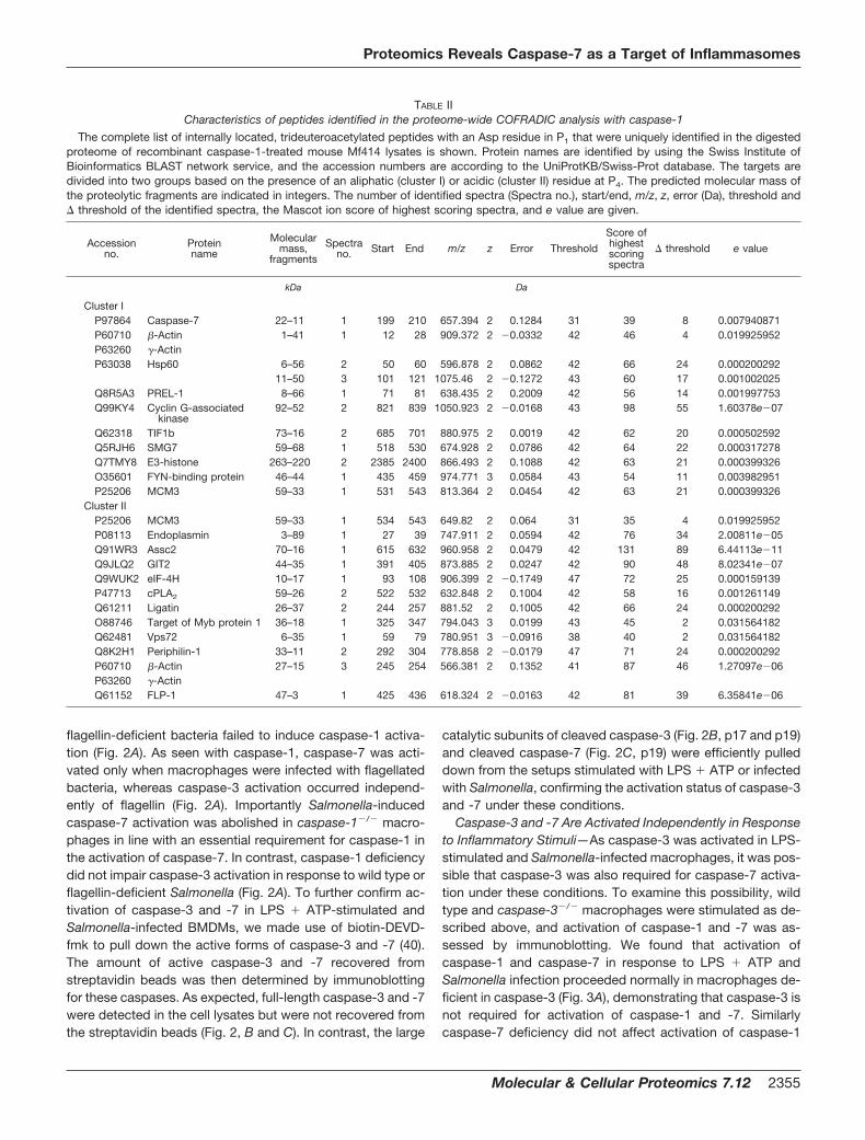
flagellin-deficient bacteria failed to induce caspase-1 activa-tion (Fig. 2A). As seen with caspase-1, caspase-7 was acti-vated only when macrophages were infected with flagellatedbacteria, whereas caspase-3 activation occurred independ-ently of flagellin (Fig. 2A). Importantly Salmonella-inducedcaspase-7 activation was abolished in caspase-1�/� macro-phages in line with an essential requirement for caspase-1 inthe activation of caspase-7. In contrast, caspase-1 deficiencydid not impair caspase-3 activation in response to wild type orflagellin-deficient Salmonella (Fig. 2A). To further confirm ac-tivation of caspase-3 and -7 in LPS � ATP-stimulated andSalmonella-infected BMDMs, we made use of biotin-DEVD-fmk to pull down the active forms of caspase-3 and -7 (40).The amount of active caspase-3 and -7 recovered fromstreptavidin beads was then determined by immunoblottingfor these caspases. As expected, full-length caspase-3 and -7were detected in the cell lysates but were not recovered fromthe streptavidin beads (Fig. 2, B and C). In contrast, the large
catalytic subunits of cleaved caspase-3 (Fig. 2B, p17 and p19)and cleaved caspase-7 (Fig. 2C, p19) were efficiently pulleddown from the setups stimulated with LPS � ATP or infectedwith Salmonella, confirming the activation status of caspase-3and -7 under these conditions.
Caspase-3 and -7 Are Activated Independently in Responseto Inflammatory Stimuli—As caspase-3 was activated in LPS-stimulated and Salmonella-infected macrophages, it was pos-sible that caspase-3 was also required for caspase-7 activa-tion under these conditions. To examine this possibility, wildtype and caspase-3�/� macrophages were stimulated as de-scribed above, and activation of caspase-1 and -7 was as-sessed by immunoblotting. We found that activation ofcaspase-1 and caspase-7 in response to LPS � ATP andSalmonella infection proceeded normally in macrophages de-ficient in caspase-3 (Fig. 3A), demonstrating that caspase-3 isnot required for activation of caspase-1 and -7. Similarlycaspase-7 deficiency did not affect activation of caspase-1
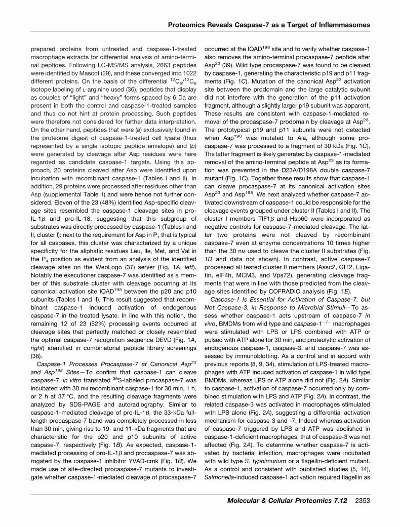
TABLE IICharacteristics of peptides identified in the proteome-wide COFRADIC analysis with caspase-1
The complete list of internally located, trideuteroacetylated peptides with an Asp residue in P1 that were uniquely identified in the digestedproteome of recombinant caspase-1-treated mouse Mf414 lysates is shown. Protein names are identified by using the Swiss Institute ofBioinformatics BLAST network service, and the accession numbers are according to the UniProtKB/Swiss-Prot database. The targets aredivided into two groups based on the presence of an aliphatic (cluster I) or acidic (cluster II) residue at P4. The predicted molecular mass ofthe proteolytic fragments are indicated in integers. The number of identified spectra (Spectra no.), start/end, m/z, z, error (Da), threshold and� threshold of the identified spectra, the Mascot ion score of highest scoring spectra, and e value are given.
Accessionno.
Proteinname
Molecularmass,
fragments
Spectrano. Start End m/z z Error Threshold
Score ofhighestscoringspectra
� threshold e value
kDa Da
Cluster IP97864 Caspase-7 22–11 1 199 210 657.394 2 0.1284 31 39 8 0.007940871P60710 �-Actin 1–41 1 12 28 909.372 2 �0.0332 42 46 4 0.019925952P63260 �-ActinP63038 Hsp60 6–56 2 50 60 596.878 2 0.0862 42 66 24 0.000200292
11–50 3 101 121 1075.46 2 �0.1272 43 60 17 0.001002025Q8R5A3 PREL-1 8–66 1 71 81 638.435 2 0.2009 42 56 14 0.001997753Q99KY4 Cyclin G-associated
kinase92–52 2 821 839 1050.923 2 �0.0168 43 98 55 1.60378e�07
Q62318 TIF1b 73–16 2 685 701 880.975 2 0.0019 42 62 20 0.000502592Q5RJH6 SMG7 59–68 1 518 530 674.928 2 0.0786 42 64 22 0.000317278Q7TMY8 E3-histone 263–220 2 2385 2400 866.493 2 0.1088 42 63 21 0.000399326O35601 FYN-binding protein 46–44 1 435 459 974.771 3 0.0584 43 54 11 0.003982951P25206 MCM3 59–33 1 531 543 813.364 2 0.0454 42 63 21 0.000399326
Cluster IIP25206 MCM3 59–33 1 534 543 649.82 2 0.064 31 35 4 0.019925952P08113 Endoplasmin 3–89 1 27 39 747.911 2 0.0594 42 76 34 2.00811e�05Q91WR3 Assc2 70–16 1 615 632 960.958 2 0.0479 42 131 89 6.44113e�11Q9JLQ2 GIT2 44–35 1 391 405 873.885 2 0.0247 42 90 48 8.02341e�07Q9WUK2 eIF-4H 10–17 1 93 108 906.399 2 �0.1749 47 72 25 0.000159139P47713 cPLA2 59–26 2 522 532 632.848 2 0.1004 42 58 16 0.001261149Q61211 Ligatin 26–37 2 244 257 881.52 2 0.1005 42 66 24 0.000200292O88746 Target of Myb protein 1 36–18 1 325 347 794.043 3 0.0199 43 45 2 0.031564182Q62481 Vps72 6–35 1 59 79 780.951 3 �0.0916 38 40 2 0.031564182Q8K2H1 Periphilin-1 33–11 2 292 304 778.858 2 �0.0179 47 71 24 0.000200292P60710 �-Actin 27–15 3 245 254 566.381 2 0.1352 41 87 46 1.27097e�06P63260 �-ActinQ61152 FLP-1 47–3 1 425 436 618.324 2 �0.0163 42 81 39 6.35841e�06
Proteomics Reveals Caspase-7 as a Target of Inflammasomes
Molecular & Cellular Proteomics 7.12 2355

and -3 (Fig. 3B), indicating that these caspases do not requirecaspase-7 for their activation. As expected, the caspase-7antibody failed to detect immunoreactive bands in lysates ofcaspase-7-deficient macrophages, thus confirming its speci-ficity (Fig. 3B). These results indicate that the regulation ofcaspase-7 activation triggered by LPS and ATP or Salmonellais highly specific and relies on caspase-1 but not caspase-3.Reciprocally the activation of caspase-3 by microbial stimuli isindependent of caspase-1 and -7. These results demonstratethe existence of non-redundant molecular mechanisms gov-erning the activation of the executioner caspase-3 and -7 inresponse to microbial stimuli.
The Inflammasomes Control Activation of Caspase-7 butNot Caspase-3—ATP induces the activation of caspase-1 inLPS-stimulated macrophages through the Cryopyrin inflam-masome (8–10, 34), whereas Salmonella and flagellin requirecomponents of the Ipaf inflammasome to activate caspase-1(5, 7, 14). We made use of BMDMs lacking the NLR proteinsCryopyrin or Ipaf as well as macrophages deficient in theessential inflammasome adaptor ASC to examine whether theCryopyrin and Ipaf inflammasomes are required for caspase-7activation in response to microbial stimuli. As a control, theactivation of caspase-1 induced by stimulation with LPS andATP was abolished in ASC�/� and Cryopyrin�/� macro-
FIG. 1. Caspase-1 process pro-caspase-7 at canonical Asp23 andAsp198 sites. A, WebLogo analysis ofthe cleavage sites of cluster I (left) andcluster II (right) targets identified in a pro-teome-wide screening for caspase-1targets. B and C, 35S-labeled pro-caspase-7 and pro-IL-1� (B) or pro-caspase-7 and the indicated site-di-rected mutants (C) were incubated withbuffer or 30 nM caspase-1 for 1 h, andcleavage fragments were analyzed bySDS-PAGE and autoradiography. D andE, 35S-labeled cluster I (D) and cluster II(E) targets were incubated with 30 nM
caspase-7, and reaction products wereanalyzed by SDS-PAGE and autoradiog-raphy. Black arrows indicate full-lengthproteins, and white arrows mark cleav-age fragments. CASP1, caspase-1;CASP7, caspase-7; CTRL, control; WT,wild type.
Proteomics Reveals Caspase-7 as a Target of Inflammasomes
2356 Molecular & Cellular Proteomics 7.12
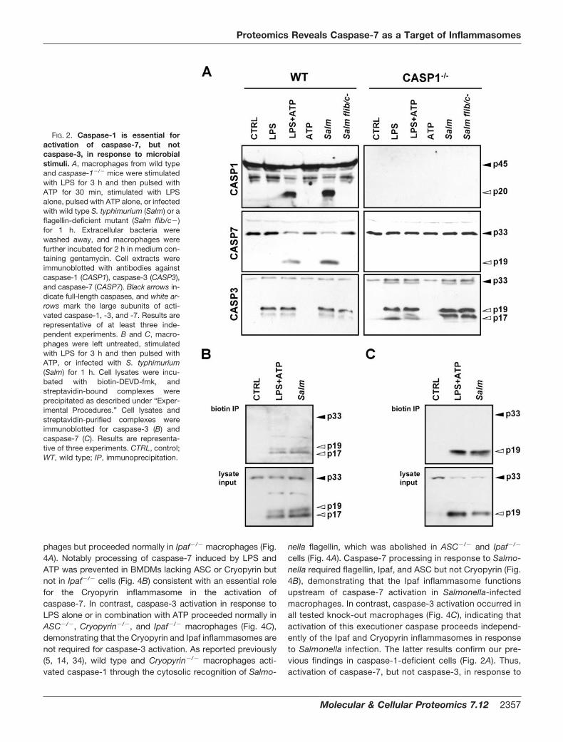
phages but proceeded normally in Ipaf�/� macrophages (Fig.4A). Notably processing of caspase-7 induced by LPS andATP was prevented in BMDMs lacking ASC or Cryopyrin butnot in Ipaf�/� cells (Fig. 4B) consistent with an essential rolefor the Cryopyrin inflammasome in the activation ofcaspase-7. In contrast, caspase-3 activation in response toLPS alone or in combination with ATP proceeded normally inASC�/�, Cryopyrin�/�, and Ipaf�/� macrophages (Fig. 4C),demonstrating that the Cryopyrin and Ipaf inflammasomes arenot required for caspase-3 activation. As reported previously(5, 14, 34), wild type and Cryopyrin�/� macrophages acti-vated caspase-1 through the cytosolic recognition of Salmo-
nella flagellin, which was abolished in ASC�/� and Ipaf�/�
cells (Fig. 4A). Caspase-7 processing in response to Salmo-nella required flagellin, Ipaf, and ASC but not Cryopyrin (Fig.4B), demonstrating that the Ipaf inflammasome functionsupstream of caspase-7 activation in Salmonella-infectedmacrophages. In contrast, caspase-3 activation occurred inall tested knock-out macrophages (Fig. 4C), indicating thatactivation of this executioner caspase proceeds independ-ently of the Ipaf and Cryopyrin inflammasomes in responseto Salmonella infection. The latter results confirm our pre-vious findings in caspase-1-deficient cells (Fig. 2A). Thus,activation of caspase-7, but not caspase-3, in response to
FIG. 2. Caspase-1 is essential foractivation of caspase-7, but notcaspase-3, in response to microbialstimuli. A, macrophages from wild typeand caspase-1�/� mice were stimulatedwith LPS for 3 h and then pulsed withATP for 30 min, stimulated with LPSalone, pulsed with ATP alone, or infectedwith wild type S. typhimurium (Salm) or aflagellin-deficient mutant (Salm flib/c�)for 1 h. Extracellular bacteria werewashed away, and macrophages werefurther incubated for 2 h in medium con-taining gentamycin. Cell extracts wereimmunoblotted with antibodies againstcaspase-1 (CASP1), caspase-3 (CASP3),and caspase-7 (CASP7). Black arrows in-dicate full-length caspases, and white ar-rows mark the large subunits of acti-vated caspase-1, -3, and -7. Results arerepresentative of at least three inde-pendent experiments. B and C, macro-phages were left untreated, stimulatedwith LPS for 3 h and then pulsed withATP, or infected with S. typhimurium(Salm) for 1 h. Cell lysates were incu-bated with biotin-DEVD-fmk, andstreptavidin-bound complexes wereprecipitated as described under “Exper-imental Procedures.” Cell lysates andstreptavidin-purified complexes wereimmunoblotted for caspase-3 (B) andcaspase-7 (C). Results are representa-tive of three experiments. CTRL, control;WT, wild type; IP, immunoprecipitation.
Proteomics Reveals Caspase-7 as a Target of Inflammasomes
Molecular & Cellular Proteomics 7.12 2357

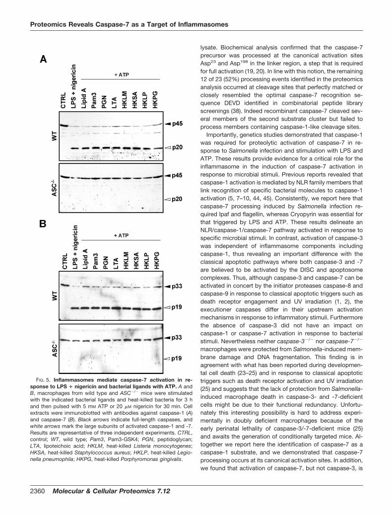
LPS and ATP requires the assembly of a functionalCryopyrin inflammasome, whereas the Ipaf inflammasomeis required for caspase-7 activation in Salmonella-infectedmacrophages. In addition, activation of caspase-1 (Fig. 5A)and caspase-7 (Fig. 5B) was abolished in ASC-deficient
BMDMs stimulated with an array of microbial ligands andATP and in LPS � nigericin-treated macrophages, demon-strating that the inflammasome is also required for activa-tion of caspase-7 in response to these proinflammatorystimuli. Collectively these results demonstrate the activation
FIG. 3. Caspase-3 and -7 are acti-vated independently in response to mi-crobial stimuli. A and B, macrophagesfrom wild type and caspase-3�/� (A) orcaspase-7�/� (B) mice were stimulatedwith LPS for 3 h and then pulsed with ATPfor 30 min, stimulated with LPS alone,pulsed with ATP alone, or infected withwild type S. typhimurium (Salm) or aflagellin-deficient mutant (Salm flib/c�) for1 h. Cell extracts were immunoblottedwith antibodies against caspase-1(CASP1), caspase-3 (CASP3), andcaspase-7 (CASP7). Black arrows indi-cate full-length caspases, and white ar-rows mark the large subunits of activatedcaspase-1, -3, and -7. Results are repre-sentative of at least three independent ex-periments. CTRL, control; WT, wild type.
Proteomics Reveals Caspase-7 as a Target of Inflammasomes
2358 Molecular & Cellular Proteomics 7.12
of an NLR-specific caspase-1/caspase-7 cascade in re-sponse to microbial stimuli.
Caspase-7 Deficiency Does Not Prevent Cytokine Produc-tion or Salmonella-induced Macrophage Cell Death—Caspase-1 is best known for its role in the proteolytic matu-ration of the proinflammatory cytokines IL-1� and IL-18 (3, 4,41–43). However, the mechanism by which the mature cyto-kines are secreted from the cytosol is unknown. To testwhether caspase-7 is essential for the secretion of IL-1� andIL-18 in response to LPS � ATP or Salmonella, we measuredthe levels of these cytokines in the culture supernatantsof stimulated macrophages. As expected, the combination ofLPS and ATP or Salmonella infection triggered the release ofsignificant levels of IL-1� and IL-18 in the culture superna-tants of wild type macrophages, whereas LPS or ATP aloneand flagellin-deficient Salmonella failed to do so (Fig. 6, A andB). The secretion of IL-1� and IL-18 triggered by LPS and ATPor Salmonella infection was abolished in caspase-1�/�,whereas cytokine levels comparable to those released by wildtype cells were detected in the culture supernatants ofcaspase-7�/� macrophages (Fig. 6, A and B). Similarly secre-tion of IL-1� (Fig. 6C) and tumor necrosis factor-� (Fig. 6D)was not affected in stimulated caspase-3-deficient macro-phages. These results suggest that although caspase-7 isactivated downstream of caspase-1 it is not required in thesecretion of the caspase-1-dependent cytokines IL-1� andIL-18.
Caspase-1 is not directly implicated in apoptosis (3) exceptfor the specialized form of macrophage cell death induced by
pathogenic bacteria such as Salmonella (15). As bothcaspase-3 and -7 were activated in response to Salmonellainfection (Fig. 2), we assessed the respective contribution ofthe executioner caspase-3 and -7 in Salmonella-inducedmacrophage cell death. As expected, substantial cell death ofwild type macrophages infected with wild type Salmonella,but not with the flagellin-deficient Salmonella mutant, wasevident 2 h after infection. In contrast to caspase-1-deficientcells, caspase-3�/� and caspase-7�/� macrophages werenot protected from Salmonella-induced membrane damageand DNA fragmentation (Fig. 7, A and B). Therefore, the lackof protection from Salmonella-induced macrophage death incaspase-7-deficient cells might be due to functional redun-dancy with caspase-3 as occurs during developmental celldeath (23–25) and in response to classical apoptotic triggers(25).
DISCUSSION
Using a proteomics approach to search for novel caspase-1targets, we identified 11 cleavage events that occurred atsites closely matching the caspase-1 cleavage sites in pro-IL-1� and pro-IL-18. In addition to cytoskeletal proteins,chaperones, and transcription factors, the executionercaspase-7 was identified as a member of this substrate clus-ter with cleavage occurring at its canonical activation siteIQAD198 between the p20 and p10 subunits. These resultssuggest that this subgroup of substrates was directly pro-cessed by caspase-1 and that recombinant caspase-1 in-duced activation of endogenous caspase-7 in the treated
FIG. 4. Specific activation ofcaspase-7 by NLR inflammasomes.A–C, macrophages from wild type,ASC�/�, Ipaf�/�, and Cryopyrin�/� micewere stimulated with LPS for 3 h andthen pulsed with ATP for 30 min, stimu-lated with LPS alone, pulsed with ATPalone, or infected with wild type S. typhi-murium (Salm) or a flagellin-deficientmutant (Salm flib/c�) for 1 h. Extracellu-lar bacteria were washed away, andmacrophages were further incubated for2 h in medium containing gentamycin.Cell extracts were immunoblotted withantibodies against caspase-1 (CASP1)(A), caspase-7 (CASP7) (B), andcaspase-3 (CASP3) (C). Black arrows in-dicate full-length caspases, and whitearrows mark the large subunits of acti-vated caspase-1, -3, and -7. Results arerepresentative of at least three inde-pendent experiments. CTRL, control;WT, wild type.
Proteomics Reveals Caspase-7 as a Target of Inflammasomes
Molecular & Cellular Proteomics 7.12 2359
lysate. Biochemical analysis confirmed that the caspase-7precursor was processed at the canonical activation sitesAsp23 and Asp198 in the linker region, a step that is requiredfor full activation (19, 20). In line with this notion, the remaining12 of 23 (52%) processing events identified in the proteomicsanalysis occurred at cleavage sites that perfectly matched orclosely resembled the optimal caspase-7 recognition se-quence DEVD identified in combinatorial peptide libraryscreenings (38). Indeed recombinant caspase-7 cleaved sev-eral members of the second substrate cluster but failed toprocess members containing caspase-1-like cleavage sites.
Importantly, genetics studies demonstrated that caspase-1was required for proteolytic activation of caspase-7 in re-sponse to Salmonella infection and stimulation with LPS andATP. These results provide evidence for a critical role for theinflammasome in the induction of caspase-7 activation inresponse to microbial stimuli. Previous reports revealed thatcaspase-1 activation is mediated by NLR family members thatlink recognition of specific bacterial molecules to caspase-1activation (5, 7–10, 44, 45). Consistently, we report here thatcaspase-7 processing induced by Salmonella infection re-quired Ipaf and flagellin, whereas Cryopyrin was essential forthat triggered by LPS and ATP. These results delineate anNLR/caspase-1/caspase-7 pathway activated in response tospecific microbial stimuli. In contrast, activation of caspase-3was independent of inflammasome components includingcaspase-1, thus revealing an important difference with theclassical apoptotic pathways where both caspase-3 and -7are believed to be activated by the DISC and apoptosomecomplexes. Thus, although caspase-3 and caspase-7 can beactivated in concert by the initiator proteases caspase-8 andcaspase-9 in response to classical apoptotic triggers such asdeath receptor engagement and UV irradiation (1, 2), theexecutioner caspases differ in their upstream activationmechanisms in response to inflammatory stimuli. Furthermorethe absence of caspase-3 did not have an impact oncaspase-1 or caspase-7 activation in response to bacterialstimuli. Nevertheless neither caspase-3�/� nor caspase-7�/�
macrophages were protected from Salmonella-induced mem-brane damage and DNA fragmentation. This finding is inagreement with what has been reported during developmen-tal cell death (23–25) and in response to classical apoptotictriggers such as death receptor activation and UV irradiation(25) and suggests that the lack of protection from Salmonella-induced macrophage death in caspase-3- and -7-deficientcells might be due to their functional redundancy. Unfortu-nately this interesting possibility is hard to address experi-mentally in doubly deficient macrophages because of theearly perinatal lethality of caspase-3/-7-deficient mice (25)and awaits the generation of conditionally targeted mice. Al-together we report here the identification of caspase-7 as acaspase-1 substrate, and we demonstrated that caspase-7processing occurs at its canonical activation sites. In addition,we found that activation of caspase-7, but not caspase-3, is
FIG. 5. Inflammasomes mediate caspase-7 activation in re-sponse to LPS � nigericin and bacterial ligands with ATP. A andB, macrophages from wild type and ASC�/� mice were stimulatedwith the indicated bacterial ligands and heat-killed bacteria for 3 hand then pulsed with 5 mM ATP or 20 �M nigericin for 30 min. Cellextracts were immunoblotted with antibodies against caspase-1 (A)and caspase-7 (B). Black arrows indicate full-length caspases, andwhite arrows mark the large subunits of activated caspase-1 and -7.Results are representative of three independent experiments. CTRL,control; WT, wild type; Pam3, Pam3-GSK4; PGN, peptidoglycan;LTA, lipoteichoic acid; HKLM, heat-killed Listeria monocytogenes;HKSA, heat-killed Staphylococcus aureus; HKLP, heat-killed Legio-nella pneumophila; HKPG, heat-killed Porphyromonas gingivalis.
Proteomics Reveals Caspase-7 as a Target of Inflammasomes
2360 Molecular & Cellular Proteomics 7.12

controlled by the inflammasomes in LPS � ATP-stimulatedand in Salmonella-infected macrophages. These results es-tablish for the first time the existence of an inflammatoryNLR/caspase-1/caspase-7 cascade in macrophages anddemonstrate the existence of differential activation mecha-nisms for the executioner caspase-3 and -7 in response tobacterial stimuli.
Acknowledgments—We thank Anthony Coyle, Ethan Grant, andJohn Bertin (Millennium Pharmaceuticals) for generous supply ofCryopyrin�/�, Ipaf�/� and ASC�/� mice; Richard Flavell (Yale Schoolof Medicine) for caspase-1�/� mice; and Kevin Roth (University ofAlabama at Birmingham) for supplying femurs of caspase-3�/� mice.We also thank Joel Whitfield from the Cellular Immunology CoreFacility of the University of Michigan Cancer Center and NiklaasColaert at the Department of Medical Protein Research of theFlanders Institute for Biotechnology and Ghent University for techni-cal support.
* This work was supported, in whole or in part, by National Insti-tutes of Health Grants AI063331 and AI064748 (to G. N.). This work
FIG. 6. Caspase-3 and -7 are dispensable for LPS- and Salmonel-la-induced cytokine secretion. A–D, macrophages from wild type,caspase-1�/�, caspase-3�/�, and caspase-7�/� mice were stimulatedwith LPS for 3 h and then pulsed with ATP for 30 min, stimulated withLPS alone, pulsed with ATP alone, or infected with wild type S. typhi-murium (Salm) or a flagellin-deficient mutant (Salm Flib/c�) for 1 h. Cellculture medium was analyzed for release of IL-1� (A and C), IL-18 (B), ortumor necrosis factor-� (TNF-�) (D) by ELISA. CASP1, caspase-1;CASP3, caspase-3; CASP7, caspase-7; CTRL, control; WT, wild type.Results represent mean � standard deviation of triplicates and arerepresentative of these three independent experiments.
FIG. 7. Neither caspase-3 nor caspase-7 deficiency protectsfrom Salmonella-induced macrophage cell death. A and B, mac-rophages from wild type, caspase-1�/�, caspase-3�/�, and caspase-7�/� mice were infected with wild type S. typhimurium (Salm) or aflagellin-deficient mutant (Salm flib/c�) for 2 h at multiplicity of infec-tion 5. Extracellular bacteria were washed away, and cell death wasstudied by measuring membrane damage (Live/Dead assay, Invitro-gen) (A) and DNA fragmentation using ELISA (Roche Applied Science)(B) according to the manufacturers’ instructions. Results representmean � standard deviation of triplicates and are representative ofthree independent experiments. CASP1, caspase-1; CASP3,caspase-3; CASP7, caspase-7; CTRL, control; WT, wild type.
Proteomics Reveals Caspase-7 as a Target of Inflammasomes
Molecular & Cellular Proteomics 7.12 2361
was also supported by a grant of the Inter University Attraction Poles(IAP-Phase VI) (to J. V.), by grants from the European Union (MarieCurie, DeathTrain, Framework Program 6, Grant MRTN-CT-035624),Inter University Attraction Poles Grant IAP6/18, the Research Fund ofGhent University (Geconcerteerde Onderzoeksacties 12.0505.02), theFonds voor Wetenschappelijk Onderzoek-Vlaanderen (Grant2G.0218.06), and the Belgian Foundation against Cancer(SCIE2003-48 Research) (to P. V.). The costs of publication of thisarticle were defrayed in part by the payment of page charges. Thisarticle must therefore be hereby marked “advertisement” in accord-ance with 18 U.S.C. Section 1734 solely to indicate this fact.
□S The on-line version of this article (available at http://www.mcponline.org) contains supplemental material.
§ Both authors contributed equally to this work.¶ Present address: Dept. of Physiological Chemistry, Genentech,
South San Francisco, CA 94080.� Present address: Dept. of Immunology, St. Jude Children’s Re-
search Hospital, Memphis, TN 38105.�� To whom correspondence should be addressed: Dept. of Pathol-
ogy, University of Michigan Medical School, 4215 CCGC, 1500 E.Medical Center Dr., Ann Arbor, Michigan 48109. Tel.: 734-764-8514;Fax: 734-647-9654; E-mail: [email protected].
REFERENCES
1. Nicholson, D. W. (1999) Caspase structure, proteolytic substrates, andfunction during apoptotic cell death. Cell Death Differ. 6, 1028–1042
2. Lamkanfi, M., Declercq, W., Kalai, M., Saelens, X., and Vandenabeele, P.(2002) A phylogenetic analysis of caspases from worm to man. CellDeath Differ. 9, 358–361
3. Li, P., Allen, H., Banerjee, S., Franklin, S., Herzog, L., Johnston, C., Mc-Dowell, J., Paskind, M., Rodman, L., Salfeld, J., Towne, E., Tracey, D.,Wardwell, S., Wei, F.-Y., Wong, W., Kamen, R., and Seshadriand, T.(1995) Mice deficient in IL-1�-converting enzyme are defective in pro-duction of mature IL-1� and resistant to endotoxic shock. Cell 80,401–411
4. Kuida, K., Lippke, J. A., Ku, G., Harding, M. W., Livingston, D. J., Su,M. S., and Flavell, R. A. (1995) Altered cytokine export and apoptosisin mice deficient in interleukin-1� converting enzyme. Science 267,2000–2003
5. Franchi, L., Amer, A., Body-Malapel, M., Kanneganti, T. D., Ozoren, N.,Jagirdar, R., Inohara, N., Vandenabeele, P., Bertin, J., Coyle, A., Grant,E. P., and Nunez, G. (2006) Cytosolic flagellin requires Ipaf for activationof caspase-1 and interleukin 1� in Salmonella-infected macrophages.Nat. Immunol. 7, 576–582
6. Lamkanfi, M., Kanneganti, T. D., Franchi, L., and Nunez, G. (2007)Caspase-1 inflammasomes in infection and inflammation. J. Leukoc.Biol. 82, 220–225
7. Mariathasan, S., Newton, K., Monack, D. M., Vucic, D., French, D. M., Lee,W. P., Roose-Girma, M., Erickson, S., and Dixit, V. M. (2004) Differentialactivation of the inflammasome by caspase-1 adaptors ASC and Ipaf.Nature 430, 213–218
8. Mariathasan, S., Weiss, D. S., Newton, K., McBride, J., O’Rourke, K.,Roose-Girma, M., Lee, W. P., Weinrauch, Y., Monack, D. M., and Dixit,V. M. (2006) Cryopyrin activates the inflammasome in response to toxinsand ATP. Nature 440, 228–232
9. Sutterwala, F. S., Ogura, Y., Szczepanik, M., Lara-Tejero, M., Lichten-berger, G. S., Grant, E. P., Bertin, J., Coyle, A. J., Galan, J. E., Askenase,P. W., and Flavell, R. A. (2006) Critical role for NALP3/CIAS1/Cryopyrin ininnate and adaptive immunity through its regulation of caspase-1. Im-munity 24, 317–327
10. Kanneganti, T. D., Ozoren, N., Body-Malapel, M., Amer, A., Park, J. H.,Franchi, L., Whitfield, J., Barchet, W., Colonna, M., Vandenabeele, P.,Bertin, J., Coyle, A., Grant, E. P., Akira, S., and Nunez, G. (2006) BacterialRNA and small antiviral compounds activate caspase-1 throughcryopyrin/Nalp3. Nature 440, 233–236
11. Inohara, N., Chamaillard, M., McDonald, C., and Nunez, G. (2005) NOD-LRR proteins: role in host-microbial interactions and inflammatory dis-ease. Annu. Rev. Biochem. 74, 355–383
12. Ozoren, N., Masumoto, J., Franchi, L., Kanneganti, T. D., Body-Malapel, M.,
Erturk, I., Jagirdar, R., Zhu, L., Inohara, N., Bertin, J., Coyle, A., Grant,E. P., and Nunez, G. (2006) Distinct roles of TLR2 and the adaptor ASCin IL-1�/IL-18 secretion in response to Listeria monocytogenes. J. Im-munol. 176, 4337–4342
13. Kanneganti, T. D., Body-Malapel, M., Amer, A., Park, J. H., Whitfield, J.,Franchi, L., Taraporewala, Z. F., Miller, D., Patton, J. T., Inohara, N., andNunez, G. (2006) Critical role for Cryopyrin/Nalp3 in activation ofcaspase-1 in response to viral infection and double-stranded RNA.J. Biol. Chem. 281, 36560–36568
14. Miao, E. A., Alpuche-Aranda, C. M., Dors, M., Clark, A. E., Bader, M. W.,Miller, S. I., and Aderem, A. (2006) Cytoplasmic flagellin activatescaspase-1 and secretion of interleukin 1� via Ipaf. Nat. Immunol. 7,569–575
15. Hersh, D., Monack, D. M., Smith, M. R., Ghori, N., Falkow, S., and Zych-linsky, A. (1999) The Salmonella invasin SipB induces macrophage apo-ptosis by binding to caspase-1. Proc. Natl. Acad. Sci. U. S. A. 96,2396–2401
16. Boatright, K. M., Renatus, M., Scott, F. L., Sperandio, S., Shin, H., Peder-sen, I. M., Ricci, J. E., Edris, W. A., Sutherlin, D. P., Green, D. R., andSalvesen, G. S. (2003) A unified model for apical caspase activation. Mol.Cell 11, 529–541
17. Salvesen, G. S., and Dixit, V. M. (1999) Caspase activation: the induced-proximity model. Proc. Natl. Acad. Sci. U. S. A. 96, 10964–10967
18. Shi, Y. (2004) Caspase activation: revisiting the induced proximity model.Cell 117, 855–858
19. Chai, J., Wu, Q., Shiozaki, E., Srinivasula, S. M., Alnemri, E. S., and Shi, Y.(2001) Crystal structure of a procaspase-7 zymogen: mechanisms ofactivation and substrate binding. Cell 107, 399–407
20. Stennicke, H. R., Jurgensmeier, J. M., Shin, H., Deveraux, Q., Wolf, B. B.,Yang, X., Zhou, Q., Ellerby, H. M., Ellerby, L. M., Bredesen, D., Green,D. R., Reed, J. C., Froelich, C. J., and Salvesen, G. S. (1998) Pro-caspase-3 is a major physiologic target of caspase-8. J. Biol. Chem. 273,27084–27090
21. Fischer, U., Janicke, R. U., and Schulze-Osthoff, K. (2003) Many cuts toruin: a comprehensive update of caspase substrates. Cell Death Differ.10, 76–100
22. Timmer, J. C., and Salvesen, G. S. (2007) Caspase substrates. Cell DeathDiffer. 14, 66–72
23. Zheng, T. S., Hunot, S., Kuida, K., Momoi, T., Srinivasan, A., Nicholson,D. W., Lazebnik, Y., and Flavell, R. A. (2000) Deficiency in caspase-9 orcaspase-3 induces compensatory caspase activation. Nat. Med. 6,1241–1247
24. Houde, C., Banks, K. G., Coulombe, N., Rasper, D., Grimm, E., Roy, S.,Simpson, E. M., and Nicholson, D. W. (2004) Caspase-7 expandedfunction and intrinsic expression level underlies strain-specific brainphenotype of caspase-3-null mice. J. Neurosci. 24, 9977–9984
25. Lakhani, S. A., Masud, A., Kuida, K., Porter, G. A., Jr., Booth, C. J., Mehal,W. Z., Inayat, I., and Flavell, R. A. (2006) Caspases 3 and 7: key medi-ators of mitochondrial events of apoptosis. Science 311, 847–851
26. Staes, A., Van Damme, P., Helsens, K., Demol, H., Vandekerckhove, J., andGevaert, K. (2008) Improved recovery of proteome-informative, proteinN-terminal peptides by combined fractional diagonal chromatography(COFRADIC). Proteomics 8, 1362–1370
27. Gevaert, K., Goethals, M., Martens, L., Van Damme, J., Staes, A., Thomas,G. R., and Vandekerckhove, J. (2003) Exploring proteomes and analyzingprotein processing by mass spectrometric identification of sorted N-terminal peptides. Nat. Biotechnol. 21, 566–569
28. Staes, A., Timmerman, E., Van Damme, J., Helsens, K., Vandekerckhove,J., Vollmer, M., and Gevaert, K. (2007) Assessing a novel microfluidicinterface for shotgun proteome analyses. J. Sep. Sci. 30, 1468–1476
29. Perkins, D. N., Pappin, D. J., Creasy, D. M., and Cottrell, J. S. (1999)Probability-based protein identification by searching sequence data-bases using mass spectrometry data. Electrophoresis 20, 3551–3567
30. Van Damme, P., Martens, L., Van Damme, J., Hugelier, K., Staes, A.,Vandekerckhove, J., and Gevaert, K. (2005) Caspase-specific and non-specific in vivo protein processing during Fas-induced apoptosis. Nat.Methods 2, 771–777
31. Vande Walle, L., Van Damme, P., Lamkanfi, M., Saelens, X., Vandekerck-hove, J., Gevaert, K., and Vandenabeele, P. (2007) Proteome-wide iden-tification of HtrA2/Omi substrates. J. Proteome Res. 6, 1006–1015
32. Martens, L., Vandekerckhove, J., and Gevaert, K. (2005) DBToolkit: proc-
Proteomics Reveals Caspase-7 as a Target of Inflammasomes
2362 Molecular & Cellular Proteomics 7.12
essing protein databases for peptide-centric proteomics. Bioinformatics21, 3584–3585
33. Elias, J. E., and Gygi, S. P. (2007) Target-decoy search strategy for in-creased confidence in large-scale protein identifications by mass spec-trometry. Nat. Methods 4, 207–214
34. Kanneganti, T. D., Lamkanfi, M., Kim, Y. G., Chen, G., Park, J. H.,Franchi, L., Vandenabeele, P., and Nunez, G. (2007) Pannexin-1-me-diated recognition of bacterial molecules activates the cryopyrin in-flammasome independent of Toll-like receptor signaling. Immunity 26,433–443
35. Lamkanfi, M., Kalai, M., Saelens, X., Declercq, W., and Vandenabeele, P.(2004) Caspase-1 activates nuclear factor of the �-enhancer in B cellsindependently of its enzymatic activity. J. Biol. Chem. 279,24785–24793
36. Ong, S. E., Blagoev, B., Kratchmarova, I., Kristensen, D. B., Steen, H.,Pandey, A., and Mann, M. (2002) Stable isotope labeling by amino acidsin cell culture, SILAC, as a simple and accurate approach to expressionproteomics. Mol. Cell. Proteomics 1, 376–386
37. Crooks, G. E., Hon, G., Chandonia, J. M., and Brenner, S. E. (2004)WebLogo: a sequence logo generator. Genome Res. 14, 1188–1190
38. Thornberry, N. A., Rano, T. A., Peterson, E. P., Rasper, D. M., Timkey, T.,Garcia-Calvo, M., Houtzager, V. M., Nordstrom, P. A., Roy, S., Vaillan-court, J. P., Chapman, K. T., and Nicholson, D. W. (1997) A combinatorialapproach defines specificities of members of the caspase family andgranzyme B. Functional relationships established for key mediators ofapoptosis. J. Biol. Chem. 272, 17907–17911
39. Denault, J. B., Bekes, M., Scott, F. L., Sexton, K. M., Bogyo, M., andSalvesen, G. S. (2006) Engineered hybrid dimers: tracking the activationpathway of caspase-7. Mol. Cell 23, 523–533
40. Grabarek, J., Amstad, P., and Darzynkiewicz, Z. (2002) Use of fluorescentlylabeled caspase inhibitors as affinity labels to detect activated caspases.Hum. Cell 15, 1–12
41. Ghayur, T., Banerjee, S., Hugunin, M., Butler, D., Herzog, L., Carter, A.,Quintal, L., Sekut, L., Talanian, R., Paskind, M., Wong, W., Kamen, R.,Tracey, D., and Allen, H. (1997) Caspase-1 processes IFN-�-inducingfactor and regulates LPS-induced IFN-� production. Nature 386,619–623
42. Thornberry, N. A., Bull, H. G., Calaycay, J. R., Chapman, K. T., Howard,A. D., Kostura, M. J., Miller, D. K., Molineaux, S. M., Weidner, J. R.,Aunins, J., Elliston, K. O., Ayala, J. M., Casanoparalle, F. J., Chin, J.,Ding, G. J.-F., Egger, L. A., Gaffney, E. P., Limjuco, G., Palyha, O. C.,Raju, S. M., Rolandoparallel, A. M., Salley, J. P., Yamin, T.-T., Lee,T. D., Shively, J. E., MacCross, M., Mumford, R. A., Schmidt, J. A., andTocciparallel, M. J. (1992) A novel heterodimeric cysteine protease isrequired for interleukin-1� processing in monocytes. Nature 356,768–774
43. Cerretti, D. P., Kozlosky, C. J., Mosley, B., Nelson, N., Van Ness, K.,Greenstreet, T. A., March, C. J., Kronheim, S. R., Druck, T., Cannizzaro,L. A., Huebner, K., and Black, R. A. (1992) Molecular cloning of theinterleukin-1� converting enzyme. Science 256, 97–100
44. Agostini, L., Martinon, F., Burns, K., McDermott, M. F., Hawkins, P. N., andTschopp, J. (2004) NALP3 forms an IL-1�-processing inflammasomewith increased activity in Muckle-Wells autoinflammatory disorder. Im-munity 20, 319–325
45. Martinon, F., Burns, K., and Tschopp, J. (2002) The inflammasome: amolecular platform triggering activation of inflammatory caspases andprocessing of proIL-�. Mol. Cell 10, 417–426
Proteomics Reveals Caspase-7 as a Target of Inflammasomes
Molecular & Cellular Proteomics 7.12 2363