dgps survey, boundary demarcation and fixation of monoliths ...
Upload
independentCategory
view
5download
0
925 (2001) 251–263Journal of Chromatography A,www.elsevier.com/ locate /chroma
Reversed-phase electrochromatography of amino acids and peptidesusing porous polymer monoliths
1 2´Renee Shediac, Sarah M. Ngola , Daniel J. Throckmorton, Deon S. Anex ,*Timothy J. Shepodd, Anup K. Singh
Chemical & Radiation Detection Laboratories, Sandia National Laboratories, Livermore, CA 94551-0969, USA
Received 20 March 2001; received in revised form 29 May 2001; accepted 6 June 2001
Abstract
Efficient and rapid separation of minute levels of amino acids and bioactive peptides is of significant importance in theemerging field of proteomics as well as in the clinical and pharmaceutical arena. We have developed novel UV-initiatedacrylate-based porous polymer monoliths as stationary phases for capillary- and chip-electrochromatography of cationic,anionic, and neutral amino acids and peptides, followed by absorbance or laser-induced fluorescence detection. The rigidmonoliths are cast-to-shape and are tunable for charge and hydrophobicity. For separations at low pH, monoliths containingquaternary amine moieties were used to achieve high electroosmotic flow, and for high pH separations monoliths with acidicsulfonic acid groups were employed. Efficient and reproducible separations of phenylthiohydantoin-labeled amino acids,native peptides, and amino acids and peptides labeled with naphthalene-2,3-dicarboxaldehyde (NDA) were achieved usingboth negatively- and positively-charged polymer monoliths in capillaries. Separation efficiencies in the range of 65 000–371 000 plates /m were obtained with capillary electrochromatography. Buffer composition and the degree of columnhydrophobicity were studied systematically to optimize separations. The monoliths were also cast in the microchannels ofglass chips and electrochromatographic separation followed by laser-induced fluorescence detection of three NDA-labeledbioactive peptides was obtained. 2001 Elsevier Science B.V. All rights reserved.
Keywords: Electrochromatography; Monolithic columns; Stationary phases, electrochromatography; Derivatization, electro-chromatography; Chip technology; Amino acids; Peptides
1. Introduction acids and peptides has important implications foramino acid analysis, peptide sequencing, protein
Achieving high-resolution separations of amino structure determination, and the rapidly emergingfield of proteomics. High-performance liquid chro-matography (HPLC) has been the method of choice
*Corresponding author. Mailstop 9951, Sandia National Lab- for separation of biological molecules [1,2] butoratories, Livermore, CA 94551-0969, USA. Tel.: 11-925-294- recently significant progress has been made towards1260; fax: 11-925-294-3020. employing capillary electrochromatography (CEC)
E-mail address: [email protected] (A.K. Singh).1 for separation of proteins [3–7], peptides [3,5,8,9],Present address: LumiCyte, Fremont, CA 94538-6532, USA.2 and amino acids [10–13]. Electrochromatography, aPresent address: Eksigent Technologies, Livermore, CA
94550, USA. powerful technique that combines the strengths of
0021-9673/01/$ – see front matter 2001 Elsevier Science B.V. All rights reserved.PI I : S0021-9673( 01 )01036-6

925 (2001) 251–263252 R. Shediac et al. / J. Chromatogr. A
capillary zone electrophoresis (CZE) and liquid of analytes in stationary and mobile phases. Sincechromatography, is especially attractive for develop- biological molecules such as peptides and proteinsment of portable analysis systems as it is readily have multiple ionizable groups that are charged andamenable to miniaturization. Traditionally, CEC has exhibit varying degrees of polarity, CEC is a poten-been performed using fused-silica capillaries packed tially powerful tool for their biochemical analysis. Inwith spherical silica particles. There are a number of this work, we report the separation of neutral,drawbacks associated with packed capillaries, such cationic, and anionic amino acids and peptides byas the time and effort required for packing, the CEC using acrylate-based UV-initiated hydrophobicnecessity for frits, and the potential leakage of porous polymers as monolithic stationary phases.particles. Packing of channels in a chip is con- Depending on the pH of the mobile phase and thesiderably more tedious and requires microfabrication nature of the analytes, either cationic or anionicof geometrical features to serve as frits. In contrast, groups were incorporated into the monoliths forin situ casting of polymer monoliths in capillaries generation of EOF. Selectivity was easily tuned byand microchannels reproducibly affords relatively manipulating the degree of polymer hydrophobicity.uniform packed beds, therefore eliminating the dif- Chip electrochromatography using in situ cast poly-ficulties associated with packing silica beads and the mer monoliths was employed to separate threeneed for retaining frits. The availability of a wide fluorescently-labeled bioactive peptides, demonstra-range of monomers enables critical stationary phase ting the applicability of these materials as separationproperties such as charge and hydrophobicity to be media in microfluidic devices.easily tuned to meet the specific demands of separat-ing many types of analytes. In situ polymerizationcan be thermal- or photo-initiated but the latter has 2. Experimentalbeen reported to produce columns with higher sepa-ration efficiencies and is also more suitable for 2.1. Apparatuscasting patterned monoliths in microfabricated chan-nels [8]. In the last few years, owing to their unique Polymerization was performed in a Spectrolinkerproperties, porous polymer monoliths have been XL-1500 UV crosslinker (Westbury, NY, USA)utilized as stationary phases for CEC, pioneered by operated at 365 nm, according to a procedure
´Hjerten and coworkers [4,14], and later modified by described elsewhere [24]. CEC, capillary electro-´several groups including Frechet and coworkers [15– phoresis (CE), and chip electrochromatography ex-
´20], Novotny [21], and Horvath and coworkers periments were performed using a Bertan 30 kV[7,22,23]. Acrylate-based polymer monoliths de- current-limited, adjustable d.c. power supply (Hicks-veloped in our laboratory [24] require a very short ville, NY, USA). UV absorbance was monitored(5–20 min) cure time under UV irradiation and using a Linear 200 detector (San Jose, CA, USA) setsupport sufficient EOF as cast (no pressure flushing at 214 nm. Laser-induced fluorescence (LIF) de-is necessary), making them especially suitable for tection was performed using a 413 nm krypton ionchip-based application. These materials have re- laser (Coherent, Santa Clara, CA, USA). A chipproducibly demonstrated high efficiencies station was built in the laboratory, and both capillary(.150 000 plates /m) for CEC separation of neutral and chip systems were enclosed in a Plexiglas boxaromatic compounds but their applicability as sepa- fitted with an interlock system to avoid electricalration media for charged analytes has not been shock. The laser was focused onto the detectioninvestigated in detail. window of the capillary or the microchip and
To date, CEC has been predominantly applied to fluorescence was collected perpendicular to theseparation of neutral molecules where electrophoretic incident beam by using a high numerical aperturemobility is not a factor. In CEC of charged mole- microscope objective (Nikon) having 403 magnifi-cules, the separation mechanism couples two cation, an NA of 0.85, and a 0.37 mm workingsimultaneous phenomena — the electrophoretic mi- distance. The background fluorescence and the scat-gration of analytes, and the differential partitioning tered light were minimized by using a long-pass filter

925 (2001) 251–263 253R. Shediac et al. / J. Chromatogr. A
and a variable slit. The fluorescence was collected by from Microchrome Technologies (San Jose, CA,a photomultiplier tube (PMT; Hamamatsu, Japan) USA). High purity sulfuric acid and hydrofluoricand the signal was amplified with a lock-in amplifier acid were obtained from Ashland (Norwalk, CT,(Stanford Research Systems, Sunnyvale, CA, USA). USA), and acetone and ammonium hydroxide wereThe current output from the amplifier was recorded purchased from General Chemical (Parsippany, NJ).using a DAQ board in conjunction with a programwritten in Labview (National Instruments, Austin, 2.3. Polymer stationary phasesTX, USA). All experiments were conducted atambient temperatures. Prior to in situ polymerization, the walls of
Teflon-coated fused-silica capillaries (100 mm capillaries and microchannels were silanized toI.D.) were purchased from Polymicro Technologies ensure covalent attachment of the polymer to the(Phoenix, AZ, USA). DB-WAX capillaries (100 mm substrate; this pretreatment has been previouslyI.D.) for CE experiments were obtained from J & W described [24]. Both positively- and negatively-Scientific (Folsom, CA, USA). Glass chips with a charged porous polymer monoliths containing butylsingle T-injector design were fabricated in the lab- (C ) and lauryl (C ) groups were prepared accord-4 12
oratory. Photomask layout was performed using ing to a previously described procedure [24]. In theAutoCAD 2000 (Autodesk, San Rafael, CA, USA) lauryl material, lauryl acrylate replaced 10% of theand the generated file was converted to GDS format. butyl monomer. The cellulose material was preparedThe photomask was manufactured by Photo Sciences from monomers in the following percentages: 1%(Torrance, CA, USA). Schott D263 glass wafers (acrylamidomethyl)cellulose acetate butyrate, 10%were purchased from S. I. Howard Glass (Worcester, tetrahydrofurfuryl (THF) acrylate, 1% [2-MA, USA). A Cooke (Cooke Vacuum Products, (acryloyloxy)ethyl]trimethylammonium methylsul-Norwalk, CT, USA) sputtering system was used to fate, 58% butyl acrylate, 30% 1,3-butanedioldeposit the metal etch mask. A Karl Suss (Karl Suss diacrylate. The glass capillaries were pretreated toAmerica, Waterbury Center, VA, USA) MA6 contact ensure adhesion of the polymer monoliths to themask aligner was used for photolithography. The wall. For the positive material, a positive pretreat-microchannel dimensions were 25 mm deep and 50 ment solution was used to reverse the EOF as well asmm wide; the separation channel length was 8.0 cm to promote adhesion.and the injection arms were 1.0 cm each.
2.4. CEC experiments2.2. Chemicals
The packed capillary was electrokinetically con-High-purity potassium phosphate, thiourea and ditioned using acetonitrile–5 mM Tris buffer pH 8
acetonitrile were used as received from Aldrich (80:20, v /v) or acetonitrile–25 mM phosphate buffer(Milwaukee, WI, USA). Sodium tetraborate was pH 2.8 (80:20, v /v) to remove residual monomericobtained from Sigma (St Louis, MO, USA). materials prior to use. A detection window in theNapthalene-2,3-dicarboxaldehyde (NDA) and potas- polymer was formed by exposure to 214 nm lightsium cyanide were purchased from Molecular Probes from a UV light source.(Eugene, OR, USA). All phenylthiohydantoin (PTH) For isocratic CEC separations of PTH-labeledamino acids and peptides were purchased from amino acids using UV detection, negatively-chargedSigma. Water was purified with an Ultra-Pure water butyl or lauryl monoliths (functionalized with sul-system from Millipore (Milford, MA, USA). fonic acid groups) were employed as stationary(Acrylamidomethyl)cellulose acetate butyrate was phases. The mobile phase was prepared by mixingobtained from Aldrich and used as received. Other the appropriate percentage of acetonitrile (by vol-acrylate monomers were obtained and purified as ume) in 25 mM phosphate solution pH 7.3 and waspreviously described [24]. OCG 825 photoresist and degassed by ultrasonication prior to use. The capil-OCG 934 developer were obtained from Arch lary was conditioned in the running buffer for(Columbus, OH, USA). Chrome etch was purchased approximately 1 h. Stock solutions of individual

925 (2001) 251–263254 R. Shediac et al. / J. Chromatogr. A
PTH–amino acids were prepared in water and aceto- peptides were labeled by adding a 40 ml aliquot ofnitrile and sample solutions were diluted appro- 0.3 mM peptide stock solution to 1 ml of 50 mMpriately with buffer solution to attain final amino borate buffer pH 9.5, followed by aliquots of 40 ml
24 23acid concentrations of 10 to 10 M. Thiourea of 4.8 mM KCN and 40 ml of 2.4 mM NDA. The(2.5 mM) was added to each sample mixture as the solutions were diluted with the run buffer to obtain
26 27unretained marker. Amino acid samples were in- final concentrations of 10 – 10 M. Peptidejected electrokinetically at the anode for 5 s at 2 kV samples were electrokinetically injected at theand separations were performed at constant field cathode for 30 s at 1.0 kV and separated at a constantstrengths of 150–300 V/cm. field strength of 1200 V/cm. LIF was employed as
For isocratic CEC separations of native peptides the means of detection. An algorithm developedusing UV detection, positively-charged butyl mono- in-house was used for background subtraction andliths (containing quaternary ammonium moieties) smoothing for all chromatographic data.were used as stationary phases. The mobile phase(acetonitrile–12.5 mM phosphate buffer pH 2.8; 2.5. CE experiments30:70, v /v) was degassed before use. The packedcapillary was conditioned in this running buffer for 1 Unpacked, coated DB-WAX capillaries were used
23h. Peptide stock solutions (10 M) were prepared in in order to eliminate EOF at the inner walls. Thewater and final peptide samples of approximately mobile phase (acetonitrile–12.5 mM phosphate buf-
2510 M were made by diluting with the running fer pH 2.8; 30:70, v /v) was degassed before use.25buffer. Peptide samples were electrokinetically in- Peptide sample solutions of approximately 10 M
jected at the cathode for 5 s at 1.5 kV and separated were hydrodynamically injected at the cathode for 10at constant field strengths of 90–200 V/cm. s and separated at constant field strengths of 90–200
For isocratic CEC separations of NDA-labeled V/cm.amino acids using LIF detection, negatively-chargedlauryl monoliths (functionalized with sulfonic acid 2.6. Chip fabricationgroups) were employed as stationary phases. Themobile phase was prepared by mixing the appro- Glass wafers were sputtered with chrome (200priate percentage of acetonitrile (by volume) in 25 nm) which served as the etch mask. A 1-mm-thickmM phosphate solution pH 7.2 and was degassed layer of OCG 825 positive photoresist was spin-prior to use. The packed capillary was conditioned in coated and soft-baked (908C, 5 min). After exposure,the running buffer for approximately 1 h. Stock the photoresist was developed with OCG 934 de-solutions of amino acids were prepared in water and veloper and hard-baked (1208C, 30 min). Exposedworking solutions of 0.3 mM were prepared by chrome was etched with a commercially availablediluting in 50 mM borate buffer pH 9.5. Deri- chrome etch and the subsequently exposed glass wasvatization was carried out by adding aliquots of 40 etched with 25% HF solution. The photoresist wasml of 4.8 mM KCN and 40 ml of 2.4 mM NDA to removed with acetone and the chrome mask wasthe 0.3 mM amino acid solution. The solutions were etched as described. Holes (1.0 mm diameter) werediluted with the run buffer to obtain final con- drilled in glass cover plates. The etched wafers and
28centrations of 10 M. Amino acid samples were cover plates were cleaned with H SO –H O2 4 2 2
injected electrokinetically at the anode for 2 s at 4 (3:1)(908C) and NH OH–H O–H O (1:5:1)(608C),4 2 2 2
kV and separations were performed at constant field aligned for contacting, and once contacted werestrengths of 200 V/cm. thermally bonded at 6508C.
For on-chip electrochromatographic separations ofpeptides, negatively-charged lauryl monoliths (con-taining sulfonic acid groups) were cast in the mi- 3. Results and discussioncrochannels. The mobile phase (acetonitrile–12.5mM phosphate buffer pH 7.3; 35:65, v /v) was 3.1. Porous polymer monolithsdegassed before use. The packed channels wereconditioned in this running buffer for 1 h. The Acrylate-based porous polymer monoliths de-

925 (2001) 251–263 255R. Shediac et al. / J. Chromatogr. A
veloped in our laboratory possess several properties In the negatively-charged material used for aminothat make them well-suited as reversed-phase chro- acid separations, a sulfonic acid monomer is incorpo-matographic media [24]. They are readily cured (5 rated to support EOF and the linear alkyl acrylatesmin in capillaries, and 10–20 min in microchannels) serve as hydrophobic sites for chromatographicby UV irradiation and do not require retaining frits retention. Scanning electron miscroscope (SEM)since they are covalently attached to the substrate micrographs indicate that the polymer nodule struc-surface. They support EOF as cast without the need ture is identical in both butyl and lauryl materialsto flush with pressure. They can be made from a [24]. In both monoliths, the peak pore diameter bywide variety of monomers, enabling the charge and porosimetry is approximately 1 mm and the surface
2hydrophobicity of the stationary phase to be easily area by BET analysis is 1–3 m /g [24]. Fig. 1a is atuned. High efficiencies (.150 000 plates /m) have representative SEM micrograph of a negative laurylbeen routinely obtained in the CEC separations of monolith.polyaromatic hydrocarbons and neutral aromatics In the positively-charged porous polymer mono-using negatively- and positively-charged materials liths used for peptide separations, a tetraalkylam-with varying degrees of hydrophobicity [24]. In this monium compound serves as the charged function-work, the applicability of both negative and positive ality. The polymer nodule structure is similar to thatpolymer monoliths in the CEC separation of charged observed for the negatively-charged materials, asanalytes was investigated. shown by SEM microscopy (Fig. 1b), and BET
Fig. 1. SEM micrographs of methanol-extracted samples of (a) negatively-charged lauryl monolith, (b) positively-charged butyl monolith,(c) positively-charged butyl monolith with double the percentage (1%) of charged monomer, and (d) positively-charged butyl monolith with1% cellulose.

925 (2001) 251–263256 R. Shediac et al. / J. Chromatogr. A
2surface area is 1–3 m /g. Unlike the negative pH 7.3; (40:60, v /v). As shown in Fig. 2, neutralmonoliths, however, the positive materials exhibit a hydrophilic amino acids elute first and all in thisrange of pore sizes between 1–2 mm without a group but PTH–Gln and PTH–Asn are baseline-distinct peak pore diameter (data not shown). In an resolved. The neutral hydrophobic amino acids areeffort to increase EOF, monoliths with twice the significantly more retained and elute much later, aspercentage of charged monomer were prepared with illustrated by PTH–Phe, PTH–Ile, PTH–Trp andno significant effect on the morphology as depicted PTH–Leu. PTH–Arg has an electrophoretic mobilityin Fig. 1c. Incorporation of 1% cellulose to the in the same direction as EOF, whereas PTH–Aspmonomer mixture resulted in slightly smaller poly- and PTH–Glu have electrophoretic mobilities thatmer nodule sizes (Fig. 1d). This is probably caused oppose EOF. Since the acidic amino acids eluteby solubility changes when including the more before PTH–Arg, we may conclude that the sepa-hydrophilic cellulose-based acrylate monomer in the ration mechanism under these conditions is domi-growing monolith network. nated by the hydrophobic interactions between the
analytes and the monolithic stationary phase rather3.2. Separation of PTH–amino acids than by CZE. For the charged amino acids, coulom-
bic interactions will also significantly influencePeptide sequencing via Edman degradation renders retention. The PTH–Arg peak is broader and more
the resultant PTH–amino acids neutral except for asymmetric because it is also partly retained byPTH–Arg, which is basic, and PTH–Asp and PTH– electrostatic interactions with the negatively chargedGlu, which are acidic. PTH–Thr exhibits two peaks, sulfonic acid groups of the monolith.as observed by others [13]. The isocratic CEC Separation efficiencies of up to 268 000 plates /mseparation of 20 PTH–amino acids was achieved are obtained with the lauryl column as shown inusing a lauryl (C ) monolithic column containing Table 1. These values are comparable to those12
highly acidic sulfonic acid groups. The mobile phase reported for conventional silica-based stationaryconsisted of acetonitrile–25 mM phosphate buffer at phases used to separate neutral and acidic PTH–
Fig. 2. Electrochromatographic separation of 20 PTH–amino acids on negatively-charged lauryl stationary phase in acetonitrile–25 mMphosphate pH 7.3 (40:60, v /v). UV detection at 214 nm. Field strength, 175 V/cm. Capillary: total length528.5 cm; length todetector517.5 cm; I.D.5100 mm.

925 (2001) 251–263 257R. Shediac et al. / J. Chromatogr. A
Table 1Retention times, capacity factors and column efficiencies for the CEC separation of five PTH–amino acids using lauryl and butyl monolithicstationary phases
Amino acid Lauryl Butyl
t k9 N t (min) k9 Nr r
(min) (plates /m) (plates /m)
PTH–Ala 18.7 1.41 176 000 10.6 1.07 127 000a aPTH–Asp 18.9 2 181 000 12.1 2 134 000a b a bPTH–Arg 36.8 2 46 000 17.5 2 14 000
PTH–Phe 58.3 6.50 238 000 24.8 3.84 120 000PTH–Trp 66.2 7.51 268 000 27.0 4.28 116 000
a k9 cannot be calculated for charged analytes because retention depends on both chromatographic retention and electrophoretic mobility.b 2The number of theoretical plates for the asymmetric PTH–Arg peak was obtained using the equation [22]: N 5 41.7 (t /w ) /(A /B 1r 0.1
1.25).
amino acids by CEC. Efficiencies in the range of column. However, while the PTH–Ala /PTH–Asp60 000–213 000 plates /m have been achieved with 3 and PTH–Arg/PTH–Lys pairs could not be well-mm ODS-modified silica particles [13] while values resolved on the lauryl column, they were wellof 176 000–530 000 plates /m have been obtained separated using the butyl column (data not shown).with 1.5 mm ODS-modified non-porous particles In addition, the elution order of PTH–Ala and PTH–[11]. Mercury porosimetry indicates that the peak Glu obtained with the lauryl column is reversed inpore size of our acrylate-based monoliths is approxi- the butyl stationary phase.mately 1 mm [24]. In phase-separated structures suchas ours, we often observe a correlation between 3.2.2. Effect of acetonitrile contentnodule size and pore size. The volume percentage of acetonitrile in the
mobile phase was found to dramatically affect the3.2.1. Effect of column hydrophobicity elution order of PTH–amino acids. A subset of
The degree of column hydrophobicity has a sig- amino acids was separated on a negative butyl (C )4
nificant effect on selectivity and on the quality of column using different volume ratios of acetonitrile–separations. Using a representative subset of PTH– 25 mM phosphate pH 7.3. Retention times wereamino acids, we compared the separation perform- normalized with respect to the EOF to give aance of a negative butyl (C ) monolithic column to retention factor t /t , which depends on both4 r eof
the negative lauryl (C ) material. Under identical hydrophobic retention as well as electrophoresis12
mobile phase conditions, column lengths and EOF (Fig. 3). In reversed-phase chromatography, sepa-velocities, we found that including 10% lauryl ration is based on the partition of analytes betweenacrylate in the porous polymer monolith increases the hydrophobic stationary phase and the relativelythe retention times of all amino acids, and the most polar mobile phase, hence increasing mobile phasedramatic increase in capacity factor was observed for polarity increases analyte retention. As shown in Fig.the most hydrophobic amino acids, PTH–Phe and 3, the retention times of the neutral amino acids,PTH–Trp (Table 1). A concomitant increase in particularly the most hydrophobic ones, increase asefficiencies was also noted and is probably due to the acetonitrile content in the mobile phase isdifferences in the morphologies of the butyl and lowered.lauryl materials. Resolution was generally greatly For the charged amino acids, however, the sepa-improved and well separated peaks were obtained for ration mechanism is comprised of an electrophoreticthe PTH–Gln and PTH–His pair as well as for the component as well as a chromatographic one. It isgroup of amino acids consisting of PTH–Gly, PTH– possible to alter the balance of these two separationTyr, PTH–Glu and PTH–Ala, none of which could components to achieve the desired resolution andbe baseline-resolved using the less hydrophobic analysis speed by changing the mobile phase com-

925 (2001) 251–263258 R. Shediac et al. / J. Chromatogr. A
3.3. Separation of NDA-labeled amino acids
Significantly lower limits of detection can beachieved using LIF to detect amino acids derivatizedwith a fluorogenic reagent. NDA reacts with primaryamines in the presence of cyanide to yield highlystable 1-cyano-2-alkylbenz[f]isoindole (CBI) adductswhich exhibit high fluorescence quantum yields [26–30]. Upon NDA-labeling, most amino acids possessa single negative charge with the exception of NDA–Glu and NDA–Asp, which possess two negativecharges, and NDA–Arg, which is neutral. Labelingdifficulties are associated with Lys and Cys due tointramolecular fluorescence quenching [31], and Procannot be labeled with NDA because it does notpossess a primary amine. Fig. 4 shows the isocraticCEC separation of 15 NDA-labeled amino acids
–8(concentrations of 10 M) employing sensitive LIFdetection and using a lauryl (C ) monolith con-Fig. 3. Plot of normalized retention factor, t / t , vs. percentage 12r eof
taining sulfonic acid groups and a mobile phase ofof acetonitrile for five PTH–amino acids separated on anegatively-charged butyl column. Capillary: total length525.0 acetonitrile–20 mM phosphate buffer at pH 7.2cm; length to detector515.2 cm; I.D.5100 mm. (15:85, v /v). NDA–Glu and NDA–Asp could be
separated (data not shown) on the same stationaryposition. At acetonitrile–25 mM phosphate (60:40), phase using a mobile phase of acetonitrile–12.5 mMPTH–Ala elutes first, followed by PTH–Lys, phosphate buffer at pH 7.2 (15:85, v /v) with re-positively-charged PTH–Arg, neutral hydrophobic tention times of 50.8 min and 58.4 min, respectivelyPTH–Phe and PTH–Trp, and finally negatively- (a lower buffer concentration was necessary tocharged PTH–Asp. This elution order suggests that achieve faster separations of these two doubly-nega-the amino acid separation is primarily due to thedifferences in electrophoretic mobility. CZE is still amajor component in the separation mechanism atacetonitrile–25 mM phosphate buffer (50:50), wherePTH–Arg elutes before PTH–Asp, but the degree ofretention of the hydrophobic amino acids is sig-nificantly increased as indicated by the higher t /tr eof
values. At a ratio of acetonitrile–25 mM phosphate(40:60), all the amino acids are significantly moreretained except PTH–Asp, which now elutes beforePTH–Arg, indicating that chromatographic interac-tions dominate the separation mechanism.
The acetonitrile content in the mobile phase alsoinfluences the EOF velocity, which is 6.81, 6.76, and5.12 mm/s at 60%, 50% and 40% acetonitrile,respectively. By increasing the percentage of ace-
Fig. 4. Electrochromatographic separation of 15 NDA–aminotonitrile, the electrolyte concentration is concurrentlyacids on negatively-charged lauryl stationary phase in
decreased, which results in an increase in the zeta acetonitrile–20 mM phosphate pH 7.2 (15:85, v /v). LIF detectionpotential and hence a corresponding increase in the at 413 nm. Field strength, 200 V/cm. Capillary: total length533.0EOF velocity [25]. cm; length to detector523.5 cm; I.D.5100 mm.
925 (2001) 251–263 259R. Shediac et al. / J. Chromatogr. A
tive species on the negatively charged stationaryphase). Fourteen of the 15 NDA-labeled amino acidsare well resolved with only NDA–Leu and NDA–Ilecoeluting, and efficiencies in the range of 65 000–371 000 plates /m were obtained. The resolution ofour NDA–amino acid separations compare favorablywith those achieved using gradient elution HPLC–LIF on C stationary phases [27,28,30,32], demon-18
strating the applicability of both CEC and polymermonolithic stationary phases in the separation ofcharged analytes.
3.4. Separation of basic bioactive peptides
Separation of peptides by CEC requires considera-tion of the nature of the charged groups on thestationary phase and the pH of the mobile phase.Peptides possess a characteristic isoelectric point
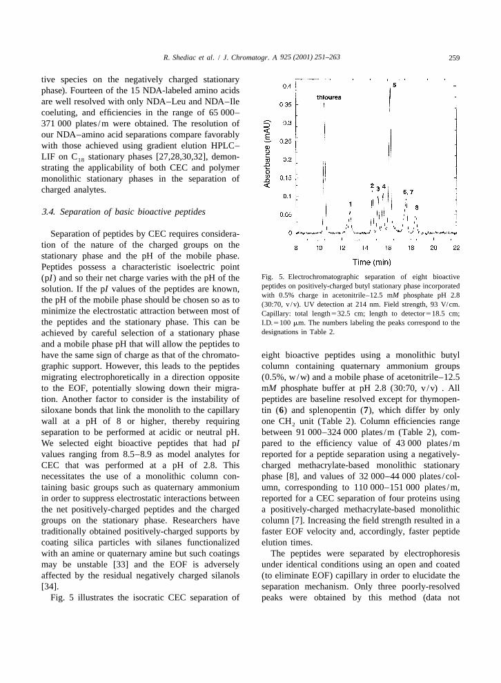
Fig. 5. Electrochromatographic separation of eight bioactive(pI) and so their net charge varies with the pH of thepeptides on positively-charged butyl stationary phase incorporatedsolution. If the pI values of the peptides are known,with 0.5% charge in acetonitrile–12.5 mM phosphate pH 2.8
the pH of the mobile phase should be chosen so as to (30:70, v /v). UV detection at 214 nm. Field strength, 93 V/cm.minimize the electrostatic attraction between most of Capillary: total length532.5 cm; length to detector518.5 cm;the peptides and the stationary phase. This can be I.D.5100 mm. The numbers labeling the peaks correspond to the
designations in Table 2.achieved by careful selection of a stationary phaseand a mobile phase pH that will allow the peptides tohave the same sign of charge as that of the chromato- eight bioactive peptides using a monolithic butylgraphic support. However, this leads to the peptides column containing quaternary ammonium groupsmigrating electrophoretically in a direction opposite (0.5%, w/w) and a mobile phase of acetonitrile–12.5to the EOF, potentially slowing down their migra- mM phosphate buffer at pH 2.8 (30:70, v /v) . Alltion. Another factor to consider is the instability of peptides are baseline resolved except for thymopen-siloxane bonds that link the monolith to the capillary tin (6) and splenopentin (7), which differ by onlywall at a pH of 8 or higher, thereby requiring one CH unit (Table 2). Column efficiencies range2
separation to be performed at acidic or neutral pH. between 91 000–324 000 plates /m (Table 2), com-We selected eight bioactive peptides that had pI pared to the efficiency value of 43 000 plates /mvalues ranging from 8.5–8.9 as model analytes for reported for a peptide separation using a negatively-CEC that was performed at a pH of 2.8. This charged methacrylate-based monolithic stationarynecessitates the use of a monolithic column con- phase [8], and values of 32 000–44 000 plates /col-taining basic groups such as quaternary ammonium umn, corresponding to 110 000–151 000 plates /m,in order to suppress electrostatic interactions between reported for a CEC separation of four proteins usingthe net positively-charged peptides and the charged a positively-charged methacrylate-based monolithicgroups on the stationary phase. Researchers have column [7]. Increasing the field strength resulted in atraditionally obtained positively-charged supports by faster EOF velocity and, accordingly, faster peptidecoating silica particles with silanes functionalized elution times.with an amine or quaternary amine but such coatings The peptides were separated by electrophoresismay be unstable [33] and the EOF is adversely under identical conditions using an open and coatedaffected by the residual negatively charged silanols (to eliminate EOF) capillary in order to elucidate the[34]. separation mechanism. Only three poorly-resolved
Fig. 5 illustrates the isocratic CEC separation of peaks were obtained by this method (data not

925 (2001) 251–263260 R. Shediac et al. / J. Chromatogr. A
Table 2Normalized retention factors and column efficiencies for the CEC separation of eight bioactive peptides using monolithic stationary phasesof different compositions
Peptide Name Sequence pI 0.5% charge 1% charge 1% charge11% cellulose
t /t N (plates /m) t /t N (plates /m) t /t N (plates /m)r eof r eof r eof
1 Leu–Enkephalin–Lys YGGFLK 8.59 1.21 92 000 1.25 99 000 1.26 116 0002 Met–Enkephalin–Arg–Phe YGGFMRF 8.75 1.40 284 000 1.48 108 000 1.49 213 0003 a-Casein, fragment 90–95 RYLGYL 8.59 1.45 241 000 1.55 2 1.57 2
4 Met–Enkephalin–Lys YGGFMK 8.59 1.49 324 000 1.55 2 1.57 2
5 b-Lipotropin, fragment 39–45 KKDSGPY 8.50 1.55 225 000 1.70 138 000 1.69 265 0006 Thymopentin RKDVY 8.59 1.68 2 1.87 171 000 1.87 330 0007 Splenopentin RKEVY 8.59 1.68 2 1.93 2 1.96 205 0008 Kyotorphin YR 8.75 1.76 177 000 1.93 2 1.91 274 000
shown). Leucine–enkephalin–lysine (1) and meth- and minimum and maximum values are the valuesionine–enkephalin–arginine–phenylalanine (2) co- obtained for the least retained peptide and the mostelute to give the first peak, methionine–enkephalin– retained peptide, respectively. Fig. 4 clearly showslysine (4) and b-lipotropin (fragment 39–45) (5) that the predicted elution order based on this modelco-elute as the second peak, and the remaining differs remarkably from what is actually observed,peptides comprise the third peak. indicating that CZE must play a significant role in
Retention of peptides in CEC is expected to the CEC separation.involve contributions from chromatographic interac- Furthermore, Fig. 6 illustrates that the elutiontions and electrophoretic mobilities of the solutes. In order follows closely to the order predicted for
2 / 3order to elucidate the relative importance of these electrophoretic migration by the ratio q /M , wherer
two contributions, we have used empirical models tocalculate retention and migration times for thepeptides. A theoretical retention order for smallpeptides composed of less than 15 amino acidresidues may be predicted from a summation ofhydrophobicity constants associated with the con-stituent amino acids for each peptide [35]:
t 5 A S D n 1 B (1)Ri j ij
where t is the peptide retention time peptide i, D isRi j
the retention constant of amino acid j, n is theij
number of amino acid residues j in peptide i, and Aand B are constants determined from data fitting. Weused the hydrophobic retention constants calculatedby Sasagawa and Teller, which are based on re-tention times measured on a C column [35].18
Normalization was performed for the retention orderso that the least retained peptide corresponds to a
Fig. 6. Plot comparing the observed peptide retention order withretention value of 0 while the most retained peptidethe predicted retention orders based on amino acid hydrophobicitycorresponds to a retention value of 1. For eachconstants and calculated electrophoretic factors. The numbers
retention order, normalization was carried out using labeling the peptide x-axis correspond to the peptide designations(X2minimum value) /(maximum value2minimum in Table 2. The equations used to generate the data are discussedvalue), where X is the value obtained for peptide X, in the text.

925 (2001) 251–263 261R. Shediac et al. / J. Chromatogr. A
q is the net charge of the peptide calculated using theHenderson–Hasselbach equation [36] and adjustedpK values for associated amino acids [37], and M isa r
the molecular mass [37,38]. The predicted migrationorder was normalized as described above for thepredicted retention order. The correlation betweenthis ratio and electrophoretic mobility has beendemonstrated before in the CZE of peptides[37,39,40]. It therefore appears that electrophoreticmigration largely determines the peptide elutionorder but interactions with the stationary phaseprovide additional selectivity.
The effect of changing stationary phase propertieswas also investigated. Increasing the surface chargeof the monolith resulted in a small increase in theEOF velocity and peptides eluted slightly fasteraccordingly but with some loss in resolution (Fig. 7).The copolymerization of cellulose ester acrylate,which is used in chiral stationary phases [41,42],
Fig. 8. Electrochromatographic separation of eight bioactiveimproves resolution as shown in Fig. 8. Presumablypeptides on positively-charged butyl stationary phase incorporatedthe incorporation of the polysaccharide to the mono-with 1% charge and 1% cellulose ester in acetonitrile–12.5 mM
lith allows for polar interactions between the cellu- phosphate pH 2.8 (30:70, v /v). UV detection at 214 nm. Fieldstrength, 200 V/cm. The numbers labeling the peaks correspond tothe designations in Table 2.
lose ester and the peptides [42], enabling enhancedresolution of individual peptide peaks. The polymermonoliths can therefore be tuned for selectivity,providing a useful means of separating different setsof peptides. Table 2 compares the normalized re-tention factors and column efficiencies obtained fromthe separation of peptides 1–8 using the differentmonolithic stationary phases.
3.5. On-chip CEC separation of bioactive peptides
Our porous polymer monoliths were designed forrapid and facile placement in the channels of glass-based microfluidic devices, obviating the need forfrits and the difficulties of packing silica beads.Pretreatment of the substrate surface ensures that thepolymer is covalently attached to the substrate, andpolymerization under UV irradiation is between 10
Fig. 7. Electrochromatographic separation of eight bioactive and 20 min in microchannels. The material supportspeptides on positively-charged butyl stationary phase incorporated sufficient EOF as cast and no pressure is required forwith 1% charge in acetonitrile–12.5 mM phosphate pH 2.8 (30:70,
purging. To demonstrate the applicability of polymerv/v). UV detection at 214 nm. Field strength, 200 V/cm. Themonoliths for chip-based CEC separations, Fig. 9numbers labeling the peaks correspond to the designations in
Table 2. shows a preliminary on-chip electrochromatographic
925 (2001) 251–263262 R. Shediac et al. / J. Chromatogr. A
at the C-terminal arginine residue, allowing moreeffective charge cancellation.
3.6. Column stability and reproducibility
The monolithic columns were very stable overtime and could be used for months without anynoticeable degradation in separation efficiency. Thesingle column run-to-run percent standard variationwas 2.1% (n59) for the negatively charged laurylmaterial (based on the retention time for PTH–Glu)and 3.8% (n58) for the positively charged butylmonoliths (based on the retention time for kyotor-phin). The column-to-column variation was 10%(n53) and 14% (n53) for the negatively- andpositively-charged monoliths, respectively.
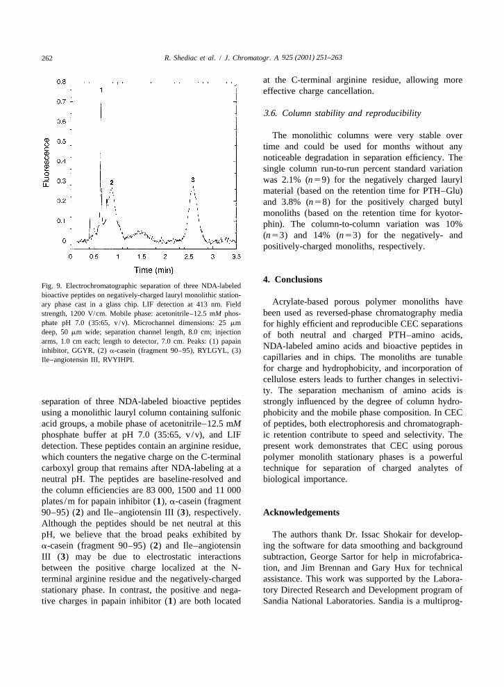
4. ConclusionsFig. 9. Electrochromatographic separation of three NDA-labeledbioactive peptides on negatively-charged lauryl monolithic station-
Acrylate-based porous polymer monoliths haveary phase cast in a glass chip. LIF detection at 413 nm. Fieldbeen used as reversed-phase chromatography mediastrength, 1200 V/cm. Mobile phase: acetonitrile–12.5 mM phos-
phate pH 7.0 (35:65, v /v). Microchannel dimensions: 25 mm for highly efficient and reproducible CEC separationsdeep, 50 mm wide; separation channel length, 8.0 cm; injection of both neutral and charged PTH–amino acids,arms, 1.0 cm each; length to detector, 7.0 cm. Peaks: (1) papain
NDA-labeled amino acids and bioactive peptides ininhibitor, GGYR, (2) a-casein (fragment 90–95), RYLGYL, (3)capillaries and in chips. The monoliths are tunableIle–angiotensin III, RVYIHPI.for charge and hydrophobicity, and incorporation ofcellulose esters leads to further changes in selectivi-ty. The separation mechanism of amino acids is
separation of three NDA-labeled bioactive peptides strongly influenced by the degree of column hydro-using a monolithic lauryl column containing sulfonic phobicity and the mobile phase composition. In CECacid groups, a mobile phase of acetonitrile–12.5 mM of peptides, both electrophoresis and chromatograph-phosphate buffer at pH 7.0 (35:65, v /v), and LIF ic retention contribute to speed and selectivity. Thedetection. These peptides contain an arginine residue, present work demonstrates that CEC using porouswhich counters the negative charge on the C-terminal polymer monolith stationary phases is a powerfulcarboxyl group that remains after NDA-labeling at a technique for separation of charged analytes ofneutral pH. The peptides are baseline-resolved and biological importance.the column efficiencies are 83 000, 1500 and 11 000plates /m for papain inhibitor (1), a-casein (fragment90–95) (2) and Ile–angiotensin III (3), respectively. AcknowledgementsAlthough the peptides should be net neutral at thispH, we believe that the broad peaks exhibited by The authors thank Dr. Issac Shokair for develop-a-casein (fragment 90–95) (2) and Ile–angiotensin ing the software for data smoothing and backgroundIII (3) may be due to electrostatic interactions subtraction, George Sartor for help in microfabrica-between the positive charge localized at the N- tion, and Jim Brennan and Gary Hux for technicalterminal arginine residue and the negatively-charged assistance. This work was supported by the Labora-stationary phase. In contrast, the positive and nega- tory Directed Research and Development program oftive charges in papain inhibitor (1) are both located Sandia National Laboratories. Sandia is a multiprog-

925 (2001) 251–263 263R. Shediac et al. / J. Chromatogr. A
´[18] F. Svec, J.M.J. Frechet, Anal. Chem. 64 (1992) 820.ram laboratory operated by Sandia Corporation, a´[19] F. Svec, J.M.J. Frechet, Ind. Eng. Chem. Res. 38 (1999) 34.Lockheed Martin Company, for the United States
´[20] F. Svec, E.C. Peters, D. Sykora, C. Yu, J.M.J. Frechet, J.Department of Energy under contract DE-AC04- High Resol. Chromatogr. 23 (2000) 3.94AL85000. [21] A. Palm, M.V. Novotny, Anal. Chem. 69 (1997) 4499.
´[22] X. Huang, J. Zhang, Cs. Horvath, J. Chromatogr. A 858(1999) 91.
´[23] I. Gusev, X. Huang, Cs. Horvath, J. Chromatogr. A 855References(1999) 273.
[24] S.M. Ngola, Y. Fintschenko, W.-Y. Choi, T.J. Shepodd, Anal.[1] C.K. Larive, S.M. Lunte, M. Zhong, M.D. Perkins, G.S. Chem. 73 (2001) 849.
Wilson, G. Gokulrangan, T. Williams, F. Afroz, C. [25] L.A. Colon, Y. Guo, A. Fermier, Anal. Chem. 69 (1997)¨Schoneich, T.S. Derrick, C.R. Middaugh, S. Bogdanowich- 461A.
Knipp, Anal. Chem. 71 (1999) 389R. [26] R.G. Carlson, K. Srinivasachar, R.S. Givens, B.K. Matus-´ ´ ´ ´ ´[2] K. Stulık,V. Pacakova, J. Suchankova, H.A. Claessens, Anal. zewski, J. Org. Chem. 51 (1986) 3978.
Chim. Acta 352 (1997) 1. [27] M.C. Roach, M.D. Harmony, Anal. Chem. 59 (1987) 411.[3] J.-T. Wu, P. Huang, M.X. Li, M.G. Qian, D.M. Lubman, [28] P. de Montigny, J.F. Stobaugh, R.S. Givens, R.G. Carlson, K.
Anal. Chem. 69 (1997) 320. Srinivasachar, L.A. Sternson, T. Higuchi, Anal. Chem. 59´[4] C. Ericson, S. Hjerten, Anal. Chem. 71 (1999) 1621. (1987) 1096.
[5] A. Apffel, H. Yin, W.S. Hancock, D. McManigill, J. Frenz, [29] B. Matuszewski, R. Givens, K. Srinivasachar, R. Carlson, T.S.-L. Wu, J. Chromatogr. A 832 (1999) 149. Higuchi, Anal. Chem. 59 (1987) 1102.
[6] J.-T. Wu, P. Huang, M.X. Li, D.M. Lubman, Anal. Chem. 69 [30] F. Lai, T. Sheehan, Biotechniques 14 (1993) 642.(1997) 2908. [31] M. Oates, J. Jorgenson, Anal. Chem. 61 (1989) 432.
[7] S. Zhang, X. Huang, J. Zhang, Cs. Horvath, J. Chromatogr. [32] S.S. Yang, I. Smetena, Chromatographia 37 (1993) 593.A 887 (2000) 465. ´ ´[33] A. Cifuentes, M.A. Rodrıguez, F.J. Garcıa-Montelongo, J.
´[8] C. Yu, F. Svec, J.M.J. Frechet, Electrophoresis 21 (2000) Chromatogr. A 742 (1996) 257.120. [34] D. Corradini, J. Chromatogr. B 699 (1997) 221.
[9] B. He, J. Ji, F.E. Regnier, J. Chromatogr. A 853 (1999) 257. [35] T. Sasagawa, D.C. Teller, in: W.S. Hancock (Ed.), CRC´[10] G. Choudhary, Cs. Horvath, J.F. Banks, J. Chromatogr. A Handbook of HPLC for the Separation of Amino Acids,
828 (1998) 469. Peptides, and Proteins, Vol. 2, CRC Press, Boca Raton, FL,[11] R.M. Seifar, J.C. Kraak, H. Poppe, W.T. Kok, J. Chromatogr. 1984, p. 53.
A 832 (1999) 133. [36] D.C. Harris, Quantitative Chemical Analysis, Freeman, New[12] C.G. Huber, G. Choudhary, Cs. Horvath, Anal. Chem. 69 York, 1999.
(1997) 4429. [37] E.C. Rickard, M.M. Strohl, R.G. Nielsen, Anal. Biochem.[13] M. Qi, X.-F. Li, C. Stathakis, N.J. Dovichi, J. Chromatogr. A 197 (1991) 197.
853 (1999) 131. [38] S. Fu, C.A. Lucy, Anal. Chem. 70 (1998) 173.´[14] C. Ericson, J. Holm, T. Ericson, S. Hjerten, Anal. Chem. 72 [39] N. Adamson, P.F. Riley, E.C. Reynolds, J. Chromatogr. 646
(2000) 81. (1993) 391.´[15] E.C. Peters, M. Petro, F. Svec, J.M.J. Frechet, Anal. Chem. [40] H.G. Lee, D.M. Desiderio, J. Chromatogr. A 666 (1994) 271.
69 (1997) 3646. [41] A. Ishikawa, T. Shibata, J. Liq. Chromatogr. 16 (1993) 859.´[16] E.C. Peters, M. Petro, F. Svec, J.M.J. Frechet, Anal. Chem. [42] Y. Okamoto, E. Yashima, Angew. Chem. Int. Ed. Engl. 37
70 (1998) 2296. (1998) 1020.´[17] E.C. Peters, M. Petro, F. Svec, J.M.J. Frechet, Anal. Chem.
70 (1998) 2288.
Copyright © 2022 FDOKUMEN