Tissue protective peptides and uses thereof - European Patent ...
83
Note: Within nine months of the publication of the mention of the grant of the European patent in the European Patent Bulletin, any person may give notice to the European Patent Office of opposition to that patent, in accordance with the Implementing Regulations. Notice of opposition shall not be deemed to have been filed until the opposition fee has been paid. (Art. 99(1) European Patent Convention). Printed by Jouve, 75001 PARIS (FR) (19) EP 2 371 855 B1 TEPZZ ¥7_855B_T (11) EP 2 371 855 B1 (12) EUROPEAN PATENT SPECIFICATION (45) Date of publication and mention of the grant of the patent: 22.07.2015 Bulletin 2015/30 (21) Application number: 11163194.1 (22) Date of filing: 07.08.2006 (51) Int Cl.: C07K 14/71 (2006.01) A61K 38/18 (2006.01) (54) Tissue protective peptides and uses thereof Gewebeschützende Peptide und ihre Verwendung Peptides protecteurs de tissus et utilisations associées (84) Designated Contracting States: AT BE BG CH CY CZ DE DK EE ES FI FR GB GR HU IE IS IT LI LT LU LV MC NL PL PT RO SE SI SK TR Designated Extension States: AL BA HR MK RS (30) Priority: 05.08.2005 US 705741 P 08.08.2005 US 706276 P 18.07.2006 US 831737 P (43) Date of publication of application: 05.10.2011 Bulletin 2011/40 (60) Divisional application: 12175352.9 / 2 540 309 12175366.9 / 2 594 279 (62) Document number(s) of the earlier application(s) in accordance with Art. 76 EPC: 06801051.1 / 1 924 276 (73) Proprietor: Araim Pharmaceuticals, Inc. Tarrytown, NY 10591 (US) (72) Inventors: • Cerami, Anthony La Jolla, CA 92037 (US) • Brines, Michael Woodbridge, Connecticut 06525 (US) • Coleman, Thomas Mt. Kisco, New York 10549 (US) (74) Representative: Piening, Niklas Jones Day Prinzregentenstraße 11 80538 München (DE) (56) References cited: • ELLIOTT S ET AL: "Mapping of the Active Site of Recombinant Human Erythropoietin", 15 January 1997 (1997-01-15), BLOOD, AMERICAN SOCIETY OF HEMATOLOGY, US, PAGE(S) 493 - 502, XP002977797, ISSN: 0006-4971 * the whole document * • ELLIOTT STEVE ET AL: "Structural requirements for additional N-linked carbohydrate on recombinant human erythropoietin", 16 April 2004 (2004-04-16), JOURNAL OF BIOLOGICAL CHEMISTRY, VOL. 279, NR. 16, PAGE(S) 16854-16862, XP000002657380, ISSN: 0021-9258 * the whole document * • ELLIOT ET AL: "Fine-Structure Epitope Mapping of Antierythropoietin Monoclonal Antibodies Reveals a Model of Recombinant Human Erythropoietin Structure", 1 April 1996 (1996-04-01), BLOOD, AMERICAN SOCIETY OF HEMATOLOGY, US, PAGE(S) 2702 - 2713, XP002988824, ISSN: 0006-4971 * the whole document *
-
Upload
khangminh22 -
Category
Documents
-
view
1 -
download
0
Transcript of Tissue protective peptides and uses thereof - European Patent ...
Note: Within nine months of the publication of the mention of the grant of the European patent in the European PatentBulletin, any person may give notice to the European Patent Office of opposition to that patent, in accordance with theImplementing Regulations. Notice of opposition shall not be deemed to have been filed until the opposition fee has beenpaid. (Art. 99(1) European Patent Convention).
Printed by Jouve, 75001 PARIS (FR)
(19)E
P2
371
855
B1
TEPZZ ¥7_855B_T(11) EP 2 371 855 B1
(12) EUROPEAN PATENT SPECIFICATION
(45) Date of publication and mention of the grant of the patent: 22.07.2015 Bulletin 2015/30
(21) Application number: 11163194.1
(22) Date of filing: 07.08.2006
(51) Int Cl.:C07K 14/71 (2006.01) A61K 38/18 (2006.01)
(54) Tissue protective peptides and uses thereof
Gewebeschützende Peptide und ihre Verwendung
Peptides protecteurs de tissus et utilisations associées
(84) Designated Contracting States: AT BE BG CH CY CZ DE DK EE ES FI FR GB GR HU IE IS IT LI LT LU LV MC NL PL PT RO SE SI SK TRDesignated Extension States: AL BA HR MK RS
(30) Priority: 05.08.2005 US 705741 P08.08.2005 US 706276 P18.07.2006 US 831737 P
(43) Date of publication of application: 05.10.2011 Bulletin 2011/40
(60) Divisional application: 12175352.9 / 2 540 30912175366.9 / 2 594 279
(62) Document number(s) of the earlier application(s) in accordance with Art. 76 EPC: 06801051.1 / 1 924 276
(73) Proprietor: Araim Pharmaceuticals, Inc.Tarrytown, NY 10591 (US)
(72) Inventors: • Cerami, Anthony
La Jolla, CA 92037 (US)• Brines, Michael
Woodbridge, Connecticut 06525 (US)
• Coleman, ThomasMt. Kisco, New York 10549 (US)
(74) Representative: Piening, NiklasJones Day Prinzregentenstraße 1180538 München (DE)
(56) References cited: • ELLIOTT S ET AL: "Mapping of the Active Site of
Recombinant Human Erythropoietin", 15 January 1997 (1997-01-15), BLOOD, AMERICAN SOCIETY OF HEMATOLOGY, US, PAGE(S) 493 - 502, XP002977797, ISSN: 0006-4971 * the whole document *
• ELLIOTT STEVE ET AL: "Structural requirements for additional N-linked carbohydrate on recombinant human erythropoietin", 16 April 2004 (2004-04-16), JOURNAL OF BIOLOGICAL CHEMISTRY, VOL. 279, NR. 16, PAGE(S) 16854-16862, XP000002657380, ISSN: 0021-9258 * the whole document *
• ELLIOT ET AL: "Fine-Structure Epitope Mapping of Antierythropoietin Monoclonal Antibodies Reveals a Model of Recombinant Human Erythropoietin Structure", 1 April 1996 (1996-04-01), BLOOD, AMERICAN SOCIETY OF HEMATOLOGY, US, PAGE(S) 2702 - 2713, XP002988824, ISSN: 0006-4971 * the whole document *

EP 2 371 855 B1
2
5
10
15
20
25
30
35
40
45
50
55
Description
1. INTRODUCTION
[0001] The present invention is directed to novel tissue protective peptides. The tissue protective peptides of theinvention may bind to a tissue protective receptor complex. In particular, the present invention is drawn to tissue protectivepeptides derived from or sharing consensus sequences with portions of cytokine receptor ligands, including Erythropoietin(EPO), that are not involved in the binding of the ligand to the receptor complex, e.g., to the EPO receptor homodimer.Accordingly, the tissue protective peptides of the invention are derived from the amino acid sequences of regions ofcytokine receptor ligands that are generally located on or within the region of the ligand protein that is opposite of thereceptor complex, i.e., are generally derived from amino acid sequences of regions of the ligand protein that face awayfrom the receptor complex while the ligand is bound to the receptor. The invention is further directed to the consensussequences for use in engineering a synthetic tissue protective peptide. These tissue protective peptides also includefragments, chimeras, as well as peptides designed to mimic the spatial localization of key amino acid residues withinthe tissue protective receptor ligands, e.g., EPO.[0002] The invention also encompasses methods for treating, preventing or ameliorating a disease or disorder and ortreating, restoring or ameliorating a tissue injury using tissue protective peptides of the current invention. The inventionalso encompasses methods for enhancing excitable tissue function using tissue protective peptides of the current in-vention.
2. BACKGROUND OF THE INVENTION
[0003] Erythropoietin ("EPO") is a glycoprotein hormone commonly associated with the maintenance of hematocritand, more recently, tissue protection. Mature human EPO protein comprises 165 amino acids and has a molecularweight of 34 kDa, with glycosyl residues contributing about 40 % of the weight of the molecule. The EPO moleculecomprises four helices that interact via their hydrophobic domains to form a predominantly globular structure within anaqueous environment (Cheetham et al., 1998, Nat. Struct. Biol. 5:861-866). The invention derives from the discoverythat certain amino acids facing the aqueous environment (i.e., away from the hydrophobic, globular central core) mediatetissue protection. Peptides can be derived or designed from an understanding of the tissue protective regions that havebeen identified by the Applicants.[0004] As noted above, EPO is pluripotent. In its hormonal role, EPO regulates hematocrit through its role in thematuration of erythroid progenitor cells into erythrocytes. EPO acts as an anti-apoptotic agent during the maturationprocess of erythroid progenitor cells, permitting progenitor cells to mature into erythrocytes. Decreased levels of tissueoxygen (hypoxia) trigger an increased production of erythropoietin by the kidney, which results in increased erythropoiesis.Given that the kidney normally produces the majority of the serum erythropoietin, the loss of kidney function, such asin chronic renal failure, results in decreased production of EPO and often anemia. Similarly, anemia may result fromother chronic conditions, such as cancer, or treatments associated with these illnesses, such as chemotherapy, whichdirectly suppress the production of EPO. Commercially available recombinant erythropoietin has been available underthe trademarks of PROCRIT, available from Ortho Biotech Inc., Raritan, NJ, and EPOGEN, available from Amgen, Inc.,Thousand Oaks, CA and has been used to treat anemia resulting from end stage renal disease, therapy with AZT(zidovudine) in HIV-infected patients, oncology patients, and chemotherapy. Elliott et al., 1996, Blood Vol. 87, No. 7,2702-2713 describes the modeling of the rHuEPO protein structure by analyzing the effects of mutations on proteinfolding and by performing epitope mapping using noncompeting anti-EPO monoclonal antibodies. Elliott et al., 1997,Blood Vol. 89, No. 2, 493-502 discloses the construction of recombinant human erythropoietin (rHuEPO) variants toidentify amino acid residues which are important for its biological activity and to determine the effect of each mutationon rHuEPO folding. Currently a hyperglycosylated erythropoietin, ARANESP™ (Amgen, Thousand Oaks, CA), is avail-able for the treatment of anemia. Elliott et al., 2004, The Journal of Biologial Chemistry Vol. 279, No. 16, 16854-16662discloses the construction of recombinant human erythropoietin molecules with N-linked consensus sequences thatwere introduced by site-directed mutagenesis into the polypeptide chain to analyse the structural rules for N-linkedglycosylation. Additionally, these compounds have been used to increase the hematocrits of patients undergoing surgeryto reduce the need for allogenic blood transfusions.[0005] Recently, several lines of evidence have suggested that EPO also functions locally in a paracrine-autocrinemanner to minimize tissue damage. For example, EPO improves an hypoxic cellular microenvironment and decreasesprogrammed cell death caused by metabolic stress. Both of these activities are moderated, in part, through EPO’sinteraction with a specific cell surface receptor comprised, in part, by the erythropoietin receptor ("EPOR") protein. EPORis an approximately 66 kDa protein and is a member of the Type-1 cytokine receptor family. This family comprisesreceptors that are grouped together based on the shared homology of their extracellular domains and includes receptorsfor interleukin IL-2, IL3, IL4, IL5, IL6, IL7, IL9, IL11, granulocyte macrophage-colony stimulating factor (GM-CSF), gran-
EP 2 371 855 B1
3
5
10
15
20
25
30
35
40
45
50
55
ulocyte colony stimulating factor (G-CSF), leukemia inhibiting factor (LIF), cillary neurotrophic factor (CNTF), throm-bopoietin, growth hormone and prolactin. The conserved extracellular domain of these receptors has a length of ap-proximately 200 amino acids, comprises four positionally conserved cysteine residuesin the amino-terminal region (Cys294, Cys 283, Cys 248, and Cys 238, which appear to be critical to the maintenance and the structural integrity of thereceptors (Murray, 1996, Harpers Biochemistry 24th ed. pp. 524-526, Appilion & Lange, Ltd.; Caravella et al., 1996,Protein: Struct. Funct. Gen. 24:394-401 )), and a Trp-Ser-X-Trp-Ser (SEQ ID NO:58) motif located proximal to thetransmembrane domain.[0006] In connection with erythropoiesis, EPOR functions in a manner similar to other receptors within the Type-1cytokine receptor family. First, the receptor ligand, e.g., EPO, binds to a preformed dimer of EPOR, (EPOR)2. It hasbeen determined that EPO interacts with the extracellular domain of the classic (EPOR)2 homodimer receptor via twodistinct regions on the ligand surface: a high affinity receptor binding site (site 1) and a low affinity receptor binding site(site 2). The amino acid sequences of EPO associated with site 1 are TKVNFY, SEQ ID NO:2, corresponding to aminoacids 44-49 of SEQ ID NO:1, and SNFLRG SEQ ID NO:3, corresponding to amino acids 146-151 of SEQ ID NO:1, thesequences associated with site 2 are VLERY, SEQ ID NO:4, corresponding to amino acids 11-15 of SEQ ID NO:1, andSGLRS, SEQ ID NO:5, corresponding to amino acids 100-104 of SEQ ID NO:1 (Cheetham et al., 1998, Nature StructuralBiology 5:861-866). EPOR homodimer activation leads to tyrosine phosphorylation of signaling proteins that are asso-ciated with EPOR, e.g., Jak2 tyrosine kinases, that may in turn activate several different pathways including, for example,the phosphatidylinositol (P1) 3-kinase pathway, the Ras/MAP kinase pathway, and/or the STAT pathway. These pathwaystrigger the anti-apoptotic functions necessary for erythropoiesis that are mediated by erythropoietin (Kirito et al., 2002,Blood 99:102-110; Livnah et al., 1999, Science 283:987-990; Naranda et al., 2002, Endocrinology 143:2293-2302; Remyet al., 1999, Science 283:990-993; and Yoshimura et al., 1996, The Oncologist 1:337-339).[0007] Recently, Applicants have discovered that the tissue protective properties of EPO are mediated by a receptorthat comprises not only EPOR but also another receptor protein, the beta common receptor ("βc"). The EPOR/ βc receptoris, in contrast to the homodimer (EPOR)2, a heterocomplex (see infra) and is known to play a role in the protection ofexcitable tissues. See, e.g., WO 2004/096148 and PCT no. PCT/US01/49479, filed December 28, 2001, U.S. PatentApplication Nos. 09/753,132, filed December 29, 2000, and 10/188,905, filed July 3, 2002. Although Applicants hadestablished that the βc receptor is central to the tissue protective pathways in these excitable tissues, the structure ofthe activating ligands for the receptors was still unknown.
3. SUMMARY
[0008] The present invention provides isolated polypeptides consisting of no more than 30 amino acid, said polypeptidescomprising the amino acid sequence QEQLERALNSS (SEQ ID NO: 40), wherein said polypeptides have cellular pro-tective activity. In some embodiments, the isolated polypeptides comprise the amino acid sequence QEQLERALNS-SLRRYINMLTRTR (SEQ ID NO: 41), or the amino acid sequence CSLNENIQEQLERALNSS (SEQ ID NO: 43). In certainembodiments, the isolated polypeptide consists of the amino acid sequence QEQLERALNSS (SEQ ID NO: 40). Theinvention further provides isolated polypeptides consisting of the amino acid sequence QEQLERALNSS (SEQ ID NO:40), wherein one amino acid residue of said amino acid sequence is replaced with a conservative or non-conservativesubstitution or an amino acid equivalent, wherein said polypeptides have cellular protective activity.[0009] The invention also provides isolated polypeptides which are a retro-inverso-analogue of the isolated polypep-tides described above, wherein said retro-inverso-analogues have cellular protective activity.[0010] Provided are further isolated polypeptides as described above that are modified with an addition of polyethyleneglycol. In certain embodiments the isolated polypeptides of the invention do not increase hemoglobin in a recipient.[0011] The invention further provides pharmaceutical compositions comprising the isolated polypeptides of the inven-tion and a pharmaceutically acceptable carrier. The pharmaceutical composition may be formulated for oral, intranasal,ocular, inhalational, transdermal, rectal, sublingual, or parenteral administration. Alternatively, it may be formulated asa perfusate solution.[0012] A further aspect of the invention concerns an in vitro method for protecting, maintaining, or enhancing theviability of a responsive cell, tissue, or organ isolated from a mammalian body comprising exposing said cell, tissue, ororgan to isolated polypeptides according to the invention or to pharmaceutical compositions according to the invention.In some embodiments the mammalian body is a human body.[0013] In one aspect, the isolated polypeptides of the invention may be used in a method for prevention, therapeutictreatment or prophylactic treatment of a cardiovascular disease, kidney disease, disease of the urinary system, bonedisease, skin disease, gastrointestinal disease, or a disease or disorder of the central or peripheral nervous system ina subject. In embodiments thereof the subject is a human.[0014] In a further aspect, the isolated polypeptides of the invention are for use in a method for preventing or treatinga tissue injury in a subject, wherein said tissue injury results from reduced oxygenation or inflammation. In some em-bodiments said reduced oxygenation results from a hypoxic or an ischemic condition. In some embodiments the subject
EP 2 371 855 B1
4
5
10
15
20
25
30
35
40
45
50
55
is a human.[0015] In a further aspect, the invention provides an isolated nucleic acid comprising a nucleotide sequence encodingthe isolated polypeptides of the invention.[0016] In a further aspect, the invention provides a vector comprising the nucleic acid of the invention. The vector maybe an expression vector.[0017] In an additional aspect, the invention provides a host cell containing the expression vector.[0018] The invention also provides a method of recombinantly producing the isolated polypeptide of the invention,wherein the method comprises (a) culturing in a medium the host cell of the invention, under conditions suitable for theexpression of said polypeptide, and (b) recovering and isolating said polypeptide from said medium.[0019] The present invention generally relates to isolated polypeptides that have at least one cellular protective activityin a responsive cell, tissue, or organ, which polypeptides contain amino acid motifs comprising the consensus sequence(a) H1-N1-(X)n-N2-H2, wherein n is 0, 1, 2, 3, 4 or 5; (b) H1-N1-(X)n-N2-L1, wherein n is 0, 1, 2, 3, 4 or 5; (c) L1-N1-(X)n-N2-H1,wherein n is 0, 1, 2, 3, 4 or 5; (d) H1-N1-(L)n-P1-H2, wherein n is 0 or 1; or (e) H1-P1-(L)n-N1-H2, wherein n is 0 or 1, andwherein H1 and H2 are hydrophobic amino acids, N1 and N2 are negatively charged amino acids, X is any amino acid,L1 is a polar amino acid, and P1 is a positively charged amino acid. These peptides may lack erythropoietic activity, e.g.,they do not increase hemoglobin or hematocrit in a recipient. The isolated polypeptides may consist of no more than10, no more than 15, no more than 20, or no more than 30 amino acids. The isolated peptide may have than 90 %, lessthan 85%, less than 80%, less than 75%, less than 70%, less than 65%, less than 60%, less than 55%, less than 50%,less than 45%, less than 40%, less than 35%, less than 30%, or less than 20 percent sequence identity with any portionof the amino acid sequence of mature human erythropoietin ("EPO") set forth in SEQ ID NO:1, wherein said portion ofEPO contains the same number of amino acid residues as said peptide.[0020] In the isolated polypeptide comprising the structural motif (a) H1-N1-(X)n-N2-H2, wherein n is 0, 1, 2, 3, 4 or 5(embodied by sequence identifiers 6-11, respectively, discussed infra); (b) H1-N1-(L)n-P1-H2, wherein n is 0 or 1 (embodiedby sequence identifiers 24-25, respectively, discussed infra); or (e) H1-P1-(L)n-N1-H2, wherein n is 0 or 1 (embodied bysequence identifiers 26-27, respectively, discussed infra), H1 and H2 may be the same hydrophobic amino acid. In theisolated polypeptide comprising the structural motifs (a) H1-N1-(X)n-N2-H2, wherein n is 0, 1, 2, 3, 4 or 5; (d)H1-N1-(L)n-P1-H2, wherein n is 0 or 1; or (e) H1-P1-(L)n-N1-H2, wherein n is 0 or 1, H1 and H2 may be different hydrophobicamino acids. Described is also an isolated polypeptide comprising: the amino acid motif (a) H1-N1-(X)n-N2-H2, wherein
n is 0, 1, 2, 3, 4 or 5; (b) H1-N1-(X)n-N2-L1, wherein n is 0, 1, 2, 3, 4 or 5; (c) L1-N1-(X)n-N2-H1, wherein n is 0, 1, 2, 3, 4or 5, and wherein N1 and N2 may the same or may be different negatively charged amino acids.[0021] In the isolated polypeptides comprising the amino acid motifs described hereinabove, said motifs may be formedby consecutive amino acids within the amino-acid sequence of said polypeptide. In specific examples such an isolatedpolypeptide comprises the amino acid motif H1-N1-N2-H2 (SEQ ID NO:6), H1-N1-X-N2-H2 (SEQ ID NO:7), H1-N1-X-X-N2-H2 (SEQ ID NO:8), H1-N1-X-X-X-N2-H2 (SEQ ID NO:9), H1-N1-X-X-X-X-N2-H2 (SEQ ID NO:10), H1-N1-X-X-X-X-X-N2-H2 (SEQ ID NO:11), H1-N1-N2-L1 (SEQ ID NO:12), H1-N1-X-N2-L1 (SEQ ID NO:13), H1-N1-X-X-N2- L1 (SEQ IDNO:14), H1-N1-X-X-X-N2- L1 (SEQ ID NO:15), H1-N1-X-X-X-X-N2-L1 (SEQ ID NO:16), H1-N1-X-X-X-X-X-N2- L1 (SEQID NO:17), L1-N1-N2-H2 (SEQ ID NO:18), L1 N1-X-N2-H2 (SEQ ID NO:19), L1-N1-X-X-N2-H2 (SEQ ID NO:20), L1-N1-X-X-X-N2-H2 (SEQ ID NO:21), L1-N1-X-X-X-X-N2-H2 (SEQ ID NO:27), L1-N1-X-X-X-X-X-N2-H2 (SEQ ID NO:23),H1-N1-P1-H2 (SEQ ID NO:24), H1-N1-L1-P1-H2 (SEQ ID NO:25), H1-P1-N1-H2 (SEQ ID NO:26), or H1-P1-L1-N1-H2 (SEQID NO:27), wherein H1 and H2 are hydrophobic amino acids, N1 and N2 are negatively charged amino acids, X is anyamino acid, L1 is a polar amino acid, and P1 is a positively charged amino acid. In certain aspects, wherein the isolatedpolypeptide comprises a motif having the amino acid residues H1 and H2, H1 and H2 may be the same or may be differenthydrophobic amino acids. In other aspects, wherein the isolated polypeptide comprises a motif having the amino acidresidues Ni and N2, N1 and N2 may be the same or may be different negatively charged amino acids.[0022] Described are further isolated polypeptides wherein the amino acid motif is formed due to the spatial organizationof amino acids within the tertiary structure of a polypeptide, i.e., the amino acids forming the motif are spatially adjacentto one another in the three dimensional structure, i.e. tertiary structure, of the polypeptide but may be separated by 1or more amino acids within the primary amino acid sequence of the polypeptide chain. In a specific example, the aminoacid motif comprising amino acid residues H1, N1-N2, and H2 analogous to SEQ ID NO:6, discussed supra, may formas a result of the tertiary structure adopted by, i.e., protein folding of peptides comprising, e.g., SEQ ID NO:7, SEQ IDNO:8, SEQ ID NO:9, SEQ ID NO:10 or SEQ ID NO:11, wherein the amino acid resides between N1 and N2, e.g. (X)n,fold such that N1 and N2 become linearly adjacent. Accordingly, described are isolated peptides comprising the aminoacid motif H1N1N2H2; H1N1N2L1; L1N1N2H1; H1N1(L)nP1H2, wherein n is 0 of 1; for H1P1(L)nN1H2, wherein n is 0 or 1,which motifs are formed as a result of the tertiary structure ofsaid polypeptide. Wherein the amino acid motif comprisesN1 and N2, the tertiary structures form such that the distance between the carbonyl carbons of N1 and N2 is about 3 Åto about 5 Å, preferably about 4 Å to about 5 Å, and more preferably about 4.4 Å to about 4.8 Å. Wherein the amino acidmotif comprises N1 and N2, the tertiary structures form such that the distance between N1 and N2 are confined spatiallysuch that the charge separation, e.g., the charged side chains, of the two is between about 6.5 Å to about 9 Å. N1 and

EP 2 371 855 B1
5
5
10
15
20
25
30
35
40
45
50
55
N2 are thus spatially confined as a result of being in an amino acid sequence that forms all or a portion of an alpha helix,and may be separated by 1, 2, or more than 2 amino acids in the sequence of said amino acids forming said helix.Wherein the amino acid motif comprises N1 and P1, the tertiary structures form such that the distance between thecarbonyl carbons of N1 and P1 is about 3 Å to about 5 Å, preferably about 4 Å to about 5 Å, and more preferably about4.4 Å to about 4.8 Å. Wherein the amino acid motif comprises N1 and P1, the tertiary structures form such that thedistance between N1 and P1 are confined spatially such that the charge separation, e.g., the charged side chains, ofthe two is between about 6.5 Å to about 9 Å. N1 and P1 are spartially confined as a result of being in an amino acidsequence that forms all or a portion of an alpha helix, and may be separated by 1, 2, or more than 2 amino acids in thesequence of said amino acids forming said helix. The amino acids forming the motif within the tertiary structure of saidpolypeptide are separated from each other by an equal number of intervening amino acid residues in the linear aminoacid sequence of said polypeptide. The amino acids forming the motif within the tertiary structure of said polypeptideare separated from each other by a different number of intervening amino acid residues in the linear amino acid sequenceof said polypeptide. Sometimes the isolated polypeptide forms a regular tertiary structure, e.g., α-helix or β-pleatedsheet, such that the surface of said structure presents the amino acids comprising said motif, and thus the motif itself,to the interface of the protein structure and the aqueous environment, i.e., presents the motif on the surface of foldedthe polypeptide. The tertiary structures of the polypeptides of the invention preferably form in an aqueous environmentat physiological conditions, e.g., PBS (13 mM NaH2PO4, 137 mM NaCl, pH 7.4) at 37 °C.[0023] The invention provides for isolated polypeptides comprising an amino acid motif as described herein above,i.e. peptide G (QEQLERALNSS, SEQ ID N0:40).[0024] Also described are isolated polypeptides comprising the amino acid motifis described herein above, e.g., peptideA (APPRLICDSRVLERYLLEAKEAE, SEQ ID NO:32), peptide C (NITVPDTKYNFYAWKRMEVG, SEQ ID NO:29), pep-tide D (QQAVEVWQGLALLSEAVLRGQALLV, SEQ ID NO:30), peptide E (GCAEHCSLNENITVPDTKVN, SEQ IDNO:31), peptide F (RYLLUNITTGC, SEQ ID NO:33), peptide I (CSLNENIQEQLERALNSS; SEQ ID NO:43), peptide J(QEQLERALNSSLRRYINMLTRTR, SEQ ID NO:41), peptide K (WEHVNAIQEARRLL, SEQ ID NO:35), or peptide L(KIRSDLTALTESYVKH, SEQ ID NO:37).[0025] Described are isolated polypeptides comprising 1 or more, 2 for more, 3 or more, 4 or more, 5 or more, 6 ormore, or more than 6 amino acid motifs described herein. In specific aspects, wherein the isolated polypeptide comprisesat least two of the amino acid motifs described herein above, said at least two motifs may be the same motif or theymay be different motifs.[0026] In certain aspects, the isolated polypeptides of the invention lack an erythropoietin activity, e.g., increasinghemoglobin in a recipient. Preferably, the isolated polypeptides lack other activities including, but hot limited to, vasoactiveaction (e.g., vasoconstriction), hyperactivating platelets, pro-coagulant activities and stimulating proliferation and/orproduction of thrombocytes and/or erythropoietic-dependent cells (see, Coleman et al., 2006, PNAS 103:5965-5970).In other aspects, the isolated polypeptides of the invention comprise at least one cellular protective activity. Such cellularprotective activity includes, but is not limited to, protecting, maintaining, enhancing of restoring the function or viabilityof a responsive mammalian cell, tissue, or organ. Accordingly, in one aspect, the present invention is directed to theuse of an isolated polypeptide of the invention for the preparation of pharmaceutical compositions for protecting, main-taining, enhancing, or restoring the function-or-viability of responsive mammalian cells and their associated cells, tissues,and organs. In related embodiments, the compositions are for administration to a subject in need thereof. In preferredembodiments, said subject is a mammal and, preferably, a human.[0027] In other aspects, the present invention is directed to the use of an isolates polypeptide of the invention for thepreparation of a pharmaceutical composition for the protection against and/or prevention of a responsive tissue injury,for the restoration of, or for the rejuvenation of responsive tissue and/or responsive tissue function in a subject in needthereof. In one particular aspect, the responsive mammalian cells and their associated cells, tissues, or organs are distalto the vasculature by virtue of a tighf endothelial cell barrier. In another particular aspect, the cells, tissues, organs orother bodily parts are isolated from a mammalian body, such as those intended for transplant. By way of non-limitingexamples, a responsive cell of tissue may be neuronal, eye (e.g., retinal), adipose, connective, hair, teeth, mucosal,pancreas, endocrine, ear, epithelial, skin, muscle, heart, long, liver, kidney, intestine, adrenal (e.g., adrenal cortex,adrenal medulla), capillary, endothelial, testes, ovary, bone, skin, or endometrial cells or tissue. Further, non-limitingexamples of responsive cells include photoreceptor (rods and cones), ganglion, bipolar, horizontal, amacrine, Müller,Purkinje, myocardium, pace maker, sinoatrial node, sinus node, junction tissue, atrioventricular node, bundle of His,hepatocytes, stellate, Kupffer, mesangial, renal epithelial, tubular interstitial, goblet, intestinal gland (crypts), enteralendocrine, glomerulosa, fasciculate, reticularis, chromaffin, pericyte, Leydig, Sertoli, sperm, Graffian follicle, primordialfollicle islets of Langerhans, α-cells, β-cells, γ-cells, F-cells, osteoprogenitor, osteoclasts, osteoblasts, endometrial stro-ma, endometrial, stem and endothelial cells. These examples of responsive cells are merely illustrative. In one aspect,the responsive cell or its associated cells, tissues, or organs are excitable cells, tissues, or organs, or predominantlycomprise excitable cells or tissues. In certain aspects of the invention, the excitable tissue is central nervous systemtissue, peripheral nervous system tissue, cardiac tissue or retinal tissue. In another aspect, the responsive cell or its

EP 2 371 855 B1
6
5
10
15
20
25
30
35
40
45
50
55
associated cells, tissues, or organs are not excitable cells, tissues, or organs, nor do they predominantly comprisesexcitable cells or tissues.[0028] The erythropoietic and/or cellular protective activity of the isolated polypeptide of the invention in responsivecells may be evaluated and/or determined by any method described herein and or known in the art. In certain embodi-ments, the erythropoietin and/or cellular protective activity is determined in an in vitro assay. In other embodiments, theerythropoietic and/or cellular protective activity is determined in an in vivo assay. In a related embodiment, wherein thecellular protective activity is neuroprotection, the invention provides for a method of evaluating said activity in vitro by(a) contacting a test culture of primary hippocampal neurons with N-methyl-D-aspartate and said peptide; and (b) de-termining the cell viability at 48 hours post said contact, such that if the cell viability determined in step (b) is greaterthan that of a control culture in the absence of said peptide, the peptide possesses cellular protective activity.[0029] In a particular embodiment, the mammalian cell, tissue, or organ for which an aforementioned isolated peptideis used are those that have expended or will expend a period of time under at least one condition adverse to the viabilityof the cell, tissue, or organ. In accordance with this embodiment, the isolated peptide of the invention provide protectionagainst and/or prevention of a tissue injury resulting from such conditions, provide for the restoration of, or provide forthe rejuvenation of tissue and/or tissue function in a subject in need thereof before, during or after such conditions arise.Such conditions include traumatic in situ hypoxia or metabolic dysfunction, surgically-induced in situ hypoxia or metabolicdysfunction, or in situ toxin exposure, the latter may be associated with chemotherapy or radiation therapy. In otherembodiments, the isolated peptides of the invention provide protection against and/or prevention of a tissue injuryresulting from a disease or disorder, provide for the restoration of, or provide for the rejuvenation of tissue and/or tissuefunction in a subject in need thereof before, during or after such conditions arise. In related embodiments said injury iscaused by a seizure disorder, multiple sclerosis, stroke, hypotension, cardiac arrest, ischemia, myocardial infarction,inflammation, age-related loss of cognitive function, radiation damage, cerebral palsy, neurodegenerative disease,Alzheizmer’s disease, Parkinson’s disease, mitochondrial disease, AIDS dementia, memory loss, amyotrophic lateralsclerosis, alcoholism, mood disorder, anxiety disorder, attention deficit disorder, autism, Creutzfeld-Jakob disease, brainor spinal cord trauma or ischemia, heart-lung bypass, chronic heart failure, macular degeneration, diabetic neuropathy,diabetic retinopathy, hepatitis, pancreatitis, glaucoma, retinal ischemia, retinal trauma, cardiovascular disease, cardiop-ulmonary disease, respiratory disease, kidney disease, disease of the urinary system, disease of the reproductive system,bone disease, skin disease, connective tissue disease, gastrointestinal disease, endocrine abnormality, metabolic ab-normality, or a disease or disorder of the central or peripheral nervous system. In still other embodiments, the adverseconditions are a result of cardio-pulmonary bypass (heart-lung machine), as is used for certain surgical procedures. Instill other embodiments, said injury is cognitive dysfunction. In a particular embodiment, the mammalian cell, tissue, ororgan for which an aforementioned isolated peptide is used empress the βc receptor.[0030] In certain embodiments, the invention is also directed to pharmaceutical compositions comprising the afore-mentioned isolated polypeptides for administration to a subject in need thereof. In specific aspects in accordance withthis embodiment, the pharmaceutical compositions of the invention further comprises a pharmaceutically acceptablecarrier. Such pharmaceutical compositions may be formulated for oral, intranasal, ocular, inhalational, transdermal,rectal, sublingual, vaginal, or parenteral administration, or in the form of a perfusate solution for maintaining the viabilityof cells, tissues, or organs ex vivo. In related embodiments of the invention the subject is a mammalian animal, preferablya human.[0031] In other aspects, the isolated polypeptides of the invention may be used in a method for facilitating the tran-scytosis of a molecule across an endothelial cell barrier in a subject in need thereof comprising administration to saidsubject a compositions comprising said molecule in association with an isolated peptide of the invention describedhereinabove. In a related embodiment, association is a labile covalentbond, a stable covalent bond, or a non-covalentassociation with a binding site for said molecule.[0032] According to another aspect, the isolated peptide of the invention, as described herein above, is capable oftraversing an endothelial cell barrier. The endothelial cell barrier may comprise the blood-brain barrier, the blood-eyebarrier, the blood-testis barrier, the blood-ovary barrier, blood-placenta, blood-heart, blood-kidney, blood-nerve, or blood-spinal cord barrier.[0033] According to one aspect of the invention, there is provided an isolated nucleic acid molecule that comprises anucleotide sequence which encodes a polypeptide compising the isolated polypeptide of the invention.[0034] In another embodiment of the invention, there is provided an isolated nucleic acid molecule that comprises anucleotide sequence (i.e., a cDNA, a nucleotide sequence interrupted by introns, or uninterrupted by introns), whichencodes a polypeptide comprising or consisting of the isolated polypeptide of the invention as described herein above.In one embodiment, the nucleotide sequence, encoding the isolated polypeptide of the invention, is synthesized usingpreferred codons that facilitate optical expression in a particular host cell. Such preferred codons can be optimal forexpression in cells of a species of plant, bacterium, yeast, mammal, fungus, or insect.[0035] The invention also provides for a vector comprising the nucleic acid molecule. The invention also provides foran expression vector comprising the nucleic acid molecule and at least one regulatory region operably linked to the
EP 2 371 855 B1
7
5
10
15
20
25
30
35
40
45
50
55
nucleic acid molecule. In another embodiment, the invention provides for a cell comprising the expression vector. In yetanother embodiment, there is provided a genetically-engineered cell which comprises the nucleic acid molecule.[0036] In another embodiment, the invention provides for a method of recombinantly producing the isolated peptideof the invention, described herein above, comprising culturing in a medium a host cell containing a nucleic acid moleculecomprising a nucleotide sequence encoding a polypeptide of the invention, under conditions suitable for the expressionof said peptide, and recovering and/or isolating the expressed polypeptide from said medium.
3.1 TERMINOLOGY
[0037] As used herein, the terms "about" or "approximately" when used in conjunction with a number refer to anynumber within 1, 5, or 10% of the referenced number.[0038] The term "administered in conjunction with" in the context of the methods of the invention means administeringa compound prior to, at the same time as, and/or subsequent to the onset of a disease, disorder, or condition.[0039] The term "amino acid" or any reference to a specific amino acid is meant to include naturally occurring prote-ogenic amino acids as well as non-naturally occurring amino acids such as amino acid analogs. Those skilled in the artwould know that this definition includes, unless otherwise specifically noted, includes naturally occurring protogenic(L)-amino acids, their optical (D)-isomers, chemically modified amino acids, including amino acid analogs such as pen-icillamine (3-mercapto-D-valine), naturally occurring non-proteogenic amino acids such as norleucine and chemicallysynthesized proteins that have properties known in the art to be characteristic of an amino acid. As used herein, aminoacids will be represented wither by their three letter acronym or one letter symbol as follows: alanine = Ala or A, arginine= Arg or R, asparagine = Asn or N, aspartic acid = Asp or D, cysteine = Cys or C, glutamic acid = Glu or E, glutamine= Gln or Q, glycine =Gly or G, histidine = His or H, isoleucine = Ile or I, leucine = Leu or L, lysine =Lys or K, methionine= Met or M, phenylalanin = Phe or F, proline = Pro or P, serine = Ser or S, threonine = Thr or T, tryptophan = Trp or W,tyrosine = Tyr or Y, and valine = Val or V. Additionally, the term "amino acid equivalent" refers to compounds that departfrom the structure of the naturally occurring amino acids, but which have substantially the structure of an amino acid,such that they can be substituted within a peptide, which retains its biological activity despite the substitution. Thus, forexample, amino acid equivalents can include amino acids having side chain modifications or substitutions, and alsoinclude related organic acids, amides or the like. The term "amino acid" is intended to include amino acid equivalents.The term "residues" refers both to amino acids and amino acid equivalents. Amino acids may also be classified into thefollowing groups as is commonly known in the art: (1) hydrophobic amino acids: His, Trp, Tyr, Phe, Met, Leu, Ile, Val,Ala; (2) neutral hydrophilic amino acids: Cys, Ser, Thr; (3) polar amino acids: Ser, Thr, Asn, Gln; (4) acidic/negativelycharged amino acids: Asp, Glu; (5) charged amino acids: Asp, Glu, Arg, Lys, His; (6) positively charged amino acids:Arg, Lys, His; and (7) basic amino acids: His, Lys, Arg.[0040] As used herein, "excitable tissue" means tissue that contains excitable cells. Excitable cells are cells thatrespond actively to an electric stimulus and have an electrical charge differential across their cellular membranes.Excitable cells are generally capable of undergoing an action potential. Such cells typically express channels, such asvoltage-gated, ligand-gated, and stretch channels, which allow flow of ions (potassium, sodium, calcium, chloride, etc.)across the membrane. Excitable tissue includes neuronal tissue, muscle tissue, and glandular tissue. Excitable tissueincludes, but is not limited to, neuronal tissues such as tissue of the peripheral nervous system (ear and retina) andcentral nervous system (brain and spinal cord); cardiovascular tissue such as the cells of the heart and associatednerves; and glandular tissue such as the pancreas where T-type calcium channels along with cell-to-cell gap junctionsparticipate in secretion of insulin. An exemplary list of excitable tissue includes organs and tissues that include nerves,skeletal muscle, smooth muscle, cardiac muscle, uterus, central nervous system, spinal cord, brain, retina, olfactorysystem, auditory system, etc.[0041] The term "host cell" as used herein refers to the particular subject cell transfected with a nucleic acid moleculeand the progeny or potential progeny of such a cell. Progeny of such a cell may not be identical to the patent celltransfected with the nucleic acid molecule due to mutations or environmental influences that may occur in succeedinggenerations or integration of the nucleic acid molecule into the host cell genome.[0042] An "isolated" or "purified" polypeptide is substantially free of cellular material or other contaminating proteinsfrom the cell or tissue source from which the protein or polypeptide is derived, or substantially free of chemical precursorsor other chemicals when chemically synthesized. The language "substantially free of cellular material" includes prepa-rations of a polypeptide in which the polypeptide is separated from cellular components of the cells from which it isisolated or recombinantly produced Thus, a polypeptide that is substantially free of cellular material includes preparationsof polypeptides having less than about 30%, 20%, 10%, or 5% (by dry weight) of heterologous protein (also referred toherein as a "contaminating protein"). When the polypeptide is recombinantly produced, it is also preferably substantiallyfree of culture medium, i.e., culture medium represents less than about 20%, 10%, or 5% of the volume of the proteinpreparation. When the polypeptide is produced by chemical synthesis, it is preferably substantially free of chemicalprecursors or other chemicals, i.e., it is separated from chemical precursors or other chemicals which are involved in

EP 2 371 855 B1
8
5
10
15
20
25
30
35
40
45
50
55
the synthesis of the protein. Accordingly such preparations of the polypeptide have less than about 30%, 20%, 10%,5% (by dry weight) of chemical precursors or compounds other than the antibody of interest. It a preferred embodiment,polypeptides of the invention are isolated or purified.[0043] An "isolated" nucleic acid molecule is one which is separated from other nucleic acid molecules which arepresent in the natural source of the nucleic acid molecule. Moreover, an "isolated" nucleic acid molecule, such as acDNA molecule, can be substantially free of other cellular material, or culture medium when produced by recombinanttechniques, or substantially free of chemical precursors or other chemicals when chemically synthesized. In a specificembodiment, a nucleic acid molecule(s) encoding a polypeptide of the invention is isolated or purified.[0044] As used herein in reference to a structure within a polypeptide, the term "motif" refers either to a set of consecutiveamino acids within the amino acid sequence of the polypeptide chain and/or to a set of linearly adjacent amino acidswithin the tertiary structure of said polypeptide. Because the motif maybe formed all or in part as a result of proteinfolding, amino acids that are adjacent in the described motif may be separated by 0, 1 or more, 5 or more, 10 or more,15 or more or 20 or more amino acids within the linear amino acid sequence of the polypeptide.[0045] As used herein, the terms "peptide," "polypeptide" and "protein" are used interchangeably and in their broadestsense to refer to constrained (that is, having somme element of structure as, for example, the presence of amino acidswhich initiate a β turn or β pleated sheet, or for example, cyclized by the presence of disulfide bonded Cys residues) orunconstrained (e.g., linear) amino acid sequences. In certain embodiments, the peptide of the invention consists of lessthan 30 amino acids. However, upon reading the instant disclosure, the skilled artisan will recognize that it is not thelength of a particular peptide but its ability to bind a tissue protective receptor complex and/or compete, with the bindingof a peptide described herein that distinguishes the peptide of the invention. The terms "peptide," "polypeptide," and"protein" also refer to compounds containing amino acid equivalents or other non-amino acid groups, while still retainingthe desired functional activity of a peptide. Peptide equivalents can differ from conventional peptides by the replacementof one or more amino acids with related organic acids, (such as PABA), amino acids or the like or the substitution ormodification of side chains or functional groups.[0046] The term "preventing a disease, disorder, or condition" means delaying the onset, hindering the progress,hindering the appearance, protection against, inhibiting or eliminating the emergence, or reducing the incidence, of suchdisease, disorder, or condition. Use of the term "prevention" is not meant to imply that all patients in a patient populationadministered a preventative therapy will never develop the disease, disorder, or condition targeted for prevention, butrather that the patient population will exhibit a reduction in the incidence of the disease, disorder, or condition. Forexample, many flu vaccines are not 100% effective at preventing flu in those administered the vaccine. One skilled inthe art can readily identify patients and situations for whom preventative therapy would be beneficial, such as, but notlimited to, individuals about to engage in activities that may lead to trauma and injury (e.g., soldiers engaging in militaryoperations, race car drivers, etc.), patients for whom surgery is planned, patients at risk for inherited diseases, disorders,or conditions, patients at risk for diseases, disorders, or conditions precipitated by environmental factors, or portions ofthe population at risk for particular diseases, disorders, or conditions such as the elderly, infants, or those with weakenedimmune systems, or those patients with genetic or other risk factors for a disease, disorder, or condition.[0047] As used herein, the terms "subject" and "patient" are used interchangeably. As used herein, the terms "subject"and "subjects" refer to an animal, preferably a mammal including a non-primate (e.g., a cow, pig, horse, cat, dog, rat,and mouse) and a non-primate (e.g., a monkey or a human), and more preferably a human.[0048] As used herein, the term "tissue protective activity" or "tissue protection" refers to the effect of inhibiting ofdelaying damage or death of a cell, tissue, or organ. Unless otherwise noted, the "delay" in damage or death of a cell,tissue or organ is evaluated relative to a control condition in the absence of a peptide of the invention. The tissueprotective activity is useful in various conditions, diseases, and cellular, organ, and/or tissue damage, for example, thosedescribed in section 5.3. Tissue protective activity is specific to tissue, cells, and/or organs expressing a tissue protectivereceptor complex (i.e., a responsive tissue cell, and or organ, respectively), such as, but not limited to, the tissues ofthe central nervous system. In specific embodiments, the responsive cells are not erythrocyte progenitor cells.[0049] The term "tissue protective receptor complex" as used herein means a complex comprising at least one eryth-ropoietin receptor subunit and at least one beta common receptor subunit. The tissue protective receptor complex maycontain multiple erythropoietin receptor subunits and/or beta common receptor subunits, as well as other types of re-ceptors or proteins. See WO 2004/096148.[0050] To determine the percent identity of two amino acid sequences, the sequences are aligned for optimal com-parison purposes. The amino acid residues at corresponding ainino acid positions are then compared. When a positionin the first sequence is occupied by the same amino acid residue as the corresponding position in the second sequence,then the molecules are identical at that position. The percent identity between the two sequences is a function of thenumber of identical positions shared by the sequences (i.e., % identity = number of identical overlapping positions/totalnumber of positions X 100%). In one embodiment, the two sequences are the same length. In an alternate embodiment,the sequences are of different length and, accordingly, the percent identity refers to a comparison of the shorter sequenceto a portion of the longer sequence, wherein said portion is the same length as said shorter sequence.

EP 2 371 855 B1
9
5
10
15
20
25
30
35
40
45
50
55
4. BRIEF DESCRIPTION OF THE FIGURES
[0051]
FIG. 1 depicts the results of an in vivo sciatic nerve injury model to compare the efficacy of peptide J (SEQ ID NO:41)to the tissue protective molecule carbamylated EPO (CEPO), wherein peptide J, SEQ ID NO:41, is a chimeric peptideconsisting of the external facing amino acids of helix B of EPO (i.e., peptide G, SEQ ID NO:40) combined with anamphipathic helix from pancreatic polypeptide (LRRYINMLTRP, SEQ ID NO:28)FIG.2 depicts the tissue protective effects of peptides of the invention as tested in an in vivo sciatic nerve injurymodel. In the assay, the right sciatic nerve of rats (n=6 per group) was injured and the animal immediately dosedwith PBS, or PBS containing equal molar concentrations of carbamylated EPO, EPO peptide A (SEQ ID NO:32,corresponding to amino acids 1-23 of SEQ ID NO:1), peptide D (SEQ:ID NO:30, corresponding to amino acids 58-82of SEQ ID NO:1), or peptide G(SEQ ID NO:40). Peptide G (SEQ ID NO:40) is based on these amino acids withinHelix B of EPO that face outward from the globular center of the EPO molecule into the hydrophilic environment,i.e., present on the surface of the polypeptide. Additionally, a 20-mer constructed from a region of pigment epithelium-derived factor known to be tissue protective via another receptor was included as a negative control. The recoveryfrom injury over the next 4 days demonstrates that peptide G, SEQ ID NO:40, and peptide D, SEQ ID NO:30, exhibita tissue protective effect in this in vivo model assay that is equivalent to or better than carbamylated EPO (CEPO).FIG. 3 depicts the erythropoietic effects of peptide D, SEQ ID NO:30, and CEPO, known to lack erythropoieticactivity, as tested in a UT-7 assay for erythropoietic activity. The results of this in vitro assay demonstrate that neitherpeptide D, SEQ ID NO:30, nor CEPO exhibit erythropoietic activity at doses up to 10,000 pM.FIG. 4 depicts the results of an in vivo assay to determine whether peptide F (SEQ ID NO:33, corresponding toamino acids 14-29 of SEQ ID NO:1) and peptide G (SEQ ID NO:40) are erythropoietic or elicit neutralizing antibodiesagainst EPO. The results demonstrate that neither protein increased hemoglobin levels in the rats when administeredat 0.8 mg/kg, 3 days/week sub-cutaneously (s.c.) over the course of 130 days. In addition, neither peptide elicits anantibody response, in contrast to the administration of EPO.FIG. 5 depicts the results of in vitro studies that demonstrate that peptide D, SEQ ID NO:30, protects motor neuronsagainst kainate induced death.FIG. 6 shows that peptide D, SEQ ID NO:30, at doses of 0.1 mg/ml and 1 ng/ml protects P-19 cells against apoptosisassociated with serum deprivation.FIGS. 7 A-B depict the results of a middle cerebral artery occlusion assay in rats. FIG. 7A depicts a graph demon-strating that peptide D (SEQ ID NO:30, corresponding to amino acids 58-82 of SEQ ID NO:1) at a single dose of4.4 ug/kg is able to reduce the volume of the infarct in the brain as robustly as four doses of 4.4 ug/kg administered2 hours apart. FIG. 7B depicts the results of a foot fault assay to determine the behavioral deficit caused by themiddle cerebral artery occlusion. FIG. 7B shows that rats demonstrated behavioral improvements when administeredpeptide D, SEQ ID NO:30, at both a single dose schedule (1 x 4.4 ug/kg) and a multiple dose schedule (4x 4.4 ug/kg).FIGS. 8 A-B depict the results of an in vivo assay of a diabetic neuropathy assay. Diabetes is induced in rats usingstreptozotocin. After verification of induced diabetes, the rats were treated with peptide D, SEQ ID NO:30, or PBSfive times a week at a dose of 4 ug/kg-bw i.p. for a period of two weeks. Both the nerve conduction velocity and thehot plate latency of the rats were observed. FIG. 8A demonstrates that the rats treated with peptide D, SEQ IDNO:30 exhibited improved conduction velocities in comparison to the untreated rats. FIG. 8B demonstrates thathotplate latency for the treated rats was reduced relative to the untreated rats, further demonstrating the improvementin conduction velocity.FIGS. 9 A-B depict the results of treatment of cisplatin induced neuropathy and nephropathy with EPO Helix Bchimera. FIG. 9A demonstrates that the animals treated with peptide G (SEQ ID NO:40, a Helix B chimera) exhibitedimproved results when tested in a hotplate latency assay. FIG. 9B demonstrated that the urine production, a measureof kidney function, was maintained as normal in the peptide G (SEQ ID NO:40) treated animals.FIG. 10 depicts the effects of peptide D (SEQ ID NO:30) on retinal leakage associated with diabetic retinopathy.The figure demonstrates that peptide D (SEQ ID NO:30) was able to substantially reduce retinal leakage in thetreated animals.FIG. 11 depicts the results of peptide F (SEQ ID NO:33) or peptide G (SEQ ID NO:40) on a model of kidney ischemia-reperfusion. The figure demonstrates that both peptides reduced the injury score resulting from an ischemia-reper-fusion injury of 60 minutes when assessed after 72 hoursFIG. 12 illustrates that the administration of peptide F (SEQ ID NO:33) protects mice from experimental cerebralmalaria.FIG. 13 Clinical Score in murine EAE model treated with Peptide E, SEQ ID NO:31. FIG. 13 depicts the clinicalcourse of neurological function in mice with experimental autoimmune encephalomyelitis. 4.4 mg/kg Peptide E wasadministered i.p. daily. Administration ofpeptide E significantly improved neurological function relative to control.

EP 2 371 855 B1
10
5
10
15
20
25
30
35
40
45
50
55
Clinical staging; 1, flaccid tail; 2, ataxia and/or hind-limb paresis, or slow righting reflex; 3, paralysis of hind limband/or paresis of forelimbs; 4, paresis of forelimb; 5, moribund or death.
5. DETAELED DESCRIPTION OF THE INVENTION
5.1 TISSUE PROJECTIVE PEPTIDES
[0052] The erythropoietic activity of erythropoietin ("EPO") has been well characterized in the art (see, e.g., Cheethamet al., 1998, Nat. Struct. Biol. 5;861-866). EPO initiates erythropoiesis by binding to the extracellular portion of a preformederythropoietin receptor (EPOR) homodimer (i.e., (EPOR)2) in a manner that bridges between specific locations on theindividual EPOR subunits. When EPO binds to the (EPOR)2, large portions of the globular ligand are remote from thebinding regions and face outward, away from the complex of EPO and (EPOR)2 into the aqueous medium. The Applicantshave determined that tissue protection, as distinct from erythropoiesis, is mediated through a receptor other than (EPOR)2,which consists of an EPOR monomer in conjunction with another receptor, CD131 (also known as the β-common receptorsubunit (βc)). EPOR and βc interact to form the receptor heterodimer, EPOR-βc. Whether other proteins are involved inthis interaction is currently unknown. The instant invention discloses tissue protective peptides derived from the threedimensional structure of EPO, and in particular, from the portions of EPO facing away from the EPOR binding sites, i.e.,not interacting witch, the classical, erythropoietic EPOR (EPOR)2 homodimer. Not wishing to be bound by any particulartheory, the Applicants believe that this portion of the EPO molecule interacts with the tissue protective receptor andthereby mediates tissue protection.[0053] The three dimensional structure of EPO is accepted as described by Cheetham et al., 1998, Nat. Struct. Biol.5; 861-866, and as set forth in SEQ ID NO:1 (also available as data deposited in the Protein Data Bank of the NationalCenter for Biotechnology Information as entry "1BUY"). The portions of the EPO molecule that face away from themembrane-proximal portion of the EPOR homodimer when bound to said receptor (i.e., away from the cell membranewhen the (EPOR)2 homodimer is expressed one the surface of a cell) consist of the following secondary structures: loopAB (corresponding to amino acids 29-55 of SEQ ID NO:1), helix B (corresponding to amino acids 56-82 of SEQ ID NO:1),loop BC (corresponding to amino acids 83-92 of SEQ ID NO:1) and loop CD (corresponding to amino acids 112-138 ofSEQ ID NO:1). In one embodiment of the invention, the tissue protective peptides consist of the amino acid sequencescorresponding to these distinct structures of the EPO molecule.[0054] Not wishing to be bound to any particular theory, the Applicants believe that the Tissue Protective Receptor ispreformed, i.e. that the EPOR and βc protein subunits are functionally associated prior to their interaction with EPO.EPO is a member of the type I cytokine superfamily. Members of type 1 cytokine superfamily branch are characterizedby four helices which interact hydrophobically to form a globular protein whose exterior surface interfaces with theaqueous medium and is termed "externally-facing", Unexpectedly, the Applicants have determined that more than onepeptide derived from the externally-facing portion of the EPO molecule is tissue-protective. A further surprising discoveryis that peptides derived from portions of the EPO molecule that are buried within the EPO:(EPOR)2 complex and peptidesthat may also contain portions of erythropoiesis binding sites 1 or 2 are also be highly potent in tissue protection. Toaccount for these discoveries, Applicants propose that successful activation of the tissue protective receptor is due toan appropriate, spatially compact charge configuration within the peptide ligand. Further, this compact charge configu-ration is embodied by two distinct structural motifs: (1) two negatively charged amino acids adjacent to each other, andflanked by hydrophobic amino acids; or (2) a positive and negative (i.e., basic and acidic) amino acid immediatelyadjacent to one another, and flanked by single hydrophobic or polar amino acid residues. The proximity of these chargesmay occur via the linear structure imposed by peptide bonding, i.e., the structure may be formed by consecutive aminoacids in a polypeptide chain, or alternatively, proximity can also occur via a spatial relationship between different partof the EPO molecule (or other related type 1 cytokine molecules) imparted by the protein’s tertiary structure, i.e., threedimensional structure. Not wishing to be bound to any specific theory, Applicants believe that, in general, this requirementdictates that a tissue protective peptide will have a distinct tertiary structure (e.g, helices or pleated sheets) that providesfor the required spatial location of the pair of charged amino acids (i.e., the two negatively charges amino acids and/orthe positive and negative amino acid). A simple exception is alinear peptide wherein the amino acid pair is immediatelyadjacent to each other, with the required rigidity imparted by the peptide backbone. Accordingly, the structural motif (1),is encompassed by a linear sequence of amino acid residues, e.g., H1-N1-N1-N2 (SEQ ID NO:6), or by a linear sequenceof amino acid residues wherein N1 and N2 are separated by 1,2,3,4,5,6, or more intervening residues, e.g., H1-N1-X-X-X-X-N1-H2 (SEQ ID NO:11).[0055] For tissue protection, the pair of charged amino acids must be spatially oriented such that the carbonyl carbonsare about 3 angstroms (A) to about 5 Å apart, preferably, about 4 Å to about 5 Å apart, and more preferably about 4.4Å to about 4.8 Å apart. This can be accomplished in a number of ways, for example, by adjacent charged amino acidsin a simple linear peptide (see, e.g., Example 2 and peptide G, SEQ ID NO:40, Table 1) or for peptides that can forman alpha helix, charged amino acids-separated by an intervening amino acid residue (see, e.g., Example 2 and peptide
EP 2 371 855 B1
11
5
10
15
20
25
30
35
40
45
50
55
F, SEQ ID NO:33, Table 1). It is to be noted that tertiary structure (e.g., an alpha helix in amphipathic peptides) can alsobe imparted when the peptide is within a specific microenvironment, such as at the extracellular-cell surface membraneinterface (see, Segrest, 1990, Proteins 8:103-117).[0056] Further, tissue protective activity is predicted for peptides that contain pairs of charged amino acids such thatthe charged side-chains (either positive and negative or two negatives) be confined spatially to within about 6.5 Å toabout 9 Å of each other. This can be provided for in an alpha helix by the charged pair being separated by one or twoamino acids, which will provide for the charges to be more or less on the same side of the helix with the required about6.5 Å to about 9 Å separation. A non-limiting example of such a peptide is found in peptide F (see, Example 2, SEQ IDNO:33, Table 1). One skilled in the art can devise a tertiary structure for the peptide that is generally required to obtainthe appropriate three dimensional location of the charged amino acids, as well as the design of small molecules to mimicthe charge separation within the peptide.[0057] The spatial distances between the carbamyl carbons of any to amino acids of between the side chains of anytwo amino acids can be deduced by any method known in the art or described herein. For example, where the three-dimensional structure of the protein is known, the charge separation of two side chains or the spatial distance betweentwo carbamyl carbons within a portion of interest of said protein can be calculated based on the published, or otherwiseart-accepted, three-dimensional coordinates of the amino acid residues in said portion of interest. Where the three-dimensional structure of the protein and, therefore, the portion of interest is unknown, or wherein a fully synthetic peptideis constructed based on the teachings herein, whose three dimensional structure is unknown, the charge separation oftwo side chains or the spatial distance between two carbamyl carbons within said peptide can be estimated using thethree-dimensional Structure predicted by protein modeling software as is known in the art. Non-limiting examples ofsuch software are MOE™ by Chemical Computing Group (Quebec, Canada) and Modeler by Accelrys (San Diego,California). Similarly such predictive software, available from the above-noted companies as well, is also known in theart for the design of small molecules as and, accordingly, one of ordinary skill in the art, based upon the teachings herein,would be able to make small molecules that emulate the disclosed structural motifs.[0058] Non-naturally occurring or chimeric peptides can be designed that mimic the critical spatial proximities describedherein above via a linear sequence of amino acids. The present invention is, therefore, directed to novel tissue protectivepeptides, including those that exhibit these structural motifs that trigger tissue protection.[0059] Tissue protective fragments may also be derived from other type 1 cytokines, including, but not limited to,granulocyte-macrophage colony stimulating factor (GM-CSF), interleukin-3 (IL-3), Thrombopoietin (TPO), Ciliary Neu-rotrophic Factor (CNTP) and Leukemia Inhibitory Factor (LIF), that are structurally homologous with the above notedexternally-presenting amino acid sequences of EPO and/or contain the structural motifs described above.[0060] Further, the tissue protective peptides may be chimeric compounds based upon structural motifs describedabove combining non-adjacent structural elements and surface presenting amino acids solely. In particular, the applicantshave determined that the addition of an amphipathic peptide helix to the above noted sequences increases the potencyof the peptide.[0061] Additionally, the tissue protective peptides of the present invention include fusion peptides resulting from thecombination of two or more of the above noted peptides, or with a related or unrelated macromolecule for specifictransport, such . as native EPO, insulin or leptin.
5.1.1 Fragments
A. EPO-derived Peptide Fragments
[0062] The present invention relates to novel tissue protective peptides that in one embodiment are comprised offragments of the amino acid sequences of EPO, derived from the three dimensional structure of the EPO protein, andin particular, were derived from those regions of HPO facing away from the ligand binding sites and/or the internal portionof the EPOR homodimer. These fragments are derived from the following EPO structures: (1) loop AB and N-terminalportion of helix B (NITVPDTKVNFYAWKRMBVG, SEQ ID NO:29, corresponding to amino acids 38-57 of SEQ ID NO:1);(2) G-terminal portion of helix B (QQAVEVWQGLALLSEAVLRGQALLV, SEQ ID NO:30, corresponding to amino. acids58-82 of SEQ ID NO:1), and (3) a portion of the A-B loop consisting of a small cysteine loop and a β-pleated sheet(GCAEHCSLNENITVPDTKVN, SEQ ID NO:31, corresponding to amino acids 28-47 of SEQ ID NO:1). These peptidefragments are all demonstrated in Example 2 (see FIG. 1 and Table 1) to exhibit tissue protective properties.[0063] Unexpectedly, some-peptides derived from other regions of the EPO molecules that are buried and otherpeptides that include portions of the binding sites to (EPOR)2 are also tissue protective. For example, a peptide consistingof the N-terminal portion of Helix A (APPRLICDSRVLERYLLEAKEAE, SEQ ID NO:32, corresponding to amino acids1-23 of SEQ ID NO:1) that contains a portion of EPOR binding Site 2 (underlined) is tissue protective (see Example 2and table 1). However, the presence of Site 2 amino acids does not account for the tissue protective activity, as a peptideconsisting of amino acids 14-19 of SEQ ID NO:1 (RYLLEAKEAENITTGC, SEQ ID NO:33) and lacking amino acids

EP 2 371 855 B1
12
5
10
15
20
25
30
35
40
45
50
55
11-13 of SEQ ID NO:1 (i.e., VLE; the site 2 amino acids that are required for binding of EPO to the EPOR dimer, (EPOR)2,is also tissue protective (see, Example 2 and Table 1, also Elliott et al., 1997, Blood 89:493). Applicants have previouslyshown that mutations within the erythropoiesis binding sites that abolish erythropoiesis do not modify the tissue protectiveproperties of EPO (Leist et al. Science (2004) 305:239).[0064] One of ordinary skill in the art will recognize that fragments of varying lengths can form a tissue protectivepeptide, although the fragment is preferably less than 30 amino acids in length. Further, judicious selection of othermolecules for inclusion, e.g., D-amino acids or polyethylene glycol, will also constitute a tissue protective peptides, butwith enhanced biological half-lives.
A. Structural Motifs
[0065] Specifically, then following structural motifs have been identified that trigger the tissue Protective Receptorcomplex:
(a) A Negative Charge Configuration ("Structural Motif A"),
[0066] In this structural motif, the peptide possesses two negatively charged amino acids, which can be separated byup to 5 amino acids, flanked by hydrophobic amino acids. Structurally this can be represented as:
(a1) HNNH;(a2) HNXNH;(a3) HNXXNH;(a4) HNXXXNH;(a5) HNXXXXNH; or(a6) HNXXXXXNH,
where H represents hydrophobic amino acids (e.g., the moderately hydrophobic amino acids: glycine, proline, cysteine,tyrosine, and tryptophan, and preferably the highly hydrophobic amino acids: alanine, valine, isoleucine, methionine,leucine, phenylalanine), N represents a negatively charged amino acid such as glutamate or aspartate, and X representsany amino acid, although preferably a hydrophilic one. In certain embodiments, the flanking hydrophobic amino acidsare the same. In other embodiments, the flanking amino acids are different.[0067] A variation of this structural motif involves a peptide where one of the flanking hydrophobic amino acids hasbeen replaced with a polar amino acid such as serine, threonine, asparagine, or glutamine.[0068] As an alternative to peptide linkages establishing the mutual proximity of the two negative charges in a linearsequence, the necessary charge proximity may also be accomplished by a three dimensional structure as discussedherein above, (Section 5.1). For example, the negatively charged amino acids may be spatially immediately adjacenton the external surface of a helix, but will be separated by additional amino acids in the linear peptide sequence. Forexample, in helix A of EPO (corresponding to amino acids 10-28 of SEQ ID NO:1), E18 and E21 are adjacent on thethree dimensional structure, but have two intervening amino acids between them in the linear peptide sequence. As anadditional example, in helix B (peptide D, SEQ ID NO:30; corresponding to amino acids 58-82 of SEQ ID NO:1) E62and E72 are separated by two amino acids (Q65 and L69) on the surface of the helix, but have 9 amino acids betweenthem within the linear peptide. Peptides constructed from helix A or helix B are tissue protective (See Example 2 andTable 1, infra). In contrast, peptide B (NITTGCAEHCSLNE, SEQ ID NO:34) a peptide with dual negative charges(underlined) at the appropriate distance but lacking a flanking hydrophobic amino acid, is not tissue protective (SeeExample 2 and Table 1, infra).
(b) Negative/ Positive Amino Acid Configuration ("Structural Motif B").
[0069] In this structural motif, the peptide has apositive amino acid next to a negative amino acid and both chargedamino acids. are flanked by single hydrophobic amino acids. Structurally this can be represented as:
(b1) HNPH; or(b2) HPNH,
where P represents positively charged amino acids such as arginine, lysine or histidine and N represents the negativelycharged amino acids glutamate or aspartate. As with the first motifi the mutual proximity of the two opposite chargesmay be accomplished by three dimensional structure. For example, a positive and a negatively charged amino acid maybe spatially adjacent on the surface of a helix, but will be separated by one or more amino acids in the linear peptide
EP 2 371 855 B1
13
5
10
15
20
25
30
35
40
45
50
55
sequence. For example, in helix B (corresponding to amino acids 58-82 of SEQ ID NO:1) E72 and R76 are immediatelyadjacent to each other on the external surface of the helix and a peptide constructed from this helix is tissue protective(see Example 2 and Table 1).[0070] In a variation of this particular motif, the negative and positive amino acids can be separated by a polar aminoacid, e.g.,
(b3) HNLPH;(b4) HPLNH,
wherein L represents a polar amino acids such as serine, threonine, asparagine, or Glutamine. An example of this motifis peptide E (GCAEHCSLNENITVPDTKVN, SEQ ID NO:34), which is tissue protective (see Example 2 and Table 1).[0071] Given that the core of the above structural motif is four amino acids in length, a peptide of this core structuralmotif may trigger the Tissue Protective Receptor. In certain embodiments the polypeptides of the invention comprise 1structural motif In alternate embodiments, the polypeptides of the invention comprise more than 1, more than 2, morethan 3 or more than 4 of the structural motifs. In certain embodiments, wherein the polypeptide comprises at least twostructural motifs, the motifs are the same. In alternate embodiments, wherein the polypeptide comprised at least twostructural motifs, the motifs are different Preferably, the multiple peptides of the present invention that one skilled in theart can generate are less than 30 amino acids in length.[0072] One of ordinary skill in the art will recognize that it is the above noted structural motifs, as opposed to the actualamino acid sequence of EPO that is important to the current invention. Thus one of ordinary skill in the art would recognizethat the isolated peptide may have less than 90 %, less than 85%, less than 80%, less than 75%, less than 70%, lessthan 65%, less than 60%, less than 55%, less than 50%, less than 45%, less than 40%, less than 35%, less than 30%,or less than 20 percent sequence identity with any portion of the amino acid sequence of mature human erythropoietin("EPO") set forth in SEQ ID NO:1, wherein said portion of EPO contains the same number of amino acid residues assaid peptide.[0073] Additionally, U.S. Pat. No. 5,700,909 to O’Brien et al.) discloses a 17 amino acid peptide sequence of EPO(SEQ ID NO:11 of O’Brien) which induces biological activity in NS20Y, SK-N-MC, and PC12 cells including sprouting,differentiation, neuroprotection, and prevention of neuronal cell death. SEQ ID NO:11 of O’Brien (designed epopeptideAB), although prophetically disclosed to have erythropoietin activity, in fact lacks such erythropoietic activity and wassubsequently found to lack in vivo activity. When epopeptide AB was injected into the muscle of mice, the frequency ofmotor end plate-sprouting in the adjacent muscles increased in a manner similar to that induced by ciliary neurotrophicfactor. These data are interpreted within the concept that neuronal (but not hematological) cells respond to a peptidesequence within EPO and that EPO may have separate domains for neurotrophic and hematotrophic activity (Campanaet al., Int. J. Mol. Med. (1998) 1(1):235-241; J.S. O’Brien in U.S. Pat. No. 5,700,909, issued Dec. 23, 1997; J. S. O’Brienin U.S. Pat. No. 5,571,787, issued Nov. 5, 1996; J. S, O’Brien in U.S. Pat. No. 5,714,459, issued Feb. 3, 1998; and J.S. O’Brien and Y. Kashimoto in U.S. Pat. No. 5,696,080, issued Dec. 9, 1997). However, O’Brien did not appreciate thecurrent structural motifs based upon the proximity of charged amino acids in the tertiary structure of the peptide.
C. Type 1 Cytokine Fragments
[0074] Given the spatially compact charge configuration able to activate the tissue protective receptor, Applicantshave discovered that certain fragments of type-1 cytokines are expected to cross react with the tissue protective receptor.This cytokine family includes, but is not limited to, interleukin (IL)-2, IL-3, IL-4, IL-5, IL-6, IL-7, IL-9, IL-10, IL-11, granulocytemacrophage-colony stimulating factor (GM-CSF), leptin, granulocyte colony stimulating factor (G-CSF), leukemia inhib-iting factor (LIF), ciliary neurotrophic factor (CNTF), thrombopoietin (TPO), growth hormone, macrophage colony stim-ulating factor (M-CSF), erythropoietin (EPO) and prolactin.[0075] Consideration of the secondary structure of EPO provides guidance for the preparation of a candidate tissueprotective peptide via the spatial arrangement of amino acids derived from homologous amino acids located withinhomologous secondary structures within other type-1 cytokine receptor ligands: e.g., GM-CSF and IL-3 (Kannan, 2000,Neuroimmunomod. 8:132-141 ), among others, have been shown to possess potent neurotrophic and neuroprotectiveactivities, due in large part, the Applicants believe, by stimulating a tissue protective receptor. For example, consideringhelix B of these type I cytokines: Homologous amino acids in thrombopoietin (TPO; Protein Data Bank (PDB) accession1V7M) comprise D62, G65, T68, L69, E72, A76 and Q80, where these amino, acids are spatially adjacent to one anotherin a linear arrangement; homologous amino acids in leukemia inhibitory factor (LIF; PDB accession 1EMR) compriseE61, R64, Y68, S72, N75, and D79; homologous amino acids in ciliary neurotrophic factor (CNTF; PDB accession 1CNT)comprise E71, E75. These all are examples of Motif A described above (section 5.1.1), wherein the undermined aminoacids are negatively charged.[0076] Examples of peptides derived from the Type-1 cytokines that exemplify the structural Motif B described herein
EP 2 371 855 B1
14
5
10
15
20
25
30
35
40
45
50
55
above (section 5.1.1) include, but are not limited to, GM-CSF helix A fragment, WEHVNAIQEARRLL (SEQ ID NO:35);TPO helix A fragment, LSKLLRDSHVLH (SEQ ID NO:36); TPO helix B fragment: E56, K59; GNTF helix A fragment,KIRSDLTALTESYVKH (SEQ ID NO:37); CNTF helix B fragment:R89, E92. LIF helix B fragment, GTEKAKLVELYRIVVYL(SEQ ID NO:38); and interleukin 3 (IL-3) helix A fragment SIMIDEIIHHLKRPPNPL (SEQ ID NO:39).[0077] These aforementioned amino acids are merely exemplary from some members of the cytokine superfamilythat signal through Type 1 cytokine receptors, and homologous regions on other members of the cytokine superfamilywill be readily identified by the skilled artisan.
5.1.2 Chimeras
[0078] "Chimeric" tissue protective peptides-linear amino acid sequences that incorporate non-linear structural ele-ments of the externally-facing amino acids of EPO molecule exhibit the above-noted structural motifs--are also contem-plated by the current invention. Chimeric tissue protective peptide of the current invention may consist of combiningstructural elements of separate amino acid sequences into a single peptide. In other words, a chimeric tissue protectivepeptide may be comprised of amino acid sequences derived from non-linear but adjacent structural elements such asa fragment derived from amino acid sequences 110-115, 133-136, and 160-165, of SEQ ID NO:1 which would allowstructural elements of the C terminal portion of helix C and N-terminal portion of loop C-D, the β-pleated sheet in loopC-D, and the C-terminal portion of EPO to be contained in a single peptide. Additionally, chimeric tissue protectivepeptides may be used to select out the important features of a particular structure, for example the externally-facingamino acids of a particular tertiary structure. Thugs, a chimeric tissue protective peptide may consist of a fragmentcomprised of helix B amino acids 58, 62, 65, 69, 72, 76, 79, 80, 83, 84, and 85, (e.g., peptide G, QEQLERALNSS, SEQID NO:40) or, in other words, all of the exterior-presenting amino acids of helix B of EPO. This peptide is shown to betissue protective in Example 2, infra (see Table 1).[0079] Furthermore, the potency of the current tissue protective peptides may be increased by attaching an amphipathicpeptide helix. Amphipathic peptide helices are well known in the art, e.g. from peptides that signal through the Class BG-protein coupled receptors (e.g., Segrest et al., 1990, Proteins 8:103 ), serving to localize the peptide ligand to the cellmembrane. Examples of such helices include, but are not limited to, the highly hydrophobic regions from: calcitonin(ALSILVLLQAGS, SEQ ID NO:48); corticotropin releasing hormone (VALLPCPPCRA, SEQ ID NO:49); beta endorphin(NAIIKNAYKKG, SEQ ID NO:50); glucagon (GSWQRSLQDTE, SEQ ID NO:51); secretin (GGSAARPAPP, SEQ IDNO:52); vasointestinal polypeptide (NALAENDTPYY, SEQ ID NO:53); neuropeptide Y (GALAEAYPSKP, SEQ ID NO:54);gonadotropin releasing hormone (GCSSQHWSYGL, SEQ ID NO:55); parathyroid hormone (VMIVMLAICFL, SEQ IDNO:56); pancreatic polypeptide (LRRYINMLTRP, SEQ ID NO:28); and calcitonia gene related peptide (LALSILVLYQA,SEQ ID NO:57) (disclosed in Grace et al., 2004, PNAS 101:12836). For example, a chimeric peptide made from a peptidewith the surface charge motif of helix B of EPO (QEQLERALNSS, SEQ ID NO:40) joined at the carboxy terminus to theampipathic helix of pancreatic polypeptide (LRRYINMLTRP, SEQ ID NO:28) for a chimeric peptide. Further modificationsmay be made to the carboxy terminus of the amphipathic helix without affecting its tissue protective properties. Thus, afurther example, replacing the terminal proline of the above chimeric peptide with the sequence TR (QEQLERALNS-SLRRYINMLTRTR, SEQ ID NO:41) generates a molecule with potent tissue protective activity as demonstrated in thesciatic nerve assay (see, FIG. 1).[0080] Additionally, instead of the above-noted helices, other tertiary structures can be attached to the tissue protectivepeptides. For example, the helix B exterior-presenting amino acids can be linked to the beta pleated sheet (CSLNENI,SEQ ID NO:42) found within the AB loop of EPO to form a chimeric peptides having the sequence CSLNENIQEQLER-ALNSS (SEQ ID NO:43), which is tissue protective (see Example 2 and Table 1). Additionally, the presenting aminoacids of the terminal portion of helix C (ALGKA, SEQ ID NO:44, corresponding to amino acids 111,112,113,116, and118 of SEQ ID NO:1) may be combined with all or part of loop CD-partial (LGAQKEAISPPDAASAAPLRTI, SEQ IDNO:45, corresponding to amino acids 112-133 of SEQ ID NO:1). Preferably, a linking arm will be present between thefused peptides to provide for flexibility so that the joined peptides can assume the proper structural orientation to bindwith the tissue protective receptor complex. Such fusion peptides may have a synergistic effect, obtaining a greatertissue protective effect jointly as opposed to individually possibly through enhanced binding with the tissue protectivereceptor complex or increased biological half life.[0081] One of ordinary skill in the art will recognize the benefit of combining various desired structural elements in toa single peptide for maximizing the tissue protective effects of such compounds. Such chimeras may comprise aminoacids peptides, and non-amino acid elements, such as linkers or bridging atoms or moieties.
5.1.3 Fusion Peptides
[0082] The present invention further contemplates that two or more of the above noted tissue protective peptides,fragment derived or chimera, may be linked to a related or unrelated protein such as erythropoietin, albumin, etc.
EP 2 371 855 B1
15
5
10
15
20
25
30
35
40
45
50
55
5.1.4 Manufacture of Tissue Protective Peptides
[0083] Tissue protective peptides of the current invention may be made using recombinant or synthetic techniqueswell known in the art. In particular, solid phase protein synthesis is well suited to the relatively short length of the tissueprotective peptides and may provide greater yields with more consistent results. Additionally, the solid phase proteinsynthesis may provide additional flexibility regarding the manufacture of the tissue protective peptides. For example,desired chemical modifications may be incorporated into the tissue protective peptide at the synthesis stage: homoc-itrulline could be used in the synthesis of the peptide as opposed to lysine, thereby obviating the need to carbamylatethe peptide following synthesis.
Synthesis
[0084] In solid-phase synthesis of a peptide an amino acid with both α-amino group and side chain protection isimmobilized on a resin. See e.g. Nilsson, B., Soellner, M., and Raines, R. Chemical Synthesis of Proteins, Annu. Rev.Biomol. Struct. 2005. 34:91-118; Meldal M. 1997. Properties of solid supports. Methods Enzymdl. 289:83-104 andSongster MF, Barany G. 1997. Handles for solid-phase peptide synthesis. Methods Enzymol. 289:126-74. Typically,two types of α-amino-protecting groups are used: an acid-sensitive tert-butoxycarbonyl (Boc) group or a base-sensitive9-fluorenylmethyloxycarbonyl (Fmoc) group. Wellings DA, Atherton E. 1997. Standard Fmoc protocols. Methods Enzy-mol. 289:44-67. After the quick and complete removal of these α-amino-protecting groups another protected amino acidwith an activated carboxyl group can then be coupled to the unprotected resin-bound amine. By using an excess ofactivated soluble amino acid, the coupling reactions are forced to completion. The cycle of deprotection and coupling isrepeated to complete the sequence. With side chain deprotection and cleavage, the resin yields the desired peptide.Guy CA, Fields GB. 1997. Trifluoroacetic acid cleavage and deprotection of resin-bound peptides following synthesisby Fmoc chemistry. Methods Enzymol. 289:67-83, and Stewart JM. 1997. Cleavage methods following Boc-based solid-phase peptide-synthesis. Methods Enzymol. 289:29-44. Additional methods for performing solid phase protein synthesisare disclosed in Bang, D. & Kent, S. 2004. A One-Pot Total Synthesis of Crambin: Angew. Chem. Int. Ed. 43:2534-2538;Bang, D., Chopra, N., & Kent, S. 2004. Total Chemical Synthesis of Crambin. J. Am. Chem. Soc. 126:1377-1383;Dawson, P. et al. 1994. Synthesis of Proteins by Native Chemical Ligation. Science. 266:776-779; Kochendoerfer et al.2003. Design and Chemical Synthesis of a Homogenous Polymer-Modified Brythropoiesis Protein. Science. 299:884-887.[0085] If necessary, smaller peptides derived from solid phase peptide synthesis may be combined through peptideligations such as native chemical ligation. In this process, the thiolate of an N-terminal cysteme residue of one peptideattacks the C-terminal thioester of a second peptide to affect transthioesterification. An amide linkage forms after rapidS→N acyl transfer. See Dawson, P. et al. 1994. Synthesis of Proteins by Native Chemical Ligation. Science. 266:776-779.[0086] Further, one of ordinary skill in the art would recognize, that the tissue protective peptides of the current inventionmay encompass peptidomimetics, peptides including both naturally occurring and non-naturally occurring amino acids,such as peptoids. Peptoids are oligomers of N substituted glycines, glycoholic acid, thiopronine; sarcosine, and thiorphan.These structures tend to have a general structure of (-(C=O)-CH2-NR-)n with the R group acting as the side chain. Suchpeptoids can be synthesized using solid phase synthesis in accordance with the protocols of Simon et al., Peptoids: Amolecular approach to drug discovery, Proc. Natl. Acad. Sci USA, 89:9367-9371 (1992) and Li et al., PhotolithographicSynthesis of Peptoids, J. AM. CHEM. SOC. 2004, 126, 4088-4089. Additionally, the current invention contemplates theuse of peptidemimetics or peptide mimetics, non-peptide drugs with properties analogous to those of the templatepeptide. (Fauchere, J. (1986) Adv. Drug Res. 15:29; Veber and Friedinger (1985) TINS.p 32; and Evans et al. (1987)J. Med. Chem 30:1229). Synthesis of various types of peptidomimetics has been reviewed for example in: Methods ofOrganic Chemistry (Rouben-Weyl), Synthesis of Peptides and Peptidomimetics - Workbench Edition Volume E22c(Editor-in-Chief Goodman M.) 2004 (George Thieme Verlag Stuttgart, New York).
Recombinant Techniques
[0087] A variety of host-expression vector systems may be utilized to produce the tissue protective peptides of theinvention. Such host-expression systems represent vehicles by which the tissue protective peptide of interest may beproduced and subsequently purified, but also represent cells that may, when transformed or transfected with the appro-priate nucleotide coding sequences, exhibit the modified erythropoietin gene product in situ. These include but are notlimited to, bacteria, insect, plant, mammalian, including human host systems, such as, but hot limited to, insect cellsystems infected with recombinant virus expression vectors (e.g., baculovirus) containing the tissue protective peptidecoding sequences; plant cell systems infected with recombinant virus expression vectors (e.g., cauliflower mosaic virus,CaMV; tobacco mosaic virus, TMV) or transformed with recombinant plasmid expression vectors (e.g., Ti plasmid)containing erythropoietin-related molecule coding sequences; or mammalian cell systems, including human cell systems,
EP 2 371 855 B1
16
5
10
15
20
25
30
35
40
45
50
55
e.g., HT1080, COS, CHO, BHK, 293, 3T3, harboring recombinant expression constructs containing promoters derivedfrom the genome of mammalian cells, e.g., metallothionein promoter, or from mammalian viruses, e.g., the adenoviruslate promoter; the vaccinia virus 7.5K promoter.[0088] In addition, a host cell strain may be chosen that modulates the expression of the inserted sequences, ormodifies and processes the gene product in the specific fashion desired. Such modifications and processing of proteinproducts may be important for the function of the protein. As known to those of ordinary skill in the art, different hostcells have specific mechanisms for the post-translational processing and modification of proteins and gene products.Appropriate cell lines or host systems can be chosen to ensure the correct modification and processing of the foreignprotein expressed. To this end, eukaryotic host cells that possess the cellular machinery for proper processing of theprimary transcript, glycosylation, and phosphorylation of the gene product may be used. Such mammalian host cells,including human host cells, include but are not limited to HT1080, CHO, VERO, BHK, HeLa, COS, MDCK, 293, 3T3,and WI38.[0089] For long-term, high-yield production of recombinant peptides, stable expression is preferred. For example, celllines that stably express the recombinant tissue protective cytokine-related molecule gene product may be engineered,Rather than using expression vectors that contain viral origins of replication, host cells can be transformed with DNAcontrolled by appropriate expression control elements, e.g., promoter, enhancer, sequences, transcription terminators,polyadenylation sites, and the like, and a selectable marker. Following the introduction of the foreign DNA, engineeredcells may be allowed to grow for 1-2 days in an enriched media, and then are switched to a selective media. The selectablemarker in the recombinant plasmid confers resistance to the selection and allows cells to stably integrate the plasmidinto their chromosomes and grow to form foci that in turn can be cloned and expanded into cell lines. This method mayadvantageously be used to engineer cell lines that express the tissue-protective product. Such engineered cell linesmay be particularly useful in screening and evaluation of compounds that affect the endogenous activity of the EPO-related molecule gene product.
Further Modifications
[0090] Additional modifications can be made to the tissue protective peptides. For example, the peptide may besynthesized with one or more (D)-amino acids. The choice of including an (L)- or (D)- amino acid into a peptide of thepresent invention depends, in part, upon the desired characteristics of the peptide. For example, the incorporation ofone or more (D)-amino acids can confer increasing stability on the peptide in vitro or in vivo. The incorporation of oneor more (D)-amino acids can also increase or decrease the binding activity of the peptide as determined, for example,using the bioassays described herein, or other methods well known in the art.[0091] Replacement of all or part of a sequence of (L)-amino acids by the respective sequence of entatiomeric (D)-aminoacids renders an optically isomeric structure in the respective part of the polypeptide chain. Inversion of the sequenceof all or part of a sequence of (L)-amino acids renders retro-analogues of the peptide. Combination of the enantiomeric(L to D, or D to L) replacement and inversion of the sequence renders retro-inverso-analogues of the peptide. It is knownto those skilled in the art that enantiomeric peptides, their retro-analogues, and their retro-inverso-analogues maintainsignificant topological relationship to the parent peptide, and especially high degree of resemblance is often obtainedfor the parent and its retro-inverso-analogues. This relationship and resemblance can be reflected in biochemical prop-erties of the peptides, especially high degrees of binding of the respective peptides and analogs to a receptor protein.The synthesis of the properties of retro-inverso anologues of peptides have been discussed for example in Methods ofOrganic Chemistry (Houben-Weyl), Synthesis of Peptides and Peptidomimetics - Workbench Edition Volume E22c(Editor-in-chief Goodman M.) 2004 (George Thieme Verlag Stuttgart, New York), and in references cited therein.[0092] Amino acid "modification" refers to the alteration of a naturally occurring amino acid to produce a non-naturallyoccurring amino acid. Derivatives of the peptides of the present invention with non-naturally occuring amino acids canbe created by chemical synthesis or by site specific incorporation of unnatural amino acids into polypeptides duringbiosynthesis, as described in Christopher J. Noren, Spencer J. Anthony-Cahill, Michael C. Griffith, Peter G. Schultz,1989 Science, 244:182-188.[0093] Peptide mimetics that are structurally similar to therapeutically useful peptides may be used to produce anequivalent therapeutic or prophylactic effect. Generally, peptidomimetics are structurally similar to a paradigm polypeptide(i.e., a polypeptide that has a biochemical property or pharmacological activity), but have one or more peptide linkagesoptionally replaced by a linkage selected from the group consisting of: --CH2-NH--, --CH2S--, --CH2-CH2--, --CH=CH-(cisand trans), -- COCH2--, --CH(OH)CH2--, and -CH2SO--, by methods known in the art and further described in the followingreferences: Spatola, A.F. in "Chemistry and Biochemistry of Amino Acids, Peptides, and Proteins," B. Weinstein, eds.,Marcel Dekker, New York, p 267 (1983); Spatola, A.F., Vega Data (March 1983), Vol. 1. Issue 3, "Peptide BackboneModifications" (general review); Merely, J.S., Trends Pharma Sci (1980) pp. 463-468 (general review); Hudson, D. etal., (1979) Int J Pept Prot Re 14: 177-185 (-CH2-NH-, -CH2-CH2-); Spatola, A.F., et al., (1986) Life Sci 38:1243-1249(-CH2-S); Hann, M. M., (1982) J Chem Soc Perkin Trans I 307-314 (--CH=CH--, cis and trans); Almquist, R. G. et al.,
EP 2 371 855 B1
17
5
10
15
20
25
30
35
40
45
50
55
(1980) J Med Chem 23: 1392 (--COCH2--); Jennings-White, C et al., (1982) Tetrahedron Lett 23:2533 (--COCH2--);Szelke, M et al., European Appln. EP 45665 (1982) CA: 97: 39405 (1982) (--CH(OH)CH2--); Holladay, M. W. et al.,(1983) Tetrahedron Lett 24:4401-4404 (--C(OH)CH2--); and Hruby, V. J., (1982) Life Sci 31:189-199 (--CH2-S--).[0094] In another embodiment, a particularly preferred non-peptide linkage is-CH2NH--. Such peptide mimetics mayhave significant advantages over polypeptide embodiments, including, for example: more economical production, greaterchemical stability, enhanced pharmacological properties (half-life, absorption, potency, efficacy, etc.), altered specificity(e.g., a broad-spectrum of biological activities), reduced antigenicity, and others.[0095] A variety of designs for peptide mimetics are possible. For example, cyclic peptides, in which the necessaryconformation is stabilized by non-peptides, are specifically contemplated, U.S. Patent No. 5,192,746 to Lobl, et al., U.S.Patent No. 5,576,423 to Aversa, et al., U.S. Patent No. 5,051,448 to Shashoua, and U.S. Patent No. 5,559,103 to Gaeta,et al., describe multiple methods for creating such compounds. Synthesis of nonpeptide compounds that mimic peptidesequences is also known in the art. Eldred et al., J. Med. Chem. 37:3882 (1994), describe non-peptide antagonists thatmimic the peptide sequence. Likewise, Ku et al., J. Med. Chem 38:9 (1995) further elucidates the synthesis of a seriesof such compounds.[0096] Further modifications following synthesis may be implemented. For example, the tissue protective peptidesmay be further chemically modified, i.e. carbamylated, acetylated, succinylated, etc., in accordance with U.S. PatentApplication No. 10/188,905, which published as 20030072737-A1 on April 17, 2003 and discloses chemically modifiedEPO, and in accordance with U.S. Patent Application No.10/612,665, filed July 1, 2003, and U.S. Patent ApplicationNo. 09/753,132, filed December 29, 2000.[0097] Additionally, the tissue protective peptides may consist of recombinant tissue protective peptides - muteins.The disclosed mutations may include substitutions, deletions, including internal deletions, addition, including additionsyielding fusion proteins, or conservative substitutions of amino acid residues within and/or adjacent to the amino acidsequence, but that result in a "silent" change, and non-conservative amino acid changes and larger insertions anddeletions, as previously disclosed in PCT/US03/20964 entitled Recombinant Tissue Protective Cytokines and EncodingNucleic Acids Thereof for Protection, Restoration, and Enhancement of Responsive Cells, Tissues, and Organs.[0098] Either conservative or non-conservative amino acid substitutions can be made at one or more amino acidresidues. Both conservative and non-conservative substitutions can be made. Conservative replacements are thosethat take place within a family of amino acids that are related in their side chains. Genetically encoded amino acids canbe divided into four families: (1) acidic = aspartate, glutamate; (2) basic = lysine, arginine, histidine; (3) nonpolar (hy-drophobic) = cysteine, alanine, valine, leucine, isoleucine, proline, phenylalanine, methionine, tryptophan, glycine, tyro-sine; and (4) uncharged polar = asparagine, glutamine, serine, threonine. Non-polar may be subdivided into: stronglyhydrophobic = alanine, valine, leucine, isoleucine, methionine, phenylalanine and moderately hydrophobic = glycine,proline, cysteine, tyrosine, tryptophan. In alternative fashion, the amino acid repertoire can be grouped as (1) acidic =aspartate, glutamate; (2) basic = lysine, arginine, histidine, (3) aliphatic = glycine, alanine, valine, leucine, isoleucine,serine, threonine, with serine and threonine optionally be grouped separately as aliphatic-hydroxyl; (4) aromatic = phe-nylalanine, tyrosine, tryptophan; (5) amide = asparagine, glutamine and (6) sulfur -containing = cysteine and methionine.(See, for example, Biochemistry, 4th ed., Ed. by L. Stryer, WH Freeman and Co., 1995).[0099] Alternatively, mutations can be introduced randomly along all or part of the coding sequence of a tissue protectivepeptide, such as by saturation mutagenesis, and the resultant mutants can be screened for biological activity to identifymutants that retrain activity. Following mutagenesis, the encoded peptide can be expressed recombinantly and theactivity of the recombinant tissue protective peptide can be determined.[0100] In another embodiment, the tissue protective peptide may be further modified through the additions of polymers(such as polyethylene glycol), sugars, or additional proteins (such as a fusion construct) in an effort to extend the half-life of the tissue protective peptide or enhance the peptide’s tissue protective effects. Examples of such modificationsare disclosed within WO/04022577 A3 and WO/05025606 A1.
5.2 ASSAYS FOR TESTING TISSUE PROTECTIVE PEPTIDES
5.2.1 Biological Screens or Assays
[0101] Tissue protective peptides in accordance with the present invention may be tested for tissue protective activity,e.g., protesting cells, tissues or organs. Protective activities may be further tested using in vitro and in vivo assays. Invitro tests that are indicative of tissue protective activity include, for example, cell proliferation assays, cell differentiationassays, or detecting the presence of proteins or nucleic acids upregulated by tissue protective receptor complex, e.g.tissue protective cytokine receptor complex, activity, e.g., nucleolin, neuroglobin, cytoglobin, or frataxin. Neuroglobin,for example, may be involved in facilitating the transport or the short-term storage of oxygen. Therefore, oxygen transportor storage assays may be used as an assay to identify or screen for compounds which modulate tissue protective activity.[0102] Neuroglobin is expressed in cells and tissues of the central nervous system in response to hypoxia or ischemia
EP 2 371 855 B1
18
5
10
15
20
25
30
35
40
45
50
55
and may provide protection from injury (Sun et al. 2001, PNAS 98:15306-15311; Schmid et al., 2003, J. Biol. Chem.276:1932-1935). Cytoglobin may play a similar role in protection, but is expressed in a variety of tissues at varying levels(Pesce et al., 2002, EMBO 3:1146-1151). In one embodiment of the invention, the levels of an upregulated protein in acell may be measured before and after contacting the tissue protective peptide to a cell. In certain embodiments, thepresence of an upregulated protein associated with tissue protective activity in a cell, may be used to confirm the tissueprotective activities of a peptide.[0103] Nucleolin may protect cells from damage. It plays numerous roles in cells including modulation of transcriptionprocesses, sequence specific RNA-binding protein, cytokinesis, nucleogensis, signal transduction, apoptosis inducedby T-cells, chromatin remodelling, or replication. It can also function as a cell surface receptor DNA/RNA helicase, DNA-dependent ATPase, protein shuttle, transcription factor component, or transcriptional repressor (Srivastava and Pollard,1999, FASEB J., 13:1911-1922; and Ginisty et al., 1999, J. Cell Sci., 112:761-772).[0104] Frataxin is a protein involved with mitochondrial iron metabolism and has previously been shown to be stronglyup-regulated by EPO both in vivo and in vitro (Sturm et al. (2005) Eur J Clin Invest 35: 711)[0105] Expression of an upregulated protein may be detected by detesting mRNA levels corresponding to the proteinsin a cell. The mRNA can be hybridized to a probe that specifically binds a nucleic acid encoding the upregulated protein.Hybridization may consist of, for example, Northern blot, Southern blot, array hybridization, affinity chromatography, orin situ hybridization.[0106] Tissue protective activity of the polypeptide of the invention can also be detected using in vitro neuroprotectionassays. For example, primary neuronal cultures may be prepared from new born rat hippocampi by trypsinization, andcultured as by any method known in the art and/or described herein e.g. in MEM-II growth medium (Invitrogen), 20 mMD-glucose, 2 mM L-glutamine, 10% Nu-serum (bovine; Becton Dickinson, Franklin Lakes, NJ), 2% B27 supplement(Invitrogen), 26.2 mMNaHCO3, 100 U/ml penicillin, and 1 mg/ml streptavidin (see; e.g, Leist et al., 2004, Science305:239-242). One day after seeding, 1m M cytosinearabino-furanoside is added. Thirteen day old cultures are thenpreincubated with increasing doses of EPO or CEPO (3-3000 pM) for 24 h. On day 14, the medium is removed and thecultures challenged with 300 mM NMDA in PBS at RT. After 5 min, pre-conditioned medium is returned to the cultureswhich are then returned to the incubator for 24 h. The cells are fixed in paraformaldehyde, stained by Hoechst 33342(Molecular Probes, Eugene, OR) and condensed apoptotic nuclei may be counted. NGF (50 ng/ml) and MK801 (1 mM)are included as positive controls.[0107] Animal model systems can be used to demonstrate the tissue protective activity of a compound or to demonstratethe safety and efficacy of the compounds identified by the screening methods of the invention described above. Thecompounds identified in the assays can then be tested for biological activity using animal models for a type of tissuedamage, disease, condition, or syndrome of interest. These include animals engineered to contain the tissue protectivereceptor complex coupled to a functional readout system, such as a transgenic mouse.[0108] Animal models that can be used to test the efficacy of the cell or tissue protective activity of an identifiedcompound include, for example, protection against the onset of acute experimental allergic encephalomyelitis (EAE;see, Example 12) in Lewis rats, restoration or protection from diminished cognitive function in mice after receiving braintrauma, cerebral ischemia ("stroke"; Example 5) or seizures stimulated by excitotoxins (Brines et al., 2000, PNAS,97:10295-10672), protection from induced retinal ischemic (Rosenbaum et al., 1997, Vis. Res. 37:3443-51), protectionfrom injury to the sciatic nerve (see, Example 2), and protection from ischemia-reperfusion injury to the heart (in vitrocardiomyocyte studies and in vivo ischemia-reperfusion injury, see, e.g., Calvillo et al., 2003, PNAS 100:4802-4806 andFiordaliso et al., 2005, PNAS 102:2046-2051). Such assays are described in further detail in Grasso et al. (2004) MedSci Monit 10: BR1-3 or PCT publication no. WO02/053580. The in vivo methods described therein are directed towardsadministration of EPO, however, tissue protective proteins administered in place of EPO have been identified to alsoexhibit similar biologic activity, e.g., Leist et al. (2004) Science 305: 239-242. Peptides may be substituted for testing aswell. Other assays for determining tissue protective activity of a peptide are well known to those of skill in the art.
5.2.2 Cell Binding Assays
[0109] Alternatively, cell binding assays can be for evaluation of the polypeptides of the invention. For example, thetissue protective peptide of interest can be bound to a biological marker such as a fluorescent or radiolabled marker forease of detection and tested for binding to transfected BaF3 cells expressing EPOR and/or βc receptor. In a 96 wellplate, eight 1:2 serial dilution of the tissue protective peptide of interest in growth medium (RPMI 1640, 10% fetal bovineserum, 1 mM sodium pyruvate, 2 mM L-glutamine) are plated, such that the final volume in each well is about 100 ml.The BaF3 parental line and BaF3 cells transfected with EPOR and/or βc receptor can be washed three times in growthmedia (see above), pellets resuspended in growth medium, and cells counted and diluted in growth media to 5,000cells/100 ml. 100 ml of diluted cells are then added to each peptide dilution. The assay plate is then incubated in a 37°C incubator for three to four days. The plate/cells are then washed and the plate is read on a fluorescent plate readeror by other suitable method to detect the level of biomarker associated with the biological activity of the tissue protective
EP 2 371 855 B1
19
5
10
15
20
25
30
35
40
45
50
55
peptide of interest.[0110] similarly, a competitive assay can be utilized to determine if a tissue protective peptide is tissue protective. Inthe competitive assay, a compound known to be tissue protective including, but not limited to, tissue protective cytokinessuch as those disclosed in U.S. Patent Application Nos. 10/188,905 and 10/185,841 can be attached to a suitable biomarker.[0111] In a 96 well plate eight 1:2 serial dilutions of a known tissue protective compound/biomarker in suitable growthmedium, and the same dilution series of the known tissue protective compound/biomarker and an excess of the tissueprotective peptide of interest are plated. The final volume of each dilution should be about 100 ml. Once again, the BaF3cells are seeded into the plates as disclosed supra and allowed to incubate. After an appropriate amount of time, thecells are washed and the plate is read on a fluorescent plate reader or by any other suitable method known in the art todetect the biomarker. If the readout of the plates and/or wells containing the known tissue protective compound/biomarkerand tissue protective peptide of interest is less than the readout of the plates containing only the known tissue protectivecompound/biomarker then the tissue protective peptide of interest is tissue protective.
5.2.3 Cytokine and Cell Proliferation/Differentiation Activity
[0112] Many protein factors discovered to date, including all known cytokines, have exhibited activity in one or morefactor-dependent cell proliferation assays, and hence these assays serve as a convenient conformation of cytokineactivity. The activity of a tissue protective peptide can be evidenced by any one of a number of routine factor dependentcell proliferation assays for cell lines including, without limitation, 32D, DA2, DA1G, T10, B9, B9/11, BaF3, MC9/G,M+(preB M+), 2E8, RB5, DA1, 123, T1165, HT2, CTLL2, TF-1, Mo7e and CMK. These cells are cultured in the presenceor absence of a tissue protective peptide, and cell proliferation is detected by, for example, measuring incorporation oftritiated thymidine or by colorimetric assay based on the metabolic breakdown of 3-(4,5-dimethylthiazol-2-yl)-2,5-diphenyltetrazolium bromide (MTT) (Mosman, 1983, J. Immunol. Meth. 65:55-63).
5.2.4 Other assays
[0113] If a tissue protective peptide exhibits a tissue protective activity, one of ordinary skill in the art would recognizethat it would be beneficial to verify the result using one of the neuroprotective and tissue protective assays known tothose skilled in the art, such as, but not limited to, P-19 and PC-12 cell assays. Additionally, various in vivo models suchas animal models related to spinal cord injury, ischemic stroke, peripheral nerve damage, heart, eyes, kidneys, etc.would be helpful in further characterizing the tissue protective peptide. Suitable in vitro and in vivo assays are disclosedin U.S. Patent Application Nos. 10/188,905 and 10/185,841.
5.3 THERAPEUTIC USE
[0114] One of ordinary skill in the art would recognize that the tissue protective peptides of the current invention areuseful as therapeutics for treatment or prevention of various diseases, disorders, and conditions. One skilled in the artwould also recognize that such peptides can be used to achieve modulation of a tissue protective receptor complex,e.g., tissue protective cytokine complex. Both in vitro and in vivo techniques that can be used for assessing the therapeuticindications of, for example, the compounds identified by the inventive assays disclosed above are disclosed in PCTApplication No. PCT/US01/49479, U.S. Patent Application Nos. 10/188,905 and 10/185,841.[0115] The aforementioned tissue protective peptides of the invention may be useful generally for the prevention,therapeutic treatment, or prophylactic treatment of human diseases or disorders of the central nervous systems orperipheral nervous system which have primarily neurological or psychiatric symptoms, ophthalmic diseases, cardiovas-cular diseases, cardiopulmonary diseases, respiratory diseases, kidney, urinary and reproductive diseases, bone dis-eases, skin diseases, connective tissue diseases, gastrointestinal diseases and endocrine and metabolic abnormalities.Examples of use include, but are not limited to, protection against and repair of injury resulting from trauma and resultinginflammation to the brain (ischemic stroke, blunt trauma, subarachnoid hemorrhage), spinal cord (ischemia, blunt forcetrauma), peripheral nerves (sciatic nerve injury, diabetic neuropathy, carpal tunnel syndrome), retinal (macular edema,diabetic retinopathy, glaucoma), and heart (myocardial infarct, chronic heart failure). In particular, such diseases, dis-orders, and conditions include hypoxic conditions, which adversely affect responsive tissues, such as excitable tissuesin the central nervous system tissue, peripheral nervous system tissue, or cardiac tissue or retinal tissue such as, forexample, brain, heart, or retina/eye. Therefore, the tissue protective peptides of the invention can be used to treat orprevent damage to responsive tissue resulting from hypoxic conditions in a variety of conditions and circumstances.Non-limiting examples of such conditions and circumstances are provided in the table herein below.[0116] The tissue protective polypeptides are also of interest in the modulation of stem cell activity. It has beenestablished that cytokines exhibiting tissue protective activity, e.g. EPO, are able to mobilize stem cells, stimulating the

EP 2 371 855 B1
20
5
10
15
20
25
30
35
40
45
50
55
migration to regions of injury and aiding the repair process, e.g. in a regenerative role. For example, in experimentalstroke, EPO mediates the migration of neuroblasts into a region of ischemic injury to regenerate, neurons during theperiod of recovery (Tsai et al, J. Neurosci (2006) 26:1269-74). As another example, EPO and CEPO mobilize endothelialprogenitor cells from the bone marrow into the circulation. These cells then home to distance regions and are involvedin the formation of new blood vessels (for effect of EPO, see, Bahlmann et al, 2003, Kidney Int. 64:1648-1632). Whilenot wishing to be bound to any particular theory, the isolated polypeptides disclosed herein are believed to have a similareffect on the migration of stem cells.[0117] In the example of the protection of neuronal tissue pathologies treatable and preventable using tissue protectivepeptides of the invention, such pathologies include those which result from reduced oxygenation of neuronal tissues.Any condition which reduces the availability of oxygen to neuronal tissue, resulting in stress, damage, and finally, neuronalcell death, can be treated using tissue protective peptides of the present invention. Generally referred to as hypoxiaand/or ischemia, these conditions arise from or include, but are not limited to, stroke, vascular occlusion, prenatal orpostnatal oxygen deprivation, suffocation, choking, near drowning, carbon monoxide poisoning, smoke inhalation, trau-ma, including surgery and radiotherapy, asphyxia, epilepsy, hypoglycemia, chronic obstructive pulmonary disease,emphysema, adult respiratory distress syndrome, hypotensive shock, septic shock, anaphylactic shock, insulin shock,sickle cell crisis, cardiac arrest, dysrhythmia, nitrogen narcosis; and neurological deficits caused by heart-lung by passprocedures.[0118] In one embodiment, for example, the tissue protective peptides of the present invention identified using theinventive assay could be administered alone or as part of a composition to prevent injury or tissue damage resultingfrom risk of injury or tissue damage prior to, during, or subsequent to a surgical procedure or a medical procedure. Forexample, surgical procedures may include tumor resection or aneurysm repair and medical procedures may includelabor or delivery. Other pathologies caused by or resulting from hypoglyceinia which the treatable using tissue protectivepeptides of the present invention include insulin overdose, also referred to as iatrogenic hyperinsulinemia, insulinoma,growth hormone deficiency, hypocortisolism, drug overdose, and certain tumors.[0119] Other pathologies resulting from excitable neuronal tissue damage include seizure disorders, such as epilepsy,convulsions, or chronic seizure disorders. Other treatable conditions and diseases include, but are not limited to, diseasessuch as stroke, multiple sclerosis, hypotension, cardiac arrest, Alzheimer’s disease, Parkinson’s disease, cerebral palsy,brain or spinal cord trauma, AIDS dementia, age-related loss of cognitive function, memory loss, amyotrophic lateralsclerosis, seizure disorders, alcoholism, retinal ischemia, optic nerve damage resulting from glaucoma, and neuronal loss.[0120] The specific tissue protective peptides of the present invention may be used to treat or prevent inflammationresulting from, disease conditions or various traumas, such as physically or chemically induced inflammation. The tissueprotective peptides are also contemplated for the treatment and prevention of inflammatory conditions in one or moreorgans or tissues including, but not limited to, the brain, spinal cord, connective, tissue, heart, lung, kidney and urinarytract, pancreas, eyes and prostate. Non-limiting examples of such trauma include tendonitis, angitis, chronic bronchitis,pancreatitis, osteomyelitis, rheumatoid arthritis, glomerulonephritis, optic neuritis, temporal arthritis, encephalitis, men-ingitis, transverse myelitis, dermatomyositis, polymyositis, necrotizing fasciitis, hepatitis, and necrotizing enterocolitis.Further the tissue protective cytokines may used to treat or prevent inflammation resulting from ischemic and non-ischemic conditions including, but not limited to, allergies, rheumatic diseases, sports related inuries, infections includingviral, fungal, and bacterial. The inflammation may be acute or chronic. Further applications in the field of inflammationare noted within PCT/US2004/031789 filmed September 29, 2004 and published as WO 2005/032467.[0121] The specific tissue protective peptides of the present invention may be used to treat central nervous andperipheral nervous system diseases resulting from demyelination or impairment of the mylin sheath. These diseasesare defined as mainly involving inflammatory myelin sheath lesions of unknown origin, with the exception of myelinationdeficiency diseases, such as leukodystrophy, and diseases due to obvious causes. Multiple sclerosis (MS) is a typicaldisease among demyelinating diseases, and pathologically, it is characterized by changes, mainly, inflammatory demy-elination, and gliosis. Since its etiology is unknown, its diagnosis is made based on its clinical features, i.e., spatialmultiplicity and multiplicity over time of central nervous system lesions. Furthermore, acute disseminated encephalomy-elitis (ADEM), inflammatory diffuse sclerosis, acute and subacute necrotizing hemorrhagic encephaloinyelitis, and trans-verse myelitis are included in demyelinating diseases. Also, peripheral nervous tissues rely upon Schwann’s cells tomaintain the myelin sheath, if these cells are impaired, peripheral demyelinating disease is caused.[0122] The tissue protective peptides of the present invention may be used to treat or prevent conditions of, anddamage to the heart including any chronic or acute pathological event involving the heart and/or associated tissue (e.g.,the pericardium, aorta and other associated blood vessels), including ischemia-reperfusion injury; congestive heartfailure; cardiac arrest; myocardial infarction; atherosclerosis, mitral valve leakage, atrial flutter, cardiotoxicity caused bycompounds such as drugs (e.g., doxorubicin, herceptin, thioridazine and cisapride); cardiac damage due to parasiticinflection (bacteria, fungi, rickettsiae, and viruses, e.g., syphilis, chronic Trypanosoma cruzi infection); fulminant cardiacamyloidosis; heart surgery; heart transplantation; augioplasty, laparoscopic surgery, traumatic cardiac injury (e.g., pen-etrating or blunt cardiac injury, and aortic valve rupture), surgical repair of a thoracic aortic aneurism; a suprarenal aortic
EP 2 371 855 B1
21
5
10
15
20
25
30
35
40
45
50
55
aneurysm; cardiogemic shock due to myocardial infarction or cardiac failure; neurogenic shock and anaphylaxis. Thetissue protective peptides of the current invention may also be used to treat those individuals at risk for heart diseasesuch as cardiac failure (i.e., where the heart is not able to pump blood at a rate required by the metabolizing tissues, orwhen the heart can do so only with an elevated filling pressure). Such at risk patients would include patients having orbeing at risk of having cardiac infarction, coronary artery disease, myocarditis, chemotherapy, cardiomyopathy, hyper-tension, valvular heart diseases (most often mitral insufficiency and aortic stenosis) and toxin-induced cardiomyopathy(e.g. ethanol, cocaine, etc.) and the like.[0123] The tissue protective peptides of the present invention may be used to treat or present conditions of, anddamage to, the eyes, e.g., retinal tissue. Such disorders include, but are not limited to retinal ischemia, macular degen-eration, retinal detachment, retinitis pigmentosa, arteriosclerotic retinopathy, hypertensive retinopathy, retinal arteryblockage, retinal vein blockage, hypotension, and diabetic retinopathy.[0124] In another embodiment, the tissue protective peptides of the present invention and principles of the inventionmay be used to prevent or treat injury resulting from radiation damage to responsive tissue. A further utility of the tissueprotective peptides of the present invention is in the treatment of poisoning, such as neurotoxin poisoning (e.g., domoicacid shellfish poisoning), toxins (ethanol, cocaine, etc.), as the result of chemotherapeutic agents of radiation exposure;neurolathyrism; Guam disease; amyotrophic lateral sclerosis; and Parkinson’s disease.[0125] As metitioned above, the present invention is also directed to tissue protective peptides of the present inventionfor use in enhancing tissue function in responsive cells, tisses and organs in a mammal by peripheral administration ofa tissue protective cytokine as described above. Various diseases and conditions are amenable to treatment using thismethod. For example this method is useful for enhancing function in excitable tissues resulting in an increased in cognitivefunction even in the absence of any condition or disease. Further, the tissue protective cytokines are useful for improvingthe quality of wound healing, reducing the time required to heal, improving the quality of the healed tissues and reducingthe incidence of adhesions resulting from the wound. See PCT/US2004/031789 filed September 29, 2004 and publishedas WO 2005/032467. These uses of the present invention are describe in further detail bellow and include enhancementof learning and training in both human and non-human mammals.[0126] Conditions and diseases treatable or preventable using tissue protective peptides of the present inventiondirected to the central nervous system include but are not limited to mood disorders, anxiety disorders, depression,autism, attention deficit hyperactivity disorder, and cognitive dysfunction. These conditions benefit from enhancementof neuronal function. Other disorders treatable in accordance with the teachings of the present invention include sleepdisruption, for example, sleep apnea and travel-related disorders; subarachnoid and aneurismal bleeds, hypotensiveshock, concussive injury, sceptic shock, anaphylactic shock, and sequelae of various encephalitides and meningitides,for example, connective tissue disease-related cerebritides such as lupus. Other uses include prevention of or protectionfrom poisoning by neurotoxins, such as domoic acid shellfish poisoning, neurolathyrism, and Guam disease, amyotraphiclateral sclerosis, Parkinson’s disease; postoperative treatment for embolic or ischemic injury; whole brain irradiation;sickle cell crisis; and eclampsia.[0127] A further group of conditions treatable or preventable using tissue protective peptide of the present inventioninclude mitochondrial dysfunction, of either a hereditary or acquired nature, which are the cause of a variety of neurologicaldiseases typified by neuronal injury and death. For example, Leigh disease (subacute necrotizing encephalopathy) ischaracterized by progressive visual loss and encephalopathy, due to neuronal drop out, and myopathy. In these cases,defective mitochondrial metabolism fails to supply enough high energy substrates to fuel the metabolism of excitablecells. A tissue protective peptide optimizes filling function in a variety of mitochondrial diseases. As mentioned above,hypoxic conditions adversely affect excitable tissues. The excitable tissues include, but are not limited to, central nervoussystem tissue, peripheral nervous system tissue, and heart tissue. In addition to the conditions described above, thetissue protective peptides of the present invention are useful in the treatment of inhalation poisoning such as carbonmonoxide and smoke inhalation, severe asthma, adult respiratory distress syndrome, and choking and near drowning.Further conditions which create hypoxic conditions or by other means induce responsive tissue, such as excitable tissuedamage include hypoglycemia that may occur in inappropriate dosing of insulin, or with insulin-producing neoplasms(insulinoma).[0128] Various neuropsychologic disorders which are described to originate from excitable tissue damage are treatableusing tissue protective peptides of the present invention. Chronic disorders in which neuronal damage is involved andfor which treatment or preventable by the present invention is provided include disorders relating to the central nervoussystem and/or peripheral nervous system including age-related loss of cognitive function and senile dementia, chronicseizure disorders, Alzheimer’s disease, Parkinson’s disease, dementia, memory loss, amyptrophic lateral sclerosis,multiple sclerosis, tuberous sclerosis, Wilson’s Disease, cerebral and progressive supranuclear palsy, Guam disease,Lewy body dementia, prion diseases, such as spongiform encephalopathies, e.g., Creutzfeldt-Jakob disease, Hunting-ton’s disease, myotonic dystrophy, Freidrich’s ataxia and other ataxias, as well as Gilles de la Tourette’s syndrome,seizure disorders such as epilepsy and chronic seizure disorder, stroke, brain or spinal cored trauma, AIDS dementia,alcoholism, autism, retinal ischemia, glaucoma, autonomic function disorders such as hypertension and sleep disorders,

EP 2 371 855 B1
22
5
10
15
20
25
30
35
40
45
50
55
and neuropsychiatric disorders that include, but are not limited to schizophrenia, schizoaffective disorder, attention deficitdisorder, dysthymic disorder, major depressive disorder, mania, obsessive-compulsive disorder, psychoactive substanceuse disorders, anxiety, panic disorder, as well as unipolar and bipolar affective disorders. Additional neuropsychiatricand neurodegenerative disorders include, for example, those listed in the American Psychiatric Association’s Diagnosticand Statistical Manual of Mental Disorders (DSM).[0129] A further group of conditions treatable or preventable using tissue protective peptides of the present inventioninclude kidney diseases such as renal failure, acute and chronic. Blood supply to the kidneys can be cut off due toseveral causes including shock from infections invading the bloodstream (septicemia), internal or external hemorrhaging,loss of fluid from the body as a result of severe diarrhea or burns, reactions to transfusions, cardiac arrest or arythmias,surgical trauma and kidney transplantations. The reduced flow of blood to the kidneys resulting from the above conditionsmay reduced blood flow to dangerously low levels for a time period great enough to cause the development of acuterenal failure. The depressed blood flow also results in necrosis, or tissue death, in the kidney, damaging the renal tubularcells. Renal failure may also result from diseases (interstitial and diabetic) nephrotic syndromes, infections, injury (CPB-induced), toxins (contrast-induced, chemotherapy-induced, cyclosporine), autoimmune inflammation (e.g. Lupus, ety-throtosis, etc.) The tissue protective peptides of the current invention assist in the repair or prevention of this damagehelping to ameliorate acute renal failure.[0130] The following table lists additional exemplary, non-limiting indications as to the various conditions and diseasesamenable to treatment by the aforementioned tissue protective peptides.
Cell, tissue or organ
Dysfunction or pathology Condition or disease Type
Heart Ischemia Coronary artery disease
Acute, chronic Stable, unstable
Myocardial infarction Dressler’s syndrome
Angina
Congestive Congenital heart disease
Valvular Cardiomyopathy
Prinzmetal angina
Cardiac rupture Aneurysmatic Septal perforation
Angiitis
Anhythmia Tachy-, bradyarrhythmia Supraventricular, ventricular Conduction abnormalities
Stable, unstable Hypersensitive carotid sinus node
Congestive heart failure Left, right, bi-ventricular, systolic, diastolic
Gardiomyopathies, such as idiopathic familial, infective, metabolic, storage disease, deficiencies, connective tissue disorder, infiltration and granulomas, neurovascular
Myocarditis Autoimmune, infective, idiopathic
Cor pulmonale
Radiation injury
Blunt and penetrating trauma
Toxins Cocaine toxicity, adriamycin
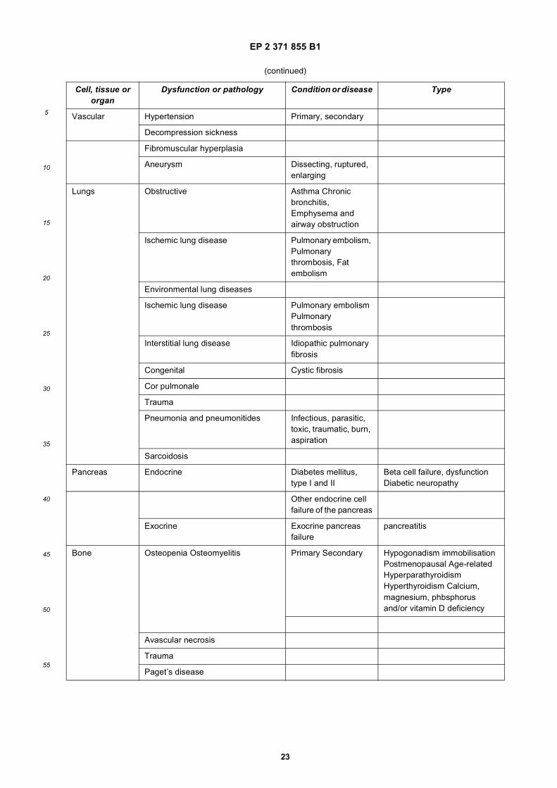
EP 2 371 855 B1
23
5
10
15
20
25
30
35
40
45
50
55
(continued)
Cell, tissue or organ
Dysfunction or pathology Condition or disease Type
Vascular Hypertension Primary, secondary
Decompression sickness
Fibromuscular hyperplasia
Aneurysm Dissecting, ruptured, enlarging
Lungs Obstructive Asthma Chronic bronchitis, Emphysema and airway obstruction
Ischemic lung disease Pulmonary embolism, Pulmonary thrombosis, Fat embolism
Environmental lung diseases
Ischemic lung disease Pulmonary embolism Pulmonary thrombosis
Interstitial lung disease Idiopathic pulmonary fibrosis
Congenital Cystic fibrosis
Cor pulmonale
Trauma
Pneumonia and pneumonitides Infectious, parasitic, toxic, traumatic, burn, aspiration
Sarcoidosis
Pancreas Endocrine Diabetes mellitus, type I and II
Beta cell failure, dysfunction Diabetic neuropathy
Other endocrine cell failure of the pancreas
Exocrine Exocrine pancreas failure
pancreatitis
Bone Osteopenia Osteomyelitis Primary Secondary Hypogonadism immobilisation Postmenopausal Age-related Hyperparathyroidism Hyperthyroidism Calcium, magnesium, phbsphorus and/or vitamin D deficiency
Avascular necrosis
Trauma
Paget’s disease
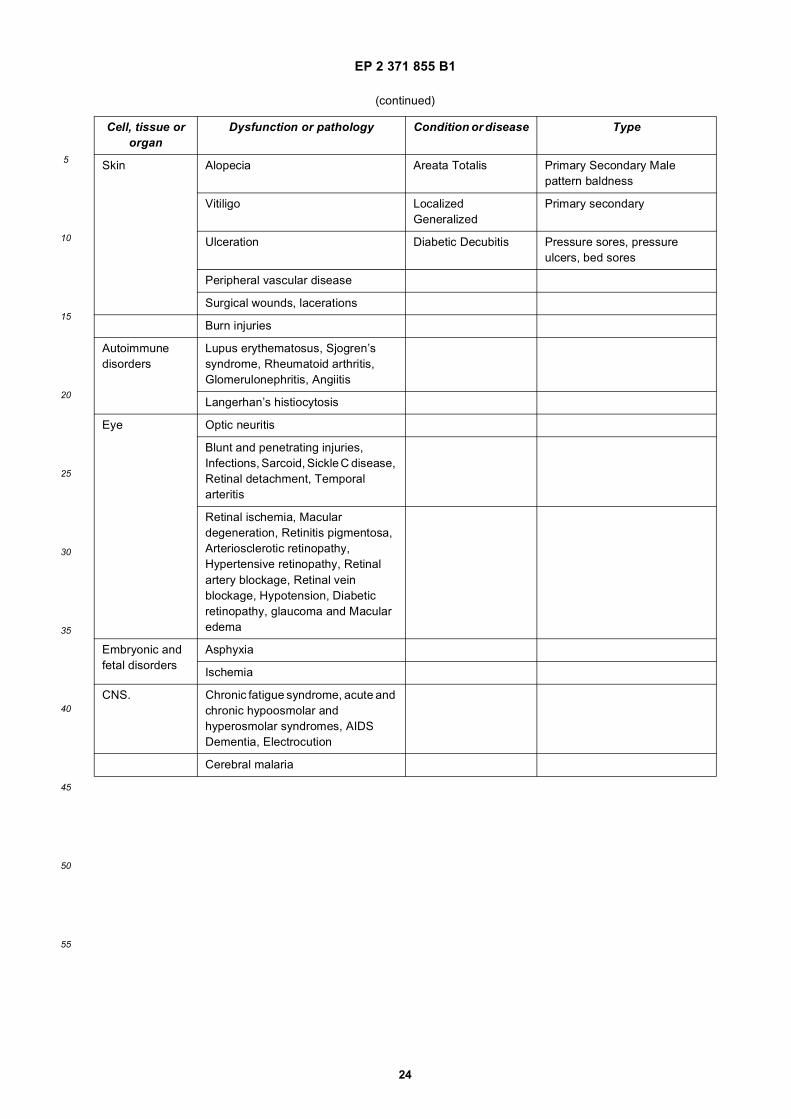
EP 2 371 855 B1
24
5
10
15
20
25
30
35
40
45
50
55
(continued)
Cell, tissue or organ
Dysfunction or pathology Condition or disease Type
Skin Alopecia Areata Totalis Primary Secondary Male pattern baldness
Vitiligo Localized Generalized
Primary secondary
Ulceration Diabetic Decubitis Pressure sores, pressure ulcers, bed sores
Peripheral vascular disease
Surgical wounds, lacerations
Burn injuries
Autoimmune disorders
Lupus erythematosus, Sjogren’s syndrome, Rheumatoid arthritis, Glomerulonephritis, Angiitis
Langerhan’s histiocytosis
Eye Optic neuritis
Blunt and penetrating injuries, Infections, Sarcoid, Sickle C disease, Retinal detachment, Temporal arteritis
Retinal ischemia, Macular degeneration, Retinitis pigmentosa, Arteriosclerotic retinopathy, Hypertensive retinopathy, Retinal artery blockage, Retinal vein blockage, Hypotension, Diabetic retinopathy, glaucoma and Macular edema
Embryonic and fetal disorders
Asphyxia
Ischemia
CNS. Chronic fatigue syndrome, acute and chronic hypoosmolar and hyperosmolar syndromes, AIDS Dementia, Electrocution
Cerebral malaria

EP 2 371 855 B1
25
5
10
15
20
25
30
35
40
45
50
55
(continued)
Cell, tissue or organ
Dysfunction or pathology Condition or disease Type
Encephalitis Rabies, Herpes,
Meningitis
Subdural hematoma
Nicotine addiction
Drug abuse and withdrawal Cocaine, heroin, crack, marijuana, LSD, PCP, poly-drug abuse, ecstasy, opioids, sedative hypnotics, amphetamines, caffeine
Obsessive-compulsive disorders
Spinal stenosis, Transverse myelitis, Guillian Barre, Trauma, Nerve root compression, Tumoral compression, Heat stroke
ENT Tunnitus Meuniere’s syndrome Hearing loss
Traumatic injury, barotraumas
Kidney Renal failure Acute, chronic Vascular/ischemic, interstitial disease, diabetic kidney disease, nephrotic syndromes, infections, injury, contrast-induced, chemotherapy-induced, cyclosporine, CPB-induced, or preventive
Radiation injury
Henoch Schonlein purpura
Striated muscle Autoimmune disorders Myasthenia gravis Dermatomyositis Polymyositis
Myopathies Inherited metabolic, endocrine and toxic
Heat stroke
Crush injury
Rhabdomyolysis
Mitochondrial disease
Infection Necrotizing fasciitis
Sexual dysfunction
Central and peripheral (e.g. erectile dysfunction)
Impotence secondary to medication, (diabetes)
EP 2 371 855 B1
26
5
10
15
20
25
30
35
40
45
50
55
[0131] As mentioned above, these diseases, disorders or conditions are merely illustrative of the range of benefitsprovided by the tissue protective peptides of the present invention. Accordingly, this invention generally provides pre-ventative, therapeutic, or prophylactic treatment of the consequences of mechanical trauma or of human diseases.Prevention, or therapeutic or prophylactic treatment for diseases, disorders or conditions of the CNS and/or peripheralnervous system are contemplated. Prevention of therapeutic or prophylactic treatment for diseases, disorders or con-ditions which have a psychiatric component is provided. Prevention or therapeutic or prophylactic treatment for diseases,disorders or conditions including but not limited to those having an ophthalmic, cardiovascular, cardiopulmonary, respi-ratory, kidney, urinary, reproductive, gastrointestinal, endocrine, or metabolic component is provided.[0132] In one embodiment, such a pharmaceutical composition comprising a tissue protective peptide can be admin-istered systemically to protect or enhance the target cells, tissue or organ. Such administration may be parenterally, viainhalation, or transmucosally, e.g., orally, nasally, rectally, intravaginally, sublingually, ocularly, subinucosally or transder-mally. Preferably, administration is parenteral, e.g,, via intravenous or intraperitoneal injection, and also including, butis not limited to, intraarterial, intramuscular, intradermal and subcutaneous administration.[0133] For other routes of administration, such as by use of a perfusate, injection into an organ, or other local admin-istration, a pharmaceutical composition will be provided which results in similar levels of a tissue protective peptide asdescribed above. A level of about 15 pM -30 nM is preferred.[0134] The pharmaceutical compositions of the invention may comprise a therapeutically effective amount of a com-pound, and a pharmaceutically acceptable carrier. In a specific embodiment, the term "pharmaceutically acceptable"means approved by a regulatory agency of the Federal or a state government or listed in the U.S. Pharmacopeia orother generally recognized foreign pharmacopeia for use in animals, and more particularly in humans. The term "carrier"refers to a diluent, adjuvant, excipient, or vehicle with which the therapeutic is administered. Such pharmaceutical carrierscan be sterile liquids, such as saline solutions in water and oils, including those of petroleum, animal, vegetable orsynthetic origin, such as peanut oil, soybean oil, mineral oil, sesame oil and the like. A saline solution is a preferredcarrier when the pharmaceutical composition is administered intravenously. Saline solutions and aqueous dextrose andglycerol solutions can also be employer as liquid carriers, particularly for injectable solutions. Suitable pharmaceuticalexcipients include starch, glucose, lactose, sucrose, gelatin, malt, rice, flour, chalk, silica gel, sodium stearate, glycerolmonostearate, talc, sodium chloride, dried skim milk, glycerol, propylene glycol, water, ethanol and the like. The com-position, if desired, can also contain minor amounts of wetting or emulsifying agents, for pH buffering agents. These
(continued)
Cell, tissue or organ
Dysfunction or pathology Condition or disease Type
Liver Hepatits Viral, bacterial, parasitic
Ischemic disease
Cirrhosis, fatty liver
Infiltrative/metabolic diseases
Gastrointestinal Ischemic bowel disease
Inflammatory bowel disease
Necrotizing enterocolitis
Organ transplantation
Treatment of donor and recipient
Reproductive tract
Infertility Vascular Autoimmune Uterine abnormalities Implantation disorders
Endocrine Glandular hyper- and hypofunction
General Shock Septic, hemodynamic
Parasitemia Malaria, trypanosomiasis, Leshmaniasis
EP 2 371 855 B1
27
5
10
15
20
25
30
35
40
45
50
55
compositions can take the form of solutions, suspensions, emulsion, tablets, pills, capsules, powders, sustained-releaseformulations and the like. The composition can be formulated as a suppository, with traditional binders and carriers suchas triglycerides. The compounds of the invention can be formulated as neutral or salt forms. Pharmaceutically acceptablesalts include those formed with free amino groups such as those derived from hydrochloric, phosphoric, acetic, oxalic,tartaric acids, etc., and those formed with free carbonyl groups such as those derived from sodium, potassium, ammonium,calcium, ferric hydroxides, isopropylamine, triethylamine, 2-ethylamino ethanol, histidine, procaine, etc. Examples ofsuitable pharmaceutical carriers are described in "Remington’s Pharmaceutical Sciences" by E.W. Martin. Such com-positions will contain a therapeutically effective amount of the compound, preferably in purified form, together with asuitable amount of carrier so as to provide the form for proper administration to the patient. The formulation should suitthe mode of administration.[0135] formulations for increasing transmucosal adsorption of peptides such as long acting tissue protective peptidesare also contemplated by the current invention. Pharmaceutical composition adapted for oral administration may beprovided as capsules or tablets; as powders or granules; as solutions, syrups or suspensions (in aqueous or non-aqueousliquids); as edible foams or whips; or as emulsions. Tablets or hard gelatine capsules may comprise lactose, starch orderivatives thereof magnesium stearate, sodium saccharine, cellulose, magnesium carbonate, stearic acid or saltsthereof. Soft gelatine capsules may comprise vegetable oils, waxes, fats, semi-solid, or liquid polyols etc. Solutions andsyrups may comprise water, polyols and sugars.[0136] An active agent intended for oral administration may be coated with or admixed with a material that delaysdisintegration and/or absorption of the active agent in the gastrointestinal tract (e.g., glycerol monostearate or glyceryldistearate may be used). Thus, the sustained release of an active agent may be achieved over many hours and, ifnecessary, the active agent can be protected from being degraded within the stomach. Pharmaceutical compositionsfor oral administration may be formulated so facilitate release of an active agent at a particular gastrointestinal locationdue to specific pH or enzymatic conditions.[0137] Pharmaceutical compositions adapted for transdermal, administration may be provided as discrete patchesintended to remain in intimate contact with the epidermis of the recipient for a prolonged period of time. Pharmaceuticalcompositions adapted for topical administration may be provided as ointments, creams, suspension, lotions, powders,solutions, pastes, gels, sprays, aerosols or oils. For topical administration to the skin, mouth, eye or other external tissuesa topical ointment or cream is preferably used. When formulated in an ointment, the active ingredient may be employerwith either a paraffinic or a water-miscible ointment base. Alternatively, the active ingredient may be formulated in acream with an oil-in-water base or a water-in-oil base. Pharmaceutical compositions adapted for topical administrationto the eye include eye drops. In these compositions, the active ingredient can be dissolved or suspended in a suitablecarrier, e.g., in an aqueous solvent. Pharmaceutical, compositions adapted for topical administration in the mouth includelozenges, pastilles and mouthwashes.[0138] Pharmaceutical compositions adapted for nasal and pulmonary administration may comprise solid carrierssuch as powders (preferably having aparticle size in the range of 20 to 500 microns). Powders can be administered inthe manner in which snuff is taken, i.e., by rapid inhalation through the nose from container of ponder held close to thenose. Alternatively, compositions adopted for nasal administration may comprise liquid carriers, e.g., nasal sprays, ornasal drops. Alternatively, inhalation of compounds directly into the lungs may te accomplished by inhalation deeply orinstallation through a mouthpiece into the oropharynx. These compositions may comprise aqueous or oil solutions ofthe active ingredient. Compositions for administration by inhalation may be supplied in specially adapted devices includ-ing, but not limited to, pressurized aerosols, nebulizers or insufflators, which can be constructed so as to provide pre-determined dosages of the active ingredient. In a preferred embodiment, pharmaceutical compositions of the inventionare administered into the nasal cavity directly or into the lungs via the nasal cavity or oropharynx.[0139] Pharmaceutical compositions adapted for rectal administration may be provided as suppositories or enemas.Pharmaceutical compositions adapted for vaginal administration may be provided as pessaries, tampons, creams, gels,pastes, foams or spray formulations.[0140] Pharmaceutical compositions adapted for parenteral administration include aqueous and non-aqueous sterileinjectable solutions or suspensions, which may contain antioxidants, buffers, bacteriostats and solutes that render thecompositions substantially isotonic with the blood of an intended recipient. Other components that may be present insuch compositions include water, alcohols, polyols, glycerine and vegetable oils, for example. Compositions adaptedfor parenteral administration may be presented in unit-dose or multi-dose containers, for example sealed ampules andvials, and may be stored in a freeze-dried (lyophilized) condition requiring only the addition of at sterile liquid carrier,e.g., sterile saline solution for injections, immediately prior to use. Extemporaneous injection solutions and suspensionsmay be prepared from sterile powders, granules and tablets. In one embodinent, an autoinjector comprising an injectablesolution of a tissue protective peptide may be provided for emergency use by ambulances, emergency rooms, andbattlefield situations, and even for self administration in a domestic netting, particularly where the possibility of traumaticamputation may occur, such as by imprudent use of a lawn mower. The likelihood that cells and tissues in a severedfoot or toe will survive after reattachment may be increased by administering a tissue protective peptide to multiple sites
EP 2 371 855 B1
28
5
10
15
20
25
30
35
40
45
50
55
in the severed part as soon as practicable, even before the arrival of medical peisonnel on site, or arrival of the afflictedindividual with severed toe in tow at the emergency room.[0141] In a preferred embodiment, the composition is formulated in accordance with routine procedures as a pharma-ceutical composition adapted for intravenous administration to human beings. Typically, compositions for intravenousadministration are solutions in sterile isotonic aqueous-buffer. Where necessary, the composition may also include asolubilizing agent and a local anesthetic such as lidocaine to case pain at the site of the injection. Generally, the ingredientsare supplied either separately or mixed together in unit dosage foam, for example, as a dry lyophilized powder or water-free concentrate in a hermetically-sealed contained such as an ampule or sachette indicating the quantity of active agent.Where the composition is to be administered by infusion, it can be dispensed with an infusion bottle containing sterilepharmaceutical grade water or saline. Where the composition is administered by injection, an ampule of sterile salinecan be provided so that the ingredients may be mixed prior to administration.[0142] Suppositories generally contain active ingredient in the range of 0.5% to 10% by weight; oral formulationspreferably contain 10% to 95% active ingredient.[0143] A perfusate composition may be provided for use in transplanted organ baths, for in situ perfusion, or foradministration to the vasculature of an organ donor prior to organ harvesting. Such pharmaceutical compositions maycomprise levels of tissue protective peptides, or a form of tissue protective peptides not suitable for acute or chronic,local or systemic administration to an individual, but will serve the functions intended herein in a cadaver, organ bath,organ perfusate, or in situ perfusate prior to removing of reducing the levels of the tissue protective peptide containedtherein before exposing or returning the treated organ or tissue to regular circulation.[0144] Disclosed herein is further a pharmaceutical pack or kit comprising one or more containers filled with one ormore of the ingredients of the pharmaceutical compositions of the invention. Optionally associated with such container(s)can be a notice in the form prescribed by a governmental agency regulating the manufacture, use or sale of pharma-ceuticals or biological products, which notice reflects approval by the agency of manufacture, use or sale for humanadministration.[0145] In another embodiment, for example, a tissue protective peptide can be delivered in a controlled-release system.For example, the peptide may be administered using intravenous infusion, an implantable osmotic pump, a transdermalpatch, liposomes, or other modes of administration. In one embodiment, a pump may be used (see Langer, supra;Sefton, 1987, CRC Crit. Ref. Biomed. End. 14:201; Buchwald et al., 1980, Surgery 88:507; Saudek et al., 1989, N. Engl.J. Med. 321:574). In another embodiment, the compound can be delivered in a vesicle, in particular a liposome (seeLanger, Science 249:1527-1533 (1990); Treat et al., in Liposomes in the Therapy of Infectious Disease and Cancer,Lopez-Berestein and Fidler (eds.), Liss, New York, pp. 353-365 (1989); WO 91/04014; U.S. Patent No. 4,704,355; Lopez-Berestein, ibid., pp. 317-327; see generally ibid.). In another embodiment, polymeric materials can be used (see MedicalApplications of Controlled Release, Langer and Wise (eds.), CRC Press: Boca Raton, Florida, 1974; Controlled DrugBioavailability, Drug Product Design and Performance, Smolen and Ball (eds.), Wiley: New York (1984); Ranger andPeppas, J. Macromol. Sci. Rev. Macromol. Chem. 23:61,1953; see also Levy et al., 1985, Science 228:190; During etal., 1989, Ann. Neurol. 25:351; Howard et al, 1989, J. Neurosurg. 71:105.[0146] In yet another embodiment, a controlled release system can be placed in proximity of the therapeutic target,i.e., the target cells, tissue or organ, thus requiring only a fraction of the systemic dose (see, e.g., Goodson, pp. 115-138in Medial Applications of Controlled Release, Vol. 2, supra, 1984). Other controlled release systems are discussed inthe review by Langer (1990, Science 249:1527-1533).[0147] In another embodiment, tissue protective peptide, as property formulated, can be administered by nasal, oral,rectal, vaginal, ocular, transdermal, parenteral or sublingual administration.[0148] In a specific embodiment, it may be desirable to administer a tissue protective peptide of the invention locallyto the area in need of treatment; this may be achieved by, for example, and not by way of limitation, local infusion duringsurgery, topical application, e.g., in conjunction with a wound dressing after surgery, by injection, by means of a catheter,by means of a suppository, or by means of an implant, said implant being of a porous, non-porous, or gelatinous material,including membranes, such as silastic membranes, for fibers. A non-limiting example of such an embodiment would bea coronary stent coated with a tissue protective peptide of the present invention.[0149] Selection of the preferred effective dose will be readily determinable by a skilled artisan based upon consideringseveral factors, which will be known to one of ordinary skill in the art. Such factors include the particular form of tissueprotective peptide, and its pharmacokinetic parameters such as bioavailability, metabolism, half-life, etc., which will havebeen established during the usual development procedures typically employed in obtaining regulatory approval for apharmaceutical compound. Further factors in considering the dose include the conditions of disease to be treated or thebenefit to be achieved in a normal individual, the body mass of the patient, the route of administration, whether admin-istration is acute or chronic, concomitant medications, and other factors well known to affect the efficacy of administeredpharmaceutical agents. Thus the precise dosage should be decided according to the judgment of the practitioner andeach patient’s circumstances, e.g., depending upon the condition and the immune status of the individual patient, andaccording to standard clinical techniques.
EP 2 371 855 B1
29
5
10
15
20
25
30
35
40
45
50
55
[0150] In another aspect of the present invention, a pharmaceutical composition according to the present inventionmay include a tissue protective peptide in a formulation with at least one small molecule that exhibits tissue protectivefunctionality. Suitable small molecules include, but are not limited to, steroids (e.g., lazaroids and glucocorticoids),antioxidants (e.g., coenzyme Q10, alpha lipoic acid, and NADH), anticatabolic enzymes (e.g., glutathione peroxidase,superoxide dimutase, catalase; synthetic catalytic scavengers, as well as mimetics), indole derivatives (e.g., indolamines,carbazoles, and carbolines), nitric acid neutralizing agents, adenosine/ adenosine agonists, phytochemicals (flavanoids),herbal extracts (ginko biloba and turmeric), vitamins (vitamins A, E, and C), oxidase electron acceptor inhibitors (e.g.,xanthine oxidase electron inhibitors), minerals (e.g., copper, zinc, and magnesium), non-steriodal anti-inflammatorydrugs (e.g., aspirin, naproxen, and ibuprofen), and combinations thereof. Additionally agents including, but not limitedto, anti-inflammatory agents (e.g., corticosteroids, prednisone and hydrocortisone), glucocorticoids, steroids, non-steri-odal anti-inflammatory drags (e.g., aspirin, ibuprofen, dialofenac, and COX-2 inhibitors), beta-agonists, anticholinergicagents and methyl xanthines), immunomodulatory agents (e.g., small organic molecules, T cell receptor modulators,cytokine receptor modulators, T-cell depleting agents, cytokine antagonists, monokine antagonists, lymphocyte inhibitors,or anti-cancer agents), gold injections, sulphasalazine, penicillamine, anti-angiogenic agents (e.g., angiostaiin), TNF-αantagonists (e.g., anti-TNFα antibodies), and endostatin), dapsone, psoralens (e.g., methoxalen and trioxsalen), anti-malarial agents (e.g., hydroxychloroquine), anti-viral agents, and antibiotics (e.g., erythromycin and penicillin) may beused in conjunction with the current pharmaceutical compositions.[0151] In another aspect of the invention a perfusate or perfusion solution is provided for perfusion and storage oforgans for transplant, the perfusion solution includes an amount of a tissue protective peptide effective to protect re-sponsive cells and associated cells, tissues or organs. Transplant includes but is not limited to allotransplantation, wherean organ (including cells, tissue or other bodily part) is harvested from one donor and transplanted into a differentrecipient, both being of the same species; autotransplantation, where the organ is taken from one part of a body andreplaced at another, including bench surgical procedures, in which an organ may be removed, and while ex vivo,respected, repaired, or otherwise manipulated, such as for tumor removal, and then returned to the original location orxenotransplantation, where tissues or organs or transplanted between species. In one embodiment, the perfusion solutionis the University of Wisconsin (UW) solution (U.S. Patent No. 4,798,824), contains from about 1 to about 25 U/ml (10ng = 1U) of tissue protective peptide, 5% hydroxyethyl starch (having a molecular weight of from about 200,000 to about300,000 and substantially free of ethylene glycol, methylene chlofohydrin, sodium chloride and acetone); 25 mM KH2PO4;3 mM glutathione; 5 mM adenosine; 10 mM glucose; 10 mM HEPES buffer; 5 mM magnesium gluconate; 1.5 mM CaCl2;105 mM sodium gluconate; 200,000 units penicillin; 40 units insulin; 16 mg dexamethasone; 12 mg Phenol Red; andhas a pH of 7.4-7.5 and an osmolality of about 320 mOsm/l. The solution is used to maintain cadaveric kidneys andpancreases prior to transplant. Using the solution, preservation can be expended beyond the 30-hour limit recommendedfor cadaveric kidney preservation. This particular perfusate is merely illustrative of a number of such solutions that canbe adapted for the present use by inclusion of an effective amount of a tissue protective peptide. In a further embodiment,the perfusate solution contains from about 1 to about 500 ng/ml of a tissue protective peptide, or from about 40 to about320 ng/ml tissue protective peptide. As mentioned above, any form of tissue protective peptide can be used in this aspectof the invention.[0152] While the preferred recipient of a tissue protective peptide for the purposes herein throughout is a human, themethods herein apply equally to other mammals, particularly domesticated animals, livestock, companion, and zooanimals. However, the invention is not so limiting and the benefits can be applied to any mammal.[0153] In further aspects of the ex-vivo invention, any tissue protective peptide such as but not limited to the onesdescribed above may be employed.[0154] In another aspect of the invention, methods and compositions for enbancing the viability of cells, tissues ororgans which are not isolated from the vasculature by an endothelial cell barrier are provided by exposing the cells,tissue or organs directly to a pharmaceutical composition comprising a tissue protective peptide, or administering orcontacting a pharmaceutical composition containing a tissue protective peptide to the vasculature of the tissue or organ.Enhanced activity of responsive cells in the treated tissue or organ is responsible for the positive effects exerted.[0155] Similar to other tissue protective compounds based on erythropoietin, it is possible that the tissue protectivepeptides of the present invention may be transported from the luminal surface to the basement membrane surface ofendothelial cells of the capillaries of organs with endothelial cell tight junctions, including, for example, the brain, retina,and testis. Thus, responsive cells across the barrier may be susceptible targets for the beneficial effects of tissueprotective peptides, and others cell types or tissues or organs that contain and depend in whole or in part on responsivecells therein may be targets for the methods of the invention. While not wishing to be bound by any particular theory,after, transcytosis of the tissue protective peptide may interact with an tissue-protective receptor on a responsive cell,for example, neuronal, eye (e.g., retinal), adipose, connective, hair, tooth, mucosal, pancreatic, endocrine, aural, epi-thelial, skin, muscle, heart, lung, liver, kidney, small, intestine, adrenal (e.g adrenal cortex, adrenal medulla), capillary,endothelial, testes, ovary, or endometrial cell, and receptor binding can initiate a signal transduction cascade resultingin the activation of a gene expression program within the responsive cell or tissue, resulting in the protection of the cell
EP 2 371 855 B1
30
5
10
15
20
25
30
35
40
45
50
55
or tissue, or organ, from damage, such as by toxins, chemotherapeutic agents, radiation therapy, hypoxia, etc. In anotherembodiment, the tissue protective peptide can be cross-linked to a compound that can cross the barrier, such, ascarbamylated erythropoietin, to be transported across the barrier in accordance with the teaching of PCT ApplicationNo. PCT/US01/49479, U.S. Patent Application Nos. 10/188,905 and 10/185,841. Thus, methods for protecting responsivecell-containing tissue from injury or hypoxic stress, and enhancing the function of such tissue are described in detailherein below.[0156] In the practice of one embodiment of the invention, a mammalian patient is undergoing systemic chemotherapyfor cancer treatment, including radiation therapy, which commonly has adverse effects such as nerve, lung, heart; ovarianor testicular damage. Administration of a pharmaceutical composition comprising a tissue protective peptide as describedabove is performed prior to and during chemotherapy and/or radiation therapy, to protect various tissues and organsfrom damage by the chemotherapeutic agent, such as to protect the testes. Treatment may be continued until circulatinglevels of the chemotherapeutic agent have fallen below a level of potential danger to the mammalian body.[0157] In the practice of another embodiment of the invention, various organs are planned to be harvested from avictim of an automobile accident for transplant into a number of recipients, some of which required transport for anextended distance and period of time. Prior to organ harvesting, the donor is infused with a pharmaceutical compositioncomprising tissue protective peptides as described herein. Harvester organs for shipment are perfused with a perfusatecontaining tissue protective peptides as described herein, and stored in a bath comprising tissue protective peptides.Certain organs are continuously perfused with a pulsatile perfusion device, utilizing a perfusate containing tissue pro-tective peptides in accordance with the present invention. Minimal deterioration of organ function occurs during thetransport and upon implant and reperfusion of the organs in situ.[0158] In another embodiment of the present invention, a participant in a hazardous activity, one could take a dose ofa pharmaceutical composition containing a tissue protective peptide sufficient to either prevent (i.e. delaying the-onsetof, inhibiting, or stopping), protect against, or mitigate the damage resulting from an injury to a responsive cell, tissue,or organ. In particular, this method of treatment may have application in various professions susceptible to injury suchas, but not limited to, professional athletes (divers, race car drivers, football players, etc.), military personnel (soldiers,paratroopers), emergency personnel (police, fire, EMS, and disaster relief personnel), stuntmen, and construction work-ers. Additionally, the prophylactic use of tissue protective peptides is contemplated in such recreational endeavorsincluding, but not limited to, rock climbing, rappelling, sky diving, racing, bicycling, football, rugby, baseball, and divingthat pose a risk of injury.[0159] In another embodiment of the intention, a surgical procedure to repair a heart valve requires temporary cardi-oplegia and arterial occlusion. Prior to surgery, the patient is infused with a tissue protective peptide. Such treatmentprevents hypoxic ischemic cellular damage, particularly after reperfusion. Additionally, the pharmaceutical compositionsof the present invention may be used prophylactically to prepare an individual for surgery in an effort to limit the traumaassociated with the surgical procedure or aide in the recovery of the individual from the surgical procedure. Althoughthe present method of treatment using pharmaceutical compositions containing tissue protective peptides provides aprophylactic use for surgical procedures, it may be particularly useful in procedures that induce temporary ischemicevents including, but not limited to, bypass procedures (coronary bypass), angioplasty procedures, amputations, andtransplantations, as well as, those performed directly upon responsive cells, tissues, or organs such as brain and spinalcord surgery, and open heart procedures. Such procedures may involve the use of cardiopulmonary (heart lung) bypass.[0160] In another embodiment of the invention, in any surgical procedure, such as in cardiopulmonary bypass surgery,a tissue protective peptide of the invention can be used. In one embodiment, administration of a pharmaceutical com-position comprising tissue protective peptides as describes above is performed prior to, during, and/or following thebypass procedure, to protect the function of brain, heart, and other organs.[0161] In the foregoing examples in which a tissue protective peptide of the invention is used for ex-vivo applications,or for in vivo applications to treat responsive cells such as neuronal tissue, retinal tissue heart, lung, liver, kidney, smallintestine, adrenal cortex, adrenal medulla; capillary endothelial, testes, ovary, or endometrial cells or tissue, the inventionprovides a pharmaceutical composition in dosage unit form adapted for projection or enhancement of responsive cells,tissues or organs distal to the vasculature which comprises an amount within the range from about .01 pg to 7.5 mg, .5pg to 6.5 mg, 1 pg to 5 mg, 500 pg to 5 mg, 1 ng to 5 mg, 500 ng to 5 mg, 1 ng to 5 mg, 500 mg to 5 mg of 1 mg to 5mg of a tissue protective peptide, and a pharmaceutically acceptable carrier. In a preferred embodiment, the amount oftissue protective peptide is within the range from about .5 pg to 1 mg. In a preferred embodiment the formulation containstissue protective peptides that are non-erythropoietic.[0162] In a further aspect of the invention, administration of tissue protective peptides may be used to restore cognitivefunction in mammals having undergone brain trauma. After a delay of either 5 days or 30 days, administration of tissueprotective peptides should be able to restore function as compared to placebo-treated mammals, indicating the abilityof the tissue protective peptide to regenerate or restore brain activity. Thus, the invention is also directed to the use oftissue protective peptides for the preparation of a pharmaceutical composition for the treatment of brain trauma andother cognitive dysfunctions, including treatment well after the injury (e.g. three days, five days, a week, a month, or

EP 2 371 855 B1
31
5
10
15
20
25
30
35
40
45
50
55
lounger). The invention is also directed to a method for the treatment of cognitive dysfunction following injury by admin-istering an effective amount of tissue protective peptides. Any tissue protective peptide as described herein may be usedfor this aspect of the invention.[0163] Furthermore, this restorative aspect of the invention is directed to the use of any tissue protective peptidesherein for the preparation of a pharmaceutical composition for the restoration of cellular, tissue or organ dysfunction,wherein treatment is initiated after, and well after, the initial insult responsible for the dysfunction. Moreover, treatmentusing tissue protective peptides of the invention can span the course of the disease or condition during the acute phaseas well as a chronic phase.[0164] A tissue protective peptide of the invention may be administered systemically at a dosage between about 1 ngand about 100 mg /kg body weight, preferably about 5 -50 mg /kg-body weight, most preferably about 10-30 mg /kg-bodyweight, per administration. This effective dose should be sufficient to achieve serum levels of tissue protective peptidesgreater than about 80,120, or 160 ng/ml of scrum after administration. Such serum levels may he achieved at about 1,2, 3, 4, 5, 6, 7, 8, 9, or 10 hours post-administration. Such dosages may be repeated as necessary. For example,administration may be repeated daily, as long as clinically necessary, or after an appropriate interval, e.g., every 1 to12 weeks, preferable, every 1 to 3 weeks. In one embodiment, the effective amount of tissue protective peptide and apharmaceutically acceptable carrier may be packaged in a single dose vial or other container. In another embodiment,the tissue protective peptides, which are capable of exerting the activities described herein but not causing an increasein hemoglobin concentration or hematocrit, are used. Such tissue protective peptides are preferred in instances whereinthe methods of the present invention are intended to he provided chronically.
5.4 TRANSCYTOSIS
[0165] Carrier Molecule and Tissue Protective Peptide. The present invention is further directed to a method forfacilitating the transport of a Tissue Protective Peptide across an endothelial tell barrier in a mammal by administeringa composition which comprises the tissue protective peptide in association with a carrier peptide, a peptide capable ofcrossing an endothelial cell barrier, such as erythropoietin, as described hereinabove. Tight junctions between endothelialcells in certain organs in the body create a barrier to the entry of certain molecules. For treatment of various conditionswithin the barriered organ, means for facilitating passage of the tissue protective peptide may be desired.[0166] Tissue Protective Peptide as Carrier Molecule. Tissue protective peptides of the invention may be useful ascarriers for delivering other molecules across the blood-brain and ether similar barriers which they can gravel across.A composition comprising a molecule desirous of cessing the barrier with a tissue protective peptide is prepared andperipheral administration of the composition results in the transcytosis of the composition across the barrier. The asso-ciation between the molecule to be transported across the barrier and the tissue protective peptide may be a labilecovalent bond, in which case the molecule is released from association with the tissue protective peptide after crossingthe barrier. If the desired pharmacological activity of the molecule is maintained or unaffected by association with tissueprotective peptides, such a complex can be administered.[0167] The skilled artisan will be aware of various means for associating molecules with tissue protective peptides ofthe invention and the other agents described above, by covalent, non-covalent, and other means. Furthermore, evaluationof the efficacy of the composition can be readily determined in an experimental system. Association of molecules withtissue protective peptides may be achieved by any number of means, including labile, covalent binding, cross-linking,etc. Biotin/avidin interactions may be employed; for example, a biotinylated tissue protective peptides of the inventionmay be complexed with a labile conjugate of avidin and a molecule desirably transported. As mentioned above, a hybridmolecule may be prepared by recombinant or synthetic means, for example, a fusion or chimeric polypeptide whichincludes both the domain of the molecule with desired pharmacological activity and the domain responsible for thepeptides tissue-protective receptor activity modulation. Protease cleavage sites may be included in the molecule.[0168] A molecule may be conjugated to a tissue protective peptide of the invention through a polyfunctional molecule,i.e., a polyfunctional crosslinker. As used herein, the term "polyfunctional molecule" encompasses molecules havingone functional group that can react more than one time in succession, such as formaldehyde, as well as molecules withmore than one reactive group. As used herein, the term "reactive group" refers to a functional group on the crosslinkerthat redacts with a functional group on a molecule (e.g., peptide, protein, carbohydrate, nucleic acid, particularly ahormone, antibiotic, or anti-cancer agent to be delivered across an endothelial cell barrier) so as to form a covalent bondbetween the cross-linker and that molecule. The term "functional group" retains its standard meaning in organic chemistry.The polyfunctional molecules that can be used are preferably biocompatible linkers, i.e., they are noncarcinogenic,nontoxic, and substantially non-immunogenic in vivo. Polyfunctional cross-linkers such as those known in the art anddescribed herein can be readily tested in animal models to determine their biocompatibility. The polyfunctional moleculeis preferably bifunctional. As used herein, the term "bifunctiorial molecule" refers to a molecule with two reactive groups.The bifunctional molecule may be heterobifunctional or homobifunctitional. A heterobifunctional cross-linker, allows forvectorial conjugation. It is particularly preferred for the polyfunctional molecule to be sufficiently soluble in water for the
EP 2 371 855 B1
32
5
10
15
20
25
30
35
40
45
50
55
cross-linking reactions to occur in aqueous solutions such as in aqueous solutions buffered at pH 6 to 8, and for theresulting conjugate to remain water soluble for more effective bio-distribution. Typically, the polyfunctional moleculecovalently bonds with an amino or a sulfhydryl functional group. However, polyfunctional molecules reactive with otherfunctional groups, such as carboxylic acids or hydroxyl groups, are contemplated in the present invention.[0169] The homobifunctional molecules have at least two reactive functional groups, which are the same. The reactivefunctional groups on a homobifunctional molecule include, for example, aldehyde groups and active ester groups. Ho-mobifunctional molecules having aldehyde groups include, for example, glutaraldehyde and subaraldehyde. The use ofglutaraldehyde as a cross-linking agent was disclosed by Poznansky et al., Science 223, 1304-1306 (1984). Homobi-functibnal molecules having at least two active ester unites include esters of dicarboxylic acids and N-hydroxysuccinimide.Some examples of such N-succinimidyl esters include disuccinimidyl suberate and dithio-bis-(succinimidyl propionate),and their soluble bis-sulfonic acid and bis-sulfonate salts such as their sodium and potassium salts. These homobifunc-tional reagents are available from Pierce, Rockford, Illinois.[0170] The heterobifunctional molecules have at least two different reactive groups. The reactive groups react withdifferent functional groups, e.g., present on the peptide and the molecule. These two different functional groups thatreact with the reactive group on the heterobifunctional cross-linker are usually an amino group, e.g., the epsilon aminogroup of lysine; a sulfhydryl group, e.g., the thiol group of cysteine; a carboxylic acid, e.g., the carboxylate on asparticacid; or a hydroxyl group, e.g., the hydroxyl group on serine.[0171] Of course, certain of the various tissue protective peptides of the invention, may not have suitable reactivegroups available for use with certain cross-linking agent; however, one of skill in the art will be amply aware of the choiceof cross-linking agents based on the available groups for cross-linking in tissue protective peptides of the invention.[0172] When a reactive groups of a heterobifunctional molecule forms a covalent bond with an amino group, thecovalent bond will usually be an amido or imido bond. The reactive group that forms a covalent bond with an aminogroup may, for example, be an activated carboxylate group, a helocarbonyl group, or an ester group. The preferredhalocarbonyl group is a chlorocarbonyl group. The ester groups are preferably reactive ester groups such as, for example,an N-hydroxy-succimmide ester group.[0173] The other functional group typically is either a thiol group, a group capable of being converted into a thiol group,or a group that forms a covalent bond with a thiol group. The covalent bond will usually be a thioether bond or a disulfide.The reactive group that forme a covalent bond with a thiol group may, for example, be a double bond that redacts withthiol groups or an activated disulfide. A reactive group containing a double bond capable of reacting with a thiol groupis the maleimido group, although others, such as acrylonitrile, are also possible. A reactive disulfide group may, forexample, be a 2-pyridyldithio group or a 5, 5’-dithio-bis-(2-nitrobenzoic acid) group. Some examples of heterobifunctiohalreagents containing reactive disulfide bonds include N-succinimidyl 3-(2-pyridyl-dithio) propionate (Carlsson, et al., 1978,Biochem J., 173:723-737), sodium S-4-succinimidyloxycarbonyl-alpha-methylbehzylthiosulfate, and 4-succinimidyloxy-carbonyl-alpha-methyl-(2-pyridyldithio)toluene. N-succinimidyl 3-(2-pyridyldithio) propionate is preferred. Some exam-ples of heterobifunctional reagents comprising reactive groups having a double bond that redacts with a thiol groupinclude succinimidyl 4-(N-maleimidomethyl)cyclohexane-1-carboxylate and succinimidyl m-maleimidobenzoate.[0174] Other heterobifunctional molecules include succinimidyl 3-(maleimido) propionate, sulfosuccinimidyl 4-(p-male-imido-phenyl) butyrate, sulfosuccinimidyl 4-(N-maleimidomethyl- cyclohexane)-1-carboxylate, maleimidobenzoyl-N-hy-droxysuccinimide ester. The sodium sulfonate salt of succinimidyl m-maleimidobenzoate is preferred. Many of the above-mentioned heterobifunctional reagents and their sulfonate salts are available from Pierce Chemical Co., Rockford, IllinoisUSA.[0175] The need for the above-described conjugated to be reversible or labile may be readily determined by the skilledartisan. A conjugate may be tested in vitro for desirable pharmacological activity. If the conjugate retains both properties(the properties of the conjugated molecule and the properties of the tissue protective peptide), its suitability may thenbe tested in vivo. If the conjugated molecule requires separation from the tissue protective peptide for activity, a labilebond or reversible association with long acting erythropoietin or the long acting tissue protective cytokine will be preferable.The lability characteristics may also be tested using standard in vitro procedures before in vivo testing.[0176] Additional information regarding how to make and use these as well as other polyfunctional reagents may beobtained from the hollowing publications or others available in the art:
Carlsson, J. et al., 1978, Biochem. J. 173:723-737;Cumber, J.A. et al., 1985, Methods in Enzymology 112:207-224;Jue, R. et al., 1978, Biochem 17:5399-5405;Sun, T.T. et al., 1974, Biochem. 13:2334-2340;Blattler, W.A. et al., 1985, Biochem. 24:1517-152;Liu, F.T. et al., 1979, Biochem. 18:690-697;Youle, R.J. and Neville, D.M. Jr., 1980, Proc. Natl. Acad. Sci. U.S.A. 77:5483-5486;Lerner, R.A. et al., 1981, Proc. Natl. Acad. Sci. U.S.A. 78:3403-3407;
EP 2 371 855 B1
33
5
10
15
20
25
30
35
40
45
50
55
Jung, S.M. and Moroi, M., 1983, Biochem. Biophys. Acta 761:162;Caulfield, M.P. et al.; 1984; Biochem. 81:7772-7776;Staros, J.V., 1982, Biochem. 21:3950-3955;Yoshitake, S. et al., 1979, Eur. J. Biochem. 101:395-399;Yoshitake, S. et al., 1982, J. Biochem. 92:1413-1424;Pilch, P.F. and Czech, M.P., 1979, J. Biol. Chem. 254:3375-3381;Novick, D. et al., 1987, J. Biol, Chem. 262:8483-8487;Lomant, A.J. and Fairbanks, G., 1976, J. Mol. Biol. 104:243-261;Hamada, H. and Tsuruo, T., 1987, Anal. Biochem. 160:483-488; orHashida, S. et al., 1984, J. Applied Biochem. 6:56-63.
[0177] Additionally, methods of cross-linking are reviewed by Means and Feeney,1990, Bioconjugate Chem. 1:2-12.[0178] Barriers which are crossed by the above-described methods and compositions of the present invention includebut are not limited to the blood-brain barrier, the blood-eye barrier, the blood-testes barrier, the blood-ovary barrier,blood-nerve barrier, blood-spinal cord barrier, and blood-placenta barrier.[0179] Candidate molecules for transport across an endothelial cell barrier include, for example, hormones, such asgrowth hormone, neurotrophic factors, antibiotics, andvirals, or antifungals such as those normally excluded from thebrain and other barriered organs, peptide radiopharmaceuticals, antisense drugs, antibodies and antivirals against bio-logically-active gents, pharmaceuticals, and anti-cancer agents. Non-limiting examples of such molecules include hor-mones such as growth hormone, nerve growth factor (NGF), brain-derived neurotrophic factor (BDNF), ciliary neuro-trophic factor (CNTF), basic fibroblast growth factor (bFGF), transforming growth factor β1 (TGFβ1), transforming growthfactor β2 (TGFβ2), transforming growth factor β3 (TGFβ3), interleukin 1, interleukin 2, interleukin 3, and interleukin 6,AZT, antibodies against tumor necrosis factor; and immunosuppressive agents such as cyclosporin. Additionally, dyesor markers may be attached to the tissue protective peptides of the present invention in order to visualize cells, tissues,or organs within the brain and other barriered organs for diagnostic purposes. As an example, a marker used to visualizeplaque within the brain could be attached to a tissue protective peptide in order to determine the progression of Alzheimer’sdisease within a patient.[0180] The present invention is also directed to a composition comprising a molecule to be transported via transcytosisacross an endothelial cell tight junction barrier and a tissue protective peptide as described above. The invention isfurther directed to the use of a conjugate between a molecule and a tissue protective peptide cytokine as describedabove for the preparation of a pharmaceutical composition for the delivery of the molecule across a barrier as describedabove.[0181] Various animal models and in-vitro tests of neuroprotection and transcytosis are provided in PGT/US01/49479to demonstrate the effectiveness of the tissue protective peptides of the invention. For transcytosis, model proteinsconjugated to the long acting erythropoietins of the invention are evaluated for transport into the brain following parenteraladministration. These tests in in-vitro models and animal models are predictive of the efficacy of the present compoundsin other mammalian species including humans.[0182] The present invention may be better understood by reference to the following non-limiting Examples, whichare provided as exemplary of the invention. The following examples are presented in order to more fully illustrate thepreferred embodiments of the invention.
6. EXAMPLES
EXAMPLE 1: METHOD OF PEPTIDE SYNTHESIS.
[0183] A. Synthesis of Peptide A (SEQ ID NO:32, corresponding to EPO amino acid sequence 38-57) and Peptide B(SEQ ID NO:34, corresponding to EPO amino acid sequence 58-82).[0184] Peptide A, SEQ ID NO:32, and Peptide B, SEQ ID NO:34, fragments of EPO (see Table 1), were synthesizedusing "in situ neutralization" Boc Chemistry stepwise solid-phase peptide synthesis, as described in Band, D., Chopra,N and Kent, S., "Total Synthesis of Crambin," J.AM. CHEM. SOC. 2004, 126, 1377-1383. Briefly, two fragments corre-sponding to EPO amino acid sequence 38-57 (peptide C, NITVPDTKVNFYAWKRMEVG, SEQ ID NO:29) and EPOamino acid sequence 58-82 peptide D, QQAVEVWQGLALLSEAVLRGQALLV, SEQ ID NO:30) where synthesized on-OCH2-Pam-resins (free αcarboxyl peptides) or on HSCHR2CH2CO-Leu-OCH2-Pam-Resin (αthioester peptides). Duringsynthesis the side chains of various amino acids were protected as follows: Arg(Tos),Asn(Xan), Asp(OcHex), Cys(4-CH3Bzl) or Cys(ACM), Glu(OcHex), Lys(2-Cl-Z), Ser(Bzl), Thr(Bzl), Tyr(Br-Z). After the peptide chain was assembled,the peptides were deprotected and simultaneously cleaved from the resin support by treatment with anhydrous HFcontaining p-cresol (90:10, v/v) for 1 hr at 0 °C. After evaporation of the HF under reduced pressure, crude productswere precipitated and triturated with chilled diethyl ether, and the peptides were dissolved in 50% aqueous acetonitrile
EP 2 371 855 B1
34
5
10
15
20
25
30
35
40
45
50
55
containing 0.1% TFA and purified by the preparative HPLC system. Peptide compositions were confirmed using LC-MS.
EXAMPLE 2. VALIDATION OF PEPTIDE-MEDIATED TISSUE PROTECTION.
[0185] The tissue protective peptides were tested for any tissue protective activity using a Sciatic Nerve Assay. Sprague-Dawley rats (250-300 grams) (six per group, including control) were anesthetized using isoflurane (Baxter NPC10019-773-60) and a Table Top Laboratory Anesthesia System (flowmeter set to 2-3 liters/minute @ 55 psi) for at least3 minutes. The rat was then placed on a homeothermic blanket to ensure that the core temperature of the rat wasmaintained at 35-37 °C during the operation. Core temperature was monitored via a rectal probe. The right sciatic nerveof the anesthetized rat was exposed at mid thigh through a quadriceps muscle dissection; a 2 cm incision with a 15blade scalpel was made through the skin parallel and over the quadriceps muscle and the quadriceps muscle was cutto expose the sciatic nerve using a pair of dissecting scissors. The sciatic nerve was then freed from the surroundingmembranes. A 2-0 braided silk thread (Ethicon, 685-G) was passed under the nerve and the ends of the suture passedthrough a guide which was maintained perpendicular to the nerve. The end of the suture was then tied to a non-elasticcord which was then draped around the pulley system (a NYL pulley hearing MTD ¨"B (PO Number 04174-01) withstabilizer) and a 100 gram weight attached to the non-elastic cord was slowly released The weight was allowed to hangfor 1 minute before the silk suture was cut to release the weight.[0186] A 289 pmol/kg dose of carbamylated erythropoietin,_a 289 pmol/kg dose of one peptide from the series A-J(see Table 1), or PBS was then injected into the caudal vein using a © cc insulin springe. A 20mer fragment (correspondingto amino acids 102-121) from pigment epithelium-derived growth factor (PEDF) which does not follow the teaching abovewas used as control.[0187] The muscle and surgical incision were then closed and 5 ml of Lactated Ringers solution was injected subcu-taneously into the rat. The core temperature of the rat was maintained at 35-37 °C using a heat blanket during recovery.[0188] Over the next four days the rear toe splaying of the rats was determined by placing the rat in an acrylic tubewith a diameter of 30 cm on the scanning surfece of a digital scanner. After waiting 5 minutes in order to permit acclimation,a scan was taken of the rat’s back feet that clearly displayed all 5 toes. Three acceptable scans of each rat were taken.From the scans, the Toe Spread (the distance between the ball of the first toe and the ball of the fifth toe) and theIntermediate Toe Spread (the distance between the ball of the second toe and the ball of the fourth toe) were measured.The static, sciatic index was then computed in accordance with S. Erbayraktar et al., 2003, Proc Natl Acad Sci US A100, 6741-6746 and statistical analysis performed.[0189] All peptides except B (SEQ ID NO:34), H (SEQ ID NO:47) and the PEDF derivative were equally protective,providing a static sciatic index ("SSI") of about -0.57 versus a SSI of about 67 to about -68 for PBS/ PEDF fragment(FIG. 2). Figure 2 also shows that the efficacy of the positive peptides was at least equivalent, if not improved, over thatof the carbamylated erythropoietin.[0190] Table 1 also presents the approximate distance between carbonyl carbons for the tested peptides. Distanceswere calculated using the three-dimensional coordinates provided by Cheetham et al., 1998, Nat. Struct. Biol. 5:861-866.The peptides which tested positive for tissue protective activity each had a carbonyl carbon to carbonyl carbon dis-tance/separation of between about 3 Å to about 5 Å.
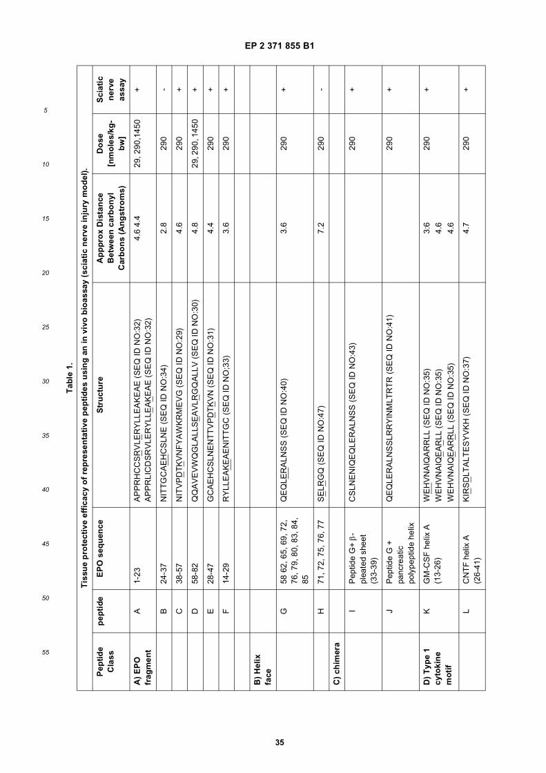
EP 2 371 855 B1
35
5
10
15
20
25
30
35
40
45
50
55
Tab
le 1
.
Tis
sue
pro
tect
ive
effi
cacy
of
rep
rese
nta
tive
pep
tid
es u
sin
g a
n in
viv
o b
ioas
say
(sci
atic
ner
ve in
jury
mo
del
).
Pep
tid
e C
lass
pep
tid
eE
PO
seq
uen
ceS
tru
ctu
reA
pp
pro
x D
ista
nce
B
etw
een
car
bo
nyl
C
arb
on
s (A
ng
stro
ms)
Do
se
[nm
ole
s/kg
-b
w]
Sci
atic
n
erve
as
say
A)
EP
O
frag
men
tA
1-23
AP
PR
HC
CS
RV
LER
YLL
EA
KE
AE
(S
EQ
ID N
O:3
2)
AP
PR
LIC
DS
RV
LER
YLL
EA
KE
AE
(S
EQ
ID N
O:3
2)4.
6 4.
429
, 290
,145
0+
B24
-37
NIT
TG
CA
EH
CS
LNE
(S
EQ
ID N
O:3
4)2.
829
0-
C38
-57
NIT
VP
DT
KV
NF
YA
WK
RM
EV
G (
SE
Q ID
NO
:29)
4.6
290
+
D58
-82
AV
EV
WQ
GLA
LLS
EA
VLR
GQ
ALL
V (
SE
Q ID
NO
:30)
4.8
29, 2
90, 1
450
+
E28
-47
GC
AE
HC
SLN
EN
TT
VP
DT
KV
N (
SE
Q ID
NO
:31)
4.4
290
+
F14
-29
RY
LLE
AK
EA
EN
ITT
GC
(S
EQ
ID N
O;3
3)3.
629
0+
B)
Hel
ix
face
G58
62,
65,
69,
72,
76
, 79,
80,
83,
84,
85
QE
QLE
RA
LNS
S (
SE
Q ID
NO
:40)
3.6
290
+
H71
, 72,
75,
76,
77
SE
LRG
Q (
SE
Q ID
NO
:47)
7.2
290
-
C)
chim
era
IP
eptid
e G
+ β
-pl
eate
d sh
eet
(33-
39)
CS
LNE
NIQ
EQ
LER
ALN
SS
(S
EQ
ID N
O:4
3)29
0+
JP
eptid
e G
+
panc
reat
ic
poly
pept
ide
helix
QE
QLE
RA
LNS
SLR
RY
INM
LTR
TR
(S
EQ
ID N
O:4
1)29
0+
D)
Typ
e 1
cyto
kin
e m
oti
f
KG
M-C
SF
hel
ix A
(1
3-26
)W
EH
VN
AIQ
AR
RLL
(S
EQ
ID N
O:3
5)3:
629
0+
WE
HV
NA
IQE
AR
LL (
SE
Q ID
NO
:35)
4.6
WE
HV
NA
IQE
AR
RLL
(S
EQ
ID N
O:3
5)4.
6
LC
NT
F h
elix
A
(26-
41)
KIR
SD
LTA
LTE
SY
VK
H (
SE
Q ID
NO
:37)
4.7
290
+

EP 2 371 855 B1
36
5
10
15
20
25
30
35
40
45
50
55
EXAMPLE 3. TISSUE PROTECTIVE PEPTIDES ARE NON ERYTHROPOIETIC.
A. In Vitro Assessment:
[0191] UT-7epo, a human erythropoietin-dependent leukemia cell line, was used for the determination of the erythro-poietic potency of the peptides. UT-7epo cells (Deutsche Sammlung von Mikroorganismen und Zellkulturen (DSMZ),Cat. No. ACC 363) were grown in a complete RPMI-1640 medium with 10% FBS and 5 ng/ml erythropoietin. Theproliferation/survial (= viability increase) response of the cells exposed to erythropoietin is mediated % the classicalerythrocyte-type erythropoietin receptor and is a quantitative measure of the capacity of erythropoielin-variants to stim-ulate the classical erythropoietin receptor.[0192] UT-7epo cells were transferred to fresh complete RPMI 1640 medium containing 10% donor calf serum, 4 mML-glutamine, and supplemented with 5 ng/ml of recombinant human erythropoietin. The cells were maintained in 75 cm2
flasks with 20 ml of medium/flask in a humidified incubator with 5% CO2 at 37°C for 48 h. On day two of the assay, i.e.,at 48 h, the cells were transferred from the flask into a 50-ml conical tube and centrifuged at 1,000 rpm for 5 minutes atroom temperature. The supernatant was discarded and the cells washed two times with 10 ml of starvation media (3%donor calf serum, 4 mM L-glutamine). The cells were then re-suspended in starvation media, using up and down pipetteaction to obtain a single cell suspension The re-suspended cells were diluted with starvation media to a obtain a densityof 4 x 10⁵ cells/ml, and plated at a total culture volume of 10 ml per 25 cm2 flask, Following a 4 h incubation, the cellswere again transferred to a 50-ml conical tube. Control cells were -maintained throughout with 5 ng/ml of rhu-erythro-poietin.[0193] Cells were diluted to 200,000 cells/ml in starvation meclium, plated at 100 ml/well in a 96 well plate and exposeto varying concentrations of erythropoietin, carbamylated erythropoietin, and Peptide D, SEQ ID NO:30. A series of 10fold dilutions in RPMI 1640 medium containing 3% serum was used to generate concentrations of test compounds from0.2 pM to 20 nM. Following a further for 48 h incubation, a solution of 15 ml WST-1 Cell Proliferation Reagent (Roche)was added to each well, and incubated for 1 hour at 37 °C in CO2. After mixing for 1 minute, the plate was read in aplate reader (absorption at 450 nm, subtracted from background absorption at 650 nm).[0194] Peptide D exhibited no erythropoietic activity at doses as high as 10,000 pM (FIG. 3). Preferably, the peptidewill have no erythropoietic activity for a dose lower than 1 mg/ml, and more preferably for a dose lower than 10 (mg/ml.
B. In Vivo Assessment:
[0195] To evaluate the erythropoietic activity of tissue protective peptide F (SEQ ID NO:33) or peptide G (SEQ IDNO:40, as discussed supra a peptide constructed of the presenting residues of Helix B), the peptides were administered0.8 mg/kg subcutaneously three time per week to male Sprague Dawley rats. The dosage schedule corresponded tothe equivalent dose (on a molar basis) of EPO previously determined to be elicit maximum erythropoiesis. Hemoglobinconcentration was determined periodically by use of an automated analyzer (Keska Corporation).[0196] Neither Peptide B nor Peptide C showed any increase in hematocrit over the course of the study (FIG. 4; theresponse to an equimolar dosage of EPO is presented for comparison). The decrease in hemoglobin noted for EPOafter 3 weeks is due to the production of anti-EPO neutralizing antibodies which cause pure red cell aphasia. In contract,no neutralizing antibody response was observed for either peptide G or peptide F.
EXAMPLE 4. PEPTIDE IS TISSUE PROTECTIVE IN IN VITRO ESSAYS.
[0197] Peptides can be readily assessed for tissue-protection using any number of in vitro assays. For example,protection from excitoxicity can be determined using kainite-induced death of mouse motoneurons. Spinal cords wereobtained from 15-day old Sprague-Dawley rat embryos as previously described (Siren et al., 2001, PNAS 98:4044). Theventral horn was trypsinized and centrifuged through a 4% BSA cushion for 10 min at 300 Xg. Cells (representing mixedneuron-glia culture) were seeded at a density of 2,000 cells/cm2 into 24-mm well plates precoated with poly-DL ornithineand laminin. Motoneurons were further purified by immunopanning and the cells were seeded at low density (20,000cells/cm2) onto 24-mm well plates precoated with poly-DL-ornithine and laminin, and containing complete culture medium[Neurobasal/B27 (2%); 0.5 mM L-glutamine; 2% horse serum; 25 mM 2 mercaptoethanol; 25 mM glutamate; 1% penicillinand streptomycin; 1 ng/ml BDNF]. The medium (without glutamate) was re-added to cultures on days 4 and 6.[0198] Cell death was induced on day 6 in culture by incubation for 48 h with kainic acid (5 mM for mixed neuron-gliacultures; 50 mM for purified cultures). Peptide D (5ng/mL) or vehicle was added to the cultures 72 h before induction ofcell death, and treatment continued for 48h. The medium was then discarded and the cells fixed with 4% (vol/vol)paraformaldehyde in PBS for 40 min, permeabilized with 0.2% Triton X-100, blacked with 10% (vol/vol) FCS in PBS,incubated with antibodies against non-phosphorylated neurofilaments (SMI-32; 1:9,000) overnight, and visualized byusing the avidin-biotin method with diaminobenzidine. Viability of motoneurons was assessed morphologically by counting
EP 2 371 855 B1
37
5
10
15
20
25
30
35
40
45
50
55
SMI-32 positive cells across four sides of the cover slip and staining for apoptotic bodies was done by using H33258.[0199] Peptide D (SEQ ID NO:30, corresponding to amino acids 58-82 of SEQ ID NO:1) completely protected mo-toneurons from injury caused by kainate (FIG. 5).[0200] Alternatively, tissue protection afforded by peptides can be determined using an assay employing mouse P19cells, which are neuronal-like and die via apoptosis upon withdrawal of serum. Tissue protection of peptide D (SEQ IDNO:30) was compared to that of EPO using P19 clone P19S1801A1 as previously published (Siren et al; 2001, PNAS98:4044. Cells were maintained undifferentiated in DMEM supplemented with 2 mM Lglutamine; 100 units/ml penicillinG; 100 mg/ml streptomycin sulfate (GIBCO); 10% (vol/vol) FBS (HyClone), containing 1.2 g/liter NaHCO3 and 10 mMHepes buffer, hereafter referred to as complete medium. Serum-free medium contained the same components as abovewith the deletion of serum and the addition of 5 mg/ml of insulin; 100 mg/ml of transferrin; 20 nM progesterone; 100 mMputrescine; 30 nM Na2SeO3 (from Sigma). For the experiments, 50% confluent cells were pretreated overnight withEPO or vehicle, dissociated with trypsin, washed in serum-free medium, and plated in 25-cm2 tissue culture flasks at afinal density of 104 cells/cm2 in serum-free medium alone or with added EPO. Cell viability and was determined by trypanblue exclusion and a hemacytometer.[0201] Peptide C, SEQ ID NO:29 (corresponding to amino acids 38-57 of SEQ ID NO:1) was at least 10 time morepotent on a weight basis than EPO in preventing apoptosis of p19 cells (FIG. 6).
EXAMPLE 5. MIDDLE CEREBRAL ARTERY OCCLUSION MODEL.
[0202] Male Crl:CD(SD)BR rats weighing 250-280 g were obtained from Charles River, Calco, Italy. Surgery wasperformed in accordance with the teachings of Brines et al., 2000, PNAS USA 97:10526-10531. Briefly, the rats wareanesthetized with chloral hydrate (400 mg/kg-bw, i.p.), the carotid arteries were visualized, and the right carotid wasoccluded by two sutures and severed. A burr hole adj acent and rostral to the right orbit allowed visualization of themiddle cerebral artery ("MCA"), which was cauterized distal to the rhinal artery. To produce a penumbra (border ozone)surrounding this fixed MCA lesion, the contralateral carotid artery was occluded for 1 hour by using traction provided bya fine forceps and then re-opened.[0203] Sprague Dawley rats (8 per group) were subjected to the above noted MCAO protocol. The rats were admin-istered PBS, carbamylated erythropoietin (44 ug/kg), or peptide D (aa 58-82; 4.4 ug/kg) upon release of the occlusion.Additionally, peptide D (aa 58-82; 4.4 ug/kg) was administered in four doses at 2 hour intervals following the occlusionto a separate group. For assessment of injury, rats were subjected to behavioral testing or the volume of the lesion wasdetermined by tetrazolium staining of brain sections performed 24 hours post surgery in accordance with the previouslynoted protocol.[0204] FIG. 7A presents a graph demonstrating the volume of lesions resulting from the MCAO protocol. Treatmentwith peptide D (SEQ ID NO:30), either as a single dose or by multiple doses, reduced the lesion volume resulting fromthe MCAO surgery by about two thirds: statistically equivalent to the tissue protective effects of carbamylated erythro-poietin.
(b) Therapeutic window of tissue protective cytokines
[0205] The MCAO protocol as outlined above was repeated for the instant example. Following the occlusion procedure,PBS, carbamylated Erythropoietin (44 ug/kg, i.v.), or peptide D (SEQ ID NO:30) (4.4 ug/kg) were administered to therats immediately after recirculation was established in the carotid (i.e., one hour from the onset of ischemia). In addition,peptide D (SEQ ID NO:30) was administered in four doses(each 4.4 ug/kg-bw) at 2 hours intervals following the occlusion.(8 rats per group).
(c) Behavioral testing.
[0206] A separate group of rats was also tested in a foot fault behavioral protocol. Rats were tested on an elevatedstainless steel grid floor 30 cm x 30 cm with grid size of 30 mm according to the protocol of Markgraf et al.,1992, BrainResearch 575:238-246. When placed on the grid, rat would attempt to move around and occasionally place a foot, ratherthan on the grid, through a grid opening ("foot fault"). The number of foot faults was measured for a 1 minute period.[0207] The rats treated with peptide D (SEQ ID NO:30) following reperfusion suffered from fewer foot faults than thosetreated with PBS (FIG. 7B). No significant additional benefit was observed following the administration of multiple dosesof peptide D (SEQ ID NO:30). Although the mean number of foot faults was less in the group receiving multiple dosesof peptide, the difference observed was not significantly different from the group receiving a single dose.
EP 2 371 855 B1
38
5
10
15
20
25
30
35
40
45
50
55
EXAMPLE 6. DIABETIC NEUROPATHY
[0208] Diabetes was induced in male Sprague Dawley rats (Charles River, Calco, IT) using streptozocin administeredat a single dose of 60 mg/kg ip in fasting rats as previously described (Bianchi et al., 2004, Proc Natl Acad Sci U S A101, 823-828). Diabetes was confirmed by increased serum glucose levels to greater than 300 mg per deciliter (mg %)(normal levels are < 100 mg %). Diabetic animals were then treated with peptide D (SEQ ID NO:30; 4 mg/kg) or vehicle5 times a week intraperitoneally. Two weeks after induction of the diabetic state, nerve conduction velocity was determinedusing the caudal nerve.[0209] As shown in FIG. 8A, the diabetic animals exhibited a reduction in caudal nerve conduction velocity from about22 m/s (normal) to about 19 m/s. Administration of peptide D (SEQ ID NO:30) was associated with an increase inconduction velocity to about 23 m/s.[0210] Additionally, the thermal nociceptive threshhold was quantified by measurement of the time to paw withdrawalin a "hot plate" test. Withdrawal latency was defined as the time between placement on the hot plate and the time ofwithdrawal and licking of hind paw. Each animal was tested twice separated by a 30 min rest interval. Hind paw thermalthresh-hold was measured 4 weeks after induction of diabetes. Peptide D (SEQ ID NO:30) reduced the latency timespent on the hot plate by the diabetic animal (FIG. 8B).
EXAMPLE 7: PROTECTION OF SCIATIC NERVE AND KIDNEY FROM CISPLATINUM-INDUCED INJURY.
[0211] Cisplatinum (CDDT) was administered intraperitoneally to male Sprague-Dawley rats at 2mg/kg twice weeklyfor 5 weeks as described in Bianchi et al., 2006, Clin Cancer Res 12: 2607-2612. Animals were separated into groupsof 6 each. During the 5 week CDDT administration, animals also received either peptide G, (SEQ ID NO:40) at 0.4mg/kg-bw or PBS i.p. three times per week. A control group received PBS instead of CDDT. Hot plate latency was determinedas described in Example 6 above.[0212] Animals that received CDDT and only PBS exhibited an increase in latency compared to controls: i.e., CDDTwas associated with impaired thermal sensitivity. In contrast, animals that received the peptide exhibited normal hotplate latency (FIG. 9A).[0213] Treatment with peptide also prevented CDDT-induced polyuria(FIG. 9B) Specifically, animals that had receivedPBS exhibited a significant increase in daily urine production from about 30 mL/day to about 47 mL/day. In contrast,animals having received the tissue-protective peptide did not significantly differ from controls animals that received PBSinstead of CDDT.
EXAMPLE 8: PROTECTION PROM DIABETES-INDUCED RETINAL VASCULAR LEAK
[0214] Beneficial effects of tissue-protective peptides on hyperglycemia-induced retinal vascular leakiness can bedetermined using a rat model of diabetic retinopathy. In this model, Evans blue is used to determine leakage from theblood vessel into tissues as described by Xu et al., 2001, Invest. Ophthal. Vis. Sci 42: 789-794. Evans blue is tightlybound to albumin and is therefore retrained within the circulation unless leakiness of the vessel wall occurs, such ascaused by uncontrolled diabetes mellitus.[0215] In this model, feasting male Sprague-Dawley rats receive a single dose of streptozotocin (60mg.kg ip). Twodays later, following verification of the development of diabetes mellitus (fasting serum glucose greater than 300mg %,animals were divided into groups of 6 animals each as well as a control group that did not receive streptozotocin. Thetwo diabetic groups were administered either peptide D (SEQ ID NO:30) at 4 mg.kg intraperitoneally 5 days a week orPBS on the same schedule. After three weeks of uncontrolled diabetes, animals were anesthetized and administeredEvans Blue dye (30 mg/kg) intravenously, which was allowed to circulate for 2 hours. Using transcardiac puncture, theanimals were then perfused with PBS until the effluent was clear followed by 4% paraformaldehyde. The eyes were thenremoved and the retinas carefully dissected from the globe. The retinal content of Evans Blue was determined byincubating the retinas in formamide at 80°C for 18 hours. The supernatant was then removed and saved for analysisand the retinas completely dried and weighed. Concentration of Evans Blue in the supernatant was determined by aspectrophotometer and a standard curve of Evans Blue dissolved in formamide established.[0216] As seen in FIG. 10, animals that received administration of peptide D (SEQ ID NO:30) experienced no increasein Evans Blue dye within the retina, compared to control. In contrast, diabetic animals that received only PBS exhibitedan increase in retinal Evans blue content, indicating the vascular leakage had occurred.
EXAMPLE 9: PROJECTION FROM ACUTE RENAL FAILURE
[0217] Tissue-protective peptides are also effective in preventing injury to the kidneys in the setting of ischemia. Adultmale Wistar rats where anesthetized and an abdominal incision made to visualize both renal arteries. Using an atraumatic
EP 2 371 855 B1
39
5
10
15
20
25
30
35
40
45
50
55
vascular clump, both arteries were compressed for 60 minutes, completely arresting renal blood flow. The clamps werethen removed to restore circulation and peptide F (SEQ ID NO:33) or peptide G (SEQ ID NO:40) was administered at290 pmol/kg-bw intravenously. An additional group undergoing ischemia received only PBS intravenously.[0218] Seventy two hours following reperfusion, the animals were anesthetized and underwent perfusion-fixation usingparaformaldehyde. Fixed animals were sectioned sagittally into halves and further fixed by immersion in 10% formalde-hyde at room temperature for one day. Histological evaluation of the kidneys was performed according to the protocolof Sharples et al., 2005, J Amer Soc Nephrol: 15: 2115. Briefly, after dehydration using graded ethanol, pieces of kidneywere embedded in parafin, cut into 5 micrometer sections and mounted on glass slides. Sections on slides were depar-affinized with xylene, counterstained with hematoxylin and eosin, and examined under a light microscope. One hundredfields were examined for each kidney, and a score from 0 to 3 was given for each tubular profile: 0, normal histology; 1,tubular cell swelling, brush border loss, and nuclear condensation with up to one third nuclear loss; 2, as for score 1 butgreater than one third and less than two thirds tubular profiles showing nuclear loss; and 3, greater than two thirds tubularprofile showing nuclear loss. The histologic score for each kidney was calculated by addition of all scores, with a maximumscore of 300.[0219] Administration of either peptide F (SEQ ID NO:33) or peptide G (SEQ ID NO:40) was associated with a significantreduction in injury score (p <0.05) compared to the controls.
EXAMPLE 11. EFFICACY OF TISSUE PROTECTIVE PEPTIDES IN CEREBRAL MALARIA.
[0220] A rodent model of cerebral malaria was developed according to Kaiser et al., 2006, J. Infect. Dis. 193:987-995.[0221] Fernale CBA/J mice 7 weeks old were separated into groups of 20 animals. Each group was infected withPlasmodium berghei Anka (PbA) administered intraperitoneally as a dose of 106 PbA infected erythrocytes. Mice receivedeither PBS or peptide F (SEQ ID NO:33) on days 4, 5, and 6 as ihtrapentoneal injection at a dose of 2.6 mg/kg. Clinicalstatus and blood smear data were gathered during the follow-up (end point D30). Cumulative long-term survival wascalculated according to the Kaplan-Meier method and groups were compared with the log rank test. Survival time wasthe dependant variable. A p-value of <0.05 was considered significant.[0222] As shown in Figure 12, all mice in the control group (saline) died by day 8. In contrast, mice that receivedpeptide F (SEQ ID NO :33) exhibited prolonged survival, significantly different from the control group (p < 0.005), usinga log-rank test.
EXAMPLE 12: EFFICACY OF TISSUE PROTECTIVE PEPTIDES IN A MURINE EAE MODEL
[0223] Experimental autoimmune encephalomyelitis ("AEA") was induced in C57BL/6 fernale mice (6-8 weeks of age)according to Savino et al., 2006, J Neuroimmunol 172:27-37. induced by subcutaneous immunization in the flanks witha total of 200 mg of MOG35-55 (Multiple Peptide Systems, San Diego, CA, USA) in incomplete Freund’s adjuvant (Sigma,St. Louis, MO, USA) supplemented with 8 mg/ml of Mycobacterium tuberculosis (strain H37RA; Difco, Detroit, MI, USA).Animals were housed in specific pathogen-free conditions, allowing access to food and water ad libitum. Mice received500 ng of pertussin toxin (Sigma) i.v. at the time of immunization and 48h later. Weight and clinical score were recordeddaily (0=healthy, 1=flaccid tail, 2=ataxia, and/or hind-limbs paresis, or slow righting reflex, 28 C. 3=paralysis of hind limband/or paresis of forelimbs,4=paraparesis of fore limb, 5=moribund or death). Food pellets and the drinking water wereplaced on Petri plates on the floor of the cage to enable sick mice to eat and drink. Peptide E (SEQ ID NO:31) wasadministered daily-subcutaneously at a dose of 4.4 micrograms/kg-bw, starting on day 4 after immunization.[0224] Administration of peptide E (SEQ ID NO:31) significantly reduced both the time course and severity of theclinical presentation of AEA in the treated animals (p < 0.01) (FIG. 13).[0225] Disclosed herein are the following items:
1. An isolated polypeptide consisting of a sequence of no more than 30 amino acids and comprising the amino acidmotif:
(a) H1-N1-(X)n-N2-H2, wherein n is 0-5;(b) H1-N1-(X)n N2-L1, wherein n is 0-5; or(c) L1-N1-(X)n-N2-H1, wherein n is 0-5;
and wherein H1 and H2 are hydrophobic amino acids, N1 and N2 are negatively charged amino acids, X is any aminoacid, and L1 is a polar amino acid;and wherein said polypeptide has cellular protective activity in a responsive cell, tissue or organ.2. An isolated polypeptide consisting of a sequence of no more than 30 amino acids and comprising the amino acidmotif:

EP 2 371 855 B1
40
5
10
15
20
25
30
35
40
45
50
55
(a) H1-N1-(L)n-P1-H2, wherein n is 0-1; or(b) H1-PI-(L)n-N1-H2, wherein n is 0-1;
and wherein H1 and H2 are hydrophobic amino acids, N1 is a negatively charged amino acid, L1 is a polar aminoacid, and P1 is a positively charged amino acid;and wherein said polypeptide has cellular protective activity in a responsive cell, tissue or organ.3. An isolated polypeptide comprising:
(i) an amino acid sequence having less than 90% sequence identity with any portion of SEQ ID NO:1 havingthe same number of amino acid residues as said polypeptide; and(ii) the amino acid motif:
(a) H1-N1-(X)n-N2-H2, wherein n is 0-5;(b) H1-N1-(X)n-N2-L1, wherein n is 0-5; or(c) L1-N1-(X)n-N2-H1, wherein n is 0-5;
and wherein H1 and H2 are hydrophobic amino acids, N1 and N2 are negatively charged amino acids, X is anyamino acid, and L1 is a polar amino acid;
and wherein said polypeptide has cellular protective activity in a responsive cell, tissue or organ.4. An isolated polypeptide comprising:
(i) an amino acid sequence having less than 90% sequence identity with any portion of SEQ ID NO: 1 havingthe same number of amino acid residues as said polypeptide; and(ii) the amino acid motif:
(a) H1-N1-(L)n-P1-H2, wherein n is 0-1; or(b) H1-P1-(L)n-N1-H2, wherein n is 0-1;
and wherein H1 and H2 are hydrophobic amino acids, N1 is a negatively charged amino acid, L1 is a polar aminoacid, and P1 is a positively charged amino acid;
and wherein said polypeptide has cellular protective activity in a responsive cell, tissue or organ.5. The isolated polypeptide of item 1 or 3, wherein the distance between N1 and N2 as a result of the tertiary structureof said polypeptide is between about 3 Å to about 5 Å.6. The isolated polypeptide of item 5, wherein said distance is between about 4 Å to about 5 Å.7. The isolated polypeptide of item 6, wherein said distance is between about 4.4 Å to about 4.8 Å.8. The isolated polypeptide of item 2 or 4, wherein the distance between N1 and P2 as a result of the tertiary structureof said polypeptide is between about 3 Å to about 5 Å.9. The isolated polypeptide of item 8, wherein said distance is between about 4 Å to about 5 Å.10. The isolated polypeptide of item 9, wherein said distance is between about 4.4 Å to about 4.8 Å.11. An isolated polypeptide consisting of a sequence of no more than 30 amino acids and comprising the aminoacid motif:
(a) H1N1N2H2;(b) H1NN2L1; or(c) L1N1N2H1,
that is formed as a result of the of the tertiary structure of said polypeptide such that the distance between thecarbonyl carbons of N1 and N2 is about 3 Å to about 5 Å, wherein H1 and H2 are hydrophobic amino acids, N1 andN2 are negatively charged amino acids, and L1 is a polar amino acid;and wherein said polypeptide has cellular protective activity in a responsive cell, tissue or organ.12. An isolated polypeptide consisting of a sequence of no more than 30 amino acids and comprising the aminoacid motif:
(a) H1N1(L)nP1H2, wherein n is 0-1; or(b) H1P1(L)nN1H2, wherein n is 0-1;
EP 2 371 855 B1
41
5
10
15
20
25
30
35
40
45
50
55
that is formed as a result of the of the tertiary structure of said polypeptide such that the distance between thecarbonyl carbons of N1 and P2 is about 3 Å to about 5 Å, wherein H1 and H2 are hydrophobic amino acids, N1 is anegatively charged amino acid, L1 is a polar amino acid, and P1 is a positively charged amino acid;and wherein said polypeptide has cellular protective activity in a responsive cell, tissue or organ.13. An isolated polypeptide comprising:
(i) an amino acid sequence having less than 90% sequence identity with any portion of SEQ ID NO:1 havingthe same number of amino acid residues as said polypeptide; and(ii) the amino acid motif:
(a) H1N1N2H2;(b) H1N1N2L1; or(c) L1N1N2H1,
that is formed as a result of the of the tertiary structure of said polypeptide such that the distance between thecarbonyl carbons of N1 and N2 is about 3 Å to about 5 Å, wherein H1 and H2 are hydrophobic amino acids, N1and N2 are negatively charged amino acids, and L1 is a polar amino acid;
and wherein said polypeptide has cellular protective activity in a responsive cell, tissue or organ.14. An isolated polypeptide comprising:
(i) an amino acid sequence having less than 90% sequence identity with any portion of SEQ ID NO:1 havingthe same number of amino acid residues as said polypeptide; and(ii) the amino acid motif:
(a) H1N1(L)nP1H2, wherein n is 0-1; or(b) H1P1(L)nN1H2, wherein n is 0-1;
that is formed as a result of the of the tertiary structure of said polypeptide such that the distance between thecarbonyl carbons of N1 and P2 is between about 3 Å and about 5 Å, wherein H1 and H2 are hydrophobic aminoacids, N1 is a negatively charged amino acid, L1 is a polar amino acid, and P1 is a positively charged amino acid;
and wherein said polypeptide has cellular protective activity in a responsive cell, tissue or organ.15. The isolated polypeptide of any one of items 1, 3, and 5- 7, wherein said amino acid motif is (a) and wherein H1and H2 are the same hydrophobic amino acid.16. The isolated polypeptide of item 11 or 13, wherein said amino acid motif is (a) and wherein H1 and H2 are thesame hydrophobic amino acid.17. The isolated polypeptide of any one of items 1, 3, and 5- 7, wherein said amino acid motif is (a) and wherein H1and H2 are different hydrophobic amino acids.18. The isolated polypeptide of item 11 or 13, wherein said amino acid motif is (a) and wherein H1 and H2 aredifferent hydrophobic amino acids.19. The isolated polypeptide of any one of items 2, 4, and 8-10, wherein said amino acid motif is (a) or (b) andwherein H1 and H2 are the same hydrophobic amino acid.20. The isolated polypeptide of item 12 or 14, wherein said amino acid motif is (a) or (b) and wherein H1 and H2 arethe same hydrophobic amino acid.21. The isolated polypeptide of any one of items 2, 4, and 8-10, wherein said amino acid motif is (a) or (b) andwherein H1 and H2 are different hydrophobic amino acids.22. The isolated polypeptide of item 12 or 14, wherein said amino acid motif is (a) or (b) and wherein H1 and H2 aredifferent hydrophobic amino acids.23. The isolated polypeptide of any one of items 1, 3, and 5-7, wherein said amino acid motif is (a), (b), or (c) andwherein N1 and N2 are the same negatively charged amino acid.24. The isolated polypeptide of item 11 or 13, wherein said amino acid motif is (a), (b), or (c) and wherein N1 andN2 are the same negatively charged amino acid.25. The isolated polypeptide of any one of items 1, 3, and 5-7, wherein said amino acid motif is (a), (b), or (c) andwherein N1 and N2 are different negatively charged amino acids.26. The isolated polypeptide of item 11 or 13, wherein said amino acid motif is (a), (b), or (c) and wherein N1 andN2 are different negatively charged amino acids.27. The isolated polypeptide of any one of items 1-26, wherein said peptide is derived from a type 1 cytokine.
EP 2 371 855 B1
42
5
10
15
20
25
30
35
40
45
50
55
28. The isolated peptide of item 27, wherein the type 1 cytokine is granulocyte-macrophage colony stimulating factor,interleukin-3, Thrombopoietin, Ciliary Neurotrophic Factor or Leukemia Inhibitory Factor.29. The isolated polypeptide of any one of items 1-28, wherein said polypeptide further comprises at least one otherof th efollowing amino acids motifs:
(a) H1-N1-(X)n-N2-H2, wherein n is 0-5;(b) H1-N1-(X)nN2-L1, wherein n is 0-5;(c) L1-N1-(X)n-N2-H1, wherein n is 0-5(d) H1-N1-(L)n-P1-H2, wherein n is 0-1;(e) H1-P1-(L)n-N1-H2, wherein n is 0-1(f) H1N1N2H2;(g) H1N1N2L1;(h) L1N1N2H1(i) H1N1(L)nP1H2, wherein n is 0-1; or(j) H1P1(L)nN1H2, wherein n is 0-1;
wherein motif (f), (g), or (h) is formed as a result of the of the tertiary structure of said polypeptide such that thedistance between the carbonyl carbons of N1 and N2 is about 3 Å to about 5 Å; wherein motif (i) or (j) is formed asa result of the of the tertiary structure of said polypeptide such that the distance between the carbonyl carbons ofN1 and P2 is about 3 Å to about 5 Å; and wherein H1 and H2 are hydrophobic amino acids, N1 and N2 are negativelycharged amino acids, X is any amino acid, L1 is a polar amino acid, and P1 is a positively charged amino acid.30. The isolated polypeptide of item 29, wherein at least two of said amino acid motifs are different.31. The isolated polypeptide of any one of items 1-29, wherein said polypeptide is chimeric peptide further comprisingan amphipathic peptide helix.32. The isolated polypeptide of item 31, wherein said amphipathic peptide helix comprises the amino acid sequenceALSILVLLQAGS (SEQ ID NO:48); VALLPCPPCRA (SEQ ID NO:49); NAIIKNAYKKG (SEQ ID NO:50);GSWQRSLQDTE (SEQ ID NO:51); GGSAARPAPP (SEQ ID NO:52); NALAENDTPYY (SEQ ID NO:53); GALAE-AYPSKP (SEQ ID NO:54); GCSSQHWSYGL (SEQ ID NO:55); VMIVMLAICFL (SEQ ID NO:56); LRRYINMLTRP(SEQ ID NO:28); or LALSILVLYQA (SEQ ID NO:57).33. The isolated polypeptide of any one of items 1-30, wherein said polypeptide does not increase hemoglobin inthe recipient.34. The isolated polypeptide of any one of items 1-31, wherein the cellular protective activity is protecting, maintaining,enhancing or restoring the function and/or viability of said cell, tissue or organ.35. The isolated polypeptide of any one of items 1-32, wherein said polypetide has cellular protective activity inneuronal, bone, eye, adipose, connective, hair, teeth, mucosal, pancreas, endocrine, ear, epithelial, skin, muscle,heart, lung, liver, kidney, intestine, adrenal, capillary, endothelial, testes, ovary, or endometrial cells or tissues.36. The isolated polypeptide of any one of items 1-32 wherein said polypeptide has cellular protective activity in astem cell.37. The isolated polypeptide of any one of items 1-32, wherein said polypeptide has cellular protective activity inexcitable tissue.38. The isolated peptide of item 35, wherein said excitable tissue is central nervous system tissue, peripheral nervoussystem tissue, cardiac tissue or retinal tissue.39. The isolated peptide of any one of items 1-36, wherein said peptide is capable of traversing an endothelial cellbarrier.40. The isolated peptide of item 37, wherein the endothelial cell barrier comprises the blood-brain barrier, the blood-eye barrier, the blood-testes barrier, blood-ovary barrier, blood-nerve or the blood-spinal cord barrier.41. A pharmaceutical composition comprising the isolated polypeptide of any of items 1-38 and a pharmaceuticallyacceptable carrier.42. The pharmaceutical composition of item 39, wherein said composition is formulated for oral, intranasal, ocular,inhalational, transdermal, rectal, sublingual or parenteral administration.43. The pharmaceutical composition of item 39, wherein said composition is formulated as a perfusate solution.44. A method for protecting, maintaining or enhancing the viability of a responsive cell, tissue or organ isolated froma mammalian body comprising exposing said cell, tissue or organ to a pharmaceutical composition comprisingexposing said cell, tissue or organ to the pharmaceutical composition of item 39, 40, or 41.45. Use of an isolated peptide of any one of items 1-38 for the preparation of a pharmaceutical composition for theprotection against and/or prevention of a tissue injury, for the restoration of, or for the rejuvenation of tissue and/ortissue function in a subject in need thereof.46. Use of an isolated peptide of any one of items 1-38 for the preparation of a pharmaceutical composition for the

EP 2 371 855 B1
43
5
10
15
20
25
30
35
40
45
50
55
prevention, therapeutic treatment or prophylactic treatment in a subject in need thereof of a cardiovascular disease,cardiopulmonary disease, respiratory disease, kidney disease, disease of the urinary system, disease of the repro-ductive system, done disease, skin disease, gastrointestinal disease, endocrine abnormality, metabolic abnormality,cognitive dysfunction, or a disease or disorders of the central or peripheral nervous system.47. The use of item 43 or 44 wherein the subject is a mammal.48. The use of item 45 wherein the mammal is a human.49. A method for facilitating the transcytosis of a molecule across an endothelial cell barrier in a subject in needthereof comprising administration to said subject a composition comprising said molecule in association with anisolated peptide of any one of items 1-38.50. The method of item 47, wherein said association is a labile covalent bond, a stable covalent bond, or a non-covalent association with a binding site for said molecule.51. An isolated nucleic acid comprising the nucleotide sequence encoding the isolated peptide of any one of items1-38.52. A vector comprising the nucleic acid of item 49.53. The vector of item 50 which is an expression vector.54. A host cell containing the expression vector of item 51.55. A method of recombinantly producing the isolated peptide of any one of items 1-38, said method comprising i)culturing in a medium the host cell of item 52, under conditions suitable for the expression of said peptide, and ii)recovery and isolation of said peptide from said medium.56. The isolated peptide of item 2, wherein said peptide comprises the amino acid sequence peptide A (APPRLICD-SRVLERYLLEAKEAE, SEQ ID NO:32), peptide C (NITVPDTKVNFYAWKRMEVG, SEQ ID NO:29), peptide D(QQAVEVWQGLALLSEAVLRGQALLV, SEQ ID NO:30), peptide E (GCAEHCSLNENITVPDTKVN, SEQ ID NO:31),peptide F (RYLLUNITTGC, SEQ ID NO:33), peptide G (QEQLERALNSS, SEQ ID NO:40), peptide I (CSLNE-NIQEQLERALNSS, SEQ ID NO:43), peptide J (QEQLERALNSSLRRYINMLTRTR, SEQ ID NO:41), peptide K (WE-HVNAIQEARRLL, SEQ ID NO:35), or peptide L (KIRSDLTALTESYVKH, SEQ ID NO:37).57. The isolated peptide of any one of items 1-38, wherein said at least one cellular protective activity is neuropro-tection, which activity is evaluated in vitro by a method comprising:
(a) contacting a test culture of primary hippocampal neurons with N-methyl-D-aspartate and said peptide; and(b) determining the cell viability at 48 hours post said contact,
such that if the cell viability determined in step (b) is greater than that of a control culture in the absence of saidpeptide, the peptide possesses cellular protective activity.
SEQUENCE LISTING
[0226]
<110> Arain Pharmaceuticals, Inc.
<120> Tissue Protective Peptides and Uses Thereof
<130> J101763PCEPT1
<150> 60/705,741 <151> 2005-08-05
<150> 60/706,276 <151> 2005-08-08
<150> 60/831,737 <151> 2006-07-18
<160> 58
<170> FastSEQ for windows version 4.0
<210> 1<211> 165<212> PRT<213> Artificial Sequence
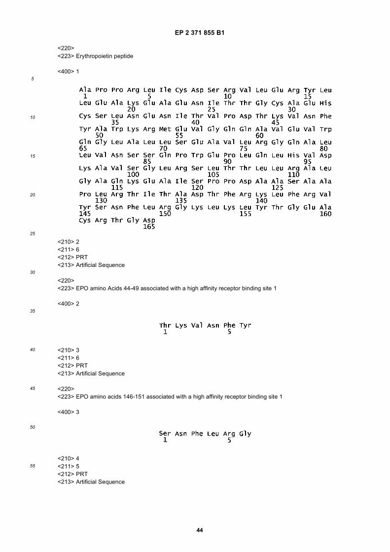
EP 2 371 855 B1
44
5
10
15
20
25
30
35
40
45
50
55
<220><223> Erythropoietin peptide
<400> 1
<210> 2<211> 6<212> PRT<213> Artificial Sequence
<220><223> EPO amino Acids 44-49 associated with a high affinity receptor binding site 1
<400> 2
<210> 3<211> 6<212> PRT<213> Artificial Sequence
<220><223> EPO amino acids 146-151 associated with a high affinity receptor binding site 1
<400> 3
<210> 4<211> 5<212> PRT<213> Artificial Sequence
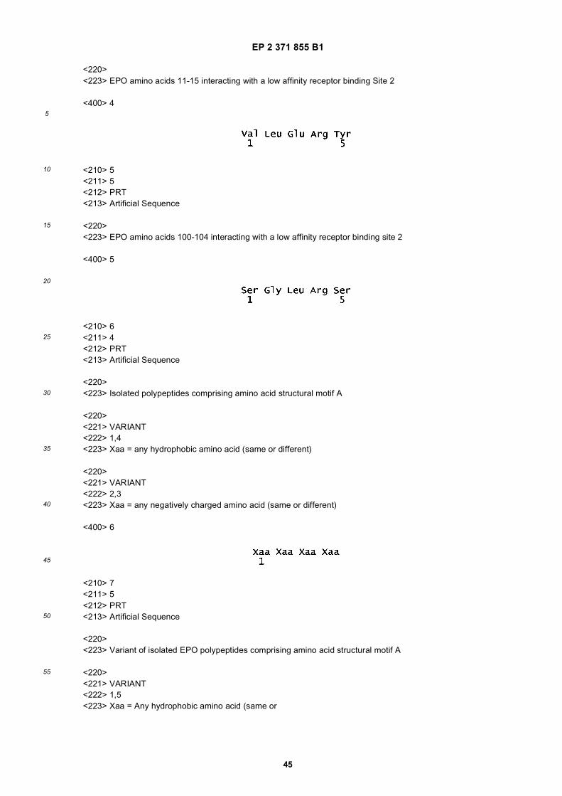
EP 2 371 855 B1
45
5
10
15
20
25
30
35
40
45
50
55
<220><223> EPO amino acids 11-15 interacting with a low affinity receptor binding Site 2
<400> 4
<210> 5<211> 5<212> PRT<213> Artificial Sequence
<220><223> EPO amino acids 100-104 interacting with a low affinity receptor binding site 2
<400> 5
<210> 6<211> 4<212> PRT<213> Artificial Sequence
<220><223> Isolated polypeptides comprising amino acid structural motif A
<220><221> VARIANT<222> 1,4<223> Xaa = any hydrophobic amino acid (same or different)
<220><221> VARIANT<222> 2,3<223> Xaa = any negatively charged amino acid (same or different)
<400> 6
<210> 7<211> 5<212> PRT<213> Artificial Sequence
<220><223> Variant of isolated EPO polypeptides comprising amino acid structural motif A
<220><221> VARIANT<222> 1,5<223> Xaa = Any hydrophobic amino acid (same or
EP 2 371 855 B1
46
5
10
15
20
25
30
35
40
45
50
55
<220><221> VARIANT<222> 2, 4<223> Xaa = Any negatively charged amino acid (same or different)
<220><221> VARIANT<222> 3<223> Xaa = Any amino acid
<400> 7
<210> 8<211> 6<212> PRT<213> Artificial Sequence
<220><223> Variant of isolated EPO polypeptides comprising amino acid structural motif A
<220><221> VARIANT<222> 1, 6<223> Xaa = Any hydrophobic amino acid (same or different)
<220><221> VARIANT<222> 2,5<223> Xaa = Any negatively charged animo acid (same or different)
<220><221> VARIANT<222> 3,4<223> Xaa = Any amino acid (same or different)
<400> 8
<210> 9<211> 7<212> PRT<213> Artificial Sequence
<220><223> Variant of isolated EPO polypeptides comprising amino acid structural motif A
<220><221> VARIANT<222> 1, 7<223> Xaa = Any hydrophobic amino acid (same or different)
<220><221> VARIANT
EP 2 371 855 B1
47
5
10
15
20
25
30
35
40
45
50
55
<222> 2,6<223> Xaa = Any negatively charged amino acid (same or different)
<220><221> VARIANT<222> 3,4,5<223> Xaa = Any amino acid (same or different)
<400> 9
<210> 10<211> 8<212> PRT<213> Artificial Sequence
<220><223> Variant of isolated EPO polypeptides comprising amino acid structural motif A
<220><221> VARIANT<222> 1,8<223> Xaa = Any hydrophobic amino acid (same or different)
<220><221> VARIANT<222> 2, 7<223> Xaa = Any negatively charged amino acid (same or different)
<220><221> VARIANT<222> 3,4,5,6<223> Xaa = Any amino acid (same or different)
<400> 10
<210> 11<211> 9<212> PRT<213> Artificial Sequence
<220><223> Variant of isolated EPO polypeptides comprising amino acid structural motif A
<220><221> VARIANT<222> 1, 9<223> Xaa = Any hydrophobic amino acid (same or different)
<220><221> VARIANT<222> 2, 8<223> Xaa = Any negatively charged amino acid (same or different)

EP 2 371 855 B1
48
5
10
15
20
25
30
35
40
45
50
55
<220><221> VARIANT<222> 3,4,5,6,7<223> Xaa = Any amino acid (same or different)
<400> 11
<210> 12<211> 4<212> PRT<213> Artificial Sequence
<220><223> Variant of isolated EPO polypeptides comprising amino acid structural motif A
<220><221> VARIANT<222> 1<223> Xaa = Any hydrophobic amino acid
<220><221> VARIANT<222> 2, 3<223> Xaa = Any negatively charged amino acid (same or different)
<220><221> VARIANT<222> 4<223> Xaa = Any polar amino acid
<400> 12
<210> 13<211> 5<212> PRT<213> Artificial Sequence
<220><223> ariant of isolated EPO polypeptides comprising amino acid structural motif A
<220><221> VARIANT<222> 1<223> Xaa = Any hydrophobic amino acid
<220><221> VARIANT<222> 2,4<223> Xaa = Any negatively charged amino acid
<220><221> VARIANT
EP 2 371 855 B1
49
5
10
15
20
25
30
35
40
45
50
55
<222> 3<223> Xaa = Any amino acid
<220><221> VARIANT<222> 5<223> Xaa = Any polar amino acid
<400> 13
<210> 14<211> 6<212> PRT<213> Artificial Sequence
<220><223> Variant of isolated EPO polypeptides comprising amino acid structural motif A
<220><221> VARIANT<222> 1<223> Xaa = Any hydrophobic amino acid
<220><221> VARIANT<222> 2,5<223> Xaa = Any negatively charged amino acid (same or different)
<220><221> VARIANT<222> 3,4<223> Xaa = Any amino acid (same or different)
<220><221> VARIANT<222> 6<223> Xaa = Any polar amino acid
<400> 14
<210> 15<211> 7<212> PRT<213> Artificial Sequence
<220><223> Variant of isolated EPO polypeptides comprising amino acid structural motif A
<220><221> VARIANT<222> 1<223> Xaa = Any hydrophobic amino acid
EP 2 371 855 B1
50
5
10
15
20
25
30
35
40
45
50
55
<220><221> VARIANT<222> 2, 6<223> Xaa = Any negatively charged amino acid (same or different)
<220><221> VARIANT<222> 3,4,5<223> Xaa = Any amino acid (same or different)
<220><221> VARIANT<222> 7<223> Xaa = Any polar amino acid
<400> 15
<210> 16<211> 8<212> PRT<213> Artificial Sequence
<220><223> Variant of isolated EPO polypeptides comprising amino acid structural motif A
<220><221> VARIANT<222> 1<223> Xaa = Any hydrophobic amino acid
<220><221> VARIANT<222> 2, 7<223> Xaa = Any negatively charged amino acid (same or different)
<220><221> VARIANT<222> 3,4,5,6<223> Xaa = Any amino acid (same or different)
<220><221> VARIANT<222> 8<223> Xaa = Any polar amino acid
<400> 16
<210> 17<211> 9<212> PRT<213> Artificial Sequence
EP 2 371 855 B1
51
5
10
15
20
25
30
35
40
45
50
55
<220><223> Variant of isolated EPO polypeptides comprising amino acid structural motif A
<220><221> VARIANT<222> 1<223> Xaa = Any hydrophobic amino acid
<220><221> VARIANT<222> 2, 8<223> Xaa = Any negatively charged amino acid (same or different)
<220><221> VARIANT<222> 3,4,5,6,7<223> Xaa = Any amino acid (same or different)
<220><221> VARIANT<222> 9<223> Xaa = Any polar amino acid
<400> 17
<210> 18<211> 4<212> PRT<213> Artificial Sequence
<220><223> Variant of isolated EPO polypeptides comprising amino acid structural motif A
<220><221> VARIANT<222> 1<223> Xaa = Any polar amino acid
<220><221> VARIANT<222> 2,3<223> Xaa = Any negatively charged amino acid (same or different)
<220><221> VARIANT<222> 4<223> Xaa = Any hydrophobic amino acid
<400> 18
<210> 19<211> 5

EP 2 371 855 B1
52
5
10
15
20
25
30
35
40
45
50
55
<212> PRT<213> Artificial Sequence
<220><223> Variant of isolated EPO polypeptides comprising amino acid structural motif A
<220><221> VARIANT<222> 1<223> Xaa = Any polar amino acid
<220><221> VARIANT<222> 2,4<223> Xaa = Any negatively charged amino acid (same or different)
<220><221> VARIANT<222> 3<223> Xaa = Any amino acid
<220><221> VARIANT<222> 5<223> Xaa = Any hydrophobic amino acid
<400> 19
<210> 20<211> 6<212> PRT<213> Artificial Sequence
<220><223> variant of isolated EPO polypeptides comprising amino acid structural motif A
<220><221> VARIANT<222> 1<223> Xaa = Any polar amino acid
<220><221> VARIANT<222> 2, 5<223> Xaa = Any negatively charged amino acid (same or different)
<220><221> VARIANT<222> 3,4<223> Xaa = Any amino acid (same or different)
<220><221> VARIANT<222> 6<223> Xaa = Any hydrophobic amino acid
EP 2 371 855 B1
53
5
10
15
20
25
30
35
40
45
50
55
<400> 20
<210> 21<211> 7<212> PRT<213> Artificial Sequence
<220><223> Variant of isolated EPO polypeptides comprising amino acid structural motif A
<220><221> VARIANT<222> 1<223> Xaa = Any polar amino acid
<220><221> VARIANT<222> 2,6<223> Xaa = Any negatively charged amino acid (same or different)
<220><221> VARIANT<222> 3,4,5<223> Xaa = Any amino acid (same or different)
<220><221> VARIANT<222> 7<223> Xaa = Any hydrophobic amino acid
<400> 21
<210> 22<211> 8<212> PRT<213> Artificial Sequence
<220><223> Variant of isolated EPO polypeptides comprising amino acid structural motif A
<220><221> VARIANT<222> 1<223> Xaa = Any polar amino acid
<220><221> VARIANT<222> 2,7<223> Xaa = Any negatively charged amino acid (same or different)
<220><221> VARIANT
EP 2 371 855 B1
54
5
10
15
20
25
30
35
40
45
50
55
<222> 3,4,5,6<223> Xaa = Any amino acid (same or different)
<220><221> VARIANT<222> 8<223> Xaa = Any hydrophobic amino acid
<400> 22
<210> 23<211> 9<212> PRT<213> Artificial Sequence
<220><223> Variant of isolated EPO polypeptides comprising amino acid structural motif A
<220><221> VARIANT<222> 1<223> Xaa = Any polar amino acid
<220><221> VARIANT<222> 2,8<223> Xaa = Any negatively charged amino acid (same or different)
<220><221> VARIANT<222> 3,4,5,6,7<223> Xaa = Any amino acid (same or different)
<220><221> VARIANT<222> 9<223> Xaa = Any hydrophobic amino acid
<400> 23
<210> 24<211> 4<212> PRT<213> Artificial Sequence
<220><223> variant of isolated EPO polypeptides comprising amino acid structural motif A
<220><221> VARIANT<222> 1,4<223> Xaa = Any hydrophobic amino acid (same or different)
EP 2 371 855 B1
55
5
10
15
20
25
30
35
40
45
50
55
<220><221> VARIANT<222> 2<223> Xaa = Any negatively charged amino acid
<220><221> VARIANT<222> 3<223> xaa = Any positively charged amino acid
<400> 24
<210> 25<211> 5<212> PRT<213> Artificial Sequence
<220><223> variant of isolated EPO polypeptides comprising amino acid structural motif A
<220><221> VARIANT<222> 1,5<223> Xaa = Any hydrophobic amino acid
<220><221> VARIANT<222> 2<223> Xaa = Any negatively charged amino acid
<220><221> VARIANT<222> 3<223> Xaa = Any polar amino acid
<220><221> VARIANT<222> 4<223> Xaa = Any positively charged amino acid
<400> 25
<210> 26<211> 4<212> PRT<213> Artificial Sequence
<220><223> variant of isolated EPO polypeptides comprising amino acid structural motif A
<220><221> VARIANT
EP 2 371 855 B1
56
5
10
15
20
25
30
35
40
45
50
55
<222> 1,4<223> Xaa = Any hydrophobic amino acid
<220><221> VARIANT<222> 2<223> Xaa = Any positively charged amino acid
<220><221> VARIANT<222> 3<223> Xaa = Any negatively charged amino acid
<400> 26
<210> 27<211> 5<212> PRT<213> Artificial Sequence
<220><223> variant of isolated EPO polypeptides comprising amino acid structural motif A
<220><221> VARIANT<222> 1,5<223> Xaa = Any hydrophobic amino acid
<220><221> VARIANT<222> 2<223> Xaa = Any positively charged amino acid
<220><221> VARIANT<222> 3<223> Xaa = Any polar amino acid
<220><221> VARIANT<222> 4<223> Xaa = Any negatively charged amino acid
<400> 27
<210> 28<211> 11<212> PRT<213> Artificial Sequence
<220><223> Amphipathic helix from pancreatic polypeptide
EP 2 371 855 B1
57
5
10
15
20
25
30
35
40
45
50
55
<400> 28
<210> 29<211> 20<212> PRT<213> Artificial Sequence
<220><223> Peptide C - loop AB and N-terminal portion of helix B corresponding to EPO amino acids 38-57
<400> 29
<210> 30<211> 25<212> PRT<213> Artificial Sequence
<220><223> Peptide D - the C-terminal portion of helix B corresponding to EPO amino acids 58-82
<400> 30
<210> 31<211> 20<212> PRT<213> Artificial Sequence
<220><223> Peptide E - a portion of the A-B loop consisting of a small cysteine loop and a Beta-pleated sheet correspondingto EPO amino acids 28-47
<400> 31
<210> 32<211> 23<212> PRT<213> Artificial Sequence
<220>

EP 2 371 855 B1
58
5
10
15
20
25
30
35
40
45
50
55
<223> Peptide A - consists of the N-terminal portion of Helix A corresponding to EPO amino acids 1-23
<400> 32
<210> 33<211> 16<212> PRT<213> Artificial Sequence
<220><223> Peptide F - consists of EPO amino acids 14-19
<400> 33
<210> 34<211> 14<212> PRT<213> Artificial Sequence
<220><223> Peptide B - consists of EPO amino acids 24-37
<400> 34
<210> 35<211> 14<212> PRT<213> Artificial Sequence
<220><223> GM-CSF helix A fragment
<400> 35
<210> 36<211> 12<212> PRT<213> Artificial Sequence
<220><223> TPO helix A fragment
EP 2 371 855 B1
59
5
10
15
20
25
30
35
40
45
50
55
<400> 36
<210> 37<211> 16<212> PRT<213> Artificial Sequence
<220><223> CNTF helix A fragment
<400> 37
<210> 38<211> 17<212> PRT<213> Artificial Sequence
<220><223> LIF helix B fragment
<400> 38
<210> 39<211> 18<212> PRT<213> Artificial Sequence
<220><223> Interleukin 3 (IL-3) helix A fragment
<400> 39
<210> 40<211> 11<212> PRT<213> Artificial Sequence
<220><223> Peptide G - All exterior-presenting amino acids of helix B of EPO
<400> 40
EP 2 371 855 B1
60
5
10
15
20
25
30
35
40
45
50
55
<210> 41<211> 23<212> PRT<213> Artificial Sequence
<220><223> Peptide J - A chimera peptide of Peptide G linked to pancreatic polypeptide helix
<400> 41
<210> 42<211> 7<212> PRT<213> Artificial Sequence
<220><223> Beta-pleated sheet found within AB loop of EPO
<400> 42
<210> 43<211> 18<212> PRT<213> Artificial Sequence
<220><223> Peptide I - A chimera peptide linking the helix B exterior-presenting amino acids to the beta pleated sheetfound within the AB loop of the EPO
<400> 43
<210> 44<211> 5<212> PRT<213> Artificial Sequence
<220><223> Terminal portion of helix C corresponding to amino acids 111,112,113,116, and 118 of EPO
<400> 44
EP 2 371 855 B1
61
5
10
15
20
25
30
35
40
45
50
55
<210> 45<211> 22<212> PRT<213> Artificial Sequence
<220><223> Loop CD-partial corresponding to amino acids 112-133 of EPO
<400> 45
<210> 46<211> 4<212> PRT<213> Artificial Sequence
<220><223> amino acid fragment of Peptide F
<400> 46
<210> 47<211> 6<212> PRT<213> Artificial Sequence
<220><223> Peptide H - consists of EPO amino acids 61, 72, 75, 76, 77
<400> 47
<210> 48<211> 12<212> PRT<213> Artificial Sequence
<220><223> Calcitonin
<400> 48
EP 2 371 855 B1
62
5
10
15
20
25
30
35
40
45
50
55
<210> 49<211> 11<212> PRT<213> Artificial Sequence
<220><223> Corticotrophin Releasing Hormone
<400> 49
<210> 50<211> 11<212> PRT<213> Artificial Sequence
<220><223> Beta Endorphin
<400> 50
<210> 51<211> 11<212> PRT<213> Artificial Sequence
<220><223> Glucagon
<400> 51
<210> 52<211> 10<212> PRT<213> Artificial Sequence
<220><223> Secretin
<400> 52
EP 2 371 855 B1
63
5
10
15
20
25
30
35
40
45
50
55
<210> 53<211> 11<212> PRT<213> Artificial Sequence
<220><223> VIP
<400> 53
<210> 54<211> 11<212> PRT<213> Artificial Sequence
<220><223> NP-Y
<400> 54
<210> 55<211> 11<212> PRT<213> Artificial Sequence
<220><223> GNRH
<400> 55
<210> 56<211> 11<212> PRT<213> Artificial Sequence
<220><223> Parathyroid Hormone
<400> 56
EP 2 371 855 B1
64
5
10
15
20
25
30
35
40
45
50
55
<210> 57<211> 11<212> PRT<213> Artificial Sequence
<220><223> CGRP
<400> 57
<210> 58<211> 5<212> PRT<213> Artificial Sequence
<220><223> A motif located proximal to the transmembrane domain of the Erythropoietin Receptor ("EPOR") protein
<220><221> VARIANT<222> 3<223> Xaa = any amino acid
<400> 58
Claims
1. An isolated polypeptide consisting of no more than 30 amino acid, said polypeptide comprising the amino acidsequence QEQLERALNSS (SEQ ID NO: 40), wherein said polypeptide has cellular protective activity.
2. The isolated polypeptide of claim 1 consisting of the amino acid sequence QEQLERALNSS (SEQ ID NO: 40).
3. The isolated polypeptide of claim 1 comprising the amino acid sequence QEQLERALNSSLRRYINMLTRTR (SEQID NO: 41), or the amino acid sequence CSLNENIQEQLERALNSS (SEQ ID NO: 43).
4. An isolated polypeptide consisting of the amino acid sequence QEQLERALNSS (SEQ ID NO: 40), wherein oneamino acid residue of said amino acid sequence is replaced with a conservative or non-conservative substitutionor an amino acid equivalent, wherein said polypeptide has cellular protective activity.
5. An isolated polypeptide which is a retro-inverso-analogue of the isolated polypeptides of any one of claims 1-4,wherein said retro-inverso-analogue has cellular protective activity.
6. The isolated polypeptide of any one of claims 1-5, wherein said polypeptide is modified with an addition of polyethyleneglycol.
7. The isolated polypeptide of any one of claims 1-6, wherein said polypeptide does not increase hemoglobin in a
EP 2 371 855 B1
65
5
10
15
20
25
30
35
40
45
50
55
recipient.
8. A pharmaceutical composition comprising the isolated polypeptide of any one of claims 1-7 and a pharmaceuticallyacceptable carrier.
9. The pharmaceutical composition of claim 8, wherein said composition is formulated for oral, intranasal, ocular,inhalational, transdermal, rectal, sublingual, or parenteral administration, or wherein said composition is formulatedas a perfusate solution.
10. An in vitro method for protecting, maintaining, or enhancing the viability of a responsive cell, tissue, or organ isolatedfrom a mammalian body comprising exposing said cell, tissue, or organ to an isolated polypeptide according to anyone of claims 1-7 or a pharmaceutical composition according to claim 8 or 9.
11. The method of claim 10, wherein the mammalian body is a human body.
12. An isolated polypeptide according to any one of claims 1-7 for use in a method for prevention, therapeutic treatmentor prophylactic treatment of a cardiovascular disease, kidney disease, disease of the urinary system, bone disease,skin disease, gastrointestinal disease, or a disease or disorder of the central or peripheral nervous system in a subject.
13. An isolated polypeptide according to any one of claims 1-7 for use in a method for preventing or treating a tissueinjury in a subject, wherein said tissue injury results from reduced oxygenation or inflammation.
14. The isolated polypeptide for use of claim 13, wherein said reduced oxygenation results from a hypoxic or an ischemiccondition.
15. The isolated polypeptide for use of any one of claims 12-14, wherein the subject is a human.
16. An isolated nucleic acid comprising a nucleotide sequence encoding the isolated polypeptide of any one of claims 1-5.
17. A vector comprising the nucleic acid of claim 16.
18. The vector of claim 17 that is an expression vector.
19. A host cell containing the expression vector of claim 18.
20. A method of recombinantly producing an isolated polypeptide comprising (a) culturing in a medium the host cell ofclaim 19, under conditions suitable for the expression of said polypeptide, and (b) recovering and isolating saidpolypeptide from said medium.
Patentansprüche
1. Isoliertes Polypeptid bestehend aus nicht mehr als 30 Aminosäuren, wobei das Polypeptid die AminosäuresequenzQEQLERALNSS (SEQ ID NO: 40) umfasst, wobei das Polypeptid zellschützende Aktivität hat.
2. Isoliertes Polypeptid nach Anspruch 1 bestehend aus der Aminosäuresequenz QEQLERALNSS (SEQ ID NO: 40).
3. Isoliertes Polypeptid nach Anspruch 1 umfassend die Aminosäuresequenz QEQLERALNSSLRRYINMLTRTR (SEQID NO: 41), oder die Aminosäuresequenz CSLNENIQEQLERALNSS (SEQ ID NO: 43).
4. Isoliertes Polypeptid bestehend aus der Aminosäuresequenz QEQLERALNSS (SEQ ID NO: 40), wobei ein Amino-säurerest der Aminosäuresequenz durch eine konservative oder nicht-konservative Substitution oder ein Amino-säureäquivalent ersetzt ist, wobei das Polypeptid zellschützende Aktivität hat.
5. Isoliertes Polypeptid, das ein retro-inverso-Analogon des isolierten Polypeptids nach einem der Ansprüche 1-4 ist,wobei das retro-inverso-Analogon zellschützende Aktivität hat.
6. Isoliertes Polypeptid nach einem der Ansprüche 1-5, wobei das Polypeptid durch Zusatz von Polyethylenglykol
EP 2 371 855 B1
66
5
10
15
20
25
30
35
40
45
50
55
modifiziert ist.
7. Isoliertes Polypeptid nach einem der Ansprüche 1-6, wobei das Polypeptid nicht das Hämoglobin in einem Empfängererhöht.
8. Pharmazeutische Zusammensetzung umfassend das isolierte Polypeptid nach einem der Ansprüche 1-7 und einenpharmazeutisch annehmbaren Träger.
9. Pharmazeutische Zusammensetzung nach Anspruch 8, wobei die Zusammensetzung für orale, intranasale, okulare,Inhalations-, transdermale, rektale, sublinguale oder parenterale Verabreichung formuliert ist, oder wobei die Zu-sammensetzung als eine Perfusionslösung formuliert ist.
10. In vitro-Verfahren zum Schutz, der Aufrechterhaltung, oder der Förderung der Lebensfähigkeit eine(r/s) anspre-chenden Zelle, Gewebes, oder Organs, die/das aus einem Säugetierkörper isoliert wurde, umfassend das Aussetzender Zelle, des Gewebes, oder des Organs einem isolierten Polypeptid gemäß einem der Ansprüche 1-7 oder einerpharmazeutischen Zusammensetzung gemäß Anspruch 8 oder 9.
11. Verfahren nach Anspruch 10, wobei der Säugetierkörper ein humaner Körper ist.
12. Isoliertes Polypeptid gemäß einem der Ansprüche 1-7 zur Anwendung in einem Verfahren für die Prävention, dietherapeutische Behandlung oder prophylaktische Behandlung einer kardiovaskulären Erkrankung, Nierenerkran-kung, Erkrankung des Harnsystems, Knochenerkrankung, Hauterkrankung, gastrointestinalen Erkrankung, odereiner Erkrankung oder Störung des zentralen oder peripheren Nervensystems in einem Subjekt.
13. Isoliertes Polypeptid gemäß einem der Ansprüche 1-7 zur Anwendung in einem Verfahren für die Prävention oderBehandlung einer Gewebeverletzung in einem Subjekt, wobei die Gewebeverletzung auf verminderte Sauerstoff-zufuhr oder Entzündung zurückzuführen ist.
14. Isoliertes Polypeptid zur Anwendung nach Anspruch 13, wobei die verminderte Sauerstoffzufuhr auf einen hypoxi-schen oder einen ischämischen Zustand zurückzuführen ist.
15. Isoliertes Polypeptid zur Anwendung nach einem der Ansprüche 12-14, wobei das Subjekt ein Mensch ist.
16. Isolierte Nukleinsäure umfassend eine Nukleotidsequenz, die das isolierte Polypeptid nach einem der Ansprüche1-5 kodiert.
17. Vektor umfassend die Nukleinsäure nach Anspruch 16.
18. Vektor nach Anspruch 17, der ein Expressionsvektor ist.
19. Wirtszelle, enthaltend den Expressionsvektor nach Anspruch 18.
20. Verfahren zur rekombinanten Herstellung eines isolierten Polypeptids umfassend (a) Kultivieren der Wirtszelle nachAnspruch 19 in einem Medium, unter für die Expression des Polypeptids geeigneten Bedingungen, und (b) Gewin-nung und Isolierung des Polypeptids aus dem Medium.
Revendications
1. Polypeptide isolé consistant en pas plus de 30 acides aminés, ledit polypeptide comprenant la séquence d’acidesaminés QEQLERALNSS (SEQ ID N° : 40), ledit polypeptide ayant une activité de protection cellulaire.
2. Polypeptide isolé selon la revendication 1, consistant en la séquence d’acides aminés QEQLERALNSS (SEQ IDN° : 40).
3. Polypeptide isolé selon la revendication 1, comprenant la séquence d’acides aminés QEQLERALNSSLRRYINML-TRTR (SEQ ID N° : 41), ou la séquence d’acides aminés CSLNENIQEQLERALNSS (SEQ ID N° : 43).
EP 2 371 855 B1
67
5
10
15
20
25
30
35
40
45
50
55
4. Polypeptide isolé consistant en la séquence d’acides aminés QEQLERALNSS (SEQ ID N° : 40), dans lequel unrésidu d’acide aminé de ladite séquence d’acides aminés est remplacé par une substitution conservatrice ou nonconservatrice ou un équivalent d’acide aminé, ledit polypeptide ayant une activité de protection cellulaire.
5. Polypeptide isolé qui est un analogue rétro-inversé des polypeptides isolés de l’une quelconque des revendications1 à 4, ledit analogue rétro-inversé ayant une activité de protection cellulaire.
6. Polypeptide isolé selon l’une quelconque des revendications 1 à 5, ledit polypeptide étant modifié par une additionde polyéthylène glycol.
7. Polypeptide isolé selon l’une quelconque des revendications 1 à 6, ledit polypeptide n’augmentant pas l’hémoglobinechez un receveur.
8. Composition pharmaceutique comprenant le polypeptide isolé de l’une quelconque des revendications 1 à 7 et unvéhicule pharmaceutiquement acceptable.
9. Composition pharmaceutique selon la revendication 8, ladite composition étant formulée pour une administrationpar voie orale, intranasale, oculaire, par inhalation, par voie transdermique, rectale, sublinguale, ou parentérale, ouladite composition étant formulée sous la forme d’une solution de perfusat.
10. Procédé in vitro de protection, de maintien ou de renforcement de la viabilité d’une cellule, d’un tissu ou d’un organey répondant favorablement et isolé d’un organisme mammifère, comprenant d’exposer ladite cellule, ledit tissu ouledit organe à un polypeptide isolé de l’une quelconque des revendications 1 à 7 ou à une composition pharmaceu-tique de la revendication 8 ou 9.
11. Procédé selon la revendication 10, dans lequel l’organisme mammifère est un corps humain.
12. Polypeptide isolé selon l’une quelconque des revendications 1 à 7 pour utilisation dans un procédé de prévention,de traitement thérapeutique ou de traitement prophylactique d’une maladie cardiovasculaire, d’une maladie rénale,d’une maladie de l’appareil urinaire, d’une maladie des os, d’une maladie de peau, d’une maladie gastro-intestinale,ou d’une maladie ou d’un trouble du système nerveux central ou périphérique chez un sujet.
13. Polypeptide isolé selon l’une quelconque des revendications 1 à 7 pour utilisation dans un procédé de préventionou de traitement d’une lésion tissulaire chez un sujet, ladite lésion tissulaire résultant d’une oxygénation réduite oud’une inflammation.
14. Polypeptide isolé pour utilisation selon la revendication 13, dans lequel ladite oxygénation réduite résulte d’un étathypoxique ou ischémique.
15. Polypeptide isolé pour utilisation selon l’une quelconque des revendications 12 à 14, où le sujet est un être humain.
16. Acide nucléique isolé comprenant une séquence de nucléotides codant pour le polypeptide isolé de l’une quelconquedes revendications 1 à 5.
17. Vecteur comprenant l’acide nucléique de la revendication 16.
18. Vecteur selon la revendication 17 qui est un vecteur d’expression.
19. Cellule hôte contenant le vecteur d’expression de la revendication 18.
20. Procédé de production recombinante d’un polypeptide isolé comprenant les étapes qui consistent à (a) mettre enculture dans un milieu la cellule hôte de la revendication 19, dans des conditions appropriées à l’expression duditpolypeptide, et à (b) récupérer et isoler ledit polypeptide dudit milieu.
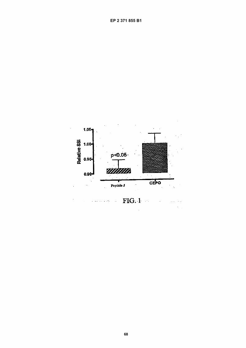
EP 2 371 855 B1
68
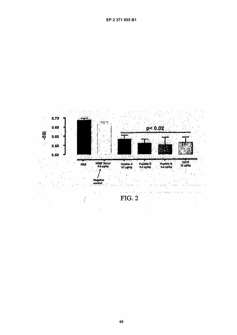
EP 2 371 855 B1
69
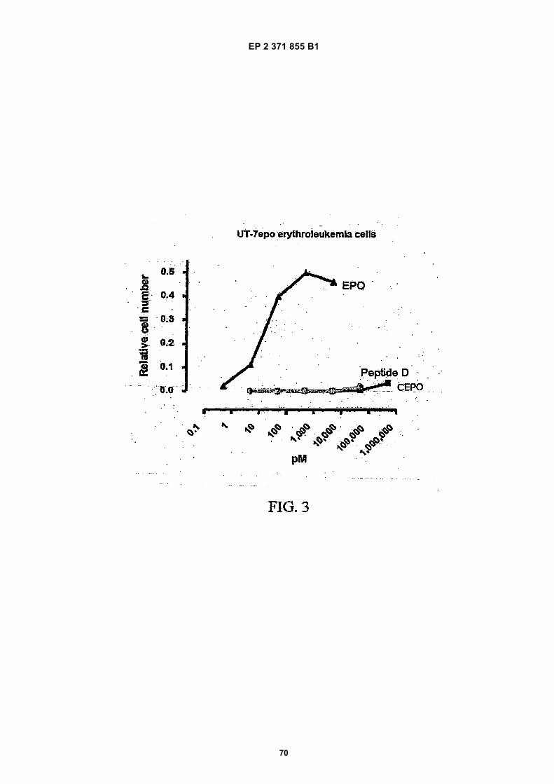
EP 2 371 855 B1
70
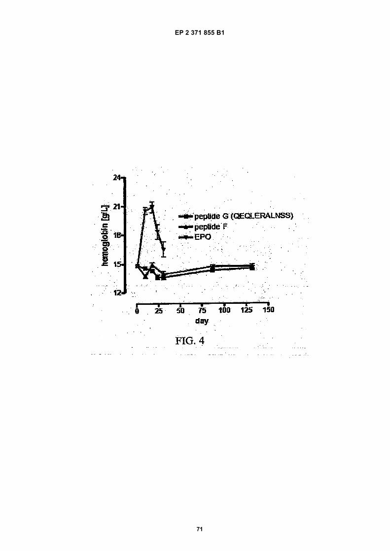
EP 2 371 855 B1
71
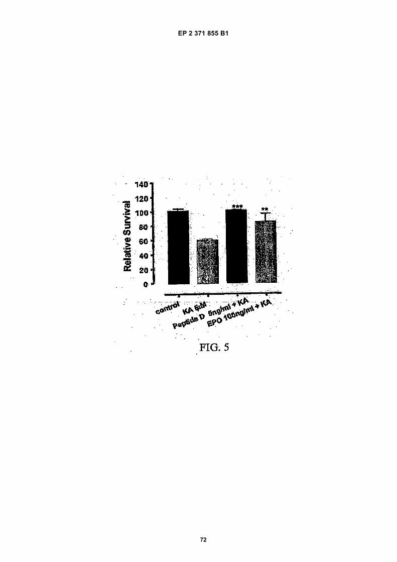
EP 2 371 855 B1
72
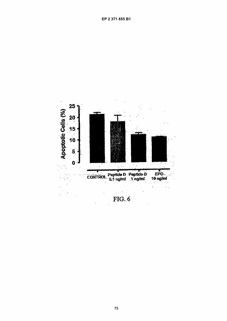
EP 2 371 855 B1
73
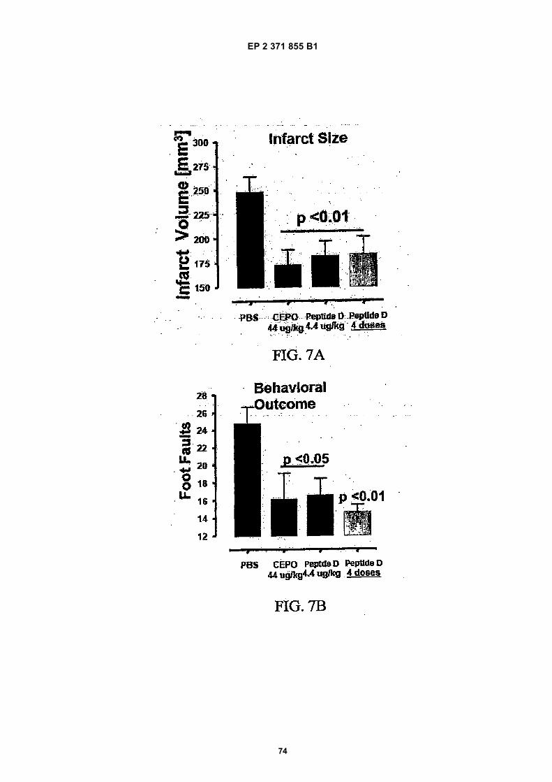
EP 2 371 855 B1
74
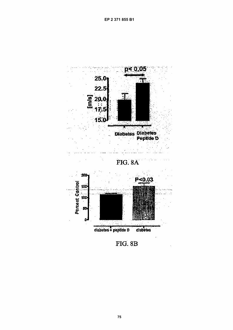
EP 2 371 855 B1
75
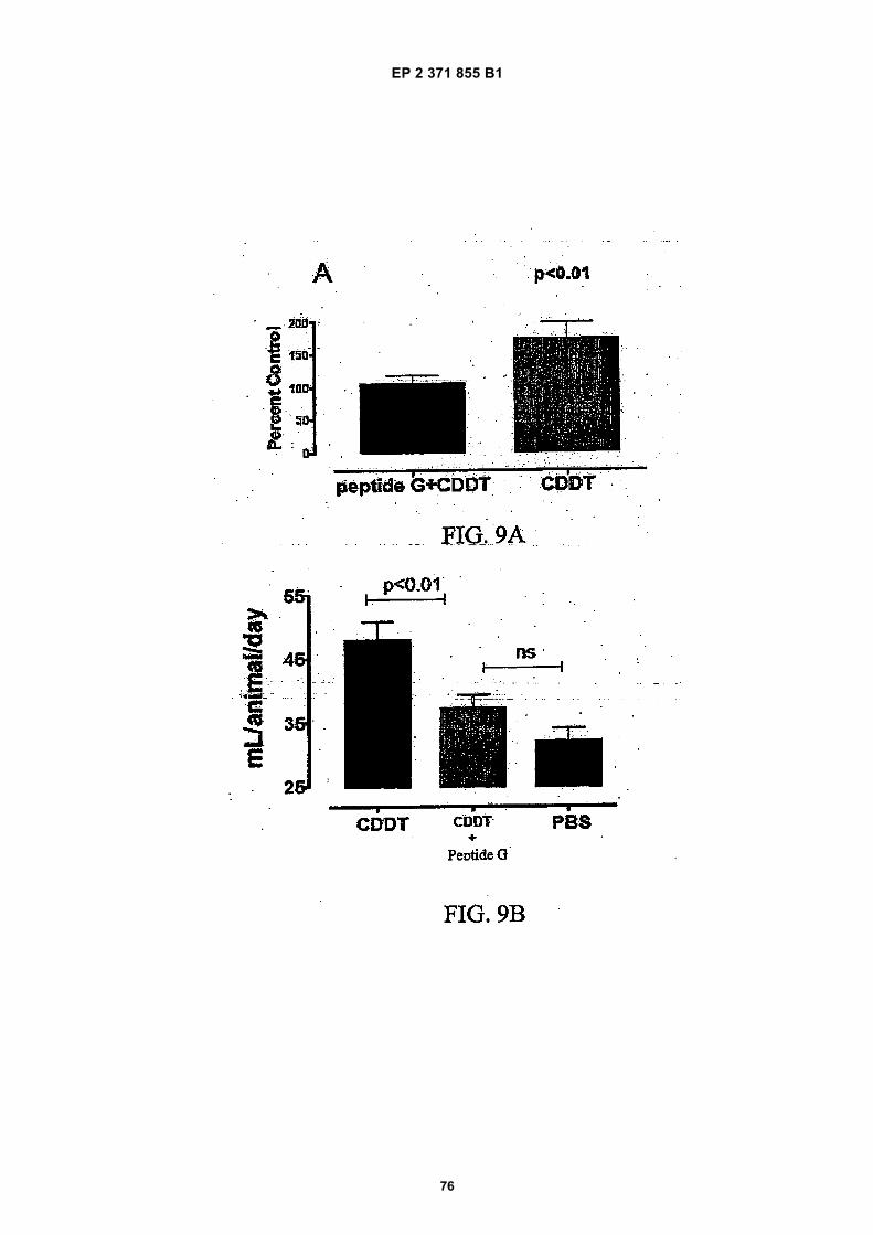
EP 2 371 855 B1
76
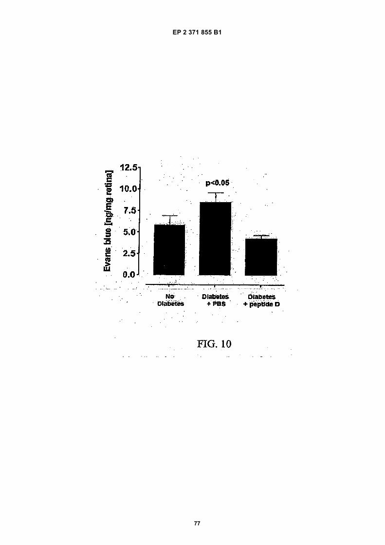
EP 2 371 855 B1
77
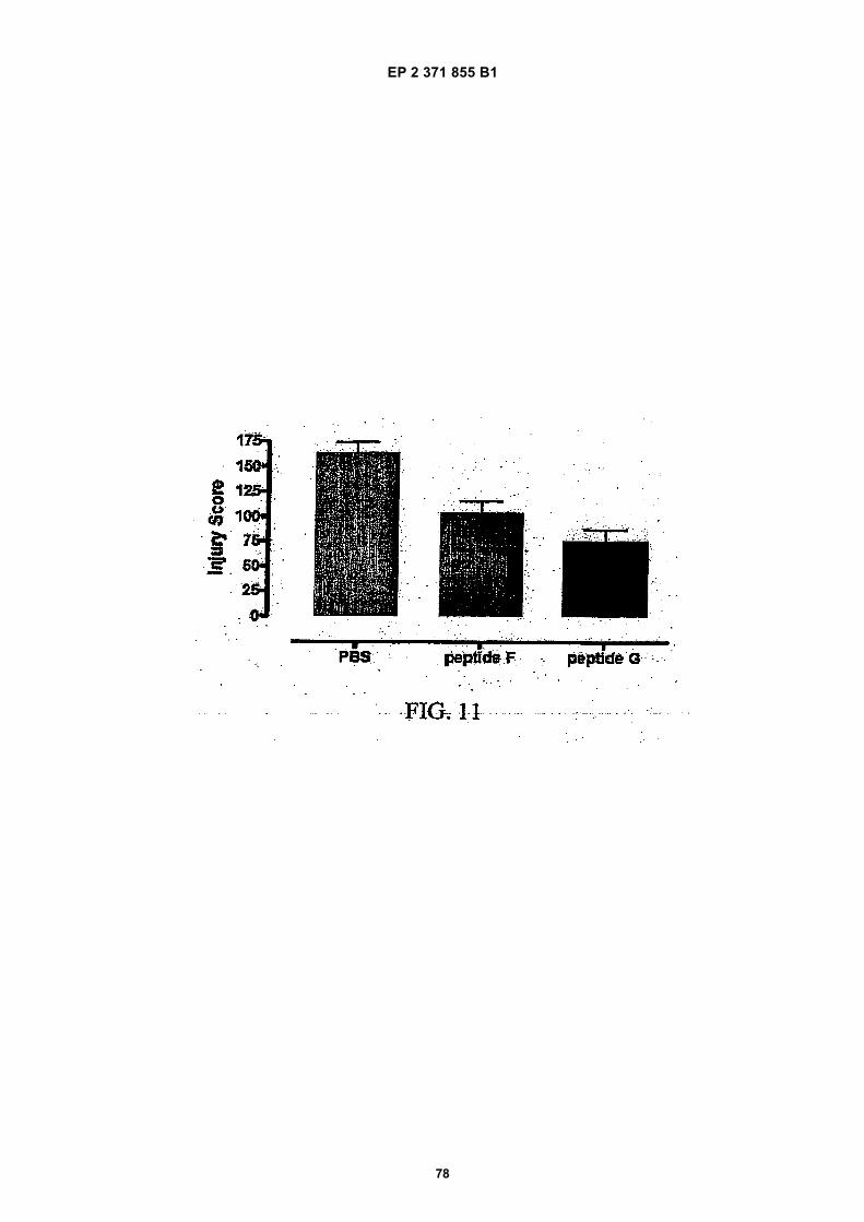
EP 2 371 855 B1
78
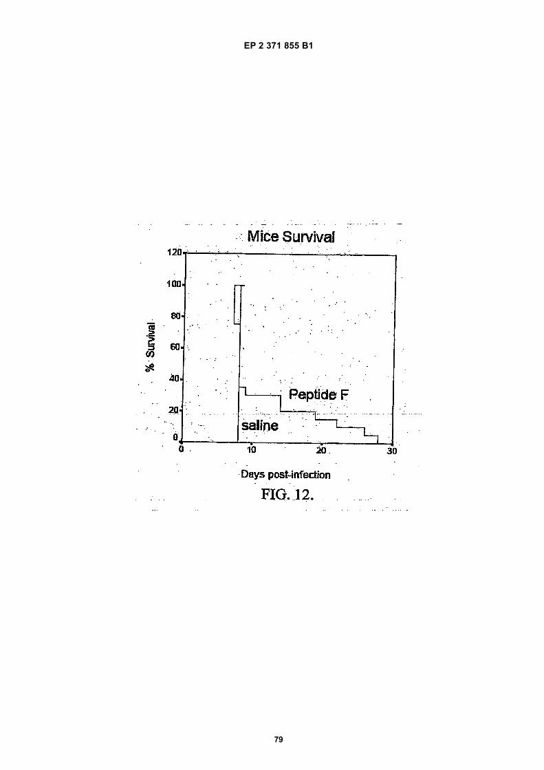
EP 2 371 855 B1
79

EP 2 371 855 B1
80
EP 2 371 855 B1
81
REFERENCES CITED IN THE DESCRIPTION
This list of references cited by the applicant is for the reader’s convenience only. It does not form part of the Europeanpatent document. Even though great care has been taken in compiling the references, errors or omissions cannot beexcluded and the EPO disclaims all liability in this regard.
Patent documents cited in the description
• WO 2004096148 A [0007] [0049]• US 0149479 W [0007] [0114] [0155]• US 75313200 A [0007] [0096]• US 10188905 A [0007]• US 5700909 A [0073]• US 5571787 A, J. S. O’Brien [0073]• US 5714459 A, J. S, O’Brien [0073]• US 5696080 A, J. S. O’Brien and Y. Kashimoto
[0073]• EP 45665 A [0093]• US 5192746 A, Lobl [0095]• US 5576423 A, Aversa [0095]• US 5051448 A, Shashoua [0095]• US 5559103 A, Gaeta [0095]• US 188905 A [0096] [0110] [0113] [0114] [0155]• US 20030072737 A1 [0096]
• US 61266503 A [0096]• US 0320964 W [0097]• WO 04022577 A3 [0100]• WO 05025606 A1 [0100]• WO 02053580 A [0108]• US 10185841 A [0110] [0113] [0114] [0155]• US 2004031789 W [0120] [0125]• WO 2005032467 A [0120] [0125]• WO 9104014 A [0145]• US 4704355 A [0145]• US 4798824 A [0151]• US 0149479 A [0181]• US 60705741 B [0226]• US 60706276 B [0226]• US 60831737 B [0226]
Non-patent literature cited in the description
• CHEETHAM et al. Nat. Struct. Biol., 1998, vol. 5,861-866 [0003] [0190]
• ELLIOTT et al. Blood, 1996, vol. 87 (7), 2702-2713[0004]
• ELLIOTT et al. Blood, 1997, vol. 89 (2), 493-502[0004]
• ELLIOTT et al. The Journal of Biologial Chemistry,2004, vol. 279 (16), 16854-16662 [0004]
• MURRAY. Harpers Biochemistry. Appilion & Lange,Ltd, 1996, 524-526 [0005]
• CARAVELLA et al. Protein: Struct. Funct. Gen.,1996, vol. 24, 394-401 [0005]
• CHEETHAM et al. Nature Structural Biology, 1998,vol. 5, 861-866 [0006]
• KIRITO et al. Blood, 2002, vol. 99, 102-110 [0006]• LIVNAH et al. Science, 1999, vol. 283, 987-990
[0006]• NARANDA et al. Endocrinology, 2002, vol. 143,
2293-2302 [0006]• REMY et al. Science, 1999, vol. 283, 990-993 [0006]• YOSHIMURA et al. The Oncologist, 1996, vol. 1,
337-339 [0006]• COLEMAN et al. PNAS, 2006, vol. 103, 5965-5970
[0026]• CHEETHAM et al. Nat. Struct. Biol, 1998, vol. 5,
861-866 [0052]• CHEETHAM et al. Nat. Struct. Biol., 1999, vol. 5,
861-866 [0053]• SEGREST. Proteins, 1990, vol. 8, 103-117 [0055]
• ELLIOTT et al. Blood, 1997, vol. 89, 493 [0063]• LEIST et al. Science, 2004, vol. 305, 239 [0063]• CAMPANA et al. Int. J. Mol. Med., 1998, vol. 1 (1),
235-241 [0073]• KANNAN. Neuroimmunomod, 2000, vol. 8, 132-141
[0075]• SEGREST et al. Proteins, 1990, vol. 8, 103 [0079]• NILSSON, B. ; SOELLNER, M. ; RAINES, R. Chem-
ical Synthesis of Proteins. Annu. Rev. Biomol. Struct.,2005, vol. 34, 91-118 [0084]
• MELDAL M. Properties of solid supports. MethodsEnzymdl., 1997, vol. 289, 83-104 [0084]
• SONGSTER MF ; BARANY G. Handles for sol-id-phase peptide synthesis. Methods Enzymol.,1997, vol. 289, 126-74 [0084]
• WELLINGS DA ; ATHERTON E. Standard Fmocprotocols. Methods Enzymol., 1997, vol. 289, 44-67[0084]
• GUY CA ; FIELDS GB. Trifluoroacetic acid cleavageand deprotection of resin-bound peptides followingsynthesis by Fmoc chemistry. Methods Enzymol.,1997, vol. 289, 67-83 [0084]
• STEWART JM. Cleavage methods followingBoc-based solid-phase peptide-synthesis. MethodsEnzymol., 1997, vol. 289, 29-44 [0084]
• BANG, D. ; KENT, S. A One-Pot Total Synthesis ofCrambin. Angew. Chem. Int. Ed., 2004, vol. 43,2534-2538 [0084]
EP 2 371 855 B1
82
• BANG, D. ; CHOPRA, N. ; KENT, S. Total ChemicalSynthesis of Crambin. J. Am. Chem. Soc., 2004, vol.126, 1377-1383 [0084]
• DAWSON, P. et al. Synthesis of Proteins by NativeChemical Ligation. Science, 1994, vol. 266, 776-779[0084] [0085]
• KOCHENDOERFER et al. Design and ChemicalSynthesis of a Homogenous Polymer-Modified Bryth-ropoiesis Protein. Science, 2003, vol. 299, 884-887[0084]
• SIMON et al. Peptoids: A molecular approach to drugdiscovery. Proc. Natl. Acad. Sci USA, 1992, vol. 89,9367-9371 [0086]
• LI et al. Photolithographic Synthesis of Peptoids. J.AM. CHEM. SOC., 2004, vol. 126, 4088-4089 [0086]
• FAUCHERE, J. Adv. Drug Res., 1986, vol. 15, 29[0086]
• VEBER ; FRIEDINGER. TINS, 1985, 32 [0086]• EVANS et al. J. Med. Chem, 1987, vol. 30, 1229
[0086]• Methods of Organic Chemistry (Rouben-Weyl). Syn-
thesis of Peptides and Peptidomimetics. Thieme Ver-lag, 2004, vol. E22c [0086]
• Methods of Organic Chemistry (Houben-Weyl). Syn-thesis of Peptides and Peptidomimetics. GeorgeThieme Verlag, 2004, vol. E22c [0091]
• CHRISTOPHER J. NOREN ; SPENCER J.ANTHONY-CAHILL ; MICHAEL C. GRIFFITH ; PE-TER G. SCHULTZ. Science, 1989, vol. 244, 182-188[0092]
• SPATOLA, A.F. Chemistry and Biochemistry of Ami-no Acids, Peptides, and Proteins. Marcel Dekker,1983, 267 [0093]
• SPATOLA, A.F. Peptide Backbone Modifications,March 1983, vol. 1 (3 [0093]
• MERELY, J.S. Trends Pharma Sci, 1990, 463-468[0093]
• HUDSON, D. et al. Int J Pept Prot Re, 1979, vol. 14,177-185 [0093]
• SPATOLA, A.F. et al. Life Sci, 1986, vol. 38,1243-1249 [0093]
• HANN, M. M. J Chem Soc Perkin Trans I, 1982,307-314 [0093]
• ALMQUIST, R. G et al. J Med Chem, 1980, vol. 23,1392 [0093]
• JENNINGS-WHITE, C et al. Tetrahedron Lett, 1982,vol. 23, 2533 [0093]
• HOLLADAY, M. W. et al. Tetrahedron Lett, 1983,vol. 24, 4401-4404 [0093]
• HRUBY, V. J. Life Sci, 1982, vol. 31, 189-199 [0093]• ELDRED et al. J. Med. Chem., 1994, vol. 37, 3882
[0095]• KU et al. J. Med. Chem, 1995, vol. 38, 9 [0095]• Biochemistry. WH Freeman and Co, 1995 [0098]• SUN et al. PNAS, 2001, vol. 98, 15306-15311 [0102]• SCHMID et al. J. Biol. Chem., 2003, vol. 276,
1932-1935 [0102]• PESCE et al. EMBO, 2002, vol. 3, 1146-1151 [0102]
• SRIVASTAVA ; POLLARD. FASEB J., 1999, vol. 13,1911-1922 [0103]
• GINISTY et al. J. Cell Sci., 1999, vol. 112, 761-772[0103]
• STURM et al. Eur J Clin Invest, 2005, vol. 35, 711[0104]
• LEIST et al. Science, 2004, vol. 305, 239-242 [0106][0108]
• BRINES et al. PNAS, 2000, vol. 97, 10295-10672[0108]
• ROSENBAUM et al. Vis. Res., 1997, vol. 37, 3443-51[0108]
• CALVILLO et al. PNAS, 2003, vol. 100, 4802-4806[0108]
• FIORDALISO et al. PNAS, 2005, vol. 102,2046-2051 [0108]
• GRASSO et al. Med Sci Monit, 2004, vol. 10, BR1-3[0108]
• MOSMAN. J. Immunol. Meth, 1983, vol. 65, 55-63[0112]
• TSAI et al. J. Neurosci, 2006, vol. 26, 1269-74 [0116]• BAHLMANN et al. Kidney Int., 2003, vol. 64,
1648-1632 [0116]• SEFTON. CRC Crit. Ref. Biomed. End., 1987, vol.
14, 201 [0145]• BUCHWALD et al. Surgery, 1980, vol. 88, 507 [0145]• SAUDEK et al. N. Engl. J. Med., 1989, vol. 321, 574
[0145]• LANGER. Science, 1990, vol. 249, 1527-1533 [0145]
[0146]• TREAT et al. Liposomes in the Therapy of Infectious
Disease and Cancer. Liss, 1989, 353-365 [0145]• Medical Applications of Controlled Release. CRC
Press, 1974 [0145]• Controlled Drug Bioavailability, Drug Product Design
and Performance. Wiley, 1984 [0145]• RANGER ; PEPPAS. J. Macromol. Sci. Rev. Macro-
mol. Chem., 1953, vol. 23, 61 [0145]• LEVY et al. Science, 1985, vol. 228, 190 [0145]• DURING et al. Ann. Neurol., 1989, vol. 25, 351 [0145]• HOWARD et al. J. Neurosurg., 1989, vol. 71, 105
[0145]• GOODSON. Medial Applications of Controlled Re-
lease, 1984, vol. 2, 115-138 [0146]• POZNANSKY et al. Science, 1984, vol. 223,
1304-1306 [0169]• CARLSSON et al. Biochem J., 1978, vol. 173,
723-737 [0173]• CARLSSON, J. et al. Biochem. J., 1978, vol. 173,
723-737 [0176]• CUMBER, J.A. et al. Methods in Enzymology, 1985,
vol. 112, 207-224 [0176]• JUE, R et al. Biochem, 1978, vol. 17, 5399-5405
[0176]• SUN, T.T. et al. Biochem., 1974, vol. 13, 2334-2340
[0176]• BLATTLER, W.A. et al. Biochem., 1985, vol. 24,
1517-152 [0176]
EP 2 371 855 B1
83
• LIU, F.T. et al. Biochem., 1979, vol. 18, 690-697[0176]
• YOULE, R.J. ; NEVILLE, D.M. JR. Proc. Natl. Acad.Sci. U.S.A., 1980, vol. 77, 5483-5486 [0176]
• LERNER, R.A. et al. Proc. Natl. Acad. Sci. U.S.A.,1981, vol. 78, 3403-3407 [0176]
• JUNG, S.M ; MOROI, M. Biochem. Biophys. Acta,1983, vol. 761, 162 [0176]
• CAULFIELD, M.P et al. Biochem., 1984, vol. 81,7772-7776 [0176]
• STAROS, J.V. Biochem., 1982, vol. 21, 3950-3955[0176]
• YOSHITAKE, S. et al. Eur. J. Biochem., 1979, vol.101, 395-399 [0176]
• YOSHITAKE, S. et al. J. Biochem., 1982, vol. 92,1413-1424 [0176]
• PILCH, P.F. ; CZECH, M.P. J. Biol. Chem., 1979, vol.254, 3375-3381 [0176]
• NOVICK, D. et al. J. Biol, Chem., 1997, vol. 262,8483-8487 [0176]
• LOMANT, A.J. ; FAIRBANKS, G. J. Mol. Biol., 1976,vol. 104, 243-261 [0176]
• HAMADA, H. ; TSURUO, T. Anal. Biochem., 1987,vol. 160, 483-488 [0176]
• HASHIDA, S. et al. J. Applied Biochem., 1984, vol.6, 56-63 [0176]
• MEANS ; FEENEY. Bioconjugate Chem., 1990, vol.1, 2-12 [0177]
• BAND, D. ; CHOPRA, N ; KENT, S. Total Synthesisof Crambin. J.AM. CHEM. SOC., 2004, vol. 126,1377-1383 [0184]
• S. ERBAYRAKTAR et al. Proc Natl Acad Sci US A,2003, vol. 100, 6741-6746 [0188]
• SIREN et al. PNAS, 2001, vol. 98, 4044 [0197] [0200]• BRINES et al. PNAS USA, 2000, vol. 97,
10526-10531 [0202]• MARKGRAF et al. Brain Research, 1992, vol. 575,
238-246 [0206]• BIANCHI et al. Proc Natl Acad Sci U S A, 2004, vol.
101, 823-828 [0208]• BIANCHI et al. Clin Cancer Res, 2006, vol. 12,
2607-2612 [0211]• XU et al. Invest. Ophthal. Vis. Sci, 2001, vol. 42,
789-794 [0214]• SHARPLES et al. J Amer Soc Nephrol, 2005, vol.
15, 2115 [0218]• KAISER et al. J. Infect. Dis., 2006, vol. 193, 987-995
[0220]• SAVINO et al. J Neuroimmunol, 2006, vol. 172, 27-37
[0223]