Rational design of hyperstable antibacterial peptides for food ...
16
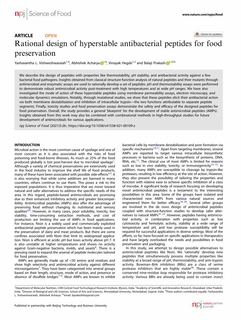
ARTICLE OPEN Rational design of hyperstable antibacterial peptides for food preservation Yashavantha L. Vishweshwaraiah 1,4 , Abhishek Acharya 1,4 , Vinayak Hegde 1,2 and Balaji Prakash 1,3 ✉ We describe the design of peptides with properties like thermostability, pH stability, and antibacterial activity against a few bacterial food pathogens. Insights obtained from classical structure-function analysis of natural peptides and their mutants through antimicrobial and enzymatic assays are used to rationally develop a set of peptides. pH and thermostability assays were performed to demonstrate robust antimicrobial activity post-treatment with high temperatures and at wide pH ranges. We have also investigated the mode of action of these hyperstable peptides using membrane permeability assays, electron microscopy, and molecular dynamics simulations. Notably, through mutational studies, we show that these peptides elicit their antibacterial action via both membrane destabilization and inhibition of intracellular trypsin—the two functions attributable to separate peptide segments. Finally, toxicity studies and food preservation assays demonstrate the safety and efficacy of the designed peptides for food preservation. Overall, the study provides a general ‘blueprint’ for the development of stable antimicrobial peptides (AMPs). Insights obtained from this work may also be combined with combinatorial methods in high-throughput studies for future development of antimicrobials for various applications. npj Science of Food (2021)5:26 ; https://doi.org/10.1038/s41538-021-00109-z INTRODUCTION Microbial action is the most common cause of spoilage and one of most concern as it is also associated with the risks of food poisoning and food-borne illnesses. As much as 25% of the food produced globally is lost post-harvest due to microbial spoilage 1 . Although a variety of chemical preservatives are extensively used in the food industry to improve the shelf life of food products, many of these have been associated with possible side-effects 2,3 . It is also worrying that while some additives are banned in many countries, others continue to use them. This poses a risk to the exposed populations. It is thus imperative that we move toward natural and safer alternatives to address the specific needs of the area. In this regard, peptide-based antimicrobials are promising due to their enhanced inhibitory activity and greater biocompat- ibility. Antimicrobial peptides (AMPs) also offer the advantage of preserving food without changing its nutritional and sensory qualities 4,5 . However, in many cases, poor solubility, toxicity, low stability, time-consuming extraction methods, and cost of production are limiting the use of AMPs in food applications. For instance, Nisin is a widely used and commercially exploited antibacterial peptide preservative which has been mainly used in the preservation of dairy and meat products. But there are some confines associated with Nisin that limit its widespread applica- tion. Nisin is efficient at acidic pH but loses activity above pH 7. It is also unstable at higher temperatures and shows no activity against Gram-negative bacteria, molds, and yeasts 6 . There is a pressing need to expand the arsenal of peptide molecules tailored for food preservation. AMPs are generally made up of <50 amino acid residues and show high selectivity and antimicrobial activity against different microorganisms 7 . They have been categorized into several groups based on their length, structure, mode of action, and presence or absence of disulfide bridges 7–9 . Most AMPs are known to inhibit bacterial cells by membrane destabilization and pore formation via specific mechanisms 10,11 . Apart from targeting membranes, several AMPs are reported to target various intracellular biosynthetic processes in bacteria such as the biosynthesis of proteins, DNA, RNA, etc. 12 . The clinical use of most AMPs is limited for reasons such as low in vivo stability, toxicity, or immunogenicity 13–15 . In addition, many AMPs are susceptible to cleavage by trypsin-like proteases, resulting in low efficiency at the site of action. However, they also present the possibility of tailoring the properties and function with relative ease to achieve specific inhibition of a class of microbe. A significant body of research focusing on developing novel antimicrobial peptides is a testament to the interesting possibilities in this area. Some of the groups have isolated and characterized new AMPs from various natural sources and engineered them for better efficacy 16–20 . Several other groups are involved in the de novo design of antimicrobial peptides coupled with structure-function studies to develop safer alter- natives to natural AMPs 21–27 . However, peptides having antimicro- bial activity, in combination with properties such as low cytotoxicity and hemolytic activity, stability at a wide range of temperature and pH, and low protease susceptibility will be required for successful applications in diverse settings. Most of the efforts, so far, have focused on specific applications in therapeutics and have largely overlooked the needs and possibilities in food preservation and packaging. In this study, we attempt to design possible alternatives to antimicrobial peptides like Nisin. We ‘rationally’ develop new peptides that simultaneously possess multiple properties like stability at a broad range of pH, thermostability, and anti-trypsin activity. Bowman–Birk inhibitors (BBIs) are a class of serine protease inhibitors that are highly stable 28 . These contain a conserved nine-residue loop responsible for protease inhibitory activity. Various BBIs are already being used to contain insect 1 Department of Molecular Nutrition, CSIR-Central Food Technological Research Institute, Mysore, India. 2 Academy of Scientific and Innovative Research, Ghaziabad, Uttar Pradesh, India. 3 Division of Biological and Life Sciences, School of Arts and Sciences, Ahmedabad University, Ahmedabad, Gujarat, India. 4 These authors contributed equally: Yashavantha L. Vishweshwaraiah, Abhishek Acharya. ✉ email: [email protected] www.nature.com/npjscifood Published in partnership with Beijing Technology and Business University 1234567890():,;
-
Upload
khangminh22 -
Category
Documents
-
view
0 -
download
0
Transcript of Rational design of hyperstable antibacterial peptides for food ...
ARTICLE OPEN
Rational design of hyperstable antibacterial peptides for foodpreservationYashavantha L. Vishweshwaraiah1,4, Abhishek Acharya 1,4, Vinayak Hegde1,2 and Balaji Prakash 1,3✉
We describe the design of peptides with properties like thermostability, pH stability, and antibacterial activity against a fewbacterial food pathogens. Insights obtained from classical structure-function analysis of natural peptides and their mutants throughantimicrobial and enzymatic assays are used to rationally develop a set of peptides. pH and thermostability assays were performedto demonstrate robust antimicrobial activity post-treatment with high temperatures and at wide pH ranges. We have alsoinvestigated the mode of action of these hyperstable peptides using membrane permeability assays, electron microscopy, andmolecular dynamics simulations. Notably, through mutational studies, we show that these peptides elicit their antibacterial actionvia both membrane destabilization and inhibition of intracellular trypsin—the two functions attributable to separate peptidesegments. Finally, toxicity studies and food preservation assays demonstrate the safety and efficacy of the designed peptides forfood preservation. Overall, the study provides a general ‘blueprint’ for the development of stable antimicrobial peptides (AMPs).Insights obtained from this work may also be combined with combinatorial methods in high-throughput studies for futuredevelopment of antimicrobials for various applications.
npj Science of Food (2021) 5:26 ; https://doi.org/10.1038/s41538-021-00109-z
INTRODUCTIONMicrobial action is the most common cause of spoilage and one ofmost concern as it is also associated with the risks of foodpoisoning and food-borne illnesses. As much as 25% of the foodproduced globally is lost post-harvest due to microbial spoilage1.Although a variety of chemical preservatives are extensively usedin the food industry to improve the shelf life of food products,many of these have been associated with possible side-effects2,3. Itis also worrying that while some additives are banned in manycountries, others continue to use them. This poses a risk to theexposed populations. It is thus imperative that we move towardnatural and safer alternatives to address the specific needs of thearea. In this regard, peptide-based antimicrobials are promisingdue to their enhanced inhibitory activity and greater biocompat-ibility. Antimicrobial peptides (AMPs) also offer the advantage ofpreserving food without changing its nutritional and sensoryqualities4,5. However, in many cases, poor solubility, toxicity, lowstability, time-consuming extraction methods, and cost ofproduction are limiting the use of AMPs in food applications.For instance, Nisin is a widely used and commercially exploitedantibacterial peptide preservative which has been mainly used inthe preservation of dairy and meat products. But there are someconfines associated with Nisin that limit its widespread applica-tion. Nisin is efficient at acidic pH but loses activity above pH 7. Itis also unstable at higher temperatures and shows no activityagainst Gram-negative bacteria, molds, and yeasts6. There is apressing need to expand the arsenal of peptide molecules tailoredfor food preservation.AMPs are generally made up of <50 amino acid residues and
show high selectivity and antimicrobial activity against differentmicroorganisms7. They have been categorized into several groupsbased on their length, structure, mode of action, and presence orabsence of disulfide bridges7–9. Most AMPs are known to inhibit
bacterial cells by membrane destabilization and pore formation viaspecific mechanisms10,11. Apart from targeting membranes, severalAMPs are reported to target various intracellular biosyntheticprocesses in bacteria such as the biosynthesis of proteins, DNA,RNA, etc.12. The clinical use of most AMPs is limited for reasonssuch as low in vivo stability, toxicity, or immunogenicity13–15. Inaddition, many AMPs are susceptible to cleavage by trypsin-likeproteases, resulting in low efficiency at the site of action. However,they also present the possibility of tailoring the properties andfunction with relative ease to achieve specific inhibition of a classof microbe. A significant body of research focusing on developingnovel antimicrobial peptides is a testament to the interestingpossibilities in this area. Some of the groups have isolated andcharacterized new AMPs from various natural sources andengineered them for better efficacy16–20. Several other groupsare involved in the de novo design of antimicrobial peptidescoupled with structure-function studies to develop safer alter-natives to natural AMPs21–27. However, peptides having antimicro-bial activity, in combination with properties such as lowcytotoxicity and hemolytic activity, stability at a wide range oftemperature and pH, and low protease susceptibility will berequired for successful applications in diverse settings. Most of theefforts, so far, have focused on specific applications in therapeuticsand have largely overlooked the needs and possibilities in foodpreservation and packaging.In this study, we attempt to design possible alternatives to
antimicrobial peptides like Nisin. We ‘rationally’ develop newpeptides that simultaneously possess multiple properties likestability at a broad range of pH, thermostability, and anti-trypsinactivity. Bowman–Birk inhibitors (BBIs) are a class of serineprotease inhibitors that are highly stable28. These contain aconserved nine-residue loop responsible for protease inhibitoryactivity. Various BBIs are already being used to contain insect
1Department of Molecular Nutrition, CSIR-Central Food Technological Research Institute, Mysore, India. 2Academy of Scientific and Innovative Research, Ghaziabad, Uttar Pradesh,India. 3Division of Biological and Life Sciences, School of Arts and Sciences, Ahmedabad University, Ahmedabad, Gujarat, India. 4These authors contributed equally: YashavanthaL. Vishweshwaraiah, Abhishek Acharya. ✉email: [email protected]
www.nature.com/npjscifood
Published in partnership with Beijing Technology and Business University
1234567890():,;
growth29 and for therapeutic applications28,30–35. Based on ouranalysis of the structure and activity of this peptide class, ourdesign strategy involves recognizing and incorporating multiple‘desirable properties’ to obtain robust antibacterial agents withhigh stability. Preliminary studies suggest that these peptidesare safe and could in principle be employed to increase the shelflife of food products. We also attempt to decipher their mode ofaction. Molecular dynamics (MD) simulation studies on thepeptide-membrane complex shed light on the underlyingpeptide-membrane interactions that result in the antimicrobialaction. Overall, this study provides insights into the mechanismof action of such class of peptides and demonstrates howmultiple properties can be rationally incorporated to developsuperior antibacterial peptides. Most importantly, some of theseinsights are broadly applicable to the design of antimicrobialsfor clinical and biomedical applications, in addition to foodpreservation.
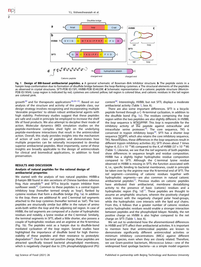
RESULTS AND DISCUSSIONAnalysis of natural peptides for the rational design ofantibacterial propertiesWe started with the analysis of two natural peptides: HVBBI-aβ-hairpin BBI found in skin secretions of Chinese bamboo odorousfrog, Huia versabilis36 and SFTI-a bicyclic trypsin inhibitor fromsunflower seeds37. Common to these peptides is a central trypsininhibitory loop (hereafter termed simply as ‘loop’), flanked bycysteine residues that form a disulfide bridge (Fig. 1a). In additionto the loop, there are additional N- and C-terminal tail segmentsattached to the loop cysteines (hereafter termed as ‘tail’). The twopeptides are structurally similar but differ in the nature of aminoacids-both within the loop and the adjoining tail region. In case ofHVBBI, the tail segments are composed of a cluster of hydrophobicresidues and notably, a lysine residue at the C-terminal. Similarly,the terminal segments in SFTI, albeit a little shorter, also possess acouple of hydrophobic residues and an arginine at the N-terminus(Fig. 1b). The peptides exist as a hairpin due to disulfide-bondmediated cyclization of the loop region. Several studies havehighlighted the importance of disulfide bond for high thermo-stability of these peptides and their resistance to proteolyticdegradation33. Due to their net positive charge, these peptides areattracted specifically toward bacterial phospholipid membrane,which is negatively charged due to 23% phosphatidylglycerol (PG)
content38. Interestingly, HVBBI, but not SFTI, displays a moderateantibacterial activity (Table 1, box A).There are also some important differences. SFTI is a bicyclic
peptide formed through a C–N-terminal cyclization, in addition tothe disulfide bond (Fig. 1c). The residues comprising the loopregion within the two peptides are also slightly different. In HVBBI,the loop sequence is WTKSIPPRP. This loop is responsible for theinhibitory activity of the peptide against extracellular andintracellular serine proteases16. The core sequence, TKS isconserved in trypsin inhibitory loops39. SFTI has a shorter loopsequence TKSIPPI, which also retains the core inhibitory sequence,TKS. Nevertheless, these differences in the loop sequences result indifferent trypsin inhibitory activities (Ki); SFTI shows about 7 timeshigher Ki (5.5 × 10⁻⁸ M) compared to the Ki of HVBBI (37 × 10−8 M)(Table 1). Likewise, we see that the tail segments of both peptidesshow differences in sequence length and residue composition.HVBBI has a slightly higher hydrophobic residue compositioncompared to SFTI. Although the C-terminal lysine residueobserved in HVBBI is missing in SFTI, the function associated withit, i.e., specific binding to bacterial membranes, could in principlebe taken over by the arginine near the N-terminal end of SFTI. Thetail segment—consisting of cationic residues together withhydrophobic segments—are also common in natural cationicantibacterial peptides17. Previous studies on cationic peptideshave attributed their antibacterial, membrane destabilizingactivity to the presence of basic (cationic) residues and ahydrophobic region (Fig. 1d)17. These peptides are thought toassume an amphiphilic structure, wherein the positively chargedends interact with the negatively charged polar head-groups,while the hydrophobic core interacts with the lipid acyl chains.From this, it follows that a greater number of cationic residuesand/or hydrophobic residues would enable a stronger interactionbetween peptides and the phospholipid bilayer. Notably, the netpositive charge on HVBBI is also higher compared to the netcharge on SFTI (Table 1, box A).We set out to understand how the aforementioned differences
in HVBBI and SFTI affect their antibacterial activities. It is importantto mention here that antimicrobial peptides are known todemonstrate significantly different antimicrobial activities orminimum inhibitory concentrations (MICs) against differentbacterial species, even when they are closely related. Therefore,we use Gram-positive bacterium, Micrococcus luteus—one of thewidespread food spoilage bacteria—as a simple model organism
Disulfide bridge
N-tail loop C-tail
HVBBI
SFTI
N- -C
N-
N- -C
-C
HVBBI
SFTI
a
b
c
d
Cationic peptidecharged end charged end
hydrophobic stretchN- -C
S V I G F V KCW T K S I P P R P
T K S I P P I F V D
d
G R
tail
tail
loop
loop
C
CC
Fig. 1 Design of BBI-based antibacterial peptides. a A general schematic of Bowman–Birk Inhibitor structure. b The peptide exists in ahairpin loop conformation due to formation of disulfide bridge between the loop-flanking cysteines. c The structural elements of the peptidesas observed in crystal structures. SFTI-PDB ID:1SFI, HVBBI-PDB ID:4U2W. d Schematic representation of a cationic peptide structure (Moricin-PDB ID:1KV4). Loop region is indicated by red, cysteines are colored yellow, tail region is colored blue, and cationic residues in the tail regionare colored pink.
Y.L. Vishweshwaraiah et al.
2
npj Science of Food (2021) 26 Published in partnership with Beijing Technology and Business University
1234567890():,;
to compare the efficiency (MIC values) of the various peptidesdesigned in this study. HVBBI shows an MIC value of 150 µg/mLagainst M. luteus, whereas SFTI shows no detectable inhibitoryactivity within the assayed concentration range (Table 1, box A).Furthermore, removing the tail segment from HVBBI peptidedrastically compromises the antibacterial activity (HVBBI-loop,Table 1, box A). As expected for SFTI-loop, no antibacterial activitywas observed upon removing the tail. Interestingly, for both SFTIand HVBBI, the removal of tail segments affected the trypsininhibitory constant by 2 orders of magnitude (compare Ki values inTable 1, box A). This suggests that apart from its role inantibacterial activity, the tail segment also influences the anti-trypsin activity of the peptide.Rational design of peptides with better antimicrobial properties
requires an understanding of how the sequence and structure ofthe peptides may be related to their bioactivity. From the abovediscussion and previous studies on cationic antimicrobial pep-tides40–43, we reasoned that increasing the hydrophobic core andthe positive charge would result in better antibacterial activity.Interestingly, besides small peptides like HVBBI and SFTI, BBIs areusually found as 8, 16, and 24 kDa proteins with a minimalrepeating 4 kDa unit44. In the larger BBIs, the loop is flanked bytwo beta strands on either side, which are linked by one or moredisulfide bridges that further enhance the stability. In the case ofshort peptides, the beta strands (tail segment) can be designedwith higher hydrophobic composition and/or greater cationiccharge for improved antibacterial activity through side-chainmutations or insertion of additional residues.To test our hypothesis, we first designed a chimeric peptide,
HSEP1—derived from HVBBI and SFTI, wherein the tail of SFTI wasinterchanged with that from HVBBI (Fig. 2a). SFTI-loop has aboutthree times higher binding affinity for trypsin compared to HVBBI-loop (Table 1, box A). At the same time, HVBBI has a higherantibacterial activity, which may be attributed to its longertail region. We reasoned that replacing the SFTI-tail with theHVBBI-tail, in principle, yields a peptide with a better overall
antibacterial activity. Table 1 (box B) shows that the trypsininhibitory constant obtained for the new peptide—HSEP1 was 23 ×10−8 M, which is similar to the value obtained for HVBBI. However,the antimicrobial activity of the chimeric peptide HSEP1 (MIC: 75 µg/mL) also showed improvement over that of HVBBI (150 µg/mL).It is interesting to compare HSEP1 and HVBBI sequences in the
context of their measured antibacterial activities. The twopeptides differ only in the loop region. If one assumes that theantibacterial activity of the peptide was entirely dependent onthe tail segments, the MIC values for both peptides should besimilar. This however is not the case, as HSEP1 shows enhancedantibacterial effect compared to HVBBI (Table 1). This is despitethe lower basicity (charge +2) of HSEP1 compared to that ofHVBBI (+3); this leads to an interesting possibility of the loopregion influencing the overall antibacterial activity of thepeptide. It appears that the loop and the tail segments bothinfluence the antibacterial as well as the anti-trypsin activities ofthe peptides; the exact factors governing this influence need tobe understood. In summary, supplementing the SFTI-loop withHVBBI-tail resulted in a peptide, HSEP1, with better antimicrobialefficacy against M. luteus.
Improving upon sequence designs for better antimicrobialefficacyWe next sought to improve the HSEP1 activity by varyingphysiochemical properties such as hydrophobicity and net charge.First, we increased the hydrophobicity of HSEP1. It is understoodthat the hydrophobic residues aid in the interaction of thepeptides with the acyl chain of the lipids. Moreover, mutualaggregation of peptides through these hydrophobic regions hasbeen suggested to aid the membrane destabilization process45. Ofall hydrophobic residues, aromatic phenylalanine residue isparticularly interesting as it has been reported to enhance mutualaggregation of the peptides on membrane surfaces16. Therefore,we introduced additional phenylalanine residues in both the N-tail(note, in HSPE2 that a Phe is inserted between Ile3 and Gly4 of
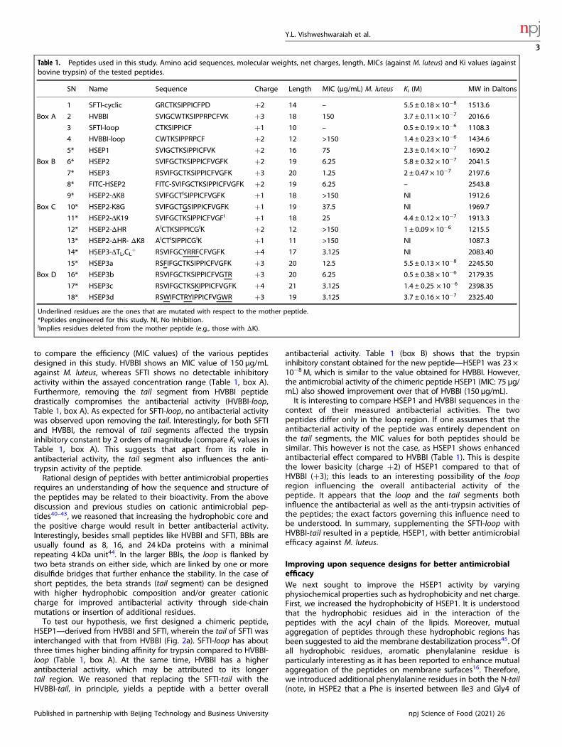
Table 1. Peptides used in this study. Amino acid sequences, molecular weights, net charges, length, MICs (against M. luteus) and Ki values (againstbovine trypsin) of the tested peptides.
SN Name Sequence Charge Length MIC (µg/mL) M. luteus Ki (M) MW in Daltons
1 SFTI-cyclic GRCTKSIPPICFPD +2 14 – 5.5 ± 0.18 × 10⁻⁸ 1513.6
Box A 2 HVBBI SVIGCWTKSIPPRPCFVK +3 18 150 3.7 ± 0.11 × 10⁻⁷ 2016.6
3 SFTI-loop CTKSIPPICF +1 10 – 0.5 ± 0.19 × 10⁻⁶ 1108.3
4 HVBBI-loop CWTKSIPPRPCF +2 12 >150 1.4 ± 0.23 × 10⁻⁶ 1434.6
5* HSEP1 SVIGCTKSIPPICFVK +2 16 75 2.3 ± 0.14 × 10⁻⁷ 1690.2
Box B 6* HSEP2 SVIFGCTKSIPPICFVGFK +2 19 6.25 5.8 ± 0.32 × 10⁻⁷ 2041.5
7* HSEP3 RSVIFGCTKSIPPICFVGFK +3 20 1.25 2 ± 0.47 × 10⁻⁷ 2197.6
8* FITC-HSEP2 FITC-SVIFGCTKSIPPICFVGFK +2 19 6.25 – 2543.8
9* HSEP2-ΔK8 SVIFGCTISIPPICFVGFK +1 18 >150 NI 1912.6
Box C 10* HSEP2-K8G SVIFGCTGSIPPICFVGFK +1 19 37.5 NI 1969.7
11* HSEP2-ΔK19 SVIFGCTKSIPPICFVGFI +1 18 25 4.4 ± 0.12 × 10⁻⁷ 1913.3
12* HSEP2-ΔHR AICTKSIPPICGIK +2 12 >150 1 ± 0.09 × 10⁻⁶ 1215.5
13* HSEP2-ΔHR- ΔK8 AICTISIPPICGIK +1 11 >150 NI 1087.3
14* HSEP3-ΔTL,CL+ RSVIFGCYRRFCFVGFK +4 17 3.125 NI 2083.40
15* HSEP3a RSFIFGCTKSIPPICFVGFK +3 20 12.5 5.5 ± 0.13 × 10⁻⁸ 2245.50
Box D 16* HSEP3b RSVIFGCTKSIPPICFVGTR +3 20 6.25 0.5 ± 0.38 × 10⁻⁶ 2179.35
17* HSEP3c RSVIFGCTKSKIPPICFVGFK +4 21 3.125 1.4 ± 0.25 × 10⁻⁶ 2398.35
18* HSEP3d RSWIFCTRYIPPICFVGWR +3 19 3.125 3.7 ± 0.16 × 10⁻⁷ 2325.40
Underlined residues are the ones that are mutated with respect to the mother peptide.*Peptides engineered for this study. NI, No Inhibition.IImplies residues deleted from the mother peptide (e.g., those with ΔK).
Y.L. Vishweshwaraiah et al.
3
Published in partnership with Beijing Technology and Business University npj Science of Food (2021) 26
1234567890():,;
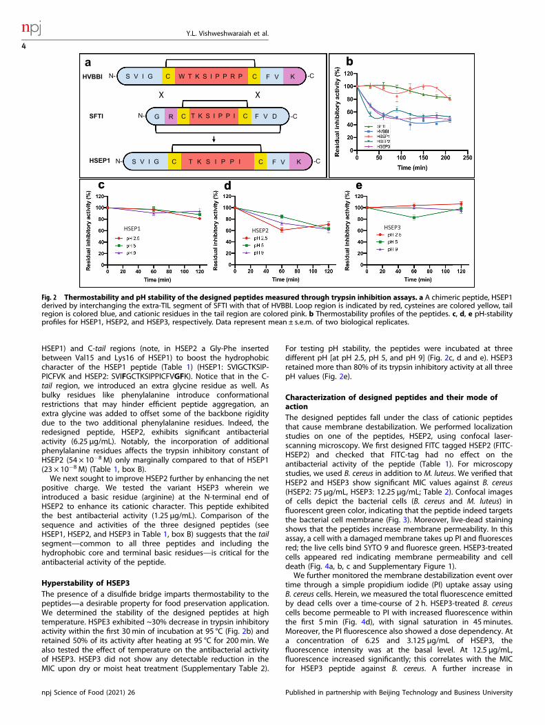
HSEP1) and C-tail regions (note, in HSEP2 a Gly-Phe insertedbetween Val15 and Lys16 of HSEP1) to boost the hydrophobiccharacter of the HSEP1 peptide (Table 1) (HSEP1: SVIGCTKSIP-PICFVK and HSEP2: SVIFGCTKSIPPICFVGFK). Notice that in the C-tail region, we introduced an extra glycine residue as well. Asbulky residues like phenylalanine introduce conformationalrestrictions that may hinder efficient peptide aggregation, anextra glycine was added to offset some of the backbone rigiditydue to the two additional phenylalanine residues. Indeed, theredesigned peptide, HSEP2, exhibits significant antibacterialactivity (6.25 µg/mL). Notably, the incorporation of additionalphenylalanine residues affects the trypsin inhibitory constant ofHSEP2 (54 × 10−8 M) only marginally compared to that of HSEP1(23 × 10−8 M) (Table 1, box B).We next sought to improve HSEP2 further by enhancing the net
positive charge. We tested the variant HSEP3 wherein weintroduced a basic residue (arginine) at the N-terminal end ofHSEP2 to enhance its cationic character. This peptide exhibitedthe best antibacterial activity (1.25 µg/mL). Comparison of thesequence and activities of the three designed peptides (seeHSEP1, HSEP2, and HSEP3 in Table 1, box B) suggests that the tailsegment—common to all three peptides and including thehydrophobic core and terminal basic residues—is critical for theantibacterial activity of the peptide.
Hyperstability of HSEP3The presence of a disulfide bridge imparts thermostability to thepeptides—a desirable property for food preservation application.We determined the stability of the designed peptides at hightemperature. HSPE3 exhibited ~30% decrease in trypsin inhibitoryactivity within the first 30 min of incubation at 95 °C (Fig. 2b) andretained 50% of its activity after heating at 95 °C for 200 min. Wealso tested the effect of temperature on the antibacterial activityof HSEP3. HSEP3 did not show any detectable reduction in theMIC upon dry or moist heat treatment (Supplementary Table 2).
For testing pH stability, the peptides were incubated at threedifferent pH [at pH 2.5, pH 5, and pH 9] (Fig. 2c, d and e). HSEP3retained more than 80% of its trypsin inhibitory activity at all threepH values (Fig. 2e).
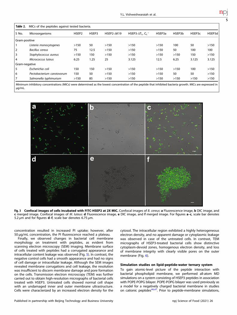
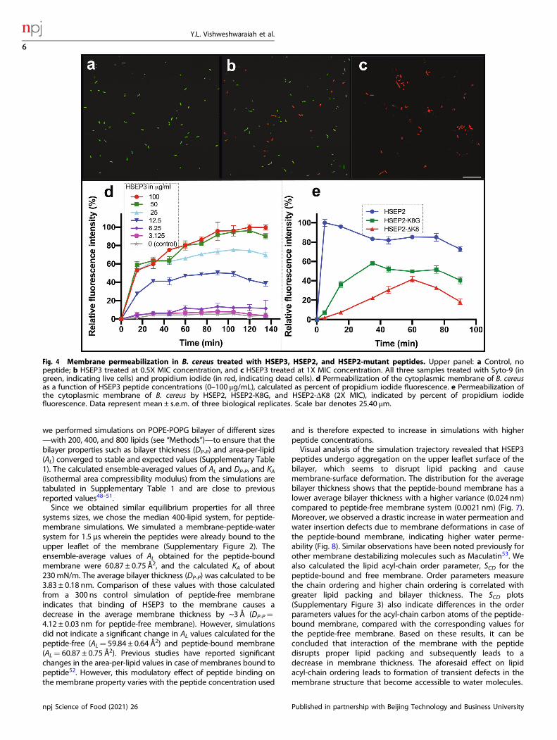
Characterization of designed peptides and their mode ofactionThe designed peptides fall under the class of cationic peptidesthat cause membrane destabilization. We performed localizationstudies on one of the peptides, HSEP2, using confocal laser-scanning microscopy. We first designed FITC tagged HSEP2 (FITC-HSEP2) and checked that FITC-tag had no effect on theantibacterial activity of the peptide (Table 1). For microscopystudies, we used B. cereus in addition to M. luteus. We verified thatHSEP2 and HSEP3 show significant MIC values against B. cereus(HSEP2: 75 µg/mL, HSEP3: 12.25 µg/mL; Table 2). Confocal imagesof cells depict the bacterial cells (B. cereus and M. luteus) influorescent green color, indicating that the peptide indeed targetsthe bacterial cell membrane (Fig. 3). Moreover, live-dead stainingshows that the peptides increase membrane permeability. In thisassay, a cell with a damaged membrane takes up PI and fluorescesred; the live cells bind SYTO 9 and fluoresce green. HSEP3-treatedcells appeared red indicating membrane permeability and celldeath (Fig. 4a, b, c and Supplementary Figure 1).We further monitored the membrane destabilization event over
time through a simple propidium iodide (PI) uptake assay usingB. cereus cells. Herein, we measured the total fluorescence emittedby dead cells over a time-course of 2 h. HSEP3-treated B. cereuscells become permeable to PI with increased fluorescence withinthe first 5 min (Fig. 4d), with signal saturation in 45minutes.Moreover, the PI fluorescence also showed a dose dependency. Ata concentration of 6.25 and 3.125 μg/mL of HSEP3, thefluorescence intensity was at the basal level. At 12.5 μg/mL,fluorescence increased significantly; this correlates with the MICfor HSEP3 peptide against B. cereus. A further increase in
c
a
d e
bN- -CS V I G F V KC CW T K S I P P R P
N- -CC CT K S I P P I F V D
N- -CS V I G F V KC CT K S I P P I
HVBBI
SFTI
HSEP1
X X
c d e
HSEP1 HSEP2 HSEP3
b
G R
Fig. 2 Thermostability and pH stability of the designed peptides measured through trypsin inhibition assays. a A chimeric peptide, HSEP1derived by interchanging the extra-TIL segment of SFTI with that of HVBBI. Loop region is indicated by red, cysteines are colored yellow, tailregion is colored blue, and cationic residues in the tail region are colored pink. b Thermostability profiles of the peptides. c, d, e pH-stabilityprofiles for HSEP1, HSEP2, and HSEP3, respectively. Data represent mean ± s.e.m. of two biological replicates.
Y.L. Vishweshwaraiah et al.
4
npj Science of Food (2021) 26 Published in partnership with Beijing Technology and Business University
concentration resulted in increased PI uptake; however, after50 μg/mL concentration, the PI fluorescence reached a plateau.Finally, we observed changes in bacterial cell membrane
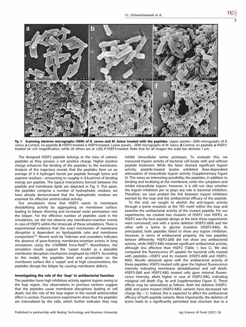
morphology on treatment with peptides, as evident fromscanning electron microscopy (SEM) imaging. Membrane surfaceof cells treated with peptides had a corrugated appearance andintracellular content leakage was observed (Fig. 5). In contrast, thenegative control cells had a smooth appearance and had no signsof cell damage or intracellular leakage. Although the SEM imagesrevealed membrane corrugations and cell leakage, the resolutionwas insufficient to discern membrane damage and pore formationon the cells. Transmission electron microscopy (TEM) was furthercarried out to obtain high-resolution micrographs of bacterial cellstreated with HSEP3. Untreated cells showed normal cell shapewith an undamaged inner and outer membrane ultrastructure.Cells were characterized by an increased electron density for the
cytosol. The intracellular region exhibited a highly heterogeneouselectron density, and no apparent damage or cytoplasmic leakagewas observed in case of the untreated cells. In contrast, TEMmicrographs of HSEP3-treated bacterial cells show distinctivecytoplasm-devoid zones, homogenous electron density, and lossof membrane integrity with clearly visible pores on the outermembrane (Fig. 6).
Simulation studies on lipid-peptide-water ternary systemTo gain atomic-level picture of the peptide interaction withbacterial phospholipid membrane, we performed all-atom MDsimulations on a system consisting of HSEP3 peptides in associationwith POPE-POPG bilayer. POPE-POPG bilayer was used previously asa model for a negatively charged bacterial membrane in studieson cationic peptides46,47. Prior to peptide-membrane simulations,
Table 2. MICs of the peptides against tested bacteria.
S No. Microorganisms HSEP2 HSEP3 HSEP2-ΔK19 HSEP3-ΔTL, CL+ HSEP3a HSEP3b HSEP3c HSEP3d
Gram-positive
1 Listeria monocytogenes >150 50 >150 >150 >150 100 50 >150
2 Bacillus cereus 75 12.5 >150 >150 >150 50 100 100
3 Staphylococcus aureus >150 150 >150 >150 >150 >150 150 >150
4 Micrococcus luteus 6.25 1.25 25 3.125 12.5 6.25 3.125 3.125
Gram-negative
5 Escherichia coli 150 150 >150 >150 >150 >150 100 >150
6 Pectobacterium carotovorum 150 50 >150 >150 >150 50 50 >150
7 Salmonella typhimurium >150 85 >150 >150 >150 >150 >150 >150
Minimum inhibitory concentrations (MICs) were determined as the lowest concentration of the peptide that inhibited bacteria growth. MICs are expressed inµg/mL.
Fig. 3 Confocal images of cells incubated with FITC-HSEP2 at 2X MIC. Confocal images of B. cereus: a Fluorescence image, b DIC image, andc merged image. Confocal images of M. luteus: d Fluorescence image, e DIC image, and f merged image. For figures a–c, scale bar denotes5.2 μm and for figures d–f, scale bar denotes 6.75 μm.
Y.L. Vishweshwaraiah et al.
5
Published in partnership with Beijing Technology and Business University npj Science of Food (2021) 26
we performed simulations on POPE-POPG bilayer of different sizes—with 200, 400, and 800 lipids (see “Methods”)—to ensure that thebilayer properties such as bilayer thickness (DP-P) and area-per-lipid(AL) converged to stable and expected values (Supplementary Table1). The calculated ensemble-averaged values of AL and DP-P, and KA(isothermal area compressibility modulus) from the simulations aretabulated in Supplementary Table 1 and are close to previousreported values48–51.Since we obtained similar equilibrium properties for all three
systems sizes, we chose the median 400-lipid system, for peptide-membrane simulations. We simulated a membrane-peptide-watersystem for 1.5 µs wherein the peptides were already bound to theupper leaflet of the membrane (Supplementary Figure 2). Theensemble-average values of AL obtained for the peptide-boundmembrane were 60.87 ± 0.75 Å2, and the calculated KA of about230mN/m. The average bilayer thickness (DP-P) was calculated to be3.83 ± 0.18 nm. Comparison of these values with those calculatedfrom a 300 ns control simulation of peptide-free membraneindicates that binding of HSEP3 to the membrane causes adecrease in the average membrane thickness by ~3 Å (DP-P=4.12 ± 0.03 nm for peptide-free membrane). However, simulationsdid not indicate a significant change in AL values calculated for thepeptide-free (AL= 59.84 ± 0.64 Å2) and peptide-bound membrane(AL= 60.87 ± 0.75 Å2). Previous studies have reported significantchanges in the area-per-lipid values in case of membranes bound topeptide52. However, this modulatory effect of peptide binding onthe membrane property varies with the peptide concentration used
and is therefore expected to increase in simulations with higherpeptide concentrations.Visual analysis of the simulation trajectory revealed that HSEP3
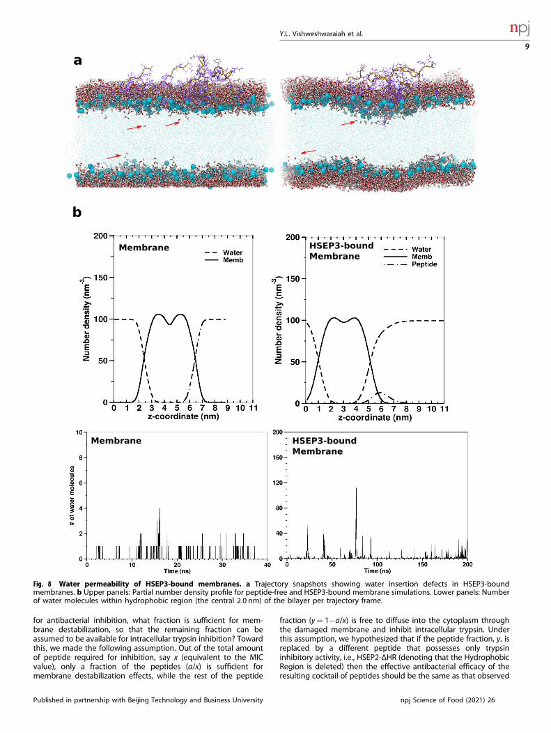
peptides undergo aggregation on the upper leaflet surface of thebilayer, which seems to disrupt lipid packing and causemembrane-surface deformation. The distribution for the averagebilayer thickness shows that the peptide-bound membrane has alower average bilayer thickness with a higher variance (0.024 nm)compared to peptide-free membrane system (0.0021 nm) (Fig. 7).Moreover, we observed a drastic increase in water permeation andwater insertion defects due to membrane deformations in case ofthe peptide-bound membrane, indicating higher water perme-ability (Fig. 8). Similar observations have been noted previously forother membrane destabilizing molecules such as Maculatin53. Wealso calculated the lipid acyl-chain order parameter, SCD for thepeptide-bound and free membrane. Order parameters measurethe chain ordering and higher chain ordering is correlated withgreater lipid packing and bilayer thickness. The SCD plots(Supplementary Figure 3) also indicate differences in the orderparameters values for the acyl-chain carbon atoms of the peptide-bound membrane, compared with the corresponding values forthe peptide-free membrane. Based on these results, it can beconcluded that interaction of the membrane with the peptidedisrupts proper lipid packing and subsequently leads to adecrease in membrane thickness. The aforesaid effect on lipidacyl-chain ordering leads to formation of transient defects in themembrane structure that become accessible to water molecules.
Fig. 4 Membrane permeabilization in B. cereus treated with HSEP3, HSEP2, and HSEP2-mutant peptides. Upper panel: a Control, nopeptide; b HSEP3 treated at 0.5X MIC concentration, and c HSEP3 treated at 1X MIC concentration. All three samples treated with Syto-9 (ingreen, indicating live cells) and propidium iodide (in red, indicating dead cells). d Permeabilization of the cytoplasmic membrane of B. cereusas a function of HSEP3 peptide concentrations (0–100 μg/mL), calculated as percent of propidium iodide fluorescence. e Permeabilization ofthe cytoplasmic membrane of B. cereus by HSEP2, HSEP2-K8G, and HSEP2-ΔK8 (2X MIC), indicated by percent of propidium iodidefluorescence. Data represent mean ± s.e.m. of three biological replicates. Scale bar denotes 25.40 μm.
Y.L. Vishweshwaraiah et al.
6
npj Science of Food (2021) 26 Published in partnership with Beijing Technology and Business University
The designed HSEP3 peptide belongs to the class of cationicpeptides as they possess a net positive charge. Higher positivecharge enhances the binding of the peptides to the membrane.Analysis of the trajectory reveals that the peptides form on anaverage of 3–4 hydrogen bonds per peptide through lysine andarginine residues—amounting to roughly 6–8 kcal/mol of bindingenergy per peptide. The typical interactions formed between thepeptide and membrane lipids are depicted in Fig. 9. This apart,the peptides comprise a number of hydrophobic residues; wehave already demonstrated that the hydrophobic residues areessential for effective antimicrobial activity.Our simulations show that HSEP3 exerts its membrane
destabilizing activity by aggregating on membrane surface,leading to bilayer thinning and increased water permeation intothe bilayer. For the effective number of peptides used in thesimulations, we did not observe any membrane-insertion eventsin case of HSEP3 within the timescale of these simulations. There isexperimental evidence that the exact mechanism of membranedisruption is dependent on lipid:peptide ratio and membranecomposition54. Recent work by Tieleman and coworkers indicatesthe absence of pore-forming membrane-insertion events in longsimulations using the CHARMM force-field55. Nonetheless, thesimulation results support the ‘carpet model’ as a plausiblemembrane disruption mechanism employed by HSEP3. Accordingto this model, the peptides bind and accumulate on themembrane surface like a ‘carpet’ and at high concentrations, thepeptides disrupt the bilayer by causing membrane defects.
Investigating the role of the ‘loop’ in antibacterial functionThe peptides have high inhibitory activity against trypsin owing tothe loop region. Our observations in previous sections suggestthat the peptides cause membrane disruptions leading to celldeath, but the role of the loop region in the overall antimicrobialeffect is unclear. Fluorescence experiments show that the peptidesare internalized by the cells, which further indicates they may
inhibit intracellular serine proteases. To evaluate this, wemeasured trypsin activity of bacterial cell lysate with and withoutpeptide treatment. While the latter showed significant trypsinactivity, peptide-treated lysates exhibited dose-dependentattenuation of intracellular trypsin activity (Supplementary Figure4). This raises an interesting possibility; the peptides, in addition tobinding and localizing at the membrane, enter the cytoplasm andinhibit intracellular trypsin. However, it is still not clear whetherthe trypsin inhibition per se plays any role in bacterial inhibition.Therefore, we next probed the link between trypsin inhibitionexerted by the loop and the antibacterial efficacy of the peptide.To this end, we sought to abolish the anti-trypsin activity
through a lysine mutation at the TKS motif within the loop andexamine the antibacterial activity of the mutant peptide. For ourexperiments, we created two mutants of HSEP2 (not HSPE3, asHSEP2 was the best peptide design at the time these experimentswere conceived); one with a lysine deletion (HSEP2-ΔK8) and theother with a lysine to glycine mutation (HSEP2-K8G). Asanticipated, both peptides failed to show any trypsin inhibition.However, in terms of antibacterial property, the two peptidesbehave differently. HSEP2-ΔK8 did not show any antibacterialactivity, while HSEP2-K8G retained significant antibacterial activity,although less effective than HSEP2 (Table 1, box C). We alsocompared the fluorescence (PI uptake) for bacterial cells treatedwith peptides—HSEP2 and its mutants (HSEP2-ΔK8 and HSEP2-K8G). Results obtained agree with the antibacterial activity ofthese peptides; HSEP2-treated cells gave the highest fluorescenceintensity indicating membrane destabilization and cell death.HSEP2-ΔK8 and HSEP2-K8G treated cells gave minimal fluores-cence intensity, albeit higher in case of HSEP2-K8G, indicatingmarginal cell death (Fig. 4e and Supplementary Figure 5). Theseeffects may be rationalized as follows. Both the deletion (HSEP2-ΔK8) and point mutant (HSEP2-K8G) variants have decreased netcharge (by +1). Indeed, this is expected to affect the antibacterialefficacy of both peptide variants. More importantly, the deletion oflysine leads to a significantly perturbed loop structure due to a
a b c
d e f
Fig. 5 Scanning electron micrographs (SEM) of B. cereus and M. luteus treated with the peptides. Upper panels—SEM micrographs of B.cereus: a Control, no peptide; b HSEP2-treated; c HSEP3-treated. Lower panels—SEM micrographs of M. luteus: d Control, no peptide; e HSEP2-treated (at ×25 magnification, while all others are at ×20); f HSEP3-treated. Note that for all images the scale bar denotes 1 μm.
Y.L. Vishweshwaraiah et al.
7
Published in partnership with Beijing Technology and Business University npj Science of Food (2021) 26

shortened length, which likely abrogates peptide-trypsin binding.In comparison, a significant antimicrobial activity seen for HSEP2-K8G could be because the mutation of lysine to glycine (in HSEP2-K8G) does not alter the loop structure significantly.To delineate whether the TKS lysine contributes to antibacterial
activity via charge effects, we also tested a HSEP2 peptide variant,HSEP2-ΔK19 wherein the C-terminal lysine was deleted withoutaltering K8 (Table 1, box C). This peptide variant retains the overall
net charge of +1, as HSEP2-K8G. HSEP2-ΔK19 demonstrated similarantibacterial activity (25 μg/mL) as HSEP2-K8G (37.5 μg/mL). While itis difficult to precisely pinpoint the roles, it appears that both K8and the terminal lysine K19 contribute to antibacterial activitythrough charge effects that directly aid peptide binding to bacterialmembrane. Indeed, HSEP3 with an additional arginine residueshowed a better MIC value (1.25 μg/mL). We further redesigned thesuperior HSEP3 peptide such that it loses anti-trypsin activity,without altering the net charge and hydrophobicity. Particularly, wereplaced the loop sequence (TSKIPPI) with a non-specific sequence(YRRF) that is expected to abrogate the anti-trypsin activity and atthe same time compensate for the charge and hydrophobicity. Wetested the new peptide, HSEP3-ΔTL,CL+ for its anti-trypsin andantibacterial activity [Note: TL stands for Trypsin Loop and CL+
stands for the compensated charge]. As expected, it did not showany anti-trypsin activity, but still possessed good antibacterialactivity (Table 1). Interestingly, the MIC value for HSEP3-ΔTL,CL+
(3.125 μg/mL) was better than that of HSEP2-ΔK8, HSEP2-K8G, andHSEP2- ΔK19 peptides. The results show that the residuesintroduced in place of the loop sequence can, to an extent,substitute for its loss via charge and hydrophobicity effects.However, trypsin inhibition appears important, given the completeloss in antibacterial activity by HSEP2-ΔK8. Overall, it appears thatthe loop contributes to antibacterial activity through physiochem-ical (charge and hydrophobicity) effects causing membranedisruption, and intracellular trypsin inhibitory effects, the formerbeing a dominant factor. The interplay of the two factors appear togovern the overall activity of the peptides.We investigated this further by rephrasing the problem
statement as follows. Of the total amount of peptide required
a
c
b
d
Fig. 6 Transmission electron micrographs (TEM) of B. cereus treated with HSEP3. TEM micrographs of B. cereus: a represents control(without peptide); b, c, and d represent different micrographs for HSEP3-treated cells (different fields from the same sample).
Fig. 7 Distribution of bilayer thickness values (DP-P) for trajectorysnapshots for membrane and HSEP3-bound membrane simula-tions. A two-sample t-test assuming unequal variances shows thatthe mean membrane thickness values are significantly different(two-sample t(df=5213)= 95.286, p < 0.001).
Y.L. Vishweshwaraiah et al.
8
npj Science of Food (2021) 26 Published in partnership with Beijing Technology and Business University
for antibacterial inhibition, what fraction is sufficient for mem-brane destabilization, so that the remaining fraction can beassumed to be available for intracellular trypsin inhibition? Towardthis, we made the following assumption. Out of the total amountof peptide required for inhibition, say x (equivalent to the MICvalue), only a fraction of the peptides (a/x) is sufficient formembrane destabilization effects, while the rest of the peptide
fraction (y= 1−a/x) is free to diffuse into the cytoplasm throughthe damaged membrane and inhibit intracellular trypsin. Underthis assumption, we hypothesized that if the peptide fraction, y, isreplaced by a different peptide that possesses only trypsininhibitory activity, i.e., HSEP2-ΔHR (denoting that the HydrophobicRegion is deleted) then the effective antibacterial efficacy of theresulting cocktail of peptides should be the same as that observed
Fig. 8 Water permeability of HSEP3-bound membranes. a Trajectory snapshots showing water insertion defects in HSEP3-boundmembranes. b Upper panels: Partial number density profile for peptide-free and HSEP3-bound membrane simulations. Lower panels: Numberof water molecules within hydrophobic region (the central 2.0 nm) of the bilayer per trajectory frame.
Y.L. Vishweshwaraiah et al.
9
Published in partnership with Beijing Technology and Business University npj Science of Food (2021) 26
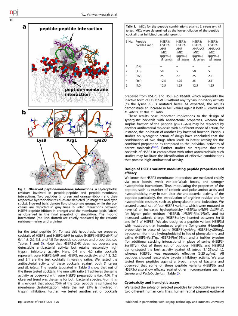
for the total peptide (x). To test this hypothesis, we preparedcocktails of HSEP3 and HSEP2-ΔHR in ratios [HSEP3:HSEP2-ΔHR] of0:4, 1:3, 2:2, 3:1, and 4:0 (for peptide sequences and properties, seeTables 1 and 3). Note that HSEP2-ΔHR does not possess anydetectable antibacterial activity but retains reasonably hightrypsin inhibitory activity. Here, 0:4 and 4:0 ratio cocktailsrepresent pure HSEP2-ΔHR and HSEP3, respectively; and 1:3, 2:2,and 3:1 are the test cocktails in varying ratios. We tested theantibacterial activity of these cocktails against both B. cereusand M. luteus. The results tabulated in Table 3 show that out ofthe three tested cocktails, the one with ratio 3:1 achieves the sameactivity as observed with pure HSEP3 preparations (i.e., 4:0). Theobserved trend was the same for both bacterial species. From this,it is evident that about 75% of the total peptide is sufficient formembrane destabilization, while the rest 25% is involved intrypsin inhibition. Further, we tested another set of cocktails
prepared from HSEP3 and HSEP2-ΔHR-ΔK8, which represents theinactive form of HSEP2-ΔHR without any trypsin inhibitory activity(as the lysine K8 is mutated here). As expected, the resultsdemonstrate an increase in MIC values against both B. cereus andM. luteus, at this 3:1 ratio.These results pose important implications to the design of
synergistic cocktails with antibacterial properties, wherein thesurplus fraction of the peptide (y= 1−a/x) may be replaced byanother antibacterial molecule with a different mode of action, forinstance, the inhibition of another key bacterial function. Previousstudies on synergistic action of drugs have concluded that thecombination of two drugs often leads to better activity for thecombined preparation as compared to the individual activities ofparent molecules56,57. Further studies are required that testcocktails of HSEP3 in combination with other antimicrobials; suchstudies may facilitate the identification of effective combinationsthat possess high antibacterial activity.
Design of HSEP3 variants: modulating peptide properties andefficacyWe know that HSEP3-membrane interactions are mediated chieflyvia polar bonds, weak van-der-Waals forces, and strongerhydrophobic interactions. Thus, modulating the properties of thepeptide, such as number of cationic and polar amino acids andhydrophobicity, may in turn alter the antibacterial activity of thepeptide; particularly, the introduction of arginine residue and/orhydrophobic residues such as phenylalanine and isoleucine. Wecreated a small set of four HSEP3 variants, which were mutated tohave (a) an increased hydrophobicity [HSEP3a (HSEP3-Val3Phe)],(b) higher polar residues [HSEP3b (HSEP3-Phe19Thr)], and (c)increased cationic charge [HSEP3c: Lys inserted between Ser10and Ile11 of HSPE3]. We also designed a peptide variant, HSEP3dwith mutations that introduced arginine (for greater H-bondingpropensity) in place of lysine (HSEP3-Lys9Arg, HSEP3-Lys20Arg),tryptophan (for more hydrophobicity) in lieu of phenylalanine andvaline (HSEP3-Val3Trp, HSEP2-Phe19Trp), and a bulkier tyrosine(for additional stacking interactions) in place of serine (HSEP3-Ser10Tyr). Out of these set of peptides, HSEP3c and HSEP3ddemonstrated the best activity against M. luteus (3.125 µg/mL),whereas HSEP3b was reasonably effective (6.25 µg/mL). Allpeptides showed reasonable trypsin inhibitory activity. We alsotested these peptides against a broad range of bacteria andobserved that some of these peptide variants (HSEP3b andHSEP3c) also show efficacy against other microorganisms such asListeria and Pectobacterium (Table 2).
Cytotoxicity and hemolytic assaysWe tested the safety of selected peptides by cytotoxicity assay ontwo different human cells lines, human retinal pigment epithelial
Fig. 9 Observed peptide-membrane interactions. a Hydrophobicresidues involved in peptide-peptide and peptide-membraneinteractions. Two peptides (in green and orange ribbon) and theirrespective hydrophobic residues are depicted (in magenta and cyansticks). Blue-red balls denote lipid phosphate groups, while the acylchains are depicted in gray lines. b Polar interactions betweenHSEP3 peptide (ribbon in orange) and the membrane lipids (sticks)as observed in the final snapshot of simulation. The h-bondinteractions (red line, dotted) are chiefly mediated by the cationicresidues—lysine and arginine.
Table 3. MICs for the peptide combinations against B. cereus and M.luteus. MICs were determined as the lowest dilution of the peptidecocktail that inhibited bacterial growth.
S No. Peptidecocktail ratio
HSEP3:HSEP2-ΔHRMIC(µg/mL)B. cereus
HSEP3:HSEP2-ΔHRMIC(µg/mL)M. luteus
HSEP3:HSEP2-ΔHR,ΔK8MIC(µg/mL)B. cereus
HSEP3:HSEP2-ΔHR,ΔK8MIC(µg/mL)M. luteus
1 (0:4) – – – –
2 (1:3) 50 5 50 5
3 (2:2) 25 2.5 25 2.5
4 (3:1) 12.5 1.25 25 2.5
5 (4:0) 12.5 1.25 12.5 1.25
Y.L. Vishweshwaraiah et al.
10
npj Science of Food (2021) 26 Published in partnership with Beijing Technology and Business University
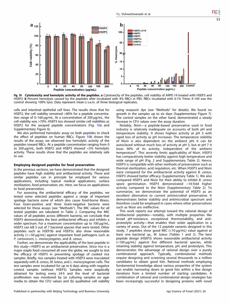
cells and intestinal epithelial cell lines. The results show that forHSEP2, the cell viability remained >80% for a peptide concentra-tion range of 0–160 µg/mL. At a concentration of 200 µg/mL, thecell viability was >70%. HSEP3 too showed similar cell viabilities asHSEP2 for the assayed peptide concentrations (Fig. 10a andSupplementary Figure 6).We also performed hemolytic assay on both peptides to check
the effect of peptides on human RBCs. Figure 10b shows theresults of the assay; we observed low hemolytic activity of thepeptides toward RBCs. At a peptide concentration ranging from 0to 200 μg/mL, both HSEP2 and HSEP3 showed <5% hemolyticactivity. These results show that the peptides are relatively safeto use.
Employing designed peptides for food preservationIn the previous sections, we have demonstrated that the designedpeptides have high stability and antibacterial activity. These andsimilar peptides can in principle be employed for variousapplications, including topical medical applications, surfacesterilization, food preservation, etc. Here, we focus on applicationsto food preservation.For assessing the antibacterial efficacy of the peptides, we
tested some selected peptides against a range of importantspoilage bacteria some of which also cause food-borne illness.Four Gram-positive and three Gram-negative bacteria wereselected for these assays (see “Methods”). The MIC values for alltested peptides are tabulated in Table 2. Comparing the MICvalues of all peptides across different bacteria, we conclude thatHSEP3 demonstrates the best antibacterial efficacy and inhibits awider spectrum. For a maximum concentration up to 100 µg/mL,HSEP3 can kill 5 out of 7 bacterial species that were tested. Otherpeptides such as HSEP3b and HSEP3c also show reasonableactivity (<=50 µg/mL) against important food pathogens such asP. cartovorum, L. monocytogenes, and B. cereus.Further, we demonstrate the applicability of the best peptide in
this study—HSEP3 as an antibacterial preservative. Since rice is amajor staple food consumed all over the globe, we sought to testthe efficacy of HSEP3 in preventing spoilage of cooked ricesamples. Briefly, rice samples treated with HSEP3 were inoculatedseparately with B. cereus, M. luteus, and L. monocytogenes cells. Thetest samples were incubated for up to 6 days along with negativecontrol samples (without HSEP3). Samples were asepticallyobtained for testing every 24 h and the level of bacterialproliferation was monitored by (a) culturing samples on agarmedia to obtain the CFU values and (b) qualitative cell viability
using resazurin dye (see “Methods” for details). We found nogrowth in the samples up to six days (Supplementary Figure 7).The control samples on the other hand, demonstrated a steadyincrease in CFU values over the assay duration.Notably, Nisin—a peptide-based preservative used in food
industry is relatively inadequate on accounts of both pH andtemperature stability. It shows highest activity at pH 3 withrapid loss of activity as pH increases. The temperature stabilityof Nisin is also dependent on the ambient pH; it can beautoclaved without much loss of activity at pH 3, but at pH 7 itloses 90% of its activity, independent of the ambienttemperature6. This severely limits applicability of Nisin. HSEP3has comparatively better stability against high temperature andwide range of pH (Fig. 2 and Supplementary Table 2). Hence,HSEP3 is compatible with other methods of preservation such asthermal sterilization, acid regulation, etc. When HSEP3 and Nisinwere compared for the antibacterial activity against B. cereus,HSEP3 showed better efficacy (Supplementary Table 1). We alsocompared HSEP3 and Nisin for their ability to inhibit B. cereusspore germination. HSEP3 demonstrated ~16-fold higheractivity compared to the Nisin (Supplementary Table 2). Tosummarize, we demonstrate the potential of HSEP3 as anexcellent alternative to current antimicrobial preservative. Itdemonstrates better stability and antimicrobial spectrum andtherefore could be employed in cases where other preservativessuch as Nisin are ineffective.This work reports our attempt toward the rational design of
antibacterial peptides—notably, with multiple properties likebroad pH-resistance, exceptional thermostability, and anti-proteolytic activity—that enables their wider application to avariety of areas. Out of the 12 peptide variants designed in thisstudy, 7 peptides show good MIC (<10 µg/mL) value against atleast one bacterial sp., M. luteus (Tables 1 and 2). The bestpeptide design (HSEP3) shows reasonable antibacterial activity(<100 µg/mL) against five different bacterial species, whileretaining stability against temperature, pH, and proteolysis. Thisdemonstrates the advantage of rational design over a purelycombinatorial approach. Typically, combinatorial methodsrequire designing and screening several thousands to a millioncandidates to obtain good hits. Rational methods employingfundamental knowledge about various properties of the systemcan enable narrowing down to good hits within a few designiterations from a limited number of starting candidates. Acombination of rational and combinatorial design strategies hasbeen increasingly successful in designing proteins with novel
Fig. 10 Cytotoxicity and hemolytic activity of the peptides. a Cytotoxicity of the peptides. cell viability of ARPE-19 treated with HSEP3 andHSEP2. b Percent hemolysis caused by the peptides after incubated with 4% RBCs in PBS. RBCs incubated with 0.1% Triton X-100 was thecontrol showing 100% lysis. Data represent mean ± s.e.m. of three biological replicates.
Y.L. Vishweshwaraiah et al.
11
Published in partnership with Beijing Technology and Business University npj Science of Food (2021) 26
functions58–61. A similar strategy can also be applied to design ofpeptides tailored for the application.Our study focuses on the role of the loop in antibacterial action
and the synergism between its trypsin inhibitory function and themembrane destabilizing action of the peptide tail. We show that(a) the peptides also inhibit intracellular trypsin and (b) bothmembrane destabilization and intracellular trypsin inhibitoryactivity of the peptide contribute to its overall antimicrobialactivity. This means that it may be possible to redesign the ‘loop’region against other essential intracellular proteins, which shouldfurther boost the inhibitory activity as well as the bactericidalspectrum of the peptides. While the loop serves as a determinantof intracellular target stability, the terminal region of the peptidescan be redesigned to achieve maximal membrane destabilizingactivity. Such strategies can be employed to expand the pool offunctional antibacterial molecules. This finding is also of relevanceto the design of synergistic combinations to obtain effectiveantibacterial peptide cocktails.The insights obtained through the aforesaid studies are of
broad importance to the design of antimicrobials for clinical andbiomedical applications as well. In addition to the development ofnovel alternatives to current antimicrobials, synergistic combina-tions of new and existing peptides may pave way to moreeffective treatment of resistant infections. Moreover, by usingcombination of peptides with different modes of antibacterialaction, the chance of developing bacterial resistance is alsolowered. A case in point is the development of a superior versionof the antibiotic—Vancomycin—that combines three differentinhibition mechanisms, resulting in a molecule that has muchbetter efficacy62.Although in principle, the peptide designs may be applicable to
a wide range of applications, a separate assessment of theirefficacy is necessary for specific applications. For this study, wehave tested the best peptide design (HSEP3) for its applicability asa food preservative and demonstrate that HSEP3 could be apotential alternative to current antibacterial preservatives likeNisin. However, one compelling argument against the applicationof trypsin inhibitor-based antimicrobial peptides could be itsactivity against digestive serine proteases. We reason that thesepeptides should be safe for human and animal consumption, if theinhibitor concentration required for effective food preservation islower compared to the concentration that cause perceptible anti-nutritive effects during digestion—a concept that is similar to theTherapeutic Index used to assess systemic drugs. In relation tothis, the studies on USFDA-approved soybean Bowman–Birkinhibitor concentrate (S-BBIC) (which is the same class of inhibitorsas our peptides) are roughly indicative of the safety of trypsininhibitor-based molecules for animal and human consumption.S-BBIC has been tested for its efficacy against various carcinogenicconditions in animal and human trials63. For these studies, aconcentration of up to 10mg/kg body weight of humans has beenrecommended as a safe dose. This value corresponds to roughly500 µg/mL of peptide concentration which is more than 3-fold ofthe maximum MIC values obtained for our peptides (150 µg/mL).Although these values are rough estimates based on S-BBIC data,they suggest that HSEP3 could be an effective and safe candidateas a food preservative. However, animal studies are necessary toobtain a more reliable assessment of the potential of HSEP3.Studies are currently underway to test the suitability of these
peptides for other applications and develop better variantsemploying more robust design approaches. In addition, themolecular details underlying the difference in antibacterialefficacy against different bacteria need to be investigated inorder to rationally develop peptides with broad-spectrumantibacterial activity.
METHODSPeptide and microbial strainsThe peptides used in this study as well as the FITC (fluoresceinisothiocyanate) labeled peptides were synthesized by Pepmic Co., Ltd(Suzhou, China). The purity of the peptides (>95%) was assessed byreverse-phase high-performance liquid chromatography (HPLC) and themolecular weight was confirmed by Electrospray Ionization Mass Spectro-metry (ESI-MS). Mueller-Hinton (MH) broth and Luria Bertani (LB) brothmedia were procured from Hi Media, India. Ethanol, Triton X-100, andGlutaraldehyde were procured from Merck, Germany. Trypsin and BAPNA(N-alpha-benzoyl-DL-arginine-p-nitroanilide) were procured from Sigma-Aldrich. WST-I was procured from Roche, Indianapolis, IN. All otherchemicals used were of the highest purity available.Four Gram-positive bacteria: Listeria monocytogenes ATCC13932, Bacillus
cereus ATCC11778, Staphylococcus aureus ATCC12900, and Micrococcusluteus ATCC4698 and three Gram-negative bacteria: Escherichia coliATCC11775, Pectobacterium carotovorum MCC2112, and Salmonellatyphimurium ATCC9844 were used in this study. These strains were storedat −80 °C in 25% (v/v) glycerol.
Antimicrobial assaysAntimicrobial assays against various bacterial cultures were done using themicrodilution broth assay according to the Clinical and LaboratoryStandards Institute (CLSI)64 with some modifications. Mueller-Hinton brothwas used to dilute the peptide stock and the bacterial inoculum. Inoculumwas prepared from the mid-logarithmic phase culture. Each well of themicrotiter plates received aliquots of 100 µL of the media containingdifferent concentrations of peptide ranging from 0.3 to 300 µg/mL. Thefinal concentration of bacteria in the wells was 5 × 10⁵ Colony FormingUnits (CFU) per mL. Ampicillin was used as positive control. For negativecontrol, ultrapure water was used instead of peptides. Microtiter plateswere incubated at 37 °C for 5–6 h with continuous shaking at 130 rpm(M. luteus cells were incubated for 7–8 h). The incubation time used forthese assays was initially optimized using a 20-h incubation period.Minimum Inhibitory Concentration (MIC) was determined by visuallyobserving the color change after adding resazurin dye into each well at afinal concentration of 37 µg/100 µL. Here, MIC is defined as the lowestdilution of peptide that completely inhibited the growth of the organism.To determine the MIC combination of peptides, HSEP3 was mixed withHSEP2-ΔHR or HSEP2-ΔHR,ΔK8 in 1:3, 2:2, 3:1 (HSEP3: HSEP2-ΔHR/HSEP2-ΔHR,ΔK8) ratios (w/w) and antimicrobial assays were carried out for thedetermination of MIC values. MIC assays were performed as threeindependent experiments in triplicates.
Propidium iodide (PI) uptake assayB. cereus and M. luteus were grown in MH broth to logarithmic phase ofgrowth and then diluted to OD600 ~ 0.25. Serial dilutions of theantimicrobial peptides (final concentrations, 0–100 μg/mL) were addedto the wells of a black-walled microplate. Hundred microliters of thebacterial suspension containing PI (final concentration, 10 μM) was addedto the wells. The emitted fluorescence was measured at excitation andemission wavelengths of 585 and 620 nm, respectively, for 2 h using amultimode plate reader (Varioskan Flash, Thermo Scientific, USA). Thepercentage of membrane permeabilization was calculated as the percentof fluorescent intensity of peptide-treated samples with respect tofluorescence intensity of PI-loaded, peptide untreated samples. PI uptakeassays were performed as three independent experiments in triplicates.
Membrane permeability assayAs a qualitative study, membrane permeability and bacterial viability wereanalyzed using LIVE/DEAD BacLight bacterial viability assay kit formicroscopy [L7007, from Invitrogen, this is a mixture of stains: SYTO 9(for live cells) and PI (for dead cells)] according to manufacturer’sinstructions. Briefly, 3 μL of the staining mixture was added to 1mL ofthe bacterial cells previously treated with different concentrations ofHSEP3 (6.25 and 12.5 μg/mL for B. cereus; 0.7 and 1.25 μg/mL for M. luteus;85 μg/mL for P. caratovorum) for 1 h, and saline-treated cells were kept ascontrol. The samples were incubated for 15min in dark at roomtemperature and 5 μL of this sample was trapped in between coverslipand glass slide. The slide was viewed under a confocal fluorescencemicroscope (LSM700, Carl Zeiss, Germany), using ×63 objective using the
Y.L. Vishweshwaraiah et al.
12
npj Science of Food (2021) 26 Published in partnership with Beijing Technology and Business University
following settings: excitation/emission of 480/500 nm and 490/635 nm forSYTO 9 and PI, respectively.
Scanning electron microscopy (SEM)Bacillus cereus and Micrococcus luteus cells were grown in LB broth at 37 °Cto mid-log phase under continuous shaking at 180 rpm. Cells wereharvested by centrifugation at 5500 rpm for 5min, washed thrice with10mM PBS, diluted 1 × 10⁸ CFU/mL with PBS. Cells were incubated with 1XMIC of peptide in a 500 µL reaction for 1 h. Control cells were incubatedwithout peptides. After incubation cells were harvested by centrifugationat 8000 rpm for 5min, washed thrice with PBS, fixed with 2.5% (w/v)glutaraldehyde at room temperature for 4 h, followed by washing twicewith PBS. The cells were dehydrated for 10min with a graded ethanolseries (25, 50, 75, 95, and 100%). Pellet was dissolved in 100% ethanol anddried at room temperature. The samples were mounted on the specimenholder and sputter-coated with gold. Samples were finally transferred toelectron microscope (LEO 435 VP, USA) for imaging.
Transmission electron microscopy (TEM)B. cereus cells were grown and incubated with AMPs as described abovefor SEM sample preparation. Cell pellets were obtained from 10mL of eachcontrol or treated cell suspension and fixed with 2.5% bufferedglutaraldehyde. The cells were post-fixed with 1% osmium tetroxide for2 h at room temperature. This was followed by dehydration in a gradedethanol series (70%, 80%) for 1 h and ‘en-bloc’ staining with 1% uranylacetate in 95% ethanol for 1 h. The final dehydration was carried out inabsolute alcohol for 30min at 4 °C. The clearing was done with propyleneoxide at room temperature. The cells were then processed for infiltration;wherein propylene oxide was replaced with liquid resin araldite CY212.Infiltration was carried out on a rotator in two steps: a mixture of propyleneoxide and araldite (1:1) overnight and two changes in pure araldite for 2 hat room temperature. Cells were allowed to polymerize in an oven at 60 °Cfor 48 h. Finally, 40–60-nm-thick sections were collected on 300 meshcopper grids, stained with lead citrate, and were viewed under thetransmission electron microscope (FEI, TECNAI G2 Spirit BioTwin,Netherlands).
Confocal laser-scanning microscopyB. cereus and M. luteus cells in mid-logarithmic phase were harvested bycentrifugation, washed three times with 10mM PBS, pH 7.2. 1 × 10⁷ CFU/mL cells were incubated with fluorescence (FITC) labeled peptide at 1X MICand 2X MIC concentration at 37 °C for 1 h. After 1 h, cells were pelleteddown and washed three times with PBS and spotted on a glass slide, andobserved under a confocal microscope (LSM700, Carl Zeiss, Germany) witha ×63 objective. Fluorescent images were obtained with a 488 nm band-pass filter for excitation of FITC. Similarly, for PI fluorescence images, thepeptide-treated cells were incubated with PI (1.3 µg/mL) for 20min in darkand were observed under confocal microscope.
Hemolytic assayThe hemolytic activity of the peptide was evaluated using human redblood cells (hRBCs). Erythrocytes were separated from 1mL of blood bycentrifugation at 1500 rpm for 10min. Collected hRBCs were washed threetimes with PBS, diluted to 4% (v/v) in PBS. Hundred microliters of thehRBCs having peptides ranging from 4 to 200 µg/mL as added into 96-wellmicrotiter plate. The plates were incubated for 1 h at 37 °C withoutagitation and centrifuged at 1500 rpm for 5min. Aliquots (100 μL) of thesupernatant were transferred to 96-well plates and absorbance wasmeasured at 414 nm. PBS and 1% Triton X-100 were used as control for 0%and 100% hemolysis, respectively. Percentage of hemolysis was calculatedas (AT− AC)/(AX− AC) × 100; where AT is the experimental absorbance oftreated supernatants, AC is the control absorbance of PBS-treated cellsupernatant, and AX is the absorbance of 0.1% (v/v) Triton X-100 lysed cells.Data presented are averages and standard error from three biologicalreplicate experiments.
Cytotoxicity assayHuman Intestinal Epithelial Cells (HIEC-6, ATCC CRL-3266) were grown inthe base medium OptiMEM-1 Reduced Serum Medium containing thefollowing components: 20 mM HEPES, 10 mM GlutaMAX, 10 ng/mLepidermal growth factor (EGF), and fetal bovine serum (FBS) to a final
concentration of 4%. Cells were incubated at 37 °C with 5% CO2 and 95%air. 10,000 cells were transferred to wells of a 96-well plate at aconcentration of 10,000 cells per well. Cells were treated with 100, 250,500, 750, and 1000 μg of HSEP3 and incubated at 37 °C with 5% CO2. Thepercentage viability of cells was determined by using the MTT assay. Themedia was removed from the wells after 12 h of incubation. Ten microlitersof 5 mg/mL solution of MTT was added to each well and incubated for 4 h.The MTT-containing medium was removed and the formazan crystals weredissolved in DMSO. The absorbance was measured at 530 nm.Cytotoxicity against adult retinal pigmented epithelial (ARPE-19, ATCC
CRL2302) cells was measured using WST assay. ARPE-19 cells growing inlog phase were seeded into 96-well cell-culture plates at 4 × 10⁴ the cellswere incubated at 37 °C for 24 h under 5% CO2. Peptide is added withfinal concentrations of 4–200 µg/mL in Dulbecco’s modified Eaglemedium (DMEM/F12). Nutrient mixture media for the treatment group,whereas for the negative control group, media alone was added. Thecells were incubated for 16 h at 37 °C under 5% CO2. Ten microliters ofWST-1 reagent was added into each well. Plate was incubated at 37 °Cfor 2 h. Color intensity was measured at 450 nm. Cytotoxicity datapresented are averages and standard error from three biologicalreplicate experiments.
Trypsin inhibition studiesThe amidase activity of trypsin and its inhibition was assayed using thechromogenic substrate BAPNA at pH 8.2 in 0.05 M Tris-HCl containing0.02 M CaCl2 at 37 °C. The assay reaction contained 50 µL of trypsinsolution (40–50 μg of trypsin in 1 mM HCl), 50 µL of water, and 125 µL ofthe substrate. The reaction was carried out at 37 °C for 10min and stoppedby addition of 0.25mL of 30% acetic acid. Absorbance of the liberatedp-nitroaniline was measured at 410 nm against an appropriate blank inwhich the reaction was arrested by adding 30% acetic acid prior to BAPNAaddition. The trypsin solution was incubated with an aliquot of inhibitor for10min at 37 °C and reaction started by the addition of 125 µL substrateand incubated at 37 °C for 10min. The reaction was arrested by theaddition of 30% acetic acid and the residual trypsin activity was measuredby recording the absorbance at 410 nm. One unit of trypsin enzymeactivity is defined as an increase in the absorbance of 0.01 at 410 nm underthe assay conditions. The effect of varying substrate concentration(BAPNA) on bovine trypsin in the presence of fixed concentrations ofpeptides was studied. The inhibition constants (Ki) were evaluated usingthe double reciprocal and Dixon plot of the data65,66. Data presented areaverages and standard error calculated from two biological replicateexperiments.
Thermostability and pH-stability studiesThermostability was measured by the determination of trypsin inhibitionactivity of the peptides after incubation for 30, 60, 90, 120, 150, and180min at 95 °C. For pH stability, peptides were dissolved in 50mM buffersof pH 2.5, 5, 9 and incubated for 2 h at room temperature and trypsininhibition activity of the peptides was assayed using the BAPNA methoddescribed earlier. Data presented are averages and standard errorcalculated from two biological replicate experiments.
Effect of heating and autoclaving on antibacterial activityTo determine the stability of HSEP3 at higher temperatures, 20 μL of HSEP3(4mg/mL) and 20 μL of Nisin in a 0.5 mL tube were autoclaved (for moistheat) at 121 °C for 20min or heated at 95 °C for 30min (for dry heat) andcooled to room temperature. Mixture was then used to test theantibacterial activity as previously described. Experiments were repeatedin LB media to study the effect of complex media on antibacterial activity.All the experiments were repeated at least two times.
Spore germination studiesB. cereus spores were used to test the effect of peptides on sporegermination. Spores were prepared by inoculating 3mL MH media with B.cereus cells and incubating at 30 °C at 180 rpm for 12 h. 2.5 mL of theculture was inoculated into a 250mL MH media and incubated at 30 °C for72 h. Furthermore, culture was temperature-treated at 80 °C for 30min toeliminate the vegetative cells. Spore suspension was stored at 4 °C untilfurther use. Two microliters of spore suspension was used to test the effectof HSEP3 and Nisin on spore germination using resazurin. All experimentswere repeated at least twice.
Y.L. Vishweshwaraiah et al.
13
Published in partnership with Beijing Technology and Business University npj Science of Food (2021) 26

Molecular dynamics of lipid bilayer-peptide-water systemFor the simulations, HSEP3 structure was modeled on the available crystalstructure of SFTI bound to bovine trypsin (PDB ID: 1SFI) using Modellerv9.1167. Only the disulfide-restricted inhibitory loop was modeled on thestructure, while the flexible tail segments were optimized using Modeller.The top-scoring (DOPE score) structure obtained was simulated for 50 nsusing isotropic pressure (1 bar; Parrinello–Rahman barostat) and tem-perature coupling (298 K; velocity rescaling68,69 to obtain a structuralensemble of the peptide in aqueous environment. GROMACS-5.1.270
package was used for performing the simulation studies. All simulationswere performed using CHARMM36 parameter set. Peptide structureswere clustered using the gromos clustering method and a representativestructure from the top cluster was used for membrane-peptide-watersimulations71.The lipid bilayers were constructed using the Membrane Bilayer Builder
available at the CHARMM-GUI webserver72. We chose to construct POPE-POPG bilayer for these studies. The lipid bilayer consists of POPE:POPG in3:1 ratio, in accordance with the experimentally observed fraction of POPGin bacterial membrane (~23% POPG in bacterial membrane). Allsimulations were performed on GROMACS-5.1.2 using the CHARMM36parameter set73. We chose the CHARMM36 parameters for thesesimulations because compared to other available lipid parameters,CHARMM parameters have been shown to model POPG correctly74.Initially, the membrane bilayer was equilibrated for a total of 50 ns. Formembrane-peptide simulations, coordinates for the lipid bilayer wereextracted from the final frame obtained from the 40 ns membrane-watersimulations.Three lipid bilayers systems were constructed with POPE:POPG in the
ratio of 3:1 and containing 200 (POPE:150, POPG:50), 400 (POPE:300,POPG:100), and 800 (POPE:600, POPG:200) lipids, respectively. All threesystems were solvated using a value of 50 as the hydration number (i.e.,number of water molecules per molecule of lipid) and adequate number ofNa+ ions were added for neutralizing the system. The systems wereminimized, and thereafter, equilibrated with position restraints on thephosphorus atoms, and dihedral restraints on the glycerol backbone andunsaturated carbon pairs. The equilibration was performed in multiplesteps with decreasing force constant values used for the restraints.Equilibration steps (100 ps each) involved two steps of NVT using theBerendsen thermostat75 for temperature coupling (T= 315 K). A tempera-ture of 315 K was chosen for these simulations which is greater than thephase transition temperatures of POPE [299 K] and POPG [268 K]. This wasfollowed by four steps of NPT (total 8 ns) equilibration using the Berendsenthermostat and barostat for temperature and semi-isotropic pressurecoupling wherein the restraints were gradually reduced. Finally, all systemswere simulated (without restraints) in NPT ensemble (T= 315 K; P= 1 bar)for 40 ns each, and the final frames of the trajectories were used for finalproduction simulations with peptides. For production runs, we used Nose-Hoover76–78 thermostat for temperature coupling (coupling constant=1.0 ps) and Parrinello–Rahman barostat for semi-isotropic pressurecoupling (coupling constant= 5.0 ps). A leap-frog integrator79 was usedwith a time-step of 2 fs for all runs with constraints on all hydrogen bondsusing the P-LINCS algorithm70. The long-range Coulomb interactions weretreated using Smooth Particle Mesh Ewald80,81 and Van-der-Waalsinteractions were treated using a twin-range cut-off scheme. Van-der-Waals interactions were truncated at 1.0 nm with the potential smoothlyshifted to zero at 1.2 nm.For constructing the peptide-membrane system, eight HSEP3 peptide
molecules (ratio of lipid:peptide= 100:2) were randomly placed on oneside of the membrane bilayer at a distance of ~5 nm from the bilayer. Thissystem was placed in a simulation box of size ~11 × 11 × 11 nm3, solvatedwith water (~27,000 molecules), and appropriate counter-ions were addedto neutralize the system charge. The system was minimized andequilibrated similar to the protocol used of membrane simulations (seethe previous paragraph). Since the membrane and peptide coordinatesused for constructing the membrane-peptide systems are alreadyequilibrated structures, we used a shorter three-step protocol here toobtain equilibrated systems for production runs; (a) minimization step withposition restraints on peptide and lipids, (b) NVT equilibration stepwith position restraints on peptide and lipids, and (c) NPT equilibrationwith position restraints on lipids alone—in total 4 ns of equilibrationsimulation. In the course of our preliminary production simulations, weobserved that once few peptide molecules adhere to the membranesurface, other charged peptides experience high repulsive forces that donot allow these other peptides to interact with the membrane. Many ofthese non-interacting peptides quickly move away and re-enter the
simulation box from the other side of the membrane and adhere to thelower leaflet. Such a behavior has been observed previously by Li et al.52. Tocircumvent this problem, we have used a similar approach as employed byLi et al.52; we first performed pulling simulations on the equilibratedpeptide-membrane system by applying a weak pulling force (k= 100 kJ/mol/nm2) between the center-of-mass of individual peptides and themembrane. To avoid any unwanted movement of lipid molecules during thepulling simulations, we applied position restraints (k= 500 kJ/mol/nm2) onthe heavy atoms of all lipid molecules. The above steps allowed all thepeptides to bind to the upper leaflet. Subsequently, this system wasequilibrated for additional 2 ns before commencing the final 1.5 µsproduction simulation that was used for studying membrane-peptideinteractions. The simulation was performed with the same run-parametersas used for membrane-water simulations. TIP3P explicit water model wasused for all simulations. Three independent simulations were run, each withdifferent initial peptide positions and orientations, as well as randomlyassigned initial atomic velocities for the given temperature, based onMaxwell-Boltzmann distribution. An additional 300 ns control simulation(membrane only) was also run for comparison of equilibrium properties withthose calculated for peptide-bound membrane simulations.All visualization, structural analyses, and rendering were done using
Chimera82 and VMD83. The membrane properties were calculated andplotted using analyses programs included in the GROMACS package andcustom python scripts. An in-house modified version of GridMAT-MDprogram was used for calculating and plotting area-per-lipid and bilayerthickness over the full trajectory84.For membrane-only simulations, the final 100 ns of the trajectory were
analyzed to obtain the average area-per-lipid and thickness value forthe bilayer. For the peptide-membrane simulations, the calculations wereperformed on the final 700 ns of the trajectory. All graphs and plots wereproduced and rendered using Grace.
Testing efficacy as a food preservativeCooked rice samples (1 g) were sterilized and aseptically transferred intopolypropylene tubes (12 × 5mL) and mixed thoroughly with an appro-priate concentration of aqueous solution of HSEP3 (excluding controls). All12 tubes were inoculated with 1 × 10⁵ CFU/g load (from broth culturesafter 24-h incubation) of testing microorganisms. Among 12 tubes, threetubes were inoculated with L. monocytogenes, three with B. cereus andthree with M. luteus. One tube was maintained as control for eachmicroorganism without adding the peptide. All the tubes were sealed andstored at room temperature. At various time intervals (0, 24, 48, 72, 96, 120,144 h), content from each tube was drawn aseptically and plated on themedia by using spread plate method. Plates were incubated at 37 °C andbacterial counts were calculated at each time point.
Reporting summaryFurther information on research design is available in the Nature ResearchReporting Summary linked to this article.
DATA AVAILABILITYThe data that support the findings of this study are in supplemental data includingboth experimental data and computational data. All other data are available from thecorresponding author on reasonable request.
Received: 1 January 2021; Accepted: 11 June 2021;
REFERENCES1. Gram, L. & Dalgaard, P. Fish spoilage bacteria—problems and solutions. Curr.
Opin. Biotechnol. 13, 262–266 (2002).2. Bondi, M., Lauková, A., de Niederhausern, S., Messi, P. & Papadopoulou, C. Natural
preservatives to improve food quality and safety. J. Food Qual. 2017, 1090932(2017).
3. Parke, D. V. & Lewis, D. F. V. Safety aspects of food preservatives. Food Addit.Contam. 9, 561–577 (1992).
4. Rai, M., Pandit, R., Gaikwad, S. & Kövics, G. Antimicrobial peptides as natural bio-preservative to enhance the shelf-life of food. J. Food Sci. Technol. 53, 3381–3394(2016).
Y.L. Vishweshwaraiah et al.
14
npj Science of Food (2021) 26 Published in partnership with Beijing Technology and Business University

5. Gálvez, A., López, R. L., Pulido, R. P. & Burgos, M. J. G. Natural antimicrobials forfood preservation. In Food Biopreservation. SpringerBriefs in food, health andnutrition. 3–14 (Springer, New York, NY 2014).
6. Rollema, H. S., Kuipers, O. P., Both, P., Willem, M. & Siezen, R. J. Improvement ofsolubility and stability of the antimicrobial peptide nisin by protein engineering.Appl. Environ. Microbiol. 61, 2873–2878 (1995).
7. Park, Y. & Hahm, K. Antimicrobial activity studies on a trypsin–chymotrypsinprotease inhibitor obtained from potato. Biochem Biophys. Res Commun. 330,921–927 (2005).
8. Pushpanathan, M., Gunasekaran, P. & Rajendhran, J. Antimicrobial peptides:versatile biological properties. Int. J. Pept. 2013, 675391 (2013).
9. Bahar, A. A. & Ren, D. Antimicrobial peptides. Pharmaceuticals 6, 1543–1575(2013).
10. Brogden, K. A. Antimicrobial peptides: pore formers or metabolic inhibitors inbacteria? Nat. Rev. Microbiol. 3, 238–250 (2005).
11. Mahlapuu, M., Håkansson, J., Ringstad, L. & Björn, C. Antimicrobial peptides: anemerging category of therapeutic agents. Front. Cell. Infect. Microbiol. 6, 194(2016).
12. Le, C.-F., Fang, C.-M. & Sekaran, S. D. Intracellular targeting mechanisms byantimicrobial peptides. Antimicrob. Agents Chemother. 61, e02340–16 (2017).
13. Mahlapuu, M., Björn, C. & Ekblom, J. Antimicrobial peptides as therapeutic agents:opportunities and challenges. Crit. Rev. Biotechnol. 40, 1–15 (2020).
14. Seo, M.-D., Won, H.-S., Kim, J.-H., Mishig-Ochir, T. & Lee, B.-J. Antimicrobialpeptides for therapeutic applications: a review. Molecules 17, 12276–12286(2012).
15. Chen, C. H. & Lu, T. K. Development and challenges of antimicrobial peptides fortherapeutic applications. Antibiotics 9, 24 (2020).
16. Li, J. et al. Trypsin inhibitory loop is an excellent lead structure to design serineprotease inhibitors and antimicrobial peptides. FASEB J. 21, 2466–2473 (2007).
17. Huang, J. F. et al. Structure-guided de novo design of a-helical antimicrobialpeptide with enhanced specificity. Pure Appl. Chem. 82, 243–257 (2010).
18. Dinh, T. T. T., Kim, D. H., Lee, B. J. & Kim, Y. W. De novo design and theirantimicrobial activity of stapled amphipathic helices of heptapeptides. Bull.Korean Chem. Soc. 35, 3632–3636 (2014).
19. Sun, J., Xia, Y., Li, D., Du, Q. & Liang, D. Relationship between peptide structureand antimicrobial activity as studied by de novo designed peptides. Biochim.Biophys. Acta - Biomembr. 1838, 2985–2993 (2014).
20. Chionis, K., Krikorian, D., Koukkou, A. I., Sakarellos-Daitsiotis, M. & Panou-Pomonis,E. Synthesis and biological activity of lipophilic analogs of the cationic anti-microbial active peptide anoplin. J. Pept. Sci. 22, 731–736 (2016).
21. Kim, H., Jang, J. H., Kim, S. C. & Cho, J. H. De novo generation of short anti-microbial peptides with enhanced stability and cell specificity. J. Antimicrob.Chemother. 69, 121–132 (2014).
22. Faccone, D. et al. Antimicrobial activity of de novo designed cationic peptidesagainst multi-resistant clinical isolates. Eur. J. Med. Chem. 71, 31–35 (2014).
23. Frecer, V., Ho, B. & Ding, J. L. De novo design of potent antimicrobial peptides.Antimicrob. Agents Chemother. 48, 3349–3357 (2004).
24. Murray, B., Pearson, C. S., Aranjo, A., Cherupalla, D. & Belfort, G. Mechanism of fourde novo designed antimicrobial peptides. J. Biol. Chem. 291, 25706–25715 (2016).
25. Chen, C. H. et al. Simulation-guided rational de novo design of a small pore-forming antimicrobial peptide. J. Am. Chem. Soc. 141, 4839–4848 (2019).
26. Vishnepolsky, B. et al. De novo design and in vitro testing of antimicrobialpeptides against gram-negative bacteria. Pharmaceuticals 12, 82 (2019).
27. Zeitler, B. et al. De-novo design of antimicrobial peptides for plant protection.PLoS ONE 8, e71687 (2013).
28. Clemente, A., Arques, C., Clemente, A. & Arques, C. Bowman-Birk inhibitors fromlegumes as colorectal chemopreventive agents. World J. Gastroenterol. 20,10305–10315 (2014).
29. Clemente, M. et al. Plant serine protease inhibitors: biotechnology application inagriculture and molecular farming. Int. J. Mol. Sci. 20, 470 (2019).
30. Morris, C. A., Morris, L. D., Kennedy, A. R. & Sweeney, H. L. Attenuation of skeletalmuscle atrophy via protease inhibition. J. Appl. Physiol. 99, 1719–1727 (2005).
31. Losso, J. N. The biochemical and functional food properties of the bowman-birkinhibitor. Crit. Rev. Food Sci. Nutr. 48, 94–118 (2008).
32. Armstrong, W. B. et al. Clinical modulation of oral leukoplakia and proteaseactivity by Bowman-Birk inhibitor concentrate in a phase IIa chemopreventiontrial. Clin. Cancer Res. 6, 4684–4691 (2000).
33. Colgrave, M. L., Korsinczky, M. J. L., Clark, R. J., Foley, F. & Craik, D. J. Sunflowertrypsin inhibitor-1, proteolytic studies on a trypsin inhibitor peptide and itsanalogs. Biopolymers 94, 665–672 (2010).
34. Srikanth, S. & Chen, Z. Plant Protease inhibitors in therapeutics-focus on cancertherapy. Front. Pharmacol. 7, 470 (2016).
35. Safavi, F. & Rostami, A. Role of serine proteases in inflammation: Bowman-Birkprotease inhibitor (BBI) as a potential therapy for autoimmune diseases. Exp. Mol.Pathol. 93, 428–433 (2012).
36. Dȩbowski, D. et al. Inhibitory and antimicrobial activities of OGTI and HV-BBIpeptides, fragments and analogs derived from amphibian skin. Peptides 35,276–284 (2012).
37. Lesner, A. & Rolka, K. Sunflower trypsin inhibitor 1 as a molecular scaffold for drugdiscovery. Curr. Pharm. Des. 17, 4308–4317 (2011).
38. Gold, V. A. M. et al. The action of cardiolipin on the bacterial translocon. Proc. NatlAcad. Sci. USA 107, 10044–10049 (2010).
39. McBride, J. D., Watson, E. M., Brauer, A. B. E., Jaulent, A. M. & Leatherbarrow, R. J.Peptide mimics of the Bowman–Birk inhibitor reactive site loop. Pept. Sci. 66,79–92 (2002).
40. Findlay, B., Zhanel, G. G. & Schweizer, F. Cationic amphiphiles, a new generationof antimicrobials inspired by the natural antimicrobial peptide scaffold. Anti-microb. Agents Chemother. 54, 4049–4058 (2010).
41. Huang, Y., Huang, J. & Chen, Y. Alpha-helical cationic antimicrobial peptides:relationships of structure and function. Protein Cell 1, 143–152 (2010).
42. Yin, L. M., Edwards, M. A., Li, J., Yip, C. M. & Deber, C. M. Roles of hydrophobicityand charge distribution of cationic antimicrobial peptides in peptide-membraneinteractions. J. Biol. Chem. 287, 7738–7745 (2012).
43. Jiang, Z. et al. Effects of net charge and the number of positively charged resi-dues on the biological activity of amphipathic α‐helical cationic antimicrobialpeptides. J. Pept. Sci. 90, 369–383 (2007).
44. Prakash, B. et al. Analysis of the amino acid sequences of plant bowman-birkinhibitors. J. Mol. Evol. 42, 560–569 (1996).
45. Brown, J. S. et al. Antibacterial isoamphipathic oligomers highlight the impor-tance of multimeric lipid aggregation for antibacterial potency. Commun. Biol. 1,220 (2018).
46. Amos, S.-B. T. A. et al. Antimicrobial peptide potency is facilitated by greaterconformational flexibility when binding to gram-negative bacterial inner mem-branes. Sci. Rep. 6, 37639 (2016).
47. Park, P. et al. Binding and flip as initial steps for bp-100 antimicrobial actions. Sci.Rep. 9, 8622 (2019).
48. Binder, H. & Gawrisch, K. Effect of unsaturated lipid chains on dimensions, molecularorder and hydration of membranes. J. Phys. Chem. B 105, 12378–12390 (2001).
49. Pandit, K. R. & Klauda, J. B. Membrane models of E. coli containing cyclic moieties inthe aliphatic lipid chain. Biochim. Biophys. Acta - Biomembr. 1818, 1205–1210 (2012).
50. Hong, C., Tieleman, D. P. & Wang, Y. Microsecond molecular dynamics simulationsof lipid mixing. Langmuir 30, 11993–12001 (2014).
51. Rog, T., Murzyn, K. & Pasenkiewicz-Gierula, M. Molecular dynamics simulations ofcharged and neutral lipid bilayers: treatment of electrostatic interactions. ActaBiochim. Pol. 50, 789–798 (2003).
52. Li, J. et al. Molecular simulations suggest how a branched antimicrobial peptideperturbs a bacterial membrane and enhances permeability. Biochim. Biophys.Acta - Biomembr. 1828, 1112–1121 (2013).
53. Parton, D. L., Akhmatskaya, E. V. & Sansom, M. S. P. Multiscale simulations of theantimicrobial peptide maculatin 1.1: water permeation through disorderedaggregates. J. Phys. Chem. B 116, 8485–8493 (2012).
54. Sato, H. & Feix, J. B. Peptide–membrane interactions and mechanisms of mem-brane destruction by amphipathic α-helical antimicrobial peptides. Biochim.Biophys. Acta - Biomembr. 1758, 1245–1256 (2006).
55. Bennett, W. F. D., Hong, C. K., Wang, Y. & Tieleman, D. P. Antimicrobial peptidesimulations and the influence of force field on the free energy for pore formationin lipid bilayers. J. Chem. Theory Comput. 12, 4524–4533 (2016).
56. Wambaugh, M. A. et al. Synergistic and antagonistic drug interactions in thetreatment of systemic fungal infections. Elife 9, e54160 (2020).
57. Yu, G., Baeder, D. Y., Regoes, R. R. & Rolff, J. Combination effects of antimicrobialpeptides. Antimicrob. Agents Chemother. 60, 1717–1724 (2016).
58. Lévy, R. et al. Rational and combinatorial design of peptide capping ligands forgold nanoparticles. J. Am. Chem. Soc. 126, 10076–10084 (2004).
59. Rausch, J. M., Marks, J. R. & Wimley, W. C. Rational combinatorial design of pore-forming β-sheet peptides. Proc. Natl Acad. Sci. USA 102, 10511–10515 (2005).
60. Maeda, Y., Makhlynets, O. V., Matsui, H. & Korendovych, I. V. Design of catalyticpeptides and proteins through rational and combinatorial approaches. Annu. Rev.Biomed. Eng. 18, 311–328 (2016).
61. Jędrzejewska, H. & Szumna, A. Peptide-based capsules with chirality-controlledfunctionalized interiors—rational design and amplification from dynamic com-binatorial libraries. Chem. Sci. 10, 4412–4421 (2019).
62. Okano, A., Isley, N. A. & Boger, D. L. Peripheral modifications of [Ψ[CH2NH]Tpg4]vancomycin with added synergistic mechanisms of action provide durable andpotent antibiotics. Proc. Natl Acad. Sci. USA 114, E5052–E5061 (2017).
63. Kennedy, A. R. The Bowman-Birk inhibitor from soybeans as an anticarcinogenicagent. Am. J. Clin. Nutr. 68, 1406–1412 (1998).
64. Clinical and Laboratory Standards Institute (CLSI). Methods for dilution anti-microbial susceptibility tests for bacteria that grow aerobically, 8th edn. CLSIstandard M07-A9. Clinical and Laboratory Standards Institute, Wayne, Pennsyl-vania USA (2012).
Y.L. Vishweshwaraiah et al.
15
Published in partnership with Beijing Technology and Business University npj Science of Food (2021) 26
65. Wagner, M. Enzyme kinetics, behavior and analysis of rapid equilibrium andsteady-state enzyme systems (Segel, Irwin H.). J. Chem. Educ. 53, A472 (1976).
66. Lineweaver, H. & Burk, D. The determination of enzyme dissociation constants. J.Am. Chem. Soc. 56, 658–666 (1934).
67. Sali, A. & Blundell, T. L. Comparative protein modelling by satisfaction of spatialrestraints. J. Mol. Biol. 234, 779–815 (1993).
68. Parrinello, M. & Rahman, A. Polymorphic transitions in single crystals: a newmolecular dynamics method. J. Appl. Phys. 52, 7182–7190 (1981).
69. Bussi, G., Donadio, D. & Parrinello, M. Canonical sampling through velocityrescaling. J. Chem. Phys. 126, 14101 (2007).
70. Hess, B. P. LINCS: a parallel linear constraint solver for molecular simulation. J.Chem. Theory Comput. 4, 116–122 (2008).
71. Daura, X. et al. Peptide folding: when simulation meets experiment. Angew.Chem. Int. Ed. 38, 236–240 (1999).
72. Jo, S., Kim, T., Iyer, V. G. & Im, W. CHARMM-GUI: a web-based graphical userinterface for CHARMM. J. Comput. Chem. 29, 1859–1865 (2008).
73. Huang, J. & MacKerell, A. D. CHARMM36 all-atom additive protein force field: vali-dation based on comparison to NMR data. J. Comput. Chem. 34, 2135–2145 (2013).
74. Hénin, J., Shinoda, W. & Klein, M. L. Models for phosphatidylglycerol lipids put to astructural test. J. Phys. Chem. B 113, 6958–6963 (2009).
75. Berendsen, H. J. C., Postma, J. P. M., van Gunsteren, W. F., DiNola, A. & Haak, J. R.Molecular dynamics with coupling to an external bath. J. Chem. Phys. 81,3684–3690 (1984).
76. Nosé, S. A molecular dynamics method for simulations in the canonical ensem-ble. Mol. Phys. 52, 255–268 (1984).
77. Hoover, W. G. Canonical dynamics: equilibrium phase-space distributions. Phys.Rev. A 31, 1695–1697 (1985).
78. Martyna, G. J., Klein, M. L. & Tuckerman, M. Nosé. Hoover chains: the canonicalensemble via continuous dynamics. J. Chem. Phys. 97, 2635–2643 (1992).
79. Van Gunsteren, W. F. & Berendsen, H. J. C. A leap-frog algorithm for stochasticdynamics. Mol. Simul. 1, 173–185 (1988).
80. Darden, T., York, D. & Pedersen, L. Particle mesh Ewald: an Nlog (N) method forEwald sums in large systems. J. Chem. Phys. 98, 10089–10092 (1993).
81. Essmann, U. et al. A smooth particle mesh Ewald method. J. Chem. Phys. 103,8577–8593 (1995).
82. Pettersen, E. F. et al. UCSF Chimera—a visualization system for exploratoryresearch and analysis. J. Comput. Chem. 25, 1605–1612 (2004).
83. Humphrey, W., Dalke, A. & Schulten, K. VMD-visual molecular dynamics. J. Mol.Graph. 14, 33–38 (1996).
84. Allen, W. J., Lemkul, J. A. & Bevan, D. R. GridMAT-MD: a grid-based membraneanalysis tool for use with molecular dynamics. J. Comput. Chem. 30, 1952–1958(2009).
ACKNOWLEDGEMENTSSupported by CSIR-CFTRI project [MLP-0174] is gratefully acknowledged. Y.L.V. wassupported by CSIR Senior Research Fellowship; A.A. was supported by DBT [grant no.BT/PR12233/BRB/10/1349/2014] and V.H. was a recipient of CSIR-UGC NET JRFFellowship. We sincerely acknowledge Dr. P. Ganesan, Central Food Technological
Research Institute for his kind help with the cytotoxicity assays. We are grateful to Prof.R. Sowdhamini, National Center for Biological Sciences for providing access tocomputational cluster for the simulation studies. We thank Dr. Chandrashekar Sagar,Department of Neuropathology, National Institute of Mental Health and Neurosciencesfor his kind help in carrying out the TEM studies. A.A. thanks Prof. S. Ramaswamy for hisencouragement and kind support. Y.L.V., A.A., and B.P. are co-inventors on a PatentCooperation Treaty (PCT) patent application (PCT/IN2017/050419) that covers aspectsof antimicrobial design discussed in this publication.
AUTHOR CONTRIBUTIONSB.P. designed the study with inputs from Y.L.V. and A.A. Y.L.V. and V.H. performed theexperimental work. A.A. performed the simulation studies. All authors contributed todata analysis, interpretation, and compilation of the manuscript. Y.L.V. and A.A.contributed equally to this work.
COMPETING INTERESTSThe authors declare no competing interests.
ADDITIONAL INFORMATIONSupplementary information The online version contains supplementary materialavailable at https://doi.org/10.1038/s41538-021-00109-z.
Correspondence and requests for materials should be addressed to B.P.
Reprints and permission information is available at http://www.nature.com/reprints
Publisher’s note Springer Nature remains neutral with regard to jurisdictional claimsin published maps and institutional affiliations.
Open Access This article is licensed under a Creative CommonsAttribution 4.0 International License, which permits use, sharing,
adaptation, distribution and reproduction in anymedium or format, as long as you giveappropriate credit to the original author(s) and the source, provide a link to the CreativeCommons license, and indicate if changes were made. The images or other third partymaterial in this article are included in the article’s Creative Commons license, unlessindicated otherwise in a credit line to the material. If material is not included in thearticle’s Creative Commons license and your intended use is not permitted by statutoryregulation or exceeds the permitted use, you will need to obtain permission directlyfrom the copyright holder. To view a copy of this license, visit http://creativecommons.org/licenses/by/4.0/.
© The Author(s) 2021
Y.L. Vishweshwaraiah et al.
16
npj Science of Food (2021) 26 Published in partnership with Beijing Technology and Business University