Purification and characterization of a surface-binding protein from Lactobacillus fermentum RC14...
10
Journal of’ General Microbiology (1992), 138, 2663-2672. Printed in Great Britain 2663 Purification and characterization of a.surface lectin from the nematode-trapping fungus Arthrobotrys ofigospora STEFAN RosEN,’ * €30 EK,~ LARS RASK~ and ANDERS TUNLID’ ,* ’ Department of’ Microbial Ecology, Lund University, Ecology Building, Helgonavagen 5, S-223 62 Lund, Sweden Department of’ Chemical Ecology and Ecotoxicology, Lund University, Ecology Building, Helgonavagen 5, S-223 62 Lund, Sweden Department of’ Cell Research, Swedish University of Agricultural Science, Box 7055, S-750 07 Uppsala, Sweden (Received 8 June 1992; revised I0 August 1992; accepted 18 August 1992) Several studies have indicated that the capture of nematodes by the nematophagous fungus Arthrobotrys oligospora is mediated by a lectin on the fungal surface. One of the major surface proteins of this fungus showed haemagglutinating activity and was isolated by affinity chromatography using a mucin Sepharose column. Biochemical analysis showed that the protein was a dimeric glycoprotein with a molecular mass of 36 kDa and an isoelectric point of pH 6.5, and contained no sulphur amino acids. The protein was N-terminally blocked; four internal peptides were sequenced, and showed no significant similarity to sequences in the Swiss-Prot or PIR databases. The haemagglutinating activity of the isolated protein was not inhibited by any of the mono- or disaccharides tested, but it was inhibited by the glycoproteins fetuin and m u c h The haemagglutinating activity changed after incubating the protein in buffers of different pH, with maximal activity at pH 11.0 and no activity at pH 2.8. The lectin was tested for different enzymic activities but none were detected. Analysis of the haemagglutinating activity in various cell fractions indicated that the protein was associated with extracellular polymer layers and with the cell wall of the fungus. About the same amount of the haemagglutinating protein was recovered from samples of vegetative mycelium and of mycelium containing nematode-trapping cells. Introduction Fungal adhesion to host surfaces is an important event in the colonization of humans, animals and plants by fungi, and subsequent pathogenesis. Knowledge of fungal adhesion and the factors that influence fungal attach- ment is still, however, limited (Kennedy, 1990), and no fungal adhesive compound has been fully characterized (Nicholson & Epstein, 1991). Carbohydrate-protein (lectin) interactions have been proposed to mediate adhesion in several fungal-host systems, plant patho- gens, fungal-fungal interactions (mycoparasitism), fun- gal-algal interactions and nematophagous fungi (Nordbring-Hertz & Chet, 1986; Manocha & Chen, 1989). One of the first systems to suggest lectin-mediated adhesion was the nematophagus fungus Arthrobotrys oligospora. This fungus captures nematodes using adhe- sive polymers present on special hyphae (traps) forming * Author for correspondence. Tel. 46 46 10 96 14; fax 46 46 10 41 58. 0001-7624 O 1992 SGM a three-dimensional network (Barron, 1977 ; Nordbring- Hertz, 1988). Flooding a colony of A. oligospora with a 20 mM-N-aCetyl-galaCtOSamine (GalNac) solution inhib- its capture, while treatment of the nematodes with this carbohydrate does not inhibit adhesion (Nordbring- Hertz & Mattiasson, 1979). Small amounts of a GalNac- binding protein have been purified on a GalNac-affinity column (Borrebaeck et al., 1984; Premachandran & Pramer, 1984), and it has been found only in trap- containing mycelium and not in vegetative mycelium (Borrebaeck et al., 1984, 1985). Using a soybean agglutinin (SBA ; specificity for GalNac) affinity gel, Borrebaeck et a!. (1985) isolated an apparent GalNac specific receptor protein with a molecular mass of approx. 65 kDa from the cuticle of the nematode. Involvement of lectin-mediated adhesion was further confirmed by demonstrating that entrapment was re- duced when the nematode was treated with a protein purified on a GalNac column (Premachandran & Pramer, 1984). Recent studies of the adhesion mechanism in A.
-
Upload
independent -
Category
Documents
-
view
1 -
download
0
Transcript of Purification and characterization of a surface-binding protein from Lactobacillus fermentum RC14...
Journal of’ General Microbiology (1992), 138, 2663-2672. Printed in Great Britain 2663
Purification and characterization of a. surface lectin from the nematode-trapping fungus Arthrobotrys ofigospora
STEFAN RosEN,’ * €30 E K , ~ LARS RASK~ and ANDERS TUNLID’ ,*
’ Department of’ Microbial Ecology, Lund University, Ecology Building, Helgonavagen 5 , S-223 62 Lund, Sweden Department of’ Chemical Ecology and Ecotoxicology, Lund University, Ecology Building, Helgonavagen 5, S-223 62
Lund, Sweden Department of’ Cell Research, Swedish University of Agricultural Science, Box 7055, S-750 07 Uppsala, Sweden
(Received 8 June 1992; revised I0 August 1992; accepted 18 August 1992)
Several studies have indicated that the capture of nematodes by the nematophagous fungus Arthrobotrys oligospora is mediated by a lectin on the fungal surface. One of the major surface proteins of this fungus showed haemagglutinating activity and was isolated by affinity chromatography using a mucin Sepharose column. Biochemical analysis showed that the protein was a dimeric glycoprotein with a molecular mass of 36 kDa and an isoelectric point of pH 6.5, and contained no sulphur amino acids. The protein was N-terminally blocked; four internal peptides were sequenced, and showed no significant similarity to sequences in the Swiss-Prot or PIR databases. The haemagglutinating activity of the isolated protein was not inhibited by any of the mono- or disaccharides tested, but it was inhibited by the glycoproteins fetuin and much The haemagglutinating activity changed after incubating the protein in buffers of different pH, with maximal activity at pH 11.0 and no activity at pH 2.8. The lectin was tested for different enzymic activities but none were detected. Analysis of the haemagglutinating activity in various cell fractions indicated that the protein was associated with extracellular polymer layers and with the cell wall of the fungus. About the same amount of the haemagglutinating protein was recovered from samples of vegetative mycelium and of mycelium containing nematode-trapping cells.
Introduction
Fungal adhesion to host surfaces is an important event in the colonization of humans, animals and plants by fungi, and subsequent pathogenesis. Knowledge of fungal adhesion and the factors that influence fungal attach- ment is still, however, limited (Kennedy, 1990), and no fungal adhesive compound has been fully characterized (Nicholson & Epstein, 199 1). Carbohydrate-protein (lectin) interactions have been proposed to mediate adhesion in several fungal-host systems, plant patho- gens, fungal-fungal interactions (mycoparasitism), fun- gal-algal interactions and nematophagous fungi (Nordbring-Hertz & Chet, 1986; Manocha & Chen, 1989).
One of the first systems to suggest lectin-mediated adhesion was the nematophagus fungus Arthrobotrys oligospora. This fungus captures nematodes using adhe- sive polymers present on special hyphae (traps) forming
* Author for correspondence. Tel. 46 46 10 96 14; fax 46 46 10 41 58.
0001-7624 O 1992 SGM
a three-dimensional network (Barron, 1977 ; Nordbring- Hertz, 1988). Flooding a colony of A . oligospora with a 20 mM-N-aCetyl-galaCtOSamine (GalNac) solution inhib- its capture, while treatment of the nematodes with this carbohydrate does not inhibit adhesion (Nordbring- Hertz & Mattiasson, 1979). Small amounts of a GalNac- binding protein have been purified on a GalNac-affinity column (Borrebaeck et al., 1984; Premachandran & Pramer, 1984), and it has been found only in trap- containing mycelium and not in vegetative mycelium (Borrebaeck et al., 1984, 1985). Using a soybean agglutinin (SBA ; specificity for GalNac) affinity gel, Borrebaeck et a!. (1985) isolated an apparent GalNac specific receptor protein with a molecular mass of approx. 65 kDa from the cuticle of the nematode. Involvement of lectin-mediated adhesion was further confirmed by demonstrating that entrapment was re- duced when the nematode was treated with a protein purified on a GalNac column (Premachandran & Pramer, 1984).
Recent studies of the adhesion mechanism in A .

2664 S . Roskn and others
oligospora have indicated that the adhesion between traps and nematodes is a complex process involving both protein and carbohydrate-containing surface polymers of the fungus (Tunlid et al., 1991, 1992a). To further understand the molecular background of the adhesion mechanism in this system, a major surface protein has been purified and characterized. It is a haemagglutinat- ing protein and reacts with antibodies prepared against the lectin previously isolated by GalNac affinity chroma- tography (Borrebaeck et al., 1984).
Met hods
Culture of A . oligospora. Trap-containing mycelium [T( +)] was obtained when A. oligospora Fres. (ATCC 24927) was grown in a liquid medium containing 0-01 % soya peptone (neutralized; Oxoid) supple- mented with 0.005% phenylalanine and 0.005% valine to induce heavy trap formation (Friman et al., 1985). For production of hyphae without traps [T( -)I, the fungus was grown in the soya peptone/amino acids medium supplemented with 12 mM-potassium phosphate buffer (pH 7.0). Each culture vessel contained 4500 ml medium and was inoculated with a water suspension of conidia prepared from a culture grown on corn meal agar (Difco) supplemented with 2 g K2HP04 1 - I .
The growth vessels were incubated at room temperature and aerated by vigorous air bubbling (Friman ef al., 1985).
Protein puriJication. All steps of the purification were carried out at 0-4 "C. Mycelium from two vessels (2 x 4500 ml) was harvested after 7 d growth by filtering through a nylon mesh. The mycelium (approx. 500 mg dry wt) was washed with 3 x 50 ml 20 mhl-sodium phosphate buffer (pH 7.4) containing 0.1 5 M-NaC1 (PBS). The fungal material was then transferred to four 50 ml tubes and suspended in 15 ml PBS per tube containing 0.1 mM-PMSF. The mycelium was disrupted in an Ultra-Turrax homogenizer (type T 18/10; Janka Kunkel, IKA) for 4 x 30s, with intervening cooling periods in an ice bath. The homogenate was centrifuged at 13 500 g for 20 min. The supernatant was filtered through a 0.22 pm filter (Millex-GV, Millipore) and then concentrated by ultrafiltration (YM-I0 membrane, cut-off 10 kDa; Amicon, W. R. Grace, Helsingborg, Sweden). The filtrate (< 10 kDa) showed no haemagglutinating activity. The mycelial pellets were frozen until used for the study of haemagglutinating activity in various cell fractions (see below).
The crude extract was further purified by affinity chromatography. Mucin from porcine stomach (Sigma) was coupled to CNBr-activated Sepharose 4B (Pharmacia) according to the manufacturer's instruc- tions. The protein extract was transferred to a 50ml test tube containing 4ml of the mucin affinity gel. The gel was kept in suspension on a flask shaker (Griffin & George) until no haemagglutin- ating activity was detected in the supernatant (approx. 80 min). The gel was washed with 2 x 40 ml PBS and was then packed in a Pharmacia C 10/10 column. The gel was further washed with PBS (flow; 0.25 ml min-I, fractions collected every fourth minute) until only background absorbance (280 nm) was detected. Protein bound to the column was eluted with freshly prepared 0.1 M-NH,OH (pH 11) and concentrated, and the buffer was changed to PBS by ultrafiltration (Centricon 10, Amicon).
The protein was also isolated according to the procedure described
Haemagglutinating activity in various cell fractions. To obtain more information about the localization of the lectin, the pellets remaining after the homogenization of the mycelium were fractionated further. Pellets from 13.5 1 T (-) and 9 1 T (+) medium were thawed and pooled. First, extracellular polymers were removed by sonication using a slight modification of the method described by Tunlid et al. (1991). The pellets were resuspended using a glass rod in 25 ml PBS, sonicated (Bransonic 220 unit, Kebo-Grave, Sweden) for 30s at 4°C and centrifuged (13500g, 10min). The pellet was washed with PBS (2 x 25 ml). The sonication was then repeated twice with 25 ml PBS and intervening centrifugation. The sonicated pellet was transferred to a test tube. Acid-washed glass beads (0.5 mm diameter) and PBS containing 0. I M-PMSF were added (1 : 2 : 1 ; pellet/buffer/glass beads, by vol.) and the pellet was fragmented with an Omni-mixer (Sorvall, Norwalk, USA) for 3 x 60 s on ice. The cell wall fragments were washed until the absorbance (280 nm) was below 0-08 (6 x 75 ml PBS, centrifugation 13 500 g, 10 min). The cell wall pellet was resuspended with a glass rod and agitated with a vortex mixer for 30 s between each wash step. All the supernatants obtained during the purification procedure were recovered for measurement of haemagglutination activity.
To test whether any haemagglutinating protein was bound to the cell walls, samples of 100 mg (wet wt) purified cell walls were transferred to 1 a 5 ml Eppendorf tubes. The cell walls were agitated (vortex mixer, 90 s) with 1 ml of an enzyme solution (1 mg ml-I ; PBS) containing either Novozyme 234 (Novo BioLabs), Lyticase (Sigma) or chitinase (EC 3.2.1.14, Sigma) and then incubated for 22 h in a water bath (slowly rotating) at 30 "C. Following incubation, the samples were centrifuged (10000 g, 10 min) and the haemagglutinating activity in the supernatant was measured. Control samples were treated with PBS only.
Protein content. The protein content was determined using the method of Bradford (1976) using bovine serum albumin as a standard.
PAGE and detection ofglycoproteins. Discontinuous SDS-PAGE was performed according to Laemmli (1970) using a 2050 Midget Electrophoresis Unit (Pharmacia) and 15% (w/v) running gels. Samples were diluted (1 : 1, by vol.) with 50 mM-Tris/HCl buffer (pH 6.8) containing 2% (w/v) SDS, 11.6% (v/v) glycerol and 0.001 % (w/v) bromophenol blue in the presence or absence of 5% (w/v) DTT. The samples were heated at 100 "C for 3 min. Samples with or without DTT were subjected to electrophoresis on separate gels. The relative molecular mass was calculated by using a molecular market kit (LKB, Bromma, Sweden) including ovotransferrin (7678 kDa), bovine serum albumin (66.25 kDa), ovalbumin (45 kDa), carbonic anhydrase (30 kDa), myoglobin (17.2 kDa) and cytochrome c (12.3 kDa). Gels were stained with Coomassie Brilliant Blue. Glycoproteins were detected using the periodic acid-Schiff reagent (PAS) on SDS-PAGE gels according to the method of Dubray & Bezard (1982).
Gelfiltration. The apparent molecular mass of the native form of the protein was determined by gel filtration using Superdex 200 prep grade gel (Pharmacia) packed in a Pharmacia HiLoad 16/60 column. The column was connected to an HPLC (Pharmacia LKB; pump: 2248, detector: Variable Wavelength Monitor 2141) and was equilibrated with PBS (flow : 0.5 ml min-I). Elution was followed at 214 and 280 nm and fractions were collected by a HeliFrac (Pharmacia). The apparent molecular mass was calculated by comparing the elution volume of the purified protein with those of standard proteins [Pharmacia low molecular mass standard kit proteins and aldolase (1 58 kDa)].
by Borrebaeck et al. (1984) except that a GalNac agarose gel (Sigma) was used instead of a GalNac Sepharose 6B gel (Pharmacia) and the protein was detected by measuring UV-absorbance (280 nm) and not by radioactivity measurements after labelling with 1251,
Isoelectric focusing and chromatofocusing. Isoelectric focusing (Phar- macia Immobiline DryPlate, pH 4-7) was performed in a Multiphore I1 system (Pharmacia) according to the manufacturer's instructions with the temperature set at 10°C. The isoelectric point (PI) was
Surface lectin from Arthrobotrys oligospora 2665
estimated using a broad pl calibration kit (pH 3-10, Pharmacia). The gel was stained with Coomassie Brilliant Blue.
Chromatofocusing was performed with a Mono P HR 5/20 (Pharmacia) connected to a Pharmacia HPLC (see above). The column was equilibrated with 0.025 M-bis-Tris (pH 7.1) and the lectin was dissolved in the same buffer (230pg). The lectin was eluted with Polybuffer 74, pH 5.0 (Pharmacia) and detected at 280 nm. The chromatofocusing was performed at 21 "C with a flow rate of 1 .O ml min-' .
Immunoblotting. Protein isolated from T (+) and T (-) cultures was electroblotted from SDS-PAGE gels onto Immobilon PVDF mem- branes (Millipore AB, Sweden), (Towbin et al., 1979). Protein bands reacting with rabbit antibodies prepared by C. Borrebaeck against the purified protein using GalNac affinity chromatography (Borrebaeck et al., 1984) were detected by using alkaline-phosphatase-conjugated goat anti-rabbit antibodies (Sigma), (Blake et al., 1984).
Protein cleavage. Purified lectin (25 pg) was digested with Achromo- bacrer lyticus protease (Wako Pure Chemical Industries Ltd, Osaka, Japan) in 2 M-guanidine hydrochloride essentially as described by Riviere et al. (1991). Hydroxylamine cleavage was carried out as described by Bornstein & Balian (1977) using 20 pg lectin. The peptides were separated by HPLC with a Brownlee C4 Aquapore column (2.1 x 30 mm). Pumps were System Gold (Beckman) and the detector was a photo diode-array 990 (Waters). Buffers used for the reversed phase chromatography were: (A) 0-1 % (v/v) trifluoroacetic acid (TFA) in HzO; and (B) 0.1% TFA (v/v>, 10% HzO (v/v), 90% (v/v) acetonitrile. The gradient was 2 to 62% B in 60 min (hydroxylamine 30 min) and 62 to 90% B in 3 min with a flow rate of 100 p1 min-I. Elution was followed at 214 nm and fractions were collected manually.
Peptide sequencing. Peptides were sequenced with a model 470 A gas- phase sequenator (Applied Biosystems) equipped with an on-line phenylthiohydantoin (PTH) amino acid analyser (model 120, Applied Biosystems) according to the manufacturer's protocol.
Amino acid analysis. Freeze-dried protein was reduced and alkylated with 4-vinylpyridine in 6 M-guanidine hydrochloride as described by Hawke & Yuan (1987). After the alkylation the sample was diluted 3-fold with H 2 0 and precipitated by addition of 20 vols cold acetone/ 0.2% HCI and left for several hours at - 20 "C. The sample was recovered by low speed centrifugation and dissolved in H20 , after which the acid acetone precipitation was repeated. The sample was then subjected to gas-phase hydrolysis in 6 M-HCI, 2% (w/v) phenol at 110 "C for 22 h. Analysis was performed on a Beckman 6300 using ninhydrin as detection reagent, and quantified using a Shimadzu R4A integrator, except for proline which was estimated manually from the 440 nm chromatogram.
Data base searches. Sequence homology was searched for in the Protein Identification Resource (PIR, 30) and Swiss-Prot (19) data- bases using the IntelliGenetics Suite program (version 5.4).
Haemagglurination assay. Haemagglutinating activity was measured on microtitre V-plates. Trypsinized and glutaraldehyde-treated human blood group A erythrocytes (Sigma) were diluted to a 2% (v/v) suspension with PBS. The protein solution was diluted in a 2-fold series with PBS. Samples (20 pl) were added to each well and mixed with an equal volume of the erythrocyte suspension. The mixture was kept at room temperature for 1 h and then examined for haemagglutination. Haemagglutinating activity was defined as the reciprocal of the end- point dilution.
Haemagglutination inhibition assay. Various sugars or glycoproteins were diluted in a 2-fold series with PBS. To 20 p1 of each dilution, an equal volume of a lectin solution was added, which had been carefully diluted to a concentration corresponding to four minimum haemagglu-
tinating units. After preincubation for 1 h at room temperature, 20 p1 erythrocyte suspension was added. After the mixture had been kept at room temperature for 1 h, haemagglutination was examined. Because commercial preparations of monosaccharides can contain haemagglu- tinating activity (cf. Marquardt & Gordon, 1974), all carbohydrate solutions were ultrafiltered through Centricon 10 membranes to remove molecules larger than 10 kDa before being used in the inhibition studies. Carbohydrate solutions that were not filtered were also tested.
Effect of pH on the haemagglutinating activity. The effect on the haemagglutinating activity of incubating the protein at various pH values was determined as follows: 50 p1 protein dissolved in PBS (160 pg ml-I) was incubated with 500 pl of either 0.1 M-NH,OH (pH 11.0), 25 mM-Tris/glycine (pH 3.0) or a universal buffer (Britton & Robinson, 1931) at pH 2.8, 5.6, 8.4 and 11.0. This buffer was made by setting the pH (with 0.2M-NaOH) of 100ml solutions containing 0.02857 ~-5,5'-diethylbarbituric acid, KH2P04, citric acid and H3B03, and adjusting the volume to 200ml with HzO. After incubating the samples for 80 min at room temperature the buffers were changed to PBS by ultrafiltration (Centricon 10) and the haemagglutinating activities were measured.
Enzymic actiuity. The affinity-purified protein (dissolved in PBS) was assayed for various activities, using 2-15 pg protein. Acid phosphatase (EC 3.1 .3.2) and alkaline phosphatase (EC 3. I .3.1) activity were determined using the Sigma diagnostics kits (procedures Nos 104 and 245). Catalase (EC 1 . I 1 . 1 .6) activity was measured by a spectrophoto- metric method according to Luck (1963). Peroxidase (EC 1 . 1 1 . I . 7) activity was assayed according to Herzog & Fahimi (1973). Proteolytic activity was assayed with the chromogenic substrate Azocoll (Sigma) using the method of Chavira et al. (1984); lectin purified using buffer without PMSF was used for these tests.
Alpha-mannosidase (EC 3.2.1 .24) activity was assayed by measur- ing the release of p-nitrophenol spectrophotometrically at 410 nm. The reaction mixture contained 100 pl p-nitrophenyl a-D-mannopyranoside (0.8 mg ml-I) and 25 pl protein (160 pg ml-I) and was incubated at 37 "C for 1 h before adding lop1 1 M-NaOH to the mixture. a-Glucosidase (EC 3.2.1 .20) and P-glucosidase (EC 3.2.1 .21) activities were assayed by measuring the release of glucose from p- nitrophenyl a-D-glucopyranoside (Sigma) or laminarin (Sigma) with a Sigma diagnostics kit (procedure No. 635). Substrate (1 ml, 0-8 mg ml-I ; PBS) and 50 1.11 lectin (160 pg ml-l) were incubated for 1 h at 37°C. The reaction was stopped by boiling for 10min. (The haemagglutinating activity of the lectin is totally inhibited by boiling for 10 min in PBS).
Chitinase (EC 3.2.1 .14) and P-N-acetyl-glucosaminidase (EC 3.2.1.30) activity were assayed by measuring the release of p- nitrophenol from p-nitrophenyl P-D-N, N'-diacetylchitobiose (Sigma) or p-nitrophenyl-N-acetyl-P-D-glucosaminide (Sigma) spectrophoto- metrically at 410 nm. The substrates were both used at a concentration of 1 . 1 mg ml-' (PBS). The reaction mixtures, containing 100 p1 substrate solution and 25 pl protein (160 pg ml-I), were incubated for 1 h at 37°C before adding 10 pl 1 M-NaOH to the mixture prior to spectrometric analysis.
Results
Protein pur ijica t ion
Soluble extracts from homogenized A. oiigospora myce- lium revealed the presence of a haemagglutinating protein. This protein was purified in a single step by

2666 S . Rosin and others
Table 1. Pur8cation of a haemagglutinating protein from a homogenate of A . oligospora
The extract was from liquid grown mycelium containing no traps [T( -)] (9 1 medium, corresponding to approx. 500 mg dry weight mycelium).
Total haemagglu tinating Specific
Volume activity Protein activity Recovery Purification Fraction (mu (units) (mg> (units mg-') (%I (-fold)
Crude extract* 17 17408 12.6 1382 1 00 1 Lectint 0.8 13 434 1.1 12213 77 8.8
~ ~~ ~ ~~ ~
* Extract obtained after homogenization of mycelium, centrifugation and concentration (ultrafiltration). Protein eluted from the mucin-Sepharose affinity column (c.f. Fig. 1 and Method section; Protein
purification).
affinity chromatography using a gel prepared with porcine stomach mucin. The haemagglutinin was eluted from the affinity ligand by 0.1 M-NH,OH, as a single well-defined peak. The purification procedure for sam- ples of mycelium without nematode traps [T(-)] is summarized in Table 1. The purification was repeated five times and the purified protein was 1.3-2.6 mg (g dry wt mycelium)-' (mean, 2.0 mg; SD, 0.5). The recovery was 55-78% (mean, 70%; SD, 9.5) and the purification - fold was 4.0-16.3 (mean, 9.9; SD, 5.3). Approximately the same amount of the protein was obtained from homo- genization of mycelium with traps [T(+)] and without traps [T( -)I.
Haemagglutinating activity in various cell fractions
Most of the haemagglutinating activity was found in the supernatants after the homogenization and sonication of the mycelium (Table 2). Only small amounts of the agglutinin were released during the subsequent fragmen- tation and cell wall purification. Haemagglutinating activity detected during the treatment of the purified cell walls with various enzymes was similar to that detected in the controls incubated with PBS.
Purity, molecular mass, isoelectric point and glycosylation
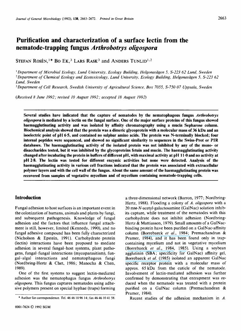
The lectin purified on the mucin gel was found to be homogeneous on SDS-PAGE (Fig. 1, lane 3). Electro- phoresis, (in the presence of absence of DTT), showed that the lectin appeared as a single band with an estimated molecular mass of approx. 15 kDa (Fig. 1). The lectin was also a major protein in the crude extract (Fig. 1, lane 1). The isoelectric point of the purified protein was 6.5 on an Immobiline gel with temperature set at 10°C. Two minor bands were also seen with isoelectric points at pH 6.3 and pH 6.1 (data not shown). The relative amounts of the three bands observed in the isoelectric focusing on Immobiline gels were determined
Table 2 . Haemagglutinating activity in various fractions oj' A . oligospora
Mycelium of the fungus was treated by homogenization, followed by sonication and finally the cells were disrupted by mixing with glass beads. The supernatants obtained from these steps were examined for haemagglutinating activity. Purified cell walls were incubated with various enzymes to release any bound protein with haemagglutinating activity.
Treatment
Total haemagglutinating Relative
activity* activity7 (units) (%I
Cell fractionation Homogenization
(crude extract) Sonication (total) Cell wall purification (total) Cell wall treatment Chi tinase Lyticase Novozyme 234 PBS (control) Mean
130560 (78 1 1 1)$ 49.2
120452 (6866)$ 45.4 1 1 366 (8 963): 4.3
2 880 ( I 728)s 2688 ( 1 760)s 2976 ( 1 42 I ) $ 2 880 ( 1 524)s 2 856 1 . 1
* For comparison all values were calculated in relation to the amount of activity in samples obtained from mycelium grown in 22.5 1 medium.
t Mean values relative to total haemagglutinating activity (265 234 units).
$ Mean from three experiments (sD). $ Mean from three experiments (sD), each with three to four parallel
incubations.
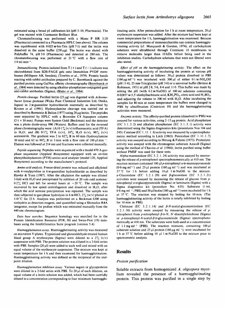
by chromatofocusing at 21 "C. The major protein eluted at pH 6.4 (Fig. 2, peak 1) and two minor peaks were seen at pH 6.3 (peak 2) and 6.2 (peak 3). The total area of the two minor peaks was less than 1% of the area of the major peak.
Gel-filtration of the purified lectin using a Superdex 200 column revealed a single peak, in which the extent of absorbance and haemagglutinating activity coincided,
Surface lectin from Arthrobotrys oligospora
0.25
2667
- 7.8 - 7.6
- - 7.4 - 7.2
1
Fig. 1. SDS-PAGE of proteins extracted from a culture of A. oligosporu with traps [T( +)I. Lanes: 1, crude extract; 2, non-adsorbed fraction, using a mucin Sepharose affinity gel; 3, the haemagglutinating protein eluted with 0.1 M-NH,OH from the affinity gel. Molecular masses of marker proteins are indicated to the left.
corresponding to an apparent molecular mass of approxi- mately 36 kDa (data not shown). The same molecular mass was obtained for the lectin in the crude extract before purification. The protein was stained by the periodic acid-Schiff reagent on an SDS-PAGE gel (data not shown) indicating that the lectin was a glycoprotein. These results suggested that the lectin was a dimeric glycoprotein of non-covalently bound subunits.
Identity with previously isolated lectin
Small amounts [< 10 pg (g dry wt my~el ium)-~] of a haemagglutinating protein with a molecular mass of approx. 15 kDa were also isolated using a GalNac- agarose affinity gel, following the procedure described by Borrebaeck et al. (1984). The identity of this protein with the lectin purified on the mucin gel was further demonstrated by Western blot analysis ; antisera pre- pared against protein purified on a GalNac column reacted with the lectin recovered from the mucin column (data not shown).
Peptide separation, sequencing, amino acid analysis and homology
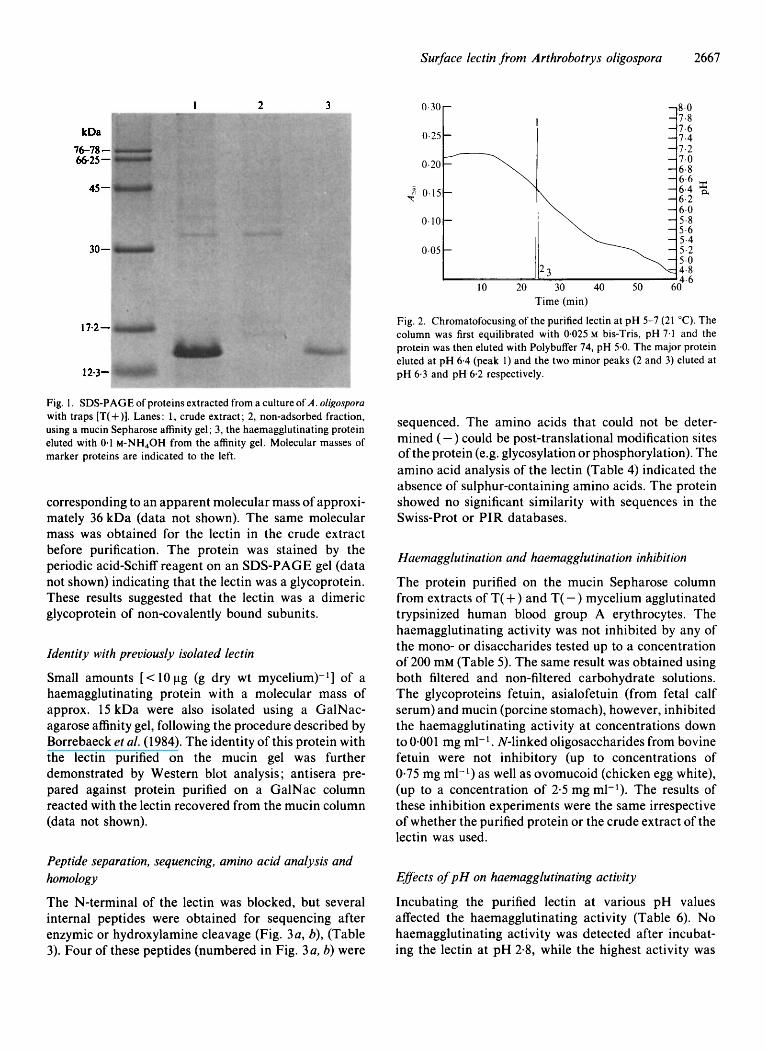
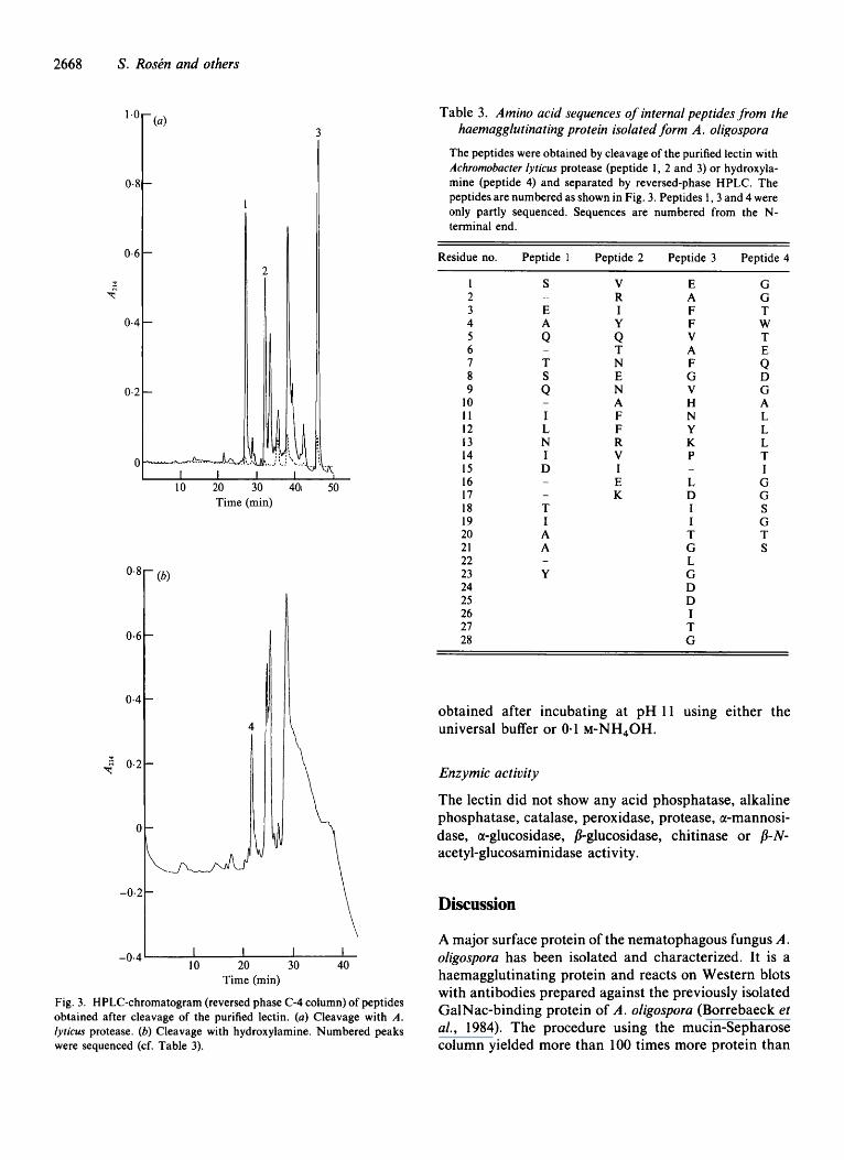
The N-terminal of the lectin was blocked, but several internal peptides were obtained for sequencing after enzymic or hydroxylamine cleavage (Fig. 3a, b), (Table 3) . Four of these peptides (numbered in Fig. 3a, b) were
sequenced. The amino acids that could not be deter- mined ( -) could be post-translational modification sites of the protein (e.g. glycosylation or phosphorylation). The amino acid analysis of the lectin (Table 4) indicated the absence of sulphur-containing amino acids. The protein showed no significant similarity with sequences in the Swiss-Prot or PIR databases.
Haemagglutination and haemagglutination inhibition
The protein purified on the mucin Sepharose column from extracts of T( +) and T( -) mycelium agglutinated trypsinized human blood group A erythrocytes. The haemagglutinating activity was not inhibited by any of the mono- or disaccharides tested up to a concentration of 200 mM (Table 5). The same result was obtained using both filtered and non-filtered carbohydrate solutions. The glycoproteins fetuin, asialofetuin (from fetal calf serum) and mucin (porcine stomach), however, inhibited the haemagglutinating activity at concentrations down to 0.001 mg ml-l. N-linked oligosaccharides from bovine fetuin were not inhibitory (up to concentrations of 0.75 mg ml-l) as well as ovomucoid (chicken egg white), (up to a concentration of 2.5 mg ml-l). The results of these inhibition experiments were the same irrespective of whether the purified protein or the crude extract of the lectin was used.
EBects of pH on haemagglutinating activity
Incubating the purified lectin at various pH values affected the haemagglutinating activity (Table 6) . No haemagglutinating activity was detected after incubat- ing the lectin at pH 2.8, while the highest activity was
2668 S . RosPn and others
3
0.8
0.6
0.4
$ 0.2
0
10 20 30 401 50 Time (min)
4
1
I I I I 10 20 30 40
Time (min)
Fig. 3. HPLC-chromatogram (reversed phase C-4 column) of peptides obtained after cleavage of the purified lectin. (a) Cleavage with A . lyricus protease. (b) Cleavage with hydroxylamine. Numbered peaks were sequenced (cf. Table 3).
Table 3. Amino acid sequences of internal peptides from the haemagglutinating protein isolated form A. oligospora
The peptides were obtained by cleavage of the purified lectin with Achromobacter lyricus protease (peptide 1, 2 and 3) or hydroxyla- mine (peptide 4) and separated by reversed-phase HPLC. The peptides are numbered as shown in Fig. 3. Peptides 1 ,3 and 4 were only partly sequenced. Sequences are numbered from the N- terminal end.
Residue no. Peptide 1 Peptide 2 Peptide 3 Peptide 4
1 2 3 4 5 6 7 8 9
10 11 12 13 14 15 16 17 18 19 20 21 22 23 24 25 26 27 28
S
E A Q
T S Q
-
-
-
I L N I D - -
T I A A
Y -
V R I Y
T N E N A F F R V I E K
Q
E A F F V A F G V H N Y K P
L D I I T G L G D D I T G
-
~
G G T W T E Q D G A L L L T I G G S G T S
obtained after incubating at pH 11 using either the universal buffer or 0.1 M-NH~OH.
Enzymic activity
The lectin did not show any acid phosphatase, alkaline phosphatase, catalase, peroxidase, protease, a-mannosi- dase, a-glucosidase, /3-glucosidase, chitinase or /I- N- acetyl-glucosaminidase activity.
Discussion
A major surface protein of the nematophagous fungus A. oligospora has been isolated and characterized. It is a haemagglutinating protein and reacts on Western blots with antibodies prepared against the previously isolated GalNac-binding protein of A. oligospora (Borrebaeck et al., 1984). The procedure using the mucin-Sepharose column yielded more than 100 times more protein than
Surface lectin from A rthrobotrys oligospora 2669
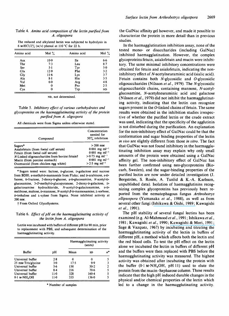
Tabie 4. Amino acid composition of the lectin purified from A . oligospora
The reduced and alkylated lectin was subjected to hydrolysis in 6 ~ -Hc1 /2% (w/v) phenol at 110 "C for 22 h.
Amino acid Mol % Amino acid Mol %
Asx Thr Ser Glx
Ala Val Met CYS
GlY
10-9 7.3 5-1
12.9 11.6 8.1 6.0 0 0
Ile Leu
Phe
His
Pro
TYr
LYS
Arl.3
TrP
6.6 8.7 5.0 3.2 3.7 3-5 4.8 2-5 ND
ND, not determined.
Table 5 . Inhibitory eflect of various carbohydrates and glycoproteins on the haemagglutinating activity of the protein
purified from A . oligospora
All chemicals were from Sigma unless otherwise stated.
Compound
Concentration needed for
50% inhibition
Sugars* >200 mM Asialofetuin (from foetal calf serum) Fetuin (from foetal calf serum) N-Linked oligosaccharides from bovine fetuint Mucin (from porcine stomach) Ovomucoid (from chicken egg white)
0.001 mg ml-' 0.001 mg ml-1
0-001 mg ml-l > 0.75 mg ml-l
>2-5 mg ml-I
* Sugars tested were : lactose, Dglucose, Dgalactose and sucrose from BDH ; a-methyl-Dmannoside from Fluka ; and Darabinose, a-D- fucose, &fructose, 2-deoxy-~glucose, a-Dglucosamine, N-acetyl-D glucosamine, 3-O-methyl-~-glucopyranose, 2-deoxy-~galactose, D- galactosamine hydrochloride, N-acetyl-D-galactosamine, a-D- melibiose, maltose, Dmannose, N-acetyl-fl-Dmannosamine, L-sorbose, Dtrehalose and L-xylose from Sigma. None inhibited activity at 200 mM.
t From Oxford GlycoSystems.
Table 6. Eflect of pH on the haemagglutinating activity of the lectin from A . oligospora
Lectin was incubated with buffers of different pH for 80 min, prior to replacement with PBS, and subsequent determination of the haemagglutinating activity.
Haemagglutinating activity (units)
Buffer PH Mean SD n*
Universal buffer 2.8 0 0 5 25 mM-Tris/glycine 3.0 17.5 9-9 3 Universal buffer 5.6 150 50-2 2 Universal buffer 8.4 216 70-6 5 Universal buffer 11.0 328 160.4 5 0.1 M-NH,OH 11.0 335 136-0 5
* Number of samples
the GalNac affinity gel however, and made it possible to characterize the protein in more detail than in previous studies.
In the haemagglutination inhibition assay, none of the tested mono- or disaccharides (including GalNac) inhibited haemagglutination. However, the complex glycoproteins fetuin, asialofetuin and mucin were inhibi- tory. The same minimal inhibitory concentrations were obtained for fetuin and asialofetuin, indicating the non- inhibitory effect of N-acetylneuraminic acid (sialic acid). Fetuin contains both N-glycosidic and 0-glycosidic oligosaccharides (Nilsson et al., 1979). The N-glycosidic oligosaccharide chains, containing mannose, N-acetyl- glucosamine, N-acetylneuraminic acid and galactose (Nilsson et al., 1979) did not inhibit the haemagglutinat- ing activity, indicating that the lectin can recognize sugars present in the 0-linked chains of fetuin. The same results were obtained in the inhibition studies irrespec- tive of whether the purified lectin or the crude extract was used, indicating that the specificity of the agglutinin is not disturbed during the purification. An explanation for the non-inhibitory effect of GalNac could be that the conformation and sugar binding properties of the lectin in vivo are slightly different from those in vitro. The fact that GalNac was not found inhibitory in the haemagglu- tinating inhibition assay may explain why only small amounts of the protein were obtained using a GalNac affinity gel. The non-inhibitory effect of GalNac has been further confirmed using neo-glycoproteins (Bio- carb, Sweden), and the sugar-binding properties of the purified lectin are now under detailed investigation (J. Bergstrom, S. Rosen, A. Tunlid & K.-A. Karlsson, unpublished data). Isolation of haemagglutinins recog- nizing complex glycoproteins has previously been re- ported from the nematophagous fungus Arthrobotrys ellipsospora (Yamanaka et al., 1988), as well as from several other fungi (Ishikawa & Oishi, 1989; Kawagishi et al., 1991).
The pH stability of several fungal lectins has been examined (e.g. Al-Mahmood et al . , 1991 ; Ishikawa et al., 1981 ; Kawagishi et al., 1990; Kawagishi & Mori, 1991 ; Sage & Vazquez, 1967) by incubating and titrating the haemagglutinating activity of the lectin in buffers of different pH, a method which affects both the lectin and the red blood cells. To test the pH effect on the lectin alone we incubated the lectin in buffers of different pH and the buffers were then replaced with PBS before the haemagglutinating activity was measured. The highest activity was obtained after incubating the protein with the buffer (0.1 M-NH,OH, pH 11) used to elute the protein from the mucin-Sepharose column. These results indicate that the high pH induced durable changes in the physical and/or chemical properties of the lectin which led to a change in the haemagglutinating activity.
2670 S. Roskn and others
However, as mentioned above, results of the inhibition studies indicate that the binding specificity of the lectin was not changed during the purification. We have found only one study of a fungal lectin that has used a similar approach to examine the effects of pH on sugar binding characteristics of the lectin. The haemagglutinating activity of the lectin from Pholiota auvivella (Kawagishi et al., 1991) showed a similar pH dependence to that of the lectin from A. oligospora with haemagglutinating activity decreasing with decreasing pH in the range pH 3.5-6.0. The highest haemagglutinating activity occurred between pH 6.5-10.0, but the activity was slightly lower at the highest pH tested (10.5). As mentioned above, this lectin was not inhibited by any of the mono- or oligosaccharides tested, however, as with the A. oligospora lectin, it was inhibited by complex gl ycoproteins.
Chemical characterization showed that the purified lectin was a glycoprotein with an apparent molecular mass of approximately 36 kDa, consisting of two noncovalently associated subunits. The molecular mass of the subunits, as estimated by SDS-PAGE (15 kDa), was somewhat lower than that reported for the protein purified on the GalNac affinity gels used by Borrebaeck et al. (1984) (20 kDa) and Premachandran & Pramer (1984) (22 kDa). The contradictory results can be explained by the use of different acrylamide concentra- tions in the SDS-PAGE analysis. It is well known that glycoproteins can show anomalous behaviour on SDS- PAGE. With increasing acrylamide concentration, the apparent molecular mass of glycoproteins decreases (Hames, 1990). The PI of the purified lectin was approx. 6.5. The two minor bands/peaks seen on isoelectric focusing and chromatofocusing of the purified lectin could originate from partial modification of the protein (e.g. deamidation).
The protein was a major constituent of the total water- soluble protein obtained by homogenization, and about the same amount was obtained from vegetative [T(-)] and trap-containing [T( +)] mycelium. In contrast, Borrebaeck et al. (1984, 1985), growing the fungus in a different medium, did not find the lectin in the T(-) mycelium. However, we found the lectin in samples of mycelium grown in this medium (unpublished data).
Further information on the localization of the lectin was obtained by analysing haemagglutinating activity in various cell fractions. The major part of this activity was found in solution after homogenization and sonication of the mycelium. Previous EM studies have shown that similar treatments do not lyse the cells but can be used to extract polymers present on the surface of the fungus (Tunlid et al., 1991). A small amount of the haemagglu- tinating protein was released from the purified cell walls. The activity was not increased using enzymes known to
degrade the cell walls of A. oligospora (Nordbring-Hertz et al., 1989), indicating that the lectin was not covalently associated with the cell wall. It is therefore possible that at least a small portion of the lectin recovered by homogenization and sonication could be non-covalently associated with the cell wall of the mycelium. The presence of the protein in the cell wall is also supported by TEM observations using the antibodies generated against the previously purified GalNac-binding protein (Nordbring-Hertz et al., 1988). Furthermore, recent studies have shown that the purified agglutinin is also found in the fungal culture supernatant (RosCn & Tunlid, unpublished data). Lectins associated with the cell walls and extracellular polymers have been found in several other fungi including Sclerotium rolfsii and Rhizoctonia solani (Barak et al., 1985, 1986), Neurospora sitophila (Ishikawa & Oishi, 1989) and Kluyveromyces bulgaricus (Al-Mahmood et al., 1991).
Studies have shown that lectins can have enzymic activity (e.g. Shannon & Hankins, 1981; Finkelstein et al., 1983) and it has been suggested that several types of enzyme, including proteases, phosphatases, and peroxi- daseslcatalases are involved in the infection of nema- todes by A. oligospora (Veenhuis et al., 1985; Tunlid et al., 1992a). The purified lectin did not show any of these activities, nor any of the other enzyme activities tested, including those of several enzymes known to be associated with the fungal cell wall (Gooday & Gow, 1990). However, it is still possible that the purified lectin affects the activity of enzymes associated with the surface of the fungus by forming co-functional complexes with the enzyme and substrate, according to a general model for the function of microbial lectins proposed by Gilboa-Garber & Garber (1 989).
The detailed function of the purified lectin from A. oligospora is not known, but several studies have suggested that a lectin present in the traps mediates adhesion by binding to complementary molecules (recep- tors) on the nematode surface. Alternatively, the lectin might be involved in adhesion by participating in an observed reorganization of the surface polymer fibrils (Tunlid et al., 1992b). However, the fact that the purified lectin is a major surface protein in both T( +) and T( -) mycelium indicates that it could have a more general role in the structure and function of cell walls and associated extracellular polymers (cf. Ishikawa & Oishi, 1989). The purification and characterization of this surface lectin from A. oligospora will enable study of its function in greater detail by, for example, using antibodies, isolation of receptors and cloning of its corresponding gene(s).
This study was supported by grants from the Swedish Natural Science Research Council. We acknowledge Professor Birgit Nord- bring-Hertz for stimulating discussions throughout this work.
Surface lectin from Arthrobotrys oligospora 267 I
References
AL-MAHMOOD, S., COLIN, S. & BONALY, R. (1991). Kluyveromyces bulgaricus yeast lectins. Isolation of two galactose-specific lectin forms from the yeast cell wall. Journal of Biological Chemistry 266,
BARAK, R., ELAD, Y., MIRELMAN, D. & CHET, I. (1985). Lectins: a possible basis recognition in the interaction of Trichoderma and Sclerotium rolfsii. Phytopathology 75, 458-462.
BARAK, R., ELAD, Y. & CHET, I. (1986). The properties of L-fucose binding agglutinin associated with the cell wall of Rhizoctonia solani. Archives of Microbiology 144, 346-349.
BARRON, G. L. (1977). The Nematode-destroying Fungi. Canadian Biological Publications.
BLAKE, M. S., JOHNSTON, K. H., RUSSEL-JONES, G . J. & GOTSCHLICH, E. C. (1984). A rapid, sensitive method for detection of alkaline phosphatase-conjugated anti-antibody on Western blots. Analytical Biochemistry 136, 175- 179.
BORNSTEIN, P. & BALIAN, G. (1977). Cleavage at asn-gly bonds with hydroxylamine. Methods in Enzymology 47, 132-145.
BORREBAECK, C. A. K., MATTIASSON, B. & NORDBRING-HERTZ, B. (1984). Isolation and partial characterization of a carbohydrate- binding protein from a nematode-trapping fungus. Journal of Bacteriology 159, 53-56.
BORREBAECK, C. A. K., MATTIASSON, B. & NORDBRING-HERTZ, B. (1985). A fungal lectin and its apparent receptors on a nematode surface. FEMS Microbiology Letters 27, 35-39.
BRADFORD, M. M. (1976). A rapid and sensitive method for the quantitation of microgram quantities of protein utilizing the principle of protein-dye binding. Analytical Biochemistry 72, 248- 254.
BRITTON, H. T. S. & ROBINSON, R. A. (1931). Universal buffer solutions and the dissociation constant of veronal. Journal of the Chemical Society 1, 1456-1462.
CHAVIRA, R., BURNETT, T. J. & HAGEMAN, J . H. (1984). Assaying proteases with Azocoll. Analytical Biochemistry 72, 248-254.
DUBRAY, G. & BEZARD, G . (1982). A highly sensitive periodic acid- silver stain for 1,2-diol groups of glycoproteins and polysaccharides in polyacrylamide gels. Analytical Biochemistry 119, 325-329.
FINKELSTEIN, R. A., BOESMAN-FINKELSTEIN, M. & HOLT, P. (1983). Vibrio cholera haemagglutinin/lectin/protease hydrolyzes fibronectin and ovomucin : F. M. Burnet revisited. Proceedings of the National Academy of Sciences of the United States of America 80, 1092-1095.
FRIMAN, E., OLSON, S. & NORDBRING-HERTZ, B. (1985). Heavy trap formation by Arthrobotrys oligospora in liquid culture. FEMS Microbiology Ecology 31, 17-2 1 ,
GILBOA-GARBER, N. & GARBER, N. (1 989). Microbial lectin cofunction with lytic activities as a model for a general basic lectin role. FEMS Microbiology Reviews 63, 21 1-222.
GOODAY, G. W. & Cow, N. A. R. (1990). In Tip Growth in Plant and Fungal Cells, pp. 31-57. Edited by I. B. Heath. New York: Academic Press.
HAMES, B. D. ( 1990). One-dimensional polyacrylamide gel electrophor- esis. In Gel Electrophoresis of Proteins, pp. 1-147. Edited by B. D. Hames and D. Rickwood. Oxford: IRL Press.
HAWKE, D. & YUAN, P. (1987). S-Pyridylation of cystine residues. User bulletin, Applied Biosystems 28.
HERZOG, V. & FAHIMI, H. D. (1973). A new sensitive colorimetric assay for peroxidase using 3,3’-diaminobenzidine as hydrogen donor. Analytical Biochemistry 55, 554-562.
ISHIKAWA, F. & OISHI, K. (1989). Production, purification, and characterization of Neurospora sitophila lectin. Agricultural and Biological Chemistry 53, 1769-1 776.
ISHIKAWA, F., OISHI, K. & AIDA, K . (1981). Purification and some properties of chitin-binding haemagglutinin from Conidiobolus lamprauges. Agricultural and Biological Chemistry 45, 551-564.
KAWAGISHI, H. & MORI, H. (1991). Chemical modification and N M R studies on a mushroom lectin Ischnoderma resinosum agglutinin (IRA). Biochimica et Biophysica Acta 1076, 179-1 86.
KAWAGISHI, H., NOMURA, A., MIZUNO, T., KIMURA, A. & CHIBA, S.
20882-20887.
(1990). Isolation and characterization of a lectin from Grifola jrondosa fruiting bodies. Biochimica et Biophysica Acta 1034,247-252.
KAWAGISHI, H., ABE, Y., NAGATA, T., KIMURA, A. & CHIBA, S. (1991). A lectin from the mushroom Pholiota auvivella. Agricultural and Biological Chemistry 55, 2485-2489.
KENNEDY, M. J. (1990). Models for studying the role of fungal attachment in colonization and pathogenesis. Mycopathologia 109,
LAEMMLI, U. K. (1970). Cleavage of structural proteins during the assembly of the head of bacteriophage T4. Nature, London 227,680- 685.
LUCK, H. (1963). Catalase. In Methods in Enzymatic Analysis, pp. 885- 894. Edited by H. U. Bergmeyer. New York: Academic Press.
MANOCHA, M. S. & CHEN, Y. (1989). Specificity of attachment of fungal parasites to their hosts. Canadian Journal of Microbiology 36,
MARQUARDT, M. D. & GORDON, J. A. (1974). Haemagglutinins in commercial preparations of monosaccharides. Nature, London 252,
NICHOLSON, R. L. & EPSTEIN, L. (1991). Adhesion of fungi to the plant surface : prerequisite for pathogenesis. In The Fungal Spore and Disease Initiation in Plants and Animals, pp. 3-23. Edited by G . T. Cole & H. C. Hoch. New York: Plenum Press.
NILSON, B., NORDEN, N. E. & SVENSSON, S. (1979). Structural studies on the carbohydrate portion of fetuin. Journal of Biological Chemistry 254, 4545-4553.
NORDBRING-HERTZ, B. (1988). Ecology and recognition in the nematode-nematophagous fungus system. In Advances in Microbial Ecology, vol. 10, pp. 81-1 14. Edited by K. C. Marshall. New York: Plenum Press.
NORDBRING-HERTZ, B. & CHET, I. (1986). Fungal lectins and agglutinins. In Microbial Lectins and Agglutinins: Properties and Biological Activity, pp. 393408. Edited by D. Mirelman. New York : John Wiley & Sons.
NORDBRING-HERTZ, B. & MATTIASSON, B. (1979). Action of a nematode-trapping fungus shows lectin-mediated host-micro- organism interaction. Nature, London 281, 477479.
NORDBRING-HERTZ, B., VEENHUIS, M. H., MATTIASSON, B. & BORRE- BAECK, C . A. K. (1988). Immunocytochemical localization of a fungal lectin. Abstracts of papers and posters presented at the XIXth International Symposium of the European Society of Nematologists. Uppsala. Nematologica 34, 284.
NORDBRING-HERTZ, B., FEHNE-PERSON, K., DE VRIES, 0. M. H. & VEENHIUS, M. (1989). Protoplast formation from adhesive network traps of Arthrobotrys oligospora. Mycological Research 92, 50-54.
PREMACHANDRAN, D. & PRAMER, D. (1984). Role of N-acetylgalactos- amine specific protein in trapping of nematodes by Arthrobotrys oligospora. Applied and Environmental Microbiology 47, 1358- 1359.
RIVIERE, L. R., FLEMING, M., ELICONE, C. & TEMPST, P. (1991). Study and applications of the effects of detergents and chaotropes on enzymatic protolysis. In Techniques in Protein Chemistry 11 . Edited by J. J. Villafranca. Academic Press, pp. 171-179.
SAGE, H. J. & VAZQUEZ, J. J. (1967). Studies on a haemagglutinin from the mushroom Agaricus campestris. Journal of’ Biological Chemistry
SHANNON, L. M. & HANKINS, C. N. (1981). Enzymic properties of phytohaemagglutinins. In The Phytochemistry of Cell Recognition and Cell Surface Interaction, vol. 15, pp. 93-1 14. Edited by F. A. Loewus & C. A. Ryan.
TOWBIN, H., STAEHELIN, T. & GORDON, J. (1979). Electrophoretic transfer of proteins form polyacrylamide gels to nitrocellulose sheets : procedure and some applications. Proceedings of the National Academy of Sciences of the United States of America 76, 4350-4354.
TUNLID, A., JOHANSSON, T. & NORDBRING-HERTZ, B. (1991). Surface polymers of the nematode-trapping fungus Arthrobotrys oligospora. Journal of General Microbiology 137, 1231-1 241.
TUNLID, A., ROSEN, S. & NORDBRING-HERTZ, B. (1992~). Molecular mechanism of adhesion in the nematophagus fungus Arthrobotrys oligospora. Journal de Mycologie Medicale 2, 3 H 2 .
TUNLID, A., JANSSON, H-B. & NORDBRING-HERTZ, B. (1992b). Fungal attachment to nematodes. Mycological Research %, 401-41 2.
123-1 37.
69-76.
175-1 76.
242, 120-125.
2672 S. Rosin and others
VEENHUIS, M., NORDBRING-HERTZ, B. & HARDER, W. (1985). An electron-microscopial analysis of capture and initial stages of penetration of nematodes by Arthrobotrys oligospora. Antonie uun Leeuwenhoek 51, 385-398.
YAMANAKA, K., WAKABAYASHI, K. & SAITO, T. (1988). Capture of pine- wilt nematodes by Arthrobotrys ellipsospora Y4007. Mucin-specific haemagglutinin and its role in the capture. Agricultural and Biological Chemistry 52, 675-683.