Protoplast isolation, transient transformation of leaf mesophyll ...
14
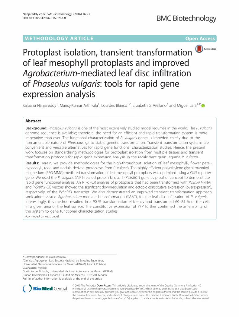
METHODOLOGY ARTICLE Open Access Protoplast isolation, transient transformation of leaf mesophyll protoplasts and improved Agrobacterium-mediated leaf disc infiltration of Phaseolus vulgaris: tools for rapid gene expression analysis Kalpana Nanjareddy 1 , Manoj-Kumar Arthikala 1 , Lourdes Blanco 1,2 , Elizabeth S. Arellano 3 and Miguel Lara 1,4* Abstract Background: Phaseolus vulgaris is one of the most extensively studied model legumes in the world. The P. vulgaris genome sequence is available; therefore, the need for an efficient and rapid transformation system is more imperative than ever. The functional characterization of P. vulgaris genes is impeded chiefly due to the non-amenable nature of Phaseolus sp. to stable genetic transformation. Transient transformation systems are convenient and versatile alternatives for rapid gene functional characterization studies. Hence, the present work focuses on standardizing methodologies for protoplast isolation from multiple tissues and transient transformation protocols for rapid gene expression analysis in the recalcitrant grain legume P. vulgaris. Results: Herein, we provide methodologies for the high-throughput isolation of leaf mesophyll-, flower petal-, hypocotyl-, root- and nodule-derived protoplasts from P. vulgaris. The highly efficient polyethylene glycol-mannitol magnesium (PEG-MMG)-mediated transformation of leaf mesophyll protoplasts was optimized using a GUS reporter gene. We used the P. vulgaris SNF1-related protein kinase 1 (PvSnRK1) gene as proof of concept to demonstrate rapid gene functional analysis. An RT-qPCR analysis of protoplasts that had been transformed with PvSnRK1-RNAi and PvSnRK1-OE vectors showed the significant downregulation and ectopic constitutive expression (overexpression), respectively, of the PvSnRK1 transcript. We also demonstrated an improved transient transformation approach, sonication-assisted Agrobacterium-mediated transformation (SAAT), for the leaf disc infiltration of P. vulgaris. Interestingly, this method resulted in a 90 % transformation efficiency and transformed 60–85 % of the cells in a given area of the leaf surface. The constitutive expression of YFP further confirmed the amenability of the system to gene functional characterization studies. (Continued on next page) * Correspondence: [email protected] 1 Ciencias Agrogenómicas, Escuela Nacional de Estudios Superiores, Universidad Nacional Autónoma de México (UNAM), León C.P.37684, Guanajuato, Mexico 4 Instituto de Biología, Universidad Nacional Autónoma de México (UNAM), Ciudad Universitaria, Coyoacan, Ciudad de México C.P. 04510, Mexico Full list of author information is available at the end of the article © 2016 The Author(s). Open Access This article is distributed under the terms of the Creative Commons Attribution 4.0 International License (http://creativecommons.org/licenses/by/4.0/), which permits unrestricted use, distribution, and reproduction in any medium, provided you give appropriate credit to the original author(s) and the source, provide a link to the Creative Commons license, and indicate if changes were made. The Creative Commons Public Domain Dedication waiver (http://creativecommons.org/publicdomain/zero/1.0/) applies to the data made available in this article, unless otherwise stated. Nanjareddy et al. BMC Biotechnology (2016) 16:53 DOI 10.1186/s12896-016-0283-8
-
Upload
khangminh22 -
Category
Documents
-
view
0 -
download
0
Transcript of Protoplast isolation, transient transformation of leaf mesophyll ...
METHODOLOGY ARTICLE Open Access
Protoplast isolation, transient transformationof leaf mesophyll protoplasts and improvedAgrobacterium-mediated leaf disc infiltrationof Phaseolus vulgaris: tools for rapid geneexpression analysisKalpana Nanjareddy1, Manoj-Kumar Arthikala1, Lourdes Blanco1,2, Elizabeth S. Arellano3 and Miguel Lara1,4*
Abstract
Background: Phaseolus vulgaris is one of the most extensively studied model legumes in the world. The P. vulgarisgenome sequence is available; therefore, the need for an efficient and rapid transformation system is moreimperative than ever. The functional characterization of P. vulgaris genes is impeded chiefly due to thenon-amenable nature of Phaseolus sp. to stable genetic transformation. Transient transformation systems areconvenient and versatile alternatives for rapid gene functional characterization studies. Hence, the presentwork focuses on standardizing methodologies for protoplast isolation from multiple tissues and transienttransformation protocols for rapid gene expression analysis in the recalcitrant grain legume P. vulgaris.
Results: Herein, we provide methodologies for the high-throughput isolation of leaf mesophyll-, flower petal-,hypocotyl-, root- and nodule-derived protoplasts from P. vulgaris. The highly efficient polyethylene glycol-mannitolmagnesium (PEG-MMG)-mediated transformation of leaf mesophyll protoplasts was optimized using a GUS reportergene. We used the P. vulgaris SNF1-related protein kinase 1 (PvSnRK1) gene as proof of concept to demonstraterapid gene functional analysis. An RT-qPCR analysis of protoplasts that had been transformed with PvSnRK1-RNAiand PvSnRK1-OE vectors showed the significant downregulation and ectopic constitutive expression (overexpression),respectively, of the PvSnRK1 transcript. We also demonstrated an improved transient transformation approach,sonication-assisted Agrobacterium-mediated transformation (SAAT), for the leaf disc infiltration of P. vulgaris.Interestingly, this method resulted in a 90 % transformation efficiency and transformed 60–85 % of the cellsin a given area of the leaf surface. The constitutive expression of YFP further confirmed the amenability ofthe system to gene functional characterization studies.(Continued on next page)
* Correspondence: [email protected] Agrogenómicas, Escuela Nacional de Estudios Superiores,Universidad Nacional Autónoma de México (UNAM), León C.P.37684,Guanajuato, Mexico4Instituto de Biología, Universidad Nacional Autónoma de México (UNAM),Ciudad Universitaria, Coyoacan, Ciudad de México C.P. 04510, MexicoFull list of author information is available at the end of the article
© 2016 The Author(s). Open Access This article is distributed under the terms of the Creative Commons Attribution 4.0International License (http://creativecommons.org/licenses/by/4.0/), which permits unrestricted use, distribution, andreproduction in any medium, provided you give appropriate credit to the original author(s) and the source, provide a link tothe Creative Commons license, and indicate if changes were made. The Creative Commons Public Domain Dedication waiver(http://creativecommons.org/publicdomain/zero/1.0/) applies to the data made available in this article, unless otherwise stated.
Nanjareddy et al. BMC Biotechnology (2016) 16:53 DOI 10.1186/s12896-016-0283-8

(Continued from previous page)
Conclusions: We present simple and efficient methodologies for protoplast isolation from multiple P. vulgaristissues. We also provide a high-efficiency and amenable method for leaf mesophyll transformation for rapidgene functional characterization studies. Furthermore, a modified SAAT leaf disc infiltration approach aids invalidating genes and their functions. Together, these methods help to rapidly unravel novel gene functionsand are promising tools for P. vulgaris research.
Keywords: Agrobacterium infiltration, Gene expression, Overexpression, Phaseolus vulgaris, Protoplasts, RNAi,SnRK1, Sonication, Transient transformation
BackgroundThe common bean, Phaseolus vulgaris, is an economic-ally important crop that belongs to the family Legumi-nosae and is the most essential grain legume for directhuman consumption in the world; in Latin Americaalone, this species holds a stake of more than 70 % [1].Despite having such enormous agroeconomic relevance,this crop suffers from several widespread major diseasesand abiotic stresses, which decrease the crop yield [2, 3].Attempts at transformation and crop improvement pro-grams have been hampered by this species’ notoriousrecalcitrance to routine in vitro regeneration and trans-formation [4]. Furthermore, unlike other legumes, P.vulgaris has serious limitations, such as an unavailabi-lity of mutants and a lack of rapid and efficient toolsfor transformation, preventing this species from beingused as a versatile model for legume-related research.The P. vulgaris genome sequence is available [5]; there-fore, the need for an efficient and rapid transformationsystem is more imperative than ever. Although somereports have suggested the feasibility of the stabletransformation of common bean using a microprojec-tile bombardment method [6], this option demands vastresources and intensive work with a miniscule yieldcompared to the bombardment methods of other modelcrop plants, including cereals [7]. Such a low efficiencymakes this method potentially unusable in small-scalelaboratories. As an alternative, the hairy root system isthe only adoptable technique available to carry outtransient gene functional analysis [8]. Nevertheless, thismethod is a transformation procedure that demandstime and it is not eligible for a high-throughput analysisof heterologous gene expression.Compared to the stable transgenic approach, the use
of transient gene expression assays offers an opportunityto rapidly assess the function of a large number of genesby evaluating the transcriptional activity of promotersand the sub-cellular localization of proteins and to in-vestigate cell biology and physiology, cell wall traits,etc. In plant biology research, protoplast transfection iswell established and used efficiently in single-cell-basedstudies. Plant protoplasts have shown reactions similarto those of intact cells to hormones, metabolites,
environmental cues and pathogen-derived elicitors,providing a powerful and versatile cell system for thehigh-throughput dissection of plant signal transductionpathways in many plant species, such as Arabidopsis[9–11], maize and rice [12], Brassica [13], sunflower[14], Populus [15], Poinsettia [16] and palm [17]. Onthe other hand, the available protocols for P. vulgarisprotoplast isolation from either cell suspension cultures[18] or cotyledonary leaves [19] are not amenable totransfection [20].In addition, the Agrobacterium-mediated leaf disc
infiltration method is another transient system that isroutinely exploited in functional analyses of genes.Sonication-assisted Agrobacterium-mediated transfor-mation (SAAT) involves subjecting plant tissue to a briefperiod of ultrasound in the presence of Agrobacterium.Unlike other transformation methods, this system has thepotential to transform several cell layers and, furthermore,is an easy and reliable approach to carry out gene func-tional characterization studies [10, 21, 22]. The protocol ispotentially suitable for a wide variety of molecular studies,including gene regulation, protein localization, taggedprotein expression, chromatin immunoprecipitation,protein-protein interactions, bimolecular fluorescencecomplementation (BiFC), protein stability, etc. Thesimplicity of the protocol allows it to be used in othercrop plants as well.The present paper describes novel protocols for proto-
plast isolation from different P. vulgaris tissues, such asleaf mesophyll, flower petal, hypocotyl, root and nodule,that could be further used to perform rapid cell biology,physiology, and biochemical assays, among others. Thisstudy also presents a highly efficient polyethylene glycol-mannitol magnesium (PEG-MMG)-mediated transfor-mation protocol for P. vulgaris leaf mesophyll-derivedprotoplasts. To validate this method for gene expressionstudies, we used the P. vulgaris SNF1-related proteinkinase 1 (PvSnRK1) gene [23]. SnRK genes are evolu-tionarily conserved metabolic sensors that undergoactivation in response to decreased energy levels ineukaryotes. Plant SnRK1 is well characterized andshown to regulate the timing of embryo maturation inArabidopsis, sucrose cleavage in potato [24, 25], and
Nanjareddy et al. BMC Biotechnology (2016) 16:53 Page 2 of 14
pollen development (due to the failure to incorporatesucrose into starch) in barley [26]. SnRK1 also interactswith ABA-dependent and ABA-independent pathways inlegumes [27]. The non-conserved cDNA region and openreading frame (ORF) of P. vulgaris SnRK1 were clonedindividually in RNAi and constitutive expression (overex-pression) vectors and transfected into mesophyll-derivedprotoplasts for the downregulation and overexpressionof PvSnRK1 transcript, respectively. Furthermore, theconcept of transient gene expression is met by providinga modified gene transformation approach, the SAAT, forthe leaf disc infiltration of P. vulgaris. A β-glucuronidase(GUS)-based assay and the constitutive expression ofyellow fluorescent protein (YFP) demonstrate the effi-ciency of Agrobacterium infiltration, T-DNA integrationand expression.
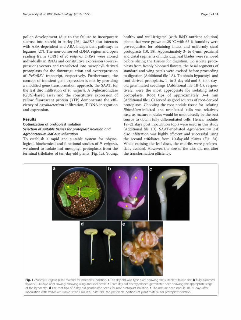
ResultsOptimization of protoplast isolationSelection of suitable tissues for protoplast isolation andAgrobacterium leaf disc infiltrationTo establish a rapid and suitable system for physio-logical, biochemical and functional studies of P. vulgaris,we aimed to isolate leaf mesophyll protoplasts from theterminal trifoliates of ten-day-old plants (Fig. 1a). Young,
healthy and well-irrigated (with B&D nutrient solution)plants that were grown at 28 °C with 65 % humidity werepre-requisites for obtaining intact and uniformly sizedprotoplasts [10, 18]. Approximately 3- to 6-mm proximaland distal segments of individual leaf blades were removedbefore slicing the tissues for digestion. To isolate proto-plasts from freshly bloomed flowers, the basal segments ofstandard and wing petals were excised before proceedingto digestion (Additional file 1A). To obtain hypocotyl- androot-derived protoplasts, 1- to 3-day-old and 3- to 4-day-old germinated seedlings (Additional file 1B-C), respec-tively, were the most appropriate for isolating intactprotoplasts. Root tips of approximately 3–4 mm(Additional file 1C) served as good sources of root-derivedprotoplasts. Choosing the root nodule tissue for isolatingRhizobium-infected and uninfected cells was relativelyeasy, as mature nodules would be undoubtedly be the bestsource to obtain fully differentiated cells. Hence, nodules18–21 days post inoculation (dpi) were used in this study(Additional file 1D). SAAT-mediated Agrobacterium leafdisc infiltration was highly efficient and successful usingthe second trifoliates from 10-day-old plants (Fig. 5a).While excising the leaf discs, the midribs were preferen-tially avoided. However, the size of the disc did not alterthe transformation efficiency.
Fig. 1 Phaseolus vulgaris plant material for protoplast isolation. a Ten-day-old wild type plant showing the suitable trifoliate size. b Fully bloomedflowers (~40 days after sowing) showing wing and keel petals. c Three-day-old decotyledoned germinated seed showing the appropriate stageof the hypocotyl. d The root tips of 3-day-old germinated seeds for root protoplast isolation. e The mature bean nodule 18–21 days afterinoculation with Rhizobium tropici strain CIAT 899. Asterisks- the preferable portions of plant material for protoplast isolation
Nanjareddy et al. BMC Biotechnology (2016) 16:53 Page 3 of 14
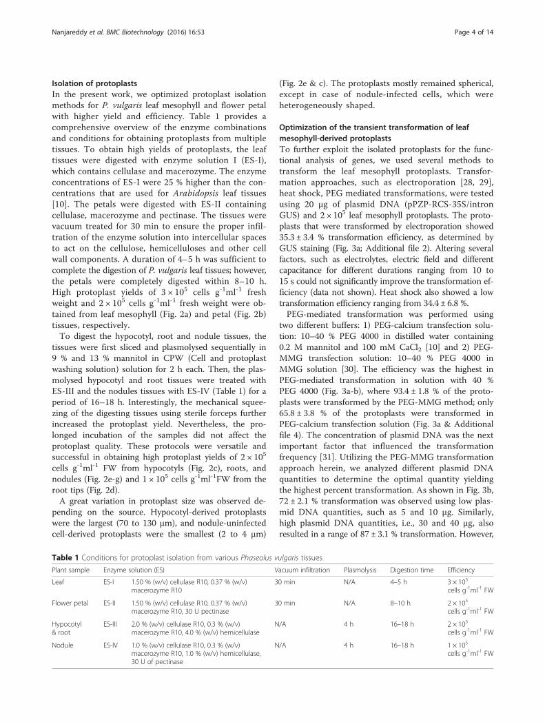
Isolation of protoplastsIn the present work, we optimized protoplast isolationmethods for P. vulgaris leaf mesophyll and flower petalwith higher yield and efficiency. Table 1 provides acomprehensive overview of the enzyme combinationsand conditions for obtaining protoplasts from multipletissues. To obtain high yields of protoplasts, the leaftissues were digested with enzyme solution I (ES-I),which contains cellulase and macerozyme. The enzymeconcentrations of ES-I were 25 % higher than the con-centrations that are used for Arabidopsis leaf tissues[10]. The petals were digested with ES-II containingcellulase, macerozyme and pectinase. The tissues werevacuum treated for 30 min to ensure the proper infil-tration of the enzyme solution into intercellular spacesto act on the cellulose, hemicelluloses and other cellwall components. A duration of 4–5 h was sufficient tocomplete the digestion of P. vulgaris leaf tissues; however,the petals were completely digested within 8–10 h.High protoplast yields of 3 × 10⁵ cells g-1ml-1 freshweight and 2 × 10⁵ cells g-1ml-1 fresh weight were ob-tained from leaf mesophyll (Fig. 2a) and petal (Fig. 2b)tissues, respectively.To digest the hypocotyl, root and nodule tissues, the
tissues were first sliced and plasmolysed sequentially in9 % and 13 % mannitol in CPW (Cell and protoplastwashing solution) solution for 2 h each. Then, the plas-molysed hypocotyl and root tissues were treated withES-III and the nodules tissues with ES-IV (Table 1) for aperiod of 16–18 h. Interestingly, the mechanical squee-zing of the digesting tissues using sterile forceps furtherincreased the protoplast yield. Nevertheless, the pro-longed incubation of the samples did not affect theprotoplast quality. These protocols were versatile andsuccessful in obtaining high protoplast yields of 2 × 10⁵
cells g-1ml-1 FW from hypocotyls (Fig. 2c), roots, andnodules (Fig. 2e-g) and 1 × 10⁵ cells g-1ml-1FW from theroot tips (Fig. 2d).A great variation in protoplast size was observed de-
pending on the source. Hypocotyl-derived protoplastswere the largest (70 to 130 μm), and nodule-uninfectedcell-derived protoplasts were the smallest (2 to 4 μm)
(Fig. 2e & c). The protoplasts mostly remained spherical,except in case of nodule-infected cells, which wereheterogeneously shaped.
Optimization of the transient transformation of leafmesophyll-derived protoplastsTo further exploit the isolated protoplasts for the func-tional analysis of genes, we used several methods totransform the leaf mesophyll protoplasts. Transfor-mation approaches, such as electroporation [28, 29],heat shock, PEG mediated transformations, were testedusing 20 μg of plasmid DNA (pPZP-RCS-35S/intronGUS) and 2 × 10⁵ leaf mesophyll protoplasts. The proto-plasts that were transformed by electroporation showed35.3 ± 3.4 % transformation efficiency, as determined byGUS staining (Fig. 3a; Additional file 2). Altering severalfactors, such as electrolytes, electric field and differentcapacitance for different durations ranging from 10 to15 s could not significantly improve the transformation ef-ficiency (data not shown). Heat shock also showed a lowtransformation efficiency ranging from 34.4 ± 6.8 %.PEG-mediated transformation was performed using
two different buffers: 1) PEG-calcium transfection solu-tion: 10–40 % PEG 4000 in distilled water containing0.2 M mannitol and 100 mM CaCl2 [10] and 2) PEG-MMG transfection solution: 10–40 % PEG 4000 inMMG solution [30]. The efficiency was the highest inPEG-mediated transformation in solution with 40 %PEG 4000 (Fig. 3a-b), where 93.4 ± 1.8 % of the proto-plasts were transformed by the PEG-MMG method; only65.8 ± 3.8 % of the protoplasts were transformed inPEG-calcium transfection solution (Fig. 3a & Additionalfile 4). The concentration of plasmid DNA was the nextimportant factor that influenced the transformationfrequency [31]. Utilizing the PEG-MMG transformationapproach herein, we analyzed different plasmid DNAquantities to determine the optimal quantity yieldingthe highest percent transformation. As shown in Fig. 3b,72 ± 2.1 % transformation was observed using low plas-mid DNA quantities, such as 5 and 10 μg. Similarly,high plasmid DNA quantities, i.e., 30 and 40 μg, alsoresulted in a range of 87 ± 3.1 % transformation. However,
Table 1 Conditions for protoplast isolation from various Phaseolus vulgaris tissues
Plant sample Enzyme solution (ES) Vacuum infiltration Plasmolysis Digestion time Efficiency
Leaf ES-I 1.50 % (w/v) cellulase R10, 0.37 % (w/v)macerozyme R10
30 min N/A 4–5 h 3 × 10⁵
cells g-1ml-1 FW
Flower petal ES-II 1.50 % (w/v) cellulase R10, 0.37 % (w/v)macerozyme R10, 30 U pectinase
30 min N/A 8–10 h 2 × 10⁵
cells g-1ml-1 FW
Hypocotyl& root
ES-III 2.0 % (w/v) cellulase R10, 0.3 % (w/v)macerozyme R10, 4.0 % (w/v) hemicellulase
N/A 4 h 16–18 h 2 × 10⁵
cells g-1ml-1 FW
Nodule ES-IV 1.0 % (w/v) cellulase R10, 0.3 % (w/v)macerozyme R10, 1.0 % (w/v) hemicellulase,30 U of pectinase
N/A 4 h 16–18 h 1 × 10⁵
cells g-1ml-1 FW
Nanjareddy et al. BMC Biotechnology (2016) 16:53 Page 4 of 14

15–20 μg of plasmid DNA was suitable for achieving92.5 ± 2 % transfection in all of the analyzed con-structs (Fig. 3b).
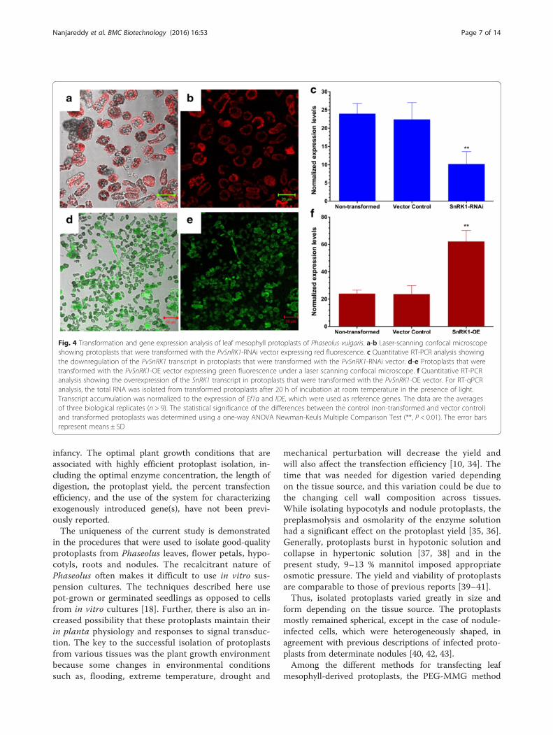
Gene functional analysisTo further examine the feasibility of using P. vulgarisleaf mesophyll-derived protoplasts for the functionalanalysis of the genes, we explored the evolutionarily con-served SnRK1 gene that is known to regulate energy andstress signaling in eukaryotes. To downregulate thePvSnRK1 transcript, a plasmid harboring the PvSnRK1-RNAi construct was transfected into leaf mesophyll-derived protoplasts. Following 4–6 h of incubation, afraction of cells was observed under a microscope toverify the expression of red fluorescence protein (RFP)from the transformed protoplasts (Fig. 4a-b). We thendetermined the percent transformation based on RFPexpression; as expected, 92 ± 2.1 % of the protoplastswere transformed successfully. Furthermore, an RT-qPCRanalysis of PvSnRK1-RNAi transformed protoplasts wasperformed to validate the downregulation of PvSnRK1expression. As depicted in Fig. 4c, the transcripts ofPvSnRK1 significantly decreased by 58 ± 3.2 % inPvSnRK1-RNAi-transformed protoplasts compared tothose in untransformed and control (transformed withan empty pTdT-RNAi vector) protoplasts. The overex-pression of PvSnRK1 was carried out under the consti-tutive 35S promoter (pH7WG2D vector, henceforthcalled ‘PvSnRK1-OE’). The transformed protoplasts
were selected based on the green fluorescent marker(GFP) (Fig. 4d-e). Furthermore, these protoplasts showeda 91 ± 1.8 % transformation efficiency as determined byGFP-associated fluorescence. RT-qPCR analysis confirmedthat PvSnRK1 transcript accumulation in PvSnRK1-OEprotoplasts significantly increased relative to that inuntransformed and control protoplasts (Fig. 4f ). Theintegration of the RNAi and overexpression constructsin the genome was further confirmed by the PCR amp-lification of the Tdt fragment (1430 bp) for RNAi andthe PvSnRK1-35S promoter (460 bp) and GFP (270 bp)for overexpression vectors (Additional file 3) in thetransformed protoplasts. Taken together, these resultsdemonstrate the suitability of using P. vulgaris meso-phyll protoplasts for gene expression studies.
Transient transformation of P. vulgaris leaf disc by animproved SAAT methodContrary to the direct transformation approach of me-sophyll protoplasts (as demonstrated above), we alsoattempted to introduce an indirect transformationapproach, SAAT [29], for the leaf disc infiltration of P.vulgaris (Fig. 5). The leaf disc infiltration assay was car-ried out using an improved SAAT method utilizingvarious infiltration media containing a bacterial densityof 0.5–0.7 at OD600. Among the tested infiltrationmedia, 10 mM MgCl2 and a combination of 10 mMMgCl2 and 5 mM MES-KOH resulted in 30 and 50 %of transfection efficiencies, respectively (Table 2).
Fig. 2 Phaseolus vulgaris protoplasts that were isolated from multiple tissues: a Leaf mesophyll. b Flower petal. c Hypocotyl. d Root. e Uninfectednodule cells. f Rhizobium tropici-infected nodule cells. g R. tropici harboring the pSN30-GFP plasmid expressing the GFP protein in infected cells asseen under a laser-scanning confocal microscope. Scale bars: A, D-E, 15 μm; B, 50 μm; and C, F-G, 100 μm
Nanjareddy et al. BMC Biotechnology (2016) 16:53 Page 5 of 14

Interestingly, a high transfection efficiency of 90 % wasobserved in Winan’s AB infiltration medium amendedwith Silwet L-77 (OSi Specialities, Inc., Danbury, CT,USA) and acetosyringone prior to sonication (Table 2)because both Silwet L-77 and acetosyringone are im-portant factors that improve transformation efficiencyby increasing the DNA delivery and integration [32].Microscopic observations of the GUS-stained leaf discsshowed that 60–85 % of cells on the leaf surface weretransformed by the infiltration medium containingSilwet L-77 and acetosyringone (Fig. 5f ).To further validate the SAAT transient transformation,
we utilized the pEarleyGate104 vector that constitutivelyexpresses yellow fluorescent protein (YFP) in P. vulgarisleaf epidermal cells. The confocal images, as expected,showed strong YFP fluorescence in the cytoplasm of leafepidermal cells (Fig. 6d). We previously showed usingthe same vector a similar expression pattern of YFP in P.vulgaris hairy roots [33]. In contrast, no fluorescencewas observed in the leaf epidermal cells that were trans-formed with empty pEarleyGate104 vector (control)(Fig. 6a-b). Together, these results indicate the suitabilityof the modified SAAT leaf disc infiltration method to
perform gene expression studies and that this methodcan be extended to studies using a variety of functionalanalyses, such as gene silencing, protein localization,promoter analysis, etc.
DiscussionThe common bean, Phaseolus vulgaris, is a model leg-ume that has been extensively studied worldwide. Giventhe absence of a reliable protocol for P. vulgaris regener-ation, the development of an optimal in vitro culturesystem remains a major challenge because this and otherspecies from the Phaseolus genus are recalcitrant for invitro regeneration [20]. Thus, the application of biotech-nological tools for crop improvement and gene func-tional characterization in P. vulgaris are major limitingfactors in plant research. Alternatively, transientexpression assays have been indispensable for rapidprogress in functional genomics research in other modelplants. For instance, the Arabidopsis mesophyll proto-plast transient expression system is an efficient and use-ful system for characterizing genes and their functions.However, the use of such a mesophyll protoplast transi-ent system for the model legume P. vulgaris is still in its
Fig. 3 Transformation efficiency of Phaseolus vulgaris leaf protoplasts by various transformation methods. a All of the experiments wereperformed using 20 μg of plasmid DNA and 2 × 10⁵ leaf mesophyll protoplasts. The graph shows the percent transformation efficiency indifferent transformation methods. b Percent transformation of protoplasts using various quantities of plasmid DNA (left) and different PEG 4000(right) concentrations. The statistical significance of differences was determined using a one-way ANOVA Newman-Keuls Multiple Comparison Test(*, P < 0.05; **, P < 0.01; ***, P < 0.001). For both A and B, the data are the averages of three biological replicates (n = 9); the error barsrepresent means ± SD
Nanjareddy et al. BMC Biotechnology (2016) 16:53 Page 6 of 14
infancy. The optimal plant growth conditions that areassociated with highly efficient protoplast isolation, in-cluding the optimal enzyme concentration, the length ofdigestion, the protoplast yield, the percent transfectionefficiency, and the use of the system for characterizingexogenously introduced gene(s), have not been previ-ously reported.The uniqueness of the current study is demonstrated
in the procedures that were used to isolate good-qualityprotoplasts from Phaseolus leaves, flower petals, hypo-cotyls, roots and nodules. The recalcitrant nature ofPhaseolus often makes it difficult to use in vitro sus-pension cultures. The techniques described here usepot-grown or germinated seedlings as opposed to cellsfrom in vitro cultures [18]. Further, there is also an in-creased possibility that these protoplasts maintain theirin planta physiology and responses to signal transduc-tion. The key to the successful isolation of protoplastsfrom various tissues was the plant growth environmentbecause some changes in environmental conditionssuch as, flooding, extreme temperature, drought and
mechanical perturbation will decrease the yield andwill also affect the transfection efficiency [10, 34]. Thetime that was needed for digestion varied dependingon the tissue source, and this variation could be due tothe changing cell wall composition across tissues.While isolating hypocotyls and nodule protoplasts, thepreplasmolysis and osmolarity of the enzyme solutionhad a significant effect on the protoplast yield [35, 36].Generally, protoplasts burst in hypotonic solution andcollapse in hypertonic solution [37, 38] and in thepresent study, 9–13 % mannitol imposed appropriateosmotic pressure. The yield and viability of protoplastsare comparable to those of previous reports [39–41].Thus, isolated protoplasts varied greatly in size and
form depending on the tissue source. The protoplastsmostly remained spherical, except in the case of nodule-infected cells, which were heterogeneously shaped, inagreement with previous descriptions of infected proto-plasts from determinate nodules [40, 42, 43].Among the different methods for transfecting leaf
mesophyll-derived protoplasts, the PEG-MMG method
Fig. 4 Transformation and gene expression analysis of leaf mesophyll protoplasts of Phaseolus vulgaris. a-b Laser-scanning confocal microscopeshowing protoplasts that were transformed with the PvSnRK1-RNAi vector expressing red fluorescence. c Quantitative RT-PCR analysis showingthe downregulation of the PvSnRK1 transcript in protoplasts that were transformed with the PvSnRK1-RNAi vector. d-e Protoplasts that weretransformed with the PvSnRK1-OE vector expressing green fluorescence under a laser scanning confocal microscope. f Quantitative RT-PCRanalysis showing the overexpression of the SnRK1 transcript in protoplasts that were transformed with the PvSnRK1-OE vector. For RT-qPCRanalysis, the total RNA was isolated from transformed protoplasts after 20 h of incubation at room temperature in the presence of light.Transcript accumulation was normalized to the expression of Ef1α and IDE, which were used as reference genes. The data are the averagesof three biological replicates (n > 9). The statistical significance of the differences between the control (non-transformed and vector control)and transformed protoplasts was determined using a one-way ANOVA Newman-Keuls Multiple Comparison Test (**, P < 0.01). The error barsrepresent means ± SD
Nanjareddy et al. BMC Biotechnology (2016) 16:53 Page 7 of 14

using 15–20 μg of plasmid DNA (pPZP-RCS-35S/intronGUS) and 40 % PEG was optimal, with 90–95 % trans-formed cells. This study demonstrates that Phaseolusprotoplasts could be the system of choice when analy-zing gene function either by RNAi or by overexpression.Because protoplasts are non-growing cells, effectiveRNAi-triggered gene silencing depends not only on thedepletion of gene transcripts but also on the turnoverrates of corresponding polypeptides. Herein, we testedwhether transient RNAi in protoplasts results in the de-pletion of a targeted polypeptide using the PvSnRK1-RNAi vector and also tested the feasibility of the ectopic,constitutive expression of PvSnRK1 in leaf mesophyll-
derived protoplasts. The quantitative RT-PCR resultsshowed that the protoplasts that were transformed withthe PvSnRK1-RNAi and PvSnRK1-OE vectors signifi-cantly downregulated and ectopically overexpressed thePvSnRK1 transcript, respectively. The transfection ofRNAi vectors in Arabidopsis and rice protoplasts de-creases the transcript level of the targeted exogenousand endogenous genes [44, 45].With the goal of standardizing the methodology for
the transient transformation of P. vulgaris leaf discinfiltration by SAAT, several aspects were optimized:1) the density of bacteria required to obtain efficienttissue transformation [30, 46], 2) Winan’s AB medium
Fig. 5 Transient gene expression by the improved SAAT method in Phaseolus vulgaris using the pPZP-RCS-GUS binary vector. a Ten-day-old plantthat was grown in a growth chamber showing the second trifoliates (asterisk) suitable for the transient assay. Arrow- first trifoliate (from shootapex). b The leaf discs in the vir-gene-induced Agrobacterium culture were first subjected to sonication and c later vacuum infiltrated in freshAgrobacterium culture. d Co-cultivation of Agrobacterium-infected leaf discs on sterile filter paper moistened with MS basal medium. e The leafdiscs that were transformed with empty vector were stained for the histochemical localization of β-glucuronidase (GUS) reporter activity. No blue-stained tissue appeared even after 24 h of incubation with the GUS assay buffer. f-g The leaf discs that were transformed with the pPZP-RCS-GUSvector were stained for the histochemical localization of β-glucuronidase (GUS) reporter activity. Blue staining appeared within 16 h of incubation
Table 2 Transfection efficiency of Phaseolus vulgaris leaf discs that were infiltrated by the SAAT method under different infiltrationmedia
Growth medium Infiltration medium Leaf discs* Transient Efficiency transfection (%) Reference
LB 10 mM MgCl2 30 9 30 [55]
LB 10 mM MgCl2 and 5 mM MES-KOH 30 15 50 [56]
Winan’s AB Winan’s AB 30 27 90 [54]
*Ten leaf discs were used per biological replicate
Nanjareddy et al. BMC Biotechnology (2016) 16:53 Page 8 of 14

effectiveness among the different infiltration media[47], 3) the Silwet concentration, and 4) unlike the pre-vious reports, the use of 5 μM acetosyringone in anovernight bacterial culture in Winan’s AB medium andthe further addition of 100 μM to the infiltrationmedium, ensuring highly efficient transformationresulting in approximately 60–85 % of the leaf disc be-ing transformed cells. Furthermore, we validated thesystem for the constitutive expression of the YFP geneand showed an intense fluorescent protein in the leafepidermal cells. Thus, these results demonstrate thatthe modified SAAT leaf disc infiltration method is asimple, highly efficient and rapid process that is suit-able for gene expression analysis.
ConclusionsIn this study, we present protocols for the isolation ofprotoplasts from 5 different tissues of the model legumeP. vulgaris. We also provide a high-efficiency and amen-able method for leaf mesophyll transformation for genefunctional characterization studies. Furthermore, wedeveloped a modified SAAT leaf disc infiltration ap-proach that aids in rapidly validating genes and theirfunctions. These methods may help to rapidly unravel
the functions of novel genes and represent promisingtools for P. vulgaris research.
MethodsPlant material and growth conditionsIn the present study, Phaseolus vulgaris L. cv NegroJamapa was the source of all of the tissue material. Theseeds were surface sterilized [8], and 2-day-old seedlingswere grown either in sterile vermiculite or on sterilefilter paper moistened with Broughton and Dilworth[48] (B&D) nutrient solution in Petri dishes (10 cmdiameter) in a growth chamber at 26–28 °C, 65 % hu-midity. The seedlings were irrigated on alternate dayswith B&D nutrient solution.
Developing constructs for protoplast transformationFor gene functional analysis in protoplast transient as-says, we utilized the Phaseolus vulgaris SNF1-relatedprotein kinase 1 (PvSnRK1) (Phvul.008G039400.1) gene(Fig. 7a) to develop RNA interference (RNAi) silencing(downregulating) or overexpression constructs. To gener-ate the RNAi construct, a 209-bp fragment correspondingto the 3′-UTR of PvSnRK1 (Phvul.008G039400.1) wasamplified from cDNA that had been isolated from com-mon bean roots at 2 days post-germination, using the
Fig. 6 YFP expression vector in leaf epidermal cells of Phaseolus vulgaris as delivered through the SAAT method. A representative confocal imageshowing the empty vector (control) of a pEarleyGate104-transformed leaf under a transmitted light and b fluorescent light. A representativeconfocal image showing the expression of the 35S:YFP vector in the leaf epidermis under c transmitted light and d fluorescent light
Nanjareddy et al. BMC Biotechnology (2016) 16:53 Page 9 of 14

following primers: Forward 5′- CAC CAG ATC TATGGA CGG ACC AGC TGG CCG-3′ and Reverse 5′-CTC GAG GAG GAC ACG AAG CTG TGC AAG G-3′.The PCR product was recombined with the pTdT-DC-RNAi vector [49] using the Gateway System (Invitrogen)(Fig. 7b).To develop an overexpression construct of PvSnRK1,
the ORF of PvSnRK1 (Phvul.008G039400.1) from P. vul-garis cDNA was isolated, and the 1548 bp ORF fragmentwas inserted into the pH7WG2D.1 binary vector underthe control of the constitutive 35S promoter [50] usingthe Gateway System (Fig. 7c). The correct orientationsof the clones were confirmed by sequencing the plasmidinsert.
Leaf mesophyll and flower petal protoplast isolationThe first or second trifoliates from the shoot apical tipof ten-day-old plants were used for leaf mesophyllprotoplast isolation (Fig. 1a). Standard and wing petalsfrom fully bloomed flowers were used for flower petalprotoplast isolation (Fig. 1b). Strips of 0.5–1 mm inthickness were cut from 1 g of both leaf and flowertissues from the portions shown in Additional file 1A,excluding the veins in the leaves. The leaf tissue stripswere first vacuum infiltrated for 30 min in enzyme
solution I [ES-I; 1.5 % (w/v) cellulase R10 (Yakultpharmaceutical industry) and 0.37 % (w/v) macerozymeR10 (Yakult pharmaceutical industry)] (Fig. 8a) andpetal strips in ES-II [ES-I + 30 U pectinase] in 20 mMMES (pH 5.7) with 20 mM KCl, 0.4 M mannitol and10 mM CaCl2 (Fig. 8b). Later, the leaf tissue strips weredigested in the dark on a horizontal shaker (40 rpm) at30 °C for 4–5 h, whereas the flower petals weredigested for 8–10 h. The enzymatic reaction wasstopped by adding an equal volume of W5 solution[2 mM MES, 154 mM NaCl, 125 mM CaCl2 and 5 mMKCl at pH 5.7]. The digested tissue was passed through108 μm mesh, and the filtrate was collected in a centri-fuge tube and incubated on ice for 30 min. Then, thecells were washed twice in W5 solution (10) at 100 gfor 3 min each. The protoplast density was calculatedusing a hemocytometer. Finally, the protoplasts wereresuspended in MMG solution (4 mM MES, 0.4 Mmannitol and 15 mM MgCl2 at pH 5.7) (10) at thedesired cell density.
Hypocotyl and root protoplast isolationProtoplasts were isolated using the hypocotyl and roottip tissues (Fig. 1c-d) of three-day-old seedlings thatwere germinated on moistened filter paper. One gram of
Fig. 7 Schematic diagram of the gene structure and T-DNA harboring of SnRK1 and different the selectable marker genes. a Graphic representationof the gene structure of PvSnRK1 as predicted using the Phytozome v11 (https://phytozome.jgi.doe.gov/pz/portal.html). Green boxes illustrateexons, and gray boxes untranslated regions. The red line represents the non-conserved region of PvSnRK1 that is used to silence the targetgene. b A schematic representation of the PvSnRK1-RNAi construct showing the sense and antisense region of PvSnRK1 at the LB. In theopposite orientation, the TdT expression cassette, which encodes a fluorescent selectable marker, is located toward the RB. This construct wascarried by the pTdT-DC-PvSnRK1-RNAi binary vector. c The overexpression of the ORF of PvSnRK1 under the control of the constitutive 35S CaMVpromoter at the LB. In the opposite orientation, the enhanced GFP expression cassette, which encodes a fluorescent selectable marker, is locatedtowards the RB. This construct was carried by the pH7WG2D.1 binary vector. d pPZP-RCS binary vector constitutively expressing intron GUS.e pEarleyGate104 binary vector constitutively expressing yellow fluorescent protein (YFP). LB, left border; RB, right border; WRKY, hairpin loop; ORF,open reading frame
Nanjareddy et al. BMC Biotechnology (2016) 16:53 Page 10 of 14
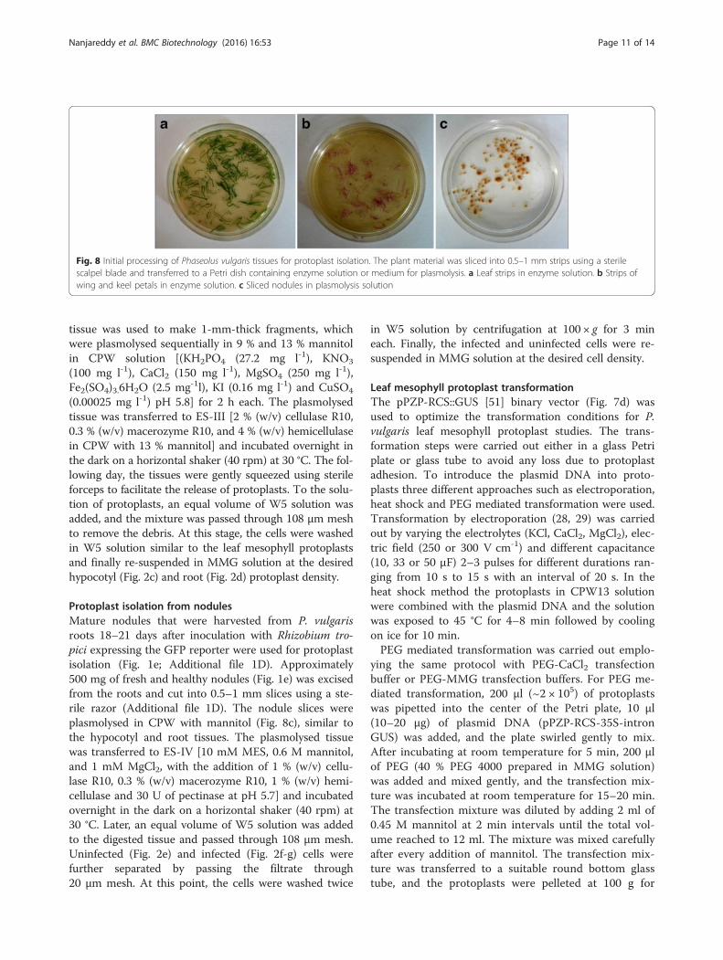
tissue was used to make 1-mm-thick fragments, whichwere plasmolysed sequentially in 9 % and 13 % mannitolin CPW solution [(KH2PO4 (27.2 mg l-1), KNO3
(100 mg l-1), CaCl2 (150 mg l-1), MgSO4 (250 mg l-1),Fe2(SO4)3.6H2O (2.5 mg-1l), KI (0.16 mg l-1) and CuSO4
(0.00025 mg l-1) pH 5.8] for 2 h each. The plasmolysedtissue was transferred to ES-III [2 % (w/v) cellulase R10,0.3 % (w/v) macerozyme R10, and 4 % (w/v) hemicellulasein CPW with 13 % mannitol] and incubated overnight inthe dark on a horizontal shaker (40 rpm) at 30 °C. The fol-lowing day, the tissues were gently squeezed using sterileforceps to facilitate the release of protoplasts. To the solu-tion of protoplasts, an equal volume of W5 solution wasadded, and the mixture was passed through 108 μm meshto remove the debris. At this stage, the cells were washedin W5 solution similar to the leaf mesophyll protoplastsand finally re-suspended in MMG solution at the desiredhypocotyl (Fig. 2c) and root (Fig. 2d) protoplast density.
Protoplast isolation from nodulesMature nodules that were harvested from P. vulgarisroots 18–21 days after inoculation with Rhizobium tro-pici expressing the GFP reporter were used for protoplastisolation (Fig. 1e; Additional file 1D). Approximately500 mg of fresh and healthy nodules (Fig. 1e) was excisedfrom the roots and cut into 0.5–1 mm slices using a ste-rile razor (Additional file 1D). The nodule slices wereplasmolysed in CPW with mannitol (Fig. 8c), similar tothe hypocotyl and root tissues. The plasmolysed tissuewas transferred to ES-IV [10 mM MES, 0.6 M mannitol,and 1 mM MgCl2, with the addition of 1 % (w/v) cellu-lase R10, 0.3 % (w/v) macerozyme R10, 1 % (w/v) hemi-cellulase and 30 U of pectinase at pH 5.7] and incubatedovernight in the dark on a horizontal shaker (40 rpm) at30 °C. Later, an equal volume of W5 solution was addedto the digested tissue and passed through 108 μm mesh.Uninfected (Fig. 2e) and infected (Fig. 2f-g) cells werefurther separated by passing the filtrate through20 μm mesh. At this point, the cells were washed twice
in W5 solution by centrifugation at 100 × g for 3 mineach. Finally, the infected and uninfected cells were re-suspended in MMG solution at the desired cell density.
Leaf mesophyll protoplast transformationThe pPZP-RCS::GUS [51] binary vector (Fig. 7d) wasused to optimize the transformation conditions for P.vulgaris leaf mesophyll protoplast studies. The trans-formation steps were carried out either in a glass Petriplate or glass tube to avoid any loss due to protoplastadhesion. To introduce the plasmid DNA into proto-plasts three different approaches such as electroporation,heat shock and PEG mediated transformation were used.Transformation by electroporation (28, 29) was carriedout by varying the electrolytes (KCl, CaCl2, MgCl2), elec-tric field (250 or 300 V cm-1) and different capacitance(10, 33 or 50 μF) 2–3 pulses for different durations ran-ging from 10 s to 15 s with an interval of 20 s. In theheat shock method the protoplasts in CPW13 solutionwere combined with the plasmid DNA and the solutionwas exposed to 45 °C for 4–8 min followed by coolingon ice for 10 min.PEG mediated transformation was carried out emplo-
ying the same protocol with PEG-CaCl2 transfectionbuffer or PEG-MMG transfection buffers. For PEG me-diated transformation, 200 μl (~2 × 10⁵) of protoplastswas pipetted into the center of the Petri plate, 10 μl(10–20 μg) of plasmid DNA (pPZP-RCS-35S-intronGUS) was added, and the plate swirled gently to mix.After incubating at room temperature for 5 min, 200 μlof PEG (40 % PEG 4000 prepared in MMG solution)was added and mixed gently, and the transfection mix-ture was incubated at room temperature for 15–20 min.The transfection mixture was diluted by adding 2 ml of0.45 M mannitol at 2 min intervals until the total vol-ume reached to 12 ml. The mixture was mixed carefullyafter every addition of mannitol. The transfection mix-ture was transferred to a suitable round bottom glasstube, and the protoplasts were pelleted at 100 g for
Fig. 8 Initial processing of Phaseolus vulgaris tissues for protoplast isolation. The plant material was sliced into 0.5–1 mm strips using a sterilescalpel blade and transferred to a Petri dish containing enzyme solution or medium for plasmolysis. a Leaf strips in enzyme solution. b Strips ofwing and keel petals in enzyme solution. c Sliced nodules in plasmolysis solution
Nanjareddy et al. BMC Biotechnology (2016) 16:53 Page 11 of 14
3 min. The cells were re-suspended in 1 ml of WI solution[4 mM MES containing 0.5 M mannitol and 20 mM KClat pH 5.7], transferred to 6- or 12-well tissue cultureplates and incubated for 3–6 h under light at roomtemperature. To stain the transformed protoplast for GUShistochemical activity, the protoplasts were resuspendedin GUS reaction buffer and incubated in the dark at 37 °Cfor 16–24 h according to Jefferson [38]. The GUS-stainedcells were mounted with 40 % glycerol in PBS (137 mMNaCl, 2.7 mM KCl, 4.3 mM Na2HPO4, and 1.47 mMKH2PO4) and observed under a microscope to assess thepercent transformation. Gene functional analyses, such asthe downregulation and overexpression of PvSnRK1, werecarried out using PvSnRK1-RNAi and PvSnRK1-OEbinary vectors, respectively.
RT-qPCR analysisThe total RNA was isolated from frozen leaf mesophyll-derived protoplasts using the RNeasy Plant Mini Kitaccording to the manufacturer’s recommendations(Qiagen, USA). Genomic DNA contamination in RNAsamples was eliminated by incubating the sampleswith RNase-free DNase (1 U μl–1) at 37 °C for 15 minand then at 65 °C for 10 min. The RNA integrity andconcentration were determined by electrophoresis anda Nanodrop ND-2000 spectrophotometer (ThermoScientifics), respectively. For qRT-PCR, 2 μg of totalRNA was used to synthesize cDNA.Quantitative real-time PCR was performed using the
iScriptTM One-step RT-PCR Kit with SYBR® Green, fol-lowing the manufacturer’s instructions, in an iQ5 Multi-color Real-time PCR Detection System (Bio-Rad). Eachreaction was set up using 40 ng of RNA as the template.A control sample that lacked reverse transcriptase (RT)was included to confirm the absence of contaminantDNA. The relative gene expression levels were calcu-lated using the 2-ΔCT method, with ΔCT = CTgene –CTreference gene. P. vulgaris EF1α and IDE were usedas internal controls, as previously described [53, 54].The relative expression values, normalized with two ref-erence genes, were calculated as previously described[55]. The data are averages of two or three biologicalreplicates, and each sample was assessed in triplicate.The expression of the genes that are listed in Additionalfile 5 was quantified using gene-specific oligonucleotides.
Agrobacterium-mediated leaf disc infiltration withsonicationAgrobacterium tumefaciens strain AGL1 carrying the bin-ary vector pPZP-RCS::GUS or pEarleyGate104 (Fig. 7e)was used for the P. vulgaris leaf disc infiltration experi-ments. Different infiltration media, such as (i) 10 mMMgCl2 [55], (ii) 10 mM MgCl2 and 5 mM MES-KOH(pH 5.6) [56] and (iii) Winan’s AB medium (pH 5.6) [52]
were used to test the SAAT method for P. vulgaris leafdisc transformation.The Agrobacteria were grown on LB agar plates with
the appropriate antibiotics for 16–18 h, after which asingle colony from the plate was used to inoculate LBbroth and further grown for 18 h at 28 °C. An aliquot of3 ml from the overnight culture was used to inoculate100 ml of freshly prepared infiltration media, including10 mM MgCl2, 10 mM MgCl2 and 5 mM MES-KOH, orWinan’s AB minimal medium amended with the appro-priate antibiotics and 5 μM acetosyringone. The culturewas further grown for 18 h on incubator shaker(230 rpm) at 28 °C. The OD600 was adjusted to 0.5–0.7with the appropriate media, and SAAT was performed.In the case of Winan’s AB medium, the bacterial culturewas divided into two halves, and 10 μl liter-1 Silwet L-77(surfactant; Vac-In-Stuff, Lehle Seeds, USA) and 100 μMacetosyringone were added (each to only one half of theculture). Bean leaf discs (6–11 mm) were immersed inone half of the bacterial culture, sonicated for 5 min andlater transferred to the second half of the culture,followed by vacuum infiltration. Vacuum infiltration wascarried out for 20–25 min with 2–3 abrupt breaks. Fi-nally, the leaf discs were incubated in the same bacterialculture in the dark for 30 min at 28 °C on a horizontalshaker (40 rpm). Following incubation, the leaf discswere washed 3–4 times in PBS and incubated for 24 hon moistened sterile filter paper towels at 28 °C. Finally,the leaf discs were washed with PBS containing250 μg ml-1 cefotaxime to remove the Agrobacteriumand were incubated for another 24 h on wet papertowels at 28 °C before further analysis.
GUS histochemical assay and microscopyA GUS assay was performed according to Jefferson [38]by incubating the leaf mesophyll-derived protoplasts orAgrobacterium-infiltrated leaf discs in the dark at 37 °Cfor 16–24 h. The β-glucuronidase activity was observedwith a brightfield Zeiss Axioplan microscope equippedwith DIC optics. Transformed leaf protoplasts expressingTdT (red) and GFP (green) fluorescence were mountedonto slides in 40 % glycerol in PBS (pH 7.4) and observedon a ZEISS-LSM/510 confocal laser-scanning microscope.GFP and YFP fluorescence was excited with a blue argonion laser (488 nm), and the emitted fluorescence was col-lected from 510 to 540 nm. RFP fluorescence was excitedat 561 nm by a solid-state laser, and emission was fil-tered using a band-pass filter of 640/50 nm.
Additional files
Additional file 1: Selection of appropriate Phaseolus vulgaris tissuematerial for protoplast isolation. (A) Wing and keel petals were excised,retaining the pink-colored portions, for protoplast isolation. (B) The roots
Nanjareddy et al. BMC Biotechnology (2016) 16:53 Page 12 of 14

of 3-day-old germinated seeds were cut 3 mm from tip and were usedfor protoplast isolation. (C) Three-day-old germinating seeds weredecotyledoned, and ~10 mm hypocotyls were used to isolate protoplasts.(D) Sliced 18-dpi nodule that was inoculated with R. tropici harboring thepSN30-GFP plasmid expressing GFP fluorescence protein as seen under alaser scanning confocal microscope. Hc, hypocotyl; dpi, days postinoculation; dashed line, site of excision. (DOC 576 kb)
Additional file 2: pPZP-RCS-GUS vector-transformed leaf mesophyllprotoplasts showing intense GUS expression. GUS staining could bedetected within 16 h of incubation with GUS assay buffer. Scale bar:20 μm. (DOC 181 kb)
Additional file 3: PCR-based detection of transgene integration intransformed protoplasts with PvSnRK1-RNAi or PvSnRK1-35S vector. Toevaluate PvSnRK1-RNAi and PvSnRK1-35S vectors, oligos that werespecific to ‘Tdt’ and ‘gene-specific-p35S promoter and GFP’ were used,respectively. gDNA that was isolated form transformed and untransformedleaf mesophyll protoplasts. Lane 1, Tdt; 2, SnRK1-35S; 3, GFP; M, molecularweight marker (1 kb); 4-6 are respective untransformed controls for 1-3.(DOC 56 kb)
Additional file 4: Percent transformation range and viability ofmesophyll protoplast in various transformation methods. (DOC 33 kb)
Additional file 5: Primer sequences of Phaseolus vulgaris genes used togenerate constructs and perform quantitative RT-PCR. (DOC 41 kb)
AcknowledgementsWe thank Dr. Rosana Sánchez-López (IBT-UNAM) for critically reading themanuscript. The authors gratefully acknowledge the partial support of thiswork by PAPIIT (Dirección General de Asuntos del Personal Académico-UNAM) IN219916 to ML and IA205115 to MKA. This work was also supportedby the Consejo Nacional de Ciencia y Tecnología, CONACYT-240614 to ML.We are grateful to the Centro de Ciencias Genómicas, UNAM, for providingthe research facilities. We thank Xochitl Alvarado Affantrange (IBT-UNAM)and Victor Manuel Bustos Zagal (CCG, UNAM) for assistance with confocalmicroscopy and in the greenhouse, respectively.
FundingThis work was supported by Dirección General de Asuntos del PersonalAcadémico (DGAPA-UNAM) for partially funding this research (Grant no.IN219916 to ML and Grant no. IA205115 to MKA) and Consejo nacional deciencia y tecnològia (CONACYT) (Grant no. 240614 to ML).
Availability of data and materialAll the data obtained in this study are presented in the main body of thisarticle and there are no supporting data available outside the main body ofthis article.
Authors’ contributionsNK designed the project, conducted the experiments and analyzed the data.NK also wrote the preliminary manuscript. MKA was involved in technicalsupport, microscopy, documentation and RT-qPCR analysis. LB isolated andcloned the PvSnRK1 gene into the RNAi binary vector. ESA assisted in theoptimization of protoplast isolation. ML (corresponding author) conceivedand coordinated the study, critically evaluated the data, and finalized themanuscript. All of the authors read and approved the final manuscript.
Competing interestsThe authors declare that they have no competing interests.
Consent to publicationNot applicable.
Ethics approval and consent to participateNot applicable.
Author details1Ciencias Agrogenómicas, Escuela Nacional de Estudios Superiores,Universidad Nacional Autónoma de México (UNAM), León C.P.37684,Guanajuato, Mexico. 2Instituto de Fisiología Celular, Universidad NacionalAutónoma de México (UNAM), Ciudad Universitaria, Coyoacan, Ciudad de
México C.P. 62210, Mexico. 3Instituto Nacional de Salud Pública, Av.Universidad 655, Col. Santa Maria, Cuernavaca, Morelos 62100, Mexico.4Instituto de Biología, Universidad Nacional Autónoma de México (UNAM),Ciudad Universitaria, Coyoacan, Ciudad de México C.P. 04510, Mexico.
Received: 22 April 2016 Accepted: 14 June 2016
References1. Broughton WJ, Hernández G, Blair M, Beebe S, Gepts P, Vanderleyden J.
Beans (Phaseolus spp.) - model food legumes. Plant Soil. 2003;252:55–128.2. Beaver JS, Osorno JM. Achievements and limitations of contemporary
common bean breeding using conventional and molecular approaches.Euphytica. 2009;168:145–75.
3. Popelka JC, Terryn N, Higgins TJV. Gene technology for grain legumes: canit contribute to the food challenge in developing countries? Plant Sci.2004;167:195–206.
4. Hnatuszko-Konka K, Kowalczyk T, Gerszberg A, Wiktorek-Smagur A,Kononowicz AK. Phaseolus vulgaris - recalcitrant potential. Biotechnol Adv.2014;32:1205–15.
5. Schmutz J, McClean PE, Mamidi S, Wu GA, Cannon SB, Grimwood J, JenkinsJ, et al. A reference genome for common bean and genome-wide analysisof dual domestications. Nat Genet. 2014;46:707–13.
6. Rech EL, Vianna GR, Aragão FJL. High-efficiency transformation bybiolistics of soybean, common bean and cotton transgenic plants. NatProtoc. 2008;3:410–18.
7. Sticklen MB, Oraby HF. Invited review: shoot apical meristem: a sustainableexplant for genetic transformation of cereal crops. In Vitro Cell Dev Biol.2005;41:187–200.
8. Estrada-Navarrete G, Alvarado-Affantranger X, Olivares J, Guillén G, Díaz-Camino C, Campos F, Quinto C, Gresshoff PM, Sanchez F. Fast, efficient andreproducible genetic transformation of Phaseolus spp. by Agrobacteriumrhizogenes. Nat Protoc. 2007;2:1819–24.
9. Sheen J. Signal transduction in maize and Arabidopsis mesophyllprotoplasts. Plant Physiol. 2001;127:1466–75.
10. Yoo SD, Cho YH, Sheen J. Arabidopsis mesophyll protoplasts: a versatile cellsystem for transient gene expression analysis. Nat Protoc. 2007;2:1565–72.
11. Zhai Z, Sooksa-nguan T, Vatamaniuk OK. Establishing RNA Interference as aReverse-Genetic approach for Gene Functional Analysis in Protoplasts. PlantPhysiol. 2009;149:642–52.
12. Nishimura A, Aichi I, Matsuoka M. A protocol for Agrobacterium-mediatedtransformation in rice. Nat Protoc. 2006;1:2796–802.
13. Bhalla PL, Singh MB. Agrobacterium-mediated transformation of Brassicanapus and Brassica oleracea. Nature Proto. 2008;3:181-9.
14. Manavella PA, Chan RL. Transient transformation of sunflower leaf discs viaan Agrobacterium-mediated method: applications for gene expression andsilencing studies. Nature proto. 2009;4:1699-707.
15. Guo J, Morrell-Falvey JL, Labbe’ JL, Muchero W, Kalluri UC, Tuskan GA,Jin-Gui C. Highly efficient isolation of Populus mesophyll protoplasts and itsapplication in transient expression assays. Plos One. 2012;7:1–8.
16. Pitzschke A, Persak H. Poinsettia protoplasts - a simple, robust and efficientsystem for transient gene expression studies. Plant Methods. 2012;8:14.
17. Masani MYA, Noll GA, Parveez GKA, Sambanthamurthi R, Prüfer D. Efficienttransformation of oil palm protoplasts by PEG-mediated transfection andDNA microinjection. Plos One. 2014;9:e96831.
18. Giovenazzo G, Greco V, Vitale A, Bollini R. Bean (Phaseolus vulgaris L.)protoplasts as model system to study the expression and stability ofrecombinant seed proteins. Plant Cell Rep. 1997;16:705–09.
19. Crepy L, Barros LMG, Valente VRN. Callus production from leafprotoplasts of various cultivars of bean (Phaseolus vulgaris L.). Plant CellRep. 1986;5:124–26.
20. Veltcheva M, Svetleva D, Petkova S, Perl A. In vitro regeneration and genetictransformation of common bean (Phaseolus vulgaris L.) problems andprogress. Sci Horticult. 2005;107:2–10.
21. Clough SJ, Bent AF. Floral dip: a simplified method for Agrobacterium-mediated transformation of Arabidopsis thaliana. Plant J. 1998;16:735–43.
22. Yang Y, Li R, Qi M. In vivo analysis of plant promoters and transcriptionfactors by agroinfiltration of tobacco leaves. Plant J. 2000;22:543–51.
23. Smith AM, Stitt M. Coordination of carbon supply and plant growth. PlantCell Environ. 2007;30:1126–49.
Nanjareddy et al. BMC Biotechnology (2016) 16:53 Page 13 of 14
24. Purcell PC, Smith AM, Halford MG. Antisense expression of a sucrose non-fermenting 1 related protein kinase sequence in potato results in decreasedexpression of sucrose synthase in tubers and loss of sucrose-inducibility ofsucrose synthase transcripts in leaves. Plant J. 1998;14:195–202.
25. Tiessen A, Prescha K, Branscheid A, Palacios N, McKibban R, Halford N,Geigenberger P. Evidence that SNF1-related kinase and hexokinase areinvolved in separate sugar-signalling pathways modulating posttranslationalredox activation of ADP-glucose pyrophosphorylase in potato tubers. PlantJ. 2003;35:490–500.
26. Zhang Y, Shewry PR, Jones H, Barcelo P, Lazzeri PA, Halford NG. Expressionof antisense SnRK1 protein kinase sequence causes abnormal pollendevelopment and male sterility in transgenic barley. Plant J. 2001;28:431–42.
27. Radchuk R, Radchuk V, Weschke W, Borisjuk L, Weber H. Repressing theexpression of the sucrose nonfermenting-1-related protein kinase gene inpea embryo causes pleiotropic defects of maturation similar to an abscisicacid-insensitive phenotype. Plant Physiol. 2006;140:263–78.
28. Bhaskar PB, Venkateshwaran M, Wu L, Ané JM, Jiang J. Agrobacteriummediated transient gene expression and silencing: a rapid tool forfunctional gene assay in potato. PLoS One. 2009;4:e5812.
29. Guerche P, Bellini C, Le Moullec JM, Caboche M, Guerche P, Bellini C, LeMoullec JM, Caboche M. Use of a transient expression assay for theoptimization of direct gene transfer into tobacco mesophyll protoplasts byelectroporation. Biochimie. 1987;69:621–28.
30. de Oliveira ML, Febres VJ, Costa MG, Moore GA, Otoni WC. High-efficiencyAgrobacterium-mediated transformation of citrus via sonication and vacuuminfiltration. Plant Cell Rep. 2009;28:387–95.
31. Díaz-Camino C, Annamalai P, Sanchez F, Kachroo A, Ghabrial SA. Aneffective virus-based gene silencing method for functional genomicsstudies in common bean. Plant Methods. 2011;13:7–16.
32. Wu H, Sparks C, Amoah B, Jones HD. Factors influencing successfulAgrobacterium-mediated genetic transformation of wheat. Plant Cell Rep.2003;21:659–68.
33. Montiel J, Nava N, Cárdenas L, Sánchez-López R, Arthikala MK, Santana O,Sánchez F, Quinto C. A Phaseolus vulgaris NADPH oxidase gene is requiredfor root infection by Rhizobia. Plant & Cell Physiol. 2012;53:1751–67.
34. Wu FH, Shen SC, Lee LY, Lee SH, Chan MT, Lin CS. Tape-ArabidopsisSandwich - a simpler Arabidopsis protoplast isolation method. PlantMethods. 2009;5:16.
35. Zhu L, Wang BC, Zhou J, Chen LX, Dai CY, Duan CR. Protoplast isolation ofcallus in Echinacea augustifolia. Colloids Surf B: Biointerfaces. 2005;44:1–5.
36. Tee CS, Lee PS, Kiong ALP, Mahmood M. Optimisation of protoplastisolation protocols using in vitro leaves of Dendrobium crumenatum (pigeonorchid) African. J Agric Res. 2010;5:2685–93.
37. Maeda E, Hagiwara T. Enzymatic isolation of protoplasts from the rice leavesand callus cultures. Proc Crop Sci Soc Jap. 1974;43:68–76.
38. Ohshima M, Toyama S. Studies on culture of cells and tissue of crop plants,I. survey on isolation and culture of protoplast from rice leaf sheath. Jap JCrop Sci. 1989;58:103–10.
39. Barros LMG, Valente VRN. Callus production from leaf protoplasts of variouscultivars of bean (Phaseolus vulgaris L.). L Plant Cell Reports. 1986;5:124–6.
40. Pladys D, Dimitrijevic L, Rigaud J. Localization of a Protease inProtoplast Preparations in infected Cells of French Bean Nodules. PlantPhysiol. 1991;97:1174–80.
41. Geerts P, Druart P, Ochatt S, Baudo JP. Protoplast fusion technology forsomatic hybridisation in Phaseolus. Biotechnol Agron Soc Environ.2008;12:41–6.
42. Kouchi H, Fukai K, Hiroki K, Kiwamu M, Shigeyuki T. Isolation andenzymological characterization of infected and uninfected cell protoplastsfrom root nodules of Glycine max. Physiol Plant. 1988;73:327–34.
43. Kearns A, Whelan J, Young S, Elthon TE, Day DA. Tissue-specific expressionof the alternative oxidase in soybean and siratro. Plant Physiol. 1992;99:712–7.
44. An CI, Sawada A, Fukusaki E, Kobayashi A. A transient RNA interferenceassay system using Arabidopsis protoplasts. Biosci Biotechnol Biochem.2003;67:2674–7.
45. An CI, Sawada A, Kawaguchi Y, Fukusaki E, Kobayashi A. Transient RNAiinduction against endogenous genes in Arabidopsis protoplasts using invitro-prepared double-stranded RNA. Biosci Biotechnol Biochem.2005;69:415–8.
46. Li JF, Park E, von Arnim AG, Nebenführ A. The FAST technique: a simplifiedAgrobacterium-based transformation method for transient gene expression
analysis in seedlings of Arabidopsis and other plant species. Plant Methods.2009;5:6.
47. Manoj Kumar A, Reddy KN, Manjulatha M, Arellano ES, Sreevathsa R,Ganeshan G. A rapid and high-throughput identification of putative bellpepper transformants generated via in planta approach. Sci Horticult.2011;129:898–903.
48. Broughton WJ, Dilworth MJ. Control of leghaemoglobin synthesis in snakebeans. Biochem J. 1971;125:1075–80.
49. Valdés-López O, Arenas-Huertero C, Ramírez M, Girard L, Sánchez F, VanceCP, Luis Reyes J, Hernández G. Essential role of MYB transcription factor:PvPHR1 and microRNA: PvmiR399 in phosphorus-deficiency signaling incommon bean roots. Plant Cell Environ. 2008;31:1834–43.
50. Karimi M, Inzé D, Depicker A. Gateway vectors for Agrobacterium-mediatedplant transformation. Trends Plant Sci. 2002;7:193–95.
51. Hajdukiewicz P, Svab Z, Maliga P. The small, versatile pPZP family ofAgrobacterium binary vectors for plant transformation. Plant Mol Biol.1994;25:989–94.
52. Winans SC, Kerstetter RA, Nester EW. Transcriptional regulation of the virAand virG genes of Agrobacterium tumefaciens. J Bacteriol. 1988;170:4047–54.
53. Islas-Flores T, Guillén G, Alvarado-Affantranger X, Lara-Flores M, Sánchez F,Villanueva MA. PvRACK1 loss-of-function impairs cell expansion andmorphogenesis in Phaseolus vulgaris L. root nodules. Mol Plant MicrobeInteract. 2011;24:819–26.
54. Borges A, Tsai SM, Caldas DG. Validation of reference genes for RT-qPCRnormalization in common bean during biotic and abiotic stresses. Plant CellRep. 2012;31:827–38.
55. Manavella PA, Chan RL. Transient transformation of sunflower leaf discs viaan Agrobacterium-mediated method: applications for gene expression andsilencing studies. Nat Protoc. 2009;4:1699–707.
56. Marion J, Bach L, Bellec Y, Meyer C, Gissot L, Faure JD. Systematic analysis ofprotein subcellular localization and interaction using high-throughputtransient transformation of Arabidopsis seedlings. Plant J. 2008;56:169–79.
• We accept pre-submission inquiries
• Our selector tool helps you to find the most relevant journal
• We provide round the clock customer support
• Convenient online submission
• Thorough peer review
• Inclusion in PubMed and all major indexing services
• Maximum visibility for your research
Submit your manuscript atwww.biomedcentral.com/submit
Submit your next manuscript to BioMed Central and we will help you at every step:
Nanjareddy et al. BMC Biotechnology (2016) 16:53 Page 14 of 14