Molecular modeling of binding between amidinobenzisothiazoles, with antidegenerative activity on...
10
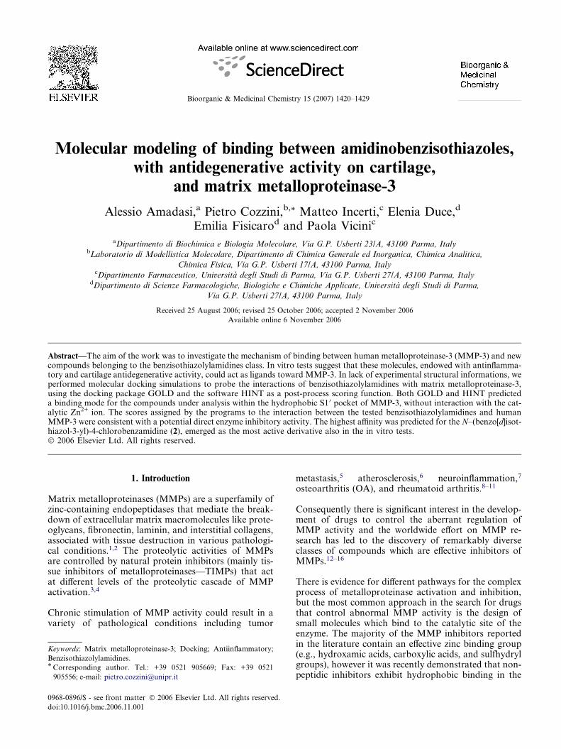
Molecular modeling of binding between amidinobenzisothiazoles, with antidegenerative activity on cartilage, and matrix metalloproteinase-3 Alessio Amadasi, a Pietro Cozzini, b, * Matteo Incerti, c Elenia Duce, d Emilia Fisicaro d and Paola Vicini c a Dipartimento di Biochimica e Biologia Molecolare, Via G.P. Usberti 23/A, 43100 Parma, Italy b Laboratorio di Modellistica Molecolare, Dipartimento di Chimica Generale ed Inorganica, Chimica Analitica, Chimica Fisica, Via G.P. Usberti 17/A, 43100 Parma, Italy c Dipartimento Farmaceutico, Universita ` degli Studi di Parma, Via G.P. Usberti 27/A, 43100 Parma, Italy d Dipartimento di Scienze Farmacologiche, Biologiche e Chimiche Applicate, Universita ` degli Studi di Parma, Via G.P. Usberti 27/A, 43100 Parma, Italy Received 25 August 2006; revised 25 October 2006; accepted 2 November 2006 Available online 6 November 2006 Abstract—The aim of the work was to investigate the mechanism of binding between human metalloproteinase-3 (MMP-3) and new compounds belonging to the benzisothiazolylamidines class. In vitro tests suggest that these molecules, endowed with antinflamma- tory and cartilage antidegenerative activity, could act as ligands toward MMP-3. In lack of experimental structural informations, we performed molecular docking simulations to probe the interactions of benzisothiazolylamidines with matrix metalloproteinase-3, using the docking package GOLD and the software HINT as a post-process scoring function. Both GOLD and HINT predicted a binding mode for the compounds under analysis within the hydrophobic S1 0 pocket of MMP-3, without interaction with the cat- alytic Zn 2+ ion. The scores assigned by the programs to the interaction between the tested benzisothiazolylamidines and human MMP-3 were consistent with a potential direct enzyme inhibitory activity. The highest affinity was predicted for the N–(benzo[d]isot- hiazol-3-yl)-4-chlorobenzamidine (2), emerged as the most active derivative also in the in vitro tests. Ó 2006 Elsevier Ltd. All rights reserved. 1. Introduction Matrix metalloproteinases (MMPs) are a superfamily of zinc-containing endopeptidases that mediate the break- down of extracellular matrix macromolecules like prote- oglycans, fibronectin, laminin, and interstitial collagens, associated with tissue destruction in various pathologi- cal conditions. 1,2 The proteolytic activities of MMPs are controlled by natural protein inhibitors (mainly tis- sue inhibitors of metalloproteinases—TIMPs) that act at different levels of the proteolytic cascade of MMP activation. 3,4 Chronic stimulation of MMP activity could result in a variety of pathological conditions including tumor metastasis, 5 atherosclerosis, 6 neuroinflammation, 7 osteoarthritis (OA), and rheumatoid arthritis. 8–11 Consequently there is significant interest in the develop- ment of drugs to control the aberrant regulation of MMP activity and the worldwide effort on MMP re- search has led to the discovery of remarkably diverse classes of compounds which are effective inhibitors of MMPs. 12–16 There is evidence for different pathways for the complex process of metalloproteinase activation and inhibition, but the most common approach in the search for drugs that control abnormal MMP activity is the design of small molecules which bind to the catalytic site of the enzyme. The majority of the MMP inhibitors reported in the literature contain an effective zinc binding group (e.g., hydroxamic acids, carboxylic acids, and sulfhydryl groups), however it was recently demonstrated that non- peptidic inhibitors exhibit hydrophobic binding in the 0968-0896/$ - see front matter Ó 2006 Elsevier Ltd. All rights reserved. doi:10.1016/j.bmc.2006.11.001 Keywords: Matrix metalloproteinase-3; Docking; Antiinflammatory; Benzisothiazolylamidines. * Corresponding author. Tel.: +39 0521 905669; Fax: +39 0521 905556; e-mail: [email protected] Bioorganic & Medicinal Chemistry 15 (2007) 1420–1429
-
Upload
independent -
Category
Documents
-
view
0 -
download
0
Transcript of Molecular modeling of binding between amidinobenzisothiazoles, with antidegenerative activity on...
Bioorganic & Medicinal Chemistry 15 (2007) 1420–1429
Molecular modeling of binding between amidinobenzisothiazoles,with antidegenerative activity on cartilage,
and matrix metalloproteinase-3
Alessio Amadasi,a Pietro Cozzini,b,* Matteo Incerti,c Elenia Duce,d
Emilia Fisicarod and Paola Vicinic
aDipartimento di Biochimica e Biologia Molecolare, Via G.P. Usberti 23/A, 43100 Parma, ItalybLaboratorio di Modellistica Molecolare, Dipartimento di Chimica Generale ed Inorganica, Chimica Analitica,
Chimica Fisica, Via G.P. Usberti 17/A, 43100 Parma, ItalycDipartimento Farmaceutico, Universita degli Studi di Parma, Via G.P. Usberti 27/A, 43100 Parma, Italy
dDipartimento di Scienze Farmacologiche, Biologiche e Chimiche Applicate, Universita degli Studi di Parma,
Via G.P. Usberti 27/A, 43100 Parma, Italy
Received 25 August 2006; revised 25 October 2006; accepted 2 November 2006
Available online 6 November 2006
Abstract—The aim of the work was to investigate the mechanism of binding between human metalloproteinase-3 (MMP-3) and newcompounds belonging to the benzisothiazolylamidines class. In vitro tests suggest that these molecules, endowed with antinflamma-tory and cartilage antidegenerative activity, could act as ligands toward MMP-3. In lack of experimental structural informations, weperformed molecular docking simulations to probe the interactions of benzisothiazolylamidines with matrix metalloproteinase-3,using the docking package GOLD and the software HINT as a post-process scoring function. Both GOLD and HINT predicteda binding mode for the compounds under analysis within the hydrophobic S1 0 pocket of MMP-3, without interaction with the cat-alytic Zn2+ ion. The scores assigned by the programs to the interaction between the tested benzisothiazolylamidines and humanMMP-3 were consistent with a potential direct enzyme inhibitory activity. The highest affinity was predicted for the N–(benzo[d]isot-hiazol-3-yl)-4-chlorobenzamidine (2), emerged as the most active derivative also in the in vitro tests.� 2006 Elsevier Ltd. All rights reserved.
1. Introduction
Matrix metalloproteinases (MMPs) are a superfamily ofzinc-containing endopeptidases that mediate the break-down of extracellular matrix macromolecules like prote-oglycans, fibronectin, laminin, and interstitial collagens,associated with tissue destruction in various pathologi-cal conditions.1,2 The proteolytic activities of MMPsare controlled by natural protein inhibitors (mainly tis-sue inhibitors of metalloproteinases—TIMPs) that actat different levels of the proteolytic cascade of MMPactivation.3,4
Chronic stimulation of MMP activity could result in avariety of pathological conditions including tumor
0968-0896/$ - see front matter � 2006 Elsevier Ltd. All rights reserved.
doi:10.1016/j.bmc.2006.11.001
Keywords: Matrix metalloproteinase-3; Docking; Antiinflammatory;
Benzisothiazolylamidines.* Corresponding author. Tel.: +39 0521 905669; Fax: +39 0521
905556; e-mail: [email protected]
metastasis,5 atherosclerosis,6 neuroinflammation,7
osteoarthritis (OA), and rheumatoid arthritis.8–11
Consequently there is significant interest in the develop-ment of drugs to control the aberrant regulation ofMMP activity and the worldwide effort on MMP re-search has led to the discovery of remarkably diverseclasses of compounds which are effective inhibitors ofMMPs.12–16
There is evidence for different pathways for the complexprocess of metalloproteinase activation and inhibition,but the most common approach in the search for drugsthat control abnormal MMP activity is the design ofsmall molecules which bind to the catalytic site of theenzyme. The majority of the MMP inhibitors reportedin the literature contain an effective zinc binding group(e.g., hydroxamic acids, carboxylic acids, and sulfhydrylgroups), however it was recently demonstrated that non-peptidic inhibitors exhibit hydrophobic binding in the
A. Amadasi et al. / Bioorg. Med. Chem. 15 (2007) 1420–1429 1421
long hydrophobic S1 0 pocket yet show no covalent inter-action with the catalytic zinc ion.17,18
A variety of substrate-based inhibitors have been de-signed and the features of these effective metalloprotein-ase inhibitors are well known and extensivelyreviewed.15 Concerning the treatment of OA and rheuma-toid arthritis, where a shift toward increased MMP activ-ity leads to cartilage degradation and destruction, despitethe advances of the medicinal chemistry in this field andthe relevance of the related MMP inhibitors, the availablechondroprotective agents are still unsatisfactory.10,19–23
Indeed, a variety of first-generation MMP inhibitors, de-signed on the basis of the protein natural substrate, pos-sess peptide-like structure and, on consequence, showpoor pharmacokinetic properties. Moreover, the lackof selectivity against a particular subset of the MMPsrepresents the reason of the unwanted side effects thatcan cause interruption of clinical trials.19,20
Our long-term commitment in the field of benzisothiaz-ole derivatives with biological activity24–32 led to the dis-covery of a class of amidinobenzo[d]isothiazolesendowed with antinflammatory and antidegenerativeactivity on cartilage. These compounds, emerged aspromising antiinflammatory–antidegenerative agentswith non-acidic character, have, in addition, non-peptid-ic, low weight structures which seem very pertinentproperties in the search of new efficient MMPinhibitors.33,34
In view of the outstanding in vitro properties shown bythe unsubstituted N-(benzo[d]isothiazol-3-yl)benzami-dine and by different substituted benzisothiazolylbenz-amidines,35 we decided to investigate if a directinteraction (i.e., inhibition) of the enzyme, implicatedin arthritic diseases,36,37 with benzisothiazolylamidinesmay be possible. Indeed, our previous in vitro resultsshowed a powerful reduction, at the micromolar level,of most of the harmful effects induced by the pro-inflam-matory cytokine interleukin-1ß on human chondrocytesas detailed by Vicini’s paper.35 The selected amidines forthe current study are N-(benzo[d]isothiazol-3-yl)ben-zamidine 1, N-(benzo[d]isothiazol-3-yl)-4-chlorobenz-amidine 2, emerged as the most active derivative, andN-(benzo[d]isothiazol-3-yl)acetamidine 3, considereduseful to explain the influence of a different substituent,such as methyl, in the binding process.
The program GOLD was used to perform moleculardocking studies, while the software HINT (HydropaticINTeractions) was used as a post-process scoring func-tion to understand in detail the interactions betweenprotein and synthesized ligands.
GOLD is a well-known automated ligand docking pro-gram, that uses a genetic algorithm to explore the fullrange of ligand conformational flexibility with partialflexibility of the protein side chains.38,39
HINT is a non-Newtonian force field, based on experi-mentally determined Log Po/w (the log10 of the partition
coefficient between 1-octanol and water) values, particu-larly suitable for the quantitative evaluation of non-co-valent molecular interactions, including the entropicallydriven hydrophobic effect,40–44 therefore, it can consti-tute a good complementary tool to classical dockingsoftware fitness functions.
The method used was found to predict the binding modeof known ligands with good accuracy and then appliedto identify the possible binding sites of benzamidines1–3.
2. Results and discussion
2.1. Selection of the protein target structure
Although GOLD applies some limited flexibility to theprotein residue side chains, one of the main problemsof any docking analysis lies in the inability in dealingwith protein flexibility. Furthermore, human MMP-3is known to undergo inhibitor-induced conformationalchanges, with Tyr223 acting as a gatekeeper of the S1 0
cavity.45,46 Because none of the MMP-3 inhibitorsreported in the literature exhibits structure similaritywith compounds 1–3 under study, we decided to com-pare the crystallographic structures of known MMP-3-ligand complexes, found in the Brookhaven ProteinData Bank, to verify whether differences were presentin the position of the binding site residues.
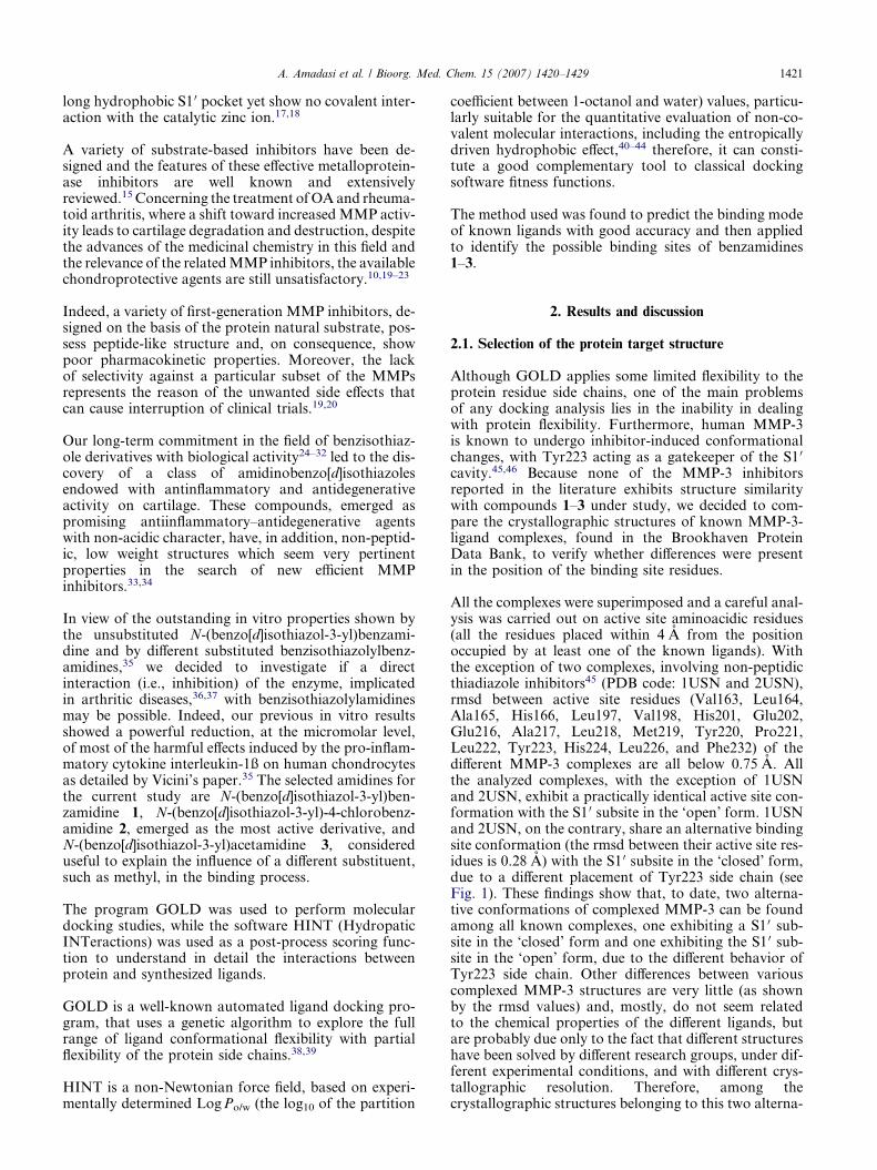
All the complexes were superimposed and a careful anal-ysis was carried out on active site aminoacidic residues(all the residues placed within 4 A from the positionoccupied by at least one of the known ligands). Withthe exception of two complexes, involving non-peptidicthiadiazole inhibitors45 (PDB code: 1USN and 2USN),rmsd between active site residues (Val163, Leu164,Ala165, His166, Leu197, Val198, His201, Glu202,Glu216, Ala217, Leu218, Met219, Tyr220, Pro221,Leu222, Tyr223, His224, Leu226, and Phe232) of thedifferent MMP-3 complexes are all below 0.75 A. Allthe analyzed complexes, with the exception of 1USNand 2USN, exhibit a practically identical active site con-formation with the S1 0 subsite in the ‘open’ form. 1USNand 2USN, on the contrary, share an alternative bindingsite conformation (the rmsd between their active site res-idues is 0.28 A) with the S1 0 subsite in the ‘closed’ form,due to a different placement of Tyr223 side chain (seeFig. 1). These findings show that, to date, two alterna-tive conformations of complexed MMP-3 can be foundamong all known complexes, one exhibiting a S1 0 sub-site in the ‘closed’ form and one exhibiting the S1 0 sub-site in the ‘open’ form, due to the different behavior ofTyr223 side chain. Other differences between variouscomplexed MMP-3 structures are very little (as shownby the rmsd values) and, mostly, do not seem relatedto the chemical properties of the different ligands, butare probably due only to the fact that different structureshave been solved by different research groups, under dif-ferent experimental conditions, and with different crys-tallographic resolution. Therefore, among thecrystallographic structures belonging to this two alterna-
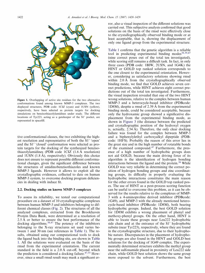
Figure 1. Overlapping of active site residues for the two alternative
conformations found among known MMP-3 complexes. The two
displayed structures, PDB code: 1CIZ (cyan) and 1USN (yellow),
respectively, have been selected as protein targets for docking
simulations on benzisothiazolylamidines under study. The different
locations of Tyr223, acting as a gatekeeper of the S1 0 pocket, are
represented in spacefill.
1422 A. Amadasi et al. / Bioorg. Med. Chem. 15 (2007) 1420–1429
tive conformational classes, the two exhibiting the high-est resolution and representative of both the S1 0 ‘open’and the S1 0 ‘closed’ conformation were selected as pro-tein targets for the docking of the synthesized benziso-thiazolylamidines (PDB code 1CIZ (1.6 A resolution)and 1USN (1.8 A), respectively). Obviously this choicedoes not ensure to represent possible different conforma-tional changes, given the significant difference betweenthe structures of amidinobenzisothiazoles and knownMMP-3 ligands. However it allows to exploit all thecrystallographic evidences, collected to date on humanMMP-3 system, to overcome docking program deficien-cies in dealing with induce fit.
2.2. Docking studies on known MMP-3 complexes
To assess its reliability, we tested our computationalprocedure on a dataset of 10 crystallographic complexesbetween human MMP-3 and inhibitors belonging to dif-ferent chemical classes (Fig. 2). The structures of all theanalyzed complexes, extracted from the BrookhavenProtein Data Bank, were determined at a resolution of2.5 A or better to ensure the best performance of thedocking algorithms.38,47 The potency of the inhibitorsbelonging to the X-ray structures set used varies be-tween 1 and 30 nm (see references in Table 1). The re-sults, obtained using our computational tools to dockthe ligand back into the binding site, are shown in Table1. All the solutions were evaluated on the basis of thermsd from the experimental orientation. The currentstandard in the field is a 2.0 A cut-off, beyond whichthe prediction is considered a docking failure.47,55 How-ever, since a small rmsd result may mask a significant er-
ror, also a visual inspection of the different solutions wascarried out. This subjective analysis confirmed that goodsolutions on the basis of the rmsd were effectively closeto the crystallographically observed binding mode or atleast acceptable, that is, showing the displacement ofonly one ligand group from the experimental structure.
Table 1 confirms that the genetic algorithm is a reliabletool in predicting experimental binding modes38,39,47
(nine correct poses out of the total ten investigated),while scoring still remains a difficult task. In fact, in onlythree cases (PDB code: 1BIW, 2USN, and 1G4K) theHINT or GOLD top ranked solution corresponds tothe one closest to the experimental orientation. Howev-er, considering as satisfactory solutions showing rmsdwithin 2.0 A from the crystallographically observedbinding mode, we find that GOLD achieves seven cor-rect predictions, while HINT achieves eight correct pre-dictions out of the total ten investigated. Furthermore,the visual inspection revealed that one of the two HINTwrong solutions, relative to the complex between humanMMP-3 and a heterocycle-based inhibitor (PDBcode:1D8M), despite a rmsd of 2.39 A from the experimentalbinding mode, could be considered acceptable, becauseonly the hydroxamic group showed a significative dis-placement from the experimental binding mode, asshown in Figure 3 (the distance between the predictedand crystallographic position of the hydroxyl oxygenis, actually, 2.54 A). Therefore, the only clear dockingfailure was found for the complex between MMP-3and a biphenylylethyl carboxyalkyl dipeptide (PDB-code: 1HFS). Probably the reason of this error lies inthe great size and in the high number of rotatable bondsof the examined compound.47 Furthermore, the pres-ence of a high number of hydrophobic groups doesnot aid GOLD, because the main driving force of itsalgorithm is the identification of hydrogen bondinginteractions between the ligand and the protein.38 WhileGOLD was very reliable in determining the correct po-sition of hydrogen bonding groups and zinc coordinat-ing groups, its difficulty in properly evaluating thehydrophobic interactions constitutes the main reasonfor the other errors found in the GOLD top ranked pos-es. The use of HINT as a post-process scoring functioncan be useful to overcome this problem, as it can be ob-served for the results relative to the complexes of MMP-3 with a 4-aminoproline derived inhibitor (PDBcode:1G49), and MMP-3 with the already mentioned hetero-cycle-based inhibitor (PDBcode: 1D8M), both bearinghydrophobic groups. Indeed, the best GOLD solutionfor 1D8M exhibits a wrong placement of phenyl andmethoxy-phenyl groups. On the other hand, HINT isable to locate these groups near Leu222 hydrophobicside chain and at the entrance of the S1 0 hydrophobicsubsite (near Tyr223), respectively, where they are foundin the crystallographic structure, due to their hydropho-bic nature. Discrepancies in the placement of hydropho-bic groups are also found in the HINT and GOLD bestsolutions for the docking of 1G49 complex. The experi-mentally determined structure exhibits the methyl groupnear the sulfonamide placed in proximity of Leu222 sidechain, while GOLD best solution shows the same groupmore exposed to the solvent. Furthermore, the best
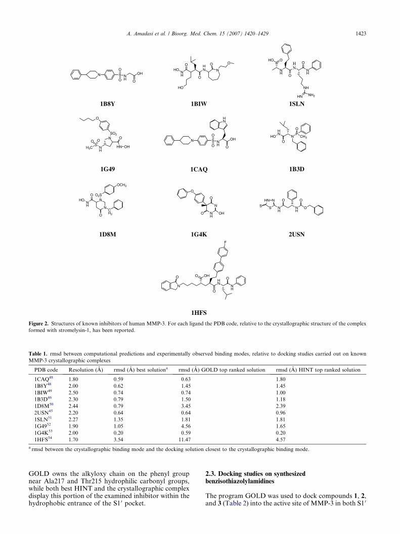
Figure 2. Structures of known inhibitors of human MMP-3. For each ligand the PDB code, relative to the crystallographic structure of the complex
formed with stromelysin-1, has been reported.
Table 1. rmsd between computational predictions and experimentally observed binding modes, relative to docking studies carried out on known
MMP-3 crystallographic complexes
PDB code Resolution (A) rmsd (A) best solutiona rmsd (A) GOLD top ranked solution rmsd (A) HINT top ranked solution
1CAQ48 1.80 0.59 0.63 1.80
1B8Y48 2.00 0.62 1.45 1.45
1BIW49 2.50 0.74 0.74 1.00
1B3D46 2.30 0.79 1.50 1.18
1D8M50 2.44 0.79 3.45 2.39
2USN45 2.20 0.64 0.64 0.96
1SLN51 2.27 1.35 1.81 1.81
1G4952 1.90 1.05 4.56 1.65
1G4K53 2.00 0.20 0.59 0.20
1HFS54 1.70 3.54 11.47 4.57
a rmsd between the crystallographic binding mode and the docking solution closest to the crystallographic binding mode.
A. Amadasi et al. / Bioorg. Med. Chem. 15 (2007) 1420–1429 1423
GOLD owns the alkyloxy chain on the phenyl groupnear Ala217 and Thr215 hydrophilic carbonyl groups,while both best HINT and the crystallographic complexdisplay this portion of the examined inhibitor within thehydrophobic entrance of the S1 0 pocket.
2.3. Docking studies on synthesizedbenzisothiazolylamidines
The program GOLD was used to dock compounds 1, 2,and 3 (Table 2) into the active site of MMP-3 in both S1 0
Figure 3. Comparison between the crystallographic binding mode and
the docking solution exhibiting the highest HINT score (best HINT in
magenta), for the complex between MMP-3 and a heterocycle-based
inhibitor (PDBcode: 1D8M).
1424 A. Amadasi et al. / Bioorg. Med. Chem. 15 (2007) 1420–1429
‘closed’ (PDB code: 1USN)45 and S1 0 ‘open’ (PDB code:1CIZ)48 conformation (see Fig. 1). For each compoundthe most stable docking model was selected according tothe best scored conformation predicted by GOLD (high-est GOLD score) and HINT (highest HINT score).
Being the acid-base properties of the amidino groupvery sensitive to the substituent in the molecule,56,57 inorder to know what species is present at the physiolog-ical pH, it is necessary to carefully evaluate the pKa ofthe N-benzo[d]isothiazol-3-yl-amidines 1–3 under inves-tigation: this was done by means of potentiometric titra-tions in methanol/water = 9:1 as solvent, to avoidsolubility problems with the unprotonated form of thehydrochlorides. The values obtained, reported in Table2, show that, in agreement with the different electroniceffects of the substituents, under physiological condi-tions compounds 1 and 2 are in neutral form, whereasfor compound 3 a non-negligible portion of the proton-ated form is still present.
Compounds 1 and 2 were modeled with the amidinogroup in the neutral form, in agreement with experimen-tal pKa of 5.40 and 4.84, respectively, while compound3, showing a pKa of 6.99, was modeled in both the pro-tonated and neutral forms and mean HINT score andGOLD score values have been reported.
Table 2. Structures, experimental pKa values, HINT calculated LogPo/w
compounds
SN
HN
Compound R pKaa
1 C6H5 5.40(0.04)
2 4-Cl–C6H4 4.84(0.01)
3 CH3 6.99(0.01)
a In MeOH/H2O = 9:1, I = 0.1 M KCl.
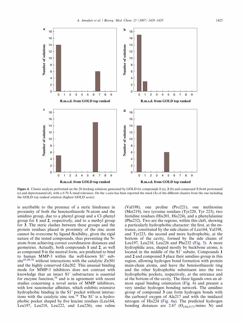
The three compounds showed high score values, consis-tent with a possible binding to MMP-3, only whendocked into the S1 0 ‘open’ active site configuration. Sig-nificant differences were not found between the bindingorientation of the HINT and GOLD predicted bestsolutions for the synthesized compounds (i.e., the rmsdbetween HINT and GOLD top ranked solutions was al-ways below 0.70 A). Furthermore, a cluster analysis per-formed with a 0.70 A rmsd tolerance (see Fig. 4) showeda high ratio of conformations very close to the HINTand GOLD best one (85% for compound 2, 70% forcompound 1, 25% for compound 3 in the protonatedform, and 70% for compound 3 in the neutral form).As reported in Table 2, both HINT and GOLD identifythe newly synthesized compound 2, emerged as the mostactive derivative during in vitro studies35, as the onewhich can interact more tightly with human MMP-3,while compound 3 is predicted to have a lower bindingaffinity. Only for compound 3, when modeled as proton-ated, the predicted distance between the benzisothiazolN-atom and Zn2+ ion (1.92 A) is consistent with a pos-sible metal coordination. However the coordinationgeometry, considering the positions of the three imi-dazol N-atoms involved in zinc coordination, belongingto His201, His205, and His211, is quite distorted fromthe tetrahedral one or the trigonal bipiramidal describedfor other known MMP-3 inhibitors,48 being the anglebetween benzoisothiazol N-atom, Zn2+ ion, and imi-dazol N-atom of His205 146.1�. This finding indicatesthat, probably, also compound 3 cannot undertake acorrect metal coordination, but it is located by the soft-ware in a position close to the Zn2+ ion to allow a strongelectrostatic interaction between its positively chargedamidino group and the negatively charged carboxylategroup of Glu202. However, the absence of availablespace for the presence of a water molecule allowingthe completion of the Zn2+ coordination suggests thatthis predicted binding mode could represent a false po-sitive, due to the high energetic cost associated with thissituation. This interpretation is supported by the findingthat an alternative docking solution, exhibiting com-pound 3 into the S1 0 pocket (see below), is within a morepopulated cluster (as shown in Fig. 4c). Therefore, thislatter solution, very similar to the one predicted for com-pound 3 when modeled as deprotonated (see below), canalso be considered the more probable binding mode forcompound 3 in the protonated form. The likely absenceof zinc-coordination, for all the three tested compounds,
values, and best HINT and GOLD score values of the synthesized
NH
R
LogPo/w Best HINT Best GOLD
3.52 2397 54.42
4.23 2823 57.60
2.41 1582 46.42
R.m.s.d. from GOLD top ranked
0 1 2 3 4 5 6 7 8 9
Num
ber
ofso
luti
ons
0
2
4
6
8
10
12
14
16
18
R.m.s.d. from GOLD top ranked
0 1 2 3 4 5 6 7 8 9
Num
ber
ofso
luti
ons
0
2
4
6
8
10
12
14
16
18
R.m.s.d. from GOLD top ranked
0 1 2 3 4 5 6 7 8 9
Num
ber
ofso
luti
ons
0
2
4
6
8
10
12
14
16
18
R.m.s.d. from GOLD top ranked
0 1 2 3 4 5 6 7 8 9
Num
ber
ofso
luti
ons
0
2
4
6
8
10
12
14
16
18
a b
c d
Figure 4. Cluster analysis performed on the 20 docking solutions generated by GOLD for compounds 1 (a), 2 (b) and compound 3 (both protonated
(c) and deprotonated (d), with a 0.70 A rmsd tolerance. On the x-axis has been reported the rmsd (A) of the different clusters from the one including
the GOLD top ranked solution (highest GOLD score).
A. Amadasi et al. / Bioorg. Med. Chem. 15 (2007) 1420–1429 1425
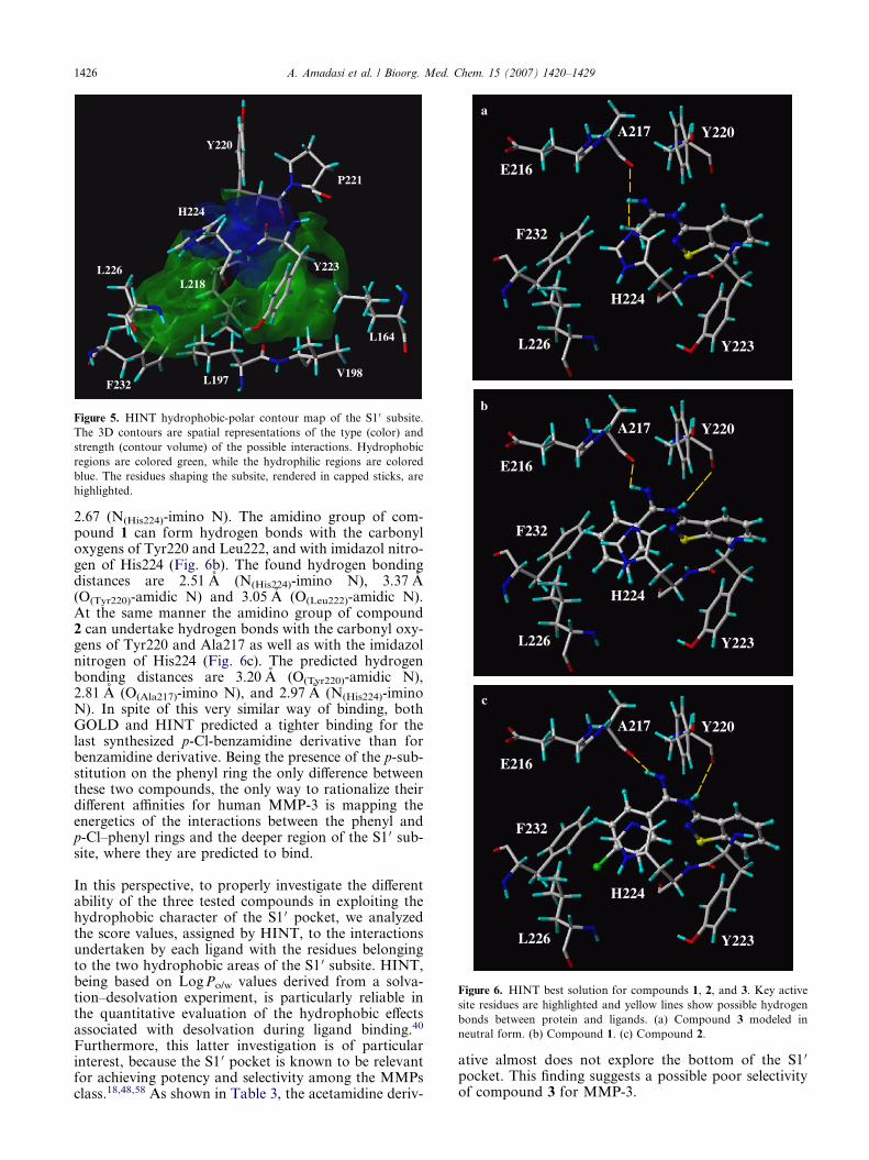
is ascribable to the presence of a steric hindrance inproximity of both the benzisothiazole N-atom and theamidino group, due to a phenyl group and a Cl–phenylgroup for 1 and 2, respectively, and to a methyl groupfor 3. The steric clashes between these groups and theprotein residues placed in proximity of the zinc atomcannot be overcome by ligand flexibility, given the rigidnature of the tested compounds, thus preventing the N-atom from achieving correct coordination distances andgeometries. Actually, both compounds 1 and 2, as wellas compound 3 in the neutral form, are predicted to bindto human MMP-3 within the well-known S1 0 sub-site45,48,58 without interactions with the catalytic Zn301and the highly conserved Glu202. This unusual bindingmode for MMP-3 inhibitors does not contrast withknowledge that an intact S1 0 substructure is essentialfor enzyme function,58 and is in agreement with recentstudies concerning a novel series of MMP inhibitors,with low nanomolar affinities, which exhibits extensivehydrophobic binding in the S1 0 pocket without interac-tions with the catalytic zinc ion.18 The S1 0 is a hydro-phobic pocket shaped by five leucine residues (Leu164,Leu197, Leu218, Leu222, and Leu226), one valine
(Val198), one proline (Pro221), one methionine(Met219), two tyrosine residues (Tyr220, Tyr 223), twohistidine residues (His201, His224), and a phenylalanine(Phe232). Two are the regions, within this cleft, showinga particularly hydrophobic character: the first, at the en-trance, constituted by the side chains of Leu164, Val198,and Tyr223, the second and more hydrophobic, at thebottom of the cavity, formed by the side chains ofLeu197, Leu218, Leu226 and Phe232 (Fig. 5). A morehydrophilic area, shaped mostly by backbone atoms, islocated in the middle of the S1 0 subsite. Compounds 1and 2 and compound 3 place their amidino group in thisregion, allowing hydrogen bond formation with proteinmain-chain atoms, and leave the benzisothiazole ringand the other hydrophobic substituent into the twohydrophobic pockets, respectively, at the entrance andat the bottom of the cavity. The three ligands own an al-most equal binding orientation (Fig. 6) and present avery similar hydrogen bonding network. The amidinogroup of compound 3 can form hydrogen bonds withthe carbonyl oxygen of Ala217 and with the imidazolnitrogen of His224 (Fig. 6a). The predicted hydrogenbonding distances are 2.67 (O(Ala217)-imino N) and
Y220
P221
L164
V198 L197 F232
L226
H224
Y223
L218
Figure 5. HINT hydrophobic-polar contour map of the S1 0 subsite.
The 3D contours are spatial representations of the type (color) and
strength (contour volume) of the possible interactions. Hydrophobic
regions are colored green, while the hydrophilic regions are colored
blue. The residues shaping the subsite, rendered in capped sticks, are
highlighted.
b
A217 Y220
Y223
H224
E216
F232
L226
c
A217 Y220
Y223
H224
E216
F232
L226
a A217 Y220
Y223
H224
E216
F232
L226
Figure 6. HINT best solution for compounds 1, 2, and 3. Key active
site residues are highlighted and yellow lines show possible hydrogen
bonds between protein and ligands. (a) Compound 3 modeled in
neutral form. (b) Compound 1. (c) Compound 2.
1426 A. Amadasi et al. / Bioorg. Med. Chem. 15 (2007) 1420–1429
2.67 (N(His224)-imino N). The amidino group of com-pound 1 can form hydrogen bonds with the carbonyloxygens of Tyr220 and Leu222, and with imidazol nitro-gen of His224 (Fig. 6b). The found hydrogen bondingdistances are 2.51 A (N(His224)-imino N), 3.37 A(O(Tyr220)-amidic N) and 3.05 A (O(Leu222)-amidic N).At the same manner the amidino group of compound2 can undertake hydrogen bonds with the carbonyl oxy-gens of Tyr220 and Ala217 as well as with the imidazolnitrogen of His224 (Fig. 6c). The predicted hydrogenbonding distances are 3.20 A (O(Tyr220)-amidic N),2.81 A (O(Ala217)-imino N), and 2.97 A (N(His224)-iminoN). In spite of this very similar way of binding, bothGOLD and HINT predicted a tighter binding for thelast synthesized p-Cl-benzamidine derivative than forbenzamidine derivative. Being the presence of the p-sub-stitution on the phenyl ring the only difference betweenthese two compounds, the only way to rationalize theirdifferent affinities for human MMP-3 is mapping theenergetics of the interactions between the phenyl andp-Cl–phenyl rings and the deeper region of the S1 0 sub-site, where they are predicted to bind.
In this perspective, to properly investigate the differentability of the three tested compounds in exploiting thehydrophobic character of the S1 0 pocket, we analyzedthe score values, assigned by HINT, to the interactionsundertaken by each ligand with the residues belongingto the two hydrophobic areas of the S1 0 subsite. HINT,being based on Log Po/w values derived from a solva-tion–desolvation experiment, is particularly reliable inthe quantitative evaluation of the hydrophobic effectsassociated with desolvation during ligand binding.40
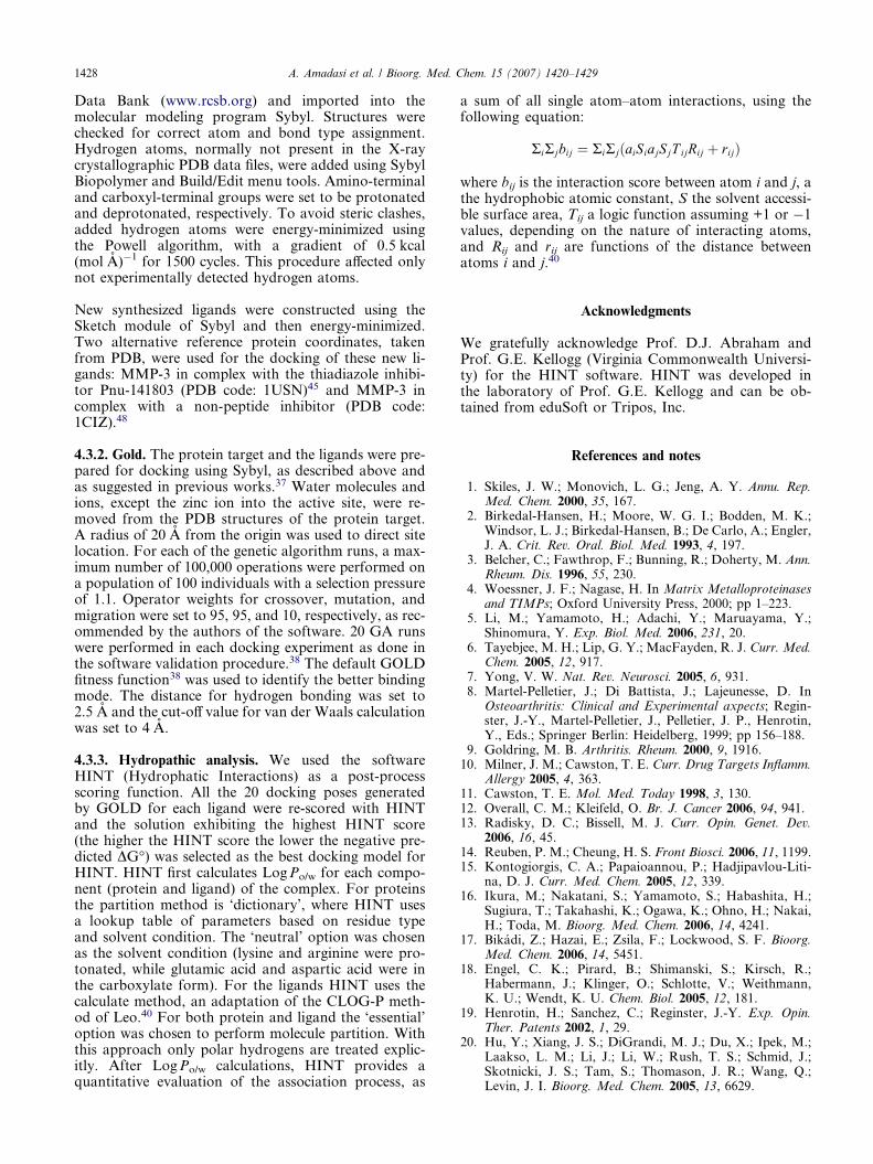
Furthermore, this latter investigation is of particularinterest, because the S1 0 pocket is known to be relevantfor achieving potency and selectivity among the MMPsclass.18,48,58 As shown in Table 3, the acetamidine deriv-
ative almost does not explore the bottom of the S1 0
pocket. This finding suggests a possible poor selectivityof compound 3 for MMP-3.
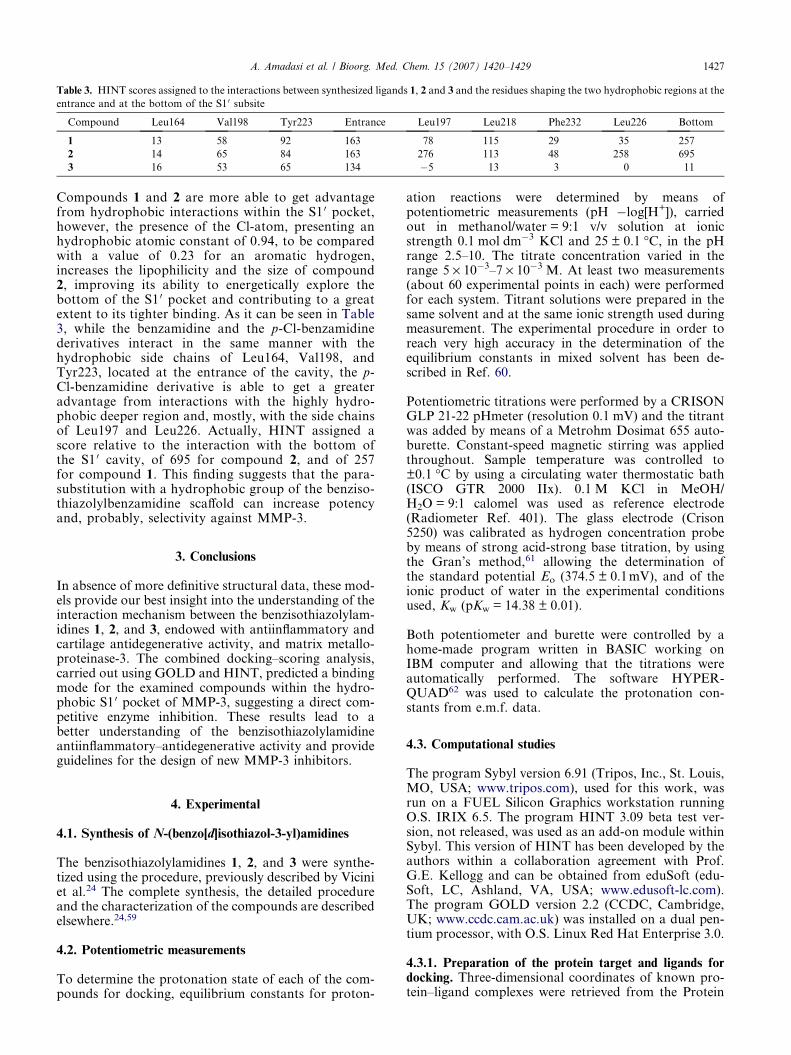
Table 3. HINT scores assigned to the interactions between synthesized ligands 1, 2 and 3 and the residues shaping the two hydrophobic regions at the
entrance and at the bottom of the S1 0 subsite
Compound Leu164 Val198 Tyr223 Entrance Leu197 Leu218 Phe232 Leu226 Bottom
1 13 58 92 163 78 115 29 35 257
2 14 65 84 163 276 113 48 258 695
3 16 53 65 134 �5 13 3 0 11
A. Amadasi et al. / Bioorg. Med. Chem. 15 (2007) 1420–1429 1427
Compounds 1 and 2 are more able to get advantagefrom hydrophobic interactions within the S1 0 pocket,however, the presence of the Cl-atom, presenting anhydrophobic atomic constant of 0.94, to be comparedwith a value of 0.23 for an aromatic hydrogen,increases the lipophilicity and the size of compound2, improving its ability to energetically explore thebottom of the S1 0 pocket and contributing to a greatextent to its tighter binding. As it can be seen in Table3, while the benzamidine and the p-Cl-benzamidinederivatives interact in the same manner with thehydrophobic side chains of Leu164, Val198, andTyr223, located at the entrance of the cavity, the p-Cl-benzamidine derivative is able to get a greateradvantage from interactions with the highly hydro-phobic deeper region and, mostly, with the side chainsof Leu197 and Leu226. Actually, HINT assigned ascore relative to the interaction with the bottom ofthe S1 0 cavity, of 695 for compound 2, and of 257for compound 1. This finding suggests that the para-substitution with a hydrophobic group of the benziso-thiazolylbenzamidine scaffold can increase potencyand, probably, selectivity against MMP-3.
3. Conclusions
In absence of more definitive structural data, these mod-els provide our best insight into the understanding of theinteraction mechanism between the benzisothiazolylam-idines 1, 2, and 3, endowed with antiinflammatory andcartilage antidegenerative activity, and matrix metallo-proteinase-3. The combined docking–scoring analysis,carried out using GOLD and HINT, predicted a bindingmode for the examined compounds within the hydro-phobic S1 0 pocket of MMP-3, suggesting a direct com-petitive enzyme inhibition. These results lead to abetter understanding of the benzisothiazolylamidineantiinflammatory–antidegenerative activity and provideguidelines for the design of new MMP-3 inhibitors.
4. Experimental
4.1. Synthesis of N-(benzo[d]isothiazol-3-yl)amidines
The benzisothiazolylamidines 1, 2, and 3 were synthe-tized using the procedure, previously described by Viciniet al.24 The complete synthesis, the detailed procedureand the characterization of the compounds are describedelsewhere.24,59
4.2. Potentiometric measurements
To determine the protonation state of each of the com-pounds for docking, equilibrium constants for proton-
ation reactions were determined by means ofpotentiometric measurements (pH �log[H+]), carriedout in methanol/water = 9:1 v/v solution at ionicstrength 0.1 mol dm�3 KCl and 25 ± 0.1 �C, in the pHrange 2.5–10. The titrate concentration varied in therange 5 · 10�3–7 · 10�3 M. At least two measurements(about 60 experimental points in each) were performedfor each system. Titrant solutions were prepared in thesame solvent and at the same ionic strength used duringmeasurement. The experimental procedure in order toreach very high accuracy in the determination of theequilibrium constants in mixed solvent has been de-scribed in Ref. 60.
Potentiometric titrations were performed by a CRISONGLP 21-22 pHmeter (resolution 0.1 mV) and the titrantwas added by means of a Metrohm Dosimat 655 auto-burette. Constant-speed magnetic stirring was appliedthroughout. Sample temperature was controlled to±0.1 �C by using a circulating water thermostatic bath(ISCO GTR 2000 IIx). 0.1 M KCl in MeOH/H2O = 9:1 calomel was used as reference electrode(Radiometer Ref. 401). The glass electrode (Crison5250) was calibrated as hydrogen concentration probeby means of strong acid-strong base titration, by usingthe Gran’s method,61 allowing the determination ofthe standard potential Eo (374.5 ± 0.1mV), and of theionic product of water in the experimental conditionsused, Kw (pKw = 14.38 ± 0.01).
Both potentiometer and burette were controlled by ahome-made program written in BASIC working onIBM computer and allowing that the titrations wereautomatically performed. The software HYPER-QUAD62 was used to calculate the protonation con-stants from e.m.f. data.
4.3. Computational studies
The program Sybyl version 6.91 (Tripos, Inc., St. Louis,MO, USA; www.tripos.com), used for this work, wasrun on a FUEL Silicon Graphics workstation runningO.S. IRIX 6.5. The program HINT 3.09 beta test ver-sion, not released, was used as an add-on module withinSybyl. This version of HINT has been developed by theauthors within a collaboration agreement with Prof.G.E. Kellogg and can be obtained from eduSoft (edu-Soft, LC, Ashland, VA, USA; www.edusoft-lc.com).The program GOLD version 2.2 (CCDC, Cambridge,UK; www.ccdc.cam.ac.uk) was installed on a dual pen-tium processor, with O.S. Linux Red Hat Enterprise 3.0.
4.3.1. Preparation of the protein target and ligands fordocking. Three-dimensional coordinates of known pro-tein–ligand complexes were retrieved from the Protein
1428 A. Amadasi et al. / Bioorg. Med. Chem. 15 (2007) 1420–1429
Data Bank (www.rcsb.org) and imported into themolecular modeling program Sybyl. Structures werechecked for correct atom and bond type assignment.Hydrogen atoms, normally not present in the X-raycrystallographic PDB data files, were added using SybylBiopolymer and Build/Edit menu tools. Amino-terminaland carboxyl-terminal groups were set to be protonatedand deprotonated, respectively. To avoid steric clashes,added hydrogen atoms were energy-minimized usingthe Powell algorithm, with a gradient of 0.5 kcal(mol A)�1 for 1500 cycles. This procedure affected onlynot experimentally detected hydrogen atoms.
New synthesized ligands were constructed using theSketch module of Sybyl and then energy-minimized.Two alternative reference protein coordinates, takenfrom PDB, were used for the docking of these new li-gands: MMP-3 in complex with the thiadiazole inhibi-tor Pnu-141803 (PDB code: 1USN)45 and MMP-3 incomplex with a non-peptide inhibitor (PDB code:1CIZ).48
4.3.2. Gold. The protein target and the ligands were pre-pared for docking using Sybyl, as described above andas suggested in previous works.37 Water molecules andions, except the zinc ion into the active site, were re-moved from the PDB structures of the protein target.A radius of 20 A from the origin was used to direct sitelocation. For each of the genetic algorithm runs, a max-imum number of 100,000 operations were performed ona population of 100 individuals with a selection pressureof 1.1. Operator weights for crossover, mutation, andmigration were set to 95, 95, and 10, respectively, as rec-ommended by the authors of the software. 20 GA runswere performed in each docking experiment as done inthe software validation procedure.38 The default GOLDfitness function38 was used to identify the better bindingmode. The distance for hydrogen bonding was set to2.5 A and the cut-off value for van der Waals calculationwas set to 4 A.
4.3.3. Hydropathic analysis. We used the softwareHINT (Hydrophatic Interactions) as a post-processscoring function. All the 20 docking poses generatedby GOLD for each ligand were re-scored with HINTand the solution exhibiting the highest HINT score(the higher the HINT score the lower the negative pre-dicted DG�) was selected as the best docking model forHINT. HINT first calculates Log Po/w for each compo-nent (protein and ligand) of the complex. For proteinsthe partition method is ‘dictionary’, where HINT usesa lookup table of parameters based on residue typeand solvent condition. The ‘neutral’ option was chosenas the solvent condition (lysine and arginine were pro-tonated, while glutamic acid and aspartic acid were inthe carboxylate form). For the ligands HINT uses thecalculate method, an adaptation of the CLOG-P meth-od of Leo.40 For both protein and ligand the ‘essential’option was chosen to perform molecule partition. Withthis approach only polar hydrogens are treated explic-itly. After Log Po/w calculations, HINT provides aquantitative evaluation of the association process, as
a sum of all single atom–atom interactions, using thefollowing equation:
RiRjbij ¼ RiRjðaiSiajSjT ijRij þ rijÞ
where bij is the interaction score between atom i and j, athe hydrophobic atomic constant, S the solvent accessi-ble surface area, Tij a logic function assuming +1 or �1values, depending on the nature of interacting atoms,and Rij and rij are functions of the distance betweenatoms i and j.40
Acknowledgments
We gratefully acknowledge Prof. D.J. Abraham andProf. G.E. Kellogg (Virginia Commonwealth Universi-ty) for the HINT software. HINT was developed inthe laboratory of Prof. G.E. Kellogg and can be ob-tained from eduSoft or Tripos, Inc.
References and notes
1. Skiles, J. W.; Monovich, L. G.; Jeng, A. Y. Annu. Rep.Med. Chem. 2000, 35, 167.
2. Birkedal-Hansen, H.; Moore, W. G. I.; Bodden, M. K.;Windsor, L. J.; Birkedal-Hansen, B.; De Carlo, A.; Engler,J. A. Crit. Rev. Oral. Biol. Med. 1993, 4, 197.
3. Belcher, C.; Fawthrop, F.; Bunning, R.; Doherty, M. Ann.Rheum. Dis. 1996, 55, 230.
4. Woessner, J. F.; Nagase, H. In Matrix Metalloproteinasesand TIMPs; Oxford University Press, 2000; pp 1–223.
5. Li, M.; Yamamoto, H.; Adachi, Y.; Maruayama, Y.;Shinomura, Y. Exp. Biol. Med. 2006, 231, 20.
6. Tayebjee, M. H.; Lip, G. Y.; MacFayden, R. J. Curr. Med.Chem. 2005, 12, 917.
7. Yong, V. W. Nat. Rev. Neurosci. 2005, 6, 931.8. Martel-Pelletier, J.; Di Battista, J.; Lajeunesse, D. In
Osteoarthritis: Clinical and Experimental axpects; Regin-ster, J.-Y., Martel-Pelletier, J., Pelletier, J. P., Henrotin,Y., Eds.; Springer Berlin: Heidelberg, 1999; pp 156–188.
9. Goldring, M. B. Arthritis. Rheum. 2000, 9, 1916.10. Milner, J. M.; Cawston, T. E. Curr. Drug Targets Inflamm.
Allergy 2005, 4, 363.11. Cawston, T. E. Mol. Med. Today 1998, 3, 130.12. Overall, C. M.; Kleifeld, O. Br. J. Cancer 2006, 94, 941.13. Radisky, D. C.; Bissell, M. J. Curr. Opin. Genet. Dev.
2006, 16, 45.14. Reuben, P. M.; Cheung, H. S. Front Biosci. 2006, 11, 1199.15. Kontogiorgis, C. A.; Papaioannou, P.; Hadjipavlou-Liti-
na, D. J. Curr. Med. Chem. 2005, 12, 339.16. Ikura, M.; Nakatani, S.; Yamamoto, S.; Habashita, H.;
Sugiura, T.; Takahashi, K.; Ogawa, K.; Ohno, H.; Nakai,H.; Toda, M. Bioorg. Med. Chem. 2006, 14, 4241.
17. Bikadi, Z.; Hazai, E.; Zsila, F.; Lockwood, S. F. Bioorg.Med. Chem. 2006, 14, 5451.
18. Engel, C. K.; Pirard, B.; Shimanski, S.; Kirsch, R.;Habermann, J.; Klinger, O.; Schlotte, V.; Weithmann,K. U.; Wendt, K. U. Chem. Biol. 2005, 12, 181.
19. Henrotin, H.; Sanchez, C.; Reginster, J.-Y. Exp. Opin.Ther. Patents 2002, 1, 29.
20. Hu, Y.; Xiang, J. S.; DiGrandi, M. J.; Du, X.; Ipek, M.;Laakso, L. M.; Li, J.; Li, W.; Rush, T. S.; Schmid, J.;Skotnicki, J. S.; Tam, S.; Thomason, J. R.; Wang, Q.;Levin, J. I. Bioorg. Med. Chem. 2005, 13, 6629.
A. Amadasi et al. / Bioorg. Med. Chem. 15 (2007) 1420–1429 1429
21. Whittaker, M.; Floyd, C. D.; Brown, P.; Gearing, J. H.Chem. Rev. 1999, 99, 2735.
22. Bottomley, K. M.; Johnson, W. H.; Walter, D. S.J. Enzyme Inhib. 1998, 13, 79.
23. Shroder, J.; Henke, A.; Wenzel, H.; Brandstetter, H.;Stammler, H. G.; Stammler, A.; Pfeiffer, W. D.; Tsche-sche, H. J. Med. Chem. 2001, 44, 3231.
24. Vicini, P.; Amoretti, L.; Ballabeni, V.; Barocelli, E.;Chiavarini, M. Eur. J. Med. Chem. 1995, 30, 809.
25. Vicini, P.; Manotti, C.; Caretta, A.; Amoretti, L. Arz-neim.-Forsch./Drug Res. 1997, 47, 1218.
26. Vicini, P.; Manotti, C.; Caretta, A.; Amoretti, L. Arz-neim.-Forsch./Drug Res. 1999, 49, 896.
27. Vicini, P.; Fisicaro, E.; Lugari, M. T. Archiv. Pharm. Med.Chem. 2000, 333, 135.
28. Vicini, P.; Amoretti, L.; Ballabeni, V.; Tognolini, M.;Barocelli, E. Bioorg. Med. Chem. 2000, 8, 2355.
29. Vicini, P.; Zani, F.; Cozzini, P.; Doytchinova, I. Eur.J. Med. Chem. 2002, 37, 553.
30. Vicini, P.; Geronikaki, A.; Incerti, M.; Busonera, B.; Poni,G.; Cabras, C. A.; La Colla, P. Bioorg. Med. Chem. 2003,11, 4785.
31. Geronikaki, A.; Vicini, P.; Theophilidis, G.; Lagunin, A.;Poroikov, V.; Dearden, J. C. SAR QSAR. Environ. Res.2003, 14, 485.
32. Zani, F.; Vicini, P.; Incerti, M. Eur. J. Med. Chem. 2004,39, 135.
33. Panico, A. M.; Vicini, P.; Incerti, M.; Cardile, V.; Gentile,B.; Ronsisvalle, G. Il Farmaco 2002, 57, 671.
34. Panico, A. M.; Vicini, P.; Massimo, G.; Cardile, V.;Gentile, B.; Avondo, S.; Vittorio, F.; Ronsisvalle, G.Inflammation 2004, 28, 231.
35. Vicini, P.; Incerti, M.; Cardile, V.; Garufi, F.; Ronsisvalle,S.; Panico, A.M. Chem. Med. Chem., in press.
36. Okada, Y.; Takeuchi, N.; Tomita, K.; Nakanishi, I.;Nagase, H. Ann. Rheum. Dis. 1989, 48, 645.
37. Obata, K.; Iwata, K.; Okada, Y.; Kohrin, Y.; Ohuchi, E.;Yoshida, S.; Shinmei, M.; Hayakawa, T. Clin. Chim. Acta1992, 211, 59.
38. Jones, G.; Willett, P.; Glen, R. C.; Leach, A. R.; Taylor,R. J. Mol. Biol. 1997, 267, 727.
39. Verdonk, M. L.; Cole, J. C.; Hartshorn, M. J.; Murray, C.W.; Taylor, R. D. Proteins: Struct., Funct., Genet. 2003,52, 609.
40. Kellogg, G. E.; Burnett, J. C.; Abraham, D. J. J. Comput.-Aided Mol. Des. 2001, 15, 381.
41. Abraham, D. J.; Kellogg, G. E.; Holt, J. M.; Ackers, G. K.J. Mol. Biol. 1997, 272, 613.
42. Cozzini, P.; Fornabaio, M.; Marabotti, A.; Abraham, D.J.; Kellogg, G. E.; Mozzarelli, A. J. Med. Chem. 2002, 45,2469.
43. Spyrakis, F.; Fornabaio, M.; Cozzini, P.; Mozzarelli, A.;Abraham, D. J.; Kellogg, G. E. J. Am. Chem. Soc. 2004,126, 11764.
44. Wang, R.; Lu, Y.; Fang, X.; Wang, S. J. Chem. Inf.Comput. Sci. 2004, 44, 2114.
45. Finzel, B. C.; Baldwin, E. T.; Bryant, G. L.; Hess, G. F.;Wilks, J. W.; Trepod, C. M.; Mott, J. E.; Marshall, V. P.;
Petzold, G. L.; Poorman, R. A.; O’Sullivan, T. J.;Schostarez, H. J.; Mitchell, M. A. Protein Sci. 1998, 7,2118.
46. Chen, L.; Rydel, T. J.; Gu, F.; Dunaway, C. M.; Pikul, S.;Dunham, K. M.; Barnett, B. L. J. Mol. Biol. 1999, 293,545.
47. Kontoyianni, M.; McClellan, L. M.; Sokol, G. S. J. Med.Chem. 2004, 47, 558.
48. Pavlovsky, A. G.; Williams, M. G.; Ye, Q.-Z.; Ortwine, D.F.; Purchase, C. F.; White, A. D.; Dhanaraj, V.; Roth, B.D.; Johnson, L. L.; Hupe, D.; Humblet, C.; Blundell, T. L.Protein Sci. 1999, 8, 1455.
49. Natchus, M. G.; Cheng, M.; Wahl, C. T.; Pikul, S.;Almstead, N. G.; Bradley, R. S.; Taiwo, Y. O.; Mieling, G.E.; Dunaway, C. M.; Snider, C. E.; McIver, J. M.; Barnett,B. L.; McPhail, S. J.; Anastasio, M. B.; De, B. Bioorg.Med. Chem. Lett. 1998, 8, 2077.
50. Pikul, S.; Dunham, K. M.; Almstead, N. G.; De, B.;Natchus, M. G.; Taiwo, Y. O.; Williams, L. E.; Hynd, B.A.; Hsieh, L. C.; Janusz, M. J.; Gu, F.; Mieling, G. E.Bioorg. Med. Chem. Lett. 2001, 11, 1009.
51. Becker, J. W.; Marcy, A. I.; Rokosz, L. L.; Axel, M. G.;Burbaum, J. J.; Fitzgerald, P. M. D.; Cameron, P. M.;Esser, C. K.; Hagmann, W. K.; Hermes, J. D.; Springer, J.P. Protein Sci. 1995, 4, 1966.
52. Natchus, M. G.; Bookland, R. G.; De, B.; Almstead, N. G.;Pikul, S.; Janusz, M. J.; Heitmeyer, S. A.; Hookfin, E. B.;Hsieh, L. C.; Dowty, M. E.; Dietsch, C. R.; Patel, V. S.;Garver, S. M.; Gu, F.; Pokross, M. E.; Mieling, G. E.;Baker, T. R.; Foltz, D. J.; Peng, S. X.; Bornes, D. M.;Strojnowski, M. J.; Taiwo, Y. O. J. Med. Chem. 2000, 43,4948.
53. Dunten, P.; Kammlott, U.; Crowther, R.; Levin, W.;Foley, L. H.; Wang, P.; Palermo, R. Protein Sci. 2001, 10,923.
54. Esser, C. K.; Bugianesi, R. L.; Caldwell, C. G.; Chapman,K. T.; Durette, P. L.; Girotra, N. N.; Kopka, I. E.; Lanza,T. J.; Levorse, D. A.; MacCoss, M.; Owens, K. A.;Ponpipom, M. M.; Simeone, J. P.; Harrison, R. K.;Niedzwiecki, L.; Becker, J. W.; Marcy, A. I.; Axel, M. G.;Christen, A. J.; McDonnell, J.; Moore, V. L.; Olszewski, J.M.; Saphos, C.; Visco, D. M.; Shen, F.; Colletti, A.;Krieter, P. A.; Hagmann, W. K. J. Med. Chem. 1997, 40,1026.
55. Brooijmans, N.; Kuntz, I. D. Annu. Rev. Biophys. Biomol.Srtuct. 2003, 32, 335.
56. Oszczapowicz, J.; Jaroszewska-Manaj, J.; Golimowska,K. J. Chem. Soc., Perkin Trans. 2 2000, 2343.
57. Greenhill, J. V. J. Chem. Soc., Perkin Trans. 2 1985, 1255.58. Johnson, L. L.; Pavlovsky, A. G.; Johnson, A. R.;
Janowicz, J. A.; Man, C-F.; Ortwine, D. F.; Purchase,C. F., II; White, A. D.; Hupe, D. J. J. Biol. Chem. 2000,275, 11026.
59. Vicini, P.; Zani, F. Il Farmaco 1997, 52, 21.60. Fisicaro, E.; Braibanti, A. Talanta 1988, 10, 769.61. Gran, G. Analyst (London) 1952, 77, 661.62. Gans, P.; Sabatini, A.; Vacca, A. Talanta 1996, 43,
1739.