Earlier onset of tumoral angiogenesis in matrix metalloproteinase-19-deficient mice
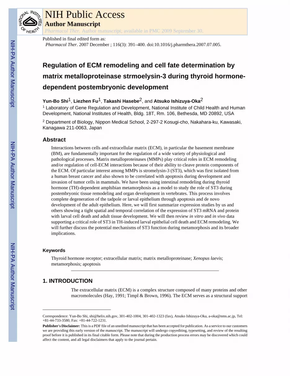
Regulation of ECM remodeling and cell fate determination bymatrix metalloproteinase strmoelysin-3 during thyroid hormone-dependent postembryonic development
Yun-Bo Shi1, Liezhen Fu1, Takashi Hasebe2, and Atsuko Ishizuya-Oka21 Laboratory of Gene Regulation and Development, National Institute of Child Health and HumanDevelopment, National Institutes of Health, Bldg. 18T, Rm. 106, Bethesda, MD 20892, USA2 Department of Biology, Nippon Medical School, 2-297-2 Kosugi-cho, Nakahara-ku, Kawasaki,Kanagawa 211-0063, Japan
AbstractInteractions between cells and extracellular matrix (ECM), in particular the basement membrane(BM), are fundamentally important for the regulation of a wide variety of physiological andpathological processes. Matrix metalloproteinases (MMPs) play critical roles in ECM remodelingand/or regulation of cell-ECM interactions because of their ability to cleave protein components ofthe ECM. Of particular interest among MMPs is stromelysin-3 (ST3), which was first isolated froma human breast cancer and also shown to be correlated with apoptosis during development andinvasion of tumor cells in mammals. We have been using intestinal remodeling during thyroidhormone (TH)-dependent amphibian metamorphosis as a model to study the role of ST3 duringpostembryonic tissue remodeling and organ development in vertebrates. This process involvescomplete degeneration of the tadpole or larval epithelium through apoptosis and de novodevelopment of the adult epithelium. Here, we will first summarize expression studies by us andothers showing a tight spatial and temporal correlation of the expression of ST3 mRNA and proteinwith larval cell death and adult tissue development. We will then review in vitro and in vivo datasupporting a critical role of ST3 in TH-induced larval epithelial cell death and ECM remodeling. Wewill further discuss the potential mechanisms of ST3 function during metamorphosis and its broaderimplications.
KeywordsThyroid hormone receptor; extracellular matrix; matrix metalloproteinase; Xenopus laevis;metamorphosis; apoptosis
1. INTRODUCTIONThe extracellular matrix (ECM) is a complex structure composed of many proteins and othermacromolecules (Hay, 1991; Timpl & Brown, 1996). The ECM serves as a structural support
Correspondence: Yun-Bo Shi, [email protected], 301-402-1004, 301-402-1323 (fax), Atsuko Ishizuya-Oka, [email protected], Tel:+81-44-733-3580, Fax: +81-44-722-1231.Publisher's Disclaimer: This is a PDF file of an unedited manuscript that has been accepted for publication. As a service to our customerswe are providing this early version of the manuscript. The manuscript will undergo copyediting, typesetting, and review of the resultingproof before it is published in its final citable form. Please note that during the production process errors may be discovered which couldaffect the content, and all legal disclaimers that apply to the journal pertain.
NIH Public AccessAuthor ManuscriptPharmacol Ther. Author manuscript; available in PMC 2009 September 30.
Published in final edited form as:Pharmacol Ther. 2007 December ; 116(3): 391–400. doi:10.1016/j.pharmthera.2007.07.005.
NIH
-PA Author Manuscript
NIH
-PA Author Manuscript
NIH
-PA Author Manuscript

media for the cells that it surrounds and is essential for maintaining the three-dimensionalstructure of the tissue or organ. The ECM can also affect cell function by interacting directlywith cells through cell surface ECM receptors, especially the integrins (Brown & Yamada,1995; Damsky & Werb, 1992; Schmidt et al., 1993) or by modulating the availability and levelsof many signaling molecules such as growth factors that are stored in the ECM (Vukicevic etal., 1992; Werb et al., 1996). Since most cells in multicellular organisms such as vertebratesare surrounded by ECM, alterations in the ECM can influence cell fate and behavior byaffecting cell-cell and cell-ECM interactions (Hay, 1991; Ruoslahti & Reed, 1994). The criticalroles of ECM are reflected by the fact that ECM degradation and remodeling always accompanytissue remodeling, organogenesis, and pathogenesis throughout the animal kingdom.
ECM remodeling and degradation is largely due to the proteolytic action of matrixmetalloproteinases (MMPs). MMPs are extracellular or membrane-bound enzymes (Alexander& Werb, 1991; Barrett et al., 1998; Birkedal-Hansen et al., 1993; McCawley & Matrisian,2001; Nagase, 1998; Parks & Mecham, 1998; Pei, 1999). This large family of proteolyticenzymes can be divided into 5 subfamilies, collagenases, gelatinases, stromelysins, membrane-type MMPs, and others. MMPs consist of multiple domains, including from the N to C-terminusthe pre- and pro-peptides, a catalytic domain, a hinge region, and in most MMPs, thehemopexin-like domain. In addition, gelatinase A (MMP2) and B (MMP9) also contain thefibronectin-like domain within the catalytic domain, and membrane type-MMPs have atransmembrane or membrane-attachment domain in the C-terminus (Fig. 1). MMPs aresynthesized as preenzymes and the pre-peptide is cleaved upon secretion into the ECM asproenzymes with a few exceptions such as ST3 and membrane type-MMPs. The pro-enzymesare enzymatically inactive and are activated extracellularly through proteolytic removal of thepropeptide (Barrett et al., 1998; Birkedal-Hansen et al., 1993; Kleiner & Stetler-Stevenson,1993; Murphy et al., 1999; Nagase, 1998; Nagase et al., 1992). ST3 and membrane type-MMPsare activated intracellularly through a furin-dependent process and are thus secreted or attachedto the plasma membrane as the mature enzymes, respectively (Fig. 1) (McCawley & Matrisian,2001; Pei & Weiss, 1995; Sato & Seiki, 1996). The mature or activated MMPs have differentbut often overlapping substrate specificities (Barrett et al., 1998; McCawley & Matrisian,2001; Overall, 2002; Uria & Werb, 1998). Collectively, they are capable of cleaving all proteincomponents of the extracellular matrix. In addition, MMPs are capable of degrading non-ECMextracellular or membrane-bound proteins such as pro-MMPs, pro-growth factors, proteinaseinhibitors, and cell surface proteins, etc. (Barrett et al., 1998; McCawley & Matrisian, 2001;Overall, 2002; Uria & Werb, 1998). Thus, MMPs may influence cell behavior and tissuedevelopment through both ECM remodeling and other mechanisms.
We have been using frog metamorphosis as a model to study MMP function in vivo. Frogdevelopment takes place in two phases. Its embryogenesis produces a free living, aquatic, andoften herbivorous tadpole. After a growth period, the tadpole undergoes metamorphosis thatchanges essentially every organ of the animal to produce, in vast majority of the cases, aterrestrial carnivorous frog in a process that is entirely dependent upon thyroid hormone (TH)(Shi, 1999). Molecular analysis has revealed that a number of MMP genes are induced by THeither directly or indirectly during metamorphosis. They include Rana catesbeiana collagenase1 (MMP1) (Oofusa et al., 1994), Xenopus laevis stromelysin-3 (ST3 or MMP11), gelatinaseA (MMP2), gelatinase B (MMP9), collagenase 3 (MMP 13) and 4 (MMP18), membranetype-1-MMP1 (MMP14) (Fujimoto et al., 2006; Hasebe et al., 2006; Jung et al., 2002; Pattertonet al., 1995; Shi & Brown, 1993; Stolow et al., 1996; Wang & Brown, 1993). Among them,ST3 is the only one that has been demonstrated to play an important role during metamorphosis.Here, we will review some of the studies that led to the conclusion and discuss potentialmechanisms by which ST3 regulates cell fate and behaviour during development.
Shi et al. Page 2
Pharmacol Ther. Author manuscript; available in PMC 2009 September 30.
NIH
-PA Author Manuscript
NIH
-PA Author Manuscript
NIH
-PA Author Manuscript
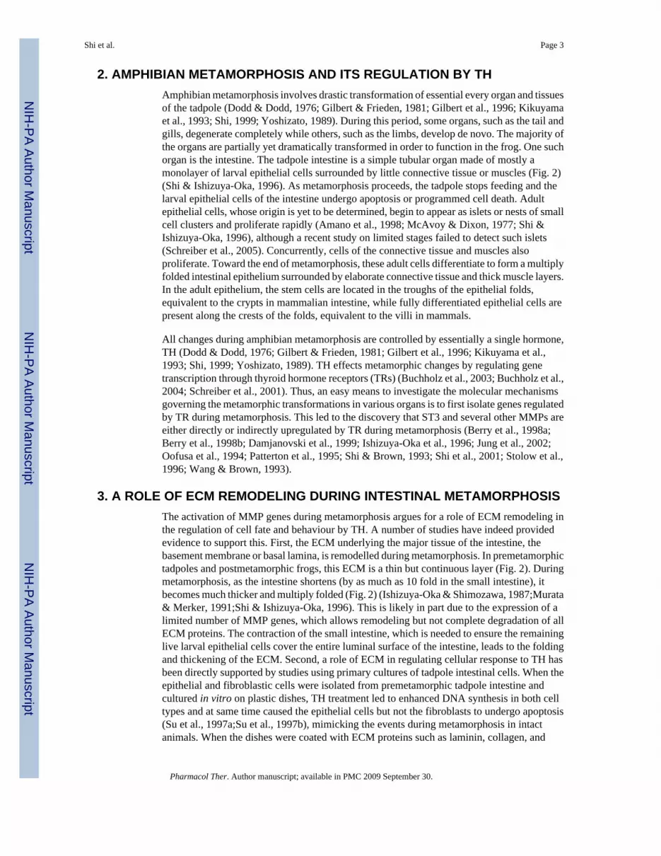
2. AMPHIBIAN METAMORPHOSIS AND ITS REGULATION BY THAmphibian metamorphosis involves drastic transformation of essential every organ and tissuesof the tadpole (Dodd & Dodd, 1976; Gilbert & Frieden, 1981; Gilbert et al., 1996; Kikuyamaet al., 1993; Shi, 1999; Yoshizato, 1989). During this period, some organs, such as the tail andgills, degenerate completely while others, such as the limbs, develop de novo. The majority ofthe organs are partially yet dramatically transformed in order to function in the frog. One suchorgan is the intestine. The tadpole intestine is a simple tubular organ made of mostly amonolayer of larval epithelial cells surrounded by little connective tissue or muscles (Fig. 2)(Shi & Ishizuya-Oka, 1996). As metamorphosis proceeds, the tadpole stops feeding and thelarval epithelial cells of the intestine undergo apoptosis or programmed cell death. Adultepithelial cells, whose origin is yet to be determined, begin to appear as islets or nests of smallcell clusters and proliferate rapidly (Amano et al., 1998; McAvoy & Dixon, 1977; Shi &Ishizuya-Oka, 1996), although a recent study on limited stages failed to detect such islets(Schreiber et al., 2005). Concurrently, cells of the connective tissue and muscles alsoproliferate. Toward the end of metamorphosis, these adult cells differentiate to form a multiplyfolded intestinal epithelium surrounded by elaborate connective tissue and thick muscle layers.In the adult epithelium, the stem cells are located in the troughs of the epithelial folds,equivalent to the crypts in mammalian intestine, while fully differentiated epithelial cells arepresent along the crests of the folds, equivalent to the villi in mammals.
All changes during amphibian metamorphosis are controlled by essentially a single hormone,TH (Dodd & Dodd, 1976; Gilbert & Frieden, 1981; Gilbert et al., 1996; Kikuyama et al.,1993; Shi, 1999; Yoshizato, 1989). TH effects metamorphic changes by regulating genetranscription through thyroid hormone receptors (TRs) (Buchholz et al., 2003; Buchholz et al.,2004; Schreiber et al., 2001). Thus, an easy means to investigate the molecular mechanismsgoverning the metamorphic transformations in various organs is to first isolate genes regulatedby TR during metamorphosis. This led to the discovery that ST3 and several other MMPs areeither directly or indirectly upregulated by TR during metamorphosis (Berry et al., 1998a;Berry et al., 1998b; Damjanovski et al., 1999; Ishizuya-Oka et al., 1996; Jung et al., 2002;Oofusa et al., 1994; Patterton et al., 1995; Shi & Brown, 1993; Shi et al., 2001; Stolow et al.,1996; Wang & Brown, 1993).
3. A ROLE OF ECM REMODELING DURING INTESTINAL METAMORPHOSISThe activation of MMP genes during metamorphosis argues for a role of ECM remodeling inthe regulation of cell fate and behaviour by TH. A number of studies have indeed providedevidence to support this. First, the ECM underlying the major tissue of the intestine, thebasement membrane or basal lamina, is remodelled during metamorphosis. In premetamorphictadpoles and postmetamorphic frogs, this ECM is a thin but continuous layer (Fig. 2). Duringmetamorphosis, as the intestine shortens (by as much as 10 fold in the small intestine), itbecomes much thicker and multiply folded (Fig. 2) (Ishizuya-Oka & Shimozawa, 1987;Murata& Merker, 1991;Shi & Ishizuya-Oka, 1996). This is likely in part due to the expression of alimited number of MMP genes, which allows remodeling but not complete degradation of allECM proteins. The contraction of the small intestine, which is needed to ensure the remaininglive larval epithelial cells cover the entire luminal surface of the intestine, leads to the foldingand thickening of the ECM. Second, a role of ECM in regulating cellular response to TH hasbeen directly supported by studies using primary cultures of tadpole intestinal cells. When theepithelial and fibroblastic cells were isolated from premetamorphic tadpole intestine andcultured in vitro on plastic dishes, TH treatment led to enhanced DNA synthesis in both celltypes and at same time caused the epithelial cells but not the fibroblasts to undergo apoptosis(Su et al., 1997a;Su et al., 1997b), mimicking the events during metamorphosis in intactanimals. When the dishes were coated with ECM proteins such as laminin, collagen, and
Shi et al. Page 3
Pharmacol Ther. Author manuscript; available in PMC 2009 September 30.
NIH
-PA Author Manuscript
NIH
-PA Author Manuscript
NIH
-PA Author Manuscript

fibronectin, TH-induced epithelial cell death was inhibited (Su et al., 1997b). Although theresult does not necessarily indicate that these ECM proteins play a protective role against TH-induced cell death in vivo, they do support the view that ECM remodeling will alter cell-ECMinteractions and thus influence epithelial cells’ response to TH.
4. MOLECULAR AND BIOCHEMICAL PROPERTIES OF STROMELYSIN-3ST3 (MMP11) was originally isolated as a breast cancer associated gene (Basset et al., 1990).While its primary structure resembles most MMPs, ST3 has several interesting and uniqueproperties. First, it is one of the very few MMPs that are secreted directly in the activated ormature form due to intracellular activation through the furin recognition motif (RXKR) in thepropeptide (Fig. 1) (Pei & Weiss, 1995). Second, in vitro, ST3 has only a very weak activitytoward ECM proteins compared to other MMPs, although it has a strong activity against non-ECM proteins α1-proteinase inhibitor and IGFBP-1 (Manes et al., 1997; Murphy et al., 1993;Pei et al., 1994). Third, the ST3 gene appears to have evolved differently as it has a uniqueintron/exon organization in the hemopexin-like domain that is conserved from human (Anglardet al., 1995), mouse (Ludwig et al., 2000), to frog (Li et al., 1998). The hinge region and thehemopexin-like domain are encoded by 4 exons in ST3 instead of 6 in other MMPs. Finally,it has been reported that alternative splicing and promoter usage of the human ST3 genegenerate a transcript encoding an intracellular active MMP (Lu et al., 2002). These findingssuggest that ST3 have distinct functions compared to other MMPs.
5. DEVELOPMENTAL AND THYROID HORMONE-DEPENDENT REGULATIONOF STROMELYSIN-3 EXPRESSION
ST3 was the first Xenopus laevis MMP gene that was isolated as a TH response gene throughsubtractive screening in both the intestine and tail (Patterton et al., 1995; Shi & Brown,1993; Wang & Brown, 1993). Subsequently, several other MMPs, including collagenase-3 and-4, gelatinase-A and -B, and membrane type 1-MMP, have also been shown to be upregulatedduring intestinal remodeling and/or tail resorption in Xenopus laevis (Fujimoto et al., 2006;Hasebe et al., 2006; Jung et al., 2002; Patterton et al., 1995; Stolow et al., 1996; Wang & Brown,1993). Interestingly, among these MMPs, only ST3 and one of the two gelatinase-B genes havebeen shown to be directly upregulated by TH through TRs as its upregulation by TH isindependent of new protein synthesis (Fujimoto et al., 2006; Patterton et al., 1995; Shi &Brown, 1993; Wang & Brown, 1993), although collagenase 1 gene in Rana catesbeiana is alsoa direct target genes of TR (Oofusa & Yoshizato, 1996). Promoter analysis has revealed thatindeed Rana collagenase 1, Xenopus ST3, and one of the two gelatinase B genes contain THresponse elements in their promoter regulatory regions (Fu et al., 2006; Fujimoto et al.,2006; Li et al., 1998; Oofusa & Yoshizato, 1996).
Consistent with this regulation mechanism, ST3 is the first one to be upregulated in the intestineduring natural metamorphosis with its mRNA levels upregulated before the onset of epithelialcell death in the intestine (by stage 58), while the other MMPs are upregulated at or after stage60 when cell death has begun (Hasebe et al., 2006; Patterton et al., 1995; Stolow et al., 1996).Its mRNA level remains high at the climax of metamorphosis when larval cells die throughapoptosis and adult cells proliferate. By stage 64, when larval epithelial cell death is completedand adult epithelial cell differentiation takes place, there is little ST3 mRNA present in theintestine. In contrast, the expression of collagenases 3 and 4, gelatinase A, and membrane type1-MMP is upregulated later and to a lower degree, The only exception is one of the twogelatinase B genes, which has similar temporal and TH-dependent regulation as ST3 (Fujimotoet al., 2006).
Shi et al. Page 4
Pharmacol Ther. Author manuscript; available in PMC 2009 September 30.
NIH
-PA Author Manuscript
NIH
-PA Author Manuscript
NIH
-PA Author Manuscript

The expression of ST3 is also correlated spatially with cell death during metamorphosis. ItsmRNA and protein have been shown to be expressed in the fibroblasts of the connective tissueunderlying the larval epithelium in the metamorphosing intestine (Ishizuya-Oka et al., 1996;Ishizuya-Oka et al., 2000; Patterton et al., 1995), similar to other MMPs (Damjanovski et al.,1999; Hasebe et al., 2006). In addition, high levels of ST3 mRNA are also present in theconnective tissue underlying the apoptotic skin epidermis and surrounding the dying musclecells in the resorbing tail (Berry et al., 1998b; Damjanovski et al., 1999). These results suggestthat ST3 may play a role in cell fate regulation by facilitating TH-induced larval epithelial celldeath, while most other MMPs may have a role after cell death has begun, such as removal orremodeling the ECM around the dying cells.
6. A ROLE OF ST3 IN ECM REMODELING AND CELL FATE DETERMINATIONTo investigate whether ST3 indeed plays a role during intestinal remodeling, we first made useof the ability to induce intestinal remodeling in organ cultures of premetamorphic intestine bysimply adding physiological concentrations of TH (Ishizuya-Oka & Shimozawa, 1991) andthe fact that ST3 is a secreted protease, whose function may thus be blocked with inhibitorsadded to the organ culture medium. When a synthetic MMP inhibitor that can inhibit multipleMMPs was added to the organ culture medium, it inhibited TH-induced larval epithelial celldeath (Ishizuya-Oka et al., 2000). To specifically inhibit ST3 function, we generated apolyclonal antibody against the catalytic domain of Xenopus laevis ST3 and found that thisantibody could block the cleavage of an in vitro substrate by ST3 (Amano et al., 2004). Whenthis functional blocking polyclonal antibody was added to the organ culture medium, it tooinhibited TH-induced epithelial cell death when assayed after 3 days of culturing (Ishizuya-Oka et al., 2000). Interestingly, at this time point, ECM remodeling induced by TH was alsoinhibited. These results suggest that ST3 plays a critical role in both ECM remodeling andlarval epithelial cell death. After 5 days of culturing, adult epithelial cells proliferated as clustersof cells or islets that expanded three dimensionally in TH-treated organ cultures in the absenceof the anti-ST3 antibody. The anti-ST3 antibody was able to block the expansion of theproliferating adult epithelial islets into the connective tissue. Instead, these TH-induced adultepithelial islets expanded only laterally along the epithelium-connective tissue interface. Thus,ST3 function appears to be critical for ECM remodeling, larval cell death, and adult cellmigration but not for adult epithelial cell proliferation (Ishizuya-Oka et al., 2000).
Currently, it is impossible to carry out gene knockout or knockdown studies in tadpoles, makingit difficult to assess the effect of ST3 deletion on metamorphosis. On the other hand, exogenousgenes can be precociously expressed in developing tadpole through transgenesis (Kroll &Amaya, 1996). This offers an opportunity to investigate whether MMPs can affect developmentin vivo. Transgenic expression of Xenopus laevis ST3 and collagenase-4 as well as amammalian membrane-type-MMP under the control of the constitutive promoter CMV causeslethality during late embryonic and early tadpole stages (Damjanovski et al., 2001). To studythe effects of ST3 during metamorphosis, we therefore developed a double promoter constructto express ST3 under the control of a heat shock-inducible promoter (Fu et al., 2002) and atthe same time express the green fluorescent protein (GFP) in the tadpole eyes under the controlof a second promoter, the γ-crystallin promoter. This allowed us to rear wild type and transgenictadpoles together to ensure no variation in animal growth and treatment conditions betweenthe two groups and later identify the transgenic ones by simply examining their eyes under afluorescent dissecting microscope. The expression of transgenic ST3 was then induced bysimply treating the animals with heat shock. In agreement with the transgenic studies using theCMV promoter to drive the expression of ST3, induction of ST3 expression upon heat shockduring early embryonic stages also led to lethality (Fu et al., 2002). Interestingly, induction ofST3 at tadpole stages (between stage 45 to 54) for up to a few weeks had little effect on overallgrowth and development (Fu et al., 2005), indicating, perhaps not surprisingly, that proper
Shi et al. Page 5
Pharmacol Ther. Author manuscript; available in PMC 2009 September 30.
NIH
-PA Author Manuscript
NIH
-PA Author Manuscript
NIH
-PA Author Manuscript

regulation of MMP function is not as critical for tadpole organ/tissue function during animalgrowth period as for organogenesis and tissue remodeling during embryogenesis. On the otherhand, analysis of the intestine of tadpoles at stage 54 after 4 days of heat shock treatment (about1 hr heat shock treatment per day) revealed that transgenic overexpression of the wild typeST3 induced larval epithelial apoptosis (Fu et al., 2005). This was accompanied by theactivation of fibroblasts (i.e., with enriched rough endoplasmic reticulum) and cell-cell contactsbetween epithelial cells and fibroblasts (Fig. 3). These changes are similar to those observedduring natural metamorphosis. They were not present in heat shock-treated non-transgenicanimals or transgenic animals containing a catalytically inactive ST3 due to a point mutationin the catalytic domain. They were also absent in both wild type and transgenic animals withoutheat shock treatments. Aside from these changes in the major cell types of the intestine, thebasal lamina separating the epithelium and connective tissue was also altered by the transgenicexpression of ST3 but not the mutant ST3 (Fig. 3). In some areas, including areas whereepithelial cells and fibroblasts contact, the basal lamina was thicker and in other areas, the basallamina appeared to be absent. This contrasts to the thick, multiply folded basal lamina observedessentially in all sections of the small intestine during metamorphosis (Fig. 3). In addition,adult epithelial cell islets were not observed in ST3 expressing transgenic animals. Thesefindings suggest that ST3 expression alone is sufficient to induce some but not all TH-inducedmetamorphic program in the intestine (Fu et al., 2005), a conclusion clearly not surprisinggiven that ST3 is only one of the many direct TR target genes that are normally induced by THduring intestinal remodeling. Nonetheless, the in vitro and in vivo studies complement eachother and support a critical role of ST3 in ECM remodeling and larval epithelial cell deathduring intestinal metamorphosis.
7. MECHANISMS OF ST3 FUNCTION DURING METAMORPHOSISAs indicated above, ST3 has distinct molecular and biochemical properties. Compared toessentially all other MMPs, ST3 has much weaker activities toward known ECM proteins butmuch higher activities toward a few non-ECM proteins such as α1-protease inhibitor, at leastin vitro (Manes et al., 1997; Murphy et al., 1993; Pei et al., 1994). Although it is possible thatthe in vivo environment may enable ST3 to cleave ECM proteins just as efficient as otherMMPs, the in vitro studies suggest that ST3 may likely function by cleaving non-ECM proteins.As the first step of ST3 function is the binding to its substrate, we carried out a yeast-two-hybrid screen to isolate proteins that bind to ST3. This led to the identification of the 67 kdlaminin receptor (LR) as a potential substrate of ST3 (Amano et al., 2005a; Amano et al.,2005b). LR is synthesized as a 37 kd precursor protein. The vast majority of the precursorproteins are localized intracellularly and may be involved in translational regulation (Ardiniet al., 1998; Auth & Brawerman, 1992). A small fraction of the precursor proteins are insertedin the plasma membrane and become the 67 kd LRs through possibly heterodimerization and/or posttranslational modifications (Buto et al., 1998; Castronovo et al., 1991; Landowski et al.,1995; Malinoff & Wicha, 1983; Menard et al., 1997; Rao et al., 1983; Starkey et al., 1999) (forsimplicity, both the precursor and membrane bound form are referred as LR thereafter). Invitro, purified Xenopus ST3 catalytic domain can cleave E. coli produced, full length Xenopuslaevis LR efficiently, with the activity slightly lower than that of the cleavage of α1-proteaseinhibitor by ST3 (Amano et al., 2005b). Importantly, the cleavage occurs at two sites locatedbetween the transmembrane domain and laminin binding sequence in the extracellular half ofthe protein (Fig. 4) (Amano et al., 2005b). Furthermore, several other MMPs cleave LR outsideof the region between the transmembrane domain and laminin binding sequence (Amano etal., 2005b), suggesting that ST3 may be unique in its ability to cleave LR to alter cell-laminininteraction.
To investigate if LR is cleaved by ST3 during intestinal metamorphosis, we have analyzed theexpression of LR mRNA and protein during metamorphosis. Both LR mRNA and protein are
Shi et al. Page 6
Pharmacol Ther. Author manuscript; available in PMC 2009 September 30.
NIH
-PA Author Manuscript
NIH
-PA Author Manuscript
NIH
-PA Author Manuscript
expressed fairly constantly in the intestine during development (Amano et al., 2005a), incontrast to the metamorphosis-specific expression of ST3. Interestingly, LR fragments of thesizes expected from ST3 cleavage are present in the intestine at the climax of metamorphosiswhen ST3 is highly expressed, whereas little such fragments are present in pre- or post-metamorphic intestine (Fig. 5) (Amano et al., 2005a). More importantly, transgenic expressionof ST3 in premetamorphic tadpoles leads to the formation of these fragments in the absenceof TH (Fig. 5) (Amano et al., 2005a). These results suggest that LR is an in vivo substrate ofST3 during intestinal remodeling.
Although LR is constitutively expressed during intestinal remodeling, immunohistochemicalanalysis has revealed that LR is highly expressed in the differentiated epithelial cells inpremetamorphic intestine with little expression in the connective tissue (Fig. 6) (Amano et al.,2005a). As intestinal metamorphosis proceeds, LR expression increases in the connective tissuebut decreases in the dying larval epithelial cells. There is no detectable signal in the proliferatingadult epithelial cells. As these cells differentiate, they begin to express LR. These resultssuggest that LR expression on the cell surface may be important for the survival and/ormaintenance of the differentiated state of the intestinal epithelial cells. Thus, one mechanismof ST3 function may be that the cleavage of LR on larval epithelial cells by ST3 reducesepithelial cell-ECM interaction, thus facilitating larval epithelial cell death during intestinalmetamorphosis. Unlike the monolayer differentiated epithelial cells, the undifferentiated adultepithelial cells are not associated with the basal lamina and thus do not require LR expression.Consequently, these cells are not affected by the highly expressed ST3. The high levels of LRexpression in the fibroblasts of the connective tissue during metamorphosis may be related tothe role of intracellular LR in protein synthesis as the fibroblasts are highly activated withextremely rich rough endoplasmic reticulum, where abundant amount of actively translatingribosomes are present.
8. CONCLUSIONSThe MMP stromelysin-3 shares many similarities with other MMPs but also has several uniqueproperties. It has little expression in adult organs under normal physiological conditions but isupregulated in a number of pathological processes such as tumor invasion (Ahmad et al.,1998; Anderson et al., 1995; Basset et al., 1997; Chenard et al., 1996; Lochter & Bissell,1999; Muller et al., 1993; Schonbeck et al., 1999; Tetu et al., 1998) and may also be involvedin tumor initiation (Masson et al., 1998; Noel et al., 2000; Noel et al., 1997). During mammaliandevelopment, ST3 is expressed in many processes involving cell migration and tissuemorphogenesis, especially where cell death takes place (Alexander et al., 1996; Basset et al.,1990; Chin & Werb, 1997; Lefebvre et al., 1992; Lefebvre et al., 1995; Uria & Werb, 1998),but a physiological role of ST3 has yet to be shown in vivo due largely to the difficulty tomanipulate mammalian development and separate the influence of the mother. The ability tomanipulate frog metamorphosis and the external development of the tadpoles have allowed thefirst demonstration that ST3 is both necessary and sufficient at least for some aspects ofintestinal metamorphosis. Substrate isolation and analysis has revealed that one potentialmechanism for ST3 to exert its effects on intestinal remodeling is to cleave LR on the larvalepithelial cells.
MMPs have long attracted the attention of researchers because of the high levels of MMPexpression in cancerous but not normal tissues, raising the possibility of targeting MMPfunction for cancer treatment. Unfortunately, human clinical trials using MMP inhibitors haveyet to produce any positive results (Coussens et al., 2002). This might be due to the fact theECM remodeling/degradation is not the determining factor in tumor invasion and metastasis,since metastasis occurs after secondary genetic changes take place in the tumor cells. In theabsence of sufficient MMP activity, tumor cells may find other means, e.g., employing other
Shi et al. Page 7
Pharmacol Ther. Author manuscript; available in PMC 2009 September 30.
NIH
-PA Author Manuscript
NIH
-PA Author Manuscript
NIH
-PA Author Manuscript
proteases, to get through the ECM barrier in order to migrate into other tissues/organs.Additional analysis of the in vivo roles of MMPs and the associated mechanisms shouldfacilitate the development of better means for target MMPs in disease prevention and treatment.The similarity of many processes during metamorphosis to processes during postembryonicdevelopment and tissue remodeling in mammals make metamorphosis a highly valuable modelfor this purpose. In this regard, LR is highly conserved from frog to human and both humanand Xenopus LR can be cleaved by Xenopus ST3 catalytic domain at the same two conservedsites located between the transmembrane domain and laminin binding sequence in theextracellular half of the protein (Fig. 4) (Amano et al., 2005b). In addition, LR is highlyexpressed in cancers (Castronovo, 1993; Hand et al., 1985; Martignone et al., 1993; Menardet al., 1997; Sanjuan et al., 1996; Sobel, 1993; Yow et al., 1988), raising the possibility thatLR cleavage by ST3 may also play a role in cancer development and metastasis. Clearly, furtherstudies of ST3 function in both frog and mammalian models are needed to fully appreciate theimportance of this interesting gene and to possibly target it for human health.
AcknowledgmentsThis research was supported in part by the Intramural Research Program of the National Institute of Child Health andHuman Development, NIH.
AbbreviationsTH
Thyroid hormone
TR Thyroid hormone receptor
MMP matrix metalloproteinase
ST3 stromelysin-3
ECM extracellular matrix
LR laminin receptor
ReferencesAhmad A, Hanby A, Dublin E, Poulsom R, Smith P, Barnes D, Rubens R, Anglard P, Hart I. Stromelysin
3: an independent prognostic factor for relapse-free survival in node-positive breast cancer anddemonstration of novel breast carcinoma cell expression. Am J Pathol 1998;152:721–8. [PubMed:9502414]
Alexander, CM.; Werb, Z. Extracellular matrix degradation. In: Hay, ED., editor. Cell Biology ofExtracellular Matrix. New York: Plenum Press; 1991. p. 255-302.
Alexander CM, Hansell EJ, Behrendtsen O, Flannery ML, Kishnani NS, Hawkes SP, Werb Z. Expressionand function of matrix metalloproteinases and their inhibitors at the maternal-embryonic boundarydurng mouse embryo implantation. Development 1996;122:1723–1736. [PubMed: 8674412]
Amano T, Noro N, Kawabata H, Kobayashi Y, Yoshizato K. Metamorphosis-associated and region-specific expression of calbindin gene in the posterior intestinal epithelium of Xenopus laevis larva.Dev Growth Differ 1998;40:177–88. [PubMed: 9572360]
Shi et al. Page 8
Pharmacol Ther. Author manuscript; available in PMC 2009 September 30.
NIH
-PA Author Manuscript
NIH
-PA Author Manuscript
NIH
-PA Author Manuscript
Amano T, Fu L, Sahu S, Markey M, Shi YB. Substrate specificity of Xenopus matrix metalloproteinasestromelysin-3. International Journal of Molecular Medicine 2004;14:233–239. [PubMed: 15254771]
Amano T, Fu L, Marshak A, Kwak O, Shi YB. Spatio-temporal regulation and cleavage by matrixmetalloproteinase stromelysin-3 implicate a role for laminin receptor in intestinal remodeling duringXenopus laevis metamorphosis. Dev Dyn 2005a;234:190–200. [PubMed: 16059908]
Amano T, Kwak O, Fu L, Marshak A, Shi YB. The matrix metalloproteinase stromelysin-3 cleaveslaminin receptor at two distinct sites between the transmembrane domain and laminin binding sequencewithin the extracellular domain. Cell Research 2005b;15:150–159. [PubMed: 15780176]
Anderson IC, Sugarbaker DJ, Ganju RK, Tsarwhas DG, Richards WG, Sunday M, Kobzik L, Shipp MA.Stromelysin-3 is overexpressed by stromal elements in primary non-small-cell lung cancers andregulated by retinoic acid in pulmonary fibroblasts. Cancer Res 1995;55:4120–4126. [PubMed:7664289]
Anglard P, Melot T, Guerin E, Thomas G, Basset P. Structure and Promoter Characterization of theHuman Stromelysin-3 Gene. J Biol Chem 1995;270:20337–20344. [PubMed: 7657606]
Ardini E, Pesole G, Tagliabue E, Magnifico A, Castronovo V, Sobel ME, Colnaghi MI, Menard S. The67-kDa laminin receptor originated from a ribosomal protein that acquired a dual function duringevolution. Mol Biol Evol 1998;15:1017–1025. [PubMed: 9718729]
Auth D, Brawerman G. A 33-kDa Polypeptide with Homology to the Laminin Receptor: Component ofTranslation Machinery. PNAS 1992;89:4368–4372. [PubMed: 1374897]
Barrett, JA.; Rawloings, ND.; Woessner, JF. Handbook of proteolytic enzymes. NY: Academic Press;1998.
Basset P, Bellocq JP, Wolf C, Stoll I, Hutin P, Limacher JM, Podhajcer OL, Chenard MP, Rio MC,Chambon P. A novel metalloproteinase gene specifically expressed in stromal cells of breastcarcinomas. Nature 1990;348:699–704. [PubMed: 1701851]
Basset P, Bellocq JP, Lefebvre O, Noel A, Chenard MP, Wolf C, Anglard P, Rio MC. Stromelysin-3: aparadigm for stroma-derived factors implicated in carcinoma progression. Crit Rev Oncol Hematol1997;26:43–53. [PubMed: 9246540]
Berry DL, Rose CS, Remo BF, Brown DD. The expression pattern of thyroid hormone response genesin remodeling tadpole tissues defines distinct growth and resorption gene expression programs. DevBiol 1998a;203:24–35. [PubMed: 9806770]
Berry DL, Schwartzman RA, Brown DD. The expression pattern of thyroid hormone response genes inthe tadpole tail identifies multiple resorption programs. Dev Biol 1998b;203:12–23. [PubMed:9806769]
Birkedal-Hansen H, Moore WGI, Bodden MK, Windsor LT, Birkedal-Hansen B, DeCarlo A, Engler JA.Matrix metalloproteinases: a review. Crit Rev in Oral Biol and Med 1993;4:197–250. [PubMed:8435466]
Brown KE, Yamada KM. The role of integrins during vertebrate development. Seminars in Develop Biol1995;6:69–77.
Buchholz DR, Hsia VSC, Fu L, Shi YB. A dominant negative thyroid hormone receptor blocks amphibianmetamorphosis by retaining corepressors at target genes. Mol Cell Biol 2003;23:6750–6758.[PubMed: 12972595]
Buchholz DR, Tomita A, Fu L, Paul BD, Shi YB. Transgenic analysis reveals that thyroid hormonereceptor is sufficient to mediate the thyroid hormone signal in frog metamorphosis. Mol Cell Biol2004;24:9026–9037. [PubMed: 15456876]
Buto S, Tagliabue E, Ardini E, Magnifico A, Ghirelli C, van den Brûle F, Castronovo V, Colnaghi MI,Sobel ME, Ménard S. Formation of the 67-kDa laminin receptor by acylation of the precursor. Journalof Cellular Biochemistry 1998;69:244–251. [PubMed: 9581863]
Castronovo V, Taraboletti G, Sobel ME. Functional domains of the 67-kDa laminin receptor precursor.J Biol Chem 1991;266:20440–20446. [PubMed: 1834645]
Castronovo V. Laminin receptors and laminin-binding proteins during tunor invasion and matastasis.Invasion Metastasis 1993;13:1–30. [PubMed: 8407208]
Chenard MP, OSiorain L, Shering S, Rouyer N, Lutz Y, Wolf C, Basset P, Bellocq JP, Duffy MJ. Highlevels of stromelysin-3 correlate with poor prognosis in patients with breast carcinoma. InternationalJournal of Cancer 1996;69:448–451.
Shi et al. Page 9
Pharmacol Ther. Author manuscript; available in PMC 2009 September 30.
NIH
-PA Author Manuscript
NIH
-PA Author Manuscript
NIH
-PA Author Manuscript
Chin JR, Werb Z. Matrix metalloproteinases regulate morphogenesis, migration and remodeling ofepithelium, tongue skeleta muscle and cartilage in the mandibular arch. Development1997;124:1519–1530. [PubMed: 9108368]
Coussens LM, Fingleton BM, Matrisian LM. Matrix metalloproteinase inhibitors and cancer: Trials andtribulations. Science 2002;295:2387–2392. [PubMed: 11923519]
Damjanovski S, Ishizuya-Oka A, Shi YB. Spatial and temporal regulation of collagenases-3, -4, andstromelysin - 3 implicates distinct functions in apoptosis and tissue remodeling during frogmetamorphosis. Cell Res 1999;9:91–105. [PubMed: 10418731]
Damjanovski S, Amano T, Li Q, Pei D, Shi YB. Overexpression of Matrix Metalloproteinases Leads toLethality in Transgenic Xenopus Laevis: Implications for Tissue-Dependent Functions of MatrixMetalloproteinases during Late Embryonic development. Dev Dynamics 2001;221:37–47.
Damsky CH, Werb Z. Signal transduction by integrin receptors for extracellular matrix: cooperativeprocessing of extracellular information. Curr Opin Cell Biol 1992;4:772–81. [PubMed: 1329869]
Dodd, MHI.; Dodd, JM. The biology of metamorphosis. In: Lofts, B., editor. Physiology of the amphibia.New York: Academic Press; 1976. p. 467-599.
Fu L, Buchholz D, Shi YB. A novel double promoter approach for identification of transgenic animals:a tool for in vivo analysis of gene function and development of gene-based therapies. MolecularReproduction and Development 2002;62:470–476. [PubMed: 12112579]
Fu L, Ishizuya-Oka A, Buchholz DR, Amano T, Shi YB. A causative role of stromelysin-3 in ECMremodeling and epithelial apoptosis during intestinal metamorphosis in Xenopus laevis. J Biol Chem2005;280:27856–65. [PubMed: 15929979]
Fu L, Tomita A, Wang H, Buchholz DR, Shi YB. Transcriptional regulation of the Xenopus laevisstromelysin-3 gene by thyroid hormone is mediated by a DNA element in the first intron. J Biol Chem2006;281:16870–8. [PubMed: 16606608]
Fujimoto K, Nakajima K, Yaoita Y. One of the duplicated matrix metalloproteinase-9 genes is expressedin regressing tail during anuran metamorphosis. Develop Growth Differ 2006;48:223–241.
Gilbert, LI.; Frieden, E. Metamorphosis: A problem in developmental biology. Vol. 2. New York: PlenumPress; 1981.
Gilbert, LI.; Tata, JR.; Atkinson, BG. Metamorphosis: Post-embryonic reprogramming of geneexpression in amphibian and insect cells. New York: Academic Press; 1996.
Hand PH, Thor A, Schlom J, Rao CN, Liotta LA. Expression of laminin receptor in normal andcarcinomatous human tissues as defined by a monoclonal antibody. Cancer Res 1985;45:2713–2719.[PubMed: 3157447]
Hasebe T, Hartman R, Matsuda H, Shi YB. Spatial and temporal expression profiles suggest theinvolvement of gelatinase A and membrane type 1 matrix metalloproteinase in amphibianmetamorphosis. Cell Tissue Res 2006;324:105–16. [PubMed: 16418836]
Hay, ED. Cell Biology of Extracellular Matrix. Vol. 2. New York: Plenum Press; 1991.Ishizuya-Oka A, Shimozawa A. Ultrastructural changes in the intestinal connective tissue of Xenopus
laevis during metamorphosis. J Morphol 1987;193:13–22. [PubMed: 3612815]Ishizuya-Oka A, Shimozawa A. Induction of metamorphosis by thyroid hormone in anuran small intestine
cultured organotypically in vitro. In Vitro Cell Dev Biol 1991;27A:853–7. [PubMed: 1748625]Ishizuya-Oka A, Ueda S, Shi YB. Transient expression of stromelysin-3 mRNA in the amphibian small
intestine during metamorphosis. Cell Tissue Res 1996;283:325–9. [PubMed: 8593661]Ishizuya-Oka A, Li Q, Amano T, Damjanovski S, Ueda S, Shi YB. Requirement for matrix
metalloproteinase stromelysin-3 in cell migration and apoptosis during tissue remodeling in Xenopuslaevis. J Cell Biol 2000;150:1177–88. [PubMed: 10974004]
Jung JC, Leco KJ, Edwards DR, Fini ME. Matrix metalloproteinase mediate the dismantling ofmesenchymal structures in the tadpole tail during thyroid hormone-induced tail resorption. Dev Dyn2002;223:402–413. [PubMed: 11891989]
Kikuyama S, Kawamura K, Tanaka S, Yamamoto K. Aspects of amphibian metamorphosis: hormonalcontrol. Int Rev Cytol 1993;145:105–48. [PubMed: 8500980]
Kleiner DE Jr, Stetler-Stevenson WG. Structural biochemistry and activation of matrix metalloproteases.Curr Opin Cell Biol 1993;5:891–7. [PubMed: 8240832]
Shi et al. Page 10
Pharmacol Ther. Author manuscript; available in PMC 2009 September 30.
NIH
-PA Author Manuscript
NIH
-PA Author Manuscript
NIH
-PA Author Manuscript
Kroll KL, Amaya E. Transgenic Xenopus embryos from sperm nuclear transplantations reveal FGFsignaling requirements during gastrulation. Development 1996;122:3173–83. [PubMed: 8898230]
Landowski TH, Dratz EA, Starkey JR. Studies of the Structure of the Metastasis-Associated 67 kDaLaminin Binding Protein: Fatty Acid Acylation and Evidence Supporting Dimerization of the 32 kDaGene Product To Form the Mature Protein. Biochemistry 1995;34:11276–11287. [PubMed:7669786]
Lefebvre O, Wolf C, Limacher JM, Hutin P, Wendling C, LeMeur M, Basset P, Rio MC. The breastcancer-associated stromelysin-3 gene is expressed during mouse mammary gland apoptosis. J CellBiol 1992;119:997–1002. [PubMed: 1429845]
Lefebvre O, Regnier C, Chenard MP, Wendling C, Chambon P, Basset P, Rio MC. Developmentalexpression of mouse stromelysin-3 mRNA. Development 1995;121:947–55. [PubMed: 7743938]
Li J, Liang VCT, Sedgwick T, Wong J, Shi YB. Unique organization and involvement of GAGA factorsin the transcriptional regulation of the Xenopus stromelysin-3 gene. Nucl Acids Res 1998;26:3018–3025. [PubMed: 9611250]
Lochter A, Bissell MJ. An odyssey from breast to bone: multi-step control of mammary metastases andosteolysis by matrix metalloproteinases. APMIS 1999;107:128–36. [PubMed: 10190289]
Lu D, Mari B, Stoll I, Anglard P. Alternative splicing and promoter usage generates an intracellularstromelysin-3 isoform directly translated as an active matrix metalloproteinase. J Biol Chem2002;277:25527–25536. [PubMed: 12006591]
Ludwig MG, Basset P, Anglard P. Multiple regulator elements in the murine stromelysin-3 promoter:Evidence for direct control by CCAAT/enhancer-binding protein beta and thyroid and retinoidreceptors. J Biol Chem 2000;275:39981–39990. [PubMed: 10993903]
Malinoff HL, Wicha MS. Isolation of a cell surface receptor protein for laminin from murine fibrosarcomacells. J Cell Biol 1983;96:1475–1479. [PubMed: 6302102]
Manes S, Mira E, Barbacid MD, Cipres A, FernandezResa P, Buesa JM, Merida I, Aracil M, MarquezG, Martinez C. Identification of insulin-like growth factor-binding protein-1 as a potentialphysiological substrate for human stromelysin-3. J of Biol Chem 1997;272:25706–25712. [PubMed:9325295]
Martignone S, Menard S, Bufalino R, Cascinelli N, Pellegrini R, Tagliabue E, Andreola S, Rilke F,Colnaghi MI. Prognostic significance of the 67-kilodalton laminin receptor expression in humanbreast carcinomas. J Natl Cancer Inst 1993;85:398–402. [PubMed: 8433393]
Masson R, Lefebvre O, Noel A, Fahime ME, Chenard MP, Wendling C, Kebers F, LeMeur M, DierichA, Foidart JM, Basset P, Rio MC. In vivo evidence that the stromelysin-3 metalloproteinasecontributes in a paracrine manner to epithelial cell malignancy. J Cell Biol 1998;140:1535–41.[PubMed: 9508784]
McAvoy JW, Dixon KE. Cell proliferation and renewal in the small intestinal epithelium ofmetamorphosing and adult Xenopus laevis. J Exp Zool 1977;202:129–138.
McCawley LJ, Matrisian LM. Matrix metalloproteinases: they’re not just for matrix anymore! CurrentOpinion in Cell Biology 2001;13:534–540. [PubMed: 11544020]
Menard S, Castronovo V, Tagliabue E, Sobel ME. New insights into the metastasis-associated 67 kDlaminin receptor. Journal of Cellular Biochemistry 1997;67:155–165. [PubMed: 9328821]
Muller D, Wolf C, Abecassis J, Millon R, Engelmann A, Bronner G, Rouyer N, Rio MC, Eber M, MethlinG. Increased stromelysin 3 gene expression is associated with increased local invasiveness in headand neck squamous cell carcinomas. Cancer Res 1993;53:165–9. [PubMed: 7677979]
Murata E, Merker HJ. Morphologic changes of the basal lamina in the small intestine of Xenopus laevisduring metamorphosis. Acta Anat 1991;140:60–9. [PubMed: 2028731]
Murphy G, Segain JP, O’Shea M, Cockett M, Ioannou C, Lefebvre O, Chambon P, Basset P. The 28-kDa N-terminal domain of mouse stromelysin-3- has the general properties of a weakmetalloproteinase. J Biol Chem 1993;268:15435–15441. [PubMed: 8340372]
Murphy G, Stanton H, Cowell S, Butler G, Knauper V, Atkinson S, Gavrilovic J. Mechanisms for promatrix metalloproteinase activation. Apmis 1999;107:38–44. [PubMed: 10190278]
Nagase H. Cell surface activation of progelatinase A (proMMP-2) and cell migration. Cell Res1998;8:179–86. [PubMed: 9791731]
Shi et al. Page 11
Pharmacol Ther. Author manuscript; available in PMC 2009 September 30.
NIH
-PA Author Manuscript
NIH
-PA Author Manuscript
NIH
-PA Author Manuscript
Nagase J, Suzuki K, Morodomi T, Englhild JJ, Salvesen G. Activation Mechanisms of the Precursors ofMatrix Metalloproteinases 1, 2, and 3. Matrix Supple 1992;1:237–244.
Nieuwkoop, PD.; Faber, J. Normal table of Xenopus laevis. Vol. 1. Amsterdam: North HollandPublishing; 1956.
Noel A, Boulay A, Kebers F, Kannan R, Hajitou A, Calberg-Bacq CM, Basset P, Rio MC, Foidart JM.Demonstration in vivo that stromelysin-3 functions through its proteolytic activity. Oncogene2000;19:1605–1612. [PubMed: 10734321]
Noel AC, Lefebvre O, Maquoi E, VanHoorde L, Chenard MP, Mareel M, Foidart JM, Basset P, Rio MC.Stromelysin-3 expression promotes tumor take in nude mice. J Clin Invest 1997;97:1924–2930.[PubMed: 8621777]
Oofusa K, Yomori S, Yoshizato K. Regionally and hormonally regulated expression of genes of collagenand collagenase in the anuran larval skin. Int J Dev Biol 1994;38:345–50. [PubMed: 7981043]
Oofusa K, Yoshizato K. Thyroid hormone-dependent expression of bullfrog tadpole collagenase gene.Roux’s Arch Dev Biol 1996;205:241–251.
Overall CM. Molecular determinants of metalloproteinase substrate specificity. MolecularBiotechnology 2002;22:51–86. [PubMed: 12353914]
Parks, WC.; Mecham, RP. Matrix metalloproteinases. New York: Academic Press; 1998.Patterton D, Hayes WP, Shi YB. Transcriptional activation of the matrix metalloproteinase gene
stromelysin-3 coincides with thyroid hormone-induced cell death during frog metamorphosis. DevBiol 1995;167:252–62. [PubMed: 7851646]
Pei D, Majmudar G, Weiss SJ. Hydrolytic inactivation of a breast carcinoma cell-derived serpin by humanstromelysin-3. J Biol Chem 1994;269:25849–55. [PubMed: 7523394]
Pei D, Weiss SJ. Furin-dependent intracellular activation of the human stromelysin-3 zymogen. Nature1995;375:244–7. [PubMed: 7746327]
Pei D. Leukolysin/MMP25/MT6-MMP: a novel matrix metalloproteinase specifically expressed in theleukocyte lineage. Cell Res 1999;9:291–303. [PubMed: 10628838]
Rao NC, Barsky SH, Terranova VP, Liotta LA. Isolation of a tumor cell laminin receptor . BiochemBiophys Res Commun 1983;111:804–808. [PubMed: 6301485]
Ruoslahti E, Reed JC. Anchorage dependence, integrins, and apoptosis. Cell 1994;77:477–8. [PubMed:8187171]
Sanjuan X, Fernandez PL, Miquel R, Munoz J, Castronovo V, Menard S, Palacin A, Cardesa A, CampoE. Overexpression of the 67-kD laminin receptor correlates with tumor progression in humancolorectal carcinoma. J Pathol 1996;179:376–380. [PubMed: 8869283]
Sato H, Seiki M. Membrane-Type Matrix Metallproteinases (MT-MMPs) in Tumor Metastasis. JBiochem 1996;119:209–215. [PubMed: 8882706]
Schmidt JW, Piepenhagen PA, Nelson WJ. Modulation of epithelial morphogenesis and cell fate by cell-to-cell signals and regulated cell adhesion. Seminars in Cell Biol 1993;4:161–73.
Schonbeck U, Mach F, Sukhova GK, Atkinson E, Levesque E, Herman M, Graber P, Basset P, Libby P.Expression of stromelysin-3 in atherosclerotic lesions: Regulation via CD40-CD40 ligand signalingin vitro and in vivo. Journal of Experimental Medicine 1999;189:843–853. [PubMed: 10049948]
Schreiber AM, Das B, Huang H, Marsh-Armstrong N, Brown DD. Diverse developmental programs ofXenopus laevis metamorphosis are inhibited by a dominant negative thyroid hormone receptor.PNAS 2001;98:10739–10744. [PubMed: 11517345]
Schreiber AM, Cai L, Brown DD. Remodeling of the intestine during metamorphosis of Xenopus laevis.Proc Natl Acad Sci U S A 2005;102:3720–5. [PubMed: 15738398]
Shi YB, Brown DD. The earliest changes in gene expression in tadpole intestine induced by thyroidhormone. J Biol Chem 1993;268:20312–20317. [PubMed: 7690754]
Shi YB, Ishizuya-Oka A. Biphasic intestinal development in amphibians: Embryogensis and remodelingduring metamorphosis. Current Topics in Develop Biol 1996;32:205–235.
Shi, YB. Amphibian Metamorphosis: From morphology to molecular biology. New York: John Wiley& Sons, Inc; 1999.
Shi YB, Fu L, Hsia SC, Tomita A, Buchholz D. Thyroid hormone regulation of apoptotic tissueremodeling during anuran metamorphosis. Cell Res 2001;11:245–52. [PubMed: 11787769]
Shi et al. Page 12
Pharmacol Ther. Author manuscript; available in PMC 2009 September 30.
NIH
-PA Author Manuscript
NIH
-PA Author Manuscript
NIH
-PA Author Manuscript
Sobel ME. Differential expression of the 67 kDa laminin receptor in cancer. Semin Cancer Biol1993;4:311–317. [PubMed: 8257781]
Starkey JR, Uthanyakumar S, Berglund DL. Cell surface and substrate distribution of the 67-kDa laminin-binding protein determined by using a ligand photoaffinity probe. Cytometry 1999;35:37–47.[PubMed: 10554179]
Stolow MA, Bauzon DD, Li J, Sedgwick T, Liang VC, Sang QA, Shi YB. Identification andcharacterization of a novel collagenase in Xenopus laevis: possible roles during frog development.Mol Biol Cell 1996;7:1471–83. [PubMed: 8898355]
Su Y, Shi Y, Shi YB. Cyclosporin A But not FK506 Inhibits Thyroid Hormone-Induced Apoptosis inXenopus Tadpole Intestinal Epithelium. FASEB J 1997a;11:559–565. [PubMed: 9212079]
Su Y, Shi Y, Stolow M, Shi YB. Thyroid hormone induces apoptosis in primary cell cultures of tadpoleintestine: cell type specificity and effects of extracellular matrix. J Cell Biol 1997b;139:1533–1543.[PubMed: 9396758]
Tetu B, Brisson J, Lapointe H, Bernard P. Prognostic significance of stromelysin 3, gelatinase A, andurokinase expression in breast cancer. Human Pathology 1998;29:979–985. [PubMed: 9744315]
Timpl R, Brown JC. Supramolecular assembly of basement membranes. BioEssays 1996;18:123–132.[PubMed: 8851045]
Uria JA, Werb Z. Matrix metalloproteinases and their expression in mammary gland. Cell Res1998;8:187–94. [PubMed: 9791732]
Vukicevic S, Kleinman HK, Luyten FP, Roberts AB, Roche NS, Reddi AH. Identification of multipleactive growth factors in basement membrane Matrigel suggests caution in interpretation of cellularactivity related to extracellular matrix components. Exp Cell Res 1992;202:1–8. [PubMed: 1511725]
Wang Z, Brown DD. Thyroid hormone-induced gene expression program for amphibian tail resorption.J Biol Chem 1993;268:16270–16278. [PubMed: 8344914]
Wei L, Shi YB. Matrix metalloproteinase stromelysin-3 in development and pathogenesis. Histology andhistopathology 2005;20:177–85. [PubMed: 15578436]
Werb Z, Sympson CJ, Alexander CM, Thomasset N, Lund LR, MacAuley A, Ashkenas J, Bissell MJ.Extracellular matrix remodeling and the regulation of epithelial- stromal interactions duringdifferentiation and involution. Kidney Int Suppl 1996;54:S68–74. [PubMed: 8731199]
Yoshizato K. Biochemistry and cell biology of amphibian metamorphosis with a special emphasis on themechanism of removal of larval organs. Int Rev Cytol 1989;119:97–149. [PubMed: 2695486]
Yow H, Wong JM, Chen HS, Lee C, Steele JGD, Chen LB. Increased mRNA expression of a laminin-binding protein in human colon carcinoma: Complete sequence of a full-length cDNA encodingthe protein. Proc Natl Acad Sci USA 1988;85:6394–6398. [PubMed: 2970639]
Shi et al. Page 13
Pharmacol Ther. Author manuscript; available in PMC 2009 September 30.
NIH
-PA Author Manuscript
NIH
-PA Author Manuscript
NIH
-PA Author Manuscript
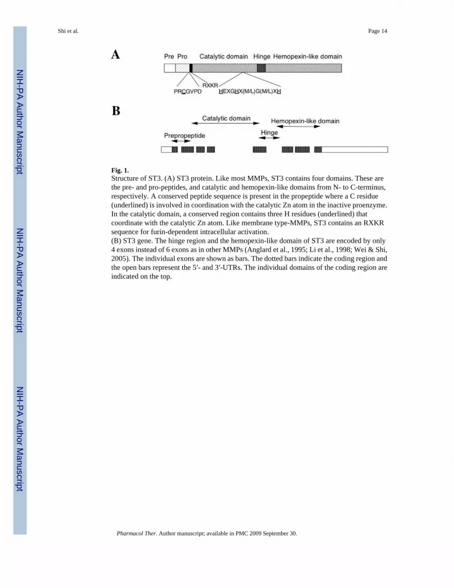
Fig. 1.Structure of ST3. (A) ST3 protein. Like most MMPs, ST3 contains four domains. These arethe pre- and pro-peptides, and catalytic and hemopexin-like domains from N- to C-terminus,respectively. A conserved peptide sequence is present in the propeptide where a C residue(underlined) is involved in coordination with the catalytic Zn atom in the inactive proenzyme.In the catalytic domain, a conserved region contains three H residues (underlined) thatcoordinate with the catalytic Zn atom. Like membrane type-MMPs, ST3 contains an RXKRsequence for furin-dependent intracellular activation.(B) ST3 gene. The hinge region and the hemopexin-like domain of ST3 are encoded by only4 exons instead of 6 exons as in other MMPs (Anglard et al., 1995; Li et al., 1998; Wei & Shi,2005). The individual exons are shown as bars. The dotted bars indicate the coding region andthe open bars represent the 5′- and 3′-UTRs. The individual domains of the coding region areindicated on the top.
Shi et al. Page 14
Pharmacol Ther. Author manuscript; available in PMC 2009 September 30.
NIH
-PA Author Manuscript
NIH
-PA Author Manuscript
NIH
-PA Author Manuscript
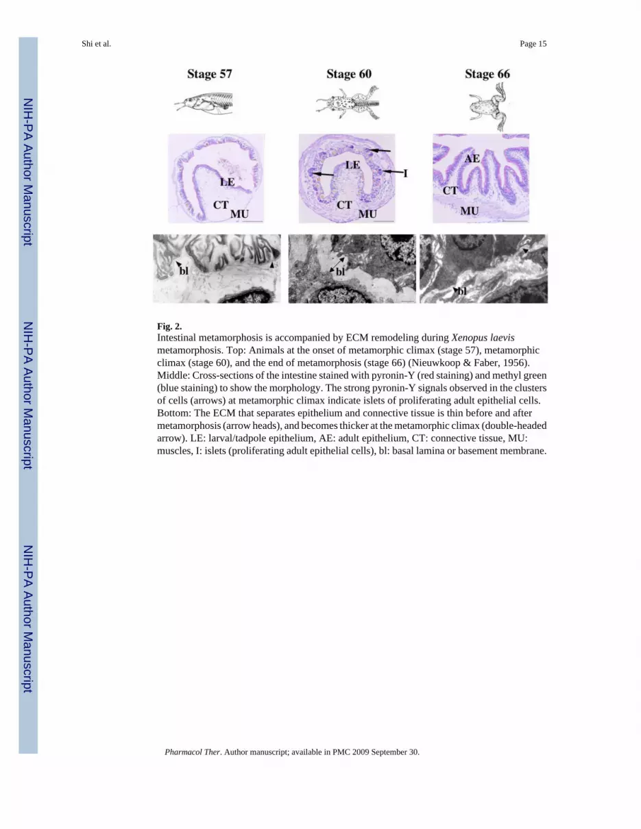
Fig. 2.Intestinal metamorphosis is accompanied by ECM remodeling during Xenopus laevismetamorphosis. Top: Animals at the onset of metamorphic climax (stage 57), metamorphicclimax (stage 60), and the end of metamorphosis (stage 66) (Nieuwkoop & Faber, 1956).Middle: Cross-sections of the intestine stained with pyronin-Y (red staining) and methyl green(blue staining) to show the morphology. The strong pyronin-Y signals observed in the clustersof cells (arrows) at metamorphic climax indicate islets of proliferating adult epithelial cells.Bottom: The ECM that separates epithelium and connective tissue is thin before and aftermetamorphosis (arrow heads), and becomes thicker at the metamorphic climax (double-headedarrow). LE: larval/tadpole epithelium, AE: adult epithelium, CT: connective tissue, MU:muscles, I: islets (proliferating adult epithelial cells), bl: basal lamina or basement membrane.
Shi et al. Page 15
Pharmacol Ther. Author manuscript; available in PMC 2009 September 30.
NIH
-PA Author Manuscript
NIH
-PA Author Manuscript
NIH
-PA Author Manuscript
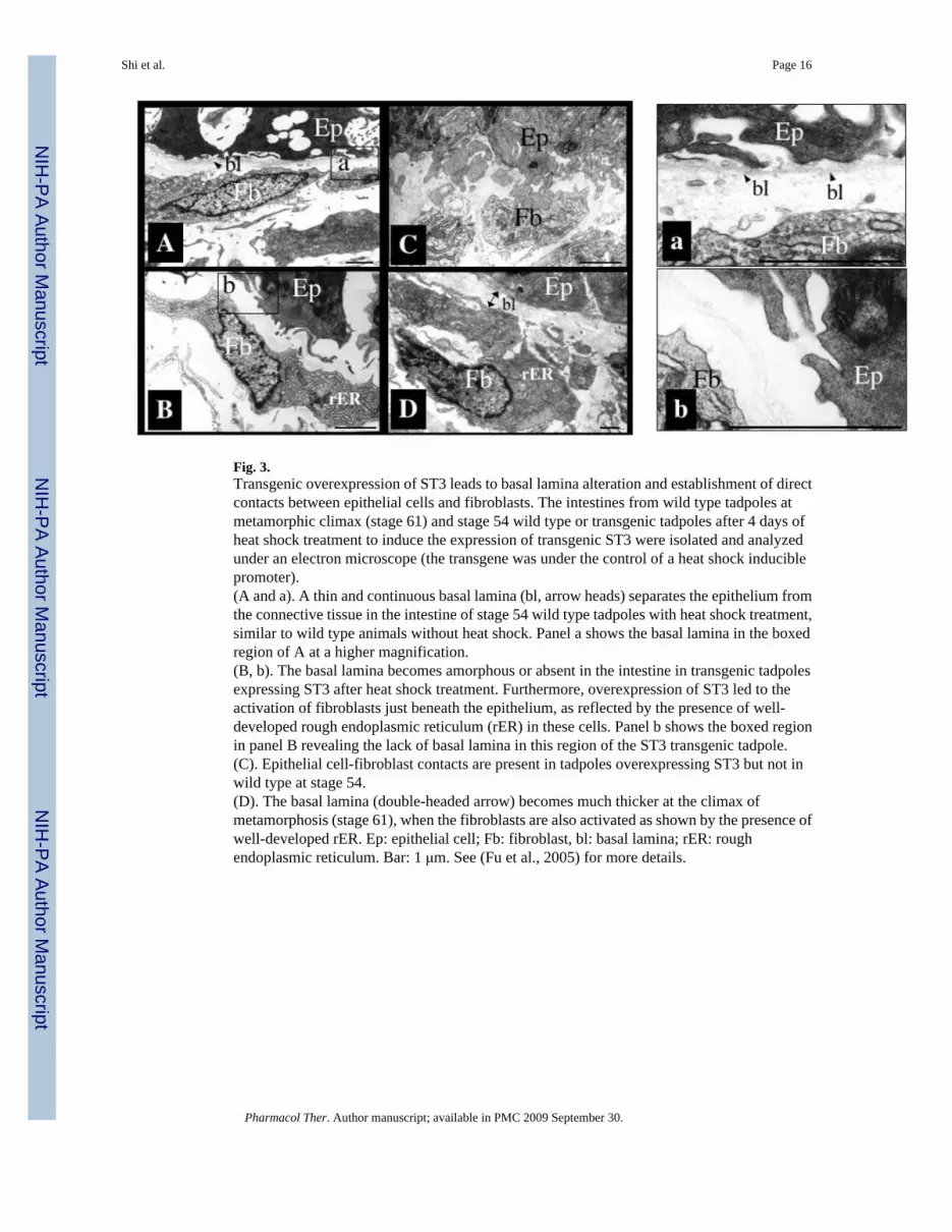
Fig. 3.Transgenic overexpression of ST3 leads to basal lamina alteration and establishment of directcontacts between epithelial cells and fibroblasts. The intestines from wild type tadpoles atmetamorphic climax (stage 61) and stage 54 wild type or transgenic tadpoles after 4 days ofheat shock treatment to induce the expression of transgenic ST3 were isolated and analyzedunder an electron microscope (the transgene was under the control of a heat shock induciblepromoter).(A and a). A thin and continuous basal lamina (bl, arrow heads) separates the epithelium fromthe connective tissue in the intestine of stage 54 wild type tadpoles with heat shock treatment,similar to wild type animals without heat shock. Panel a shows the basal lamina in the boxedregion of A at a higher magnification.(B, b). The basal lamina becomes amorphous or absent in the intestine in transgenic tadpolesexpressing ST3 after heat shock treatment. Furthermore, overexpression of ST3 led to theactivation of fibroblasts just beneath the epithelium, as reflected by the presence of well-developed rough endoplasmic reticulum (rER) in these cells. Panel b shows the boxed regionin panel B revealing the lack of basal lamina in this region of the ST3 transgenic tadpole.(C). Epithelial cell-fibroblast contacts are present in tadpoles overexpressing ST3 but not inwild type at stage 54.(D). The basal lamina (double-headed arrow) becomes much thicker at the climax ofmetamorphosis (stage 61), when the fibroblasts are also activated as shown by the presence ofwell-developed rER. Ep: epithelial cell; Fb: fibroblast, bl: basal lamina; rER: roughendoplasmic reticulum. Bar: 1 μm. See (Fu et al., 2005) for more details.
Shi et al. Page 16
Pharmacol Ther. Author manuscript; available in PMC 2009 September 30.
NIH
-PA Author Manuscript
NIH
-PA Author Manuscript
NIH
-PA Author Manuscript
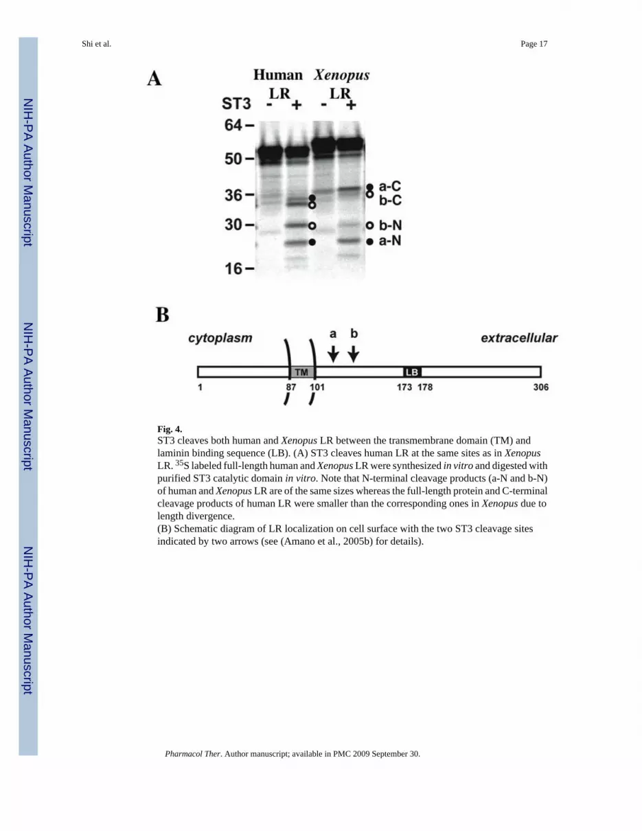
Fig. 4.ST3 cleaves both human and Xenopus LR between the transmembrane domain (TM) andlaminin binding sequence (LB). (A) ST3 cleaves human LR at the same sites as in XenopusLR. 35S labeled full-length human and Xenopus LR were synthesized in vitro and digested withpurified ST3 catalytic domain in vitro. Note that N-terminal cleavage products (a-N and b-N)of human and Xenopus LR are of the same sizes whereas the full-length protein and C-terminalcleavage products of human LR were smaller than the corresponding ones in Xenopus due tolength divergence.(B) Schematic diagram of LR localization on cell surface with the two ST3 cleavage sitesindicated by two arrows (see (Amano et al., 2005b) for details).
Shi et al. Page 17
Pharmacol Ther. Author manuscript; available in PMC 2009 September 30.
NIH
-PA Author Manuscript
NIH
-PA Author Manuscript
NIH
-PA Author Manuscript

Fig. 5.ST3 degrades LR in the intestine during metamorphosis. LR is degraded in the intestine at theclimax of metamorphosis and in the intestine of premetamorphic transgenic tadpolesoverexpressing ST3. Transgenic animals (T) and non-transgenic siblings (W) at stage 56 weretreated with or without heat shocked daily in the same tank. Two or three days later, theintestines were isolated. Total protein was isolated from these intestines as well as from theintestines of tadpoles at stage 57, prior to intestinal metamorphosis, or stage 61, at the climaxof intestinal metamorphosis. The proteins were subjected to Western blotting with anti-Xenopus LR antibody. The open arrow indicates full length LR. The triangles, and solid andopen circles indicate degradation products. The larger bands (triangles) were occasionallyobserved in different sample preparations in tadpoles at different stages but the bands indicatedby the circles were found at much higher levels in metamorphosing intestine or in animalsexpressing transgenic ST3 (see (Amano et al., 2005a) for details).
Shi et al. Page 18
Pharmacol Ther. Author manuscript; available in PMC 2009 September 30.
NIH
-PA Author Manuscript
NIH
-PA Author Manuscript
NIH
-PA Author Manuscript
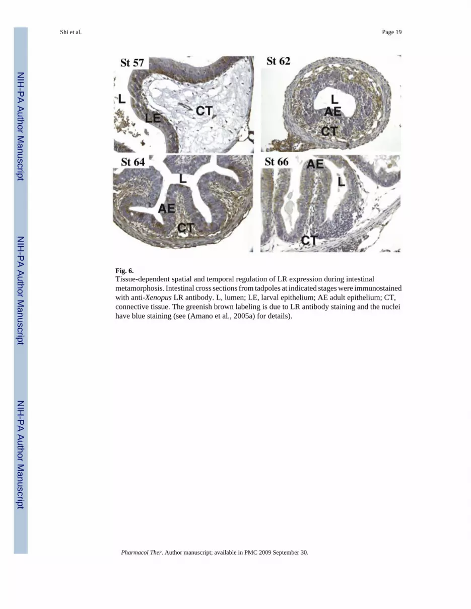
Fig. 6.Tissue-dependent spatial and temporal regulation of LR expression during intestinalmetamorphosis. Intestinal cross sections from tadpoles at indicated stages were immunostainedwith anti-Xenopus LR antibody. L, lumen; LE, larval epithelium; AE adult epithelium; CT,connective tissue. The greenish brown labeling is due to LR antibody staining and the nucleihave blue staining (see (Amano et al., 2005a) for details).
Shi et al. Page 19
Pharmacol Ther. Author manuscript; available in PMC 2009 September 30.
NIH
-PA Author Manuscript
NIH
-PA Author Manuscript
NIH
-PA Author Manuscript
Copyright © 2022 FDOKUMEN