Physical and Functional Interaction between p53 Mutants and Different Isoforms of p73
Upload
eastangliaCategory
view
0download
0
Available online http://breast-cancer-research.com/content/11/2/R24
Open AccessVol 11 No 2Research articleTumour-associated tenascin-C isoforms promote breast cancer cell invasion and growth by matrix metalloproteinase-dependent and independent mechanismsRachael A Hancox1*, Michael D Allen2*, Deborah L Holliday2, Dylan R Edwards3, Caroline J Pennington3, David S Guttery1, Jacqueline A Shaw1, Rosemary A Walker1, J Howard Pringle1 and J Louise Jones2
1Department of Cancer Studies and Molecular Medicine, Infirmary Close, University of Leicester, Robert Kilpatrick Clinical Sciences Building, Leicester Royal Infirmary, Leicester, LE1 5WW, UK2Centre for Tumour Biology, Institute of Cancer, Barts and The London School of Medicine and Dentistry, Queen Mary University of London, Charterhouse Square, London, EC1M 6BQ, UK3School of Biological Sciences, University Drive, University of East Anglia, Norwich, Norfolk, NR4 7TJ, UK* Contributed equally
Corresponding author: J Howard Pringle, [email protected]; J Louise Jones, [email protected]
Received: 4 Aug 2008 Revisions requested: 27 Aug 2008 Revisions received: 15 Jan 2009 Accepted: 30 Apr 2009 Published: 30 Apr 2009
Breast Cancer Research 2009, 11:R24 (doi:10.1186/bcr2251)This article is online at: http://breast-cancer-research.com/content/11/2/R24© 2009 Hancox et al.; licensee BioMed Central Ltd. This is an open access article distributed under the terms of the Creative Commons Attribution License (http://creativecommons.org/licenses/by/2.0), which permits unrestricted use, distribution, and reproduction in any medium, provided the original work is properly cited.
Abstract
Introduction The stromal microenvironment has a profoundinfluence on tumour cell behaviour. In tumours, the extracellularmatrix (ECM) composition differs from normal tissue and allowsnovel interactions to influence tumour cell function. The ECMprotein tenascin-C (TNC) is frequently up-regulated in breastcancer and we have previously identified two novel isoforms –one containing exon 16 (TNC-16) and one containing exons 14plus 16 (TNC-14/16).
Methods The present study has analysed the functionalsignificance of this altered TNC isoform profile in breast cancer.TNC-16 and TNC-14/16 splice variants were generated usingPCR-ligation and over-expressed in breast cancer cells (MCF-7,T47D, MDA-MD-231, MDA-MB-468, GI101) and humanfibroblasts. The effects of these variants on tumour cell invasionand proliferation were measured and compared with the effectsof the large (TNC-L) and fully spliced small (TNC-S) isoforms.
Results TNC-16 and TNC-14/16 significantly enhanced tumourcell proliferation (P < 0.05) and invasion, both directly (P <0.01) and as a response to transfected fibroblast expression (P< 0.05) with this effect being dependent on tumour cellinteraction with TNC, because TNC-blocking antibodiesabrogated these responses. An analysis of 19 matrixmetalloproteinases (MMPs) and tissue inhibitor of matrixmetalloproteinases 1 to 4 (TIMP 1 to 4) revealed that TNC up-regulated expression of MMP-13 and TIMP-3 two to four foldrelative to vector, and invasion was reduced in the presence ofMMP inhibitor GM6001. However, this effect was not isoform-specific but was elicited equally by all TNC isoforms.Conclusions These results demonstrate a dual requirement forTNC and MMP in enhancing breast cancer cell invasion, andidentify a significant role for the tumour-associated TNC-16 andTNC-14/16 in promoting tumour invasion, although theseisoform-specific effects appear to be mediated through MMP-independent mechanisms.
IntroductionCellular interactions with the extracellular matrix (ECM) arecritical in the modulation of cell growth, migration, invasion and
tissue-specific gene expression [1-3]. The ECM aroundtumours differs markedly from that in normal tissues [4,5] andtherefore is likely to deliver different signals to tumour cells,
Page 1 of 13(page number not for citation purposes)
BSA: bovine serum albumin; CM: conditioning medium; ECM: extracellular matrix; EGF: epidermal growth factor; ELISA: enzyme-linked immunosorb-ent assay; ER: oestrogen receptor; DMEM: Dulbecco's modified Eagle's medium; FBS: fetal bovine serum; FN III: fibronectin type III-like repeats; HPRT1: hypoxanthine phosphoribosyltransferase 1; MII: mean invasion index; MMP: matrix metalloproteinase; PCR: polymerase chain reaction; RT-PCR: reverse transcriptase polymerase chain reaction; TIMP: tissue inhibitor of matrix metalloproteinases; TNC: tenascin C; TNC-16: tenascin C with only exon 16 of variable region; TNC 14/16: tenascin C with only exons 14 and 16 of variable region; TNC-L: tenascin C largest splice variant; TNC-S: tenascin C smallest splice variant.
Breast Cancer Research Vol 11 No 2 Hancox et al.
which will impact on their behaviour. One of the most consist-ent changes in the ECM of many solid tumours is up-regulationof the matrix glycoprotein, tenascin-C (TNC) [6-8]. TNC is acomplex multifunctional protein, which has been shown to pro-mote cell migration, inhibit focal contact formation, promoteangiogenesis and, in some systems, act as a cell survival factor[9-11]. Each TNC subunit consists of an N-terminal tenascinassembly region, 14.5 epidermal growth factor-like (EGF)repeat domains, a variable number of fibronectin type III-likerepeats (FN III) and a C-terminal fibrinogen-like domain [12](Figure 1). Multiple isoforms of TNC can be generated throughalternative splicing of nine FN III repeats between conservedrepeats 5 and 6 (exons 9 and 17) at the pre-mRNA level andthese may have differing effects. For example, in the develop-ing mouse central nervous system, up to 27 distinct splice var-iants have been identified and are expressed in a stricttemporal-spatial manner supporting a role for these variants inspecific neurone-glia interactions [13]. A number of studieshave shown that specific functions are mediated by distinctdomains of TNC [10,11,14] and there is growing evidence toindicate that the biological function of TNC is dependent onthe splicing pattern [15]. This raises the possibility thattumour-associated stroma can generate novel interactionswith tumour cells through the expression of different TNCsplice variants. In keeping with this, changes in the pattern ofTNC isoform expression have been described in a number of
malignancies [16-18], the nature of which appears to betumour-type specific.
Previously, we have identified two TNC isoforms that consist-ently and specifically are up-regulated in invasive breast carci-noma as well as in a subset of pre-invasive ductal carcinomain-situ: one containing exon 16 (TNC-16) the other containingexon 14 plus 16 (TNC-14/16) [19]. Up-regulation of these iso-forms has also been reported in ovarian carcinoma [20]; how-ever, their effect on tumour cell behaviour has not beenestablished.
A further consistent change in the tumour stromal environmentis up-regulation of proteolytic enzymes, particularly membersof the matrix metalloproteinase (MMP) family. Overexpressionof several MMPs has been described in breast cancer [21,22]and both in-vitro and in-vivo systems demonstrate a role forMMP in mediating breast cancer cell invasion [23,24]. Regu-lation of MMP expression and activity is complex, and may bemediated through naturally occurring tissue inhibitors of MMP(TIMP) or by direct regulation of gene expression. Severalstudies have indicated a role for TNC in regulating MMP geneexpression [25-27].
The aim of this study was to investigate directly the effects oftumour-associated TNC-16 and TNC-14/16 isoforms on
Figure 1
Schematic diagram of tenascin-CSchematic diagram of tenascin-C. The domain structure of tenascin-C comprising N-terminal tenascin assembly (TA) domain followed by 14.5 epi-dermal growth factor (EGF)-like repeats, the fibronectin type III (FN III)-like repeats and the carboxy fibrinogen-like domain. The FN III region consists of eight conserved repeats, designated 1 to 8, and up to nine alternatively spliced FN III repeats designated as letters A to D, AD1 and AD2 (shown in shading). The exon organisation of the tenascin-C gene is shown below.
Page 2 of 13(page number not for citation purposes)

Available online http://breast-cancer-research.com/content/11/2/R24
breast cancer cell behaviour and to determine whether theseisoforms modulate tumour cell behaviour through regulation ofMMP.
Materials and methodsCell lines and primary cellsBreast cancer cell lines (MCF-7, T47D, MDA MB 231, ZR-75-1, MDA MB 468, GI101, MDA MB 436, HS578T, SkMel 28)and the human fetal foreskin fibroblast cell line hfff2 wereobtained from ATCC (American Type Culture Collection, Man-assas, VA, USA). Primary normal breast tissue was obtainedfollowing Ethics Approval and informed patient consent(Leicestershire, LREC 7054). Fibroblasts were isolated fromreduction mammoplasty specimens and purity confirmed aspreviously described [28]. All cells were maintained in Dul-becco's modified Eagle's medium (DMEM) plus 2 mM L-glutamine and 10% FBS.
TaqMan real-time PCR analysis of endogenous tenascin-C expressionTaqman real-time PCR (Applied Biosystems, Foster, CA,USA) was applied to survey different exons in breast cell lines.An inventoried assay was available for the TNC invariant exon17/18 boundary (Applied Biosystems Taqman Assay,Hs01115654_m1) and hypoxanthine phosphoribosyltrans-ferase 1 (HPRT1) (Applied Biosystems Taqman Assay,Hs99999909_m1) as a housekeeping gene. Primers andprobe were developed in house for 9/16 and 14/16 exonboundaries. For the inventoried Taqman assays, 4 μl of cDNA(diluted 1:10) was analysed in a reaction containing 0.5 μl ofprobe, 0.5 μl Ultrapure water and 5 μl of 2 × Taqman FastPCR mastermix. For the 9/16 and 14/16 assays, 3.6 μl ofcDNA (diluted 1:10) was analysed in a reaction containing 0.2μl of probe, 0.6 μl of each primer and 5 μl of 2 × Taqman FastPCR mastermix. Normalised relative expression was deter-mined by comparison with standard curves derived from TNC9–16 and TNC 9–14–16 recombinant clones to correct fordifferences in PCR efficiency for each TNC probe set and thelevel of housekeeping gene expression was used to correct forany differences in cellularity.
Generation of tenascin-C isoformsTNC splice variant cDNAs containing either exon 16 or exons14 and 16 were prepared by ligation and nested PCR usingcloned human TNC cDNA for the large, unspliced isoformTNC-L (pNUT-HxB.L) and the truncated isoform TNC-S(pNUT-HxB.S) [29].
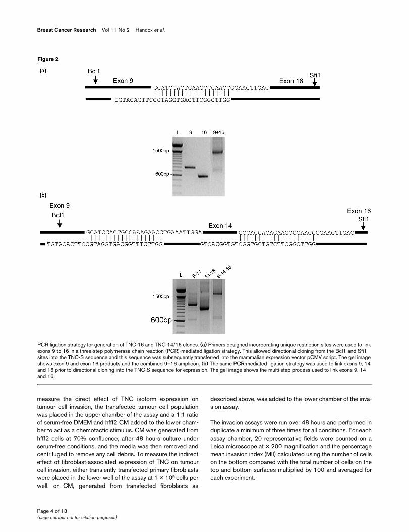
Primers designed across exon boundaries 9–16, 9–14 and14–16 were used to amplify flanking DNA to include therestriction sites Bcl1 and Sfi1 with bcl1ten1 and sfi1ten1(Table 1 and Figure 2). These enzymes cut uniquely withinexon 9 and 17 of the TNC cDNA. The amplicons were purifiedby Qiagen column (Sussex, UK) and ligated by nested PCRusing bcl1ten2 and sfi1ten2 internal primers. The ligated DNA
fragments were cloned into TOPO vector pcDNA3.1/V5/His-TOPO, screened by PCR for recombinants and sequenced.The Bcl1 and Sfi1 fragments for each spliced variant were re-cloned into TNC-S sequence in the mammalian expressionvector pCMVscript and pCMV-tag4a (Invitrogen Life Science,Carlsbad, CA, USA) to produce pTNC-16 and pTNC-14/16.At each stage, all clones were confirmed by sequencing.Clones were tagged with Flag sequence to allow detection ofthe expressed protein.
Transfection of cell populationsThe breast cancer cell lines MCF-7, T47D, MDA-MB 231,MDA-MB-468 and GI101, the fibroblast cell line hfff2 and pri-mary normal breast fibroblasts from a series of donors weretransiently transfected with TNC-16, TNC-14/16, TNC-L,TNC-S or vector only using Genejuice Transfection Reagent(Novagen, Darmstadt, Germany) according to the manufac-turer's instructions. In some experiments, TNC clones weretransfected in combination using the same approach. Expres-sion was confirmed by RT-PCR using the primer set 8F/18Rspanning the entire alternatively spliced region (Table 1) andimmunohistochemistry using the anti-Flag M2 antibody(Sigma, Dorset, UK), anti-TNC with BC-24 at 1:7500 for allisoforms and α IIIB at 1:1000 for isoforms containing exon 14and a standard Avidin-Biotin Alkaline Phosphatase detectionsystem. Equal transfection efficiencies for each isoform wereconfirmed by estimating the proportion of cells that immunos-tained positive.
Western blottingLevels of cellular and secreted TNC isoforms were determinedby western blotting of transfected cell lysates and conditionedmedia (CM) respectively. Cells were transiently transfectedand incubated for 24 hours, serum-free media was added andCM was collected after a further 48 hours. Cell lysates wereharvested in gold lysis buffer containing a protease inhibitorcocktail (Sigma, Dorset, UK) 48 hours after transient transfec-tion. Protein concentrations were quantified on a Lambda 25UV/VIS spectrophotometer at 750 nm using the BSA proteinassay, then equal amounts of protein were loaded onto 6%SDS-PAGEs and transferred to Hybond ECL nitrocellulosemembrane (Amersham Biosciences, Buckinghamshire, UK).Membranes were blocked in Tris-buffered saline, 5% milk and1% Tween for one hour and then probed for two hours with arabbit polyclonal TNC antibody (clone H300, recognising allforms of TNC; Santa-Cruz, California, USA). A secondary anti-body, donkey anti-rabbit HRP-linked IgG, 1:2000 (AmershamBiosciences, Buckinghamshire, UK), was added for one hourand blots were detected using an enhanced chemilumines-cence detection kit (Amersham Biosciences, Buckingham-shire, UK).
Analysis of tumour cell invasionMeasurement of tumour cell invasion was based on modifiedBoyden chamber assays as described previously [28]. To
Page 3 of 13(page number not for citation purposes)
Breast Cancer Research Vol 11 No 2 Hancox et al.
measure the direct effect of TNC isoform expression ontumour cell invasion, the transfected tumour cell populationwas placed in the upper chamber of the assay and a 1:1 ratioof serum-free DMEM and hfff2 CM added to the lower cham-ber to act as a chemotactic stimulus. CM was generated fromhfff2 cells at 70% confluence, after 48 hours culture underserum-free conditions, and the media was then removed andcentrifuged to remove any cell debris. To measure the indirecteffect of fibroblast-associated expression of TNC on tumourcell invasion, either transiently transfected primary fibroblastswere placed in the lower well of the assay at 1 × 10⁵ cells perwell, or CM, generated from transfected fibroblasts as
described above, was added to the lower chamber of the inva-sion assay.
The invasion assays were run over 48 hours and performed induplicate a minimum of three times for all conditions. For eachassay chamber, 20 representative fields were counted on aLeica microscope at × 200 magnification and the percentagemean invasion index (MII) calculated using the number of cellson the bottom compared with the total number of cells on thetop and bottom surfaces multiplied by 100 and averaged foreach experiment.
Figure 2
PCR-ligation strategy for generation of TNC-16 and TNC-14/16 clonesPCR-ligation strategy for generation of TNC-16 and TNC-14/16 clones. (a) Primers designed incorporating unique restriction sites were used to link exons 9 to 16 in a three-step polymerase chain reaction (PCR)-mediated ligation strategy. This allowed directional cloning from the Bcl1 and Sfi1 sites into the TNC-S sequence and this sequence was subsequently transferred into the mammalian expression vector pCMV script. The gel image shows exon 9 and exon 16 products and the combined 9–16 amplicon. (b) The same PCR-mediated ligation strategy was used to link exons 9, 14 and 16 prior to directional cloning into the TNC-S sequence for expression. The gel image shows the multi-step process used to link exons 9, 14 and 16.
Page 4 of 13(page number not for citation purposes)
Available online http://breast-cancer-research.com/content/11/2/R24
In some experiments, blocking antibody to TNC (BC-24,Sigma, Dorset, UK), IgG1 control (Dakocytomation, Glostrup,Denmark) or a broad-spectrum MMP inhibitor (GM6001,Chemicon, Watford, UK) were included. The TNC antibodyBC-24 was desalted to remove sodium azide before use bypassing through a PD-10 Sephadex G-25 column (GE Health-care, Buckinghamshire, UK). Both BC-24 and IgG1 controlwere included in the upper and lower wells of the assay at 1ng/ml final concentration. GM6001 was included at 10 nM.
Analysis of tumour cell proliferationThe direct and indirect effects of TNC isoforms on tumour cellproliferation were assessed on the basis of BrdU incorpora-tion. Tumour cells were transfected, as described above, andcultured on poly-d-lysine coated coverslips for 72 hours underserum-free conditions. BrdU was added to the media at a con-centration of 5 mM for the final three hours. The cells werefixed and incorporated BrdU localised using anti-BrdU(Bu20a; Sigma, Dorset, UK) and a standard Avidin-Biotindetection system. The mean proliferation index of three assayswas calculated by averaging the number of stained cells as apercentage of the total number of cells (minimum of 1000 cellsover at least 10 high power fields) for each assay. To measurethe indirect effect of fibroblast-associated TNC on tumour cellproliferation, fibroblast populations were transfected as previ-ously described, the CM harvested and added to tumour cells.Proliferation was then assessed as described above.
Analysis of matrix metalloproteinase expression and activityTaqMan real-time PCR analysis of the effect of TNC isoforms on expression profile of MMP and TIMPTaqMan real-time PCR was carried out as previouslydescribed on mRNA isolated from MCF-7 breast cancer cellsand hfff2 fibroblasts transfected with different TNC clones,compared with vector-only and non-transfected control popu-lations [30]. Analysis was performed on mRNA from three sep-arate experiments and included measurement of 19 MMPs[3,6,7,9-18,20,22-25] and TIMP 1 to 4.
ELISAFor selected MMPs, levels of enzyme activity were measuredby ELISA. Invasion assays were set up to include MCF-7 cellstransfected with different TNC clones or CM from TNC-trans-fected hfff2 fibroblasts and cultured for 48 hours, as previouslydescribed. End-of-assay medium was collected from theassays, protein levels measured and equal concentrationsapplied to ELISA plates for MMP-1, MMP-2 and MMP-9(Amersham, Buckinghamshire, UK). Each experiment was per-formed in triplicate.
ZymographyLevels of enzyme activity were also measured by substrate gelzymography. End-of-assay medium was collected from assayscontaining cells transfected with different TNC clones, asdescribed above. SDS-PAGE substrate gels were made byincorporating Gelatin (Bloom 300, Sigma, Dorset, UK) in a10% acrylamide separating gel at a final concentration of 1mg/ml. To each gel, samples containing equal amounts of pro-tein (as determined by BCA protein assay) were mixed withnon-reducing sample buffer (62.5 mM Tris-HCl (pH 6.8), 10%glycerol, 2% SDS, 0.1% bromophenol blue) and added to thegel without boiling. MMP-9 recombinant pro-enzyme (Calbio-chem, California, USA) and molecular weight markers wererun on each gel. Following electrophoresis, gels were washedtwice in 2.5% Triton X-100 for 30 minutes at 37°C to removethe SDS. Gels were incubated at 37°C overnight in develop-ing buffer containing 50 mM Tris-HCl, 0.2 M sodium chloride,5 mM calcium chloride and 0.02% Triton X-100. Gels werestained with 0.5% coomassie blue G250 in 30% methanol,10% glacial acetic acid for 30 minutes and de-stained in thesame solution without coomassie blue. Gelatin-degradingenzymes were identified as clear bands against the blue back-ground of the stained gel. Images of stained gels were cap-tured under illumination using the UVP Imagestore 5000(Ultra-Violet Products, Cambridge, UK). Direct comparisonsbetween separate gels were not made, because the intensityof background staining was variable. Experiments wererepeated a minimum of three times.
Statistical analysisStatistical analysis was performed using the SPSS 12.0 sta-tistics package (Chicago, IL, USA). For comparison between
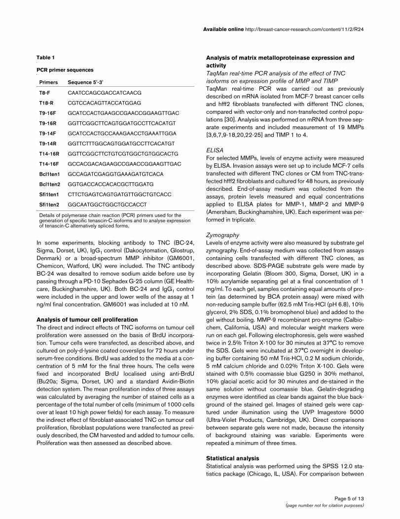
Table 1
PCR primer sequences
Primers Sequence 5'-3'
T8-F CAATCCAGCGACCATCAACG
T18-R CGTCCACAGTTACCATGGAG
T9-16F GCATCCACTGAAGCCGAACCGGAAGTTGAC
T9-16R GGTTCGGCTTCAGTGGATGCCTTCACATGT
T9-14F GCATCCACTGCCAAAGAACCTGAAATTGGA
T9-14R GGTTCTTTGGCAGTGGATGCCTTCACATGT
T14-16R GGTTCGGCTTCTGTCGTGGCTGTGGCACTG
T14-16F GCCACGACAGAAGCCGAACCGGAAGTTGAC
Bcl1ten1 GCCAGATCGAGGTGAAAGATGTCACA
Bcl1ten2 GGTGACCACCACACGCTTGGATG
Sfi1ten1 CTTCTGAGTCAGTGATGTTGGCTGTCACC
Sfi1ten2 GGCAATGGCTGGCTGCCACCT
Details of polymerase chain reaction (PCR) primers used for the generation of specific tenascin-C isoforms and to analyse expression of tenascin-C alternatively spliced forms.
Page 5 of 13(page number not for citation purposes)

Breast Cancer Research Vol 11 No 2 Hancox et al.
different TNC isoforms the one-way analysis of variance testwas used. A Bonferroni correction was applied to all post-hocanalyses and a P value of less than 0.05 was considered sig-nificant.
ResultsEndogenous expression of tenascin-C isoforms in breast cancer cellsReal-time PCR for endogenous TNC isoform expression wasperformed on a series of breast cancer cell lines. This showedthat MCF-7, T47D and ZR-75-1 cell lines do not expressdetectable levels of TNC or its isoforms, while GI-101,Hs578T, MDA MB 231, MDA MB 436 and MDA MB 468 cellsall express TNC. Furthermore, all express TNC-14/16 and GI-101, MDA 436 and MDA MB 468 also express TNC-16 (Fig-ure 3a).
Expression of tenascin-C isoforms in breast cancer cells and fibroblastsThe breast cancer cells MCF-7, T47D, MDA MB 231, MDAMB 468 and GI101, the hfff2 fibroblast cell line and a series(n = 5) of primary normal breast fibroblasts were transientlytransfected with clones for TNC-S, TNC-L, TNC-16 and TNC-14/16 and vector only controls. Expression of specific iso-forms was confirmed by RT-PCR (Figure 3b), and proteinexpression was confirmed by immunohistochemistry to theFlag tag (Figure 3c), which demonstrated an average transfec-tion efficiency of 35% for each isoform. No native TNC wasdetected in MCF-7 cells (Figure 3c, right image). The level ofexpression in transfected MCF-7 cells was equivalent to thatseen endogenously in MDA-MB-231 cells (unpublished data).
Western blotting of cell lysates (Figure 3d, i) and CM (Figure3d, ii) demonstrated equivalent levels of TNC protein expres-sion from each of the clones, and confirmed the presence ofTNC.
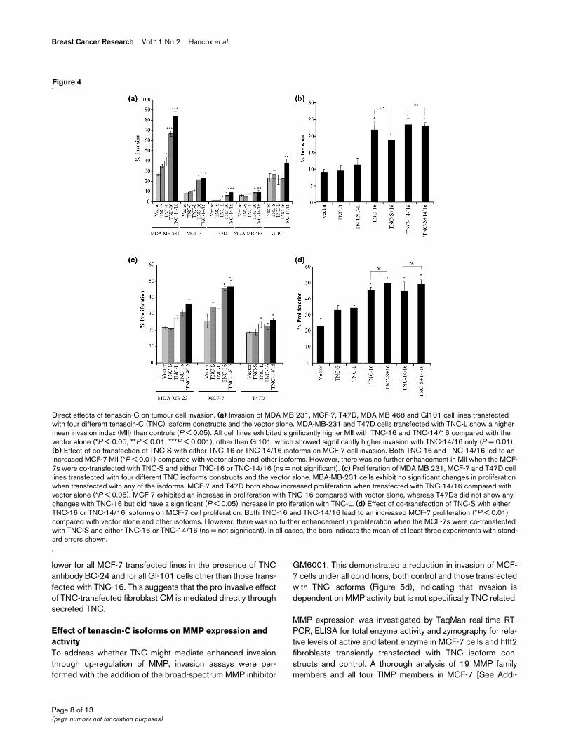
Direct effects of tenascin-C isoforms on tumour cell behaviour: effect on invasion and proliferationThe breast cancer cell lines exhibited different levels of inva-sion. The MCF-7, T47D and MDA MB 468 cell lines showedlow level invasion (Mean Invasion Index (MII) of 8%, 2% and6%, respectively) whereas the MDA-MB-231 and GI101 celllines exhibited high invasion (MII of 27% and 23% respec-tively; Figure 4a). For cell lines transfected with either the vec-tor alone or TNC-S there was no significant change in the MIIcompared with the untreated cells (Figure 4a). TNC-L trans-fectants in MDA-MD-231 and T47D showed increased MIIover baseline levels (P = 0.005 and P = 0.011, respectively).However, the highest levels of invasion were observed foreach of the cell lines when transfected with TNC-14/16 (P <0.001 for MDA-MB-231, P < 0.001 for MCF-7, P = 0.001 forT47D, P = 0.005 for MDA-MB-468, P = 0.01 for GI101) and(except for GI101) TNC-16 (P < 0.001 for MDA-MB-231, P =0.004 for MCF-7 and P = 0.002 for T47D). The TNC-14/16
isoforms also showed significantly higher MII than the TNC-Ltransfectants, P < 0.01 for all cell lines. The MII was also sig-nificantly higher with TNC-16 vs. TNC-L for MDA MB 231 (P= 0.008), MCF-7 (P = 0.004), T47D (P = 0.009) and GI101(P = 0.01) cells but not MDA MB 468 (Figure 4a).
In human breast carcinoma tissues, TNC-16 and/or TNC-14/16 are not always detected alone, but frequently are seen incombination with TNC-S [19]; therefore MCF-7 cells were co-transfected with TNC-S and either TNC-16 or TNC-14/16.The results showed that although TNC-16 and TNC-14/16both enhanced MCF-7 cell MII, there was no additional effectof co-expression of TNC-S (Figure 4b).
Increased breast cancer cell proliferation was observed in allcell lines over-expressing TNC-L, TNC-16 and TNC-14/16.This reached significance in MCF-7 cells for both TNC-16 andTNC-14/16 (P = 0.023 and P = 0.002, respectively), and inT47D cells for TNC-L and TNC-14/16 (P = 0.025 and P =0.011, respectively; Figure 4c). A similar effect was seen inMCF-7 cells co-transfected with TNC-S and either TNC-16 orTNC-14/16 with no additive effect over the single isoforms(Figure 4d).
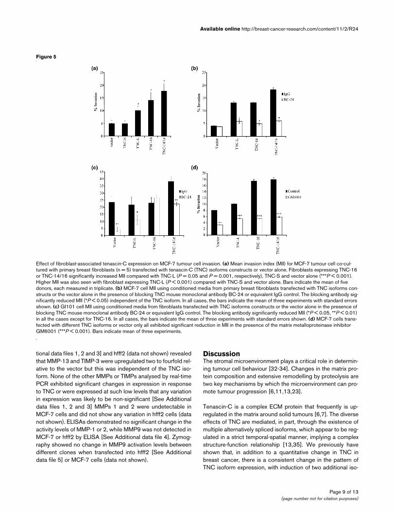
Effect of fibroblast-associated tenascin-C expression on tumour cell invasionThe major source of TNC in breast carcinomas is the peri-tumoural stroma [19], therefore we analysed the effect offibroblast-associated TNC isoform expression on tumour cellinvasion. Primary breast fibroblasts isolated from normalbreast tissue were transfected with individual TNC isoformsand co-cultured in the lower chamber of invasion assays withMCF-7 breast cancer cells. Standard invasion assays werethen carried out for each TNC isoform with vector-only con-trols. There was some variability between donors in the capac-ity of fibroblasts to promote tumour cell invasion (data notshown). However, for all donors, a significantly higher MCF-7MII was observed in the presence of fibroblasts over-express-ing TNC-L, TNC-16 or TNC-14/16 (P < 0.001, Figure 5a) butnot in the presence of TNC-S. Furthermore, a higher MII forMCF-7 cells was seen with fibroblasts over-expressing TNC-16 or TNC-14/16 compared to TNC-L (P = 0.05 and P =0.001, respectively). These results reflect the pattern seenwhen the tumour cells themselves over-express TNC, and asimilar pattern also was seen in the presence of CM fromtransfected primary fibroblasts (data not shown).
TNC is secreted by cells [31], so we investigated whether theeffect of fibroblast-associated TNC isoforms was mediateddirectly through soluble TNC. CM from transfected fibroblastswas added to the lower well of invasion assays containingMCF-7 cells (Figure 5b) or GI101 cells (Figure 5c) with addi-tion of blocking antibody to TNC (1 ng/ml BC-24) or equiva-lent IgG1 control. This demonstrated inhibition of tumour cellinvasion to control levels, (cells transfected with vector only) or
Page 6 of 13(page number not for citation purposes)

Available online http://breast-cancer-research.com/content/11/2/R24
Page 7 of 13(page number not for citation purposes)
Figure 3
Confirmation of expression of tenascin-C isoformsConfirmation of expression of tenascin-C isoforms. (a) Endogenous expression of tenascin-C (TNC) isoforms in untransfected cell lines. Normalised relative expression of TNC isoforms were determined by reverse transcriptase polymerase chain reaction (RT-PCR) using primers and probes to invariant exon 17/18 boundary (Total TNC), the 9/16 (TNC-16) and 14/16 (TNC-14/16) exon boundary for breast cell lines ZR-75-1, MCF-7, T-47D, GI-101, Hs578T, MDA-MB-231, MDA-MB-436, MDA-MB-468 and melanoma cell line SKMel-28. Relative expression was calculated by compari-son with standard curves derived from TNC-9-16 and TNC 9–14–16 recombinant clones to correct for differences in PCR efficiency for each TNC probe set normalised using level of housekeeping gene expression to correct for any differences in cellularity. (b) RT-PCR of primary fibroblasts and MCF-7 cells transiently transfected with TNC-S, TNC-L, TNC-16, TNC-14/16 and vector-only control (vector), using primers spanning the FN III alternatively spliced region (8F/18R). This shows appropriately sized bands in each of the cell populations, with no product in vector-only and non-transfected MCF-7 controls, although there was evidence of low level expression of TNC-S in vector only and non-transfected fibroblast controls. (c) Immunohistochemistry for anti-Flag M2 antibody in MCF-7 cells transfected with TNC-L (left image) and for TNC (Monoclonal, BC24) in MCF-7 transfected with vector control (right image). An average transfection efficiency of 35% was determined for each TNC isoform and staining con-firmed that MCF-7 cells do not express TNC. (d) Western blot analysis for TNC in transiently transfected MCF-7 cells. This demonstrated a single species of TNC present in the whole cell lysate (WCL; i) and conditioned media (CM; ii) of transfected cells for each isoform. TNC-S is seen as a band at about 250 kDa, with slightly larger bands detected for TNC-16 and TNC 14/16, while TNC-L is detected at about 350 kDa.
Breast Cancer Research Vol 11 No 2 Hancox et al.
lower for all MCF-7 transfected lines in the presence of TNCantibody BC-24 and for all GI-101 cells other than those trans-fected with TNC-16. This suggests that the pro-invasive effectof TNC-transfected fibroblast CM is mediated directly throughsecreted TNC.
Effect of tenascin-C isoforms on MMP expression and activityTo address whether TNC might mediate enhanced invasionthrough up-regulation of MMP, invasion assays were per-formed with the addition of the broad-spectrum MMP inhibitor
GM6001. This demonstrated a reduction in invasion of MCF-7 cells under all conditions, both control and those transfectedwith TNC isoforms (Figure 5d), indicating that invasion isdependent on MMP activity but is not specifically TNC related.
MMP expression was investigated by TaqMan real-time RT-PCR, ELISA for total enzyme activity and zymography for rela-tive levels of active and latent enzyme in MCF-7 cells and hfff2fibroblasts transiently transfected with TNC isoform con-structs and control. A thorough analysis of 19 MMP familymembers and all four TIMP members in MCF-7 [See Addi-
Figure 4
Direct effects of tenascin-C on tumour cell invasionDirect effects of tenascin-C on tumour cell invasion. (a) Invasion of MDA MB 231, MCF-7, T47D, MDA MB 468 and GI101 cell lines transfected with four different tenascin-C (TNC) isoform constructs and the vector alone. MDA-MB-231 and T47D cells transfected with TNC-L show a higher mean invasion index (MII) than controls (P < 0.05). All cell lines exhibited significantly higher MII with TNC-16 and TNC-14/16 compared with the vector alone (*P < 0.05, **P < 0.01, ***P < 0.001), other than GI101, which showed significantly higher invasion with TNC-14/16 only (P = 0.01). (b) Effect of co-transfection of TNC-S with either TNC-16 or TNC-14/16 isoforms on MCF-7 cell invasion. Both TNC-16 and TNC-14/16 led to an increased MCF-7 MII (*P < 0.01) compared with vector alone and other isoforms. However, there was no further enhancement in MII when the MCF-7s were co-transfected with TNC-S and either TNC-16 or TNC-14/16 (ns = not significant). (c) Proliferation of MDA MB 231, MCF-7 and T47D cell lines transfected with four different TNC isoforms constructs and the vector alone. MBA-MB-231 cells exhibit no significant changes in proliferation when transfected with any of the isoforms. MCF-7 and T47D both show increased proliferation when transfected with TNC-14/16 compared with vector alone (*P < 0.05). MCF-7 exhibited an increase in proliferation with TNC-16 compared with vector alone, whereas T47Ds did not show any changes with TNC-16 but did have a significant (P < 0.05) increase in proliferation with TNC-L. (d) Effect of co-transfection of TNC-S with either TNC-16 or TNC-14/16 isoforms on MCF-7 cell proliferation. Both TNC-16 and TNC-14/16 lead to an increased MCF-7 proliferation (*P < 0.01) compared with vector alone and other isoforms. However, there was no further enhancement in proliferation when the MCF-7s were co-transfected with TNC-S and either TNC-16 or TNC-14/16 (ns = not significant). In all cases, the bars indicate the mean of at least three experiments with stand-ard errors shown.
Page 8 of 13(page number not for citation purposes)
Available online http://breast-cancer-research.com/content/11/2/R24
tional data files 1, 2 and 3] and hfff2 (data not shown) revealedthat MMP-13 and TIMP-3 were upregulated two to fourfold rel-ative to the vector but this was independent of the TNC iso-form. None of the other MMPs or TIMPs analysed by real-timePCR exhibited significant changes in expression in responseto TNC or were expressed at such low levels that any variationin expression was likely to be non-significant [See Additionaldata files 1, 2 and 3] MMPs 1 and 2 were undetectable inMCF-7 cells and did not show any variation in hfff2 cells (datanot shown). ELISAs demonstrated no significant change in theactivity levels of MMP-1 or 2, while MMP9 was not detected inMCF-7 or hfff2 by ELISA [See Additional data file 4]. Zymog-raphy showed no change in MMP9 activation levels betweendifferent clones when transfected into hfff2 [See Additionaldata file 5] or MCF-7 cells (data not shown).
DiscussionThe stromal microenvironment plays a critical role in determin-ing tumour cell behaviour [32-34]. Changes in the matrix pro-tein composition and extensive remodelling by proteolysis aretwo key mechanisms by which the microenvironment can pro-mote tumour progression [6,11,13,23].
Tenascin-C is a complex ECM protein that frequently is up-regulated in the matrix around solid tumours [6,7]. The diverseeffects of TNC are mediated, in part, through the existence ofmultiple alternatively spliced isoforms, which appear to be reg-ulated in a strict temporal-spatial manner, implying a complexstructure-function relationship [13,35]. We previously haveshown that, in addition to a quantitative change in TNC inbreast cancer, there is a consistent change in the pattern ofTNC isoform expression, with induction of two additional iso-
Figure 5
Effect of fibroblast-associated tenascin-C expression on MCF-7 tumour cell invasionEffect of fibroblast-associated tenascin-C expression on MCF-7 tumour cell invasion. (a) Mean invasion index (MII) for MCF-7 tumour cell co-cul-tured with primary breast fibroblasts (n = 5) transfected with tenascin-C (TNC) isoforms constructs or vector alone. Fibroblasts expressing TNC-16 or TNC-14/16 significantly increased MII compared with TNC-L (P = 0.05 and P = 0.001, respectively), TNC-S and vector alone (***P < 0.001). Higher MII was also seen with fibroblast expressing TNC-L (P < 0.001) compared with TNC-S and vector alone. Bars indicate the mean of five donors, each measured in triplicate. (b) MCF-7 cell MII using conditioned media from primary breast fibroblasts transfected with TNC isoforms con-structs or the vector alone in the presence of blocking TNC mouse monoclonal antibody BC-24 or equivalent IgG control. The blocking antibody sig-nificantly reduced MII (*P < 0.05) independent of the TNC isoform. In all cases, the bars indicate the mean of three experiments with standard errors shown. (c) GI101 cell MII using conditioned media from fibroblasts transfected with TNC isoforms constructs or the vector alone in the presence of blocking TNC mouse monoclonal antibody BC-24 or equivalent IgG control. The blocking antibody significantly reduced MII (*P < 0.05, **P < 0.01) in all the cases except for TNC-16. In all cases, the bars indicate the mean of three experiments with standard errors shown. (d) MCF-7 cells trans-fected with different TNC isoforms or vector only all exhibited significant reduction in MII in the presence of the matrix metalloproteinase inhibitor GM6001 (***P < 0.001). Bars indicate mean of three experiments.
Page 9 of 13(page number not for citation purposes)
Breast Cancer Research Vol 11 No 2 Hancox et al.
forms – TNC-16 and TNC-14/16 – rarely detected in normalresting breast [28]. In this study analysis of a series of breastcancer cell lines demonstrated expression of TNC-16 and/orTNC-14/16 in all oestrogen receptor (ER) negative lines char-acterised by more aggressive behaviour [36], but not in theER-positive cell lines. We demonstrated that each of theseTNC isoforms increased breast cancer cell invasion signifi-cantly and also enhanced tumour cell proliferation. The largest,unspliced isoform TNC-L has been associated with a motilephenotype most frequently [10,18,22] and, in keeping withthis, was found to promote invasion in this study. However, theeffect was significantly less than obtained with expression ofthe tumour-associated TNC-14/16 isoforms and, in all butGI101 cells, also with TNC-16. The fully spliced, adult-typeTNC-S had no effect on tumour cell invasion or proliferation,consistent with its role as a component of normal basementmembrane in many tissues [22].
Inclusion of domains within the alternatively spliced FN IIIregion may alter the biological function of TNC through anumber of mechanisms. Some of the domains introduce newadhesion motifs, for example, domain D (exon 14) contains abinding site for α7β1 integrin [37], domains B and D interactwith the cell adhesion molecule F3/contactin [38], while theA1-4/BD region binds to the cell surface receptor annexin II[39]. Domains A3 and D also contain sites susceptible to pro-teolytic cleavage [40] leading to generation of TNC fragmentsthat have been implicated in tumour progression [41]. Finally,the alternatively spliced FN III region can also modify the inter-action of TNC with other ECM proteins, specifically fibronectin[42]. Thus, the precise structure of the TNC molecule deter-mines the final biological effect.
The mechanism by which TNC-16 and TNC-14/16 promotebreast cancer cell invasion and proliferation is unclear,although it does appear to require direct interaction betweenthe tumour cell and the protein, because the pro-invasiveeffect was blocked by TNC antibody. Furthermore, the effectof stromal-derived isoforms appear to be mediated through adirect effect on the tumour cell rather than modification offibroblast function because the promotion of invasion by TNC-transfected fibroblast CM was completely abrogated by anti-TNC antibodies, suggesting that in transwell assays the solu-ble TNC within the CM is acting as a chemoattractant mediat-ing in the effect on invasion. These data imply a novel adhesiveinteraction between TNC-16, TNC-14/16 and the breast can-cer cells, although the nature of this currently is unclear. It isunlikely that this interaction replicates the described interac-tion between domain D (exon 14) and α7β1 on neurites [37],because these breast cancer cells do not express α7 integrin(unpublished data). It is interesting that TNC-L, which containsboth exons 14 and 16, does not promote invasion to the sameextent as the smaller isoforms. This is reminiscent of the inter-play between domains identified in fibronectin. The centralArg-Gly-Asp (RGD)-containing 120FN fragment of fibronectin
induces MMP expression in rabbit synovial fibroblasts, butregions outside this domain inhibit this induction [43]. Thus itis plausible that additional domains in the TNC-L protein coun-teract the invasion-promoting effect of exons 14 and 16. Fur-ther work using peptide fragments to investigate theinteractions between different domains of TNC would help dis-sect these functions.
The actions of MMPs have been implicated in many aspects ofcancer progression including invasion, as a result of proteo-lytic cleavage of MMPs and cell adhesion molecules [24,44],and may influence tumour growth via release of matrix-boundgrowth factors [45]. The importance of MMP activity in the sys-tems used in this study is demonstrated by the significantreduction of tumour cell invasion by a broad-spectrum MMPinhibitor. This led us to investigate whether TNC-16 and TNC-14/16 isoforms up-regulate MMP expression or activity. A rela-tionship between TNC and MMP expression has been impliedin a number of studies, generally as a result of correlation ofexpression levels or co-localisation in tissue studies [46,47].There also is emerging evidence that TNC can modulate MMPlevels directly. Tremble and colleagues [27] showed that TNCcould increase MMP-1, 3 and 9 expression in rabbit synovialfibroblasts, but only in collaboration with fibronectin and not ifadded as a soluble protein. Kalembeyi and colleagues [25]demonstrated up-regulation of MMP-9 expression in mousemammary carcinoma cells in response to exogenous TNC,while a recent study reported that the invasion-promotingeffect of TNC on glioma cells is mediated through up-regula-tion of MMP-12 [26].
In this study, an extensive analysis of MMP and TIMP expres-sion revealed that TNC up-regulates MMP-13 and TIMP-3, butthat this is not specific to TNC-16 or TNC-14/16 isoforms,with TNC-S, which does not generate enhanced tumour inva-sion over control levels, also elevating expression levels to asimilar extent. Both MMP-13 and TIMP-3 have been implicatedin breast cancer [48]. Thus their induction by TNC may be ofrelevance in vivo, although in the systems used in this study,they do not appear to be specifically mediating the tumour-promoter effect of TNC-16 and TNC-14/16. Furthermore, nochange in MMP activity, as revealed by zymographic analysis,was seen in relation to different TNC isoforms. Thus, althoughMMP activity is clearly required for invasion, these results sug-gest that TNC-16 and TNC-14/16 mediate their actionthrough MMP-independent mechanisms. TNC has beenshown to up-regulate MMP in previous studies [25-27]; how-ever, this difference may be due to a number of reasons: spe-cies- and cell-type differences; the collaborating effect of otherMMPs; a three-dimensional environment compared with themonolayer system used in this study [26]; and, of key impor-tance, the nature of the TNC protein being investigated.
The precise nature of the TNC protein employed in previousstudies is unclear. However, if obtained commercially, as it
Page 10 of 13(page number not for citation purposes)

Available online http://breast-cancer-research.com/content/11/2/R24
appears to be in at least one of the studies, [26] it is likely torepresent a mixture of isoforms predominantly of the largestsplice variants and these could have quite distinct functionsfrom the isoforms used in the present study. Although we didnot demonstrate any interaction between TNC-S and TNC-16or TNC-14/16 in co-transfection experiments, we did notinvestigate the additional influence of TNC-L as this was lessfrequently detected in the breast carcinoma tissues we ana-lysed [19].
It is possible, and quite likely, that different TNC isoforms exerttheir biological effects through different mechanisms. Thuswhile it appears that many TNC species can influence MMPexpression, it is possible that TNC-16 and TNC-14/16 medi-ate their effects on invasion and proliferation by signallingthrough tumour cell adhesion receptors rather than throughMMP up-regulation. It is clear, however, that in the systemdescribed in this study MMPs are a key requirement for tumourinvasion. The interplay of different factors in mediating a proc-ess as complex as invasion is not surprising, and the inter-dependence of multiple factors in this process has previouslybeen reported [31].
ConclusionsThis study demonstrates a dual requirement for TNC and MMPactivity in breast cancer cell invasion and it confirms that thetumour-associated TNC-16 and TNC-14/16 isoforms signifi-cantly enhance this invasive process, even above the TNC-Lisoform traditionally associate with cell migration. The almostuniversal and high level of expression of these isoforms inbreast carcinomas coupled with their largely tumour-restricteddistribution make them a plausible therapeutic target, a strat-egy that already is being employed for tumour-restricted TNCisoforms in other systems [49,50].
Competing interestsThe authors declare that they have no competing interests.
Authors' contributionsJHP and JS created the TNC isoform expression vectors. RHcarried out invasion assays, proliferation assays and immunos-taining and blotting. MA carried out invasion assays andzymography. DH and MA carried out ELISA. DG carried outQPCR for TNC isoforms. DE, CP and MA performed experi-ments for MMP QPCR. JHP, JLJ, JS and RA conceived of thestudy, participated in its design and coordination and helpedto draft the manuscript. RH, DH and MA performed statisticalanalysis. All authors read and approved the final manuscript.
Additional files
The following Additional files are available online:
Additional file 1A Powerpoint file containing a figure showing real-time polymerase chain reactions for matrix metalloproteinase (MMP) 3, 8, 9, 10, 11, 13, 14 and 15 expression. The mean level of MMP gene expression relative to 18s in control MCF-7 vs isoform transfected MCF-7 cells. MMP1 and 2 did not provide any signal.See http://www.biomedcentral.com/content/supplementary/bcr2251-S1.ppt
Additional file 2A Powerpoint file containing a figure showing real-time polymerase chain reactions for matrix metalloproteinase (MMP) 16, 17, 18/19, 2, 23, 24, 25 expression. The mean level of MMP gene expression relative to 18s in control MCF-7 vs isoform transfected MCF-7 cells.See http://www.biomedcentral.com/content/supplementary/bcr2251-S2.ppt
Additional file 3A Powerpoint file containing a figure showing real-time polymerase chain reactions for matrix metalloproteinase (MMP) 27, 28 and tissue inhibitor of matrix metalloproteinase (TIMP) 1 to 4 expression. The mean level of MMP and TIMP gene expression relative to 18s in control MCF-7 vs isoform transfected MCF-7 cells.See http://www.biomedcentral.com/content/supplementary/bcr2251-S3.ppt
Additional file 4A Powerpoint file containing a figure showing ELISA to determine secreted levels of matrix metalloproteinase (MMP) 1, 2 and 9. ELISA analysis was carried out on conditioned media from transfected MCF-7 and hfff2 cells for MMP 1, 2 and 9; however, no signal was determined for MMP9 in either cell line.See http://www.biomedcentral.com/content/supplementary/bcr2251-S4.ppt
Page 11 of 13(page number not for citation purposes)
Breast Cancer Research Vol 11 No 2 Hancox et al.
AcknowledgementsDr H P Erickson, Department of Cell Biology, Duke University School of Medicine, Durham, USA kindly provided the cloned human tenascin-C cDNA for the large, unspliced isoform TNC-L (pNUT-HxB.L) and the truncated isoform TNC-S (pNUT-HxB.S). We thank Mrs Lindsay Prim-rose for support with cloning and transfection and Mr David Guttery for the tenascin-C gene and protein structure figure. Dr Rachael Hancox was funded by the Breast Cancer Campaign and by the University of Leicester, Dr Michael Allen was funded by the Breast Cancer Campaign and Dr Deborah Holliday was funded by the Dr Hadwen Trust for Humane Research.
References1. Garcia AJ, Vega MD, Boettiger D: Modulation of cell proliferation
and differentiation through substrate-dependent changes infibronectin conformation. Mol Biol Cell 1999, 10:785-798.
2. Howe A, Aplin AE, Alahari SK, Juliano RL: Integrin signaling andcell growth control. Curr Opin Cell Biol 1998, 10:220-231.
3. Yoshida T, Yoshimura E, Numata H, Sakakura Y, Sakakura T:Involvement of tenascin-C in proliferation and migration oflaryngeal carcinoma cells. Virchows Arch 1999, 435:496-500.
4. Brown LF, Guidi AJ, Schnitt SJ, Water L Van De, Iruela-Arispe ML,Yeo TK, Tognazzi K, Dvorak HF: Vascular stroma formation incarcinoma in situ, invasive carcinoma, and metastatic carci-noma of the breast. Clin Cancer Res 1999, 5:1041-1056.
5. Orimo A, Gupta PB, Sgroi DC, Arenzana-Seisdedos F, DelaunayT, Naeem R, Carey VJ, Richardson AL, Weinberg RA: Stromalfibroblasts present in invasive human breast carcinomas pro-mote tumor growth and angiogenesis through elevated SDF-1/CXCL12 secretion. Cell 2005, 121:335-348.
6. Ioachim E, Charchanti A, Briasoulis E, Karavasilis V, Tsanou H,Arvanitis DL, Agnantis NJ, Pavlidis N: Immunohistochemicalexpression of extracellular matrix components tenascin,fibronectin, collagen type IV and laminin in breast cancer: theirprognostic value and role in tumour invasion and progression.Eur J Cancer 2002, 38:2362-2370.
7. Jahkola T, Toivonen T, Virtanen I, von Smitten K, Nordling S, vonBoguslawski K, Haglund C, Nevanlinna H, Blomqvist C: Tenascin-C expression in invasion border of early breast cancer: a pre-dictor of local and distant recurrence. Br J Cancer 1998,78:1507-1513.
8. Jones JL, Critchley DR, Walker RA: Alteration of stromal proteinand integrin expression in breast – a marker of premalignantchange? J Pathol 1992, 167:399-406.
9. Chung CY, Murphy-Ullrich JE, Erickson HP: Mitogenesis, cellmigration, and loss of focal adhesions induced by tenascin-Cinteracting with its cell surface receptor, annexin II. Mol BiolCell 1996, 7:883-892.
10. Murphy-Ullrich JE, Lightner VA, Aukhil I, Yan YZ, Erickson HP,Hook M: Focal adhesion integrity is downregulated by the
alternatively spliced domain of human tenascin. J Cell Biol1991, 115:1127-1136.
11. Phillips GR, Krushel LA, Crossin KL: Domains of tenascininvolved in glioma migration. J Cell Sci 1998, 111(Pt8):1095-1104.
12. Jones PL, Jones FS: Tenascin-C in development and disease:gene regulation and cell function. Matrix Biol 2000,19:581-596.
13. Joester A, Faissner A: Evidence for combinatorial variability oftenascin-C isoforms and developmental regulation in themouse central nervous system. J Biol Chem 1999,274:17144-17151.
14. Puente Navazo MD, Valmori D, Ruegg C: The alternativelyspliced domain TnFnIII A1A2 of the extracellular matrix proteintenascin-C suppresses activation-induced T lymphocyte pro-liferation and cytokine production. J Immunol 2001,167:6431-6440.
15. Meiners S, Geller HM: Long and short splice variants of humantenascin differentially regulate neurite outgrowth. Mol CellNeurosci 1997, 10:100-116.
16. Carnemolla B, Castellani P, Ponassi M, Borsi L, Urbini S, Nicolo G,Dorcaratto A, Viale G, Winter G, Neri D, Zardi L: Identification ofa glioblastoma-associated tenascin-C isoform by a high affin-ity recombinant antibody. Am J Pathol 1999, 154:1345-1352.
17. Dueck M, Riedl S, Hinz U, Tandara A, Moller P, Herfarth C,Faissner A: Detection of tenascin-C isoforms in colorectalmucosa, ulcerative colitis, carcinomas and liver metastases.Int J Cancer 1999, 82:477-483.
18. Katenkamp K, Berndt A, Hindermann W, Wunderlich H, Haas KM,Borsi L, Zardi L, Kosmehl H: mRNA expression and protein dis-tribution of the unspliced tenascin-C isoform in prostatic ade-nocarcinoma. J Pathol 2004, 203:771-779.
19. Adams M, Jones JL, Walker RA, Pringle JH, Bell SC: Changes intenascin-C isoform expression in invasive and preinvasivebreast disease. Cancer Res 2002, 62:3289-3297.
20. Wilson KE, Langdon SP, Lessells AM, Miller WR: Expression ofthe extracellular matrix protein tenascin in malignant andbenign ovarian tumours. Br J Cancer 1996, 74:999-1004.
21. Heppner KJ, Matrisian LM, Jensen RA, Rodgers WH: Expressionof most matrix metalloproteinase family members in breastcancer represents a tumor-induced host response. Am JPathol 1996, 149:273-282.
22. Jones JL, Glynn P, Walker RA: Expression of MMP-2 and MMP-9, their inhibitors, and the activator MT1-MMP in primary breastcarcinomas. J Pathol 1999, 189:161-168.
23. Balduyck M, Zerimech F, Gouyer V, Lemaire R, Hemon B, Grard G,Thiebaut C, Lemaire V, Dacquembronne E, Duhem T, Lebrun A,Dejonghe MJ, Huet G: Specific expression of matrix metallo-proteinases 1, 3, 9 and 13 associated with invasiveness ofbreast cancer cells in vitro. Clin Exp Metastasis 2000,18:171-178.
24. Sternlicht MD, Lochter A, Sympson CJ, Huey B, Rougier JP, GrayJW, Pinkel D, Bissell MJ, Werb Z: The stromal proteinaseMMP3/stromelysin-1 promotes mammary carcinogenesis.Cell 1999, 98:137-146.
25. Kalembeyi I, Inada H, Nishiura R, Imanaka-Yoshida K, Sakakura T,Yoshida T: Tenascin-C upregulates matrix metalloproteinase-9in breast cancer cells: direct and synergistic effects with trans-forming growth factor beta1. Int J Cancer 2003, 105:53-60.
26. Sarkar S, Nuttall RK, Liu S, Edwards DR, Yong VW: Tenascin-Cstimulates glioma cell invasion through matrix metalloprotein-ase-12. Cancer Res 2006, 66:11771-11780.
27. Tremble P, Chiquet-Ehrismann R, Werb Z: The extracellularmatrix ligands fibronectin and tenascin collaborate in regulat-ing collagenase gene expression in fibroblasts. Mol Biol Cell1994, 5:439-453.
28. Jones JL, Shaw JA, Pringle JH, Walker RA: Primary breastmyoepithelial cells exert an invasion-suppressor effect onbreast cancer cells via paracrine down-regulation of MMPexpression in fibroblasts and tumour cells. J Pathol 2003,201:562-572.
29. Aukhil I, Joshi P, Yan Y, Erickson HP: Cell- and heparin-bindingdomains of the hexabrachion arm identified by tenascinexpression proteins. J Biol Chem 1993, 268:2542-2553.
30. Nuttall RK, Pennington CJ, Taplin J, Wheal A, Yong VW, ForsythPA, Edwards DR: Elevated membrane-type matrix metallopro-
Additional file 5A JPG file containing a figure showing a zymogram for matrix metalloproteinase (MMP) expression. Hfff2 fibroblasts were transiently transfected with the four tenascin isoforms, after 24 hours the media was changed to serum free and conditioned for 48 hours. Equal protein concentrations were applied to a 10% SDS-PAGE containing gelatin. The first lane contains recombinant MMP9 as a molecular weight marker and control.See http://www.biomedcentral.com/content/supplementary/bcr2251-S5.jpeg
Page 12 of 13(page number not for citation purposes)
Available online http://breast-cancer-research.com/content/11/2/R24
teinases in gliomas revealed by profiling proteases and inhib-itors in human cancer cells. Mol Cancer Res 2003, 1:333-345.
31. De Wever O, Nguyen QD, Van Hoorde L, Bracke M, Bruyneel E,Gespach C, Mareel M: Tenascin-C and SF/HGF produced bymyofibroblasts in vitro provide convergent pro-invasive sig-nals to human colon cancer cells through RhoA and Rac.Faseb J 2004, 18:1016-1018.
32. Kinzler KW, Vogelstein B: Landscaping the cancer terrain. Sci-ence 1998, 280:1036-1037.
33. Roskelley CD, Bissell MJ: The dominance of the microenviron-ment in breast and ovarian cancer. Semin Cancer Biol 2002,12:97-104.
34. Shekhar MP, Werdell J, Santner SJ, Pauley RJ, Tait L: Breaststroma plays a dominant regulatory role in breast epithelialgrowth and differentiation: implications for tumor develop-ment and progression. Cancer Res 2001, 61:1320-1326.
35. von Holst A, Egbers U, Prochiantz A, Faissner A: Neural stem/progenitor cells express 20 tenascin C isoforms that are differ-entially regulated by Pax6. J Biol Chem 2007, 282:9172-9181.
36. Neve RM, Chin K, Fridlyand J, Yeh J, Baehner FL, Fevr T, Clark L,Bayani N, Coppe JP, Tong F, Speed T, Spellman PT, DeVries S,Lapuk A, Wang NJ, Kuo WL, Stilwell JL, Pinkel D, Albertson DG,Waldman FM, McCormick F, Dickson RB, Johnson MD, LippmanM, Ethier S, Gazdar A, Gray JW: A collection of breast cancercell lines for the study of functionally distinct cancer subtypes.Cancer Cell 2006, 10:515-527.
37. Mercado ML, Nur-e-Kamal A, Liu HY, Gross SR, Movahed R, Mein-ers S: Neurite outgrowth by the alternatively spliced region ofhuman tenascin-C is mediated by neuronal alpha7beta1integrin. J Neurosci 2004, 24:238-247.
38. Rigato F, Garwood J, Calco V, Heck N, Faivre-Sarrailh C, FaissnerA: Tenascin-C promotes neurite outgrowth of embryonic hip-pocampal neurons through the alternatively spliced fibronec-tin type III BD domains via activation of the cell adhesionmolecule F3/contactin. J Neurosci 2002, 22:6596-6609.
39. Chung CY, Erickson HP: Cell surface annexin II is a high affinityreceptor for the alternatively spliced segment of tenascin-C. JCell Biol 1994, 126:539-548.
40. Siri A, Knauper V, Veirana N, Caocci F, Murphy G, Zardi L: Differ-ent susceptibility of small and large human tenascin-C iso-forms to degradation by matrix metalloproteinases. J BiolChem 1995, 270:8650-8654.
41. Cai M, Onoda K, Takao M, Kyoko IY, Shimpo H, Yoshida T, YadaI: Degradation of tenascin-C and activity of matrix metallopro-teinase-2 are associated with tumor recurrence in early stagenon-small cell lung cancer. Clin Cancer Res 2002,8:1152-1156.
42. Huang W, Chiquet-Ehrismann R, Moyano JV, Garcia-Pardo A,Orend G: Interference of tenascin-C with syndecan-4 bindingto fibronectin blocks cell adhesion and stimulates tumor cellproliferation. Cancer Res 2001, 61:8586-8594.
43. Huhtala P, Humphries MJ, McCarthy JB, Tremble PM, Werb Z,Damsky CH: Cooperative signaling by alpha 5 beta 1 and alpha4 beta 1 integrins regulates metalloproteinase gene expres-sion in fibroblasts adhering to fibronectin. J Cell Biol 1995,129:867-879.
44. Symowicz J, Adley BP, Gleason KJ, Johnson JJ, Ghosh S, FishmanDA, Hudson LG, Stack MS: Engagement of collagen-bindingintegrins promotes matrix metalloproteinase-9-dependent E-cadherin ectodomain shedding in ovarian carcinoma cells.Cancer Res 2007, 67:2030-2039.
45. Egeblad M, Werb Z: New functions for the matrix metallopro-teinases in cancer progression. Nat Rev Cancer 2002,2:161-174.
46. Jian B, Jones PL, Li Q, Mohler ER 3rd, Schoen FJ, Levy RJ: Matrixmetalloproteinase-2 is associated with tenascin-C in calcificaortic stenosis. Am J Pathol 2001, 159:321-327.
47. Westernoff TH, Jordan RC, Regezi JA, Ramos DM, Schmidt BL:Beta-6 Integrin, tenascin-C, and MMP-1 expression in salivarygland neoplasms. Oral Oncol 2005, 41:170-174.
48. Nielsen BS, Rank F, Lopez JM, Balbin M, Vizoso F, Lund LR, DanoK, Lopez-Otin C: Collagenase-3 expression in breast myofi-broblasts as a molecular marker of transition of ductal carci-noma in situ lesions to invasive ductal carcinomas. CancerRes 2001, 61:7091-7100.
49. Brack SS, Silacci M, Birchler M, Neri D: Tumor-targeting proper-ties of novel antibodies specific to the large isoform oftenascin-C. Clin Cancer Res 2006, 12:3200-3208.
50. Reardon DA, Zalutsky MR, Bigner DD: Antitenascin-C mono-clonal antibody radioimmunotherapy for malignant gliomapatients. Expert Rev Anticancer Ther 2007, 7:675-687.
Page 13 of 13(page number not for citation purposes)
Copyright © 2022 FDOKUMEN