Epidermal growth factor (EGF)-like repeats of human tenascin-C as ligands for EGF receptor
10
The Rockefeller University Press, 0021-9525/2001/07/459/10 $5.00 The Journal of Cell Biology, Volume 154, Number 2, July 23, 2001 459–468 http://www.jcb.org/cgi/doi/10.1083/jcb.200103103 JCB Article 459 Epidermal growth factor (EGF)-like repeats of human tenascin-C as ligands for EGF receptor C. Scott Swindle, 2 Kien T. Tran, 1 Terry D. Johnson, 4 Pallab Banerjee, 3 Anne M. Mayes, 3 Linda Griffith, 4 and Alan Wells 1,2 1 Department of Pathology, University of Pittsburgh, Pittsburgh, PA 15261 2 Department of Pathology, University of Alabama at Birmingham, Birmingham, AL 35294 3 Department of Materials Science and Engineering, and 4 Division of Bioengineering and Environmental Health and Department of Chemical Engineering, Massachusetts Institute of Technology, Cambridge, MA 02139 ignaling through growth factor receptors controls such diverse cell functions as proliferation, migration, and differentiation. A critical question has been how the activation of these receptors is regulated. Most, if not all, of the known ligands for these receptors are soluble factors. However, as matrix components are highly tissue-specific and change during development and pathology, it has been suggested that select growth factor receptors might be stim- ulated by binding to matrix components. Herein, we de- scribe a new class of ligand for the epidermal growth factor (EGF) receptor (EGFR) found within the EGF-like repeats of tenascin-C, an antiadhesive matrix component present dur- ing organogenesis, development, and wound repair. Select EGF-like repeats of tenascin-C elicited mitogenesis and EGFR autophosphorylation in an EGFR-dependent manner. Micromolar concentrations of EGF-like repeats induced EGFR autophosphorylation and activated extracellular sig- nal–regulated, mitogen-activated protein kinase to levels S comparable to those induced by subsaturating levels of known EGFR ligands. EGFR-dependent adhesion was noted when the ligands were tethered to inert beads, sim- ulating the physiologically relevant presentation of tena- scin-C as hexabrachion, and suggesting an increase in avidity similar to that seen for integrin ligands upon surface binding. Specific binding to EGFR was further established by immunofluorescence detection of EGF-like repeats bound to cells and cross-linking of EGFR with the repeats. Both of these interactions were abolished upon competi- tion by EGF and enhanced by dimerization of the EGF-like repeat. Such low affinity behavior would be expected for a matrix-“tethered” ligand; i.e., a ligand which acts from the matrix, presented continuously to cell surface EGF recep- tors, because it can neither diffuse away nor be internal- ized and degraded. These data identify a new class of “in- soluble” growth factor ligands and a novel mode of activation for growth factor receptors. Introduction The EGF receptor (EGFR)* transduces signals from the ex- tracellular milieu to trigger diverse cell functions. EGFR sig- naling has been shown to play critical roles in organogenesis, tissue maintenance and repair, and, when dysregulated in can- cers, to promote tumor progression (Khazaie et al., 1993; Wells et al., 1998; Wells, 2000). The several known ligands for EGFR are peptide growth factors that are processed from membrane-associated propeptides. These ligands can engage and activate EGFR in both their soluble and membrane- anchored forms (Brachmann et al., 1989; Anklesaria et al., 1990; Wells et al., 1990) and when chemically tethered to a substratum (Kuhl and Griffith-Cima, 1996). In addition, EGFR signaling and mitogenesis can be fully signaled from plasma membrane–restricted receptors (Wells et al., 1990; Vieira et al., 1996; Haugh et al., 1999). As internalization of ligand serves to attenuate signaling (Wells et al., 1990; Reddy et al., 1996b), presenting an EGFR ligand as part of a larger extracellular complex would present novel signaling and mod- ulatory possibilities. It may, for example, serve to coposition activated integrin and growth factor receptors to modulate in- tegrin functioning in processes like cell migration. Although growth factor receptors have been traditionally thought to in- teract with high avidity to individual ligands, the discoidin domain receptor recognize and initiate signaling in response to epitopes in matrix collagen (Shrivastava et al., 1997; Vogel Address correspondence to Alan Wells, Department of Pathology, 713 Scaife, University of Pittsburgh, Pittsburgh, PA 15261. Tel.: (412) 647- 7813. Fax: (412) 647-8567. E-mail: [email protected] C. Scott Swindle and Kien T. Tran contributed equally to this work. *Abbreviations used in this paper: EGFR, EGF receptor; ERK, extracel- lular signal–regulated kinase; M, mutant; MAP, mitogen-activated pro- tein; mEGF, murine EGF; PEO, polyethylene oxide; WT, wild-type. Key words: receptor signaling; cell–substratum interactions; adhesion; ex- tracellular matrix; tissue engineering on July 8, 2014 jcb.rupress.org Downloaded from Published July 16, 2001
-
Upload
independent -
Category
Documents
-
view
0 -
download
0
Transcript of Epidermal growth factor (EGF)-like repeats of human tenascin-C as ligands for EGF receptor
The Rockefeller University Press, 0021-9525/2001/07/459/10 $5.00The Journal of Cell Biology, Volume 154, Number 2, July 23, 2001 459–468http://www.jcb.org/cgi/doi/10.1083/jcb.200103103
JCB
Article
459
Epidermal growth factor (EGF)-like repeats of human tenascin-C as ligands for EGF receptor
C. Scott Swindle,
2
Kien T. Tran,
1
Terry D. Johnson,
4
Pallab Banerjee,
3
Anne M. Mayes,
3
Linda Griffith,
4
and Alan Wells
1,2
1
Department of Pathology, University of Pittsburgh, Pittsburgh, PA 15261
2
Department of Pathology, University of Alabama at Birmingham, Birmingham, AL 35294
3
Department of Materials Science and Engineering, and
4
Division of Bioengineering and Environmental Health and Department of Chemical Engineering, Massachusetts Institute of Technology, Cambridge, MA 02139
ignaling through growth factor receptors controls suchdiverse cell functions as proliferation, migration, anddifferentiation. A critical question has been how the
activation of these receptors is regulated. Most, if not all, ofthe known ligands for these receptors are soluble factors.However, as matrix components are highly tissue-specificand change during development and pathology, it has beensuggested that select growth factor receptors might be stim-ulated by binding to matrix components. Herein, we de-scribe a new class of ligand for the epidermal growth factor(EGF) receptor (EGFR) found within the EGF-like repeats oftenascin-C, an antiadhesive matrix component present dur-ing organogenesis, development, and wound repair. SelectEGF-like repeats of tenascin-C elicited mitogenesis andEGFR autophosphorylation in an EGFR-dependent manner.Micromolar concentrations of EGF-like repeats inducedEGFR autophosphorylation and activated extracellular sig-nal–regulated, mitogen-activated protein kinase to levels
S
comparable to those induced by subsaturating levels ofknown EGFR ligands. EGFR-dependent adhesion wasnoted when the ligands were tethered to inert beads, sim-ulating the physiologically relevant presentation of tena-scin-C as hexabrachion, and suggesting an increase inavidity similar to that seen for integrin ligands upon surfacebinding. Specific binding to EGFR was further establishedby immunofluorescence detection of EGF-like repeatsbound to cells and cross-linking of EGFR with the repeats.Both of these interactions were abolished upon competi-tion by EGF and enhanced by dimerization of the EGF-likerepeat. Such low affinity behavior would be expected for amatrix-“tethered” ligand; i.e., a ligand which acts from thematrix, presented continuously to cell surface EGF recep-tors, because it can neither diffuse away nor be internal-ized and degraded. These data identify a new class of “in-soluble” growth factor ligands and a novel mode ofactivation for growth factor receptors.
Introduction
The EGF receptor (EGFR)* transduces signals from the ex-tracellular milieu to trigger diverse cell functions. EGFR sig-naling has been shown to play critical roles in organogenesis,tissue maintenance and repair, and, when dysregulated in can-cers, to promote tumor progression (Khazaie et al., 1993;Wells et al., 1998; Wells, 2000). The several known ligandsfor EGFR are peptide growth factors that are processed frommembrane-associated propeptides. These ligands can engage
and activate EGFR in both their soluble and membrane-anchored forms (Brachmann et al., 1989; Anklesaria et al.,1990; Wells et al., 1990) and when chemically tethered to asubstratum (Kuhl and Griffith-Cima, 1996). In addition,EGFR signaling and mitogenesis can be fully signaled fromplasma membrane–restricted receptors (Wells et al., 1990;Vieira et al., 1996; Haugh et al., 1999). As internalization ofligand serves to attenuate signaling (Wells et al., 1990; Reddyet al., 1996b), presenting an EGFR ligand as part of a largerextracellular complex would present novel signaling and mod-ulatory possibilities. It may, for example, serve to copositionactivated integrin and growth factor receptors to modulate in-tegrin functioning in processes like cell migration. Althoughgrowth factor receptors have been traditionally thought to in-teract with high avidity to individual ligands, the discoidindomain receptor recognize and initiate signaling in responseto epitopes in matrix collagen (Shrivastava et al., 1997; Vogel
Address correspondence to Alan Wells, Department of Pathology, 713Scaife, University of Pittsburgh, Pittsburgh, PA 15261. Tel.: (412) 647-7813. Fax: (412) 647-8567. E-mail: [email protected]
C. Scott Swindle and Kien T. Tran contributed equally to this work.*Abbreviations used in this paper: EGFR, EGF receptor; ERK, extracel-lular signal–regulated kinase; M, mutant; MAP, mitogen-activated pro-tein; mEGF, murine EGF; PEO, polyethylene oxide;
WT, wild-type.Key words: receptor signaling; cell–substratum interactions; adhesion; ex-tracellular matrix; tissue engineering
on July 8, 2014jcb.rupress.org
Dow
nloaded from
Published July 16, 2001
460 The Journal of Cell Biology
|
Volume 154, 2001
et al., 1997). Thus, it has been hypothesized that similar ma-trix signals may exist for other growth factor receptors.
Several matrix components possess EGF-like repeats, thefunctions of which are largely unknown. Two of these com-ponents have been suggested to initiate signaling through theEGFR. The EGF-like repeats in laminin and tenascin-C(hexabrachion) have been shown to modulate cell adhesionand cell motility (Prieto et al., 1992; Nelson et al., 1995). Ex-periments have suggested that these repeats may directly trig-ger EGFR signaling by acting as very low affinity ligands (En-gel, 1989; Panayotou et al., 1989; Nelson et al., 1995) orpotentiate signaling from soluble EGF (Jones et al., 1997).However, these previous studies did not directly isolate EGFRsignaling or binding and therefore the exact mechanism ofsignaling remains undetermined. Furthermore, low affinityligands (dissociation constant kd in the micromolar range atbest) would not be detected by standard binding assays. Lowvalues of solution phase affinity would be predicted for ma-trix-embedded EGFR ligands because they effectively actfrom two dimensions, constrained at the cell surface. The ef-fective concentration is increased by being constrained to theinterface between the extracellular matrix and the cell surface.Further, the tethered ligand receptor complexes are physicallyrestrained from entering the cell and thus impervious to themajor long-term attenuation mechanism of ligand-dependentinternalization and degradation (Herbst et al., 1994).
We decided to investigate whether the EGF-like repeats intenascin-C activate EGFR, as tenascin-C is restricted to sitesof tissue development and regeneration and is up-regulatedin tumor cells, all of which are sites of EGFR functioning(Erickson, 1993; Chiquet-Ehrismann, 1995). To isolateEGFR signaling, we used NR6 mouse fibroblasts devoid ofendogenous EGFR (Pruss and Herschman, 1977) that havebeen engineered to express various EGFR constructs (Wells,et al., 1990). We found that select EGF-like repeats of ten-ascin-C were capable of eliciting mitogenesis in anEGFR-dependent manner. Furthermore, although EGFRautophosphorylation was negligible at best, extracellularsignal–regulated kinase (ERK) mitogen-activated protein(MAP) kinase activation was comparable to subsaturatinglevels of known EGFR ligands. These cell responses requiredthe extracellular ligand-binding motifs of EGFR, suggestingdirect binding. EGFR-dependent adhesion was noted whenthe predicted avidity of the EGF-like repeat was increased bydimerization or polyvalency via tethering the ligands to inertbeads, thereby simulating the physiologically relevant pre-sentation of tenascin-C as hexabrachion. Immunofluores-cent imaging further revealed EGFR-dependent binding ofthe EGF-like repeats to the cell surface. These bindings wereabolished upon preincubation with EGF ligand. Directproof of interaction was demonstrated by cross-linking ofEGFR to the EGF-like repeats of tenascin-C.
Results
EGF-like repeats of tenascin-C have mitogenic activity dependent on functional EGFR
The EGF-like repeats of tenascin-C were expressed and pu-rified as recombinant proteins in
Escherichia coli
; this mode
of expression was chosen because bacterial expression resultsin fully functional EGF (Reddy et al., 1996a). All 14 repeatswere divided among separate clones containing one, two, orthree contiguous repeats. Each clone was tested for EGFR-dependent mitogenic activity by a
3
H-thymidine incorpora-tion assay using NR6 fibroblasts transduced to overexpresseither the wild-type (WT) or a kinase-inactive mutant (M)EGFR (WT NR6 and M NR6, respectively). The EGFR Mmutant contains a methionine substituted for lysine 721,abolishing the kinase activity of the EGFR and, hence, itsautophosphorylation. Cells were treated with concentrationsof each protein, which varied from 1–4 uM, depending onthe preparation/purification. The two proteins correspond-ing to EGF-like repeats 1/2 (Ten1/2) and 11/12/13 (Ten11/12/13), exhibited mitogenic activity on WT NR6 cells at 2and 4 uM, respectively, but not at 10 and 1% of their initialconcentration (Fig. 1 A). Ten14 (the 14th EGF-like repeatfrom the NH
2
-terminal end) exhibited mitogenic activity at3 uM (ninefold over uninduced) and slight mitogenic activ-ity at 0.3 uM (threefold over uninduced), but none at 0.03uM. Since known soluble EGFR ligands can drive prolifera-tion at
�
0.01 kd, this suggested that we were at the lowerend of effective ligand concentration and below the solutionkd of this matrix-derived ligand (Reddy et al., 1996a). Themitogenic activity was dependent on functional EGFR, asno increased incorporation was observed for the M NR6cells expressing the kinase-inactive EGFR mutant. To ex-clude the possibility that these repeats might be potentiatingan unknown growth factor signal within the quiescence me-dia, mitogenesis was repeated in serum-free media for theTen14 repeat. Fold induction of thymidine incorporationover uninduced control in dialyzed serum-free media wascomparable to results obtained with quiescence media (Fig.1 B). Thus, mitogenic stimulation is an inherent property ofthese repeats. The absence of activity from similar concen-trations of proteins containing repeats 9/10, and of EGF-like repeats from laminin
�
1 chain (data not shown), whichwere cloned and purified in parallel and identical manner,establishes specificity of the observed activity to select EGF-like repeats.
EGF-like repeats of tenascin-C directly activate EGFR
The ability of these three repeat proteins to activate theEGFR was assessed by induction of tyrosine phosphoryla-tion of the EGFR in treated cells. Activation of the WTEGFR of WT NR6 cells was induced upon treatment witheach of the three repeat proteins that scored for mitogenesis,as detected by slightly increased tyrosyl-directed phosphory-lation (Fig. 2 A). The induced activation required EGFR ki-nase, because tyrosine phosphorylation of the kinase-inactiveEGFR mutant protein of M NR6 cells was not observedupon treatment. The degree of EGFR phosporylation by theEGF-like repeats was seen to be relatively small comparedwith EGF or the low-affinity EGF ligand Y13G at concen-trations approximately kd (
�
2 nM for EGF;
�
0.1 uM forY13G); yet it was comparable to the degree of EGFR phos-phorylation achieved by lower (
�
0.01 kd), but physiologi-cally relevant, concentrations of these two ligands (Fig. 3 A).Both EGF and Y13G elicit maximal stimulation of mito-
on July 8, 2014jcb.rupress.org
Dow
nloaded from
Published July 16, 2001

EGF-like repeats as EGF receptor ligands |
Swindle et al. 461
genic response in WT NR6 cells at concentrations of
�
0.01kd (Reddy et al., 1996a). Thus, the activation pattern forEGFR by the tenascin EGF–like repeats in soluble form issimilar to that for an EGFR ligand at a concentration signif-icantly below solution kd but significantly above the thresh-old required to stimulate a mitogenic response.
Because the degree of EGFR phosphorylation is not a sen-sitive indicator of the degree of mitogenic response, we alsodetermined whether downstream signaling is initiated. Acrucial pathway for mitogenesis and motility signaled byEGFR is the one leading to MAP kinase activation. BothERK MAP kinases were dually phosphorylated in responseto the tenascin repeats 1/2, 11/12/13, and 14, but not 9/10(Fig. 2 B). Again, this was noted only in WT NR6, not MNR6 cells. That the M NR6 cells were competent was dem-onstrated by exposure to serum inducing both mitogenesisand ERK/ MAP kinase phosphorylation.
This pattern of relatively small phosphorylation of EGFRbut robust activation of ERK MAP kinases is not unex-pected if one considers the highly nonlinear effects that re-sult from differential rates of signal activation and attenua-tion of molecules activated downstream from EGFR (Bhallaand Iyengar, 1999). Such nonlinearities can result, for exam-ple, in persistent activation of MAP kinase or PKC after theEGF signal is withdrawn and maximal pathway stimulationover a wide range of concentrations of signaling pathwaycomponents (Bhalla and Iyengar, 1999). The EGFR is sub-ject to multiple signal attentuation mechanisms, includingrapid dephosphorylation and internalization (Welsh et al.,1991; Countaway et al., 1992; Hernandez-Sotomayor et al.,1993), but at the same time is associated with a prolongedactivation or slower deactivation of downstream signals,such as persistence of grb2–SOS interactions (Waters et al.,1996). The observed EGFR activation pattern would be ex-pected for either a ligand at concentrations significantly be-low kd or a low affinity ligand with a high off-rate. This wastested with both a high affinity ligand (EGF, kd
�
2 nM)and a lower affinity ligand (Y13G-EGF, kd
�
100 nM; 41;
Fig. 3 B). As observed, EGFR autophosphorylation wasbarely demonstrable at 0.1 kd for both these ligands. Nota-bly, though, dually phosphorylated ERK MAP kinase couldbe detected at even lower concentrations: 0.01 kd for bothligands, and both ligands stimulated maximal MAP kinasephosphorylation at 0.01 kd. It was at approximately thislevel of fractional kd that Ten14 induced dually phosphory-lated ERK MAP kinase (Fig. 3 C).
These data suggest that EGFR phosphorylation would beenhanced by limiting attenuation or increasing ligand acces-sibility. We used sodium vanadate to block receptor dephos-phorylation, as this is the most rapid attenuation event(Hernandez-Sotomayor et al., 1993; Fig. 3 D). Treatmentwith this generalized tyrosine phosphatase inhibitor in-creased EGFR phosphotyrosine content after exposure toTen14; similar vanadate-increased EGFR phosphorylationwas noted in response to low levels of EGF (0.01 nM), dem-onstrating fidelity of the assay. To increase signaling persis-tence and/or ligand accessibility, we tethered EGFR ligandsvia the NH
2
-termini to
�
1-um diameter latex beads using20 nm polyethylene oxide (PEO) flexible spacer chains toensure ligand accessibility. This represents an initial attemptto present low affinity ligands in a context that mirrorsligands constrained within the extracellular matrix (Kuhland Griffith-Cima, 1996). When the tenascin 14 repeat wascovalently tethered to these beads, a significantly higher levelof tyrosyl-phosphorylation was observed over that obtainedwith soluble, monomeric ten14 (Fig. 3 E). It was noted thatEGFR phosphorylation increased with time exposed to thetenascin-tethered beads; however, it remains to be deter-mined whether this is due to slow diffusion and settling ofbeads or reflects a situation akin to eph receptor activation(Davis et al., 1994). These data support the finding that se-lect tenascin EGF–like repeats directly activate the EGF re-ceptor from an insoluble presentation mode. Furthermore,the initial findings with these insoluble ligand complexesstrongly suggest that manner of ligand presentation altersthe balance between signaling and attenuation.
Figure 1. Tenascin-C EGF–like repeats stimulate cell mitogenesis. (A) WT (black) or M (open) NR6 cells were exposed to the EGF-like re-peat proteins, and 3H-thymidine incorporation was assessed. EGF (1 nM) and serum (1%) were used as positive controls (for WT and M NR6 cells, respectively). The EGF-like repeat proteins were used at the following concentrations: left bars are 2 uM for 1/2; 1 uM for 9/10; 4 uM for 11/12/13; and 3 uM for 14, and at 10 and 1% of that level for each concentration (middle and right bars). (B) Mitogenesis assay performed in serum-free media on WT NR6 cells with decreasing concentrations of Ten14 as described. No tx designates cells not exposed to ligand. Val-ues are the mean �SD (performed in triplicate) for one experiment representative of three experiments.
on July 8, 2014jcb.rupress.org
Dow
nloaded from
Published July 16, 2001

462 The Journal of Cell Biology
|
Volume 154, 2001
The MAP kinase signaling pathway is activated by the EGF-like repeats of tenascin-C through their direct activation of EGFR
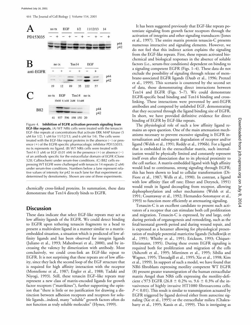
That EGFR kinase activity is required for downstream cellresponses was corroborated directly by inhibiting EGFR us-ing the pharmacological agent PD153035. This selective in-hibitor of EGFR kinase blocked ERK MAP kinase activa-tion by the tenascin EGF–like repeats (Fig. 4 A).
The foregoing results may be due to the tenascin repeatsinteracting with an unknown receptor but transmitting sig-nal through an intact EGFR kinase, as has been reported re-cently for G protein–linked receptors (Daub et al., 1996),integrins (Li et al., 1999), and growth hormone receptor(Yamauchi et al., 1997). To probe this unlikely possibility,
we blocked the external binding site of known EGFRligands using a nonactivating antibody (clone 528; Sunadaet al., 1986). The extracellular domain antibody inhibitedthe induced MAP kinase activation (Fig. 4 B), indicatingthat activation of the EGFR by tenascin-C EGF–like repeatoccurs through the ligand binding, extracellular domain ofthe EGFR. However, it is still possible that the signals occurby a receptor cascade in which the unknown receptor causesrelease of membrane-anchored EGFR ligands that then actin autocrine fashion, as has been shown for G protein–cou-pled receptors (Prenzel et al., 1999). This is unlikely, asNR6 cells are not known to produce significant levels ofEGFR ligands. However, we challenged B82 cells that havebeen shown not to express any of the known EGFR ligands(Oehrman et al., 1998) with the tenascin 14 fragment (Fig.4 C). The tenascin 14 fragment activated ERK MAP kinasesimilarly to what had been noted in WT NR6 cells.
Tenascin EGF–like repeats directly bind to EGFR
Final proof that these EGF-like repeats act as novel directEGFR ligands, however, requires a visualization of EGFR-dependent binding and/or a demonstration of an interactionwith EGFR. In direct binding assays, we were unable to de-tect specific binding to the EGFR by the EGF-like repeatproteins at micromolar concentration (data not shown).This indicates that the ligand is quite low affinity (
�
10 uM)when compared with prototypical growth factor ligands forEGFR (in the low nanomolar range). This proposed low so-lution affinity of the tenascin 14 repeats appears to be com-mensurate with the solution affinities for integrin ligands,which are in the micromolar range for fibronectin (Akiyamaand Yamada, 1985) and the millimolar range for linear argi-nine–glycine–aspartic acid peptides (Pierschbacher andRuoslahti, 1987). Since specific binding to these integrinligands can be readily detected by presenting the ligandsfrom the solid phase (i.e., bound to beads or the substrate;Pierschbacher and Ruoslahti, 1984) in a relatively normalphysiological manner, we reasoned that specific binding tothe tenascin 14 repeats might also be detected by presentingthe repeats in a method that resembles their presentation inECM. We generated
�
1-um diameter beads that presenteda high surface density of ligand (either tenascin 14 or EGF).Beads presenting EGF or the tenascin 14 fragment exhibitedspecific adhesion to WT NR6 cells compared with controlbeads (Fig. 5). That this occurred via EGFR is demonstratedby blocking of binding by anti-EGFR antibodies (number ofbound beads were reduced by
�
90% in each of three in-dependent experiments). There was negligible binding toM721 NR6 cells, which are devoid of EGFR.
Direct interaction of the EGF-like repeats with EGFRshould be enhanced by increasing the valency of the ligand,with even dimerization sufficient to achieve a log-greateravidity (Mammen et al., 1998). Ligands were predimerizedwith an antibody to the poly-His tag at the NH
2
terminus ofthe expressed repeats. The binding of these ligands to WTNR6 cells was visualized by indirect immunofluorescence(Fig. 6). As a positive control, murine EGF (mEGF-His6)was cloned and purified in a similar manner as the tenascinEGF–like repeats. Ten14 dimers bound at a level signifi-cantly greater than antibody alone. Most importantly, this
Figure 2. Tenascin EGF–like repeats activate the EGFR kinase cas-cade. WT (top) and M (bottom) NR6 cells were treated with EGF-like repeat proteins and activation of EGFR signaling was assessed. (A) EGFR autophosphorylation was determined by antiphosphoty-rosine immunoblotting of immunoprecipitated EGFR after treatment with EGF or EGF-like repeats 1/2 (5 uM), 11/12/13 (1 uM), or 14 (6 uM). Immunoblotting with an antibody to EGFR demonstrated equal loading (data not shown). (B) ERK MAP kinase activation was as-sessed by immunoblotting for dually phosphorylated p44/p42 ERK, indicative of activated ERK. The cells were treated with various con-centrations of the EGF-like repeats (5 uM for 1/2; 2 uM for 9/10; 1 uM for 11/12/13; and 6 uM for 14) and at 50 and 10% of those lev-els. EGF and serum were used as positive controls (for WT and M NR6 cells, respectively). Numbers below a lane represent relative values of intensity for pEGFR or p42 in each lane for that experi-ment as determined by densitometry. In both panels, an experiment representative of at least three determinations are shown.
on July 8, 2014jcb.rupress.org
Dow
nloaded from
Published July 16, 2001
EGF-like repeats as EGF receptor ligands |
Swindle et al. 463
binding, and that of mEGF-His6, was competed by 100 nMEGF that did not present the poly-His tag. Interestingly,Ten14 that was not predimerized also demonstrated statisti-cally significant cell association, though at a slightly lowerlevel than the predimerized Ten14. These data, includingthe bead-binding study, strongly support the model of lowaffinity ligands interacting with the EGFR.
The final demonstration would be to biochemically de-tect an association between EGFR and Ten14. To confirmthe immunofluorescence of Ten14 binding to EGFR, wecross-linked the ligands to their receptors and examined im-munoprecipitates of the poly-His tags. Upon poly-Hisimmunoprecipitation, we detected EGFR in cells dithio-bis(succinimidyl propionate) cross-linked in the presence of
either mEGF-His6 or Ten14 (Fig. 7 A). This interaction wasnot noted in cells competitively treated with 100 nM unla-beled EGF. Diluent (notx) or just secondary antibody (IgG)alone did not identify any cell surface receptor. Further-more, we assessed specificity by probing for another cell sur-face receptor, the insulin receptor (Fig. 7 B); we could notdetect any interaction between Ten14 or mEGF-His6 andthis receptor, further establishing the fact that binding isspecific for EGFR. That Ten14 coprecipitated at least simi-lar levels of EGFR, as did mEGF-His6, is not unexpected.The extended period of cross-linking would minimize theeffects of the rapid off-rate predicted for Ten14 and thus“drive” the binding towards completion. The slight retarda-tion of migration for EGFR noted is also not unexpected for
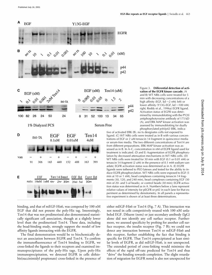
Figure 3. Differential detection of acti-vation of the EGFR kinase cascade. (A and B) WT NR6 cells were treated for 5 min with decreasing concentrations of a high affinity (EGF, kd �2 nM; left) or lower affinity (Y13G-EGF, kd �100 nM; right; Reddy et al., 1996a) EGFR ligand. Activation status of EGFR was deter-mined by immunoblotting with the PY20 antiphosphotyrosine antibody of 175 kD (A), and ERK MAP kinase activation was assessed by immunoblotting for dually phosphorylated p44/p42 ERK, indica-
tive of activated ERK (B). no tx designates cells not exposed to ligand. (C) WT NR6 cells were treated as in B with various concen-trations of EGF or 2 uM tenascin 14 fragment in quiescence media or serum-free media. The two different concentrations of Ten14 are from different preparations. ERK MAP kinase activation was as-sessed as in B. In A–C, concentration in nM of EGFR ligand used for treatment is indicated. (D and E) Augmentation of EGFR phosphory-lation by decreased attenuation mechanisms in WT NR6 cells. (D) WT NR6 cells were treated for 30 min with EGF (0.1 or 0.01 nM) or tenascin 14 fragment (2 uM) in the presence of 0.1 mM sodium van-adate; EGFR activation status was determined as in A. (E) EGFR ligands were tethered to PEO latexes and tested for the ability to in-duce EGFR phosphorylation. WT NR6 cells were exposed to EGF (5 min at 10 or 1 nM), bead complexes containing tenascin 14 frag-ments (30, 120, and 240 min), bead complexes containing EGF (30 min of 20- and 5-ul beads), or control beads (30 min); EGFR activa-tion status was determined as in A. Numbers below a lane represent relative values of intensity for pEGFR or p42 in each lane for that ex-periment as determined by densitometry. In all panels a representa-tive experiment is shown of at least three determinations.
on July 8, 2014jcb.rupress.org
Dow
nloaded from
Published July 16, 2001
464 The Journal of Cell Biology
|
Volume 154, 2001
chemically cross-linked proteins. In summation, these datademonstrate that Ten14 directly binds to EGFR.
Discussion
These data indicate that select EGF-like repeats may act aslow affinity ligands of the EGFR. We could detect bindingto EGFR upon tethering tenascin fragments to a bead topresent a multivalent ligand in a manner similar to a matrix-embedded situation, a situation which is predicted of low af-finity ligands and has been observed for integrin ligands(Juliano et al., 1993; Maheshwari et al., 2000), and by in-creasing the valency by dimerization with antibody. Mostconclusively, we could cross-link an EGF-like repeat toEGFR. It is not surprising that these repeats are of low affin-ity, since they lack the second loop of the EGF structure thatis required for high affinity interaction with the receptor(Montelione et al., 1987; Engler et al., 1988; Tadaki andNiyogi, 1993). Still, these tenascin EGF–like repeats mayrepresent a new class of matrix-encoded ligands for growthfactor receptors (“matrikines”), further supporting the opin-ion that “there is little or no justification for drawing a dis-tinction between adhesion receptors and receptors for solu-ble ligands...indeed, many “soluble” growth factors often donot function as truly soluble molecules” (Hynes, 1999).
It has been suggested previously that EGF-like repeats po-tentiate signaling from growth factor receptors through theactivation of integrins and other signaling transducers (Joneset al., 1997). The entire matrix protein tenascin-C presentsnumerous interactive and signaling elements. However, wedo not feel that this indirect action explains the signalingfrom the EGF-like repeats. First, these repeats activated bio-chemical and biological responses in the absence of solublefactors (i.e., serum-free conditions) dependent on binding toa signaling competent EGFR (Figs. 1–4). These data do notexclude the possibility of signaling through release of mem-brane-associated EGFR ligands (Daub et al., 1996; Prenzelet al., 1999). This scenario is countered by the second setof data, those demonstrating direct interactions betweenTen14 and EGFR (Figs. 5–7). We could demonstrateEGFR-specific bead binding and Ten14 binding and cross-linking. These interactions were prevented by anti-EGFRantibodies and competed by unlabeled EGF, demonstratingthat they occurred through the ligand binding site of EGFR.In short, we have provided definitive evidence for directbinding of EGFR by EGF-like repeats.
The physiological role of such a low affinity ligand re-mains an open question. One of the main attenuation mech-anisms necessary to prevent excessive signaling is EGFR in-ternalization and subsequent degradation of receptor and/orligand (Welsh et al., 1991; Reddy et al., 1996b). For a ligandthat is embedded in the extracellular matrix, such internal-ization would be physically limited and constantly representitself even after dissociation due to its physical proximity tothe cell surface. A matrix-embedded ligand with high affinitywould lead to continuous, strong signaling through EGFR;this has been shown to lead to cellular transformation (Di-Fiore et al., 1987; Wells et al., 1990). In contrast, a ligandwith low affinity (fast off rate; Ebner and Derynck, 1991)would result in ligand decoupling from receptor, allowingdephosphorylation and other mechanisms (Welsh et al.,1991; Countaway et al., 1992; Hernandez-Sotomayor et al.,1993) to function more efficiently at attenuating signaling.
Tenascin-C is an excellent candidate to present such acti-vators of a receptor that can stimulate both cell proliferationand migration. Tenascin-C is expressed, by and large, onlyduring periods of organogenesis and remodeling, such as thefetal/neonatal growth period and during wound repair, andis expressed as a hexamer allowing for physiological presen-tation of multiple potential matricrine ligands (Schalkwijk etal., 1991; Whitby et al., 1991; Erickson, 1993; Chiquet-Ehrismann, 1995). During these events EGFR signaling isrequired both for proliferation and migration of the cells(Ashcroft et al., 1995; Miettinen et al., 1995; Sibilia andWagner, 1995; Threadgill et al., 1995; Xie et al., 1998; Kimet al., 1999). In support of such a model, we have found thatNR6 fibroblasts expressing motility competent WT EGFR(8) present greater transmigration of the human extracellularmatrix Amgel than NR6 cells expressing the motility-defi-cient c’973 EGFR (26.8
�
0.2% vs. 9.6
�
0.3% of the in-vasiveness of highly invasive HT1080 fibrosarcoma cells;
P
�
0.01). This result is similar to transmigration actuated byEGFR triggered by ligand derived either from autocrine sig-naling (Xie et al., 1995) or the extracellular milieu (Chakra-barty et al., 1995; Kassis et al., 1999). This is intriguing as
Figure 4. Inhibition of EGFR activation prevents signaling from EGF-like repeats. (A) WT NR6 cells were treated with the tenascin EGF–like repeats at concentrations that activate ERK MAP kinase (5 uM for 1/2; 1 uM for 11/12/13; and 6 uM for 14). The cells were treated with the EGF-like repeat proteins in the absence (�) or pres-ence (�) of the EGFR-specific pharmacologic inhibitor PD153035. no tx represents no ligand. (B) WT NR6 cells were treated with Ten14 (1 uM) or EGF (0.01 nM) in the presence (�) or absence (�) of an antibody specific for the extracellular domain of EGFR (Clone 528; Calbiochem) under serum-free conditions. (C) B82 cells ex-pressing WT EGFR were challenged with tenascin 14 repeats (2 uM) under serum-free conditions. Numbers below a lane represent rela-tive values of intensity for p42 in each lane for that experiment as determined by densitometry. Shown are one of three experiments. on July 8, 2014
jcb.rupress.orgD
ownloaded from
Published July 16, 2001

EGF-like repeats as EGF receptor ligands |
Swindle et al. 465
Figure 5. Beads presenting tetheredtenascin 14 fragments bind to EGFR. WT NR6 cells were treated for 4 h at 37C with beads tethered with eithertenascin 14 or EGF or control beads. Cells were washed three times with iced PBS and visualized by phase-contrast microscopy (top). In parallel, cells were treated with 5 ug/ml anti-EGFR antibody (clone 528; Sunada et al., 1986; Turner et al., 1996) for 15 min before addition of beads and continuing throughout the 4 h (bottom). Beads appear as bright, re-fractile, and translucent spheres mainly above the plane of the cells. Shown is one of three experiments.
Figure 6. Tenascin 14 binds to EGFR-expressing WT NR6 cells. (A) WT NR6 cells were quiesced for 24 h and ex-posed to various concentrations of mo-nomeric or dimerized ligand to visualize binding to EGFR. Ten14 and mEGF-His6 expressed from the same vector (pRSETA; Invitrogen) as Ten14 were in-cubated overnight for dimerization with monoclonal anti-HisG (Invitrogen) anti-body that recognized the NH2-terminal poly His epitope of mEGF-His6 and Ten14. This was at concentrations of 50 nM for mEGF-His6 and 25 nM formonoclonal anti-HisG antibody and2 uM Ten14 with 0.63 uM of the anti-body (Ab) to increase affinity to receptor.
Ligands were incubated for 10 min at room temperature before fixation. 1:500 of goat anti–mouse antibody conjugated to Oregon green was used as secondary antibody before visualizing by fluorescence microscopy and captured at a constant exposure by a SPOT II CCD camera. a, mEGF-His6 (50 nM); b, mEGF-His6 (50 nM) dimerized with primary antibody (25 nM); c, cells were preincubated with EGF (100 nM) for 5 min to compete for EGFR with mEGF-His6 (50 nM) dimerized with antibody (25 nM); d, Ten14 (2 uM); e, Ten14 (2 uM) dimerized with pri-mary antibody(0.63 uM); f, cells preincubated with EGF (100 nM) for 5 min with Ten14 (2 uM) preincubated with 0.63 uM antibody subse-quently added. (B) Each cell in four randomly selected fields were outlined and measured for luminosity as compared with background. The data are the mean �SE of an average of �25 cells per experimental condition. Statistical analyses were performed via Student’s t test. Double asterisk represents P � 0.01. Shown is one representative of two sets of experiments.
on July 8, 2014jcb.rupress.org
Dow
nloaded from
Published July 16, 2001

466 The Journal of Cell Biology
|
Volume 154, 2001
NR6 cells do not produce known EGFR ligands, and Am-gel, derived from human amniotic membranes (Siegal et al.,1993), does not contain detectable levels of EGF, TGF-
,
orother soluble EGFR ligands, but does contain appreciablelevels of tenascin (75 mg/ml out of
�
1,300 mg/ml proteina-ceous material). Thus, low affinity ligands encrypted withinmatrix components might represent a new mode of modula-tion of cellular responses by matrix acting directly throughgrowth factor receptors.
Materials and methods
Cell lines and plasmids
The establishment and maintenance of the WT and M NR6 cell lines havebeen described previously (Wells et al., 1990; Chen et al., 1994). In brief,cells were grown in MEM with 7.5% fetal calf serum and 350 ug/ml G418.Cells were quiesced in MEM containing 1% dialyzed fetal calf serum. Fail-ure to adequately quiesce the cells results in higher background phosphor-ylation of EGFR and ERK even in the absence of exogenous added EGFRligand. The percentage of dialyzed serum and length of time of the quies-cence before testing must be empirically determined for each lot of dia-lyzed serum. For the NR6 cells and our lots of serum, 24 h of quiescencewas sufficient. Tenascin-C cDNA was generated from human placentalRNA by reverse transcriptase PCR using tenascin-C–specific primers (cgcg-gatccggccccaactgctctgagc and ccggaattcagacacctctgagcagtc), and ligatedinto pTrc-His-A (Invitrogen) to yield plasmid pTrc-ten. DNAs coding forspecific EGF-like repeat regions within tenascin-C were generated from thepTrc-ten template by PCR and ligated into pRSET-A (Invitrogen) to yieldplasmids pTen-1/2, -9/10, -11/12/13, and -14 with the numbers corre-sponding to the order with which each repeat occurs within tenascin C.Sequences of primer pairs used in the PCR were cgcggatccggccccaact-gctctgagc and ccggaattcgatttcacggctgcagtc for pTen-1/2; cgcggatccagc-cagctacggtgc and ccggaattcttggcgatcccggcag for pTen-9/10; cgcggatcccgg-gatcgccaatgc and ccggaattcggagtgctggccacag for pTen-11/12/13; andcgcggatccggccagcactcctgc and ccggaattcagacacctctgagcagtc for pTen-14.mEGF-His6 was similarly cloned to serve as a control. These cloningsyielded the EGF-like repeats and mEGF preceded by poly-His.
Expression and purification of EGF-like repeat proteins
Midlog phase cultures of
Escherichia coli
strain BL21/DE3/pLys-S (Strat-
agene) transformed with the individual expression plasmids were inducedfor recombinant protein expression with 1 mM isopropyl-b-D-thiogalacto-pyranoside for 4 h at 37
C. Bacteria were harvested by centrifugation for 10min at 5,000
g
at 4
C, and bacterial lysates were prepared by extractionwith 0.02 culture volumes of B-PER detergent (Pierce Chemical Co.). Re-combinant proteins were purified from bacterial lysates by nickel-agarosechromatography with imidazole elution. Purified protein was dialyzedagainst PBS, 0.25 mM 2-mercaptoethanol for 24 h at room temperature.
Mitogenesis assay
Cells were quiesced for 24 h under normal growth conditions in starvationmedium (serum-free growth medium supplemented with 1% dialyzed fetalcalf serum). The ligand-induced
3
H-thymidine incorporation assay hasbeen described previously (Chen et al., 1996). In brief, cells were exposedto EGF (1 nM), serum (1%), or various concentrations of EGF-like repeatproteins for 24 h.
3
H-thymidine was added to the cells for the last 8 h todetermine stimulation of proliferation.
Phosphorylation assays
For assaying EGFR activation, quiesced cells were treated with ligand for 5min in quiescence medium (for the experiments described in Fig. 3 D only,the medium was supplemented with 0.1 mM sodium vanadate during thistime period and treated for 30 min). When indicated in the figure legends,after cells were quiesced the experiment was performed under serum-freeconditions; this further reduces background phosphorylation of ERK. De-tergent lysates were immunoprecipitated at 4
C with anti-EGFR antibody(Ab-1; Oncogene Research Products) bound to protein A–conjugated aga-rose (GIBCO BRL). Immunoprecipitated EGFR was analyzed for tyrosinephosphorylation by immunoblotting using antiphopshotyrosine antibody(PY20; Transduction Laboratories). For assessing MAP kinase activation,quiesced cells were treated with ligand for 5 min in the presence or ab-sence of anti-EGFR Ab-1 (4 ug/ml; Calbiochem) or PD153035 (1 uM).Whole cell lysates were analyzed for dually-phosphorylated ERK MAP ki-nase by immunoblotting using antiphospho-MAP kinase antibody (NewEngland Biolabs, Inc.). Equal loading was assured using the pan-erk anti-body. Relative densitometric values were derived with the NIH Imageshareware and Adobe Photoshop
®
software.
Tethered ligands
Tenascin 14 fragments and EGF were covalently tethered to surfaces topresent the ligands in a manner analogous to physiological presentation ofmatrix-associated tenascin. Poly(methyl methacrylate) latex beads weresynthesized by dispersion polymerization using an amphiphilic comb co-polymer stabilizer, following a procedure adapted from (Banerjee et al.,2000). A comb stabilizer comprised of methylmethacrylate, polyethyleneglycol methacrylate (Mn
�
526 g/mol), and methoxypolyethylene glycolmethacrylate (Mn
�
425 g/mol) in a weight ratio of 30:10:10 was synthe-sized by free radical polymerization using AIBN as initiator. The hydroxy-terminated polyethylene glycol side chains were subsequently carbox-ylated by refluxing 16 g of comb with 10 g succinic anhydride, and 0.15ml N-methyl imidazole in 300 ml of dichloroethane overnight at 80
C. Thecarboxylated product was precipitated and washed with acidified water.Polyethylene methylmethacrylate latexes were synthesized by the additionof 9 ml methylmethacrylate, 1.25 g of carboxylated comb stabilizer, 1.2 mlvinyl methacrylate cross-linking agent, and 0.50 g of ammonium persulfateinitiator to 45 ml of 70:30 (vol/vol) methanol/water. The reaction pro-ceeded at 50
C for 3 h, resulting in a highly stable dispersion of micron-sized polyethylene methylmethacrylate latex beads, each coated by combpolymers that situate and become grafted at the water/bead interface. Thecarboxylated latex suspension was purified by repeated centrifugation andredispersion, before peptide coupling.
The NH
2
-terminal amine groups of EGF and tenascin 14 fragments wereused to covalently link the peptides to ends of the PEO chains emanatingfrom the surface of the latex beads. Beads were resuspended in dry ethanolwith 20 mg/mL sulfo-NHS (Pierce Chemical Co.) and 1-ethyl-3-(3-dimeth-ylaminopropyl)carbodiimide hydrochloride (Pierce Chemical Co.) and ac-tivated at room temperature for 3 h, then centrifuged to remove superna-tant and resuspended in ethanol for a total of three washes. Beads werewashed a final time in 100 mM phosphate buffer, pH 7, and resuspendedin 5 ug/mL mouse EGF (Collaborative Biomedical), tenascin 14 fragment,or buffer alone in 100 mM phosphate buffer, pH 7. The coupling reactionwas allowed to proceed for 24 h at 4
C. Unreacted peptide was removedand residual NHS reactivity blocked by washing three times in 100 mMtris buffer, pH 7. The beads were then washed once with sterile PBS beforeaddition to cells.
Figure 7. Tenascin 14 can be cross-linked to EGFR. Ten14 (2 uM) and mEGF (mEGF-His6; 10 nM) were bound and chemically cross-linked to quiesced WT NR6 fibroblasts and immunoprecipitated from ensuing lysates with anti-HisG. Presence of EGFR (A) or insulin receptor � chain was assessed by immunoblotting with respective antibodies. In lanes 1 and 2, cells were preincubated with 100 nM EGF for 5 min and throughout cross-linking as a competitive ligand. In lane 5, cells were exposed to and cross-linked with antibody alone and lysate was immunoprecipitated. From left to right: 1, EGF/mEGF-His6 (pretreatment with EGF and addition of mEGF-His6 ligand); 2, EGF/Ten14 (pretreatment with EGF and addition of Ten14 ligand); 3, mEGF-His6 (10 nM); 4, Ten14 (2 uM); 5, IgG (anti-HisG antibody); 6, No tx (no treatment with ligand); and 7, cell ly-sate (WT NR6 lysate). Shown is a representative of two experiments.
on July 8, 2014jcb.rupress.org
Dow
nloaded from
Published July 16, 2001
EGF-like repeats as EGF receptor ligands |
Swindle et al. 467
Immunofluorescence
WT NR6 cells were quiesced at 50% confluency. Mouse anti-HisG anti-body (Invitrogen) was incubated 24 h at 4
C with Ten14 or mEGF-His6 fordimerization. After quiescence, ligand, either without antibody or anti-body-ligand mix, was added to cells in serum-free media and incubatedfor 10 min at room temperature. 100 nM EGF served as a competitor andwas added 5 min before the Ten14 or mEGF-His6. Cells were fixed with3% formaldehyde for 15 min at room temperature, washed three timeswith PBS and incubated in 1% BSA for 30 min. Cells that were exposed toligand alone were washed twice with PBS and incubated with mouse anti-HisG antibody (Invitrogen; 1:1000) for 30 min at 37
C. Cells were washedfive times with PBS and secondary goat anti–mouse conjugated to Oregongreen (Molecular Probes; 1:1,000) was added at 37
C for 30 min. Cellswere once again washed three times, mounted, and viewed.
Immunoprecipitation
WT NR6 cells were quiesced at 80% confluency. Cells were washed oncewith PBS. 100 nM EGF served as a competitor and was added 5 min beforethe Ten14 or mEGF-His6. Cells were than washed with PBS and incubatedwith ligands Ten14 (2 uM) or mEGF-His6 (10 nM) in PBS for 5 min at roomtemperature. In parallel, cells were incubated with just PBS (no tx) or withmonoclonal anti-HisG (0.01 uM) in PBS. Dithiobis(succinimidyl propi-onate) (Pierce Chemical Co.) was added to the solution and the cells wereplaced at 4
C for 30 min. Cells were than washed with 0.2% glycine solu-tion in PBS twice and incubated with 0.2% glycine in PBS for 5 min at 4
Cfollowed by a final wash with 0.2% glycine in PBS once again. Cells werelysed with RIPA lysis buffer with PMSF, aprotinin, and leupeptin as pro-tease inhibitors. 30 ul of protein G agarose beads (GIBCO BRL) and mouseanti-HisG (Invitrogen; final concentration, 0.01 uM) was added to the ly-sate and incubated overnight. Beads were washed for a total of five times.Lysates were separated by SDS-PAGE with 2-mercaptoethanol (to cleavethe cross-linker), transferred, and immunoblotted. The upper half of themembrane was probed with a monoclonal anti-EGFR (Zymed Laborato-ries; 1:500) and the bottom for polyclonal antiinsulin receptor
�
-subunit(Transduction Laboratories; 1:1,000).
We thank Alan Hall and Doug Lauffenburger for critical insights and dis-cussions. We thank Hidenori Shiraha and Latha Satish for excellent assis-tance with the immunofluorescence studies.
These studies were supported by grants from the National Institutes ofHealth/National Institute of General Medical Sciences and the NationalScience Foundation.
Submitted: 22 March 2001Revised: 24 May 2001Accepted: 29 May 2001
References
Akiyama, S.K., and K.M. Yamada. 1985. Synthetic peptides competitively inhibitboth direct binding to fibroblasts and functional biological assays for the pu-rified cell-binding domain of fibronectin.
J. Biol. Chem
. 260:4492–4500.Anklesaria, P., J. Teixido, M. Laiho, J.H. Pierce, J.S. Greenberger, and J. Mas-
sague. 1990. Cell adhesion mediated by binding of membrane-anchoredtransforming growth factor alpha to epidermal growth factor receptors pro-motes cell proliferation.
Proc. Natl. Acad. Sci. USA
. 87:3289–3293.Ashcroft, G.S., M.A. Horan, and M.W. Ferguson. 1995. The effects of ageing on
cutaneous wound healing in mammals.
J. Anat
. 187:1–26.Banerjee, P., D.J. Irvine, A.M. Mayes, and L.G. Griffith. 2000. Polymer latexes for
controlling cell adhesion and receptor-mediated interactions.
J. Biomed.Mater. Res
. 50:331–339.Bhalla, U.S., and R. Iyengar. 1999. Emergent properties of networks of biological
signaling pathways.
Science
. 283:381–387.Brachmann, R., P.B. Lindquist, M. Nagashima, W. Kohr, T. Lipari, M. Napier,
and R. Derynck. 1989. Transmembrane TGF-
precursors activate EGF/TGF-
receptors.
Cell
. 56:691–700.Chakrabarty, S., S. Rajagopal, and S. Huang. 1995. Expression of antisense epider-
mal growth factor receptor RNA downmodulates the malignant behavior ofhuman colon cancer cells.
Clin. Exp. Metastasis
. 13:191–195.Chen, P., K. Gupta, and A. Wells. 1994. Cell movement elicited by epidermal
growth factor receptor requires kinase and autophosphorylation but is sepa-rable from mitogenesis.
J. Cell Biol
. 124:547–555.Chen, P., H. Xie, and A. Wells. 1996. Mitogenic signaling from the EGF receptor
is attenuated by a motility-associated phospholipase C-
�
/protein kinase C
feedback mechanism.
Mol. Biol. Cell
. 7:871–881.Chiquet-Ehrismann, R. 1995. Tenascins, a growing family of extracellular matrix
proteins.
Experentia. 51:853–862.Countaway, J.L., A.C. Nairn, and R.J. Davis. 1992. Mechanism of desensitization
of the epidermal growth factor receptor protein-tyrosine kinase. J. Biol.Chem. 267:1129–1140.
Daub, H., T.U. Weiss, C. Wallasch, and A. Ullrich. 1996. Role of transactivationof the EGF receptor in signalling by G-protein-coupled receptors. Nature.379:557–560.
Davis, S., N.W. Gale, T.H. Aldrich, P.C. Maisonpierre, V. Lhotak, T. Pawson, M.Goldfarb, and G.D. Yancopoulos. 1994. Ligands for EPH-related receptortyrosine kinases that require membrane attachment or clustering for activity.Science. 266:816–819.
DiFiore, P.P., J.H. Pierce, T.P. Fleming, R. Hazan, A. Ullrich, C.R. King, J.Schlessinger, and S.A. Aaronson. 1987. Overexpression of the human EGFreceptor confers an EGF-dependent transformed phenotype to NIH 3T3cells. Cell. 51:1063–1070.
Ebner, R., and R. Derynck. 1991. Epidermal growth factor and transforminggrowth factor-: differential intracellular routing and processing of ligand-receptor complexes. Cell Regul. 2:599–612.
Engel, J. 1989. EGF-like domains in extracellular matrix proteins: localized signalsfor growth and differentiation? FEBS Lett. 251:1–7.
Engler, D.A., R.K. Matsunami, S.R. Campion, C.D. Stringer, A. Stevens, and S.Niyogi. 1988. Cloning of authentic human epidermal growth factor as abacterial secretory protein and its initial structure-function analysis by site-directed mutagenesis. J. Biol. Chem. 263:12384–12390.
Erickson, H.P. 1993. Tenascin-C, tenascin-R and tenascin-X: a family of talentedproteins in search of functions. Curr. Opin. Cell Biol. 5:869–876.
Haugh, J.M., K. Schooler, A. Wells, H.S. Wiley, and D.A. Lauffenburger. 1999.Effect of epidermal growth factor receptor internalization on regulation ofthe phospholipase C-� signaling pathway. J. Biol. Chem. 274:8958–8965.
Herbst, J.J., L.K. Opresko, B.J. Walsh, D.A. Lauffenburger, and H.S. Wiley. 1994.Regulation of postendocytic trafficking of the epidermal growth factor re-ceptor through endosomal retention. J. Biol. Chem. 269:12865–12873.
Hernandez-Sotomayor, S.M.T., C.L. Artega, C. Soler, and G. Carpenter. 1993.Epidermal growth factor stimulates substrate-selective protein-tyrosine phos-phatase activity. Proc. Natl. Acad. Sci. USA. 90:7691–7695.
Hynes, R.O. 1999. Cell adhesion: old and new questions. Trends Biol. Sci. 24:M33–M37.
Jones, P.L., J. Crack, and M. Rabinovitch. 1997. Regulation of tenascin-C, a vas-cular smooth muscle cell survival factor that interacts with the v �3 inte-grin to promote epidermal growth factor receptor phosphorylation andgrowth. J. Cell Biol. 139:279–293.
Juliano, D.J., S.S. Saavedra, and G.A. Trusky. 1993. Effect of the conformationand orientation of adsorbed fibronectin on endothelial cell spreading and thestrength of adhesion. J. Biomed. Mater. Res. 27:1103–1113.
Kassis, J., J. Moellinger, H. Lo, N. Greenberg, H.-G. Kim, and A. Wells. 1999. Arole for phospholipase C-�-mediated signaling in tumor cell invasion. Clin.Cancer Res. 5:2251–2260.
Khazaie, K., V. Schirrmacher, and R.B. Lichtner. 1993. EGF receptor in neoplasiaand metastasis. Cancer and Metastasis Reviews. 12:255–274.
Kim, H., T. Turner, J. Kassis, J. Souto, and A. Wells. 1999. EGF receptor signal-ing in prostate development. Histol. Histopathol. 14:1175–1182.
Kuhl, P.R., and L.G. Griffith-Cima. 1996. Tethered epidermal growth factor as aparadigm for growth factor-induced stimulation from the solid phase. Nat.Med. 2:1022–1027.
Li, J., M.L. Lin, G.J. Wiepz, A.G. Guadarrama, and P.J. Bertics. 1999. Integrin-mediated migration of murine B82L fibroblasts is dependent on the expres-sion of an intact epidermal growth factor receptor. J. Biol. Chem. 274:11209–11219.
Maheshwari, G., G. Brown, D.A. Lauffenburger, A. Wells, and L.G. Griffith.2000. Cell adhesion and motility depend on nanoscale RGD clustering. J.Cell Sci. 113:1677–1686.
Mammen, M., S.-K. Choi, and G.M. Whitesides. 1998. Polyvalent interactions inbiological systems: implications for design and use of multivalent ligandsand inhibitors. Angew. Chem. Int. Ed. 37:2754–2794.
Miettinen, P.J., J.E. Berger, J. Meneses, Y. Phung, R.A. Pedersen, Z. Werb, and R.Derynck. 1995. Epithelial immaturity and multiorgan failure in mice lack-ing epidermal growth factor receptor. Nature. 376:337–341.
Montelione, G.T., K. Wuthrich, E.C. Nice, A.W. Burgess, and H.A. Scheraga.1987. Solution structure of murine epidermal growth factor: determinationof the polypeptide backbone chain-fold by nuclear magnetic resonance and
on July 8, 2014jcb.rupress.org
Dow
nloaded from
Published July 16, 2001
468 The Journal of Cell Biology | Volume 154, 2001
distance geometry. Proc. Natl. Acad. Sci. USA. 84:5226–5230.Nelson, J., W.E. Allen, W.N. Scott, J.R. Bailie, B. Walker, N.V. McFarren, and
D.J. Wilson. 1995. Murine epidermal growth factor (EGF) fragment (33-42) inhibits both EGF- and laminin-dependent endothelial cell motility andangiogenesis. Cancer Res. 55:3772–3776.
Oehrman, G.T., H.S. Wiley, and D.A. Lauffenburger. 1998. Escape of autocrineligands into extracellular medium: experimental test of theoretical modelpredictions. Biotechnol. Bioeng. 57:571–582.
Panayotou, G., P. End, M. Aumailley, R. Timpl, and J. Engel. 1989. Domains oflaminin with growth-factor activity. Cell. 56:93–101.
Pierschbacher, M.D., and E. Ruoslahti. 1984. Cell attachment activity of fibronec-tin can be duplicated by small synthetic fragments of the molecule. Nature.309:30–33.
Pierschbacher, M.D., and E. Ruoslahti. 1987. Influence of stereochemistry of thesequence Arg-Gly-Asp-Xaa on binding specificity in cell adhesion. J. Biol.Chem. 262:17294–17298.
Prenzel, N., E. Zwick, H. Daub, M. Leserer, R. Abraham, C. Wallasch, and A. Ull-rich. 1999. EGF receptor transactivation by G-protein-coupled receptors re-quires metalloproteinase cleavage of proHB-EGF. Nature. 402:884–888.
Prieto, A.L., C. Andersson-Fisone, and K.L. Crossin. 1992. Characterization ofmultiple adhesive and counteradhesive domains in the extracellular matrixprotein cytotactin. J. Cell Biol. 119:663–678.
Pruss, R.M., and H.R. Herschman. 1977. Variants of 3T3 cells lacking mitogenicresponse to epidermal growth factor. Proc. Natl. Acad. Sci. USA. 74:3918–3921.
Reddy, C.C., S.K. Niyogi, A. Wells, H.S. Wiley, and D.A. Lauffenburger. 1996a.Re-engineering epidermal growth factor for enhanced potency. Nat. Biotech-nol. 14:1696–1699.
Reddy, C.C., A. Wells, and D.A. Lauffenburger. 1996b. Receptor-mediated effectson ligand availability influence relative mitogenic potencies of epidermalgrowth factor and transforming growth factor a. J. Cell. Physiol. 166:512–522.
Schalkwijk, J., P.M. Steijlin, I.M.J.J.v. Vlijmen-Willems, B. Oosterling, E.J.Mackie, and A.A. Verstraeten. 1991. Tenascin expression in human dermisis related to epidermal proliferation. Am. J. Pathol. 139:1143–1150.
Shrivastava, A., C. Radziejewski, E. Campbell, L. Kovac, M. McGlynn, T.E. Ryan,S. Davis, M.P. Goldfarb, D.J. Glass, G. Lemke, and G.D. Yancopoulos.1997. An orphan receptor tyrosine kinase family whose members serve asnonintegrin collagen receptors. Mol. Cell. 1:25–34.
Sibilia, M., and E.F. Wagner. 1995. Strain dependent epithelial defects in micelacking the EGF receptor. Science. 269:234–238.
Siegal, G.P., M.-H. Wang, C.A. Rinehart, J.W. Kennedy, L.J. Goodly, Y. Miller,D.G. Kaufman, and R.K. Singh. 1993. Development of a novel human ex-tracellular matrix for quantitation of the invasiveness of human cells. CancerLett. 69:123–132.
Sunada, H., B.E. Magun, J. Mendelsohn, and C.L. MacLeod. 1986. Monoclonal
antibody against epidermal growth factor receptor is internalized withoutstimulating receptor phosphorylation. Proc. Natl. Acad. Sci. USA. 83:3825–3829.
Tadaki, D.K., and S.K. Niyogi. 1993. The functional importance of hydrophobic-ity of the tyrosine at position 13 of human epidermal growth factor in recep-tor binding. J. Biol. Chem. 268:10114–10119.
Threadgill, D.W., A.A. Dlugosz, L.A. Hansen, T. Tennenbaum, U. Lichti, D. Yee,C. LaMantia, T. Mourton, K. Herrup, R.C. Harris, et al. 1995. Targeteddisruption of mouse EGF receptor: effects of genetic background on mutantphenotype. Science. 269:230–234.
Turner, T., P. Chen, L.J. Goodly, and A. Wells. 1996. EGF receptor signaling en-hances in vivo invasiveness of DU-145 human prostate carcinoma cells. Clin.Exp. Metastasis. 14:409–418.
Vieira, A.V., C. Lamaze, and S.L. Schmid. 1996. Control of EGF receptor signal-ing by clathrin-mediated endocytosis. Science. 274:2086–2089.
Vogel, W., G.D. Gish, F. Alves, and T. Pawson. 1997. The discoidin domain re-ceptor tyrosine kinases are activated by collagen. Mol. Cell. 1:13–23.
Waters, S.B., D. Chen, A.W. Kao, S. Okada, K.H. Holt, and S.E. Pessin. 1996.Insulin and epidermal growth factor receptors regulate distinct pools ofgrb2-sos in the control of ras activation. J. Biol. Chem. 271:18224–18230.
Wells, A. 2000. Tumor invasion: role of growth factor-induced cell motility. Adv.Cancer Res. 78:31–101.
Wells, A., J.B. Welsh, C.S. Lazar, H.S. Wiley, G.N. Gill, and M.G. Rosenfeld.1990. Ligand-induced transformation by a non-internalizing EGF receptor.Science. 247:962–964.
Wells, A., K. Gupta, P. Chang, S. Swindle, A. Glading, and H. Shiraha. 1998. Epi-dermal growth factor receptor-mediated motility in fibroblasts. Microsc. Res.Tech. 43:395–411.
Welsh, J.B., G.N. Gill, M.G. Rosenfeld, and A. Wells. 1991. A negative feedbackloop attenuates EGF-induced morphological changes. J. Cell Biol. 114:533–543.
Whitby, D.J., M.T. Longaker, M.R. Harrison, N.S. Adzick, and M.W.J. Ferguson.1991. Rapid epithelialisation of fetal wounds is associated with the earlydeposition of tenascin. J. Cell Sci. 99:583–586.
Xie, H., T. Turner, M.-H. Wang, R.K. Singh, G.P. Siegal, and A. Wells. 1995. Invitro invasiveness of DU-145 human prostate carcinoma cells is modulatedby EGF receptor-mediated signals. Clin. Exp. Metastasis. 13:407–419.
Xie, H., M.A. Pallero, K. Gupta, M.F. Ware, P. Chang, W. Witke, D.J. Kwiat-kowski, D.A. Lauffenburger, J. Murphy-Ullrich, and A. Wells. 1998. EGFreceptor regulation of cell-substratum interactions: EGF-induced disassem-bly of focal adhesions does not require the motility-associated PLC signalingpathway. J. Cell Sci. 111:616–625.
Yamauchi, T., K. Ueki, K. Tobe, H. Tamemoto, N. Sekine, M. Wada, M. Honjo,M. Takahashi, T. Takahashi, H. Hirai, et al. 1997. Tyrosine phosphoryla-tion of the EGF receptor by the kinase Jak2 is induced by growth hormone.Nature. 390:91–96.
on July 8, 2014jcb.rupress.org
Dow
nloaded from
Published July 16, 2001