Mitochondrial genomes are retained by selective constraints ...
Upload
khangminh22Category
view
0download
0
i
Virus integration and tandem repeats in the genomes of
Petunia
A thesis submitted to the University of Leicester for the
degree of Doctor of Philosophy
By
Osamah Nadhim Kadhim Alisawi M.Sc.
January 2019
Department of Genetics and Genome Biology

i
Abstract
The integration of endogenous pararetroviruses (EPRVs) and tandemly repeated sequences
were examined in whole genome raw reads, and two genome assemblies, in diploid Petunia
species including hybrid-derivatives and their ancestors, using bioinformatics, molecular
biology, cytogenetics and microscopy. Three types of EPRV clusters (petuvirus-,
florendovirus- and caulimovirus-like sequences) were found. Chromosomal signals of PVCV
(Petunia vein clearing virus) were seen by in situ hybridization in all Petunia species.
Fragmented parts of four novel florendovirus-like sequences were found and the complete
sequence was reconstructed, adding petunia to the 27 known host species. Chromosome III of
P. axillaris and P. hybrida Rdc showed strong pericentromeric signal of PVCV and
Florendovirus suggesting both EPRVs have similar positions, integration patterns and
endogenization events (unlike P. integrifolia subsp inflata and P. axillaris subsp parodii). The
caulimovirus-like sequence cluster was less abundant in genomes, with four novel members.
RNA analysis from infected and healthy petunia samples revealed expression of endogenous
PVCV and Caulimovirus sequences, unlike Florendovirus (not detected in RNA). The
episomal form of vertically transmitted PVCV was integrated near the telomere of heterologous
chromosomes. Transmission electron microscopy (TEM) showed differences in number and
size of PVCV particles and inclusion bodies for both chlorotic spots and vein clearing
symptoms, the latter correlated with PVCV particles in cytoplasm from vascular bundle cells.
In plants with chlorotic symptoms, infected cells contained virions in parenchyma cells, while
scattered virions were seen in chlorotic spots in P. hybrida W138 after heat induction of
symptoms. Eight unique types of tandem repeat clusters were analysed within Petunia raw
reads with variable genome proportions and different loci on mitotic chromosomes. Three were
useful markers for chromosome identification. Taken together, the work shows the contribution
of repetitive DNA to diversity and variation within petunia genomes, and has consequences for
evolution, and both resistance and spread of some viruses.

ii
Declaration
I hereby declare that no part of this thesis has been previously submitted to this or any other
university as part of the requirements for a higher degree. The study described in this thesis, unless
otherwise acknowledged in the text or by reference, was conducted by the undersigned who is
fully responsible.
This work was achieved in the Department of Genetics and Genome Biology, University of
Leicester and Julius Kühn-Institut (JKI), Braunschweig, Germany (see Appendix 4.1), during
the period from January 2015 to December 2018.
Signed:…………………………...
Date:………………………………
Osamah Nadhim Kadhim Alisawi

iii
Dedication
Thanks to ALLAH for blessing me much more than I deserve
This work is dedicated to the sacred two date palm trees grown on Euphrates river beach,
covered me with their fronds, and taken care of me for my entire life, proud of you both my
great father and mother
Nadhim Kadhim Alisawi and Rabab Waheed Alisawi
My lovely, amazing wife Wasan Riyadh Alisawi, who brilliantly supported me to build up
this story.
Future men, my little boys, Zaid, Yazan and newborn baby Taim.
My beloved sisters (Israa and Rawaa) and brothers (Ahmed and Yaseen).
I would also send my fulfilment and sincerity to my great grandfather who passed away and
left behind a massive heritage for the whole tribe (Al-Isa) and Iraqi community.
Al Sheikh Haj Waheed Abood Alisawi (1927-2012)
, my respected uncles
Dr. Abood Waheed Alisawi and Dr. Riyadh Waheed Alisawi
Finally, thanks must go to my homeland where the first ever civilization established
IRAQ

iv
Acknowledgements
First and foremost, I would like to express my sincere gratitude and appreciation to my three
supervisors Prof. Pat Heslop-Harrison, Dr. Trude Schwarzacher and Dr. Katja Richert-
Pöggeler, for their support, guidance, kindness and generosity over the whole time of study.
I am indebted to Prof. David Twell and Dr. Richard Gornall for their positive comments and
notes.
I would also like to acknowledge my thanks to Mr. Ramesh Patel and my colleagues in lab 201
Dr. Sarbast Ihsan Mustafa, Dr. Jotyar Jassim Muhammed, Dr. Rubar M. Salih, Dr. Nouf Fakieh
Alsayied and Noorhariza Mohd Zaki.
My special thanks go to very gentle members of Dr. Katja Richert-Pöggeler’s lab in Julius
Kühn-Institut (JKI), Braunschweig, Germany; Mr. Dirk Schmalowski, Ms. Christina Maaß
(Grille) and Ms. Sabine Schuhmann for their assistance and fruitful cooperation.
I highly appreciate the full cooperation of Assistant Prof. Aureliano Bombarely, Department
of Horticulture, Virginia Tech, USA, who provided me with raw reads and assemblies of
Petunia species.
I wish to thank my Iraqi friends Dr. Wadhah Mahbooba, Dr. Ghazwan Al Hassan, Dr. Pshtiwan
Bebane, Dr. Tariq A. Kareem, Mr. Imad Younis, Mr. Muhammed Abdul Sattar, Mr. Wisam
Aziz and Mr. Maitham Al-Jalali for their kind help.
I would like to thank my sponsorship; the Iraqi Ministry of Higher Education and Scientific
research and my home department; Plant Protection Department, Faculty of Agriculture,
University of Kufa for their funding and giving me this great opportunity.
Last, and by no means least, I wish to thank all people who have pushed me forward to be in
progress all time.

v
Contents
Abbreviations ....................................................................................................................................... viii
Chapter I. Introduction ............................................................................................................................ 1
1.1 The genus Petunia ............................................................................................................................. 1
1.2 The karyotype of petunia chromosomes ....................................................................................... 8
1.3 Repetitive DNA .......................................................................................................................... 10
1.3.1 Types of Repetitive DNA sequences ................................................................................... 11
1.4 Virus definition and taxonomy ................................................................................................... 13
1.5 Transposable elements (TEs) ...................................................................................................... 14
1.5.1 Retrotransposons (Class I elements) .................................................................................... 15
1.5.2 DNA transposons (Class II elements) .................................................................................. 18
1.5.3 Transposable elements in petunia ........................................................................................ 19
1.6 Fluorescence in situ hybridization .............................................................................................. 19
1.7 Bioinformatic techniques ............................................................................................................ 20
1.7.1 Whole genome sequencing data ........................................................................................... 20
1.7.2 k-mer counting ..................................................................................................................... 21
1.7.3 Graph-based clustering of raw read sequences .................................................................... 22
1.8 Aims and objectives .................................................................................................................... 25
Chapter II. Materials and methods ........................................................................................................ 26
2.1 Plant material and cultivation ..................................................................................................... 26
2.2 Standard solutions and media ..................................................................................................... 27
2.3 Methods....................................................................................................................................... 31
2.3.1 Genomic DNA extraction .................................................................................................... 31
2.3.2 DNA quantification .............................................................................................................. 32
2.3.3 PCR amplification ................................................................................................................ 32
2.3.4 Purification of DNA fragments from agarose gel ................................................................ 33
2.3.5 Cloning of PCR products ..................................................................................................... 33
2.3.6 Probe labelling ..................................................................................................................... 37
2.3.7 Petunia chromosome preparations ....................................................................................... 37
2.3.8 Fluorescent in situ hybridization .......................................................................................... 38
Chapter III. Integration of Endogenous Pararetroviruses (EPRVs) ...................................................... 41
3.1 Endogenous pararetroviruses (EPRVs) ....................................................................................... 41
3.1.1 Caulimoviridae .................................................................................................................... 42

vi
3.2 Aims and objectives .................................................................................................................... 46
3.3 Materials and methods ................................................................................................................ 47
3.3.1 EPRV fragments in Petunia species .................................................................................... 48
3.3.2 De novo integration .............................................................................................................. 56
3.4 Results ......................................................................................................................................... 62
3.4.1 EPRVs sequences ................................................................................................................. 62
3.4.2 Phylogenetic relationships of EPRVs .................................................................................. 82
3.4.3 De novo integration .............................................................................................................. 83
3.5 Discussion ................................................................................................................................... 90
3.5.1 PVCV fragments in all examined Petunia species .............................................................. 90
3.5.2 PVCV as a cytogenetic marker ............................................................................................ 91
3.5.3 Florendovirus-like sequences ............................................................................................... 92
3.5.4 Hotspot of Chromosome III ................................................................................................. 92
3.5.5 Caulimovirus-like sequences ............................................................................................... 93
3.5.6 Expression of EPRVs ........................................................................................................... 94
3.5.7 De novo integration .............................................................................................................. 96
Chapter IV. Transmission electron microscopy of PVCV .................................................................... 98
4.1 Introduction ................................................................................................................................. 98
4.1.1 Electron microscopy and detection of Petunia vein clearing virus (PVCV) ....................... 98
4.2 Aims and objectives .................................................................................................................... 99
4.3 Materials and methods ................................................................................................................ 99
4.3.1 Plant species ......................................................................................................................... 99
4.3.2 Electron microscopy .......................................................................................................... 100
4.4 Results ....................................................................................................................................... 103
4.4.1 Horizontally transmitted PVCV ......................................................................................... 103
4.4.2 Vertical transmission of PVCV .......................................................................................... 108
4.5 Discussion ................................................................................................................................. 113
4.5.1 Ultrastructural analysis of diffuse chlorosis and vein clearing symptoms after horizontal
transmission of PVCV ................................................................................................................ 113
4.5.2 De novo integration and vertical transmission of PVCV in P. axillaris subsp parodii S7 114
4.5.3 PVCV symptom expression and replication after induction of chromosomal copies ........ 115
Chapter V. Tandem repeats ................................................................................................................. 117
5.1 Introduction ............................................................................................................................... 117
5.2 Aims and objectives .................................................................................................................. 118
5.3 Materials and methods .............................................................................................................. 119

vii
5.3.1 Plant material and DNA extraction .................................................................................... 119
5.3.2 Tandem repeat analysis ...................................................................................................... 119
5.3.3 Chromosomes preparation and in situ hybridization ......................................................... 119
5.4 Results ....................................................................................................................................... 122
5.4.1 Identification of tandem repeats by graph-based clustering............................................... 122
5.4.2 Genome organization ............................................................................................................. 122
5.4.3 Chromosomal organization of tandem repeats ................................................................... 125
5.5 Discussion ................................................................................................................................. 144
5.5.1 Genome proportion of tandem repeat clusters ................................................................... 144
5.5.2 Tandemly repeated sequences within assemblies .............................................................. 145
5.5.3 Graph based clusters pipelines ........................................................................................... 145
5.5.4 The role of tandemly repeated sequences .......................................................................... 145
5.5.5 The evolution of tandem repeats ........................................................................................ 146
5.5.6 Tandem repeats as genetic markers.................................................................................... 147
Chapter VI. General discussion: Repetitive elements in petunia ........................................................ 148
References ........................................................................................................................................... 152
Appendices .......................................................................................................................................... 177

viii
Abbreviations
bp Base pairs
kb Kilo bases
Mbp Mega bases
pg Picogram
C C-value
Gb Giga bases
PVCV Petunia vein clearing virus
ICTV International Committee on Taxonomy of Viruses
TSD Target site duplications
DNA Deoxyribonucleic acid
rDNA Ribosomal DNA
cDNA Complementary deoxyribonucleic acid
ITS Internal transcriped spacer
cpDNA Chloroplast DNA
mtDNA Mitochonderia DNA
RE RepeatExplorer
RNA Ribonucleic acid
RNAse Ribonuclease
Ch. Chromosome
ORF Open reading frame
DAPI 4ʹ, 6-diamidino-2-phenylindole
dNTPs Deoxy nucleotide triphosphates
FISH Fluorescent in situ hybridization
RT Reverse transcriptase
RH1 Ribonuclase H1
BSA Bovine Serum Albumin
Epon Epoxy resin
DDSA Dodecenyl succinic anhydride
MNA Methyl nadic anhydride
DMP Dimethoxy propane
CCD Charge - coupled device
TEM Transmission Electron Microscope
IEM Immuno- Electron Microscopy
ELISA Enzyme Linked Immuno-Sorbent Assay
IBs Inclusion Bodies
PPT Polypurine tract
PBS Primer binding site
% Percentage
g Gravity
NOR Nucleolar organising region
TEs Transposable elements
UV Ultra violet
MS Murashige Skoog
v/v Volume per volume
w/v Weight per volume

ix
μg Microgram
CL Cluster
Scf Scaffold
F Forward
R Reverse
MYA Million years ago
nm Nanometer
µm Micrometer
Btn Biotin
DPV Description of plant viruses
RFLP Restriction fragment length polymorphism
TVCV Tobacco vein clearing virus
CaMV Cauliflower mosaic virus
BSV Banana streak virus
BSOLV Banana streak Obino l’ewai virus
BSGFV Banana streak Golden finger virus
BSMYV Banana streak Mysore virus
HHV-6 Herpesvirus 6
ICNV International Committee on Nomenclature of Viruses
HTS High throughput sequencing
NGS Next generation sequencing
SNP Single nucleotides polymorphism
AP Aspartic proteinase
CP Coat protein
MP Movement protein

1
Chapter I. Introduction
1.1 The genus Petunia
The genus Petunia (Solanaceae) is a popular ornamental plant cultivated worldwide, but the
taxonomy of the genus has not been clear throughout the years and has been changed a few
times since the first description by Jussieu (1803). The name of Petunia derives from ‘’petum’’
or ‘’betum’’, an original name for the tobacco plant, Nicotiana tabacum, that is closely similar
to Petunia nyctaginiflora (now = P. axillaris), that was one of the two Petunia species firstly
described (Stehmann et al. 2009).
In 1803, based on collected material from Montevideo and Uruguay, Jussieu was the first
person to describe Petunia (Jussieu 1803). As quite distinct species, P. parviflora Juss. and P.
nyctaginiflora Juss. were described in the same paper. Earlier, Lamarck (1793) described P.
nyctaginiflora Lam. as Nicotiana axillaris Lam, and then Petunia species were attributed to
various genera of the Solanaceae such as Nicotiana, Fabiana, Calibrachoa, Salpiglossis and
Nierembergia. The South American Solanaceae were revised by Miers (1846) who recognized
ten species of Petunia, five of them new. In the Flora of Brazil, Sendtner (1850) revised
Solanaceae with nine of the thirteen described Petunia species being new. In 1852, de
Candolle’s Prodromus publication, presented sixteen Petunia species by Dunal (1852) and
transferred three species to the genus Fabiana; a new genus Leptophragma was described but
is now considered as a synonym of Calibrachoa (Stehmann et al. 2009).
The first monograph of Petunia was published by Fries (1911) which included 27 species with
nine species described as new. The morphology, geographic distribution, circumscription, and
relationships between Petunia and other genera within Solanaceae were discussed in detail in
this monograph and it is still the latest available revision of the genus (Stehmann et al. 2009).
Some species of Petunia and Calibrachoa share similar floral and vegetative morphology in
addition to geographic distribution, presenting difficulties to differentiate these two genera.
Nevertheless, Wijsman’s (1983) decision to separate the two genera (Petunia and Calibrachoa)
has been confirmed by anatomical, reproductive, cytotaxonomic and chemical studies.
Additionally, all Petunia species have seven pairs of chromosomes (Watanabe et al. 1996),
whereas all examined Calibrachoa have nine pairs (Stehmann et al. 1996; Watanabe et al.
1997). There is a high level of cross-incompatibility between Petunia species that have

2
different numbers of chromosomes, indicating that they belong to genetically isolated groups
(Wijsman 1983; Watanabe et al. 1996). Outcomes from recent molecular research have
illuminated phylogenetic relationships using RFLP chloroplast DNA, ITS, cpDNA and
mtDNA analyses and support the separation of the two genera, showing them as sister groups
(Ando et al. 2005; Kulcheski et al. 2006). However, some commercial stocks are putative
hybrids between Petunia and Calibrachoa and have been distributed in the floral markets with
no clear explanation how they were hybridized. ×Calitunia and ×Petchoa Supercal were made
by the Danziger and Sakata seed companies respectively. ×Calitunia is characterized by early
blooming, resistance to high soil pH, lush foliage, and a wide colour range while, ×Petchoa is
characterized by lush foliage, large vigour, sterile flowers and tolerance to hard weather
conditions. These hybrids are triploid (3x=25) and two of the chromosome sets come from
Calibrachoa (Van Meggelen 2005; Olschowski et al. 2012; Jędrzejuk et al. 2017).
P. inflata is considered as a synonym of P. integrifolia by Smith (1966), but Wijsman (1982)
restored this plant taxon to be a subspecies under P. integrifolia, a common species in southern
regions of South America that he divided into three regional sub-species P. integrifolia subsp
integrifolia, P. integrifolia subsp occidentalis and P. integrifolia subsp inflata. Recently, Ando
et al. (2005) resurrected P. inflata as a species separate from P. integrifolia according to a
morphometric analysis and suggested using straight calyx lobes as a good diagnostic attribute.
Stehmann et al. (2009) reported that Petunia has 14 species (Table 1.1).
Table 1.1 List of wild species of petunia and their main descriptions (Stehmann et al. 2009).
Petunia species Morphology Distribution and habitat
1 P. axillaris (Lam.) Britton, Sterns
and Poggenb
‘’Flowers white and tube
slightly enlarged or cylindrical
toward the top’’.
‘’It exhibits the largest geographic distribution in
the genus and is known to occur in Brazil (Rio
Grande do Sul), Argentina, Uruguay, Paraguay, and
Bolivia. Three allopatric subspecies have been
accepted based on corolla tube length and stamen
arrangement. Individuals of P. axillaris are
heliophilous and inhabit rocky sites, but can also be
found along roadsides’’.
2 P. integrifolia (Hook.) Schinz and
Thell
‘’Stems decumbent; flowers
purple; capsule subglobose with
peduncle deflexed’’.
‘’It inhabits the Pampas province and occurs in
Argentina, Uruguay, and southern Brazil (from Rio
Grande do Sul to the coast of Santa Catarina),
growing on different kinds of substrata (latossols,
sandsoils, and litosoils). It can also be found on
disturbed areas such as roadsides or cultivated
lands’’.
3 P. interior T. Ando & Hashim ‘’Flowers purple and anthers
with channeled lobes at
dehiscence’’.
‘’Its geographic distribution ranges from
northwestern Rio Grande do Sul and western Santa
Catarina (with some disjunct places) in Brazil to the
province of Misiones, Argentina’’.
4 P. bajeensis T. Ando & Hashim ‘’Flowers purple and plant
viscid; leaves with prominent
‘’ To date found only in the extreme southern region
of Rio Grande do Sul, Brazil, in the municipalities

3
venation. Vegetatively, the
individuals of this species
roughly resemble more robust
plants of P. bonjardinensis, but
the morphology of the flowers
does not differ from that of P.
integrifolia except for the larger
size of the floral parts’’.
of Baj’e, Cangucu, and Lavras do Sul, it can be
found growing along roadside slopes’’.
5 P. secreta Stehmann & Semir ‘’Flowers purple and plant erect;
filaments adnate nearly to
middle of corolla tube; pollen
yellow’’.
‘’ It is endemic to the place called “Pedra do
Segredo” and adjacent areas around the
municipality of Cac﹐apava do Sul, in Rio Grande
do Sul, southern Brazil. It is clearly heliophilous,
inhabiting the top of conglomerate sandstone
towers at about 300–400 m elevation and visited by
bees’’.
6 P. bonjardinensis T. Ando & Hashim ‘’Flowers purple and stigma
exserted above anthers of the
longest stamens’’.
‘’ It is endemic to a small area near to the border of
the southern Brazilian plateau, in the municipality
of Bom Jardim da Serra, Santa Catarina, where it is
not difficult to find individuals growing on roadside
slopes’’.
7 P. exerta Stehmann ‘’Flowers red-orange and
Sciophilous plants; anthers and
stigma exserted from corolla
tube’’.
‘’ This strictly endemic species is known only from
the “guaritas” and adjacent areas, at the
municipality of Cac﹐apava do Sul, Rio Grande do
Sul, Brazil, growing in shallow caves sculpted by
the wind in sandstone towers’’.
8 P. mantiqueirensis T. Ando &
Hashim
‘’Flowers purple and plant
procumbent; filaments adnate
below the middle of tube; pollen
violet or bluish’’.
‘’It is restricted to the Serra da Mantiqueira, in
Minas Gerais, southeastern Brazil, where few
populations are known. Individuals of P.
mantiqueirensis are shade tolerant and grow on the
border of the Araucaria or montane forests, as well
as on more open places, at altitudes ranging from
1000 to 1700 m above sea level’’.
9 P. reitzii L. B. Sm. & Downs ‘’Flowers bright red and stigma
located below the anthers of the
longest pair of stamens’’.
‘’ It is endemic to the oriental border of the southern
Brazilian plateau in Santa Catarina and seems to be
restricted to a small area between the municipalities
of Bom Retiro and Urubici, at altitudes of about
1000 m and associated with Araucaria forest. It
grows on the walls of small cliffs beside rivers,
hanging freely in space, but can also be found along
exposed roadside slopes’’.
10 P. saxicola L. B. Sm. & Downs ‘’Flowers bright red and stigma
slightly exserted above the
anthers of longest pair of
stamens’’.
‘’ The saxicolous habit of this species is unique in
the genus, and individuals are found growing on
humid and rocky escarpments of a small area of the
border of the southern Brazilian plateau, in the
municipality of Otacilio Costa, Santa Catarina.
Only one population of P. saxicola is known to
exist’’.
11 P. scheideana L. B. Sm. & Downs ‘’Flowers purple and stigma
located at the same level to the
anthers of the longest pair of
stamens’’.
‘’ The geographic distribution ranges from higher
altitudes (800–1000 m) in Parana and Santa
Catarina, Brazil, often associated with Araucaria
forests, westward into the lowlands of extreme
northern Misiones, Argentina (about 200–300 m)’’.
12 P. altiplana T. Ando & Hashim ‘’Flowers purple and plant
repent, rooting at the nodes;
leaves widely obovate or
orbicular’’.
‘’ This species is distributed in the highlands of
Santa Catarina and Rio Grande do Sul, Brazil, in
altitudes from 800 to 1200 m, and grows in outcrops
or exposed roadside slopes’’.
13 P. occidentalis R. E. Fr ‘’Flowers purple and corolla
limb 20–25 mm in diameter,
‘’ It is geographic distribution is restricted to the
Sub-Andean mountains (from 650 to 2000 m of
altitude) in northwestern Argentina (Jujuy, Salta)
and southern Bolivia (Tarija), being separated from

4
filaments adnated >7 mm to the
corolla tube base’’.
the other Petunia species by the Chaco, a large, flat
region covered by a dry forest, in northern
Argentina, Bolivia, and Paraguay’’.
14 P. inflata R. E. Fr ‘’Flowers purple and corolla
limb 25–40 mm in diameter,
filaments adnated <5 mm to the
corolla tube base’’.
‘’ It is found in a hybrid zone in northwestern Rio
Grande do Sul, Brazil’’.
Recently, Reck-Kortmann et al. (2014) used nuclear and plastid DNA markers to study twenty
species of petunia phylogenetically adding three wild species (P. riograndensis, P. littoralis,
P. guarapuavensis) to the listed species above in Table 1.1 in addition to three subspecies (P.
axillaris subsp subandina, P. axillaris subsp parodii, P. integrifolia subsp depauperata). The
monophyly and the divergence based on the differentiation of corolla tube length of Petunia
species were confirmed by this phylogenetic study, while Petunia species were geographically
distributed as a result of divergence within main clades suggesting the Pampas region as the
earliest area of petunia divergence (Figure 1.1).
Figure 1.1 Phylogenetic tree of Petunia species based on nuclear and plastid sequences.
Nodes with 1 or between 0.7 and 0.9 posterior probabilities appear as thick black and
gray branches respectively. The principal clades identified in the analysis were appeared
to the right as vertical bars . The most likely ancestral areas were shown as pie charts on
the nodes, while other reconstructions indicated in black. This tree results were taken
from four areas identified in the map at the top of this figure . Figure taken from Reck-
Kortmann et al. (2014).

5
Figure 1.2 The examined Petunia species images: Petunia axillaris subsp axillaris
N (A), P. integrifolia subsp inflata S6(B), P. hybrida Rdc (C), P. hybrida
W138(D), P. hybrida V26 (E) and P. axillaris subsp parodii S7(F).
The garden petunia, Petunia hybrida has been considered as a decisive species for the genus
taxonomy. It was obtained in (1834) by Atkins of Northampton, a British nurseryman, through
hybridization and crossing, and rapidly spread to European nurseries (Sink 1984; Klemm et al.
2017). This hybrid described as Nierembergia atkinsiana by Sweet (1935), and later, Vilmorin
(1863) represented the term Petunia x hybrida as the garden petunia (Ganga et al. 2011). Now,
P. hybrida is cultivated around the world and is considered a very important Solanaceae
ornamental plant. The origin of P. hybrida and genetic incompatibility mechanisms have been
studied by many researchers (Ferguson & Ottley 1932; Mather & Edwardes 1943; Stout 1952;
Van Der Donk 1974; Linskens 1975; Sink 1984). As parents of this hybrid, multiple species
have been proposed: Wijsman (1982) reported that this plant was produced from breeding
between two different species, P. axillaris that has white flowers pollinated by moths and P.
integrifolia that has purple flowers pollinated by bees. Species of Calibrachoa such as C.
calycina and C. linearis (2n = 18) have been crossed with the parents of the hybrid plant (2n =
14) by Wijsman (1903) without any success. Crosses between plants with different numbers of

6
chromosomes usually failed, unlike plants with similar chromosomes numbers (Wijsman 1983;
Klemm et al. 2017).
Petunia is endemic to South America region and distributed between 22° and 39° S subtropical
regions (Figure 1.3). Brazil has most Petunia species with thirteen species (except P.
occidentalis), followed by Argentina with five species, and then Uruguay, Paraguay, and
Bolivia with two species for each. Two principal areas in southern Brazil considered as centers
of diversity of Petunia species: (a) Pampean region lowlands (Figure 1.3A) and (b) southern
Brazilian plateau highlands (Figure 1.3B). The two regions are included in the Pampean and
Paranense provinces, respectively (Cabrera and Willink 1980). Serra do Sudeste region is
considered as the highest richness area at low altitudes in the Brazilian pampa, the Pampas also
occupied a large areas in Argentina, Uruguay, and southernmost Brazil.
Figure 1.3 Map of geographic distribution of Petunia in solid lines. The two dotted
lines are the centers of diversity: Serra do Sudeste in Rio Grande do Sul, Brazil
(A), and Highlands of Serra Geral in Santa Catarina, Brazil (B), while the large
disjunct regions: Serra da Mantiqueira, in Minas Gerais, Brazil (C), and the Sub-
Andean region in Argentina and Bolivia (D). Figure taken from Gerats and
Strommer (2009).

7
The Serra do Sudeste in southern Rio Grande do Sul has a low range of mountains with diverse
edaphic conditions. P. axillaris and Petunia integrifolia, parental species of the hybrid petunia,
are endemic in these areas (Ando et al. 2001). In Serra do Sudeste, three out of five Petunia
species grow in this region are strict sympatric (P. bajeensis, P. exserta, and P. secreta).
The second region includes the Serra Geral borders in Santa Catarina state where Petunia
species grow alongside with grasslands or with Araucaria moist forests. In this area, three out
of four species are strict endemics (P. bonjardinensis, P. reitzii and P. saxicola) that restricted
to the higher region of the Santa Catarina plateau. Two large disjunct regions of Petunia are
reported: the first is Serra da Mantiqueira in Brazil, where only P. mantiqueirensis is registered
as endemic species phylogenetically linked to the Brazilian highland group and the second is
Sub-Andean region in Argentina and Bolivia (Figure 1.3C, 1.3D). The the Atlantic rainforest
and savanna considered as geographical barrier covered most of the S˜ao Paulo state. The Sub-
Andean area is separated by the Chaco, a drier region from the core Petunia distribution where
two species are inhibited: P. axillaris subsp. subandina, and P. occidentalis (Fries 1911; Ando
1996; Tsukamoto & Kao 1998; Kokubun et al. 2006).
Fregonezi et al. (2012) reported that changes in climate, soil conditions and ecology probably
played a significant role in Petunia speciation of lowland clade. Some species of Petunia are
sympatric and associated with particular phytoecological areas while others, like P. axillaris,
have widely distributed habitat within temperate South America. The role of ecological
divergence is strongly accepted due to habitat changes that affected on population
differentiation (Zheng & Ge 2010). Multiple ecological and environmental pressures, such as
different pollinators and gene flux disruption among groups, might be affected in
differentiation of subspecies morphology. The differences of floral characteristics and
pollinator attraction are associated with each other, and probably drove speciation together with
ecological effects in the Pampas area (Fregonezi et al., 2012).
In scientific research, the accessions/lines V26 and Mitchell of P. hybrida are very frequently
used, having high transformation ability, in addition to P. hybrida W138 that has dTPH1
transposon with high copy number and has been applied for transposon mutagenesis. These
lines, as well as, genetic self-incompatibility, development, transposon activity, and integration
with herbivores, pollinators and pathogens strongly support Petunia as a model plant
(Bombarely et al. 2016; Vandenbussche et al. 2016).
Genomes of diploid Petunia species range between 1.30 to 1.57 pg 1C (corresponding to 1,300-
1,570 Mb) (Mishiba et al. 2000). Compared with genomes of other Solanaceae, the genome of
Petunia is larger than Solanum tuberosum (844 Mb) and Solanum lycopersicum (900 Mb), but

8
smaller than Capsicum annuum genome (3480 Mb) (Kim et al. 2014). Additionally, Petunia
has important uses as an anti –microbial source, possesses slight anti-oxidation activity, and
the leaves have insecticide properties (Gautam et al. 2012).
P. hybrida along with other species of Chenopodium, Cucumis, Nicotiana, Phaseolus and
Vigna is a common host plant used in virology for virus amplification (Hull 2014). More than
150 plant viruses are reported to infect petunia using artificial inoculations (Engelmann &
Hamacher 2008). The European and Mediterranean Plant Protection Organization (EPPO)
requires testing of stock plants used in commercialized propagation schemes (OEPP/EPPO
2008) for nineteen viruses. Petunia viruses are known for their negative effect on its economic
value such as Tobacco mosaic virus, Tomato mosaic virus, Potato virus Y, Broad bean wilt I
virus, Alfalfa mosaic virus, Cucumber mosaic virus, Petunia asteroid mosaic virus, Petunia
ring spot virus, Petunia vein banding virus, Petunia flower mottle virus and Petunia vein
clearing virus (Lesemann 1996; Mavric et al. 1996; Cohen et al. 1999). Moreover, Chilli leaf
curl virus, a new virus infecting P. hybrida, has been characterised in India for the first time
by Nehra and Gaur (2015) while in Iran, Anabestani et al. (2017) found that Beet curly top
virus could transmit through seeds of P. hybrida.
1.2 The karyotype of petunia chromosomes
Numbers and sizes of the somatic chromosomes as a complete set in each species are
considered as a physical feature of the genome, presented as karyotypes (Stebbins & Dunn
1950). The karyotype includes some fundamental aspects like centromere positions, ratios of
arms, as well as chromosome numbers and sizes, and presence of secondary constrictions at
the Nucleolar Organization Region (NOR, defining the satellited chromosome), and sometimes
supernumerary B chromosomes. Using paraffin-sectioned samples, many researchers studied
the somatic metaphase karyotype of P. hybrida (Dermen 1931; Steere 1932; Malinowski 1935;
Marthaler 1936; Wergin 1936; Levan 1937; Cooper 1946). From those names, however,
Malinowski, Levan and Marthaler were the only successful workers to find out loci of
kinetochore by camera-lucida drawings. Malinowski and Levan works show distinct nucleolar
constrictions and positions of kinetochore except karyotypes that were appeared slightly
different from Marthaler results. The employed species in the Malinowski paper was registered
firstly as P. violacea and then later revealed that it was actually P. hybrida (Stout 1952).
Takehisa (1964) reported that in all chromosomes, differentiation in chromosome thickness
happens from late prophase to metaphase and this result was confirmed by comparing lengths
9
of chromosomes at both phases. Individual chromosomes were classified to metacentric (M)
and submetacentric (SM) groups as well as given numbers from the biggest to the smallest as
M1, SM1, SM2, M2, SM3, SM4 and M3. Malinowski (1935) and Marthaler (1936) pointed
out that chromosome SM1 had a satellite at the short arm. Five of the seven pairs of petunia
chromosomes have been recognized using staining protocols, but the only problem was with
chromosomes V and VI that share arm ratio and relative length. Smith and Oud (1972)
characterised differences between those two chromosomes (V and VI) based on fluorescence
patterns using quinacrine fluorescence staining. Furthermore, chromosome I has been
characterised based on its relative length, while chromosomes II and III appeared identical but
the existence of the satellite in II was adequate to differentiate them from each other (Marthaler
1936; Bentzer et al. 1971) (Figure 1.4). Smith et al. (1973) applied an improved method using
cellulase and pectinase with quinacrine staining to obtain chromosome sets of P. hybrida under
fluorescence microscopy showing relative length, centromere index and fluorescence intensity.
On the other hand, Dietrich et al. (1981) demonstrated that C-banding technique and pachytene
analysis are not valuable methods for large scale karyotyping because of the dispersed pattern
of the heterochromatin in petunia that is not restricted in particular positions unlike in tomato.
Conia et al. (1987) applied flow cytometric analysis to produce a high metaphase index for P.
hybrida chromosomes showing theoretical histograms and experimental flow karyotype.
Fransz et al. (1996) applied fluorescent in situ hybridization (FISH) to characterise the whole
chromosomes of some hybrid cultivars of petunia using probes of 18S rDNA and chsA gene.
Figure 1.4 Idiogram of P. hybrida chromosomes according to (Marthaler 1936)
showing the numbered seven chromosomes of petunia .

10
1.3 Repetitive DNA
Plant genomes contain large proportions of repetitive DNA that could reach up to 90-95%,
while low copy sequences, coding regions, regulatory units are only a small portion of the
genome (Heslop-Harrison 2000). The repetitive DNA sequences consist of highly
heterogeneous sets of many thousands of super families, families and subfamilies with a variety
of copy numbers, motif length and arrangements within the genome. It has been proven that
changes in repetitive DNA parts happened rapidly in parallel with speciation in higher plants
(Bennett & Leitch 2011). The existence of repetitive DNA sequences has different aspects from
one to few sites inside the genome, whereas the widely dispersed motifs and tandem repeats
throughout the DNA are making up 50-75% of the nucleus DNA (Flavell et al. 1974; Schmidt
& Heslop-Harrison 1998; Heslop‐Harrison & Schmidt 2001).
Repetitive sequences have often been referred to as ‘junk DNA’ (Schmidt & Heslop-Harrison
1998) as they are not transcriptionally active in general. Now, it is however clear that repetitive
DNA sequences have impacts on the behaviour and structure of the genome and chromosomes,
as well as chromosomes packaging and histone proteins modification in addition to gene
expression, segregation and recombination (Martienssen 1998; Heslop-Harrison &
Schwarzacher 2012).
It has been shown that the repetitive DNA has many benefits including the function (e.g. rDNA
repeats), evolution of the genome and the structural roles of some sequences inside
chromosomes like centromeres and telomeres (Heslop-Harrison & Schwarzacher 2012). The
histone proteins modification is correlated with chromatin packaging or epigenetic fact, while
some repetitive sequences might be transcribed to small RNAs which are included in chromatin
or genome regulation and modification through evolution. This may lead to produce diversity,
genome divergence and speciation as well as the modulation of gene expression can be
controlled by losing and gaining of repeats (Schmidt & Heslop-Harrison 1998).
The reasons for maintaining and tolerating repetitive sequences within the genome, as well as
the vast diversity and different types of repetitive DNA sequences (Figure 1.3) are not totally
clear. However, there is an assumption that the importance of the repetitive DNA is correlated
with the maintenance and stabilization of the chromosomes structure (Irick 1994; Vig 1994),
or is linked with identification and chromosome segregation in meiosis and mitosis (Vershinin

11
et al. 1995; Kubis et al. 1998). For example, telomeres can be protected by telomere-associated
repeats and involved with gene regulation in subtelomeric regions (Sýkorová et al. 2003).
Moreover, the repetitive DNA probably has a role in protecting coding DNA against shock
during stress conditions (Pluhar et al. 2001), and it has been used for nuclear architecture study
(Heslop-Harrison 2000) (see Figure 1.5).
Figure 1.5 General image of plant DNA components in the nuclear genome of
eukaryotes showing coding (Genes and regulatory sequences) and non-coding
(repetitive DNA sequences include dispersed repeats, structural components,
tandem repeats and repeated genes) divisions (Heslop‐Harrison & Schmidt 2001).
1.3.1 Types of Repetitive DNA sequences
1.3.1.1 Tandemly repeated sequences (Microsatellite, Minisatellite and Satellite DNA)
Microsatellites consist of 2-6 bp nucleotide repeats within arrays up to 1 kb, and are also
called simple sequence repeat (SSR). They are abundantly found in plants in coding and non-
coding regions. Generally, tri-nucleotides are found in coding-regions and while they do not

12
cause frameshifts, large arrays are linked to malfunctioning genes and diseases (Morgante et
al. 2002). In many plants, di- nucleotides are considered the main members of SSRs, and the
common repeats are GA, CT, AT and TA (Tóth et al. 2000). These types of repeats evolve
quickly and due to the variable length of arrays have been used successfully for DNA markers,
fingerprinting species and accessions (Kubis et al. 1998).
Minisatellites are tandem repeats with a monomer size between 9 to about 40 bp, for example
positioned in the pericentromeric regions of Arabidopsis thaliana depending on the in situ
hybridization findings. In eukaryotic genomes, both micro/minisatellites have different
distributions and possible functions (Vergnaud & Denoeud 2000). Interestingly, highly
polymorphic minisatellites found hypervariable as well as too abundant within genomes as an
efficient material to distinguish individuals (Weitzel et al. 1988).
Satellite sequences have larger monomer sizes of up to several kb and were initially discovered
in Caesium density gradients for DNA isolation, to form distinct shoulders or satellites. They
are the essential constituent of heterochromatin and found as large blocks of up to 1Mb arrays
in pericentromeric and centromeric position, but also interstitially or in subtelomeric regions
(telomere associated sequences, TAS) (Arney & Fisher 2004; Hall et al. 2004; Sharma & Raina
2005; Bloom 2007). Satellite DNAs are highly varying in nucleotide sequence, intricacy,
genomic multiplicity. Monomers often are found to have high ratio of A+T nucleotides and
while variable lengths in plant and animal genomes, the detected monomers have preferential
lengths between 150 to 360 bp and significantly reflect requirements of wrapped DNA length
around nucleosomes (Schmidt & Heslop-Harrison 1998; Henikoff et al. 2001). In eukaryotes
genomes, satellite DNA content is considerably variable and can make up 50% of the whole
DNA (Doolittle & Sapienza 1980; Cavalier-Smith 1985; Elder Jr & Turner 1995; Schmidt &
Heslop-Harrison 1998; Gregory et al. 2006).
To detect and characterise satellite DNAs, digesting genomic DNA with restriction
endonucleases and sequencing of short multimers or randomly cloned monomers is still the
main technique despite the great progress of whole genome sequencing tools (Salih 2017).
Tandemly repeated motifs assembly has been faced by serious restrictions: while the individual
monomers might show some variation, these are generally too low for building contigs, and
repeats are collapsed within an array, but also between different genome locations (Eichler et
al. 2004; Rudd & Willard 2004). Most assemblies are therefore devoid of large satellite arrays.
Although satellite DNAs are highly abundant within the heterochromatin, they are still

13
underrepresented in the processes of genome analysis, and the data available are not enough to
conclude about their functional evidence and general organization (Nagaki et al. 2004; Hoskins
et al. 2007). See more details of tandemly repeated sequences (Satellite DNAs) in chapter V.
1.4 Virus definition and taxonomy
Based on the International Committee on Taxonomy of Viruses (ICTV), viruses define as ‘‘an
elementary biosystem that possesses some of the properties of living systems such as having a
genome and being able to adapt to changing environments. However, viruses cannot capture
and store free energy and they are not functionally active outside their host cells’’ (Hull, 2001;
Hansen and Heslop-Harrison 2004). Also, the ICTV accepted the proposed definition of Van
Regenmortel (1990) ‘’ A virus species is defined as a polythetic class of viruses that constitutes
a replicating lineage and occupies a particular ecological niche’’(Murphy et al. 2012). These
entities have genes with well identified properties of expression and replication in host tissues.
Viruses were recognized till the 1990s based on their symptoms, physical and biological
descriptions as well as host range, particle structure, replication and biochemical composition.
Nowadays, viral genome nature, size and sequence have been accurately applied to categorize
and classify viruses to different groups (Büchen-Osmond 2003; Hansen & Heslop-Harrison
2004a).
The earliest attempts to recognize viruses started to differentiate them from other detectable
microbes through light microscope. Ivanowski (1892) and Beijerinck (1898) were the first
scientists who discovered plant viruses (Tobacco mosaic virus) based on physiochemical
properties using filterability to assess their small sizes (van Helvoort & Sankaran 2018). Most
measurements made to classify viruses focused on the ability to do infections and diseases. The
common features of pathogenic properties, organ tropisms, transmission and ecological
characteristics were applied in virus classification at that early time such as viruses that share
pathogenicity of causing mosaic symptoms (e.g., Cauliflower mosaic virus, Alfalfa mosaic
virus and Tobacco mosaic virus). After 1930, the structure and composition of virus particles
proposed by Bawden (1941, 1950) for the first time to identify viruses groups based on shared
virion characteristics. On this basis, several groups of plant viruses constructed with
filamentous or rod-shaped virions (Brandes & Wetter 1959). Massive numbers of new viruses
discovered in 1950s and 1960s with rapidly growing data that prompted some committees and
individuals to work independently in classification schemes. Therefore, the International
Committee on Nomenclature of Viruses (ICNV) was established in 1966 at the International
14
Congress of Microbiology in Moscow as a result of this background that became later in 1973
the International Committee on Taxonomy of Viruses (ICTV). The ICTV developed universal
scheme taking virion properties as a main criteria to divide families, subfamilies and genera
(Murphy et al., 2012). In the 1990s, the progress of species taxon suggests that families and
genera should be classified monothetically while species are best identified polythetically (Van
Regenmortel 1990). Nowadays, existing viruses of economically important plants have been
classified based on useful and usable taxonomic system to orders, families, subfamilies, genera
and species. Virus families are named with suffix-viridae and viruses within same family have
distinct morphology, genome structure and replication. Also, family members seem more
stable and indicate phylogenetic independence or separation. Virus genera are named with
suffix-virus and genus members share same characteristics and phylogenies, and have
differences from members of other genera. The species taxon is considered the crucial
hierarchical level and could be recognized by more than one property. In some cases, genomic,
structural, physicochemical and serological properties could be used to differentiate species
level, while in other examples, viruses have already named as species due to their very distinct
properties (Murphy et al., 2012).
1.5 Transposable elements (TEs)
TEs are involved in the genome with high ability to transpose between different loci as well as
to replicate themselves producing abundant copies within genomes (Craig 2002). They are
widely distributed through all organisms so far analysed, and frequently make up a large portion
of the genome (Feschotte & Pritham 2007; Pritham 2009). These elements can efficiently
transmit between hosts through two styles, either by horizontal transfer between different
species, or vertical inheritance over host generations (Schaack et al. 2010; Wallau et al. 2018).
In genomes, TEs and endogenous viruses (see below) are considered main catalysts of
innovation and variation with a more severe impact of their horizontal activity on eukaryotic
evolution than expected before (Gilbert & Feschotte 2018). Most of interspersed repetitive
sequences were created by activity of mobile genetic elements (MGEs) inside the nucleus and
cells. These MGEs are classified into two classes:
Class I elements are retrotransposons or retroelements, several kb in length, that move via
‘copy-paste’ mechanism through an RNA intermediates.

15
Class II elements are DNA transposons that move as DNA copies via ‘cut- paste’ mechanism
and often have in plants much lower copy numbers, and due to their much smaller size and
genome proportion, than retrotransposons (Heslop‐Harrison & Schmidt 2001).
MGEs represent DNA fragments that change their replication and movement within
chromosomal positions of the same genome species, causing mutation and alteration in the
genome (Jurka et al. 1992; Flavell et al. 1994). The elements show variable copy numbers
between a few to millions per genome, and represent main and major component in the genome
of eukaryotes (Schmidt 1999; Heslop‐Harrison & Schmidt 2001). TEs have been considered
as an ancient component of genomes because of their existence in most organisms (Kidwell
2002; Hua-Van et al. 2005). Many researchers suggested that these components in plants
comprise >80-85% of the whole genomes sizes such as maize (Heslop‐Harrison & Schmidt
2001; Schnable et al. 2009), wheat (Tenaillon et al. 2011) and Liliaceae (Vitte & Panaud 2005).
Fungi, metazoans and yeasts do not have abundant TEs unlike plants (Daboussi & Capy 2003;
Hua-Van et al. 2005; Kidwell 2005), while these elements are not found in 20% of prokaryotic
genomes and several parasitic apicomplexa (Bringaud et al. 2006; Hua-Van et al. 2011).
1.5.1 Retrotransposons (Class I elements)
As a genomic element, the retrotransposons and retroviruses share a similar mode of
propagation via transcription and translation, then package their transcripts to particles (Adams
et al. 1987). ‘Copy- paste’ mechanism is used to transpose these elements through an RNA
intermediate, and RNA polymerase II used to transcribe retrotransposons to mRNA, then
convert to a complementary DNA (cDNA) and integrate into a new locus in the genome via an
integrase (Wicker et al. 2007; López-Flores & Garrido-Ramos 2012; Lisch 2013). These
elements were firstly characterised and identified based on Drosophila elements (Emori et al.
1985; Marlor et al. 1986). Retrotransposons are distributed with dispersal manner over
chromosomes because of their life cycle (Heslop-Harrison et al. 1997). These elements go
through duplicative transposition at the end of their cycle, increasing their numbers and
expanding genome size (SanMiguel et al. 1996; Kumar & Bennetzen 1999; Slotkin &
Martienssen 2007; López-Flores & Garrido-Ramos 2012). Retrotransposons can be divided
according to the existence or absence of long terminal repeats (LTRs) into LTR and non- LTR-
retrotransposons. In comparison to animal genomes, plants have higher amounts of LTR
retrotransposons and considered the major retrotransposons order while animals have many
non-LTR retrotransposons (Wicker et al. 2007; López-Flores & Garrido-Ramos 2012).

16
1.5.1.1 LTR- retrotransposons
LTR- retrotransposons with a full length of these elements up to 25kb, are present in plant
genomes with varying proportion: in Arabidopsis (5%), rice (10%), sorghum (54,5%) and more
than this percentage in maize (50-80%) (Sanmiguel & Bennetzen 1998; Kapitonov & Jurka
1999; Meyers et al. 2001; Neumann et al. 2003; Paterson et al. 2009). The LTRs have an
internal domain that encodes the required proteins for retrotransposition (Schulman & Kalendar
2005). There are two major open reading frames (ORFs), the gag polyprotein ORF that encodes
the essential proteins for virus like elements and genome integration, and secondly, the longer
pol ORF that is more conserved than gag, is auto-operated and contains a polyprotein with
aspartic proteinase (AP), reverse transcriptase (RT) that is considered as highly conserved
domains in all retrotransposons types, RNase H (RH), and integrase (INT) (Suoniemi et al.
1998) (Figure 1.4). LTR- retrotransposons have been classified to five superfamilies according
to their domain/ORF order, Metaviridae (Ty3-gypsy); Pseudoviridae (Ty1-copia);
Retroviruses; Endogenous retroviruses (ERVs); and Bel-Pao (Kumar & Bennetzen 1999;
Hansen & Heslop-Harrison 2004a; Wicker et al. 2007). These elements have been classified to
three groups (Gypsy, Copia and Bel-Pao) by López-Flores and Garrido-Ramos (2012) based
on the similarity of sequences and the encoded genes order. As reverse-transcribing viruses,
The International Committee on Taxonomy of Viruses (ICTV) has classified these elements
under new order Ortervirales to five families: Caulimoviridae, Retroviridae, Hepadnaviridae,
Metaviridae and Pseudoviridae. Latterly, in 2018, the ICTV added a new family,
Belpaoviridae, that was previously considered as a member of the Metaviridae family
(Krupovic et al. 2018) (Figure 1.6).

17
Figure 1.6 Phylogenetic tree of the six families of reverse-transcribing viruses
(Retroviridae, Metaviridae, Caulimoviridae, Belpaoviridae, Pseudoviridae and
Hepadnaviridae) showing genome arrangement of the protein domains and the
close relationship between Metaviridae and Caulimoviridae . Figure taken from
Krupovic et al. (2018).
Endogenous pararetroviruses are a relatively new discovery in plants. They include BSV
(Banana streak virus), TVCV (Tobacco vein clearing virus) and PVCV (Petunia vein clearing
virus). More detail about viruses and their nuclear integration are introduced in chapter III.
Ty3-gypsy (Metaviridae)
This superfamily is widely distributed in fungi, plants and animals, and generates 4-6 bp target
site duplications (TSD) and is bound by LTRs of varying size. Protein domains PBS and PPT
have encoded by the gag-pol genes across downstream and upstream of LTR. Ty3-gypsy has
derived its name from the Ty3 retrotransposons of Saccharomyces cerevisiae and Drosophila
18
melanogaster genomes. The integrase domain (INT) is localized downstream of RT and RH
domains like in retroviruses that include the same ORF3 in the Ty3-gypsy (Marlor et al. 1986;
Hansen et al. 1988; Krupovic et al. 2018).
Ty1-copia (Pseudoviridae)
The Ty1-copia has been found in plants as well as other living organisms (Manninen &
Schulman 1993; White et al. 1994; Bennetzen 1996; Wicker et al. 2007), and named based on
the Ty1 retrotransposons of the genomes of Saccharomyces cerevisiae (Clare & Farabaugh
1985), and Drosophila melanogaster (Mount & Rubin 1985; Boeke & Corces 1989;
Grandbastien et al. 1989). The RT domain is located downstream of the INT and both are
flanked by LTRs while the PBS and PPT have been located towards downstream and upstream
of LTRs. In plants, this group shows less divergence than fungi and insects, and have variable
sequence heterogeneity among plant species (Flavell et al. 1992). Additionally, significant
differences have been found between Gypsy and Copia with the three proteins domains (INT,
RT, and RH), as Gypsy has the INT in the downstream of RT and RH while in Copia the INT
is positioned at the upstream of RT and RH (Hansen & Heslop-Harrison 2004a; Krupovic et
al. 2018).
1.5.1.2 Non LTR- retrotransposons
The non LTR-retrotransposons or retroposons have a very short LTR at the terminal region and
transcribe from an internal promoter (Slotkin & Martienssen 2007). They have two sub-
divisions, the first is the long interspersed nuclear elements (LINEs) and the second is the short
interspersed nuclear elements (SINEs) based on the size and encoded domains. Plants have
both types, but generally at lower levels (Kubis et al. 1998), while in animals, SINEs are
abundantly found (Schmidt 1999; Jurka et al. 2007).
1.5.2 DNA transposons (Class II elements)
DNA transposons are transposons via a ‘cut-paste’ mechanism and do not involve an RNA
intermediate. Also, these elements excise from some loci of chromosomes and then reintegrate
within a new position of the genome by transposase enzyme. This class includes superfamilies
of CACTA, Mutator-like element (MULE) and hAT (hobo, Activator and Tam3) (Lisch 2013).
Another classification has put three subclasses based on DNA strand numbers that are excised
through transposition, subclass I that works with ‘cut-paste’ mechanism, subclass II that works

19
with rolling circle approach (Helitrons), and subclass III with self-synthesizing DNA
transposons (Polintons) (Feschotte & Pritham 2007; Kapitonov & Jurka 2008; Bao et al. 2009).
1.5.3 Transposable elements in petunia
Petunia genome has two major groups as viral (EPRVs), and non-viral (retrotransposons and
DNA transposons) retroelements that play a crucial role in biodiversity of Petunia species
(Richert-Pöggeler & Schwarzacher 2009). Long terminal repeat (LTR) retroelements, as a type
of repetitive DNA, are found in the Petunia genome with comparatively lower ratio than in
other Solanaceae with higher percentage of DNA transposons than retrotransposons (Gerats
2009; Bombarely et al. 2016). In Petunia, the identification of conserved domains of gag-pol
regions in LTR-retrotransposons (Richert‐Pöggeler et al. 2003) has revealed homology to
similar components within different plant families (Richert-Pöggeler & Schwarzacher 2009).
Although, reports of the Copia and Gypsy superfamilies (LTR retroelements) are still limited,
Matsubara et al. (2005) studied the common features between rTph1 the novel transposable
element and Copia elements. Richert-Pöggeler and Schwarzacher (2009) had earlier reported
relationships of Gypsy and Copia reverse transcriptase domains from some species of Petunia.
Furthermore, Kriedt et al. (2014) discussed the relationship between the RNase H-3’LTR
region of eight of Copia families in wild Petunia species.
1.6 Fluorescence in situ hybridization
The cytogenetic tool, is a technique in which widely used for detecting and proving the
existence, abundance and location of genome sequences through hybridization between single
stranded chromosomal DNA and a specific probe of labelled DNA(Schwarzacher & Heslop-
Harrison 2000). Separately, the first description of in situ hybridization (ISH) was provided by
Gall and Pardue (1969), and John et al. (1969), as initially, this technique used radioactive and
colorimetric materials with some problems in radioactivity that could be released with low
spatial resolution. Later, and since the mid-1980s, fluorophores have been used for detecting
the hybridization sites instead of radioactive materials with more safely, saving time and high
quality products. In addition, it is more flexible than before as we could mix more than one
probe in multicolour tests (Salvo-Garrido et al. 2001; Schwarzacher 2003).
The metaphase and meiotic chromosomes, DNA fibres, nuclei, and tissues can be used as a
target DNA (De Jong et al. 1999; Van Stedum & King 2002). Single and double stranded DNA,

20
RNA and oligonucleotides can be applied as a DNA probe (Nouri-Aria 2008). Suitable length
of probe is between 100 and 300 bp (Salvo-Garrido et al. 2001), and different methods can be
used for labelling probes like nick translation, PCR labelling and random primer labelling
(Schwarzacher & Heslop-Harrison 2000). These methods can be used in two ways of labelling,
the direct one in which the fluorophores are directly bound to the probe by using fluorophore
conjugated nucleotides. The second way is indirect for detecting the hapten that has no
fluorescence and integrated within the DNA probe, and then detected by a fluorophore-tagged
antibody against the hapten (Volpi & Bridger 2008).
The biotin and digoxigenin are most widely used in indirect labelling methods by utilizing the
high avidin (or streptavidin) affinity to biotin more than anti-biotin, and DAPI applied as a blue
dye to counterstain the chromosomes (Sharma & Sharma 2001). The interaction of DNA: DNA
in FISH is normally implemented in chromosomes preparations. In order to allow probe access
and reduce image background, it is very important to make chromosomes free out the cells and
spread on a glass slide. As well as, the inhibition of RNA and proteins should be done before
the hybridization pre-treatments. After the hybridization process, the post-hybridization wash
should be performed for removing free and weakly bound probes (Schwarzacher & Heslop-
Harrison 2000).
Repetitive sequences, dispersed motifs, and (to some extent, although not routinely) single
copy genes can be shown and localized on chromosomes with FISH (Bang et al. 1997). FISH
is applied also to explore the phylogenetic relationships, chromosome identification, in
addition to study evolutionary chromosome rearrangements, DNA mapping and genome
organization (Heslop-Harrison 2000; Schwarzacher 2003; Contento et al. 2005).
1.7 Bioinformatic techniques
1.7.1 Whole genome sequencing data
NGS (Next Generation Sequencing) includes multiple high-throughput sequencing (HTS)
techniques that generate huge numbers of reads from multiplexed specimens in a one run. A
range of techniques have been developed extremely with time as this technology started with
Sanger sequencing system by semi-automated tools as a first generation method for only a
single strand DNA. Later, after twenty years, this approach has developed to sequence around

21
1000 bp per run (Shendure & Ji 2008). Thereafter, NGS platforms have been developed
significantly with high capability to sequence the whole genome using cyclic-array technology
(Hutchison III 2007; Mardis 2008; Metzker 2010; Van Dijk et al. 2014). NGS has two
approaches, short reads sequencing such as Illumina and Qiagen GeneReader and long reads
like PacBio and Oxford Nanopore (Goodwin et al. 2016).
Although, the whole genome sequencing is very efficient in genome coverage, only partial
genome assembly can be produced from short read approach, due to the impact of different
repetitive sequences classes. Single copy sequence and repetitive sequences can be assembled
accurately only in the case of shorter repeats than the read length (Ricker et al. 2012).
Bombarely et al. (2016) performed a mixed de novo assembly for P. axillaris subsp axillaris
N by hybridizing short reads (including mate pairs, short reads separated by approximately
known distances) from Illumina and long reads from PacBio, while only short reads have been
de novo assembled exclusively for P. integrifolia subsp inflata S6. High-quality assemblies
represent sizes of 1.26 Gb and 1.29 Gb for P. axillaris subsp axillaris N and P. integrifolia
subsp inflata S6 respectively.
Illumina reads have been remapped to the assemblies, and the degree of heterozygosity has
been estimated by single nucleotide polymorphism (SNP). The unassembled fractions of the
genome have been rated with 140 Mb for P. axillaris subsp axillaris N and 110 Mb for P.
integrifolia subsp inflata S6, because of the major impact of repetitive elements (Bombarely
et al. 2016).
1.7.2 k-mer counting
Repetitive sequences contents in a genome can be analysed by using the k-mer frequency
(Bergman & Quesneville 2007; Marçais & Kingsford 2011). K-mer means length k sequence
included in the analysed dataset, for instance, the sequence AAGAG is a 5-mer and it is only
one of the 5-mers positioned in the sequence AAGAGAAGAG. We can explore these
sequences very repeatedly in the genome by counting all k-mers. Interestingly, k-mer counting
can be useful tool for estimation of repeat libraries completeness and explore further sequences
not found in the libraries (Krassovsky & Henikoff 2014). K-mer analysis has been used to
count the frequency of DNA sequences of length k from raw reads data. It is an appropriate
tool for measuring genome sizes and correcting sequence errors (Pevzner et al. 2001; Kelley
et al. 2010), using some tools such as Jellyfish (Marçais & Kingsford 2011), and Tallymer

22
(Kurtz et al. 2008). This method is considered unbiased tool for counting repetitive sequences
due to its independence of genome assembly (Bergman & Quesneville 2007; Marçais &
Kingsford 2011).
K-mer was applied for identifying highly repeated structures from unassembled genome
sequences and the correlation between these sequences and the centromeric regions of several
mammalian genomes (Alkan et al. 2011), and Williams et al. (2013) counted the repeated DNA
sequences in bacteria using this tool. In Drosophila melanogaster, k-mer frequencies were
used for counting the repetitive sequences, identifying known transposons and short repeats
(Krassovsky & Henikoff 2014). Recently, various lengths of Taraxacum microspecies motifs
have been analysed using frequency analysis of all possible sequences, evaluating different
lengths and complementing the graph-based outcomes (Salih et al. 2017). Using NGS data
from the sheep genome, major classes of dispersed, tandemly repeated elements and
endogenous retroviruses related repetitive sequences were identified by frequency analysis of
short motifs (Mustafa et al. 2018).
1.7.3 Graph-based clustering of raw read sequences
1.7.3.1 RepeatExplorer
Sequences represented in multiple reads can be clustered using graph-based approaches.
RepeatExplorer is a group of accessible software programs for identification of repetitive DNA
sequences. De novo repeat characterisation could be achieved in the computational pipeline
using the algorithm of a graph-based clustering without any demand for known reference
databases. The main input of this pipeline is millions of short reads from next-generation
sequencing that are random and non-selective. Phylogenetic relationships, comparative
analysis and repeat classification of different types of retroelements result from the tools in this
pipeline (Novák et al. 2010; Novák et al. 2013) (see Figure 1.7; Appendices 3.1 and 3.2).
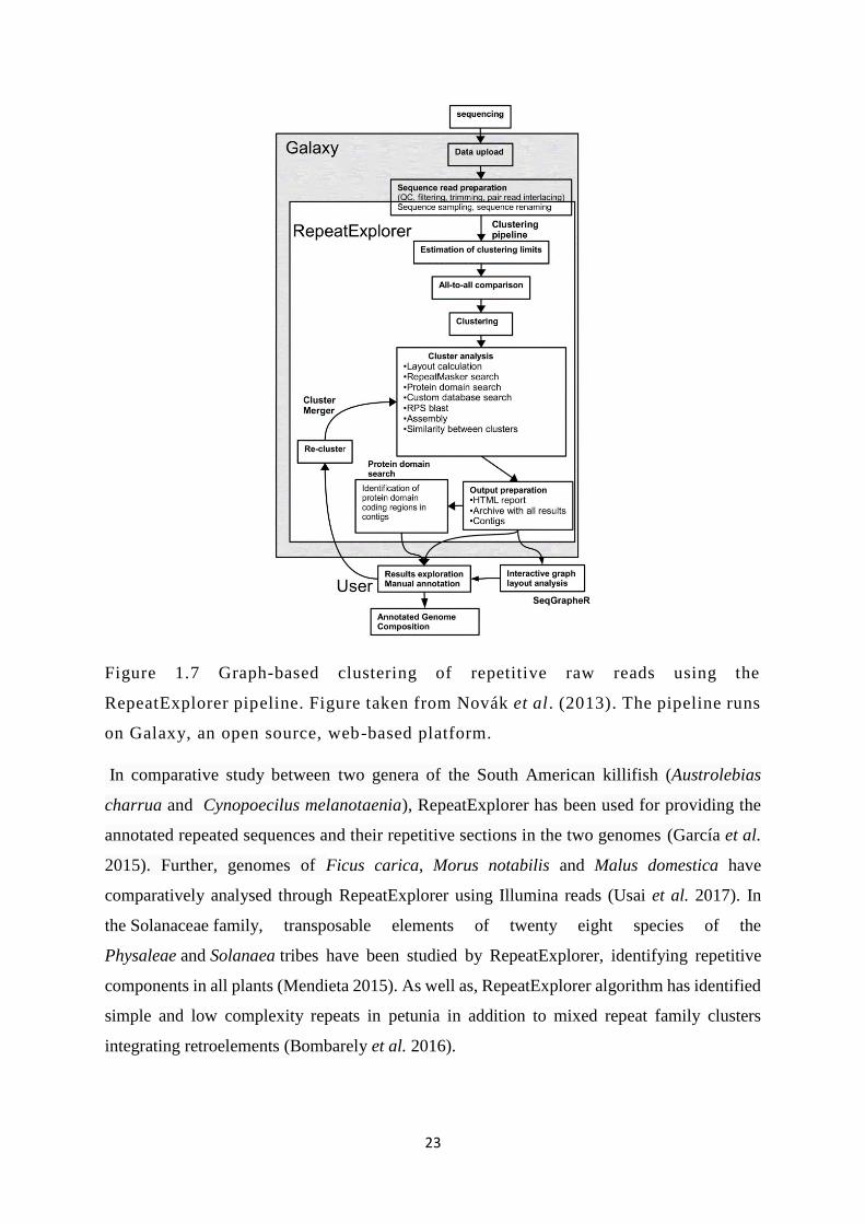
23
Figure 1.7 Graph-based clustering of repetitive raw reads using the
RepeatExplorer pipeline. Figure taken from Novák et al . (2013). The pipeline runs
on Galaxy, an open source, web-based platform.
In comparative study between two genera of the South American killifish (Austrolebias
charrua and Cynopoecilus melanotaenia), RepeatExplorer has been used for providing the
annotated repeated sequences and their repetitive sections in the two genomes (García et al.
2015). Further, genomes of Ficus carica, Morus notabilis and Malus domestica have
comparatively analysed through RepeatExplorer using Illumina reads (Usai et al. 2017). In
the Solanaceae family, transposable elements of twenty eight species of the
Physaleae and Solanaea tribes have been studied by RepeatExplorer, identifying repetitive
components in all plants (Mendieta 2015). As well as, RepeatExplorer algorithm has identified
simple and low complexity repeats in petunia in addition to mixed repeat family clusters
integrating retroelements (Bombarely et al. 2016).
24
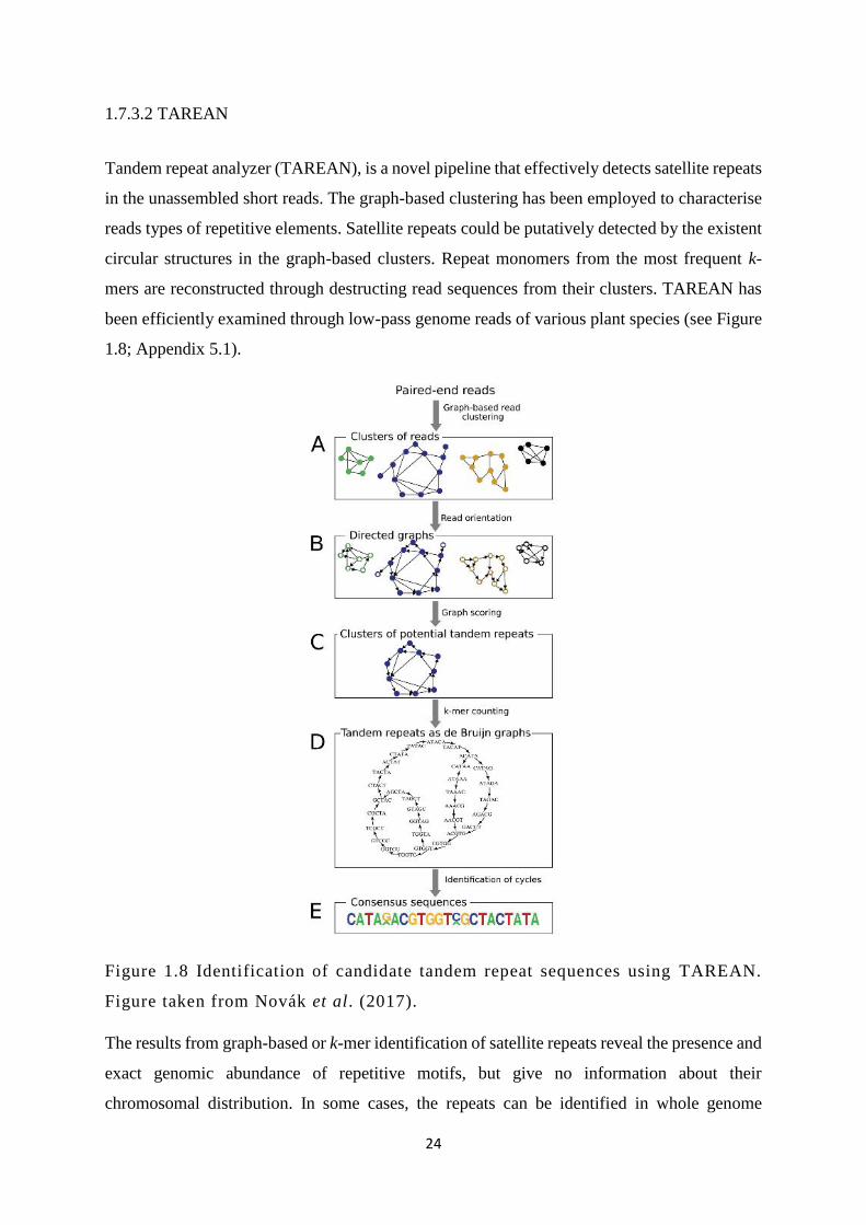
1.7.3.2 TAREAN
Tandem repeat analyzer (TAREAN), is a novel pipeline that effectively detects satellite repeats
in the unassembled short reads. The graph-based clustering has been employed to characterise
reads types of repetitive elements. Satellite repeats could be putatively detected by the existent
circular structures in the graph-based clusters. Repeat monomers from the most frequent k-
mers are reconstructed through destructing read sequences from their clusters. TAREAN has
been efficiently examined through low-pass genome reads of various plant species (see Figure
1.8; Appendix 5.1).
Figure 1.8 Identification of candidate tandem repeat sequences using TAREAN.
Figure taken from Novák et al. (2017).
The results from graph-based or k-mer identification of satellite repeats reveal the presence and
exact genomic abundance of repetitive motifs, but give no information about their
chromosomal distribution. In some cases, the repeats can be identified in whole genome

25
sequence assemblies (despite the collapse in number of copies during assembly), but in situ
hybridization (FISH) has probed essential to give detailed information about locations, number
of sites and relative abundance between sites. An example of such characterisation was given
in Vicia faba where three repeats were detected and their loci identified on chromosomes
(Novák et al. 2017).
1.8 Aims and objectives
Aims: The main aim of this study is to determine the interaction, chromosomal location of
tandemly repeated sequences, PVCV and other endogenous viruses in Petunia species, to find
out more about episomal, de novo integrated and induced infections, study expression patterns,
and any differences between vein clearing and spot symptoms of PVCV.
Objectives:
1. Organization of PVCV and other integrated viral sequences in Petunia species.
To explore sequences of PVCV, florendoviruses and other endogenous viruses in Petunia
genomes, their location within petunia chromosomes, viral sequences activity and expression,
the relationship and interaction of these sequences (chapter III).
2. Differences of PVCV symptoms and infections within petunia tissues.
To associate distinct symptom expression patterns as well as different modes of PVCV
transmission (horizontal or vertical) with virus particle concentration as well as with changes
in the cell ultrastructure of infected cells using transmission electron microscopy (TEM)
together with immunogold labelling (chapter IV).
3. Identification of tandem repeats in petunia.
To reveal and characterise all tandem repeats that highly abundant motifs in petunia DNA, their
organization within chromosome sequence assemblies, chromosomal location, and diversity
among petunia species (chapter V).

26
Chapter II. Materials and methods
2.1 Plant material and cultivation
The parental species, P. axillaris subsp axillaris N and P. integrifolia subsp inflata S6 in
addition to three cultivars of P. hybrida (Rdc, V26 and W138) and P. axillaris subsp parodii
S7 were used in this study. Seeds of the three wild Petunia species have been provided by Cris
Kuhlemeier, University of Bern, Switzerland. P. hybrida seeds were obtained from Ronald
Koes, University of Amsterdam (W138), NL Chrestensen, Erfurt, Germany (Rdc, aka
“Himmelsröschen”) and John Innes Institute, Norwich (V26) (Table 2.1; Figure 1.1). The seeds
were placed in small pots and covered lightly with a peat moss (Levington advance), and
irrigated regularly under greenhouse conditions (25 ˚C temperature and 16 hr of daylight) at
the Department of Genetics and Genome Biology, University of Leicester (UK).
Table 2.1 List of suppliers for seeds from wild and hybrid petunia plants used in the studies.
No. Species Cultivar Source
1. P. axillaris subsp. axillaris N Cris Kuhlemeier,
University of Bern,
Switzerland
2. P. integrifolia subsp inflata S6 Cris Kuhlemeier,
University of Bern,
Switzerland
3. P. hybrida“Himmelsröschen” or
Rdc (Rose du ciel)
Rdc NL Chtestensen,
Erfurt, Germany
4. P. hybrida V26 John Innes
Institute, Norwich
5. P. hybrida W138 Ronald Koes,
University of
Amsterdam

27
6. P. axillaris subsp parodii S7 Cris Kuhlemeier,
University of Bern,
Switzerland
2.2 Standard solutions and media
Table 2.2 Standard solutions used in experiments
Experiments Solutions Constitutions
D
NA
extr
acti
on
CTAB buffer 2% (w/v) cetyltrimethylammonium bromide,
100mM Tris–HCL, 1.4M NaCL, 20mM EDTA. (pH
7.5-8.0)
10x TE buffer 100mM Tris (tris-hydroxymethylamino-methane)-
HCl, 10mM EDTA (ethylene-diamine-tetra-acetic
acid. (pH 8)
DNA wash buffer
76 % ethanol, 10mM ammonium acetate.
DNA extraction
buffer
50 mM of Tris-HCl, pH 8.0, 25 mM EDTA, 400 mM
NaCl.
G
el e
lect
ropho
resi
s
6x Gel loading
buffer
60% (v/v) glycerol; 0.25% (w/v) bromophenol blue;
0.25% (w/v) xylene cyanol FF; diluted to 1x in 50%
(v/v) glycerol.
50x TAE buffer
(tris acetate-EDTA)
2M Tris-HCl; 50 mM EDTA
(ethylenediaminetetraacetic acid; pH 8)
5.71% (v/v) glacial acetic acid, diluted to 1x in
deionised H2O.
Ethidium Bromide 10 mg/ml (1 g Ethidium bromide dissolved in 100 ml
of sterile distilled water and stored at 4°C).
Clo
nin
g Ampicillin 10 mg/ml (10 mg of ampicillin dissolved in 1 ml of
distilled water and stored at -20°C).

28
SOB medium
(super optimal broth)
20g of tryptone, 5g yeast extract 0.5g NaCl, 10 ml
250 mM KCl. The final volume was 1000ml with
sterile distilled water. (pH7.0)
LB (lysogeny broth)
Agar plates: 2.5% LB broth (Melford), 1.5% agar
(For Medium), 100 μg/ml ampicillin, 80 μg/ml x-gal,
0.5 mM IPTG (isopropyl β-D-1-
thiogalactopyranoside. (pH 7.2)
LB medium (luria-
bertani)
12.5g of LB broth (10g tryptone, 5g yeast extract,
10g NaCl). The final volume was 500ml with sterile
distilled water. ( pH 7.0)
LB medium agar 25g of LB broth (10g tryptone, 5 g yeast extract, 10g
NaCl). The final volume was 1000ml with sterile
distilled water and 15 g of Bacto-Agar (DIFCO).
IPTG 200 mM (476 mg/ml isopropyl-B-D-thiogalacto-
pyronoside dissolved in 10 ml distilled water, filter
sterilized and stored at -20°C).
X-gal 40mg/ml (1g of 5-bromo-4-chloro-3-indolyl β-D-
galactopyranoside with 25ml of dimethylformamide,
filter sterilized and stored at -20°C).
2x rapid ligation
buffer
60mM Tris-HCl (pH 7.8), 20mM MgCl2, 20mM
DTT, 2mM ATP and 10% PEG.
Chro
moso
me
pre
par
atio
ns
10x Enzyme buffer 100 mM citric acid, 100 mM tri-sodium citrate,
stored at 4°C. (pH 4.6)
1x Enzyme solution 3% (w/v) pectinase (Sigma), 0.2 % (w/v) cellulase
[1.8% (w/v), Onozuka RS] cellulase (Calbiochem) in
1x enzyme buffer, stored at -20°C.
8-hydroxyquinoline
0.002M (Dissolving 0.29 g of S-hydroxyquinoline in
1000 ml of ddH2O, stored in the dark at 4°C).

29
FIS
H
SDS
(sodiumdodecyl
sulfate)
20% (2g Sodium dodecyl sulfate (SDS) with 8ml
water).
20x SSC
(Saline Sodium
Citrate)
0.3M NaCl, 0.03M Sodium citrate. (pH 7.0)
Paraformaldehyde 4% paraformaldehyde (Fisher Scientific) dissolved
in distilled water. The final volume was 100 ml. (pH
7.0)
50% Dextran
sulphate
50 gm dextran sulfate with 100 ml distilled water,
filter sterilized and stored at -20°C.
Salmon sperm DNA 1mg/ml of sheared salmon sperm DNA.
Blocking solution 5% BSA in 4xSSC with 0.2% Tween 20.
Detection buffer 4xSSC, 0.2% (v/v) Tween 20.
0.5 M EDTA 186.1g disodium ethylenediamine tetraacetate 2H2O
into 800ml of distilled water. Adjust pH to 8.0 with
NaOH. The final volume was 1 L.
DAPI solution 5g of DAPI (4ʹ, 6-diamidino-2-phenylindole)
dissolved in molecular grade water. The final volume
was 50ml (stock of 100μg/ml diluted in water. Final
concentration of 4μg/ml was diluted with
McIlvaine’s buffer and stored at -20°C).
De
novo
inte
gra
tion
RLT buffer
(RNeasy lysis
buffer)
RLT buffer contains a high concentration of
guanidine isothiocycanate that supports the binding
of RNA to the silica membrane.
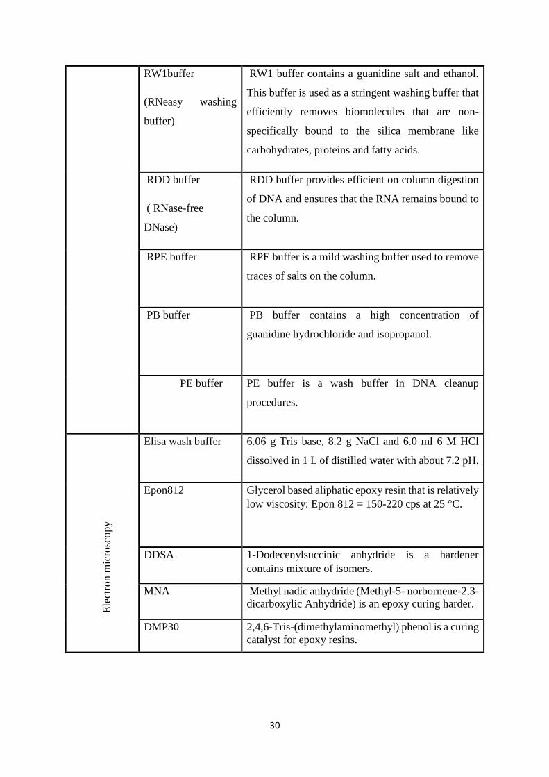
30
RW1buffer
(RNeasy washing
buffer)
RW1 buffer contains a guanidine salt and ethanol.
This buffer is used as a stringent washing buffer that
efficiently removes biomolecules that are non-
specifically bound to the silica membrane like
carbohydrates, proteins and fatty acids.
RDD buffer
( RNase-free
DNase)
RDD buffer provides efficient on column digestion
of DNA and ensures that the RNA remains bound to
the column.
RPE buffer
RPE buffer is a mild washing buffer used to remove
traces of salts on the column.
PB buffer PB buffer contains a high concentration of
guanidine hydrochloride and isopropanol.
PE buffer
PE buffer is a wash buffer in DNA cleanup
procedures.
E
lect
ron m
icro
scopy
Elisa wash buffer 6.06 g Tris base, 8.2 g NaCl and 6.0 ml 6 M HCl
dissolved in 1 L of distilled water with about 7.2 pH.
Epon812 Glycerol based aliphatic epoxy resin that is relatively
low viscosity: Epon 812 = 150-220 cps at 25 °C.
DDSA 1-Dodecenylsuccinic anhydride is a hardener
contains mixture of isomers.
MNA Methyl nadic anhydride (Methyl-5- norbornene-2,3-
dicarboxylic Anhydride) is an epoxy curing harder.
DMP30 2,4,6-Tris-(dimethylaminomethyl) phenol is a curing
catalyst for epoxy resins.

31
2.3 Methods
2.3.1 Genomic DNA extraction
Genomic DNA was prepared from young leaves of petunia using the CTAB method (Doyle &
Doyle 1990) with some modifications. One to two grams of asymptomatic young leaves were
collected from each individual species, and washed with distilled water and then wrapped with
aluminium foil followed by shock freezing in liquid nitrogen and immediately ground up using
a pestle and mortar to avoid enzymatic degradation. About half a spatula of
polyvinylpyrrolidone (Sigma) was added to the material before transferring it into 10 ml of
preheated CTAB buffer (contains 50 µl of B-mercaptoethanol) in 50 ml tube and incubated at
60 °C for 30 min. Absolute chloroform: isoamyl alcohol (24:1) was added with equal volume
to each tube and blended via inverting many times for 3 min, then centrifuged at 2040 g at
room temperature for 10 min. The supernatant was transferred gently from the top of the tube
to another new one using 1ml tip with cut end, the steps of washing and centrifugation were
repeated another time. After that, pre-chilled isopropanol (0.6x volume) was added to the
supernatant for DNA precipitation, then the material was mixed carefully by inverting several
times, and kept for 10 min on ice. The precipitated DNA was spun down at 735 g for 3 min,
and then air dried and washed with 5 ml of DNA wash buffer for 20 min. After this step, the
DNA was re-suspended in 1ml of 1x TE buffer at RT overnight after air drying. It was
incubated for 1 hr at 37 °C with 2 µl of 10 µg /µl RNase A (Bioline) to remove the RNA and
re-precipitated with 1x sodium acetate 3M (pH 6.8) and 2x absolute ethanol. The DNA was
spun down at 735 g for 3 min and then re-suspended in 500 µl of 1x TE buffer at RT overnight,
the prepared DNA was stored at -20 °C.

32
2.3.2 DNA quantification
2.3.2.1 Gel electrophoresis
For conformation check and detection of denaturation, 5 µl of the extracted total genomic DNA
was separated on agarose gel (1% w/v in 1xTAE buffer with ethidium bromide 1µg/µl) with a
size and amount reference (5 µl of Bioline 1kb hyper ladder). The gel run was set up for 60
min at 75 V. Gel documentation system (Gen flash) was used to capture the gel image.
2.3.2.2 Spectrophotometry
Genomic DNA was assessed for purity, quality and amount using a Nanodrop ND-1000
spectrophotometer at 260/280 nm wavelength using 1 µl of each single DNA sample. Quality
was considered as a high when the OD 260/280 nm value was about 1.8-2.2.
2.3.3 PCR amplification
Genomic DNA, dNTPs and primers were diluted with sterilized water, and the final
concentration of the DNA was 30-50 ng/μl to use as a template. The master mix components
(Table 2.3) were set up to amplify some tandem repeats (see chapter V) and EPRVs (Table
2.4). PCR reactions used a gradient PCR machine (Biometra) with a program that consisted of
3 min for initial denaturation at 95 °C and then 35 cycles of denaturation (95 °C for 30 sec),
annealing (50.2-69.3 °C for 30 sec) and primer extension (72 °C for 1 min). The last cycle was
1 min at 72 °C as a final extension. After these steps, the temperature was set up at 16 °C as
indefinite time (Table 2.5). PVCV primers (Table 3.1) were amplified as (Table 3.2), and PCR
products were checked by gel electrophoresis (see below).

33
Table 2.3 Components and quantities of PCR Master Mix.
Components Amounts(µl)
Molecular biology grade water 13.4
10x KAPA Taq Buffer 2
MgCl2(25mM) 1.3
10mM dNTP Mix 1.2
10 μL Forward Primer 0.5
10 μL Reverse Primer 0.5
KAPA Taq DNA Polymerase 0.1
Template DNA 1
Total 20 µl
2.3.4 Purification of DNA fragments from agarose gel
Selected DNA fragments were cut from the gel by sterilized forceps and scalpels under a UV
trans-illuminator (UVP). The gel pieces were placed into 1.5 ml Eppendorf tubes and then
purified and washed from agarose parts according to the instructions in the clean-up kit (Nucleo
Spin Extract II Clean up kit Macherey-Nagel Company).
2.3.5 Cloning of PCR products
The pGEM-T Easy Vector System I kit (Promega) was used for cloning of some purified PCR
products.

34
2.3.5.1 Ligation
The ligation reactions were performed in a 300 μl Eppendorf tube. The reactions consisted of
2x rapid ligation buffer (7 μl), pGEM-T Easy Vector (0.9 μl), DNA sample (5.4 μl), T4 DNA
ligase (1.2 μl), and sterile water (0.5 μl). The final volume was 15 μl as (Table 2.4), and mixed
gently by flicking, then incubated at RT for 1 hr and then overnight at 4°C (see Table 2.2).
Table 2.4 Components of the ligation reaction.
Component Volume (μl) Final concentration
2x rapid ligation buffer 7 1x
pGEM-T Easy Vector 0.9 1-10 ng/μl
Purified PCR products 5.4 1-10 ng/μl
T4 DNA ligase 1.2 6U/μl
Molecular grade sterile water 0.5 NA
Total 15
2.3.5.2 Transformation
Plasmid vectors containing the PCR product were transformed into transformation competent
cells (Escherichia coli). After growth in selective media, culture was plated out on LB
(Lysogeny Broth) agar plates with selective markers (pre-prepared media contains 100 μg/ml
ampicillin, 500 μg/ml (IPTG) - isopropyl-β-A-thiogalactopyranoside and 40 μg/ml (X-Gal) -
5-bromo-4-chloro-3-indolyl-β-D-galactosidase) (see Table 2.2).
2.3.5.3 Screening and isolation of recombinant clones
Recombinant colonies with inserted sequences in the plasmid were white, while non-
recombinant cells appeared as blue colonies. White colonies were picked by a sterile toothpick
and transferred into 5ml LB medium tube and then incubated overnight at 37°C in a shaker
(230 g) (see Table 2.2).

35
2.3.5.4 Colony PCR
Colony PCR was used to confirm recombinant clones and the size of insert using the universal
M13 sequencing primers [forward (GTA AAA CGA CGG CCA GT) and reverse (GGA AAC
AGC TAT GAC CAT G)]. The master mix components were prepared as (Table 2.5) in 1.5 ml
Eppendorf tubes. 1-2 μl bacterial culture was used in addition to 15 μl of master mix
components in each clean 0.2 ml PCR tube. After flicking and spinning down the mixture tube,
the PCR machine was set up according to the PCR protocol in (Table 2.6). PCR products and
fragment lengths were analysed in horizontal agarose gel electrophoresis.
Table 2.5 Materials for PCR reaction of cloned inserts products.
Components Amounts (μl)
Molecular grade Water 9.65
10x KAPA Taq Buffer 1.5
MgCl2(25mM) 1
10Mm dNTP Mix 1.2
10 μL Forward Primer-M13 0.75
10 μL Reverse Primer-M13 0.75
KAPA Taq DNA Polymerase 0.15
Total 15

36
Table 2.6 Colony PCR protocol of cloned PCR products.
Steps Temperature °C
Duration
Minutes : seconds
Cycles
1 Initial
denaturation
95 5:00 1
2 Denaturation 95 1:00 35
3 Annealing 55 00:30
4 Extension 72 1:00
5 Final extension 72 10:00 1
6 Hold 16 pause 1
2.3.5.5 Plasmid DNA isolation and purification
Plasmid DNA was purified using Machery-Nagel Kit following the Manufacturer’s
instructions. Recombinant colonies culture (1400 μl) was added into 1.5 ml Eppendorf tube
and centrifuged directly at 9875 g for 30 sec. Then, the supernatant was gently removed and
only pellets left at the bottom of the tube. The cell pellet was re-suspended totally by adding
buffer A1 and pipetting up and down. In order to continue with cell lysis process, buffer A2
and buffer A3 were separately and respectively added and gently mixed by inverting the tube
many times. The lysate was clarified by centrifugation at 9875 g for 5 min. The supernatant
was loaded in a NucleoSpin Plasmid/Plasmid (no lid) column and centrifuged at 9875 g for 1
min. Next, the silica membrane was washed by adding buffer AW and buffer A4 separately
and respectively with centrifugation at 9875 g for 1 min each. Finally, the silica membrane was
dried by centrifugation and plasmid DNA was collected by adding 50 µl elution buffer and
centrifuged at 9875 g for 1 min.

37
2.3.5.6 Sequencing of cloned PCR products
Cloned PCR products were sequenced at GATC biotech (Germany) by sending the gel
extracted DNA including the universal M13 forward primer.
2.3.6 Probe labelling
Purified PCR products or amplified cloned inserts were used as template for labelling with
biotin-11-dUTP or digoxigenin-11-dUTP (Roche Diagnostics). The BioPrime (Invitrogen)
DNA Labelling System was used for biotin labelling following the manufacturer’s instruction.
Briefly, 25 µl of DNA sample (500-1000 ng) was added to 20 µl of 2.5X Random Primers
Solution (final volume 45 µl) and then denatured at 95 °C in a water bath. After the denaturation
step, 10x dNTP (5 μl) and Klenow Fragment of E. coli DNA polymerase I (40U) (1 μl) were
added on ice and mixed carefully but thoroughly, and then incubated for 2 hr at 37 °C. The
BioPrime (Invitrogen) Array CGH Genomic Labelling Module was applied for digoxigenin
labelling. After denaturing 19 µl of DNA sample (500-1000 ng) and 16 µl of 2.5X Random
Primers Solution (final volume 35 µl), 10x dUTP nucleotide (3 μl), digoxigenin-11-dUTP (1,8
μl) and Exo-Klenow Fragment (40U) (0,8 μl) were gathered and mixed carefully but
thoroughly and incubated for 2 hr at 37 °C. After incubating each sample, stop buffer (5 μl)
was added to end the reaction. Finally, a NucleoSpin Extract II Clean-up Kit (Machery-Nagel)
was used for cleaning each labelled probe and stored at -20 °C. In addition, six probes of
tandem repeats clusters (Table 5.2) have been labelled directly by synthesis with nucleotides
linked with biotin.
2.3.7 Petunia chromosome preparations
Fresh root tips were firstly pretreated with 0.2 M 8-hydroxyquinoline for 4 hr and then fixed
with freshly made ethanol:glacial acetic acid (3:1). Fixed roots were prepared and washed two
times for 10 min each in 1x enzyme buffer diluted from 10x enzyme buffer to remove the
fixative solution residues, and then digested in 1x enzyme solution at 37 ˚C in incubator for
15-20 min. After this period, root tips were soaked again in 1x enzyme buffer to stop the
enzyme action. Clean glass slides (SuperFrost, Menzel-Glaser, Thermo Scientific) were used
for preparation under a stereo microscope, with a fine needle and forceps, a single root tip was
added to one drop of 60% acetic acid. Then, the root cap was removed keeping only the

38
meristematic tissues, and cells were separated with the needle and forceps. A glass coverslip
(22 mm x 22 mm No. 1) was placed on the prepared cells and excess of acid removed by a
filter paper. The coverslip was tapped gently to disperse the material and then squashed by
force with the thumb. A phase contrast microscope was used for visualizing the slides to check
cell quality, metaphase index, and to confirm if they were well squashed. After checking, the
slides were transferred on to dry ice for 15 min and the cover slip was removed with a razor
blade. The slides were air dried and stored at room temperature and checked again (see Table
2.2).
2.3.8 Fluorescent in situ hybridization
The method of Schwarzacher and Heslop-Harrison (2000) was applied for fluorescent in situ
hybridization.
2.3.8.1 Pre-hybridization
Selected slides with free chromosomes and good metaphase distribution were re-fixed using
immersion in new fixative absolute ethanol:glacial acetic acid (3:1) mixture for 10 min. Slides
were soaked in absolute ethanol for two times 5 min each and air dried, and then incubated
with 200 μl of RNase (100 μg/ml) solution each under a plastic cover slip at 37 °C for 1 hr in
a humid chamber. The plastic coverslips were removed from slides by washing in 2xSSC for
2 and 10 min respectively, then slides were incubated in a pepsin solution (400 μl pepsin:80 ml
0,01M HCL) for 3 min at room temperature and washed with distilled water and 2xSSC for 1
and 5 min respectively. Next, slides were incubated at RT for 10 min with saline buffered
paraformaldehyde 4 % (w/v) (Fisher scientific) under the fume hood. The 2xSSC solution was
used again for washing slides, and then slides were dehydrated in 70%, 85% and 100% ethanol
for 2 min each. Then, slides were air dried to prepare them for probe adding (see Table 2.2).
2.3.8.2 Hybridization
The total quantity of hybridization mixture including labelled probe was prepared in an
Eppendorf tube (37 μl /slide) (see Table 2.7). The mixture was heated at 95°C for 10 min and
cooled on ice for 10 min. The prepared mixture was added to the marked area of a slide and
covered with a plastic cover slip. The slides were placed on a thermal cycler and heated to 70°C
for 7 min denaturing both chromosomal and probe DNA. For probe hybridization to
chromosomal DNA, temperature was decreased slowly to 37°C and kept for 16-20 hr.
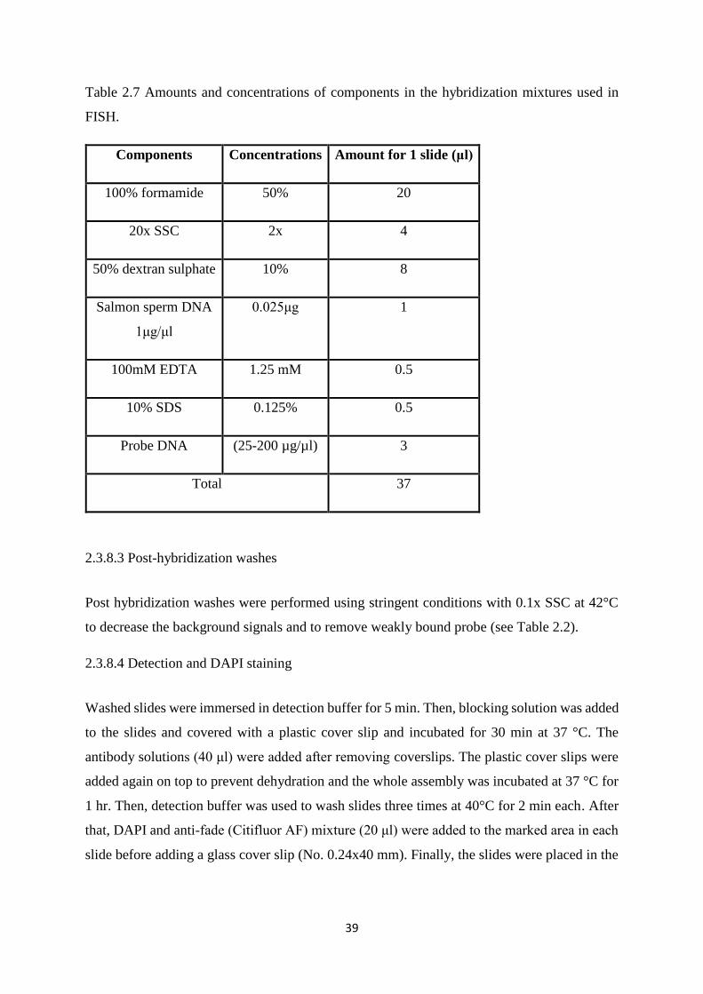
39
Table 2.7 Amounts and concentrations of components in the hybridization mixtures used in
FISH.
Components Concentrations Amount for 1 slide (μl)
100% formamide 50% 20
20x SSC 2x 4
50% dextran sulphate 10% 8
Salmon sperm DNA
1μg/μl
0.025μg 1
100mM EDTA 1.25 mM 0.5
10% SDS 0.125% 0.5
Probe DNA (25-200 µg/µl) 3
Total 37
2.3.8.3 Post-hybridization washes
Post hybridization washes were performed using stringent conditions with 0.1x SSC at 42°C
to decrease the background signals and to remove weakly bound probe (see Table 2.2).
2.3.8.4 Detection and DAPI staining
Washed slides were immersed in detection buffer for 5 min. Then, blocking solution was added
to the slides and covered with a plastic cover slip and incubated for 30 min at 37 °C. The
antibody solutions (40 μl) were added after removing coverslips. The plastic cover slips were
added again on top to prevent dehydration and the whole assembly was incubated at 37 °C for
1 hr. Then, detection buffer was used to wash slides three times at 40°C for 2 min each. After
that, DAPI and anti-fade (Citifluor AF) mixture (20 μl) were added to the marked area in each
slide before adding a glass cover slip (No. 0.24x40 mm). Finally, the slides were placed in the

40
dark at 4°C overnight before they were checked under an epifluorescence microscope (see
Table 2.2).
2.3.8.5 Microscopy and photography
The FISH slides were scanned and viewed by a Nikon Eclipse 80i epifluorescence microscope
(B&W CCD camera, Nikon Digital Sight USB (H) EXT I/0 B2W camera controller and a
Nikon Intenslight C-HGFI lamp). A drop of immersion oil was placed on the slide after
pressing the cover slip carefully and firmly with a filter paper. Three types of band filters were
used for checking FISH signals: DAPI (blue) was captured by UV-2E/C (wavelengths of
excitation filter 340-380 nm), Digoxigenin (green) by B-2E/C (wavelengths of excitation filter
465-495 nm), and Alexa Flour 594 (red) by G-2E/C (wavelengths of excitation filter 528-553
nm). The red biotin/ Alexa594 streptavidin antibody signal was captured first, followed by the
green digoxigenin/FITC-anti-digoxigenin signals, and later the DAPI signal. Each single image
was captured using Nikon, NIS-Elements 4.0, false coloured, and overlaid for producing a
composite image with the red and green colours for FISH probes and the blue for
chromosomes. Adobe Photoshop CC2015.5 was used for preparing and overlaying images and
hybridization signals, apart from cropping, using only functions affecting the whole image
equally.

41
Chapter III. Integration of Endogenous Pararetroviruses (EPRVs)
3.1 Endogenous pararetroviruses (EPRVs)
For a long time, a principle in virology stated that there was no evidence for plant viruses
integrating into the host genome unlike bacterial and animal viruses. This dogma has been
overruled with the discovery of endogenous pararetroviruses in plants. Most of our knowledge
about EPRVs biology and evolution was gained from two sources: i) the activation of
integrated EPRVs that gave factual proof of their infectivity under particular conditions and ii)
identification of positioning and diversity within sequenced plant genomes. Richert-Pöggeler
and Shepherd (1997) reported that Petunia vein clearing virus transmits through seeds and
integrates in the Petunia hybrida genome. The whole virus sequence is present in most of P.
hybrida and the parental species P. axillaris subsp. axillaris and P. integrifolia subsp inflata ,
and capable of episomal virus replication in response to stress (Richert‐Pöggeler et al. 2003).
Ndowora et al. (1999) and Harper et al. (1999) showed that Banana streak virus-OL integrates
at two loci in the Musa genome using FISH and could trigger virus infection in healthy plants.
In Musa balbisiana genome, four species of BSV have recently been reported as pathogenic
integrants: Banana streak OL virus (BSOLV), Banana streak IM virus (BSIMV), Banana
streak MY virus (BSMYV), and Banana streak GF virus (BSGFV) (Gayral et al. 2008; Iskra-
Caruana et al. 2010; Geering et al. 2014). Lockhart et al. (2000) revealed that Tobacco vein
clearing virus could also integrate in the genome of Nicotiana edwardsonii using hybridization
tests and genomic library screening. These viruses, Petunia vein clearing virus (PVCV),
Banana streak virus (BSV), and Tobacco vein clearing virus (TVCV), show episomal diseases
related with integrated motifs (Harper et al. 2002).
Nowadays, many sequences of plant viruses have been found as integrants in different plant
genomes (Hohn et al. 2008; Geering et al. 2014).
According to tenth Report of the International Committee on Taxonomy of Viruses (ICTV),
there are five DNA plant virus families with ssDNA genomes and the family of Caulimoviridae
encapsidating dsDNA:
1- The family of Geminiviridae has ten genera.
2- The family of Alphasatellitidae has twelve genera.
3- The family of Nanoviridae has three genera.

42
4- The family of Tolecusatellitidae has two genera.
5- The family of Caulimoviridae has eight recognized genera.
Recently, one additional genus has been proposed to the family of Caulimoviridae. It was
named Florendovirus which members are found abundantly in a variety of flowering plants
(Geering et al. 2010; Geering et al. 2014).
3.1.1 Caulimoviridae
The Caulimoviridae family includes genera of Caulimovirus, Soymovirus, Cavemovirus,
Solendovirus, Petuvirus, Badnavirus, Rosadnavirus, Tungrovirus and Florendovirus with viral
genomes from 7.2 to 9.2 kb, which are essentially differentiated from each other by their
genome organization (Geering et al. 2010; Geering & Hull 2012; Geering et al. 2014; Diop et
al. 2018). Those viruses replicate uniquely by reverse transcription. Virus particles encapsidate
a double-stranded DNA genome and virus integration step is not obligatory for viral
replication. Their genome consists of open circular double-stranded DNA with single strand
discontinuities at particular positions. Based on these biological properties which are distinct
from retroviruses, they are also referred to as pararetoviruses (Temin 1985).
These gaps have resulted from DNA replication and are located at the priming positions for
plus strand DNA synthesis as well as on the minus DNA strand (Gronenborn 1987; Harper et
al. 2002). The family produce a more-than-genome-length RNA that takes two roles as a
template for reverse transcription and as mRNA for some of gene products (Hull 2002).
Caulimovirus replication occurs in two cellular compartments: first, transcription of a genome
length RNA from a viral minichromosomal form in the nucleus, followed by reverse
transcription to give circular dsDNA with discontinuties in the cytoplasm (Gronenborn 1987;
Harper et al. 2002). The genetic information can be either organized in up to eight ORFs
comprising single gene products or like the ORF III of Badnavirus, encoding for the coat
protein (CP), the movement protein (MP), an aspartate proteinase (AP), the reverse
transcriptase (RT) and the ribonuclease H1 (RH1) that are involved in virus replication. The
genera of Badnavirus, Petuvirus, Solendovirus and Caulimovirus could integrate within host
nuclear genomes and transmit over generations like cellular genes (Geering et al. 2014).
Recently, Diop et al. (2018) reported that both primitive and complex vascular plants contain
endogenous caulimovirid-like sequences. Up to five different genera of Caulimoviridae have

43
been detected in angiosperms with florendoviruses being the most prominent representative
followed by petuviruses.
3.1.1.1 Petunia vein clearing virus (PVCV)
The virus was detected and described for the first time in Germany by Lesemann and Casper
(1973). In a study aimed to identify mycoplasma–like bodies and viruses in Opuntia tuna
showing witches–broom disease, Lesemann and Casper (1973) reported that it was not possible
to detect virus-like particles in ultrathin sections of O. tuna tissues and most transmission
experiments from infected Opuntia were negative. Petunia plants that were not used in the
transmission tests, but had been raised from the same seed source showed all of sudden vein
clearing symptoms and leaf deformation in young shoots with stunted growth. In ultrathin
sections of symptomatic leaf and stem material, virus-like particles and inclusion bodies were
discovered. Horizontal transmission of the virus to other petunia plants was possible using
grafting. Based on host plant and symptoms the infectious entity was named ‘`Petunia vein
clearing virus’’ (Lesemann & Casper 1973). Later, it has been recorded in United States of
America (Lockhart & Lesemann 1998), and in Israel (Gera et al. 2000). Particle measurements
using purified virus suspensions showed that the average virion diameter with 40-50 nm is
slightly smaller than other Caulimoviridae (Zeidan et al. 2000). Based on immune electron
microscopy, no serological relationship to other members of caulimoviruses was found
(Lesemann & Casper 1973). Phylogenetic analyses confirmed PVCV being distinct from so far
known Caulimoviridae and it was classified as type member of the new genus Petuvirus
(Richert-Pöggeler & Shepherd 1997; Fauquet et al. 2005).
The infected petunia plants appear stunted especially in young shoots, and display chlorosis
along the leaf veins with either diffuse or sharp borders. Additionally symptomatic leaves show
epinasty (Richert‐Pöggeler et al. 2003). These symptoms (see Figure 3.1) are well expressed
when the temperature is more than 25°C under nursery conditions (Zeidan et al. 2000).
Furthermore, symptoms can arise from stressed plants that suffer from water and nutrient
deficiency, in addition to wounding (Lockhart & Lesemann 1998; Harper et al. 2002) and tissue
culture (Richert‐Pöggeler et al. 2003).

44
Figure 3.1 The symptoms on P. axillaris subsp parodii S7 resulted by grafting with a
PVCV infected scion of P. hybrida. The main symptoms appear as chlorosis along the
leaf veins with either diffuse or sharp borders and stunted young shoots (Richert‐
Pöggeler et al. 2003).
Horizontal transmission of episomal PVCV was successful to Nicotiana glutinosa and P.
axillaris subsp parodii S7 using grafting. So far, the determined host range of PVCV is limited
within Solanaceae, including species of Petunia like P. integrifolia subsp. integrifolia, P.
axillaris subsp axillaris, P. hybrida and N. glutinosa but not N. tabacum (Richert‐Pöggeler et
al. 2003). Outbreak of vein clearing disease has been observed for some cultivars of P. hybrida
such as “Himmelsröschen” (Lesemann & Casper 1973), “Fantasy Pink” (Lockhart &
Lesemann 1998), and the “Cascadia” series (Gera et al. 2000). Although PVCV transmission
by vectors has not been found yet, the viral DNA can be transmitted by biolistic inoculation of
petunia plants (Richert‐Pöggeler et al. 2003).
Screening a genomic library of P. hybrida cultivar W138 identified integrated PVCV
sequences arranged in a tandem array from which direct full genome length transcript can be
synthesized. Indeed these “provirus”-like structures were infectious using biolistic inoculation
of provirus-free P. axillaris subsp parodii S7 (Richert‐Pöggeler et al. 2003). Most likely release
of epigenetic control leads to PVCV infections originating from such endogenous copies in P.
hybrida W138 after wounding and tissue culture in 30% of the treated plants after 2-3 months
(Noreen et al. 2007).
Detection of endogenous forms of PVCV in asymptomatic plants using FISH proved that there
are homologous sequences in the genomes of P. axillaris subsp axillaris, P. integrifolia subsp
inflata and P. hybrida (Richert‐Pöggeler et al. 2003; Bombarely et al. 2016). No hybridization

45
signal was obtained in P. axillaris subsp parodii and N. glutinosa when stringent washing
conditions were applied. Thus integrants are vertically transmitted via reproductive cells to the
next generation (Richert‐Pöggeler et al. 2003). Interestingly, FISH studies indicated the
collocalisation of PVCV integrants with Metaviridae sequences that are phylogenetically the
closest relative to Caulimoviridae (Bombarely et al. 2016; Krupovic et al. 2018).
The genome sequence of PVCV is 7206 bp in size (GenBank accession no. U95208). All
genetic information is comprised in one large ORF consisting of 2179 amino acids which
distinguishes PVCV from other members within Caulimoviridae having their genomic
information distributed over 3 to 8 ORFs (Hohn & Rothnie 2013) . Within the polyprotein
consensus sequences known from the gag and pol genes of retroviruses for a RNA binding
domain (RB), protease (PR), reverse transcriptase (RT) and RNase H (RH) were identified
(Richert-Pöggeler & Shepherd 1997; Richert‐Pöggeler et al. 2003).
An inactivated integrase-like domain and quasi (long) terminal repeats were described for
PVCV which are core elements for retrotransposon integration during replication supporting
the ancestral link of both phylogenetic groups (Richert-Pöggeler & Shepherd 1997; Richert‐
Pöggeler et al. 2003; Krupovic et al. 2018).
The available data on genome assemblies of the two Petunia parental species P. axillaris subsp
axillaris N (PaxiN) and P. integrifolia subsp inflata S6 (PinfS6) reveals numerous integrated
sequences of PVCV (Bombarely et al. 2016). FISH was used to localize sequences on
chromosomes. In cytological studies distinct patterns for PVCV sequence localization could
be seen. In P. axillaris N they were mainly identified on chromosome III and less frequently
on chromosome VI compared to P. integrifolia subsp inflata S6, in which only a weak signal
on chromosome IV was observed in FISH. So far, almost the complete viral genome length as
well as degenerated PVCV sequences have been found in arrays within both assemblies when
screened for larger than 500 nt in length against PVCV (GenBank accession no. U95208).
Different degrees of preservation of integrated PVCV sequence were found between the two
Petunia species. Thirty sequences from PaxiN ranged between 542 and 2848 nt in length with
80-99% identity, while in PinfS6, only nine smaller fragments with 563-635 nt in size revealed
78-80% identity. The route of PVCV integration seems to be similar in both Petunia species
resulting in single insertions as well as small tandem arrays (Richert‐Pöggeler et al. 2003;
Bombarely et al. 2016).

46
3.1.1.2 Florendovirus
The newly described genus Florendovirus (Geering et al. 2014), out of the family of
Caulimoviridae, has colonized the genomes from a large variety of flowering plants comprising
34 distinct viral species. Endogenous viral sequences have been found in monocot as well as
eudicot plants including potato, maize, papaya, soybean, apple, citrus, cacao, grape, tomato,
cassava, rice and other plants. The name ‘Florendovirus’ based on the existence in flowering
plants referring to Flora the Roman goddess of flowers and to the fact of being endogenous.
Further, depending on 80% nt identity in the reverse transcriptase (RT)-ribonuclease H (RH)
domains and other sequence clusters, 34 representative distinct virus species have been
identified and classified giving them names including the host species name. The sequence
fragments have been assembled into a total of 76 entire or nearly full-length Florendovirus
genomes sized between 7.2 and 8.5 kb. They can encode for one or two open reading frames
(ORFs) and thus were divided into two categories. The majority of florendoviruses have two
ORFs in different reading frames. Sequence analysis revealed potential open reading frames
encoding various protein domains necessary for viral replication. Phylogenetic analyses have
shown that the genus Florendovirus branches next to Petuvirus that comprises all genetic
information for replication within one large ORF. Florendoviruses harbor conserved domains
of gag-pol genes typical for retroelements, being clearly distinct from known reverse
transcribing elements like monopartite Caulimoviridae and diploid retroviruses (Geering et al.
2014; Bombarely et al. 2016).
3.2 Aims and objectives
Aims: The main aim of this chapter is to determine the interaction, chromosomal location of
PVCV and other endogenous pararetroviruses in Petunia species, to find out more about
episomal and induced infections, study expression patterns and EPRVs sites within Petunia
chromosomes.
Objectives:
- To identify sequences, copy numbers and genome proportions of PVCV, florendoviruses, and
caulimoviruses in Petunia genomes.
- To define how EPRVs are organized and integrated in Petunia species and find their
chromosomal location.

47
- To define relationships, pathways and mechanisms for evolution of integrated viral sequences
and their episomal counterparts
- To investigate viral sequence activity or expression, involving nuclear and episomal copies,
and sequences interactions.
3.3 Materials and methods
The range of approaches and workflow used in this chapter is presented in Figure 3.2.
Figure 3.2 Flowchart shows the outline of the materials of five genome raw reads (NGS),
two assemblies and three wild Petunia species, three hybrid species, plus two genomes
as an episomal and progeny of infected plants. Use of bioinformatic tools was in parallel
with molecular biology and cytology techniques (PCR amplification, cloning, sequencing
and FISH) (blue line). On the other side, the line of de novo integration started with non
infected three wild species as well as two infected species with horizontally ( P. axillaris
subsp parodii S7+PVCV) and vertically (progeny of P. axillaris subsp parodii
S7+PVCV) transmitted infections. DNA and RNA extractions as well as cDNA synthesis
have processed toward PCR amplification and FISH techniques (red line). Each species
has particular raw reads in addition to two assemblies for the parents ( P. axillaris subsp
axillaris N and P. integrifolia subsp inflata S6), while one raw read set (PhybR27) was
used as a representative for the three hybrid species (P. hybrida Rdc, V26 and
W138)(purple line).

48
3.3.1 EPRV fragments in Petunia species
3.3.1.1 Next generation sequencing
The raw reads of P. hybrida R27 (PhybR27) as a representative of the hybrid genomes was
obtained from Aureliano Bombarely, Department of Horticulture, Virginia Tech, USA in
addition to the assemblies of P. integrifolia subsp inflata S6 (PinfS6) and P. axillaris subsp
axillaris N (PaxiN) genomes (Bombarely et al. 2016). The genome of healthy, provirus-free P.
axillaris subsp parodii S7 and PVCV infected P. axillaris subsp parodii S7 were sequenced
commercially by Novogene Company Limited, Hong Kong, China using Illumina HiSeq-
PE150 reads. The bi-directional reads have been paired by Geneious software before uploading
through RepeatExplorer and doing ‘Map to reference’. These data have been used in a basic
search for EPRVs clusters within genomes sequences by RepeatExplorer (Novák et al. 2013),
Repbase (Jurka et al. 2005; Bao et al. 2015), and Ubuntu Linux 13.10, using Geneious software
(http://www.geneious.com/) (Kearse et al. 2012).
3.3.1.2 Graph-based read clustering with RepeatExplorer
This pipeline (Novák et al. 2013) has been used to explore EPRV clusters in the whole raw
reads. The program only has the capability to recognize EPRVs at family level as
caulimoviruses, and clusters of EPRVs needed further characterisation to identify to species
level. Total contigs were extracted and each cluster was submitted through Repbase (Jurka et
al. 2005), Basic Local Alignment Search Tool (Altschul et al. 1990), and then aligned to known
viral sequences from DPV web (Adams & Antoniw 2005) and Repbase dataset (Jurka et al.
2005) to identify virus sequences on the genus and species levels using the alignment tool in
Geneious.
3.3.1.3 De novo assembly
Geneious software assembler was applied to de novo assembly of the whole raw reads of
infected P. axillaris subsp parodii S7. The bi-directional reads have been paired by Geneious
software before subjecting to the assembler, then 23 % of the reads were taken to generate more
than 100 contigs with variable lengths. Every individual contig was constructed of multiple
overlapping assembled reads with consensus sequence for each contig. These consensus

49
sequences were applied to ‘Map to reference’ tool using well identified references (PVCV
sequence), and then aligned (Figure 3.3).
Figure 3.3 Image of de novo assembly tool showing assembled raw reads that
overlapped to construct single contig with consensus sequence . The blue line
indicates coverage with variable depths over the whole sequence. The scattered
black dots within reads indicate single nucleotide polymorphisms (SNPs).
3.3.1.4 Map to reference
This bioinformatic tool was applied using Geneious and other software packages (Kearse et al.
2012), to assemble the raw reads of all examined NGS data for EPRV sequences (Petuvirus,
Florendovirus and Caulimovirus). The result was included in the main report that has number
of assembled reads and the whole used reads as well as highly frequent overlapped reads have
been incorporated in one contig and consensus sequence. This report data was used to calculate
genome proportions and copy numbers as below:
1- Genome proportion: number of assembled reads / number of total NGS reads x 100.
2- Copy number: number of assembled reads x read length/ reference sequence length.
(Mustafa et al. 2018) (see Figure 3.4).

50
Figure 3.4 Reference mapping tool showing mapped raw reads to the reference
sequence (PVCV genome) to construct consensus sequence. The yellow line within
reads indicate recombinant single nucleotide polymorphism (SNP) observed in
high numbers of reads, while the red and blue dots indicate only one SNP for each
dot within individuale read.
3.3.1.5 Phylogenetic analysis
In order to choose a robust model for phylogeny, MEGA7 (Tamura et al. 2013) was used with
maximum likelihood method. Firstly, the Geneious software (founded by Biomatters; available
from http://www.geneious.com) was applied for alignment with default parameters and
optimized manually. Then, ClustalW alignment was used for the extracted sequences (about
6500 bp for each). After that, a phylogenetic tree has been reconstructed by General Time
Reversible (GTR) as a best substitution model. Bayesian phylogeny inference was used for
analysis with Bayesian inference of phylogeny (MrBayes 3.2.6). (Huelsenbeck & Ronquist
2001).
3.3.1.6 PCR amplification
To confirm the “in silico” results obtained from the RepeatExplorer and ‘Map to reference’,
primers based on sequences out of the identified EPRV-like clusters were designed as below:
1- Petuvirus-like sequences
Petuvirus cluster was found in the RepeatExplorer contigs (CL205), primers and cycling
program listed in Tables 3.1 and 3.2 respectively. PCR amplification was applied to confirm
presence or absence of missing parts of PVCV within petunias according to preliminary results

51
of ‘map to reference’ that show some parts of PVCV were missing from P. integrifolia subsp
inflata S6 raw reads (Figure 3.5).
Figure 3.5 Distribution of primers and generated fragments within coding and non -coding
regions. These parts have been selected as missing regions in P. integrifolia subsp inflata S6
raw reads (results from ‘Map to reference’) over the entire length of PVCV, while other parts
have been found with variable pairwise identity and degeneration. The full length comprised
five main protein domains, MP (movement protein), CP (coat protein), RT (reverse
transcriptase), RVT (RNA dependent DNA polymerase), and RH (RNase H) respectively .
The main three fragments of endogenous PVCV (left, middle and right from accession no.
AY228106) (Figure 3.6) were combined together by adding 220 ng of PVCV-L, 600 ng of
PVCV-M, and 480 ng of PVCV-R, these concentrations have calibrated according to suitable
volumes of the purified fragments. The mixture was applied as a probe in FISH (Table 3.4),
and the cycling program mentioned in Table 3.2.
Figure 3.6 Organization of chromosomal PVCV in lambda clone 5 (Accession no.
AY228106) of P. hybrida W138 library with PVCV-middle (-M), right (-R) and left (-L)
fragments in addition to their primers with arrows above to cover each part . The reverse
primer of the middle part (34961) located in real above pink box next to SacI position.
The pink boxed regions are coding sequences interrupted with non -coding regions. PVCV
sequences at the 5 end found in reverse orientation (mirrored PVCV). The two lines
below QTRs indicate putative transcripts and numbers below those lines indicate primers
used to analyse QTRs. Figure taken from (Richert‐Pöggeler et al. 2003).

52
2- Florendovirus-like sequence
This cluster was extracted from the RepeatExplorer contigs (CL131) with about 3000 nt and
then characterised by aligning the sequence with some well identified florendoviruses that were
taken from Geering et al. (2014) in addition to further identification in Repbase and NCBI.
The Florendovirus primer was designed in two steps. First, scaffolds of the Petunia assemblies
were searched for Florendovirus sequences and the shared scaffolds have been collected. Next,
the identified scaffolds that have Florendovirus sequences were aligned and a consensus
sequence determined in order to design the forward and reverse primers respectively (Figure
3.7). So far, designed primer represents about 63 of 76 of Florendovirus members, and
sequence analysis revealed that the consensus sequence comprised conserved domains of
RNaseH (RH), and two conserved domains out of the reverse transcriptase region of reverse
transcriptase (RNA dependent DNA polymerase, RVT), and of reverse transcriptase (RT-
LTR), with 697 bp length (Figure 3.8). Many primers were designed and the best set (FlorB)
was chosen (see Tables 3.1 and 3.3).
Figure 3.7 Alignment of multiple Florendovirus members that shared similar
scaffolds in the assemblies; the consensus has been applied to design forward and
reverse primers (Scf380F and Scf380R).
Figure 3.8 FlorB consensus sequence organization with conserved protein
domains, the total size is 697 bp (Scf380F and Scf380R), the consensus sequence
was inverted from 3’ to 5’, and comprised conserved domains of RNaseH (RH ), of
two conserved domains out of the reverse transcriptase (RT) out of ORF3 region
of Florendovirus member in P. axillaris subsp axillaris N.

53
3- Caulimovirus-like sequence
Caulimovirus sequences were taken from the RepeatExplorer contigs (CL112) with about 1700
nt, and then one set of primers was designed from the consensus sequence with length of 1030
bp, comprising reverse transcriptase (RNA dependent DNA polymerase, RVT) as a part of
ORF2 (Figure 3.9; Tables 3.1 and 3.3) .
Figure 3.9 Consensus sequence of caulimovirus-like sequence that shows forward
and reverse primers of 1030 bp (RE-CL112F and RE-CL112R), comprising one
conserved domain out of the reverse transcriptase domain as a part of ORF2.
Table 3.1 List of EPRVs primers.
No. Primer name Sequence Tm °C Product
length
(bp)
1 PVCV (802F-
1432R)
(802F) CCC AAT GTA CCC AAG TCC C
(1432R) GGC TCC ACT GTC AGA TGA GG
59.5 630
2 PVCV(1341F
-1633R)
(1341F) GTG GGA TCA TTG AGC AGG CC
(1633R) GGG TCT TTT GCA CCA GAT GG
62.8 292
3 PVCV(2002F
-2588R)
(2002F) CAA ATC CGC CTA ACT CAC CG
(2588R) CTT AGG TGG AGT GAT TTC AGG
58 586
4 PVCV(6539F
-7103R)
(6539F) GCC ATC AAC CCC CTC ATG G
(7103R) GGC ACC ATA CAG CCA TAA TAC
C
61.9 564

54
5 PVCV(6966F
-7206R)
(6966F) CTC TCT CTT GTT TCC AAA CTC
(7206R) ATA CGA GAT ATG GAG GAA TT
50.6 240
6 Florendovirus
(FlorB)
(Scf380F) GGT CAG CAC CAG AGT CTA G
(Scf380R) GCG GAA CAA CTC CAA GGT
GAC
57 697
7 Caulimoivirus (RE-CL112F) CCT CAC CTG GAT CCG ATA
TCT C
(RE-CL112R) GCT CCG AAT AGT TTC AGC
GG
60 1030
Table 3.2 PCR cycling program for PVCV primers.
Step Temperature °C
Duration
Minuets :seconds
Cycles
1 Initial
denaturation
94 2:00 1
2 Denaturation 94 00:45 30
3 Annealing 58 00:45
4 Extension 72 2:00
5 Final extension 72 10:00 1
6 Hold 16 pause 1

55
Table 3.3 PCR cycling program for Florendovirus and Caulimovirus primers.
Step Temperature °C
Duration
Minutes :seconds
Cycles
1 Initial
denaturation
95 3:00 1
2 Denaturation 95 00:30 35
3 Annealing 50.2-69.3 00:30
4 Extension 72 1:00
5 Final extension 72 1:00 1
6 Hold 16 pause 1
3.3.1.7 Cloning, sequencing and probes labelling
These methods were mentioned earlier in 2.3.5 and 2.3.6 respectively.
3.3.1.8 Chromosome preparations and fluorescent in situ hybridization
These protocols were explained earlier with details in 2.3.7 and 2.3.8 respectively. Three main
fragments of chromosomal PVCV used in FISH are in Table 3.4.

56
Table 3.4 PVCV hybridization probes used in FISH.
3.3.2 De novo integration
P. axillaris subsp parodii S7 had been selected as a model plant to study PVCV de novo
integration since it did not contain provirus-like sequences (Richert‐Pöggeler et al. 2003). A
model system to study de novo integration was set up by Richert-Pöggeler (unpublished). P.
axillaris subsp parodii S7 had been infected using biolistic inoculation on plants grown under
sterile conditions on Murashige Skoog (MS) agar in magenta boxes (100 x100 x120 mm). As
inoculum, the infectious full-length clone p72-2, 3c of PVCV (Richert‐Pöggeler et al. 2003)
was used. Successful PVCV infection was proven using PCR verified by immune electron
microscopy. PVCV infected plants were maintained using cuttings under sterile tissue culture
conditions for more than 8 years. For seed production plants were transferred to soil and grown
under greenhouse conditions and maintained using cuttings. In leaves with vein clearing
symptoms presence of episomal virus replication was proven by immune electron microscopy.
PVCV positive plants were registered under the number EM09-348 and manually pollinated.
Harvested seeds were plated first on MS agar plates and maintained in tissue culture
transferring cuttings to new media every 6-8 weeks. Episomal PVCV infection was proven
using immune electron microscopy.
No Primer
name
Nucleotide sequence (5’-3’) Tm
°C
Length
(bp)
Reference
1 PVCV-M
(middle)
(36191F) CAG TCT AGC AGT
CAC CTT GG
(34961R) TGC TCT CAT GTC
CAT TTC AAC C
58 3000 Richert-Pöggeler et
al., 2003
2 PVCV-L
(large)
(32463F) CAA GGA GCT CCC
CTT ACA AAA GAC TCC
(36828R) CGA GAA CTC TGA
TAA GAC CTT G
60 1100 Richert-Pöggeler et
al., 2003
3 PVCV-R
(right)
(34926F) TTG CTG ATT TCC
TAT CAA GGC C
(32462R) AGG GGA GCT CCT
TGG ATT TGG ACT TGG
66 2400 Richert-Pöggeler et
al., 2003
57
3.3.2.1 Plant material and cultivation
The parents P. axillaris subsp axillaris N and P. integrifolia subsp inflata S6 in addition to
provirus free, PVCV-infected P. axillaris subsp parodii S7, and next generation of infected P.
axillaris subsp parodii S7 maintained by tissue culture, were used in this study. Seeds of the
three wild Petunia species have been provided by Cris Kuhlemeier, University of Bern,
Switzerland. The seeds were placed after surface sterilization on Murashige Skoog (MS)
(Appendix 4.3) agar plates (2.165 g of MS salts with macro and micronutrients added to 10 g
of sucrose and dissolved in ultrapure water and then filled to 1 liter. After that, 8 g of bacto
agar added to the mixture and autoclaved at 121°C for 30 min, 30 ml added for each plate).
After three weeks, the young plants were transferred directly to soil or in peatmoss (Klassmann
substrate 1 type) and cultivated at Julius Kühn-Institut (JKI) greenhouse, Braunschweig,
Germany (see Appendix 4.1). Light (12-14 hr), temperature (20-25°C) and humidity (60-80 %)
conditions (Table 3.5).
Table 3.5 The descriptions of the five DNA samples from different petunias.
Samples Registration codes Descriptions
i P. axillaris subsp
axillaris N
TEM16-124_1-6
Mixed sample of six plants TEM16-124_1 to _6, PVCV
sequence insertions have been found in the genome
assemblies.
ii P. integrifolia subsp
inflata S6
TEM15-652 PVCV sequence insertions have been found in the genome
assemblies.
iii P. axillaris subsp
parodii S7+ PVCV
TEM16-35_1
Episomal PVCV genome, horizontally transmitted virus via
grafting.
iv P. axillaris subsp
parodii S7
TEM16-315_1-3
Mixed sample of three plants TEM16-315_1to _3, no
episomal PVCV genome detected.
v progeny of P.
axillaris subsp
parodii S7 + PVCV
EM09-348
Parental plant registered as EM09-348 to study if episomal
replicating virus can be vertically transmitted to the next
generation (progeny) via de novo integration (Figure 3.35).

58
3.3.2.2 DNA extraction
Total DNA of P. axillaris subsp axillaris N, P. integrifolia subsp inflata S6, P. axillaris subsp
parodii S7, PVCV-infected P. axillaris subsp parodii S7 (plants were grown in greenhouse),
and progeny of PVCV-infected P. axillaris subsp parodii S7 (plant was grown in tissue culture)
has been extracted and purified from leaves using Edwards’s protocol (Edwards et al. 1991)
with some modification. Small petunia leaves were harvested in a tube with one small grinding
ball and then frozen in liquid nitrogen and ground well for 30 sec. After that, extraction buffer
(800 µl) was added and mixed well, tubes have been applied for heat extraction in heat block
at 60°C for 30 min followed by centrifugation for 15 min at 1306 g. Next, supernatant (600 µl)
was transferred to a new tube and 600 µl iso-propanol (ice-cold) was added and then
centrifuged for 5 min at 11752 g. After the supernatant was poured off, 1000 µl of 70% ethanol
(ice-cold) was added and centrifuged for 5 min at 11752 g, and the supernatant was discarded
and pellets left to dry at room temperature. Finally, pellets were dissolved in 100 µl water and
measured for DNA concentration using a NanoDrop ND-1000.
3.3.2.3 RNA extraction
RNeasy Plant Mini Kit (QIAGEN) was used for RNA extraction and plant material was about
100 mg for each sample, RLT buffer (445.5µl) with mercaptoethanol (4.5µl) added to each
sample and mixed well vigorously, and then transferred to QLAshredder spin column and
centrifuged at 12752 g for 2 min. After that, the supernatant was relocated to a new Eppendorf
tube without disturbing the cell debris pellet and 200µl of ethanol (96%) was added, and
transferred then to the RNeasy Mini spin column for spinning at 12752 g for 15 sec. The flow
through material was discarded, RW1 buffer (350µl) added and centrifuged for 15 sec at 12752
g. The DNase –RDD mixture was prepared earlier to be ready at this stage by adding 10µl of
DNase with 70µl RDD buffer. Later, this 80µl samples were added to the center of RNeasy
tube membrane and incubated for 15 min at room temperature. After this step, buffers [RW1
(350µl) and RPE (500µl)] were added to the membrane and then centrifuged for 15 sec at
12752 g. For collecting the purified RNA, the spin column was placed in a new 2 ml collection
tube and centrifuged at 12752 g for 1 min and then placed in a new Eppendorf tube and 30µl
of RNase free water added and centrifuged for 1 min at 12752 g. Finally, this sample was
measured using a NanoDrop ND-1000 to check purity and concentration.

59
3.3.2.4 cDNA- synthesis
RNA samples were used for cDNA synthesis using oligo dT primer [M4-T, 5’-
GTTTTCCCAGTCACGAC(T)15-3’ described by Chen et al. (2002)] since a polyA-tail is
present at the 3’ end of the viral RNA. Generated cDNA is a prerequisite for PCR with virus
specific primers to monitor transcription of viral sequences. Omniscript Reverse Transcription
Kit (QIAGEN) was used for obtaining the complementary DNA of the RNA samples. In a PCR
tube, 3µl of each RNA sample added to 9µl of RNase free water and then mixed well, the
mixture denatured at 65°C for 5 min in a PCR machine to remove RNA secondary structures.
Components of cDNA-synthesis (Table 3.6) mixed well in a PCR tube and then 8µl for each
sample was added to the denatured sample, then incubated in a PCR machine at 37°C for 1 hr.
Table 3.6 Components and quantities of cDNA synthesis mixture.
3.3.2.5 Gel electrophoresis of RNA samples and cDNA-synthesis
3-4 µl of RNA sample mixed with 1-2µl of RNase free water then added to 5µl of 2x loading
dye. 2µl of RNA marker (Thermo Scientific RiboRuler High Range RNA-Ladder, 6000 bp-
200 bp), was mixed with 5µl 2x RNA loading dye (Thermo Scientific) and 3µl H2O. Samples
and markers were denatured in a PCR machine at 70°C for 10 min and then directly put on ice
for 5 min before running on 1.5% agarose gel in 1x TAE-buffer.
For cDNA samples, 5 µl of each cDNA was added to 5µl of RNase free water then mixed with
2 µl of 6x loading dye. 1µl of DNA ladder added to 1µl of 6x DNA loading dye and then mixed
with 4µl H2O. Samples and markers were added on 1.5% agarose gel electrophoresis to run for
1 hr at 80 volts.
cDNA synthesis µl/sample
10x RT buffer 2
dNTP mix (5mM) 2
Primer M4-T (10 µM) 2
RNase inhibitor (4U/µl) 1
Omniscript Reverse
Transcriptase 1
Total for each sample 8

60
3.3.2.6 Reverse transcriptase-PCR amplification
cDNA of each sample was used as a template with specific primer pairs for PVCV named F
16a and R16b (Richert‐Pöggeler et al. 2003). PVCV primers 16a and 16b produced a 483 bp
fragment comprising part of the conserved RT domain within PVCV pol region (Figure 3.10):
F16a (3972 to 3988 bp): CGC ATT GGA GCA GAT GG
R16b (4454 to 4437 bp): GTG AGA GAA GAG TGT GAG
Figure 3.10 PVCV primer location within the whole sequence of PVCV, including
RT domain and the total size is 483 bp.
For Florendovirus and Caulimovirus, primers were used as in Table 3.1.
Genomic DNA was diluted to 30-50 ng/μl with sterilized water and used as a template. The
dNTPs and primers were also diluted with sterilized water. A PCR machine (PTC-100,
Programmable Thermo Controller, MJ Research) was used for primer amplification. The
master mix components (Table 3.7) were set up for PCR reactions using the specific program
that consisted of 2 min for initial denaturation at 95°C and then 35 cycles for denaturation
(95°C for 45 sec), annealing (54°C for 45 sec) and primer extension (72°C for 1 min). The last
cycle was 10 min at 72°C as a final extension. After these steps, the temperature was set up at
10°C as indefinite hold time (Table 3.8). PCR products were checked using 1.5% agarose gel
electrophoresis in 1x TAE-buffer with 75 volts for 1 hr.
Table 3.7 Components and quantities of PCR Master Mix.
PVCV-PCR Sample (µl)
10X buffer 5
dNTP-Mix 10mM 1
Primer 16a forward 2
Primer 16 b reverse 2
MgCl2 25mM 4

61
Table 3.8 PCR cycling program using PVCV primers F16a and R16b.
3.3.2.7 Purification of PCR- product
QIAquick PCR Purification Kit was used for purifying PCR products following the
manufacturer’s instructions. A 5x volume of PB buffer were added to the sample in a new tube
and mixed gently. The mixture was added on the lilac column and spun down for 1 min at
13793 g and then the flow-through was discarded. Next, PE buffer (750µl) was added and
centrifuged for 1min at 13793 g, and then the flow-through was discarded. After that, the
column was spun down for 1 min at 13793 g to remove residual wash buffer. Finally, the
column was placed in a clean 1.5 ml Eppendorf tube and 50µl ultrapure-water added and left
for 1min at room temperature before centrifugation for 1min at 13793 g, then the purified PCR
product was measured using a NanoDrop ND-1000, and then sent for sequencing.
Taq- Polymerase (5U/µl)
(HotStart) 0.25
RNase-free water 35
Total for 1 sample 49.25
Step
Temperature
°C
Duration
Min:Sec Cycles
Denature 95 2 :00 1
Denature 95 0:45 35
Annealing 54 0:45
Extension 72 1 :00
Final extension 72 10 :00 1
Hold 10 pause 1

62
3.4 Results
3.4.1 EPRVs sequences
Three clusters of pararetroviruses as endogenous sequences within all examined raw reads were
identified by the RepeatExplorer pipeline (see Appendix 3.2):
3.4.1.1 Petuvirus-like cluster
The typical member of this cluster is Petunia vein clearing virus (PVCV) which is found in
CL205 with 34.8%. A very distinct shape of the virus cluster has produced as graph based
sequence with interfered circles and the main protein domains (RT, RVT and RH) in the middle
(Figure 3.11).
Figure 3.11 Graph based cluster of PVCV in CL205, showing interfered circular
shapes and protein domains of RT (in green), RVT (in cyan) in the middle and RH-
domain (in yellow). The graphs generated by RepeatExplorer show nodes (dots)
representing groups of sequences, connected by edges to other nodes where there
is similarity between domains of the sequences. Here, RVT domains are all quite
similar, while there is more variability and a longer dist ance (base pairs) between
the RH and RT domains (seen as three loops).

63
Genome proportions
Using the reference sequence of PVCV, ‘map to reference’ of raw reads showed variable
proportions of PVCV between samples. PhybR27 was the highest, (0.0278%); while PaxiN
(0.0075 %) was more than the other parent PinfS6 (0.0016%). In the healthy P. axillaris subsp
parodii S7 (PparS7), the virus proportion (0.000273%) was at the lowest. DNA from the
infected plant PparS7+PVCV, including episomal PVCV in the DNA used for sequencing,
showed the highest proportion of reads mapping to the PVCV reference (0.0588%) (Figure
3.12).
Figure 3.12 Genome proportions of PVCV within all examined raw reads, showing
higher proportion in PparS7+PVCV, then PhybR27 , PaxiN, PinfS6 and the lower
proportion was in PparS7.
Copy numbers
When converted to copy numbers per genome equivalent for integrated PVCV, PhybR27 had
949 copies, PaxiN 172.7, and PinfS6 36.8 copies, with minimal numbers (4.5) in PparS7. The
infected genome of P. axillaris subsp parodii S7 (PparS7+PVCV) has 1075 copies, much more
that the integrated PVCV in PhybR27 by 126 copies while differences were very high with
other reads (Figure 3.13).
0 0.01 0.02 0.03 0.04 0.05 0.06 0.07
PaxiN
PinfS6
PhybR27
Ppar
(Ppar + PVCV)
Genome proportion %

64
Figure 3.13 Copy numbers of PVCV in all examined raw reads, showing higher
proportion in PparS7+PVCV and then PhybR27, PaxiN, PinfS6 while the lower
proportion was in PparS7.
PVCV fragments in all examined Petunia species
Using the reference sequence of PVCV against whole reads of Petunia genomes by map to
reference tool revealed that some parts of PVCV were missing from raw reads of some species.
To check that result, PCR amplification was applied for some specific regions over the whole
sequence of the virus against all Petunia DNAs. The first primer set was located between 802
and 1432 bp and successfully amplified the expected size 630 bp in P. axillaris subsp axillaris
N, P. hybrida Rdc, P. hybrida V26, P. hybrida W138, and P. axillaris subsp parodii S7 in
addition to infected P. axillaris subsp parodii S7 while it is clearly missed in P. integrifolia
subsp inflata S6 (Figure 3.14).
0 200 400 600 800 1000 1200
PaxiN
PinfS6
PhybR27
Ppar
(Ppar + PVCV)
Copy numbers

65
Figure 3.14 First set of primers to amplify 630 bp sequence size between 802 and
1432 bp over PVCV, and find out missing parts in all examined Petunia genomes:
P. axillaris subsp axillaris N (1), P. hybrida Rdc (2), P. hybrida V26 (3), P.
hybrida W138 (4), P. integrifolia subsp inflata S6 (5), P. axillaris subsp parodii
S7 (6) P. axillaris subsp parodii S7 +PVCV(7), and negative control (8), showing
that P. integrifolia subsp inflata S6 has missed this part of sequence unlike other
species.
The second and third sets of primers (1341-1633 bp) and (2002-2588 bp) amplified 292bp and
586 bp bands sizes respectively in P. axillaris subsp axillaris N, P. hybrida Rdc, P. hybrida
V26, P. hybrida W138, P. axillaris subsp parodii S7, and infected P. axillaris subsp parodii
S7 while they failed in P. integrifolia subsp inflata S6 (Figure 3.15). The fourth set of primers
(6539-7103 bp) failed to amplify the 564 bp product in both P. integrifolia subsp inflata S6
and P. axillaris subsp parodii S7, and existed in other Petunia genomes. The last primer set
was designed to amplify 240 bp at the end of PVCV sequence that bioinformatically appeared
as a polymorphic region; this failed by PCR in all genomes except the infected P. axillaris
subsp parodii S7 that has full length episomal virus (Figure 3.15).

66
Figure 3.15 Four sets of primers to explore different sizes of sequences that
expected to be missed in some species: P. axillaris subsp axillaris N (1), P.
hybrida Rdc (2), P. hybrida V26 (3), P. hybrida W138 (4), P. integrifolia subsp
inflata S6 (5), P. axillaris subsp parodii S7 (6), P. axillaris subsp parodii S7
+PVCV (7), and negative control (8). No fragment was amplified in P. integrifolia
subsp inflata S6, while third set failed in P. axillaris subsp parodii S7. The last
part 240 bp of PVCV did not amplify in all genomes except the episomal copies
from infected P. axillaris subsp parodii S7.
In order to confirm whether the last region of integrated virus is really missed or not, another
PCR was applied using primer set forward 6966 and reverse 1432 bp to cover the end and the
start parts of the virus sequence taking into account that PVCV is tandemly arranged within
genomes. This amplification was successful in all hybrids and infected P. axillaris subsp
parodii S7 genomes with expected size 1672 bp, while in P. axillaris subsp axillaris N, P.
integrifolia subsp inflata S6 and P. axillaris subsp parodii S7 genomes, the band was less than
expected size 250, 900 and 900 bp respectively. Interestingly, in the case of P. hybrida Rdc
and P. hybrida W138, two more bands were noticed with sizes of 900 and 700 bp, those bands
were sharper in P. hybrida W138 than Rdc (Figure 3.16).

67
Figure 3.16 Amplified bands have covered the end and start regions of PVCV to
confirm if there is missing part at the end of PVCV in genomes of P. axillaris
subsp axillaris N (1), P. hybrida Rdc (2), P. hybrida V26 (3), P. hybrida W138
(4), P. integrifolia subsp inflata S6 (5), P. axillaris subsp parodii S7 (6), P.
axillaris subsp parodii S7 +PVCV (7), and negative control (8).
According to the results of ‘map to reference’ and PCR, P. axillaris subsp axillaris N and all
hybrid plants include the full sequence of chromosomal PVCV except the last part 240 bp that
was polymorphic or degenerate. Virus sequence in P. integrifolia subsp inflata S6 has very
degenerated regions that were not possible to amplify by PCR except for some shorter lengths
with higher conservation. In P. axillaris subsp parodii S7 genome, ‘map to reference’
suggested the PVCV sequence has about nine missing regions and the last part is degenerated
(Appendix 3.3), although regions spanning some of the ‘missing parts’ were amplified by PCR
primers.
FISH
The amplified fragments PVCV-M (3000 bp), PVCV-L (1100 bp), and PVCV-R (2400 bp)
(Figure 3.17) were combined and labelled using biotin-11-dUTP and digoxigenin-11- dUTP
and then applied for in situ hybridization over all examined Petunia species metaphases.

68
Figure 3.17 The main three fragments of PVCV, PVCV-M (3000 bp), PVCV-L
(1100 bp), and PVCV-R (2400 bp) based on (Richert‐Pöggeler et al. 2003), see
Figure 3.6 and Table 3.4.
In P. axillaris subsp axillaris N, pericentromeric signals were seen on three pairs of
chromosomes: a strong signal on Ch.III, lower strength signal on Ch.II, and a weak signal on
Ch.VI. Hybrid plant chromosomes showed a distinct distribution for each species: P. hybrida
Rdc shows nicely strong signals on the centromere of Ch. I, II, III, IV and VI with higher
strength signal than others on Ch. III. In P. hybrida W138, five pairs of chromosomes clearly
showed strong pericentromeric signals on Ch.III, lower strength on Ch.II, and weaker signals
on Ch.IV, VII, and I, interestingly, however, Ch.IV shows telomeric signals on the short arm
as well as the pericentromeric signal. Only two pairs of P. hybrida V26 chromosomes showed
pericentromeric signals on Ch.III and IV. Very weak signals on the centromere of Ch.IV and
III in P. integrifolia subsp inflata S6 and P. axillaris subsp parodii S7 respectively were
sometimes detected. Ch.III in most species showed the hotspot of strong signals of
chromosomal PVCV sequences (Figure 3.18).

69
A B
C D
E F
Figure 3.18 Signals of PVCV probe over all examined Petunia chromosomes, showing three pairs with signals
(red) in P. axillaris subsp axillaris N (A), one pair (green) in P. integrifolia subsp inflata S6 (B), five pairs (red)
in P. hybrida Rdc (C) and P. hybrida W138 (D), two pairs (green) in P. hybrida V26 (E), and one pair (red) in P.
axillaris subsp parodii S7 (F). Chromosomes were stained with DAPI (blue) and probes were labelled with biotin
11-dUTP (detected in red) and digoxigenin-11-dUTP (detected in green). Bar = 10 μm.

70
3.4.1.2 Florendovirus-like cluster
This graph-based read cluster (CL131) includes 36.3% of reads with pararetrovirus homology
(output from the Repeat Explorer). Further analysis of the reads showed a new genus of
Caulimoviridae called Florendovirus (Geering et al. 2014) in this cluster at high copy
numbers. The graph showed a circular pattern, with a butterfly half wing shape linked
irregularly, having RT and RH domains in the middle of graph (Figure 3.19).
Figure 3.19 Florendovirus-like cluster of CL131, showing butterfly half wing
shape linked with irregular circle, having RT (in blue) and RH (in green) domains
in the middle of the graph.
Novel florendoviruses in Petunia species
Initially, all 76 entire sequences of Florendovirus members in (Geering et al. 2014) have been
extracted and blasted against the parental genome assemblies; 74 of these sequences were
found as fragmented parts with variable numbers of hits between PaxiN and PinfS6. Most of
florendoviruses were registered in PaxiN with slightly higher numbers than PinfS6; two
members were not found (OsatBV-compBsc1 and SbicV-compB) (Figure 3.20). From the
RepeatExplorer, CL131 shared with most of florendoviruses in RT and RH domains with about
70% pairwise identity, although the entire sequence was different. To reveal the real identity
of this cluster, the whole sequence was mapped to the whole raw reads of the four examined
genomes, and the consensus sequence was extracted. After that, this sequence was blasted
through NCBI and Repbase to find protein domains and open reading frames. Interestingly, in

71
PparS7 case, the extracted cluster from the RepeatExplorer was the entire sequence of
Florendovirus. The new members of florendoviruses, one from each raw read set, were named
following Geering et al. (2014). In PaxiN, the whole sequence length of its own Florendovirus,
PaxiV is 7170 bp and has four open reading frames, and four conserved domains RH, RT, RVT
and MP, also flanked by poly TA. The PinfS6 member is PinfV with entire length 7142 bp and
same protein domains as PaxiV with poly TA flanking regions and three open reading frames.
The hybrid read has PhybV with 7519 bp and six open reading frames, while PparV in PparS7
has 6519 bp and two open reading frames, and similar features as above (Figure 3.21;
Appendices 3.4, 3.5, 3.6 and 3.7).
Figure 3.20 Hits of florendoviruses fragments over parental genomes P. axillaris
N and P. integrifolia subsp inflata S6, showing P. axillaris subsp axillaris N has
slightly higher numbers than P. integrifolia subsp inflata S6 except in MdomV-
scMd1. The references Florendovirus types OsatBV-compBsc1 and SbicV-compB
were not found in either genome.

72
Figure 3.21 New sequences of florendoviruses in Petunia genome reads, PaxiV,
PinfV, PhybV and PparV in PaxiN, PinfS6 , PhybR27 and PparS7 respectively,
having variable lengths and ORFs with inverted sequences. The circular forms on
the left show putative exogenous forms with expected cutting side and the
direction of integration (black arrow) before endogenisation event.

73
Genome proportion
Each Florendovirus member was used against the whole raw reads of its own genome to
calculate genome proportion. Highly variable proportions within Petunia species have been
registered, PhybR27 shows most with 0.1331%, followed by PaxiN with 0.1012% and then
PparS7 0.071% while the lowest one was PinfS6 (0.0097%) (Figure 3.22).
Figure 3.22 Genome proportions of florendovirus-like clusters over the examined
Petunia species, showing higher ratio in PhybR27 , then PaxiN , and PparS7, the
lower proportion was in PinfS6.
Copy numbers
Florendovirus-like sequence integrated in PhybR27 with highest number of copies (5018) that
doubled PaxiN copies (2579), 1300 copies found in PparS7 and only 176 copies were
registered in PinfS6 (Figure 3.23).
0 0.02 0.04 0.06 0.08 0.1 0.12 0.14
PaxiN
PinfS6
PhybR27
Ppar
Genome proportion %

74
Figure 3.23 Copy numbers of florendovirus-like sequences in all used raw reads,
showing higher copies in PhybR27 , PaxiN, PparS7, and PinfS6 respectively.
FISH
The amplified Florendovirus PCR product FlorB was cloned and then sequenced to confirm
the sequence identity and purity (Figures 3.24 and 3.25). Next, the FlorB sequence was labelled
and used as a probe for in situ hybridization. FlorB probe was not easy hybridizable over most
of Petunia species that in situ was repeated many times to confirm results, and the most
successful hybridizations were achieved for P. axillaris subsp axillaris N and P. hybrida Rdc.
Clearly, pericentromeric signals have been identified at the centromere of five pairs of P.
axillaris N chromosomes (Ch.II, III, IV, V and VI), and the strongest signal has spotted on
Ch.III. In P. hybrida Rdc, pericentromeric signals have been spotted on five pairs (Ch.I, II, III,
IV and VI), and like in P. axillaris N, Ch.III has shown high condensed signal on the
centromere. Signals were not detected on P. integrifolia subsp inflata S6 and P. axillaris subsp
parodii S7 chromosomes (Figure 3.26).
0 1000 2000 3000 4000 5000 6000
PaxiN
PinfS6
PhybR27
Ppar
Copy numbers

75
Figure 3.24 Amplified bands with multiple annealing temperatures using florB
primer pair, a DNA template was for P. axillaris subsp axillaris N. Highest
fragment concentration was gained at 60˚C, the length is 697 bp.
Figure 3.25 Amplification of cloned florB sequence at 55˚C. Product size is 903
bp, representing the insert plus M13 cloning site and primers (206 bp).

76
A B
C D
Figure 3.26 FISH of florB probe to metaphase of P. axillaris subsp axillaris N (A)
and P. hybrida Rdc (C), showing signals at the centromere regions of five pairs of
chromosomes, chromosome III has a stronger signal than others. No signals were
seen in P. integrifolia subsp inflata S6 (B) and P. axillaris subsp parodii S7 (D).
Chromosomes were stained with DAPI (blue) and the probe was labelled with
biotin 11- dUTP (detected in red). Bar = 10 μm.
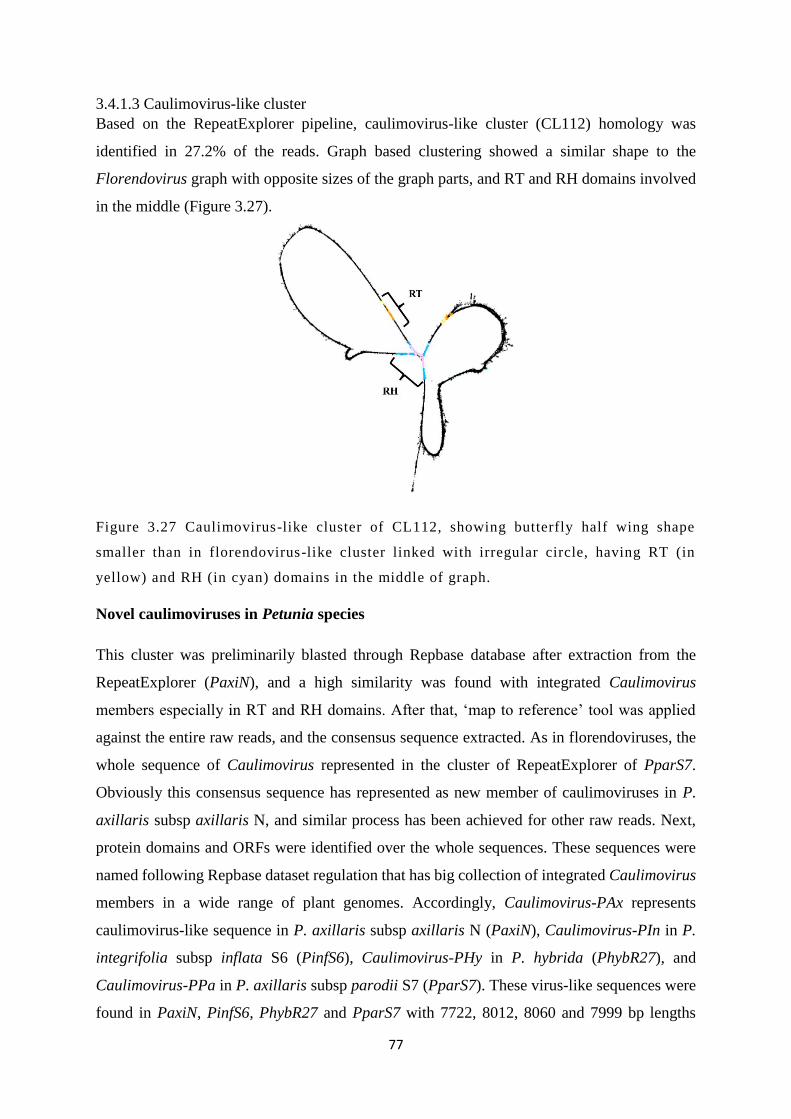
77
3.4.1.3 Caulimovirus-like cluster
Based on the RepeatExplorer pipeline, caulimovirus-like cluster (CL112) homology was
identified in 27.2% of the reads. Graph based clustering showed a similar shape to the
Florendovirus graph with opposite sizes of the graph parts, and RT and RH domains involved
in the middle (Figure 3.27).
Figure 3.27 Caulimovirus-like cluster of CL112, showing butterfly half wing shape
smaller than in florendovirus-like cluster linked with irregular circle, having RT (in
yellow) and RH (in cyan) domains in the middle of graph.
Novel caulimoviruses in Petunia species
This cluster was preliminarily blasted through Repbase database after extraction from the
RepeatExplorer (PaxiN), and a high similarity was found with integrated Caulimovirus
members especially in RT and RH domains. After that, ‘map to reference’ tool was applied
against the entire raw reads, and the consensus sequence extracted. As in florendoviruses, the
whole sequence of Caulimovirus represented in the cluster of RepeatExplorer of PparS7.
Obviously this consensus sequence has represented as new member of caulimoviruses in P.
axillaris subsp axillaris N, and similar process has been achieved for other raw reads. Next,
protein domains and ORFs were identified over the whole sequences. These sequences were
named following Repbase dataset regulation that has big collection of integrated Caulimovirus
members in a wide range of plant genomes. Accordingly, Caulimovirus-PAx represents
caulimovirus-like sequence in P. axillaris subsp axillaris N (PaxiN), Caulimovirus-PIn in P.
integrifolia subsp inflata S6 (PinfS6), Caulimovirus-PHy in P. hybrida (PhybR27), and
Caulimovirus-PPa in P. axillaris subsp parodii S7 (PparS7). These virus-like sequences were
found in PaxiN, PinfS6, PhybR27 and PparS7 with 7722, 8012, 8060 and 7999 bp lengths

78
respectively with a different series of protein domains. In Caulimovirus-PAx and
Caulimovirus-PPa, protein domains start with MP, RT, RVT and RH with four and five ORFs
respectively, while in Caulimovirus-PIn, begin with RH, MP, RT and RVT with eight open
reading frames for all. Interestingly, these domains were inverted in Caulimovirus-PHy and
start with RH, RT, RVT and MP, and has four open reading frames (Figure 3.28; Appendices
3.8, 3.9, 3.10 and 3.11).
Figure 3.28 Novel members of caulimoviruses within Petunia genome reads, Caulimovirus-PAx,
Caulimovirus-PIn, Caulimovirus-PHy and Caulimovirus-PPa , showing different lengths and
domain distributions, and variable ORFs numbers . Interestingly, Caulimovirus-PHy has an
inverted sequence unlike the others. The circular forms on the left show putative exogenous
forms with suggested cutting side and the direction of integration (black ar row) before
endogenisation event .

79
Genome proportion
Unlike the other two EPRV clusters, the caulimovirus–like cluster had a higher proportion
(0.0279%) in PparS7 than others, while PaxiN and PhybR27 have approximately same ratio
(0.015%), and the lowest proportion was in PinfS6 (0.0062%) (Figure 3.29).
Figure 3.29 Genome proportions of caulimovirus-like clusters within the four
petunia reads, showing higher ratio in PparS7, then PaxiN and PhybR27 with
similar ratio , the lower proportion was in PinflS6.
Copy numbers
Higher number of copies registered in PhybR27 (3843) and then PparS7 and PaxiN with 3237
and 2493 respectively, and like other EPRV, PinfS6 has the lower copies (Figure 3.30).
0 0.005 0.01 0.015 0.02 0.025 0.03
PaxiN
PinfS6
PhybR27
Ppar
Genome proportion %

80
Figure 3.30 Copy numbers of caulimovirus-like clusters within the four petunia
reads, showing higher number in PhybR27, then PparS7 and PaxiN, the lower
number was in PinflS6.
PCR amplification and in situ hybridization
The CL112 primers (1030 bp) amplified a product that was labelled for in situ hybridization to
Petunia chromosomes (Figure 3.31). No hybridization signals were identified in any Petunia
metaphases (Figure 3.32) using the protocol for localization of repetitive sequences at discrete
loci.
0 1000 2000 3000 4000 5000
PaxiN
PinfS6
PhybR27
Ppar
Copy numbers

81
Figure 3.31 Amplified band of Caulimovirus primer (CL112) with 1030 bp at 60˚C.
Figure 3.32 FISH image of caulimovirus-like cluster probe on P. hybrida Rdc
metaphase, showing no spotted signals on chromosomes.

82
3.4.2 Phylogenetic relationships of EPRVs
The closely related EPRVs links have been confirmed through Bayesian phylogenetic tree of
the whole EPRVs sequences (6500 bp each) that reconstructed after their alignments. The most
closely related florendoviruses were PhybV and PaxiV (supported by posterior probability of
1), and both members have 0.61 support with PparV, then those three have 1 support with
PinfV. A similar story was repeated with caulimoviruses: Caulimovirus-PHy shows high
similarity with Caulimovirus-PAx (1 support), then the two linked with Caulimovirus-PPa (1
support), and all related to Caulimovirus-PIn with 1 support. The outgroup viruses were PVCV
and CaMV, and the latter was closer to the EPRVs than PVCV especially in the region of RT
and RH domains (Figure 3.33).
Figure 3.33 Bayesian phylogenetic tree of a group of caulimoviruses and florendoviruses in
comparison with PVCV and CaMV using 18 entire sequences (about 6500 bp each), showing
high relationship between Florendovirus members (in red), and Caulimovirus members (in blue)
in Petunia genomes. The EPRVs of florendoviruses and caulimoviruses from Amborella
trichopoda and Oryza sativa show closely related sequences due to host impact unlike EPRVs
from Solanum tuberosum and Glycine max . The sequences were aligned by ClustalW alignment
and then tree was reconstructed through MrBayes inference. The number next each branch is for
posterior probability (> 0.5 support).

83
3.4.3 De novo integration
3.4.3.1 DNA extraction
Total DNA comprising genomic and potential episomal DNA of different Petunia species was
extracted from leaf tissues. Quantity and quality of the samples were analysed and determined
by spectrophotometry and gel electrophoresis. For all Petunia samples (see Table 3.5), high
molecular weight DNAs (above 10 kb) were collected (Figure 3.34), genomic DNA from all
samples was of good quality with a 260/280 ratio between 1.70 to 2.10 and the concentrations
were about 30 to 800 ng/μl. The differences in the DNA quantity were due to different amount
of leaves from each sample as well as plant age. All plants have been registered and submitted
to TEM inspection for presence of PVCV virions or other viral contamination.
Figure 3.34 1% agarose gel of extracted DNA from P. axillaris subsp axillaris N
(mixed sample from plants numbered TEM16-124_1 to _6, lane) (1), P.
integrifolia subsp inflata S6 TEM15-652 (2) PVCV infected P. axillaris subsp
parodii S7 TEM16-35_1 (3), P. axillaris subsp parodii S7 (mixed sample from
plants TEM16-315_1to _3) (4) and progeny of PVCV infected P. axillaris subsp
parodii S7 EM09-348 (5) by loading 10 μL purified DNA + 3 μL loading Buffer
(5X). 10 μl of 1kb hyper ladder (Bioline), run on each side of the gel.

84
Figure 3.35 Model of horizontal followed by vertical transmission of PVCV in P.
axillaris subsp parodii S7. Healthy plants can be infected with PVCV via biolistic
inoculation (horizontal transmission). Vegetative propagation using cuttings
probably promotes de novo integration of PVCV sequences into the P. axillaris
subsp parodii S7 genome. Thus the virus gets vertically transmitted over
generations by seeds.
3.4.3.2 RNA extraction
PCR for detection of episomal PVCV replication and activation of other pararetroviral-like
sequences is challenging on DNA templates because of the presence of chromosomal viral
copies which can also serve as template during PCR. RNA transcription is a first step in
retroelement replication, therefore, total RNA extracts were prepared from equal weights of
the selected five samples, including a DNase incubation to remove chromosomal DNA
contamination in the final RNA sample. RNA concentration and quality were determined using
a spectrophotometer and gel electrophoresis (Figure 3.36). The obtained RNA was variable,
and samples with lower RNA content showed a higher degree of RNA degradation (see Table
3.9):

85
Table 3.9 RNA concentrations of the five petunias samples.
Figure 3.36 RNA extraction of P. axillaris subsp axillaris N (mixed sample from plants
numbered TEM16-124_1 to _6, lane) (1), P. integrifolia subsp inflata S6 TEM15-652 (2) PVCV
infected P. axillaris subsp parodii S7 TEM16-35_1 (3), P. axillaris subsp parodii S7 (mixed
sample from plants TEM16-315_1to _3) (4) and progeny of PVCV infected P. axillaris subsp
parodii S7 EM09-348 (5) by loading 5 μL purified RNA + 5 μL loading dye (2X). 10µl of RNA
ladder (2µl RNA Ladder, 5µl RNA loading dye (2X) and 3µl H 20), run on the left lane (M) of
each gel.
Samples Registration codes RNA (ng/µl)
i P. axillaris subsp axillaris N TEM16-124_1-6
141,66
ii P. integrifolia subsp inflata
S6
TEM15-652
314,36
iii P. axillaris subsp parodii S7
+ PVCV
TEM16-35_1
107,98
iv P. axillaris subsp parodii S7 TEM16-315_1-3
416,50
v Progeny of P. axillaris subsp
parodii S7 + PVCV
EM09-348 252,72

86
3.4.3.3 Reverse Transcriptase-PCR
Complementary DNAs were synthesized, and electrophoresed for the five RNA samples
(Figure 3.37). All cDNA bands were between 250 and 1000 bp. cDNAs were used as templates
for PCR using PVCV F16a-R16b (483 bp), Florendovirus florB (697 bp), Caulimovirus CL112
(1030 bp) primer pairs (Figure 3.38).
Figure 3.37 cDNA synthesis of P. axillaris subsp axillaris N (mixed sample from
plants numbered TEM16-124_1 to _6, lane) (1), P. integrifolia subsp inflata S6
TEM15-652 (2) PVCV infected P. axillaris subsp parodii S7 TEM16-35_1 (3), P.
axillaris subsp parodii S7 (mixed sample from plants TEM16-315_1to _3) (4) and
progeny of PVCV infected P. axillaris subsp parodii S7 EM09-348 (5) with
Omniscript Reverse Transcription Kit. Gel was run by loading 5 µl cDNA, 5µl
H2O and 2µl DNA loading dye (6X). 6µl of DNA Ladder (1µl DNA Ladder, 1µl
6X DNA loading dye and 4µl H2O).
Although the PCR was not quantitative, there were suggestive differences in product amount
between samples. For PVCV primer, the strongest band was produced with sample No.3 (P.
axillaris subsp parodii S7 + PVCV), known to be episomally infected by PVCV. Weaker bands
were found in samples No.5 (progeny of P. axillaris subsp parodii S7 + PVCV) and No.1 (P.
axillaris N), very light bands were amplified for samples No.2 (P. integrifolia subsp inflata
S6) and No.4 (P. axillaris subsp parodii S7). The faint PCR products in the wild Petunia

87
species may originate from residual DNA in the RNA sample. A control used directly extracted
RNA as a template for PCR without the reverse transcription step. The PCR product obtained
in P. axillaris subsp parodii S7 has confirmed earlier results in 3.4.1.1 as this wild Petunia was
suggested to be free of any forms of PVCV before these experiments.
CL112 primer of Caulimovirus has amplified strongly with samples No.3 (P. axillaris subsp
parodii S7 + PVCV) and No.4 (P. axillaris subsp parodii S7), and then sample No.5 (P.
axillaris subsp parodii S7 + PVCV), light band for sample No.1 (P. axillaris subsp axillaris
N) and very light band for sample No.2 (P. integrifolia subsp inflata S6).
Using the florB primer for Florendovirus sequence, the bands were often light for samples
No.3, 4 and 5 even after amplifying them again using their PCR products as templates while
very light bands were identified for samples No.1 and 2 (Figure 3.38).
Figure 3.38 PCR of P. axillaris subsp axillaris N (mixed sample from plants
numbered TEM16-124_1 to _6, lane) (1), P. integrifolia subsp inflata S6 TEM15-
652 (2) PVCV infected P. axillaris subsp parodii S7 TEM16-35_1 (3), P. axillaris
subsp parodii S7 (mixed sample from plants TEM16-315_1to _3) (4) and progeny
of PVCV infected P. axillaris subsp parodii S7 EM09-348 (5) cDNAs as templates
and primer pairs of PVCV(F16a+R16b), Caulimovirus (CL112) and Florendovirus
(florB).
3.4.3.4 In situ hybridization
From tissue culture, de novo integrated PVCV sample with vein clearing symptom that was
infected horizontally in 2001 by grafting and then maintained in a tissue culture condition was

88
checked and analysed. Further, produced seeds from this particular plant as vertically
transmitted and de novo integrated virus have been examined with same protocol (Figure 3.35).
In order to find out de novo integration sites of PVCV over host chromosomes, PVCV probe
has been applied against progeny of PVCV infected P. axillaris subsp parodii S7 EM09-348
metaphases. Findings show telomeric signals of integrated virus on the telomere of
heterologous chromosomes with variable strengths on short or long arms. In some cases, signal
was shown on the telomere of one individual chromosome or two heterologous chromosomes,
and signal strength was very concentrated with multiple dots or too weak with only two dots
around the telomere. For instance, in Figure 3.39 fluorescent signals of PVCV were located on
the short and long arms of Ch.III and IV chromosomes respectively, with a weak signal on the
short arm of Ch.III and too condensed dots on the long arm of Ch.IV. Typical signal was
registered as two dots on the telomere follow by a dotted ring around the telomere region
(Figure 3.40). In contrast, signal features and loci of de novo integrated PVCV in host
chromosomes are completely different from locations of the chromosomal PVCV in all Petunia
chromosomes where it appeared as only two dots in the centromere or telomere regions over
only homologous chromosomes (Figure 3.18).
Figure 3.39 PVCV signals over next generation of PVCV infected P. axillaris
subsp parodii S7 EM09-348 metaphases that show two signals with variable
strength on the long arm (A) of Ch.IV and short arm (B) of Ch.III. Chromosomes
were stained with DAPI (blue) and probe was labelled with biotin-11- dUTP
(detected in red). Bar = 10 μm.

89
Figure 3.40 Typical signal of integrated PVCV on the long arm of the telomere
region of progeny of PVCV infected P. axillaris subsp parodii S7 chromosome,
showing an unusual hybridization pattern with two dots and a halo of dots looks
like a ring around the telomere. Chromosomes were stained with DAPI (blue) and
probe was labelled with biotin 11-dUTP (detected in red). Bar = 10 μm.
3.4.3.5 De novo assembly
In order to confirm the results of in situ hybridization above of de novo integrated PVCV, de
novo assembly for the whole raw reads of infected P. axillaris subsp parodii S7 was applied.
Then, the constructed consensus sequences were mapped to PVCV sequence to check if there
is any telomeric sequence (TTTAGGG) near the virus sequence. After that, the mapped contig
was extracted and then aligned against PVCV sequence. The result shows that some contigs
have telomere sequence incorporated within a recombinant PVCV while other contigs have
telomere sequences around the integrated virus sequence (Figure 3.41). More importantly,
PVCV has two regions of telomeric repeat TTAGGG (mammalian-type) in at the start (590-
595 bp) and end (6214-6219 bp) of the entire length.

90
Figure 3.41 The mapped and aligned contig to PVCV shows contig 21950 extracted
from de novo assembly has reverse complement telomere sequence (CCCTAAA)
next to recombinant PVCV sequence.
3.5 Discussion
3.5.1 PVCV fragments in all examined Petunia species
Findings of this project strongly support the importance of endogenous viral sequences in plant
genomes, and provided further evidence of the integration diversity between plant viruses and
their hosts. Although the integrated sequences are inherited over generations and many are
degenerated, some sequences are relatively intact, transcriptionally efficient and could be
infectious under particular conditions (Geering 2001; Geering et al. 2010; Geering et al. 2014).
The revolution of sequencing techniques confirmed that some plant genomes have been
infected by some Caulimoviridae members especially in the Solanaceae that were reported as
appropriate hosts for a wide number of plant viruses (Engelmann & Hamacher 2008; Geering
et al. 2014; Kim et al. 2014). Endogenisation searches have been extended successfully within
Petunia genomes, using bioinformatics tools and PCR amplification. According to the results,
virus sequence has been found with full length in the hybrid genomes of Petunia with respect
to some shorter lengths or degenerated copies. Although PVCV has been naturally fragmented
in the genome of Petunia, necessarily conserved protein domains are still active and capable to
make infection under stress conditions (Richert‐Pöggeler et al. 2003; Bombarely et al. 2016).
In the wild species, P. axillaris subsp axillaris N has similar length to PVCV in the hybrids

91
and only the last part was degenerated. While P. axillaris subsp parodii S7 was reported in
Bombarely et al. (2016) as missing all PVCV fragments, this work has shown the existence of
virus parts with some missing fragments. The virus in the other parent P. integrifolia subsp
inflata S6 was found degenerated over the whole sequence (so primer homologies were
probably the reason for failure of amplification of some regions). The degenerated nucleotides
of the last part of chromosomal PVCV in most Petunia genomes enables primers from this
region to act as markers to differentiate integrated virus copies from those copies of the
exogenous form (Figures 3.14, 3.15 and 3.16). FISH results have confirmed variable existence
of virus sequences over Petunia species: strong signals have been captured in the hybrids and
P. axillaris subsp axillaris N, in contrast to very weak signal in one pair of P. integrifolia subsp
inflata S6 and P. axillaris subsp parodii S7 chromosomes. In P. axillaris subsp axillaris N, one
more pair of chromosomes (Ch.II) were captured with weak pericentromeric signals adding
this chromosome to Bombarely et al. (2016) who showed only two pairs of P. axillaris N
chromosomes with strong signals of PVCV (Figure 3.18). The existence of EPRVs may also
play a role in genome diversification by providing coding or transcription regulatory elements
as new genetic components.
3.5.2 PVCV as a cytogenetic marker
Chromosomal signals of PVCV in each species enabled recognition of Petunia species and
chromosomes according to their signal positions, showing EPRV is a useful cytogenetic marker
(Figure 3.18). For retrotransposons, their widespread distribution within chromosomes, high
copy numbers, in addition to the ubiquitous nature, have made these components quite typical
for developing of the DNA based markers (Flavell et al. 1992; Teo et al. 2005). As very closely
related to retrotransposons, endogenous pararetroviruses (EPRVs) probably integrate within
host genomes by hitchhiking on retrotransposons and the hybrid of the two types is eligible of
real transposition might be made through this path (Hohn 1994; Richert‐Pöggeler et al. 2003;
Gregor et al. 2004; Froissart et al. 2005; Staginnus et al. 2007; Richert-Pöggeler &
Schwarzacher 2009). Additionally, in the context of diversity among Petunia genomes,
integration of PVCV has revealed evolution and phylogeny approaches over Petunia species,
and clarified the real relationships between wild and hybrid hosts based on EPRV.

92
3.5.3 Florendovirus-like sequences
The florendovirus-like clusters in Petunia species show fragmented parts of the 74 members
of florendoviruses (Geering et al. 2014) in the two parents assemblies (PaxiN and PinfS6)
(Figure 3.20). Conversely to the PVCV case, these fragments registered in P. axillaris subsp
parodii S7 with higher proportions and copies than in P. integrifolia subsp inflata S6.
Interestingly, four novel florendoviruses were recorded as one virus-like sequence in raw reads
from each genome, adding petunia to the list of 27 host species invaded by these viral units
(Figure 3.21; Appendices 3.4-3.7). As Geering et al. (2014) reported about the phylogenetic
link between the two sister groups of Florendovirus and PVCV, new members in petunia have
shared the plesiomorphic trait particularly of RT and RH with PVCV. However, those two
could be distinguished by the existence of more than one ORF in florendoviruses unlike PVCV
that has one ORF. The results of FISH revealed condensed signal also in the centromere of
Ch.III of P. axillaris subsp axillaris N and P. hybrida Rdc in addition to weaker signals in other
chromosomes (Figure 3.26).
In this project, our findings have proved Geering et al. (2014) suggestion about integration loci
of Florendovirus sequences in TE-rich regions of plant chromosomes flanked by TA
dinucleotides repeats. Results did not particularly agree with Geering et al. (2014) reporting
about 9% of Florendovirus positions within introns of plant genes based on data from Vitis
vinifera genome suggesting biological effects of these elements on gene transcripts and
expression levels. Here, inserted Florendovirus motifs accumulated in heterochromatin regions
in fragile sites of poly-TA simple repeats, probably including secondary structures like hairpins
and therefore avoiding chromosome fragility (Zlotorynski et al. 2003; Dillon et al. 2013).
These elements could act as fillers to repair DNA breaks either through microhomology-
mediated end or non-homologous end joining (Huertas 2010).
3.5.4 Hotspot of Chromosome III
Chromosome III in particular shows stronger pericentromeric signal in P. axillaris subsp
axillaris N, the hybrid species and weaker signal in P. axillaris subsp parodii S7, and this might
reflect a PVCV hotspot inside this chromosome. It probably happened due to high rates of
recombination hotspots and frequencies within this chromosome that resulted from elevated
DNA break formation. Croll et al. (2015) found that recombination rate frequency for each

93
chromosome was inversely linked to the chromosome length as it was higher in smallest core
chromosome. In Petunia chromosomes, the centromere of Ch.III has the closest length to the
telomere region among all chromosomes as eccentric type with relatively small size (Smith et
al. 1973). More importantly, the existence of PVCV and florendovirus-like sequences in the
same chromosome (Figure 3.42) might supported similarity of the two EPRVs sequence in
phylogeny as they pass through the same ancient endogenisation events. It might be a source
of virus sequences to other chromosomes by rearrangements, crossovers, frequency of
recombination (Lively 2010; Morran et al. 2011) or transposition, all features that influence
genetic variability of species and elimination of pathogenic sequences. On the virus side, in
animals, a crucial role is played by pathogen recombination, leading to annual recurring
outbreaks of recombined viral strains driving epidemic influenza (Nelson & Holmes 2007).
Figure 3.42 FISH image of florB (A) and PVCV (B) probes on P. axillaris subsp
axillaris N chromosomes shows both probes hybridized at the same region of the
centromere of Ch.III. The probe was labelled with biotin11-dUTP (red). Bar =10
μm.
3.5.5 Caulimovirus-like sequences
The caulimovirus-like sequence RepeatExplorer cluster was found in lower genome
proportions than other two EPRVs clusters with varying proportions of copies in raw reads
(Figures 3.29 and 3.30), and undetectable signals along chromosomes (Figure 3.32). Signal
strength reflects copy numbers and abundance of applied sequence; although FISH is not a

94
quantitative tool, probes with low copy number will be hardly detectable and many researchers
failed to quantify in situ experiments (Leitch & Leitch 2013). Here, four novel caulimovirus
sequences have been identified in raw reads from each species, and they have multiple ORFs
(more than four; Figure 3.28). In the assemblies, protein domains were surprisingly rearranged,
and this might happen due to different cutting points and integration sites of their circular DNA
at endogenisation (20-34 MYA), probably explaining why Caulimovirus-PHy has an inverted
sequence with respect to other caulimovirus-like sequences. Recently, many endogenous
viruses that belong to genus Caulimovirus have been characterised in plant genomes, and
despite these integrants being found fragmented and degenerated, they could be reassembled
through in silico (Geering et al. 2010; Geering et al. 2014), and data suggests in vivo too
(Harper et al. 1999; Richert‐Pöggeler et al. 2003). Sequences of endogenous caulimoviruses
have been arranged and listed in a Repbase database (Appendices 3.8-3.11) that incorporates
representative repetitive sequences of eukaryotes species. They have variable numbers of ORFs
with normal sequence direction (5’-3’) (Bao et al. 2015). Interestingly, this study has not
confirmed that some caulimovirid sequences have bipartite genome structure like that
mentioned in Geering et al. (2014) study about Florendovirus members depending on their
gene organization as there is no such example of bipartite genomes in the members of the
Caulimoviridae.
3.5.6 Expression of EPRVs
Exploring more details about the activity of all three EPRVs has further importance to
investigate their ability for expression and being active under specific conditions. Synthesizing
cDNAs for infected and healthy Petunia samples has revealed that PVCV and caulimovirus-
like sequences have been strongly expressed, unlike Florendovirus that surprisingly have not
been detected (Figure 3.38), suggesting that despite very abundant copy numbers (Figure 3.23),
they are relatively inactive. So far, our results have disagreed with the comment of Geering et
al. (2014) about good representation of Florendovirus members in expressed sequence tag
(EST) databases, suggesting that Florendovirus sequences are transcribed. Interestingly,
Geering et al. (2014) has reported findings above based on only three from 27 host genomes
(Citrus clementia, Oryza sativa and Prunus persica) data with a range between 47-57%
alignment identities. So far, the transcription of florendovirus-like sequence has dependent
activity according to host effect and specificity, also extended search to explore more facts

95
from other host genomes is quite recommended. It is also noteworthy that early analysis of
EST libraries from potato showed pararetrovirus-like sequences, particularly in those derived
from callus (Hansen et al. 2005).
The chromosomal environment has some influences on the expression of genes that observed
in some species such as in Drosophila where particular gene has been inactivated after it
translocated into heterochromatic region (Reuter et al. 1990), while in tomato, the sulfurea
locus instability was studied in closely located gene to heterochromatin region that suppressed
its function (Hagemann & Snoad 1971). In P. hybrida, the foreign genes expression of lines 17
and 24 show stable and distinct methylation pattern at the integration regions in transgenic and
wild plants. This pattern was imposed on the flanked regions of the integrated fragments and
probably it’s responsible for the intensity changes of gene expression (Pröls & Meyer 1992).
Richert‐Pöggeler et al. (2003) reported that integrated PVCV sequences of healthy petunias are
likely to be methylated. While under induction factors, the virus could be released at short
period of demethylation in one integration loci at single cell.
Phylogenetic relationships among florendoviruses have revealed that P. axillaris subsp
axillaris N and P. hybrida were so far closely related genomes (Figure 3.33) and reflected that
most of the hybrid characteristics came from the parent P. axillaris subsp axillaris N, the same
story as for caulimoviruses. EPRV data from examined genomes has suggested that P.
integrifolia subsp inflata S6 was not permissive with invasion of these elements, unlike the
hybrid one, P. axillaris subsp axillaris N and P. axillaris subsp parodii S7 respectively (Figures
3.12, 3.13, 3.22, 3.23, 3.29 and 3.30). This outcome suggests that P. integrifolia subsp inflata
S6 contributed less than P. axillaris subsp axillaris N in formation of the hybrid genome, a
result in agreement with preliminary data that has been discussed within the Petunia genome
sequencing consortium (Bombarely et al. 2016).
The role of endogenous pararetroviruses in inducing new virus infection from the host genome
has opened the door widely to answer many questions about the benefits of EPRVs to their
host. It was reported for long time that EPRVs could prevent new infections as one of defence
role for EPRVs, but researches of inducible patterns of Petunia vein clearing virus, Banana
streak virus and Tobacco vein clearing virus have contradicted the first statement of EPRVs
role. In addition, these elements have been found with higher numbers of copies than silencing
approaches need to prevent virus infection, as well as having different distributions in different
plant families that have no infection from the exogenous related virus (Jakowitsch et al. 1999;
Wesley et al. 2001; Kunii et al. 2004). The differences between the species in endogenous copy
96
numbers and in the domains represented, may suggest silencing mechanisms are more efficient
in some genomes than others in parallel with cross-protection, and silencing of RNA-viruses
(Papaya ringspot virus; Ming et al. 2008).
3.5.7 De novo integration
Vertical transmission has been studied here through de novo integration using our virus of
interest PVCV that horizontally infected P. axillaris subsp parodii S7 by grafting, and then
maintained vertically since 2001. Many plant viruses use vertical transmission through seeds
(or other propagules such as seed potatoes) to cause severe infections on host plants by
transmitting virus particles from parents to hybrids, as well as this method has a huge impact
on pathogen virulence and plant adaptation. It seems most likely that vertical transmission rate
has negative correlation with pathogen virulence that may have evolved with challenge to the
hypothesis of trade-off and suggested models (Ewald 1983, 1987; Alizon et al. 2009).
PVCV is integrated into arbitrary loci on host plant chromosomes as virus signals were found
on the telomere of the short or long arm of heterologous chromosomes and signal strength
widely fluctuated within examined chromosomes (Figure 3.39 and 3.40). De novo assembly
confirmed the telomere sequence (TTTAGGG) was associated around the recombinant virus
sequence suggesting PVCV has integrated in the telomere region (Figure 3.41). In plant
genomes, this study has been applied for the first time as only in recent years, few researches
have started to explore this type of integration for herpesvirus 6 (HHV-6) within human
chromosomes using fluorescent in situ hybridization (FISH). Herpesvirus 6 integrated in the
telomere site of human chromosomes, and the examined individual’s cells were 100%
vertically infected, also virus signals found interestingly on short or long arm of heterologous
chromosomes (Nacheva et al. 2008). Further studies have characterised that chromosomally
integrated herpesvirus 6 is so far unstable and could be released to cause infection as a
functioning virus (Huang et al. 2013; Wood & Royle 2017). Our data in this research has so
far agreed with data from human genome, suggesting similar pattern of integration between
two viruses (PVCV and HHV-6) and two hosts (P. axillaris subsp parodii S7 and Homo
sapiens). However, PVCV sequence has TTAGGG (mammalian-type) telomeric repeat in two
regions at the start (590-595 bp) and end (6214-6219 bp) of the whole length. PVCV integrates
at the telomere of infected Petunia probably because of unstable proximal telomeric sites of
these chromosomes and still prone to high recombination rate coupled together with abundant
97
virus genomes that integrated by such a mechanism contributing the telomere regions
(McKnight et al. 2002; Wang et al. 2004). This mechanism perhaps starts with disturbing the
t-loop site and allow emergence of telomere rapid deletion events as reported in yeast (Lustig
2003), and then truncated regions may arise by double strand break and end processing to
produce single-strand overhang in presence of virus genome strands that is easily interfered
with (Huang et al. 2013). However, this model was taken from results of other kingdoms, and
the applicability of the model to plant genomes needs further investigation. Sequencing using
technologies such as 10X Genomics or Nanopore Minion to identify long-range associations
of sequences would be helpful.

98
Chapter IV. Transmission electron microscopy of PVCV
4.1 Introduction
4.1.1 Electron microscopy and detection of Petunia vein clearing virus (PVCV)
Transmission electron microscope (TEM) of ultrathin sections or sap from infected tissues is
widely used for detection and identification of plant and animal viruses. In a study aimed to
identify phytoplasma–like bodies and viruses in Opuntia tuna showing witches–broom disease-
like symptoms, Lesemann and Casper (1973) reported that it was not possible to detect virus-
like particles in ultrathin sections of embedded O. tuna tissues and that most transmission
experiments from infected Opuntia were negative. Petunia test plants that were not used for
the transmission experiments, showed all of sudden vein clearing symptoms and leaf
deformation in young shoots with stunted growth. In ultrathin sections of embedded
symptomatic leaf and stem material, virus-like particles and inclusion bodies were discovered.
Only grafting from petunia to petunia transmitted the virus-like particles. Based on host plant
and symptoms the infectious entity was named ‘`Petunia vein clearing virus’’ (PVCV). No
serological relationships between PVCV and other caulimoviruses using immuno electron
microscopy (IEM) was identified (Lesemann & Casper 1973). Based on phylogentic analysis
and distinct genome organization PVCV was classified in a new genus amongst
Caulimoviridae named Petuvirus (Richert-Pöggeler & Shepherd 1997; King et al. 2011). Lack
of molecular tools and information about virus biology at that time made electron microscopy
a useful diagnostic tool for PVCV detection in plant tissues. However, using electron
microscopy for virus detection is hampered because of the low virus concentration and unequal
virus distribution within plant tissues (Sikron et al. 1995). Consequently the risk of false
negative results for PVCV existed which is critical when testing propagation material from
commercial nurseries (Zeidan et al. 2000). A PCR based technique increased the sensitivity for
plant virus detection (Rybicki & Hughes 1990; Navot et al. 1992; Zeidan et al. 2000). The
virions can be detected in crude samples only in symptomatic tissues. Virion’s diameter was
variable with the majority of particles being between 40 and 45 nm in size (Lesemann and
Casper, 1973). Ultrathin sections of embedded symptomatic plant material revealed that many
infected cells contained one or more densely stained inclusion bodies (IB) with a diameter up
to 10 µm. The inclusion bodies were rounded, but in some cases appeared irregular, the matrix

99
appeared finely granular and within the bodies many lightly contrasted, rounded, vacuole-like
regions were present. The IBs were not enveloped by a membrane. Virus particles have not
been found within the IB matrix but sometimes close to the inclusion bodies and just within
vacuole-like regions, particles have rarely occurred within a nucleus (Lesemann & Casper
1973). Here, the ultra-structure of variability in vein clearing symptom expression reaching
from a more spot like appearance to extended chlorosis along the veins were analysed using
transmission electron microscopy (TEM). Furthermore the ultra-structure of PVCV infected
cells originating from horizontal or vertical transmission was compared.
4.2 Aims and objectives
Aims: One aim of this chapter is to determine the cellular ultra-structure and any differences
between vein clearing and spot symptoms. The second aim addresses if viral integration is
essential for virus replication. Therefore, episomal viral replication from induced chromosomal
copies of PVCV which were preexisting or derived from de novo integration was compared
with PVCV replication in petunia plants after horizontal virus transmission.
Objectives:
Using transmission electron microscopy (TEM) together with immunogold labelling technique
to associate different modes of PVCV transmission (horizontal or vertical) with virus particle
concentration as well as with changes in the ultra-cellular structure of infected cells in both of
episomal, de novo integrated and induced infections of PVCV.
4.3 Materials and methods
4.3.1 Plant species
The examined Petunia species were: i) episomally infected P. axillaris subsp parodii S7,
registered as TEM16-35_1, showing vein clearing and local lesion symptoms. ii) progeny of
PVCV-infected P. axillaris subsp parodii S7 grown from seeds on Murashige-Skoog agar
(Appendix 4.3) as tissue culture samples showing vein clearing registered as TEM16-483. iii)
progeny of episomally infected P. axillaris subsp parodii S7 that showed vein clearing
symptoms, registered as TEM12-584. iv) induced symptoms under heat application were taken

100
from P. hybrida W138, registered as 3645d [(examined plants at four weeks old were exposed
to 16 h at 30°C and then 8 h at 25°C until viral symptoms were induced, while control plants
were grown for 24 h at 25°C (Noreen et al. 2007)]. v) healthy leaves of P. axillaris subsp
parodii S7 that do not carry infectious chromosomal copies of PVCV, registered as TEM 16-
403_3. vi) healthy leaves of P. hybrida W138, that harbor inducible PVCV genomes within
their chromosomes, registered as TEM 16-484 (Table 4.1).
Table 4.1 List of examined samples of petunia with symptoms, registration codes and
descriptions.
4.3.2 Electron microscopy
Dip preparations were done with symptomatic leaf tissues of petunia. TEM grids were mounted
on the crude sap preparations for 5 min, washed with water and for contrast negative stained
with 1% aqueous uranyl acetate. Samples were dried and screened for presence of virions using
a transmission electron microscope, Tecnai G2 spirit, FEI. TEM examination was performed
at acceleration voltage of 80 kV providing optimal contrast for biological material. Images
were taken using a 2x2k digital camera (Veleta) which was attached at the side of the
microscope column. A serological approach was used to enhance detection sensitivity using
the electron microscope. For immuno electron microscopy (IEM), a polyclonal PVCV antibody
Samples Symptoms Registration codes Descriptions
i P. axillaris subsp
parodii S7
Vein clearing
and chlorotic
spot
TEM 16-35_1
PVCV infected P. axillaris subsp parodii S7 with
episomal form that horizontally transmitted via
grafting.
ii P. axillaris subsp
parodii S7
Vein clearing TEM16-483
Progeny of PVCV-infected P. axillaris subsp
parodii S7 grown from seeds on Murashige-Skoog
agar.
iii P. axillaris subsp
parodii S7
Vein clearing TEM 12-584 Progeny of PVCV infected P. axillaris subsp
parodii S7 that vertically transmitted.
iv P. hybrida W138 Chlorotic spot 3645d Induced P. hybrida W138 by heat application.
v P. axillaris
subsp parodii S7
No symptoms TEM 16-403_3
Healthy P. axillaris subsp parodii S7 leaves.
vi P. hybrida W138 No symptoms TEM 16-484 Healthy P. hybrida W138 leaves.

101
produced in rabbits (Richert 1992) was employed. IEM included immunosorbant electron
microscopy (ISEM) for trapping of virus particles with PVCV specific antibody diluted 1:1000.
For virus identification, samples were incubated with PVCV antibody diluted 1:50. For a more
pronounced visualization of antibody-virion interaction, selected samples from dip
preparations as well as ultrathin sections were immunogold labelled with a secondary gold-
goat-anti-rabbit antibody conjugate diluted 1:50. For immunogold labelling, 5 or 10 nm gold
particles were used, and both particle sizes obtained specific labelling of PVCV particles.
4.3.2.1 Embedding procedure
According to Lesemann (1991), symptomatic leaf tissue was sliced into 1x2 mm pieces using
a sharp razor blade. The leaf sections were transferred carefully and directly with a preparation
needle into 1 ml of the first fixation solution (2.5 % glutaraldehyde in 0.1 M (K, Na) P-buffer)
in 3.5 ml glass tubes. Leaf tissues were submerged in the fixation solution and degassed for 15
min in a vacuum desiccator. Once all air had been removed from the sample tissue, fixation
was done for at least 2 hr followed by a washing step to remove the first fixation solution. For
the second (post) fixation served 1 ml 0.5% osmium tetroxide in 0.1 M (K, Na) P-buffer for at
least 2 hr. Samples were washed using a Pasteur pipette filled with Millipore water four times
for 5 min each. For contrast, samples were negative stained with 1% aqueous uranyl acetate.
4.3.2.2 Preparation of embedding forms
Freshly prepared embedding resin (Epon812+DDSA, MNA, catalysator DMP30) from stock
solutions was added to half the volume of the embedding form that had been labelled with a
small piece of paper by pencil at upper right corner of the form (see Appendix 4.2). A
preparation needle was used to remove air bubbles and the resin was left for polymerizing
overnight at 40°C in a drying oven (Memmert). On the second day, dehydration procedure was
done by preparing 50% acetone (15ml 100% acetone + 15 ml Millipore water) and 70% acetone
(35ml 100% acetone + 15ml Millipore water). Then, washing for two times (30 min) with 50%
acetone, two times (30 min) with 70% acetone and four times (30 min) with 100% acetone.
After the last 100% acetone step, solutions have been added into small glass tubes (5 ml) with
flat bottom that were labelled accordingly. Solutions were transferred via fast pouring and
remaining pieces were transferred using a preparation needle. 100% acetone were replaced
with acetone/Epon 1:1 in order to remove acetone, glass tubes placed inside dry oven with open

102
door and rotated by rotator for 1 hr at 40°C. Solution replaced with Epon and acetone/Epon
solution was removed as much as possible. Final infiltration step of the fixed sample was done
before transfer to embedding forms two times for 1hr at 40°C with Epon. Embedding forms
that have been prefilled with Epon (half volume, see above) were filled up with the sample in
Epon solution and air bubbles removed. Polymerization occurred at 60°C for 48 hr in a drying
oven (Memmert). For each sample 4 repetitions were done, the generated blocks have been
numbered chronologically (registration codes) and each repetition is indicated by a letter (see
Table 2.2).
4.3.2.3 Immunogold labelling using ultra-thin sections
Three to five ultra-thin sections of 60 nm thickness in average were mounted on a nickel grid
(75 mesh). To avoid unspecific binding, the mounted sections were blocked for 15 min with
1% BSA in 0.1 M phosphate (P-) buffer, pH 7.0 followed by washing with 1.5ml ELISA wash-
buffer and excess liquid was removed. Incubation with the primary antibody diluted 1:50 in
ELISA wash-buffer+0.5 % BSA was done overnight followed by washing with 1.5ml ELISA
wash-buffer and excess liquid was removed. After that, the grid was incubated with the
secondary antibody (gold-goat-anti-rabbit, 10 nm) diluted 1:50 in ELISA wash-buffer+0.5 %
BSA for 2h. Before the addition of 2% uranyl acetate for contrast, the sections were washed
with ultra-pure H2O. Finally, the grid was washed with few drops of ultra-pure H2O and stored
dry (see Table2.2).

103
4.4 Results
4.4.1 Horizontally transmitted PVCV
In order to study the cellular ultra-structure of observed symptomatic phenotypes comprising
diffuse chlorosis or direct vein clearing (Figures 4.1and 4.2) in leaves after horizontal
transmission, tissues were embedded for TEM analysis. In P. axillaris subsp parodii S7, no
full-length PVCV sequences capable of triggering infection are present (Figure 3.18; Appendix
3.3). Therefore these plants are predestined to study the ultra-structure of cells for PVCV
replication using episomal DNA templates only without interference from inducible
chromosomal PVCV DNA copies.
Figure 4.1 Typical vein clearing symptoms on young shoot after vertical transmission of
PVCV. The depicted plant on the left, registered as TEM12 -584, is from the progeny of
P. axillaris subsp parodii S7 that had been infected with PVCV using biolistic
inoculation and maintained in tissue culture over several years. Prior to pollination and
seed harvest plants were transferred from tissue culture to the green house. Healthy leaf
of P. axillaris subsp parodii S7, registered as TEM16-403_3 on the right.

104
Figure 4.2 PVCV symptoms on infected P. axillaris subsp parodii S7 (TEM16-
35_1) leaves start with yellow local lesion close to the leaf midrib (left), elongated
spot along the side vein (right). P. axillaris subsp parodii S7 plants were infected
with an infectious full-length clone of PVCV [72-2_3c, (Richert‐Pöggeler et al.
2003)] using biolistic inoculation and propagated in tissue culture for several years
and transferred to soil to increase growth under greenhouse conditions.
4.4.1.1 Cellular ultra-structure of diffuse chlorotic symptoms along the veins
In symptomatic leaves of P. axillaris subsp parodii S7 (TEM16-35_1) displaying elongated
chlorotic spots (Figure 4.2 left panel), virus particles have been shown in mesophyll cells that
are adjacent to the vasculature and concentrated in distinct cytoplasmic regions. Additionally,
rough endoplasmic reticulum (ER) was noticed in these cytoplasmic regions adjacent to virus
particles. The isometric particles had an average diameter between 36 and 46 nm. During
temporal progression of infection these viroplasm seem to become more compact and appear
as electron dense material in which virions are not distinguishable any more. In average 500 to
1000 virus particles were counted in infected cells. Various phases during maturation from
viroplasm to inclusion bodies are displayed in Figures 4.3 and 4.4. Inclusion bodies (IB) have
been noticed with a rounded shape in some cells and irregular shape in others with a diameter
of 500 nm-2µm. IBs are without membranes. Vacuoles were observed in some infected cells
and chloroplasts in the examined cells did not have any starch granules (Figures 4.3, 4.4 and
4.5). Immunogold labelling confirmed that the majority of virions can be found in the

105
cytoplasm and not within the inclusion body matrix where only few gold particles were located
(Figure 4.6).
Figure 4.3 Ultrathin section of PVCV-infected P. axillaris subsp parodii S7
(TEM16-35_1) mesophyll leaf cell. High concentration of virions and two large
inclusion bodies were identified in the cytoplasm. Vp= viroplasm, IB= inclusion
body, M= mitochondria, V= vacuole. Bar= 2µm (left), and 1 µm (right).
Figure 4.4 Transmission electron micrograph of ultrathin section of PVCV -infected P.
axillaris subsp parodii S7 (TEM16-35_1) mesophyll leaf cell, showing distributed
virions with less dense inclusion body matrix. Vp= viroplasm, IB= inclusion body,
M=mitochondria, V=vacuole, Ch=chloroplasts without starch. Bar =1 µm.

106
Figure 4.5 Details of PVCV virions within the cytoplasm in episomally infected
P. axillaris subsp parodii S7 (TEM16-35_1) and interaction with vesicular
membranes. Vp= viroplasm, V= vacuole. Bar=500 nm.
Figure 4.6 Immunogold labelling of PVCV particles in episomally infected P.
axillaris subsp parodii S7 (TEM16-35_1) mesophyll leaf cell using 10 nm gold
conjugated-anti-rabbit antiserum. The inclusion bodies have been noticed with a
rounded shape. Some virions are found outside but in close contact with the IB
(black arrow). Vp= viroplasm, IB= inclusion body, arrows indicate regions with
gold label. Contrast and brightness have been adjusted to improve visibility of the
gold particles. Bar=500 nm.

107
4.4.1.2 Cellular ultra-structure of vein restricted chlorosis symptoms
Tissue embedded from leaves of PVCV infected P. axillaris subsp parodii S7 (TEM16-35_1)
displaying vein clearing symptoms revealed a high concentration of PVCV particles in the
cytoplasm of mesophyll cells adjacent to the vasculature. No inclusion bodies have been
noticed in these cells indicating probably an early event in virus infection. Viroplasm has
formed but has not yet reached a more condensed stage resulting in the formation of inclusion
bodies. Vacuole like regions have been found with a big portion at the periphery of these cells.
These results of vein clearing samples have been confirmed with another infected plant under
the same greenhouse conditions (Figure 4.7).
Figure 4.7 Transmission electron micrograph of ultrathin section of PVCV -
infected P. axillaris subsp parodii S7 (TEM16_35_1) in the cytoplasm of a
mesophyll cell adjacent to the vasculature , showing isometric virions. No
inclusion bodies have been found in these cells. Vp= viroplasm, M=mitochondria,
Ch=chloroplasts without starch. Bar =1 µm.

108
4.4.2 Vertical transmission of PVCV
4.4.2.1 TEM for progeny of PVCV-infected P. axillaris subsp parodii S7
PVCV infected P. axillaris subsp parodii S7 derived from biolistic inoculation using an
infectious full-length clone have been vegetatively propagated for several years on Murashige
Skoog medium by cuttings and transferred to the greenhouse for further growth and seed
production. The seeds of such plants were grown under sterile as well as greenhouse conditions.
Under both cultivation methods, vein clearing symptoms occurred and were tested for episomal
PVCV infection which must have occurred during vertical transmission via seeds. PVCV
particles have been shown in companion cells adjacent to the vasculature (Figure 4.8). The
particles have been found in condensed regions within a cell and were spherical with an average
diameter of 44 nm, immunogold labelled sections have confirmed this result as gold particles
were attached to virions. Inclusion bodies have not been found in the infected cells (Figure
4.9).
Figure 4.8 Ultrathin section of progeny of PVCV-infected P. axillaris subsp
parodii S7 (TEM16-483) grown from seeds on Murashige-Skoog agar, showing
virions in the cytoplasm of a bundle sheath cell. No inclusion bodies have been
found in these cells. Vp= viroplasm, V= vacuole. Bar = 5 µm (left), and 500 nm
(right).

109
Figure 4.9 Overview and details of ultra-structure of progeny of PVCV-infected
P. axillaris subsp parodii S7 (TEM12-584) leaves with vein clearing symptoms.
Virions are distributed in the cytoplasm of a probable companion cell, showing
condensed virions. No inclusion bodies have been found in these cells. Vp=
viroplasm, M= mitochondria. Bar =5 µm (left), and 2 µm (right).
4.4.2.2 Ultrastructure of PVCV infection after induction of chromosomal PVCV forming a
tandem array
In the P. hybrida cultivar W138 it has been shown that integrated PVCV genomes arranged in
a tandem array are infectious (Richert‐Pöggeler et al. 2003). Furthermore, it has been reported
that such chromosomal copies can be activated after heat exposure (Noreen et al. 2007).
Symptomatic leaf sections from induced P. hybrida W138 have been embedded and ultrathin
sections were screened for PVCV infected cells originated from vertical transmission.
Isometric virions as well as inclusion bodies resided in the cytoplasm (Figure 4.10). About 300-
400 virions were counted in infected cells that have approximately the same dimensions in
comparison to particles (about 40 to 52 nm) derived from horizontal transmission (Figures 4.10
and 4.11). Three different stages of inclusion bodies were present (Figures 4.10 and 4.11). They
all displayed a rounded shape with an average diameter of 450-600 nm. The cell displayed in
Figure 4.10 contained two IBs with vacuole-like inner core that have released generated
particles in the cytoplasm. These particles reacted specifically with immunogold. The IB in the
upper left corner is electron dense and surrounded by cellular (ER) membrane structures. In

110
Figure 4.11 an earlier stage of PVCV replication is depicted. Here the two IBs, flanking the
virion containing region, are less electron dense and virions seem to egress from the viroplasm
matrix into the cytoplasm. Mature particles in the cytoplasm are immunogold labelled.
When chromosomal copies of PVCV are present they can theoretically trigger PVCV infection
in each cell. The host plant seems to employ epigenetic regulation to avoid this (Noreen et al.
2007). Instead the virus can move from the initially infected cell via plasmodesmata to the
neighboring cell. An indication for such intercellular movement is depicted in Figure 4.11 on
the right panel. A single virion is located adjacent to the cell wall with plasmodesmata exposed.
Figure 4.10 Transmission electron micrograph of an ultrathin section of induced
P. hybrida W138 (3645d). In the cytoplasm of a mesophyll cell , scattered virions
and three inclusion bodies are visible. Vp= viroplasm, M= mitochondria, IB=
inclusion body, Ch= chloroplast with and without starch granules, V= vacuole. Bar
= 5µm (left), and 1 µm (right).

111
Figure 4.11 PVCV replication in induced P. hybrida W138 (3645d). In an early stage of
replication PVCV particles seem to egress from the viroplasm matrix (VP) into the
cytoplasm. Mature virions in the cytoplasm are immunogold label led. Red arrow
indicates single virion adjacent to the cell wall with plasmodesmata (PD) visible. Vp=
viroplasm. Bar = 2µm (left), 1 µm (middle ), and 500 nm (right).
4.4.2.3 TEM for healthy P. axillaris subsp parodii S7 and P. hybrida W138
Embedded tissue of healthy plants contained no abnormality regarding organelle sizes or
shapes and vasculature. Neither PVCV virions nor inclusion bodies have been noticed in the
respective cells. (Figures 4.12 and 4.13).
Figure 4.12 Healthy cell of P. axillaris subsp parodii S7, showing normal cell
components in the vasculature. M= mitochondria, V=vacuole. Bar = 1µm.

112
Figure 4.13 Ultrathin section of healthy P. hybrida W138, showing normal cell
components at parenchyma cells. N= nucleolus within the nucleus, M=
mitochondria, Ch= chloroplast , V= vacuole. Bar = 2µm.
To sum up the results above see Table 4.2:
Table 4.2 Differences in virion and inclusion body numbers and sizes within applied samples
contents.
Type of symptom/origin of DNA Virion
numbers
Virion
sizes
Number
of IBs
IB sizes
1 Spreading chlorosis/episomal 500-1000 36-46 nm 2-3 0.5-2 µm
2 Vein clearing/episomal 500-750 40-44 nm No No
3 Vein clearing/chromosomal 300-750 44 nm No No
4 Spontaneous chlorosis/chromosomal 300-400 40-52 nm 1-3 450-600 nm

113
4.5 Discussion
Electron microscopy provides high resolution images of the viruses, and shows their
relationship or interactions with the host cells. Together with microscopy techniques based on
light (fluorescence microscopy, confocal laser scanning microscopy) and molecular biology
tools (e.g. PCR, FISH, NGS) it enables comprehensive analysis of the virosphere (Hoang et al.
2011; Corbetta et al. 2017; Hesketh et al. 2017; Lei et al. 2017; Balke et al. 2018). In this
chapter we employ electron microscopy to study PVCV in petunia cells (Figures 4.3- 4.13).
PVCV can occur both in episomal as well as chromosomal forms depending on the host. Both
templates can trigger viral infection. The aim was to compare PVCV replication after
horizontal and vertical transmission respectively. In earlier studies it had been shown that the
presence of PVCV particles is correlated with symptom expression (Richert 1992). Therefore,
the impact of various phenotypes on viral replication also investigated in the distinct
transmission modes.
4.5.1 Ultrastructural analysis of diffuse chlorosis and vein clearing symptoms
after horizontal transmission of PVCV
Despite no vector for horizontal (from plant to plant) PVCV transmission being identified yet,
plants can be infected artificially using grafting or biolistic inoculation (Richert‐Pöggeler et al.
2003). The leaf symptoms on PVCV infected petunia are generally described as leaf yellowing
and vein clearing in young shoots (Lockhart & Lesemann 1998; Zeidan et al. 2000; Richert‐
Pöggeler et al. 2003). In some cases, symptoms start with local chlorosis adjacent to one of
small netted veins then developing either strictly along the vein or becoming a diffuse chlorosis
spreading further into the leaf lamina (Figure 4.2). In horizontal PVCV transmission, the
encapsidated circular double stranded DNA molecules are released from the capsid and are
transcribed in the plant nucleus. The generated RNA molecule comprising the complete viral
genome including promoter sequences is transported to the cytoplasm. The transcript serves as
messenger RNA and as template for DNA synthesis using reverse transcription. To determine
whether the differences in symptomatic phenotypes were also reflected on the cellular level of
PVCV infected P. axillaris subsp parodii S7 plants, electron microscopy was applied,
including use of immunolabelling of PVCV to confirm the identification of structures with
viral capsids. So far, cells from tissue with vein clearing contained clustered virions but lacked
viral inclusion bodies. The minimum diameter of the virion was 36nm and the maximum was

114
45nm; this is in accordance with the published value of 46nm which was obtained measuring
purified virions directly (Richert-Pöggeler & Lesemann 2007). Many of these cells included
vacuoles, and chloroplasts sometimes contain starch granules. In leaves displaying diffuse
chlorosis, infected cells contained virions in parenchyma cells with minimum diameter 44nm
and the maximum was 56nm. Conversely to cells derived from vein clearing tissue, there were
condensed inclusion bodies of rounded or irregular shapes with a diameter of 500 nm – 2 µm
similar to those described by Lesemann and Casper (1973) (Figures 4.3, 4.4, 4.5 and 4.6). The
existence of rough endoplasmic reticulum (ER) in the cytoplasmic regions adjacent to virus
particles may be an indication that these metabolic active sites are also used for translation of
viral proteins (Schaad et al. 1997; Turner et al. 2004).
Comparing the two symptom types, differences in particle sizes and IB numbers have been
found in infected cells for each phenotype (Table 4.2). These observations give first insights in
stages during viral replication. Early on infected tissue in which chlorosis is limited to the vein
region viral replication starts. Spreading of viral infection to the leaf lamina is accompanied by
enhanced viral replication. As consequence more virus particles are present and inclusion
bodies are formed. However, in virology, inclusion bodies are accumulated dead-end materials
that contain unused viral proteins in the cytoplasm of infected cells at late times of infection,
while viroplasms are electron-dense masses at early stages of infection include viral and
cellular factors in viral replication sites within infected cells (Novoa et al. 2005). The
variability in particle sizes may indicate different steps of particle interactions with viral
proteins as well as with host components. For Cauliflower mosaic virus it has been shown that
during virus movement interaction of coat protein with the virion associated protein occurs
(Stavolone et al. 2005; Amari et al. 2010; Stavolone & Lionetti 2017).
4.5.2 De novo integration and vertical transmission of PVCV in P. axillaris
subsp parodii S7
The P. axillaris subsp parodii S7 genome was assumed to be free of any integrated form of
PVCV, since no signal was generated under stringent hybridization conditions using total
genomic DNA as template and virus specific probes (Richert‐Pöggeler et al. 2003). When
artificially infected using grafting or biolistic inoculation, P. axillaris subsp parodii S7 proved
to be a suitable host for PVCV allowing episomal replication (Figures 4.1 and 4.2). These
experiments showed that unlike retroviruses, endogenous pararetroviruses do not depend on
integration into the host genome to complete their replication cycle; indeed, before Harper et

115
al. (1999), integrated pararetroviruses were unknown in plants. P. axillaris subsp parodii S7 is
an ideal model system to study de novo integration of PVCV sequences into the Petunia
genome. Whereas it is estimated that integration occurs frequently in somatic cells, the
manifestation of such an event in reproductive cells and thus in the progeny may be rare (Gayral
et al. 2008). Therefore the progeny of PVCV infected P. axillaris subsp parodii S7 grown in
tissue culture for prolonged times (>10 years) were screened. For infection of sterile plants,
biolistic inoculation with an infectious full-length clone had been used (Richert‐Pöggeler et al.
2003). Cuttings from PVCV infected plants were transferred to new MS medium every 4-8
weeks. The dedifferentiation of cells during the process of new root development most likely
facilitates invasion of the host genome by linearized PVCV DNA molecules present in the
nucleus. Furthermore it had been postulated that integrated copies get activated in such
meristematic cells due to changes in epigenetic modifications (Noreen et al. 2007).
PVCV infection of symptomatic plants were confirmed by PCR and IEM. A first evidence for
vertical transmission of PVCV was obtained when the progeny of these plants showed typical
vein clearing symptoms and tested positive in PCR and IEM. FISH further confirmed de novo
integration of PVCV in the telomeric regions of chromosomes III and IV (see Figure 3.39).
The fact that virions but no IBs were detectable (Figures 4.7, 4.8 and 4.9), may indicate either
that an early step in viral replication is caught or that in this cell only intracellular replication
occurs. The latter case would be similar to the replication of LTR retrotransposons (Richert-
Pöggeler & Schwarzacher 2009).
4.5.3 PVCV symptom expression and replication after induction of chromosomal
copies
Elevated temperatures above 25°C for a continuous time period of 7 days can enhance
symptom expression in P. hybrida (Zeidan et al. 2000). Additionally, P. hybrida line W138
was shown to be more permissive than cultivar Rdc (Himmelsröschen) to induce symptoms
after repeated heat treatments with a higher rate of infection (Noreen et al. 2007). Symptoms
from induced infection are often starting as spot features more than vein clearing. In contrast
to horizontally infected P. axillaris subsp parodii S7 cells, symptom expression resulted from
activation of chromosomal copies. Particle concentration in W138 cells was at the maximum
about 400 /cell (Figures 4.10 and 4.11), while in PVCV infected P. axillaris subsp parodii S7
it could reach 1000 /cell (Figures 4.3- 4.7). Virions were found loosely scattered in the cell

116
whereas in horizontally infected P. axillaris subsp parodii S7 additionally viroplasm and IBs
were present.
The ultra-structural analyses provided first evidence that the efficiency in viral replication
depends on the origin of PVCV infection. Virus activation from chromosomal copies seems to
be less efficient. This may happen due to tighter epigenetic control by the host than from
episomal PVCV copies. It may also take longer to build up high virus load because the number
of integrated tandem array is less than the number of circular DNA molecules provided in
artificial infection. The low efficiency of expression of EPRVs is also found in banana, where
infection originating from endogenous copies is unusual, sometimes following stress such as
tissue culture or cold nights (Harper et al. 1999; Harper et al. 2002).
The virulance of viruses could be significantly affected by transmission mode as its increase
within horizontal transmission as a side effect of a trade off between infectivity and virulance.
In vertical transmission, virulance decreases to allow virus transmission to wide host
offsprings. Stewart et al. (2005) reported that Barley stripe mosaic virus (BSMV) horizontally
transmitted in its host (Hordeum vulgare) with twofold increase in infectivity and tripled
virulance, while in vertical transmission, infectivity slightly increased, while virulence highly
reduced.
Interestingly, in the P. hybrida line W138 telomeric PVCV insertions as seen after de novo
integration in P. axillaris subsp parodii S7 of this study were identified (Richert-Pöggeler and
Schwarzacher, unpublished) (see Figure 3.18). The plasticity of telomeres harbouring various
retroelements as shown for bdelloid rotifers and Drosophila (Arkhipova et al. 2017;
Casacuberta 2017), and we can speculate similar plasticity and reorganization of sequences in
telomeric regions may also be true for petunia. Notably, Vicient and Casacuberta (2017)
discussed transposable elements and their insertion into heterochromatic regions of
chromosomes (including centromeres and telomeres), potentially a model for the behaviour of
EPRVs. Future studies should address if the invasion of the Petunia genome by PVCV starts
at the telomere and then spreads to other loci in the pericentromeric regions of chromosomes.
The identified telomeric copy in W138 may indicate the dynamics in virus host symbiosis and
thus may represent a recent de novo integration by PVCV.
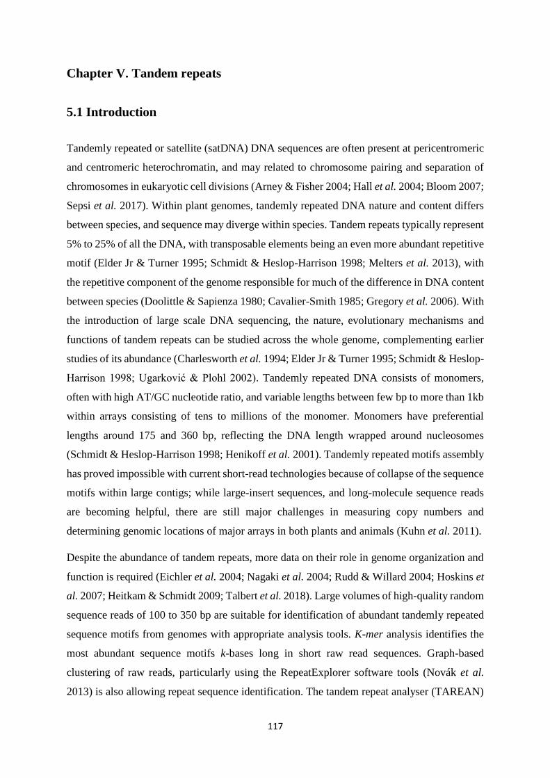
117
Chapter V. Tandem repeats
5.1 Introduction
Tandemly repeated or satellite (satDNA) DNA sequences are often present at pericentromeric
and centromeric heterochromatin, and may related to chromosome pairing and separation of
chromosomes in eukaryotic cell divisions (Arney & Fisher 2004; Hall et al. 2004; Bloom 2007;
Sepsi et al. 2017). Within plant genomes, tandemly repeated DNA nature and content differs
between species, and sequence may diverge within species. Tandem repeats typically represent
5% to 25% of all the DNA, with transposable elements being an even more abundant repetitive
motif (Elder Jr & Turner 1995; Schmidt & Heslop-Harrison 1998; Melters et al. 2013), with
the repetitive component of the genome responsible for much of the difference in DNA content
between species (Doolittle & Sapienza 1980; Cavalier-Smith 1985; Gregory et al. 2006). With
the introduction of large scale DNA sequencing, the nature, evolutionary mechanisms and
functions of tandem repeats can be studied across the whole genome, complementing earlier
studies of its abundance (Charlesworth et al. 1994; Elder Jr & Turner 1995; Schmidt & Heslop-
Harrison 1998; Ugarković & Plohl 2002). Tandemly repeated DNA consists of monomers,
often with high AT/GC nucleotide ratio, and variable lengths between few bp to more than 1kb
within arrays consisting of tens to millions of the monomer. Monomers have preferential
lengths around 175 and 360 bp, reflecting the DNA length wrapped around nucleosomes
(Schmidt & Heslop-Harrison 1998; Henikoff et al. 2001). Tandemly repeated motifs assembly
has proved impossible with current short-read technologies because of collapse of the sequence
motifs within large contigs; while large-insert sequences, and long-molecule sequence reads
are becoming helpful, there are still major challenges in measuring copy numbers and
determining genomic locations of major arrays in both plants and animals (Kuhn et al. 2011).
Despite the abundance of tandem repeats, more data on their role in genome organization and
function is required (Eichler et al. 2004; Nagaki et al. 2004; Rudd & Willard 2004; Hoskins et
al. 2007; Heitkam & Schmidt 2009; Talbert et al. 2018). Large volumes of high-quality random
sequence reads of 100 to 350 bp are suitable for identification of abundant tandemly repeated
sequence motifs from genomes with appropriate analysis tools. K-mer analysis identifies the
most abundant sequence motifs k-bases long in short raw read sequences. Graph-based
clustering of raw reads, particularly using the RepeatExplorer software tools (Novák et al.
2013) is also allowing repeat sequence identification. The tandem repeat analyser (TAREAN)

118
is a computational pipeline that can be applied to explore tandem repeats using graph-based
sequence clustering algorithms (Novák et al. 2013; Bilinski et al. 2017; Novák et al. 2017).
Using in situ hybridization to chromosomes with repetitive motifs identified by assemblies of
abundant motifs in k-mers or graph-based clusters shows the genomic organization and any
clustering of the loci.
Like most plant genomes, repetitive DNA is abundant in Petunia genomes, but the existence
of these sequences at 60% (see Appendix 3.1) is comparatively low with a larger space of genes
and low copy sequences, taking into consideration the genome size. In the assembled genomes
of the P. axillaris subsp axillaris N (PaxiN) and P. integrifolia subsp inflata S6 (PinfS6),
abundant repeats of the typical 180 bp or 340 bp that wrap around nucleosomes have not been
found, while repeat searches revealed the existence of short (60) bp and long repeats (500-1000
bp) (Bombarely et al. 2016). RepeatExplorer (Novák et al. 2013) has been used to identify
simple and low complexity repeats, finding clusters including retroelements. Bombarely et al.
(2016) revealed that tandem repeats are not very abundant in the Petunia genome assembly or
raw reads, although some were extracted from smaller scaffolds, many composed of a 169 bp
repeat. A strong in situ hybridization signal was found at the centromeres. Larger scaffolds
(409-826 kb) had multiple tandem repeat copies as internal as well as terminal sequences, and
dispersed short arrays have been found in other scaffolds. Interestingly, there was no evidence
found for association with centromeres of repeat carrying scaffolds to give the distribution of
a Gypsy retroelement-related fragment located near the centromeres of all chromosomes.
5.2 Aims and objectives
The main goal of this chapter is to:
- identify the tandemly repeated satellite DNA motifs in Petunia axillaris subsp axillaris N
and P. integrifolia subsp inflata S6 with their hybrid cultivars P. hybrida Rdc, P. hybrida V26
and P. hybrida W138, and the sister P. axillaris subsp parodii S7.
- characterise their nature, organization, diversity and abundance to understand the
evolutionary processes and potential consequences of the presence of this abundant component
of the nuclear genome.

119
5.3 Materials and methods
5.3.1 Plant material and DNA extraction
Petunia species from the same sources in chapter II; 2.1 (see Table 2.1), were germinated and
root-tips were used for somatic chromosome preparations. Genomic DNA was prepared from
young leaves using the CTAB method (mentioned earlier in 2.3.1) (Doyle & Doyle 1990) with
some modifications. Purified genomic DNA was used to amplify tandem repeats by PCR (see
Table 2.5), cloned (see 2.3.5), and sequenced commercially (see 2.3.5.6).
5.3.2 Tandem repeat analysis
Illumina HiSeq PE126 raw reads of P. hybrida R27 (PhybR27) were obtained from Aureliano
Bombarely, Department of Horticulture, Virginia Tech, USA, in addition to the assemblies and
raw reads of P. integrifolia subsp inflata S6 (PinfS6) and P. axillaris subsp axillaris N (PaxiN)
genomes (Bombarely et al. 2016). Genomic DNA of P. axillaris subsp parodii S7 was
sequenced commercially by Novogene, Hong Kong, China using Illumina HiSeq-PE150 reads.
These data have been used in a basic analysis of tandem repeat clusters within genome
sequences using Geneious (http://www.geneious.com/) (Kearse et al. 2012). The programmes
RepeatExplorer (Novák et al. 2013), and Tandem Repeat Analyser (TAREAN) (Novák et al.
2017), were used for graph-based clustering of repeated sequences in the raw reads.
Additionally, Repbase (Jurka et al. 2005; Bao et al. 2015), Tandem repeats finder (Benson
1999), (BLAST); Basic Local Alignment Search Tool (Altschul et al. 1990), dot-plot tools and
RepeatMasker (Smit & Hubley 2015) (http://www.repeatmasker.org) were used for checking,
finding and characterising repeat sequences, and the database of conserved protein motifs of
retroelements (Hansen & Heslop-Harrison 2004b). Copy numbers in raw reads were calculated
by counting number of reads mapping to reference sequences (see 3.3.1.4).
5.3.3 Chromosomes preparation and in situ hybridization
Root tips from young plants of all examined Petunia species were pretreated with 0.2 M 8-
hydroxyquinoline for 4 hr before fixation with freshly made ethanol:glacial acetic acid (3:1).
Chromosome preparations were made on slides following proteolytic digestion of fixed and
washed root tips with pectinase and cellulase (see 2.3.7). The method of Schwarzacher and
Heslop-Harrison (2000) was applied for fluorescent in situ hybridization (mentioned earlier in

120
2.3.8). For making probes, PCR was used to amplify Type I-CL43A and Type II-CL43B (Tables
5.1and 5.2) and products were labelled with biotin-11-dUTP and digoxigenin-11-dUTP via
BioPrime (Invitrogen) DNA Labelling System and Array CGH Genomic Labelling Module
(see 2.3.6). Probes of the six other clusters (Table 5.3) were synthesized as oligonucleotides
5’-labelled directly with biotin-11-dUTP (Sigma Aldrich Company). Hybridization sites were
detected by anti-avidin antibody conjugated to red fluorochomes, and anti-digoxigenin
antibody conjugated with FITC. DAPI (4, 6-diamidino-2-phenylindole) and an antifade
solution were used for counterstaining and mounting chromosomes. Slides were examined
using a Zeiss Axioplan 2 or Nikon Eclipse 80i epifluorescence microscope and images were
captured as mentioned earlier in 2.3.8.5. Adobe Photoshop CC2015.5 was used for preparing
and overlaying images and hybridization signals, apart from cropping, using only functions
affecting the whole image equally.
Table 5.1 The first two tandem repeat clusters primers.
No Primer
name
Sequence Tm °C Product
length
(bp)
1 CL43A (Scf160-72F ) CCG AAA GCG CAA AC TATC CC
(Scf160-26R ) AAA AAG AGG TAG GCG TTG A AG
64.5 157
2 CL43B (Scf160-62F) ACC AGC AGA AAT AGA TCC AC
(Scf160-64R) TGC AGA AGT ACA TCT ATG CG
53 1002
Table 5.2 PCR cycling program for the first tandem repeats primers.
Step Temperature °C Duration (Minutes:seconds) Cycles
1 Initial denaturation 95 3:00 1
2 Denaturation 95 00:30 35
3 Annealing 50.2-69.3 00:30
4 Extension 72 1:00
5 Final extension 72 1:00 1
6 Hold 16 pause 1

121
Table 5.3 Direct probe sequences of six tandem repeat clusters.
No. Probe name
&type
Nucleotide sequence (5’-3’) Probe length
(bp)
1 CL58[Btn] TCACTAGAAATGACCAATTA
TACTTGTTAGAGTGACAAAT
GATGATCATTA
51
2 CL95[Btn] CCTTTTTGGTATACTGTATAC
TCTTTCGGTATACCTTGTTAT
GTTTGGATCGAAG
55
3 CL102[Btn] AACATACATAAATATTTGAT
TGTAGAAAATATTTGAGCCG
AAGCGGCCG
49
4 CL114[Btn] AAACTGACTCGAAAAGGAA
ATGATCGCTATCTTTTAGC
38
5 CL295[Btn] TGATGATGATCATCACTAGA
CATGACCAAATATACAAGTA
AGAGTGATAAA
51
6 CL331[Btn] GGCTACACCATGCGAAGTTC
GGGGACGAACTTGCTTTAAA
GAAAGGGGGGATGA
54

122
5.4 Results
5.4.1 Identification of tandem repeats by graph-based clustering
In the P. hybrida R27 reads, graph-based clustering with the program RepeatExplorer (Novák
et al. 2013), and tandem repeat analysis (TAREAN) identified candidate tandem repeats by
characteristic cluster graphs with star- or donut-like forms (Figures 5.2, 5.7, 5.9, 5.11, 5.13,
5.15, and 5.17; Appendix 5.1). In total, only eight candidate tandem repeats were identified in
P. hybrida (PhybR27) (Table 5.1; two came from a division of a cluster), a number that is
consistent with examination of sequence assembly data. Each tandem repeat had a
characteristic monomer length and genome proportion.
Other bioinformatics approaches were also used to identify tandem-repeat motifs, including
analysis of the most abundant k-mers (motifs k bases long, where 15<k<64), and visualization
of dot-plots covering the sequence assemblies to show tandem arrays by a series of lines of
homology parallel to the major diagonal (Figure 5.4). These revealed the same tandem-repeat
types with no additional motifs more than 50 bp long. Some of the identified tandem repeats
marked the ends of assembled sequence scaffolds.
5.4.2 Genome organization
5.4.2.1 Assemblies and raw-reads
Analysis of sequence reads from the Petunia genomes (P. axillaris subsp axillaris N, P.
integrifolia subsp inflata S6, the ancestors of the hybrid petunia; and the sister species P.
axillaris subsp parodii S7) by mapping reads to reference motifs for each tandem repeat type
identified in P. hybrida showed each was abundant, although there were substantial differences
in relative copy numbers (Table 5.4; Figure 5.1). Copies of all tandem repeat motifs were found
in the two whole genome assemblies, although collapsed by the assembly algorithms or being
placed at ends of scaffolds (eg Figure 5.4); assembled lengths of tandem repeat arrays are also
shown.
The genome sizes of the two parental and the hybrid-origin Petunia species are similar (1,400
Mb, all 2n=2x=14). The evolutionary differences were analysed in tandem repeat abundance.

123
The expectation would be stochastic processes would lead to differences in abundance in the
parental species (separation 30 MYA), while P. hybrida would represent an average. Compared
to P. axillaris subsp axillaris N, P. integrifolia subsp inflata S6 had a total repeat copy number
of 30% less, while P. hybrida had 30% more; among individual tandem repeat types, some
increased in copy number while some decreased. P. axillaris subsp parodii S7 (accurate
genome size not known) had fewer tandem repeats, with a particular reduction (27%) in the
most abundant Type I.
Table 5.4 Tandem repeat types with copy numbers and genome proportions within Petunia
genomes reads plus array lengths in assemblies.
Types
Con
sen
sus
len
gth
(bp
)
Prop
orti
on
in
Ph
ybR
27
(%
)
Copy numbers per genome Location
Fig
ures
no.
Pa
xiN
/Pin
fS6
Pa
xiN
+
P
inf6
/Ph
yb
R27
Copies in assembly
& array size
PaxiN PinfS6 PhybR27 PparS7 PaxiN PinfS6
TypeI-CL43A
157 0.21 314,586 316,813 389,959 111,816 Sub-
telomeric
5.3 0.99 1.61 1,016 1,013
5-10 kb
TypeII-CL43B 1,002 147,144 318,384 401,170 89,908 Centromeric 5.5 0.46 1.16 2,486 1,702
TypeIII-CL58 51 0.150 140,814 40,308 98,868 42,894 Centromeric 5.8 3.49 1.83 4,252 4,316
3 - 15 kb
TypeIV-CL95 55 0.092 57,672 34,756 82,679 31,748 No spots 5.10 1.65 1.11 8,622 10,007
1kb
TypeV-CL102 49 0.080 45,909 22,652 87,498 22,131 Telomeric
(TTTAGGG)
5.12 2 0.78 45 10
1kb
TypeVI-CL114 38 0.074 34,039 31,516 46,875 18,930 Centromeric 5.14 1 1.39 9,595 8,388
1-2kb
TypeVII-CL295 51
0.017 24,192 3,049 27,596 7,019 Centromeric
and
intercalary
5.16 7.93 0.98 9,082 5,411
1-20kb
TypeVIII-CL331 54 0.013 11,952 9,779 16,797 19,616 No spot 5.18 1.22 1.29 599 467
0.1-1kb
Total 776,308
777,257
1,151,442
344,062
0.99 1.34

124
Figure 5.1 Relative copy number of each repeat type in P. axillaris subsp axillaris
N (red) and P. integrifolia subsp inflata S6 (yellow), and the numbers in the
hybrid species P. hybrida (orange) and sister species P. axillaris subsp parodii S7
(cyan). Each sequence shows a unique absolute (Table 5.4) and relative pattern of
copy number variation.
Type I
Type II
Type III
Type IV
Type V
Type VI
Type VII
Type VIII
Total
P. hybrida P. axillaris P. inflata P. parodii

125
Table 5.5 Ratios of mapped tandem repeat sequences to the four examined raw reads with 0%
mismatch (absolute copy number) over 10% mismatch (relative copy number).
In situ hybridization was used to find the location of each repeat on metaphase chromosomes.
Except for the least abundant sequence TypeVIII-CL331 (where no hybridization signal was
observed), the chromosomal study showed the motifs were repeated and presented at multiple
discrete loci.
5.4.3 Chromosomal organization of tandem repeats
5.4.3.1 Type I- CL43A
CL43A is a tandem repeat with consensus monomer length of 157 bp. It appears in the genome
assembly 5-10 kb arrays, and marks the end of several scaffolds. There are 1,013 and 1,016
copies in the PinfS6 and PaxiN assemblies respectively, but as expected, these represent a
collapse of monomers from raw reads, with more than 300,000 copies per genome in PhybR27,
PaxiN and PinfS6, and a lower content in PparS7 (Table 5.4; Figure 5.1). The TAREAN and
RepeatExplorer clustering graphs for Type I-CL43A and Type II-CL43B are condensed star-
like shapes (Figure 5.2). In situ experiments showed that this sequence has strong (in agreement
with the high copy number), subtelomeric signals, showing two dots on most chromosomes
ends in the six Petunia species (Figure 5.3). Interestingly, this tandem repeat is sharing exactly
similar features of the published repeat (169 bp) in Bombarely et al. (2016) that extracted by
k-mer analysis.
Types PaxiN
% PinfS6 % PhybR27 % PparS7 %
TypeI-CL43A 56,598/314,586 17.9 35,523/316,813 11.21 59,862/389,959
15.3 18,142/111,816
16.2
TypeII-CL43B 90/147,144 0.06 1/318,384 0.0003 1/401,170 0.000002 6/89,908 0.0066
TypeIII-CL58 12,688/140,814 9 3,022/40,308 7.4 6,958/98,868 7 3,170/42,894 7.39
TypeIV-CL95 285/57,672 0.49 465/34,756
1.3 2,318/82,679
2.8 757/31,748
2.38
TypeV-CL102 19,575/45,909 42 7,865/22,652
34.7 62,927/87,498 71.9 8,054/22,131 36.3
TypeVI-CL114 11,811/34,039 34.6 6,534/31,516 20.7 16,325/46,875 34.8 6,295/18,930 33.2
TypeVII-CL295 3,185/24,192 13 261/3,049 8.5 2,058/27,596 7.4 1,251/7,019 17.8
TypeVIII-CL331 3,613/11,952 30 3,006/9,779 30.7 4,573/16,797 27 3,733/19,616
19

126
Figure 5.2 Graphs of tandem repeat clusters of Type I-CL43A and Type II-CL43B,
showing similar condensed star like shapes with high similarity to other genome
reads clusters (PinfS6-CL15, PhybR27-CL108, PaxiN-CL98, and Ppar-CL147)
based on the results of TAREAN and RepeatExplorer pipelines.

127
A B
C D
E F
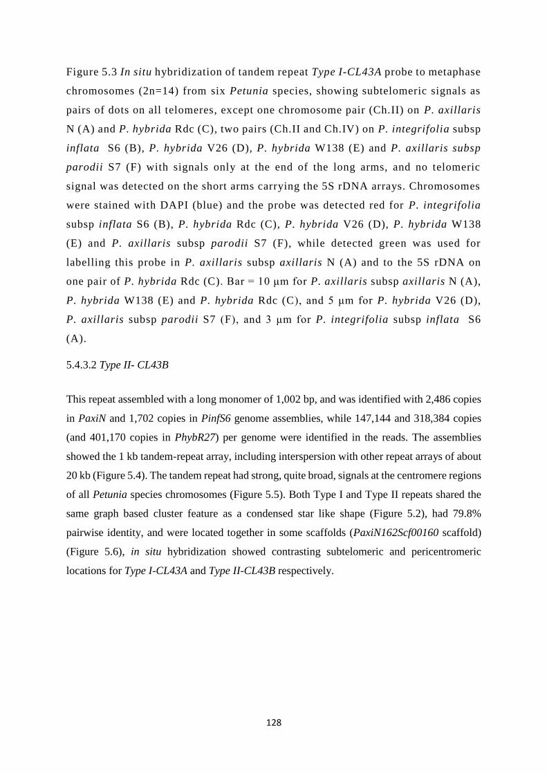
128
Figure 5.3 In situ hybridization of tandem repeat Type I-CL43A probe to metaphase
chromosomes (2n=14) from six Petunia species, showing subtelomeric signals as
pairs of dots on all telomeres, except one chromosome pair (Ch.II) on P. axillaris
N (A) and P. hybrida Rdc (C), two pairs (Ch.II and Ch.IV) on P. integrifolia subsp
inflata S6 (B), P. hybrida V26 (D), P. hybrida W138 (E) and P. axillaris subsp
parodii S7 (F) with signals only at the end of the long arms, and no telomeric
signal was detected on the short arms carrying the 5S rDNA arrays. Chromosomes
were stained with DAPI (blue) and the probe was detected red for P. integrifolia
subsp inflata S6 (B), P. hybrida Rdc (C), P. hybrida V26 (D), P. hybrida W138
(E) and P. axillaris subsp parodii S7 (F), while detected green was used for
labelling this probe in P. axillaris subsp axillaris N (A) and to the 5S rDNA on
one pair of P. hybrida Rdc (C). Bar = 10 μm for P. axillaris subsp axillaris N (A),
P. hybrida W138 (E) and P. hybrida Rdc (C), and 5 μm for P. hybrida V26 (D),
P. axillaris subsp parodii S7 (F), and 3 μm for P. integrifolia subsp inflata S6
(A).
5.4.3.2 Type II- CL43B
This repeat assembled with a long monomer of 1,002 bp, and was identified with 2,486 copies
in PaxiN and 1,702 copies in PinfS6 genome assemblies, while 147,144 and 318,384 copies
(and 401,170 copies in PhybR27) per genome were identified in the reads. The assemblies
showed the 1 kb tandem-repeat array, including interspersion with other repeat arrays of about
20 kb (Figure 5.4). The tandem repeat had strong, quite broad, signals at the centromere regions
of all Petunia species chromosomes (Figure 5.5). Both Type I and Type II repeats shared the
same graph based cluster feature as a condensed star like shape (Figure 5.2), had 79.8%
pairwise identity, and were located together in some scaffolds (PaxiN162Scf00160 scaffold)
(Figure 5.6), in situ hybridization showed contrasting subtelomeric and pericentromeric
locations for Type I-CL43A and Type II-CL43B respectively.

129
Figure 5.4 Self-dot plots of tandem repeat Type II- CL43B in PinfS6101Scf00909
scaffold at P. integrifolia subsp inflata S6 assembly (A) showing tandem structure
between 360,000 bp and 415,000 bp (total size of this scaffold is about 1,820,000
bp), and PaxiN162Scf01294 scaffold at P. axillaris subsp axillaris N assembly (B)
showing about 2 kb array at the end of this scaffold. Arrows in (A) indicate miss-
assembled array of Type II- CL43B while in (B), single arrow indicates a small
array at the end of the scaffold.

130
A B
C D
E F

131
Figure 5.5 Metaphase chromosomes of Petunia (2n=14) species probed with
tandem repeat Type II-CL43B showing dispersed and strong signals at the
centromere regions of all Petunia species chromosomes. Chromosomes stained
with DAPI (blue) and the probe detected red for P. axillaris subsp axillaris N (A),
P. hybrida Rdc (C) and P. hybrida V26 (D), while in P. integrifolia subsp inflata
S6 (B), P. hybrida W138 (E) and P. axillaris subsp parodii S7(F), the probe was
detected green. Bar = 10 μm.
Figure 5.6 Self-dot plot of PaxiN162Scf160 scaffold, showing tandem repeat Type
I-CL43 A and Type II-CL43 B positions as 10 kb and 1 kb arrays respectively.
5.4.3.3 Type III-CL58
This tandem repeat is abundantly represented in the assemblies and raw reads with 0.150 %
genome proportion. It showed variable sizes of arrays between 3 kb and 15 kb within small
scaffolds, with 23% (PinfS6) and 7% (PaxiN) of the reads being represented in assemblies,
presumably representing collapse of reads of the repeat in the assemblies, and different copy
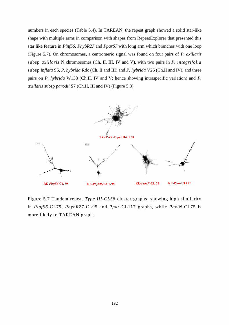
132
numbers in each species (Table 5.4). In TAREAN, the repeat graph showed a solid star-like
shape with multiple arms in comparison with shapes from RepeatExplorer that presented this
star like feature in PinfS6, PhybR27 and PparS7 with long arm which branches with one loop
(Figure 5.7). On chromosomes, a centromeric signal was found on four pairs of P. axillaris
subsp axillaris N chromosomes (Ch. II, III, IV and V), with two pairs in P. integrifolia
subsp inflata S6, P. hybrida Rdc (Ch. II and III) and P. hybrida V26 (Ch.II and IV), and three
pairs on P. hybrida W138 (Ch.II, IV and V; hence showing intraspecific variation) and P.
axillaris subsp parodii S7 (Ch.II, III and IV) (Figure 5.8).
Figure 5.7 Tandem repeat Type III-CL58 cluster graphs, showing high similarity
in PinfS6-CL79, PhybR27-CL95 and Ppar-CL117 graphs, while PaxiN-CL75 is
more likely to TAREAN graph.

133
A B
C D
E F

134
Figure 5.8 Metaphase chromosomes of Petunia (2n=14) species probed with
tandem repeat Type III-CL58 detected red shows that centromeric signals clearly
found on four pairs of P. axillaris subsp axillaris N (A) (Ch.II, III, IV and V).
Two pairs are seen in P. integrifolia subsp inflata S6 (B), P. hybrida Rdc (C)
(Ch.II and III) and P. hybrida V26 (D) (Ch.II and IV) have centromeric signals;
there are three pairs on P. hybrida W138 (E) (Ch.II, IV and V) and P. axillaris
subsp parodii S7 (F) (Ch.II, III and IV). Bar = 10 μm for all species except P.
axillaris subsp parodii S7 (F) that has 5 μm.
5.4.3.4 Type IV-CL95
Despite of 0.092% genome proportion and the abundant sequences of this repeat within PinfS6
and PaxiN assemblies, it has low numbers of copies in PhybR27and PparS7 (Figure 5.1). This
repeat had small arrays with about 1 kb that were widely distributed over assemblies. Cluster
graphs had a solid circle with few extensions especially in PaxiN and PparS7 reads; PinfS6 and
PhybR27 were similar, while PaxiN had a smaller central cluster and four branches (Figure
5.9). Consistent with the copy number and distribution in assemblies, and weak and dispersed
signals were seen on chromosomes (Figure 5.10).
Figure 5.9 Graphs of four genomes clusters of tandem repeat Type IV-CL95 that
compared together with TAREAN shape, representing spitting images for PinfS6-
CL140 and PhybR27-CL157 clusters with smaller central cluster and four
branches, two branches are coming together at the middle, and between PaxiN-
CL139, Ppar-CL173 and TAREAN with a solid circle and few extensions.

135
A B
C D
E F

136
Figure 5.10 Metaphase chromosomes of Petunia (2n=14) species probed with
repeat Type IV-CL95 shows weak and dispersed signals (red) over all chromosomes
(blue). P. axillaris subsp axillaris N (A), P. integrifolia subsp inflata S6 (B), P.
hybrida Rdc (C), P. hybrida V26 (D), P. hybrida W138(E) and P. axillaris subsp
parodii S7 (F). Bar = 10 μm for all species.
5.4.3.5 Type V-CL102
Type V was poorly represented within PinfS6 and PaxiN assemblies (as small 1 kb arrays),
although copies were abundant in all raw reads (0.08% genome proportion). The sequence
included many copies of the telomeric repeat (TTTAGGG) and variants, and the graph had two
connected solid circles or a circular pattern (Figure 5.11). In situ hybridization signals were
strong on the telomere region of one pair of all Petunia species chromosomes, showing highly
condensed signal on just the short arm of chromosome II (Figure 5.12).
Figure 5.11 Tandem repeat Type V-CL102 graphs, showing two connected solid
circles for TAREAN and PinfS6-CL195, and a bit different from both PaxiN-CL55
and PhybR27-CL175 clusters shapes that look very close to each other, while
Ppar-CL119 shows circular shape surrounded by condensed nodes.

137
A B
C D
E F

138
Figure 5.12 Metaphase chromosomes of Petunia (2n=14) species probed with
tandem repeat Type V-CL102, showing strong signals (red) on the telomere region
on the short arm of chromosome II. P. axillaris subsp axillaris N (A), P.
integrifolia subsp inflata S6 (B), P. hybrida Rdc (C), P. hybrida V26 (D), P.
hybrida W138 (E) and P. axillaris subsp parodii S7 (F). Bar =5 μm for A and F,
and 10 μm for the rest.
5.4.3.6 TypeVI-CL114
Abundant sequences have been shown in the two assemblies with notably higher copy numbers
in PaxiN and PhybR27 than other two species. For this tandem repeat, arrays are between 1 to
2 kb with 0.074% genome proportion. Graphs showed that PhybR27 has a solid circle with
extensions linked and a ring shape, and PparS7 shows similar graph without ring, while this
ring shape looks bigger in PaxiN, and very complex feature of PinfS6 (Figure 5.13). P. axillaris
subsp axillaris N, P. integrifolia subsp inflata S6, P. hybrida V26, and P. axillaris subsp
parodii S7 had centromeric signals on two chromosome pairs (Ch.II and IV) with variable site
strengths, while chromosomes of P. hybrida Rdc and P. hybrida W138 only showed one pair
(Ch.IV) (Figure 5.14).
Figure 5.13 Graphs show tandem repeat Type VI-CL114 shapes that have a solid
circle with extensions linked with a ring shape for PhybR27-CL172, and Ppar-
CL175 shows similar graph without ring, while this ring shape looks bigger in
PaxiN-CL146, and very complex feature of PinfS6-CL61.

139
A B
C D
E F
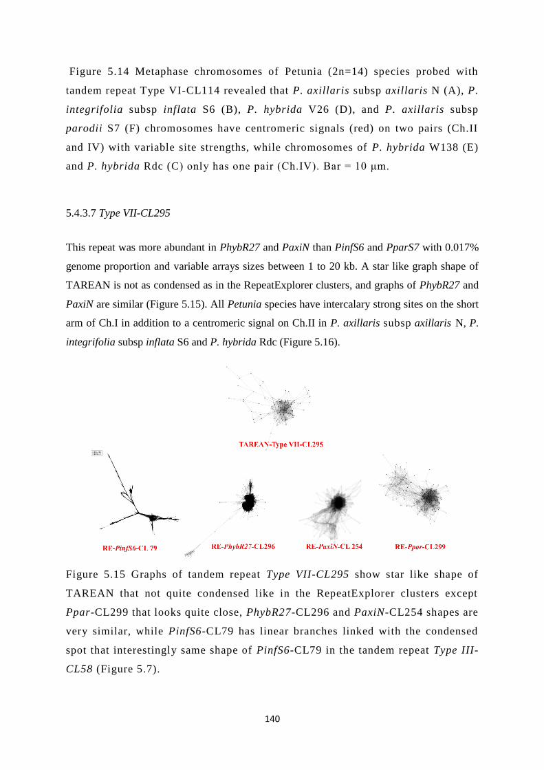
140
Figure 5.14 Metaphase chromosomes of Petunia (2n=14) species probed with
tandem repeat Type VI-CL114 revealed that P. axillaris subsp axillaris N (A), P.
integrifolia subsp inflata S6 (B), P. hybrida V26 (D), and P. axillaris subsp
parodii S7 (F) chromosomes have centromeric signals (red) on two pairs (Ch.II
and IV) with variable site strengths, while chromosomes of P. hybrida W138 (E)
and P. hybrida Rdc (C) only has one pair (Ch.IV). Bar = 10 μm.
5.4.3.7 Type VII-CL295
This repeat was more abundant in PhybR27 and PaxiN than PinfS6 and PparS7 with 0.017%
genome proportion and variable arrays sizes between 1 to 20 kb. A star like graph shape of
TAREAN is not as condensed as in the RepeatExplorer clusters, and graphs of PhybR27 and
PaxiN are similar (Figure 5.15). All Petunia species have intercalary strong sites on the short
arm of Ch.I in addition to a centromeric signal on Ch.II in P. axillaris subsp axillaris N, P.
integrifolia subsp inflata S6 and P. hybrida Rdc (Figure 5.16).
Figure 5.15 Graphs of tandem repeat Type VII-CL295 show star like shape of
TAREAN that not quite condensed like in the RepeatExplorer clusters except
Ppar-CL299 that looks quite close, PhybR27-CL296 and PaxiN-CL254 shapes are
very similar, while PinfS6-CL79 has linear branches linked with the condensed
spot that interestingly same shape of PinfS6-CL79 in the tandem repeat Type III-
CL58 (Figure 5.7).

141
A B
C D
E F

142
Figure 5.16 Metaphase chromosomes of Petunia (2n=14) species probed with
tandem repeat Type VII-CL295 shows intercalary strong sites on the short arm of
Ch.I in P. axillaris subsp axillaris N (A), P. integrifolia subsp inflata S6 (B), P.
hybrida Rdc (C), P. hybrida V26 (D), P. hybrida W138 (E), and P. axillaris subsp
parodii S7 (F), in addition to a centromeric signal on Ch.II only in P. axillaris
subsp axillaris N (A), P. integrifolia subsp inflata S6 (B), and P. hybrida Rdc
(C). The probe was labelled with biotin 11- dUTP (red). Bar = 10 μm.
5.4.3.8 Type VIII-CL331
A small number of copies of this repeat were found in PinfS6 and PaxiN, with only small arrays
in the assembly. From TAREAN data, this repeat has donut graph shape, not as condensed as
others in RepeatExplorer (Figure 5.17). No signal was detected by in situ hybridization on
chromosomes over all petunias (Figure 5.18), confirming the low abundance. The result
suggests that clusters with this abundance and lower can be discounted from analysis of major
abundant tandemly repeat satDNA sequences.
Figure 5.17 Donut shapes of Type VIII-CL331, showing TAREAN's graph that is
not quite condensed in comparison with more concentrated shapes in the
RepeatExplorer (PinfS6-CL326, PhybR27-CL345, PaxiN-CL286, and Ppar-
CL280).

143
A B
C D
E F

144
Figure 5.18 Metaphase chromosomes of Petunia (2n=14) species probed with tandem
repeat Type VIII-CL331 shows no clear signals over all Petunia species chromosomes.
Bar = 10 μm for all species except P. integrifolia subsp inflata S6 that has 5 μm.
5.5 Discussion
The detailed analysis of repetitive elements from genomes of four Petunia species used
complementary tools (graph-based repeat clustering using unassembled raw reads, raw read
mapping, sequence assemblies, in situ hybridization and chromosome studies).
5.5.1 Genome proportion of tandem repeat clusters
Repetitive elements represented 64-68 % of the Petunia genomes (Figure 1.3; Appendix 3.1),
consistent with the measurements reported by Bombarely et al. (2016) in P. axillaris subsp
axillaris N. The analysis showed that a relatively low proportion, 0.6%, of the genome (8.4 Mb
of the 1,400 Mb genomes) was represented by a limited number – just eight– tandemly-repeated
clusters of satellite DNA clusters (Table 5.4). This contrasts with a higher percentage of tandem
repeats in other species with a range of genome sizes: 4.89 % in Coix aquatica (Cai et al. 2014;
2,335 Mb), 6.8% in Beta vulgaris (Zakrzewski et al. 2010; 714-758 Mb), 5% in Zea mays
(Meyers et al. 2001; 2,300-2,700 Mb), 2.7% in Vicia faba (Robledillo et al. 2018; 1,300 Mb),
20% in Citrus spp. (380 Mb), 30% in Cucumis sativus genomes (367 Mb) (Hemleben et al.
2007). The Petunia figure is similar to that reported in Coix lacryma-jobi (0.60%, 1,600 Mb).
Even Arabidopsis thaliana (176 Mb) includes some 8% of the genome as rDNA repeats, with
4% as the single AtCon tandemly repeated centromeric sequence (Heslop-Harrison et al. 1999).
Except for the 45S rDNA sequences at the secondary constrictions (slightly darker in DAPI
staining), the low proportion of tandemly repeated satellites in petunia is supported by the
chromosome images where no positively- or negatively- staining bands are seen (Figures 5.3,
5.5, 5.8, 5.10, 5.12, 5.14, 5.16 and 5.18). In contrast, many other plant species have conspicuous
bands, considered as heterochromatin, at centromeric, sub-telomeric or intercalary positions on
chromosomes. It is important to study in detail the nature of the tandem repeats in a genome
where they represent a small but significant proportion: is their number, distribution and even
function the same as in genomes with a higher proportion? Do any features of the genome
mitigate against amplification of tandem arrays?

145
5.5.2 Tandemly repeated sequences within assemblies
In assembled sequence scaffolds, various short satellite repeat arrays were found ranging in
length from less than 1kb to some 20 kb (Table 5.4; Figures 5.4 and 5.6). Collapse of repeats,
and mis-assembly of flanking regions, is often a consequence of the sequence assembly data
and algorithms. Nevertheless, the assembly data in petunia, using both PacBio long-reads and
mate-pairs (Bombarely et al. 2016) were supported the low proportion and diversity of tandem
repeats. Depending on conformation, small repeat arrays (<10-50 kb) were not expected to be
seen, by in situ hybridization, nor identify SSRs and microsatellites, typically represented by
10s of copies of 2-, 3- or 4- base pair motifs, in the repeat analysis here. However, the
chromosomes do show some array loci that are absent from the P. axillaris N assembly: for
example, tandem repeats Type I-CL43A (10 kb array length, with much higher copy number by
in situ hybridization and read mapping than in the assembly) and Type II-CL43B (1 kb) both
locate in scaffold PaxiN162Scf160, but in situ hybridization locates Type I at subtelomeric sites
and Type II is pericentromeric (Figures 5.3, 5.5 and 5.6). Short arrays of telomeric sequences,
their derivatives, or other subtelomeric repeats, are often also found near centromeres (Presting
et al. 1996), as seen in the sub-centromeric scaffold PaxiN162Scf160, while the major
subtelomeric Type I arrays, like the Type V telomere (Figure 5.12), are collapsed in the draft
assemblies.
5.5.3 Graph based clusters pipelines
Graph-based clustering algorithms are widely applied to whole-genome sequence assembly
(Zerbino & Birney 2008; Kingsford et al. 2010; Novák et al. 2010). Here, RepeatExplorer and
TAREAN graphs are used to cluster repetitive elements, and the tandem repeats generally show
a graph with a dense centre (star) or ring (donut) graph shape representing multiple similar
reads with overlaps, with a lower number of peripheral reads with variants or representing ends
of arrays (Figures 5.2, 5.7, 5.9, 5.11, 5.13, 5.15 and 5.17). While the graphs were helpful for
identification of candidate tandem repeats, no link was evident between biological features of
the repeat (abundance, location, monomer length and array size, or inter-monomer variation)
and the detailed pattern of the graph.
5.5.4 The role of tandemly repeated sequences

146
Despite the relatively low proportion of repeats in petunia, tandem repeat classes were found
with centromeric, peri-centromeric, sub-telomeric, telomeric and intercalary locations (Table
5.4; Figures 5.3, 5.5, 5.8, 5.10, 5.12, 5.14, 5.16 and 5.18). Their potential role in maintenance
of nuclear of chromosome structure at metaphase and interphase has been considered, including
centromeric function (Hemleben et al. 2007). Roles in large-scale genome organization
(Biscotti et al. 2015), chromatin packaging, or maintenance of chromosome stability
(Vershinin & Heslop-Harrison 1998), and effects on the environment of the expressed genes
(Lamb et al. 2007), have also been proposed (Ohtsubo et al. 1991; Han et al. 2008; Torres et
al. 2011) (Figure 5.1).
5.5.5 The evolution of tandem repeats
P. axillaris subsp axillaris N and P. integrifolia subsp inflata S6 separated from a common
ancestor some 30 MYA (Wang et al. 2008), and came together in P. hybrida in the last 2
centuries (Gerats & Strommer 2008). Comparing PaxiN and PinfS6, of the eight tandem
repeats, three have substantially more copies in PaxiN than PinfS6, and two have more in
PinfS6 than PaxiN. Notably, in the hybrid combination, one tandem repeat has a substantially
greater copy number (similar copies in both ancestors, telomeric location) while two are
diminished (one more PaxiN than PinfS6, one more PinfS6 than PaxiN). Tandem repeats most
likely evolve in copy number by unequal crossing-over at meiosis, or slippage during
replication before mitosis, events that our data show have occurred in both recent and older
evolutionary time with both amplification and loss. The abundance of all types I to VIII in the
sister species P. axillaris subsp parodii S7 suggests that all types are ancient. Given the defined
nature of the tandem repeats and availability of straightforward analysis pathway, it will be
valuable to resynthesize P. hybrida from it ancestors and analyse the copy number changes
occurring over a few generations, comparing both sexual and vegetative pathways to
distinguish meiotic and mitotic events, and examining if the changes are salutatory, occurring
in bursts soon after hybrid formation as with transposon activation, (Grandbastien 1998), and
potentially related through chromatin remodelling and methylation changes. Genomic changes
are regularly seen in hybrid species (Alix et al. 2017). Gaeta et al. (2007) have resynthesized
tetraploid Brassicas and seen homoeologous chromosome exchanges, and it would be valuable
to examine repetitive DNA changes over similar timescales in petunia. Ma and Gustafson
(2008) examined changes in new triticale (wheat x rye) hybrids, and found more genomic
changes in the rye chromosomes, occurring immediately after hybridization, also noting an

147
effect of cytoplasm; major rearrangements of the genome are also seen hybrids in the grass
genus Brachypodium (Lusinska et al. 2018). In other Triticeae species, substantial differences
in tandem repeat abundance between sister species combining in a polyploid hybrid are evident:
the tandem repeat dpTa1 is much more abundant in the N than D genomes in ancestral species
and hybrid Aegilops ventricosa (Bardsley et al. 1999). Here, though, each type of repeat has a
unique pattern of amplification in the three sister species P. axillaris subsp axillaris N , P.
integrifolia subsp inflata S6 and P. axillaris subsp parodii S7 suggesting different evolution
from that of the rest of the genome. Very rapid changes in copy number – from approximately
5x to 1/5x in the hybrid compared to ancestral species – were observed for the each type. No
correlations between abundance nor copy number evolution and chromosomal location were
evident (see Figure 5.10).
5.5.6 Tandem repeats as genetic markers
Most of tandem clusters involved in pericentromeric regions except Type I-CL43A and Type
V-CL102 that discovered in subtelomeric and telomeric loci respectively (Figures 5.3 and 5.12).
This distribution is functionally important to strengthen chromosome structure of petunia as a
typical genome. Findings of tandem repeat types III, VI and VII have been characterised as
chromosomes markers, providing useful cytogenetic tool for easily identifying metaphase
chromosomes, depending on repeat localization and fluorescence intensity (Figures 5.8, 5.14
and 5.16). Recently, many tools have been used for finding a specific approach to characterise
genomes of different species, these methods are classified as non-PCR and PCR based
techniques (Agarwal et al. 2008; Korir et al. 2013). So far, facts of the homology between in
situ hybridization strength and the abundance of copy numbers in raw reads have been achieved
for some tandem repeats but unlikely approved for others like Type IV and Type VIII (Figures
5.10 and 5.18). However, the two types have to some extent enough copy numbers and genome
proportion (especially Type IV), with no clear signal has seen through FISH that is not a
quantitative way for detecting unclear probes. This probably because of the existence of these
repeats is much dispersed and in very small arrays. Interestingly, no correlation has been
noticed between the copy numbers of some repeats and chromosome sizes.
Results obtained in the present chapter will consequently be used in integration of the
chromosomal and genetic maps that are undertaken by the Petunia Genome Sequencing
Consortium through molecular mapping and in situ hybridization, efficient chromosomes
markers, providing useful cytogenetic tool for easily identifying metaphase chromosomes.

148
Chapter VI. General discussion: Repetitive elements in petunia
This work has characterised the repetitive DNA landscape in the Petunia genome (see
Appendix 3.1), with DNA components that can be divided into two major divisions based on
their organization and position within genome. One division contains semi-autonomous
sequences such as DNA transposable elements, retroelements, and integrated retrovirus-related
sequences. The retroviral-related sequences were the focus of more detailed studies of their
consequences for the genome and plant. The second division includes tandemly repeated
sequences like satellite DNAs.
PVCV and other integrated viral sequences in Petunia species
From the first group, EPRVs, as elements with major consequences for the host plant, were
investigated here using bioinformatics tools, cytogenetic work (chapter III), and transmission
electron microscopy (chapter IV) within Petunia genomes. Although these elements have
extensively colonized flowering plant genomes since 20-34 MYA, EPRVs are still less well
studied in plant genomes than in animals where they have been known for many years (Geering
et al. 2014). Our results have highlighted further facts to understand the role of EPRVs
elements (petu-like, florendo-like, and caulimo-like sequences) in formation of wild and hybrid
host genomes, their possible functions, and relationship between different units. High variable
proportions of EPRVs sequences have been counted within wild and hybrid genomes of
petunia, and widely distinguished over their loci on chromosomes suggesting these elements
(petu-like virus) as a novel cytogenetic marker alongside retrotransposons (Figure 3.18).
Interestingly, some EPRVs (petu, and florendo-like sequences) share similar positions over
host chromosomes (Figure 3.42) suggesting probably their closely related phylogenetic
relationship with same endogenisation events. Endogenisation searches were extended to
discover novel members of florendoviruses and caulimoviruses (four sequences of each type
of both groups).
Also, the outcomes show higher levels of genome proportions and copy nymbers of EPRVs
within hybrid petunia than wild parents, in addition to integration pattern of these elements
inside hybrids as tandemly repeated sequences unlike their parents. As a consequence, hybrid
petunias are considered as a reservoir of EPRVs and have more permissive and inducible
genomes than wild parents (Richert‐Pöggeler et al. 2003) (chapter III).

149
PVCV expression and infections within Petunia tissues
Expression protocols have revealed that florendoviruses were non-active integrants despite
their abundance unlike petu- and caulimo-like sequences that were strongly expressed (Figure
3.38). For the first time in plants, the approach of de novo integration of chromosomally
integrated PVCV has discovered to be integrated over next generations on the telomere of
heterologous chromosomes (see Figure 3.39). Moreover, significant ultra-structure changes
have been noticed within chlorotic spot, vein clearing symptoms of horizontally transmitted
PVCV as well as progeny of vertically transmitted PVCV and induced symptoms of PVCV
(chapter IV).
The wide existence of endogenous pararetroviruses in abundant copy numbers within flowering
plant genomes has suggested these elements as a source of infection, but also have a protective
function in silencing and suppression via RNA interference-induced resistance (Richert‐
Pöggeler et al. 2003). The number of copies in petunia (chapter III) is much higher than is
known to induce RNAi-mediated resistance to RNA viruses (Ratcliff et al. 1997; Jakowitsch
et al. 1999; Wesley et al. 2001; Kunii et al. 2004). With the close relationship to retroelements,
EPRV insertion has many impacts on host genome, potentially including chromosome
breakage and rearrangement, insertional mutation, sequence amplification and gene regulation.
However, most retroelements such as the Gypsy and Copia families, or DNA elements
including CACTA-EnSpm, have more dispersed distributions over broad chromosomal
regions, compared to the EPRVs that occur a discrete chromosomal loci. Fragmented lengths
of integrated EPRV units also could have activities of promoter/enhancer, ORFs, active splice
sites and RT in addition to transposition and related amplification ability (Löwer 1999; Hansen
& Heslop-Harrison 2004a). Additionally, they are playing a very clear role in their host
evolution by adding novel genetic components at the levels of coding or transcriptional
regulation taking into account that plants have higher plasticity in their genome structure than
animals: the existence of EPRVs may play a role in genome diversification by providing coding
or transcription regulatory elements as new genetic components. The spread of EPRVs is
widley correlated with their host reproduction and distribution between different regions
around the world. Although some EPRVs are located in geographically segregated regions,
they probably spreaded to different areas of the world around 500 YA. Based on genetic
population structure, these elements showed frequent spread among regions. Our findings
confirmed the widespread distribution of EPRVs in the genomes of cultivated and wild plants

150
with extensive colonization patterns. These elements followed similar scale of high copy
numbers families of transposable elements (Geering et al. 2014; Yasaka et al. 2014).
Tandemly repeats as major repetitive DNA sequences in petunia
Tandemly repeated sequences have been analysed in this work through bioinformatics analysis
and cytogenetic tools to explore their diversity over host genomes (chapter V). These elements
were found with relatively low abundance in Petunia genomes, and include eight types of
clusters that involved in centromeric, peri-centromeric, sub-telomeric, telomeric and
intercalary regions of Petunia chromosomes (Table 5.4). Tandemly repeated clusters are vary
in their copy numbers and localizations over examined Petunia genomes. More importantly,
three types of tandem repeats have been characterised as efficient chromosomes markers,
providing useful cytogenetic tools for the identification of metaphase chromosomes based on
repeat localization and fluorescence intensity (Figures 5.8, 5.14 and 5.16). This work is
consistent with a functional role suggested for the repeats in genome evolution and structural
roles within chromosomes regions like centromeres and telomeres (Schmidt & Heslop-
Harrison 1998). Their maintenance role in chromosome structure at metaphase and interphase
has been considered, including centromeric function (Hemleben et al. 2007). Roles in large-
scale genome organization (Biscotti et al. 2015), chromatin packaging, or maintenance of
chromosome stability (Vershinin & Heslop-Harrison 1998), and effects on the environment of
the expressed genes (Lamb et al. 2007), have also been proposed (Ohtsubo et al. 1991; Han et
al. 2008; Torres et al. 2011), as well as in the stability of chromatin packaging (Escudeiro,
Adega, Robinson, Schwarzacher, Heslop-Harrison and Chaves, submitted). Moreover, as
repetitive DNA, tandem repeats probably have a protective role for coding DNA from any
shock through stress conditions due to its high stability and it has been used for nuclear
architecture study, chromosomes and genomes identification, and phylogenetic analysis
(Heslop-Harrison 2000; Pluhar et al. 2001).
Future extensions
Work should be continued to find out more integration features, localization, and transmission
of different EPRVs within other plant species: given the well-defined presence and effects in
petunia described here, and in banana (Harper et al. 1999), it is likely that they will be found
more widely across plant species. Investigating new members of EPRVs should be extended

151
with assistance of the whole genome sequencing techniques. FISH work on caulimovirus-like
sequences position in Petunia chromosomes should be extended using new techniques such as
a massive single copy pools of oligonucleotides (Han et al. 2015) to cover all EPRVs types’
locations. Expression patterns of EPRVs have been explored within and between wild Petunia
species, and more data from hybrid varieties is strongly needed. In addition, de novo integration
experiments have revealed novel facts about plant virus integration over plant generations, and
exploring more details about integration sites within telomere regions are highly recommended.
It would also be valuable to characterise further the mechanisms of vein clearing following
PVCV infection, including the effects on chloroplasts. It will also be important to examine
miRNA/siRNA species present in various infected and resistant or asymptomatic lines to futher
characterise the endogenous effects of the integrated EPRV sequence motifs. For tandemly
repeated sequences, it will be valuable to resynthesize P. hybrida from it ancestors and analyse
the copy number changes occurring over a few generations, and examining chromosome
elimination and diploidization, comparing both sexual and vegetative pathways to distinguish
meiotic and mitotic events, and examining if the changes are salutatory, occurring in bursts
soon after hybrid formation. For all the repetitive sequence classes, it will be interesting to
further examine their role in epigenetics, including chromatin organization, their relationships
with histone and DNA modifications, and potential consequences for the modulation of gene
expression and chromosomes behaviour in adjacent regions.
Conclusion
In the context of diversity and evolution, using six petunia species, raw reads from four whole-
genome survey sequencing experiments, and two assemblies with bioinformatics tools,
cytogenetic techniques and electron microscopy allowed us to explore our collected results
regarding EPRVs and tandem repeats. This project has achieved our main aims (see 1.7) in
order to provide new insights and highlight further facts to understand the role of repetitive
DNA elements in formation of wild and hybrid host genomes, their possible functions and
relationships. These elements present fundamental diversity among the evolutionarily related
species and individuals, suggesting an evolutionary impact associated with repeats on host
genome variation.

152
References
Adams M.J. & Antoniw J.F. (2005) DPVweb: An open access internet resource on plant
viruses and virus diseases. Outlooks on Pest Management 16, 268-70.
Adams S.E., Mellor J., Gull K., Sim R.B., Tuite M.F., Kingsman S.M. & Kingsman A.J.
(1987) The functions and relationships of Ty-VLP proteins in yeast reflect those of
mammalian retroviral proteins. Cell 49, 111-9.
Agarwal M., Shrivastava N. & Padh H. (2008) Advances in molecular marker techniques
and their applications in plant sciences. Plant Cell Reports 27, 617-31.
Alix K., Gérard P.R., Schwarzacher T. & Heslop-Harrison J. (2017) Polyploidy and
interspecific hybridization: partners for adaptation, speciation and evolution in plants.
Annals of Botany 120, 183-94.
Alizon S., Hurford A., Mideo N. & Van Baalen M. (2009) Virulence evolution and the trade‐
off hypothesis: history, current state of affairs and the future. Journal of Evolutionary
Biology 22, 245-59.
Alkan C., Cardone M.F., Catacchio C.R., Antonacci F., O'Brien S.J., Ryder O.A.,
Purgato S., Zoli M., Della Valle G. & Eichler E.E. (2011) Genome-wide
characterization of centromeric satellites from multiple mammalian genomes. Genome
Research 21, 137-45.
Altschul S.F., Gish W., Miller W., Myers E.W. & Lipman D.J. (1990) Basic local alignment
search tool. Journal of Molecular Biology 215, 403-10.
Amari K., Boutant E., Hofmann C., Schmitt-Keichinger C., Fernandez-Calvino L., Didier
P., Lerich A., Mutterer J., Thomas C.L. & Heinlein M. (2010) A family of
plasmodesmal proteins with receptor-like properties for plant viral movement proteins.
PLoS Pathogens 6, e1001119.
Anabestani A., Behjatnia S.A.A., Izadpanah K., Tabein S. & Accotto G.P. (2017) Seed
Transmission of Beet Curly Top Virus and Beet Curly Top Iran Virus in a Local Cultivar
of Petunia in Iran. Viruses 9, 299-310.
Ando T. (1996) Distribution of Petunia axillaris (Solanaceae) and its new subspecies in
Argentina and Bolivia. Acta Phytotax. Geobot. 47, 19-30.
Ando T., Kokubun H., Watanabe H., Tanaka N., Yukawa T., Hashimoto G., Marchesi E.,
Suárez E. & Basualdo I.L. (2005) Phylogenetic analysis of Petunia sensu Jussieu
(Solanaceae) using chloroplast DNA RFLP. Annals of Botany 96, 289-97.

153
Ando T., Nomura M., Tsukahara J., Watanabe H., Kokubun H., Tsukamoto T.,
Hashimoto G., Marchesi E. & Kitching I.J. (2001) Reproductive isolation in a
native population of Petunia sensu Jussieu (Solanaceae). Annals of Botany 88, 403-
13.
Arkhipova I.R., Yushenova I.A. & Rodriguez F. (2017) Giant reverse transcriptase-
encoding transposable elements at telomeres. Molecular Biology and Evolution 34,
2245-57.
Arney K.L. & Fisher A.G. (2004) Epigenetic aspects of differentiation. Journal of Cell
Science 117, 4355-63.
Balke I., Reseviča G. & Zeltins A. (2018) Isolation and Characterization of Two Distinct
Types of Unmodified Spherical Plant Sobemovirus-Like Particles for Diagnostic and
Technical Uses. In: Virus-Derived Nanoparticles for Advanced Technologies (pp. 19-
34). Springer.
Bang J.W., Kim J.S. & Lee M.K. (1997) Molecular cytogenetics of plant genome. Journal of
Plant Biology 40, 149-55.
Bao W., Jurka M.G., Kapitonov V.V. & Jurka J. (2009) New superfamilies of eukaryotic
DNA transposons and their internal divisions. Molecular Biology and Evolution 26,
983-93.
Bao W., Kojima K.K. & Kohany O. (2015) Repbase Update, a database of repetitive elements
in eukaryotic genomes. Mobile DNA 6, 11-16.
Bardsley D., Cuadrado A., Jack P., Harrison G., Castilho A. & Heslop-Harrison J. (1999)
Chromosome markers in the tetraploid wheat Aegilops ventricosa analysed by in situ
hybridization. Theoretical and Applied Genetics 99, 300-4.
Bennett M.D. & Leitch I.J. (2011) Nuclear DNA amounts in angiosperms: targets, trends and
tomorrow. Annals of Botany 107, 467-590.
Bennetzen J.L. (1996) The contributions of retroelements to plant genome organization,
function and evolution. Trends in Microbiology 4, 347-53.
Benson G. (1999) Tandem repeats finder: a program to analyze DNA sequences. Nucleic Acids
Research 27, 573-80.
Bentzer B., Bothmer R.v., Engstrand L., Gustafsson M. & Snogerup S. (1971) Some
sources of error in the determination of arm rations of chromosomes. Botaniska Notiser.
Bergman C.M. & Quesneville H. (2007) Discovering and detecting transposable elements in
genome sequences. Briefings in Bioinformatics 8, 382-92.

154
Bilinski P., Han Y., Hufford M.B., Lorant A., Zhang P., Estep M.C., Jiang J. & Ross-
Ibarra J. (2017) Genomic abundance is not predictive of tandem repeat localization in
grass genomes. PloS one 12, e0177896.
Biscotti M.A., Olmo E. & Heslop-Harrison J.P. (2015) Repetitive DNA in eukaryotic
genomes. Chromosome Res. 23:415–20.
Bloom K. (2007) Centromere dynamics. Current Opinion in Genetics & Development 17, 151-
6.
Boeke J. & Corces V.G. (1989) Transcription and reverse transcription of retrotransposons.
Annual Reviews in Microbiology 43, 403-34.
Bombarely A., Moser M., Amrad A., Bao M., Bapaume L., Barry C.S., Bliek M., Boersma
M.R., Borghi L. & Bruggmann R. (2016) Insight into the evolution of the Solanaceae
from the parental genomes of Petunia hybrida. Nature Plants 2, 16074-82.
Brandes J. & Wetter C. (1959) Classification of elongated plant viruses on the basis of
particle morphology. Virology 8, 99-115.
Bringaud F., Ghedin E., Blandin G., Bartholomeu D.C., Caler E., Levin M.J., Baltz T. &
El-Sayed N. M. (2006) Evolution of non-LTR retrotransposons in the trypanosomatid
genomes: Leishmania major has lost the active elements. Molecular and Biochemical
Parasitology 145, 158-70.
Büchen-Osmond C. (2003) Taxonomy and classification of viruses. Manual of clinical
microbiology 2, 1217-26.
Britton N.L. & Brown A. (1913) An illustrated flora of the northern United States, Canada
and the British possessions: from Newfoundland to the parallel of the southern
boundary of Virginia, and from the Atlantic Ocean westward to the 102d meridian. C.
Scribner's sons.
Cabrera, A.L. andWillink, A. (1980) Biogeografia de America Latina, 2nd Edn. Secretaria
Generalde la OEA, Washington.
Cai Z., Liu H., He Q., Pu M., Chen J., Lai J., Li X. & Jin W. (2014) Differential genome
evolution and speciation of Coix lacryma-jobi L. and Coix aquatica Roxb. hybrid
guangxi revealed by repetitive sequence analysis and fine karyotyping. BMC Genomics
15, 1025-40.
Casacuberta E. (2017) Drosophila: retrotransposons making up telomeres. Viruses 9, 192-
207.
Cavalier-Smith T. (1985) Selfish DNA and the origin of introns. Nature 315, 283-84.

155
Charlesworth B., Sniegowski P. & Stephan W. (1994) The evolutionary dynamics of
repetitive DNA in eukaryotes. Nature 371, 215-20.
Chen J., Chen J.P. & Adams M. (2002) Characterisation of some carla-and potyviruses from
bulb crops in China. Archives of Virology 147, 419-28.
Clare J. & Farabaugh P. (1985) Nucleotide sequence of a yeast Ty element: evidence for an
unusual mechanism of gene expression. Proceedings of the National Academy of
Sciences 82, 2829-33.
Cohen J., Sikron N., Shuval S. & Gera A. (1999) Susceptibility of vegetatively propagated
petunia to tobamovirus infection and its possible control. Hortscience 34, 292-3.
Conia J., Bergounioux C., Perennes C., Muller P., Brown S. & Gadal P. (1987) Flow
cytometric analysis and sorting of plant chromosomes from Petunia hybrida
protoplasts. Cytometry: The Journal of the International Society for Analytical
Cytology 8, 500-8.
Contento A., Heslop-Harrison J. & Schwarzacher T. (2005) Diversity of a major repetitive
DNA sequence in diploid and polyploid Triticeae. Cytogenetic and Genome Research
109, 34-42.
Cooper D. (1946) Double fertilization in Petunia. American Journal of Botany 33, 54-7.
Corbetta M., Kasdorf G., Engelbrecht D. & Wiid J. (2017) Detection of viral-like particles
by electron microscopy of negatively stained extracts from grapevines. South African
Journal of Enology and Viticulture 5, 43-9.
Craig N.L. (2002) Mobile DNA: an introduction. In: Mobile DNA II (pp. 3-11). American
Society of Microbiology.
Croll D., Lendenmann M.H., Stewart E. & McDonald B.A. (2015) The impact of
recombination hotspots on genome evolution of a fungal plant pathogen. Genetics 201,
1213-28.
Daboussi M.-J. & Capy P. (2003) Transposable elements in filamentous fungi. Annual
Reviews in Microbiology 57, 275-99.
De Jong J.H., Fransz P. & Zabel P. (1999) High resolution FISH in plants–techniques and
applications. Trends in Plant Science 4, 258-63.
Dermen H. (1931) Polyploidy in petunia. American Journal of Botany 18, 250-61.
Dietrich A., De Jong J. & Mulder R. (1981) Location and variation of the constitutive
heterochromatin in Petunia hybrida. Genetica 55, 85-91.

156
Dillon L.W., Pierce L.C., Ng M.C. & Wang Y.-H. (2013) Role of DNA secondary structures
in fragile site breakage along human chromosome 10. Human Molecular Genetics 22,
1443-56.
Diop S.I., Geering A.D., Alfama-Depauw F., Loaec M., Teycheney P.-Y. & Maumus F.
(2018) Tracheophyte genomes keep track of the deep evolution of the Caulimoviridae.
Scientific Reports 8, 572-80.
Doolittle W.F. & Sapienza C. (1980) Selfish genes, the phenotype paradigm and genome
evolution. Nature 284, 601-3.
Doyle J. & Doyle J. (1990) Isolation of DNA from small amounts of plant tissues. BRL Focus
12, V15.
Edwards K., Johnstone C. & Thompson C. (1991) A simple and rapid method for the
preparation of plant genomic DNA for PCR analysis. Nucleic Acids Research 19, 1349.
Eichler E.E., Clark R.A. & She X. (2004) An assessment of the sequence gaps: unfinished
business in a finished human genome. Nature Reviews Genetics 5, 345-54.
Elder Jr J.F. & Turner B.J. (1995) Concerted evolution of repetitive DNA sequences in
eukaryotes. The Quarterly Review of Biology 70, 297-320.
Emori Y., Shiba T., Kanaya S., Inouye S., Yuki S. & Saigo K. (1985) The nucleotide
sequences of copia and copia-related RNA in Drosophila virus-like particles. Nature
315, 773-76.
Engelmann J. & Hamacher J. (2008) Plant Virus Diseases: Ornamental Plants 207-229.
Ewald P.W. (1983) Host-parasite relations, vectors, and the evolution of disease severity.
Annual Review of Ecology and Systematics 14, 465-85.
Ewald P.W. (1987) Transmission Modes and Evolution of the Parasitism‐Mutualism
Continuum a. Annals of the New York Academy of Sciences 503, 295-306.
Fauquet C.M., Mayo M.A., Maniloff J., Desselberger U. & Ball L.A. (2005) Virus
taxonomy: VIIIth report of the International Committee on Taxonomy of Viruses.
Academic Press.
Ferguson M.C. & Ottley A.M. (1932) Studies on Petunia III. A redescription and additional
discussion of certain species of Petunia. American Journal of Botany 19, 385-405.
Feschotte C. & Pritham E.J. (2007) DNA transposons and the evolution of eukaryotic
genomes. Annu. Rev. Genet. 41, 331-68.
Flavell A.J., Dunbar E., Anderson R., Pearce S.R., Hartley R. & Kumar A. (1992) Ty1–
copia group retrotransposons are ubiquitous and heterogeneous in higher plants.
Nucleic Acids Research 20, 3639-44.

157
Flavell A.J., Pearce S.R. & Kumar A. (1994) Plant transposable elements and the genome.
Current Opinion in Genetics & Development 4, 838-44.
Flavell R., Bennett M., Smith J. & Smith D. (1974) Genome size and the proportion of
repeated nucleotide sequence DNA in plants. Biochemical Genetics 12, 257-69.
Fransz P.F., Stam M., Montijn B., Hoopen R.T., Wiegant J., Kooter J.M., Oud O. &
Nanninga N. (1996) Detection of single‐copy genes and chromosome rearrangements
in Petunia hybrida by fluorescence in situ hybridization. The Plant Journal 9, 767-74.
Fregonezi J.N., Turchetto C., Bonatto S.L. & Freitas L.B. (2012) Biogeographical history
and diversification of Petunia and Calibrachoa (Solanaceae) in the Neotropical
Pampas grassland. Botanical Journal of the Linnean Society 171, 140-53.
Fries R.E. (1911) Die Arten der Gattung Petunia. Kungl. Svenska Vetenskapsakademiens
Handlingar 46, 1-72.
Froissart R., Roze D., Uzest M., Galibert L., Blanc S. & Michalakis Y. (2005)
Recombination every day: abundant recombination in a virus during a single multi-
cellular host infection. PLoS Biology 3, 389-95.
Gaeta R.T., Pires J.C., Iniguez-Luy F., Leon E. & Osborn T.C. (2007) Genomic changes
in resynthesized Brassica napus and their effect on gene expression and phenotype. The
Plant Cell 19, 3403-17.
Gall J.G. & Pardue M.L. (1969) Formation and detection of RNA-DNA hybrid molecules in
cytological preparations. Proceedings of the National Academy of Sciences 63, 378-83.
Ganga M., Jayalakshmi S., Jegadeeswari V., Padmadevi K. & Jawaharlal M. (2011)
Petunia. In: Wild Crop Relatives: Genomic and Breeding Resources. 209-42.
Springer.
García G., Ríos N. & Gutiérrez V. (2015) Next-generation sequencing detects repetitive
elements expansion in giant genomes of annual killifish genus Austrolebias
(Cyprinodontiformes, Rivulidae). Genetica 143, 353-60.
Gautam K.K., Kumar S. & Raj S.K. (2012) Molecular characterization of Cucumber mosaic
virus strain causing severe mosaic disease on petunia in India. Phytoparasitica 40, 425-
31.
Gayral P., Noa-Carrazana J.-C., Lescot M., Lheureux F., Lockhart B.E., Matsumoto T.,
Piffanelli P. & Iskra-Caruana M.-L. (2008) A single Banana streak virus integration
event in the banana genome as the origin of infectious endogenous pararetrovirus.
Journal of Virology 82, 6697-710.

158
Geering A. & Hull R. (2012) Family Caulimoviridae. Virus taxonomy classification and
nomenclature of viruses ninth Report of the International Committee on Taxonomy of
Viruses, 424-43.
Geering A.D. (2001) Caulimoviridae (Plant Pararetroviruses). eLS.
Geering A.D., Maumus F., Copetti D., Choisne N., Zwickl D.J., Zytnicki M., McTaggart
A.R., Scalabrin S., Vezzulli S. & Wing R.A. (2014) Endogenous florendoviruses are
major components of plant genomes and hallmarks of virus evolution. Nature
Communications 5, 5269-79.
Geering A.D., Scharaschkin T. & Teycheney P.-Y. (2010) The classification and
nomenclature of endogenous viruses of the family Caulimoviridae. Archives of
Virology 155, 123-31.
Gera A., Sikron N., Cohen J. & Zeidan M. (2000) First report of Petunia vein clearing virus
in Israel. Plant Disease 84, 201.
Gerats T. (2009) Identification and exploitation of Petunia transposable elements: a brief
history. In: Petunia (pp. 365-79). Springer.
Gerats T. & Strommer J. (2008) Petunia: evolutionary, developmental and physiological
genetics. Springer Science & Business Media.
Gilbert C. & Feschotte C. (2018) Horizontal acquisition of transposable elements and viral
sequences: patterns and consequences. Current Opinion in Genetics & Development
49, 15-24.
Goodwin S., McPherson J.D. & McCombie W.R. (2016) Coming of age: ten years of next-
generation sequencing technologies. Nature Reviews Genetics 17, 333-51.
Grandbastien M.-A. (1998) Activation of plant retrotransposons under stress conditions.
Trends in Plant Science 3, 181-7.
Grandbastien M.-A., Spielmann A. & Caboche M. (1989) Tnt1, a mobile retroviral-like
transposable element of tobacco isolated by plant cell genetics. Nature 337, 376-80.
Gregor W., Mette M.F., Staginnus C., Matzke M.A. & Matzke A.J. (2004) A distinct
endogenous pararetrovirus family in Nicotiana tomentosiformis, a diploid progenitor of
polyploid tobacco. Plant Physiology 134, 1191-9.
Gregory T.R., Nicol J.A., Tamm H., Kullman B., Kullman K., Leitch I.J., Murray B.G.,
Kapraun D.F., Greilhuber J. & Bennett M.D. (2006) Eukaryotic genome size
databases. Nucleic Acids Research 35, 332-8.
Greuter W., McNeill J., Barrie F.R., Burdet H., Demoulin V., Filgueiras T., Nicholson D.,
Silva P., Skog J. & Trehane P. (2000) International code of botanical nomenclature

159
(Saint Louis Code): Sixteenth International Botanical Congress, St Louis, Missouri,
USA, July-August 1999. In: International code of botanical nomenclature (Saint Louis
Code): Sixteenth International Botanical Congress, St Louis, Missouri, USA, July-
August 1999. International Association for Plant Taxonomy.
Gronenborn B. (1987) The molecular biology of Cauliflower mosaic virus and its application
as plant gene vector. In: Plant DNA Infectious Agents (pp. 1-29). Springer.
Hagemann R. & Snoad B. (1971) Paramutation (somatic conversion) at the Sulfurea locus
of Lycopersicon esculentum V. The localisation of Sulf. Heredity 27, 409.
Hall A.E., Keith K.C., Hall S.E., Copenhaver G.P. & Preuss D. (2004) The rapidly evolving
field of plant centromeres. Current Opinion in Plant Biology 7, 108-14.
Han Y., Zhang T., Thammapichai P., Weng Y. & Jiang J. (2015) Chromosome-specific
painting in Cucumis species using bulked oligonucleotides. Genetics 200, 771-9.
Han Y., Zhang Z., Liu J., Lu J., Huang S. & Jin W. (2008) Distribution of the tandem repeat
sequences and karyotyping in cucumber (Cucumis sativus L.) by fluorescence in situ
hybridization. Cytogenetic and Genome Research 122, 80-8.
Hansen C., Harper G. & Heslop-Harrison J. (2005) Characterisation of pararetrovirus-like
sequences in the genome of potato (Solanum tuberosum). Cytogenetic and Genome
Research 110, 559-65.
Hansen C. & Heslop-Harrison J.S. (2004b) Sequences and Phylogenies of Plant
Pararetroviruses, Viruses, and Transposable Elements. Advances in Botanical Research
41, 165-93.
Hansen L.J., Chalker D.L. & Sandmeyer S.B. (1988) Ty3, a yeast retrotransposon associated
with tRNA genes, has homology to animal retroviruses. Molecular and Cellular
Biology 8, 5245-56.
Harper G., Hull R., Lockhart B. & Olszewski N. (2002) Viral sequences integrated into
plant genomes. In: Annual Review of Phytopathology, pp. 119-36.
Harper G., Osuji J.O., Heslop-Harrison J.P. & Hull R. (1999) Integration of banana streak
badnavirus into the Musa genome: molecular and cytogenetic evidence. Virology 255,
207-13.
Heitkam T. & Schmidt T. (2009) BNR–a LINE family from Beta vulgaris–contains a RRM
domain in open reading frame 1 and defines a L1 sub‐clade present in diverse plant
genomes. The Plant Journal 59, 872-82.

160
Hemleben V., Kovarik A., Torres‐Ruiz R.A., Volkov R.A. & Beridze T. (2007) Plant highly
repeated satellite DNA: molecular evolution, distribution and use for identification of
hybrids. Systematics and Biodiversity 5, 277-89.
Henikoff S., Ahmad K. & Malik H.S. (2001) The centromere paradox: stable inheritance with
rapidly evolving DNA. Science 293, 1098-102.
Hesketh E.L., Meshcheriakova Y., Thompson R.F., Lomonossoff G.P. & Ranson N.A.
(2017) The structures of a naturally empty Cowpea mosaic virus particle and its
genome-containing counterpart by cryo-electron microscopy. Scientific Reports 7, 539-
48.
Heslop-Harrison J. (2000) Comparative genome organization in plants: from sequence and
markers to chromatin and chromosomes. The Plant Cell 12, 617-35.
Heslop-Harrison J.P., Brandes A., Taketa S., Schmidt T., Vershinin A.V., Alkhimova
E.G., Kamm A., Doudrick R.L., Schwarzacher T. & Katsiotis A. (1997) The
chromosomal distributions of Ty1-copia group retrotransposable elements in higher
plants and their implications for genome evolution. Genetica 100, 197-204.
Heslop-Harrison J.P. & Schwarzacher T. (2012) Genetics and genomics of crop
domestication. In: Plant Biotechnology and Agriculture (pp. 3-18). Elsevier.
Heslop-Harrison J.S., Murata M., Ogura Y., Schwarzacher T. & Motoyoshi F. (1999)
Polymorphisms and genomic organization of repetitive DNA from centromeric regions
of Arabidopsis chromosomes. The Plant Cell 11, 31-42.
Heslop‐Harrison J. & Schmidt T. (2001) Plant nuclear genome composition. Chichester:
John Wiley & Sons Ltd; 2012.
Hoang A.T., Zhang H.-m., Yang J., Chen J.-p., Hébrard E., Zhou G.-h., Vinh V.N. &
Cheng J.-a. (2011) Identification, characterization, and distribution of Southern rice
black-streaked dwarf virus in Vietnam. Plant Disease 95, 1063-9.
Hohn T. (1994) Recombination of a plant pararetrovirus: Cauliflower mosaic virus. In:
Homologous Recombination and Gene Silencing in Plants (pp. 25-38). Springer.
Hohn T., Richert-Pöggeler K.R., Staginnus C., Harper G., Schwarzacher T., Teo C.H.,
Teycheney P.-Y., Iskra-Caruana M.-L. & Hull R. (2008) Evolution of integrated
plant viruses. In: Plant Virus Evolution (pp. 53-81). Springer.
Hohn T. & Rothnie H. (2013) Plant pararetroviruses: replication and expression. Current
Opinion in Virology 3, 621-8.

161
Hoskins R.A., Carlson J.W., Kennedy C., Acevedo D., Evans-Holm M., Frise E., Wan
K.H., Park S., Mendez-Lago M. & Rossi F. (2007) Sequence finishing and mapping
of Drosophila melanogaster heterochromatin. Science 316, 1625-8.
Hua-Van A., Le Rouzic A., Boutin T.S., Filée J. & Capy P. (2011) The struggle for life of
the genome's selfish architects. Biology Direct 6, 19-47.
Hua-Van A., Le Rouzic A., Maisonhaute C. & Capy P. (2005) Abundance, distribution and
dynamics of retrotransposable elements and transposons: similarities and differences.
Cytogenetic and Genome Research 110, 426-40.
Huang Y., Hidalgo-Bravo A., Zhang E., Cotton V.E., Mendez-Bermudez A., Wig G.,
Medina-Calzada Z., Neumann R., Jeffreys A.J. & Winney B. (2013) Human
telomeres that carry an integrated copy of human herpesvirus 6 are often short and
unstable, facilitating release of the viral genome from the chromosome. Nucleic Acids
Research 42, 315-27.
Huelsenbeck J.P. & Ronquist F. (2001) MRBAYES: Bayesian inference of phylogenetic
trees. Bioinformatics 17, 754-5.
Huertas P. (2010) DNA resection in eukaryotes: deciding how to fix the break. Nature
Structural and Molecular Biology 17, 11-16.
Hull R. (2002) Matthew’s Plant Virology 4ª Ed.[e-book] Grã-Bretanha. Bath Press. Disponível
em: http://pt. scribd. com/doc/51572781/Matthews-Plant-Virology-4th-Ed [Acedido
em 2 de Setembro de 2013].
Hull R. (2014) Chapter 8–origins and evolution of plant viruses. Plant Virology, 5th edn (ed.
Hull R.), 423-76.
Hutchison III C.A. (2007) DNA sequencing: bench to bedside and beyond. Nucleic Acids
Research 35, 6227-37.
Irick H. (1994) A new function for heterochromatin. Chromosoma 103, 1-3.
Iskra-Caruana M.-L., Baurens F.-C., Gayral P. & Chabannes M. (2010) A four-partner
plant–virus interaction: enemies can also come from within. Molecular Plant-Microbe
Interactions 23, 1394-402.
Jakowitsch J., Mette M., van Der Winden J., Matzke M. & Matzke A. (1999) Integrated
pararetroviral sequences define a unique class of dispersed repetitive DNA in plants.
Proceedings of the National Academy of Sciences 96, 13241-6.
Jędrzejuk A., Meyer L. & Serek M. (2017) Characterization of interspecific hybrids of
Petunia and Calibrachoa by multiplex PCR, DNA content, and chromosome number.
The Journal of Horticultural Science and Biotechnology 92, 493-501.

162
John H., Birnstiel M. & Jones K. (1969) RNA-DNA hybrids at the cytological level. Nature
223, 582-87.
Jurka J., Kapitonov V.V., Kohany O. & Jurka M.V. (2007) Repetitive sequences in
complex genomes: structure and evolution. Annual Review of Genomics and Human
Genetics. 8, 241-59.
Jurka J., Kapitonov V.V., Pavlicek A., Klonowski P., Kohany O. & Walichiewicz J. (2005)
Repbase Update, a database of eukaryotic repetitive elements. Cytogenetic and Genome
Research 110, 462-7.
Jurka J., Walichiewicz J. & Milosavljevic A. (1992) Prototypic sequences for human
repetitive DNA. Journal of Molecular Evolution 35, 286-91.
Jussieu A.d. (1803) Sur le Petunia genera nouveau de la famille des plants solanees. Annales
du Muséum d'histoire naturelle. 2, 214-6.
Kapitonov V.V. & Jurka J. (1999) The long terminal repeat of an endogenous retrovirus
induces alternative splicing and encodes an additional carboxy-terminal sequence in the
human leptin receptor. Journal of Molecular Evolution 48, 248-51.
Kapitonov V.V. & Jurka J. (2008) A universal classification of eukaryotic transposable
elements implemented in Repbase. Nature Reviews Genetics 9, 411-12.
Kearse M., Moir R., Wilson A., Stones-Havas S., Cheung M., Sturrock S., Buxton S.,
Cooper A., Markowitz S. & Duran C. (2012) Geneious Basic: an integrated and
extendable desktop software platform for the organization and analysis of sequence
data. Bioinformatics 28, 1647-9.
Kelley D.R., Schatz M.C. & Salzberg S.L. (2010) Quake: quality-aware detection and
correction of sequencing errors. Genome Biology 11, R116-28.
Kidwell M.G. (2002) Transposable elements and the evolution of genome size in eukaryotes.
Genetica 115, 49-63.
Kidwell M.G. (2005) Transposable elements. In: The Evolution of the genome (pp. 165-221).
Elsevier.
Kim S., Park M., Yeom S.-I., Kim Y.-M., Lee J.M., Lee H.-A., Seo E., Choi J., Cheong K.
& Kim K.-T. (2014) Genome sequence of the hot pepper provides insights into the
evolution of pungency in Capsicum species. Nature genetics 46, 270-78.
King A.M., Lefkowitz E., Adams M.J. & Carstens E.B. (2011) Virus taxonomy: ninth report
of the International Committee on Taxonomy of Viruses. Elsevier.
Kingsford C., Schatz M.C. & Pop M. (2010) Assembly complexity of prokaryotic genomes
using short reads. BMC bioinformatics 11, 21-31.

163
Klemm N., Dohm A., Sander U. & Capo A. (2017) Petunia plants having a unique flower
color pattern. Patent Application Publication 461, 822.
Kokubun H., Nakano M., Tsukamoto T., Watanabe H., Hashimoto G., Marchesi E.,
Bullrich L., Basualdo I.L., Kao T.-h. & Ando T. (2006) Distribution of self-
compatible and self-incompatible populations of Petunia axillaris (Solanaceae)
outside Uruguay. Journal of Plant Research 119, 419.
Korir N.K., Han J., Shangguan L., Wang C., Kayesh E., Zhang Y. & Fang J. (2013) Plant
variety and cultivar identification: advances and prospects. Critical Reviews in
Biotechnology 33, 111-25.
Krassovsky K. & Henikoff S. (2014) Distinct chromatin features characterize different classes
of repeat sequences in Drosophila melanogaster. BMC Genomics 15, 105-18.
Kriedt R.A., Cruz G.M., Bonatto S.L. & Freitas L.B. (2014) Novel transposable elements
in Solanaceae: evolutionary relationships among Tnt1-related sequences in wild
Petunia species. Plant Molecular Biology Reporter 32, 142-52.
Krupovic M., Blomberg J., Coffin J.M., Dasgupta I., Fan H., Geering A.D., Gifford R.,
Harrach B., Hull R. & Johnson W. (2018) Ortervirales: New Virus Order Unifying
Five Families of Reverse-Transcribing Viruses. Journal of Virology 92, e00515-18.
Kubis S., Schmidt T. & Heslop-Harrison J.S. (1998) Repetitive DNA elements as a major
component of plant genomes. Annals of Botany 82, 45-55.
Kuhn G.C., Küttler H., Moreira-Filho O. & Heslop-Harrison J.S. (2011) The 1.688
repetitive DNA of Drosophila: concerted evolution at different genomic scales and
association with genes. Molecular Biology and Evolution 29, 7-11.
Kulcheski F.R., Muschner V.C., Lorenz-Lemke A.P., Stehmann J.R., Bonatto S.L.,
Salzano F.M. & Freitas L.B. (2006) Molecular phylogenetic analysis of Petunia
juss.(Solanaceae). Genetica 126, 3-14.
Kumar A. & Bennetzen J.L. (1999) Plant retrotransposons. Annual Review of Genetics 33,
479-532.
Kunii M., Kanda M., Nagano H., Uyeda I., Kishima Y. & Sano Y. (2004) Reconstruction
of putative DNA virus from endogenous Rice tungro bacilliform virus-like sequences
in the rice genome: implications for integration and evolution. BMC Genomics 5, 80-
94.
Kurtz S., Narechania A., Stein J.C. & Ware D. (2008) A new method to compute K-mer
frequencies and its application to annotate large repetitive plant genomes. BMC
Genomics 9, 517-35.

164
Lamb J.C., Meyer J.M., Corcoran B., Kato A., Han F. & Birchler J.A. (2007) Distinct
chromosomal distributions of highly repetitive sequences in maize. Chromosome
Research 15, 33-49.
Lei R., Jiang H., Hu F., Yan J. & Zhu S. (2017) Chlorophyll fluorescence lifetime imaging
provides new insight into the chlorosis induced by plant virus infection. Plant Cell
Reports 36, 327-41.
Leitch I.J. & Leitch A.R. (2013) Genome size diversity and evolution in land plants. In: Plant
Genome Diversity Volume 2 (pp. 307-22). Springer.
Lesemann D.-E. (1991) Specific cytological alterations in virus-infected plant cells. In:
Electron Microscopy of Plant Pathogens (pp. 147-59). Springer.
Lesemann D.-E. (1996) Viruses recently detected in vegetatively propagated Petunia. In: IX
International Symposium on Virus Diseases of Ornamental Plants 432, pp. 88-95.
Lesemann D. & Casper R. (1973) Electron microscopy of Petunia vein-clearing virus, an
isometric plant virus associated with specific inclusions in petunia cells.
Phytopathology 63, 1118-1124.
Levan A. (1937) On chromosome fragments in Petunia. Svensk Botanisk Tidskrift 31: 1-10.
Linskens H. (1975) Incompatibility in Petunia. Proceedings of the Royal Society of London
188, 299-311.
Lisch D. (2013) How important are transposons for plant evolution? Nature Reviews Genetics
14, 49-61.
Lively C.M. (2010) A review of Red Queen models for the persistence of obligate sexual
reproduction. Journal of Heredity 101, S13-S20.
Lockhart B. & Lesemann D.-E. (1998) Occurrence of Petunia vein-clearing virus in the USA.
Plant Disease 82, 262.
Lockhart B.E., Menke J., Dahal G. & Olszewski N. (2000) Characterization and genomic
analysis of Tobacco vein clearing virus, a plant pararetrovirus that is transmitted
vertically and related to sequences integrated in the host genome. Journal of General
Virology 81, 1579-85.
López-Flores I. & Garrido-Ramos M. (2012) The repetitive DNA content of eukaryotic
genomes. In: Repetitive DNA (pp. 1-28). Karger Publishers.
Löwer R. (1999) The pathogenic potential of endogenous retroviruses: facts and fantasies.
Trends in Microbiology 7, 350-6.

165
Lusinska J., Majka J., Betekhtin A., Susek K., Wolny E. & Hasterok R. (2018)
Chromosome identification and reconstruction of evolutionary rearrangements in
Brachypodium distachyon, B. stacei and B. hybridum. Annals of Botany 122, 445–59.
Lustig A.J. (2003) Clues to catastrophic telomere loss in mammals from yeast telomere rapid
deletion. Nature Reviews Genetics 4, 916-23.
Ma X.-F. & Gustafson J.P. (2008) Allopolyploidization-accommodated genomic sequence
changes in triticale. Annals of Botany 101, 825-32.
Malinowski E. (1935) Studies on unstable characters in Petunia. I. The extreme flower types
of the unstable race with mosaic color patterns. Genetics 20, 342-56.
Manninen I. & Schulman A.H. (1993) BARE-1, a copia-like retroelement in barley
(Hordeum vulgare L.). Plant Molecular Biology 22, 829-46.
Marçais G. & Kingsford C. (2011) A fast, lock-free approach for efficient parallel counting
of occurrences of k-mers. Bioinformatics 27, 764-70.
Mardis E.R. (2008) The impact of next-generation sequencing technology on genetics. Trends
in Genetics 24, 133-41.
Marlor R.L., Parkhurst S. & Corces V.G. (1986) The Drosophila melanogaster gypsy
transposable element encodes putative gene products homologous to retroviral proteins.
Molecular and Cellular Biology 6, 1129-34.
Marthaler H. (1936) Morphologie der Chromosomen des Zellkerns von Petunia. Z. indukt.
Ab- stamm u. Vererb. 72: 258-266.
Martienssen R.A. (1998) Functional genomics: probing plant gene function and expression
with transposons. Proceedings of the National Academy of Sciences 95, 2021-6.
Mather K. & Edwardes P. (1943) Specific differences in Petunia. III. Flower colour and
genetic isolation. J. Genet 45, 243-60.
Matsubara K., Kodama H., Kokubun H., Watanabe H. & Ando T. (2005) Two novel
transposable elements in a cytochrome P450 gene govern anthocyanin biosynthesis of
commercial petunias. Gene 358, 121-6.
Mavric I., Blatnik A. & Ravnikar M. (1996) Viruses infecting trailing petunias in Slovenia.
In: IX International Symposium on Virus Diseases of Ornamental Plants 432, pp. 364-
7.
McKnight T.D., Riha K. & Shippen D.E. (2002) Telomeres, telomerase, and stability of the
plant genome. Plant Molecular Biology 48, 331-7.
166
Melters D.P., Bradnam K.R., Young H.A., Telis N., May M.R., Ruby J.G., Sebra R.,
Peluso P., Eid J. & Rank D. (2013) Comparative analysis of tandem repeats from
hundreds of species reveals unique insights into centromere evolution. Genome Biology
14, R10-30.
Mendieta J.P. (2015) Transposable Element Abundance and Variability in 28 Different
Species in the Family Solanaceae. Undergraduate Honors Theses. 980.
Metzker M.L. (2010) Sequencing technologies—the next generation. Nature Reviews
Genetics 11, 31-46.
Meyers B.C., Tingey S.V. & Morgante M. (2001) Abundance, distribution, and
transcriptional activity of repetitive elements in the maize genome. Genome Research
11, 1660-76.
Ming R., Hou S., Feng Y., Yu Q., Dionne-Laporte A., Saw J.H., Senin P., Wang W., Ly
B.V. & Lewis K.L. (2008) The draft genome of the transgenic tropical fruit tree papaya
(Carica papaya Linnaeus). Nature 452, 991-96.
Mishiba K.-I., Ando T., Mii M., Watanabe H., Kokubun H., Hashimoto G. & Marchesi
E. (2000) Nuclear DNA content as an index character discriminating taxa in the genus
Petunia sensu Jussieu (Solanaceae). Annals of Botany 85, 665-73.
Morgante M., Hanafey M. & Powell W. (2002) Microsatellites are preferentially associated
with nonrepetitive DNA in plant genomes. Nature Genetics 30, 194.
Morran L.T., Schmidt O.G., Gelarden I.A., Parrish R.C. & Lively C.M. (2011) Running
with the Red Queen: host-parasite coevolution selects for biparental sex. Science 333,
216-8.
Mount S.M. & Rubin G.M. (1985) Complete nucleotide sequence of the Drosophila
transposable element copia: homology between copia and retroviral proteins.
Molecular and Cellular Biology 5, 1630-8.
Murphy F.A., Fauquet C.M., Bishop D.H., Ghabrial S.A., Jarvis A.W., Martelli G.P.,
Mayo M.A. & Summers M.D. (2012) Virus taxonomy: classification and
nomenclature of viruses. Springer Science & Business Media.
Mustafa S.I., Schwarzacher T. & Heslop-Harrison J. (2018) Complete mitogenomes from
Kurdistani sheep: abundant centromeric nuclear copies representing diverse ancestors.
Mitochondrial DNA Part A, 1-14.
Nacheva E.P., Ward K.N., Brazma D., Virgili A., Howard J., Leong H.N. & Clark D.A.
(2008) Human herpesvirus 6 integrates within telomeric regions as evidenced by five
different chromosomal sites. Journal of Medical Virology 80, 1952-8.

167
Nagaki K., Cheng Z., Ouyang S., Talbert P.B., Kim M., Jones K.M., Henikoff S., Buell
C.R. & Jiang J. (2004) Sequencing of a rice centromere uncovers active genes. Nature
Genetics 36, 138-45.
Navot N., Zeidan M., Pichersky E., Zamir D. & Czosnek H. (1992) Use of the polymerase
chain reaction to amplify Tomato yellow leaf curl virus DNA from infected plants and
viruliferous whiteflies. Phytopathology 82, 1199-202.
Ndowora T., Dahal G., LaFleur D., Harper G., Hull R., Olszewski N.E. & Lockhart B.
(1999) Evidence that badnavirus infection inmusacan originate from integrated
pararetroviral sequences. Virology 255, 214-20.
Nehra C. & Gaur R. (2015) Molecular characterization of Chilli leaf curl virus infecting new
host plant Petunia hybrida in India. Virus Genes 50, 58-62.
Nelson M.I. & Holmes E.C. (2007) The evolution of epidemic influenza. Nature Reviews
Genetics 8, 196-05.
Neumann P., Požárková D. & Macas J. (2003) Highly abundant pea LTR retrotransposon
Ogre is constitutively transcribed and partially spliced. Plant Molecular Biology 53,
399-410.
Noreen F., Akbergenov R., Hohn T. & Richert‐Pöggeler K.R. (2007) Distinct expression
of endogenous Petunia vein clearing virus and the DNA transposon dTph1 in two
Petunia hybrida lines is correlated with differences in histone modification and siRNA
production. Plant Journal 50, 219-29.
Nouri-Aria K.T. (2008) In situ hybridization. In: Allergy Methods and Protocols (pp. 331-47).
Springer.
Novák P., Ávila Robledillo L., Koblížková A., Vrbová I., Neumann P. & Macas J. (2017)
TAREAN: A computational tool for identification and characterization of satellite
DNA from unassembled short reads. Nucleic Acids Research 45, 111-21.
Novák P., Neumann P. & Macas J. (2010) Graph-based clustering and characterization of
repetitive sequences in next-generation sequencing data. BMC bioinformatics 11, 378-
90.
Novák P., Neumann P., Pech J., Steinhaisl J. & Macas J. (2013) RepeatExplorer: a Galaxy-
based web server for genome-wide characterization of eukaryotic repetitive elements
from next-generation sequence reads. Bioinformatics 29, 792-3.
Novoa R.R., Calderita G., Arranz R., Fontana J., Granzow H. & Risco C. (2005) Virus
factories: associations of cell organelles for viral replication and morphogenesis.
Biology of the Cell 97, 147-72.

168
OEPP/EPPO (2008) Production of pathogen-tested herbaceous ornamentals. Bulletin
OEPP/EPPO Bulletin 38, 31–52.
Ohtsubo H., Umeda M. & Ohtsubo E. (1991) Organization of DNA sequences highly
repeated in tandem in rice genomes. The Japanese Journal of Genetics 66, 241-54.
Olschowski S., Meyer L., Mußmann V. & Serek M. (2012) Investigation of crossing
barriers between Calibrachoa and Petunia species. In: II All Africa Horticulture
Congress 1007, pp. 765-9.
Paterson A.H., Bowers J.E., Bruggmann R., Dubchak I., Grimwood J., Gundlach H.,
Haberer G., Hellsten U., Mitros T. & Poliakov A. (2009) The Sorghum bicolor
genome and the diversification of grasses. Nature 457, 551-56.
Pevzner P.A., Tang H. & Waterman M.S. (2001) An Eulerian path approach to DNA
fragment assembly. Proceedings of the National Academy of Sciences 98, 9748-53.
Pluhar S., Erickson L. & Pauls K. (2001) Effects of tissue culture on a highly repetitive DNA
sequence (E180 satellite) in Medicago sativa. Plant Cell, Tissue and Organ Culture 67,
195-9.
Presting G.G., Frary A., Pillen K. & Tanksley S.D. (1996) Telomere-homologous sequences
occur near the centromeres of many tomato chromosomes. Molecular and General
Genetics MGG 251, 526-31.
Pritham E.J. (2009) Transposable elements and factors influencing their success in
eukaryotes. Journal of Heredity 100, 648-55.
Pröls F. & Meyer P. (1992) The methylation patterns of chromosomal integration regions
influence gene activity of transferred DNA in Petunia hybrida. The Plant Journal 2,
465-75.
Ratcliff F., Harrison B.D. & Baulcombe D.C. (1997) A similarity between viral defense and
gene silencing in plants. Science 276, 1558-60.
Reck-Kortmann M., Silva-Arias G.A., Segatto A.L.A., Mäder G., Bonatto S.L. & de
Freitas L.B. (2014) Multilocus phylogeny reconstruction: new insights into the
evolutionary history of the genus Petunia. Molecular phylogenetics and evolution 81,
19-28.
Reuter G., Giarre M., Farah J., Gausz J., Spierer A. & Spierer P. (1990) Dependence of
position-effect variegation in Drosophila on dose of a gene encoding an unusual zinc-
finger protein. Nature 344, 219.
Richert-Pöggeler K. & Shepherd R. (1997) Petunia vein-clearing virus: a plant
pararetrovirus with the core sequences for an integrase function. Virology 236, 137-46.

169
Richert-Pöggeler K.R. & Lesemann D.-E. (2007) Petunia vein clearing virus. AAB
Descriptions of Plant Viruses.
Richert-Pöggeler K.R. & Schwarzacher T. (2009) Impact of Retroelements in Shaping the
Petunia Genome. In: Petunia (pp. 343-63). Springer.
Richert‐Pöggeler K.R., Noreen F., Schwarzacher T., Harper G. & Hohn T. (2003)
Induction of infectious Petunia vein clearing (pararetro) virus from endogenous
provirus in petunia. EMBO Journal 22, 4836-45.
Richert (1992) Untersuchungen zur Charakterisierung des Petunia vein clearing virus
(PVCV), ein samenübertragbares Pararetrovirus. PhD thesis, , Göttingen, Germany. In:
PhD thesis. University of Göttingen, Göttingen, Germany.
Ricker N., Qian H. & Fulthorpe R. (2012) The limitations of draft assemblies for
understanding prokaryotic adaptation and evolution. Genomics 100, 167-75.
Robledillo L.Á., Koblížková A., Novák P., Böttinger K., Vrbová I., Neumann P., Schubert
I. & Macas J. (2018) Satellite DNA in Vicia faba is characterized by remarkable
diversity in its sequence composition, association with centromeres, and replication
timing. Scientific Reports 8, 5838-49.
Rudd M.K. & Willard H.F. (2004) Analysis of the centromeric regions of the human genome
assembly. Trends in Genetics 20, 529-33.
Rybicki E.P. & Hughes F.L. (1990) Detection and typing of Maize streak virus and other
distantly related geminiviruses of grasses by polymerase chain reaction amplification
of a conserved viral sequence. Journal of General Virology 71, 2519-26.
Salih M. (2017) Nuclear and chloroplast genome diversity in apomictic microspecies of
Taraxacum. University of Leicester, Department of Genetics, PhD thesis.
Salih R.H.M., Majeský Ľ., Schwarzacher T., Gornall R. & Heslop-Harrison P. (2017)
Complete chloroplast genomes from apomictic Taraxacum (Asteraceae): Identity and
variation between three microspecies. PloS one 12, e0168008.
Salvo-Garrido H., Travella S., Schwarzacher T., Harwood W. & Snape J. (2001) An
efficient method for the physical mapping of transgenes in barley using in situ
hybridization. Genome 44, 104-10.
SanMiguel P. & Bennetzen J.L. (1998) Evidence that a recent increase in maize genome size
was caused by the massive amplification of intergene retrotransposons. Annals of
Botany 82, 37-44.

170
SanMiguel P., Tikhonov A., Jin Y.-K., Motchoulskaia N., Zakharov D., Melake-Berhan
A., Springer P.S., Edwards K.J., Lee M. & Avramova Z. (1996) Nested
retrotransposons in the intergenic regions of the maize genome. Science 274, 765-8.
Schaack S., Gilbert C. & Feschotte C. (2010) Promiscuous DNA: horizontal transfer of
transposable elements and why it matters for eukaryotic evolution. Trends in Ecology
& Evolution 25, 537-46.
Schaad M.C., Jensen P.E. & Carrington J.C. (1997) Formation of plant RNA virus
replication complexes on membranes: role of an endoplasmic reticulum‐targeted viral
protein. The EMBO Journal 16, 4049-59.
Schmidt T. (1999) LINEs, SINEs and repetitive DNA: non-LTR retrotransposons in plant
genomes. Plant Molecular Biology 40, 903-10.
Schmidt T. & Heslop-Harrison J. (1998) Genomes, genes and junk: the large-scale
organization of plant chromosomes. Trends in Plant Science 3, 195-9.
Schnable P.S., Ware D., Fulton R.S., Stein J.C., Wei F., Pasternak S., Liang C., Zhang J.,
Fulton L. & Graves T.A. (2009) The B73 maize genome: complexity, diversity, and
dynamics. Science 326, 1112-5.
Schulman A.H. & Kalendar R. (2005) A movable feast: diverse retrotransposons and their
contribution to barley genome dynamics. Cytogenetic and Genome Research 110, 598-
605.
Schwarzacher T. (2003) Meiosis, recombination and chromosomes: a review of gene isolation
and fluorescent in situ hybridization data in plants. Journal of Experimental Botany 54,
11-23.
Schwarzacher T. & Heslop-Harrison P. (2000) Practical in situ hybridization. BIOS
Scientific. Oxford: Bios Scientific Publishers Limited.
Sepsi A., Higgins J.D., Heslop‐Harrison J.S. & Schwarzacher T. (2017) CENH 3
morphogenesis reveals dynamic centromere associations during synaptonemal complex
formation and the progression through male meiosis in hexaploid wheat. The Plant
Journal 89, 235-49.
Sharma A.K. & Sharma A. (2001) Chromosome painting–principles, strategies and scope.
Methods in Cell Science 23, 1-5.
Sharma S. & Raina S. (2005) Organization and evolution of highly repeated satellite DNA
sequences in plant chromosomes. Cytogenetic and Genome Research 109, 15-26.
Shendure J. & Ji H. (2008) Next-generation DNA sequencing. Nature Biotechnology 26,
1135-45.

171
Sikron N., Cohen J., Shoval S. & Gera A. (1995) Virus diseases in petunia. Phytoparasitica
23, 273.
Sink K. (1984) Taxonomy. In: Petunia (pp. 3-9). Springer.
Slotkin R.K. & Martienssen R. (2007) Transposable elements and the epigenetic regulation
of the genome. Nature Reviews Genetics 8, 272-85.
Smit A. & Hubley R. (2015) GreenP. RepeatMasker at http://repeatmasker. org Retrieved
online.
Smith F. & Oud J. (1972) The possibility to distinguish chromosomes of Petunia hybrida by
quinacrine fluorescence. Genetica 43, 589-96.
Smith F., Oud J. & De Jong J. (1973) A standard karyogram of Petunia hybrida Hort.
Genetica 44, 474-84.
Smith L. (1966) Flora Illustrada Catarinense. Solanaceas: Herbario. Barbosa Rodrigues, 261-
91.
Staginnus C., Gregor W., Mette M.F., Teo C.H., Borroto-Fernandez E.G., Machado
M.L.d.C., Matzke M. & Schwarzacher T. (2007) Endogenous pararetroviral
sequences in tomato (Solanum lycopersicum) and related species.(Research
article)(Clinical report). BMC Plant Biology 7, 24-40.
Stavolone L. & Lionetti V. (2017) Extracellular matrix in plants and animals: Hooks and locks
for viruses. Frontiers in Microbiology 8, 1760-68.
Stavolone L., Villani M.E., Leclerc D. & Hohn T. (2005) A coiled-coil interaction mediates
Cauliflower mosaic virus cell-to-cell movement. Proceedings of the National Academy
of Sciences 102, 6219-24.
Stebbins G. & Dunn L. (1950) Chapter XII: the karyotype. pp. 442-72. Columbia University
Press New York.
Steere W.C. (1932) Chromosome behavior in triploid Petunia hybrida. American Journal of
Botany 19, 340-57.
Stehmann J. & Bohs L. (2007) Nuevas combinaciones en Solanaceae. Darwiniana 45, 240-
1.
Stehmann J., Semir J., Dutilh J. & Forni-Martins E. (1996) Solanaceae. IOPB chromosome
data. International Organization of Plant Biosystematics Newsletter, 26-7.
Stehmann J.R., Lorenz-Lemke A.P., Freitas L.B. & Semir J. (2009) The genus Petunia. In:
Petunia (pp. 1-28). Springer.
Stehmann J.R. & Semir J. (1997) A new species and new combinations in Calibrachoa
(Solanaceae). Novon, 417-9.

172
Stewart A.D., Logsdon J.M. & Kelley S.E. (2005) An empirical study of the evolution of
virulence under both horizontal and vertical transmission. Evolution 59, 730-9.
Stout A.B. (1952) Reproduction in Petunia. Memoirs of the Torrey Botanical Club 20, 1-202.
Suoniemi A., Tanskanen J. & Schulman A.H. (1998) Gypsy‐like retrotransposons are
widespread in the plant kingdom. The Plant Journal 13, 699-705.
Sýkorová E., Lim K., Kunicka Z., Chase M., Bennett M., Fajkus J. & Leitch A. (2003)
Telomere variability in the monocotyledonous plant order Asparagales. Proceedings of
the Royal Society of London B: Biological Sciences 270, 1893-904.
Takehisa S. (1964) The karyotype of Petunia hybrida and the differential chromosome
condensation. The Japanese Journal of Genetics 38, 237-43.
Talbert P., Kasinathan S. & Henikoff S. (2018) Simple and Complex Centromeric Satellites
in Drosophila Sibling Species. Genetics 208, 977-990.
Tamura K., Stecher G., Peterson D., Filipski A. & Kumar S. (2013) MEGA6: molecular
evolutionary genetics analysis version 6.0. Molecular Biology and Evolution 30, 2725-
9.
Temin H. (1985) Reverse transcription in the eukaryotic genome: retroviruses,
pararetroviruses, retrotransposons, and retrotranscripts. Molecular Biology and
Evolution 2, 455-68.
Tenaillon M.I., Hufford M.B., Gaut B.S. & Ross-Ibarra J. (2011) Genome size and
transposable element content as determined by high-throughput sequencing in maize
and Zea luxurians. Genome Biology and Evolution 3, 219-29.
Teo C.H., Tan S.H., Ho C.L., Faridah Q.Z., Othman Y.R., Heslop-Harrison J.S.,
Kalendar R. & Schulman A.H. (2005) Genome constitution and classification using
retrotransposon-based markers in the orphan crop banana. Journal of Plant Biology 48,
96-105.
Torres G.A., Gong Z., Iovene M., Hirsch C.D., Buell C.R., Bryan G.J., Novák P., Macas
J. & Jiang J. (2011) Organization and evolution of subtelomeric satellite repeats in the
potato genome. G3: Genes, Genomes, Genetics 1, 85-92.
Tóth G., Gáspári Z. & Jurka J. (2000) Microsatellites in different eukaryotic genomes:
survey and analysis. Genome Research 10, 967-81.
Tsukamoto T., Ando, T., Kokubun, H., Watanabe, H., Tanaka, R., Hashimoto, G.,
Marchesi, E. & Kao T. (1998) Differentiation in the status of self-incompatibility
among all natural taxa of Petunia (Solanaceae). Acta Phytotaxonomica et
Geobotanica 49, 115-33.

173
Turner K.A., Sit T.L., Callaway A.S., Allen N.S. & Lommel S.A. (2004) Red clover necrotic
mosaic virus replication proteins accumulate at the endoplasmic reticulum. Virology
320, 276-90.
Ugarković Ð. & Plohl M. (2002) Variation in satellite DNA profiles—causes and effects. The
EMBO Journal 21, 5955-9.
Usai G., Mascagni F., Zuccolo A., Giordani T., Natali L. & Cavallini A. (2017)
Characterization of the repetitive component of the fig (Ficus carica) genome. In: LX
SIGA Annual Congress.
Van Der Donk J. (1974) Gene activity and the incompatibility reaction in Petunia. In:
Proceedings International Symposium on Fertilization in Higher Plants. North-
Holland Publishing Co., Amsterdam, pp. 279–292.
Van Dijk E.L., Auger H., Jaszczyszyn Y. & Thermes C. (2014) Ten years of next-generation
sequencing technology. Trends in Genetics 30, 418-26.
van Helvoort T. & Sankaran N. (2018) How Seeing Became Knowing: The Role of the
Electron Microscope in Shaping the Modern Definition of Viruses. Journal of the
History of Biology, 1-36.
Van Meggelen I. (2005) Flower trails in den Niederlanden, Erste Kreuzung zwischen Petunia
and Calibrachoa vorgestellt. Dtsch Gartenbau 25, 10-6.
Van Regenmortel M.H. (1990) Virus species, a much overlooked but essential concept in
virus classification. Intervirology 31, 241-54.
Van Stedum S. & King W. (2002) Basic FISH techniques and troubleshooting. In: Molecular
Cytogenetics (pp. 51-63). Springer.
Vandenbussche M., Chambrier P., Rodrigues Bento S. & Morel P. (2016) Petunia, your
next supermodel? Frontiers in Plant Science 7, 72-83.
Vergnaud G. & Denoeud F. (2000) Minisatellites: mutability and genome architecture.
Genome Research 10, 899-907.
Vershinin A.V. & Heslop-Harrison J.S. (1998) Comparative analysis of the nucleosomal
structure of rye, wheat and their relatives. Plant Molecular Biology 36, 149-61.
Vershinin A.V., Schwarzacher T. & Heslop-Harrison J.S. (1995) The large-scale genomic
organization of repetitive DNA families at the telomeres of rye chromosomes. The
Plant Cell 7, 1823-33.
Vicient C.M. & Casacuberta J.M. (2017) Impact of transposable elements on polyploid plant
genomes. Annals of Botany 120, 195-207.

174
Vig B.K. (1994) Do specific nucleotide bases constitute the centromere? Mutation
Research/Fundamental and Molecular Mechanisms of Mutagenesis 309, 1-10.
Vitte C. & Panaud O. (2005) LTR retrotransposons and flowering plant genome size:
emergence of the increase/decrease model. Cytogenetic and Genome Rresearch 110,
91-107.
Volpi E.V. & Bridger J.M. (2008) FISH glossary: an overview of the fluorescence in situ
hybridization technique. Biotechniques 45, 385-409.
Wallau G.L., Vieira C. & Loreto É.L.S. (2018) Genetic exchange in eukaryotes through
horizontal transfer: connected by the mobilome. Mobile DNA 9, 6-22.
Wang R.C., Smogorzewska A. & De Lange T. (2004) Homologous recombination generates
T-loop-sized deletions at human telomeres. Cell 119, 355-68.
Wang Y., Diehl A., Wu F., Vrebalov J., Giovannoni J., Siepel A. & Tanksley S.D. (2008)
Sequencing and comparative analysis of a conserved syntenic segment in the
Solanaceae. Genetics 180, 391–408.
Watanabe H., Ando T., Iida S.-i., Buto K.-i., Tsukamoto T., Kokubun H., Hashimoto G.
& Marchesi E. (1997) Cross-compatibility of Petunia pubescens and P. pygmaea with
native taxa of Petunia. Journal of the Japanese Society for Horticultural Science 66,
607-12.
Watanabe H., Ando T., Iida S.-i., Suzuki A., Buto K.-i., Tsukamoto T., Hashimoto G. &
Marchesi E. (1996) Cross compatibility of Petunia cultivars and P. axillaris with
native taxa of Petunia in relation to their chromosome number. Journal of the Japanese
Society for Horticultural Science 65, 625-34.
Weitzel J.N., Hows J.M., Jeffreys A.J., Min G.L. & Goldman J.M. (1988) Use of a
hypervariable minisatellite DNA probe (33.15) for evaluating engraftment two or more
years after bone marrow transplantation for aplastic anaemia. British Journal of
Haematology 70, 91-7.
Wergin W. (1936) Zytologische Untersuchungen on Petunia hybrida hort. Z, indukt. Abstamm
u. Vererb. 71: 120-155.
Wesley S.V., Helliwell C.A., Smith N.A., Wang M., Rouse D.T., Liu Q., Gooding P.S.,
Singh S.P., Abbott D. & Stoutjesdijk P.A. (2001) Construct design for efficient,
effective and high‐throughput gene silencing in plants. The Plant Journal 27, 581-90.
White S.E., Habera L.F. & Wessler S.R. (1994) Retrotransposons in the flanking regions of
normal plant genes: a role for copia-like elements in the evolution of gene structure and
expression. Proceedings of the National Academy of Sciences 91, 11792-6.

175
Wicker T., Sabot F., Hua-Van A., Bennetzen J.L., Capy P., Chalhoub B., Flavell A., Leroy
P., Morgante M. & Panaud O. (2007) A unified classification system for eukaryotic
transposable elements. Nature Reviews Genetics 8, 973-82.
Wijnands D., Bos J., Wijsman H., Schneider F., Brickell C. & Zimmer K. (1986) Proposal
to conserve 7436 petunia with P. nyctaginiflora as typs. cons.(Solanaceae). Taxon 35,
748-9.
Wijsman H. (1982) On the interrelationships of certain species of Petunia : 1. Taxonomic
notes on the parental species of Petunia hybrida. Acta Botanica Neerlandica 31, 477-
90.
Wijsman H. (1983) On the interrelationships of certain species of Petunia II. Experimental
data: Crosses between different taxa. Acta Botanica Neerlandica 32, 97-107.
Wijsman H. (1990) On the inter-relationships of certain species of Petunia VI. New names for
the species of Calibrachoa formerly included into Petunia (Solanaceae). Acta Botanica
Neerlandica 39, 101-2.
Wijsman H. & De Jong J. (1985) On the interrelationships of certain species of Petunia IV.
Hybridization between P. linearis and P. calycina and nomenclatorial consequences in
the petunia group. Acta Botanica Neerlandica 34, 337-49.
Williams D., Trimble W.L., Shilts M., Meyer F. & Ochman H. (2013) Rapid quantification
of sequence repeats to resolve the size, structure and contents of bacterial genomes.
BMC Genomics 14, 537-48.
Wood M.L. & Royle N.J. (2017) Chromosomally integrated human herpesvirus 6: models of
viral genome release from the telomere and impacts on human health. Viruses 9, 184-
98.
Yasaka R., Nguyen H.D., Ho S.Y., Duchêne S., Korkmaz S., Katis N., Takahashi H.,
Gibbs A.J. & Ohshima K. (2014) The temporal evolution and global spread of
Cauliflower mosaic virus, a plant pararetrovirus. PloS one 9, e85641.
Zakrzewski F., Wenke T., Holtgräwe D., Weisshaar B. & Schmidt T. (2010) Analysis of
ac 0 t-1 library enables the targeted identification of minisatellite and satellite families
in Beta vulgaris. BMC Plant Biology 10, 8-22.
Zeidan M., Sikron N., Cohen J. & Gera A. (2000) Improved detection of Petunia vein
clearing caulimovirus. Hortscience 35, 1279-82.
Zerbino D.R. & Birney E. (2008) Velvet: algorithms for de novo short read assembly using
de Bruijn graphs. Genome Research 18, 821-9.

176
Zheng X.M. & Ge S. (2010) Ecological divergence in the presence of gene flow in two closely
related Oryza species (Oryza rufipogon and O. nivara). Molecular Ecology 19, 2439-
54.
Zlotorynski E., Rahat A., Skaug J., Ben-Porat N., Ozeri E., Hershberg R., Levi A.,
Scherer S.W., Margalit H. & Kerem B. (2003) Molecular basis for expression of
common and rare fragile sites. Molecular and Cellular Biology 23, 7143-51.

177
Appendices
Appendix 3.1 Summary histogram of all used raw reads from RepeatExplorer
report showing about 60% of the repetitive DNA components and ratios .
Appendix 3.2 The first page of RepeatExplorer report showing repetitive DNA
components with list starts with highest proportion clusters to the smallest ones.

178
Appendix 3.3 Mapped P. axillaris subsp parodii S7 reads (PparS7) to PVCV
reference, showing nine missing parts within the entire PVCV sequence and
degenerated part at the end.
;ID PaxiV DNA ; PLN ; 7170 BP
;XX
;DE Consensus Florendovirus in Petunia axillaris.
;XX
;AC .
;XX
;DT 04-Jun-2018 (Rel. -1, Created)
;DT 04-Jun-2018 (Rel. -1, Last updated, Version 1)
;XX
;KW Caulimoviridae; Integrated Virus; PaxiV.
;XX
;OS Petunia axillaris
;XX
;OC Petunia axillaris
;OC Eukaryota; Viridiplantae; Streptophyta; Embryophyta; Tracheophyta;
;OC Spermatophyta; Magnoliophyta; eudicotyledons; Gunneridae;
;OC Pentapetalae; asterids; lamiids; Solanales; Solanaceae;
;OC Petunioideae; Petunia.
;XX
;RN [1] (bases 1 to 7170)
;RA Alisawi,O., Richert-Poggeler,K.,
;RA Heslop-Harrison,P. and Schwarzacher,T.
;RT New integrated Florendovirus in Petunia axillaris.
;RL Direct Submission to RR (04-Jun-2018)
;XX
;CC New integrated Florendovirus in Petunia axillaris.

179
;XX
;FT CDS 6704..1263
;FT /product="PaxiV_1p"
;FT /translation="LKVIIEKLVTVQEPIIKMEGSSQSVLNSINLSTEI
;FT DKISKIENNLLNWNIPKEKLEKIYEVGGFDFINRFNIKTCESTIAINQS
;FT VETIQLLSIKDIEKYRKSYKFLHIGLVQVAVKPLHRLGLDIPICLLLRD
;FT NRFLNFDDSLIAVLQSNMANGPVYFNCYPNFSVDINDPTILDTLTLNVK
;FT TKHLNSKHQAREIAIIYRVYYKLMKTTLAPKAKPSRAHKGVTMLMEANI
;FT KHHNIFVPRLLKWDEVLSKDEWHFDSITEPKVEDESIQIEQVIQHTDGS
;FT IDLKFLKSRSFNATSSSSRMSFSRPSTSCPLEEEDIISKNLKGVNFSKK
;FT IPQPEYKPVIINQPSSPTFSEINDSLGMITLEEPFEPDIKYLQQLWVKP
;FT ENNIKREWYRNNYTLEQRKSFRDKWMKDMKKWKCSLEFFKWFEYTGKLA
;FT NCESLNMVVKKWYTKSNKIIESTIPPLEEINIPVGDKVVIGNPFKRTSI
;FT DAQGEPKNKDINKLIEQSNYTNQILHVIANQVEEARSKSSFKPFPEKRI
;FT KFEDNIEENPIFRLPSMSKDKFPSLPKGGLEIKGDVLDEITKRLKSSLT
;FT INTIPKEKENSVKTLSELNNQLNKLGSNYHHKKNYYKKPTFPDIQYEEE
;FT HLVSFSSHSGDAITEWNIDGQAEHQILNRLHEMGIAATAYKIKGNTDKQ
;FT AATLLSYGFTGMLKNWWDNYLSDKDRETVLNATTIIKIVKQEGSSTNQI
;FT IEEQVREDAINTLLYAIIKHFIGEPKVFKDRSLEILSNLSCPKLHDFKW
;FT YKDIFLNKVMIREDCNHSYWKEKFISGLPHLFADKVRQKIKDRFNGEIP
;FT YDGLTYGDIVSFINITALELCTDMKLKNQLKRDYKITKKELGNFCSDFG
;FT YAKITAPSTEATKRSKRINRKDKSSNSNYKRKSSKRYRKKIPKNASQSK
;FT QPSFKERCYNCGKKGHKANDCRVKTKKKNKINSLELDEDIKNKIYAILD
;FT ENDNSEEESSSTESISDDEQINIAYESSDSYTSESECDCPRGLCTCGTG
;FT SIKVISNENKEFFFDLIDQIKDEEIKREYLLKLKDIVLSRESNKPNISN
;FT PFSMKNILQKFDKKEEKISISDLYAELATLKQEVNILKQKVNNIEEENL
;FT ALELIQKTQKELNSTIIGESSNTKQVEAQDDELGVLAISKIKSQKWYVP
;FT IKLVINSEYSIQTVALLDSGADQNCIVEGLIPTKYCDKTSAKLFSASGE
;FT KLKIKYKLSNAHICNNNICFKNTFILIKDLNEEVILGTPFLTQISPFTV
;FT NTTGITTNNLGKEIIFYFLNGPKQREIDTLKERSIYKIQMIKNQINYIK
;FT DEIKIVKIEDQLKDKLIQDKILQAQKEIESEVCSDLPNAFWERKRHIVR
;FT LPYIEGFEDGSIPTKARPIQMNQELMEYCKKEINELLEKKIIRPSKSPW
;FT SCSAFYVNNNAERERGAPRLVINYKPLNKVLRWIRYPIPNKRDLLKRTY
;FT KAKIYSKFDMKSGFWQIQIDERDKYKTAFNVPFGQYEWNVMPFGLKNAP
;FT SEFQNIMNNIFNPYSSFSIVYIDDVLIFSENIDQHFKHLKIFLNIIKKN
;FT GLVVSAKKIKLFQTSIRFLGHDLSQGMYKPIYRAIEFSTKFPDVLTDKT
;FT QLQRFLGSLNYVSDFIPNLRQICEPLYQRLRKNPIPWSQEQTQTVQKVK
;FT SIVQRLPCLGIPHPDAFMIVETDASEIGFGGILKQRLSPKESEQLVRYC
;FT SGIWNGTQKNYSTIKKEILSIVLCITKFQDDLINKEFLLRVDCKAAKDV
;FT LQKDVKNLVSKQIFARWQALLSSFDFQIEFIKGEDNSLPDFLTREFLQR
;FT RHEAIISSKTSEAIP"
;XX
;DR [1] (Consensus)
;XX
;SQ Sequence 7170 BP; 2014 A; 1105 C; 1016 G; 3035 T; 0 other;
PaxiV
gactaaataatcaaagtcaagaatcttatctttacaaaagtaaagtaagatctacattattgataccact
tggcacttatttgttgtctatcttttgctaaaaatccgtgagatatgcttgagcaatcccgtgtgggtcc
tccatatctccattatcttctgctgatgctgttgatgatgctgtatccaataattcagtaatttgattta
tttcactgccatttgcttggtctaaatttgctaaggcctgttgaagttttcttttcaacgcttcctttga
taatttgtctgttgatccgatcttcgtttgaacttgctgaacctgtttttctttgttcttaggggtccac
cccttaatttttatagttttaacaaggtacctgaccttgcataatacctctatattgaattcccaactaa
taatataggacactcttttagccatgaagaatttacataactgtatatgttctggtaatgctgctagttc
ttcgttttctttctggtaattggtgtgtaactgtttgaaagaaggtgacaacaccctttcgtctgctcca
aatgtattccaccattcataaaaccatcttgggatgggatccgttgataccttttcacagaacttgacaa
accatgaatggctgccaggtcttaagtatatgaagttaaaccacgcatttttgtagtcgtaaaaattata
acctgatattttaaaattcactgataatggaaattgaatcggcgtgtgtagatgatctactggccaatcc
gcagggcttaaaattcttttaatagtaaacttggagtacctaatactcctcggatcatttttatctgcta

180
actcattttcaatctctattgaattggttgctatcaatatagcttcataaaattctcttgttttataagg
gttgtctgtttccacataatttctgtctgtgtaacaagacttgataatatctgtaattcgtttttcaact
aattttctctcaatcgcaatgattggtacctgttgactgtttatccaatcgaatttctgttgacttgctg
ttgaagagcttgcttgttcttgtttaatgggaggttgaggttgtactgcctcagaataactgtttatggt
tcttttggcataactgtttaaggattgttttgaggttcctggggttgcttctctctttaccggtgtagga
ggaggtaattgaggaaactctgctaaagccgtatatctgtttatatgtgagttctggttatgatgctgtt
caaggaattgcctcggaggttttactggatataatggcctcatgtcttctctgtaaaaattctctagtta
aaaaatctggaagactattgtcttctcctttaataaattctatttgaaaatcaaagctagataataaagc
ttgccatctggcaaatatttgttttgaaactaaatttttaacatctttttgcaaaacatcttttgctgct
ttacaatcaactcttaaaagaaattctttgtttatcaaatcatcttgaaatttagttatacataaaacta
ttgataaaatttcttttttaatagtactataatttttttgagttccgttccatatgccagagcaatatcg
gactaattgttcagactcttttggagagagacgctgtttaagaattcctccgaaacctatttcagaggca
tcggtttcaacaatcataaaggcatccggatgtggtatgcctagacatggaagtctttgaacaatacttt
tgaccttttggactgtttgtgtttgttcttggctccaaggaattggatttttcctaagacgctgataaag
gggttcacatatttgtctaagatttggaataaaatctgagacatagttgaggcttcctaaaaatctttgt
aactgagttttatctgttaaaacatctggaaattttgtagagaattctatggctctataaattggtttat
acatgccttgagataagtcatgtcctaaaaatctgatagaggtttgaaatagtttgatctttttggcact
cactaccagcccatttttcttaataatattaagaaatatctttaaatgcttaaaatgttgatctatattt
tctgaaaatattaatacgtcatctatataaactattgagaatgaactatatggattaaatatattattca
ttatattttgaaattctgatggggcattttttaacccaaagggcataacattccattcatattggccaaa
gggcacattaaatgctgttttatacttatccctttcatctatttggatttgccaaaatcctgatttcata
tcaaatttactatatattttagccttatacgttctttttaataaatctcttttatttggaattggatatc
gtatccatcttaatactttgtttaaaggtttataatttataactaatcttggagctcctctttctcgttc
agcattgttattaacataaaatgcggaacaactccaaggtgacttagagggacgaattattttcttttct
aataattcattaatttcctttttacaatattccattaattcttggttcatttgaattggtctagccttag
tggggatagatccatcttcaaatccttctatatatggtaatcgtactatatgcctttttctttcccaaaa
ggcattaggtagatctgaacaaacttcagattctatttctttttgggcttgtagtattttatcttgaatt
aatttatctttaagctggtcttctattttaacaattttaatttcatcttttatgtaatttatttgatttt
ttatcatttgtattttatagatacttctttcttttagagtatctatttctctttgttttggaccatttag
gaaataaaatattatttctttaccaagattattggtagtaatacctgttgtattaactgtaaaaggagat
atctgagttaaaaagggagttcctaatataacttcctcatttaaatcttttattagaataaaagtatttt
taaagcatatgttattattacatatatgtgcattagataatttatattttattttaagtttttctcctga
tgcagagaataattttgctgatgttttatcacaatattttgttggaatcaatccttctactatacaattt
tggtcagcaccagagtctagtaaagctactgtctggatactgtattcactatttatgactaattttattg
gtacataccatttttgagacttaattttagagatagccaagactcctaattcgtcgtcttgtgcttctac
ttgtttagtatttgaactttctcctattattgtagaatttaattccttttgagttttttgaattaattct
agagcaaggttttcttcctctatattattaactttttgttttaaaatattaacttcttgttttaatgttg
ctaattctgcataaagatctgatatagatattttttcttcttttttatcaaatttttgtagaatattttt
catactaaaaggattggatatgttaggtttatttgattctctacttaagactatatcttttaattttaaa
agatattctcttttaatttcttcatcctttatttgatctattagatcaaagaagaattctttattttcat
ttgatatgacttttattgaacctgtcccgcaggtacataatcctctaggacaatcacactctgattcgga
agtgtaactatcactagactcatatgctatattaatttgttcatcatcacttatgctttccgtggatgaa
ctttcttcttcactattatcattttcgtctagtatagcatatattttattcttaatgtcttcatctaatt
ctaaactattaatcttgtttttcttctttgtcttgactctacaatcatttgccttatggcctttttttcc
acaattgtagcatctttctttaaaagaaggttgtttactttgacttgcattttttggaattttcttcctg
tatcttttggaagattttcttttataattactattgctacttttgtcttttctatttatccttttggatc
tttttgttgcctcggtagacggggcagttattttagcataaccaaaatcactacagaaatttcctaattc
cttcttagtgattttataatctctttttaactgattttttaatttcatatcagtacatagttctaaagct
gttatattaataaagcttactatatcaccataggttaatccgtcataaggtatttcaccgttaaatctat
cttttattttttgtctaactttatctgcaaataaatgtggtagtccacttataaatttttctttccaata
actatgattacaatcttctctaatcattactttatttaaaaatatgtctttataccatttaaagtcatgt
aattttggacatgataaattagataatatttctaaacttctatctttaaatacttttggttctccaataa
agtgtttaattatggcatataataaagtgtttatggcatcttcacgtacttgttcttctattatttgatt
agtactactaccttcttgtttaactatttttatgattgtagtagcattaaggacagtttctctatcttta
tccgacagatagttgtcccaccaattttttaacattcctgtaaatccgtaagaaagcagggttgctgctt
gcttatctgtattaccttttattttataggctgttgctgctattcccatttcatgtaatctattaagtat
ttgatgttctgcttgtccatctatattccattcagttatagcatcacctgagtgagaactgaatgatact
agatgttcttcttcatactgtatatcaggaaatgtaggttttttataataatttttcttatgatgataat

181
tagatcctaatttatttaattgattatttaattcagataaggtttttaccgaattttctttttcttttgg
aattgtatttatcgttaaagatgattttaaccttttggtaatttcatctagaacatcaccttttatttcc
aatccacctttaggaagactagggaatttatcttttgacatactaggtagcctgaaaattggattttctt
caatattatcttcaaattttattcttttctctgggaatggtttaaaagaagacttgcttcttgcttcttc
tacctgattggctatgacatgtagtatttgatttgtataattactttgttctattaatttattaatatct
ttatttttaggttctccttgggcatctattgatgttcttttaaaaggatttcctattactactttatcac
ctactggtatatttatttcttctaatgggggaatggtagattctattattttattacttttcgtatacca
ttttttaactaccatatttaaagattcacagttagctaattttcctgtatattcaaaccatttaaaaaat
tctaaactacacttccattttttcatatctttcatccatttatctctaaatgatttcctctgttctaaag
tgtaattgtttctataccattctcttttaatattattctctggttttacccagagttgttggagatattt
tatatctggttcaaatggttcttctaatgttatcattcctaaggaatcatttatttcactaaaggttggg
cttgatggttgattgattatgacaggcttgtattctggttgtggaatttttttactaaagttaacacctt
ttaaatttttgcttattatgtcttcttcctccaggggacaagaagtagatggccttgaaaaggacatcct
ggaagatgaagatgtagcattaaaggatctagattttaggaattttaaatctattgatccatctgtatgt
tgtataacttgttctatttgtatagattcatcttccacttttggttccgtgatggaatcaaagtgccatt
catcttttgataagacttcatcccatttaagtaatcttgggacaaatatattatgatgtttaatatttgc
ttccattaacattgtaactcctttatgagctctacttggtttagctttgggagctaaagtagttttcatt
aatttataatatactctatatattatagctatttctctggcttgatgtttactatttaaatgttttgttt
taacatttaatgttagagtatctaatattgttggatcattaatatcaactgagaaatttgggtaacaatt
aaagtatactggcccatttgccatgttactttgtaatacggctattaatgaatcatcaaagtttaaaaat
ctattatctcttaataataaacatataggaatatctaatcctagtctatgaagtggttttacagccactt
gcactagtcctatatgtaggaacttataactttttctatatttttctatatcttttatagacaatagttg
aattgtttcaactgactgatttatagcaattgttgattcacatgttttaatattaaatctatttatgaag
tcaaatcctccgacttcatatatcttttctaacttttctttgggaatattccaatttaacagattatttt
ctatttttgatattttatctatttctgttgataggtttatactgtttaatactgactgcgatgatccttc
cattttaataatgggctcctggaccgtcacaagcttttctataataactttcagctataggttttaatct
aagtatttctgaaatattattttgcatataatgtagaaaatcaatatttcctgttaagagatatctatgt
agattatcctttaaaagcttttcatggtcattaatttctttatcaatatttatccagagttctagataac
ctggatccatattcatttttaaaatattatatttttatttttattaaggaaatctcttctccaaagttta
taatatagcagaagagtttcttctacgtaaggattatcgttgtttattaaatcagaaaaacatatattca
taaaatagatataataatcattatcattatatttattaaaccagtgtttcatataatacgggagttaagt
gttttttgttttcttatgggggggttatcctatagttaactctactcttttctattaaggtgttttcttt
attcagatttccttcgttcttatcctatgt
Appendix 3.4 Approved sequence of PaxiV, the Florendovirus member in P.
axillaris subsp axillaris N by Repbase database.
;ID PinfV DNA ; PLN ; 7142 BP
;XX
;DE consensus sequence of new Florendovirus in Petunia inflata.
;XX
;AC .
;XX
;DT 04-Jun-2018 (Rel. -1, Created)
;DT 04-Jun-2018 (Rel. -1, Last updated, Version 1)
;XX
;KW Caulimoviridae; Integrated Virus; PinfV.
;XX
;OS Petunia integrifolia subsP. integrifolia subsp inflata
;XX
;OC Petunia integrifolia subsP. integrifolia subsp inflata
;OC Eukaryota; Viridiplantae; Streptophyta; Embryophyta; Tracheophyta;

182
;OC Spermatophyta; Magnoliophyta; eudicotyledons; Gunneridae;
;OC Pentapetalae; asterids; lamiids; Solanales; Solanaceae;
;OC Petunioideae; Petunia.
;XX
;RN [1] (bases 1 to 7142)
;RA Alisawi,O., Richert-Poggeler,K.,
;RA Heslop-Harrison,P. and Schwarzacher,T.
;RT New integrated Florendovirus in Petunia inflata.
;RL Direct Submission to RR (04-Jun-2018)
;XX
;CC New integrated Florendovirus in Petunia inflata.
;XX
;FT CDS 6760..1319
;FT /product="PinfV_1p"
;FT /translation="LKVIIEKLVTVQELIIKMEGSSQSVLNSINLSTEV
;FT DKISKIENNLLNWNIPKEKIEKIYEVGGFDFINRFNIKTCESTIAINQS
;FT VETIQLLSIKDIEKYRKSYKFLHIGLVQVAVKPLHRLGLDIPICLLLRD
;FT NRFLNFDDSLIAVLQSNMANGPVYFNCYPNFSVDINDPTILDVLTLNVK
;FT TKHLNSKHQTREIAIIYRVYYKLMKTTLAPKAKPSRAHKGVTMLMEANI
;FT KHHNIFVPRLLKWDEVLSKNEWHFDSITEPKVEDESIQIEQVIQHTDGS
;FT IDLKFLKSRSFNATSSSSRMSFSRPSTSCPLEEEDIISKNLKGVNFSKR
;FT IPQPEYKPVIINQPSSPTFSEINDSLGMITLEEPFEPDIKYLQQLWVKP
;FT ENNIKREWYKNNYTLEQRKSFRDKWMKDMKKWKCSLEFFKWFEYTGKLA
;FT NCESLNMVVKKWYTKSNKIIESTIPPLEEINIPVGDKVVIGNPFKRTSI
;FT DAQGEPKNKDINKLIEQSNYTNQILHVIANQVEEARSKSSFKPFPEKRI
;FT KFEDNIEKNPIFRLPSMSKDKFPSLPKGGLEIKGDVLDEITKRLKSSLT
;FT INTIPIEKENSVKTLSELNNQLNKLGSNYHHKKNYYKKPTFPDIQYEEE
;FT HLVSFSSHTGDAITEWNIDGQAEHQILNRLHEMGIAATAYKIKGNTDKQ
;FT AATLLSYGFTGMLKNWWDNYLSDRDRETVLNATTIIKVVKQESNSTNQT
;FT IEEQVREDAINTLLYAIIKHFIGEPKVFKDRSLEILSNLSCPKLHDFKW
;FT YKDIFLNKVMIREDCNHSYWKEKFISGLPQLFADKVRQKIKDRFNGEIP
;FT YDGLTYGDIISFINITALELCTDMKLKNQLKRDYKITKKELGNFCSDFG
;FT YAKITAPSTEATKKSKRINRKDKSSNSYYKRKSSKRYRKKIPKNTSQNK
;FT QPSFEERCYNCGKKGHKANNCRVKAKKKNKINSLELDEDIKNKIYAILD
;FT ENDNSEEGSSSTEDISDDEQINIAYESSDSYTSESECDCPKGLCTCGTG
;FT SIKVISNENKEFFFDLIDQIKDEEIKREYLLKLKDIVLSRESNKPNISN
;FT PFSMKNILQKFDKKEEKISISDLYAELTTLKQEVNILKQKVNKIEEENL
;FT ALELIQKTQKELNSTIIGESSNTKQVEAQDDELGVLAISKIKSQKWYVP
;FT IKLVINSEYSIQTVALLDSGADQNCIVEGLIPTKYCDKTSAKLFSASGE
;FT KLKIKYKLSNAHICNNNICFKNTFILIKDLNEEVILGTPFLTQISPFTV
;FT NTTGITTNNLGKEIIFYFLNGPKQREIDTLKERSIYKIQLIKNQINYIK
;FT EEIKIVKIENQLKDKLIQDKILQTQKEIESEVCSDLPNAFWERKRHIVR
;FT LPYIEGFEEESIPTKARPIQMNQELMEYCKKEINELLEKKIIRPSKSPW
;FT SCSAFYVNNNAERERGAPRLVINYKPLNKVLRWIRYPIPNKRDLLKRTY
;FT KAKIYSKFDMKSGFWQIQIDERDKYKTAFNVPFGQYEWNVMPFGLKNAP
;FT SEFQNIMNNIFNPYSSFSIVYIDDVLIFSENIDQHFKHLKIFLNIIQKN
;FT GLVVSAKKIKLFQTSIRFLGHDLSQGMYKPICRAIEFSTKFPDVLTDKT
;FT QLQRFLGSLNYVSDFIPNLRQICEPLYQRLRKNPVPWSQEQTQTVQKVK
;FT SIVQRLPCLGIPHPDAFMIVETDASEIGFGGILKQRLSPKESEQLVRYC
;FT SGIWNGAQKNYSTIKKEILSIVLCITKFQDDLINKEFLLRVDCKAAKDV
;FT LQKDVKNLVSKQIFARWQALLSSFDFQIEFIKGEDNSLPDFLTREFLQR
;FT KHEAIISSKTSEAIP"
;XX
;DR [1] (Consensus)
;XX
;SQ Sequence 7142 BP; 2018 A; 1103 C; 1006 G; 3015 T; 0 other;
PinfV
tattaacggggaatcttatcttttataaagtaagataagatttacattattgatgctaatgggatcctat
183
tgtcaggaatcttttctttacaaaagtaaagtaagatttacattattgataccacttggcatctatttgt
tgtctatcttttgctaaaaatccgtgagatacgcttgagcaatcccgtgtgggtcctccatatctccatt
atctcctgctgatgctgttgatgacgctgtatctaataattcagtgatttgatttatttcactaccattt
gcttggtccaaatttgccaaggcctgttgaagttttctttttaacgcttcctttgataatttgtctgttg
atccgatctttgtttgaacttgttgaacctgtttttctttgtttttaggggtccaccccttaatttttat
agttttaacaaggtacctgaccttgcataatacttctatattgaattcccaactaataatataggacact
cttttagccatgaagaatttacataactgtatatgttctggtaatgctgctagttcttcattttctttct
ggtaattggtgtgtaactgtttgaaagaaggtgacaacaccctttcgtctgctccaaatgtattccacca
ttcataaaaccatcttgggatgggatccgttgataccttttcacagaacttgacaaaccatgaatggctg
ccaggtcttaagtatatgaagttaaaccatgcatttttatagtcgtaaaaattataacctgatattttaa
aatccactgataatgggaattgaatcggcgtgtgtagatgatctactggccaatccgcagggcttaaaat
tcttttaatagtaaacttggagtacctaatactccttggatcatttttatctgctaactcattttcaatt
tctattgaatttgttgctatcaatatagcttcataaaattctcttgttttataagggttgtctgtttcca
cataatttctgtctgtgtaacaagacttgataatatctgtaattcgtttttcaactaattttctctcaat
tgcaatgattggtacctgttgactgtttatccaatcgaatttctgttgacttgctgttgaagagcttgct
tgttcttgtttaatgggaggttgaggttgtactgcctcagaataactgtttatggttcttttggcataac
tgtttaaggattgttttgaggttcctgaggttgcttctctctttaccggtgtaggaggaggtaattgagg
aaactctgctaaaactgtatatctgtttatatgtgagttctggttatgatgctgttcaaggaattgcctc
ggaggtcttactggatataatggcctcatgttttctctgtaaaaattctctagttaaaaaatccggaaga
ctattgtcttctcctttaataaattctatttgaaaatcaaagctagataataaagcttgccatctggcaa
atatttgttttgaaactaaattcttaacatctttttgcaaaacatcttttgctgctttacaatcaactct
taaaagaaattctttgtttatcaaatcatcttgaaatttagttatacataaaactattgataaaatttct
tttttaatagtactataatttttttgagctccgttccatattccagagcaatatcggactaattgttcag
actcttttggagagagacgctgtttaagaattcctccgaaacctatttcagaggcatcggtttcaacaat
cataaaggcatccggatgtggtatgcctagacaaggaagtctttgaacaatacttttgactttttggact
gtttgtgtttgttcttggctccaaggaactggatttttcctaagacgctgataaaggggttcacatattt
gtctaaggtttggaataaaatctgaaacataattgaggcttcctaaaaacctttgtaactgagttttatc
tgttaaaacatctggaaattttgtagagaattctatggctctacaaattggtttatacatgccttgagac
aagtcatgtcctaaaaatctaatagaggtttgaaataatttaatcttcttggcactcactaccagcccat
ttttctgaataatattaagaaatatttttaaatgtttaaaatgttgatctatattttctgaaaatattaa
tacatcatcaatataaactattgagaatgaactatatggattaaatatattattcattatattttgaaat
tctgatggggcattttttaacccaaagggcataacattccattcatattggccaaagggcacattaaatg
ctgttttatacttatccctttcatctatttggatttgccaaaatcctgatttcatatcaaatttactata
tattttagccttatacgttctttttaataaatctcttttatttggaattggatatcgtatccatcttaat
actttgtttaaaggtttataatttataactaatcttggagctcctctttctcgttcagcattattattaa
cataaaatgcggaacaactccaaggtgacttagaaggacgaattattttcttttctaataattcattaat
ttcctttttacaatattccattaattcttggttcatttgaattggtctagccttagtggggatagactct
tcttcaaatccttctatatacggtaatcgtactatatgcctttttctttcccaaaaggcattaggtagat
ctgagcagacttcagattctatttctttttgggtttgtaatattttatcttgaattagtttatctttaag
ctggttttctattttaacaattttaatttcttcttttatgtaatttatttgattttttatcaattgtatt
ttatagatacttctttcttttagagtatctatttctctttgttttggaccatttaggaaataaaatatta
tttctttaccaagattattggtagtaatacctgttgtattaactgtaaaaggagatatctgagttaaaaa
gggagttcctaatataacttcttcatttaaatcctttatcagaataaaagtatttttaaagcatatgtta
ttattacatatatgtgcattagataatttatattttattttaagtttttctcctgatgcagagaataatt
ttgctgatgttttatcacaatattttgttggaattaatccttctactatacaattttggtcagcaccaga
gtctaataaagctactgtctggatactgtattcactatttatgactaattttattggtacataccatttt
tgagacttaattttagagatagccaagactcccaattcgtcgtcttgtgcttctacttgtttagtatttg
aactttctcctattattgtagaatttaactccttttgagttttttgaattaattctagagcaaggttttc
ttcctctattttattaactttttgttttaaaatattaacttcttgttttaatgttgttaattctgcataa
agatctgatatagatattttttcttcttttttatcaaatttttgtagaatatttttcatactaaaaggat
tggatatgttaggtttatttgattctctacttaagactatgtcttttaattttaaaagatattctctttt
aatttcttcatcctttatttgatctattagatcaaagaagaactctttattttcatttgatatgactttt
attgaacctgtcccgcaggtacataatcctttaggacaatcacactctgattcagaagtgtaactatcac
tagactcatatgctatattaatttgttcatcatcacttatgtcttccgtggatgaacttccttcttcact
attatcattttcgtctagtatagcatatattttattcttaatgtcttcatctagttctaaactattaatt
ttgtttttcttttttgcttttactctacaattatttgccttatggcctttctttccacaattgtagcatc
tttcttcaaaagaaggttgcttattttgacttgtattttttggaattttcttcctgtatcttttggaaga
ttttcttttataataactattgctacttttgtcttttctatttatccttttggatttttttgttgcctcg

184
gtagacggggcagttatcttagcataaccaaaatcactacagaaatttcctaattctttcttagtgattt
tataatctctttttagctgattttttaatttcatatcagtacaaagttctaaagctgttatgttaataaa
gcttattatatcaccataggttaaaccatcataaggtatttcaccgttaaatctatcttttattttttgt
ctaactttatctgcaaataactgtggtaatccacttataaatttttctttccaataactatgattacaat
cttctctaatcattactttatttaggaatatgtctttataccatttaaagtcatgtaattttggacatga
taaattagataatatttctaaacttctatctttaaatacttttggttctccaataaagtgtttaattata
gcatataataaagtatttatagcatcttcacgtacttgttcttctattgtttgattagtactattacttt
cttgtttaactacctttatgattgtagtagcattaaggacagtttctctatctctatctgacagatagtt
gtcccaccaattttttaacattcctgtaaatccgtaagaaagcagggttgctgcttgcttatctgtatta
ccttttattttataggctgttgctgctattcccatttcatgtaatctattaagtatttgatgttctgctt
gtccatctatattccactcagttatagcatcacctgtgtgcgaactgaatgatactagatgttcttcttc
atactgtatatcaggaaatgtaggttttttataataatttttcttatgatgataattagatcctaattta
tttaattgattatttaattcagataaggtttttactgaattttctttctctattggaattgtatttattg
ttaaagatgattttaaccttttagtaatttcatctagaacatcaccttttatttccaatccacctttagg
aagactagggaatttatcttttgacatactaggtagcctgaaaattggattcttttcaatattatcttca
aattttattcttttctctgggaatggtttaaaagaagacttgcttcttgcttcttctacctgattagcta
tgacatgtaatatttgattcgtataattactttgttctattaatttattaatatctttatttttaggttc
tccttgggcatctattgatgttcttttaaaaggatttcctattactactttatcacctactggtatattt
atttcttctaatgggggaatggtagattctattattttattacttttcgtataccattttttaactacca
tatttaaagactcacagttagctaattttcctgtatattcaaaccatttaaaaaattctaaactacactt
ccattttttcatatctttcatccatttatctctaaatgatttcctctgttctaaagtgtaattgttttta
taccattctcttttaatattattctctggttttacccagagttgttggagatattttatatctggttcaa
atggttcttctaacgttatcattcctaaggaatcatttatttcactaaaggttgggcttgatggttgatt
gattatgacaggcttgtattctggttgtggaattcttttactaaagttaacaccttttaaatttttgctt
attatgtcttcttcctccaggggacaagaagtagacggccttgaaaaggacatcctggaagatgaagatg
tagcgttaaaggatctagattttagaaattttaaatctattgatccatctgtatgttgtataacttgttc
tatttgtatagattcatcttccacttttggttccgtgatggaatcaaagtgccattcattttttgataag
acttcatcccatttaagtaatcttgggacaaatatattatgatgtttaatatttgcttccattaacattg
taactcctttatgagctctacttggtttagctttgggagctaaagtagttttcattaatttataatatac
tctatatattatagctatttctctggtttgatgtttactattcaaatgttttgttttaacatttaatgtt
agaacatctaatattgttggatcattaatatcaactgagaaattcggataacaattaaagtatactggcc
catttgccatgttactttgtaatacggctattaatgaatcatcaaagtttaagaatctattatctcttaa
taataaacatataggaatatctaatcctagtctatgaagtggttttacagccacttgcactagtcctata
tgtaggaacttataactttttctatatttttctatatcttttatagacaatagttgaattgtttcaactg
actgatttatagcaattgttgattcacatgttttaatattaaatctatttatgaagtcaaatcctccgac
ttcatatatcttttctatcttttctttgggaatattccaatttaacagattattttcaatttttgatatt
ttatctacttctgttgataggtttatactgtttaatactgactgtgatgatccttccattttaataatga
gctcctggaccgtcacaagcttttctataataactttcagctataggttttaacctaagtatttctgaaa
tattattttgcatataatgtagaaaatcaatatttcctgttaaaagatatctatgtaaattatcctttaa
aagcttttcatggtcactaatttctttatcagtatttatccagagttctagataacctggatccatattc
atttttaaaatatttctatttcttctaaggaaatctcttctccaaagtctataatataactgaagagttt
cttttacgtagggattatcactgtttactaaatcagtgtaacatgtattcatatagtagatataatagtt
attattattatatttattaaaccagtgtttcattaagtacgggagtttaagtgttttagttttcttatgg
gg
Appendix 3.5 Approved sequence of PinfV, the Florendovirus member in P.
integrifolia subsp inflata S6 by Repbase database.

185
;ID PhybV DNA ; PLN ; 7519 BP
;XX
;DE Consensus sequence of Florendovirus.
;XX
;AC .
;XX
;DT 04-Jun-2018 (Rel. -1, Created)
;DT 04-Jun-2018 (Rel. -1, Last updated, Version 1)
;XX
;KW Caulimoviridae; Integrated Virus; PhybV.
;XX
;OS Petunia x hybrida
;XX
;OC Petunia x hybrida
;OC Eukaryota; Viridiplantae; Streptophyta; Embryophyta; Tracheophyta;
;OC Spermatophyta; Magnoliophyta; eudicotyledons; Gunneridae;
;OC Pentapetalae; asterids; lamiids; Solanales; Solanaceae;
;OC Petunioideae; Petunia.
;XX
;RN [1] (bases 1 to 7519)
;RA Alisawi,O., Richert-Poggeler,K.,
;RA Heslop-Harrison,P. and Schwarzacher,T.
;RT New integrated Florendovirus in Petunia hybrida.
;RL Direct Submission to RR (04-Jun-2018)
;XX
;CC Integrated Florendovirus in Petunia hybrida.
;XX
;FT CDS 6922..1481
;FT /product="PhybV_1p"
;FT /translation="LKVIIEKLVTVQEFIIKMEGSSQSVLNSINLSTEV
;FT DKISKIENNLLNWNIPKEKLEKIYEVGGFDFINRFNIKTCESTIAINQS
;FT VETIQLLSIKDIEKYRKSYKFLHIGLVQVAVKPLHRLGLDIPICLLLRD
;FT NRFLNFDDSLIAVLQSNMANGPVYFNCYPNFSVDINDPTILDVLTLNVK
;FT TKHLNSKHQAREIAIIYRVYYKLMKTTLAPKAKPSRAHKGVTMLMEANI
;FT KHHNIFVPRLLKWDEVLSKDEWHFDSITEPKVEDESIQIEQVIQHTDGS
;FT IDLKFLKSRSFNATSSSSRMSFSRPSTSCPLEEEDIISKNLKGVNFSKR
;FT IPQPEYKPVIINQPSSPTFSEINDSLGMITLEEPFEPDIKYLQQLWVKP
;FT ENNIKREWYRNNYTLEQRKSFRDKWMKDMKKWKCSLEFFKWFEYTGKLA
;FT NCESLNMVVKKWYTKSNKIIESTIPPLEEINIPVGDKVVIGNPFKRTSI
;FT DAQGEPKNKDINKLIEQSNYTNQILHVIANQVEEARSKSSFKSFPEKRI
;FT KFEDNIEKNPIFRLPSMSKDKFPSLPKGGLEIKGDVLDEITKRLKSSLT
;FT INTIPKEKENSVKTLSELNNQLNKLGSNYHHKKNYYKKPTFPDIQYEEE
;FT HLVSFSSHSGDAITEWNIDGQAEHQILNRLHEMGIAATAYKIKGNTDKQ
;FT AATLLSYGFTGMLKNWWDNYLSDRDRETVLNATTIIKIVKQESSSTNQI
;FT IEEQVREDAINTLLYAIIKHFIGEPKVFKDRSLEILSNLSCPKLHDFKW
;FT YKDIFLNKVMIREDCNHSYWKEKFISGLPHLFADKVRQKIKDRFNGEIP
;FT YDGLTYGDIVSFINITALELCTDMKLKNQLKRDYKITKKELGNFCSDFG
;FT YAKITAPSTEATKRSKRINRKDKSSNSYYKRKSSKRYRKKIPKNTSQSK
;FT QPSFEERCYNCGKKGHKANDCRVKTKKKNKINSLELDEDIKNKIYAILD
;FT ENDNSEEESSSTESISDDEQINIAYESSDSYTSESECDCPRGLCTCGTG
;FT SIKVISNENKEFFFDLIDQIKDEEIKREYLLKLKDIVLSRESNKPNISN
;FT PFSMKNILQKFDKKEEKISISDLYAELTTLKQEVNILKQKVNKIEEENL
;FT ALELIQKTQKELNSTIIGESSNTKQVEAQDDELGVLAISKIKSQKWYVP
;FT IKLVINSEYSIQTVALLDSGADQNCIVEGLIPTKYCDKTSAKLFSASGE
;FT KLKIKYKLSNAHICNNNICFKNTFILIKDLNEEVILGTPFLTQISPFTV
;FT NTTGITTNNLGKEIIFYFLNGPKQREIDTLKERSIYKIQLIKNQINYIK
;FT DEIKIVKIENQLKDKLIQDKILQTQKEIESEVCSDLPNAFWERKRHIVR
;FT LPYIEGFEEGSIPTKARPIQMNQELMEYCKKEINELLEKKIIRPSKSPW

186
;FT SCSAFYVNNNAERERGAPRLVINYKPLNKVLRWIRYPIPNKRDLLKRTY
;FT KAKIYSKFDMKSGFWQIQIDERDKYKTAFNVPFGQYEWNVMPFGLKNAP
;FT SEFQNIMNNIFNPYSSFSIVYIDDVLIFSENIDQHFKHLKIFLNIIKKN
;FT GLVVSAKKIKLFQTSIRFLGHDLSQGMYKPICRAIEFSTKFPDVLTDKT
;FT QLQRFLGSLNYVSDFIPNLRQICEPLYQRLRKNPIPWSQEQTQTVQKVK
;FT SIVQRLPCLGIPHPDAFMIVETDASEIGFGGILKQRLSPKESEQLVRYC
;FT SGIWNGTQKNYSTIKKEILSIVLCITKFQDDLINKEFLLRVDCKAAKDV
;FT LQKDVKNLVSKQIFARWQALLSSFDFQIEFIKGEDNSLPDFLTREFLQR
;FT RHEAIISSKTSEAIP"
;XX
;DR [1] (Consensus)
;XX
;SQ Sequence 7519 BP; 2137 A; 1167 C; 1080 G; 3135 T; 0 other;
PhybV
aataaaattggaactgtttattgaaaaagaaggaacattgttatagacagagggaaaggagggaatattt
ctacaatgaggatttcgagtgatgccttacttctgatttctacttctatttatagctgctaatgacacta
ttctcctttacaggtggagagctattaacggggaatcttatcttttataaagtaagataagatttacatt
attgatgctaatgggatcctattgtcaagaatcttatctttacaaaagtaaagtaagatctacattattg
ataccacttggcacctatttgttgtctatcttttgctaaaaatccgtgagatatgcttgagcaatcccgt
gtgggtcctccatatctccattatctcctgctgatgctgttgatgatgctgtatctaataattcagtgat
ttgatttatttcactgccatttgcttggtctaaatttgccaaggcctgttgaagttttctttttaacgct
tcctttgataatttgtctgttgatccgatcttcgtttgaacttgttgaacctgtttttctttgttcttag
gggtccaccccttaatttttatagttttaacaaggtacctgaccttgcataatacttctatattgaattc
ccaactaataatataggacactcttttagccatgaagaatttacataactgtatatgttctggtaatgct
gctagttcttcgttttctttctggtaattggtgtgtaactgtttgaaagaaggtgacaacaccctttcgt
ctgctccaaatgtattccaccattcataaaaccatcttgggatgggatccgttgataccttttcacagaa
cttgacaaaccatgaatggctgccaggtcttaagtatatgaagttaaaccacgcatttttttagtcgtaa
aaattataacctgatattttaaaattcactgataatggaaattgaatcggcgtgtgtagatgatctactg
gccaatccgcagggcttaaaattcttttaatagtaaacttggagtacctaatactccttggatcattttt
atctgctaactcattttcaatttctattgaattggttgctatcaatatagcttcataaaattctcttgtt
ttataagggttgtctgtttccacataatttctgtctgtgtaacaagacttgataatatctgtaattcgtt
tttcaactaattttctctcaatcgcaatgattggtacctgttgactgtttatccaatcgaatttctgttg
acttgctgttgaagagcttgcttgttcttgtttaatgggaggttgaggttgtactgcctcagaataactg
tttatggttcttttggcataactgtttaaggattgttttgaggttcctggggttgcttctctctttaccg
gtgtaggaggaggtaattgaggaaactctgctaaagctgtatatctgtttatatgtgagttctggttatg
atgctgttcaaggaattgcctcggaggtcttactggatataatggcctcatgtcttctctgtaaaaattc
tctagttaaaaaatctggaagactattgtcttctcctttaataaattctatttgaaaatcaaagctagat
aataaagcttgccatctggcaaatatttgttttgaaactaaattcttaacatctttttgcaaaacatctt
ttgctgctttacaatcaactcttaaaagaaattctttgtttatcaaatcatcttgaaatttagttataca
taaaactattgataaaatttcttttttaatagtactataatttttttgagttccgttccatatgccagag
caatatcggactaattgttcagactcttttggagagagacgctgtttaagaattcctccgaaacctattt
cagaggcatcggtttcaacaatcataaaggcatccggatgtggtatgcctagacaaggaagtctttgaac
aatacttttgactttttggactgtttgtgtttgttcttggctccaaggaattggatttttcctaagacgc
tgataaaggggttcacatatttgtctaagatttggaataaaatctgaaacataattgaggcttcctaaaa
acctttgtaactgagttttatctgttaaaacatctggaaattttgtagagaattctatggctctacaaat
tggtttatacatgccttgagacaagtcatgtcctaaaaatctaatagaggtttgaaatagtttaatcttt
ttggcactcactaccagcccatttttcttaataatattaagaaatatctttaaatgcttaaaatgttgat
ctatattttctgaaaatattaatacatcatcaatataaactattgagaatgaactatatggattaaatat
attattcattatattttgaaattctgatggggcattttttaacccaaagggcataacattccattcatat
tggccaaagggcacattaaatgctgttttatacttatccctttcatctatttggatttgccaaaatcctg
atttcatatcaaatttactatatattttagccttatacgttctttttaataaatctcttttatttggaat
tggatatcgtatccatcttaatactttgtttaaaggtttataatttataactaatcttggagctcctctt
tctcgttcagcattattattaacataaaatgcggaacaactccaaggtgacttagaaggacgaattattt
tcttttctaataattcattaatttcctttttacaatattccattaattcttggttcatttgaattggtct
agccttagtggggatagatccttcttcaaatccttctatatatggtaatcgtactatatgcctttttctt
tcccaaaaggcattaggtagatctgaacagacttcagattctatttctttttgggtttgtagtattttat
cttgaattagtttatctttaagctggttttctattttaacaattttaatttcatcttttatgtaatttat
ttgattttttatcaattgtattttatagatacttctttcttttagagtatctatttctctttgttttgga

187
ccatttaggaaataaaatattatttctttaccaagattattggtagtaatacctgttgtattaactgtaa
aaggagatatctgagttaaaaagggagttcctaatataacttcttcatttaaatcttttatcagaataaa
agtatttttaaagcatatgttattattacatatatgtgcattagataatttatattttattttaagtttt
tctcctgatgcagagaataattttgctgatgttttatcacaatattttgttggaattaatccttctacta
tacaattttggtcagcaccagagtctaataaagctactgtctggatactgtattcactatttatgactaa
ttttattggtacataccatttttgagacttaattttagagatagccaagactcccaattcgtcgtcttgt
gcttctacttgtttagtatttgaactttctcctattattgtagaatttaactccttttgagttttttgaa
ttaattctagagcaaggttttcttcctctattttattaactttttgttttaaaatattaacttcttgttt
taatgttgttaattctgcataaagatctgatatagatattttttcttcttttttatcaaatttttgtaga
atatttttcatactaaaaggattggatatgttaggtttatttgattctctacttaagactatatctttta
attttaaaagatattctcttttaatttcttcatcctttatttgatctattagatcaaagaagaattcttt
attttcatttgatatgacttttattgaacctgtcccgcaggtacataatcctctaggacaatcacactct
gattcggaagtgtaactatcactagactcatatgctatattaatttgttcatcatcacttatgctttccg
tggatgaactttcttcttcactattatcattttcgtctagtatagcatatattttattcttaatgtcttc
atctaattctaaactattaattttgtttttcttttttgtcttgactctacaatcatttgccttatggcct
ttttttccacaattgtagcatctttcttcaaaagaaggttgtttactttgacttgtattttttggaattt
tcttcctgtatcttttggaagattttcttttataataactattgctacttttgtcttttctatttatcct
tttggatctttttgttgcctcggtagacggggcagttatcttagcataaccaaaatcactacagaaattt
cctaattctttcttagtgatcttgtaatctctttttaactgattttttaatttcatatcagtacatagtt
ctaaagctgttatgttaataaagcttactatatcaccataggttaaaccatcataaggtatttcaccgtt
aaatctatcttttattttttgtctaactttatctgcaaataaatgtggtaatccacttataaatttttct
ttccaataactatgattacaatcttctctaatcattactttatttagaaatatgtctttataccatttaa
agtcatgtaattttggacatgataaattagataatatttctaaacttctatctttaaatacttttggttc
tccaataaagtgtttaattatagcatataataaagtatttatagcatcttcacgtacttgttcttctatt
atttgattagtactactactttcttgtttaactatttttatgattgtagtagcattaaggacagtttctc
tatctctatctgacagatagttgtcccaccaattttttaacattcctgtaaatccgtaagaaagcagggt
tgctgcttgcttatctgtattaccttttattttataggctgttgctgctattcccatttcatgtaatcta
ttaagtatttgatgttctgcttgtccatctatattccattcagttatagcatcacctgagtgcgaactga
atgatactagatgttcttcttcatactgtatatcaggaaatgtaggttttttataataatttttcttatg
atgataattagatcctaatttatttaattgattatttaattcagataaggtttttactgaattttctttt
tcttttggaattgtatttatcgttaaagatgattttaaccttttagtaatttcatctagaacatcacctt
ttatttccaatccacctttaggaagactagggaatttatcttttgacatactaggtagcctgaaaattgg
atttttttcaatattatcttcaaattttattcttttctctgggaatgatttaaaagaagacttgcttctt
gcttcttctacttgattagctatgacatgtagtatttgatttgtataattactttgttctattaatttat
taatatctttatttttaggttctccttgggcatctattgatgttcttttaaaaggatttcctattactac
tttatcacctactggtatatttatttcttctaatgggggaatggtagattctattattttattacttttc
gtataccattttttaactaccatatttaaagattcacagttagctaattttcctgtatattcaaaccatt
taaaaaattctaaactacacttccattttttcatatctttcatccatttatctctaaatgatttcctctg
ttctaaagtgtaattgtttctataccattctcttttaatattattctctggttttacccagagttgttgg
agatattttatatctggttcaaatggttcttctaacgttatcattcctaaggaatcatttatttcactaa
aggttgggcttgatggttgattgattatgacaggcttgtattctggttgtggaattcttttactaaagtt
aacaccttttaaatttttgcttattatgtcttcttcctccaggggacaagaagtagatggccttgaaaag
gacatcctggaagatgaagatgtagcgttaaaggatctagattttaggaattttaaatctattgatccat
ctgtatgttgtataacttgttctatttgtatagattcatcttccacttttggttccgtgatggaatcaaa
gtgccattcatcttttgataagacttcatcccatttaagtaatcttgggacaaatatattatgatgttta
atatttgcttccattaacattgtaactcctttatgagctctacttggtttagctttgggagctaaagtag
ttttcattaatttataatatactctatatattatagctatttctctggcttgatgtttactatttaaatg
ttttgttttaacatttaatgttagaacatctaatattgttggatcattaatatcaactgagaaattcgga
taacaattaaagtatactggcccatttgccatgttactttgtaatacggctattaatgaatcatcaaagt
ttaagaatctattatctcttaataataaacatataggaatatctaatcctagtctatgaagtggttttac
agccacttgcactagtcctatatgtaggaacttataactttttctatatttttctatatcttttatagac
aatagttgaattgtttcaactgactgatttatagcaattgttgattcacatgttttaatattaaatctat
ttatgaagtcaaatcctccgacttcatatattttttctaacttttctttgggaatattccaatttaacag
attattttctatttttgatattttatctacttctgttgataggtttatactgtttaatactgactgtgat
gatccttccattttaataatgaactcctggaccgtcacaagcttttctataataactttcagctataggt
tttaacctaagtatttctgaaatattattttgcatataatgtagaaaatcaatatttcctgttaaaagat
atctatgtaaattatcctttaaaagcttttcatggtcactaatttctttatcaatatttatccagagttc
tagataacctggatccatattcatttttaaaatattatatttttatttttattaaggaaatctcttctcc

188
aaagtttataatataacagaagagtttcttctacgtaaggattatcgttgtttattaaatcagagaaaca
tatattcataaaatagatataatattcattatcattatatttattaaaccagtgtttcataaaatacggg
agttaagtgttttttgttttcttatgggggggttatcctatagttaactctactcttttctattaaggtg
ttttctttattcagaatttccttcgttcttatcctatgtagctaagcgacgggaacaccaatccgggtcc
ggccctacgtcctagactcaacgggctaatccaacagaactttatggcttgccaacgtcgaaacggggga
ttaatcggttacctcgacacaaggataag
Appendix 3.6 Approved sequence of PhybV, the Florendovirus member in P.
hybrida by Repbase database.
;ID PparV DNA ; PLN ; 6519 BP
;XX
;DE Integrated Florendovirus in P. axillaris subsp parodii.
;XX
;AC .
;XX
;DT 05-Apr-2018 (Rel. -1, Created)
;DT 05-Apr-2018 (Rel. -1, Last updated, Version 1)
;XX
;KW Caulimoviridae; Integrated Virus; PparV.
;XX
;OS Petunia axillaris subsP. axillaris subsp parodii
;XX
;OC Petunia axillaris subsP. axillaris subsp parodii
;OC Eukaryota; Viridiplantae; Streptophyta; Embryophyta; Tracheophyta;
;OC Spermatophyta; Magnoliophyta; eudicotyledons; Gunneridae;
;OC Pentapetalae; asterids; lamiids; Solanales; Solanaceae;
;OC Petunioideae; Petunia.
;XX
;RN [1] (bases 1 to 6519)
;RA Alisawi,O., Richert-Poggeler,K.,
;RA Schwarzacher,T. and Heslop-Harrison,P.
;RT Integrated Florendovirus within P. axillaris subsp parodii S7
genome.
;RL Direct Submission to RR (05-Apr-2018)
;XX
;CC Consensus sequence.
;XX
;FT CDS 6432..991
;FT /product="PparV_1p"
;FT /translation="LKVIIEKLVTVQEPIIKMEGSSQSVLNSINLSTEV
;FT DKISKIENNLLNWNIPKEKLEKIYEVGGFDFINRFNIKTCESTIAINQS
;FT VETIQLLSIKDIEKYRKSYKFLHIGLVQVAVKPLHRLGLDIPICLLLRD
;FT NRFLNFDDSLIAVLQSNMANGPVYFNCYPNFSVDINDPTILDVLTLNVK
;FT TKHLNSKHQAREIAIIYRVYYKLMKTTLAPKAKPSRAHKGVTMLMEANI
;FT RHHNIFVPRLLKWDEVLSKDEWHFDSITEPKVEDESIQIEQVIQHTDGS
;FT IDLKFLKSRSSNATSSSSRMSFSRPSTSCPLEEEDIISKNLKGVNFSKR
;FT IPQPEYKPVIINQPSSPTFSEINDSLGMITLEEPFEPDIKYLQQLWVKP
;FT ENNIKREWYRNNYTLEQRKSFRDKWMKDMKKWKCSLEFFKWFEYTGKLA
;FT NCESLNMVVKKWYTKSNKIIESTIPPLEEINIPVGDKVVIGNPFKRTSI
;FT DAQGEPKNRDINKLIEQSNYTNQILHVIANQVEEARSKSSFKPFPEKRI
;FT KFEDNIEENPIFRLPSMSKDKFPSLPKGGLEIKGDVLDEITKRLKSSLT
;FT INTIPKEKENSVKTLSELNNQLNKLGSNYHHKKNYYKKPTFPDIQYEEE

189
;FT HLVSFSSHSGDAITEWNIDGQAEHQILNRLHEMGIAATAYKIKGNTDKQ
;FT AATLLSYGFTGMLKNWWDNYLSDRDRETVLNATTIIKVVKQESNSTNQT
;FT IEEQVREDAINTLLYAIIKHFIGEPKVFKDRSLEILSNLSCPKLHDFKW
;FT YKDIFLNKVMIREDCNHSYWKEKFISGLPQLFADKVRQKIKDRFNGEIP
;FT YDGLTYGDIISFINITALELCTDMKLKNQLKRDYKITKKELGNFCSDFG
;FT YAKITAPSTEATKKSKRKNRKDKSSNSDYKRKYSKRYRKKIPKNTSQSK
;FT QPSFKERCYNCGKKGHKANDCRVKTKKKNKINSLELDEDIKNKIYAILD
;FT ENDNSEEESSSTESISDDEQINIAYESSDSYTSESECDCPRGLCTCGTG
;FT SIKVISNENKEFFFDLIDQIKDEEIKREYLLKLKEIVLSRESNKPNISN
;FT PFSMKKILQKFDKKEEKISISDLYAELTTLKQEVNILKQKVNNIEEENL
;FT ALELIQKTQKELNSTIIGESSNTKQVEAQDDELGVLAISKIKSQKWYVP
;FT IKLVINSEYSIQTVALLDSGADQNCIVEGLIPTKYCDKTSAKLFSASGE
;FT KLKIKYKLSNAHICNDNICFKNTFILIKDLNEEVILGTPFLTQISPFTV
;FT NTTGITTNNLGKEIIFYFLNGPKQREIDTLKERSIYKIQLIKNQINYIK
;FT DEIKIVKIENQFKDKLIQDKILQTQKEIESEVCSDLPNAFWERKRHIVR
;FT LPYIEGFEEGSIPTKARPIQMNQELMEYCKKEINELLEKKIIRPSKSPW
;FT SCSAFYVNNNAERERGAPRLVINYKPLNKVLRWIRYPIPNKRDLLKRTY
;FT KAKIYSKFDMKSGFWQIQIDERDKYKTAFNVPFGQYEWNVMPFGLKNAP
;FT SEFQNIMNNIFNPYSSFSIVYIDDVLIFSENIDQHFKHLKIFLNIIKKN
;FT GLVVSAKKIKLFQTSIRFLGHDLSQGMYKPICRAIEFSTKFPDVLTDKT
;FT QLQRFLGSLNYVSDFIPNLRQICEPLYQRLRKNPVPWSQEQTQTVQKVK
;FT SIVQRLPCLGIPHPDAFMIVETDASEIGFGGILKQRLSPKESEQLVRYC
;FT SGIWNGAQKNYSTIKKEILSIVLCITKFQDDLINKEFLLRVDCKAAKDV
;FT LQKDVKNLVSKQIFARWQALLSSFDFQIEFIKGEDNSLPDFLTREFLQR
;FT RHEAIISSKTSEAIP"
;XX
;DR [1] (Consensus)
;XX
;SQ Sequence 6519 BP; 1830 A; 1013 C; 921 G; 2755 T; 0 other;
PparV
tcctttgataatttgactgttgatccgatattcgtttgaacttgctgaacctgtttttctttgttcttag
gggtccatcccttaatttttatagttttaacaaggtacctgaccttgcataatacttctatattgaattc
ccaactaataatataggacactcttttagccatgaagaatttacataactgtatatgttctggtaatgct
gctagttcttcattttctttctggtaattggtgtgtagctgtttgaaagaaggtgacaacaccctttcgt
ctgctccaaatgtattccaccattcataaaaccatcttgggatgggatccgttgataccttttcacagaa
cttgacaaaccatgaatggctgccaggtcttaagtatatgaagttaaaccacgcatttttgtagtcgtag
aaattataacctgatattttaaaattcactgataatggaaattgaatcggcgtgtgtagatgatctactg
gccaatccgcagggcttaaaattcttttaatagtaaacttggagtacctaatactccttggatcattttt
atctgctaactcattttcaatctctattgaattggttgctatcaatatagcttcataaaattctcttgtt
ttataagggttgtctgtttccacataatttctgtctgtgtaacaagacttgataatatctgtaattcgtt
tttcaactaattttctctcaatcgcaatgattggtacctgttgactgtttatccaatcgaatttctgttg
acttgctgttgaagagcttgcttgttcttgtttaatgggaggttgaggttgtactgcctcagaataactg
tttatggttcttttggcataactgtttaaggattgttttgaggttcctggggttgcttctctctttaccg
gtgtaggaggaggtaattgaggaaactctgctaaagccgtatatctgtttatatgtgagttctggttatg
atgctgttcaaggaattgcctcggaggttttactggatataatggcctcatgtcttctctgtaaaaattc
tctagttaaaaaatctggaagactattgtcttctcctttaataaattctatttgaaaatcaaagctagat
aataaagcttgccatctggcaaatatttgttttgaaactaaattcttaacatctttttgcaaaacatctt
ttgctgctttacaatcaactcttaaaagaaattctttgtttatcaaatcatcttgaaatttagttataca
taaaactattgataaaatttcttttttaatagtactataatttttttgagctccgttccatattccagag
caatatcggactaattgttcagactcttttggagagagacgctgtttaagaattcctccgaaacctattt
cagaggcatcggtttcaacaatcataaaggcatccggatgtggtatgcctagacaaggaagtctttgaac
aatacttttgactttttggactgtttgtgtttgttcttggctccaaggaactggatttttcctaagacgc
tgataaaggggttcacatatttgtctaagatttggaataaaatctgaaacataattgaggcttcctagaa
acctttgtaactgagttttatctgttaaaacatctggaaattttgtagagaattctatggctctacaaat
tggtttatacatgccttgagacaagtcatgtcctaaaaatctaatagaggtttgaaataatttaatcttc
ttggcactcactaccagcccatttttcttaataatattaagaaatatctttaaatgcttaaaatgttgat
ctatattttctgaaaatattaatacatcatcaatataaactattgagaatgaactatatggattaaatat
attattcattatattttgaaattctgatggggcattctttaacccaaagggcataacattccattcatat

190
tggccaaagggcacattaaatgctgttttatacttatccctttcatctatttggatttgccaaaatcctg
atttcatatcaaatttactatatattttagccttatacgttctttttaataaatctcttttatttggaat
tggatatcgtatccatcttaatactttgtttaaaggtttataatttataactaatcttggagctcctctt
tctcgttcagcattattattaacataaaatgcggaacaactccaaggtgacttagaaggacgaattattt
tcttttctaataattcattaatttcctttttacaatattccattaattcttggttcatttgaattggtct
agccttagtggggatagatccttcttcaaatccttctatatacggtaatcgtactatatgcctttttctt
tcccaaaaggcattaggtagatctgaacagacttcagattctatttctttttgggtttgtagtattttat
cttgaattagtttatctttaaactggttttctattttaacaattttaatttcgtcttttatgtaatttat
ttgattttttatcaattgtattttatagatacttctttcttttagagtatctatttctctttgctttgga
ccatttaggaaataaaatattatttctttaccaagattattggtagtaatacctgttgtattaactgtaa
aaggagatatctgagttaaaaagggagttcctaatataacttcttcatttaaatcctttatcagaataaa
agtatttttaaagcatatgttatcattacatatatgtgcattagataatttatattttattttaagtttt
tctcctgatgcagagaataattttgctgatgttttatcgcaatattttgttggaattaatccttctacta
tacaattttggtcagcaccagagtctaataaagctactgtctggatactgtattcactatttatgactaa
ttttattggtacataccatttttgagacttaattttagagatagccaagactcccaattcgtcgtcttgt
gcttctacttgtttagtatttgaactttctcctattattgtagaatttaactccttttgagttttttgaa
ttaattctagagcaaggttttcttcctctatgttattaactttttgttttaaaatattaacttcttgttt
taatgttgttaattctgcataaagatctgatatagatattttttcttcttttttatcaaatttttgtaga
atttttttcatactaaaaggattggatatgttaggtttatttgattctctacttaagactatttctttta
attttaaaagatattctcttttaatttcttcatcctttatttgatctattagatcaaagaagaattcttt
attttcatttgatatgacttttattgaacctgtcccgcaggtacataatcctctaggacaatcacactct
gattcggaagtgtaactatcactagactcatatgctatattaatttgttcatcatcacttatgctttccg
tggatgaactttcttcttcactattatcattttcgtctagtatagcatatattttattcttaatgtcttc
atctaattctaaactattaattttgtttttcttctttgtcttgactctacaatcatttgccttatggcct
ttttttccacaattgtagcatctttctttaaaagaaggttgtttactttgacttgtattctttggaattt
tcttcctgtatcttttggaatattttcttttataatcactattgctacttttgtcttttctatttttcct
tttggatttttttgttgcctcggtagacggggcagttatcttagcataaccaaaatcactacagaaattt
cctaattctttcttagtgattttataatctctttttagctgattttttaatttcatatcagtacatagtt
ctaaagctgttatgttaataaagcttattatatcaccataggttaaaccatcataaggtatttcaccgtt
aaatctatcttttattttttgtctaactttatctgcaaataactgtggtaatccacttataaatttttct
ttccaataactatgattacaatcttctctaatcattactttatttaggaatatgtctttataccatttaa
agtcatgtaattttggacatgataaattagataatatttctaaacttctatctttaaatacttttggttc
tccaataaagtgtttaattatagcatataataaagtatttatagcatcttcacgtacttgttcttctatt
gtttgattagtactattactttcttgtttaactacctttatgattgtagtagcattaaggacagtttctc
tatctctatctgacagatagttgtcccaccaattttttaacattcctgtaaatccgtaagaaagcagggt
tgctgcttgcttatctgtattaccttttattttataggctgttgctgctattcccatttcatgtaatcta
tttagtatttgatgttctgcttgtccatctatattccactcagttatagcatcacctgagtgagaactga
atgatactagatgttcttcttcatactgtatatcaggaaatgtaggttttttataataatttttcttatg
atgataattagatcctaatttatttaattgattatttaattcagataaggtttttactgaattttctttt
tcttttggaattgtatttatcgttaaagatgattttaaccttttagtaatttcatctagaacatcacctt
ttatttctaatccacctttaggaagactagggaatttatcttttgacatactaggtagcctgaaaattgg
attttcttcaatattatcttcaaattttattcttttctctgggaatggtttaaaagaagacttgcttctt
gcttcttctacctgattggctatgacatgtagtatttgatttgtataattactttgttctattaatttat
taatatctctatttttaggttctccttgggcatctattgatgttcttttaaaaggatttcctattactac
tttatcacctactggtatatttatttcttctaatgggggaatggtagattctattattttattacttttc
gtataccattttttaactaccatatttaaagattcacagttagctaattttcctgtatattcaaaccatt
taaaaaattctaaactacacttccattttttcatatctttcatccatttatctctaaatgatttcctctg
ttctaaagtgtaattgtttctataccattctcttttaatattattctctggttttacccagagttgttgg
agatattttatatctggttcaaatggttcttctaacgttatcattcctaaggaatcatttatttcactaa
aggttgggcttgatggttgattgattatgacaggcttgtattctggttgtggaattcttttactaaagtt
aacaccttttaaatttttgcttattatgtcttcttcctccaggggacaagaagtagatggccttgaaaag
gacatcctggaagatgaagatgtagcattagaggatctagattttaggaattttaaatctattgatccat
ctgtatgttgtataacttgttctatttgtatagattcatcttccacttttggttccgtgatggaatcaaa
gtgccattcatcttttgataagacttcatcccatttaagtaatcttgggacaaatatattatgatgtcta
atatttgcttccattaacattgtaactcctttatgagctctacttggtttagctttgggagctagagtag
ttttcattaatttataatatactctatatattatagctatttctctggcttgatgtttactatttagatg
ttttgttttaacatttaatgttagaacatctaatattgttggatcattaatatcaactgagaaattcgga
taacaattaaagtatactggcccatttgccatgttactttgtaatacggctattaatgaatcatcaaagt

191
ttaagaatctattatctcttaataataaacatataggaatatctaatcctagtctatgaagtggttttac
agccacttgcactagtcctatatgtaggaacttataactttttctatatttttctatatcttttatagac
aatagttgaattgtttcaactgactgatttatagcaattgttgattcacatgttttaatattaaatctat
ttatgaagtcaaatcctccgacttcatatatcttttctaacttttctttgggaatattccaatttaacag
attattttctatttttgatattttatctacttctgttgataggtttatactgtttaatactgactgtgat
gatccttccattttaataatgggctcctggaccgtcacaagcttttctataataactttcagctataggt
tttaatctaagtatttctgaaatattattttgcatataatgtaaaaaatcaatatttcctgttgaaagat
atctatgta
Appendix 3.7 Approved sequence of PparV, the Florendovirus member in P.
axillaris subsp parodii S7 by the Repbase database.
;ID Caulimovirus-PAx DNA ; PLN ; 7722 BP
;XX
;DE Integrated caulimovirus sequence.
;XX
;AC .
;XX
;DT 07-Aug-2017 (Rel. -1, Created)
;DT 07-Aug-2017 (Rel. -1, Last updated, Version 1)
;XX
;KW Caulimoviridae; Integrated Virus; Caulimovirus-PAx.
;XX
;OS Petunia axillaris
;XX
;OC Petunia axillaris
;OC Eukaryota; Viridiplantae; Streptophyta; Embryophyta; Tracheophyta;
;OC Spermatophyta; Magnoliophyta; eudicotyledons; Gunneridae;
;OC Pentapetalae; asterids; lamiids; Solanales; Solanaceae;
;OC Petunioideae; Petunia.
;XX
;RN [1] (bases 1 to 7722)
;RA Alisawi,O., Richert-Poggeler,K.,
;RA Heslop-Harrison,P. and Schwarzacher,T.
;RT New integrated viral sequence within Petunia axillaris genome.
;RL Direct Submission to RR (07-Aug-2017)
;XX
;CC 100% identical to consensus.
;CC ~ 61.1% identical to Caulimovirus-2_STu.
;CC ~ 55.8% identical to Caulimovirus-3_STu.
;CC ~ 61.4% identical to Caulimovirus-4_STu.
;XX
;FT CDS 1072..5505
;FT /product="Caulimovirus-PAx_1p"
;FT /translation="KYLMDFTSTIDFSRGSPARKKISEDFYKPRWELFR
;FT RWFFKSFNRTEQNDFQEEFYKDLEKVNKIISFIPWFMGKYVANYIAVLE
;FT RQFELLNGKIIDAVLPPQQPFKIKKEGKMMDFAAFSAMIEDNTLQVTVK
;FT HINIMLKQHNYTNIYVHILGEHIVSLHDKVDKLCSIISSNTASTSGKEK
;FT LEKPPRVAIPTIQPPPDVEDFKLKPDAYNIEKFLNEKFKELSLKPLINT
;FT EESDNAFSENEVFEDDFPKLTYDQINKLKSSYSSKFADKPRQRMFYYPR
;FT PTPQDVLFEEQDDMYAPNSFSGKQIYEWNIDGLTNRQMYVVMHRMMMYS
;FT TICKTNGNSDKTVANMITSGFTGQLKGWWDNYLTVDQRNEILNAVKSEP

192
;FT GTSTADNTNAVYFLVVNIIEHFSGRWSDNSETIRTLLQNMRCKTLTSFR
;FT LYKDTFLCRVMELPECNSTHWKSKFIDGLPNLFAERVKKVLRQEGVNIN
;FT YDDHTYGSLVGICIQEGLALCNDIKLNQELKRHRLNERQQLGEFCGQFG
;FT MDDPRTRTKSKSHKLHRSKTGKKHDYQEWRQRYSERRTEKRARRKKAFK
;FT ERKDDIKAKAKNPLNCWKCGRFGHRYKDCKVKERIKALNIDEDLKDSLL
;FT KILLNSEPEISDPGEENIPTKEELNKVFQDESDIVTSSEDECDPCKIGQ
;FT PCDNKENDEFYNLFSQFSLNTLDNDNLIELLKMVQDSKVRSKLIDQIKG
;FT NFEQKESEGLEKGFYAPMDNTPYTMKEVKRLLEQRKMSHSGSPTTSQDL
;FT EKEVYNLKKEIYNLKKENKILDQRISRIEGKGKEKDETTTPSNSENLDN
;FT IFSDMNEEFLTTLEVFTSQKFFIKITLLIDYNFKKEFIALVDSGADLNC
;FT VQEGLIPSRYFHKTTHSLRSANGEKMQIEYKLPTASICTNKVCIPQSFV
;FT LIKNMSNQVILGTPFLQKIFPITRIDKEGIIGTFQGKPIIFQFITEPIT
;FT RVLNQLKDMLAKKSQHINFLKQEIYTLNIDEILKNPKLREKIDFMINQF
;FT SLQICSEHPNAFWNRKKHVVTLPYEEEFSEANSIPTKARPCQMNSEYLE
;FT LCKKEIDSLLHKGLIRSSKSPWSCTAFYVNKHAEQERGVPRLVINYKPL
;FT NKVLKWIRYPIPNKKDLLDRLHNAIIFSKFDLKSGYWQIQIAETDRYKT
;FT AFNVPIGQFEWNVMPFGLKNAPSEFQKIMNDIFIPYSNFIIVYIDDILV
;FT FSNTIEMHFKHLELFKKIIIQNGLVISKPKMFLFQTKVRFLGHNIEKGK
;FT IIPINRSIEFASKFPDVITDKTQLQRFLGSLNYIAPFYKDLAKDTSILY
;FT DRLKKIPKPWTDAHTETVRKIKERVKNLPCLTLANPTWPKIIETDASDI
;FT GYGGILKQNAPGEKIEYLIQFHSGKWNNSQKNYSTVAKEILAIVKCVLK
;FT FQGDLYNQKFLIKTDCQSAKFMFNKDCKHDVSKQMFARWQALLAPFDFE
;FT IHYKKGEDNSLPDFLTREYLAS"
;XX
;DR [1] (Consensus)
;XX
;SQ Sequence 7722 BP; 2842 A; 1120 C; 1379 G; 2381 T; 0 other;
Caulimovirus-PAx
tttaatgattagaaacaagactactgttaaaaacagtagaaaggaggaatatgatattccacaacaccta
gatttacttaacaagtggactattcctaaagttcaacctaagttaatataccagttaggaacctttgaga
aatttggtttgaaacaagtagtgaaaactacagaagatactattactattgataatgatcatgctacctt
taatttattgactcagcatgatttattgccttatatgcacactcatagatttattcatattggtttaatt
caagttgcatttaaacctttaactttaagaggactgaatgaaacgtttatagctgctctcagggatggta
gaaaccataactggaagaaatcccttatagggattgtacaatcaagtttggcctatggcccggtatattt
taatgcttatccgaatttacaaatatcattatcggatgaaaatgctttaaaagccctagttttaaatgtt
aaactgcatggttatgactatatgcctaatacagaagtcatatgtatctgttataggatttattttaaac
cattatatactctaaatcctatgtgcaaaatcatggattttaaaaatgagactattttaatagaaactaa
ttttggtaaaacaaaggttactaccagaagaccaatcaagtgggaagaaatagatttccctaaagagtgg
gtcatggaggatgctgctcctccaaaagatttgaaaagtactgatttaactggaattgaacagactccag
agggcaatgtaaaaatcagatttgaatctgatgacgttgtaagtaggataaatatgggatcggatagatc
tagctctaaaaatttatctagatctaattctttgtattattctcatatctctccaattgattatattgtt
gaaaccccttctagagcttctacttctcaaataagagaagaaatgcctagatataatagtttgaaaattg
ataaagataatattgtcactcctgttgctcctataagagaacaacagattgacgatgcggatataactgc
atctgaaatggattttggtgaaaatatttaatggattttacatccacaattgattttagtcggggatcac
ccgcaagaaagaaaatatctgaggatttttataaacccagatgggaactttttagaagatggtttttcaa
aagttttaatagaactgaacaaaatgattttcaagaagaattttataaagatcttgagaaagtcaacaaa
attatatcttttatcccatggtttatgggaaaatatgtggcaaattatattgctgttttggaaagacaat
tcgagttgttaaatggtaaaatcattgacgctgtattacctccacagcaaccttttaaaataaagaagga
gggaaaaatgatggattttgcagctttttctgcgatgatagaggataataccttacaagttactgttaaa
catattaatattatgttaaaacagcataactatactaatatttatgttcatatattgggtgagcatattg
tatctttacatgataaggtagataaactatgttctataataagcagtaatactgctagcacatcaggaaa
agaaaaattggaaaagccacctcgagtagccattccaactattcagcctcctcctgatgttgaagatttt
aaattaaaacctgatgcatataatatcgaaaagtttttaaatgagaaatttaaagagttatcactaaagc
ctctaattaatacggaggaaagtgataatgcattttctgaaaatgaagtttttgaagatgattttccaaa
actaacttatgatcaaattaataaattaaagtcttcttatagttcgaaatttgctgataaaccaaggcaa
aggatgttttactatcctaggcctactcctcaggatgttctattcgaagagcaggatgatatgtatgctc
cgaatagtttcagcggtaagcagatatatgaatggaatattgatggccttaccaataggcaaatgtatgt
agttatgcatagaatgatgatgtatagcacgatttgcaagactaatggtaatagtgataaaactgttgcc

193
aatatgattacctcgggttttactggtcaattaaaagggtggtgggataattatctcactgttgaccaga
gaaatgaaattttgaatgcagttaaaagtgaaccaggaacttctacagctgataatactaatgctgttta
ttttttggttgttaatataattgaacacttttcaggaagatggtctgataatagtgagactattcgaacc
ctcttacagaatatgagatgtaagactttaacttcatttagactttataaagatacatttctttgtagag
tcatggagttaccagagtgtaatagcactcattggaaatctaaatttatagatggacttcctaacctctt
tgcagaaagagttaaaaaagttcttaggcaagaaggagtcaatattaactatgatgaccatacttatgga
agtttggttggaatatgtatccaagaaggattggcgttatgtaatgatatcaagctaaatcaggagctta
aacgccatcgtcttaatgaaagacaacagttgggtgaattttgtggtcaattcggcatggatgatccacg
aacccgaaccaaatcaaagtctcataagcttcatagaagcaagactggtaagaaacatgattaccaggaa
tggagacaaagatattcggagagacgcacagaaaagcgtgctagaagaaaaaaggcttttaaagagcgaa
aggatgacattaaagccaaagccaagaatccactaaattgttggaaatgtggaaggtttggtcatcgata
taaagactgcaaggtcaaagagagaatcaaagctcttaatatagatgaagatctgaaggattctttattg
aagatcttattaaattctgaaccagaaatatcggatccaggtgaggaaaatatccctaccaaggaagaac
taaataaagtcttccaggatgaaagtgatattgtcactagttctgaggatgaatgtgatccttgcaagat
tggacaaccttgtgataataaagaaaatgatgaattttataatttattttctcaatttagtcttaacact
ttggataatgataatttaatagaattattgaaaatggtacaggattccaaagtgagatccaaacttatcg
atcagatcaaaggaaatttcgaacagaaggaatccgaaggattggaaaagggtttctatgctcctatgga
taatactccatacaccatgaaagaagttaaaagacttctagaacaaagaaaaatgagtcatagtggttca
cctactacgtctcaggatttggaaaaagaggtttataacctcaaaaaagagatttataatctcaagaaag
aaaataagatattagatcaaaggatttctagaattgaaggaaaaggtaaagaaaaggatgaaaccactac
cccttctaattctgaaaatcttgataatatttttagcgatatgaatgaagagtttttaactaccctagaa
gtatttacttctcaaaagttttttataaaaattactcttcttattgattataatttcaaaaaagaattta
tagctcttgttgatagtggagcagatttaaattgtgttcaagaaggattaattccttcacgatattttca
taagactactcatagtttaaggtctgctaatggtgagaaaatgcagattgagtacaaattgccaacagct
tctatatgcacaaataaggtatgtatacctcaaagctttgttttaataaaaaatatgtctaaccaagtta
tactaggaacaccctttttacaaaaaatatttcctattactaggattgataaagaagggataataggaac
ttttcaaggaaaacctattatttttcagtttataactgaacctattactagagttttaaaccagttgaaa
gatatgctagctaagaagagtcagcatatcaattttcttaaacaagaaatttatactttaaatatcgatg
agattttaaaaaatcctaaattgagagaaaaaattgattttatgataaatcagttttcattacaaatttg
tagtgaacatccaaatgctttttggaataggaaaaaacatgtagttacacttccatatgaggaagagttt
tctgaagctaatagtatccctactaaggcaagaccttgccagatgaattcagaatacttggaattatgca
aaaaggaaatagattctttattgcataaaggtctcatacgatcatcgaaatcaccatggtcctgtacagc
gttctatgttaataaacatgctgaacaggaaagaggagttccaaggttagtaataaactataaacctttg
aataaagttttgaaatggatcaggtatccaattcctaataaaaaggatttattagataggcttcataatg
ctattatattttctaaatttgatttaaaatcagggtactggcagattcaaatagctgaaactgacaggta
caaaactgctttcaacgttccaataggacaatttgaatggaacgttatgccatttggtttaaaaaatgct
ccttctgaatttcagaaaattatgaatgacatattcatcccttatagtaattttattatcgtttatattg
acgatatcttagtattttctaatactatcgaaatgcattttaagcatttagaattatttaaaaaaattat
tatacaaaatggtttggttatatctaaacctaaaatgtttttgtttcaaactaaagtaagatttcttggc
cataatattgaaaaggggaaaattattcctattaatagaagtatagaatttgcttctaaatttccagatg
ttataactgacaaaactcaattacagagatttcttggaagtctaaattatattgctccattttataaaga
tctggctaaggatacatctattttatatgatagattgaaaaagattccaaagccatggacagatgctcat
actgagacagtccggaaaatcaaggaaagggtaaaaaatctcccttgtcttacattggctaatcctacat
ggccaaaaattatagagaccgatgcatcagacattggttatggaggtattttaaaacaaaatgctccagg
agagaaaattgaatatttgatacaattccattctggtaaatggaataacagtcagaaaaactattcaact
gtagcaaaagaaatacttgctatagtaaaatgtgttttaaaattccaaggtgatttatacaatcagaagt
ttcttataaagaccgattgccaatcagctaaattcatgtttaataaagattgtaaacatgatgtctctaa
gcaaatgtttgctagatggcaagcgctattagctccatttgattttgaaatacattataagaaaggagaa
gataatagccttcctgattttttgactagagaatatttggctagttaatgataataatcattatcttaat
gtgcaggttatttaatatgtgtagacgagataatatgctttattattgtccatactgccaaaatcaactc
ttacccaggcagatgtatgctatatattgggatttgcgtgcaacgcagagtagattgaaaaaactggtgt
atgaagctgaagatttattatcacgtgccaagttctttaataaagaagatgtagatcatgttatcagcat
tatatgcacagcaaaaactagaatgaaagatattacaaaggaaattataggaatccataaagtattggaa
atttatccaatatatctggcctgatgaatttatgatctattatatgcaggttatgcatccggcccgaggg
aacccaacctctaatagaggaaggggaaacagaggaagaggcactggtagaggtgctgtaatagcccaaa
tgggtaagcaaaggcttatagccgaagacttaacaggatcatcttcgaagagtaatacaagcaatgatac
aatgtcataccaacagtatcaagaattcctgaaatggaaaaattctcaagagaagcaaaagggaacagag
gataatatgcctccgtcatattcttctattatattagaagatgataacgagactatccagtcttatatta

194
agacagatcacaaggaaatagtattattccttgaagatagagaccttcaatggaaagatgagccatggaa
attaatggcaagatatttaaataatgcgtcatacgcagcctgtacttataagtacagaggtcattatgag
atgatacttaacaccactgaatcttgcgagatatcgcatttttacccagctaataccaaaaaagttttta
acttttccaaaatgataatcaaaaggattatatccgcagaagaatggggtttaagtaccctaactgaaag
aagttataatcatccagagatgaaaacaccggtaaaatataattactgggactatgtggatagcttttat
aaagcattgctatatgagaatgagaacaagaaacattcttggttcataaaaatatgctcaaatgtttata
aacagagcaatataccaaattggttttcacagtggtggatcgtttatggtcctacggtaaaattgctacc
ggagccattgctcagtttatatactgaatgggtggatgtatctcccaagattcatgaattacagggaaat
aacactcttatagagggaaattcttatatgaatttctttattgaatattcaatcccatggattatgaaat
ggagcgttgaagtggattataccccacataacattcctgccttatatagaaccttttatacgaggttttg
gaaaaaaatgattcaaagaacagctgatggtaatctacactgtcaagatacaattgatttaataaaacaa
tcactcgagaaatataaacagattgatcatctggaacaagttaatacaccaagtccttatcaacacattc
tccataagttaaggatgaagaagggattaatgaccaaagaggaaaagttagccttctacttggaagaagt
caaaaaagatttaatcaaaaatcttgaggttgaaattcagtcagacacgtcaatggcttctgcaaataat
acagaagatgagggcgaatattgtctaccaggggaatcccaatcaattacagaagatgaagcaaatgatt
atatcaccaatataatgacacaggtgactgaacaagtgactaaagagagggaatcttccaaggcacaaaa
cgctaagggaaaggataaactctgacgaggacatattattcaagacccctcagcttttataaagttacct
ttaataaagtagcttcaataatgttgctctctctttaataaaagcagctttgataaagttgtgaaaataa
tgggccatctatcggccatttttctttctttaattttcaaccggccacttgtaaagtttgggcctaattt
gttgatttattttgtctataaatagcttaggagttagaacggagaagggcatcagtttacaaccctctca
aaattgtaaatctctcctattgtaaaaactttcaagcatataaaattctcttaattgcctgagcgcaaaa
ccaggtatttaacattgttcttttaattttgtttcttttaataaatttaagctttgatacttggcctaat
gataggctaaatcattaagtgt
Appendix 3.8 Approved sequence of Caulimovirus-PAx, the Caulimovirus member
in P. axillaris subsp axillaris N by the Repbase database.
;ID Caulimovirus-PIn DNA ; PLN ; 8012 BP
;XX
;DE Integrated caulimovirus sequence.
;XX
;AC .
;XX
;DT 17-Aug-2017 (Rel. -1, Created)
;DT 17-Aug-2017 (Rel. -1, Last updated, Version 1)
;XX
;KW Caulimoviridae; Integrated Virus; Caulimovirus-PIn.
;XX
;OS Petunia integrifolia subsP. integrifolia subsp inflata
;XX
;OC Petunia integrifolia subsP. integrifolia subsp inflata
;OC Eukaryota; Viridiplantae; Streptophyta; Embryophyta; Tracheophyta;
;OC Spermatophyta; Magnoliophyta; eudicotyledons; Gunneridae;
;OC Pentapetalae; asterids; lamiids; Solanales; Solanaceae;
;OC Petunioideae; Petunia.
;XX
;RN [1] (bases 1 to 8012)
;RA Alisawi,O., Richert-Poggeler,K.,
;RA Heslop-Harrison,P. and Schwarzacher,T.
;RT Integrated caulimovirus sequence within Petunia inflata genome.
;RL Direct Submission to RR (17-Aug-2017)
;XX
;CC 100% identical to consensus.
;XX
;FT CDS 787..2298

195
;FT /product="Caulimovirus-PIn_1p"
;FT /translation="ESIKYWKFIQYIWPDEFMIYYMQVMHPARGNPTSN
;FT RGRGNRGRGTGRGDVIAQMGKQRLIAEDLTGSSSKSNTSNDTMSYQQYQ
;FT EFLKWKNSQEKQKGTEDNMPPSYSSIILEDDNETIQSYIKTDHKEIILF
;FT LEDRDLQWKDEPWKLMARYLNNASYAACTYKYRGHYEMILNTTESCEIS
;FT HFYPANTKKVFNFSKMIIKRIISAEEWGLSTLTERSYNHPEMKTPVKYN
;FT YWDYVDSFYKALLYENENKKHSWFIKICSNVYKQSNIPNWFSQWWIVYG
;FT PTVKLLPEPLLSLYTEWVDVSPKIHELQGNNTLIEENSYMNFFIEYSIP
;FT WIMKWSVEVDYTPHNIPALYRTFYTRFWKKMIQRTADGNLHCQDTIDLI
;FT KQSLEKYKQIDHLEQVNTPSPYQHILHKLRMKKGLMTKEEKLAFYLEEV
;FT KKDLIKNLEVEIQSDTSMASANNTEDEGEYCLPGESQSITEDEANDYIT
;FT NIMTQVTEQVTKERESSKAQNAKGKDKL"
;XX
;DR [1] (Consensus)
;XX
;SQ Sequence 8012 BP; 2908 A; 1183 C; 1436 G; 2485 T; 0 other;
Caulimovirus-PIn
gcaagtatttcttttgctacagttgaatagtttttctgactgttattccatttaccagaatggaattgta
tcaaatattcaattttctctcctggagcattttgttttaaaatacctccataaccaatgtctgatgcatc
ggcctcctggagtattttaaaacaaaatgctccaggagagaaaattgaatatttgatacaattccatttt
ggtaaatggaataacagtcagaaaaactattcaactgtagcaaaagaaatacttgctatagtaaaatgtg
ttttaaaattccaaggtgatttatacaatcagaagtttcttataaagaccgattgccaatcagctaaatt
catgtttaataaagattgtaaacatgatgtctctaagcaaatgtttgctagatggcaggcgctattagcc
ccatttgattttgaaatacattataagaaaggagaagataatagccttcctgattttttgactagagaat
atttggctagttaatgataataatcattatcttaatgtgcaggttatttaatatgtgtagacgagataat
atgctttattattgtccatactgccaaaatcaactcttacccaagcagatgtatgctatatattgggatt
tgcgtgcaacgcagagtagattgaaaaaactggtgtatgaagctgaagatttattatcacgtgccaagtt
ctttaataaagaagatgtagatcatgttatcagcattatatgcacagcaaaaactagaatgaaagatatt
acaaagcaaattataggaatccataaagtattggaaatttatccaatatatctggcctgatgaatttatg
atctattatatgcaggttatgcatccggcccgagggaacccaacctctaatagaggaaggggaaacagag
gaagaggcactggtagaggtgatgtaatagcccaaatgggtaagcaaaggcttatagccgaagacttaac
aggatcatcttcgaagagtaatacaagcaatgatacaatgtcataccaacagtatcaagaattcctgaaa
tggaaaaattctcaagaaaagcaaaagggaacagaggataatatgcctccgtcatattcttctattatat
tagaagatgataacgagactatccagtcttatattaagacagatcacaaggaaataatattattccttga
agatagagacctccaatggaaagatgagccatggaaattaatggcaagatatttaaataatgcgtcatac
gcagcctgtacttataagtacagaggtcattatgagatgatacttaacaccactgaatcttgcgagatat
cgcatttttacccagctaataccaaaaaagtttttaacttttccaaaatgataatcaaaagaattatatc
cgcagaagaatggggtttaagtaccctaactgaaagaagttataatcatccagagatgaaaacaccggta
aaatataattactgggactatgtggatagcttttataaagcattgctatatgagaatgagaacaagaaac
attcttggttcataaaaatatgctcaaatgtttataaacagagcaatataccaaattggttttcacagtg
gtggatcgtttatggtcctacggtaaaattgctaccagagccattgctcagtttatatactgaatgggtg
gatgtatctcccaagattcatgaattacagggaaataacactcttatagaggaaaattcttatatgaatt
tctttattgaatattcaatcccatggattatgaaatggagcgttgaagtggattataccccacataacat
tcctgccttatatagaaccttttatacgaggttttggaaaaaaatgattcaaagaacagctgatggtaat
ctacactgtcaagatacaattgatttaataaaacaatcactcgagaaatataaacagattgatcatctgg
aacaagttaatacaccaagtccttatcaacacattctccataagttaaggatgaagaagggattaatgac
caaagaggaaaagttagccttctacttggaagaagtcaaaaaagatttaatcaaaaatcttgaggttgaa
attcagtcagacacgtcaatggcttctgcaaataatacagaagatgagggcgaatattgtctaccagggg
aatcccaatcaattacagaagatgaagcaaatgattatatcaccaatataatgacacaggtgactgaaca
agtgactaaagagagggaatcttccaaggcacaaaacgctaagggaaaggataaactctgacgaggacat
attattcaagacccctcagcttttataaagttacctttaataaagtagcttcaataatgttgctctctct
ttaataaaagcagctttgataaagttgtaatgggccatctatcggccatttttctttctttaattttcaa
ccggccacttgtaaagtttgggcctaatttgttgatttattttgtctataaatagcttaggagttagaac
ggagaagggcatcagtttacaaccctctcaaaattgtaaatctctcctattgtaaaaactttcaagcata
taaaattctcttaattgcctgagcgcaaaaccaggtatttaacattgttcttttaattttgtttctttta
ataaatttaagctttgatacttggcctaatgagaggctaaatcattaagtgttgagctctgtcacactag
gctactagggtaaaatgattctaataacagattatgattagaatctcagcaggtggtggtgttaaaggta
tggtttgatagacaacccaggcggaaggctgcatgccagaaccaaggttctgttgtttgcccgcttagcc

196
gaggatggcgcttagggctgttgttgaacctaattttgggttcagccactccttgccagggttgtgtcac
cagtaaaattttcctgttacccccatgctgttaagatccgtcataaatttggtatcagagccatggtgca
cagtagcaataggtgacgagttcaacacttaactttctgatgaatatcattagaaacaagactactgtta
aaaacagtagaaaggaggaatatgatattccacaacacctagatttacttaacaagtggactattcctaa
agttcaacctaagttaatataccagttaggaacctttgagaaattgggtttgaaacaagtagtgaaaact
acagaagatactattactattgataatgatcatgctacctttaatttattgactcagcatgatttattgc
cttatatgcacactcatagatttattcatattggtttgattcaagttgcatttaaacctttaactttaag
aggactaaatgaaacgtttatagctgctctcagggatggtagaaaccataactggaagaaatcccttata
gggattgtacaatcaagtttggcctatggcccggtatattataatgcttatccgaatttacagatatcat
tatcggatgaaaatgctttaaaagccctagttttaaatgttaaactgcatggttatgactatatgcctaa
tacagaagtcatatgtatctgttataggatttattttaaaccattatatactctaaatcctatgtgcaaa
atcatggattttaaaaatgagactattttaatagaaactaattttggtaaaacaaaggttactactagaa
gacctatcaagtgggaagaaatagattttcctaaagagtgggtcattgaggatgctgctcctccaaaaga
tttgaaaagtactgatttaactggaattgaacagactccagagggcaatgtaaaaatcagatttgaatct
gatgacgttgtaagtaggataaatatgggatcggatagatctagctctaaaaatttatctagatctaatt
ctttgtattattctcatatctctccaattgattatattgttgaaaccccttctagagcttctacttctca
aataagagaagaaatgcttagatataatagtttgaaaattgataaagataatattgttactcctgttgct
ccaataaaggaacaacaaaggaatgatgttgatgcagatataactgagtcagaaatgggttttgaaattt
aatggattttaagcctaaaatagattttagtcggggatcacccgcaagaaaaaaaaattctgaagaattt
tataaacctagatgggcattatttcggaaatgattttttaaaagttttgataaaaccaagcaacatgcgt
ttcaagaagaattctatgaagatcttgaaaagcatgacaagcttatatcttttgtcttgtggtttatggg
aagatatgtagcaaataatattgctgtcatgggaaggcaatttgaaatggcaaatggtacaattgttgat
gctgtattacctccacagcaaccttttaaaatgaaaaaacttgactttgcagcgtttgctacattatggg
atgagcaaactttgcaagttactgttaaacatattaatattatgttaaaacagcataactatactaatat
ttatgttcatgtattaggtgagcatattgtatctttacatgataaggtagataaaatatgttctataata
agcagtaatactgctagcacatcaggaaaagaaaaattggaaaagccacctcgagtagccattccaacta
ttcagcctcctcctgatgttgaagattttaaattaaaacctgatgcatataatatcgaaaagtttttaaa
tgagaaatttaaaaagttatcactaaagcctctaattaatacggaggaaagtgataatgcattatctgaa
aatgaagtttttgaaagtgattttccaaaactaacttatgatcaaattaataaattaaagtcttcttata
gttcgaaatttgctgataaaccaaggcaaaggatgttttactatcctaggcctactcctcaggatgttta
attcgaagagcaggatgatatgtatgctccgaatagtttcagcggtaagcagatatatgaatggaatatt
gatggccttaccagtaggcaaatgtatgtagttatgcatagaatgatgatgtatagcacgatttgcaaga
ctaatggtaatagtgataaaactgttgccaatatgattacctcgggttttactggtcaattaaaagggtg
gtgggataattatctcactgttgaccagagaaatgaaattttgaatgcagttaaaagtgaaccaggaact
tctacagctgataatactaatgctgtttattttttggttgttaatataattgaacacttttcaggaagat
ggtctgataatagtgagactattcgaaccctcttacagaatatgagatgtaagactttaacttcatttag
actttataaagatacatttctttgtagagtcatggagttaccagagtgtaatagcactcattggaaatct
aaatttatagatggacttcctaacctctttgcagaaagagttaaaaaagttcttaggcaagaaggagtca
atattaactatgatgaccatacttatggaagtttggttggaatatgtatccaagaaggattggcgttatg
taatgatatcaagctaaatcaggagcttaaacgccatcgtcttaatgaaagacaacagttgggtgaattt
tgtggtcaattcggcatggatgatccacgaacccgaaccaaatcaaagtctcataagcttcataaaagca
agactggtaagaaacatgattaccaagaatggagacaaaaatatttagaaagacgcacagaaaagtgtac
tagaagaaaaaaagcttttaaagagcgaaaggatgacattaaagccaaagccaagaatccactaaattgt
tggaaatgtggaaggtttggtcatcgatataaagactgcaaggtcaaagagagaatcaaagctcttaata
tagatgaagatctgaaggattctttattgaagatcttattaaattctgaaccagaaatatcggatccagg
tgaggaaaatatccctaccaaggaagatctaaataaagtcttccaggatgaaagtgatattgtcactagt
tctgaggatgaatgtgatccttgcaagattggacaaccttgtgataatgaagaaaatgatgaattttata
atttattttctcaatttagtctttaacactttggataatgataatttaatagaattattgaaaatggtac
aggattccaaagtgagatccaaacttatcgatcagatcaaaggaaatttcgaacagaaggaatccgaagg
attggaaaagggtttctatgctcctatggataatactccatacaccatgaaagaagttaaaagacttcta
gaacaaagaaaaatgagtcatagtggttcacctactacgtctcaggatttggaaaaagaggtttataacc
tcaaaaaagagatttataatctcaagaaagaaaataagatattagatcaaaggatttctagaattgaagg
aaaaggtaaagaaaaagatgaaaccactaccccttctaattctgaaaatcttgataatatttttagcgat
atgaatgaagagtttttaactacactagaagtatttacttctcaaaagttttttataaaaattactcttc
ttattgattataatttcaaaaaagaatttatagctcttgttgatagtggagcagatttaaattgtgttca
agaaggattaattccttcacgatattttcataagactactcatagtttaaggtctgctaatggtgagaaa
atgcagattgagtacaaattgccaacagcttctatatgcacaaataaggtatgtatacctcaaagctttg
ttttaataaaaaatatgtctaaccaagttatactaggaacaccctttttacaaaaaatatttcctattac

197
taggattgataaagaagagataataggaacttttcaaggaaaacctattatttttcagtttataactgaa
cctattactagagttttaaaccagttgaaagatatgctagctaagaagagtcagcatatcaattttctta
aacaagaaatttatactttaaatatcgatgagattttaaaaaatcctaaattgagagaaaaaattgattt
tatgataaatcagttttcattacaaatttgtagtgaacatccaaatgctttttggaataggaaaaaaaca
tgtagttacacttccatatgaggaagagttttctgaagctaatagtatccctactaagccaagaccttgc
cagatgaattcagaatacttggaattatgcaaaaaggaaatagattctttattgcataaaggtctcatac
gatcatcgaaatcaccatggtcctgtacagcgttctatgttaataaacatgctgaacaggaaagaggagt
tccaaggttagtaataaactataaacctttgaataaagttttgaaatggatcaggtatccaattcctaat
aaaaaggatttattagataggcttcataatgctattatattttctaaatttgatttaaaatcagggtact
ggcagattcaaatagctgaaactgacaggtacaaaactgctttcaacgttccaataggacaatttgaatg
gaacgttatgccatttggtttaaaaaatgctccttctgaatttcagaaaattatgaatgacatattcatc
ccttatagtaattttattatcgtttatattgacgatatcttagtattttctaatactatcgaaatgcatt
ttaagcatttagaattatttaaaaaaattattatacaaaatggtttggttatatctaaatctaaaatgtt
tttgtttcaaactaaagcaagatttcttggccataatattgaaaaggggaaaattattcctattaataga
agtatagaatttgcttctaaatttccagatgttataactgacaaaactcaattacagagatttcttggaa
gtctaaattatattgctccatttcataatcca
Appendix 3.9 Approved sequence of Caulimovirus-PIn, the Caulimovirus member
in P. integrifolia subsp inflata S6 by the Repbase database.
;ID Caulimovirus-PHy DNA ; PLN ; 8060 BP
;XX
;DE Integrated caulimovirus sequence.
;XX
;AC .
;XX
;DT 18-Aug-2017 (Rel. -1, Created)
;DT 18-Aug-2017 (Rel. -1, Last updated, Version 1)
;XX
;KW Caulimoviridae; Integrated Virus; Caulimovirus-PHy.
;XX
;OS Petunia x hybrida
;XX
;OC Petunia x hybrida
;OC Eukaryota; Viridiplantae; Streptophyta; Embryophyta; Tracheophyta;
;OC Spermatophyta; Magnoliophyta; eudicotyledons; Gunneridae;
;OC Pentapetalae; asterids; lamiids; Solanales; Solanaceae;
;OC Petunioideae; Petunia.
;XX
;RN [1] (bases 1 to 8060)
;RA Alisawi,O., Richert-Poggeler, Heslop-Harrison,P. and
Schwarzacher,T.
;RT New integrated caulimovirus sequence within Petunia hybrida genome.
;RL Direct Submission to RR (18-Aug-2017)
;XX
;CC 95.9% identical to consensus.
;CC ~ 61% identical to Caulimovirus-2_STu.
;CC ~ 55.9% identical to Caulimovirus-3_STu.
;CC ~ 61.1% identical to Caulimovirus-4_STu.
;XX
;FT CDS 1549..722
;FT /product="Caulimovirus-PHy_1p"
;FT /translation="NIITGTMWIAFIKALLYENENKKHSWFIKICSNVY

198
;FT KQSNIPNWFSQWWIVYGPTVKLLPEPLLSLYTEWVDVSPKIHELQGNNT
;FT LIEGNSYMNFFIEYSIPWIMKWSVEVDYTPHNIPALYRTFYTRFWKKMI
;FT QRTADGNLHCQDTIDLIKQSLEKYKQIDHLEQVNTPSPYQHILHKLRMK
;FT KGLMTKEEKLAFYLEEVKKDLIKNLEVEIQSDTSMASANNTEDEGEYCL
;FT PGESQSITEDEANDYITNIMTQVTEQVTKERESSKAQNAKGKDKL"
;XX
;DR [1] (Consensus)
;XX
;SQ Sequence 8060 BP; 2520 A; 1418 C; 1146 G; 2976 T; 0 other;
Caulimovirus-PHy
gctcttgttgatagtggagcagatttaaattgtgtggaatctatttcctttttgcataattccagacaca
accctggcaaggagtggctgaacccaaaattaggttcaacaacagccctaaacgccatcctcggctaagc
gggcaaacaacagaaccttggttctggcattcaaccttccgcctgggttgtctatcaaaccataccttta
acaccaccacctgctgagattctaatcttaatctgttatttagaatcattttacccccagtagcctagtg
tgacagagctcaacacttaatgaattttagcctatcatttaggccaagtatcaaagcttaaaatttatta
aaagaaacaaaattaaaagaacaatgttaaatacctggttttgcgctcaggcaattaagagaatttttat
atgcttgaaagtttttacaataggagagatttacaattttgagagggttgtgaaaactgatgcccttctc
cgttctaactcctaagctatttatagacaaagaataaatcaacaaattaggcccaaactttacaagtggc
cggttgaaaattaaagaaagaaaaatggccgatagatggcccattattttcacaactttatcaaagctgc
ttttattaaagagagagcaacattattgaagctactttattaaaggtaactttataaaagctgaggggtc
ttgaataatatgtcctcgtcagagtttatcctttcccttagcgttttgtgccttggaagattccctctct
ttagtcacttgttcagtcacctgtgtcattatattggtgatataatcatttgcttcatcttctgtaattg
attgggattcccctggtagacaatattcgccctcatcttctgtattatttgcagaagccattgacgtgtc
tgactgaatttcaacctcaagatttttgattaaatcttttttgacttcttccaagtagaaggctaacttt
tcctctttggtcattaatcccttcttcatccttaacttatggagaatgtgttgataaggacttggtgtat
taacttgttccagatgatcaatctgtttatatttctcgagtgattgttttattaaatcaattgtatcttg
acagtgtagattaccatcagctgttctttgaatcatttttttccaaaacctcgtataaaaggttctatat
aaggcaggaatgttatgtggggtataatccacttcaacgctccatttcataatccatgggattgaatatt
caataaagaaattcatataagaatttccctctataagagtgttatttccctgtaattcatgaatcttggg
agatacatccacccattcagtatataaactgagcaatggctccggtagcaattttaccgtaggaccataa
acgatccaccactgtgaaaaccaatttggtatattgctctgtttataaacatttgagcatatttttatga
accaagaatgtttcttgttctcattctcatatagcaatgcttttataaaagctatccacatagtcccagt
aattatattttaccggtgttttcatctctggatgattataacttctttcagttagggtacttaaacccca
ttcttctgcggatataatccttttgattatcattttggaaaaaagttaaaaacttttttggtattagctg
ggtaaaaatgcgatatctcgcaagattcagtggtgttaagtatcatctcataatgacctctgtacttata
agtacaggctgcgtatgacgcattatttaaatatcttgccattaatttccatggctcatctttccattgg
aggtctctatcttcaaggaataatattatttccttgtgatctgtcttaatataagactggatagtttcgt
tatcatcttctaatataatagaagaatatgacggaggcatattatcctctgttcccttttgcttctcttg
agaatttttccatttcaggaattcttgatactgttggtatgacattgtatcattgcttgtattactcttc
gaagatgatcctgttaagatcttcggctataagcctttgcttacccatttgggctattacagcacctcta
ccagtgcctcttcctctgtttccccttcctctattagaggttgggtttcctcgggccggatgcataacct
gcatataatagatcataaattcatcaggccagatatattggataaatttccaatactttacggattccta
taatttcctttgtaatatctttcattctagtttttgctgtgcatataatgctgataacatgatctacatc
ttctttattaaagaacttggcacgtgataataaatcttcagcttcatacaccagtttttttcaatctact
ctgcgttgcacgcaaatcccaatatatagcatacatctgcctgggtaagagttgattttggcagtatgga
caataataaagcatattatctcgtctacacatattaaataacctgcacattcaaaataatgattctatta
tcattaactagccaaatattctctagttaaaaaaatcaggaagactattatcttctccttttttatattg
tatttcaaaatcaaatggggctaataatgcctgccatcttgcaaacatttgtttagaaacatcatgttta
caatctttattaaacataaatttagctgattggcaatcagtctttataataaacttttggttatataaat
caccttgaaattttaaaacacattttactatagcaagtatttcttttgctacagttgcataattttttct
ggctattattccatttaccagaatggaatatgaaccaaatattcaattttattcttctggagagtattgc
tttaaaatacctccataaccaatgtcagaagcatcagtttctattatctttggccaagtaggattagcca
atgtaagacaaggaagagttttttactctttccttgatctttctggactgttttagtatgagcatcagtc
catggctttggaatcttttttcaatctatcatataagatagctgtatctttagcaagattctcataaaat
ggagcaatataatttaaactccctagaaatctctgtaattgagttttatctgttataatatcaggaaatt
tagaagcaaattcaatacttctattaataggtataatctttcctttttcaatattatgaccaaggaatct
tacctttagtttgaaataaaaacatttttggttttagatataaccaaaccattttgtataattattttct
taaataaatttcaaagatgcttaaaatgcatttcaatagtatttgaaaatactaatatatcgtcaatata

199
aacgataataaaattgctataaggaatgaatatatcattcataattttctgaaattcagaaggagcattt
ttttaatccaaatggcataacattccattcgaactgtcctattgggacattaaaagcagttttatacctg
tcagcttcagctatttgaatctgccagtatcctgatttcaaatcaaatttagaaaatataatagcattat
gaagcctatccaataaatcctttttattaggaataggatatctaatccatttcaaaactttattcaaagg
tttatagtttatgactaaccttggaactcctctttcttgttcagcatgtttattaacataaaaagctgta
caagaccatggggattttttgaagatcgaataagacctttatgtaataaagaatctatttccttttttac
ataattccaaatattctgaattcatttggcaaggtcttgccttagtaggaatacttattagtttcagaaa
agtcttcctcatatggaagtgtaactacatgctttttcctattccaaaaaagcatttggatgttcactac
aaatttgtaatgaaaactgatttattataaaatcaatttttttctctcaatttgggatttttttataaat
ctcatcaatatttaaagtataaatttcttgtttaagaaaattgatatgctgacttttcttagctcagcat
atcttttaactggtttaaaactctagtaataggttcagttataaactgaaaaataataggtttttcttga
aaagttcctattatcccttctttatcaatcctagtaataggaaatattttttgtaaaaagggtgttccta
gtataacttggttagacatattttttattaaaacaaagctttgaggtatacataccttatttgtgcatat
agaagcttttggcaatttgtactcaatctgcattttctcaccattagcagaccttaaactatgagtagtt
ttatgaaaatatcgtgaaggaattaatccttcttgaacacaatttaaatctgctccactatcaacaagag
ctataaattcttttttgaaattataatcaataagaagagtaatttttataaaaaaacttttgagaagtaa
atacttctagggtagttaaaaactctgcctcattcccgatatcgctaaaaatattatcaagattttcaga
attagaaggggtagtggtttcatccttttctttaccttttccttcaattctagaaatcctttgatctaat
atcctattttccttcttgagattataaatctcctttttatgaggttataaacctcttttttccaaatcct
gagacgtagtaggtgaactactatgactcatttttcttttgttctagaagtcttttaacttctttcatgg
tgtatggagtattatccataggggcatagaaatccctttttcaatccttcggattccttctgttcgaaat
ttcttcctttgatctgatcgcgataagtttagatctaacttcggaatcctgtaccatcttcaataaattc
tattaaattatcattatccaaagtgttaaagactaaaattgagaaaataaattataaaattcatcatttt
cttcattatcacaaggttgtccaatcttgcaaggatcacattcatcctcagaactagtgacaatatcact
ttcatcctggaagactttaatttagttcttccttggtagggatattttcctcacctggatccgatatttc
tggttcagaatttaataagatctttaataaagaatcttacacaaagaatctttcagatcttcatctatat
taagagctttgatcttttcttttactttgcaatctttataatgatgaccaaatcttccacacttccaaca
attttgtggtttctttgatttagtggattcttggctttggctttaatgtcatcctttcgctctttaaaag
ccttttttcttctagcacgcttttctgtgcgtctctccgaatatctttgtctccattcctggtaatcatg
tttcttaccagtcttgcttctatgaagcttatgagactttgatttggttcgggttcgtggatcatccatg
ccgaattgaccacaaaattcacccaactgttgtctttcattaagacgatggcgtttaagctcttgattta
gcttaatttcattacataacgccaatccttcttggatacatattccaactaaacttccataagtatggtc
atcataatcgattaatatgaactcctccttgtctaagagcttttttaactctttctgcaaagaggttagg
aagtccatctataaatttggatttccaatgagtgctattacactctggtaactccataactctgcaaaga
aatgtatctttataaagtctaaatgaagttaaagtcttacatctcatattctgtaagagggttcgaatag
tctcactattatcagaccatcttcctgaaaagtgttcaattatattaacaaccaaagaataaacagcatt
agaattatcagctgtagaagatgttcctggttctgtagaagttcctggttcacttttaactgcattcaaa
atttcatttcgctggtcaacagtgagataattatcccaccacccttttaattgaccagtaaaacctgaga
taatcatattattggcaacagttttatcactattaccattagtcttgcaaatcgtgctatacatcatcat
tctatgtataactacatacatttgcctattggtaaaggccatcaatattccattcatatatctgcttacc
gctgaaactattcggagcatacagtatcatcctgctcttcgaatagaacatcctgaggagtaggcctagg
atagtaaaacatcctttgccttggtttatcagcaaatttcgaactataagaagactttaatttattaatt
tgatcataagtcaatttattaatttgatcataagttagttttggaaaatcactttcgaaaaacttcattt
tcagatgaaaatgcattatcactttcctccgtattaattagaggctttagtgataactctttaaatttct
catttaaaaacttttcgatattagatgcatcaggtcttttaatttaaaattttcaacatcaggaggaggt
tgaatagttggaatggctactcgtggtggctcttccaatttttcttttcctgatgtgctagcagtattac
tgcttattgatagaacataatttatctaccttatcatgtaaagatacaatatgctcacccaatatatgaa
cataaatattagtatagttatgctgttttaacataatattaatatgtttaacagtaacttgtaaagtatt
atcttcaattattgcagaaaaagctgcaaaatcaatcatttttccctccttctttattttaaaaggttgc
tgtggaggtaatacagcatcaatgattttaccatttgacaattcaaattgtctttccaaaacagcaatat
tatttgctacatattttcccataaaccatgggataaaagatataattttattaactttctcaagatcttt
ataaaattcttcttgaaaatcattttgttcagttttattaaaacttttaaaaaaccattttctaaatagt
tcccatctgggtttataaaaatcctcagatattttctttcttgcgggtgatccccgactaaaatcaattg
tggacttaaaatccattaaatatttatttcaaaactttcaaaatccatttcagatgcagttatgtctaca
tcatttatctgttgttctttttataggagcaacaggagtaacaatattatttttatcaattttcaaagta
tgatatctaggcacttcctctcttatctgagaagtagaagctctagaaggagtttgaacaatataatcaa
ttggagaaatatgagaatgatataaagaattagatctagataaaccttgaaaactagatctgtcagatct
catatccatcctacttataacatcttcagattcaaatctgatttttacatttccttctggagtttgttca

200
attccagtcaaatctgtatttttaaaatcttttggaggagctgcttcctccatgacccactctttagggg
aaatctatttcatcccacttgataggtcttctggtagtaactttagttttaccaaaattagtttctatta
aaatagtttcatttttaaaatccataattttgcacataggatttaaagtatataatggcttaaaataaat
cctataacagatacatattacttctgtattaggcatatatatcataaccatgtaatttaacatttaaaac
taaagctttttaaagcattttcatccgacaatgatatctgtaaattcggataagcattaaaatatactgg
gccataggccaaacttgattgtacaatccctataagggatttcttccagttatggtttctaccatccctg
agagcagctataaaaagtttcatttaatcctcttaaagttaaaggtttaaatgcaacttgaattaaacca
atatgaataaatctatgagtatgcatataaggcaataaatcatgctgagacaataaattaaaagtagcat
gatcactatcaatcgtgatagtttcttcagtagttttaactacctgctttaaacccaatttttcaaaggt
tcctagctgg
Appendix 3.10 Approved sequence of Caulimovirus-PHy, the Caulimovirus
member in P. hybrida by the Repbase database.
;ID Caulimovirus-PPa DNA ; PLN ; 7999 BP
;XX
;DE Integrated viral sequence of Caulimovirus in P. axillaris subsp
parodii.
;XX
;AC .
;XX
;DT 04-Apr-2018 (Rel. -1, Created)
;DT 04-Apr-2018 (Rel. -1, Last updated, Version 1)
;XX
;KW Caulimoviridae; Integrated Virus; Caulimovirus-PPa.
;XX
;OS Petunia axillaris subsP. axillaris subsp parodii
;XX
;OC Petunia axillaris subsP. axillaris subsp parodii
;OC Eukaryota; Viridiplantae; Streptophyta; Embryophyta; Tracheophyta;
;OC Spermatophyta; Magnoliophyta; eudicotyledons; Gunneridae;
;OC Pentapetalae; asterids; lamiids; Solanales; Solanaceae;
;OC Petunioideae; Petunia.
;XX
;RN [1] (bases 1 to 7999)
;RA Alisawi,O., Richert-Poggeler,K.,
;RA Schwarzacher,T. and Heslop-Harrison,P.
;RT Integrated caulimoviral sequence within P. axillaris subsp parodii
S7 genome.
;RL Direct Submission to RR (04-Apr-2018)
;XX
;CC Consensus sequence.
;XX
;FT CDS 1437..5870
;FT /product="Caulimovirus-PPa_1p"
;FT /translation="KYLMDFTSTIDFSRGSPARKKISEDFYKPRWELFR
;FT KWFFKNFNKTEQNAFQEEFYKDLEKHNKLISFIPWFMGKYVANNIAVLE
;FT RQFEMSNGKIIDAVLPPQQPFKIKKEGKMIDFAAFSAMIEDNTLQITVR
;FT HINIMLKQQNYTNIYVHILGEHIVSLHDKVDKLCSIISSNTASTSGKEK

201
;FT LEKPPRVAIPTIQPPPDVENFKLKPDAYNIEKFLNEKFKELSLKPLINT
;FT EESDNAFSEDEVFEGDFPKLTYDQINKLKSSYSSKFADKPRQRMFYYPR
;FT PTPQDVLFEEQDDMYAPNSFSGKQIYEWNIDGLTNRQMYVVIHRMMMYS
;FT TICKTNGNSDKTVANMIIAGFTGQLKGWWDNYLTVDQRNEILNAVKSEP
;FT GTSTADNTNAVYFLVVNIIEHFSGRWSDNSETIRTLLQNMRCKTLTSFR
;FT LYKDTFLCRVMELPECNSTHWKSKFIDGLPNLFAERVKKALRQGGVHIN
;FT YDDHTYGSLVGICIQEGLALCNEIKLNQELKRHRLNERQQLGEFCGQFG
;FT MDDPRTRTKSKSHKLHRSKTGKKHDYQEWRQRYSERRTEKRTRRKKAFK
;FT ERKDDIKAKAKNPLNCWKCGRFGHRYKDCKVKERIKALNIDEDLKDSLC
;FT KILLNSEPEISDPGEENIPTKEELNKVFQDESDIVTSSEDECDPCKIGQ
;FT PCDNKENDEFYNLFSQFSLNTLDNDNLIELLKMVQDSEVRSKLIDQIKG
;FT NFEQKESEGLKKGFYAPMDNTPYTMKEVKRLLEQRKMSHSSSPTTSQDL
;FT EKEVYNLKQEIYNLKKENRILEQRISRIEGKGKEKDEILTPSNSENLDN
;FT IFSNMNEEFLTTLEVFTSQKFFIKITLLIDYNFKKEFIALVDSGADLNC
;FT VQEGLIPSRYFHKTTHSLRSANGEKMQIEYKLPKASICTNKVCIPQSFV
;FT LIKNMSNQVILGTPFLQKIFPITRIDKEGIIGTFQEKPIIFQFITEPIT
;FT RVLNQLKDMLAKKSQHINFLKQEIYTLNIDEILKNPKLREKIDFMINQF
;FT SLQICSEHPNAFWDRKKHVVTLPYEEEFSEANSIPTKARPCQMNSEYLE
;FT LCKKEIDSLLHKGLIRSSKSPWSCTAFYVNKHAEQERGVPRLVINYKPL
;FT NKVLKWIRYPIPNKKDLLDRLHNAIIFSKFDLKSGYWQIQIAEADRYKT
;FT AFNVPIGQFEWNVMPFGLKNAPSEFQKIMNDIFIPYSNFIIVYIDDILV
;FT FSNTIEMHFKHLELFKKIIIQNGLVISKPKMFLFQTKIRFLGHNIEKGK
;FT IIPINRSIEFASKFPDIITDKTQLQRFLGSLNYIAPFYENLAKDTAILY
;FT DRLKKIPKPWTDAHTKTVQKIKERVKNLPCLTLANPTWPKIIETDASDI
;FT GYGGILKQYSPEEKIEYLIQFHSGKWNNSQKNYATVAKEILAIVKCVLK
;FT FQGDLYNQKFIIKTDCQSAKFMFNKDCKHDVSKQMFARWQALLAPFDFE
;FT IHYKKGEDNSLPDFLTREYLAS"
;XX
;DR [1] (Consensus)
;XX
;SQ Sequence 7999 BP; 2984 A; 1120 C; 1395 G; 2500 T; 0 other;
Caulimovirus-PPa
tatttggcctaaatgataggctaaaatcattaagtgttgagctctatcacactaggctactggggtaaaa
tgattctaataacagattatgattagaatctcagcaggtggtggtgttaaaggtatggtttgatagacaa
cccaggcggaaggttgaatgccagaaccaaggttctgttgtttgcccgcttagccgaggatggcgcttag
ggctgttgttgaacctaattttgggttcagccactccttgccagggttgtgtcaccagtaaaatttcctg
ttacccccatgctgttaagatccgtcgaaatttggtatcagagccatggtgcacagtagcaataggtgaa
gagttcaacacttatctttctaatgaatatcattagaaacaaaactactgttaaaaacagtagaaaagag
gaatatgatattccacaacacttagatttacttaataaatggactattcctaaagttcaacctaagttaa
tatatcagctaggaacctttgaaaaattgggtttgaaacaagtagttaaaactacagaagatactatcac
tattgataatgatcatgctacttttaatttattgactcagcatgatttattgccttatatgcatactcat
agatttattcatattggtttaattcaagttgcatttaaacctttaactttaagaggattaaatgaaacgt
ttatagctgctctcagggatggtagaaaccataactggaaaaaatcccttatagggattgttcaatcaag
tttggcctatggcccagtatattttaatgcttatccgaatttacagatatcattgtcggatgaaaatgct
ttaaaagctttagttttaaatgttaaattacatggttatgattatatgcctaatacagaagttatatgta
tctgttataggatttattttaagccattatatactttaaatcctatgtgtaaaattatggattttaaaaa
tgagactattttaatagaaactaattttggtaaaactaaagttactaccagaagacctatcaagtgggat
gaaatagatttccctaaagagtgggtcatggaggatgctgctcctccaaaagatttgaaaagtactgatt
taactggaattgaacagactccagaaggtaatgtgaaaatcagatttgaatttgatgatagaataaatat
gggatcggatagatctagctctaaaaatttatctagatctaattctttgtattattctcatatctctcca
attgattatattgttcaaactccttctagagcttctacttctcaaataagagaagaagtgcctagatata
atagtttgaaaattgataaaaataatattgttactcctgttgctcctataagagaacaacaaatagacga
tgcggatataactgcatctgaaatggattttggtgaaaatatttaatggattttacatccacaattgatt
ttagtcggggatcacccgcaagaaagaaaatatctgaggatttttataaacccagatgggaactttttag
aaaatggttttttaaaaattttaataaaactgagcaaaatgcttttcaagaagaattttataaagatctt
gaaaagcataataaacttatatcttttatcccatggtttatgggaaaatatgtagcaaataatattgctg
ttttggaaagacaatttgaaatgtcaaatggtaaaatcattgacgctgtattacctccacagcaaccttt
taaaataaagaaggagggaaaaatgattgattttgcagctttttctgcaatgattgaagataatactctt

202
caaattactgttagacacattaatataatgttgaaacagcagaactatactaatatttatgttcatatat
tgggtgaacatattgtatctttacatgataaggtagataaattatgttctataataagcagtaatactgc
tagcacatcaggaaaagaaaaattggaaaagccacctcgagtagccattccaactattcagcctcctcct
gatgttgaaaattttaaattaaaacctgatgcatataatattgaaaagtttttaaatgagaaatttaaag
agttatcactaaagcctctaattaatacggaggaaagtgataatgcattttctgaagatgaagtttttga
aggtgattttccaaaactaacttatgatcaaattaataaattaaaatcttcttatagttcgaaatttgct
gataaaccaaggcaaaggatgttttactatcccaggcctactcctcaggatgttctattcgaagagcagg
atgatatgtatgctccgaatagtttcagcggtaagcaaatatatgaatggaatattgatggccttaccaa
taggcaaatgtatgtagttatacatagaatgatgatgtatagcacgatttgcaagactaatggtaatagt
gataaaactgttgccaatatgattattgcaggttttactggtcaattaaaaggatggtgggataattatc
tcactgttgaccagcgaaatgaaattttgaatgcagttaaaagtgaaccaggaacttctacagctgataa
tactaatgctgtttattttttggttgttaatataattgaacacttttcaggaagatggtctgataatagt
gagactattcgaaccctcttacagaatatgagatgtaagactttaacttcatttagactttataaagata
catttctttgtagagtcatggagttaccagagtgtaatagcactcattggaaatctaaatttatagatgg
acttcctaacctctttgcagaaagagttaagaaagctcttagacaaggaggagttcatattaattatgat
gaccatacttatggaagtttggttggaatatgtatccaagaaggattggcgttatgtaatgaaattaagc
taaatcaggagcttaaacgccatcgtcttaatgaaagacaacagttgggtgaattttgtggtcaattcgg
catggatgatccacgaacccgaaccaaatcaaagtctcataagcttcatagaagcaagactggtaagaaa
catgattaccaggaatggagacaaagatattcggagagacgcacagaaaagcgtactagaagaaaaaagg
cttttaaagagcgaaaggatgacattaaagccaaagccaagaatccactaaattgttggaaatgtggaag
gtttggtcatcgatataaagactgcaaggtcaaagagagaatcaaagctcttaatatagatgaagatctg
aaggattctttatgcaagatcttattaaattctgaaccagaaatatcggatccaggtgaggaaaatatcc
ctaccaaggaagaactaaataaagtcttccaggatgaaagtgatattgtcactagttctgaggatgaatg
tgatccttgcaagattggacaaccttgtgataataaagaaaatgatgaattttataatttattttctcaa
tttagtcttaacactttggataatgataatttaatagaattattgaagatggtacaggattccgaagtga
gatctaaacttatcgatcagatcaaaggaaatttcgaacagaaggaatccgaaggattgaaaaagggttt
ctatgcccctatggataatactccatacaccatgaaagaagttaaaagacttctagaacaaagaaaaatg
agtcatagtagttcacctactacgtctcaggatttggaaaaagaggtttataaccttaaacaggagattt
ataatctcaaaaaggaaaatagaatattagaacaaaggatttccagaattgaaggaaaaggtaaagaaaa
ggatgaaatccttaccccatctaattctgaaaatcttgataatatttttagcaatatgaatgaagagttt
ttaactaccctagaagtatttacttctcaaaagttttttataaaaattactcttcttattgattataatt
tcaaaaaagaatttatagctcttgttgatagtggagcagatttaaattgtgttcaagaaggattaattcc
ttcacgatattttcataaaactactcatagtttaaggtctgctaatggtgagaaaatgcagattgagtac
aaattgccaaaagcttctatatgcacaaataaggtatgtatacctcaaagctttgttttaataaaaaata
tgtctaaccaagttatacttggaacaccctttttacaaaaaatatttcctattactaggattgataaaga
agggataataggaacttttcaagaaaaacctattatttttcagtttataactgaacctattactagagtt
ttaaaccagttaaaagatatgctagctaagaagagtcagcatatcaattttcttaaacaagaaatttata
ctttaaatatcgatgagattttaaaaaatcctaaattgagggaaaaaattgattttatgataaatcagtt
ttcattacaaatttgtagtgaacatccaaatgctttttgggataggaaaaaacatgtagttacacttcca
tatgaggaagagttttctgaagctaatagtatccctactaaggcaagaccttgccagatgaattcagaat
atttggaattatgcaaaaaggaaatagattctttattgcataaaggtctcatacgatcatcgaaatcacc
atggtcttgtacagcgttctatgttaataaacatgctgaacaagaaagaggagttccaaggttagtaata
aattataaacctttgaataaagttttgaaatggatcaggtatccaattcctaataaaaaggatttattgg
ataggcttcataatgctattatattttctaaatttgatttaaaatcaggatactggcagattcaaatagc
tgaagctgacaggtataaaactgcttttaatgtcccaataggacagttcgaatggaatgttatgccattt
ggattaaaaaatgctccttctgaatttcagaaaattatgaatgatatattcattccttatagtaatttta
ttatcgtttatattgacgatatattagtattttcaaatactattgaaatgcattttaagcatttagaatt
atttaaaaaaattattatacaaaatggtttggttatatctaaacctaaaatgtttttgtttcaaactaaa
ataagatttcttggccataatattgaaaaggggaaaattattcctattaatagaagtatagaatttgctt
ctaaatttccagatattataacagataaaactcaattacagagatttctaggaagtttaaattatattgc
tccattttatgaaaatcttgctaaagatacagctatcttatatgatagattgaaaaagattccaaagcca
tggactgatgctcatactaaaacagtccagaagatcaaggaaagagtaaaaaatctcccttgtcttacat
tggctaatcctacatggccaaaaataatagaaactgatgcttctgacattggttatggaggtattttaaa
gcaatactctccagaagaaaaaattgaatatttgatacaatttcattctggtaaatggaataatagccag
aaaaattatgcaactgtagcaaaagaaattcttgctatagtaaaatgtgttttaaaatttcaaggtgatt
tatacaatcagaagtttattataaagactgattgccaatcagctaaatttatgtttaataaagattgtaa
acatgatgtttctaaacaaatgtttgctagatggcaggcactattagccccatttgattttgaaatacat
tataaaaaaggagaagataatagccttcctgattttttaactagagaatatttggctagttaatgataat

203
aatcattattttaatgtgcaggttatttaggatgtgcagaagtaaaaatatgctctattattgtccctac
tgccagcaccagattatatcaagacaaatgagaatattatattgggatctgcttaatacaaaaaagaaat
taaaatactggaaaaatgaatccaaggatttgttatcacatgctgatcacttttataaaggagaaataga
tcatatcataagtatcctgaaaaggactattgatagaatgaaagaaataaatacggacataataggaatc
cgtaaagtattggaaacatatccaatgtatttagcctgacaaaattgtgatttatcgaatgcaggacatg
caccctggtcacggaaacccaacctctaatagaggaaggggaaacagaggaagaggcactggtagaggtg
ctgtaatagcccaaatgggtaagcaaaggcttatagccgaagacttaacaggatcatcttcaaagaataa
taaaaaggatgatactatgtcataccaacagtatcaagaattcctgaaatggaaaaattctcaagaaaag
caaaagggaacagaggataatatgcctccatcatattcttctattataatagaagatgataatgaaaata
tccagtcttatattaagacagattacaaggagttaatattattccttgaagatagagaccttcaatggaa
ggatgagccatggaaattaatggcaaaatatctgaataatacatcatacccaagctgtacttataagtac
agagctcattatgagatgatactcaaaggcactgaatcttgcgaaatatcgcatttttatccagcaaata
caaaaaaagtttataacttttccaaaatgataatcaaacggattatatccgcagaagaatggggtttaag
taccctgactgaaagaagttataatcatccagaaatgaaaacaccggtaaaatacaattactgggattat
gtggatagcttttataaagcattattatatgagaatgataacaagaaacattcttggtttataaaaatat
gctcaaatgtttataaacagagcaatataccaaattggttttcacaatggtggatcacttatggtcctac
cgtaaaaatattaccagaaccattgctcagtttatatactgattgggtggatgtatctccaaaaattcaa
gaattacaaagaaataatactcttattgaaggaagttcttatatgaatttctttattgaatattcaattc
catggataatgaaatggagtgttgaagtggattatactccacataatattcctgctttatacaggacctt
ttatacaaggttttggaaaaaaatgatccagaaaacagctgatggtaatctgcagtgccaggatacaatt
gatttaataaaacaatcactggaagaatataaacagattgatcatatgaagcaacttaatacaccaagtc
cttatcaacatataattcataaattaaagatgaagaagggactaatgacaaaagaagaaatgttagcttc
ttatttggaagaagtaaaaaaagatttaataaaaaatcttgaagttgaaattcagtcagacacatcaatg
gcttctgcaaataatacagaagatgaaggtgaatattgtctgccaggagaatcccagtcaatcacagaag
aagaagtaaatgattatatcacaaatatcataacacaggtgaataaagaagaatcttctatattacaaag
caccaaagggaaagataagatctgacgcggatgcattattgaagattccctcaactttcataaagtacct
tttataaagcagctttgataaagttgtaatgggccatctatcggccatttttctttctttaattttcaac
cggccacttgtaaagtttgggccatattagttgttttattctttgaaatttttctataaatagggtagga
gtttgagtagagaaggggagacaacccctcactctcattgtaaatctccccattgtaaaaatctttcagt
ttataaaaattctctattgcctgagcgcaaactaaggtattcataattgttcctttattttgtttaactt
atataattttaaatattgc
Appendix 3.11 Approved sequence of Caulimovirus-PPa, the Caulimovirus
member in P. axillaris subsp parodii S7 by the Repbase database.

204
Appendix 4.1 certificate of scientific visit at Julius Kühn-Institut (JKI),
Braunschweig, Germany for three months.

205
Appendix 4.2 Embedding steps of selected samples that embedded in f reshly
prepared and added resin to half the volume of the labelled form and then
polymerized overnight at 40°C in a drying oven (Memmert).
Appendix 4.3 The selected Petunia species (P. axillaris subsp parodii S7+PVCV),
showing vein clearing symptoms maintained in Murashige skoog medium (MS)
(tissue culture condition) for embedding later.

206
Appendix 5.1 The first page of tandem repeat analyzer (TAREAN) report showing
a list of tandem repeat clusters with genome proportions and graphs.
Copyright © 2022 FDOKUMEN