Compositional heterogeneity within and among isochores in mammalian genomes
Upload
khangminh22Category
view
1download
0
Lawrence Berkeley National LaboratoryRecent Work
TitleAnalysis of 1,000+ Type-Strain Genomes Substantially Improves Taxonomic Classification of Alphaproteobacteria.
Permalinkhttps://escholarship.org/uc/item/5xr3w8d5
AuthorsHördt, AntonLópez, Marina GarcíaMeier-Kolthoff, Jan Pet al.
Publication Date2020
DOI10.3389/fmicb.2020.00468 Peer reviewed
eScholarship.org Powered by the California Digital LibraryUniversity of California
fmicb-11-00468 April 4, 2020 Time: 10:24 # 1
ORIGINAL RESEARCHpublished: 07 April 2020
doi: 10.3389/fmicb.2020.00468
Edited by:David W. Ussery,
University of Arkansas for MedicalSciences, United States
Reviewed by:Ramprasad E. V. V.,
University of Hyderabad, IndiaAharon Oren,
Hebrew University of Jerusalem, Israel
*Correspondence:Markus Göker
Specialty section:This article was submitted to
Evolutionary and GenomicMicrobiology,
a section of the journalFrontiers in Microbiology
Received: 22 August 2019Accepted: 04 March 2020
Published: 07 April 2020
Citation:Hördt A, López MG,
Meier-Kolthoff JP, Schleuning M,Weinhold L-M, Tindall BJ, Gronow S,Kyrpides NC, Woyke T and Göker M
(2020) Analysis of 1,000+ Type-StrainGenomes Substantially Improves
Taxonomic Classificationof Alphaproteobacteria.
Front. Microbiol. 11:468.doi: 10.3389/fmicb.2020.00468
Analysis of 1,000+ Type-StrainGenomes Substantially ImprovesTaxonomic Classification ofAlphaproteobacteriaAnton Hördt1, Marina García López1, Jan P. Meier-Kolthoff1, Marcel Schleuning1,Lisa-Maria Weinhold1,2, Brian J. Tindall3, Sabine Gronow3, Nikos C. Kyrpides4,Tanja Woyke4 and Markus Göker1*
1 Department of Bioinformatics, Leibniz Institute DSMZ – German Collection of Microorganisms and Cell Cultures,Brunswick, Germany, 2 Institute of Organic Chemistry and Biochemistry, Czech Academy of Sciences, Prague, Czechia,3 Department of Microorganisms, Leibniz Institute DSMZ – German Collection of Microorganisms and Cell Cultures,Brunswick, Germany, 4 Department of Energy, Joint Genome Institute, Berkeley, CA, United States
The class Alphaproteobacteria is comprised of a diverse assemblage of Gram-negative bacteria that includes organisms of varying morphologies, physiologiesand habitat preferences many of which are of clinical and ecological importance.Alphaproteobacteria classification has proved to be difficult, not least when taxonomicdecisions rested heavily on a limited number of phenotypic features and interpretationof poorly resolved 16S rRNA gene trees. Despite progress in recent years regarding theclassification of bacteria assigned to the class, there remains a need to further clarifytaxonomic relationships. Here, draft genome sequences of a collection of genomesof more than 1000 Alphaproteobacteria and outgroup type strains were used toinfer phylogenetic trees from genome-scale data using the principles drawn fromphylogenetic systematics. The majority of taxa were found to be monophyletic butseveral orders, families and genera, including taxa recognized as problematic long agobut also quite recent taxa, as well as a few species were shown to be in need of revision.According proposals are made for the recognition of new orders, families and genera,as well as the transfer of a variety of species to other genera and of a variety of generato other families. In addition, emended descriptions are given for many species mainlyinvolving information on DNA G+C content and (approximate) genome size, both ofwhich are confirmed as valuable taxonomic markers. Similarly, analysis of the genecontent was shown to provide valuable taxonomic insights in the class. Significantincongruities between 16S rRNA gene and whole genome trees were not found inthe class. The incongruities that became obvious when comparing the results of thepresent study with existing classifications appeared to be caused mainly by insufficientlyresolved 16S rRNA gene trees or incomplete taxon sampling. Another probable causeof misclassifications in the past is the partially low overall fit of phenotypic characters tothe sequence-based tree. Even though a significant degree of phylogenetic conservationwas detected in all characters investigated, the overall fit to the tree varied considerably.
Keywords: G+C content, genome size, Genome BLAST Distance Phylogeny, chemotaxonomy, morphology,phylogenetic systematics, phylogenomics
Frontiers in Microbiology | www.frontiersin.org 1 April 2020 | Volume 11 | Article 468
fmicb-11-00468 April 4, 2020 Time: 10:24 # 2
Hördt et al. Classification of Alphaproteobacteria
INTRODUCTION
The class Alphaproteobacteria is a diverse group of bacteriathat is taxonomically assigned to the phylum Proteobacteria(Garrity et al., 2005a). At the time of writing the class comprisesmore than a dozen orders with validly published names.Alphaproteobacteria are cosmopolitan and colonize a wide rangeof habitats including soil, pelagic and benthic regions of theocean, fresh water, and lichens. Frequently Alphaproteobacteriaaccount for one of the most active and numerically dominanttaxon of microbial communities (Brinkhoff et al., 2008; Bateset al., 2011; Schiaffino et al., 2016). The variety of habitatsis illustrated by Rhodobacteraceae which is predominantlymarine as for genera such as Oceanicella (Albuquerque et al.,2012) but also includes genera such as Pannonibacter (Borsodiet al., 2003; Biebl et al., 2007), which is found in lakes,and Ketogulonigenium (Urbance et al., 2001; Simon et al., 2017),found in soil.
Although the vast majority of Alphaproteobacteria are free-living, this class does include representatives associated witha broad range of hosts. Rhizobium, for example, establishesendosymbiotic nitrogen-fixing associations with roots of legumes(Pini et al., 2011). These bacteria are key players in thenitrogen turnover and have an important role in agriculturebecause they act as a natural fertilizer for plants (Foxet al., 2007) and for bioremediation and mineralization ofindustrial pollutants (Siddavattam et al., 2011). Other kindsof symbiosis are also established, such as the one betweenSilicibacter and marine phytoplankton (Belas et al., 2009).Wolbachia includes endosymbionts of arthropods (Hedgeset al., 2008). Their host interactions are often complex andin some cases have evolved into a mutualistic rather thanparasitic relationship (Hosokawa et al., 2010; Nikoh et al.,2014) giving their hosts resistance to viral infections (Teixeiraet al., 2008). Other Alphaproteobacteria, such as Bartonella(Strong et al., 1915; Brenner et al., 1993; Birtles et al.,1995) and Brucella (Verger et al., 1985; Meyer and Shaw,1920), are obligate intracellular parasites. Genera like Rickettsiacan trigger serious diseases in plants, animals and humans(Fournier et al., 2000; Luis-Pantoja et al., 2015; Maina et al.,2016). Alphaproteobacteria also harbours opportunistic humanpathogens such as Roseomonas (Rihs et al., 1993; Sánchez-Porroet al., 2009; Venkata Ramana et al., 2010).
Alphaproteobacteria are metabolically diverse, too. Mostrepresentatives of the class are chemoorganoheterotrophs butmany others perform anoxygenic photosynthesis (Brinkmannet al., 2018), including the families Rhodobacteraceae orRhodospirillaceae, the so-called purple non-sulfur bacteria(Imhoff et al., 1998). The phototrophic genera includePorphyrobacter (Fuerst et al., 1993; Coil et al., 2015),Roseobacter (Shiba, 1991; Martens et al., 2006), and Rhodobacter(Imhoff et al., 1984; Srinivas et al., 2007b; Wang et al., 2014).bacteriochlorophyll α and carotenoids are mostly presentin phototrophic bacteria but can also be found in non-phototrophic bacteria like Roseibium (Zhong et al., 2014).While photoorganoheterotrophy is found in Rhodovulum(Hiraishi and Ueda, 1994a) and Phaeospirillum (Imhoff
et al., 1998), chemolithoorganotrophy is present in Elioraea(Albuquerque et al., 2008) and facultative methylotrophy inMethylarcula (Doronina et al., 2000). Magnetospirillum (Schleiferet al., 1991), Magnetococcus (Bazylinski et al., 2013a) andMagnetovibrio (Bazylinski et al., 2013b) contain tiny chains ofmagnetite which support magnetotaxis (Schleifer et al., 1991).Alphaproteobacteria include obligate aerobic bacteria suchas Maribius (Choi et al., 2007) as well as facultative aerobes,facultative anaerobes like Pannonibacter (Borsodi et al., 2003),and obligate anaerobes such as Phaeobacterium (Borsodi et al.,2003; Choi et al., 2007; Nupur et al., 2015). Yet the vast majorityof Alphaproteobacteria are aerobes and to a lesser extentfacultative anaerobes.
As for chemotaxonomy, the presence of sphingolipids isremarkable within Alphaproteobacteria since it appears tobe restricted to Sphingomonadales (Kosako and Yabuuchi,2005). Morphologically, Alphaproteobacteria are mostlyfound to be rod-, coccus- or ovoid-shaped. Yet sometaxa deviate from this pattern, such as the spirilla-shapedRhodospirillaceae (Pfennig and Trüper, 1971) includingMagnetospirillum (Schleifer et al., 1991) and Thalassospira(López-López et al., 2002; Liu et al., 2007; Tsubouchi et al.,2014). Caulobacter (Henrici and Johnson, 1935; Abrahamet al., 1999) and Brevundimonas (Segers et al., 1994; Abrahamet al., 1999) of Caulobacteraceae (Henrici and Johnson, 1935),as well as Litorimonas (Jung et al., 2011; Nedashkovskayaet al., 2013), Hellea (Alain et al., 2008) and Oceanibulbus(Wagner-Döbler et al., 2004) of Rhodobacteraceae (Garrityet al., 2005b), are also unique as they form stalks. ManyAlphaproteobacteria are motile by means of flagella, asexemplified by Caulobacterales (Henrici and Johnson,1935) which mostly display flagella. Periplasmic flagella arepresent in some species, particularly in Salinispira (BenHania et al., 2015). Gliding motility has rarely been reported;examples are Pacificimonas (Liu K. et al., 2014) and Acuticoccus(Hou et al., 2015).
The class Alphaproteobacteria was proposed relatively recently(Garrity et al., 2005a) even though the first representatives ofthe group were isolated as early as 1898 (Beijerinck, 1898).As in other groups of bacteria the initial classification ofAlphaproteobacteria into orders, families and genera was basedon morphological and physiological characteristics, whereasadvances in molecular systematics led to the view thattaxonomic classification should be based on the integrateduse of genotypic and phenotypic data (Wayne et al., 1987;Stackebrandt, 1992), an approach known as polyphasic taxonomy(Colwell, 1970; Vandamme et al., 1996; Gillis et al., 2005;Kämpfer and Glaeser, 2012). In particular, 16S rRNA genesequences have been routinely applied to infer phylogenetic treesor in conjunction with simpler approaches such as pairwisedistance or similarities (Meier-Kolthoff et al., 2013b; Kim andChun, 2014; Yarza and Munoz, 2014). The technique namedMultilocus Sequence Analysis or MLSA (Glaeser and Kämpfer,2015) has widely been used to resolve the phylogeny ofdifferent taxonomic groups of Alphaproteobacteria like Ensifer(Martens et al., 2008) and Bradyrhizobium (Rivas et al.,2009). However, trees based on a few thousand nucleotides
Frontiers in Microbiology | www.frontiersin.org 2 April 2020 | Volume 11 | Article 468
fmicb-11-00468 April 4, 2020 Time: 10:24 # 3
Hördt et al. Classification of Alphaproteobacteria
such as those based on a single phylogenetic marker (1400–1500 nucleotides in the case of the 16S rRNA gene), oreven a few concatenated housekeeping genes as in the caseof MLSA tend to have branches with low bootstrap values(Klenk and Göker, 2010).
Better resolved phylogenies based on the hundreds ofhousekeeping genes or even the core-genome has been used toelucidate the phylogenetic relationships among selected groupsof closely related taxa (Williams et al., 2007; Wirth and Whitman,2018). Given the rapid and ongoing progress in sequencingtechnologies (Mavromatis et al., 2012), classifications based onwhole genome sequences and associated bioinformatic toolscan be based on millions of characters. This provides a stepchange in reliability, as evidenced by high average bootstrapsupport in phylogenomic trees (Breider et al., 2014; Meier-Kolthoff et al., 2014a), even though the ordinary bootstrapis not necessarily the most reliable approach when dealingwith supermatrices potentially comprised of genes with distincthistories (Siddall, 2010; Simon et al., 2017). Reclassificationsat all levels of the taxonomic hierarchy can result from suchapproaches (Hahnke et al., 2016; Nouioui et al., 2018). Itwas also shown that DNA G+C composition values directlycalculated from genome sequences have a significantly betterfit to the phylogeny than the experimentally determined onescited in many species descriptions (Hahnke et al., 2016). Thisis in line with the observation that within-species variation isat most 1% when G+C content is calculated from genomesequences (Meier-Kolthoff et al., 2014c) and that claims in theliterature that the variation in G+C content within bacterialspecies is at most 3 mol% (Mesbah et al., 1989) or even5% (Rosselló-Mora and Amann, 2001) can be attributed toexperimental error in traditional methods (Mesbah et al., 1989;Moreira et al., 2011). Recent studies based on complete genomesalso confirm that the distribution of the G+C content isphylogenetically conserved. While this also holds to a somewhatlesser degree for genome size (Nouioui et al., 2018), phylogeneticinertia of these features has not yet been measured forAlphaproteobacteria. Likewise, it is as yet unknown to whichdegree gene-content phylogenies (Huson and Steel, 2004) arein concordance with standard genome-scale phylogenies eventhough both approaches showed high agreement in subgroupsof Alphaproteobacteria (Breider et al., 2014) and because thegene content is of relevance as it conveys phenotypic features(Zhu et al., 2015).
The aim of the present study is an improved phylogeneticframework for the classification of Alphaproteobacteria. Genome-scale phylogenetic trees were inferred for genome-sequencedtype strains and augmented by analyses of a comprehensivecollection of type-strain 16S rRNA gene sequences to address thefollowing questions: (i) to what extent are phylogenies calculatedfrom whole genome sequences still in conflict with the currentclassification of Alphaproteobacteria and with their 16S rRNAgene phylogenies? (ii) Which taxa need to be revised because theyare evidently non-monophyletic? (iii) Which taxon descriptionsshould be modified because of inaccurate or missing G+Cvalues? and (iv) How do G+C content, genome size, genomicgene content and routinely recorded phenotypic features of
Alphaproteobacteria relate to their phylogeny and to whichdegree can they serve as a taxonomic marker?
MATERIALS AND METHODS
The approach to taxon sampling and analysis was in almostall respects the same as previously described (Hahnke et al.,2016; Nouioui et al., 2018). A total of 1104 annotatedtype-strain genome sequences (Supplementary Table S1) forAlphaproteobacteria (ingroup) and Spirochaetes (outgroup) werecollected. While some originated from GenBank the majoritywas obtained de novo in the course of the KMG projectsphase II (Mukherjee et al., 2017) and phase IV and depositedin the Integrated Microbial Genomes platform (Chen et al.,2019) and in the Type-Strain Genome Server database (Meier-Kolthoff and Göker, 2019). Among Alphaproteobacteria KMG-IImainly targeted Rhodobacteraceae but also representatives ofother families. All newly generated KMG sequences underwentstandard quality control at DSMZ and JGI documented onthe respective web pages and had < 100 contigs. All acceptedgenome sequences had < 500 contigs and matched the 16SrRNA gene reference database described below. Structuralannotation at JGI and DSMZ was done using Prodigal v. 2.6.2(Hyatt et al., 2010). The features of all genome sequences thatentered these analyses are provided in Supplementary TableS1. These annotated genome sequences were processed furtheras in our previous study using the high-throughput version ofthe Genome BLAST Distance Phylogeny (GBDP) approach inconjunction with BLAST+ v2.2.30 in blastp mode (Auch et al.,2006; Camacho et al., 2009; Meier-Kolthoff et al., 2014a) andFastME version 2.1.6.1 using the improved neighbor-joiningalgorithm BioNJ for obtaining starting trees followed by branchswapping under the balanced minimum evolution criterion(Desper and Gascuel, 2004) using the subtree-pruning-and-regrafting algorithm (Desper and Gascuel, 2006; Lefort et al.,2015). One hundred pseudo-bootstrap replicates (Meier-Kolthoffet al., 2013a, 2014a) were used to obtain branch-support valuesfor these genome-scale phylogenies.
Trees were visualized using Interactive Tree Of Life (Letunicand Bork, 2019) in conjunction with the script deposited athttps://github.com/mgoeker/table2itol. Outgroup-based rootingwas compared with rooting using least-squares dating asimplemented in LSD version 0.2 (To et al., 2016) after removingthe outgroup taxa and inferring an accordingly reduced tree withFastME. Species and subspecies boundaries were investigatedusing digital DNA:DNA hybridization (dDDH) as implementedin the Genome-To-Genome Distance Calculator (GGDC)version 2.1 (Meier-Kolthoff et al., 2013a) and in TYGS, the Type(Strain) Genome Server (Meier-Kolthoff and Göker, 2019).
In addition to GBDP formula d5, which explores sequence(dis-)similarity and is the recommended one for phylogeneticinference (Auch et al., 2006; Meier-Kolthoff et al., 2014a) wehere used formula d3, which compares the gene content of theinvestigated genomes after correcting for reduction in genomesize (Henz et al., 2005). While this analysis was also doneusing the GBDP software, for consistency with previous work
Frontiers in Microbiology | www.frontiersin.org 3 April 2020 | Volume 11 | Article 468
fmicb-11-00468 April 4, 2020 Time: 10:24 # 4
Hördt et al. Classification of Alphaproteobacteria
we will refer to the d5 phylogeny as GBDP tree and to the d3tree as gene-content analysis. There are various reasons whya gene-content phylogeny may fail to recover the true tree, asdetailed below, hence the gene-content analysis is not intendedto lend phylogenetic support. However, it may neverthelessbe of taxonomic interest whether or not a certain branch issupported by gene-content data, particularly since the genecontent conveys metabolic capabilities (Zhu et al., 2015) and yieldindependent evidence for conclusions from standard genome-scale phylogenies (Breider et al., 2014).
Full-length 16S rRNA gene sequences were extracted fromthe genomes using RNAmmer version 1.2 (Lagesen et al., 2007)and compared with the 16S rRNA gene reference databaseusing BLAST and phylogenetic trees to verify the taxonomicaffiliation of genomes. Non-matching genome sequences werediscarded from further analyses. A comprehensive sequencealignment was generated with MAFFT version 7.271 withthe “localpair” option (Katoh et al., 2005) using either thesequences extracted from the genome sequences or the previouslypublished 16S rRNA gene sequences, depending on the lengthand number of ambiguous bases. Trees were inferred from thealignment with RAxML (Stamatakis, 2014) under the maximum-likelihood (ML) criterion and with TNT (Goloboff et al.,2008) under the maximum-parsimony (MP). In addition tounconstrained, comprehensive 16S rRNA gene trees (UCT),constrained comprehensive trees (CCT) were inferred with MLand MP using the bipartitions of the GBDP tree with ≥95%support as backbone constraint, as previously described (Hahnkeet al., 2016; Nouioui et al., 2018).
Taxa were analyzed to determine whether they weremonophyletic, paraphyletic or polyphyletic (Farris, 1974; Wood,1994) Taxa non-monophyletic according to the GBDP tree weretested for evidence for their monophyly in the UCT and the16S rRNA gene trees, if any, in the original publication. Inthe case of a significant conflict (i.e., high support values forcontradicting bipartitions) between trees or low support in theGBDP tree, additional phylogenomic analyses of selected taxawere conducted. To this end, protein sequences of those taxawith the reciprocal best hits from GBDP/BLAST were clusteredwith MCL (Markov Chain Clustering) version 14-137 (Enrightet al., 2002) under default settings and an e-value filter of10−5 in analogy to OrthoMCL (Li et al., 2003). The resultingsets of orthologous proteins were aligned with MAFFT andconcatenated to form a supermatrix after discarding the fewclusters that still contained more than a single protein for at leastone genome. Comprehensive supermatrices were compiled fromall the orthologs that occurred in at least four genomes, whereascore-genome supermatrices were constructed for the orthologsthat occurred in all of the genomes. Supermatrices were analyzedwith TNT, and with RAxML under the “PROTCATLGF” model,in conjunction with 100 partition bootstrap replicates (Siddall,2010; Simon et al., 2017)
Additionally, selected phenotypic features relevant forthe taxonomic classification of Alphaproteobacteria were ascomprehensively as possible collected from the taxonomicliterature: motility by flagella, absence or presence of carotenoids,absence or presence of bacteriochlorophyll α, absence or presence
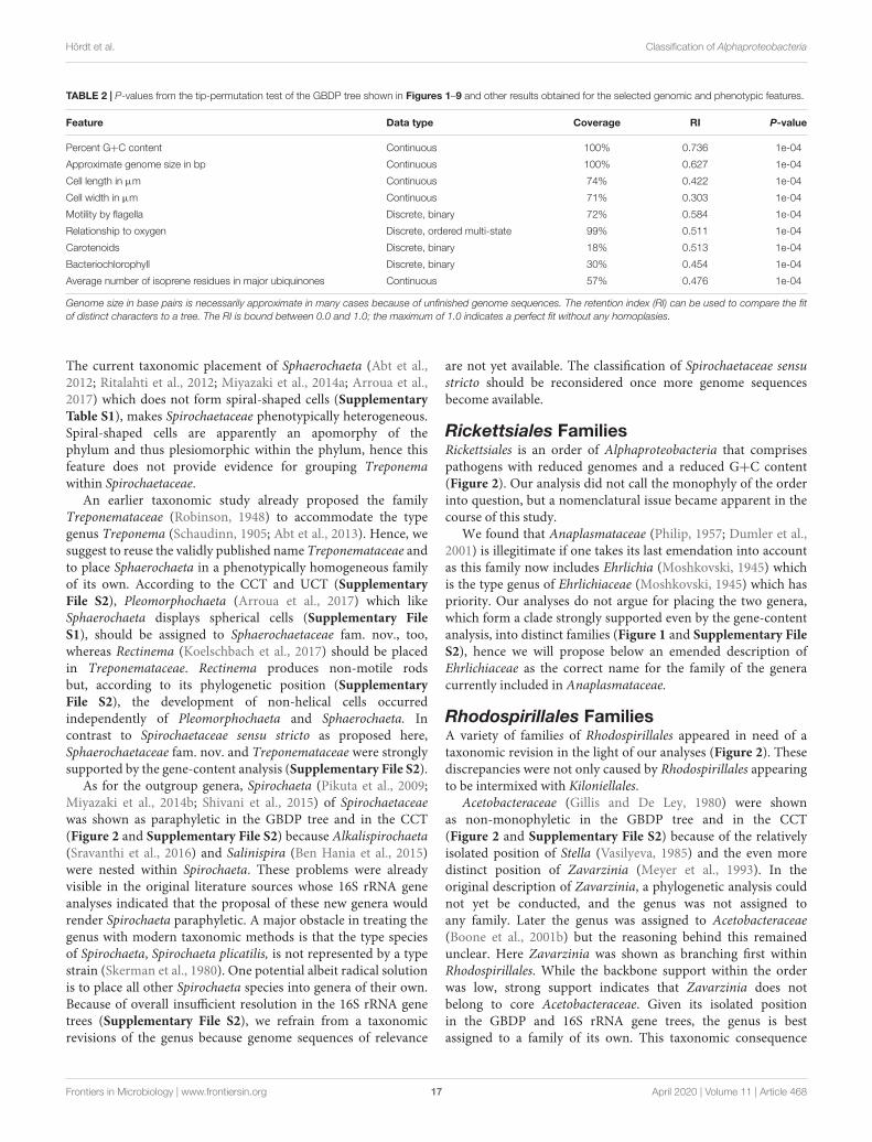
of sphingolipids, average number of isoprene residues of themajor ubiquinones, and relationship to oxygen. To avoid circularreasoning, missing features of a species were only inferred fromfeatures of its genus when species and genus were describedin the same publication or when the species description hadexplicitly been declared as adding to the features of the genus.For the binary chemotaxonomic characters an alternative codingwas also investigated that treated all missing values as indicatingabsence. Ubiquinone percentages would be more informativethan just statements about being “major” but mostly only thelatter are provided in the literature. Oxygen conditions werecoded as ordered multi-state character: (1) strictly anaerobic; (2)facultatively aerobic, facultatively anaerobic, or microaerophilic;(3) strictly aerobic. Among all nine coding options tested, thisyielded the highest fit to the tree (Supplementary Table S1) butthe differences between the coding options were not pronounced.Phylogenetic conservation of selected phenotypic and genomiccharacters with respect to the GBDP tree (reduced to representeach set of equivalent strains by only a single genome) wasevaluated using a tip-permutation test in conjunction withthe calculation of maximum-parsimony scores with TNT aspreviously described (Simon et al., 2017; Carro et al., 2018)and 10,000 permutations. TNT input files were generatedwith opm (Vaas et al., 2013). The proportion of times thescore of a permuted tree was at least as low as the score ofthe original tree yielded the p-value. Maximum-parsimonyretention indices (Farris, 1989; Wiley and Lieberman, 2011)were calculated to further differentiate between the fit of eachcharacter to the tree.
Taxa that were unambiguously non-monophyletic accordingto the genome-scale analyses were screened for publishedevidence of their monophyly. The published evidence wasjudged as inconclusive when based on unsupported branchesin phylogenetic trees, based on probably homoplastic charactersor on probable plesiomorphic character states. Plesiomorphiesmight well be “diagnostic” but just for paraphyletic groups(Hennig, 1965; Wiley and Lieberman, 2011; Montero-Calasanzet al., 2017) hence “diagnostic” features alone are insufficient inphylogenetic systematics.
For fixing the obviously non-monophyletic taxa taxonomicconsequences were proposed if new taxon delineations couldbe determined that were sufficiently supported by the CCT. Inthese cases, the uncertain phylogenetic placement of taxa whosegenome sequences were not available at the time of writingwould not affect the new proposals. Where necessary taxa weretentatively place in newly delineated groups.
RESULTS
The presentation of the results is organized as follows. Aftera brief overview on the figures and tables the outcome ofthe tests for the phylogenetic conservation are illustrated.Next, the phylogenetic results for the outgroup taxa aredescribed and put in the context of their current taxonomicclassification. Finally, the hierarchical classification of the classAlphaproteobacteria itself, arranged according to the orders in
Frontiers in Microbiology | www.frontiersin.org 4 April 2020 | Volume 11 | Article 468
fmicb-11-00468 April 4, 2020 Time: 10:24 # 5
Hördt et al. Classification of Alphaproteobacteria
which it is currently subdivided and then according to thetaxonomic categories, is compared to the phylogenomic trees.These sections motivate the need for a variety of reclassifications,whereas the actual taxonomic consequences are listed at the endof the section “Discussion.” Finally, the outcome of the tests forthe phylogenetic conservation are illustrated.
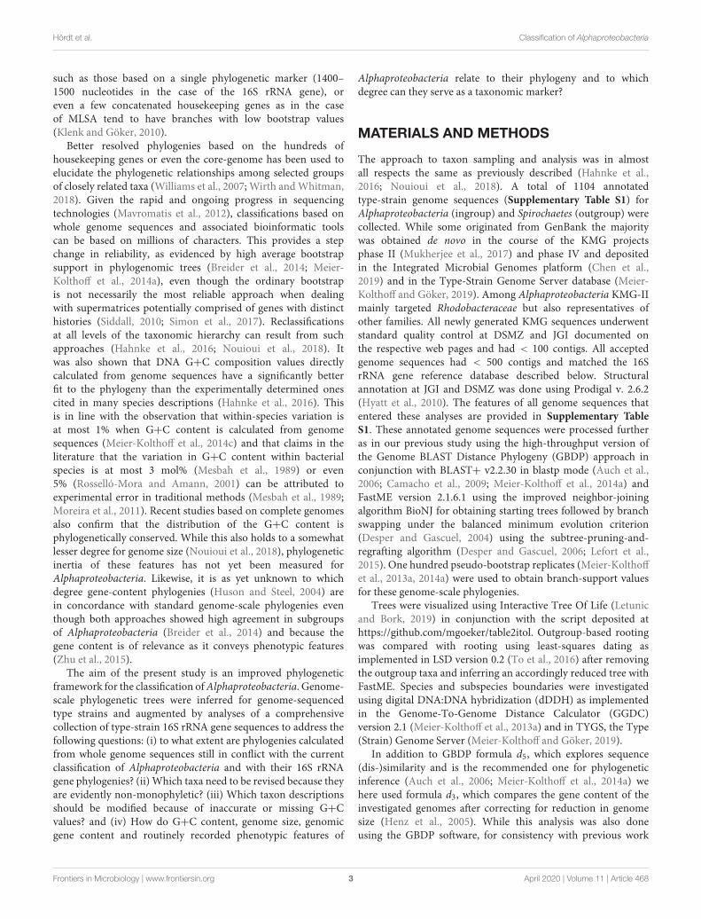
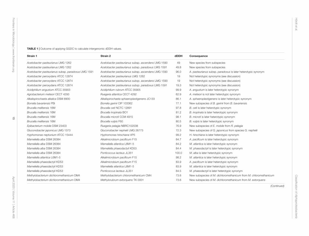
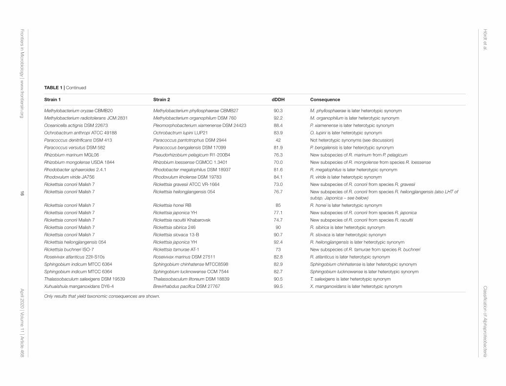
The GBDP tree is shown in Figures 1–8; Figure 1 provides anoverview and explains which specific sections of the same tree aredisplayed in greater detail in Figures 2–8. Table 1 shows dDDHresults for pairs of type strains of interest, while Table 2 displaysthe results of the tests for phylogenetic inertia. Phenotypicinformation for groups of taxa whose taxonomic classification istreated in detail below is summarized in Supplementary TableS1. This Supplementary Table S1 also contains the complete listof genome sequences used in this study, including their GenBankand IMG accession numbers. Additional phylogenetic trees,including the GBDP tree in a single figure and with phenotypicannotation and the results from the gene-content analysis, arefound in Supplementary File S2.
Classes and OrdersThe taxon sampling used in the present study was not mainlyintended to provide support for or against the monophyly ofthe class Alphaproteobacteria, or of the phylum Proteobacteriain general. The choice of the outgroup in the present study wasnot intended to indicate that the phylum Spirochaetes representsthe sister group of Alphaproteobacteria but was motivated byuncertainty regarding the monophyly of Proteobacteria (Yarzaet al., 2014). Inferring the tree depicted in Figure 1 again afterremoving the outgroup and rooting this reduced tree usingleast-squares dating yielded the same branching order for theingroup, i.e., the root was located between the clade formedby Magnetococcus and Mariprofundus on the one hand and theremainder of the tree on the other hand. Spirochaetes may thusnot be the ideal outgroup for Alphaproteobacteria phylogeny butthe alternative rooting confirmed the depicted branching order.
Only a single issue regarding the classes became apparent inthis study, and most of the orders of the class Alphaproteobacteriaappeared as monophyletic in our analysis (Figure 1). Theexceptions were mainly caused by specific genera taxonomicallyassigned to Rhodospirillales and particularly genera assigned toRhodobacterales that were phylogenetically intermixed with theorder currently called Rhizobiales.
Alphaproteobacteria appeared as paraphyletic in the GBDPtree (Figures 1, 2) since Mariprofundus ferrooxydans ofZetaproteobacteria (Emerson et al., 2007) formed a stronglysupported clade together with the alphaproteobacteriumMagnetococcus marinus (Bazylinski et al., 2013a). The cladeeven obtained reasonable support in the gene-content analysis(Supplementary File S2) and its two representatives displayedalmost the same G+C content. In the original descriptionof M. ferrooxydans (Emerson et al., 2007) a new class(Zetaproteobacteria), order (Mariprofundales) and family(Mariprofundaceae) were proposed in the SupplementaryMaterial only. None of these names became validly published sofar even though a corrected name, Mariprofundia, was suggestedfor Zetaproteobacteria in the meantime (Oren, 2017a).
In the originally presented 16S rRNA gene trees the placementof Mariprofundus has no strong support and Magnetococcusmarinus could not yet be considered. The additionally presentedprotein phylogenies (RecA, GyrB) only partially showedsupport for the placement of M. ferrooxydans separate fromAlphaproteobacteria. In the CCT we did not find strongsupport for the placement of M. ferrooxydans branching firstwithin the ingroup (Supplementary File S2). Phenotypically,alphaproteobacterial taxa such as Magnetococcus marinus arecapable of forming iron oxides much like M. ferrooxydans. Eventhough the filamentous iron oxyhydroxide and branched-chainfatty acids produced by this species may differentiate it fromtaxa with a similar ecology, this alone provides no evidence fora separate class. It thus makes sense to again propose a separateorder and family for Mariprofundus but to tentatively assign itto the class Alphaproteobacteria; an alternative arrangement is toremove Magnetococcus from Alphaproteobacteria.
Within Alphaproteobacteria, Rhodospirillales appeared asparaphyletic in the GBDP tree and in the CCT (Figure 2and Supplementary File S2) because Kiloniella (Wiese et al.,2009; Yang S.-H. et al., 2015) of Kiloniellales as well asRoseospirillum (Glaeser and Overmann, 1999) and Terasakiella(Satomi et al., 2002; Han et al., 2016) of the order currentlycalled Rhizobiales were nested within Rhodospirillales with highsupport. As this also affects the monophyly of the familyRhodospirillaceae, we will below propose the reclassification ofthese three genera into Rhodospirillales as the preferred way torestore a monophyletic order and family. Moreover, the distantposition of Geminicoccaceae (Proença et al., 2018) also conflictswith the monophyly of Rhodospirillales. Because this conflict wasonly poorly supported, we do not propose taxonomic changes forGeminicoccaceae based on the here examined data.
Rhodobacterales were shown as non-monophyletic in variousways, most of which also affect families and will thus be treatedbelow. Rhizobiales (Kuykendall, 2005) appeared as paraphyleticin GBDP tree (Figures 3–5 and Supplementary File S2) becauseAcuticoccus (Hou et al., 2015) Ahrensia (Uchino et al., 1998; LiuJ. et al., 2016) Labrenzia (Biebl et al., 2007; Bibi et al., 2014)Nesiotobacter (Donachie et al., 2006) Pannonibacter (Borsodiet al., 2003; Biebl et al., 2007) Pseudovibrio (Shieh et al.,2004) Roseibium (Suzuki et al., 2000) and Stappia (Uchinoet al., 1998; Biebl et al., 2007) all of which are currentlyclassified in Rhodobacterales, were nested within Rhizobiales.According taxonomic solutions are suggested below for theaffected families. Hartmannibacter (Suarez et al., 2014) which wasnot explicitly assigned to an order in its original description, isalso treated below.
It should also be noted that Rhizobiales (Kuykendall, 2005)is validly published but illegitimate (i.e., not in accordancewith the rules of the International Code of Nomenclatureof Prokaryotes) as this order includes Hyphomicrobium ofHyphomonadaceae, type genus of Hyphomicrobiales (Douglas,1957) which has priority. Our analyses do not call for placingRhizobium and Hyphomicrobium in distinct orders (Figure 4and Supplementary File S2), hence we will below proposean emended description of Hyphomicrobiales to replace theillegitimate Rhizobiales. The following description of the results
Frontiers in Microbiology | www.frontiersin.org 5 April 2020 | Volume 11 | Article 468
fmicb-11-00468 April 4, 2020 Time: 10:24 # 6
Hördt et al. Classification of Alphaproteobacteria
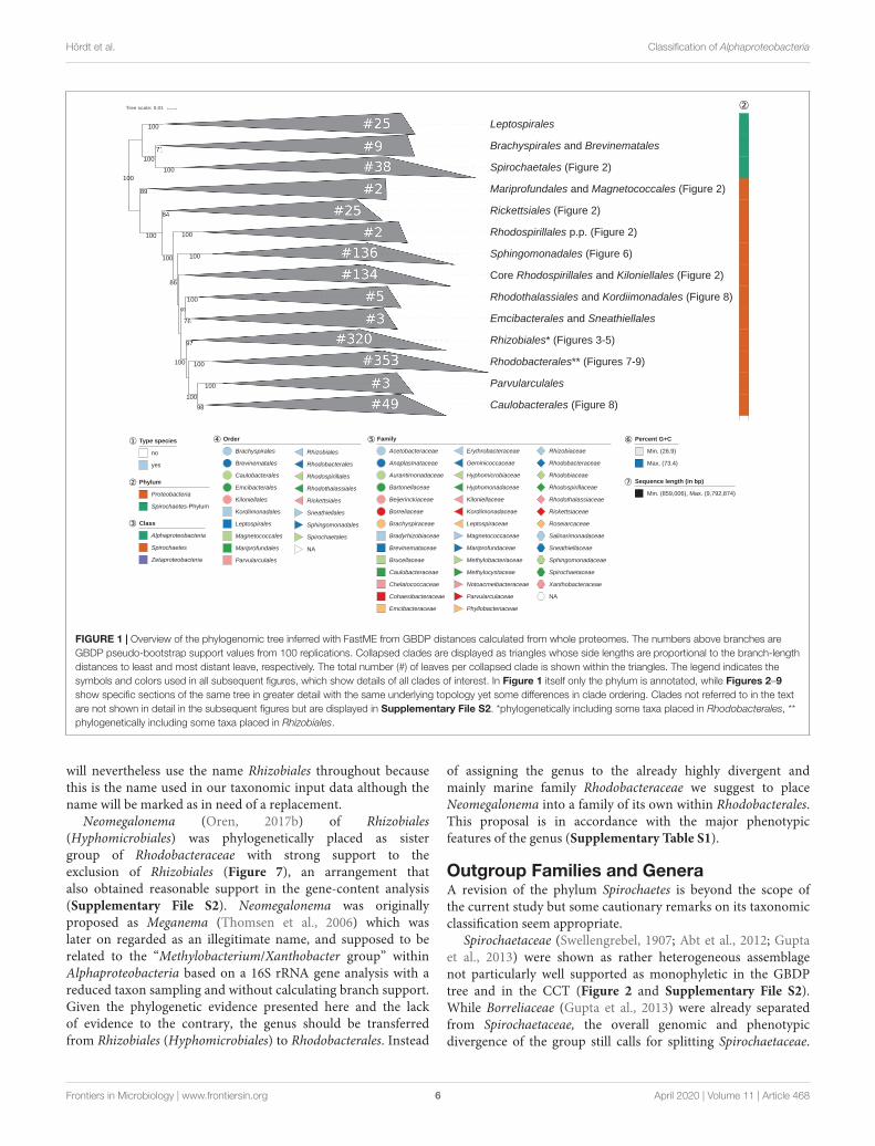
FIGURE 1 | Overview of the phylogenomic tree inferred with FastME from GBDP distances calculated from whole proteomes. The numbers above branches areGBDP pseudo-bootstrap support values from 100 replications. Collapsed clades are displayed as triangles whose side lengths are proportional to the branch-lengthdistances to least and most distant leave, respectively. The total number (#) of leaves per collapsed clade is shown within the triangles. The legend indicates thesymbols and colors used in all subsequent figures, which show details of all clades of interest. In Figure 1 itself only the phylum is annotated, while Figures 2–9show specific sections of the same tree in greater detail with the same underlying topology yet some differences in clade ordering. Clades not referred to in the textare not shown in detail in the subsequent figures but are displayed in Supplementary File S2. *phylogenetically including some taxa placed in Rhodobacterales, **phylogenetically including some taxa placed in Rhizobiales.
will nevertheless use the name Rhizobiales throughout becausethis is the name used in our taxonomic input data although thename will be marked as in need of a replacement.
Neomegalonema (Oren, 2017b) of Rhizobiales(Hyphomicrobiales) was phylogenetically placed as sistergroup of Rhodobacteraceae with strong support to theexclusion of Rhizobiales (Figure 7), an arrangement thatalso obtained reasonable support in the gene-content analysis(Supplementary File S2). Neomegalonema was originallyproposed as Meganema (Thomsen et al., 2006) which waslater on regarded as an illegitimate name, and supposed to berelated to the “Methylobacterium/Xanthobacter group” withinAlphaproteobacteria based on a 16S rRNA gene analysis with areduced taxon sampling and without calculating branch support.Given the phylogenetic evidence presented here and the lackof evidence to the contrary, the genus should be transferredfrom Rhizobiales (Hyphomicrobiales) to Rhodobacterales. Instead
of assigning the genus to the already highly divergent andmainly marine family Rhodobacteraceae we suggest to placeNeomegalonema into a family of its own within Rhodobacterales.This proposal is in accordance with the major phenotypicfeatures of the genus (Supplementary Table S1).
Outgroup Families and GeneraA revision of the phylum Spirochaetes is beyond the scope ofthe current study but some cautionary remarks on its taxonomicclassification seem appropriate.
Spirochaetaceae (Swellengrebel, 1907; Abt et al., 2012; Guptaet al., 2013) were shown as rather heterogeneous assemblagenot particularly well supported as monophyletic in the GBDPtree and in the CCT (Figure 2 and Supplementary File S2).While Borreliaceae (Gupta et al., 2013) were already separatedfrom Spirochaetaceae, the overall genomic and phenotypicdivergence of the group still calls for splitting Spirochaetaceae.
Frontiers in Microbiology | www.frontiersin.org 6 April 2020 | Volume 11 | Article 468
fmicb-11-00468 April 4, 2020 Time: 10:24 # 7
Hördt et al. Classification of Alphaproteobacteria
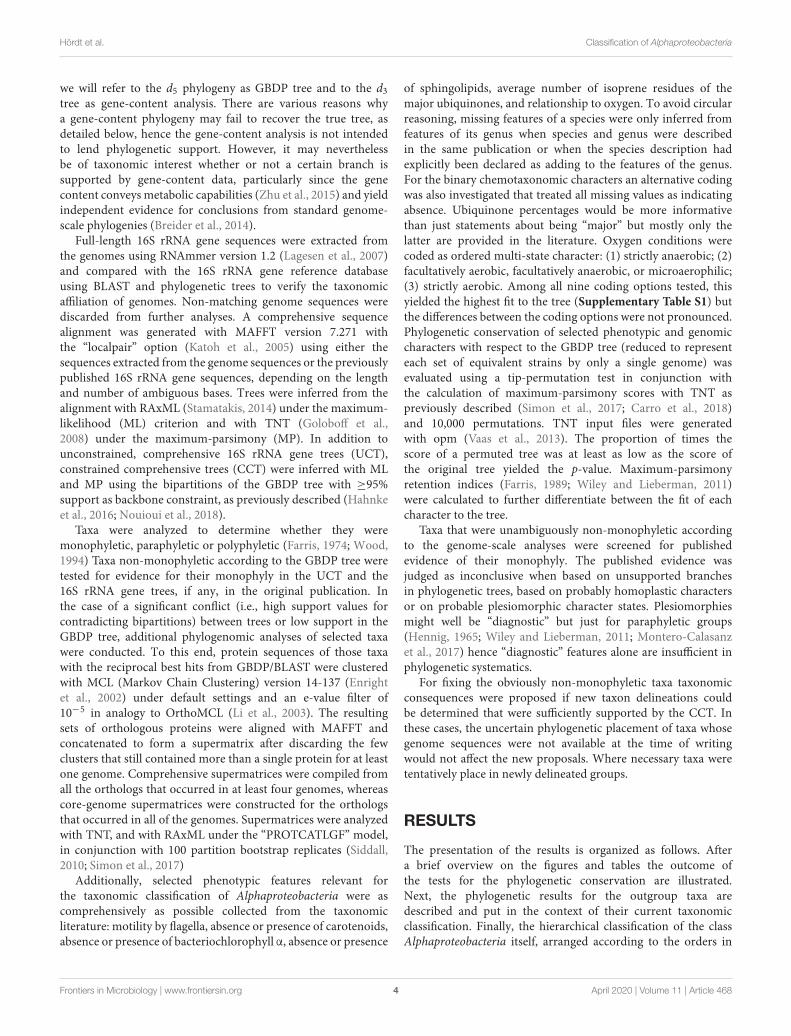
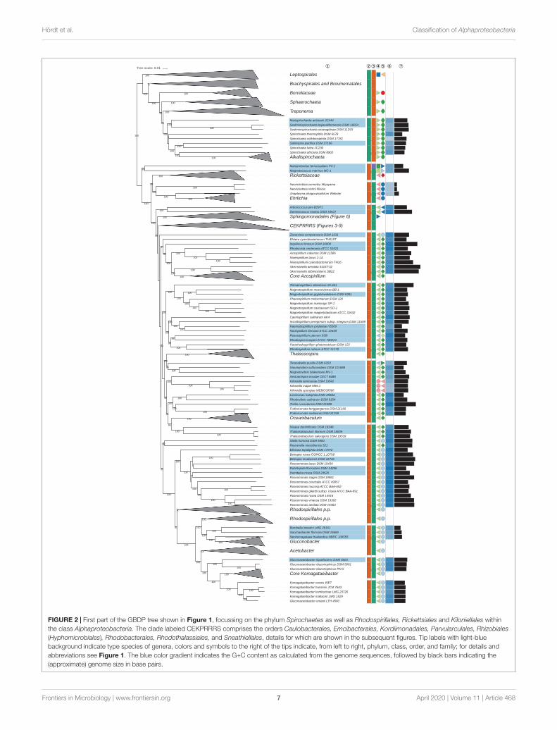
FIGURE 2 | First part of the GBDP tree shown in Figure 1, focussing on the phylum Spirochaetes as well as Rhodospirillales, Rickettsiales and Kiloniellales withinthe class Alphaproteobacteria. The clade labeled CEKPRRRS comprises the orders Caulobacterales, Emcibacterales, Kordiimonadales, Parvularculales, Rhizobiales(Hyphomicrobiales), Rhodobacterales, Rhodothalassiales, and Sneathiellales, details for which are shown in the subsequent figures. Tip labels with light-bluebackground indicate type species of genera, colors and symbols to the right of the tips indicate, from left to right, phylum, class, order, and family; for details andabbreviations see Figure 1. The blue color gradient indicates the G+C content as calculated from the genome sequences, followed by black bars indicating the(approximate) genome size in base pairs.
Frontiers in Microbiology | www.frontiersin.org 7 April 2020 | Volume 11 | Article 468
fmicb-11-00468 April 4, 2020 Time: 10:24 # 8
Hördt et al. Classification of Alphaproteobacteria
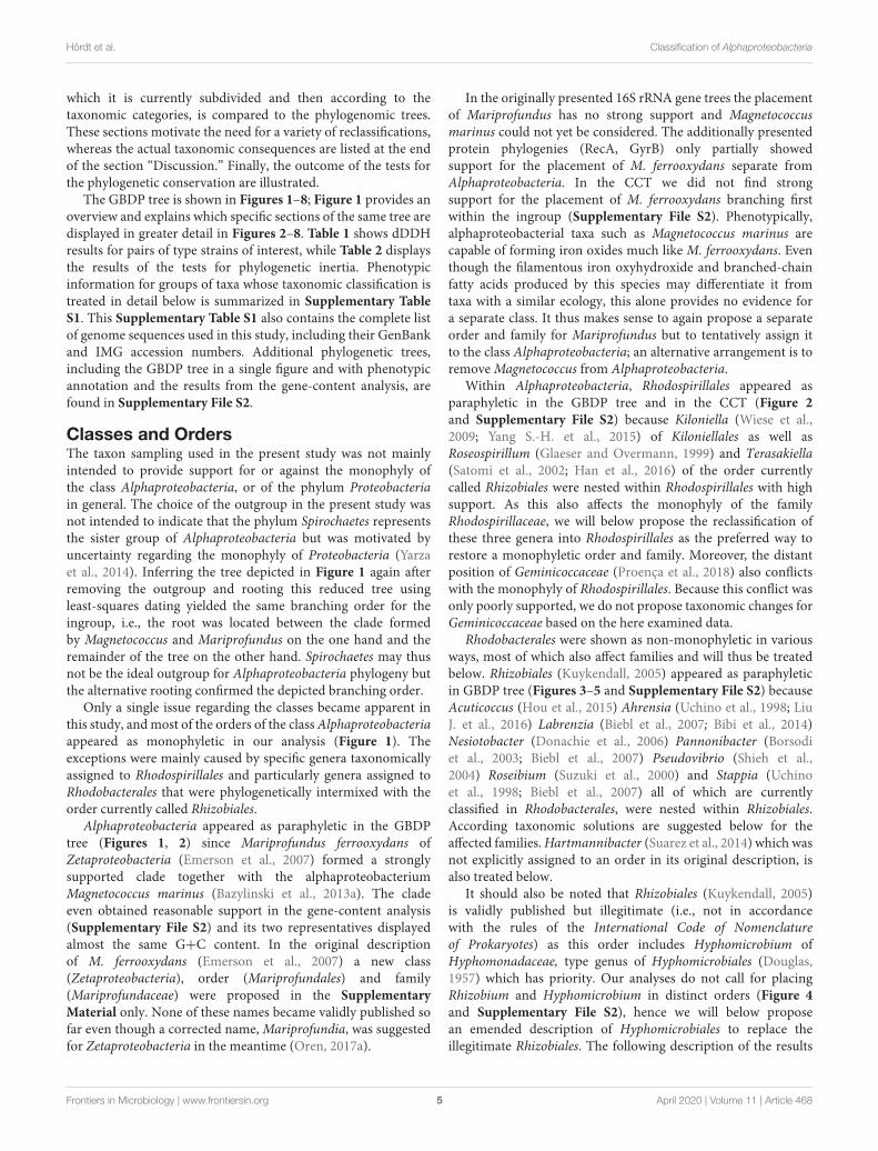
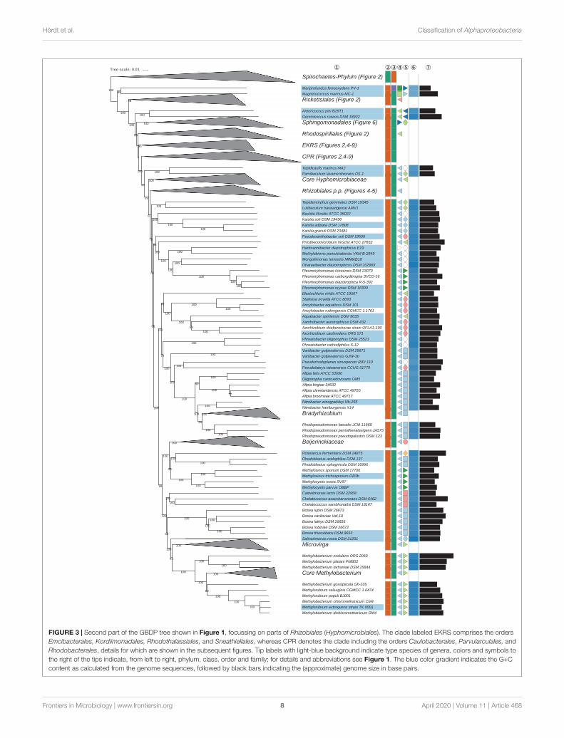
FIGURE 3 | Second part of the GBDP tree shown in Figure 1, focussing on parts of Rhizobiales (Hyphomicrobiales). The clade labeled EKRS comprises the ordersEmcibacterales, Kordiimonadales, Rhodothalassiales, and Sneathiellales, whereas CPR denotes the clade including the orders Caulobacterales, Parvularculales, andRhodobacterales, details for which are shown in the subsequent figures. Tip labels with light-blue background indicate type species of genera, colors and symbols tothe right of the tips indicate, from left to right, phylum, class, order and family; for details and abbreviations see Figure 1. The blue color gradient indicates the G+Ccontent as calculated from the genome sequences, followed by black bars indicating the (approximate) genome size in base pairs.
Frontiers in Microbiology | www.frontiersin.org 8 April 2020 | Volume 11 | Article 468
fmicb-11-00468 April 4, 2020 Time: 10:24 # 9
Hördt et al. Classification of Alphaproteobacteria
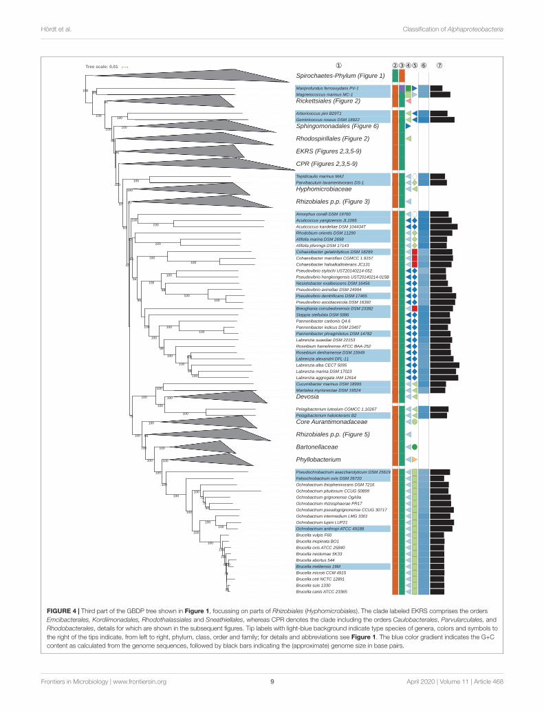
FIGURE 4 | Third part of the GBDP tree shown in Figure 1, focussing on parts of Rhizobiales (Hyphomicrobiales). The clade labeled EKRS comprises the ordersEmcibacterales, Kordiimonadales, Rhodothalassiales and Sneathiellales, whereas CPR denotes the clade including the orders Caulobacterales, Parvularculales, andRhodobacterales, details for which are shown in the subsequent figures. Tip labels with light-blue background indicate type species of genera, colors and symbols tothe right of the tips indicate, from left to right, phylum, class, order and family; for details and abbreviations see Figure 1. The blue color gradient indicates the G+Ccontent as calculated from the genome sequences, followed by black bars indicating the (approximate) genome size in base pairs.
Frontiers in Microbiology | www.frontiersin.org 9 April 2020 | Volume 11 | Article 468
fmicb-11-00468 April 4, 2020 Time: 10:24 # 10
Hördt et al. Classification of Alphaproteobacteria
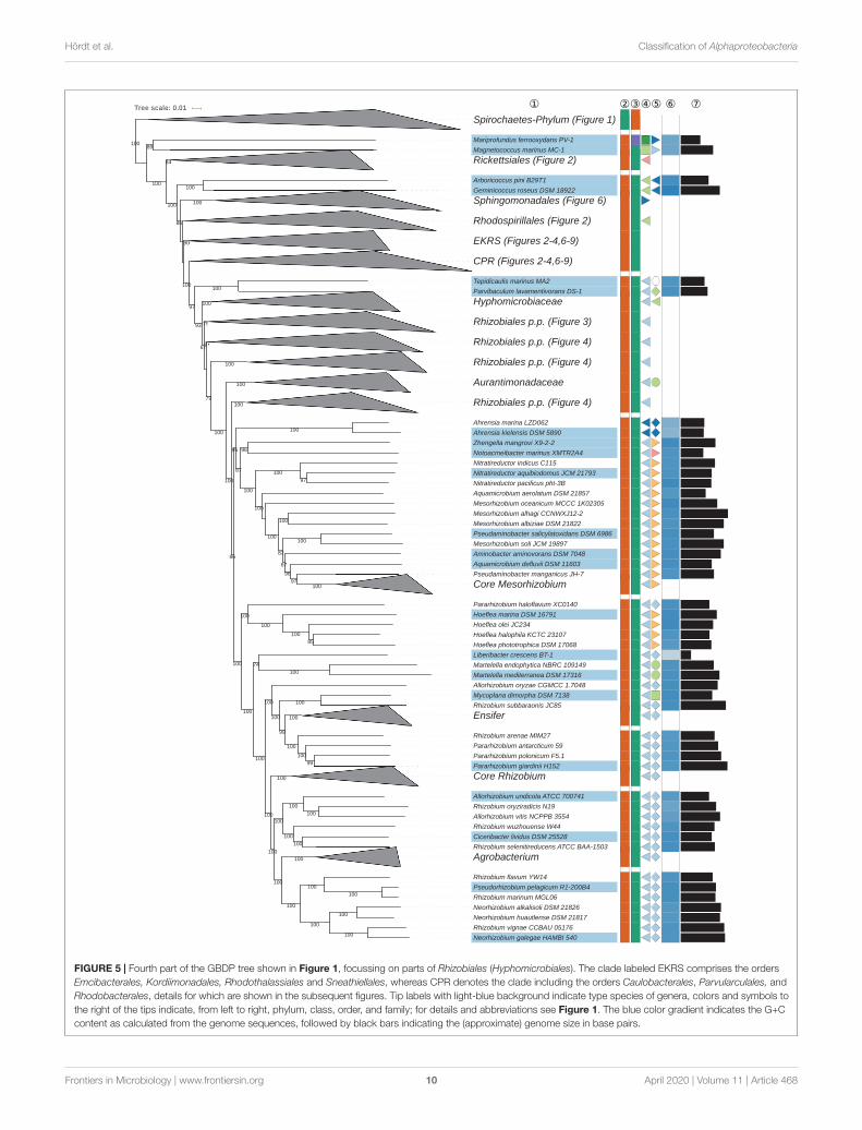
FIGURE 5 | Fourth part of the GBDP tree shown in Figure 1, focussing on parts of Rhizobiales (Hyphomicrobiales). The clade labeled EKRS comprises the ordersEmcibacterales, Kordiimonadales, Rhodothalassiales and Sneathiellales, whereas CPR denotes the clade including the orders Caulobacterales, Parvularculales, andRhodobacterales, details for which are shown in the subsequent figures. Tip labels with light-blue background indicate type species of genera, colors and symbols tothe right of the tips indicate, from left to right, phylum, class, order, and family; for details and abbreviations see Figure 1. The blue color gradient indicates the G+Ccontent as calculated from the genome sequences, followed by black bars indicating the (approximate) genome size in base pairs.
Frontiers in Microbiology | www.frontiersin.org 10 April 2020 | Volume 11 | Article 468
fmicb-11-00468 April 4, 2020 Time: 10:24 # 11
Hördt et al. Classification of Alphaproteobacteria
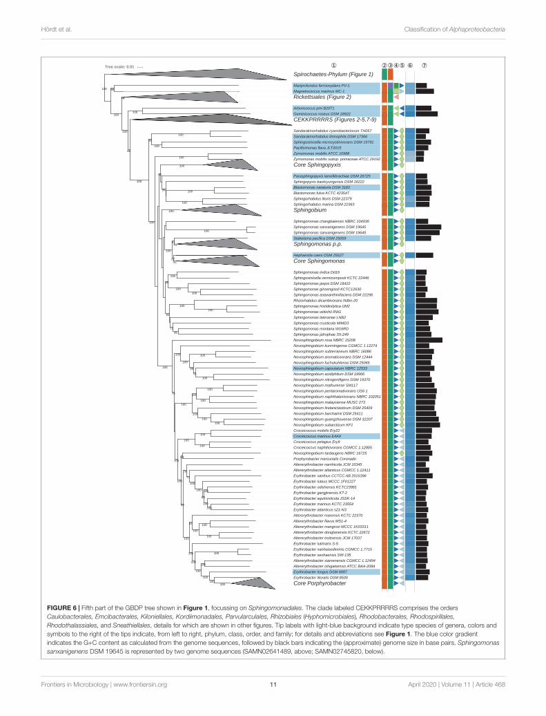
FIGURE 6 | Fifth part of the GBDP tree shown in Figure 1, focussing on Sphingomonadales. The clade labeled CEKKPRRRRS comprises the ordersCaulobacterales, Emcibacterales, Kiloniellales, Kordiimonadales, Parvularculales, Rhizobiales (Hyphomicrobiales), Rhodobacterales, Rhodospirillales,Rhodothalassiales, and Sneathiellales, details for which are shown in other figures. Tip labels with light-blue background indicate type species of genera, colors andsymbols to the right of the tips indicate, from left to right, phylum, class, order, and family; for details and abbreviations see Figure 1. The blue color gradientindicates the G+C content as calculated from the genome sequences, followed by black bars indicating the (approximate) genome size in base pairs. Sphingomonassanxanigenens DSM 19645 is represented by two genome sequences (SAMN02641489, above; SAMN02745820, below).
Frontiers in Microbiology | www.frontiersin.org 11 April 2020 | Volume 11 | Article 468
fmicb-11-00468 April 4, 2020 Time: 10:24 # 12
Hördt et al. Classification of Alphaproteobacteria
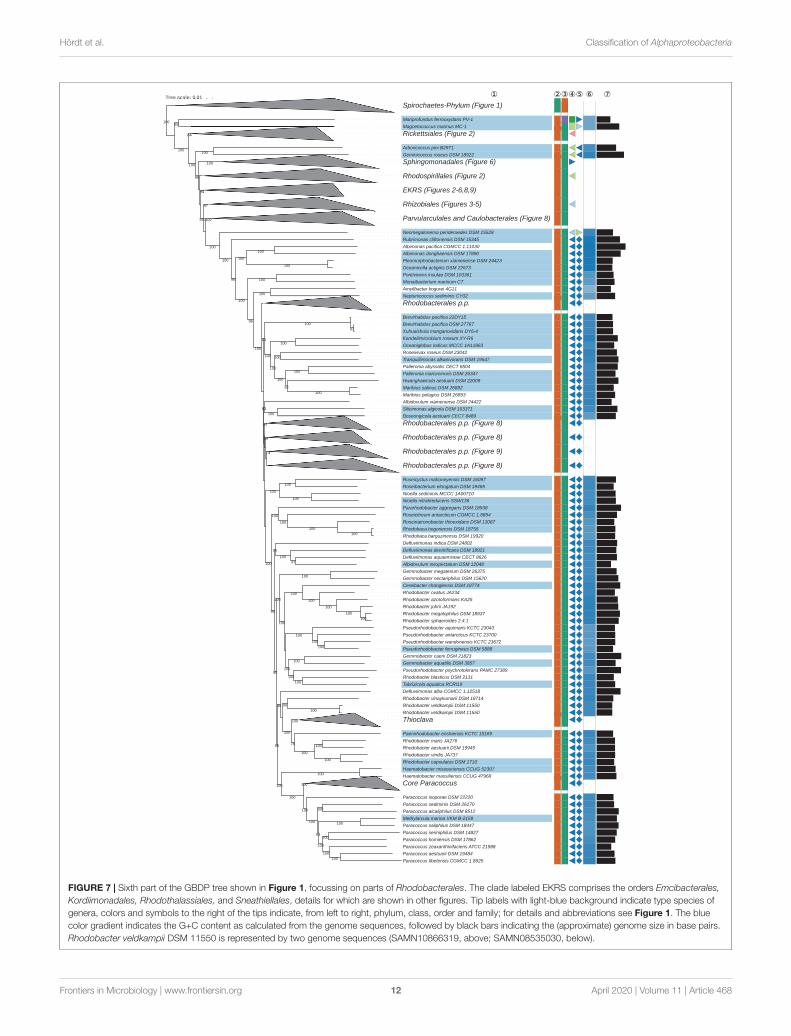
FIGURE 7 | Sixth part of the GBDP tree shown in Figure 1, focussing on parts of Rhodobacterales. The clade labeled EKRS comprises the orders Emcibacterales,Kordiimonadales, Rhodothalassiales, and Sneathiellales, details for which are shown in other figures. Tip labels with light-blue background indicate type species ofgenera, colors and symbols to the right of the tips indicate, from left to right, phylum, class, order and family; for details and abbreviations see Figure 1. The bluecolor gradient indicates the G+C content as calculated from the genome sequences, followed by black bars indicating the (approximate) genome size in base pairs.Rhodobacter veldkampii DSM 11550 is represented by two genome sequences (SAMN10866319, above; SAMN08535030, below).
Frontiers in Microbiology | www.frontiersin.org 12 April 2020 | Volume 11 | Article 468
fmicb-11-00468 April 4, 2020 Time: 10:24 # 13
Hördt et al. Classification of Alphaproteobacteria
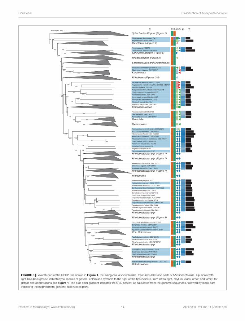
FIGURE 8 | Seventh part of the GBDP tree shown in Figure 1, focussing on Caulobacterales, Parvularculales and parts of Rhodobacterales. Tip labels withlight-blue background indicate type species of genera, colors and symbols to the right of the tips indicate, from left to right, phylum, class, order, and family; fordetails and abbreviations see Figure 1. The blue color gradient indicates the G+C content as calculated from the genome sequences, followed by black barsindicating the (approximate) genome size in base pairs.
Frontiers in Microbiology | www.frontiersin.org 13 April 2020 | Volume 11 | Article 468
fmicb-11-00468 April 4, 2020 Time: 10:24 # 14
Hördt et al. Classification of Alphaproteobacteria
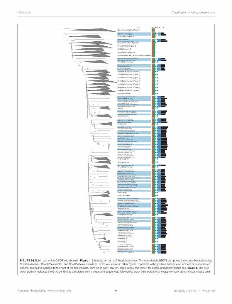
FIGURE 9 | Eighth part of the GBDP tree shown in Figure 1, focussing on parts of Rhodobacterales. The clade labeled EKRS comprises the orders Emcibacterales,Kordiimonadales, Rhodothalassiales, and Sneathiellales, details for which are shown in other figures. Tip labels with light-blue background indicate type species ofgenera, colors and symbols to the right of the tips indicate, from left to right, phylum, class, order, and family; for details and abbreviations see Figure 1. The bluecolor gradient indicates the G+C content as calculated from the genome sequences, followed by black bars indicating the (approximate) genome size in base pairs.
Frontiers in Microbiology | www.frontiersin.org 14 April 2020 | Volume 11 | Article 468
fmicb-11-00468
April4,2020
Time:10:24
#15
Hördtetal.
Classification
ofAlphaproteobacteria
TABLE 1 | Outcome of applying GGDC to calculate intergenomic dDDH values.
Strain 1 Strain 2 dDDH Consequence
Acetobacter pasteurianus LMG 1262 Acetobacter pasteurianus subsp. ascendens LMG 1590 49 New species from subspecies
Acetobacter pasteurianus LMG 1262 Acetobacter pasteurianus subsp. paradoxus LMG 1591 49.8 New species from subspecies
Acetobacter pasteurianus subsp. paradoxus LMG 1591 Acetobacter pasteurianus subsp. ascendens LMG 1590 96.0 A. pasteurianus subsp. paradoxus is later heterotypic synonym
Acetobacter peroxydans ATCC 12874 Acetobacter pasteurianus LMG 1262 19 Not heterotypic synonyms (see discussion)
Acetobacter peroxydans ATCC 12874 Acetobacter pasteurianus subsp. ascendens LMG 1590 19 Not heterotypic synonyms (see discussion)
Acetobacter peroxydans ATCC 12874 Acetobacter pasteurianus subsp. paradoxus LMG 1591 19.3 Not heterotypic synonyms (see discussion)
Acidiphilium angustum ATCC 35903 Acidiphilium rubrum ATCC 35905 99.9 A. angustum is later heterotypic synonym
Agrobacterium meteori CECT 4293 Ruegeria atlantica CECT 4292 62.9 A. meteori is not later heterotypic synonym
Alkalispirochaeta alkalica DSM 8900 Alkalispirochaeta sphaeroplastigenens JC133 86.1 A. sphaeroplastigenens is later heterotypic synonym
Borrelia bavariensis PBi Borrelia garinii CIP 103362 77.1 New subspecies of B. garinii from B. bavariensis
Brucella melitensis 16M Brucella ceti NCTC 12891 97.8 B. ceti is later heterotypic synonym
Brucella melitensis 16M Brucella inopinata BO1 81.2 B. inopinata is later heterotypic synonym
Brucella melitensis 16M Brucella microti CCM 4915 98.1 B. microti is later heterotypic synonym
Brucella melitensis 16M Brucella vulpis F60 80.5 B. vulpis is later heterotypic synonym
Epibacterium mobile DSM 23403 Ruegeria pelagia NBRC102038 76.9 New subspecies of E. mobile from R. pelagia
Gluconobacter japonicus LMG 1373 Gluconobacter nephelii LMG 26773 72.3 New subspecies of G. japonicus from species G. nephelii
Hyphomonas neptunium ATCC 15444 Hyphomonas hirschiana VP5 99.2 H. hirschiana is later heterotypic synonym
Mameliella alba DSM 26384 Alkalimicrobium pacificum F15 84.7 A. pacificum is later heterotypic synonym
Mameliella alba DSM 26384 Mameliella atlantica L6M1-5 84.2 M. atlantica is later heterotypic synonym
Mameliella alba DSM 26384 Mameliella phaeodactyli KD53 84.4 M. phaeodactyli is later heterotypic synonym
Mameliella alba DSM 26384 Ponticoccus lacteus JL351 100.0 M. alba is later heterotypic synonym
Mameliella atlantica L6M1-5 Alkalimicrobium pacificum F15 98.2 M. atlantica is later heterotypic synonym
Mameliella phaeodactyli KD53 Alkalimicrobium pacificum F15 83.9 A. pacificum is later heterotypic synonym
Mameliella phaeodactyli KD53 Mameliella atlantica L6M1-5 83.9 M. atlantica is later heterotypic synonym
Mameliella phaeodactyli KD53 Ponticoccus lacteus JL351 84.5 M. phaeodactyli is later heterotypic synonym
Methylobacterium dichloromethanicum DM4 Methylobacterium chloromethanicum CM4 73.6 New subspecies of M. dichloromethanicum from M. chloromethanicum
Methylobacterium dichloromethanicum DM4 Methylorubrum extorquens TK 0001 73.6 New subspecies of M. dichloromethanicum from M. extorquens
(Continued)
Frontiersin
Microbiology
|ww
w.frontiersin.org
15A
pril2020|Volum
e11
|Article
468
fmicb-11-00468
April4,2020
Time:10:24
#16
Hördtetal.
Classification
ofAlphaproteobacteria
TABLE 1 | Continued
Strain 1 Strain 2 dDDH Consequence
Methylobacterium oryzae CBMB20 Methylobacterium phyllosphaerae CBMB27 90.3 M. phyllosphaerae is later heterotypic synonym
Methylobacterium radiotolerans JCM 2831 Methylobacterium organophilum DSM 760 92.2 M. organophilum is later heterotypic synonym
Oceanicella actignis DSM 22673 Pleomorphobacterium xiamenense DSM 24423 88.4 P. xiamenense is later heterotypic synonym
Ochrobactrum anthropi ATCC 49188 Ochrobactrum lupini LUP21 83.9 O. lupini is later heterotypic synonym
Paracoccus denitrificans DSM 413 Paracoccus pantotrophus DSM 2944 42 Not heterotypic synonyms (see discussion)
Paracoccus versutus DSM 582 Paracoccus bengalensis DSM 17099 81.9 P. bengalensis is later heterotypic synonym
Rhizobium marinum MGL06 Pseudorhizobium pelagicum R1-200B4 76.3 New subspecies of R. marinum from P. pelagicum
Rhizobium mongolense USDA 1844 Rhizobium loessense CGMCC 1.3401 70.0 New subspecies of R. mongolense from species R. loessense
Rhodobacter sphaeroides 2.4.1 Rhodobacter megalophilus DSM 18937 81.6 R. megalophilus is later heterotypic synonym
Rhodovulum viride JA756 Rhodovulum kholense DSM 19783 84.1 R. viride is later heterotypic synonym
Rickettsia conorii Malish 7 Rickettsia gravesii ATCC VR-1664 73.0 New subspecies of R. conorii from species R. gravesii
Rickettsia conorii Malish 7 Rickettsia heilongjiangensis 054 76.7 New subspecies of R. conorii from species R. heilongjiangensis (also LHT ofsubsp. Japonica – see below)
Rickettsia conorii Malish 7 Rickettsia honei RB 85 R. honei is later heterotypic synonym
Rickettsia conorii Malish 7 Rickettsia japonica YH 77.1 New subspecies of R. conorii from species R. japonica
Rickettsia conorii Malish 7 Rickettsia raoultii Khabarovsk 74.7 New subspecies of R. conorii from species R. raoultii
Rickettsia conorii Malish 7 Rickettsia sibirica 246 90 R. sibirica is later heterotypic synonym
Rickettsia conorii Malish 7 Rickettsia slovaca 13-B 90.7 R. slovaca is later heterotypic synonym
Rickettsia heilongjiangensis 054 Rickettsia japonica YH 92.4 R. heilongjiangensis is later heterotypic synonym
Rickettsia buchneri ISO-7 Rickettsia tamurae AT-1 73 New subspecies of R. tamurae from species R. buchneri
Roseivivax atlanticus 22II-S10s Roseivivax marinus DSM 27511 82.8 R. atlanticus is later heterotypic synonym
Sphingobium indicum MTCC 6364 Sphingobium chinhatense MTCC8598 82.9 Sphingobium chinhatense is later heterotypic synonym
Sphingobium indicum MTCC 6364 Sphingobium lucknowense CCM 7544 82.7 Sphingobium lucknowense is later heterotypic synonym
Thalassobaculum salexigens DSM 19539 Thalassobaculum litoreum DSM 18839 90.5 T. salexigens is later heterotypic synonym
Xuhuaishuia manganoxidans DY6-4 Brevirhabdus pacifica DSM 27767 99.5 X. manganoxidans is later heterotypic synonym
Only results that yield taxonomic consequences are shown.
Frontiersin
Microbiology
|ww
w.frontiersin.org
16A
pril2020|Volum
e11
|Article
468
fmicb-11-00468 April 4, 2020 Time: 10:24 # 17
Hördt et al. Classification of Alphaproteobacteria
TABLE 2 | P-values from the tip-permutation test of the GBDP tree shown in Figures 1–9 and other results obtained for the selected genomic and phenotypic features.
Feature Data type Coverage RI P-value
Percent G+C content Continuous 100% 0.736 1e-04
Approximate genome size in bp Continuous 100% 0.627 1e-04
Cell length in µm Continuous 74% 0.422 1e-04
Cell width in µm Continuous 71% 0.303 1e-04
Motility by flagella Discrete, binary 72% 0.584 1e-04
Relationship to oxygen Discrete, ordered multi-state 99% 0.511 1e-04
Carotenoids Discrete, binary 18% 0.513 1e-04
Bacteriochlorophyll Discrete, binary 30% 0.454 1e-04
Average number of isoprene residues in major ubiquinones Continuous 57% 0.476 1e-04
Genome size in base pairs is necessarily approximate in many cases because of unfinished genome sequences. The retention index (RI) can be used to compare the fitof distinct characters to a tree. The RI is bound between 0.0 and 1.0; the maximum of 1.0 indicates a perfect fit without any homoplasies.
The current taxonomic placement of Sphaerochaeta (Abt et al.,2012; Ritalahti et al., 2012; Miyazaki et al., 2014a; Arroua et al.,2017) which does not form spiral-shaped cells (SupplementaryTable S1), makes Spirochaetaceae phenotypically heterogeneous.Spiral-shaped cells are apparently an apomorphy of thephylum and thus plesiomorphic within the phylum, hence thisfeature does not provide evidence for grouping Treponemawithin Spirochaetaceae.
An earlier taxonomic study already proposed the familyTreponemataceae (Robinson, 1948) to accommodate the typegenus Treponema (Schaudinn, 1905; Abt et al., 2013). Hence, wesuggest to reuse the validly published name Treponemataceae andto place Sphaerochaeta in a phenotypically homogeneous familyof its own. According to the CCT and UCT (SupplementaryFile S2), Pleomorphochaeta (Arroua et al., 2017) which likeSphaerochaeta displays spherical cells (Supplementary FileS1), should be assigned to Sphaerochaetaceae fam. nov., too,whereas Rectinema (Koelschbach et al., 2017) should be placedin Treponemataceae. Rectinema produces non-motile rodsbut, according to its phylogenetic position (SupplementaryFile S2), the development of non-helical cells occurredindependently of Pleomorphochaeta and Sphaerochaeta. Incontrast to Spirochaetaceae sensu stricto as proposed here,Sphaerochaetaceae fam. nov. and Treponemataceae were stronglysupported by the gene-content analysis (Supplementary File S2).
As for the outgroup genera, Spirochaeta (Pikuta et al., 2009;Miyazaki et al., 2014b; Shivani et al., 2015) of Spirochaetaceaewas shown as paraphyletic in the GBDP tree and in the CCT(Figure 2 and Supplementary File S2) because Alkalispirochaeta(Sravanthi et al., 2016) and Salinispira (Ben Hania et al., 2015)were nested within Spirochaeta. These problems were alreadyvisible in the original literature sources whose 16S rRNA geneanalyses indicated that the proposal of these new genera wouldrender Spirochaeta paraphyletic. A major obstacle in treating thegenus with modern taxonomic methods is that the type speciesof Spirochaeta, Spirochaeta plicatilis, is not represented by a typestrain (Skerman et al., 1980). One potential albeit radical solutionis to place all other Spirochaeta species into genera of their own.Because of overall insufficient resolution in the 16S rRNA genetrees (Supplementary File S2), we refrain from a taxonomicrevisions of the genus because genome sequences of relevance
are not yet available. The classification of Spirochaetaceae sensustricto should be reconsidered once more genome sequencesbecome available.
Rickettsiales FamiliesRickettsiales is an order of Alphaproteobacteria that comprisespathogens with reduced genomes and a reduced G+C content(Figure 2). Our analysis did not call the monophyly of the orderinto question, but a nomenclatural issue became apparent in thecourse of this study.
We found that Anaplasmataceae (Philip, 1957; Dumler et al.,2001) is illegitimate if one takes its last emendation into accountas this family now includes Ehrlichia (Moshkovski, 1945) whichis the type genus of Ehrlichiaceae (Moshkovski, 1945) which haspriority. Our analyses do not argue for placing the two genera,which form a clade strongly supported even by the gene-contentanalysis, into distinct families (Figure 1 and Supplementary FileS2), hence we will propose below an emended description ofEhrlichiaceae as the correct name for the family of the generacurrently included in Anaplasmataceae.
Rhodospirillales FamiliesA variety of families of Rhodospirillales appeared in need of ataxonomic revision in the light of our analyses (Figure 2). Thesediscrepancies were not only caused by Rhodospirillales appearingto be intermixed with Kiloniellales.
Acetobacteraceae (Gillis and De Ley, 1980) were shownas non-monophyletic in the GBDP tree and in the CCT(Figure 2 and Supplementary File S2) because of the relativelyisolated position of Stella (Vasilyeva, 1985) and the even moredistinct position of Zavarzinia (Meyer et al., 1993). In theoriginal description of Zavarzinia, a phylogenetic analysis couldnot yet be conducted, and the genus was not assigned toany family. Later the genus was assigned to Acetobacteraceae(Boone et al., 2001b) but the reasoning behind this remainedunclear. Here Zavarzinia was shown as branching first withinRhodospirillales. While the backbone support within the orderwas low, strong support indicates that Zavarzinia does notbelong to core Acetobacteraceae. Given its isolated positionin the GBDP and 16S rRNA gene trees, the genus is bestassigned to a family of its own. This taxonomic consequence
Frontiers in Microbiology | www.frontiersin.org 17 April 2020 | Volume 11 | Article 468
fmicb-11-00468 April 4, 2020 Time: 10:24 # 18
Hördt et al. Classification of Alphaproteobacteria
did not appear in conflict with the major phenotypic features ofZavarzinia (Supplementary Table S1).
Stella appeared as sister group of the equally deviatinggenus of Rhodospirillaceae, Reyranella (Pagnier et al., 2011) withmoderate support, and separated from core Acetobacteraceaeby long branches. The original description of Stella did notexplicitly assign the genus to a family. Stella was placed inAcetobacteraceae in Bergey’s manual (Boone et al., 2001a);whether this placement was based on a phylogenetic assessment isunclear. Phenotypically the genus is rather unique because of itsstar-like morphology (Supplementary Table S1). All results thussuggest placing Stella into a family of its own. The same holds forReyranella, which is treated below; the gene-content analysis doesnot support the sister-group relationship of the two genera.
When Constrictibacter (Yamada et al., 2011) was proposedit was placed in Rhodospirillaceae in a 16S rRNA gene treewith low support values. In the CCT (Supplementary FileS2) with its much broader taxon sampling Constrictibacterformed a clade together with Stella albeit with low support.We thus tentatively include Constrictibacter in the familynewly proposed to accommodate Stella (see above). Althoughphenotypic differences in morphology and respiration mightsuggest to alternatively place Constrictibacter into a family of itsown this solution should be postponed until a Constrictibactergenome sequence is available.
Rhodospirillaceae (Pfennig and Trüper, 1971) appeared asparaphyletic in GBDP tree and in the CCT (Figure 2 andSupplementary File S2) for a variety of reasons. For instance,Roseospirillum (Glaeser and Overmann, 1999) of Rhodobiaceaewas nested within Rhodospirillaceae. Roseospirillum was placedas sister group of Rhodospira (Pfennig et al., 1997) with highsupport (Figure 2), a clade that in turn formed, with highsupport, the sister group of a clade containing the type speciesof the type genus of the family, Rhodospirillum rubrum (Molisch,1907) these arrangements are even supported by the gene-contentanalysis (Supplementary File S2). For these reasons, we proposeto include Roseospirillum into Rhodospirillaceae, which is alsosupported by the high phenotypic agreement between them(Supplementary Table S1). When Roseospirillum was proposed,bootstrapping was not conducted, and only few species could beincluded in the phylogenetic analysis at that time.
Furthermore, Ferrovibrio (Sorokina et al., 2012) Taonella(Xi et al., 2013) and Marinibaculum (Yu et al., 2016) formedan isolated but strongly supported clade with Sneathiellaceae(Kurahashi et al., 2008) in the CCT (Supplementary FileS2). When Ferrovibrio was proposed, it was already placed inthe order Sneathiellales but not explicitly in Sneathiellaceae.When Taonella and Marinibaculum were proposed bothpublications lacked strong support for the positioning ofthe respective taxa and both publications did not sampleSneathiella (Jordan et al., 2007). Therefore, we propose totentatively assign Ferrovibrio, Taonella, and Marinibaculumto Sneathiellaceae. These taxonomic consequences are notprecluded by the phenotypic features of the involved genera(Supplementary Table S1).
Similarly, Terasakiella (Satomi et al., 2002; Han et al.,2016) of Methylocystaceae and Kiloniella (Wiese et al., 2009;
Yang S.-H. et al., 2015) of Kiloniellaceae were nested withinRhodospirillaceae (Figure 2). However, even if these generawere included in Rhodospirillaceae, the family would lack anysupport. For this reason, to address the remaining causes ofthe non-monophyly of Rhodospirillaceae other measures areadvisable. Rhodospirillaceae would not appear monophyleticeven if these three genera were included. Rhodospirillaceaeshowed high genomic divergence (Figure 2) and unsupportedby the gene-content analysis (Supplementary File S2). Thefamily is phenotypically heterogeneous, as, e.g., most of itsrepresentatives are phototrophic whereas some genera arechemoorganoheterotrophs (Supplementary Table S1). SplittingRhodospirillaceae into several families corresponding to well-supported clades can also solve this issue.
Azospirillaceae fam. nov. is thus proposed in agreementwith the GBDP tree (Figure 2 and Supplementary File S2)to accommodate Niveispirillum (Lin et al., 2014) Azospirillum(Tarrand et al., 1978; Falk et al., 1985) Rhodocista (Kawasaki et al.,1993) and Skermanella (Sly and Stackebrandt, 1999; Luo et al.,2012; Weon et al., 2007) Nitrospirillum (Lin et al., 2014) andDesertibacter (Liu et al., 2011) are also assigned to this familybased on the 16S rRNA gene analyses (Supplementary File S2).Even though the gene-content analysis does not support the cladebut only its two major subclades, this conclusion fits well to themajor phenotypic features of these genera (Supplementary TableS1), most of which also show larger genomes than the othergenera currently included in Rhodospirillaceae. Azospirillum andRhodocista were placed in Rhodospirillaceae in Bergey’s manual(Boone et al., 2001a) but it remained unclear to us whetherthis was based on a phylogenetic assessment. The proposalsof these genera directly assigned them to Rhodospirillaceae butphylogenetic support for the monophyly of the family wasnot presented. The situation regarding genera such as Elstera(Rahalkar et al., 2012) and Inquilinus (Coenye et al., 2002)is less clear in the GBDP tree and particularly in the 16SrRNA gene trees, even the constrained ones. This holds alsofor Lacibacterium (Sheu et al., 2013) which lacks a publishedgenome but was found to be the sister taxon of Elstera accordingto 16S rRNA gene analyses when it was proposed (Sheu et al.,2013) and in our findings (Supplementary File S2). These generaare tentatively assigned to the new family Azospirillaceae, too.Further reclassifications should be attempted once more genomesequences become available.
When Terasakiella was originally described, it wasnot assigned to any family. Terasakiella was placed inMethylocystaceae later on (Garrity et al., 2003b) but thisproposal did not appear to be based on a phylogenetic analysis.The last emendation of the genus (Yoon and Kang, 2018)still assigned Terasakiella to Methylocystaceae but did notinclude the type species of the family in the phylogeneticanalysis. We conclude that the taxonomic literature contains nophylogenetic evidence for an affiliation of the genus to the family.Similarly, Magnetovibrio (Williams et al., 2012) Thalassospira(López-López et al., 2002), and Varunaivibrio (Patwardhan andVetriani, 2016) were placed in Rhodospirillaceae based on largelyunresolved 16S rRNA gene trees with a reduced taxon sampling.Given the phylogenetic evidence presented here (Figure 2 and
Frontiers in Microbiology | www.frontiersin.org 18 April 2020 | Volume 11 | Article 468
fmicb-11-00468 April 4, 2020 Time: 10:24 # 19
Hördt et al. Classification of Alphaproteobacteria
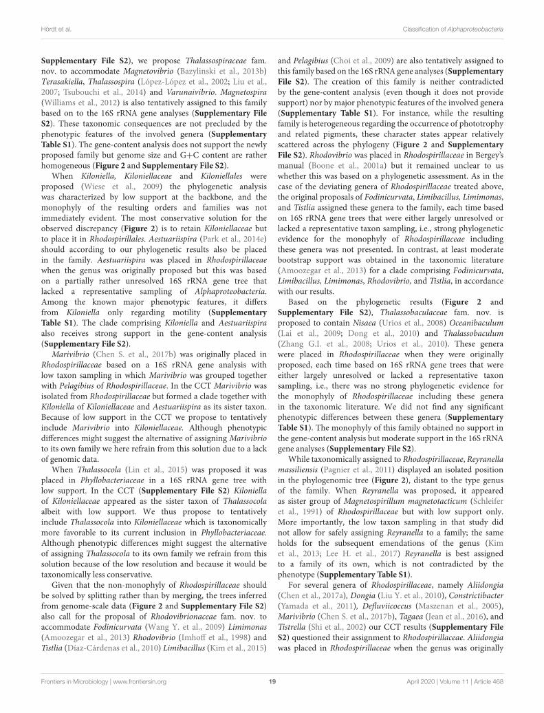
Supplementary File S2), we propose Thalassospiraceae fam.nov. to accommodate Magnetovibrio (Bazylinski et al., 2013b)Terasakiella, Thalassospira (López-López et al., 2002; Liu et al.,2007; Tsubouchi et al., 2014) and Varunaivibrio. Magnetospira(Williams et al., 2012) is also tentatively assigned to this familybased on to the 16S rRNA gene analyses (Supplementary FileS2). These taxonomic consequences are not precluded by thephenotypic features of the involved genera (SupplementaryTable S1). The gene-content analysis does not support the newlyproposed family but genome size and G+C content are ratherhomogeneous (Figure 2 and Supplementary File S2).
When Kiloniella, Kiloniellaceae and Kiloniellales wereproposed (Wiese et al., 2009) the phylogenetic analysiswas characterized by low support at the backbone, and themonophyly of the resulting orders and families was notimmediately evident. The most conservative solution for theobserved discrepancy (Figure 2) is to retain Kiloniellaceae butto place it in Rhodospirillales. Aestuariispira (Park et al., 2014e)should according to our phylogenetic results also be placedin the family. Aestuariispira was placed in Rhodospirillaceaewhen the genus was originally proposed but this was basedon a partially rather unresolved 16S rRNA gene tree thatlacked a representative sampling of Alphaproteobacteria.Among the known major phenotypic features, it differsfrom Kiloniella only regarding motility (SupplementaryTable S1). The clade comprising Kiloniella and Aestuariispiraalso receives strong support in the gene-content analysis(Supplementary File S2).
Marivibrio (Chen S. et al., 2017b) was originally placed inRhodospirillaceae based on a 16S rRNA gene analysis withlow taxon sampling in which Marivibrio was grouped togetherwith Pelagibius of Rhodospirillaceae. In the CCT Marivibrio wasisolated from Rhodospirillaceae but formed a clade together withKiloniella of Kiloniellaceae and Aestuariispira as its sister taxon.Because of low support in the CCT we propose to tentativelyinclude Marivibrio into Kiloniellaceae. Although phenotypicdifferences might suggest the alternative of assigning Marivibrioto its own family we here refrain from this solution due to a lackof genomic data.
When Thalassocola (Lin et al., 2015) was proposed it wasplaced in Phyllobacteriaceae in a 16S rRNA gene tree withlow support. In the CCT (Supplementary File S2) Kiloniellaof Kiloniellaceae appeared as the sister taxon of Thalassocolaalbeit with low support. We thus propose to tentativelyinclude Thalassocola into Kiloniellaceae which is taxonomicallymore favorable to its current inclusion in Phyllobacteriaceae.Although phenotypic differences might suggest the alternativeof assigning Thalassocola to its own family we refrain from thissolution because of the low resolution and because it would betaxonomically less conservative.
Given that the non-monophyly of Rhodospirillaceae shouldbe solved by splitting rather than by merging, the trees inferredfrom genome-scale data (Figure 2 and Supplementary File S2)also call for the proposal of Rhodovibrionaceae fam. nov. toaccommodate Fodinicurvata (Wang Y. et al., 2009) Limimonas(Amoozegar et al., 2013) Rhodovibrio (Imhoff et al., 1998) andTistlia (Díaz-Cárdenas et al., 2010) Limibacillus (Kim et al., 2015)
and Pelagibius (Choi et al., 2009) are also tentatively assigned tothis family based on the 16S rRNA gene analyses (SupplementaryFile S2). The creation of this family is neither contradictedby the gene-content analysis (even though it does not providesupport) nor by major phenotypic features of the involved genera(Supplementary Table S1). For instance, while the resultingfamily is heterogeneous regarding the occurrence of phototrophyand related pigments, these character states appear relativelyscattered across the phylogeny (Figure 2 and SupplementaryFile S2). Rhodovibrio was placed in Rhodospirillaceae in Bergey’smanual (Boone et al., 2001a) but it remained unclear to uswhether this was based on a phylogenetic assessment. As in thecase of the deviating genera of Rhodospirillaceae treated above,the original proposals of Fodinicurvata, Limibacillus, Limimonas,and Tistlia assigned these genera to the family, each time basedon 16S rRNA gene trees that were either largely unresolved orlacked a representative taxon sampling, i.e., strong phylogeneticevidence for the monophyly of Rhodospirillaceae includingthese genera was not presented. In contrast, at least moderatebootstrap support was obtained in the taxonomic literature(Amoozegar et al., 2013) for a clade comprising Fodinicurvata,Limibacillus, Limimonas, Rhodovibrio, and Tistlia, in accordancewith our results.
Based on the phylogenetic results (Figure 2 andSupplementary File S2), Thalassobaculaceae fam. nov. isproposed to contain Nisaea (Urios et al., 2008) Oceanibaculum(Lai et al., 2009; Dong et al., 2010) and Thalassobaculum(Zhang G.I. et al., 2008; Urios et al., 2010). These generawere placed in Rhodospirillaceae when they were originallyproposed, each time based on 16S rRNA gene trees that wereeither largely unresolved or lacked a representative taxonsampling, i.e., there was no strong phylogenetic evidence forthe monophyly of Rhodospirillaceae including these generain the taxonomic literature. We did not find any significantphenotypic differences between these genera (SupplementaryTable S1). The monophyly of this family obtained no support inthe gene-content analysis but moderate support in the 16S rRNAgene analyses (Supplementary File S2).
While taxonomically assigned to Rhodospirillaceae, Reyranellamassiliensis (Pagnier et al., 2011) displayed an isolated positionin the phylogenomic tree (Figure 2), distant to the type genusof the family. When Reyranella was proposed, it appearedas sister group of Magnetospirillum magnetotacticum (Schleiferet al., 1991) of Rhodospirillaceae but with low support only.More importantly, the low taxon sampling in that study didnot allow for safely assigning Reyranella to a family; the sameholds for the subsequent emendations of the genus (Kimet al., 2013; Lee H. et al., 2017) Reyranella is best assignedto a family of its own, which is not contradicted by thephenotype (Supplementary Table S1).
For several genera of Rhodospirillaceae, namely Aliidongia(Chen et al., 2017a), Dongia (Liu Y. et al., 2010), Constrictibacter(Yamada et al., 2011), Defluviicoccus (Maszenan et al., 2005),Marivibrio (Chen S. et al., 2017b), Tagaea (Jean et al., 2016), andTistrella (Shi et al., 2002) our CCT results (Supplementary FileS2) questioned their assignment to Rhodospirillaceae. Aliidongiawas placed in Rhodospirillaceae when the genus was originally
Frontiers in Microbiology | www.frontiersin.org 19 April 2020 | Volume 11 | Article 468
fmicb-11-00468 April 4, 2020 Time: 10:24 # 20
Hördt et al. Classification of Alphaproteobacteria
proposed but this was based on a 16S rRNA gene analysiswith low taxon sampling in which Aliidongia was groupedtogether with Inquilinus and Dongia of Rhodospirillaceae withlow support. When Dongia was originally proposed, it groupedtogether in a 16S rRNA gene tree with Rhodospirillaceaegenera including Azospirillum, Rhodocista and Skermanella, butwith overall low taxon sampling. When Tagaea was originallyproposed it was placed in a 16S rRNA gene tree in a wellsupported clade together with Oceanibaculum, Nisaea, andThalassobaculum of Rhodospirillaceae. Yet the placement of theclade itself showed low support and overall taxon sampling lackedthe type genus of Rhodospirillaceae. None of these genera couldsafely be placed in a family in the CCT (Supplementary File S2)hence we recommend to regard Aliidongia, Dongia, and Tagaeaas genera incertae sedis until their phylogenetic position can beclarified once more genome sequences become available.
Defluviicoccus (Maszenan et al., 2005) was placed inRhodospirillaceae (Garrity et al., 2007) after its originaldescription but based on the original 16S rRNA geneanalysis wherein Defluviicoccus was placed together withRhodospirillaceae genera such as Rhodospirillum, Azospirillum,and Magnetospirillum yet with low taxon sampling. Tistrellawas originally not placed in any family but was placed inRhodospirillaceae later on in Bergey’s Manual (Garrity et al.,2003b) which cited the original description of Tistrella eventhough it had cautioned against an assignment to a familybecause of low support in 16S rRNA gene analysis. Defluviicoccusand Tistrella were isolated from Rhodospirillaceae in thefar better sampled CCT and formed a clade together withGeminicoccus and Arboricoccus of Geminicoccaceae. Support forthis arrangement was also low, hence we suggest the tentativeinclusion of Defluviicoccus and Tistrella in Geminicoccaceae.Although phenotypic differences might suggest to alternativelyassign both genera to their own family we refrain from thissolution due to a lack of genomic data.
Rhodospirillales GeneraSome genera of Rhodospirillales also appeared in need of ataxonomic revision in the light of our analyses (Figure 2)although to a lesser extent than the families of the order.
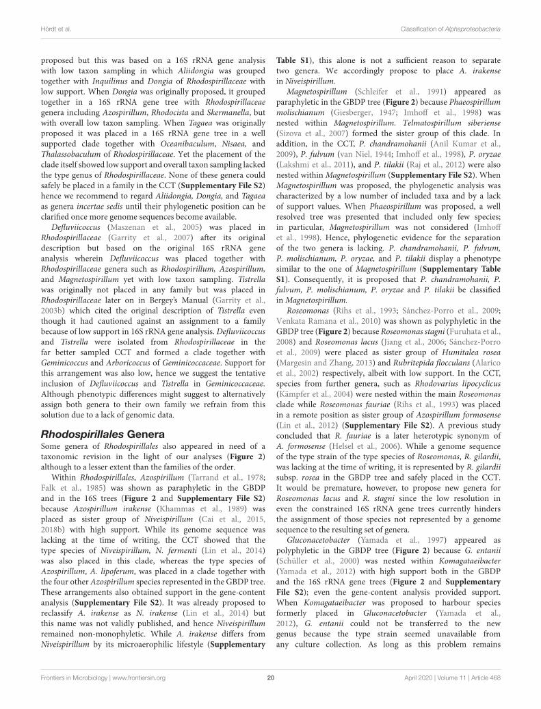
Within Rhodospirillales, Azospirillum (Tarrand et al., 1978;Falk et al., 1985) was shown as paraphyletic in the GBDPand in the 16S trees (Figure 2 and Supplementary File S2)because Azospirillum irakense (Khammas et al., 1989) wasplaced as sister group of Niveispirillum (Cai et al., 2015,2018b) with high support. While its genome sequence waslacking at the time of writing, the CCT showed that thetype species of Niveispirillum, N. fermenti (Lin et al., 2014)was also placed in this clade, whereas the type species ofAzospirillum, A. lipoferum, was placed in a clade together withthe four other Azospirillum species represented in the GBDP tree.These arrangements also obtained support in the gene-contentanalysis (Supplementary File S2). It was already proposed toreclassify A. irakense as N. irakense (Lin et al., 2014) butthis name was not validly published, and hence Niveispirillumremained non-monophyletic. While A. irakense differs fromNiveispirillum by its microaerophilic lifestyle (Supplementary
Table S1), this alone is not a sufficient reason to separatetwo genera. We accordingly propose to place A. irakensein Niveispirillum.
Magnetospirillum (Schleifer et al., 1991) appeared asparaphyletic in the GBDP tree (Figure 2) because Phaeospirillummolischianum (Giesberger, 1947; Imhoff et al., 1998) wasnested within Magnetospirillum. Telmatospirillum siberiense(Sizova et al., 2007) formed the sister group of this clade. Inaddition, in the CCT, P. chandramohanii (Anil Kumar et al.,2009), P. fulvum (van Niel, 1944; Imhoff et al., 1998), P. oryzae(Lakshmi et al., 2011), and P. tilakii (Raj et al., 2012) were alsonested within Magnetospirillum (Supplementary File S2). WhenMagnetospirillum was proposed, the phylogenetic analysis wascharacterized by a low number of included taxa and by a lackof support values. When Phaeospirillum was proposed, a wellresolved tree was presented that included only few species;in particular, Magnetospirillum was not considered (Imhoffet al., 1998). Hence, phylogenetic evidence for the separationof the two genera is lacking. P. chandramohanii, P. fulvum,P. molischianum, P. oryzae, and P. tilakii display a phenotypesimilar to the one of Magnetospirillum (Supplementary TableS1). Consequently, it is proposed that P. chandramohanii, P.fulvum, P. molischianum, P. oryzae and P. tilakii be classifiedin Magnetospirillum.
Roseomonas (Rihs et al., 1993; Sánchez-Porro et al., 2009;Venkata Ramana et al., 2010) was shown as polyphyletic in theGBDP tree (Figure 2) because Roseomonas stagni (Furuhata et al.,2008) and Roseomonas lacus (Jiang et al., 2006; Sánchez-Porroet al., 2009) were placed as sister group of Humitalea rosea(Margesin and Zhang, 2013) and Rubritepida flocculans (Alaricoet al., 2002) respectively, albeit with low support. In the CCT,species from further genera, such as Rhodovarius lipocyclicus(Kämpfer et al., 2004) were nested within the main Roseomonasclade while Roseomonas fauriae (Rihs et al., 1993) was placedin a remote position as sister group of Azospirillum formosense(Lin et al., 2012) (Supplementary File S2). A previous studyconcluded that R. fauriae is a later heterotypic synonym ofA. formosense (Helsel et al., 2006). While a genome sequenceof the type strain of the type species of Roseomonas, R. gilardii,was lacking at the time of writing, it is represented by R. gilardiisubsp. rosea in the GBDP tree and safely placed in the CCT.It would be premature, however, to propose new genera forRoseomonas lacus and R. stagni since the low resolution ineven the constrained 16S rRNA gene trees currently hindersthe assignment of those species not represented by a genomesequence to the resulting set of genera.
Gluconacetobacter (Yamada et al., 1997) appeared aspolyphyletic in the GBDP tree (Figure 2) because G. entanii(Schüller et al., 2000) was nested within Komagataeibacter(Yamada et al., 2012) with high support both in the GBDPand the 16S rRNA gene trees (Figure 2 and SupplementaryFile S2); even the gene-content analysis provided support.When Komagataeibacter was proposed to harbour speciesformerly placed in Gluconacetobacter (Yamada et al.,2012), G. entanii could not be transferred to the newgenus because the type strain seemed unavailable fromany culture collection. As long as this problem remains
Frontiers in Microbiology | www.frontiersin.org 20 April 2020 | Volume 11 | Article 468
fmicb-11-00468 April 4, 2020 Time: 10:24 # 21
Hördt et al. Classification of Alphaproteobacteria
unsolved a new combination for G. entanii cannot be proposed(Parker et al., 2019).
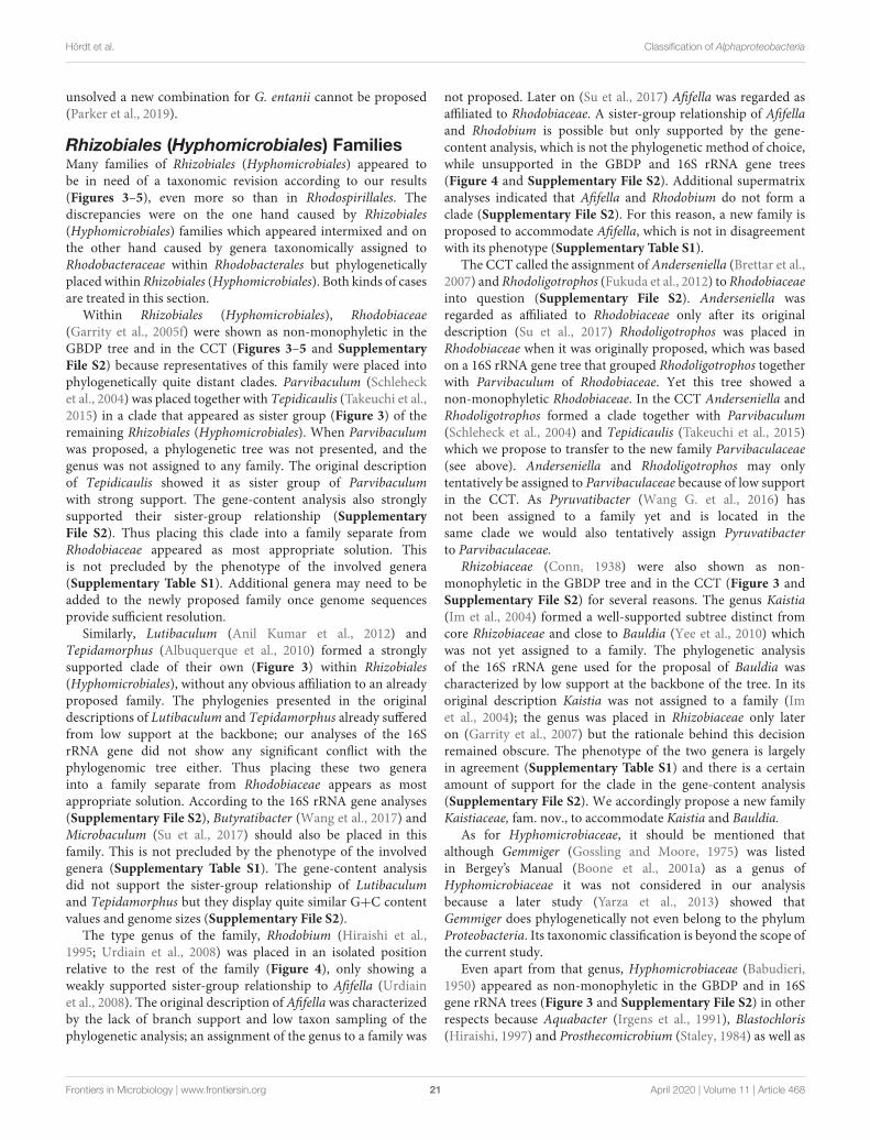
Rhizobiales (Hyphomicrobiales) FamiliesMany families of Rhizobiales (Hyphomicrobiales) appeared tobe in need of a taxonomic revision according to our results(Figures 3–5), even more so than in Rhodospirillales. Thediscrepancies were on the one hand caused by Rhizobiales(Hyphomicrobiales) families which appeared intermixed and onthe other hand caused by genera taxonomically assigned toRhodobacteraceae within Rhodobacterales but phylogeneticallyplaced within Rhizobiales (Hyphomicrobiales). Both kinds of casesare treated in this section.
Within Rhizobiales (Hyphomicrobiales), Rhodobiaceae(Garrity et al., 2005f) were shown as non-monophyletic in theGBDP tree and in the CCT (Figures 3–5 and SupplementaryFile S2) because representatives of this family were placed intophylogenetically quite distant clades. Parvibaculum (Schlehecket al., 2004) was placed together with Tepidicaulis (Takeuchi et al.,2015) in a clade that appeared as sister group (Figure 3) of theremaining Rhizobiales (Hyphomicrobiales). When Parvibaculumwas proposed, a phylogenetic tree was not presented, and thegenus was not assigned to any family. The original descriptionof Tepidicaulis showed it as sister group of Parvibaculumwith strong support. The gene-content analysis also stronglysupported their sister-group relationship (SupplementaryFile S2). Thus placing this clade into a family separate fromRhodobiaceae appeared as most appropriate solution. Thisis not precluded by the phenotype of the involved genera(Supplementary Table S1). Additional genera may need to beadded to the newly proposed family once genome sequencesprovide sufficient resolution.
Similarly, Lutibaculum (Anil Kumar et al., 2012) andTepidamorphus (Albuquerque et al., 2010) formed a stronglysupported clade of their own (Figure 3) within Rhizobiales(Hyphomicrobiales), without any obvious affiliation to an alreadyproposed family. The phylogenies presented in the originaldescriptions of Lutibaculum and Tepidamorphus already sufferedfrom low support at the backbone; our analyses of the 16SrRNA gene did not show any significant conflict with thephylogenomic tree either. Thus placing these two generainto a family separate from Rhodobiaceae appears as mostappropriate solution. According to the 16S rRNA gene analyses(Supplementary File S2), Butyratibacter (Wang et al., 2017) andMicrobaculum (Su et al., 2017) should also be placed in thisfamily. This is not precluded by the phenotype of the involvedgenera (Supplementary Table S1). The gene-content analysisdid not support the sister-group relationship of Lutibaculumand Tepidamorphus but they display quite similar G+C contentvalues and genome sizes (Supplementary File S2).
The type genus of the family, Rhodobium (Hiraishi et al.,1995; Urdiain et al., 2008) was placed in an isolated positionrelative to the rest of the family (Figure 4), only showing aweakly supported sister-group relationship to Afifella (Urdiainet al., 2008). The original description of Afifella was characterizedby the lack of branch support and low taxon sampling of thephylogenetic analysis; an assignment of the genus to a family was
not proposed. Later on (Su et al., 2017) Afifella was regarded asaffiliated to Rhodobiaceae. A sister-group relationship of Afifellaand Rhodobium is possible but only supported by the gene-content analysis, which is not the phylogenetic method of choice,while unsupported in the GBDP and 16S rRNA gene trees(Figure 4 and Supplementary File S2). Additional supermatrixanalyses indicated that Afifella and Rhodobium do not form aclade (Supplementary File S2). For this reason, a new family isproposed to accommodate Afifella, which is not in disagreementwith its phenotype (Supplementary Table S1).
The CCT called the assignment of Anderseniella (Brettar et al.,2007) and Rhodoligotrophos (Fukuda et al., 2012) to Rhodobiaceaeinto question (Supplementary File S2). Anderseniella wasregarded as affiliated to Rhodobiaceae only after its originaldescription (Su et al., 2017) Rhodoligotrophos was placed inRhodobiaceae when it was originally proposed, which was basedon a 16S rRNA gene tree that grouped Rhodoligotrophos togetherwith Parvibaculum of Rhodobiaceae. Yet this tree showed anon-monophyletic Rhodobiaceae. In the CCT Anderseniella andRhodoligotrophos formed a clade together with Parvibaculum(Schleheck et al., 2004) and Tepidicaulis (Takeuchi et al., 2015)which we propose to transfer to the new family Parvibaculaceae(see above). Anderseniella and Rhodoligotrophos may onlytentatively be assigned to Parvibaculaceae because of low supportin the CCT. As Pyruvatibacter (Wang G. et al., 2016) hasnot been assigned to a family yet and is located in thesame clade we would also tentatively assign Pyruvatibacterto Parvibaculaceae.
Rhizobiaceae (Conn, 1938) were also shown as non-monophyletic in the GBDP tree and in the CCT (Figure 3 andSupplementary File S2) for several reasons. The genus Kaistia(Im et al., 2004) formed a well-supported subtree distinct fromcore Rhizobiaceae and close to Bauldia (Yee et al., 2010) whichwas not yet assigned to a family. The phylogenetic analysisof the 16S rRNA gene used for the proposal of Bauldia wascharacterized by low support at the backbone of the tree. In itsoriginal description Kaistia was not assigned to a family (Imet al., 2004); the genus was placed in Rhizobiaceae only lateron (Garrity et al., 2007) but the rationale behind this decisionremained obscure. The phenotype of the two genera is largelyin agreement (Supplementary Table S1) and there is a certainamount of support for the clade in the gene-content analysis(Supplementary File S2). We accordingly propose a new familyKaistiaceae, fam. nov., to accommodate Kaistia and Bauldia.
As for Hyphomicrobiaceae, it should be mentioned thatalthough Gemmiger (Gossling and Moore, 1975) was listedin Bergey’s Manual (Boone et al., 2001a) as a genus ofHyphomicrobiaceae it was not considered in our analysisbecause a later study (Yarza et al., 2013) showed thatGemmiger does phylogenetically not even belong to the phylumProteobacteria. Its taxonomic classification is beyond the scope ofthe current study.
Even apart from that genus, Hyphomicrobiaceae (Babudieri,1950) appeared as non-monophyletic in the GBDP and in 16Sgene rRNA trees (Figure 3 and Supplementary File S2) in otherrespects because Aquabacter (Irgens et al., 1991), Blastochloris(Hiraishi, 1997) and Prosthecomicrobium (Staley, 1984) as well as
Frontiers in Microbiology | www.frontiersin.org 21 April 2020 | Volume 11 | Article 468
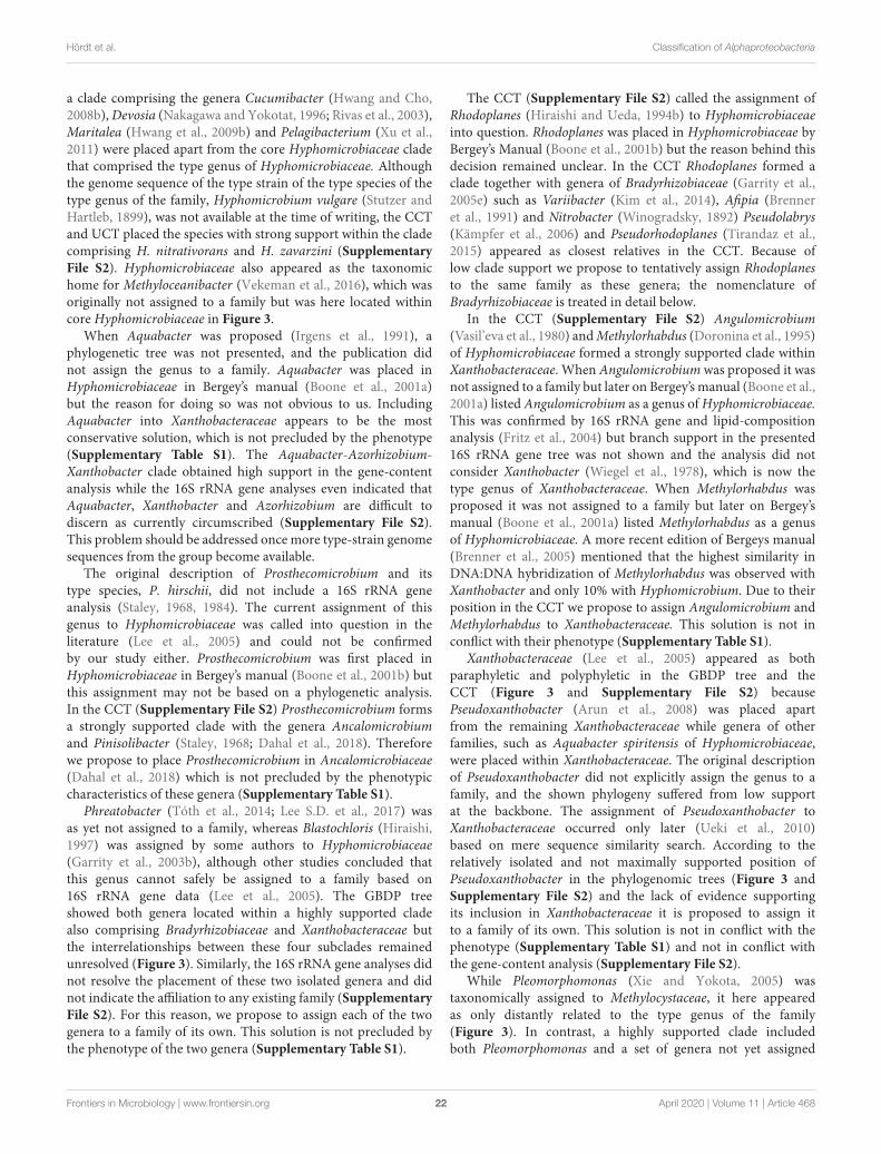
fmicb-11-00468 April 4, 2020 Time: 10:24 # 22
Hördt et al. Classification of Alphaproteobacteria
a clade comprising the genera Cucumibacter (Hwang and Cho,2008b), Devosia (Nakagawa and Yokotat, 1996; Rivas et al., 2003),Maritalea (Hwang et al., 2009b) and Pelagibacterium (Xu et al.,2011) were placed apart from the core Hyphomicrobiaceae cladethat comprised the type genus of Hyphomicrobiaceae. Althoughthe genome sequence of the type strain of the type species of thetype genus of the family, Hyphomicrobium vulgare (Stutzer andHartleb, 1899), was not available at the time of writing, the CCTand UCT placed the species with strong support within the cladecomprising H. nitrativorans and H. zavarzini (SupplementaryFile S2). Hyphomicrobiaceae also appeared as the taxonomichome for Methyloceanibacter (Vekeman et al., 2016), which wasoriginally not assigned to a family but was here located withincore Hyphomicrobiaceae in Figure 3.
When Aquabacter was proposed (Irgens et al., 1991), aphylogenetic tree was not presented, and the publication didnot assign the genus to a family. Aquabacter was placed inHyphomicrobiaceae in Bergey’s manual (Boone et al., 2001a)but the reason for doing so was not obvious to us. IncludingAquabacter into Xanthobacteraceae appears to be the mostconservative solution, which is not precluded by the phenotype(Supplementary Table S1). The Aquabacter-Azorhizobium-Xanthobacter clade obtained high support in the gene-contentanalysis while the 16S rRNA gene analyses even indicated thatAquabacter, Xanthobacter and Azorhizobium are difficult todiscern as currently circumscribed (Supplementary File S2).This problem should be addressed once more type-strain genomesequences from the group become available.
The original description of Prosthecomicrobium and itstype species, P. hirschii, did not include a 16S rRNA geneanalysis (Staley, 1968, 1984). The current assignment of thisgenus to Hyphomicrobiaceae was called into question in theliterature (Lee et al., 2005) and could not be confirmedby our study either. Prosthecomicrobium was first placed inHyphomicrobiaceae in Bergey’s manual (Boone et al., 2001b) butthis assignment may not be based on a phylogenetic analysis.In the CCT (Supplementary File S2) Prosthecomicrobium formsa strongly supported clade with the genera Ancalomicrobiumand Pinisolibacter (Staley, 1968; Dahal et al., 2018). Thereforewe propose to place Prosthecomicrobium in Ancalomicrobiaceae(Dahal et al., 2018) which is not precluded by the phenotypiccharacteristics of these genera (Supplementary Table S1).
Phreatobacter (Tóth et al., 2014; Lee S.D. et al., 2017) wasas yet not assigned to a family, whereas Blastochloris (Hiraishi,1997) was assigned by some authors to Hyphomicrobiaceae(Garrity et al., 2003b), although other studies concluded thatthis genus cannot safely be assigned to a family based on16S rRNA gene data (Lee et al., 2005). The GBDP treeshowed both genera located within a highly supported cladealso comprising Bradyrhizobiaceae and Xanthobacteraceae butthe interrelationships between these four subclades remainedunresolved (Figure 3). Similarly, the 16S rRNA gene analyses didnot resolve the placement of these two isolated genera and didnot indicate the affiliation to any existing family (SupplementaryFile S2). For this reason, we propose to assign each of the twogenera to a family of its own. This solution is not precluded bythe phenotype of the two genera (Supplementary Table S1).
The CCT (Supplementary File S2) called the assignment ofRhodoplanes (Hiraishi and Ueda, 1994b) to Hyphomicrobiaceaeinto question. Rhodoplanes was placed in Hyphomicrobiaceae byBergey’s Manual (Boone et al., 2001b) but the reason behind thisdecision remained unclear. In the CCT Rhodoplanes formed aclade together with genera of Bradyrhizobiaceae (Garrity et al.,2005e) such as Variibacter (Kim et al., 2014), Afipia (Brenneret al., 1991) and Nitrobacter (Winogradsky, 1892) Pseudolabrys(Kämpfer et al., 2006) and Pseudorhodoplanes (Tirandaz et al.,2015) appeared as closest relatives in the CCT. Because oflow clade support we propose to tentatively assign Rhodoplanesto the same family as these genera; the nomenclature ofBradyrhizobiaceae is treated in detail below.
In the CCT (Supplementary File S2) Angulomicrobium(Vasil’eva et al., 1980) and Methylorhabdus (Doronina et al., 1995)of Hyphomicrobiaceae formed a strongly supported clade withinXanthobacteraceae. When Angulomicrobium was proposed it wasnot assigned to a family but later on Bergey’s manual (Boone et al.,2001a) listed Angulomicrobium as a genus of Hyphomicrobiaceae.This was confirmed by 16S rRNA gene and lipid-compositionanalysis (Fritz et al., 2004) but branch support in the presented16S rRNA gene tree was not shown and the analysis did notconsider Xanthobacter (Wiegel et al., 1978), which is now thetype genus of Xanthobacteraceae. When Methylorhabdus wasproposed it was not assigned to a family but later on Bergey’smanual (Boone et al., 2001a) listed Methylorhabdus as a genusof Hyphomicrobiaceae. A more recent edition of Bergeys manual(Brenner et al., 2005) mentioned that the highest similarity inDNA:DNA hybridization of Methylorhabdus was observed withXanthobacter and only 10% with Hyphomicrobium. Due to theirposition in the CCT we propose to assign Angulomicrobium andMethylorhabdus to Xanthobacteraceae. This solution is not inconflict with their phenotype (Supplementary Table S1).
Xanthobacteraceae (Lee et al., 2005) appeared as bothparaphyletic and polyphyletic in the GBDP tree and theCCT (Figure 3 and Supplementary File S2) becausePseudoxanthobacter (Arun et al., 2008) was placed apartfrom the remaining Xanthobacteraceae while genera of otherfamilies, such as Aquabacter spiritensis of Hyphomicrobiaceae,were placed within Xanthobacteraceae. The original descriptionof Pseudoxanthobacter did not explicitly assign the genus to afamily, and the shown phylogeny suffered from low supportat the backbone. The assignment of Pseudoxanthobacter toXanthobacteraceae occurred only later (Ueki et al., 2010)based on mere sequence similarity search. According to therelatively isolated and not maximally supported position ofPseudoxanthobacter in the phylogenomic trees (Figure 3 andSupplementary File S2) and the lack of evidence supportingits inclusion in Xanthobacteraceae it is proposed to assign itto a family of its own. This solution is not in conflict with thephenotype (Supplementary Table S1) and not in conflict withthe gene-content analysis (Supplementary File S2).
While Pleomorphomonas (Xie and Yokota, 2005) wastaxonomically assigned to Methylocystaceae, it here appearedas only distantly related to the type genus of the family(Figure 3). In contrast, a highly supported clade includedboth Pleomorphomonas and a set of genera not yet assigned
Frontiers in Microbiology | www.frontiersin.org 22 April 2020 | Volume 11 | Article 468
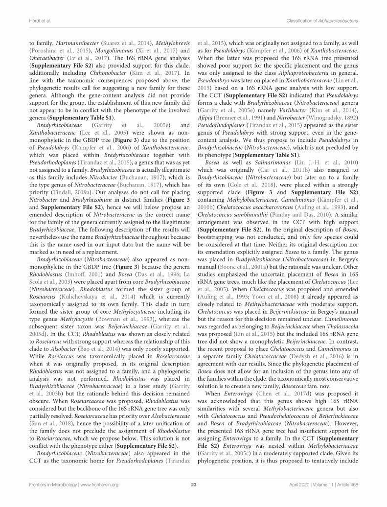
fmicb-11-00468 April 4, 2020 Time: 10:24 # 23
Hördt et al. Classification of Alphaproteobacteria
to family, Hartmannibacter (Suarez et al., 2014), Methylobrevis(Poroshina et al., 2015), Mongoliimonas (Xi et al., 2017) andOharaeibacter (Lv et al., 2017). The 16S rRNA gene analyses(Supplementary File S2) also provided support for this clade,additionally including Chthonobacter (Kim et al., 2017). Inline with the taxonomic consequences proposed above, thephylogenetic results call for suggesting a new family for thesegenera. Although the gene-content analysis did not providesupport for the group, the establishment of this new family didnot appear to be in conflict with the phenotype of the involvedgenera (Supplementary Table S1).
Bradyrhizobiaceae (Garrity et al., 2005e) andXanthobacteraceae (Lee et al., 2005) were shown as non-monophyletic in the GBDP tree (Figure 3) due to the positionof Pseudolabrys (Kämpfer et al., 2006) of Xanthobacteraceae,which was placed within Bradyrhizobiaceae together withPseudorhodoplanes (Tirandaz et al., 2015), a genus that was as yetnot assigned to a family. Bradyrhizobiaceae is actually illegitimateas this family includes Nitrobacter (Buchanan, 1917), which isthe type genus of Nitrobacteraceae (Buchanan, 1917), which haspriority (Tindall, 2019a). Our analyses do not call for placingNitrobacter and Bradyrhizobium in distinct families (Figure 3and Supplementary File S2), hence we will below propose anemended description of Nitrobacteraceae as the correct namefor the family of the genera currently assigned to the illegitimateBradyrhizobiaceae. The following description of the results willnevertheless use the name Bradyrhizobiaceae throughout becausethis is the name used in our input data but the name will bemarked as in need of a replacement.
Bradyrhizobiaceae (Nitrobacteraceae) also appeared as non-monophyletic in the GBDP tree (Figure 3) because the generaRhodoblastus (Imhoff, 2001) and Bosea (Das et al., 1996; LaScola et al., 2003) were placed apart from core Bradyrhizobiaceae(Nitrobacteraceae). Rhodoblastus formed the sister group ofRoseiarcus (Kulichevskaya et al., 2014) which is currentlytaxonomically assigned to its own family. This clade in turnformed the sister group of core Methylocystaceae including itstype genus Methylocystis (Bowman et al., 1993), whereas thesubsequent sister taxon was Beijerinckiaceae (Garrity et al.,2005d). In the CCT, Rhodoblastus was shown as closely relatedto Roseiarcus with strong support whereas the relationship of thisclade to Alsobacter (Bao et al., 2014) was only poorly supported.While Roseiarcus was taxonomically placed in Roseiarcaceaewhen it was originally proposed, in its original descriptionRhodoblastus was not assigned to a family, and a phylogeneticanalysis was not performed. Rhodoblastus was placed inBradyrhizobiaceae (Nitrobacteraceae) in a later study (Garrityet al., 2003b) but the rationale behind this decision remainedobscure. When Roseiarcaceae was proposed, Rhodoblastus wasconsidered but the backbone of the 16S rRNA gene tree was onlypartially resolved. Roseiarcaceae has priority over Alsobacteraceae(Sun et al., 2018), hence the possibility of a later unification ofthe family does not preclude the assignment of Rhodoblastusto Roseiarcaceae, which we propose below. This solution is notconflict with the phenotype either (Supplementary File S2).
Bradyrhizobiaceae (Nitrobacteraceae) also appeared in theCCT as the taxonomic home for Pseudorhodoplanes (Tirandaz
et al., 2015), which was originally not assigned to a family, as wellas for Pseudolabrys (Kämpfer et al., 2006) of Xanthobacteraceae.When the latter was proposed the 16S rRNA tree presentedshowed poor support for the specific placement and the genuswas only assigned to the class Alphaproteobacteria in general.Pseudolabrys was later on placed in Xanthobacteraceae (Lin et al.,2015) based on a 16S rRNA gene analysis with low support.The CCT (Supplementary File S2) indicated that Pseudolabrysforms a clade with Bradyrhizobiaceae (Nitrobacteraceae) genera(Garrity et al., 2005e) namely Variibacter (Kim et al., 2014),Afipia (Brenner et al., 1991) and Nitrobacter (Winogradsky, 1892)Pseudorhodoplanes (Tirandaz et al., 2015) appeared as the sistergenus of Pseudolabrys with strong support, even in the gene-content analysis. We thus propose to include Pseudolabrys inBradyrhizobiaceae (Nitrobacteraceae), which is not precluded byits phenotype (Supplementary Table S1).
Bosea as well as Salinarimonas (Liu J.-H. et al., 2010)which was originally (Cai et al., 2011b) also assigned toBradyrhizobiaceae (Nitrobacteraceae) but later on to a familyof its own (Cole et al., 2018), were placed within a stronglysupported clade (Figure 3 and Supplementary File S2)containing Methylobacteriaceae, Camelimonas (Kämpfer et al.,2010b) Chelatococcus asaccharovorans (Auling et al., 1993), andChelatococcus sambhunathii (Panday and Das, 2010). A similararrangement was observed in the CCT with high support(Supplementary File S2). In the original description of Bosea,bootstrapping was not conducted, and only few species couldbe considered at that time. Neither its original description norits emendation explicitly assigned Bosea to a family. The genuswas placed in Bradyrhizobiaceae (Nitrobacteraceae) in Bergey’smanual (Boone et al., 2001a) but the rationale was unclear. Otherstudies emphasized the uncertain placement of Bosea in 16SrRNA gene trees, much like the placement of Chelatococcus (Leeet al., 2005). When Chelatococcus was proposed and emended(Auling et al., 1993; Yoon et al., 2008) it already appeared asclosely related to Methylobacteriaceae with moderate support.Chelatococcus was placed in Beijerinckiaceae in Bergey’s manualbut the reason for this decision remained unclear. Camelimonaswas regarded as belonging to Beijerinckiaceae when Thalassocolawas proposed (Lin et al., 2015) but the included 16S rRNA genetree did not show a monophyletic Beijerinckiaceae. In contrast,the recent proposal to place Chelatococcus and Camelimonas ina separate family Chelatococcaceae (Dedysh et al., 2016) is inagreement with our results. Since the phylogenetic placement ofBosea does not allow for an inclusion of the genus into any ofthe families within the clade, the taxonomically most conservativesolution is to create a new family, Boseaceae fam. nov.
When Enterovirga (Chen et al., 2017d) was proposed itwas acknowledged that this genus shows high 16S rRNAsimilarities with several Methylobacteriaceae genera but alsowith Chelatococcus and Pseudochelatococcus of Beijerinckiaceaeand Bosea of Bradyrhizobiaceae (Nitrobacteraceae). However,the presented 16S rRNA gene tree had insufficient support forassigning Enterovirga to a family. In the CCT (SupplementaryFile S2) Enterovirga was nested within Methylobacteriaceae(Garrity et al., 2005c) in a moderately supported clade. Given itsphylogenetic position, it is thus proposed to tentatively include
Frontiers in Microbiology | www.frontiersin.org 23 April 2020 | Volume 11 | Article 468
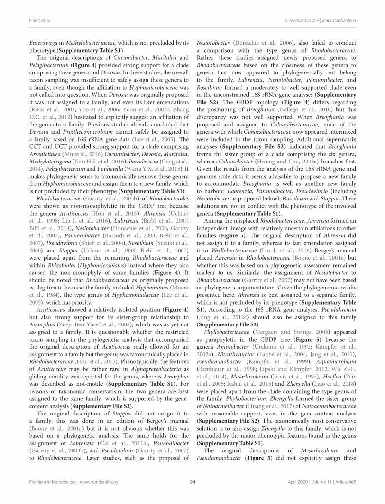
fmicb-11-00468 April 4, 2020 Time: 10:24 # 24
Hördt et al. Classification of Alphaproteobacteria
Enterovirga in Methylobacteriaceae, which is not precluded by itsphenotype (Supplementary Table S1).
The original descriptions of Cucumibacter, Maritalea andPelagibacterium (Figure 4) provided strong support for a cladecomprising these genera and Devosia. In these studies, the overalltaxon sampling was insufficient to safely assign these genera toa family, even though the affiliation to Hyphomicrobiaceae wasnot called into question. When Devosia was originally proposedit was not assigned to a family, and even its later emendations(Rivas et al., 2003; Yoo et al., 2006; Yoon et al., 2007c; ZhangD.C. et al., 2012) hesitated to explicitly suggest an affiliation ofthe genus to a family. Previous studies already concluded thatDevosia and Prosthecomicrobium cannot safely be assigned toa family based on 16S rRNA gene data (Lee et al., 2005). TheCCT and UCT provided strong support for a clade comprisingArsenicitalea (Mu et al., 2016) Cucumibacter, Devosia, Maritalea,Methyloterrigena (Kim H.S. et al., 2016), Paradevosia (Geng et al.,2014), Pelagibacterium and Youhaiella (Wang Y.X. et al., 2015). Itmakes phylogenetic sense to taxonomically remove these generafrom Hyphomicrobiaceae and assign them to a new family, whichis not precluded by their phenotype (Supplementary Table S1).
Rhodobacteraceae (Garrity et al., 2005b) of Rhodobacteraleswere shown as non-monophyletic in the GBDP tree becausethe genera Acuticoccus (Hou et al., 2015), Ahrensia (Uchinoet al., 1998; Liu J. et al., 2016), Labrenzia (Biebl et al., 2007;Bibi et al., 2014), Nesiotobacter (Donachie et al., 2006; Garrityet al., 2007), Pannonibacter (Borsodi et al., 2003; Biebl et al.,2007), Pseudovibrio (Shieh et al., 2004), Roseibium (Suzuki et al.,2000) and Stappia (Uchino et al., 1998; Biebl et al., 2007)were placed apart from the remaining Rhodobacteraceae andwithin Rhizobiales (Hyphomicrobiales) instead where they alsocaused the non-monophyly of some families (Figure 4). Itshould be noted that Rhodobacteraceae as originally proposedis illegitimate because the family included Hyphomonas (Mooreet al., 1984), the type genus of Hyphomonadaceae (Lee et al.,2005), which has priority.
Acuticoccus showed a relatively isolated position (Figure 4)but also strong support for its sister-group relationship toAmorphus (Zeevi Ben Yosef et al., 2008), which was as yet notassigned to a family. It is questionable whether the restrictedtaxon sampling in the phylogenetic analysis that accompaniedthe original description of Acuticoccus really allowed for anassignment to a family but the genus was taxonomically placed inRhodobacteraceae (Hou et al., 2015). Phenotypically, the featuresof Acuticoccus may be rather rare in Alphaproteobacteria asgliding motility was reported for the genus, whereas Amorphuswas described as not-motile (Supplementary Table S1). Forreasons of taxonomic conservatism, the two genera are bestassigned to the same family, which is supported by the gene-content analysis (Supplementary File S2).
The original description of Stappia did not assign it toa family; this was done in an edition of Bergey’s manual(Boone et al., 2001a) but it is not obvious whether this wasbased on a phylogenetic analysis. The same holds for theassignment of Labrenzia (Cai et al., 2011a), Pannonibacter(Garrity et al., 2003b), and Pseudovibrio (Garrity et al., 2007)to Rhodobacteraceae. Later studies, such as the proposal of
Nesiotobacter (Donachie et al., 2006), also failed to conducta comparison with the type genus of Rhodobacteraceae.Rather, these studies assigned newly proposed genera toRhodobacteraceae based on the closeness of these genera togenera that now appeared to phylogenetically not belongto the family. Labrenzia, Nesiotobacter, Pannonibacter, andRoseibium formed a moderately to well supported clade evenin the unconstrained 16S rRNA gene analyses (SupplementaryFile S2). The GBDP topology (Figure 4) differs regardingthe positioning of Breoghania (Gallego et al., 2010) but thisdiscrepancy was not well supported. When Breoghania wasproposed and assigned to Cohaesibacteraceae, none of thegenera with which Cohaesibacteraceae now appeared intermixedwere included in the taxon sampling. Additional supermatrixanalyses (Supplementary File S2) indicated that Breoghaniaforms the sister group of a clade comprising the six genera,whereas Cohaesibacter (Hwang and Cho, 2008a) branches first.Given the results from the analysis of the 16S rRNA gene andgenome-scale data it seems advisable to propose a new familyto accommodate Breoghania as well as another new familyto harbour Labrenzia, Pannonibacter, Pseudovibrio (includingNesiotobacter as proposed below), Roseibium and Stappia. Thesesolutions are not in conflict with the phenotype of the involvedgenera (Supplementary Table S1).
Among the misplaced Rhodobacteraceae, Ahrensia formed anindependent lineage with relatively uncertain affiliations to otherfamilies (Figure 5). The original description of Ahrensia didnot assign it to a family, whereas its last emendation assignedit to Phyllobacteriaceae (Liu J. et al., 2016) Bergey’s manualplaced Ahrensia in Rhodobacteraceae (Boone et al., 2001a) butwhether this was based on a phylogenetic assessment remainedunclear to us. Similarly, the assignment of Nesiotobacter toRhodobacteraceae (Garrity et al., 2007) may not have been basedon phylogenetic argumentation. Given the phylogenomic resultspresented here, Ahrensia is best assigned to a separate family,which is not precluded by its phenotype (Supplementary TableS1). According to the 16S rRNA gene analyses, Pseudahrensia(Jung et al., 2012c) should also be assigned to this family(Supplementary File S2).
Phyllobacteriaceae (Mergaert and Swings, 2005) appearedas paraphyletic in the GBDP tree (Figure 5) because thegenera Aminobacter (Urakami et al., 1992; Kämpfer et al.,2002a), Nitratireductor (Labbé et al., 2004; Jang et al., 2011),Pseudaminobacter (Kämpfer et al., 1999), Aquamicrobium(Bambauer et al., 1998; Lipski and Kämpfer, 2012; Wu Z.-G.et al., 2014), Mesorhizobium (Jarvis et al., 1997), Hoeflea (Peixet al., 2005; Rahul et al., 2015) and Zhengella (Liao et al., 2018)were placed apart from the clade containing the type genus ofthe family, Phyllobacterium. Zhengella formed the sister groupof Notoacmeibacter (Huang et al., 2017) of Notoacmeibacteraceaewith reasonable support, even in the gene-content analysis(Supplementary File S2). The taxonomically most conservativesolution is to also assign Zhengella to this family, which is notprecluded by the major phenotypic features found in the genus(Supplementary Table S1).
The original descriptions of Mesorhizobium andPseudaminobacter (Figure 5) did not explicitly assign these
Frontiers in Microbiology | www.frontiersin.org 24 April 2020 | Volume 11 | Article 468
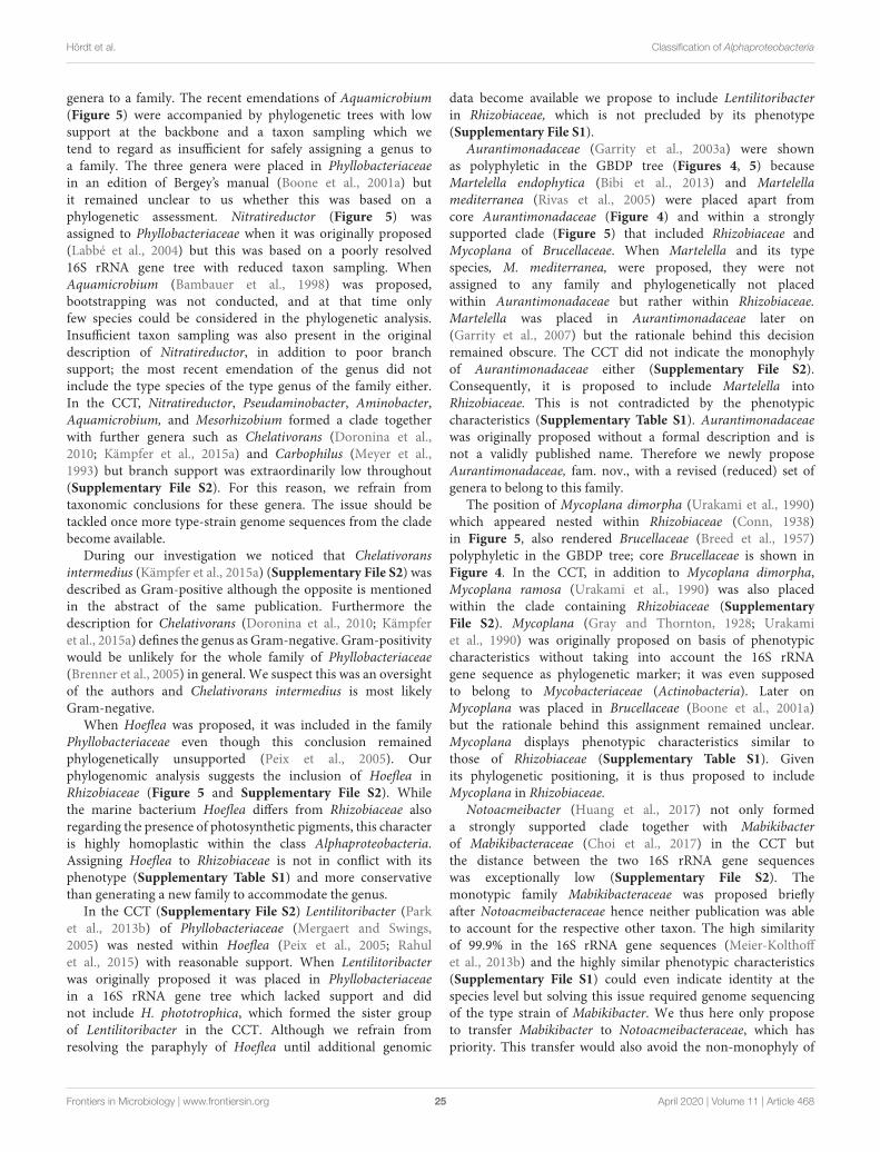
fmicb-11-00468 April 4, 2020 Time: 10:24 # 25
Hördt et al. Classification of Alphaproteobacteria
genera to a family. The recent emendations of Aquamicrobium(Figure 5) were accompanied by phylogenetic trees with lowsupport at the backbone and a taxon sampling which wetend to regard as insufficient for safely assigning a genus toa family. The three genera were placed in Phyllobacteriaceaein an edition of Bergey’s manual (Boone et al., 2001a) butit remained unclear to us whether this was based on aphylogenetic assessment. Nitratireductor (Figure 5) wasassigned to Phyllobacteriaceae when it was originally proposed(Labbé et al., 2004) but this was based on a poorly resolved16S rRNA gene tree with reduced taxon sampling. WhenAquamicrobium (Bambauer et al., 1998) was proposed,bootstrapping was not conducted, and at that time onlyfew species could be considered in the phylogenetic analysis.Insufficient taxon sampling was also present in the originaldescription of Nitratireductor, in addition to poor branchsupport; the most recent emendation of the genus did notinclude the type species of the type genus of the family either.In the CCT, Nitratireductor, Pseudaminobacter, Aminobacter,Aquamicrobium, and Mesorhizobium formed a clade togetherwith further genera such as Chelativorans (Doronina et al.,2010; Kämpfer et al., 2015a) and Carbophilus (Meyer et al.,1993) but branch support was extraordinarily low throughout(Supplementary File S2). For this reason, we refrain fromtaxonomic conclusions for these genera. The issue should betackled once more type-strain genome sequences from the cladebecome available.
During our investigation we noticed that Chelativoransintermedius (Kämpfer et al., 2015a) (Supplementary File S2) wasdescribed as Gram-positive although the opposite is mentionedin the abstract of the same publication. Furthermore thedescription for Chelativorans (Doronina et al., 2010; Kämpferet al., 2015a) defines the genus as Gram-negative. Gram-positivitywould be unlikely for the whole family of Phyllobacteriaceae(Brenner et al., 2005) in general. We suspect this was an oversightof the authors and Chelativorans intermedius is most likelyGram-negative.
When Hoeflea was proposed, it was included in the familyPhyllobacteriaceae even though this conclusion remainedphylogenetically unsupported (Peix et al., 2005). Ourphylogenomic analysis suggests the inclusion of Hoeflea inRhizobiaceae (Figure 5 and Supplementary File S2). Whilethe marine bacterium Hoeflea differs from Rhizobiaceae alsoregarding the presence of photosynthetic pigments, this characteris highly homoplastic within the class Alphaproteobacteria.Assigning Hoeflea to Rhizobiaceae is not in conflict with itsphenotype (Supplementary Table S1) and more conservativethan generating a new family to accommodate the genus.
In the CCT (Supplementary File S2) Lentilitoribacter (Parket al., 2013b) of Phyllobacteriaceae (Mergaert and Swings,2005) was nested within Hoeflea (Peix et al., 2005; Rahulet al., 2015) with reasonable support. When Lentilitoribacterwas originally proposed it was placed in Phyllobacteriaceaein a 16S rRNA gene tree which lacked support and didnot include H. phototrophica, which formed the sister groupof Lentilitoribacter in the CCT. Although we refrain fromresolving the paraphyly of Hoeflea until additional genomic
data become available we propose to include Lentilitoribacterin Rhizobiaceae, which is not precluded by its phenotype(Supplementary File S1).
Aurantimonadaceae (Garrity et al., 2003a) were shownas polyphyletic in the GBDP tree (Figures 4, 5) becauseMartelella endophytica (Bibi et al., 2013) and Martelellamediterranea (Rivas et al., 2005) were placed apart fromcore Aurantimonadaceae (Figure 4) and within a stronglysupported clade (Figure 5) that included Rhizobiaceae andMycoplana of Brucellaceae. When Martelella and its typespecies, M. mediterranea, were proposed, they were notassigned to any family and phylogenetically not placedwithin Aurantimonadaceae but rather within Rhizobiaceae.Martelella was placed in Aurantimonadaceae later on(Garrity et al., 2007) but the rationale behind this decisionremained obscure. The CCT did not indicate the monophylyof Aurantimonadaceae either (Supplementary File S2).Consequently, it is proposed to include Martelella intoRhizobiaceae. This is not contradicted by the phenotypiccharacteristics (Supplementary Table S1). Aurantimonadaceaewas originally proposed without a formal description and isnot a validly published name. Therefore we newly proposeAurantimonadaceae, fam. nov., with a revised (reduced) set ofgenera to belong to this family.
The position of Mycoplana dimorpha (Urakami et al., 1990)which appeared nested within Rhizobiaceae (Conn, 1938)in Figure 5, also rendered Brucellaceae (Breed et al., 1957)polyphyletic in the GBDP tree; core Brucellaceae is shown inFigure 4. In the CCT, in addition to Mycoplana dimorpha,Mycoplana ramosa (Urakami et al., 1990) was also placedwithin the clade containing Rhizobiaceae (SupplementaryFile S2). Mycoplana (Gray and Thornton, 1928; Urakamiet al., 1990) was originally proposed on basis of phenotypiccharacteristics without taking into account the 16S rRNAgene sequence as phylogenetic marker; it was even supposedto belong to Mycobacteriaceae (Actinobacteria). Later onMycoplana was placed in Brucellaceae (Boone et al., 2001a)but the rationale behind this assignment remained unclear.Mycoplana displays phenotypic characteristics similar tothose of Rhizobiaceae (Supplementary Table S1). Givenits phylogenetic positioning, it is thus proposed to includeMycoplana in Rhizobiaceae.
Notoacmeibacter (Huang et al., 2017) not only formeda strongly supported clade together with Mabikibacterof Mabikibacteraceae (Choi et al., 2017) in the CCT butthe distance between the two 16S rRNA gene sequenceswas exceptionally low (Supplementary File S2). Themonotypic family Mabikibacteraceae was proposed brieflyafter Notoacmeibacteraceae hence neither publication was ableto account for the respective other taxon. The high similarityof 99.9% in the 16S rRNA gene sequences (Meier-Kolthoffet al., 2013b) and the highly similar phenotypic characteristics(Supplementary File S1) could even indicate identity at thespecies level but solving this issue required genome sequencingof the type strain of Mabikibacter. We thus here only proposeto transfer Mabikibacter to Notoacmeibacteraceae, which haspriority. This transfer would also avoid the non-monophyly of
Frontiers in Microbiology | www.frontiersin.org 25 April 2020 | Volume 11 | Article 468
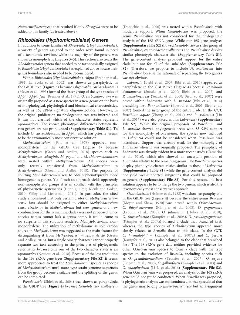
fmicb-11-00468 April 4, 2020 Time: 10:24 # 26
Hördt et al. Classification of Alphaproteobacteria
Notoacmeibacteraceae that resulted if only Zhengella were to beadded to this family (as treated above).
Rhizobiales (Hyphomicrobiales) GeneraIn addition to some families of Rhizobiales (Hyphomicrobiales),a variety of genera assigned to the order were found in needof a taxonomic revision, while the majority of the genera wasshown as monophyletic (Figures 3–5). This section also treats theRhodobacterales genera that needed to be taxonomically assignedto Rhizobiales (Hyphomicrobiales) as explained above in case theirgenus boundaries also needed to be reconsidered.
Within Rhizobiales (Hyphomicrobiales), Afipia (Brenner et al.,1991; La Scola et al., 2002) was shown as paraphyletic inthe GBDP tree (Figure 3) because Oligotropha carboxidovorans(Meyer et al., 1993) formed the sister group of the type species ofAfipia, Afipia felis (Brenner et al., 1991). O. carboxidovorans wasoriginally proposed as a new species in a new genus on the basisof morphological, physiological and biochemical characteristics,as well as 16S rRNA signature oligonucleotides. However, inthe original publication no phylogenetic tree was inferred andit was not clarified which of the character states representapomorphies. The known phenotypic differences between thetwo genera are not pronounced (Supplementary Table S1). Toinclude O. carboxidovorans in Afipia, which has priority, seemsto be the taxonomically most conservative solution.
Methylobacterium (Patt et al., 1976) appeared non-monophyletic in the GBDP tree (Figure 3) becauseMethylorubrum (Green and Ardley, 2018) species such asMethylorubrum salsuginis, M. populi and M. chloromethanicumwere nested within Methylobacterium. All species wereonly recently transferred from Methylobacterium toMethylorubrum (Green and Ardley, 2018). The purpose ofsplitting Methylobacterium was to obtain phenotypically morehomogeneous genera. Yet if such an approach yields apparentlynon-monophyletic groups it is in conflict with the principlesof phylogenetic systematics (Hennig, 1965; Klenk and Göker,2010; Wiley and Lieberman, 2011). In particular, the citedstudy emphasized that only certain clades of Methylobacteriumsensu lato should be assigned to either Methylobacteriumsensu stricto or to Methylorubrum but new genera and newcombinations for the remaining clades were not proposed. Sincespecies names cannot lack a genus name, it would come asno surprise if this solution rendered Methylobacterium non-monophyletic. The utilization of methylamine as sole carbonsource in Methylorubrum was suggested as the main feature fordistinguishing it from Methylobacterium sensu stricto (Greenand Ardley, 2018). But a single binary character cannot properlyseparate two taxa according to the principles of phylogeneticsystematics because only one of the two character states is anapomorphy (Nouioui et al., 2018). Because of the low resolutionin the 16S rRNA gene trees (Supplementary File S2) it seemsmore appropriate to treat the Methylorubrum species as speciesof Methylobacterium until more type-strain genome sequencesfrom the group become available and the splitting of the genuscan be completed.
Pseudovibrio (Shieh et al., 2004) was shown as paraphyleticin the GBDP tree (Figure 4) because Nesiotobacter exalbescens
(Donachie et al., 2006) was nested within Pseudovibrio withmoderate support. When Nesiotobacter was proposed, thegenus Pseudovibrio was not considered for the phylogeneticanalysis of the 16S rRNA gene. While our 16S gene analyses(Supplementary File S2) showed Nesiotobacter as sister group ofPseudovibrio, Nesiotobacter exalbescens and Pseudovibrio displaysimilar phenotypic characteristics (Supplementary Table S1).The gene-content analysis provided support for the entireclade but not for all of the subclades (Supplementary FileS2). Therefore, we propose to include N. exalbescens intoPseudovibrio because the rationale of separating the two generawas not obvious.
Labrenzia (Biebl et al., 2007; Bibi et al., 2014) appeared asparaphyletic in the GBDP tree (Figure 4) because Roseibiumdenhamense (Suzuki et al., 2000; Biebl et al., 2007) andR. hamelinenseas (Suzuki et al., 2000; Biebl et al., 2007) werenested within Labrenzia, with L. suaedae (Bibi et al., 2014)branching first. Pannonibacter (Borsodi et al., 2003; Biebl et al.,2007) formed the sister group of the entire clade. In the CCT,Roseibium aquae (Zhong et al., 2014) and R. sediminis (Liuet al., 2017) were also placed within Labrenzia (SupplementaryFile S2). While the original proposals of Roseibium andL. suaedae showed phylogenetic trees with 83–93% supportfor the monophyly of Roseibium, the species now includedin Labrenzia could not be considered when Roseibium wasintroduced. Support was already weak for the monophyly ofLabrenzia when it was originally proposed. The paraphyly ofLabrenzia was already evident in a more recent study (Camachoet al., 2016), which also showed an uncertain position ofL. suaedae relative to the remaining genus. The Roseibium speciesdisplay phenotypic characteristics similar to those of Labrenzia(Supplementary Table S1) while the gene-content analysis didnot yield well-supported subgroups that could be proposedas genera (Supplementary File S2). For this reason, the bestsolution appears to be to merge the two genera, which is also thetaxonomically most conservative approach.
Ochrobactrum (Holmes et al., 1988) was shown as paraphyleticin the GBDP tree (Figure 4) because the entire genus Brucella(Meyer and Shaw, 1920) was nested within Ochrobactrum.O. thiophenivorans (Kämpfer et al., 2008), O. grignonense(Lebuhn et al., 2000), O. pituitosum (Huber et al., 2010),O. rhizosphaerae (Kämpfer et al., 2008), O. pseudogrignonense(Kämpfer et al., 2007a) formed a clade that branched first,whereas the type species of Ochrobactrum appeared moreclosely related to Brucella than to this clade. In the CCT,O. haematophilum (Kämpfer et al., 2007a) and O. pecoris(Kämpfer et al., 2011) also belonged to the clade that branchedfirst. The 16S rRNA gene data neither provided evidence forother Ochrobactrum species to form a clade with the typespecies to the exclusion of Brucella, including species suchas O. pseudintermedium (Teyssier et al., 2007), O. oryzae(Tripathi et al., 2006), O. gallinifaecis (Kämpfer et al., 2003) andO. endophyticum (Li L. et al., 2016) (Supplementary File S2).When Ochrobactrum was proposed, an analysis of the 16S rRNAgene could not yet be conducted. When Brucella was proposed,a phylogenetic analysis was not conducted; it was speculated thatthe genus may belong to Enterobacteriaceae but an assignment
Frontiers in Microbiology | www.frontiersin.org 26 April 2020 | Volume 11 | Article 468
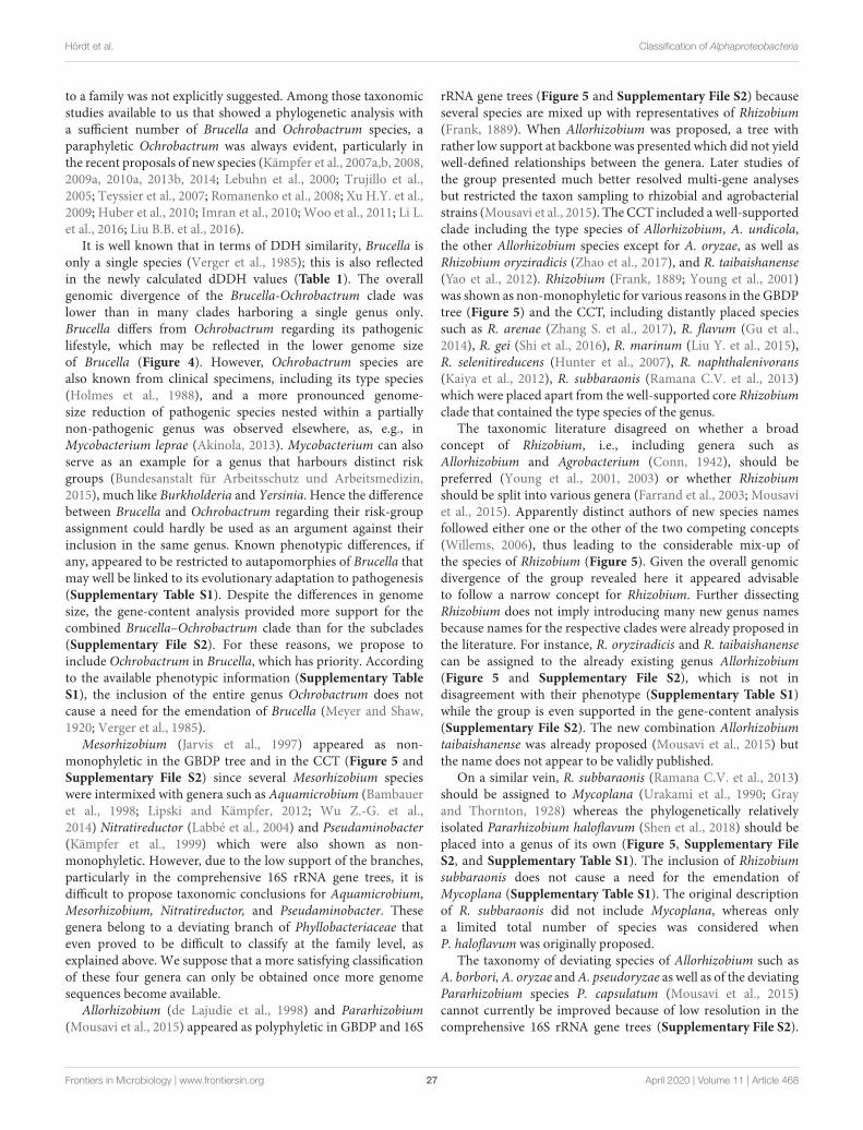
fmicb-11-00468 April 4, 2020 Time: 10:24 # 27
Hördt et al. Classification of Alphaproteobacteria
to a family was not explicitly suggested. Among those taxonomicstudies available to us that showed a phylogenetic analysis witha sufficient number of Brucella and Ochrobactrum species, aparaphyletic Ochrobactrum was always evident, particularly inthe recent proposals of new species (Kämpfer et al., 2007a,b, 2008,2009a, 2010a, 2013b, 2014; Lebuhn et al., 2000; Trujillo et al.,2005; Teyssier et al., 2007; Romanenko et al., 2008; Xu H.Y. et al.,2009; Huber et al., 2010; Imran et al., 2010; Woo et al., 2011; Li L.et al., 2016; Liu B.B. et al., 2016).
It is well known that in terms of DDH similarity, Brucella isonly a single species (Verger et al., 1985); this is also reflectedin the newly calculated dDDH values (Table 1). The overallgenomic divergence of the Brucella-Ochrobactrum clade waslower than in many clades harboring a single genus only.Brucella differs from Ochrobactrum regarding its pathogeniclifestyle, which may be reflected in the lower genome sizeof Brucella (Figure 4). However, Ochrobactrum species arealso known from clinical specimens, including its type species(Holmes et al., 1988), and a more pronounced genome-size reduction of pathogenic species nested within a partiallynon-pathogenic genus was observed elsewhere, as, e.g., inMycobacterium leprae (Akinola, 2013). Mycobacterium can alsoserve as an example for a genus that harbours distinct riskgroups (Bundesanstalt für Arbeitsschutz und Arbeitsmedizin,2015), much like Burkholderia and Yersinia. Hence the differencebetween Brucella and Ochrobactrum regarding their risk-groupassignment could hardly be used as an argument against theirinclusion in the same genus. Known phenotypic differences, ifany, appeared to be restricted to autapomorphies of Brucella thatmay well be linked to its evolutionary adaptation to pathogenesis(Supplementary Table S1). Despite the differences in genomesize, the gene-content analysis provided more support for thecombined Brucella–Ochrobactrum clade than for the subclades(Supplementary File S2). For these reasons, we propose toinclude Ochrobactrum in Brucella, which has priority. Accordingto the available phenotypic information (Supplementary TableS1), the inclusion of the entire genus Ochrobactrum does notcause a need for the emendation of Brucella (Meyer and Shaw,1920; Verger et al., 1985).
Mesorhizobium (Jarvis et al., 1997) appeared as non-monophyletic in the GBDP tree and in the CCT (Figure 5 andSupplementary File S2) since several Mesorhizobium specieswere intermixed with genera such as Aquamicrobium (Bambaueret al., 1998; Lipski and Kämpfer, 2012; Wu Z.-G. et al.,2014) Nitratireductor (Labbé et al., 2004) and Pseudaminobacter(Kämpfer et al., 1999) which were also shown as non-monophyletic. However, due to the low support of the branches,particularly in the comprehensive 16S rRNA gene trees, it isdifficult to propose taxonomic conclusions for Aquamicrobium,Mesorhizobium, Nitratireductor, and Pseudaminobacter. Thesegenera belong to a deviating branch of Phyllobacteriaceae thateven proved to be difficult to classify at the family level, asexplained above. We suppose that a more satisfying classificationof these four genera can only be obtained once more genomesequences become available.
Allorhizobium (de Lajudie et al., 1998) and Pararhizobium(Mousavi et al., 2015) appeared as polyphyletic in GBDP and 16S
rRNA gene trees (Figure 5 and Supplementary File S2) becauseseveral species are mixed up with representatives of Rhizobium(Frank, 1889). When Allorhizobium was proposed, a tree withrather low support at backbone was presented which did not yieldwell-defined relationships between the genera. Later studies ofthe group presented much better resolved multi-gene analysesbut restricted the taxon sampling to rhizobial and agrobacterialstrains (Mousavi et al., 2015). The CCT included a well-supportedclade including the type species of Allorhizobium, A. undicola,the other Allorhizobium species except for A. oryzae, as well asRhizobium oryziradicis (Zhao et al., 2017), and R. taibaishanense(Yao et al., 2012). Rhizobium (Frank, 1889; Young et al., 2001)was shown as non-monophyletic for various reasons in the GBDPtree (Figure 5) and the CCT, including distantly placed speciessuch as R. arenae (Zhang S. et al., 2017), R. flavum (Gu et al.,2014), R. gei (Shi et al., 2016), R. marinum (Liu Y. et al., 2015),R. selenitireducens (Hunter et al., 2007), R. naphthalenivorans(Kaiya et al., 2012), R. subbaraonis (Ramana C.V. et al., 2013)which were placed apart from the well-supported core Rhizobiumclade that contained the type species of the genus.
The taxonomic literature disagreed on whether a broadconcept of Rhizobium, i.e., including genera such asAllorhizobium and Agrobacterium (Conn, 1942), should bepreferred (Young et al., 2001, 2003) or whether Rhizobiumshould be split into various genera (Farrand et al., 2003; Mousaviet al., 2015). Apparently distinct authors of new species namesfollowed either one or the other of the two competing concepts(Willems, 2006), thus leading to the considerable mix-up ofthe species of Rhizobium (Figure 5). Given the overall genomicdivergence of the group revealed here it appeared advisableto follow a narrow concept for Rhizobium. Further dissectingRhizobium does not imply introducing many new genus namesbecause names for the respective clades were already proposed inthe literature. For instance, R. oryziradicis and R. taibaishanensecan be assigned to the already existing genus Allorhizobium(Figure 5 and Supplementary File S2), which is not indisagreement with their phenotype (Supplementary Table S1)while the group is even supported in the gene-content analysis(Supplementary File S2). The new combination Allorhizobiumtaibaishanense was already proposed (Mousavi et al., 2015) butthe name does not appear to be validly published.
On a similar vein, R. subbaraonis (Ramana C.V. et al., 2013)should be assigned to Mycoplana (Urakami et al., 1990; Grayand Thornton, 1928) whereas the phylogenetically relativelyisolated Pararhizobium haloflavum (Shen et al., 2018) should beplaced into a genus of its own (Figure 5, Supplementary FileS2, and Supplementary Table S1). The inclusion of Rhizobiumsubbaraonis does not cause a need for the emendation ofMycoplana (Supplementary Table S1). The original descriptionof R. subbaraonis did not include Mycoplana, whereas onlya limited total number of species was considered whenP. haloflavum was originally proposed.
The taxonomy of deviating species of Allorhizobium such asA. borbori, A. oryzae and A. pseudoryzae as well as of the deviatingPararhizobium species P. capsulatum (Mousavi et al., 2015)cannot currently be improved because of low resolution in thecomprehensive 16S rRNA gene trees (Supplementary File S2).
Frontiers in Microbiology | www.frontiersin.org 27 April 2020 | Volume 11 | Article 468
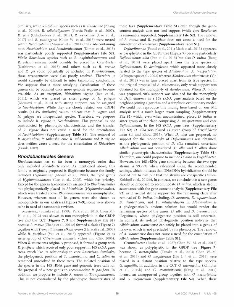
fmicb-11-00468 April 4, 2020 Time: 10:24 # 28
Hördt et al. Classification of Alphaproteobacteria
Similarly, while Rhizobium species such as R. smilacinae (Zhanget al., 2014b), R. cellulosilyticum (García-Fraile et al., 2007),R. zeae (Celador-lera et al., 2017), R. wenxiniae (Gao et al.,2017) and R. yantingense (Chen W. et al., 2015) were nestedwithin Neorhizobium (Mousavi et al., 2014), the clade containingboth Neorhizobium and Pseudorhizobium (Kimes et al., 2015)was particularly poorly supported (Supplementary File S2).While Rhizobium species such as R. naphthalenivorans andR. selenitireducens could possibly be placed in Ciceribacter(Kathiravan et al., 2013) and others such as R. arenaeand R. gei could potentially be included in Pararhizobium,these arrangements were also poorly resolved. Therefore itwould currently be difficult to infer taxonomic conclusions.We suppose that a more satisfying classification of thesegenera can be obtained once more genome sequences becomeavailable. As an exception, Rhizobium vignae (Ren et al.,2011), which was placed as sister group of N. galegae(Mousavi et al., 2014) with strong support, can be assignedto Neorhizobium. While they are closely related, our dDDHresults (41.4% similarity) values indicate that R. vignae andN. galegae are independent species. Therefore, we proposeto include R. vignae in Neorhizobium. This proposal is notcontradicted by phenotypic characteristics and the transferof R. vignae does not cause a need for the emendationof Neorhizobium (Supplementary Table S1). The removal ofR. oryziradicis, R. taibaishanense, R. subbaraonis and R. vignaedoes neither cause a need for the emendation of Rhizobium(Frank, 1889).
Rhodobacterales GeneraRhodobacterales has so far been a monotypic order thatonly included Rhodobacteraceae. As mentioned above, thisfamily as originally proposed is illegitimate because the familyincluded Hyphomonas (Moore et al., 1984), the type genusof Hyphomonadaceae (Lee et al., 2005), which has priority.Except for the genera taxonomically assigned to Rhodobacteralesbut phylogenetically placed in Rhizobiales (Hyphomicrobiales),which were treated above, Rhodobacteraceae was monophyletic.However, whereas most of its genera were also shown asmonophyletic in our analyses (Figures 7–9), some were shownto be in need of a taxonomic revision.
Roseivivax (Suzuki et al., 1999a; Park et al., 2010; Chen M.-H. et al., 2012) was shown as non-monophyletic in the GBDPtree and the CCT (Figures 7, 9 and Supplementary File S2)because R. roseus (Zhang et al., 2014c) formed a clade (Figure 7)together with Tranquillimonas alkanivorans (Harwati et al., 2008)while R. pacificus (Wu et al., 2013) appeared (Figure 9) assister group of Citreimonas salinaria (Choi and Cho, 2006).When R. roseus was originally proposed, it formed a group withR. pacificus which received only poor support in 16S rRNA genetrees, much like its relationship to core Roseivivax. Similarly,the phylogenetic position of T. alkanivorans and C. salinariaremained unresolved in these trees. The isolated position ofthe species in the 16S rRNA gene and genomic trees calls forthe proposal of a new genus to accommodate R. pacificus. Inaddition, we propose to include R. roseus in Tranquillimonas.This is not contradicted by the phenotypic characteristics of
these taxa (Supplementary Table S1) even though the gene-content analysis does not lend support (while core Roseivivaxis reasonably supported; Supplementary File S2). The removalof R. roseus and R. pacificus does not cause a need for theemendation of Roseivivax (Supplementary Table S1).
Defluviimonas (Foesel et al., 2011; Math et al., 2013) appearedas paraphyletic in the GBDP tree (Figure 7) because particularlyDefluviimonas alba (Pan et al., 2015) but also D. indica (Jianget al., 2014) were placed apart from the type species ofDefluviimonas, D. denitrificans, which appeared more closelyrelated to the type species of Albidovulum, A. inexpectatum(Albuquerque et al., 2002) whereas Albidovulum xiamenense (Yinet al., 2012) was in turn placed apart from its type species. Inthe original proposal of A. xiamenense, only weak support wasobtained for the monophyly of Albidovulum. When D. indicawas proposed, 90% support was obtained for the monophylyof Defluviimonas in a 16S rRNA gene analysis based on theneighbor-joining algorithm and a simplistic evolutionary model.We could not reproduce this finding here based on our MLanalysis with a much larger taxon sampling (SupplementaryFile S2) which, even when unconstrained, placed D. indica assister group of the clade comprising A. inexpectatum and coreDefluviimonas. In the 16S rRNA gene trees (SupplementaryFile S2) D. alba was placed as sister group of Frigidibacteralbus (Li and Zhou, 2015). When D. alba was proposed, nosupport for the monophyly of Defluviimonas was obtained,as the phylogenetic position of D. alba remained uncertain;Albidovulum was not considered. D. alba and F. albus showsimilar phenotypic characteristics (Supplementary Table S1).Therefore, one could propose to include D. alba in Frigidibacter.However, the 16S rRNA gene similarity between the two typestrains is 99.79% when calculated using the recommendedsettings, which indicates that DNA:DNA hybridization should becarried out to rule out that the strains are conspecific (Meier-Kolthoff et al., 2013b). In contrast, we conclude that a new genusshould be proposed to accommodate D. indica, which is also inaccordance with the gene-content analysis (Supplementary FileS2) as it yielded strong support for the remaining clade afterremoval of D. indica. Including, D. aestuarii, D. aquaemixtae,D. denitrificans, and D. nitratireducens in Albidovulum isa phylogenetically obvious solution but would render theremaining species of the genus, D. alba and D. pyrenivorans,illegitimate, whose phylogenetic position is still uncertain.In contrast, its isolated phylogenetic position indicates thatAlbidovulum xiamenense can safely be placed into a genus ofits own, which is not precluded by its phenotype. The removalof A. xiamenense does not cause a need for the emendation ofAlbidovulum (Supplementary Table S1).
Gemmobacter (Rothe et al., 1987; Chen W.-M. et al., 2013)was shown as polyphyletic in the GBDP tree (Figure 7)because G. nectariphilus (Tanaka et al., 2004; Chen W.-M.et al., 2013) and G. megaterium (Liu J.-J. et al., 2014) wereplaced in a distant position relative to the type species,G. aquatilis. In addition, in the CCT G. intermedius (Kämpferet al., 2015b) and G. straminiformis (Kang et al., 2017)formed an unsupported group together with G. nectariphilusand G. megaterium (Supplementary File S2). When these
Frontiers in Microbiology | www.frontiersin.org 28 April 2020 | Volume 11 | Article 468
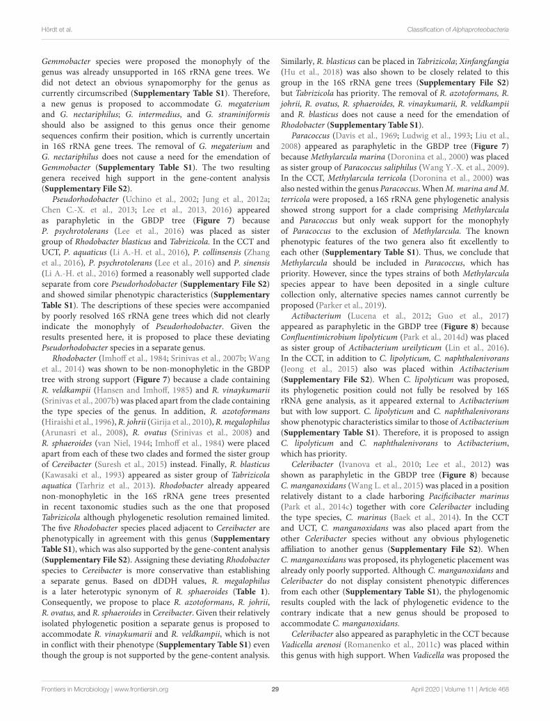
fmicb-11-00468 April 4, 2020 Time: 10:24 # 29
Hördt et al. Classification of Alphaproteobacteria
Gemmobacter species were proposed the monophyly of thegenus was already unsupported in 16S rRNA gene trees. Wedid not detect an obvious synapomorphy for the genus ascurrently circumscribed (Supplementary Table S1). Therefore,a new genus is proposed to accommodate G. megateriumand G. nectariphilus; G. intermedius, and G. straminiformisshould also be assigned to this genus once their genomesequences confirm their position, which is currently uncertainin 16S rRNA gene trees. The removal of G. megaterium andG. nectariphilus does not cause a need for the emendation ofGemmobacter (Supplementary Table S1). The two resultinggenera received high support in the gene-content analysis(Supplementary File S2).
Pseudorhodobacter (Uchino et al., 2002; Jung et al., 2012a;Chen C.-X. et al., 2013; Lee et al., 2013, 2016) appearedas paraphyletic in the GBDP tree (Figure 7) becauseP. psychrotolerans (Lee et al., 2016) was placed as sistergroup of Rhodobacter blasticus and Tabrizicola. In the CCT andUCT, P. aquaticus (Li A.-H. et al., 2016), P. collinsensis (Zhanget al., 2016), P. psychrotolerans (Lee et al., 2016) and P. sinensis(Li A.-H. et al., 2016) formed a reasonably well supported cladeseparate from core Pseudorhodobacter (Supplementary File S2)and showed similar phenotypic characteristics (SupplementaryTable S1). The descriptions of these species were accompaniedby poorly resolved 16S rRNA gene trees which did not clearlyindicate the monophyly of Pseudorhodobacter. Given theresults presented here, it is proposed to place these deviatingPseudorhodobacter species in a separate genus.
Rhodobacter (Imhoff et al., 1984; Srinivas et al., 2007b; Wanget al., 2014) was shown to be non-monophyletic in the GBDPtree with strong support (Figure 7) because a clade containingR. veldkampii (Hansen and Imhoff, 1985) and R. vinaykumarii(Srinivas et al., 2007b) was placed apart from the clade containingthe type species of the genus. In addition, R. azotoformans(Hiraishi et al., 1996), R. johrii (Girija et al., 2010), R. megalophilus(Arunasri et al., 2008), R. ovatus (Srinivas et al., 2008) andR. sphaeroides (van Niel, 1944; Imhoff et al., 1984) were placedapart from each of these two clades and formed the sister groupof Cereibacter (Suresh et al., 2015) instead. Finally, R. blasticus(Kawasaki et al., 1993) appeared as sister group of Tabrizicolaaquatica (Tarhriz et al., 2013). Rhodobacter already appearednon-monophyletic in the 16S rRNA gene trees presentedin recent taxonomic studies such as the one that proposedTabrizicola although phylogenetic resolution remained limited.The five Rhodobacter species placed adjacent to Cereibacter arephenotypically in agreement with this genus (SupplementaryTable S1), which was also supported by the gene-content analysis(Supplementary File S2). Assigning these deviating Rhodobacterspecies to Cereibacter is more conservative than establishinga separate genus. Based on dDDH values, R. megalophilusis a later heterotypic synonym of R. sphaeroides (Table 1).Consequently, we propose to place R. azotoformans, R. johrii,R. ovatus, and R. sphaeroides in Cereibacter. Given their relativelyisolated phylogenetic position a separate genus is proposed toaccommodate R. vinaykumarii and R. veldkampii, which is notin conflict with their phenotype (Supplementary Table S1) eventhough the group is not supported by the gene-content analysis.
Similarly, R. blasticus can be placed in Tabrizicola; Xinfangfangia(Hu et al., 2018) was also shown to be closely related to thisgroup in the 16S rRNA gene trees (Supplementary File S2)but Tabrizicola has priority. The removal of R. azotoformans, R.johrii, R. ovatus, R. sphaeroides, R. vinaykumarii, R. veldkampiiand R. blasticus does not cause a need for the emendation ofRhodobacter (Supplementary Table S1).
Paracoccus (Davis et al., 1969; Ludwig et al., 1993; Liu et al.,2008) appeared as paraphyletic in the GBDP tree (Figure 7)because Methylarcula marina (Doronina et al., 2000) was placedas sister group of Paracoccus saliphilus (Wang Y.-X. et al., 2009).In the CCT, Methylarcula terricola (Doronina et al., 2000) wasalso nested within the genus Paracoccus. When M. marina and M.terricola were proposed, a 16S rRNA gene phylogenetic analysisshowed strong support for a clade comprising Methylarculaand Paracoccus but only weak support for the monophylyof Paracoccus to the exclusion of Methylarcula. The knownphenotypic features of the two genera also fit excellently toeach other (Supplementary Table S1). Thus, we conclude thatMethylarcula should be included in Paracoccus, which haspriority. However, since the types strains of both Methylarculaspecies appear to have been deposited in a single culturecollection only, alternative species names cannot currently beproposed (Parker et al., 2019).
Actibacterium (Lucena et al., 2012; Guo et al., 2017)appeared as paraphyletic in the GBDP tree (Figure 8) becauseConfluentimicrobium lipolyticum (Park et al., 2014d) was placedas sister group of Actibacterium ureilyticum (Lin et al., 2016).In the CCT, in addition to C. lipolyticum, C. naphthalenivorans(Jeong et al., 2015) also was placed within Actibacterium(Supplementary File S2). When C. lipolyticum was proposed,its phylogenetic position could not fully be resolved by 16SrRNA gene analysis, as it appeared external to Actibacteriumbut with low support. C. lipolyticum and C. naphthalenivoransshow phenotypic characteristics similar to those of Actibacterium(Supplementary Table S1). Therefore, it is proposed to assignC. lipolyticum and C. naphthalenivorans to Actibacterium,which has priority.
Celeribacter (Ivanova et al., 2010; Lee et al., 2012) wasshown as paraphyletic in the GBDP tree (Figure 8) becauseC. manganoxidans (Wang L. et al., 2015) was placed in a positionrelatively distant to a clade harboring Pacificibacter marinus(Park et al., 2014c) together with core Celeribacter includingthe type species, C. marinus (Baek et al., 2014). In the CCTand UCT, C. manganoxidans was also placed apart from theother Celeribacter species without any obvious phylogeneticaffiliation to another genus (Supplementary File S2). WhenC. manganoxidans was proposed, its phylogenetic placement wasalready only poorly supported. Although C. manganoxidans andCeleribacter do not display consistent phenotypic differencesfrom each other (Supplementary Table S1), the phylogenomicresults coupled with the lack of phylogenetic evidence to thecontrary indicate that a new genus should be proposed toaccommodate C. manganoxidans.
Celeribacter also appeared as paraphyletic in the CCT becauseVadicella arenosi (Romanenko et al., 2011c) was placed withinthis genus with high support. When Vadicella was proposed the
Frontiers in Microbiology | www.frontiersin.org 29 April 2020 | Volume 11 | Article 468
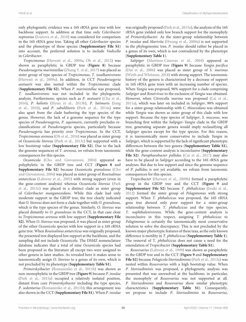
fmicb-11-00468 April 4, 2020 Time: 10:24 # 30
Hördt et al. Classification of Alphaproteobacteria
only phylogenetic evidence was a 16S rRNA gene tree with lowbackbone support. In addition at that time only Celeribacterneptunius (Ivanova et al., 2010) was considered for comparisonin the 16S rRNA gene tree. Taking all other Celeribacter speciesand the phenotype of these species (Supplementary File S1)into account, the preferred solution is to include Vadicellain Celeribacter.
Tropicimonas (Harwati et al., 2009a; Oh et al., 2012) wasshown as paraphyletic in GBDP tree (Figure 8) becausePseudoruegeria marinistellae (Zhang Y. et al., 2017) was placed assister group of type species of Tropicimonas, T. isoalkanivorans(Harwati et al., 2009a). In addition, in CCT Pseudoruegeriaaestuarii was also nested within the Tropicimonas clade(Supplementary File S2). When P. marinistellae was proposed,T. isoalkanivorans was not included in the phylogeneticanalysis. Furthermore, species such as P. aestuarii (Cha et al.,2016), P. haliotis (Hyun et al., 2013b), P. lutimaris (Junget al., 2010), and P. sabulilitoris (Park et al., 2014a) werealso apart from the clade harboring the type species of thegenus. However, the lack of a genome sequence for the typespecies of Pseudoruegeria, P. aquimaris, currently precludes re-classifications of Pseudoruegeria species, particularly becausePseudoruegeria has priority over Tropicimonas. In the CCT,Tropicimonas arenosa (Oh et al., 2016) was placed as sister groupof Oceanicola litoreus (Park et al., 2013c) but supported with alow bootstrap value (Supplementary File S2). Due to the lackthe genome sequences of T. arenosa, we refrain from taxonomicconsequences for this species.
Oceanicola (Cho and Giovannoni, 2004) appeared aspolyphyletic in the GBDP tree and CCT (Figure 8 andSupplementary File S2) because Oceanicola granulosus (Choand Giovannoni, 2004) was placed as sister group of Roseisalinusantarcticus (Labrenz et al., 2005) with strong support (even inthe gene-content analysis) whereas Oceanicola litoreus (Parket al., 2013c) was placed in a distinct clade as sister groupof Celeribacter manganoxidans. While this clade had onlymoderate support in the GBDP tree, the tree clearly indicatedthat O. litoreus does not form a clade together with O. granulosus,which is the type species of the genus. Similarly, O. litoreus wasplaced distantly to O. granulosus in the CCT, in that case closeto Tropicimonas arenosa with low support (Supplementary FileS2). When O. litoreus was proposed, it was placed as sister groupof the other Oceanicola species with low support in a 16S rRNAgene tree. When Roseisalinus antarcticus was originally proposed,the presented tree displayed low support at the backbone, and thesampling did not include Oceanicola. The DSMZ nomenclaturedatabase indicates that a total of nine Oceanicola species hadbeen proposed in the literature all except two were assigned toother genera in later studies. As revealed here it makes sense totaxonomically assign O. litoreus to a genus of its own, which isnot precluded by its phenotype (Supplementary Table S1).
Primorskyibacter (Romanenko et al., 2011b) was shown asnon-monophyletic in the GBDP tree (Figure 9) because P. insulae(Park et al., 2015a) occupied a relatively isolated position,distant from core Primorskyibacter including the type species,P. sedentarius (Romanenko et al., 2011b); this arrangement wasalso shown in the CCT (Supplementary File S1). When P. insulae
was originally proposed (Park et al., 2015a), the analysis of the 16SrRNA gene yielded only low branch support for the monophylyof Primorskyibacter. As the sister-group relationship betweenP. insulae and Marivita (Hwang et al., 2009a) is not supportedin the phylogenomic tree, P. insulae should rather be placed ina genus of its own, which is not contradicted by the phenotype(Supplementary Table 1).
Salipiger (Martínez-Cánovas et al., 2004) appeared asparaphyletic in GBDP tree (Figure 9) because Yangia pacifica(Dai et al., 2006) was placed as sister group of S. marinus(Wirth and Whitman, 2018) with strong support. The taxonomichistory of the genera is characterized by a decrease of supportin 16S rRNA gene trees with an increasing number of species.When Yangia was proposed, 96% support for a clade comprisingSalipiger and Roseivivax to the exclusion of Yangia was obtained.However, when Citreicella marina was proposed (Lai et al.,2011a), which was later on included in Salipiger, 98% supportfor a sister-group relationship with C. thiooxidans was obtainedwhile Yangia was shown as sister group of this clade with lowsupport. Because the type species of Salipiger, S. mucosus, wasbranching first within the Salipiger–Yangia clade in the GBDPtree, generating separate genera would imply reclassifying allSalipiger species except for the type species. For this reason,it is taxonomically more conservative to include Yangia inSalipiger, which is supported by the lack of significant phenotypicdifferences between the two genera (Supplementary Table S1),while the gene-content analysis is inconclusive (SupplementaryFile S2). Paraphaeobacter pallidus (Cai et al., 2017) may alsohave to be placed in Salipiger according to the 16S rRNA geneanalyses. But due to low support and since the genome sequenceof P. pallidus is not yet available, we refrain from taxonomicconsequences for this species.
Tropicibacter (Harwati et al., 2009b) formed a paraphyleticgroup in the GBDP tree and the CCT (Figure 9 andSupplementary File S2) because T. phthalicicus (Iwaki et al.,2012) formed the sister group of Pelagimonas with strongsupport. When T. phthalicicus was proposed, the 16S rRNAgene tree showed only poor support for a sister-grouprelationship between T. phthalicicus and the type species,T. naphthalenivorans. While the gene-content analysis isinconclusive in this respect, assigning T. phthalicicus toPelagimonas is certainly the taxonomically most conservativesolution to solve the discrepancy. This is not precluded by theknown major phenotypic features of these taxa, as the only knowndifference is motility in T. phthalicicus (Supplementary Table 1).The removal of T. phthalicicus does not cause a need for theemendation of Tropicibacter (Supplementary Table S1).
Roseovarius (Labrenz et al., 1999) was shown as paraphyleticin the GBDP tree and in the CCT (Figure 9 and SupplementaryFile S2) because Pelagicola litorisediminis (Park et al., 2013a) wasnested within Roseovarius with a high bootstrap value. WhenP. litorisediminis was proposed, a phylogenetic analysis waspresented that was unresolved at the backbone; in particular,the monophyly of Roseovarius was not supported at all.P. litorisediminis and Roseovarius show similar phenotypiccharacteristics (Supplementary Table S1). Consequently,we propose to include P. litorisediminis in Roseovarius,
Frontiers in Microbiology | www.frontiersin.org 30 April 2020 | Volume 11 | Article 468
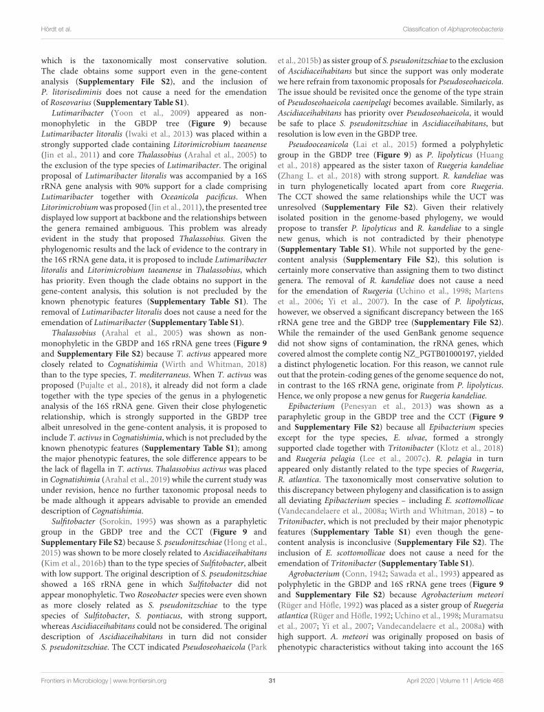
fmicb-11-00468 April 4, 2020 Time: 10:24 # 31
Hördt et al. Classification of Alphaproteobacteria
which is the taxonomically most conservative solution.The clade obtains some support even in the gene-contentanalysis (Supplementary File S2), and the inclusion ofP. litorisediminis does not cause a need for the emendationof Roseovarius (Supplementary Table S1).
Lutimaribacter (Yoon et al., 2009) appeared as non-monophyletic in the GBDP tree (Figure 9) becauseLutimaribacter litoralis (Iwaki et al., 2013) was placed within astrongly supported clade containing Litorimicrobium taeanense(Jin et al., 2011) and core Thalassobius (Arahal et al., 2005) tothe exclusion of the type species of Lutimaribacter. The originalproposal of Lutimaribacter litoralis was accompanied by a 16SrRNA gene analysis with 90% support for a clade comprisingLutimaribacter together with Oceanicola pacificus. WhenLitorimicrobium was proposed (Jin et al., 2011), the presented treedisplayed low support at backbone and the relationships betweenthe genera remained ambiguous. This problem was alreadyevident in the study that proposed Thalassobius. Given thephylogenomic results and the lack of evidence to the contrary inthe 16S rRNA gene data, it is proposed to include Lutimaribacterlitoralis and Litorimicrobium taeanense in Thalassobius, whichhas priority. Even though the clade obtains no support in thegene-content analysis, this solution is not precluded by theknown phenotypic features (Supplementary Table S1). Theremoval of Lutimaribacter litoralis does not cause a need for theemendation of Lutimaribacter (Supplementary Table S1).
Thalassobius (Arahal et al., 2005) was shown as non-monophyletic in the GBDP and 16S rRNA gene trees (Figure 9and Supplementary File S2) because T. activus appeared moreclosely related to Cognatishimia (Wirth and Whitman, 2018)than to the type species, T. mediterraneus. When T. activus wasproposed (Pujalte et al., 2018), it already did not form a cladetogether with the type species of the genus in a phylogeneticanalysis of the 16S rRNA gene. Given their close phylogeneticrelationship, which is strongly supported in the GBDP treealbeit unresolved in the gene-content analysis, it is proposed toinclude T. activus in Cognatishimia, which is not precluded by theknown phenotypic features (Supplementary Table S1); amongthe major phenotypic features, the sole difference appears to bethe lack of flagella in T. activus. Thalassobius activus was placedin Cognatishimia (Arahal et al., 2019) while the current study wasunder revision, hence no further taxonomic proposal needs tobe made although it appears advisable to provide an emendeddescription of Cognatishimia.
Sulfitobacter (Sorokin, 1995) was shown as a paraphyleticgroup in the GBDP tree and the CCT (Figure 9 andSupplementary File S2) because S. pseudonitzschiae (Hong et al.,2015) was shown to be more closely related to Ascidiaceihabitans(Kim et al., 2016b) than to the type species of Sulfitobacter, albeitwith low support. The original description of S. pseudonitzschiaeshowed a 16S rRNA gene in which Sulfitobacter did notappear monophyletic. Two Roseobacter species were even shownas more closely related as S. pseudonitzschiae to the typespecies of Sulfitobacter, S. pontiacus, with strong support,whereas Ascidiaceihabitans could not be considered. The originaldescription of Ascidiaceihabitans in turn did not considerS. pseudonitzschiae. The CCT indicated Pseudoseohaeicola (Park
et al., 2015b) as sister group of S. pseudonitzschiae to the exclusionof Ascidiaceihabitans but since the support was only moderatewe here refrain from taxonomic proposals for Pseudoseohaeicola.The issue should be revisited once the genome of the type strainof Pseudoseohaeicola caenipelagi becomes available. Similarly, asAscidiaceihabitans has priority over Pseudoseohaeicola, it wouldbe safe to place S. pseudonitzschiae in Ascidiaceihabitans, butresolution is low even in the GBDP tree.
Pseudooceanicola (Lai et al., 2015) formed a polyphyleticgroup in the GBDP tree (Figure 9) as P. lipolyticus (Huanget al., 2018) appeared as the sister taxon of Ruegeria kandeliae(Zhang L. et al., 2018) with strong support. R. kandeliae wasin turn phylogenetically located apart from core Ruegeria.The CCT showed the same relationships while the UCT wasunresolved (Supplementary File S2). Given their relativelyisolated position in the genome-based phylogeny, we wouldpropose to transfer P. lipolyticus and R. kandeliae to a singlenew genus, which is not contradicted by their phenotype(Supplementary Table S1). While not supported by the gene-content analysis (Supplementary File S2), this solution iscertainly more conservative than assigning them to two distinctgenera. The removal of R. kandeliae does not cause a needfor the emendation of Ruegeria (Uchino et al., 1998; Martenset al., 2006; Yi et al., 2007). In the case of P. lipolyticus,however, we observed a significant discrepancy between the 16SrRNA gene tree and the GBDP tree (Supplementary File S2).While the remainder of the used GenBank genome sequencedid not show signs of contamination, the rRNA genes, whichcovered almost the complete contig NZ_PGTB01000197, yieldeda distinct phylogenetic location. For this reason, we cannot ruleout that the protein-coding genes of the genome sequence do not,in contrast to the 16S rRNA gene, originate from P. lipolyticus.Hence, we only propose a new genus for Ruegeria kandeliae.
Epibacterium (Penesyan et al., 2013) was shown as aparaphyletic group in the GBDP tree and the CCT (Figure 9and Supplementary File S2) because all Epibacterium speciesexcept for the type species, E. ulvae, formed a stronglysupported clade together with Tritonibacter (Klotz et al., 2018)and Ruegeria pelagia (Lee et al., 2007c). R. pelagia in turnappeared only distantly related to the type species of Ruegeria,R. atlantica. The taxonomically most conservative solution tothis discrepancy between phylogeny and classification is to assignall deviating Epibacterium species – including E. scottomollicae(Vandecandelaere et al., 2008a; Wirth and Whitman, 2018) – toTritonibacter, which is not precluded by their major phenotypicfeatures (Supplementary Table S1) even though the gene-content analysis is inconclusive (Supplementary File S2). Theinclusion of E. scottomollicae does not cause a need for theemendation of Tritonibacter (Supplementary Table S1).
Agrobacterium (Conn, 1942; Sawada et al., 1993) appeared aspolyphyletic in the GBDP and 16S rRNA gene trees (Figure 9and Supplementary File S2) because Agrobacterium meteori(Rüger and Höfle, 1992) was placed as a sister group of Ruegeriaatlantica (Rüger and Höfle, 1992; Uchino et al., 1998; Muramatsuet al., 2007; Yi et al., 2007; Vandecandelaere et al., 2008a) withhigh support. A. meteori was originally proposed on basis ofphenotypic characteristics without taking into account the 16S
Frontiers in Microbiology | www.frontiersin.org 31 April 2020 | Volume 11 | Article 468
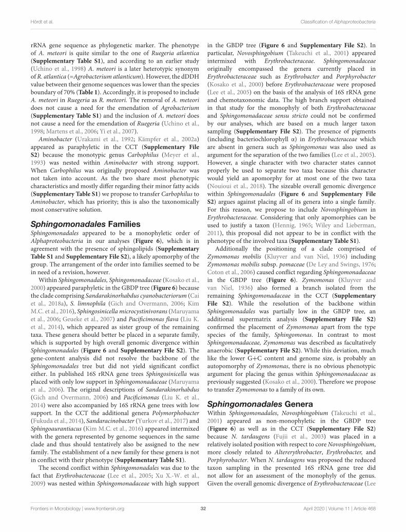
fmicb-11-00468 April 4, 2020 Time: 10:24 # 32
Hördt et al. Classification of Alphaproteobacteria
rRNA gene sequence as phylogenetic marker. The phenotypeof A. meteori is quite similar to the one of Ruegeria atlantica(Supplementary Table S1), and according to an earlier study(Uchino et al., 1998) A. meteori is a later heterotypic synonymof R. atlantica (=Agrobacterium atlanticum). However, the dDDHvalue between their genome sequences was lower than the speciesboundary of 70% (Table 1). Accordingly, it is proposed to includeA. meteori in Ruegeria as R. meteori. The removal of A. meteoridoes not cause a need for the emendation of Agrobacterium(Supplementary Table S1) and the inclusion of A. meteori doesnot cause a need for the emendation of Ruegeria (Uchino et al.,1998; Martens et al., 2006; Yi et al., 2007).
Aminobacter (Urakami et al., 1992; Kämpfer et al., 2002a)appeared as paraphyletic in the CCT (Supplementary FileS2) because the monotypic genus Carbophilus (Meyer et al.,1993) was nested within Aminobacter with strong support.When Carbophilus was originally proposed Aminobacter wasnot taken into account. As the two share most phenotypiccharacteristics and mostly differ regarding their minor fatty acids(Supplementary Table S1) we propose to transfer Carbophilus toAminobacter, which has priority; this is also the taxonomicallymost conservative solution.
Sphingomonadales FamiliesSphingomonadales appeared to be a monophyletic order ofAlphaproteobacteria in our analyses (Figure 6), which is inagreement with the presence of sphingolipids (SupplementaryTable S1 and Supplementary File S2), a likely apomorphy of thegroup. The arrangement of the order into families seemed to bein need of a revision, however.
Within Sphingomonadales, Sphingomonadaceae (Kosako et al.,2000) appeared paraphyletic in the GBDP tree (Figure 6) becausethe clade comprising Sandarakinorhabdus cyanobacteriorum (Caiet al., 2018a), S. limnophila (Gich and Overmann, 2006; KimM.C. et al., 2016), Sphingosinicella microcystinivorans (Maruyamaet al., 2006; Geueke et al., 2007) and Pacificimonas flava (Liu K.et al., 2014), which appeared as sister group of the remainingtaxa. These genera should better be placed in a separate family,which is supported by high overall genomic divergence withinSphingomonadales (Figure 6 and Supplementary File S2). Thegene-content analysis did not resolve the backbone of theSphingomonadales tree but did not yield significant conflicteither. In published 16S rRNA gene trees Sphingosinicella wasplaced with only low support in Sphingomonadaceae (Maruyamaet al., 2006). The original descriptions of Sandarakinorhabdus(Gich and Overmann, 2006) and Pacificimonas (Liu K. et al.,2014) were also accompanied by 16S rRNA gene trees with lowsupport. In the CCT the additional genera Polymorphobacter(Fukuda et al., 2014), Sandaracinobacter (Yurkov et al., 2017) andSphingoaurantiacus (Kim M.C. et al., 2016) appeared intermixedwith the genera represented by genome sequences in the sameclade and thus should tentatively also be assigned to the newfamily. The establishment of a new family for these genera is notin conflict with their phenotype (Supplementary Table S1).
The second conflict within Sphingomonadales was due to thefact that Erythrobacteraceae (Lee et al., 2005; Xu X.-W. et al.,2009) was nested within Sphingomonadaceae with high support
in the GBDP tree (Figure 6 and Supplementary File S2). Inparticular, Novosphingobium (Takeuchi et al., 2001) appearedintermixed with Erythrobacteraceae. Sphingomonadaceaeoriginally encompassed the genera currently placed inErythrobacteraceae such as Erythrobacter and Porphyrobacter(Kosako et al., 2000) before Erythrobacteraceae were proposed(Lee et al., 2005) on the basis of the analysis of 16S rRNA geneand chemotaxonomic data. The high branch support obtainedin that study for the monophyly of both Erythrobacteraceaeand Sphingomonadaceae sensu stricto could not be confirmedby our analyses, which are based on a much larger taxonsampling (Supplementary File S2). The presence of pigments(including bacteriochlorophyll α) in Erythrobacteraceae whichare absent in genera such as Sphingomonas was also used asargument for the separation of the two families (Lee et al., 2005).However, a single character with two character states cannotproperly be used to separate two taxa because this characterwould yield an apomorphy for at most one of the two taxa(Nouioui et al., 2018). The sizeable overall genomic divergencewithin Sphingomonadales (Figure 6 and Supplementary FileS2) argues against placing all of its genera into a single family.For this reason, we propose to include Novosphingobium inErythrobacteraceae. Considering that only apomorphies can beused to justify a taxon (Hennig, 1965; Wiley and Lieberman,2011), this proposal did not appear to be in conflict with thephenotype of the involved taxa (Supplementary Table S1).
Additionally the positioning of a clade comprised ofZymomonas mobilis (Kluyver and van Niel, 1936) includingZymomonas mobilis subsp. pomaceae (De Ley and Swings, 1976;Coton et al., 2006) caused conflict regarding Sphingomonadaceaein the GBDP tree (Figure 6). Zymomonas (Kluyver andvan Niel, 1936) also formed a branch isolated from theremaining Sphingomonadaceae in the CCT (SupplementaryFile S2). While the resolution of the backbone withinSphingomonadales was partially low in the GBDP tree, anadditional supermatrix analysis (Supplementary File S2)confirmed the placement of Zymomonas apart from the typespecies of the family, Sphingomonas. In contrast to mostSphingomonadaceae, Zymomonas was described as facultativelyanaerobic (Supplementary File S2). While this deviation, muchlike the lower G+C content and genome size, is probably anautopomorphy of Zymomonas, there is no obvious phenotypicargument for placing the genus within Sphingomonadaceae aspreviously suggested (Kosako et al., 2000). Therefore we proposeto transfer Zymomonas to a family of its own.
Sphingomonadales GeneraWithin Sphingomonadales, Novosphingobium (Takeuchi et al.,2001) appeared as non-monophyletic in the GBDP tree(Figure 6) as well as in the CCT (Supplementary File S2)because N. tardaugens (Fujii et al., 2003) was placed in arelatively isolated position with respect to core Novosphingobium,more closely related to Altererythrobacter, Erythrobacter, andPorphyrobacter. When N. tardaugens was proposed the reducedtaxon sampling in the presented 16S rRNA gene tree didnot allow for an assessment of the monophyly of the genus.Given the overall genomic divergence of Erythrobacteraceae (Lee
Frontiers in Microbiology | www.frontiersin.org 32 April 2020 | Volume 11 | Article 468
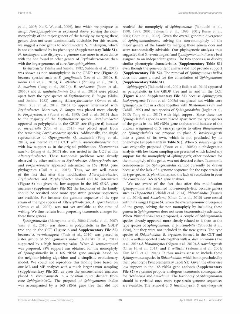
fmicb-11-00468 April 4, 2020 Time: 10:24 # 33
Hördt et al. Classification of Alphaproteobacteria
et al., 2005; Xu X.-W. et al., 2009), into which we propose toassign Novosphingobium as explained above, solving the non-monophyly of the major genera of the family by merging thesegenera does not seem taxonomically advisable. For this reason,we suggest a new genus to accommodate N. tardaugens, whichis not contradicted by its phenotype (Supplementary Table S1).N. tardaugens also displayed a genome size more in accordancewith the one found in other genera of Erythrobacteraceae thanwith the larger genomes of core Novosphingobium.
Erythrobacter (Shiba and Smidu, 1982; Subhash et al., 2013)was shown as non-monophyletic in the GBDP tree (Figure 6)because species such as E. gangjinensis (Lee et al., 2010), E.luteus (Lei et al., 2015), E. atlanticus (Zhuang et al., 2015),E. marinus (Jung et al., 2012b), E. seohaensis (Yoon et al.,2005b) and E. nanhaisediminis (Xu et al., 2010) were placedapart from the type species of Erythrobacter, E. longus (Shibaand Smidu, 1982) causing Altererythrobacter (Kwon et al.,2007; Xue et al., 2012, 2016) to appear intermixed withErythrobacter. Moreover, E. longus was placed more closelyto Porphyrobacter (Fuerst et al., 1993; Coil et al., 2015) thanto the majority of the Erythrobacter species. Porphyrobacterappeared as polyphyletic in the GBDP tree (Figure 6) becauseP. mercurialis (Coil et al., 2015) was placed apart fromthe remaining Porphyrobacter species. Additionally, the singlerepresentative of Qipengyuania, Q. sediminis (Feng et al.,2015), was nested in the CCT within Altererythrobacter butwith low support as in the original publication. Blastomonasmarina (Meng et al., 2017) also was nested in the CCT withinAltererythrobacter. These taxonomic problems were alreadyobserved by other authors as Erythrobacter, Altererythrobacter,and Porphyrobacter appeared intermixed in 16S rRNA genephylogenies (Coil et al., 2015). Thus, we are well awareof the fact that after this modification Altererythrobacter,Erythrobacter and Porphyrobacter would still be intermixed(Figure 6) but given the low support in the 16S rRNA geneanalyses (Supplementary File S2) the taxonomy of the familyshould be revisited once more type-strain genome sequencesare available. For instance, the genome sequence of the typestrain of the type species of Altererythrobacter, A. epoxidivorans(Kwon et al., 2007), was not yet available at the time ofwriting. We thus refrain from proposing taxonomic changes forthese three genera.
Sphingosinicella (Maruyama et al., 2006; Geueke et al., 2007;Yasir et al., 2010) was shown as polyphyletic in the GBDPtree and in the CCT (Figure 6 and Supplementary File S2)because S. vermicomposti (Yasir et al., 2010) was placed assister group of Sphingomonas indica (Niharika et al., 2012)supported by a high bootstrap value. When S. vermicompostiwas proposed, 98% support was obtained for the monophylyof Sphingosinicella in a 16S rRNA gene analysis based onthe neighbor-joining algorithm and a simplistic evolutionarymodel. We could not reproduce this finding here based onour ML and MP analyses with a much larger taxon sampling(Supplementary File S2), as even the unconstrained analysesplaced S. vermicomposti in a position quite distinct fromcore Sphingosinicella. The proposal of Sphingomonas indicawas accompanied by a 16S rRNA gene tree that did not
resolved the monophyly of Sphingomonas (Yabuuchi et al.,1990, 1999, 2001; Takeuchi et al., 1993, 2001; Busse et al.,2003; Chen et al., 2012). Given the overall genomic divergenceof Sphingomonadaceae, solving the non-monophyly of themajor genera of the family by merging these genera does notseem taxonomically advisable. Our phylogenetic analyses thussuggested that S. vermicomposti and Sphingomonas indica are bestassigned to an independent genus. The two species also displaysimilar phenotypic characteristics (Supplementary Table S1)even though the gene-content analysis did not provide support(Supplementary File S2). The removal of Sphingomonas indicadoes not cause a need for the emendation of Sphingomonas(Supplementary Table S1).
Sphingopyxis (Takeuchi et al., 2001; Baik et al., 2013) appearedas paraphyletic in the GBDP tree and in and in the CCT(Figure 6 and Supplementary File S2) because Sphingopyxisbaekryungensis (Yoon et al., 2005a) was placed not within coreSphingopyxis but in a clade together with Blastomonas (Sly andCahill, 1997) and two species of Sphingorhabdus (Jogler et al.,2013; Yang et al., 2017) with high support. Since these twoSphingorhabdus species were placed apart from the type speciesof the genus in the 16S rRNA gene analyses and because of theunclear assignment of S. baekryungensis to either Blastomonasor Sphingorhabdus we propose to place S. baekryungensisinto a genus of its own, which is not precluded by itsphenotype (Supplementary Table S1). When S. baekryungensiswas originally proposed (Yoon et al., 2005a) a phylogeneticanalysis with low taxon sampling was presented which lacked anysupport for the monophyly of Sphingopyxis; other evidence forthe monophyly of the genus was not detected either. Taxonomicconsequences for Sphingorhabdus cannot currently be drawnbecause of the lack of a genome sequence for the type strain ofits type species, S. planktonica, and the lack of resolution in eventhe constrained 16S rRNA gene analyses.
We are aware of the fact that after this modificationSphingomonas still remained non-monophyletic because generasuch as Hephaestia (Felföldi et al., 2014), Rhizorhabdus (Franciset al., 2014), and Stakelama (Chen C. et al., 2010) were nestedwithin its range (Figure 6). Given the overall genomic divergenceof the group, solving the non-monophyly by including thesegenera in Sphingomonas does not seem taxonomically advisable.When Rhizorhabdus was proposed, a couple of Sphingomonasspecies already appeared more closely related to it than to thetype species of Sphingomonas, S. paucimobilis (Yabuuchi et al.,1990), but they were not included in the new genus. The typespecies of Rhizorhabdus, R. argentea, formed in the CCT andUCT a well-supported clade together with R. dicambivorans (Yaoet al., 2016), S. histidinilytica (Nigam et al., 2010), S. starnbergensis(Chen H. et al., 2013) and S. wittichii (Yabuuchi et al., 2001;Kim M.C. et al., 2016). It thus makes sense to include theseSphingomonas species in Rhizorhabdus, which is not precluded bytheir phenotype (Supplementary Table S1). Given the otherwiselow support in the 16S rRNA gene analyses (SupplementaryFile S2) we cannot propose analogous taxonomic consequencesfor Hephaestia and Stakelama. The taxonomy of Sphingomonasshould be revisited once more type-strain genome sequencesare available. The removal of S. histidinilytica, S. starnbergensis
Frontiers in Microbiology | www.frontiersin.org 33 April 2020 | Volume 11 | Article 468
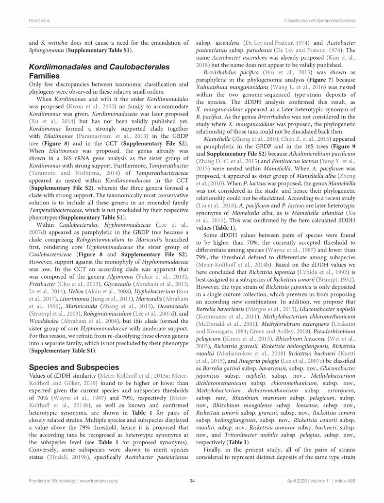
fmicb-11-00468 April 4, 2020 Time: 10:24 # 34
Hördt et al. Classification of Alphaproteobacteria
and S. wittichii does not cause a need for the emendation ofSphingomonas (Supplementary Table S1).
Kordiimonadales and CaulobacteralesFamiliesOnly few discrepancies between taxonomic classification andphylogeny were observed in these relative small orders.
When Kordiimonas and with it the order Kordiimonadaleswas proposed (Kwon et al., 2005) no family to accommodateKordiimonas was given. Kordiimonadaceae was later proposed(Xu et al., 2014) but has not been validly published yet.Kordiimonas formed a strongly supported clade togetherwith Eilatimonas (Paramasivam et al., 2013) in the GBDPtree (Figure 8) and in the CCT (Supplementary File S2).When Eilatimonas was proposed, the genus already wasshown in a 16S rRNA gene analysis as the sister group ofKordiimonas with strong support. Furthermore, Temperatibacter(Teramoto and Nishijima, 2014) of Temperatibacteraceaeappeared as nested within Kordiimonadaceae in the CCT(Supplementary File S2), wherein the three genera formed aclade with strong support. The taxonomically most conservativesolution is to include all these genera in an emended familyTemperatibacteraceae, which is not precluded by their respectivephenotypes (Supplementary Table S1).
Within Caulobacterales, Hyphomonadaceae (Lee et al.,2007d) appeared as paraphyletic in the GBDP tree because aclade comprising Robiginitomaculum to Maricaulis branchedfirst, rendering core Hyphomonadaceae the sister group ofCaulobacteraceae (Figure 8 and Supplementary File S2).However, support against the monophyly of Hyphomonadaceaewas low. In the CCT an according clade was apparent thatwas composed of the genera Algimonas (Fukui et al., 2013),Fretibacter (Cho et al., 2013), Glycocaulis (Abraham et al., 2013;Lv et al., 2014), Hellea (Alain et al., 2008), Hyphobacterium (Sunet al., 2017), Litorimonas (Jung et al., 2011), Maricaulis (Abrahamet al., 1999), Marinicauda (Zhang et al., 2013), Oceanicaulis(Strömpl et al., 2003), Robiginitomaculum (Lee et al., 2007d), andWoodsholea (Abraham et al., 2004), but this clade formed thesister group of core Hyphomonadaceae with moderate support.For this reason, we refrain from re-classifying these eleven generainto a separate family, which is not precluded by their phenotype(Supplementary Table S1).
Species and SubspeciesValues of dDDH similarity (Meier-Kolthoff et al., 2013a; Meier-Kolthoff and Göker, 2019) found to be higher or lower thanexpected given the current species and subspecies thresholdsof 70% (Wayne et al., 1987) and 79%, respectively (Meier-Kolthoff et al., 2014b), as well as known and confirmedheterotypic synonyms, are shown in Table 1 for pairs ofclosely related strains. Multiple species and subspecies displayeda value above the 79% threshold, hence it is proposed thatthe according taxa be recognised as heterotypic synonyms atthe subspecies level (see Table 1 for proposed synonyms).Conversely, some subspecies were shown to merit speciesstatus (Tindall, 2019b), specifically Acetobacter pasteurianus
subsp. ascendens (De Ley and Frateur, 1974) and Acetobacterpasteurianus subsp. paradoxus (De Ley and Frateur, 1974). Thename Acetobacter ascendens was already proposed (Kim et al.,2018) but the name does not appear to be validly published.
Brevirhabdus pacifica (Wu et al., 2015) was shown asparaphyletic in the phylogenomic analysis (Figure 7) becauseXuhuaishuia manganoxidans (Wang L. et al., 2016) was nestedwithin the two genome-sequenced type-strain deposits ofthe species. The dDDH analysis confirmed this result, asX. manganoxidans appeared as a later heterotypic synonym ofB. pacifica. As the genus Brevirhabdus was not considered in thestudy where X. manganoxidans was proposed, the phylogeneticrelationship of those taxa could not be elucidated back then.
Mameliella (Zheng et al., 2010; Chen Z. et al., 2015) appearedas paraphyletic in the GBDP and in the 16S trees (Figure 9and Supplementary File S2) because Alkalimicrobium pacificum(Zhang D.-C. et al., 2015) and Ponticoccus lacteus (Yang Y. et al.,2015) were nested within Mameliella. When A. pacificum wasproposed, it appeared as sister group of Mameliella alba (Zhenget al., 2010). When P. lacteus was proposed, the genus Mameliellawas not considered in the study, and hence their phylogeneticrelationship could not be elucidated. According to a recent study(Liu et al., 2018), A. pacificum and P. lacteus are later heterotypicsynonyms of Mameliella alba, as is Mameliella atlantica (Xuet al., 2015). This was confirmed by the here calculated dDDHvalues (Table 1).
Some dDDH values between pairs of species were foundto be higher than 70%, the currently accepted threshold todifferentiate among species (Wayne et al., 1987) and lower than79%, the threshold defined to differentiate among subspecies(Meier-Kolthoff et al., 2014b). Based on the dDDH values wehere concluded that Rickettsia japonica (Uchida et al., 1992) isbest assigned to a subspecies of Rickettsia conorii (Brumpt, 1932).However, the type strain of Rickettsia japonica is only depositedin a single culture collection, which prevents us from proposingan according new combination. In addition, we propose thatBorrelia bavariensis (Margos et al., 2013), Gluconobacter nephelii(Kommanee et al., 2011), Methylobacterium chloromethanicum(McDonald et al., 2001), Methylorubrum extorquens (Urakamiand Komagata, 1984; Green and Ardley, 2018), Pseudorhizobiumpelagicum (Kimes et al., 2015), Rhizobium loessense (Wei et al.,2003), Rickettsia gravesii, Rickettsia heilongjiangensis, Rickettsiaraoultii (Mediannikov et al., 2008) Rickettsia buchneri (Kurttiet al., 2015), and Ruegeria pelagia (Lee et al., 2007c) be classifiedas Borrelia garinii subsp. bavariensis, subsp. nov., Gluconobacterjaponicus subsp. nephelii, subsp. nov., Methylobacteriumdichloromethanicum subsp. chloromethanicum, subsp. nov.,Methylobacterium dichloromethanicum subsp. extorquens,subsp. nov., Rhizobium marinum subsp. pelagicum, subsp.nov., Rhizobium mongolense subsp. loessense, subsp. nov.,Rickettsia conorii subsp. gravesii, subsp. nov., Rickettsia conoriisubsp. heilongjiangensis, subsp. nov., Rickettsia conorii subsp.raoultii, subsp. nov., Rickettsia tamurae subsp. buchneri, subsp.nov., and Tritonibacter mobilis subsp. pelagius, subsp. nov.,respectively (Table 1).
Finally, in the present study, all of the pairs of strainsconsidered to represent distinct deposits of the same type strain
Frontiers in Microbiology | www.frontiersin.org 34 April 2020 | Volume 11 | Article 468
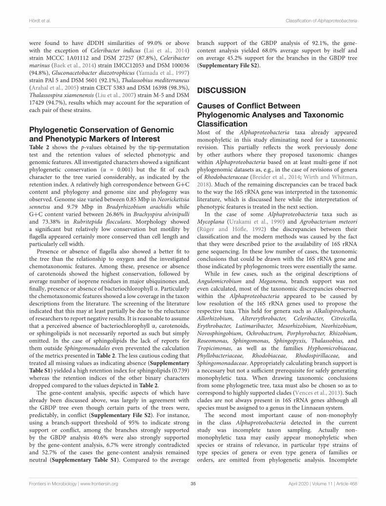
fmicb-11-00468 April 4, 2020 Time: 10:24 # 35
Hördt et al. Classification of Alphaproteobacteria
were found to have dDDH similarities of 99.0% or abovewith the exception of Celeribacter indicus (Lai et al., 2014)strain MCCC 1A01112 and DSM 27257 (87.8%), Celeribactermarinus (Baek et al., 2014) strain IMCC12053 and DSM 100036(94.8%), Gluconacetobacter diazotrophicus (Yamada et al., 1997)strain PAl 5 and DSM 5601 (92.1%), Thalassobius mediterraneus(Arahal et al., 2005) strain CECT 5383 and DSM 16398 (98.3%),Thalassospira xiamenensis (Liu et al., 2007) strain M-5 and DSM17429 (94.7%), results which may account for the separation ofeach pair of these strains.
Phylogenetic Conservation of Genomicand Phenotypic Markers of InterestTable 2 shows the p-values obtained by the tip-permutationtest and the retention values of selected phenotypic andgenomic features. All investigated characters showed a significantphylogenetic conservation (α = 0.001) but the fit of eachcharacter to the tree varied considerably, as indicated by theretention index. A relatively high correspondence between G+Ccontent and phylogeny and genome size and phylogeny wasobserved. Genome size varied between 0.85 Mbp in Neorickettsiasennetsu and 9.79 Mbp in Bradyrhizobium arachidis whileG+C content varied between 26.86% in Brachyspira alvinipulliand 73.38% in Rubritepida flocculans. Morphology showeda significant but relatively low conservation but motility byflagella appeared certainly more conserved than cell length andparticularly cell width.
Presence or absence of flagella also showed a better fit tothe tree than the relationship to oxygen and the investigatedchemotaxonomic features. Among these, presence or absenceof carotenoids showed the highest conservation, followed byaverage number of isoprene residues in major ubiquinones and,finally, presence or absence of bacteriochlorophyll α. Particularlythe chemotaxonomic features showed a low coverage in the taxondescriptions from the literature. The screening of the literatureindicated that this may at least partially be due to the reluctanceof researchers to report negative results. It is reasonable to assumethat a perceived absence of bacteriochlorophyll α, carotenoids,or sphingolipids is not necessarily reported as such but simplyomitted. In the case of sphingolipids the lack of reports forthem outside Sphingomonadales even prevented the calculationof the metrics presented in Table 2. The less cautious coding thattreated all missing values as indicating absence (SupplementaryTable S1) yielded a high retention index for sphingolipids (0.739)whereas the retention indices of the other binary charactersdropped compared to the values depicted in Table 2.
The gene-content analysis, specific aspects of which havealready been discussed above, was largely in agreement withthe GBDP tree even though certain parts of the trees were,predictably, in conflict (Supplementary File S2). For instance,using a branch-support threshold of 95% to indicate strongsupport or conflict, among the branches strongly supportedby the GBDP analysis 40.6% were also strongly supportedby the gene-content analysis, 6.7% were strongly contradictedand 52.7% of the cases the gene-content analysis remainedneutral (Supplementary Table S1). Compared to the average
branch support of the GBDP analysis of 92.1%, the gene-content analysis yielded 68.0% average support by itself andon average 45.2% support for the branches in the GBDP tree(Supplementary File S2).
DISCUSSION
Causes of Conflict BetweenPhylogenomic Analyses and TaxonomicClassificationMost of the Alphaproteobacteria taxa already appearedmonophyletic in this study eliminating need for a taxonomicrevision. This partially reflects the work previously doneby other authors where they proposed taxonomic changeswithin Alphaproteobacteria based on at least multi-gene if notphylogenomic datasets as, e.g., in the case of revisions of generaof Rhodobacteraceae (Breider et al., 2014; Wirth and Whitman,2018). Much of the remaining discrepancies can be traced backto the way the 16S rRNA gene was interpreted in the taxonomicliterature, which is discussed here while the interpretation ofphenotypic features is treated in the next section.
In the case of some Alphaproteobacteria taxa such asMycoplana (Urakami et al., 1990) and Agrobacterium meteori(Rüger and Höfle, 1992) the discrepancies between theirclassification and the modern methods was caused by the factthat they were described prior to the availability of 16S rRNAgene sequencing. In these low number of cases, the taxonomicconclusions that could be drawn with the 16S rRNA gene andthose indicated by phylogenomic trees were essentially the same.
While in few cases, such as the original descriptions ofAngulomicrobium and Meganema, branch support was noteven calculated, most of the taxonomic discrepancies observedwithin the Alphaproteobacteria appeared to be caused bylow resolution of the 16S rRNA genes used to propose therespective taxa. This held for genera such as Alkalispirochaeta,Allorhizobium, Altererythrobacter, Celeribacter, Citreicella,Erythrobacter, Lutimaribacter, Mesorhizobium, Neorhizobium,Novosphingobium, Ochrobactrum, Porphyrobacter, Rhizobium,Roseomonas, Sphingomonas, Sphingopyxis, Thalassobius, andTropicimonas, as well as the families Hyphomicrobiaceae,Phyllobacteriaceae, Rhodobiaceae, Rhodospirillaceae, andSphingomonadaceae. Appropriately calculating branch support isa necessary but not a sufficient prerequisite for safely generatingmonophyletic taxa. When drawing taxonomic conclusionsfrom some phylogenetic tree, taxa must also be chosen so as tocorrespond to highly supported clades (Vences et al., 2013). Suchclades are not always present in 16S rRNA genes although allspecies must be assigned to a genus in the Linnaean system.
The second most important cause of non-monophylyin the class Alphaproteobacteria detected in the currentstudy was incomplete taxon sampling. Actually non-monophyletic taxa may easily appear monophyletic whenspecies or strains of relevance, in particular type strains oftype species of genera or even type genera of families ororders, are omitted from phylogenetic analysis. Incomplete
Frontiers in Microbiology | www.frontiersin.org 35 April 2020 | Volume 11 | Article 468
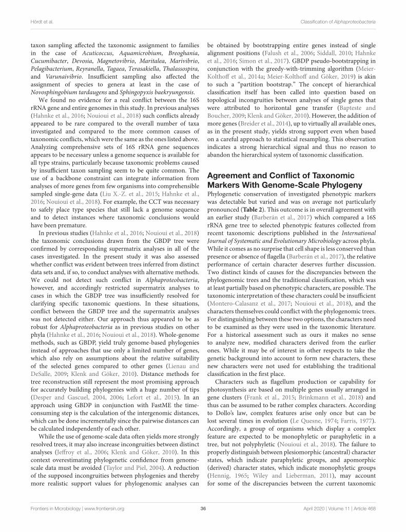
fmicb-11-00468 April 4, 2020 Time: 10:24 # 36
Hördt et al. Classification of Alphaproteobacteria
taxon sampling affected the taxonomic assignment to familiesin the case of Acuticoccus, Aquamicrobium, Breoghania,Cucumibacter, Devosia, Magnetovibrio, Maritalea, Marivibrio,Pelagibacterium, Reyranella, Tagaea, Terasakiella, Thalassospira,and Varunaivibrio. Insufficient sampling also affected theassignment of species to genera at least in the case ofNovosphingobium tardaugens and Sphingopyxis baekryungensis.
We found no evidence for a real conflict between the 16SrRNA gene and entire genomes in this study. In previous analyses(Hahnke et al., 2016; Nouioui et al., 2018) such conflicts alreadyappeared to be rare compared to the overall number of taxainvestigated and compared to the more common causes oftaxonomic conflicts, which were the same as the ones listed above.Analyzing comprehensive sets of 16S rRNA gene sequencesappears to be necessary unless a genome sequence is available forall type strains, particularly because taxonomic problems causedby insufficient taxon sampling seem to be quite common. Theuse of a backbone constraint can integrate information fromanalyses of more genes from few organisms into comprehensiblesampled single-gene data (Liu X.-Z. et al., 2015; Hahnke et al.,2016; Nouioui et al., 2018). For example, the CCT was necessaryto safely place type species that still lack a genome sequenceand to detect instances where taxonomic conclusions wouldhave been premature.
In previous studies (Hahnke et al., 2016; Nouioui et al., 2018)the taxonomic conclusions drawn from the GBDP tree wereconfirmed by corresponding supermatrix analyses in all of thecases investigated. In the present study it was also assessedwhether conflict was evident between trees inferred from distinctdata sets and, if so, to conduct analyses with alternative methods.We could not detect such conflict in Alphaproteobacteria,however, and accordingly restricted supermatrix analyses tocases in which the GBDP tree was insufficiently resolved forclarifying specific taxonomic questions. In these situations,conflict between the GBDP tree and the supermatrix analyseswas not detected either. Our approach thus appeared to be asrobust for Alphaproteobacteria as in previous studies on otherphyla (Hahnke et al., 2016; Nouioui et al., 2018). Whole-genomemethods, such as GBDP, yield truly genome-based phylogeniesinstead of approaches that use only a limited number of genes,which also rely on assumptions about the relative suitabilityof the selected genes compared to other genes (Lienau andDeSalle, 2009; Klenk and Göker, 2010). Distance methods fortree reconstruction still represent the most promising approachfor accurately building phylogenies with a huge number of tips(Desper and Gascuel, 2004, 2006; Lefort et al., 2015). In anapproach using GBDP in conjunction with FastME the time-consuming step is the calculation of the intergenomic distances,which can be done incrementally since the pairwise distances canbe calculated independently of each other.
While the use of genome-scale data often yields more stronglyresolved trees, it may also increase incongruities between distinctanalyses (Jeffroy et al., 2006; Klenk and Göker, 2010). In thiscontext overestimating phylogenetic confidence from genome-scale data must be avoided (Taylor and Piel, 2004). A reductionof the supposed incongruities between phylogenies and therebymore realistic support values for phylogenomic analyses can
be obtained by bootstrapping entire genes instead of singlealignment positions (Falush et al., 2006; Siddall, 2010; Hahnkeet al., 2016; Simon et al., 2017). GBDP pseudo-bootstrapping inconjunction with the greedy-with-trimming algorithm (Meier-Kolthoff et al., 2014a; Meier-Kolthoff and Göker, 2019) is akinto such a “partition bootstrap.” The concept of hierarchicalclassification itself has been called into question based ontopological incongruities between analyses of single genes thatwere attributed to horizontal gene transfer (Bapteste andBoucher, 2009; Klenk and Göker, 2010). However, the addition ofmore genes (Breider et al., 2014), up to virtually all available ones,as in the present study, yields strong support even when basedon a careful approach to statistical resampling. This observationindicates a strong hierarchical signal and thus no reason toabandon the hierarchical system of taxonomic classification.
Agreement and Conflict of TaxonomicMarkers With Genome-Scale PhylogenyPhylogenetic conservation of investigated phenotypic markerswas detectable but varied and was on average not particularlypronounced (Table 2). This outcome is in overall agreement withan earlier study (Barberán et al., 2017) which compared a 16SrRNA gene tree to selected phenotypic features collected fromrecent taxonomic descriptions published in the InternationalJournal of Systematic and Evolutionary Microbiology across phyla.While it comes as no surprise that cell shape is less conserved thanpresence or absence of flagella (Barberán et al., 2017), the relativeperformance of certain character deserves further discussion.Two distinct kinds of causes for the discrepancies between thephylogenomic trees and the traditional classification, which wasat least partially based on phenotypic characters, are possible. Thetaxonomic interpretation of these characters could be insufficient(Montero-Calasanz et al., 2017; Nouioui et al., 2018), and thecharacters themselves could conflict with the phylogenomic trees.For distinguishing between these two options, the characters needto be examined as they were used in the taxonomic literature.For a historical assessment such as ours it makes no senseto analyze new, modified characters derived from the earlierones. While it may be of interest in other respects to take thegenetic background into account to form new characters, thesenew characters were not used for establishing the traditionalclassification in the first place.
Characters such as flagellum production or capability forphotosynthesis are based on multiple genes usually arranged ingene clusters (Frank et al., 2015; Brinkmann et al., 2018) andthus can be assumed to be rather complex characters. Accordingto Dollo’s law, complex features arise only once but can belost several times in evolution (Le Quesne, 1974; Farris, 1977).Accordingly, a group of organisms which display a complexfeature are expected to be monophyletic or paraphyletic in atree, but not polyphyletic (Nouioui et al., 2018). The failure toproperly distinguish between plesiomorphic (ancestral) characterstates, which indicate paraphyletic groups, and apomorphic(derived) character states, which indicate monophyletic groups(Hennig, 1965; Wiley and Lieberman, 2011), may accountfor some of the discrepancies between the current taxonomic
Frontiers in Microbiology | www.frontiersin.org 36 April 2020 | Volume 11 | Article 468
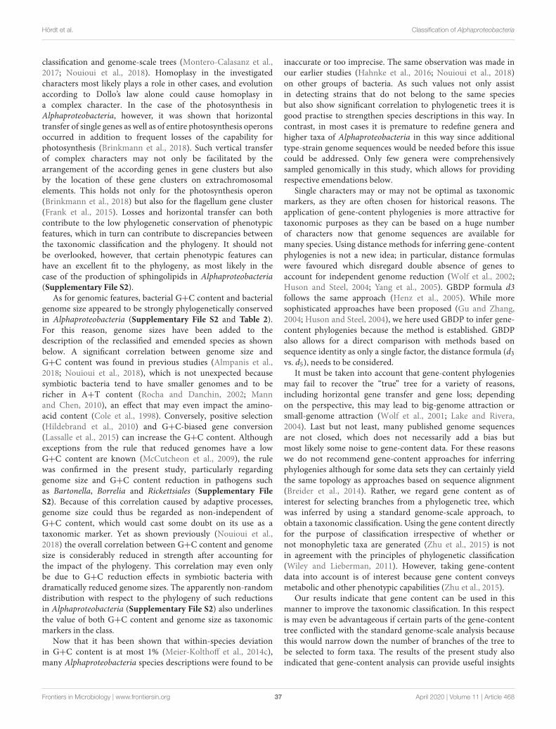
fmicb-11-00468 April 4, 2020 Time: 10:24 # 37
Hördt et al. Classification of Alphaproteobacteria
classification and genome-scale trees (Montero-Calasanz et al.,2017; Nouioui et al., 2018). Homoplasy in the investigatedcharacters most likely plays a role in other cases, and evolutionaccording to Dollo’s law alone could cause homoplasy ina complex character. In the case of the photosynthesis inAlphaproteobacteria, however, it was shown that horizontaltransfer of single genes as well as of entire photosynthesis operonsoccurred in addition to frequent losses of the capability forphotosynthesis (Brinkmann et al., 2018). Such vertical transferof complex characters may not only be facilitated by thearrangement of the according genes in gene clusters but alsoby the location of these gene clusters on extrachromosomalelements. This holds not only for the photosynthesis operon(Brinkmann et al., 2018) but also for the flagellum gene cluster(Frank et al., 2015). Losses and horizontal transfer can bothcontribute to the low phylogenetic conservation of phenotypicfeatures, which in turn can contribute to discrepancies betweenthe taxonomic classification and the phylogeny. It should notbe overlooked, however, that certain phenotypic features canhave an excellent fit to the phylogeny, as most likely in thecase of the production of sphingolipids in Alphaproteobacteria(Supplementary File S2).
As for genomic features, bacterial G+C content and bacterialgenome size appeared to be strongly phylogenetically conservedin Alphaproteobacteria (Supplementary File S2 and Table 2).For this reason, genome sizes have been added to thedescription of the reclassified and emended species as shownbelow. A significant correlation between genome size andG+C content was found in previous studies (Almpanis et al.,2018; Nouioui et al., 2018), which is not unexpected becausesymbiotic bacteria tend to have smaller genomes and to bericher in A+T content (Rocha and Danchin, 2002; Mannand Chen, 2010), an effect that may even impact the amino-acid content (Cole et al., 1998). Conversely, positive selection(Hildebrand et al., 2010) and G+C-biased gene conversion(Lassalle et al., 2015) can increase the G+C content. Althoughexceptions from the rule that reduced genomes have a lowG+C content are known (McCutcheon et al., 2009), the rulewas confirmed in the present study, particularly regardinggenome size and G+C content reduction in pathogens suchas Bartonella, Borrelia and Rickettsiales (Supplementary FileS2). Because of this correlation caused by adaptive processes,genome size could thus be regarded as non-independent ofG+C content, which would cast some doubt on its use as ataxonomic marker. Yet as shown previously (Nouioui et al.,2018) the overall correlation between G+C content and genomesize is considerably reduced in strength after accounting forthe impact of the phylogeny. This correlation may even onlybe due to G+C reduction effects in symbiotic bacteria withdramatically reduced genome sizes. The apparently non-randomdistribution with respect to the phylogeny of such reductionsin Alphaproteobacteria (Supplementary File S2) also underlinesthe value of both G+C content and genome size as taxonomicmarkers in the class.
Now that it has been shown that within-species deviationin G+C content is at most 1% (Meier-Kolthoff et al., 2014c),many Alphaproteobacteria species descriptions were found to be
inaccurate or too imprecise. The same observation was made inour earlier studies (Hahnke et al., 2016; Nouioui et al., 2018)on other groups of bacteria. As such values not only assistin detecting strains that do not belong to the same speciesbut also show significant correlation to phylogenetic trees it isgood practise to strengthen species descriptions in this way. Incontrast, in most cases it is premature to redefine genera andhigher taxa of Alphaproteobacteria in this way since additionaltype-strain genome sequences would be needed before this issuecould be addressed. Only few genera were comprehensivelysampled genomically in this study, which allows for providingrespective emendations below.
Single characters may or may not be optimal as taxonomicmarkers, as they are often chosen for historical reasons. Theapplication of gene-content phylogenies is more attractive fortaxonomic purposes as they can be based on a huge numberof characters now that genome sequences are available formany species. Using distance methods for inferring gene-contentphylogenies is not a new idea; in particular, distance formulaswere favoured which disregard double absence of genes toaccount for independent genome reduction (Wolf et al., 2002;Huson and Steel, 2004; Yang et al., 2005). GBDP formula d3follows the same approach (Henz et al., 2005). While moresophisticated approaches have been proposed (Gu and Zhang,2004; Huson and Steel, 2004), we here used GBDP to infer gene-content phylogenies because the method is established. GBDPalso allows for a direct comparison with methods based onsequence identity as only a single factor, the distance formula (d3vs. d5), needs to be considered.
It must be taken into account that gene-content phylogeniesmay fail to recover the “true” tree for a variety of reasons,including horizontal gene transfer and gene loss; dependingon the perspective, this may lead to big-genome attraction orsmall-genome attraction (Wolf et al., 2001; Lake and Rivera,2004). Last but not least, many published genome sequencesare not closed, which does not necessarily add a bias butmost likely some noise to gene-content data. For these reasonswe do not recommend gene-content approaches for inferringphylogenies although for some data sets they can certainly yieldthe same topology as approaches based on sequence alignment(Breider et al., 2014). Rather, we regard gene content as ofinterest for selecting branches from a phylogenetic tree, whichwas inferred by using a standard genome-scale approach, toobtain a taxonomic classification. Using the gene content directlyfor the purpose of classification irrespective of whether ornot monophyletic taxa are generated (Zhu et al., 2015) is notin agreement with the principles of phylogenetic classification(Wiley and Lieberman, 2011). However, taking gene-contentdata into account is of interest because gene content conveysmetabolic and other phenotypic capabilities (Zhu et al., 2015).
Our results indicate that gene content can be used in thismanner to improve the taxonomic classification. In this respectis may even be advantageous if certain parts of the gene-contenttree conflicted with the standard genome-scale analysis becausethis would narrow down the number of branches of the tree tobe selected to form taxa. The results of the present study alsoindicated that gene-content analysis can provide useful insights
Frontiers in Microbiology | www.frontiersin.org 37 April 2020 | Volume 11 | Article 468
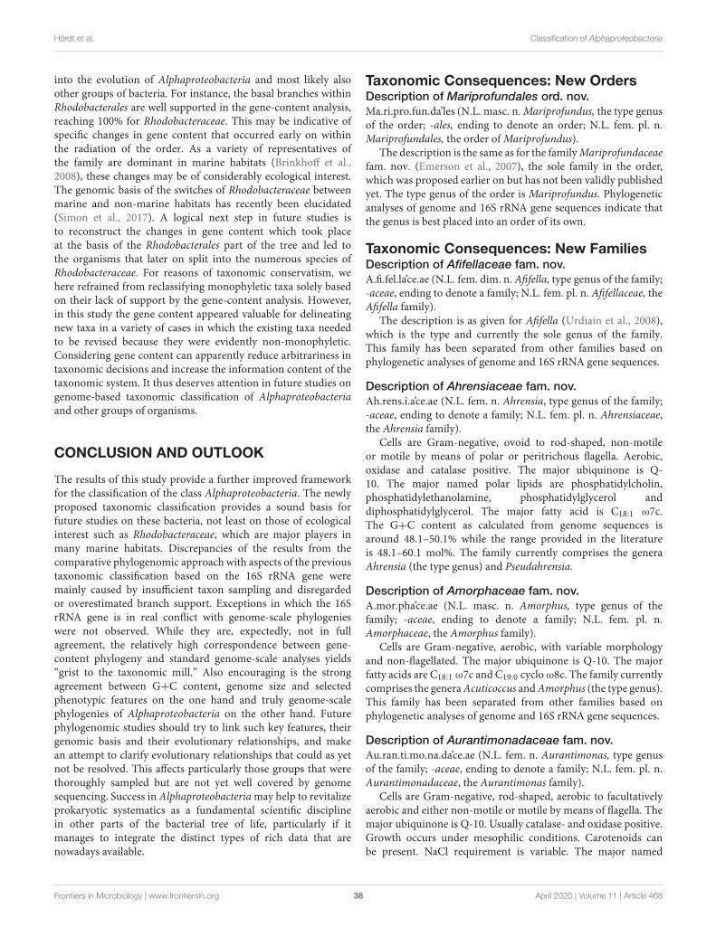
fmicb-11-00468 April 4, 2020 Time: 10:24 # 38
Hördt et al. Classification of Alphaproteobacteria
into the evolution of Alphaproteobacteria and most likely alsoother groups of bacteria. For instance, the basal branches withinRhodobacterales are well supported in the gene-content analysis,reaching 100% for Rhodobacteraceae. This may be indicative ofspecific changes in gene content that occurred early on withinthe radiation of the order. As a variety of representatives ofthe family are dominant in marine habitats (Brinkhoff et al.,2008), these changes may be of considerably ecological interest.The genomic basis of the switches of Rhodobacteraceae betweenmarine and non-marine habitats has recently been elucidated(Simon et al., 2017). A logical next step in future studies isto reconstruct the changes in gene content which took placeat the basis of the Rhodobacterales part of the tree and led tothe organisms that later on split into the numerous species ofRhodobacteraceae. For reasons of taxonomic conservatism, wehere refrained from reclassifying monophyletic taxa solely basedon their lack of support by the gene-content analysis. However,in this study the gene content appeared valuable for delineatingnew taxa in a variety of cases in which the existing taxa neededto be revised because they were evidently non-monophyletic.Considering gene content can apparently reduce arbitrariness intaxonomic decisions and increase the information content of thetaxonomic system. It thus deserves attention in future studies ongenome-based taxonomic classification of Alphaproteobacteriaand other groups of organisms.
CONCLUSION AND OUTLOOK
The results of this study provide a further improved frameworkfor the classification of the class Alphaproteobacteria. The newlyproposed taxonomic classification provides a sound basis forfuture studies on these bacteria, not least on those of ecologicalinterest such as Rhodobacteraceae, which are major players inmany marine habitats. Discrepancies of the results from thecomparative phylogenomic approach with aspects of the previoustaxonomic classification based on the 16S rRNA gene weremainly caused by insufficient taxon sampling and disregardedor overestimated branch support. Exceptions in which the 16SrRNA gene is in real conflict with genome-scale phylogenieswere not observed. While they are, expectedly, not in fullagreement, the relatively high correspondence between gene-content phylogeny and standard genome-scale analyses yields“grist to the taxonomic mill.” Also encouraging is the strongagreement between G+C content, genome size and selectedphenotypic features on the one hand and truly genome-scalephylogenies of Alphaproteobacteria on the other hand. Futurephylogenomic studies should try to link such key features, theirgenomic basis and their evolutionary relationships, and makean attempt to clarify evolutionary relationships that could as yetnot be resolved. This affects particularly those groups that werethoroughly sampled but are not yet well covered by genomesequencing. Success in Alphaproteobacteria may help to revitalizeprokaryotic systematics as a fundamental scientific disciplinein other parts of the bacterial tree of life, particularly if itmanages to integrate the distinct types of rich data that arenowadays available.
Taxonomic Consequences: New OrdersDescription of Mariprofundales ord. nov.Ma.ri.pro.fun.da’les (N.L. masc. n. Mariprofundus, the type genusof the order; -ales, ending to denote an order; N.L. fem. pl. n.Mariprofundales, the order of Mariprofundus).
The description is the same as for the family Mariprofundaceaefam. nov. (Emerson et al., 2007), the sole family in the order,which was proposed earlier on but has not been validly publishedyet. The type genus of the order is Mariprofundus. Phylogeneticanalyses of genome and 16S rRNA gene sequences indicate thatthe genus is best placed into an order of its own.
Taxonomic Consequences: New FamiliesDescription of Afifellaceae fam. nov.A.fi.fel.la’ce.ae (N.L. fem. dim. n. Afifella, type genus of the family;-aceae, ending to denote a family; N.L. fem. pl. n. Afifellaceae, theAfifella family).
The description is as given for Afifella (Urdiain et al., 2008),which is the type and currently the sole genus of the family.This family has been separated from other families based onphylogenetic analyses of genome and 16S rRNA gene sequences.
Description of Ahrensiaceae fam. nov.Ah.rens.i.a’ce.ae (N.L. fem. n. Ahrensia, type genus of the family;-aceae, ending to denote a family; N.L. fem. pl. n. Ahrensiaceae,the Ahrensia family).
Cells are Gram-negative, ovoid to rod-shaped, non-motileor motile by means of polar or peritrichous flagella. Aerobic,oxidase and catalase positive. The major ubiquinone is Q-10. The major named polar lipids are phosphatidylcholin,phosphatidylethanolamine, phosphatidylglycerol anddiphosphatidylglycerol. The major fatty acid is C18:1 ω7c.The G+C content as calculated from genome sequences isaround 48.1–50.1% while the range provided in the literatureis 48.1–60.1 mol%. The family currently comprises the generaAhrensia (the type genus) and Pseudahrensia.
Description of Amorphaceae fam. nov.A.mor.pha’ce.ae (N.L. masc. n. Amorphus, type genus of thefamily; -aceae, ending to denote a family; N.L. fem. pl. n.Amorphaceae, the Amorphus family).
Cells are Gram-negative, aerobic, with variable morphologyand non-flagellated. The major ubiquinone is Q-10. The majorfatty acids are C18:1 ω7c and C19:0 cyclo ω8c. The family currentlycomprises the genera Acuticoccus and Amorphus (the type genus).This family has been separated from other families based onphylogenetic analyses of genome and 16S rRNA gene sequences.
Description of Aurantimonadaceae fam. nov.Au.ran.ti.mo.na.da’ce.ae (N.L. fem. n. Aurantimonas, type genusof the family; -aceae, ending to denote a family; N.L. fem. pl. n.Aurantimonadaceae, the Aurantimonas family).
Cells are Gram-negative, rod-shaped, aerobic to facultativelyaerobic and either non-motile or motile by means of flagella. Themajor ubiquinone is Q-10. Usually catalase- and oxidase positive.Growth occurs under mesophilic conditions. Carotenoids canbe present. NaCl requirement is variable. The major named
Frontiers in Microbiology | www.frontiersin.org 38 April 2020 | Volume 11 | Article 468

fmicb-11-00468 April 4, 2020 Time: 10:24 # 39
Hördt et al. Classification of Alphaproteobacteria
polar lipids are diphosphatidylglycerol, phosphatidylglycerol,phosphatidylcholin, phosphatidylethanolamine, phosphati-dylmonomethylethanolamine and aminolipids. The majornamed fatty acids are usually C18:1 ω7c and C19:0 cyclo ω8c.The family currently comprises the genera Mangrovicella, Jiella,Aurantimonas (the type genus), Aureimonas, Fulvimarinaand Consotaella.
Description of Azospirillaceae fam. nov.A.zo.spi.ril.la’ce.ae (N.L. neut. dim. n. Azospirillum, type genusof the family; -aceae, ending to denote a family; N.L. fem. pl. n.Azospirillaceae, the Azospirillum family).
Cells are Gram-negative, mostly rod-shaped, in generalaerobic, usually motile by flagella, usually heterotrophic. Themajor ubiquinone is predominantly Q-10. The major namedpolar lipids are phosphatidylcholin, phosphatidylethanolamine,phosphatidylglycerol and diphosphatidylglycerol. The majorfatty acids are usually C18:1 ω7c, C18:1 ω6c, C16:0 and C16:1ω6c. The family currently comprises the genera Niveispirillum,Azospirillum (including Conglomeromonas), Skermanella,Nitrospirillum, Rhodocista, and Desertibacter. The genera Elstera,Inquilinus and Lacibacterium are tentatively assigned to thisfamily. The type genus is Azospirillum.
Description of Blastochloridaceae fam. nov.Blas.to.chlo.ri.da’ce.ae (N.L. fem. n. Blastochloris, type genus ofthe family; -aceae, ending to denote a family; N.L. fem. pl. n.Blastochloridaceae, the Blastochloris family).
The description is as given for Blastochloris (Hiraishi, 1997),which is the type and currently the sole genus of the family.This family has been separated from Hyphomicrobiaceae based onphylogenetic analyses of genome and 16S rRNA gene sequences.
Description of Boseaceae fam. nov.Bo.se.a’ce.ae (N.L. fem. n. Bosea, type genus of the family; -aceae,ending to denote a family; N.L. fem. pl. n. Boseaceae, theBosea family).
The description is as given for Bosea (Das et al., 1996;La Scola et al., 2003), which is the type genus and currentlythe sole genus of the family. This family has been separatedfrom Nitrobacteraceae (whose illegitimate synonym isBradyrhizobiaceae) based on phylogenetic analyses of genomeand 16S rRNA gene sequences.
Description of Breoghaniaceae fam. nov.Bre.o.gha.ni.a’ce.ae (N.L. fem. n. Breoghania, type genus of thefamily; -aceae, ending to denote a family; N.L. fem. pl. n.Breoghaniaceae, the Breoghania family).
The description is as given for Breoghania (Gallego et al.,2010),which is the type and currently the sole genus of the family.This family has been separated from other families based onphylogenetic analyses of genome and 16S rRNA gene sequences.
Description of Devosiaceae fam. nov.De.vo.si.a’ce.ae (N.L. fem. n. Devosia, type genus of the family; -aceae, ending to denote a family; N.L. fem. pl. n. Devosiaceae, theDevosia family).
Cells are Gram-negative, predominantly rod-shaped, aerobic,usually motile by flagella, and heterotrophic. The majorubiquinone is predominantly Q-10 whereas Q-9 and Q-11present as minor ubiquinone in some species. The majorpolar lipids are phosphatidylglycerol, diphosphatidylglyceroland glycolipid. The major fatty acids are usually C18:1ω7c, 11-methyl C18:1 ω7c, C18:1 ω6c, C18:0, C16:0 ω6cand C19:0 cyclo ω8c. The family currently comprises thegenera Arsenicitalea, Cucumibacter, Devosia (the type genus),Maritalea (including Zhangella), Methyloterrigena, Paradevosia,Pelagibacterium, and Youhaiella. This family has been separatedfrom Hyphomicrobiaceae based on phylogenetic analyses ofgenome and 16S rRNA gene sequences.
Description of Kaistiaceae fam. nov.Kais.ti.a’ce.ae (N.L. fem. n. Kaistia, type genus of the family; -aceae, ending to denote a family; N.L. fem. pl. n. Kaistiaceae, theKaistia family).
Cells are Gram-negative, cocci or rod-shaped, non-motile,aerobic and chemoorganotrophic. The G+C content ascalculated from genome sequences is around 64.5–67.1% whilethe range provided in the literature is 61.6–69.0 mol%. Thefamily currently comprises the genera Kaistia (the type genus)and Bauldia.
Description of Mariprofundaceae fam. nov.Ma.ri.pro.fun.da’ce.ae (N.L. masc. n. Mariprofundus, type genusof the family; -aceae, ending to denote a family; N.L. fem. pl. n.Mariprofundaceae, the Mariprofundus family).
The description is that for Mariprofundus (Emerson et al.,2007), which is the type and currently sole genus of the family.The family was proposed earlier on but has not been validlypublished yet. It has been separated from other families based onphylogenetic analyses of genome and 16S rRNA gene sequences.
Description of Neomegalonemataceae fam. nov.Ne.o.me.ga.lo.ne.ma.ta’ce.ae (N.L. neut. n. Neomegalonema, typegenus of the family; -aceae, ending to denote a family; N.L. fem.pl. n. Neomegalonemataceae, the Neomegalonema family).
The description is as given for Neomegalonema (Oren,2017b), which is the type and currently the sole genus of thefamily. This family has been separated from Hyphomicrobiales(known under the illegitimate synonym Rhizobiales) and fromRhodobacteraceae based on phylogenetic analyses of genome and16S rRNA gene sequences.
Description of Parvibaculaceae fam. nov.Par.vi.ba.cu.la’ce.ae (N.L. neut. n. Parvibaculum, type genus ofthe family; -aceae, ending to denote a family; N.L. fem. pl. n.Parvibaculaceae, the Parvibaculum family).
Cells are Gram-negative, usually rod-shaped, generallyaerobic, motile by flagella or non-motile and heterotrophic.The major ubiquinone is Q-10 or Q-11. The major polarlipids are diphosphatidylglycerol, phosphatidylethanolamine andphosphatidylglycerol. The major fatty acid is usually C18:1 ω7cand in some cases C16:0 and C19:0 cyclo. The family currentlycomprises the genera Parvibaculum (the type genus) and
Frontiers in Microbiology | www.frontiersin.org 39 April 2020 | Volume 11 | Article 468
fmicb-11-00468 April 4, 2020 Time: 10:24 # 40
Hördt et al. Classification of Alphaproteobacteria
Tepidicaulis. Anderseniella, Rhodoligotrophos and Pyruvatibacterare tentatively assigned to this family as well.
Description of Phreatobacteraceae fam. nov.Phre.a.to.bac.te.ra’ce.ae (N.L. masc. n. Phreatobacter, type genusof the family; -aceae, ending to denote a family; N.L. fem. pl. n.Phreatobacteraceae, the Phreatobacter family).
The description is as given for Phreatobacter (Tóth et al.,2014; Lee S.D. et al., 2017), which is the type and currentlythe sole genus of the family. This family is proposed based onphylogenetic analyses of genome and 16S rRNA gene sequences.
Description of Pleomorphomonadaceae fam. nov.Ple.o.mor.pho.mo.na.da’ce.ae (N.L. fem. n. Pleomorphomonas,type genus of the family; -aceae, ending to denote afamily; N.L. fem. pl. n. Pleomorphomonadaceae, thePleomorphomonas family).
Cells are Gram-negative, usually rod-shaped or spherical,generally aerobic and mostly diazotrophic. The majorubiquinone is predominantly Q-10. The major polarlipids are phosphatidylglycerol, phosphatidylcholin,phosphatidylethanolamine and sometimes phospholipid.The major fatty are usually C18:1 ω7c and C18:0. Thefamily currently comprises the genera Chthonobacter,Hartmannibacter, Methylobrevis, Mongoliimonas, Oharaeibacter,and Pleomorphomonas (the type genus). This family has beenseparated from other families based on phylogenetic analyses ofgenome and 16S rRNA gene sequences.
Description of Pseudoxanthobacteraceae fam. nov.Pseu.do.xan.tho.bac.te.ra’ce.ae (N.L. masc. n.Pseudoxanthobacter, type genus of the family; -aceae, ending todenote a family; N.L. fem. pl. n. Pseudoxanthobacteraceae, thePseudoxanthobacter family).
The description is as given for Pseudoxanthobacter (Arunet al., 2008), which is the type and currently sole genusof the family. This family has been separated from otherfamilies based on phylogenetic analyses of genome and 16SrRNA gene sequences.
Description of Reyranellaceae fam. nov.Rey.ra.nel.la’ce.ae (N.L. fem. dim. n. Reyranella, type genus ofthe family; -aceae, ending to denote a family; N.L. fem. pl. n.Reyranellaceae, the Reyranella family).
The description is as given for Reyranella (Pagnier et al.,2011) which is the type and currently sole genus of the family.This family has been separated from other families based onphylogenetic analyses of genome and 16S rRNA gene sequences.
Description of Rhodovibrionaceae fam. nov.Rho.do.vi.bri.o.na’ce.ae (N.L. masc. n. Rhodovibrio, type genus ofthe family; -aceae, ending to denote a family; N.L. fem. pl. n.Rhodovibrionaceae, the Rhodovibrio family).
Gram-negative, vibrioid, spiral or rod-shaped, non-motileor motile by means of polar flagella. Aerobic, facultativelyanaerobic or anaerobic, with chemoorganotrophic orphotoorganoheterotrophic metabolism. The major ubiquinoneis Q-10; MK-10 was reported for Rhodovibrio. The major fatty
acids are C19:0 cyclo ω8c, C18:1 ω7c and C18:0. The G+C contentprovided in the literature is 61.5–69.0 mol%. The family currentlycomprises the genera Fodinicurvata, Limimonas, Rhodovibrio(the type genus) and Tistlia. Limibacillus and Pelagibius aretentatively assigned to this family.
Description of Sphaerochaetaceae fam. nov.Sphae.ro.chae.ta’ce.ae (N.L. fem. n. Sphaerochaeta, type genus ofthe family; -aceae, ending to denote a family; N.L. fem. pl. n.Sphaerochaetaceae, the Sphaerochaeta family).
Cells are Gram-negative, mostly non-motile, pleomorphic,anaerobic and heterotrophic. The family currently comprises thegenera Pleomorphochaeta and Sphaerochaeta (the type genus).This family has been separated from other families based onphylogenetic analyses of genome and 16S rRNA gene sequences.
Description of Sphingosinicellaceae fam. nov.Sphin.go.si.ni.cel.la’ce.ae (N.L. fem. n. Sphingosinicella, type genusof the family; -aceae, ending to denote a family; N.L. fem. pl. n.Sphingosinicellaceae, the Sphingosinicella family).
Cells are Gram-negative, aerobic, rod-shaped, mostly motileheterotrophs. The major ubiquinone is Q-10. The major namedpolar lipids are phosphatidylethanolamine, phosphatidylglyceroland in some species diphosphatidylglycerol. The major fattyacids are usually C18:1 ω7c, C16:0 and C16:1 ω7c. The familycurrently comprises the genera Pacificimonas, Sphingosinicella(the type genus), Sandaracinobacter and Sandarakinorhabdus.Polymorphobacter and Sphingoaurantiacus are tentativelyassigned to this family. This family has been separated fromother families based on phylogenetic analyses of genome and 16SrRNA gene sequences.
Description of Stappiaceae fam. nov.Stap.pi.a’ce.ae (N.L. fem. n. Stappia, type genus of the family; -aceae, ending to denote a family; N.L. fem. pl. n. Stappiaceae, theStappia family).
Cells are Gram-negative, usually rod-shaped, motile, mostlyaerobic or facultatively anaerobic and heterotroph. Themajor ubiquinone is usually Q-10. The major fatty acids arepredominantly C18:1 ω7c and C16:1 ω7c. The family currentlycomprises the genera Pannonibacter, Pseudovibrio, Roseibium(including Labrenzia) and Stappia (the type genus). This familyhas been separated from other families based on phylogeneticanalyses of genome and 16S rRNA gene sequences.
Description of Stellaceae fam. nov.Stel.la’ce.ae (L. fem. n. Stella, type genus of the family; -aceae,ending to denote a family; N.L. fem. pl. n. Stellaceae, theStella family).
The description is as given for Stella (Vasilyeva, 1985), whichis the type genus of the family, with the following modification.Cells are six-pronged stars, rods or ovoids. The family housesStella and tentatively also Constrictibacter. This family has beenseparated from Acetobacteraceae based on phylogenetic analysesof genome and 16S rRNA gene sequences.
Frontiers in Microbiology | www.frontiersin.org 40 April 2020 | Volume 11 | Article 468
fmicb-11-00468 April 4, 2020 Time: 10:24 # 41
Hördt et al. Classification of Alphaproteobacteria
Description of Tepidamorphaceae fam. nov.Te.pid.a.mor.pha’ce.ae (N.L. masc. n. Tepidamorphus, type genusof the family; -aceae, ending to denote a family; N.L. fem. pl. n.Tepidamorphaceae, the Tepidamorphus family).
Cells are Gram-negative, either ovoid or rod-shaped,predominantly aerobic, mostly motile via flagella andgenerally chemoorganothrophs. The major ubiquinone isQ-10. The major named polar lipids are diphosphatidylglycerol,phosphatidylethanolamine, and phosphatidylglycerol. Themajor fatty acids are usually C19:0 cyclo ω8c, C18:1 ω7c,C18:0 and C16:0. The family currently comprises the generaButyratibacter, Lutibaculum, Microbaculum, and Tepidamorphus(the type genus).
Description of Terasakiellaceae fam. nov.Te.ra.sa.ki.el.la’ce.ae (N.L. fem. dim. n. Terasakiella, type genusof the family; -aceae, ending to denote a family; N.L. fem. pl. n.Terasakiellaceae, the Terasakiella family).
The description is as given for Terasakiella (Satomi et al.,2002; Han et al., 2016), which is the type and currently solegenus of the family. This family has been separated from otherfamilies based on phylogenetic analyses of genome and 16SrRNA gene sequences.
Description of Thalassobaculaceae fam. nov.Tha.las.so.ba.cu.la’ce.ae (N.L. neut. n. Thalassobaculum, typegenus of the family; -aceae, ending to denote a family; N.L. fem.pl. n. Thalassobaculaceae, the Thalassobaculum family).
Gram-negative, rod-shaped, motile by means of one polarflagellum. Aerobic or facultatively anaerobic respiration andchemoorganotrophic metabolism. The major ubiquinone is Q-10. The major fatty acids are C18:1 ω7c, C16:1 ω7c and C16:0. TheG+C content as calculated from genome sequences is around60.5–67.4% while the range provided in the literature is 60–68 mol%. The family currently comprises the genera Nisaea,Oceanibaculum and Thalassobaculum (the type genus).
Description of Thalassospiraceae fam. nov.Tha.las.so.spi.ra’ce.ae (N.L. fem. n. Thalassospira, type genus ofthe family; -aceae, ending to denote a family; N.L. fem. pl. n.Thalassospiraceae, the Thalassospira family).
Cells are Gram-negative, usually motile by flagella,mostly aerobic or microaerophilic with a heterotrophicor autolithothrophic metabolism. The major ubiquinoneis predominantly Q-10. The major polar lipids arephosphatidylethanolamine and phosphatidylglycerol. Themajor fatty acids are usually C18:1 ω7c, C16:1 ω7c and C16:0.The family currently comprises the genera Magnetovibrio,Terasakiella, Thalassospira (the type genus), Varunaivibrio.Magnetospira is tentatively assigned to this family. This familyhas been separated from other families based on phylogeneticanalyses of genome and 16S rRNA gene sequences.
Description of Zavarziniaceae fam. nov.Za.var.zi.ni.a’ce.ae (N.L. fem. n. Zavarzinia, type genus of thefamily; -aceae, ending to denote a family; N.L. fem. pl. n.Zavarziniaceae, the Zavarzinia family).
The description is as given for Zavarzinia (Meyer et al.,1993), which is the type and currently sole genus of the family.This family has been separated from Acetobacteraceae based onphylogenetic analyses of genome and 16S rRNA gene sequences.
Description of Zymomonadaceae fam. nov.Zy.mo.mo.na.da’ce.ae (N.L. fem. n. Zymomonas, type genus ofthe family; -aceae, ending to denote a family; N.L. fem. pl. n.Zymomonadaceae, the Zymomonas family).
The description is as given for Zymomonas (Kluyver andvan Niel, 1936), which is the type and currently sole genusof the family. This family has been separated from otherfamilies based on phylogenetic analyses of genome and 16SrRNA gene sequences.
Taxonomic Consequences: New GeneraDescription of Albibacillus gen. nov.Al.bi.ba.cil’lus (L. masc. adj. albus, white; L. masc. n. bacillus, rod;N.L. masc. n. Albibacillus, white rod).
Gram-negative, rod-shaped, aerobic, oxidase and catalasepositive. The major ubiquinone is Q-10. The major polarlipids are phosphatidylglycerol, phosphatidylethanolamine,phosphatidylmonomethylethanolamine and aminolipids. Themajor fatty acids are C18:1 ω7c and C19:0 cyclo ω8c. The G+Ccontent is around 64-66%. The type species is Albibacilluskandeliae, comb. nov.
Description of Allgaiera gen. nov.All.gai’er.a (N.L. fem. n. Allgaiera, named after the Germanmicrobiologist Martin Allgaier for his work on marineRhodobacteraceae).
The description is as given for Allgaiera indica, comb. nov.,which is the type species. The genus has been separated fromDefluviimonas based on physiology and phylogenetic analyses ofgenome and 16S rRNA gene sequences.
Description of Allosediminivita gen. nov.Al.lo.se.di.mi.ni.vi’ta (Gr. masc. adj. allos, another, other,different; N.L. fem. n. Sediminivita, a bacterial genus; N.L. fem.n. Allosediminivita, a genus different from Sediminivita).
The description is as given for Allosediminivita pacifica, comb.nov., which is the type species. The genus has been separatedfrom Roseivivax based on physiology and phylogenetic analysesof genome and 16S rRNA gene sequences.
Description of Allosphingosinicella gen. nov.Al.lo.sphin.go.si.ni.cel’la (Gr. masc. adj. allos, another, other,different; N.L. fem. n. Sphingosinicella, a bacterial genus;N.L. fem. n. Allosphingosinicella, a genus different fromSphingosinicella).
Cells are Gram-negative, non-motile, non-spore-formingand rod-shaped. Strictly aerobic. The major ubiquinone isQ-10. The major polar lipids are diphosphatidylglycerol,phosphatidylglycerol and phosphatidylethanolamine. The majorfatty acids are C18:1 ω7c, C16:1 ω7c, C14:0 2-OH and C16:0.The G+C content as calculated from genome sequences isaround 62.4–67.0% while the range provided in the literature
Frontiers in Microbiology | www.frontiersin.org 41 April 2020 | Volume 11 | Article 468
fmicb-11-00468 April 4, 2020 Time: 10:24 # 42
Hördt et al. Classification of Alphaproteobacteria
is 59.4–65.8 mol%. The type species is Allosphingosinicellavermicomposti comb. nov.
Description of Bieblia gen. nov.Biebl’i.a (N.L. fem. n. Bieblia, named after the Germanmicrobiologist Hanno Biebl for his work on marineRhodobacteraceae).
Facultatively aerobic or anaerobic, Gram-negative, non-motile, rods or ovoids, mostly autotrophs. bacteriochlorophyllαnd carotenoids present. The G+C content is 64–69%. Thetype species is Bieblia veldkampii, comb. nov. The genus hasbeen separated from Rhodobacter based on physiology andphylogenetic analyses of genome and 16S rRNA gene sequences.
Description of Caenibius gen. nov.Cae.ni’bi.us (L. neut. n. caenum, mud, referring to the isolation ofthe type strain from activated sludge; N.L. masc. n. bius, life (fromGr. n. bios); N.L. masc. n. Caenibius, sludge life).
The description is as given for Caenibius tardaugens, comb.nov., which is the type species. The genus has been separated fromNovosphingobium based on physiology and phylogenetic analysesof genome and 16S rRNA gene sequences.
Description of Cypionkella gen. nov.Cy.pi.on.kel’la (N.L. fem. dim. n. Cypionkella, named after theGerman microbiologist Heribert Cypionka for his work onmarine Rhodobacteraceae).
Cells are Gram-negative, rod-shaped or oval, non-motile,mostly aerobic, heterotrophic. Catalase and oxidase positive. Thepredominant ubiquinone is Q-10. The major polar lipids arephosphatidylglycerol and phosphatidylcholin. The major fattyacid is C18:1 ω7c. The G+C content is 60–62%. The typespecies is Cypionkella psychrotolerans, comb. nov. The genus hasbeen separated from Pseudorhodobacter based on phylogeneticanalyses of genome and 16S rRNA gene sequences.
Description of Elkelangia gen. nov.El.ke.lang’i.a (N.L. fem. n. Elkelangia, named after Elke Lang, aGerman microbiologist known for her work as long term curatorof Gram negative bacteria at DSMZ).
The description is as given for Elkelangia baekryungensis,comb. nov., which is the type species. The genus hasbeen separated from Sphingopyxis based on physiology andphylogenetic analyses of genome and 16S rRNA gene sequences.
Description of Meinhardsimonia gen. nov.Mein.hard.si.mon’i.a (N.L. fem. n. Meinhardsimonia, named afterthe German microbiologist Meinhard Simon for his work onmarine Rhodobacteraceae).
The description is as given for Meinhardsimonia xiamenensis,comb. nov., which is the type species. The genus hasbeen separated from Albidovulum based on physiology andphylogenetic analyses of genome and 16S rRNA gene sequences.
Description of Neopararhizobium gen. nov.Ne.o.pa.ra.rhi.zo’bi.um (Gr. pref. neo, new; N.L. neut. n.Pararhizobium, the genus Pararhizobium; N.L. neut. n.Neopararhizobium, new Pararhizobium).
The type species is Neopararhizobium haloflavum, comb. nov.The genus has been separated from Pararhizobium based onphylogenetic analyses of genome and 16S rRNA gene sequences.
Description of Pacificitalea gen. nov.Pa.ci.fi.ci.ta’le.a (L. masc. adj. pacificus, peaceful, referring to thePacific Ocean; L. fem. n. talea, a rod; N.L. fem. n. Pacificitalea, arod isolated from the Pacific Ocean).
The description is as given for Pacificitalea manganoxidans,comb. nov., which is the type species. The genus hasbeen separated from Celeribacter based on physiology andphylogenetic analyses of genome and 16S rRNA gene sequences.
Description of Pseudoprimorskyibacter gen. nov.Pseu.do.pri.mor.sky.i.bac’ter (Gr. masc. adj. pseudês, false; N.L.n. primorsky/-yos, primorsky kray, a far-Eastern region of theRussian federation where the first strains were isolated; N.L.masc. n. bacter, a rod; N.L. masc. n. Pseudoprimorskyibacter, likePrimorskyibacter, referring to the close relationship to the genusPrimorskyibacter).
The description is as given for Pseudoprimorskyibacter insulaecomb. nov., which is the sole and type species. The genus hasbeen separated from Primorskyibacter based on physiology andphylogenetic analyses of genome and 16S rRNA gene sequences.
Description of Vannielia gen. nov.Van.niel’i.a (N.L. fem. n. Vannielia, named to honorCornelis Bernardus van Niel, and his many contributionsto microbiology).
The description is as given for Vannielia litorea, comb. nov.,which is the type species. The genus has been separated fromOceanicola based on physiology and phylogenetic analyses ofgenome and 16S rRNA gene sequences.
Description of Wagnerdoeblera gen. nov.Wag.ner.doeb’ler.a (N.L. fem. n. Wagnerdoeblera, named after theGerman microbiologist Irene Wagner-Döbler for her work onmarine Rhodobacteraceae).
Gram-negative, non-motile, non-spore-forming bacteria.Cells are irregular rod-shaped. Aerobic. Oxidase and catalasepositive. The major ubiquinone is Q-10. The major polarlipids are phosphatidylethanolamine, phosphatidylglyceroland phosphatidylcholine. The major fatty acid is C18:1 ω7c.The G+C content as calculated from genome sequences isaround 64.9–66.2% while the range provided in the literatureis 61.4–64.5 mol%. The type species is Wagnerdoebleranectariphila, comb. nov.
Taxonomic Consequences: New(Combinations for) SpeciesDescription of Acetobacter ascendens comb. nov.,Change of RankA. as.cen’dens (L. part. adj. ascendens, ascending, climbing).
Basonym: Acetobacter pasteurianus subsp. ascendens De Leyand Frateur, 1974 (Approved Lists 1980)
Frontiers in Microbiology | www.frontiersin.org 42 April 2020 | Volume 11 | Article 468
fmicb-11-00468 April 4, 2020 Time: 10:24 # 43
Hördt et al. Classification of Alphaproteobacteria
The description is as given for Acetobacter pasteurianus subsp.ascendens (De Ley and Frateur, 1974). The type strain is CCM3612 = LMG 1590 = NCCB 51001.
Description of Acetobacter paradoxus comb. nov.,Change of RankA. pa.ra.do’xus (L. masc. adj. paradoxus, strange, contrary to allexpectation, paradoxical).
Basonym: Acetobacter pasteurianus subsp. ascendens De Leyand Frateur, 1974 (Approved Lists 1980)
The description is as given for Acetobacter pasteurianus subsp.paradoxus (De Ley and Frateur, 1974). The type strain is LMG1591 = NCCB 53006.
Description of Actibacterium lipolyticum comb. nov.A. li.po.ly’ti.cum (Gr. neut. n. lipos, fat; Gr. masc. adj.lytikos, able to loosen, dissolving; N.L. neut. adj. lipolyticum,dissolving fat or lipid).
Basonym: Confluentimicrobium lipolyticum Parker et al., 2019The description is as given for Confluentimicrobium
lipolyticum (Park et al., 2014d). The type strain is SSK1-4 = CECT8621 = KCTC 42136.
Description of Actibacterium naphthalenivoranscomb. nov.A. naph.tha.le.ni.vo’rans (N.L. neut. n. naphthalenum,naphthalene; L. part. adj. vorans, devouring; N.L. part. adj.naphthalenivorans, naphthalene-consuming).
Basonym: Confluentimicrobium naphthalenivorans Jeonget al., 2015
The description is as given for Confluentimicrobiumnaphthalenivorans (Jeong et al., 2015). The type strain isNS6 = DSM 105040 = JCM 30828.
Description of Afipia carboxidovorans comb. nov.A. car.bo.xi.do’vo.rans (L. masc. n. carbo, charcoal, carbon; Gr.masc. adj. oxys, sour, acid; L. v. voro, devour; N.L. part. adj.carboxidovorans, carbon-acid devouring).
Basonym: Oligotropha carboxidovorans (ex Meyer andSchlegel 1978) Meyer et al. 1994.
The description is as given for Oligotropha carboxidovorans(Meyer et al., 1993). The type strain is OM5 = DSM1227 = ATCC 49405.
Description of Albibacillus kandeliae comb. nov.A. kan.de’li.ae (N.L. gen. n. kandeliae, of Kandelia, referring to agenus of mangrove plant).
Basonym: Ruegeria kandeliae Zhang L. et al., 2018The description is as given for Ruegeria kandeliae (Zhang L.
et al., 2018). The type strain is DSM 104293 = MCCC 1K03284.
Description of Allgaiera indica comb. nov.A. in’di.ca (L. fem. adj. indica, referring to the Indian Ocean,where the type strain was first isolated).
Basonym: Defluviimonas indica Jiang et al., 2014The description is as given for Defluviimonas indica (Jiang
et al., 2014). The type strain is 20V17 = DSM 24802 = JCM 17871.
Description of Allorhizobium oryziradicis comb. nov.A. o.ry.zi.ra’di.cis (L. fem. n. oryza, rice; L. fem. n. radix/-icis, root;N.L. gen. n. oryziradicis, of the rice root).
Basonym: Rhizobium oryziradicis Zhao et al., 2017The description is as given for Rhizobium oryziradicis (Zhao
et al., 2017). The genomic G+C content of the type strainis 55.1%. Its approximate genome size is 5.16 Mbp. The typestrain is KCTC 52413.
Description of Allorhizobium taibaishanense comb.nov.A. tai.bai.shan.en’se (N.L. neut. adj. taibaishanense, of orbelonging to the Taibaishan Mountains in the Shaanxi provinceof China, where the bacterium was isolated).
Basonym: Rhizobium taibaishanense Yao et al., 2012The description is as given for Rhizobium taibaishanense (Yao
et al., 2012). The type strain is DSM 100021 = HAMBI 3214.
Description of Allosediminivita pacifica comb. nov.A. pa.ci’fi.ca (L. fem. adj. pacifica, peaceful, pertaining tothe Pacific Ocean).
Basonym: Roseivivax pacificus Wu et al., 2013The description is as given for Roseivivax pacificus (Wu et al.,
2013) with the following modification. The G+C content of thetype-strain genome is 66.2%, its approximate size 4.84 Mbp. Thetype strain is 22DY03 = DSM 29329 = JCM 18866.
Description of Allosphingosinicella indica comb. nov.A. in’di.ca (L. fem. adj. indica, of India, the origin ofthe type strain).
Basonym: Sphingomonas indica Niharika et al., 2012The description is as given for Sphingomonas indica (Niharika
et al., 2012). The genomic G+C content of the type strain is67.0%. Its approximate genome size is 2.81 Mbp. The type strainis Dd16 = CCM 7882 = DSM 25434.
Description of Allosphingosinicella vermicomposticomb. nov.A. ver.mi.com.pos’ti (L. neut. n. vermis, worm; N.L. neut.n. compostum, compost; N.L. gen. n. vermicomposti, ofvermicompost, referring to the isolation of the type strainfrom vermicompost).
Basonym: Sphingosinicella vermicomposti Yasir et al., 2010The description is as given for Sphingosinicella vermicomposti
(Yasir et al., 2010) with the following modification. TheG+C content of the type-strain genome is 62.4%, itsapproximate size 2.51 Mbp. The type strain is YC7378 = DSM21593 = KCTC 22446.
Description of Aminobacter carboxidus comb. nov.A. car.bo’xi.dus (L. masc. adj. carboxidus, intended to meanconnected with carbon oxides).
Basonym: Carbophilus carboxidus (ex Nozhevnikova andZavarzin 1974) Meyer et al. 1994.
The description is as given for Carbophilus carboxidus(Meyer et al., 1993). The type strain is ATCC 51424 = CIP105722 = DSM 1086.
Frontiers in Microbiology | www.frontiersin.org 43 April 2020 | Volume 11 | Article 468
fmicb-11-00468 April 4, 2020 Time: 10:24 # 44
Hördt et al. Classification of Alphaproteobacteria
Description of Bieblia veldkampii comb. nov.B. veld.kamp’i.i (N.L. gen. n. veldkampii, of Veldkamp, namedafter Hans Veldkamp, a Dutch microbiologist).
Basonym: Rhodobacter veldkampii Hansen and Imhoff, 1985The description is as given for Rhodobacter veldkampii
(Hansen and Imhoff, 1985) with the following restriction. TheG+C content of the type-strain genome is 65.1%, its approximatesize 3.26 Mbp. The type strain is BN 714 = DSM 11550 = ATCC35703 = CIP 103912 = IFO 16458 = NBRC 16458.
Description of Bieblia vinaykumarii comb. nov.B. vi.nay.ku.ma’ri.i (N.L. gen. n. vinaykumarii, of Vinaykumar,named after the late Dr. M. Vinaykumar, an Indianmicrobiologist and research supervisor of Ch. V. Ramana. andCh. Sasikala, who initiated work on anoxygenic phototrophicbacteria in India).
Basonym: Rhodobacter vinaykumarii Srinivas et al., 2007aThe description is as given for Rhodobacter vinaykumarii
(Srinivas et al., 2007b). The type strain is CCUG 54311 = DSM18714 = JCM 14544.
Description of Brucella anthropi comb. nov.B. an.thro’pi (Gr. masc. n. anthropos, a human being; N.L. gen. n.anthropi, of a human being).
Basonym: Ochrobactrum anthropi Holmes et al., 1988The description is as given for Ochrobactrum anthropi
(Holmes et al., 1988). The type strain is ATCC 49188 = CCUG24695 = CIP 82.115 = DSMZ 6882 = IFO 15819 = JCM21032 = LMG 3331 = NBRC 15819 = NCTC 12168.
Description of Brucella ciceri comb. nov.B. ci.ce’ri (L. gen. n. ciceri, of chickpea (Cicer arietinum), referringto the habitat from which the type strain was isolated).
Basonym: Ochrobactrum ciceri Imran et al., 2010The description is as given for Ochrobactrum ciceri (Imran
et al., 2010). The type strain is CCUG 57879 = DSM 22292.
Description of Brucella cytisi comb. nov.B. cy.ti’si (N.L. masc. n. Cytisus, botanical genus name ofthe legume Cytisus scoparius; N.L. gen. n. cytisi, of Cytisus,referring to the isolation source of the first strains, nodules ofCytisus scoparius).
Basonym: Ochrobactrum cytisi Zurdo-Piñeiro et al., 2007The description is as given for Ochrobactrum cytisi (Zurdo-
Piñeiro et al., 2007). The type strain is CECT 7172 = DSM19778 = LMG 22713.
Description of Brucella daejeonensis comb. nov.B. dae.jeon.en’sis (N.L. fem. adj. daejeonensis, of or pertainingto Daejeon, a city in South Korea, from where the typestrain was isolated).
Basonym: Ochrobactrum daejeonense Woo et al., 2011The description is as given for Ochrobactrum daejeonense
(Woo et al., 2011). The type strain is DSM 26944 = JCM16234 = KCTC 22458.
Description of Brucella endophytica comb. nov.B. en.do.phy.ti’ca (Gr. pref. endo, within; Gr. neut. n. phyton,plant; L. neut. suff. -icum, adjectival suffix used with thesense of belonging to; N.L. fem. adj. endophytica, within plant,endophytic, because the type strain was isolated from the interiorof a plant nodule).
Basonym: Ochrobactrum endophyticum Li L. et al., 2016The description is as given for Ochrobactrum endophyticum
(Li L. et al., 2016). The type strain is CGMCC 1.15082 = DSM29930 = KCTC 42485.
Description of Brucella gallinifaecis comb. nov.B. gal.li.ni.fae’cis (L. fem. n. gallina, hen; L. fem. n. faex, faecis,faeces; N.L. gen. n. gallinifaecis, of the faeces of a hen).
Basonym: Ochrobactrum gallinifaecis Kämpfer et al., 2003The description is as given for Ochrobactrum gallinifaecis
(Kämpfer et al., 2003). The type strain is CCUG 48291 = CIP107753 = DSM 15295.
Description of Brucella grignonensis comb. nov.B. gri.gnon.en’sis (N.L. fem. adj. grignonensis,pertaining to Grignon, region from which the strainswere isolated).
Basonym: Ochrobactrum grignonense Lebuhn et al., 2000The description is as given for Ochrobactrum grignonense
(Lebuhn et al., 2000). The genomic G+C content of the typestrain is 54.1%. Its approximate genome size is 4.84 Mbp.The type strain is CCUG 46362 = DSM 13338 = LMG18954 = NBRC 102586.
Description of Brucella haematophila comb. nov.B. hae.ma.to’phi.la (Gr. neut. n. haima/-atos, Latin transliterationhaema/-atos, blood; N.L. adj. philus/-a/-um, from Greek adj.philos/-ê/-on, friend, loving; N.L. fem. adj. haematophila, blood-loving).
Basonym: Ochrobactrum haematophilum Kämpfer et al., 2007aThe description is as given for Ochrobactrum haematophilum
(Kämpfer et al., 2007a). The type strain is CCUG 38531 = CIP109452 = DSM 22355.
Description of Brucella intermedia comb. nov.B. in.ter.me’di.a (L. fem. adj. intermedia, that isbetween, intermediate).
Basonym: Ochrobactrum intermedium Velasco et al., 1998The description is as given for Ochrobactrum intermedium
(Velasco et al., 1998). The type strain is CCUG 24694 = CIP105838 = DSM 17986 = IFO 15820 = LMG 3301 = NBRC15820 = NCTC 12171.
Description of Brucella lupini comb. nov.B. lu.pi’ni (L. gen. n. lupini, of a lupine, referring to the isolationsource of this microorganism, nodules of Lupinus albus).
Basonym: Ochrobactrum lupini Trujillo et al. 2006.The description is as given for Ochrobactrum lupini (Trujillo
et al., 2005). The type strain is DSM 16930 = LMG22726 = NBRC 102587.
Frontiers in Microbiology | www.frontiersin.org 44 April 2020 | Volume 11 | Article 468
fmicb-11-00468 April 4, 2020 Time: 10:24 # 45
Hördt et al. Classification of Alphaproteobacteria
Description of Brucella oryzae comb. nov.B. o.ry’zae (L. gen. n. oryzae, of rice, pertaining to the habitat fromwhich the first strains were isolated).
Basonym: Ochrobactrum oryzae Tripathi et al., 2006The description is as given for Ochrobactrum oryzae (Tripathi
et al., 2006). The type strain is DSM 17471 = MTCC4195 = NBRC 102588.
Description of Brucella pecoris comb. nov.B. pe.co’ris (L. gen. n. pecoris, of livestock).
Basonym: Ochrobactrum pecoris Kämpfer et al., 2011The description is as given for Ochrobactrum pecoris
(Kämpfer et al., 2011). The type strain is CCM 7822 = CCUG60088 = DSM 23868.
Description of Brucella pituitosa comb. nov.B. pi.tu.i.to’sa (L. fem. adj. pituitosa, full of phlegm, pituilous,intended to mean slimy, referring to the consistency of thecolonies after extended incubation).
Basonym: Ochrobactrum pituitosum Huber et al., 2010The description is as given for Ochrobactrum pituitosum
(Huber et al., 2010) with the following addition. The G+Ccontent of the type-strain genome is 53.7%, its approximate size4.28 Mbp. The type strain is CCUG 50899 = DSM 22207.
Description of Brucella pseudintermedia comb. nov.B. pseud.in.ter.me’di.a (Gr. neut. adj. pseudês, false; L. fem.adj. intermedia, intermediate, and a specific epithet ofthe genus Brucella; N.L. fem. adj. pseudintermedia, a falseBrucella intermedia).
Basonym: Ochrobactrum pseudintermedium Teyssier et al.,2007
The description is as given for Ochrobactrumpseudintermedium (Teyssier et al., 2007). The genomic G+Ccontent of the type strain is 54.0%. Its approximate genome sizeis 5.53 Mbp. The type strain is CIP 109116 = DSM 17490.
Description of Brucella pseudogrignonensis comb.nov.B. pseu.do.gri.gnon.en’sis (Gr. neut. adj. pseudês, false; N.L.fem. adj. grignonensis, a bacterial species epithet; N.L. fem. adj.pseudogrignonensis, a false Brucella grignonensis).
Basonym: Ochrobactrum pseudogrignonense Kämpfer et al.,2007a
The description is as given for Ochrobactrumpseudogrignonense (Kämpfer et al., 2007a) with the followingaddition. The G+C content of the type-strain genome is54.0%, its approximate size 5.53 Mbp. The type strain is CCUG30717 = CIP 109451 = DSM 22354.
Description of Brucella rhizosphaerae comb. nov.B. rhi.zo.sphae’rae (Gr. fem. n. rhiza, root; L. fem. n. sphaera,a ball, sphere; N.L. n. rhizosphaera, rhizosphere; N.L. gen. n.rhizosphaerae, of the rhizosphere).
Basonym: Ochrobactrum rhizosphaerae Kämpfer et al., 2008The description is as given for Ochrobactrum rhizosphaerae
(Kämpfer et al., 2008). The genomic G+C content of the type
strain is 53.0%. Its approximate genome size is 4.90 Mbp. Thetype strain is CCM 7493 = CCUG 55411 = DSM 19824.
Description of Brucella thiophenivorans comb. nov.B. thi.o.phe.ni.vo’rans (N.L. neut. n. thiophenum, thiophene; L.pres. part. vorans, devouring; N.L. part. adj. thiophenivorans,thiophene-devouring, referring to the ability to utilize thiophene2-carboxylate as a sole source of carbon and sulfur).
Basonym: Ochrobactrum thiophenivorans Kämpfer et al., 2008The description is as given for Ochrobactrum thiophenivorans
(Kämpfer et al., 2008). The genomic G+C content of the typestrain is 51.6%. Its approximate genome size is 4.36 Mbp. Thetype strain is CCM 7492 = CCUG 55412 = DSM 7216.
Description of Brucella tritici comb. nov.B. tri.ti’ci (L. neut. n. triticum, wheat, and also the generic namefor wheat, Triticum; L. gen. n. tritici, of wheat, of Triticum, fromwhich the strains were isolated).
Basonym: Ochrobactrum tritici Lebuhn et al., 2000The description is as given for Ochrobactrum tritici (Lebuhn
et al., 2000). The type strain is CCUG 47104 = DSM 13340 = LMG18957 = NBRC 102585.
Description of Caenibius tardaugens comb. nov.C. tard.au’gens (L. masc. adj. tardus, slow; L. pres. part. augens,growing; N.L. part. adj. tardaugens, slowly growing).
Basonym: Novosphingobium tardaugens Fujii et al., 2003The description is as given for Novosphingobium tardaugens
(Fujii et al., 2003). The type strain is ARI-1 = DSM16702 = JCM 11434.
Description of Celeribacter arenosi comb. nov.C. a.re.no’si (L. gen. n. arenosi, of a sandy place, dwellingin marine sand).
Basonym: Vadicella arenosi Romanenko et al., 2011cThe description is as given for Vadicella arenosi (Romanenko
et al., 2011c). The type strain is JCM 17190 = KMM9024 = NRIC 0787.
Description of Cereibacter azotoformans comb. nov.C. a.zo.to.for’mans (N.L. neut. n. azotum, (from Fr. n. azote),nitrogen; L. pres. part. formans, forming; N.L. part. adj.azotoformans, nitrogen forming).
Basonym: Rhodobacter azotoformans Hiraishi et al. 1997.The description is as given for Rhodobacter azotoformans
(Hiraishi et al., 1996) with the following modification. The G+Ccontent of the type-strain genome is 68.4%, its approximate size4.41 Mbp. The type strain is KA25 = JCM 9340 = NBRC 16436.
Description of Cereibacter johrii comb. nov.C. joh’ri.i (N.L. gen. n. johrii, of B. N. Johri, an eminent and well-known Indian microbiologist).
Basonym: Rhodobacter johrii Girija et al., 2010The description is as given for Rhodobacter johrii (Girija et al.,
2010). The genomic G+C content of the type strain is 69.1%.Its approximate genome size is 4.51 Mbp. The type strain isJA192 = DSM 18678 = JCM 14543.
Frontiers in Microbiology | www.frontiersin.org 45 April 2020 | Volume 11 | Article 468
fmicb-11-00468 April 4, 2020 Time: 10:24 # 46
Hördt et al. Classification of Alphaproteobacteria
Description of Cereibacter ovatus comb. nov.C. o.va’tus (L. masc. adj. ovatus, egg-shaped, ovate).
Basonym: Rhodobacter ovatus Srinivas et al., 2008The description is as given for Rhodobacter ovatus (Srinivas
et al., 2008) with the following modification. The G+C content ofthe type-strain genome is 66.5%, its approximate size 3.81 Mbp.The type strain is JA234 = CCUG 55049 = JCM 14779.
Description of Cereibacter sphaeroides comb. nov.C. sphae.ro.i’des (L. fem. n. sphaera, sphere, globe; L. suff. -oides,from Greek suffix eides, from Greek noun eidos, that which isseen, form, shape, figure, resembling, similar; N.L. masc. adj.sphaeroides, spherical).
Basonym: Rhodopseudomonas sphaeroides van Niel, 1944(Approved Lists 1980)
The description is as given for Rhodobacter sphaeroides(Imhoff et al., 1984). The genomic G+C content of the type strainis 68.8%. Its approximate genome size is 4.60 Mbp. The typestrain is CECT 300 = DSM 158 = JCM 6121.
Description of Cypionkella aquatica comb. nov.C. a.qua’ti.ca (L. fem. adj. aquatica, growing or found in water).
Basonym: Pseudorhodobacter aquaticus Li L. et al., 2016The description is as given for Pseudorhodobacter aquaticus
(Li A.-H. et al., 2016). The type strain is DC2N1-10 = CGMCC1.14433 = KCTC 52040.
Description of Cypionkella collinsensis comb. nov.C. col.lins.en’sis (N.L. fem. adj. collinsensis, pertaining to Collins,an icecap of Antarctic, from where the type strain was isolated).
Basonym: Pseudorhodobacter collinsensis Zhang et al., 2016The description is as given for Pseudorhodobacter collinsensis
(Zhang et al., 2016). The type strain is 4-T-34 = CCTCC AB2014005 = LMG 28256.
Description of Cypionkella psychrotolerans comb.nov.C. psy.chro.to’le.rans (Gr. masc. adj. psychros, cold; L. pres.part. tolerans, tolerating; N.L. part. adj. psychrotolerans, cold-tolerating).
Basonym: Pseudorhodobacter psychrotolerans Lee et al., 2016The description is as given for Pseudorhodobacter
psychrotolerans (Lee et al., 2016). The type strain is JCM30764 = KCTC 42640.
Description of Cypionkella sinensis comb. nov.C. sin.en’sis (N.L. fem. adj. sinensis, pertaining to China, referringto the geographical origin of the type strain).
Basonym: Pseudorhodobacter sinensis Li L. et al., 2016The description is as given for Pseudorhodobacter sinensis
(Li A.-H. et al., 2016). The type strain is Y1R2-4 = CGMCC1.14435 = KCTC 52039.
Description of Elkelangia baekryungensis comb. nov.L. baek.ryung.en’sis (N.L. fem. adj. baekryungensis, of BaekryungIsland, an island of the Yellow Sea in Korea where the typestrain was isolated).
Basonym: Sphingopyxis baekryungensis Yoon et al., 2005a
The description is as given for Sphingopyxis baekryungensis(Yoon et al., 2005a). The type strain is DSM 16222 = KCTC 12231.
Description of Magnetospirillum chandramohaniicomb. nov.M. chan.dra.mo.han’i.i (N.L. gen. n. chandramohanii, ofChandramohan, named after Dr. D. Chandramohan, an Indianmarine microbiologist, who has played a crucial role intransforming microbiological research at the National Institute ofOceanography, India, into technologically rewarding activities).
Basonym: Phaeospirillum chandramohanii Anil Kumar et al.,2009
The description is as given for Phaeospirillum chandramohanii(Anil Kumar et al., 2009). The type strain is JA145 = JCM14933 = KCTC 5703.
Description of Magnetospirillum fulvum comb. nov.M. ful’vum (L. neut. adj. fulvum, deep yellow,reddish yellow, tawny).
Basonym: Rhodospirillum fulvum van Niel, 1944 (ApprovedLists 1980)
The description is as given for Phaeospirillum fulvum (Imhoffet al., 1998). The type strain is NCIMB 11762 = ATCC15798 = DSM 113.
Description of Magnetospirillum molischianum comb.nov.M. mo.lisch.i.a’num (N.L. neut. adj. molischianum, pertaining toMolisch, named for H. Molisch, an Austrian botanist).
Basonym: Rhodospirillum molischianum Giesberger, 1947(Approved Lists 1980)
The description is as given for Phaeospirillum molischianum(Imhoff et al., 1998). The genomic G+C content of the type strainis 61.5%. Its approximate genome size is 3.81 Mbp. The typestrain is ATCC 14031 = DSM 120 = LMG 4354.
Description of Magnetospirillum oryzae comb. nov.M. o.ry’zae (L. gen. n. oryzae, of rice, pertaining to the isolationof the type strain from rice paddy soil).
Basonym: Phaeospirillum oryzae Lakshmi et al., 2011The description is as given for Phaeospirillum oryzae
(Lakshmi et al., 2011). The type strain is JA317 = KCTC5704 = NBRC 104938.
Description of Magnetospirillum tilakii comb. nov.M. ti.la’ki.i (N.L. gen. n. tilakii, of Tilak, named after Dr. K. V. B.R. Tilak, an eminent microbiologist in India).
Basonym: Phaeospirillum tilakii Raj et al., 2012The description is as given for Phaeospirillum tilakii (Raj et al.,
2012). The type strain is JA492 = KCTC 15012 = NBRC 107650.
Description of Meinhardsimonia xiamenensis comb.nov.M. xia.men.en’sis (N.L. fem. adj. xiamenensis, of or pertaining toXiamen, the city where the organism was first isolated).
Basonym: Albidovulum xiamenense Yin et al., 2012The description is as given for Albidovulum xiamenense
(Yin et al., 2012). The genomic G+C content of the type
Frontiers in Microbiology | www.frontiersin.org 46 April 2020 | Volume 11 | Article 468
fmicb-11-00468 April 4, 2020 Time: 10:24 # 47
Hördt et al. Classification of Alphaproteobacteria
strain is 68.2%. Its approximate genome size is 3.13 Mbp.The type strain is CGMCC 1.10789 = DSM 24422 = LMG26247 = MCCC 1A06317.
Description of Mycoplana subbaraonis comb. nov.M. sub.ba.ra.o’nis (N.L. gen. n. subbaraonis, of Subba Rao, namedafter Professor N. S. Subba Rao, an eminent microbiologistwho significantly contributed to the knowledge of Rhizobiumbiofertilizers in India).
Basonym: Rhizobium subbaraonis Ramana V.V. et al., 2013The description is as given for Rhizobium subbaraonis
(Ramana C.V. et al., 2013) with the following modification. TheG+C content of the type-strain genome is 63.1%, its approximatesize 6.58 Mbp. The type strain is DSM 24765 = KCTC 23614.
Description of Neopararhizobium haloflavum comb.nov.N. ha.lo.fla’vum (Gr. masc. n. hals/halos, salt; L. masc. adj. flavus,yellow; N.L. neut. adj. haloflavum, salty and yellow).
Basonym: Pararhizobium haloflavum Shen et al., 2018The description is as given for Pararhizobium
haloflavum (Shen et al., 2018). The type strain is KCTC52582 = MCCC 1K03228.
Description of Neorhizobium vignae comb. nov.N. vi’gnae (N.L. gen. n. vignae, of Vigna, referring to the factthat the majority of strains were isolated from the mung bean,Vigna radiata).
Basonym: Rhizobium vignae Ren et al., 2011The description is as given for Rhizobium vignae (Ren
et al., 2011). The type strain is HAMBI 3039 = DSM25378 = LMG 25447.
Description of Niveispirillum irakense comb. nov.N. i.rak.en’se (N.L. neut. adj. irakense, pertaining to thecountry of Iraq).
Basonym: Azospirillum irakense Khammas et al. 1991.The description is as given for Azospirillum irakense
(Khammas et al., 1989). The genomic G+C content of the typestrain is 63.0%. Its approximate genome size is 5.45 Mbp. Thetype strain is KBC1 = ATCC 51182 = DSM 11586.
Description of Pacificitalea manganoxidans comb.nov.P. man.gan.o’xi.dans (N.L. neut. n. manganum, manganese; N.L.pres. part. oxidans, oxidizing; N.L. part. adj. manganoxidans,manganese-oxidizing).
Basonym: Celeribacter manganoxidans Wang Y.X. et al., 2015The description is as given for Celeribacter manganoxidans
(Wang L. et al., 2015). The type strain is DY2-5 = DSM27541 = JCM 19384.
Description of Pelagimonas phthalicica comb. nov.P. phtha.li’ci.ca (N.L. neut. n. acidum phthalicum, phthalic acid;L. fem. suff. -ica, suffix used with the sense of belonging to; N.L.fem. adj. phthalicica, belonging to phthalic acid, referring to thesubstrate phthalic acid that can be utilized by the species).
Basonym: Tropicibacter phthalicicus Iwaki et al., 2012
The description is as given for Tropicibacter phthalicicus(Iwaki et al., 2012). The type strain is DSM 26923 = JCM17793 = KCTC 23703.
Description of Pseudoprimorskyibacter insulaecomb. nov.P. in’su.lae (L. gen. n. insulae, of an island, referring to the sourceof isolation of the type strain).
Basonym: Primorskyibacter insulae Parker et al., 2019The description is as given for Primorskyibacter insulae (Park
et al., 2015a). The type strain is CECT 8871 = KCTC 42602.
Description of Pseudovibrio exalbescens comb. nov.P. ex.al.bes’cens (L. part. adj. exalbescens, (from L. v. exalbesco)becoming white, growing white, referring to the fading color ofmaturing colonies).
Basonym: Nesiotobacter exalbescens Donachie et al., 2006The description is as given for Nesiotobacter exalbescens
(Donachie et al., 2006) with the following modification. The G+Ccontent of the type-strain genome is 55.1%, its approximate size4.15 Mbp. The type strain is LA33B = DSM 16456 = ATCC BAA-994.
Description of Rhizorhabdus histidinilytica comb. nov.R. his.ti.di.ni.ly’ti.ca (N.L. neut. n. histidinum, histidine; N.L. fem.adj. lytica, from Greek fem. adj. lytikê, able to loose, able todissolve; N.L. fem. adj. histidinilytica, histidine-dissolving).
Basonym: Sphingomonas histidinilytica Nigam et al., 2010The description is as given for Sphingomonas histidinilytica
(Nigam et al., 2010). The type strain is CCM 7545 = DSM24951 = MTCC 9473.
Description of Rhizorhabdus starnbergensis comb.nov.R. starn.berg.en’sis (N.L. fem. adj. starnbergensis, of orpertaining to Lake Starnberg, Bavaria, Germany, from where theorganism was isolated).
Basonym: Sphingomonas starnbergensisChen W.-M. et al., 2013
The description is as given for Sphingomonasstarnbergensis (Chen H. et al., 2013). The type strain is DSM25077 = LMG 26763.
Description of Rhizorhabdus wittichii comb. nov.R. wit.tich.i’i (N.L. gen. n. wittichii, of Wittich, referring to Rolf-Michael Wittich, the German bacteriologist who first isolatedthis potent metabolizer of dibenzo-p-dioxin from the water ofthe river Elbe and described the metabolism of the compoundby this organism).
Basonym: Sphingomonas wittichii Yabuuchi et al., 2001The description is as given for Sphingomonas wittichii (Kim
M.C. et al., 2016). The type strain is CCUG 31198 = DSM6014 = JCM 10273.
Description of Roseibium aggregatum comb. nov.R. ag.gre.ga’tum (L. neut. adj. aggregatum, joined together).
Basonym: Stappia aggregata (ex Ahrens 1968) Uchino et al.1999.
Frontiers in Microbiology | www.frontiersin.org 47 April 2020 | Volume 11 | Article 468
fmicb-11-00468 April 4, 2020 Time: 10:24 # 48
Hördt et al. Classification of Alphaproteobacteria
The description is as given for Labrenzia aggregata (Biebl et al.,2007). The type strain is ATCC 25650 = DSM 13394 = IFO16684 = JCM 20685 = LMG 122 = NBRC 16684 = NCIMB 2208.
Description of Roseibium album comb. nov.R. al’bum (L. neut. adj. album, white).
Basonym: Stappia alba Pujalte et al. 2006.The description is as given for Labrenzia alba (Biebl et al.,
2007) with the following addition. The genomic G+C contentof the type strain is 56.4%. Its approximate genome size is6.90 Mbp. The type strain is CECT 5095 = CIP 108402 = DSM18320 = DSM 18380.
Description of Roseibium alexandrii comb. nov.R. a.le.xan’dri.i (N.L. gen. n. alexandrii, of Alexandrium, thegenus name of the dinoflagellate Alexandrium lusitanicum, thesource of isolation of the type strain).
Basonym: Labrenzia alexandrii Biebl et al., 2007The description is as given for Labrenzia alexandrii (Biebl
et al., 2007). The type strain is DSM 17067 = NCIMB 14079.
Description of Roseibium marinum comb. nov.R. ma.ri’num (L. neut. adj. marinum, of the sea, marine).
Basonym: Stappia marina Kim et al. 2006.The description is as given for Labrenzia marina (Biebl et al.,
2007). The type strain is DSM 17023 = KCTC 12288.
Description of Roseibium salinum comb. nov.R. sa.li’num (N.L. neut. adj. salinum, salted, referring to the salinehabitat of the micro-organism).
Basonym: Labrenzia salina Camacho et al., 2016The description is as given for Labrenzia salina (Camacho
et al., 2016). The type strain is CECT 8816 = DSM 29163.
Description of Roseibium suaedae comb. nov.R. su.ae’dae (N.L. gen. n. suaedae, of the plant Suaeda corniculata,referring to the isolation of the type strain from the roots ofSuaeda corniculata).
Basonym: Labrenzia suaedae Bibi et al., 2014The description is as given for Labrenzia suaedae (Bibi et al.,
2014) with the following modification. The G+C content of thetype-strain genome is 60.2%, its approximate size 5.14 Mbp. Thetype strain is DSM 22153 = KACC 13772.
Description of Roseovarius litorisediminis comb. nov.R. li.to.ri.se.di’mi.nis (L. neut. n. litus/-oris, the seashore, coast; L.n. sedimen/-inis, sediment; N.L. gen. n. litorisediminis, of a coastalsediment, tidal flat sediment).
Basonym: Pelagicola litorisediminis Park et al. 2013.The description is as given for Pelagicola litorisediminis
(Park et al., 2013a). The type strain is D1-W8 = CECT8287 = KCTC 32327.
Description of Ruegeria meteori comb. nov.R. me.te’or.i (N.L. gen. n. meteori, of meteor, after the Germanresearch vessel Meteor).
Basonym: Agrobacterium meteori Rüger and Höfle, 1992
Agrobacterium meteori (Rüger and Höfle, 1992) with thefollowing modification. The G+C content of the type-straingenome is 56.5%, its approximate size 4.83 Mbp. The type strainis ATCC 700001 = CECT 4293 = DSM 5824.
Description of Salipiger pacificus comb. nov.S. pa.ci’fi.cus (L. masc. adj. pacificus, peacemaking, pacific, andby extension pertaining to the Pacific Ocean, the origin ofthe type strain).
Basonym: Yangia pacifica Dai et al., 2006The description is as given for Yangia pacifica (Dai et al.,
2006). The genomic G+C content of the type strain is 66.3%. Itsapproximate genome size is 6.14 Mbp. The type strain is CGMCC1.3455 = DSM 26894 = JCM 12573.
Description of Tabrizicola blastica comb. nov.T. bla’sti.cus (Gr. adj. blastikos/-ê/-on, budding, sprouting; N.L.fem. adj. blastica, budding, apt to bud).
Basonym: Rhodopseudomonas blastica Eckersley and Dow1981.
The description is as given for Rhodobacter blasticus(Kawasaki et al., 1993). The type strain is ATCC 33485 = CIP104374 = DSM 2131 = DSM 26431 = IFO 16437 = LMG4305 = NBRC 16437 = NCIMB 11576.
Description of Thalassobius litoralis comb. nov.T. li.to.ra’lis (L. masc. adj. litoralis, of or belonging to theseashore, referring to the supralitoral habitat from which the typestrain was isolated).
Basonym: Lutimaribacter litoralis Iwaki et al., 2013The description is as given for Lutimaribacter litoralis
(Iwaki et al., 2013). The type strain is DSM 29506 = JCM17792 = KCTC 23660.
Description of Thalassobius taeanensis comb. nov.T. tae.an.en’sis (N.L. masc. adj. taeanensis, of or belonging toTaean, from where the organism was isolated).
Basonym: Litorimicrobium taeanense Jin et al., 2011The description is as given for Litorimicrobium taeanense (Jin
et al., 2011). The genomic G+C content of the type strain is60.5%. Its approximate genome size is 4.02 Mbp. The type strainis DSM 22007 = KACC 13706.
Description of Tranquillimonas rosea comb. nov.T. ro’se.a (L. fem. adj. rosea, rose-colored, pink).
Basonym: Roseivivax roseus Zhang et al., 2014a.The description is as given for Roseivivax roseus (Zhang et al.,
2014c) with the following modification. The G+C content of thetype-strain genome is 67.8%, its approximate size 4.23 Mbp. Thetype strain is BH87090 = DSM 23042 = KCTC 22650.
Description of Tritonibacter mobilis comb. nov.T. mo.bi’lis (L. masc. adj. mobilis, movable, motile).
Basonym: Ruegeria mobilis Muramatsu et al., 2007The description is as given for Epibacterium mobile (Wirth
and Whitman, 2018). The type strain is CIP 109181 = DSM23403 = NBRC 101030.
Frontiers in Microbiology | www.frontiersin.org 48 April 2020 | Volume 11 | Article 468
fmicb-11-00468 April 4, 2020 Time: 10:24 # 49
Hördt et al. Classification of Alphaproteobacteria
Description of Tritonibacter multivorans comb. nov.T. mul.ti.vo’rans (L. masc. adj. multus, many, numerous; L.v. vorare, to devour, swallow; N.L. part. adj. multivorans,devouring many, referring to the utilization of numerousdifferent substrates for growth).
Basonym: Tropicibacter multivorans Lucena et al., 2012The description is as given for Epibacterium multivorans
(Wirth and Whitman, 2018). The genomic G+C content of thetype strain is 59.7%. Its approximate genome size is 4.15 Mbp.The type strain is CECT 7557 = DSM 26470 = KCTC 23350.
Description of Tritonibacter scottomollicae comb.nov.T. scot.to.mol’li.cae (N.L. gen. n. scottomollicae, of Scotto-Mollica, in honor of Dr. Victoria Scotto-Mollica and Dr. AlfonsoMollica, both of whom were pioneers in the field of microbe-induced corrosion of steels and the generation of electroactiveseawater biofilms).
Basonym: Ruegeria scottomollicae Vandecandelaere et al. 2008The description is as given for Epibacterium scottomollicae
(Wirth and Whitman, 2018). The type strain is CCUG55858 = DSM 25328 = LMG 24367.
Description of Tritonibacter ulvae comb. nov.T. ul’vae (N.L. gen. n. ulvae, of Ulva, the name of a genus of greenalgae, as the type strain was isolated from a frond surface of Ulvapertusa Kjellman).
Basonym: Epibacterium ulvae Penesyan et al., 2013The description is as given for Epibacterium ulvae (Penesyan
et al., 2013). The type strain is DSM 24752 = LMG 26464.
Description of Vannielia litorea comb. nov.V. li.to.re’a (L. fem. adj. litorea, living near the sea, of or belongingto the seashore).
Basonym: Oceanicola litoreus Park et al., 2013aThe description is as given for Oceanicola litoreus (Park
et al., 2013c). The type strain is CCUG 62794 = DSM29440 = KCTC 32083.
Description of Wagnerdoeblera megaterium comb.nov.W. me.ga.te’ri.um (Gr. masc. adj. megas, large; Gr. neut. n. teras/-atos, monster, beast; N.L. neut. n. megaterium, big beast).
Basonym: Gemmobacter megaterium Liu K. et al., 2014The description is as given for Gemmobacter megaterium (Liu
J.-J. et al., 2014) with the following modification. The G+Ccontent of the type-strain genome is 64.9%, its approximate size4.17 Mbp. The type strain is CF17 = DSM 26375 = JCM 18498.
Description of Wagnerdoeblera nectariphila comb.nov.W. nec.ta.ri’phi.la (L. n. nectar, nectar; N.L. fem. adj. phila, friend,loving (from Gr. masc. adj. philos); N.L. fem. adj. nectariphila,loving nectar, referring to the stimulation of growth by excretionsof other bacteria).
Basonym: Gemmobacter nectariphilus (Tanaka et al., 2004)Chen W.-M. et al., 2013
The description is as given for Gemmobacter nectariphilus(Chen W.-M. et al., 2013) with the following modification.The G+C content of the type-strain genome is 66.2%, itsapproximate size 4.52 Mbp. The type strain is AST4 = DSM15620 = JCM 11959.
Taxonomic Consequences: NewSubspeciesDescription of Borrelia garinii subsp. bavariensissubsp. nov.B. ga.ri’ni.i subsp. ba.va.ri.en’sis (N.L. fem. adj. bavariensis, of orbelonging to Bavaria, from where the type strain was isolated).
The description is as given for Borrelia bavariensis(Margos et al., 2013). The type strain is PBi = ATCCBAA-2496 = DSM 23469.
Description of Gluconobacter japonicus subsp.nephelii subsp. nov.G. ja.po.ni’cus subsp. ne.phe’li.i (N.L. neut. n. Nephelium, thegeneric name of rambutan, Nephelium lappaceum, a tropicalfruit; N.L. gen. n. nephelii, of Nephelium, from which the typestrain was isolated).
The description is as given for Gluconobacter nephelii(Kommanee et al., 2011). The type strain is RBY-1 = BCC36733 = NBRC 106061.
Description of Methylobacteriumdichloromethanicum subsp. chloromethanicumsubsp. nov.M. di.chlo.ro.me.tha’ni.cum subsp. chlo.ro.me.tha’ni.cum (N.L.neut. n. chloromethanicum, chloromethane-utilizing).
The description is as given for Methylobacteriumchloromethanicum (McDonald et al., 2001). The type strainis CM4 = NCIMB 13688 = VKM B-2223.
Description of Rhizobium marinum subsp. pelagicumsubsp. nov.R. ma.ri’num subsp. pe.la.gi’cum (L. neut. adj. pelagicum, of orbelonging to the sea).
The description is as given for Pseudorhizobium pelagicum(Kimes et al., 2015). The type strain is R1-200B4 = CECT8629 = LMG 28314.
Description of Rhizobium mongolense subsp.loessense subsp. nov.R. mon.go.len’se subsp. loess.en’se (N.L. neut. adj. loessense,referring to the Loess Plateau of China, where thebacterium was isolated).
The description is as given for Rhizobium loessense (Weiet al., 2003). The type strain is CGMCC 1.3401 = CIP108030 = LMG 21975.
Description of Rickettsia conorii subsp. gravesiisubsp. nov.R. co.no’ri.i subsp. gra.ves’i.i (N.L. gen. n. gravesii, of Graves,named after Professor Stephen Graves, founder of the Australian
Frontiers in Microbiology | www.frontiersin.org 49 April 2020 | Volume 11 | Article 468
fmicb-11-00468 April 4, 2020 Time: 10:24 # 50
Hördt et al. Classification of Alphaproteobacteria
Rickettsial Reference Laboratory and a major contributor torickettsial research in Australia).
The description is as given for Rickettsia gravesii (Abdad et al.,2017). The type strain is BWI-1 = ATCC VR-1664 = CSUR R172.
Description of Rickettsia conorii subsp.heilongjiangensis subsp. nov.R. co.no’ri.i subsp. hei.long.jiang.en’sis (N.L. fem. adj.heilongjiangensis, from Heilongjiang, the Chinese province wherethe D. silvarum tick providing the first isolate was collected).
The description is as given for Rickettsia heilongjiangensis(Fournier et al., 2003). The genomic G+C content of the typestrain is 32.3%. Its approximate genome size is 1.28 Mbp. Thetype strain is 054 = ATCC VR-1524 = CSUR 054.
Description of Rickettsia conorii subsp. raoultiisubsp. nov.R. co.no’ri.i subsp. ra.oult’i.i (N.L. gen. n. raoultii, of Raoult,named after Professor Didier Raoult, founder of the WHO-Collaborative Centre for Rickettsioses, Borrelioses and Tick-borne Infections in Marseilles, France, and a major contributorto the study of rickettsiae).
The description is as given for Rickettsia raoultii (Mediannikovet al., 2008). The type strain is Khabarovsk = ATCC VR-1596 = CSUR R3.
Description of Rickettsia tamurae subsp. buchnerisubsp. nov.R. ta.mu’rae subsp. buch’ner.i (N.L. gen. n. buchneri, ofBuchner, named in honor of Dr. Paul Buchner, a Germanbiologist who made pioneering contributions to theidentification of non-pathogenic tick endosymbionts thatare transovarially transmitted).
The description is as given for Rickettsia buchneri (Kurtti et al.,2015). The type strain is ISO-7 = DSM 29016 = ATCC VR-1814.
Description of Tritonibacter mobilis subsp. pelagiussubsp. nov.T. mo’bi.lis subsp. pe.la’gi.us (L. masc. adj. pelagius, of the sea).
The description is as given for Ruegeria pelagia (Leeet al., 2007c). The type strain is HTCC2662 = KCCM42378 = NBRC 102038.
Taxonomic Consequences: Emendationsof OrdersEmended Description of Hyphomicrobiales Douglaset al. 1957 (Approved Lists 1980)The description is as given for Rhizobiales (Kuykendall,2005), which is an illegitimate synonym, with the followingadditions. The order consists of the families Ancalomicrobiaceae,Aurantimonadaceae, Bartonellaceae, Beijerinckiaceae,Nitrobacteraceae, Brucellaceae, Chelatococcaceae,Cohaesibacteraceae, Hyphomicrobiaceae, Methylobacteriaceae,Methylocystaceae, Notoacmeibacteraceae, Phyllobacteriaceae,Rhizobiaceae, Rhodobiaceae, Xanthobacteraceae. In additionto the new families Acuticoccaceae fam. nov., Afifellaceaefam nov., Ahrensiaceae fam. nov., Amorphaceae fam. nov.,
Blastochloridaceae fam. nov., Breoghaniaceae fam. nov.,Devosiaceae fam. nov., Kaistiaceae fam. nov., Parvibaculaceaefam. nov., Phreatobacteraceae fam. nov., Pleomorphomonadaceaefam. nov., Pseudoxanthobacteraceae fam. nov., Stappiaceaefam. nov. and Tepidamorphaceae fam. nov. The type genus isHyphomicrobium.
Emended Description of Kordiimonadales Kwon et al.2005The description is as given before (Kwon et al., 2005)with the following modifications. Cells are Gram-negative,rod-shaped, motile, aerobic heterotrophs. This order housesKordiimonadaceae fam. nov., which is currently the sole familyof the order. The type genus is Kordiimonas.
Emended Description of Rhodobacterales Garrityet al. 2006The description is as given before (Garrity et al., 2005d)with the following modifications. This order housesNeomegalonemataceae fam. nov. in addition to the previouslyincluded families. The type genus is Rhodobacter.
Emended Description of Rhodospirillales Pfennig andTrüper 1971 (Approved Lists 1980)The description is as given before (Pfennig and Trüper, 1971)with the following modification. The order contains the familiesAcetobacteraceae and Kiloniellaceae. In addition to the newfamilies Azospirillaceae, fam. nov., Reyranellaceae fam. nov.,Rhodospirillaceae, Rhodovibrionaceae, fam. nov., Stellaceae fam.nov., Terasakiellaceae fam. nov., Thalassobaculaceae fam. nov.,Thalassospiraceae fam. nov. and Zavarziniaceae fam. nov. Theorder contains heterotrophs as well as autotrophs. The type genusis Rhodospirillum.
Emended Description of SphingomonadalesYabuuchi and Kosako 2006The description is as given before (Garrity et al., 2005d) withthe following modification. This order houses Zymomonadaceae,fam. nov. and Sphingosinicellaceae, fam. nov., in addition to thepreviously included families. The type genus is Sphingomonas.
Emended Description of Spirochaetales Buchanan1917 (Approved Lists 1980) emend. Gupta et al., 2013The description is as given before (Gupta et al., 2013) withthe following modification. This order contains the familiesSphaerochaetaceae, Spirochaetaceae, and Treponemataceae. Theorder comprises motile as well as non-motile bacteria. The typegenus is Spirochaeta.
Taxonomic Consequences: Emendationsof FamiliesEmended Description of Acetobacteraceae (exHenrici 1939) Gillis and De Ley 1980The description is as given before (Gillis and De Ley, 1980)with the following modification. This family houses Acidicaldus,Gluconacetobacter, Neoasaia, Roseococcus, Swaminathania,Rubritepida, Saccharibacter, Swingsia, Teichococcus,
Frontiers in Microbiology | www.frontiersin.org 50 April 2020 | Volume 11 | Article 468
fmicb-11-00468 April 4, 2020 Time: 10:24 # 51
Hördt et al. Classification of Alphaproteobacteria
Dankookia, Crenalkalicoccus, Acetobacter, Ameyamaea,Asaia, Gluconobacter, Granulibacter, Humitalea, Kozakia,Muricoccus, Paracraurococcus, Tanticharoenia, Acidiphilium,Acidisoma, Acidisphaera, Acidocella, Acidomonas, Belnapia,Bombella, Craurococcus, Endobacter, Komagataeibacter,Neokomagataea, Nguyenibacter, Rhodopila, Rhodovarius,Roseomonas (including Muricoccus and Teichococcus),Caldovatus, Elioraea, Siccirubricoccus and Rhodovastum.The type genus is Acetobacter.
Emended Description of Beijerinckiaceae Garrityet al. 2006 emend. Dedysh et al. 2016The description is as given before (Garrity et al., 2005d; Dedyshet al., 2016), with the following modification. This familyhouses Beijerinckia (the type genus), Methylocapsa, Methylocella,Methyloferula, Methylorosula, and Methylovirgula.
Emended Description of Cohaesibacteraceae Hwangand Cho 2018 emend. Gallego et al. 2010The description is as given before (Hwang and Cho, 2008a;Gallego et al., 2010) with the following modification. This familyhouses Cohaesibacter, which is the type and currently the solegenus of the family.
Emended Description of Ehrlichiaceae Moshkovski1945 (Approved Lists 1980)The description is as given for Anaplasmataceae (Philip, 1957;Dumler et al., 2001) with the following additions. This familyhouses Anaplasma, Ehrlichia (the type genus), Lyticum andNeorickettsia. The genera Aegyptianella, Cowdria and Wolbachia(for which cultures and 16S rRNA gene sequences of type strainsare missing) are also tentatively assigned to this family.
Emended Description of Erythrobacteraceae Lee etal. 2005 emend. Xu et al. 2009The description is as given before (Lee et al., 2005; Xu X.-W.et al., 2009) with the following modification. Some genera of thisfamily contain carotenoids and bacteriochlorophyll α. This familyhouses Novosphingobium, in addition to the previously includedgenera. The type genus is Erythrobacter.
Emended Description of Geminicoccaceae Proençaet al. 2018The description is as given before (Proença et al., 2018) withthe following modification. Cells are cocci, diplococci or rods.This family houses Arboricoccus and Geminicoccus (the typegenus). Defluviicoccus and Tistrella are tentatively assigned tothis family as well.
Emended Description of HyphomicrobiaceaeBabudieri 1950 (Approved Lists 1980)Cells are Gram negative, mostly rod-shaped or ovoid,usually non-motile, predominantly aerobic. The familycontains heterotrophic as well as phototrophic genera. Themajor ubiquinone is Q-10. This family houses Caenibius,Dichotomicrobium, Filomicrobium, Hyphomicrobium (the typegenus), Methyloceanibacter, Methyloligella, Pedomicrobium,Rhodomicrobium, and Seliberia.
Emended Description of Kiloniellaceae Wiese et al.2009The description is as given before (Wiese et al., 2009) with thefollowing modification. Cells are rod-, spiral- or vibrio-shaped.The G+C content of the DNA is 50-61%. This family housesKiloniella (the type genus) and Aestuariispira. Marivibrio andThalassocola are tentatively assigned to this family as well.
Emended Description of Methylobacteriaceae Garrityet al. 2006The description is as before (Garrity et al., 2005c) withthe following modification. Colonies are usually pink orcream colored. Cells are Gram-negative, are rod-shaped, non-motile or motile. Aerobic with Chemoorganoheterotrophicor chemolithoheterotrophic metabolism. The major cellularfatty acids are usually C18:1 ω7c and C16:1 ω7c. The majorubiquinone is Q-10. The G+C content is 60-70 mol%. The familycurrently comprises the genera Microvirga, Methylobacterium(the type genus), Protomonas and Psychroglaciecola. Enterovirgais tentatively assigned to this family.
Emended Description of Methylocystaceae Bowman2006The description is as given before (Bowman, 2005) with thefollowing modification. This family houses Methylocystis (thetype genus) and Methylosinus.
Emended Description of Nitrobacteraceae Buchanan1917 (Approved Lists 1980)The description is as given for Bradyrhizobiaceae (Garrity et al.,2005e), which is an illegitimate synonym of the family, withthe following additions. This family houses Afipia, Blastobacter,Bradyrhizobium (including Agromonas), Nitrobacter (the typegenus), Pseudolabrys, Pseudorhodoplanes, Rhodopseudomonas,Tardiphaga and Variibacter. Rhodoplanes is tentatively assignedto this family as well.
Emended Description of NotoacmeibacteraceaeHuang et al. 2017The description is as before (Huang et al., 2017), withthe following modification. Gram-stain-negative, oxidase- andcatalase-positive, aerobic or facultatively anaerobic heterotrophs.The predominant respiratory quinone is Q-10. The major fattyacids are usually C18:1 ω7c and C18:1 ω6c. The family containsMabikibacter, Notoacmeibacter (the type genus), and Zhengella.
Emended Description of PhyllobacteriaceaeMergaert and Swings 2006The description is as given for Phyllobacteriaceae (Mergaertand Swings, 2005) with the following additions. Cells areGram-negative, rod, ovoid or coccoid shaped and usuallymotile by flagella. Predominently aerobic heterotrophs.Generally catalyze and oxidase positive. The predominantrespiratory quinone is Q-10. The major polar lipidsare phosphatidylcholin, phosphatidylethanolamine andphosphatidylglycerol. The major fatty acids is usually C18:1ω7c. The G+C content of the DNA is 56–64%. This family
Frontiers in Microbiology | www.frontiersin.org 51 April 2020 | Volume 11 | Article 468
fmicb-11-00468 April 4, 2020 Time: 10:24 # 52
Hördt et al. Classification of Alphaproteobacteria
houses Aminobacter, Aquamicrobium, Chelativorans (includingThermovum), Chelatobacter, Corticibacterium, Defluvibacter,Mesorhizobium, Nitratireductor, Oricola, Phyllobacterium (thetype genus), Pseudaminobacter, Pseudohoeflea, Roseitalea, andTianweitania.
Emended Description of Rhizobiaceae Conn 1938(Approved Lists 1980)The description is as before (Conn, 1938) with the followingmodification updated in accordance to later findings (Peix et al.,2005; Rahul et al., 2015). Cells are Gram-negative, mostly rod-shaped, usually motile, aerobic and generally heterotrophic withsome autotrophic genera. The major respiratory quinone is Q-10. The G+C content is 49–68%. The family contains Hoeflea,Lentilitoribacter, Martelella, Mycoplana, and Neopararhizobiumalong with the previously included genera except for Kaistia,which has been removed from the family. The type genus isRhizobium.
Emended Description of Rhodobacteraceae Garrityet al. 2006The description is as given before (Garrity et al., 2005b)with the following modification. Rhodobacteraceae housesmultiple and diverse genera but not Ahrensia, Gemmobacter,Hyphomonas, Maricaulis, Methylarcula, Pannonibacter,Roseibium, and Stappia as stated in the initial description.Additionally the following new genera are included to this family:Albibacillus, Allgaiera, Allosediminivita, Bieblia, Cypionkella,Meinhardsimonia, Pacificitalea, Pseudoprimorskyibacter,Vannielia, and Wagnerdoeblera. The type genus is Rhodobacter.
Emended Description of Rhodobiaceae Garrity et al.2006The description is as given before (Garrity et al., 2005f) with thefollowing modification. This family houses Rhodobium which isthe type and currently the sole genus of the family.
Emended Description of Rhodospirillaceae Pfennigand Trueper 1971 (Approved Lists 1980)The description is as given before (Pfennig and Trüper,1971), with the following modification. This family housesCaenispirillum, Conglomeromonas, Haematospirillum,Insolitispirillum, Magnetospirillum, Marispirillum, Novispirillum,Pararhodospirillum, Phaeospirillum, Phaeovibrio, Rhodospira,Rhodospirillum (the type genus), Roseospira, Roseospirillum, andTelmatospirillum.
Emended Description of RoseiarcaceaeKulichevskaya et al. 2014The description is as before (Kulichevskaya et al., 2014) withthe following modification after inclusion of Rhodoblastus. Themajor cellular fatty acids are usually C16:0, C16:1, C18:1 ω7c andsometimes C19:0 ω8c.The family contains Roseiarcus (the typegenus) and Rhodoblastus.
Emended Description of Sneathiellaceae Kurahashiet al. 2008The description is as before (Kurahashi et al., 2008) with thefollowing modification. This family houses Oceanibacterium
and Sneathiella (the type genus). Ferrovibrio, Taonella andMarinibaculum are tentatively assigned to this family.
Emended Description of SphingomonadaceaeKosako et al. 2000The description is as given before (Kosako et al., 2000) withthe following modification. This family houses Blastomonas(including Erythromonas), Hephaestia, Parablastomonas,Parasphingopyxis, Rhizorhabdus, Rhizorhapis (includingRhizomonas), Sphingobium, Sphingomicrobium, Sphingomonas(the type genus), Sphingopyxis, Sphingorhabdus and Stakelama.Additionally the following new genera are included as well:Allosphingosinicella and Elkelangia.
Emended Description of SpirochaetaceaeSwellengrebel 1907 (Approved Lists 1980) emend.Abt et al. 2012 emend. Gupta et al. 2013The description is as given before (Gupta et al., 2013),with the following modification. This family housesAlkalispirochaeta, Marispirochaeta, Oceanispirochaeta,Salinispira, Sediminispirochaeta, and Spirochaeta (the typegenus). The genera Clevelandina, Diplocalyx, Hollandina andPillotina which lack published 16S rRNA gene sequences are alsotentatively assigned to this family.
Emended Description of TemperatibacteraceaeTeramoto and Nishijima 2014Cells are Gram-negative, rod-shaped, motile, aerobicheterotrophs. The major ubiquinone is predominantly Q-10. The major polar lipids are phosphatidylethanolamine andphosphatidylglycerol. The major fatty acids are usually iso-C19:1ω9c, iso-C17:0, C18:1 ω7c, C17:1 ω6c, and iso-C15:0. The familycurrently comprises the genera Eilatimonas, Kordiimonas andTemperatibacter (the type genus). The family Kordiimonadaceaewas proposed earlier on, too (Xu et al., 2014), but has notbeen validly published yet. The emended family is based onphylogenetic analyses of genome and 16S rRNA gene sequences.
Emended Description of Treponemataceae Robinson1948 (Approved Lists 1980)The description is as given before (Robinson, 1948) withthe following modification. Cells are mostly motile and oftypical spirochaete-like, helical shape or rarely (in Rectinema)rod-shaped to spherical. The family contains Rectinema andTreponema (the type genus).
Emended Description of Xanthobacteraceae Lee etal. 2005The description is as given before (Lee et al., 2005) withthe following modification. Cells are rod-shaped, coccoidor ellipsoidal, aerobic and generally heterotrophic withsome autotrophic genera. This family houses Ancylobacter(including Microcyclus), Angulomicrobium, Aquabacter,Azorhizobium, Labrys, Methylorhabdus, Starkeya andXanthobacter (the type genus).
Frontiers in Microbiology | www.frontiersin.org 52 April 2020 | Volume 11 | Article 468
fmicb-11-00468 April 4, 2020 Time: 10:24 # 53
Hördt et al. Classification of Alphaproteobacteria
Taxonomic Consequences: Emendationsof GeneraEmended Description of Actibacterium Lucena et al.2012 emend. Guo et al. 2017The description is as given before (Lucena et al., 2012;Guo et al., 2017) with additions following the inclusion ofConfluentimicrobium lipolyticum and C. naphthalenivorans. Cellscan be motile. Additionally another major polar lipid isfrequently sulphoquinovosyldiacylglyceride. The type species isActibacterium mucosum.
Emended Description of Allorhizobium De Lajudie etal. 1998The description is as given before (de Lajudie et al., 1998) withadditions following the inclusion of Rhizobium oryziradicis andRhizobium taibaishanense. The genomic G+C content is 55.1–62.8%. The type species is Allorhizobium undicola.
Emended Description of Celeribacter Ivanova et al.2010 emend. Lee et al. 2012The description is as given before (Ivanova et al., 2010;Lee et al., 2012) with additions that reflect developmentsin the composition of the genus, notably the removal ofCeleribacter manganoxidans and addition of Vadicella arenosi.The genomic G+C content is 56.7–60.9%. The type species isCeleribacter neptunius.
Emended Description of Cereibacter Suresh et al.2015The description is as given before (Suresh et al., 2015) withadditions following the inclusion of Rhodobacter azotoformans,Rhodobacter johrii, Rhodobacter ovatus and Rhodobactersphaeroides. Consists of motile (via flagella) as well as non-motilespecies. The genomic G+C content is 66.5–69.1%. The typespecies is Cereibacter changlensis.
Emended Description of Cognatishimia Wirth andWhitman 2018After the inclusion of Thalassobius activus (Arahal et al., 2019)the original description (Wirth and Whitman, 2018) needsto be modified as follows. Cells are Gram-negative, motile ornon-motile, rod- or coccus-shaped, aerobic chemotrophs.Some species require sodium chloride for growth. Thepredominant ubiquinone is Q-10. The major polar lipidsare phosphatidylcholine and phosphatidylglycerol. The majorfatty acid is C18:1 ω7c and in some species also C18:1 ω6c as wellas C10:0 3OH. The G+C content is 54.4–56.3%. The type speciesis Cognatishimia maritima.
Emended Description of Magnetospirillum Schleiferet al. 1991The description is as given before (Schleifer et al., 1991)with additions following the inclusion of Phaeospirillummolischianum, Phaeospirillum chandramohanii, Phaeospirillumfulvum, Phaeospirillum oryzae, and Phaeospirillum tilakii. Cellsare vibrio-, spiral- or helix-shaped and motile by meansof flagella. Includes photoorganoheterotrophic anaerobic and
microaerophilic chemoorganotrophic species. Some species aremagnetotactic and contain enveloped magnetosomes whichare arranged in a chain within the cytoplasm. Mobility andmagnetic behavior can be diminished or lost after severalsubcultivations. The G+C content is 60–71%. The type speciesis Magnetospirillum gryphiswaldense.
Emended Description of Pelagimonas Hahnke et al.2013The description is as given before (Hahnke et al., 2013) withadditions following the inclusion of Tropicibacter phthalicicus.Oxidase and catalase variable. The G+C content is 55.2–57.9%.The type species is Pelagimonas varians.
Emended Description of Pseudorhodobacter Uchinoet al. 2002 emend. Lee et al. 2013 emend. Lee et al.2016The description is as given before (Uchino et al., 2002; Leeet al., 2013, 2016) with additions that reflect developmentsin the composition of the genus, notably the removal ofPseudorhodobacter aquaticus, P. collinsensis, P. psychrotolerans,and P. sinensis. The G+C content is 58.0–63.1%. The type speciesis Pseudorhodobacter ferrugineus.
Emended Description of Pseudovibrio Shieh et al.2004The description is as given before (Shieh et al., 2004) withadditions following the inclusion of Nesiotobacter exalbescens.Mesophilic, growing at 20–35◦C, some species able togrow at 45◦C. Halophilic, no growth in the absence ofNaCl. The G+C content is 47.0–55.8%. The type species isPseudovibrio denitrificans.
Emended Description of Salipiger Martínez-Cánovaset al. 2004 emend. Wirth and Whitman 2018The description is as given before (Martínez-Cánovas et al.,2004; Wirth and Whitman, 2018) with additions following theinclusion of Yangia pacifica. Cells are rod-shaped and of variablesizes with widths up to 1 µm and lengths in the range of 1–2.5 µm. The G+C content is 64.3–67.3%. The type species isSalipiger mucosus.
Emended Description of Sphingopyxis Takeuchi et al.2001 emend. Baik et al. 2013The description is as given before (Takeuchi et al., 2001; Baiket al., 2013) with additions that reflect developments in thecomposition of the genus, notably the removal of Sphingopyxisbaekryungensis. The G+C content is 63.3–66.4%. The typespecies is Sphingopyxis macrogoltabida.
Emended Description of Tabrizicola Tarhriz et al. 2013The description is as given before (Tarhriz et al., 2013) withadditions following the inclusion of Rhodobacter blasticus. Cellsare ovoid to rod-shaped, aerobic or facultatively anaerobic.Includes chemoheterotrophic as well as photoorganotrophicspecies. The G+C content is 66.4–66.5%. The type species isTabrizicola aquatica.
Frontiers in Microbiology | www.frontiersin.org 53 April 2020 | Volume 11 | Article 468
fmicb-11-00468 April 4, 2020 Time: 10:24 # 54
Hördt et al. Classification of Alphaproteobacteria
Emended Description of Thalassobius Arahal et al.2005The description is as given before (Arahal et al., 2005) withadditions that reflect developments in the composition of thegenus, notably the removal of Thalassobius activus and additionof Litorimicrobium taeanense as well as Lutimaribacter litoralis.Cells are coccoid, rod-shaped or ovoid. The G+C content is58.5–66.5%. The type species is Thalassobius mediterraneus.
Emended Description of Tranquillimonas Harwati etal. 2008The description is as given before (Harwati et al., 2008) withadditions following the inclusion of Roseivivax roseus. Includesmotile as well as non-motile species. Oxidase variable. Cells arerod-shaped and of variable sizes, 0.2–1.0 µm width and 1.1–2.8 µm in length. The G+C content is 67.3–67.8%. The typespecies is Tranquillimonas alkanivorans.
Taxonomic Consequences: Emendationsof SpeciesEmended Description of Acetobacter aceti (Pasteur1864) Beijerinck 1898 (Approved Lists 1980)The description is as before (Beijerinck, 1898) with the followingaddition. The G+C content of the type-strain genome is 57.1%,its approximate size 3.63 Mbp.
Emended Description of Acetobacter cerevisiaeCleenwerck et al. 2002The description is as before (Cleenwerck et al., 2002) with thefollowing restriction. The genomic G+C content of the typestrain is 58.0%. Its approximate genome size is 3.09 Mbp.
Emended Description of Acetobacter nitrogenifigensDutta and Gachhui 2006The description is as before (Dutta and Gachhui, 2006) with thefollowing modification. The genomic G+C content of the typestrain is 60.5%. Its approximate genome size is 4.27 Mbp.
Emended Description of Acetobacter okinawensisIino et al. 2013The description is as before (Iino et al., 2012) with the followingmodification. The genomic G+C content of the type strain is57.6%. Its approximate genome size is 3.17 Mbp.
Emended Description of Acetobacter orleanensis(Henneberg 1906) Lisdiyanti et al. 2001The description is as before (Lisdiyanti et al., 2000) with thefollowing addition. The genomic G+C content of the type strainis 56.4%. Its approximate genome size is 3.01 Mbp.
Emended Description of Acetobacter papayae Iinoet al. 2013The description is as before (Iino et al., 2012) with the followingmodification. The genomic G+C content of the type strain is59.3%. Its approximate genome size is 3.04 Mbp.
Emended Description of Acetobacter pasteurianus(Hansen 1879) Beijerinck and Folpmers 1916(Approved Lists 1980)The description is as before (Beijerinck and Folpmers, 1916) withthe following addition. The genomic G+C content of the typestrain is 53.1%. Its approximate genome size is 2.98 Mbp.
Emended Description of Acetobacter peroxydansVisser’t Hooft 1925 (Approved Lists 1980)The description is as before (Visser’t Hooft, 1925) with thefollowing addition. The genomic G+C content of the type strainis 60.5%. Its approximate genome size is 2.71 Mbp.
Emended Description of Acetobacter persici Iinoet al. 2013The description is as before (Iino et al., 2012) with the followingmodification. The genomic G+C content of the type strain is57.2%. Its approximate genome size is 3.70 Mbp.
Emended Description of Acetobacter syzygiiLisdiyanti et al. 2002The description is as before (Lisdiyanti et al., 2001) withthe following restriction. The G+C content of the type-straingenome is 55.5%, its approximate size 2.67 Mbp.
Emended Description of Acidiphilium angustumWichlacz et al. 1986The description is as before (Wichlacz et al., 1986) with thefollowing modification. The genomic G+C content of the typestrain is 63.6%. Its approximate genome size is 4.18 Mbp.
Emended Description of Acidiphilium multivorumWakao et al. 1995The description is as before (Wakao et al., 1994) with thefollowing restriction. The genomic G+C content of the typestrain is 67.0%. Its approximate genome size is 4.21 Mbp.
Emended Description of Acidocella facilis (Wichlaczet al. 1986) Kishimoto et al. 1996The description is as before (Kishimoto et al., 1995) with thefollowing restriction. The genomic G+C content of the typestrain is 64.5%. Its approximate genome size is 3.40 Mbp.
Emended Description of Acidomonas methanolica(Uhlig et al. 1986) Urakami et al. 1989 emend.Yamashita et al. 2004The description is as before (Yamashita et al., 2004) with thefollowing modification. The G+C content of the type-straingenome is 64.7%, its approximate size 3.68 Mbp.
Emended Description of Actibacterium ureilyticumLin et al. 2016The description is as before (Lin et al., 2016) with the followingmodification. The genomic G+C content of the type strain is64.3%. Its approximate genome size is 4.15 Mbp.
Frontiers in Microbiology | www.frontiersin.org 54 April 2020 | Volume 11 | Article 468
fmicb-11-00468 April 4, 2020 Time: 10:24 # 55
Hördt et al. Classification of Alphaproteobacteria
Emended Description of Acuticoccus yangtzensisHou et al. 2017The description is as before (Hou et al., 2015) with the followingmodification. The genomic G+C content of the type strain is68.6%. Its approximate genome size is 5.04 Mbp.
Emended Description of Aestuariivita boseongensisPark et al. 2014The description is as before (Park et al., 2014f) with the followingmodification. The G+C content of the type-strain genome is61.1%, its approximate size 3.94 Mbp.
Emended Description of Afifella marina (Imhoff 1984)Urdiain et al. 2009The description is as before (Urdiain et al., 2008) with thefollowing restriction. The genomic G+C content of the typestrain is 63.2%. Its approximate genome size is 3.96 Mbp.
Emended Description of Afipia birgiae La Scola et al.2002The description is as before (La Scola et al., 2002) with thefollowing modification. The genomic G+C content of the typestrain is 60.8%. Its approximate genome size is 5.30 Mbp.
Emended Description of Afipia clevelandensisBrenner et al. 1992The description is as before (Brenner et al., 1991) with thefollowing modification. The genomic G+C content of the typestrain is 61.7%. Its approximate genome size is 4.39 Mbp.
Emended Description of Afipia felis Brenner et al.1992The description is as before (Brenner et al., 1991) with thefollowing modification. The genomic G+C content of the typestrain is 60.7%. Its approximate genome size is 4.20 Mbp.
Emended Description of Agrobacterium larrymooreiBouzar and Jones 2001The description is as before (Bouzar and Jones, 2001) with thefollowing addition. The G+C content of the type-strain genomeis 57.2%, its approximate size 5.16 Mbp.
Emended Description of Agrobacterium nepotum(Pulawska et al. 2012) Mousavi et al. 2016The description is as before (Mousavi et al., 2015) with thefollowing modification. The genomic G+C content of the typestrain is 59.1%. Its approximate genome size is 5.33 Mbp.
Emended Description of Agrobacterium radiobacter(Beijerinck and van Delden 1902) Conn 1942(Approved Lists 1980) emend. Zhang et al. 2014The description is as before (Zhang et al., 2014a) with thefollowing restriction. The genomic G+C content of the typestrain is 59.4%. Its approximate genome size is 5.50 Mbp.
Emended Description of Agrobacterium tumefaciens(Smith and Townsend 1907) Conn 1942The description is as before (Conn, 1942) with the followingaddition. The genomic G+C content of the type strain is 59.3%.Its approximate genome size is 5.66 Mbp.
Emended Description of Albidovulum inexpectatumAlbuquerque et al. 2003The description is as before (Albuquerque et al., 2002) with thefollowing modification. The genomic G+C content of the typestrain is 64.8%. Its approximate genome size is 3.00 Mbp.
Emended Description of Albimonas pacifica Li et al.2013The description is as before (Li G.-W. et al., 2013) with thefollowing modification. The genomic G+C content of the typestrain is 72.9%. Its approximate genome size is 6.03 Mbp.
Emended Description of Aliiroseovarius halocynthiae(Kim et al. 2012) Park et al. 2015The description is as before (Park et al., 2015c) with the followingmodification. The G+C content of the type-strain genome is57.1%, its approximate size 3.40 Mbp.
Emended Description of Aliiroseovariussediminilitoris (Park and Yoon 2013) Park et al. 201The description is as before (Park et al., 2015c) with the followingmodification. The G+C content of the type-strain genome is58.7%, its approximate size 3.41 Mbp.
Emended Description of Alkalimicrobium pacificumZhang et al. 2015The description is as before (Zhang G. et al., 2015) with thefollowing modification. The genomic G+C content of the typestrain is 65.0%. Its approximate genome size is 5.79 Mbp.
Emended Description of Alkalispirochaeta alkalica(Zhilina et al. 1996) Sravanthi et al. 2016The description is as before (Sravanthi et al., 2016) with thefollowing modification. The genomic G+C content of the typestrain is 60.5%. Its approximate genome size is 3.34 Mbp.
Emended Description of Alkalispirochaeta americana(Hoover et al. 2003) Sravanthi et al. 2016The description is as before (Sravanthi et al., 2016) with thefollowing modification. The genomic G+C content of the typestrain is 57.5%. Its approximate genome size is 3.31 Mbp.
Emended Description of Alkalispirochaetasphaeroplastigenens (Vishnuvardhan Reddy et al.2013) Sravanthi et al. 2016The description is as before (Sravanthi et al., 2016) with thefollowing addition. The G+C content of the type-strain genomeis 60.5%, its approximate size 3.35 Mbp.
Frontiers in Microbiology | www.frontiersin.org 55 April 2020 | Volume 11 | Article 468
fmicb-11-00468 April 4, 2020 Time: 10:24 # 56
Hördt et al. Classification of Alphaproteobacteria
Emended Description of Allorhizobium oryzae (Penget al. 2008) Mousavi et al. 2016The description is as before (Mousavi et al., 2015) with thefollowing modification. The genomic G+C content of the typestrain is 62.8%. Its approximate genome size is 5.39 Mbp.
Emended Description of Allorhizobium vitis (Opheland Kerr 1990) Mousavi et al. 2016The description is as before (Mousavi et al., 2015) with thefollowing modification. The genomic G+C content of the typestrain is 57.6%. Its approximate genome size is 5.74 Mbp.
Emended Description of Altererythrobacter atlanticusWu et al. 2014The description is as before (Wu Y.-H. et al., 2014) with thefollowing modification. The genomic G+C content of the typestrain is 61.9%. Its approximate genome size is 3.48 Mbp.
Emended Description of Altererythrobacterishigakiensis Matsumoto et al. 2011The description is as before (Matsumoto et al., 2011) with thefollowing modification. The genomic G+C content of the typestrain is 56.9%. Its approximate genome size is 2.67 Mbp.
Emended Description of Altererythrobacter marensisSeo and Lee 2010The description is as before (Seo and Lee, 2010) with thefollowing modification. The genomic G+C content of the typestrain is 64.7%. Its approximate genome size is 2.89 Mbp.
Emended Description of Altererythrobacternamhicola Park et al. 2011The description is as before (Park S.C. et al., 2011) with thefollowing modification. The genomic G+C content of the typestrain is 65.0%. Its approximate genome size is 2.59 Mbp.
Emended Description of Amylibacter kogurei Wonget al. 2018The description is as before (Wong et al., 2018) with the followingmodification. The G+C content of the type-strain genome is48.8%, its approximate size 2.98 Mbp.
Emended Description of Ancylobacter aquaticus(Ørskov 1928) Raj 1983The description is as before (Raj, 1983) with the followingaddition. The G+C content of the type-strain genome is 67.0%,its approximate size 4.83 Mbp.
Emended Description of Aquamicrobium aerolatumKämpfer et al. 2009The description is as before (Kämpfer et al., 2009b) with thefollowing addition. The genomic G+C content of the type strainis 60.1%. Its approximate genome size is 3.64 Mbp.
Emended Description of Aquamicrobium defluviiBambauer et al. 1998The description is as before (Bambauer et al., 1998) with thefollowing modification. The G+C content of the type-straingenome is 63.2%, its approximate size 4.52 Mbp.
Emended Description of Arboricoccus pini Proençaet al. 2018The description is as before (Proença et al., 2018) with thefollowing modification. The G+C content of the type-straingenome is 63.2%.
Emended Description of Asaia prunellae Suzuki et al.2012The description is as before (Suzuki et al., 2010) with thefollowing modification. The genomic G+C content of the typestrain is 55.8%. Its approximate genome size is 3.18 Mbp.
Emended Description of Asticcacaulis benevestitusVasilyeva et al. 2006The description is as before (Vasilyeva et al., 2006) with thefollowing modification. The genomic G+C content of the typestrain is 58.4%. Its approximate genome size is 4.99 Mbp.
Emended Description of Asticcacaulis excentricusPoindexter 1964 (Approved Lists 1980)The description is as before (Poindexter, 1964) with the followingaddition. The genomic G+C content of the type strain is 59.5%.Its approximate genome size is 4.31 Mbp.
Emended Description of Aurantimonas coralicidaDenner et al. 2003 emend. Rathsack et al. 2011The description is as before (Rathsack et al., 2011) with thefollowing addition. The genomic G+C content of the type strainis 66.7%. Its approximate genome size is 4.62 Mbp.
Emended Description of Aureimonas altamirensis(Jurado et al. 2006) Rathsack et al. 2011The description is as before (Rathsack et al., 2011) with thefollowing modification. The genomic G+C content of the typestrain is 64.8%. Its approximate genome size is 4.19 Mbp.
Emended Description of Aureimonas frigidaquae(Kim et al. 2008) Rathsack et al. 2011The description is as before (Rathsack et al., 2011) with thefollowing modification. The G+C content of the type-straingenome is 66.1%, its approximate size 4.10 Mbp.
Emended Description of Azorhizobium caulinodansDreyfus et al. 1988The description is as before (Dreyfus et al., 1988) with thefollowing modification. The genomic G+C content of the typestrain is 67.3%. Its approximate genome size is 5.37 Mbp.
Emended Description of Azorhizobiumdoebereinerae Moreira et al. 2006The description is as before (Moreira et al., 2006) with thefollowing addition. The genomic G+C content of the type strainis 68.9%. Its approximate genome size is 5.82 Mbp.
Emended Description of Azospirillum brasilenseTarrand et al. 1979The description is as before (Tarrand et al., 1978)with thefollowing addition. The genomic G+C content of the type strainis 68.3%. Its approximate genome size is 7.14 Mbp.
Frontiers in Microbiology | www.frontiersin.org 56 April 2020 | Volume 11 | Article 468
fmicb-11-00468 April 4, 2020 Time: 10:24 # 57
Hördt et al. Classification of Alphaproteobacteria
Emended Description of Azospirillum thiophilumLavrinenko et al. 2010The description is as before (Lavrinenko et al., 2010) with thefollowing modification. The genomic G+C content of the typestrain is 68.2%. Its approximate genome size is 7.61 Mbp.
Emended Description of Bartonella bacilliformis(Strong et al. 1913) Strong et al. 1915 (Approved Lists1980)The description is as before (Strong et al., 1915) with thefollowing addition. The genomic G+C content of the type strainis 38.2%. Its approximate genome size is 1.45 Mbp.
Emended Description of Bartonella clarridgeiaeLawson and Collins 1996The description is as before (Lawson and Collins, 1996) with thefollowing addition. The genomic G+C content of the type strainis 35.7%. Its approximate genome size is 1.49 Mbp.
Emended Description of Bartonella doshiae Birtles etal. 1995The description is as before (Birtles et al., 1995) with the followingmodification. The genomic G+C content of the type strain is37.9%. Its approximate genome size is 1.77 Mbp.
Emended Description of Bartonella elizabethae (Dalyet al. 1993) Brenner et al. 1993The description is as before (Brenner et al., 1993) with thefollowing modification. The genomic G+C content of the typestrain is 38.3%. Its approximate genome size is 1.96 Mbp.
Emended Description of Bartonella grahamii Birtleset al. 1995The description is as before (Birtles et al., 1995) with the followingmodification. The genomic G+C content of the type strain is38.0%. Its approximate genome size is 2.19 Mbp.
Emended Description of Bartonella henselae(Regnery et al. 1992) Brenner et al. 1993The description is as before (Brenner et al., 1993) with thefollowing modification. The genomic G+C content of the typestrain is 38.2%. Its approximate genome size is 1.93 Mbp.
Emended Description of Bartonella koehlerae Drozet al. 2000The description is as before (Droz et al., 1999) with the followingaddition. The genomic G+C content of the type strain is 37.6%.Its approximate genome size is 1.74 Mbp.
Emended Description of Bartonella rattaustralianiGundi et al. 2009The description is as before (Gundi et al., 2009) with the followingaddition. The genomic G+C content of the type strain is 38.8%.Its approximate genome size is 2.16 Mbp.
Emended Description of Bartonella rochalimaeEremeeva et al. 2012The description is as before (Eremeeva et al., 2007) with thefollowing addition. The genomic G+C content of the type strainis 35.8%. Its approximate genome size is 1.54 Mbp.
Emended Description of Bartonella schoenbuchensisDehio et al. 2001The description is as before (Dehio et al., 2001) with the followingaddition. The genomic G+C content of the type strain is 37.6%.Its approximate genome size is 1.74 Mbp.
Emended Description of Bauldia litoralis (Bauld et al.1983) Yee et al. 2010The description is as before (Yee et al., 2010) with the followingmodification. The genomic G+C content of the type strain is65.8%. Its approximate genome size is 5.09 Mbp.
Emended Description of Beijerinckia indica (Starkeyand De 1939) Derx 1950 (Approved Lists 1980)The description is as before (Derx, 1950) with the followingaddition. The genomic G+C content of the type strain is 57.0%.Its approximate genome size is 4.42 Mbp.
Emended Description of Beijerinckia mobilis Derx1950 (Approved Lists 1980)The description is as before (Derx, 1950) with the followingaddition. The genomic G+C content of the type strain is 57.2%.Its approximate genome size is 4.32 Mbp.
Emended Description of Belnapia moabensis Reddyet al. 2006The description is as before (Reddy et al., 2006) with the followingmodification. The genomic G+C content of the type strain is68.9%. Its approximate genome size is 6.72 Mbp.
Emended Description of Blastochloris viridis (Drewsand Giesbrecht 1966) Hiraishi 1997The description is as before (Hiraishi, 1997) with the followingaddition. The genomic G+C content of the type strain is 67.9%.Its approximate genome size is 3.72 Mbp.
Emended Description of Blastomonas natatoria (Sly1985) Sly and Cahill 1997 emend. Hiraishi et al. 2000The description is as before (Hiraishi et al., 2000) with thefollowing modification. The G+C content of the type-straingenome is 63.4%, its approximate size 4.05 Mbp.
Emended Description of Borrelia bavariensis Margoset al. 2013The description is as before (Margos et al., 2013) with thefollowing addition. The G+C content of the type-strain genomeis 28.1%, its approximate size 0.99 Mbp.
Emended Description of Borrelia bissettiae Margos etal. 2016The description is as before (Margos et al., 2016) with thefollowing modification. The genomic G+C content of the typestrain is 28.3%. Its approximate genome size is 1.40 Mbp.
Frontiers in Microbiology | www.frontiersin.org 57 April 2020 | Volume 11 | Article 468
fmicb-11-00468 April 4, 2020 Time: 10:24 # 58
Hördt et al. Classification of Alphaproteobacteria
Emended Description of Borrelia burgdorferiJohnson et al. 1984 emend. Baranton et al. 1992The description is as before (Baranton et al., 1992) with thefollowing restriction. The genomic G+C content of the typestrain is 28.2%. Its approximate genome size is 1.52 Mbp.
Emended Description of Borrelia coriaceae Johnsonet al. 1987The description is as before (Johnson et al., 1987) with thefollowing modification. The genomic G+C content of the typestrain is 29.3%. Its approximate genome size is 1.57 Mbp.
Emended Description of Borrelia mayonii Pritt et al.2016The description is as before (Pritt et al., 2016) with the followingaddition. The genomic G+C content of the type strain is 27.9%.Its approximate genome size is 1.31 Mbp.
Emended Description of Borrelia valaisiana Wang etal. 1997The description is as before (Wang et al., 1997) with the followingaddition. The genomic G+C content of the type strain is 27.5%.Its approximate genome size is 1.26 Mbp.
Emended Description of Bosea lathyri De Meyer andWillems 2012The description is as before (De Meyer and Willems, 2012) withthe following modification. The genomic G+C content of thetype strain is 64.8%. Its approximate genome size is 5.92 Mbp.
Emended Description of Bosea robiniae De Meyerand Willems 2012The description is as before (De Meyer and Willems, 2012) withthe following modification. The genomic G+C content of thetype strain is 66.3%. Its approximate genome size is 5.28 Mbp.
Emended Description of Brachyspira alvinipulliStanton et al. 1998The description is as before (Stanton et al., 1998) with thefollowing modification. The genomic G+C content of the typestrain is 26.9%. Its approximate genome size is 3.41 Mbp.
Emended Description of Brachyspira hyodysenteriae(Harris et al. 1972) Ochiai et al. 1998The description is as before (Ochiai et al., 1997) with thefollowing modification. The genomic G+C content of the typestrain is 27.0%. Its approximate genome size is 3.05 Mbp.
Emended Description of Brachyspira innocens(Kinyon and Harris 1979) Ochiai et al. 1998The description is as before (Ochiai et al., 1997) with thefollowing modification. The genomic G+C content of the typestrain is 27.7%. Its approximate genome size is 3.28 Mbp.
Emended Description of Brachyspira intermedia(Stanton et al. 1997) Hampson and La 2006The description is as before (Hampson and La, 2006) with thefollowing modification. The genomic G+C content of the typestrain is 27.2%. Its approximate genome size is 3.31 Mbp.
Emended Description of Brachyspira pilosicoli (Trottet al. 1996) Ochiai et al. 1998The description is as before (Ochiai et al., 1997) with thefollowing modification. The genomic G+C content of the typestrain is 27.9%. Its approximate genome size is 2.56 Mbp.
Emended Description of Bradyrhizobium arachidisWang et al. 2015The description is as before (Wang et al., 2013) with the followingmodification. The genomic G+C content of the type strain is63.6%. Its approximate genome size is 9.79 Mbp.
Emended Description of Bradyrhizobium elkaniiKuykendall et al. 1993The description is as before (Kuykendall et al., 1992) with thefollowing addition. The genomic G+C content of the type strainis 63.7%. Its approximate genome size is 9.48 Mbp.
Emended Description of Bradyrhizobiumembrapense Delamuta et al. 2015The description is as before (Delamuta et al., 2015) with thefollowing modification. The genomic G+C content of the typestrain is 64.0%. Its approximate genome size is 8.27 Mbp.
Emended Description of Bradyrhizobium japonicum(Kirchner 1896) Jordan 1982The description is as before (Jordan, 1982) with the followingrestriction. The genomic G+C content of the type strain is 63.7%.Its approximate genome size is 9.21 Mbp.
Emended Description of Bradyrhizobium jicamaeRamírez-Bahena et al. 2009The description is as before (Ramírez-Bahena et al., 2009) withthe following modification. The genomic G+C content of thetype strain is 62.4%. Its approximate genome size is 8.71 Mbp.
Emended Description of Bradyrhizobium lablabiChang et al. 2011The description is as before (Chang et al., 2011) with thefollowing modification. The genomic G+C content of the typestrain is 62.6%. Its approximate genome size is 8.80 Mbp.
Emended Description of Bradyrhizobium ottawaenseYu et al. 2014The description is as before (Yu et al., 2014) with the followingmodification. The genomic G+C content of the type strain is63.8%. Its approximate genome size is 8.68 Mbp.
Emended Description of Brevinema andersoniiDefosse et al. 1995The description is as before (Defosse et al., 1995) with thefollowing modification. The genomic G+C content of the typestrain is 35.2%. Its approximate genome size is 1.50 Mbp.
Emended Description of Brevirhabdus pacifica Wu etal. 2015The description is as before (Wu et al., 2015) with the followingmodification. The genomic G+C content of the type strain is66.2%. Its approximate genome size is 3.30 Mbp.
Frontiers in Microbiology | www.frontiersin.org 58 April 2020 | Volume 11 | Article 468
fmicb-11-00468 April 4, 2020 Time: 10:24 # 59
Hördt et al. Classification of Alphaproteobacteria
Emended Description of Brevundimonas aveniformisRyu et al. 2007The description is as before (Ryu et al., 2007) with the followingmodification. The genomic G+C content of the type strain is65.1%. Its approximate genome size is 2.58 Mbp.
Emended Description of Brevundimonas bacteroides(Poindexter 1964) Abraham et al. 1999The description is as before (Abraham et al., 1999) with thefollowing addition. The genomic G+C content of the type strainis 68.2%. Its approximate genome size is 3.22 Mbp.
Emended Description of Brevundimonas diminuta(Leifson and Hugh 1954) Segers et al. 1994The description is as before (Segers et al., 1994) with the followingrestriction. The genomic G+C content of the type strain is 67.1%.Its approximate genome size is 3.24 Mbp.
Emended Description of Brevundimonassubvibrioides (Poindexter 1964) Abraham et al. 1999The description is as before (Abraham et al., 1999) with thefollowing addition. The genomic G+C content of the type strainis 68.4%. Its approximate genome size is 3.45 Mbp.
Emended Description of Brevundimonas vesicularis(Büsing et al. 1953) Segers et al. 1994The description is as before (Segers et al., 1994) with the followingmodification. The genomic G+C content of the type strain is66.3%. Its approximate genome size is 3.36 Mbp.
Emended Description of Brevundimonas viscosaWang et al. 2012The description is as before (Wang et al., 2012) with the followingmodification. The genomic G+C content of the type strain is70.4%. Its approximate genome size is 3.00 Mbp.
Emended Description of Brucella abortus (Schmidt1901) Meyer and Shaw 1920 (Approved Lists 1980)The description is as before (Meyer and Shaw, 1920) with thefollowing addition. The genomic G+C content of the type strainis 57.2%. Its approximate genome size is 3.29 Mbp.
Emended Description of Brucella canis Carmichaeland Bruner 1968 (Approved Lists 1980)The description is as before (Carmichael and Bruner, 1968) withthe following addition. The genomic G+C content of the typestrain is 57.2%. Its approximate genome size is 3.31 Mbp.
Emended Description of Brucella ceti Foster et al.2007The description is as before (Foster et al., 2007) with the followingaddition. The genomic G+C content of the type strain is 57.3%.Its approximate genome size is 3.27 Mbp.
Emended Description of Brucella inopinata Scholz etal. 2010The description is as before (Scholz et al., 2010) with the followingaddition. The genomic G+C content of the type strain is 57.1%.Its approximate genome size is 3.37 Mbp.
Emended Description of Brucella melitensis (Hughes1893) Meyer and Shaw 1920 (Approved Lists 1980)emend. Verger et al. 1985The description is as before (Verger et al., 1985) with thefollowing addition. The genomic G+C content of the type strainis 57.2%. Its approximate genome size is 3.29 Mbp.
Emended Description of Brucella microti Scholz et al.2008The description is as before (Scholz et al., 2008) with the followingaddition. The genomic G+C content of the type strain is 57.3%.Its approximate genome size is 3.34 Mbp.
Emended Description of Brucella neotomae Stoennerand Lackman 1957 (Approved Lists 1980)The description is as before (Stoenner and Lackman, 1957) withthe following addition. The genomic G+C content of the typestrain is 57.3%. Its approximate genome size is 3.30 Mbp.
Emended Description of Brucella ovis Buddle 1956(Approved Lists 1980)The description is as before (Buddle, 1956) with the followingaddition. The genomic G+C content of the type strain is 57.2%.Its approximate genome size is 3.28 Mbp.
Emended Description of Brucella suis Huddleson1929 (Approved Lists 1980)The description is as before (Huddleson, 1929) with the followingaddition. The genomic G+C content of the type strain is 57.3%.Its approximate genome size is 3.32 Mbp.
Emended Description of Caenispirillum salinarumRitika et al. 2012The description is as before (Ritika et al., 2012) with the followingmodification. The genomic G+C content of the type strain is68.8%. Its approximate genome size is 4.95 Mbp.
Emended Description of Camelimonas lactis Kämpferet al. 2010The description is as before (Kämpfer et al., 2010b) with thefollowing modification. The G+C content of the type-straingenome is 66.3%, its approximate size 4.17 Mbp.
Emended Description of Caulobacter crescentusPoindexter 1964 (Approved Lists 1980)The description is as before (Poindexter, 1964) with the followingaddition. The G+C content of the type-strain genome is 67.2%,its approximate size 4.12 Mbp.
Emended Description of Caulobacter henriciiPoindexter 1964 (Approved Lists 1980)The description is as before (Poindexter, 1964) with the followingmodification. The genomic G+C content of the type strain is65.8%. Its approximate genome size is 3.96 Mbp.
Frontiers in Microbiology | www.frontiersin.org 59 April 2020 | Volume 11 | Article 468
fmicb-11-00468 April 4, 2020 Time: 10:24 # 60
Hördt et al. Classification of Alphaproteobacteria
Emended Description of Caulobacter mirabilisAbraham et al. 2008The description is as before (Abraham et al., 2008) with thefollowing modification. The G+C content of the type-straingenome is 69.3%, its approximate size 4.58 Mbp.
Emended Description of Caulobacter vibrioidesHenrici and Johnson 1935 (Approved Lists 1980)The description is as before (Henrici and Johnson, 1935) with thefollowing addition. The G+C content of the type-strain genomeis 67.2%, its approximate size 3.97 Mbp.
Emended Description of Celeribacter baekdonensisLee et al. 2012The description is as before (Lee et al., 2012) with the followingmodification. The genomic G+C content of the type strain is58.1%. Its approximate genome size is 4.44 Mbp.
Emended Description of Celeribacter halophilus(Wang et al. 2012) Lai et al. 2014The description is as before (Lai et al., 2014) with the followingmodification. The genomic G+C content of the type strain is58.1%. Its approximate genome size is 3.87 Mbp.
Emended Description of Celeribacter marinus Baeket al. 2014The description is as before (Baek et al., 2014) with the followingmodification. The genomic G+C content of the type strain is56.2%. Its approximate genome size is 3.10 Mbp.
Emended Description of Celeribacter neptuniusIvanova et al. 2010The description is as before (Ivanova et al., 2010) with thefollowing modification. The genomic G+C content of the typestrain is 61.7%. Its approximate genome size is 4.40 Mbp.
Emended Description of Cereibacter changlensis(Anil Kumar et al. 2007) Suresh et al. 2015The description is as before (Suresh et al., 2015) with thefollowing modification. The genomic G+C content of the typestrain is 68.1%. Its approximate genome size is 4.92 Mbp.
Emended Description of Ciceribacter lividusKathiravan et al. 2013The description is as before (Kathiravan et al., 2013) with thefollowing modification. The G+C content of the type-straingenome is 63.2%, its approximate size 4.52 Mbp.
Emended Description of Citreicella aestuarii Park etal. 2011The description is as before (Park M.S. et al., 2011) with thefollowing modification. The genomic G+C content of the typestrain is 64.3%. Its approximate genome size is 4.66 Mbp.
Emended Description of Citreicella thiooxidansSorokin et al. 2006The description is as before (Sorokin et al., 2005a) with thefollowing restriction. The genomic G+C content of the typestrain is 67.3%. Its approximate genome size is 5.87 Mbp.
Emended Description of Cognatiyoonia koreensis(Weon et al. 2006) Wirth and Whitman 2018The description is as before (Wirth and Whitman, 2018) withthe following modification. The G+C content of the type-straingenome is 57.2%, its approximate size 3.65 Mbp.
Emended Description of Cognatiyoonia sediminum(Liang et al. 2015) Wirth and Whitman 2018The description is as before (Wirth and Whitman, 2018) withthe following modification. The G+C content of the type-straingenome is 54.4%, its approximate size 3.26 Mbp.
Emended Description of Cohaesibacter gelatinilyticusHwang and Cho 2008 emend. Gallego et al. 2010The description is as before (Gallego et al., 2010) with thefollowing modification. The genomic G+C content of the typestrain is 50.5%. Its approximate genome size is 5.20 Mbp.
Emended Description of Cohaesibacterhaloalkalitolerans Sultanpuram et al. 2013The description is as before (Sultanpuram et al., 2013) withthe following modification. The G+C content of the type-straingenome is 57.1%, its approximate size 5.01 Mbp.
Emended Description of Cohaesibacter marisflavi Quet al. 2011The description is as before (Qu et al., 2011) with the followingmodification. The genomic G+C content of the type strain is53.8%. Its approximate genome size is 5.34 Mbp.
Emended Description of Cribrihabitans marinus Chenet al. 2014The description is as before (Chen et al., 2014) with the followingmodification. The genomic G+C content of the type strain is66.0%. Its approximate genome size is 4.18 Mbp.
Emended Description of Defluviimonas alba Pan etal. 2015The description is as before (Pan et al., 2015) with the followingrestriction. The genomic G+C content of the type strain is 66.5%.Its approximate genome size is 4.99 Mbp.
Emended Description of Defluviimonas aquaemixtaeJung et al. 2014The description is as before (Jung et al., 2014b) with the followingmodification. The G+C content of the type-strain genome is64.4%, its approximate size 4.24 Mbp.
Emended Description of Devosia chinhatensis Kumaret al. 2008The description is as before (Kumar et al., 2008) with thefollowing addition. The genomic G+C content of the type strainis 62.4%. Its approximate genome size is 3.50 Mbp.
Emended Description of Devosia crocina Verma et al.2009The description is as before (Verma et al., 2009) with thefollowing addition. The genomic G+C content of the type strainis 61.3%. Its approximate genome size is 3.72 Mbp.
Frontiers in Microbiology | www.frontiersin.org 60 April 2020 | Volume 11 | Article 468
fmicb-11-00468 April 4, 2020 Time: 10:24 # 61
Hördt et al. Classification of Alphaproteobacteria
Emended Description of Devosia elaeis Mohd Nor etal. 2017The description is as before (Mohd Nor et al., 2017) with thefollowing modification. The G+C content of the type-straingenome is 64.1%, its approximate size 3.88 Mbp.
Emended Description of Devosia epidermidihirudinisGalatis et al. 2013The description is as before (Galatis et al., 2013) with thefollowing addition. The genomic G+C content of the type strainis 61.1%. Its approximate genome size is 3.86 Mbp.
Emended Description of Devosia geojensis Ryu et al.2008The description is as before (Ryu et al., 2008) with the followingmodification. The genomic G+C content of the type strain is65.9%. Its approximate genome size is 4.47 Mbp.
Emended Description of Devosia soli Yoo et al. 2006The description is as before (Yoo et al., 2006) with the followingmodification. The genomic G+C content of the type strain is61.0%. Its approximate genome size is 4.14 Mbp.
Emended Description of Devosia submarinaRomanenko et al. 2013The description is as before (Romanenko et al., 2013) with thefollowing addition. The G+C content of the type-strain genomeis 60.4%, its approximate size 3.98 Mbp.
Emended Description of Dichotomicrobiumthermohalophilum Hirsch and Hoffmann 1989The description is as before (Hirsch and Hoffmann, 1989) withthe following restriction. The genomic G+C content of the typestrain is 64.3%. Its approximate genome size is 2.99 Mbp.
Emended Description of Ehrlichia chaffeensisAnderson et al. 1992 emend. Dumler et al. 2001The description is as before (Dumler et al., 2001) with thefollowing addition. The genomic G+C content of the typestrain is 30.1%.
Emended Description of Ehrlichia muris Wen et al.1995 emend. Dumler et al. 2001The description is as before (Dumler et al., 2001) with thefollowing addition. The genomic G+C content of the type strainis 29.7%. Its approximate genome size is 1.20 Mbp.
Emended Description of Ehrlichia ruminantium(Cowdry 1925) Dumler et al. 2001The description is as before (Dumler et al., 2001) with thefollowing addition. The genomic G+C content of the type strainis 27.5%. Its approximate genome size is 1.51 Mbp.
Emended Description of Elioraea tepidiphilaAlbuquerque et al. 2008The description is as before (Albuquerque et al., 2008) with thefollowing restriction. The genomic G+C content of the typestrain is 71.3%. Its approximate genome size is 4.30 Mbp.
Emended Description of Ensifer adhaerens Casida1982The description is as before (Casida, 1982) with the followingrestriction. The genomic G+C content of the type strain is 62.3%.Its approximate genome size is 7.28 Mbp.
Emended Description of Ensifer americanus (Toledoet al. 2004) Wang et al. 2015The description is as before (Wang Y.C. et al., 2013) with thefollowing addition. The genomic G+C content of the type strainis 62.3%. Its approximate genome size is 6.65 Mbp.
Emended Description of Ensifer arboris (Nick et al.1999) Young 2003The description is as before (Young, 2003) with the followingrestriction. The genomic G+C content of the type strain is 62.0%.Its approximate genome size is 6.85 Mbp.
Emended Description of Ensifer fredii (Scholla andElkan 1984) Young 2003The description is as before (Young, 2003) with the followingaddition. The genomic G+C content of the type strain is 62.3%.Its approximate genome size is 6.58 Mbp.
Emended Description of Ensifer saheli (de Lajudieet al. 1994) Young 2003The description is as before (Young, 2003) with the followingmodification. The genomic G+C content of the type strain is63.6%. Its approximate genome size is 5.99 Mbp.
Emended Description of Ensifer shofinae Chen et al.2017The description is as before (Chen et al., 2017c) with thefollowing modification. The G+C content of the type-straingenome is 61.6%.
Emended Description of Ensifer sojae Li et al. 2011The description is as before (Li et al., 2011) with the followingmodification. The genomic G+C content of the type strain is62.0%. Its approximate genome size is 5.96 Mbp.
Emended Description of Epibacterium multivorans(Lucena et al. 2012) Wirth and Whitman 2018The description is as before (Wirth and Whitman, 2018) withthe following modification. The G+C content of the type-straingenome is 59.7%, its approximate size 4.15 Mbp.
Emended Description of Erythrobacter gangjinensisLee et al. 2010The description is as before (Lee et al., 2010) with the followingmodification. The genomic G+C content of the type strain is62.7%. Its approximate genome size is 2.72 Mbp.
Emended Description of Erythrobacter litoralisYurkov et al. 1994The description is as before (Yurkov et al., 1994) with thefollowing modification. The genomic G+C content of the typestrain is 65.2%. Its approximate genome size is 3.21 Mbp.
Frontiers in Microbiology | www.frontiersin.org 61 April 2020 | Volume 11 | Article 468
fmicb-11-00468 April 4, 2020 Time: 10:24 # 62
Hördt et al. Classification of Alphaproteobacteria
Emended Description of Erythrobacter longus Shibaand Simidu 1982The description is as before (Shiba and Smidu, 1982) with thefollowing modification. The genomic G+C content of the typestrain is 57.4%. Its approximate genome size is 3.60 Mbp.
Emended Description of Erythrobacter marinus Junget al. 2012The description is as before (Jung et al., 2012b) with the followingmodification. The genomic G+C content of the type strain is59.1%. Its approximate genome size is 2.84 Mbp.
Emended Description of Erythrobacternanhaisediminis Xu et al. 2010The description is as before (Xu et al., 2010) with the followingmodification. The genomic G+C content of the type strain is62.0%. Its approximate genome size is 2.90 Mbp.
Emended Description of Erythrobacter odishensisSubhash et al. 2013The description is as before (Subhash et al., 2013) with thefollowing modification. The G+C content of the type-straingenome is 63.7%, its approximate size 3.19 Mbp.
Emended Description of Falsochrobactrum ovisKämpfer et al. 2013The description is as before (Kämpfer et al., 2013b) with thefollowing modification. The G+C content of the type-straingenome is 50.7%, its approximate size 3.27 Mbp.
Emended Description of Filomicrobium insigne Wu etal. 2009The description is as before (Wu et al., 2009) with the followingmodification. The genomic G+C content of the type strain is57.4%. Its approximate genome size is 3.85 Mbp.
Emended Description of Fodinicurvatafenggangensis Wang et al. 2009The description is as before (Wang Y. et al., 2009) with thefollowing modification. The genomic G+C content of the typestrain is 61.0%. Its approximate genome size is 3.77 Mbp.
Emended Description of Fulvimarina manganoxydansRen et al. 2014The description is as before (Ren et al., 2014) with the followingmodification. The genomic G+C content of the type strain is62.9%. Its approximate genome size is 4.83 Mbp.
Emended Description of Fulvimarina pelagi Cho andGiovannoni 2003 emend. Rathsack et al. 2011The description is as before (Rathsack et al., 2011) with thefollowing modification. The genomic G+C content of the typestrain is 61.2%. Its approximate genome size is 3.80 Mbp.
Emended Description of Geminicoccus roseusFoesel et al. 2008The description is as before (Foesel et al., 2007) with the followingmodification. The genomic G+C content of the type strain is68.5%. Its approximate genome size is 5.70 Mbp.
Emended Description of Gemmobacter aquatilisRothe et al. 1988 emend. Chen et al. 2013The description is as before (Chen W.-M. et al., 2013) with thefollowing modification. The genomic G+C content of the typestrain is 65.1%. Its approximate genome size is 3.96 Mbp.
Emended Description of Gemmobacter caeni (Zhenget al. 2011) Chen et al. 2013The description is as before (Chen W.-M. et al., 2013) with thefollowing modification. The genomic G+C content of the typestrain is 64.7%. Its approximate genome size is 5.13 Mbp.
Emended Description of Gluconacetobacterdiazotrophicus (Gillis et al. 1989) Yamada et al. 1998The description is as before (Yamada et al., 1997) with thefollowing restriction. The genomic G+C content of the typestrain is 66.3%. Its approximate genome size is 3.91 Mbp.
Emended Description of Gluconacetobacter entaniiSchüller et al. 2000The description is as before (Schüller et al., 2000) with thefollowing modification. The G+C content of the type-straingenome is 62.6%, its approximate size 3.59 Mbp.
Emended Description of Gluconacetobacterliquefaciens (Asai 1935) Yamada et al. 1998The description is as before (Yamada et al., 1997) with thefollowing restriction. The G+C content of the type-straingenome is 64.4%, its approximate size 4.18 Mbp.
Emended Description of Gluconobacter cerinus (exAsai 1935) Yamada and Akita 1984 emend. Katsura etal. 2002The description is as before (Katsura et al., 2002) with thefollowing restriction. The G+C content of the type-straingenome is 55.6%, its approximate size 3.59 Mbp.
Emended Description of Gluconobacter frateuriiMason and Claus 1989The description is as before (Mason and Claus, 1989) withthe following restriction. The G+C content of the type-straingenome is 56.1%, its approximate size 3.31 Mbp.
Emended Description of Gluconobacter kondoniiMalimas et al. 2008The description is as before (Malimas et al., 2007) with thefollowing modification. The G+C content of the type-straingenome is 58.3%, its approximate size 3.27 Mbp.
Emended Description of Gluconobacter nepheliiKommanee et al. 2011The description is as before (Kommanee et al., 2011) with thefollowing modification. The G+C content of the type-straingenome is 55.8%, its approximate size 3.16 Mbp.
Frontiers in Microbiology | www.frontiersin.org 62 April 2020 | Volume 11 | Article 468
fmicb-11-00468 April 4, 2020 Time: 10:24 # 63
Hördt et al. Classification of Alphaproteobacteria
Emended Description of Gluconobacter oxydans(Henneberg 1897) De Ley 1961 (Approved Lists 1980)emend. Mason and Claus 1989The description is as before (Mason and Claus, 1989) withthe following restriction. The G+C content of the type-straingenome is 60.8%, its approximate size 2.91 Mbp.
Emended Description of Haematobacter massiliensis(Greub and Raoult 2006) Helsel et al. 2007The description is as before (Helsel et al., 2007) with the followingaddition. The genomic G+C content of the type strain is 64.6%.Its approximate genome size is 4.13 Mbp.
Emended Description of Haematobactermissouriensis Helsel et al. 2007The description is as before (Helsel et al., 2007) with the followingmodification. The genomic G+C content of the type strain is64.3%. Its approximate genome size is 3.96 Mbp.
Emended Description of Haematospirillum jordaniaeHumrighouse et al. 2016The description is as before (Humrighouse et al., 2016) with thefollowing addition. The genomic G+C content of the type strainis 55.4%. Its approximate genome size is 2.47 Mbp.
Emended Description of Hartmannibacterdiazotrophicus Suarez et al. 2014The description is as before (Suarez et al., 2014) with thefollowing restriction. The G+C content of the type-straingenome is 64.0%, its approximate size 5.45 Mbp.
Emended Description of Hellea balneolensis Alain etal. 2008The description is as before (Alain et al., 2008) with the followingmodification. The genomic G+C content of the type strain is48.4%. Its approximate genome size is 3.21 Mbp.
Emended Description of Henriciella algicola Abrahamet al. 2017The description is as before (Abraham et al., 2017) with thefollowing modification. The G+C content of the type-straingenome is 60.4%, its approximate size 3.20 Mbp.
Emended Description of Henriciella aquimarina Leeet al. 2011The description is as before (Lee et al., 2011) with the followingmodification. The genomic G+C content of the type strain is62.2%. Its approximate genome size is 4.34 Mbp.
Emended Description of Henriciella barbarensisAbraham et al. 2017The description is as before (Abraham et al., 2017) with thefollowing modification. The G+C content of the type-straingenome is 59.2%, its approximate size 3.33 Mbp.
Emended Description of Henriciella litoralis Lee et al.2011The description is as before (Lee et al., 2011) with the followingmodification. The genomic G+C content of the type strain is58.9%. Its approximate genome size is 3.78 Mbp.
Emended Description of Henriciella marina Quan etal. 2009The description is as before (Quan et al., 2009) with the followingmodification. The genomic G+C content of the type strain is59.9%. Its approximate genome size is 3.28 Mbp.
Emended Description of Hoeflea halophila Jung et al.2013The description is as before (Jung et al., 2013) with the followingmodification. The G+C content of the type-strain genome is61.1%, its approximate size 4.19 Mbp.
Emended Description of Hoeflea marina Peix et al.20055The description is as before (Peix et al., 2005) with the followingmodification. The G+C content of the type-strain genome is65.0%, its approximate size 5.26 Mbp.
Emended Description of Hoeflea olei Rahul et al. 2015The description is as before (Rahul et al., 2015) with the followingmodification. The genomic G+C content of the type strain is65.6%. Its approximate genome size is 4.72 Mbp.
Emended Description of Humitalea rosea Margesinand Zhang 2013The description is as before (Margesin and Zhang, 2013) with thefollowing modification. The genomic G+C content of the typestrain is 69.6%. Its approximate genome size is 4.97 Mbp.
Emended Description of Hwanghaeicola aestuariiKim et al. 2010The description is as before (Kim J.M. et al., 2010) with thefollowing modification. The genomic G+C content of the typestrain is 66.0%. Its approximate genome size is 4.54 Mbp.
Emended Description of Hyphomicrobium zavarziniiHirsch 1989The description is as before (Hirsch, 1989) with the followingmodification. The genomic G+C content of the type strain is63.7%. Its approximate genome size is 4.65 Mbp.
Emended Description of Hyphomonas adhaerensWeiner et al. 2000The description is as before (Weiner et al., 2000) with thefollowing modification. The genomic G+C content of the typestrain is 61.3%. Its approximate genome size is 3.67 Mbp.
Emended Description of Hyphomonas hirschianaWeiner et al. 1985The description is as before (Weiner et al., 1985) with thefollowing modification. The genomic G+C content of the typestrain is 61.9%. Its approximate genome size is 3.69 Mbp.
Frontiers in Microbiology | www.frontiersin.org 63 April 2020 | Volume 11 | Article 468
fmicb-11-00468 April 4, 2020 Time: 10:24 # 64
Hördt et al. Classification of Alphaproteobacteria
Emended Description of Hyphomonas jannaschianaWeiner et al. 1985The description is as before (Weiner et al., 1985) with thefollowing modification. The genomic G+C content of the typestrain is 61.4%. Its approximate genome size is 3.64 Mbp.
Emended Description of Hyphomonas johnsoniiWeiner et al. 2000The description is as before (Weiner et al., 2000) with thefollowing modification. The genomic G+C content of the typestrain is 62.5%. Its approximate genome size is 3.62 Mbp.
Emended Description of Hyphomonas oceanitisWeiner et al. 1985The description is as before (Weiner et al., 1985) with thefollowing modification. The genomic G+C content of the typestrain is 60.2%. Its approximate genome size is 4.28 Mbp.
Emended Description of Hyphomonas polymorpha(ex Pongratz 1957) Moore et al. 1984The description is as before (Moore et al., 1984) with thefollowing modification. The genomic G+C content of the typestrain is 62.3%. Its approximate genome size is 4.05 Mbp.
Emended Description of Jannaschia faecimaris Jungand Yoon 2014The description is as before (Jung and Yoon, 2014) with thefollowing modification. The genomic G+C content of the typestrain is 62.0%. Its approximate genome size is 3.81 Mbp.
Emended Description of Jannaschia pohangensisKim et al. 2008The description is as before (Kim B.Y. et al., 2008) with thefollowing modification. The genomic G+C content of the typestrain is 65.5%. Its approximate genome size is 3.73 Mbp.
Emended Description of Jannaschia rubra Macián etal. 2005The description is as before (Macián et al., 2005) with thefollowing modification. The genomic G+C content of the typestrain is 67.9%. Its approximate genome size is 3.55 Mbp.
Emended Description of Jannaschia seosinensis Choiet al. 2006The description is as before (Choi et al., 2006) with the followingmodification. The genomic G+C content of the type strain is65.3%. Its approximate genome size is 3.83 Mbp.
Emended Description of Jhaorihella thermophilaRekha et al. 2011The description is as before (Rekha et al., 2011) with the followingmodification. The genomic G+C content of the type strain is66.0%. Its approximate genome size is 3.77 Mbp.
Emended Description of Kaistia granuli Lee et al.2007The description is as before (Lee et al., 2007a) with the followingmodification. The genomic G+C content of the type strain is65.9%. Its approximate genome size is 4.77 Mbp.
Emended Description of Kaistia soli Weon et al. 2008The description is as before (Weon et al., 2008b) with thefollowing modification. The genomic G+C content of the typestrain is 64.5%. Its approximate genome size is 5.24 Mbp.
Emended Description of Kiloniella majae Gerpe et al.2017The description is as before (Gerpe et al., 2017) with the followingaddition. The G+C content of the type-strain genome is 45.5%,its approximate size 4.39 Mbp.
Emended Description of Komagataeibactereuropaeus (Sievers et al. 1992) Yamada et al. 2013The description is as before (Yamada et al., 2012) with thefollowing restriction. The genomic G+C content of the typestrain is 61.3%. Its approximate genome size is 4.23 Mbp.
Emended Description of Komagataeibacterkombuchae (Dutta and Gachhui 2007) Yamada et al.2013The description is as before (Yamada et al., 2012) with thefollowing modification. The genomic G+C content of the typestrain is 59.4%. Its approximate genome size is 3.59 Mbp.
Emended Description of Komagataeibacteroboediens (Sokollek et al. 1998) Yamada et al. 2013The description is as before (Yamada et al., 2012) with thefollowing modification. The G+C content of the type-straingenome is 61.4%, its approximate size 3.75 Mbp.
Emended Description of Komagataeibacter xylinus(Brown 1886) Yamada et al. 2013The description is as before (Yamada et al., 2012) with thefollowing restriction. The genomic G+C content of the typestrain is 62.3%. Its approximate genome size is 3.63 Mbp.
Emended Description of Kordiimonasgwangyangensis Kwon et al. 2005 emend. Yang et al.2013The description is as before (Yang et al., 2013) with the followingrestriction. The genomic G+C content of the type strain is 57.5%.Its approximate genome size is 4.08 Mbp.
Emended Description of Kordiimonas lacus Xu et al.2011 emend. Wu et al. 2016The description is as before (Wu et al., 2016) with the followingmodification. The genomic G+C content of the type strain is57.2%. Its approximate genome size is 4.02 Mbp.
Emended Description of Leisingeramethylohalidivorans Schaefer et al. 2002 emend.Vandecandelaere et al. 2008The description is as before (Vandecandelaere et al., 2008b) withthe following modification. The genomic G+C content of thetype strain is 62.3%. Its approximate genome size is 4.65 Mbp.
Frontiers in Microbiology | www.frontiersin.org 64 April 2020 | Volume 11 | Article 468
fmicb-11-00468 April 4, 2020 Time: 10:24 # 65
Hördt et al. Classification of Alphaproteobacteria
Emended Description of Lentibacter algarum Li et al.2012The description is as before (Li et al., 2012) with the followingmodification. The genomic G+C content of the type strain is55.7%. Its approximate genome size is 3.29 Mbp.
Emended Description of Leptonema illiniHovind-Hougen 1983The description is as before (Hovind-Hougen, 1979) with thefollowing restriction. The genomic G+C content of the typestrain is 54.3%. Its approximate genome size is 4.52 Mbp.
Emended Description of Leptospira alexanderiBrenner et al. 1999The description is as before (Brenner et al., 1999) with thefollowing modification. The genomic G+C content of the typestrain is 40.2%. Its approximate genome size is 4.22 Mbp.
Emended Description of Leptospira alstonii Smytheet al. 2013The description is as before (Smythe et al., 2013) with thefollowing restriction. The genomic G+C content of the typestrain is 42.5%. Its approximate genome size is 4.44 Mbp.
Emended Description of Leptospira biflexa (Wolbachand Binger 1914) Noguchi 1918 (Approved Lists1980) emend. Faine and Stallman 1982The description is as before (Faine and Stallman, 1982) with thefollowing addition. The genomic G+C content of the type strainis 38.9%. Its approximate genome size is 3.95 Mbp.
Emended Description of Leptospira fainei Perolat etal. 1998The description is as before (Perolat et al., 1998) with thefollowing addition. The genomic G+C content of the type strainis 43.5%. Its approximate genome size is 4.29 Mbp.
Emended Description of Leptospira inadai Yasuda etal. 1987The description is as before (Yasuda et al., 1987) with thefollowing restriction. The genomic G+C content of the typestrain is 44.6%. Its approximate genome size is 4.46 Mbp.
Emended Description of Leptospira interrogans(Stimson 1907) Wenyon 1926 (Approved Lists 1980)emend. Faine and Stallman 1982The description is as before (Faine and Stallman, 1982) with thefollowing addition. The genomic G+C content of the type strainis 35.0%. Its approximate genome size is 4.60 Mbp.
Emended Description of Leptospira kirschneriRamadass et al. 1992The description is as before (Ramadass et al., 1992) with thefollowing addition. The genomic G+C content of the type strainis 35.9%. Its approximate genome size is 4.41 Mbp.
Emended Description of Leptospira kmetyi Slack etal. 2009The description is as before (Slack et al., 2009) with the followingmodification. The genomic G+C content of the type strain is44.8%. Its approximate genome size is 4.42 Mbp.
Emended Description of Leptospira licerasiaeMatthias et al. 2009The description is as before (Matthias et al., 2008) with thefollowing modification. The genomic G+C content of the typestrain is 41.1%. Its approximate genome size is 4.21 Mbp.
Emended Description of Leptospira meyeri Yasuda etal. 1987The description is as before (Yasuda et al., 1987) with thefollowing modification. The genomic G+C content of the typestrain is 38.0%. Its approximate genome size is 4.24 Mbp.
Emended Description of Leptospira noguchii Yasudaet al. 1987The description is as before (Yasuda et al., 1987) with thefollowing restriction. The genomic G+C content of the typestrain is 35.5%. Its approximate genome size is 4.71 Mbp.
Emended Description of Leptospira santarosaiYasuda et al. 1987The description is as before (Yasuda et al., 1987) with thefollowing restriction. The genomic G+C content of the typestrain is 41.8%. Its approximate genome size is 3.98 Mbp.
Emended Description of Leptospira terpstrae Smytheet al. 2013The description is as before (Smythe et al., 2013) with thefollowing restriction. The genomic G+C content of the typestrain is 38.2%. Its approximate genome size is 4.09 Mbp.
Emended Description of Leptospira vanthielii Smytheet al. 2013The description is as before (Smythe et al., 2013) with thefollowing restriction. The genomic G+C content of the typestrain is 38.9%. Its approximate genome size is 4.23 Mbp.
Emended Description of Leptospira wolbachii Yasudaet al. 1987The description is as before (Yasuda et al., 1987) with thefollowing modification. The genomic G+C content of the typestrain is 39.2%. Its approximate genome size is 4.08 Mbp.
Emended Description of Leptospira wolffii Slack et al.2008The description is as before (Slack et al., 2008) with the followingmodification. The genomic G+C content of the type strain is45.6%. Its approximate genome size is 4.40 Mbp.
Emended Description of Leptospira yanagawaeSmythe et al. 2013The description is as before (Smythe et al., 2013) with thefollowing restriction. The genomic G+C content of the typestrain is 38.2%. Its approximate genome size is 4.06 Mbp.
Frontiers in Microbiology | www.frontiersin.org 65 April 2020 | Volume 11 | Article 468
fmicb-11-00468 April 4, 2020 Time: 10:24 # 66
Hördt et al. Classification of Alphaproteobacteria
Emended Description of Limimaricola cinnabarinus(Tsubuchi et al. 2013) Wirth and Whitman 2018The description is as before (Wirth and Whitman, 2018) withthe following modification. The G+C content of the type-straingenome is 66.7%, its approximate size 3.90 Mbp.
Emended Description of Limimaricola hongkongensis(Lau et al. 2004) Wirth and Whitman 2018The description is as before (Wirth and Whitman, 2018) withthe following modification. The G+C content of the type-straingenome is 68.3%, its approximate size 3.19 Mbp.
Emended Description of Limimaricola pyoseonensis(Moon et al. 2010) Wirth and Whitman 2018The description is as before (Wirth and Whitman, 2018) withthe following modification. The G+C content of the type-straingenome is 70.3%, its approximate size 3.91 Mbp.
Emended Description of Limimonas halophilaAmoozegar et al. 2013The description is as before (Amoozegar et al., 2013) with thefollowing modification. The genomic G+C content of the typestrain is 69.5%. Its approximate genome size is 3.04 Mbp.
Emended Description of Litoreibacter albidusRomanenko et al. 2011The description is as before (Romanenko et al., 2011a) with thefollowing modification. The genomic G+C content of the typestrain is 59.2%. Its approximate genome size is 3.58 Mbp.
Emended Description of Litoreibacter arenae (Kimet al. 2009) Kim et al. 2012)The description is as before (Kim et al., 2012a) with the followingmodification. The genomic G+C content of the type strain is60.2%. Its approximate genome size is 3.69 Mbp.
Emended Description of Litoreibacter janthinusRomanenko et al. 2011The description is as before (Romanenko et al., 2011a) with thefollowing modification. The genomic G+C content of the typestrain is 57.5%. Its approximate genome size is 3.75 Mbp.
Emended Description of Litorimonas taeanensis Junget al. 2011 emend. Nedashkovskaya et al. 2013The description is as before (Nedashkovskaya et al., 2013) withthe following modification. The G+C content of the type-straingenome is 46.9%, its approximate size 2.78 Mbp.
Emended Description of Loktanella atrilutea Hosoyaand Yokota 2007The description is as before (Hosoya and Yokota, 2007) with thefollowing modification. The genomic G+C content of the typestrain is 64.9%. Its approximate genome size is 4.21 Mbp.
Emended Description of Loktanella cinnabarinaTsubouchi et al. 2013The description is as before (Tsubouchi et al., 2013) with thefollowing modification. The genomic G+C content of the typestrain is 66.7%. Its approximate genome size is 3.90 Mbp.
Emended Description of Loktanella hongkongensisLau et al. 2004The description is as before (Lau et al., 2004) with the followingmodification. The genomic G+C content of the type strain is68.3%. Its approximate genome size is 3.19 Mbp.
Emended Description of Loktanella koreensis Weonet al. 2006The description is as before (Weon et al., 2006) with the followingmodification. The genomic G+C content of the type strain is57.2%. Its approximate genome size is 3.65 Mbp.
Emended Description of Loktanella maritima Tanakaet al. 2014The description is as before (Tanaka et al., 2014) with thefollowing addition. The genomic G+C content of the type strainis 53.4%. Its approximate genome size is 3.68 Mbp.
Emended Description of Loktanella pyoseonensisMoon et al. 2010The description is as before (Moon et al., 2010) with the followingmodification. The genomic G+C content of the type strain is70.3%. Its approximate genome size is 3.91 Mbp.
Emended Description of Loktanella rosea Ivanova etal. 2005The description is as before (Ivanova et al., 2005) with thefollowing restriction. The genomic G+C content of the typestrain is 57.7%. Its approximate genome size is 3.51 Mbp.
Emended Description of Loktanella sediminum Lianget al. 2015The description is as before (Liang et al., 2015) with the followingmodification. The genomic G+C content of the type strain is54.4%. Its approximate genome size is 3.26 Mbp.
Emended Description of Loktanella tamlensis Lee2012The description is as before (Lee, 2012) with the followingmodification. The genomic G+C content of the type strain is56.9%. Its approximate genome size is 3.19 Mbp.
Emended Description of Loktanella vestfoldensis VanTrappen et al. 2004The description is as before (Van Trappen et al., 2004) with thefollowing modification. The genomic G+C content of the typestrain is 61.8%. Its approximate genome size is 3.72 Mbp.
Emended Description of Lutibaculum baratangenseAnil Kumar et al. 2012The description is as before (Anil Kumar et al., 2012) with thefollowing modification. The genomic G+C content of the typestrain is 68.6%. Its approximate genome size is 4.30 Mbp.
Emended Description of Magnetospirillumgryphiswaldense Schleifer et al. 1992The description is as before (Schleifer et al., 1991) with thefollowing modification. The genomic G+C content of the typestrain is 63.3%. Its approximate genome size is 4.37 Mbp.
Frontiers in Microbiology | www.frontiersin.org 66 April 2020 | Volume 11 | Article 468
fmicb-11-00468 April 4, 2020 Time: 10:24 # 67
Hördt et al. Classification of Alphaproteobacteria
Emended Description of Magnetospirillum marisnigriDziuba et al. 2016The description is as before (Dziuba et al., 2016) with thefollowing modification. The genomic G+C content of the typestrain is 64.7%. Its approximate genome size is 4.62 Mbp.
Emended Description of Magnetovibrio blakemoreiBazylinski et al. 2013The description is as before (Bazylinski et al., 2013b) with thefollowing modification. The genomic G+C content of the typestrain is 54.3%.
Emended Description of Maliponia aquimaris Jung etal. 2016The description is as before (Jung et al., 2016) with the followingmodification. The G+C content of the type-strain genome is67.2%, its approximate size 5.31 Mbp.
Emended Description of Mameliella alba Zheng et al.2010The description is as before (Zheng et al., 2010) with the followingmodification. The genomic G+C content of the type strain is65.2%. Its approximate genome size is 5.26 Mbp.
Emended Description of Mameliella atlantica Xu et al.2015The description is as before (Xu et al., 2015) with the followingmodification. The genomic G+C content of the type strain is65.0%. Its approximate genome size is 5.90 Mbp.
Emended Description of Maribius salinus Choi et al.2007The description is as before (Choi et al., 2007) with the followingmodification. The genomic G+C content of the type strain is67.7%. Its approximate genome size is 3.57 Mbp.
Emended Description of Marinovum algicola (Lafayet al. 1995) Martens et al. 2006The description is as before (Martens et al., 2006) with thefollowing addition. The genomic G+C content of the type strainis 65.0%. Its approximate genome size is 5.39 Mbp.
Emended Description of Maritimibacter alkaliphilusLee et al. 2007The description is as before (Lee et al., 2007b) with the followingmodification. The genomic G+C content of the type strain is64.1%. Its approximate genome size is 4.53 Mbp.
Emended Description of Marivita hallyeonensis Yoonet al. 2012The description is as before (Yoon et al., 2012) with the followingmodification. The genomic G+C content of the type strain is60.4%. Its approximate genome size is 4.19 Mbp.
Emended Description of Martelella mediterraneaRivas et al. 2005The description is as before (Rivas et al., 2005) with the followingmodification. The genomic G+C content of the type strain is62.4%. Its approximate genome size is 5.64 Mbp.
Emended Description of Mesorhizobium albiziaeWang et al. 2007The description is as before (Wang et al., 2007) with the followingmodification. The genomic G+C content of the type strain is62.1%. Its approximate genome size is 6.27 Mbp.
Emended Description of Mesorhizobium alhagi Chenet al. 2010The description is as before (Chen W.-M. et al., 2010) with thefollowing restriction. The genomic G+C content of the typestrain is 62.7%. Its approximate genome size is 6.97 Mbp.
Emended Description of Mesorhizobium erdmaniiMartínez-Hidalgo et al. 2015The description is as before (Martínez-Hidalgo et al., 2015) withthe following modification. The genomic G+C content of thetype strain is 62.7%. Its approximate genome size is 7.02 Mbp.
Emended Description of Mesorhizobium loti (Jarviset al. 1982) Jarvis et al. 1997 emend. Hameed et al.2015The description is as before (Hameed et al., 2015) with thefollowing restriction. The G+C content of the type-straingenome is 62.4%, its approximate size 7.45 Mbp.
Emended Description of Mesorhizobiummediterraneum (Nour et al. 1995) Jarvis et al. 1997The description is as before (Jarvis et al., 1997) with the followingaddition. The genomic G+C content of the type strain is 62.0%.Its approximate genome size is 7.20 Mbp.
Emended Description of Mesorhizobiummetallidurans Vidal et al. 2009The description is as before (Vidal et al., 2009) with the followingaddition. The genomic G+C content of the type strain is 62.5%.Its approximate genome size is 6.23 Mbp.
Emended Description of Mesorhizobium muleienseZhang et al. 2012The description is as before (Zhang J.J. et al., 2012) with thefollowing modification. The genomic G+C content of the typestrain is 62.3%. Its approximate genome size is 6.81 Mbp.
Emended Description of Mesorhizobium qingshengiiZheng et al. 2013The description is as before (Zheng et al., 2013) with the followingmodification. The genomic G+C content of the type strain is62.7%. Its approximate genome size is 7.06 Mbp.
Emended Description of Mesorhizobium soli Nguyenet al. 2015The description is as before (Nguyen et al., 2015) with thefollowing modification. The G+C content of the type-straingenome is 62.6%, its approximate size 6.27 Mbp.
Frontiers in Microbiology | www.frontiersin.org 67 April 2020 | Volume 11 | Article 468
fmicb-11-00468 April 4, 2020 Time: 10:24 # 68
Hördt et al. Classification of Alphaproteobacteria
Emended Description of Mesorhizobium temperatumGao et al. 2004The description is as before (Gao et al., 2004) with the followingmodification. The genomic G+C content of the type strain is61.9%. Its approximate genome size is 7.17 Mbp.
Emended Description of Methylarcula marinaDoronina et al. 2000The description is as before (Doronina et al., 2000) with thefollowing modification. The G+C content of the type-straingenome is 63.0%, its approximate size 4.23 Mbp.
Emended Description of Methylobacteriumchloromethanicum McDonald et al. 2001The description is as before (McDonald et al., 2001) with thefollowing modification. The G+C content of the type-straingenome is 68.1%, its approximate size 6.18 Mbp.
Emended Description of Methylobacteriumgossipiicola Madhaiyan et al. 2012The description is as before (Madhaiyan et al., 2012) with thefollowing modification. The genomic G+C content of the typestrain is 68.7%. Its approximate genome size is 4.52 Mbp.
Emended Description of Methylobacteriumkomagatae Kato et al. 2008The description is as before (Kato et al., 2008) with the followingmodification. The genomic G+C content of the type strain is67.5%. Its approximate genome size is 5.50 Mbp.
Emended Description of Methylobacterium nodulansJourand et al. 2004The description is as before (Jourand et al., 2004) with thefollowing addition. The genomic G+C content of the type strainis 68.4%. Its approximate genome size is 8.84 Mbp.
Emended Description of Methylobacteriumorganophilum Patt et al. 1976 (Approved Lists 1980)The description is as before (Patt et al., 1976) with the followingmodification. The G+C content of the type-strain genome is71.4%, its approximate size 6.75 Mbp.
Emended Description of Methylobacterium oryzaeMadhaiyan et al. 2007The description is as before (Madhaiyan et al., 2007) with thefollowing modification. The genomic G+C content of the typestrain is 69.5%. Its approximate genome size is 6.52 Mbp.
Emended Description of Methylobacteriumphyllosphaerae Madhaiyan et al. 2009The description is as before (Madhaiyan et al., 2009) with thefollowing modification. The genomic G+C content of the typestrain is 69.6%. Its approximate genome size is 6.20 Mbp.
Emended Description of Methylobacteriumphyllostachyos Madhaiyan and Poonguzhali 2014The description is as before (Madhaiyan and Poonguzhali, 2014)with the following modification. The genomic G+C content ofthe type strain is 68.7%. Its approximate genome size is 6.01 Mbp.
Emended Description of Methylobacterium plataniKang et al. 2007The description is as before (Kang et al., 2007) with the followingmodification. The genomic G+C content of the type strain is71.2%. Its approximate genome size is 7.01 Mbp.
Emended Description of Methylobacteriumpseudosasicola Madhaiyan and Poonguzhali 2014The description is as before (Madhaiyan and Poonguzhali, 2014)with the following modification. The genomic G+C content ofthe type strain is 68.4%. Its approximate genome size is 6.85 Mbp.
Emended Description of Methylobacteriumradiotolerans (Ito and Iizuka 1971) Green andBousfield 1983The description is as before (Green and Bousfield, 1983) with thefollowing addition. The genomic G+C content of the type strainis 71.0%. Its approximate genome size is 6.90 Mbp.
Emended Description of Methylobacterium tarhaniaeVeyisoglu et al. 2013The description is as before (Veyisoglu et al., 2013) with thefollowing modification. The genomic G+C content of the typestrain is 70.4%. Its approximate genome size is 6.74 Mbp.
Emended Description of Methylobrevispamukkalensis Poroshina et al. 2015The description is as before (Poroshina et al., 2015) with thefollowing modification. The genomic G+C content of the typestrain is 68.9%. Its approximate genome size is 4.38 Mbp.
Emended Description of Methylocapsa acidiphilaDedysh et al. 2002The description is as before (Dedysh et al., 2002) with thefollowing modification. The genomic G+C content of the typestrain is 61.8%. Its approximate genome size is 4.10 Mbp.
Emended Description of Methyloceanibactercaenitepidi Takeuchi et al. 2014The description is as before (Takeuchi et al., 2014) with thefollowing modification. The genomic G+C content of the typestrain is 62.8%. Its approximate genome size is 3.42 Mbp.
Emended Description of Methylocella silvestrisDunfield et al. 2003The description is as before (Dunfield et al., 2003) with thefollowing modification. The genomic G+C content of the typestrain is 63.1%. Its approximate genome size is 4.31 Mbp.
Emended Description of Methylocystis parvus (exWhittenbury et al. 1970) Bowman et al. 1993The description is as before (Bowman et al., 1993) with thefollowing restriction. The genomic G+C content of the typestrain is 63.4%. Its approximate genome size is 4.48 Mbp.
Frontiers in Microbiology | www.frontiersin.org 68 April 2020 | Volume 11 | Article 468
fmicb-11-00468 April 4, 2020 Time: 10:24 # 69
Hördt et al. Classification of Alphaproteobacteria
Emended Description of Methyloferula stellataVorobev et al. 2011The description is as before (Vorobev et al., 2011) with thefollowing addition. The genomic G+C content of the type strainis 59.5%. Its approximate genome size is 4.24 Mbp.
Emended Description of Methyloligella halotoleransDoronina et al. 2014The description is as before (Doronina et al., 2013) with thefollowing modification. The genomic G+C content of the typestrain is 63.6%. Its approximate genome size is 3.19 Mbp.
Emended Description of Methylorubrum extorquens(Urakami and Komagata 1984) Green and Ardley2018The description is as before (Green and Ardley, 2018) withthe following restriction. The G+C content of the type-straingenome is 68.3%, its approximate size 5.72 Mbp.
Emended Description of Methylorubrum populi (VanAken et al. 2004) Green and Ardley 2018The description is as before (Green and Ardley, 2018) with thefollowing modification. The genomic G+C content of the typestrain is 69.4%. Its approximate genome size is 5.85 Mbp.
Emended Description of Methylorubrum salsuginis(Wang et al. 2007) Green and Ardley 2018The description is as before (Green and Ardley, 2018) with thefollowing addition. The genomic G+C content of the type strainis 69.6%. Its approximate genome size is 5.32 Mbp.
Emended Description of Methylosinus sporium (exWhittenbury et al. 1970) Bowman et al. 1993The description is as before (Bowman et al., 1993) with thefollowing restriction. The G+C content of the type-straingenome is 65.2%, its approximate size 3.79 Mbp.
Emended Description of Methylosinus trichosporium(ex Whittenbury et al. 1970) Bowman et al. 1993The description is as before (Bowman et al., 1993) with thefollowing modification. The genomic G+C content of the typestrain is 65.9%. Its approximate genome size is 4.96 Mbp.
Emended Description of Microvirga guangxiensisZhang et al. 2009The description is as before (Zhang et al., 2009) with the followingmodification. The genomic G+C content of the type strain is61.4%. Its approximate genome size is 4.72 Mbp.
Emended Description of Microvirga subterraneaKanso and Patel 2003The description is as before (Kanso and Patel, 2003) with thefollowing modification. The G+C content of the type-straingenome is 65.1%, its approximate size 5.15 Mbp.
Emended Description of Microvirga vignae Radl et al.2014The description is as before (Radl et al., 2014) with the followingmodification. The genomic G+C content of the type strain is61.1%. Its approximate genome size is 6.47 Mbp.
Emended Description of Monaibacterium marinumChernikova et al. 2017The description is as before (Chernikova et al., 2017) with thefollowing modification. The G+C content of the type-straingenome is 58.9%, its approximate size 3.73 Mbp.
Emended Description of Nautella italicaVandecandelaere et al. 2009The description is as before (Vandecandelaere et al., 2009) withthe following restriction. The genomic G+C content of the typestrain is 60.0%. Its approximate genome size is 4.06 Mbp.
Emended Description of Neoasaia chiangmaiensisYukphan et al. 2006The description is as before (Yukphan et al., 2005) with thefollowing addition. The genomic G+C content of the type strainis 61.5%. Its approximate genome size is 3.41 Mbp.
Emended Description of Neokomagataea thailandicaYukphan et al. 2011The description is as before (Yukphan et al., 2011) with thefollowing modification. The genomic G+C content of the typestrain is 52.4%. Its approximate genome size is 2.49 Mbp.
Emended Description of Neomegalonemaperideroedes (Thomsen et al. 2006) Oren 2017The description is as before (Oren, 2017b) with the followingmodification. The G+C content of the type-strain genome is67.2%, its approximate size 3.41 Mbp.
Emended Description of Neorhizobium alkalisoli (Luet al. 2009) Mousavi et al. 2015The description is as before (Mousavi et al., 2014) with thefollowing modification. The G+C content of the type-straingenome is 60.3%, its approximate size 5.87 Mbp.
Emended Description of Neorhizobium galegae(Lindström 1989) Mousavi et al. 2015The description is as before (Mousavi et al., 2014) with thefollowing restriction. The genomic G+C content of the typestrain is 61.2%. Its approximate genome size is 6.46 Mbp.
Emended Description of Neorhizobium huautlense(Wang et al. 1998) Mousavi et al. 2015The description is as before (Mousavi et al., 2014) with thefollowing restriction. The G+C content of the type-straingenome is 60.0%, its approximate size 5.73 Mbp.
Emended Description of Neorickettsia risticii (Hollandet al. 1985) Dumler et al. 2001The description is as before (Dumler et al., 2001) with thefollowing addition. The genomic G+C content of the typestrain is 41.3%.
Frontiers in Microbiology | www.frontiersin.org 69 April 2020 | Volume 11 | Article 468
fmicb-11-00468 April 4, 2020 Time: 10:24 # 70
Hördt et al. Classification of Alphaproteobacteria
Emended Description of Neorickettsia sennetsu(Misao and Kobayashi 1956) Dumler et al. 2001The description is as before (Dumler et al., 2001) with thefollowing addition. The genomic G+C content of the typestrain is 41.1%.
Emended Description of Neptunicoccus sediminisZhang et al. 2018The description is as before (Zhang Y.-J. et al., 2018) with thefollowing addition. The G+C content of the type-strain genomeis 57.5%, its approximate size 3.86 Mbp.
Emended Description of Nereida ignava Pujalte et al.2005The description is as before (Pujalte et al., 2005) with thefollowing modification. The genomic G+C content of the typestrain is 54.1%. Its approximate genome size is 2.84 Mbp.
Emended Description of Nitratireductoraquibiodomus Labbé et al. 2004The description is as before (Labbé et al., 2004) with the followingmodification. The genomic G+C content of the type strain is61.3%. Its approximate genome size is 4.52 Mbp.
Emended Description of Nitratireductor indicus Laiet al. 2011The description is as before (Lai et al., 2011c) with the followingmodification. The genomic G+C content of the type strain is60.9%. Its approximate genome size is 4.99 Mbp.
Emended Description of Nitratireductor pacificus Laiet al. 2011The description is as before (Lai et al., 2011b) with the followingmodification. The genomic G+C content of the type strain is65.5%. Its approximate genome size is 4.47 Mbp.
Emended Description of Nitrobacter winogradskyiWinslow et al. 1917 (Approved Lists 1980)The description is as before (Winslow et al., 1917) with thefollowing addition. The genomic G+C content of the type strainis 62.0%. Its approximate genome size is 3.40 Mbp.
Emended Description of Novosphingobiumacidiphilum Glaeser et al. 2009The description is as before (Glaeser et al., 2009) with thefollowing addition. The genomic G+C content of the type strainis 64.3%. Its approximate genome size is 3.71 Mbp.
Emended Description of Novosphingobiumaromaticivorans (Balkwill et al. 1997) Takeuchi et al.2001The description is as before (Takeuchi et al., 2001) with thefollowing restriction. The genomic G+C content of the typestrain is 65.1%. Its approximate genome size is 4.23 Mbp.
Emended Description of Novosphingobiumbarchaimii Niharika et al. 2013The description is as before (Niharika et al., 2013b) with thefollowing modification. The genomic G+C content of the typestrain is 64.0%. Its approximate genome size is 5.31 Mbp.
Emended Description of Novosphingobiumcapsulatum (Leifson 1962) Takeuchi et al. 2001The description is as before (Takeuchi et al., 2001) with thefollowing modification. The genomic G+C content of the typestrain is 65.7%. Its approximate genome size is 4.84 Mbp.
Emended Description of Novosphingobiumfuchskuhlense Glaeser et al. 2013The description is as before (Glaeser et al., 2013) with thefollowing addition. The genomic G+C content of the type strainis 65.4%. Its approximate genome size is 3.96 Mbp.
Emended Description of Novosphingobiumguangzhouense Sha et al. 2017The description is as before (Sha et al., 2017) with the followingmodification. The G+C content of the type-strain genome is63.5%, its approximate size 5.97 Mbp.
Emended Description of Novosphingobiumlindaniclasticum Saxena et al. 2013The description is as before (Saxena et al., 2013) with thefollowing modification. The genomic G+C content of the typestrain is 64.6%. Its approximate genome size is 4.86 Mbp.
Emended Description of Novosphingobiummathurense Gupta et al. 2009The description is as before (Gupta et al., 2009) with the followingaddition. The genomic G+C content of the type strain is 63.3%.Its approximate genome size is 4.84 Mbp.
Emended Description of Novosphingobiumnitrogenifigens Addison et al. 2007The description is as before (Addison et al., 2007) with thefollowing addition. The genomic G+C content of the type strainis 64.0%. Its approximate genome size is 4.15 Mbp.
Emended Description of Novosphingobiumpentaromativorans Sohn et al. 2004The description is as before (Sohn et al., 2004) with the followingmodification. The genomic G+C content of the type strain is63.0%. Its approximate genome size is 5.46 Mbp.
Emended Description of Novosphingobiumsubarcticum (Nohynek et al. 1996) Takeuchi et al.2001The description is as before (Takeuchi et al., 2001) with thefollowing restriction. The genomic G+C content of the typestrain is 65.1%. Its approximate genome size is 6.30 Mbp.
Frontiers in Microbiology | www.frontiersin.org 70 April 2020 | Volume 11 | Article 468
fmicb-11-00468 April 4, 2020 Time: 10:24 # 71
Hördt et al. Classification of Alphaproteobacteria
Emended Description of Novosphingobiumsubterraneum (Balkwill et al. 1997) Takeuchi et al.2001The description is as before (Takeuchi et al., 2001) with thefollowing modification. The genomic G+C content of the typestrain is 63.3%. Its approximate genome size is 4.70 Mbp.
Emended Description of Oceanibacteriumhippocampi Balcázar et al. 2013The description is as before (Balcázar et al., 2012) with thefollowing modification. The genomic G+C content of the typestrain is 66.7%. Its approximate genome size is 4.83 Mbp.
Emended Description of Oceanibaculum pacificumDong et al. 2010The description is as before (Dong et al., 2010) with the followingmodification. The genomic G+C content of the type strain is65.7%. Its approximate genome size is 3.89 Mbp.
Emended Description of Oceanicella actignisAlbuquerque et al. 2012The description is as before (Albuquerque et al., 2012) with thefollowing modification. The genomic G+C content of the typestrain is 72.4%. Its approximate genome size is 3.28 Mbp.
Emended Description of Oceanicola granulosus Choand Giovannoni 2004The description is as before (Cho and Giovannoni, 2004) with thefollowing modification. The genomic G+C content of the typestrain is 70.4%. Its approximate genome size is 4.04 Mbp.
Emended Description of Octadecabacter antarcticusGosink et al. 1998 emend. Billerbeck et al. 2015The description is as before (Billerbeck et al., 2015) with thefollowing modification. The genomic G+C content of the typestrain is 54.6%. Its approximate genome size is 4.88 Mbp.
Emended Description of Octadecabacter arcticusGosink et al. 1998 emend. Billerbeck et al. 2015The description is as before (Billerbeck et al., 2015) with thefollowing modification. The genomic G+C content of the typestrain is 55.1%. Its approximate genome size is 5.48 Mbp.
Emended Description of Octadecabacterascidiaceicola Kim et al. 2016The description is as before (Kim et al., 2016a) with the followingmodification. The G+C content of the type-strain genome is54.9%, its approximate size 3.23 Mbp.
Emended Description of Oharaeibacterdiazotrophicus Lv et al. 2017The description is as before (Lv et al., 2017) with the followingmodification. The G+C content of the type-strain genome is71.6%, its approximate size 4.99 Mbp.
Emended Description of Orientia tsutsugamushi(Hayashi 1920) Tamura et al. 1995The description is as before (Tamura et al., 1995) with thefollowing restriction. The genomic G+C content of the typestrain is 29.9%. Its approximate genome size is 1.45 Mbp.
Emended Description of Pacificibacter marinus (Junget al. 2011) Park et al. 2015The description is as before (Park et al., 2014c) with the followingmodification. The genomic G+C content of the type strain is52.0%. Its approximate genome size is 3.87 Mbp.
Emended Description of Palleronia marisminorisMartínez-Checa et al. 2005 emend. Albuquerque etal. 2015The description is as before (Albuquerque et al., 2015) with thefollowing modification. The genomic G+C content of the typestrain is 66.2%. Its approximate genome size is 3.94 Mbp.
Emended Description of Pannonibacter phragmitetusBorsodi et al. 2003The description is as before (Borsodi et al., 2003) with thefollowing modification. The genomic G+C content of the typestrain is 63.1%. Its approximate genome size is 4.78 Mbp.
Emended Description of Paracoccus aestuarii Roh etal. 2009The description is as before (Roh et al., 2009) with the followingmodification. The G+C content of the type-strain genome is67.7%, its approximate size 3.74 Mbp.
Emended Description of Paracoccus alcaliphilusUrakami et al. 1989The description is as before (Urakami et al., 1989) with thefollowing restriction. The genomic G+C content of the typestrain is 64.3%. Its approximate genome size is 4.61 Mbp.
Emended Description of Paracoccus alkenifer Lipskiet al. 1998The description is as before (Lipski et al., 1998) with the followingaddition. The genomic G+C content of the type strain is 67.3%.Its approximate genome size is 3.19 Mbp.
Emended Description of Paracoccus bengalensisGhosh et al. 2006The description is as before (Ghosh et al., 2006) with thefollowing modification. The G+C content of the type-straingenome is 67.4%, its approximate size 4.99 Mbp.
Emended Description of Paracoccus contaminansKämpfer et al. 2016The description is as before (Kämpfer et al., 2016) with thefollowing modification. The genomic G+C content of the typestrain is 68.7%. Its approximate genome size is 3.03 Mbp.
Frontiers in Microbiology | www.frontiersin.org 71 April 2020 | Volume 11 | Article 468
fmicb-11-00468 April 4, 2020 Time: 10:24 # 72
Hördt et al. Classification of Alphaproteobacteria
Emended Description of Paracoccus denitrificans(Beijerinck and Minkman 1910) Davis 1969 (ApprovedLists 1980) emend. Rainey et al., 1999The description is as before (Rainey et al., 1999) with thefollowing restriction. The genomic G+C content of the typestrain is 66.8%. Its approximate genome size is 5.19 Mbp.
Emended Description of Paracoccus halophilus Liuet al. 2008The description is as before (Liu et al., 2008) with the followingmodification. The genomic G+C content of the type strain is65.2%. Its approximate genome size is 4.01 Mbp.
Emended Description of Paracoccus isoporae Chenet al. 2011The description is as before (Chen et al., 2011a) with the followingmodification. The genomic G+C content of the type strain is65.8%. Its approximate genome size is 3.52 Mbp.
Emended Description of Paracoccus pantotrophus(Robertson and Kuenen 1984) Rainey et al. 1999The description is as before (Rainey et al., 1999) with thefollowing modification. The G+C content of the type-straingenome is 67.6%, its approximate size 4.41 Mbp.
Emended Description of Paracoccus sediminis Panet al. 2014The description is as before (Pan et al., 2014) with the followingmodification. The genomic G+C content of the type strain is66.0%. Its approximate genome size is 3.65 Mbp.
Emended Description of Paracoccus seriniphilusPukall et al. 2003The description is as before (Pukall et al., 2003) with the followingmodification. The genomic G+C content of the type strain is61.6%. Its approximate genome size is 4.20 Mbp.
Emended Description of Paracoccus solventivoransSiller et al. 1996 emend. Lipski et al. 1998The description is as before (Lipski et al., 1998) with the followingrestriction. The genomic G+C content of the type strain is 68.7%.Its approximate genome size is 3.38 Mbp.
Emended Description of Paracoccus sphaerophysaeDeng et al. 2011The description is as before (Deng et al., 2011) with the followingmodification. The genomic G+C content of the type strain is69.1%. Its approximate genome size is 3.36 Mbp.
Emended Description of Paracoccus yeei Daneshvaret al. 2003The description is as before (Daneshvar et al., 2003) with thefollowing modification. The genomic G+C content of the typestrain is 67.5%. Its approximate genome size is 4.43 Mbp.
Emended Description of Pararhizobium giardinii(Amarger et al. 1997) Mousavi et al. 2016The description is as before (Mousavi et al., 2015) with thefollowing addition. The genomic G+C content of the type strainis 60.7%. Its approximate genome size is 6.81 Mbp.
Emended Description of Pararhodospirillumphotometricum (Molisch 1907) Lakshmi et al. 2014The description is as before (Lakshmi et al., 2014) with thefollowing addition. The genomic G+C content of the type strainis 64.7%. Its approximate genome size is 3.88 Mbp.
Emended Description of Parvibaculumlavamentivorans Schleheck et al. 2004The description is as before (Schleheck et al., 2004) with thefollowing modification. The genomic G+C content of the typestrain is 62.3%. Its approximate genome size is 3.91 Mbp.
Emended Description of Pelagibaca bermudensisCho and Giovannoni 2006The description is as before (Cho and Giovannoni, 2006) with thefollowing modification. The genomic G+C content of the typestrain is 66.4%. Its approximate genome size is 5.43 Mbp.
Emended Description of Pelagibacteriumhalotolerans Xu et al. 2011The description is as before (Xu et al., 2011) with the followingmodification. The genomic G+C content of the type strain is61.4%. Its approximate genome size is 3.95 Mbp.
Emended Description of Pelagibacterium luteolumXu et al. 2011The description is as before (Xu et al., 2011) with the followingmodification. The genomic G+C content of the type strain is60.4%. Its approximate genome size is 4.30 Mbp.
Emended Description of Pelagicola litoralis Kim et al.2008The description is as before (Kim Y.-G. et al., 2008) with thefollowing modification. The genomic G+C content of the typestrain is 54.8%. Its approximate genome size is 3.64 Mbp.
Emended Description of Phaeobacter gallaeciensis(Ruiz-Ponte et al. 1998) Martens et al. 2006The description is as before (Martens et al., 2006) with thefollowing modification. The genomic G+C content of the typestrain is 59.4%. Its approximate genome size is 4.54 Mbp.
Emended Description of Phaeobacter italicus(Vandecandelaere et al. 2009) Wirth and Whitman2018The description is as before (Wirth and Whitman, 2018) withthe following restriction. The G+C content of the type-straingenome is 60.0%, its approximate size 4.06 Mbp.
Frontiers in Microbiology | www.frontiersin.org 72 April 2020 | Volume 11 | Article 468
fmicb-11-00468 April 4, 2020 Time: 10:24 # 73
Hördt et al. Classification of Alphaproteobacteria
Emended Description of Phenylobacterium compostiWeon et al. 2008The description is as before (Weon et al., 2008a) with thefollowing modification. The genomic G+C content of the typestrain is 70.0%. Its approximate genome size is 3.30 Mbp.
Emended Description of Phenylobacterium desertiKhan et al. 2017The description is as before (Khan et al., 2017) with the followingmodification. The G+C content of the type-strain genome is68.2%, its approximate size 3.87 Mbp.
Emended Description of Phenylobacterium immobileLingens et al. 1985The description is as before (Lingens et al., 1985) with thefollowing modification. The genomic G+C content of the typestrain is 66.7%. Its approximate genome size is 3.33 Mbp.
Emended Description of Phenylobacteriumkunshanense Chu et al. 2015The description is as before (Chu et al., 2015) with the followingmodification. The G+C content of the type-strain genome is69.3%, its approximate size 4.18 Mbp.
Emended Description of Phyllobacteriumbrassicacearum Mantelin et al. 2006The description is as before (Mantelin et al., 2006) with thefollowing modification. The G+C content of the type-straingenome is 57.3%, its approximate size 5.79 Mbp.
Emended Description of Phyllobacteriumendophyticum Flores-Félix et al. 2013The description is as before (Flores-Félix et al., 2013) with thefollowing modification. The G+C content of the type-straingenome is 57.0%, its approximate size 5.51 Mbp.
Emended Description of Phyllobacterium leguminumMantelin et al. 2006The description is as before (Mantelin et al., 2006) with thefollowing modification. The G+C content of the type-straingenome is 59.8%, its approximate size 3.72 Mbp.
Emended Description of Phyllobacteriumrubiacearum (ex von Faber 1912) Knösel 1984The description is as before (Knösel, 1984) with the followingmodification. The G+C content of the type-strain genome is59.2%, its approximate size 5.58 Mbp.
Emended Description of Phyllobacterium salinisoliLeón-Barrios et al. 2018The description is as before (León-Barrios et al., 2018) withthe following modification. The G+C content of the type-straingenome is 59.9%, its approximate size 5.04 Mbp.
Emended Description of Phyllobacterium sophoraeJiao et al. 2015The description is as before (Jiao et al., 2015) with the followingmodification. The G+C content of the type-strain genome is57.0%, its approximate size 6.36 Mbp.
Emended Description of Pleomorphomonasdiazotrophica Madhaiyan et al. 2013The description is as before (Madhaiyan et al., 2013) with thefollowing modification. The G+C content of the type-straingenome is 65.1%, its approximate size 4.53 Mbp.
Emended Description of Pleomorphomonaskoreensis Im et al. 2006The description is as before (Im et al., 2006) with the followingmodification. The genomic G+C content of the type strain is67.2%. Its approximate genome size is 4.62 Mbp.
Emended Description of Pontibacamethylaminivorans Kim et al. 2010The description is as before (Kim K.K. et al., 2010) with thefollowing modification. The genomic G+C content of the typestrain is 66.5%. Its approximate genome size is 2.65 Mbp.
Emended Description of Ponticaulis koreensis Kangand Lee 2009The description is as before (Kang and Lee, 2009) with thefollowing modification. The genomic G+C content of the typestrain is 54.8%. Its approximate genome size is 3.40 Mbp.
Emended Description of Ponticoccus lacteus Yang etal. 2015The description is as before (Yang Y. et al., 2015) with thefollowing modification. The genomic G+C content of the typestrain is 65.1%. Its approximate genome size is 5.42 Mbp.
Emended Description of Porphyrobacter cryptus daCosta et al. 2003The description is as before (Rainey et al., 2003) with thefollowing modification. The genomic G+C content of the typestrain is 67.9%. Its approximate genome size is 2.95 Mbp.
Emended Description of Porphyrobacterneustonensis Fuerst et al. 1993The description is as before (Fuerst et al., 1993) with the followingmodification. The genomic G+C content of the type strain is65.3%. Its approximate genome size is 3.09 Mbp.
Emended Description of Primorskyibactersedentarius Romanenko et al. 2011The description is as before (Romanenko et al., 2011b) withthe following restriction. The G+C content of the type-straingenome is 60.8%, its approximate size 5.06 Mbp.
Emended Description of Prosthecomicrobiumhirschii Staley 1984The description is as before (Staley, 1984) with the followingrestriction. The genomic G+C content of the type strain is 68.9%.Its approximate genome size is 6.46 Mbp.
Emended Description of Pseudaminobactersalicylatoxidans Kämpfer et al. 1999The description is as before (Kämpfer et al., 1999) with thefollowing modification. The G+C content of the type-straingenome is 62.7%, its approximate size 4.84 Mbp.
Frontiers in Microbiology | www.frontiersin.org 73 April 2020 | Volume 11 | Article 468
fmicb-11-00468 April 4, 2020 Time: 10:24 # 74
Hördt et al. Classification of Alphaproteobacteria
Emended Description of Pseudodonghicolaxiamenensis (Tan et al. 2009) Hameed et al. 2014The description is as before (Hameed et al., 2014) with thefollowing modification. The genomic G+C content of the typestrain is 63.6%. Its approximate genome size is 4.73 Mbp.
Emended Description of Pseudolabrys taiwanensisKämpfer et al. 2006The description is as before (Kämpfer et al., 2006) with thefollowing modification. The G+C content of the type-straingenome is 64.4%, its approximate size 5.59 Mbp.
Emended Description of Pseudooceanicolaantarcticus (Huo et al. 2014) Lai et al. 2015The description is as before (Lai et al., 2015) with the followingmodification. The G+C content of the type-strain genome is66.3%, its approximate size 4.23 Mbp.
Emended Description of Pseudooceanicola batsensis(Cho and Giovannoni 2004) Lai et al. 2015The description is as before (Lai et al., 2015) with the followingmodification. The genomic G+C content of the type strain is66.1%. Its approximate genome size is 4.44 Mbp.
Emended Description of Pseudooceanicola marinus(Lin et al. 2007) Lai et al. 2015The description is as before (Lai et al., 2015) with the followingmodification. The genomic G+C content of the type strain is66.8%. Its approximate genome size is 4.53 Mbp.
Emended Description of Pseudooceanicolananhaiensis (Gu et al. 2007) Lai et al. 2015The description is as before (Lai et al., 2015) with the followingmodification. The genomic G+C content of the type strain is67.9%. Its approximate genome size is 4.66 Mbp.
Emended Description of Pseudooceanicolanitratireducens (Zheng et al. 2010) Lai et al. 2015The description is as before (Lai et al., 2015) with the followingmodification. The genomic G+C content of the type strain is64.2%. Its approximate genome size is 4.07 Mbp.
Emended Description of Pseudorhodobacterantarcticus Chen et al. 2013The description is as before (Chen C.-X. et al., 2013) with thefollowing modification. The genomic G+C content of the typestrain is 59.1%. Its approximate genome size is 3.88 Mbp.
Emended Description of Pseudorhodobacteraquimaris Jung et al. 2012The description is as before (Jung et al., 2012a) with the followingmodification. The genomic G+C content of the type strain is58.7%. Its approximate genome size is 3.82 Mbp.
Emended Description of Pseudorhodobacterwandonensis Lee et al. 2013The description is as before (Lee et al., 2013) with the followingmodification. The genomic G+C content of the type strain is57.8%. Its approximate genome size is 3.89 Mbp.
Emended Description of Pseudoroseovariushalocynthiae (Kim et al. 2012) Sun et al. 2015The description is as before (Sun et al., 2015) with the followingmodification. The genomic G+C content of the type strain is57.1%. Its approximate genome size is 3.40 Mbp.
Emended Description of Pseudoroseovariussediminilitoris (Park and Yoon 2013) Sun et al. 2015The description is as before (Sun et al., 2015) with the followingmodification. The genomic G+C content of the type strain is58.7%. Its approximate genome size is 3.41 Mbp.
Emended Description of Pseudoruegeria haliotisHyun et al. 2013The description is as before (Hyun et al., 2013b) with thefollowing modification. The genomic G+C content of the typestrain is 63.0%. Its approximate genome size is 5.04 Mbp.
Emended Description of Pseudoruegeria lutimarisJung et al. 2010The description is as before (Jung et al., 2010) with the followingmodification. The genomic G+C content of the type strain is62.3%. Its approximate genome size is 5.81 Mbp.
Emended Description of Pseudoruegeria sabulilitorisPark et al. 2014The description is as before (Park et al., 2014a) with the followingmodification. The genomic G+C content of the type strain is62.4%. Its approximate genome size is 5.32 Mbp.
Emended Description of Puniceibacterium sediminisZhang et al. 2015The description is as before (Zhang D.-C. et al., 2015) with thefollowing modification. The genomic G+C content of the typestrain is 60.8%. Its approximate genome size is 4.64 Mbp.
Emended Description of Reyranella massiliensisPagnier et al. 2011The description is as before (Pagnier et al., 2011) with thefollowing addition. The genomic G+C content of the type strainis 64.9%. Its approximate genome size is 5.77 Mbp.
Emended Description of Rhizobium altiplani Baraúnaet al. 2016The description is as before (Baraúna et al., 2016) with thefollowing modification. The G+C content of the type-straingenome is 59.5%, its approximate size 8.10 Mbp.
Emended Description of Rhizobium esperanzaeCordeiro et al. 2017The description is as before (Cordeiro et al., 2017) with thefollowing modification. The G+C content of the type-straingenome is 61.1%, its approximate size 6.29 Mbp.
Emended Description of Rhizobium etli Segovia et al.1993The description is as before (Segovia et al., 1993) with thefollowing addition. The genomic G+C content of the type strainis 61.0%. Its approximate genome size is 6.53 Mbp.
Frontiers in Microbiology | www.frontiersin.org 74 April 2020 | Volume 11 | Article 468
fmicb-11-00468 April 4, 2020 Time: 10:24 # 75
Hördt et al. Classification of Alphaproteobacteria
Emended Description of Rhizobium flavum Gu et al.2014The description is as before (Gu et al., 2014) with the followingmodification. The genomic G+C content of the type strain is61.6%. Its approximate genome size is 4.66 Mbp.
Emended Description of Rhizobium hainanense Chenet al. 1997The description is as before (Chen et al., 1997) with the followingmodification. The genomic G+C content of the type strain is59.6%. Its approximate genome size is 7.25 Mbp.
Emended Description of Rhizobium jaguarisRincón-Rosales et al. 2013The description is as before (Rincón-Rosales et al., 2013) with thefollowing addition. The G+C content of the type-strain genomeis 59.4%, its approximate size 8.03 Mbp.
Emended Description of Rhizobium leguminosarum(Frank 1879) Frank 1889 (Approved Lists 1980)emend. Ramírez-Bahena et al. 2008The description is as before (Ramírez-Bahena et al., 2008) withthe following modification. The genomic G+C content of thetype strain is 60.6%. Its approximate genome size is 7.81 Mbp.
Emended Description of Rhizobium leucaenaeRibeiro et al. 2012The description is as before (Ribeiro et al., 2012) with thefollowing modification. The genomic G+C content of the typestrain is 59.4%. Its approximate genome size is 6.68 Mbp.
Emended Description of Rhizobium lusitanumValverde et al. 2006The description is as before (Valverde et al., 2006) with thefollowing modification. The genomic G+C content of the typestrain is 59.6%. Its approximate genome size is 7.92 Mbp.
Emended Description of Rhizobium miluonense Gu etal. 2008The description is as before (Gu et al., 2008) with the followingmodification. The genomic G+C content of the type strain is59.7%. Its approximate genome size is 6.81 Mbp.
Emended Description of Rhizobium mongolense vanBerkum et al. 1998The description is as before (van Berkum et al., 1998) with thefollowing addition. The genomic G+C content of the type strainis 59.5%. Its approximate genome size is 7.17 Mbp.
Emended Description of Rhizobium multihospitiumHan et al. 2008The description is as before (Han et al., 2008) with the followingmodification. The genomic G+C content of the type strain is59.8%. Its approximate genome size is 7.32 Mbp.
Emended Description of Rhizobium rhizogenes (Rikeret al. 1930) Young et al. 2001The description is as before (Young et al., 2001) with thefollowing restriction. The genomic G+C content of the typestrain is 59.9%. Its approximate genome size is 7.04 Mbp.
Emended Description of Rhizobium selenitireducensHunter et al. 2008The description is as before (Hunter et al., 2007) with thefollowing modification. The genomic G+C content of the typestrain is 63.5%. Its approximate genome size is 4.97 Mbp.
Emended Description of Rhizobium tropiciMartínez-Romero et al. 1991The description is as before (Martínez-Romero et al., 1991) withthe following addition. The genomic G+C content of the typestrain is 59.5%. Its approximate genome size is 6.69 Mbp.
Emended Description of Rhodobacter aestuariiVenkata Ramana et al. 2009The description is as before (Venkata Ramana et al., 2009) withthe following modification. The genomic G+C content of thetype strain is 61.1%. Its approximate genome size is 3.84 Mbp.
Emended Description of Rhodobacter blasticus(Eckersley and Dow 1981) Kawasaki et al. 1994The description is as before (Kawasaki et al., 1993) with thefollowing modification. The G+C content of the type-straingenome is 66.5%, its approximate size 3.59 Mbp.
Emended Description of Rhodobacter capsulatus(Molisch 1907) Imhoff et al. 1984The description is as before (Imhoff et al., 1984) with thefollowing addition. The genomic G+C content of the type strainis 66.5%. Its approximate genome size is 3.67 Mbp.
Emended Description of Rhodobacter maris VenkataRamana et al. 2008The description is as before (Venkata Ramana et al., 2008) withthe following modification. The G+C content of the type-straingenome is 65.1%, its approximate size 3.83 Mbp.
Emended Description of Rhodobacter megalophilusArunasri et al. 2008The description is as before (Arunasri et al., 2008) with thefollowing modification. The genomic G+C content of the typestrain is 68.8%. Its approximate genome size is 4.86 Mbp.
Emended Description of Rhodobacter viridis ShalemRaj et al. 2013The description is as before (Shalem Raj et al., 2013) with thefollowing modification. The G+C content of the type-straingenome is 65.4%, its approximate size 3.86 Mbp.
Emended Description of Rhodoblastus acidophilus(Pfennig 1969) Imhoff 2001The description is as before (Imhoff, 2001) with the followingrestriction. The genomic G+C content of the type strain is 65.2%.Its approximate genome size is 4.71 Mbp.
Frontiers in Microbiology | www.frontiersin.org 75 April 2020 | Volume 11 | Article 468
fmicb-11-00468 April 4, 2020 Time: 10:24 # 76
Hördt et al. Classification of Alphaproteobacteria
Emended Description of Rhodoblastus sphagnicolaKulichevskaya et al. 2006The description is as before (Kulichevskaya et al., 2006) withthe following modification. The G+C content of the type-straingenome is 63.9%, its approximate size 5.09 Mbp.
Emended Description of Rhodomicrobiumudaipurense Ramana et al. 2013The description is as before (Ramana V.V. et al., 2013) with thefollowing modification. The genomic G+C content of the typestrain is 62.5%. Its approximate genome size is 3.63 Mbp.
Emended Description of Rhodomicrobium vannieliiDuchow and Douglas 1949 (Approved Lists 1980)The description is as before (Duchow and Douglas, 1949) with thefollowing addition. The genomic G+C content of the type strainis 62.2%. Its approximate genome size is 4.01 Mbp.
Emended Description of Rhodopseudomonaspentothenatexigens Kumar et al. 2013The description is as before (Kumar et al., 2013) with thefollowing modification. The G+C content of the type-straingenome is 66.0%, its approximate size 5.38 Mbp.
Emended Description of Rhodopseudomonaspseudopalustris Venkata Ramana et al. 2012The description is as before (Venkata Ramana et al., 2012) withthe following modification. The genomic G+C content of thetype strain is 64.6%. Its approximate genome size is 5.27 Mbp.
Emended Description of Rhodospira trueperi Pfenniget al. 1998The description is as before (Pfennig et al., 1997) with thefollowing modification. The genomic G+C content of the typestrain is 67.0%. Its approximate genome size is 4.21 Mbp.
Emended Description of Rhodospirillum rubrum(Esmarch 1887) Molisch 1907 (Approved Lists 1980)The description is as before (Molisch, 1907) with the followingaddition. The genomic G+C content of the type strain is 65.4%.Its approximate genome size is 4.41 Mbp.
Emended Description of Rhodothalassium salexigens(Drews 1982) Imhoff et al. 1998The description is as before (Imhoff et al., 1998) with thefollowing restriction. The G+C content of the type-straingenome is 68.6%, its approximate size 3.35 Mbp.
Emended Description of Rhodovibrio salinarum(Nissen and Dundas 1985) Imhoff et al. 1998The description is as before (Imhoff et al., 1998) with thefollowing modification. The genomic G+C content of the typestrain is 66.0%. Its approximate genome size is 4.18 Mbp.
Emended Description of Rhodovulum euryhalinum(Kompantseva 1989) Hiraishi and Ueda 1994The description is as before (Hiraishi and Ueda, 1994a) withthe following modification. The G+C content of the type-straingenome is 67.9%, its approximate size 3.78 Mbp.
Emended Description of Rhodovulum imhoffiiSrinivas et al. 2007The description is as before (Srinivas et al., 2007a) with thefollowing modification. The genomic G+C content of the typestrain is 63.1%. Its approximate genome size is 2.91 Mbp.
Emended Description of Rhodovulum kholense AnilKumar et al. 2008The description is as before (Anil Kumar et al., 2008) with thefollowing modification. The genomic G+C content of the typestrain is 67.8%. Its approximate genome size is 4.46 Mbp.
Emended Description of Rhodovulum marinumSrinivas et al. 2006The description is as before (Srinivas et al., 2006) with thefollowing modification. The G+C content of the type-straingenome is 67.2%, its approximate size 3.94 Mbp.
Emended Description of Rhodovulum viride Srinivaset al. 2014The description is as before (Srinivas et al., 2014) with thefollowing modification. The G+C content of the type-straingenome is 67.7%, its approximate size 4.52 Mbp.
Emended Description of Rickettsia australis Philip1950 (Approved Lists 1980)The description is as before (Philip, 1950) with the followingaddition. The genomic G+C content of the type strain is 32.3%.Its approximate genome size is 1.32 Mbp.
Emended Description of Rickettsia bellii Philip et al.1983The description is as before (Philip et al., 1983) with the followingmodification. The genomic G+C content of the type strain is31.6%. Its approximate genome size is 1.52 Mbp.
Emended Description of Rickettsia conorii Brumpt1932 (Approved Lists 1980)The description is as before (Brumpt, 1932) with the followingaddition. The genomic G+C content of the type strain is 32.4%.Its approximate genome size is 1.27 Mbp.
Emended Description of Rickettsia heilongjiangensisFournier et al. 2006The description is as before (Fournier et al., 2003) with thefollowing addition. The G+C content of the type-strain genomeis 32.3%, its approximate size 1.28 Mbp.
Emended Description of Rickettsia honei Stenos etal. 1998The description is as before (Stenos et al., 1998) with the followingaddition. The genomic G+C content of the type strain is 32.4%.Its approximate genome size is 1.27 Mbp.
Emended Description of Rickettsia hoogstraalii Duhet al. 2010The description is as before (Duh et al., 2010) with the followingaddition. The genomic G+C content of the type strain is 32.4%.Its approximate genome size is 1.48 Mbp.
Frontiers in Microbiology | www.frontiersin.org 76 April 2020 | Volume 11 | Article 468
fmicb-11-00468 April 4, 2020 Time: 10:24 # 77
Hördt et al. Classification of Alphaproteobacteria
Emended Description of Rickettsia japonica Uchidaet al. 1992The description is as before (Uchida et al., 1992) with thefollowing restriction. The genomic G+C content of the typestrain is 32.4%. Its approximate genome size is 1.28 Mbp.
Emended Description of Rickettsia prowazekiiRocha-Lima 1916 (Approved Lists 1980)The description is as before (da Rocha-Lima, 1916) with thefollowing addition. The genomic G+C content of the type strainis 29.0%. Its approximate genome size is 1.11 Mbp.
Emended Description of Rickettsia raoultiiMediannikov et al. 2008The description is as before (Mediannikov et al., 2008) withthe following modification. The G+C content of the type-straingenome is 32.6%, its approximate size 1.48 Mbp.
Emended Description of Rickettsia sibiricaZdrodovskii 1948 (Approved Lists 1980)The description is as before (Zdrodovskii, 1948) with thefollowing addition. The genomic G+C content of the type strainis 32.5%. Its approximate genome size is 1.25 Mbp.
Emended Description of Rickettsia slovaca Sekeyováet al. 1998The description is as before (Sekeyová et al., 1998) with thefollowing addition. The genomic G+C content of the type strainis 32.5%. Its approximate genome size is 1.28 Mbp.
Emended Description of Rickettsia tamurae Fournieret al. 2006The description is as before (Fournier et al., 2006) with thefollowing addition. The genomic G+C content of the type strainis 32.5%. Its approximate genome size is 1.45 Mbp.
Emended Description of Rickettsia typhi (Wolbachand Todd 1920) Philip 1943 (Approved Lists 1980)The description is as before (Philip, 1943) with the followingaddition. The genomic G+C content of the type strain is 28.9%.Its approximate genome size is 1.11 Mbp.
Emended Description of Robiginitomaculumantarcticum Lee et al. 2007The description is as before (Lee et al., 2007d) with the followingmodification. The genomic G+C content of the type strain is52.5%. Its approximate genome size is 2.77 Mbp.
Emended Description of Roseiarcus fermentansKulichevskaya et al. 2014The description is as before (Kulichevskaya et al., 2014) withthe following modification. The G+C content of the type-straingenome is 68.2%, its approximate size 6.79 Mbp.
Emended Description of Roseibacterium elongatumSuzuki et al. 2006The description is as before (Suzuki et al., 2006) with thefollowing modification. The genomic G+C content of the typestrain is 65.7%. Its approximate genome size is 3.56 Mbp.
Emended Description of Roseibium denhamenseSuzuki et al. 2000 emend. Biebl et al. 2007The description is as before (Biebl et al., 2007) with the followingrestriction. The G+C content of the type-strain genome is 57.3%,its approximate size 4.85 Mbp.
Emended Description of Roseibium hamelinenseSuzuki et al. 2000 emend. Biebl et al. 2007The description is as before (Biebl et al., 2007) with the followingrestriction. The G+C content of the type-strain genome is 56.4%,its approximate size 4.72 Mbp.
Emended Description of Roseinatronobacterthiooxidans Sorokin et al. 2000The description is as before (Sorokin et al., 2000) with thefollowing modification. The genomic G+C content of the typestrain is 60.1%. Its approximate genome size is 3.68 Mbp.
Emended Description of Roseivivax haloduransSuzuki et al. 1999The description is as before (Suzuki et al., 1999a) with thefollowing modification. The genomic G+C content of the typestrain is 66.3%. Its approximate genome size is 4.49 Mbp.
Emended Description of Roseivivax halotoleransSuzuki et al. 1999The description is as before (Suzuki et al., 1999a) with thefollowing modification. The genomic G+C content of the typestrain is 64.1%. Its approximate genome size is 3.77 Mbp.
Emended Description of Roseivivax isoporae Chen etal. 2012The description is as before (Chen M.-H. et al., 2012) with thefollowing restriction. The genomic G+C content of the typestrain is 69.8%. Its approximate genome size is 4.90 Mbp.
Emended Description of Roseivivax jejudonensisJung et al. 2015The description is as before (Jung et al., 2014a) with the followingmodification. The genomic G+C content of the type strain is68.0%. Its approximate genome size is 4.31 Mbp.
Emended Description of Roseivivax lentus Park et al.2010The description is as before (Park et al., 2010) with the followingmodification. The genomic G+C content of the type strain is66.1%. Its approximate genome size is 4.44 Mbp.
Emended Description of Roseobacter denitrificansShiba 1991The description is as before (Shiba, 1991) with the followingmodification. The genomic G+C content of the type strain is58.9%. Its approximate genome size is 4.32 Mbp.
Emended Description of Roseobacter litoralis Shiba1991The description is as before (Shiba, 1991) with the followingrestriction. The genomic G+C content of the type strain is 57.2%.Its approximate genome size is 4.75 Mbp.
Frontiers in Microbiology | www.frontiersin.org 77 April 2020 | Volume 11 | Article 468
fmicb-11-00468 April 4, 2020 Time: 10:24 # 78
Hördt et al. Classification of Alphaproteobacteria
Emended Description of Roseomonas aerilata Yoo etal. 2008The description is as before (Yoo et al., 2008) with the followingmodification. The genomic G+C content of the type strain is69.7%. Its approximate genome size is 6.43 Mbp.
Emended Description of Roseomonas cervicalis Rihset al. 1998 emend. Sánchez-Porro et al. 2009The description is as before (Sánchez-Porro et al., 2009) with thefollowing modification. The genomic G+C content of the typestrain is 72.0%. Its approximate genome size is 4.44 Mbp.
Emended Description of Roseomonas lacus Jiang etal. 2006 emend. Sánchez-Porro et al. 2009The description is as before (Sánchez-Porro et al., 2009) with thefollowing modification. The genomic G+C content of the typestrain is 68.7%. Its approximate genome size is 6.39 Mbp.
Emended Description of Roseomonas mucosa Hanet al. 2003 emend. Sánchez-Porro et al. 2009The description is as before (Sánchez-Porro et al., 2009) with thefollowing addition. The genomic G+C content of the type strainis 70.4%. Its approximate genome size is 4.86 Mbp.
Emended Description of Roseomonas rosea(Kämpfer et al. 2003) Sánchez-Porro et al. 2009The description is as before (Sánchez-Porro et al., 2009) with thefollowing modification. The genomic G+C content of the typestrain is 70.8%. Its approximate genome size is 5.34 Mbp.
Emended Description of Roseomonas stagniFuruhata et al. 2008The description is as before (Furuhata et al., 2008) with thefollowing modification. The genomic G+C content of the typestrain is 70.7%. Its approximate genome size is 6.38 Mbp.
Emended Description of Roseomonas vinacea Zhanget al. 2008The description is as before (Zhang Y.-Q. et al., 2008) with thefollowing modification. The genomic G+C content of the typestrain is 70.3%. Its approximate genome size is 6.35 Mbp.
Emended Description of Roseospirillum parvumGlaeser and Overmann 2001The description is as before (Glaeser and Overmann, 1999) withthe following modification. The genomic G+C content of thetype strain is 69.9%. Its approximate genome size is 3.50 Mbp.
Emended Description of Roseovarius azorensisRajasabapathy et al. 2014The description is as before (Rajasabapathy et al., 2014) with thefollowing modification. The genomic G+C content of the typestrain is 63.1%. Its approximate genome size is 3.86 Mbp.
Emended Description of Roseovarius confluentis Jiaet al. 2017The description is as before (Jia et al., 2017) with the followingmodification. The G+C content of the type-strain genome is63.2%, its approximate size 4.46 Mbp.
Emended Description of Roseovarius halotolerans Ohet al. 2009The description is as before (Oh et al., 2009) with the followingmodification. The genomic G+C content of the type strain is63.8%. Its approximate genome size is 3.73 Mbp.
Emended Description of Roseovarius lutimaris Choiet al. 2013The description is as before (Choi et al., 2013) with the followingmodification. The genomic G+C content of the type strain is59.9%. Its approximate genome size is 4.28 Mbp.
Emended Description of Roseovarius marisflavi Liet al. 2014The description is as before (Li Z. et al., 2013) with the followingmodification. The genomic G+C content of the type strain is60.3%. Its approximate genome size is 4.08 Mbp.
Emended Description of Roseovarius nanhaiticusWang et al. 2010The description is as before (Wang et al., 2010) with the followingmodification. The genomic G+C content of the type strain is63.3%. Its approximate genome size is 3.70 Mbp.
Emended Description of Roseovarius nubinhibensGonzález et al. 2003The description is as before (González et al., 2003) with thefollowing modification. The genomic G+C content of the typestrain is 63.9%. Its approximate genome size is 3.67 Mbp.
Emended Description of Roseovarius toleransLabrenz et al. 1999The description is as before (Labrenz et al., 1999) with thefollowing restriction. The genomic G+C content of the typestrain is 63.1%. Its approximate genome size is 3.77 Mbp.
Emended Description of Rubrimonas cliftonensisSuzuki et al. 1999The description is as before (Suzuki et al., 1999b) with thefollowing modification. The genomic G+C content of the typestrain is 71.6%. Its approximate genome size is 4.86 Mbp.
Emended Description of Rubritepida flocculansAlarico et al. 2002The description is as before (Alarico et al., 2002) with thefollowing modification. The genomic G+C content of the typestrain is 73.4%. Its approximate genome size is 3.84 Mbp.
Emended Description of Ruegeria atlantica (Rügerand Höfle 1992) Uchino et al. 1999 emend.Vandecandelaere et al. 2008The description is as before (Vandecandelaere et al., 2008a) withthe following restriction. The genomic G+C content of the typestrain is 56.4%. Its approximate genome size is 4.82 Mbp.
Frontiers in Microbiology | www.frontiersin.org 78 April 2020 | Volume 11 | Article 468
fmicb-11-00468 April 4, 2020 Time: 10:24 # 79
Hördt et al. Classification of Alphaproteobacteria
Emended Description of Ruegeria faecimaris Oh etal. 2011The description is as before (Oh et al., 2011) with the followingmodification. The genomic G+C content of the type strain is56.7%. Its approximate genome size is 4.12 Mbp.
Emended Description of Ruegeria halocynthiae Kimet al. 2012The description is as before (Kim et al., 2012b) with the followingmodification. The genomic G+C content of the type strain is56.5%. Its approximate genome size is 4.24 Mbp.
Emended Description of Ruegeria intermediaKämpfer et al. 2013The description is as before (Kämpfer et al., 2013a) with thefollowing addition. The genomic G+C content of the type strainis 64.0%. Its approximate genome size is 3.86 Mbp.
Emended Description of Ruegeria lacuscaerulensis(Petursdottir and Kristjansson 1999) Yi et al. 2007emend. Vandecandelaere et al. 2008The description is as before (Vandecandelaere et al., 2008a) withthe following modification. The genomic G+C content of thetype strain is 63.0%. Its approximate genome size is 3.52 Mbp.
Emended Description of Ruegeria pomeroyi(González et al. 2003) Yi et al. 2007 emend.Vandecandelaere et al. 2008The description is as before (Vandecandelaere et al., 2008a) withthe following modification. The genomic G+C content of thetype strain is 64.1%. Its approximate genome size is 4.60 Mbp.
Emended Description of Saccharibacter floricolaJojima et al. 2004The description is as before (Jojima et al., 2004) with the followingmodification. The genomic G+C content of the type strain is51.2%. Its approximate genome size is 2.38 Mbp.
Emended Description of Salipiger aestuarii (Park etal. 2011) Wirth and Whitman 2018The description is as before (Wirth and Whitman, 2018) withthe following modification. The G+C content of the type-straingenome is 64.3%, its approximate size 4.66 Mbp.
Emended Description of Salipiger bermudensis (Choand Giovannoni 2006) Wirth and Whitman 2018The description is as before (Wirth and Whitman, 2018) withthe following modification. The G+C content of the type-straingenome is 66.4%, its approximate size 5.43 Mbp.
Emended Description of Salipiger mucosusMartínez-Cánovas et al. 2004The description is as before (Martínez-Cánovas et al., 2004) withthe following modification. The genomic G+C content of thetype strain is 67.3%. Its approximate genome size is 5.67 Mbp.
Emended Description of Salipiger nanhaiensis Dai etal. 2015The description is as before (Dai et al., 2015) with the followingmodification. The genomic G+C content of the type strain is66.9%. Its approximate genome size is 5.16 Mbp.
Emended Description of Salipiger thiooxidans(Sorokin et al. 2006) Wirth and Whitman 2018The description is as before (Wirth and Whitman, 2018) withthe following restriction. The G+C content of the type-straingenome is 67.3%, its approximate size 5.87 Mbp.
Emended Description of Sediminispirochaetasmaragdinae (Magot et al. 1998) Shivani et al. 2016The description is as before (Shivani et al., 2016) with thefollowing modification. The genomic G+C content of the typestrain is 49.0%. Its approximate genome size is 4.65 Mbp.
Emended Description of Shimia abyssi (Nogi et al.2016) Wirth and Whitman 2018The description is as before (Wirth and Whitman, 2018) withthe following modification. The G+C content of the type-straingenome is 56.2%, its approximate size 4.73 Mbp.
Emended Description of Shimia haliotis Hyun et al.2013The description is as before (Hyun et al., 2013a) with thefollowing modification. The genomic G+C content of the typestrain is 58.0%. Its approximate genome size is 4.00 Mbp.
Emended Description of Shimia isoporae Chen et al.2011The description is as before (Chen et al., 2011b) with thefollowing modification. The genomic G+C content of the typestrain is 56.7%. Its approximate genome size is 4.23 Mbp.
Emended Description of Skermanella stibiiresistensLuo et al. 2012The description is as before (Luo et al., 2012) with the followingmodification. The genomic G+C content of the type strain is65.9%. Its approximate genome size is 7.87 Mbp.
Emended Description of Sneathiella glossodoripedisKurahashi et al. 2008The description is as before (Kurahashi et al., 2008) with thefollowing modification. The genomic G+C content of the typestrain is 46.9%. Its approximate genome size is 3.63 Mbp.
Emended Description of Sphaerochaeta coccoides(Dröge et al. 2006) Abt et al. 2012The description is as before (Abt et al., 2012) with the followingmodification. The genomic G+C content of the type strain is50.6%. Its approximate genome size is 2.23 Mbp.
Emended Description of Sphaerochaeta globosaRitalahti et al. 2012The description is as before (Ritalahti et al., 2012) with thefollowing modification. The genomic G+C content of the typestrain is 48.9%. Its approximate genome size is 3.32 Mbp.
Frontiers in Microbiology | www.frontiersin.org 79 April 2020 | Volume 11 | Article 468
fmicb-11-00468 April 4, 2020 Time: 10:24 # 80
Hördt et al. Classification of Alphaproteobacteria
Emended Description of Sphaerochaeta pleomorphaRitalahti et al. 2012The description is as before (Ritalahti et al., 2012) with thefollowing restriction. The genomic G+C content of the typestrain is 46.2%. Its approximate genome size is 3.59 Mbp.
Emended Description of Sphingobium abikonenseKumari et al. 2009The description is as before (Kumari et al., 2009) with thefollowing addition. The genomic G+C content of the type strainis 63.5%. Its approximate genome size is 3.75 Mbp.
Emended Description of Sphingobium amienseUshiba et al. 2003The description is as before (Ushiba et al., 2003) with thefollowing modification. The genomic G+C content of the typestrain is 64.7%. Its approximate genome size is 4.54 Mbp.
Emended Description of Sphingobium baderi Kaur etal. 2013The description is as before (Kaur et al., 2013) with the followingmodification. The genomic G+C content of the type strain is63.6%. Its approximate genome size is 4.69 Mbp.
Emended Description of Sphingobium chinhatenseDadhwal et al. 2009The description is as before (Dadhwal et al., 2009) with thefollowing addition. The genomic G+C content of the type strainis 64.1%. Its approximate genome size is 5.85 Mbp.
Emended Description of Sphingobiumchlorophenolicum (Nohynek et al. 1996) Takeuchi etal. 2001The description is as before (Takeuchi et al., 2001) with thefollowing restriction. The genomic G+C content of the typestrain is 64.3%. Its approximate genome size is 4.80 Mbp.
Emended Description of Sphingobium cloacae (Fujiiet al. 2001) Prakash and Lal 2006The description is as before (Prakash and Lal, 2006) with thefollowing modification. The genomic G+C content of the typestrain is 64.6%. Its approximate genome size is 4.29 Mbp.
Emended Description of Sphingobium czechenseNiharika et al. 2013The description is as before (Niharika et al., 2013a) with thefollowing modification. The genomic G+C content of the typestrain is 63.6%. Its approximate genome size is 4.66 Mbp.
Emended Description of Sphingobium faniae Guo etal. 2010The description is as before (Guo et al., 2010) with the followingrestriction. The genomic G+C content of the type strain is 63.3%.Its approximate genome size is 4.84 Mbp.
Emended Description of Sphingobiumherbicidovorans (Zipper et al. 1997) Takeuchi et al.2001The description is as before (Takeuchi et al., 2001) with thefollowing addition. The genomic G+C content of the type strainis 62.4%. Its approximate genome size is 4.03 Mbp.
Emended Description of Sphingobiumhydrophobicum Chen et al. 2016The description is as before (Chen et al., 2016) with the followingmodification. The genomic G+C content of the type strain is63.2%. Its approximate genome size is 4.60 Mbp.
Emended Description of Sphingobium indicum Pal etal. 2005The description is as before (Pal et al., 2005) with the followingaddition. The genomic G+C content of the type strain is 65.0%.Its approximate genome size is 4.08 Mbp.
Emended Description of Sphingobium lactosutensKumari et al. 2009The description is as before (Kumari et al., 2009) with thefollowing addition. The genomic G+C content of the type strainis 63.0%. Its approximate genome size is 5.36 Mbp.
Emended Description of Sphingobium lucknowenseGarg et al. 2012The description is as before (Garg et al., 2012) with the followingmodification. The genomic G+C content of the type strain is64.3%. Its approximate genome size is 4.44 Mbp.
Emended Description of Sphingobium quisquiliarumBala et al. 2010The description is as before (Bala et al., 2010) with the followingmodification. The genomic G+C content of the type strain is64.0%. Its approximate genome size is 4.17 Mbp.
Emended Description of Sphingobium ummarienseSingh and Lal 2009The description is as before (Singh and Lal, 2009) with thefollowing modification. The genomic G+C content of the typestrain is 65.0%. Its approximate genome size is 4.75 Mbp.
Emended Description of Sphingobium yanoikuyae(Yabuuchi et al. 1990) Takeuchi et al. 2001The description is as before (Takeuchi et al., 2001) with thefollowing modification. The genomic G+C content of the typestrain is 64.4%. Its approximate genome size is 5.50 Mbp.
Emended Description of Sphingomonas aerolataBusse et al. 2003The description is as before (Busse et al., 2003) with the followingmodification. The G+C content of the type-strain genome is66.5%, its approximate size 3.83 Mbp.
Frontiers in Microbiology | www.frontiersin.org 80 April 2020 | Volume 11 | Article 468
fmicb-11-00468 April 4, 2020 Time: 10:24 # 81
Hördt et al. Classification of Alphaproteobacteria
Emended Description of Sphingomonas aestuariiRoh et al. 2009The description is as before (Roh et al., 2009) with the followingaddition. The genomic G+C content of the type strain is 64.5%.Its approximate genome size is 2.99 Mbp.
Emended Description of Sphingomonas aurantiacaBusse et al. 2003The description is as before (Busse et al., 2003) with the followingmodification. The G+C content of the type-strain genome is66.2%, its approximate size 4.41 Mbp.
Emended Description of Sphingomonas azotifigensXie and Yokota 2006The description is as before (Xie and Yokota, 2006) with thefollowing restriction. The genomic G+C content of the typestrain is 67.3%. Its approximate genome size is 5.15 Mbp.
Emended Description of Sphingomonas echinoides(Heumann 1962) Denner et al. 1999The description is as before (Denner et al., 1999) with thefollowing modification. The genomic G+C content of the typestrain is 64.7%. Its approximate genome size is 4.18 Mbp.
Emended Description of Sphingomonas faeni Busseet al. 2003The description is as before (Busse et al., 2003) with the followingmodification. The G+C content of the type-strain genome is64.8%, its approximate size 4.38 Mbp.
Emended Description of Sphingomonasguangdongensis Feng et al. 2014The description is as before (Feng et al., 2014) with the followingrestriction. The G+C content of the type-strain genome is 68.6%,its approximate size 3.54 Mbp.
Emended Description of Sphingomonashengshuiensis Wei et al. 2015The description is as before (Wei et al., 2015) with the followingmodification. The genomic G+C content of the type strain is66.7%. Its approximate genome size is 5.23 Mbp.
Emended Description of Sphingomonas jaspsi Askeret al. 2007The description is as before (Asker et al., 2007) with the followingmodification. The genomic G+C content of the type strain is64.7%. Its approximate genome size is 2.55 Mbp.
Emended Description of Sphingomonas jatrophaeMadhaiyan et al. 2017The description is as before (Madhaiyan et al., 2013) with thefollowing modification. The G+C content of the type-straingenome is 68.5%, its approximate size 4.06 Mbp.
Emended Description of Sphingomonas laterariaeKaur et al. 2012The description is as before (Kaur et al., 2012) with the followingmodification. The genomic G+C content of the type strain is65.5%. Its approximate genome size is 4.42 Mbp.
Emended Description of Sphingomonas maliTakeuchi et al. 1995The description is as before (Takeuchi et al., 1995) with thefollowing modification. The genomic G+C content of the typestrain is 64.9%. Its approximate genome size is 5.23 Mbp.
Emended Description of Sphingomonas melonisBuonaurio et al. 2002The description is as before (Buonaurio et al., 2002) with thefollowing modification. The genomic G+C content of the typestrain is 67.0%. Its approximate genome size is 4.10 Mbp.
Emended Description of Sphingomonas mucosissimaReddy and Garcia-Pichel 2007The description is as before (Reddy and Garcia-Pichel, 2007) withthe following addition. The genomic G+C content of the typestrain is 65.1%. Its approximate genome size is 3.58 Mbp.
Emended Description of Sphingomonas panacisSingh et al. 2017The description is as before (Singh et al., 2016) with the followingmodification. The genomic G+C content of the type strain is65.5%. Its approximate genome size is 5.32 Mbp.
Emended Description of Sphingomonasparapaucimobilis Yabuuchi et al. 1990The description is as before (Yabuuchi et al., 1990) with thefollowing modification. The genomic G+C content of the typestrain is 66.4%. Its approximate genome size is 3.99 Mbp.
Emended Description of Sphingomonas paucimobilis(Holmes et al. 1977) Yabuuchi et al. 1990The description is as before (Yabuuchi et al., 1990) with thefollowing restriction. The genomic G+C content of the typestrain is 65.7%. Its approximate genome size is 4.33 Mbp.
Emended Description of Sphingomonasphyllosphaerae Rivas et al. 2004The description is as before (Rivas et al., 2004) with the followingmodification. The genomic G+C content of the type strain is67.2%. Its approximate genome size is 3.92 Mbp.
Emended Description of Sphingomonas pituitosaDenner et al. 2001The description is as before (Denner et al., 2001) with thefollowing modification. The genomic G+C content of the typestrain is 67.1%. Its approximate genome size is 4.74 Mbp.
Emended Description of Sphingomonas rubra Huo etal. 2011The description is as before (Huo et al., 2011) with the followingmodification. The genomic G+C content of the type strain is68.8%. Its approximate genome size is 3.20 Mbp.
Emended Description of Sphingomonas sanguinisTakeuchi et al. 1993The description is as before (Takeuchi et al., 1993) with thefollowing modification. The genomic G+C content of the typestrain is 66.1%. Its approximate genome size is 4.05 Mbp.
Frontiers in Microbiology | www.frontiersin.org 81 April 2020 | Volume 11 | Article 468
fmicb-11-00468 April 4, 2020 Time: 10:24 # 82
Hördt et al. Classification of Alphaproteobacteria
Emended Description of Sphingomonas soli Yang etal. 2006The description is as before (Yang et al., 2006) with the followingmodification. The genomic G+C content of the type strain is65.1%. Its approximate genome size is 3.51 Mbp.
Emended Description of Sphingopyxis bauzanensisZhang et al. 2010The description is as before (Zhang et al., 2010) with the followingmodification. The genomic G+C content of the type strain is63.3%. Its approximate genome size is 4.26 Mbp.
Emended Description of Sphingopyxis granuli Kimet al. 2011The description is as before (Kim et al., 2005) with the followingmodification. The genomic G+C content of the type strain is66.4%. Its approximate genome size is 4.26 Mbp.
Emended Description of Sphingopyxis indica Jindalet al. 2013The description is as before (Jindal et al., 2013) with the followingmodification. The genomic G+C content of the type strain is65.7%. Its approximate genome size is 4.15 Mbp.
Emended Description of Sphingopyxismacrogoltabida (Takeuchi et al. 1993) Takeuchi et al.2001The description is as before (Takeuchi et al., 2001) with thefollowing restriction. The genomic G+C content of the typestrain is 64.9%. Its approximate genome size is 5.75 Mbp.
Emended Description of Sphingopyxis terrae(Takeuchi et al. 1993) Takeuchi et al. 2001The description is as before (Takeuchi et al., 2001) with thefollowing restriction. The genomic G+C content of the typestrain is 64.6%. Its approximate genome size is 3.98 Mbp.
Emended Description of Sphingopyxis ummariensisSharma et al. 2010The description is as before (Sharma et al., 2010) with thefollowing modification. The genomic G+C content of the typestrain is 65.2%. Its approximate genome size is 3.58 Mbp.
Emended Description of Sphingopyxis witflariensisKämpfer et al. 2002The description is as before (Kämpfer et al., 2002b) with thefollowing addition. The genomic G+C content of the type strainis 63.3%. Its approximate genome size is 4.31 Mbp.
Emended Description of Sphingorhabdus litoris (Kimet al. 2008) Jogler et al. 2013The description is as before (Jogler et al., 2013) with the followingaddition. The genomic G+C content of the type strain is 52.7%.Its approximate genome size is 3.61 Mbp.
Emended Description of Sphingorhabdus marina(Kim et al. 2008) Jogler et al. 2013The description is as before (Jogler et al., 2013) with the followingaddition. The genomic G+C content of the type strain is 57.4%.Its approximate genome size is 3.55 Mbp.
Emended Description of Spirochaeta africana Zhilinaet al. 1996The description is as before (Zhilina et al., 1996) with thefollowing modification. The genomic G+C content of the typestrain is 57.8%. Its approximate genome size is 3.29 Mbp.
Emended Description of Spirochaeta cellobiosiphilaBreznak and Warnecke 2008The description is as before (Breznak and Warnecke, 2008) withthe following modification. The genomic G+C content of thetype strain is 37.0%. Its approximate genome size is 3.95 Mbp.
Emended Description of Spirochaeta thermophilaAksenova et al. 1992The description is as before (Aksenova et al., 1992) with thefollowing modification. The genomic G+C content of the typestrain is 60.9%. Its approximate genome size is 2.56 Mbp.
Emended Description of Stakelama pacifica Chen etal. 2010The description is as before (Chen C. et al., 2010) with thefollowing modification. The G+C content of the type-straingenome is 62.9%, its approximate size 3.98 Mbp.
Emended Description of Stappia stellulata (Rügerand Höfle 1992) Uchino et al. 1999 emend. Biebl et al.2007The description is as before (Biebl et al., 2007) with the followingmodification. The genomic G+C content of the type strain is64.7%. Its approximate genome size is 4.62 Mbp.
Emended Description of Starkeya novella (Starkey1934) Kelly et al. 2000The description is as before (Kelly et al., 2000) with the followingrestriction. The genomic G+C content of the type strain is 67.9%.Its approximate genome size is 4.77 Mbp.
Emended Description of Stella humosa Vasilyeva1985The description is as before (Vasilyeva, 1985) with the followingrestriction. The G+C content of the type-strain genome is 69.9%,its approximate size 5.82 Mbp.
Emended Description of Sulfitobacter donghicolaYoon et al. 2007The description is as before (Yoon et al., 2007a) with thefollowing modification. The genomic G+C content of the typestrain is 55.2%. Its approximate genome size is 3.54 Mbp.
Frontiers in Microbiology | www.frontiersin.org 82 April 2020 | Volume 11 | Article 468
fmicb-11-00468 April 4, 2020 Time: 10:24 # 83
Hördt et al. Classification of Alphaproteobacteria
Emended Description of Sulfitobacter dubius Ivanovaet al. 2004The description is as before (Ivanova et al., 2004) with thefollowing modification. The genomic G+C content of the typestrain is 60.2%. Its approximate genome size is 3.67 Mbp.
Emended Description of Sulfitobacter guttiformis(Labrenz et al. 2000) Yoon et al. 2007The description is as before (Yoon et al., 2007a) with thefollowing restriction. The genomic G+C content of the typestrain is 56.1%. Its approximate genome size is 3.98 Mbp.
Emended Description of Sulfitobacter litoralis Park etal. 2007The description is as before (Park et al., 2007) with the followingaddition. The genomic G+C content of the type strain is 58.5%.Its approximate genome size is 3.68 Mbp.
Emended Description of Sulfitobacter pontiacusSorokin 1996The description is as before (Sorokin, 1995) with the followingmodification. The genomic G+C content of the type strain is60.3%. Its approximate genome size is 3.76 Mbp.
Emended Description of Tanticharoeniasakaeratensis Yukphan et al. 2008The description is as before (Yukphan et al., 2008) with thefollowing modification. The G+C content of the type-straingenome is 64.2%, its approximate size 3.50 Mbp.
Emended Description of Telmatospirillum siberienseSizova et al. 2007The description is as before (Sizova et al., 2007) with the followingmodification. The G+C content of the type-strain genome is62.3%, its approximate size 6.20 Mbp.
Emended Description of Terasakiella pusilla (Terasaki1973) Satomi et al. 2002 emend. Han et al. 2016The description is as before (Han et al., 2016) with the followingmodification. The genomic G+C content of the type strain is50.0%. Its approximate genome size is 4.05 Mbp.
Emended Description of Thalassobaculum salexigensUrios et al. 2010The description is as before (Urios et al., 2010) with the followingmodification. The genomic G+C content of the type strain is67.4%. Its approximate genome size is 5.08 Mbp.
Emended Description of Thalassobius abyssi Nogi etal. 2016The description is as before (Nogi et al., 2016) with the followingmodification. The genomic G+C content of the type strain is56.2%. Its approximate genome size is 4.73 Mbp.
Emended Description of Thalassobius mediterraneusArahal et al. 2005The description is as before (Arahal et al., 2005) with thefollowing modification. The genomic G+C content of the typestrain is 58.7%. Its approximate genome size is 3.41 Mbp.
Emended Description of Thalassospira alkalitoleransTsubouchi et al. 2014The description is as before (Tsubouchi et al., 2014) with thefollowing modification. The genomic G+C content of the typestrain is 53.1%. Its approximate genome size is 4.79 Mbp.
Emended Description of Thalassospira lucentensisLópez-López et al. 2002The description is as before (López-López et al., 2002) with thefollowing modification. The genomic G+C content of the typestrain is 53.4%. Its approximate genome size is 4.75 Mbp.
Emended Description of Thalassospira povalilyticaNogi et al. 2014The description is as before (Nogi et al., 2014) with the followingmodification. The G+C content of the type-strain genome is53.6%, its approximate size 4.72 Mbp.
Emended Description of Thalassospira profundimarisLiu et al. 2007The description is as before (Liu et al., 2007) with the followingmodification. The genomic G+C content of the type strain is55.2%. Its approximate genome size is 4.38 Mbp.
Emended Description of Thalassospira xiamenensisLiu et al. 2007The description is as before (Liu et al., 2007) with the followingmodification. The genomic G+C content of the type strain is54.7%. Its approximate genome size is 4.77 Mbp.
Emended Description of Thioclava electrotrophaChang et al. 2018The description is as before (Chang et al., 2018) with thefollowing addition. The G+C content of the type-strain genomeis 63.8%, its approximate size 4.41 Mbp.
Emended Description of Thioclava pacifica Sorokinet al. 2005The description is as before (Sorokin et al., 2005b) with thefollowing modification. The genomic G+C content of the typestrain is 63.9%. Its approximate genome size is 3.73 Mbp.
Emended Description of Tistlia consotensisDíaz-Cárdenas et al. 2010The description is as before (Díaz-Cárdenas et al., 2010) withthe following restriction. The genomic G+C content of the typestrain is 71.5%. Its approximate genome size is 6.93 Mbp.
Emended Description of Tranquillimonasalkanivorans Harwati et al. 2008The description is as before (Harwati et al., 2008) with thefollowing modification. The genomic G+C content of the typestrain is 67.3%. Its approximate genome size is 4.54 Mbp.
Emended Description of Treponema berlinenseNordhoff et al. 2005The description is as before (Nordhoff et al., 2005) with thefollowing addition. The genomic G+C content of the type strainis 39.4%. Its approximate genome size is 2.52 Mbp.
Frontiers in Microbiology | www.frontiersin.org 83 April 2020 | Volume 11 | Article 468
fmicb-11-00468 April 4, 2020 Time: 10:24 # 84
Hördt et al. Classification of Alphaproteobacteria
Emended Description of Treponema brennaborenseSchrank et al. 1999The description is as before (Schrank et al., 1999) with thefollowing addition. The genomic G+C content of the type strainis 51.5%. Its approximate genome size is 3.06 Mbp.
Emended Description of Treponema denticola (exFlügge 1886) Chan et al. 1993The description is as before (Chan et al., 1993) with the followingrestriction. The genomic G+C content of the type strain is 37.9%.Its approximate genome size is 2.84 Mbp.
Emended Description of Treponema lecithinolyticumWyss et al. 1999The description is as before (Wyss et al., 1999) with the followingaddition. The genomic G+C content of the type strain is 43.8%.Its approximate genome size is 2.33 Mbp.
Emended Description of Treponema maltophilumWyss et al. 1996The description is as before (Wyss et al., 1996) with the followingaddition. The genomic G+C content of the type strain is 47.9%.Its approximate genome size is 2.53 Mbp.
Emended Description of Treponema mediumUmemoto et al. 1997The description is as before (Umemoto et al., 1997) with thefollowing modification. The genomic G+C content of the typestrain is 44.3%. Its approximate genome size is 2.72 Mbp.
Emended Description of Treponema porcinumNordhoff et al. 2005The description is as before (Nordhoff et al., 2005) with thefollowing addition. The genomic G+C content of the type strainis 42.5%. Its approximate genome size is 2.51 Mbp.
Emended Description of Treponema putidum Wyss etal. 2004The description is as before (Wyss et al., 2004) with the followingaddition. The genomic G+C content of the type strain is 37.3%.Its approximate genome size is 2.77 Mbp.
Emended Description of Treponema socranskiiSmibert et al. 1984The description is as before (Smibert et al., 1984) with thefollowing modification. The genomic G+C content of the typestrain is 49.4%. Its approximate genome size is 2.80 Mbp.
Emended Description of Treponema succinifaciensCwyk and Canale-Parola 1981The description is as before (Cwyk and Canale-Parola, 1979) withthe following modification. The genomic G+C content of thetype strain is 39.2%. Its approximate genome size is 2.73 Mbp.
Emended Description of Tropicibacternaphthalenivorans Harwati et al. 2009The description is as before (Harwati et al., 2009b) with thefollowing modification. The genomic G+C content of the typestrain is 63.2%. Its approximate genome size is 4.45 Mbp.
Emended Description of Tropicimonasisoalkanivorans Harwati et al. 2009The description is as before (Harwati et al., 2009a) with thefollowing modification. The genomic G+C content of the typestrain is 64.6%. Its approximate genome size is 4.98 Mbp.
Emended Description of Tropicimonas sediminicolaShin et al. 2012The description is as before (Shin et al., 2012) with the followingmodification. The genomic G+C content of the type strain is66.4%. Its approximate genome size is 5.17 Mbp.
Emended Description of Turneriella parva(Hovind-Hougen et al. 1982) Levett et al. 2005The description is as before (Levett et al., 2005) with the followingmodification. The genomic G+C content of the type strain is53.6%. Its approximate genome size is 4.41 Mbp.
Emended Description of Wenxinia marina Ying et al.2007The description is as before (Ying et al., 2007) with the followingmodification. The genomic G+C content of the type strain is70.5%. Its approximate genome size is 4.18 Mbp.
Emended Description of Wenxinia saemankumensisPark et al. 2014The description is as before (Park et al., 2014b) with the followingmodification. The genomic G+C content of the type strain is71.2%. Its approximate genome size is 3.58 Mbp.
Emended Description of Woodsholea maritimaAbraham et al. 2004The description is as before (Abraham et al., 2004) with thefollowing modification. The genomic G+C content of the typestrain is 55.7%. Its approximate genome size is 3.10 Mbp.
Emended Description of Xanthobacter autotrophicus(Baumgarten et al. 1974) Wiegel et al. 1978 (ApprovedLists 1980)The description is as before (Wiegel et al., 1978) with thefollowing addition. The genomic G+C content of the type strainis 67.5%. Its approximate genome size is 5.03 Mbp.
Emended Description of Yoonia maritima (Tanaka etal. 2014) Wirth and Whitman 2018The description is as before (Wirth and Whitman, 2018) with thefollowing addition. The G+C content of the type-strain genomeis 53.4%, its approximate size 3.68 Mbp.
Emended Description of Yoonia rosea (Ivanova et al.2005) Wirth and Whitman 2018The description is as before (Wirth and Whitman, 2018) withthe following restriction. The G+C content of the type-straingenome is 57.7%, its approximate size 3.51 Mbp.
Frontiers in Microbiology | www.frontiersin.org 84 April 2020 | Volume 11 | Article 468
fmicb-11-00468 April 4, 2020 Time: 10:24 # 85
Hördt et al. Classification of Alphaproteobacteria
Emended Description of Yoonia tamlensis (Lee 2012)Wirth and Whitman 2018The description is as before (Wirth and Whitman, 2018) withthe following modification. The G+C content of the type-straingenome is 56.9%, its approximate size 3.19 Mbp.
Emended Description of Yoonia vestfoldensis (VanTrappen et al. 2004) Wirth and Whitman 2018The description is as before (Wirth and Whitman, 2018) withthe following modification. The G+C content of the type-straingenome is 61.8%, its approximate size 3.72 Mbp.
Emended Description of Zavarzinia compransoris (exNozhevnikova and Zavarzin 1974) Meyer et al. 1994The description is as before (Meyer et al., 1993) with the followingmodification. The G+C content of the type-strain genome is68.1%, its approximate size 4.75 Mbp.
Emended Description of Zymomonas mobilis(Lindner 1928) De Ley and Swings 1976 (ApprovedLists 1980)The description is as before (De Ley and Swings, 1976) with thefollowing addition. The genomic G+C content of the type strainis 46.1%. Its approximate genome size is 2.14 Mbp.
Taxonomic Consequences: Emendationsof SubspeciesEmended Description of Acetobacter pasteurianussubsp. ascendens (Henneberg 1898) De Ley andFrateur 1974 (Approved Lists 1980)The description is as before (De Ley and Frateur, 1974) with thefollowing addition. The G+C content of the type-strain genomeis 53.2%, its approximate size 3.00 Mbp.
Emended Description of Acetobacter pasteurianussubsp. paradoxus (Frateur 1950) De Ley and Frateur1974 (Approved Lists 1980)The description is as before (De Ley and Frateur, 1974) with thefollowing addition. The G+C content of the type-strain genomeis 53.3%, its approximate size 3.22 Mbp.
Emended Description of Bartonella vinsonii subsp.arupensis Welch et al. 2000The description is as before (Welch et al., 1999) with the followingaddition. The G+C content of the type-strain genome is 38.6%,its approximate size 1.75 Mbp.
Emended Description of Bartonella vinsonii subsp.berkhoffii Kordick et al. 1996The description is as before (Kordick et al., 1996) with thefollowing addition. The G+C content of the type-strain genomeis 38.9%, its approximate size 1.79 Mbp.
Emended Description of Insolitispirillum peregrinumsubsp. integrum (Terasaki 1973) Yoon et al. 2007The description is as before (Yoon et al., 2007b) with thefollowing modification. The G+C content of the type-straingenome is 61.9%, its approximate size 4.64 Mbp.
Emended Description of Roseomonas gilardii subsp.rosea Han et al. 2003 emend. Sánchez-Porro et al.2009The description is as before (Sánchez-Porro et al., 2009) with thefollowing addition. The G+C content of the type-strain genomeis 70.7%, its approximate size 4.61 Mbp.
Emended Description of Treponema socranskiisubsp. paredis Smibert et al. 1984The description is as before (Smibert et al., 1984) with thefollowing addition. The G+C content of the type-strain genomeis 48.3%, its approximate size 2.73 Mbp.
Emended Description of Zymomonas mobilis subsp.pomaceae (Millis 1956) De Ley and Swings 1976(Approved Lists 1980) emend. Coton et al. 2006The description is as before (Coton et al., 2006) with the followingaddition. The G+C content of the type-strain genome is 44.0%,its approximate size 2.06 Mbp.
DATA AVAILABILITY STATEMENT
The datasets generated for this study can be found in the INSDCdatabases, in the IMG database and in the TYGS database.
AUTHOR CONTRIBUTIONS
BT, SG, and MG organized strain selection, cultivation, DNApreparation, and quality control at DSMZ. TW and NK organizedgenome sequencing, assembly, annotation, and quality controlat JGI. JM-K and MG phylogenetically and statistically analyzedthe data. AH and JM-K prepared the figures. L-MW, MS, ML,and AH collected the phenotypic information. AH, ML, MG, andMS interpreted the phenotypic information. AH, ML, MG, andJM-K wrote the manuscript. All authors read and approved thefinal manuscript.
FUNDING
This work was conducted by the Joint Genome Institute,a United States Department of Energy Office of ScienceUser Facility, is supported under Contract No. DE-AC02-05CH11231. This work was conducted by German Collectionof Microorganisms and Cell Cultures GmbH (DSMZ), amember of the Leibniz association, is supported as part of theSFB/Transregio 51 by the German Research Council.
ACKNOWLEDGMENTS
We are grateful to Beatrice Trümper, Meike Döppner, andKatrin von Nyssen (all DSMZ) for technical assistance inDNA preparation, to a variety of colleagues, particularly RegineFähnrich, Susanne Schneider, Iljana Schroeder and AndreaSchütze, for cultivation of Rhodobacteraceae and outgroup
Frontiers in Microbiology | www.frontiersin.org 85 April 2020 | Volume 11 | Article 468
fmicb-11-00468 April 4, 2020 Time: 10:24 # 86
Hördt et al. Classification of Alphaproteobacteria
strains, and to Tugce Kaman (all DSMZ) for assistance incollecting phenotypic information from the literature. HolgerScholz (Institut für Mikrobiologie der Bundeswehr, Germany)provided valuable hints on the classification of Brucella andOchrobactrum, which is gratefully acknowledged.
SUPPLEMENTARY MATERIAL
The Supplementary Material for this article can be foundonline at: https://www.frontiersin.org/articles/10.3389/fmicb.2020.00468/full#supplementary-material
REFERENCESAbdad, M. Y., Abdallah, R. A., El Karkouri, K., Beye, M., Stenos, J., Owen, H.,
et al. (2017). Rickettsia gravesii sp. nov.: a novel spotted fever group rickettsiain Western Australian Amblyomma triguttatum triguttatum ticks. Int. J. Syst.Evol. Microbiol. 67, 3156–3161. doi: 10.1099/ijsem.0.001865
Abraham, W. R., de Carvalho, M. P., da Costa Neves, T. S. P., Memoria, M. T.,Tartuci, I. T., Vancanneyt, M., et al. (2017). Proposal of Henriciella barbarensissp. nov. and Henriciella algicola sp. nov., stalked species of the genus andemendation of the genus Henriciella. Int. J. Syst. Evol. Microbiol. 67, 2804–2810.doi: 10.1099/ijsem.0.002024
Abraham, W.-R., Lünsdorf, H., Vancanneyt, M., and Smit, J. (2013).Cauliform bacteria lacking phospholipids from an abyssal hydrothermalvent: proposal of Glycocaulis abyssi gen. nov., sp. nov., belonging to thefamily Hyphomonadaceae. Int. J. Syst. Evol. Microbiol. 63, 2207–2215.doi: 10.1099/ijs.0.047894-0
Abraham, W. R., Macedo, A. J., Lünsdorf, H., Fischer, R., Pawelczyk, S., Smit, J.,et al. (2008). Phylogeny by a polyphasic approach of the order Caulobacterales,proposal of Caulobacter mirabilis sp. nov., Phenylobacterium haematophilumsp. nov. and Phenylobacterium conjunctum sp. nov., and emendation of thegenus Phenylobacterium. Int. J. Syst. Evol. Microbiol. 58(Pt 8), 1939–1949. doi:10.1099/ijs.0.65567-0
Abraham, W.-R., Strömpl, C., Meyer, H., Lindholst, S., Moore, E. R. B., Christ,R., et al. (1999). Phylogeny and polyphasic taxonomy of Caulobacter species.Proposal of Maricaulis gen. nov. with Maricaulis maris (Poindexter) comb. nov.as the type species, and emended description of the genera Brevundimonas andCaulobacter. Int. J. Syst. Bacteriol. 49(Pt 3), 1053–1073. doi: 10.1099/00207713-49-3-1053
Abraham, W.-R., Strömpl, C., Vancanneyt, M., Bennasar, A., Swings, J., Lünsdorf,H., et al. (2004). Woodsholea maritima gen. nov., sp. nov., a marine bacteriumwith a low diversity of polar lipids. Int. J. Syst. Evol. Microbiol. 54(Pt 4),1227–1234. doi: 10.1099/ijs.0.02943-0
Abt, B., Göker, M., Scheuner, C., Han, C., Lu, M., Misra, M., et al. (2013). Genomesequence of the thermophilic fresh-water bacterium Spirochaeta caldaria typestrain (H1T), reclassification of Spirochaeta caldaria, Spirochaeta stenostrepta,and Spirochaeta zuelzerae in the genus Treponema as Treponema caldaria comb.nov., Treponema stenostrepta comb. nov., and Treponema zuelzerae comb. nov.,and emendation of the genus Treponema. Stand. Genomic Sci. 8, 88–105. doi:10.4056/sigs.3096473
Abt, B., Han, C., Scheuner, C., Lu, M., Lapidus, A., Nolan, M., et al. (2012).Complete genome sequence of the termite hindgut bacterium Spirochaetacoccoides type strain (SPN1T), reclassification in the genus Sphaerochaetaas Sphaerochaeta coccoides comb. nov. and emendations of the familySpirochaetaceae and the genus Sphaerochaeta. Stand. Genomic Sci. 6, 194–209.doi: 10.4056/sigs.2796069
Addison, S. L., Foote, S. M., Reid, N. M., and Lloyd-Jones, G. (2007).Novosphingobium nitrogenifigens sp. nov., a polyhydroxyalkanoate-accumulating diazotroph isolated from a New Zealand pulp andpaper wastewater. Int. J. Syst. Evol. Microbiol. 57, 2467–2471. doi:10.1099/ijs.0.64627-0
Akinola, R. O. (2013). A systems level comparison of Mycobacterium tuberculosis,Mycobacterium leprae and Mycobacterium smegmatis based on functionalinteraction network analysis. J. Bacteriol. Parasitol. 4:173. doi: 10.4172/2155-9597.1000173
Aksenova, H. Y., Rainey, F. A., Janssen, P. H., Zavarzin, G. A., and Morgan,H. W. (1992). Spirochaeta thermophila sp. nov., an obligately anaerobic,polysaccharolytic, extremely thermophilic bacterium. Int. J. Syst. Evol.Microbiol. 42, 175–177. doi: 10.1099/00207713-42-1-175
Alain, K., Tindall, B. J., Intertaglia, L., Catala, P., and Lebaron, P. (2008). Helleabalneolensis gen. nov., sp. nov., a prosthecate alphaproteobacterium from the
Mediterranean Sea. Int. J. Syst. Evol. Microbiol. 58, 2511–2519. doi: 10.1099/ijs.0.65424-0
Alarico, S., Rainey, F. A., Empadinhas, N., Schumann, P., Nobre, M. F., and daCosta, M. S. (2002). Rubritepida flocculans gen. nov., sp. nov., a new slightlythermophilic member of the α-1 subclass of the Proteobacteria. Syst. Appl.Microbiol. 25, 198–206. doi: 10.1078/0723-2020-00116
Albuquerque, L., França, L., Taborda, M., La Cono, V., Yakimov, M., and da Costa,M. S. (2015). Palleronia abyssalis sp. nov., isolated from the deep MediterraneanSea and the emended description of the genus Palleronia and of the speciesPalleronia marisminoris. Antonie Van Leeuwenhoek 107, 633–642. doi: 10.1007/s10482-014-0358-2
Albuquerque, L., Rainey, F. A., Nobre, M. F., and da Costa, M. S. (2008).Elioraea tepidiphila gen. nov., sp. nov., a slightly thermophilic member of theAlphaproteobacteria. Int. J. Syst. Evol. Microbiol. 58, 773–778. doi: 10.1099/ijs.0.65294-0
Albuquerque, L., Rainey, F. A., Nobre, M. F., and da Costa, M. S. (2012). Oceanicellaactignis gen. nov., sp. nov., a halophilic slightly thermophilic member of theAlphaproteobacteria. Syst. Appl. Microbiol. 35, 385–389. doi: 10.1016/j.syapm.2012.07.001
Albuquerque, L., Rainey, F. A, Pena, A., Tiago, I., Veríssimo, A., Nobre, M. F., et al.(2010). Tepidamorphus gemmatus gen. nov., sp. nov., a slightly thermophilicmember of the Alphaproteobacteria. Syst. Appl. Microbiol. 33, 60–66. doi: 10.1016/j.syapm.2010.01.002
Albuquerque, L., Santos, J., Travassos, P., Nobre, M. F., Rainey, F. A., Wait, R., et al.(2002). Albidovulum inexpectatum gen. nov., sp. nov., a nonphotosynthetic andslightly thermophilic bacterium from a marine hot spring that is very closelyrelated to members of the photosynthetic genus Rhodovulum. Appl. Environ.Microbiol. 68, 4266–4273. doi: 10.1128/AEM.68.9.4266
Almpanis, A., Swain, M., Gatherer, D., and McEwan, N. (2018). Correlationbetween bacterial G+C content, genome size and the G+C content of associatedplasmids and bacteriophages. Microb. Genomics 4:e000168. doi: 10.1099/mgen.0.000168
Amoozegar, M. A., Makhdoumi-kakhki, A., Ramezani, M., Nikou, M. M.,Abolhassan, S., Fazeli, S., et al. (2013). Limimonas halophila gen. nov., sp. nov.,an extremely halophilic bacterium in the family Rhodospirillaceae. Int. J. Syst.Evol. Microbiol. 63, 1562–1567. doi: 10.1099/ijs.0.041236-0
Anil Kumar, P., Aparna, P., Srinivas, T. N. R., Sasikala, C., and Ramana, C. V.(2008). Rhodovulum kholense sp. nov. Int. J. Syst. Evol. Microbiol. 58(Pt 7),1723–1726. doi: 10.1099/ijs.0.65620-0
Anil Kumar, P., Srinivas, T. N. R., Manasa, P., Madhu, S., and Shivaji, S. (2012).Lutibaculum baratangene gen. nov., sp. nov., a proteobacterium isolated froma mud volcano. Int. J. Syst. Evol. Microbiol. 62, 2025–2031. doi: 10.1099/ijs.0.036350-0
Anil Kumar, P., Srinivas, T. N. R., Takaichi, S., Maoka, T., Sasikala, C., andRamana, C. V. (2009). Phaeospirillum chandramohanii sp. nov., a phototrophicalphaproteobacterium with carotenoid glycosides. Int. J. Syst. Evol. Microbiol.59, 2089–2093. doi: 10.1099/ijs.0.009019-0
Arahal, D. R., La Mura, A., Lucena, T., Rodrigo-Torres, L., Aznar, R., and Pujalte,M. J. (2019). Shimia thalassica sp. nov., and reclassification of Pseudopelagicolagijangensis as Shimia gijangensis comb. nov., and Thalassobius activus asCognatishimia activa comb. nov. Int. J. Syst. Evol. Microbiol. 69, 3405–3413.doi: 10.1099/ijsem.0.003629
Arahal, D. R., Macián, M. C., Garay, E., and Pujalte, M. J. (2005). Thalassobiusmediterraneus gen. nov., sp. nov., and reclassification of Ruegeria gelatinovoransas Thalassobius gelatinovorus comb. nov. Int. J. Syst. Evol. Microbiol. 55, 2371–2376. doi: 10.1099/ijs.0.63842-0
Arroua, B., Ranchou-Peyruse, A., Ranchou-Peyruse, M., Magot, M., Urios, L., andGrimaud, R. (2017). Pleomorphochaeta caudata gen. nov., sp. nov., an anaerobicbacterium isolated from an offshore oil well, reclassification of Sphaerochaetamultiformis MO-SPC2T as Pleomorphochaeta multiformis MO-SPC2T comb.
Frontiers in Microbiology | www.frontiersin.org 86 April 2020 | Volume 11 | Article 468
fmicb-11-00468 April 4, 2020 Time: 10:24 # 87
Hördt et al. Classification of Alphaproteobacteria
nov. as the type strain of this novel genus and emended description of the genusSphaerochaeta. Int. J. Syst. Evol. Microbiol. 67, 417–424. doi: 10.1099/ijsem.0.001641
Arun, A. B., Schumann, P., Chu, H.-I., Tan, C.-C., Chen, W.-M., Lai, W.-A.,et al. (2008). Pseudoxanthobacter soli gen. nov., sp. nov., a nitrogen-fixingalphaproteobacterium isolated from soil. Int. J. Syst. Evol. Microbiol. 58, 1571–1575. doi: 10.1099/ijs.0.65206-0
Arunasri, K., Venkata Ramana, V., Spröer, C., Sasikala, C., and Ramana, C. V.(2008). Rhodobacter megalophilus sp. nov., a phototroph from the IndianHimalayas possessing a wide temperature range for growth. Int. J. Syst. Evol.Microbiol. 58, 1792–1796. doi: 10.1099/ijs.0.65642-0
Asker, D., Beppu, T., and Ueda, K. (2007). Sphingomonas jaspsi sp. nov., a novelcarotenoid-producing bacterium isolated from Misasa, Tottori, Japan. Int. J.Syst. Evol. Microbiol. 57, 1435–1441. doi: 10.1099/ijs.0.64828-0
Auch, A. F., Henz, S. R., Holland, B. R., and Göker, M. (2006). Genome BLASTdistance phylogenies inferred from whole plastid and whole mitochondriongenome sequences. BMC Bioinformatics 7:350. doi: 10.1186/1471-2105-7-350
Auling, G., Hans-Jürgen, B., Egli, T., El-banna, T., and Stackebrandt, E. (1993).Description of the Gram-Negative, obligately aerobic, nitrilotriacetate (NTA)-utilizing bacteria as Chelatobacter heintzii, gen. nov., sp. nov., and Chelatococcusasaccharovorans, gen. nov., sp. nov. Syst. Appl. Microbiol. 16, 104–112. doi:10.1016/S0723-2020(11)80254-7
Babudieri, B. (1950). Natura delle cosidette “S-formen” delle leptospire. Loroidentificazione con Hyphomicrobium vulgare Stutzer e Hartleb. Studio di quest.Ultimo germe. R. C. 1 St. Suppl. Sanita Roma 13, 580–591.
Baek, K., Choi, A., Kang, I., and Cho, J.-C. (2014). Celeribacter marinus sp. nov.,isolated from coastal seawater. Int. J. Syst. Evol. Microbiol. 64, 1323–1327. doi:10.1099/ijs.0.060673-0
Baik, K. S., Choe, H. N., Park, S. C., Hwang, Y. M., Kim, E. M., Park, C., et al. (2013).Sphingopyxis rigui sp. nov. and Sphingopyxis wooponensis sp. nov., isolated fromwetland freshwater, and emended description of the genus Sphingopyxis. Int. J.Syst. Evol. Microbiol. 63, 1297–1303. doi: 10.1099/ijs.0.044057-0
Bala, K., Sharma, P., and Lal, R. (2010). Sphingobium quisquiliarum sp. nov., ahexachlorocyclohexane (HCH)-degrading bacterium isolated from an HCH-contaminated soil. Int. J. Syst. Evol. Microbiol. 60, 429–433. doi: 10.1099/ijs.0.010868-0
Balcázar, J. L., Planas, M., and José, P. (2012). Oceanibacterium hippocampi gen.nov., sp. nov., isolated from cutaneous mucus of wild seahorses (Hippocampusguttulatus). Antonie Van Leeuwenhoek 102, 187–191. doi: 10.1007/s10482-012-9726-y
Bambauer, A., Rainey, F. A., Stackebrandt, E., and Winter, J. (1998).Characterization of Aquamicrobium defluvii gen. nov. sp. nov., a thiophene-2-carboxylate-metabolizing bacterium from activated sludge. Arch. Microbiol.169, 293–302. doi: 10.1007/s002030050575
Bao, Z., Sato, Y., Fujimura, R., and Ohta, H. (2014). Alsobacter metallidurans gen.nov., sp. nov., a thallium-tolerant soil bacterium in the order Rhizobiales. Int. J.Syst. Evol. Microbiol. 64, 775–780. doi: 10.1099/ijs.0.054783-0
Bapteste, E., and Boucher, Y. (2009). “Epistemological impacts of horizontalgene transfer on classification in microbiology,” in Horizontal Gene Transfer.Methods in Molecular Biology, eds M. B. Gogarten, J. P. Gogarten, and L. C.Olendzenski (Totowa, NJ: Humana Press), 55–72. doi: 10.1007/978-1-60327-853-9
Baranton, G. U. Y., Postic, D., Girons, S., Boerlin, P., Piffaretti, J., Assous, M.,et al. (1992). Delineation of Borrelia burgdorferi sensu stricto, Borrelia garinii sp.nov., and group VS461 associated with Lyme borreliosis. Int. J. Syst. Bacteriol.42, 378–383. doi: 10.1099/00207713-42-3-378
Baraúna, A. C., Rouws, L. F. M., Simoes-Araujo, J. L., dos Reis, F. B. Jr., Iannetta,P. P. M., Maluk, M., et al. (2016). Rhizobium altiplani sp. nov., isolated fromeffective nodules on Mimosa pudica growing in untypically alkaline soil incentral Brazil. Int. J. Syst. Evol. Microbiol. 66, 4118–4124. doi: 10.1099/ijsem.0.001322
Barberán, A., Velazquez, H. C., Jones, S., and Fierer, N. (2017). Hiding inplain sight: mining bacterial species records for phenotypic trait information.mSphere 2:e00237-17. doi: 10.1128/mSphere.00237-17
Bates, S. T., Cropsey, G. W. G., Caporaso, J. G., Knight, R., and Fierer, N. (2011).Bacterial communities associated with the lichen symbiosis. Appl. Environ.Microbiol. 77, 1309–1314. doi: 10.1128/AEM.02257-10
Bazylinski, D. A., Williams, T. J., Lefèvre, C. T., Berg, R. J., Zhang, C. L., Bowser,S. S., et al. (2013a). Magnetococcus marinus gen. nov., sp. nov., a marine,magnetotactic bacterium that represents a novel lineage (Magnetococcaceaefam. nov., Magnetococcales ord. nov.) at the base of the Alphaproteobacteria.Int. J. Syst. Evol. Microbiol. 63, 801–808. doi: 10.1099/ijs.0.038927-0
Bazylinski, D. A., Williams, T. J., Lefèvre, C. T., Trubitsyn, D., Fang, J.,Beveridge, T. J., et al. (2013b). Magnetovibrio blakemorei gen. nov., sp. nov.,a magnetotactic bacterium (Alphaproteobacteria: Rhodospirillaceae) isolatedfrom a salt marsh. Int. J. Syst. Evol. Microbiol. 63, 1824–1833. doi: 10.1099/ijs.0.044453-0
Beijerinck, M. W. (1898). Ueber die arten der essigbakterien. Cent. für Bakteriol.Parasitenkd. und Infekt. 4, 209–216.
Beijerinck, M. W., and Folpmers, T. (1916). Verslagen van de gewone vergaderingder wis- en natuurkundige afdeeling. K. Akad. van Wet. te Amsterdam 18,1198–1200.
Belas, R., Horikawa, E., Aizawa, S.-I., and Suvanasuthi, R. (2009). Geneticdeterminants of Silicibacter sp. TM1040 motility. J. Bacteriol. 191, 4502–4512.doi: 10.1128/JB.00429-09
Ben Hania, W., Joseph, M., Schumann, P., Bunk, B., Fiebig, A., Spröer, C., et al.(2015). Complete genome sequence and description of Salinispira pacifica gen.nov., sp. nov., a novel spirochaete isolated form a hypersaline microbial mat.Stand. Genomic Sci. 10, 1–14. doi: 10.1186/1944-3277-10-7
Bibi, F., Chung, E. J., Khan, A., Jeon, C. O., and Chung, Y. R. (2013). Martelellaendophytica sp. nov., an antifungal bacterium associated with a halophyte. Int.J. Syst. Evol. Microbiol. 63, 2914–2919. doi: 10.1099/ijs.0.048785-0
Bibi, F., Jeong, J. H., Chung, E. J., Jeon, C. O., and Chung, Y. R. (2014). Labrenziasuaedae sp. nov., a marine bacterium isolated from a halophyte, and emendeddescription of the genus Labrenzia. Int. J. Syst. Evol. Microbiol. 64, 1116–1122.doi: 10.1099/ijs.0.052860-0
Biebl, H., Pukall, R., Lünsdorf, H., Schulz, S., Allgaier, M., Tindall, B. J., et al.(2007). Description of Labrenzia alexandrii gen. nov., sp. nov., a novelalphaproteobacterium containing bacteriochlorophyll α, and a proposal forreclassification of Stappia aggregata as Labrenzia aggregata comb. nov., ofStappia marina as Labrenzia marina comb. Int. J. Syst. Evol. Microbiol. 57,1095–1107. doi: 10.1099/ijs.0.64821-0
Billerbeck, S., Orchard, J., Tindall, B. J., Giebel, H.-A., Brinkhoff, T., and Simon,M. (2015). Description of Octadecabacter temperatus sp. nov., isolated from thesouthern North Sea, emended descriptions of the genus Octadecabacter and itsspecies and reclassification of Octadecabacter jejudonensis Park and Yoon 2014as Pseudooctadecabacter jejudonensis gen. nov., comb. nov. Int. J. Syst. Evol.Microbiol. 65(Pt 6), 1967–1974. doi: 10.1099/ijs.0.000205
Birtles, R. J., Harrison, T. G., Saunders, N. A., and Molyneux, D. H. (1995).Proposals to unify the genera Grahamella and Bartonella, with descriptions ofBartonella talpae comb. nov., Bartonella peromysci comb. nov., and three newspecies, Bartonella grahamii sp. nov., Bartonella taylorii sp. nov., and Bartonelladoshiae sp. nov. Int. J. Syst. Bacteriol. 45, 1–8. doi: 10.1099/00207713-45-1-1
Boone, D. R., Castenholz, R. W., and Garrity, G. M. (2001a). Bergey’s Manualof Systematic Bacteriology: The Archaea and the Deeply Branching andPhototrophic Bacteria, 2nd Edn, Vol. 1. New York, NY: Springer, doi: 10.1007/978-0-387-68489-5
Boone, D. R., Castenholz, R. W., and Garrity, G. M. (2001b). “Taxonomic outlineof the Archaea and Bacteria,” in Bergey’s Manual of Systematic Bacteriology: TheArchaea and the Deeply Branching and Phototrophic Bacteria, Vol. 1 ed. W. B.Whitman (New York, NY: Springer), 155–166.
Borsodi, A. K., Micsinai, A., Kovács, G., Tóth, E., Schumann, P., Kovács, A. L., et al.(2003). Pannonibacter phragmitetus gen. nov., sp. nov., a novel alkalitolerantbacterium isolated from decomposing reed rhizomes in a Hungarian soda lake.Int. J. Syst. Evol. Microbiol. 53, 555–561. doi: 10.1099/ijs.0.02356-0
Bouzar, H., and Jones, J. B. (2001). Agrobacterium larrymoorei sp. nov., a pathogenisolated from aerial tumours of Ficus benjamina. Int. J. Syst. Evol. Microbiol. 51,1023–1026. doi: 10.1099/00207713-51-3-1023
Bowman, J. P. (2005). “Family V. Methylocystaceae fam. nov,” in Bergey’s Manualof Systematic Bacteriology: The Proteobacteria, Part C, the Alpha-, Beta-, Delta-,and Epsilonbacteria, Vol. 2, eds D. J. Brenner, N. R. Krieg, J. T. Staley, and G. M.Garrity (New York, NY: Springer), 411–422.
Bowman, J. P., Sly, L. I., Nichols, P. D., and Hayward, A. C. (1993). Revisedtaxonomy of the methanotrophs: description of Methylobacter gen. nov.,emendation of Methylococcus, validation of Methylosinus and Methylocystis
Frontiers in Microbiology | www.frontiersin.org 87 April 2020 | Volume 11 | Article 468
fmicb-11-00468 April 4, 2020 Time: 10:24 # 88
Hördt et al. Classification of Alphaproteobacteria
species, and a proposal that the family Methylococcaceae includes only thegroup I methanotrophs. Int. J. Syst. Evol. Microbiol. 43, 735–753. doi: 10.1099/00207713-43-4-735
Breed, R. S., Murray, E. G. D., and Smith, N. (eds) (1957). Bergey’s Manual ofDeterminative Bacteriology, 7th Edn. Baltimore, MD: The Williams & WilkinsCo.
Breider, S., Scheuner, C., Schumann, P., Fiebig, A., Petersen, J., Pradella, S.,et al. (2014). Genome-scale data suggest reclassifications in the Leisingera-Phaeobacter cluster including proposals for Sedimentitalea gen. nov. andPseudophaeobacter gen. nov. Front. Microbiol. 5:416. doi: 10.3389/fmicb.2014.00416
Brenner, D. J., Hollis, D. G., Moss, C. W., English, C. K., Hall, G. S., Vincent, J.,et al. (1991). Proposal of Afipia gen. nov., with Afipia felis sp. nov. (formerlythe cat scratch disease bacillus), Afipia clevelandensis sp. nov. (formerly theCleveland clinic foundation strain), Afipia broomeae sp. nov., and threeunnamed genospecies. J. Clin. Microbiol. 29, 2450–2460. doi: 10.1128/jcm.29.11.2450-2460.1991
Brenner, D. J., Kaufmann, A. F., Sulzer, K. R., Steigerwalt, A. G., Rogers, F. C.,and Weyant, R. (1999). Further determination of DNA relatedness betweenserogroups and serovars in the family Leptospiraceae with a proposal forLeptospira alexanderi sp. nov. and four new Leptospira genomospecies. Int. J.Syst. Bacteriol. 49, 839–858. doi: 10.1099/00207713-49-2-839
Brenner, D. J., Krieg, N. R., Staley, J. T., and Garrity, G. M. (eds) (2005). Bergey’sManual of Systematic Bacteriology: The Proteobacteria; Part C, the Alpha-, Beta-,Delta-, and Epsilonproteobacteria, 2nd Edn, Vol. 2. Bosten, MA: Springer.
Brenner, D. J., O’Connor, S. P., Winkler, H. H., and Steigerwalt, A. G. (1993).Proposals to unify the genera Bartonella and Rochalimaea, with descriptionsof Bartonella quintana comb. nov., Bartonella vinsonii comb. nov., Bartonellahenselae comb. nov. and Bartonella elizabethae comb. nov. and to remove thefamily Bartonellaceae from the order Rickettsiales. Int. J. Syst. Bacteriol. 43,777–786. doi: 10.1099/00207713-43-4-777
Brettar, I., Christen, R., Bötel, J., Lünsdorf, H., and Höfle, M. G. (2007).Anderseniella baltica gen. nov., sp. nov., a novel marine bacterium of theAlphaproteobacteria isolated from sediment in the central Baltic Sea. Int. J. Syst.Evol. Microbiol. 57, 2399–2405. doi: 10.1099/ijs.0.65007-0
Breznak, J. A., and Warnecke, F. (2008). Spirochaeta cellobiosiphila sp. nov., afacultatively anaerobic, marine spirochaete. Int. J. Syst. Evol. Microbiol. 58,2762–2768. doi: 10.1099/ijs.0.2008/001263-0
Brinkhoff, T., Giebel, H.-A., and Simon, M. (2008). Diversity, ecology, andgenomics of the Roseobacter clade: a short overview. Arch. Microbiol. 189,531–539. doi: 10.1007/s00203-008-0353-y
Brinkmann, H., Göker, M., Koblížek, M., Wagner-Döbler, I., and Petersen,J. (2018). Horizontal operon transfer, plasmids, and the evolution ofphotosynthesis in Rhodobacteraceae. ISME J. 12, 1994–2010. doi: 10.1038/s41396-018-0150-9
Brumpt, E. (1932). Longévité du virus de la fièvre boutonneuse (Rickettsia conori, n.sp.) chez la Tique, Rhipicephalus sanguineus. C. R. Soc. Biol. Fil. 110, 1199–1202.
Buchanan, R. E. (1917). Studies on the nomenclature and classification of thebacteria: III. the families of the Eubacteriales. J. Bacteriol. 2, 347–350. doi:10.1128/jb.2.4.347-350.1917
Buddle, M. B. (1956). Studies on Brucella ovis (n. sp.), a cause of genital diseaseof sheep in New Zealand and Australia. J. Hyg. (Lond) 54, 351–364. doi:10.1017/s0022172400044612
Bundesanstalt für Arbeitsschutz und Arbeitsmedizin (2015). Technische regel fürbiologische arbeitsstoffe TRBA 466, einstufung von Prokaryonten (Bacteriaund Archaea) in Risikogruppen. Einstufung von Prokaryonten (Bacteria undArchaea) Risikogruppen 25, 1–382.
Buonaurio, R., Stravato, V. M., Kosako, Y., Fujiwara, N., Naka, T., Kobayashi, K.,et al. (2002). Sphingomonas melonis sp. nov., a novel pathogen that causes brownspots on yellow Spanish melon fruits. Int. J. Syst. Evol. Microbiol. 52, 2081–2087.doi: 10.1099/ijs.0.02063-0
Busse, H. J., Denner, E. B. M., Buczolits, S., Salkinoja-Salonen, M., Bennasar, A., andKämpfer, P. (2003). Sphingomonas aurantiaca sp. nov., Sphingomonas aerolatasp. nov. and Sphingomonas faeni sp. nov., air- and dustborne and Antarctic,orange-pigmented, psychrotolerant bacteria, and emended description of thegenus Sphingomonas. Int. J. Syst. Evol. Microbiol. 53, 1253–1260. doi: 10.1099/ijs.0.02461-0
Cai, H., Cui, H., Zeng, Y., An, M., and Jiang, H. (2018a). Sandarakinorhabduscyanobacteriorum sp. nov., a novel bacterium isolated from cyanobacterialaggregates in a eutrophic lake. Int. J. Syst. Evol. Microbiol. 68, 730–735. doi:10.1099/ijsem.0.002571
Cai, H., Cui, H., Zeng, Y., Wang, Y., and Jiang, H. (2018b). Niveispirillum lacussp. nov., isolated from cyanobacterial aggregates in a eutrophic lake. Int. J. Syst.Evol. Microbiol. 68, 507–512. doi: 10.1099/ijsem.0.002526
Cai, H., Wang, Y., Xu, H., Yan, Z., Jia, B., Maszenan, A. M., et al. (2015).Niveispirillum cyanobacteriorum sp. nov., a nitrogen-fixing bacterium isolatedfrom cyanobacterial aggregates in a eutrophic lake. Int. J. Syst. Evol. Microbiol.65, 2537–2541. doi: 10.1099/ijs.0.000299
Cai, M., Wang, L., Cai, H., Li, Y., Tang, Y. Q., and Wu, X. L. (2011a). Rubrimonasshengliensis sp. nov. and Polymorphum gilvum gen. nov., sp. nov., novelmembers of Alphaproteobacteria from crude oil contaminated saline soil. Syst.Appl. Microbiol. 34, 321–327. doi: 10.1016/j.syapm.2011.03.004
Cai, M., Wang, L., Cai, H., Li, Y., Wang, Y.-N., Tang, Y.-Q., et al. (2011b).Salinarimonas ramus sp. nov. and Tessaracoccus oleiagri sp. nov., isolated froma crude oil-contaminated saline soil. Int. J. Syst. Evol. Microbiol. 61, 1767–1775.doi: 10.1099/ijs.0.025932-0
Cai, X., Wang, Y., Yang, X., Liu, J., Wu, Y., and Zhang, X.-H. (2017).Paraphaeobacter pallidus gen. nov., sp. nov., isolated from seawater. Int. J. Syst.Evol. Microbiol. 67, 2520–2526. doi: 10.1099/ijsem.0.001935
Camacho, C., Coulouris, G., Avagyan, V., Ma, N., Papadopoulos, J., Bealer, K., et al.(2009). BLAST+: architecture and applications. BMC Bioinformatics 10:421.doi: 10.1186/1471-2105-10-421
Camacho, M., Redondo-Gómez, S., Rodríguez-Llorente, I., Rohde, M., Spröer,C., Schumann, P., et al. (2016). Labrenzia salina sp. nov., isolated from therhizosphere of the halophyte Arthrocnemum macrostachyum. Int. J. Syst. Evol.Microbiol. 66, 5173–5180. doi: 10.1099/ijsem.0.001492
Carmichael, L. E., and Bruner, D. W. (1968). Characteristics of a newly-recognizedspecies of Brucella responsible for infectious canine abortions. Cornell Vet. 48,579–592.
Carro, L., Nouioui, I., Sangal, V., Meier-Kolthoff, J. P., Trujillo, M. E., Montero-Calasanz, M., et al. (2018). Genome-based classification of micromonosporaewith a focus on their biotechnological and ecological potential. Sci. Rep. 8:525.doi: 10.1038/s41598-017-17392-0
Casida, L. E. (1982). Ensifer adhaerens gen. nov., sp. nov.: a bacterial predator ofbacteria in soil? Int. J. Syst. Bacteriol. 32, 339–345. doi: 10.1099/00207713-32-3-339
Celador-lera, L., Menéndez, E., Peix, A., Igual, J. M., Velázquez, E., and Rivas, R.(2017). Rhizobium zeae sp. nov., isolated from maize (Zea mays L.) roots. Int. J.Syst. Evol. Microbiol. 67, 2306–2311. doi: 10.1099/ijsem.0.001944
Cha, I.-T., Park, I., Lee, H.-W., Lee, H., Park, J.-M., Roh, S. W., et al. (2016).Pseudoruegeria aestuarii sp. nov., of the family Rhodobacteraceae, isolated froma tidal flat. Int. J. Syst. Evol. Microbiol. 66, 3125–3131. doi: 10.1099/ijsem.0.001156
Chan, E. C. S., Siboo, R., Keng, T., Psarra, N., Hurley, R., Cheng, S.-L., et al. (1993).Treponema denticola (ex Brumpt 1925) sp. nov., nom. rev., and identificationof new Spirochete isolates from periodontal pockets. Int. J. Syst. Bacteriol. 43,196–203. doi: 10.1099/00207713-43-2-196
Chang, R., Bird, L., Barr, C., Osburn, M., Wilbanks, E., Nealson, K., et al. (2018).Thioclava electrotropha sp. nov., a versatile electrode and sulfur-oxidizingbacterium from marine sediments. Int. J. Syst. Evol. Microbiol. 68, 1652–1658.doi: 10.1099/ijsem.0.002723
Chang, Y. L., Wang, J. Y., Wang, E. T., Liu, H. C., Sui, X. H., and Chen,W. X. (2011). Bradyrhizobium lablabi sp. nov., isolated from effective nodulesof Lablab purpureus and Arachis hypogaea. Int. J. Syst. Evol. Microbiol. 61,2496–2502. doi: 10.1099/ijs.0.027110-0
Chen, C., Zheng, Q., Wang, Y. N., Yan, X. J., Hao, L. K., Du, X., et al.(2010). Stakelama pacifica gen. nov., sp. nov., a new member of the familySphingomonadaceae isolated from the Pacific Ocean. Int. J. Syst. Evol. Microbiol.60, 2857–2861. doi: 10.1099/ijs.0.018945-0
Chen, C.-X., Zhang, X.-Y., Liu, C., Yu, Y., Liu, A., Li, G.-W., et al. (2013).Pseudorhodobacter antarcticus sp. nov., isolated from Antarctic intertidal sandysediment, and emended description of the genus Pseudorhodobacter Uchinoet al., 2002 emend. Jung et al. 2012. Int. J. Syst. Evol. Microbiol. 63(Pt 3), 849–854.doi: 10.1099/ijs.0.042184-0
Frontiers in Microbiology | www.frontiersin.org 88 April 2020 | Volume 11 | Article 468
fmicb-11-00468 April 4, 2020 Time: 10:24 # 89
Hördt et al. Classification of Alphaproteobacteria
Chen, H., Jogler, M., Rohde, M., Klenk, H. P., Busse, H. J., Tindall, B. J., et al.(2012). Reclassification and emended description of Caulobacter leidyi asSphingomonas leidyi comb. nov., and emendation of the genus Sphingomonas.Int. J. Syst. Evol. Microbiol. 62, 2835–2843. doi: 10.1099/ijs.0.039636-0
Chen, H., Jogler, M., Tindall, B. J., Klenk, H. P., Rohde, M., Busse, H. J., et al. (2013).Sphingomonas starnbergensis sp. nov., isolated from a prealpine freshwater lake.Int. J. Syst. Evol. Microbiol. 63, 1017–1023. doi: 10.1099/ijs.0.042887-0
Chen, I. M. A., Chu, K., Palaniappan, K., Pillay, M., Ratner, A., Huang, J.,et al. (2019). IMG/M v.5.0: an integrated data management and comparativeanalysis system for microbial genomes and microbiomes. Nucleic Acids Res. 47,D666–D677. doi: 10.1093/nar/gky901
Chen, M.-H., Sheu, S.-Y., Chen, C. A., Wang, J.-T., and Chen, W.-M. (2011a).Paracoccus isoporae sp. nov., isolated from the reef-building coral Isoporapalifera. Int. J. Syst. Evol. Microbiol. 61, 1138–1143. doi: 10.1099/ijs.0.023333-0
Chen, M.-H., Sheu, S.-Y., Chen, C. A., Wang, J.-T., and Chen, W.-M. (2011b).Shimia isoporae sp. nov., isolated from the reef-building coral Isopora palifera.Int. J. Syst. Evol. Microbiol. 61, 823–827. doi: 10.1099/ijs.0.022848-0
Chen, M.-H., Sheu, S.-Y., Chen, C. A., Wang, J.-T., and Chen, W.-M. (2012).Roseivivax isoporae sp. nov., isolated from a reef-building coral, and emendeddescription of the genus Roseivivax. Int. J. Syst. Evol. Microbiol. 62, 1259–1264.doi: 10.1099/ijs.0.032961-0
Chen, M. H., Zhou, X. Y., Ou, F. H., Xia, F., Lv, Y. Y., and Qiu, L. H.(2017a). Aliidongia dinghuensis gen. nov., sp. nov., a poly-β-hydroxybutyrate-producing bacterium isolated from pinus massoniana forest soil. Int. J. Syst.Evol. Microbiol. 67, 212–217. doi: 10.1099/ijsem.0.001588
Chen, S., Xu, Y., Zheng, C., and Ke, L. X. (2017b). Marivibrio halodurans gen.nov., sp. nov., a marine bacterium in the family Rhodospirillaceae isolatedfrom underground rock salt. Int. J. Syst. Evol. Microbiol. 67, 4266–4271. doi:10.1099/ijsem.0.002309
Chen, W., Sheng, X.-F., He, L.-Y., and Huang, Z. (2015). Rhizobium yantingense sp.nov., a mineral-weathering bacterium. Int. J. Syst. Evol. Microbiol. 65, 412–417.doi: 10.1099/ijs.0.064428-0
Chen, W. H., Yang, S. H., Li, Z. H., Zhang, X. X., Sui, X. H., Wang, E. T., et al.(2017c). Ensifer shofinae sp. nov., a novel rhizobial species isolated from rootnodules of soybean (Glycine max). Syst. Appl. Microbiol. 40, 144–149. doi:10.1016/j.syapm.2017.01.002
Chen, W.-M., Cho, N.-T., Huang, W.-C., Young, C.-C., and Sheu, S.-Y. (2013).Description of Gemmobacter fontiphilus sp. nov., isolated from a freshwaterspring, reclassification of Catellibacterium nectariphilum as Gemmobacternectariphilus comb. nov., Catellibacterium changlense as Gemmobacterchanglensis comb. nov., Catellibacterium aquatile as Gemmobacter aquaticusnom. nov., Catellibacterium caeni as Gemmobacter caeni comb. nov.,Catellibacterium nanjingense as Gemmobacter nanjingensis comb. nov., andemended description of the genus Gemmobacter and of Gemmobacter aquatilis.Int. J. Syst. Evol. Microbiol. 63, 470–478. doi: 10.1099/ijs.0.042051-0
Chen, W.-M., Zhu, W.-F., Bontemps, C., Young, J. P. W., and Wei, G. H. (2010).Mesorhizobium alhagi sp. nov., isolated from wild Alhagi sparsifolia in north-western China. Int. J. Syst. Evol. Microbiol. 60, 958–962. doi: 10.1099/ijs.0.014043-0
Chen, W.-X., Tan, Z.-Y., Gao, J.-L., Li, Y., and Wang, E.-T. (1997). Rhizobiumhainanense sp. nov., isolated from tropical legumes. Int. J. Syst. Evol. Microbiol.47, 870–873. doi: 10.1099/00207713-47-3-870
Chen, X., Li, Q. L. G.-D., Lei, H., Jiang, Y., Han, L., Huang, X.-S., et al. (2017d).Enterovirga rhinocerotis gen. nov., sp. nov., isolated from Rhinoceros unicornisfaeces. Antonie Van Leeuwenhoek 110, 553–562. doi: 10.1007/s10482-016-0823-1
Chen, X., Wang, H., Xu, J., Song, D., Sun, G., and Xu, M. (2016). Sphingobiumhydrophobicum sp. nov., a hydrophobic bacterium isolated from electronic-waste-contaminated sediment. Int. J. Syst. Evol. Microbiol. 66, 3912–3916. doi:10.1099/ijsem.0.001287
Chen, Z., Liu, Y., Liu, L.-Z., Zhong, Z.-P., Liu, Z.-P., and Liu, Y. (2014).Cribrihabitans marinus gen. nov., sp. nov., isolated from a biological filterin a marine recirculating aquaculture system. Int. J. Syst. Evol. Microbiol. 64,1257–1263. doi: 10.1099/ijs.0.059576-0
Chen, Z., Zhang, J., Lei, X., Lai, Q., Yang, L., Zhang, H., et al. (2015). Mameliellaphaeodactyli sp. nov., a member of the family Rhodobacteraceae isolated fromthe marine algae Phaeodactylum tricornutum. Int. J. Syst. Evol. Microbiol. 65,1617–1621. doi: 10.1099/ijs.0.000146
Chernikova, T. N., Dallimore, J., Lünsdorf, H., Heipieper, H. J., and Golyshin,P. N. (2017). Monaibacterium marinum, gen. nov, sp. nov, a new member ofthe Alphaproteobacteria isolated from seawater of Menai Straits. Wales, UK.Int. J. Syst. Evol. Microbiol. 67, 3310–3317. doi: 10.1099/ijsem.0.002111
Cho, J.-C., and Giovannoni, S. J. (2004). Oceanicola granulosus gen. nov., sp. nov.and Oceanicola batsensis sp. nov., poly-β-hydroxybutyrate-producing marinebacteria in the order ‘Rhodobacterales.’. Int. J. Syst. Evol. Microbiol. 54, 1129–1136. doi: 10.1099/ijs.0.03015-0
Cho, J.-C., and Giovannoni, S. J. (2006). Pelagibaca bermudensis gen. nov., sp.nov., a novel marine bacterium within the Roseobacter clade in the orderRhodobacterales. Int. J. Syst. Evol. Microbiol. 56, 855–859. doi: 10.1099/ijs.0.64063-0
Cho, Y.-J., Yi, H., Seo, B., Cho, K. H., and Chun, J. (2013). Fretibacter rubidus gen.nov., sp. nov., isolated from seawater. Int. J. Syst. Evol. Microbiol. 63, 4633–4638.doi: 10.1099/ijs.0.053751-0
Choi, D. H., and Cho, B. C. (2006). Citreimonas salinaria gen. nov., sp. nov., amember of the Roseobacter clade isolated from a solar saltern. Int. J. Syst. Evol.Microbiol. 56, 2799–2803. doi: 10.1099/ijs.0.64373-0
Choi, D. H., Cho, J.-C., Lanoil, B. D., Giovannoni, S. J., and Cho, B. C. (2007).Maribius salinus gen. nov., sp. nov., isolated from a solar saltern and Maribiuspelagius sp. nov., cultured from the Sargasso Sea, belonging to the Roseobacterclade. Int. J. Syst. Evol. Microbiol. 57(Pt 2), 270–275. doi: 10.1099/ijs.0.64552-0
Choi, D. H., Hwang, C. Y., and Cho, B. C. (2009). Pelagibius litoralis gen. nov., sp.nov., a marine bacterium in the family Rhodospirillaceae isolated from coastalseawater. Int. J. Syst. Evol. Microbiol. 59, 818–823. doi: 10.1099/ijs.0.002774-0
Choi, D. H., Yi, H., Chun, J., and Cho, B. C. (2006). Jannaschia seosinensis sp. nov.,isolated from hypersaline water of a solar saltern in Korea. Int. J. Syst. Evol.Microbiol. 56, 45–49. doi: 10.1099/ijs.0.63835-0
Choi, E. J., Lee, H. J., Kim, J. M., and Jeon, C. O. (2013). Roseovarius lutimaris sp.nov., isolated from a marine tidal flat. Int. J. Syst. Evol. Microbiol. 63(Pt 10),3835–3840. doi: 10.1099/ijs.0.050807-0
Choi, G., Gao, M., Lee, D., and Choi, H. (2017). Mabikibacter ruber gen.nov., sp. nov., a bacterium isolated from marine sediment, and proposal ofMabikibacteraceae fam. nov. in the class Alphaproteobacteria. Int. J. Syst. Evol.Microbiol. 67, 3375–3380. doi: 10.1099/ijsem.0.002121
Chu, C., Yuan, C., Liu, X., Yao, L., Zhu, J., He, J., et al. (2015). Phenylobacteriumkunshanense sp. nov., isolated from the sludge of a pesticide manufacturingfactory. Int. J. Syst. Evol. Microbiol. 65(Pt 2), 325–330. doi: 10.1099/ijs.0.063644-0
Cleenwerck, I., Vandemeulebroecke, K., Janssens, D., and Swings, J. (2002).Re-examination of the genus Acetobacter, with descriptions of Acetobactercerevisiae sp. nov. and Acetobacter malorum sp. nov. Int. J. Syst. Evol. Microbiol.52(Pt 5), 1551–1558. doi: 10.1099/ijs.0.02064-0
Coenye, T., Goris, J., Spilker, T., Vandamme, P., and LiPuma, J. J. (2002).Characterization of unusual bacteria isolated from respiratory secretions ofcystic fibrosis patients and description of Inquilinus limosus gen. nov., sp. nov.J. Clin. Microbiol. 40, 2062–2069. doi: 10.1128/JCM.40.6.2062-2069.2002
Coil, D. A., Flanagan, J. C., Stump, A., Alexiev, A., Lang, J. M., and Eisen, J. A.(2015). Porphyrobacter mercurialis sp. nov., isolated from a stadium seat andemended description of the genus Porphyrobacter. PeerJ 3:e1400. doi: 10.7717/peerj.1400
Cole, J. K., Morton, B. R., Cardamone, H. C., Lake, H. R. R., Dohnalkova,A. C., Kim, Y. M., et al. (2018). Salinivirga fredricksonii gen. nov., sp. nov.,a heterotrophic halophile isolated from a photosynthetic mat, a member ofa novel lineage (Salinarimonadaceae fam. nov.) within the order Rhizobiales,and reclassification of the genus Salinarimonas Liu Y. et al., 2010 intoSalinarimonadaceae. 2010 into Salinarimonadaceae. Int. J. Syst. Evol. Microbiol.68, 1591–1598. doi: 10.1099/ijsem.0.002715
Cole, S. T., Brosch, R., Parkhill, J., Garnier, T., Churcher, C., Harris, D., et al. (1998).Deciphering the biology of mycobacterium tuberculosis from the completegenome sequence. Nature 393, 537–544. doi: 10.1038/31159
Colwell, R. R. (1970). Polyphasic taxonomy of the genus vibrio: numericaltaxonomy of Vibrio cholerae, Vibrio parahaemolyticus, and related Vibriospecies. J. Bacteriol. 104, 410–433. doi: 10.1128/jb.104.1.410-433.1970
Conn, H. J. (1938). Taxonomic relationships of certain non-sporeforming rods insoil. J. Bacteriol. 36, 320–321.
Conn, H. J. (1942). Validity of the genus Alcalignes. J. Bacteriol. 44, 353–360.
Frontiers in Microbiology | www.frontiersin.org 89 April 2020 | Volume 11 | Article 468
fmicb-11-00468 April 4, 2020 Time: 10:24 # 90
Hördt et al. Classification of Alphaproteobacteria
Cordeiro, A. B., Ribeiro, R. A., Helene, L. C. F., and Hungria, M. (2017). Rhizobiumesperanzae sp. nov., a N2-fixing root symbiont of Phaseolus vulgaris fromMexican soils. Int. J. Syst. Evol. Microbiol. 67, 3937–3945. doi: 10.1099/ijsem.0.002225
Coton, M., Laplace, J. M., Auffray, Y., and Coton, E. (2006). Polyphasic studyof Zymomonas mobilis strains revealing the existence of a novel subspeciesZ. mobilis subsp. francensis subsp. nov., isolated from French cider. Int. J. Syst.Evol. Microbiol. 56, 121–125. doi: 10.1099/ijs.0.63732-0
Cwyk, W. M., and Canale-Parola, E. (1979). Treponema succinifaciens sp. nov.,an aerobic Spirochete from the swine intestine. Arch. Microbiol. 122, 231–239.doi: 10.1007/bf00411285
da Rocha-Lima, H. (1916). Zur aetiologie des fleckfeibers. Berliner Klin.Wochenschr. 53, 567–569.
Dadhwal, M., Jit, S., Kumari, H., and Lal, R. (2009). Sphingobium chinhatense sp.nov., a hexachlorocyclohexane (HCH)-degrading bacterium isolated from anHCH dumpsite. Int. J. Syst. Evol. Microbiol. 59, 3140–3144. doi: 10.1099/ijs.0.005553-0
Dahal, R. H., Chaudhary, D. K., and Kim, J. (2018). Pinisolibacter ravus gen.nov., sp. nov., isolated from pine forest soil and allocation of the generaAncalomicrobium and pinisolibacter to the family ancalomicrobiaceae fam.nov., and emendation of the genus Ancalomicrobium staley 1968. Int. J. Syst.Evol. Microbiol. 68, 1955–1962. doi: 10.1099/ijsem.0.002772
Dai, X., Shi, X., Gao, X., Liang, J., and Zhang, X.-H. (2015). Salipiger nanhaiensissp. nov., a bacterium isolated from deep sea water. Int. J. Syst. Evol. Microbiol.65, 1122–1126. doi: 10.1099/ijs.0.000066
Dai, X., Wang, B.-J., Yang, Q.-X., Jiao, N.-Z., and Liu, S.-J. (2006). Yangia pacificagen. nov., sp. nov., a novel member of the Roseobacter clade from coastalsediment of the East China Sea. Int. J. Syst. Evol. Microbiol. 56, 529–533. doi:10.1099/ijs.0.64013-0
Daneshvar, M. I., Hollis, D. G., Weyant, R. S., Steigerwalt, A. G., Whitney, A. M.,Douglas Michael, P., et al. (2003). Paracoccus yeeii sp. nov. (formely CDCgroup EO-2), a novel bacterial species associated with human infection. J. Clin.Microbiol. 41, 1289–1294. doi: 10.1128/JCM.41.3.1289
Das, S. K., Mishra, A. K., Tindall, B. J., Rainey, F. A., and Stackebrandt, E. (1996).Oxidation of Thiosulfate by a new bacterium, Bosea thiooxidans (strain BI-42)gen. nov., sp. nov.: analysis of phylogeny based on chemotaxonomy and 16sribosomal DNA sequencing. Int. J. Syst. Bacteriol. 46, 981–987. doi: 10.1099/00207713-46-4-981
Davis, D. H., Doudoroff, M., Stanier, R. Y., and Mandel, M. (1969). Proposal toreject the genus Hydrogenomonas: taxonomic implications. Int. J. Syst. Bacteriol.19, 375–390. doi: 10.1099/00207713-19-4-375
de Lajudie, P., Laurent-Fulele, E., Wiilerns, A., Torck, U., Coopman, R., Collin,M. D., et al. (1998). Allorhizobium undicola gen. nov., sp. nov., nitrogen-fixingbacteria that efficiently nodulate Neptunia natans in Senegal. Int. J. Syst. Evol.Microbiol. 48, 1277–1290. doi: 10.1099/00207713-48-4-1277
De Ley, J., and Frateur, J. (1974). “Genus Acetobacter Beijerinck, 1898,” in Bergey’sManual of Determinative Bacteriology, 8th Edn, eds R. E. Buchanan and N. E.Gibbons (Baltimore, MD: The Williams & Wilkins Co), 276–278.
De Ley, J., and Swings, J. (1976). Phenotypic description, numerical analysis,and proposal for an improved taxonomy and nomenclature of the genusZymomonas Kluyver and van Niel 1936. Int. J. Syst. Evol. Microbiol. 26, 146–157.doi: 10.1099/00207713-26-2-146
De Meyer, S. E., and Willems, A. (2012). Multilocus sequence analysis of Boseaspecies and description of Bosea lupini sp. nov., Bosea lathyri sp. nov. andBosea robiniae sp. nov., isolated from legumes. Int. J. Syst. Evol. Microbiol. 62,2505–2510. doi: 10.1099/ijs.0.035477-0
Dedysh, S. N., Haupt, E. S., and Dunfield, P. F. (2016). Emended descriptionof the family Beijerinckiaceae and transfer of the genera Chelatococcus andCamelimonas to the family Chelatococcaceae fam. nov. Int. J. Syst. Evol.Microbiol. 66, 3177–3182. doi: 10.1099/ijsem.0.001167
Dedysh, S. N., Khmelenina, V. N., Suzina, N. E., Trotsenko, Y. A., Semrau,J. D., Liesack, W., et al. (2002). Methylocapsa acidiphila gen. nov., sp. nov.,a novel methane-oxidizing and dinitrogen-fixing acidophilic bacterium fromSphagnum bog. Int. J. Syst. Evol. Microbiol. 52, 251–261. doi: 10.1099/00207713-52-1-251
Defosse, D. L., Johnson, R. C., Paster, B. J., Dewhirst, F. E., and Fraser, G. J. (1995).Brevinema andersonii gen. nov., sp. nov., an infectious spirochete isolatedfrom the short-tailed shrew (Blarina brevicauda) and the white-footed mouse
(Peromyscus leucopus). Int. J. Syst. Bacteriol. 45, 78–84. doi: 10.1099/00207713-45-1-78
Dehio, C., Lanz, C., Pohl, R., Behrens, P., Bermond, D., Piémont, Y., et al. (2001).Bartonella schoenbuchii sp. nov., isolated from the blood of wild roe deer. Int. J.Syst. Evol. Microbiol. 51, 1557–1565. doi: 10.1099/00207713-51-4-1557
Delamuta, J. R. M., Ribeiro, R. A., Ormeño-Orillo, E., Parma, M. M., Melo,I. S., Martínez-Romero, E., et al. (2015). Bradyrhizobium tropiciagri sp. nov.and Bradyrhizobium embrapense sp. nov., nitroen-fixing symbionts of tropicalforage legumes. Int. J. Syst. Evol. Microbiol. 65, 4424–4433. doi: 10.1099/ijsem.0.000592
Deng, Z.-S., Zhao, L.-F., Xu, L., Kong, Z.-Y., Zhao, P., Qin, W., et al.(2011). Paracoccus sphaerophysae sp. nov, a siderophore-producing, endophyticbacterium isolated from root nodules of Sphaerophysa salsula. Int. J. Syst. Evol.Microbiol. 61, 665–669. doi: 10.1099/ijs.0.021071-0
Denner, E. B. M., Kämpfer, P., Busse, H.-J., and Moore, E. R. B. (1999).Reclassification of Pseudomonas echinoides Heumann 1962, 343AL, in the genusSphingomonas as Sphingomonas echinoides comb. nov. Int. J. Syst. Bacteriol. 49,1103–1109. doi: 10.1099/00207713-49-3-1103
Denner, E. B. M., Paukner, S., Kämpfer, P., Moore, E. R. B., Abraham, W.-R., Busse,H.-J., et al. (2001). Sphingomonas pituitosa sp. nov., an exopolysaccharide-producing bacterium that secretes an unusual type of sphingan. Int. J. Syst. Evol.Microbiol. 51, 827–841. doi: 10.1099/00207713-51-3-827
Derx, H. G. (1950). Beijerinckia, a new genus of nitrogen-fixing bacteria occurringin tropical soils. Proc. K. Ned. Akad. van Wet. Ser. C 53, 140–147.
Desper, R., and Gascuel, O. (2004). Theoretical foundation of the balancedminimum evolution method of phylogenetic inference and its relationship toweighted least-squares tree fitting. Mol. Biol. Evol. 21, 587–598. doi: 10.1093/molbev/msh049
Desper, R., and Gascuel, O. (2006). Getting a tree fast: neighbor joining, FastME,and distance-based methods. Curr. Protoc. Bioinformatics Chapter 6, Unit 6.3.doi: 10.1002/0471250953.bi0603s15
Díaz-Cárdenas, C., Patel, B. K. C., and Baena, S. (2010). Tistlia consotensis gen.nov., sp. nov., an aerobic, chemoheterotrophic, freel-living, nitrogen-fixingalphaproteobacterium, isolated from a Colombian saline spring. Int. J. Syst.Evol. Microbiol. 60, 1437–1443. doi: 10.1099/ijs.0.010926-0
Donachie, S. P., Bowman, J. P., and Alam, M. (2006). Nesiotobacter exalbescensgen. nov., sp. nov., a moderately thermophilic alphaproteobacterium from anHawaiian hypersaline lake. Int. J. Syst. Evol. Microbiol. 56, 563–567. doi: 10.1099/ijs.0.63440-0
Dong, C., Lai, Q., Chen, L., Sun, F., Shao, Z., and Yu, Z. (2010). Oceanibaculumpacificum sp. nov., isolated from hydrothermal field sediment of the south-west Pacific Ocean. Int. J. Syst. Evol. Microbiol. 60, 219–222. doi: 10.1099/ijs.0.011932-0
Doronina, N. V., Braus-Stromeyer, S. A., Leisinger, T., and Trotsenko, Y. A.(1995). Isolation and characterization of a new facultatively methylotrophicbacterium: description of Methylorhabdus multivorans, gen. nov., sp. nov. Syst.Appl. Microbiol. 18, 92–98. doi: 10.1016/S0723-2020(11)80454-6
Doronina, N. V., Kaparullina, E. N., Trotsenko, Y. A., Nörtemann, B., Bucheli-Witschel, M., Weilenmann, H.-U., et al. (2010). Chelativorans multitrophicusgen. nov., sp. nov. and Chelativorans oligotrophicus sp. nov., aerobic EDTA-degrading bacteria. Int. J. Syst. Evol. Microbiol. 60, 1044–1051. doi: 10.1099/ijs.0.003152-0
Doronina, N. V., Poroshina, M. N., Kaparullina, E. N., Ezhov, V. A.,and Trotsenko, Y. A. (2013). Methyloligella halotolerans gen. nov., sp.nov. and Methyloligella solikamskensis sp. nov., two non-pigmentedhalotolerant obligately methylotrophic bacteria isolated from the Ural salineenvironments. Syst. Appl. Microbiol. 36, 148–154. doi: 10.1016/j.syapm.2012.12.001
Doronina, N. V., Trotsenko, Y. A., and Tourova, T. P. (2000). Methylarculamarina gen. nov., sp. nov. and Methylarcula terricola sp. nov.: novel aerobic,moderately halophilic, facultatively methylotrophic bacteria from coastal salineenvironments. Int. J. Syst. Evol. Microbiol. 50, 1849–1859. doi: 10.1099/00207713-50-5-1849
Douglas, H. C. (1957). “Order III. Hyphomicrobiales Douglas, ordo nov,” inBergey’s Manual of Determinative Bacteriology, 7th Edn, eds R. S. Breed, E. G. D.Murray, and N. R. Smith (Baltimore, MD: The Williams and Wilkins Co), 276.
Dreyfus, B., Garcia, J. L., and Gillis, M. (1988). Characerization of Azorhizobiumcaulinodans gen. nov., sp. nov., a stem-nodulating nitrogen-fixing bacterium
Frontiers in Microbiology | www.frontiersin.org 90 April 2020 | Volume 11 | Article 468
fmicb-11-00468 April 4, 2020 Time: 10:24 # 91
Hördt et al. Classification of Alphaproteobacteria
isolated from Sesbania rostrata. Int. J. Syst. Bacteriol. 38, 89–98. doi: 10.1099/00207713-38-1-89
Droz, S., Chi, B., Horn, E., Steigerwalt, A. G., Whitney, A. M., and Brenner, D. J.(1999). Bartonella koehlerae sp. nov., isolated from cats. J. Clin. Microbiol. 37,1117–1122. doi: 10.1128/jcm.37.4.1117-1122.1999
Duchow, E., and Douglas, H. C. (1949). Rhodomicrobium vannielii, a newphotoheterotrophic bacterium. J. Bacteriol. 58, 409–416. doi: 10.1128/jb.58.4.409-416.1949
Duh, D., Punda-Polic, V., Avsic-Zupanc, T., Bouyer, D., Walker, D. H., Popov, V. L.,et al. (2010). Rickettsia hoogstraalii sp. nov., isolated from hard- and soft-bodiedticks. Int. J. Syst. Evol. Microbiol. 60, 977–984. doi: 10.1099/ijs.0.011049-0
Dumler, J. S., Barbet, A. F., Bekker, C. P. J., Dasch, G. A., Palmer, G. H., Ray,S. C., et al. (2001). Reorganization of genera in the families Rickettsiaceaeand Anaplasmataceae in the order Rickettsiales: unification of some speciesof Ehrlichia with Anaplasma, Cowdria with Ehrlichia and Ehrlichia withNeorickettsia, descriptions of six new species combinations and designationof Ehrlichia equi and ‘HGE agent’ as subjective synonyms of Ehrlichiaphagocytophila. Int. J. Syst. Evol. Microbiol. 51, 2145–2165. doi: 10.1099/00207713-51-6-2145
Dunfield, P. F., Khmelenina, V. N., Suzina, N. E., Trotsenko, Y. A., and Dedysh,S. N. (2003). Methylocella silvestris sp. nov., a novel methanotroph isolatedfrom an acidic forest cambisol. Int. J. Syst. Evol. Microbiol. 53, 1231–1239.doi: 10.1099/ijs.0.02481-0
Dutta, D., and Gachhui, R. (2006). Novel nitrogen-fixing Acetobacternitrogenifigens sp. nov., isolated from Kombucha tea. Int. J. Syst. Evol.Microbiol. 56, 1899–1903. doi: 10.1099/ijs.0.64101-0
Dziuba, M., Koziaeva, V., Grouzdev, D., Burganskaya, E., Baslerov, R., Kolganova,T., et al. (2016). Magnetospirillum caucaseum sp. nov., Magnetospirillummarisnigri sp. nov. and Magnetospirillum moscoviense sp. nov., freshwatermagnetotactic bacteria isolated from three distinct geographical locations inEuropean Russia. Int. J. Syst. Evol. Microbiol. 66, 2069–2077. doi: 10.1099/ijsem.0.000994
Emerson, D., Rentz, J. A., Lilburn, T. G., Davis, R. E., Aldrich, H., Chan, C., et al.(2007). A novel lineage of Proteobacteria involved in formation of marine Fe-oxidizing microbial mat communities. PLoS One 2:e667. doi: 10.1371/journal.pone.0000667
Enright, A. J., Van Dongen, S., and Ouzounis, C. A. (2002). An efficient algorithmfor large-scale detection of protein families. Nucleic Acids Res. 30, 1575–1584.doi: 10.1093/nar/30.7.1575
Eremeeva, M. E., Gerns, H. L., Lydy, S. L., Goo, J. S., Ryan, E. T., Mathew, S. S.,et al. (2007). Bacteremia, fever, and splenomegaly caused by a newly recognizedBartonella species. N. Engl. J. Med. 356, 2381–2387. doi: 10.1056/nejmoa065987
Faine, S., and Stallman, N. D. (1982). Amended descriptions of the genus LeptospiraNoguchi 1917 and the species L. interrogans (Stimson 1907) Wenyon 1926 andL. biflexa (Wolbach and Binger 1914) Noguchi 1918. Int. J. Syst. Bacteriol. 32,461–463. doi: 10.1099/00207713-32-4-461
Falk, E. C., Döbereiner, J., Johnson, J. L., and Krieg, N. R. (1985). Deoxyribonucleicacid homology of Azospirillum amazonense Magalhães et al. 1984 andemendation of the description of the genus Azospirillum. Int. J. Syst. Bacteriol.35, 117–118. doi: 10.1099/00207713-35-1-117
Falush, D., Torpdahl, M., Didelot, X., Conrad, D. F., Wilson, D. J., and Achtman,M. (2006). Mismatch induced speciation in Salmonella: model and data. Philos.Trans. R. Soc. B Biol. Sci. 361, 2045–2053. doi: 10.1098/rstb.2006.1925
Farrand, S. K., van Berkum, P. B., and Oger, P. (2003). Agrobacterium is a definablegenus of the family Rhizobiaceae. Int. J. Syst. Evol. Microbiol. 53, 1681–1687.doi: 10.1099/ijs.0.02445-0
Farris, J. S. (1974). Formal definitions of paraphyly and polyphyly. Syst. Zool. 23,548–554. doi: 10.2307/2412474
Farris, J. S. (1977). Phylogenetic analysis under Dollo’s Law. Syst. Zool. 26, 77–88.doi: 10.1093/sysbio/26.1.77
Farris, J. S. (1989). The retention index and homoplasy excess. Syst. Biol. 38,406–407. doi: 10.2307/2992406
Felföldi, T., Vengring, A., Márialigeti, K., András, J., Schumann, P., and Tóth,E. M. (2014). Hephaestia caeni gen. nov., sp. nov., a novel member of the familySphingomonadaceae isolated from activated sludge. Int. J. Syst. Evol. Microbiol.64, 738–744. doi: 10.1099/ijs.0.053736-0
Feng, G., Da Yang, S. Z., Wang, Y. H., Zhang, X. X., Zhao, G. Z., Deng,M. R., et al. (2014). Description of a Gram-negative bacterium, Sphingomonas
guangdongensis sp. nov. Int. J. Syst. Evol. Microbiol. 64, 1697–1702. doi: 10.1099/ijs.0.056853-0
Feng, X.-M., Mo, Y.-X., Han, L., Nogi, Y., Zhu, Y.-H., and Lv, J. (2015).Qipengyuania sediminis gen. nov., sp. nov., a member of the familyErythrobacteraceae isolated from subterrestrial sediment. Int. J. Syst. Evol.Microbiol. 65, 3658–3665. doi: 10.1099/ijsem.0.000472
Flores-Félix, J. D., Carro, L., Velázquez, E., Valverde, Á, Cerda-Castillo, E., García-Fraile, P., et al. (2013). Phyllobacterium endophyticum sp. nov., isolated fromnodules of Phaseolus vulgaris. Int. J. Syst. Evol. Microbiol. 63, 821–826. doi:10.1099/ijs.0.038497-0
Foesel, B. U., Drake, H. L., and Schramm, A. (2011). Defluviimonas denitrificansgen. nov., sp. nov., and Pararhodobacter aggregans gen. nov. sp. nov., non-phototrophic Rhodobacteraceae from the biofilter of a marine aquaculture. Syst.Appl. Microbiol. 34, 498–502. doi: 10.1016/j.syapm.2011.08.006
Foesel, B. U., Größner, A. S., Drake, H. L., and Schramm, A. (2007). Geminicoccusroseus gen. nov., sp. nov., an aerobic phototrophic Alphaproteobacteriumisolated from a marine aquaculture biofilter. Syst. Appl. Microbiol. 30, 581–586.doi: 10.1016/j.syapm.2007.05.005
Foster, G., Osterman, B. S., Godfroid, J., Jacques, I., and Cloeckaert, A. (2007).Brucella ceti sp. nov. and Brucella pinnipedialis sp. nov. for Brucella strainswith cetaceans and seals as their preferred hosts. Int. J. Syst. Evol. Microbiol.57, 2688–2693. doi: 10.1099/ijs.0.65269-0
Fournier, P.-E., Dumler, J. S., Greub, G., Zhang, J., Wu, Y., and Raoult, D.(2003). Gene sequence-based criteria for identification of new Rickettsia isolatesand description of Rickettsia heilongjiangensis sp. nov. J. Clin. Microbiol. 41,5456–5465. doi: 10.1128/JCM.41.12.5456
Fournier, P.-E., Grunnenberger, F., Jaulhac, B., Gastinger, G., and Raoult, D. (2000).Evidence of Rickettsia helvetica infection in Humans, eastern France. Emerg.Infect. Dis. 6, 389–392. doi: 10.3201/eid0604.000412
Fournier, P.-E., Takada, N., Fujita, H., and Raoult, D. (2006). Rickettsia tamurae sp.nov., isolated from Amblyomma testudinarium ticks. Int. J. Syst. Evol. Microbiol.56, 1673–1675. doi: 10.1099/ijs.0.64134-0
Fox, J. E., Gulledge, J., Engelhaupt, E., Burow, M. E., and McLachlan, J. A. (2007).Pesticides reduce symbiotic efficiency of nitrogen-fixing rhizobia and hostplants. PNAS 104, 10282–10287. doi: 10.1073/pnas.0611710104
Francis, I. M., Jochimsen, K. N., De Vos, P., and van Bruggen, A. H. C.(2014). Reclassification of rhizosphere bacteria including strains causing corkyroot of lettuce and proposal of Rhizorhapis suberifaciens gen. nov., comb.nov., Sphingobium mellinum sp. nov., Sphingobium xanthum sp. nov. andRhizorhabdus argentea gen. nov., sp. nov. Int. J. Syst. Evol. Microbiol. 64,1340–1350. doi: 10.1099/ijs.0.058909-0
Frank, B. (1889). Über die pilzsymbiose der leguminosen. Ber. Dtsch. Bot. Ges. 7,332–346.
Frank, O., Goker, M., Pradella, S., and Petersen, J. (2015). Ocean’s twelve: flagellarand biofilm chromids in the multipartite genome of Marinovum algicola DG898exemplify functional compartmentalization. Environ. Microbiol. 17, 4019–4034.doi: 10.1111/1462-2920.12947
Fritz, I., Strömpl, C., and Abraham, W.-R. (2004). Phylogenetic relationships of thegenera Stella, Labrys and Angulomicrobium within the “Alphaproteobacteria”and description of Angulomicrobium amanitiforme sp. nov. Int. J. Syst. Evol.Microbiol. 54, 651–657. doi: 10.1099/ijs.0.02746-0
Fuerst, J. A., Hawkins, J. A., Holmes, A., Sly, L. I., Moore, C. J., andStackebrandt, E. (1993). Porphyrobacter neustonensis gen. nov., sp. nov.,an aerobic bacteriochlorophyll-synthesizing budding bacterium from freshwater. Int. J. Syst. Bacteriol. 43, 125–134. doi: 10.1099/00207713-43-1-125
Fujii, K., Satomi, M., Morita, N., Motomura, T., Tanaka, T., and Kikuchi, S. (2003).Novosphingobium tardaugens sp. nov., an oestradiol-degrading bacteriumisolated from activated sludge of a sewage treatment plant in Tokyo. Int. J. Syst.Evol. Microbiol. 53, 47–52. doi: 10.1099/ijs.0.02301-0
Fukuda, W., Chino, Y., Araki, S., Kondo, Y., Imanaka, H., Kanai, T., et al. (2014).Polymorphobacter multimanifer gen. nov., sp. nov., a polymorphic bacteriumisolated from antarctic white rock. Int. J. Syst. Evol. Microbiol. 64, 2034–2040.doi: 10.1099/ijs.0.050005-0
Fukuda, W., Yamada, K., Miyoshi, Y., Okuno, H., Atomi, H., and Imanaka,T. (2012). Rhodoligotrophos appendicifer gen. nov., sp. nov., an appendagedbacterium isolated from a freshwater Antarctic lake. Int. J. Syst. Evol. Microbiol.62, 1945–1950. doi: 10.1099/ijs.0.032953-0
Frontiers in Microbiology | www.frontiersin.org 91 April 2020 | Volume 11 | Article 468
fmicb-11-00468 April 4, 2020 Time: 10:24 # 92
Hördt et al. Classification of Alphaproteobacteria
Fukui, Y., Abe, M., Kobayashi, M., Saito, H., Oikawa, H., Yano, Y., et al.(2013). Algimonas porphyrae gen. nov., sp. nov., a member of the familyHyphomonadaceae, isolated from the red alga Porphyra yezoensis. Int. J. Syst.Evol. Microbiol. 63, 314–320. doi: 10.1099/ijs.0.040485-0
Furuhata, K., Miyamoto, H., Goto, K., Kato, Y., Hara, M., and Fukuyama, M.(2008). Roseomonas stagni sp. nov., isolated from pond water in Japan. J. Gen.Appl. Microbiol. 54, 167–171. doi: 10.2323/jgam.54.167
Galatis, H., Martin, K., Kämpfer, P., and Glaeser, S. P. (2013). Devosiaepidermidihirudinis sp. nov. isolated from the surface of a medical leech.Antonie Van Leeuwenhoek 103, 1165–1171. doi: 10.1007/s10482-013-9895-3
Gallego, S., Vila, J., Nieto, J. M., Urdiain, M., Rosselló-Móra, R., andGrifoll, M. (2010). Breoghania corrubedonensis gen. nov. sp. nov., a novelalphaproteobacterium isolated from a Galician beach (NW Spain) after theprestige fuel oil spill, and emended description of the family Cohaesibacteraceaeand the species Cohaesibacter gelatinilyticus. Syst. Appl. Microbiol. 33, 316–321.doi: 10.1016/j.syapm.2010.06.005
Gao, J., Sun, P., Wang, X., Lv, F., Mao, X., and Sun, J. (2017). Rhizobiumwenxiniae sp. nov., an endophytic bacterium isolated from maizeroot. Int. J. Syst. Evol. Microbiol. 67, 2798–2803. doi: 10.1099/ijsem.0.002025
Gao, J.-L., Turner, S. L., Kan, F. L., Wang, E. T., Tan, Z. Y., Qiu, Y. H., et al. (2004).Mesorhizobium septentrionale sp. nov. and Mesorhizobium temperatum sp. nov.,isolated from Astragalus adsurgens growing in the northern regions of China.Int. J. Syst. Evol. Microbiol. 54, 2003–2012. doi: 10.1099/ijs.0.02840-0
García-Fraile, P., Rivas, R., Willems, A., Peix, A., Martens, M., Martínez-Molina,E., et al. (2007). Rhizobium cellulosilyticum sp. nov., isolated from sawdust ofPopulus alba. Int. J. Syst. Evol. Microbiol. 57, 844–848. doi: 10.1099/ijs.0.64680-0
Garg, N., Bala, K., and Lal, R. (2012). Sphingobium lucknowense sp. nov.,a hexachlorocyclohexane (HCH)-degrading bacterium isolated from HCH-contaminated soil. Int. J. Syst. Evol. Microbiol. 62, 618–623. doi: 10.1099/ijs.0.028886-0
Garrity, G. M., Bell, J. A., and Lilburn, T. (2005a). “Class I. Alphaproteobacteriaclass. nov,” in Bergey’s Manual of Systematic Bacteriology: The Proteobacteria,(Part C), Vol. 2, eds D. J. Brenner, N. R. Krieg, J. T. Staley, and G. M. Garrity(New York, NY: Springer).
Garrity, G. M., Bell, J. A., and Lilburn, T. (2005b). “Family I. Rhodobacteraceaefam. nov,” in Bergey’s Manual of Systematic Bacteriology: The Proteobacteria,(Part C), Vol. 2, eds D. J. Brenner, N. R. Krieg, J. T. Staley, and G. M. Garrity(New York, NY: Springer), 161.
Garrity, G. M., Bell, J. A., and Lilburn, T. (2005c). “Family IX. Methylobacteriaceaefam. nov,” in Bergey’s Manual of Systematic Bacteriology: The Proteobacteria,(Part C), Vol. 2, eds D. J. Brenner, N. R. Krieg, J. T. Staley, and G. M. Garrity(New York, NY: Springer), 567.
Garrity, G. M., Bell, J. A., and Lilburn, T. (2005d). “Family VI. Beijerinckiaceae fam.nov,” in Bergey’s Manual of Systematic Bacteriology: The Proteobacteria, (Part C),Vol. 2, eds D. J. Brenner, N. R. Krieg, J. T. Staley, and G. M. Garrity (New York,NY: Springer), 422.
Garrity, G. M., Bell, J. A., and Lilburn, T. (2005e). “Family VII. Bradyrhizobiaceaefam. nov,” in Bergey’s Manual of Systematic Bacteriology: The Proteobacteria,(Part C), Vol. 2, eds D. J. Brenner, N. R. Krieg, J. T. Staley, and G. M. Garrity(New York, NY: Springer), 438.
Garrity, G. M., Bell, J. A., and Lilburn, T. (2005f). “Family X. Rhodobiaceae fam.nov,” in Bergey’s Manual of Systematic Bacteriology: The Proteobacteria, (Part C),Vol. 2, eds D. J. Brenner, N. R. Krieg, J. T. Staley, and G. M. Garrity (New York,NY: Springer), 571.
Garrity, G. M., Bell, J. A., and Lilburn, T. G. (2003a). Taxonomic Outline of theProcaryotes, 2nd Edn. New York, NY: Springer.
Garrity, G. M., Bell, J. A., and Lilburn, T. G. (2003b). Taxonomic Outline of theProkaryotes, Bergey’s Manual of Systematic Bacteriology, Release 4.0, 2nd Edn.New York, NY: Springer.
Garrity, G. M., Lilburn, T. G., Cole, J. R., Harrison, S. H., Euzéby, J., and Tindall,B. J. (2007). Taxonomic Outline of the Bacteria and Archaea, Release 7.7 March6, 2007. Part 3 – the bacteria: phylum “Proteobacteria”, class Alphaproteobacteria(East Lansing: Michigan State University).
Geng, S., Pan, X. C., Mei, R., Wang, Y. N., Sun, J. Q., Liu, X. Y., et al.(2014). Paradevosia shaoguanensis gen. nov. sp. nov. isolated from a CokingWastewater. Curr. Microbiol. 70, 110–118. doi: 10.1007/s00284-014-0689-2
Gerpe, D., Buján, N., Diéguez, A. L., Lasa, A., and Romalde, J. L. (2017). Kiloniellamajae sp. nov., isolated from spider crab (Maja brachydactyla) and pullet carpetshell clam (Venerupis pullastra). Syst. Appl. Microbiol. 40, 274–279. doi: 10.1016/j.syapm.2017.05.002
Geueke, B., Busse, H.-J., Fleischmann, T., Kämpfer, P., and Kohler, H. E. (2007).Description of Sphingosinicella xenopeptidilytica sp. nov., a β-peptide-degradingspecies, and emended descriptions of the genus Sphingosinicella and the speciesSphingosinicella microcystinivorans. Int. J. Syst. Evol. Microbiol. 57, 107–113.doi: 10.1099/ijs.0.64509-0
Ghosh, W., Mandal, S., and Roy, P. (2006). Paracoccus bengalensis sp. nov., a novelsulfur-oxidizing chemolithoautotroph from the rhizospheric soil of an Indiantropical leguminous plant. Syst. Appl. Microbiol. 29, 396–403. doi: 10.1016/j.syapm.2005.10.004
Gich, F., and Overmann, J. (2006). Sandarakinorhabdus limnophila gen. nov., sp.nov., a novel bacteriochlorophyll α-containing, obligately aerobic bacteriumisolated from freshwater lakes. Int. J. Syst. Evol. Microbiol. 56, 847–854. doi:10.1099/ijs.0.63970-0
Giesberger, G. (1947). Some observations on the culture, physiology andmorphology of some brown-red Rhodospirillum-species. Antonie VanLeeuwenhoek 13, 135–148. doi: 10.1007/bf02272755
Gillis, M., and De Ley, J. (1980). Intra- and intergeneric similarities of the ribosomalribonucleic acid cistrons of Acetobacter and Gluconobacter. Int. J. Syst. Bacteriol.30, 7–27. doi: 10.1099/00207713-30-1-7
Gillis, P., Vandamme, P., De Vos, P., Swings, J., and Kersters, K. (2005). “Polyphasictaxonomy,” in Bergey’s Manual of Systematic Bacteriology: The Proteobacteria(Part A), Vol. 2, eds D. J. Brenner, N. R. Krieg, J. T. Staley, and G. M. Garrity(New York, NY: Springer Verlag), 43–48. doi: 10.1007/0-387-28021-9_7
Girija, K. R., Sasikala, C., Ramana, C. V., Spröer, C., Takaichi, S., Thiel, V., et al.(2010). Rhodobacter johrii sp. nov., an endospore-producing cryptic speciesisolated from semi-arid tropical soils. Int. J. Syst. Evol. Microbiol. 60, 2099–2107.doi: 10.1099/ijs.0.011718-0
Glaeser, J., and Overmann, J. (1999). Selective enrichment and characterizationof Roseospirillum parvum, gen. nov. and sp. nov., a new purple nonsulfurbacterium with unusual light absorption properties. Arch. Microbiol. 11, 405–416. doi: 10.1007/s002030050727
Glaeser, S. P., Galatis, H., Martin, K., and Kämpfer, P. (2013). Niabella hirudinis andNiabella drilacis sp. nov., isolated from the medicinal leech Hirudo verbana. Int.J. Syst. Evol. Microbiol. 63, 3487–3493. doi: 10.1099/ijs.0.050823-0
Glaeser, S. P., and Kämpfer, P. (2015). Multilocus sequence analysis (MLSA) inprokaryotic taxonomy. Syst. Appl. Microbiol. 38, 237–245. doi: 10.1016/j.syapm.2015.03.007
Glaeser, S. P., Kämpfer, P., Busse, H.-J., Langer, S., and Glaeser, J. (2009).Novosphingobium acidiphilum sp. nov., an acidophilic salt-sensitive bacteriumisolated from the humic acid-rich Lake Grosse Fuchskuhle. Int. J. Syst. Evol.Microbiol. 59, 323–330. doi: 10.1099/ijs.0.65852-0
Goloboff, P. A., Farris, J. S., and Nixon, K. C. (2008). TNT, a free program forphylogenetic analysis. Cladistics 24, 774–786. doi: 10.1111/j.1096-0031.2008.00217.x
González, J. M., Covert, J. S., Whitman, W. B., Henriksen, J. R., Mayer, F., Scharf,B., et al. (2003). Silicibacter pomeroyi sp. nov. and Roseovarius nubinhibenssp. nov., dimethylsulfoniopropionate-demethylating bacteria from marineenvironments. Int. J. Syst. Evol. Microbiol. 53, 1261–1269. doi: 10.1099/ijs.0.02491-0
Gossling, J., and Moore, W. E. C. (1975). Gemmiger formicilis, n.gen., n.sp., ananaerobic budding bacterium from intestines. Int. J. Syst. Bacteriol. 25, 202–207.doi: 10.1099/00207713-25-2-202
Gray, P. H. H., and Thornton, H. G. (1928). Soil bacteria that decompose certainaromatic compounds. Zentralblatt für Bakteriol. Parasitenkunde Infekt. undHyg. Abteilung II 73, 74–96.
Green, P. N., and Ardley, J. K. (2018). Review of the genus Methylobacteriumand closely related organisms: a proposal that some Methylobacterium speciesbe reclassified into a new genus, Methylorubrum gen. nov. Int. J. Syst. Evol.Microbiol. 68, 2727–2748. doi: 10.1099/ijsem.0.002856
Green, P. N., and Bousfield, I. J. (1983). Emendation of Methylobacterium Patt,Cole, and Hanson 1976; Methylobacterium rhodinum (Heumann 1962) comb.nov. corrig.; Methylobacterium radiotolerans (Ito and Iizuka 1971) comb. nov.corrig.; and Methylobacterium mesophilicum (Austin and Goodfellow 1979)comb. nov. Int. J. Syst. Bacteriol. 33, 875–877. doi: 10.1099/00207713-33-4-875
Frontiers in Microbiology | www.frontiersin.org 92 April 2020 | Volume 11 | Article 468
fmicb-11-00468 April 4, 2020 Time: 10:24 # 93
Hördt et al. Classification of Alphaproteobacteria
Gu, C. T., Wang, E. T., Tian, C. F., Han, T. X., Chen, W. F., Sui, X. H., et al.(2008). Rhizobium miluonense sp. nov., a symbiotic bacterium isolated fromLespedeza root nodules. Int. J. Syst. Evol. Microbiol. 58, 1364–1368. doi: 10.1099/ijs.0.65661-0
Gu, T., Sun, L. N., Zhang, J., Sui, X. H., and Li, S. P. (2014). Rhizobium flavumsp. nov., a triazophos-degrading bacterium isolated from soil under the long-term application of triazophos. Int. J. Syst. Evol. Microbiol. 64, 2017–2022.doi: 10.1099/ijs.0.061523-0
Gu, X., and Zhang, H. (2004). Genome phylogenetic analysis based on extendedgene contents. Mol. Biol. Evol. 21, 1401–1408. doi: 10.1093/molbev/msh138
Gundi, V. A. K. B., Taylor, C., Raoult, D., and La Scola, B. (2009). Bartonellarattaustraliani sp. nov., Bartonella queenslandensis sp. nov. and Bartonellacoopersplainsensis sp. nov., identified in Australian rats. Int. J. Syst. Evol.Microbiol. 59, 2956–2961. doi: 10.1099/ijs.0.002865-0
Guo, L.-L., Wu, Y.-H., Xu, X.-W., Huang, C.-J., Xu, Y.-Y., Cheng, H., et al.(2017). Actibacterium pelagium sp. nov., a novel alphaproteobacterium, andemended description of the genus Actibacterium. Int. J. Syst. Evol. Microbiol.67, 5080–5086. doi: 10.1099/ijsem.0.002417
Guo, P., Wang, B.-Z., Hang, B.-J., Li, L., Li, S.-P., and He, J. (2010). Sphingobiumfaniae sp. nov., a pyrethroid-degrading bacterium isolated from activated sludgetreating wastewater from pyrethroid manufacture. Int. J. Syst. Evol. Microbiol.60, 408–412. doi: 10.1099/ijs.0.009795-0
Gupta, R. S., Mahmood, S., and Adeolu, M. (2013). A phylogenomic and molecularsignature based approach for characterization of the phylum Spirochaetes andits major clades: proposal for a taxonomic revision of the phylum. Front.Microbiol. 4:217. doi: 10.3389/fmicb.2013.00217
Gupta, S. K., Lal, D., and Lal, R. (2009). Novosphingobium panipatense sp. nov. andNovosphingobium mathurense sp. nov., from oil-contaminated soil. Int. J. Syst.Evol. Microbiol. 59, 156–161. doi: 10.1099/ijs.0.65743-0
Hahnke, R. L., Meier-Kolthoff, J. P., García-López, M., Mukherjee, S., Huntemann,M., Ivanova, N. N., et al. (2016). Genome-based taxonomic classification ofBacteroidetes. Front. Microbiol. 7:2003. doi: 10.3389/fmicb.2016.02003
Hahnke, S., Tindall, B. J., Schumann, P., Simon, M., and Brinkhoff, T. (2013).Pelagimonas varians gen. nov., sp. nov., isolated from the southern North Sea.Int. J. Syst. Evol. Microbiol. 63, 835–843. doi: 10.1099/ijs.0.040675-0
Hameed, A., Shahina, M., Lai, W.-A., Lin, S.-Y., Young, L.-S., Liu, Y.-C.,et al. (2015). Oricola cellulosilytica gen. nov., sp. nov., a cellulose-degradingbacterium of the family Phyllobacteriaceae isolated from surface seashorewater, and emended descriptions of Mesorhizobium loti and Phyllobacteriummyrsinacearum. Antonie Van Leeuwenhoek 107, 759–771. doi: 10.1007/s10482-014-0370-6
Hameed, A., Shahina, M., Lin, S.-Y., Nakayan, P., Liu, Y.-C., Lai, W.-A.,et al. (2014). Youngimonas vesicularis gen. nov., sp. nov., of the familyRhodobacteraceae, isolated from surface seawater, reclassification of Donghicolaxiamenensis Tan et al. 2009 as Pseudodonghicola xiamenensis gen. nov., comb.nov. and emended description of the g. Int. J. Syst. Evol. Microbiol. 64, 2729–2737. doi: 10.1099/ijs.0.060962-0
Hampson, D. J., and La, T. (2006). Reclassification of Serpulina intermedia andSerpulina murdochii in the genus Brachyspira as Brachyspira intermedia comb.nov. and Brachyspira murdochii comb. nov. Int. J. Syst. Evol. Microbiol. 56,1009–1012. doi: 10.1099/ijs.0.64004-0
Han, S.-B., Su, Y., Hu, J., Wang, R.-J., Sun, C., Wu, D., et al. (2016). Terasakiellabrassicae sp. nov., isolated from the wastewater of a pickle-processing factory,and emended descriptions of Terasakiella pusilla and the genus Terasakiella.Int. J. Syst. Evol. Microbiol. 66, 1807–1812. doi: 10.1099/ijsem.0.000946
Han, T. X., Wang, E. T., Wu, L. J., Chen, W. F., Gu, J. G., Gu, C. T., et al.(2008). Rhizobium multihospitium sp. nov., isolated from multiple legumespecies native of Xinjiang. China. Int. J. Syst. Evol. Microbiol. 58, 1693–1699.doi: 10.1099/ijs.0.65568-0
Hansen, T. A., and Imhoff, J. F. (1985). Rhodobacter veldkampii, a new speciesof phototrophic purple nonsulfur bacteria. Int. J. Syst. Bacteriol. 35, 115–116.doi: 10.1099/00207713-35-1-115
Harwati, T. U., Kasai, Y., and Kodama, Y. (2009a). Tropicimonas isoalkanivoransgen. nov., sp. nov., a branched-alkane-degrading bacterium isolated fromSemarang Port in Indonesia. Int. J. Syst. Evol. Microbiol. 59, 388–391. doi:10.1099/ijs.0.65822-0
Harwati, T. U., Kasai, Y., Kodama, Y., Susilaningsi, D., and Watanabe, K. (2009b).Tropicibacter naphthalenivorans gen. nov., sp. nov., a polycyclic aromatic
hydrocarbon-degrading bacterium isolated from Semarang Port in Indonesia.Int. J. Syst. Evol. Microbiol. 59, 392–396. doi: 10.1099/ijs.0.65821-0
Harwati, T. U., Kasai, Y., Kodama, Y., Susilanngsih, D., and Watanabe, K.(2008). Tranquillimonas alkanivorans gen. nov., sp. nov., an alkane-degrading bacterium isolated from Semarang Port in Indonesia.Int. J. Syst. Evol. Microbiol. 58, 2118–2121. doi: 10.1099/ijs.0.65817-0
Hedges, L. M., Brownlie, J. C., O’Neill, S. L., and Johnson, K. N. (2008). Wolbachiaand virus protection in insects. Science 322:702. doi: 10.1126/science.1162418
Helsel, L. O., Hollis, D., Steigerwalt, A. G., Morey, R. E., Jordan, J., Aye, T., et al.(2007). Identification of “Haematobacter,” a new genus of aerobic gram-negativerods isolated from clinical specimens, and reclassification of Rhodobactermassiliensis as “Haematobacter massiliensis comb. nov.”. J. Clin. Microbiol. 45,1238–1243. doi: 10.1128/JCM.01188-06
Helsel, L. O., Hollis, D. G., Steigerwalt, A. G., and Levett, P. N. (2006).Reclassification of Roseomonas fauriae Rihs et al. 1998 as a later heterotypicsynonym of Azospirillum brasilense Tarrand et al. 1979. Int. J. Syst. Evol.Microbiol. 56, 2753–2755. doi: 10.1099/ijs.0.64549-0
Hennig, W. (1965). Phylogenetic systematics. Annu. Rev. Entomol. 10, 97–116.Henrici, A. T., and Johnson, D. E. (1935). Studies of freshwater bacteria: II. stalked
bacteria, a new order of schizomycetes. J. Bacteriol. 30, 61–93.Henz, S. R., Huson, D. H., Auch, A. F., Nieselt-Struwe, K., and Schuster, S. C.
(2005). Whole-genome prokaryotic phylogeny. Bioinformatics 21, 2329–2335.doi: 10.1093/bioinformatics/bth324
Hildebrand, F., Meyer, A., and Eyre-Walker, A. (2010). Evidence of selection upongenomic GC-content in bacteria. PLoS Genet. 6:e1001107. doi: 10.1371/journal.pgen.1001107
Hiraishi, A. (1997). Transfer of the bacteriochlorophyll β-containing phototrophicbacteria Rhodopseudomonas viridis and Rhodopseudomonas sulfoviridis to thegenus Blastochloris gen. nov. Int. J. Syst. Bacteriol. 47, 217–219. doi: 10.1099/00207713-47-1-217
Hiraishi, A., Kuraishi, H., and Kawahara, K. (2000). Emendation of the descriptionof Blastomonas natatoria (Sly 1985) Sly and Cahill, 1997 as an aerobicphotosynthetic bacterium and reclassification of Erythromonas ursincolaYurkov et al. 1997 as Blastomonas ursincola comb. nov. Int. J. Syst. Evol.Microbiol. 50, 1113–1118. doi: 10.1099/00207713-50-3-1113
Hiraishi, A., Muramatsu, K., and Ueda, Y. (1996). Molecular genetic analysesof Rhodobacter azotoformans sp. nov. and related species of phototrophicbacteria. Syst. Appl. Microbiol. 19, 168–177. doi: 10.1016/S0723-2020(96)80042-7
Hiraishi, A., and Ueda, Y. (1994a). Intrageneric structure of the genus Rhodobacter:transfer of Rhodobacter sulfidophilus and related marine species to the genusRhodovulum gen. nov. Int. J. Syst. Bacteriol. 44, 15–23. doi: 10.1099/00207713-44-1-15
Hiraishi, A., and Ueda, Y. (1994b). Rhodoplanes gen. nov., a new genus ofphototrophic bacteria including Rhodopseudomonas rosea as Rhodoplanesroseus comb. nov. and Rhodoplanes elegans sp. nov. Int. J. Syst. Bacteriol. 44,665–673. doi: 10.1099/00207713-44-4-665
Hiraishi, A., Urata, K., and Satoh, T. (1995). A new genus of marine buddingphototrophic bacteria, Rhodobium gen. nov., which includes Rhodobiumorientis sp. nov. and Rhodobium marinum comb. nov. Int. J. Syst. Bacteriol. 45,226–234. doi: 10.1099/00207713-45-2-226
Hirsch, P. (1989). “Genus Hyphomicrobium Stutzer and Hartleb 1898, 76AL,” inBergey’s Manual of Systematic Bacteriology, Vol. 3, eds J. T. Staley, M. P. Bryant,N. Pfennig, and J. G. Holt (Baltimore, MD: The Williams & Wilkins Co),1895–1904.
Hirsch, P., and Hoffmann, B. (1989). Dichotomicrobium thermohalophilum, gen.nov., spec. nov., budding prosthecate bacteria from the solar lake (Sinai) andsome related strains. Syst. Appl. Microbiol. 11, 291–301. doi: 10.1016/S0723-2020(89)80027-X
Holmes, B., Popoff, M., Kiredjian, M., and Kersters, K. (1988). Ochrobactrumanthropi gen. nov., sp. nov. from human clinical specimens and previouslyknown as group Vd. Int. J. Syst. Bacteriol. 38, 406–416. doi: 10.1099/00207713-38-4-406
Hong, Z., Lai, Q., Luo, Q., Jiang, S., Zhu, R., Liang, J., et al. (2015). Sulfitobacterpseudonitzschiae sp. nov., isolated from the toxic marine diatom Pseudo-nitzschia multiseries. Int. J. Syst. Evol. Microbiol. 65, 95–100. doi: 10.1099/ijs.0.064972-0
Frontiers in Microbiology | www.frontiersin.org 93 April 2020 | Volume 11 | Article 468
fmicb-11-00468 April 4, 2020 Time: 10:24 # 94
Hördt et al. Classification of Alphaproteobacteria
Hosokawa, T., Koga, R., Kikuchi, Y., Meng, X.-Y., and Fukatsu, T. (2010).Wolbachia as a bacteriocyte-associated nutritional mutualist. PNAS 107, 769–774. doi: 10.1073/pnas.0911476107
Hosoya, S., and Yokota, A. (2007). Loktanella atrilutea sp. nov., isolated fromseawater in Japan. Int. J. Syst. Evol. Microbiol. 57, 1966–1969. doi: 10.1099/ijs.0.64978-0
Hou, L., Zhang, Y., Sun, J., and Xie, X. (2015). Acuticoccus yangtzensis gen. nov., sp.nov., a novel member in the family Rhodobacteraceae, isolated from the surfacewater of the Yangtze estuary. Curr. Microbiol. 70, 176–182. doi: 10.1007/s00284-014-0698-1
Hovind-Hougen, K. (1979). Leptospzraceae, a new family to include LeptospiraNoguchi 1917 and Leptonema gen. nov. Int. J. Syst. Bacteriol. 29, 245–251.doi: 10.1099/00207713-29-3-245
Hu, Q., Zhang, L., Hang, P., Zhou, X.-Y., Jia, W.-B., Li, S.-P., et al. (2018).Xinfangfangia soli gen. nov., sp. nov., isolated from a diuron-polluted soil. Int.J. Syst. Evol. Microbiol. 68, 2622–2626. doi: 10.1099/ijsem.0.002887
Huang, M. M., Guo, L. L., Wu, Y. H., Lai, Q. L., Shao, Z. Z., Wang, C. S., et al.(2018). Pseudooceanicola lipolyticus sp. nov., a marine alphaproteobacterium,reclassification of oceanicola flagellates as Pseudooceanicola flagellatus comb.nov. and emended description of the genus Pseudooceanicola. Int. J. Syst. Evol.Microbiol. 68, 409–415. doi: 10.1099/ijsem.0.002521
Huang, Z., Guo, F., Lai, Q., and Shao, Z. (2017). Notoacmeibacter marinusgen. nov., sp. nov., isolated from the gut of a limpet and proposalof notoacmeibacteraceae fam. nov. in the order rhizobiales of the classalphaproteobacteria. Int. J. Syst. Evol. Microbiol. 67, 2527–2531. doi: 10.1099/ijsem.0.001951
Huber, B., Scholz, H. C., Kämpfer, P., Falsen, E., Langer, S., and Busse, H.-J. (2010).Ochrobactrum pituitosum sp. nov., isolated from an industrial environment. Int.J. Syst. Evol. Microbiol. 60, 321–326. doi: 10.1099/ijs.0.011668-0
Huddleson, I. F. (1929). The differentiation of the species of the genus Brucella.Michigan State Coll. Agric. Exp. Stn. Tech. Bull. 100, 1–16.
Humrighouse, B. W., Emery, B. D., Kelly, A. J., Metcalfe, M. G., Mbizo, J., andMcQuiston, J. R. (2016). Haematospirillum jordaniae gen. nov., sp. nov., isolatedfrom human blood samples. Antonie Van Leeuwenhoek 109, 493–500. doi:10.1007/s10482-016-0654-0
Hunter, W. J., Kuykendall, L. D., and Manter, D. K. (2007). Rhizobiumselenireducens sp. nov.: a selenite-reducing α-Proteobacteria isolated from abioreactor. Curr. Microbiol. 55, 455–460. doi: 10.1007/s00284-007-9020-9
Huo, Y.-Y., Xu, X.-W., Liu, S.-P., Cui, H.-L., Li, X., and Wu, M. (2011).Sphingomonas rubra sp. nov., isolated from bioreactor wastewater. Int. J. Syst.Evol. Microbiol. 61, 1028–1032. doi: 10.1099/ijs.0.020958-0
Huson, D. H., and Steel, M. (2004). Phylogenetic trees based on gene content.Bioinformatics 20, 2044–2049. doi: 10.1093/bioinformatics/bth198
Hwang, C. Y., Bae, G. D., Yih, W., and Cho, B. C. (2009a). Marivita cryptomonadisgen. nov., sp. nov. and Marivita litorea sp. nov., of the family Rhodobacteraceae,isolated from marine habitats. Int. J. Syst. Evol. Microbiol. 59, 1568–1575. doi:10.1099/ijs.0.005462-0
Hwang, C. Y., and Cho, B. C. (2008a). Cohaesibacter gelatinilyticus gen. nov., sp.nov. a marine bacterium that forms a distinct branch in the order Rhizobiales,and proposal of Cohaesibacteraceae fam. nov. Int. J. Syst. Evol. Microbiol. 58,267–277. doi: 10.1099/ijs.0.65016-0
Hwang, C. Y., and Cho, B. C. (2008b). Cucumibacter marinus gen. nov., sp. nov., amarine bacterium in the family Hyphomicrobiaceae. Int. J. Syst. Evol. Microbiol.58, 1591–1597. doi: 10.1099/ijs.0.65587-0
Hwang, C. Y., Cho, K. D., Yih, W., and Cho, B. C. (2009b). Maritalea myrionectaegen. nov., sp. nov., isolated from a culture of the marine ciliate Myrionectarubra. Int. J. Syst. Evol. Microbiol. 59, 609–614. doi: 10.1099/ijs.0.002881-0
Hyatt, D., Chen, G.-L., LoCascio, P. F., Land, M. L., Larimer, F. W., and Hauser,L. J. (2010). Prodigal: prokaryotic gene recognition and translation initiationsite identification. BMC Bioinformatics 11:119. doi: 10.1186/1471-2105-11-119
Hyun, D.-W., Kim, M.-S., Shin, N.-R., Kim, J. Y., Kim, P. S., Whon, T. W.,et al. (2013a). Shimia haliotis sp. nov., a bacterium isolated from the gut ofan abalone, Haliotis discus hannai. Int. J. Syst. Evol. Microbiol. 63, 4248–4253.doi: 10.1099/ijs.0.053140-0
Hyun, D.-W., Shin, N.-R., Kim, M.-S., Kim, P. S., Kim, J. Y., Whon, T. W., et al.(2013b). Pseudoruegeria haliotis sp. nov., isolated from the gut of the abaloneHaliotis discus hannai. Int. J. Syst. Evol. Microbiol. 63, 4626–4632. doi: 10.1099/ijs.0.053892-0
Iino, T., Suzuki, R., Kosako, Y., Ohkuma, M., Komagata, K., and Uchimura, T.(2012). Acetobacter okinawensis sp. nov., Acetobacter papayae sp. nov., andAcetobacter persicus sp. nov.; novel acetic acid bacteria isolated from stems ofsugarcane, fruits, and a flower in Japan. J. Gen. Appl. Microbiol. 58, 235–243.doi: 10.2323/jgam.58.235
Im, W.-T., Kim, S.-H., Kim, M. K., Ten, L. N., and Lee, S.-T. (2006).Pleomorphomonas koreensis sp. nov., a nitrogen-fixing species in the orderRhizobiales. Int. J. Syst. Evol. Microbiol. 56, 1663–1666. doi: 10.1099/ijs.0.63499-0
Im, W.-T., Yokota, A., Kim, M.-K., and Lee, S.-T. (2004). Kaistia adipata gen.nov., sp. nov., a novel α-proteobacterium. J. Gen. Appl. Microbiol. 50, 249–254.doi: 10.2323/jgam.50.249
Imhoff, J. F. (2001). Transfer of Rhodopseudomonas acidophila to the new genusRhodoblastus as Rhodoblastus acdidophilus gen. nov., comb. nov. Int. J. Syst.Evol. Microbiol. 51, 1863–1866. doi: 10.1099/00207713-51-5-1863
Imhoff, J. F., Petri, R., and Süling, J. (1998). Reclassification of species of thespiral-shaped phototrophic purple non-sulfur bacteria of the α-Proteobacteria:description of the new genera Phaeospirillum gen. nov., Rhodovibrio gen. nov.,Rhodothalassium gen. nov. and Roseospira gen. nov. as well as. Int. J. Syst.Bacteriol. 48, 793–798. doi: 10.1099/00207713-48-3-793
Imhoff, J. F., Trüpper, H. G., and Pfenning, N. (1984). Rearrangement of thespecies and genera of the phototrophic “purple nonsulfur bacteria.”. Int. J. Syst.Bacteriol. 34, 340–343. doi: 10.1099/00207713-34-3-340
Imran, A., Hafeez, F. Y., Frühling, A., Schumann, P., Malik, K. A., andStackebrandt, E. (2010). Ochrobactrum ciceri sp. nov., isolated from nodules ofCicer arietinum. Int. J. Syst. Evol. Microbiol. 60, 1548–1553. doi: 10.1099/ijs.0.013987-0
Irgens, R. L., Kersters, K., Segers, P., Gillis, M., and Staley, J. T. (1991). Aquabacterspiritensis, gen. nov., sp. nov. an aerobic, gas-vacuolate aquatic bacterium. Arch.Microbiol. 155, 137–142. doi: 10.1007/bf00248607
Ivanova, E. P., Gorshkova, N. M., Sawabe, T., Zhukova, N. V., Hayashi, K.,Kurilenko, V. V., et al. (2004). Sulfitobacter delicatus sp. nov. and Sulfitobacterdubius sp. nov., respectively from a starfish (Stellaster equestris) and sea grass(Zostera marina). Int. J. Syst. Evol. Microbiol. 54, 475–480. doi: 10.1099/ijs.0.02654-0
Ivanova, E. P., Webb, H., Christen, R., Zhukova, N. V., Kurilenko, V. V.,Kalinovskaya, N. I., et al. (2010). Celeribacter neptunius gen. nov., sp. nov., anew member of the class Alphaproteobacteria. Int. J. Syst. Evol. Microbiol. 60,1620–1625. doi: 10.1099/ijs.0.014159-0
Ivanova, E. P., Zhukova, N. V., Lysenko, A. M., Gorshkova, N. M., Sergeev, A. F.,Mikhailov, V. V., et al. (2005). Loktanella agnita sp. nov. and Loktanella roseasp. nov., from the north-west Pacific Ocean. Int. J. Syst. Evol. Microbiol. 55,2203–2207. doi: 10.1099/ijs.0.63461-0
Iwaki, H., Nishimura, A., and Hasegawa, Y. (2012). Tropicibacter phthalicus sp.nov., a phthalate-degrading bacterium from seawater. Curr. Microbiol. 64,392–396. doi: 10.1007/s00284-012-0085-8
Iwaki, H., Yasukawa, N., Fujioka, M., Takada, K., and Hasegawa, Y. (2013). Isolationand characterization of a marine cyclohexylacetate-degrading bacteriumLutimaribacter litoralis sp. nov., and reclassification of Oceanicola pacificus asLutimaribacter pacificus comb. nov. Curr. Microbiol. 66, 588–593. doi: 10.1007/s00284-013-0321-x
Jang, G. Il, Hwang, C. Y., and Cho, B. C. (2011). Nitratireductor aquimarinussp. nov., isolated from a culture of the diatom Skeletonema costatum, andemended description of the genus Nitratireductor. Int. J. Syst. Evol. Microbiol.61, 2676–2681. doi: 10.1099/ijs.0.028373-0
Jarvis, B. D. W., Van Berkum, P., Chen, W. X., Nour, S. M., Fernandez, M. P.,Cleyet-Marel, J. C., et al. (1997). Transfer of Rhizobium loti, Rhizobium huakuii,Rhizobium ciceri, Rhizobium mediterraneum, and Rhizobium tianshanense toMesorhizobium gen. nov. Int. J. Syst. Bacteriol. 47, 895–898. doi: 10.1099/00207713-47-3-895
Jean, W. D., Huang, S. P., Chen, J. S., and Shieh, W. Y. (2016). Tagaea marina gen.nov., sp. nov., a marine bacterium isolated from shallow coastal water. Int. J.Syst. Evol. Microbiol. 66, 592–597. doi: 10.1099/ijsem.0.000756
Jeffroy, O., Brinkmann, H., Delsuc, F., and Philippe, H. (2006). Phylogenomics: thebeginning of incongruence? Trends Genet. 22, 225–231. doi: 10.1016/j.tig.2006.02.003
Jeong, H. I., Jin, H. M., and Jeon, C. O. (2015). Confluentimicrobiumnaphthalenivorans sp. nov., a naphthalene-degrading bacterium isolated
Frontiers in Microbiology | www.frontiersin.org 94 April 2020 | Volume 11 | Article 468
fmicb-11-00468 April 4, 2020 Time: 10:24 # 95
Hördt et al. Classification of Alphaproteobacteria
from sea-tidal-flat sediment, and emended description of the genusConfluentimicrobium Park et al. 2015. Int. J. Syst. Evol. Microbiol. 65, 4191–4195.doi: 10.1099/ijsem.0.000561
Jia, X., Kim, H. R., Jia, B., Jeon, H. H., Baek, K., Choi, S. H., et al. (2017). Roseovariusconfluentis sp. nov., isolated from estuary sediment. Int. J. Syst. Evol. Microbiol.67, 346–351. doi: 10.1099/ijsem.0.001630
Jiang, C.-Y., Dai, X., Wang, B.-J., Zhou, Y.-G., and Liu, S.-J. (2006). Roseomonaslacus sp. nov., isolated from freshwater lake sediment. Int. J. Syst. Evol.Microbiol. 56, 25–28. doi: 10.1099/ijs.0.63938-0
Jiang, L., Xu, H., Shao, Z., and Long, M. (2014). Defluviimonas indica sp. nov., amarine bacterium isolated from a deep-sea hydrothermal vent environment.Int. J. Syst. Evol. Microbiol. 64, 2084–2088. doi: 10.1099/ijs.0.061614-0
Jiao, Y. S., Yan, H., Ji, Z. J., Liu, Y. H., Sui, X. H., Zhang, X. X., et al. (2015).Phyllobacterium sophorae sp. nov., a symbiotic bacterium isolated from rootnodules of Sophora flavescens. Int. J. Syst. Evol. Microbiol. 65, 399–406. doi:10.1099/ijs.0.067017-0
Jin, H. M., Lee, H. J., Kim, J. M., Park, M. S., Lee, K., and Jeon, C. O. (2011).Litorimicrobium taeanense gen. nov., sp. nov., isolated from a sandy beach. Int.J. Syst. Evol. Microbiol. 61, 1392–1396. doi: 10.1099/ijs.0.025007-0
Jindal, S., Dua, A., and Lal, R. (2013). Sphingopyxis indica sp. nov., isolated from ahigh dose point hexachlorocyclohexane (HCH)-contaminated dumpsite. Int. J.Syst. Evol. Microbiol. 63, 2186–2191. doi: 10.1099/ijs.0.040840-0
Jogler, M., Chen, H., Simon, J., Rohde, M., Busse, H.-J., Klenk, H.-P., et al.(2013). Description of Sphingorhabdus planktonica gen. nov., sp. nov. andreclassification of three related members of the genus Sphingopyxis in thegenus Sphingorhabdus gen. nov. Int. J. Syst. Evol. Microbiol. 63, 1342–1349.doi: 10.1099/ijs.0.043133-0
Johnson, R. C., Burgdorfer, W., Lane, R. S., Barbour, A. G., Hayes Stanley, F.,and Hyde, F. W. (1987). Borrelia coriaceae sp. nov.: putative agent of epizooticbovine abortion. Int. J. Syst. Bacteriol. 37, 72–74. doi: 10.1099/00207713-37-1-72
Jojima, Y., Mihara, Y., Suzuki, S., Yokozeki, K., Yamanaka, S., and Fudou, R.(2004). Saccharibacter floricola gen. nov, sp. nov., a novel osmophilic aceticacid bacterium isolated from pollen. Int. J. Syst. Evol. Microbiol. 54, 2263–2267.doi: 10.1099/ijs.0.02911-0
Jordan, D. C. (1982). Transfer of Rhizobium japonicum Buchanan 1980 toBradyrhizobium gen. nov., a genus of slow-growing, root nodule bacteria fromleguminous plants. Int. J. Syst. Bacteriol. 32, 136–139. doi: 10.1099/00207713-32-1-136
Jordan, E. M., Thompson, F. L., Zhang, X. H., Li, Y., Vancanneyt, M.,Kroppenstedt, R. M., et al. (2007). Sneathiella chinensis gen. nov., sp. nov.,a novel marine alphaproteobacterium isolated from coastal sediment inQingdao. China. Int. J. Syst. Evol. Microbiol. 57, 114–121. doi: 10.1099/ijs.0.64478-0
Jourand, P., Giraud, E., Béna, G., Sy, A., Willems, A., Gillis, M., et al.(2004). Methylobacterium nodulans sp. nov., for a group of aerobic,facultatively methylotrophic, legume root-nodule-forming and nitrogen-fixingbacteria. Int. J. Syst. Evol. Microbiol. 54, 2269–2273. doi: 10.1099/ijs.0.02902-0
Jung, J. Y., Kim, J. M., Jin, H. M., Kim, S. Y., Park, W., and Jeon, C. O.(2011). Litorimonas taeanensis gen. nov., sp. nov., isolated from a sandybeach. Int. J. Syst. Evol. Microbiol. 61, 1534–1538. doi: 10.1099/ijs.0.022129-0
Jung, M.-Y., Shin, K.-S., Kim, S., Kim, S.-J., Park, S.-J., Kim, J.-G., et al. (2013).Hoeflea halophila sp. nov., a novel bacterium isolated from marine sedimentof the East Sea. Korea. Antonie Van Leeuwenhoek 103, 971–978. doi: 10.1007/s10482-013-9876-6
Jung, Y.-T., Kim, B.-H., Oh, T.-K., and Yoon, J.-H. (2010). Pseudoruegeria lutimarissp. nov., isolated from a tidal flat sediment, and emended description of thegenus Pseudoruegeria. Int. J. Syst. Evol. Microbiol. 60, 1177–1181. doi: 10.1099/ijs.0.015073-0
Jung, Y.-T., Lee, J.-S., and Yoon, J.-H. (2014a). Roseivivax jejudonensis sp. nov.,isolated from the junction between the ocean and a freshwater spring at Jejuisland. South Korea Antonie Van Leeuwenhoek 106, 959–967. doi: 10.1007/s10482-014-0265-6
Jung, Y. T., Lee, J. S., and Yoon, J. H. (2016). Maliponia aquimaris gen. nov.,sp. nov., isolated from seawater. Int. J. Syst. Evol. Microbiol. 66, 2271–2277.doi: 10.1099/ijsem.0.001021
Jung, Y.-T., Oh, K.-H., Oh, T.-K., and Yoon, J.-H. (2012a). Pseudorhodobacteraquimaris sp. nov., isolated from seawater, and emended description of thegenus Pseudorhodobacter Uchino et al., 2002. Int. J. Syst. Evol. Microbiol. 62,100–105. doi: 10.1099/ijs.0.029769-0
Jung, Y. T., Park, S., Lee, J. S., and Yoon, J. H. (2014b). Defluviimonas aquaemixtaesp. nov., isolated from the junction between a freshwater spring and the ocean.Int. J. Syst. Evol. Microbiol. 64, 4191–4197. doi: 10.1099/ijs.0.068767-0
Jung, Y.-T., Park, S., Oh, T.-K., and Yoon, J.-H. (2012b). Erythrobacter marinussp. nov., isolated from seawater. Int. J. Syst. Evol. Microbiol. 62, 2050–2055.doi: 10.1099/ijs.0.034702-0
Jung, Y.-T., and Yoon, J.-H. (2014). Jannaschia faecimaris sp. nov., isolated froma tidal flat sediment. Int. J. Syst. Evol. Microbiol. 64, 945–951. doi: 10.1099/ijs.0.057984-0
Jung, Y. T., Yoon, J. H., Park, S., Lee, J.-S., and Oh, T.-K. (2012c). Pseudahrensiaaquimaris gen. nov., sp. nov., isolated from seawater. Int. J. Syst. Evol. Microbiol.62, 2056–2061.
Kaiya, S., Rubaba, O., Yoshida, N., Yamada, T., and Hiraishi, A. (2012).Characterization of Rhizobium naphthalenivorans sp. nov. with specialemphasis on aromatic compound degradation and multilocus sequence analysisof housekeeping genes. J. Gen. Appl. Microbiol. 58, 211–224. doi: 10.2323/jgam.58.211
Kämpfer, P., Arun, A. B., Busse, H.-J., Zhang, Z.-L., Young, C.-C., and Glaeser,S. P. (2015a). Chelativorans intermedius sp. nov. and proposal to reclassifyThermovum composti as Chelativorans composti comb. nov. Int. J. Syst. Evol.Microbiol. 65, 1646–1652. doi: 10.1099/ijs.0.000155
Kämpfer, P., Arun, A. B., Rekha, P. D., Busse, H.-J., Young, C.-C., and Glaeser, S. P.(2013a). Ruegeria intermedia sp. nov., a moderately thermophilic bacteriumisolated from a coastal hot spring. Int. J. Syst. Evol. Microbiol. 63, 2538–2544.doi: 10.1099/ijs.0.047910-0
Kämpfer, P., Aurass, P., Karste, S., Flieger, A., and Glaeser, S. P. (2016). Paracoccuscontaminans sp. nov., isolated from a contaminated water microcosm. Int. J.Syst. Evol. Microbiol. 66, 5101–5105. doi: 10.1099/ijsem.0.001478
Kämpfer, P., Buczolits, S., Albrecht, A., Busse, H.-J., and Stackebrandt, E. (2003).Towards a standardized format for the description of a novel species (ofan established genus): Ochrobactrum gallinifaecis sp. nov. Int. J. Syst. Evol.Microbiol. 53, 893–896. doi: 10.1099/ijs.0.02710-0
Kämpfer, P., Busse, H.-J., Rosséllo-Mora, R., Kjellin, E., and Falsen, E. (2004).Rhodovarius lipocyclicus gen. nov. sp. nov., a new genus of the α-1 subclassof the Proteobacteria. Syst. Appl Microbiol 27, 511–516. doi: 10.1078/0723202041748235
Kämpfer, P., Glaeser, S., Busse, H. J., Eisenberg, T., and Scholz, H. (2013b).Falsochrobactrum ovis gen. nov., sp. nov., isolated from a sheep. Int. J. Syst. Evol.Microbiol. 63, 3841–3847. doi: 10.1099/ijs.0.049627-0
Kämpfer, P., and Glaeser, S. P. (2012). Prokaryotic taxonomy in the sequencingera – the polyphasic approach revisited. Environ. Microbiol. 14, 291–317. doi:10.1111/j.1462-2920.2011.02615.x
Kämpfer, P., Huber, B., Busse, H.-J., Scholz, H. C., Tomaso, H., Hotzel, H., et al.(2011). Ochrobactrum pecoris sp. nov., isolated from farm animals. Int. J. Syst.Evol. Microbiol. 61, 2278–2283. doi: 10.1099/ijs.0.027631-0
Kämpfer, P., Huber, B., Lodders, N., Warfolomeow, I., Busse, H. J., and Scholz,H. C. (2009a). Pseudochrobactrum lubricantis sp. nov., isolated from a metal-working fluid. Int. J. Syst. Evol. Microbiol. 59, 2464–2467. doi: 10.1099/ijs.0.008540-0
Kämpfer, P., Jerzak, L., Wilharm, G., Golke, J., Busse, H.-J., and Glaeser, S. P.(2015b). Gemmobacter intermedius sp. nov., isolated from a white stork(Ciconia ciconia). Int. J. Syst. Evol. Microbiol. 65, 778–783. doi: 10.1099/ijs.0.000012
Kämpfer, P., Martin, E., Lodders, N., and Jäckel, U. (2009b). Transfer ofDefluvibacter lusatiensis to the genus Aquamicrobium as Aquamicrobiumlusatiense comb. nov. and description of Aquamicrobium aerolatum sp. nov.Int. J. Syst. Evol. Microbiol. 59, 2468–2470. doi: 10.1099/ijs.0.008730-0
Kämpfer, P., Martin, E., Lodders, N., Jäckel, U., Huber, B. E., Schumann, P.,et al. (2010a). Paenochrobactrum gallinarii gen. nov., sp. nov., isolated fromair of a duck barn, and reclassification of Pseudochrobactrum glaciei asPaenochrobactrum glaciei comb. nov. Int. J. Syst. Evol. Microbiol. 60, 1493–1498.doi: 10.1099/ijs.0.015842-0
Kämpfer, P., Müller, C., Mau, M., Neef, A., Auling, G., Busse, H.-J., et al.(1999). Description of Pseudaminobacter gen. nov. with two new species,
Frontiers in Microbiology | www.frontiersin.org 95 April 2020 | Volume 11 | Article 468
fmicb-11-00468 April 4, 2020 Time: 10:24 # 96
Hördt et al. Classification of Alphaproteobacteria
Pseudarninobacter salicylatoxidans sp. nov. and Pseudaminobacter defluvii sp.nov. Int. J. Syst. Bacteriol. 49, 887–897. doi: 10.1099/00207713-49-2-887
Kämpfer, P., Neef, A., Salkinoja-Salonen, M. S., and Busse, H.-J. (2002a).Chelatobacter heintzii (Auling et al., 1993) is a later subjective synonym ofAminobacter aminovorans (Urakami et al., 1992). Int. J. Syst. Evol. Microbiol.52, 835–839. doi: 10.1099/ijs.0.01897-0
Kämpfer, P., Poppel, M. T., Wilharm, G., Glaeser, S. P., and Busse, H. J. (2014).Paenochrobactrum pullorum sp. nov. isolated from a chicken. Int. J. Syst. Evol.Microbiol. 64, 1724–1728. doi: 10.1099/ijs.0.061101-0
Kämpfer, P., Scholz, H. C., Huber, B., Falsen, E., and Busse, H.-J. (2007a).Ochrobactrum haematophilum sp. nov. and Ochrobactrum pseudogrignonensesp. nov., isolated from human clinical specimens. Int. J. Syst. Evol. Microbiol.57, 2513–2518. doi: 10.1099/ijs.0.65066-0
Kämpfer, P., Scholz, H., Huber, B., Thummes, K., Busse, H. J., Maas, E. W., et al.(2007b). Description of Pseudochrobactrum kiredjianiae sp. nov. Int. J. Syst.Evol. Microbiol. 57, 755–760. doi: 10.1099/ijs.0.64714-0
Kämpfer, P., Scholz, H. C., Langer, S., Wernery, U., Wernery, R., Johnson, B., et al.(2010b). Camelimonas lactis gen. nov., sp. nov., isolated from the milk of camels.Int. J. Syst. Evol. Microbiol. 60, 2382–2386. doi: 10.1099/ijs.0.017806-0
Kämpfer, P., Sessitsch, A., Schloter, M., Huber, B., Busse, H.-J., and Scholz,H. C. (2008). Ochrobactrum rhizosphaerae sp. nov. and Ochrobactrumthiophenivorans sp. nov., isolated from the environment. Int. J. Syst. Evol.Microbiol. 58, 1426–1431. doi: 10.1099/ijs.0.65407-0
Kämpfer, P., Witzenberger, R., Denner, E. B. M., Busse, H.-J., and Neef, A. (2002b).Sphingopyxis witflariensis sp. nov., isolated from activated sludge. Int. J. Syst.Evol. Microbiol. 52, 2029–2034. doi: 10.1099/ijs.0.02217-0
Kämpfer, P., Young, C. C., Arun, A. B., Shen, F. T., Jäckel, U., Rosselló-Mora, R., et al. (2006). Pseudolabrys taiwanensis gen. nov., sp. nov., analphaproteobacterium isolated from soil. Int. J. Syst. Evol. Microbiol. 56, 2469–2472. doi: 10.1099/ijs.0.64124-0
Kang, H. S., and Lee, S. D. (2009). Ponticaulis koreensis gen. nov., sp. nov., a newmember of the family Hyphomonadaceae isolated from seawater. Int. J. Syst.Evol. Microbiol. 59, 2951–2955. doi: 10.1099/ijs.0.011114-0
Kang, J. Y., Kim, M.-J., Chun, J., Son, K. P., and Jahng, Y. (2017). Gemmobacterstraminiformis sp. nov., isolated from an artificial fountain. Class QuantumGravity 67, 5019–5025. doi: 10.1099/ijsem.0.002403
Kang, Y.-S., Kim, J., Shin, H.-D., Nam, Y.-D., Bae, J.-W., Jeon, C. O., et al. (2007).Methylobacterium platani sp. nov., isolated from a leaf of the tree Platanusorientalis. Int. J. Syst. Evol. Microbiol. 57, 2849–2853. doi: 10.1099/ijs.0.65262-0
Kanso, S., and Patel, B. K. C. (2003). Microvirga subterranea gen. nov., sp. nov., amoderate thermophile from a deep subsurface Australian thermal aquifer. Int.J. Syst. Evol. Microbiol. 53, 401–406. doi: 10.1099/ijs.0.02348-0
Kathiravan, R., Jegan, S., Ganga, V., Prabavathy, V. R., Tushar, L., Sasikala, C., et al.(2013). Ciceribacter lividus gen. nov., sp. nov., isolated from rhizosphere soilof chick pea (Cicer arietinum L.). Int. J. Syst. Evol. Microbiol. 63, 4484–4488.doi: 10.1099/ijs.0.049726-0
Kato, Y., Asahara, M., Goto, K., Kasai, H., and Yokota, A. (2008). Methylobacteriumpersicinum sp. nov., Methylobacterium komagatae sp. nov., Methylobacteriumbrachiatum sp. nov., Methylobacterium tardum sp. nov. and Methylobacteriumgregans sp. nov., isolated from freshwater. Int. J. Syst. Evol. Microbiol. 58,1134–1141. doi: 10.1099/ijs.0.65583-0
Katoh, K., Kuma, K., Miyata, T., and Toh, H. (2005). Improvement in the accuracyof multiple sequence alignment program MAFFT. Genome Inf. 16, 22–33.
Katsura, K., Yamada, Y., Uchimura, T., and Komagata, K. (2002). Gluconobacterasaii Mason and Claus, 1989 is a junior subjective synonym of Gluconobactercerinus Yamada and Akita 1984. Int. J. Syst. Evol. Microbiol. 52, 1635–1640.doi: 10.1099/ijs.0.02093-0
Kaur, J., Kaur, J., Niharika, N., and Lal, R. (2012). Sphingomonas laterariae sp. nov.,isolated from a hexachlorocyclohexane-contaminated dump site. Int. J. Syst.Evol. Microbiol. 62, 2891–2896. doi: 10.1099/ijs.0.034686-0
Kaur, J., Moskalikova, H., Niharika, N., Sedlackova, M., Hampl, A.,Damborsky, J., et al. (2013). Sphingobium baderi sp. nov., isolated from ahexachlorocyclohexane dump site. Int. J. Syst. Evol. Microbiol. 63, 673–678.doi: 10.1099/ijs.0.039834-0
Kawasaki, H., Hoshino, Y., Hirata, A., and Yamasato, K. (1993). Is intracytoplasmicmembrane structure a generc criterion? It does not coincide with phylogeneticinterrelationships among phototrophic purple nonsulfur bacteria. Arch.Microbiol. 160, 358–362.
Kelly, D. P., McDonald, I. R., and Wood, A. P. (2000). Proposal for thereclassification of Thiobacillus novellus as Starkeya novella gen. nov., comb.nov., in the α-subclass of the Proteobacteria. Int. J. Syst. Evol. Microbiol. 50,1797–1802. doi: 10.1099/00207713-50-5-1797
Khammas, K. M., Ageron, E., Grimont, P. A. D., and Kaiser, P. (1989). Azospirillumirakense sp. nov., a nitrogen-fixing bacterium associated with rice roots andrhizosphere soil. Res. Microbiol. 140, 679–693. doi: 10.1016/0923-2508(89)90199-x
Khan, I. U., Hussain, F., Habib, N., Wadaan, M. A. M., Ahmed, I., Im, W. T., et al.(2017). Phenylobacterium deserti sp. nov., isolated from desert soil. Int. J. Syst.Evol. Microbiol. 67, 4722–4727. doi: 10.1099/ijsem.0.002366
Kim, B.-Y., Yoo, S.-H., Weon, H.-Y., Jeon, Y.-A., Hong, S.-B., Go, S.-J., et al. (2008).Jannaschia pohangensis sp. nov., isolated from seashore sand in Korea. Int. J.Syst. Evol. Microbiol. 58, 496–499. doi: 10.1099/ijs.0.65167-0
Kim, D., Kang, K., and Ahn, T. Y. (2017). Chthonobacter albigriseus gen. nov.,sp. nov., isolated from grass-field soil. Int. J. Syst. Evol. Microbiol. 67, 883–888.doi: 10.1099/ijsem.0.001695
Kim, H. S., Srinivasan, S., and Lee, S. S. (2016). Methyloterrigena soli gen.nov., sp. nov., a methanol-utilizing bacterium isolated from chloroethylene-contaminated soil. Int. J. Syst. Evol. Microbiol. 66, 101–106. doi: 10.1099/ijsem.0.000680
Kim, J. H., Konkit, M., Yoon, J. H., and Kim, W. (2015). Limibacillushalophilus gen. nov., sp. nov., a moderately halophilic bacterium in the familyRhodospirillaceae isolated from reclaimed land. Int. J. Syst. Evol. Microbiol. 65,3155–3161. doi: 10.1099/ijs.0.000346
Kim, J. M., Jung, J. Y., Chae, H. B., Park, W., and Jeon, C. O. (2010). Hwanghaeicolaaestuarii gen. nov., sp. nov., a moderately halophilic bacterium isolated froma tidal flat of the Yellow Sea. Int. J. Syst. Evol. Microbiol. 60, 2877–2881. doi:10.1099/ijs.0.021048-0
Kim, K. H., Cho, G. Y., Chun, B. H., Weckx, S., Moon, J. Y., Yeo, S. H., et al. (2018).Acetobacter oryzifermentans sp. nov., isolated from Korean traditional vinegarand reclassification of the type strains of Acetobacter pasteurianus subsp.ascendens (Henneberg 1898) and Acetobacter pasteurianus subsp. paradoxus(Frateur 1950) as Acetobact. Syst. Appl. Microbiol. 41, 324–332. doi: 10.1016/j.syapm.2018.03.003
Kim, K. K., Lee, J.-S., Lee, K. C., Oh, H.-M., and Kim, S.-G. (2010).Pontibaca methylaminivorans gen. nov., sp. nov., a member of the familyRhodobacteraceae. Int. J. Syst. Evol. Microbiol. 60, 2170–2175. doi: 10.1099/ijs.0.020172-0
Kim, K. K., Lee, K. C., Eom, M. K., Kim, J.-S., Kim, D.-S., Ko, S.-H., et al. (2014).Variibacter gotjawalensis gen. nov., sp. nov., isolated from soil of a lava forest.Antonie Van Leeuwenhoek 105, 915–924. doi: 10.1007/s10482-014-0146-z
Kim, M., and Chun, J. (2014). 16S rRNA gene based identification of bacteria andArchaea using the EzTaxon server. Meth. Microbiol. 41, 61–74. doi: 10.1016/bs.mim.2014.08.001
Kim, M. C., Kang, O. C., Zhang, Y., Ren, L., Chang, X., Jiang, F., et al. (2016).Sphingoaurantiacus polygranulatus gen. nov., sp. nov., isolated from high-Arctictundra soil, and emended descriptions of the genera Sandarakinorhabdus,Polymorphobacter and Rhizorhabdus and the species Sandarakinorhabduslimnophila, Rhizorhabdus argentea and Sphingomonas wittichii. Int. J. Syst. Evol.Microbiol. 66, 91–100. doi: 10.1099/ijsem.0.000677
Kim, M. K., Im, W.-T., Ohta, H., Lee, M., and Lee, S.-T. (2005). Sphingopyxisgranuli sp. nov., a β-glucosidase-producing bacterium in the familySphingomonadaceae in α-4 subclass of the Proteobacteria. J. Microbiol.43, 152–157.
Kim, S. J., Ahn, J. H., Lee, T. H., Weon, H. Y., Hong, S. B., Seok, S. J., et al. (2013).Reyranella soli sp. nov., isolated from forest soil, and emended description of thegenus Reyranella Pagnier et al., 2011. Int. J. Syst. Evol. Microbiol. 63, 3164–3167.doi: 10.1099/ijs.0.045922-0
Kim, Y.-G., Hwang, C. Y., and Cho, B. C. (2008). Pelagicola litoralis gen. nov.,sp. nov., isolated from coastal water in Korea. Int. J. Syst. Evol. Microbiol. 58,2102–2106. doi: 10.1099/ijs.0.65820-0
Kim, Y. O., Park, I. S., Park, S., Nam, B. H., Park, J. M., Kim, D. G., et al. (2016a).Octadecabacter ascidiaceicola sp. nov., isolated from a sea squirt (Halocynthiaroretzi). Int. J. Syst. Evol. Microbiol. 66, 296–301. doi: 10.1099/ijsem.0.000715
Kim, Y.-O., Park, S., Nam, B.-H., Kang, S.-J., Hur, Y.-B., Kim, D.-G., et al. (2012a).Description of Litoreibacter meonggei sp. nov., isolated from the sea squirt
Frontiers in Microbiology | www.frontiersin.org 96 April 2020 | Volume 11 | Article 468
fmicb-11-00468 April 4, 2020 Time: 10:24 # 97
Hördt et al. Classification of Alphaproteobacteria
Halocynthia roretzi, reclassification of Thalassobacter arenae as Litoreibacterarenae comb. nov. and emended description of the genus LitoreibacterRomanenko et al. 2011. Int. J. Syst. Evol. Microbiol. 62, 1825–1831. doi: 10.1099/ijs.0.035113-0
Kim, Y.-O., Park, S., Nam, B.-H., Kang, S.-J., Hur, Y. B., Lee, S.-J., et al.(2012b). Ruegeria halocynthiae sp. nov., isolated from the sea squirt Halocynthiaroretzi. Int. J. Syst. Evol. Microbiol. 62, 925–930. doi: 10.1099/ijs.0.031609-0
Kim, Y. O., Park, S., Nam, B. H., Park, J. M., Kim, D. G., and Yoon, J. H. (2016b).Ascidiaceihabitans donghaensis gen. nov., sp. nov., isolated from the goldensea squirt Halocynthia aurantium. Int. J. Syst. Evol. Microbiol. 64, 3970–3975.doi: 10.1099/ijsem.0.000849
Kimes, N. E., López-Pérez, M., Flores-Félix, J. D., Ramírez-Bahena, M.-H., Igual,J. M., Peix, A., et al. (2015). Pseudorhizobium pelagicum gen. nov., sp. nov.isolated from a pelagic Mediterranean zone. Syst. Appl. Microbiol. 38, 293–299.doi: 10.1016/j.syapm.2015.05.003
Kishimoto, N., Kosako, Y., Wakao, N., Tano, T., and Hiraishi, A. (1995). Transferof Acidiphilium facilis and Acidiphilium aminolytica to the genus Acidocellagen. nov., and emendation of the genus Acidiphilium. Syst. Appl. Microbiol. 18,85–91. doi: 10.1016/S0723-2020(11)80453-4
Klenk, H.-P., and Göker, M. (2010). En route to a genome-based classification ofArchaea and Bacteria? Syst. Appl. Microbiol. 33, 175–182. doi: 10.1016/j.syapm.2010.03.003
Klotz, F., Brinkhoff, T., Freese, H. M., Wietz, M., Teske, A., Simon, M., et al. (2018).Tritonibacter horizontis gen. nov., sp. nov., a member of the rhodobacteraceae,isolated from the Deepwater Horizon oil spill. Int. J. Syst. Evol. Microbiol. 68,736–744. doi: 10.1099/ijsem.0.002573
Kluyver, B., and van Niel, C. B. (1936). Prospects for a natural system ofclassification of bacteria. Zentralblatt fur Bakteriol. Parasitenkunde, Infekt. undHyg. 94, 369–403.
Knösel, D. H. (1984). “Genus IV, Phyllobacterium,” in Bergey’s Manual of SystematicBacteriology, Vol. 1, eds N. R. Krieg and J. G. Holt (Baltimore, MD: TheWilliams and Wilkins Co.), 254–256.
Koelschbach, J. S., Mouttaki, H., Pickl, C., Heipieper, H. J., Rache, R., Lawson,P. A., et al. (2017). Rectinema cohabitans gen. nov., sp. nov., a rod-shapedspirochaete isolated from an anaerobic naphthalene-degrading enrichmentculture. Int. J. Syst. Evol. Microbiol. 67, 1288–1295. doi: 10.1099/ijsem.0.001799
Kommanee, J., Tanasupawat, S., Yukphan, P., Malimas, T., Muramatsu, Y.,Nakagawa, Y., et al. (2011). Gluconobacter nephelii sp. nov., an acetic acidbacterium in the class Alphaproteobacteria. Int. J. Syst. Evol. Microbiol. 61,2117–2122. doi: 10.1099/ijs.0.026385-0
Kordick, D. L., Swaminathan, B., Greene, C. E., Wilson, K. H., Whitney, A. M.,Connor, S. O., et al. (1996). Bartonella vinsonii subsp. berkhofii subsp.nov., isolated from dogs; Bartonella vinsonii subsp. vinsonii; and emendeddescription of Bartonella vinsonii. Int. J. Syst. Bacteriol. 46, 704–709. doi:10.1099/00207713-46-3-704
Kosako, Y., and Yabuuchi, E. (2005). “Order IV, Sphingomonadales ord. nov,” inBergey’s Manual of Systematic Bacteriology Part C, Vol. 2, eds D. J. Brenner,N. R. Krieg, and J. T. Staley (Boston, MA: Springer), 230–233. doi: 10.1007/0-387-29298-5
Kosako, Y., Yabuuchi, E., Naka, T., Fujiwara, N., and Kobayashi, K. (2000).Proposal of Sphingomonadaceae fam. nov., consisting of SphingomonasYabuuchi et al., 1990, Erythrobacter Shiba and Shimidu 1982, ErythromicrobiumYurkov et al., 1994, Porphyrobacter Fuerst et al., 1993, Zymomonas Kluyverand van Niel, 1936, and Sandaracinobacter Yurkov et al. 1997, with the typegenus Sphingomonas Yabuuchi et al., 1990. Microbiol. Immunol. 44, 563–575.doi: 10.1111/j.1348-0421.2000.tb02535.x
Kulichevskaya, I. S., Danilova, O. V., Tereshina, V. M., Kevbrin, V. V., andDedysh, S. N. (2014). Descriptions of Roseiarcus fermentans gen. nov.,sp. nov., a bacteriochlorophyll α-containing fermentative bacterium relatedphylogenetically to alphaproteobacterial methanotrophs, and of the familyRoseiarcaceae fam. nov. Int. J. Syst. Evol. Microbiol. 64, 2558–2565. doi: 10.1099/ijs.0.064576-0
Kulichevskaya, I. S., Guzev, V. S., Gorlenko, V. M., Liesack, W., and Dedysh, S. N.(2006). Rhodoblastus sphagnicola sp. nov., a novel acidophilic purple non-sulfurbacterium form Sphagnum peat bog. Int. J. Syst. Evol. Microbiol. 56, 1397–1402.doi: 10.1099/ijs.0.63962-0
Kumar, B. V., Ramprasad, E. V. V., Sasikala, C., and Ramana, C. V. (2013).Rhodopseudomonas pentothenatexigens sp. nov. and Rhodopseudomonasthermotolerans sp. nov., isolated from paddy soils. Int. J. Syst. Evol. Microbiol.63, 200–207. doi: 10.1099/ijs.0.038620-0
Kumar, M., Verma, M., and Lal, R. (2008). Devosia chinhatensis sp. nov., isolatedfrom a hexachlorocyclohexane (HCH) dump site in India. Int. J. Syst. Evol.Microbiol. 58, 861–865. doi: 10.1099/ijs.0.65574-0
Kumari, H., Gupta, S. K., Jindal, S., Katoch, P., and Lal, R. (2009). Sphingobiumlactosutens sp. nov., isolated from a hexachlorocyclohexane dump site andSphingobium abikonense sp. nov., isolated from oil-contaminated soil. Int. J.Syst. Evol. Microbiol. 59, 2291–2296. doi: 10.1099/ijs.0.004739-0
Kurahashi, M., Fukunaga, Y., Harayama, S., and Yokota, A. (2008). Sneathiellaglossodoripedis sp. nov., a marine alphaproteobacterium isolated from thenudibranch Glossodoris cincta, and proposal of Sneathiellales ord. nov. andSneathiellaceae fam. nov. Int. J. Syst. Evol. Microbiol. 58, 548–552. doi: 10.1099/ijs.0.65328-0
Kurtti, T. J., Felsheim, R. F., Burkhardt, N. Y., Oliver, J. D., Heu, C. C.,and Munderloh, U. G. (2015). Rickettsia buchneri sp. nov., a rickettsialendosymbiont of the blacklegged tick Ixodes scapularis. Int. J. Syst. Evol.Microbiol. 65, 965–970. doi: 10.1099/ijs.0.000047
Kuykendall, L. D. (2005). “Order VI, Rhizobiales ord. nov,” in Bergey’s Manual ofSystematic Bacteriology: The Proteobacteria, (Part C), Vol. 2, eds D. J. Brenner,N. R. Krieg, J. T. Staley, and G. M. Garrity (New York, NY: Springer), 324.
Kuykendall, L. D., Saxena, B., Devine, T. E., and Udell, S. E. (1992). Geneticdiversity in Bradyrhizobium japonicum Jordan, 1982 and a proposal forBradyrhizobium elkanii sp. nov. Can. J. Microbiol. 38, 501–505. doi: 10.1139/m92-082
Kwon, K. K., Lee, H. S., Yang, S. H., and Kim, S. J. (2005). Kordiimonasgwangyangenis gen. nov., sp. nov., a marine bacterium isolated from marinesediments that forms a distinct lieage (Kordiimonadales ord. nov.) in the“Alphaproteobacteria.”. Int. J. Syst. Evol. Microbiol. 55, 2033–2037. doi: 10.1099/ijs.0.63684-0
Kwon, K. K., Woo, J.-H., Yang, S.-H., Kang, J.-H., Kang, S. G., Kim, S.-J., et al. (2007). Altererythrobacter epoxidivorans gen. nov., sp. nov., anepoxide hydrolase-active, mesophilic marine bacterium isolated from cold-seepsediment, and reclassification of Erythrobacter luteolus Yoon et al. 2005 asAltererythrobacter luteolus comb. nov. Int. J. Syst. Evol. Microbiol. 57, 2207–2211. doi: 10.1099/ijs.0.64863-0
La Scola, B., Mallet, M.-N., Grimont, P. A. D., and Raoult, D. (2002). Descriptionof Afipia birgiae sp. nov. and Afipia massiliensis sp. nov. and recognition ofAfipia felis genospecies A. Int. J. Syst. Evol. Microbiol. 52, 1773–1782. doi:10.1099/ijs.0.02149-0
La Scola, B., Mallet, M.-N., Grimont, P. A. D., and Raoult, D. (2003). Bosea eneaesp. nov., Bosea massiliensis sp. nov. and Bosea vestrisii sp. nov., isolated fromhospital water supplies, and emendation of the genus Bosea (Das et al., 1996).Int. J. Syst. Evol. Microbiol. 53, 15–20. doi: 10.1099/ijs.0.02127-0
Labbé, N., Parent, S., and Villemur, R. (2004). Nitratireductor aquibiodomus gen.nov., sp. nov., a novel α-proteobacterium from the marine denitrificationsystem of the Montreal Biodome (Canada). Int. J. Syst. Evol. Microbiol. 54,269–273. doi: 10.1099/ijs.0.02793-0
Labrenz, M., Collins, M. D., Lawson, P. A., Tindall, B. J., Schumann, P.,and Hirsch, P. (1999). Roseovarius tolerans gen. nov., sp. nov., a buddingbacterium with variable bacteriochlorophyll α production from hypersalineEkho Lake. Int. J. Syst. Bacteriol. 49, 137–147. doi: 10.1099/00207713-49-1-137
Labrenz, M., Lawson, P. A., Tindall, B. J., Collins, M. D., and Hirsch,P. (2005). Roseisalinus antarcticus gen. nov., sp. nov., a novel aerobicbacteriochlorophyll α-producing α-proteobacterium isolated from hypersalineEkho Lake. Antarctica. Int. J. Syst. Evol. Microbiol. 55, 41–47. doi: 10.1099/ijs.0.63230-0
Lagesen, K., Hallin, P., Rødland, E. A., Stærfeldt, H.-H., Rognes, T., and Ussery,D. W. (2007). RNAmmer: consistent and rapid annotation of ribosomal RNAgenes. Nucleic Acids Res. 35, 3100–3108. doi: 10.1093/nar/gkm160
Lai, Q., Cao, J., Yuan, J., Li, F., and Shao, Z. (2014). Celeribacter indicus sp.nov., a polycyclic aromatic hydrocarbon-degrading bacterium from deep-seasediment and reclassification of Huaishuia halophila as Celeribacter halophiluscomb. nov. Int. J. Syst. Evol. Microbiol. 64, 4160–4167. doi: 10.1099/ijs.0.069039-0
Frontiers in Microbiology | www.frontiersin.org 97 April 2020 | Volume 11 | Article 468
fmicb-11-00468 April 4, 2020 Time: 10:24 # 98
Hördt et al. Classification of Alphaproteobacteria
Lai, Q., Fu, Y., Wang, J., Chen, S., Zhong, H., Sun, F., et al. (2011a). Citreicellamarina sp. nov., isolated from deep-sea sediment. Int. J. Syst. Evol. Microbiol.61, 728–731. doi: 10.1099/ijs.0.022376-0
Lai, Q., Li, G., Liu, X., Du, Y., Sun, F., and Shao, Z. (2015). Pseudooceanicolaatlanticus gen. nov. sp. nov., isolated from surface seawater of theAtlantic Ocean and reclassification of Oceanicola batsensis, Oceanicolamarinus, Oceanicola nitratireducens, Oceanicola nanhaiensis, Oceanicolaantarcticus and Oceanicola flagellatus, as Pseudooceanicola batsensis comb.nov., Pseudooceanicola marinus comb. nov., Pseudooceanicola nitratireducenscomb. nov., Pseudooceanicola nanhaiensis comb. nov., Pseudooceanicolaantarcticus comb. nov., and Pseudooceanicola flagellatus comb. nov. AntonieVan Leeuwenhoek 107, 1065–1074. doi: 10.1007/s10482-015-0398-2
Lai, Q., Yu, Z., Wang, J., Zhong, H., Sun, F., Wang, L., et al. (2011b). Nitratireductorpacificus sp. nov., isolated from a pyrene-degrading consortium. Int. J. Syst.Evol. Microbiol. 61, 1386–1391. doi: 10.1099/ijs.0.024356-0
Lai, Q., Yu, Z., Yuan, J., Sun, F., and Shao, Z. (2011c). Nitratireductor indicus sp.nov., isolated from deep-sea water. Int. J. Syst. Evol. Microbiol. 61, 295–298.doi: 10.1099/ijs.0.022962-0
Lai, Q., Yuan, J., Wu, C., and Shao, Z. (2009). Oceanibaculum indicum gen. nov.,sp. nov., isolated from deep seawater of the Indian Ocean. Int. J. Syst. Evol.Microbiol. 59, 1733–1737. doi: 10.1099/ijs.0.004341-0
Lake, J. A., and Rivera, M. C. (2004). Deriving the genomic tree of life in thepresence of horizontal gene transfer: conditioned reconstruction. Mol. Biol.Evol. 21, 681–690. doi: 10.1093/molbev/msh061
Lakshmi, K. V. N. S., Divyasree, B., Ramprasad, E. V. V., Sasikala, C., and Ramana,C. V. (2014). Reclassification of Rhodospirillum photometricum Molisch, 1907,Rhodospirillum sulfurexigens Anil Kumar et al., 2008 and Rhodospirillumoryzae Lakshmi et al. 2013 in a new genus, Pararhodospirillum gen. nov.,as Pararhodospirillum photometricum comb. nov., Pararhodospirillumsulfurexigens comb. nov. and Pararhodospirillum oryzae comb. nov.,respectively, and emended description of the genus Rhodospirillum. Int. J.Syst. Evol. Microbiol. 64, 1154–1159. doi: 10.1099/ijs.0.059147-0
Lakshmi, K. V. N. S., Sasikala, C., Takaichi, S., and Ramana, C. V.(2011). Phaeospirillum oryzae sp. nov., a spheroplast-forming, phototrophicalphaproteobacterium from a paddy soil. Int. J. Syst. Evol. Microbiol. 61, 1656–1661. doi: 10.1099/ijs.0.025544-0
Lassalle, F., Périan, S., Bataillon, T., Nesme, X., Duret, L., and Daubin, V.(2015). GC-content evolution in bacterial genomes: the biased gene conversionhypothesis expands. PLoS Genet. 11:e1004941. doi: 10.1371/journal.pgen.1004941
Lau, S. C. K., Tsoi, M. M. Y., Li, X., Plakhotnikova, I., Wu, M., Wong, P.-K.,et al. (2004). Loktanella hongkongensis sp. nov., a novel member of the α-Proteobacteria originating from marine biofilms in Hong Kong waters. Int. J.Syst. Evol. Microbiol. 54, 2281–2284. doi: 10.1099/ijs.0.63294-0
Lavrinenko, K., Chernousova, E., Gridneva, E., Dubinina, G., Akimov, V., Kuever,J., et al. (2010). Azospirillum thiophilum sp. nov., a diazotrophic bacteriumisolated from a sulfide spring. Int. J. Syst. Evol. Microbiol. 60, 2832–2837. doi:10.1099/ijs.0.018853-0
Lawson, P. A., and Collins, M. (1996). Description of Bartonella clarridgeiae sp.nov. isolated from the cat of a patient with Bartonella henselae septicemia. Med.Microbiol. Lett. 5, 64–73.
Le Quesne, W. J. (1974). The uniquely evolved character concept and its cladisticapplication. Syst. Biol. 23, 513–517. doi: 10.1093/sysbio/23.4.513
Lebuhn, M., Achouak, W., Schloter, M., Berge, O., Meier, H., Barakat, M., et al.(2000). Taxonomic characterization of Ochrobactrum sp. isolates from soilsamples and wheat roots and description of Ochrobactrum tritici sp. nov. andOchrobactrum grignonense sp. nov. Int. J. Syst. Evol. Microbiol. 50, 2207–2223.doi: 10.1099/00207713-50-6-2207
Lee, H., Kim, D.-U., Lee, S., Park, S., Yoon, J.-H., Seong, C. N., et al. (2017).Reyranella terrae sp. nov., isolated from an agricultural soil, and emendeddescription of the genus Reyranella. Int. J. Syst. Evol. Microbiol. 67, 2031–2035.doi: 10.1099/ijsem.0.001913
Lee, H.-W., Yu, H.-S., Liu, Q.-M., Jung, H.-M., An, D.-S., Im, W.-T., et al. (2007a).Kaistia granuli sp. nov., isolated from anaerobic granules in an upflow anaerobicsludge blanket reactor. Int. J. Syst. Evol. Microbiol. 57, 2280–2283. doi: 10.1099/ijs.0.65023-0
Lee, K., Choo, Y.-J., Giovannoni, S. J., and Cho, J.-C. (2007b). Maritimibacteralkaliphilus gen. nov., sp. nov., a genome-sequenced marine bacterium of the
Roseobacter clade in the order Rhodobacterales. Int. J. Syst. Evol. Microbiol. 57,1653–1658. doi: 10.1099/ijs.0.64960-0
Lee, K., Choo, Y.-J., Giovannoni, S. J., and Cho, J.-C. (2007c). Ruegeria pelagia sp.nov., isolated from the Sargasso Sea, Atlantic Ocean. Int. J. Syst. Evol. Microbiol.57, 1815–1818. doi: 10.1099/ijs.0.65032-0
Lee, K., Lee, H. K., Choi, T.-H., and Cho, J.-C. (2007d). Robiginitomaculumantarcticum gen. nov., sp. nov., a member of the family Hyphomonadaceae,from Antarctic seawater. Int. J. Syst. Evol. Microbiol. 57, 2595–2599. doi: 10.1099/ijs.0.65274-0
Lee, K.-B., Liu, C.-T., Anzai, Y., Kim, H., Aono, T., and Oyaizu, H.(2005). The hierarchical system of the ‘Alphaproteobacteria’: descriptionof Hyphomonadaceae fam. nov., Xanthobacteraceae fam. nov. andErythrobacteraceae fam. nov. Int. J. Syst. Evol. Microbiol. 55, 1907–1919.doi: 10.1099/ijs.0.63663-0
Lee, M.-H., Lee, S.-Y., Jung, Y.-T., Park, S., and Yoon, J.-H. (2013).Pseudorhodobacter wandonensis sp. nov., isolated from wood falls, andemended description of the genus Pseudorhodobacter. Int. J. Syst. Evol.Microbiol. 63, 1479–1484. doi: 10.1099/ijs.0.042879-0
Lee, S. D. (2012). Loktanella tamlensis sp. nov., isolated from seawater. Int. J. Syst.Evol. Microbiol. 62, 586–590. doi: 10.1099/ijs.0.029462-0
Lee, S. D., Joung, Y., and Cho, J. C. (2017). Phreatobacter stygius sp. nov., isolatedfrom pieces of wood in a lava cave and emended description of the genusphreatobacter. Int. J. Syst. Evol. Microbiol. 67, 3296–3300. doi: 10.1099/ijsem.0.002106
Lee, S. H., Shim, J. K., Kim, J. M., Choi, H.-K., and Jeon, C. O. (2011). Henriciellalitoralis sp. nov., isolated from a tidal flat, transfer of Maribaculum marinumLai et al., 2009 to the genus Henriciella as Henriciella aquimarina nom. nov.and emended description of the genus Henriciella. Int. J. Syst. Evol. Microbiol.61, 722–727. doi: 10.1099/ijs.0.020396-0
Lee, S.-Y., Park, S., Oh, T.-K., and Yoon, J.-H. (2012). Celeribacter baekdonensis sp.nov., isolated from seawater, and emended description of the genus CeleribacterIvanova et al., 2010. Int. J. Syst. Evol. Microbiol. 62, 1359–1364. doi: 10.1099/ijs.0.032227-0
Lee, Y. M., Yang, J. Y., Baek, K., Han, S. J., Shin, S. C., Hwang, C. Y.,et al. (2016). Pseudorhodobacter psychrotolerans sp. nov., a psychrotolerantbacterium isolated from terrestrial soil, and emended description of the genusPseudorhodobacter. Int. J. Syst. Evol. Microbiol. 66, 1068–1073. doi: 10.1099/ijsem.0.000841
Lee, Y. S., Lee, D.-H., Kahng, H.-Y., Kim, E. M., and Jung, J. S. (2010). Erythrobactergangjinensis sp. nov., a marine bacterium isolated from seawater. Int. J. Syst.Evol. Microbiol. 60, 1413–1417. doi: 10.1099/ijs.0.015743-0
Lefort, V., Desper, R., and Gascuel, O. (2015). FastME 2.0: a comprehensive,accurate, and fast distance-based phylogeny inference program. Mol. Biol. Evol.32, 2798–2800. doi: 10.1093/molbev/msv150
Lei, X., Zhang, H., Chen, Y., Li, Y., Chen, Z., Lai, Q., et al. (2015). Erythrobacterluteus sp. nov., isolated from mangrove sediment. Int. J. Syst. Evol. Microbiol.65, 2472–2478. doi: 10.1099/ijs.0.000283
León-Barrios, M., Ramírez-Bahena, M. H., Igual, J. M., Peix, Á, and Velázquez, E.(2018). Phyllobacterium salinisoli sp. nov., isolated from a lotus lancerottensisroot nodule in saline soil from lanzarote. Int. J. Syst. Evol. Microbiol. 68,1085–1089. doi: 10.1099/ijsem.0.002628
Letunic, I., and Bork, P. (2019). Interactive Tree Of Life (iTOL) v4: recent updatesand new developments. Nucleic Acids Res. 47, W256–W259. doi: 10.1093/nar/gkz239
Levett, P. N., Morey, R. E., Galloway, R., Steigerwalt, A. G., and Ellis, W. A. (2005).Reclassification of Leptospira parva Hovind-Hougen et al. 1982 as Turneriellaparva gen. nov., comb. nov. Int. J. Syst. Evol. Microbiol. 55, 1497–1499. doi:10.1099/ijs.0.63088-0
Li, A.-H., Liu, H.-C., Hou, W.-G., and Zhou, Y.-G. (2016). Pseudorhodobactersinensis sp. nov. and Pseudorhodobacer aquaticus sp. nov., isolated from craterlakes. Int. J. Syst. Evol. Microbiol. 66, 2819–2824. doi: 10.1099/ijsem.0.001061
Li, A.-H., and Zhou, Y.-G. (2015). Frigidibacter albus gen. nov., sp. nov.,a novel member of the family Rhodobacteraceae isolated from lakewater. Int. J. Syst. Evol. Microbiol. 65, 1199–1206. doi: 10.1099/ijs.0.000080
Li, G.-W., Zhang, X.-Y., Wang, C.-S., Zhang, Y.-J., Xu, X.-W., Liu, C., et al. (2013).Albimonas pacifica sp. nov., isolated from seawater of the Pacific, and emended
Frontiers in Microbiology | www.frontiersin.org 98 April 2020 | Volume 11 | Article 468
fmicb-11-00468 April 4, 2020 Time: 10:24 # 99
Hördt et al. Classification of Alphaproteobacteria
description of the genus Albimonas. Int. J. Syst. Evol. Microbiol. 63, 3597–3601.doi: 10.1099/ijs.0.049742-0
Li, L., Li, Y.-Q., Jiang, Z., Gao, R., Nimaichand, S., Duan, Y.-Q., et al. (2016).Ochrobactrum endophyticum sp. nov., isolated from roots of Glycyrrhizauralensis. Arch. Microbiol. 198, 171–179. doi: 10.1007/s00203-015-1170-8
Li, L., Stoeckert, C. J., and Roos, D. S. (2003). OrthoMCL: identification of orthologgroups for eukaryotic genomes. Genome Res. 13, 2178–2189. doi: 10.1101/gr.1224503
Li, Q. Q., Wang, E. T., Chang, Y. L., Zhang, Y. Z., Zhang, Y. M., Sui, X. H.,et al. (2011). Ensifer sojae sp. nov., isolated from root nodules of Glycinemax grown in saline-alkaline soils. Int. J. Syst. Evol. Microbiol. 61, 1981–1988.doi: 10.1099/ijs.0.025049-0
Li, Z., Qu, Z., Zhang, X., and Zhang, X.-H. (2012). Lentibacter algarum gen. nov.,sp. nov., isolated from coastal water during a massive green algae bloom. Int. J.Syst. Evol. Microbiol. 62, 1042–1047. doi: 10.1099/ijs.0.029868-0
Li, Z., Zhao, R., Ji, S., Shi, X., and Zhang, X.-H. (2013). Roseovarius marisflavisp. nov., isolated from an amphioxus breeding zone in the coastal region ofthe Yellow Sea, China. Antonie Van Leeuwenhoek 104, 413–421. doi: 10.1007/s10482-013-9965-6
Liang, J., Zhang, Z., Liu, Y., Wang, M., and Zhang, X.-H. (2015). Loktanellasediminum sp. nov., isolated from marine surface sediment. Int. J. Syst. Evol.Microbiol. 65, 686–691. doi: 10.1099/ijs.0.070680-0
Liao, H., Li, Y., Lin, X., Lai, Q., and Tian, Y. (2018). Zhengella mangrovi gen. nov.,sp. nov., a novel member of family phyllobacteriaceae isolated from mangrovesediment. Int. J. Syst. Evol. Microbiol. 68, 2819–2825. doi: 10.1099/ijsem.0.002900
Lienau, K. E., and DeSalle, R. (2009). Evidence, content and corroboration and thetree of life. Acta Biotheor. 57, 187–199. doi: 10.1007/s10441-008-9066-5
Lin, S.-Y., Hameed, A., Shen, F.-T., Liu, Y.-C., Hsu, Y.-H., Shahina, M., et al.(2014). Description of Niveispirillum fermenti gen. nov., sp. nov., isolated froma fermentor in Taiwan, transfer of Azospirillum irakense (1989) as Niveispirillumirakense comb. nov., and reclassification of Azospirillum amazonense (1983)as Nitrospirillum amazonense gen. nov. Antonie Van Leeuwenhoek 105, 1149–1162. doi: 10.1007/s10482-014-0176-6
Lin, S. Y., Hameed, A., Young, C. C., Liu, Y. C., Hsu, Y. H., Chung, W. C.,et al. (2015). Thalassocola ureilytica gen. nov., sp. nov., of the familyPhyllobacteriaceae isolated from seawater. Int. J. Syst. Evol. Microbiol. 65,4100–4106. doi: 10.1099/ijsem.0.000544
Lin, S.-Y., Shen, F.-T., Young, L.-S., Zhu, Z.-L., Chen, W.-M., and Young, C.-C. (2012). Azospirillum formosense sp. nov., a diazotroph from agriculturalsoil. Int. J. Syst. Evol. Microbiol. 62, 1185–1190. doi: 10.1099/ijs.0.030585-0
Lin, S.-Y., Young, C.-C., Hameed, A., Liu, Y.-C., Hsu, Y.-H., Chung, W.-C., et al.(2016). Actibacterium ureilyticum sp. nov., isolated from seawater. Int. J. Syst.Evol. Microbiol. 66, 2769–2773. doi: 10.1099/ijsem.0.001052
Lingens, F., Blecher, R., Blecher, H., Blobel, F., Eberspächer, J., Fröhner, C.,et al. (1985). Phenylobacterium immobile gen. nov., sp. nov., a gran-negativebacterium that degrades the herbicide chloridazon. Int. J. Syst. Bacteriol. 35,26–39. doi: 10.1099/00207713-35-1-26
Lipski, A., and Kämpfer, P. (2012). Aquamicrobium ahrensii sp. nov. andAquamicrobium segne sp. nov., isolated from experimental biofilters. Int. J. Syst.Evol. Microbiol. 62, 2511–2516. doi: 10.1099/ijs.0.038224-0
Lipski, A., Reichert, K., Reuter, B., Spröer, C., and Altendorf, K. (1998).ldentif ication of bacterial isolates from biofilters as Paracoccus alkenifer sp.nov. and Paracoccus solventivorans with emended description of Paracoccussolventivorans. Int. J. Syst. Bacteriol. 48, 529–536. doi: 10.1099/00207713-48-2-529
Lisdiyanti, P., Kawasaki, H., Seki, T., Yamada, Y., Uchimura, T., and Kazuo,K. (2000). Systematic study of the genus Acetobacter with descriptions ofAcetobacter indonesiensis sp. nov., Acetobacter tropicalis sp. nov., Acetobacterorleanensis (Henneberg 1906) comb. nov., Acetobacter lovaniensis (Frateur1950) comb. nov., and Acetobacter estunensis (Carr 1958) comb. nov. J. Gen.Appl. Microbiol. 46, 147–165. doi: 10.2323/jgam.46.147
Lisdiyanti, P., Kawasaki, H., Seki, T., Yamada, Y., Uchimura, T., and Komagata, K.(2001). Identification of Acetobacter strains isolated from Indonesian sources,and proposals of Acetobacter syzygii sp. nov., Acetobacter cibinongensis sp.nov., and Acetobacter orientalis sp. nov. J. Gen. Appl. Microbiol. 47, 119–131.doi: 10.1016/S1043-2760(97)84344-5
Liu, B. B., Wang, H. F., Li, Q. L., Zhou, X. K., Zhang, Y. G., Xiao, M., et al. (2016).Aurantimonas endophytica sp. nov., a novel endophytic bacterium isolated fromroots of Anabasis elatior (C. A. Mey.) Schischk. Int. J. Syst. Evol. Microbiol. 66,4112–4117. doi: 10.1099/ijsem.0.001320
Liu, C., Wu, Y., Li, L., Ma, Y., and Shao, Z. (2007). Thalassospira xiamenensis sp.nov. and Thalassospira profundimaris sp. nov. Int. J. Syst. Evol. Microbiol. 57,316–320. doi: 10.1099/ijs.0.64544-0
Liu, J., Wang, Y., Liu, Y., and Zhang, X.-H. (2016). Ahrensia marina sp. nov.,a dimethylsulfoniopropionate-cleaving bacterium isolated from seawater, andemended descriptions of the genus Ahrensia and Ahrensia kielensis. Int. J. Syst.Evol. Microbiol. 66, 874–880. doi: 10.1099/ijsem.0.000805
Liu, J., Wang, Y., Yang, X., Sun, Z., Ren, Q., Zhang, Y., et al. (2017). Roseibiumsediminis sp. nov., isolated from sea surface sediment. Int. J. Syst. Evol.Microbiol. 67, 2862–2867. doi: 10.1099/ijsem.0.002034
Liu, J.-H., Wang, Y.-X., Zhang, X.-X., Wang, Z.-G., Chen, Y.-G., Wen, M.-L.,et al. (2010). Salinarimonas rosea gen. nov., sp. nov., a new member of theα-2 subgroup of the Proteobacteria. Int. J. Syst. Evol. Microbiol. 60, 55–60.doi: 10.1099/ijs.0.006981-0
Liu, J.-J., Zhang, X.-Q., Chi, F.-T., Pan, J., Sun, C., and Wu, M. (2014).Gemmobacter megaterium sp. nov., isolated from coastal planktonic seaweeds.Int. J. Syst. Evol. Microbiol. 64, 66–71. doi: 10.1099/ijs.0.050955-0
Liu, K., Li, S., Jiao, N., and Tang, K. (2014). Pacificamonas flava gen. nov., sp.nov., a novel member of the family Sphingomonadaceae isolated from theSoutheastern Pacific. Curr. Microbiol. 69, 96–101. doi: 10.1007/s00284-014-0560-5
Liu, M., Dai, J., Liu, Y., Cai, F., Wang, Y., Rahman, E., et al. (2011). Desertibacterroseus gen. nov., sp. nov., a gamma radiation-resistant bacterium in the familyRhodospirillaceae, isolated from desert sand. Int. J. Syst. Evol. Microbiol. 61,1109–1113. doi: 10.1099/ijs.0.021246-0
Liu, X.-Z., Wang, Q.-M., Göker, M., Groenewald, M., Kachalkin, A. V., Lumbsch,H. T., et al. (2015). Towards an integrated phylogenetic classification of theTremellomycetes. Stud. Mycol. 81, 85–147. doi: 10.1016/j.simyco.2015.12.001
Liu, Y., Jin, J. H., Liu, Y. H., Zhou, Y. G., and Liu, Z. P. (2010). Dongia mobilis gen.nov., sp. nov., a new member of the family Rhodospirillaceae isolated from asequencing batch reactor for treatment of malachite green effluent. Int. J. Syst.Evol. Microbiol. 60, 2780–2785. doi: 10.1099/ijs.0.020347-0
Liu, Y., Wang, R.-P., Ren, C., Lai, Q.-L., and Zeng, R.-Y. (2015). Rhizobiummarinum sp. nov., a malachite-green-tolerant bacterium isolated from seawater.Int. J. Syst. Evol. Microbiol. 65, 4449–4454. doi: 10.1099/ijsem.0.000593
Liu, Y., Zhang, X., Lai, Q., and Shao, Z. (2018). Reclassification of Mameliellaphaeodactyli, Mameliella atlantica, Ponticoccus lacteus and Alkalimicrobiumpacificum as later heterotypic synonyms of Mameliella alba and an emendeddescription of Mameliella alba. Int. J. Syst. Evol. Microbiol. 68, 1047–1051.doi: 10.1099/ijsem.0.002617
Liu, Z.-P., Wang, B.-J., Liu, X.-Y., Dai, X., Liu, Y.-H., and Liu, S.-J. (2008).Paracoccus halophilus sp. nov., isolated from marine sediment of the SouthChina Sea, China, and emended description of genus Paracoccus Davis 1969.Int. J. Syst. Evol. Microbiol. 58, 257–261. doi: 10.1099/ijs.0.65237-0
López-López, A., Pujalte, M. J., Benlloch, S., Mata-Roig, M., Roselló-Mora, R.,Garay, E., et al. (2002). Thalassospira lucentensis gen. nov., sp. nov., a newmarine member of the α-Proteobacteria. Int. J. Syst. Evol. Microbiol. 52, 1277–1283. doi: 10.1099/ijs.0.01928-0
Lucena, T., Ruvira, M. A., Garay, E., Macián, M. C., Arahal, D. R.,and Pujalte, M. J. (2012). Actibacterium mucosum gen. nov., sp.nov., a marine alphaproteobacterium from Mediterranean seawater.Int. J. Syst. Evol. Microbiol. 62, 2858–2864. doi: 10.1099/ijs.0.038026-0
Ludwig, W., Mittenhuber, G., and Friedrich, C. G. (1993). Transfer of Thiosphaerapantotropha to Paracoccus denitrificans. Int. J. Syst. Bacteriol. 43, 363–367.doi: 10.1099/00207713-43-2-363
Luis-Pantoja, M., Ramos-González, P. L., Naranjo, M., Hernández-Rodríguez, L.,Rodríguez, J., and Pérez-López, E. (2015). Rickettsia-related bacteria associatedwith papaya plants showing bunchy top disease in Cuba. J. Gen. Plant. Pathol.81, 166–168. doi: 10.1007/s10327-014-0571-x
Luo, G., Shi, Z., Wang, H., and Wang, G. (2012). Skermanella stibiiresistens sp.nov., a highly antimony-resistant bacterium isolated from coal-mining soil, andemended description of the genus Skermanella. Int. J. Syst. Evol. Microbiol. 62,1271–1276. doi: 10.1099/ijs.0.033746-0
Frontiers in Microbiology | www.frontiersin.org 99 April 2020 | Volume 11 | Article 468
fmicb-11-00468 April 4, 2020 Time: 10:24 # 100
Hördt et al. Classification of Alphaproteobacteria
Lv, H., Masuda, S., Fujitani, Y., Sahin, N., and Tani, A. (2017). Oharaeibacterdiazotrophicus gen. nov., sp. nov., a diazotrophic and facultativelymethylotrophic bacterium, isolated from rice rhizosphere. Int. J. Syst.Evol. Microbiol. 67, 576–582. doi: 10.1099/ijsem.0.001660
Lv, X.-L., Xie, B.-S., Cai, M., Geng, S., Tang, Y.-Q., Wang, Y.-N., et al. (2014).Glycocaulis albus sp. nov., a moderately halophilic dimorphic prosthecatebacterium isolated from petroleum-contaminated saline soil. Int. J. Syst. Evol.Microbiol. 64, 3181–3187. doi: 10.1099/ijs.0.063537-0
Macián, M. C., Arahal, D. R., Garay, E., Ludwig, W., Schleifer, K. H., and Pujalte,M. J. (2005). Jannaschia rubra sp. nov., a red-pigmented bacterium isolatedfrom sea water. Int. J. Syst. Evol. Microbiol. 55, 649–653. doi: 10.1099/ijs.0.63412-0
Madhaiyan, M., Jin, T. Y., Roy, J. J., Kim, S. J., Weon, H. Y., Kwon, S. W.,et al. (2013). Pleomorphomonas diazotrophica sp. nov., an endophytic N-fixingbacterium isolated from root tissue of Jatropha curcas L. Int. J. Syst. Evol.Microbiol. 63, 2477–2483. doi: 10.1099/ijs.0.044461-0
Madhaiyan, M., Kim, B.-Y., Poonguzhali, S., Kwon, S.-W., Song, M.-H., Ryu, J.-H.,et al. (2007). Methylobacterium oryzae sp. nov., and aerobic, pink-pigmented,facultatively methylotrophic, 1-aminocyclopropane-1-carboxylate deaminase-producing bacterium isolated from rice. Int. J. Syst. Evol. Microbiol. 57, 326–331.doi: 10.1099/ijs.0.64603-0
Madhaiyan, M., and Poonguzhali, S. (2014). Methylobacterium pseudosasicola sp.nov. and Methylobacterium phyllostachyos sp. nov., isolated from bamboo leafsurfaces. Int. J. Syst. Evol. Microbiol. 64, 2376–2384. doi: 10.1099/ijs.0.057232-0
Madhaiyan, M., Poonguzhali, S., Kwon, S.-W., and Sa, T.-M. (2009).Methylobacterium phyllosphaerae sp. nov., a pink-pigmented, facultativemethylotroph from the phyllosphere of rice. Int. J. Syst. Evol. Microbiol. 59,22–27. doi: 10.1099/ijs.0.001693-0
Madhaiyan, M., Poonguzhali, S., Senthilkumar, M., Lee, J.-S., and Lee, K.-C.(2012). Methylobacterium gossipiicola sp. nov., a pink-pigmented, facultativelymethylotrophic bacterium isolated from the cotton phyllosphere. Int. J. Syst.Evol. Microbiol. 62, 162–167. doi: 10.1099/ijs.0.030148-0
Maina, A. N., Fogarty, C., Krueger, L., Macaluso, K. R., Odhiambo, A., Nguyen, K.,et al. (2016). Rickettsial infections among Ctenocephalides felis and host animalsduring a flea-borne rickettsioses outbreak in Orange County, California. PLoSOne 11:e0160604. doi: 10.1371/journal.pone.0160604
Malimas, T., Yukphan, P., Takahashi, M., Kaneyasu, M., Potacharoen, W.,Tanasupawat, S., et al. (2007). Gluconobacter kondonii sp. nov., an acetic acidbacterium in the a-Proteobacteria. J. Gen. Appl. Microbiol. 53, 301–307. doi:10.2323/jgam.53.301
Mann, S., and Chen, Y.-P. P. (2010). Bacterial genomic G + C composition-elicitingenvironmental adaptation. Genomics 95, 7–15. doi: 10.1016/j.ygeno.2009.09.002
Mantelin, S., Fischer-Le Saux, M., Zakhia, F., Béna, G., Bonneau, S., Jeder, H., et al.(2006). Emended description of the genus Phyllobacterium and description offour novel species associated with plant roots: Phyllobacterium bourgognensesp. nov., Phyllobacterium ifriqiyense sp. nov., Phyllobacterium leguminum sp.nov. and Phyllobacterium brassicacearum sp. nov. Int. J. Syst. Evol. Microbiol.56, 827–839. doi: 10.1099/ijs.0.63911-0
Margesin, R., and Zhang, D.-C. (2013). Humitalea rosea gen. nov., sp.nov., an aerobic bacteriochlorophyll-containing bacterium of the familyAcetobacteraceae isolated from soil. Int. J. Syst. Evol. Microbiol. 63, 1411–1416.doi: 10.1099/ijs.0.043018-0
Margos, G., Lane, R. S., Fedorova, N., Koloczek, J., Piesman, J., Hojgaard, A., et al.(2016). Borrelia bissettiae sp. nov. and Borrelia californiensis sp. nov. prevail indiverse enzootic transmission cycles. Int. J. Syst. Evol. Microbiol. 66, 1447–1452.doi: 10.1099/ijsem.0.000897
Margos, G., Wilske, B., Sing, A., Hizo-Teufel, C., Cao, W. C., Chu, C.,et al. (2013). Borrelia bavariensis sp. nov. is widely distributed in Europeand Asia. Int. J. Syst. Evol. Microbiol. 63, 4284–4288. doi: 10.1099/ijs.0.052001-0
Martens, M., Dawyndt, P., Coopman, R., Gillis, M., De Vos, P., and Willems, A.(2008). Advantages of multilocus sequence analysis for taxonomic studies: acase study using 10 housekeeping genes in the genus Ensifer (including formerSinorhizobium). Int. J. Syst. Evol. Microbiol. 58, 200–214. doi: 10.1099/ijs.0.65392-0
Martens, T., Heidorn, T., Pukall, R., Simon, M., Tindall, B. J., and Brinkhoff,T. (2006). Reclassification of Roseobacter gallaeciensis Ruiz-Ponte et al. 1998
as Phaeobacter gallaeciensis gen. nov., comb. nov., description of Phaeobacterinhibens sp. nov., reclassification of Ruegeria algicola (Lafay et al. 1995) Uchinoet al. 1999 as Marinov. Int. J. Syst. Evol. Microbiol. 56, 1293–1304. doi: 10.1099/ijs.0.63724-0
Martínez-Cánovas, M. J., Quesada, E., Martínez-Checa, F., del Moral, A., andBéjar, V. (2004). Salipiger mucescens gen. nov., sp. nov., a moderatelyhalophilic, exopolysaccharide-producing bacterium isolated from hypersalinesoil, belonging to the α-Proteobacteria. Int. J. Syst. Evol. Microbiol. 54, 1735–1740. doi: 10.1099/ijs.0.63166-0
Martínez-Hidalgo, P., Ramírez-Bahena, M. H., Flores-Félix, J. D., Rivas, R., Igual,J. M., Mateos, P. F., et al. (2015). Revision of the taxonomic status of type strainsof Mesorhizobium loti and reclassification of strain USDA 3471T as the typestrain of Mesorhizobium erdmanii sp. nov. and ATCC 33669T as the type strainof Mesorhizobium jarvisii sp. nov. Int. J. Syst. Evol. Microbiol. 65, 1703–1708.doi: 10.1099/ijs.0.000164
Martínez-Romero, E., Segovia, L., Mercante, F. M., Franco, A. A., Graham, P., andPardo, M. A. (1991). Rhizobium tropici, a novel species nodulating Phaseolusvulgaris L. Beans and Leucaena sp. trees. Int. J. Syst. Bacteriol. 41, 417–426.
Maruyama, T., Park, H.-D., Ozawa, K., Tanaka, Y., Sumino, T., Hamana, K., et al.(2006). Sphingosinicella microcystinivorans gen. nov., sp. nov., a microcystin-degrading bacterium. Int. J. Syst. Evol. Microbiol. 56, 85–89. doi: 10.1099/ijs.0.63789-0
Mason, L. M., and Claus, G. W. (1989). Phenotypic characteristics correlated withdeoxyribonucleic acid sequence similarities for three species of Gluconobacter:G. oxydans (Henneberg 1897) De Ley 1961. G. frateurii sp. nov., and G. asaii sp.nov. Int. J. Syst. Bacteriol. 39, 174–184. doi: 10.1099/00207713-39-2-174
Maszenan, A. M., Seviour, R. J., Patel, B. K. C., Janssen, P. H., and Wanner,J. (2005). Defluvicoccus vanus gen. nov., sp. nov., a novel Gram-negativecoccus/coccobacillus in the “Alphaproteobacteria” from activated sludge. Int.J. Syst. Evol. Microbiol. 55, 2105–2111. doi: 10.1099/ijs.0.02332-0
Math, R. K., Jin, H. M., Jeong, S. H., and Jeon, C. O. (2013). Defluviimonas aestuariisp. nov., a marine bacterium isolated from a tidal flat, and emended descriptionof the genus Defluviimonas Foesel et al., 2011. Int. J. Syst. Evol. Microbiol. 63,2895–2900. doi: 10.1099/ijs.0.048389-0
Matsumoto, M., Iwama, D., Arakaki, A., Tanaka, A., Tanaka, T., Miyashita, H.,et al. (2011). Altererythrobacter ishigakiensis sp. nov., an astaxanthin-producingbacterium isolated from a marine sediment. Int. J. Syst. Evol. Microbiol. 61,2956–2961. doi: 10.1099/ijs.0.024729-0
Matthias, M. A., Ricaldi, J. N., Cespedes, M., Diaz, M. M., Galloway, R. L., Saito,M., et al. (2008). Human Leptospirosis caused by a new, antigenically uniqueLeptospira associated with a Rattus species reservoir in the Peruvian Amazon.PLoS Neglected Top. Dis. 2:e213. doi: 10.1371/journal.pntd.0000213
Mavromatis, K., Land, M. L., Brettin, T. S., Quest, D. J., Copeland, A., Clum, A.,et al. (2012). The fast changing landscape of sequencing technologies and theirimpact on microbial genome assemblies and annotation. PLoS One 7:e48837.doi: 10.1371/journal.pone.0048837
McCutcheon, J. P., McDonald, B. R., and Moran, N. A. (2009). Origin of analternative genetic code in the extremely small and GC-rich genome of abacterial symbiont. PLoS Genet. 5:e1000565. doi: 10.1371/journal.pgen.1000565
McDonald, I. R., Doronina, N. V., Trotsenko, Y. A., McAnulla, C., and Murrell, J. C.(2001). Hyphomicrobium chloromethanicum sp. nov. and Methylobacteriumchloromethanicum sp. nov., chloromethane-utilizing bacteria isolated from apolluted environment. Int. J. Syst. Evol. Microbiol. 51, 119–122. doi: 10.1099/00207713-51-1-119
Mediannikov, O., Matsumoto, K., Samoylenko, I., Drancourt, M., Roux, V.,Rydkina, E., et al. (2008). Rickettsia raoultii sp. nov., a spotted fever grouprickettsia associated with Dermacentor ticks in Europe and Russia. Int. J. Syst.Evol. Microbiol. 58, 1635–1639. doi: 10.1099/ijs.0.64952-0
Meier-Kolthoff, J. P., Auch, A. F., Klenk, H.-P., and Göker, M. (2013a). Genomesequence-based species delimitation with confidence intervals and improveddistance functions. BMC Bioinformatics 14:60. doi: 10.1186/1471-2105-14-60
Meier-Kolthoff, J. P., Auch, A. F., Klenk, H.-P., and Göker, M. (2014a).Highly parallelized inference of large genome-based phylogenies.Concurr. Comput. Pract. Exp. 26, 1715–1729. doi: 10.1002/cpe.3112
Meier-Kolthoff, J. P., and Göker, M. (2019). TYGS is an automated high-throughput platform for state-of-the-art genome-based taxonomy. Nat.Commun. 10:2182. doi: 10.1038/s41467-019-10210-3
Frontiers in Microbiology | www.frontiersin.org 100 April 2020 | Volume 11 | Article 468
fmicb-11-00468 April 4, 2020 Time: 10:24 # 101
Hördt et al. Classification of Alphaproteobacteria
Meier-Kolthoff, J. P., Göker, M., Spröer, C., and Klenk, H.-P. (2013b). When shoulda DDH experiment be mandatory in microbial taxonomy? Arch. Microbiol. 195,413–418. doi: 10.1007/s00203-013-0888-4
Meier-Kolthoff, J. P., Hahnke, R. L., Petersen, J., Scheuner, C., Michael, V., Fiebig,A., et al. (2014b). Complete genome sequence of DSM 30083T, the type strain(U5/41T) of Escherichia coli, and a proposal for delineating subspecies inmicrobial taxonomy. Stand. Genomic Sci. 9:2. doi: 10.1186/1944-3277-9-2
Meier-Kolthoff, J. P., Klenk, H.-P., and Göker, M. (2014c). Taxonomic use of DNAG+C content and DNA-DNA hybridization in the genomic age. Int. J. Syst. Evol.Microbiol. 64, 352–356. doi: 10.1099/ijs.0.056994-0
Meng, Y., Liu, H., Kang, Y., Zhou, Y., and Cai, M. (2017). Blastomonas marina sp.nov., a bacteriochlorophyll-containing bacterium isolated from seawater. Int. J.Syst. Evol. Microbiol. 67, 3015–3019. doi: 10.1099/ijsem.0.002070
Mergaert, J., and Swings, J. (2005). “Family IV. Phyllobacteriaceae fam. nov,” inBergey’s Manual of Systematic Bacteriology: The Proteobacteria, (Part C), Vol. 2,eds D. J. Brenner, N. R. Krieg, J. T. Staley, and G. M. Garrity (New York, NY:Springer), 393.
Mesbah, M., Premachandran, U., and Whitman, W. B. (1989). Precisemeasurement of the G+C content of deoxyribonucleic acid by high-performance liquid chromatography. Int. J. Syst. Bacteriol. 39, 159–167. doi:10.1099/00207713-39-2-159
Meyer, K. F., and Shaw, E. B. (1920). A comparison of the morphologic, culturaland biochemical characteristics of B. abortus and B. melitensis: studies on thegenus Brucella nov. gen. I. J. Infect. Dis. 27, 173–184. doi: 10.1093/infdis/27.3.173
Meyer, O., Stackebrandt, E., and Auling, G. (1993). Reclassification of UbiquinoneQ-10 containing carboxidotrophic bacteria: transfer of “[Pseudomonas]carboxydovorans” OM5T to Oligotropha, gen. nov., as Oligotrophacarboxidovorans, comb. nov., transfer of “[Alcaligenes] carboxydus” DSM1086T to Carbophilus, gen. nov., as Carbophilus carboxidus, comb. nov.,transfer of “[Pseudomonas] compransoris” DSM 1231T to Zavarzinia,gen. nov., as Zavarzinia compransoris, comb. nov., and AmendedDescriptions of the New Genera. Syst. Appl. Microbiol. 16, 390–395.doi: 10.1016/S0723-2020(11)80271-7
Miyazaki, M., Sakai, S., Ritalahti, K. M., Saito, Y., Yamanaka, Y., Saito, Y.,et al. (2014a). Sphaerochaeta multiformis sp. nov., an anaerobic, psychrophilicbacterium isolated from subseafloor sediment, and emended description of thegenus Sphaerochaeta. Int. J. Syst. Evol. Microbiol. 64, 4147–4154. doi: 10.1099/ijs.0.068148-0
Miyazaki, M., Sakai, S., Yamanaka, Y., Saito, Y., Takai, K., and Imachi, H. (2014b).Spirochaeta psychrophila sp. nov., a psychrophilic spirochaete isolated fromsubseafloor sediment offshore Shimokita, Japan, and emended description ofthe genus Spirochaeta. Int. J. Syst. Evol. Microbiol. 64(Pt 8), 2798–2804. doi:10.1099/ijs.0.062463-0
Mohd Nor, M. N., Sabaratnam, V., and Annie Tan, G. Y. (2017). Devosia elaeis sp.nov., isolated from oil palm rhizospheric soil. Int. J. Syst. Evol. Microbiol. 67,851–855. doi: 10.1099/ijsem.0.001683
Molisch, H. (1907). Die Purpurbakterien Nach Neuen Untersuchungen. Jena: G.Fischer. 1–95.
Montero-Calasanz, M., del, C., Meier-Kolthoff, J. P., Zhang, D. F., Yaramis, A.,Rohde, M., et al. (2017). Genome-scale data call for a taxonomic rearrangementof Geodermatophilaceae. Front. Microbiol. 8:2501. doi: 10.3389/fmicb.2017.02501
Moon, Y. G., Seo, S. H., Lee, S. D., and Heo, M. S. (2010). Loktanella pyoseonensissp. nov., isolated from beach sand, and emended description of the genusLoktanella. Int. J. Syst. Evol. Microbiol. 60, 785–789. doi: 10.1099/ijs.0.011072-0
Moore, R. L., Weiner, R. M., and Gebers, R. (1984). Genus Hyphomonas Pongratz1957 nom. rev. emend., Hyphomonas polymorpha Pongratz 1957 nom. rev.emend., and Hyphomonas neptunium (Leifson 1964) comb. nov. emend.(Hyphomicrobium neptunium). Int. J. Syst. Bacteriol. 34, 71–73. doi: 10.1099/00207713-34-1-71
Moreira, A. P. B., Pereira, N., Thompson, F. L., Pereira, N. Jr. and Thompson, F. L.(2011). Usefulness of a real-time PCR platform for G+C content and DNA-DNAhybridization estimations in vibrios. Int. J. Syst. Evol. Microbiol. 61, 2379–2383.doi: 10.1099/ijs.0.023606-0
Moreira, F. M., de, S., Cruz, L., de Faria, S. M., Marsh, T., Martínez-Romero, E.,et al. (2006). Azorhizobium doebereinerae sp. nov. microsymbiont of Sesbania
virgata (caz.) pers. Syst. Appl. Microbiol. 29, 197–206. doi: 10.1016/j.syapm.2005.09.004
Moshkovski, S. D. (1945). Cytotropic inducers of infection and the classification ofthe Rickettsiae within Chlamydozoa (original title in russian). Adv. Mod. Biol.19, 1–44.
Mousavi, S. A., Österman, J., Wahlberg, N., Nesme, X., Lavire, C., Vial, L.,et al. (2014). Phylogeny of the Rhizobium–Allorhizobium–Agrobacterium cladesupports the delineation of Neorhizobium gen. nov. Syst. Appl. Microbiol. 37,208–215. doi: 10.1016/j.syapm.2013.12.007
Mousavi, S. A., Willems, A., Nesme, X., de Lajudie, P., and Lindström, K.(2015). Revised phylogeny of Rhizobiaceae: proposal of the delineation ofPararhizobium gen. nov., and 13 new species combinations. Syst. Appl.Microbiol. 38, 84–90. doi: 10.1016/j.syapm.2014.12.003
Mu, Y., Zhou, L., Zeng, X. C., Liu, L., Pan, Y., Chen, X., et al. (2016). Arsenicitaleaaurantiaca gen. nov., sp. nov., a new member of the family Hyphomicrobiaceae,isolated from high-arsenic sediment. Int. J. Syst. Evol. Microbiol. 66, 5478–5484.doi: 10.1099/ijsem.0.001543
Mukherjee, S., Seshadri, R., Varghese, N. J., Eloe-Fadrosh, E. A., Meier-Kolthoff,J. P., Göker, M., et al. (2017). 1,003 reference genomes of bacterial and archaealisolates expand coverage of the tree of life. Nat. Biotechnol. 35, 676–685. doi:10.1038/nbt.3886
Muramatsu, Y., Uchino, Y., Kasai, H., Suzuki, K., and Nakagawa, Y. (2007).Ruegeria mobilis sp. nov., a member of the Alphaproteobacteria isolated in Japanand Palau. Int. J. Syst. Evol. Microbiol. 57, 1304–1309. doi: 10.1099/ijs.0.64572-0
Nakagawa, Y., and Yokotat, A. (1996). Transfer of “Pseudomonas riboflavina”(Foster 1944), a Gram-negative, motile rod with long-chain 3-hydroxy fattyacids, to Devosia riboflavina gen. nov., sp. nov., nom. rev. Int. J. Syst. Bacteriol46, 16–22. doi: 10.1099/00207713-46-1-16
Nedashkovskaya, O. I., Kukhlevskiy, A. D., Zhukova, N. V., Kim, S.-J., and Rhee,S.-K. (2013). Litorimonas cladophorae sp. nov., a new Alphaproteobacteriumisolated from the Pacific green alga Cladophora stimpsoni, and emendeddescriptions of the genus Litorimonas and Litorimonas taeaensis. Antonie VanLeeuwenhoek 103, 1263–1269. doi: 10.1007/s10482-013-9906-4
Nguyen, T. M., Pham, V. H. T., and Kim, J. (2015). Mesorhizobium soli sp. nov.,a novel species isolated from the rhizosphere of Robinia pseudoacacia L. inSouth Korea by using a modified culture method. Antonie Van Leeuwenhoek108, 301–310. doi: 10.1007/s10482-015-0481-8
Nigam, A., Jit, S., and Lal, R. (2010). Sphingomonas histidinilytica sp. nov., isolatedfrom a hexachlorocyclohexane dump site. Int. J. Syst. Evol. Microbiol. 60,1038–1043. doi: 10.1099/ijs.0.008995-0
Niharika, N., Jindal, S., Kaur, J., and Lal, R. (2012). Sphingomonas indica sp. nov.,isolated from hexachlorocyclohexane (HCH)-contaminated soil. Int. J. Syst.Evol. Microbiol. 62, 2997–3002. doi: 10.1099/ijs.0.033845-0
Niharika, N., Moskalikova, H., Kaur, J., Khan, F., Sedlackova, M., Hampl, A., et al.(2013a). Sphingobium czechense sp. nov., isolated from a hexachlorocyclohexanedump site. Int. J. Syst. Evol. Microbiol. 63, 723–728. doi: 10.1099/ijs.0.039396-0
Niharika, N., Moskalikova, H., Kaur, J., Sedlackova, M., Hampl, A., Damborsky,J., et al. (2013b). Novosphingobium barchaimii sp. nov., isolated fromhexachlorocyclohexane-contaminated soil. Int. J. Syst. Evol. Microbiol. 63, 667–672. doi: 10.1099/ijs.0.039826-0
Nikoh, N., Hosokawa, T., Moriyama, M., Oshima, K., Hattori, M., and Fukatsu, T.(2014). Evolutionary origin of insect – Wolbachia nutritional mutualism. Proc.Natl. Acad. Sci. U.S.A. 111, 10257–10262. doi: 10.1073/pnas.1409284111
Nogi, Y., Mori, K., Makita, H., and Hatada, Y. (2016). Thalassobius abyssi sp. nov., amarine bacterium isolated from cold-seep sediment. Int. J. Syst. Evol. Microbiol.66, 574–579. doi: 10.1099/ijsem.0.000758
Nogi, Y., Yoshizumi, M., and Miyazaki, M. (2014). Thalassospira povalilyticasp. nov., a polyvinyl-alcohol-degrading marine bacterium. Int. J. Syst. Evol.Microbiol. 64, 1149–1153. doi: 10.1099/ijs.0.058321-0
Nordhoff, M., Taras, D., Macha, M., Tedin, K., Busse, H.-J., and Wieler, L. H.(2005). Treponema berlinense sp. nov. and Treponema porcinum sp. nov.,novel spirochaetes isolated from porcine faeces. Int. J. Syst. Evol. Microbiol. 55,1675–1680. doi: 10.1099/ijs.0.63388-0
Nouioui, I., Carro, L., García-López, M., Meier-Kolthoff, J. P., Woyke, T., Kyrpides,N. C., et al. (2018). Genome-based taxonomic classification of the phylumactinobacteria. Front. Microbiol. 9:2007. doi: 10.3389/fmicb.2018.02007
Nupur, P., Srinivas, T.N.R., Takaichi, S., AnilKumar, P. (2015). Phaeobacteriumnitratireducens gen. nov., sp. nov., a phototrophic gammaproteobacterium
Frontiers in Microbiology | www.frontiersin.org 101 April 2020 | Volume 11 | Article 468
fmicb-11-00468 April 4, 2020 Time: 10:24 # 102
Hördt et al. Classification of Alphaproteobacteria
isolated from a mangrove forest sediment sample. Int. J. Syst. Evol. Microbiol.65, 2357–2364. doi: 10.1099/ijs.0.000263
Ochiai, S., Adachi, Y., and Mori, K. (1997). Unification of the genera serpulinaand Brachyspira, and proposals of Brachyspira hyodysenteriae comb. nov.,Brachyspira innocens comb. nov. and Brachyspira pilosicoli comb. nov.Microbiol. Immunol. 41, 445–452. doi: 10.1111/j.1348-0421.1997.tb01877.x
Oh, K.-H., Choi, W.-C., Jung, Y.-T., Kang, S.-J., Oh, T.-K., and Yoon, J.-H.(2012). Tropicimonas aquimaris sp. nov., isolated from seawater, and emendeddescription of the genus Tropicimonas Harwati et al. 2009. Int. J. Syst. Evol.Microbiol. 62, 688–692. doi: 10.1099/ijs.0.029371-0
Oh, K.-H., Jung, Y.-T., Oh, T.-K., and Yoon, J.-H. (2011). Ruegeria faecimarissp. nov., isolated from a tidal flat sediment. Int. J. Syst. Evol. Microbiol. 61,1182–1188. doi: 10.1099/ijs.0.025999-0
Oh, M., Kim, J.-H., and Kim, W. (2016). Tropicimonas arenosa sp. nov., isolatedfrom marine sand. Int. J. Syst. Evol. Microbiol. 66, 5514–5518. doi: 10.1099/ijsem.0.001549
Oh, Y.-S., Lim, H.-J., Cha, I.-T., Im, W.-T., Yoo, J.-S., Kang, U. G., et al. (2009).Roseovarius halotolerans sp. nov., isolated from deep seawater. Int. J. Syst. Evol.Microbiol. 59, 2718–2723. doi: 10.1099/ijs.0.002576-0
Oren, A. (2017a). A plea for linguistic accuracy – also for Candidatus taxa. Int. J.Syst. Evol. Microbiol. 67, 1085–1094. doi: 10.1099/ijsem.0.001715
Oren, A. (2017b). On names of genera of prokaryotes that are later homonyms ofgeneric names with standing in the zoological or the botanical nomenclature.proposal of Neomegalonema gen. nov. and Neomegalonema perideroedes comb.nov. as replacements for the prokaryotic generic name Meganema and thespecies name Meganema perideroedes. Int. J. Syst. Evol. Microbiol. 67, 4276–4278. doi: 10.1099/ijsem.0.002349
Pagnier, I., Raoult, D., and La Scola, B. (2011). Isolation and characterization ofReyranella massiliensis gen. nov., sp. nov. from freshwater samples by usingan amoeba co-culture procedure. Int. J. Syst. Evol. Microbiol. 61, 2151–2154.doi: 10.1099/ijs.0.025775-0
Pal, R., Bala, S., Dadhwal, M., Kumar, M., Dhingra, G., Prakash, O., et al. (2005).Hexachlorocyclohexane-degrading bacterial strains Sphingomonas paucimobilisB90A, UT26 and Sp+, having similar lin genes, represent three distinctspecies, Sphingobium indicum sp. nov., Sphingobium japonicum sp. nov.and Sphingobium francense sp. nov., and reclassification of [Sphingomonas]chungbukensis as Sphingobium chungbukense comb. nov. Int. J. Syst. Evol.Microbiol. 55, 1965–1972. doi: 10.1099/ijs.0.63201-0
Pan, J., Sun, C., Zhang, X.-Q., Huo, Y.-Y., Zhu, X.-F., and Wu, M. (2014).Paracoccus sediminis sp. nov., isolated from Pacific Ocean marine sediment. Int.J. Syst. Evol. Microbiol. 64, 2512–2516. doi: 10.1099/ijs.0.051318-0
Pan, X.-C., Geng, S., Lv, X.-L., Mei, R., Jiangyang, J.-H., Wang, Y.-N., et al. (2015).Defluviimonas alba sp. nov., isolated from an oilfield. Int. J. Syst. Evol. Microbiol.65, 1805–1811. doi: 10.1099/ijs.0.000181
Panday, D., and Das, S. K. (2010). Chelatococcus sambhunathii sp. nov., amoderately thermophilic alphaproteobacterium isolated from hot springsediment. Int. J. Syst. Evol. Microbiol. 60, 861–865. doi: 10.1099/ijs.0.013466-0
Paramasivam, N., Ben-Dov, E., Arotsker, L., and Kushmaro, A. (2013). Eilatimonasmilleporae gen. nov., sp. nov., a marine bacterium isolated from the hydrocoralMillepora dichotoma. Int. J. Syst. Evol. Microbiol. 63, 1880–1884. doi: 10.1099/ijs.0.043976-0
Park, J. R., Bae, J.-W., Nam, Y.-D., Chang, H.-W., Kwon, H.-Y., Quan, Z.-X., et al.(2007). Sulfitobacter litoralis sp. nov., a marine bacterium isolated from the EastSea, Korea. Int. J. Syst. Evol. Microbiol. 57, 692–695. doi: 10.1099/ijs.0.64267-0
Park, M. S., Chung, B.-S., Lee, H. J., Jin, H. M., Lee, S.-S., Oh, Y. K., et al. (2011).Citreicella aestuarii sp. nov., isolated from a tidal flat. Int. J. Syst. Evol. Microbiol.61, 2595–2599. doi: 10.1099/ijs.0.028332-0
Park, S., Jung, Y.-T., Won, S.-M., and Yoon, J.-H. (2014a). Pseudoruegeriasabulilitoris sp. nov., isolated from seashore sand. Int. J. Syst. Evol. Microbiol.64, 3276–3281. doi: 10.1099/ijs.0.066258-0
Park, S., Jung, Y.-T., and Yoon, J.-H. (2013a). Pelagicola litorisediminis sp. nov.,a novel alphaproteobacterium isolated from tidal flat sediment. Antonie VanLeeuwenhoek 104, 103–110. doi: 10.1007/s10482-013-9930-4
Park, S., Jung, Y.-T., and Yoon, J.-H. (2014b). Wenxinia saemankumensis sp. nov.,isolated from a tidal flat sediment. Int. J. Syst. Evol. Microbiol. 64, 1958–1963.doi: 10.1099/ijs.0.062190-0
Park, S., Kang, C.-H., Park, J.-M., and Yoon, J.-H. (2014c). Aquimixticolasoesokkakensis gen. nov., sp. nov., a novel lipolytic alphaproteobacterium
isolated from the junction between the ocean and a freshwater spring, andreclassification of Roseovarius marinus as Pacificibacter marinus comb. nov. andemended description of the genus Pacificibacter. Antonie Van Leeuwenhoek 106,647–655. doi: 10.1007/s10482-014-0235-z
Park, S., Kang, S.-J., Oh, T.-K., and Yoon, J.-H. (2010). Roseivivax lentus sp. nov.,isolated from a tidal flat sediment, and emended description of the genusRoseivivax Suzuki et al. 1999. Int. J. Syst. Evol. Microbiol. 60, 1113–1117. doi:10.1099/ijs.0.014795-0
Park, S., Lee, J.-S., Lee, K.-C., and Yoon, J.-H. (2013b). Lentilitoribacter donghaensisgen. nov., sp. nov., a slowly-growing alphaproteobacterium isolated fromcoastal seawater. Antonie Van Leeuwenhoek 103, 457–464. doi: 10.1007/s10482-012-9825-9
Park, S., Lee, M.-H., and Yoon, J.-H. (2013c). Oceanicola litoreus sp. nov.,an alphaproteobacterium isolated from the seashore sediment. Antonie VanLeeuwenhoek 103, 859–866. doi: 10.1007/s10482-012-9867-z
Park, S., Park, J.-M., Jung, Y.-T., Won, S.-M., and Yoon, J.-H. (2015a).Primorskyibacter insulae sp. nov., isolated from the junction between the oceanand a freshwater spring. Int. J. Syst. Evol. Microbiol. 65, 3971–3976. doi: 10.1099/ijsem.0.000522
Park, S., Park, J. M., Kang, C. H., Kim, S. G., and Yoon, J. H. (2015b).Pseudoseohaeicola caenipelagi gen. nov., sp. nov., isolated from a tidal flat. Int.J. Syst. Evol. Microbiol. 65, 1819–1824. doi: 10.1099/ijs.0.000182
Park, S., Park, J. M., Kang, C. H., and Yoon, J.-H. (2014d). Aestuariispira insulaegen. nov., sp. nov., a lipolytic bacterium isolated from a tidal flat. Int. J. Syst.Evol. Microbiol. 64, 1841–1846. doi: 10.1099/ijs.0.062406-0
Park, S., Park, J.-M., Kang, C.-H., and Yoon, J.-H. (2014e). Confluentimicrobiumlipolyticum gen. nov., sp. nov., a novel lipolytic alphaproteobacterium isolatedfrom the junction between the ocean and a freshwater spring, and emendeddescription of Actibacterium mucosum Lucena et al., 2012. Antonie VanLeeuwenhoek 106, 969–977. doi: 10.1007/s10482-014-0266-5
Park, S., Park, J. M., Kang, C. H., and Yoon, J. H. (2015c). Aliiroseovariuspelagivivens gen. nov., sp. nov., isolated from seawater, and reclassificationof three species of the genus Roseovarius as Aliiroseovarius crassostreaecomb. nov., Aliiroseovarius halocynthiae comb. nov. and Aliiroseovariussediminilitori. Int. J. Syst. Evol. Microbiol. 65, 2646–2652. doi: 10.1099/ijs.0.000315
Park, S., Won, S. M., Kim, H., Park, D. S., and Yoon, J. H. (2014f). Aestuariivitaboseongensis gen. nov., sp. nov., isolated from a tidal flat sediment. Int. J. Syst.Evol. Microbiol. 64, 2969–2974. doi: 10.1099/ijs.0.062406-0
Park, S. C., Baik, K. S., Choe, H. N., Lim, C. H., Kim, H. J., Ka, J.-O., et al.(2011). Altererythrobacter namhicola sp. nov. and Altererythrobacter aestuariisp. nov., isolated from seawater. Int. J. Syst. Evol. Microbiol. 61, 709–715. doi:10.1099/ijs.0.021196-0
Parker, C. T., Tindall, B. J., and Garrity, G. M. (2019). International code ofnomenclature of prokaryotes. Int. J. Syst. Evol. Microbiol. 69, S1–S111.
Patt, T. E., Cole, G. C., and Hanson, R. S. (1976). Methylobacterium, a new genusof facultatively methyotrophic bacteria. Int. J. Syst. Bacteriol. 26, 226–229. doi:10.1099/00207713-26-2-226
Patwardhan, S., and Vetriani, C. (2016). Varunaivibrio sulfuroxidans gen. nov., sp.nov., a facultatively chemolithoautotrophic, mesophilic alphaproteobacteriumfrom a shallow-water gas vent at Tor Caldara, Tyrrhenian Sea. Int. J. Syst. Evol.Microbiol. 66, 3579–3584. doi: 10.1099/ijsem.0.001235
Peix, A., Rivas, R., Trujillo, M. E., Vancanneyt, M., Velázquez, E., and Willems,A. (2005). Reclassification of Agrobacterium ferrugineum LMG 128 as Hoefleamarina gen. nov., sp. nov. Int. J. Syst. Evol. Microbiol. 55, 1163–1166. doi:10.1099/ijs.0.63291-0
Penesyan, A., Breider, S., Schumann, P., Tindall, B. J., Egan, S., and Brinkhoff,T. (2013). Epibacterium ulvae gen. nov., sp. nov., epibiotic bacteria isolatedfrom the surface of a marine alga. Int. J. Syst. Evol. Microbiol. 63, 1589–1596.doi: 10.1099/ijs.0.042838-0
Perolat, P., Chappel, R. J., Adler, B., Baranton, G., Bulacht, D. M., Billinghurst,M. L., et al. (1998). Leptospira fainei sp. nov., isolated from pigs in Australia.Int. J. Syst. Bacteriol. 48, 851–858. doi: 10.1099/00207713-48-3-851
Pfennig, N., Lünsdorf, H., Süling, J., and Imhoff, J. F. (1997). Rhodospira trueperigen. nov., spec. nov., a new phototrophic Proteobacterium of the alpha group.Arch. Microbiol. 168, 39–45. doi: 10.1007/s002030050467
Pfennig, N., and Trüper, H. G. (1971). Higher taxa of the phototrophic bacteria.Int. J. Syst. Bacteriol. 21, 17–18. doi: 10.1099/00207713-21-1-17
Frontiers in Microbiology | www.frontiersin.org 102 April 2020 | Volume 11 | Article 468
fmicb-11-00468 April 4, 2020 Time: 10:24 # 103
Hördt et al. Classification of Alphaproteobacteria
Philip, C. B. (1943). Nomenclature of the pathogenic rickettsiae. Am. J. Hyg. 37,301–309.
Philip, C. B. (1950). “Miscellaneous human rickettsioses,” in CommunicableDiseases, ed. R. L. Pullen (Philadelphia, PA: Lea and Febiger Co.), 781–788.
Philip, C. B. (1957). ““Family IV. anaplasmataceae philip, fam. nov,” in Bergey’sManual of Systematic Bacteriology, 7th Edn, eds R. S. Breed, E. G. D. Murray,and N. R. Smith (Baltimore, MD: The Williams and Wilkins Co.), 980–984.
Philip, R. N., Casper, E. A., Anacker, R. L., Cory, J., Hayes, S. F., Burgdorfer,W., et al. (1983). Rickettsia bellii sp. nov.: a tick-borne Rickettsia, widelydistributed in the United States, that is distinct from the spotted fever andtyphus biogroups. Int. J. Syst. Bacteriol. 33, 94–106. doi: 10.1099/00207713-33-1-94
Pikuta, E. V., Hoover, R. B., Bej, A. K., Marsic, D., Whitman, W. B., and Krader, P.(2009). Spirochaeta dissipatitropha sp. nov., an alkaliphilic, obligately anaerobicbacterium, and emended description of the genus Spirochaeta Ehrenberg 1835.Int. J. Syst. Evol. Microbiol. 59, 1798–1804. doi: 10.1099/ijs.0.016733-0
Pini, F., Galardini, M., Bazzicalupo, M., and Mengoni, A. (2011). Plant-bacteria association and symbiosis: are there common genomic traits inAlphaproteobacteria? Genes (Basel) 2, 1017–1032. doi: 10.3390/genes2041017
Poindexter, J. S. (1964). Biological properties and classification of the Caulobactergroup. Bacteriol. Rev. 28, 231–295. doi: 10.1128/mmbr.28.3.231-295.1964
Poroshina, M. N., Trotsenko, Y. A., and Doronina, N. V. (2015). Metylobrevispamukkalensis gen. nov., sp. nov., a halotolerant restricted facultativemethylotroph isolated from saline water. Int. J. Syst. Evol. Microbiol. 65, 1321–1327. doi: 10.1099/ijs.0.000105
Prakash, O., and Lal, R. (2006). Description of Sphingobium fuliginis sp. nov.,a phenanthrene-degrading bacterium from a fly ash dumping site, andreclassification of Sphingomonas cloacae as Sphingobium cloacae comb. nov. Int.J. Syst. Evol. Microbiol. 56, 2147–2152. doi: 10.1099/ijs.0.64080-0
Pritt, B. S., Respicio-Kingry, L. B., Sloan, L. M., Schriefer, M. E., Replogle, A. J.,Bjork, J., et al. (2016). Borrelia mayonii sp. nov., a member of the Borreliaburgdorferi sensu lato complex, detected in patients and ticks in the uppermidwestern United States. Int. J. Syst. Evol. Microbiol. 66, 4878–4880. doi:10.1099/ijsem.0.001445
Proença, D. N., Whitman, W. B., Varghese, N., Shapiro, N., Woyke, T., Kyrpides,N. C., et al. (2018). Arboriscoccus pini gen. nov., sp. nov., an endophyte from apine tree of the class Alphaproteobacteria, emended description of Geminicoccusroseus, and proposal of Geminicoccaceae fam. nov. Syst. Appl. Microbiol. 41,94–100. doi: 10.1016/j.syapm.2017.11.006
Pujalte, M. J., Lucena, T., Rodrigo-Torres, L., and Arahal, D. R. (2018).Comparative genomics of Thalassobius including the description ofThalassobius activus sp. nov., and Thalassobius autumnalis sp. nov. Front.Microbiol. 8:2645. doi: 10.3389/fmicb.2017.02645
Pujalte, M. J., Macián, M. C., Arahal, D. R., Ludwig, W., Schleifer, K. H., andGaray, E. (2005). Nereida ignava gen. nov., sp. nov., a novel aerobic marineα-proteobacterium that is closely related to uncultured prionitis (alga) gallsymbionts. Int. J. Syst. Evol. Microbiol. 55, 631–636. doi: 10.1099/ijs.0.63442-0
Pukall, R., Laroche, M., Kroppenstedt, R. M., Schumann, P., Stackebrandt, E.,and Ulber, R. (2003). Paracoccus seriniphilus sp. nov., an L-serine-dehydratase-producing coccus isolated from the marine bryozoan Bugula plumosa. Int. J.Syst. Evol. Microbiol. 53, 443–447. doi: 10.1099/ijs.0.02352-0
Qu, L., Lai, Q., Zhu, F., Hong, X., Sun, X., and Shao, Z. (2011). Cohaesibactermarisflavi sp. nov., isolated from sediment of a seawater pond used for seacucumber culture, and emended description of the genus Cohaesibacter. Int.J. Syst. Evol. Microbiol. 61, 762–766. doi: 10.1099/ijs.0.021972-0
Quan, Z.-X., Zeng, D.-N., Xiao, Y.-P., Roh, S. W., Nam, Y.-D., Chang, H.-W.,et al. (2009). Henriciella marina gen. nov., sp. nov., a novel member of thefamily Hyphomonadaceae isolated form the East Sea. J. Microbiol. 47, 156–161.doi: 10.1007/s12275-008-0290-0
Radl, V., Simôes-Araújo, J. L., Leite, J., Ribeiro Passos, S., Vieira Martins, L. M.,Ribeiro Xavier, G., et al. (2014). Microvirga vignae sp. nov., a root nodulesymbiotic bacterium isolated from cowpea grown in semi-arid Brazil. Int. J. Syst.Evol. Microbiol. 64, 725–730. doi: 10.1099/ijs.0.053082-0
Rahalkar, M., Bahulikar, R. A., Deutzmann, J. S., Kroth, P. G., and Schink, B.(2012). Elstera litoralis gen. nov., sp. nov., isolated from stone biofilms of lakeconstance, Germany. Int. J. Syst. Evol. Microbiol. 62, 1750–1754. doi: 10.1099/ijs.0.026609-0
Rahul, K., Azmatunnisa, M., Sasikala, C., and Ramana, C. V. (2015). Hoeflea oleisp. nov., a diesel-oil-degrading, anoxygenic, phototrophic bacterium isolatedfrom backwaters and emended description of the genus Hoeflea. Int. J. Syst.Evol. Microbiol. 65, 2403–2409. doi: 10.1099/ijs.0.000277
Rainey, F. A., Kelly, D. P., Stackebrandt, E., Burghardt, J., Hiraishi, A., Katayama,Y., et al. (1999). A re-evaluation of the taxonomy of Paracoccus denitrificansand a proposal for the combination Paracoccus pantotrophus comb. nov. Int. J.Syst. Bacteriol. 49, 645–651. doi: 10.1099/00207713-49-2-645
Rainey, F. A., Silva, J., Nobre, M. F., Silva, M. T., and da Costa, M. S.(2003). Porphyrobacter cryptus sp. nov., a novel slightly thermophilic, aerobic,bacteriochlorophyll α-containing species. Int. J. Syst. Evol. Microbiol. 53, 35–41.doi: 10.1099/ijs.0.02308-0
Raj, H. D. (1983). Proposal of Ancylobacter gen. nov. as a substitute for thebacterial genus microcyclus Ørskov 1928. Int. J. Syst. Bacteriol. 33, 397–398.doi: 10.1099/00207713-33-2-397
Raj, P. S., Chakravarthy, S. K., Ramaprasad, E. V. V., Sasikala, C., andRamana, C. V. (2012). Phaeospirillum tilakii sp. nov., a phototrophicAlphaproteobacterium isolated from aquatic sediments. Int. J. Syst. Evol.Microbiol. 62, 1069–1074. doi: 10.1099/ijs.0.032250-0
Rajasabapathy, R., Mohandass, C., Dastager, S. G., Liu, Q., Khieu, T.-N., Son,C. K., et al. (2014). Roseovarius azorensis sp. nov., isolated from seawater atEspalamaca, Azores. Antonie Van Leeuwenhoek 105, 571–578. doi: 10.1007/s10482-013-0109-9
Ramadass, P., Jarvis, B. D. W., Corner, R. J., Penny, D., and Marshall, R. B.(1992). Genetic characterization of pathogenic Leptospira apecies by DNAhybridization. Int. J. Syst. Bacteriol. 42, 215–219. doi: 10.1099/00207713-42-2-215
Ramana, C. V., Parag, B., Girija, K. R., Ram, B. R., Ramana, V. V., and Sasikala, C.(2013). Rhizobium subbaraonis sp. nov., an endolithic bacterium isolated frombeach sand. Int. J. Syst. Evol. Microbiol. 63, 581–585. doi: 10.1099/ijs.0.041442-0
Ramana, V. V., Raj, P. S., Tushar, L., Sasikala, C., and Ramana, C. V.(2013). Rhodomicrobium udaipurense sp. nov., a psychrotolerant, phototrophicAlphaproteobacterium isolated from a freshwater stream. Int. J. Syst. Evol.Microbiol. 63, 2684–2689. doi: 10.1099/ijs.0.046409-0
Ramírez-Bahena, M. H., García-Fraile, P., Peix, A., Valverde, A., Rivas, R., Mateos,P. F., et al. (2008). Revision of the taxonomic status of the species Rhizobiumleguminosarum (Frank 1879) Frank, 1889AL, Rhizobium phaseoli Dangeard1926AL and Rhizobium trifolii Dangeard 1926AL. R. trifolii is a later synonymof R. leguminosarum. Reclassification of the stra. Int. J. Syst. Evol. Microbiol. 58,2484–2490. doi: 10.1099/ijs.0.65621-0
Ramírez-Bahena, M. H., Peix, A., Rivas, R., Camacho, M., Rodríguez-Navarro,D. N., Mateos, P. F., et al. (2009). Bradyrhizobium pachyrhizi sp. nov. andBradyrhizobium jicamae sp. nov., isolated from effective nodules of Pachyrhizuserosus. Int. J. Syst. Evol. Microbiol. 59, 1929–1934. doi: 10.1099/ijs.0.006320-0
Rathsack, K., Reitner, J., Stackebrandt, E., and Tindall, B. J. (2011). Reclassificationof Aurantimonas altamirensis (Jurado et al. 2006), Aurantimonas ureilytica(Weon et al., 2007) and Aurantimonas frigidaquae (Kim Y.-G. et al., 2008) asmembers of a new genus, Aureimonas gen. nov., as Aureimonas altamirensisgen. nov., comb. nov. Int. J. Syst. Evol. Microbiol. 61, 2722–2728. doi: 10.1099/ijs.0.027029-0
Reddy, G. S. N., and Garcia-Pichel, F. (2007). Sphingomonas mucosissima sp.nov. and Sphingomonas desiccabilis sp. nov., from biological soil crusts in theColorado Plateau, USA. Int. J. Syst. Evol. Microbiol. 57(Pt 5), 1028–1034. doi:10.1099/ijs.0.64331-0
Reddy, G. S. N., Nagy, M., and Garcia-Pichel, F. (2006). Belnapia moabensisen. nov., sp. nov., an Alphaproteobacterium from biological soil crusts in theColorado Plateau, USA. Int. J. Syst. Evol. Microbiol. 56, 51–58. doi: 10.1099/ijs.0.63764-0
Rekha, P. D., Young, C.-C., Kämpfer, P., Martin, K., Arun, A. B., Chen, W.-M., et al.(2011). Jhaorihella thermophila gen. nov., sp. nov., a moderately thermophilicbacterium isolated from a coastal hot spring. Int. J. Syst. Evol. Microbiol. 61,1544–1548. doi: 10.1099/ijs.0.025817-0
Ren, D. W., Chen, W. F., Sui, X. H., Wang, E. T., and Chen, W. X. (2011).Rhizobium vignae sp. nov., a symbiotic bacterium isolated from multiple legumespecies. Int. J. Syst. Evol. Microbiol. 61, 580–586. doi: 10.1099/ijs.0.023143-0
Ren, F., Zhang, L., Song, L., Xu, S., Xi, L., Huang, L., et al. (2014). Fulvimarinamanganoxydans sp. nov., isolated from a deep-sea hydrothermal plume in
Frontiers in Microbiology | www.frontiersin.org 103 April 2020 | Volume 11 | Article 468
fmicb-11-00468 April 4, 2020 Time: 10:24 # 104
Hördt et al. Classification of Alphaproteobacteria
the south-west Indian Ocean. Int. J. Syst. Evol. Microbiol. 64, 2920–2925. doi:10.1099/ijs.0.060558-0
Ribeiro, R. A., Rogel, M. A., López-López, A., Ormeño-Orillo, E., Barcellos, F. G.,Martínez, J., et al. (2012). Reclassification of Rhizobium tropici type A strainsas Rhizobium leucaenae sp. nov. Int. J. Syst. Evol. Microbiol. 62, 1179–1184.doi: 10.1099/ijs.0.032912-0
Rihs, J. D., Brenner, D. J., Weaver, R. E., Steigerwalt, A. G., Hollis, D. G., and Yu,V. L. (1993). Roseomonas, a new genus associated with bacteremia and otherhuman infections. J. Clin. 31, 3275–3283. doi: 10.1128/jcm.31.12.3275-3283.1993
Rincón-Rosales, R., Villalobos-Escobedo, J. M., Rogel, M. A., Martinez, J., Ormeño-Orrillo, E., and Martínez-Romero, E. (2013). Rhizobium calliandrae sp. nov.,Rhizobium mayense sp. nov. and Rhizobium jaguaris sp. nov., rhizobial speciesnodulating the medicinal legume Calliandra grandiflora. Int. J. Syst. Evol.Microbiol. 63, 3423–3429. doi: 10.1099/ijs.0.048249-0
Ritalahti, K. M., Justicia-Leon, S. D., Cusick, K. D., Ramos-Hernandez, N., Rubin,M., Dornbush, J., et al. (2012). Sphaerochaeta globosa gen. nov., sp. nov. andSphaerochaeta pleomorpha sp. nov., free-living, spherical spirochaetes. Int. J.Syst. Evol. Microbiol. 62, 210–216. doi: 10.1099/ijs.0.023986-0
Ritika, C., Suresh, K., and Anil Kumar, P. (2012). Caenispirillum salinarum sp. nov.,a member of the family Rhodospirillaceae isolated from a solar saltern. Int. J.Syst. Evol. Microbiol. 62, 1698–1702. doi: 10.1099/ijs.0.035287-0
Rivas, R., Abril, A., Trujillo, M. E., and Velázquez, E. (2004). Sphingomonasphyllosphaerae sp. nov., from the phyllosphere of Acacia caven in Argentina.Int. J. Syst. Evol. Microbiol. 54, 2147–2150. doi: 10.1099/ijs.0.63102-0
Rivas, R., Martens, M., de Lajudie, P., and Willems, A. (2009). Multilocus sequenceanalysis of the genus Bradyrhizobium. Syst. Appl. Microbiol. 32, 101–110. doi:10.1016/j.syapm.2008.12.005
Rivas, R., Sánchez-Márquez, S., Mateos, P. F., Martínez-Molina, E., andVelázquez, E. (2005). Martelella mediterranea gen. nov., sp. nov., a novel α-proteobacterium isolated from a subterranean saline lake. Int. J. Syst. Evol.Microbiol. 55, 955–959. doi: 10.1099/ijs.0.63438-0
Rivas, R., Willems, A., Subba-Rao, N. S., Mateos, P. F., Dazzo, F. B., Kroppenstedt,R. M., et al. (2003). Description of Devosia neptuniae sp. nov. that nodulatesand fixes nitrogen in symbiosis with Neptunia natans, an aquatic legume fromIndia. Syst. Appl. Microbiol. 26, 47–53. doi: 10.1078/072320203322337308
Robinson, G. H. (1948). “Order V. Spirochaetales buchanan,” in Bergey’s Manual ofDeterminative Bacteriology, eds R. S. Breed, E. G. D. Murray, and A. P. Hitchens(Baltimore, MD: The Williams & Wilkins Co.), 1051–1058.
Rocha, E. P. C., and Danchin, A. (2002). Base composition bias might result fromcompetition for metabolic resources. Trends Genet. 18, 291–294. doi: 10.1016/S0168-9525(02)02690-2
Roh, S. W., Kim, K.-H., Nam, Y.-D., Chang, H.-W., Kim, M.-S., Oh, H.-M., et al.(2009). Sphingomonas aestuarii sp. nov., isolated from tidal flat sediment. Int. J.Syst. Evol. Microbiol. 59, 1359–1363. doi: 10.1099/ijs.0.65528-0
Romanenko, L. A., Tanaka, N., Frolova, G. M., and Mikhailov, V. V. (2008).Pseudochrobactrum glaciei sp. nov., isolated from sea ice collected from Peterthe great bay of the sea of Japan. Int. J. Syst. Evol. Microbiol. 58, 2454–2458.doi: 10.1099/ijs.0.65828-0
Romanenko, L. A., Tanaka, N., Frolova, G. M., Svetashev, V. I., and Mikhailov, V. V.(2011a). Litoreibacter albidus gen. nov., sp. nov. and Litoreibacter janthinus sp.nov., members of the class Alphaproteobacteria isolated from the seashore. Int.J. Syst. Evol. Microbiol. 61, 148–154. doi: 10.1099/ijs.0.019513-0
Romanenko, L. A., Tanaka, N., Svetashev, V. I., and Mikhailov, V. V. (2011b).Primorskyibacter sedentarius gen. nov., sp. nov., a novel member of the classAlphaproteobacteria from shallow marine sediments. Int. J. Syst. Evol. Microbiol.61, 1572–1578. doi: 10.1099/ijs.0.025551-0
Romanenko, L. A., Tanaka, N., Svetashev, V. I, and Mikhailov, V. V.(2011c). Vadicella arenosi gen. nov., sp. nov., a novel member of the classAlphaproteobacteria isolated from sandy sediments from the Sea of Japanseashore. Curr. Microbiol. 62, 795–801. doi: 10.1007/s00284-010-9780-5
Romanenko, L. A., Tanaka, N., and Svetashev, V. I. (2013). Devosia submarina sp.nov., isolated from deepsea surface sediments. Int. J. Syst. Evol. Microbiol. 63,3079–3085. doi: 10.1099/ijs.0.046607-0
Rosselló-Mora, R., and Amann, R. (2001). The species concept for prokaryotes.FEMS Microbiol. Rev. 25, 39–67. doi: 10.1016/S0168-6445(00)00040-1
Rothe, B., Fischer, A., Hirsch, P., Sittig, M., and Stackebrandt, E. (1987). Thephylogenetic position of the budding bacteria Blastobacter aggregatus and
Gemmobacter aquatilis gen., nov. sp. nov. Arch. Microbiol. 147, 92–99. doi:10.1007/bf00492911
Rüger, H.-J., and Höfle, M. G. (1992). Marine star-shaped-sggregate-formingbacteria: Agrobacterium atlanticum sp. nov.; Agrobacterium meteori sp. nov.;Agrobacterium ferrugineum sp. nov., nom. rev.; Agrobacterium gelatinovorumsp. nov., nom. rev.; and Agrobacterium stellulatum sp. nov., nom. Int. J. Syst.Bacteriol. 42, 133–143. doi: 10.1099/00207713-42-1-133
Ryu, S. H., Chung, B. S., Le, N. T., Jang, H. H., Yun, P.-Y., Park, W., et al. (2008).Devosia geojensis sp. nov., isolated from diesel-contaminated soil in Korea. Int.J. Syst. Evol. Microbiol. 58, 633–636. doi: 10.1099/ijs.0.65481-0
Ryu, S. H., Park, M., Lee, J. R., Yun, P.-Y., and Jeon, C. O. (2007). Brevundimonasaveniformis sp. nov., a stalked species isolated from activated sludge. Int. J. Syst.Evol. Microbiol. 57, 1561–1565. doi: 10.1099/ijs.0.64737-0
Sánchez-Porro, C., Gallego, V., Busse, H.-J., Kämpfer, P., and Ventosa, A. (2009).Transfer of Teichococcus ludipueritiae and Muricoccus roseus to the genusRoseomonas, as Roseomonas ludipueritiae comb. nov. and Roseomonas roseacomb. nov., respectively, and emended description of the genus Roseomonas.Int. J. Syst. Evol. Microbiol. 59, 1193–1198. doi: 10.1099/ijs.0.004820-0
Satomi, M., Kimura, B., Hamada, T., Harayama, S., and Fujii, T. (2002).Phylogenetic study of the genus Oceanospirillum based on 16S rRNA andgyrB genes : emended description of the genus Oceanospirillum, descriptionof Pseudospirillum gen. nov., Oceanobacter gen. nov. and Terasakiellagen. nov. and transfer of Oceanospirillum jannaschii and Pseudomonasstanieri to Marinobacterium as Marinobacterium jannaschii comb. nov. andMarinobacterium stanieri comb. nov. Int. J. Syst. Evol. Microbiol. 52, 739–747.doi: 10.1099/ijs.0.01427-0
Sawada, H., Ieki, H., Oyaizu, H., and Matsumoto, S. (1993). Proposal forrejection of Agrobacterium tumefaciens and revised descriptions for thegenus Agrobacterium and for Agrobacterium radiobacter and Agrobacteriumrhizogenes. Int. J. Syst. Bacteriol. 43, 694–702. doi: 10.1099/00207713-43-4-694
Saxena, A., Anand, S., Dua, A., Sangwan, N., Khan, F., and Lal, R. (2013).Novosphingobium lindaniclasticum sp. nov., a hexachlorocyclohexane (HCH)-degrading bacterium isolated from an HCH dumpsite. Int. J. Syst. Evol.Microbiol. 63, 2160–2167. doi: 10.1099/ijs.0.045443-0
Schaudinn, F. (1905). Korrespondenzen. Dtsch. Med. Wochenschr. 31:1728.Schiaffino, R., Sánchez, L., Gerea, M., Unrein, F., Balagué, V., Gasol, J. M., et al.
(2016). Distribution patterns of the abundance of major bacterial and archaealgroups in Patagonian lakes. J. Plankton Res. 38, 64–82. doi: 10.1093/plankt/fbv105
Schleheck, D., Tindall, B. J., Roselló-Mora, R., and Cook, A. M. (2004).Parvibaculum lavamentivorans gen. nov., sp. nov., a novel heterotroph thatinitiates catabolism of linear alkylbenzenesulfonate. Int. J. Syst. Evol. Microbiol.54, 1489–1497. doi: 10.1099/ijs.0.03020-0
Schleifer, H. K., Schüler, D., Spring, S., Weizenegger, M., Amann, R., Ludwig,W., et al. (1991). The genus Magnetospirillum gen. nov. description ofMagnetospirillum gryphiswaldense sp. nov. and transfer of Aquaspirillummagnetotacticum to Magnetospirillum magnetotacticum comb. nov. Syst. Appl.Microbiol. 14, 379–385. doi: 10.1016/S0723-2020(11)80313-9
Scholz, H. C., Hubalek, Z., Sedláèek, I., Vergnaud, G., Tomaso, H., Al Dahouk,S., et al. (2008). Brucella microti sp. nov., isolated from the common voleMicrotus arvalis. Int. J. Syst. Evol. Microbiol. 58, 375–382. doi: 10.1099/ijs.0.65356-0
Scholz, H. C., Nöckler, K., Göllner, C., Bahn, P., Vergnaud, G., Tomaso, H., et al.(2010). Brucella inopinata sp. nov., isolated from a breast implant infection. Int.J. Syst. Evol. Microbiol. 60, 801–808. doi: 10.1099/ijs.0.011148-0
Schrank, K., Choi, B.-K., Grund, S., Moter, A., Heuner, K., Nattermann, H., et al.(1999). Treponema brennaborense sp. nov., a novel spirochaete isolated froma diary cow suffering from digital dermatitis. Int. J. Syst. Bacteriol. 49, 43–50.doi: 10.1099/00207713-49-1-43
Schüller, G., Hertel, C., and Hammes, W. P. (2000). Gluconacetobacter entanii sp.nov., isolated from submerged high-acid industrial vinegar fermentations. Int.J. Syst. Evol. Microbiol. 50, 2013–2020. doi: 10.1099/00207713-50-6-2013
Segers, P., Vancanneyt, M., Pot, B., Torck, U., Hoste, B., Dewettinck, D., et al.(1994). Classification of Pseudomonas diminuta Leifson and Hugh 1954 andPseudomonas vesicularis Büsing, Döll, and Freytag 1953 in Brevundimonas gen.nov. as Brevundimonas diminuta comb. nov. and Brevundimonas vesiculariscomb. nov., respectively. Int. J. Syst. Bacteriol. 44, 499–510. doi: 10.1099/00207713-44-3-499
Frontiers in Microbiology | www.frontiersin.org 104 April 2020 | Volume 11 | Article 468
fmicb-11-00468 April 4, 2020 Time: 10:24 # 105
Hördt et al. Classification of Alphaproteobacteria
Segovia, L., Young, J. P. W., and Martínez-Romero, E. (1993). Reclassificationof American Rhizobium leguminosarum biovar phaseoli type I strains asRhizobium etli sp. nov. Int. J. Syst. Bacteriol. 43, 374–377. doi: 10.1099/00207713-43-2-374
Sekeyová, Z., Roux, V., Xu, W., Øeháèek, J., and Raoultl, D. (1998). Rickettsiaslovaca sp. nov., a member of the spotted fever group rickettsiae. Int. J. Syst.Bacteriol. 48, 1455–1462. doi: 10.1099/00207713-48-4-1455
Seo, S. H., and Lee, S. D. (2010). Altererythrobacter marensis sp. nov., isolated fromseawater. Int. J. Syst. Evol. Microbiol. 60, 307–311. doi: 10.1099/ijs.0.011031-0
Sha, S., Zhong, J., Chen, B., Lin, L., and Luan, T. (2017). Novosphingobiumguangzhouense sp. nov., with the ability to degrade 1-methylphenanthrene. Int.J. Syst. Evol. Microbiol. 67, 489–497. doi: 10.1099/ijsem.0.001669
Shalem Raj, P., Ramaprasad, E. V. V., Vaseef, S., Sasikala, C., and Ramana, C. V.(2013). Rhodobacter viridis sp. nov., a phototrophic bacterium isolated frommud of a stream. Int. J. Syst. Evol. Microbiol. 63, 181–186. doi: 10.1099/ijs.0.038471-0
Sharma, P., Verma, M., Bala, K., Nigam, A., and Lal, R. (2010). Sphingopyxisummariensis sp. nov., isolated from a hexachlorocyclohexane dump site. Int.J. Syst. Evol. Microbiol. 60, 780–784. doi: 10.1099/ijs.0.008805-0
Shen, X., Li, Y., Zhao, Z., Han, Y. F., Zhang, W. W., Yu, X. Y., et al. (2018).Polyphasic taxonomic characterisation of a novel strain as Pararhizobiumhaloflavum sp. nov., isolated from soil samples near a sewage treatment tank.Antonie van Leeuwenhoek 111, 485–491. doi: 10.1007/s10482-017-0969-5
Sheu, S. Y., Chen, Y. L., Young, C. C., and Chen, W. M. (2013). Lacibacteriumaquatile gen. nov., sp. nov., a new member of the family Rhodospirillaceaeisolated from a freshwater lake. Int. J. Syst. Evol. Microbiol. 63, 4797–4804.doi: 10.1099/ijs.0.055145-0
Shi, B.-H., Arunpairojana, V., Palakawong, S., and Yokota, A. (2002). Tistrellamobilis gen. nov., sp. nov., a novel polyhydroxyalkanoate-producing bacteriumbelonging to.ALPHA.-Proteobacteria. J. Gen. Appl. Microbiol. 48, 335–343. doi:10.2323/jgam.48.335
Shi, X., Li, C., Zhao, L., Si, M., Zhu, L., Xin, K., et al. (2016). Rhizobium gei sp.nov., A bacterial endophyte of Geum aleppicum. Int. J. Syst. Evol. Microbiol. 66,4282–4288. doi: 10.1099/ijsem.0.001348
Shiba, T. (1991). Roseobacter litoralis gen. nov., sp. nov., and Roseobacterdenitrificans sp. nov., aerobic pink-pigmented bacteria which containbacteriochlorophyll α. Syst. Appl. Microbiol. 14, 140–145. doi: 10.1016/s0723-2020(11)80292-4
Shiba, T., and Smidu, U. (1982). Erythrobacter longus gen. nov., sp. nov., anaerobic bacterium which contains bacteriochlorophyll α. Int. J. Syst. Bacteriol.32, 211–217. doi: 10.1099/00207713-32-2-211
Shieh, W. Y., Lin, Y.-T., and Jean, W. D. (2004). Pseudovibrio denitrificans gen.nov., sp. nov., a marine, facultatively anaerobic, fermentative bacterium capableof denitrification. Int. J. Syst. Evol. Microbiol. 54, 2307–2312. doi: 10.1099/ijs.0.63107-0
Shin, N.-R., Roh, S. W., Kim, M.-S., Yun, B., Whon, T. W., Kim, Y.-O., et al. (2012).Tropicimonas sediminicola sp. nov., isolated from marine sediment. Int. J. Syst.Evol. Microbiol. 62, 2424–2429. doi: 10.1099/ijs.0.037929-0
Shivani, Y., Subhash, Y., Sasikala, C., and Ramana, C. V. (2016). Descriptionof ‘Candidatus Marispirochaeta associata’ and reclassification of Spirochaetabajacaliforniensis, Spirochaeta smaragdinae and Spirochaeta sinaica to a newgenus Sediminispirochaeta gen. nov. as Sediminispirochaeta bajacalidorniensiscomb. nov. Int. J. Syst. Evol. Microbiol. 66, 5485–5492. doi: 10.1099/ijsem.0.001545
Shivani, Y., Subhash, Y., Tushar, L., Sasikala, C., and Ramana, C. V. (2015).Spirochaeta lutea sp. nov., isolated from marine habitats and emendeddescription of the genus Spirochaeta. Syst. Appl. Microbiol. 38, 110–114. doi:10.1016/j.syapm.2014.11.002
Siddall, M. E. (2010). Unringing a bell: metazoan phylogenomics and the partitionbootstrap. Cladistics 26, 444–452. doi: 10.1111/j.1096-0031.2009.00295.x
Siddavattam, D., Karegoudar, T. B., Mudde, S. K., Kumar, N., Baddam, R., Avasthi,T. S., et al. (2011). Genome of a novel isolate of Paracoccus denitrificanscapable of degrading N,N-dimethylformamide. J. Bacteriol. 193, 5598–5599.doi: 10.1128/JB.05667-11
Simon, M., Scheuner, C., Meier-Kolthoff, J. P., Brinkhoff, T., Wagner-Döbler,I., Ulbrich, M., et al. (2017). Phylogenomics of Rhodobacteraceae revealsevolutionary adaptation to marine and non-marine habitats. ISME J. 11, 1483–1499. doi: 10.1038/ismej.2016.198
Singh, A., and Lal, R. (2009). Sphingobium ummariense sp. nov., ahexachlorocyclohexane (HCH)-degrading bacterium, isolated fromHCH-contaminated soil. Int. J. Syst. Evol. Microbiol. 59, 162–166.doi: 10.1099/ijs.0.65712-0
Singh, P., Kim, Y.-J., Hoang, V.-A., Farh, M. E.-A., and Yang, D.-C. (2016). Erratumto: Sphingomonas panacis sp. nov., isolated from rhizosphere of rusty ginseng.Antonie Van Leeuwenhoek 109, 735–736. doi: 10.1007/s10482-016-0662-0
Sizova, M. V., Panikov, N. S., Spiridonova, E. M., Slobodova, N. V., and Tourova,T. P. (2007). Novel facultative anaerobic acidotolerant Telmatospirillumsiberiense gen. nov. sp. nov. isolated from mesotrophic fen. Syst. Appl. Microbiol.30, 213–220. doi: 10.1016/j.syapm.2006.06.003
Skerman, V. B. D., McGowan, V., and Sneath, P. H. A. (1980). Approved lists ofbacterial names. Int. J. Syst. Bacteriol. 30, 225–420. doi: 10.1099/00207713-30-1-225
Slack, A. T., Kalambaheti, T., Symonds, M. L., Dohnt, M. F., Galloway, R. L.,Steigerwalt, A. G., et al. (2008). Leptospira wolffii sp. nov., isolated from ahuman with suspected leptospirosis in Thailand. Int. J. Syst. Evol. Microbiol. 58,2305–2308. doi: 10.1099/ijs.0.64947-0
Slack, A. T., Khairani-Bejo, S., Symonds, M. L., Dohnt, M. F., Galloway, R. L.,Steigerwalt, A. G., et al. (2009). Leptospira kmetyi sp. nov., isolated from anenvironmental source in Malaysia. Int. J. Syst. Evol. Microbiol. 59, 705–708.doi: 10.1099/ijs.0.002766-0
Sly, L. I., and Cahill, M. M. (1997). Transfer of Blastobacter natatorius (Sly 1985)to the genus Blastomonas gen. nov. as Blastomonas natatoria comb. nov. Int. J.Syst. Bacteriol. 47, 566–568. doi: 10.1099/00207713-47-2-566
Sly, L. I., and Stackebrandt, E. (1999). Description of Skermanella parooensisgen. nov., sp. nov. to accommodate Conglomeromonas largomobilis subsp.parooensis following the transfer of Conglomeromonas largomobilis subsp.largomobilis to the genus Azospirillum. Int. J. Syst. Bacteriol. 49, 541–544. doi:10.1099/00207713-49-2-541
Smibert, R. M., Johnson, J. L., and Ranney, R. R. (1984). Treponema socranskii sp.nov., Treponema socranskii subsp. socranskii subsp. nov., Treponema socranskiisubsp. buccale subsp. nov., and Treponema socranskii subsp. paredis subsp. nov.Isolated from the human periodontia. Int. J. Syst. Evol. Microbiol. 34, 457–462.doi: 10.1099/00207713-34-4-457
Smythe, L., Adler, B., Hartskeerl, R. A., Galloway, R. L., Turenne, C. Y., andLevett, P. N. (2013). Classification of Leptospira genomospecies 1, 3, 4 and 5 asLeptospira alstonii sp. nov., Leptospira vanthielii sp. nov., Leptospira terpstrae sp.nov. and Leptospira yanagawae sp. nov., respectively. Int. J. Syst. Evol. Microbiol.63, 1859–1862. doi: 10.1099/ijs.0.047324-0
Sohn, J. H., Kwon, K. K., Kang, J.-H., Jung, H.-B., and Kim, S.-J. (2004).Novosphingobium pentaromativorans sp. nov. a high-molecular-mass polycyclicaromatic hydrocarbon-degrading bacterium isolated from estuarine sediment.Int. J. Syst. Evol. Microbiol. 54, 1483–1487. doi: 10.1099/ijs.0.02945-0
Sorokin, D. Y. (1995). Sulfitobacter pontiacus gen. nov., sp. nov. a newheterotrophic bacterium from the Black Sea, specialized on sulfite oxidation.Microbiology 64, 354–365.
Sorokin, D. Y., Tourova, T. P., Kuznetsov, B. B., Bryantseva, I. A., and Gorlenko,V. M. (2000). Roseinatronobacter thiooxidans gen. nov., sp. nov., a newalkaliphilic aerobic bacteriochlorophyll α-containing bacterium isolated froma Soda Lake. Microbiology 69, 75–82. doi: 10.1007/bf02757261
Sorokin, D. Y., Tourova, T. P., and Muyzer, G. (2005a). Citreicella thiooxidans gen.nov., sp. nov., a novel lithoheterotrophic sulfur-oxidizing bacterium from theBlack Sea. Syst. Appl. Microbiol. 28, 679–687. doi: 10.1016/j.syapm.2005.05.006
Sorokin, D. Y., Tourova, T. P., Spiridonova, E. M., Rainey, F. A., and Muyzer, G.(2005b). Thioclava pacifica gen. nov., sp. nov., a novel facultatively autotrophic,marine, sulfur-oxidizing bacterium from a near-shore sulfidic hydrothermalarea. Int. J. Syst. Evol. Microbiol. 55, 1069–1075. doi: 10.1099/ijs.0.63415-0
Sorokina, A. Y., Chernousova, E. Y., and Dubinina, G. A. (2012). Ferrovibriodenitrificans gen. nov., sp. nov., a novel neutrophilic facultative anaerobicFe(II)-oxidizing bacterium. FEMS Microbiol. Lett. 335, 19–25. doi: 10.1111/j.1574-6968.2012.02631.x
Sravanthi, T., Tushar, L., Sasikala, C., and Ramana, C. V. (2016). Alkalispirochaetacellulosivorans gen. nov., sp. nov., a cellulose-hydrolysing, alkaliphilic,halotolerant bacterium isolated from the gut of a wood-eating cockroach(Cryptocercus punctulatus), and reclassification of four species of Spirochaeta asnew combinations within Alkalispirochaeta gen. nov. Int. J. Syst. Evol. Microbiol.66, 1612–1619. doi: 10.1099/ijsem.0.000865
Frontiers in Microbiology | www.frontiersin.org 105 April 2020 | Volume 11 | Article 468
fmicb-11-00468 April 4, 2020 Time: 10:24 # 106
Hördt et al. Classification of Alphaproteobacteria
Srinivas, A., Vinay Kumar, B., Divya Sree, B., Tushar, L., Sasikala, C., and Ramana,C. V. (2014). Rhodovulum salis sp. nov. and Rhodovulum viride sp. nov.,phototrophic Alphaproteobacteria isolated from marine habitats. Int. J. Syst.Evol. Microbiol. 64(Pt 3), 957–962. doi: 10.1099/ijs.0.058974-0
Srinivas, T. N. R., Anil Kumar, P., Sasikala, C., and Ramana, C. V. (2007a).Rhodovulum imhoffii sp. nov. Int. J. Syst. Evol. Microbiol. 57, 228–232. doi:10.1099/ijs.0.64470-0
Srinivas, T. N. R., Kumar, P. A., Sasikala, C., Ramana, C. V., and Imhoff,J. F. (2007b). Rhodobacter vinaykumarii sp. nov., a marine phototrophicAlphaproteobacterium from tidal waters, and emended description of the genusRhodobacter. Int. J. Syst. Evol. Microbiol. 57, 1984–1987. doi: 10.1099/ijs.0.65077-0
Srinivas, T. N. R., Kumar, P. A., Sasikala, C., Ramana, C. V., Süling, J., and Imhoff,J. F. (2006). Rhodovulum marinum sp. nov., a novel phototrophic purple non-sulfur alphaproteobacterium from marine tides of Visakhapatnam, India. Int. J.Syst. Evol. Microbiol. 56, 1651–1656. doi: 10.1099/ijs.0.64005-0
Srinivas, T. N. R., Kumar, P. A., Sasikala, C., Spröer, C., and Ramana, C. V. (2008).Rhodobacter ovatus sp. nov., a phototrophic Alphaproteobacterium isolatedfrom a polluted pond. Int. J. Syst. Evol. Microbiol. 58, 1379–1383. doi: 10.1099/ijs.0.65619-0
Stackebrandt, E. (1992). “Unifying phylogeny and phenotypic diversity,” in TheProkaryotes, eds A. Balows, H. G. Trueper, M. Dworkin, W. Harder, and K. H.Schleifer (New York, NY: Springer), 19–47.
Staley, J. T. (1968). Prosthecomicrobium and Ancalomicrobiutm: new prosthecatefreshwater bacteria. J. Bacteriol. 95, 1921–1942. doi: 10.1128/jb.95.5.1921-1942.1968
Staley, J. T. (1984). Prosthecomicrobium hirschii, a new species in a redefined genus.Int. J. Syst. Bacteriol. 34, 304–308. doi: 10.1099/00207713-34-3-304
Stamatakis, A. (2014). RAxML version 8: a tool for phylogenetic analysis andpost-analysis of large phylogenies. Bioinformatics 30, 1312–1313. doi: 10.1093/bioinformatics/btu033
Stanton, T. B., Postic, D., and Jensenl, N. S. (1998). Serpulina alvinipulli sp. nov.,a new Serpulina species that is enteropathogenic for chickens. Int. J. Syst.Bacteriol. 48, 669–676. doi: 10.1099/00207713-48-3-669
Stenos, J., Roux, V., Walker, D., and Raoult, D. (1998). Rickettsia honei sp. nov.,the aetiological agent of Flinders Island spotted fever in Australia. Int. J. Syst.Bacteriol. 48, 1399–1404. doi: 10.1099/00207713-48-4-1399
Stoenner, H. G., and Lackman, D. B. (1957). A new species of Brucella isolatedfromde desert wook rat, Neotoma lepida Thomas. Am. J. Vet. Res. 69, 947–951.
Strömpl, C., Hold, G. L., Lünsdorf, H., Graham, J., Gallacher, S., Abraham, W.-R., et al. (2003). Oceanicaulis alexandrii gen. nov., sp. nov., a novel stalkedbacterium isolated from a culture of the dinoflagellate Alexandrium tamarense(Lebour) Balech. Int. J. Syst. Evol. Microbiol. 53, 1901–1906. doi: 10.1099/ijs.0.02635-0
Strong, R. P., Tyzzer, E. E., and Sellards, A. W. (1915). Oroya fever. Second report.J. Am. Med. Assoc. 64, 806–808.
Stutzer, A., and Hartleb, R. (1899). Untersuchungen über die bei der Bildungvon Salpeter beobachteten Mikroorganismen. Breslau: Abhandlungen undMitteilungen des Landwirtschaftlishen Instituts der Königlichen Universität.
Su, Y., Han, S., Wang, R., Yu, X., Fu, G., Chen, C., et al. (2017). Microbaculummarinum gen. nov., sp. nov., isolated from deep seawater. Int. J. Syst. Evol.Microbiol. 67, 812–817. doi: 10.1099/ijsem.0.001647
Suarez, C., Ratering, S., Geissler-Plaum, R., and Schnell, S. (2014). Hartmannibacterdiazotrophicus gen. nov., sp. nov., A phosphate-solubilizing and nitrogen-fixing alphaproteobacterium isolated from the rhizosphere of a natural salt-meadow plant. Int. J. Syst. Evol. Microbiol. 64, 3160–3167. doi: 10.1099/ijs.0.064154-0
Subhash, Y., Tushar, L., Sasikala, C., and Ramana, C. V. (2013). Erythrobacterodishensis sp. nov. and Pontibacter odishensis sp. nov. isolated from dry soilof a solar saltern. Int. J. Syst. Evol. Microbiol. 63, 4524–4532. doi: 10.1099/ijs.0.052183-0
Sultanpuram, V. R., Lodha, T. D., Chintalapati, V. R., and Chintalapati, S.(2013). Cohaesibacter haloalkalitolerans sp. nov., isolated from a soda lake, andemended description of the genus Cohaesibacter. Int. J. Syst. Evol. Microbiol. 63,4271–4276. doi: 10.1099/ijs.0.050112-0
Sun, C., Pan, J., Zhang, X.-Q., Su, Y., and Wu, M. (2015). Pseudoroseovariuszhejiangensis gen. nov., sp. nov., a novel alpha-proteobacterium isolatedfrom the chemical wastewater, and reclassification of Roseovarius crassostreae
as Pseudoroseovarius crassostreae comb. nov., Roseovarius sediminilitoris asPseudoroseovarius sediminilitoris comb. nov. and Roseovarius halocynthiae asPseudoroseovarius halocynthiae comb. nov. Antonie Van Leeuwenhoek 108,291–299. doi: 10.1007/s10482-015-0480-9
Sun, C., Wang, R., Su, Y., Fu, G., Zhao, Z., Yu, X., et al. (2017). Hyphobacteriumvulgare gen. nov., sp. nov., a novel Alphaproteobacterium isolated fromseawater. Int. J. Syst. Evol. Microbiol. 67, 1169–1176. doi: 10.1099/ijsem.0.001780
Sun, L., Liu, H., Chen, W., Huang, K., Lyu, W., and Gao, X. (2018). Alsobacter solisp. nov., a novel bacterium isolated from paddy soil, emended description of thegenus Alsobacter and description of the family Alsobacteraceae fam. nov. Int. J.Syst. Evol. Microbiol. 68, 3902–3907. doi: 10.1099/ijsem.0.003088
Suresh, G., Sasikala, C., and Ramana, C. V. (2015). Reclassification of Gemmobacterchanglensis to a new genus as Cereibacter changlensis gen. nov., comb. nov. Int.J. Syst. Evol. Microbiol. 65, 794–798. doi: 10.1099/ijs.0.000016
Suzuki, R., Zhang, Y., Iino, T., Kosako, Y., Komagata, K., and Uchimura, T. (2010).Asaia astilbes sp. nov., Asaia platycodi sp. nov. and Asaia prunellae sp. nov.,novel acetic acid bacteria isolated from flowers in Japan. J. Gen. Appl. Microbiol.56, 339–346. doi: 10.2323/jgam.56.339
Suzuki, T., Mori, Y., and Nishimura, Y. (2006). Roseibacterium elongatum gen.nov., sp. nov., sp. nov., an aerobic, bacteriochlorophyll-containing bacteriumisolated from the west coast of Australia. Int. J. Syst. Evol. Microbiol. 56,417–421. doi: 10.1099/ijs.0.02094-0
Suzuki, T., Muroga, Y., Takahama, M., and Nishimura, Y. (1999a). Roseivivaxholodurans gen. nov., sp. nov. and Roseivivax halotolerans sp. nov., aerobicbacteriochlorophyll-containing bacteria isolated from a saline lake. Int. J. Syst.Bacteriol. 49, 629–634. doi: 10.1099/00207713-49-2-629
Suzuki, T., Muroga, Y., Takahama, M., and Nishimura, Y. (2000). Roseibiumdenhamense gen. nov., sp. nov. and Roseibium hamelinense sp. nov., aerobicbacteriochlorophyll-containing bacteria isolated from the east and west coastsof Australia. Int. J. Syst. Evol. Microbiol. 50, 2151–2156. doi: 10.1099/00207713-50-6-2151
Suzuki, T., Muroga, Y., Takahama, M., Shiba, T., and Nishimura, Y. (1999b).Rubrimonas cliftonensis gen. nov., sp. nov., an aerobic bacteriochlorophyll-containing bacterium isolated from a saline lake. Int. J. Syst. Bacteriol. 49,201–205. doi: 10.1099/00207713-49-1-201
Swellengrebel, N. H. (1907). Sur la cytologie comparée des spirochètes et desspirilles. Ann. Institut Pasteur 21, 562–586.
Takeuchi, M., Hamana, K., and Hiraishi, A. (2001). Proposal of the genusSphingomonas sensu stricto and three new genera, Sphingobium,Novosphingobium and Sphingopyxis, on the basis of phylogenetic andchemotaxonomic analyses. Int. J. Syst. Evol. Microbiol. 51, 1405–1417.doi: 10.1099/00207713-51-4-1405
Takeuchi, M., Katayama, T., Yamagishi, T., Hanada, S., Tamaki, H., Kamagata, Y.,et al. (2014). Methyloceanibacter caenitepidi gen. nov., sp. nov., a facultativelymethylotrophic bacterium isolated from marine sediments near a hydrothermalvent. Int. J. Syst. Evol. Microbiol. 64, 462–468. doi: 10.1099/ijs.0.053397-0
Takeuchi, M., Kawai, F., Shimada, Y., and Yokota, A. (1993). Taxonomic studyof polyethylene glycol-utilizing bacteria: emended description of the genusSphingomonas and new descriptions of Sphingomonas macrogoltabidus sp. nov.,Sphingomonas sanguis sp. nov. and Sphingomonas terrae sp. nov. Syst. Appl.Microbiol. 16, 227–238. doi: 10.1016/S0723-2020(11)80473-X
Takeuchi, M., Sakane, T., Yanagi, M., Yamasato, K., Hamana, K., and Yokota,A. (1995). Taxonomic study of bacteria isolated from plants: proposal ofSphingomonas rosa sp. nov., Sphingomonas pruni sp. nov., Sphingomonasasaccharolytica sp. nov., and Sphingomonas mali sp. nov. Int. J. Syst. Bacteriol.45, 334–341. doi: 10.1099/00207713-45-2-334
Takeuchi, M., Yamagishi, T., Kamagata, Y., Oshima, K., Hattori, M., Katayama,T., et al. (2015). Tepidicaulis marinus gen. nov., sp. nov., a marinebacterium that reduces nitrate to nitrous oxide under strictly microaerobicconditions. Int. J. Syst. Evol. Microbiol. 65, 1749–1754. doi: 10.1099/ijs.0.000167
Tamura, A., Ohashi, N., Urakami, H., and Miyamura, S. (1995). Classificationof Rickettsia tsutsugamushi in a new genus, Orientia gen. nov., as Orientiatsutsugamushi comb. nov. Int. J. Syst. Evol. Microbiol. 45, 589–591. doi: 10.1099/00207713-45-3-589
Tanaka, N., Romanenko, L. A., Kurilenko, V. V., Svetashev, V. I., Kalinovskaya,N. I., and Mikhailov, V. V. (2014). Loktanella maritima sp. nov. isolated from
Frontiers in Microbiology | www.frontiersin.org 106 April 2020 | Volume 11 | Article 468
fmicb-11-00468 April 4, 2020 Time: 10:24 # 107
Hördt et al. Classification of Alphaproteobacteria
shallow marine sediments. Int. J. Syst. Evol. Microbiol. 64, 2370–2375. doi:10.1099/ijs.0.061747-0
Tanaka, Y., Hanada, S., Manome, A., Tsuchida, T., Kurane, R., Nakamura, K.,et al. (2004). Catellibacterium nectariphilum gen. nov., sp. nov., which requiresa diffusible compound from a strain related to the genus Sphingomonas forvigorous growth. Int. J. Syst. Evol. Microbiol. 54, 955–959. doi: 10.1099/ijs.0.02750-0
Tarhriz, V., Thiel, V., Nematzadeh, G., Hejazi, M. A., Imhoff, J. F., and Hejazi, M. S.(2013). Tabrizicola aquatica gen. nov. sp. nov., a novel Alphaproteobacteriumisolated from Qurugöl Lake nearby Tabriz city, Iran. Antonie Van Leeuwenhoek104, 1205–1215. doi: 10.1007/s10482-013-0042-y
Tarrand, J., Krieg, N., and Döbereiner, J. (1978). A taxonomic study of the Spirillumlipoferum group, with descriptions of a new genus, Azospirillum gen. nov. andtwo species, Azospirillum lipoferum (Beijerinck) comb. nov. and Azospirillumbrasilense sp. nov. Can. J. Microbiol. 24, 967–980. doi: 10.1139/m78-160
Taylor, D. J., and Piel, W. H. (2004). An assessment of accuracy, error, and conflictwith support values from genome-scale phylogenetic data. Mol. Biol. Evol. 21,1534–1537. doi: 10.1093/molbev/msh156
Teixeira, L., Ferreira, Á, and Ashburner, M. (2008). The bacterial symbiontWolbachia induces resistance to RNA viral infections in Drosophilamelanogaster. PLoS Biol. 6:e1000002. doi: 10.1371/journal.pbio.1000002
Teramoto, M., and Nishijima, M. (2014). Temperatibacter marinus gen. nov., sp.nov., a mesophilic bacterium isolated from surface seawater and descriptionof Temperatibacteraceae fam. nov. in the class Alphaproteobacteria. Int. J. Syst.Evol. Microbiol. 64, 3075–3080. doi: 10.1099/ijs.0.063685-0
Teyssier, C., Marchandin, H., Jean-Pierre, H., Masnou, A., Dusart, G., and Jumas-Bilak, E. (2007). Ochrobactrum pseudintermedium sp. nov., a novel member ofthe family Brucellaceae, isolated from human clinical samples. Int. J. Syst. Evol.Microbiol. 57, 1007–1013. doi: 10.1099/ijs.0.64416-0
Thomsen, T. R., Blackall, L. L., Aquino, de Muro, Marilena Nielsen, J. L., andNielsen, P. H. (2006). Meganema perideroedes gen. nov., sp. nov., a filamentousAlphaproteobacterium from activated sludge. Int. J. Syst. Evol. Microbiol. 56,1865–1868. doi: 10.1099/ijs.0.02916-0
Tindall, B. J. (2019a). The name Bradyrhizobiaceae Garrity et al. 2006 containsNitrobacter Winogradsky, 1892 (Approved Lists 1980), the nomenclaturaltype of the family Nitrobacteraceae Buchanan, 1917 (Approved Lists 1980), isillegitimate and proposals to alter the wording of Rule 54 of the InternationalCode of Nomenclature of Prokaryotes to clarify the fact that the familyname Bradyrhizobiaceae Garrity et al. 2006 is replaced by the family nameNitrobacteraceae Buchanan, 1917 (Approved Lists 1980) the only correct name.Int. J. Syst. Evol. Microbiol. 69, 2609–2611. doi: 10.1099/ijsem.0.003253
Tindall, B. J. (2019b). The role of incorrect citation of the international codeof nomenclature of prokaryotes and subsequent misinterpretation in causingunnecessary nomenclatural confusion. Int. J. Syst. Evol. Microbiol. 69, 2621–2625. doi: 10.1099/ijsem.0.003529
Tirandaz, H., Mehdi, S. M. D., Amoozegar, M. A., Shavandi, M., de la Haba,R. R., and Ventosa, A. (2015). Pseudorhodoplanes sinuspersici gen nov sp. novisolated from oil-contaminated soil. Int. J. Syst. Evol. Microbiol. 65, 4743–4748.doi: 10.1099/ijsem.0.000643
To, T. H., Jung, M., Lycett, S., and Gascuel, O. (2016). Fast dating using least-squares criteria and algorithms. Syst. Biol. 65, 82–97. doi: 10.1093/sysbio/syv068
Tóth, E. M., Vengring, A., Homonnay, Z. G., Kéki, Z., Spröer, C., Borsodi,A. K., et al. (2014). Phreatobacter oligotrophus gen. nov., sp. nov., anAlphaproteobacterium isolated from ultrapure water of the water purificationsystem of a power plant. Int. J. Syst. Evol. Microbiol. 64, 839–845. doi: 10.1099/ijs.0.053843-0
Tripathi, A. K., Verma, S. C., Chowdhury, S. P., Lebuhn, M., Gattinger, A., andSchloter, M. (2006). Ochrobactrum oryzae sp. nov., an endophytic bacterialspecies isolated from deep-water rice in India. Int. J. Syst. Evol. Microbiol. 56,1677–1680. doi: 10.1099/ijs.0.63934-0
Trujillo, M. E., Willems, A., Abril, A., Luden, D., Mateos, P. F.,and Vela, E. (2005). Nodulation of Lupinus albus by strains ofOchrobactrum lupini sp. nov. Society 71, 1318–1327. doi: 10.1128/AEM.71.3.1318
Tsubouchi, T., Ohta, Y., Haga, T., Usui, K., Shimane, Y., Mori, K., et al. (2014).Thalassospira alkalitolerans sp. nov. and Thalassospira mesophila sp. nov.,isolated from a decaying bamboo sunken in the marine environment, and
emended description of the genus Thalassospira. Int. J. Syst. Evol. Microbiol. 64,107–115. doi: 10.1099/ijs.0.056028-0
Tsubouchi, T., Shimane, Y., Mori, K., Miyazaki, M., Tame, A., Uematsu, K.,et al. (2013). Loktanella cinnabarina sp. nov., isolated from a deep subseafloorsediment, and emended description of the genus Loktanella. Int. J. Syst. Evol.Microbiol. 63, 1390–1395. doi: 10.1099/ijs.0.043174-0
Uchida, T., Uchiyama, T., Kumano, K., and Walker, D. H. (1992). Rickettsiajaponica sp. nov., the etiological agent of spottedfever group rickettsiosis inJapan. Int. J. Syst. Bacteriol. 42, 303–305. doi: 10.1099/00207713-42-2-303
Uchino, Y., Hamada, T., and Yokota, A. (2002). Proposal of Pseudorhodobacterferrugineus gen. nov., comb. nov., for a non-photosynthetic marine bacterium,Agrobacterium ferrugineum, related to the genus Rhodobacter. J. Gen. Appl.Microbiol. 48, 309–319. doi: 10.2323/jgam.48.309
Uchino, Y., Hirata, A., Yokota, A., and Sugiyama, J. (1998). Reclassification ofmarine Agrobacterium species: proposals of Stappia stellulata gen. nov., comb.nov., Stappia aggregata sp. nov., nom. rev., Ruegeria atlantica gen. nov., comb.nov., Ruegeria gelatinovora comb. nov., Ruegeria algicola comb. nov., andAhrensia kieliense gen. nov., sp. nov., nom. rev. J. Gen. Appl. Microbiol. 44,201–210. doi: 10.2323/jgam.44.201
Ueki, A., Kodama, Y., Kaku, N., Shiromura, T., Satoh, A., Watanabe, K., et al.(2010). Rhizomicrobium palustre gen. nov., sp. nov., a facultatively anaerobic,fermentative stalked bacterium in the class Alphaproteobacteria isolated fromrice plant roots. J. Gen. Appl. Microbiol. 56, 193–203. doi: 10.2323/jgam.56.193
Umemoto, T., Nakazawa, F., Hoshino, E., Okada, K., Fukunaga, M., andNamikawa, I. (1997). Treponema medium sp. nov., isolated from humansubgingival dental plaque. Int. J. Syst. Bacteriol. 47, 67–72. doi: 10.1099/00207713-47-1-67
Urakami, T., Araki, H., Oyanagi, H., Suzuki, K., and Komagata, K. (1992). Transferof Pseudomonas aminovorans (den Dooren de Jong 1926) to Aminobacter gen.nov. as Arninobacter aminovorans comb. nov. and description of Aminobacteraganoensis sp. nov. and Aminobactev niigataensis sp. nov. Int. J. Syst. Bacteriol.42, 84–92. doi: 10.1099/00207713-42-1-84
Urakami, T., and Komagata, K. (1984). Protomonas, a new genus of facultativelymethylotrophic bacteria. Int. J. Syst. Bacteriol. 34, 188–201. doi: 10.1099/00207713-34-2-188
Urakami, T., Oyanagi, H., Araki, H., Suzuki, K.-I., and Komagata, K. (1990).Recharacterization and emended description of the genus Mycoplana anddescription of two new species, Mycoplana ramosa and Mycoplana segnis. Int. J.Syst. Bacteriol. 40, 434–442. doi: 10.1099/00207713-40-4-434
Urakami, T., Tamaoka, J., Suzuki, K.-I., and Komagata, K. (1989). Paracoccusalcaliphilus sp. nov., an alkaliphilic and facultatively methylotrophic bacterium.Int. J. Syst. Bacteriol. 39, 116–121. doi: 10.1099/00207713-39-2-116
Urbance, J. W., Bratina, B. J., Stoddard, S. F., and Schmidt, T. M. (2001).Taxonomic characterization of Ketogulonigenium vulgare gen. nov., sp. nov.and Ketogulonigenium robustum sp. nov., which oxidize L-sorbose to 2-keto-L-gulonic acid. Int. J. Syst. Evol. Microbiol. 51, 1059–1070. doi: 10.1099/00207713-51-3-1059
Urdiain, M., López-López, A., Gonzalo, C., Busse, H.-J., Langer, S., Kämpfer, P.,et al. (2008). Reclassification of Rhodobium marinum and Rhodobium pfennigiias Afifella marina gen. nov. comb. nov. and Afifella pfennigii comb. nov., a newgenus of photoheterotrophic Alphaproteobacteria and emended descriptionsof Rhodobium, Rhodobium orientis and Rhodobium gokarnense. Syst. Appl.Microbiol. 31, 339–351. doi: 10.1016/j.syapm.2008.07.002
Urios, L., Michotey, V., Intertaglia, L., Lesongeur, F., and Lebaron, P.(2010). Thalassobaculum salexigens sp. nov., a new member of the familyRhodospirillaceae from the NW Mediterranean Sea, and emended descriptionof the genus Thalassobaculum. Int. J. Syst. Evol. Microbiol. 60, 209–213. doi:10.1099/ijs.0.011460-0
Urios, L., Michotey, V., Intertalia, L., Lesongeur, F., and Lebaron, P. (2008). Nisaeadenitrificans gen. nov., sp. nov. and Nisaea nitritireducens sp. nov., two novelmembers of the class Alphaproteobacteria from the Mediterranean Sea. Int. J.Syst. Evol. Microbiol. 58, 2336–2341. doi: 10.1099/ijs.0.64592-0
Ushiba, Y., Takahara, Y., and Ohta, H. (2003). Sphingobium amiense sp. nov., anovel nonylphenol-degrading bacterium isolated from a river sediment. Int. J.Syst. Evol. Microbiol. 53, 2045–2048. doi: 10.1099/ijs.0.02581-0
Vaas, L. A. I., Sikorski, J., Hofner, B., Fiebig, A., Buddruhs, N., Klenk, H.-P., et al.(2013). opm: an R package for analysing OmniLog§phenotype microarray data.Bioinformatics 29, 1823–1824. doi: 10.1093/bioinformatics/btt291
Frontiers in Microbiology | www.frontiersin.org 107 April 2020 | Volume 11 | Article 468
fmicb-11-00468 April 4, 2020 Time: 10:24 # 108
Hördt et al. Classification of Alphaproteobacteria
Valverde, A., Igual, J. M., Peix, A., Cervantes, E., and Velázquez, E. (2006).Rhizobium lusitanum sp. nov. a bacterium that nodulates Phaseolus vulgaris.Int. J. Syst. Evol. Microbiol. 56, 2631–2637. doi: 10.1099/ijs.0.64402-0
van Berkum, P., Beyene, D., Bao, G., Campbell, T. A., and Eardly, B. D. (1998).Rhizobium mongolense sp. nov. is one of three rhizobial genotypes identifiedwhich nodulate and form nitrogen-fixing symbioses with Medicago ruthenica[(L.) Ledebour]. Int. J. Syst. Bacteriol. 48, 13–22. doi: 10.1099/00207713-48-1-13
van Niel, C. B. (1944). The culture, general physiology, morphology, andclassification of the non-sulfur purple and brown bacteria. Bacteriol. Rev. 8,1–118. doi: 10.1128/mmbr.8.1.1-118.1944
Van Trappen, S., Mergaert, J., and Swings, J. (2004). Loktanella salsilacus gen.nov., sp. nov., Loktanella fryxellensis sp. nov. and Loktanella vestfoldensis sp.nov., new members of the Rhodobacter group, isolated from microbial mats inAntarctic lakes. Int. J. Syst. Evol. Microbiol. 54, 1263–1269. doi: 10.1099/ijs.0.03006-0
Vandamme, P., Pot, B., Gillis, M., De Vos, P., Kersters, K., and Swings, J. (1996).Polyphasic taxonomy, a consensus approach to bacterial systematics. Microbiol.Rev. 60, 407–438. doi: 10.1128/mmbr.60.2.407-438.1996
Vandecandelaere, I., Nercessian, O., Segaert, E., Achouak, W., Faimali, M., andVandamme, P. (2008a). Ruegeria scottomollicae sp. nov., isolated from a marineelectroactive biofilm. Int. J. Syst. Evol. Microbiol. 58, 2726–2733. doi: 10.1099/ijs.0.65843-0
Vandecandelaere, I., Nercessian, O., Segaert, E., Achouak, W., Mollica, A., Faimali,M., et al. (2009). Nautella italica gen. nov., sp. nov., isolated from a marineelectroactive biofilm. Int. J. Syst. Evol. Microbiol. 59, 811–817. doi: 10.1099/ijs.0.002683-0
Vandecandelaere, I., Segaert, E., Mollica, A., Faimali, M., and Vandamme, P.(2008b). Leisingera aquimarina sp. nov., isolated from a marine electroactivebiofilm, and emended descriptions of Leisingera methylohalidivorans Schaeferet al. 2002, Phaeobacter daeponensis Yoon et al. 2007 and Phaeobacter inhibensMartens et al., 2006. Int. J. Syst. Evol. Microbiol. 58, 2788–2793. doi: 10.1099/ijs.0.65844-0
Vasil’eva, L. V., Lafitskaya, T. N., and Namsaraev, B. B. (1980). Angulomicrobiumtetraedrale, a new genus of budding bacteria with radial cell symmetry.Mikrobiologiya 48, 843–849.
Vasilyeva, L. V. (1985). Stella, a new genus of soil prosthecobacteria, with proposalsfor Stella humosa sp. nov. and Stella vacuolata sp. nov. Int. J. Syst. Evol.Microbiol. 35, 518–521. doi: 10.1099/00207713-35-4-518
Vasilyeva, L. V., Omelchenko, M. V., Berestovskaya, Y. Y., Lysenko, A. M.,Abraham, W.-R., Dedysh, S. N., et al. (2006). Asticcacaulis benevestitus sp. nov.,a psychrotolerant, dimorphic, prosthecate bacterium from tundra wetland soil.Int. J. Syst. Evol. Microbiol. 56, 2083–2088. doi: 10.1099/ijs.0.64122-0
Vekeman, B., Kerckhof, F. M., Cremers, G., de Vos, P., Vandamme, P., Boon,N., et al. (2016). New methyloceanibacter diversity from North Sea sedimentsincludes methanotroph containing solely the soluble methane monooxygenase.Environ. Microbiol. 18, 4523–4536. doi: 10.1111/1462-2920.13485
Velasco, J., Romero, C., Leiva, J., Diaz, R., Moriyon, I., and Lopez-Goni, I. (1998).Evaluation of the relatedness of Brucella spp. and Ochrobactrum anthropi anddescription of Ochrobactrum intermedium sp. nov., a new species with a closerrelationship to Brucella spp. Int. J. Syst. Bacteriol. 48, 759–768. doi: 10.1099/00207713-48-3-759
Vences, M., Guayasamin, J. M., Miralles, A., and De La Riva, I. (2013). To name ornot to name: criteria to promote economy of change in Linnaean classificationschemes. Zootaxa 3636, 201–244. doi: 10.11646/zootaxa.3636.2.1
Venkata Ramana, V., Anil Kumar, P., Srinivas, T. N. R., Sasikala, C., andRamana, C. V. (2009). Rhodobacter aestuarii sp. nov., a phototrophicAlphaproteobacterium isolated an estuarine environment. Int. J. Syst. Evol.Microbiol. 59, 1133–1136. doi: 10.1099/ijs.0.004507-0
Venkata Ramana, V., Chakravarthy, S. K., Raj, P. S., Kumar, B. V., Shobha, E.,Ramaprasad, E. V. V., et al. (2012). Descriptions of Rhodopseudomonasparapalustris sp. nov., Rhodopseudomonas harwoodiae sp. nov. andRhodopseudomonas pseudopalustris sp. nov., and emended description ofRhodopseudomonas palustris. Int. J. Syst. Evol. Microbiol. 62, 1790–1798.doi: 10.1099/ijs.0.026815-0
Venkata Ramana, V., Sasikala, C., and Ramana, C. V. (2008). Rhodobacter marissp. nov., a phototrophic Alphaproteobacterium isolated from a marine habitat
of India. Int. J. Syst. Evol. Microbiol. 58, 1719–1722. doi: 10.1099/ijs.0.65638-0
Venkata Ramana, V., Sasikala, C., Takaichi, S., and Ramana, C. V. (2010).Roseomonas aestuarii sp. nov., a bacteriochlorophyll-a containingalphaproteobacterium isolated from an estuarine habitat of India. Syt.Appl. Microbiol. 33, 198–203. doi: 10.1016/j.syapm.2009.09.004
Verger, J.-M., Grimont, F., Grimont, P. A. D., and Grayon, M. (1985). Brucella,a monospecific genus as shown by deoxyribonucleic acid hybridization. Int. J.Syst. Bacteriol. 35, 292–295. doi: 10.1099/00207713-35-3-292
Verma, M., Kumar, M., Dadhwal, M., Kaur, J., and Lal, R. (2009). Devosia albogilvasp. nov. and Devosia crocina sp. nov., isolated from a hexachlorocyclohexanedump site. Int. J. Syst. Evol. Microbiol. 59, 795–799. doi: 10.1099/ijs.0.005447-0
Veyisoglu, A., Camas, M., Tatar, D., Guven, K., Sazak, A., and Sahin, N. (2013).Methylobacterium tarhaniae sp. nov., isolated from arid soil. Int. J. Syst. Evol.Microbiol. 63, 2823–2828. doi: 10.1099/ijs.0.049551-0
Vidal, C., Chantreuil, C., Berge, O., Mauré, L., Escarré, J., Béna, G., et al. (2009).Mesorhizobium metallidurans sp. nov., a metal-resistant symbiont of Anthyllisvulneraria growing on metallicolous soil in Languedoc, France. Int. J. Syst. Evol.Microbiol. 59, 850–855. doi: 10.1099/ijs.0.003327-0
Visser’t Hooft, F. (1925). Biochemische Onderzoekingen over het geslachtAcetobacter. Delft: Meinema.
Vorobev, A. V., Baani, M., Doronina, N. V., Brady, A. L., Liesack, W., Dunfield,P. F., et al. (2011). Methyloferula stellat gen. nov., sp. nov., an acidophilic,obligately methanotrophic bacterium that possesses only a soluble methanemonooxygenase. Int. J. Syst. Evol. Microbiol. 61, 2456–2463. doi: 10.1099/ijs.0.028118-0
Wagner-Döbler, I., Rheims, H., Felske, A., El-Ghezal, A., Flade-Schröder, D.,Laatsch, H., et al. (2004). Oceanibulbus indolifex gen. nov., sp. nov., a NorthSea Alphaproteobacterium that produces bioactive metabolites. Int. J. Syst. Evol.Microbiol. 54, 1177–1184. doi: 10.1099/ijs.0.02850-0
Wakao, N., Nagasawa, N., Matsuura, T., Matsukura, H., Matsumoto,T., Hiraishi, A., et al. (1994). Acidiphilium multivorum sp. nov., anacidophilic chemoorganotrophic bacterium from pyritic acid minedrainage. J. Gen. Appl. Microbiol. 40, 143–159. doi: 10.2323/jgam.40.143
Wang, B., Sun, F., Lai, Q., Du, Y., Liu, X., Li, G., et al. (2010). Roseovariusnanhaiticus sp. nov., a member of the Roseobacter clade isolated from marinesediment. Int. J. Syst. Evol. Microbiol. 60, 1289–1295. doi: 10.1099/ijs.0.012930-0
Wang, D., Liu, H., Zheng, S., and Wang, G. (2014). Paenirhodobacter enshiensis gen.nov., sp. nov., a non-photosynthetic bacterium isolated from soil, and emendeddescriptions of the genera Rhodobacter and Haematobacter. Int. J. Syst. Evol.Microbiol. 64, 551–558. doi: 10.1099/ijs.0.050351-0
Wang, F. Q., Wang, E. T., Liu, J., Chen, Q., Sui, X. H., Chen, W. F., et al.(2007). Mesorhizobium albiziae sp. nov., a novel bacterium that nodulatesAlbizia kalkora in a subtropical region of China. Int. J. Syst. Evol. Microbiol.57, 1192–1199. doi: 10.1099/ijs.0.64363-0
Wang, G., Tang, M., Wu, H., Dai, S., Li, T., Chen, C., et al. (2016). Pyruvatibactermobilis gen. nov., sp. nov., a marine bacterium from the culture broth ofPicochlorum sp. 122. Int. J. Syst. Evol. Microbiol. 66, 184–188. doi: 10.1099/ijsem.0.000692
Wang, G., van Dam, A. P., Le Fleche, A., Postic, D., Peter, O., Baranton, G.,et al. (1997). Genetic and phenotypic analysis of Bowelia valaisiana sp. nov.(Bowelia genomic groups VS116 and M19). Int. J. Syst. Bacteriol. 47, 926–932.doi: 10.1099/00207713-47-4-926
Wang, G., Wang, Y., Su, H., Wu, H., Li, T., Liang, J., et al. (2017). Butyratibacteralgicola gen. nov., sp. nov., a marine bacterium from the culture broth ofPicochlorum sp. 122. Int. J. Syst. Evol. Microbiol. 67, 3209–3213. doi: 10.1099/ijsem.0.002081
Wang, J., Zhang, J., Ding, K., Xin, Y., and Pang, H. (2012). Brevundimonas viscosasp. nov., isolated from saline soil. Int. J. Syst. Evol. Microbiol. 62, 2475–2479.doi: 10.1099/ijs.0.035352-0
Wang, L., Liu, Y., Shi, X., Wang, Y., Zheng, Y., Dai, X., et al. (2016). Xuhuaishuiamanganoxidans gen. nov., sp. nov., a manganese-oxidizing bacteriumisolated from deep-sea sediments from the Pacific Polymetallic NoduleProvince. Int. J. Syst. Evol. Microbiol. 66, 1521–1526. doi: 10.1099/ijsem.0.000912
Frontiers in Microbiology | www.frontiersin.org 108 April 2020 | Volume 11 | Article 468
fmicb-11-00468 April 4, 2020 Time: 10:24 # 109
Hördt et al. Classification of Alphaproteobacteria
Wang, L., Liu, Y., Wang, Y., Dai, X., and Zhang, X.-H. (2015). Celeribactermanganoxidans sp. nov., a manganese-oxidizing bacterium isolated from deep-sea sediment of a polymetallic nodule province. Int. J. Syst. Evol. Microbiol. 65,4180–4185. doi: 10.1099/ijsem.0.000558
Wang, R., Chang, Y. L., Zheng, W. T., Zhang, D., Zhang, X. X., Sui, X. H., et al.(2013). Bradyrhizobium arachidis sp. nov., isolated from effective nodules ofArachis hypogaea grown in China. Syst. Appl. Microbiol. 36, 101–105. doi:10.1016/j.syapm.2012.10.009
Wang, Y., Tang, S.-K., Lou, K., Mao, P.-H., Jin, X., Jiang, C.-L., et al. (2009).Paracoccus saliphilus sp. nov., a halophilic bacterium isolated from a saline soil.Int. J. Syst. Evol. Microbiol. 59, 1924–1928. doi: 10.1099/ijs.0.005918-0
Wang, Y. C., Wang, F., Hou, B. C., Wang, E. T., Chen, W. F., Sui, X. H., et al.(2013). Proposal of Ensifer psoraleae sp. nov., Ensifer sesbaniae sp. nov., Ensifermorelense comb. nov. and Ensifer americanum comb. nov. Syst. Appl. Microbiol.36, 467–473. doi: 10.1016/j.syapm.2013.05.001
Wang, Y. X., Huang, F. Q., Nogi, Y., Pang, S. J., Wang, P. K., and Lv, J. (2015).Youhaiella tibetensis gen. nov., sp. nov., isolated from subsurface sediment. Int.J. Syst. Evol. Microbiol. 65, 2048–2055. doi: 10.1099/ijs.0.000219
Wang, Y.-X., Liu, J.-H., Zhang, X.-X., Chen, Y.-G., Wang, Z.-G., Chen, Y.,et al. (2009). Fodinicurvata sediminis gen. nov., sp. nov. and Fodinicurvatafenggangensis sp. nov., poly-β-hydroxybutyrate-producing bacteria in thefamily Rhodospirillaceae. Int. J. Syst. Evol. Microbiol. 59, 2575–2581. doi: 10.1099/ijs.0.009340-0
Wayne, L. G., Brenner, D. J., Colwell, R. R., Grimont, P. A. D., Kandler, O.,Krichevsky, M. I., et al. (1987). Report of the ad hoc committee on reconciliationof approaches to bacterial systematics. Int. J. Syst. Bacteriol. 37, 463–464. doi:10.1099/00207713-37-4-463
Wei, G. H., Tan, Z. Y., Zhu, M. E., Wang, E. T., Han, S. Z., and Chen, W. X. (2003).Characterization of rhizobia isolated from legume species within the generaAstragalus and Lespedeza grown in the Loess Plateau of China and descriptionof Rhizobium loessense sp. nov. Int. J. Syst. Evol. Microbiol. 53, 1575–1583.doi: 10.1099/ijs.0.02031-0
Wei, S., Wang, T., Liu, H., Zhang, C., Guo, J., Wang, Q., et al. (2015). Sphingomonashengshuiensis sp. nov., isolated from lake wetland. Int. J. Syst. Evol. Microbiol. 65,4644–4649. doi: 10.1099/ijsem.0.000626
Weiner, R. M., Devine, R. A., Powell, D. M., Dagasan, L., and Moore, R. L.(1985). Hyphomonas oceanitis sp. nov., Hyphomonas hirschiana sp. nov., andHyphomonas jannaschiana sp. nov. Int. J. Syst. Bacteriol. 35, 237–243. doi:10.1007/s10482-014-0236-y
Weiner, R. M., Melick, M., O’Neill, K., and Quintero, E. (2000). Hyphomonasadhaerens sp. nov., Hyphomonas johnsonii sp. nov. and Hyphomonas rosenbergiisp. nov., marine budding and prosthecate bacteria. Int. J. Syst. Evol. Microbiol.50, 459–469. doi: 10.1099/00207713-50-2-459
Welch, D. F., Carroll, K. C., Hofmeister, E. K., Persing, D. H., Robison, D. A.,Steigerwalt, A. G., et al. (1999). Isolation of a new subspecies, Bartonellavinsonii subsp. arupensis, from a cattle rancher: identity with isolates foundin conjunction with Borrelia burgdorferi and Babesia microti among naturallyinfected mice. J. Clin. Microbiol. 37, 2598–2601. doi: 10.1128/jcm.37.8.2598-2601.1999
Weon, H.-Y., Kim, B.-Y., Hong, S.-B., Joa, J.-H., Nam, S.-S., Lee, K. H., et al. (2007).Skermanella aerolata sp. nov., isolated from air, and emended description of thegenus Skermanella. Int. J. Syst. Evol. Microbiol. 57, 1539–1542. doi: 10.1099/ijs.0.64676-0
Weon, H.-Y., Kim, B.-Y., Kwon, S.-W., Go, S.-J., Koo, B.-S., and Stackebrandt,E. (2008a). Phenylobacterium composti sp. nov., isolated from cotton wastecompost in Korea. Int. J. Syst. Evol. Microbiol. 58, 2301–2304. doi: 10.1099/ijs.0.65530-0
Weon, H.-Y., Kim, B.-Y., Yoo, S.-H., Kim, J.-S., Kwon, S.-W., Go, S.-J., et al. (2006).Loktanella koreensis sp. nov., isolated from sea sand in Korea. Int. J. Syst. Evol.Microbiol. 56, 2199–2202. doi: 10.1099/ijs.0.64276-0
Weon, H.-Y., Lee, C.-M., Hong, S.-B., Kim, B.-Y., Yoo, S.-H., Kwon, S.-W., et al.(2008b). Kaistia soli sp. nov., isolated from a wetland in Korea. Int. J. Syst. Evol.Microbiol. 58, 1522–1524. doi: 10.1099/ijs.0.65366-0
Wichlacz, P. L., Unz, R. F., and Langworthy, T. A. (1986). Acidiphilium angustumsp. nov., Acidiphilium facilis sp. nov., and Acidiphilium rubrum sp. nov.:acidopilic heterotrophic bacteria isolated from acidic coal mine drainage. Int.J. Syst. Bacteriol. 36, 197–201. doi: 10.1099/00207713-36-2-197
Wiegel, J., Wilke, D., Baumgarten, J., Opitz, R., and Schlegel, H. G. (1978). Transferof the nitrogen-fixing hydrogen bacterium Corynebacterium autotrophicumBaumgarten et al. to Xanthobacter gen. nov. Int. J. Syst. Evol. Microbiol. 28,573–581. doi: 10.1099/00207713-28-4-573
Wiese, J., Thiel, V., Gärtner, A., Schmaljohann, R., and Imhoff, J. F. (2009).Kiloniella laminariae gen. nov., sp. nov., an Alphaproteobacterium from themarine macroalga Laminaria saccharina. Int. J. Syst. Evol. Microbiol. 59, 350–356. doi: 10.1099/ijs.0.001651-0
Wiley, E. O., and Lieberman, B. S. (2011). Phylogenetics: Theory and Practice ofPhylogenetic Systematics, 2nd Edn. Hoboken, NJ: John Wiley & Sons, Inc.
Willems, A. (2006). The taxonomy of rhizobia: an overview. Plant Soil 287, 3–14.doi: 10.1007/s11104-006-9058-7
Williams, K. P., Sobral, B. W., and Dickerman, A. W. (2007). A robust speciestree for the Alphaproteobacteria. J. Bacteriol. 189, 4578–4586. doi: 10.1128/JB.00269-07
Williams, T. J., Lefèvre, C. T., Zhao, W., Beveridge, T. J., and Bazylinski, D. A.(2012). Magnetospira thiophila gen. nov., sp. nov., a marine magnetotacticbacterium that represents a novel lineage within the Rhodospirillaceae(Alphaproteobacteria). Int. J. Syst. Evol. Microbiol. 62, 2443–2450. doi: 10.1099/ijs.0.037697-0
Winogradsky, S. (1892). Contributions à la morphologie des organismes de lanitrification. Arkhiv Biol. Nauk 1, 87–137.
Winslow, C.-E. A., Broadhurst, J., Buchanan, R. E., Krumwiede, C., Rogers,L. A., and Smith, G. H. (1917). The families and genera of the bacteria:preliminary report of the committee of the society of american bacteriologiston characterization and classification of bacterial types. J. Bacteriol. 2, 505–566.doi: 10.1128/jb.2.5.505-566.1917
Wirth, J. S., and Whitman, W. B. (2018). Phylogenomic analyses of a cladewithin the roseobacter group suggest taxonomic reassignments of species ofthe genera Aestuariivita, Citreicella, Loktanella, Nautella, Pelagibaca, Ruegeria,Thalassobius, Thiobacimonas and Tropicibacter, and the proposal. Int. J. Syst.Evol. Microbiol. 68, 2393–2411. doi: 10.1099/ijsem.0.002833
Wolf, Y. I, Rogozin, I. B., Grishin, N. V., Tatusov, R. L., and Koonin, E. V. (2001).Genome trees constructed using five different approaches suggest new majorbacterial clades. BMC Evol. Biol 1:8. doi: 10.1186/1471-2148-1-8
Wolf, Y. I., Rogozin, I. B., Grishin, N. V., and Koonin, E. V. (2002). Genome treesand the tree of life. Trends Genet. 18, 472–479. doi: 10.1016/S0168-9525(02)02744-0
Wong, S. K., Yoshizawa, S., Nakajima, Y., Cuadra, M. J., Nogi, Y., Nakamura, K.,et al. (2018). Amylibacter kogurei sp. nov., a novel marine Alphaproteobacteriumisolated from the coastal sea surface microlayer of a marine inlet. Int. J. Syst.Evol. Microbiol. 68, 2872–2877. doi: 10.1099/ijsem.0.002911
Woo, S. G., Ten, L. N., Park, J., and Lee, M. (2011). Ochrobactrum daejeonense sp.nov., a nitrate-reducing bacterium isolated from sludge of a leachate treatmentplant. Int. J. Syst. Evol. Microbiol. 61, 2690–2696. doi: 10.1099/ijs.0.025510-0
Wood, S. (1994). Monophyly and comparison between trees. Cladistics 10, 339–346. doi: 10.1006/clad.1994.1024
Wu, X.-L., Yu, S.-L., Gu, J., Zhao, G.-F., and Chi, C.-Q. (2009). Filomicrobiuminsigne sp. nov., isolated from an oil-polluted saline soil. Int. J. Syst. Evol.Microbiol. 59, 300–305. doi: 10.1099/ijs.0.65758-0
Wu, Y.-H., Jian, S.-L., Meng, F.-X., Maripatay, Tohty, D., Wang, C.-S., et al.(2016). Kordiimonas lipolytica sp. nov., isolated from seawater. Int. J. Syst. Evol.Microbiol. 66, 2198–2204. doi: 10.1099/ijsem.0.001007
Wu, Y.-H., Meng, F.-X., Xu, L., Zhang, X.-Q., Wang, C.-S., Oren, A., et al. (2013).Roseivivax pacificus sp. nov., isolated from deep-sea sediment. Int. J. Syst. Evol.Microbiol. 63, 4574–4579. doi: 10.1099/ijs.0.052274-0
Wu, Y.-H., Xu, L., Meng, F.-X., Zhang, D.-S., Wang, C.-S., Oren, A., et al. (2014).Altererythrobacter atlanticus sp. nov., isolated from deep-sea sediment. Int. J.Syst. Evol. Microbiol. 64, 116–121. doi: 10.1099/ijs.0.052951-0
Wu, Y.-H., Xu, L., Zhou, P., Wang, C.-S., Oren, A., and Xu, X.-W. (2015).Brevirhabdus pacifica gen. nov., sp. nov., isolated from deep-sea sediment ina hydrothermal vent field. Int. J. Syst. Evol. Microbiol. 65, 3645–3651. doi:10.1099/ijsem.0.000469
Wu, Z.-G., Wang, F., Gu, C.-G., Zhang, Y.-P., Yang, Z.-Z., Wu, X.-W., et al. (2014).Aquamicrobium terrae sp. nov., isolated from the polluted soil near a chemicalfactory. Antonie Van Leeuwenhoek 105, 1131–1137. doi: 10.1007/s10482-014-0174-8
Frontiers in Microbiology | www.frontiersin.org 109 April 2020 | Volume 11 | Article 468
fmicb-11-00468 April 4, 2020 Time: 10:24 # 110
Hördt et al. Classification of Alphaproteobacteria
Wyss, C., Choi, B. K., Schüpbach, P., Guggenheim, B., and Göbel, U. B. (1996).Treponema maltophilum sp. nov., a small oral Spirochete isolated from humanperiodontal lesions. Int. J. Syst. Bacteriol. 46, 745–752. doi: 10.1099/00207713-46-3-745
Wyss, C., Choi, B.-K., Schüpbach, P., Moter, A., Guggenheim, B., and Göbel, U. B.(1999). Treponema lecithinolyticum sp. nov., a small saccharolytic spirochaetewith phospholipase A and C activities associated with periodontal diseases. Int.J. Syst. Bacteriol. 49, 1329–1339. doi: 10.1099/00207713-49-4-1329
Wyss, C., Moter, A., Choi, B.-K., Dewhirst, F. E., Xue, Y., Schüpbach, P., et al.(2004). Treponema putidum sp. nov., a medium-sized proteolytic spirochaeteisolated from lesions of human periodontitis and acute necrotizing ulcerativegingivitis. Int. J. Syst. Evol. Microbiol. 54, 1117–1122. doi: 10.1099/ijs.0.02806-0
Xi, J., Wang, Y., Yang, X., Tao, Y., Shao, Y., and Feng, F. (2017). Mongoliimonasterrestris gen. nov., sp. nov., isolated from desert soil. Int. J. Syst. Evol. Microbiol.67, 3010–3014. doi: 10.1099/ijsem.0.002067
Xi, X.-D., Dong, W. -l, Zhang, J., Huang, Y., and Cui, Z. -l (2013). Taonella mepensisgen. nov., sp. nov., a member of the family Rhodospirillaceae isolated fromactivated sludge. Int. J. Syst. Evol. Microbiol 63, 2472–2476. doi: 10.1099/ijs.0.047803-0
Xie, C.-H., and Yokota, A. (2005). Pleomorphomonas oryzae gen. nov., sp. nov., anitrogen-fixing bacterium isolated from paddy soil of Oryza sativa. Int. J. Syst.Evol. Microbiol. 55, 1233–1237. doi: 10.1099/ijs.0.63406-0
Xie, C.-H., and Yokota, A. (2006). Sphingomonas azotifigens sp. nov., a nitrogen-fixing bacterium isolated from the roots of Oryza sativa. Int. J. Syst. Evol.Microbiol. 56, 889–893. doi: 10.1099/ijs.0.64056-0
Xu, H., Jiang, L., Li, S., Zeng, X., and Shao, Z. (2015). Mameliella atlantica sp. nov.,a marine bacterium of the Roseobacter clade isolated from deep-sea sedimentof the South Atlantic Ocean. Int. J. Syst. Evol. Microbiol. 65, 2255–2259. doi:10.1099/ijs.0.000248
Xu, H. Y., Chen, L. P., Fu, S. Z., Fan, H. X., Zhou, Y. G., Liu, S. J., et al.(2009). Zhangella mobilis gen. nov., sp. nov., a new member of the familyHyphomicrobiaceae isolated from coastal seawater. Int. J. Syst. Evol. Microbiol.59, 2297–2301. doi: 10.1099/ijs.0.004366-0
Xu, M., Xin, Y., Yu, Y., Zhang, J., Zhou, Y., Liu, H., et al. (2010). Erythrobacternanhaisediminis sp. nov., isolated from marine sediment of the South ChinaSea. Int. J. Syst. Evol. Microbiol. 60, 2215–2220. doi: 10.1099/ijs.0.014027-0
Xu, X.-W., Huo, Y.-Y., Wang, C.-S., Oren, A., Cui, H.-L., Vedler, E., et al. (2011).Pelagibacterium halotolerans gen. nov. sp. nov. and Pelagibacterium luteolumsp. nov., novel members of the family Hyphomicrobiaceae. Int. J. Syst. Evol.Microbiol. 61, 1817–1822. doi: 10.1099/ijs.0.023325-0
Xu, X.-W., Wu, M., and Oren, A. (2014). “The family kordiimonadaceae,” in TheProkaryotes, eds E. Rosenberg, E. F. DeLong, S. Lory, E. Stackebrandt, and F.Thompson (Berlin: Springer Berlin Heidelberg), 307–312. doi: 10.1007/978-3-642-30197-1_261
Xu, X.-W., Wu, Y.-H., Wang, C.-S., Wang, X.-G., Oren, A., and Wu, M. (2009).Croceicoccus marinus gen. nov., sp. nov. a yellow-pigmented bacterium fromdeep-sea sediment, and emended description of the family Erythrobacteraceae.Int. J. Syst. Evol. Microbiol. 59, 2247–2253. doi: 10.1099/ijs.0.004267-0
Xue, H., Piao, C., Guo, M., Wang, L., Fang, W., and Li, Y. (2016). Description ofAltererythrobacter aerius sp. nov., isolated from air, and emended descriptionof the genus Altererythrobacter. Int. J. Syst. Evol. Microbiol. 66, 4543–4548.doi: 10.1099/ijsem.0.001388
Xue, X., Zhang, K., Cai, F., Dai, J., Wang, Y., Rahman, E., et al. (2012).Altererythrobacter xinjiangensis sp. nov., isolated from desert sand, andemended description of the genus Altererythrobacter. Int. J. Syst. Evol. Microbiol.62, 28–32. doi: 10.1099/ijs.0.025437-0
Yabuuchi, E., Kosako, Y., Naka, T., Suzuki, S., and Yano, I. (1999). Proposal ofSphingomonas suberifaciens (van Bruggen, Jochimsen and Brown 1990) comb.nov., Sphingomonas natatoria (Sly 1985) comb. nov., and Emendation of theGenus Sphingomonas. Microbiol. Immunol. 43, 339–349. doi: 10.1111/j.1348-0421.1999.tb02414.x
Yabuuchi, E., Yamamoto, H., Terakubo, S., Okamura, N., Naka, T., Fujiwara, N.,et al. (2001). Proposal of Sphingomonas wittichii sp. nov. for strain RW1T,known as a dibenzo-p-dioxin metabolizer. Int. J. Syst. Evol. Microbiol. 51,281–292. doi: 10.1099/00207713-51-2-281
Yabuuchi, E., Yano, I., Oyaizu, H., Hashimoto, Y., Ezaki, T., and Yamamoto, H.(1990). Proposals of Sphingomonas paucimobilis gen. nov. and comb. nov.,Sphingomonas parapaucimobilis sp. nov., Sphingomonas yanoikuyae sp. nov.,
Sphingomoas adhaesiva sp. nov., Sphingomonas capsulata comb. nov. and twogenospecies of the genus Sphingomonas. Microbiol. Immunol. 34, 99–119. doi:10.1111/j.1348-0421.1990.tb00996.x
Yamada, K., Fukuda, W., Kondo, Y., Miyoshi, Y., Atomi, H., and Imanaka,T. (2011). Constrictibacter antarcticus gen. nov., sp. nov., a cryptoendolithicmicro-organism from Antarctic white rock. Int. J. Syst. Evol. Microbiol. 61,1973–1980. doi: 10.1099/ijs.0.026625-0
Yamada, Y., Hoshino, K., and Ishikawa, T. (1997). The phylogeny of acetic acidbacteria based on the partial sequences of 16S ribosomal RNA: the elevationof the subgenus Gluconoacetobacter to the generic level. Biosci. Biotechnol.Biochem. 61, 1244–1251. doi: 10.1271/bbb.61.1244
Yamada, Y., Yukphan, P., Vu, H. T. L., Muramatsu, Y., Ochaikul, D., Tanasupawat,S., et al. (2012). Description of Komagataeibacter gen. nov., with proposals ofnew combinations (Acetobacteraceae). J. Gen. Appl. Microbiol. 58, 397–404.doi: 10.2323/jgam.58.397
Yamashita, S. I., Uchimura, T., and Komagata, K. (2004). Emendation of the genusAcidomonas Urakami, Tamaoka, Suzuki and Komagata 1989. Int. J. Syst. Evol.Microbiol. 54, 865–870. doi: 10.1099/ijs.0.02946-0
Yang, D.-C., Im, W.-T., Kim, M. K., Ohta, H., and Lee, S.-T. (2006).Sphingomonas soli sp. nov., a ß-glucosidase-producing bacterium in the familySphingomonadaceae in the α-4 subgroup of the Proteobacteria. Int. J. Syst. Evol.Microbiol. 56, 703–707. doi: 10.1099/ijs.0.63839-0
Yang, S., Doolittle, R. F., and Bourne, P. E. (2005). Phylogeny determined by proteindomain content. Proc. Natl. Acad. Sci. U.S.A. 102, 373–378. doi: 10.1073/pnas.0408810102
Yang, S.-H., Kim, M.-R., Seo, H.-S., Lee, S. H., Lee, J.-H., Kim, S.-J., et al. (2013).Description of Kordiimonas aquimaris sp. nov., isolated from seawater, andemended descriptions of the genus Kordiimonas Kwon et al., 2005 emend. Xuet al., 2011 and of its existing species. Int. J. Syst. Evol. Microbiol. 63, 298–302.doi: 10.1099/ijs.0.038893-0
Yang, S.-H., Seo, H.-S., Lee, J.-H., Kim, S.-J., and Kwon, K. K. (2015). Kiloniellaspongiae sp. nov., isolated from a marine sponge and emended description ofthe genus Kiloniella Wiese et al., 2009 and Kiloniella laminariae. Int. J. Syst.Evol. Microbiol. 65, 230–234. doi: 10.1099/ijs.0.069773-0
Yang, S.-Z., Xiong, X., Feng, G.-D., Li, H.-P., and Zhu, H.-H. (2017).Reclassification of Sphingopyxis contaminans as Sphingorhabdus contaminanscomb. nov. and emended description of the genus Sphingorhabdus. Int. J. Syst.Evol. Microbiol. 67, 4328–4331. doi: 10.1099/ijsem.0.002201
Yang, Y., Sun, J., Tang, K., Lin, D., Li, C., and Lin, Y. (2015). Ponticoccus lacteus sp.nov. of the family Rhodobacteraceae, isolated from surface seawater. Int. J. Syst.Evol. Microbiol. 65, 1247–1250. doi: 10.1099/ijs.0.000086
Yao, L., Zhang, J. J., Yu, L. L., Chen, Q., Zhu, J. C., He, J., et al. (2016). Rhizorhabdusdicambivorans sp. nov., a dicamba-degrading bacterium isolated from compost.Int. J. Syst. Evol. Microbiol. 66, 3317–3323. doi: 10.1099/ijsem.0.001194
Yao, L. J., Shen, Y. Y., Zhan, J. P., Xu, W., Cui, G. L., and Wei, G. H. (2012).Rhizobium taibaishanense sp. nov., isolated from a root nodule of Kummerowiastriata. Int. J. Syst. Evol. Microbiol. 62, 335–341. doi: 10.1099/ijs.0.029108-0
Yarza, P., and Munoz, R. (2014). The all-species living tree project. Meth. Microbiol.41, 45–59. doi: 10.1016/bs.mim.2014.07.006
Yarza, P., Spröer, C., Swiderski, J., Mrotzek, N., Spring, S., Tindall, B. J., et al. (2013).Sequencing orphan species initiative (SOS): Filling the gaps in the 16S rRNAgene sequence database for all species with validly published names. Syst. Appl.Microbiol. 36, 69–73. doi: 10.1016/j.syapm.2012.12.006
Yarza, P., Yilmaz, P., Pruesse, E., Glöckner, F. O., Ludwig, W., Schleifer, K. H.,et al. (2014). Uniting the classification of cultured and uncultured bacteriaand archaea using 16S rRNA gene sequences. Nat. Rev. Microbiol. 12, 635–645.doi: 10.1038/nrmicro3330
Yasir, M., Aslam, Z., Song, G. C., Jeon, C. O., and Chung, Y. R. (2010).Sphingosinicella vermicomposti sp. nov., isolated from vermicompost, andemended description of the genus Sphingosinicella. Int. J. Syst. Evol. Microbiol.60, 580–584. doi: 10.1099/ijs.0.010777-0
Yasuda, P. H., Steigerwalt, A. G., Sulzer, K. R., Kaufmann, A. F., Rogers, F., andBrenner, D. J. (1987). Deoxyribonucleic acid relatedness between serogroupsand serovars in the family leptospiraceae with proposals for seven newLeptospira species. Int. J. Syst. Bacteriol. 37, 407–415. doi: 10.1099/00207713-37-4-407
Yee, B., Oertli, G. E., Fuerst, J. A., and Staley, J. T. (2010). Reclassification of thepolyphyletic genus Prosthecomicrobium to form two novel genera, Vasilyevaea
Frontiers in Microbiology | www.frontiersin.org 110 April 2020 | Volume 11 | Article 468
fmicb-11-00468 April 4, 2020 Time: 10:24 # 111
Hördt et al. Classification of Alphaproteobacteria
gen. nov. and Bauldia gen. nov. with four new combinations: Vasilyevaeaenhydra comb. nov., Vasilyevaea mishustinii comb. nov., Bauldia consociatacomb. nov. and Bauldia litoralis comb. nov. Int. J. Syst. Evol. Microbiol. 60,2960–2966. doi: 10.1099/ijs.0.018234-0
Yi, H., Lim, Y. W., and Chun, J. (2007). Taxonomic evaluation of thegenera Ruegeria and Silicibacter: a proposal to transfer the genus SilicibacterPetursdottir and Kristjansson 1999 to the genus Ruegeria Uchino et al. 1999.Int. J. Syst. Evol. Microbiol. 57, 815–819. doi: 10.1099/ijs.0.64568-0
Yin, D., Xiao, J., Ao, J., Ai, C., and Chen, X. (2012). Albidovulum xiamenense sp.nov., a moderately thermophilic bacterium from a terrestrial hot spring. Int. J.Syst. Evol. Microbiol. 62, 1609–1612. doi: 10.1099/ijs.0.034454-0
Ying, J.-Y., Wang, B.-J., Dai, X., Yang, S.-S., Liu, S.-J., and Liu, Z.-P. (2007).Wenxinia marina gen. nov., sp. nov., a novel member of the Roseobacter cladeisolated from oilfield sediments of the South China Sea. Int. J. Syst. Evol.Microbiol. 57, 1711–1716. doi: 10.1099/ijs.0.64825-0
Yoo, S.-H., Weon, H.-Y., Kim, B.-Y., Hong, S.-B., Kwon, S.-W., Cho, Y.-H., et al.(2006). Devosia soli sp. nov., isolated from greenhouse soil in Korea. Int. J. Syst.Evol. Microbiol. 56, 2689–2692. doi: 10.1099/ijs.0.64214-0
Yoo, S.-H., Weon, H.-Y., Noh, H.-J., Hong, S.-B., Lee, C.-M., Kim, B.-Y., et al.(2008). Roseomonas aerilata sp. nov., isolated from an air sample. Int. J. Syst.Evol. Microbiol. 58, 1482–1485. doi: 10.1099/ijs.0.65385-0
Yoon, J., and Kang, D.-H. (2018). Terasakiella salincola sp. nov., a marineAlphaproteobacterium isolated from seawater, and emended description of thegenus Terasakiella. Int. J. Syst. Evol. Microbiol. 68, 2048–2053. doi: 10.1099/ijsem.0.002788
Yoon, J.-H., Kang, S.-J., Im, W.-T., Lee, S.-T., and Oh, T.-K. (2008). Chelatococcusdaeguensis sp. nov., isolated from wastewater of a textile dye works, andemended description of the genus Chelatococcus. Int. J. Syst. Evol. Microbiol.58, 2224–2228. doi: 10.1099/ijs.0.65291-0
Yoon, J.-H., Kang, S.-J., Lee, J.-S., and Oh, T.-K. (2009). Lutimaribactersaemankumensis gen. nov., sp. nov., isolated from a tidal flat of the Yellow Sea.Int. J. Syst. Evol. Microbiol. 59, 48–52. doi: 10.1099/ijs.0.000109-0
Yoon, J.-H., Kang, S.-J., Lee, M.-H., and Oh, T.-K. (2007a). Description ofSulfitobacter donghicola sp. nov., isolated from seawater of the East Sea in Korea,transfer of Staleya guttiformis Labrenz et al. 2000 to the genus Sulfitobacter asSulfitobacter guttiformis comb. nov. and emended description of the genus. Int.J. Syst. Evol. Microbiol. 57, 1788–1792. doi: 10.1099/ijs.0.65071-0
Yoon, J.-H., Kang, S.-J., Lee, S.-Y., Jung, Y.-T., Lee, J.-S., and Oh, T.-K. (2012).Marivita hallyeonensis sp. nov., isolated from seawater, reclassification ofGaetbulicola byunsanensis as Marivita byunsanensis comb. nov. and emendeddescription of the genus Marivita Hwang et al. 2009. Int. J. Syst. Evol. Microbiol.62, 839–843. doi: 10.1099/ijs.0.032086-0
Yoon, J. H., Kang, S. J., Park, S., Lee, S. Y., and Oh, T. K. (2007b). Reclassification ofAquaspirillum itersonii and Aquaspirillum peregrinum as Novispirillum itersoniigen. nov., comb. nov. and Insolitispirillum peregrinum gen. nov., comb. nov. Int.J. Syst. Evol. Microbiol. 57, 2830–2835. doi: 10.1099/ijs.0.65340-0
Yoon, J. H., Kang, S. J., Park, S., and Oh, T. K. (2007c). Devosia insulae sp. nov.,isolated from soil, and emended description of the genus Devosia. Int. J. Syst.Evol. Microbiol. 57, 1310–1314. doi: 10.1099/ijs.0.65028-0
Yoon, J.-H., Lee, C.-H., Yeo, S.-H., and Oh, T.-K. (2005a). Sphingopyxisbaekryunenis sp. nov., an orange-pigmented bacterium isolated from sea waterof the Yellow Sea in Korea. Int. J. Syst. Evol. Microbiol. 55, 1223–1227. doi:10.1099/ijs.0.63495-0
Yoon, J.-H., Oh, T.-K., and Park, Y.-H. (2005b). Erythrobacter seohaensis sp. nov.and Erythrobacter gaetbuli sp. nov., isolated from a tidal flat of the Yellow Seain Korea. Int. J. Syst. Evol. Microbiol. 55, 71–75. doi: 10.1099/ijs.0.63233-0
Young, J. M. (2003). The genus name Ensifer Casida, 1982 takes priority overSinorhizobium Chen et al. 1988, and Sinorhizobium morelense Wang et al.2002 is a later synonym of Ensifer adhaerens Casida, 1982. Is the combination“Sinorhizobium adhaerens” (Casida, 1982) Willems et. Int. J. Syst. Evol.Microbiol. 53, 2107–2110. doi: 10.1099/ijs.0.02665-0
Young, J. M., Kuykendall, L. D., Martínez-Romero, E., Kerr, A., and Sawada, H.(2001). A revision of Rhizobium Frank, 1889, with an emended description ofthe genus, and the inclusion of all species of Agrobacterium Conn, 1942 andAllorhizobium undicola de Lajudie et al., 1998 as new combinations: Rhizobiumradiobacter, R. rhizogenes, R. rubi, R. undicola and R. vitis. Int. J. Syst. Evol.Microbiol. 51, 89–103.
Young, J. M., Kuykendall, L. D., Martínez-Romero, E., Kerr, A., and Sawada, H.(2003). Classification and nomenclature of Agrobacterium and Rhizobium –A reply to Farrand et al. (2003). Int. J. Syst. Evol. Microbiol. 53, 1689–1695.doi: 10.1099/ijs.0.02762-0
Yu, X., Cloutier, S., Tambong, J. T., and Bromfield, E. S. P. (2014). Bradyrhizobiumottawaense sp. nov., a symbiotic nitrogen fixing bacterium from root nodules ofsoybeans in Canada. Int. J. Syst. Evol. Microbiol. 64, 3202–3207. doi: 10.1099/ijs.0.065540-0
Yu, X. Y., Tao, T. Y., Fu, G. Y., Su, Y., Han, S. B., Wang, R. J., et al. (2016).Marinibaculum pumilum gen. nov., sp. nov., isolated from seawater. Int. J. Syst.Evol. Microbiol. 66, 4844–4849. doi: 10.1099/ijsem.0.001439
Yukphan, P., Malimas, T., Muramatsu, Y., Potacharoen, W., Tanasupawat, S.,Nakagawa, Y., et al. (2011). Neokomagataea gen. nov., with descriptions ofNeokomagataea thailandica sp. nov. and Neokomagataea tanensis sp. nov.,osmotolerant acetic acid bacteria of the α-Proteobacteria. Biosci. Biotechnol.Biochem. 75, 419–426. doi: 10.1271/bbb.100344
Yukphan, P., Malimas, T., Muramatsu, Y., Takahashi, M., Kaneyasu, M.,Tanasupawat, S., et al. (2008). Tanticharoenia sakaeratensis gen. nov., sp. nov.,a new osmotolerant acetic acid bacterium in the α-Proteobacteria. Biosci.Biotechnol. Biochem. 72, 672–676. doi: 10.1271/bbb.70319
Yukphan, P., Malimas, T., Potacharoen, W., Tanasupawat, S., Tanticharoen, M.,and Yamada, Y. (2005). Neoasaia chiangmaiensis gen. nov., sp. nov., a novelosmotolerant acetic acid bacterium in the α-Proteobacteria. J. Gen. Appl.Microbiol. 51, 301–311. doi: 10.2323/jgam.51.301
Yurkov, V., Stackebrandt, E., Buss, O., Vermegl, A., Gorlenk, V., and Beattyl,J. T. (2017). Reorganization of the Genus Eythromicrobium: description of“Eythromicrobium sibiricum” as Sandaracinobacter sibiricus gen. nov., sp. nov.,and of “Erythromicrobium ursincola” as Erythromonas ursincola gen. nov., sp.nov. Int. J. Syst. Bacteriol. 47, 1172–1178. doi: 10.1099/00207713-47-4-1172
Yurkov, V., Stackebrandt, E., Holmes, A., Fuerst, J. A., Hugenholtz, P., Golecki,J., et al. (1994). Phylogenetic positions of novel aerobic, bacteriochlorophyll α-containing bacteria and description of Roseococcus thiosulfatophilus gen. nov.,sp. nov., Erythromicrobium ramosum gen. nov., sp. nov., and Erythrobacterlitoralis sp. nov. Int. J. Syst. Bacteriol. 44, 427–434. doi: 10.1099/00207713-44-3-427
Zdrodovskii, P. F. (1948). Rickettsiae and Rickettsioses. Moscow: Academy ofMedical Sciences.
Zeevi Ben, Yosef, D., Ben-Dov, E., and Kushmaro, A. (2008). Amorphus coralli gen.nov., sp. nov., a marine bacterium isolated from coral mucus, belonging to theorder Rhizobiales. Int. J. Syst. Evol. Microbiol. 58, 2704–2709. doi: 10.1099/ijs.0.65462-0
Zhang, D.-C., Liu, H.-C., Xin, Y.-H., Zhou, Y.-G., Schinner, F., and Margesin, R.(2010). Sphingopyxis bauzanensis sp. nov., a psychrophilic bacterium isolatedfrom soil. Int. J. Syst. Evol. Microbiol. 60, 2618–2622. doi: 10.1099/ijs.0.018218-0
Zhang, D.-C., Neuner, K., Wu, J., Yao, J., and Margesin, R. (2015). Puniceibacteriumsediminis sp. nov., from intertidal sediment. Int. J. Syst. Evol. Microbiol. 65,1462–1466. doi: 10.1099/ijs.0.000119
Zhang, D. C., Redzic, M., Liu, H. C., Zhou, Y. G., Schinner, F., and Margesin, R.(2012). Devosia psychrophila sp. nov. and Devosia glacialis sp. nov., from alpineglacier cryoconite, and an emended description of the genus Devosia. Int. J. Syst.Evol. Microbiol. 62, 710–715. doi: 10.1099/ijs.0.023937-0
Zhang, G., Yang, Y., Wang, S., Sun, Z., and Jiao, K. (2015). Alkalimicrobiumpacificum gen. nov., a marine bacterium in the family Rhodobacteraceae. Int.J. Syst. Evol. Microbiol. 65, 2453–2458. doi: 10.1099/ijs.0.000282
Zhang, G. I., Hwang, C. Y., and Cho, B. C. (2008). Thalassobaculum litoreum gen.nov., sp. nov., a member of the family Rhodospirillaceae isolated from coastalseawater. Int. J. Syst. Evol. Microbiol. 58, 479–485. doi: 10.1099/ijs.0.65344-0
Zhang, J., Song, F., Xin, Y. H., Zhang, J., and Fang, C. (2009). Microvirgaguangxiensis sp. nov., a novel Alphaproteobacterium from soil, and emendeddescription of the genus Microvirga. Int. J. Syst. Evol. Microbiol. 59, 1997–2001.doi: 10.1099/ijs.0.007997-0
Zhang, J. J., Liu, T. Y., Chen, W. F., Wang, E. T., Sui, X. H., Zhang, X. X., et al.(2012). Mesorhizobium muleiense sp. nov., nodulating with Cicer arietinum L.Int. J. Syst. Evol. Microbiol. 62, 2737–2742. doi: 10.1099/ijs.0.038265-0
Zhang, L., Li, X., Zhang, F., and Wang, G. (2014a). Genomic analysisof Agrobacterium radiobacter DSM 30147T and emended description ofA. radiobacter (Beijerinck and van Delden 1902) Conn, 1942 (Approved Lists
Frontiers in Microbiology | www.frontiersin.org 111 April 2020 | Volume 11 | Article 468
fmicb-11-00468 April 4, 2020 Time: 10:24 # 112
Hördt et al. Classification of Alphaproteobacteria
1980) emend. Sawada et al., 1993. Stand. Genom. Sci. 9, 574–584. doi: 10.4056/sig
Zhang, L., Shi, X., Si, M., Li, C., Zhu, L., Zhao, L., et al. (2014b). Rhizobiumsmilacinae sp. nov., an endophytic bacterium isolated from the leaf of Smilacinajaponica. Antonie Van Leeuwenhoek 106, 715–723. doi: 10.1007/s10482-014-0241-1
Zhang, L., Wang, K. L., Yin, Q., Liang, J. Y., and Xu, Y. (2018). Ruegeria kandeliaesp. nov., isolated from the rhizosphere soil of a mangrove plant kandelia candel.Int. J. Syst. Evol. Microbiol. 68, 2653–2658. doi: 10.1099/ijsem.0.002894
Zhang, S., Yang, S., Chen, W., Chen, Y., Zhang, M., Zhou, X., et al. (2017).Rhizobium arenae sp. nov., isolated from the sand of Desert Mu Us, China. Int.J. Syst. Evol. Microbiol. 67, 2098–2103. doi: 10.1099/ijsem.0.001810
Zhang, X.-Y., Li, G.-W., Wang, C.-S., Zhang, Y.-J., Xu, X.-W., Li, H., et al. (2013).Marinicauda pacifica gen. nov., sp. nov., a prosthecate Alphaproteobacteriumof the family Hyphomonadaceae isolated from deep seawater. Int. J. Syst. Evol.Microbiol. 63, 2248–2253. doi: 10.1099/ijs.0.046656-0
Zhang, Y., Jiang, F., Chang, X., Qiu, X., Ren, L., Qu, Z., et al. (2016).Pseudorhodobacter collinsensis sp. nov., isolated from a till sample of anicecap front. Int. J. Syst. Evol. Microbiol. 66, 178–183. doi: 10.1099/ijsem.0.000693
Zhang, Y., Xu, Y., Fang, W., Wang, X., Fang, Z., and Xiao, Y. (2017). Pseudoruegeriamarinistellae sp. nov., isolated from an unidentified starfish in Sanya, China.Antonie Van Leeuwenhoek 110, 187–194. doi: 10.1007/s10482-016-0789-z
Zhang, Y.-J., Liu, X.-F., Kuang, B.-Z., Zhang, X.-Y., Zhou, M.-Y., and Chen, S.(2018). Neptunicoccus sediminis gen. nov., sp. nov., a member of the familyRhodobacteraceae isolated from the Yellow Sea. Int. J. Syst. Evol. Microbiol. 68,1702–1706. doi: 10.1099/ijsem.0.002728
Zhang, Y.-Q., Lee, J.-C., Park, D.-J., Lu, X.-X., Mou, X.-Z., and Kim, C.-J. (2014c).Roseivivax roseus sp. nov., an Alphaproteobacterium isolated from a solar salternsoil sample. Int. J. Syst. Evol. Microbiol. 64, 1743–1746. doi: 10.1099/ijs.0.061630-0
Zhang, Y.-Q., Yu, L.-Y., Wang, D., Liu, H.-Y., Sun, C.-H., Jiang, W., et al. (2008).Roseomonas vinacea sp. nov., a Gram-negative coccobacillus isolated from a soilsample. Int. J. Syst. Evol. Microbiol. 58, 2070–2074. doi: 10.1099/ijs.0.65789-0
Zhao, J.-J., Zhang, J., Sun, L., Zhang, R.-J., Zhang, C.-W., Yin, H.-Q., et al. (2017).Rhizobium oryziradicis sp. nov., isolated from rice roots. Int. J. Syst. Evol.Microbiol. 67, 963–968. doi: 10.1099/ijsem.0.001724
Zheng, Q., Chen, C., Yan, X.-J., Wang, Y.-N., Zeng, Y.-H., Hao, L.-K., et al. (2010).Mameliella alba gen. nov., sp. nov., a marine bacterium of the Roseobacter
clade in the order Rhodobacterales. Int. J. Syst. Evol. Microbiol. 60, 953–957.doi: 10.1099/ijs.0.011437-0
Zheng, W. T., Li, Y., Wang, R., Sui, X. H., Zhang, X. X., Zhang, J. J., et al.(2013). Mesorhizobium qingshengii sp. nov., isolated from effective nodules ofAstragalus sinicus. Int. J. Syst. Evol. Microbiol. 63, 2002–2007. doi: 10.1099/ijs.0.044362-0
Zhilina, T. N., Zavarzin, G. A., Rainey, F., Kevbrin, V. V., Kostrikina, N. A.,and Lysenko, A. M. (1996). Spirochaeta alkalica sp. nov., Spirochaetaafricana sp. nov., and Spirochaeta asiatica sp. nov., alkaliphilic anaerobesfrom the continental soda lakes in central Asia and the East AfricanRift. Int. J. Syst. Bacteriol. 46, 305–312. doi: 10.1099/00207713-46-1-305
Zhong, Z.-P., Liu, Y., Liu, H.-C., Wang, F., Zhou, Y.-G., and Liu, Z.-P. (2014).Roseibium aquae sp. nov., isolated from a saline lake. Int. J. Syst. Evol. Microbiol.64, 2812–2818. doi: 10.1099/ijs.0.065508-0
Zhu, C., Delmont, T. O., Vogel, T. M., and Bromberg, Y. (2015). Functional basisof microorganism classification. PLoS Comput. Biol. 11:e1004472. doi: 10.1371/journal.pcbi.1004472
Zhuang, L., Liu, Y., Wang, L., Wang, W., and Shao, Z. (2015). Erythrobacteratlanticus sp. nov., a bacterium from ocean sediment able to degrade polycyclicaromatic hydrocarbons. Int. J. Syst. Evol. Microbiol. 65, 3714–3719. doi: 10.1099/ijsem.0.000481
Zurdo-Piñeiro, J. L., Rivas, R., Trujillo, M. E., Vizcaíno, N.,Carrasco, J. A., Chamber, M., et al. (2007). Ochrobactrum cytisisp. nov., isolated from nodules of Cytisus scoparius in Spain.Int. J. Syst. Evol. Microbiol. 57, 784–788. doi: 10.1099/ijs.0.64613-0
Conflict of Interest: The authors declare that the research was conducted in theabsence of any commercial or financial relationships that could be construed as apotential conflict of interest.
Copyright © 2020 Hördt, López, Meier-Kolthoff, Schleuning, Weinhold, Tindall,Gronow, Kyrpides, Woyke and Göker. This is an open-access article distributedunder the terms of the Creative Commons Attribution License (CC BY). The use,distribution or reproduction in other forums is permitted, provided the originalauthor(s) and the copyright owner(s) are credited and that the original publicationin this journal is cited, in accordance with accepted academic practice. No use,distribution or reproduction is permitted which does not comply with these terms.
Frontiers in Microbiology | www.frontiersin.org 112 April 2020 | Volume 11 | Article 468
Copyright © 2022 FDOKUMEN