Progress of hybrid nanocomposite materials for thermoelectric ...
Upload
independentCategory
view
5download
0
Investigation of biphasic calcium phosphate/gelatin nanocomposite
scaffolds as a bone tissue engineering
Leila Bakhtiari a, Hamid Reza Rezaie a,*, Seyed Mohamad Hosseinalipour a,Mohamad Ali Shokrgozar b
a Materials Engineering and Metallurgy Department, Iran University of Science and Technology, Tehran, Narmak 16844, Iranb National Cell Bank of Iran, Pasteur Institute of Iran, Tehran 13164, Iran
Received 12 October 2009; received in revised form 12 May 2010; accepted 27 June 2010
Available online 4 August 2010
Abstract
The porous scaffold of nanobiphasic calcium phosphate (n-BCP) and gelatin from bovine skin type B was prepared by freeze-drying method.
The porogen which used was Naphthalene. EDC (N-(3-dimethyl aminopropyl)-N0-ethyl carbodiimide hydrochloride) for stabilization of gelatin by
cross-linking method was used. The scaffold was characterized by SEM, XRD and FTIR. As a result, a biocompatible scaffold with good cell
attachment, facility in formation in desired shapes and simplicity in production were prepared for bone tissue engineering.
# 2010 Elsevier Ltd and Techna Group S.r.l. All rights reserved.
Keywords: A. Sintering; B. Nanocomposite; B. Porosity; E. Biomedical application; Scaffold
www.elsevier.com/locate/ceramint
Available online at www.sciencedirect.com
Ceramics International 36 (2010) 2421–2426
1. Introduction
Nowadays tissue engineering is one of the important ways to
achieve tissues for repair or replacement applications. Its goal is
to design and fabricate reproducible, bioactive and bioresorb-
able 3D scaffolds with tailored properties that are able to
maintain their structure and integrity for predictable times, even
under load-bearing conditions [1]. Materials and fabrication
technologies are critically important for tissue engineering in
designing temporary, artificial extra cellular matrixes (scaf-
folds), which support three-dimensional tissue formation [2].
In the tissue engineering approach, the temporary 3D
scaffold serves an important role in the manipulation of the
functions of osteoblasts and a central role in the guidance of
new bone formation into desired shapes [3–7].
Currently, composites of polymers and ceramics are being
developed with the aim to increase the mechanical scaffold
stability and to improve tissue interaction. Many suitable
materials have been used as scaffolds, including bioactive
ceramics (e.g. hydroxy apatite (HA), TCP, BCP), bioactive glass
* Corresponding author. Tel.: +98 9121025394; fax: +98 21 77240480.
E-mail address: [email protected] (H.R. Rezaie).
0272-8842/$36.00 # 2010 Elsevier Ltd and Techna Group S.r.l. All rights reserve
doi:10.1016/j.ceramint.2010.07.012
(BG) and polymers. Totally there are two types of biodegradable
polymers: The natural-based materials are one category,
including polysaccharides (starch, alginate, chitin/chitosan,
hyaluronic acid derivatives) or proteins (soy, collagen, fibrin
gels, silk) and, as reinforcement, a variety of biofibers such as
lignocellulosic natural fibers and on the second category,
synthetic biodegradable polymers such as saturated poly-a-
hydroxy esters, including poly(lactic acid) (PLA) and poly(-
glycolic acid) (PGA), poly(lactic-coglycolide) (PLGA) copoly-
mers. Synthetic polymers can be produced under controlled
conditions and therefore exhibit in general predictable and
reproducible mechanical and physical properties such as tensile
strength, elastic modulus and degradation rate [8].
Natural polymers possess several inherent advantages such
as bioactivity, the ability to present receptor-binding ligands to
cells, susceptibility to cell-triggered proteolytic degradation
and natural remodeling [9].
The fragility of the ceramics and the inflammatory response
induced by the degradation products of the synthetic polymers
are problems which have been encountered regarding the use of
ceramic and polymer materials [10].
Nowadays, biphasic calcium phosphate (BCP) ceramics (HA/
b-TCP) were developed as scaffolding materials. This matter can
be explained by effective role of biphasic calcium phosphates in
d.

L. Bakhtiari et al. / Ceramics International 36 (2010) 2421–24262422
bone repair and regeneration in comparison with pure HA or
b-TCP, and their controllable degradation rate [11].
Gelatin is a natural material derived from collagen by
hydrolysis and has almost identical composition as that of
collagen. Since is a denatured biopolymer, the selection of
gelatin as a scaffolding material can circumvent the concerns of
immunogenicity and pathogen transmission associated with
collagen. The collagen-based gelatin has a high degree of
biological functional groups, and has some potential for
application in tissue scaffolds. In practical terms, gelatin is
currently used in pharmaceuticals, wound dressings and
adhesives in clinics due to its good cell viability and lack of
antigenicity. Its shape availability and cost-efficiency can
facilitate the selectivity and mass-producibility [12–14].
Some researches in preparation of gelatin scaffolds such as
phase separation with porogen leaching techniques [15], gel-
castingandpolymerspongemethods[16],co-precipitationof HA
withina gelatin sol and further freeze-drying [17]ceramic–gelatin
assembly(CGA)andprecipitationofhydroxyapatitenanocrystals
in aqueous solution of gelatin [18] were studied before.
There are some difficulties for preparation of a nanocomposite
which is similar to the nanostructure of real bone. The biggest
practical problem with type-I COL is its cost, solubility and the
poor definition of commercial sources of this material which
makes it difficult to follow up on well controlled processing.
Therefore in the present study, COL type-1 was replaced by a
gelatin (GEL) precursor. The commercial sources of GEL
materials show good water solubility, and well-defined physical
and chemical properties. Our objective in this study is to achieve
a biomimetic ECM (extra cellular matrix) similar to the natural
bone with a novel technique for using in orthopedic application.
Advantages of this method are biocompatibility, simplicity in
production and facility in formation in desired shapes.
2. Experimental procedures
2.1. Synthesis of nano-BCP powder
The nano-sized BCP powder with 70% hydroxy apatite
(HA) and 30% b-tricalcium phosphate (b-TCP) were prepared
by precipitation method. Calcium hydroxide (Ca(OH)2, Acros
Organics, 98% purity) and orthophosphoric acid (H3PO4,
Merck, 85% purity) were used as starting materials. For
obtaining a homogenous suspension, calcium hydroxide
solution was prepared by magnet stirrer for 1 h and then
1 M orthophosphoric acid solution was added with a rate of 15–
20 ml/min. For controlling the PH of the solution in the range of
10–12, hydrogen nitride (NH3, Merck) was used. To obtain high
homogeneity and gel, the prepared sol was aged at room
temperature (25 8C) for 24 h. The gel was dried at 110 8C for
48 h and then calcined at 700 8C for 2 h to achieve mixture of
hydroxy apatite (HA) and b-tricalcium phosphate
2.2. Scaffold fabrication
Naphthalene powder was used as porogen material with
particle sizes under 500 mm. Naphthalene and BCP powder
were mixed with a ratio of 2:3, respectively. To supply
adhesion, the prepared batch and 5% polyvinyl alcohol (PVA,
Acros Organics, 88% purity) solution were mixed (weight ratio
of 10:1). The batch was aged for 24 h and then prepared powder
was pressed by uniaxial press with 50 MPa pressure and then
sintered at 1400 8C for 1 h with a heating rate of 10 8C min�1
Thereafter, the BCP scaffold was immersed in gelatin from
bovine skin type B (Sigma–Aldrich) in distilled water (6 mg/ml
and pH �7) for 4 h, so that pores could be filled by the gelatin
solution. Immersing up to 4 h could be result in gelling
formation and decreasing flow ability of gelatin into the pores,
so it caused making some difficulties during sublimation
process. The BCP scaffold was kept in freezer (Ultra Low
Temperature Freezer, New Brunswick Scientific) at�70 8C for
18 h and then freeze-dried in freeze-dryer (Christ Alpha 2–4,
German) at �80 8C for 7 h to form a gelatin network matrix on
the pores and surface of the scaffold. During the freezing, the
gelatin solution which filled the pores were frigid and by freeze-
drying and sublimation of distilled water, gelatin network
matrix was formed on the pores and surface of the walls but it
did not fill the entire pores. Then, the gelatin network matrix
immersed in 1% EDC (N-(3-dimethyl aminopropyl)-N0-ethyl
carbodiimide hydrochloride) (Merck) solution in distilled water
for cross-linking and stabilizing the gelatin. Then the samples
washed by a 0.1 M Na2HPO4�12H2O (Merck) and deionized
water and finally dried in air and again freeze-dried at �80 8Cfor 4 h.
Phase characterization of nano-BCP powder was carried out
using X-ray diffraction (XRD: JDX-8030, Jeol, Japan). The
surface morphology of the BCP powder and scaffolds was
investigated by scanning electron microscopy (SEM: XL30,
Philips).
In order to further illustration of the formation mechanism of
HA and b-TCP, Fourier-transformed infrared spectroscopy
(FTIR: Shimadzu, 8400s) before and after calcination was used.
FTIR spectra were recorded in a spectral range of 400–
4000 cm�1.
The osteoblast cell line (G292) was obtained from National
Cell Bank of Iran (NCBI, Iran). The cells were cultured in
HAMS-DMEM medium with 10% FBS (fetal bovine serum) as
seeding material. After incubating in a humidified atmosphere
of 5% CO2 at 37 8C, the cells were washed with trypsin 1�solution in PBS (phosphate-buffered saline) and centrifuged
and resuspended for in vitro tests.
The scaffolds were sterilized by gamma irradiation with
2.5 KGy dose [19]. Cell adhesion test was carried out by
osteoblast cells. 104 cells were poured on the surface of the
steriled samples. After 24 h seeding, cells were stabilized by
Karnovsky’s fixative and then were characterized by SEM. Cell
adhesion test was repeated three times for samples.
3. Results and discussion
3.1. Characterization of nano-BCP powder
Fig. 1 shows the XRD patterns of pre-calcined and calcined
synthesized powders at different temperatures. By calculation

[(Fig._1)TD$FIG]
Fig. 1. XRD patterns of pre-calcined and calcined synthesized powders at
different temperatures.
[(Fig._3)TD$FIG]
Fig. 3. FTIR spectra of HA powder before calcination.
L. Bakhtiari et al. / Ceramics International 36 (2010) 2421–2426 2423
the quantitative analysis of the XRD patterns, it was confirmed
that 70% HA and 30% b-TCP was acquirable at 700 8C.
Fig. 2 shows the microstructure of the BCP powder after
calcination at 700 8C. As this figure shows the particles are
agglomerated with size in the range of 20–50 nm.
Figs. 3 and 4 show the FTIR spectra of the non-calcined and
calcined powders. As FTIR spectra in Figs. 3 and 4 show, both
spectra have similar FTIR bands but with different intensities.
The characteristic of the OH�1 bands in HA were observed at
3448.49 and 3421.48 cm�1 in non-calcined sample (Fig. 3) and
3429.20 cm�1 in calcined one (Fig. 4). CO3�1 groups were
observed at 1400–1500 cm�1, which was commonly found in
synthetic HA and natural bone [20]. Carbonate bands in BCP
[(Fig._2)TD$FIG]Fig. 2. SEM micrograph of the BCP powder.
powder (calcined sample) were more intense than non-calcined
sample, which could be explained by forming HA crystals after
calcination. The strong bands at 1041.46, 570.86 cm�1 for non-
calcined sample (Fig. 3) and 1037.63, 580 cm�1 for calcined
one (Fig. 4) associated with PO4�3 groups.
3.2. Structure of nano-BCP scaffold
SEM observation of the nano-BCP scaffold showed a 3D
interconnected porous structure with 65% porosity. In the other
hand calculated degradation rate of scaffold was 0.008 g in 24 h.
Scaffolds fabricated from biomaterials with a high degradation
rate should not have high porosities (>90%), since rapid
depletion of the biomaterial will compromise the mechanical and
structural integrity before substitution by newly formed bone. In
contrast, scaffolds fabricated from biomaterials with low
degradation rates and robust mechanical properties can be
highly porous, because the higher pore surface area interacting
with the host tissue can accelerate degradation due to
macrophages via oxidation and/or hydrolysis [21]. Fig. 5 shows
cross-section of nano-BCP scaffold with pore size in the range of[(Fig._4)TD$FIG]
Fig. 4. FTIR spectra of HA powder after calcination.

[(Fig._5)TD$FIG]
Fig. 5. Cross-section of nano-BCP scaffold.
[(Fig._7)TD$FIG]
Fig. 7. SEM micrograph of pores on the wall of the BCP/GEL scaffold.
L. Bakhtiari et al. / Ceramics International 36 (2010) 2421–24262424
100–200 mm. Pores which made by exhausting the naphthalene
powder through sintering, can be observed.
3.3. Structure of nano-BCP/GEL scaffold
Freezing the immersed nano-BCP scaffold in gelatin solution,
could be resulted in the formation of ice crystals that force and
aggregate the gelatin molecules into the interstitial spaces [22].
The ice crystals were then removed by freeze-drying so that
scaffold with homogen distribution of porosities was obtained. In
the other hand, using the sphere-like naphthalene particles as
porogen before freeze-drying, pre-pores could be formed.
Because of the high degradation rate and low biomechanical
stiffness of gelatin in vivo, cross-linking is necessary to reduce
biodegradation and enhance the biomechanical properties of the
biomaterial for tissue repair. In this study, EDC cross-linking
method was carried out. SEM micrograph of the nano-BCP/GEL
scaffolds show a 3D porous structure with interconnected
porosities similar to natural bone in the range of 100–200 mm
(Fig. 6). As Fig. 7 shows, there are some macropores and
micropores, whose pore size was less than 50 mm. Roughness
and micro-holes on the walls of the BCP/GEL scaffold fabricated
[(Fig._6)TD$FIG]Fig. 6. Cross-section of nano-BCP/GEL scaffold.
by freeze-drying method could be resulted in good cell
attachment.
Fig. 8 shows SEM micrograph of gelatin stabilized in BCP
powder and nanocomposite structure of scaffold.
Fig. 9 shows FTIR spectra of BCP, gelatin and BCP/GEL
scaffold. As the results show, PO4�3 groups in BCP/GEL scaffold
were more intense than BCP powder. For the BCP powder
resonances associated with the stretching mode of (PO4)3� ion,
were observed at 1041 and 570 cm�1, respectively. Resonances
associated with the stretching mode of the (CO3)2� ion, were also
observed at 1400–1500 cm�1. The resonances around
1445 cm�1 were the result of carbonate stretching vibrations.
The FTIR spectrum of gelatin showed the typical amides I (C–O),
II (N–H) and III (C–N) peaks at 1691, 1250 and 950 cm�1,
respectively. The spectrum of GEL/BCP composite sample was
characterized by absorption bands arising from BCP and gelatin,
determined by analogy with FTIR spectra of pure BCP and
gelatin standard samples. Absorption bands at 561–609 cm�1
associated with the (PO4)3� groups of BCP. Bands at 3400, 3000,
1700 and 1200 cm�1 in BCP/GEL scaffold associated with
formation of amide A, B, I and II due to adding gelatin in BCP/
GEL spectra. The appearance of an amide I mode at 1664 cm�1
indicates that BCP–GEL composites adopt a predominantly
[(Fig._8)TD$FIG]Fig. 8. SEM micrograph of BCP/GEL nanocomposite.
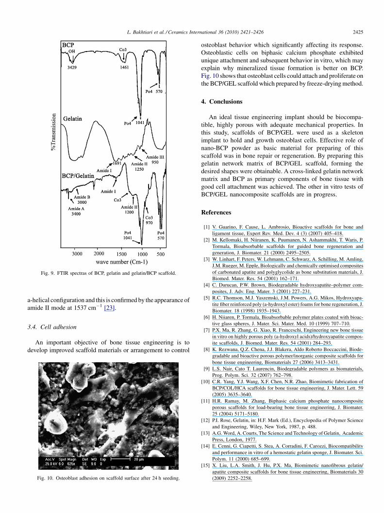
[(Fig._9)TD$FIG]
Fig. 9. FTIR spectras of BCP, gelatin and gelatin/BCP scaffold.
L. Bakhtiari et al. / Ceramics International 36 (2010) 2421–2426 2425
a-helical configuration and this is confirmed by the appearance of
amide II mode at 1537 cm�1 [23].
3.4. Cell adhesion
An important objective of bone tissue engineering is to
develop improved scaffold materials or arrangement to control[(Fig._10)TD$FIG]
Fig. 10. Osteoblast adhesion on scaffold surface after 24 h seeding.
osteoblast behavior which significantly affecting its response.
Osteoblastic cells on biphasic calcium phosphate exhibited
unique attachment and subsequent behavior in vitro, which may
explain why mineralized tissue formation is better on BCP.
Fig. 10 shows that osteoblast cells could attach and proliferate on
the BCP/GEL scaffold which prepared by freeze-drying method.
4. Conclusions
An ideal tissue engineering implant should be biocompa-
tible, highly porous with adequate mechanical properties. In
this study, scaffolds of BCP/GEL were used as a skeleton
implant to hold and growth osteoblast cells. Effective role of
nano-BCP powder as basic material for preparing of this
scaffold was in bone repair or regeneration. By preparing this
gelatin network matrix of BCP/GEL scaffold, forming the
desired shapes were obtainable. A cross-linked gelatin network
matrix and BCP as primary components of bone tissue with
good cell attachment was achieved. The other in vitro tests of
BCP/GEL nanocomposite scaffolds are in progress.
References
[1] V. Guarino, F. Cause, L. Ambrosio, Bioactive scaffolds for bone and
ligament tissue, Expert Rev. Med. Dev. 4 (3) (2007) 405–418.
[2] M. Kellomaki, H. Niiranen, K. Puumanen, N. Ashammakhi, T. Waris, P.
Tormala, Bioabsorbable scaffolds for guided bone regeneration and
generation, J. Biomater. 21 (2000) 2495–2505.
[3] W. Linhart, F. Peters, W. Lehmann, C. Schwarz, A. Schilling, M. Amling,
J.M. Rueger, M. Epple, Biologically and chemically optimised composites
of carbonated apatite and polyglycolide as bone substitution materials, J.
Biomed. Mater. Res. 54 (2001) 162–171.
[4] C. Durucan, P.W. Brown, Biodegradable hydroxyapatite–polymer com-
posites, J. Adv. Eng. Mater. 3 (2001) 227–231.
[5] R.C. Thomson, M.J. Yaszemski, J.M. Powers, A.G. Mikos, Hydroxyapa-
tite fiber reinforced poly (a-hydroxyl ester) foams for bone regeneration, J.
Biomater. 18 (1998) 1935–1943.
[6] H. Niiaren, P. Tormala, Bioabsorbable polymer plates coated with bioac-
tive glass spheres, J. Mater. Sci. Mater. Med. 10 (1999) 707–710.
[7] P.X. Ma, R. Zhang, G. Xiao, R. Franceschi, Engineering new bone tissue
in vitro on highly porous poly (a-hydroxyl acids)/hydroxyapatite compos-
ite scaffolds, J. Biomed. Mater. Res. 54 (2001) 284–293.
[8] K. Rezwana, Q.Z. Chena, J.J. Blakera, Aldo Roberto Boccaccini, Biode-
gradable and bioactive porous polymer/inorganic composite scaffolds for
bone tissue engineering, Biomaterials 27 (2006) 3413–3431.
[9] L.S. Nair, Cato T. Laurencin, Biodegradable polymers as biomaterials,
Prog. Polym. Sci. 32 (2007) 762–798.
[10] C.R. Yang, Y.J. Wang, X.F. Chen, N.R. Zhao, Biomimetic fabrication of
BCP/COL/HCA scaffolds for bone tissue engineering, J. Mater. Lett. 59
(2005) 3635–3640.
[11] H.R. Ramay, M. Zhang, Biphasic calcium phosphate nanocomposite
porous scaffolds for load-bearing bone tissue engineering, J. Biomater.
25 (2004) 5171–5180.
[12] P.I. Rose, Gelatin, in: H.F. Mark (Ed.), Encyclopedia of Polymer Science
and Engineering, Wiley, New York, 1987, p. 488.
[13] A.G. Word, A. Courts, The Science and Technology of Gelatin, Academic
Press, London, 1977.
[14] E. Cenni, G. Ciapetti, S. Stea, A. Corradini, F. Carozzi, Biocompatibility
and performance in vitro of a hemostatic gelatin sponge, J. Biomater. Sci.
Polym. 11 (2000) 685–699.
[15] X. Liu, L.A. Smith, J. Hu, P.X. Ma, Biomimetic nanofibrous gelatin/
apatite composite scaffolds for bone tissue engineering, Biomaterials 30
(2009) 2252–2258.
L. Bakhtiari et al. / Ceramics International 36 (2010) 2421–24262426
[16] H.R. Ramay, M. Zhang, Preparation of porous hydroxyapatite scaffolds by
combination of the gel-casting and polymer sponge methods, Biomaterials
24 (2003) 3293–3302.
[17] H.-W. Kima, H.-E. Kima, V. Salih, Stimulation of osteoblast responses to
biomimetic nanocomposites of gelatin–hydroxyapatite for tissue engi-
neering scaffolds, Biomaterials 26 (2005) 5221–5230.
[18] M. Chul Changa, C.-C. Koa, W.H. Douglas, Preparation of hydroxyapa-
tite–gelatin nanocomposite, Biomaterials 24 (2003) 2853–2862.
[19] J. Fua, W. Shena, J. Baoa, Q. Chen, The decontamination effects of gamma
irradiation on the edible gelatin, Radiat. Phys. Chem. 57 (2000) 345–348.
[20] J. Fernandes, D. Oliveira Ogarte, L. Agata De Sena, C. Andre De Dastro
Perez, P. Fernandes De Aguiar, A. Malta Rossi, G. Almeida Soares,
Influence of processing parameters on structural characteristic of porous
calcium phosphate samples: a study using an experimental design method,
J. Mater. Res. 8 (1) (2005) 71–76.
[21] V. Karageorgiou, D. Kaplan, Porosity of 3D biomaterial Scaffolds osteo-
genesis, Biomaterials 26 (2005) 5474–5491.
[22] E. Sachlos, J.T. Czernuszka, Making tissue engineering scaffolds work,
review on the application of solid freeform fabrication, technology to
the production of tissue engineering scaffolds, J. Eur. Cell Mater. 5 (2003)
29–40.
[23] M.C. Chang, C.-C. Ko, W.H. Douglas, Conformational change of hy-
droxyapatite/gelatin nanocomposite by glutaraldehyde, J. Biomater. 24
(2002) 3087–3094.
Copyright © 2022 FDOKUMEN