SLUG: a new target of lymphoid enhancer factor-1 in human osteoblasts

www.landesbioscience.com Organogenesis 181
Organogenesis 6:3, 181-188; July/August/September 2010; © 2010 Landes Bioscience
ReSeARch pApeR ReSeARch pApeR
*Correspondence to: Dietmar W. Hutmacher; Email: [email protected]: 03/18/10; Accepted: 04/12/10Previously published online: www.landesbioscience.com/journals/organogenesis/article/12041
Introduction
In North America, it is predicted that prostate cancer (CaP) will be the most diagnosed male cancer by 2009, accounting for more than 25% of new cancer cases.1,2 One of the most rampant com-plications of advanced CaP is bone metastasis. In high incidence countries such as the United States and United Kingdom, only 28–33% of men with metastatic disease survive up to five years.3,4 Management of this aspect of the disease is difficult, primarily due to insufficient knowledge about the cellular and molecular mechanisms involved in osteotropism of CaP cells. Numerous two-dimensional (2D) culture studies and in vivo studies have been actively employed to further understand the complex mechanisms.5-8
Interactions between human osteoblasts and prostate cancer cells
in a novel 3D in vitro modelShirly Sieh,1 Amy A. Lubik,1,2 Judith A. clements,1,2 colleen c. Nelson1,2 and Dietmar W. hutmacher1,3,*
1Institute of health and Biomedical Innovation; Queensland University of Technology; QLD, Australia; 2Australian prostate cancer Research centre—Queensland; princess Alexandra hospital; QLD, Australia
Key words: 3D in vitro model, bone metastasis, co-culture, tissue engineering, prostate cancer, osteoblasts
abbreviation: 2D, two-dimensional; 3D, three-dimensional; ADT, androgen deprivation therapy; BCA, bicinchoninic acid; CaP, prostate cancer; CLSM, confocal laser scanning microscopy; CM, conditioned media; CYP11A1, cytochrome p450 family member 11A1; DAPI, 4',6-diamidino-2-phenylindole; DHT, dihydrosterone; ECM, extracellular matrix; FASN, fatty-acid synthase; FBS,
fetal bovine serum; FDA, fluorescein diacetate; HMDS, hexamethyldisilazane; hOB, human osteoblast; HSD17B3, hydroxysteroid dehydrogenase 17 beta3; MEM, minimum essential media; MMP, matrix metalloproteinase; mPCL-TCP, medical grade
polycaprolcatone-tricalcium phosphate; mRNA, messenger ribonucleic acid; pan Ck, pan cytokeratin; PBS, phosphate buffered saline; PFA, paraformaldehyde; PI, propidium iodide; PSA, prostate-specific antigen; RDH5, 11-cis-retinol dehydrogenase; RNA,
ribonucleic acid; RPMI, roswell park memorial institute; SDS, sodium dodecyl sulphate; SEM, scanning electron microscopy; TEB, tissue engineered bone; qRT-PCR, quantitative real time polymerase chain reaction
In a 2D culture, cells are not surrounded by a three-dimen-sional (3D) microenvironment consisting of an extracellular matrix (ECM) as it is in native tissue. The ECM provides biologi-cal and physical support to cells, and is involved in the modulation of cell behavior. On 2D surfaces, the lack of cell-matrix inter-action causes disparity from the in vivo physiological situation which leads to changes in cell phenotypes and gene expression.9,10 These inconsistencies may result in less reliable experimental find-ings; therefore may not be translatable for clinical applications.
In vivo studies involving animal models no doubt offer more relevant clinical values; however, these models also have their limitations. This is because reproducibility is compromised, and pathology and disease progression in humans may not be repre-sented accurately in animal models. Moreover, animal hosts add
cell-cell and cell-matrix interactions play a major role in tumor morphogenesis and cancer metastasis. Therefore, it is crucial to create a model with a biomimetic microenvironment that allows such interactions to fully represent the pathophysiology of a disease for an in vitro study. This is achievable by using three-dimensional (3D) models instead of conventional two-dimensional (2D) cultures with the aid of tissue engineering technology. We are now able to better address the complex intercellular interactions underlying prostate cancer (cap) bone metastasis through such models. In this study, we assessed the interaction of cap cells and human osteoblasts (hOBs) within a tissue engineered bone (TeB) construct. consistent with other in vivo studies, our findings show that intercellular and cap cell-bone matrix interactions lead to elevated levels of matrix metalloproteinases, steroidogenic enzymes and the cap biomarker, prostate specific antigen (pSA); all associated with cap metastasis. hence, it highlights the physiological relevance of this model. We believe that this model will provide new insights for understanding of the previously poorly understood molecular mechanisms of bone metastasis, which will foster further translational studies, and ultimately offer a potential tool for drug screening.
182 Organogenesis Volume 6 Issue 3
Results
fabrication of tissue engineered bone (teb) construct using cell sheet-based technique. We used a cell sheet-based technique to wrap medical grade polycaprolcatone-tricalcium phosphate (mPCL-TCP) scaffolds within hOB sheets for fabrication of the
complexity to a study by contribut-ing to multivariable factors (e.g., immune system). Such complica-tions in in vivo models have lead to the development and utilization of 3D in vitro models.
3D in vitro models are able to compensate for the shortfalls of 2D cultures and in vivo models. In these models, cells are embed-ded within matrices resembling the natural ECM which allow cell-ECM interaction in a less complex and more well-defined condition. Previous studies using 3D mod-els have shed light on phenotypic changes, cell signaling, angiogen-esis and chemoresistance of can-cer cells.11-13 As we forsee the great utility of having such a 3D culture system for studying CaP-mediated bone metastasis, we have estab-lished a novel 3D in vitro model using tissue engineering strategies to create a biologically competent bone-like microenvironment.
Our approach for this model is to recapitulate the condition first encountered by CaP cells metas-tasizing to bone, an environment featuring dynamic cell-cell inter-action within a bone-like ECM. In order to do this, we cultured CaP cells within tissue engineered bone (TEB) constructs, fabricated using a human osteoblast (hOB) cell sheet-based technique. The TEB construct mimics the natural bone microenvironment in terms of architecture as well as ECM com-ponents (Reichert, manuscript in review). We then further assessed the interaction between CaP cells and hOBs using this model by eval-uating the expression of enzymes involved in metastasis and andro-gen production. This application is described herein.
Data collected from this study provides some insights into cellular responses in relation to cell-cell communication within the CaP cell-bone metastatic microenvironment, which may not have been ascertained from previously utilized models. Therefore, the estab-lished 3D model offers promise for the study of the mechanisms for bone metastasis, as well as having the potential to be a valuable pre-clinical tool particularly for drug development studies.
Figure 1. The structure and cell morphology of the hOB sheet-based TeB construct. (A) Bright field microscopy image of the bone explant culture shows hOBs growing from the bone to the tissue cul-ture surface indicated by the black arrow. (B) cLSM fluorescence image of TeB construct stained with phalloidin (cytoskeleton) and DApI (nucleus) at the pore region of the scaffold reveals gradual growth of hOBs to fill the pore of the scaffold. (c) SeM image of the scaffold completely wrapped within the hOB sheet. (D) cLSM fluorescence image of the TeB construct stained with FDA and pI demonstrates live hOBs significantly exceed dead cells. (e) Bright field image corresponding to (D) illustrates the orientation of scaffold struts (white arrows, s) that provide support to the hOB sheet. Scale bars, 100 µm (A), 250 µm (B, D and e), 2 mm (c).
www.landesbioscience.com Organogenesis 183
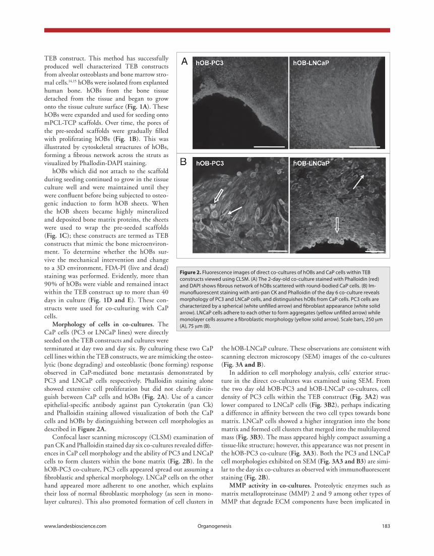
the hOB-LNCaP culture. These observations are consistent with scanning electron microscopy (SEM) images of the co-cultures (fig. 3a and B).
In addition to cell morphology analysis, cells’ exterior struc-ture in the direct co-cultures was examined using SEM. From the two day old hOB-PC3 and hOB-LNCaP co-cultures, cell density of PC3 cells within the TEB construct (fig. 3a2) was lower compared to LNCaP cells (fig. 3b2), perhaps indicating a difference in affinity between the two cell types towards bone matrix. LNCaP cells showed a higher integration into the bone matrix and formed cell clusters that merged into the multilayered mass (fig. 3b3). The mass appeared highly compact assuming a tissue-like structure; however, this appearance was not present in the hOB-PC3 co-culture (fig. 3a3). Both the PC3 and LNCaP cell morphologies exhibited on SEM (fig. 3a3 and b3) are simi-lar to the day six co-cultures as observed with immunofluorescent staining (fig. 2b).
mmp activity in co-cultures. Proteolytic enzymes such as matrix metalloproteinase (MMP) 2 and 9 among other types of MMP that degrade ECM components have been implicated in
TEB construct. This method has successfully produced well characterized TEB constructs from alveolar osteoblasts and bone marrow stro-mal cells.14,15 hOBs were isolated from explanted human bone. hOBs from the bone tissue detached from the tissue and began to grow onto the tissue culture surface (fig. 1a). These hOBs were expanded and used for seeding onto mPCL-TCP scaffolds. Over time, the pores of the pre-seeded scaffolds were gradually filled with proliferating hOBs (fig. 1b). This was illustrated by cytoskeletal structures of hOBs, forming a fibrous network across the struts as visualized by Phallodin-DAPI staining.
hOBs which did not attach to the scaffold during seeding continued to grow in the tissue culture well and were maintained until they were confluent before being subjected to osteo-genic induction to form hOB sheets. When the hOB sheets became highly mineralized and deposited bone matrix proteins, the sheets were used to wrap the pre-seeded scaffolds (fig. 1c); these constructs are termed as TEB constructs that mimic the bone microenviron-ment. To determine whether the hOBs sur-vive the mechanical intervention and change to a 3D environment, FDA-PI (live and dead) staining was performed. Evidently, more than 90% of hOBs were viable and remained intact within the TEB construct up to more than 40 days in culture (fig. 1d and e). These con-structs were used for co-culturing with CaP cells.
morphology of cells in co-cultures. The CaP cells (PC3 or LNCaP lines) were directly seeded on the TEB constructs and cultures were terminated at day two and day six. By culturing these two CaP cell lines within the TEB constructs, we are mimicking the osteo-lytic (bone degrading) and osteoblastic (bone forming) response observed in CaP-mediated bone metastasis demonstrated by PC3 and LNCaP cells respectively. Phalloidin staining alone showed extensive cell proliferation but did not clearly distin-guish between CaP cells and hOBs (fig. 2a). Use of a cancer epithelial-specific antibody against pan Cytokeratin (pan Ck) and Phalloidin staining allowed visualization of both the CaP cells and hOBs by distinguishing between cell morphologies as described in figure 2a.
Confocal laser scanning microscopy (CLSM) examination of pan CK and Phalloidin stained day six co-cultures revealed differ-ences in CaP cell morphology and the ability of PC3 and LNCaP cells to form clusters within the bone matrix (fig. 2b). In the hOB-PC3 co-culture, PC3 cells appeared spread out assuming a fibroblastic and spherical morphology. LNCaP cells on the other hand appeared more adherent to one another, which explains their loss of normal fibroblastic morphology (as seen in mono-layer cultures). This also promoted formation of cell clusters in
Figure 2. Fluorescence images of direct co-cultures of hOBs and cap cells within TeB constructs viewed using cLSM. (A) The 2-day-old co-culture stained with phalloidin (red) and DApI shows fibrous network of hOBs scattered with round-bodied cap cells. (B) Im-munofluorescent staining with anti-pan cK and phalloidin of the day 6 co-culture reveals morphology of pc3 and LNcap cells, and distinguishes hOBs from cap cells. pc3 cells are characterized by a spherical (white unfilled arrow) and fibroblast appearance (white solid arrow). LNcap cells adhere to each other to form aggregates (yellow unfilled arrow) while monolayer cells assume a fibroblastic morphology (yellow solid arrow). Scale bars, 250 µm (A), 75 µm (B).

184 Organogenesis Volume 6 Issue 3
androgen regulated CaP biomarker, prostate specific antigen (PSA) were analyzed by quantitative real-time PCR (qRT-PCR).
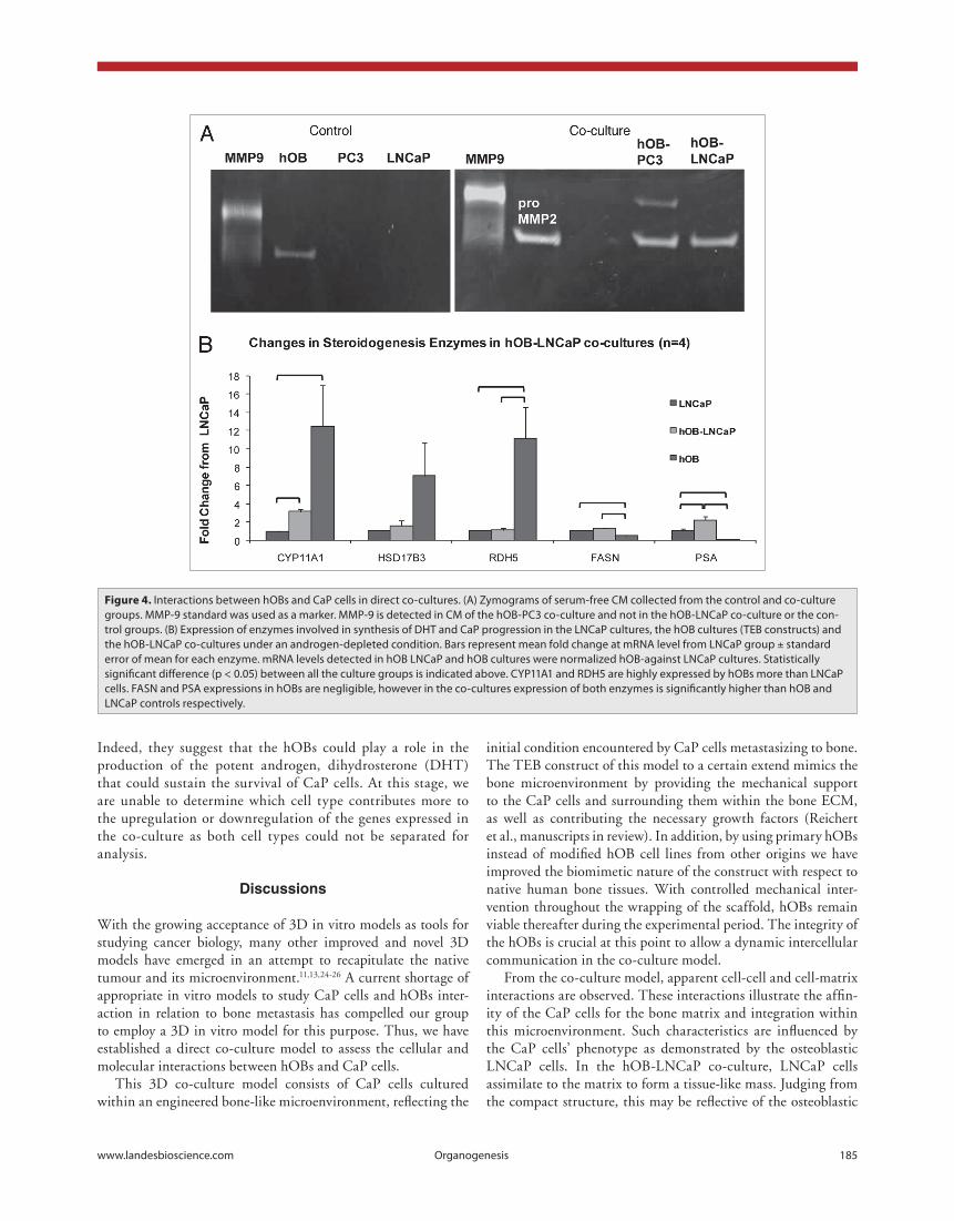
PSA is a biomarker for CaP progression and was significantly upregulated in the hOB-LNCaP co-cultures relative to LNCaP only controls (p = 0.0108, fig. 4b). It is important to note that PSA was not expressed by hOBs but was highly upregulated in LnCaP cells when co-cultured with hOBs. This indicates that hOB-related factors may increase the aggressiveness of CaP cells. Fatty acid synthase (FASN), which is upregulated during CaP progression, did not increase in the co-cultures relative to LNCaP controls but was higher compared to hOB controls. This suggests that hOBs’ influence does not change the need for lipids as an energy source, nor is it likely to contribute to enhancement of that particular pathway.
As shown in figure 4b, most of the steroidogenic enzymes were co-expressed by both LNCaP cells and hOBs. In particu-lar, cytochrome p450 family member 11A1 (CYP11A1) and 11-cis-retinol dehydrogenase (RDH5) of hOB cultures were ten times higher in expression compared to LNCaP cultures (p = 0.0419 and 0.0252 respectively, fig. 4b). Significantly, CYP11A1, the first enzyme committing cholesterol to steroido-genesis, was significantly upregulated in the co-cultures relative to LNCaP only controls (p = 0.0004). In cases where expression of steroidogenic enzymes was higher in hOBs, there was also an increased expression in the co-cultures above that detected in LNCaP cells although lower than that detected in hOBs alone.
Other groups have also reported that hOBs express enzymes responsible for conversion of precursors to potent androgen.21-23
cancer cell proliferation and migration.16-18 To further investi-gate the effect of CaP cells-hOBs interaction on MMPs activity, gelatin zymography was performed. Activity of the MMP2 and MMP9 was detected as bands on zymograms (fig. 4a).
MMPs activity of the co-cultures was compared to the control groups comprising of LNCaP or PC3 cells cultured on scaffolds and the TEB construct alone. Both co-cultures and TEB con-trol group showed presence of pro-MMP2. While MMP9 activ-ity was not detected in any of the control groups, it was clearly enhanced in the hOB-PC3 co-culture (fig. 4a). Other groups have also reported augmentation of MMP9 both in vitro and in vivo with an increase in MMP9 activity when PC3 cells were cultured with bone explants as well as in a bone xenograft colo-nized by PC3 cells.6,17 Our findings from in vitro study indicate association to the osteolytic nature of PC3 cells as proven in in vivo studies.19,20
expression of steroidogenesis enzymes at mrna level. In an attempt to relate to the clinical scenario, we used this 3D co-culture model to investigate the effects of the metastatic micro-environment on CaP cells that are reliant on external androgens for growth by co-culturing LNCaP cells (androgen dependent) and hOBs under an androgen-depleted condition to mimic the system of patients who have undergone androgen deprivation therapy (ADT). After culturing for 14 days, the growth media containing androgens was replaced by androgen-depleted growth media and maintained for four days in “starved media.” Cells were then harvested for RNA extraction. Expression of several enzymes involved in steroid synthesis (suppl. fig. 1) and the
Figure 3. SeM images of direct co-cultures of hOBs and cap cells within TeB constructs. The coculture was harvested 2 days after seeding with pc3 (A1–3) and LNcap (B1–3) cells. circled regions denote magnified areas at 300x (A2 and B2) and 1000x (A3 and B3). The hOB-pc3 culture appears more homogenous and less dense (A2) when compared to the hOB-LNcap culture, which appears coarse and compact (B2). Within the TeB construct, LNcap cells form multiple clusters (arrows) and layers that are integrated within the matrix (B3). Such features are absent in the hOBpc3 culture (A3). Scale bars, 2 mm (A1 and B1), 400 µm (A2 and B2), 100 µm (A3 and B3).
www.landesbioscience.com Organogenesis 185
initial condition encountered by CaP cells metastasizing to bone. The TEB construct of this model to a certain extend mimics the bone microenvironment by providing the mechanical support to the CaP cells and surrounding them within the bone ECM, as well as contributing the necessary growth factors (Reichert et al., manuscripts in review). In addition, by using primary hOBs instead of modified hOB cell lines from other origins we have improved the biomimetic nature of the construct with respect to native human bone tissues. With controlled mechanical inter-vention throughout the wrapping of the scaffold, hOBs remain viable thereafter during the experimental period. The integrity of the hOBs is crucial at this point to allow a dynamic intercellular communication in the co-culture model.
From the co-culture model, apparent cell-cell and cell-matrix interactions are observed. These interactions illustrate the affin-ity of the CaP cells for the bone matrix and integration within this microenvironment. Such characteristics are influenced by the CaP cells’ phenotype as demonstrated by the osteoblastic LNCaP cells. In the hOB-LNCaP co-culture, LNCaP cells assimilate to the matrix to form a tissue-like mass. Judging from the compact structure, this may be reflective of the osteoblastic
Indeed, they suggest that the hOBs could play a role in the production of the potent androgen, dihydrosterone (DHT) that could sustain the survival of CaP cells. At this stage, we are unable to determine which cell type contributes more to the upregulation or downregulation of the genes expressed in the co-culture as both cell types could not be separated for analysis.
Discussions
With the growing acceptance of 3D in vitro models as tools for studying cancer biology, many other improved and novel 3D models have emerged in an attempt to recapitulate the native tumour and its microenvironment.11,13,24-26 A current shortage of appropriate in vitro models to study CaP cells and hOBs inter-action in relation to bone metastasis has compelled our group to employ a 3D in vitro model for this purpose. Thus, we have established a direct co-culture model to assess the cellular and molecular interactions between hOBs and CaP cells.
This 3D co-culture model consists of CaP cells cultured within an engineered bone-like microenvironment, reflecting the
Figure 4. Interactions between hOBs and cap cells in direct co-cultures. (A) Zymograms of serum-free cM collected from the control and co-culture groups. MMp-9 standard was used as a marker. MMp-9 is detected in cM of the hOB-pc3 co-culture and not in the hOB-LNcap co-culture or the con-trol groups. (B) expression of enzymes involved in synthesis of DhT and cap progression in the LNcap cultures, the hOB cultures (TeB constructs) and the hOB-LNcap co-cultures under an androgen-depleted condition. Bars represent mean fold change at mRNA level from LNcap group ± standard error of mean for each enzyme. mRNA levels detected in hOB LNcap and hOB cultures were normalized hOB-against LNcap cultures. Statistically significant difference (p < 0.05) between all the culture groups is indicated above. cYp11A1 and RDh5 are highly expressed by hOBs more than LNcap cells. FASN and pSA expressions in hOBs are negligible, however in the co-cultures expression of both enzymes is significantly higher than hOB and LNcap controls respectively.

186 Organogenesis Volume 6 Issue 3
Materials and Methods
culture of cap cell lines. PC3 and LNCaP cell lines were purchased from American Type Culture Collection (ATCC). Both cell lines were cultured in RPMI growth media, com-prising of RPMI media (Invitrogen), 10% fetal bovine serum (FBS, Thermo Scientific) and 10% penicillin/streptomycin. In all experiments, cells from passage 18–30 were used. PC3 cells are androgen independent while LNCaP cells are androgen dependent.
isolation and culture of hobs. hOB explants were obtained from patients undergoing knee replacement surgery as approved by the institutional ethics committee of Queensland University of Technology. Bone was collected, minced and washed with phosphate buffered saline (PBS) at least five times to remove connective tissues, adipocytes and other debris. Then, the bone pieces were treated with 0.5% trypsin for 15 min at 37°C. After trypsinization, the bone pieces were washed and transferred to T175 tissue culture flasks. The bone explants were then main-tained in α-MEM growth media. The α-MEM growth media consists of α-MEM media (Invitrogen), 10% FBS and 10% pen-icillin/streptomycin. Cells no later than passage five were used for all experiments.
fabrication of teb constructs. As described previously by Zhou et al.15 we seeded 1 x 10⁵ hOBs suspended in 20 uL growth media on 1.5 mm thick, 5 mm x 5 mm mPCL-TCP scaffolds (Osteopore, Singapore) in six-well plates. The pre-seeded scaffolds were incubated at 37°C for 90 min before being topped up with 2 mL of α-MEM growth media. After hOB culture reached 80% confluency in the wells, hOB cultures were changed to osteogenic media, consisting of α-MEM growth media supplemented with 10 mM glycerol 2-glycerophosphate, 50 µg/mL L-ascorbic acid-2-phosphate and 100 nM dexamethasone (Sigma). After three weeks of culture in osteogenic media, mineralized hOB sheets were formed. The sheets were detached from the plate surface with a cell scrapper and wrapped around the pre-seeded scaffolds. Wrapped scaffolds continued to be cultured in osteogenic media until ready for seeding with CaP cells.
co-culture of cap cells and hobs. For direct co-cultures, 5 x 10⁵ CaP cells/20 uL (PC3 or LNCaP cells) were seeded on TEB constructs. CaP cells seeded on mPCL-TCP scaffolds were CaP cells control group. After seeding, the scaffolds were incu-bated for 90 min at 37°C to allow CaP cells to attach. After incu-bation, 1 mL of RPMI growth media was added and cultures were maintained until termination. TEB constructs prepared from the same hOBs source (same patient) were used for co-culturing with CaP cells and served as the hOB control group.
To prepare for analysis of MMP activity, co-cultures and con-trol groups were cultured in RPMI growth media for 14 days after seeding of CaP cells on TEB constructs or scaffolds. They were then cultured in serum-free RPMI media for 48 hours. The serum-free conditioned media (CM) was then collected to be analyzed for MMP activity.
Regarding the study of steroidogenic enzymes expression, LNCaP cells and hOBs were co-cultured in RPMI growth media for 14 days. Following, the growth media was changed
lesions known to be caused by LNCaP cells.27,28 as observed in vivo by Yonou et al.20 when LNCaP cells metastasized to human bone chips in Non-Obese Diabetic/Severe Combined Immunodeficient mice. Similarly, culturing of known osteolytic PC3 cells with hOBs resulted in an increase of MMP9 produc-tion and possibly MMP9 activation. This augmentation may be associated with raised invasive potential of CaP cells as indicated by previous studies.29,30 These findings confirm the physiologi-cal relevance of this model for analytical assays and translational studies.
We have further explored the application of this model for other clinical conditions namely, post castrate metastasis. Prostatectomy causes androgens to decline to a minimal level, causing apoptosis of androgen-dependant CaP cells. However, Locke et al.31 have reported that de novo synthesis of andro-gens, minimal before castration, is upregulated as tumors move towards castrate resistant. This could explain the recurrence of CaP in patients even after ADT.
Here, we have utilized hOB-LNCaP co-cultures to mimic the post castration bone metastasis scenario in an androgen-depleted environment. The increase in CYP11A1 and RDH5 expression in hOB-LNCaP co-cultures suggest that synthesis of the potent androgen, DHT, from circulating precursors may occur in CaP bone metastasis. Simultaneously, high expression of steroidogenic enzymes in hOBs may contribute androgens or precursor steroids to drive CaP tumour growth and aggression, as indicated by a corresponding rise in PSA.
Though it is clear that CaP-hOB cell interactions influence each others’ mRNA expression profiles, we have yet to deter-mine the signaling pathways or bioactive molecules leading to the aforementioned phenomena. Another question raised here is which cell type has initiated the mRNA alterations observed in the co-cultures. Nevertheless, we postulate that the three fac-tors which could explain the resilience and progression of CaP cells during post castration bone metastasis are: hOBs are able to compensate for the lack of androgen; the CaP cells respond to the bone environment by producing more steroidogenic enzymes; and the synergistic upregulation of enzymes from both cell types could essentially contribute to de novo synthesis of DHT or other steroids. Since all the genes tested except PSA, are jointly expressed by both cells, the pooled mRNA reflects cumulative expression of the cell mixture. To overcome this issue in future experiments, we can resort to cell sorting or modifying the 3D model to enable more effective separation of the CaP cells and hOBs.
Our study so far proves that this model is both practicable and versatile for studying intercellular and cell-matrix interac-tion at a cellular and molecular level. Evidently, results from the in vitro 3D co-culture model are consistent with results from in vivo studies. Therefore, this model is a useful tool to elucidate the multistage process of bone metastasis or osteotropism of CaP cells. A biomimetic in vitro model is a critical asset for transla-tional studies including drug target selection prior to clinical tri-als. Our approach is less demanding compared to in vivo models and yet is able to marry the complexity of native cellular physiol-ogy with the simplicity of in vitro systems.
187 Organogenesis Volume 6 Issue 3
solution for approximately one hour or until apparent gelatinase activities were detected.
Quantitative real-time pcr of steroidogenic enzymes. Quantitative real-time PCR (qRT-PCR) was performed to ana-lyze expression of enzymes involved in the steroidogenic path-way for the co-culture and control groups. The RNA of cells from direct co-cultures and control groups was extracted using TRIZOL reagent (Invitrogen) according to the manufacturer’s instructions. Then, the RNA was quantified using the ND-1000 NanoDrop spectrophotometer (NanoDrop, Delaware). 1 µg of RNA was treated with DNase I Amp grade (Invitrogen) before being reverse transcribed to cDNAs with Super Script III (Invitrogen). qRT-PCR was performed using Applied Biosystems to detect expression of CYP11A1, HSD17B3, RDH5, PSA and FASN genes. Data was analyzed with SDS 2.3 software by means of the 2-∆∆C
T method.32 Primers used were as follows: CYP11A1,
5'-AGT TCT CGG GAC TTC GTC AGT-3' and 5'-GGA GCC CGC CTT CTT GA-3', HSD17B3, 5'-TGG GAC AGT GGG CAG TGA-3' and 5'-CGA GTA CGC TTT CCC AAT TCC-3', RDH5, 5'-GCC CGC CAG CAA TGC-3' and 5'-CGC CCA AAG CCT GAG TCA-3', PSA, 5'-TCT GCG GCG GTG TTC TG-3' and 5'-GCC GAC CCA GCA AG-3', and FASN, 5'-CGC TCG GCA TGG CTA TCT-3' and 5'-CTC GTT GAA GAA CGC ATC CA-3'.
statistical analysis. An unpaired Student’s t-test was used to determine the statistical significance of steroidogenic enzymes expression between hOB-LNCaP co-cultures and control groups. By conventional criteria, the difference is considered to be sta-tistically significant when p < 0.05. Four independent in vitro experiments were conducted for each condition.
acknowledgements
This work is funded by the Prostate Cancer Foundation of Australia (PCFA) grant and Professor Dietmar W. Hutmacher QUT Chair in Regenerative Medicine start-up grant. We would like to thank Dr. Leonore de Boer and Dr. Christina Theodoropoulos for their assistance in CLSM and SEM prep-arations. We also thank the members of Prof. Dietmar W. Hutmacher’s group, especially Dr. Johannes Reichert, Dr. Mia Woodruff, Dr. Travis Klein and Mr. Toby Brown for their com-ments and recommendations in preparation of the manuscript.
note
Supplementary materials can be found at:www.landesbioscience.com/supplement/SiehORG6-3-Sup.pdf
to androgen-depleted media (RPMI with 5% charcoal stripped serum) and cultured for 96 hours with media change at 48 hours interval. After cultures were “starved,” they were terminated and cells were harvested for RNA isolation.
microscopy image analysis of co-cultures. Investigation of the exterior structure of hOBs and CaP cells, and constructs was performed using SEM. The TEB constructs and co-cultures were fixed with 3% glutaraldehyde then washed thoroughly with caco-dylate buffer. The samples were treated with 1% osmium tetraox-ide before being dehydrated sequentially with ethanol solutions. In order to preserve their surface detail, they were air dried by evaporation of hexamethyldisilazane (HMDS) before sputter coated with gold using Biorad SC500. Viewing and analysis were performed using the Quanta 200 Scanning Electron Microscope (FEI).
For visualization of CaP cells and hOBs in a co-culture, a wide spectrum pan Ck and cytoskeletal fluorescent stainings, were used to distinguish the two cell types. Co-cultures and con-trol groups were fixed with 4% paraformaldehyde (PFA) for 20 minutes and treated with 0.2% Triton-X for 15 min. Samples subjected to cytoskeletal staining were incubated with 0.8 U/mL rhodamine-conjugated Phalloidin (Invitrogen) for 45 minutes and 4',6-diamidino-2-phenylindole (DAPI) for 40 minutes. Pan Ck immunofluorescent staining involved incubation with diluted (1:300) mouse anti-human pan cytokeratin antibody (Abcam) for one hour. Following that, the samples were incubated with Alexa Fluor® 488 goat anti-mouse IgG antibody (1:300) for one hour. Finally, they were counterstained with cytoskeletal stain. Fluorescent images of 100–200 µm Z stacks thick were captured using Leica SP5 CLSM (Leica) and presented as overlaid images.
gelatin zymography of co-cultures. The activity of MMP2 and MMP9 was analyzed using gelatin zymography. CM col-lected from co-cultures and control groups were quantified with bicinchoninic acid (BCA) assay according to supplier’s instruc-tions (Pierce). A 12% SDS-polyacrylamide gel containing 1 mg/mL gelatin was prepared. From the CM, 5 ug of protein were mixed with 5x loading buffer (0.05 g bromophenol blue, 5 mL glycerol, 1 g SDS, 1 M Tris, pH 6.8) and loaded into the prepared gels. The gels were electrophoresed at 120 V for 2 hours at 4°C. Once the electrophoresis was terminated, the gels were removed and washed once with 2.5% triton-X for 30 min. The gels were then incubated for 1 hour in the same solution on a shaker. Gelatinolytic reaction was induced by incubation at 37°C in a buffer containing 50 mM Tris, 10 mM CaCl and 50 mM NaCl
2
for 24 hours. The gels were then stained with 0.25% Brilliant Blue R250 and destained with 9% acetic acid and 36% methanol
references1. Jemal A, Siegel R, Ward E, Hao Y, Xu J, Murray T, et
al. Cancer Statistics 2008; 2008:71-96.2. Society CC. Canadian Cancer Statistics. 2009.3. SEER. Fast facts: Prostate cancer: Survival and stage.
National Cancer Institute 2008.4. SWPHO. Prostate cancer survival by stage: South West
Public Health Observatory 2008.5. Sikes RA, Nicholson BE, Koeneman KS, Edlund NM,
Bissonette EA, Bradley MJ, et al. Cellular interactions in the tropism of prostate cancer to bone. Int J Cancer 2004; 110:497-503.
6. Wiesner C, Bonfil RD, Dong Z, Yamamoto H, Nabha SM, Meng H, et al. Heterogeneous activation of MMP-9 due to prostate cancer-bone interaction. Urology 2007; 69:795-9.
7. Dai J, Hall CL, Escara-Wilke J, Mizokami A, Keller JM, Keller ET. Prostate cancer induces bone metastasis through wnt-induced bone morphogenetic protein-dependent and independent mechanisms. Cancer Res 2008; 68:5785-94.
8. Dong Z, Saliganan AD, Meng H, Nabha SM, Sabbota AL, Sheng S, et al. Prostate cancer cell-derived uroki-nase-type plasminogen activator contributes to intraos-seous tumor growth and bone turnover. Neoplasia (New York, NY) 2008; 10:439-49.
9. Birgersdotter A, Sandberg R, Ernberg I. Gene expres-sion perturbation in vitro-A growing case for three-dimensional (3D) culture systems. Semin Cancer Biol 2005; 15:405-12.
10. Weigelt B, Bissell MJ. Unraveling the microenviron-mental influences on the normal mammary gland and breast cancer. Semin Cancer Biol 2008; 18:311-21.
11. Fischbach C, Chen R, Matsumoto T, Schmelzle T, Brugge JS, Polverini PJ, et al. Engineering tumors with 3D scaffolds. Nat Meth 2007; 4:855-60.
188 Organogenesis Volume 6 Issue 3
28. Corey E, Quinn JE, Bladou F, Brown LG, Roudier MP, Brown JM, et al. Establishment and characterization of osseous prostate cancer models: Intra-tibial injection of human prostate cancer cells. Prostate 2002; 52:20-33.
29. Webber M, Waghray A, Rhim J, Deocampo N. Urokinase and gelatinises in the proteolytic cascade in human prostate cancer invasion: Implication in cancer prevention. Proc Am Assoc Cancer Res 1995; 36:643.
30. Quax PHA, de Bart ACW, Schalken JA, Verheijen JH. Plasminogen activator and matrix metalloproteinase production and extracellular matrix degradation by rat prostate cancer cells in vitro: Correlation with meta-static behavior in vivo. Prostate 1997; 196-204.
31. Locke JA, Guns ES, Lubik AA, Adomat HH, Hendy SC, Wood CA, et al. Androgen levels increase by intra-tumoral de novo steroidogenesis during progression of castration-resistant prostate cancer. Cancer Res 2008; 6407-15.
32. Livak KJ, Schmittgen TD. Analysis of relative gene expression data using real-time quantitative PCR and the 2-[Delta][Delta]CT method. Methods 2001; 25:402-8.
33. Hutmacher DW. Biomaterials offer cancer research the third dimension. Nat Mater 2010; 9:90-3.
20. Yonou H, Yokose T, Kamijo T, Kanomata N, Hasebe T, Nagai K, et al. Establishment of a novel species- and tissue-specific metastasis model of human prostate can-cer in humanized non-obese diabetic/severe combined immunodeficient mice engrafted with human adult lung and bone. Cancer Res 2001; 61:2177-82.
21. Schweikert HU, Rulf W, Niederle N, Schafer HE, Keck E, Kruck F. Testosterone metabolism in human bone 1980; 258-64.
22. Bruch HR, Wolf L, Budde R, Romalo G, Schweikert HU. Androstenedione metabolism in cultured human osteoblast-like cells. J Clin Endocrinal Metab 1992; 101-5.
23. Issa S, Schnabel D, Feix M, Wolf L, Schaefer H-E, Russell DW, et al. Human osteoblast-like cells express predominantly steroid 5{alpha}-reductase type 1. J Clin Endocrinal Metab 2002; 87:5401-7.
24. Kenny PA, Lee GY, Myers CA, Neve RM, Semeiks JR, Spellman PT, et al. The morphologies of breast cancer cell lines in three-dimensional assays correlate with their profiles of gene expression. Mol Oncol 2007; 1:84-96.
25. Dhurjati R, Krishnan V, Shuman L, Mastro A, Vogler E. Metastatic breast cancer cells colonize and degrade three-dimensional osteoblastic tissue in vitro. Clin Exp Metastasis 2008; 25:741-52.
26. Marrero B, Messina J, Heller R. Generation of a tumor spheroid in a microgravity environment as a 3D model of melanoma. In Vitro Cell Dev Biol Anim 2009; 45:523-34.
27. Nordstrand A, Nilsson J, Tieva Å, Wikström P, Lerner U, Widmark A. Establishment and validation of an in vitro co-culture model to study the interactions between bone and prostate cancer cells. Clin Exp Metastasis 2009; 26:945-53.
12. Chu JH, Yu S, Hayward SW, Chan FL. Development of a three-dimensional culture model of prostatic epithelial cells and its use for the study of epithelial-mesenchymal transition and inhibition of PI3K path-way in prostate cancer. Prostate 2008; 69:428-42.
13. Fischbach C, Kong HJ, Hsiong SX, Evangelista MB, Yuen W, Mooney DJ. Cancer cell angiogenic capability is regulated by 3D culture and integrin engagement. Proc Natl Acad Sci USA 2009; 399-404.
14. Zhou Y, Hutmacher DW, Varawan SL, Meng LT. Comparison of human alveolar osteoblasts cultured on polymer-ceramic composite scaffolds and tissue culture plates. Int J Oral Maxillofac Surgery 2007; 36:137-45.
15. Zhou Y, Chen F, Ho ST, Woodruff MA, Lim TM, Hutmacher DW. Combined marrow stromal cell-sheet techniques and high-strength biodegradable compos-ite scaffolds for engineered functional bone grafts. Biomaterials 2007; 28:814-24.
16. Nemeth JA, Yousif R, Herzog M, Che M, Upadhyay J, Shekarriz B, et al. Matrix metalloproteinase activity, bone matrix turnover, and tumor cell proliferation in prostate cancer bone metastasis. J Natl Cancer Inst 2002; 17-25.
17. Dong Z, Bonfil RD, Chinni S, Deng X, Trindade Filho JC, Bernardo M, et al. Matrix metalloproteinase activ-ity and osteoclasts in experimental prostate cancer bone metastasis tissue. Am J Pathol 2005; 1173-86.
18. Cao J, Chiarelli C, Richman O, Zarrabi K, Kozarekar P, Zucker S. Membrane type 1 matrix metalloproteinase induces epithelial-to-mesenchymal transition in pros-tate cancer. J Biol Chem 2008; 6232-40.
19. Nemeth JA, Harb JF, Barroso U Jr, He Z, Grignon DJ, Cher ML. Severe combined immunodeficient-hu model of human prostate cancer metastasis to human bone. Cancer Res 1999; 1987-93.
Copyright © 2022 FDOKUMEN