Expression of cell adhesion receptors in human osteoblasts cultured on biofunctionalized poly-(...
11
Biomaterials 28 (2007) 3668–3678 Expression of cell adhesion receptors in human osteoblasts cultured on biofunctionalized poly-(e-caprolactone) surfaces Ilaria Amato a , Gabriela Ciapetti a , Stefania Pagani a , Giovanni Marletta c , Cristina Satriano c , Nicola Baldini a,b , Donatella Granchi a, a Laboratory for Pathophysiology, Istituti Ortopedici Rizzoli, via di Barbiano 1/10, 40136 Bologna, Italy b Department of Orthopaedic Surgery, University of Bologna, Bologna, Italy c Laboratory for Molecular Surfaces and Nanotechnology, Department of Chemical Sciences, Universita` di Catania and CSGI, v.le A. Doria 6, 95125 Cata nia, Italy Received 1 February 2007; accepted 30 April 2007 Available online 3 May 2007 Abstract This study was aimed to investigate whether the activation of poly-(e-caprolactone) (PCL) surface by low-energy irradiation and/or the biofunctionalization by absorption of arginine–glycine–aspartic sequences (RGD), can modify the expression of integrins closely related to the osteoblast activity. For this purpose, we analysed the physicochemical changes induced by irradiation and RGD immobilization, the consequences on cell adhesion and spreading, and the effects on integrin expression. PCL irradiated with 5 10 15 He + /cm 2 (10 keV energy) (irr-PCL) showed an altered surface layer with a partial loss of carboxyl species and the formation of carbonyl groups. Moreover, irr-PCL showed a small smoothening effect and a less polar character in comparison to the pristine ones. The RGD immobilization was observed only on irr-PCL (surface coverage: 7.0 pmol/cm 2 ). Human osteoblasts (hOB) were cultured on untreated PCL (ut-PCL), ut- PCL+RGD, irr-PCL, and irr-PCL+RGD. After 24 h, ut-PCL hindered the cell adhesion, while a discrete layer of hOB with a good cytoskeleton organization was detected on irr-PCL and irr-PCL+RGD. Before seeding, the single hOB suspension expressed a1, a2, a3, a5, b1, and aVb3; after 24 h, cells cultured on tissue-plastic expressed high levels of b1 and aVb3, while a1 showed a low intensity and a2, a3, and a5 were negative. b1 and aVb3 were selected to evaluate the interaction between cells and PCL samples. The b1 expression was higher in hOB cultured on irr-PCL than on the other samples. A significant increase in aVb3 expression was observed only in irr- PCL+RGD, and confirmed by the gene expression analysis. In conclusion, ion irradiation and RGD adsorption on PCL surfaces modulate the expression of integrin involved in hOB growth and function, indicating the effectiveness of biomimetic surfaces in promoting cell adhesion. Ultimately, the study of integrin expression may suggest proper changes to the surface structure in order to improve the osteoconductivity of selected materials. r 2007 Elsevier Ltd. All rights reserved. Keywords: Poly-(e-caprolactone); Bioactivity; Osteoblast; Cell adhesion; Integrin 1. Introduction Biodegradable synthetic polymers offer a number of advantages as compared to other materials for developing scaffolds to be used for bone tissue engineering [1,2] including the ability to tailor mechanical properties and degradation kinetics to suit various applications. Synthetic polymers are also attractive because they can be fabricated into various shapes and with the desired pore features favouring osteoconduction. Moreover, they can be designed with chemical functional groups to induce tissue in-growth [3]. A variety of biodegradable polymers for bone tissue engineering have been investigated. In parti- cular, poly-(e-caprolactone) (PCL) is a promising material for bone and cartilage repair [3–6]. It shares good mechanical properties with a lower degradation rate, which are excellent requirements for the application in tissue engineering, in particular when the scaffold is thought for ARTICLE IN PRESS www.elsevier.com/locate/biomaterials 0142-9612/$ - see front matter r 2007 Elsevier Ltd. All rights reserved. doi:10.1016/j.biomaterials.2007.04.038 Corresponding author. Tel.: +39 51 6366896; fax: +39 51 6366748. E-mail address: [email protected] (D. Granchi).
Transcript of Expression of cell adhesion receptors in human osteoblasts cultured on biofunctionalized poly-(...

ARTICLE IN PRESS
0142-9612/$ - se
doi:10.1016/j.bi
�CorrespondE-mail addr
Biomaterials 28 (2007) 3668–3678
www.elsevier.com/locate/biomaterials
Expression of cell adhesion receptors in human osteoblasts cultured onbiofunctionalized poly-(e-caprolactone) surfaces
Ilaria Amatoa, Gabriela Ciapettia, Stefania Pagania, Giovanni Marlettac, Cristina Satrianoc,Nicola Baldinia,b, Donatella Granchia,�
aLaboratory for Pathophysiology, Istituti Ortopedici Rizzoli, via di Barbiano 1/10, 40136 Bologna, ItalybDepartment of Orthopaedic Surgery, University of Bologna, Bologna, Italy
cLaboratory for Molecular Surfaces and Nanotechnology, Department of Chemical Sciences, Universita di Catania and CSGI,
v.le A. Doria 6, 95125 Cata nia, Italy
Received 1 February 2007; accepted 30 April 2007
Available online 3 May 2007
Abstract
This study was aimed to investigate whether the activation of poly-(e-caprolactone) (PCL) surface by low-energy irradiation and/or the
biofunctionalization by absorption of arginine–glycine–aspartic sequences (RGD), can modify the expression of integrins closely related
to the osteoblast activity. For this purpose, we analysed the physicochemical changes induced by irradiation and RGD immobilization,
the consequences on cell adhesion and spreading, and the effects on integrin expression. PCL irradiated with 5� 1015He+/cm2 (10 keV
energy) (irr-PCL) showed an altered surface layer with a partial loss of carboxyl species and the formation of carbonyl groups. Moreover,
irr-PCL showed a small smoothening effect and a less polar character in comparison to the pristine ones. The RGD immobilization was
observed only on irr-PCL (surface coverage: 7.0 pmol/cm2). Human osteoblasts (hOB) were cultured on untreated PCL (ut-PCL), ut-
PCL+RGD, irr-PCL, and irr-PCL+RGD. After 24 h, ut-PCL hindered the cell adhesion, while a discrete layer of hOB with a good
cytoskeleton organization was detected on irr-PCL and irr-PCL+RGD. Before seeding, the single hOB suspension expressed a1, a2, a3,a5, b1, and aVb3; after 24 h, cells cultured on tissue-plastic expressed high levels of b1 and aVb3, while a1 showed a low intensity and a2,a3, and a5 were negative. b1 and aVb3 were selected to evaluate the interaction between cells and PCL samples. The b1 expression was
higher in hOB cultured on irr-PCL than on the other samples. A significant increase in aVb3 expression was observed only in irr-
PCL+RGD, and confirmed by the gene expression analysis. In conclusion, ion irradiation and RGD adsorption on PCL surfaces
modulate the expression of integrin involved in hOB growth and function, indicating the effectiveness of biomimetic surfaces in
promoting cell adhesion. Ultimately, the study of integrin expression may suggest proper changes to the surface structure in order to
improve the osteoconductivity of selected materials.
r 2007 Elsevier Ltd. All rights reserved.
Keywords: Poly-(e-caprolactone); Bioactivity; Osteoblast; Cell adhesion; Integrin
1. Introduction
Biodegradable synthetic polymers offer a number ofadvantages as compared to other materials for developingscaffolds to be used for bone tissue engineering [1,2]including the ability to tailor mechanical properties anddegradation kinetics to suit various applications.
e front matter r 2007 Elsevier Ltd. All rights reserved.
omaterials.2007.04.038
ing author. Tel.: +3951 6366896; fax: +39 51 6366748.
ess: [email protected] (D. Granchi).
Synthetic polymers are also attractive because they canbe fabricated into various shapes and with the desired porefeatures favouring osteoconduction. Moreover, they can bedesigned with chemical functional groups to induce tissuein-growth [3]. A variety of biodegradable polymers forbone tissue engineering have been investigated. In parti-cular, poly-(e-caprolactone) (PCL) is a promising materialfor bone and cartilage repair [3–6]. It shares goodmechanical properties with a lower degradation rate, whichare excellent requirements for the application in tissueengineering, in particular when the scaffold is thought for

ARTICLE IN PRESSI. Amato et al. / Biomaterials 28 (2007) 3668–3678 3669
considerable bone defects. Previous studies have shownthat osteoblast-like cells grow successfully on PCL matrices[7], although cell adhesion of human primary osteoblasts(hOB) is not so effective [8].
Osteoblasts are attachment-dependent cells and theiradhesion to materials is an essential step that influences allthe events leading to the formation of new bone which willtotally replace the scaffold. These events may be summar-ized in the recruitment of osteoprogenitor cells from thetissue around the implanted material, their proliferationand differentiation into mature osteoblasts, and finally theformation of bone matrix and its mineralization [9]. Celladhesion onto a polymeric material can be closelyexamined through the study of the receptors involved inthis process, i.e., integrins, which play a crucial role inspreading, migration, and signalling by providing trans-membrane links between the intra- and extra-cellularmatrix (ECM) [10]. They are composed of at leasttwenty-four a and nine b different subunits that dimerizein specific combinations. hOB are known to expressvarious integrins, each component having a large extra-cellular domain responsible for ligand binding, a trans-membrane domain, and a short cytoplasmic domainresponsible for interacting with the actin cytoskeleton.Integrin heterodimers, bind to specific amino acid se-quences, such as the arginine–glycine–aspartic acid (Arg–Gly–Asp or RGD) recognition motif that is present inmany ECM proteins, including fibronectin, vitronectin,bone sialoprotein, and osteopontin [9,10]. Small syntheticpeptides (a few hundred Dalton) that contain amino acidsequence RGD can mediate cell attachment as well as thelarge parental molecule (a hundred thousand Dalton).Over the last decade, biomimetic approaches have beendeveloped to immobilize short peptides, such as RGD,onto synthetic or natural surfaces. This opens thepossibility to produce biofunctional materials that promotecell attachment by the binding of adhesion receptors[11,12]. In addition, the characteristics of material surfaces,such as topography, chemistry or surface energy, play anessential role in osteoblast adhesion and growth [13], byinfluencing the integrin localization and structure [14].
Among the various possible surface treatments, ionbeam irradiation of polymer surfaces is known to inducethe formation of a drastically modified altered layer,strongly affecting the cell-surface interaction [15]. Inparticular, low-energy ion irradiation in itself has beenshown to markedly enhance the adhesion of hOB [8], andto improve the performance of PCL through the differ-entiation of osteoprogenitors into mature osteoblasts [16].Because the high hydrophobicity has a detrimental effecton cell adhesion, PCL represents a proper material fortesting the changes in cell reactivity due to surfacetreatment. In the present study, 10 keV He+ has beenemployed to investigate in vitro whether such enhanced celladhesion and differentiation onto irradiated surfaces canbe related to the modification of the integrin expressioninvolved in the hOB function. The comparison has
been extended to the same surfaces treated with RGDsolutions.In fact, as integrin expression and osteoblast biology are
closely related, the behaviour of these molecules in theearly phases of the adhesion could represent an indicator ofthe effectiveness of biomimetic surfaces in promotingadhesion, proliferation and function of osteoblast.For this purpose, hOB were cultured for 24 h on He-
irradiated PCL, with or without RGD adsorption. At thatendpoint, cell adhesion, morphology of cytoskeleton, andintegrin expression were analysed by confocal microscopyand quantified by image analysis.
2. Materials and methods
2.1. PCL film preparation and modification
Thin films of PCL (about 100 nm) were prepared as previously
described [8]. The ion irradiation of samples was performed at room
temperature by using 10 keV He+ ions from a differentially pumped AG61
ion gun (VG Ionex). The ion dose, controlled by means of a Faraday cup,
was set at a fluence of 5� 1015 ions/cm2, with a current density of 1.5 mA/
cm2, and the chamber pressure during irradiation was at maximum
10�5 Pa.
RGD sequence was purchased as a powder (Sigma) and solubilized in
deionized water at a final concentration of 2.9mM at a pH values of 5.35,
which is consistent with the estimated value for the isoelectric point. The
RGD adsorption was performed by soaking the PCL samples in the RGD
solution at room temperature for 1 h, then rinsing with Millipore water,
and drying in the laboratory atmosphere.
Four types of PCL samples were prepared and coded as follows:
untreated PCL film: ut-PCL; untreated PCL film with RGD moieties:
ut+RGD-PCL; irradiated PCL film: irr-PCL; irradiated PCL film with
RGD moieties: irr+RGD-PCL.
X-ray photoelectron spectroscopy (XPS) and Atomic Force Micro-
scopy (AFM) were employed to characterize chemical structure and
surface morphology, while the Surface Free Energy (SFE) detection was
measured by the sessile drop method of contact-angle technique.
XPS analysis were performed by means of a small spot AXIS Kratos
apparatus, equipped with hemispherical analyser and dual Al/Mg anode.
Spectra were acquired at photoelectron take-off angles (y) with respect to
the sample surface of 901, with an estimated sampling depth of 7.5 nm.
The X-ray source (MgKa1/2) was run at a reduced power of 150W
(15 kV, 10mA) which did not caused any appreciable damage to the
sample during the time required to acquire the full set of XPS data, as
core-level line shapes of the sample did not change significantly in a
repeated scan. The pressure in the analysis chamber was maintained at
10�9mbar or lower during each measurement. All binding energies were
referenced to C 1 s neutral carbon peak at 285.0 eV. The peak fitting
analysis was performed by maintaining constant the full-width at half-
maximum (FWMH) for all the components in the spectra. Surface
elemental stoichiometry was determined from peak area ratios, after
correcting with sensitivity factors, and were reliable to 72%.
Contact-angle measurements were performed by using a half automatic
video-based contact-angle meter (OCA30, Dataphysics): liquid drops of
2ml of volume were applied with a microsyringe on the sample surface and
measurements of contact angle were made on both sides of the two-
dimensional projection of the droplet. At least five measurements were
made on different areas of each sample and then averaged. In order to
evaluate the SFE parameters the three liquid technique was employed,
according to the Good-van Oss approach [17].
The total SFE (gTOT) and its components were evaluated, in terms of
apolar Lifshitz-van der Waals (gLW), electron-donor Lewis base (g�) andelectron-acceptor Lewis acid (g+). The probe liquids were: Milli-Q water,
trycresylphosphate and glycerol (Sigma).

ARTICLE IN PRESSI. Amato et al. / Biomaterials 28 (2007) 3668–36783670
2.2. Cell cultures
Primary cultures of hOB were obtained from normal bone removed
during surgery for primary hip replacement, as previously described [8]. A
signed informed consent was obtained from patients, all of whom agreed
to the use of biological materials for research studies. Cells were cultured
in alpha modification of Eagle’s Medium (a-MEM) supplemented with
10% FBS, L-glutamine, penicillin and streptomycin (Gibco BRL, Life
Technologies, Scotland), and maintained at 37 1C in a 5% CO2 humidified
atmosphere. At confluence (mean 3–4 weeks), hOB were washed with
phosphate buffered saline (PBS), detached from the flask by trypsin-
EDTA digestion (0.05%, Sigma Chemicals Poole, UK) and splitted 1:2.
Two- or three-passage hOB were detached and seeded at 15,000 cells onto
30–50mm2 PCL samples, which were previously put in borosilicate glass
Lab-Tek Chamber SlideTM System (area: 1 cm2/well, VWR International,
Milan, Italy). Before cell seeding, the PCL samples were treated for 2 h
with a solution containing 10,000U/mL penicillin, 10mg/mL streptomy-
cin, and 25mg/mL amphotericin, then washed in PBS, and finally pre-
wetted for 2 h in a-MEM+10% FBS. Cells were maintained at 37 1C in a
5% CO2 humidified atmosphere for 24 h and then used for integrin
detection. Integrin expression of hOB seeded directly onto tissue-culture
plastic surface and on the borosilicate glass of empty wells was used as a
control.
2.3. Integrin detection
2.3.1. Flow cytometric analysis
Before seeding onto PCL samples, hOB were fixed in 3.7%
paraformaldehyde in 2% sucrose for 20min, and then washed in PBS.
100,000 cells were incubated with 100ml of anti-a1, -a2, -a3, -a5, -avb3, and-b1 monoclonal antibody (0.5–1mg/mL diluted in PBS 2% BSA) (BD
Bioscience Pharmigen, San Diego, CA) for 45min at room temperature;
IgG1k, monoclonal antibody was used (BD Bioscience) as negative
control. After washing, cells were incubated for 30min at room
temperature with a fluorescein isothiocyanate (FITC)-conjugated goat
anti-mouse antibody (BD Bioscience Pharmigen), which was diluted 1:100
(volume/volume) in PBS 2% BSA. Finally, samples were resuspended in
300ml of PBS, and analysed with a flow cytometer equipped with an argon
laser (EPICS XL-MCL; Beckman Coulter, Hialeah, FL). The percentage
of positive cells was calculated on 10,000 events.
2.3.2. Confocal microscopy
The confocal microscopy was performed in hOB cultured in wells
containing the PCL samples as well as in empty wells of the chamber
slides.
Cells were fixed with PBS containing 3.7% paraformaldehyde and 2%
sucrose for 20min at 4 1C. The plasma membrane was permeabilized with
Hepes 0.5% Triton X-100 for 5min. Cells were washed in PBS and
incubated in 100ml (1:100) of anti-a1, -a2, -a3, -a5, -aVb3, and b1antibodies for 45min followed by immunostaining with FITC-goat anti-
mouse antibody (1:10) (BD Bioscience Pharmigen) and rhodamine-
conjugated phalloidin (10mg/ml) (Sigma-Aldrich, Saint Louis, MO) in
PBS. The specimens were kept in the dark for 30min, washed twice with
PBS, mounted in glycerol/PBS, and protected by a glass coverslip. Cells
were viewed with a confocal microscope (Nikon, Tokio, Japan), and laser
scanning was performed in order to investigate the co-localization of
integrins and actin filaments; the optical sections were collected with a step
size of 1.2 mm from the top to the bottom of the cell (depth of 13.8mm).
Image analysis was employed to quantify both integrin expression and
cell adhesion on PCL samples. Samples were observed with a Nikon
Eclipse E800M microscope, equipped with a Nikon Plan Fluor 20x/0.50
objective and the appropriate excitation/emission filters (494 nm/518nm).
Three non-overlapping fields were chosen within the inner sample area
in order to avoid edge effects. Images were analysed by using a dedicated
software (LUCIA Measurement, version 4.60, Nikon Instruments,
Florence, Italia) which detects changes in image colour intensity based
upon the fluorescence signal per pixel, and calculates the area occupied by
fluorescence on the total area (170,600mm2 with 20� magnification). The
quantification of osteoblast spreading was obtained by the ratio between
the area occupied by red fluorescence (rhodamine fluorescent emission)
and the total area analysed. The quantification of integrin expression was
represented by the ratio between green-fluorescent area (FITC emission)
and area of spreading.
2.3.3. Gene expression analysis
RNA isolation and reverse transcription were performed as previously
described [18]. Five microlitres of cDNA (1.5mg) were amplified to detect
the expression of aVb3. The b-actin (house-keeping gene) was used as
control for the reverse transcription effectiveness, and as a reference for
the semi-quantitative analysis of the gene of interest. Each sample was
added to 45ml of a master mix solution containing 1.25 units of TAQ
polymerase (Invitrogen, San Giuliano Milanese, MI, Italy) and 0.1 mM of
each primer (b actin: sequence number NM_001101, sense: 50-ATC TGG
CAC CAC ACC TTC TAC AAT GAG CTG CG-30, antisense: 50-CGT
CAT ACT CCT GCT TGC TGA TCC ACA TCT GC-30; aV: sequence
number NM_002210, sense 50-GGC TTC GCC GTG GAT TTC TT-30,
antisense 50-TGC TGT AAA CAT TGG GGT CGT-30). The amplifica-
tion was performed in a thermal cycler (iCycler, Bio-Rad Laboratories,
Hercules, CA, USA) under the following conditions: denaturation at 94 1C
for 10min; 30 cycles including denaturation at 94 1C for 30 s, annealing at
65 1C for 45 s for b actin and 59.4 1C for 30 s for aV, extension at 72 1C for
10min, and final extension at 72 1C for 10min.
Ten microlitres of each product were mixed with 2ml of 6� loading
buffer (Sigma). The mixture was applied to a 2% agarose gel containing
0.5% ethidium bromide, then it was run at 90V per 30min and analysed
by using a UV transilluminator. The molecular weights of the products
were 838 bp for b-actin, and 547 bp for aV. The gel image was captured by
a digital photocamera and the semi-quantitative analysis was performed
by using dedicated software for the densitometric evaluation of the bands
(Gel-Doc, Biorad). For each experiment, the ratio between aV and b-actinwas calculated.
2.3.4. Statistical analysis
Statistical analyses were performed using StatView 5.01 for Windows
(SAS Institute Inc., Cary, NC). Data were expressed as arithmetic mean
plus or minus the standard error of the mean (SEM) of three separate
experiments. A paired analysis of the data (Wilcoxon signed rank test) was
applied to detect the effects of the treatments. The p-values equal or below
0.05 were considered significant.
3. Results
3.1. Surface characterization
Both the He+-beam-induced modification as well as theefficiency of RGD immobilization has been controlled byusing XPS technique. Fig. 1 shows that He+ irradiation ofPCL induced a drastic change of the chemical structure ofthe polymer surface, with the significant decrease of estergroups (C3 component), appearance of newly formedcarbonyl bonds (C2
0 component), and evidence of–OHgroups due to water adsorbed onto the irradiated surfaces(O3 component).Quantitative information about surface atomic composi-
tions for the various components of oxygen is shown inFig. 2, where the decrease of total oxygen content onto thesurfaces of irradiated PCL is evident. In parallel the totalcarbon content increased, and the surface stoichiometrychanged from C3.0O1 for unirradiated PCL to C4.1O1 forHe+-irradiated PCL (data not shown).

ARTICLE IN PRESS
292 288 284
C1(285.0±0.2eV): C-C, C-H
C2 (286.4±0.2 eV): C-O-C
C2' (288.0±0.2 eV):C=O
C3 (289.0±0.2 eV): C(=O)O-
O1 (532.2±0.2 eV): C=O
O2 (533.5±0.2eV): C-O-C
O3 (534.2±0.2 eV): O-H
N1 (400.7±0.2 eV): N-CO
C1
C3 C2
C1a
536 532 528
O2 O1
dc
b
292 288 284
C3
C2' C2
C3C2
'C2
C1e
binding energy (eV)
inte
nsi
ty (a.u
.)
536 532 528
O3
O2
O1
f
404 400 396
N1g
536 532 528
O3
O2
O1
292 288 284
Fig. 1. XPS peaks for surfaces of: ut-PCL ((a) C 1 s and (b) O 1 s); irr-PCL ((c) C 1 s and (d) O 1 s); irr+RGD-PCL ((e) C 1 s, (f) O 1 s and (g) N 1 s). N 1 s
regions for ut-PCL and irr-PCL are not reported as no detectable N signal has been found.
30
25
20
15
10
5
0
ut-PCL irr-PCL irr+RGD-PCLut+RGD-PCL
O3 O2 O1
O c
om
p. (a
t%)
Fig. 2. Surface content in atomic percentage of oxygen with the related components for the different PCL surfaces.
I. Amato et al. / Biomaterials 28 (2007) 3668–3678 3671
Fig. 3 shows the relative ratio between the carbonoxidized-related components (i.e., C2+C2
0+C3) and theapolar carbon component (C1). The ratio significantlydecreased for irradiated PCL surfaces with respect to theunirradiated ones, suggesting the ion-induced formation ofa surface layer less polar than the pristine polymer. Inparticular, the ratio C2
0/C1, diagnostic of carbonyl presenceand absent from unirradiated PCL, was found as anirradiation effect on the irr-PCL surfaces and it signifi-cantly increased onto the irr+RGD-PCL ones, indicatingthat RGD-carbonyl immobilization occurred to someextent on this surface.
Indeed, it is to note that only for irradiated PCL surfacesa noticeable peak of nitrogen, corresponding to a surfaceatomic percentage of about 1.0%, was found after theadsorption step (see Fig. 1(g)), the estimated RGD contentwithin the XPS sampling depth being about 4%, which,according to the procedure described in ref. [8], corre-sponds to about 7.0� 10�12mol/cm2 adsorbed onto thesurface.The analysis of SFE components (Fig. 4) clearly
indicated that 10 keV He+ irradiation of PCL induced areduction of the total SFE, in corresponding to the smallincrease (�7%) of the hydrophobic character of the

ARTICLE IN PRESS
0.8
0.6
0.4
0.2
0
Inte
nsity r
atios
ut-PCL irr-PCLut+RGD-PCL irr+RGD-PCL
C2/C1 C3/C1 Cox/C1C2′/C1
Fig. 3. Intensity ratios of carbon components for the different PCL surfaces. Cox corresponds to the sum of C2, C20, and C3 components, i.e., the oxidated
carbon species.
0.5
-0.5
-1
-2
-3
-2.5
-1.5
0
TOTLW
ABAcid
BaseΔγ (m
J/m
2)
Fig. 4. SFE values, taken as difference between irr-PCL and ut-PCL values. LW and AB respectively indicate the dispersive (Lifshitz-van der Waals) and
the acid–base component of the total SFE.
I. Amato et al. / Biomaterials 28 (2007) 3668–36783672
unirradiated PCL surfaces, which exhibited a static watercontact angle of about 701. Furthermore, the figureindicates that, notwithstanding the evidence of the increaseof acid character of the irradiated PCL, the total SFEdecreases mainly due to the decrease of both dispersive LWand Lewis electron-donor (base) contributions.
Finally, the morphology at the nanometer scale wasinvestigated by means of AFM and showed a reduction ofabout 30% of the roughness of the unmodified PCL and novisible changes after RGD immobilization (data notshownn). Details on the AFM analysis for the He+-irradiated PCL surfaces has been reported elsewhere [16].
3.2. Integrin expression on HOB
Flow cytometric analysis was used to measure theexpression of integrin-specific subunits on a single cellsuspension of hOB before the seeding onto a substrate:high levels of b1 (90%), a5 (87%), a1 (80%), avb3 (79%),
and a3 (72%) were detected, while a2 (21%) was expressedwith a low intensity.After 24 h of culture on tissue-plastic surface, the same
integrins were analysed by confocal microscopy: HOB werepositive for b1, a1, and aVb3, integrin subunits, while a2,a3, and a5 were not expressed (Fig. 5). Confocal analysis ofthe immunostained cells were used to study the spatialrelationship between integrins and actin filaments.A diffuse expression of the b1 chain was found on theapical surface, while a co-localization between b1 andF-actin was found at levels of the focal contacts (Fig. 6).
3.3. HOB adhesion on PCL and integrin expression
The cell adhesion of hOB seeded onto PCL samples wasvisualized after 24 h of culture. The number of adherentcells on ut-PCL (Fig. 7a and i) was very low and thecytoskeleton architecture was completely disrupted. Theaddition of RGD (Fig. 7c and k) partially improved

ARTICLE IN PRESS
Fig. 5. Integrin expression in hOB for b1 (a), avb3 (b), a1 (c), a3 (d), a5 (e), and negative control (f) after 24 h on tissue-culture plastic surface. b1 and avb3were strongly expressed, a1 showed a low intensity, while a2, a3, and a5 were not found. Arrows indicate the co-localization (yellow) between integrin
(green-fluorescence) and F-actin (red-fluorescence) (original magnification, 60� ).
Fig. 6. Laser confocal microscopy of hOB stained for F-actin and b1integrin. The optical sections were collected with a step size of 1.2mm and
are directed from the top to the bottom of the cell (depth of 13.8mm).
Optical sections reveal that the b1 subunit is diffusely localized on the cell
apex. The arrows show the presence of focal adhesion contacts which are
evident on the bottom of the cell (original magnification, 60� ).
I. Amato et al. / Biomaterials 28 (2007) 3668–3678 3673
the hOB adhesion, although the cytoskeleton organizationwas affected, and the actin filaments were assembled as athick layer at the cell borders. In contrast, the irr-PCL,with (Fig. 7g and o) or without RGD (Fig. 7e and m),favoured the cell adhesion and the actin filaments werewell-defined, with marked stress fibers oriented with thelong axis of the cell. Moreover, actin concentration infocal adhesion contacts was seen in both irr-PCL andirr+RGD-PCL.
The quantitative analysis of the spreading showed thatthe area occupied by hOB was significantly lower in ut-PCL in comparison to irr-PCL, ut+RGD-PCL, irr-PCLand irr+RGD-PCL (po0.01) (Fig. 8a), as well as inut+RGD-PCL in comparison to irr-PCL, irr+RGD-PCL(po0.05).The expression of the b1 chain was higher in hOB grown
on irr-PCL, with (Fig. 7h) or without RGD (Fig. 7f), thanin cells seeded onto others samples (Fig. 7b and d). The b1expression on PCL samples correlated with the osteoblastadhesion. The staining pattern was thoroughly diffused oncell surface, while the co-localization of the b1 subunit withthe F-actin was observed only on irradiated PCL. Thequantitative analysis of the b1 expression confirmed thequalitative data, even though no significant differenceswere observed (Fig. 8b).The highest expression of aVb3 was found in hOB plated
on irr+RGD-PCL samples (Fig. 7p), while in othersamples the vitronectin receptor was absent (Fig. 7j andl) or poorly expressed (Fig. 7n). Moreover, cells seededonto irr+RGD-PCL showed a diffuse co-localization ofavb3 with cytoskeletal actin, compatible with the initialformation of focal contacts. The quantitative imaging ofthe aVb3 expression supported the morphological data,and the statistical analysis highlighted significant differ-ences between irr+RGD-PCL and ut-PCL, RGD-PCLand irr-PCL (po0.01).As a further confirmation of the significant results
obtained with the immunofluorescence image analysis,the expression of the transcripts related to the vitronectinreceptor was analysed. Fig. 8d shows a representativeexperiment where the expression of b actin house-keepinggene was similar in all samples, while the lack of aV
transcripts was evident in hOB seeded onto ut-PCL. Thevitronectin-receptor expression showed a slight increase inRGD-PCL and irr-PCL, but in irr+RGD PCL becamesimilar to that observed in cells seeded onto tissue-culture
ARTICLE IN PRESS
Fig. 7. HOB adhesion on PCL, and integrin expression. The rhodamine–phalloidin staining (red fluorescence) shows the number of adherent cells on ut-
PCL (a, i) is very low and the cytoskeleton architecture appears completely disrupted. The addition of RGD alone partially improves the hOB adhesion (c, k),
even if the cell morphology is poor with the actin filaments assembled as a thick layer at the cell borders. The cell adhesion increases on irradiated PCL,
without (e, m) or with RGD (g, o), and the actin filaments are well-defined, with marked stress fibers oriented with the long axis of the cell. The actin
concentration in focal adhesion contacts (red spots) is observed in both irr-PCL and irr+RGD-PCL. The b1 expression (green fluorescence, ut-PCL (b),
ut+RGD-PCL (d), irr-PCL (f), and irr+RGD-PCL (h)) correlates with the number of adherent cells. In irr-PCL, the arrows indicate the yellow spots
representing the co-localization of the b1 subunit with F-actin, which is consistent with the presence of focal contact adhesion. The aVb3 expression
(green-fluorescence, ut-PCL (j), ut+RGD-PCL (l), irr-PCL (n), and irr+RGD-PCL (p)) was found only in hOB plated on irr-RGD-PCL (p). The
presence of yellow ‘dots’ (arrows) in irr+RGD-PCL indicates the diffuse co-localization of avb3 with cytoskeletal actin (original magnification, 60� ).
I. Amato et al. / Biomaterials 28 (2007) 3668–36783674
polystyrene plate which was used as control. Twoexperiments were performed and the semi-quantitativeanalysis with the aV-to-b actin ratio confirmed the aboveresults (ut-PCl 0.0370.03; RGD-PCL 0.1170.09; irr-PCL0.1570.04, irr+RGD PCL 0.2970.03; control0.2070.12).
4. Discussion
The interaction between bone cells and ECM is anessential step for the development and remodelling of bonetissue. Often synthetic materials do not support osteoblastadhesion and this may result in poor cell differentiationand limited bone formation [9,10]. Generally, the effect ofsurface properties on cellular response depends on differ-ences in species, concentration, and biological activity ofadsorbed proteins, which may be obtained from differentsources, i.e., biological fluids and cell-mediated synthesisand deposition [19,20]. Recently, bioadhesive motifsengineered on biomaterial surfaces have been used tofavour cell adhesion. RGD are the most employedsequences for stimulating the cell adhesion on syntheticsurfaces, even if their attachment strictly depends on thetopography, chemistry, or surface energy [13,14,21,22].
Because the adhesion process is closely related to theadhesion receptors [19], we aimed to investigate whether alow-energy irradiation (He+10keV) and/or RGD absorp-tion on PCL surfaces could modify the expression ofintegrins remarkable for the hOB function. For thispurpose following ion and/or RGD treatment of the PCLfilms, we analysed (i) the physicochemical changes of thesurfaces, (ii) the consequences on cell adhesion andspreading, and (iii) the effects on the integrin expression.He irradiation may be defined as a mild modification of
surface which leads to a slight smoothening as well as to adifferent nano-structured surface. In fact, total SFE andthe related components of irradiated samples were found tobe almost unchanged in comparison to untreated PCL,while the contact-angle hysteresis measurements and AFMphase contrast imaging revealed the formation of chemi-cally heterogeneous nanometric domains. Moreover, RGDpeptides seemed to be strongly attached only to theirradiated PCL films, resisting to the adopted severewashing procedure involving two rinsing steps and airdrying. In spite of the low proportion (about 10%) ofRGD coverage onto ion-irradiated surface, we couldcalculate a relevant absolute amount of adsorbed peptideranging between 1.7� 10�11 and 5.0� 10�11mol/cm2,

ARTICLE IN PRESS
1 2 3 4 5
β-actin 838 bp
αV 547 bp
0
20
40
60
80
100
are
a c
ove
red
by c
ells
(%
)
0
4
8
12
16
20
are
a b
eta
1 s
ubunit (
%)
0
2
4
6
8
10
12
ut-PCL ut+RGD-PCL irr-PCL irr+RGD-PCL Control
are
a a
lfa
(v)
be
ta 3
(%
)
*
**
*
a
b
c
Fig. 8. Quantitative analysis of cell adhesion and integrin expression on PCL samples. In panel (a), bars represent the mean7SEM of the area occupied by
cells (ratio ‘red fluorescence area/total area’� 100). **po0.01 ut-PCL vs. ut+RGD-PCL, irr-PCL, and irr+RGD-PCL; *po0.05 ut+RGD-PCL vs. irr-
PCL, irr+RGD-PCL. In panel (b), the quantification of b1 expression is represented by the mean7SEM of the ratios between area covered by b1staining’ and ‘area covered by cells’. In panel (c), bars represent the mean7SEM of the ratios between ‘area covered by avb3 staining’ and ‘area covered by
cells’. *p ¼ 0.01 vs. ut+RGD-PCL and irr-PCL. Panel (d) shows a representative experiment confirming the significant differences observed in panel (c).
The expression of b-actin house-keeping gene is similar in all samples; no aV transcripts are observed in hOB seeded onto ut-PCL; a poor expression is
found in RGD-PCL and irr-PCL, while in irr+RGD PCL the aV expression becomes similar to that observed in cells seeded onto tissue-culture
polystyrene (control).
I. Amato et al. / Biomaterials 28 (2007) 3668–3678 3675
depending on the RGD configuration, i.e., ‘stretched’RGD (average area�1 nm2) or ‘loop-structure’ RGD(average area�0.35 nm2), respectively. According to
literature data, the amount of adsorbed RGD ismuch higher than the threshold quantity sufficient topromote cell spreading of fibroblasts (1.0� 10�15mol/cm2)
ARTICLE IN PRESSI. Amato et al. / Biomaterials 28 (2007) 3668–36783676
or the formation of focal contacts (1.0� 10�14mol/cm2)[22].
These data suggest that ion beam treatment of thepolymer surfaces allows to obtain physicochemical mod-ifications increasing protein adsorption, and from atheoretical point of view the osteoblast adhesion shouldbe improved. After 24 h of culture, the adhesion rate ofhOB on untreated PCL, with or without RGD, was verypoor, but irradiated surfaces attracted a high number ofosteoblasts which showed a well-organized cytoskeletonwith marked stress fibers oriented with the long axis of thecell and focal adhesion contacts. The addition of RGD onirradiated surface did not enhance cell adhesion andspreading, either qualitatively or quantitatively.
These results agree with previous findings that havedemonstrated how the presence of RGD on high-energyirradiated PCL do not significantly influence the hOBadhesion, even if a slight increase in cell viability wasobserved [8].
A possible explanation is that the adsorbed RGDundergoes conformational changes which alter the recep-tor–ligand interaction. More simply, it is reasonable toassume that whether 10% of PCL surface is covered byRGD, the remaining area may be occupied by the proteinsof the medium which was used for pre-wetting the samples.In this condition, the effect of the adsorbed RGD could bemasked by the other proteins; nevertheless, if the peptidesare functionally active a modulation of the integrinexpression is expected, and the analysis of these moleculesmay be useful to solve the doubt.
Before the seeding onto PCL sample, we defined theexpression of different integrin subunits, i.e., a1, a2, a3, a5,and b1 and aVb3 heterodimer on a single cell suspension ofconfluent hOB by means of flow cytometry. In agreementwith literature data [23], we found that all the testedmolecules were positive. The integrin expression was foundto be uniform in the whole cell population except for the a2chain which was detectable in only 30–40% of the cells.After the seeding on tissue-culture plastic, hOB werepositive for the b1, a1, and aVb3, integrin subunits, whilea5, a2, and a3 were not expressed. These data suggest thathOB are equipped with a wide pattern of adhesionreceptors, but their expression is modulated according tothe type of substrate and it is probably related to theculture time and cell function.
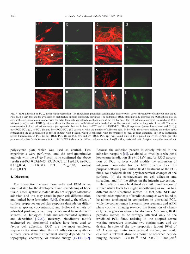
For studying the molecular mechanisms of osteoblastadhesion on biofunctionalized PCL surfaces, two mole-cules were selected: b1 and aVb3. b1 is associated to themost part of the integrins expressed by osteoblastsrepresenting the dominant adhesion mechanism to ECMligands, such as collagen, fibronectin, and laminin [24]. Inaddition to support adhesion, spreading, and migration, b1activates various intracellular signalling pathways, geneexpression, and protein activity that regulate cellularfunctions, including osteoclastogenesis [25,26]. The aVb3integrin, also known as vitronectin receptor, expressesRGD-dependent binding and mediates the adhesion of
hOB to the major non-collagenous proteins, i.e., osteo-pontin, bone sialoprotein, vitronectin, and fibronectin [23].aVb3 overexpression appears to promote hOB prolifera-tion over differentiation by influencing integrin–matrixinteractions, signal transduction and ECM protein expres-sion [27]. Recent data show that the initial attachment ofhOB to RGD surfaces is mediated by the b1 chainassociated with the a2 subunit (collagen receptor), whileaVb3 integrin governs longer-term adhesion [28]. Further-more, in our experimental condition, a noticeable reasonfor selecting b1 and aVb3 was the formation of focaladhesion contacts mediated by these molecules in thecontrol cultures. The focal adhesion complex is composedof a cluster of integrins which connects the ECM to theactin filaments through the binding of b chain withcytoskeletal proteins, i.e., talin–paxillin–vinculin subunits[29]. The adhesion plaque represents an essential structuralunit which directs the flow of information from outside thecell to the inside, promoting specific signalling downstreaminvolved in the control of cell motility, shape, growth, andsurvival [30,31].Our results showed a high expression of b1 on irradiated
PCL surfaces, which was not increased by the addition ofRGD peptides. On the opposite, the aVb3 expression wassignificantly higher on irr+RGD-PCL than on the othersamples, even if the number of attached osteoblasts did notchange.These findings confirm that ion irradiation produced
changes in surface properties and favoured the adsorptionof RGD peptides and proteins released from osteoblasts orderived from the sample ‘pre-wetting’ with culture medium.The pre-wetting has been performed in order to mimic thein vivo condition of an implanted material, which iscovered before by blood and biological fluids and later bycells. It is widely accepted that differences in proteinslayered onto PCL may be responsible for different cellularresponses, such as the modulation of integrin expression[8–17,20,32]. Generally, the cell spreading seems to beclosely related to the b1 modulation. This subunit is sharedby a number of adhesion molecules which can bind theirligands in a RGD-dependent or -independent manner[10,28]. For instance, the recognition of type-1 collagenby a2b1 and a1b1 largely depends on GFOGER sequence,which is found in triple-helical collagens [33]. Although theamount of RGD peptides on irradiated PCL surfaces wascalculated as sufficient to promote cell spreading and theformation of focal contacts [22], we have previouslyhypothesized some conformational changes of adsorbedRGD which could alter the receptor–ligand interaction andaffect cell adhesion. The behaviour of vitronectin receptorcancelled these doubt and confirmed that the RGD peptidewas functionally active. The aVb3 integrin was stronglyexpressed by hOB cultured on irr+RGD-PCL, which wasthe only one sample where the RGD adsorption wasdetectable. Like us, other Authors demonstrated the pivotalrole of aVb3 in mediating the cell adhesion on substratespresenting RGD or RGD-containing sequences [34].
ARTICLE IN PRESSI. Amato et al. / Biomaterials 28 (2007) 3668–3678 3677
On irradiated PCL, a high number of b1-mediated focalcontacts were detectable. The presence of the adhesionplaques supports the effectiveness of the biofunctionaliza-tion strategy, because the structural links between thecytoskeleton and ECM mediate stable adhesion and cellmigration [10]. Furthermore, focal adhesions activate thesignalling pathways, such as MAPK and JNK, thatregulate transcription factor activity and direct cell growthand differentiation [30,31]. The b1-mediated focal contactswere not found on irradiated/RGD surfaces, while adiffuse co-localization of aVb3 with cytoskeletal actinwas observed. This morphological variant of the focaladhesion, so-called ‘dot-like’, represents the initial step ofthe contact which is constituted of both transmembraneand some linker proteins, but is not associated with actinbundles [29]. Initial formation of ‘dot’ evolves by matura-tion into ‘dash’ contact, that is the morphological variantfound in b1-mediated focal adhesion.
Our results showed that surfaces presenting RGDpeptides do not enhance the early steps of osteoblastadhesion. Garcia and Reyes [10] describe the critical factorslimiting the potential of this biomolecular strategy for bonerepair purposes: (a) the biological activity of short adhesivepeptides is significantly lower than that of the completeprotein, (b) RGD peptides exhibit limited specificity amongintegrins, and although required, in some cases may be notsufficient for an efficient binding, and (c) osteogenic cellsrequire signals from non-RGD-binding integrins for robustdifferentiation.
We demonstrated that structural changes due to surfaceirradiation help the adsorption of peptides, and they aresufficient and functionally active to modulate the expres-sion of RGD-dependent integrins, i.e., aVb3. Nevertheless,the aVb3 binding competes with the formation ofb1-mediated focal contacts, which in the early steps ofthe adhesion seems to be induced in an RGD-independentmanner [10,28,33]. Both aVb3 and b1 subunit are essentialin regulating the osteoblast activity, as their neutralizationretards matrix mineralization by osteoblasts [35]. On onehand, the b1 integrins appear to be the predominantadhesion receptor subfamily utilized by hOB to adhere tocollagen, fibronectin, laminin [24], and on the other handavb3 seems to be mainly involved in the binding of non-collagenous proteins [23], as well as in increasing the cellproliferation rate [27].
In summary, it is desirable that bioactive materialsdesigned for bone tissue repair do not hamper the early cellattachment because that could influence long-term events,i.e., matrix deposition and mineralization. In this study, wehave further pointed out the importance of integrinanalysis to elucidate the molecular mechanisms of cellularadhesion on biofunctionalized surfaces.
The ability to induce integrin expression throughbiomimetic strategies may be a key step in determiningwhich modifications are suitable for application in bonetissue engineering and other orthopaedically relatedcircumstances.
5. Conclusion
The biofunctionalization of biodegradable syntheticpolymers represents one of the most promising strategiesfor the development of biologically active implants andgrafting substrates for bone tissue repair. As integrinexpression and osteoblast biology are closely related, thestudy of these molecules in the early phases of the adhesioncould represent an indicator that proves the effectiveness ofbiomimetic surfaces in promoting adhesion, proliferation,and function of osteoblasts. In this study, we investigatedthe effect of two well-defined strategies based on theirradiation-induced surface engineering, triggering the‘spontaneous’ reorganization of the ECM matrix, and themimicking of ECM by the engrafting/adsorption ofsuitable cell-adhesive sequences onto the PCL surface. Celladhesion and spreading were dramatically enhanced onirradiated PCL surfaces, and the addition of RGD did notchange the result. The expression of adhesion receptorsvaried according to the type of substrate: a high expressionof b1 on irradiated PCL surfaces was not increased by theaddition of RGD peptides, while a high aVb3 expressionwas found only on irradiated and RGD-treated surfaces,even if the number of attached osteoblasts did not change.We conclude that the low-energy He irradiation (10 keV)produces changes in surface properties which favour thepeptides/proteins adsorption; in turn, differences in thestructure of the adsorbed protein film can significantlymodify the expression of integrins relevant to the hOBfunction. Our findings add new knowledge on themolecular mechanisms of the cell/material interactionsand could be exploited to engineer surfaces provided withbioactive sites, which could elicit specific cell responsesenhancing the performance of implantable devices.
Acknowledgements
This study was funded by grants from the ItalianMinistry of University and Research (MIUR) throughthe projects FIRB 2001 RBAU01N79B and FIRB 2001RBNE01458S_003.
References
[1] Brekke JH, Toth JM. Principles of tissue engineering applied
to programmable osteogenesis. J Biomed Mater Res 1998;43:
380–98.
[2] Jackson DW, Simon TM. Tissue engineering principles in orthopae-
dic surgery. Clin Orthop Rel Res 1999:S31–45 (367, Suppl.).
[3] Gunatillake PA, Adhikari R. Biodegradable synthetic polymers for
tissue engineering. Eur Cells Mater 2003;5:1–16.
[4] Kweon HY, Yoo MK, Park IK, Kim TH, Lee HC, Lee HS, et al. A
novel degradable polycaprolactone networks for tissue engineering.
Biomaterials 2003;24:801–8.
[5] Causa F, Netti P. Poly-epsilon-caprolactone/hydroxyapatite
composites for bone regeneration: in vitro characterization and
human osteoblast response. J Biomed Mater Res A 2006;76:
151–62.
ARTICLE IN PRESSI. Amato et al. / Biomaterials 28 (2007) 3668–36783678
[6] Williams JM, Adewunmi A, Schek RM, Colleen L, Flanagan CL,
Krebsbach PH, Feinberg SE, Scott J. Hollister SJ, Das S. Bone tissue
engineering using polycaprolactone scaffolds fabricated via selective
laser sintering. Biomaterials 2005;26:4817–27.
[7] Ciapetti G, Ambrosio L, Savarino L, Granchi D, Cenni E, Baldini N,
et al. Osteoblast growth and function in porous poly-epsilon-
caprolactone matrices for bone repair: a preliminary study. Bioma-
terials 2003;24:3815–24.
[8] Marletta G, Ciapetti G, Satriano C, Pagani S, Baldini N. The effect
of irradiation modification and RGD sequence adsorption on the
response of human osteoblasts to polycaprolactone. Biomaterials
2005;26:4793–804.
[9] Anselme K. Osteoblast adhesion on biomaterials. Biomaterials
2000;21:667–81.
[10] Garcia AJ, Reyes CD. Bio-adhesive surfaces to promote osteoblast
differentiation and bone formation. J Dent Res 2005;84:407–13.
[11] Hersel U, Dahmen C, Kessler H. RGD modified polymers:
biomaterials for stimulated cell adhesion and beyond. Biomaterials
2003;24:4385–415.
[12] El-Amin SF, Kofron MD, Attawia MA, Lu HH, Tuan RS,
Laurencin CT. Molecular regulation of osteoblasts for tissue
engineered bone repair. Clin Orthop Rel Res 2004;427:220–5.
[13] Wan Y, Wang Y, Liu Z, Ou X, Han B, Bei J, et al. Adhesion and
proliferation of OCT-1 osteoblast-like cells on micro- and nano-scale
topography structured poly(L-lactide). Biomaterials 2005;26:4453–9.
[14] Lee MH, Ducheyne P, Lynch L, Boettiger D, Composto RJ. Effect of
biomaterial surface properties on fibronectin-alpha5beta1 integrin
interaction and cellular attachment. Biomaterials 2006;27:1907–16.
[15] Pignataro B, Conte E, Scandurra A, Marletta G. Improved cell
adhesion to ion beam-irradiated polymer surfaces. Biomaterials
1997;18(22):1461–70.
[16] Marletta G, Ciapetti G, Satriano C, Perut F, Manuela S; Nicola B.
Improved osteogenic differentiation of human marrow stromal cells
by ion-induced chemical nanostructuring of poly-e-caprolactone.Biomaterials 2007;28:1132–40.
[17] Kwok DY, Neumann AW. Contact angle measurement and contact
angle interpretation. Adv. Colloid Interface Sci. 1999;81:167–249.
[18] Granchi D, Amato I, Battistelli L, Ciapetti G, Pagani S, Avnet S,
et al. Molecular basis of osteoclastogenesis induced by osteoblasts
exposed to wear particles. Biomaterials 2005;26:2371–9.
[19] Keselowsky BG, Collard DM, Garcia AJ. Surface chemistry
modulates focal adhesion composition and signaling through changes
in integrin binding. Biomaterials 2004;25:5947–54.
[20] Mc Farland CD, Thomas CH, DeFilippis C, Steele JC, Healy KE.
Protein adsorption and cell attachment to patterned surfaces.
J Biomed Mater Res 2000;49:200–10.
[21] Shin H, Jo S, Mikos AG. Biomimetic materials for tissue engineering.
Biomaterials 2003;24:4353–6.
[22] Massia SP, Hubbell JA. An RGD spacing of 440nm is sufficient for
integrin av, b3-mediated fibroblast spreading and 140nm for focal
contact and stress fiber formation. J Cell Biol 1991;114:1089–100.
[23] Gronthos S, Stewart K, Graves SE, Hay S, Simmons PJ. Integrin
expression and function on human osteoblast-like cells. J Bone Miner
Res 1997;12:1189–97.
[24] Garcia AJ. Get a grip: integrins in cell-biomaterial interactions.
Biomaterials 2005;26:7525–9.
[25] Hynes RO. Integrins: bidirectional allosteric signaling machines. Cell
2002;110:673–87.
[26] Nakayamada S, Okada Y, Saito K, Tamura M, Tanaka Y. b1integrin/focal adhesion kinase-mediated signaling induces intercellu-
lar adhesion molecule 1 and receptor activator of nuclear factor kBligand on osteoblasts and osteoclast maturation. J Cell Biochem
2003;278:45368–74.
[27] Cheng SL, Lai CF, Blystone SD, Avioli LVA. Bone mineralization
and osteoblast differentiation are negatively modulated by integrin
avb3. J Bone Miner Res 2001;16:277–88.
[28] Rezania A, Healy KE. Integrin subunits responsible for adhesion of
human osteoblast-like cells to biomimetic peptide surfaces. J Orthop
Res 1999;17:615–23.
[29] Owen GRh, Meredith DO, ap Gwynn I, Richards RG. Focal
adhesion quantification- a new assay of material biocompatibility?
Review. Eur Cell Mater 2005;9:85–96.
[30] Giancotti FG, Ruoshlati E. Integrin signaling. Science 1999;
13(285):1028–32.
[31] Boudreau NJ, Jones PL. Extracellular matrix and integrin signalling:
the shape of things to come. Biochem J 1999;339:481–8.
[32] Allen LT, Tosetto M, Miller IS, O’Connor DP, Penney SC, Lynch IS,
et al. Surface-induced changes in protein adsorption and implications
for cellular phenotypic responses to surface interaction. Biomaterials
2006;27:3096–108.
[33] Knight CG, Morton LF, Peachey AR, Tuckwell DS, Farndale RW,
Barnes MJ. The collagen-binding A-domains of integrins alpha(1)-
beta(1) and alpha(2)beta(1) recognize the same specific amino acid
sequence, GFOGER, in native (triple-helical) collagens. J Biol Chem
2000;275:35–40.
[34] Petrie TA, Capadona JR, Reyes CD, Garcia AJ. Integrin specificity
and enhanced cellular activities associated with surfaces presenting a
recombinant fibronectin fragment compared to RGD support.
Biomaterials 2006;27:5459–70.
[35] Lai CF, Cheng SL. Alphabeta integrins play an essential role in
BMP-2 induction of osteoblast differentiation. J Bone Miner Res
2005;20:330–40.