Electrospun poly(ε-caprolactone)/Ca-deficient hydroxyapatite nanohybrids: Microstructure,...
9
Electrospun poly(ε-caprolactone)/Ca-deficient hydroxyapatite nanohybrids: Microstructure, mechanical properties and cell response by murine embryonic stem cells Alessandra Bianco a, ⁎, Erica Di Federico a, 1 , Ilana Moscatelli b, 1 , Antonella Camaioni b , Ilaria Armentano c , Luisa Campagnolo b , Mariaserena Dottori c , Josè Maria Kenny c , Gregorio Siracusa b , Gualtiero Gusmano a a Department of Chemical Sciences and Technologies, Research Unit INSTM Tor Vergata, University of Rome Tor Vergata, via Ricerca Scientifica, 00133 Rome, Italy b Department of Public Health and Cell Biology, University of Rome Tor Vergata, via Montpellier, 00133 Rome, Italy c Material Science and Technology Center, Research Unit INSTM, NIPLAB, University of Perugia, Terni, Italy abstract article info Article history: Received 28 October 2008 Received in revised form 12 March 2009 Accepted 2 April 2009 Available online 9 April 2009 Keywords: Biocompatibility Electrospun fibers Murine embryonic stem cells Nanohydroxyapatite Poly(ε-caprolactone) Nanohybrid scaffolds mimicking extracellular matrix are promising experimental models to study stem cell behaviour, in terms of adhesion and proliferation. In the present study, the structural characterization of a novel electrospun nanohybrid and the analysis of cell response by a highly sensitive cell type, embryonic stem (ES) cells, are investigated. Ca-deficient hydroxyapatite nanocrystals (d-HAp) were synthesized by precipitation. Fibrous PCL/d-HAp nanohybrids were obtained by electrospinning, d-HAp content ranging between 2 and 55 wt.%. Electrospun mats showed a non-woven architecture, average fiber size was 1.5± 0.5 μm, porosity 80–90%, and specific surface area 16 m 2 g -1 . Up to 6.4 wt.% d-HAp content, the nanohybrids displayed comparable microstructural, mechanical and dynamo-mechanical properties. Murine ES cell response to neat PCL and to nanohybrid PCL/d-HAp (6.4 wt.%) mats was evaluated by analyzing morpho- logical, metabolic and functional markers. Cells growing on either scaffold proliferated and maintained pluripotency markers at essentially the same rate as cells growing on standard tissue culture plates with no detectable signs of cytotoxicity, despite a lower cell adhesion at the beginning of culture. These results indicate that electrospun PCL scaffolds may provide adequate supports for murine ES cell proliferation in a pluripotent state, and that the presence of d-HAp within the mat does not interfere with their growth. © 2009 Elsevier B.V. All rights reserved. 1. Introduction The composition and topology of the extracellular matrix (ECM) has been found to affect cell morphology, function, and physiological response [1]. Electrospun nanofibrous scaffolds aimed to mimic the architecture and biological functions of ECM, are considered as very promising substrates for tissue engineering [2,3]. Among them, scaffolds made of poly(ε-caprolactone) (PCL), a bioresorbable aliphatic polyester, have been used to provide a 3D environment for in vitro embryonic stem cell culture and differentiation [4]. The use of a 3D environment might make the culture system closer to the in vivo situation: how various 3D scaffolds might affect the differentia- tion potential of murine ES cells has been the object of recent research [5–7]. Electrospun hybrid scaffolds based on bioresorbable polymers and conventional hydroxyapatite allow osteoblast proliferation and differentiation, and are thus considered very promising as bone scaffolding materials [8–10]. It is well known that Ca-deficient- hydroxyapatite (d-HAp) shows more similar features to biological apatites with respect to conventional stoichiometric hydroxyapatite (s-Hap) [11]. In the present study we have evaluated the biocompat- ibility of PCL/d-HAp nanohybrid mats by using murine ES cells. Because of their high sensitivity to toxic agents [12], their embryonic origin [13,14] and their ability to differentiate along all tissue types [15,16], ES cells are already being used in a standard cytotoxicity and embryotoxicity test for soluble compounds [17,18], and might as well provide a good model to test biocompatibility in vitro of novel scaffolds. In greater detail, we have analyzed how fibrous PCL/HAp affects an essential property of ES cells: their ability to proliferate without differentiating. Fibrous PCL/d-HAp mats were obtained by electrospinning, with a d-HAp content ranging from 2 to 55 wt.%. The d-HAp nanopowder was freshly synthesized by precipitation. Micro- structure, mechanical and dynamo-mechanical properties of the electrospun nanohybrid mats were extensively investigated. The biocompatibility of the mats was evaluated by culturing mouse ES cells and analyzing colony morphology and number, ES cell adhesion, viability, proliferation and preservation of pluripotency markers. To our knowledge, this is the first report in which electrospun PCL Materials Science and Engineering C 29 (2009) 2063–2071 ⁎ Corresponding author. Fax: +39 0672594328. E-mail address: [email protected] (A. Bianco). 1 These authors contributed equally to this work. 0928-4931/$ – see front matter © 2009 Elsevier B.V. All rights reserved. doi:10.1016/j.msec.2009.04.004 Contents lists available at ScienceDirect Materials Science and Engineering C journal homepage: www.elsevier.com/locate/msec
-
Upload
independent -
Category
Documents
-
view
0 -
download
0
Transcript of Electrospun poly(ε-caprolactone)/Ca-deficient hydroxyapatite nanohybrids: Microstructure,...
Materials Science and Engineering C 29 (2009) 2063–2071
Contents lists available at ScienceDirect
Materials Science and Engineering C
j ourna l homepage: www.e lsev ie r.com/ locate /msec
Electrospun poly(ε-caprolactone)/Ca-deficient hydroxyapatite nanohybrids:Microstructure, mechanical properties and cell response by murine embryonicstem cells
Alessandra Bianco a,⁎, Erica Di Federico a,1, Ilana Moscatelli b,1, Antonella Camaioni b, Ilaria Armentano c,Luisa Campagnolo b, Mariaserena Dottori c, Josè Maria Kenny c, Gregorio Siracusa b, Gualtiero Gusmano a
a Department of Chemical Sciences and Technologies, Research Unit INSTM Tor Vergata, University of Rome Tor Vergata, via Ricerca Scientifica, 00133 Rome, Italyb Department of Public Health and Cell Biology, University of Rome Tor Vergata, via Montpellier, 00133 Rome, Italyc Material Science and Technology Center, Research Unit INSTM, NIPLAB, University of Perugia, Terni, Italy
⁎ Corresponding author. Fax: +39 0672594328.E-mail address: [email protected] (A. Bianco).
1 These authors contributed equally to this work.
0928-4931/$ – see front matter © 2009 Elsevier B.V. Adoi:10.1016/j.msec.2009.04.004
a b s t r a c t
a r t i c l e i n f oArticle history:Received 28 October 2008Received in revised form 12 March 2009Accepted 2 April 2009Available online 9 April 2009
Keywords:BiocompatibilityElectrospun fibersMurine embryonic stem cellsNanohydroxyapatitePoly(ε-caprolactone)
Nanohybrid scaffolds mimicking extracellular matrix are promising experimental models to study stem cellbehaviour, in terms of adhesion and proliferation. In the present study, the structural characterization of anovel electrospun nanohybrid and the analysis of cell response by a highly sensitive cell type, embryonicstem (ES) cells, are investigated. Ca-deficient hydroxyapatite nanocrystals (d-HAp) were synthesized byprecipitation. Fibrous PCL/d-HAp nanohybrids were obtained by electrospinning, d-HAp content rangingbetween 2 and 55 wt.%. Electrospun mats showed a non-woven architecture, average fiber size was 1.5±0.5 μm, porosity 80–90%, and specific surface area 16 m2 g−1. Up to 6.4 wt.% d-HAp content, the nanohybridsdisplayed comparable microstructural, mechanical and dynamo-mechanical properties. Murine ES cellresponse to neat PCL and to nanohybrid PCL/d-HAp (6.4 wt.%) mats was evaluated by analyzing morpho-logical, metabolic and functional markers. Cells growing on either scaffold proliferated and maintainedpluripotency markers at essentially the same rate as cells growing on standard tissue culture plates with nodetectable signs of cytotoxicity, despite a lower cell adhesion at the beginning of culture. These resultsindicate that electrospun PCL scaffolds may provide adequate supports for murine ES cell proliferation in apluripotent state, and that the presence of d-HAp within the mat does not interfere with their growth.
© 2009 Elsevier B.V. All rights reserved.
1. Introduction
The composition and topology of the extracellular matrix (ECM)has been found to affect cell morphology, function, and physiologicalresponse [1]. Electrospun nanofibrous scaffolds aimed to mimic thearchitecture and biological functions of ECM, are considered as verypromising substrates for tissue engineering [2,3]. Among them,scaffolds made of poly(ε-caprolactone) (PCL), a bioresorbablealiphatic polyester, have been used to provide a 3D environment forin vitro embryonic stem cell culture and differentiation [4]. The useof a 3D environment might make the culture system closer to the invivo situation: how various 3D scaffolds might affect the differentia-tion potential of murine ES cells has been the object of recent research[5–7]. Electrospun hybrid scaffolds based on bioresorbable polymersand conventional hydroxyapatite allow osteoblast proliferation anddifferentiation, and are thus considered very promising as bone
ll rights reserved.
scaffolding materials [8–10]. It is well known that Ca-deficient-hydroxyapatite (d-HAp) shows more similar features to biologicalapatites with respect to conventional stoichiometric hydroxyapatite(s-Hap) [11]. In the present study we have evaluated the biocompat-ibility of PCL/d-HAp nanohybrid mats by using murine ES cells.Because of their high sensitivity to toxic agents [12], their embryonicorigin [13,14] and their ability to differentiate along all tissue types[15,16], ES cells are already being used in a standard cytotoxicity andembryotoxicity test for soluble compounds [17,18], and might as wellprovide a good model to test biocompatibility in vitro of novelscaffolds. In greater detail, we have analyzed how fibrous PCL/HApaffects an essential property of ES cells: their ability to proliferatewithout differentiating. Fibrous PCL/d-HAp mats were obtained byelectrospinning, with a d-HAp content ranging from 2 to 55 wt.%. Thed-HAp nanopowder was freshly synthesized by precipitation. Micro-structure, mechanical and dynamo-mechanical properties of theelectrospun nanohybrid mats were extensively investigated. Thebiocompatibility of the mats was evaluated by culturing mouse EScells and analyzing colony morphology and number, ES cell adhesion,viability, proliferation and preservation of pluripotency markers. Toour knowledge, this is the first report in which electrospun PCL
2064 A. Bianco et al. / Materials Science and Engineering C 29 (2009) 2063–2071
nanohybrids containing d-HAp have been described, and theirbiocompatibility tested by the use of ES cells.
2. Experimental
2.1. Synthesis of Ca-deficient hydroxyapatite (d-HAp) nanopowders
The d-HAp nanopowder was prepared in a double-walled jacketreactor at 40 °C following a precipitation route described elsewhere[19]. Stoichiometric volume of 1M calcium nitrate tetrahydrate (Ca(NO3)2.4H2O, 99.2% Aldrich) was added dropwise to diammoniumhydrogen phosphate ((NH4)2HPO4, 99.2% Aldrich) pH was continu-ously monitored and adjusted at 9.0±0.1 by adding NH4OH conc.Precipitate was aged in mother solutions for 24 h, washed withNH4OH aqueous solution, and vacuum filtered. Chemical analysis wasperformed by induced coupled plasma atomic emission spectroscopy(AES-ICP, JobinYvon JV 24R). Morphology was examined by transmis-sion electron microscopy in bright field mode, accelerating voltagebeing 100 kV (TEM, Philips CM120) and specific surface area measuredby N2 adsorption (Sorptomatic 1900, Carlo Erba) using the Brunauer–Emmett–Teller (BET)method. X-ray diffraction analysis (XRD) (PhilipsX'Pert) was performed in the following conditions: Cu Kα radiationλ=1.5405600 Å, 20–55° 2θ, step size 0.010°, time per step 2 s, scanspeed 0.005°/s.
2.2. Fabrication of electrospun PCL membranes
PCL (Aldrich, MW 80000) was dissolved at RT under magneticstirring in a 1:1 dichloromethane (CH2Cl2) and N,N-dimethylforma-mide (DMF) mixture, the polymer concentration being 12% (w/v).The solution was poured in a glass syringe (Socorex) equipped with a21 G needle, fixed in a digitally controlled syringe pump (KDScientific) and electrospun as follows: tension 21 kV (Spellman, ModelSL 30), needle-target distance 10 cm, feed rate 1 ml h−1. According toprevious studies [20–23], electrospun meshes were dried undervacuum for 24 h.
2.3. Fabrication of electrospun PCL/d-HAp membranes
The d-HAp nanopowder was poured to a 1:1 CH2Cl2–DMF solutionand the suspension ultrasonicated for 2 h. PCL granules were thenadded and the mixture stirred for 36 h. Suspensions were electrospunand the resultingmats treated as described above. All prepared PCL/d-HAp nanohybrid samples are listed in Table 1.
2.4. Viscosity and conductivity
Viscosity of polymeric suspensions was measured at 25 °C bydigital viscosimeter (Brookfield DV-II+) equipped with a SC4-21spindle at 20 rpm and electrical conductivity measured at 25 °C byconductivity meter (CDM230, Analitica De Mori).
Table 1Designation and composition of PCL/d-HAp nanohybrid electrospun mats.
Sample PCL/d-Hap, wt.%a PCL/d-Hap, vol.%b
PCL – –
2HAp 2.0 54HAp 3.6 96HAp 6.4 1625HAp 24.9 4840HAp 42.5 6755HAp 55.1 77
a Values referred to residual mass (T=600 °C), as calculated by TGA measurements.b Volume fraction of the ceramic filler (ϕc) was derived from the weight percentage
(Wc) as follows, where ρp e ρc are, respectively the density of PCL (ρPCL 1.145 g cm−3)and d-HAp (ρd-HAp 3.156 g cm−3): 1
/c= 1 + ρp
ρc
100Pc
− 1� �
.
2.5. Scanning electron microscopy (SEM) and energy dispersionspectroscopy (EDS)
Microstructure of electrospun fabrics was investigated by SEM(Cambridge Stereoscan 300). The actual d-HAp distribution wasevidenced by energy dispersion spectroscopy (EDS). Average fiberdiameter was determined on 120 different fibers by means of CADsoftware.
2.6. Density, porosity and specific surface area
Density (ρ) was estimated as mass to volume ratio on 12 mmdiameter disks cut out of the membranes, four samples were consi-dered for each material. Sample thickness was measured according toISO7198 by means of a digital micrometer equipped with a 2 kg loadcell. Porosity (ε) was estimated as follows, where ρ0 is the density ofas-purchased PCL (1.145 g cm−3):
e = 1− ρρ0
� �� 100 ð1Þ
Specific surface area was measured by N2 adsorption (Sorptomatic1900, Carlo Erba) using BET method.
2.7. Infrared spectroscopy (ATR-IR) and thermal analyses
IR spectra were recorded in the 4000–400 cm−1 region by meansof a Jasco FTIR-615 spectrophotometer in ATR reflection method,spectral resolution 4 cm−1.
Thermogravimetric analysis (TGA) was performed by means of aquartz rod microbalance (Seiko Exstar 6000). Measurements wereperformed in N2 atmosphere between 30 and 1000 °C, heating rate10 °C min−1. Residual mass and maximum degradation temperature(Td) were measured. Thermal characterization was carried out usingDifferential Scanning Calorimeter (DSC) (Perkin Elmer Pyris 1),heating and cooling scans were performed in the following condi-tions: sample weight 10 mg, heating rate 10 °C min−1, temperaturerange from −25 °C to 100 °C, N2 atmosphere. Melting temperature(Tm), melting enthalpy (ΔHm), and crystallinity degree (Xc) weredetermined from the heating scan. Crystallization temperature (Tc)and enthalpy (ΔHc) were measured from the cooling run. Xc of thePCL component in the nanocomposites was calculated according to(2):
Xc kð Þ = ΔHm =ΔH0 ð2Þ
where ΔHm is derived from the DSC curves, with respect to the actualPCL content [24],ΔH0 is the theoretical enthalpy of the fully crystallinepolymer, for PCL the considered value was 136 J g−1 [25].
2.8. Mechanical characterization
Uniaxial tensile tests were carried out on dog-bone specimens,gauge width 4.8 mm and gauge length 22.3 mm. Mechanical testswere performed at 1.2mmmin−1 to rupture by an electromechanicalmachine equipped with a 5 kg load cell (TA-HDi, Stable MicroSystems). Four specimens were considered for each electrospunmatrix. The tensile modulus (E) was evaluated between 0 and 5%strain [26]. The yield stress was determined from the intersection ofthe stress–strain curve with a line parallel to the linear region andoffset by 2% strain. The ultimate stress (σmax) and ultimate percentdeformation (εf) were calculated considering the nominal cross-sectional area of the tensile specimen. Dynamic-Mechanical Analysis(DMA) was performed using a Reometric Scientific-ARES sample sizewas 10 mm width, 30 mm length, 0.3 mm thick. Shear modulus was
2065A. Bianco et al. / Materials Science and Engineering C 29 (2009) 2063–2071
measured at RT in the dynamic time sweep test, fixed frequency 1 Hzand shear strain 0.3%.
2.9. Mouse ES cell culture
Mouse ES cell line R1 was a kind gift of Dr. Heidi Stuhlmann.Following standard procedures, ES cells were cultured on a feederlayer of mitotically-inactivated primary mouse embryonic fibroblasts(MEFs) from CD-1 mouse embryos, in tissue culture polystyreneplates (TCPS) that had been gelatin-coated to facilitate cell adhesion(a 0.7% denatured collagen solution, Sigma, MO, USA, for 10 min at37 °C). The same procedure was used when testing experimentalscaffolds. ES cells were expanded and maintained undifferentiated byculture in Dulbecco's Modified Eagle's Medium (DMEMwith 4,5 g L−1
D-glucose) supplemented with 15% heat-inactivated fetal calf serum(ES cell tested), 20 mM Hepes, 2 mM L-glutamine, 100 μM β-mercaptoethanol, 100 μM non-essential amino acids, 50 U ml−1
penicillin and 50 μg ml−1 streptomycin (all from Lonza, Switzerland),and 103 U ml−1 Leukemia Inhibitory Factor (LIFES, ImmunologicalSciences). The cells were cultured at 37 °C in a humidified atmosphereof 5% CO2 in air. To obtain highly enriched ES cell populations, culturesreaching 80% confluency and forming round colonies were trypsinizedand gently disaggregated by pipetting. After centrifugation, single cellsuspensions were plated onto TCPS and kept for 20–40 min in theincubator. During this time, MEFs attach to the plate while ES cells,that require longer adhesion times, remain in the supernatant and canbe harvested for subsequent use. Before each experiment, scaffoldswere sterilized in 70% ethanol overnight, air dried, and secured bystainless steel rings to the bottom of the wells of 24-well plates(Multiwell™, Falcon, Becton Dickinson Labware). MEFs were thenseeded on the scaffolds and on standard TCPS and incubatedovernight. The next day, ES cells were seeded in each well in 1 ml ofmedium, at the concentrations specified in the text, and allowed toattach for 6 h before measuring their adhesion rate to the substrates(see below). When proliferation was to be measured, after adhesion,the ES cells were allowed to grow for approximately 3 days. Duringsuch time, each adhered ES cell forms a round colony made of closelypacked cells. In the proliferation experiments, the medium waschanged every 24 h until the ES colonies in the standard cultures wereapproximately 80% confluent (about 3 days).
2.10. Staining procedures
For morphological analysis, at the end of the culture cells werewashed with Dulbecco's phosphate buffered saline (PBS, Lonza) andfixed with methanol for 5 min at−20 °C. To stain nuclei, a solution of0.5 μgml−1 Hoechst 33258 (Sigma) in PBSwas added for 30min. Afterthree PBSwashes, cells weremountedwithMöwiol (Polysciences Inc.).Fluorescent images were taken with Axioplan 2 microscope (Zeiss).Pluripotency of the ES cell colonies was evaluated by alkalinephosphatase assay [13,27]. Enzyme activity was detected by incubat-ing the fixed cells for 15 min in a solution made by mixing 960 μl FastRed TR salt (1 mg ml−1) with 40 μl Naphthol AS-BI (4 mg ml−1) (allfrom Sigma). Cells were washed with PBS and observed using a LeitzDiavert microscope. Pluripotent colonies expressing alkaline phos-phatase appeared red, while the differentiated ones were colourless.Cell viability was determined by staining with a 0.4% trypan bluesolution (Sigma). The dead/blue cells were counted with a hemocyt-ometer and the data were expressed as percentages of the total cellnumber (±SD).
2.11. Western blot analysis
For protein immunodetection by Western blot, cells were homo-genized in lysis buffer (50 mM HEPES, pH 7.4, 15 mM MgCl2, 150 mMNaCl, 15 mM EGTA, 20 mM β-glycerophosphate, 1% Triton X-100, 10%
glycerol, 0.5 mM sodium orthovanadate, 1 mM dithiothreitol) withProtease Inhibitor Cocktail (Roche). Homogenates were sonicated,centrifuged at 4 °C for 15 min at 15,000 g, supernatants were collectedand protein concentration was determined. Extracts were solubilizedin NuPAGE LDS sample buffer with the addition of 50 mM dithio-threitol, separated by electrophoresis on a 4–12% NuPAGE Bis–Tris GelSystem (Invitrogen), and transferred to PVDF Transfer MembraneHybond™ -P (Amersham). Membranes were saturated with 5% nonfatdry milk in PBS containing 0.1% Tween 20 (PBS/T), for 1 h at roomtemperature and incubated overnight at 4 °C with the followingprimary antibodies, diluted 1:1000 in PBS/T with 5% BSA: rabbit anti-Nanog (Cosmo Bio) andmouse anti-αTubulin (Sigma). Secondary anti-rabbit or anti-mouse antibodies conjugated to horseradish peroxidase(Amersham) were incubated with the membranes for 1 h at roomtemperature at 1:10,000 dilution in PBS/T with 5% nonfat dry milk.Immunoreactive bands were detected by SuperSignal West Picochemiluminescent reagent (Pierce) and densitometric analysis of thebands was performed using ImageQuant 5.0 (Molecular Dynamics).
2.12. MTT assays
Cell adhesion was evaluated 6 h after seeding 8×10⁴ ES cells onscaffolds and onTCPS by amodification of themethylthiazolyldiphenyl–tetrazolium bromide (MTT; Sigma) assay [28]. Parallel cultures withoutES cells (MEFs only) were analyzed in order to subtract the backgrounddue to the feeder layer. Briefly, each sample was rinsed with PBS toremove unattached cells and then incubated with 0.5 mg ml−1 MTT(250 μl/well, 1 h, 37 °C).. The MTT solution was then removed anddimethylsulfoxide (Sigma) (900 μl/well) and glycine buffer (pH 10.5,125 μl/well) were added to each well. After 10 min of agitation, theabsorbance at 540 nm was measured with a SmartSpec 3000 spectro-photometer (Bio-Rad) and recorded as optical density (OD).
2.13. [methyl-3H]thymidine incorporation
Cell proliferationwas evaluated 3 days after seeding 104 ES cells onscaffolds and TCPS, by measuring incorporation of radioactive DNAprecursor [methyl-3H]thymidine, Amersham, (3HTdR) in the acid-insoluble fraction. Parallel MEF-only cultures were also run as above(see MTT assay). For radiolabeling, cells were washed with PBS andincubated with fresh serum-free DMEM containing all the supple-ments and 1 μCi/well of 3HTdR for 2 h at 37 °C. After incubation, cellswere treated with 5% trichloroacetic acid (TCA) on ice for 20 min, andthen washed twice with ice-cold distilled water. The acid-insolublematerial was dissolved with 1 N NaOH for 20 min at roomtemperature, and an equal volume of 1 N HCl was added. Each samplewas mixed 1:10 with scintillation liquid (OptiPhase ‘HiSafe’3, PerkinElmer) and the radioactivity, expressed as counts per minute, wasmeasured by a scintillation counter (LS 6500, Beckman Instruments).
2.14. Statistical analysis
Data from the MTT and 3HTdR incorporation assays are shown asmeans±standard error of three replicates from three independentexperiments. Radioactivity countswere normalized to take into accountthe differences in the number of adhered cells, 6 h after seeding.Statistical analysis was performed by one-way analysis of variance(ANOVA) and Tukey test for pairwise multiple comparisons.
3. Results and discussion
3.1. Characterization of d-HAp nanopowder
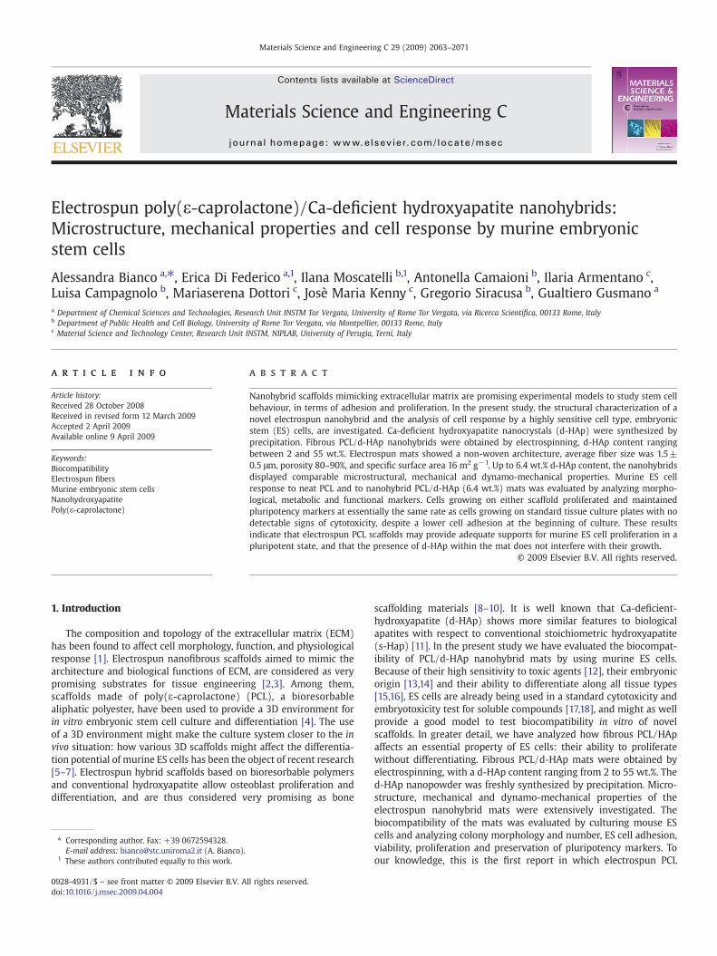
TEM observations showed that the as-precipitate powder con-sisted of needle-like particles, width 10–30 nm and length 50–100 nm(Fig. 1). The correspondent selected area electron diffraction (SAED)
Fig. 1. TEM image and SAED pattern of as-precipitated d-HAp nanoparticles.
2066 A. Bianco et al. / Materials Science and Engineering C 29 (2009) 2063–2071
pattern exhibit spotted sharp and continuous rings, evidencingpolycrystalline grains. XRD analysis showed that the as-synthesizedpowder consisted of semicrystalline hydroxyapatite [29]. As expectedfor a calcium-deficient hydroxyapatite [11], the composition of theprecipitate in terms of Ca/P molar ratio was 1.57. Finally, the specificsurface area of the as-precipitated Ca-deficient nanohydroxyapatitewas 100 m2 g−1.
3.2. Characterization of PCL/d-HAp suspensions
Viscosity and conductivity of PCL/d-HAp suspensions prepared forelectrospinning are summarized in Table 2. Particularly, viscosity of Psolution and 2HAp suspension were comparable, the value beingaround 470 mPa s. In the case of 4HAp and 6HAp suspensions,viscosity decreased down to 414 mPa s and 427 mPa s, respectively.This anomalous effect is typical of fine dispersed nanocompositesystems, in which interphase characteristics predominate on bulkproperties. Suspensions containing higher d-HAp amounts showedprogressively increased viscosity, themaximumvalue being 1432mPas for 55HAp suspension. In agreement with Wutticharoenmongkol etal. [9], the highest conductivity was registered for the neat PCLsolution. The presence of d-HAp nanophase induced conductivitychanges, leading to progressively higher values with increasednanofiller amounts. The conductivity increased from 1.55 μS cm−1
to 4.12 μS cm−1 for 2HAp and 55HAp, respectively, due to the presenceof calcium and phosphate ions.
3.3. Characterization of nanohybrid PCL/d-HAp mats
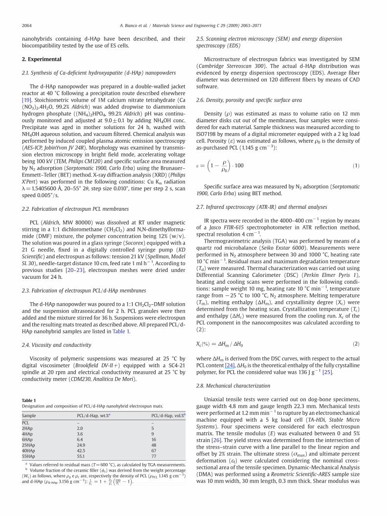
In Fig. 2 A–D SEM micrographs of different PCL/d-HAp nanohy-brids are presented. Electrospun mats consisted of randomlyoriented fibers of fairly uniform diameter. Large interconnectedvoids were present within the fibers, resulting in a 3D porousnetwork. Fiber size and morphology were not significantly influ-enced by the presence of d-HAp, the average fiber size being 1.5±0.5 μm, for both neat PCL and PCL/d-HAp nanohybrids. EDSmappings
Table 2Viscosity and conductivity of PCL/d-HAp suspensions at different d-HAp contents.
Sample Viscosity (mPa s) Conductivity (μS cm−1)
PCL 468 4.74±0.032HAp 471 1.55±0.034HAp 414 1.84±0.036HAp 427 2.55±0.0425HAp 574 3.34±0.0540HAp 697 3.83±0.0555HAp 1432 4.12±0.08
showed that few submicronic and micronic (2÷5 μm) agglomeratesrich in Ca and P appearedwithin the fibers of the nanohybrid samplescontaining 6.4 wt.% d-HAp (Fig. 2C). The agglomerates progressivelyincreased in number and size in nanohybrid samples loaded withhigher d-HAp amounts, due to the heterogeneity of the correspon-dent suspensions (Fig. 2D). On the other side, it has to be pointed outthat such agglomerates were not observed in the nanocompositeloaded with 2.0 wt.% of nanoparticles. These overall results clearlyindicated that sample 2HAp is characterized by a higher interdisper-sion of the two components (Fig. 2B). According to the literature,density of electrospun membranes was around 0.2–0.3 g cm−3,porosity 80–90% and specific surface area around 16 m2 g−1, withoutsignificant difference among samples [9,10].
The actual d-HAp content was accurately determined by TGAmeasurements (Table 1), considering that by 600 °C the polymer iscompletely burned out. Thermogravimetric data reported in Table 3also evidenced that electrospun meshes containing up to 6.4 wt.% d-HAp nanoparticles showed only a degradation temperature around400 °C associated to the thermal decomposition of the polymer. Thethermal stability of these nanocomposites seemed thus not remark-ably influenced by low amounts of nanofiller. Further increasing thed-HAp content, the polymermatrix destabilized and the temperaturedecomposition onset decreased, probably due to the pseudo-catalytic and disruptive effect of oxydril groups on hydroxyapatitenanoparticles [30,31].
In order to investigate the effect of d-Hap content on the propertiesof nanocomposites, IR spectra were recorded (Fig. 3). It mainlyresulted that (i) the peak around 1030 cm−1, associated to d-HAp,increased progressively with ceramic loading; (ii) absorption bandsmost sensitive to crystallinity changes (e.g. 1170 cm−1 (C=O–C,strong) and 1185 cm−1 (C=O, shoulder) changed in shape andintensity, the shoulder at 1185 cm−1 decreasingwith increasing the d-HAp content [32,33].
DSC analyses were also performed on PCL and its nanocomposites,the resulting calorimetric parameters are reported in Table 3. The nonisothermal crystallization curves for PCL and its nanocomposites arereported in Fig. 4. Regarding the relevance of these results, it has to bepointed out that it is well accepted that mechanical and physicalproperties of crystalline polymers are governed by the supra-molecular morphology which is in turn controlled by the crystal-lization process. Thus, the study of crystallization behaviour isnecessary for establishing the structure–property correlations of thenanocomposites [34]. It resulted that the Tc of nanohybrids occurredat higher temperature with respect to the neat sample, clearlyevidencing that d-HAp nanoparticles promote the crystallization ofthe PCL matrix, acting as a heterogeneous nucleating agent. RegardingTm, it can be observed that the valueswere not significantly affected bythe presence of the d-HAp. Moreover, DSC analyses also evidencedthat the presence of low d-HAp contents (e.g. up to 6.4 wt.%) did not
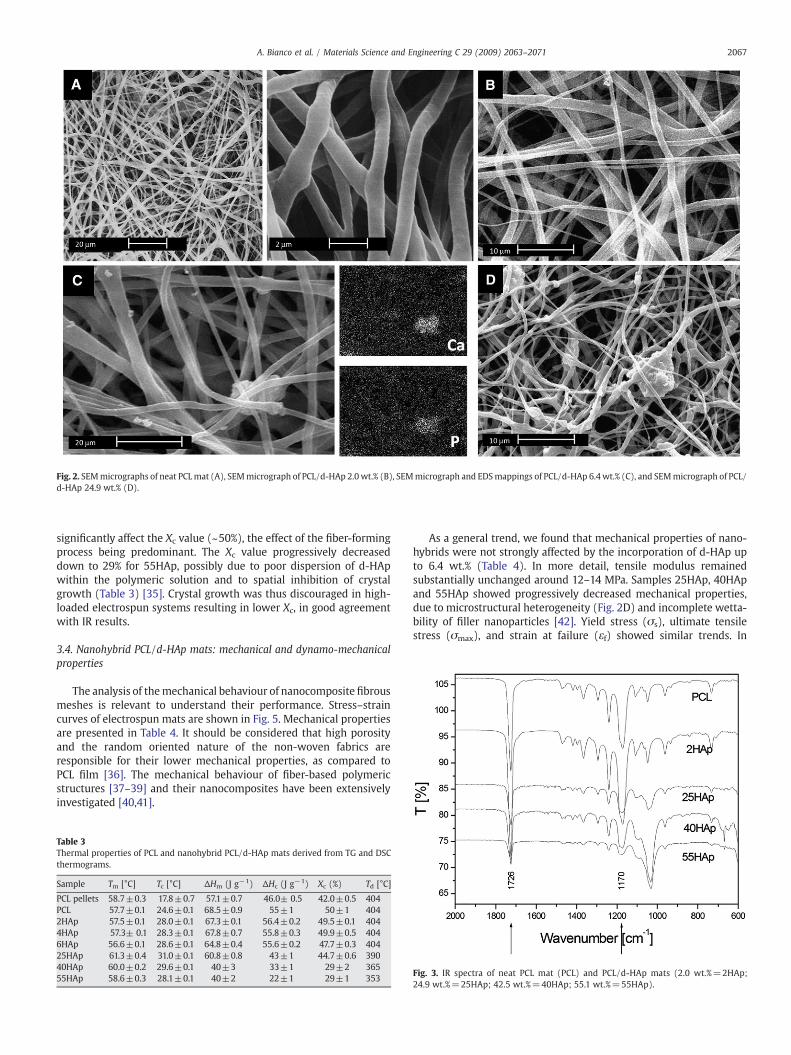
Fig. 2. SEMmicrographs of neat PCLmat (A), SEMmicrograph of PCL/d-HAp 2.0wt.% (B), SEMmicrograph and EDSmappings of PCL/d-HAp 6.4wt.% (C), and SEMmicrograph of PCL/d-HAp 24.9 wt.% (D).
2067A. Bianco et al. / Materials Science and Engineering C 29 (2009) 2063–2071
significantly affect the Xc value (~50%), the effect of the fiber-formingprocess being predominant. The Xc value progressively decreaseddown to 29% for 55HAp, possibly due to poor dispersion of d-HApwithin the polymeric solution and to spatial inhibition of crystalgrowth (Table 3) [35]. Crystal growth was thus discouraged in high-loaded electrospun systems resulting in lower Xc, in good agreementwith IR results.
3.4. Nanohybrid PCL/d-HAp mats: mechanical and dynamo-mechanicalproperties
The analysis of themechanical behaviour of nanocomposite fibrousmeshes is relevant to understand their performance. Stress–straincurves of electrospun mats are shown in Fig. 5. Mechanical propertiesare presented in Table 4. It should be considered that high porosityand the random oriented nature of the non-woven fabrics areresponsible for their lower mechanical properties, as compared toPCL film [36]. The mechanical behaviour of fiber-based polymericstructures [37–39] and their nanocomposites have been extensivelyinvestigated [40,41].
Table 3Thermal properties of PCL and nanohybrid PCL/d-HAp mats derived from TG and DSCthermograms.
Sample Tm [°C] Tc [°C] ΔHm (J g−1) ΔHc (J g−1) Xc (%) Td [°C]
PCL pellets 58.7±0.3 17.8±0.7 57.1±0.7 46.0± 0.5 42.0±0.5 404PCL 57.7±0.1 24.6±0.1 68.5±0.9 55±1 50±1 4042HAp 57.5±0.1 28.0±0.1 67.3±0.1 56.4±0.2 49.5±0.1 4044HAp 57.3± 0.1 28.3±0.1 67.8±0.7 55.8±0.3 49.9±0.5 4046HAp 56.6±0.1 28.6±0.1 64.8±0.4 55.6±0.2 47.7±0.3 40425HAp 61.3±0.4 31.0±0.1 60.8±0.8 43±1 44.7±0.6 39040HAp 60.0±0.2 29.6±0.1 40±3 33±1 29±2 36555HAp 58.6±0.3 28.1±0.1 40±2 22±1 29±1 353
As a general trend, we found that mechanical properties of nano-hybrids were not strongly affected by the incorporation of d-HAp upto 6.4 wt.% (Table 4). In more detail, tensile modulus remainedsubstantially unchanged around 12–14 MPa. Samples 25HAp, 40HApand 55HAp showed progressively decreased mechanical properties,due to microstructural heterogeneity (Fig. 2D) and incomplete wetta-bility of filler nanoparticles [42]. Yield stress (σs), ultimate tensilestress (σmax), and strain at failure (εf) showed similar trends. In
Fig. 3. IR spectra of neat PCL mat (PCL) and PCL/d-HAp mats (2.0 wt.%=2HAp;24.9 wt.%=25HAp; 42.5 wt.%=40HAp; 55.1 wt.%=55HAp).
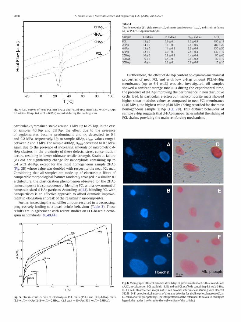
Table 4Tensile modulus (E), yield stress (σs), ultimate tensile stress (σmax), and strain at failure(εf) of PCL/d-HAp nanohybrids.
Sample E (MPa) σs (MPa) σmax (MPa) εf (%)
PCL 13±2 0.9±0.1 3.0±0.5 150±152HAp 14±3 1.1±0.1 3.4±0.5 280±204HAp 13±5 1.1±0.2 2.3±0.6 130±506HAp 12±1 0.9±0.1 2.4±0.3 130±1025HAp 10±3 0.8±0.2 1.4±0.4 80±4040HAp 6±1 0.4±0.1 0.5±0.2 30±1055HAp 6±4 0.2±0.1 0.8±0.6 15±10
Fig. 4. DSC curves of neat PCL mat (PCL) and PCL/d-HAp mats (2.0 wt.%=2HAp;3.6 wt.%=4HAp; 6.4 wt.%=6HAp) recorded during the cooling scan.
2068 A. Bianco et al. / Materials Science and Engineering C 29 (2009) 2063–2071
particular, σs remained stable around 1 MPa up to 25HAp. In the caseof samples 40HAp and 55HAp, the effect due to the presenceof agglomerates became predominant and σs decreased to 0.4and 0.2 MPa, respectively. Up to sample 6HAp, σmax values rangedbetween 2 and 3 MPa. For sample 40HAp, σmax decreased to 0.5 MPa,again due to the presence of increasing amounts of micrometric d-HAp clusters. In the proximity of these defects, stress concentrationoccurs, resulting in lower ultimate tensile strength. Strain at failure(εf) did not significantly change for nanohybrids containing up to6.4 wt.% d-HAp, except for the most homogeneous sample 2HAp(Fig. 2B) whose value was doubled with respect to the neat PCL mat.Considering that all samples are made up of electrospun fibers ofcomparable morphological features randomly arranged in a similar 3Darchitecture, the plasticization phenomenon observed for the 2HApnanocomposite is a consequence of blending PCLwith a low amount ofnanoscale-sized d-HAp particles. According to [43], blending PCL withnanoparticles is an effective approach to afford dramatic improve-ment in elongation at break of the resulting nanocomposites.
Further increasing the nanofiller amount resulted in ɛf decreasing,progressively leading to a quasi brittle behaviour (Table 3). Theseresults are in agreement with recent studies on PCL-based electro-spun nanohybrids [10,40,44].
Fig. 5. Stress–strain curves of electrospun PCL mats (PCL) and PCL/d-HAp mats(3.6 wt.%=4HAp; 24.9 wt.%=25HAp; 42.5 wt.%=40HAp; 55.1 wt.%=55HAp).
Furthermore, the effect of d-HAp content on dynamo-mechanicalproperties of neat PCL and with low d-Hap amount PCL/d-HApmembranes (up to 6.4 wt.%) was also investigated. All samplesshowed a constant storage modulus during the experimental time,the presence of d-HAp improving the performance in non disruptivecyclic load. In particular, electrospun nanocomposite mats showedhigher shear modulus values as compared to neat PCL membranes(140 MPa), the highest value (640 MPa) being recorded for the mosthomogeneous sample 2HAp (Fig. 2B). This distinct behaviour ofsample 2HAp suggests that d-HAp nanoparticles inhibit the sliding ofPCL chains, providing the main reinforcing mechanism.
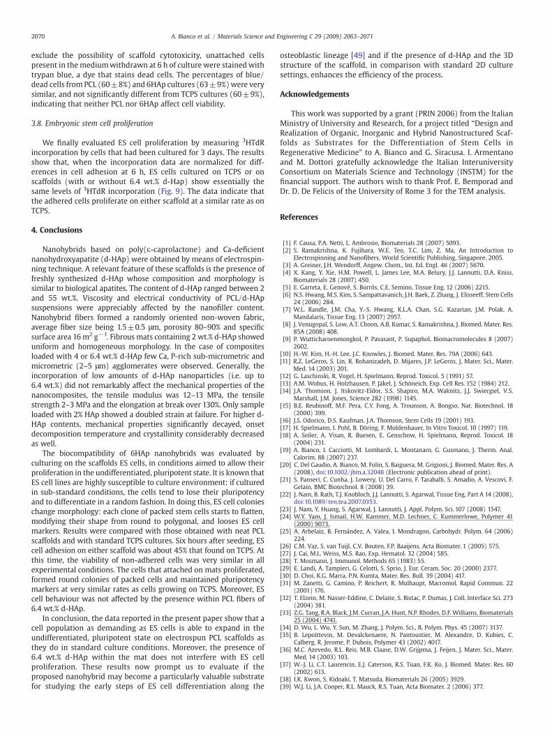
Fig. 6.MicrographsofEScell colonies after3days of growth in standard cultures conditions(A, D), in cultures on PCL scaffolds (B, E) and on PCL scaffolds containing 6.4 wt.% d-HAp(C, F). A–C: fluorescence analysis of ES cell colonies after nuclear staining with Hoechst33258; D–F: cytochemical analysis of the same colonies for alkaline phosphatase (red), anES cell marker of pluripotency. (For interpretation of the references to colour in this figurelegend, the reader is referred to the web version of this article.)
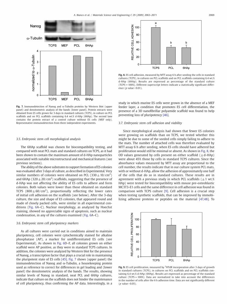
Fig. 7. Immunodetection of Nanog and α-Tubulin proteins by Western blot (upperpanel) and densitometric analysis of the bands (lower panel). Protein extracts wereobtained from ES cells grown for 3 days in standard cultures (TCPS), in culture on PCLscaffolds and on PCL scaffolds containing 6.4 wt.% d-HAp (6HAp). The second lanecontains the protein extract of a control culture without ES cells (MEF only).Representative immunodetection from three independent experiments.
Fig. 8. ES cell adhesion, measured by MTT assay 6 h after seeding the cells in standardcultures (TCPS), in cultures on PCL scaffolds and on PCL scaffolds containing 6.4 wt.%d-HAp (6HAp). Results are expressed as percentage of the standard culture(TCPS=100%). Different superscript letters indicate a statistically significant differ-ence (p valueb0.01).
Fig. 9. ES cell proliferation, measured by 3HTdR incorporation after 3 days of growthin standard cultures (TCPS), in cultures on PCL scaffolds and on PCL scaffolds con-taining 6.4 wt.% d-HAp (6HAp). Results are expressed as percentage of the standardculture (TCPS=100%). Data are normalized to take into account the differencesin the number of cells after the 6 h adhesion time. Data are not significantly different(p valueN0.05).
2069A. Bianco et al. / Materials Science and Engineering C 29 (2009) 2063–2071
3.5. Embryonic stem cell morphological analysis
The 6HAp scaffold was chosen for biocompatibility testing, andcompared with neat PCL mats and standard cultures on TCPS, as it hadbeen shown to contain the maximum amount of d-HAp nanoparticlesassociated with suitable microstructural and mechanical features (seeprevious sections).
The abilityof the above substrates to support formationofES colonieswas evaluated after 3 days of culture, as described in Experimental. Verysimilar numbers of colonies were obtained on PCL (330±10/cm2)and 6HAp (320±20/cm2) scaffolds, suggesting that the presence ofd-HAp was not affecting the ability of ES cells to adhere and formcolonies. Both values were lower than those obtained on standardTCPS (800±60/cm2), proportionally reflecting the lower ratesof initial cell adhesion on the scaffolds (see below). After the 3-dayculture, the size and shape of ES colonies, that appeared round andmade of closely packed cells, were similar in all experimental con-ditions (Fig. 6A–C). Nuclear morphology, as analyzed by Hoechststaining, showed no appreciable signs of apoptosis, such as nuclearcondensation, in any of the cultures examined (Fig. 6A–C).
3.6. Embryonic stem cell pluripotency markers
As all cultures were carried out in conditions aimed to maintainpluripotency, cell colonies were cytochemically stained for alkalinephosphatase (AP), a marker for undifferentiated ES cells (seeExperimental). As shown in Fig. 6D–F, all colonies grown on eitherscaffold were AP positive, as they were in standard TCPS cultures. Inaddition, the colonies were analyzed byWestern blot for the presenceof Nanog, a transcription factor that plays a crucial role in maintainingthe pluripotent state of ES cells [45]. Fig. 7 shows (upper panel) theimmunodetection of Nanog and α-Tubulin, a housekeeping proteinused as reference to correct for differences in gel loading and (lowerpanel) the densitometric analysis of the bands. The results, showingsimilar levels of Nanog in standard, neat PCL and 6HAp cultures,indicate that culture on the scaffolds does not hinder the maintenanceof cell pluripotency, thus confirming the AP data. Interestingly, in a
study in which murine ES cells were grown in the absence of a MEFfeeder layer, a condition that promotes ES cell differentiation, thepresence of a 3D nanofibrillar polyamide scaffold was found to helppreventing loss of pluripotency [46].
3.7. Embryonic stem cell adhesion and viability
Since morphological analysis had shown that fewer ES colonieswere growing on scaffolds than on TCPS, we tested whether thismight be due to some of the seeded cells simply failing to adhere tothe mats. The number of attached cells was therefore evaluated byMTT assay 6 h after seeding, when ES cells should have adhered butproliferation would still be minimal or absent. As shown in Fig. 8, theOD values generated by cells present on either scaffold (±d-HAp)were about 45% those by cells in standard TCPS cultures. Since theabsorbance values measured by MTT assay are proportional to thecell number, the results indicate that in our culture system PCL mats,with or without d-HAp, allow the adhesion of approximately one halfof the cells that do so in standard cultures. These results are inagreement with a previous study in which PCL scaffolds (±1 wt.%HAp) were tested for biocompatibility with mouse pre-osteoblasticMC3T3-E1 cells and the same difference in cell adhesionwas found incomparison with TCPS culture [9]. Cell adhesion is a crucial stepwhen testing synthetic scaffolds, that can be improved by immobi-lizing adhesive proteins or peptides on the material [47,48]. To
2070 A. Bianco et al. / Materials Science and Engineering C 29 (2009) 2063–2071
exclude the possibility of scaffold cytotoxicity, unattached cellspresent in themediumwithdrawn at 6 h of culturewere stained withtrypan blue, a dye that stains dead cells. The percentages of blue/dead cells from PCL (60±8%) and 6HAp cultures (63±9%)were verysimilar, and not significantly different from TCPS cultures (60±9%),indicating that neither PCL nor 6HAp affect cell viability.
3.8. Embryonic stem cell proliferation
We finally evaluated ES cell proliferation by measuring 3HTdRincorporation by cells that had been cultured for 3 days. The resultsshow that, when the incorporation data are normalized for diff-erences in cell adhesion at 6 h, ES cells cultured on TCPS or onscaffolds (with or without 6.4 wt.% d-Hap) show essentially thesame levels of 3HTdR incorporation (Fig. 9). The data indicate thatthe adhered cells proliferate on either scaffold at a similar rate as onTCPS.
4. Conclusions
Nanohybrids based on poly(ε-caprolactone) and Ca-deficientnanohydroxyapatite (d-HAp) were obtained by means of electrospin-ning technique. A relevant feature of these scaffolds is the presence offreshly synthesized d-HAp whose composition and morphology issimilar to biological apatites. The content of d-HAp ranged between 2and 55 wt.%. Viscosity and electrical conductivity of PCL/d-HApsuspensions were appreciably affected by the nanofiller content.Nanohybrid fibers formed a randomly oriented non-woven fabric,average fiber size being 1.5±0.5 μm, porosity 80–90% and specificsurface area 16m2 g−1. Fibrous mats containing 2 wt.% d-HAp showeduniform and homogeneous morphology. In the case of compositesloaded with 4 or 6.4 wt.% d-HAp few Ca, P-rich sub-micrometric andmicrometric (2–5 μm) agglomerates were observed. Generally, theincorporation of low amounts of d-HAp nanoparticles (i.e. up to6.4 wt.%) did not remarkably affect the mechanical properties of thenanocomposites, the tensile modulus was 12–13 MPa, the tensilestrength 2–3 MPa and the elongation at break over 130%. Only sampleloaded with 2% HAp showed a doubled strain at failure. For higher d-HAp contents, mechanical properties significantly decayed, onsetdecomposition temperature and crystallinity considerably decreasedas well.
The biocompatibility of 6HAp nanohybrids was evaluated byculturing on the scaffolds ES cells, in conditions aimed to allow theirproliferation in the undifferentiated, pluripotent state. It is known thatES cell lines are highly susceptible to culture environment: if culturedin sub-standard conditions, the cells tend to lose their pluripotencyand to differentiate in a random fashion. In doing this, ES cell colonieschange morphology: each clone of packed stem cells starts to flatten,modifying their shape from round to polygonal, and looses ES cellmarkers. Results were compared with those obtained with neat PCLscaffolds and with standard TCPS cultures. Six hours after seeding, EScell adhesion on either scaffold was about 45% that found on TCPS. Atthis time, the viability of non-adhered cells was very similar in allexperimental conditions. The cells that attached on mats proliferated,formed round colonies of packed cells and maintained pluripotencymarkers at very similar rates as cells growing on TCPS. Moreover, EScell behaviour was not affected by the presence within PCL fibers of6.4 wt.% d-HAp.
In conclusion, the data reported in the present paper show that acell population as demanding as ES cells is able to expand in theundifferentiated, pluripotent state on electrospun PCL scaffolds asthey do in standard culture conditions. Moreover, the presence of6.4 wt.% d-HAp within the mat does not interfere with ES cellproliferation. These results now prompt us to evaluate if theproposed nanohybrid may become a particularly valuable substratefor studying the early steps of ES cell differentiation along the
osteoblastic lineage [49] and if the presence of d-HAp and the 3Dstructure of the scaffold, in comparison with standard 2D culturesettings, enhances the efficiency of the process.
Acknowledgements
This work was supported by a grant (PRIN 2006) from the ItalianMinistry of University and Research, for a project titled “Design andRealization of Organic, Inorganic and Hybrid Nanostructured Scaf-folds as Substrates for the Differentiation of Stem Cells inRegenerative Medicine” to A. Bianco and G. Siracusa. I. Armentanoand M. Dottori gratefully acknowledge the Italian InteruniversityConsortium on Materials Science and Technology (INSTM) for thefinancial support. The authors wish to thank Prof. E. Bemporad andDr. D. De Felicis of the University of Rome 3 for the TEM analysis.
References
[1] F. Causa, P.A. Netti, L. Ambrosio, Biomaterials 28 (2007) 5093.[2] S. Ramakrishna, K. Fujihara, W.E. Teo, T.C. Lim, Z. Ma, An Introduction to
Electrospinning and Nanofibers, World Scientific Publishing, Singapore, 2005.[3] A. Greiner, J.H. Wendorff, Angew. Chem., Int. Ed. Engl. 46 (2007) 5670.[4] X. Kang, Y. Xie, H.M. Powell, L. James Lee, M.A. Belury, J.J. Lannutti, D.A. Kniss,
Biomaterials 28 (2007) 450.[5] E. Garreta, E. Genové, S. Borrós, C.E. Semino, Tissue Eng. 12 (2006) 2215.[6] N.S. Hwang, M.S. Kim, S. Sampattavanich, J.H. Baek, Z. Zhang, J. Elisseeff, Stem Cells
24 (2006) 284.[7] W.L. Randle, J.M. Cha, Y.-S. Hwang, K.L.A. Chan, S.G. Kazarian, J.M. Polak, A.
Mandalaris, Tissue Eng. 13 (2007) 2957.[8] J. Venugopal, S. Low, A.T. Choon, A.B. Kumar, S. Ramakrishna, J. Biomed. Mater. Res.
85A (2008) 408.[9] P. Wutticharoenmongkol, P. Pavasant, P. Supaphol, Biomacromolecules 8 (2007)
2602.[10] H.-W. Kim, H.-H. Lee, J.C. Knowles, J. Biomed. Mater. Res. 79A (2006) 643.[11] R.Z. LeGeros, S. Lin, R. Rohanizadeh, D. Mijares, J.P. LeGeros, J. Mater. Sci., Mater.
Med. 14 (2003) 201.[12] G. Laschinski, R. Vogel, H. Spielmann, Reprod. Toxicol. 5 (1991) 57.[13] A.M. Wobus, H. Holzhausen, P. Jäkel, J. Schöneich, Exp. Cell Res. 152 (1984) 212.[14] J.A. Thomson, J. Itskovitz-Eldor, S.S. Shapiro, M.A. Waknitz, J.J. Swiergiel, V.S.
Marshall, J.M. Jones, Science 282 (1998) 1145.[15] B.E. Reubinoff, M.F. Pera, C.Y. Fong, A. Trounson, A. Bongso, Nat. Biotechnol. 18
(2000) 399.[16] J.S. Odorico, D.S. Kaufman, J.A. Thomson, Stem Cells 19 (2001) 193.[17] H. Spielmann, I. Pohl, B. Döring, F. Moldenhauer, In Vitro Toxicol. 10 (1997) 119.[18] A. Seiler, A. Visan, R. Buesen, E. Genschow, H. Spielmann, Reprod. Toxicol. 18
(2004) 231.[19] A. Bianco, I. Cacciotti, M. Lombardi, L. Montanaro, G. Gusmano, J. Therm. Anal.
Calorim. 88 (2007) 237.[20] C. Del Gaudio, A. Bianco, M. Folin, S. Baiguera, M. Grigioni, J. Biomed. Mater. Res. A
(2008), doi:10.1002/jbm.a.32048 (Electronic publication ahead of print).[21] S. Panseri, C. Cunha, J. Lowery, U. Del Carro, F. Taraballi, S. Amadio, A. Vescovi, F.
Gelain, BMC Biotechnol. 8 (2008) 39.[22] J. Nam, B. Rath, T.J. Knobloch, J.J. Lannutti, S. Agarwal, Tissue Eng, Part A 14 (2008),
doi:10.1089/ten.tea.2007.0353.[23] J. Nam, Y. Huang, S. Agarwal, J. Lannutti, J. Appl. Polym. Sci. 107 (2008) 1547.[24] W.Y. Yam, J. Ismail, H.W. Kammer, M.D. Lechner, C. Kummerlowe, Polymer 41
(2000) 9073.[25] A. Arbelaiz, B. Fernández, A. Valea, I. Mondragon, Carbohydr. Polym. 64 (2006)
224.[26] C.M. Vaz, S. van Tuijl, C.V. Bouten, F.P. Baaijens, Acta Biomater. 1 (2005) 575.[27] J. Cai, M.L. Weiss, M.S. Rao, Exp. Hematol. 32 (2004) 585.[28] T. Mosmann, J. Immunol. Methods 65 (1983) 55.[29] E. Landi, A. Tampieri, G. Celotti, S. Sprio, J. Eur. Ceram. Soc. 20 (2000) 2377.[30] D. Choi, K.G. Marra, P.N. Kumta, Mater. Res. Bull. 39 (2004) 417.[31] M. Zanetti, G. Camino, P. Reichert, R. Mulhaupt, Macromol. Rapid Commun. 22
(2001) 176.[32] T. Elzein, M. Nasser-Eddine, C. Delaite, S. Bistac, P. Dumas, J. Coll. Interface Sci. 273
(2004) 381.[33] Z.G. Tang, R.A. Black, J.M. Curran, J.A. Hunt, N.P. Rhodes, D.F. Williams, Biomaterials
25 (2004) 4741.[34] D. Wu, L. Wu, Y. Sun, M. Zhang, J. Polym. Sci., B, Polym. Phys. 45 (2007) 3137.[35] B. Lepoittevin, M. Devalckenaere, N. Pantoustier, M. Alexandre, D. Kubies, C.
Calberg, R. Jerome, P. Dubois, Polymer 43 (2002) 4017.[36] M.C. Azevedo, R.L. Reis, M.B. Claase, D.W. Grijpma, J. Feijen, J. Mater. Sci., Mater.
Med. 14 (2003) 103.[37] W.-J. Li, C.T. Laurencin, E.J. Caterson, R.S. Tuan, F.K. Ko, J. Biomed. Mater. Res. 60
(2002) 613.[38] I.K. Kwon, S. Kidoaki, T. Matsuda, Biomaterials 26 (2005) 3929.[39] W.J. Li, J.A. Cooper, R.L. Mauck, R.S. Tuan, Acta Biomater. 2 (2006) 377.
2071A. Bianco et al. / Materials Science and Engineering C 29 (2009) 2063–2071
[40] J. Venugopal, S. Low, A.T. Choon, A. Bharath Kumar, S. Ramakrishna, J. Biomed.Mater. Res. A 85 (2008) 408.
[41] E.D. Pinho, A. Martins, J.V. Araujo, R.L. Reis, N.M. Neves, Acta Biomater. (2009),doi:10.1016/J.ACTBIO.2008.11.018.
[42] S.G.C. Leeuwenburgh, M.C. Heine, J.C.G. Wolke, S.E. Pratsinis, J. Schoonman, J.A.Jansen, Thin Solid Films 503 (2006) 69.
[43] K.K. Yang, X.L. Wang, Y.Z. Wang, J. Ind. Eng. Chem. 13 (2007) 485.[44] K. Fujihara, M. Kotaki, S. Ramakrishna, Biomaterials 26 (2005) 4139.
[45] I. Chambers, D. Colby, M. Robertson, J. Nichols, S. Lee, S. Tweedie, A. Smith, Cell 113(2003) 643.
[46] A. Nur-E-Kamal, I. Ahmed, J. Kamal, M. Schindler, S. Meiners, Stem Cells 24 (2006)426.
[47] T. Boontheekul, D.J. Mooney, Curr. Opin. Biotechnol. 14 (2003) 559.[48] U. Hersel, C. Dahmen, H. Kessler, Biomaterials 24 (2003) 4385.[49] L. Duplomb, M. Dagouassat, P. Jourdon, D. Heymann, Stem Cells 25 (2007) 544.