In vitro astrocyte and cerebral endothelial cell response to electrospun poly(ε-caprolactone) mats...
10
In vitro astrocyte and cerebral endothelial cell response to electrospun poly(e-caprolactone) mats of different architecture Silvia Baiguera • Costantino Del Gaudio • Lara Fioravanzo • Alessandra Bianco • Mauro Grigioni • Marcella Folin Received: 13 March 2009 / Accepted: 10 November 2009 / Published online: 3 December 2009 Ó Springer Science+Business Media, LLC 2009 Abstract This work focuses on the evaluation of the potential use of electrospun poly(e-caprolactone) (PCL) micrometric and/or sub-micrometric fibrous membranes for rat hippocampal astrocyte (HA) and rat cerebro-microvas- cular endothelial cell (CEC) cultures. Both mats supported cell adhesion, proliferation, cellular phenotype and spread- ing. Microfibrous mats allowed cellular infiltration, while both HAs and CECs were unable to migrate within the sub- micrometric fibrous mat, leaving an acellularized inner region. This finding was correlated to the presence of larger voids within electrospun PCL microfibrous mats, suggesting that the morphology should be accurately selected for the realization of a cell environment-mimicking mat. Based on our results, the proper fiber architecture can be regarded as a crucial issue to be considered in order to deal with suitable polymeric mats tailored for specific in vitro application. 1 Introduction Electrospinning is an efficient and cost-effective method- ology for the production of polymeric fibers whose diam- eters range from microns down to nanometers [1]. The great versatility of electrospinning process allows to fab- ricate made-on-purpose mats with selected biological and mechanical features. Moreover, electrospinning technique can be regarded as a valuable approach aimed to reproduce a suitable cell environment able to mimic the physical and structural properties of native extracellular matrix (ECM). The fibrous architecture of ECM acts as cell support and furnishes an instructive background to guide cell behaviour. The Authors have previously obtained early promising results analyzing the role of topography of different elec- trospun poly(e-caprolactone) (PCL) mats as potential platform for in vitro cell culture assay [2]. The influence of electrospinning parameters was investigated in order to produce a suitable fibrous environment for cell adhesion, proliferation and migration in the whole three-dimensional polymeric structure. Specifically, human umbilical vein endothelial cells (HUVECs) were seeded onto electrospun PCL mats characterized either by micrometric or sub- micrometric fibers and cell attachment and proliferation were investigated. Our results suggested that polymeric mat properties, such as fiber size and architecture, could influence cell response [2]. Recently studies have been focused on the design of in vitro models that are able to reproduce the physiological, anatomical and functional characteristics of the blood– brain barrier (BBB) in order to provide a valid and cost- effective tool for studying new therapeutic approaches [3]. BBB is a complex microanatomical structure that clo- sely regulates the passage of metabolites in the central nervous system (CNS) and it is crucial for normal CNS functions owing to its ability to regulate ion flux and the supply of nutrients to the brain. Loss of BBB structural integrity and function plays a pivotal role in the patho- genesis of many diseases of the CNS, including brain S. Baiguera Á L. Fioravanzo Á M. Folin (&) Dipartimento di Biologia, Universita ` di Padova, Via Ugo Bassi 58b, 35131 Padova, Italy e-mail: [email protected] C. Del Gaudio Á A. Bianco Dipartimento di Scienze e Tecnologie Chimiche, INSTM Research Unit Tor Vergata, Universita ` di Roma ‘‘Tor Vergata’’, Via della Ricerca Scientifica, Rome, Italy M. Grigioni Istituto Superiore di Sanita `, Laboratorio di Ingegneria Biomedica, Viale Regina Elena 299, Rome, Italy 123 J Mater Sci: Mater Med (2010) 21:1353–1362 DOI 10.1007/s10856-009-3944-5
-
Upload
independent -
Category
Documents
-
view
2 -
download
0
Transcript of In vitro astrocyte and cerebral endothelial cell response to electrospun poly(ε-caprolactone) mats...
In vitro astrocyte and cerebral endothelial cell responseto electrospun poly(e-caprolactone) mats of different architecture
Silvia Baiguera • Costantino Del Gaudio •
Lara Fioravanzo • Alessandra Bianco •
Mauro Grigioni • Marcella Folin
Received: 13 March 2009 / Accepted: 10 November 2009 / Published online: 3 December 2009
� Springer Science+Business Media, LLC 2009
Abstract This work focuses on the evaluation of the
potential use of electrospun poly(e-caprolactone) (PCL)
micrometric and/or sub-micrometric fibrous membranes for
rat hippocampal astrocyte (HA) and rat cerebro-microvas-
cular endothelial cell (CEC) cultures. Both mats supported
cell adhesion, proliferation, cellular phenotype and spread-
ing. Microfibrous mats allowed cellular infiltration, while
both HAs and CECs were unable to migrate within the sub-
micrometric fibrous mat, leaving an acellularized inner
region. This finding was correlated to the presence of larger
voids within electrospun PCL microfibrous mats, suggesting
that the morphology should be accurately selected for the
realization of a cell environment-mimicking mat. Based on
our results, the proper fiber architecture can be regarded as a
crucial issue to be considered in order to deal with suitable
polymeric mats tailored for specific in vitro application.
1 Introduction
Electrospinning is an efficient and cost-effective method-
ology for the production of polymeric fibers whose diam-
eters range from microns down to nanometers [1]. The
great versatility of electrospinning process allows to fab-
ricate made-on-purpose mats with selected biological and
mechanical features. Moreover, electrospinning technique
can be regarded as a valuable approach aimed to reproduce
a suitable cell environment able to mimic the physical and
structural properties of native extracellular matrix (ECM).
The fibrous architecture of ECM acts as cell support
and furnishes an instructive background to guide cell
behaviour.
The Authors have previously obtained early promising
results analyzing the role of topography of different elec-
trospun poly(e-caprolactone) (PCL) mats as potential
platform for in vitro cell culture assay [2]. The influence of
electrospinning parameters was investigated in order to
produce a suitable fibrous environment for cell adhesion,
proliferation and migration in the whole three-dimensional
polymeric structure. Specifically, human umbilical vein
endothelial cells (HUVECs) were seeded onto electrospun
PCL mats characterized either by micrometric or sub-
micrometric fibers and cell attachment and proliferation
were investigated. Our results suggested that polymeric
mat properties, such as fiber size and architecture, could
influence cell response [2].
Recently studies have been focused on the design of in
vitro models that are able to reproduce the physiological,
anatomical and functional characteristics of the blood–
brain barrier (BBB) in order to provide a valid and cost-
effective tool for studying new therapeutic approaches [3].
BBB is a complex microanatomical structure that clo-
sely regulates the passage of metabolites in the central
nervous system (CNS) and it is crucial for normal CNS
functions owing to its ability to regulate ion flux and the
supply of nutrients to the brain. Loss of BBB structural
integrity and function plays a pivotal role in the patho-
genesis of many diseases of the CNS, including brain
S. Baiguera � L. Fioravanzo � M. Folin (&)
Dipartimento di Biologia, Universita di Padova,
Via Ugo Bassi 58b, 35131 Padova, Italy
e-mail: [email protected]
C. Del Gaudio � A. Bianco
Dipartimento di Scienze e Tecnologie Chimiche, INSTM
Research Unit Tor Vergata, Universita di Roma ‘‘Tor Vergata’’,
Via della Ricerca Scientifica, Rome, Italy
M. Grigioni
Istituto Superiore di Sanita, Laboratorio di Ingegneria
Biomedica, Viale Regina Elena 299, Rome, Italy
123
J Mater Sci: Mater Med (2010) 21:1353–1362
DOI 10.1007/s10856-009-3944-5

trauma [4], focal brain ischemia [5], meningitis [6], brain
tumor [7], stroke [8], inflammation [9], Alzheimer’s dis-
ease [10], and multiple sclerosis [11].
To date, an in vitro BBB model that allows for perme-
ability variation study and for inexpensive and mass testing
of putative CNS therapeutic agents is a relevant issue to be
addressed. In fact, several models are based on commer-
cially available polymer membranes that lack certain
physical and structural characteristics (such as biodegrad-
ability, porosity, pore size, arrangement of pores) resem-
bling natural ECM.
Therefore, bioresorbable fibrous membranes, mimicking
the natural 3D environment of the ECM, can be regarded as
suitable platform for the culture of specific cell types
essential for the establishment of BBB characteristics. BBB
characteristics are regulated by complex interactions
between capillary endothelial cells, sealed by tight junc-
tions, basement lamina and astrocyte endfeet process, both
cell types constituting the BBB itself as well.
On these grounds, the present study focuses on the
evaluation of the potential use of electrospun PCL micro-
metric and/or sub-micrometric fibrous mats as synthetic
membranes either for rat hippocampal astrocyte (HA) and
rat cerebro-microvascular endothelial cell (CEC) cultures
in order to move towards a deeper comprehension of the
interaction between cells and electrospun polymeric fibers.
2 Experimental part
2.1 Fabrication of electrospun PCL membranes
Poly(e-caprolactone) (PCL, Mn = 80000) was supplied by
Sigma–Aldrich. Tetrahydrofuran (THF), N,N dimethyl-
formamide (DMF) and chloroform (CHCl3) were supplied
by Carlo Erba Reagenti. All reagents were analytical grade
and were used as received. PCL granules were solved in (a)
THF:DMF (1:1) or (b) CHCl3, the concentration being
14% w/v.
Each polymeric solution was electrospun in air at room
temperature through a blunt-tip metal capillary (22G) in the
following conditions: applied tension of 12 kV (Spellman,
USA), feed rate of 0.4 ml/h (KD Scientific, USA). Polymer
mats were collected onto a fixed grounded aluminium
target at 15 cm from the tip of the capillary. Matrices were
vacuum dried for 48 h and stored in a desiccator. The
morphology and the average fiber diameter were deter-
mined by scanning electronic microscopy (SEM, Leo
Supra 35) before and after the collagen treatment (see par.
‘‘Cell cultures on polymeric mats’’). Porosity and 2D void
size were estimated as previously reported [2]. Briefly,
SEM micrographs were processed by means of a custom
image analysis software. The size of the 2D voids was
evaluated as the diameters of the circles having equivalent
area of the bidimensional voids among the fibers.
2.2 Cell and culture conditions
The experimental protocol was approved by the Ethics
Committee of the University of Padua for Animal Testing
(CEASA).
In this study HAs and CECs derived from 1 to 3-day-old
Sprague–Dawley rat pups (Charles-River, Como, Italy)
were used. Primary cultures of HAs were prepared accord-
ing to McCarthy [12]. Pups were killed by cervical dislo-
cation, hippocampal was removed, stripped of meninges
and cut into small pieces. Cells were enzymatically treated
with trypsin solution (0.8 mg/ml) and after subsequent
treatment with trypsin inhibitor solution (0.5 mg/ml), plated
on poly-lysine-coated Petri dishes at a density of 0.8 9
105 cells/cm2. The culture medium consisted of Eagle’s
basal medium (BME) supplemented with 2 mM glutamine,
20 mM NaHCO3, 25 mM KCl, 10% fetal calf serum (FCS)
and streptomycin (0.1 mg/ml). After the cultures reached
confluence, an enriched population of type-1 astrocytes
was prepared by means of a 20 h continuous shaking.
CECs were isolated and cultured as previously described
[13]. Briefly, the grey matter of rat brains was dissected,
chopped and centrifuged to separate microvessel frag-
ments. In order to separate microvessels from other com-
ponents, tissue was digested by means of 0.1% collagenase/
dispase solution for 1 h at 37�C, cells were resuspended in
25% BSA and the mixture was centrifuged. The cells were
resuspended in cell basal medium MV2 (PromoCell, Hei-
delberg, Germany) and seeded on Petri dishes coated with
fibronectin (1 lg/cm2). Confluence cultures were exposed
to immunoseparation by means of Dynabeads M-450 To-
sylactivated coated with CD31 in order to obtain pure CEC
cultures.
2.3 Characterization of cultured HAs and CECs
Purity of HA cultures were estimated by immuno-charac-
terization of glial fibrillary acidic protein (GFAP), marker
of glial filaments in astrocyte cytoplasm. Cells were fixed
with 4% paraformaldehyde in PBS for 10 min at room
temperature and permeabilized with 1% Triton in PBS for
5 min at room temperature. Cells were incubated, at room
temperature, with 0.5% BSA in PBS for 1 h to block non-
specific binding sites, with the primary antibody mouse
anti-GFAP monoclonal antibody (1:800, Chemicon Inter-
national, Temecula, CA) for 1 h, and then with the sec-
ondary texas red anti mouse IgG (1:200, Vector
Laboratories, Burlingame, CA) for 30 min. Each incuba-
tion was followed by 3 washes with PBS. The samples
were mounted with VECTASHIELD Mounting Medium
1354 J Mater Sci: Mater Med (2010) 21:1353–1362
123

for fluorescence with DAPI (Vector Laboratories, Burlin-
game, CA) and observed by means of a fluorescence
microscope (Olympus BH Series), excitation wavelength
350 nm. Negative controls were carried out omitting the
primary antibody.
CEC cultures have been characterized for morphology,
phenotype (expression of von Willerbrand factor), and
formation of capillary-like structures, as previously repor-
ted [13].
2.4 Cell cultures on polymeric mats
PCL disks (12 mm diameter) were previously sterilized by
immersion in 70% v/v ethanol solution overnight and dried
at room temperature in a sterile hood. Thereafter, in order
to facilitate cell attachment on fibers, the mats were treated
with rat tail collagen solution (1lg/cm2) (Sigma–Aldrich,
St Louis, MO) dissolved in 0.05% acetic acid overnight
and washed twice with PBS prior to cell seeding. HAs and
CECs were seeded at a density of 105 and 5 9 104 cells/
well, respectively, on polymeric disks in a 24-well micro-
titer plate, and cultured under standard conditions with the
medium replaced every 2–3 days.
2.5 Viability study
Cell viability was monitored after 1, 7 and 14 days by the
colorimetric MTS assay 3-(4,5-dimethylthiazol-2-yl)-5-(3-
carboxymethoxyphenyl)-2-(4-sulfophenyl)-2H-tetrazo-
lium, inner salt) (CellTiter 96 Aqueous Assay, Promega).
Metabolically active cells react with a tetrazolium salt in
the MTS reagent to produce a soluble formazan dye that
can be observed at wavelength 490 nm. The cellular con-
structs were rinsed with PBS (phosphate buffer solution) in
order to wash out unattached cells, transferred into a new
well in order to take in account only cells attached to the
mat, and finally incubated with 20% MTS reagent in cul-
ture medium for 90 min. Thereafter, aliquots were pipetted
into 96 well plates and the sample were read at 490 nm in a
microplate autoreader EL13 (Bio-tek Instruments).
2.6 Morphology study
In order to investigate the cellular morphology on poly-
meric mats, after 14 days incubation period, cellular con-
structs were washed once with PBS and fixed with 4%
formaldehyde. Successively the samples were incubated
with 4’-6-diamidino-2-phenylindole (DAPI), as a fluores-
cence nucleic acid stain (VECTASHIELD Mounting
Medium with DAPI, Vector Laboratories, CA, USA)
(excitation wavelength 350 nm, emission wavelength
460 nm) for 30 min at room temperature in darkness. The
nuclei of the cells were observed by means of a confocal
laser scanning microscopy (Leica SP5, HCX PL Apo 63x/
0.50 oil objective) and imagines were captured using LAS
AF software.
To observe cells interaction with the PCL substrates,
live cells staining procedure involving PKH26 Red fluo-
rescent cell linker kit (Sigma, St. Louis, MO) was used.
The kit uses membrane labelling technology to stably
incorporate a fluorescent dye (PKH26) (excitation wave-
length 551 nm, emission wavelength 567 nm) into lipid
regions of the cell membrane. Labelling conditions
employed were essentially as described by the manufac-
turer. Briefly, cells were resuspended in Diluent C (Sigma)
at 107 cells/ml and diluted in the same volume of 2.5 lM
PKH26 dye stock (Sigma). The cell suspension was incu-
bated for 5 min at room temperature, then added to an
equal volume of heat-inactivated fetal calf serum (FCS)
and incubated at room temperature for 1 min to stop the
labelling. Cells were then washed three times in cell basal
medium, resuspended and seeded on polymeric disks. After
14 days incubation period, cellular constructs were washed
once with PBS and fixed with 4% formaldehyde. Then, the
specimens were observed under a confocal laser scanning
microscopy (Leica SP5, HCX PL Apo 63x/0.50 oil
objective) and imagines were captured using LAS AF
software.
2.7 Cryosectioning
In order to investigate cellular infiltration within PCL mats,
after 14 days incubation period, cellular constructs were
fixed in 4% PBS-buffered paraformaldehyde for 15 min,
washed three times with PBS and infiltrated with 5%
agarose. Specimens were cryosectioned at 8 lm thickness
using the Leica CM 1850 UV Cryostat. Samples were then
incubated with 4’-6-diamidino-2-phenylindole (DAPI), as a
fluorescence nucleic acid stain (VECTASHIELD Mounting
Medium with DAPI, Vector Laboratories, CA, USA) for
30 min at room temperature in darkness. The nuclei of the
cells were observed by means of a fluorescence microscope
(Olympus BH Series), excitation wavelength 350 nm.
2.8 Scanning electronic microscopy (SEM)
In order to investigate cellular interaction with micrometric
polymeric fibers, after 14 days incubation period, cellular
constructs were fixed for 24 h with 3% glutaraldehyde in
0.1 M cacodylate buffer (pH 7.2). Following dehydration
in 70, 80, 95% and absolute ethanol and gold sputtering,
samples were examined by a scanning electron microscope
(Stereoscan-205 S, Cambridge, UK).
J Mater Sci: Mater Med (2010) 21:1353–1362 1355
123
2.9 Immunocytochemistry staining
The expression of the HA and CEC markers (GFAP and
vWF, respectively) was assessed by immunocytochemistry
staining. Sections were treated with 1% Triton in PBS for
5 min at room temperature. Cells were incubated, at room
temperature, with 0.5% BSA in PBS for 1 h to block non-
specific binding sites, with the primary antibodies: mouse
anti-GFAP monoclonal antibody (1:800, Chemicon Inter-
national, Temecula, CA) and rabbit anti-vWF polyclonal
antibody (1:800, GeneTex Inc., San Antonio, TX) for 1 h.
Following PBS washing, a second incubation was per-
formed for 30 min at room temperature with the secondary
antibodies: texas red anti mouse IgG (1:200, Vector Lab-
oratories, Burlingame, CA) for GFAP and fluorescein anti-
rabbit IgG (1:200, Vector Laboratories, Burlingame, CA)
for vWF. The samples were mounted with VECTA-
SHIELD Mounting Medium for fluorescence with DAPI
(Vector Laboratories, Burlingame, CA) and observed by
means of a fluorescence microscope (Olympus BH Series).
Negative controls were carried out omitting the primary
antibody.
2.10 Statistical analysis
Data are expressed as mean ± standard deviation. Statis-
tical analysis was performed by means of non parametric
tests (SPSS 12.0, SPSS Inc, USA). Assessment of differ-
ences between groups was performed by means of the
Mann–Whitney U test. Significant level was set at P\ 0.05.
3 Results
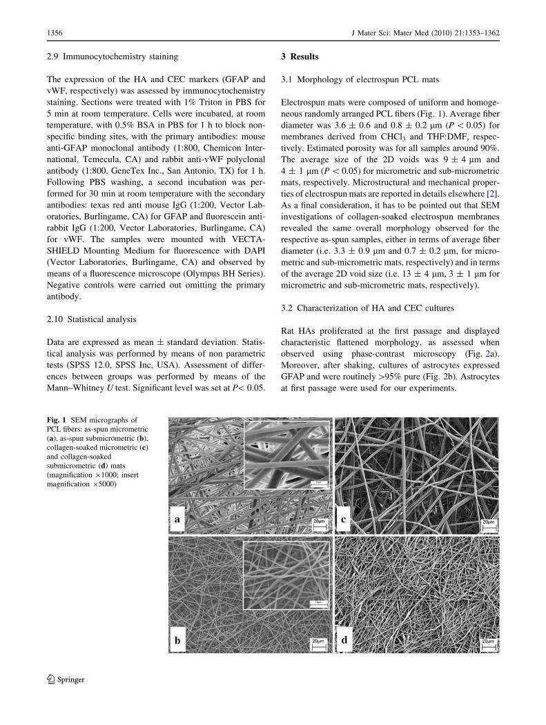
3.1 Morphology of electrospun PCL mats
Electrospun mats were composed of uniform and homoge-
neous randomly arranged PCL fibers (Fig. 1). Average fiber
diameter was 3.6 ± 0.6 and 0.8 ± 0.2 lm (P \ 0.05) for
membranes derived from CHCl3 and THF:DMF, respec-
tively. Estimated porosity was for all samples around 90%.
The average size of the 2D voids was 9 ± 4 lm and
4 ± 1 lm (P \ 0.05) for micrometric and sub-micrometric
mats, respectively. Microstructural and mechanical proper-
ties of electrospun mats are reported in details elsewhere [2].
As a final consideration, it has to be pointed out that SEM
investigations of collagen-soaked electrospun membranes
revealed the same overall morphology observed for the
respective as-spun samples, either in terms of average fiber
diameter (i.e. 3.3 ± 0.9 lm and 0.7 ± 0.2 lm, for micro-
metric and sub-micrometric mats, respectively) and in terms
of the average 2D void size (i.e. 13 ± 4 lm, 3 ± 1 lm for
micrometric and sub-micrometric mats, respectively).
3.2 Characterization of HA and CEC cultures
Rat HAs proliferated at the first passage and displayed
characteristic flattened morphology, as assessed when
observed using phase-contrast microscopy (Fig. 2a).
Moreover, after shaking, cultures of astrocytes expressed
GFAP and were routinely[95% pure (Fig. 2b). Astrocytes
at first passage were used for our experiments.
Fig. 1 SEM micrographs of
PCL fibers: as-spun micrometric
(a), as-spun submicrometric (b),
collagen-soaked micrometric (c)
and collagen-soaked
submicrometric (d) mats
(magnification 91000; insert
magnification 95000)
1356 J Mater Sci: Mater Med (2010) 21:1353–1362
123

Rat CECs were isolated, cultured and characterized as
previously described in Conconi et al. [13]: cultured CECs
displayed characteristic oval nuclei, were spindle shaped
when confluent and maintained their phenotype and in vitro
angiogenic capability until the fifth passage (data not
shown). Hence, cultures from the second to the fourth
passage were chosen for the experiments with the poly-
meric electrospun mats.
3.3 Characterization of HA and CEC cultures
on PCL mats: viability assay
HA viability on days 1, 7 and 14 after seeding was revealed
on tissue culture plate support (TCPS), collagen-coated
TCPS, and on both investigated electrospun PCL matrices.
As can be observed in Fig. 3, astrocytes were vital on both
PCL mats, even if their viability was significantly (P \ 0.05)
lower in comparison to TCPS and collagen-coated TCPS.
Moreover their viability on PCL mats did not vary during the
observation period, the same behaviour has been observed
for HAs seeded on either TCPS or collagen-coated TCPS.
CEC viability on days 1, 7 and 14 after seeding was
observed only in the case of the two investigated electro-
spun matrices (Fig. 4). In fact, using the same seeding
density, CECs cultured on TCPS became confluent after
5 days. The results showed that CECs were vital and pro-
liferate on both PCL mats, and a significant (P \ 0.05)
increasing trend was observed during the observation per-
iod. On the other side, cells seeded on the submicrofibrous
net after 14 days showed higher viability, even if not sig-
nificant, with respect to those seeded on microfibrous one.
3.4 Characterization of HA and CEC cultures
on PCL mats: morphology study
As can be observed in Fig. 5 the nuclei of HAs displayed a
characteristic oval morphology and were well distributed
on the surface of both mats (Fig. 5a, d). These observations
were also confirmed by membrane labelling (PKH26)
presented in Fig. 5b, e: both PCL mats supported cell
growth. Mats cryosectioning allowed us to investigate
cellular infiltration within PCL mats, as DAPI staining
demonstrated HAs grown on microfibrous mats were
present onto and inside the polymeric surface, suggest-
ing a cellular infiltration and a three-dimensional cell
growth (Fig. 5c). On the other side, HAs grown on
Fig. 2 Rat HAs 5 days after isolation at passage 1 before purification
(a) and their immunofluorescence characterization for GFAP after
purification (b) (original magnification 9100)
Fig. 3 HA viability evaluated with MTS assay for tissue culture plate
scaffold (TCPS), collagen-coated TCPS, microfibrous (PCL/CHCl3)
and submicrofibrous matrices (PCL/THF:DMF) (§ P \ 0.05 with
respect to TCPS; c P \ 0.05 with respect to collagen-coated TCPS;
* P \ 0.05 with respect to 1 day; � P \ 0.05 with respect to 7 days)
Fig. 4 CEC viability evaluated with MTS assay for microfibrous
(PCL/CHCl3) and submicrofibrous matrices (PCL/THF:DMF)
(* P \ 0.05 with respect to 1 day; � P \ 0.05 with respect to 7 days)
J Mater Sci: Mater Med (2010) 21:1353–1362 1357
123

submicrofibrous mats were unable to migrate, the inner part
of the constructs remaining acellularized (Fig. 5f).
Figure 6 shows CEC attachment and growth on PCL
electrospun mats on days 14 after seeding. It can be seen
that cells adhered and spread on the surface of micro-
metric and submicrometric fibrous mats, demonstrating
that both mats allowed adhesion and net interaction with
endothelial cells. At day 14, DAPI staining allowed to
observe that CECs on both mats had intact, rounded shape
nuclei (Fig. 6a, d). Membrane labelling (Fig. 6b, e) dem-
onstrates that CECs grew on microfibrous mat in a scat-
tered way adopting an elongated shape, spread on along
the fibers (Fig. 6b). Otherwise, CECs cultured on submi-
crofibrous network formed clusters and spread on the mat
surface side by side (Fig. 6e). DAPI staining of mat
cryosections confirmed the results highlighted for HAs:
microfibrous mats allowed CEC infiltration and three-
dimensional cell growth (Fig. 6c). On the other side, the
small size of the voids delimited by the submicrometric
fibers unabled cells grown and migration and, as a con-
sequence, the inner part of the constructs remained acell-
ularized (Fig. 6f).
On the basis of these overall results, only micrometric
PCL mats were further investigated.
Fig. 5 HA cultures 14 days
after seeding on a–cPCL:CHCl3 mat and d–f on
PCL:THF:DMF mat, stained
with a, d DAPI (9630), b, ePKH26 (9630), c, fcryosections stained with DAPI
(broken lines identify cross-
section of the mat) (c 950, f9100)
1358 J Mater Sci: Mater Med (2010) 21:1353–1362
123
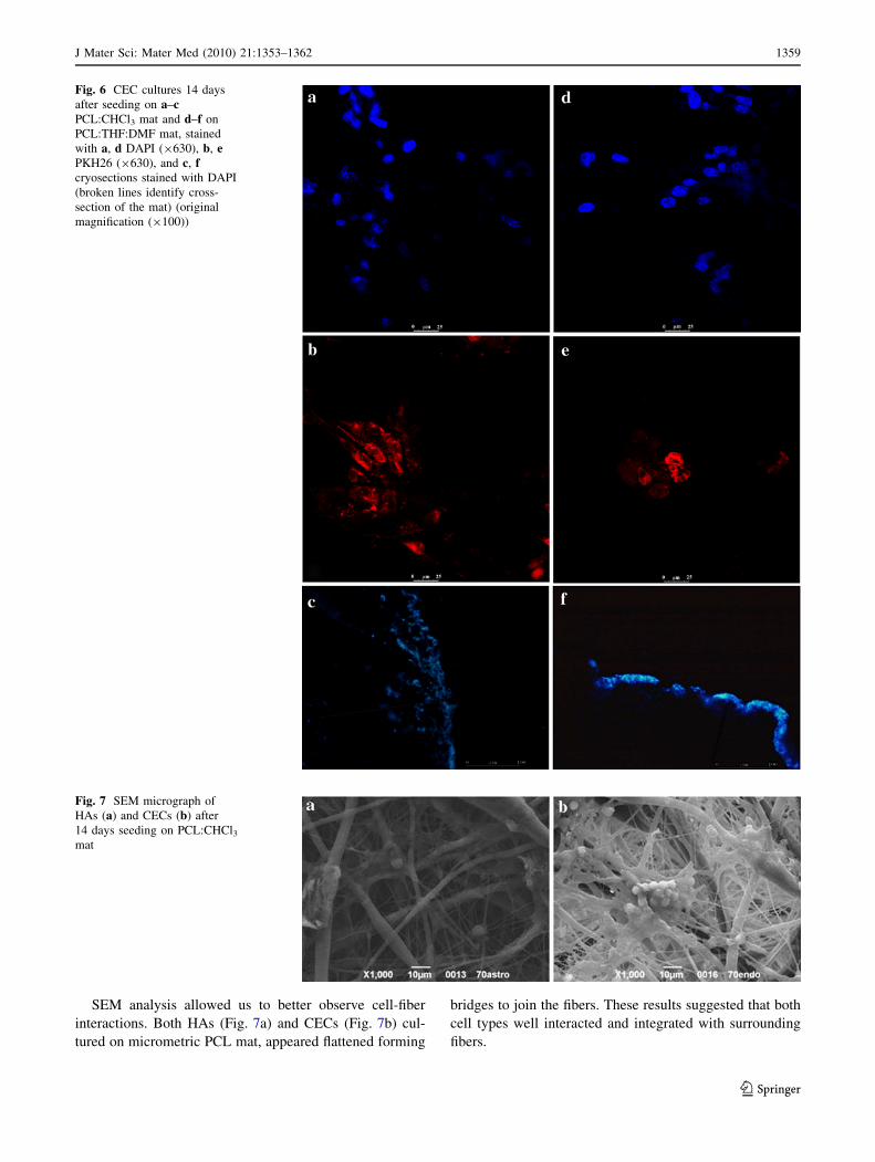
SEM analysis allowed us to better observe cell-fiber
interactions. Both HAs (Fig. 7a) and CECs (Fig. 7b) cul-
tured on micrometric PCL mat, appeared flattened forming
bridges to join the fibers. These results suggested that both
cell types well interacted and integrated with surrounding
fibers.
Fig. 6 CEC cultures 14 days
after seeding on a–cPCL:CHCl3 mat and d–f on
PCL:THF:DMF mat, stained
with a, d DAPI (9630), b, ePKH26 (9630), and c, fcryosections stained with DAPI
(broken lines identify cross-
section of the mat) (original
magnification (9100))
Fig. 7 SEM micrograph of
HAs (a) and CECs (b) after
14 days seeding on PCL:CHCl3mat
J Mater Sci: Mater Med (2010) 21:1353–1362 1359
123

Moreover, fluorescence immunocytochemistry staining
(Fig. 8) showed that both HAs and CECs displayed typical
markers expression, GFAP (Fig. 8a) and vWF (Fig. 8b) for
astrocytes and endothelial cells, respectively.
4 Discussion
Aim of the present study was to assess whether micro-
metric and submicrometric PCL electrospun mats could
promote the survival, proliferation and culture of HAs and
CECs.
Electrospinning is a versatile technique of producing
micro- and nano-fibrous mats [14]. Electrospun fibrous
meshes show an organized architecture mimicking the
ECM and, holding the promise to provide the topographic
cues to the seeded cells, might enhance tissue regeneration.
In a previous study, cytotoxicity evaluation showed that
electrospun PCL mats were free of toxic amounts of
residual solvent/s and were biocompatible for HUVEC
cultures [2].
In this study, the rationale was to evaluate the cyto-
compatibility of electrospun PCL micro and sub-micro
fibers for rat HAs and CECs, both cell types playing
an active role in the formation and maintenance of the
BBB.
In our study, cell response demonstrated that both mi-
crometric and sub-micrometric mats supported adhesion,
proliferation, cellular phenotype and spreading of rat HAs
and CECs. After 14 culture days, HA viability did not
significantly increased, following a trend comparable to
that observed for cultures on TCPS. We hypothesize that
astrocytes were in a normally quiescent condition consid-
ering that for culturing we used serum-addicted medium
without added growth factors that could enhanced their
viability [15]. At our knowledge, a very few number of
literature data concerning astrocyte cultures on PCL mats
were carried out and the most part of these studies were
realized in in vivo experiments. Recently Gerardo-Nava
et al. [16] demonstrated that human neural progenitor-
derived astrocytes increased the adhesion and migration
rate in the blended nanofibers mats (25% collagen Type I,
75% PCL) in comparison to PCL nanofibers, while pro-
liferation was not affected. Moreover Brynda et al. [17]
demonstrated the importance of the presence of collagen
layers on hydrogel surfaces in association with laminin or
fibronectin, enhancing the adhesion and growth of astro-
cytes on the hydrogel in comparison to cell-non-adhesive
hydrogel surface. These results confirmed previously
reported data for which conditioning with cell adhesion
proteins, such as collagen or fibronectin, prior to the cell
seeding is a recognized optimal in vitro culture condition
aimed to facilitate cell adhesion. In our experiments PCL
mats were soaked in collagen solution, the presence of
collagen onto and/or within electrospun fibers only assisted
cell attachment. In fact, the viability of HAs on both the
collagen-treated tissue culture plate support (TCPS) and on
the untreated one, was comparable, the difference being not
statistically significant.
In the case of CEC viability, a significant increasing
trend was found. In particular, after 14 days, higher cell
viability was observed for sub-microscale mat with respect
to the micro-fibrous one. This behaviour can be related to
the surface topography. Xu et al. [18] proved that the pro-
liferation of vascular endothelial cells (ECs) was enhanced
on smooth poly(L-lactic acid) surface rather than on a rough
one. They suggested that, depending on mat roughness, cell
interaction with substrate surface could activate different
focal contact structure connected with the control of cyto-
skeletal arrangements and of cell behaviour (such as cell
proliferation). Furthermore, a different cell response to the
surface roughness has been demonstrated by a number of
researchers [19, 20]. The two investigated electrospun mats
were comprised of fibers of different size and topography,
the microfibers showed a nanoporous surface while sub-
microfibers presented a rather smooth surface (Fig. 1). This
effect can be attributed to a thermodynamic instability
during the electrospinning process in which phase separa-
tion takes place. The phase separation yields both solvent-
rich and polymer-rich regions, being the nanopores a con-
sequence of the solvent-rich regions within the jet [21].
Jarusuwannapoom et al. [22] investigated the effect of the
Fig. 8 HA (a) and CEC (b)
culture cryosections, after
14 days seeding, on PCL:CHCl3mat stained with anti-GFAP and
DAPI (a) and with anti-von
Willebrand factor and DAPI (b).
(broken lines identify cross-
section of the mat) (original
magnification 9100)
1360 J Mater Sci: Mater Med (2010) 21:1353–1362
123

solvents on the electrospun polystyrene mats, observing that
the rough surface of the collected fibers may be a result of
the very low boiling point of the chloroform (61.2�C), while
smooth fibers were obtained using DMF as solvent and for
any spinning condition. However, the exact mechanism
behind cell response to different surface topography needs
to be further investigated.
The presented results demonstrated that the different
cell-mat/cell–cell interactions depended on mat structural
architecture. By comparing cell morphology and cryosec-
tion staining of the two constructs, it can be observed that
both HAs and CECs elongate on the microfibrous mat fol-
lowing fiber direction instead of adopting a random orien-
tation and spread-out morphology (similar to that observed
on two-dimensional smooth substrates) when cultured on
submicrofibrous net. Moreover, SEM analysis demon-
strated the interaction and integration of both cell types with
PCL microfibers. Additionally, microfibrous environment
favoured cellular infiltration with respect to submicrofiber
one, where HAs and CECs were unable to migrate within
the mat, leaving an acellularized internal region.
The fiber dimension and the pore size resulted hence to
be critical mat features, affecting cell attachment, prolif-
eration, migration and ingrowth. Kwon et al. [23] found
that HUVECs proliferated better on scaffold with fiber
diameter of 1.16 ± 0.17 lm with respect to scaffold with
the same porosity and fiber dimension of 7.02 ± 1.03 lm,
and suggested that highly packed fabrics or high-surface-
density fibers provide an extremely high surface-to-volume
ratio, which favours cell attachment and proliferation [24].
The decrease in the fiber size of fabric resulted in an
increased fiber density and mechanical strength that seem
to favour cells adhesion and proliferation. However, our
results suggest that a decreased fiber size resulted in a 3D
architecture characterized by lower average 2D void size
that limit cell infiltration and 3D colonization. Carampin
et al. [25] described that electrospun polyphosphazene
nanofiber (fiber diameter 0.8 ± 0.1 lm) scaffolds, seeded
with CECs, showed a cell monolayer having 100% con-
fluence. However, even if nanofibers supported cell adhe-
sion and proliferation, ECs were unable to migrate through
the wall thickness and the inner part of the scaffold. In a
previous study it has been shown that HUVEC behaviour
was remarkably affected by morphological features of PCL
mats, demonstrating that microfibrous network was a more
suitable environment for cell colonization with respect to
the submicrofibrous one [2]. Here presented results con-
firmed that submicrofibrous polymeric mat could favour
cell attachment, proliferation and spreading but their small
average 2D void size, usually smaller than normal cell size,
might limit 3D cell colonisation and inhibit cell infiltration
and migration [26]. On the other side, microfibrous mat,
even if not the same size scale as ECM, could be
advantageous since it comprised larger voids that allow and
facilitate cellular infiltration and/or diffusion of nutrients
enhancing their potential 3D applications [27].
5 Conclusions
In this work the response of the blood brain barrier (BBB)
cell types to the three-dimensional architecture of electro-
spun PCL mats was investigated. It was observed that
hippocampal astrocytes (HAs) and cerebromicrovascular
endothelial cells (CECs) adhered, were viable and grew on
micro- and submicro-electrospun PCL fibrous mats.
Moreover, cells maintained their specific phenotype on
both mats, indicating a biological function of the cells.
However, cell infiltration and 3D growth were influenced
by fiber dimension and 2D void size: microfibrous network
appeared, indeed, to be a more suitable environment for
cell colonization. Based on our results, the proper fiber
architecture can be regarded as a crucial issue to be con-
sidered in order to deal with suitable polymeric mats tai-
lored for specific in vitro application.
Acknowledgements This research has been supported by PRIN
2006 fundings ‘‘Progettazione e realizzazione di scaffolds nano-
strutturati organici, inorganici ed ibridi da utilizzare in medicina ri-
generativa come substrati per il differenziamento di cellule
staminali’’. The authors wish to thank Dr. Francesca Nanni and Prof.
Giampiero Montesperelli (Department of Chemical Science and
Technology, University of Rome ‘‘Tor Vergata’’) for SEM analysis.
References
1. Greiner A, Wendorff JH. Electrospinning: a fascinating method
for the preparation of ultrathin fibers. Angew Chem Int Ed Engl.
2007;46(30):5670–703.
2. Del Gaudio C, Bianco A, Folin M, Baiguera S, Grigioni M.
Structural characterisation and cell response evaluation of elec-
trospun PCL membranes: micrometric vs sub-micrometric fibers.
J Biomed Mater Res A. 2009;89(4):1028–39.
3. Cucullo L, Aumayr B, Rapp E, Janigro D. Drug delivery and in
vitro models of the blood-brain barrier. Curr Opin Drug Discov
Devel. 2005;8(1):89–99.
4. Unterberg AW, Stover J, Kress B, Kiening KL. Edema and brain
trauma. Neuroscience. 2004;129(4):1021–9.
5. Latour LL, Kang DW, Ezzeddine MA, Chalela JA, Warach S.
Early blood–brain barrier disruption in human focal brain
ischemia. Ann Neurol. 2004;56(4):468–77.
6. van der FM, Hoppenreijs S, van Rensburg AJ, Ruyken M, Kolk
AH, Springer P, et al. Vascular endothelial growth factor and
blood–brain barrier disruption in tuberculous meningitis. Pediatr
Infect Dis J. 2004;23(7):608–13.
7. Lee SW, Kim WJ, Park JA, Choi YK, Kwon YW, Kim KW.
Blood–brain barrier interfaces and brain tumors. Arch Pharm Res.
2006;29(4):265–75.
8. Cipolla MJ, Crete R, Vitullo L, Rix RD. Transcellular transport
as a mechanism of blood–brain barrier disruption during stroke.
Front Biosci. 2004;9:777–85.
J Mater Sci: Mater Med (2010) 21:1353–1362 1361
123

9. Stamatovic SM, Dimitrijevic OB, Keep RF, Andjelkovic AV.
Inflammation and brain edema: new insights into the role of
chemokines and their receptors. Acta Neurochir Suppl. 2006;
96:444–50.
10. Kalaria RN. The blood–brain barrier and cerebral microcircula-
tion in Alzheimer disease. Cerebrovasc Brain Metab Rev. 1992;
4(3):226–60.
11. Minagar A, Alexander JS. Blood–brain barrier disruption in
multiple sclerosis. Mult Scler. 2003;9(6):540–9.
12. McCarthy KD, de Vellis J. Preparation of separate astroglial and
oligodendroglial cell cultures from rat cerebral tissue. J Cell Biol.
1980;85(3):890–902.
13. Conconi MT, Lora S, Baiguera S, Boscolo E, Folin M, Scienza R,
et al. In vitro culture of rat neuromicrovascular endothelial cells
on polymeric scaffolds. J Biomed Mater Res A. 2004;71(4):669–
74.
14. Chew SY, Wen Y, Dzenis Y, Leong KW. The role of electros-
pinning in the emerging field of nanomedicine. Curr Pharm
Design. 2006;12(36):4751–70.
15. Chiarini A, Dal Pra I, Menapace L, Pacchiana R, Whitfield JF,
Armato U. Soluble amyloid b-peptide and myelin basic protein
strongly stimulate, alone and in synergism with combined pro-
inflammatory cytokines, the expression of functional nitric oxide
synthase-2 in normal adult human astrocytes. Int J Mol Med.
2005;16:801–7.
16. Gerardo-Nava J, Fuhrmann T, Klinkhammer K, Seiler N, Mey J,
Klee D, et al. Human neural cell interactions with orientated
electrospun nanofibers in vitro. Nanomed. 2009;4(1):11–30.
17. Brynda E, Houska M, Kysilka J, Pradny M, Lesny P, Jendelova P,
et al. Surface modification of hydrogels based on poly
(2-hydroxyethyl methacrylate) with extracellular matrix proteins.
J Mater Sci Mater Med. 2009;20(4):909–15.
18. Xu C, Yang F, Wang S, Ramakrishna S. In vitro study of human
vascular endothelial cell function on materials with various sur-
face roughness. J Biomed Mater Res A. 2004;71(1):154–61.
19. Chung TW, Liu DZ, Wang SS. Enhancement of the growth of
human endothelial cells by surface roughness at nanometer scale
Biomaterials. 2003;24(25):4655–61.
20. Thapa A, Webster TJ, Haberstroh KM. Polymers with nano-
dimensional surface features enhance bladder smooth muscle cell
adhesion. J Biomed Mater Res A. 2003;67(4):1374–83.
21. Ramakrishna S, Fujihara K, Teo WE, Lim TC, Ma Z. An intro-
duction to electrospinning and nanofibers. Singapore: World
Scientific Publishing; 2005.
22. Jarusuwannapoom T, Hongrojjanawiwat W, Jitjaicham S,
Wannatong L, Nithitanakul M, Pattamaprom C, et al. Effect of
solvents on electro-spinnability of polystyrene solutions and
morphological appearance of resulting electrospun polystyrene
fibers. Eur. Polymer. J. 2005;41:409–21.
23. Kwon IK, Kidoaki S, Matsuda T. Electrospun nano- to microfiber
fabrics made of biodegradable copolyesters: structural charac-
teristics, mechanical properties and cell adhesion potential. Bio-
materials. 2005;26(18):3929–39.
24. Mo XM, Xu CY, Kotaki M, Ramakrishna S. Electrospun P(LLA-
CL) nanofiber: a biomimetic extarcellular matrix for smooth
muscle cells and endothelial cell proliferation. Biomaterials.
2004;25(10):1883–90.
25. Carampin P, Conconi MT, Lora S, Menti AM, Baiguera S, Bellini
S, et al. Electrospun polyphosphazene nanofibers for in rat
endothelial cells proliferation. J Biomed Mater Res A.
2007;80(3):661–8.
26. Zhang Y, Ouyang H, Lim CT, Ramakrishna S, Huang ZM.
Electrospinning of gelatin fibers and gelatin/PCL composite
fibrous scaffolds. J Biomed Mater Res B Appl Biomater.
2005;72(1):156–65.
27. Pham QP, Sharma U, Mikos AG. Electrospun poly(e-caprolac-
tone) microfiber and multilayer nanofiber/microfiber scaffolds:
characterization of scaffolds and measurement of cellular infil-
tration. Biomacromolecules. 2006;7(10):2796–805.
1362 J Mater Sci: Mater Med (2010) 21:1353–1362
123