Astrocyte-targeted expression of interleukin-6 protects the central nervous system during neuroglial...
16
Astrocyte-Targeted Expression of Interleukin-6 Protects the Central Nervous System During Neuroglial Degeneration Induced by 6-Aminonicotinamide Milena Penkowa, 1 Jordi Camats, 2 Hanne Hadberg, 1 Albert Quintana, 2 Santiago Rojas, 2 Mercedes Giralt, 2 Amalia Molinero, 2 Iain L. Campbell, 3 and Juan Hidalgo 2 * 1 Department of Medical Anatomy, The Panum Institute, University of Copenhagen, Copenhagen, Denmark 2 Institute of Neurosciences and Departament of Cellular Biology, Physiology and Immunology, Animal Physiology Unit, Faculty of Sciences, Autonomous University of Barcelona, Barcelona, Spain 3 Department of Neuropharmacology, The Scripps Research Institute, La Jolla, California 6-Aminonicotinamide (6-AN) is a niacin antagonist, which leads to degeneration of gray matter astrocytes mainly in the brainstem. We have examined the role of interleukin-6 (IL-6) in this degenerative process by using transgenic mice with astrocyte-targeted IL-6 expression (GFAP-IL6 mice). This study demonstrates that transgenic IL-6 expression significantly increases the 6-AN-induced inflammatory re- sponse of reactive astrocytes, microglia/macrophages, and lymphocytes in the brainstem. Also, IL-6 induced significant increases in proinflammatory cytokines IL-1, IL-12, and tu- mor necrosis factor- as well as growth factors basic fibro- blast growth factor (bFGF), transforming growth factor-, neurotrophin-3, angiopoietin, vascular endothelial growth factor, and the receptor for bFGF. In accordance, angio- genesis was increased in GFAP-IL6 mice relative to con- trols after 6-AN. Moreover, oxidative stress and apoptotic cell death were significantly reduced by transgenic IL-6 expression. IL-6 is also a major inducer in the CNS of metallothionein I and II (MT-III), which were significantly increased in the GFAP-IL6 mice. MT-III are antioxidants and neuroregenerative factors in the CNS, so increased MT-III levels in GFAP-IL6 mice could contribute to the reduction of oxidative stress and cell death in these mice. © 2003 Wiley-Liss, Inc. Key words: neuropathology; oxidative stress; apoptosis; neuroprotection 6-Aminonicotinamide (6-AN) is a niacin antagonist that shuts down the hexose monophosphate pathway, which is toxic to neuroglial cells (Krum, 1994, 1995; Haghighat et al., 1996; Penkowa et al., 1997, 2002; Hoth- ersall et al., 1998; Krinke and Classen, 1998; Budihardjo et al., 2000; Penkowa and Hidalgo, 2000a). Injection of 6-AN leads to degeneration of gray matter macroglia of brainstem and spinal cord (Krum, 1994, 1995; Haghighat et al., 1996; Penkowa et al., 1997, 2002). The latter is followed by inflammation (Penkowa et al., 1997) and formation of reactive oxygen species (ROS), leading to oxidative stress (Penkowa and Hidalgo, 2000a; Penkowa et al., 2002), which can induce neurodegeneration and apoptosis (Sun and Chen, 1998; Cassarino and Bennett, 1999; Floyd, 1999). Interleukin-6 (IL-6) is a multifunctional cytokine produced in activated cells during CNS inflammation (Hirano et al., 1990; Gadient and Otten, 1997; Gruol and Nelson, 1997; Benveniste, 1998; Mun ˜ oz-Fernandez and Fresno, 1998; Penkowa et al., 2000b; Lenzlinger et al., 2001). IL-6 is a main regulator of inflammatory and im- mune responses (Benveniste, 1998; Raivich et al., 1999; Van Wagoner and Benveniste, 1999; Brunello et al., 2000; Penkowa and Hidalgo, 2000a; Penkowa et al., 2000b; Morganti-Kossmann et al., 2002). Consequently, in IL-6 knock-out (IL-6KO) mice, the CNS inflammatory re- sponse is reduced (Klein et al., 1997; Eugster et al., 1998; Raivich et al., 1999; Penkowa et al., 2000b; Swartz et al., 2001). Moreover, transgenic IL-6 overexpression under the control of the glial fibrillary acdic protein (GFAP) gene promoter (GFAP-IL6 mice) induces neuroinflammation (Campbell et al., 1993; Brett et al., 1995; Fattori et al., 1995; Di Santo et al., 1996; Campbell, 1998a,b; Brunello et al., 2000; Giralt et al., 2002a), but, when a brain injury is superimposed, these increased IL-6 levels show signifi- cant antiinflammatory effects and lead to increased CNS repair (Fee et al., 2000; Swartz et al., 2001). Accordingly, posttraumatic tissue healing is significantly impaired, whereas neuronal cell death is increased, in IL-6KO mice during neuropathology (Murphy et al., 1999; Penkowa et al., 2000b; Swartz et al., 2001). Furthermore, local injec- tion of IL-6 attenuates neurotoxic effects of N-methyl-D- *Correspondence to: Juan Hidalgo, Departamento de Biologı ´a Celular, Fisiologı ´a e Inmunologı ´a, Unidad de Fisiologı ´a Animal, Facultad de Cien- cias, Universidad Auto ´ noma de Barcelona, Bellaterra, Barcelona, Spain 08193. E-mail: [email protected] Received 7 March 2003; Revised 23 April 2003; Accepted 24 April 2003 Journal of Neuroscience Research 73:481– 496 (2003) © 2003 Wiley-Liss, Inc.
-
Upload
independent -
Category
Documents
-
view
1 -
download
0
Transcript of Astrocyte-targeted expression of interleukin-6 protects the central nervous system during neuroglial...

Astrocyte-Targeted Expression ofInterleukin-6 Protects the Central NervousSystem During Neuroglial DegenerationInduced by 6-Aminonicotinamide
Milena Penkowa,1 Jordi Camats,2 Hanne Hadberg,1 Albert Quintana,2 Santiago Rojas,2
Mercedes Giralt,2 Amalia Molinero,2 Iain L. Campbell,3 and Juan Hidalgo2*1Department of Medical Anatomy, The Panum Institute, University of Copenhagen, Copenhagen, Denmark2Institute of Neurosciences and Departament of Cellular Biology, Physiology and Immunology, AnimalPhysiology Unit, Faculty of Sciences, Autonomous University of Barcelona, Barcelona, Spain3Department of Neuropharmacology, The Scripps Research Institute, La Jolla, California
6-Aminonicotinamide (6-AN) is a niacin antagonist, whichleads to degeneration of gray matter astrocytes mainly inthe brainstem. We have examined the role of interleukin-6(IL-6) in this degenerative process by using transgenic micewith astrocyte-targeted IL-6 expression (GFAP-IL6 mice).This study demonstrates that transgenic IL-6 expressionsignificantly increases the 6-AN-induced inflammatory re-sponse of reactive astrocytes, microglia/macrophages, andlymphocytes in the brainstem. Also, IL-6 induced significantincreases in proinflammatory cytokines IL-1, IL-12, and tu-mor necrosis factor-� as well as growth factors basic fibro-blast growth factor (bFGF), transforming growth factor-�,neurotrophin-3, angiopoietin, vascular endothelial growthfactor, and the receptor for bFGF. In accordance, angio-genesis was increased in GFAP-IL6 mice relative to con-trols after 6-AN. Moreover, oxidative stress and apoptoticcell death were significantly reduced by transgenic IL-6expression. IL-6 is also a major inducer in the CNS ofmetallothionein I and II (MT-I�II), which were significantlyincreased in the GFAP-IL6 mice. MT-I�II are antioxidantsand neuroregenerative factors in the CNS, so increasedMT-I�II levels in GFAP-IL6 mice could contribute to thereduction of oxidative stress and cell death in these mice.© 2003 Wiley-Liss, Inc.
Key words: neuropathology; oxidative stress; apoptosis;neuroprotection
6-Aminonicotinamide (6-AN) is a niacin antagonistthat shuts down the hexose monophosphate pathway,which is toxic to neuroglial cells (Krum, 1994, 1995;Haghighat et al., 1996; Penkowa et al., 1997, 2002; Hoth-ersall et al., 1998; Krinke and Classen, 1998; Budihardjo etal., 2000; Penkowa and Hidalgo, 2000a). Injection of6-AN leads to degeneration of gray matter macroglia ofbrainstem and spinal cord (Krum, 1994, 1995; Haghighatet al., 1996; Penkowa et al., 1997, 2002). The latter isfollowed by inflammation (Penkowa et al., 1997) andformation of reactive oxygen species (ROS), leading to
oxidative stress (Penkowa and Hidalgo, 2000a; Penkowaet al., 2002), which can induce neurodegeneration andapoptosis (Sun and Chen, 1998; Cassarino and Bennett,1999; Floyd, 1999).
Interleukin-6 (IL-6) is a multifunctional cytokineproduced in activated cells during CNS inflammation(Hirano et al., 1990; Gadient and Otten, 1997; Gruol andNelson, 1997; Benveniste, 1998; Munoz-Fernandez andFresno, 1998; Penkowa et al., 2000b; Lenzlinger et al.,2001). IL-6 is a main regulator of inflammatory and im-mune responses (Benveniste, 1998; Raivich et al., 1999;Van Wagoner and Benveniste, 1999; Brunello et al., 2000;Penkowa and Hidalgo, 2000a; Penkowa et al., 2000b;Morganti-Kossmann et al., 2002). Consequently, in IL-6knock-out (IL-6KO) mice, the CNS inflammatory re-sponse is reduced (Klein et al., 1997; Eugster et al., 1998;Raivich et al., 1999; Penkowa et al., 2000b; Swartz et al.,2001). Moreover, transgenic IL-6 overexpression underthe control of the glial fibrillary acdic protein (GFAP) genepromoter (GFAP-IL6 mice) induces neuroinflammation(Campbell et al., 1993; Brett et al., 1995; Fattori et al.,1995; Di Santo et al., 1996; Campbell, 1998a,b; Brunelloet al., 2000; Giralt et al., 2002a), but, when a brain injuryis superimposed, these increased IL-6 levels show signifi-cant antiinflammatory effects and lead to increased CNSrepair (Fee et al., 2000; Swartz et al., 2001). Accordingly,posttraumatic tissue healing is significantly impaired,whereas neuronal cell death is increased, in IL-6KO miceduring neuropathology (Murphy et al., 1999; Penkowa etal., 2000b; Swartz et al., 2001). Furthermore, local injec-tion of IL-6 attenuates neurotoxic effects of N-methyl-D-
*Correspondence to: Juan Hidalgo, Departamento de Biologıa Celular,Fisiologıa e Inmunologıa, Unidad de Fisiologıa Animal, Facultad de Cien-cias, Universidad Autonoma de Barcelona, Bellaterra, Barcelona, Spain08193. E-mail: [email protected]
Received 7 March 2003; Revised 23 April 2003; Accepted 24 April 2003
Journal of Neuroscience Research 73:481–496 (2003)
© 2003 Wiley-Liss, Inc.

aspartate (Toulmond et al., 1992) and is neuroprotectiveduring cerebral ischemia (Loddick et al., 1998). Consis-tently with this, IL-6 is a member of the neuropoietinfamily of cytokines (Hopkins and Rothwell, 1995) and hasneurotrophic and neuroprotective effects (Gruol and Nel-son, 1997; Benveniste, 1998; Carlson et al., 1999; Gahringet al., 1999; Penkowa et al., 2000b; Swartz et al., 2001;Eskes et al., 2002; Pavelko et al., 2003). Furthermore, IL-6is a major inducer of the antioxidant neuroprotectivefactors metallothionein I and II (MT-I�II; Hernandez etal., 1997; Carrasco et al., 1998; Penkowa et al., 2000b),which significantly inhibit brain damage and cell deathduring various neuropathological conditions and promoteneuroregeneration and tissue repair (Penkowa et al.,1999a,b, 2000a, 2003; Carrasco et al., 2000; Hidalgo et al.,2001, 2002; Giralt et al., 2002a,b). Accordingly, MT-I�IIcan be used therapeutically against different CNS disorders(Penkowa and Hidalgo, 2000b; Giralt et al., 2002b; Pen-kowa et al., 2002).
Thus, IL-6 exerts both proinflammatory and neu-roprotective roles in the injured CNS. To understandthe mechanisms underlying such different roles, wehave studied astrocyte-targeted overexpression of IL-6in a model of CNS damage, subjection to an i.p. injec-tion with the gliotoxic niacin antagonist 6-AN. Wehave used transgenic GFAP-IL6 mice, a well-knownanimal model of IL-6 overexpression (Campbell et al.,1993; Fattori et al., 1995; Brett et al., 1995; Di Santo etal., 1996; Campbell, 1998a,b; Brunello et al., 2000).The results clearly demonstrate that, although in thenormal brain transgenic overexpression of IL-6 may bedetrimental, during a toxic insult, such as after 6-AN,IL-6 is a neuroprotective factor.
MATERIALS AND METHODS
Animals
Construction and characterization of the GFAP-IL6 trans-genic mice has been described previously (Campbell et al.,1993). Briefly, an expression vector derived from the murineGFAP gene was used to target expression of IL-6 to astrocytes.The GFAP-IL6 mice have a mixed C57B6 � SJL geneticbackground, and a colony is maintained in our laboratory bycrossing heterozygous GFAP-IL6 males with C57B6 � SJLfemales. To study the role of metallothioneins (MTs) in theGFAP-IL6 animal model, we have recently undertaken theapproach of crossing these mice with either MT1�2KO mice(Giralt et al., 2002) or TgMTI mice (Molinero et al., 2003). Forthis particular study, heterozygous GFAP-IL6 mice were crossedwith MT-I�II-deficient mice, which have a 129/SvJ geneticbackground (Masters et al., 1994). Although all the offspring areheterozygous for MT-I�II, they were genotyped for identifyingthe GFAP-IL6 �/– and the GFAP-IL6 –/– animals as previ-ously described (Penkowa et al., 2000a). Both groups share thesame genetic background and so were used for this study; werefer to the GFAP-IL6 –/– animals as the “normal” or “wild-type” mice. Age and gender were matched for both groups.
All experiments were carried out in accordance with theEuropean Communities Council Directive of 24 November1986 (86/609/EEC). Also, all animals were acquired and cared
for in accordance with the guidelines published in the NIHGuide for the Care and Use of Laboratory Animals and with theprinciples presented in the Guidelines for the Use of Animals inNeuroscience Research by the Society for Neuroscience. More-over, all animal protocols used were approved by our institu-tional animal experimentation committee, and all efforts weremade to minimize animal distress and to reduce the number ofanimals used.
Experimental Procedures
To induce CNS injury, mice were injected intraperito-neally with 6-AN (Sigma Aldrich, St. Louis, MO; code A0630),an antimetabolite that shuts down the hexose monophosphatepathway, which is preferentially used by protoplasmic astroglia,which thereby suffer cytotoxic edema and cell death (Krum,1995; Haghighat and McCandless, 1996; Penkowa et al., 1997).This injurious effect of 6-AN is confined to certain gray matterareas of hindbrain and spinal cord, whether or not these neuronsproject outside the CNS (Penkowa et al., 1997). As a result, onlyastroglia of the nuclei of brainstem and medulla spinalis areinjured, whereas other CNS regions, such as the forebrain,appear to be resistant to 6-AN (Krum, 1995; Penkowa et al.,1997).
6-AN Administration
Eight-month-old female normal and GFAP-IL6 mice re-ceived an i.p. injection of 10 mg/kg body weight of 6-AN,which was dissolved in physiological saline. As a control for the6-AN injection, other adult mice were injected with saline. Themice were killed 1 or 3 days after the injection. The brains werefixed for 24 hr in 4% paraformaldehyde. Brains were afterwarddissected and cut as 3-�m-thick consecutive sections, whichwere processed for histochemistry and immunohistochemistry aspreviously described (Penkowa et al., 2000b). Four animals pergroup and type of procedure were used.
Histochemistry
Stainings for hematoxylin and eosin (H&E) and tomatolectin from Lycopersicon esculentum (Sigma; code L9389) wereperformed as previously described (Penkowa et al., 1999,2000b).
Immunohistochemistry
Sections were incubated overnight at 4°C with one of thefollowing primary antibodies: rabbit anti-cow GFAP 1:250 (Da-kopatts, Glostrup, Denmark; code Z 334); rabbit anti-humanS100 1:2,000 (Dakopatts; code Z311); rat anti-mouse MOMA(monocyte-macrophage marker-1) 1:20 (Serotec, Bicester,United Kingdom; code MCA947; a marker of lymphoid tissuemacrophages); mouse anti-human CD-34 1:20 (Neomarkers,USA; code MS-363; a marker of myeloid and lymphoid pro-genitor cells as well as proliferating vessels); goat anti-mouseCD-20 1:200 (Santa Cruz Biotechnology, Santa Cruz, CA;code sc-7736; a marker of B lymphocytes); mouse anti-rat CD-3(IgM) 1:50 (Serotec; code KD MCA 772; a marker of Tlymphocytes); mouse anti-rat CD-4 1:50 [Serotec; code MCA55R; a marker of T helper (Th) lymphocytes]; rabbit anti-ratMT-I�II (Gasull et al., 1993; Penkowa et al., 1997, 1999);mouse anti-human IL-1� 1:50 (Biogenesis, USA; code 5375-4329); rat anti-mouse IL-6 1:50 (Harlan Seralab, Indianapolis,
482 Penkowa et al.

IN; code MAS584); goat anti-mouse IL-12 1:50 (ChemiconInt., United Kingdom; code AB2130P); rabbit anti-mouse tu-mor necrosis factor-� (TNF-�) 1:100 (Biosource, USA; codeAMC 3012); rabbit anti-human transforming growth factor-�1(TGF-�1) 1:200 (Santa Cruz Biotechnology; code sc-146); rab-bit anti-human basic fibroblast growth factor (bFGF) 1:100(Santa Cruz Biotechnology; code sc-79); goat anti-humanNT-3 1:20 (R&D Systems, Minneapolis, MN; code AF-267-NA); goat anti-human angiopoietin-1/4 1:100 (Santa Cruz Bio-technology; code sc-9360; marker of angiogenesis); rabbit anti-human vascular endothelial growth factor (VEGF) 1:50(Neomarkers, USA; code RB-222P0; a marker of angiogenesis);rat anti-mouse perlecan (bFGF receptor, heparan sulfate proteo-glycan) 1:30 (Neomarkers, USA; code RT-794-P0; a marker ofangiogenesis); rabbit anti-mouse inducible nitric oxide synthase(iNOS; a marker for oxidative stress) 1:100 (Alexis Biochemi-cals, USA; code 210-503-R050); rabbit antimalondialdehyde(MDA; a marker for oxidative stress) 1:100 (Alpha DiagnosticInt., USA; code MDA 11-S); rabbit antinitrotyrosine (NITT; amarker for oxidative stress) 1:100 (Alpha Diagnostic Int., USA;code NITT 12-A); mouse anti-8-oxyguanine (a marker foroxidative stress) 1:100 (Chemicon Int.; code MAB-3560); andrabbit anti-human cleaved caspase-3 1:50 (Cell Signaling Tech-nology Inc., USA; code 9661).
The primary antibodies were detected using biotinylatedanti-mouse IgG 1:200 (Sigma; code B8774), or biotinylatedanti-mouse IgM (�-chain-specific) 1:50 (Jackson Immunore-
search, West Grove, PA; code 115-065-020), or biotinylatedanti-rabbit IgG 1:400 (Sigma; code B3275), or biotinylatedanti-rat IgG 1:1500 (Amersham, Buckinghamshire, UnitedKingdom; code RPN 1005), or biotinylated anti-goat/sheepIgG 1:20 (Amersham; code RPN 1025), followed bystreptavidin-biotin-peroxidase complex (StreptABComplex/HRP; Dakopatts; code K377) prepared at the manufacturer’srecommended dilution. These secondary and tertiary steps in theimmunoreaction were performed for 30 min at room temper-ature. Afterward, sections were incubated with biotinylatedtyramide and streptavidin-peroxidase complex (tyramide signalamplification, TSA indirect; NEN, Life Science Products, Bos-ton, MA; code NEL700A) prepared following the manufactur-er’s recommendations. The immunoreaction was visualized us-ing 0.015% H2O2 in diaminobenzidine (DAB)/Tris-bufferedsaline (TBS) for 10 min at room temperature.
To evaluate the extent of nonspecific binding in theimmunohistochemical experiments, control sections were incu-bated in the absence of primary antibody. Results were consid-ered only if these controls were negative.
In Situ Detection of DNA Fragmentation (TUNEL)
Terminal deoxynucleotidyl transferase (TdT)-mediateddeoxyuridine triphosphate (dUTP)-digoxigenin nick end label-ing (TUNEL) was performed as previously described (Penkowaet al., 1999a).
Fig. 1. H&E-stained brainstem sections of saline- and 6-AN-injectedwild-type and GFAP-IL6 mice. A and B show the brainstem ofwild-type (A) and GFAP-IL6 (B) mice after saline injection. C and Dshow the brainstem of 6-AN injected wild-type (C) and GFAP-IL6 (D)mice. 6-AN induced tissue degeneration in certain gray matter areas of
the brainstem such as in the medial vestibular nuclei (Mve), the nucleusof the solitary tract (SolM), the solitary nucleus (SolV), the cuneatenucleus (Cu), the pontine nuclei (Pn), the superior paraolivary nucleusand the medioventral periolivary nucleus (Po), and the facial nucleus(VII). Scale bars � 280 �m.
IL-6 Reduces CNS Damage 483

Fig. 2. GFAP immunoreactivity in the brainstem of saline- and 6-AN-injected wild-type and GFAP-IL6 mice. A: After saline injection, somedispersed GFAP� astrocytes are seen in the brainstem of wild-typemice. The star marks the area magnified in E. B: In GFAP-IL6 miceinjected with saline, GFAP� reactive astrogliosis is seen in the brain-stem. Hence, several astrocytes are seen in posterior and anterior areasthroughout the brainstem. The star marks the area magnified in F.C: After 6-AN treatment, the astroglia dissappear from certain brain-stem gray matter areas of wild-type mice, and simultaneously GFAP�
reactive astrogliosis is observed around the degenerated gray matterareas. D: Also in 6-AN-injected GFAP-IL6 mice, astroglia disappearedfrom specific gray matter areas of the brainstem, such as the medialvestibular nuclei (Mve), the nucleus of the solitary tract (SolM), the
solitary nucleus (SolV), the cuneate nucleus (Cu), the pontine nuclei(Pn), the superior paraolivary nucleus and the medioventral periolivarynucleus (Po), and the facial nucleus (VII). Reactive astrogliosis sur-rounding the damaged areas in GFAP-IL6 mice was significantly in-creased relative to wild-type mice. E: Higher magnification of the areamarked by the star in A. F: Higher magnification of the area marked bythe star in B, which shows the increased astrocytes of GFAP-IL6 micerelative to normal mice. G: Higher magnification of the area marked bya star in C, showing 6-AN-induced reactive astrogliosis of normal mice.H: Higher magnification of area marked by the star in D. As shown,GFAP-IL6 mice had significantly increased reactive astrogliosis com-pared with that in normal mice after 6-AN.
484 Penkowa et al.

Fluorescence
To detect which cell types suffer from oxidative stress orapoptotic cell death, we performed double- and triple-fluorescencestainings. To determine which cells suffer from oxidative stress,sections were pretreated as described above and incubated over-night at 4°C with mouse anti-human NF ready-to-use (Biogenex,San Ramon, CA; code AM073-10M) or mouse anti-porcine vi-mentin 1:50 (Dakopatts; code M0725; as a marker for both reactiveastrocytes and macrophages) and simultaneously with rabbit anti-human iNOS, or rabbit anti-NITT, or rabbit anti-MDA (all asdescribed above). The monoclonal antibodies were detected byusing goat anti-mouse IgG linked with Texas red (TXRD) 1:50(Southern Biotechnology, Birmingham, AL; code 1030-07),whereas the polyclonal antibodies were detected by using goatanti-rabbit IgG linked with fluorescein (FITC) 1:50 (SouthernBiotechnology; code 4050-02).
To determine which cells suffer from apoptosis, triplestainings for TUNEL and cellular markers were performed.Sections were incubated with FITC-linked TUNEL (Oncor,Gaithersburg, MD; code S7111-KIT) according to the manu-facturer’s protocol and afterward incubated overnight at 4°Cwith rabbit anti-human neuron-specific enolase (NSE) 1:1,000(Calbiochem, La Jolla, CA; code D05059) and mouse anti-porcine vimentin (as described above) simultaneously. The anti-NSE antibodies were detected by using TXRD-linked goatanti-rabbit IgG 1:40 (Jackson Immunoresearch; code 111-075-144), whereas antivimentin antibodies were detected by usinggoat anti-mouse IgG linked with aminomethylcoumarin(AMCA) 1:20 (Jackson Immunoresearch; code 115-155-146).
The sections were embedded in 20 �l fluorescent mount-ing medium (Dakopatts; code S3023) and kept in darkness at4°C. To evaluate the extent of nonspecific binding of theantisera in the fluorescence stainings, control sections wereincubated in the absence of primary antibody. Other standardcontrol stainings were performed as previously described (Pen-kowa and Hidalgo, 2000a,b; Penkowa et al., 2000a,b). Resultswere considered only if these controls were negative. For thesimultaneous examination and recording of the three stains, aZeiss Axioplan2 light microscope equipped with a triple-band(FITC/TXRD/AMCA) filter was used.
Cell Counts
In addition to morphological analysis, cellular counts inthe caudal border of the medial vestibular nuclei were carriedout in a blinded manner in a 0.5-mm2 unilateral area of 3-�mbrainstem sections. Cell counts were performed in three miceper group and in two sections from each mouse.
Statistical Analysis
The results of cellular counts were evaluated by two-wayANOVA, with genotype and 6-AN as the main factors.
RESULTS
General ResultsThe administration of 6-AN caused the animals to
become hypoactive, and they developed weakness andslight motor impairments on the first day; by the day ofsacrifice, the animals were clearly paralyzed in their ex-tremities. Food was therefore put into the animal cages.We present the histochemistry and immunostaining resultsfor the 3-day period.
H&E StainingsBrainstem sections from control and GFAP-IL6 mice
injected with saline did not reveal significant histologicaldifferences, as verified by H&E stainings (Fig. 1A,B). After6-AN injection, both control and GFAP-IL6 miceshowed tissue degeneration in specific gray matter areas ofthe brainstem; for instance, the medial vestibular nuclei,the nucleus of the solitary tract and the solitary nucleus,the cuneate nucleus, the pontine nuclei, the superior para-olivary nucleus, the medioventral periolivary nucleus, andthe facial nucleus displayed signs of tissue degeneration asjudged by H&E stainings both 3 days (Fig. 1) and 1 dayafter 6-AN injection (not shown). Despite the fact that nothorough quantitative analysis was performed, the dam-aged areas were similar in both genotypes as judged by thisapproach and the timings studied. Presumably, this de-crease of H&E staining in specific brainstem nuclei reflectsthe well-known effect of 6-AN, which causes cytotoxicedema and acute cell death of astroglia and oligodendroglia(Hothersall et al., 1981; Krum and Rosenstein, 1993;Krum, 1994, 1995; Haghighat and McCandless, 1996).This was verified by GFAP and S100 stainings (see below).
AstrocytesThroughout brainstem sections from control mice in-
jected with saline, GFAP expression was seen in some dis-
TABLE I. Immunohistochemical Cell Counts in Brainstem ofSaline- and 6-AN-Injected Mice*
Saline 6-AN
Wild-type GFAP-IL6 Wild-type GFAP-IL6
GFAP� 69 � 1.7 130 � 9.8 221 � 8.3 446 � 9.6S-100 83 � 6.4 140 � 12.6 187 � 8.9 392 � 7.0Lectin� 32 � 4.1 51 � 4.9 212 � 5.9 355 � 11.6MOMA-1� 2.3 � 0.3 15.3 � 3.4 196 � 7.8 348 � 10.7CD-34� 1.0 � 0.6 12 � 1.5 212 � 7.8 370 � 9.5CD-3� 1.7 � 0.3 9.3 � 1.2 20 � 1.4 72 � 6.1CD-20� 0.7 � 0.33 5.7 � 1.2 12 � 2.3 33 � 2.3MT-I�II� 6.3 � 1.2 16 � 2.5 169 � 39 409 � 11MDA� 2.0 � 0.6 2.7 � 0.9 172 � 14.2 87 � 7.6NITT� 2 � 1.5 1.3 � 0.3 184 � 14.7 105 � 23TUNEL� 1.3 � 0.9 2.3 � 0.7 195 � 8.2 114 � 7.1IL-1� 1.3 � 0.3 9.3 � 1.2 63 � 6.2 130 � 12IL-6 1.0 � 0.6 22 � 5.3 66 � 6.8 151 � 6.0TNF-� 2.0 � 0.6 9 � 0.6 61 � 5.2 134 � 3.8bFGF 9.0 � 1.5 26 � 3.6 45 � 5.4 103 � 6.7TGF� 11 � 1.7 24 � 3.8 64 � 6.7 124 � 9.8NT-3 6.3 � 1.2 13 � 2.3 36 � 4.1 84 � 5.2
*Cellular counts from a 0.5-mm2 area were carried out in 3-�m-thicksections of unilateral pons. The cell counts are from the caudal border of themedial vestibular nuclei, and they were carried out in a blinded manner.The main morphological features of these stainings are shown in Figures1–8 in representative animals. Results were analyzed with two-wayANOVA, with strain and treatment as main factors. Both factors weresignificant for all variables (P at least �0.05).
IL-6 Reduces CNS Damage 485

Figure 3.
486 Penkowa et al.
persed astrocytes situated in both gray and white matter (Fig.2A,E). In saline-injected GFAP-IL6 mice, moderate reactiveastrogliosis was observed in both anterior and posterior partsof the brainstem (Fig. 2B,F). After 6-AN injections, GFAP-positive astrocytes of specific degenerated gray matter areas ofthe brainstem (see Fig. 1) decreased in both wild-type andGFAP-IL6 mice (Fig. 2C,D). In contrast, the areas surround-ing the affected areas showed a dramatic increase of GFAPimmunoreactivity, which was further increased in theGFAP-IL6 mice (Fig. 2D,H) compared with wild-type mice(Fig. 2C,G, Table I).
By using S100 immunoreactivity, we confirmed thatsaline-injected GFAP-IL6 mice show a moderately increasednumber of astrocytes in both anterior and posterior parts ofthe brainstem relative to control mice. After 6-AN injections,the GFAP-IL6 mice showed significantly increased S100�
reactive astrogliosis compared with that of normal mice in theareas surrounding the damaged tissue (Table I).
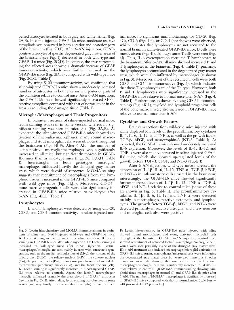
Microglia/Macrophages and Their ProgenitorsIn brainstem sections of saline-injected normal mice,
lectin staining was seen in some vessels, whereas no sig-nificant staining was seen in microglia (Fig. 3A,E). Asexpected, the saline-injected GFAP-IL6 mice showed ac-tivation of microglia/macrophages; many round macro-phages and stout microglial cells were obvious throughoutthe brainstem (Fig. 3B,F). After 6-AN, the number oflectin-positive microglia/macrophages was significantlyincreased in all mice, but significantly moreso in GFAP-IL6 mice than in wild-type mice (Figs. 3C,D,G,H, TableI). Interestingly, in both genotypes microglia/macrophages infiltrated heavily the damaged gray matterareas, which were devoid of astrocytes. MOMA stainingsuggests that recruitment of macrophages from the lym-phoid tissues is increased in the GFAP-IL6 mice comparedwith wild-type mice (Fig. 3I,JF). Moreover, CD-34�
bone marrow progenitor cells were also significantly in-creased in GFAP-IL6 mice relative to wild-type after6-AN (Fig. 4K,L, Table I).
LymphocytesB and T lymphocytes were detected by using CD-20,
CD-3, and CD-4 immunoreactivity. In saline-injected nor-
mal mice, no significant immunostainings for CD-20 (Fig.4G), CD-3 (Fig. 4H), or CD-4 (not shown) were observed,which indicates that lymphocytes are not recruited to thenormal brain. In saline-treated GFAP-IL6 mice, B cells weremostly absent (Fig. 4I), although some T cells were seen (Fig.4J). Thus, IL-6 overexpression recruited T lymphocytes tothe brainstem. After 6-AN, all mice showed increased B andT lymphocytes in the brainstem (Fig. 4, Table I); primarily,the lymphocytes accumulated in the degenerated gray matterareas, which were also infiltrated by macrophages (as shownin Fig. 3). Moreover, most of the recruited T cells were bothCD-3 and CD-4 immunoreactive (Fig. 4), which indicatesthat these T lymphocytes are of the Th type. However, bothB and T lymphocytes were significantly increased in theGFAP-IL6 mice relative to normal mice after 6-AN (Fig. 4,Table I). Furthermore, as shown by using CD-34 immunos-tainings (Fig. 4K,L), myeloid and lymphoid progenitor cellsof the bone marrow were also increased in GFAP-IL6 micerelative to normal mice after 6-AN.
Cytokines and Growth FactorsBrainstem sections from wild-type mice injected with
saline displayed low levels of the proinflammatory cytokinesIL-1, IL-6, IL-12, and TNF-�, as well as the growth factorsTGF-�, bFGF, and neurotrophin-3 (NT-3; Table I). Asexpected, the GFAP-IL6 mice showed moderately increasedIL-6 expression. Moreover, the levels of IL-1, IL-12, andTNF-� were also mildly increased in saline-injected GFAP-IL6 mice, which also showed up-regulated levels of thegrowth factors TGF-�, bFGF, and NT-3 (Table I).
After 6-AN injection, wild-type mice increased theexpression of IL-1�, IL-6, IL-12, TNF-�, TGF-�, bFGF,and NT-3 in inflammatory cells situated in the brainstem;interestingly, the GFAP-IL6 mice showed significantlyfurther increased levels of IL-1�, IL-12, TNF-�, TGF-�,bFGF, and NT-3 relative to control mice (some of theseare shown in Fig. 5, Table I). The proinflammatory cy-tokines IL-1�, IL-6, IL-12, and TNF-� were detectedmainly in macrophages, reactive astrocytes, and lympho-cytes. The growth factors TGF-�, bFGF, and NT-3 weredetected primarily in reactive astroglia, and a few neuronsand microglial cells also were positive.
Š
Fig. 3. Lectin histochemistry and MOMA immunostainings in brain-stem of saline- and 6-AN-injected wild-type and GFAP-IL6 mice.A: Lectin staining in control mice after saline injection. B: Lectinstaining in GFAP-IL6 mice after saline injection. C: Lectin staining isincreased in wild-type mice after 6-AN injection. Lectin�
macrophages/microglia are seen mainly in areas with astrocyte degen-eration, such as the medial vestibular nuclei (Mve), the nucleus of thesolitary tract (SolM), the solitary nucleus (SolV), the cuneate nucleus(Cu), the pontine nuclei (Pn), the superior paraolivary nucleus and themedioventral periolivary nucleus (Po), and the facial nucleus (VII).D: Lectin staining is significantly increased in 6-AN-injected GFAP-IL6 mice relative to controls. Again, the lectin� macrophages/microglia infiltrated primarily the areas devoid of GFAP� astrocytes(see this in Fig. 2). E: After saline, lectin staining was observed in somevessels (and very faintly in some ramified microglia) of control mice.
F: Lectin histochemistry in GFAP-IL6 mice injected with salineshowed round macrophages and stout, activated microglial cellsthroughout the brainstem. G: After 6-AN injection, control miceshowed recruitment of activated lectin� macrophages/microglial cells,which were seen primarily inside of the damaged gray matter areas.H: 6-AN treatment also induced macrophage/microglial activation inGFAP-IL6 mice. Again, macrophages/microglial cells were infiltratingthe degenerated gray matter areas but were also numerous in otherbrainstem areas. As shown, the number of recruited lectin�
macrophages/microglial cells was significantly increased in GFAP-IL6mice relative to controls. I,J: MOMA immunostaining showing lym-phoid tissue macrophages in normal (I) and GFAP-IL6 (J) mice after6-AN. The number of MOMA� macrophages is significantly increasedin GFAP-IL6 mice compared with that in normal mice. Scale bars �240 �m in A–D; 42 �m in E–J.
IL-6 Reduces CNS Damage 487

Fig. 4. CD-20, CD-3, CD-4, and CD-34 immunostainings in brain-stem of wild-type and GFAP-IL6 mice. A: CD-20 immunoreactivityof 6-AN-treated normal mice showing a few B lymphocytes in thebrainstem. B: 6-AN-treated GFAP-IL6 mice show increased CD-20�
B cells in the brainstem. C: CD-3� T cells in 6-AN-treated normalmice. D: CD-3� T cells are significantly increased in GFAP-IL6 miceafter 6-AN injection relative to normal mice. E: CD-4 immunoreac-tivity showing that most of the recruited T lymphocytes are Th cells innormal mice injected with 6-AN. F: Also in the 6-AN-treated GFAP-
IL6 mice, most T cells are CD-4� Th cells. G,H: Normal miceinjected with saline show no significant CD-20 (G) or CD-3 (H)immunostaining. I,J: GFAP-IL6 mice treated with saline show nosignificant CD-20 staining (I), although some CD-3� T cells are seenin the brainstem (J). K: 6-AN-treated normal mice show a few CD-34-stained cells. L: The numbers of CD-34� myeloid and lymphoidprogenitor cells of the bone marrow are significantly increased in6-AN-treated GFAP-IL6 mice. Scale bars � 20 �m in A,B; 30 �m inC,D; 17 �m E,F; 33 �m in G–J; 16 �m in K,L.

Angiogenesis
Angiogenesis, as judged by using angiopoietin,VEGF, and perlecan immunoreactivity, was not pro-nounced in the brainstem of saline-injected normal mice(not shown), whereaas, as shown by Brett et al. (1995), theGFAP-IL6 mice showed an increased angiogenic potentialwith increased number of vessels (not shown). After 6-ANinjection, expression of angiogenic factors increasedaround the degenerated areas. However, in the brainstemof the normal mice, angiogenic factors were only verymildly expressed, whereas the angiogenic response ofGFAP-IL6 mice was very pronounced and angiogenesiswas increased relative to the normal mice (Fig. 6).
Oxidative Stress and ApoptosisTo study oxidative stress, we have used immunoreac-
tivity for iNOS (Sasaki et al., 2001), MDA (Lung et al.,1990), NITT (Ye et al., 1996), and 8-oxoguanine (Soultan-akis et al., 2000). In brainstem sections of saline-injectedmice, the iNOS, MDA, NITT, and 8-oxoguanine immu-nostainings were comparable in wild-type and GFAP-IL6mice and showed that the normal CNS hardly suffers fromoxidative stress (some of these are shown in Fig. 7, see alsoTable I). After 6-AN, oxidative stress as determined by usingimmunoreactivity for iNOS, MDA, NITT, and8-oxoguanine was significantly increased in wild-type micerelative to GFAP-IL6 mice (see Fig. 7, Table I). The cells
Fig. 5. Immunoreactivity for TNF-�, TGF-�, and NT-3 in brainstemof 6-AN-injected wild-type and GFAP-IL6 mice. A: Normal miceshow TNF-� staining in some macrophages and astrocytes after 6-AN.B: In GFAP-IL6 mice, the levels of TNF-� are significantly increasedin macrophages and astrocytes. C,D: TGF-� immunostainings in nor-
mal (C) and GFAP-IL6 (D) mice showing increased levels in the latter.Mainly astroglia expressed TGF-�. E,F: Expression of NT-3 in normal(E) and GFAP-IL6 (F) mice. NT-3 was clearly increased by IL-6overexpression, mainly in astrocytes, neurons, and microglia/macrophages. Scale bars � 44 �m in A,B,E,F; 50 �m in C,D.
IL-6 Reduces CNS Damage 489

suffering from oxidative stress after 6-AN were mainly neu-rons and astrocytes, as verified by using triple immunofluo-rescence for MDA or NITT and cell markers. Apoptotic celldeath was determined by using TUNEL, which showed thatthe normal CNS contains very few apoptotic cells (Fig. 7,Table I). After 6-AN injections, all mice increased the num-ber of TUNEL� cells, but GFAP-IL6 mice showed signifi-cantly fewer TUNEL� cells than wild-type mice after 6-AN(Fig. 7, Table I). Apoptotic cells were mainly neurons andastrocytes, as verified by using fluorescence TUNEL andimmunofluorescence (data not shown). In contrast to the3-day time point, basically no TUNEL� cells were observed1 day after 6-AN (not shown).
MT-I�II ExpressionFigure 8 and Table I show MT-I�II immunohisto-
chemistry results. In accordance with previous data
(Carrasco et al., 1998; Penkowa et al., 2000b; Molinero etal., 2003), MT-I�II immunoreactivity increased signifi-cantly in the CNS in GFAP-IL6 mice relative to wild-typemice (Fig. 8A,B). After 6-AN administration, wild-typeand GFAP-IL6 mice showed clearly increased MT-I�IIexpression, which was most pronounced in the areas ofreactive astrogliosis around the degenerated gray matterareas. As might be expected, the GFAP-IL6 mice showeda significantly greater MT-I�II up-regulation following6-AN than wild-type mice (Fig. 8C,D, Table I).
DISCUSSIONIn the present study, we have examined transgenic
mice with overexpression of IL-6 (GFAP-IL6 mice) andproper controls following an i.p. injection with 6-AN.6-AN induces astroglial toxicity and cell death in certainastrocyte populations, mainly those of the brainstem gray
Fig. 6. Immunostaining for angiogenesis and endothelial proliferationin brainstem of 6-AN-injected control and GFAP-IL6 mice. A,B:Angiopoietin expression in the vessels was low in normal mice (A) after6-AN, whereas the GFAP-IL6 mice (B) show significantly increasedangiopoietin immunostaining. C,D: VEGF expression in normal (C)
and GFAP-IL6 (D) mice after 6-AN. The GFAP-IL6 mice showedincreased VEGF in several small vessels relative to normal mice. E,F:Perlecan immunoreactivity in 6-AN-injected normal (E) and GFAP-IL6 (F) mice showing significantly increased levels in the latter. Scalebars � 46 �m.
490 Penkowa et al.

matter areas, followed by a CNS inflammatory response(Krum, 1994, 1995; Haghighat and McCandless, 1996;Penkowa et al., 1997, 1999b, 2002; Penkowa andHidalgo, 2000a; Tyson et al., 2000).
In 6-AN-injected GFAP-IL6 mice, the degree of6-AN-induced degeneration of brainstem gray matter ar-eas was similar to that of wild-type mice. This was estab-lished by H&E as well as GFAP and S100 stainings, whichshowed that almost all the astrocytes disappeared from the
degenerated gray matter areas of the brainstem. This in-dicates that the initial effects elicited by 6-AN are unaf-fected by IL-6 transgenic expression regarding astrocytedamage and death. However, reactive astrogliosis was ob-served surrounding these injured areas, and this was greaterin the GFAP-IL6 mice than in the wild-type mice, sug-gesting that the transgenic expression of IL-6 potentiatedthe astrocyte response to the tissue injury elicited by6-AN, which could have significant effects on the delayed
Fig. 7. NITT and MDA immunoreactivity and TUNEL in the brain-stem of 6-AN-treated wild-type and GFAP-IL6 mice. A,B: MDAimmunostaining in 6-AN-treated normal (A) and GFAP-IL6 (B) mice.As shown, normal mice display increased MDA immunoreactivity after6-AN. C,D: NITT immunoreactivity in 6-AN-treated normal (C) andGFAP-IL6 (D) mice. As shown, the control mice display increased
NITT immunostaining after 6-AN. E,F: TUNEL stainings showingthat, after 6-AN, normal mice (E) increase the number of TUNEL�
apoptotic cells relative to GFAP-IL6 mice (F). G–J: Saline-injectednormal (G,H) and GFAP-IL6 (I,J) mice show no significant staining foroxidative stress marker NITT (G,I) or for apoptosis marker TUNEL(H,J). Scale bars � 43 �m in A,B; 49 �m in C,D; 40 �m in E–J.
IL-6 Reduces CNS Damage 491

responses of the CNS to 6-AN-induced gliotoxicity giventhe importance of astrocytes (Krum and Rosenstein, 1993;Xiao and Link, 1998; Benveniste, 1998; Raivich et al.,1999). GFAP and S100 stainings were also higher insaline-injected GFAP-IL6 mice, which is in agreementwith previous studies showing that these mice displayreactive astrogliosis in the hindbrain (Campbell et al.,1993; Brett et al., 1995; Campbell, 1998a,b; Giralt et al.,2002a; Molinero et al., 2003). Also, this is consistent withresults observed in IL-6-deficient mice showing signifi-cantly reduced astrogliosis (Klain et al., 1997; Penkowaand Hidalgo, 2000a; Penkowa et al., 2000b).
In addition, the inflammatory responses ofmacrophages/microglia and lymphocytes, including theirprecursors, CD-34� myeloid and lymphoid progenitorcells of the bone marrow, were clearly increased in CNSof the GFAP-IL6 mice relative to wild-type mice after6-AN injection. The recruitment to the CNS of CD-34�
myeloid and lymphoid progenitor cells induced by IL-6overexpression is shown for the first time. However, thiseffect fits with the fact that IL-6 is a multifunctionalcytokine, which has profound effects on the three majorsystems, the immune system, the hematopoietic system,and the nervous system (Heinrich et al., 1990; Hirano etal., 1990; Kopf et al., 1994; Hauser et al., 1997). Hence,IL-6 functions synergistically with IL-3 to stimulate mul-
tilineage blast-colony formation in the bone marrow, andIL-6 also affects the further growth and differentiation ofhematopoietic cells (for review see Hirano et al., 1990;Kopf et al., 1994; Peters et al., 1998). Accordingly,CD-34� cells differentiate and grow in the presence ofstem cell factor (SCF) and IL-6, causing increases in cellsize and numbers relative to cells treated with SCF alone(Conti et al., 2002).
Moreover, IL-6, as expected, increased in GFAP-IL6 mice, but also other proinflammatory cytokines, suchas IL-1�, IL-12, and TNF-� were increased in these mice(Campbell et al., 1993; Campbell 1998ab). Along withproinflammatory cytokines, the antiinflammatory and/ortrophic factors TGF-�, bFGF, VEGF, and NT-3 werealso increased by 6-AN administration, and, again, thisresponse was increased more in the IL-6-overexpressingmice than in wild-type mice. Furthermore, expression ofthe antiinflammatory and antioxidant proteins MT-I�IIwas significantly increased by IL-6 overexpression. This isin agreement with previous studies showing that IL-6 isthe major inducer in the CNS of MT-I�II (Hernandez etal., 1997; Carrasco et al., 1998; Penkowa and Hidalgo,2000a; Penkowa et al., 2000b). Because MT-I�II areinducers of antiinflammatory growth factors (Penkowa etal., 2000a, 2003), both increased IL-6 and increased
Fig. 8. MT-I�II immunostainings in the brainstem of saline- and6-AN-injected control and GFAP-IL6 mice. A: MT-I�II expressionin saline-injected control mice showing MT-I�II in a few scatteredglial cells. B: Saline-injected GFAP-IL6 mice show significantly in-creased MT-I�II expression relative to wild-type mice. C: After6-AN, the wild-type mice showed increased MT-I�II levels in reac-
tive atsrocytes and some macrophages/microglia. D: In 6-AN-injectedGFAP-IL6 mice, a further and significant increase in MT-I�II expres-sion was seen relative to wild-type mice. Hence, numerous reactiveatsrocytes and macrophages/microglia showed increased MT-I�II.Scale bars � 44 �m.
492 Penkowa et al.

MT-I�II may explain the increases in TGF-�, bFGF,VEGF, and NT-3 expression in GFAP-IL6 mice.
Additionally, the angiogenic response seen after6-AN was significantly increased in GFAP-IL6 mice,which showed significantly increased immunoreactivityfor angiopoietin, VEGF, and perlecan in the brainstem.Thus, vessel sprouting and vasculogenesis are induced byIL-6 overproduction, which is in agreement with otherstudies (Brett et al., 1995; Swartz et al., 2001) showinghow IL-6 stimulates vascular sprouting and the number ofvessels in the CNS. This could be an indirect effect, whichis mediated by MT-I�II rather than by IL-6 per se. Thus,GFAP-IL6 mice with MT-I�II deficiency show signifi-cantly reduced angiogenesis relative to GFAP-IL6 micewith normal MT-I�II levels (Penkowa et al., 2000a).Also, MT-I overexpression increases angiogenesis signifi-cantly in GFAP-IL6 mice (Molinero et al., 2003). As well,MT-I overexpression and MT-II treatment induce CNSangiogenesis following brain damage (Giralt et al., 2002b;Penkowa et al., 2002). Thus, the IL-6-induced increases inMT-I�II levels along with growth factors may be respon-sible for the increased angiogenesis in the parenchyma. Inaddition, the IL-6-induced increases in growth factor ex-pression could also contribute to increased angiogenesis, inthat bFGF, TGF-�, NT-3, and VEGF are neuroprotectivefactors involved in angiogenesis and tissue regeneration(Pepper et al., 1992, 1993; Gomez-Pinilla et al., 1992;Logan et al., 1994; Hopkins and Rothwell, 1995; Moc-chetti and Wrathall, 1995; Ebadi et al., 1997; Yoshida etal., 1997; Xiao and Link, 1998; Morisaki et al., 1999;Neufeld et al., 1999; Suda et al., 2000).
In addition to the increase in these inflammatoryreactions, 6-AN-injected GFAP-IL6 mice also showedsignificantly reduced levels of oxidative stress as deter-mined by immunoreactivity for iNOS, NITT, MDA, and8-oxoguanine; moreover, 6-AN-induced apoptotic celldeath as judged by TUNEL and cleaved caspase-3 immu-nostaining was considerably reduced relative to that inwild-type mice. It is likely that oxidative stress and apo-ptosis are linked (Sun and Chen, 1998; Floyd, 1999;Cassarino and Bennett, 1999). Hence, in general, theGFAP-IL6 mice appeared to suffer less from 6-AN-induced CNS tissue damage than wild-type mice at moreadvanced stages (3 days after 6-AN injection), in contrastto the initial stages. It is likely that such neuroprotectionwould be apparent also in H&E stainings carried out forlonger times, which deserves further attention.
It is very likely that the IL-6-induced increase inMT-I�II contributes to the observed decreases in oxida-tive stress and apoptosis in GFAP-IL6 mice after 6-ANdespite the increased ongoing inflammatory response.Thus, accumulating data show how MT-I�II are ex-tremely competent antioxidants and scavengers of freeradicals, thereby having important roles during oxidativestress (Thornalley and Vasak, 1985; Aschner, 1998; Simp-kins, 2000; Van Lookeren Campagne et al., 2000; Vi-arengo et al., 2000; Hidalgo et al., 2001, 2002; Giralt et al.,2002). Thus, in vitro, MT-I�II inhibit DNA degradationand tissue damage from oxidative stress (Cai et al., 2000;
Kling and Olsson, 2000; Rana and Kumar, 2000), andMT-I�II functionally substitute for Cu/Zn-superoxidedismutase (Cu/Zn-SOD) in vivo to protect cells fromoxygen toxicity (Tamai et al., 1993). Moreover, cells fromMT-I�II-deficient mice, in spite of their normal levels ofreduced glutathione and other antioxidant enzymes,showed enhanced ROS production (Lazo et al., 1995). Inaddition, genetic MT-I�II deficiency increases signifi-cantly the levels of oxidative stress and apoptosis in vivoduring CNS pathological conditions, such as during sei-zures (Carrasco et al., 2000), traumatic injury (Penkowa etal., 1999, 2000a), IL-6-induced cerebellar neuroinflamma-tion (Giralt et al., 2002a), and experimental autoimmuneencephalomyelitis (EAE; Penkowa et al., 2003). In thismanner, reduced levels of oxidative stress and apoptosiswere induced by MT-I overexpression and MT-II treat-ment in mice after 6-AN (Penkowa et al., 2002), traumaticbrain injury (Giralt et al., 2002b), IL-6-induced cerebellarneuroinflammation (Molinero et al., 2003), and EAE(Penkowa and Hidalgo, 2000b). Thus, by inducingMT-I�II in the CNS, IL-6 can prevent oxidative stress,which leads to cell death (Cassarino and Bennett, 1999),and thereby IL-6 provides significant neuroprotection.This study was carried out in mice heterozygous for MT-I&II, so it will be interesting to determine in furtherstudies whether more prominent effects are observed inanimals with normal MT expression and/or MT overex-pression; these studies are currently underway in our lab-oratories. We nevertheless anticipate that this will actuallyhappen, judging by previous results obtained in MT-I-overexpressing mice (Penkowa et al., 2002).
On the other hand, we cannot rule out that IL-6may also have direct antioxidant effects in the brain. How-ever, it was demonstrated that IL-6 per se has no effects onnitric oxide synthesis (De Laurentiis et al., 2000). There-fore, we suggest that the decrease in oxidative stress andapoptosis observed in 6-AN-injected GFAP-IL6 mice ismediated by the increased MT-I�II in these mice.
In addition, IL-6 may have other indirect actions,such as stimulating other neurotrophic factors. This pos-sibility has been shown in vivo when intrathecal IL-6infusion in lumbar dorsal root ganglia induced brain-derived neurotrophic factor (BDNF; Murphy et al. 2000).In vitro, IL-6 alone or together with IL-1 induced pro-duction of nerve growth factor (Juric and Carman-Krzan2000). Indeed, in this study, IL-6 overexpression inducedthe growth factors bFGF, TGF-�, NT-3, angiopoietin,and VEGF as well as the receptor for bFGF. Therefore, itis likely that, in addition to its own, innate neurotrophicactivity, IL-6 induces additional trophic factors in theCNS. The proposed mechanisms through which endog-enous IL-6 may protect the brain during pathologicalconditions should be explored further to determine therole of IL-6 in neuroprotection.
ACKNOWLEDGMENTSWe thank Hanne Hadberg, Ha Nguyen, and Drs.
Joaquın Hernandez and Javier Carrasco for excellent tech-nical assistance and Keld Stub for photographic assistance.These studies were supported by The Lundbeck Founda-
IL-6 Reduces CNS Damage 493

tion, Scleroseforeningen, Lily Benthine Lunds Fond, Dir.Ejnar Jonasson’s Fond, The Danish Medical ResearchCouncil, Novo Nordisk Fonden, Direktør Ib HenriksensFond, Warwara Larsens Fond, The Danish Medical Asso-ciation Research Fund, Schrøders Fond, Kathrine og VigoSkovgaards Fond, Dir. Jacob Madsen’s Fond, Fonden af17.12.1981, Fonden til Laegevidenskabens Fremme,Gerda og Aage Haensch’s Fond, Eva og Henry FrænkelsMindefond, Karen A. Tolstrups Fond, Dansk Parkinson-forening, Th. Maigaard’s Eftf. Fru Lily Benthine LundsFond af 1/6 1978, Hans og Nora Buchards Fond, HolgerRabitz Mindelegat, Ragnhild Ibsens Legat for MedicinskForskning, and Hestehandler Ole Jacobsens Mindelegat(all to M.P.) and by Ministerio de Ciencia y Tecnologıaand Feder grant SAF2002-01268 and Direccio General deRecerca grant 2001SGR 00203 (to J.H.).
REFERENCESAschner M. 1998. Metallothionein (MT) isoforms in the central nervous
system (CNS): regional and cell-specific distribution and potential func-tions as an antioxidant. Neurotoxicology 19:653–660.
Benveniste EN. 1998. Cytokine actions in the central nervous system.Cytokine Growth Factor Rev 9:259–275.
Brett FM, Mizisin AP, Powell HC, Campbell IL. 1995. Evolution ofneuropathologic abnormalities associated with blood–brain barrier break-down in transgenic mice expressing interleukin-6 in astrocytes. J Neuro-pathol Exp Neurol 54:766–775.
Brunello AG, Weissenberger J, Kappeler A, Vallan C, Peters M, Rose-JohnS, Weis J. 2000. Astrocytic alterations in interleukin-6/solubleinterleukin-6 receptor alpha double-transgenic mice. Am J Pathol 157:1485–1593.
Budihardjo II, Boerner SA, Eckdahl S, Svingen PA, Rios R, Ames MM,Kaufmann SH. 2000. Effect of 6-aminonicotinamide and other proteinsynthesis inhibitors on formation of platinum-DNA adducts and cisplatinsensitivity. Mol Pharmacol 57:529–538.
Cai L, Klein JB, Kang YJ. 2000. Metallothionein inhibits peroxynitrite-induced DNA and lipoprotein damage. J Biol Chem 275:38957–38960.
Campbell IL. 1998a. Structural and functional impact of the transgenicexpression of cytokines in the CNS. Ann N Y Acad Sci 840:83–96.
Campbell IL. 1998b. Transgenic mice and cytokine actions in the brain:bridging the gap between structural and functional neuropathology. BrainRes Rev 26:327–336.
Campbell IL, Abraham CR, Masliah E, Kemper P, Inglis JD, OldstoneMBA, Mucke L. 1993. Neurological diseases induced in transgenic miceby cerebral overexpression of interleukin-6. Proc Natl Acad Sci USA90:10061–10065.
Carlson NG, Wieggel WA, Chen J, Bacchi A, Rogers SW, Gahring LC.1999. Inflammatory cytokines IL-1 alpha, IL-1 beta, IL-6, and TNF-alphaimpart neuroprotection to an excitotoxin through distinct pathways.J Immunol 163:3963–3968.
Carrasco J, Hernandez J, Gonzalez B, Campbell IL, Hidalgo J. 1998.Localization of metallothionein-I and -III expression in the CNS oftransgenic mice with astrocyte-targeted expression of interleukin 6. ExpNeurol 153:184–194.
Carrasco J, Penkowa M, Hadberg H, Molinero A, Hidalgo J. 2000. En-hanced seizures and hippocampal neurodegeneration following kainicacid induced seizures in metallothionein-1�2 deficient mice. Eur J Neu-rosci 12:2311–2322.
Cassarino DS, Bennett JP Jr. 1999. An evaluation of the role of mitochon-dria in neurodegenerative diseases: mitochondrial mutations and oxidativepathology, protective nuclear responses, and cell death in neurodegen-eration. Brain Res Rev 29:1–25.
Conti P, Kempuraj D, Di Gioacchino M, Boucher W, Letourneau R,
Kandere K, Barbacane RC, Reale M, Felaco M, Frydas S, TheoharidesTC. 2002. Interleukin-6 and mast cells. Allergy Asthma Proc 23:331–335.
De Laurentiis A, Pisera D, Lasaga M, Diaz M, Theas S, Duvilanski B,Seilicovich A. 2000. Effect of interleukin-6 and tumor necrosis factor-alpha on GABA release from mediobasal hypothalamus and posteriorpituitary. Neuroimmunomodulation 7:77–83.
Di Santo E, Alonzi T, Fattori E, Poli V, Ciliberto G, Sironi M, Gnocchi P,Ricciardi-Castagnoli P, Ghezzi P. 1996. Overexpression of interleukin-6in the central nervous system of transgenic mice increases central but notsystemic proinflammatory cytokine production. Brain Res 740:239–244.
Ebadi M, Bashir RM, Heidrick ML, Hamada FM, Refaey HE, Hamed A,Helal G, Baxi MD, Cerutis DR, Lassi NK. 1997. Neurotrophins and theirreceptors in nerve injury and repair. Neurochem Int 30:347–374.
Eskes C, Honegger P, Juillerat-Jeanneret L, Monnet-Tschudi F.2002. Microglial reaction induced by noncytotoxic methylmercury treat-ment leads to neuroprotection via interactions with astrocytes and IL-6release. Glia 37:43–52.
Eugster HP, Frei K, Kopf M, Lassmann H, Fontana A. 1998. IL-6-deficientmice resist myelin oligodendrocyte glycoprotein-induced autoimmuneencephalomyelitis. Eur J Immunol 28:2178–2187.
Fattori E, Lazzaro D, Musiani P, Modesti A, Alonzi T, Ciliberto G. 1995.IL-6 expression in neurons of transgenic mice causes reactive astrocytosisand increase in ramified microglial cells but no neuronal damage. EurJ Neurosci 7:2441–2449.
Fee D, Grzybicki D, Dobbs M, Ihyer S, Clotfelter J, Macvilay S, Hart MN,Sandor M, Fabry Z. 2000. Interleukin 6 promotes vasculogenesis ofmurine brain microvessel endothelial cells. Cytokine 12:655–665.
Floyd RA. 1999. Antioxidants, oxidative stress, and degenerative neuro-logical disorders. Proc Soc Exp Biol Med 222:236–245.
Gadient RA, Otten UH. 1997. Interleukin-6 (IL-6)—a molecule with bothbeneficial and destructive potentials. Prog Neurobiol 52:379–390.
Gahring LC, Carlson NG, Wieggel WA, Howard J, Rogers SW. 1999.Alcohol blocks TNFalpha but not other cytokine-mediated neuroprotec-tion to NMDA. Alcohol Clin Exp Res 23:1571–1579.
Gasull T, Rebollo DV, Romero B, Hidalgo J. 1993. Development of acompetitive double antibody radioimmunoassay for rat metallothionein.J Immunoassay 14:209–225.
Giralt M, Penkowa M, Hernandez J, Molinero A, Carrasco J, Lago N,Camats J, Campbell IL, Hidalgo J. 2002a. Metallothionein-1�2 defi-ciency increases brain pathology in transgenic mice with astrocyte-targeted expression of interleukin 6. Neurobiol Dis 9:319–338.
Giralt M, Penkowa M, Lago N, Camats J, Hernandez J, Molinero A,Hidalgo J. 2002b. Metallothionein-1�2 protect the CNS after a focalbrain injury. Exp Neurol 173:114–128.
Gomez-Pinilla F, Won-Kyun Lee J, Cotman CW. 1992. Basic FGF in adultrat brain: Cellular distribution and response to entorhinal lesion andfimbria-fornix transection. J Neurosci 12:345–355.
Gruol DL, Nelson TE. 1997. Physiological and pathological roles ofinterleukin-6 in the central nervous system. Mol Neurobiol 15:307–339.
Haghighat N, McCandless DW. 1996. Effect of 6-aminonicotinamide onmetabolism of astrocytes and C6-glioma cells. Metab Brain Dis 12:29–45.
Hauser SP, Kajkenova O, Lipschitz DA. 1997. The pivotal role of inter-leukin 6 in formation and function of hematopoietically active murinelong-term bone marrow cultures. Stem Cells 15:125–132.
Heinrich P, Castell JV, Andus T. 1990. Interleukin-6 and the acute phaseresponse. J Biochem 265:621–636.
Hernandez J, Molinero A, Campbell IL, Hidalgo J. 1997. Transgenicexpression of interleukin 6 in the central nervous system regulates brainmetallothionein-I and -III expression in mice. Brain Res Mol Brain Res48:125–131.
Hidalgo J, Aschner M, Zatta P, Vasak M. 2001. Roles of the metallothio-nein family of proteins in the central nervous system. Brain Res Bull55:133–145.
494 Penkowa et al.

Hidalgo J, Penkowa M, Giralt M, Carrasco J, Molinero A.2002. Metallothionein expression and oxidative stress in the brain. Meth-ods Enzymol 348:238–249.
Hirano T, Akira S, Taga T, Kishimoto T. 1990. Biological and clinicalaspects of interleukin 6. Immunol Today 11:443–449.
Hopkins SJ, Rothwell NJ. 1995. Cytokines and the nervous system I:expression and recognition. Trends Neurosci 18:83–88.
Hothersall JS, Zubairu S, McLean P, Greenbaum AL. 1981. Alternativepathways of glucose utilization in brain; changes in the pattern of glucoseutilization in brain resulting from treatment of rats with6-aminonicotinamide. J Neurochem 37:1484–1496.
Hothersall JS, Gordge M, Noronha-Dutra AA. 1998. Inhibition ofNADPH supply by 6-aminonicotinamide: effect on glutathione, nitricoxide and superoxide in J774 cells. FEBS Lett 434:97–100.
Juric DM, Carman-Krzan M. 2000. Cytokine-regulated secretion of nervegrowth factor from cultured rat neonatal astrocytes. Pflugers Arch 440:R96–R98.
Klein MA, Moller JC, Jones LL, Bluethmann H, Kreutzberg GW, RaivichG. 1997. Impaired neuroglial activation in interleukin-6 deficient mice.Glia 19:227–233.
Kling PG, Olsson P. 2000. Involvement of differential metallothioneinexpression in free radical sensitivity of RTG-2 and CHSE-214 cells. FreeRadic Biol Med 28:1628–1637.
Kopf M, Baumann H, Freer G, Freudenberg M, Lamers M, Kishimoto T,Zinkernagel R, Bluethmann H, Kohler G. 1994. Impaired immune andacute-phase responses in interleukin-6-deficient mice. Nature 368:339–342.
Krinke GJ, Classen W. 1998. Spongiform neuropathy induced in dogs byprolonged, low-level administration of 6-aminonicotinamide (6-ANA).Exp Toxicol Pathol 50:277–282.
Krum JM. 1994. Experimental gliopathy in the adult rat CNS: effect on theblood–spinal cord barrier. Glia 11:354–366.
Krum JM. 1995. Age-dependent susceptibility of CNS glial populations insitu to the antimetabolite 6-aminonicotinamide. Mol Chem Neuropathol26:79–94.
Krum JM, Rosenstein JM. 1993. Effect of astroglial degeneration on theblood–brain barrier to protein in neonatal rats. Brain Res Dev Brain Res74:41–50.
Lazo JS, Kondo Y, Dellapiazza D, Michalska AE, Choo KH, Pitt BR. 1995.Enhanced sensitivity to oxidative stress in cultured embryonic cells fromtransgenic mice deficient in metallothionein I and II genes. Biol Chem270:5506–5510.
Lenzlinger PM, Morganti-Kossmann MC, Laurer HL, McIntosh TK. 2001.The duality of the inflammatory response to traumatic brain injury. MolNeurobiol 24:169–181.
Loddick SA, Turnbull AV, Rothwell NJ. 1998. Cerebral interleukin-6 isneuroprotective during permanent focal cerebral ischemia in the rat.J Cereb Blood Flow Metab 18:176–179.
Logan A, Berry M, Gonzalez AM, Frautschy SA, Sporn MB, Baird A. 1994.Effects of transforming growth factor b1 on scar production in the injuredcentral nervous system of the rat. Eur J Neurosci 6:355–363.
Lung CC, Fleisher JH, Meinke G, Pinnas JL. 1990. Immunochemicalproperties of malondialdehyde-protein adducts. J Immunol Methods 128:127–132.
Masters BA, Kelly EJ, Quaife CJ, Brinster RL, Palmiter RD. 1994. Tar-geted disruption of metallothionein I and II genes increases sensitivity tocadmium. Proc Natl Acad Sci USA 91:584–588.
Mocchetti I, Wrathall JR. 1995. Neurotrophic factors in central nervoussystem trauma. J Neurotrauma 12:853–870.
Molinero A, Penkowa M, Hernandez J, Camats J, Giralt M, Lago N,Carrasco J, Campbell IL, Hidalgo J. 2003. Metallothionein-I overexpres-sion decreases brain pathology in transgenic mice with astrocyte-targetedexpression of interleukin 6. J Neuropathol Exp Neurol 62:315–328.
Morganti-Kossmann MC, Rancan M, Stahel PF, Kossmann T. 2002.
Inflammatory response in acute traumatic brain injury: a double-edgedsword. Curr Opin Crit Care 8:101–105.
Morisaki N, Watanabe S, Fukuda K, Saito Y. 1999. Angiogenic interactionbetween retinal endothelial cells and pericytes from normal and diabeticrabbits, and phenotypic changes of diabetic cells. Cell Mol Biol 45:67–77.
Munoz-Fernandez MA, Fresno M. 1998. The role of tumour necrosisfactor, interleukin 6, interferon-gamma and inducible nitric oxide syn-thase in the development and pathology of the nervous system. ProgNeurobiol 56:307–340.
Murphy PG, Borthwick LS, Johnston RS, Kuchel G, Richardson PM.1999. Nature of the retrograde signal from injured nerves that inducesinterleukin-6 mRNA in neurons. J Neurosci 19:3791–3800.
Murphy PG, Borthwick LA, Altares M, Gauldie J, Kaplan D, RichardsonPM. 2000. Reciprocal actions of interleukin-6 and brain-derived neuro-trophic factor on rat and mouse primary sensory neurons. Eur J Neurosci12:1891–1899.
Neufeld G, Cohen T, Gengrinovitch S, Poltorak Z. 1999. Vascular endo-thelial growth factor (VEGF) and its receptors. FASEB J 13:9–22.
Pavelko KD, Howe CL, Drescher KM, Gamez JD, Johnson AJ, Wei T,Ransohoff RM, Rodriguez M. 2003. Interleukin-6 protects anterior hornneurons from lethal virus-induced injury. J Neurosci 23:481–492.
Penkowa M, Hidalgo J. 2000a. IL-6 deficiency leads to reduced metallo-thionein I�II expression and increased oxidative stress in the brainstemafter 6-aminonicotinamide treatment. Exp Neurol 163:72–84.
Penkowa M, Hidalgo J. 2000b. Metallothionein I�II (MT-I�II) expres-sion and their role on experimental autoimmune encephalomyelitis(EAE). Glia 32:247–263.
Penkowa M, Hidalgo J, Moos T. 1997. Increased astrocytic expression ofmetallothioneins I�II in brainstem of adult rats treated with6-aminonicotinamide. Brain Res 774:256–259.
Penkowa M, Carrasco J, Giralt M, Moos T, Hidalgo J. 1999a. CNS woundhealing is severely depressed in metallothionein-I- and -II-deficient mice.J Neurosci 19:2535–2545.
Penkowa M, Giralt M, Moos T, Thomsen PS, Hernandez J, Hidalgo J.1999b. Impaired inflammatory response to glial cell death in geneticallymetallothionein-I�II deficient mice. Exp Neurol 156:149–164
Penkowa M, Carrasco J, Giralt M, Molinero A, Hernandez J, Campbell IL,Hidalgo J. 2000a. Altered central nervous system cytokine-growth factorexpression profiles and angiogenesis in metallothionein-I�II deficientmice. J Cerebral Blood Flow Metab 20:1174–1190.
Penkowa M, Giralt M, Carrasco J, Hadberg H, Hidalgo J. 2000b. Impairedinflammatory response and increased oxidative stress and neurodegenera-tion after brain injury in interleukin-6-deficient mice. Glia 32:271–285.
Penkowa M, Giralt M, Camats J, Hidalgo J. 2002. Metallothionein 1�2protect the CNS during neuroglial degeneration induced by6-aminonicotinamide. J Comp Neurol 444:174–189.
Penkowa M, Espejo C, Martınez Caceres EM, Montalban X, Hidalgo J.2003. Increased demyelination and axonal damage in metallothioneinI�II deficient mice during experimental autoimmune encephalomyelitis.Cell Mol Life Sci 60:1–13.
Pepper MS, Ferrara N, Orci L, Montesano R. 1992. Potent synergismbetween vascular endothelial growth factor and basic fibroblast growthfactor in the induction of angiogenesis in vitro. Biochem Biophys ResCommun 189:824–831.
Pepper MS, Vassalli JD, Orci L, Montesano R. 1993. Biphasic effect oftransforming growth factor-beta 1 on in vitro angiogenesis. Exp Cell Res204:356–363.
Peters M, Muller AM, Rose-John S. 1998. Interleukin-6 and solubleinterleukin-6 receptor: direct stimulation of gp130 and hematopoiesis.Blood 92:3495–3504.
Raivich G, Bohatschek M, Kloss CU, Werner A, Jones LL, KreutzbergGW. 1999. Neuroglial activation repertoire in the injured brain: gradedresponse, molecular mechanisms and cues to physiological function. BrainRes Rev 30:77–105.
IL-6 Reduces CNS Damage 495

Rana SV, Kumar A. 2000. Metallothionein induced by cadmium or zincinhibits lipid peroxidation in rats exposed to dimethylnitrosamine. ArhHig Rada Toksikol 51:279–286.
Sasaki S, Warita H, Abe K, Iwata M. 2001. Inducible nitric oxide synthase(iNOS) and nitrotyrosine immunoreactivity in the spinal cords of trans-genic mice with a G93A mutant SOD1 gene. J Neuropathol Exp Neurol60:839–846.
Simpkins CO. 2000. Metallothionein in human disease. Cell Mol Biol46:465–488.
Soultanakis RP, Melamede RJ, Bespalov IA, Wallace SS, Beckman KB,Ames BN, Taatjes DJ, Janssen-Heininger YM. 2000. Fluorescence de-tection of 8-oxoguanine in nuclear and mitochondrial DNA of culturedcells using a recombinant Fab and confocal scanning laser microscopy.Free Radic Biol Med 28:987–998.
Suda T, Takakura N, Oike Y. 2000. Hematopoiesis and angiogenesis. IntJ Hematol 71:99–107.
Sun AY, Chen YM. 1998. Oxidative stress and neurodegenerative disor-ders. J Biomed Sci 5:401–414.
Swartz KR, Liu F, Sewell D, Schochet T, Campbell I, Sandor M, Fabry Z.2001. Interleukin-6 promotes post-traumatic healing in the central ner-vous system. Brain Res 896:86–95.
Tamai KT, Gralla EB, Ellerby LM, Valentine JS, Thiele DJ. 1993. Yeastand mammalian metallothioneins functionally substitute for yeast copper-zinc superoxide dismutase. Proc Natl Acad Sci USA 90:8013–8017.
Thornalley PJ, Vasak M. 1985. Possible role for metallothionein in protec-
tion against radiation-induced oxidative stress. Kinetics and mechanism ofits reaction with superoxide and hydroxyl radicals. Biochim Biophys Acta827:36–44.
Toulmond S, Vige X, Fage D, Benavides J. 1992. Local infusion ofinterleukin-6 attenuates the neurotoxic effects of NMDA on rat striatalcholinergic neurons. Neurosci Lett 144:49–52.
Tyson RL, Perron J, Sutherland GR. 2000. 6-Aminonicotinamide inhibi-tion of the pentose phosphate pathway in rat neocortex. Neuroreport11:1845–1848.
Van Lookeren Campagne M, Thibodeaux H, Van Bruggen N, Caims B,Gerlai R, Palmer JT, Williams SP, Lowe D. 1999. Evidence for aprotective role of metallothionein-1 in focal cerebral ischemia. Proc NatlAcad Sci USA 96:12870–12875.
Van Wagoner NJ, Benveniste EN. 1999. Interleukin-6 expression andregulation in astrocytes. J Neuroimmunol 100:124–139.
Viarengo A, Burlando B, Ceratto N, Panfoli I. 2000. Antioxidant role ofmetallothioneins: a comparative overview. Cell Mol Biol 46:407–417.
Xiao BG, Link H. 1998. Immune regulation within the central nervoussystem. J Neurol Sci 157:1–12.
Ye YZ, Strong M, Huang ZQ, Beckman JS. 1996. Antibodies that recog-nize nitrotyrosine. Methods Enzymol 269:201–209.
Yoshida S, Ono M, Shono T, Izumi H, Ishibashi T, Suzuki H, Kuwano M.1997. Involvement of interleukin-8, vascular endothelial growth factor,and basic fibroblast growth factor in tumor necrosis factor alpha-dependent angiogenesis. Mol Cell Biol 17:4015–4023.
496 Penkowa et al.